Multimodal imaging of repetition priming: Using fMRI, MEG, and intracranial EEG to reveal...
11
Multimodal imaging of repetition priming: Using fMRI, MEG, and intracranial EEG to reveal spatiotemporal profiles of word processing Carrie R. McDonald a,b, ⁎, Thomas Thesen b,c , Chad Carlson c , Mark Blumberg c , Holly M. Girard b , Amy Trongnetrpunya c , Jason S. Sherfey b , Orrin Devinsky c , Rubin Kuzniecky c , Werner K. Dolye d , Sydney S. Cash b,e , Matthew K. Leonard b,h , Donald J. Hagler Jr. b,f , Anders M. Dale b,f,g , Eric Halgren b,f,g a Department of Psychiatry, University of California, San Diego, CA, USA b Multimodal Imaging Laboratory, University of California, San Diego, CA, USA c Comprehensive Epilepsy Center, Department of Neurology, New York University, New York, NY, USA d Department of Neurosurgery, New York University, New York, NY, USA e Department of Neurology, Massachusetts General Hospital, Harvard Medical School, Boston, MA, USA f Department of Radiology, University of California, San Diego, CA, USA g Department of Neurosciences, University of California, San Diego, CA, USA h Department of Cognitive Science, University of California, San Diego CA, USA abstract article info Article history: Received 31 March 2010 Revised 10 June 2010 Accepted 26 June 2010 Available online xxxx Keywords: fMRI Magnetoencepholography Intracranial EEG Memory Language Gamma Repetition priming is a core feature of memory processing whose anatomical correlates remain poorly understood. In this study, we use advanced multimodal imaging (functional magnetic resonance imaging (fMRI) and magnetoencephalography; MEG) to investigate the spatiotemporal profile of repetition priming. We use intracranial electroencephalography (iEEG) to validate our fMRI/MEG measurements. Twelve controls completed a semantic judgment task with fMRI and MEG that included words presented once (new, ‘N’) and words that repeated (old, ‘O’). Six patients with epilepsy completed the same task during iEEG recordings. Blood-oxygen level dependent (BOLD) responses for N vs. O words were examined across the cortical surface and within regions of interest. MEG waveforms for N vs. O words were estimated using a noise-normalized minimum norm solution, and used to interpret the timecourse of fMRI. Spatial concordance was observed between fMRI and MEG repetition effects from 350 to 450 ms within bilateral occipitotemporal and medial temporal, left prefrontal, and left posterior temporal cortex. Additionally, MEG revealed widespread sources within left temporoparietal regions, whereas fMRI revealed bilateral reductions in occipitotemporal and left superior frontal, and increases in inferior parietal, precuneus, and dorsolateral prefrontal activity. BOLD suppression in left posterior temporal, left inferior prefrontal, and right occipitotemporal cortex correlated with MEG repetition-related reductions. IEEG responses from all three regions supported the timecourse of MEG and localization of fMRI. Furthermore, iEEG decreases to repeated words were associated with decreased gamma power in several regions, providing evidence that gamma oscillations are tightly coupled to cognitive phenomena and reflect regional activations seen in the BOLD signal. © 2010 Elsevier Inc. All rights reserved. Introduction Repetition priming has been studied extensively in cognitive neuroscience, but its exact neural correlates remain poorly under- stood. fMRI studies have demonstrated response suppression, the main neural correlate of priming, in a number of cortical regions following word repetitions. Reliable decreases in blood-oxygen level dependent (BOLD) responses are consistently reported in left ventral occipitotemporal, posterior temporal, and left inferior frontal cortex— regions implicated in word form identification (Allison et al., 1999), lexical access, and semantic processing (Marinkovic et al., 2003; Matsumoto et al., 2005). Repetition of words also has been associated with increased activity in bilateral precuneus, frontoparietal, and hippocampal cortex—regions implicated in resting state and episodic retrieval processes (Weiss et al., 2009). What is not clear from fMRI is precisely when repetition effects occur during the course of word processing. Unlike fMRI, MEG provides a highly accurate picture of the temporal dynamics of cognitive processes, allowing one to visualize repetition effects in real time. MEG studies of priming have revealed reductions in the N400 response—an event-related field (ERF) implicated in semantic processing—to repeated words from ~350 to NeuroImage xxx (2010) xxx–xxx ⁎ Corresponding author. Multimodal Imaging Laboratory, 8950 Villa La Jolla Drive, Suite C101, La Jolla, CA 92037, USA. E-mail address: [email protected] (C.R. McDonald). YNIMG-07456; No. of pages: 11; 4C: 4, 5, 7, 8 1053-8119/$ – see front matter © 2010 Elsevier Inc. All rights reserved. doi:10.1016/j.neuroimage.2010.06.069 Contents lists available at ScienceDirect NeuroImage journal homepage: www.elsevier.com/locate/ynimg Please cite this article as: McDonald, C.R., et al., Multimodal imaging of repetition priming: Using fMRI, MEG, and intracranial EEG to reveal spatiotemporal profiles of word processi..., NeuroImage (2010), doi:10.1016/j.neuroimage.2010.06.069
-
Upload
independent -
Category
Documents
-
view
4 -
download
0
Transcript of Multimodal imaging of repetition priming: Using fMRI, MEG, and intracranial EEG to reveal...
NeuroImage xxx (2010) xxx–xxx
YNIMG-07456; No. of pages: 11; 4C: 4, 5, 7, 8
Contents lists available at ScienceDirect
NeuroImage
j ourna l homepage: www.e lsev ie r.com/ locate /yn img
Multimodal imaging of repetition priming: Using fMRI, MEG, and intracranial EEG toreveal spatiotemporal profiles of word processing
Carrie R. McDonald a,b,⁎, Thomas Thesen b,c, Chad Carlson c, Mark Blumberg c, Holly M. Girard b,Amy Trongnetrpunya c, Jason S. Sherfey b, Orrin Devinsky c, Rubin Kuzniecky c, Werner K. Dolye d,Sydney S. Cash b,e, Matthew K. Leonard b,h, Donald J. Hagler Jr. b,f, Anders M. Dale b,f,g, Eric Halgren b,f,g
a Department of Psychiatry, University of California, San Diego, CA, USAb Multimodal Imaging Laboratory, University of California, San Diego, CA, USAc Comprehensive Epilepsy Center, Department of Neurology, New York University, New York, NY, USAd Department of Neurosurgery, New York University, New York, NY, USAe Department of Neurology, Massachusetts General Hospital, Harvard Medical School, Boston, MA, USAf Department of Radiology, University of California, San Diego, CA, USAg Department of Neurosciences, University of California, San Diego, CA, USAh Department of Cognitive Science, University of California, San Diego CA, USA
⁎ Corresponding author. Multimodal Imaging LaboraSuite C101, La Jolla, CA 92037, USA.
E-mail address: [email protected] (C.R. McDona
1053-8119/$ – see front matter © 2010 Elsevier Inc. Aldoi:10.1016/j.neuroimage.2010.06.069
Please cite this article as: McDonald, C.R., espatiotemporal profiles of word processi...,
a b s t r a c t
a r t i c l e i n f oArticle history:Received 31 March 2010Revised 10 June 2010Accepted 26 June 2010Available online xxxx
Keywords:fMRIMagnetoencepholographyIntracranial EEGMemoryLanguageGamma
Repetition priming is a core feature of memory processing whose anatomical correlates remain poorlyunderstood. In this study, we use advanced multimodal imaging (functional magnetic resonance imaging(fMRI) and magnetoencephalography; MEG) to investigate the spatiotemporal profile of repetitionpriming. We use intracranial electroencephalography (iEEG) to validate our fMRI/MEG measurements.Twelve controls completed a semantic judgment task with fMRI and MEG that included words presentedonce (new, ‘N’) and words that repeated (old, ‘O’). Six patients with epilepsy completed the same taskduring iEEG recordings. Blood-oxygen level dependent (BOLD) responses for N vs. O words were examinedacross the cortical surface and within regions of interest. MEG waveforms for N vs. O words wereestimated using a noise-normalized minimum norm solution, and used to interpret the timecourse of fMRI.Spatial concordance was observed between fMRI and MEG repetition effects from 350 to 450 ms withinbilateral occipitotemporal and medial temporal, left prefrontal, and left posterior temporal cortex.Additionally, MEG revealed widespread sources within left temporoparietal regions, whereas fMRI revealedbilateral reductions in occipitotemporal and left superior frontal, and increases in inferior parietal,precuneus, and dorsolateral prefrontal activity. BOLD suppression in left posterior temporal, left inferiorprefrontal, and right occipitotemporal cortex correlated with MEG repetition-related reductions. IEEGresponses from all three regions supported the timecourse of MEG and localization of fMRI. Furthermore,iEEG decreases to repeated words were associated with decreased gamma power in several regions,providing evidence that gamma oscillations are tightly coupled to cognitive phenomena and reflectregional activations seen in the BOLD signal.
tory, 8950 Villa La Jolla Drive,
ld).
l rights reserved.
t al., Multimodal imaging of repetition priminNeuroImage (2010), doi:10.1016/j.neuroima
© 2010 Elsevier Inc. All rights reserved.
Introduction
Repetition priming has been studied extensively in cognitiveneuroscience, but its exact neural correlates remain poorly under-stood. fMRI studies have demonstrated response suppression, themain neural correlate of priming, in a number of cortical regionsfollowing word repetitions. Reliable decreases in blood-oxygen leveldependent (BOLD) responses are consistently reported in left ventraloccipitotemporal, posterior temporal, and left inferior frontal cortex—
regions implicated in word form identification (Allison et al., 1999),lexical access, and semantic processing (Marinkovic et al., 2003;Matsumoto et al., 2005). Repetition of words also has been associatedwith increased activity in bilateral precuneus, frontoparietal, andhippocampal cortex—regions implicated in resting state and episodicretrieval processes (Weiss et al., 2009). What is not clear from fMRI isprecisely when repetition effects occur during the course of wordprocessing.
Unlike fMRI, MEG provides a highly accurate picture of thetemporal dynamics of cognitive processes, allowing one to visualizerepetition effects in real time. MEG studies of priming have revealedreductions in the N400 response—an event-related field (ERF)implicated in semantic processing—to repeated words from ~350 to
g: Using fMRI, MEG, and intracranial EEG to revealge.2010.06.069

Table 1Clinical characteristics of the patient sample.
PatientID
Gender Age Age ofseizureonset
Handedness Seizurelocalization
MRI Surgery
PatientA
Male 26 15 Right Left MTL LeftMTS
Left ATL
PatientB
Female 53 32 Right Left MTL LeftMTS
Left ATL
PatientC
Male 42 13 Right Right MTL Normal Right ATL
PatientD
Female 44 4 Right Left MTL LeftMTS
Left ATL
PatientE
Male 53 12 Right Left OTL Normal Leftoccipitalresection
PatientF
Male 18 7 Right Right MTL Left HCatrophy
Right ATL
MTL=mesial temporal lobe; OTL=occipitotemporal lobe; HC=hippocampal.
2 C.R. McDonald et al. / NeuroImage xxx (2010) xxx–xxx
450 ms followingword presentation. MEGN400 reductions have beenreported in previous studies in similar regions to those identified withfMRI (Marinkovic et al., 2003). In addition, MEG studies havegenerally revealed priming effects that are more widespread intemporoparietal regions, often extending into the anterior temporalpole—a region not always captured with fMRI due to signal loss(Devlin et al., 2000).
However, neuroimaging methods such as fMRI and MEG rely onnoninvasively recorded responses, which cannot provide unequivocalevidence of local neuronal generators. In addition, it has beensuggested that some discrepancies between fMRI and MEG patternsmay stem from the fact that that the BOLD signal is closely coupledwith power changes in high gamma activity (Lachaux et al., 2007)—afrequency range not always detected at the scalp due to the lowamplitude characteristic of gamma waveforms (Dalal et al., 2009),contamination with EMG artifact (Whitham et al., 2008) andmicrosaccades (Yuval-Greenberg et al., 2008). Increased gammaoscillations have been associated with a number of cognitiveprocesses, including language and memory (Lachaux et al., 2007;Sederberg et al., 2007). Therefore, understanding their local genera-tion may further enhance knowledge of word priming effects.Intracranial electroencephalography (iEEG) is the only currentmethod capable of localizing such sources unambiguously andproviding validation of the temporal, spatial, and spectral features ofthe fMRI and MEG repetition priming effects (Halgren, 2004b).
The goal of this study was to utilize sophisticated multimodalimaging to evaluate the spatiotemporal dynamics of repetitionpriming. We leveraged the high temporal resolution of MEG andiEEG to examine the time course of regional fMRI activations. Inaddition, we explored the regions and time windows during whichthe electromagnetic and hemodynamic priming effects showed thestrongest correlation across participants. IEEG recordings wereevaluated in regions that showed strong MEG and/or fMRI repetitioneffects, and the spatiotemporal and spectral features of iEEGresponses were analyzed. We hypothesized that fMRI and MEGwould show repetition priming effects in left inferior frontal, ventraloccipitotemporal, and superior temporal cortex. We predicted thatthe regions associated with response suppression in fMRI would showstrong correlations with reductions in MEG sources between ~350and 450 ms—capturing peak N400 effects. We predicted that iEEGrecordings would support previous studies demonstrating localgenerators of the N400 in multiple perisylvian regions (Halgren,2004a), and that N400 iEEG responses would be particularly evidentin the high gamma range.
Materials and methods
Participants
Twelve right-handed, healthy controls between the ages of 19 and36 (six males) and six patients undergoing invasive inpatientmonitoring at the New York University (NYU) ComprehensiveEpilepsy Center for treatment of drug-resistant epilepsy participatedin the study. The study was approved by the Institutional ReviewBoard at NYU and each subject's consent was obtained in accordancewith the ethical standards promulgated in the Declaration of Helsinki.Handedness in all control participants was assessed with theEdinburgh Handedness Inventory (Oldfield, 1971). MEG and fMRIdata were available for all healthy controls, whereas intracranial dataonly are provided for the patients. Table 1 provides clinicalinformation for the six patients.
Semantic judgment task
All 12 healthy controls completed comparable versions of asemantic judgment task for both fMRI and MEG and all patients
Please cite this article as: McDonald, C.R., et al., Multimodal imaging ofspatiotemporal profiles of word processi..., NeuroImage (2010), doi:10.
completed the same semantic judgment task during iEEG recordings.In this task, participants were instructed to respond by pressing abutton in response to low-frequency target items (i.e., animals). Taskstimuli were presented visually as white letters on a black backgroundin Arial font. Stimuli consisted of 400 novel words that were presentedonly once, 400 “old” words (20 repetitions of 20 words), 400consonant letter strings, 400 false font stimuli, and 80 target words(i.e., animals). In order to minimize expectancy effects, repeatingwords did not occur in the same order each time. All real word stimuliwere 4–8 letter nouns, with a written lexical frequency of 3–80 per 10million (Francis and Kucera, 1982). Data were collected using a rapidstimulus onset asynchrony (SOA; 600 ms) and a very large number oftrials per condition in order to obtain electrophysiological data with ahigh signal-to-noise ratio (SNR) in a short time frame. For all threemodalities, the experimental task was organized into two separatelists, each list taking approximately 10 min to complete. The taskswere programmed using Presentation software (NeurobehavioralSystems, Inc). The order of MEG and fMRI sessions was counter-balanced across healthy controls.
fMRIA blocked version of the semantic judgment task was designed
for fMRI in order to maximize the signal-to-noise ratio (SNR) inregions believed to be involved in lexical-semantic processing, butalso known to be susceptible to signal loss (Devlin et al., 2000).Sixty blocks of stimuli were created that included 10 blocks of newwords, repeating (old) words, and letter strings, and 30 blocks offalse font stimuli (i.e., sensory controls). This design produced 30active blocks and 30 control blocks. Each block contained 40 wordsof one stimulus type, plus two target words. Blocks of new wordsand consonant strings were presented in random order. Blocks ofold words were not randomized. Rather, each old word waspresented four times in each of the five blocks (i.e., 20 repetitions),and blocks were spaced such that old words only occurredapproximately every 5–10 s per block and blocks of words repeatedevery 2–3 min. The button response was to low frequency targets(i.e., animals) in each block.
MEG and iEEG
Event-related versions of the semantic judgment task weredesigned for MEG and iEEG such that each of the old words waspresented approximately every 30 s (±10 s) and there were onaverage 42 intervening stimuli between presentations of a given oldword. Novel words, consonant strings, and false fonts were presentedin random order throughout each list. However, the sequence of
repetition priming: Using fMRI, MEG, and intracranial EEG to reveal1016/j.neuroimage.2010.06.069

3C.R. McDonald et al. / NeuroImage xxx (2010) xxx–xxx
stimulus conditions was balanced to ensure that each condition waspreceded by every other condition with equal likelihood.
Procedure
MRI acquisition
MRI data were acquired using a 3 T Siemens Allegra head-onlyMRIscanner (TE=3.25 ms, TR=2530 ms, TI=1100 ms, flip angle=7°,FOV=256 mm, matrix=256×256×171, slice thickness=1.3 mm).Two T1-weighted images were acquired, rigid body registered to eachother, and reoriented into a common space, roughly similar toalignment based on the AC–PC line. Images were corrected for non-linear warping caused by non-uniform fields created by the gradientcoils. Image intensities were further normalized and made uniformwith the FreeSurfer (3.0.5) software package (http://surfer.nmr.mgh.harvard.edu). Functional BOLD data images were acquired using aT2*-sensitive echo planar imaging sequence (TR=3000 ms,TE=25 ms, flip angle 90°; 3×3×3 voxels; FOV=192 mm). Thirty-nine interleaved transverse slices (3 mm width with no gap) wereobtained during each TR, covering the entire cortex. The image files inDICOM format were transferred to a Linux workstation for morpho-metric and functional BOLD analysis.
MRI analysisGeometric representations of the cortical surface were constructed
from the T1-weighted structural volumetric images using proceduresdescribed previously (Dale et al., 1999; Fischl et al., 2002; Fischl et al.,1999a). First, segmentation of cortical white matter was performedand the estimated border between gray and white matter wastessellated, providing a topographically correct representation of thesurface. This representation of the folded cortical surface was used toderive the locations of the dipoles used in the analysis of theMEG dataand visualization of the BOLD response for fMRI. For MEG and fMRIintersubject averaging, the reconstructed surface for each subject wasmorphed into an average spherical representation, optimally aligningsulcal and gyral features across subjects while minimizing metricdistortion (Fischl et al., 1999b). This surface-based deformationprocedure results in a substantial reduction in anatomical andfunctional variability across subjects relative to volume-basednormalization approaches (Fischl et al., 1999b).
Functional MRI data were preprocessed using FSL (www.fmrib.ox.ac.uk/fsl). For each subject, motion correction was performed usingMCFLIRT (Jenkinson et al., 2002), data were spatially smoothed usinga 5 mm full width half-maximum (FWHM) Gaussian kernel, grand-mean intensity normalized, high-pass filtered at sigma=50 s andpre-whitened using FILM (Woolrich et al., 2001). Functional scanswere co-registered to T1-weighted images (Jenkinson et al., 2002;Jenkinson and Smith, 2001), and analyzed using FMRI Expert AnalysisTool (FEAT) Version 5.90, part of FSLs FMRIB's software library. BOLDparameter estimates (beta-weights) were averaged across the tworuns for each contrast of interest (NNO and ONN). Percent signalchange was calculated in MATLAB (The Mathworks, Natick, MA) bymultiplying the beta-weights by 100×the regressor height anddividing by the mean functional volume. Individually averagedfunctional data were then resampled from each volume to eachindividual's native surface, then from native surface to spherical atlasspace for surface-based group analysis.
Normalized beta-weights (relating to the BOLD amplitude) for theNNO and ObN contrasts were examined across the cortical surfaceand within group comparisons were performed for each contrast. T-stat maps were generated from the beta-weights, and cluster-basedthresholding was performed according to procedures described byHagler et al. (2006). In brief, Gaussian Random Field Theory was usedto model the distribution (Worsley et al., 1996) and the FWHMsmoothness of the data was estimated from normalized residuals. This
Please cite this article as: McDonald, C.R., et al., Multimodal imaging ofspatiotemporal profiles of word processi..., NeuroImage (2010), doi:10.
yielded significant clusters of fMRI activity for the ONN and NNOcontrasts for the group (FWHM=8 mm, with t-statistics thresholdedat pb .001 and cortical surface clusters smaller than 94 mm2 excluded,achieving a corrected cluster pb0.05). In addition, ROI analyses wereperformed based on clusters of significant functional activityidentified in the fMRI data. In each case, a functional label wasgenerated at the group level and mapped from the average subjectback to each individual (Fischl et al., 1999a; Fischl et al., 1999b). Thisyielded the average BOLD response and percent signal change valuefor each individual and within each ROI.
MEG acquisitionMagnetic fields were recorded by a whole-head Magnes 3600 WH
MEG system with 248 magnetometers (4D Neuroimaging, San Diego)in a magnetically shielded room. Pairs of EOG electrodes were used todetect eye blinks and movements. To ensure head stability, foamwedges were inserted between the subject's head and the inside ofthe unit and a Velcro strap was placed under the subject's chin. Thetranslation between the MEG coordinate systems and each partici-pant's structural MRI was made using three head position coils placedon the scalp and fiducial landmarks (Hamalainen et al., 1993). Signalswere recorded continuously with 1017 Hz sampling rate and minimalon-line filtering (.1–200 Hz). Data were then low-pass filtered off-lineat 40 Hz (transition band=4 Hz), high-pass filtered at .2 Hz (transi-tion band=.4 Hz), detrended, baseline corrected, and downsampledby a factor of 4 before separate averages were created for each subject.Epochs containing EOG amplitudes exceeding 280 μV in the EOGelectrode or magnetometer amplitudes exceeding approximately3000 fT were excluded from the averages. Grand averages for eachstimulus type were created by averaging across the two runs.
MEG source analysisTo estimate the time courses of cortical activity, distributed source
estimates were calculated from magnetometer data using dynamicstatistical parametric mapping (dSPM) (Dale et al., 2000). Thismethod is based on the assumption that the main generators ofMEG and EEG signals are localized in the gray matter. Once the exactshape of the cortical surface is known, this information can be used toreduce the MEG solution space. Furthermore, normalization proce-dures are used that take into account the noise sensitivity at eachspatial location, allowing for statistical parametric maps that provideinformation about the estimated signal at each location relative to thenoise. First, the cortical surface was subsampled to about 2500 dipolelocations per hemisphere (Dale et al., 2000). Second, the forwardsolution at each location was calculated using boundary elementmethod (Oostendorp and Van Oosterom, 1992). Third, dipole powerwas estimated at each cortical location every 4 ms and divided by thepredicted noise power obtained from all conditions for eachindividual. This method generates statistical maps that are squareroot of F distributed and represent the activity for each participantthroughout the time course. These values were then averaged on thecortical surface across individuals after aligning their sulcal-gyralpatterns (Dale and Halgren, 2001). From the mean group activitymaps, thresholded t-stat maps were calculated that take into accountwithin-group variability and the same cluster based-thresholdingapproach used for the fMRI analysis was performed on selected timeframes for the MEG data (FWHM=22 mm; t-statistics thresholded atpb .001 and cortical surface clusters smaller than 257 mm2 excluded;corrected cluster pb0.05). Three time windows were selected basedon previous MEG (Dhond et al., 2001; Marinkovic et al., 2003) andintracranial (Halgren et al., 1994; Halgren et al., 2006) studies ofsemantic and repetition priming that have revealed the approximatetime course associated with lexical access (~200–240 ms), semanticprocessing (~350–450 ms), and conscious recollection (~500–600 ms). The last time window was of interest because previousstudies have demonstrated late repetition effects that occur ~540 ms,
repetition priming: Using fMRI, MEG, and intracranial EEG to reveal1016/j.neuroimage.2010.06.069
4 C.R. McDonald et al. / NeuroImage xxx (2010) xxx–xxx
even in the context of incidental memory tasks (Dale et al., 2000).Finally, ROI analyses were performed using the ROIs derived from thefMRI surface maps. By using identical surface-based ROIs, statisticalmaps (t-stats), and methods for surface-based clustering of the data,we sought to optimize comparisons between our MEG and fMRIresults. MEG waveforms across the entire 0–600 ms timecourse wereextracted from each of the surface-based ROIs.
iEEG acquisitioniEEG data were recorded from six patients undergoing invasive
monitoring. Each patientwas implantedwith 96 to 208 clinical electrodecontacts in the form of grid, strip and/or depth arrays. EEG activity wasrecorded from .1 to 130 Hz (3 dB down) using Nicolet clinical amplifierand digitized at 400 Hz, or from 0.6 to 1000 Hz and digitized at 2000 Hzusing a custom NSpike recording system (see supplementary figure).Placement of electrodes was based entirely on clinical considerations foridentification of seizure foci, as well as eloquent cortex duringstimulation mapping. Consequently, a wide range of brain areas wascovered, with coverage extending widely into non-epiletogenic regions.Electrode localization was computed by co-registering two T1-weightedMRIs, one obtained preoperatively and one on the day after implantsurgery with the electrodes in place. A spatial optimization algorithmwas used to integrate additional information from the known arraygeometry and intra-operative photos to achieve high spatial accuracy ofthe electrode locations in relation to the cortical MRI surface createdduring Freesurfer reconstruction.
iEEG analysisiEEG was down-sampled to 400 Hz if necessary and epoched using
500 ms before and 1000 ms after each stimulus. Datawere analyzed inMatlab using the Fieldtrip software package (http://www.ru.nl/fcdonders/fieldtrip/) and custom analysis and visualization routines.For ERP analysis, post-processing steps included a bandpass-filter at.1–30 Hz, detrending and baseline correction. Epochs containingartifacts were identified by visual inspection of band-pass filtereddata and were excluded from further time- and frequency-domainanalysis. Raw data were transformed from the time-domain to thetime-frequency domain using the complex Morlet wavelet transform
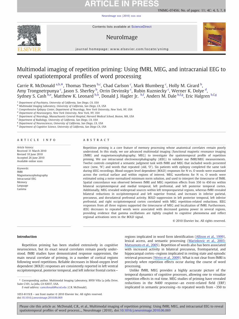
Fig. 1. Cluster-thresholded t-stat maps of the fMRI NNO (red/yellow) and ONN (cyan/blue)(right panel). Activity clusters are shown on the lateral and ventral inflated surfaces of bopb .001 to pb .00001 and represent significant clusters of activity exceeding a t-stat and siz
Please cite this article as: McDonald, C.R., et al., Multimodal imaging ofspatiotemporal profiles of word processi..., NeuroImage (2010), doi:10.
(Lachaux et al., 1999). Constant temporal and frequency resolutionacross target frequencies was obtained by adjusting the waveletwidths according to the target frequency. The wavelet widths increaselinearly from 14 to 38 as frequency increases from 70 to 190 Hz,resulting in a constant temporal resolution (σt) of 16 ms andfrequency resolution (σf) of 10 Hz. For each epoch, spectral powerwas calculated from the wavelet spectra, normalized by the inversesquare frequency to adjust for the rapid drop-off in the powerspectrum with frequency, and averaged from 70 to 190 Hz, excludingline noise harmonics. Visual inspection of the resulting high gammapower waveforms revealed additional artifacts not apparent in thetime-domain signal, and the artifactual epochs were excluded fromthe gamma power analysis. Both ERP and gamma waveforms werecompared across stimulus types using a nonparametric randomiza-tion test with temporal clustering to correct for multiple comparisons(Maris and Oostenveld, 2007).
Statistical analysis
Functional MRI t-stat cluster maps for the NNO and ONN contrastswere compared to the t-stat cluster maps generated for MEG of the Nvs. O difference waveform at each of the three time windows ofinterest. To examine the time course of regional priming effects, timewindow×condition repeated measures (RM) ANOVAs were per-formed on the average source estimates within each ROI andhemisphere. To explore the relationship between the hemodynamicand electromagnetic repetition priming effects, Spearman correla-tions were performed between fMRI percent signal change andmodulation of the MEG difference waveform within each ROI andtimewindow. Percent signal changewas selected due to evidence thatthis measure of the BOLD response correlates with N400 primingeffects in EEG studies (Matsumoto et al., 2005).
Results
Fig. 1 displays cluster-based t-stat surface maps of the NNO (red-yellow) and ONN (blue-cyan) contrasts for fMRI (left panel) and the Nvs. O difference waveform (red-yellow) for MEG time windows of
contrasts (left panel) and the MEG N vs. O difference waveform for four time windowsth hemispheres (gyri=light gray; sulci=dark gray). Significance values range from ae threshold.
repetition priming: Using fMRI, MEG, and intracranial EEG to reveal1016/j.neuroimage.2010.06.069
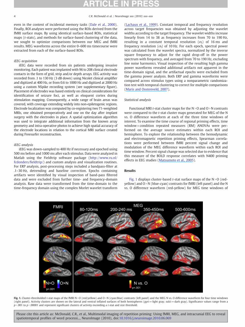
Fig. 2. fMRI cluster-thresholded t-stat maps of the NNO and ObN contrasts with MEG waveforms extracted from each significant fMRI cluster. MEG waveforms are shown from 0 to600 ms for new words (red) and old words (blue) within each ROI. Significance (*) denotes regions producing a condition×time interaction. Y-axis values reflect noise-normalizeddipole strengths and can be conceptualized as estimates of the average signal-to-noise within an ROI.
Table 2Average percent signal change for each region of interest.
Region of interest Mean % signal change(standard deviation)
Range of % signal change
NNOLeft entorhinal .16 (.05) .10–.23Left ventral occipitotemporal .14 (.04) .08–.21Left posterior inferior frontal .16 (.04) .10–.23Left superior temporal .15 (.03) .11–.20Left middle temporal .15 (.04) .11–.24Left pars orbitalis .14 (.04) .09–.21Left parahippocampal .16 (.03) .11–.21Left lateral occipital .16 (.04) .11–.22Left superior medial frontal .19 (.02) .09–.24Right entorhinal .13 (.03) .08–.21Right ventral occipitotemporal .14 (.03) .10–.20Right lateral occipitotemporal .14 (.03) .10–.19
ONNLeft dorsolateral prefrontal .15 (.03) .10–.20Left inferior parietal .15 (.04) .10–.23Left precuneus .14 (.04) .10–.22Right dorsolateral prefrontal .14 (.04) .10–.20Right inferior parietal .14 (.03) .09–.20Right precuneus .13 (.03) .10–.18
5C.R. McDonald et al. / NeuroImage xxx (2010) xxx–xxx
interest (right panel). Fig. 2 portrays the same fMRI cluster maps, withMEG timecourses extracted from significant ROIs identified on thefMRI surfaces.
fMRI
Prominent repetition-related effects for fMRI were observedwithin 16 regions in the left or right hemisphere. For the NNOcontrast, significant clusters were observed in the left inferiorprefrontal extending into the precentral gyrus, pars orbitalis, posteriorsuperior temporal, middle temporal, parahippocampal, and superiormedial frontal, as well as in bilateral entorhinal, lateral occipitotem-poral, and ventral occipitotemporal cortex that extends into anteriorfusiform on the left. In addition, ONN responses were observedwithinbilateral dorsolateral prefrontal cortex, precuneus, inferior parietal/supramarginal cortex. Average percent signal change for the NNO andObN responses are provided for each ROI in Table 2.
MEG
Cluster-thresholded surface maps of the MEG O vs. N differencewaveform revealed a dynamic pattern of activity that was observedprimarily in bilateral orbital and ventral occipitotemporal cortex earlyon (~80–120 ms), followed by repetition effects from ~200 to 240 msin the superior temporal cortex bilaterally, left dorsolateral prefrontal,inferior parietal, and left ventral occipitotemporal regions. Inspectionof the waveforms in Fig. 2 reveals that this early activity is greater forO relative to N words across most regions and may reflect a temporaladvantage for processing repeating word stimuli (Marinkovic et al.,2003). This pattern of activity appears increasingly left lateralizedfrom 350 to 450 ms, and is characterized by greater responses to Nrelative to O words, consistent with modulation of the N400 responseto repeated stimuli. In particular, prominent clusters of activity were
Please cite this article as: McDonald, C.R., et al., Multimodal imaging ofspatiotemporal profiles of word processi..., NeuroImage (2010), doi:10.
observed within left inferior prefrontal, left superior, middle, andinferior temporal regions, left ventral occipitotemporal (lingual andfusiform), left temporal pole, left entorhinal, left parahippocampal,and left inferior parietal cortex including parts of the angular andsupramarginal gyri. In addition, activity is observed within the rightlateral and ventral occipitotemporal cortex (mostly lingual). By ~500–600 ms, activity is seen within many of the same regions and againappears more bilaterally distributed.
repetition priming: Using fMRI, MEG, and intracranial EEG to reveal1016/j.neuroimage.2010.06.069

6 C.R. McDonald et al. / NeuroImage xxx (2010) xxx–xxx
To estimate the time course within selected ROIs, time bycondition RM ANOVAs were performed (see Fig. 2). Within the lefthemisphere, time by condition interactions were observed within theventral occipitotemporal [F(2, 22)=3.8, pb .05)], superior temporal[F(2, 22)=3.7, pb .05)], middle temporal [F(2, 22)=4.7, pb .05], parsorbitalis [F(2, 22)=3.8, pb .05)], supramarginal [F(2, 22)=4.0,pb .05)], inferior frontal [F(2, 22)=3.9, pb .05)], entorhinal [F(2,22)=4.1, pb .01)], parahippocampal [F(2, 22)=3.5, pb .05)], andtemporal pole [F(2, 22)=3.8, pb .05)]. Within the right hemisphere,time x condition interactions were observed in the right ventraloccipitotemporal only [F(2, 22)=3.4 pb .05)]. In each case, newwords produced greater responses than old words, but only in the350–450 ms time window. Despite a trend for ONN word responsesin the left and right dorsolateral prefrontal and superior temporalROIs in the 500–600 ms time window and across many regions in the200–240 ms time window (p-values between .05 and .10), none ofthe ONN comparisons reached statistical significance at the ROI level.
fMRI/MEG correlational analysis
Visual inspection of the surface maps and ROI waveforms reveals thatthe fMRI NNO effect most closely resembles the MEG 350–450 ms timewindow, presumably representingmodulationof theN400effect. In orderto examine the relationship between the electromagnetic N400 andhemodynamic priming effects, Spearman correlations were calculatedacross subjects for each ROI between fMRI percent signal change valuesand MEG difference waveform values within each time window.Correlational analysis revealed a positive relationship between fMRINNO and MEG N vs. O responses in the left inferior prefrontal (r=.72,pb .01), left superior temporal (r=.53, pb .05) and right ventraloccipitotemporal (r=.56, pb .05) ROIs, but only within the 350–450 mstime window. Despite similar spatial patterns of activity between MEGand fMRI at the surface level in other ROIs and time windows, themagnitude of the BOLD signal change did not correlate with themodulation MEG waveforms in other regions across individual subjects.
Intracranial validation of MEG/fMRI repetition effects
Inverse methods for source localization based on MEG and fMRIrequire unproven a priori assumptions. In contrast, it is possible todemonstrate local generation from intracranial recordings withoutambiguities. Thus, we also sampled responses to N vs. O words fromintracranial recordings obtained in six patients. Event-related poten-tials and high gamma frequency analyses were performed on allpatient responses. Table 3 summarizes the number of patients
Table 3Neocortical regions showing repetition effects in MEG and iEEG ERP and high gammaresponses.
Region of interest 350–450 500–600
MEG ERP 70+Hz MEG ERP 70+Hz
LeftDorsolateral prefrontal 1/4 2/4 1/4 3/4a
Inferior frontal X 2/3 0/3 2/3 1/3Lateral occipitotemporal 2/4 2/4 1/4 1/4a
Ventral occipitotemporal X 2/4 2/4 0/4 0/4Superior medial frontal 1/2 0/2 0/2 1/2a
Superior temporal X 2/4 1/4 1/4 1/4a
RightDorsolateral prefrontal 0/1 0/1 1/1 0/1Lateral occipitotemporal X 1/1 1/1 0/4 1/4a
Ventral occipitotemporal X 2/2 2/2 2/2 0/2
a Reflects increases in gamma power to O vs. N words. All other responses in the70+Hz analysis reflect increases to N vs. O gamma power. X reflects a lack of anysignificant effect. Fractions represent number of patients showing an effect overnumber of patients with grid coverage in the anatomical region.
Please cite this article as: McDonald, C.R., et al., Multimodal imaging ofspatiotemporal profiles of word processi..., NeuroImage (2010), doi:10.
showing locally generated iEEG repetition effects within the 350–450 and 500–600 ms time windows.
Event-related potential analysis
Fig. 3 shows examples of localized ERPs from four patients basedon iEEG recordings. Of our six patients, four had grid coverage of theleft posterior superior temporal gyrus/sulcus. Two of the four patientsshowed a locally generated NNO response between 350 and 500 ms inthe vicinity of the left posterior superior temporal region in or nearWernicke's area (Patients A and E). One patient showed an ONNresponse in the same region that peaked at 600 ms following stimuluspresentation (Patient D). Only three patients had grids covering theleft inferior frontal region near pars opercularis. Two of the threepatients demonstrated a NNO response that peaked between 400 and500 ms following stimulus presentation (Patients B and D). Five of sixpatients had grid coverage of the lateral occipitotemporal region,either on the left (N=4) or right (N=1). Two of the four with leftcoverage showed a N vs. O difference in lateral occipitotemporalcortex that peaked between 350 and 500 ms (Patients A and E). Thepatient with right coverage showed a similar ERP response in rightlateral occipitotemporal cortex (Patient C). Five patients had at leastpartial coverage of the ventral occipitotemporal region (Left=3;Right=2), with one showing N vs. O differences on the left (Patient A)between 350 and 400 ms and two patients showing a NNO responseon the right (Patients C and F) between 400 and 600 ms in this region.Four patients had partial coverage of the left dorsolateral prefrontalcortex. Two of these patients had strips extending into superiormedial frontal cortex on the left. Of the four with left dorsolateralprefrontal coverage, one demonstrated NNO responses between 350and 450 ms (Patient E). The same patient also demonstrated ONNresponses between 500 and 700 ms in left dorsolateral prefrontalcortex. One patient had right dorsolateral prefrontal coverage anddemonstrated a NNO response between 500 and 600 ms (Patient F).In addition, one patient with coverage of the left superior medialfrontal region showed a local NNO response between 350 and 450 ms(Patient A).
Time course of high gamma (70–190 Hz) power
In addition to the ERP analysis, high gamma responses between 70and 190 Hz were sampled in all six patients from the same electrodelocations as the ERPs (see examples in Fig. 4). High gamma activitywas selected to investigate whether or not gamma oscillations showstronger co-localization with fMRI repetition effects than ERPs in oneor more regions, as well as to interpret the valence of our ERPresponses. Statistical analysis revealed gamma power differences to Nvs. O words in the left superior temporal region in two patients(Patients A and E). In one patient, increased gamma power wasassociated with NNO words that peaked at 400 ms (Patient A),whereas the other patient showed ONN increases in gamma thatpeaked at 500 ms (Patient D). In both cases, the gamma oscillationssupported ERPs generated at or near the same electrode location.Three patients showed high gamma differences in lateral occipito-temporal cortex (Patients C, D, and E; two left and one right). Of theseresponses, NNO differences were seen in two patients that began~300 ms following word presentation (Patients C and D). In the otherpatient, ONN gamma differences were observed later in the timecourse, peaking around 500 ms (Patient E). Three patients showedincreased gamma in ventral occipitotemporal cortex (Patients A, C,and F; one left and two right). In all cases, increased gammaoscillations peaked between 400 and 500 ms and reflected NNOresponses. Two patients showed increased gamma for NNO words inleft dorsolateral prefrontal (Patients A and D) associated with theN400 responses in the ERP data. In addition, increased gamma forONN words was observed in the left dorsolateral prefrontal cortex in
repetition priming: Using fMRI, MEG, and intracranial EEG to reveal1016/j.neuroimage.2010.06.069

Fig. 3. Example ERPs from iEEG recordings in patients with intractable epilepsy. The y-axis represents the amplitudes (μV) of the N vs. O words, scaled individually for each patientShaded areas reflect time windows during which N vs. O words showed significantly different temporal clusters in the ERPs using randomization statistics. OCT=occipitotemporal
7C.R. McDonald et al. / NeuroImage xxx (2010) xxx–xxx
three patients (Patients A, B, and D) and left superior medial/anteriorcingulate cortex in one patient (Patient D) from ~500 to 700 ms post-stimulus, associated with late repetition effects.
Discussion
Using advanced multimodal imaging, we demonstrate spatialconcordance between fMRI and MEG N400 priming effects within leftinferior prefrontal extending into precentral, left posterior superiortemporal, left medial temporal, right lateral occipitotemporal, andbilateral ventral occipitotemporal cortex, encompassing much of thelingual and fusiform cortex on the left. We also provide iEEGvalidation of our MEG/fMRI responses in key regions in multiplepatients and reveal co-localization between increased gamma powerin iEEG and the fMRI BOLD response. Table 4 summarizes regions ofspatiotemporal concordance associated with repetition priming
Please cite this article as: McDonald, C.R., et al., Multimodal imaging of repetition priming: Using fMRI, MEG, and intracranial EEG to revealspatiotemporal profiles of word processi..., NeuroImage (2010), doi:10.1016/j.neuroimage.2010.06.069
.
.
across all three modalities. As can be seen, these priming patternswere largely characterized by reduced responses to repeated words,suggesting facilitation of word processing that is measurable in thehemodynamic and electromagnetic signals.
Furthermore, fMRI revealed repetition enhancement in whichrepeated words produced greater responses relative to new words inbilateral dorsolateral prefrontal, precuneus, and inferior parietal/supramarginal cortex. Although the MEG ROI analysis indicates a late(~500–600 ms) trend for an ONN effects within these ROIs thatexceeded our cluster threshold in the surface maps, the conditioncontrasts did not reach significance at the ROI level. Thismay be due toour short trial length (i.e., 600 ms), which precluded a completeanalysis of the late repetition effects that often persist until 700–800 ms (Dale et al., 2000). This late effect has been referred to as theLPC/P3b andmay reflect conscious recollection of the repeated words.This has been described previously in incidental memory tasks with a

Fig. 4. Gamma powerwaves (integrated from 70 to 190 Hz) from iEEG recordings in patients with intractable epilepsy. The y-axis represents gamma power and is scaled for each plotto optimize visibility of the response. Shaded areas reflect time windows during which N vs. O words showed significantly different temporal clusters in high gamma power usingrandomization statistics. OCT=occipitotemporal; DLPF=dorsolateral prefrontal; VLPF=ventrolateral prefrontal.
8 C.R. McDonald et al. / NeuroImage xxx (2010) xxx–xxx
large number of repetitions (Dale et al., 2000), presumably becauseword repetitions become apparent to the participant. In addition, theMEG surface maps reveal significant O vs. N differences within leftventral occipitotemporal and bilateral superior temporal clustersbetween 200 and 240 ms that appear to reflect early, transientenhancements to repeated stimuli, as reported in previous MEGstudies of visualwordprocessing (Dhondet al., 2001;Marinkovic et al.,2003). Although these N vs. O effects were not significant in the ROIanalysis during the early time window, inspection of the waveformsreveals a trend toward ONN responses across several ROIs. Evidencefor ONN activitywithin these ROIswas not captured by fMRI and it hasbeen suggested that the transient nature of the early ONN response isnot robust enough to overcome the sustained NNO response that
Please cite this article as: McDonald, C.R., et al., Multimodal imaging ofspatiotemporal profiles of word processi..., NeuroImage (2010), doi:10.
dominates the fMRI BOLD response in these same regions (Marinkovicet al., 2003).
Correlational analysis between our fMRI and MEG repetitionpriming effects across individuals revealed that fMRI BOLD suppres-sion correlated with MEG priming effects from 350 to 450 ms, butonly within the left inferior prefrontal, left superior temporal, andright occipitotemporal region. Repetition suppression within each ofthese regions has been reported in previous neuroimaging studies ofword and object priming (Marinkovic et al., 2003; Schacter andBuckner, 1998). Our within-subject, multimodal fMRI/MEG analysisprovides further evidence that these regional priming effects arerobust across imaging modalities. Although the relationship betweenelectromagnetic and hemodynamic response changes is complex, a
repetition priming: Using fMRI, MEG, and intracranial EEG to reveal1016/j.neuroimage.2010.06.069

Table 4Neocortical regions showing significant repetition effects in each modality.
Region of interest fMRI MEG 350–450 ms iEEGa
LeftTemporal pole XDorsolateral prefrontal Xb XEntorhinal X XInferior frontal c X X XLateral occipitotemporal X XVentral occipitotemporal X X XMiddle temporal X XParahippocampal X XPars orbitalis X XSupramarginal Xb XSuperior medial frontal X XSuperior temporal X X XTemporoparietal X
RightDorsolateral prefrontal Xb
Entorhinal XLateral occipitotemporal X X XVentral occipitotemporal X X XSupramarginal Xb
a Regions in which one or more patients showed a significant iEEG repetition effect.The absence of an effect in the iEEG column does not necessary represent discordance,but may reflect inadequate grid coverage or the elimination of electrodes in whichinterictal activity was detected.
b Regions showing ONN effects. All other regions represent those showing NNOeffects.
c Bolded regions are those demonstrating concordance across all three modalities.
9C.R. McDonald et al. / NeuroImage xxx (2010) xxx–xxx
direct correlation between BOLD signal change and N400modulationshas been observed with EEG within the vicinity of the left superiortemporal gyrus (Matsumoto et al., 2005). We extend the literature bydemonstrating fMRI-MEG correlations within other perisylvianregions implicated in the N400 effect and by providing validationfrom iEEG of their local generation.
Recent neuroimaging research has demonstrated that repetitionsuppression across neocortical regions is unlikely to reflect a unitaryprocess. Rather, there is convincing evidence that repetition priminginvolves multiple component processes with partially unique ana-tomical substrates. Whereas early response suppression withinbilateral occipitotemporal regions likely reflects a “sharpening” ofneural activity in response to the perceptual attributes of the stimulus(Fiebach et al., 2005; Schacter and Buckner, 1998) and visual wordform priming (Cohen et al., 2002), the late effects (N300 ms)identified in our MEG responses suggest significant top-downinfluences that may reflect general attention-dependent priming forconceptual information (Klaver et al., 2007). This top-down interpre-tation is supported by iEEG ERP and/or high gamma recordings in fiveof six patients who showed late NNO responses in left or rightoccipitotemporal cortex in or near regions showing strong activationin fMRI and MEG. This late NNO effect in occipitotemporal cortex hasnot been previously reported across all three modalities and providesevidence that priming effects within extrastriate cortex are influencedby both feedforward and feedback mechanisms.
In addition, response suppression within left posterior temporalcortex has been attributed to facilitation of lexical access (Matsumotoet al., 2005) or recoding of a written word into a lexical representation(Klaver et al., 2007). This component of repetition priming appears tobe independent of the earlier perceptual effects, and may representautomatic, spreading activation of a word's representation. Our datademonstrate response suppressionwithin left temporal cortex in fMRIand in the MEG waveforms that is highly lateralized and sustainedafter ~250 ms. This time period is congruent with other studies thathave identified a lexical-processing stage that may represent atransitional stage between perceptual and conceptual priming(Marinkovic et al., 2003). However, the sustained effects observed
Please cite this article as: McDonald, C.R., et al., Multimodal imaging ofspatiotemporal profiles of word processi..., NeuroImage (2010), doi:10.
in these regions in theMEG surfacemaps andwaveforms indicate thatleft temporal regions also contribute to the main N400 component ofrepetition priming.
Response suppression within the left inferior prefrontal region isgenerally believed to reflect conceptual priming, including a reduceddemand for semantic memory retrieval (Wagner et al., 2001) and/orfacilitation of response selection (Thompson-Schill et al., 1997). OurMEG and fMRI data show response suppression in the left inferiorfrontal cortex that appears to evolve somewhat latter than thepriming effects observed in posterior temporal cortex and is maximalbetween 350 and 450 ms (i.e., peak N400 effect). The sustainedresponse suppression in left temporal regions during this timesupports the notion that left lateral temporal cortex is important forinitial lexical access, as well as interactions with prefrontal regionsduring semantic memory retrieval (Gold and Buckner, 2002).Inspection of the iEEG waveforms provides evidence of locallygenerated N400 responses within the left posterior superior temporaland left inferior prefrontal cortex (i.e., in or near Wernicke's andBroca's areas) that support our fMRI and MEG N400 reductions andrepresent core anatomical substrates of conceptual priming.
Taken together, our data provide evidence that the main repetitionpriming effects seen in fMRI BOLD responses are those associated withthe MEG N400 reductions, and not necessarily with the more transientearly or late repetition effects that are apparent in the MEG waveforms.This is consistent with previous findings that iEEG/MEG and BOLDresponses correlate reasonably well for the N400, but not as stronglywith the scalp P3b/LPC (Halgren, 2004b). However, our iEEG data didreveal ONN late repetition effects in left dorsolateral prefrontal cortex inall four patients with recordings sampled from this region that peakedbetween 500 and 600 ms and may reflect LPC/P3b effects. In threepatients, the responses were observed in iEEG high gamma responsesbut not in the ERPs, suggesting that the BOLD effect associated withconscious recollection may be tightly coupled to increased power in thehigh frequency ranges not always captured with MEG or ERP measure-ments (Jerbi et al., 2009; Yuval-Greenberg et al., 2008).
The exact relationship between the BOLD response and increasedgamma oscillations observed in cognitive tasks in not entirely known,but memory formation has been associated with increased gammaoscillations recorded with iEEG in left temporal and prefrontal regions(Sederberg et al., 2007). We extend the literature by demonstratingan association between increased gamma power and word primingeffects in the context of an incidental memory task. Furthermore, wereport increased gamma power for NNO responses in numerouspatients in ventral and lateral occipitotemporal, inferior prefrontal,superior temporal, andmedial prefrontal cortex that are also reflectedin our fMRI activations—many of which were not observed in the ERPdata. These findings are consistent with an emerging literaturedemonstrating that gamma power co-localize with BOLD variationsacross numerous cortical regions during lexico-semantic tasks(Lachaux et al., 2007), presumably due to its correlation with localneuronal firing (Manning et al., 2009).
In this study, we provide multimodality evidence for the spatio-temporal profile of repetition word priming using fMRI/MEG withsupport from iEEG recordings. However, there are several limitationsto our study that should be noted. First, whereas our MEG and iEEGtasks were event-related, we used a blocked version of the same taskfor fMRI. A blocked design was selected in order to increase the SNR,optimizing our ability to detect very subtle BOLD changes associatedwith repetition priming in anterior and ventral temporal lobe regionsthat are known to be susceptible to signal loss (Chee et al., 2003; Daleet al., 2000). Although numerous studies have reported highly similarpatterns of activations between blocked and event-related fMRIdesigns using lexical-semantic tasks (Chee et al., 2003; Pilgrim et al.,2002;Wagner et al., 2005;Weiss et al., 2009), blocked presentation ofitems may induce strategies or lead to greater levels of habituation insome regions relative to event-related designs. This may explain the
repetition priming: Using fMRI, MEG, and intracranial EEG to reveal1016/j.neuroimage.2010.06.069

10 C.R. McDonald et al. / NeuroImage xxx (2010) xxx–xxx
bilateral precuneus and lateral occipitotemporal activations seen inour fMRI data that were not apparent with MEG. Greater BOLDresponses to repeated stimuli have been reported previously inbilateral precuneus (Horner and Henson, 2008)—a region implicatedin episodic retrieval (Wagner et al., 2005) and task difficulty/workload(Korsnes et al., 2008; Scheibe et al., 2006). It is unclear in our studywhether the fMRI responses observed in this region reflect consciousrecollection of previous items or the lower task demands introducedby our blocked presentation. However, the presence of lateraloccipitotemporal repetition effects in our iEEG recordings that co-localize with the fMRI activations suggest that task design differencesare unlikely to account for the occipitotemporal findings. Althoughusing identical task designs for fMRI and MEG/iEEG would appearideal, there are possible limitations to this approach as well. Had weimplemented an event-related fMRI design, wemay have reduced ourSNR and failed to detect subtle task effects in the anteriormedialtemporal lobe that appear to be involved in repetition priming.Alternatively, we could have designed an event-related fMRI designwith a higher number of trials per condition to increase the SNR.However, this would have resulted in a greater number of repetitionsin our fMRI task relative to MEG and iEEG tasks. The number ofrepetitions has been shown to influence priming effects (Ostergaard,1998), introducing another confound. Despite task differences, thehigh spatial correlation across imaging modalities in many criticalregions suggests that most of the repetition priming effects wererobust to differences in the task design.
Second, we used a very brief SOA (i.e., 600 ms) in order to increaseour SNR by allowing for a large number of averages across taskconditions in a relatively short time. This brief SOAdiminishedour abilityto fully evaluate very late MEG repetition effects that generally peakaround 600 to 700 ms post-stimulus. Finally, it is important to note thatiEEG data are acquired from patients who are undergoing evaluation forsurgical resection of an epileptic focus. Therefore, many of the iEEGresponses were sampled near diseased tissue, and the area sampled ineach patient varies and is limited to the details of the electrodeplacement. These are well-known limitations of iEEG research thatcannot be avoided. As is accepted practice in iEEG research (Jerbi et al.,2009), we sought to minimize the effects of brain pathology in our iEEGresponses by eliminating electrodes from which ictal or interictaldischarges were recorded. However, it is still possible that one or moreof these factorsmitigatedour ability to record clearN400or LPCeffects insome of the patients or consistently across regions. Nevertheless, wewere still able to detect local repetition priming effects with iEEG in oneor more patients that supported the temporal and spatial patternsdetected with our non-invasive measures. Multimodal imaging datasuch as these provide unique insight into the timing, location, andspectral features of cognitive processes, such as repetition priming, anddemonstrate the validity of using non-invasive measures for under-standing complex brain functions.
Acknowledgments
The work was supported by National Institutes of Health (NIH)Grant K23NS056091 (C.R.M) and NS18741 (E.H.).
Appendix A. Supplementary data
Supplementary data associated with this article can be found, inthe online version, at doi:10.1016/j.neuroimage.2010.06.069.
References
Allison, T., Puce, A., Spencer, D.D., McCarthy, G., 1999. Electrophysiological studies ofhuman face perception: I. Potentials generated in occipitotemporal cortex by faceand non-face stimuli. Cereb Cortex 9, 415–430.
Please cite this article as: McDonald, C.R., et al., Multimodal imaging ofspatiotemporal profiles of word processi..., NeuroImage (2010), doi:10.
Chee, M.W., Venkatraman, V., Westphal, C., Siong, S.C., 2003. Comparison of block andevent-related fMRI designs in evaluating the word-frequency effect. Hum. BrainMapp. 18, 186–193.
Cohen, L., Lehericy, S., Chochon, F., Lemer, C., Rivaud, S., Dehaene, S., 2002. Language-specific tuning of visual cortex? Functional properties of the visual word form area.Brain 125, 1054–1069.
Dalal, S.S., Baillet, S., Adam, C., Ducorps, A., Schwartz, D., Jerbi, K., Bertrand, O., Garnero,L., Martinerie, J., Lachaux, J.P., 2009. Simultaneous MEG and intracranial EEGrecordings during attentive reading. Neuroimage 45, 1289–1304.
Dale, A.M., Fischl, B., Sereno, M.I., 1999. Cortical surface-based analysis: I. Segmentationand surface reconstruction. Neuroimage 9, 179–194.
Dale, A.M., Halgren, E., 2001. Spatiotemporal mapping of brain activity by integration ofmultiple imaging modalities. Curr. Opin. Neurobiol. 11, 202–208.
Dale, A.M., Liu, A.K., Fischl, B.R., Buckner, R.L., Belliveau, J.W., Lewine, J.D., Halgren, E.,2000. Dynamic statistical parametric mapping: combining fMRI and MEG for high-resolution imaging of cortical activity. Neuron 26, 55–67.
Devlin, J.T., Russell, R.P., Davis, M.H., Price, C.J., Wilson, J., Moss, H.E., Matthews, P.M.,Tyler, L.K., 2000. Susceptibility-induced loss of signal: comparing PET and fMRI on asemantic task. Neuroimage 11, 589–600.
Dhond, R.P., Buckner, R.L., Dale, A.M., Marinkovic, K., Halgren, E., 2001. Spatiotemporalmaps of brain activity underlying word generation and their modification duringrepetition priming. J. Neurosci. 21, 3564–3571.
Fiebach, C.J., Gruber, T., Supp, G.G., 2005. Neuronal mechanisms of repetition priming inoccipitotemporal cortex: spatiotemporal evidence from functional magneticresonance imaging and electroencephalography. J. Neurosci. 25, 3414–3422.
Fischl, B., Salat, D.H., Busa, E., Albert, M., Dieterich, M., Haselgrove, C., van der Kouwe, A.,Killiany, R., Kennedy, D., Klaveness, S., Montillo, A., Makris, N., Rosen, B., Dale, A.M.,2002. Whole brain segmentation: automated labeling of neuroanatomicalstructures in the human brain. Neuron 33, 341–355.
Fischl, B., Sereno, M.I., Dale, A.M., 1999a. Cortical surface-based analysis: II. Inflation,flattening, and a surface-based coordinate system. Neuroimage 9, 195–207.
Fischl, B., Sereno, M.I., Tootell, R.B., Dale, A.M., 1999b. High-resolution intersubjectaveraging and a coordinate system for the cortical surface. Hum. Brain Mapp. 8,272–284.
Francis, W.N., Kucera, H., 1982. Frequency Analysis of English Usage: Lexicon andGrammar. Houghton Mifflin, Boston.
Gold, B.T., Buckner, R.L., 2002. Common prefrontal regions coactivate with dissociableposterior regions during controlled semantic and phonological tasks. Neuron 35,803–812.
Hagler Jr., D.J., Saygin, A.P., Sereno, M.I., 2006. Smoothing and cluster thresholding forcortical surface-based group analysis of fMRI data. Neuroimage 33, 1093–1103.
Halgren, E., 2004a. How can intracranial recordings assist MEG source localization?Neurol. Clin. Neurophysiol. 2004, 86.
Halgren, E., 2004b. How can intracranial recordings assist MEG source localization?Neurol. Clin. Neurophysiol. 86, 1–18.
Halgren, E., Baudena, P., Heit, G., Clarke, J.M., Marinkovic, K., Clarke, M., 1994. Spatio-temporal stages in face and word processing: I. Depth-recorded potentials in thehuman occipital, temporal and parietal lobes [corrected]. J. Physiol. Paris 88, 1–50.
Halgren, E., Wang, C., Schomer, D.L., Knake, S., Marinkovic, K., Wu, J., Ulbert, I., 2006.Processing stages underlying word recognition in the anteroventral temporal lobe.Neuroimage 30, 1401–1413.
Hamalainen, M., Hari, R., Ilmoniemi, R.J., Knuutila, J., Lounasmaa, O.V., 1993.Magnetoencephalography—theory, instrumentation, and applications to noninva-sive studies of the working human brain. Rev. Mod. Phys. 65, 413–497.
Horner, A.J., Henson, R.N., 2008. Priming, response learning and repetition suppression.Neuropsychologia 46, 1979–1991.
Jenkinson, M., Bannister, P., Brady, M., Smith, S., 2002. Improved optimization for therobust and accurate linear registration and motion correction of brain images.Neuroimage 17, 825–841.
Jenkinson, M., Smith, S., 2001. A global optimisation method for robust affineregistration of brain images. Med. Image Anal. 5, 143–156.
Jerbi, K., Ossandon, T., Hamame, C.M., Senova, S., Dalal, S.S., Jung, J., Minotti, L., Bertrand,O., Berthoz, A., Kahane, P., Lachaux, J.P., 2009. Task-related gamma-band dynamicsfrom an intracerebral perspective: review and implications for surface EEG andMEG. Hum. Brain Mapp. 30, 1758–1771.
Klaver, P., Schnaidt, M., Fell, J., Ruhlmann, J., Elger, C.E., Fernandez, G., 2007. Functionaldissociations in top-down control dependent neural repetition priming. Neuro-image 34, 1733–1743.
Korsnes, M.S., Wright, A.A., Gabrieli, J.D., 2008. An fMRI analysis of object priming andworkload in the precuneus complex. Neuropsychologia 46, 1454–1462.
Lachaux, J.P., Fonlupt, P., Kahane, P., Minotti, L., Hoffmann, D., Bertrand, O., Baciu,M., 2007.Relationship between task-related gamma oscillations and BOLD signal: new insightsfrom combined fMRI and intracranial EEG. Hum. Brain Mapp. 28, 1368–1375.
Lachaux, J.P., Rodriguez, E., Martinerie, J., Varela, F.J., 1999. Measuring phase synchronyin brain signals. Hum. Brain Mapp. 8, 194–208.
Manning, J.R., Jacobs, J., Fried, I., Kahana, M.J., 2009. Broadband shifts in local fieldpotential power spectra are correlated with single-neuron spiking in humans. J.Neurosci. 29, 13613–13620.
Marinkovic, K., Dhond, R.P., Dale, A.M., Glessner, M., Carr, V., Halgren, E., 2003.Spatiotemporal dynamics of modality-specific and supramodal word processing.Neuron 38, 487–497.
Maris, E., Oostenveld, R., 2007. Nonparametric statistical testing of EEG- and MEG-data.J Neurosci Methods 164, 177–190.
Matsumoto, A., Iidaka, T., Haneda, K., Okada, T., Sadato, N., 2005. Linking semanticpriming effect in functional MRI and event-related potentials. Neuroimage 24,624–634.
repetition priming: Using fMRI, MEG, and intracranial EEG to reveal1016/j.neuroimage.2010.06.069

11C.R. McDonald et al. / NeuroImage xxx (2010) xxx–xxx
Oldfield, R.C., 1971. The assessment and analysis of handedness: the Edinburghinventory. Neuropsychologia 9, 97–113.
Oostendorp, T.F., Van Oosterom, A., 1992. Source parameter estimation using realisticgeometry in bioelectricity and biomagnetism. In: Nenonen, J., Rajala, H.M., Katila, T.(Eds.), Biomagnetic Localization and 3D Modeling. Helsinki University ofTechnology, Helsinki. Report TKK-F-A689,.
Ostergaard, A.L., 1998. The effects on priming of word frequency, number of repetitions,and delay depend on the magnitude of priming. Mem Cognit 26, 40–60.
Pilgrim, L.K., Fadili, J., Fletcher, P., Tyler, L.K., 2002. Overcoming confounds of stimulusblocking: an event-related fMRI design of semantic processing. Neuroimage 16,713–723.
Schacter, D.L., Buckner, R.L., 1998. Priming and the brain. Neuron 20, 185–195.Scheibe, C., Wartenburger, I., Wustenberg, T., Kathmann, N., Villringer, A., Heekeren, H.R.,
2006. Neural correlates of the interaction between transient and sustained processes:a mixed blocked/event-related fMRI study. Hum. Brain Mapp. 27, 545–551.
Sederberg, P.B., Schulze-Bonhage, A., Madsen, J.R., Bromfield, E.B., McCarthy, D.C.,Brandt, A., Tully, M.S., Kahana, M.J., 2007. Hippocampal and neocortical gammaoscillations predict memory formation in humans. Cereb. Cortex 17, 1190–1196.
Thompson-Schill, S.L., D'Esposito, M., Aguirre, G.K., Farah, M.J., 1997. Role of left inferiorprefrontal cortex in retrieval of semantic knowledge: a reevaluation. Proc. NatlAcad. Sci. USA 94, 14792–14797.
Please cite this article as: McDonald, C.R., et al., Multimodal imaging ofspatiotemporal profiles of word processi..., NeuroImage (2010), doi:10.
Wagner, A.D., Pare-Blagoev, E.J., Clark, J., Poldrack, R.A., 2001. Recoveringmeaning: left prefrontal cortex guides controlled semantic retrieval. Neuron31, 329–338.
Wagner, A.D., Shannon, B.J., Kahn, I., Buckner, R.L., 2005. Parietal lobe contributions toepisodic memory retrieval. Trends Cogn. Sci. 9, 445–453.
Weiss, A.P., Ellis, C.B., Roffman, J.L., Stufflebeam, S., Hamalainen, M.S., Duff, M., Goff, D.C.,Schacter, D.L., 2009. Aberrant frontoparietal function during recognitionmemory inschizophrenia: a multimodal neuroimaging investigation. J. Neurosci. 29,11347–11359.
Whitham, E.M., Lewis, T., Pope, K.J., Fitzgibbon, S.P., Clark, C.R., Loveless, S.,DeLosAngeles, D., Wallace, A.K., Broberg, M., Willoughby, J.O., 2008. Thinkingactivates EMG in scalp electrical recordings. Clin. Neurophysiol. 119,1166–1175.
Woolrich, M.W., Ripley, B.D., Brady, M., Smith, S.M., 2001. Temporal autocorrelation inunivariate linear modeling of FMRI data. Neuroimage 14, 1370–1386.
Worsley, K.J., Marrett, S., Neelin, P., Vandal, A.C., Friston, K.J., Evans, A.C., 1996. A unifiedstatistical approach for determining significant signals in images of cerebralactivation. Hum. Brain Mapp. 4, 58–73.
Yuval-Greenberg, S., Tomer, O., Keren, A.S., Nelken, I., Deouell, L.Y., 2008. Transientinduced gamma-band response in EEG as a manifestation of miniature saccades.Neuron 58, 429–441.
repetition priming: Using fMRI, MEG, and intracranial EEG to reveal1016/j.neuroimage.2010.06.069