
MPI Vol. 8 No.2 (11 MB ) - MEDICAL PHYSICS ...
397
-
Upload
khangminh22 -
Category
Documents
-
view
0 -
download
0
Transcript of MPI Vol. 8 No.2 (11 MB ) - MEDICAL PHYSICS ...


MEDICAL PHYSICS INTERNATIONAL Journal, vol.8, No.2, 2020
3
MEDICAL PHYSICS INTERNATIONAL
THE JOURNAL OF
THE INTERNATIONAL ORGANIZATION FOR MEDICAL PHYSICS
MEDICAL PHYSICS INTERNATIONAL Journal, vol.8, No.2, 2020
MEDICAL PHYSICS INTERNATIONAL Journal, vol.8, No.2, 2020
4
MEDICAL PHYSICS INTERNATIONAL The Journal of the International Organization for Medical Physics Aims and Coverage: Medical Physics International (MPI) is the official IOMP journal. The journal provides a new platform for medical physicists to share their experience, ideas and new information generated from their work of scientific, educational and professional nature. The e- journal is available free of charge to IOMP members. MPI Co-Editors in Chief
Slavik Tabakov, IOMP Past-President (2018-2021), IOMP President (2015-2018), UK
Perry Sprawls, Atlanta, USA
Editorial Board
KY Cheung, IUPESM Previous President (2018-2021), IOMP President (2012-2015), Hong Kong, China
Madan Rehani, IOMP President (2018-2021), Boston, USA
John Damilakis, IOMP Vice-President (2018-2021), EFOMP Past-President, Greece
Eva Bezak, IOMP Secretary General (2019-2021), Australia
Ibrahim Duhaini, IOMP Treasurer (2018-2021), MEFOMP Past-President, Lebanon
Geoffrey Ibbott, IOMP Scientific Com Chair (2018-2021), Texas, USA
Paulo Russo, IOMP Publication Com Chair (2018-2021), Italy
Yakov Pipman, IOMP PRC Chair (2018-2021), New York, USA
Arun Chougule, IOMP ETC Chair (2018-2021), AFOMP President, India
Simone Kodlulovich Renha, IOMP Awards Committee Chair (2018-2021), ALFIM Past President, Brazil
Taofeeq Ige, FAMPO President, Nigeria
Marco Brambilla, EFOMP President, Italy
Anchali Krisanachinda, SEAFOMP Past President, Thailand
Renato Padovani, EFOMP Past Secretary General, ICTP, Italy
Colin Orton, IOMP Previous President (2000-2003), AAPM Past-President, Michigan, USA
Magdalena Stoeva, IOMP Chair Medical Physics World Board (2015-2021), Bulgaria
Medical Physics History Project Editors: Slavik Tabakov, Perry Sprawls and Geoffrey Ibbott
Technical Editors: Magdalena Stoeva & Asen Cvetkov, Bulgaria
Editorial Assistant: Vassilka Tabakova, UK
MPI web address: www.mpijournal.org Published by: The International Organization for Medical Physics (IOMP), web address: www.iomp.org ; post address: IOMP c/o IPEM,
230 Tadcaster Road, York YO24 1ES, UK.
Copyright ©2013 International Organisation Medical Physics. All rights reserved. No part of this publication may be reproduced, stored,
transmitted or disseminated in any form, or by any means, without prior permission from the Editors-in-Chief of the Journal, to whom all
request to reproduce copyright material should be directed in writing.
All opinions expressed in the Medical Physics International Journal are those of the respective authors and not the Publisher. The Editorial
Board makes every effort to ensure the information and data contained in this Journal are as accurate as possible at the time of going to
press. However IOMP makes no warranties as to the accuracy, completeness or suitability for any purpose of the content and disclaim all
such representations and warranties whether expressed or implied.
ISSN 2306 – 4609
MEDICAL PHYSICS INTERNATIONAL Journal, vol.8, No.2, 2020
5
CONTENTS
Contents EDITORIALS
Slavik Tabakov, Perry Sprawls 7
COLLABORATING JOURNALS AND ORGANIZATIONS 8 INTERNATIONAL UNION FOR PHYSICAL AND ENGINEERING SCIENCES IN MEDICINE
(IUPESM) 40TH ANNIVERSARY S Tabakov
9
EDUCATIONAL TOPICS 12 THE INTERNATIONAL MEDICAL PHYSICS CERTIFICATION BOARD (IMPCB): OBJECTIVES,
HISTORY AND ACHIEVEMENTS IN THE FIRST DECADE Tomas Kron, Raymond Wu, Carmel J. Caruana, Siyong Kim, Adel Mustafa, Golam Abu Zakaria and Colin Orton
13
INTRODUCING MOLECULAR BIOLOGY TO MEDICAL PHYSICISTS Kwan Hoong Nga, Deming Chaub, Thamil Selvee Ramasamy
19
WEB-BASED IMAGES FOR EFFECTIVE CLASSROOM LEARNING AND TEACHING OF MEDICAL PHYSICS Perry Sprawls
22
TEACHING MEDICAL PHYSICS WITH MODERN EDUCATIONAL TECHNIQUES Buchgeister M
27
PROFESSIONAL ISSUES 31 South-East Asian Federation of Organizations for Medical Physics (SEAFOMP) – Celebrating 20th
Anniversary of formation JHD Wong, KH Ng, F Haryanto, A Krisanachinda, JCL Lee, TC Nguyen, S Pawiro, A Peralta, DS Soejoko, CH Yeong, TJ Wong, V Inphavong, SS Lin, VY Ath, D Arzabal, DT Luong
32
DEVELOPING THE MEDICAL PHYSICS EDUCATION AND TRAINING PROGRAMME IN VIET NAM Anh-Tung Hoang, Nhu-Tuyen Pham, Thanh-Luong Dang
39
PROFESSIONAL TRAINING SCHEME TO ANSWER NATIONAL DEMAND MEDICAL PHYSICISTS IN INDONESIA S.A. Pawiro, L.E. Lubis, A. N. Oktavianto, M. Mukhlisin, D.S. Soejoko
43
THE ESTABLISHMENT OF UNSCEAR REPORT IN THAILAND A.Krisanachinda, T.Chaiwatanarat, T.Phungrassami, P.Treenavarat, N.Pongnapang, S.Srimanoroth, J.Laothamatas
47
MEDICAL PHYSICS IN THE REPUBLIC OF THE PHILIPPINES Agnette P. Peralta, Lilian V. Rodriguez, and Bayani C. San Juan
51
HISTORY OF MYANMAR MEDICAL PHYSICIST ASSOCIATION S.S.Lin, O.M.Swe
56
MEDICAL PHYSICS ACTIVITY IN LAO PDR V.Inphavong
60
THE FUTURE OF MEDICAL PHYSICS: A CASE STUDY OF ZAMBIA K.A. Nkonde, M. Kawesha, M.M. Kanduza, B.C. M’ule, M. Mofya, A.N. Mwale, K.D. Manyika and D.C. Chilukusha
61
HOW TO 68 NATIONAL DIAGNOSTIC REFERENCE LEVELS OF COMPUTED TOMOGRAPHY IN
THAILAND S Buncharat, S Tuiduang, A.Krisanachinda, A.Singkavongsay
69
NATIONAL DIAGNOSTIC REFERENCE LEVELS OF DIGITAL MAMMOGRAPHY IN THAILAND A. Singkavongsay, C. Natheethorn, A. Krisanachinda, S. Thupsuri, R. Chansoong, P. Ritthitham, N. Jitpinit, C. Nhosiri, S. Chantasingh, W. Sunanrungangkhana, T. Suphawattanaphan, K. Nikapruek, S. Sayumphuruchinan, P. Saengpradub, S. Thumdee, S. Buncharat
73
MEDICAL PHYSICS INTERNATIONAL Journal, vol.8, No.2, 2020
6
TECHNOLOGY INNOVATION 76 MAKING ON-LINE ADAPTIVE RADIOTHERAPY POSSIBLE USING ARTIFICIAL
INTELLIGENCE AND MACHINE LEARNING FOR EFFICIENT DAILY RE-PLANNING Yves Archambault, Christopher Boylan, Drew Bullock, Tomasz Morgas, Jarkko Peltola, Emmi Ruokokoski, Angelo Genghi, Benjamin Haas, Pauli Suhonen, and Stephen Thompson
77
BOOKS 87 “AN INTRODUCTION TO MRI FOR MEDICAL PHYSICISTS AND ENGINEERS” BY ANTHONY
WOLBARST AND NATHAN YANASAK Godfrey, D
88
“ADVANCED RADIATION PROTECTION DOSIMETRY” BY SHAHEEN DEWJI AND NOLAN E. HERTEL Damilakis, J
90
“PROBLEMS AND SOLUTION IN MEDICAL PHYSICS – NUCLEAR MEDICINE PHYSICS” BY KWAN HOON NG, CHAI HONG YEONG AND ALAN CHRISTOPHER PERKINS Brambilla, M.
91
PhD ABSTRACTS 92 LESION DETECTABILITY ON DUAL ENERGY COMPUTED TOMOGRAPHY ABDOMINAL
IMAGING: PHANTOM STUDY H. Jantawong, A. Krisanachinda
93
DETERMINATION OF FIELD OUTPUT CORRECTION FACTORS OF RADIOPHOTOLUMINESCENT GLASS DOSIMETER IN 6 MV SMALL PHOTON BEAM S. Yabsantia
95
INTERFACE AND SMALL RADIATION FIELD DOSIMETRY USING A MOSFET-BASED DETECTOR W.L. Jong, N.M. Ung, J.H.D., Wong
97
PRODUCTION OF 153SM-LABELLED MICROPARTICLES AND DOSIMETRIC STUDIES FOR POTENTIAL APPLICATION IN LIVER RADIOEMBOLIZATION N.A.A. Hashikin, B.J.J. Abdullah, C.H. Yeong, K.H. Ng, L.Y. Chung
99
OPTIMISATION OF RADIATION DOSE, IMAGE QUALITY AND CONTRAST MEDIUM ADMINISTRATION IN CORONARY COMPUTED TOMOGRAPHY ANGIOGRAPHY S.K. TAN, K.H. Ng, C.H. Yeong, R.R. Aman
101
AN INVESTIGATION OF RADIATION DOSE TO PATIENT’S EYE LENS AND SKIN DURING NEURO-INTERVENTIONAL RADIOLOGY PROCEDURES Mohammad Javad Safari
102
ANNEX 103 ABSTRACTS BOOKLET OF THE MMP THESIS (5TH CYCLE)
Universita degli Studi di Trieste and The Abdus Salam International Centre for Theoretical Physics 104
INFORMATION FOR AUTHORS 143
AOCMP 2020 - Book of Abstract 146

MEDICAL PHYSICS INTERNATIONAL Journal, vol.8, No.2, 2020
7
EDITORIALS
In this issue of the Journal Medical Physics International (MPI, May 2020) we continue with the focus on the IOMP Regional Organisations (RO). The focus now is on SEAFOMP (South East Asian Federation of Organizations for Medical Physics), which celebrates this year its 20th Anniversary. This issue follows the previous two issues ommiss on the African Region (Dec 2019) and the South and Central America and the Caribbean Region (May 2019). The current issue includes papers tracing the development of our profession in Vietnam, Thailand, Malaysia, Indonesia, Philipinnes, Myanmar and Lao DR. Additionally we have included abstracts of PhD theses of students from these RO, as well as information about activities ommiss to DRL in the resion. SEAFOMP is very active and in the past years has had very good professional growth. We are grateful to Prof. Kwan Ng and Prof. Anchali Krisanachinda from SEAFOMP – our Contributing Co-Editors of the MPI May 2020 – who solicited papers from the Region.
This MPI issue also includes information about the International Union for Physical and Engineering Sciences in Medicine (IUPESM), related to its 40th anniversary this year. The paper traces the activities of IUPESM in achieving the recognition of medical physics at high level – at the International Science Council (ISC) and the International Labour Organisation (ILO).
The section about Educational projects includes some ideas for more effective teaching, as well as a paper tracing the progress of the IMPCB (the International Medical Physics Certification Board) after 10 years activities in collaboration with IOMP.
We also included information about current developments in industry (from the IOMP long standing Corporate member – Varian). The issue also includes reviews of three new textbooks.
In an ANNEX we provide a booklet with MSc theses abstracts from the students of the recently IOMP re-accredited International Programme Master of Advanced Studies in Medical Physics in Trieste (a collaboration between the ICTP and the University of Trieste).
We believe that many colleagues will find interesting information in the new issue of the MPI Journal. We are happy to inform our readers that the previous MPI Special Issue (March 2020), related to the Project History of Medical Physcs, had 8967 downloads for just 5 weeks. The consistently high number of MPI readers underlines the importance of the aim of our free MPI Journal -supporting of the global development of our profession.
Slavik Tabakov MPI Co-Editor-in-Chief
As this Edition is published and being read we are all living in very challenging times with the COVID-19 virus in our communities around the world. In addition to the catastrophic effect on health and life there is the major impact on our professional medical physics activities with the restrictions on close personal contact for work in the hospitals and clinics and especially our educational programs when classes cannot physically get together. For many with restricted activities and social isolation there is a need for something to give attention to, other than the virus! The Special History Series of this journal at http://www.mpijournal.org/history.aspx provides that opportunity. Our medical physics profession and associated technical developments and clinical applications have a very rich history and heritage. Our knowledge of that provides an understanding of the foundation and evolution of medical
physics as we know and practice it today. History is not a subject that can be read quickly like the daily newspaper. It has its value when interacted with and explored and perhaps related to our own personal experiences. Here is our opportunity for now. Go to the website and select an article and begin your exploration. Then consider passing it along to others. For medical physics educators needing more online materials for students consider a short course on the History of Medical Physics. Each of the articles in the History Series can be assigned for reading and study followed by some online discussion. When the virus is no longer restricting our activities we will all have a much better understanding and appreciation for our rich medical physics history and heritage. Perry Sprawls MPI Co-Editor-in-Chief

MEDICAL PHYSICS INTERNATIONAL Journal, vol.6, No.1, 2018
8
COLLABORATING JOURNALS
AND ORGANIZATIONS

MEDICAL PHYSICS INTERNATIONAL Journal, vol.6, No.1, 2018
9
INTERNATIONAL UNION FOR PHYSICAL AND ENGINEERING SCIENCES
IN MEDICINE (IUPESM) 40TH ANNIVERSARY
S Tabakov1,2 1 King’s College London, UK; 2 International Union for Physical and Engineering Sciences in Medicine (IUPESM)
Abstract– The International Union for Physical and
Engineering Sciences in Medicine (IUPESM) celebrates this year its 40th anniversary. The paper presents a short history of the IUPESM (the Union formed of IOMP and IFMBE) and its very important activities for the global recognition of our professions. The paper underlines the role of IUPESM in representing our professions at the highest level of scientific and professional organizations. The paper also lists the main activities of IUPESM.
The International Union for Physical and Engineering
Sciences in Medicine (IUPESM) started its activities in January 1980 as a Union of the International Organization for Medical Physics (IOMP) with the International Federation for Medical and Biological Engineering (IFMBE) [1].
The necessity of forming a Union to represent both
sister professions was recognised during the 1970s. At that time IOMP was an Associated Commission of the International Union for Pure and Applied Biophysics (IUPAB) while IFMBE was a member of the Council of International Organizations for Medical Science. However both organisations believed that their scientific affiliation would be stronger if both joined forces and as a Union and become directly a member to the International Council of Scientific Unions (ICSU). This would be recognition of medical physics and biomedical engineering as specific branches of science and would allow further IUPESM activities for the recognition of the two professional occupations by the International Labour Organisation. The idea was a very long term strategy at that time, but was the most important decision taken by both organisations.
In 1975 a committee between IOMP and IFMBE was
formed with IOMP represented by Prof. R. L. Clarke, and IFMBE represented by Dr. J A Hopps. They prepared a paper to be discussed by both organizations at the joint meeting in 1976. Both colleagues proposed the name of the Union “The International Union for Physics and Engineering in Medicine”. That was agreed to at a joint IOMP/IFMBE meeting at Ottawa during the 4th ICMP which was organised specifically to follow the IFMBE Conference in the same venue in Ottawa) [2].
In 1979 both organisations, IOMP and IFMBE, held
their International Conferences together in Jerusalem – the International Conference on Medical Physics (ICMP) and the International Conference on Medical and Biological Engineering (ICMBE). This joint activity was recognised as the 1st World Congress on Medical Physics and Biomedical Engineering. At this event in Jerusalem the Councils of both Organisations discussed and agreed to the draft Statutes of the IUPESM.
In January 1980 IUPESM was established as an
Organisation uniting medical physicists and biomedical engineers. Its Founding President was chosen to be the then IOMP President – Prof. John Mallard. At that time the joint workforce of IUPESM (IOMP+IFMBE) was 20,000 members in 54 countries.
The IUPESM officers initiated immediately the
activities for linking our professions with the largest and most powerful International Scientific Organisation – ICSU (International Council of Scientific Unions).
ICSU is one of the oldest scientific non-governmental
organisations in the world. It was formed in 1931 and by 2017 it had 122 multi-disciplinary National Scientific Members, Associates and Observers representing 142 countries and 31 international, disciplinary Scientific Unions and 22 Scientific Associates. Becoming a member of ICSU was an immediate recognition of the two scientific fields medical physics and biomedical engineering.
The memoirs of Prof. Mallard [3], who has guided all
initial discussions with ICSU, present a brief picture of this very important and long process. IUPESM application to ICSU was supported by the National Academies of the countries, founders of IOMP – USA, Canada, Sweden and UK. The application was also supported by several Scientific Unions member of ICSU – the International Union of Pure and Applied Physics, the International Union of Pure and Applied Chemistry and the International Union of Biochemistry and Molecular Biology. A large IUPESM application was submitted to ICSU. However ICSU insisted to have an
MEDICAL PHYSICS INTERNATIONAL Journal, vol.8, No.2, 2020
10
assessment period, hence an application should be made for Associate Membership. Another application was prepared in 1982 and, based on it, in 1983 IUPESM was accepted as an ICSU Associate Member.
In 1997 a meeting between ICSU and IUPESM gave a
green light for a new application for Full Membership. This time the process was driven by Prof. Keith Boddy -President of IUPESM (and Past-President of IOMP). Another substantial application was prepared in 1999. Based on it ICSU accepted unanimously IUPESM as its full member in September 1999. IUPESM was the 27th Scientific Union member of ICSU. This major success was a true recognition for our scientific fields. Since that time, now over 20 years, IUPESM has sustained this achievement and takes part in all ICSU meetings. In 2008 ICSU elected one of the IUPESM representatives ,Prof. Dov Jaron, as member of its Executive Committee. In 2018 ICSU merged with the International Social Science Council (ISSC). Both formed the International Science Council (ISC). IUPESM became a full member of ISC.
After the recognition of the two scientific fields
IUPESM increased its activities in another very important direction – the recognition of both professional occupations (of medical physicists and biomedical engineers) by the International Labour Organization (ILO) in Geneva. It was through ILO, that the professional occupations could be included in the International Standard Classification of Occupations (ISCO). This was of great importance in many countries, as ISCO, an extensive document of over 400 pages assigns specific code numbers to each recognised professional occupation. The lack of such specific codes for medical physicists or biomedical engineers resulted in some cases in undesirable employment of our colleagues under different recognised professional occupations (often with lower qualifications and remuneration).
Many IUPESM officers took part in the discussions with ILO, among them – Prof. Colin Orton, Prof. Azam Niroomand-Rad, Prof. Joahim Nagel, Prof. Fridtjof Nuesslin, Prof. Peter Smith.
The initial suggestion was to include our professions in
the listing of Health professionals. However this appeared to be a long path. Finally, after many discussions, ILO decided to align the two professions with science and engineering and a note was added to clarify their position in relation to other health professions. Thus both professional occupations were included in ISCO-08, which came in force in 2012 – medical physicists are listed under Unit Group 2111, and biomedical engineers under Unit Group 2149. This was another huge achievement of IUPESM [4].
To celebrate this achievement IOMP established in
2013 the International Day of Medical Physics (IDMP) –
7th November (the birthday of Maria Sklodowska Curie). Now it is celebrated globally.
These recognitions could only be achieved by a joint
Union of the two professions, which together can overcome the relatively small number of specialists in each one.
The current IUPESM activities are administered by a number of committees, including: Congress Coordinating Com; Awards Com; Education and Training Com, ISC Liaison Com; Union Journal Com; Public and International Relations Ad-hoc Com; Rules Com; IUPESM Data Com; Women in Medical Physics and Biomedical Engineering Task Group. In 2011 the Education and Training Committee published a book about academic programs in various countries (Ed. S Tabakov, P Sprawls, A Krisanachinda, C Lewis) [5].
In 2012 IUPESM set up a Health Technology Task
Group (HTTG) intended to assist countries in defining their health technology needs, and identifying and rectifying health system constraints for adequate management and utilization of health technology, particularly through training, capacity building and the development and application of appropriate technology. In 2017 the HTTG published the book Defining the Medical Imaging Requirements for a Rural Health Center (Ed. C Borras) [6]
The main IUPESM publication is the Journal ‘Health
and Technology’ (Springer), which has a number of regular and special issues bridging subjects of interest of both medical physicists and engineers [7]. In 2018 IUPESM set up a new activity (headed by M Stoeva and P Lin) for organizing joint Workshops between medical physicists and biomedical engineers.
A main task of IUPESM is to lead and coordinate the
triennial “World Congress on Medical Physics and Biomedical Engineering”. The Union has organized all World Congresses since 1979 (Jerusalem) and is currently preparing for the World Congress 2021 in Singapore [8].
The IUPESM General Assembly is the highest
authority of the Union and determines its general policy. It consists of representatives of the Constituent Organizations The Administrative Council conducts the business of the IUPESM between sessions of the General Assembly The current members of IUPESM Administrative Council are: Prof James Goh (President, Singapore), Prof Slavik Tabakov (Vice-President, UK), Prof Kin Yin Cheung (Past-President, Hong Kong), Prof Leandro Pecchia (Secretary General, UK), Prof Magdalena Stoeva (Treasurer, Bulgaria), Prof Madan Rehani (President IOMP, USA), Prof Shankar Krishnan (President IFMBE, USA), Prof John Damilakis (Vice-President IOMP, Greece), Prof Ratko Magjarevic (Vice-

MEDICAL PHYSICS INTERNATIONAL Journal, vol.8, No.2, 2020
11
President IFMBE, Croatia), Prof Eva Bezak (Secretary General IOMP, Australia), Prof Kang Ping Lin (Secretary General IFMBE Taiwan), Prof Geoff Ibbott (IOMP, USA), Prof Stephen Keevil (IOMP, UK), Prof Timo Jamsa (IFMBE, Finland) and Prof Marc Nyssen (IFMBE, Belgium).
Currently IUPESM represents about 150,000 members
from over 100 countries. To celebrate its 40th anniversary IUPESM approved a Fellowship scheme.
References:
1.IUPESM: the international umbrella ommissionin for biomedical engineering and medical physics, at www.iupesm.org
2.Niroomand-Rad A, Orton C, Smith P, Tabakov S (2014), A History of the International Organisation for Medical Physics – 50 Years Anniversary – Part II, Journal Medical Physics International, v.2, p 7-17 3.Mallard J, The Birth of the International Organizations – with Memories, Scope, Vol. 3 No.2, 25-31, June 1994 4.P Smith, F Nuesslin (2013), Benefits to Medical Physics from the Recent Inclusion of Medical Physicists in the International Classification of Standard Occupations (ICSO-08), Medical Physics International Journal, vol. 1, No.1, 2013, p.11 5.Tabakov S, Sprawls P, Kirsanachinda A, Podgorsak E, Lewis C, 2011) IOMP Model Curriculum for postgraduate (MSc-level) education programme on Medical Physics, in Medical Physics and Engineering Education and Training – part I, ICTP, Trieste, ISBN 92-95003-44-6 6.Borras C (Ed), (2017), Defining Medical Imaging Requirements for a Rural Health Center, Springer, ISBN 978-981-10-1611-0 7. Journal Health and Technology, Springer, https://www.springer.com/journal/12553 8. IUPESM World Congress on Medical Physics and Biomedical Engineering, Singapore, https://wc2021.org/
Corresponding Author: Prof. Slavik Tabakov IUPESM Vice President and IOMP Past President, King’s College London, Denmark Hill, SE5 9RS, London, UK Email: [email protected]
IUPESM Leadership (part) at IUPESM World Congress on Medical Physics and Biomedical Engineering, Prague, Czech Republic, June 2018

MEDICAL PHYSICS INTERNATIONAL Journal, vol.6, No.1, 2018
12
EDUCATIONAL TOPICS

MEDICAL PHYSICS INTERNATIONAL Journal, vol.8, No.2, 2020
13
THE INTERNATIONAL MEDICAL PHYSICS CERTIFICATION BOARD (IMPCB): OBJECTIVES, HISTORY AND ACHIEVEMENTS IN THE FIRST
DECADE
Tomas Kron1, Raymond Wu2, Carmel J. Caruana3, Siyong Kim4, Adel Mustafa5, Golam Abu Zakaria6 and Colin Orton7
1 Physical Sciences, Peter MacCallum Cancer Centre, Melbourne, Australia and Centre for Medical Radiation Physics, University of
Wollongong, Wollongong, Australia 2 Eastern Virginia Medical School, Norfolk, VA, USA
3 Medical Physics Department, University of Malta, Malta 4 Virginia Commonwealth University, Virginia, USA,
5 Yale School of Medicine, Yale New Haven Hospital, New Haven, CT, USA 6 Clinical Engineering, Anhalt University of Applied Sciences, Koethen, Germany
7 Wayne State University, Detroit, MI, USA
Abstract— The International Medical Physics Certification Board (IMPCB) was formed in 2010 by eleven Charter Member Organizations to support medical physicists all over the world by defining minimum professional standards for, and improve the practice of, medical physics using international standards and guidelines provided by organizations such as IOMP and IAEA. This is to be achieved by establishing an accreditation program for national or regional Medical Physics Certification Boards and a certification scheme for individual medical physicists from or working in countries where no such boards exist.
IMPCB has accredited three national programs in the Asia Pacific region to date with an additional one imminent. To achieve certification, individual candidates will be expected to have a degree in physics or equivalent, a higher degree in medical physics (or equivalent) and at least two years of clinical training in one of the medical physics specialties. The examination is conducted in three parts consisting of assessments in general medical physics, specialized medical physics (e.g., radiation oncology, diagnostic radiology or nuclear medicine physics) and an oral examination. By submission date more than 160 candidates have commenced their journey through the examination process with 25 candidates having been awarded full certification in radiation oncology medical physics and 2 in diagnostic imaging medical physics. IMPCB offers a pathway to individual certification for medical physicists who have no other options. For existing certification boards it provides independent evaluation and accreditation with the assurance that the board’s procedures and graduates are meeting international standards.
Keywords— Medical Physics, Accreditation, Board
Certification, Standardization, Harmonization.
I. INTRODUCTION
Medical Physics is an increasingly important aspect of healthcare as medicine continues to benefit from advanced technology and techniques. This can be seen by the ever increasing number of medical physicists in the workforce(1) and the inclusion of Medical Physicists in the International Standard Classification of Occupations (ISCO-08) of the International Labor Organization (ILO) (https://www.iomp.org/wp-content/uploads/2019/02/ iomp_guidance_on_isco-08.pdf).(2)
However, it continues to be difficult for non-medical
physicists to identify persons who have the appropriate skills and competencies to work in one or more subspecialties of medical physics. This does not only affect services to patients but also impacts on careers and recognition of medical physicists. Not surprisingly, many countries and regions have established certification boards to define attributes that characterize medical physicists and standards by which they should operate. (3-6)
Internationally, the International Organization for
Medical Physics (IOMP) has developed policies for roles of physicists and their education. (https://www.iomp.org/wp-content /uploads/2019/02/iomp_policy_statement_no_1_0.pdf; https://www.iomp.org/wp-content/uploads/2019/02/iomp_ policy_statement_no_2_0.pdf)
In collaboration with IOMP, the International Atomic Energy Agency (IAEA) developed and published Guides on roles and responsibilities of medical physicists (7) and developed a syllabus for relevant academic (8) and clinical training programs for three major specialties in medical physics(9-11)
Linked to these developments the International Medical
Physics Certification Board (IMPCB) was formed in 2010

MEDICAL PHYSICS INTERNATIONAL Journal, vol.8, No.2, 2020
14
by eleven Charter Member Organizations located in four continents. It was set up to support medical physicists all over the world by defining and assessing minimum professional standards for medical physics with the view of improving medical physics practice. This was made specifically in support of colleagues from countries where Certification schemes and Boards do not exist. The IMPCB activities are based on international standards and guidelines provided by organizations such as IOMP, ICRP and IAEA.
The present paper sets out to report on the first 10 years
of IMPCB and its achievements to date. It also explores its role within the international field of medical physics in health care.
II. HISTORY
A brief sketch of the history of IMPCB is given in table 1. IMPCB was formed on May 23rd 2010. However, as one can see from the table, there were several important meetings and discussions held even before IMPCB was founded. Many of these activities originated in the US where similar discussions about the medical physics profession were held a few years earlier.(12, 13)
IMPCB was set up to define minimum professional
standards for, and improve the practice of, medical physics using international standards and guidelines provided by organizations such as IOMP and IAEA. In particular, IOMP helped the formation of IMPCB by tasking the Professional Relations Committee (PRC) chaired by Kin Yin Cheung to study the feasibility of doing certification. As a result the PRC facilitated several meetings, which provided the impetus for the Charter Member Organizations to fund the formation and incorporation of IMPCB.
The involvement of IOMP became formalized in 2015
when a memorandum of understanding (MoU) was signed between IOMP and IMPCB at the IOMP Council meeting during the World Congress 2015 in Toronto. IOMP was designated the Principal Supporting Organization and it was agreed that three board members of IMPCB are to be elected by IOMP.
IMPCB objectives also include recommending
infrastructure and procedures for accreditation of medical physics certification programs offered by national or regional certification boards as well as establishing the examination procedures for the certification of individual medical physicists by conducting examinations all over the world to assess competence of candidates in countries where no other certification boards exist. The latter is achieved by conducting examinations to test the competence of candidates and award certificates to deserving candidates.
Relatively early IMPCB developed a model program for certification (https://www.impcbdb.org/model-program/), which can serve as an example for a workable certification program and can guide others who would like to develop such a program. It specifies minimum requirements for persons to be certified in terms of
Education: graduation from an accredited college or university with an advanced degree (Masters or Doctorate) in physics, medical physics or an equivalent degree in an appropriate physical or engineering science discipline, and
Professional training: at least one year full-time equivalent training preceding the date of application for examination. Two years of clinical training are required for sitting the oral part of the examination and achieving full certification; however, IMPCB admits candidates with only one year of training to commence the process by sitting the first part of the exam. Training should be carried out under the supervision of a Certified Medical Physicist (CMP) specializing in the same sub-field or under the supervision of a qualified individual with a level of professional experience and expertise equivalent to that of a CMP.
Table 1 IMPCB time line
Year Occasion Event/Activity Comment 2008 ACMP
meeting, Seattle
Symposium: Certification of Experienced Clinical Medical Physicists
2009 ACMP meeting, Virginia Beach
Symposium: Creating an International Medical Physics Certification Board
2009 IOMP World Congress, Munich
IOMP task group to investigate establishment of an IMPCB
2010 ACMP meeting, San Antonio
Establishment of IMPCB May 23, 2010
11 Charter Members: ABFM, ACMP, ACPSEM, CSMP, CSMPT, FMOFM, HKAMP, IMPS, KSMP, LAMP and NAMP
2011 Model certification program adopted
2012 Bylaws adopted 2014 Officers commence
work
2015 MoU between IOMP and IMPCB
Strengthening links between organizations
2015 Seoul, Korea November 2015: first accredited certification board: KMPCB
Korean Medical Physics Certification Board
2017 ICTP, Trieste, Italy
April 2017: first written examinations for individuals
2017 ICTP, Trieste, Italy
December 2017: first fully IMPCB certified individual

MEDICAL PHYSICS INTERNATIONAL Journal, vol.8, No.2, 2020
15
The model program is based on a three-part examination:
• Part I Written Examination (General Medical Physics) • Part II Written Examination (Medical Physics
Specialty) • Part III Oral Examination (Medical Physics Specialty)
which requires candidates to have successfully passed Parts I and II
The model program also indicates the level of
competence and rigor of examination expected of certification boards seeking accreditation from IMPCB which are reflected in a requirements document (https://www.impcbdb.org/wp-content/uploads/2017/01/IMPCB_requirements_V10b.pdf).
Figure 1 shows the structure of IMPCB. Five principal
committees support the objectives of the organization with the Accreditation Committee (AC) being responsible for many of the actions, which will be described later in the manuscript. The AC itself is supported by four subcommittees, the first three of which are dedicated to the three parts of the examination program. The fourth, the Examination Setting Subcommittee is responsible for vetting the examination papers and linking to the candidates. The Examination Setting Subcommittee is itself supported by the Question Bank Working Group, which is the custodian of the actual questions used in the exam.
IMPCB’s remit is towards certification of individuals and
accreditation of national or regional certification boards only. In parallel an accreditation scheme of medical physics academic and educational programs was developed by and is now offered by IOMP (https://www.iomp.org/accreditation/). IMPCB is working on making it a prerequisite of IMPCB accreditation of certification boards.
III. ACCREDITATION OF CERTIFICATION BOARDS
One of the underlying principles of IMPCB is that every suitably qualified medical physicist across the world should have access to a certification program that attests to others that they are competent to practice. As physics is identical all over the world, many if not most components of a certification program can also be expected to be the same. Based on this IMPCB offers an accreditation program for certification boards.
Applications for accreditation can be made at any time
by existing national or regional certification boards or boards that have just been established. IMPCB is also providing support and advice to individuals who consider establishment of a board in their jurisdiction.
An application for accreditation would include a detailed
description of the certification body including terms of reference, structure and governance, requirements for examinations, all relevant documentation and list of office bearers with terms of office. IMPCB will also consider links to professional organizations, any other accreditations (such as IOMP) and the number of certified persons in each specialty. Whilst the legal/regulatory status of the national or regional certification (e.g., is it required to practice?) is not necessarily relevant for IMPCB accreditation, it is of considerable interest as it helps to promote the status of medical physics.
The evaluation panel consists of the members of the
IMPCB Accreditation Committee plus the CEO of IMPCB. IMPCB will identify any conflicts of interest and if other outside expertise (relevant to such issues as contents, language and culture) is required to assess the application. Panel members will be asked to assess the application against the guidelines of the International Organization for
Fig 1: Structure of IMPCB
Fig 2: Celebration on the occasion of the IMPCB Accreditation of the
Korean Medical Physics Certification Board in the National Assembly, Seoul, November 2015

MEDICAL PHYSICS INTERNATIONAL Journal, vol.8, No.2, 2020
16
Medical Physics (IOMP), the contents of the model program, the requirements document and other applicable guidelines. The process will take approximately 3 months.
IMPCB has accredited three boards at present (Korean
Medical Physics Certification Board (KMPCB), Hong Kong Institution of Physicists in Medicine (HKIPM) board and Hong Kong Association of Medical Physics (HKAMP) board) with two additional ones in progress. Figure 2 shows the celebration after accreditation of KMPCB in the National Assembly Hall in Seoul in November 2015.
IV. CERTIFICATION OF INDIVIDUALS
Certification of individuals commenced in April 2017 with an examination session at the International Centre for Theoretical Physics in Trieste (https://www.ictp.it/). ICTP runs jointly with the University of Trieste a Master of Advanced Studies in Medical Physics Program (accredited by IOMP) for medical physicists that is particularly aimed at low and middle income countries. This creates an environment that is attractive for IMPCB to offer examinations in and IMPCB has held examinations every year in Trieste.
The certification examination for individuals follows the
IMPCB model program with prerequisites of a Masters degree and at least two year practical experience, under the supervision of a qualified medical physicist. The examination is conducted in three parts as outlined above. The first two parts each consist of a hundred multiple choice written questions that allow for coverage of a broad range of topics in a standardized format. IMPCB has held 13 written examination sessions in 9 locations over three years. Figure 3 shows a group photo of candidates, local organizing committee and IMPCB examiners at the part I exam held in Riyadh, February 9, 2019.
In addition to the exams held at ICTP in Trieste, IMPCB
aims to hold exams in conjunction with major conferences
to reduce costs. Applications for the examination were received from 47 countries representing 6 continents with African and Asian countries most frequently represented. Four countries had more than 10 applicants, three of them were hosting examinations. To date, 163 candidates have sat
part I of the examination and 109 part II.
Figure 4a shows the distribution of scores in the two parts of the exam. As the examination consists of 100 multiple-choice questions, the maximum number of correct answers is 100. Given that each question has five possible answers the probability of getting half the answers correct by chance is considerably smaller than 1 in 1,000,000. The results in both parts of the examination are close to normally distributed with the results for part II being slightly better than part I. This may be due to the fact that many practicing medical physicists would be more familiar with questions relating to their specialty.
Figure 4b shows the correlation between the scores in
part I and part II taken by the same candidate. There is a reasonable association between the scores (r2 = 0.48). Several candidates who failed part I have repeated the exam. Figure 5 shows the results of the second attempt as a function of the first. As can be expected, the second attempt typically yielded a higher score and several persons passed the examination in a repeat examination.
The third part of the examination is oral and specific to the various medical physics’ specialties. It must be taken no less than 3 months after the written examinations. To date only Radiation Oncology and Diagnostic and Interventional Radiological medical physicists have completed all three parts of the examination. In total 27 colleagues are now fully certified by IMPCB, 25 in radiation oncology and 2 in diagnostic radiology medical physics.
Fig 3: Group photo taken during the IMPCB part I exam in Riyadh,
February 2019
Fig 4a: Distribution of marks for IMPCB written examinations part I (n
= 153) and part II (n = 98)

MEDICAL PHYSICS INTERNATIONAL Journal, vol.8, No.2, 2020
17
V. FUTURE PLANS
The need for medical physicists in the workforce is increasing due to many factors ranging from increasing levels of technology in medicine, better quality standards and safety awareness to the need for reduction of population doses in high dose diagnostic procedures and the general problem of aging populations which require more services (14, 15). This is particularly important in low and middle income countries that are the focus of IMPCB activities (16).
It is therefore possibly not surprising that the services
provided by IMPCB, in particular the certification of individuals, are in demand. All IMPCB work is voluntary and pro bono. Charges for accreditation or certification are solely invested in maintaining the services and the organization. After 10 years of operation, IMPCB is becoming sustainable. As certification is becoming a more integral part of the medical physics profession, the IAEA is
currently developing document on certification of medical physicists, which will be endorsed by IMPCB.
However, accreditation and certification is only a starting
point. IMPCB is in the process of establishing a registry of IMPCB certified individuals, which will also list persons certified by IMPCB accredited boards. This registry will be accessible by stakeholders serving the public by furnishing lists of medical physicists who have been certified by the Board. The next important step is the development of a process for maintaining certification. From an operational point of view this may be done by regular re-certification or through linking registration to participation in a continuing professional development (CPD) scheme. In any case CPD and a code of ethics will be central to such a program.
One limitation of IMPCB is that all business is conducted
in English. It is appreciated that this may limit its scope and that it could make it more difficult for candidates from non- English speaking countries to achieve full IMPCB certification. However, as most medical physics literature is in English and several other international organizations such as IOMP conduct their business in English this was the most practical way forward. Examinations in other languages may be considered at a later stage. In any case, accreditation by IMPCB does not require the use of English by the National or regional board.
VI. CONCLUSIONS
IMPCB offers a pathway to certification for individual medical physicists who have no other options. For existing certification boards it provides an independent evaluation and accreditation with the assurance that the board’s procedures and graduates are meeting international standards. IMPCB aims to be an important instrument to support the work of medical physicists world-wide with the objective to ensure that all suitably qualified medical physicists have access to a certification process that can attest to their internationally recognized credentials.
ACKNOWLEDGMENT
The support of international organizations such as IOMP, IAEA and ICTP is greatly appreciated.
REFERENCES
1. Kron T, Healy B, Ng KH. Surveying trends in radiation oncology medical physics in the Asia Pacific Region. Phys Med. 2016;32(7):883-8. 2. Smith P, Nuesslin F. Benefits to medical physics from the recent inclusion of medical physicists in the International Classification of Standard Occupations (ICSO-08). Med Phys Int. 2013;1(1):11-5.
Fig 4b: Scores for candidates who sat both part I and part II of the
IMPCB examination as a function of each other
Fig 5: Comparison of the scores achieved by candidates in the second
attempt to the ones achieved in the first (failed) one
MEDICAL PHYSICS INTERNATIONAL Journal, vol.8, No.2, 2020
18
3. Bushong SC. History of standards, certification, and licensure in medical health physics. Health Phys. 1995;69(5):824-36. 4. Round WH. Certification and licensing of clinical medical physicists in AFOMP countries. Australas Phys Eng Sci Med. 2011;34(3):309-15. 5. Starkschall G. Editorial: international certification of medical physicists. J Appl Clin Med Phys. 2009;10(1):3006. 6. Evans S, Christofides S, Brambilla M. The European Federation of Organisations for Medical Physics. Policy Statement No. 7.1: The roles, responsibilities and status of the medical physicist including the criteria for the staffing levels in a Medical Physics Department approved by EFOMP Council on 5th February 2016. Phys Med. 2016;32(4):533-40. 7. IAEA. Roles and Responsibilities, and Education and Training Requirements for Clinically Qualified Medical Physicists. Vienna: International Atomic Energy Agency; 2013. 8. IAEA. Postgraduate Medical Physics: Academic Programmes. Vienna: International Atomic Energy Agency; 2013. 9. IAEA. Clinical Training of Medical Physicsts Specializing in Radiation Oncology. Vienna: International Atomic Energy Agency; 2009. 10. IAEA. Clinical Training of Medical Physicists Specializing in Diagnostic Radiology. Vienna: International Atomic Energy Agency; 2010. 11. IAEA. Clinical Training of Medical Physicists Specializing in Nuclear Medicine. Vienna: International Atomic Energy Agency; 2011.
12. Hendee WR. Linking accreditation and certification in medical physics. J Am Coll Radiol. 2005;2(2):198-9. 13. Hendee WR, Mower HW. A time of opportunity in the education of medical physicists: Report of a multi-organizational summit on the education of medical physicists. Med Phys. 2006;33(9):3327-32. 14. Atun R, Jaffray DA, Barton MB, Bray F, Baumann M, Vikram B, et al. Expanding global access to radiotherapy. Lancet Oncol. 2015;16(10):1153-86. 15. Tsapaki V, Tabakov S, Rehani MM. Medical physics workforce: A global perspective. Phys Med. 2018;55:33-9. 16. Datta NR, Samiei M, Bodis S. Radiation therapy infrastructure and human resources in low- and middle-income countries: present status and projections for 2020. Int J Radiat Oncol Biol Phys. 2014;89(3):448-57.T
Contact information of the corresponding author:
Author: Tomas Kron Institute: Peter MacCallum Cancer Centre Street: 305 Grattan Street City: Melbourne Country: Australia Email: [email protected]

MEDICAL PHYSICS INTERNATIONAL Journal, vol.8, No.2, 2020
19
INTRODUCING MOLECULAR BIOLOGY TO MEDICAL PHYSICISTS
Kwan Hoong Ng1, Deming Chau2, Thamil Selvee Ramasamy3
1 Department of Biomedical Imaging, Faculty of Medicine, University of Malaya, Kuala Lumpur, Malaysia 2 Department of Biomedical Science, Faculty of Medicine and Health Sciences, Univesiti Putra Malaysia, Serdang, Malaysia
3 Department of Molecular Medicine, Faculty of Medicine, University of Malaya, Kuala Lumpur, Malaysia
Abstract –
Molecular biology helps us understand how genetic information is converted to functional proteins, how proteins interact through complex networks to determine the fate and function of a cell and how mutations lead to diseases. In the era of molecular medicine and ommissionin medicine, medical physicists need to acquire basic knowledge of molecular biology in order to communicate and collaborate with clinical and life science colleagues. This article documents our experience in introducing molecular biology as an academic module in a regional training course for educators held in Kuala Lumpur, Nov 2019. The module consists of didactic lectures, simulation, group exercises, etc. From the positive feedbacks that we received, the participants benefited from the exposure and we plan to produce some learning materials for future courses.
.
Keywords- Cancer biology, Molecular biology, Molecular medicine, Personalised medicine
I. INTRODUCTION
The completion of the human genome project in the early
21st century and the subsequent initiation of the global effort to map human cancer genes were built on tools and techniques developed through the 20th century. The engine behind this drive towards our better understanding of the foundation of life and improvement of living experience on the earth is built on decades of knowledge on cellular and molecular biology – the study of how molecules in the cell give rise to functions in the body. Today, technologies such as gene cloning, gene sequencing, PCR, gene editing, targeted-cancer therapy are converging at a rapid rate to change the way human diseases are diagnosed and treated. Molecular biology helps us understand how genetic information is converted to functional proteins, how proteins, like factory workers, interact through complex networks to determine the fate and function of a cell and how mutations lead to diseases [1].
II. WHY MEDICAL PHYSICISTS SHOULD LEARN MOLECULAR BIOLOGY
Traditionally medical physicists have been working with radiology, radiotherapy and nuclear medicine – all requiring knowledge of human anatomy and physiology. However, as we are in the era of molecular and ommissionin medicine, molecular biology has become fundamental in understanding how nuclear medicine works. Molecular biology is also driving new innovations and discoveries in medical physics. Thus, it behooves medical physicists to acquire basic knowledge and understanding in order for them to communicate and collaborate with their clinical and life science colleagues [2].
4. THE EXPERIENCE OF INTRODUCTION MOLECULAR BIOLOGY IN IAEA REGIONAL
TRAINING COURSE RAS6088
This Training Course on ‘Basic Radiation Dosimetry,
Molecular Biology and Radiobiology for Radiotherapy Medical Physics’, was held in Kuala Lumpur, Malaysia, from 18 to 22 November 2019.
A seven-hour module on molecular biology consisting of didactic lectures, simulation, group exercises, etc. were delivered by D Chau and TS Ramasamy.
In the first session on the introduction of molecular
biology, the students were asked to compare activities inside In the first session on the introduction of molecular biology,
Topics covered: 1. Introduction to molecular biology 2. Central dogma of molecular biology 3. Cell Signaling 3.1 Cell cycle 3.2 DNA mutation and repair 4. Cancer biology 5. Application of molecular biology in medicine

MEDICAL PHYSICS INTERNATIONAL Journal, vol.8, No.2, 2020
20
the students were asked to compare activities inside a car factory with activities inside a cell. This analogy allowed the students to find similarities between a car factory and a cell, such as the assembly line in a car factor is analogous to the production of proteins from DNA information.
Figure 1: An example of lecture slide in which an active learning mode was employed to foster understanding of students of cell and molecular biology using a day-today related metaphor.
In the next session, the students were asked to
differentiate between gene, genome, DNA, nucleotide and chromosomes. A cookbook analogy was used to link these concepts. A cookbook contains instructions, gene contains information; a recipe is made up of words, gene is made up of DNA/nucleotide; recipes are translated into making a dish, genetic information is translated to make proteins, which captures the central dogma of molecular biology [3].
Figure 2: Learning about the central dogma of molecular biology through active learning. The students were given an unlabelled diagram depicting the central dogma of molecular biology and tasked to labeled this diagram with the given keywords. They were allowed to use online resources as they worked through this task.
Cancers share many common features and these features are commonly called the cancer hallmarks [4]. Rather than telling the students what these hallmarks are, these students were asked to work in groups and prepare a short 5-minute presentation on 4 of the 10 hallmarks of cancer. The students used online resources for this assignment.
Figure 3: Peer-learning the Hallmarks of Cancer. The students were tasked to work in a group and prepare a short presentation to teach other students about the Hallmarks of Cancer.
Since the early 2000s, targeted therapy has become one
of the common regiments in cancer therapy. The basis of targeted therapy is rooted in our understanding of genes and mutations. As a closing on the discussion of molecular biology, the students were given a case study on the use of genetic information to classified common breast cancer subtypes and how these information is used to guide whether the patients will be given tamoxifen or Herceptin as drug.
Figure 4: Group activity and presentation. Students actively participated in teaching and learning process, in which they used online resources and group discussion to presents some of the hallmarks of cancer.
Centraldogmaofmolecularbiology
Labelthisdiagramwiththefollowingkeywords:DNA,Translation,RNA,Transcription,Protein,Replication
Sustainingproliferativesignaling
Inducingangiogenesis
Enablingreplicativeimmortality
Activatinginvasionandmetastasis
Hallmarksofcancer
Asagroup,preparea5-minutepresentationtoteachotherstudentsaboutyourassignedHallmarksofcancer

MEDICAL PHYSICS INTERNATIONAL Journal, vol.8, No.2, 2020
21
III. OUTOCOMES OF STUDENT ACTIVIE LEARNING AND LECTURE ON MOLECULAR BIOLOGY
The course which is conducted based on lecture, quiz, discussion and group activities was instrumental in provoking the thought of the students to absorb the concept in molecular biology. Students were asked to provide metaphors for these concepts, in turn, this facilitate their understanding of the concept and pave a path to amalgamate this understanding in their job related knowledge. The student group presentation on the selected topics, in turns, has demonstrated their great interest and knowledge acquisition. This indeed stimulate their readiness to apply knowledge of molecular biology in radiobiology and facilitate new discover and develop which are much needed.
VI. CONCLUSION The introduction of molecular biology in the RTC has
proven to be a great success. The participants could relate to what they have been exposed to in the clinical settings. This session serves as a model for universities that conduct post-graduate programmes on medical physics. We hope to produce suitable teaching materials to share with others. This article is based on the local experience of organizers and participants of an IAEA Regional Training Course held under the Technical Cooperation project RAS6/088; it does not represent in any way IAEA official opinions nor views.
REFERENCES: [1] Crick F. Central Dogma Of Molecular Biology. Nature. 1970; 227: 561–563.
[2] Ng KH. Medical physics in 2020: will we still be relevant? Australas Phys Eng Sci Med. 2008 Jun;31(2):85-9. [3] Erasmus RT, Murthy DP, Ogunbanjo BO. Basics Of Molecular Biology And Its Applications: I. Molecular Biology In Medicine: Basic Concepts. P N G Med J. 1996 Mar;39(1):56-66.
[4] Hanahan D, Weinberg R. The Hallmarks Of Cancer. Cell 2000;100:57–70.
[5] Hanahan D, Weinberg RA. Hallmarks Of Cancer: The Next Generation. Cell. 2011 Mar 4;144(5):646-74.
** images were retrieved from www.google.com under Google’s “labeled for noncommercial reuse”.
Contacts of the corresponding author:
Author: Kwan Hoong Ng, PhD Institute: Department of Biomedical Imaging, University of Malaya. City: Kuala Lumpur Country: Malaysia Email: [email protected]
MEDICAL PHYSICS INTERNATIONAL Journal, vol.8, No.2, 2020
22
WEB-BASED IMAGES FOR EFFECTIVE CLASSROOM LEARNING AND TEACHING OF MEDICAL PHYSICS
Perry Sprawls
Emory University, Atlanta and Sprawls Educational Foundation, www.sprawls.org
Abstract— The internet and World Wide Web (the web)
is an extensive source of images and related visuals that can be used by medical physics educators to enhance and add value to their classroom and conference presentations and discussions. This is a result of the connectivity between local classrooms and the many creators and providers of visual resources from anywhere in the world. A major value is when materials, including images, are posted on the web they are indexed by subject and can be searched for to find visuals relating to all medical physics topics. In addition to searching the complete web by specific subject or topic there are collections or teaching files provided by medical institutions and organizations that provide a comprehensive overview or “table of contents” is helpful. The technical capability of the web is providing opportunities for major enhancements to medical physics classroom education. This includes the opportunity for medical physicists to share their creations of images and visuals with other physicists anywhere in the world in the spirit of collaborative teaching.
Keywords— Images, Visuals, Clinical, Concepts, Teaching
I. INTRODUCTION, OVERVIEW, AND OBJECTIVES
The internet and world-wide-web (WWW) is making major contributions to medical physics education with a variety of methods and applications. Online modules, textbooks, and other study materials provide learners/students with opportunities to study and learn wherever they are located and not gathered in group activities; physical classes and conferences. While this provides many learning activities throughout the world there remains great value in classes and conferences with medical physicists actively leading the learning process and interacting with the learners. Both the learning and teaching that occurs in those activities can be made much more effective with images and visuals that “provide windows” through which the physical universe related to medical physics can be observed. This enhances the ability of the medical physics educator to guide the learning process using their knowledge and experience. A major value is learning through visual observation contributes to the development of sensory conceptual knowledge structures that are required for applying medical physics to many clinical activities; by both physicists and physicians. The internet/www is now an extensive and valuable source of images and visuals that can be used to enhance classroom and conference teaching and learning activities. After reviewing the concepts and factors related to effective learning and
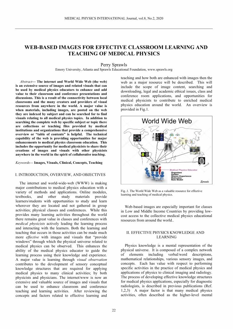
teaching and how both are enhanced with images then the web as a major resource will be described. This will include the scope of image content, searching and downloading, legal and academic ethical issues, class and conference room applications, and opportunities for medical physicists to contribute to enriched medical physics education around the world. An overview is provided in Fig.1.
Fig..1. The World Wide Web as a valuable resource for effective learning and teaching of medical physics. Web-based images are especially important for classes in Low and Middle Income Countries by providing low-cost access to the collective medical physics educational resources from around the world..
II. EFFECTIVE PHYSICS KNOWLEDGE AND LEARNING
Physics knowledge is a mental representation of the physical universe. It is composed of a complex network of elements including verbal/word descriptions, mathematical relationships, various sensory images, and concepts. Each has value with respect to performing specific activities in the practice of medical physics and applications of physics to clinical imaging and radiology. The process of developing effective knowledge structures for medical physics applications, especially for diagnostic radiologists, is described in previous publications (Ref. 1,2,3) A major factor is that many medical physics activities, often described as the higher-level mental
MEDICAL PHYSICS INTERNATIONAL Journal, vol.8, No.2, 2020
23
functions, including analysis, problem solving (non-mathematical), creativity, etc., require conceptual knowledge structures composed of images representing the physical universe. It is knowledge in the form of images that provides an effective connection to the physical objects, interactions, procedures, etc. within medical physics.
III. CLINICAL IMAGES AS PHYSICAL OBJECTS
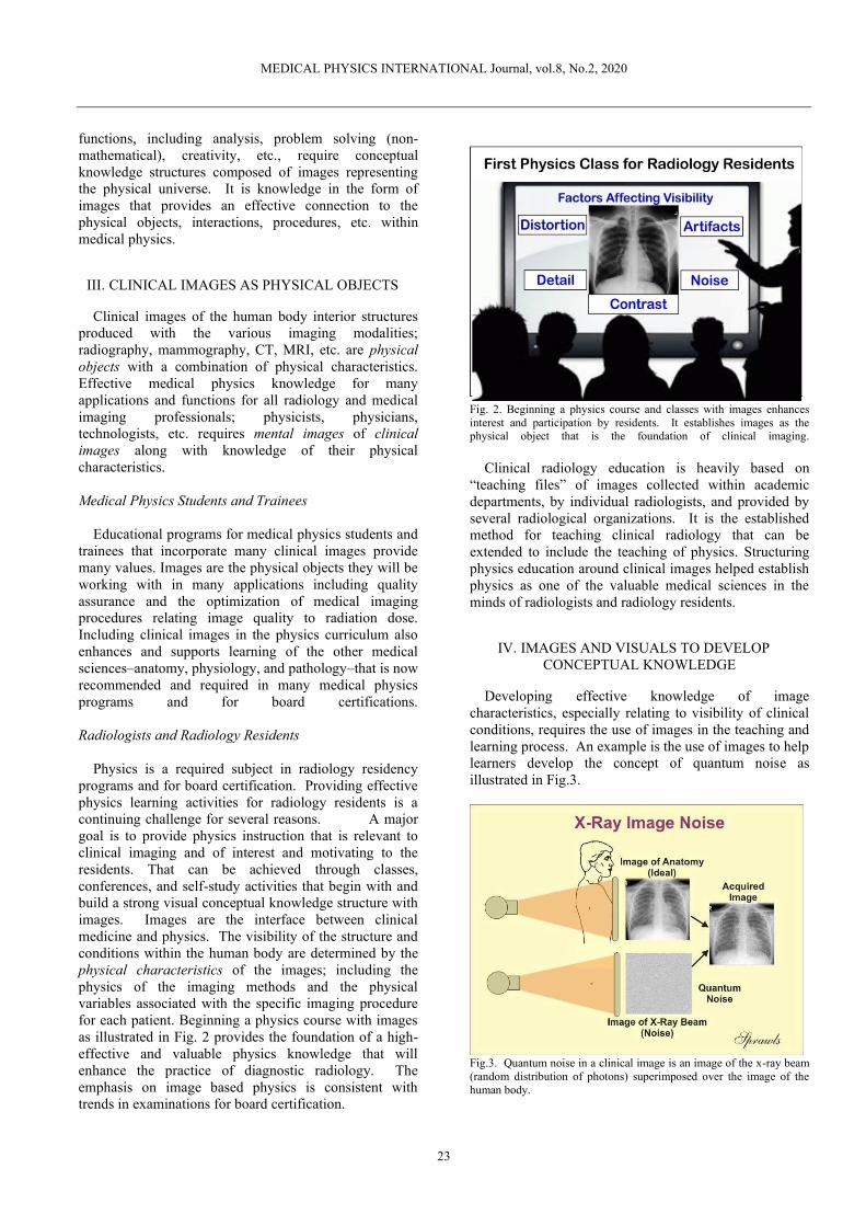
Clinical images of the human body interior structures produced with the various imaging modalities; radiography, mammography, CT, MRI, etc. are physical objects with a combination of physical characteristics. Effective medical physics knowledge for many applications and functions for all radiology and medical imaging professionals; physicists, physicians, technologists, etc. requires mental images of clinical images along with knowledge of their physical characteristics. Medical Physics Students and Trainees Educational programs for medical physics students and trainees that incorporate many clinical images provide many values. Images are the physical objects they will be working with in many applications including quality assurance and the optimization of medical imaging procedures relating image quality to radiation dose. Including clinical images in the physics curriculum also enhances and supports learning of the other medical sciences–anatomy, physiology, and pathology–that is now recommended and required in many medical physics programs and for board certifications. Radiologists and Radiology Residents Physics is a required subject in radiology residency programs and for board certification. Providing effective physics learning activities for radiology residents is a continuing challenge for several reasons. A major goal is to provide physics instruction that is relevant to clinical imaging and of interest and motivating to the residents. That can be achieved through classes, conferences, and self-study activities that begin with and build a strong visual conceptual knowledge structure with images. Images are the interface between clinical medicine and physics. The visibility of the structure and conditions within the human body are determined by the physical characteristics of the images; including the physics of the imaging methods and the physical variables associated with the specific imaging procedure for each patient. Beginning a physics course with images as illustrated in Fig. 2 provides the foundation of a high-effective and valuable physics knowledge that will enhance the practice of diagnostic radiology. The emphasis on image based physics is consistent with trends in examinations for board certification.
Fig. 2. Beginning a physics course and classes with images enhances interest and participation by residents. It establishes images as the physical object that is the foundation of clinical imaging. Clinical radiology education is heavily based on “teaching files” of images collected within academic departments, by individual radiologists, and provided by several radiological organizations. It is the established method for teaching clinical radiology that can be extended to include the teaching of physics. Structuring physics education around clinical images helped establish physics as one of the valuable medical sciences in the minds of radiologists and radiology residents.
IV. IMAGES AND VISUALS TO DEVELOP CONCEPTUAL KNOWLEDGE
Developing effective knowledge of image characteristics, especially relating to visibility of clinical conditions, requires the use of images in the teaching and learning process. An example is the use of images to help learners develop the concept of quantum noise as illustrated in Fig.3.
Fig.3. Quantum noise in a clinical image is an image of the x-ray beam (random distribution of photons) superimposed over the image of the human body.
MEDICAL PHYSICS INTERNATIONAL Journal, vol.8, No.2, 2020
24
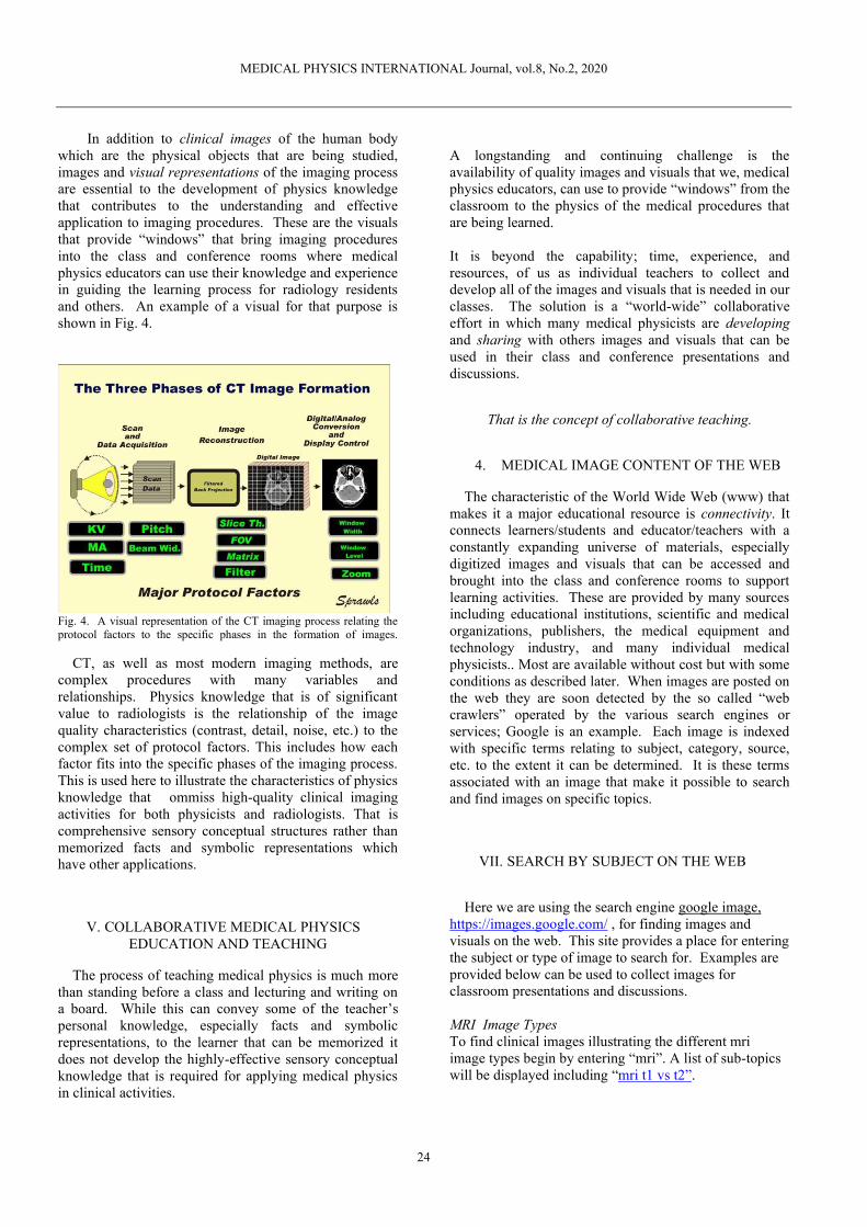
In addition to clinical images of the human body which are the physical objects that are being studied, images and visual representations of the imaging process are essential to the development of physics knowledge that contributes to the understanding and effective application to imaging procedures. These are the visuals that provide “windows” that bring imaging procedures into the class and conference rooms where medical physics educators can use their knowledge and experience in guiding the learning process for radiology residents and others. An example of a visual for that purpose is shown in Fig. 4.
Fig. 4. A visual representation of the CT imaging process relating the protocol factors to the specific phases in the formation of images. CT, as well as most modern imaging methods, are complex procedures with many variables and relationships. Physics knowledge that is of significant value to radiologists is the relationship of the image quality characteristics (contrast, detail, noise, etc.) to the complex set of protocol factors. This includes how each factor fits into the specific phases of the imaging process. This is used here to illustrate the characteristics of physics knowledge that ommiss high-quality clinical imaging activities for both physicists and radiologists. That is comprehensive sensory conceptual structures rather than memorized facts and symbolic representations which have other applications.
V. COLLABORATIVE MEDICAL PHYSICS EDUCATION AND TEACHING
The process of teaching medical physics is much more than standing before a class and lecturing and writing on a board. While this can convey some of the teacher’s personal knowledge, especially facts and symbolic representations, to the learner that can be memorized it does not develop the highly-effective sensory conceptual knowledge that is required for applying medical physics in clinical activities.
A longstanding and continuing challenge is the availability of quality images and visuals that we, medical physics educators, can use to provide “windows” from the classroom to the physics of the medical procedures that are being learned. It is beyond the capability; time, experience, and resources, of us as individual teachers to collect and develop all of the images and visuals that is needed in our classes. The solution is a “world-wide” collaborative effort in which many medical physicists are developing and sharing with others images and visuals that can be used in their class and conference presentations and discussions.
That is the concept of collaborative teaching.
4. MEDICAL IMAGE CONTENT OF THE WEB
The characteristic of the World Wide Web (www) that makes it a major educational resource is connectivity. It connects learners/students and educator/teachers with a constantly expanding universe of materials, especially digitized images and visuals that can be accessed and brought into the class and conference rooms to support learning activities. These are provided by many sources including educational institutions, scientific and medical organizations, publishers, the medical equipment and technology industry, and many individual medical physicists.. Most are available without cost but with some conditions as described later. When images are posted on the web they are soon detected by the so called “web crawlers” operated by the various search engines or services; Google is an example. Each image is indexed with specific terms relating to subject, category, source, etc. to the extent it can be determined. It is these terms associated with an image that make it possible to search and find images on specific topics.
VII. SEARCH BY SUBJECT ON THE WEB
Here we are using the search engine google image, https://images.google.com/ , for finding images and visuals on the web. This site provides a place for entering the subject or type of image to search for. Examples are provided below can be used to collect images for classroom presentations and discussions. MRI Image Types To find clinical images illustrating the different mri image types begin by entering “mri”. A list of sub-topics will be displayed including “mri t1 vs t2”.

MEDICAL PHYSICS INTERNATIONAL Journal, vol.8, No.2, 2020
25
Breast Compression in Mammography Compression of the breast during mammography is an important topic for both medical physicists and radiologists. Excellent visuals can be found by entering “breast compression mammography”. Dose Reduction in Computed Tomography various methods used for ct dose reduction can be discussed with visuals found by entering “computed tomography dose reduction”. Ultrasound Image Artifacts A collection of clinical images displaying a wide range of artifacts can be found by searching on “ultrasound artifacts images”. These examples illustrate the types and range of clinical images and related visuals that are available from many sources that can be accessed by searching on the web. A major value is the ability to search on very specific topics as illustrated above.
VIII. LEGAL AND PROFESIONAL EDITHS ISSUES
While the majority of the images available on the web are free to use for educational purposes there are certain conditions and restrictions that must be considered. Copyright Protection and Fair Use Copyrighting is a legal process, generally administered by federal governments, to provide creators, authors, and artists, with protection of their work from unauthorized use by others. The creators must apply for copyright protection and indicate on published work that it is copyrighted with words or the copyright symbol ©. A major purpose of copyright protection is to prevent others than the copyright holder from making copies, especially for commercial purposes or personal gain, without permission and authorization. For example it would generally not be legal to use someone’s copyrighted image or visual without permission in publications, presentations, or multiple copies of educational materials.
Copyright law provides for the principle, commonly called “fair use” that the reproduction of copyright works for certain limited, educational purposes, does not constitute copyright infringement. Our interest here is specifically the use of images and visuals from the web in class and conference presentations and discussions. Generally the showing of a copyrighted visual in a classroom is not infringement. Individual clinical images as found on the web generally do not meet the requirements for copyright protection.
A general principle of the copyright process is to not interfere with or discourage the educational process and learning activities. As medical physics educators conducting classes and conferences we have the opportunity to enhance the activities with images and visuals downloaded from the web. It is also our individual responsibility to follow appropriate legal and professional guidelines and requirements. Most universities have staff, usually in the libraries that can provide information on copyright issues and especially the use of copyrighted materials for educational purposes.
Academic and Professional Ethics
In addition to the legal there are ethical issues that must be considered when using materials from the web. This applies specifically to visuals, illustrations, and diagrams that represent the creative work of individuals, fig. 3 is an example. Guidance is provided by the following quotation from the AAPM code of ethics.
Creative influence is the cornerstone of creativity and innovation. Without the appropriate citation or acknowledgment of the work of others, imitation of the work of others can result in plagiarism. All forms of plagiarism, including self‐plagiarism, are dishonest and must be avoided. When using visuals that are the creative work of others that should be clearly indicated
4. . THE SPRAWLS ONLINE RESOURCES
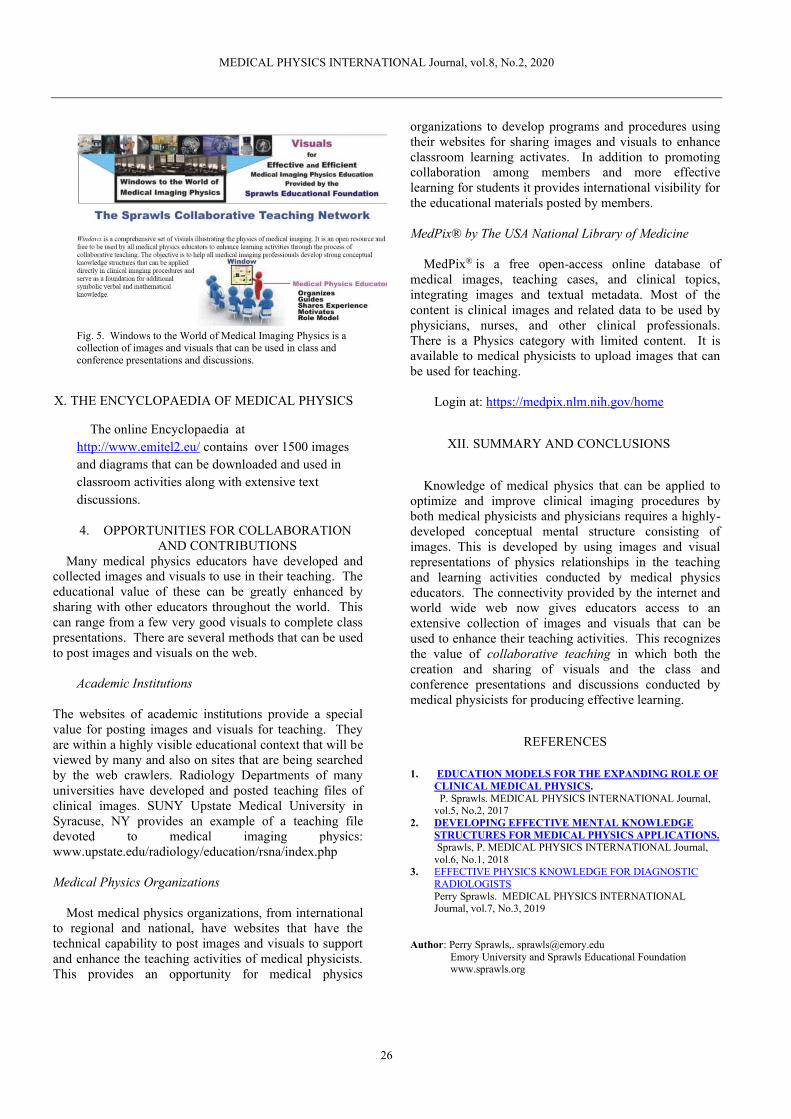
The Sprawls Resources online at: http://www.sprawls.org/resources provides an extensive collection of images and visuals along with modules and textbooks that are being used by medical physics educators in many countries to enhance their teaching activities. The objective is to provide physics classrooms around the world with “windows” through which the medical imaging physics universe can be viewed and used by educators in the process of collaborative teaching. Many of the visuals from within the Resources are organized in PowerPoint presentations and can be downloaded: http://www.sprawls.org/PhysicsWindows/ . This is to support the process of collaborative teaching as illustrated in Fig. 5.
MEDICAL PHYSICS INTERNATIONAL Journal, vol.8, No.2, 2020
26
Fig. 5. Windows to the World of Medical Imaging Physics is a collection of images and visuals that can be used in class and conference presentations and discussions.
X. THE ENCYCLOPAEDIA OF MEDICAL PHYSICS
The online Encyclopaedia at http://www.emitel2.eu/ contains over 1500 images and diagrams that can be downloaded and used in classroom activities along with extensive text discussions.
4. OPPORTUNITIES FOR COLLABORATION AND CONTRIBUTIONS
Many medical physics educators have developed and collected images and visuals to use in their teaching. The educational value of these can be greatly enhanced by sharing with other educators throughout the world. This can range from a few very good visuals to complete class presentations. There are several methods that can be used to post images and visuals on the web.
Academic Institutions
The websites of academic institutions provide a special value for posting images and visuals for teaching. They are within a highly visible educational context that will be viewed by many and also on sites that are being searched by the web crawlers. Radiology Departments of many universities have developed and posted teaching files of clinical images. SUNY Upstate Medical University in Syracuse, NY provides an example of a teaching file devoted to medical imaging physics: www.upstate.edu/radiology/education/rsna/index.php Medical Physics Organizations Most medical physics organizations, from international to regional and national, have websites that have the technical capability to post images and visuals to support and enhance the teaching activities of medical physicists. This provides an opportunity for medical physics
organizations to develop programs and procedures using their websites for sharing images and visuals to enhance classroom learning activates. In addition to promoting collaboration among members and more effective learning for students it provides international visibility for the educational materials posted by members. MedPix® by The USA National Library of Medicine MedPix® is a free open-access online database of medical images, teaching cases, and clinical topics, integrating images and textual metadata. Most of the content is clinical images and related data to be used by physicians, nurses, and other clinical professionals. There is a Physics category with limited content. It is available to medical physicists to upload images that can be used for teaching.
Login at: https://medpix.nlm.nih.gov/home
XII. SUMMARY AND CONCLUSIONS
Knowledge of medical physics that can be applied to optimize and improve clinical imaging procedures by both medical physicists and physicians requires a highly-developed conceptual mental structure consisting of images. This is developed by using images and visual representations of physics relationships in the teaching and learning activities conducted by medical physics educators. The connectivity provided by the internet and world wide web now gives educators access to an extensive collection of images and visuals that can be used to enhance their teaching activities. This recognizes the value of collaborative teaching in which both the creation and sharing of visuals and the class and conference presentations and discussions conducted by medical physicists for producing effective learning.
REFERENCES
1. EDUCATION MODELS FOR THE EXPANDING ROLE OF CLINICAL MEDICAL PHYSICS. P. Sprawls. MEDICAL PHYSICS INTERNATIONAL Journal, vol.5, No.2, 2017
2. DEVELOPING EFFECTIVE MENTAL KNOWLEDGE STRUCTURES FOR MEDICAL PHYSICS APPLICATIONS. Sprawls, P. MEDICAL PHYSICS INTERNATIONAL Journal, vol.6, No.1, 2018
3. EFFECTIVE PHYSICS KNOWLEDGE FOR DIAGNOSTIC RADIOLOGISTS Perry Sprawls. MEDICAL PHYSICS INTERNATIONAL Journal, vol.7, No.3, 2019
Author: Perry Sprawls,. [email protected] Emory University and Sprawls Educational Foundation www.sprawls.org

MEDICAL PHYSICS INTERNATIONAL Journal, vol.8, No.2, 2020
27
TEACHING MEDICAL PHYSICS WITH MODERN EDUCATIONAL TECHNIQUES
Buchgeister M 1, 2
1 Beuth University of Applied Sciences, Berlin, Germany, 2 EFOMP ETC
Abstract— This description of novel teaching techniques
is an updated version of a summary of an introductory presentation in the focus session on educational techniques at the EFOMP ECMP2018 congress at Copenhagen that was published in the European Medical Physics News Autumn 2018
Keywords— Medical Physics Teaching
When you start teaching a Medical Physics course or just give a single lecture, it is wise to know a little bit about your audience, especially about their composition and pre-existing knowledge of your topic and, naturally, their interests. Besides having an oral self-presentation round of each individual (which is fine only for small groups) or filling out a questionnaire (works also for large groups), a quicker and more up-to-date way is to use one of those (usually anonymous) online-feedback tools via smartphone. I use such tools even in presentations like the one on this topic in the session on teaching Medical Physics at ECMP2018 at Copenhagen. During my talk, I could only evaluate and show some of the responses live during my presentation. But afterwards, I was able to analyze all responses offline. So I know now, that up to 40 participants responded to my questions via such a smartphone feedback tool, consisting of 60% male and 40% female participants. Among these, 11 are teaching in diagnostics, 3 in imaging, 9 in radiotherapy, 8 in radiation protection, 2 in nuclear medicine and even 5 are not teaching at all (it was possible to name multiple sub-jects as free text). What is regarded as the “classic style” of teaching (not limited to Medical Physics subjects)? From my own experience of many lectures it is, that someone stands in front of a black or white board in an lecture hall and is often clicking through power-point slides, summarizing content from his or her favourite lecture book or review papers on the subject (Fig. 1). This is the case for almost any kind of audience like students, physicists, physicians, RTs, nurses… This can get very boring as can be seen even in special lectures such as shown in Fig. 1! If you are lucky, the lecturer is of the very old style, that is using the board or flipchart to develop the key points of the subject taught by writing and sketching in a dialogue manner together with the audience. As you may have
experienced yourself in such lectures, it is the involvement of yourself and especially your thoughts and ideas, that will keep you awake.
Fig.1: Teaching in the “classic style”: My Christmas lecture,
where bored students are desperately awaiting the Glühwein to reach its drinking temperature. © M. Buchgeister
Times have changed and as already stated above, there are now more didactic tools available besides boards to write on. If you are teaching at a university or similar larger institution, you may have the chance to participate in didactic seminars, where new concepts and the use of new tools are presented. Already in 2010, the AAPM summer school “Teaching Medical Physics: Innovations in Learning” addressed this topic. This summer school presentations were recorded and videos are available for free at vimeo.com/channels/ss2010. Fig. 2 – 4 are screenshots of titles of some of the presentations given there to get you even more interested to check out the videos yourself! Just one month before ECMP2018, there was again a workshop of AAPM at Nashville, TN, on “Improving the Teaching and Mentoring in Medical Physics”. The course director was Victor Montemayor, who is also the current chairman of the AAPM Committee on Medical Physicists as Educators (MPESC). I just list three titles of presentations given there for you: “What Neuroscience Research Has to Tell Us About the Effects of Learning on the Brain” (Laurie Cutting), “Best Practice: Project-based Learning

MEDICAL PHYSICS INTERNATIONAL Journal, vol.8, No.2, 2020
28
(PBL) in Medical Physics” (Rebecca Howell) and “Best Practice: Flipped Learning in the Medical Physics Classroom” (Matt Studentski).
Fig. 2: Title slide of Georg Starkschall’s presentation on “Innovations in teaching an introductory medical physics class” at the 2010 AAPM summer school. © Georg Starkschall
Fig. 3: Title slide of Robert Beichner’s presentation on “Multiple Technologies to Ad-dress Multiple Instructional Needs” at the 2010 AAPM summer school. © Robert Beichner
Fig. 4: Title slide of Cindy Hmelo-Silver’s presentation on “Learning Through Prob-lem-Solving” at the 2010 AAPM summer school. © Cindy Hmelo-Silver
Do you recognize the already known keywords of modern style teaching in these titles: Neuroscience, Project-(also: Problem-)based Learning and Flipped (classroom) Learning. If you have not heard about these, here is a very
brief (!) introduction to the basics of the later two teaching concepts: Project-/Problem-based learning and/or the flipped classroom approach just intro-duces the audience to basic knowledge like facts, tables of data, formulas and hints on where to start searching for information on the subject in books, papers and on the internet. This is the longest time that you will spend in kind of the “old style” standing in front of the audience since the key mark of these new teaching techniques is the formation of groups, that work on a problem scenario or a set of problems on their own. The basic idea behind this approach is “peer instruction”: Based on pre-existing knowledge or understanding and continuation of your introductory information the correct solution or a reasonable approach to solve the problem will mingle out by arguments exchanged within the group. The teacher or lecturer is just an “accompanying expert”, that consults in a regular sequence with the group, pre-venting them to get too far off or to provide deeper specific information upon request if needed. The results of the groups are finally presented to the whole audience, so the new knowledge can be gathered by all. Very important to this approach is the teamwork-aspect: if you can explain it to your fellow student, you got it! In the flipped- classroom approach, the basic information material is provided before the lecture in printed or electronic form (e.g. via learning management platforms like “Moodle”) and has to be studied in advance. During the time present in the class-room, a set of problems is worked on, mostly in groups as well. But very important: no basics are repeated in class, otherwise you end up in the old style! Most important again is the interaction of the audience (“peers”) with itself accompanied by an expert (“you”). If you got already interested and want to know more, start your search for publications on this topic by one of the pioneers of these teaching concepts in physics: Eric Mazur. What are the Pros and Cons of these “new style” teaching methods? As you can easily see from the open approach in group discussions of solutions, it is the project and problem-solving competence, that is focused on, as well as the competence to learn and work in groups, enforcing team and communication skills. If you have worked in groups yourself, you surely know, that the composition of the group is an important key to its success. It always needs someone that did not get it on the first grasp, so it needs someone else in the group to explain and answer the “stupid questions” posed. Let me compare this to a dough that needs kneading (questions) to distribute the ingredients (answers/knowledge) evenly. A pile of flour, baking pow-der, raisins etc. will not make a cake on its own! While it was maybe plain luck in the past, if the composition of your group was good for its success, results of modern teaching analysis indicate, that a good

MEDICAL PHYSICS INTERNATIONAL Journal, vol.8, No.2, 2020
29
mixture or heterogeneity of the group is an important key to its success. It has to be paid attention to existing knowledge, as well as to social skill aspects among the group members when forming the groups. Special tools like questionnaires can facilitate the formation of such balanced groups as well as introduction of the group members to dedicated roles or functions of group members that have been commonly agreed on such as the speaker or team leader (contact for and reporting results etc. to the instructor), the protocol secretary (keeping notes on what has been worked on and what is planned for the next meeting that is digitally available to all members and facilitates very much the production of inter-mediate and final reports), the timekeeper (when to start and to make a break etc.), the police officer or judge (deciding on keeping the set rules of the group and agreed consequences if not being followed like by e.g. a round of coffee for all) and the “Feel-Good-manager” (organizing e.g. cookies or a nice setting for the meeting by a piece of decoration on the table identifying the group). Needless to say, that these roles can rotate among the group members, as long as everyone is informed and aware of them. I introduce this concept to my classes but leave it up to them to realize them. I just mention that the most successful project groups of previous classes picked up my suggestions as start configuration for getting organized themselves in the project.
When these modern techniques are introduced, students, as well as teachers, will have to get used to it. It requires discipline and self-control of your learning or teaching attitude. Since additional aims are addressed with these teaching techniques, you have to cut down the content of your lecture to the very essentials. That means most of the time fewer formulas and facts are taught in great detail. The main emphasis is put on the competences to solve problems and accomplish a new project. The questions and hurdles of the project will lead automatically to those details and additional facts that you intend to get into their brains. If you start a course from scratch, this is at no extra labour (except that you cannot copy your teacher of the past…), but if you have already set out your lectures in the “old style”, you will have to start over again. Or maybe not completely, as there might be an “intermediate style” of teaching, too. Since a few years and as a result from own teaching experience, as well as input from didactic seminars, I created my own “mixture”. I use activating breaks of discussions of the students with their neighbors. This introduces a “change of view” so that the students look at some else than me and have to express their thoughts and ideas to someone besides listening to me. To catch misunderstandings or misconceptions, I have alternating students summarizing the content at the start, in the middle and at the end of the lecture. At the same time, the others get the content presented once more (varied repetition is a key to memory!). To train problem-solving competence, I have this person try to answer/solve a question/problem within 3 minutes. A clock bar shown by
the beamer is counting down. 3 minutes are more time than you may guess at first hand! To add a bit of team competence training, this is usually done with the help of another student acting like the telephone-joker in the “who wants to be a millionaire”-quiz on TV. Being a “telephone-joker” is limited to two times, so quickly the “good ones” are out of the game in the class. With this example of a teaching concept, I have not even touched the field of good animations, videos or presenting original pieces of equipment during a lecture of a Medical Physics subject. All this together will make up the smart arrangement and activating interaction with the audience that will render your lecture a success in gained knowledge for the audience. The is even more important in the current situation of shutting down regular teaching at universities etc. due to the CoVID-19 virus lock-down when you have to use distance teaching tools like recorded lectures or open-source or commercial video-platforms like Jitsi, BigBlueButton, Zoom, Webex, GoToWebinar etc. just to list some for your live teaching or consultations with your class. This situation is even more challenging since the non-verbal contact present in the “normal” situation at class is now missing. You have to actively address your audience as much as possible by names and have them speak up, too, to get them used to the new teaching tools and situations. My current classes prefer to write questions and remarks through the chat function of the platform. By calling them up by name to use the microphone for a direct conversation, I am trying to get them used to “step out of the dark” of the class for verbal communication. Just to add an-other hint to get them involved in the organization of the lecture, too, for you: Since it is very hard for me to recognize the point where they need a break from mostly listening to me, I ask for a volunteer from the audience at the beginning to whom by private chat they can indicate if they need a break. He or she will tell me then as “speaker of the class”. Some of the mentioned video-platforms already have such a tool built in to indicate to the presenter to slow down or to repeat a topic. But this way, I do not have to concentrate also on this item but on my very topic and the questions and answers of my class. As mentioned before: this is about getting them involved as a peer in the organization of the lecture, too. These concepts like the ones described above are regarded as an educational re-source, just like the animations or videos e.g. that you may use in your lectures, too. The goal of an initiative of the EFOMP Education and Training Committee is to establish a network of Medical Physics teachers, who are interested in and would like to share their concepts and materials as Open Educational Resources (OER) under the creative commons licensing (CCL or GPL) concept. EFOMP would like to create a teaching material repository to this end. At the start, already existing materials and concepts of teaching could be made available there. Preferably in English, but this is just a matter of translation. Foremost

MEDICAL PHYSICS INTERNATIONAL Journal, vol.8, No.2, 2020
30
is the creation of links between interested and active teachers in Medical Physics in Europe and maybe also beyond, as there exist already links to the AAPM MPESC mentioned in the introduction above. I have to show the other results of my smartphone feedback poll during my presentation at ECMP2018. Among 35 responders, 40% had already experience with some kind of “new style” teaching, while 60% did not. Asked, if they were satisfied with their teaching result, 53% out of 32 responders answered with yes, 47% with no. So at least for almost half of them, there might be an interest in improvement. Among 28 responders who are
teaching, 79% would be willing to try something new, 4% would refuse and 18% already teach in the “new style”. My resume of these figures in total is, that there is a change of style in teaching Medical Physics on the way in Europe. And I would like to make the Education and Training Committee of EFOMP as well as the European Congress of Medical Physics the place to exchange new concepts as well as tools/media and foster this change of teaching Medical Physics!
Author: Prof. Dr. Markus Buchgeister Email: [email protected]

MEDICAL PHYSICS INTERNATIONAL Journal, vol.6, No.1, 2018
31
PROFESSIONAL ISSUES
MEDICAL PHYSICS INTERNATIONAL Journal, vol.8, No.2, 2020
32
South-East Asian Federation of Organizations for Medical Physics (SEAFOMP) – Celebrating 20th Anniversary of formation
JHD Wong1, KH Ng1, F Haryanto2, A Krisanachinda3, JCL Lee4, TC Nguyen5, S Pawiro6, A Peralta7, DS Soejoko6, CH Yeong8, TJ Wong4 V Inphavong9 SS Lin10, VY Ath11, D Arzabal12, DT Luong13
1 Department of Biomedical Imaging, Faculty of Medicine, University of Malaya, Kuala Lumpur, Malaysia. 2 Department of Physics, Faculty of Mathematics and Natural Sciences, Bandung Institute of Technology, Bandung, Indonesia. 3 Department of Radiology, Faculty of Medicine, Chulalongkorn University, Bangkok, Thailand. 4 Division of Radiation Oncology, National Cancer Centre Singapore. 5 Unit of PET-CT & Cyclotron, Cho Ray Hospital, Ho Chi Minh City, Vietnam. 6 Department of Physics, Faculty of Mathematics and Natural Sciences, Universitas Indonesia, Depok, Indonesia. 7 Graduate School, University of Santo Tomas, Philippines. 8 School of Medicine, Faculty of Health and Medical Sciences, Taylor’s University, Malaysia. 9 Mittaphab Hospital,Ministry of Health, Chommany village, Chanthabuly district, Vientiane, Lao PDR. 10 Department of Radiation Oncology, Pinlon Cancer Center, Yangon, Myanmar. 11National Cancer Centre, Calmette Hospital, Phnom Penh, Cambodia. 12 Rizal Medical Center, Pasig City, Philippines. 13 Department of Biomedical Physics, Faculty of Medicine, Nguyen Tat Thanh University, Vietnam.
Abstract— The South-East Asian Federation of Organizations for Medical Physics (SEAFOMP), which was established in 2000, stemmed from a vision to galvanize efforts in promoting and developing the medical physics profession in the ASEAN region. The South East Asia Congress of Medical Physics (SEACOMP) – its largest annual scientific meeting, and in recent years, the ASEAN College of Medical Physics (ACOMP) had been the catalyst for the growth and progress of medical physics in this region. In this paper, we present brief reports of the progress achieved and challenges faced by its member countries. The IloIlo Declaration acknowledged and affirmed the efforts of SEAFOMP members towards education and training of medical physics, laying out the strategies and focus of the federation in charting the way forward for the federation and its member countries.
Keywords— SEACOMP. Medical physics, education, profession, ASEAN
I. INTRODUCTION
South-East Asian Federation of Organizations for Medical Physics (SEAFOMP) started in the year 2000. This year, we celebrate the 20th anniversary of the federation. This article intend to summarize the progress and achievement of the medical physics professions of the south east Asian region, to commemorate this important milestone. Fig.1 shows the logo of the 20th anniversary of SEAFOMP.
II. THE BEGINNING OF SEAFOMP
SEAFOMP was formed in an informal discussion in 1996 during the International Organization of Medical
Physics (IOMP) World Congress at Nice, France. The founding members were Anchali Krisanachinda, Kwan-Hoong Ng, Agnette Peralta, Ratana Pirabul, Djarwani S. Soejoko, and Toh-Jui Wong (Fig.2 and Fig.3). It was only in 2000, four years later, that the federation was officially accepted as a regional chapter of the International Organization of Medical Physics (IOMP) at the Chicago World Congress [1]. SEAFOMP started with five (out of 10) member countries of the Association of Southeast Asian Nations (ASEAN), namely Indonesia, Malaysia, Philippines, Singapore, and Thailand. Brunei and Vietnam joined in 2002 and 2005, respectively[1, 2]. Professor Dr. Kwan-Hong Ng served as the founding president. Since then, SEAFOMP has underwent five changeover of executive committees (Table 1).
Fig. 1 SEAFOMP 20th anniversary logo.
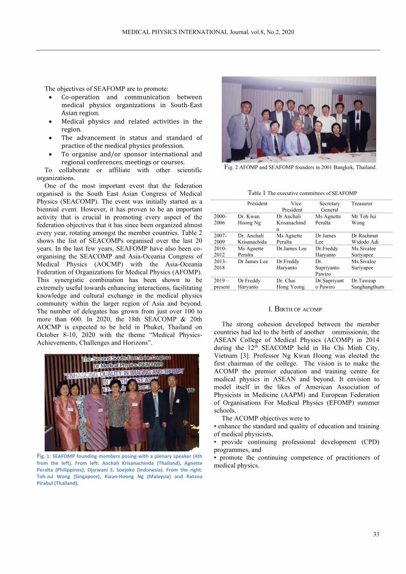
MEDICAL PHYSICS INTERNATIONAL Journal, vol.8, No.2, 2020
33
The objectives of SEAFOMP are to promote: • Co-operation and communication between
medical physics organizations in South-East Asian region.
• Medical physics and related activities in the region.
• The advancement in status and standard of practice of the medical physics profession.
• To organise and/or sponsor international and regional conferences, meetings or courses.
To collaborate or affiliate with other scientific organizations.
One of the most important event that the federation organised is the South East Asian Congress of Medical Physics (SEACOMP). The event was initially started as a biennial event. However, it has proven to be an important activity that is crucial in promoting every aspect of the federation objectives that it has since been organized almost every year, rotating amongst the member countries. Table 2 shows the list of SEACOMPs organised over the last 20 years. In the last few years, SEAFOMP have also been co-organising the SEACOMP and Asia-Oceania Congress of Medical Physics (AOCMP) with the Asia-Oceania Federation of Organizations for Medical Physics (AFOMP). This synergistic combination has been shown to be extremely useful towards enhancing interactions, facilitating knowledge and cultural exchange in the medical physics community within the larger region of Asia and beyond. The number of delegates has grown from just over 100 to more than 600. In 2020, the 18th SEACOMP & 20th AOCMP is expected to be held in Phuket, Thailand on October 8-10, 2020 with the theme “Medical Physics-Achievements, Challenges and Horizons”.
Fig. 1: SEAFOMP founding members posing with a plenary speaker (4th
from the left). From left: Anchali Krisanachinda (Thailand), Agnette Peralta (Philippines), Djarwani S. Soejoko (Indonesia). From the right: Toh-Jui Wong (Singapore), Kwan-Hoong Ng (Malaysia) and Ratana Pirabul (Thailand).
Fig. 2 AFOMP and SEAFOMP founders in 2001 Bangkok, Thailand.
Table 1 The executive committees of SEAFOMP
President Vice President
Secretary General
Treasurer
2000-2006
Dr. Kwan Hoong Ng
Dr Anchali Krisanachinda
Ms Agnette Peralta
Mr Toh Jui Wong
2007-2009
Dr. Anchali Krisanachida
Ms Agnette Peralta
Dr James Lee
Dr Rachmat Widodo Adi
2010-2012
Ms Agnette Peralta
Dr.James Lee Dr.Freddy Haryanto
Ms.Sivalee Suriyapee
2013-2018
Dr James Lee Dr.Freddy Haryanto
Dr. Supriyanto Pawiro
Ms.Sivalee Suriyapee
2019 – present
Dr Freddy Haryanto
Dr. Chai Hong Yeong
Dr.Supriyanto Pawiro
Dr.Taweap Sanghangthum
I. BIRTH OF ACOMP
The strong cohesion developed between the member countries had led to the birth of another ommissionin, the ASEAN College of Medical Physics (ACOMP) in 2014 during the 12th SEACOMP held in Ho Chi Minh City, Vietnam [3]. Professor Ng Kwan Hoong was elected the first chairman of the college. The vision is to make the ACOMP the premier education and training centre for medical physics in ASEAN and beyond. It envision to model itself in the likes of American Association of Physicists in Medicine (AAPM) and European Federation of Organisations For Medical Physics (EFOMP) summer schools.
The ACOMP objectives were to • enhance the standard and quality of education and training of medical physicists, • provide continuing professional development (CPD) programmes, and • promote the continuing competence of practitioners of medical physics.
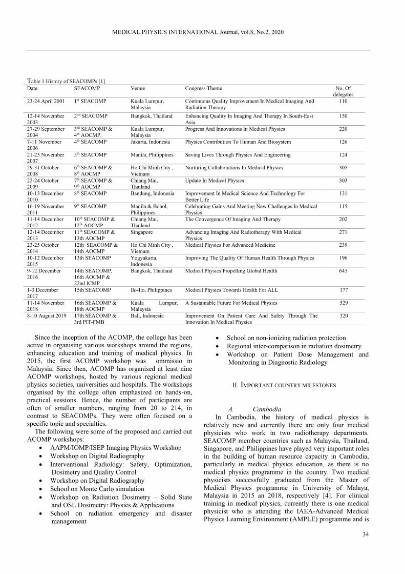
MEDICAL PHYSICS INTERNATIONAL Journal, vol.8, No.2, 2020
34
Table 1 History of SEACOMPs [1] Date SEACOMP Venue Congress Theme No. Of
delegates 23-24 April 2001 1st SEACOMP Kuala Lumpur,
Malaysia Continuous Quality Improvement In Medical Imaging And Radiation Therapy
110
12-14 November 2003
2nd SEACOMP Bangkok, Thailand Enhancing Quality In Imaging And Therapy In South-East Asia
150
27-29 September 2004
3rd SEACOMP & 4th AOCMP.
Kuala Lumpur, Malaysia
Progress And Innovations In Medical Physics 220
7-11 November 2006
4th SEACOMP Jakarta, Indonesia Physics Contribution To Human And Biosystem 126
21-23 November 2007
5th SEACOMP Manila, Philippines Saving Lives Through Physics And Engineering 124
29-31 October 2008
6th SEACOMP & 8th AOCMP
Ho Chi Minh City , Vietnam
Nurturing Collaborations In Medical Physics 305
22-24 October 2009
7th SEACOMP & 9th AOCMP
Chiang Mai, Thailand
Update In Medical Physics 303
10-13 December 2010
8th SEACOMP Bandung, Indonesia Improvement In Medical Science And Technology For Better Life
131
16-19 November 2011
9th SEACOMP Manila & Bohol, Philippines
Celebrating Gains And Meeting New Challenges In Medical Physics
115
11-14 December 2012
10th SEACOMP & 12th AOCMP
Chiang Mai, Thailand
The Convergence Of Imaging And Therapy 202
12-14 December 2013
11th SEACOMP & 13th AOCMP
Singapore Advancing Imaging And Radiotherapy With Medical Physics
271
23-25 October 2014
12th SEACOMP & 14th AOCMP
Ho Chi Minh City , Vietnam
Medical Physics For Advanced Medicine 239
10-12 December 2015
13th SEACOMP Yogyakarta, Indonesia
Improving The Quality Of Human Health Through Physics 196
9-12 December 2016
14th SEACOMP, 16th AOCMP & 22nd ICMP
Bangkok, Thailand Medical Physics Propelling Global Health 645
1-3 December 2017
15th SEACOMP Ilo-Ilo, Philippines Medical Physics Towards Health For ALL 177
11-14 November 2018
16th SEACOMP & 18th AOCMP
Kuala Lumpur, Malaysia
A Sustainable Future For Medical Physics 529
8-10 August 2019 17th SEACOMP & 3rd PIT-FMB
Bali, Indonesia Improvement On Patient Care And Safety Through The Innovation In Medical Physics
320
Since the inception of the ACOMP, the college has been
active in organising various workshops around the regions, enhancing education and training of medical physics. In 2015, the first ACOMP workshop was ommissio in Malaysia. Since then, ACOMP has organised at least nine ACOMP workshops, hosted by various regional medical physics societies, universities and hospitals. The workshops organised by the college often emphasized on hands-on, practical sessions. Hence, the number of participants are often of smaller numbers, ranging from 20 to 214, in contrast to SEACOMPs. They were often focused on a specific topic and specialties.
The following were some of the proposed and carried out ACOMP workshops:
• AAPM/IOMP/ISEP Imaging Physics Workshop • Workshop on Digital Radiography • Interventional Radiology: Safety, Optimization,
Dosimetry and Quality Control • Workshop on Digital Radiography • School on Monte Carlo simulation • Workshop on Radiation Dosimetry – Solid State
and OSL Dosimetry: Physics & Applications • School on radiation emergency and disaster
management
• School on non-ionizing radiation protection • Regional inter-comparison in radiation dosimetry • Workshop on Patient Dose Management and
Monitoring in Diagnostic Radiology
II. IMPORTANT COUNTRY MILESTONES
A. Cambodia
In Cambodia, the history of medical physics is relatively new and currently there are only four medical physicists who work in two radiotherapy departments. SEACOMP member countries such as Malaysia, Thailand, Singapore, and Philippines have played very important roles in the building of human resource capacity in Cambodia, particularly in medical physics education, as there is no medical physics programme in the country. Two medical physicists successfully graduated from the Master of Medical Physics programme in University of Malaya, Malaysia in 2015 an 2018, respectively [4]. For clinical training in medical physics, currently there is one medical physicist who is attending the IAEA-Advanced Medical Physics Learning Environment (AMPLE) programme and is
MEDICAL PHYSICS INTERNATIONAL Journal, vol.8, No.2, 2020
35
planning to join the new batch of radiation oncology medical physics (ROMP) programme in Thailand, starting October 2020. To summarise, the SEACOMP has a huge contribution to the development of medical physics of member states. Certainly, the spirit of growing together for a better medical physics and healthcare system in ASEAN rings true.
B. Indonesia Currently, the country has six universities offering master programmes in physics, with major in medical physics and two universities offering PhD programmes in physics, majoring in medical physics related research [2] The medical physics profession has been recognised by the Indonesian government, in particular, the Ministry of Health, Ministry of Man Power and Nuclear Regulatory Agency[5] [6].
We have also started the registration for medical physicists in 2012 and continued with licensing for clinical medical physicists by the Indonesian Health Professional Board – Ministry of Health. In 2020, it has transformed to be the Indonesian Health Professional Council (Konsil Tenaga Kesehatan Indonesia, KTKI)[2]. The medical physics community had also been active in education and continuous professional development. Collectively, we had organised three schools in collaboration with the AAPM, International Centre for Theoretical Physics (ICTP), Institute of Electrical and Electronics Engineers (IEEE) and the International Atomic Energy Agency (IAEA). From 2016 to 2020, we had established clinical qualified medical physicist residency programme in radiotherapy, diagnostic radiology, and nuclear medicine. In addition, an associate medical physicist training programme was also established to cater for the national demand of medical physicist in 2016.
C. Lao PDR
Lao P.D.R, with a population of 7 million is ranked as one of the Least Developed Developing Countries (LDDC) in South East Asian region by the United Nations. Currently, two medical physicists had graduated with Master of Science in Medical Physics/Medical Imaging from the Chulalongkorn University in 2017 under IAEA fellowships. One medical physicist has been attending IAEA-AMPLE clinical training in ROMP since the last 2 years (2018-2020), supported by VAMED Company. The facility consists of the first linear accelerator system at Radiotherapy Center, Mittaphab Hospital, Ministry of Health. Their responsibilities are to run a quality assurance (QA) programme, which includes mechanical and radiation dosimetry. Medical physicist also involves in treatment planning of patients and patient verification QA. The medical physicists are also responsible for radiation protection and radiation safety in the hospital. They also lecture on basic radiation physics, quality assurance and
quality control of radiation modalities in medical fields to technologist student and resident of the Radiology Department, University of Health Science. In terms of research, they are also involved in supervision of postgraduate students in nuclear physics, University of Laos for research related to radiation. We are still facing many obstacles such as the lack of experienced medical physicst, clinical practice; budget and fundings. The most difficult part is working without clinical supervisors.
D. Malaysia
Malaysia has about 349 medical physicists in the country [6]. Currently, the country has two master programme for the education and training of medical physicists at University of Malaya and Universiti Sains Malaysia [4, 7]. The master programme at the University of Malaya is Institute of Physics and Engineering in Medicine (IPEM) accredited, and had been ommission as a regional training centre for postgraduate medical physics by the IAEA, contributing to the development of medical physics profession in Cambodia, Vietnam and Brunei. Malaysia currently has two medical physics professional organizations, i.e. Medical Physics Division (MPD), under the umbrella of Institute of Physics Malaysia (IFM) and Malaysia Association of Medical Physics (MAMP). They have organized several regional conferences, workshops and seminars under the umbrella of SEAFOMP and ACOMP In 2015, the MPD organized a joint workshop with AAPM/ ISEP and IOMP on the theme of imaging physics and hashas attracted more than 200 participants from the region. In addition, Malaysia has also hosted three hands-on workshopunder the ACOMP. In 2016, the Allied Health Professions Act (Act 774) was gazatted. Medical physicist along with 22 other allied health professions are included under this Act whereby all the practicing professionals must obtain a ommission certificate and compulsory Continuous Professional Development (CPD) of 30 points per year. Malaysia’s strength lies in having well established postgraduate programmes while their weakness is that they have yet to establish national certification for competent medical physicists. There is currently no medical physics residency programme in Malaysia, hence most of the medical physicists are trained via on-the-job, non-structured trainings. However, under the IAEA RAS6038 programme, Malaysia has completed the residential trainings for 6 ROMPs and 2 DRMPs. The latest cohort of the ROMP, DRMP and NMMP residential training was started in 2018 and expected to complete in 2021. The continuing challenges faced by the medical physics community in the country is outward migration of experienced medical physicists and sustainable research funding.
E. Myanmar The first medical physicist from Myanmar was trained in England and later assigned to work in the Radiation
MEDICAL PHYSICS INTERNATIONAL Journal, vol.8, No.2, 2020
36
Therapy Department of Yangon General Hospital in 1958. In the beginning, a limited number of medical physicists, who were opportuned to take long term training (more than 1 year), were allocated in Mandalay and Taunggyi General Hospital. Since then, medical physicist trainings have been accomplished via local apprenticeship training and some short course training programmes supported by the IAEA and World Health Organization (WHO). The candidates for the training programmes were from academic level or senior technologist who graduated from physics or equivalent subject. Our challenges is that we do not have a proper academic education and training programme at the national level. The undergraduate degree course for medical imaging technology was established by the Ministry of Health in 1991 at Yangon. Currently, there are three universities which offer undergraduate degrees in medical imaging technology and two universities which offers master degrees in that field. Radiation therapy technologist undergraduate course was introduced in 2018 at the University of Medical Technology, Yangon. The first 2-year master of medical physics programme is expected to be established in 2021 at the same university. The Association of Myanmar Medical Physicists (MMPA) was setup in the 2016 with 30 members who were working as medical physicists and radiation protection officers. MMPA joined IOMP, AFOMP and SEAFOMP at the end of 2016.
F. Philippines
During the past 20 years, giant strides have been made in the development of medical physics in the Philippines. The first Certifying Board Examination in Radiation Oncology Medical Physics was held in February 2010. There are now 19 certified ROMPs. The first Certifying Board Examination in Diagnostic Radiology Medical Physics was held in July 2019. There are now 13 certified Diagnostic Radiology Medical Physicists (DRMPs). Sixty-one health facilities now employ medical physicists, 19 of which employ three medical physicists each. The two national radiation regulatory agencies continue to employ medical physicists. Three companies now employ medical physicists; they provide medical physics consulting and testing services. Five universities employ medical physicists as faculty members. Eight multinational companies employ Filipino medical physicists. University of Santo Tomas, the only school offering the master’s degree in medical physics, produced a total of 145 graduates from 2000 to 2019. The ROMP clinical residency programme started in 2009; that of DRMP in 2010; and that of Nuclear Medicine Medical Physics (NMMP) in 2018. The Philippine Organization of Medical Physicists established in 1986 is now the Society of Medical Physicists
in the Republic of the Philippines, created in 2016. It has hosted three SEACOMPs.
G. Singapore
Singapore’s demand for medical physicists continues to grow with our expanding healthcare such as the new proton beam therapy at the National Cancer Centre and increasing diagnostic radiology and nuclear medicine capabilities. We have about 50 medical physicists in the Society of Medical Physicists (Singapore), formed in 1998. We currently have two universities offering Medical Physics minor or concentration at the bachelor’s level and a PhD programme in one of the university. We have implemented residency programmes for nuclear medicine and radiotherapy using the IAEA residency syllabus. At least one of the public healthcare cluster has ommission the residency’s competency framework as part of the career path of a medical physicist. We also continue to host important regional conferences such as the AOCMP-SEACOMP in 2013 and the upcoming World Congress on Medical Physics and Biomedical Engineering in 2021. We continue to work actively with the IAEA to offer training fellowships, conduct regional training workshops and planning meetings for various projects.
H. Thailand
The first medical physicist, Mrs. Pradub Atthakorn was trained in England and later worked in the Radiotherapy Department at Siriraj Hospital in the 1960s. Medical physics education was started in 1972 at Ramathibodi Hospital, Mahidol University. Followed by Siriraj Hospital, Mahidol University in 1990. Medical Physics Club of Thailand was started in 1978. The 3rd medical physics programme was established at Chiang Mai University in 2001. The M.Sc. programme in Medical Imaging/Medical Physics was started at the Faculty of Medicine, Chulalongkorn University in 2002. The 5th medical physics programme was established at Naresuan University in collaboration with Chulabhon Hospial and the 6th medical physics programme was started at HRH Princess Chulabhorn College of Medical Science, Chulabhorn Royal Academy in 2018. Doctoral programme in medical physics was established at Chulalongkorn University in 2016. IAEA clinical training of medical physicist, 2-year programme, in radiation oncology (ROMP) was piloted in Thailand in 2005 with the cooperation of Thai Medical Physicist Society (TMPS) established in 2000, and the support on the resources by the IAEA. IAEA training guides, template for self-assessment, six monthly report, external visit by IAEA experts are obtained under the Regional Cooperative Agreement (RCA) for Asian Region. DRMP started in 2009 and NMMP started in 2010. In 2016 IAEA-AMPLE was piloted in Thailand. Residents from Myanmar, Vietnam Laos and Nepal participated clinical training with Thai clinical supervisors in ROMP and NMMP. In 2019, Ministry of Public Health approved the Medical Physicist
MEDICAL PHYSICS INTERNATIONAL Journal, vol.8, No.2, 2020
37
licence. All six master programmes are 2-year programme of didactic lecture, laboratory, on-the-job training and research. The success of the first pilot on ROMP, NMMP serves the needs at major university hospitals and the cancer centers in Thailand and neighbor countries. DRMP serves the need in university hospitals and major private hospitals with international accreditation (JCI). The certification and accreditation are in progress. TMPS members are 200 in 2020.
I. Vietnam
Vietnam has about 150 medical physicists in the country. Currently, the medical physicists trained at the Bachelor’s degree. Vietnam has yet to have a master training programme. All medical physicists were received on-the-job trainings at dedicate hospitals for at least six months focusing on nuclear medicine or radiotherapy under the supervision of senior medical physicists before they start work. Several universities have a training programmes for medical physics in the whole country at big cities such as Hanoi, Hue, Da Nang, and Ho Chi Minh City.
One of the main issues in Vietnam is to maintain qualified medical physicist resources in order to sustain development in radiation medicine. To achieve this goal, the Ministry of Education and Training has approved the first official syllabus in medical physics and ommission the Nguyen Tat Thanh University (NTTU) to conduct this pilot programme. NTTU has developed this Bachelor programme in compliance with the IAEA TCS 56 Postgraduate medical physics academic programmes under framework of IAEA TC project VIE 6030 “Development of education and training programmes for medical physics”. At this moment about 80 new medical physics students are currently enrolled. In parallel, The University of Malaya has supported the train the trainer programme. Two young medical physicists have graduated with Master of Medical Physics degree. The master programme is IPEM accredited. One of them has since continued with PhD study in medical physics. To guarantee working position of medical physicists in hospitals, the government is going to consider the medical physicist resource as one of the prerequisite conditions for licensing.
III. THE SEAFOMP ILOILO DECLARATION
On the occasion of the 15th South East Asian Congress of Medical Physics held in Iloilo City, the Philippines on 1- 3 Dec 2017, the ‘Declaration on SEAFOMP Moving Forward’ was unveiled and endorsed by all those present. It is a historical landmark for SEAFOMP.
The South East Asian Federation of Organizations for
Medical Physics (SEAFOMP) DECLARATION ON
SEAFOMP MOVING FORWARD hereinafter called the ILOILO DECLARATION
We, the representatives of the member societies and the
officers of the South East Asian Federation of Organizations for Medical Physics, on the occasion of the 15th South East Asian Congress of Medical Physics held in Iloilo City, the Philippines on 1- 3 December 2017;
CONSIDERING the strong friendship among South East Asian countries as it celebrates in the Philippines this year the 50th anniversary of the founding of the Association of South East Asian Nations;
AFFIRMING the importance of cooperation in ensuring success and solidarity in any endeavour;
ACKNOWLEDGING the extremely important role of medical physicists in the delivery of quality and safe health care services and in radiation protection, especially in such fields as diagnostic radiology medical physics, nuclear medicine medical physics and radiation oncology medical physics;
RECOGNIZING the importance of education and training in ensuring the availability of qualified medical physicists; and
TAKING INTO ACCOUNT the current situation of a lack of qualified medical physicists and of the non-existence of appropriate positions for medical physicists in some ASEAN countries;
Do hereby agree to: 1. Promote cooperation in the development and
implementation of capacity-building programmes and sharing of best practices in critical areas of concern such as radiation dosimetry, quality assurance, radiation protection, risk management, and professional skills enhancement;
2. Raise the professional standards and competency of medical physicists in the region through quality-driven,knowledge-based and value-enriched education; and
3. Strengthen the leadership qualities among members through role models, mentoring and empowerment. Done in Iloilo City, Republic of the Philippines, on the second day of December in the Year Two Thousand and Seventeen, in a single original copy in the English Language.
IV. LOOKING FORWARD
The spirit of ASEAN is resounded in SEAFOMP. The idea of setting up an organization for South-east Asian medical physics societies was first mooted in 1996. The South East Asian Federation of Organizations for Medical Physics (SEAFOMP) was officially accepted as a regional chapter of the IOMP at the Chicago World Congress in 2000 with five member countries, viz. Indonesia, Malaysia,

MEDICAL PHYSICS INTERNATIONAL Journal, vol.8, No.2, 2020
38
Philippines, Singapore and Thailand. Today we have nine members. Looking forward to the future, all members of SEAFOMP will continue to strive for continual promotion of the Medical Physics profession by working with world bodies like IAEA and IOMP. We would need to continuously enhance our education and professional development. To sustain this growth, it is essential to develop new generation of younger leaders, who are passionate and progressive in this field.
LIST OF ABBREVIATIONS
AMPLE: Advanced Medical Physics Learning Environment
ASEAN: Association of Southeast Asian Nations DRMP: Diagnostic Radiology Medical Physics NMMP: Nuclear Medicine Medical Physics ROMP: Radiation Oncology Medical Physics SEACOMP: South East Asian Congress of Medical
Physics
ACKNOWLEDGMENT
We thank Assoc. Prof. Dr Nahrizul Adib, University of Malaya for designing the 20th-anniversary logo.
REFERENCES
1. Ng, K.H. and J.H.D. Wong, The South East Asian Federation of Organizations for Medical Physics (SEAFOMP): Ist history and role in the ASEAN countries. Biomedical Imaging and Intervention Journal, 2008. 4(2): p. e21.
2. Pawiro, S.A., et al., Current Status of Medical Physics Recognition in SEAFOMP Countries MEDICAL PHYSICS INTERNATIONAL Journal, 2017. 5(1).
3. Ng, K.H., et al., The ASEAN College Of Medical Physics (ACOMP) – The First Two Years. MEDICAL PHYSICS INTERNATIONAL Journal, 2017. 5(1).
4. Wong, J.H.D. and K.H. Ng, Medical Physics Education In Malaysia –With The Example Of The Master Of Medical Physics Programme At The University Of Malaya. MEDICAL PHYSICS INTERNATIONAL, 2015. 3(2): p. 72-80.
5. Pawiro, S.A., et al. Overseeing the Growth of Medical Physics: Indonesia Case. . in World Congress on Medical Physics and Biomedical Engineering 2018. 2019. Springer.
6. Wong, J.H.D., et al., Medical physicists in Malaysia: A national workforce survey 2019. Physica Medica, 2019. 67: p. 40-49.
7. Wong, J.H.D., K.H. Ng, and S. Sarasanandarajah, Survey of postgraduate medical physics programmes in the Asia-Oceania region. Physica Medica, 2019. 66: p. 21-28.
Contacts of the corresponding author:
Author: JHD Wong Institute: Department of Biomedical Imaging, Faculty of Medicine, University of Malaya Street: City: Kuala Lumpur Country: Malaysia Email: [email protected]
MEDICAL PHYSICS INTERNATIONAL Journal, vol.8, No.2, 2020
39
DEVELOPING THE MEDICAL PHYSICS EDUCATION AND TRAINING PROGRAMME IN VIET NAM
Anh-Tung Hoang, Nhu-Tuyen Pham, Thanh-Luong Dang
Department of Biomedical physics, Faculty of Medicine, Nguyen Tat Thanh University, Ho Chi Minh City, Viet Nam
Abstract— This paper presents the recent activities and successes of developing education and training programme in medical physics in Viet Nam. The country has suffered from shortage of medical physicists in terms of quantity and quality for many years. This is because of the lack of a formal education and training programme for medical physics meeting the international requirements. Fully aware of this situation, Nguyen Tat Thanh University (NTTU), national counterpart of IAEA’s Technical Cooperation project VIE6030 “Developing an education and training programme for medical physics”, has built several medical physics education programmes based on the IAEA TCS 56 to meet national demand. One of these programme is the first formal education programme in medical physics in Viet Nam.
Keywords— Medical physics, education and training, IAEA TC
Project VIE 6030, Viet Nam
I. INTRODUCTION
It has been more than 20 years since the first cobalt-60 teletherapy unit was installed in Viet Nam. This was marking the first demand of medical physics profession in the country. Unfortunately, until now not only the governmental recognition of roles and responsibilities of medical physicists does not exist, but also the education and training programme in medical physics for healthcare is still not well established.
Fully realizing the danger due to the absence of a standard education and training programme for medical physics in the era of rapidly changing in technology innovation, Nguyen Tat Thanh university (NTTU) has started to address these issues since 2015. Under the support from the International Atomic Energy Agency (IAEA)’s technical cooperation (TC) project VIE6030, the works of NTTU in this area has borne the first fruits in the last few years.
In 2015, Viet Nam had a total of 47 radiotherapy treatment devices, 6049 X-ray machines including conventional radiography, CT, angiography and 32 nuclear medicine departments to serve a population of 95.54 million. There are only 105 so called “medical physicists” [1], most of them are now working in radiotherapy departments. By 2018, the number of devices have been drastically increased while the rise of medical physicists is still limited. Moreover, among this employees, there are only less than 10 of medical physicists who graduated from dedicated academic programmes for medical physics abroad. The lack of such qualified personnel in Viet Nam
currently has detrimented the development of medical physics profession in patient care for a very long time.
As reported in [2][3], before 2018 almost all of “medical physicists” working in radiotherapy department have gained their knowledge via various undergraduate and master programmes, which are essentially physics courses with a few credits on application of radiation in medicine – so called “medical physics orientation”. The provided knowledge hardly satisfies the solid background knowledge requirements for medical physics, which were recommended in the IAEA document TCS 56 [4]. In 2015, the national workshop on “The roles and responsibilities of medical physicists and its training, education” was held at NTTU. The delegates from Ministry of Health, Ministry of Educaton and Training and Ministry of Science and Technology and Mr. Ahmed Meghzifene, Head of the Section of Dosimetry and Medical Radiation Physics – IAEA have participated in this event. The main outcome of this workshop is again to emphasise the importance of medical physics in the modern medicine, and proper education and training programme in medical physics should be developed urgently. IAEA recommended to support this issue via the TC channel.
II. IAEA TC PROJECT VIE6030
Recognizing the lack of medical physicist resource in medicine and the importance to have a standard education programme in medical physics, with an approval from the goverment, NTTU has been cooperating with IAEA to prepare the TC national project VIE 6030 “Developing an education and training programme for medical physics” for FY 2018-2019 . The overall objective of the project is to develop human resources in medical physics for radiation medicine and to generate a new generation of medical physicists, who will be equipped with professional knowledge to be able to enhance the effectiveness, efficiency and ensure radiation protection to meet IAEA new BSS [5]. For achieving this goal, The IAEA project VIE 6030 will provide with the following supports in:
• Expert missions. • Train the trainers. • Equipment for teaching purposes.
The expert missions are aimed to provide the essential evaluation and recommendation to the counterpart and various stakeholders of Viet Nam government and also to the IAEA regarding the issues addressed in the project; To
MEDICAL PHYSICS INTERNATIONAL Journal, vol.8, No.2, 2020
40
find the gaps that should be fulfilled. This will bring the great benefits guiding us to implement the project to the best possible approach. Besides, the experts also provide some training courses to participants from different hospitals, regulatory agencies on some issues related to medical physics and radiation protection in medicine. Due to the lack of qualified medical physicists in Viet Nam and the importance of lecturer resource as the key contributor to the success in developing education programme and teaching in medical physics, NTTU has discussed with IAEA to proceed with the trainer training programme as soon as possible. University of Malaya – Malaysia has been selected by IAEA as the host university to train the trainer for us, in which the medical physics postgraduate course has been accredited by the Institute of Physics and Engineering in Medicine (IPEM) for 20 years. Besides academic education, clinical training is also provided in the qualified associated medical center of the university, in order to ensure competence for the trainers. Two young lecturers of NTTU were nominated for participating in the 2-year master course of medical physics in the University of Malaya. These two lecturers have acquired the master degree with distinction at the end of 2018 and now they are participating in the clinic training. With good success in study both of them have a chance to continue their study for PhD in conjunction with contributing to the workforce in NTTU. In some hospitals, patient numbers are very high, medical physics staff time is limited and access to equipment to teach students will be limited or non-existent. With a desire for the students to have access to medical physics technologies in the labs and classroom as much as possible. We carried out an analysis of what medical physics equipment (e.g. QA equipment) and teaching aids will be needed. In the project VIE6030 some fund was allocated for purchasing basic equipment for teaching three sub-fields of radiotherapy, diagnostic imaging and nuclear medicine and radiation protection. As it was mentioned above, the expert missions are very beneficial for developing the various education and training programmes in medical physics for different audiences. One of the important issues is to evaluate the quality of these education programmes. During the period of 2016-2019, the NTTU has been preparing the following three programmes:
• NTTU Bachelor programme in medical physics • Supplementary education programme for existing medical physicists • National framework for Medical physics education and training programme The following paragraphs will provide more detail
information about these education programmes.
A. The first Bachelor medical physics academic programme
Through extensive study on IAEA TCS 56 document and referring to medical physics programme of the University of
Malaya – Malaysia, and of Chulalongkorn University – Thailand, NTTU has sucessfully developed the first academic medical physics programme in Viet Nam during 2016-2018 period. In August 2018, an IAEA’s expert mission was held to evaluate the Programme.
“The medical physics syllabus developed by Nguyen Tat Thanh University was compared with IAEA’s TCS 56 (Postgraduate medical physics academic programmes). The syllabus is very detailed and provides confidence that the syllabus has been prepared with great thought and after considerable research” [6], reported by the expert. The expert also noted that “the BSc degrees in Viet Nam are typically 4.5 – 5 years in duration. This results in BSc in medical physics of duration equivalent to that taken for a BSc in physics followed by an MSc in medical physics offered in other countries. The academic content in the proposed syllabus is also equivalent to what would normally be covered up to MSc level. The result is a BSc programme that fulfils the requirements of a typical MSc in medical physics. The difference is purely semantic”.
NTTU has to develop the medical physics Bachelor’s degree programme instead of Master’s degree programme because the national regulation [7] does not allow to conduct a Master programme in medical physics without conducting the undergraduate course in the same specialty.
In addition, to conduct MSc course in medical physics, the university needs to have at least five core lecturers with the title of professor or associate professor or with a doctoral degree in the same discipline or disciplines close to the disciplines of the training registration; of which at least one professor or associate professor in charge of the relevant specialty.
The bachelor programme consists of 15 trimesters to be delivered in 5 years, in which the first 3 years student will be provided with fundamental knowledge such as mathematics, physics, in the subsequent 2 years, they will acquire the academic knowledge equivalent to Master level of medical physics [3]. The Bachelor programme was submitted to the Ministry of Education and Training for approval. On 31st August, 2017 NTTU acquired the approval to carry out the pilot programme. The first batch is undergoing freshman year with 47 students.
Despite receiving positive assessment from the expert, NTTU aims to continue improving the programme to harmonise education programe to achive the international level. In the future, we will upgrade our course to Master level and subsequently target to get an accreditation from international organisations
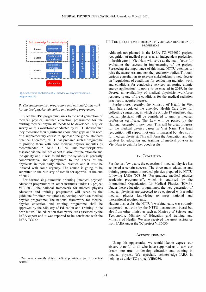
MEDICAL PHYSICS INTERNATIONAL Journal, vol.8, No.2, 2020
41
Fig 2. Schematic illustration of NTTU Medical physics education
programme [3].
B. The supplementary programme and national framework for medical physics education and training programme
Since the BSc programme aims to the next generation of medical physics, another education programme for the existing medical physicists1 needs to be developed. A quick survey on this workforce conducted by NTTU showed that they recognise their significant knowledge gaps and in need of a supplementary course to approach the global standard practice. Therefore, NTTU has prepared such a programme to provide them with core medical physics modules as recommended in IAEA TCS 56. This manuscript was assessed via the IAEA’s expert mission for the rationale and the quality and it was found that the syllabus is generally comprehensive and appropriate to the needs of the physicists in their daily clinical practice and it must be initiated with some urgency. The course is going to be submitted to the Ministry of Health for approval at the end of 2019.
For harmonising numerous orienting “medical physics” education programmes in other institutes, under TC project VIE 6030, the national framework for medical physics education and training programme will serve as the guideline for other institutions to develop their own medical physics programme. The national framework for medical physics education and training programme shall be approved by the Ministry of Education and Training in the near future. The education framework was assessed by the IAEA expert and it was reported to be consistent with the IAEA TCS 56.
1 Personnel currently doing medical physicist’s job in medical centres
III. THE RECOGITION OF MEDICAL PHYSICS AS A HEALTH CARE PROFESSION
Although not planned in the IAEA TC VIE6030 project, recognition of medical physics as an independent profession in health care in Viet Nam will serve as the main factor for evaluating the success in implementing of the project. Foreseeing the importance of this issue, NTTU attempts to raise the awareness amongst the regulatory bodies. Through various consultation to relevant stakeholders, a new decree on “regulations of conditions for conducting radiation work and conditions for conducting services supporting atomic energy application” is going to be enacted in 2019. In the Decree, an availability of medical physicisist workforce resource is one of the conditions for the medical radiation practices to acquire license.
Furthermore, recently, the Ministry of Health in Viet Nam has circulated the amended Health Care Law for collecting suggestion, in which the Article 17 stipulated that medical physicist will be considered to grant a medical profession certificate. The Law will be passed by the National Assemby in next year. This will be great progress for the medical physics career in Viet Nam. The legal recognition will support not only in material but also spirit for medical physicist. This will form the foundation and the catalyst for education and training of medical physics in Viet Nam to gain further good results.
IV. CONCLUSION
For the last few years, the education in medical physics has achieved a certain success. The three main education and training programmes in medical physics prepared by NTTU following IAEA TCS 56 “Postgraduate medical physics academic programmes”, which is endorsed by the International Organization for Medical Physics (IOMP). Under these education programmes, the new generation of medical physicists are expected to be equipped with a solid medical physics knowledge to meet national and international requirements. Having this results, the NTTU’s working team, was strongly supported not only by the NTTU management board but also from other ministries such as Ministry of Science and Techonoloy, Ministry of Education and training and Ministry of Health. We also received the great assistance from IAEA under the TC project VIE6030.
ACKNOWLEDGMENT
Using this opportunity, we would like to express our sincere thankful to all who have supported us to turn our dream into true, to develop education and training in medical physics. We especially acknowledge IAEA in helping us under TC project VIE6030.
Basic science
Fundamental knowledge
Professional knowledge
Internship, thesis
TOEI
C 4
50
or
equ
ival
ent
Ph
ysic
al e
du
cati
on
Nat
ion
al d
efen
se e
du
cati
on
2 years
1 years
2 years
Basic knowledge for medical physics
Graduation
MEDICAL PHYSICS INTERNATIONAL Journal, vol.8, No.2, 2020
42
We also would like to show our gratitude to our home university – NTTU, for the rightful vision and thoughtful infrastructure supporting for our programme. Last but not least, we shall give the warmest appreciation to the University of Malaya, Malaysia and especially Professor Ng Kwan Hoong for providing our trainers with the best education and training environment and practical advices.
REFERENCES
1. Viet Nam Agency for Radiation and Nuclear safety (VARANS), Statistic data of radiation sources, Hanoi: VARANS, 2018, Internal data
2. Kieu-Trang T. Hoang, Tan-Chau Nguyen, Trung-Hieu Nguyen, Hong-Loan T. , Tao V. Chau (2018) Medical physics in Viet Nam: The current status of equipment, workforce and education , MPI Journal vol. 6, No.2, 2018 at http://www.mpijournal.org
3. Thanh-Luong Dang, Nhu-Tuyen Pham, Anh-Tung Hoang, Huu-Vinh Cao (2018) The medical physics education and training programme of Nguyen Tat Thanh University – the first official medical physics training program in Viet Nam. MedPharmRes 2(2):43-47 DOI 10.32895/UMP.MPR.2.2.43
4. International Atomic Energy (2013) Training Course Series 56: Postgraduate Medical physics Academic Programmes
5. EUROPEAN COMMISSION, FOOD AND AGRICULTURE ORGANIZATION OF THE UNITED NATIONS, INTERNATIONAL ATOMIC ENERGY AGENCY, INTERNATIONAL LABOUR ORGANIZATION, OECD NUCLEAR ENERGY AGENCY, PAN AMERICAN HEALTH ORGANIZATION, UNITED NATIONS ENVIRONMENT PROGRAMME, WORLD HEALTH ORGANIZATION, Radiation Protection and Safety of Radiation Sources: International Basic Safety Standards, IAEA Safety Standards Series No. GSR Part 3, IAEA, Vienna (2014).
6. IAEA (2018) End of Mission “Education and Training Programmes for Medical Physicists in Viet Nam” VIE6030 [VIE2016005], 20-24 August, 2018
7. Circular 38/2010/TT- BGDĐT on conditions, dossiers and procedures for training, suspend enrollment,revoke training permission decision master and doctoral degrees
Contacts of the corresponding author:
Author: Thanh-Luong Dang Institute: Nguyen Tat Thanh University Street: Nguyen Tat Thanh City: Ho Chi Minh Country: Viet Nam Email: [email protected]

MEDICAL PHYSICS INTERNATIONAL Journal, vol.8, No.2, 2020
43
Professional Training Scheme to Answer National Demand Medical Physicists in Indonesia
S.A. Pawiro1,2, L.E. Lubis1,2, A. N. Oktavianto2,3, M. Mukhlisin4, D.S. Soejoko1
1 Department of Physics, Faculty of Mathematics and Natural Sciences, Universitas Indonesia, Depok, Indonesia 2 Aliansi Fisikawan Medik Indonesia (AFISMI), Jakarta, Indonesia
3 Directorate Health Facilities, Directorate General of Health Services, Ministry of Health, Jakarta, Indonesia 4 Directorate Licensing of Radiation Facililities and Radioactive Material, Nuclear Regulatory Agency , Jakarta, Indonesia
Abstract—The recognition of medical physicist in
Indonesia has been implemented in many aspects. In regulatory point of view, registered medical physicists has to be present during application of license for ionizing radiation-utilizing medical facilities in Nuclear Energy Regulatory Agency (Badan Pengawas Tenaga Nuklir, BAPETEN). Regulations also mandate diagnostic x-ray devices to be compliance-tested by BAPETEN-appointed companies prior to use—with certified personnel in these companies being mostly medical physicists. One of the major impacts of these policies is the significant increase in number of clinical medical physicists from 2016 to 2020, particularly for diagnostic radiology medical physicist (DRMP). In addition, the number of radiation oncology medical physicist (ROMP) is also increasing along with the increasing number of radiotherapy centers. Similarly, the incresing number of nuclear medicine medical physicists (NMMP) is also due to the the increasing number of nuclear medicine facilities in Indonesia. This situation has been made possible by the short-term policy of the medical physics professional society in collaboration with university and regulatory authority to develop the professional training scheme for medical physics. Two professional training programs to produce young medical physicists has been implemented in Indonesia since 2018. The result of the training is presented in this paper.
Keywords — medical physics, recognition, prefessional training, Indonesia.
I. INTRODUCTION
Medical physicist as profession in Indonesia has been
recognized by Ministry of Health in Decree No. 48/ 2007.
The profession ‘medical physicist’ has also been
mentioned in Government Regulation No. 36/2014, stated
as medical professional under biomedical technology
cluster with radiographer and biomedical engineer. By
law, medical physicists have to be present in
Radiotherapy, Diagnostic and Interventional Radiology,
and Nuclear Medicine services as one of the prerequisites
for medical devices to be licensed for clinical use. It is
regulated by both the Nuclear Energy Regulatory Agency
and Ministry of Health under separate decrees [1].
According to Government Regulation No. 12/2012 on
higher education, medical physics education comprises of
academic and professional training. [1] In line with
international recommendations, the Clinically Qualified
Medical Physicist (CQMP) has academic qualification of
postgraduate level with minimum 2 years of additional
clinical training at hospital [2].
II. MEDICAL PHYSICS TRAINING PROGRAMME
2.1. Short-term Policy Pawiro et al [1] mentioned that direct implementation
of international recommendation on clinically qualified medical physicist training program faces a challenge concerning the geography, demography of population, and medical devices distribution in Indonesia.
Table 1. Radiation medicine facilities in Indonesia and the need of medical physicists
Province DR RT NM MP need
Aceh 14 - - 11 North Sumatera 56 5 1 78 West Sumatera 11 2 - (1) 17 Riau 18 2 - 20 Jambi 5 - - 7 South Sumatera 18 1 - 25 Bengkulu 4 - - 5 Lampung 20 1 - 23 Bangka Belitung 4 - - 5 Riau Island 11 - - 10 Jakarta 95 12 6 207 West Java 134 3 2 289 Central Java 99 8 1 207 Yogyakarta 22 3(1) 1(1) 34 East Java 116 5 - (2) 213 Banten 39 1 - 104 Bali 30 3(1) - 32 West Nusa Tenggara 8 1 - 12 East Nusa Tenggara 5 - - 6 West Kalimantan 9 - - 17 Central Kalimantan 6 - - 5 South Kalimantan 11 1 - 16 East Kalimantan 15 2(1) 1 24 North Kalimantan 3 - - 0 North Sulawesi 6 1 - 10 Central Sulawesi 8 - - 6 South Sulawesi 36 3 - 52 South-East Sulawesi 5 - - 16 Gorontalo 4 - - 10 West Sulawesi 4 - - 2 Maluku 3 (1) - 1 North Maluku 2 - - 3 Papua 7 - - 3 West Papua 1 - - 0 Total 839 56(4) 16(4) 1470
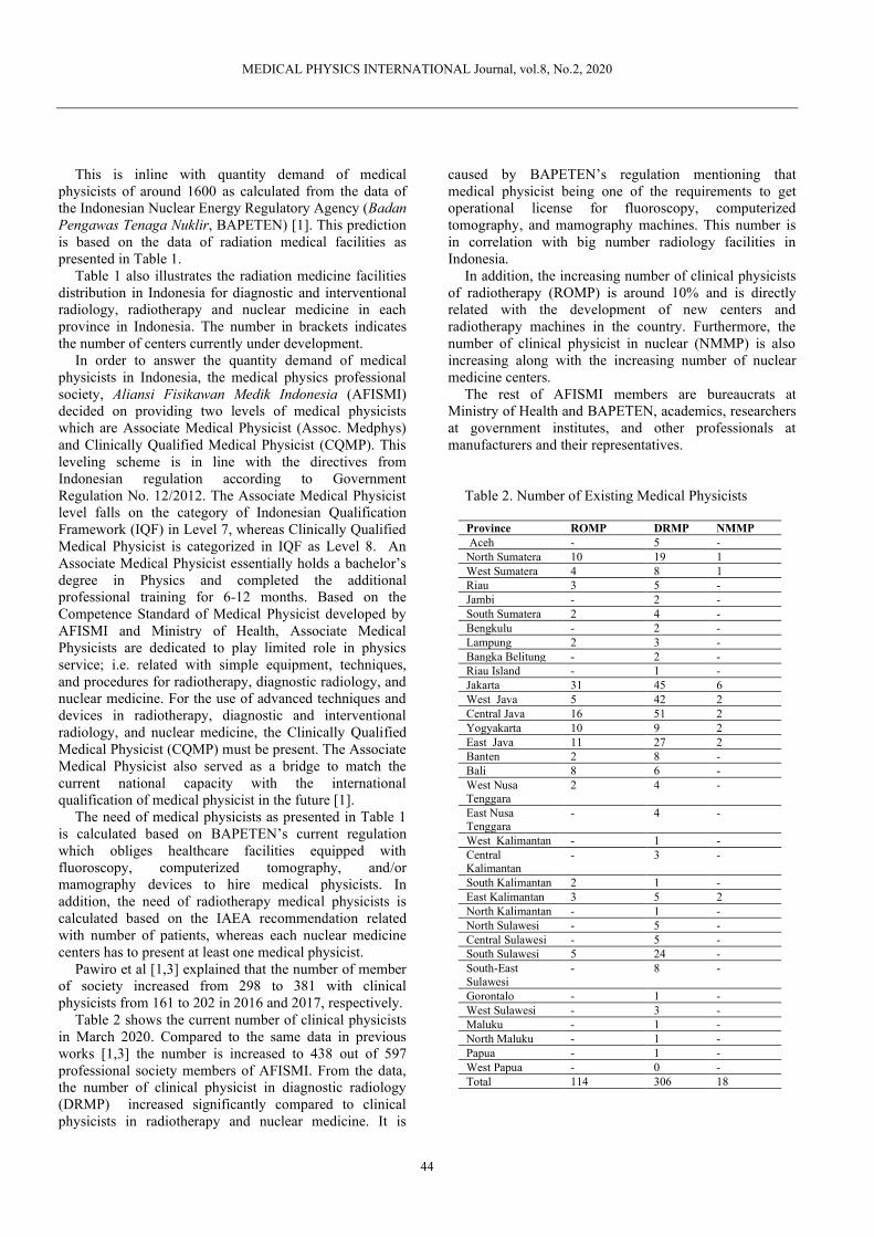
MEDICAL PHYSICS INTERNATIONAL Journal, vol.8, No.2, 2020
44
This is inline with quantity demand of medical physicists of around 1600 as calculated from the data of the Indonesian Nuclear Energy Regulatory Agency (Badan Pengawas Tenaga Nuklir, BAPETEN) [1]. This prediction is based on the data of radiation medical facilities as presented in Table 1.
Table 1 also illustrates the radiation medicine facilities distribution in Indonesia for diagnostic and interventional radiology, radiotherapy and nuclear medicine in each province in Indonesia. The number in brackets indicates the number of centers currently under development.
In order to answer the quantity demand of medical
physicists in Indonesia, the medical physics professional
society, Aliansi Fisikawan Medik Indonesia (AFISMI)
decided on providing two levels of medical physicists
which are Associate Medical Physicist (Assoc. Medphys)
and Clinically Qualified Medical Physicist (CQMP). This
leveling scheme is in line with the directives from
Indonesian regulation according to Government
Regulation No. 12/2012. The Associate Medical Physicist
level falls on the category of Indonesian Qualification
Framework (IQF) in Level 7, whereas Clinically Qualified
Medical Physicist is categorized in IQF as Level 8. An
Associate Medical Physicist essentially holds a bachelor’s
degree in Physics and completed the additional
professional training for 6-12 months. Based on the
Competence Standard of Medical Physicist developed by
AFISMI and Ministry of Health, Associate Medical
Physicists are dedicated to play limited role in physics
service; i.e. related with simple equipment, techniques,
and procedures for radiotherapy, diagnostic radiology, and
nuclear medicine. For the use of advanced techniques and
devices in radiotherapy, diagnostic and interventional
radiology, and nuclear medicine, the Clinically Qualified
Medical Physicist (CQMP) must be present. The Associate
Medical Physicist also served as a bridge to match the
current national capacity with the international
qualification of medical physicist in the future [1].
The need of medical physicists as presented in Table 1 is calculated based on BAPETEN’s current regulation which obliges healthcare facilities equipped with fluoroscopy, computerized tomography, and/or mamography devices to hire medical physicists. In addition, the need of radiotherapy medical physicists is calculated based on the IAEA recommendation related with number of patients, whereas each nuclear medicine centers has to present at least one medical physicist.
Pawiro et al [1,3] explained that the number of member of society increased from 298 to 381 with clinical physicists from 161 to 202 in 2016 and 2017, respectively.
Table 2 shows the current number of clinical physicists in March 2020. Compared to the same data in previous works [1,3] the number is increased to 438 out of 597 professional society members of AFISMI. From the data, the number of clinical physicist in diagnostic radiology (DRMP) increased significantly compared to clinical physicists in radiotherapy and nuclear medicine. It is
caused by BAPETEN’s regulation mentioning that medical physicist being one of the requirements to get operational license for fluoroscopy, computerized tomography, and mamography machines. This number is in correlation with big number radiology facilities in Indonesia.
In addition, the increasing number of clinical physicists of radiotherapy (ROMP) is around 10% and is directly related with the development of new centers and radiotherapy machines in the country. Furthermore, the number of clinical physicist in nuclear (NMMP) is also increasing along with the increasing number of nuclear medicine centers.
The rest of AFISMI members are bureaucrats at Ministry of Health and BAPETEN, academics, researchers at government institutes, and other professionals at manufacturers and their representatives.
Table 2. Number of Existing Medical Physicists Province ROMP DRMP NMMP
Aceh - 5 - North Sumatera 10 19 1 West Sumatera 4 8 1 Riau 3 5 - Jambi - 2 - South Sumatera 2 4 - Bengkulu - 2 - Lampung 2 3 - Bangka Belitung - 2 - Riau Island - 1 - Jakarta 31 45 6 West Java 5 42 2 Central Java 16 51 2 Yogyakarta 10 9 2 East Java 11 27 2 Banten 2 8 - Bali 8 6 - West Nusa Tenggara
2 4 -
East Nusa Tenggara
- 4 -
West Kalimantan - 1 - Central Kalimantan
- 3 -
South Kalimantan 2 1 - East Kalimantan 3 5 2 North Kalimantan - 1 - North Sulawesi - 5 - Central Sulawesi - 5 - South Sulawesi 5 24 - South-East Sulawesi
- 8 -
Gorontalo - 1 - West Sulawesi - 3 - Maluku - 1 - North Maluku - 1 - Papua - 1 - West Papua - 0 - Total 114 306 18

MEDICAL PHYSICS INTERNATIONAL Journal, vol.8, No.2, 2020
45
2.2. Curriculum of Training The training scheme of Associate Medical Physicist
was developed to produce the large number of junior physicist who can play role as junior physicist in three specialities under senior physicists or Clinically Qualified Medical Physicist. The curriculum of this scheme will train the candidate to understand the role of medical physicist in simple diagnostic radiology (general x-ray, dental and fluoroscopy), radiotherapy (teletherapy machines with 3D and brachytherapy 2D capabilities), and nuclear medicine (gamma camera and single photon emision computerized tomography, SPECT). Therefore, the 6 to 12 months training for Associate Medical Physicists will cover the basic competency of medical physicist in three specialities. The curriculum of associate medical physicist is decribed in Table 3. Table 3. Curriculum of associate medical physicist training scheme
No
Module Length of
training ( hours)
1 Ethics of medical physicist in
clinic and research
16
2 Acceptance and
ommissioning of
equipment
24
3 Quality Assurance of
Equipment
32
4 Radiotherapy planning 28 5 Dose Audit 6 Radiation protection and
radiation safety
68
7 Clinical rotation in
Diagnostic Radiology
160
8 Clinical Rotation in Radiotherapy
160
9 Clinical Rotation in Nuclear
Medicine
80
In addition to the Associate Medical Physicist training
scheme, we also run international standard of clinical residency program following the IAEA Training Course Series (TCS) publication 37, 47, and 50 [1,2].
2.3 Intake of participants As consensus at the professional society
(www.afismi.org) and the Indonesian Association of Higher Education in Medical Physics (AIPFMI, www.aipfmi.org), the educational background to enroll to the Associate Medical Physics training is described. Participants must only graduate from undergraduate program of physics or nuclear engineering with major in medical physics. The candidate has to proof with their academic transcript and completed the specified subjects; anatomy and physiology, radiological physics and dosimetry, imaging physics, radiotherapy physics, and nuclear medicine physics. If the candidates come from other theoretical and applied physics program, they must
complete the matriculation on the aforementioned core subjects in medical physics.
The training is initiated and conducted by the Center for Medical Physics and Biophysics, Institute of Applied Sciences, Faculty of Mathematics and Natural Sciences, Universitas Indonesia (CMPB UI), with participants coming from other universities throughout Indonesia. The first batch of this training started in February 2018.
According to the data from batch 1 to batch 5, the training administrator must select participants based on admission test scores and also geographical distribution aspects. The selected participants for the Associate Medical Physics training can be seen in Table 4.
Table 4. Professional training program for Associate Medical Physicist
Batch Number of
participants
Graduated Job position
secured
Batch 1 49 47 44 Batch 2 34 33 23 Batch 3 32 32 26 Batch 4 29 29 12
Batch 5* 33 - - Total 177 141 105 • On-going
International Atomic Energy Agency (IAEA) through
the Regional Technical Cooperation project in Asia Pacific conducted the pilot project to initiate the Clinically Qualified Medical Physics residency program in Indonesia in 2016. The pilot ROMP and DRMP has been started as described in Tabel 5 under the IAEA project RAS6077 followed the IAEA Training Coure Series, and it was translated to e-learning system called the Advanced Medical Physics Learning Environment (AMPLE). This e-learning system provides the possibility for residents to submit their work and the supervisor to grade their work [1]. Table 5 shows the paticipant of CQMP residency program in Indonesia which is registered by CMPB UI. The requirement of candidate is graduated form master of medical physics with background education in physics or nuclear engineering and have to completed associate medical physics training scheme before the candidate started the training. Up to now, this program is a voluntary program.
For ROMP program, three residents have graduated from based on asessment in April 2019 which was performed by external expert of IAEA and local medical physcists. Two residents of ROMP batch 1 still doing additional assignment to pass the program. The second batch will be asessed and evaluated in 2020. The first and second batch is trial program for 2 years flexible program depending on the clinical environment. The third batch has been started with fixed program for mandatory module in one year and then continue with internship for 6 months.
The DRMP program is started with remote residency program which is supervised by clinically qualified medical physicist from Australia. Up to now, we only have

MEDICAL PHYSICS INTERNATIONAL Journal, vol.8, No.2, 2020
46
one resident who still struggling to finish the program. In addition, the NMMP program is finally started under the project IAEA RAS6087 with 2 residents in June 2020.
Furthermore, the Table 5 indicated that ROMP batch 3 and NMMP batch 1 will be started with 4 residents and 2 residents, respectively.
Table 5. clinical qualified medical physics residency program
Specialities*,
Batch
Number
Participants
Graduated Job position
secured
ROMP 1 5 3 5 ROMP 2 4 - 4 ROMP 3 4 - -
DRMP 1 1 - 1 NMMP 1 2 - - Total 16 3 10
*ROMP (Radiation Oncology Medical Physics), DRMP (Diagnostic Radiology Medical Physics), NMMP (Nuclear Medicine Medical Physics). The first batch ROMP and DRMP is under IAEA RAS6077 project, wheras first batch of NMMP is under IAEA RAS087 project After completion of the professional training in both
schemes, medical physicist has to be registered in the Indonesian Health Professional Council (Konsil Tenaga Kesehatan Indonesia, KTKI), Ministry of Health. The council will issue the registration certificate. This medical physicist registration certificate is a prerequisite document for radiation medicine facilities license application under BAPETEN’s authority.
2.4 Placement of the alumnae As above mentioned, two professional training schemes
is developed to answer national demand of medical physicists in Indonesia. Tables 4 and 5 indicated the placement of alumnae of professional training of associate medical physicist and clinically qualified medical physicist, respectively.
Table 4 shows that more than 95% of participant have graduated from AMP training program and around 75% of them have each secured a job position. Participants having not secured any job position is because they are also master students who prefer to finish their study prior to seeking a job. Since Batch 4 has just graduated in March 2020, that the unemployment rate of alumnae of the training for batch 1 to batch 3 is less than 10 %.
For the survey, around 80% of the alumnae from batch 1 to 4 work in Java island, while the rest are being distributed in other islands. This distribution of alumnae is another issue of the job placement, so the admission requirement of participants have changed for batch 5, where we prioritize to select participants from outside Java island. It is expected that a more proportional distribution to take place within two years in the future.
In addition, Table 5 shows that all participants of the clinically qualified medical physicist training already have a job before their completion the program for batch 1 and batch 2.
Based on the distribution of placement, the participants and alumnae of the CQMP training of ROMP have occupied jobs in Java and Sumatera Island at new centers with advanced technology. This has been made possible thanks to our cooperation with Indonesian Radiation Oncologist Society (IROS) and BAPETEN. The aforementioned parties have recommended new centers with advance technology to recruit new medical physicist with master’s degree in Physics or Medical Physics and with on going or graduated from CQMP training.
III. CONCLUSIONS
The recognition of medical physics profession in Indonesia has been established. The number of members and clinical medical physicists is increasing rapidly especially in diagnostic radiology. There are still homeworks to increase their competency through the continuing medical physics education which can be conducted by collaboration Ministry of Health, AFISMI, and Universities.
ACKNOWLEDGMENT
We would like to thank all the founders of medical physics in Indonesia; Prof. Djarwani Soeharso, Mr. Sudharto Wahab, and Ms. Ratih Oemiyati for their tireless dedication in accelerating the recognition of medical physics profession in national level.
REFERENCES
1. SA Pawiro, et al. (2019) Overseeing the Growth of Medical Physics: Indonesia Case. In: Lhotska L., Sukupova L., Lacković I., Ibbott G. (eds) World Congress on Medical Physics and Biomedical Engineering 2018. IFMBE Proceedings, vol 68/1. Springer, Singapore
2. ID McLean et al, (2019) Recommendation for Accreditation and Certification in Medical Physics Education and Clinical Training Programmes for The RCA Region, MEDICAL PHYSICS INTERNATIONAL Journal, vol.7, No.3, 2019
3. S.A. Pawiro et al, (2017) Current Status of Medical Physics Recognition in SEAFOMP Countries. MEDICAL PHYSICS INTERNATIONAL Journal, vol.5, No.1, pp 11- 15: 2017
Contacts of the corresponding author:
Author: Supriyanto Ardjo Pawiro Institute: Department of Physics, FMNS Universitas Indonesia Street: Kampus UI City: Depok Country: Indonesia Email: [email protected]
MEDICAL PHYSICS INTERNATIONAL Journal, vol.8, No.2, 2020
47
THE ESTABLISHMENT OF UNSCEAR REPORT IN THAILAND A.Krisanachinda1, T.Chaiwatanarat1, T.Phungrassami2, P.Treenavarat1, N.Pongnapang3, S.Srimanoroth4,
J.Laothamatas5
1Faculty of Medicine Chulalongkorn University, Bangkok, Thailand. 2Faculty of Medicine Prince of Songkla University, Songkla, Thailand, 3Faculty of Medical Technology, Mahidol University, Bangkok, Thailand. 4Bureau of Radiation Medical Devices, Department of
Medical Sciences, Ministry of Public Health, Nontaburi, Thailand 5Faculty of Health Science Technology, HRH Princess Chulabhorn College of Medical Science, Chulabhorn Royal Academy, Bangkok, Thailand
Abstract- The radiation medicine was established in
Thailand in 1921. The rules and regulations on the use of ionizing radiation had been enacted by the Atomic Energy for Peace of Thailand in 1961. After that the application of radiation in various fields had been grown up rapidly. There was no official report on the increasing use of radiation in any field until 2019. The Office of Atoms for Peace set up the Sub Committee on the use of ionizing radiation in medical field with the main objective is to collect the data according to UNSCEAR requirements on medical exposure and occupational exposure and be able to submit such the data for publication in UNSCEAR Report of the year 2020.
Keywords- medical exposure, occupational exposure, healthcare level, frequency, effective dose.
I.INTRODUCTION Royal Government of Thailand had appointed a board,
Atomic Energy Commission for Peace, A.E.C. since 1956. The Cabinet later assigned the committee to implement the plan to establish a nuclear research reactor and laboratory for education, training and research of Thai scientists and engineers pursuing peaceful utilization of atomic energy. On 15 October 1957, Royal Thai Government ratified the statue of the International Atomic Energy Agency, IAEA, as an agency under the United Nations and consequently was the 58th on the list of IAEA Member States. On May 14th 1958, the Cabinet approved the Thai A.E.C. to issue a contract for the construction of a research reactor building and the reactor from Curtiss-Wright Corporation, USA at the location of Bangkhen, Vibhavadi-Rangsit Road on May 20th, 1960. On April 25th 1961, Royal Government of Thailand proclaimed the enactment of the Atomic Energy for Peace Act, B.E. 2504 resulting in the establishment of the Office of Atomic Energy for Peace, OAEP. The Cabinet Resolution on November 21st 2006 on transfer of business, property, authority, liability, and budget involving nuclear research have separated Thailand Nuclear Institute of Technology, TINT (Public Organization), from the Office of Atoms for Peace, OAP, to mainly focus on nuclear research conduction.
At present, the Office of Atoms for Peace is a major organization responsible for formulating of a national nuclear policy and strategy for peaceful purposes and for regulating the use of radiation and nuclear energy in the country for safety of users, people and environment according to international standards and obligations. The responsibility of the OAP is to protect life, health and property from the hazards of nuclear energy and from the harmful effects of ionizing radiation.
II.UNSCEAR
United Nations Scientific Committee on the Effects of Atomic Radiation (UNSCEAR) was established in 1955 at the General Assembly of the United Nations. The committee collected and evaluated information on the levels and effects of ionizing radiation from all nuclear explosions. The first two substantive reports1,2 submitted to the General Assembly, in 1958 and 1962, presented comprehensive evaluations of the state of knowledge about the levels of ionizing radiation to which human beings were exposed and of the possible effects of such exposures (Figure 1). UNSCEAR became the official international authority on the levels and effects of ionizing radiation, used for peaceful as well as military purposes and derived from natural as well as man-made sources. The first UNSCEAR report of 1958 had been recognized that medical diagnostic and therapeutic exposures were a major component of artificial radiation exposure globally, a fact that remains true today. The Committee has systematically reviewed and evaluated global and regional levels and trends of medical exposure, as well as exposure of the public and workers. These reviews have prompted significant worldwide reductions in unnecessary radiation exposure, and continue to influence the programs of international bodies such as the International Atomic Energy Agency (IAEA), the International Labor Organization (ILO), the World Health Organization (WHO) and the International Commission on Radiological Protection (ICRP).
The Chernobyl accident in 1986 was a tragic event for its victims and there has been major hardship for those most affected. From early on, UNSCEAR was involved in the assessment of radiation exposures and health effects. In 1988 it published a first account of acute radiation effects in emergency workers and of the global exposures3. A more detailed assessment of radiation levels and effects from the accident was published in 20004. Furthermore, the Committee held technical discussions on four documents:
● Evaluation of medical exposures to ionizing radiation;
● Evaluation of occupational exposures to ionizing radiation;
● Biological mechanisms relevant for influence of cancer risks from low-dose radiation,
● Levels and effects of radiation exposure due to the accident at the Fukushima Daiichi nuclear power station: implications of information published since the 2013 UNSCEAR report5.
explosion
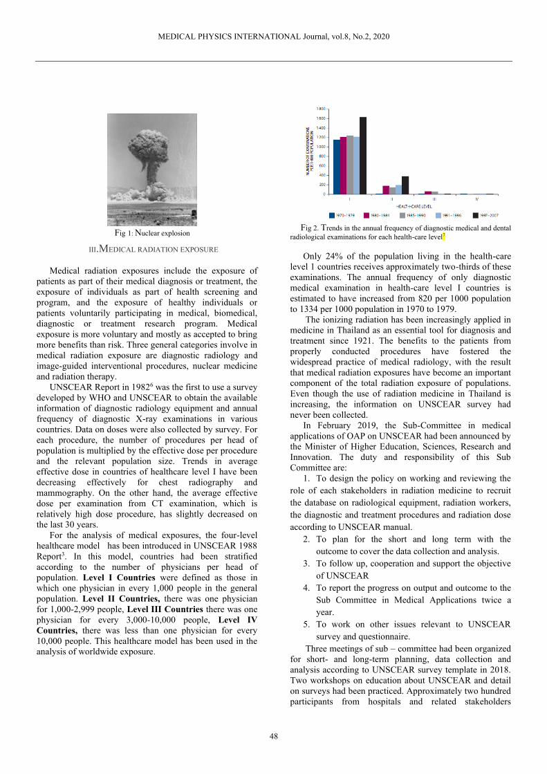
MEDICAL PHYSICS INTERNATIONAL Journal, vol.8, No.2, 2020
48
Fig 1: Nuclear explosion
III.MEDICAL RADIATION EXPOSURE
Medical radiation exposures include the exposure of patients as part of their medical diagnosis or treatment, the exposure of individuals as part of health screening and program, and the exposure of healthy individuals or patients voluntarily participating in medical, biomedical, diagnostic or treatment research program. Medical exposure is more voluntary and mostly as accepted to bring more benefits than risk. Three general categories involve in medical radiation exposure are diagnostic radiology and image-guided interventional procedures, nuclear medicine and radiation therapy.
UNSCEAR Report in 19826 was the first to use a survey developed by WHO and UNSCEAR to obtain the available information of diagnostic radiology equipment and annual frequency of diagnostic X-ray examinations in various countries. Data on doses were also collected by survey. For each procedure, the number of procedures per head of population is multiplied by the effective dose per procedure and the relevant population size. Trends in average effective dose in countries of healthcare level I have been decreasing effectively for chest radiography and mammography. On the other hand, the average effective dose per examination from CT examination, which is relatively high dose procedure, has slightly decreased on the last 30 years.
For the analysis of medical exposures, the four-level healthcare model has been introduced in UNSCEAR 1988 Report3. In this model, countries had been stratified according to the number of physicians per head of population. Level I Countries were defined as those in which one physician in every 1,000 people in the general population. Level II Countries, there was one physician for 1,000-2,999 people, Level III Countries there was one physician for every 3,000-10,000 people, Level IV Countries, there was less than one physician for every 10,000 people. This healthcare model has been used in the analysis of worldwide exposure.
Fig 2. Trends in the annual frequency of diagnostic medical and dental
radiological examinations for each health-care level7
Only 24% of the population living in the health-care
level 1 countries receives approximately two-thirds of these examinations. The annual frequency of only diagnostic medical examination in health-care level I countries is estimated to have increased from 820 per 1000 population to 1334 per 1000 population in 1970 to 1979.
The ionizing radiation has been increasingly applied in medicine in Thailand as an essential tool for diagnosis and treatment since 1921. The benefits to the patients from properly conducted procedures have fostered the widespread practice of medical radiology, with the result that medical radiation exposures have become an important component of the total radiation exposure of populations. Even though the use of radiation medicine in Thailand is increasing, the information on UNSCEAR survey had never been collected.
In February 2019, the Sub-Committee in medical applications of OAP on UNSCEAR had been announced by the Minister of Higher Education, Sciences, Research and Innovation. The duty and responsibility of this Sub Committee are:
1. To design the policy on working and reviewing the role of each stakeholders in radiation medicine to recruit the database on radiological equipment, radiation workers, the diagnostic and treatment procedures and radiation dose according to UNSCEAR manual.
2. To plan for the short and long term with the outcome to cover the data collection and analysis.
3. To follow up, cooperation and support the objective of UNSCEAR
4. To report the progress on output and outcome to the Sub Committee in Medical Applications twice a year.
5. To work on other issues relevant to UNSCEAR survey and questionnaire.
Three meetings of sub – committee had been organized for short- and long-term planning, data collection and analysis according to UNSCEAR survey template in 2018. Two workshops on education about UNSCEAR and detail on surveys had been practiced. Approximately two hundred participants from hospitals and related stakeholders

MEDICAL PHYSICS INTERNATIONAL Journal, vol.8, No.2, 2020
49
attended the one - day workshops in July and August 2019. In September 2019, the UNSCEAR data had been gathered and analyzed from major centers represent all parts of the country. The information on the number of populations, physicians, radiologists, dentists, radiation oncologists, nuclear medicine physicians, medical physicists, technologists, nurses, etc. and the number of equipment in diagnostic radiology, radiotherapy and nuclear medicine in 2018 in Thailand had been collected. Furthermore, the number on procedures in radiotherapy and nuclear medicine as well as the patient radiation dose had been estimated to obtain the effective dose as detail in UNSCEAR manual. Unfortunately, the data on diagnostic radiology on estimated number of procedures and radiation dose could not be collected in time. UNSCEAR data on medical exposures in radiation oncology and nuclear medicine and occupational radiation exposure had already been submitted to the UNSCEAR Headquarter via OAP official channel. Information on diagnostic radiology submitted only equipment and personnel. That information, hopefully, should be published in UNSCEAR 2020 Report.
IV.OCCUPATIONAL RADIATION EXPOSURE The information on occupational exposure relates to the
number of all workers working in supervised and controlled areas with the percent uncertainties. The information related to dosimetry consists of external and internal dose monitoring, the value of minimum detectable level (MDL) per measurement interval. Work categories on medical use are diagnostic radiology consisted of conventional diagnostic radiology, interventional procedures - radiology and cardiology, nuclear medicine, radiotherapy, dental practice, veterinary medicine and other medical uses. The workforce consists of the number of workers in dose intervals such as below MDL, MDL-1,>1-5, >5-10, >10-15, >15-20, >20-30, >30-50 and >50 mSv, the average and median effective doses by the dose interval, dose to eye lens and hands, number of female workers.
V. UNSCEAR REPORT OF THAILAND:
5.1 MEDICAL RADIATION EXPOSURE The UNSCEAR Survey form version 1.6 in 2018
consists of three parts on A. Diagnostic and interventional radiology (RD) B. Nuclear Medicine (NM) C. Radiotherapy (RT)
There are two methods of data collection regarding the annual frequency of procedures. First – a representative sample of hospitals then scaled up to the whole country. Practically, the data is available from the hospital radiology information system (RIS). Second- central statistics held by government department or insurance companies for all radiology practices in the country. In Thailand, the first method is used and the data is mostly collected from the
government offices, organizations, professional societies such as Medical Council of Thailand, Royal College of Radiologists of Thailand, Nuclear Medicine Society of Thailand, Thai Association of Radiation Oncology, Radiological Technology Society of Thailand, and Thai Medical Physicist Society, etc.
The government department involving directly in the survey data on occupational exposure is the Bureau of Radiation Medical Devices, Department of Medical Science, Ministry of Public Health. The Bureau of Radiation Medical Devices offers the service on the personnel monitoring. The information related to UNSCEAR survey is well recorded on annual basis.
For the Medical Exposure Report, the data recorded in the questionnaires are classified as: 1. Essential information: the number of population on the survey period in 2018 is 66,413,979 2. Staff and devices: a. all physicians are 58,025, radiologists 1,834, nuclear medicine physicians 66, radiation oncologists 174 b. all radiographic systems 28,039, nuclear medicine equipment 82, radiotherapy system 123 and radiotherapy imaging system 99 3. Information on the frequency of the radiological examinations, nuclear medicine procedures (diagnostic and therapeutic), external beam radiotherapy and brachytherapy with number of patients is recorded. 4. Dose: the mean effective dose per procedure is recorded in the unit of mSv for radiology and nuclear medicine. In radiotherapy the dose is in the unit of gray.
According to the number of physicians at 58,025 and Thai population of 66,413,979, these result in the ratio of one physician to Thai population at 1:1144 leading to Healthcare Level II for Thailand.
5.2 OCCUPATIONAL RADIATION EXPOSURE The personnel radiation monitoring service is available
at the Bureau of Radiation Medical Devices, Department of Medical Sciences, Ministry of Public Health and Thailand Institute of Nuclear Technology (TINT). The minimum detectable level (MDL) per measurement interval is 0.1 mSv. In 2018 the average, median and standard deviation of the effective dose were 0.45, 0.17 mSv and 1.87 respectively. The number of occupational exposed workers in Thailand was 86,922. Total number of medical workers was 50,519. The workers received effective dose higher than MDL was 20,628 and the number of female medical workers was 48,816.The workers in RD, RT and NM were 37364, 3349 and 1085 respectively. Number of female workers in RD, RT and NM were 34,389, 2,215 and 741 respectively.
5.3 THE FUTURE PLAN The sub-committee will review the obstacles in
collecting data especially in diagnostic radiology. The data collection on number of radiation workers, equipment, the frequency of procedures and the patient dose should be

MEDICAL PHYSICS INTERNATIONAL Journal, vol.8, No.2, 2020
50
collected annually. The result should be reported to the Sub Committee in medical application for the publication of OAP Newsletter or other relevant publishers.
For diagnostic radiology, the data from simple procedure could be collected from participating centers at 13 healthcare regions of Thailand while the complex procedures could be collected at the university hospitals where the manpower is available. This could be applied for interventional radiology which the simple procedures such as TACE and PTBD could be firstly collected in 2020.
The workshop on UNSCEAR should be set to inform more members in medical field to be more cooperative in data collection and be aware of the safely use on ionizing radiation. The future trend on number of workers and equipment, number of procedures, frequency and dose could be estimated for the development in medical radiation exposure of the country. All data could be archived at the main computer system at OAP.
VI. CONCLUSIONS The data on the exposure of patients and workers on
national level are valuable. Therefore the data collection should be improved to provide further relevant data about levels, effects and risk of radiation exposure from various sources. The government should support the NCP (National Contact Persons) and Sub Committee to facilitate coordination of collection and submission on the exposure of patients, workers and public in Thailand. The data should be published annually on website.
REFERENCES 1. UNSCEAR Report 1958. Reports of The United Nations Scientific
Committee on the Effects of Atomic Radiation Annex A-E. Annex F-I, New York 1958
2. UNSCEAR Report 1962. Reports of The United Nations Scientific Committee on the Effects of Atomic Radiation Annex A-K., New York 1962
3. UNSCEAR Report 1988. Source, Effects and Risks of Ionizing Radiation. Annex D Exposure from the Chernobyl accident, Annex G, Early effect in man of high doses of radiation. Annex J Exposures and effects of the Chernobyl accident. United Nations Publication, New York 1988
4. UNSCEAR Report 2000. Source and Effects of Ionizing Radiation. Annex D Medical Radiation Exposures. United Nations Publication, New York 2000
5. UNSCEAR Report 2013. Maps of Atmospheric Transport, Dispersion and Deposition Modelling Results. Annex A Levels and effects of radiation exposure due to nuclear accident after the 2011 great east Japan earthquake and tsunami. Appendix C Assessment of doses to the public. United Nations Publication, New York 2014
6. UNSCEAR Report 1982. Ionizing Radiation: Sources and Biological Effects. Annex G Medical Exposures. Annex H Occupational Exposure, United Nations Publication, New York 1982.
7. UNSCEAR Report 2008. Annex A Medical Radiation Exposure. United Nations Publication, New York 2008.
Contacts of the corresponding author:
Author: Anchali Krisanachinda Institute Faculty of Medicine, Chulalongkorn University Address: Bangkok, Rama IV Road, Thailand Email: [email protected]

MEDICAL PHYSICS INTERNATIONAL Journal, vol.8, No.2, 2020
51
MEDICAL PHYSICS IN THE REPUBLIC OF THE PHILIPPINES
Agnette P. Peralta1, Lilian V. Rodriguez2, and Bayani C. San Juan3
1University of Santo Tomas Graduate School, Manila 2Jose R. Reyes Memorial Medical Center, DOH, Manila
3Center for Device Regulation, Radiation Health, and Research, FDA, DOH, Muntinlupa City
ABSTRACT
The Republic of the Philippines is an archipelagic
state in South East Asia with a land area of 300,00 sq km. It has an estimated population for 2020 of 109,947,900. With such a big population, it is most important that a sufficient number of health care personnel is available in the country to provide appropriate services. Unfortunately, for medical physicists, the current number is not enough. There is also only one university in the country offering the master’s program in medical physics. In this paper, the development and current status of medical physics in the country will be presented.
Key words: Philippines, medical physics, status,
challenges, education, training
I. BACKGROUND
The Republic of the Philippines is an archipelago of
about 7,641 islands (1) including the Philippine Sea in the north and the east, the West Philippine Sea in the west, and the Sulu Sea in the south. It is located in South East Asia and its nearest neighbors are Brunei, Malaysia, Indonesia, Vietnam, and Taiwan. It has a land area of 300,00 sq km. It has an estimated population for 2020 of 109,947,900 (2) of which 50.4% are male and 49.6% are female. The age group 24 years old and below comprises 48.7% of its population, while the age group 60 years old and above comprises 8.6% of its population. Its gross domestic product in 2019 was US $355.50 billion. (3)
With such a big population, it is crucial for the
Philippines that a sufficient number of health care personnel is available to provide appropriate services.
Unfortunately, for medical physicists, the current number is not enough. In this paper, the development and current status of medical physics in the country will be presented.
II. AVAILABLE MAJOR EQUIPMENT
Radiation medicine equipment is among the most
costly equipment used in a hospital. In the Philippines, both the government and private sectors have invested in such equipment. Tables 1 – 3 below show the distribution of selected types of equipment in the country. (4,5)
Table I: Radiotherapy Equipment
ExternalBeam Radiotherapy 66 Brachytherapy 31 Simulator (CT sim) 48 Table 2: Diagnostic Radiology Equipment General radiography 2593 Fluoroscopy/interventional
radiology 309
Computed Tomography 354 Mammography 137 Dental x ray 217 Table 3: Nuclear Medicine Equipment SPECT 10 SPECT-CT 10 PET-CT 12 Gamma Camera 0 Cyclotron 4

MEDICAL PHYSICS INTERNATIONAL Journal, vol.8, No.2, 2020
52
III. THE FIRST STEPS
The first Filipino medical physicist was Mr. Luciano
N. Niguidula. He was employed by the government – owned Philippine General Hospital (PGH), at first as an engineer in the Radiology Department. In 1963, PGH acquired a cobalt-60 teletherapy unit. Dr. Harold Cook, a medical physicist from the UK, came to Manila as an International Atomic Energy Agency (IAEA) expert to train Mr. Niguidula in radiation dosimetry and treatment planning. Mr. Niguidula worked in radiation oncology medical physics well into his late eighties and trained the older generation of Filipino medical physicists.
In the 1970’s, Rizal Medical Center, another
government hospital employed the first Filipino physicist in a nuclear medicine department. In the 1980’s, Makati Medical Center became the first private hospital to employ a fulltime medical physicist for its radiation oncology department. In the 1990’s, The Medical City, a private hospital was the first hospital to employ a fulltime medical physicist in diagnostic radiology.
Any new profession needs a strong advocate. In the
Philippines this person was Dr. Celia T. Anatalio, a radiologist and a radiation oncologist, who is considered the Mother of Medical Physics in the country. She was the first director of the Radiation Health Office (RHO) of the Department of Health which was created in 1974.
IV. THE ROLE OF THE REGULATORY AGENCIES
The Philippines has two national radiation regulatory
agencies – the Philippine Nuclear Research Institute (PNRI) of the Department of Science and Technology (DOST) and the Center for Device Regulation, Radiation Health, and Research (CDRRHR) of the Food and Drug Administration of the Department of Health (DOH). PNRI used to be the Philippine Atomic Energy Commission while CDRRHR used to be the Radiation Health Office. PNRI regulates radionuclides while CDRRHR regulates electrical or electronic devices emitting radiation, and other medical and health-related devices.
Both agencies have worked closely in the development
of medical physics in the country. The MSc in Applied Physics, major in Medical Physics, program was established in 1981 as a joint project of the DOH, DOST, and the University of Santo Tomas Graduate School (USTGS), with technical assistance from the International Atomic Energy Agency. The DOH was also able to tap the World Health Organization (WHO) Western Pacific Regional Office (WPRO) and the Colombo Plan for fellowship grants to enable three young DOH physicists
to take up their MSc in Medical Physics degrees abroad (Aberdeen University, University of Surrey, University of Wisconsin-Madison). Upon their return to the country, they became part-time faculty members in the program while working fulltime for the DOH. Both regulatory agencies have also collaborated in the implementation in the country of IAEA projects in medical physics and radiation protection.
Regulations issued by the PNRI [for cobalt 60 external
beam radiotherapy (EBRT), brachytherapy, and nuclear medicine facilities] and by the CDRRHR [for EBRT using linear accelerators and tomotherapy equipment] triggered the employment of medical physicists by these facilities. The existing CDRRHR regulations for EBRTs and diagnostic radiology facilities are currently undergoing revision. It is expected that the requirement for the employment of diagnostic radiology medical physicists will be incorporated in the revised regulation for diagnostic radiology facilities while that for EBRT will be amended.
V. EDUCATION AND TRAINING
In June 1981, the two-year MSc in Applied Physics,
major in Medical Physics, program began in the University of Santo Tomas Graduate School. The program started with four students; three of them were employed by the DOH. These three are currently still active in medical physics.
However, the first graduates of the program completed
all their academic requirements only in 1986. The IAEA project for the program needed a medical physicist expert who could stay not just for a few weeks but for at least a year to serve as a faculty member and as the thesis adviser of the students. This assignment was accepted by Dr. Lars-Eric Larsson from Sweden who was then newly-retired.
In 2004, USTGS established a second master’s degree
program, the Master in Medical Physics course with no thesis requirement but with additional units to replace the thesis writing units. For both programs, the incoming students have educational backgrounds in physics, applied physics, engineering, chemistry, and physics for teachers.
From 1986 to 2019, both programs produced a total of
164 graduates. The first graduate of the MSc program was Mr. Marlon Raul Tecson. From 1986 to 1999, there were nineteen (19) graduates. During the period 2000 - 2019, there were 145 graduates.
Through the IAEA Regional Cooperative Project
(RCA) RAS 6038, structured clinical training programs in
MEDICAL PHYSICS INTERNATIONAL Journal, vol.8, No.2, 2020
53
radiation oncology medical physics (ROMP), diagnostic radiology medical physics (DRMP), and nuclear medicine medical physics (NMMP) were established in the country. Implementation of the residency training programs in the country was enhanced with the IAEA RCA RAS 6077 project using the advanced medical physics leaning environment (AMPLE) platform. It enabled supervision at a distance because, in many instances, the resident and the supervisor are not based in the same hospital, nor even in the same city or island.
IAEA fellowships have also enabled Filipino medical
physicists to undergo short-term training courses or workshops abroad such as those held in the Abdus Salam International Center for Theoretical Physics in Italy or in other countries.
There have also been short-term workshops hosted
locally by the DOH, the DOST, the Philippine Radiation Oncology Society, and private medical centers, especially St. Luke’s Medical Center, in cooperation with the IAEA, the WHO WPRO, the International Organization for Medical Physics (IOMP), the South East Asian Federation of Organizations for Medical Physics (SEAFOMP), the Asia-Oceania Federation of Organizations for Medical Physics (AFOMP) , the American Association of Physicists in Medicine, the American Society for Radiation Oncology, and the European Society for Radiotherapy and Oncology.
VI. THE PROFESSIONAL SOCIETY AND CERTIFICATION
The Philippine Organization of Medical Physicists
(POMP) was established in 1986. It became a member of IOMP in 1986 and a founding member-organization of SEAFOMP and AFOMP in 2000. In 2016, POMP became the Society of Medical Physicists in the Republic of the Philippines (SMPRP).
Under POMP, now SMPRP, the first certifying board
examination in ROMP was held in 2010 with Dr. Brendan Healy, from Australia, as the external examiner and Ms. Lilian Vidal-Rodriguez as the local examiner. There are now twenty (20) board certified ROMPs.
Under SMPRP, the first certifying board examination
in DRMP was held in 2019 with Dr. Ian Donald McLean, also from Australia, as the external examiner with Ms. Agnette Peralta, Mr. Bayani San Juan, and Ms. Aida Lobriguito as the local examiners. There are now thirteen (13) board certified DRMPs.
The board examination for NMMPs is still being
planned.
VII. MAJOR CHALLENGES
During the first twenty years of the USTGS academic
program, the low number of MSc students in the medical physics program was the first major challenge. Partial scholarships were available from DOH and DOST but these were awarded only to their qualified employees. In the latter part of the third decade of the program, the DOST expanded its fulltime scholarship program for graduate degree courses, and medical physics was included among the fields. With this development, more students were able to enroll in medical physics as fulltime students. Private sector scholarships have also been made available. A local supplier of linear accelerators sponsored three scholars. Private investors or hospital owners who want to put up radiotherapy or nuclear medicine facilities have employed young BSc physics or BSc engineering graduates and awarded them scholarships to enroll in the MSc program.
The small number of graduates was the second major
challenge. The MSc thesis requirement was an impediment for graduation of students who were mainly working students. Several of them worked fulltime in radiotherapy facilities and, due to their workload and the distance of their hospitals from UST, were unable to complete their schooling. Thus, USTGS established a second program, the non-thesis Master in Medical Physics degree program.
Because UST is located in Manila, most students are
based in the National Capital Region. The third major challenge is the establishment of more master’s programs in medical physics. Currently three universities, one of them in Mindanao, have plans to do so.
The fourth major challenge was the lack of positions
for medical physicists. This is not the situation anymore. In 2014, with the DOH plan to establish radiotherapy and nuclear medicine facilities in most of its regional hospitals and medical centers, many positions for physicists in DOH hospitals were created.
The fifth major challenge is the shortage of medical
physicists in the country. This situation has limited the number of radiotherapy and nuclear medicine facilities being established in the country.
The sixth major challenge for hospital-based medical
physicists is their workload. Administrative work is added to their already very heavy clinical workload. When combined with the mentoring of medical physics residents, the clinical supervisor is left with no more time to do research. This situation has also resulted in a shortage of clinical supervisors for residency training of medical physicists.

MEDICAL PHYSICS INTERNATIONAL Journal, vol.8, No.2, 2020
54
The seventh major challenge is the recognition of the important role of medical physicists; this has mainly already been overcome. After Dr. Anatalio’s retirement, her successor was Ms. Agnette Peralta, the first medical physicist director of RHO. She was then succeeded by another medical physicist, Mr. Bayani San Juan.
The SMPRP also works closely with the Philippine
Radiation Oncology Society (PROS), the Philippine College of Radiology, and the Philippine Society of Nuclear Medicine. In 2003, Ms. Lilian Rodriguez became the first medical physicist to be the examiner in the physics portion of the certifying examination of the PROS Board of Radiation Oncology. The Board of Radiologic Technology (BORT) of the Professional Regulation Commission has had a medical physicist as a member of the BORT since its creation in 1992. Ms. Eulinia Valdezco was the first medical physicist BORT member.
However, there is still a need to make the rest of the
medical and paramedical professionals and the rest of the population realize the importance of medical physicists in health care.
VIII. CURRENT STATUS OF MEDICAL PHYSICS
Medical physicists in the Philippines have post
graduate educational backgrounds in medical physics or physics. Most of them possess a master’s degree in medical physics from the University of Santo Tomas Graduate School or have earned at least twenty academic units in the same program. Five received their master’s degrees in medical physics from foreign universities. Four of them have doctoral degrees in physics, two from a local university and two from overseas universities.
Filipino medical physicists who live in the Philippines
work in private and government hospitals and free-standing health-care facilities, two national radiation regulatory agencies, three private firms providing medical physics services, six multi-national companies, and three universities.
There are currently one hundred-ninety (190) medical
physicists active in the different fields of medical physics in the country with one hundred thirty-seven (137) working fulltime in health care facilities. The hospital employing the most number of medical physicists is St. Luke’s Medical Center with a total of thirteen in their two branches. Twenty health facilities employ at least three medical physicists each. Those medical physicists who have left the country to work and live abroad, or who have stayed in the country but are not active in medical physics anymore are not included in Table 4 below.
Table 4: Subfields of Medical Physics Radiation Oncology 106 Diagnostic Radiology 23 Nuclear Medicine 8 Others (radiation
protection, related fields) 53
TOTAL 190
IX. CONCLUSION
Establishment in the Philippines of the master’s program in medical physics was a major step in responding to the need for medical physicists in the country. Medical physics has become an attractive field for BSc Physics, Applied Physics, Engineering, Chemistry, and Physics for Teachers degree holders. However, there is still a shortage of medical physicists in the country. Moreover, there is a lack of awareness of the medical physics profession among the other healthcare specialties.
Board certification, regulation by the national radiation
regulatory agencies, support from the government and private sectors, technical assistance from IAEA, WHO, and national, regional and international professional organizations, and recognition by radiation medicine physicians in the country have all contributed greatly to the current status of medical physics in the Philippines.
Most important of all, the Filipino medical physicists
through their professionalism and excellent work have proved their worth in modern medicine and society.
ACKNOWLEDGEMENT
The authors thank Dr. Augusto Morales, Jr. and Mr.
Royce Gaspar for the photographs, and their other medical physics colleagues for sharing
information used in this paper.
REFERENCES
(1) en.m.wikipedia.org (2) Philippine Statistics Authority. Technical Notes
“2020 Midyear Population Projection”. (3) tradingeconomics.com. “World Trends
Economic Database”.
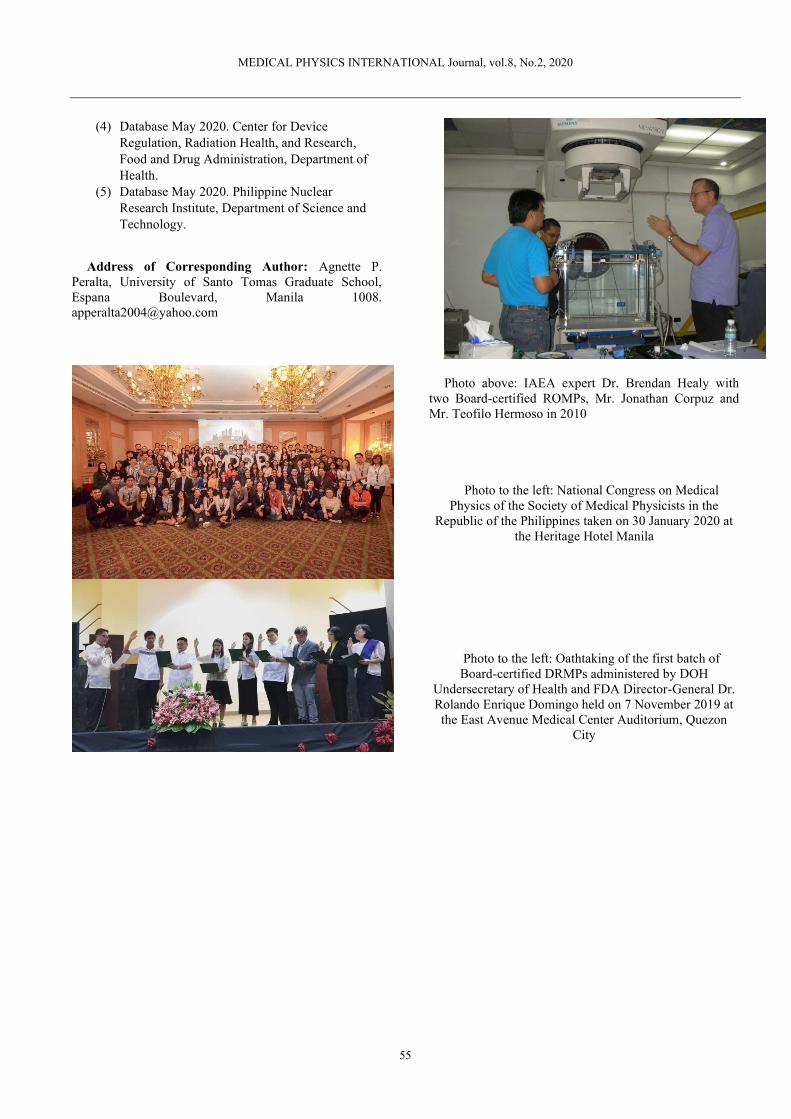
MEDICAL PHYSICS INTERNATIONAL Journal, vol.8, No.2, 2020
55
(4) Database May 2020. Center for Device Regulation, Radiation Health, and Research, Food and Drug Administration, Department of Health.
(5) Database May 2020. Philippine Nuclear Research Institute, Department of Science and Technology.
Address of Corresponding Author: Agnette P.
Peralta, University of Santo Tomas Graduate School, Espana Boulevard, Manila 1008. [email protected]
Photo above: IAEA expert Dr. Brendan Healy with two Board-certified ROMPs, Mr. Jonathan Corpuz and Mr. Teofilo Hermoso in 2010
Photo to the left: National Congress on Medical Physics of the Society of Medical Physicists in the
Republic of the Philippines taken on 30 January 2020 at the Heritage Hotel Manila
Photo to the left: Oathtaking of the first batch of Board-certified DRMPs administered by DOH
Undersecretary of Health and FDA Director-General Dr. Rolando Enrique Domingo held on 7 November 2019 at the East Avenue Medical Center Auditorium, Quezon
City

MEDICAL PHYSICS INTERNATIONAL Journal, vol.8, No.2, 2020
56
HISTORY OF MYANMAR
MEDICAL PHYSICIST ASSOCIATION
S.S.Lin1, O.M.Swe2 1Department of Radiation Oncology, Pinlon Cancer Center, Yangon, Myanmar
2Department of Nuclear Medicine, Yangon General Hospital, Yangon,
Myanmar
I. RADIATION THERAPY MEDICAL PHYSICIST
The first medical physicist from Myanmar who trained in England assigned to work in radiation therapy department of Yangon General Hospital in 1958. Starting from 1958, the limited numbers of medical physicists who had a chance to take long-term training (more than 1 year) were allocated in Mandalay and Taunggyi General Hospital. From that time, the medical physicist training accomplished in local apprenticeship and some short course programs which supported by International Atomic Energy Agency (IAEA) and World Health Organization (WHO). The candidates for the training came from academic level or senior technologist who graduate from physics or equivalent subject. That will be the challenge for Myanmar medical physics field which has no proper academic education and training program for national level. The bachelor degree course for medical imaging technology was established by Ministry of Health in 1991 at Yangon. Currently, there are three universities which offer bachelor in medical imaging technology and two universities which give master degree in that field. Radiation therapy technologist undergraduate course was introduced in 2018 at University of Medical Technology, Yangon. The first medical physics two-year master program is expected to establish in 2021 at the same University.
II. NUCLEAR MEDICINE MEDICAL PHYSICIST
Nuclear medicine in Myanmar is an independent medical specialty which is now operated by seven departments. The first nuclear medicine department was founded at Yangon General Hospital in 1963. There is no medical physicist post in nuclear medicine till 2003.
The person who had got a Bachelor of Physics was assigned as a nuclear medicine technologist in 1965. Limited number of human resources were working in nuclear medicine at that time. Only one technologist worked at a time. The first three were trained in United Kingdom for one year each. Among them, the last technologist who was also trained in Thailand, Singapore and China for physicist training became a medical physicist in 2003. The second medical physicist who worked in Mandalay General Hospital was a former radiographer. The third one who worked in Yangon General Hospital held a Bachelor of Science (Physics). All of them were already retired.
Present situation, all medical physicist in nuclear medicine are graduated from University of Medical Technology and Postgraduate from Thailand. Two working at Yangon General Hospital, government sector and two are at Pinlon Hospital, private sector.
III. MYANMAR MEDICAL PHYSICIST ASSOCIATION
The Association of Myanmar Medical Physicist (MMPA) was set up in mid of 2016 with 30 members who were working as medical physicists and radiation protection officers from Myanmar. The number of memberships increased to 33 in current year. The percentage of male and female in the association is 24% and 76% respectively. The majority of the members are from radiation therapy totaling of 24 persons. The second lead is from nuclear medicine with 7 members and diagnostic radiology with 2 radiation protection officers.
MMPA was an official member of the International Organization for Medical Physics (IOMP), Asia-Oceania Federation of Organizations for Medical Physics (AFOMP) and South-East Asian Federation of Organizations for Medical Physics (SEAFOMP) at the end of 2016.
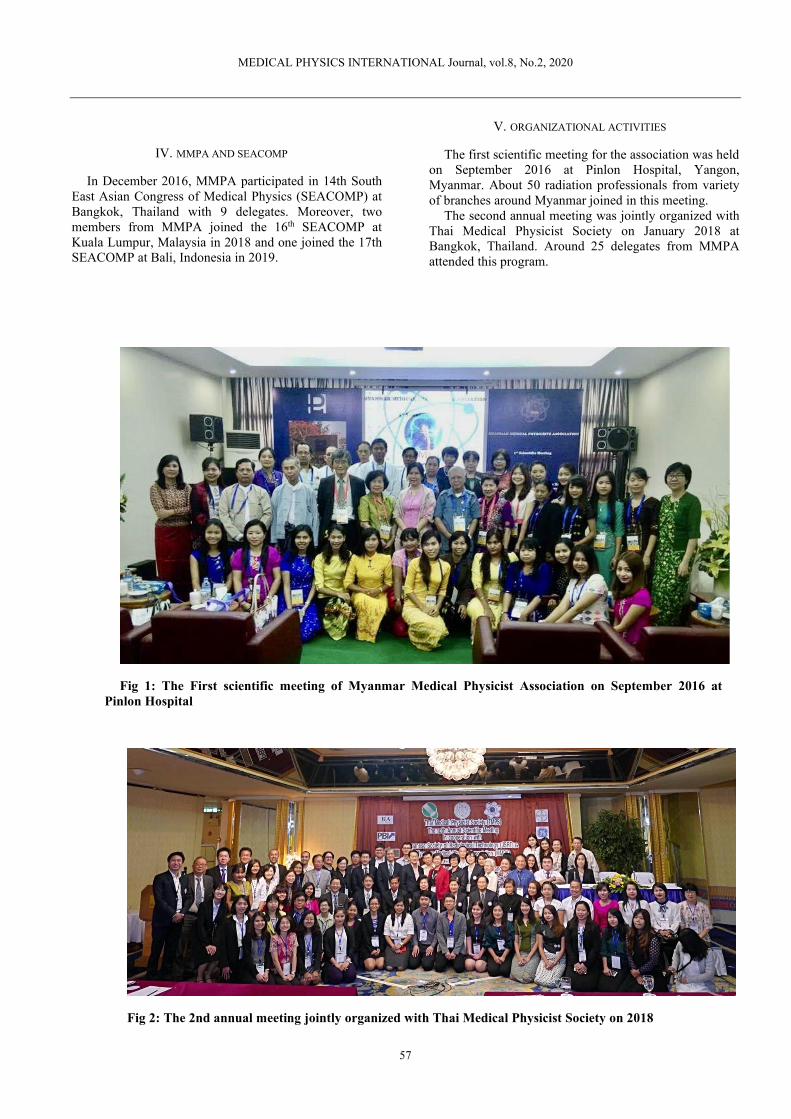
MEDICAL PHYSICS INTERNATIONAL Journal, vol.8, No.2, 2020
57
IV. MMPA AND SEACOMP
In December 2016, MMPA participated in 14th South East Asian Congress of Medical Physics (SEACOMP) at Bangkok, Thailand with 9 delegates. Moreover, two members from MMPA joined the 16th SEACOMP at Kuala Lumpur, Malaysia in 2018 and one joined the 17th SEACOMP at Bali, Indonesia in 2019.
V. ORGANIZATIONAL ACTIVITIES
The first scientific meeting for the association was held on September 2016 at Pinlon Hospital, Yangon, Myanmar. About 50 radiation professionals from variety of branches around Myanmar joined in this meeting.
The second annual meeting was jointly organized with Thai Medical Physicist Society on January 2018 at Bangkok, Thailand. Around 25 delegates from MMPA attended this program.
Fig 1: The First scientific meeting of Myanmar Medical Physicist Association on September 2016 at Pinlon Hospital
Fig 2: The 2nd annual meeting jointly organized with Thai Medical Physicist Society on 2018

MEDICAL PHYSICS INTERNATIONAL Journal, vol.8, No.2, 2020
58
In the mid of 2018, MMPA conducted the SPECT/CT Quality Assurance Workshop (1/2018) in Yangon, Myanmar. This activity could help to strengthen the quality assurance knowledge of medical physicists who specialized in Nuclear Medicine.
Radiation Protection and Patient Safety Workshop (2/2018) was held at the end of 2018. This workshop
organized with the intention to provide the deep understanding of the radiation risk, protection and patient safety not only for radiation professionals but also for other medical staffs who are not the radiation workers. The workshop conducted in two hospitals, Shwe Gone Ding hospital and Pinlon Hospital, Yangon, Myanmar.
Fig 3: SPECT/CT Quality Assurance Workshop (1/2018) at Pinlon Hospital, Yangon, Myanmar
Fig 4: Radiation Protection and Patient Safety Workshop (2/2018) at Shwe Gone Daing and Pinlon hospital in 2018

MEDICAL PHYSICS INTERNATIONAL Journal, vol.8, No.2, 2020
59
VI. CONCLUSION
Compare to the past, there are more opportunities to share the knowledges through organizational activities for Myanmar medical physicists these days than those were in decade. More than that, the members can seize the chance of participating international medical physics conferences in each year. These all can be labelled as the benefits of setting up the Myanmar Medical Physicist Association.
In the near future, MMPA will develop the functional website of the association to offer an easy access with the medical physics community. The association is going to take part in more activities of both national and international levels.
The number of medical physicists in Myanmar would escalate in the foreseeable future, if the government initiated the graduate medical physics program in next year. Myanmar medical physicist community is anticipating for the compensation of the challenges what they are facing due to lack of medical physics education program in the country. They are hoping for the more promising future.
Contacts of the corresponding author:
Author: Swe Swe Lin, M. Sc. Institute: Department of Radiation Oncology,
Pinlon Cancer Center Street: Saya San Road, Ward 27, North Dagon
11421 City: Yangon Country: Myanmar Email: [email protected]

MEDICAL PHYSICS INTERNATIONAL Journal, vol.8, No.2, 2020
60
MEDICAL PHYSICS ACTIVITY IN LAO PDR
V.Inphavong1
1 Radiotherapy Center, Mittaphab Hospital, Vientiane Capital 6035, Lao PDR
Lao People’s Democratic Republic (Lao PDR) is a member
of South East Asian Federation of Organizations for Medical Physics (SEAFOMP) in 2018. Lao PDR is located in Southeast Asian Region sharing borders with Cambodia (541 km to the south), People’s Republic of China (423 km to the northwest), Myanmar (235 km), Thailand (1,754 km to the west) and Viet Nam (2,130 km to the east). Its total area is 236,800 km2 (land: 230,800 km2 and water: 6,000 km2). The current population of the Lao PDR is about 7 million and majority is young, with 50% of the total population under the age of 20. Lao PDR is one of Least Developed Country (LDC) and aim to be graduate from LDC status by 2025. At present, National Law on Radiation and Nuclear was drafted and submitted recently to the National Assembly of the Lao PDR for consideration and approval. The Infrastructure in Lao PDR, in Diagnostic Radiology number of X-ray machine, Fluoroscopy, CT scanner, Mammography are 78, 3, 10, 2 systems respectively. Radiation Oncology was established in 2016 which Linear Accelerator and CT simulator were installed and operated at Radiotherapy Center, Mittaphab Hospital. For Human Resource, there were only 2 medical physicists who graduated Master of Science in Medical Physics/Medical imaging at Chulalongkorn University under IAEA fellowship (2015-2017). One medical physicist is attending a clinical training in radiation oncology medical physics (ROMP) for 2 years (2018-2020), IAEA curriculum (AMPLE) supported by VAMED Company. She obtained a remote clinical supervisor from King Chulalongkorn Memorial Hospital in Bangkok, Thailand.
Medical physicist responsibilities in radiation oncology are
to perform Quality Assurance both in mechanical and dosimetry including daily, weekly, monthly and annually. For 3D TPS, they are performing treatment planning of patient, export, calendar, patient QA before deliver radiation dose to patients and also take role as Administrator in Radiotherapy Center. In Diagnostic Radiology, they are performing Quality Control of X-rays DR/CR systems, image quality of CT scanner, Mammography, Ultrasound, Optimization and Control of Radiation Dose of Patients in CT. For Radiation Protection and Safety in hospital, they are evaluating personal dosimeter, report occupational dose for staff, share knowledges to their college and other hospitals and survey radiation dose in radiology. Aside of working as medical physics, they are cooperate with regional and international organizations, give lectures on Radiation Physics including fundamentals of ionizing radiation, radiation interaction with matter, application of ionizing radiation in medical field, dosimetry principle, quantities and units used in radiation dosimetry, radiation protection against external exposure, biological effect of ionized radiation, principle of QA\QC and practice QC and image quality of X-ray machine, CT scanner and Ultrasound to radiographer,
radiologist, student and resident of radiology. They are facing many obstacles due to the lack of medical physicist in the country such as workload, opportunities, lack of experience in clinical practice and limitation of QA/QC equipment. The most difficult parts of medical physicist is working without clinical supervision.
We received the supports from regional and international
organizations such as Thailand, Vietnam, IAEA, PMSF, SEACOMP. AFOMP, and ACPSEM. Hopefully, Lao PDR could obtain enough the education and clinical training so that the number of medical physicists would be increasing to meet the standards in radiology and radiation oncology in Lao PDR in the near future.

MEDICAL PHYSICS INTERNATIONAL Journal, vol.8, No.2, 2020
61
THE FUTURE OF MEDICAL PHYSICS: A CASE STUDY OF ZAMBIA K.A. Nkonde1, M. Kawesha2, M.M. Kanduza2, B.C. M’ule2, M. Mofya2, A.N. Mwale2, K.D. Manyika1
and D.C. Chilukusha1 1 Department of Science and Mathematics, Mulungushi University, Kabwe, Zambia
2 Medical Physics Section, Cancer Diseases Hospital, Lusaka, Zambia
ABSTRACT
The last few decades have seen tremendous growth in the health sector in Zambia. At the beginning of the 21st century, policies were developed and resources allocated, leading to the establishment of the first radiation treatment centre in the country; Cancer Diseases Hospital (CDH). Among the key personnel needed to run this kind of specialized hospital are medical physicists (MPs). As of January 2019, the country had less than five medical physicists on the Health Professions Council of Zambia (HPCZ) register.
In 2018, CDH had recorded about 2700 new cancer cases corresponding to a 12% increase from the past year. This value was representative of 34.7% patients with access to radiotherapy. In order to accommodate this rise in patient load, two radiotherapy centres and smaller satellite radiotherapy departments attached to existing hospitals with cancer management capabilities are envisioned. Furthermore, the Zambian Government in collaboration with ROSATOM, plan to construct the Centre for Nuclear Science and Technology (CNST); hosting Radiotherapy and Nuclear Medicine facilities also. These increases therefore, necessitate the procurement of more diagnostic and therapeutic equipment. This will subsequently require a substantial number of MPs commensurate with the International Atomic Energy Agency (IAEA) staffing recommendation.
Growth of Medical physics in Latin America, Asia and Africa was looked at and a comparison to Zambia undertaken. The IAEA tool was used to estimate the recommended staffing requirements of MPs and a comparison with the prevailing situation done from 2006 to 2019. Future projections and trends were also analysed to offer a much clearer perspective.
Results showed that the current growth in MPs is below the recommended threshold, the future trends are also indicative of a deficit which will likely not be met in the next 10 years. The following conclusions were made; the need to develop local curricula, improve enumerations and career prospects.
Keywords: Medical Physics, Medical Physicists, LMIC, IAEA, Zambia
I.INTRODUCTION
The cancer prevalence rate in low and middle income countries (LMIC) is estimated to double by 2050. This is attributed to several factors including dietary and lifestyle changes, HIV/AIDS, increased smoking and alcohol consumption. In Zambia 12052 new cases were reports in 2018, resulting in approximately over 7300 deaths (61.23 %) [1].
According to an IAEA news report in 2003 prior to setting up of the Cancer Diseases Hospital (CDH) [2], most patient with cancer had to travel to either Zimbabwe or South Africa to access radiotherapy (RT) treatment. Few, though could afford this even with the government covering most of the associated costs. The lack of proper treatment resulted in recurrences of most malignancies. This trend was greatly reduced with the opening of a radiotherapy centre in 2006 (officially opened in 2007).
The CDH has from inception to present treated over 21247 cases. This is a great milestone, considering that it recorded only 35 patients at inception [3].

MEDICAL PHYSICS INTERNATIONAL Journal, vol.8, No.2, 2020
62
Figure 1 Cancer patients treated at CDH from 2006 to 2018. Data adapted from [3].
Radiotherapy capacity building in Zambia has been a phased approach; with the initial phase started in 2001 with funding from OPEC ($5.6M), IAEA and GRZ [2]. Total funding was estimated to be about $7M by Lishimpi [3]. Phase I mostly catered for the construction of the radiotherapy unit, with a LINAC, Co-60, auxiliary equipment and training of initial staff (radiation oncologists-RO, medical physicists and radiation therapy technicians-RTT). Phase II, commenced in 2012 and involved the expansion of the Centre to include wards, Nuclear Medicine wing, dedicated chemotherapy wing, procurement of another Co-60 teletherapy machine with an SAD of 100 m and other facilities and equipment. This phase had a funding of $8M from OFID and GRZ. In 2016 funding of about $40M from OPEC Fund for International Development (OFID), Government of the Republic of Zambia (GRZ) and others was secured to embark on the third phase. The projects under this phase are the upgrade of CDH (infrastructure, staff and equipment), setting up of 3 out of the planned 9 satellite cancer centres across the country.
Nine people have been trained in medical physics from the inception of radiotherapy in Zambia, even though the training of medical physicists has not been consistent owing to several factors. The IAEA has been facilitating clinical training, though lately academic training has been facilitated in collaboration with the International Centre for Theoretical Physics (ICTP) through fellowships in partner countries. However, other partners have come on board to offer scholarships for staff to train in the UK and other countries.
A. CURRENT STATUS
The country has about ten medical physicists mainly in academia, health and other allied sectors. There are five MPs in the health sector, all stationed at the CDH, in radiotherapy. Of these five, only three have undergone structured clinical training to be designated as clinically qualified medical physicists (CQMP). A report by Datta et al [4] established a deficit of about 13 MPs for the country as of 2014, though the situation has improved slightly as 3 MPs were trained and retained since then.
Table 1 IAEA Directory of radiotherapy centres (DIRAC) report for Zambia [5]. RT Centres Clinical Accelerator. Co-60 CT Simulator. TPS HDR (Ir-192)
1 1 2 2 1 5 2
Currently there are programmes training ROs and RTT locally, but none for MPs. The current system does offer unstructured clinical training. Recently, plans to commence postgraduate studies at local universities are underway with curricula already developed. Clinical Medical Physics training will be offered in the hospital and efforts are underway to introduce this based on the Federation of African Medical Physics Organisations (FAMPO) clinical training curriculum. There is a
medical physics elective in the B.Sc. Physics offered by the University of Zambia [6], while Mulungushi University has introduced a B.Sc. Physics with individual medical physics modules in radiotherapy, medical imaging and nuclear medicine physics rather than electives. A B.Sc. in Medical Physics is also being planned, with duration of five years with clinical attachments during training.
2006 2007 2008 2009 2010 2011 2012 2013 2014 2015 2016 2017 20180
500
1000
1500
2000
2500
3000
Year
Numb
er of
Case
s
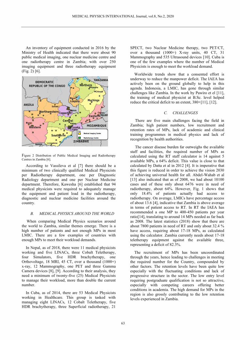
MEDICAL PHYSICS INTERNATIONAL Journal, vol.8, No.2, 2020
63
An inventory of equipment conducted in 2016 by the Ministry of Health indicated that there were about 90 public medical imaging, one nuclear medicine centre and one radiotherapy centre in Zambia; with over 250 imaging equipment and three radiotherapy equipment (Fig. 2) [6].
Figure 2 Distribution of Public Medical Imaging and Radiotherapy Centres in Zambia [6].
According to Vassileva et al [7] there should be a minimum of two clinically qualified Medical Physicists per Radiotherapy department, one per Diagnostic Radiology department and one per Nuclear Medicine department. Therefore, Kawesha [6] established that 94 medical physicists were required to adequately manage the equipment and patient load in the radiotherapy, diagnostic and nuclear medicine facilities around the country.
B. MEDICAL PHYSICS AROUND THE WORLD
When comparing Medical Physics scenarios around the world to Zambia, similar themes emerge. There is a high number of patients and not enough MPs in most LMIC. There are a few examples of countries with enough MPs to meet their workload demands.
In Nepal, as of 2018, there were 11 medical physicists working and five LINACs, three Cobalt Teletherapy, four Simulators, five HDR brachytherapy, one Orthovoltage, 18 MRI, 45 CT, over a thousand (1000+) x-ray, 12 Mammography, one PET and three Gamma Camera devices [8], [9]. According to their analysis, they need a minimum of twenty-five (25) Medical Physicists to manage their workload, more than double the current number.
In Cuba, as of 2014, there are 53 Medical Physicists working in Healthcare. This group is tasked with managing eight LINACs, 12 Cobalt Teletherapy, five HDR brachytherapy, three Superficial radiotherapy, 21
SPECT, two Nuclear Medicine therapy, two PET/CT, over a thousand (1000+) X-ray units, 40 CT, 31 Mammography and 535 Ultrasound devices [10]. Cuba is one of the few examples where the number of Medical Physicists is enough to meet the workload demand.
Worldwide trends show that a consented effort is underway to reduce the manpower deficit. The IAEA has actively been on the ground globally to help in this agenda. Indonesia, a LMIC, has gone through similar challenges like Zambia. In the work by Pawiro et el [11], the training of medical physicist at B.Sc. level helped reduce the critical deficit to an extent, 380+[11], [12].
C. CHALLENGES
There are five main challenges facing the field in Zambia; high patient numbers, low recruitment and retention rates of MPs, lack of academic and clinical training programmes in medical physics and lack of recognition by health authorities.
The cancer disease burden far outweighs the available staff and facilities, the required number of MPs as calculated using the RT staff calculator is 14 against 5 available MPs, a 64% deficit. This value is close to that calculated by Datta et al in 2012 [4]. It is imperative that this figure is reduced in order to achieve the vision 2030 of achieving universal health for all. Abdel-Wahab et al [13] also indicated that as of 2008, we had about 10100 cases and of these only about 6476 were in need of radiotherapy, about 64%. However, Fig. 1 shows that only 18.4% of patients actually had access to radiotherapy. On average, LMICs have percentage access of about 13.6 [4], indicative that Zambia is above average in terms of patient access to RT. In RT the IAEA has recommended a one MP to 400-450 patients per year ratio[14], translating to around 14 MPs needed as far back as 2008. The latest statistics (2018) show that there are about 7800 patients in need of RT and only about 32.4 % have access, requiring about 17-18 MPs, as calculated using the calculator. Zambia currently needs about 17-18 teletherapy equipment against the available three, representing a deficit of 82.3%.
The recruitment of MPs has been uncoordinated through the years, hence leading to challenges in meeting the required number for the Country, compounded by other factors. The retention levels have been quite low especially with the fluctuating conditions and lack of progressive structure in the sector. The low entry level requiring postgraduate qualification is not so attractive, especially with competing careers offering better conditions in academia. The high demand for MPs in the region is also grossly contributing to the low retention levels experienced in Zambia.
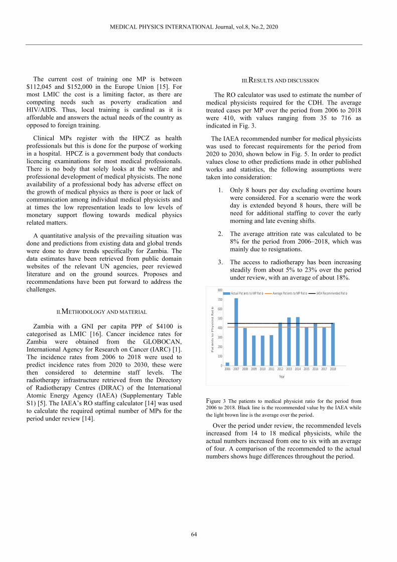
MEDICAL PHYSICS INTERNATIONAL Journal, vol.8, No.2, 2020
64
The current cost of training one MP is between $112,045 and $152,000 in the Europe Union [15]. For most LMIC the cost is a limiting factor, as there are competing needs such as poverty eradication and HIV/AIDS. Thus, local training is cardinal as it is affordable and answers the actual needs of the country as opposed to foreign training.
Clinical MPs register with the HPCZ as health professionals but this is done for the purpose of working in a hospital. HPCZ is a government body that conducts licencing examinations for most medical professionals. There is no body that solely looks at the welfare and professional development of medical physicists. The none availability of a professional body has adverse effect on the growth of medical physics as there is poor or lack of communication among individual medical physicists and at times the low representation leads to low levels of monetary support flowing towards medical physics related matters.
A quantitative analysis of the prevailing situation was done and predictions from existing data and global trends were done to draw trends specifically for Zambia. The data estimates have been retrieved from public domain websites of the relevant UN agencies, peer reviewed literature and on the ground sources. Proposes and recommendations have been put forward to address the challenges.
II.METHODOLOGY AND MATERIAL
Zambia with a GNI per capita PPP of $4100 is categorised as LMIC [16]. Cancer incidence rates for Zambia were obtained from the GLOBOCAN, International Agency for Research on Cancer (IARC) [1]. The incidence rates from 2006 to 2018 were used to predict incidence rates from 2020 to 2030, these were then considered to determine staff levels. The radiotherapy infrastructure retrieved from the Directory of Radiotherapy Centres (DIRAC) of the International Atomic Energy Agency (IAEA) (Supplementary Table S1) [5]. The IAEA’s RO staffing calculator [14] was used to calculate the required optimal number of MPs for the period under review [14].
III.RESULTS AND DISCUSSION
The RO calculator was used to estimate the number of medical physicists required for the CDH. The average treated cases per MP over the period from 2006 to 2018 were 410, with values ranging from 35 to 716 as indicated in Fig. 3.
The IAEA recommended number for medical physicists was used to forecast requirements for the period from 2020 to 2030, shown below in Fig. 5. In order to predict values close to other predictions made in other published works and statistics, the following assumptions were taken into consideration:
1. Only 8 hours per day excluding overtime hours were considered. For a scenario were the work day is extended beyond 8 hours, there will be need for additional staffing to cover the early morning and late evening shifts.
2. The average attrition rate was calculated to be 8% for the period from 2006–2018, which was mainly due to resignations.
3. The access to radiotherapy has been increasing steadily from about 5% to 23% over the period under review, with an average of about 18%.
Figure 3 The patients to medical physicist ratio for the period from 2006 to 2018. Black line is the recommended value by the IAEA while the light brown line is the average over the period. Over the period under review, the recommended levels increased from 14 to 18 medical physicists, while the actual numbers increased from one to six with an average of four. A comparison of the recommended to the actual numbers shows huge differences throughout the period.
2006 2007 2008 2009 2010 2011 2012 2013 2014 2015 2016 2017 20180
100
200
300
400
500
600
700
800Actual Pat iants to MP Rat io Average Pat ients to MP Rat io IAEA Recommended Rat io
Year
Pat
ien
ts t
o P
hysic
ist
Rat
io

MEDICAL PHYSICS INTERNATIONAL Journal, vol.8, No.2, 2020
65
Figure 4 The actual total number of medical physicists (FTE) involved in cancer treatment at CDH for the period 2006-2019 (light brown) compared to the IAEA guidelines calculated using the RO calculator (light blue).
Our estimates indicate a deficit of about 11 medical physicists by 2030, a slight reduction. There are proactive measures already underway; the main ones being the setting up of two cancer centres in Southern and Copperbelt provinces. Assuming that the centres will each accommodate a Co-60 teletherapy machine, then they will increase the current access to radiotherapy by over 50% [3], [17]. The planned Centre for Nuclear Science and Technology is anticipated to house a radiotherapy facility with at least one LINAC on site [18], [19]. This also will translate into a recommended capacity of about 450 patients per year. There are plans by a private organisation to set up a radiotherapy centre on the Copperbelt, with the facility expected to open in mid-2020 [20]. The facility is anticipated to house two LINACs when fully operational in 2021, further improving access to radiotherapy by 900+ patients year. On overall these centres will offer 2250 patients access to
life saving radiotherapy, almost doubling the current numbers.
Figure 5 The trendline (black) show the steady increase of medical physicists at CDH over a 12 year period.
Figure 6 Correlation between the predicted and recommended numbers of FTE medical physicists at the only Zambian Centre.
2006 2007 2008 2009 2010 2011 2012 2013 2014 2015 2016 2017 2018 2019
0
2
4
6
8
10
12
14
16
18
20
Model Actual
Year
CDH
Phys
icist
Sta
ffing
Num
bers
2006 2008 2010 2012 2014 2016 20180
1
2
3
4
5
6
7
f(x) = 0.32 x − 641.77R² = 0.78
Year
# M
ed
ical P
hysic
ists
2020 2021 2022 2023 2024 2025 2026 2027 2028 2029 2030
0
5
10
15
20
25
Recommended Predicted
Year
CDH
Phys
icist
Staf
fing N
umbe
rs

MEDICAL PHYSICS INTERNATIONAL Journal, vol.6, No.1, 2018
66
The infrastructure growth will need to be matched by clinically qualified medical physicists to guarantee quality care by carrying out quality assurance of the equipment, personalised treatment planning and commissioning. There are plans to improve the current staffing levels so as to alleviate this challenge. These plans involve the commencement of a postgraduate academic programme in medical physics reinforced by clinical training, locally. There is a prevailing notion that postgraduate programmes alone may not adequately and quickly bridge the gap. There sectors advocating for an undergraduate programme in medical physics, with the core radiotherapy courses offered as a short term solution. However, the stance of most MPs is that outlined and supported by IAEA and FAMPO[21], [22]-Postgraduate (MSc) then a two to three year clinical training.
The setting up of a professional organisation will also greatly improve the information flow between medical physicists and other stakeholders. This will also provide a platform on which some of the current challenges like full recognition, retention and remuneration can be tackled.
It is interesting to note that the current level (Fig. 3) in terms of patient to physicist ratio, on average is around the recommended ratio. In comparison to the region, the patient to physicist are some of the lowest, this is without considering factors such as low access to RT and others.
The prediction of the staffing levels from 2020 to 2030 as shown in Fig.6; do show a reduction between the recommended and predicted figures. However, the required number may be higher, as there is anticipated investment by both public and private players in the sector. This period will also see improved access to high-end radiotherapy equipment on overall, therefore requiring more expertise as more complex techniques will be employed to treat cases. This will also push up the number of clinically qualified medical physicists required.
IV. CONCLUSION
The high attrition rate of about 100 % has been observed over the period from 2006 to 2018. This is attributed to low renumeration for MPs on average and lack of career prospects. The establishment of physics programmes in local universities has also put a strain on the available manpower. Therefore, an increased demand for physicists at the new radiotherapy centres envisioned will further increase the strain; compounding the situation. So if challenges relating to pay, career progression and structured clinical training are not addressed effectively the developmental gains will be of little significance. The creation of curricula for academic training in medical physics would offer a solution in as far as reducing the manpower deficit, but it would need to be coupled with retention measures to curb ‘greener pasture’ migration
around the region and worldwide. The need for clinical research targeting challenges affecting the practice of medical physics, adaptation of radiotherapy techniques and development of new cancer treatment cannot be over emphasised.
In Zambia, there are efforts underway to set up an organisation to solely propel the development of medical physics; dealing with several aspects stemming from career progression to continuous professional development (CPD). Such a body would have to work in collaboration with the HPCZ in the issuance of practising licences and malpractice.
ACKNOWLEDGEMENT
We would like to thank the IAEA for providing a travel grant to present this work at the ICMP 2019 in Chile.
REFERENCE
[1] “894-zambia-fact-sheets.pdf.” . [2] “Battling Cancer in Zambia,” 26-Jun-2003. [Online]. Available:
https://www.iaea.org/newscenter/news/battling-cancer-zambia. [Accessed: 04-Sep-2019].
[3] “lishimpi-presentation-010219.pdf.” . [4] N. R. Datta, M. Samiei, and S. Bodis, “Radiation therapy
infrastructure and human resources in low- and middle-income countries: present status and projections for 2020,” Int. J. Radiat. Oncol. Biol. Phys., vol. 89, no. 3, pp. 448–457, Jul. 2014.
[5] “Division for Human Health: DIRAC (DIrectory of RAdiotherapy Centres).” [Online]. Available: https://dirac.iaea.org/Query/Countries. [Accessed: 04-Sep-2019].
[6] M. Kawesha, “MEDICAL PHYSICS PROFESSIONAL DEVELOPMENT AND TRAINING IN ZAMBIA,” p. 3, 2017.
[7] J. Vassileva, “Х.2. ORGANISATIONAL ASPECTS OF THE QUALIFICATION AND INVOLVEMENT OF MEDICAL PHYSICISTS IN RADIOLOGY,” p. 6.
[8] K. . Adhikari, “MEDICAL PHYSICS INTERNATIONAL,” p. 3, 2018.
[9] M. P. Adhikari and P. P. Chaurasia, “STATUS OF MEDICAL PHYSICS PROFESSIONAL DEVELOPMENT AND EDUCATION IN NEPAL,” p. 2, 2016.
[10] L. H. Rosales and A. R. Laguardia, “MEDICAL PHYSICS INTERNATIONAL,” p. 3, 2015.
[11] S. A. Pawiro, L. E. Lubis, A. N. Oktavianto, and D. S. Soejoko, “Overseeing the Growth of Medical Physics: Indonesia Case,” in World Congress on Medical Physics and Biomedical Engineering 2018, 2019, pp. 859–863.
[12] D. S Soejoko, S. Pawiro, and L. Lubis, “Medical Physics in Indonesia: Current Status and Plans,” 2015, vol. 51.
[13] M. Abdel-Wahab et al., “Status of radiotherapy resources in Africa: an International Atomic Energy Agency analysis,” Lancet Oncol., vol. 14, no. 4, pp. e168-175, Apr. 2013.
[14] “Human Health Campus - Staffing and Cost Calculation.” [Online]. Available: https://humanhealth.iaea.org/HHW/RadiationOncology/Makingthecaseforradiotherapyinyourcountry/Roleofradiotherapyincancercare/Staffingandcostcalculation/index.html. [Accessed: 04-Sep-2019].
[15] E. H. Zubizarreta, E. Fidarova, B. Healy, and E. Rosenblatt, “Need for Radiotherapy in Low and Middle Income Countries – The Silent Crisis Continues,” Clin. Oncol., vol. 27, no. 2, pp. 107–114, Feb. 2015.

MEDICAL PHYSICS INTERNATIONAL Journal, vol.6, No.1, 2018
67
[16] “World Bank Country and Lending Groups – World Bank Data Help Desk.” [Online]. Available: https://datahelpdesk.worldbank.org/knowledgebase/articles/906519-world-bank-country-and-lending-groups. [Accessed: 04-Sep-2019].
[17] T. H. P. editorial team, “THE COMMEMORATION OF WORLD CANCER DAY – The Health Press – Zambia.” .
[18] “Nuclear Center for Zambia – ROSATOM NEWSLETTER.” . [19] “MINISTERIAL STATEMENT BY THE MINISTER OF
HIGHER EDUCATION.pdf.” . [20] “Faith and a fish farm lead to hope for better cancer care,”
Elekta | Focus Magazine, 01-May-2019. . [21] “IAEA-TCS-56_web.pdf.” . [22] “(PDF) Accreditation of medical physics clinical training
programmes in Africa:survey by the Federation of African Medical Physics Organizations (FAMPO) and the International Atomic Energy Agency (IAEA).” [Online]. Available: https://www.researchgate.net/publication/320386318_Accreditation_of_medical_physics_clinical_training_programmes_in_Africasurvey_by_the_Federation_of_African_Medical_Physics_Organizations_FAMPO_and_the_International_Atomic_Energy_Agency_IAEA. [Accessed: 09-Sep-2019].
Contacts of the corresponding author:
Author: Kangwa Alex Nkonde Institute: Mulungushi University Main Campus Street: Great North Road City: Kabwe Country: Zambia Email: [email protected]

MEDICAL PHYSICS INTERNATIONAL Journal, vol.6, No.1, 2018
68
HOW TO

MEDICAL PHYSICS INTERNATIONAL Journal, vol.6, No.1, 2018
69
NATIONAL DIAGNOSTIC REFERENCE LEVELS OF COMPUTED TOMOGRAPHY IN THAILAND
S Buncharat1, S Tuiduang1, A.Krisanachinda2, A.Singkavongsay1
1Department of Medical Sciences, Tiwanond Road, Nonthaburi 11000, Thailand
2Department of Radiology, Faculty of Medicine, Chulalongkorn University, Bangkok, Thailand
Abstract- The patient radiation dose survey on DRL of CT procedures was started after the quality control of CT system and the normalized weighted computed tomography dose index; nCTDIw (mGy/mAs) was obtained. The CTDI and DLP values per single scan among various CT procedures were collected from patient of size selection on brain, chest and whole abdomen with and without contrast media. DRLs were determined and compared to other NDRLs for further optimization on the CT protocols.
Keywords- CTDI, DLP, brain, chest, abdomen. I.INTRODUCTION
The computed tomography has been used in major medical imaging departments. In Thailand the number of CT systems is 903 in 2018 which most of them are installed in Bangkok and other big cities. There has been a dramatic increase in the use of CT, furthermore, CT is a relative high dose technique. UNSCEAR 2000 Report [1] showed that 34 % of patient cumulative radiation dose were from CT systems. Mettler FA Jr, et al [2] reported the entrance surface dose equivalent from chest PA from general X-rays was 0.02 mSv while CT chest was 7 mSv. The biological effect of radiation can lead to the stochastic effect or cancer induction. ICRP 103, 2007[3] reported the risk of cancer was 5.5 % Sv-1. If the patient received 10 mSv, the risk of cancer would be 5.5 x10⁻⁴ Sv-1 and the risk ratio would be 1:1800. ICRP, IAEA BSS recommended the three concepts of justification which the benefit is higher than risk, optimization; As Low As Reasonably Achievable: ALARA, for the reduction of patient dose from medical exposure when the image quality had been considered, and the dose limit. In the survey of patient dose, the Department of Medical Science, Ministry of Public Health of Thailand has established DRLs of CT in the national level to be used for the patient radiation dose reduction and optimization [4].
II.MATERIALS AND METHODS The data collection from the patients of both male and
female with the body weight at 60+ 15 kg and the patient age was over 15 years old. The data collected from CT system consists of CTDI volume; CTDIvol (mGy) dose length product; DLP (mGy.cm), normalized weighted computed tomography dose index; nCTDIw (mGy/mAs), scan length, (cm), and pitch factor. The measurement of each parameter follows IAEA TRS 457[5]. The DRLs
consists of CTDI and DLP values per single scan among various CT procedures.
III. RESULTS In 2018, the national survey of 135 CT systems, 6
manufacturers and 52 models of 4 to 128 detector slices to establish the DRLs started with the collection of nCTDIw from the central part of Thailand at 67 CT systems, 15 systems from the north, 35 systems from the north-east, and 18 systems from the south as the details in Table 1 and Table 2. The nCTDIw values were estimated from head and body phantom [5] as in Table 3.Table 4 shows technique parameters of brain, chest, and whole abdomen at with and without contrast media. Table 5 shows volume CTDI, mGy, per sequence while Table 6 shows DLP, mGy.cm per sequence. Table 7 shows the National DRLs from Thailand, Japan, USA and UK. Our DRLs values of various parts of the body, with and without the contrast media were lower than Japan but higher than USA and UK.
Table 1: The number of CT systems at different regions Region CT Systems
Percentage Central 67
49.6 North 15 11.1 North-East 35
25.9 South 18 13.3 Total 135
100.0 Table2: CT systems according to number of detector slices Number of Detector slices CT Systems
Percentage <16 25 18.5 16 49 36.3 >16-<64 35
25.9 64 12 8.9 >64 - <128 4 3.0 128 10 7.4 Total 135 100.0
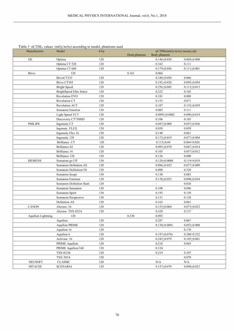
MEDICAL PHYSICS INTERNATIONAL Journal, vol.6, No.1, 2018
70
Table 3 nCTDIw values (mGy/mAs) according to model, phantoms used Manufacturer Model kVp nCTDIw(mGy/mAs) (mean+sd)
Head phantom Body phantom GE Optima 120 0.146+0.038 0.069+0.008
Optima CT 520 120 0.162 0.111
Optima CT 660 120 0.179+0.056 0.111+0.001
Brivo 120 0.161 0.066
BrivoCT325 120 0.180+0.050 0.066
Brivo CT385 120 0.192+0.028 0.095+0.054
Bright Speed 120 0.256+0.045 0.113+0.013
BrightSpeed Elite Select 120 0.222 0.102
Revolution EVO 120 0.181 0.088
Revolution CT 120 0.153 0.071
Revolution ACT 120 0.107 0.135+0.029
Somatom Emotion 130 0.085 0.111
Light Speed VCT 120 0.0995+0.0002 0.090+0.019
Discoverry CT750HD 120 0.106 0.103
PHILIPS Ingenuity CT 120 0.047+0.008 0.057+0.026
Ingenuty FLEX 120 0.038 0.059
Ingenuity Flex 16 120 0.140 0.061
Ingenuity 128 120 0.172+0.019 0.077+0.004
Brilliance CT 120 0.115+0.04 0.064+0.026
Brilliance 64 120 0.095+0.070 0.067+0.014
Brilliance 16 120 0.105 0.057+0.012
Brilliance 128 120 0.126 0.080
SIEMENS Somatom go UP 130 0.120+0.0008 0.114+0.019
Somatom Definition AS 120 0.096+0.025 0.077+0.009
Somatom Definition128 120 0.090 0.520
Somatom Scope 130 0.130 0.083
Somatom Emotion 130 0.138+0.053 0.098+0.034
Somatom Definition flash 120 - 0.026
Somatom Sensation 120 0.108 0.096
Somatom Spirit 130 0.193 0.159
Somatom Perspective 130 0.131 0.120
Definition AS 120 0.163 0.061
CANON Alexion /16 120 0.153+0.068 0.073+0.032
Alexion TSX-032A 120 0.328 0.137
Aquilion Lightning 120 0.230 0.092
Aquilion 120 0.287 0.067
Aquilion PRIME 120 0.130+0.009) 0.052+0.008
Aquilion 16 120 - 0.170
Aquilion 6 120 0.197+0.078) 0.280+0.252
Activion 16 120 0.243+0.079 0.145+0.061
PRIME Aquilion 120 0.218 0.065
PRIME Aquilion/160 130 0.124 -
TSX-021B 120 0.219 0.107
TSX-303A 120 - 0.070
NEUSOFT CLASSIC 120 N/A N/A
HITACHI SCENARIA 120 0.157+0.070 0.098+0.023
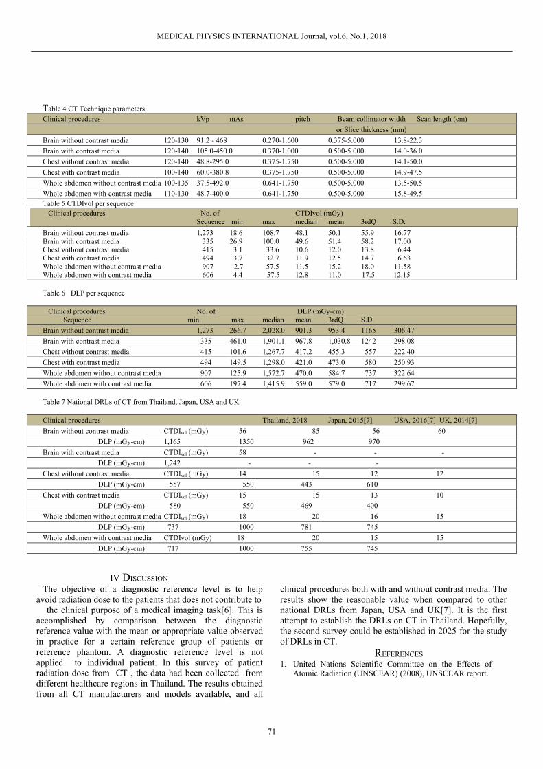
MEDICAL PHYSICS INTERNATIONAL Journal, vol.6, No.1, 2018
71
Table 4 CT Technique parameters Clinical procedures kVp mAs pitch Beam collimator width Scan length (cm) or Slice thickness (mm) Brain without contrast media 120-130 91.2 - 468 0.270-1.600 0.375-5.000 13.8-22.3 Brain with contrast media 120-140 105.0-450.0 0.370-1.000 0.500-5.000 14.0-36.0 Chest without contrast media 120-140 48.8-295.0 0.375-1.750 0.500-5.000 14.1-50.0 Chest with contrast media 100-140 60.0-380.8 0.375-1.750 0.500-5.000 14.9-47.5 Whole abdomen without contrast media 100-135 37.5-492.0 0.641-1.750 0.500-5.000 13.5-50.5 Whole abdomen with contrast media 110-130 48.7-400.0 0.641-1.750 0.500-5.000 15.8-49.5 Table 5 CTDIvol per sequence Clinical procedures No. of CTDIvol (mGy)
Sequence min max median mean 3rdQ S.D. Brain without contrast media 1,273 18.6 108.7 48.1 50.1 55.9 16.77 Brain with contrast media 335 26.9 100.0 49.6 51.4 58.2 17.00 Chest without contrast media 415 3.1 33.6 10.6 12.0 13.8 6.44 Chest with contrast media 494 3.7 32.7 11.9 12.5 14.7 6.63 Whole abdomen without contrast media 907 2.7 57.5 11.5 15.2 18.0 11.58 Whole abdomen with contrast media 606 4.4 57.5 12.8 11.0 17.5 12.15 Table 6 DLP per sequence Clinical procedures No. of DLP (mGy-cm)
Sequence min max median mean 3rdQ S.D. Brain without contrast media 1,273 266.7 2,028.0 901.3 953.4 1165 306.47 Brain with contrast media 335 461.0 1,901.1 967.8 1,030.8 1242 298.08 Chest without contrast media 415 101.6 1,267.7 417.2 455.3 557 222.40 Chest with contrast media 494 149.5 1,298.0 421.0 473.0 580 250.93 Whole abdomen without contrast media 907 125.9 1,572.7 470.0 584.7 737 322.64 Whole abdomen with contrast media 606 197.4 1,415.9 559.0 579.0 717 299.67 Table 7 National DRLs of CT from Thailand, Japan, USA and UK Clinical procedures Thailand, 2018 Japan, 2015[7] USA, 2016[7] UK, 2014[7] Brain without contrast media CTDIvol (mGy) 56 85 56 60 DLP (mGy-cm) 1,165 1350 962 970 Brain with contrast media CTDIvol (mGy) 58 - - - DLP (mGy-cm) 1,242 - - - Chest without contrast media CTDIvol (mGy) 14 15 12 12 DLP (mGy-cm) 557 550 443 610 Chest with contrast media CTDIvol (mGy) 15 15 13 10 DLP (mGy-cm) 580 550 469 400 Whole abdomen without contrast media CTDIvol (mGy) 18 20 16 15 DLP (mGy-cm) 737 1000 781 745 Whole abdomen with contrast media CTDIvol (mGy) 18 20 15 15 DLP (mGy-cm) 717 1000 755 745
IV DISCUSSION The objective of a diagnostic reference level is to help
avoid radiation dose to the patients that does not contribute to the clinical purpose of a medical imaging task[6]. This is
accomplished by comparison between the diagnostic reference value with the mean or appropriate value observed in practice for a certain reference group of patients or reference phantom. A diagnostic reference level is not applied to individual patient. In this survey of patient radiation dose from CT , the data had been collected from different healthcare regions in Thailand. The results obtained from all CT manufacturers and models available, and all
clinical procedures both with and without contrast media. The results show the reasonable value when compared to other national DRLs from Japan, USA and UK[7]. It is the first attempt to establish the DRLs on CT in Thailand. Hopefully, the second survey could be established in 2025 for the study of DRLs in CT.
REFERENCES 1. United Nations Scientific Committee on the Effects of
Atomic Radiation (UNSCEAR) (2008), UNSCEAR report.

MEDICAL PHYSICS INTERNATIONAL Journal, vol.6, No.1, 2018
72
2. Mettler FA Jr, Huda W, Yoshizumi TY, Mahesh M. (2008). Effective doses in radiology and diagnostic nuclear medicine : a catalog. Radiology 248:254-63
3. International Commission Radiation Protection (2007). The
2007 recommendations of the International Commission on Radiological Protection. IRCP publication 103. Ann, ICRP 37:1-33
4. Department of Medical Sciences, Ministry of Public Health.
(2018) Criteria and guidelines on development of diagnostic radiology, Ministry of Public Health.
5. IAEA. (2007) Dosimetry in Diagnostic Radiology: An
International Code of Practice, Technical Report Series No. 457. Vienna Austria;188-208
6. International Commission on Radiological Protection Publication135 (2001) Diagnostic reference levels in medical imaging review and additional advice. Committee 3
of the International Commission on Radiological Protection. www.icrp.org/docs/DRL_for_web.pdf
7. Kanpana MK, Priscilla FB, Depapriya S, Mythreyi BC,
Laura PC, Richard LM.(2017) U.S. diagnostic reference levels and achievable doses for 10 adult CT examinations. Radiology 281(1) :120-33
Contacts of the corresponding authors Author: Anchali Krisanachinda Institute: Chulalongkorn University Street: Rama IV Road, City: Bangkok Country: Thailand e-mail: [email protected]

MEDICAL PHYSICS INTERNATIONAL Journal, vol.8, No.2, 2020
73
NATIONAL DIAGNOSTIC REFERENCE LEVELS OF DIGITAL MAMMOGRAPHY IN THAILAND
A. Singkavongsay1, C. Natheethorn1, A. Krisanachinda2, S. Thupsuri1, R. Chansoong1, P. Ritthitham1, N. Jitpinit1, C. Nhosiri1, S. Chantasingh1, W. Sunanrungangkhana1, T. Suphawattanaphan1,
K. Nikapruek1, S. Sayumphuruchinan1, P. Saengpradub1, S. Thumdee1, S. Buncharat1
1 Department of Medical Sciences, Tiwanond Road, Nonthaburi 11000, Thailand 2 Department of Radiology, Faculty of Medicine, Chulalongkorn University, Bangkok, Thailand
Abstract— The patient radiation dose survey on national diagnostic reference level (NDRLs) of digital mammography was planned to be established in 2018 by Department of Medical Sciences, Ministry of Public Health, Thailand. The survey covered 157 from 456 digital mammography systems installed at various parts of Thailand. At least twenty patients had been selected per system which 3320 patients were included. The x-ray output and the entrance surface air kerma (ESAK) had been measured as part of the quality control of the mammography system to determine the mean glandular dose (MGD) from the routine techniques. The patient compressed breast thickness (CBT) had been recorded in relation to the MGD. The mean, 1st quartile, 2nd quartile (median), 3rd quartile, maximum and the standard deviation for MGD and ESAK were determined. The NDRLs were obtained from the third quartile of MGD, ESAK and compared to other NDRLs and RDRLs for further optimization on the mammographic protocols.
Keywords— DRL, digital mammography, MGD, ESAK
I. INTRODUCTION Breast is one of the highly radiosensitive organs. The
annual screening of mammography for women aged 40–80 years is associated with a life time attributable risk, LAR, of fatal breast cancer of 20–25 cases in 100 000 (BEIR VII) [1]. The benefit and risk ratio for annual mammography is estimated to be greater than 50:1 for both the 40–80 and 50–80 year old screening groups, but drops to 3:1 for the 40–49 age group mammography. Mammography dosimetry is a complex issue. MGD depends on the breast size, compressed breast thickness (CBT), kVp, mAs, and compression force (CF). The MGD slightly decreases with increasing patient age. Current methods of dose optimization make assumptions of the breast composition of gland and other breast tissue at 50:50, and using Dance models [2] for estimating glandularity and patient-specific dose. According to the CBT from 20 to 50 mm, the MGD slightly increases with increased CBT then decreases, no difference in dose at CBT greater than 90 mm. ICRP[3] recommends MGD as a DRL quantity, even though it is a measure of organ dose rather than the amount of ionizing radiation used to perform a medical imaging task. Entrance surface air kerma (Ka, e) allows direct comparisons among mammography units with similar target/filter combinations. Incident air kerma (Ka, i) per mAs is derived from output measurements, made with the breast compression device in position. This is then multiplied by the mAs used to obtain the incident air kerma for the examination. Incident air
kerma is required for the calculation of mean glandular dose. The relationship between incident air kerma and mean glandular dose is highly dependent on breast thickness and composition, as well as beam quality. MGD is calculated from the incident air kerma used for the examination for a specified thickness of compressed breast. The incident air kerma and MGD will depend on the size of the breast and its composition, which changes throughout a woman’s life. For mammography, the recommended DRL quantity is one or more of incident air kerma, entrance surface air kerma, and MGD, with the choice of quantity depending on local practices and regulatory requirements. Establishing DRL values for different breast thicknesses is a more complex but better approach to refine the DRL process for mammography. When entrance surface air kerma or incident air kerma is used as the DRL quantity, evaluation program arrangements should be based on medical physicist’s recommendations to ensure that dependence on breast thickness and differences in glandular dose are taken into account. Surveys of patients recommended as the main method of evaluating the amount of radiation applied in mammography as phantoms do not assess the full range of breast sizes for which examinations will be undertaken, and do not reflect clinical use of the equipment. Compliance with DRL values does not indicate that the procedure is performed at an optimized level with regard to the amount of radiation used. The median value of the national distribution can serve as an additional tool to aid in optimization, may be a desirable goal at which to aim using standard techniques and technologies, and represents a situation closer to the optimum use of the applied radiation.
ICRP recommends setting local and national DRL values based on DRL quantities for imaging examinations and procedures performed on patients [3]. Data on DRL quantities can be collected using surveys, registries, or other automated data collection methods. All dosimeters must be calibrated and should be traceable to a primary or secondary standard laboratory. The accuracy of DRL quantity data produced by and transferred from x-ray systems should be verified periodically by a clinically qualified medical physicist in diagnostic radiology.
II. MATERIALS AND METHODS The hospitals in Thailand are classified according to the
number of beds, such as the community hospital equipped with 10-120 beds, general hospital equipped with 121-500

MEDICAL PHYSICS INTERNATIONAL Journal, vol.8, No.2, 2020
74
beds, regional hospital with more than 500 beds. Specialized hospitals are cancer centres, psychiatric hospital, cardiac center, and etc. The survey covered 157 from 456 digital mammographic systems in Thailand. From all parts of the country, only 1 system was randomly selected from community hospital, 52 systems from general hospitals, 22 systems from regional hospitals and 82 systems from private hospitals as shown in Table 1. Select at least 20 women from each system with average CBT at 50 ± 5 mm.3320 women were included in the survey on MGD of 4 views of cranio-caudal (CC) and medio- lateral oblique (MLO) for left and right breasts per woman resulting in total 13,280 views in the survey Extract CBT, kVp, mAs, MGD and ESAK from the system displayed monitors, PACS and DICOM header.
Table 1 157 mammographic systems from the community, general, and regional hospitals, Ministry of Public Health, and private hospitals at various regions of Thailand included in this survey.
III. RESULTS The patient data on the compressed breast thickness
(CBT, mm), the mean glandular dose, MGD (mGy) and the exposure techniques of kVp and mAs were recorded from PACS. Among the data distributions, the first quartile (25th percentile), the second quartile (50th percentile) or the median and achievable dose, the mean, the third quartile (75th percentile), the maximum and the standard deviation were determined for CBT, kVp, mAs and MGD as in table 2, 3, 4,5.
IV. DISCUSSION AND CONCLUSION Eighty seven percent of the mammographic system in
Thailand is the digital mammogram while the other thirteen percent is computed radiographic system and had not been included in this survey. Such the CR system will be obsoleted in the near future. Among 157 digital mammographic systems in this survey, the x-ray target materials were molybdenum (Mo), rhodium (Rh) and tungsten (W). The filters were molybdenum, rhodium, and silver (Ag). The target/filter combinations were Mo/Mo, Mo/Rh, Mo/Ag, Rh/Rh, Rh/Ag, W/Rh, and W/Ag which
result in various exposure techniques and the MGD according to the CBT, the breast tissue composition and the breast glandularity.
Table 2 Technical parameters on tube voltage (kVp) for cranio- caudal (CC) and medio- lateral oblique (MLO) views on right and left breasts and mean values at 25th percentile (1st Quartile), median (2nd Quartile) and achievable dose, 75th percentile (3rd Quartile), maximum and standard deviation.
kVp
RCC LCC RMLO LMLO Mean
1st Q 28 28 28 28 28
Median 29 29 29 29 29
Mean 28.9 29 29.3 29.3 29.1
3rd Q 30 30 30 30 30
Max 35 35 35 35 35
SD 1.7 1.7 1.8 1.8 1.8
Table 3 Technical parameters on tube current time (mAs) for cranio caudal (CC) and medio lateral oblique (MLO) views on right and left breasts and mean values at 25th percentile (1st Quartile), median (2nd Quartile) and achievable dose, 75th percentile (3rd Quartile), maximum and standard deviation.
mAs
RCC LCC RMLO LMLO Mean
1st Q 82 80 75.9 76 78.5 Median 110.8 109.7 109 106 108.9
Mean 120.3 119 114.3 111 116.2
3rd Q 148.1 147.6 148 144 146.9
Max 384 447 420 400 412.8
SD 55.5 56.3 59.4 55.6 56.7
Table 4 Compressed breast thickness (CBT, mm) for cranio caudal (CC) and medio lateral oblique (MLO) views on right and left breasts and mean values at 25th percentile (1st Quartile), median (2nd Quartile), 75th percentile (3rd Quartile), maximum and standard deviation.
CBT (mm) RCC LCC RML
O LMLO Mean
1st Q 45 45 46 46 45.5 Median 51 52 53 53.3 52.3
Mean 51.9 52.3 53.6 53.7 52.9
3rd Q 58 58 60 60 59
Max 100 93 95 96 96
SD 10.4 10.6 10.8 11 10.7
Number of mammographic systems
Region Community
Hospital
General Hospital
Regional Hospital
Private Hospital
Total
North 1 15 2 12 30
Middle - 9 12 41 62 North-East - 14 4 12 30
East - 2 2 10 14
South - 12 2 7 21
Total 1 52 22 82 157

MEDICAL PHYSICS INTERNATIONAL Journal, vol.8, No.2, 2020
75
Table 5 Mean Glandular Dose (MGD, mGy) for cranio – caudal (CC) and medio lateral oblique (MLO) views on right and left breasts and mean values at 25th percentile (1st Quartile), median (2nd Quartile) and achievable dose, 75th percentile (3rd Quartile), maximum and standard deviation.
MGD (mGy)
RCC LCC RMLO LMLO Mean
1st Q 1.23 1.22 1.29 1.18 1.23 Median 1.57 1.57 1.62 1.58 1.59
Mean 1.72 1.7 1.74 1.69 1.71
3rd Q 2.05 2.06 2.04 2.01 2.04
Max 5.29 5.05 5.69 6.42 5.61
SD 0.68 0.67 0.66 0.78 0.70
Table 6 National Diagnostic Reference Levels of MGD among Thailand Australia and Japan in comparison to regional diagnostic reference level of the International Atomic Energy Agency (IAEA). The ESAK (mGy) from Thailand and IAEA are displayed in the table
NDRLs & RDRLs (mGy)
Thailand
Australia Japan IAEA
MGD (mGy) 2.04 2.06 2.4 3
ESAK(mGy) 9.74 - 11
In this survey, the national authority at the Department of Medical Science, Ministry of Public Health had planned and requested the government budget in 2018 to arrange the quality control test of the mammographic system in Thailand [4]. The incident air kerma and the entrance surface air kerma of all mammographic systems in Thailand had been determined by following the technology as mentioned in IAEA TRS 457[5]. The cooperation between the national authority and the professional societies such as Royal College of Radiologists of Thailand, Radiological Society of Thailand, Radiological Technologist Society of Thailand, and Thai Medical Physicist Society had been set up to establish the mammogram guidelines for training the users on the concept of LDRLs, NDRLs on digital mammogram with the applications of the guidelines, the methodology on data collection and analysis [4].
The first NDRLs on the digital mammogram system is established in 2019 in Thailand. From 157 mammographic systems, 3320 Thai women with the average CBT was 52.9+ 5 mm, the third quartile of the MGD was 2.04 mGy, the mean was 1.71 mGy, the median and the achievable dose (2nd Q) was 1.59 mGy. The third quartile of ESAK was 9.74 mGy. NDRLs on mammogram of Thailand had been compared to other countries, it was close to Australia at 2.06 mGy and lower than Japan DRL (2015) which was 95th percentile at 2.4 mGy. RDRLs established by IAEA, MGD was 3 mGy and ESAK was 11 mGy as in table 6.
NDRLs in mammography should be stratified according
to CBT and detector technology. The age and breast density may need to be taken into account. The digital mammographic system with the digital breast tomosynthesis (DBT) would be increasing at the tertiary care hospital such as cancer center, university hospital and private hospital. The national survey on DRLs in digital mammogram with DBT is planned in the next three years and established in 2025.
REFERENCE 1. National Research Council, BEIR VII. (2006) Health risks from
exposure to low levels of ionizing radiation. National Academic Press, Washington DC.
2. Dance DR, Skinner CL, Young KC, Beckett J, Kotre CJ. (2000) Additional factors for the estimation of mean glandular dose using the UK mammographic dosimetry protocol. PMB 45:3225-3240
3. International Commission on Radiological Protection.(2017) ICRP publication 135: Diagnostic reference levels in medical imaging. Annals of ICRP 46(1): 1–144.
4. Department of Medical Sciences, Ministry of Public Health (2018). Criteria and guidelines on development of diagnostic radiology, Ministry of Public Health.
5. IAEA. (2007). Dosimetry in Diagnostic Radiology: An International Code of Practice, Technical Reports Series No. 457. Vienna Austria;.155-186
Contacts of the corresponding authors Author: Anchali Krisanachinda Institute: Chulalongkorn University Street: Rama IV Road, City: Bangkok Country: Thailand e-mail: [email protected]

MEDICAL PHYSICS INTERNATIONAL Journal, vol.6, No.1, 2018
76
TECHNOLOGY INNOVATION

MEDICAL PHYSICS INTERNATIONAL Journal, vol.8, No.2, 2020
77
MAKING ON-LINE ADAPTIVE RADIOTHERAPY POSSIBLE USING ARTIFICIAL INTELLIGENCE AND MACHINE LEARNING FOR EFFICIENT
DAILY RE-PLANNING
Yves Archambault, Christopher Boylan, Drew Bullock, Tomasz Morgas, Jarkko Peltola, Emmi Ruokokoski, Angelo Genghi, Benjamin Haas, Pauli Suhonen, and Stephen Thompson
Varian Medical Systems, Inc. Palo Alto, California USA; Baden, Switzerland; and Helsinki, Finland
Abstract— Adaptive therapy involves the ability to alter a radiotherapy treatment plan based on tumor and anatomical changes over a course of therapy. The goal is to better target the tumor, reduce dose to healthy tissue and potentially improve overall outcomes. To date, achieving this has typically required time-consuming re-planning between treatment sessions or monopolizing a linac for an extended period while a patient waits on the treatment couch for new plans to be generated. Neither of these alternatives has been deemed practical or affordable at scale, as very often clinics don’t have the resources even if they have the tools.
Consequently, Varian Medical Systems developed Ethos™ therapy, a radiotherapy treatment system that uses artificial intelligence (AI) and machine learning to accomplish adaptive radiotherapy. In this paper, we describe the technology that underlies the adaptive capabilities of the system.
Keywords— Adaptive radiotherapy, artificial intelligence, machine learning, Ethos, RapidPlan, treatment planning, neural networks, .
VII. INTRODUCTION
A number of challenges exists in the delivery of adaptive radiotherapy. Briefly, the challenges have been:
• The challenge of performing a full treatment planning workflow during a radiotherapy treatment session, while the patient is on the treatment couch, in treatment position. Treatment planning is a complicated task and requires time and significant attention and knowledge. The pressure imposed on clinicians performing on-couch adaptive therapy heightens opportunities for mistakes.
• The challenge of manually delineating influencers—anatomy that influences the shape of targets—during the detection of daily anatomy. Manual delineation of target structures for daily re-planning is time-consuming and technically challenging. The process can be particularly challenging in the presence of artifacts.
• The challenge of producing quality plans using inverse planning, which is typically performed by dedicated treatment planning staff and the manual or templated application of optimization cost-function based objectives or constraints. The skill and expertise of the treatment planner can have a major impact to the final plan quality.
Additionally, plan generation in existing commercialized systems typically requires moderately complex user interactions which distract the focus during the on-couch session.
The Ethos system was designed to address these challenges. In this paper, we take a deeper look at the technologies within the Ethos system that address these challenges.
VIII. ON-COUCH ADAPTIVE THERAPY WORKFLOW
The challenges involved in delivering on-couch adaptive therapy are addressed, in the Ethos system, through a re-planning workflow that has been reduced to well-defined and predictable clinical decision points in order to lower the cognitive load of the clinician.
Figure 1 (p. 3) illustrates the on-couch adaptive work-flow implemented in Ethos therapy. It guides the user by presenting focused information and asking for a single decision at the time.
• After the kV-CBCT image is acquired, it is presented to the clinician for evaluation. The clinician can either accept the image or decide to acquire a new one (Decision 1).
• Once the image is accepted, the system detects selected normal organ structures directly on the kV-CBCT. These structures are referred to as “influencer” structures. They are those structures that are in the closest proximity to the target(s) and have the biggest impact on their shape and position. The influencer structures are then presented to the clinician, who is asked to review and adjust them, and then to confirm that they are adequate (Decision 2).
• Once confirmed, the influencer structures are used to guide an algorithm that propagates the target structures from the planning, or reference, image to the kV-CBCT image. This ensures that features and relations between target and anatomy structures that were present on the reference image are preserved on the kV-CBCT image. The propagated targets and the detected normal anatomy structures comprise a new patient model—the session model. The user is asked to

MEDICAL PHYSICS INTERNATIONAL Journal, vol.8, No.2, 2020
78
review and accept the session patient model, with focus on the propagated targets (Decision 3).
• The session patient model is then used by the automated treatment planning to produce two plans. The scheduled plan is obtained by calculating dose from the reference plan on the session patient model while the adapted plan is obtained by running a new optimization and calculating dose using the session patient model. The two plans are then shown to the user so the appropriate one, scheduled or adapted, can be selected for treatment (Decision 4).
IX. DETECTION OF DAILY ANATOMY
To address the challenges involved during detection of daily anatomy, an AI-based algorithm that is based on convolutional neural networks is used to contour the influencers on the session images. Neural networks
A neural network is a collection of connected units or nodes called “artificial neurons” that behave much like biological neurons. They have an input layer, an output layer (prediction) and one or more hidden layers. The depth of the network depends on the number of hidden layers. Deep neural networks are neural networks with multiple hidden layers. Deep learning convolutional neural networks (CNNs) make the explicit assumption that the inputs are images, which allows for the incorporation of certain properties into their architecture. CNNs are best for solving problems related to image recognition, object detection, and other computer vision applications. A typical CNN can be viewed as a sequence of layers that transforms an image volume into an output volume. Varian’s in-house developed and trained deep learning model for Ethos therapy utilizes TensorFlow, CUDA and cuDNN libraries, and processes images on different interconnected resolution levels. Ethos uses full-image deep convolutional neural networks with tailored architectures that share many similarities with U-Net and DenseNet, which are widely used in image segmentation tasks. The network itself takes the full 3D iCBCT as an input and returns the same size of segmentation as an output. The neural network models used in the influencer segmentation process are static and do not continuously learn based on user input. This ensures the stability and performance of the algorithms over time. Deep neural network model production
Neural network training was performed in a supervised learning setting using images and ground truth contours from several hundred patients. Data was acquired from multiple clinics across the Americas, Europe, Australia and Asia. Images for the training set were selected to represent a realistic spectrum of anatomical variety and typical image
artifacts. Human anatomy experts created the ground truth contours as part of the algorithm development. A single set of contours was produced for each training image.
Training involves three separate datasets: 1. Training dataset. The training dataset is used to fit
the model. This process involves utilizing a large set of consistently contoured data, which is used to perform the actual training of the neural networks. Contours in training data sets are randomly peer reviewed to ensure adherence to selected guidelines.
2. Validation dataset. The validation dataset is used to provide an unbiased evaluation of a model fit on the training dataset while tuning the model with hyperparameters. The validation data set is considered a subset of the training data set.
3. Test dataset. The test dataset is a smaller set of data used to provide an evaluation of a final model fit. Scans related to a patient that belongs to test set cannot be used for network training. Each image and contour in test set is reviewed by physicians for accuracy.
A neural network is trained using the classical backward-error-propagation algorithm. An error is computed at the output and distributed throughout the network layers. The gradient descent optimization algorithm uses back propagation to adjust the weight of neurons by calculating the gradient of the cost function. A cost function is a measure of how well a neural network performs with respect to the given training sample and the expected output. The cost function is typically expressed as a difference or distance between the predicted value and the actual value. It can be estimated by iteratively running the model to compare estimated predictions against the ground truth.
Hyperparameters in deep learning models
Hyperparameters are settings that can be tuned to control the behavior of a machine learning algorithm. Conceptually, they can be considered orthogonal to the learning model itself; although they live outside of the models, there is a direct relationship between them.
Examples of hyperparameters: • Learning rate — the learning rate quantifies the
learning progress of a model in a way that can be used to optimize its capacity.
• Number of hidden units — the number of hidden units is key to regulating the representation capacity of the model.
• Convolution kernel width — In CNNs, the kernel width influences the number of parameters in a model which, in turn, influences its capacity.
Hyperparameters may be tuned using two basic approaches: manual or automatic selection. Both approaches are technically viable but choosing between them typically represents a trade-off. The decision is related to the high
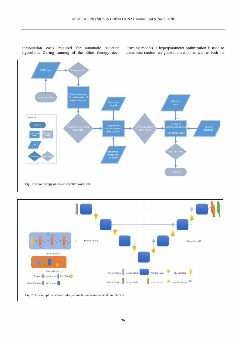
MEDICAL PHYSICS INTERNATIONAL Journal, vol.8, No.2, 2020
79
computation costs required for automatic selection algorithms. During training of the Ethos therapy deep
learning models, a hyperparameter optimization is used to determine random weight initialization, as well as both the
Fig. 1: Ethos therapy on-couch adaptive workflow
Fig. 2: An example of Varian’s deep convolution neural network architecture.

MEDICAL PHYSICS INTERNATIONAL Journal, vol.8, No.2, 2020
80
loss function and the layer order. Post-processing of convolutional neural network outputs
The segmentation output of the networks is passed through a post-processing module that ensures that the output matches the selected clinical guidelines. The processing operations include the removal of smaller segments or dislocated segments, smoothing the final contours as well as selecting terminal slices for segmentation. Model verification and validation
Each trained neural network model undergoes verification tests that compare the obtained classification to the ground truth contours on multiple test sets. Several evaluation metrics are computed and evaluated during each verification test against passing criteria that are established for each of the evaluated structures. Models that pass verification tests qualify as candidates for validation tests. Because of low correlation between similarity metrics and measured correction and review times, the verification tests by themselves do not qualify models for deployment. Models that passed verification are then validated by clinicians in a test that better estimates the clinical review effort. Passing validation testing ensures that the model meets user needs, which qualifies the model for deployment. Performance of deployed models is monitored and compared to validation test results enabling algorithm improvement over time.
Propagation of targets
The generation and evaluation of a new treatment plans requires the fast generation of new target volume. The new target volume needs to be anatomically consistent with the initially defined target, that is, it must include the same areas of the body as the initial target. Usually, these areas are the primary tumor, the primarily affected organ, and regions where invasion of lymph nodes has been observed or is expected. The initial target volume is informed by many sources of clinical information which might include imaging, anatomical boundaries, or clinical disease spread knowledge, and therefore contains medical reasoning for which a human clinician is needed. Detecting it automatically on a new image consistently with the initial medical reasoning is thus not a straightforward process. However, finding a suitable geometric transformation that considers the large motion of organs and the partial rigidity due to anatomical circumstances at the same time, e.g, proximity to bone, is a feasible approach. In Ethos therapy previously detected normal organ structures that strongly affect targets (so-called influencers) are used as a guidance together with partial rigidity constraints in a new deformable registration algorithm. This is a non-demons algorithm using the discretize-then-optimize approach. It is formulated as an optimization
Fig. 3: Machine learning model production process

MEDICAL PHYSICS INTERNATIONAL Journal, vol.8, No.2, 2020
81
problem – NGF(CT(y),CBCT)+Curv(y)→min – using the normalized-gradient fields image similarity measure (NGF), which tries to align image edges, and the curvature regularizer (Curv), which utilizes the Laplacian to penalize large deformations y. Hence, we are solving for a deformation y so that CT(y) becomes similar to CBCT. The proposed algorithm extends the objective function with a set of penalty terms. For each delineated structure 𝑆𝐶𝑇 available on the planning CT and its corresponding structure 𝑆𝐶𝐵𝐶𝑇 on the CBCT, we can add a structure guidance term ∫(𝑆𝐶𝑇 − 𝑆𝐶𝐵𝐶𝑇)2𝑑𝑥 called sum-of-squared-differences. Those additional terms are driving the result of the deformable image registration towards a maximum overlap of the structures. This term enables an improved organ match for high magnitude deformations. The resulting deformable registration is then used to propagate the target from the planning CT to the new image.
X. ON-COUCH ADAPTIVE TREATMENT PLAN GENERATION: INTELLIGENT OPTIMIZATION ENGINE
The challenges involved in delivering on-couch adaptive therapy are addressed, in the Ethos system, through a re-planning workflow that has been reduced to well-defined and predictable clinical decision points in order to lower the cognitive load of the clinician.
Within Ethos therapy, the treatment planning is highly automated to allow the user to focus on the clinical aspects of the patient’s therapy. In order to automate the plan generation, we introduce the Intelligent Optimization Engine (IOE), an algorithm that orchestrates the plan optimization. This algorithm aims to perform all the actions necessary to generate high-quality dose distributions that meet the clinical expectations for the plan and ensure that the plan is dosimetrically accurate. It sets up the optimization problem for the Photon Optimization algorithm and then controls and monitors the optimization process. The IOE is used in the Ethos Dose Preview workspace, which provides a fast, optimized dose distribution to check for potential clinical trade-offs, as well as in the automated plan generation, which produces IMRT and VMAT plans for a given set of inputs. In both cases, the IOE works as follows:
Pre-processing: Translation of goals to objective functions
The primary input of the IOE is an ordered list of clinical goals. The ordered list of clinical goals is created in the Ethos treatment management RT Intent module by the physician. IOE performs translation of the ordered list of clinical goals into objective functions for the Photon Optimizer and creates Quality-functions (Q-functions) to monitor and guide the progress of the optimization. Since the clinical goals from Ethos treatment management have an enforced syntax, the goal to objective function translation is
straightforward. The Q-functions are described in more detail below. Pre-processing: Overlap handling and objective setting
Prior to initiating the optimization and plan generation, the IOE performs a structure pre-processing step. In this step, the system examines the ordered goal list and the contoured organs and targets and assesses possible conflicts and overlaps between targets and organs, as well as between targets with different dose levels.
A common overlap situation that requires resolution prior to the plan generation occurs when a target overlaps with an organ and the user has specified goals for the target and the organ that conflict with one another. Due to the overlap these goals cannot be physically met. IOE uses the ordered clinical goal list to determine how to resolve these overlap situations. The IOE then creates a modified optimization structure set and adjusts the objective functions to account for the overlaps. In Dose Preview, the physician can investigate the effects of overlaps and fine-tune the clinical goal priority order prior to authorizing the RT Intent and starting the automated plan generation.
Figures 4 and 5 (p. 6) show some overlap examples and how they are resolved. Optimization progress monitoring: Q-functions
The IOE establishes a set of piecewise continuous “quality” functions (Q-functions) for driving the plan quality optimization. The Q-functions are derived from a set of prototype functions per type of clinical goal. Examples of the different types are target lower dose (TLD) goals (goals which specify the minimum dose desired for a target), target upper dose (TUD) goals (goals which specify the tolerated maximum dose to the target), and organ upper dose (OUD) goals. The functional form of each prototype is based on the known features of a good dose distribution as described in the next chapter. The Q-functions are formed from the prototype functions by inserting the goal priority and relative goal value (dose or volume). This places the functions on the Priority-Quality-plane (P, Q) in such a way that the Q function goes through the goal point (Pi,Qi), where Pi is the priority for goal i and Qi is mapped to the relative goal value (volume or dose) for that goal. The Q-functions for TLD goals are increasing for P<Pi. This advises the optimizer to improve the dose for the target if the plan quality metric is smaller than Pi. For P>Pi the TLD Q-functions are constant P=Qi. This signals the optimizer that there is no need to improve the achieved value for the goal once the goal is met. TUD and OUD Q-functions start as constant P=Qh for P<Pi-1, where Qh is a large Q-value. This ensures that while P is in this range, the goal plays no role in the optimization. After the constant part the functions have a steep decrease towards the goal value Qi. This advises the optimizer to work on these goals. Furthermore, for some P>Pi the TUD Q-functions decrease
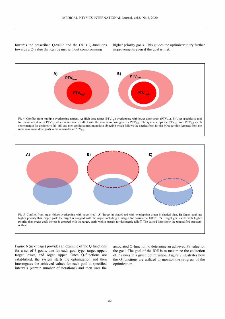
MEDICAL PHYSICS INTERNATIONAL Journal, vol.8, No.2, 2020
82
towards the prescribed Q-value and the OUD Q-functions towards a Q-value that can be met without compromising
Figure 6 (next page) provides an example of the Q functions for a set of 3 goals, one for each goal type: target upper, target lower, and organ upper. Once Q-functions are established, the system starts the optimization and then interrogates the achieved values for each goal at specified intervals (certain number of iterations) and then uses the
higher priority goals. This guides the optimizer to try further improvements even if the goal is met. associated Q-function to determine an achieved Pa value for the goal. The goal of the IOE is to maximize the collection of P values in a given optimization. Figure 7 illustrates how the Q-functions are utilized to monitor the progress of the optimization.
Fig 4. Conflict from multiple overlapping targets. A) High dose target (PTVhigh) overlapping with lower dose target (PTVlow). B) User specifies a goal for maximum dose in PTVlow which is in direct conflict with the minimum dose goal for PTVhigh. The system crops the PTVlow from PTVhigh (with some margin for dosimetric fall-off) and then applies a maximum dose objective which follows the needed form for the PO algorithm (created from the input maximum dose goal) to the remainder of PTVlow.
Fig 5. Conflict from organ (blue) overlapping with target (red). A) Target in shaded red with overlapping organ in shaded blue. B) Organ goal has higher priority than target goal: the target is cropped with the organ including a margin for dosimetric falloff. C) Target goal exists with higher priority than organ goal: the oar is cropped with the target, again with a margin for dosimetric falloff. The dashed lines show the unmodified structure outline.

MEDICAL PHYSICS INTERNATIONAL Journal, vol.8, No.2, 2020
83
Fig 6. Intelligent Optimization Engine Q-functions (goal functions). In this example, the vertical axis in each panel is a quality measure for a goal. A goal meets its quality measure if the achieved value is the same or better than the goal value. The horizontal axis is a depiction of the relative priority of the set of goals. A) Red line is a TLD Q-function with priority 1, orange line is a TUD Q-function with priority 2, green line is an OUD Q-function with priority 3. In each, the circled point is the goal value in relative dose or volume. Note that organ goal functions have a decreasing component for P>Pi. This guides further improvement after the goal is met. B) Q-functions for a different set of goals: user has decided that the OUD goal has highest priority, and TUD has lowest priority.
Fig 7. IOE, monitoring the optimization progress. Example with three goals. Optimization is started with fixed weightings for all objective functions. A) After x iterations, the dose distribution is interrogated, and P values obtained. IOE elevates cost for objective function with lowest P (TLD goal=0.82; red line). B) After another x iterations, dose distribution is interrogated again. TLD goal is met and P is at a maximum value (6.0 here). IOE elevates the cost for the goal with P=1.68 (TUD). C) After another x iterations, dose distribution is interrogated a third time. Target upper goal is now met with P evaluated to 4.25. IOE elevates cost for the goal that had P=2.68 (OUD). Optimization continues until the collection of P is maximal and cannot further be improved.
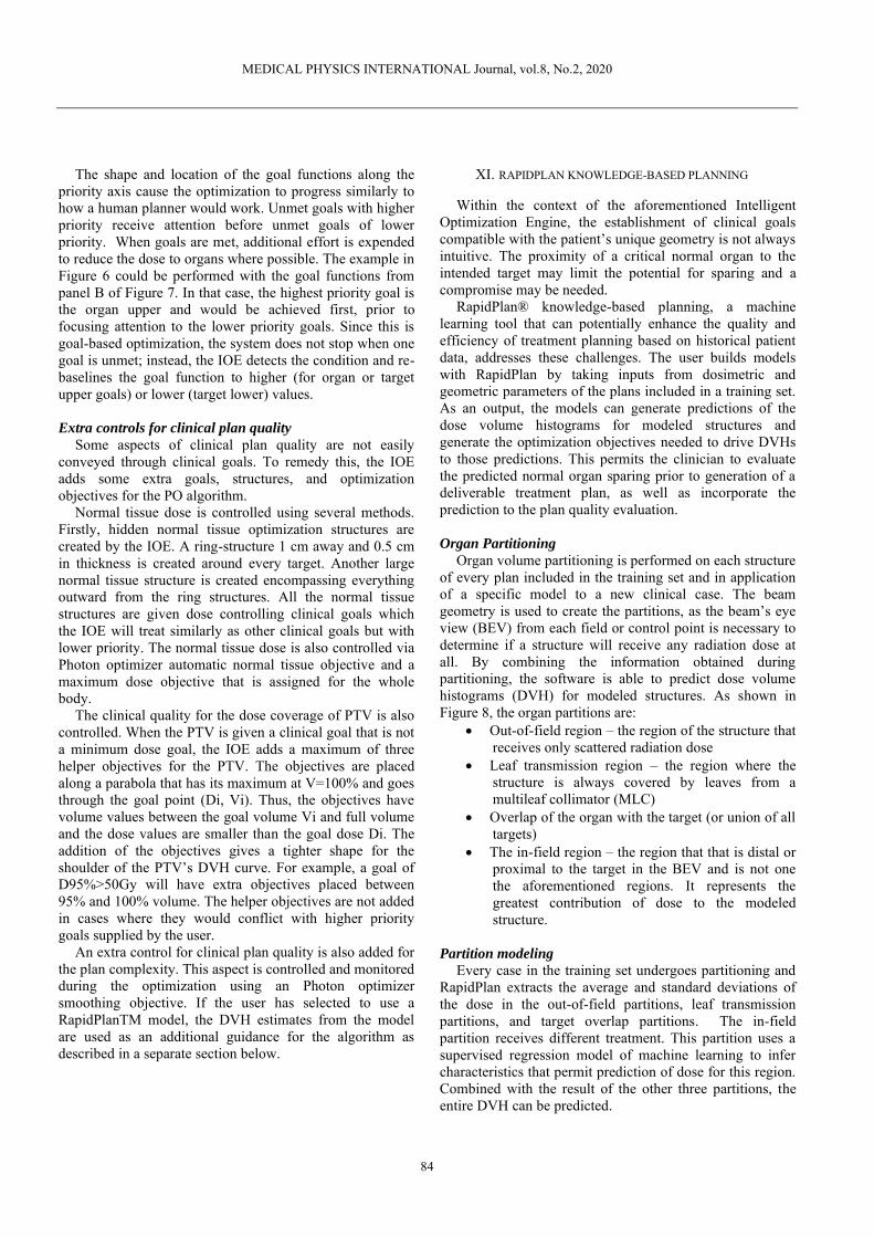
MEDICAL PHYSICS INTERNATIONAL Journal, vol.8, No.2, 2020
84
The shape and location of the goal functions along the priority axis cause the optimization to progress similarly to how a human planner would work. Unmet goals with higher priority receive attention before unmet goals of lower priority. When goals are met, additional effort is expended to reduce the dose to organs where possible. The example in Figure 6 could be performed with the goal functions from panel B of Figure 7. In that case, the highest priority goal is the organ upper and would be achieved first, prior to focusing attention to the lower priority goals. Since this is goal-based optimization, the system does not stop when one goal is unmet; instead, the IOE detects the condition and re-baselines the goal function to higher (for organ or target upper goals) or lower (target lower) values. Extra controls for clinical plan quality
Some aspects of clinical plan quality are not easily conveyed through clinical goals. To remedy this, the IOE adds some extra goals, structures, and optimization objectives for the PO algorithm.
Normal tissue dose is controlled using several methods. Firstly, hidden normal tissue optimization structures are created by the IOE. A ring-structure 1 cm away and 0.5 cm in thickness is created around every target. Another large normal tissue structure is created encompassing everything outward from the ring structures. All the normal tissue structures are given dose controlling clinical goals which the IOE will treat similarly as other clinical goals but with lower priority. The normal tissue dose is also controlled via Photon optimizer automatic normal tissue objective and a maximum dose objective that is assigned for the whole body.
The clinical quality for the dose coverage of PTV is also controlled. When the PTV is given a clinical goal that is not a minimum dose goal, the IOE adds a maximum of three helper objectives for the PTV. The objectives are placed along a parabola that has its maximum at V=100% and goes through the goal point (Di, Vi). Thus, the objectives have volume values between the goal volume Vi and full volume and the dose values are smaller than the goal dose Di. The addition of the objectives gives a tighter shape for the shoulder of the PTV’s DVH curve. For example, a goal of D95%>50Gy will have extra objectives placed between 95% and 100% volume. The helper objectives are not added in cases where they would conflict with higher priority goals supplied by the user.
An extra control for clinical plan quality is also added for the plan complexity. This aspect is controlled and monitored during the optimization using an Photon optimizer smoothing objective. If the user has selected to use a RapidPlanTM model, the DVH estimates from the model are used as an additional guidance for the algorithm as described in a separate section below.
XI. RAPIDPLAN KNOWLEDGE-BASED PLANNING
Within the context of the aforementioned Intelligent Optimization Engine, the establishment of clinical goals compatible with the patient’s unique geometry is not always intuitive. The proximity of a critical normal organ to the intended target may limit the potential for sparing and a compromise may be needed.
RapidPlan® knowledge-based planning, a machine learning tool that can potentially enhance the quality and efficiency of treatment planning based on historical patient data, addresses these challenges. The user builds models with RapidPlan by taking inputs from dosimetric and geometric parameters of the plans included in a training set. As an output, the models can generate predictions of the dose volume histograms for modeled structures and generate the optimization objectives needed to drive DVHs to those predictions. This permits the clinician to evaluate the predicted normal organ sparing prior to generation of a deliverable treatment plan, as well as incorporate the prediction to the plan quality evaluation. Organ Partitioning
Organ volume partitioning is performed on each structure of every plan included in the training set and in application of a specific model to a new clinical case. The beam geometry is used to create the partitions, as the beam’s eye view (BEV) from each field or control point is necessary to determine if a structure will receive any radiation dose at all. By combining the information obtained during partitioning, the software is able to predict dose volume histograms (DVH) for modeled structures. As shown in Figure 8, the organ partitions are:
• Out-of-field region – the region of the structure that receives only scattered radiation dose
• Leaf transmission region – the region where the structure is always covered by leaves from a multileaf collimator (MLC)
• Overlap of the organ with the target (or union of all targets)
• The in-field region – the region that that is distal or proximal to the target in the BEV and is not one the aforementioned regions. It represents the greatest contribution of dose to the modeled structure.
Partition modeling
Every case in the training set undergoes partitioning and RapidPlan extracts the average and standard deviations of the dose in the out-of-field partitions, leaf transmission partitions, and target overlap partitions. The in-field partition receives different treatment. This partition uses a supervised regression model of machine learning to infer characteristics that permit prediction of dose for this region. Combined with the result of the other three partitions, the entire DVH can be predicted.

MEDICAL PHYSICS INTERNATIONAL Journal, vol.8, No.2, 2020
85
Geometry Expected Dose
In order to extract information which connects the geometry of the patient to the observed radiation dose, we utilize the concept of Geometry Expected Dose (GED). The GED is a score for each voxel within the treatment volume based solely on basic photon beam characteristics and the relationship of the structure with the radiation fields.
GED includes understanding the following properties: • Field geometry • Photon behavior • Target geometry and dose levels • Heuristics about which kind of beam arrangements
lead to sparing of normal tissue The GED can be calculated very quickly and only
requires the field geometry, planning CT, and the structure geometry on the planning CT. Supervised Regression
Once the GED for a given case is calculated, we can tabulate the GED volume histogram for the in-field partitions of modeled structures. These are considered to be highly correlated to the dose volume histograms for the same structures. This assumption is based on the observable fact that the geometry relation (proximity) to a target highly influences how well a structure can be spared in a given treatment plan. Over a population of similar treatment plans, or treatment plans from a similar anatomical site, the DVH of the in-field partition and the GED volume histogram will be highly correlated.
To extract the correlations, RapidPlan uses principle component analysis applied to both the collection of in-field DVH and GED volume histograms in a training data set. The coefficient obtained from the principle components can be arranged and analyzed through regression models to extract the correlation from a given GED volume histogram
to an observed dose volume histogram. For any case for which a DVH prediction is desired, the dose volume histogram for the in-field partition is predicted from this regression obtained from the training data set. RapidPlan compatibility with Ethos therapy
Any RapidPlan DVH estimation model can be imported and applied to a RT Intent within Ethos treatment management. If attached to an RT Intent, the DVH estimates for modeled structures are shown in the Ethos Dose Preview and in the Plan Review work areas. Additionally, the lower border of the DVH estimation band is used to derive a line objective which is applied during the plan generation process for both the initial planning and adaptive planning workflows in Ethos treatment planning.
Because there is not a known priority order for the line objective derived from the RapidPlan model, we cannot utilize the Intelligent Optimization Engine to effectively monitor or modify the strength of this line objective. The cost function derived from the line objective is added to the overall optimization, but at a level low enough not to overwhelm the objectives that the IOE determines, places, and monitors from the input goals and priority rankings. As such, its primary use in Ethos therapy is as a quality monitor. If the Ethos treatment planning Dose Preview or candidate plans from automated plan generation cannot achieve a result within the DVH predictions, the planner may need to add additional goals, change the order of goals, change the beam geometry, or determine that the case is not suitable for automated planning.
XII. CONCLUSION
Varian introduced and received CE mark for the Ethos therapy system in September of 2019; first patient treatments occurred later that month at Herlev Hospital in Denmark. The system received 510(k) clearance from the U.S. Food and Drug Administration in February 2020.
The Ethos therapy system incorporates technology that uses artificial intelligence and machine learning to create contours and generate adapted plans for physician review while a patient is on the treatment couch. The system offers radiation oncologists a set of simple tools that enable them to achieve their intention for each patient. The daily variation in a patient’s anatomy, captured and visualized by iterative kV cone-beam CT (iCBCT) imaging, enables the on-couch adaptive workflow.
Ethos further allows a physician to choose which plan to deliver and to complete an adapted treatment within a typical 15-minute treatment time slot. Clinical images at treatment delivery
Ethos therapy integrates multi-modality diagnostic images at the point of treatment on the treatment console. This means the daily re-planning sessions can utilize the
Fig 8. Organ volume partitions used in the machine learning DVH models. Each partition contributes to the sum total DVH as shown.
MEDICAL PHYSICS INTERNATIONAL Journal, vol.8, No.2, 2020
86
same multi-modality images that informed the initial planning stage. At each treatment, Ethos therapy shows:
• That day’s anatomy with iCBCT images • Registered CT, PET, and MR images • The expected 3D radiation dose to the target and
organs at risk for both the un-adapted and adapted plans
Decision-making guided by AI
The goal of Ethos therapy was to design a simple adaptive therapy workflow for both the initial planning and daily re-planning sessions.
During initial planning, Ethos therapy automatically produces several plan candidates with various beam geometries and techniques using prioritized target and organ at risk goals from the physician’s intent. The clinician chooses the most suitable plan and authorizes it for delivery. This step provides confidence that the goals and patient geometry are compatible, and that plan automation can be performed each day. Each treatment day, once the daily anatomy is reviewed and accepted, Ethos therapy will prepare a new adapted plan using the beam geometry of the initial plan, the initial set of target and organ and risk goals, and give the clinician the choice of either the original or adapted plan for delivery.
The process is guided by the technology, as follows: • A decision tree guides the entire adaptive therapy
process • Treatment management and treatment planning
applications are tightly coupled and context-aware • Clinician approvals move the process from one step
to the next • Every step of the workflow is optimized for speed
and engineered for safety Automated dose accumulation
Each day, the Ethos therapy system automatically reconstructs delivered dose in relation to today’s anatomy. This capability:
• Demonstrates that the patient is receiving the intended dose
• Improves understanding of the treatment progress • Helps identify when re-simulation may be required • Simplifies off-line adaption
Familiar, efficient QA
QA for Ethos therapy follows a familiar workflow. • The flexible workflow for pre-treatment QA
accommodates phantom- or calculation-based QA methodologies.
• Initial planning and adaptive planning at the console use the same algorithms for consistency.
• Independent adaptive plan QA can be performed on-demand, without impeding treatment workflow.
This article derives from a Varian technical brief on
Ethos™ therapy artificial intelligence. Intended use summary: Varian Medical Systems’ linear
accelerators are intended to provide stereotactic radiosurgery and precision radiotherapy for lesions, tumors, and conditions anywhere in the body where radiation treatment is indicated.
Important safety information: Radiation treatments may cause side effects that can vary depending on the part of the body being treated. The most frequent ones are typically temporary and may include, but are not limited to, irritation to the respiratory, digestive, urinary or reproductive systems, fatigue, nausea, skin irritation, and hair loss. In some patients, they can be severe. Treatment sessions may vary.
Corresponding Author: Stephen Thompson Varian Medical Systems 3100 Hansen Way, Palo Alto, California, 94089 USA Email: [email protected]

MEDICAL PHYSICS INTERNATIONAL Journal, vol.6, No.1, 2018
87
BOOKS

MEDICAL PHYSICS INTERNATIONAL Journal, vol.8, No.2, 2020
88
“AN INTRODUCTION TO MRI FOR MEDICAL PHYSICISTS AND ENGINEERS” BY ANTHONY WOLBARST AND NATHAN YANASAK
Godfrey, D. 1,
1 Duke University, Durham, North Carolina, USA
Abstract— This article is a brief review of the textbook “An Introduction to MRI for Medical Physicists and Engineers” by Anthony Wolbarst and Nathan Yanasak, 2019, Medical Physics Publishing, ISBN-13 978 1 930524 20 0
“An Introduction to MRI for Medical Physicists and Engineers” is a comprehensive presentation of MRI fundamentals -- beginning with the simplest NMR concepts and building ultimately to thorough descriptions of many of today’s most widely-used clinical MRI algorithms and technologies. Conceptual explanations are presented along with their fundamental mathematical underpinnings in a way that is understandable and engaging for the reader. Throughout the book, simplified quasi-quantum mechanical (QM) explanations of protons in spin-up and spin-down states are logically woven together with the more rigorous and powerful classical physics (pulsed energy) view of NMR. Numerous clear diagrams and tables are provided to illustrate key topics, and helpful exercises are included in each chapter. Interesting historical developments and examples of MRI’s clinical utility are also sprinkled throughout, helping the reader anchor abstract physics concepts to practical imaging implementations. Though the book is intended for the biomedical engineer or medical physicist in training, the content is comprehensive enough that practicing medical physicists and imaging instructors will also find it useful to enhance their own understanding of MRI.
The book is organized as follows: Chapter one introduces the fundamental concepts of a
nuclear spin magnetic moment, the gyromagnetic ratio, the quasi-QM notion of a proton’s spin-up and spin-down states when placed in a magnetic field, the Larmor frequency and resultant energy necessary to flip a proton’s spin, nuclear magnetic resonance (NMR), the spatial encoding of different voxels via a gradient magnetic field, the classical presentation of a voxel’s net magnetization precessing in the plane normal to the external magnetic field, as well as the “T1” and “T2” spin-relaxation mechanisms of a nucleus with its local microenvironment – i.e., the interactions of protons in water and lipids with the nearby biomolecules that exist within and between cells --that create the exceptional soft-tissue contrast for which MRI is renowned.
Chapter two provides a helpful overview of electromagnetism, including Maxwell’s equations; magnetic susceptibility (diamagnetism, paramagnetism, and ferromagnetism); proton-proton dipole interactions; NMR at the Larmor frequency; and a simple demonstration of NMR in water. The final pages address the differences between a simplified QM treatment and a classical treatment of NMR in a manner that illustrates how the same phenomenon can be viewed through each physics lens.
Chapter three addresses 1D proton density (PD) image
creation via spatial encoding, standard image quality and performance figures of merit, and common MR artifacts (nonlinear gradient field, chemical shift, and susceptibility).
Chapter four presents the origins of net voxel
magnetization. Thermal equilibrium, and the constant battle between entropy and energy in a system, are explained. The Boltzmann distribution is introduced to describe the population statistics of spin-up and spin-down protons at thermal equilibrium. This leads to a discussion of the resultant impact of MRI magnet strength B0 on net voxel magnetization, and therefore image quality. Stimulated radiative spin transitions brought on by EM waves at the Larmor frequency which drive the system away from thermal equilibrium, and non-radiative spin transitions -- including T1 relaxation --which return the system to equilibrium, are also introduced.
Chapter five extensively reviews the basis of Fourier
theory and other mathematics and statistics principles required to understand MRI physics, signal processing, image reconstruction, and artifacts. Graduate level physics students will likely already have been exposed to most of these concepts, but seeing them applied to the MRI-specific examples presented in the chapter is helpful.
Chapter six considers the classical approach to proton
NMR in a voxel, relating the useful analogies of gyroscopic precession and nutation under the influence of gravity. The Bloch equations are presented, and the effect of applying an orthogonal pulsed magnetic field (B1) near the Larmor frequency follows.

MEDICAL PHYSICS INTERNATIONAL Journal, vol.8, No.2, 2020
89
Chapter seven -- free induction decay (FID) imaging of a hypothetical 1D patient -- stitches together all of the previously presented concepts into a method for mapping NMR signals along a line, including consideration of the signal induced in a receiver antenna, and the inverse Fourier Transform required to convert k-space data into a spatial NMR map.
Chapter eight details the major components of an MRI system, including the main magnet, shim coils, gradient coils, and transmit/receive RF systems.
Chapters nine and ten dive deeply into the
fundamentals of T1 and T2 relaxations, respectively, along with the mechanisms that create anatomic and physiologic contrast between different tissues and molecular environments. Spin-echo (SE) sequences are introduced as a method to separate purely biological T2 dephasing from the effects of magnetic field imperfections. The use of electron-paramagnetic contrast agents, such as gadolinium chelates, is also explained.
Chapter 11 further explores spin-echo pulse sequences,
including the theory behind inversion recovery (IR), fluid attenuated inversion recovery (FLAIR), short T1 recovery (STIR), and fat suppression sequences.
Chapters 12 and 13 expand 1D MRI theory to voxel
selection and image reconstruction in 2D and 3D, respectively. Phase encoding, gradient echo (GE) vs gradient recall echo (GRE), echo planar imaging (EPI), and the impact of k-space sampling and readout – including cartesian vs non-cartesian (radial and spiral) trajectories, and periodically rotated overlapping parallel lines with enhanced reconstruction (PROPELLER) approaches – are all explained.
Chapter 14 explores the use of MRI to map fluid
motion in the body, including MR angiography (MRA), perfusion imaging, diffusion tensor imaging (DTI), and functional MRI (fMRI) with the blood oxygenation level dependent (BOLD) effect.
Chapter 15 covers standard MRI quality assurance and
safety processes and hazards, and American College of Radiology (ACR) accreditation.
And, finally, Chapter 16 wraps up the book with a presentation of MRI future developments that are already underway.
Summary: The authors manage to successfully take
the reader on a journey from the discovery and fundamentals of NMR all the way to novel k-space sampling and advanced MR imaging sequences – and their underlying molecular physics – in a manner that is quite thorough, yet should be approachable even to a reader with limited prior MRI knowledge. I believe this will be an excellent source for graduate students and professionals alike, and intend to incorporate it into my own teaching.

MEDICAL PHYSICS INTERNATIONAL Journal, vol.8, No.2, 2020
90
“ADVANCED RADIATION PROTECTION DOSIMETRY” BY SHAHEEN DEWJI AND NOLAN E. HERTEL
Damilakis, J. 1,2,
1 University of Crete, Greece, 2 Vice-President IOMP
Abstract— This article is a brief review of the textbook “ADVANCED RADIATION PROTECTION DOSIMETRY” by Shaheen Dewji and Nolan E. Hertel, 2019, CRC Press, 480 p., ISBN 9781498785433
1. Description ‘Advanced radiation protection dosimetry’ covers in
10 Chapters aspects of radiation protection dosimetry that physicists need in order to use ionizing radiation efficiently. The techniques presented in the book can be employed to determine radiation doses to patients and workers and, therefore, they are useful for medical physicists.
2. Purpose This book is written primarily for experienced
physicists wishing to acquire advanced and detailed knowledge in the field of radiation protection dosimetry.
3. Content and features The book starts with a short introduction that includes
basic information on radiation dosimetry and international committees and organizations that publish reports, guidelines, regulations and standards in the area of radiation protection and dosimetry. This introduction is very useful for individuals planning to follow a career in medical physics. The scientific content is structured in 9 Chapters. Chapter 2 provides an overview of units and quantities and describes briefly the atomic structure, the radioactive decay and the interactions of radiation with matter. Chapter 3 provides an interesting historical review starting from the discovery of X-rays and early reports of skin burns and other radiation-induced injuries. The status of protection standards during the early period (1905-1925 and 1925-1950) is very well covered and the transition to the risk-based model of radiation regulation (Linear No-Threshold model) is also discussed. Chapter 4 focuses on radiation detection. Dosimeters used in everyday clinical practice such as dosimeters based on ionization chambers and description of methods for calibration and testing are covered very well and, therefore, this chapter is very useful for clinical medical physicists. Chapters 5–8 are devoted on the scientific models in radiation dosimetry employing reference phantoms, biokinetic and dosimetric models as well as on dose coefficients. Medical physicists interested in Monte Carlo simulation will benefit considerably from the wealth of experience of authors who provide all necessary
details on the above topics. Chapter 9 provides important information about cancer risk coefficients. Sections on limitations and uncertainties associated with these coefficients are very well written and include useful references for medical physicists. The appendix on BEIR VII risk models is also a useful addition to this subject. The last chapter examines the process of interpreting metabolic models for estimating the intake of radionuclides.
4. Assessment This is a very well-written, comprehensive book on
radiation protection dosimetry. An indispensable resource for medical physicists, this book provides theoretical and practical information from experts in this field.

MEDICAL PHYSICS INTERNATIONAL Journal, vol.8, No.2, 2020
91
“PROBLEMS AND SOLUTION IN MEDICAL PHYSICS – NUCLEAR MEDICINE PHYSICS” BY KWAN HOON NG, CHAI HONG YEONG AND
ALAN CHRISTOPHER PERKINS
Brambilla, M. 1,2,
1 University Hospital of Novara, Italy 2 President EFOMP
Abstract— This article is a brief review of the textbook “Problems and Solutions in Medical Physics : Nuclear Medicine Physics” by Kwan-Hoong Ng , Chai Hong Yeong, Alan Perkins, 2019, CRC Press, 139 p., ISBN 9781482240009
The book Problems and solution in Medical Physics –
Nuclear Medicine Physics represents an innovative way of teaching.
Instead of using the conventional flow of introducing
the basic principles, explaining the theory, providing the numerical equations linking the physical figures of merit, providing examples and finally proposing to the reader some practical exercises, the authors Kwan Hoon Ng, Chai Hong Yeong and Alan Christopher Perkins have chosen a completely different approach.
They start from a problem and ask the reader to
provide an answer. The problem can be theoretical, descriptive, technological or practical. Irrespective of the nature of the problem, the authors provide a detailed and comprehensive solution for each of them. In doing so the introduce the underlying theory, suggest the qualitative link between the variables involved, illustrate the numerical equations which allows to quantitate the relationship between the variables and guide the reader step by step in the numerical calculations necessary to obtain the final solution, when needed.
Following this approach, they cover all the different
areas of the Nuclear Medicine specialty: radionuclide and radiopharmaceutical production; imaging, with a special focus on the instrumentation (counters, planar camera, SPECT and PET), the techniques and the ways to ensure the quality of the imaging; radionuclide therapy; internal dosimetry and radioprotection.
I was fascinated by this approach, which I found in line
with the modern educational techniques. Having had the privilege of chairing the EFOMP - European School for Medical Physics Expert, I was always challenged with the increasing need and demand coming from our students of practical, hands-on, question and answer sessions.
I am pleased to recognize that this book provides many, if not all, the answers to the questions that a Medical Physicist must answers in his everyday clinical practice as a health professional.
A famous quote of Yogi Berra, a former US baseball
player says: “In theory there is no difference between theory and practice. In practice there is”. This book reconciles practice and theory, showing the reader, step by step, that should this difference really exists, it must be negligible.
MEDICAL PHYSICS INTERNATIONAL Journal, vol.8, No.2, 2020
92
PhD ABSTRACTS

MEDICAL PHYSICS INTERNATIONAL Journal, vol.8, No.2, 2020
93
LESION DETECTABILITY ON DUAL ENERGY COMPUTED TOMOGRAPHY ABDOMINAL IMAGING: PHANTOM STUDY
H. Jantawong1, A. Krisanachinda2
1Doctor of Philosophy Program in Medical Physics, 2Department of Radiology, Faculty of Medicine, Chulalongkorn University, Bangkok, 10330, Thailand
Abstract—Background: Computed Tomography (CT) is
a widely used imaging modality to detect and diagnose liver tumors. The early detection of hepatic lesions is crucial because the treatment for liver malignancy is most effective when the tumor is small (≤ 2 cm). The major problems of small hepatic lesion detection in CT are lower lesion-to-liver contrast in nature and higher noise level relative to other organs. Many studies [1] suggested the detectability of low contrast liver lesion in CT depends on acquisition and reconstruction parameters, lesion characteristics, radiation dose and patient diameter.
Dual-energy computed tomography (DECT) is considered a promising new development in CT that had a potential to improve lesion detection and characterization beyond the levels currently achieved by conventional single energy CT. Based on reconstructed low- and high energy images can create virtual monochromatic images at energy E as a weighted average of the CT number at low (CTL)- and high-energy (CTH) scans, which is given by
𝐶𝑇(𝐸) = 𝑤(𝐸) ∙ 𝐶𝑇𝐿 + [1 − 𝑤(𝐸)]. 𝐶𝑇𝐻 (1)
where L and H represent low- and high-energy, respectively and w represents weighting factor is given by
𝑤(𝐸) =𝜇1(𝐸)∙𝜇2
𝐻−𝜇2(𝐸)∙𝜇1𝐻
𝜇1𝐿∙𝜇2
𝐻−𝜇1𝐻∙𝜇2
𝐿 ∙𝜇2
𝐿
𝜇2(𝐸)
(2) where “μ” is linear attenuation coefficient, 1 and 2
represent the two basis materials. Therefore, the monochromatic image generated from
image space data is simply a linear combination of the two CT images at low and high energies, where the sum of the two weighting factors equals 1.
The purpose of generating the monochromatic images in the image domain is primarily to generate a single optimized set of images for routine diagnosis.
The aim of this study is to investigate the lesion detectability in low keV-VMC (virtual monochromatic) images from dual energy and low kVp images from single-energy CT and to determine the characteristic of lesion detection in different phantom size and kV combinations in dual-energy CT.
Methods: A semi-anthropomorphic abdomen phantom (QRM Moehrendorf, Germany) with two additional rings represent the abdominal size of small, medium and large
adults (Fig.1). The phantoms were acquired in both dual- and single-energy scans with a dual-source CT scanner (Somatom® Force, Siemens Healthineer, Germany). The single energy (SE) scans were acquired at kVp 80, 100, 120, each scan using the same radiation output, expressed by CTDIvol (a standard 32cm-CTDI phantom), as in a typical clinical protocol performed at 120 kVp. The CTDIvol were 7.3, 10.5, 14.5 mGy for small, medium and large phantoms, respectively. For each phantom size, three dual energy (DE) acquisitions were performed at the same CTDIvol as in the SE scans using the different kVp combinations of 80/150Sn, 90/150Sn and 100/150Sn.
The VMC images at 4 energy levels (40, 50, 60, 70 keV) were then created using Syngo Dual Energy Application, Siemens, Healthineer (Fig. 1.) for each phantom size and each scan setting. Only low keV (40-70) images were investigated in this study because these energy levels provide a superior contrast than the higher energies and is comparable to the low kVp SE images.
Fig. 1. The abdomen phantom images; the top row is generated
VMC images of at 40, 50, 60 and 70 keV from a dual-energy scan with 80/150Sn kV combination and the bottom row is conventional axial images reconstructed from SE scan at 70, 80,100 and 120 kVp.
The lesion detectability of virtual monochromatic images from DECT scan) and polychromatic images from SECT scan, were analyzed by using conventional matrix; in term of contrast to noise ratio (CNR) and task-based images quality matrix; in term of detectability index, d’.
For each single and dual energy data sets, the CNRs were calculated by measuring the CT number of hyperdense liver lesion and adjacent uniform liver background, and noise in the liver background.
To calculate a detectability index, d’, the imQUEST software version 6.01 designed by Duke CIPG had been used. A method of non-pre-whitening observer model with eye filter (NPWE) that incorporated the resolution, noise texture, diagnostic task, and viewing conditions[2],
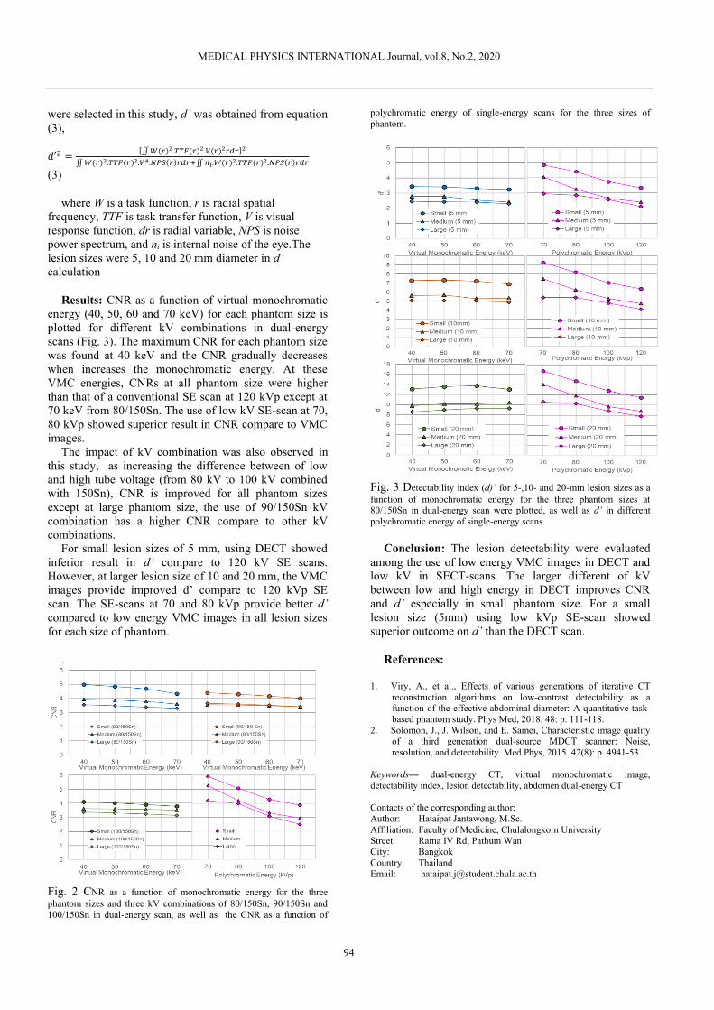
MEDICAL PHYSICS INTERNATIONAL Journal, vol.8, No.2, 2020
94
were selected in this study, d’ was obtained from equation (3),
𝑑′2 =[∬ 𝑊(𝑟)2.𝑇𝑇𝐹(𝑟)2.𝑉(𝑟)2𝑟𝑑𝑟]2
∬ 𝑊(𝑟)2.𝑇𝑇𝐹(𝑟)2.𝑉4.𝑁𝑃𝑆(𝑟)𝑟𝑑𝑟+∬ 𝑛𝑖.𝑊(𝑟)2.𝑇𝑇𝐹(𝑟)2.𝑁𝑃𝑆(𝑟)𝑟𝑑𝑟
(3)
where W is a task function, r is radial spatial frequency, TTF is task transfer function, V is visual response function, dr is radial variable, NPS is noise power spectrum, and ni is internal noise of the eye.The lesion sizes were 5, 10 and 20 mm diameter in d’ calculation
Results: CNR as a function of virtual monochromatic energy (40, 50, 60 and 70 keV) for each phantom size is plotted for different kV combinations in dual-energy scans (Fig. 3). The maximum CNR for each phantom size was found at 40 keV and the CNR gradually decreases when increases the monochromatic energy. At these VMC energies, CNRs at all phantom size were higher than that of a conventional SE scan at 120 kVp except at 70 keV from 80/150Sn. The use of low kV SE-scan at 70, 80 kVp showed superior result in CNR compare to VMC images.
The impact of kV combination was also observed in this study, as increasing the difference between of low and high tube voltage (from 80 kV to 100 kV combined with 150Sn), CNR is improved for all phantom sizes except at large phantom size, the use of 90/150Sn kV combination has a higher CNR compare to other kV combinations.
For small lesion sizes of 5 mm, using DECT showed inferior result in d’ compare to 120 kV SE scans. However, at larger lesion size of 10 and 20 mm, the VMC images provide improved d’ compare to 120 kVp SE scan. The SE-scans at 70 and 80 kVp provide better d’ compared to low energy VMC images in all lesion sizes for each size of phantom.
.
Fig. 2 CNR as a function of monochromatic energy for the three phantom sizes and three kV combinations of 80/150Sn, 90/150Sn and 100/150Sn in dual-energy scan, as well as the CNR as a function of
polychromatic energy of single-energy scans for the three sizes of phantom.
Fig. 3 Detectability index (d)’ for 5-,10- and 20-mm lesion sizes as a function of monochromatic energy for the three phantom sizes at 80/150Sn in dual-energy scan were plotted, as well as d’ in different polychromatic energy of single-energy scans.
Conclusion: The lesion detectability were evaluated among the use of low energy VMC images in DECT and low kV in SECT-scans. The larger different of kV between low and high energy in DECT improves CNR and d’ especially in small phantom size. For a small lesion size (5mm) using low kVp SE-scan showed superior outcome on d’ than the DECT scan.
References:
1. Viry, A., et al., Effects of various generations of iterative CT reconstruction algorithms on low-contrast detectability as a function of the effective abdominal diameter: A quantitative task-based phantom study. Phys Med, 2018. 48: p. 111-118.
2. Solomon, J., J. Wilson, and E. Samei, Characteristic image quality of a third generation dual-source MDCT scanner: Noise, resolution, and detectability. Med Phys, 2015. 42(8): p. 4941-53.
Keywords— dual-energy CT, virtual monochromatic image, detectability index, lesion detectability, abdomen dual-energy CT
Contacts of the corresponding author: Author: Hataipat Jantawong, M.Sc. Affiliation: Faculty of Medicine, Chulalongkorn University Street: Rama IV Rd, Pathum Wan City: Bangkok Country: Thailand Email: [email protected]

MEDICAL PHYSICS INTERNATIONAL Journal, vol.8, No.2, 2020
95
DETERMINATION OF FIELD OUTPUT CORRECTION FACTORS OF RADIOPHOTOLUMINESCENT GLASS DOSIMETER IN 6 MV SMALL
PHOTON BEAM
S. Yabsantia
Medical Physics Program, Department of Radiology, Faculty of Medicine, Chulalongkorn University
Abstract— Background: Small field is utilized in
advanced radiotherapy techniques for performing the complex fluence to achieve the better dose distribution in radiation treatment. However, the small field problems including the lateral charged particle disequilibrium, source occlusion and size of detectors with respect to the field size limit the response of each type of detector and impact the absorbed dose determination.
These problems lead to the large variation of output factors (ratio of reading) among various detectors types. The IAEA and AAPM group established the proposal for small field dosimetry and proposed the field output correction factors (𝑘𝑄clin ,𝑄msr
𝑓clin,𝑓msr ) in order to improve the output factor determination. In 2017, the IAEA/AAPM TRS 483 has been released and the 𝑘𝑄clin,𝑄msr
𝑓clin,𝑓msr of several active detectors were
reported. The 𝑘𝑄clin,𝑄msr
𝑓clin,𝑓msr is defined using the following
equation.
𝑘𝑄clin,𝑄msr
𝑓clin,𝑓msr =𝐷w,𝑄clin
𝑓clin �̅�det,𝑄clin
𝑓clin⁄
𝐷w,𝑄msr
𝑓msr �̅�det,𝑄msr
𝑓msr⁄ (1)
Where 𝐷w,𝑄clin
𝑓clin and 𝐷w,𝑄msr
𝑓msr are absorbed dose in water for clinical field size and machine specific reference field size, respectively. �̅�det,𝑄clin
𝑓clin and �̅�det,𝑄msr
𝑓msr are average absorbed dose in sensitive volume of detector for clinical field size and for machine specific reference field size, respectively.
Radiophotoluminescent glass dosimeter (RPLGD) is a passive detector and increasingly applied for output factor measurement due to its small size. However, high Z (12.04) and high physical density (2.61 g/cm3) of RPLGD restrict its response for small field output factor measurement. Moreover, the orientations of RPLGD with respect to the beam central axis may influence the 𝑘𝑄clin,𝑄msr
𝑓clin,𝑓msr as a consequence of altering the detector size with respect to the field size.
Therefore, this study aims to determine the 𝑘𝑄clin,𝑄msr
𝑓clin,𝑓msr of RPLGD in 6 MV small photon beam for perpendicular and parallel orientations using Monte Carlo (MC) simulation.
Methods: The 𝑘𝑄clin,𝑄msr
𝑓clin,𝑓msr were determined using
EGSnrc Monte Carlo code. The TrueBeam linear accelerator) Varian Medical Systems, Palo Alto, CA,
US) with 6 MV WFF was simulated using BEAMnrc code. Each component module of simulated machine is illustrated in Fig 1. For tuning the source parameters, the PDD and beam profile were reproduced by DOSXYZnrc and compared with the measurement data.
Fig. 1 The treatment head geometry of linear accelerator

MEDICAL PHYSICS INTERNATIONAL Journal, vol.8, No.2, 2020
96
Fig. 2 Example of particle track of perpendicular RPLGD using egs_view The egs-chamber was utilized to simulate the
detector or point of water in water phantom and to calculate dose in scoring volume (Fig. 2). The orientations of RPLGD were studied in both perpendicular and parallel to the beam axis. The absorbed doses in water and average doses in sensitive volume of detector at 90-cm SSD and 10-cm depth for field size ranging from 0.5×0.5 to 10×10 cm2
were calculated. The number of particles were set to maintain the statistical uncertainty less than 0.08%. The dose in each field size was normalized to 10×10 cm2 machine specific reference field. Then, the 𝑘𝑄clin ,𝑄msr
𝑓clin,𝑓msr was
determined using equation (1). The equivalent square small field size (Sclin), which was
given by the geometric mean of full width of half maximum (FWHM) of in-plane and cross-plane, was determined.
Results: For MC commissioning, the suitable source
parameters were 5.9 MeV initial electron energy and 0.11 cm FWHM. The average percentage differences between measured and simulated dose distribution were within 1%.
The ratio of RPLGD reading which was determined using MC is shown in Fig. 3. For field size 2 × 2 to 6 × 6 cm2, the ratio of RPLGD reading showed underestimation comparing with the water. This is influenced by the over response in 10 × 10 cm2. RPLGD exhibits an energy dependent response and over response to low energy scattered photon in large field size due to the high Z of this material.
At the smallest field size, the ratio of RPLGD reading for perpendicular orientation presented underestimation while parallel showed overestimation. In small field size, the perturbation effect was typically affected by volume averaging effect and high density of material. The volume averaging effect was pronounced in perpendicular RPLGD. Therefore, the underestimation was detected. On the other hand, the volume averaging effect was small in parallel orientation and the high density of RPLGD affected the over response, so the overestimation was discovered.
The field output correction factors of RPLGD are demonstrated in Table 1. The 𝑘𝑄clin,𝑄msr
𝑓clin,𝑓msr of RPLGD were 1.000-1.118 and 0.954-1.017 for perpendicular and parallel, respectively. Their overall statistical uncertainties were less than 0.13%. Moreover, the 𝑘𝑄clin,𝑄msr
𝑓clin,𝑓msr of perpendicular and
parallel RPLGD deviated from unity within 5% for field size down to 1 × 1 cm2 and 0.5 × 0.5 cm2 each.
Fig. 3 The reading ratio of RPLGD determined from MC simulation for
perpendicular and parallel orientation in comparison to water
Table 1 Field output correction factors
Side of square field (cm)
𝑺𝐜𝐥𝐢𝐧 (cm)
RPLGD perpendicular
RPLGDparallel
10 10.01 1.000 1.000 6 5.98 1.006 1.010 4 4.04 1.010 1.014 3 3.00 1.010 1.016 2 2.00 1.011 1.017 1 1.00 1.011 1.004
0.5 0.52 1.188 0.956
Conclusion: The 𝑘𝑄clin,𝑄msr
𝑓clin,𝑓msr of RPLGD for both orientations were reported in this study. The correction factors were practical for field size down to 1 × 1 and 0.5 × 0.5 cm2 for perpendicular and parallel orientations, respectively. The parallel orientation of RPLGD might be preferred for small field output factor measurement.
Keywords — Field output correction factors, Monte Carlo
simulation, Radiophotoluminescent Glass Dosimeter, Small field dosimetry
Contacts of the corresponding author: Author: Sumalee Yabsantia, M. Sc. Affiliation: Faculty of Medicine, Chulalongkorn University Street: Rama IV Rd, Pathum Wan City: Bangkok Country: Thailand Email: [email protected]

MEDICAL PHYSICS INTERNATIONAL Journal, vol.8, No.2, 2020
97
INTERFACE AND SMALL RADIATION FIELD DOSIMETRY USING A MOSFET-BASED DETECTOR
W.L. Jong1, N.M. Ung1, J.H.D., Wong2,
1 Clinical Oncology Unit, Faculty of Medicine, University of Malaya, Kuala Lumpur, Malaysia 2 Department of Biomedical Imaging, Faculty of Medicine, University of Malaya, Kuala Lumpur, Malaysia
Abstract— Accurate dosimetry is a critical step in radiotherapy. Generally, essential dosimetric parameters are measured in a relatively flat and homogeneous water phantom, where charged-particle equilibrium (CPE) exists. However, dosimetry in certain conditions such as skin, small radiation field and interface region of two different media, CPE does not exist and therefore, poses challenges to achieve dosimetric accuracy. For accurate dose measurements in non-CPE conditions, a detector should have a small sensitive volume and water equivalent packaging to avoid dose perturbation effect. In this study, the feasibility of a MOSFET-based detector, the MOSkin detector (Fig. 1) for skin, small radiation fields and interface dose measurements were investigated. The advantages of the MOSkin detector are its water equivalent depth of 0.07 mm, small sensitive volume and water equivalent packaging.
The MOSkin detector was first characterised for in vivo
dosimetry under megavoltage photon and electron beams. Then, the MOSkin detector was used to measure patients’ skin dose in tangential breast radiotherapy and compared against dose threshold for different skin reactions. The clinical application (Fig. 2) showed that the MOSkin detector is suitable for in vivo dosimetry and it is necessary to accurately determine the skin dose for better clinical management of skin complications.
The suitability of the MOSkin detector in “Edge-on” orientation (Fig. 3) for small radiation field was investigated. Results showed that the MOSkin detector (Fig. 4) is suitable for dose assessment in small radiation fields as compared to other commercial detectors. The feasibility of the MOSkin detector for dosimetric verification in stereotactic radiotherapy was also tested. The result shows that it is a suitable verification tool for stereotactic radiotherapy.
Challenges in treating a lung tumour are related to respiratory-induced tumour motion and accuracy of treatment planning dose calculations in non-CPE conditions. The dosimetric characteristics near the interface of lung and Perspex media during static, respiratory-gated and non-gated (moving) radiotherapy were investigated using Gafchromic EBT2 film and the MOSkin detector in a moving phantom. In non-gated radiotherapy, dose smearing effect was observed and this effect was reduced in respiratory-gated radiotherapy (Fig. 5). However, there were still some dose discrepancies due to residual motion. The accuracy of different dose calculation algorithms near interface region was evaluated. It was found that over-estimation of the dose coverage will lead to the choice of smaller field size and thus, under-dosage to the tumour. Therefore, better dose calculation algorithms are essential.
In conclusion, the MOSkin detector has been characterised and used for dosimetric assessment in the non-CPE regions. The results suggest that the MOSkin detector possesses suitable physical characteristics for dosimetric assessment of interface and small radiation field.
Keywords— in vivo dosimetry, quality assurance, characterisation, stereotactic radiotherapy, skin dosimetry, electron beam dosimetry.
Fig. 1 The MOSkin detector system
Fig. 2 Total skin dose for 13 patients who underwent “no bolus
radiotherapy” and “bolus radiotherapy”. The black dots show the average total skin dose. The dotted lines show the dose threshold for different skin reactions. *This radiobiological effect does not occur on the breast but it
may appear on other skin sites.

MEDICAL PHYSICS INTERNATIONAL Journal, vol.8, No.2, 2020
98
Fig. 3 The MOSkin detector in “Edge-on” orientation.
Fig. 4 Measured output correction factors for the “Edge-on” MOSkin
detector and SRS diodes. All set of factors are derived from the EBT2 film measurement. The error bar shows the quadrature-combined uncertainties
(1 SD) of the “Edge-on” MOSkin detector and the EBT2 film readings.
Fig. 5 Measured dose distribution using EBT2 film near the lung and Perspex interface during static radiotherapy (no motion), non-gated
radiotherapy (motion amplitude of 40 mm) and gated radiotherapy with 25% gating window (motion amplitude of 40 mm).
REFERENCES
1. Jong, W. L., Wong, J. H. D., Ung, N. M., Ng, K. H., Ho, G. F., Cutajar, D., & Rosenfeld, A. (2014). Characterisation of MOSkinTM detector for in vivo skin dose measurement during megavoltage radiotherapy. J Appl Clin Med Phys, 15(5), 120-132.
2. Jong, W. L., Ung, N. M., Wong, J. H. D., Ng, K. H., Wan Ishak, W. Z., Abdul Malik, R., Phua, V. C. E., Cutajar, D., Metcalfe, P., Rosenfeld, A., & Ho, G. F. (2016). In vivo skin dose measurement using MOSkin detectors in tangential breast radiotherapy. Phys Med, 32(11), 1466-1474.
3. Jong, W. L., Ung, N. M., Vannyat, A., Jamalludin, Z., Rosenfeld, A., & Wong, J. H. D. (2017). "Edge-on" MOSkin detector for stereotactic beam measurement and verification. Phys Med, 33, 127-135.
4. Jong, W. L., Ung, N. M., Vannyat, A., Rosenfeld, A., & Wong, J. H. D. (2017) Dosimetric evaluation near lung and soft tissue interface region during respiratory-gated and non-gated radiotherapy: a moving phantom study. Phys Med. 42, 39-46.
5. Jong, W. L., Ung, N. M., Tiong, A. H. L., A., Rosenfeld, A., & Wong, J. H. D. (2018). Characterisation of the MOSkin detector for dose measurement under megavoltage electron beam. Radiat Phys Chem, 144,76-84.
Contacts of the corresponding author:
Author: W.L. Jong Institute: University of Malaya City: Kuala Lumpur Country: Malaysia Email: [email protected]

MEDICAL PHYSICS INTERNATIONAL Journal, vol.8, No.2, 2020
99
PRODUCTION OF 153SM-LABELLED MICROPARTICLES AND DOSIMETRIC STUDIES FOR POTENTIAL APPLICATION
IN LIVER RADIOEMBOLIZATION
N.A.A. Hashikin1, B.J.J. Abdullah2, C.H. Yeong3, K.H. Ng2, L.Y. Chung4.
1 School of Physics, Universiti Sains Malaysia, Penang, Malaysia. 2 Department of Biomedical Imaging, Faculty of Medicine, University of Malaya, Kuala Lumpur, Malaysia.
3 School of Medicine, Faculty of Health & Medical Science, Taylor’s University, Selangor, Malaysia. 4 Department of Pharmacy, Faculty of Medicine, University of Malaya, Kuala Lumpur, Malaysia.
Abstract— Yttrium-90 (90Y)-microspheres have been increasingly used for transarterial radioembolization (TARE) of hepatocellular carcinoma (HCC). 90Y (a pure beta emitter) does not show sufficient post-procedural imaging capability. Samarium-153 (153Sm) may serve as a better alternative, due to its promising theranostic (therapy and diagnostic) characteristics. This thesis explored the production of 153Sm-microparticles and dosimetric studies for its application in TARE of HCC.
A pilot study was performed to determine the suitable microparticle (diameter: 20 – 40 µm) to be labelled with 153Sm. Two commercially available ion-exchange resins; Fractogel® EMD SO3- and Amberlite® IR-120H+, each was labelled with 1 g of Samarium Chloride (152SmCl3) in six different formulations and sent for neutron activation in the TRIGA PUSPATI research reactor. Radionuclide purity of these microparticles were tested via gamma spectrometry, and the optimum formulation for each microparticle was determined following a 48 h radiolabelling efficiency study in distilled water and human blood plasma (Fig 1). Amberlite® IR-120H+ was chosen, as it possesses excellent (99.9 %) radiolabelling efficiency and did not produced any radionuclide impurity following neutron activation.
Physicochemical properties of the chosen microparticle (Fig 2) before and after neutron activation was further investigated. Fourier Transform Infrared (FTIR) spectroscopy showed its unaffected functional group throughout the preparation processes. Energy Dispersive X-ray (EDX) spectroscopy confirmed the absence of radionuclide impurity. The microparticles possess irregular surface with increased fragments (< 10 µm) following neutron activation, as seen via a Field Emission Scanning Electron Microscope (FESEM) (Fig 3). The measured particle density was 2.5 g.cm-3 with specific radioactivity of 54 Bq per microparticle, and settling velocity of 0.03 cm.s-1.
Monte Carlo (MC) simulations were done using the Geometry and Tracking 4 (Geant4) software toolkit, to study the dosimetric accuracy of the routinely used Medical Internal Radiation Dose (MIRD) based partition model (PM) for TARE with 90Y-microspheres. It was found that PM markedly underestimated the normal liver dose by up to – 78 % (Fig 4), due to exclusion of cross-fire irradiation between the tumour and normal liver tissue. The model also overestimated both tumour and lung dose by up to 8 and 12 %, respectively (Fig 5). These data can be used to recognise the cases with large dosimetric inaccuracy when PM is being used. Also, a corrected formula for lung dose was suggested for future used.
Dosimetric assessment for TARE with 153Sm-microparticles
was performed using similar MC method. Various treatment scenarios were simulated by targeting 120 Gy to the tumour. The 153Sm-microparticles were able to deliver comparable tumour dose with normal liver and lung dose close to that of 90Y-microspheres, and other organ doses far below 1 Gy.
Finally, the simulations were repeated with other potential radionuclides; Holmium-166 (166Ho), Lutetium-177 (177Lu) and Rhenium-188 (188Re), and the doses were compared with 90Y and 153Sm.
153Sm-microparticles showed great potential as alternative to 90Y with advantage of post-procedure imaging. It possesses ideal characteristics including; stable for neutron activation, excellent radiolabelling efficiency, absence of radionuclide impurities, stable in suspension, low production cost, and ability to deliver comparable tumour dose, without exceeding the organ dose limit. However, improvements should be made to its physical structure for better intraarterial delivery to the tumour.
Keywords— Samarium-153, Yttrium-90, Microspheres,
Radioembolization, Hepatocellular Carcinoma,
Geant4 Monte Carlo Simulation.
Fig. 1 Percentage retention of 153Sm in both resins suspended in distilled water (DW) and blood plasma over 48 h.

MEDICAL PHYSICS INTERNATIONAL Journal, vol.8, No.2, 2020
100
(a) (b)
Fig. 2 The Amberlite resin (a) beads (620 – 830 µm) and (b) powder (20 – 40 µm) following size reduction.
Fig. 3 The FESEM images of the Sm-Amberlite microparticles, (a) before and (b) after 6 h neutron activation with their corresponding EDX spectra
(c) and (d), respectively.
Fig.
4
Normal liver absorbed dose difference (%) between the partition model
(PM) and Geant4 (G4) for various tumour involvements (TI) (including 10 % sphere (S)) and tumour-to-normal liver uptake ratio (T/N),
normalised to PM, with 90Y-microspheres.
Fig. 5 Tumour absorbed dose difference (%) between the partition model
(PM) and Geant4 (G4) for various tumour involvements (TI) (including 10 % sphere (S)) and tumour-to-normal liver uptake ratio (T/N), normalised to
PM, with 90Y-microspheres.
REFERENCES
1. Hashikin, NAA, Yeong, CH, Guatelli, S, Abdullah, BJJ, Ng, KH, Malaroda, A, Rosenfeld, AB, & Perkins, AC. (2017). Systematic Investigation on the Validity of Partition Model Dosimetry for 90Y Radioembolization using Monte Carlo Simulation. Phys Med Biol, 62(18), 7342.
2. Hashikin, NAA, Yeong, CH, Guatelli, S, Abdullah, BJJ, Ng, KH, Malaroda, A, Rosenfeld, AB, & Perkins, AC. (2016). Organ Doses from Hepatic Radioembolization with 90Y, 153Sm, 166Ho and 177Lu: A Monte Carlo Simulation Study using Geant4. J Phys Conf Ser, 694(1), 012059.
3. Hashikin, NAA, Yeong, CH, Abdullah, BJJ, Ng, KH, Chung, LY, Dahalan, R, & Perkins, AC. (2015). Neutron Activated Samarium-153 Microparticles for Transarterial Radioembolization of Liver Tumour with Post-Procedure Imaging Capabilities. PLoS ONE, 10(9), e0138106.
4. Hashikin, NAA, Yeong, CH, Abdullah, BJJ, Ng, KH, Chung, LY, Dahalan, R, & Perkins, AC. (2015). Samarium-153 Labelled Microparticles for Targeted Radionuclide Therapy of Liver Tumor. In DA Jaffray (Ed.), World Congress on Medical Physics and Biomedical Engineering, June 7-12, 2015, Toronto, Canada (IFMBE Proceedings Vol. 51, pp. 471-474): Springer International Publishing.
Contacts of the corresponding author:
Author: N.A.A. Hashikin Institute: Universiti Sains Malaysia City: Penang Country: Malaysia Email: [email protected]

MEDICAL PHYSICS INTERNATIONAL Journal, vol.8, No.2, 2020
101
OPTIMISATION OF RADIATION DOSE, IMAGE QUALITY AND CONTRAST MEDIUM ADMINISTRATION
IN CORONARY COMPUTED TOMOGRAPHY ANGIOGRAPHY
S.K. TAN1,2, K.H. Ng1,2, C.H. Yeong3, R.R. Aman1,2
1Department of Biomedical Imaging, Faculty of Medicine, University of Malaya, Kuala Lumpur, Malaysia. 2University of Malaya Research Imaging Centre (UMRIC), Faculty of Medicine, University of Malaya, Kuala Lumpur, Malaysia.
3School of Medicine, Faculty of Health and Medical Sciences, Taylor’s University, Subang Jaya, Malaysia. Abstract— Radiation dose and contrast medium administration
are two major concerns in coronary computed tomography angiography (CCTA). This study aimed to assess the radiation dose and risk of radiation-induced cancer associated with different prospectively ECG-triggered CCTA protocols, and to optimise the radiation dose, image quality and contrast medium administration with an improved retrospectively ECG-triggered CCTA protocol.
This study is divided into four phases whereby the phases that involved patients’ recruitment were approved by the Institutional Ethics Committee (Medical Ethics No: 989.35). Firstly, radiation dose received from prospectively ECG-triggered CCTA using different generations of CT scanners was assessed through organ doses measurement in a standard female adult anthropomorphic phantom. The measured organ doses were used for the estimation of lifetime attributable risk (LAR) of cancer incidence in different sex and age. Secondly, a low tube voltage (100 kVp) protocol was developed for retrospectively ECG-triggered CCTA and tested in 30 patients. The radiation dose and image quality were compared to the routine 120 kVp protocol. Then, a personalised contrast volume calculation model based on patient characteristics and test bolus parameters was developed and validated in 30 recruited patients. Finally, an improved retrospectively ECG-triggered CCTA protocol was developed using the combination of 100 kVp and personalised contrast protocol and validated in 30 recruited patients.
Among the prospectively ECG-triggered CCTA protocols, the highest effective dose (HE) was received from 2 × 32-detector-row dual-source CT (DSCT) scanner (6.06 ± 0.72 mSv). Although the heart is the organ of interest in CCTA imaging, the highest radiation dose was received by breasts and lungs (4 to 8 times higher than heart). The estimated LARs were generally low for all cancers (less than 0.02 to 113 cases per 100,000 population).
For patient’s body mass index (BMI) less than 30 kgm-2, using automatic tube current modulation, statistically significant (p < 0.05) radiation dose reduction (37.8 %) and higher vessel contrast enhancement (VCE) were achieved at 100 kVp. A strong linear relationship was found between VCE and contrast volume (r = 0.97, p < 0.05). Age, sex, body surface area (BSA) and peak contrast enhancement (PCE) at test bolus were found to be significant predictors for VCE (p < 0.05). A personalised contrast volume calculation model was then developed by applying these factors. The model successfully reduced the total iodine dose (TID) while achieving optimal VCE and image quality at 120 kVp compared to the routine contrast protocol (9.8 %, p < 0.05).
When combining both the low tube voltage (100 kVp) and personalised contrast protocol, optimal VCE and image quality
were achieved with statistical significant (p < 0.05) radiation dose (33.8 %) and TID reduction (43.9 %) compared to 120 kVp.
The radiation doses and LAR for cancer incidence from a prospectively ECG-triggered CCTA are relatively low and depend on the scanner model and imaging protocol. This study successfully developed a scanning protocol using low tube voltage (100 kVp) and personalised volume calculation that optimise radiation dose, image quality and contrast medium administration for retrospectively ECG-triggered CCTA.
Keywords— coronary computed tomography angiography, tube
voltage, radiation dose, image quality and contrast
medium.
REFERENCES
1. Tan, S.K., Yeong, C.H., Ng, K.H. & Sun, Z. (2016). Recent Update on Radiation Dose Assessment for the State-of-the-Art Coronary Computed Tomography Angiography Protocols. PLoS One, 11(8): e0161543.
2. Tan, S.K., Yeong, C.H., Aman, R.R., Ng, K.H., Abdul Aziz, Y.F., Chee, K.H., & Sun, Z. (2018). Low tube voltage prospectively ECG-triggered coronary CT angiography: A systematic review of image quality and radiation dose. Br J Radiol., 91: 20170874.
3. Givenchi, S., Safari, M.J., Tan, S.K., Md Shah, M.N.B., Sani, F.B.M., Azman, R.R., Sun, Z., Yeong, C.H., Ng, K.H., Wong, J.H.D. (2018). Measurement of coronary bifurcation angle with coronary CT angiography: A phantom study. Physica Medica: European Journal of Medical Physics, 45:198-204.
4. Tan, S.K., Ng, K.H., Yeong, C.H., Aman, R.R., Sani, F.M., Abdul Aziz, Y.F. & Sun, Z. (2019). Personalized administration of contrast medium with high delivery rate in low tube voltage coronary computed tomography angiography. Quant Imaging Med Surg., 9(4):552-564.
Contacts of the corresponding author: Author: S.K. Tan Institute: University of Malaya Supervisors: Prof. Dr. Kwan Hoong Ng, Assoc. Prof. Dr. Chai Hong Yeong, Assoc. Prof. Dr. Raja Rizal Azman Raja Aman, Graduation Date: October 20, 2018 Funding: High Impact Research Chancellory Grant UM.C/625/1/HIR/MOHE/MED/36, Ministry of Education, Malaysia; Postgraduate Research Grant PG307-2016A, University of Malaya, Malaysia (Street: Jalan Universiti ) City: Kuala Lumpur ; Country: Malaysia Email: [email protected]

MEDICAL PHYSICS INTERNATIONAL Journal, vol.8, No.2, 2020
102
An Investigation of Radiation Dose to Patient's Eye Lens and Skin During Neuro-Interventional Radiology Procedures
Mohammad Javad Safari1,2,
1University of Malaya, Kuala Lumpur, Malaysia. Abstract— X-ray guided interventional procedure is a common
diagnostic and/or treatment modality for various vascular, and cardiac diseases. Advanced technology has enabled interventional radiologists in performing more complex neuro-radiology procedures, resulting in concomitant increase in radiation dose to the patients. The main contributions of this thesis were firstly, characterization of the MOSkin detector in kilovoltage photon beams, and testing its suitability for in-vivo patient’s dose measurements during interventional radiology procedures. Secondly, a comprehensive evaluation of the exposure parameters contributions on patient’s dose during neuro-interventional procedures was performed. It was found that the lateral x-ray tube contributed considerably high radiation dose to the patient’s eye lens. This led to the design and fabrication of a novel type of eye lens protector for those procedures that the patient’s eye is repeatedly positioned within the lateral tube exposure field, where the applying of collimation on lateral beam is not possible. An eye lens protector was particularly designed in order to be placed within the x-ray field of view, attenuating the direct beam from the lateral x-ray tube while being sufficiently radiolucent not to perturb the radiological image and the interventional procedure. Finally, a new type of an anthropomorphic head phantom was fabricated with more options for dosimeter placement and more similar tissue substitute materials to the actual human eye to evaluate the dose delivered to the patient’s eye lens during a clinical neuro-interventional procedure.
The MOSkin detector has been proven to be a reliable and suitable dosimetry system for the measurement of the radiation dose in kilovoltage photon beams and has been successfully utilized during 35 clinical neuro-interventional procedures to evaluate the radiation dose received by the patients’ eye lenses. This study revealed that among the 35 patients, the left outer canthus regions of 8 patients and left eyelid region of one patient were found to receive higher dose than the recommended threshold dose for cataract formation (500 mGy). Based on the study of the contribution of exposure parameters on patient’s dose, it is recommended that the judicial use of acquisition imaging techniques, and the use of the lateral x-ray tube particularly in the anterior-oblique orientation to reduce the patient’s eye lens dose during neuro-interventional procedures. In the situation where the application of physical collimation on lateral tube beam is not possible, the novel eye protector layer may be used to attenuate the direct radiation beam to the patient’s eye lens. This work showed
that for a simulated aneurysm procedure, this protector reduced the maximum radiation dose received by the eye lens and eyelid up to 67.0%, and 23.3%, respectively. The eye protector also had negligible effects on the exposure parameters by a maximum of 8% for the tube current-time product of the DSA (2 frame per second) imaging mode, and image quality (increases the fluoroscopy image pixel value up to 4.7% ± 0.6%). Lastly, the fabricated anthropomorphic phantom has been proved to be a suitable tissue-mimicking medium for the evaluation of the radiation dose received by the patient’s eye lens during clinical diagnostic procedures.
REFERENCES
1.Safari, M.J., Wong, J.H.D., Ng, K.H., Jong, W.L., Cutajar, D.L., Rosenfeld, A.B. (2015): Characterization of MOSkin detector for in vivo skin dose measurement during interventional radiology procedures. Medical Physics. 42(5): 2550-8 2.Safari, M.J., Wong, J.H.D., Ng, K.H., Kadir, K.A.A., Thorpe, N., Cutajar, D.L., Petasecca, M., Lerch, M., Rosenfeld. A.B. (2016): Real-time eye lens dose monitoring during cerebral angiography procedures. European Radiology. 26(1): 79-86 3.Safari, M.J., Wong, J.H.D., Kadir, K.A.A., Sani, F. A., Ng, K.H. (2016): A method to reduce patient's eye lens dose in neuro-intervention radiological procedures: A Phantom Study. Radiation Physics and Chemistry. 125, 75-81 4.Safari, M. J., Wong, J. H. D., Jong, W. L., Thorpe, N., Cutajar, D., Rosenfeld, A., & Ng, K. H. (2017). Influence of exposure and geometric parameters on absorbed doses associated with common neuro-interventional procedures. Physica Medica, 35, 66-72
Contacts of the corresponding author: Author: Mohammad Javad Safari Email: [email protected] Institution: University of Malaya, Malaysia Field of research: Medical Physics Supervisors: Prof. Dr. Kwan-Hoong Ng, Dr. Jeannie Hsiu Ding Wong Graduation date: August 28, 2016 .

MEDICAL PHYSICS INTERNATIONAL Journal, vol.6, No.1, 2018
103
ANNEX

\
Abstracts Booklet of the MMP Thesis (5th cycle)
Master of Advanced Studies in Medical Physics
The programme is accredited by The International Organization for Medical Physics (IOMP)
2
Hospitals in the Programme’s Training Network
MMP Students
Fifth Cycle
2018-2019
The Master is supported and sponsored by:
IAEA Int. Organization of Medical Physics
European federation of Medical Physics
Italian Association of Medical Physics
Trieste University Hospital
Az. Ospedaliero Universitaria
IRCCS Centro di Riferimento Oncologico
Az. Ospedaliera Papa Giovanni XXIII
Ancona
Aviano
Bergamo
Az. Universitaria “Arcispedale Sant’Anna”
Az. USL Toscana Centro
Ferrara
Firenze
Ospedale Niguarda Ca' Granda
IRCCS Istituto Nazionale dei Tumori
Az Ospedaliero Universitaria
Az. Ospedaliera S. Gerado
Milano
Milano
Modena
Monza
Az. Ospedaliero Universitaria
Istituto Oncologico Veneto
Novara
Padova
Az. Ospedaliero Universitaria
Istituti Fisioterapici Ospitalieri
Pisa
Roma
Az. Ospedaliero Universitaria Siena
AOU Città della Salute e della Scienza Torino
A.O. Ordine Mauriziano Torino
Ospedale S. Chiara Trento
Az. Sanitaria Universitaria Integrata Trieste
Az. Sanitaria Universitaria Integrata Udine
Az. Ospedaliero Universitaria Integrata Verona
ULSS 6 Vicenza, Ospedale San Bortolo Vicenza
University Hospital Centre Zagreb (KBC) Zagreb

3
Prof. Luciano Bertocchi (ICTP)
Coordinator of the ICTP Medical Physics Activities and board member
Email:[email protected]
Office:ICTP-Leonardo Building- 2nd Floor- Room#227
Prof. Renata Longo (University of Trieste)
Director of the Master in Medical Physics
Email: [email protected]
Office:Via Alfonso Valerio, 2,Edificio F, Department of Physics, University of Trieste, 2nd Floor
Prof. Renato Padovani (ICTP)
Coordinator of the Master in Medical Physics
Email: [email protected]
Office:ICTP-Adriatico bulidng-1st Floor
Dr. Mario De Denaro (University hospital of Trieste)
Board Memeber&Head of the Medical Physics Department (AOU Trieste)
Email: [email protected]
Office: Via della Pietà, 4,Ospedale Maggiore, Fisica Sanitaria, 2nd Floor
Prof. Luigi Rigon (University of Trieste)
Board Member
Email:[email protected]
Prof. Edoardo Milotti (University of Trieste)
Board Member
Email: [email protected]
Prof. Slavik Tabakov (King's College London)
External Adviser
Email:[email protected]
Prof. Ahmed Meghzifene (IAEA)
External Adviser
Email: [email protected]
Prof. Debbie VAN DER MERWE (IAEA)
External Adviser
Email: [email protected]
Ms. Suzie Radosic (ICTP)
Administrative Secretary for the Master's in Medical Physics
Email: [email protected] Phone:
Office: Adriatico Building- Ground floor - Secretariat of medical phyics
Board Members

4
Khalid Elbochi Abdalla Abdelrhman
Sudan
Azienda Provinciale per i Servizi Sanitari – Santa Chiara Clinical Supervisor: Dr. Francesco Ziglio
Prospective/Objective. Accurate commissioning of the treatment planning system is a critical part of the radiotherapy treatment planning process. Inaccuracy at the planning phase will affect not just one fraction, but all patient treatment planning courses. In this work the verification of point doses were performed at different regions of a heterogeneous phantom using 3D conformal Radiotherapy (3D-CRT) techniques as recommended by IAEA-TECDOC-1583 report.
The aim of this verification is to apply for the first time, this report on the Pinnacle3 at the Santa Chiara hospital, and gain the necessary knowledge to establish national audits in my home country.
Results. The CT-Density curves created show a difference 1.4% between the curve of the Gammex 467 using thorax protocol and the curve stored in Pinnacle3. When the Gammex was scanned using the monthly protocol, there was large difference in the areas of low density (maximum 24%) and in the areas of high density (maximum 68%), when compared to the Pinnacle3 curve. However, the monthly protocol is not used for TPS data, only for constancy evaluation. In clinical test cases, 315 data sets for the three algorithms and the three energies show that 301 data sets meet the
agreement criteria based on IAEA-TECDOC-1583. 14 data sets fell outside the tolerance criteria. When analyzed based on energies, there was no data set that failed the tolerance for 10 MV, 8 data sets failed for 6 MV and 6 data sets failed for 4 MV. When analyzed based on algorithms, there were 3 data sets failed for CCC, 5 data sets failed for AD and 6 data sets failed for FAST.
Conclusion. The overall results
show that 95.6% of data sets for
the three energies and three
algorithms met the criteria set by
the IAEA-TECDOC-1583. The results
revealed that the Pinnacle3 TPS
calculations and Elekta Precise
LINAC dose delivery for 3DCRT at
Santa Chiara Hospital was
generally within acceptable criteria
according to IAEA-TECDOC-1583,
and there was no major causes for
concern. This study verified that
CCC, AD and FAST were general
well modelled in the TPS for high
energy 10 MV. There was some
fine tuning required for beams
modelled with 6 MV and 4 MV,
especially at heterogeneous
regions containing high and low
densities. It was also verified that
CCC algorithm shows a better
accuracy, compared to the AD and
Fast algorithms. Deviations
outside the agreement criteria
Title:
Quality assurance of dosimetry calculation within radiotherapy planning systems in clinical practice, according to IAEA-TECDOC-1583

5
mostly occurred within test cases
using beam modifiers and
tangential beam passing through
inhomogeneous materials.

6
Tirthraj Adhikari
Nepal
Az. Ospedaliero Universitaria Maggiore della Caritá di Novara
Thesis Supervisor: Dr. Gianfranco Loi
Prospective/Objective: To validate the robust optimization formalism to plan the VMAT SABRT of lung lesions using the Mid-ventilation approach by means of phantom simulations and measurements.
Materials and methods. To validate the robustness of the mid-ventilation approach, the quasar programmable respiratory motion management phantom (QPRMP) was used. The lung motion was mimicked by standard and customized homemade inserts containing films for an end to end test measurements, positioned inside it. The phantoms were simulated using a Toshiba 16 slice CT scanner, 4DCT datasets were acquired binning the respiratory cycle triggered by the Varian RPM system in ten phases. Real and perturbed patient RPM breathing signals were used to reproduce the effects of irregular breathing. The mid-ventilation phase, defined as the closer to the average lesion position, was used to plan the treatments with the Raystation TPS (version 8. A). Two plans were created using the geometric uncertainties derived by the non-linear Van Herk recipe applied to lung tissue: one relying on the standard static PTV concept, the other based on min-max robust optimization method that accounts directly for geometric uncertainties minimizing the optimizer cost-function in the worst-case scenario. For each plan, a 4D dose
calculation was performed summing the deformable image registration (DIR) propagated dose recalculated on each phase on the reference Mid-ventilation phase. Treatment plans were compared between them and with standard ITV based procedures, to assess the advantages of robust optimization versus the standard static PTV concept and the effectiveness of the Mid-ventilation approach to manage tumor motion also in presence of marked breathing irregularities. Radiochromic films EBT3 irradiated inside the moving phantom were used to assess the accuracy of the robust optimized plans in the context of lung SABR.
Results. The Mid-ventilation approach in both cases allowed significant dose reduction to not target structures respect the standard ITV based method as expected. The robust optimized plan outperformed the standard static PTV based plan reducing the high dose spillage to not target structures and increasing the dose distribution conformity. The dose coverage to GTV resulted unaffected by robust optimization and was insensitive to the induced motion perturbations testifying the effectiveness of the method also in the presence of breathing irregularities. Film measurements showed optimal agreement between the 4D calculated dose by robust optimization and measured dose distribution with gamma
Title:
Validation of Robust Optimization Approach of VMAT Treatment Planning of Stereotactic Body Radiation Therapy of Lung Cancer

7
passing ratio of 91.6% (global, DD%=3, DTA=2mm, threshold 30%) demonstrating the optimal quality of the end to end process. Similar levels of agreement were found with the perturbed respiratory patterns.
Conclusion. The mid-ventilation approach outperforms the conventional PTV and ITV approach for lung SABR with online image guidance and is insensitive to breathing irregularities. The robust optimization resulted in an accurate and effective method to account for the geometric and dosimetric uncertainty for SABR inverse planning. As robust plans can better spare OARs located close to the irradiated volumes without compromising the dose coverage to targets, as this thesis demonstrated, they appear the technique of choice for the planning of SABR of lung lesions.
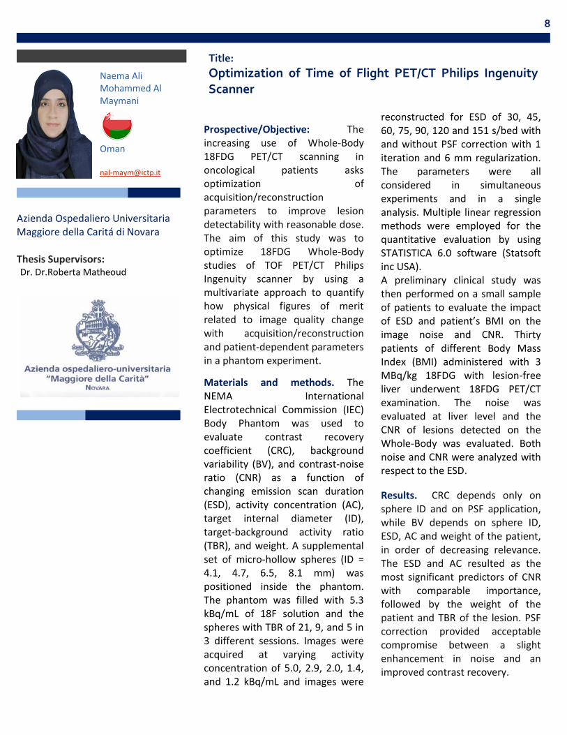
8
Naema Ali Mohammed Al Maymani
Oman [email protected]
Azienda Ospedaliero Universitaria Maggiore della Caritá di Novara
Thesis Supervisors: Dr. Dr.Roberta Matheoud
Prospective/Objective: The increasing use of Whole-Body 18FDG PET/CT scanning in oncological patients asks optimization of acquisition/reconstruction parameters to improve lesion detectability with reasonable dose. The aim of this study was to optimize 18FDG Whole-Body studies of TOF PET/CT Philips Ingenuity scanner by using a multivariate approach to quantify how physical figures of merit related to image quality change with acquisition/reconstruction and patient-dependent parameters in a phantom experiment.
Materials and methods. The NEMA International Electrotechnical Commission (IEC) Body Phantom was used to evaluate contrast recovery coefficient (CRC), background variability (BV), and contrast-noise ratio (CNR) as a function of changing emission scan duration (ESD), activity concentration (AC), target internal diameter (ID), target-background activity ratio (TBR), and weight. A supplemental set of micro-hollow spheres (ID = 4.1, 4.7, 6.5, 8.1 mm) was positioned inside the phantom. The phantom was filled with 5.3 kBq/mL of 18F solution and the spheres with TBR of 21, 9, and 5 in 3 different sessions. Images were acquired at varying activity concentration of 5.0, 2.9, 2.0, 1.4, and 1.2 kBq/mL and images were
reconstructed for ESD of 30, 45, 60, 75, 90, 120 and 151 s/bed with and without PSF correction with 1 iteration and 6 mm regularization. The parameters were all considered in simultaneous experiments and in a single analysis. Multiple linear regression methods were employed for the quantitative evaluation by using STATISTICA 6.0 software (Statsoft inc USA). A preliminary clinical study was then performed on a small sample of patients to evaluate the impact of ESD and patient’s BMI on the image noise and CNR. Thirty patients of different Body Mass Index (BMI) administered with 3 MBq/kg 18FDG with lesion-free liver underwent 18FDG PET/CT examination. The noise was evaluated at liver level and the CNR of lesions detected on the Whole-Body was evaluated. Both noise and CNR were analyzed with respect to the ESD.
Results. CRC depends only on sphere ID and on PSF application, while BV depends on sphere ID, ESD, AC and weight of the patient, in order of decreasing relevance. The ESD and AC resulted as the most significant predictors of CNR with comparable importance, followed by the weight of the patient and TBR of the lesion. PSF correction provided acceptable compromise between a slight enhancement in noise and an improved contrast recovery.
Title:
Optimization of Time of Flight PET/CT Philips Ingenuity Scanner
9
Although preliminary, the clinical study, confirmed CNR improvement and a decrease of CVB with increasing ESD.
Conclusion. The main finding of this study was the dependence of CNR on both ESD and AC with a similar importance. Thus, it will be equivalent to increase lesion detectability by adjusting the administered activity to the patient or the duration of PET bed. From radiation protection point of view, it is advisable to use the manufacturer suggested dose scheme while increasing the bed duration. To further increase lesions visualization, PSF correction could be applied, although with a slight increase in the noise, but without any Gibbs or edge artefacts.
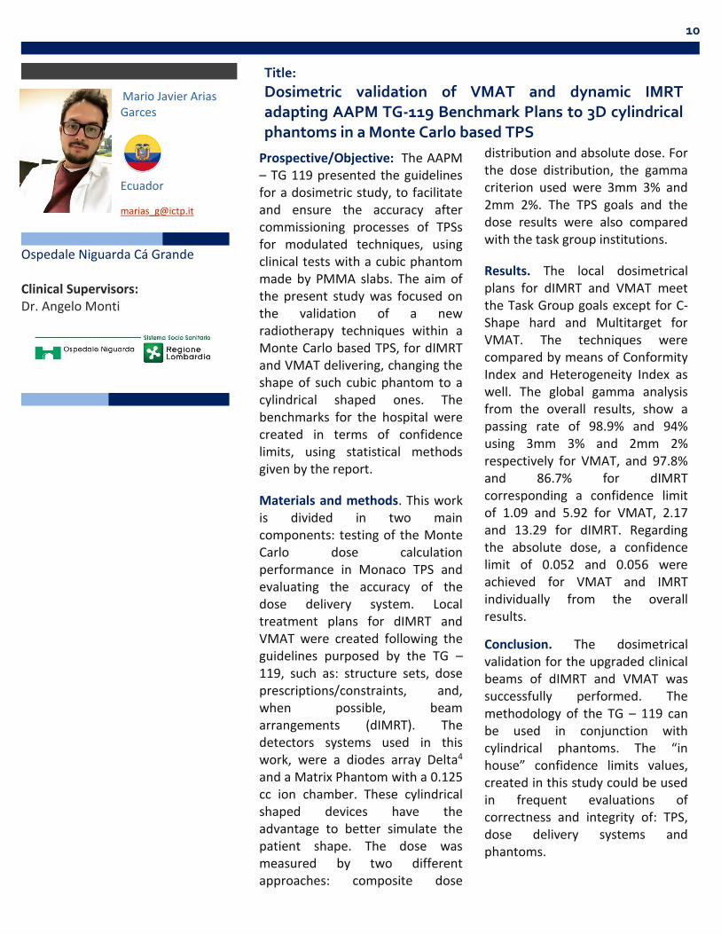
10
Mario Javier Arias Garces
Ecuador
Ospedale Niguarda Cá Grande Clinical Supervisors: Dr. Angelo Monti
Prospective/Objective: The AAPM – TG 119 presented the guidelines for a dosimetric study, to facilitate and ensure the accuracy after commissioning processes of TPSs for modulated techniques, using clinical tests with a cubic phantom made by PMMA slabs. The aim of the present study was focused on the validation of a new radiotherapy techniques within a Monte Carlo based TPS, for dIMRT and VMAT delivering, changing the shape of such cubic phantom to a cylindrical shaped ones. The benchmarks for the hospital were created in terms of confidence limits, using statistical methods given by the report.
Materials and methods. This work is divided in two main components: testing of the Monte Carlo dose calculation performance in Monaco TPS and evaluating the accuracy of the dose delivery system. Local treatment plans for dIMRT and VMAT were created following the guidelines purposed by the TG – 119, such as: structure sets, dose prescriptions/constraints, and, when possible, beam arrangements (dIMRT). The detectors systems used in this work, were a diodes array Delta4 and a Matrix Phantom with a 0.125 cc ion chamber. These cylindrical shaped devices have the advantage to better simulate the patient shape. The dose was measured by two different approaches: composite dose
distribution and absolute dose. For the dose distribution, the gamma criterion used were 3mm 3% and 2mm 2%. The TPS goals and the dose results were also compared with the task group institutions.
Results. The local dosimetrical plans for dIMRT and VMAT meet the Task Group goals except for C-Shape hard and Multitarget for VMAT. The techniques were compared by means of Conformity Index and Heterogeneity Index as well. The global gamma analysis from the overall results, show a passing rate of 98.9% and 94% using 3mm 3% and 2mm 2% respectively for VMAT, and 97.8% and 86.7% for dIMRT corresponding a confidence limit of 1.09 and 5.92 for VMAT, 2.17 and 13.29 for dIMRT. Regarding the absolute dose, a confidence limit of 0.052 and 0.056 were achieved for VMAT and IMRT individually from the overall results.
Conclusion. The dosimetrical validation for the upgraded clinical beams of dIMRT and VMAT was successfully performed. The methodology of the TG – 119 can be used in conjunction with cylindrical phantoms. The “in house” confidence limits values, created in this study could be used in frequent evaluations of correctness and integrity of: TPS, dose delivery systems and phantoms.
Title:
Dosimetric validation of VMAT and dynamic IMRT adapting AAPM TG-119 Benchmark Plans to 3D cylindrical phantoms in a Monte Carlo based TPS

11
Katrīna Čaikovska
Latvia
kcaikovs@ ictp.it
Az. Ospedaliera Universitaria Integrata Verona Clinical Supervisors: Dr. Alessio Pierelli Dr. Carlo Cavedon
Prospective/Objective: Due to the increasing number of stereotactic body radiation therapy (SBRT) lung treatments and gated RT techniques performed, it is highly important to evaluate the accuracy of available treatment planning algorithms. The aim of the study was to measure dose distributions inside lung-like heterogeneous medium and compare them to dose distributions calculated by TPS algorithms (AAA and Acuros XB), in order to evaluate the impact of lateral scatter on their accuracy in simple geometries.
Materials and methods. Measurements were done inside heterogeneous phantoms containing solid water slabs in periphery and lung like material in the centre. Two materials were selected to simulate the lung with different standard deviation of CT numbers. Additionally, to observe the lateral scatter impact on dose calculations, the lung like material was removed and measurements were repeated with the Solid water phantom containing an air gap in the centre. Measurements were done using radiochromic films at different depths with constant beam parameters (6 MV photonbeam, SSD100cm, 10x10cm2 field size and 300MU for cork-based phantoms and 500 MU for solid water air phantom).
TPS calculations were carried out using the Eclipse treatment planning system and CT scans of the phantoms. Radiochromic films were scanned using FilmScan software and calibrated using FilmCal software. Differences between measured and calculated dose values were evaluated using PTW software VeriSoft.
Results. Beam profiles of measured dose and calculated dose in homogeneous lung phantom, inhomogeneous lung phantom and solid water phantom with air gap were obtained at different depths–1cm, 2cm, 3 cm, 5 cm and 6 cm in homogeneous lung phantom, at 2 cm and 6 cm in inhomogeneous lung phantom and at 2 cm in the phantom containing an air gap. Percentage differences and the mean difference between calculated and measured data were obtained.
Conclusion. The Acuros XB algorithm demonstrated better agreement with film measurements and therefore provided more accurate dose calculations compared to the AAA algorithm in heterogeneous medium with a simple geometry setup.
Title:
Comparison of AAA (Anisotropic Analytic Algorithm) and AXB (Acuros-XB) calculation algorithms in heterogeneous medium with elementary geometry using radiochromic films

12
Kadiata Cisse
Mali
Az. Ospedaliero Universitaria di Ferrara Clinical Supervisor: Dr. Sara Fabbri Dr. Ma. Concetta Longo
Prospective/Objective: The purpose of this study was to evaluate the anatomical changes and related dosimetric effect using biweekly CBCT and deformable registration throughout the entire course of radiotherapy in a set of patients affected by head and neck cancer, in order to suggest to physician when replanning is necessary
Materials and methods. Nine Head and Neck patients, treated by VMAT and verified with bi-weekly CBCT for setup corrections, were reviewed retrospectively. A total of 9 planning CT and 102 acquired CBCT image sets were transferred from Pinnacle3 treatment planning system (TPS) to the MIM maestro DIR software. Each CBCT was used as reference image to which the planning CT was registered and the contours delineated for planning purposes were propagated. For both normal tissues and target volumes the deformed contours were visually evaluated by physician. Each deformed planning CT was transferred back into the TPS. The beam arrangements and optimization parameters of the initial treatment plan were copied to the deformed planning CT. The dose calculation module calculated the dose distribution from the copied treatment plan directly onto these deformed planning CT images. In this way for each deformed planning CT plan, dose volume histogram (DVH) was generated. Moreover, an
accumulated DVH was generated by using deformable image registration and dose propagation. Targets and parotids volumes at each CBCT scan were compared to those of the planning CT. The dosimetric parameters including the near minimum dose (D98) to target, the volume of target receiving 95% and 99% of prescription dose were also compared. The mean dose to parotids from each deformed planning CT was compared to the mean dose from the initial planning CT. The actual delivered maximum dose to spinal cord, larynx and mandible was also compared to those in the planning CT.
Results. In the set of patients we considered, the mean volume of CTV, PTV1 (high dose planning target volume) and PTV2 (low dose planning target volume) decreased throughout the course of treatment by 19.14%, 14.42% and 14.43% respectively. However, the reduction in volume of target was not statistically significant. Compared to the initial planning, reduction in target coverage and a decrease in minimum dose to the target were observed. The minimum dose D98 decreased for PTV1 and PTV2 by 1.92Gy (p=0.017) and 6.46Gy (p=0.0091) respectively at the end treatment. Left and right parotids showed a mean reduction in volume of 31.1% (p=0.003) and 35.54% (p = 0.002) respectively. On average, the mean dose to the left and right
Title:
Adaptive Radiotherapy in Head and Neck Cancer
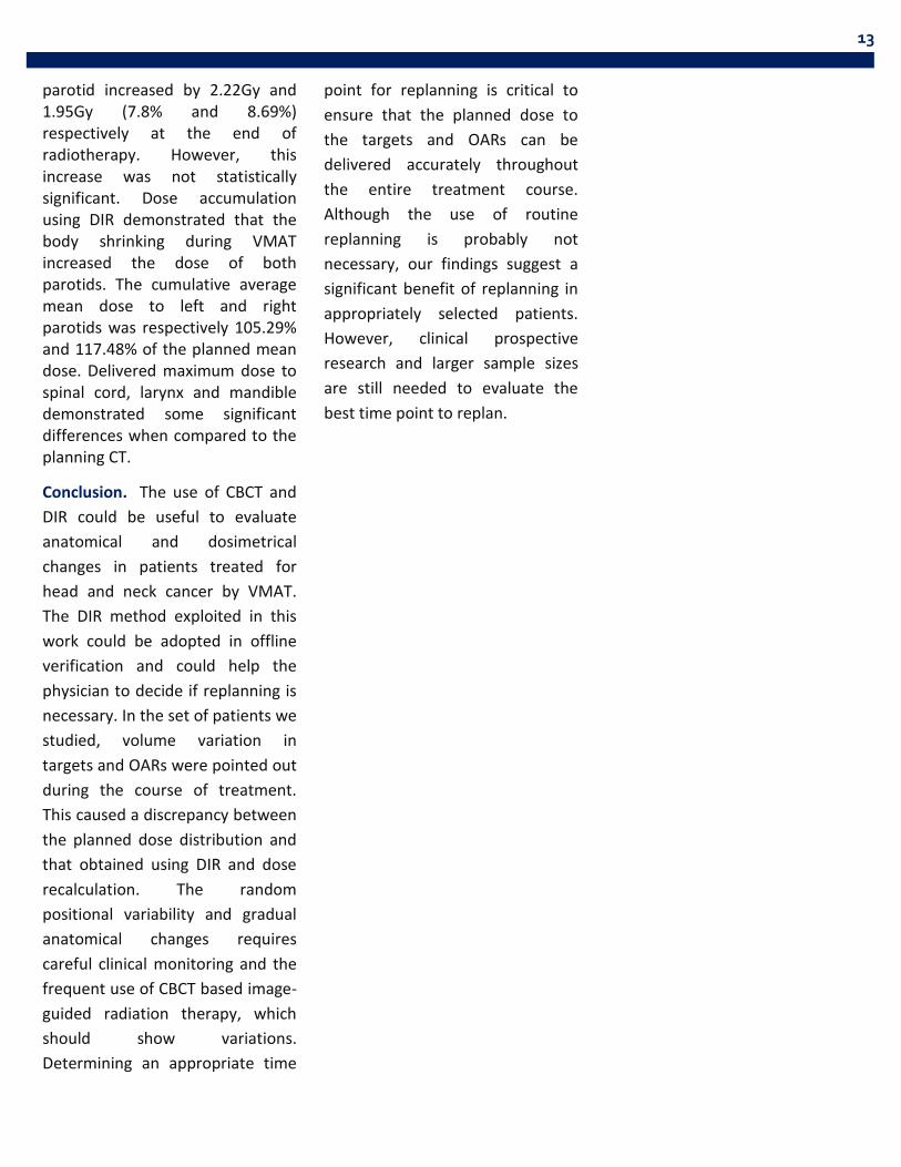
13
parotid increased by 2.22Gy and 1.95Gy (7.8% and 8.69%) respectively at the end of radiotherapy. However, this increase was not statistically significant. Dose accumulation using DIR demonstrated that the body shrinking during VMAT increased the dose of both parotids. The cumulative average mean dose to left and right parotids was respectively 105.29% and 117.48% of the planned mean dose. Delivered maximum dose to spinal cord, larynx and mandible demonstrated some significant differences when compared to the planning CT.
Conclusion. The use of CBCT and
DIR could be useful to evaluate
anatomical and dosimetrical
changes in patients treated for
head and neck cancer by VMAT.
The DIR method exploited in this
work could be adopted in offline
verification and could help the
physician to decide if replanning is
necessary. In the set of patients we
studied, volume variation in
targets and OARs were pointed out
during the course of treatment.
This caused a discrepancy between
the planned dose distribution and
that obtained using DIR and dose
recalculation. The random
positional variability and gradual
anatomical changes requires
careful clinical monitoring and the
frequent use of CBCT based image-
guided radiation therapy, which
should show variations.
Determining an appropriate time
point for replanning is critical to
ensure that the planned dose to
the targets and OARs can be
delivered accurately throughout
the entire treatment course.
Although the use of routine
replanning is probably not
necessary, our findings suggest a
significant benefit of replanning in
appropriately selected patients.
However, clinical prospective
research and larger sample sizes
are still needed to evaluate the
best time point to replan.

14
Adji Yaram Diop
Senegal
Az. Sanitaria Universitaria Integrata di Trieste Clinical Supervisor: Dr. Fabrizio Cupardo
Prospective/Objective: The integration of cross-sectional imaging modalities (such as CT or MRI) into treatment planning for BT has allowed for the development of a three dimensional (3D) image based approach to prescribing and reporting. The aim of this study was evaluate the target coverage and dose received by OARs using different methods of assessment from 2D to 3D CT IGBT.
Materials and methods.: A retrospective study was done, fifty one (51) HDR BT plans from thirteen patients treated with 3D IGBT were re-evaluated using the recommended procedure from GYN GEC ESTRO working group for a transition from 2D to 3D CT IGBT. The clinical target volume (CTV) and OARs (bladder, rectum and sigmoid) were contoured on CT plans. Point A and ICRU 38 bladder and rectal points were defined on reconstructed CT images. All the plans were re-evaluated using Oncentra Brachy TPS which is based on TG43 algorithm. All the patients were treated with CT compatible Fletcher Applicator set and and Elekta Microselectron HDR After loader with a 192Ir source and underwent a post-implant pelvic CT scans with applicator in place. Firstly, 3D CT IGBT plans were used to evaluate how much doses point A and ICRU reference points receive when dose is prescribed in target volume. Secondly, the 3D CT IGBT
plans for patients treatment were reevaluated normalizing the dose to point A. lastly, the dose was prescribed to point A as in 2D based plans without any optimization to be in the same condition than traditional 2D plans but with volumetric dose calculations. Target volume coverage were checked comparing D90 (minimum dose received by 90% of the volume of CTV) and dose to point A. International Commission on Radiation Units and Measurement (ICRU) reference points for dose to OARs (bladder and Rectum) were evaluated in all the methods and compared to the doses received by 0.1cc and 2cc of these organs displayed by the Dose Volume Histograms (DVHs) in the 3D CT IGBT plans. The EBRT dose was given to a total of 45 Gy in 25 fractions and 50.4 Gy in 28 fractions. The total biologically weighted dose including EBRT was estimated using linear quadratic model.
Results. The CTV volume was 71±16 cc (range, 50.1 – 102.9cc). The results were given depending on the HDR fraction dose (6 and 7Gy) respectively. The mean percentage of coverage in 3D CT IGBT plans were 94±2% for 6Gy and 90.1±5% for 7Gy and the mean percentage of dose received by point A in these plans were 72±8.5% for 6Gy and 79±9.1% for 7Gy. For 3D CT IGBT plans normalized in point A, the mean percentage of coverage were
Title:
From 2D to 3D Image Guided Brachytherapy in carcinoma cervix
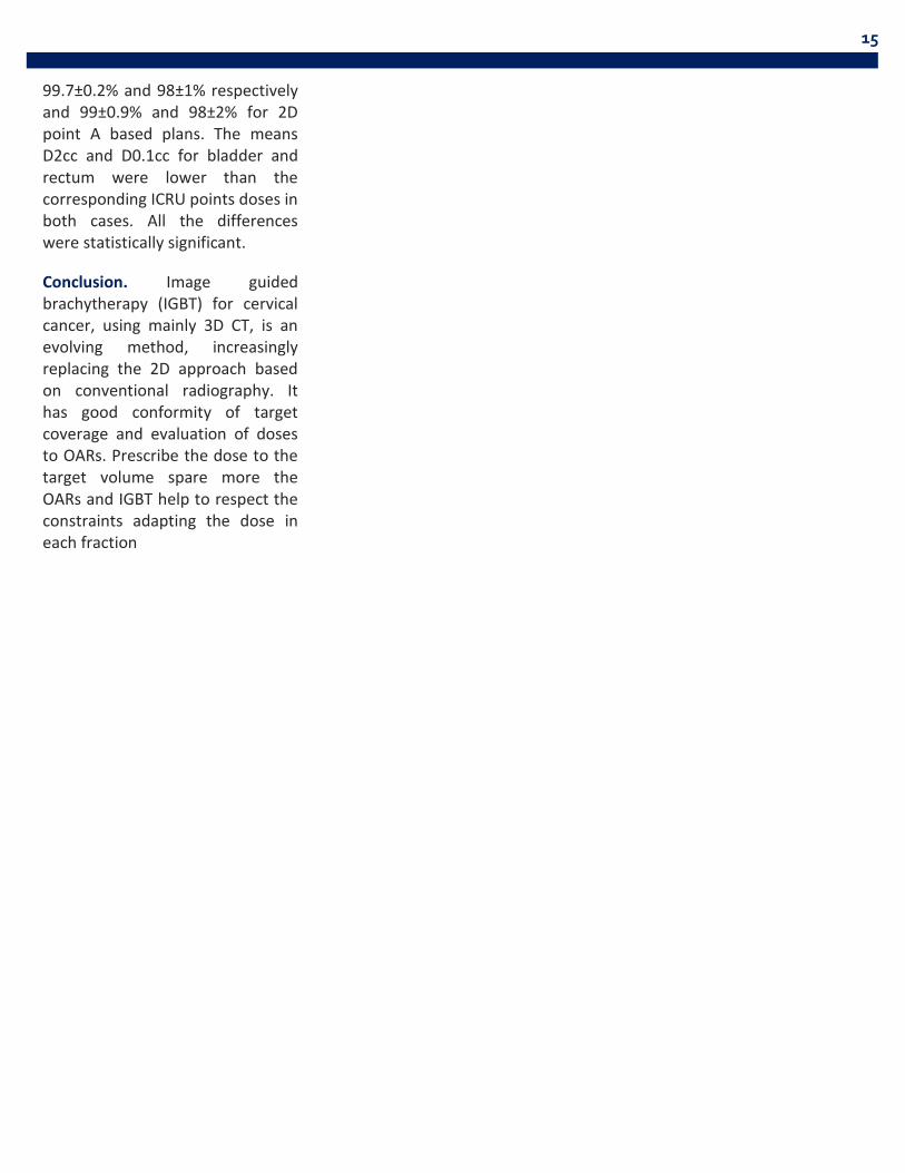
15
99.7±0.2% and 98±1% respectively and 99±0.9% and 98±2% for 2D point A based plans. The means D2cc and D0.1cc for bladder and rectum were lower than the corresponding ICRU points doses in both cases. All the differences were statistically significant.
Conclusion. Image guided brachytherapy (IGBT) for cervical cancer, using mainly 3D CT, is an evolving method, increasingly replacing the 2D approach based on conventional radiography. It has good conformity of target coverage and evaluation of doses to OARs. Prescribe the dose to the target volume spare more the OARs and IGBT help to respect the constraints adapting the dose in each fraction

16
Nino Esvanjia
Gergia [email protected]
AZIENDA OSPEDALIERO-UNIVERSITARIA PISANA (AOUP) Clinical Supervisor:
Dr. Fabio Di Martino
Prospective/Objective: The limited spatial resolution in positron emission tomography (PET) images leads to difficulties to measure correct uptake in tumors. In particular, this is the cause of the partial volume effects (PVE) and can lead to serious bias, especially for small tumors. Correct uptake values are essential for the correct quantification of a parameter widely used in clinical practice, i.e. the standardized-uptake-value (SUV). The aim of this research is to theoretically elaborate and experimentally evaluate a corrective method for PVE, alternative to the Recovery Coefficient (RC).
Materials and methods. In order to obtain the PSF of the PET system, we had to acquire the activity distribution from a point-like source. The acquisition was performed by GE MEDICAL SYSTEMS, DISCOVER 710. In the study, acquisition was performed using NON TOF and TOF correction, in order to analyze the effect of a change in the reconstruction algorithm on the PVE. For acquisition NEMA IEC Body Phantom, has been used.
The counts C of the six spheres has been evaluated inside spherical ROI with radius R equal to the real one using an ideal segmentation criteria. We obtained the x and y coordinates of the six spheres centers from the CT. After
determining the center, we construct binary masks in which the voxels inside the ROI have value 1 while the voxels on the background have a value of 0. In order to be more precise in the application of the spherical mask on the acquired image we have done a rebinning. This procedure has been implemented in the MATLAB program and it Works. We did a voxel by voxel product between the finer image matrix and the binary mask, selecting only voxel in the ROI. We made the sum of these voxel obtaining the counts inside the spheres, which are representative of the activity in the ROI. From the different acquisition mode and time, we took the maximum value of counts, the mean value over a 1 cc inside each sphere and the average value on the total volume of each spheres. In order to recover the concentration values, we multiply the counts by the calibration factor normalizing to the volume. With this procedure, we obtain the respective SUVmax, SUVpeak and SUVmean.
Results. If the tracer is distributed uniformly throughout the VOI, then the SUV would be 1 everywhere. Any departure from 1 indicates a different distribution of the tracer in the VOI in comparison to that the rest of the body. There is no single way to assess the tumour uptake. SUV therefore
Title:
Quantitative analysis of PET acquisition performed by varying the acquisition/reconstruction settings: A corrective post-processing method
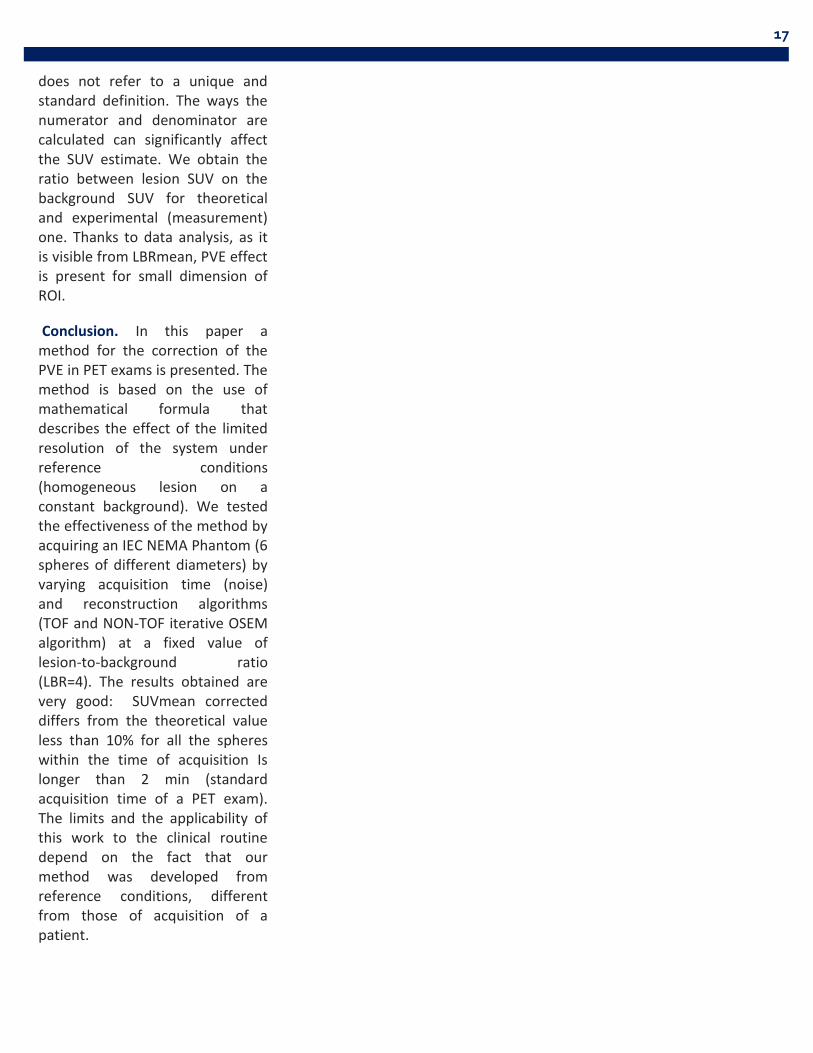
17
does not refer to a unique and standard definition. The ways the numerator and denominator are calculated can significantly affect the SUV estimate. We obtain the ratio between lesion SUV on the background SUV for theoretical and experimental (measurement) one. Thanks to data analysis, as it is visible from LBRmean, PVE effect is present for small dimension of ROI.
Conclusion. In this paper a method for the correction of the PVE in PET exams is presented. The method is based on the use of mathematical formula that describes the effect of the limited resolution of the system under reference conditions (homogeneous lesion on a constant background). We tested the effectiveness of the method by acquiring an IEC NEMA Phantom (6 spheres of different diameters) by varying acquisition time (noise) and reconstruction algorithms (TOF and NON-TOF iterative OSEM algorithm) at a fixed value of lesion-to-background ratio (LBR=4). The results obtained are very good: SUVmean corrected differs from the theoretical value less than 10% for all the spheres within the time of acquisition Is longer than 2 min (standard acquisition time of a PET exam). The limits and the applicability of this work to the clinical routine depend on the fact that our method was developed from reference conditions, different from those of acquisition of a patient.
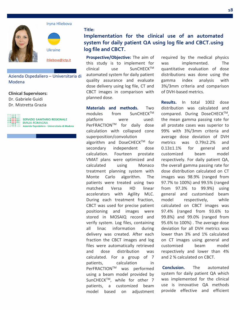
18
Iryna Hliebova
Ukraine [email protected]
Azienda Ospedaliero – Universitaria di Modena
Clinical Supervisors: Dr. Gabriele Guidi Dr. Mistretta Grazia
Prospective/Objective: The aim of this study is to implement for clinical use SunCHECKTM automated system for daily patient quality assurance and evaluate dose delivery using log file, CT and CBCT images in comparison with planned dose.
Materials and methods. Two modules from SunCHECKTM platform were used: PerFRACTIONTM for daily dose calculation with collapsed cone superposition/convolution algorithm and DoseCHECKTM for secondary independent dose calculation. Fourteen prostate VMAT plans were optimized and calculated using Monaco treatment planning system with Monte Carlo algorithm. The patients were treated using two matched Versa HD linear accelerators with Agility MLC. During each treatment fraction, CBCT was used for precise patient positioning and images were stored in MOSAIQ record and verify system. Log files, containing all linac information during delivery was created. After each fraction the CBCT images and log files were automatically retrieved and dose distribution was calculated. For a group of 7 patients, calculation in PerFRACTIONTM was performed using a beam model provided by SunCHECKTM, while for other 7 patients, a customized beam model based on adjustment
required by the medical physics was implemented. The quantitative evaluation of dose distributions was done using the gamma index analysis with 3%/3mm criteria and comparison of DVH-based metrics.
Results. In total 1002 dose distribution was calculated and compared. During DoseCHECKTM, the mean gamma passing rate for all prostate cases was superior to 99% with 3%/3mm criteria and average dose deviation of DVH metrics was 0.79±2.2% and 0.13±1.1% for general and customized beam model respectively. For daily patient QA, the overall gamma passing rate for dose distribution calculated on CT images was 98.9% (ranged from 97.7% to 100%) and 99.5% (ranged from 97.3% to 99.9%) using general and customised beam model respectively, while calculated on CBCT images was 97.4% (ranged from 93.6% to 99.8%) and 99.0% (ranged from 95.6% to 100%) . The average dose deviation for all DVH metrics was lower than 3% and 1% calculated on CT images using general and customised beam model respectively and lower than 4% and 2 % calculated on CBCT.
Conclusion. The automated system for daily patient QA which was implemented for the clinical use is innovative QA methods provide effective and efficient
Title:
Implementation for the clinical use of an automated system for daily patient QA using log file and CBCT.using log file and CBCT.

19
approach in verifying delivery accuracy in terms of gantry, collimator, jaws, MLCs. position and delivered MUs. According to result of performed daily QA, all patients were treated with recommended accuracy. The confidence limit for Gamma passing rate with 3%/3mm criteria for daily QA and median dose deviation was suggested. CBCT images for daily QA can give information about interfraction patient anatomy changes and patient positioning. After 3rd week of treatment the increasing of dose deviation was observed. The log file and CBCT or CT images-based dose calculation together with specific QA program for the dynamic performance of the MLC and accuracy of log file recorder could replace conventional QA methods.
20
Siaka Maiga
Mali [email protected]
Azienda Ospedaliera Ordine Mauriziano di Torino Clinical Supervisor: Dr. Antonella Giostra
Prospective/Objective: The project was aimed at commissioning of electron beam of an Elekta Synergy linear accelerator at Mauriziano Hospital in Turin, Radiotherapy Department.
Materials and methods. Commissioning of electron beam has been carried out following the recommendations of AAPM TG-106 for appropriate detector selection, measurement techniques, etc., and the measurements required for RayStation TPS commissioning have been realized. Commissioned energies of electron beam were 4, 6, 9, 12, 15 MeV generated by the Elekta Synergy linear accelerator (linac). All the measurements were performed at collimator and gantry zero using: 3D- sun nuclear water tank phantom filled of water except for the measurement of air fluence factor OFair where it was empty, and two detectors (diode, SemiFlex ionization chamber type-31010). The measurements required to model electron beams in the treatment planning system RayStation (which use Monte Carlo dose calculation algorithm for electrons) for each energy were: Depth dose in water with (6 × 6 cm2, 10 × 10 cm2, 14 × 14 cm2, 20 × 20 cm2 and 25 × 25 cm2 applicators ) and without applicators at 100 cm source-to-surface distance (SSD), profiles both in-line and cross-line with and without applicators, air fluence
factor OFair at SDD=75cm and 95 cm with 8×8, 8×20, 8×30 and 30×30 cm2 field size, and applicator water dosimetry calibration data (at fixed number of monitor units) as specified in the commissioning manual. Sun nuclear dosimetry software was used to analyze data collected.
Result. From electron PDD data obtained, the depth of dose maximum for 4 MeV,6 MeV, 9 MeV ,12 MeV and 15 MeV was 0.93 cm, 1.27 cm ,1.88 cm, 2.93 cm, 3.40 cm respectively. The differences in the surface area of the PDDs were of the order of 6%. The flatness and symmetry of scanned profiles for each energy were not exceeding 2%.
Conclusion The results obtained for dosimetric data (percentage depth dose, beam profiles, output factor applicator water dosimetry calibration data) are within the tolerances recommended from published research, the beams of linac meet the specifications to be adequately used clinically for patient treatment.
Title:
Electron beam commissioning of an Elekta Synergy linear accelerator

21
Mwape Mofya
Zambia
Ist. Nazionale Tumori Regina Elena Clinical Supervisor: Dr. Lidia Strigari Dr. Marco D’Andrea
Prospective/Objective: The EclipseTM treatment planning system is in routine use at Istituto Nazionale Tumori Regina Elena, Italy, to generate individual treatment plans. The institute has recently purchased the research version of the Pinnacle3 treatment planning system. The Pinnacle3
treatment planning system incorporates the Auto-Planning® optimizer that automates many processes of the manual optimization. The aim of this study is to compare the plan quality among plans manually optimized both in EclipseTM and Pinnacle3, and plans generated by the Pinnacle3 Auto-Planning R engine.
Materials and methods. Nine cases including three breast, three head and neck, and three prostate were selected for this study. IMRT plans were generated using the FiF technique for the breast cases, fixed gantry inverse planning IMRT for the head and neck cases and VMAT for the prostate cases. Two plans were manually optimized for each case: the first plan was optimized using EclipseTM and the second plan was optimized using Pinnacle3. A third plan was generated using Pinnacle’s Auto-Planning® optimizer for the prostate and the head and neck cases. The target coverage, dose homogeneity, dose conformity, organ at risk sparing and delivery efficiency were evaluated. The
PQM% and the APQM% scores calculated using the plan quality algorithm in the PlanIQTM software provided a measure of the overall achieved plan quality of the plans. Statistical analyses were performed using paired t-tests with a level of significance at 5%.
Results. There were no significant differences between the FiF plans created in EclipseTM and Pinnacle3 treatment planning systems. Similar DVHs where obtained from the IMRT plans. On average dose conformity was better in the EclipseTM IMRT plans but with significantly increased monitor units. The Auto-Planning® IMRT plans provided better sparing of the OARs. The PQM% scores were slightly higher in the EclipseTM IMRT plans but the differences with the manual Pinnacle3 and the Auto-Planning® IMRT plans were not significant. VMAT plans optimized with Auto-Planning® had better target coverage, dose homogeneity, OAR sparing, and higher PQM% scores than the manually optimized EclipseTM and Pinnacle3 VMAT plans. The monitor units obtained from EclipseTM, Pinnacle3 manual planning and Auto-Planning® VMAT optimization were comparable.
Conclusions. While the optimization algorithms, optimization tools and dose computation algorithms differ in
Title:
A comparative study of two treatment planning systems for IMRT optimization
22
the EclipseTM and Pinnacle3 treatment planning systems, IMRT plans of similar quality can be created. Auto-Planning®, with manual intervention, could increase the quality of IMRT and VMAT plans. Auto-Planning® could be used as a starting point. Manual improvements to the dose distribution could then be made starting from the Auto-Planning®
solution.

23
Nkuebe Clement Moleko
Lesotho
Fonsazione IRCSS Istituto Nazionali dei Tumori Clinical Supervisor: Dr. Mauro Carrara MS. Simona Di Biaso
Prospective/Objective: To develop an artificial neural network (ANN) model able to predict the expected total reference air-kerma (TRAK) of HDR brachytherapy (BT) plans for endometrial cancer patients, to be used as a quality assurance (QA) tool for treatment plan verification.
Materials and methods. 182 vaginal cuff postoperative HDR BT treatments of patients with endometrial cancer delivered at the Fondazione IRCCS Istituto Nazionale dei Tumori between January 2018 and July 2019 were considered. Patients were treated according to various dose prescriptions (i.e., between 500 and 700 cGy), fractionation schedules, applicator types (i.e, cylindrical single-channel and multichannel vaginal applicators), and applicator diameters (i.e., 25 and 30 mm). TRAK, target volume (Vtarget), and the percentage loading of the central catheter (%CCL) with respect to peripheral ones (where available) were collected. Since the TRAK is linearly correlated with the dose, it was normalized to prescribed dose (nTRAK) to avoid any bias related to the delivered dose. A multilayer-perceptron regression ANN was trained to predict the nTRAK starting from Vtarget, %CCL and the applicator diameter. To overcome data overfitting, a simplified ANN architecture with up to three hidden neurons was chosen and the training was
performed according to the early stopping method. The dataset was randomly split into train (120), verify (31) and test (31)
Results. The resulting Pearson R-correlations between the actual nTRAKs and those predicted by the obtained ANN were 0.94, 0.95 and 0.94 for train, verify and test cases, respectively. The performance of the ANN was higher when compared to the linear regression of data, stratified according to the applicator diameter (i.e., R=0.92 and R=0.89 for the 25-mm and the 30-mm applicator, respectively). 13.3%, 6.5% and 3.2% of the studied treatment plans belonging to the train, verify and test groups, respectively, showed a relative difference between predicted and actual nTRAK values greater than 10%.
Conclusion. The TRAK in vaginal cuff HDR BT treatments can be accurately predicted with an ANN model and can therefore be used as a possible QA parameter to check treatment plan reasonableness. A deviation greater than 10% between predicted and actual nTRAK values could constitute a possible action level for a thorough investigation of the treatment plan. The proposed methodology can in principle be extended to other BT treatments.
Title:
An artificial neural network model for treatment plan verification of vaginal cuff HDR Brachytherapy

24
Roberto Alejandro Montezuma Velasquez
Guatemala
Az. Socio Sanitaria Territoriale di Monza Clinical Supervisor: Dr. Sabrina Morzeneti Dr. Nicoletta Paruccini
Prospective/Objective: To implement a commissioning protocol for a MRI system dedicated to neuroimaging, supplied by quality controls on subsystem components and image quality assurance in a multi-channel head coil. Establishing a benchmark data from acquisitions series on different advanced imaging techniques following international guidelines.
Materials and methods. The MRI system characterized was a Philips Ingenia CX 3T, 60 cm bore diameter, with the main signal receiver, a 32 channels dStream head coil. The commissioning protocol established was separated in quality control and image quality assurance. The quality control includes the assessment in subsystems, homogeneity of the main magnetic field and the coil elements performance, in a single slice reconstruction from an elliptical phantom, provided by Philips. The image quality assurance was performed using three phantoms (i.e. AIFM, PIQT and ACR). The AIFM (Italian Working group) phantom in advance techniques on MRI (i.e. MRS, DWI and fMRI). The PIQT phantom in periodical controls by Philips on spin echo and fast field echo. The ACR (American College of Radiology) phantom for clinical accreditation on weighted T1 spin echo. The assessment metrics were taken from international guidelines (i.e.
AAPM, ACR, AIFM, FBIRN and NEMA). Afterward the set of images acquisitions were processed in Matlab (R2017b) from developed custom codes included in an interface. The benchmark data were represented by the mean value and the standard deviation of a normal distribution, from 5 measurements repetition on each metric and compared with the references recommended on the guidelines.
Results. The benchmark produced during the commissioning shown to be in agreement in the 5% significance level (t-test) with the reference data taken from the guidelines. In the cases were not possible to find studies under similar conditions, PIQT phantom analysis and FBIRN in the AIFM phantom, was produced a discussion according the expected results for a correct behavior of the MRI system under study.
Conclusion. The commissioning protocol provided includes three main test types, identified as: General Systems, Quality Controls and Quality Assurance. The metrics analyzed were comparable with the guidelines recommendations for a stable MRI 3T, ensuring the correct functionality in the scanner for clinical use. For the implementation in a routine protocol is recommended a study in terms of the time production and levels of confidence found.
Title:
Implementation of a 3T MRI scanner dedicated to research: Commissioning and Quality assurance program establishment.

25
Kanchan Sarga Nyaichyai
Nepal
Az. Ospedaliera Universitaria Senese Clinical Supervisor: Dr. Eleonora Vanzi
Prospective/Objective: Replace the 12 years old Thermo 6600Plus dosimetry system with a new automated RADOS RE2000A reader and RadPro LiF:MCP dosimetry system. General public together with hospital staffs, students and trainees working in an area where there is a risk of being exposed to ionizing radiation, should be in a safer environment as recommended by the IAEA Basic Safety Standard Series. For this conformity, proper radiation monitoring should be established.
Materials and methods. A new RADOS RE2000A reader system from MIRION Inc. was acquired with the LiF:MCP chips provided by RadPro. LiF:MCP (Radpro specific commercial name of LiF:Mg,Cu,P material) is mostly used thermo-luminescent crystal having dosimetric glow curve peak around 220 °C. It has the advantage of being linear up to 10 Gy, more sensitive with respect to previous LiF:Mg,Ti (from internal tests with previous system about 10 times) and negligible fading. The reader can identify the slides, which are assigned with unique serial number and a binary bar code, through its inbuilt barcode scanner and data entry. The reader can process automatically up to 200 dosimeter cards or 800 single TL-elements at one load. The pre-heat, measurement and anneal cycles are programmable through the software. In addition to this,
the software can be fed with element-sensitivity correction, background subtraction and pre- and post-calibration capability. For calibrations and angular dependence purposes, set of selected chips were irradiated in Secondary Standard Dosimetry Laboratory (SSDL) with different energies from X-ray tube, Cs-137 and Co-60. For type testing, anyhow, being not possible to make all testes in SSDL, irradiations were done in the hospital environment with inhouse calibrated X-ray tube and RadPro Irradiator with Sr-90 source. A group of 20 cards were selected as a reference calibration cards because they showed less fluctuations within 5% following standard irradiation with 90Sr source and were sent to SSDL for the reference irradiation of 500 µSv with different energies, at different angles and on slab or pillar mimicking body and wrist of a person. Calibration was made for Hp(10), Hp(0.07) slab and Hp(0.07) pillar operational quantities; calibration with H*(10) was deduced from the one for Hp(10) based on previous obtained data. Individual element correction factor (SIR) were evaluated for each dosimeter. All the tests recommended by the European Commission Radiation Protection 160 like additivity, fading, linearity, reproducibility, sensitivity, energy and angular dependence were checked. Simplified IEC 62387 standards were followed to
Title:
Commissioning and Type Testing of RADOS RE2000A and RadPro LiF:MCP Dosimetry System

26
estimate both the compliance with limits and the uncertainty for each evaluation.
Results. The distribution of the SIR value of all the TLD chips were not Gaussian however the repeatability of the SIR values were within 2%. The additivity response of the dosimeters was between 0.99 and 1.01. The sensitivity was evaluated with three processes, with a simple 2σ, a box plot (Upper Limit-Median) and Hirnings method and all of them resulted in quite near values of around 12 µSv. The signals were found to fade to about 11% in 10 weeks and the crystals were found to be linear below 1 Gy dose corresponding to the reader environment. The crystals were found to be reproducible within 6%. Considering 137Cs as the standard beam energy, we found maximum of 20% variation with ISO beam quality L1, that is of energy of 45 keV while this variation decreases with increasing radiation energy. Angular dependence was checked for 3 energies at two angles 0° and 60°. The angular dependence was highest at around 31% for Hp(10) cards for the ISO L4 beam quality, of energy 104 keV.
Conclusion. All the results agreed with requirements from the IEC 62387 standards and the new dosimetry system is all capable for efficient personal monitoring of the public in addition to staffs, students and trainees who are at risk of being exposed to ionizing radiation.

27
Grace Kukiriza
Uganda
Ospedali Riuniti Ancona Clinical Supervisor: Dr. Marco Valenti Dr. Marco Parisotto
Prospective/Objective: Provide an assessment of selected dosimetric and geometric checks on two TrueBeam linear accelerators and compare the outcomes against MPC tool.
Materials and methods. Weekly absolute dose measurements in RW3 solid water phantom were conducted with a secondary dosimetric system (SS) using Farmer ionization chamber and PTW’s Unidose electrometer for the commissioned photon energies. Beam output change measured with MPC and SS was analysed using student’s t-test tool. Beam center shift assessments were performed on the EPID at SSD 130cm with 6MV for 8 collimator rotations. A Matlab script evaluated the maximum distance of the MV image(as detected from the edges)at source axis distance from the beam central axis (BCA)position. Radiation treatment isocenter size with 6MV, 10MV and 6FFF was evaluated on the EPID using Winston-Lutz test performed with Brainlab’s ball bearing tool fixed on a mask base insert, for 8 gantry angles with collimator 90º and 270º. A Matlab script evaluated the maximum distance between the BCA and imaging isocenter (identified as the center of the sphere).Rotation induced couch shift was assessed using EBT3 Gafchromic film placed on
the couch at SSD 100cm, for 5 couch rotations with collimator 240°, 0° and 120°.A Matlab script automatically calculated the projection of the BCA position on the film for five couch angles. The mean distance between the projected BCA position on the film and the couch rotation axis was assumed to quantify the induced couch shift. Collimator rotation offset was evaluated on the EPID for five collimator rotations at zero gantry and MV images analysed using ImageJ.
Results. The mean beam output change acquired for all energies with MPC and the SS were within ±2% tolerance because the TrueBeam linacs were calibrated whenever the output approached this limit. A good agreement between MPC and SS was realized. The mean difference in beam center shift measured with MPC and EPID was 0.02mm ±0.02 and 0.13mm ±0.02 on TB STX and TB respectively. The mean beam center shifts measured with MPC and EPID were within ±0.5mm tolerance limit. The mean difference in isocenter size measured with MPC and WL for 6MV was -0.02mm ±0.03 and -0.05mm ±0.02 on TB STX and TB respectively. TB STX showed a mean WL isocenter size of 0.29mm ±0.02 and 0.27mm ±0.03 for 10MV and 6 FFF respectively, while TB recorded 0.33mm ±0.01 and 0.36mm ±0.02, all within ±0.5mm
Title:
Independent verification of selected dosimetric and geometric parameters of Machine Performance Check on two TrueBeam linear accelerators.

28
tolerance. The mean difference in the rotation induced couch shift measured with MPC and film was 0.02mm ±0.02 and 0.04mm ±0.04 on TB STX and TB respectively. The mean difference in the collimator rotation offset attained with MPC and EPID was -0.03º±0.03º and 0.04º±0.03º on TB STX and TB respectively, all within ±0.5º tolerance
Conclusion. MPC is a suitable tool for daily QA since the overall procedure is very fast and less cumbersome. However, departments maintaining and implementing a robust QA programme alongside MPC tool for an independent assessment is fundamental.
29
Sanchez Okieffy Roydell Palmer
Jamaica
Azienda Provinciale per i Servizi Sanitari – Santa Chiara Clinical Supervisor: Dr. Anna Delana
Prospective/Objective: As part of
the quality control program for
stereotactic treatments at the
Santa Chiara hospital Trento, a
Pinpoint (PTW-31016) ionization
chamber in conjunction with a Real
Water head and neck phantom
(PTW-T40015), is used to perform
pre-treatment dosimetric
verifications. The correction
factors applied to these
measurements are those published
for small static fields, as there are
none that exists for composite
fields using a similar setup and
equipment. It is the objective of
this work, to determine chamber
correction factors 𝑘𝑄𝑓𝑠,𝑄
𝑓𝑓𝑠,𝑓𝑟𝑒𝑓 and
𝑘𝑄𝑝𝑐𝑠𝑟,𝑄
𝑓𝑝𝑐𝑠𝑟,𝑓𝑟𝑒𝑓for a Pinpoint (PTW-
31016) chamber in small static and
composite fields. These factors are
to be determined using Real Water
phantoms for 6MV photon fields as
small as 10mm2.
Materials and methods. EBT3 films and TLD-100 chips were chosen as reference dosimeters due to their availably, established calibration methods, and suitability for accurate dosimetry in small fields.1 Following the optimization of these dosimeters using tried and tested techniques,2–5 calibrations were carried out by exposure to augmented doses ranging from 45 to 1090cGy. Exposures were carried out with RW3 slabs using 6MV photon beams from an Elekta
Agility™ LINAC. A PTW-31016 Pinpoint ionization chamber was characterized for small field dosimetry and true point of measurement with respect to the housing phantoms as well polarity and saturation factors determined. Field output factors and static field chamber correction factors
(𝑘𝑄𝑓𝑠,𝑄
𝑓𝑓𝑠,𝑓𝑟𝑒𝑓) for the Pinpoint chamber
were evaluated using RW3 slabs. These results were compared to TRS-483 data as a check of consistency. Plan class specific
correction factors (𝑘𝑄𝑝𝑐𝑠𝑟,𝑄
𝑓𝑝𝑐𝑠𝑟,𝑓𝑟𝑒𝑓) for the
same chamber were determined using composite fields created from homogenous beams of fixed
widths and angles. The 𝑘𝑄𝑓𝑠,𝑄
𝑓𝑓𝑠,𝑓𝑟𝑒𝑓
data and the 𝑘𝑄𝑝𝑐𝑠𝑟 ,𝑄
𝑓𝑝𝑐𝑠𝑟,𝑓𝑟𝑒𝑓 data were
used to create fitted models of Pinpoint correction factors, which were tested using composite fields comprised of modulated beams.
Results. The chamber correction factors assessed with both the Gaf and TLDs were compared with the data published in TRS-483. It was seen that the correction factors
𝑘𝑄𝑓𝑠,𝑄
𝑓𝑓𝑠,𝑓𝑟𝑒𝑓 determined with the EBT3
showed an excellent agreement with that of the TRS-483 data, and validated its use for acquiring similar factors in composite fields. TLDs produced undesirable results, due primarily to the lack of appropriate phantoms within which they could be held. Corrections factors
Title:
Determination of small field correction factors in LINAC-based stereotactic radiosurgery through the application of the Alfonso formalism
30
𝑘𝑄𝑝𝑐𝑠𝑟,𝑄
𝑓𝑝𝑐𝑠𝑟,𝑓𝑟𝑒𝑓 acquired for similar field
sizes increased significantly at 20mm and lower. Both models were tested with composite fields and results showed that for unmodulated fields, factors
derived with 𝑘𝑄𝑓𝑠,𝑄
𝑓𝑓𝑠,𝑓𝑟𝑒𝑓 fitting were
predicted to within 1% of the measured values for field sizes greater than 20mm2. Beyond this point errors in prediction increased up to 10%. For modulated beams,
factors derived with 𝑘𝑄𝑓𝑠,𝑄
𝑓𝑓𝑠,𝑓𝑟𝑒𝑓were
predicted to within 3% on average. Correction factors projected with the homogenous composite field
fitting 𝑘𝑄𝑝𝑐𝑠𝑟,𝑄
𝑓𝑝𝑐𝑠𝑟,𝑓𝑟𝑒𝑓 , were accurately
predicted to within 1% of the measured value for both modulated and unmodulated beams.
Conclusions. EBT3 films are a good
option as reference detectors in
small photon fields if the necessary
precautions are taken to reduce
uncertainties. Under clinical
conditions, the application of
chamber correction factors
acquired from static fields may
result in dosimetric inaccuracies.
These inaccuracies are more
pronounced for field sizes lower
than 20mm2, especially when
there is a high degree of
modulation or inhomogeneity dose
over the chamber associated with
the plan. To improve the dosimetry
of ionization chambers
measurements under clinical
conditions, it is recommended to
use chamber correction factors
derived, from fields more closely
comparable to that of the clinical
situation.

31
Md Masud Parvej
Bangladesh
Centro di Riferimento Oncologico Clinical Supervisor: Dr. Paola Chiovati Dr. Cristina Cappelletto
Prospective/Objective: Several studies have demonstrated that the deep inspiration breath hold (DIBH) technique of radiotherapy for the patients of left-sided breast cancer reduce the cardiac dose more than free breath technique. In dosimetric comparison, the objective is the comparison the dose coverage of PTV (Planning target volume) and the comparison the OARs (organs at risk) respect their constrain between 3DCRT (Three Dimensional Conformal Radiotherapy) and IMRT (Intensity Modulated Radiotherapy), and between FB and DIBH. Then, to find some parameters to divide patients to choose either FB or DIBH which depends on free slot of LINAC (Linear Accelerator). And also, to find out the difference of dose distribution between with & without interruption of DIBH technique for DQA (Delivery Quality Assurance) of IMRT by using Delta4 phantom+. Another aim is to evaluate the setup margin for DIBH and also find some criteria to apply the DIBH technique according to patient’s ability.
Materials and methods. There was studied total 26 patients of left breast cancer at CRO (Centro di Riferimento Oncologico), Aviano. The simulation of FB and DIBH were done by Toshiba Aquilion 16 CT (Computed Tomography) simulator with RPM (Real-time Position Management) system.
After contouring the PTV and OARs and prescribed the dose with constrains by physician, then the physicist planed FB_3DCRT, DIBH_3DCRT, FB_IMRT and DIBH_IMRT for each patient. After evaluation of these plan and performed DQA test by Delta4 phantom+, DIBH_IMRT plan was executed by Varian-TrueBeam LINAC included RPM system. We performed the additional QC (Quality Control) of gating camera of TureBeam and also the DQA with & without interruption of DIBH technique of IMRT using Delta4 phantom+. Moreover, we measured the setup margin by Van Herk’s formula and compared with residual uncertainties.
Results. Between FB and DIBH image acquisition, there are 15 cm3 (~2.5%) (p<0.01) difference of PTV volume and the average minimum distance between LAD (Left Anterior Descending artery) and PTV is 1.25 cm in slice view and 1.43 cm in BEV (p<0.00) which indicate significant difference between both images. In dosimetric comparison, in DIBH technique, 90% and 95% dose coverage of PTV are significant better (p<0.05) and dose of all OARs is more satisfied than FB technique. But, comparison between modalities, it varies for each OAR. At the 1.6 cm distance between LAD and PTV in slice view of FB images, there is no significant difference of PTV coverage for all modalities and techniques.
Title:
Comparison between free breath and deep inspiration breath hold techniques and advantages for left breast cancer in radiotherapy
32
Therefore, we can choose either FB or DIBH technique depends on free slot of LINAC included DIBH equipment. Because, OARs respected the constrain for both techniques. For DIBH_IMRT, the setup margin by Van Herk’s formula is calculated on average 0.9 cm for 90% CI (Confidence Interval). Total residual uncertainty was calculated as 0.7 cm when online correction was applied. Therefore, there is possibility to minimize these setup margin 0.2 cm more for online correction in DIBH. The average duration of DIBH per respiration = 19 ± 4 sec while CT scan required minimum 19 sec. So, minimum 19sec of BH is criteria to choose patient to apply DIBH. The difference of dose distribution between with & without interruption of DIBH technique for DQA of IMRT by using Delta4 phantom+ is on average 0.9 mGy which is negligible and it ensures that the interruption of treatment delivery by LINAC isn’t significant.
Conclusion. DIBH advantages in significant for dose coverage and spare OARs for 3DCRT and IMRT. But, to apply DIBH technique depends on patient’s ability and free slot of LINAC with RPM. Moreover, treatment delivery for DIBH with interruption is not significant difference and there is option to minimize setup margin for DIBH by using visual coaching.

33
Wesley Ramos Gonzalez
Nicaragua
Centro di Riferimento Oncologico Clinical Supervisor: Dr. Andrea Dassie
Prospective/Objective: The objective of this study is to obtain for the Parallel plate Ion chambers in electron beam as a second check we compared the absorbed dose calculated with this factor for different electron energies with absorbed dose measured using 𝑁𝐷,𝑊(60𝐶𝑜) for the same
chambers and using as reference the farmer chamber with 𝑁𝐷,𝑊(60𝐶𝑜) factor.
Materials and methods. Clinac 2100 CD linear accelerator with photons and electron beams, one cylindrical chambers and one parallel-plate ionization chamber were utilized. PTW Unidos electrometer and water phantom were also used.
Results. For the plane-parallel chamber the calibration factor 60𝐶𝑜 provided was also used. A direct comparison of absorbed dose values was performed. The maximum difference between the dose calculated by 𝑁𝐷,𝑊(60𝐶𝑜) (Advanced Markus and the calculated by 𝑁𝐷,𝑊 (cross calibration) (Advanced Markus) it is 1% in low energy (6 MeV). The maximum difference between the dose calculated by 𝑁𝐷,𝑊(60𝐶𝑜) (farmer chamber) and 𝑁𝐷,𝑊 (cross calibration) went of 0.3 %.
Conclusions. The calibration of PPIC cameras in electron fields using the Farmer camera as a reference is a good option and this can be done in the local
department, so cross calibration could be added as part of the tasks of the local medical physicist
Title:
Cross calibration with different dosimeters in Electron Fields
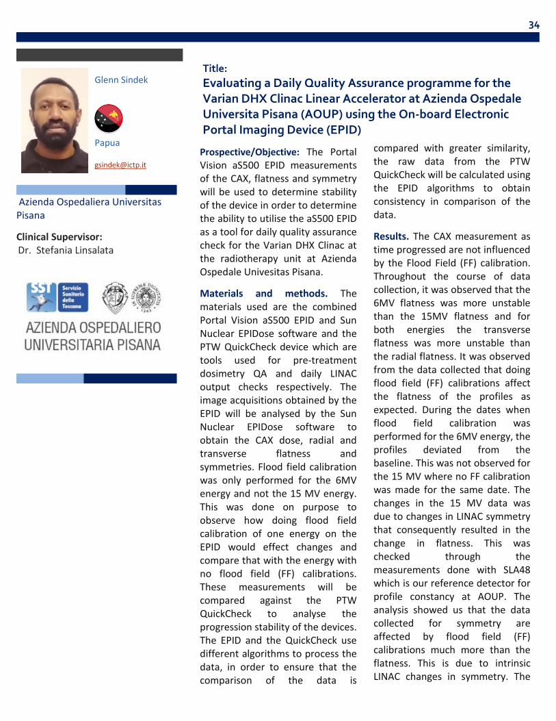
34
Glenn Sindek
Papua
Azienda Ospedaliera Universitas Pisana
Clinical Supervisor: Dr. Stefania Linsalata
Prospective/Objective: The Portal Vision aS500 EPID measurements of the CAX, flatness and symmetry will be used to determine stability of the device in order to determine the ability to utilise the aS500 EPID as a tool for daily quality assurance check for the Varian DHX Clinac at the radiotherapy unit at Azienda Ospedale Univesitas Pisana.
Materials and methods. The materials used are the combined Portal Vision aS500 EPID and Sun Nuclear EPIDose software and the PTW QuickCheck device which are tools used for pre-treatment dosimetry QA and daily LINAC output checks respectively. The image acquisitions obtained by the EPID will be analysed by the Sun Nuclear EPIDose software to obtain the CAX dose, radial and transverse flatness and symmetries. Flood field calibration was only performed for the 6MV energy and not the 15 MV energy. This was done on purpose to observe how doing flood field calibration of one energy on the EPID would effect changes and compare that with the energy with no flood field (FF) calibrations. These measurements will be compared against the PTW QuickCheck to analyse the progression stability of the devices. The EPID and the QuickCheck use different algorithms to process the data, in order to ensure that the comparison of the data is
compared with greater similarity, the raw data from the PTW QuickCheck will be calculated using the EPID algorithms to obtain consistency in comparison of the data.
Results. The CAX measurement as time progressed are not influenced by the Flood Field (FF) calibration. Throughout the course of data collection, it was observed that the 6MV flatness was more unstable than the 15MV flatness and for both energies the transverse flatness was more unstable than the radial flatness. It was observed from the data collected that doing flood field (FF) calibrations affect the flatness of the profiles as expected. During the dates when flood field calibration was performed for the 6MV energy, the profiles deviated from the baseline. This was not observed for the 15 MV where no FF calibration was made for the same date. The changes in the 15 MV data was due to changes in LINAC symmetry that consequently resulted in the change in flatness. This was checked through the measurements done with SLA48 which is our reference detector for profile constancy at AOUP. The analysis showed us that the data collected for symmetry are affected by flood field (FF) calibrations much more than the flatness. This is due to intrinsic LINAC changes in symmetry. The
Title:
Evaluating a Daily Quality Assurance programme for the Varian DHX Clinac Linear Accelerator at Azienda Ospedale Universita Pisana (AOUP) using the On-board Electronic Portal Imaging Device (EPID)
35
changes in the symmetry were in fact observed even in the 15MV measurement where no flood field calibration was performed. This was again confirmed with the measurements done with the SLA48 which we needed once to readjust the LINAC symmetry to the baseline value.
Conclusion. In conclusion we can assess that the combined aS500 Varian EPID and Sun Nuclear EPIDose software can be used for daily dosimetric quality assurance of the AOUP DHX Varian LINAC. This is true in particular for CAX dose measurement. In fact, this system is reliable for relative measurements of the output provided that we fix the baseline values and set a tolerance threshold. For daily measurements of flatness and symmetry we can say that, provided that flood field (FF) calibrations are done one time only and immediately after big maintenance of the LINAC in order to set correctly the baseline values, this system could be a quick and accurate tool to guaranty linac dosimetric stability in a more accurate way than typical daily quality assurance instruments (e.g. QuickCheck) do.
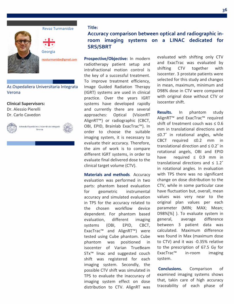
36
Revaz Turmanidze
Georgia
Az.Ospedaliera Universitaria Integrata Verona Clinical Supervisors: Dr. Alessio Pierelli Dr. Carlo Cavedon
Prospective/Objective: In modern radiotherapy patient setup and intrafractional motion control is the key of a successful treatment. To improve treatment efficiency, Image Guided Radiation Therapy (IGRT) systems are used in clinical practice. Over the years IGRT systems have developed rapidly and currently there are several approaches: Optical (VisionRT AlignRT™) or radiographic (CBCT, OBI, EPID, Brainlab ExacTrac™). In order to choose the suitable imaging system, it is necessary to evaluate their accuracy. Therefore, the aim of work is to compare different IGRT systems, in order to evaluate final delivered dose to the clinical target volume (CTV).
Materials and methods. Accuracy evaluation was performed in two parts: phantom based evaluation for geometric instrumental accuracy and simulated evaluation in TPS for the accuracy related to the chosen workflow device dependent. For phantom based evaluation, different imaging systems (OBI, EPID, CBCT, ExacTrac™ and AlignRT™) were tested using Cube phantom. Cube phantom was positioned in isocenter of Varian TrueBeam STx™ linac and suggested couch shift was registered for each imaging system. Secondly, the possible CTV shift was simulated in TPS to evaluate the inaccuracy of imaging system effect on dose distribution to CTV. AlignRT was
evaluated with shifting only CTV and ExacTrac was evaluated by shifting CTV together with isocenter. 3 prostate patients were selected for this study and changes in mean, maximum, minimum and D98% dose in CTV were compared with original dose without CTV or isocenter shift.
Results. In phantom study AlignRT™ and ExacTrac™ required shift of treatment couch was ≤ 0.6 mm in translational directions and ≤0.7˚ in rotational angles, while CBCT required ≤0.2 mm in translational direction and ≤ 0.2˚ in rotational angels, OBI and EPID have required ≤ 0.9 mm in translational directions and ≤ 1.2˚ in rotational angles. In evaluation with TPS there was no significant change on dose distribution to the CTV, while in some particular case have fluctuation but, overall, mean values was very near to the original plan values per each parameter (MIN; MAX; Mean; D98%[%] ). To evaluate system in general, average difference between 3 patient data was calculated. Maximum difference was found in Max (maximum dose to CTV) and it was -0.35% relative to the prescription of 67.5 Gy for ExacTrac™ in-room imaging system.
Conclusions. Comparison of examined imaging systems shows that, takin care of high accuracy traceability of each phase of
Title:
Accuracy comparison between optical and radiographic in-room imaging systems on a LINAC dedicated for SRS/SBRT

37
calibration process for all system, instrumental accuracy evaluation in terms of deviation relative to the LINAC reference systems (axes and isocenter) is excellent for all systems and leftover inaccuracy is lower than 0.6 mm(along axes) and lower than 0.7 ° (around each axes). Simulated virtual evaluation using TPS was proposed as an open question on the impact on the overall accuracy of the devices. this kind of treatment accuracy was evaluated in terms of robustness of delivered dose to the clinical target volume (CTV). such robustness was evaluated in relation to the possible sources of residual error in the positioning and related to inner organ motion or patient surface random or breathing related movement. The TPS simulation with dose recalculation workflow with Vision RT, shows greater robustness of D98% and maximum dose delivered to CTV, but minimum and average CTV doses are more robust with the ExacTrac workflow. Ultimately, the simple investigation does not identify a better workflow/device in an absolute way but indicates the need to carefully evaluate the choice to make according to the clinical requirements considered as priorities in different cases.

38
Larba Landry Zoungrana
Burkina Faso
ASST Papa Giovanni XXIII Clinical Supervisor: Dr. Paolo Colleoni
Prospective/Objective: The goal of radiosurgery is to deliver the prescription dose inside of the target and a steep dose drop-off outside the target volume. This work has been prepared to facilitate beam data acquisition for Eclipse beam configuration in SRS mode, highlight spine metastasis treatment process and from that, perform end-to-end testing using different test objects in an effort to validate the accuracy of TPS dose calculation.
Materials and methods. Firstly, measurements were done in MP3 water phantom using microLion chamber (0.002cm3) for PDD/Profile, semi-flex 0.125cm3 for output factors and MLC transmission/DLG. Second we reported the importance of beam commissioning data review, and end-to-end testing based upon single fraction 16 Gy of VMAT spine metastasis SRS for validation of dose calculation. In the third part, the important of carrying out quality assurance procedures using arccheck and EPID was reported. Gamma analysis was performed with 3% /3mm criteria. Finally optimal DLG was determined using Arccheck.
Results. For 6MV_SRS, the build-up depth was 15mm and the dose at reference depth 66.20% versus 15mm and 66.31% for standard 6MV. The mean value of penumbra was 2.23mm. Measured mean
MLC transmission and dynamic leaf gap were 1.33% and 1.33 mm respectively. The mean ± std of CI, HI, η_(50%), high / low dose target regions difference(%), D2cm, R50%, high dose spillage and delivery time values were 1.40±0.03, 1.20±0.01, 31.74±2.10, 1.20±1.50 / 2.34±1.25, 81.53±9.17, 4.10±0.40, 0.05±0.08, 14.51±3.07 respectively. More than 98±1.35 % and 84.28± 2.51 % of the points passed the gamma criterion for EPID and Arccheck respectively. With optimal DLG (1.8mm), the mean Gamma passing rate was 96.39%, with a range from 94.6% to 99.91% using Arccheck.
Conclusions. Although one can do a substantial part of the basic validation of beam configuration with a single shot beam, it found insufficient to assess the geometric precision of the dose-fall off during arc delivery in SRS mode. It is therefore highly recommended to invest final plan quality in a detector system that can provide 2D and 3D dose information as well. The EPID was found suitable for the validation of VMAT treatment plans in high dose delivery while Arccheck failed. Using Arccheck, accuracy of dose calculation will be obtained with determination of optimal DLG value that depends also on the complexity of the plan, target volume and the prescribed dose.
Title:
A prospective clinical implementation of stereotactic radiosurgery for spine metastasis treatment: from algorithm configuration to Patient Quality Assurance

ICTP- Strada Costiera, 11 I-34151 Trieste Italy
Via Alfonso Valerio, 2, Department of Physics,
University of Trieste, 34127 Trieste, Italy
ICTP & UniTS’s Medical Physics Graduates (IUMPG)

MEDICAL PHYSICS INTERNATIONAL Journal, vol.6, No.1, 2018
143
INFORMATION FOR AUTHORS
PUBLICATION OF DOCTORAL THESIS AND DISSERTATION ABSTRACTS
A special feature of Medical Physics International (on line at www.mpijournal.org ) is the publication of thesis and dissertation abstracts for recent graduates, specifically those receiving doctoral degrees in medical physics or closely related fields in 2010 or later. This is an opportunity for recent graduates to inform the global medical physics community about their research and special interests.
Abstracts should be submitted by the author along with
a letter/message requesting and giving permission for publication, stating the field of study, the degree that was received, and the date of graduation. The abstracts must
be in English and no longer than 2 pages (using the MPI manuscript template) and can include color images and illustrations. The abstract document should contain the thesis title, author’s name, and the institution granting the degree.
Complete information on manuscript preparation is
available in the INSTRUCTIONS FOR AUTHORS section of the online journal: www.mpijournal.org.
For publication in the next edition abstracts must be
submitted not later than Април 1, 2019.
MEDICAL PHYSICS INTERNATIONAL Journal, vol.6, No.1, 2018
144
INSTRUCTIONS FOR AUTHORS
The goal of the new IOMP Journal Medical Physics International (http://mpijournal.org) is to publish manuscripts that will enhance medical physics education and professional development on a global basis. There is a special emphasis on general review articles, reports on specific educational methods, programs, and resources. In general, this will be limited to resources that are available at no cost to medical physicists and related professionals in all countries of the world. Information on commercial educational products and services can be published as paid advertisements. Research reports are not published unless the subject is educational methodology or activities relating to professional development. High-quality review articles that are comprehensive and describe significant developments in medical physics and related technology are encouraged. These will become part of a series providing a record of the history and heritage of the medical physics profession.
A special feature of the IOMP MPI Journal will be the publication of thesis and dissertation abstracts for will be the publication of thesis and dissertation abstracts for recent doctoral graduates, specifically those receiving their doctoral degrees in medical physics (or closely related fields) in 2010 or later.
MANUSCRIPT STYLE
Manuscripts shall be in English and submitted in WORD. Either American or British spelling can be used but it must be the same throughout the manuscript. Authors for whom English is not their first language are encouraged to have their manuscripts edited and checked for appropriate grammar and spelling. Manuscripts can be up to 10 journal pages (approximately 8000 words reduced by the space occupied by tables and illustrations) and should include an unstructured abstract of no more than 100 words.
The style should follow the template that can be downloaded from the website at:
http://mpijournal.org/authors_submitapaper.aspx
ILLUSTRATIONS SPECIAL REQUIREMENTS
Illustrations can be inserted into the manuscript for the review process but must be submitted as individual files when a manuscript is accepted for publication.
The use of high-quality color visuals is encouraged. Any published visuals will be available to readers to use in their educational activities without additional approvals.
REFERENCE WEBSITES
Websites that relate to the manuscript topic and are sources for additional supporting information should be included and linked from within the article or as references.
EDITORIAL POLICIES, PERMISSIONS AND
APPROVALS
AUTHORSHIP
Only persons who have made substantial contributions to the manuscript or the work described in the manuscript shall be listed as authors. All persons who have contributed to the preparation of the manuscript or the work through technical assistance, writing assistance, financial support shall be listed in an acknowledgements section.
CONFLICT OF INTEREST
When they submit a manuscript, whether an article or a letter, authors are responsible for recognizing and disclosing financial and other conflicts of interest that might bias their work. They should acknowledge in the manuscript all financial support for the work and other financial or personal connections to the work.
All submitted manuscripts must be supported by a document (form provided by MPI) that:
• Is signed by all co-authors verifying that they have participated in the project and approve the manuscript as submitted.
• Stating where the manuscript, or a substantially similar manuscript has been presented, published, or is being submitted for publication. Note: presentation of a paper at a
MEDICAL PHYSICS INTERNATIONAL Journal, vol.8, No.2, 2020
145
conference or meeting does not prevent it from being published in MPI and where it was presented can be indicated in the published manuscript.
• Permission to publish any copyrighted material, or material created by other than the co-authors, has been obtained.
• Permission is granted to MPI to copyright, or use with permission copyrighted materials, the manuscripts to be published.
• Permission is granted for the free use of any published materials for non-commercial educational purposes.
SUBMISSION OF MANUSCRIPTS
Manuscripts to be considered for publication should be submitted as a WORD document to: Slavik Tabakov, Co-editor: [email protected]
MANUSCRIPT PROPOSALS
Authors considering the development of a manuscript for a Review Article can first submit a brief proposal to the editors. This should include the title, list of authors, an abstract, and other supporting information that is appropriate. After review of the proposal the editors will consider issuing an invitation for a manuscript. When the manuscript is received it will go through the usual peer-review process.

MEDICAL PHYSICS INTERNATIONAL Journal, vol.8, No.2, 2020
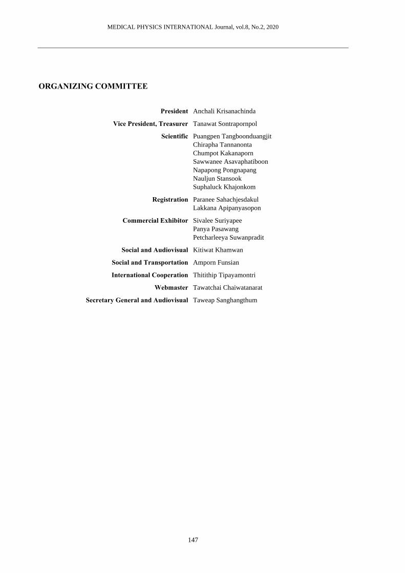
MEDICAL PHYSICS INTERNATIONAL Journal, vol.8, No.2, 2020
147
ORGANIZING COMMITTEE
President Anchali Krisanachinda
Vice President, Treasurer Tanawat Sontrapornpol
Scientific Puangpen Tangboonduangjit
Chirapha Tannanonta
Chumpot Kakanaporn
Sawwanee Asavaphatiboon
Napapong Pongnapang
Nauljun Stansook
Suphaluck Khajonkom
Registration Paranee Sahachjesdakul
Lakkana Apipanyasopon
Commercial Exhibitor Sivalee Suriyapee
Panya Pasawang
Petcharleeya Suwanpradit
Social and Audiovisual Kitiwat Khamwan
Social and Transportation Amporn Funsian
International Cooperation Thitithip Tipayamontri
Webmaster Tawatchai Chaiwatanarat
Secretary General and Audiovisual Taweap Sanghangthum
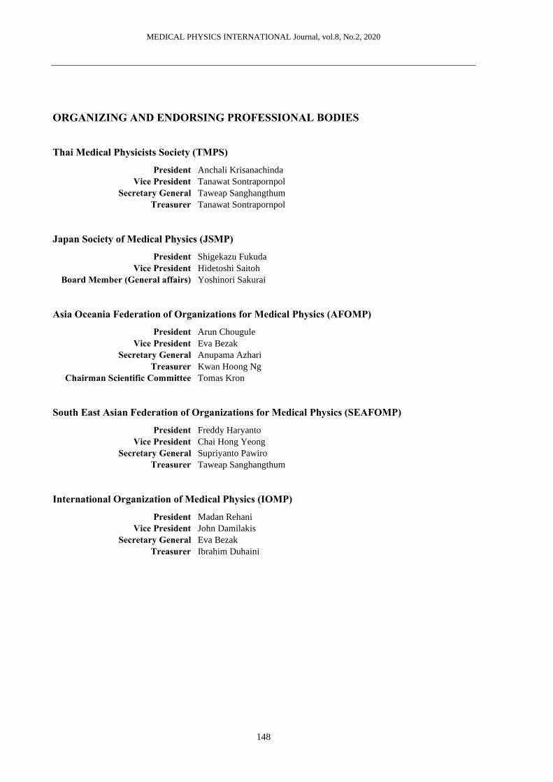
MEDICAL PHYSICS INTERNATIONAL Journal, vol.8, No.2, 2020
148
ORGANIZING AND ENDORSING PROFESSIONAL BODIES
Thai Medical Physicists Society (TMPS)
President Anchali Krisanachinda Vice President Tanawat Sontrapornpol Secretary General Taweap Sanghangthum Treasurer Tanawat Sontrapornpol
Japan Society of Medical Physics (JSMP)
President Shigekazu Fukuda Vice President Hidetoshi Saitoh Board Member (General affairs) Yoshinori Sakurai
Asia Oceania Federation of Organizations for Medical Physics (AFOMP)
President Arun Chougule Vice President Eva Bezak Secretary General Anupama Azhari Treasurer Kwan Hoong Ng Chairman Scientific Committee Tomas Kron
South East Asian Federation of Organizations for Medical Physics (SEAFOMP)
President Freddy Haryanto Vice President Chai Hong Yeong Secretary General Supriyanto Pawiro Treasurer Taweap Sanghangthum
International Organization of Medical Physics (IOMP)
President Madan Rehani Vice President John Damilakis Secretary General Eva Bezak Treasurer Ibrahim Duhaini
MEDICAL PHYSICS INTERNATIONAL Journal, vol.8, No.2, 2020
149
GREETINGS FROM PRESIDENT AFOMP
I am delighted to note that the 20th Asia-Oceania Congress on Medical Physics (AOCMP),
the 18th South-East Asia Congress of Medical Physics (SEACOMP), 120th Scientific
Meeting of Japan Society of Medical Physics (JSMP) and the 12th Annual Meeting of Thai
Medical Physicist Society (TMPS) are jointly organized at Phuket, Thailand during 3rd to 5th
December 2020. The theme of the conference is ‘Medical Physics- Achievements,
Challenges and Horizons’. Many congratulations to the organizing team especially Prof
Anchali Krisanachinda for making it happen despite the unprecedented pandemic situation worldwide.
This congress is unique as it’s the 20th anniversary of AFOMP and four very active medical physics associations
of Asia-Oceania are joined together in organizing this mega scientific event. It is heartening to note that the 1st
AOCMP was held in Thailand and afterwards 9th, 12th and 16th AOCMP’s were also hosted by Thailand. It’s the
5th time AOCMP is being held in Thailand marking the 20th Anniversary of AFOMP celebrating it in a big way
associating with SEACOMP, JSMP and TMPS which shows the acumen of Prof Anchali and team and the great
hospitality of Thailand.
AFOMP has started many activities to encourage professional and academic excellence. Prof Kiyonari Inamura
Memorial AFOMP Oration awarded for 2020 is awarded to Prof K Y Cheung of Hong Kong, AFOMP lifetime
achievement award 2020 to Prof Anchali Krisanachinda, Thailand and Prof Donald McLean, Australia and to
celebrate the 20th anniversary, AFOMP is honouring 21 medical physicists with AFOMP outstanding medical
physicist award. In addition to encourage publication of scientific papers in AFOMP journals, best paper
published award is also started by AFOMP.
Education and training is the key to accelerated professional achievements. As healthcare professionals,
education cannot wait or stop for the pandemic. Despite all the hurdles, the organizers have done tremendous
efforts to materialize this conference a reality though the dates were postponed and this shows the resolute
determination of the organizers. I would like to thank JSMP also for being a partner. The organizers have
arranged virtual as well as face to face opportunity this time so that everyone may get benefitted.
Let me also urge the participants of this conference not only be confined with the challenges at their own field of
specialization but take it as a moral responsibility to be proactive and enthusiastic in learning about the
contemporary cutting edge tools in all aspects of physics in medicine. This pandemic was an eye opener for all
of us to realize the necessity of being proactive to be successful in the changing world.
Wishing you all, a pleasant and fruitful conference and continued success in all your professional endeavours. I
am sure this will be an academic feast satisfying the diverse interests of all participants giving novel ideas and
new insights. Wish you all a very fruitful attending of the conference and pleasant stay in Phuket.
Prof Arun Chougule President- AFOMP
Chair- ETC and accreditation board, IOMP
Senior Professor and Head, Dept of Radiological Physics
SMS Medical College and Hospitals
Jaipur, INDIA

MEDICAL PHYSICS INTERNATIONAL Journal, vol.8, No.2, 2020
150
WELCOME MESSAGE FROM PRESIDENT OF SEAFOMP
Dear colleagues and friends,
On Behalf of the President of South East Asian Federation of Organizations for Medical
Physics (SEAFOMP), allow me to warmly thank the organizers of this Joint Conference
among the AFOMP, SEAFOMP, JSMP, and TMPS for giving me the privilege of
welcoming and addressing you all. For me, it is an honor and a pleasure.
I would also like to thank Prof. Anchali and her team for bringing us together on the first
hybrid Conference for our society. Due to Covid-19 Pandemic, we could not meet all to gather in wonderful and
beautiful Phuket Island, Thailand, to discuss the achievements, challenges, and horizons in our medical physics
word.
The Conference, composed of 3 parallel sessions, tackles important topics on medical physics. There are also
mini-symposium and workshops. I am sure that each of you will identify subjects of his/her interest and benefit
from many fruitful and enriching discussions.
I am incredibly happy to be virtually present in this unique event today and to exchange views and share
experiences with colleagues and friends. For the participants who come to Phuket Island, please to have time
also for exploring the island.
I congratulate you on your commitment and active participation and wish you all the success.
Thank you,
Freddy Haryanto
MEDICAL PHYSICS INTERNATIONAL Journal, vol.8, No.2, 2020
151
WELCOME MESSAGE FROM PRESIDENT OF JSMP
It is a great pleasure for me to hold the 120th Scientific Meeting of Japan Society of
Medical Physics jointly with 20th Asia-Oceania Congress on Medical Physics, 18th
South-East Asia Congress of Medical Physics, and 12th Annual Meeting of Thai Medical
Physicist Society.
First of all, I would like to thank Dr. Anchali Krisanachinda, the mother of medical
physics of Thailand, and also Dr. Arun Chougule, President of AFOMP. Despite the
uncertain and difficult situation of the impact of COVID-19, they have committed very hard to the holding of
this conference, which resulted in the successful opening today.
The recent environmental conditions surrounding radiotherapy, radiodiagnosis, and nuclear medicine have
changed significantly due to the popularization of advanced radiotherapy such as IGRT and particle therapy, the
introduction of AI, the fusion of diagnosis and therapy and so on. The education and training for these advanced
technique in medical physics are becoming increasingly important. However, the environment for education and
training for them is still insufficient in Asian region. So JSMP wants to contribute to improving this
environmental situation in the Asian region with our resources and experiences through the international
collaboration.
Although the COVID-19 pandemic is a disaster, if we think positively, due to this situation, we are able to hold
the JSMP scientific meeting jointly with the AOCMP, SEACOMP, and TMPS scientific meeting. This JSMP
scientific meeting is held overseas for the first time except Japan-Korea Joint Meeting of Medical Physics, which
is held once every three years. So, I’d like to regard this opportunity as an important steppingstone to make an
international contribution to the development of medical physics in the Asian region.
I hope this conference will be a successful and fruitful to all participants. Thank you.
Shigekazu Fukuda, Ph. D President of the 120th Scientific Meeting of Japan Society of Medical Physics,
President of JSMP

MEDICAL PHYSICS INTERNATIONAL Journal, vol.8, No.2, 2020
152
TMPS PRESIDENT MESSAGE
In the name of Thai Medical Physicist Society, I am pleased to welcome you to the 20th
Asia Oceania Congress on Medical Physics, the 18th South-East Asian Congress of
Medical Physics, the 120th Scientific Meeting of Japan Society of Medical Physics and
the 12th Annual Meeting of Thai Medical Physicist Society. This Congress had been
endorsed by the International Organization on Medical Physics.
This is the fifth time Thailand has hosted the Congress for AFOMP:
First: 2001 Royal Jubilee 50th year Coronation Medical Society Building Petchaburi Road, Bangkok
Second: 2009 Pang Suan Kaew Convention Hall, Chiang Mai
Third: 2012 Kum Poo Come Chiang Mai
Fourth: 2016 ICMP at Shangari La Hotel Bangkok
Fifth: 2020 Duangjitt Resort and Spa Phuket
This is the first time to host the Congress for Japan Society of Medical Physics especially on the 120th. It is
genuinely our great honor to host on hybrid at virtual and in person. Hopefully, we can co-host the annual
Scientific Meeting for JSMP again in the future.
We plan to celebrate the 20th Anniversary for AFOMP at the Gala Dinner on December 3rd, 2020, Thai and
seafood are arranging on Phuket style.
Scientific program had been arranged as following: John Cameron Memorial Lecture, Professor Kiyonari
Inamura Oration Lecture, JSMP Honor Lecture, Proton Therapy Mini Symposium, JSRT Mini Symposium, 49
Invited Lectures, 6 Mini Symposia, 121 Oral and 54 e-poster presentations. The Convention Hall consists of 2
Grand Lecture Rooms for Radiotherapy and one Commercial Exhibition room. Outside the Convention Hall
there are 3 rooms for Medical Imaging - Diagnostic Radiology and Nuclear Medicine for Oral and e Poster
presentations. Beverages and lunch are arranged during the Congress.
Certification for Clinically Qualified Medical Physicists is arranged at the Gala Dinner, including TMPS
Outstanding Medical Physicists, AFOMP and TMPS Awards.
Please enjoy your time at the Congress in Phuket for In Person and Virtual participations.
Anchali Krisanachinda, Ph.D. Chair, 20th AOCMP, 18th SEACOMP, 120th JSMP, 12th TMPS
MEDICAL PHYSICS INTERNATIONAL Journal, vol.8, No.2, 2020
153
CONTENTS
Organizing Committee ......................................................................................................................................... 147
Organizing and Endorsing Professional Bodies................................................................................................... 148
Greetings from President AFOMP ...................................................................................................................... 149
Welcome Message from President of SEAFOMP ............................................................................................... 150
Welcome Message From President of JSMP ....................................................................................................... 151
TMPS PRESIDENT MESSAGE ......................................................................................................................... 152
Contents ............................................................................................................................................................... 153
Invited Presentations
JOHN CAMERON MEMORIAL LECTURE: New Additions to the Scientific Dictionary and Encyclopaedia
on Medical Physics ..................................................................................................................................... 161
Professor Kiyonari Inamura Memorial AFOMP Oration 2020 ....................................................................... 162
JSMP INVITED LECTURE: Evaluation of ionising radiation induced DNA damage on a cell and prediction
of biological response by integrated track structure Monte Carlo simulations using Geant4-DNA ........... 163
Impact of radiosensitivity and gender on paediatric cranial tumours following proton and photon intensity
modulated radiation therapy – A radiobiological response modelling study .............................................. 165
Comparisons of DNA damage in HPV negative and positive head and neck cancer cell lines following
fractionated irradiation. ............................................................................................................................... 167
MS 1: Proton Therapy in Hong Kong ............................................................................................................. 168
MS 2: JSRT Clinical application of the latest radiation technology – CT ...................................................... 169
MS 2: JSRT Clinical application of the latest radiation technology in Magnetic Resonance Imaging ........... 170
MS 2: JSRT Clinical application of the latest radiation technology in Nuclear Medicine .............................. 171
IAEA Activities in Support of Recognition of the Medical Physics Profession ...................................... 172
South Asia Centre for Medical Physics and Cancer Research (SCMPCR): A Centre of Excellence to Fight
against Cancer in South Asia Region .......................................................................................................... 173
Radiation incidents and accidents in radiotherapy .......................................................................................... 174
National Diagnostic Reference Level (NDRLs) of Procedures in Cardiac Catheterization Laboratory in
Thailand: A Multi Center Survey ................................................................................................................ 175
Outline of updated national diagnostic reference levels in Japan (Japan DRLs 2020) ................................... 176
National DRLs in Thailand ............................................................................................................................. 177
Dose monitoring and audit, tools for patient dose optimization ............................................................... 178
Clinical Aspects for Theranostics in the Era of Precision Medicine ............................................................... 179
Interesting Radiation Protection Issues in Theranostic Radiopharmaceutical; Workers, Caregivers, Members
of the Public and Waste Management ......................................................................................................... 180
How to make a research plan for young investigators .................................................................................... 181
e-Learning in Medical Physics: Building Educational Modules with Moodle VLE ....................................... 182
Role of Radiation and Caution in the Management of Covid-19 .................................................................... 183
AFOMP Medical Physics Education and Clinical Training ........................................................................... 184
Postgraduate medical physics programmes in Asia Pacific countries - A survey study ................................. 185
Imaging-based Biomarkers ............................................................................................................................. 186
Teaching Linear Accelerator Physics using a Web Based Simulator ............................................................. 187
Automating Plan Checking Tasks by Scripting .............................................................................................. 188
Principle and Application of MR-Linac .......................................................................................................... 189
Extreme hypofractionated radiotherapy – Radiobiology and radiobiological models .................................... 190
High patient radiation doses from recurrent CT exams .................................................................................. 191
Patients who receive high radiation doses in a single day .............................................................................. 192
Challenges in CT with photon counting detectors .......................................................................................... 193
Physicists and Clinical Trials: Support and Evidence for New Technology and Techniques ......................... 194
Novel technologies for advanced radiomics for clinical practices .................................................................. 195
Consideration of stochastic risks in interventional procedures ....................................................................... 196
MS 5: Eye Lens Dose Measurements of Medical Staffs in Thailand .............................................................. 197
MEDICAL PHYSICS INTERNATIONAL Journal, vol.8, No.2, 2020
154
Rapid estimation of muscle transverse relaxation time (T2) based on ultrafast magnetic resonance imaging at
3.0 Tesla ...................................................................................................................................................... 198
Optimization of Cone Beam Breast CT – Radiation Dose and Image Quality ........................................ 199
The Establishment of UNSCEAR Report in Thailand .................................................................................... 200
MS 6: Facility design of a PETCT .................................................................................................................. 201
MS 6: Facility design of a cyclotron ............................................................................................................... 202
MS 6: Radiation Emergency Preparedness in Nuclear Medicine .............................................................. 203
Theranostics in Nuclear Medicine .................................................................................................................. 204
Preparation and Application of Ga-68 in Nuclear Medicine ........................................................................... 205
Radiomics in Radiation Oncology .................................................................................................................. 206
Patient specific quality assurance by log file-based software: a multi-institutional study in Thailand . 207
Oral Presentations
Radiotherapy: Dosimetry ............................................................................................................................. 208
Parametric impact study of interplay effects in 6 MV flattening filter free (FFF) of photon beams .......... 209
Development and dosimetric verification of 3D customized bolus in radiotherapy ................................... 210
Tuning effective spot size parameter in Acuros XB algorithm for Edge™ radiosurgery machine .... 211
Investigation of field output correction factors in small elongated fields for 6 MV photon beam ............. 212
Comparison of field output factors of different detectors in small field for 6 MV flattening filter and
flattening filter free photon beams .......................................................................................................... 213
Evaluation of detectors on relative dosimetric measurement for CyberKnife M6 System ......................... 214
Surface and Build Up Doses Comparison between Analytical Anisotropic Algorithm and Acuros XB for
Various Treatment Parameters ................................................................................................................ 215
Determination of small field output factors and specific correction factors for various diodes and
microDiamond detector for CyberKnife M6 system equipped with fixed collimator ............................. 216
Efficiency of EPID dosimetry based software compared with ion chamber ............................................... 217
An update on local blood irradiation technique using medical LINAC: Blood Irradiation Kit .................. 218
Radiotherapy: Monte Carlo Simulation ...................................................................................................... 219
Determination of fetal dose and health effect to the fetus from breast cancer radiotherapy during pregnancy
................................................................................................................................................................. 220
Calculation of field output correction factors for radiophotoluminescence glass dosimeter in 6 MV WFF
and FFF small photon beams using Monte Carlo simulation .................................................................. 221
Investigation of dose perturbations around brachytherapy seeds in high-energy photon beams ................ 222
Monte Carlo Simulation of semiflex chamber in Megavoltage Photon Beam ............................................ 223
Estimation of Linear and Mass Attenuation Coefficients of Soy-lignin Bonded Rhizophora spp.
Particleboard as a Potential Phantom Material for Low- and High-energy Studies using Monte Carlo
Simulation ............................................................................................................................................... 224
Comparison simulation treatment between Gamma Knife and Linac Stereoradiosugery on small target
volume by using GEANT 4 Monte Carlo Simulation. ............................................................................ 225
Monte Carlo calculation of beam quality correction factors in proton beams using PTSIM/GEANT4 ..... 226
VDOSE: A tools to analyze dose distribution from Monte Carlo dose Calculation ................................... 227
Radiotherapy: Radiobiology ......................................................................................................................... 228
Low-dose radiation enhance cytotoxicity of chemotherapeutic agent (pirarubicin) in K562 and K562/adr
leukemic cancer cells .............................................................................................................................. 229
Radioprotective effect of interruptin derived from Cyclosorus terminans could protect cell damage of
irradiated normal cells from radiotherapy ............................................................................................... 230
Temperature variation of stimulated diabetic blood and its effects on Evaluated specific absorption rate
(SAR) when used a 532 nm wavelength laser for therapeutics. .............................................................. 231
The Effect of Different Sizes of Bismuth Oxide Nanorods on Radiosensitization Enhancement Ratio ..... 232
The Effect of Different Sizes of Bismuth Oxide Nanorods on Cancer Cells .............................................. 233
Influence of PEG-coated Bismuth Oxide Nanoparticles on ROS Generation for Electron Beam
Radiotherapy ........................................................................................................................................... 234
MEDICAL PHYSICS INTERNATIONAL Journal, vol.8, No.2, 2020
155
Radiotherapy: Treatment planning ............................................................................................................. 235
Dosimetric Validation of Monaco Treatment Planning System ................................................................. 236
Evaluation of dose calculation algorithm accuracy in Eclipse treatment planning system for jaw-defined
and MLC-defined small fields ................................................................................................................. 237
The quality improvement of dose escalation plan of prostate cancer using an integration of biological
parameters for optimization and evaluation ............................................................................................ 238
Treatment planning evaluation of 3D-printed Anthropomorphic Head Phantom ....................................... 239
Comparison of different treatment planning techniques for left-sided breast cancer: A dosimetric study . 240
Investigation of the optimal cost function for pelvic CT-MR deformable image registration .................... 241
Dosimetric comparison between 3D and IMRT treatment techniques for whole breast radiotherapy ........ 242
Radiotherapy: Big data, Deep learning, AI and Modeling ....................................................................... 243
Development of automated prostate VMAT treatment planning system using deep learning-based
predictive dose distribution ..................................................................................................................... 244
Automated fast data collection and organization for medical artificial intelligence ................................... 245
A feasibility study of kVCBCT-based radiomics for radiological complication prediction ....................... 246
Evaluation of machine learning-based prediction model for radiation pneumonitis in NSCLC patients ... 247
Impact of image type and deep learning architecture in deep learning radiomics on the accuracy of lung
cancer prediction ..................................................................................................................................... 248
Prognostic analysis of CT-based radiomics focusing on a subgroup of NSCLC patients........................... 249
Radiotherapy: Patient-specific QA .............................................................................................................. 250
Preliminary evaluation of the performance of an EPID-based in vivo dosimetry for advanced radiotherapy
treatment verification .............................................................................................................................. 251
Patient-specific quality assurance for IMRT delivery: A multi-centre study ............................................. 252
Pilot study of remote clinical dosimetry auditing for IMRT using virtual EPID standard phantom audit
(VESPA) in Thailand .............................................................................................................................. 253
Evaluation of Mobius and Portal dosimetry Quality Assurance tools for volumetric modulated arc therapy
plans ........................................................................................................................................................ 254
Effect of plan complexity to gantry angle uncertainty for VMAT delivery ................................................ 255
Development of novel x-ray-opaque-marker system for improvement and quantification of phantom
positioning accuracy in patient-specific quality assurance ...................................................................... 256
Radiotherapy: Particle beam therapy ......................................................................................................... 257
Dosimetric validation of the commercial dose calculation algorithm for carbon ion therapy in a
heterogeneous phantom ........................................................................................................................... 258
Simulation of the efficiency of rescanning with respiratory gating technique in pencil beam scanning
proton therapy for lung cancer ................................................................................................................ 259
Dosimetric comparison of normal liver sparing in hepatocellular carcinoma between intensity modulated
proton therapy and volumetric modulated arc therapy ............................................................................ 260
Development of Ultra-fast Irradiation for Proton Therapy ......................................................................... 261
Radiotherapy: IGRT ..................................................................................................................................... 262
Tolerance level determination for automated EPID-based deep inspiration breath-hold (DIBH) instability
evaluation in breast cancer patients ......................................................................................................... 263
A simulation study of fractional image guidance protocol contributing to the dosimetric accuracy of
patients treated on Halcyon system ......................................................................................................... 264
Localization accuracy of off-isocenter multi-target brain stereotactic radiosurgery using SyncTraX FX4 265
Evaluation of tumor delineation accuracy in denoised contrast enhancement four-dimension CT using
deformable image registration method .................................................................................................... 266
Radiotherapy: Radiation Safety / DRL ....................................................................................................... 267
Acceptance Test and Commissioning of Tomotherapy Radixact X5 series in LBCH ................................ 268
The preliminary survey report of the compact proton therapy unit............................................................. 269
Mitigation of Error Propagation in Tele-cobalt Treatment: An Institutional Assessment .......................... 270
Evaluation of radionuclides and activation detected by a 15 MeV medical linear accelerator ................... 271
MEDICAL PHYSICS INTERNATIONAL Journal, vol.8, No.2, 2020
156
Radiotherapy: Others ................................................................................................................................... 272
Dosimetric comparison of manual and library applicator reconstruction in MRI image -based for
gynecological brachytherapy ................................................................................................................ 273
Verification of Treatment Time in Interstitial Brachytherapy using Paris System ..................................... 274
Dosimetric evaluation of the uncorrectable rotational setup error in total body irradiation using helical
tomotherapy system ................................................................................................................................ 275
Investigating the effect of material absorber the source of Gamma Knife Perfexion TM based on Monte
Carlo simulations ................................................................................................................................... 276
Development of an In-house MATLAB code for dynamic multileaf collimator quality assurance based on
EPID ........................................................................................................................................................ 277
Dosimetric evaluation of photon beam-matching for two similar linear accelerators ................................ 278
Assessment of Radiation Protection and Radiation Safety Awareness among Radiation Professionals in a
Tertiary Care Teaching Hospital ............................................................................................................. 279
Design of universal 3D-printed oral stent for head and neck radiotherapy ................................................. 280
Fabrication and validation of cubic phantom embed with an accelerometer sensor for verifying six-degrees-
of-freedom couch .................................................................................................................................... 281
Daily machine-specific performance monitoring through clinical treatment delivery using Statistical
Process Control and MobiusFX ............................................................................................................ 282
Establishment of MOH Medical Physics Research Task Force .................................................................. 283
CT Density Profile and Physicochemical Study of Soy-Lignin Bonded Rhizophora spp. Particleboard as
Phantom Material in Medical Physics Application ................................................................................. 284
Dignostic: Imaging ........................................................................................................................................ 285
Correlation of Ultrasound Attenuation Imaging versus MRI Proton Density Fat Fraction in Non -
alcoholic Fatty Liver ............................................................................................................................. 286
Sonographic predictors for developing cholangiocarcinoma: A cohort study from an endemic area. ....... 287
Value of Superb Microvascular Imaging (SMI) in characterize of nodular type focal fat sparing lesion and
true hepatic nodules in the background of fatty liver .............................................................................. 288
Predicting Treatment Response in Nasopharyngeal Cancer Using Radiomics: A Preliminary Study ........ 289
Performance of quantitative lesion measurement in hepatic dual-energy computed tomography (DECT):
Phantom study ......................................................................................................................................... 290
Automatic QC Analysis Program in Digital Radiography System ............................................................. 291
Correlation between renal histological fibrosis and renal cortical thickness using shear wave elastography
in patients with kidney disease ................................................................................................................ 292
Automatic stratification of prostate cancer patients into low- and high-grade groups based on
multiparametric MR image feature analysis ....................................................................................... 293
Image analysis of pancreatic cancer tissues using Haralick Features ......................................................... 294
Digital Breast Tomosynthesis (DBT): Dose and image quality assessment of man-made breast
phantom .................................................................................................................................................. 295
Grey Matter Segmentation of T1-Weighted MR Image in Ischemic Stroke Patients using k-Means
Clustering ................................................................................................................................................ 296
Diagnostic: Big data, Deep learning, AI and Modeling ............................................................................. 297
The effective methods for liver cyst labelling in ultrasound images by using R-CNN ............................... 298
Quantitative Assessment of Breast Density Using Pixel Intensity Threshold ............................................ 299
Overall Survival Prediction of Nasopharyngeal Carcinoma using Radiomics............................................ 300
Machine Learning model for Alzheimer’s disease prediction and classification using T1-weighted MR
Brain imaging based on SVM algorithm ................................................................................................. 301
The Breast Microcalcification Detection in Mammogram Image by Using the Deep Convolutional
Neural Network ..................................................................................................................................... 302
Prediction of Tumor Growth Trajectories During Molecularly Targeted Therapy Based on Mathematical
Models ..................................................................................................................................................... 303
Quantitative evaluation of deep convolutional neural network based denoising for ultra-low-dose CT .... 304
MEDICAL PHYSICS INTERNATIONAL Journal, vol.8, No.2, 2020
157
A Preliminary Study of Various Augmentation Techniques on Deep-learning Based Classification of Lung
Squamous Cell Carcinoma using Chest X-Ray ....................................................................................... 305
A Computational Study on Deep-learning Based Classification of Lung Adenocarcinoma using Chest X-
Ray .......................................................................................................................................................... 306
Dignostic: Dosimetry ..................................................................................................................................... 307
Patient-specific organ dose calculated using dose tracking software based on Monte Carlo simulation in
pediatric abdominal CT ........................................................................................................................... 308
Size-Specific Dose Estimates (SSDE) for Head CT in Pediatric Patients .................................................. 309
Preliminary investigation on performance of photodiode sensor as a dosimeter ........................................ 310
Evaluation of scattering spectra from an examinee during in CT scans ..................................................... 311
Diagnostic: Radiation Safety / DRL ............................................................................................................. 312
The Study of Local Diagnostic Reference Levels at Unit of Vascular and Interventional Radiology, King
Chulalongkorn Memorial Hospital .......................................................................................................... 313
Local Diagnostic Reference Levels (DRLs) of Interventional Radiology Procedures in Thailand: A Single
Center ...................................................................................................................................................... 314
Estimation the effective and skin doses for pediatric and adult patients undergoing cardiac interventional
examination using five PMMA phantoms and TLD/ionization chamber technique ............................... 315
Facility reference level for common x-ray procedures: a preliminary study .............................................. 316
Evaluation of gonad shielding in digital radiography: Based on clinically adjustable controls. ................. 317
Diagnostic: Others ......................................................................................................................................... 318
Evaluation of student skill competency on accuracy of mid-trimester fetal sonography scan following the
newly-established Master degree in medical sonography ....................................................................... 319
The teaching and learning of postgraduate medical physics using Internet-based e-learning during the
COVID-19 pandemic .............................................................................................................................. 320
Analysis of NTCP Based Radiobiological Models: A Systematic Review of Literatures .......................... 321
Evaluation of Visually Induced Motion Sickness During Virtual Reality based Head-mounted
Display Viewing using Electroencephalography ................................................................................ 322
Nuclear Medicine: Imaging .......................................................................................................................... 323
The comparison between planar gated blood pool imaging and gated blood pool SPECT to evaluate
ventricular function in breast cancer patients receiving cardiotoxic cancer treatments .......................... 324
Diagnostic performance of dual-tracer subtraction, single-tracer dual-phase 99mTc-MIBI parathyroid
scintigraphy, and 99mTc-MIBI SPECT/CT for preoperative localization in patients with
hyperparathyroidism. .............................................................................................................................. 325
Clearance Patterns of Tc-99m ECD Using Serial SPECT Imaging to Define Seizure Onset Zone ... 326
Monte Carlo simulation for high resolution imaging of Rb-82 with bremsstrahlung X-ray camera for
small animals ......................................................................................................................................... 327
Nuclear Medicine: Radiation Safety / DRL ................................................................................................. 328
Size specific dose estimate (SSDE) for estimating CT doses in SPECT/CT and PET/CT examinations ... 329
Flexible, Lightweight and Lead-free Radiation Shielding for Nuclear Medicine from Natural Rubber with
Barium Sulfate Composite ...................................................................................................................... 330
Investigation of position and angle effects of nanoDot OSL dosimeter for the eye lens dose measurement
................................................................................................................................................................. 331
Determination for current status of environmental radioactivity in Fukushima, using NaI (Tl) scintillation
survey meter ............................................................................................................................................ 332
Nuclear Medicine: Others ............................................................................................................................. 333
The labeling of 99mTc-PSMA-HBED-CC for prostate cancer imaging ....................................................... 334
Gamma Scintigraphy for Hepatic Radioembolization: Comparison between Different Radionuclides using
GATE Monte Carlo Simulation .............................................................................................................. 335
Positron Range Correction in Positron Emission Tomography .................................................................. 336

MEDICAL PHYSICS INTERNATIONAL Journal, vol.8, No.2, 2020
158
Evaluation of patient doses from 177Lu-PSMA in metastases prostate cancer treatment at King
Chulalongkorn Memorial Hospital .......................................................................................................... 337
Development of Flexible Radiotherapeutic Bandage Containing Samarium-153 for the Treatment of Skin
Cancer ..................................................................................................................................................... 338
E-Poster Presentations
Radiotherapy ................................................................................................................................................. 339
Real-time in vivo dose verification using Metal-Oxide Semiconductor Field Effect Transistor (MOSFET)
in HDR brachytherapy ............................................................................................................................ 340
Development of external dosimetry audit system for image-guided brachytherapy in Asian Countries .... 341
Comparison of ICRU Point Doses and Volumetric Doses to the Rectum High Dose Rate Cobalt-60
Brachytherapy of Cervical Cancer .......................................................................................................... 342
Correlation analysis of CT-based rectal planning dosimetric parameters with in vivo dosimetry of MOSkin
and PTW 9112 detectors in Co-60 source HDR intracavitary cervix brachytherapy .............................. 343
A development of a primary standard of absorbed dose to water for a small field in high-energy photon
beam ........................................................................................................................................................ 344
Construction of regional network and remote support system for linac output dose difference facilities. . 345
Investigation for measurement of photon depth dose using UVC camera .................................................. 346
Impact of the Frequency of Verification with In-room CT on Setup Errors in Carbon Ion Radiotherapy for
Prostate Cancer........................................................................................................................................ 347
Determination of the beam quality correction factors for carbon-ion beams by Monte Carlo
simulations and measurements ............................................................................................................. 348
Effects of parallax error and light refraction on estimation for dose distribution of proton beams using
luminescence in water ............................................................................................................................. 349
Time dependence of intra-fractional motion for spine stereotactic body radiotherapy ............................... 350
Calculation of an automatic irradiation path for dynamic-wave-arc irradiation ......................................... 351
Dosimetric effect of the baseline shift in the phase and amplitude gated lung SBRT ................................ 352
The Proposal of the Simple Alternative Planning Method for Lung/Esophageal IMRT plan in case of
failure in Varian Halcyon ........................................................................................................................ 353
Evaluate the dose distribution of mono-isocenter and dual-isoncenter volumetric modulated arc therapy for
synchronous bilateral breast cancer ......................................................................................................... 354
Remote Monte Carlo verification system of radiotherapy with multiple processor ............................ 355
Detection of multi-leaf collimator errors in a small-field volumetric modulated arc therapy plan using
portal dosimetry ...................................................................................................................................... 356
Focus control of electron beam for arbitrary field formation ...................................................................... 357
Feasibility test of independent dose verification with Monte Carlo simulation tool at Osaka Heavy Ion
Therapy Center ........................................................................................................................................ 358
Development of method to cancel the calculation direction dependence for image processing applying
diffusion equation.................................................................................................................................... 359
Impact of automatic image registration with contrast enhancement filter in head and neck cancer ........... 360
Development of facial recognition system for patient safety: Analysis of benchmark data ....................... 361
Development of Real – time Radioluminescence Dosimetry System for External Beam Radiotherapy: A
Preliminary Study.................................................................................................................................... 362
Diagnostic ....................................................................................................................................................... 363
LORETA as a Method of Brain Activation Analysis for Autism Spectrum Disorder: an Evaluation using
Traditional Benchmarks .......................................................................................................................... 364
Material decomposition of photon-counting CT spectra with Machine Learning ...................................... 365
The Study of Correlation Between Water-equivalent Diameter (Dw) and Age on CT Examination in
Pediatric Head Patient with Tube Current Modulation (TCM) Technique ............................................. 366
Spectral distortion correction caused by pulse-pileup effects with a machine learning technique for a
photon counting x-ray detector ............................................................................................................... 367
The method to estimate source position for mobile tomosynthesis ............................................................ 368

MEDICAL PHYSICS INTERNATIONAL Journal, vol.8, No.2, 2020
159
Evaluation method of isotropic performance of CT system using “Spiral Micro Holes Phantom” ... 369
Effect of Various Hybrid Iterative Reconstruction Method on High Resolution Scan Mode in Abdominal
CT ........................................................................................................................................................... 370
Investigation on dose assessment and image quality evaluation of selected dental fillings in dental
radiography. ............................................................................................................................................ 371
Image Quality Evaluation of a Hybrid Iterative Reconstruction Algorithm in the Multi-Slice Computed
Tomography Scanner for the Pediatric Head Diagnostic Procedure ....................................................... 372
Denoising of OCT images with a bandelet transform for feature extractions ............................................. 373
Comparison of compensation effect for non-uniform magnetic field with major grains ............................ 374
Metal Artifact Reduction using Tilt Scan Technique in CT ....................................................................... 375
Bone mineral density screening in a rural population, Ayutthaya, Thailand ....................................... 376
The study of Power Spectral Density Value Ratio between Wet and Dry Electrodes with
Electroencephalography (EEG) Using Audio Stimulus .......................................................................... 377
Investigating the current status of Medical Physics in Sri Lanka ............................................................... 378
The role of a medical physicist in an advanced emergency pediatric hospital ........................................... 379
Deploying Long Short-Term Memory (LSTM) Network to Classify EEG Signal for Epileptic Seizure
Detection ................................................................................................................................................. 380
Using Radial Basis Function-Kernel Support Vector Machine with Tree-Based Feature Selection for
Cancer Type Prediction Based from Gene Expressions .......................................................................... 381
CT-based convolutional-neural-network segmentation of poorly differentiated HCCs with lung-cancer-
based transfer learning ............................................................................................................................ 382
Fabrication of Bismuth-Polymer Composite for Additive Manufacturing of Radiation Shielding ............ 383
Change of CTDIvol in medical exposure management of computed tomography ................................ 384
Effects of Quercetin on adipogenesis by studying the metabolic profiles by NMR ................................... 385
Single-scan method for the inner ear in cone-beam computed tomography ........................................ 386
Nuclear Medicine ........................................................................................................................................... 387
Diagnostic performance of I-131 MIBG scintigraphy in 101 patients suspected for
pheochromocytoma/paraganglioma: A comparison between visual and semi-quantitative planar analyses
and a hybrid SPECT/CT imaging ............................................................................................................ 388 99mTc-Labeled Bismuth Nanoparticles for Contrast Enhancement in SPECT-CT Imaging: A Phantom Study
................................................................................................................................................................. 389
Image reconstruction method based on a deep learning in a multi-pinhole SPECT system ....................... 390
Improvement of the spatial resolution with a deconvolution method in a pinhole SPECT system ............ 391
Estimated Radiation Dose Received By Resuscitation Providers Following the CPR Operation of a Patient
Undergoing a Nuclear Medicine Studies ................................................................................................. 392
Optimal administered activity for Tc99m MDP SPECT-Bone scan patients in relation to body mass index
................................................................................................................................................................. 393
Shielding Calculation for a Diagnostic Nuclear Medicine Facility ............................................................ 394
Inter-laboratory comparisons of ring dosimeters in term of Hp(0.07) ......................................................... 395
Author Index ............................................................................................................................................... 396

MEDICAL PHYSICS INTERNATIONAL Journal, vol.8, No.2, 2020
160
Invited Presentations

MEDICAL PHYSICS INTERNATIONAL Journal, vol.8, No.2, 2020
161
JOHN CAMERON MEMORIAL LECTURE: NEW ADDITIONS TO THE SCIENTIFIC DICTIONARY AND ENCYCLOPAEDIA ON MEDICAL PHYSICS
Slavik Tabakov1,2,3
1 King’s College London, UK 2 IUPESM Vice-President 3 Chair Editorial Board of Scientific Dictionary and Encyclopaedia on Medical Physics
Introduction: The Scientific Dictionary and Encyclopaedia on Medical Physics exist 10 years as a free
reference resource for the profession. The project for their development attracted over 320 specialists from 36
countries. Their dedicated web site (www.emitel2.eu) has over 5,000 visits per month worldwide. The
development of this huge project passed through 6 phases. The current 6th phase was completed during 2017-
2020 – new additions including new articles and languages.
Purpose: The purpose of the Scientific Dictionary and Encyclopaedia on Medical Physics is to support the
education of medical physicists worldwide. Over the past 10 years the profession developed further - new
methods and equipment were introduced in clinical practice. Due to this reason new encyclopaedic articles were
added to the existing ones.
Methods: The new materials for the Encyclopaedia were distributed as per the existing Workgroups.
Additionally new Groups were introduced – on Non-ionising radiation and Clinical engineering. The update
phase used the original methodology of the Encyclopaedia – Workgroups of specialists with internal and external
reviewing of the articles. The translation of the new terms was made on national basis, in parallel with the
encyclopaedic articles update.
Results: The updated edition of the Encyclopaedia of Medical Physics contains over 3300 cross-referenced
articles related to medical physics and associated technologies. The materials are supported by over 1300 figures
and diagrams. Additionally the Encyclopaedia includes over 600 synonyms, abbreviations and other linked
entries. The existing materials were organised as per the main fields of the profession: X-ray Diagnostic
Radiology, Nuclear Medicine, Radiotherapy, Ultrasound Imaging, Magnetic Resonance Imaging, Radiation
Protection, General Terms. The Scientific Dictionary was updated by translating all new terms into 31
languages.
Conclusion: These free reference materials support medical physics students worldwide. They are especially
useful for the colleagues from Low and Middle Income (LMI) Countries.
Keywords: Education, Encyclopaedia of Medical Physics, Scientific Dictionary of Medical Physics, Online
Reference resources

MEDICAL PHYSICS INTERNATIONAL Journal, vol.8, No.2, 2020
162
PROFESSOR KIYONARI INAMURA MEMORIAL AFOMP ORATION 2020
Kin Yin Cheung
Hong Kong Sanatorium & Hospital, Hong Kong SAR
The medical physics profession in the AFOMP region in general has gone through admirable development since
year 2000 when the Federation was founded. The enormous achievements could be the outcome of increasing
demands for quality medical services that was driven by economic prosperity in the region during this period of
time. In the case of radiation medicine, provision of advanced disease diagnosis and treatment services
demanded the implementation in large scale of sophisticated equipment technologies in the clinics and the
corresponding demand for manpower to operate the service. This in turn paved the way for establishing the
infrastructure and resources for training of healthcare professionals, including medical physicists. The speed and
scale of the developments in AFOMP could not have been possible without the vision, efforts and contributions
of the leaders and grandfather medical physicists in the respective countries or regions. The contributions of
AFOMP together with IOMP in setting international standards on such issues as education and training
requirements for medical physicists, and provision of guidance, resources and supports in establishing a platform
for networking, collaboration and mutual support amongst the medical physics organizations on such matters as
education and training, scientific exchange and experience sharing also played an important part in the
development of the profession. The inclusion by IAEA in their new International Safety Standard (a document
endorsed by the relevant UN Agencies including WHO and ILO) the definition and the role and responsibility of
medical physicist has also helped promoting the recognition of medical physicists as health professionals by
national health and regulatory authorities in IAEA member countries. The initiatives of the individual national
organizations in establishing national professional certification boards have helped promoting the standard of
practice of the medical physicists. IMPCB also contributed to the AFOMP success from the aspect of quality
audit on medical physicist certification boards.

MEDICAL PHYSICS INTERNATIONAL Journal, vol.8, No.2, 2020
163
JSMP INVITED LECTURE: EVALUATION OF IONISING RADIATION INDUCED DNA DAMAGE ON A CELL AND PREDICTION OF BIOLOGICAL RESPONSE BY INTEGRATED TRACK STRUCTURE MONTE CARLO SIMULATIONS USING GEANT4-DNA
Dousatsu Sakata1, Nathanael Lampe2, Mathieu Karamitros2, Ioanna Kyriakou3, Oleg Belov4,
Marie-Claude Bordage5,6, Vladimir Ivanchenko7,8, Ivan Petrovic9, Aleksandra Ristic-Fira9, Wook-Geun Shin2,10,
Dimitris Emfietzoglou3, Susanna Guatelli11 and Sebastien Incerti2,10
1 Department of Accelerator and Medical Physics, National Institute of Radiological Sciences, QST, Chiba, Japan, 2 CNRS, IN2P3, CENBG, UMR 5797, Gradignan, France, 3 Medical Physics Laboratory, University of Ioannina, Medical School, GR-45110 Ioannina, Greece, 4 Joint Institute for Nuclear Research, Dubna, Russia, 5 INSERM, Université Paul Sabatier, UMR 1037, CRCT, Toulouse, France, 6 Université Toulouse III-Paul Sabatier, UMR 1037, CRCT, Toulouse, France, 7 Geant4 Associates International Ltd, Hebden Bridge, UK, 8 Tomsk State University, Tomsk, Russia, 9 Vinca Institute of Nuclear Science, University of Belgrade, Belgrade, Serbia, 10 Université de Bordeaux, CENBG, UMR 5737, Gradignan, France, 11 Centre for Medical Radiation Physics, University of Wollongong, Wollongong, NSW, Australias Corresponding author: Dousatsu Sakata [4-9-1 Anagawa, Inage-ku, Chiba- shi, Chiba 263-8555, Japan]・
1. Introduction
Simulation of radiation induced DNA damage and
following biological repair is still a challenging issue
due to their complexity. Monte Carlo (MC)
mechanistic simulations are a promising tool for the
modelling of DNA damage induction by ionising
radiation. They indeed provide functionalities which
allow us to simulate physical interactions and
radiolysis chemical processes in combination with
simplified geometries of biological targets for the
prediction of such damage. In the last decades,
several MC codes were developed to predict DNA
damage, like KURBUC, PARTRAC and Geant4-
DNA. Recently, Geant4-DNA has demonstrated the
possibility to evaluate early DNA damage in an E.
coli bacterium and in a fibroblast cell nucleus
irradiated by protons. In particular, we have
presented for the first time the combination of the
features across physics, chemistry and biological
geometry, in a single Geant4-DNA application for
the modelling of early DNA damage induction in a
cell nucleus [1]. However, it is still a challenge to
model biological response against such early DNA
damage. In this talk, we present our progress on the
extension of Geant4-DNA for the early biological
DNA damage and repair.
2. Materials and Methods
As used in our previous work, we have built a
fractal-based cell nucleus geometry composed by
double-helix DNA fibre and histone [1]. We
simulated physical particle interactions followed by
the production of radiolysis molecular species,
diffusion of free radicals and ions as well as
chemical reactions under gamma and proton
irradiation. Through “track-structure simulations,
early DNA damage (typically, created within 1 μs
after irradiation) can be calculated by the
microscopic energy deposition pattern in the region
surrounding DNA molecules, as well as by the
interaction of molecular species created during
water radiolysis (typically hydroxyl radicals). Then,
resulting damage clusters are classified according to
the damage type definition proposed by Nikjoo, for
the purpose of calculating damage complexity used
in biological prediction models. The simulations
provide two biological predictions for the γ
H2AX foci yield and the survival fraction. The
biological prediction model for foci yield has been
developed by one of our collaborators, Dr Oleg
Belov [2], and the survival fractions obtained by the
Two-Lesion Kinetic model [3] using the optimized
model parameters for proton irradiation.

MEDICAL PHYSICS INTERNATIONAL Journal, vol.8, No.2, 2020
164
3. Results and Discussion
Using the Geant4-DNA, it’s possible to simulate
early
DNA damage when a cell is irradiated by
protons and gamma from 60Co. The simulated
numbers of DSB for protons and gamma are in
agreement with the corresponding experimental data
within 13.3% and 2.5% respectively. With the
numbers of DSB and complexity of DSB as inputs,
survival fraction and foci yields can be calculated
by the use of the corresponding prediction model.
For instance, Figure 1 shows the survival fraction
of V79 cell after 1.5 MeV proton irradiation and the
scaled yield of γ H2AX till 24 hours after from
gamma irradiation from a 60Co radiation source.
Both the simulated survival fraction and foci yield
show a good agreement with experimental data.
Figure1: Left; Survival fraction of V79 after proton
irradiation as a function of dose. Right; γH2Ax yield after
gamma irradiation as a function of time up to 24 hours.
4. Conclusion
We have developed for the first time a Monte
Carlo simulation model based on Geant4-DNA that
calculates, in addition to early DNA damage,
biological quantities such as survival fraction and
foci accumulation for γ H2AX. This integrated
simulation chain will allow us to further explore the
mechanisms of ionising radiation induced DNA
damage.
References
1) D. Sakata et al, “Evaluation of early radiation DNA
damage in a fractal cell nucleus model using
Geant4-DNA”, Phys. Med. 62 (2019) 152-157
2) O. Belov et al, “A quantitative model of the major
pathways for radiation-induced DNA double-
strand break repair”, Journal of Theoretical
Biology 366 (2015) 115–130
3) R. D. Stewart, “Two-Lesion Kinetic Model of
Double-Strand Break Rejoining and Cell Killing”,
Rad. Res. 156 (2001), 365–378

MEDICAL PHYSICS INTERNATIONAL Journal, vol.8, No.2, 2020
165
Graph 1. Effect of TD50 on NTCP of
brainstem necrosis and cochlea tinnitus
for medulloblastoma patients. TD50 for
females is approximately 20% lower than
males, this is represented by the first
dashed line compare to the second line
(underlined TD50) which illustrates the
mean value. Therefore, females will
potentially have a higher NTCP for the
given dose distribution.
Graph 2. Effect of α/β on NTCP of
brainstem necrosis and cochlea
tinnitus for medulloblastoma patients.
IMPACT OF RADIOSENSITIVITY AND GENDER ON PAEDIATRIC CRANIAL TUMOURS FOLLOWING PROTON AND PHOTON INTENSITY MODULATED RADIATION THERAPY – A RADIOBIOLOGICAL RESPONSE MODELLING STUDY
Mikaela Dell’Oro1,2, Michala Short1, Puthenparampil Wilson2,3, Eva Bezak1,4
1 Cancer Research Institute, University of South Australia, Adelaide, SA 5001, Australia, 2 Department of Radiation Oncology, Royal Adelaide Hospital, Adelaide, SA 5000, Australia, 3 UniSA STEM, University of South Australia, Adelaide, SA 5001, Australia, 4 Department of Physics, University of Adelaide, Adelaide, SA 5005, Australia
Introduction: Medulloblastoma is the most diagnosed paediatric malignancy of the central nervous system.
Despite the high number of paediatric patients treated with intensity-modulated photon (IMRT) and proton
(IMPT) radiotherapy, risk factors for increased normal tissue complication probability (NTCP) are still not fully
understood. Disparities in intrinsic radiosensitivity (α/β ratio) for paediatric patients exist, influencing NTCP
outcomes. Additionally, literature suggests that TD50 can be ~20% lower in female patients.
Purpose: Following this data, our aim was to model the impact of gender and intrinsic radiosensitivity on
paediatric brain tumour IMRT and IMPT outcomes.
Methods: 216 comparative IMRT and IMPT plans were created for six paediatric patients, using Varian Eclipse
software. Relative-seriality and Lyman-Kutcher-Burman models were used to calculate NTCP values for cranial
irradiation of several anatomical structures. Evaluated complication endpoints included brainstem necrosis,
blindness, and deafness. Sensitivity analyses were performed to gauge the impact of α/β values and TD50 on
radiation response.
Results: IMPT plans demonstrated smaller side-effect risks compared to IMRT across all NTCP models. For
medulloblastoma, tinnitus and brainstem necrosis NTCP depended on modality and TD50 of irradiated normal
tissue, therefore NTCP could be potentially underestimated in female paediatric patients (Graph 1). Similarly,
IMPT demonstrated advantages for NTCP across all tested α/β values compared to IMRT, with lower α/β values
estimating a reduced risk of adverse side effects (Graph 2). If the true α/β of these paediatric tissues are higher
than predicted (α/β ~3), planning could severely increase risk of cochlea tinnitus for IMRT. In general, NTCP for
IMPT was not as influenced when changing parameters in the model compared to IMRT.
Conclusion: This study aimed to estimate the impact of gender on NTCP for paediatric cranial critical
structures. Modified NTCP calculations can be used for ranking of treatment plans to better stratify patients who
would benefit most from IMPT.

MEDICAL PHYSICS INTERNATIONAL Journal, vol.8, No.2, 2020
166

MEDICAL PHYSICS INTERNATIONAL Journal, vol.8, No.2, 2020
167
COMPARISONS OF DNA DAMAGE IN HPV NEGATIVE AND POSITIVE HEAD AND NECK CANCER CELL LINES FOLLOWING FRACTIONATED IRRADIATION.
Paul Reid1,2*, Alexander H. Staudacher6,8, Loredana G. Marcu1,3, Ian Olver2,7, Leyla Moghaddasi4,5,
Eva Bezak1, 2, 4
1 School of Health Sciences, University of South Australia, Adelaide, SA 5001, Australia. 2 Cancer Research Institute, University of South Australia, Adelaide, SA 5001, Australia. 3 Faculty of Science, University of Oradea, Oradea 410087, Romania. 4 Department of Physics, University of Adelaide, Adelaide, SA 5005, Australia. 5 Genesis Care, Adelaide Radiotherapy Centre, Adelaide, SA 5000, Australia 6 Translational Oncology Laboratory, Centre for Cancer Biology, SA Pathology and University of South Australia, Adelaide,
SA 5000, Australia. 7 School of Psychology, University of Adelaide, Adelaide, SA 5000, Australia. 8 School of Medicine, University of Adelaide, Adelaide, SA 5000, Australia.
Introduction: Significantly better responses to treatment for head and neck squamous cell carcinoma (HNSCC)
resulting from human papillomavirus (HPV), compared to other causes, is an important focus of research for
dose de-escalation.
Purpose: To examine the intrinsic response of HNSCC cell lines to fractionated X-radiation, comparing between
HPV positive and negative groups. Potential to repair DNA, using measures of H2AX resolution, and the
accumulation of chromosomal damage following 4 Gy fractions were investigated.
Methods: Three cell lines of each HPV status were irradiated with 4 Gy fractions. Cells surviving 4 Gy were re-
cultured to become the next generation of that cell line before reirradiation and plating for microscopy.
Expression of H2AX was measured by flow cytometry at ½ and 24 hours post 4 Gy irradiation.
Results: H2AX resolution was greatest in the first generation of all cell lines but the extent of recovery varied,
ranging from 60.6% for the cell line UM-SCC-17a to 11.8% for cell line UM-SCC-1(figure 1). All cell lines
displayed deceasing H2AX response at 30 min post 4 Gy and increasing residual expression at 24 hours with
accumulating absorbed dose. Cross-sectional area of nuclei increased 158% and 303% in HPV negative and
HPV positive cell lines respectively after three 4 Gy fractions. The extent of H2AX recovery and the
accumulation of residual expression was mixed between cell lines and HPV status. All lines however, showed a
decreasing ability to resolve H2AX in subsequent generations. This was supported by increasing signs of
nuclear disfunction and chromosomal anomaly in the later generations of cells by micrograph.
Conclusion: HPV positive
and HPV negative cell lines
demonstrate accumulation of
DNA damage while HPV
positive cell lines, as a group,
demonstrate significantly
greater change in nuclear size
with progressive fractional
dose.
Keywords: Head and neck
cancers; HPV; H2AX:
Radiation; DNA.
Figure 1. Change in H2AX expression post 4 Gy fractions.
1st gen 2nd gen 3rd gen
0
20
40
60
80
gH2A
X expre
ssio
n %
UPCI-SCC-154
1/2 hr 1/2 hr 1/2 hr24 hr 24 hr24 hr
Post 1/2 hr Control
Post 1/2 hr 4 Gy
Post 24 hr Control
Post 24 hr 4 Gy
1st gen 2nd gen 3rd gen
0
20
40
60
80
gH2A
X expre
ssio
n %
UM-SCC-47
1/2 hr 1/2 hr 1/2 hr24 hr 24 hr24 hr
Post 1/2 hr Control
Post 1/2 hr 4 Gy
Post 24 hr Control
Post 24 hr 4 Gy
090 1st gen 090 2nd gen 090 3rd gen
0
20
40
60
80
gH2A
X expre
ssio
n %
UPCI-SCC-090
Post 1/2 hr Control
Post 1/2 hr 4 Gy
Post 24 hr Control
Post 24 hr 4 Gy
1/2 hr 1/2 hr 1/2 hr24 hr 24 hr24 hr
1st gen 2nd gen 3rd gen
0
20
40
60
80
gH2A
X expre
ssio
n %
UM-SCC-1
1/2 hr 1/2 hr 1/2 hr24 hr 24 hr24 hr
Post 1/2 hr Control
Post 1/2 hr 4 Gy
Post 24 hr Control
Post 24 hr 4 Gy
17a 1st gen 17a 2nd gen 17a 3rd gen
0
20
40
60
80
gH2A
X expre
ssio
n %
UM-SCC-17a
Post 1/2 hr Control
Post 1/2 hr 4 Gy
Post 24 hr Control
Post 24 hr 4 Gy
1/2 hr 1/2 hr 1/2 hr24 hr 24 hr24 hr
1st gen 2nd gen 3rd gen
0
20
40
60
80
gH2A
X expre
ssio
n %
UM-SCC-22a
1/2 hr 1/2 hr 1/2 hr24 hr 24 hr24 hr
Post 1/2 hr Control
Post 1/2 hr 4 Gy
Post 24 hr Control
Post 24 hr 4 Gy
HP
V -
HP
V +
MEDICAL PHYSICS INTERNATIONAL Journal, vol.8, No.2, 2020
168
MS 1: PROTON THERAPY IN HONG KONG
Kin Yin Cheung
Hong Kong Sanatorium & Hospital, Hong Kong SAR
An overview on the design philosophy and site specific requirements and features of the first proton therapy
facility in Hong Kong will be presented together with the key milestone developments of the project such as site
planning and design, equipment installation, testing and commissioning for clinical service. Some of the
challenges encountered from physics perspective during the various stages of the project will also be discussed.

MEDICAL PHYSICS INTERNATIONAL Journal, vol.8, No.2, 2020
169
MS 2: JSRT CLINICAL APPLICATION OF THE LATEST RADIATION TECHNOLOGY – CT
Kosuke Matsubara
Department of Quantum Medical Technology, Faculty of Health Sciences, Institute of Medical, Pharmaceutical and Health Sciences, Kanazawa University, Kanazawa, Japan
Introduction: Significant technological improvements of CT are still going on, and they shifted from increasing
the gantry rotation speed and adding more detector rows toward developing technologies such as dual-energy
imaging and applications, new image reconstruction algorithms, and applications of dose reduction. It is
necessary for physicists/technologists to evaluate the usefulness of those new technologies in CT and determine
whether they can be applied to clinical practice based on the evaluation results.
Purpose: This presentation shows some examples of the latest technologies in CT and the possibility of their
clinical application.
Dual-energy imaging and application: Dual-energy CT is provided by all major CT manufacturers, and there
are different implementations of dual-energy CT in clinical routine. Various clinical applications of dual-energy
CT have been developed and evaluated such as virtual nonenhanced imaging, automated bone removal, urinary
stone classification, and metal artifact reduction.
Image reconstruction algorithm: Filtered back projection (FBP) is being replaced by iterative reconstruction
(IR) algorithms. Because IR exhibits more nonlinear behaviour than FBP, task-transfer function is useful for
characterizing the properties that are introduced by IR algorithms. Recently, image reconstruction algorithms
based on deep learning has been introduced, and their effectiveness in clinical practice needs to be proven.
Application of dose reduction: In CT, automatic exposure control (AEC) is essential to ensure proper dose
management for patients. A systematic approach for characterizing AEC is needed when optimizing acquisition
protocols, and the Mercury 4.0 phantom can assist physicists/technologists in completing these tasks.
Keywords: CT, dual-energy, iterative reconstruction, automatic exposure control

MEDICAL PHYSICS INTERNATIONAL Journal, vol.8, No.2, 2020
170
MS 2: JSRT CLINICAL APPLICATION OF THE LATEST RADIATION TECHNOLOGY IN MAGNETIC RESONANCE IMAGING
Yasuo TAKATSU, Ph.D.1,2
1 Department of System Control Engineering, Graduate School of Engineering, Tokushima Bunri University 2 Department of Radiological Technology, Faculty of Health and Welfare, Tokushima Bunri University
Topics of Magnetic Resonance Imaging
Amide proton transfer (APT) imaging
Chemical exchange saturation transfer (CEST) imaging is a method of molecular imaging in MRI, and its
representative one is APT imaging. APT imaging gives contrast based on the concentration of peptide (amide
group) in mobile protein and exchange rate, and is applied to the assessment of malignancy of brain tumours and
differentiation between radiation necrosis and brain tumour recurrence.
3D nerve view
3D SHINKEI (nerve view) is based on 3D fast spin echo T2-weighted images Selective fat suppression pulses
for suppressing signals from bone and adipose tissue existing around peripheral nerves, iMSDE pulse suppresses
blood vessel signal and short T2 value; such as muscle tissue. As a result, it becomes possible to suppress
background signals and selectively visualize nerves.
Gd-EOB-DTPA
Basically, MR examination takes long scanning time; therefore, long examination. Especially Liver MRI using
Gd-EOB-DTPA.
The management about throughput or avoiding fatigue for the patient due to long examination, based on the
cutoff value of quantitative liver-spleen contrast ratio, was performed.

MEDICAL PHYSICS INTERNATIONAL Journal, vol.8, No.2, 2020
171
MS 2: JSRT CLINICAL APPLICATION OF THE LATEST RADIATION TECHNOLOGY IN NUCLEAR MEDICINE
Kohei HANAOKA, Ph.D.
Institute of Advanced Clinical Medicine, Division of Positron Tomography, KINDAI University
Nuclear medicine and molecular imaging has been developed tremendously. Radionuclides have been used for
the diagnosis and therapy of cancers. In Japan, about 1.8 million studies are performed annually, especially on
bone, the heart, the brain and cancer. 18F-FDG, which images glucose metabolism, has been widely used as the
management of lung, colorectal and other cancers. Currently, there are more than 500 scanners have been
installed in most of the prefectures. Furthermore, radionuclides are also employed in the therapy of cancer.
Hybrid imaging systems integrating nuclear medicine imaging and CT or MRI into PET/CT, SPECT/CT and
PET/MRI provide important functional and anatomical information. And the development of modern technology
can make new applications safer.
The Nuclear Medicine Division is the Japanese Society of Radiological Technology’s sub group which consists
of members who share an interest in the improvement of academic levels in the field of nuclear medicine
scanning and nuclear medicine imaging.
The contents of this presentation are as follows.
1. Current Development and Strengths of Nuclear Medicine Technology
2. JSRT's activities in Nuclear Medicine
・introduction of related papers from JSRT members
・group research for technical evaluation
・joint research with other institutions

MEDICAL PHYSICS INTERNATIONAL Journal, vol.8, No.2, 2020
172
IAEA ACTIVITIES IN SUPPORT OF RECOGNITION OF THE MEDICAL PHYSICS PROFESSION
Giorgia Loreti1, Debbie van der Merwe1
1 IAEA, Vienna
Introduction: Despite the inclusion of medical physics as a health profession by ILO in 2008, the recognition of
medical physics in Member States has been limited. The IAEA aims to promote and support medical uses of
radiation in order to ensure that all patients have access to safe and effective services, provided in accordance
with international best practices.
Purpose: For more than a decade, the IAEA has made concerted efforts to highlight medical physics
professional recognition, through the support of appropriate education and training, amongst other activities.
This presentation describes this approach, as well as some efforts that have been made to track outcomes.
Methods: To support the medical physics profession, the IAEA works simultaneously on:
- coordination of an international postgraduate programme in Medical Physics and its monitoring
- development and dissemination of harmonized guidelines
- provision of support to Member States through national or regional technical cooperation projects, facilitated
through the use of online tools
- support to clinically qualified medical physicists.
Results: The IAEA-supported ICTP/UT Master of Advanced Studies in Medical Physics (MMP) has been
instrumental in disseminating best practices and in producing professionals who could lead in the establishment
and development of the medical physics profession in their national settings. Surveys of graduates have revealed
limited brain drain; however, recognition is still a challenge.
Instruments such as AMPLE, MRP Net and other regionally driven tools help enhance networking and
community. Specific guidelines focussing on supporting quality education and continuous professional
development are made available to underpin the work of medical physicists.
Professional guidelines continue to be developed and updated.
Conclusion: To support the medical physics profession, the IAEA has aimed at a coordinated multi-facetted
outreach converging on promoting the recognition of the profession with activities that favour Member States’
ownership and have fostered regional collaborations.
Keywords: Medical Physics, Recognition, Healthcare

MEDICAL PHYSICS INTERNATIONAL Journal, vol.8, No.2, 2020
173
SOUTH ASIA CENTRE FOR MEDICAL PHYSICS AND CANCER RESEARCH (SCMPCR): A CENTRE OF EXCELLENCE TO FIGHT AGAINST CANCER IN SOUTH ASIA REGION
H.A. Azhari1,3, G.A. Zakaria1,2,3, V. Steil4, F. Hensley5
1 South Asia Centre for Medical Physics and Cancer Research, Dhaka, Bangladesh 2 Ex-Gummersbach Hospital of Klinikum Oberberg, Medical Radiation Physics, Teaching Hospital of the University
Cologne, Gummersbach, Germany 3 Gono Bishwabidyalay (University), Department of Medical Physics and Biomedical Engineering (MPBME), Savar,
Bangladesh 4 Radiooncology, University Medical Center Mannheim, Mannheim, Germany 5 Ex-Department of Radiation Oncology, Heidelberg University, Heidelberg, Germany
Introduction: Cancer is one of the major causes of morbidity and mortality among non-communicable diseases
in the world. South Asian countries face a big challenge in all four key components of cancer control such as
prevention, early detection, diagnosis and treatment. This well-known fact indicates that this region of the world
requires improvement strategies in cancer management. In order to address the aforementioned issues, Medical
Physics education starts through some seminars at Bangladesh in 1996 in cooperation with the Task group K16
“Medical Physics in the Developing Countries” of the German Society of Medical Physics (DGMP). With the
help of German collaboration and DAAD, the Medical Physics education and research is still developing sector
in Bangladesh. To extend the Medical Physics education and research and fight against cancer, SCMPCR started
its journey in 2018 with a mission to advance cancer care practice in Bangladesh and in other countries in South
Asia by disseminating scientific information, fostering the educational and professional development and
promoting the highest quality medical services for patients.
Material and Methods: SCMPCR does awareness campaign about cancer, its warning signs with a focus on the
importance of early diagnosis in treatment of cancer, by visiting into the rural area over Bangladesh. Alongside,
a screening program will be executed in parallel with the awareness program. In addition, SCMPCR made a self-
group of the cancer patient for increasing cancer awareness, patient support and patient to patient
communication. On the other hand, capacity building of cancer professionals is made by the hands-on training
program by the highly experienced international trainer.
Results: Until now, SCMPCR was arranged five hands-on training programs for the cancer professionals of
South Asia region (4 for medical physicists, 1 for radiation oncologists). The trainers of those training programs
came from Germany, Japan, India, Canada and Taiwan and Bangladesh. The training program of SCMPCR
maintains the international standard and accredited by the international organization. In the COVID 19 pandemic
situation from 2020 first online program on medical physics in Bangladesh is organized by SCMPCR three times
in a year.
Conclusion: SCMPCR works as a model cancer centre with the cancer professionals, local government
community health workers and increasing awareness and provision of support for the cancer patient in the South
Asia region. Our centre is a good example of south-south cooperation in South Asia region and it will also
expand its activities in other developing countries especially in Africa continent.
Keywords: SCMPCR, hands on traing, Awareness, Self Help Group, Welfare home

MEDICAL PHYSICS INTERNATIONAL Journal, vol.8, No.2, 2020
174
RADIATION INCIDENTS AND ACCIDENTS IN RADIOTHERAPY
Golam Abu Zakaria, Ph.D
South Asia Centre for Medical Physics and Cancer Research (SCMPCR), Dhaka, Bangladesh and Former Chairman, Department of Medical Radiation Physics, Gummersbach Hospital of Klinikum Oberberg, Academic
Teaching Hospital of the University of Cologne, Gummersbach, Germany
The aim in radiotherapy is to deliver a precisely predetermined dose to the malignant target region without
causing injury to surrounding healthy tissue. An accident or a miss-administration in radiotherapy is significant
if it results in either an underdose or overdose, whereas in conventional radiation protection only overdoses are
generally of concern. Therefore all procedures should be performed in such a way as to optimize the dose to
tumor volume and to minimize the dose to healthy tissue.
According to ICRU Publication 112, a quality assurance program should be established to implement a new
technology considering the following points for an overall preventive measure:
• Lessons from conventional techniques
• Safety awareness of all responsible persons for radiotherapy
• Manufacturers responsibility
• Program of purchasing, acceptance and commissioning
• Need for new protocols for treatment prescription and dosimeter
• Decision of dose escalation
• Safety-critical communication and notifications
• Computers and data integrity
• Updating of quality control tests
• Using lessons from experience
• Overcoming the lack of experience when introducing new technologies

MEDICAL PHYSICS INTERNATIONAL Journal, vol.8, No.2, 2020
175
NATIONAL DIAGNOSTIC REFERENCE LEVEL (NDRLS) OF PROCEDURES IN CARDIAC CATHETERIZATION LABORATORY IN THAILAND: A MULTI CENTER SURVEY
Suphot Srimahachota1, Panruethai Trinavarat2, Anchali Krisanachinda2, Kornkamol Prajumchuae3,
Waraporn Sudchai4
1 Cardiac center and Department of Medicine, Faculty of Medicine, Chulalongkorn University, Bangkok 10330, Thailand 2 Department of Radiology, Faculty of Medicine, Chulalongkorn University, Bangkok 10330, Thailand, 3 Department of Radiology, King Chulalongkorn Memorial Hospital, Bangkok 10330, Thailand 4 Thailand Institute of Nuclear Technology (Public Office)
Introduction: The International Commission on Radiological Protection (ICRP) recommended the
establishment of reference levels as the method for optimization of the radiation exposure. Diagnostic Reference
Level (DRLs) was introduced by ICRP Publication 73 in 1996. Cardiac angiography and interventional
procedures constitute one of the examination major methods applied to the diagnosis and therapeutic procedures.
During CAG and PCI, the same area is exposed to radiation for a long period, and therefore, there is a high
possibility of occurrence of radiation injury to the skin. In this survey on national diagnostic reference level
(NDRLs) for coronary angiography (CAG), percutaneous transluminal coronary intervention (PCI) procedures
and PPM (Permanent Pacemaker implantation) at ten centers of tertiary healthcare hospitals had been studied in
2019 in Thailand.
Purpose: To determine the National Diagnostic Reference Levels (NDRLs) of procedures in cardiac
catheterization laboratory.
Methods: The survey covered 1102 common procedures, 366 CAG procedures, 397 PCI procedures, 339
permanent pacemaker implantation (PPM), and 29 R/F equipment had been used in this survey. Results: In
CAG, 75th Percentile (DRLs) of total kerma-area–product (KAP) and air kerma were 28 Gy.cm2 and 379 mGy
respectively. In PCI, DRLs for KAP and AK were 82.2 Gy.cm2 and 1332 mGy respectively. For PPM, the DRLs
for total KAP and AK were 5.8 Gy.cm2 and 58.5 mGy respectively.
Conclusion: This is the first study to introduce the DRLs for common interventional cardiovascular procedures
in Thailand. The results will help the optimization of patient dose in the interventional cardiology procedures at
some center in Thailand. When we compare our DRLs with Jung Su Kim et al3 studies, our DRLs are lower than
Korean DRLs of CAG of 47.0 Gy.cm2 and PCI DRLs 171.38Gy.cm2
Keywords: DRL, coronary angiography, percutaneous transluminal intervention (PCI), ICRP, 75th percentile.

MEDICAL PHYSICS INTERNATIONAL Journal, vol.8, No.2, 2020
176
OUTLINE OF UPDATED NATIONAL DIAGNOSTIC REFERENCE LEVELS IN JAPAN (JAPAN DRLS 2020)
Kosuke Matsubara
Department of Quantum Medical Technology, Faculty of Health Sciences, Institute of Medical, Pharmaceutical and Health Sciences, Kanazawa University, Kanazawa, Japan
Introduction: National diagnostic reference levels (NDRLs) were updated in Japan after five years since the
first NDRLs had been published in 2015. According to the International Commission on Radiological Protection
(ICRP) Publication 135, NDRLs should be reviewed at regular intervals (3–5 years) in order to promote the
optimization of protection by implementing them and to respond to changes in technical progress and clinical
demands.
Purpose: This lecture offers an outline of the updated NDRLs in Japan (Japan DRLs 2020) especially those for
CT and diagnostic fluoroscopy.
Details: During the updating process of the NDRLs in Japan, ICRP Publication 135 was used as a reference. Japan DRLs 2020 includes DRLs for CT (adult CT and pediatric CT), general radiography, mammography,
dental radiography (intraoral radiography, panoramic radiography, and dental cone beam CT), interventional
radiology, diagnostic fluoroscopy, and nuclear medicine (SPECT radiopharmaceuticals, PET
radiopharmaceuticals, SPECT/CT hybrid CT, and PET/CT hybrid CT). In the DRLs for adult CT, the DRL
values were updated, those for “acute pulmonary thromboembolism and deep vein thrombosis” and “whole-body
CT for trauma” were newly added, and the weight range of the standard-sized patient changed from 50–60 kg to
50–70 kg. In the DRLs for pediatric CT, weight bands in addition to age bands were introduced for setting DRL
values. The DRLs for diagnostic fluoroscopy, which included 12 examination types, were newly set although
they had not been set in the first NDRLs in Japan (Japan DRLs 2015). Incident air kerma at the patient entrance
reference point, air kerma-area product, fluoroscopic time, and number of images per examination were selected
as DRL quantities.
Expected outcome: By enforcing the partial revision of the Enforcement Regulations of the Medical Care Law
that includes the safety management of radiation for medical use in April 2020 in Japan, the optimization at each
institution is expected to be promoted using these updated NDRLs.
Keywords: diagnostic reference level, optimization, safety management, CT, diagnostic fluoroscopy

MEDICAL PHYSICS INTERNATIONAL Journal, vol.8, No.2, 2020
177
NATIONAL DRLS IN THAILAND
Panruethai Trinavarat1, Anong Singkavongsay2, Tawatchai Chaiwatanarat1, Suphot Srimahachota1,
Pisit Wattanaruangkowit3, Jiraporn Laothamatas4, Anchalee Churojana5, Nucharin Supakul4,
Napapong Pongnapang6
1 Faculty of Medicine, Chulalongkorn University, 2 Department of Medical Sciences, Ministry of Public Health, 3 Faculty of Medicine, Thammasat University, 4 Faculty of Health Science Technology, HRH Princess Chulabhorn College of Medical Sciences, Chulabhorn Royal
Academy, 5 Faculty of Medicine, Siriraj Hospital, Mahidol University, 6 Faculty of Medical Technology, Mahidol University
National DRLs in Thailand for “Projection Radiography” have been established in 2017, “Computed
Tomography” in 2018, “Mammography” in 2019, and “Interventional Radiography” in 2020, from the surveys
performed by the Department of Medical Sciences, Ministry of Public Health (DMSc, MoPH) on the most
common examinations performed in each imaging modality. The project of DRLs in Interventional Radiology
was under the cooperation of DMSc, MoPH and the Thai Society of Vascular & Interventional Radiology and
also the Royal College of Radiologists of Thailand.
Additional DRLs have also been established or are during the process of survey by the groups leaded by
radiologists and cardiologists in University Hospitals since 2018, some projects coordinating with the Office of
Atom for Peace (OAP) passing the ad hoc subcommittee for national DRLs of the OAP.
The values of the DRLs in Thailand comparing to those of other countries will be presented in the meeting.
Official report of national DRLs in Thailand will be announced in 2021.
Beside the survey with manual record, a pilot project of Thailand CT dose registry is getting started in 2021 by
installation of dose tracking software in 9 public hospitals and sending CT dose data to the central server at the
OAP. This project was initially started by radiologists in University Hospitals and has been assisted by the
IAEA, then transferring to the OAP with the cooperation of the hospitals in the MoPH.
MEDICAL PHYSICS INTERNATIONAL Journal, vol.8, No.2, 2020
178
DOSE MONITORING AND AUDIT, TOOLS FOR PATIENT DOSE OPTIMIZATION
Napapong Pongnapang
Department of Radiological Technology, Faculty of Medical Technology, Mahidol University
Throughout the past decades, diagnostic and interventional radiology have been known as major sources of
radiation dose exposed to the world population. The International Basic Safety Standard (BSS) issued by the
International Atomic Energy Agency (IAEA) clearly outlines key activities in radiation protection from medical
exposure. Those activities include justification, optimization and limitation of dose. The BSS requires
optimization of protection and safety for each and every medical exposure. Key components of optimization
include patient dosimetry and Dose Reference Levels (DRLs). Patient dose monitoring and audit have been used
as tools for optimization and required by either regulation or accreditation such as the Joint Commission (JCI).
The speaker aims to provide information regarding the use of dose monitoring software in radiological practice.
How to start up the monitoring, important role of clinical medical physicists in diagnostic radiology, JCI
compliance issues, DRLs and audit systems. Benefits of the dose monitoring software in a purpose of national
dose index registry will also be discussed.

MEDICAL PHYSICS INTERNATIONAL Journal, vol.8, No.2, 2020
179
CLINICAL ASPECTS FOR THERANOSTICS IN THE ERA OF PRECISION MEDICINE
Maythinee Chantadisai, MD.
Division of Nuclear Medicine, Department of Radiology, Faculty of Medicine, Chulalongkorn University, King Chulalongkorn Memorial Hospital, The Thai Red Cross Society, Bangkok, Thailand.
The term “theranostics” is a combination of diagnostics and therapy by using the same/ or very similar molecule
labelled with diagnostic and therapeutic radionuclides for treating patients within a concept of targeted therapy
and precision medicine. The first theranostic radiopharmaceutical was radioiodine, which was used for therapy
and imaging in thyroid diseases. In recent years, the theranostics concept then progressed through the treatment
of other advanced cancers mainly in neuroendocrine neoplasia (NEN) and prostate cancer. Numerous clinical
trials and practice guidelines were established for radiolabeled somatostatin analogs called peptide receptor
radionuclide therapy (PRRT) in treating NEN, mostly in patients with advanced metastatic
gastroenteropancreatic tumors. The well-known randomized controlled phase III study (NETTER-1 trial)
showed a significant clinical benefit of PRRT plus octreotide over octreotide alone, with some common adverse
events such as mild nausea and vomiting. Grade 3 or 4 hematologic toxicity rate were low (9%) and transient.
No evidence of renal toxicity was seen.
Lutetium-177-labelled prostate specific membrane antigen (PSMA)-ligand is also another established treatment
within the theranostics concept by directing to PSMA expression on prostate cancer cells. Several studies
reported on safety and efficacy of 177Lu-PSMA treatment in men with metastatic castration resistant prostate
cancer (mCRPC) who previously failed conventional therapies showing the clinical benefit of 177Lu-PSMA with
low rate of grade 3 or 4 hematologic toxicity (2%).
In summary, 177Lu-Dotatate PRRT, and 177Lu-PSMA radioligand therapy appear to have clinical benefits in
patients with advanced NEN and mCRPC, respectively with tolerable/ acceptable adverse effects. The next
challenges are to evaluate the efficacy of these targeted treatments in an earlier phase of diseases, the use of
alpha-emitting radionuclides, and selective approaches (e.g. intraarterial injection) in patients with some specific
conditions.
Keywords: Theranostics, neuroendocrine neoplasia, prostate cancer, PRRT, Lu-177 PSMA

MEDICAL PHYSICS INTERNATIONAL Journal, vol.8, No.2, 2020
180
INTERESTING RADIATION PROTECTION ISSUES IN THERANOSTIC RADIOPHARMACEUTICAL; WORKERS, CAREGIVERS, MEMBERS OF THE PUBLIC AND WASTE MANAGEMENT
Krisanat Chuamsaamarkkee1,2
1 Master of Science Program in Medical Physics, Department of Diagnostic and therapeutic Radiology, Faculty of Medicine Ramathibodi Hospital, Mahidol University, Thailand
2 Division of Nuclear Medicine, Department of Diagnostic and therapeutic Radiology, Faculty of Medicine Ramathibodi Hospital, Mahidol University, Thailand
Radiation protection is required in all processes of theranostic nuclear medicine because it utilizes unsealed
radionuclides and delivers high radiation doses to patient. For radiation protection framework, medical,
occupational, and public exposures are important in nuclear medicine practices. Medical exposure is involved
mainly patients and it also includes their caregivers since the treated patient is acted as the radioactive source.
According to the International Commission on Radiological Protection (ICRP), the dose limit for caregiver is
established as the whole-body effective dose of 5 mSv per treatment episode. Occupational dose for radiation
worker is also essential topic in theranostic. The multidisciplinary healthcare professionals are involved in
theranostic for example nuclear medicine physician, radiopharmacist, radiopharmaceutical scientist, medical
physicist, nurse, nuclear medicine technologist. The annual occupational dose limit is not only the whole-body
effective dose, more importantly, doses to the eyes and the extremities are of particular concern. Therefore,
investigation of occupation dose for those radiation workers are necessitated. For members of the public, the
effective dose should not exceed 1 mSv in a year. This limit could be achieved by limiting the dose when release
the treated patient, controlling the exposure in unrestricted area and radioactive waste management and the
sanitary sewerage.
Keywords: radiation protection, theranostic, radioactive waste

MEDICAL PHYSICS INTERNATIONAL Journal, vol.8, No.2, 2020
181
HOW TO MAKE A RESEARCH PLAN FOR YOUNG INVESTIGATORS
Hidetaka Arimura
Division of Medical Quantum Science, Department of Health Sciences, Faculty of Medical Sciences, Kyushu University
It would be substantially difficult for young students or researchers even the author to make a research plan.
After you find a new research theme, you have to confirm four items in the following checklist to make a
research plan. (1) Why do you do the research? (2) What are known and what are unknown? (3) What is the
purpose of your research? (4) What is the working (tentative) hypothesis?
A tip of doing your research is to logically but “slowly” or “thoroughly” (Stay foolish?) think the research and
actually perform it based on practices. You should not conclude that you do not have to do it based on your
“smart” understanding. In this case, you have never done anything, and thus you may miss substantially
important things. Please seek for the reasons why you do the research, what is the hypothesis, why you use the
method, and why you got the results. Eventually, you can find something new and meaningful from academic
and clinical point of view.
In this lecture, the author will describe how to make a research plan.
MEDICAL PHYSICS INTERNATIONAL Journal, vol.8, No.2, 2020
182
E-LEARNING IN MEDICAL PHYSICS: BUILDING EDUCATIONAL MODULES WITH MOODLE VLE
Vassilka Tabakova
King’s College London
Introduction: Professional bodies predict the necessity of almost tripling the number of medical physicists by
2035, most significant growth being expected in Lower- and Middle- Income countries. Efficient and fast
increasing of these specialists will have to rely on broad implementation of e-Learning and a free and open
source Virtual Learning Platform (VLE) such as Moodle will be very useful for providing this on a limited
budget.
Purpose: The presentation aims to provide a concise guide to educators in Medical Physics to build their own e-
Learning course or a whole educational programme while enhancing the overall quality of education. It is based
on the free and open source Moodle VLE, but the general principles are applicable to many other popular VLE
platforms for Higher Education.
Methods: The major roles on the VLE are outlined with special focus on the Teacher’s preliminary preparation
of material for the VLE and his/her role in editing, creating content (such as course works and quizzes),
performing assessment and initiating communication with Students on the VLE (through forums, chats etc.).
Illustration is given as well of Student functions (submitting assignments and forum participation) and Manager
functions such as organising and managing the structure of an educational programme.
Results: A concise guide for building an e-Learning educational programme with a limited budget is given, with
detailed results, in the new book e-Learning in Medical Physics and Engineering: Building Educational Modules
with Moodle, CRC Press, 2020, ISBN-13: 978-1138347328
Conclusion: e-Learning provides a quick, effective and efficient way to transfer knowledge. However the
quality of education is best achieved by providing blended learning through combining e-Learning with classical
teaching.
Keywords: e-Learning in Medical Physics; blended learning in Medical Physics; Moodle VLE; Medical Physics
education in Lower and Middle Income countries

MEDICAL PHYSICS INTERNATIONAL Journal, vol.8, No.2, 2020
183
ROLE OF RADIATION AND CAUTION IN THE MANAGEMENT OF COVID-19
Indra J Das, PhD, FACR, FASTRO
Northwestern Memorial Hospital, Northwestern University Feinberg of Medicine, Chicago, IL, USA
Abstract: Covid-19, a highly contagious and deadly disease that started in Dec 2019 in Wuhan China has spread
rapidly to entire globe and has no medicine and relatively no cure. This has created a pandemic of epic
proportion worldwide and have touched every part of the society. This is compared to the 1918 Spanish flue
pandemic that killed nearly 50 million people. Covid-19 has infected over 35.3 million people and > 1.04 million
death worldwide as the end of September 2020.
Since there is no cure, a few in radiation oncology community is proposing to use low-dose radiation treatment
(LDRT) for the management of Covid-19. This is anecdotal to century old data where radiation was used for
every possible illness. Such practices faded away with improvement in medical care and development of anti-
viral and antibody therapies. Low-dose (<50 cGy) has shown to produce immunomodulatory that produces
cytokines interlukin-8 (IL-8), granulocyte microphage colony stimulating factor (G-CSF) and platelet derived
growth factor (PDGF). These cytokines are time and dose dependent and are shown in cell culture. These
findings suggest that low-dose can help coordinate related patients with management of pneumonia but this is an
extrapolated view as no data in animal nor in human has proven this hypothesis. On the other hand, there is a
vast amount of data on LDRT associated with long terms serious complications. For example, LDRT in
childhood for tinea-captitis, and thymic abnormalities are associated with long term development of cancer. In
adult patients with mastitis where radiation was practiced in the past is associated with severe complications with
secondary cancer. Additionally the atomic bomb survivor data are also indicative of long term LDRT
complications.
This aspect has been discussed and cautioned for use in Covid-19. It is recommended that without a clinical trial
with positive results, LDRT is unethical and should not be attempted.
MEDICAL PHYSICS INTERNATIONAL Journal, vol.8, No.2, 2020
184
AFOMP MEDICAL PHYSICS EDUCATION AND CLINICAL TRAINING
Arun Chougule
President AFOMP Senior Professor & Head Department of Radiological Physics SMS Medical College & hospital, Jaipur, India [email protected]
Clinical Medical physicists are health professionals and therefore medical physicists working in clinical
environment should have required competency and therefore undergo structured clinical training program and
residency under experienced medical physicist in recognized institution. Further medical physics is a fast
growing area needing high degree of knowledge and professional competency due to the rise in complexity of
treatment procedures, increasing access to medical technology and the requirement of coordination between
medicine, physics and biomedical engineering areas. The unprecedented surge in medical physics competency in
the last 2- 3 decades is due to implementation of specialized physics intensive procedures such as particle
therapy, image guided & intra operative radiotherapy ,advanced imaging and nuclear medicine techniques. In
this scenario to handle this new technology era the quantity of qualified medical physicist needs to be in
consonance with the competency needed. There is a special requirement for education and training of medical
physicists which lead to opening of numerous educational programs around the world and so in AFOMP region.
However if we look at socio-economic & educational status of AFOMP countries we find huge diversity, further
there are no unified guidelines as Euroatom for European region/EU and therefore task of AFOMP to
homogenise the medical Physics education and profession is quite challenging.
To cater to the needs of the medical physicists and their education, AFOMP has created three main committees
to work on number of important tasks.
1. Professional development committee (PDC)
2. Education and training Committee (ETC)
3. Scientific Committee (SC)
These committees have drafted policy statements to deal with minimum level of education and training of
medical physics, continuous professional development and career progression for clinical medical physicist in
AFOMP countries. We have conducted a survey to access the medical physics and education status in AFOMP
region and found that out 21 countries, 14 countries has the structured medical physics education program and
only 07 countries has the mandatory residency program and accreditation. Further there is huge gap in
availability of CQMP in AFOMP region, 18 MP/ million population [Australia] to 0.5 MP/ million population
[Myanmar] whereas compared to US- Canada [25 MP/ million] and Europe [13 MP/ million] the number of
medical physicists for one million population is in AFOMP region is only 2.0, that shows huge potential to ramp
up the structured education and training of medical physicists in AFOMP region to cope up the need.
Keywords: Education, Residency, Medical Physicists, accreditation
MEDICAL PHYSICS INTERNATIONAL Journal, vol.8, No.2, 2020
185
POSTGRADUATE MEDICAL PHYSICS PROGRAMMES IN ASIA PACIFIC COUNTRIES - A SURVEY STUDY
Sivananthan Sarasanandarajah1, Jeannie Hsiu Ding Wong2, Kwan Hoong Ng2
1 Peter MacCallum Cancer Centre and RMIT University, Melbourne, Australia 2 University of Malaya, Malaysia
The increased use of medical imaging and radiation therapies has resulted in a high demand for medical
physicists. Although medical physics programmes are well established in advanced countries, the same cannot
be said for many low- and medium-income countries. In some countries, there may be huge variations in the
graduates' skill and quality, which pose a problem in ensuring patient safety, providing quality assurance in
treatments, optimisation of protocols and standardisation of quality. It also makes any yet-to-be-established
regional peer recognition efforts problematic. In order to understand the depth of this problem, a survey was
carried out as part of the home-based assignment under the RAS 6088 IAEA programme. A large diversity in
terms of course content, duration, clinical training and student profile could be observed across the Asia-Oceania
universities surveyed. Out of 25 programmes, only six received recognition from professional bodies, and they
were mostly in Australia and New Zealand. Hence, to ensure quality education, a regional curriculum model
needs to be developed to harmonise standards. And there is still a long way to go towards standardizing medical
physics education and clinical training in the region.
MEDICAL PHYSICS INTERNATIONAL Journal, vol.8, No.2, 2020
186
IMAGING-BASED BIOMARKERS
Yothin Rakvongthai
Chulalongkorn University Biomedical Imaging Group and Division of Nuclear Medicine, Department of Radiology, Faculty of Medicine, Chulalongkorn University, Bangkok, Thailand
Advances in medical imaging enable us to gain useful anatomical and functional information inside the human
body. To leverage this technology, imaging-based biomarkers, especially quantitative biomarkers, have been
developed as numerical metrics derived from an image to indicate biological processes, pathogenic processes, or
treatment response. In this session, we will discuss a framework for imaging-based biomarker research,
summarize challenges and obstacles in imaging-based biomarker development, and outline current and potential
uses of these biomarkers in clinical practice.

MEDICAL PHYSICS INTERNATIONAL Journal, vol.8, No.2, 2020
187
TEACHING LINEAR ACCELERATOR PHYSICS USING A WEB BASED SIMULATOR
Marco Carlone1,2
1 BC Cancer Kelowna, British Columbia, Canada, 2 Linax Technologies Ltd., Ke;owna, British Columbia, Canada,
Introduction: Linear accelerators are the most important equipment used in radiotherapy as they create the
beams used for irradiating patients. Medical Physicists are responsible for the safe operation of the device and
must ensure that the radiation beams are calibrated and reflect the beam models in planning systems. Other than
two textbooks (Karzmark and Green), there are few resources for teaching medical physicists their operating
principles. Learning their functionality is also challenging since linear accelerators must be operated within a
narrow set of parameter ranges, making it difficult for a student to experiment with the device to learn its
operating principles. This presentation will describe a linear accelerator simulator that is designed to assist
medical physicists in learning their operating principles.
Methods and Materials: SIMAC is a linear accelerator simulator. It simulates the electron acceleration in an
accelerating waveguide as well as microwave production, electron injection, the bending chamber, and the
photon production and transport. The web environment was designed to include sections for didactic material as
well as a virtual laboratory, which complement the accelerator and reinforce the learning objectives.
Results: SIMAC is hosted in a web environment at https://linax.ca. It includes the ability to create unique
flattening filters, and to swap them in customized beam configurations, and examining their effect on beam
flatness. Steering coils are also simulated so that symmetry errors can be recreated. There are thirteen linac
lessons covering all aspects of linear accelerator functionality with evaluations for each lesson. As well, certain
features of the simulator have been automated so that a learner can study the effect of adjusting one parameter,
there are 8 linear accelerator exercises in total. The website has over 300 registered users; at present it is
accessed about 20 times per day.
Conclusion: A linear accelerator simulator has been constructed and is available on a public website for anyone
interested in using it to learn about linear accelerator functionality.

MEDICAL PHYSICS INTERNATIONAL Journal, vol.8, No.2, 2020
188
AUTOMATING PLAN CHECKING TASKS BY SCRIPTING
Michael Lamey1,2
1 BC Cancer Kelowna, British Columbia, Canada, 2 University of British Columbia Okanagan, British Columbia, Canada,
The purpose of this work is to illustrate how scripting can be used in the clinical environment to automate many
of the tasks associated with Radiation Therapy Plan checks and the associated workflow. In this work the C#
programming language was applied to the Varian Eclipse Scripting API to program many of the plan check
tasks. Windows presentation foundation is used for the graphical user interface.
Vendors as well as many Cancer Centres involved in research continue work on applying machine learning and
big data strategies to Radiation Therapy Planning (RTP). This work is the future of RTP but, as there are process
differences from center-to-center, there are still opportunities for scripting of the practical nature at the
individual center level. In this work I will discuss and illustrate how we have integrated scrips into our clinical
workflow, the scripts are of the practical nature which could be of interest to and integrated by most centers.
Many plan checking tasks can be time consuming and mundane by nature, automating these tasks gives the plan
checker more time to evaluate plan quality. Some of these checks include: ICRU point location warnings,
VMAT collimator angle warnings, appropriate tolerance table, dose calc. warnings, appropriate plan approver,
appropriate beam model, appropriate dose rate, overridden structures, Bolus HU values, optimizer settings,
checks for MLC end-of-travel and beam on time for deep inspiration breath hold patients. Scripts have also been
used to standardize instructions for secondary calculation phantom setup checks and increase plan export speed.
Automating these plan check tasks and improving workflow efficiency at our center has improved staff morale
and increased plan check efficiency. As patient numbers increase improvements in workflow are necessary to
keep up with the increasing demands of the cancer center.

MEDICAL PHYSICS INTERNATIONAL Journal, vol.8, No.2, 2020
189
PRINCIPLE AND APPLICATION OF MR-LINAC
Kin Yin Cheung
Hong Kong Sanatorium & Hospital, Hong Kong SAR
A brief overview on physics and unique characteristics of an Elekta Unity MR-Linac and the principle and
rationale behind the clinical applications of on-line MR-guided adaptive radiotherapy will be presented. The
adaptation treatment planning and delivery workflow, and QA measures will also be discussed.

MEDICAL PHYSICS INTERNATIONAL Journal, vol.8, No.2, 2020
190
EXTREME HYPOFRACTIONATED RADIOTHERAPY – RADIOBIOLOGY AND RADIOBIOLOGICAL MODELS
Arun Chougule
Senior Professor Radiological Physics SMS Medical College & Hospitals Jaipur, India [email protected]
Radiotherapy started within few months of discovery of X- rays and today it is the main modality of cancer
treatment. In last 124 years radiotherapy has seen tremendous technological developments in dose delivery
systems, imaging modalities, accuracy of dosimetry, planning, execution and at the same time improved
understanding of underlying radiobiology. The aim of radiotherapy is to deliver lethal dose to tumour at the same
time spare the organ at risk [OAR] and keep the normal tissue complication probability [NTPC] clinically
manageable. To achieve the goals various radiotherapy fractionation schedules were tried such as conventional
fractionation, hypofractionation, accelerated fractionation, hypo and extreme hypofractionation. The imaging
techniques such as CT, MRI, CT-PET, MR-PET combined with computerised treatment planning systems [TPS]
and stable radiotherapy machines have given rise to techniques like conformal radiotherapy [CRT], Intensity
modulated radiotherapy [IMRT], image guided radiotherapy [IGRT], stereotactic radiosurgery [SRS], and
stereotactic body radiotherapy [SBRT] where in the normal tissue and/or OAR irradiated volume is minimised
thereby by escalating the tumour dose leading to better tumour control probability [TCP].
With increased understanding of radiobiology, to compare the efficacy in terms TCP & NTCP of various
fractionation schedules, various empirical and biological models were introduced and used. One of the
radiobiological models widely used is linear quadratic [LQ] model. To achieve the goal of radiotherapy, more
than the physical dose, the radiobiological effectivity of the dose on various tissues and tumour is of importance
and therefore the newer versions of TPS are providing radiobiological optimisation with iso- ERD plots,
paradigm shift from dose volume optimisation [DVO] to biological model optimisation [BMO] leading to the
concept is biologically guided radiotherapy [BGRT] so as to ultimately maximise the tumour lethality with
moderate normal tissue complications.
Questions are raised regarding predictability accuracy, validity of LQ model to extremely hypofractionated
radiotherapy especially SRS and SBRT. In this presentation, attempts are made to analyse validity, predictability
and limitations of LQ model.
Keyword: Radiobiology, hypofractionation, SRS, LQ

MEDICAL PHYSICS INTERNATIONAL Journal, vol.8, No.2, 2020
191
HIGH PATIENT RADIATION DOSES FROM RECURRENT CT EXAMS
Madan M. Rehani
Massachusetts General Hospital, USA ([email protected]; [email protected],edu)
Computed tomography (CT) has remained the most important imaging modality in medical practice. One cannot
imagine modern medicine without CT and thus it has indispensable role. There have been reports from time to
time raising radiation risk concerns associated with CT. They have helped to make CT safer as the dose for a
defined level of diagnostic information has gone down substantially. A large number of publications have
assessed how a different technology or technique has helped to reduce the dose in a single CT exam to maintain
the same level of noise in the image or how and how much dose reduction could be achieved for the same CT
exam. Despite that, we have not reached a point where one can say that CT is a low dose imaging modality. The
concept of low or high is always relative and is related to use and risks. The risk depends on how recurrent the
use is. Three papers published recently from our group covering data of 3.2 million patients undergoing CT
exams in 344 hospitals in 20 countries have shown that 0.64% to 3.4% of the patients undergoing CT exams
reach the cumulative effective dose (CED) of ≥ 100 mSv in 1 to 5 years period. The papers estimated that about
0.9 million patients probably reach a CED ≥ 100 mSv every year globally through recurrent CT exams alone.
About every fifth patient who was exposed to more than 100 mSv in this study was ≤ 50 years old. Further, these
papers identified patients in this cohort who are < 40 years of age and with no malignant disease. Another paper
reported that total number of patients with CED ≥100 mSv for all 35 OECD countries combined in a 5-year
period is around 2.5 million (2,493,685) in a population of 1.2 billion (1,176,641,900), i.e., 0.21% of the
population. Expressed per 1,000 population, the range is from 0.51 for Finland to 2.94 for the US, a nearly six-
fold difference. Countries with more than 2 patients with CED ≥100 mSv in a 5-yr period per 1,000 population
are: Belgium, France, Iceland, Japan, Korea, Luxembourg, Portugal, Turkey, and US.
Despite significant advances in CT technology, unfortunately, we are not at a stage to tell millions of these
patients that CT is a low dose imaging modality. It will be so if we consider our responsibility to lie up to the
level of a single CT exam rather than the patient as a whole. We have responsibility towards patient radiation
safety and that is what creates requirements in national regulations for their appointment at first place. We need
to create awareness about these facts and continue actions at all levels to make CT safe for patients.
MEDICAL PHYSICS INTERNATIONAL Journal, vol.8, No.2, 2020
192
PATIENTS WHO RECEIVE HIGH RADIATION DOSES IN A SINGLE DAY
Madan M. Rehani
Massachusetts General Hospital, Boston, USA ([email protected]; [email protected],edu)
Our recent studies had indicated that there are patients who might be receiving relatively high doses such as 100
mSv in a single day through computed tomography (CT). We had just flagged this issues in our earlier paper
(Rehani et al. Eur Radiol. 2020 Apr;30(4):1828-1836). Since this was a new observation not reported before, we
wanted to study further if this was an incidental and rare finding or there could be sizable number of patients
with such doses. We have just completed analysis of a big study covering nearly 4 million CT exams. We are
finding that the percentage of cases with effective dose of ≥ 50 mSv in a single day with respect to total patient-
days are below 1% and cases with dose ≥ 100 mSv are below 0.1%. These are not ignorable figures. Further
>90% of the cases had only one CT procedure in a day and thus such doses are in fact in a single procedure. Our
analysis shows that CT imaging of body region (chest or abdomen and pelvis) are responsible for such high
doses including multiphasic angiographic studies. Once again these are new findings under publication for the
first time. They highlight the need strengthen actions on radiation protection of patients. The most prominent
action is to use alternative imaging techniques that do not utilize ionizing radiation.

MEDICAL PHYSICS INTERNATIONAL Journal, vol.8, No.2, 2020
193
CHALLENGES IN CT WITH PHOTON COUNTING DETECTORS
Picha Shunhavanich, PhD
Department of Radiology, Faculty of Medicine, Chulalongkorn University
With many attractive attributes including improved tissue characterization, reduced noise, and lower dose,
photon counting detectors (PCDs) with multiple energy bins are being considered for clinical CT systems. A
major problem with PCDs is the slow count rate, resulting in count rate loss and pulse pileup. Another challenge
to PCDs with multiple energy bins is the large amount of projection data acquired that must be transferred in real
time through slip rings to data storage subsystems, causing a bandwidth bottleneck problem. Our goal is to
address these two challenges with a dynamic bowtie filter and a compression algorithm for PCD projection data,
respectively. A dynamic bowtie modulates flux distribution as a function of fan and view angles to reduce dose,
scatter, and detector dynamic range. One approach, the piecewise-linear attenuator (Hsieh and Pelc, Med
Phys 2013), consists of multiple wedges, each of which covers a different fan angle range and is moved in the
axial direction to change the thickness seen in an axial slice. The filter was implemented and tested. Another
dynamic bowtie design, called a fluid-filled dynamic bowtie filter (Shunhavanich et al., Med Phys 2019) was
also proposed, aiming for potentially more reproducibility due to digital control of the filter thickness. For the
bandwidth problem, the use of data compression was explored. The compression process could be performed
prior to transmission through the slip ring.
Keywords: dynamic bowtie filter, fluence field modulation, photon counting detector, data compression

MEDICAL PHYSICS INTERNATIONAL Journal, vol.8, No.2, 2020
194
PHYSICISTS AND CLINICAL TRIALS: SUPPORT AND EVIDENCE FOR NEW TECHNOLOGY AND TECHNIQUES
Tomas Kron
Peter MacCallum Cancer Centre, Melbourne, Australia
Introduction Clinical trials are the backbone of evidence based medicine. Whilst most clearly visible in the
context of drugs and more recently vaccines, also technology and its judicious application must be tested
rigorously not only in terms of technical performance but also in relation to its clinical objectives. This
presentation aims to explore the role of medical physicists in clinical trials.
Methods Motion management is one of the key areas of concern in modern radiotherapy and deep inhalation
breath hold (DIBH) is one of the methods to optimise patient preparation for example in breast cancer
radiotherapy where DIBH can reduce dose to the heart in left sided breast cancer patients. We are exploring the
role of a physicist in a multicentre trial testing feasibility, cost and patient anxiety in DIBH for breast cancer
patients.
Results The trial needed to define, which was captured through ability of patients to hold their breath, the quality
of the DIBH plans and the reproducibility of breast position in breath hold. The latter was assessed using mid-
lung-distance (MLD) as shown on electronic portal images acquired during treatment. The trial enrolled 30 left
sided (DIBH) patients and 30 right sided patients treated in free breathing who serve as a control in terms of
reproducibility and treatment time as a measure of additional resource requirements for DIBH.
Discussion The trial was physics led and just completed accrual. It helps to demonstrate the contributions
physicists can make to trials that test new techniques in particular if more than one technology can be utilised to
achieve a similar goal.
Conclusions Medical physicists are at the interface between technology and patients. They must actively engage
not just in quality assurance of technology in trials but also help to define endpoints with the aim to better
introduce new technology and techniques into clinical practice.
Acknowledgement: The support of our work by the Gross Foundation is greatly appreciated

MEDICAL PHYSICS INTERNATIONAL Journal, vol.8, No.2, 2020
195
NOVEL TECHNOLOGIES FOR ADVANCED RADIOMICS FOR CLINICAL PRACTICES
Hidetaka Arimura
Division of Medical Quantum Science, Department of Health Sciences, Faculty of Medical Sciences, Kyushu University
Radiomics is one of omics research fields where medical image features are considered “radiome” like genome,
transcriptome, proteome, metabolome, and so on. The aim of the radiomics is to find the relationships between
image features in pretreatment images and patients’ prognoses, and make advantages of them in decision making
of treatment policies. We assume that the image features could mathematically characterize the heterogeneity of
tumors in medical images, which could be associated with the patients’ prognoses. Since the radiomics concept
has been introduced, image features have been based on classical medical image processing, i.e., histogram and
texture analysis. The problem of the conventional image features is vulnerable to variations of image quality for
imaging scanners and protocols.
A number of researchers in various fields could not resist the large wave of deep learning (DL) technologies.
Therefore, many medical researchers are moving on the DL-based radiomics, where you do not have to
subjectively use hand-crafted features in a happy-go-lucky manner, but image features will be automatically and
objectively derived. All that you can do are to find a problem, collect the large number of data, train DL
networks, and test them. After you found the problem, the procedure with changing many parameters may be
tedious, but recently we have DL systems whose parameters will be automatically determined. However, there
are mainly two issues in DL, i.e., black box and overfitting problems (requirement of the large number of data).
To overcome these issues, the author of a classical medical image processing researcher thought that one of the
approaches would be to employ mathematical models which will be white or comprehensive, and will not
require the large number of data.
The purpose of this lecture is to introduce mathematical models for advanced radiomics in clinical practices
which could characterize tumor heterogeneity, such as homology of topology [1-3]. The homology can quantify
the cavitation derived from pixel value heterogeneity in cancerous regions using Betti numbers, which are
mathematically invariant. The persistent homology is defined as a data analytical approach for computing
topological features of data with increasing spatial resolutions.
Keywords: radiomics, mathematical models, homology, classical image processing, deep learning

MEDICAL PHYSICS INTERNATIONAL Journal, vol.8, No.2, 2020
196
CONSIDERATION OF STOCHASTIC RISKS IN INTERVENTIONAL PROCEDURES
Madan M. Rehani
Massachusetts General Hospital, USA ([email protected]; [email protected],edu)
Interventional procedures using fluoroscopy are well known to impart high radiation doses to patients. The usual
emphasis of radiation protection actions in interventional procedures has been to lay emphasis on avoidance of
skin injuries (tissue reactions) in adults and limiting stochastic risks in children. However, recent studies from
group at our hospital have drawn attention to the risk of stochastic effects in adults also as high effective doses
have been observed. We have recently reported that patients undergoing fluoroscopy guided interventional
procedures at our hospital during the 9-year period (excluding cardiac cases) were found to have radiation dose
of ≥ 100 mSv in the 9-year period studt [Li et al. AJR Am J Roentgenol. 2020 Aug;215(2):433-440]. It may be
noted that majority (about 90%) of their procedures were within 12 months, and 10.7% patients were under 40
years age. It is commonly believed that most patients undergoing long and complicated interventional
procedures are old people and with conditions that imply shorter life expectancy. But our studies indicate that
this may not be a correct notion. This was the first such report that studied medical condition of these patients
receiving substantial cumulative doses.
Radiation dose information should always be recorded in the patient’s medical record after completion of the
procedure. For patients who have received a radiation dose greater than the defined level during the procedure,
additional actions are necessary. The interventionalist should write an appropriate note in the patient’s medical
record indicating that the substantial dose level has been exceeded.

MEDICAL PHYSICS INTERNATIONAL Journal, vol.8, No.2, 2020
197
MS 5: EYE LENS DOSE MEASUREMENTS OF MEDICAL STAFFS IN THAILAND
Waraporn Sudchai1, Suphot Srimahachota2, Anchali Krisanachinda3
1 Thaland Institute of Technology, Bangkok, Thailand 2 Cardiac Center, Department of Medicine, Faculty of Medicine, Chulalongkorn University, Bangkok, Thailand, 3 Department of Radiology, Faculty of Medicine, Chulalongkorn University, Bangkok, Thailand
Introduction: Due to the epidemiology evidence of cataract and eye lens opacity, ICRP Publication 118 in
2012, decreased the dose limit for the eye lens from 150 to 20 mSv per year, averaged over 5 year period, no
single year exceeding 50 mSv. On 5 October 2018, the Royal Thai Government announced in the Royal Gazette
on the reduction of eye lens dose limit from 150 to 20 mSv per year averaged over 5 year period. Thailand
Institute of Nuclear Technology, TINT, (Public Organization) proposed a national research project on eye lens
dose measurement in medical staff in 2016-2020.
Purpose: To measure the eye lens dose in medical staff performed various procedures in interventional
cardiology, interventional radiology and nuclear medicine and to investigate the deterministic effects of radiation
on eye lens upon the new dose limit
Methods: The eye lens doses were measured in 182 medical staffs in Thailand. 8 Cardiac Centers of 111
medical staffs, 3 Interventional Radiology Units of 15 medical staffs, and 6 Nuclear Medicine Sections of 56
medical staff were included. OSLD, Model nanoDot used for eye lens dose monitoring, were supplied and
calibrated by TINT. The annual eye investigations were performed to check the effects of radiation on the eye
lens.
Results: The average left and right eye lens doses, mSv/year, in interventional cardiology staffs, interventional
radiologists and nuclear medicine staff were 2.06 and 1.90, 6.32 and 2.83 and 1.89 and 1.90 respectively.
Conclusion: The average doses were less than the new dose limit recommended by ICRP publication 118. Both
interventional cardiologists and radiologists received the higher left eye lens doses than the right eye, while
nuclear medicine staff received similar dose on both eyes. The interventional cardiology (IC) staffs, and the age-
and sex- matched controls were investigated on the prevalence of radiation-associated posterior lens opacities at
3-year follow-up period. Relative risk was determined using 95% confidence interval from 0.535 to 3.075.
Keywords: eye lens dose, interventional cardiology, interventional radiology, nuclear medicine, OSLD

MEDICAL PHYSICS INTERNATIONAL Journal, vol.8, No.2, 2020
198
RAPID ESTIMATION OF MUSCLE TRANSVERSE RELAXATION TIME (T2) BASED ON ULTRAFAST MAGNETIC RESONANCE IMAGING AT 3.0 TESLA
Noriyuki Tawara1,2, Atsushi Nishiyama1
1 Department of Radiological Sciences, Faculty of Health Sciences, Japan Health Care College, Sapporo, JAPAN 2 Department of Radiology, Faculty of Medicine, Chulalongkorn University, Bangkok, Thailand
Introduction: Exercise-induced muscle activity is essential in sports medicine and rehabilitation medicine.
Magnetic resonance imaging (MRI) can evaluate muscle activity; transverse relaxation time (T2) of exercised
muscle is increased compared to that of rested muscle. The pulse sequence for calculating T2 is spin echo (SE),
but it has been pointed out the problems about temporal resolution and spatial resolution. Therefore, it is
necessary to consider the pulse sequence for solving these problems, we focused on spin-echo echo-planar-
imaging (SE-EPI) and double-echo-steady-state (DESS) as the pulse sequences for shortening the acquisition
time and increasing the spatial resolution.
Purpose: The aim of this study was to assess the utility of SE-EPI and DESS pulse sequences for T2
measurement at 3.0T by comparing them with multiple spin echo (MSE) sequences in order to determine which
protocol would best reduce the image acquisition time for the calculation of muscle T2.
Methods: The data for comparison of T2 were obtained using the same 3.0 Tesla whole body scanner with an
eight-channel knee coil. The comparison of T2 was performed using T2 relaxation curve and calculated T2.
Regions of interest (ROI) for comparison of T2 were placed in m. semitendinosus (semi).
Results: This result indicates that it is possible to calculate T2 from SE-EPI if the TE is short, but that errors may
arise with longer TE values. Regarding calculated T2 from DESS, there was no significant difference in T2 with
and without fat suppression.
Conclusion: In this study we demonstrated the feasibility of calculating T2 values using ultrafast imaging. The
muscle T2 calculating using 3 Tesla MRI units were uninfluenced by intramuscular lipids. The ultrafast imaging
T2 method can be creatively used with each sequence type depending on the reason for performing the scan.
Keywords: transverse relaxation time (T2), muscle, spin-echo echo-planar-imaging (SE-EPI), double-echo-
steady-state (DESS).
MEDICAL PHYSICS INTERNATIONAL Journal, vol.8, No.2, 2020
199
OPTIMIZATION OF CONE BEAM BREAST CT – RADIATION DOSE AND IMAGE QUALITY
Kanlayanee Theerakul1, Porjai Techanitisawat1, Wasinee Panomsert1, Apawadee Chackrapong2,
Anchali Krisanachinda3
1 Queen Sirikit Centre for Breast Cancer, King Chulalongkorn Memorial Hospital, The Thai Red Cross Society, Bangkok, Thailand
2 Medical Physics Program, Department of Radiology, Faculty of Medicine, Chulalongkorn University, Bangkok, Thailand, 3 Department of Radiology, Faculty of Medicine, Chulalongkorn University, Bangkok 10330, Thailand
Introduction: Cone Beam Breast CT (CBBCT) uses projections acquired in a 360-degree scan to
reconstruct the 3D image of the breast. The ultrasound image is scanned to confirm the findings. ACR BI-
RADS had also been reported. However, the optimization of the breast dose can also greatly reduce the
image quality and the diagnostic performance.
Purpose: The purpose of the study is to optimize the patient breast dose from CBBCT imaging using
manufacturer default technique and reduced tube current technique with the analyses on the related image
quality of both groups.
Study Design: In this retrospective study, 213 patients received CBBCT scans, had ACR BI-RADS
report, ultrasound scan, were selected from the facility’s patient database as the control group: cases with
original CBBCT images. The second group of 201 patients with reduced tube current received CBBCT
scan to optimize the breast dose. The breast dose was compared between two groups as well as the image
quality. The breast dose reduction of 40 percent has been observed. Radiologists specialized in CBBCT
evaluated the image quality by scoring to confirm the second technique for optimization is applicable for
the diagnostic performance of CBBCT. The readers were blinded to the group allocation of each case.
Keywords: cone beam breast CT, dose optimization, image quality

MEDICAL PHYSICS INTERNATIONAL Journal, vol.8, No.2, 2020
200
THE ESTABLISHMENT OF UNSCEAR REPORT IN THAILAND
Anchali Krisanachinda
Department of Radiology, Faculty of Medicine, Chulalongkorn University, Bangkok 10330, Thailand
Introduction: The radiation medicine has been established in Thailand in 1921. The rules and regulations on the
use of ionizing radiation had been enacted by the Atomic Energy for Peace of Thailand in 1961. After that the
application of radiation in various fields had been grown up rapidly. There was no official report on the
increasing use of radiation in any fields until the year 2019. The Office of Atoms for Peace had set up the Sub
Committee on the use of ionizing radiation in medical field with the main objective is to collect data according to
the UNSCEAR requirements on medical exposure and occupational exposure.
Methods: In September 2019, the UNSCEAR data had been gathered and analyzed from major centers
represent all parts of the country. The information on the number of populations, physicians, radiologists,
dentists, radiation oncologists, nuclear medicine physicians, medical physicists, technologists, nurses, etc. and
the number of equipment in diagnostic radiology, radiotherapy and nuclear medicine in 2018 in Thailand had
been collected. Furthermore, the number on procedures in radiotherapy and nuclear medicine as well as the
patient radiation dose had been estimated to obtain the effective dose as detail in UNSCEAR manual.
Unfortunately, the data on diagnostic radiology on estimated number of procedures and patient dose could not
be collected in time. UNSCEAR data on medical exposures in radiation oncology and nuclear medicine and
occupational radiation exposure had already been submitted to the UNSCEAR Headquarter via OAP official
channel. That information, hopefully, should be published in UNSCEAR 2020 Report
Conclusion: The data on the exposure of patients and radiation workers on national level are valuable. The data
collection should provide further relevant data about healthcare level, effects and risk of radiation exposure from
various sources. The data should be published annually on website.
Keywords: medical exposure, occupational exposure, healthcare level, frequency, effective dose
MEDICAL PHYSICS INTERNATIONAL Journal, vol.8, No.2, 2020
201
MS 6: FACILITY DESIGN OF A PETCT
Mr Somanesan S
Department of Nuclear Medicine and Molecular Imaging, Singapore General Hospital
Introduction: The design elements and critical aspects of a facility design for a PETCT imaging facility shall be
presented.
Purpose: To ensure a PETCT facility is set up for a safe, efficient that is compliant with local and international
practises such as the IAEA Basic Safety Standards for PETCT molecular imaging.
Methods: The conflicting requirements of a PETCT facility design will be reviewed with an emphasis on
facilitating a safe and efficient eventual operation of the PETCT to ensure a safe and efficient PETCT imaging
operation that is compliant with local and international regulations.
Conclusion: Design of a safe, efficient, effective PETCT imaging facility that is future proofed.
Keywords: PETCT, Facility, radiation safety

MEDICAL PHYSICS INTERNATIONAL Journal, vol.8, No.2, 2020
202
MS 6: FACILITY DESIGN OF A CYCLOTRON
Mr Somanesan S
Department of Nuclear Medicine and Molecular Imaging, Singapore General Hospital
Introduction: The design elements and critical aspects of a facility design for a cyclotron shall be presented.
Purpose: To ensure a cyclotron facility is set up for a safe, efficient and compliant with the current Good
Manufacturing Production (cGMP) practises in PET radiopharmaceutical production.
Methods: The conflicting requirements of a cyclotron facility design will be reviewed with an emphasis on
facilitating a safe and efficient eventual operation of the cyclotron to ensure a safe, steady and consistent PET
radiopharmaceutical production that is compliant with local and international regulations.
Results: NA
Conclusion: Design of a safe, efficient, effective cyclotron facility that is future proofed.
Keywords: Cyclotron, radiation safety, shielding, radiopharmaceutical

MEDICAL PHYSICS INTERNATIONAL Journal, vol.8, No.2, 2020
203
MS 6: RADIATION EMERGENCY PREPAREDNESS IN NUCLEAR MEDICINE
Chai Hong Yeong
School of Medicine, Faculty of Health and Medical Sciences, Taylor’s University, 47500 Subang Jaya, Malaysia
Radiological accidents can have a lasting impact on public health and environment. In nuclear medicine, large
amounts of sealed and unsealed radionuclides are used for diagnosis and therapy of various diseases. During the
planning of a nuclear medicine facility, it is important to include a detailed emergency and contingency plan
towards any possibilities of radiation emergency or accidents. Some of the examples of emergency and radiation
incidents in nuclear medicine department include spillage of radionuclides, mis-administration of
radiopharmaceutical to patients, loss of radioactive source in the department or in transit, death of patient with
administered radiopharmaceutical in the body, vomiting of radiopharmaceutical by the patient, accidental
radiation exposure to pregnant women, etc. An accident in a nuclear medicine laboratory may cause personnel
exposure, internal/external contamination, or both. Hence it is essential that sufficient level of emergency
preparedness be ensured and practised in the department. Medical physicist, who is a professional in radiation
applications and protection, should take an active role in the planning and implementation of the emergency
preparedness programme in the nuclear medicine department.
Keywords: nuclear medicine, emergency preparedness, contingency plan, medical physicist

MEDICAL PHYSICS INTERNATIONAL Journal, vol.8, No.2, 2020
204
THERANOSTICS IN NUCLEAR MEDICINE
Shuichi Shiratori
Division of Nuclear Medicine, Department of Radiology, Faculty of Medicine Siriraj Hospital, Mahidol University, Bangkok, Thailand
Theranostic concept has coined as a part of personalized medicine since last two decades. Up-to-date, theranostic
approach is clinically tailored utilizing specific ligand labelled with gamma or positron emitter for diagnosis and consequent alpha or beta emitter for therapeutic purpose. The first proof-of-concept theranostic in Thailand
has been successfully applied to prostate cancer and neuroendocrine tumor (NET) in King Chulalongkorn
Memorial Hospital since February 2018.
A highly promising therapeutic 177Lu-PSMA has a palliative effect, particularly, in patients with metastatic
castration-resistant prostate cancer (mCRPC) as well as improvement of disease progression and overall survival.
An imaging of 68Ga-PSMA is very useful not only planning before treatment but also monitoring and evaluation
of therapeutic outcome. Recently, 18F-PSMA is addressed as alternative option for prostate cancer diagnosis due
to a better benefit in daily multiple-case management. In the other hands, a group of somatostatin receptor
agonists, known as DOTATATE, DOTANOC, DOTATOC, can be labelled with gallium-68 and lutetium-177 in
the same manner to PSMA. Therefore, for example, 68Ga-DOTATATE provides high resolution medical image
before administration of 177Lu-DOTATATE to treat NET.
Moreover, Ac-225 is an alpha emitter which has remarkable advantage on anticancer activity over Lu-177. There
are some evidences of both 225Ac-PSMA and 225Ac-DOTATATE which are effectively utilized in clinic in
many countries. However, xerostrommia was found more severe than its 177Lu-labelled analogues. The fixed
dose of 100kBq/kg became dose-limiting regimen for alpha therapy.
Besides mCRPC and NET, more advance theranostics is expanded to others common cancers such as non-small
cell lung cancer, breast cancer, ovarian cancer, colorectal carcinoma, pancreatic carcinoma which express
fibloblast activation protein (FAP) over 90% of epithelial cancers. Well-developed small molecule FAP inhibitor,
called FAPI04, can be utilized as a novel potential ligand under the same theranostic concept in near future.

MEDICAL PHYSICS INTERNATIONAL Journal, vol.8, No.2, 2020
205
PREPARATION AND APPLICATION OF GA-68 IN NUCLEAR MEDICINE
Shuichi Shiratori
Division of Nuclear Medicine, Department of Radiology, Faculty of Medicine Siriraj Hospital, Mahidol University, Bangkok, Thailand
Over past two decades, positron emission tomography (PET) has increasingly utilized in diagnosis and treatment
evaluation. Especially, 18F-FDG became clinically routine PET radiotracer used in worldwide. However, its
availability depends on on-site or nearby cyclotron facility which requires significant high investment and
maintenance. One potentially interesting PET radionuclide that relevant to fluorine-18 is generator-based
gallium-68 (Ga-68).
To-date, Ga-68 becomes one of the most frequently used radionuclide in nuclear medicine due to its nearly ideal
physicochemical properties together with feasible 68Ge/68Ga generators and commercially available cold kits.
Among positron emitters, Ga-68 is very convenient to produce and label compare to carbon-11 or fluorine-18.
Moreover, Ga-68 chemistry principle based on well-defined chelation to various chelators or macrocyclic
molecules.
Ga-68 is obtained by eluting of hydrochloric acid to a 68Ge/68Ga generator that prompts to label with bifunctional
chelating agents to form complexation of 68Ga3+ ion. Subsequently, loading crude Ga-68 labelled product to C-
18 cartridge then washing with mixture solution of ethanol:water (1:1) via 0.22 filter to receive >99%
radiochemical purity. After quality control process according to requirements, certain 68Ga-labelled
radiopharmaceuticals can be administered to patient.
Due to its robust and simple preparation, Ga-68 is extensively utilized to label with many specific ligands to
have high resolution imaging in oncology for example 68Ga-PSMA for prostate cancer, 68Ga-DOTATATE for
neuroendocrine tumor, 68Ga-FAPI for various cancers. Moreover, Ga-68 is one of the most necessary
radionuclide in research field. Many novel PET tracers are previously composed of Ga-68, then adapted to its
fluorine-18 analogue in clinical service.

MEDICAL PHYSICS INTERNATIONAL Journal, vol.8, No.2, 2020
206
RADIOMICS IN RADIATION ONCOLOGY
Yothin Rakvongthai1 and Anussara Prayongrat2
1 Chulalongkorn University Biomedical Imaging Group and Division of Nuclear Medicine, Department of Radiology, Faculty of Medicine, Chulalongkorn University, Bangkok, Thailand
2 Division of Radiation Oncology, Department of Radiology, King Chulalongkorn Memorial Hospital, Bangkok, Thailand
Radiomics is an evolving field which utilizes the extraction of large amounts of quantitative features, termed
radiomic features, from medical images. These radiomic features can capture tumor characteristics such as
intratumoral heterogeneity, which can be used for decision making in radiation oncology. The goal of this
session is to provide an overview of fundamentals and workflow of radiomics as well as technical limitations.
Clinical applications in radiation oncology such as prognosis and treatment response prediction will also be
covered.

MEDICAL PHYSICS INTERNATIONAL Journal, vol.8, No.2, 2020
207
PATIENT SPECIFIC QUALITY ASSURANCE BY LOG FILE-BASED SOFTWARE: A MULTI-INSTITUTIONAL STUDY IN THAILAND
Sornjarod Oonsiri1, Chulee Vannavijit1, Puntiwa Oonsiri1, Weerachai Injai2, Rosarin Sintiprom2, Pichet Uber3,
Paya Dardaranonda3, Chalermchart Bunsong3, Issara Lekkong3, Pubade Kaewpruk3, Phattarawat Junpue4,
Sowitchaya Huakham4, Rusdchai Chaisuttee5, Nataporn Buakaew5, Jongchai Tinlapat5,
Chanidapa Kanchanawaleekun6, Taweap Sanghangthum7, Sivalee Suriyapee7
1 Division of Radiation Oncology, Department of Radiology, King Chulalongkorn Memorial Hospital, Bangkok, Thailand 2 Division of Radiation Oncology, Department of Radiology, Bhumibol Adulyadej Hospital, Directorate of Medical Service,
Royal Thai Airforce, Bangkok, Thailand 3 Department of Radiation Oncology, Chonburi Cancer Hospital, Chonburi, Thailand. 4 Radiation Therapy and Oncology Division, Chiwamitra Hospital, Ubon Ratchathani, Thailand. 5 Division of Radiation Oncology, Department of Radiology, Phrapokklao Hospital, Chanthaburi, Thailand. 6 Department of Radiology, Cancer Alliance Hospital, Chonburi, Thailand 7 Department of Radiology, Faculty of Medicine, Chulalongkorn University, Bangkok, Thailand
There is an evident increase in the trend of volumetric modulated arc therapy practice over the years, which will
necessitate measures to address quality assurance concerns in Thailand. Over recent years, as the complexity of
treatment techniques evolved alongside technology, log file-based software is the new trend of patient specific
quality assurance for advanced technique. This project aims to determine the patient specific quality assurance
result of measurement-based and log file-based software and to set the confidence limits for log file-based
software. Six institutions participated in the multi-institutional study in Thailand. A total of 167 plans for
volumetric modulated arc therapy patients with head and neck, lung and prostate cancer were examined. The
gamma evaluation of 3% and 3 mm criteria for both methods were used in this project. The results of gamma
evaluation for measurement-based and log file-based methods were 99.0 ± 1.6% and 96.6 ± 3.4%, respectively.
The statistical analysis showed that there was no noticeable correlation between measurement-based and log file-
based methods. Our multi-institutional study suggested that the confidence limit for clinical practice is 90% for
log file-based patient specific quality assurance.
Keywords: Patient specific quality assurance, Log file-based software

MEDICAL PHYSICS INTERNATIONAL Journal, vol.8, No.2, 2020
208
Oral Presentations
Radiotherapy: Dosimetry
MEDICAL PHYSICS INTERNATIONAL Journal, vol.8, No.2, 2020
209
PARAMETRIC IMPACT STUDY OF INTERPLAY EFFECTS IN 6 MV FLATTENING FILTER FREE (FFF) OF PHOTON BEAMS
Vanida Poolnapol1, Mintra Keawsamur2, Taweap Sanghangthum3
1 Medical Physics Program, Faculty of Medicine, Chulalongkorn University, Bangkok, Thailand. 2 Division of Radiation Oncology, Department of Radiology, King Chulalongkorn Memorial Hospital , Bangkok, Thailand. 3 Division of Radiation Oncology, Department of Radiology, Faculty of Medicine, Chulalongkorn University, Bangkok,
Thailand.
Introduction: Tumour moving induced interplay effects in stereotactic body radiotherapy (SBRT) of lung
cancer patients, which lead to non-uniform doses within the target volume and unwanted dose to the surrounding
tissues.
Purpose: To investigate the interplay effects from parameters of breathing amplitudes and phases, dose levels,
dose rates, field sizes, and fractionations.
Methods: The standard AP-field plans of 6 MV FFF photon beams were created on Eclipse treatment planning
system version 15. The moving patterns were simulated on a robotic platform (MotionSimXY/4D, Sun Nuclear,
Melbourne, FL) for different breathing amplitudes and phases, dose levels, dose rates, field sizes, and number of
fractions. The 2D diode array (MapCHECK2; Sun Nuclear, Melbourne, FL) with 3 cm of solid water phantom
buildup was placed above robotic platform. The measurements were performed on a TrueBeam linear accelerator
(Varian Medical Systems, Palo Alto, CA). The measured and calculated doses were compared by using gamma
analysis with 1%/1 mm criteria.
Results: The gamma passing rates decreased for higher amplitudes (especially for 2D phantom motion
compered to 1D phantom motion) and dose rates, these results were agreement with Edvardsson A and
Mukhlisin M studied. However, gamma passing rates increased with larger field sizes (32.7%, 46.4%, and 54.1%
for 4x4 cm2, 6x6 cm2, and 10x10 cm2, respectively), dose level (32.7% for 5 Gy and 38.8% for 8 and 12 Gy),
and number of fractions (32.7%, 34.7%, and 36.7% for 1, 2, and 5 fractions, respectively). For phase parameter,
no interplay effect was detected.
Conclusion: The tumor motion induced interplay effects were verified with the robotic platform motion
measurements. The interplay effects are pronounced for higher breathing amplitudes, higher dose rates, smaller
field sizes, lower dose level and lesser fractionation numbers.
Keywords: FFF, interplay effects, photon beams, and robotic platform
MEDICAL PHYSICS INTERNATIONAL Journal, vol.8, No.2, 2020
210
DEVELOPMENT AND DOSIMETRIC VERIFICATION OF 3D CUSTOMIZED BOLUS IN RADIOTHERAPY
Nichakan Chatchumnan1, Sakda Kingkeaw 2, Taweap Sanghangthum3 1 Master of Medical Physics Program, Faculty of Medicine, Chulalongkorn University, Bangkok, Thailand
2 Division of Radiation Oncology, Department of Radiology, King Chulalongkorn Memorial Hospital, Bangkok, Thailand 3 Division of Radiation Oncology, Department of Radiology, Faculty of Medicine, Chulalongkorn University, Bangkok,
Thailand
Introduction: Bolus is a tissue equivalent material that commonly used to reduce skin-sparing effect in
radiotherapy. The commercial flat bolus cannot form perfect contact with irregular surface of the patient’s skin,
resulting in air gap, especially in irregular surface shape.
Purpose: The purpose of this study was to evaluate the feasibility of two kinds of silicone rubber bolus, RA-
00AB and RA-05AB that were made as the fabricated flat and 3D customized bolus using 3D printing
technology.
Methods: The 1 cm boluses were made from two kinds of silicone rubber solutions. The planar dose differences
were evaluated at 1.5 cm depth by comparing with virtual bolus using gamma index from SNC-patient software.
For 3D customized bolus, the bolus shell was designed at nose region by Fusion 360 program. Then print out the
shell by 3D printer and fill the shell with silicone rubber solution. The dosimetric effect of 3D customized bolus
was compared to without bolus situation.
Results: The planar dose differences presented the excellent agreement with 100% passing rate at 2%/2 mm
gamma criteria in both types of flat bolus. However, RA-00AB showed lower passing rate to 80% when the
criteria was reduced to 1%/1 mm, while RA-05AB still presented 100%. When 3D customized bolus was placed
on the RANDO phantom, surface and build up doses increased and the target volume was obviously presented
more uniform doses compared to the without bolus situation.
Conclusion: A silicone rubber bolus produced the feasible dosimetric properties to a commercial bolus and
could save cost when compared to commercial bolus. The 3D printed customized bolus is a good buildup
material and could potentially replace and improve commercially flat bolus [2].
Keywords: silicone rubber, flat bolus, 3D Customize bolus
MEDICAL PHYSICS INTERNATIONAL Journal, vol.8, No.2, 2020
211
TUNING EFFECTIVE SPOT SIZE PARAMETER IN ACUROS XB ALGORITHM FOR EDGE™ RADIOSURGERY MACHINE
Warinthorn Rattanaareeyakorn1, Suphalak Khachonkham1, Pimolpun Changkaew1, Panun Sritubtim1,
Surendra Dhungana1, Nauljun Stansook1, Pornpan Yongvithisatid2, Puangpen Tangboonduangjit1
1 Master of Science Program in Medical Physics, Department of Diagnostic and Therapeutic Radiology, Faculty of Medicine Ramathibodi Hospital, Mahidol University, Thailand
2 Radiosurgery center, Faculty of Medicine Ramathibodi Hospital, Mahidol University, Thailand
Introduction: The effective spot size parameter (ESS) is an important parameter that can affect to a beam
profile shaping and output factor in a small beam such as in radiosurgical beam. Inappropriate ESS can lead
improper isodose distribution in treatment planning system. Vendor suggested to use a film to fine-tune ESS by
beam profile matching. However, because of film processing complication, the microDiamond detector was used
for derived beam profile.
Purpose: To tune an effective spot size parameter in Acuros XB for radiosurgical beam defined by MLCs for
EdgeTM machine.
Methods: MLC field size of 0.5, 1, 2 cm square field with 3×3 cm2 Jaw fixing was scanned in crossline and
inline. The ESS was varied separately in X and Y directions from 0 to 1.5 mm with 0.25 mm increment. The
calculated profile was compared with the measured profile using beam profile matching and penumbra
difference in both directions for selecting ESS. The field output factors between default and selecting parameter
were compared with measured field output factors followed TRS-483 CoP [5].
Results: The selecting parameter was 0.75 and 0 in X and Y directions, respectively. This parameter showed a
good beam profile agreement in shoulder of high dose region for 0.5, 1, 2 cm MLC square fields. The field
output factors of selecting parameter showed an excellent agreement (0.02%) in 0.5 cm MLC delimited field
while default demonstrate poor agreement (6.66%). However, the field output factors were independent with an
ESS for larger fields.
Conclusion: The ESS can improve an accuracy of field output factor calculation in Acuros XB algorithm for a
radiosurgical beam size but this parameter is not affected in large field. However, other parameters should be
considered such as DLG. More evaluation should be performed to make more confidence of selecting ESS.
Keywords: effective spot size parameter, Acuros XB, field output factor, the microDiamond
MEDICAL PHYSICS INTERNATIONAL Journal, vol.8, No.2, 2020
212
INVESTIGATION OF FIELD OUTPUT CORRECTION FACTORS IN SMALL ELONGATED FIELDS FOR 6 MV PHOTON BEAM
Mananchaya Vimlnoch1, Sornjarod Oonsiri1, Taweap Sanghangthum2, Sivalee Suriyapee2, Jan Seuntjens3
1 Radiation Oncology Division. King Chulalongkorn Memorial Hospital, Thai Red Cross Society, Bangkok, Thailand 2 Radiation Oncology Division, Radiology Department, Faculty of Medicine, Chulalongkorn University, Bangkok, Thailand 3 Medical Physics Unit, McGill University, Montreal, Québec H3G 1A4, Canada
Introduction: The recent publication of IAEA TRS-483 has clarified the dosimetry of small static fields. The
protocol provides the field output correction factors (kQclin,Qmsr
fclin,fmsr ) for symmetry field or when the field is not too
elongated (0.7<Y/X<1.4). However, a clinical implementation of these new recommendations may require
absorbed dose to be specified in elongated fields which TRS-483 had not yet been issued.
Purpose: To investigate the field output correction factors of the small elongated fields in 6 MV photon beam.
Methods: Three different detectors, including an ion chamber (IBA CC01), a natural diamond (PTW), and a
diode detector (Sun Nuclear Edge) were used to determine k𝐐𝐜𝐥𝐢𝐧,𝐐𝐦𝐬𝐫
𝐟𝐜𝐥𝐢𝐧,𝐟𝐦𝐬𝐫 for 20 field sizes of small symmetry and
elongated fields. The jaw sizes of symmetric field were 0.6, 1, 1.5, 2, 3, 4, 6, and 10 cm2. The elongated field
sizes with X and Y jaw were 0.6×1, 0.6×1.5, 0.6×3, 1×0.6, 1×1.5, 1×3, 1.5×0.6, 1.5×1, 1.5×3, 3×0.6, 3×1, and
3×1.5 cm2. The ratio of meter reading of any field sizes and 10×10 cm2 field size were determined. Then, the
averaged ratio of meter reading for three detectors was used as the reference value to determine kQclin,Qmsr
fclin,fmsr . Then
these factors were compared with kQclin,Qmsr
fclin,fmsr from TRS-483[1] and Francescon studied.
Results: The calculated kQclin,Qmsr
fclin,fmsr of each three detectors for symmetry fields were in good agreement with
literatures within 1.2% for field size larger than 0.6x0.6 cm2. The large differences were observed for all
detectors when comparing the kQclin,Qmsr
fclin,fmsr of elongated fields with TRS-483. These belonged to fields with either
jaw set at 0.6 cm.
Conclusion: The calculated kQclin,Qmsr
fclin,fmsr can be used if the jaw Y/X is in the range of 0.7-1.4 if one of the jaw set
larger than 0.6 cm.
Keywords: small field, small elongated field, field output correction factors
MEDICAL PHYSICS INTERNATIONAL Journal, vol.8, No.2, 2020
213
COMPARISON OF FIELD OUTPUT FACTORS OF DIFFERENT DETECTORS IN SMALL FIELD FOR 6 MV FLATTENING FILTER AND FLATTENING FILTER FREE PHOTON BEAMS
Surendra Dhungana 1, Suphalak Khachonkham 1, Pimolpun Changkaew 1, Warinthorn Rattanaareeyakorn 1,
Panun Sritubtim 1, Nauljun Stansook 1, Supakiet Piasanthia 1 Pornpan Yongvithisatid 2, Puangpen
Tangboonduangjit 1
1 Masters of Science Program in Medical Physics, Department of Diagnostic and Therapeutic Radiology, Faculty of Medicine Ramathibodi Hospital, Mahidol University, Thailand
2 Radiosurgery Centre, Faculty of Medicine Ramathibodi Hospital, Mahidol University, Thailand
Introduction: The conventional output factors (OF) differ significantly from small field output factors (FOF).
At small fields; application of detector specific output correction factors is essential. The difference in detectors
response in clinical and reference field size are considered by these correction factors. The higher dose rate in
flattening filter free (FFF) compared to with flattening filter (WFF) beams can alter detector responses in small
fields.
Purpose: This work aims to compare the FOF of detectors in WFF and FFF modes.
Methods: Small field output factors were measured with 5 detectors (Electron Field Diode-3G, Microdiamond,
Ionization chamber - CC01, Dosimetric diode SRS and Edge) for 6 MV WFF and FFF modes in Varian’s Edge
accelerator. Field sizes (FS) range of 0.6×0.6 - 3×3 cm2 with a reference field of 10×10 cm2 were scanned in
Blue Phantom2 system controlled by OMNIPRO-ACCEPT software. Individual detector equivalent square field
sizes (Sclin) were used for calculation of field output factors except for CC01 for which Microdiamond’s Sclin was
considered.
Results: The mean (reference) FOF of all detectors for nominal square fields of 0.6, 1, 1.5 ,2, 2.5 and 3 cm2 are
0.498, 0.671 ,0.751 ,0.790, 0.813, 0.831 in WFF mode and 0.529 ,0.688,0.762, 0.797, 0.821, 0.839 in FFF mode
respectively. The Standard error of the mean (SEM) are 0.0032, 0.0016, 0.0013, 0.0014, 0.0011, 0.0013 for WFF
and 0.0014, 0.0033, 0.0032, 0.0030, 0.0029 ,0.0025 for FFF modes for the respective field sizes mentioned
above.
Conclusion: The SEM is higher for FFF than WFF mode in all FS except 0.6×0.6 cm2. There is the decreasing
trend of SEM with an increase in F.S in both modes for FS >0.6×0.6 cm2.
Keywords: Small field output factors, Detectors, Flattening filter, Flattening filter free
MEDICAL PHYSICS INTERNATIONAL Journal, vol.8, No.2, 2020
214
EVALUATION OF DETECTORS ON RELATIVE DOSIMETRIC MEASUREMENT FOR CYBERKNIFE M6 SYSTEM
Wisawa Phongprapun1, Siwadol Pleanarom1, Penpisuth Suksawang2, Putthipong Chanwichu1, Chumpot Kakanaporn1
1 Division of Radiation Oncology, Faculty of Medicine Siriraj Hospital, Mahidol University, Bangkok, Thailand, 2 Master of Science Program in Medical Physics, Faculty of Medicine Ramathibodi Hospital, Mahidol University, Bangkok,
Thailand
Introduction: CyberKnife-M6 machine is normally used for stereotactic radiotherapy especially in small lesion
cases with small field for treatment. Thus, small field dosimetry has been challenged for accuracy measurement.
From a study of Francescon and Vanessa et al proposed that unshielded diode and synthetic diamond were
suitable for small field dosimetry. However, most of diodes still confront with a problem of dose rate
dependence and over-response to low energy.
Purpose: The aim of study was to compare the unshielded-diode and synthetic diamond detector effect on
relative dosimetry measurement of small field including output factors(OFs), percentage depth doses(PDDs), and
beam profiles (BPs) by using PTW-60019, PTW-60017, PTW-60018, and Razor diode.
Methods: The OFs, PDDs, and BPs of CyberKnife-M6 were measured by four detector types with MP3-XS
water phantom system for eight fixed circular cone sizes including 5, 7.5, 10, 15, 20, 30, 40, and 60 mm in
diameter. Measurement conditions of OFs were at 785 mm source to surface distance(SSD), and 15 mm depth in
water. For PDDs, setup condition was at 800 mm SSD by ranging 0 to 280 cm depth in water. In term of BPs,
measurement condition was at 800 mm SSD with measurement of four depth in water including 15, 50, 100, and
200 mm.
Results: The OFs before used field output correction factor from TRS-483 and study of Francescon et al. for
Razor diode showed more difference of range OFs than the OFs factors with field output correction factor (Mean
of max-min OF value; 0.0126, and 0.0082, maximum difference of range, 0.0375, and 0.0260, respectively). The
PDDs of PTW-60017, PTW-60018, and Razor diode yielded similar result, but PTW-60019 showed over-
response in depth more than 15cm. Field size of BFs had little difference value in each detector. PTW-60019
possessed more penumbra width than another detector.
Conclusion: Type of unshielded diode had effect of relative dosimetric measurement.
Keywords: Relative doismetry, CyberKnife-M6, small-field dosimetry
MEDICAL PHYSICS INTERNATIONAL Journal, vol.8, No.2, 2020
215
SURFACE AND BUILD UP DOSES COMPARISON BETWEEN ANALYTICAL ANISOTROPIC ALGORITHM AND ACUROS XB FOR VARIOUS TREATMENT PARAMETERS
Sonam Choki1, Sivalee Suriyapee2, Chulee Vannavijit3, Taweap Sanghangthum2
1 Medical Physics Program, Department of Radiology, Faculty of Medicine, Chulalongkorn University, Bangkok, Thailand 2 Division of Radiation Oncology, Department of Radiology, Faculty of Medicine, Chulalongkorn University, Bangkok,
Thailand 3 Division of Radiation Oncology, Department of Radiology, King Chulalongkorn Memorial Hospital, Thai Red Cross
Society, Bangkok, Thailand
Introduction: Surface and build up region doses are important for superficial cancer, however, dose calculation
and measurement in these regions are challenging because charged particle equilibrium does not exist. The
Eclipse treatment planning system has dose calculation algorithms like standard of Analytical Anisotropic
Algorithm (AAA) and new algorithm of Acuros XB (AXB).
Purpose: To compare the surface and build-up region doses under various treatment parameters between AAA
and AXB algorithms.
Methods: Surface and build up region doses were calculated in a solid water phantom using AAA and AXB
algorithms. Doses at the surface and build up region were calculated based on four treatment parameters of
energy (6, 10 and 15 MV), field size (1x1 to 40x40 cm2), dynamic wedge angle (10 to 60 degree), and bolus
thickness (0, 1.0 and 1.5 cm). Moreover, the out-of-field doses from 0 to 20 cm from field edge were evaluated.
Results: Surface dose increases linearly with field size and photon energy, with AAA showing higher dose than
AXB. The dose differences between two algorithms were ranged approximately from 0.1% to 36% at depth 0.07
cm and 0 cm. With dynamic wedges, surface dose decreases for both algorithms, with difference of about 21%
and AAA also showing higher doses. The differences in out-of- field surface dose decreased with distance from
the beam edge for both algorithms. The calculated surface dose increases linearly with respect to bolus thickness
for both algorithms and for all depths.
Conclusion: Differences in surface dose calculations are observed between AAA and AXB algorithms for all
conditions. At deeper depths, the differences in dose between AAA and AXB become lesser.
Keywords: Surface dose, Build up region, AAA, AXB.
MEDICAL PHYSICS INTERNATIONAL Journal, vol.8, No.2, 2020
216
DETERMINATION OF SMALL FIELD OUTPUT FACTORS AND SPECIFIC CORRECTION FACTORS FOR VARIOUS DIODES AND MICRODIAMOND DETECTOR FOR CYBERKNIFE M6 SYSTEM EQUIPPED WITH FIXED COLLIMATOR
Penpisuth Suksawang1, Sawwanee Asavaphatiboon1, Lakkana Apipunyasopon2, Wisawa Phongprapun3,
Chumpot Kakanaporn3
1 Master of Science Program in Medical Physics, Faculty of Medicine Ramathibodi Hospital, Mahidol University, Bangkok 10400, Thailand
2 Department of Radiological Technology and Medical Physics, Faculty of Allied Health Sciences, Chulalongkorn University, Bangkok 10330, Thailand
3 Division of Radiation Oncology, Faculty of Medicine Siriraj Hospital, Mahidol University, Bangkok 10700, Thailand
Introduction: The CyberKnife M6 (Accuray Incorporated, Sunnyvale, CA, USA), which is a robotic
manipulator LINAC delivered small field in radiosurgery techniques. The small field has a challenge on the
dosimetric measurements due to characteristics of the beam such as steep dose gradient and unflattened profile.
According to TRS483 and Russo S. et al., one of the suggested detectors as a reference is microDiamond
PTW60019 because of the material of detector has much better water equivalence than other diodes and provide
agreement of Monte Carlo simulation within 0.6%.
Purpose: This study was designed to determine small field output factors and specific output correction factors
of CyberKnife M6 equipped with fixed collimators using various detector measurements, including
microDiamond PTW60019, diode SRS PTW60018, diode E PTW60017, and IBA RAZOR.
Methods: Measured the relative output factors of PTW60019, PTW60018, PTW60017, and IBA RAZOR. Then
calculated the output factors for each detector and used microdiamond as the reference detector to calculate the
specific output correction factors for various detectors.
Results: The result showed good agreement of output factors between PTW60019, PTW60018 and PTW60017
at smallest circular field sizes of 7.5mm and 5mm, which the percentage difference compare to PTW60019 are
1.3% and 1.8%, respectively. Specific output correction factors does not exceed than 1% ,when compared to
TRS483 percentage difference within. 1%.
Conclusion: The result shown the comparable output factor of Cyberknife M6 with fixed collimator between
various diodes with percentage difference criteria 1%. The specific output correction factors does not exceed
than 1%. In future work should be investigate the measurements of reference detector with Extradin W1,
radiochromic film or Monte Carlo simulation.
Keywords: CyberKnife M6, Small field, Output factors, Specific output correction factors

MEDICAL PHYSICS INTERNATIONAL Journal, vol.8, No.2, 2020
217
EFFICIENCY OF EPID DOSIMETRY BASED SOFTWARE COMPARED WITH ION CHAMBER
Wanwanat Sumanaphan1, Jumneanphan Rueansri2, Wannita Malila2, Patsuree Cheebsumon3
1 Master of Science Program in Medical Physics, Faculty of Allied Health Science, Naresuan University, Thailand 2 Division of Medical Physics, Department of Radiation Therapy, Lampang Cancer Hospital, Thailand 3 Department of Radiologic Technology, Faculty of Allied Health Science, Naresuan University, Thailand
Introduction: Electronic portal imaging device (EPID) is an essential part of LINAC for patient dosimetry and
verification. PerFRACTIONTM is EPID dosimetry program. Accessibility of program reduces time to setup.
Before using EPID dosimetric program, point dose measurement is a necessary process to confirm software
efficiency.
Purpose: To validate point dose measurement in EPID dosimetric program.
Methods: Square fields sizes between 3×3 and 20×20 cm2 at 5, 10, and 15 cm depth in virtual water phantom on
Raysearch (version 9A) TPS for 6 and 10 MV were investigated with collapse cone convolution (CCC)
algorithm. Beam output was measured in water phantom as condition of TRS-398, and also square fields sizes
were tested. PerFRACTIONTM obtained data from TPS and ELEKTA Versa HDTM that delivered directly to
EPID in air without phantom to calculate point dose with CCC algorithm. Dose difference from IC and
PerFRACTIONTM was evaluated, compared with that from TPS.
Results: Average of percentage difference between ion chamber and TPS in 6 MV was 1.16±0.59, 0.66±0.24
and 2.21±0.50 for depth 5, 10 and 15 cm, respectively, between PerFRACTIONTM and TPS was 0.29±0.31,
0.63±0.46 and 0.58±0.45.. In 10 MV, average of percentage difference of all field sizes from IC was less than
1.40, except 2.54 at 15 cm depth. In contrast, result from PerFRACTIONTM remained relatively stable < 0.74.
Field size lower than 5x5 cm2 dose not received full scatter factor. More different result found in the smallest
field. The maximum different found at depth 15 because higher MU effect to scatter.
Conclusion: Point dose good agreements (<3%) between measurements and TPS data were found in 6 and 10
MV. However, maximum different (3.34%) was found in 3×3 cm2 at depth 15 cm for 10 MV. Measured point
doses obtained from PerFRACTIONTM were accepted as recommendation of TG-119 report.
Keywords: EPID, Dosimetry, PerFRACTIONTM, Delta4+TM, Validation

MEDICAL PHYSICS INTERNATIONAL Journal, vol.8, No.2, 2020
218
AN UPDATE ON LOCAL BLOOD IRRADIATION TECHNIQUE USING MEDICAL LINAC: BLOOD IRRADIATION KIT
Janatul Madinah Wahabi1, 2, 3, Noor Zati Hani Abu Hanifah1, Manshahruddin Sawal1, Md Faziali Sulaiman1,
Musa Mohd Salehuddin1
1 Pusat Rawatan Onkologi, Hospital Sultan Ismail, Malaysia 2 Department of Radiotherapy and Oncology, National Cancer Institute, Malaysia 3 Department of Biomedical Imaging, University of Malaya, Malaysia
Introduction: Blood irradiation is a procedure of irradiating blood and its component using ionising radiation
prior to blood transfusion. The purpose of blood irradiation is to prevent the risk of Transfusion Associated Graft
versus Host Disease (TA-GvHD) to the receiver.
Purpose: The objective of this study is to improve blood irradiation technique that has been employed
previously and increase the amount of irradiated packed cells to cater for increasing local demand .
Methods: In this study, plastic acrylic was assembled into a box and a homogeneous material was then used to
increase dose homogeneity around the packed cells. A treatment plan was then created and the dose distribution
around the box was evaluated. The dose delivered to the box was measured using Optically Stimulated
Luminescent Dosimeter (OSLDs) to compare between calculated dose and measured dose.
Results: The dose distribution calculated was within 95% to 106% of prescribed dose inside the blood
irradiation kit. The box can fill up to 7 units of packed cells and irradiation time was 20 minutes per box.
Furthermore, the measured dose using OSLDs were within 5% of the prescribed dose.
Conclusion: The box provides better dose homogeneity and better dose accuracy compared to previous
technique adopted. It also simplifies the workflow and is easy to understand by the operators.
Keywords: Blood irradiation, Radiotherapy, OSLD, TA-GvHD

MEDICAL PHYSICS INTERNATIONAL Journal, vol.8, No.2, 2020
219
Oral Presentations
Radiotherapy: Monte Carlo Simulation

MEDICAL PHYSICS INTERNATIONAL Journal, vol.8, No.2, 2020
220
DETERMINATION OF FETAL DOSE AND HEALTH EFFECT TO THE FETUS FROM BREAST CANCER RADIOTHERAPY DURING PREGNANCY
Pattarakan Suwanbut1, Wilai Masa-nga2, Chirapha Tannanonta2, Danupon Nantajit1, Thiansin Liamsuwan1
1 Medical Physics Program, Faculty of Medicine and Public Health, HRH Princess Chulabhorn College of Medical Science, Chulabhorn Royal Academy, Bangkok, Thailand
2 Radiation Oncology Department, Chulabhorn Hospital, Chulabhorn Royal Academy, Bangkok, Thailand
Introduction: Radiotherapy treatment planning during pregnancy must be considered both in-field radiation
dose for the patient and out-of-field radiation dose to avoid deterministic effects and minimize stochastic effects
for the fetus. According to AAPM TG-36, the fetus must receive dose of less than 10 cGy, which is the threshold
dose for deterministic effects. However, the fetal dose also increases the likelihood of cancer development in the
fetus. There are many methods used to assess fetal dose, each of which has some limitations. Monte Carlo
simulations with realistic patient and radiation field characteristics can help to achieve an accurate assessment of
the fetal dose.
Purpose: The objective of this study is to evaluate fetal dose and associated deterministic effects and risks from
breast cancer radiotherapy during pregnancy using a Monte Carlo simulation and computational pregnancy
phantoms.
Methods: The fetal dose will be simulated with the J-45 computational pregnant female phantom at 8 weeks
post-conception receiving dose from a Varian TrueBeam linear accelerator using the Monte Carlo simulation
code PHITS version 3.20. The treatment plan used to assess the fetal dose has been used for a breast cancer
pregnant patient, for which the planning target volume received a total dose of 50 Gy of 10 MV photons with
three-dimensional conformal radiotherapy for the breast tumor. Once the fetal dose is evaluated from the
simulation, the occurrence of deterministic effects and the risks for developing stochastic effects will be assessed
using the recommendations of NCRP Report No. 174, AAPM Report No. 50 and ICRP Publication 84.
Results: Validation of the Monte Carlo simulation will be presented to assure the accuracy of the calculation.
Moreover, the fetal dose and associated deterministic effects and risks will be evaluated and discussed for the
case studied.
Conclusion: The Monte Carlo simulation with computational phantoms are mathematical tools that can be used
for evaluation of the fetal dose and associated deterministic effects and risks from radiotherapy treatment of
pregnant breast cancer patients.
Keywords: Fetal dose, Monte Carlo simulation, Computational phantom, Deterministic effect, Stochastic effect
MEDICAL PHYSICS INTERNATIONAL Journal, vol.8, No.2, 2020
221
CALCULATION OF FIELD OUTPUT CORRECTION FACTORS FOR RADIOPHOTOLUMINESCENCE GLASS DOSIMETER IN 6 MV WFF AND FFF SMALL PHOTON BEAMS USING MONTE CARLO SIMULATION
Sumalee Yabsantia1, Sivalee Suriyapee1, Nakorn Phaisangittisakul2, Sornjarod Oonsiri3,
Taweap Sanghangthum1, Jan Seuntjens4
1 Department of Radiology, Faculty of Medicine, Chulalongkorn University, Bangkok, Thailand 2 Department of Physics, Faculty of Science, Chulalongkorn University, Bangkok, Thailand 3 Department of Radiation Oncology, King Chulalongkorn Memorial Hospital, Thai Red Cross Society, Bangkok, Thailand 4 Medical Physics Unit, McGill University, Montreal, Québec H3G 1A4, Canada
Introduction: The high atomic number, density, and volume of radiophotoluminescence glass dosimeter
(RPLGD) affect the perturbation factor in small field dosimetry. Therefore, the field output correction factor
should be employed when using RPLGD for determining field output factors.
Purpose: To determine the field output correction factors of RPLGD for 6 MV flattened (WFF) and unflattened
(FFF) beams using Monte Carlo simulation
Methods: The ratio of absorbed dose to a small water volume and sensitive volume of RPLGD were determined
at 90-cm SSD, 10-cm depth for 6 MV WFF, and FFF beams using egs_chamber user-code. The field output
correction factors of RPLGD for jaw field size range from 0.5×0.5 to 10×10 cm2 were determined. The
orientations of RPLGD were perpendicular and parallel.
Results: The RPLGD in perpendicular orientation for both energies exhibited underestimation for all field sizes.
For the parallel orientation, the detector showed underestimation for field size down to 1×1 cm2, while
overestimations were observed for lower field sizes. In the smallest field size, the corrections of RPLGD in
parallel & perpendicular orientations for 6 MV WFF and FFF were respectively within 5% & 19% and 3% &
21%.
Conclusion: The field output correction factors of RPLGD in parallel orientation are suitable for determining
field output factors down to 0.5×0.5 cm2 field size for 6 MV WFF and FFF beams. However, the RPLGD in the
perpendicular direction can determine field output factors to 1×1 cm2 field size.
Keywords: egs_chamber, Field output correction factors, Small field, Radiophotoluminescence glass dosimeter

MEDICAL PHYSICS INTERNATIONAL Journal, vol.8, No.2, 2020
222
INVESTIGATION OF DOSE PERTURBATIONS AROUND BRACHYTHERAPY SEEDS IN HIGH-ENERGY PHOTON BEAMS
Shuta Ogawa1, Keisuke Yasui2, Yasunori Saito3, Kazuki Ouchi1, Junya Nagata1, and Naoki Hayashi2
1 Graduate School of Health Sciences, Fujita Health University 2 School of Medical Sciences, Fujita Health University 3 Division of Radiology, Fujita Health University Hospital
Introduction: A combination of external-beam radiation therapy and brachytherapy (combo-RT) is useful for
the treatment of high-risk prostate cancer. However, the dose perturbations around seeds due to backscatter and
attenuation during combo-RT is difficult to calculate on treatment planning systems.
Purpose: The purpose of this study was to investigate the dose perturbations around seeds in high-energy photon
beams (6 MV, 10 MV; with flattening filter (WFF), flattening filter-free (FFF)).
Methods: TheraAgX100 (TAX) and Bard BRACHYSOURCE STM125I (BBS) were used for seed models. We
calculated the dose distribution around seeds by Monte Carlo (MC) code (PHITS ver.3.11) under the following
geometric conditions: (a) One seed at the depth of 5.0 cm on the beam axis in water (b) Three seeds at the depths
of 4.8, 5.0 and 5.2 cm on the beam axis in water (c) One seed at the center of the elliptical columnar water
phantom. It was irradiated from one direction under the condition (a) and (b); five directions under the condition
(c). The measurement using GafchromicEBT3 films under the condition (a) with TAX carried out to verify the
accuracy of the MC calculation. The dose perturbation is defined as the difference between the dose with and
without the seed.
Results: The result of MC simulations was coincident with film measurements within 4%. The dose
perturbations were ranging from -14.9% (condition (b), on the downstream of TAX, 10 MV-WFF) to 13.0%
(condition (b), on the upstream of BBS, 6 MV-WFF) on the beam axis under the condition (a) and (b). Only dose
enhancements of up to 9.0% were observed under the condition (c).
Conclusion: The dose perturbations around seeds in high-energy photon beams were investigated by the MC
simulation and film measurements. The seeds located close to organs at risk may cause excessive irradiation.
Keywords: Monte Carlo simulation, prostate cancer, brachytherapy, external-beam radiation therapy

MEDICAL PHYSICS INTERNATIONAL Journal, vol.8, No.2, 2020
223
MONTE CARLO SIMULATION OF SEMIFLEX CHAMBER IN MEGAVOLTAGE PHOTON BEAM
Suresh Poudel1, Gunter Hartmann2, Golam Abu Zakaria3, Asita Kulshreshtha1
1 Amity School of Applied Sciences, Amity University, Lucknow Campus, Lucknow, India, 2 German Cancer Research Center, Heidelberg, Germany 3 Klinikum Oberberg, Teaching Hospital of University of Colonge, Germany
Introduction: Monte Carlo (MC) method provides a numerical solution to a problem that can be described as a
temporal evolution of objects interacting with other objects based upon object−object interaction relationships. It
is a numerical method to solve equation or to calculate integrals based on random number sampling and
statistical analysis to compute results. MC is used when it is difficult to solve a problem analytically, for
example, there are too many particles in the system and have complex interactions among the particles or with
the external field.
Purpose: The main purpose of this study is to perform MC simulation with PTW semiflex chamber in 6 MV
photon beams. The specific purposes are to determine the appropriate profile estimator at and to utilize the same
estimator to predict the profiles at other depths, hence, to explain the differences between measured profile and
MC generated profiles.
Methods: Assuming point source (isource=3) incident on the water phantom from front, for PTW semiflex
chamber, MC simulation program is written in Visual Studio Fortran Compiler to generate a random profile,
field size= 4*4 cm2 at SSD= 100 cm and 0.5 cm depth in water from an ideal estimator, which is a rectangular
profile. Thus generated MC profile is compared with measured profile in same depth. The differences between
the MC profile and measured profile are accounted to adjust the estimator to proper shape. Hence, the adjusted
estimator is used to generate the profiles at 5 cm and 10 cm depths in water.
Results: A parameter a=2 mm is added to the field border where the profile changes from 0 to 1, and the
contribution outside the field is accounted to define appropriate shape of the estimator, which is trapezoid
shaped. This estimator appropriately generated the profiles at 5 cm and 10 cm depths in water at SSD=100.
Conclusion: The estimator could appropriately generate the MC profiles at any depths in water for semiflex
chamber. The success of this study will reduce the time in measuring the profiles at different depths during
commissioning of external beam radiotherapy.
Keywords: Monte Carlo Simulation, Random Number Sampling, Profiles
MEDICAL PHYSICS INTERNATIONAL Journal, vol.8, No.2, 2020
224
ESTIMATION OF LINEAR AND MASS ATTENUATION COEFFICIENTS OF SOY-LIGNIN BONDED RHIZOPHORA SPP. PARTICLEBOARD AS A POTENTIAL PHANTOM MATERIAL FOR LOW- AND HIGH-ENERGY STUDIES USING MONTE CARLO SIMULATION
Siti Hajar Zuber1, Muhammad Rizal Fahmi1, Nurul Ab. Aziz Hashikin1, Mohd Fahmi Mohd Yusof2,
Rokiah Hashim3
1 School of Physics, Universiti Sains Malaysia, 11800, Penang, MALAYSIA. 2 School of Health Sciences, Universiti Sains Malaysia, 16150, Kelantan, MALAYSIA. 3 School of Industrial Technology, Universiti Sains Malaysia, 11800, Penang, MALAYSIA.
Introduction: Estimation of linear and mass attenuation coefficients of materials for medical physics application
is important, as they affect the interactions of radiations with the material, among other factors.
Purpose: We aim to estimate the linear and mass attenuation coefficients of a fabricated particleboard intended
for use as a phantom material, using GATE Monte Carlo (MC) package at low- and high-energy photons. The
results were compared with experimental measurements.
Methods: Particleboard made of Rhizophora spp. wood trunk bonded with soy-flour and lignin were fabricated
at target density of 1.0 g.cm-3. The elemental composition of the sample was obtained from Energy Dispersive
X-ray (EDX) spectroscopy. Americium-241 (241Am) was used for low energy, while Cobalt-60 (60Co) and
Cesium-137 (137Cs) were used for high energy. For low energy, samples were placed at 6.0 cm from the source,
and 7.0 cm from the LEGe detector. For high energy, samples were placed at 20.0 cm from the source, and 10.0
cm from the NaI detector. Similar setups were simulated via GATE (version 1.2.3) MC package, with histories
of 1 x 10⁶ to 1 x 10⁸. Finally, the linear and mass attenuation coefficients obtained from the experimental
measurements and simulations were compared and discussed.
Results: The percentage differences between the experimentally measured and calculated (via GATE) linear and
mass attenuation coefficients of the samples at high energies were smaller (6.4% for 137Cs and 5.4% for 60Co)
compared to that of the low energy (56%). This could be due to the boundary conditions and device limitation of
the simulated model in correspond to the experimental setup. Regardless, all the recorded attenuation
coefficients obtained were in good agreement to the attenuation coefficient of water based on XCOM data.
Conclusion: The particleboard has the potential to be used as a phantom material as the attenuation coefficients
obtained were in good agreement to that of water. Verification of experimental measurement via GATE
simulation has been shown crucial in providing reliable data for high energy studies.
Keywords: Monte Carlo Simulation; X-Ray Fluorescence; Energy Dispersive X-ray Spectroscopy;
particleboard; soy-lignin adhesive

MEDICAL PHYSICS INTERNATIONAL Journal, vol.8, No.2, 2020
225
COMPARISON SIMULATION TREATMENT BETWEEN GAMMA KNIFE AND LINAC STEREORADIOSUGERY ON SMALL TARGET VOLUME BY USING GEANT 4 MONTE CARLO SIMULATION.
Mohammad Shazmi bin Sapeei1, Muhammad Rizal Fahmi1, Nik Noor Ashikin Nik Ab Razak1,
Nurul Ab. Aziz Hashikin1
1 School of Physics, Universiti Sains Malaysia, Pulau Pinang, 11800, MALAYSIA
Introduction: Stereotactic radiosurgery (SRS) is a radiation therapy technique that delivers an accurate and
efficient high radiation dose to smaller diameter of target tumor that able deliver in single fraction. These studies
were simulating Stereotactic radiosurgery (SRS) techniques used by Linac-based SRS and Gamma Knife based
on real treatment setup.
Purpose: We aimed to compare absorbed dose received by organ at risk (OAR) by simulating IMRT, SRS
VMAT coplanar, SRS VMAT with non-coplanar and Gamma Knife technique to treat 1.8 mm diameter of
parasellar meningiomas using GEANT 4 Application.
Methods These simulations were using GEANT 4 Montecarlo Simulation using MIRD5 human_phantom
(readily available in GEANT 4 Monte Carlo package) with additional active volume for OARs such as brain,
brain stem, optic chiasm, optic nerve, eyeball, lens, parotid gland and upper spine. Other than that, these
simulations are involve the placement of beam arrangement for Linac-based SRS and Gamma Knife to treat 18
mm diameter tumor using 24 Gy prescribed dose. Additionally, in this study the effect of 6 MV and 18 MV
photon of Linac-based SRS for IMRT-8 fields, SRS VMAT with coplanar, SRS VMAT with non-coplanar were
measured.
Results: From the result obtained, dose falloff of SRS VMAT non-coplanar techniques using 18 MV photon was
sharper than Gamma Knife, SRS VMAT coplanar and IMRT technique. Other than that, IMRT technique shows
higher absorbed dose to cochlear and optic chiasm as compared to other therapy simulation techniques.
Conclusions: In conclusion, the comparison between all the techniques shows SRS VMAT with non-coplanar
technique using 18 MV photon is the most efficient technique for treatment small brain tumor near the OARs.
Keywords: GEANT 4, Stereotactic radiosurgery (SRS), Monte Carlo simulation

MEDICAL PHYSICS INTERNATIONAL Journal, vol.8, No.2, 2020
226
MONTE CARLO CALCULATION OF BEAM QUALITY CORRECTION FACTORS IN PROTON BEAMS USING PTSIM/GEANT4
Masaya Ichihara1, Keisuke Yasui2, Chihiro Omachi3, Toshiyuki Toshito3, Junya Nagata1, Riki Oshika1,
Shun Kurata1 and Naoki Hayashi2
1 Graduate school of Health Sciences, Fujita Health University, 2 School of Medical Sciences, Fujita Health University, 3 Nagoya Proton Therapy Center
Introduction/Purpose: In radiotherapy, determination and management of delivery dose by measuring the
absolute dose are important. The reference dose for clinical proton beams is based on ionization chamber
dosimetry. However, there are few data on the beam quality correction factors (kQ) required for calculating the
absolute dose for the proton beam. Furthermore, the perturbation correction factor (PQ) of ionization chambers is
assumed to be unity for proton beams in the IAEA TRS-398 CoP. Hence, the aim of this study is to calculate kQ
factors for ionization chambers in proton beams using the Monte Carlo simulation (MC), and also calculated PQ
to clarify whether the assumption from the IAEA TRS-398 CoP is sufficiently accurate or not.
Methods: We used PTSIM version 10.05.p01, a toolkit based on the Monte Carlo code GEANT4. As reference
beam quality Q0, we used a 60Co source. We reproduced a spot scanning nozzle at the Nagoya Proton Therapy
Center and calculated the kQ for the cylindrical ionization chamber (30013: PTW). We investigated four different
monoenergetic beams (150,160,200 and 250 MeV). To estimate the PQ, the water to air stopping power ratios
(Sw,air) calculated in the previous report were used for each beam.
Results: The fQ0 was agreed with previous study within a standard deviation. The kQ was the agreement with
previous studies at 150 MeV, however, there was a difference of up to 2.2% at 250 MeV. The total perturbation
correction factor for PTW 30013 was 0.990 at 200 MeV.
Conclusion: We calculated the kQ factors under the clinical condition in proton beams using PTSIM/GEANT4.
We need to further investigate the kQ for high proton energies. Additionally, PQ are different than unity in
contrast to the assumption from the IAEA TRS-398 CoP.
Keywords: proton dosimetry, beam quality correction factors, perturbation correction factors, Monte Carlo

MEDICAL PHYSICS INTERNATIONAL Journal, vol.8, No.2, 2020
227
VDOSE: A TOOLS TO ANALYZE DOSE DISTRIBUTION FROM MONTE CARLO DOSE CALCULATION
Sitti Yani1, Kamirul2, Mohamad Fahdillah Rhani3, Choirul Anam4, Freddy Haryanto5
1 Department of Physics, Faculty of Mathematics and Natural Sciences, IPB University (Bogor Agricultural University), West Java, 16680, Indonesia,
2 Indonesian National Institute of Aeronautics and Space, Jl. Goa Jepang, Sumberker, Samofa, Kabupaten Biak Numfor, Papua 98118, Indonesia,
3 Radiolology Department, National University Cancer Institute, Singapore, 119074, Singapore, 4 Department of Physics, Faculty of Sciences and Mathematics, Diponegoro University, Central Java, 50275, Indonesia, 5 Department of Physics, Faculty of Mathematics and Natural Sciences, Institut Teknologi Bandung, West Java, 40132,
Indonesia
Introduction: DOSZYZnrc Monte Carlo code can be used to simulate IMRT planning with several different
beam angles. At the end of simulation, the several dose distribution data will be produced. Each beam angle
produces one distribution data. To obtain the real dose distribution, the data must be combined into one file and
cannot be done with DOSXYZnrc.
Purpose: Therefore, the graphical user interface based on MATLAB namely VDOSE GUI was developed to
combine the dose distribution data from some beam angles and produce the dose volume histogram (DVH)
curve.
Methods: The VDOSE GUI inputs required only the 3ddose and egsphant files. These files obtained from each
simulation with different beam angle using DOSXYZnrc can be read by dosxyz_show. However, this
dosxyz_show program still have some drawbacks in displaying more than one 3ddose data at the same time,
especially for IMRT. In this study, IMRT simulation with seven beam angles was used to produce seven
different 3ddose and one egsphant files. These files were combined using VDOSE GUI in the end of simulation.
The isodose curve obtained from one angle using dosxyz_show and VDOSE GUI was compared.
Results: The VDOSE GUI can assist to combine more than one dose distribution in one. 3ddose file and
calculate the DVH. The similar contour was obtained from dosxyz_show and VDOSE GUI. The relative dose
error in the dose distribution for body from VDOSE GUI and anisotropic analytical algorithm (AAA) was
51.23%.
Conclusion: The isodose contour and DVH curve formed from the IMRT simulation using EGSnrc have a
pattern that is similar to the isodose and DVH from AAA TPS. Although some slices show different isodose
curve.
Keywords: VDOSE GUI, EGSnrc, MATLAB, IMRT
MEDICAL PHYSICS INTERNATIONAL Journal, vol.8, No.2, 2020
228
Oral Presentations
Radiotherapy: Radiobiology
MEDICAL PHYSICS INTERNATIONAL Journal, vol.8, No.2, 2020
229
LOW-DOSE RADIATION ENHANCE CYTOTOXICITY OF CHEMOTHERAPEUTIC AGENT (PIRARUBICIN) IN K562 AND K562/ADR LEUKEMIC CANCER CELLS
Khin The Nu Aye1, Benjamaporn Supawat1, Suchart Kothan1, Chatchanok Udomtanakunchai1, Singkome Tima2,
Montree Tungjai1
1 Department of Radiologic Technology, Faculty of Associated Medical Sciences, Chiang Mai University, Chiang Mai, Thailand, 50200
2 Division of Clinical Microscopy, Department of Medical Technology, Faculty of Associated Medical Sciences, Chiang Mai University, Chiang Mai, Thailand, 50200
Introduction: Cancer disease is a major cause of death in the world. There are several strategies to overcome
cancer disease is used for cancer treatment. Radio-and Chemo-therapy, one of these strategies play important
rule in cancer treatment especially for systemic cancer. Recently, the evidences suggest no deleterious effect of
low-dose radiation (<100 mGy) in normal cell. In contrast, low-dose radiation can induce the hyper-
radiosensitivity in a number of cancer cells. In this study, low-dose radiation and chemotherapeutic agent
(pirarubicin) was used for treating in cancer cells. Pirarubicin (4'-O-tetrahydropyranyl doxorubicin), one of the
anthracycline groups is clinical anticancer drug.
Purpose: To determine effect of low-dose radiation on cytotoxicity of pirarubicin in K562 and K562/adr
leukemic cancer cells.
Methods: The K562 and K562/adr cancer cells were exposed to X-radiation at 0.02, 0.05 and 0.1 Gy of X-
radiation doses. At 24 hours after irradiation, cells were treated with 10 nM pirarubicin. Cells were not exposed
to X-radiation and not treated with pirarubicin serve as a control. The cell viability was determined by using
resazurin assay at 24 hours after treatment with pirarubicin. In addition, cell morphology was observed under
light microscope.
Results: The cell viability was decreased in both K562 and K562/adr cancer cells after exposure to X-radiation followed by treatment with 10 nM pirarubicin, as compared to control. Also the change of cell morphology in exposed cells was found.
Conclusion: This finding suggested that low-dose radiation could enhance cytotoxicity of pirarubicin in K562
and K562/adr leukemic cancer cells. The mechanism should be further investigation.
Keywords: ionizing radiation, chemotherapeutic agent, cancer cells

MEDICAL PHYSICS INTERNATIONAL Journal, vol.8, No.2, 2020
230
RADIOPROTECTIVE EFFECT OF INTERRUPTIN DERIVED FROM CYCLOSORUS TERMINANS COULD PROTECT CELL DAMAGE OF IRRADIATED NORMAL CELLS FROM RADIOTHERAPY
Nipha Chumsuwan1,2, Pasarat Khongkow1, Sireewan Kaewsuwan3, Kanyanatt Kanokwiroon1*
1 Department of Biomedical Sciences and Biomedical Engineering, Faculty of Medicine, Prince of Songkla University, 2 Department of Radiology, Faculty of Medicine, Prince of Songkla University, 3 Department of Pharmacognosy and Pharmaceutical Botany, Faculty of Pharmaceutical Sciences, Prince of Songkla
University
Introduction: Radiotherapy is one of the most common method to treat cancers. The irradiation of tissues
during radiotherapy leading to various complications. The main radiation damage occurs from free radicals
generated by interaction of radiation with water. Radioprotectors are the agents administered to reduce toxicity
of normal tissues from radiotherapy. The natural compounds with free radicals scavenging activity were shown
to be the effective radioprotectors. Thus, interruptin derived from C. terminans may be use as the radioprotector
to reduce the effect from radiotherapy in normal cells.
Purpose: To investigate radioprotective activity of interruptin in x-ray irradiated normal cells.
Methods: The radioprotective effect of interruptin was demonstrated in HaCat skin keratinocyte and MCF-10A
normal breast cell lines. MDA-MB-231 and Hs578T human breast cancer cell lines were used as cancer cell
models. Irradiation was performed using 6 MV x-ray linac. After irradiation, the clonogenic cell survival assay
was carried out to evaluate cell survival. SOD activity assay was used to investigate the antioxidant enzyme
activity. The investigation of DNA repair dynamic was performed by γ-H2AX assay. Micronuclei formation
assay was detected DNA damage and cell cycle progression.
Results: Our results showed that pre-treatment of interruptin in irradiated normal cells showed more cell
survival than irradiated-normal cells alone. However, interruptin did not promote cell proliferation in cancer
cells. Antioxidant activity also increased in interruptin pre-treatment normal cells while significantly decreased
DNA damage by reducing γ-H2AX foci and micronucleus formation (p value < 0.05).
Conclusion: The radioprotective effects of interruptin could recover the cell viability of x-ray irradiated normal
cells in both HaCat skin keratinocyte and MCF-10A cells. Moreover, interruptin did not enhance cell viability of
cancer cells. Finally, interruptin may be a potential compound for developing as the radioprotective agent to
reduce the complication in radiotherapy patients.
Keywords: radioprotective effect, interruptin, cell damage, radiotherapy, breast cancer
MEDICAL PHYSICS INTERNATIONAL Journal, vol.8, No.2, 2020
231
TEMPERATURE VARIATION OF STIMULATED DIABETIC BLOOD AND ITS EFFECTS ON EVALUATED SPECIFIC ABSORPTION RATE (SAR) WHEN USED A 532 NM WAVELENGTH LASER FOR THERAPEUTICS.
Sylvester J. Gemanam1,2, Nursakinah Suardi1, Barnabas A. Ikyo2, Samson Damilola Oluwafemi1, Soheila Mokmeli2
1 School of Physics, Universiti Sains Malaysia (USM), Pulau Pinang, Penang, 11800, Malaysia. 2 Department of Physics, Faculty of Science, Benue State University, Makurdi, 102119, Nigeria. 3 Canadian Optic and Laser Center, COL Center, Victoria, Canada.
Introduction: Human blood exposed to irradiation absorbed electromagnetic energy which consequently effect
temperature variation. The evaluation of Specific Absorption Rate (SAR) of human blood helps to determine the
values for optimum laser power, time and temperature variation for fair therapy to avoid blood-irradiation
pollution but improving its rheological properties in using lasers. The prior knowledge of blood SAR evaluating
its dielectric properties is important; however, this is under investigated.
Purpose: We investigate the appropriate SAR threshold value as affected temperature variation using
fundamental blood dielectric parameters for the optimal effect of low level laser therapy based on the
physiological and morphological changes of the stimulated diabetic blood.
Methods: Studies were carried out with Agilent 4294A impedance analyser at frequencies (40Hz – 30MHz) and
designed cells (cuvettes) comprises of electrodes were used in the measurements before and after irradiations.
Blood samples were exposed at various irradiation therapy durations using portable laser diode-pumped solid
state with wavelength 532 nm at different laser power output.
Results: Result showed laser at low energy is capable of moderating morphologically the proportion of
abnormal diabetic red blood cells. Hence, there is a significance effect using laser at low energy, as non-
medicinal therapy in controlling diabetic health conditions.
The positive biostimulation effects on the irradiated diabetic blood occurred within absorbance threshold SAR
values range 0.140≤0.695 W/kg and average temperatures range 24.2≤26.0 0C before blood saturation
absorbance peak.
Conclusion: There is morphological stimulation at laser power of 50 mW for exposure time of 10-15 minutes
and 60 mW for 5 -10 minutes of laser therapy that demonstrates better blood rejuvenated conditions. This
occurred within the threshold SAR of 0.140≤0.695 W/kg and average temperatures range 24.2≤26.0 0C. The
diabetic blood irradiated using laser output powers of 70 and 80 mW within exposure durations of 5-20 minutes
rather bio-inhibits positive blood stimulation therefore resulted to crenation due to excessive irradiation.
Keywords: Dielectric properties, specific absorption rates, diabetic blood, low level laser therapy.
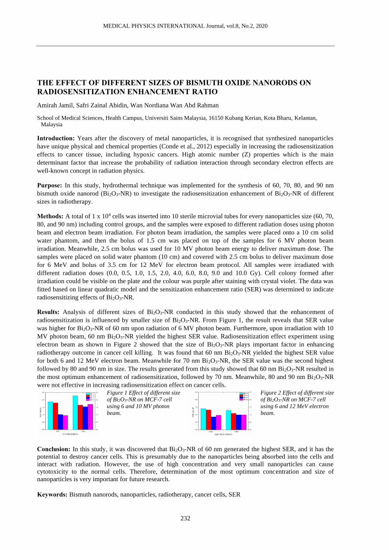
MEDICAL PHYSICS INTERNATIONAL Journal, vol.8, No.2, 2020
232
THE EFFECT OF DIFFERENT SIZES OF BISMUTH OXIDE NANORODS ON RADIOSENSITIZATION ENHANCEMENT RATIO
Amirah Jamil, Safri Zainal Abidin, Wan Nordiana Wan Abd Rahman
School of Medical Sciences, Health Campus, Universiti Sains Malaysia, 16150 Kubang Kerian, Kota Bharu, Kelantan, Malaysia
Introduction: Years after the discovery of metal nanoparticles, it is recognised that synthesized nanoparticles
have unique physical and chemical properties (Conde et al., 2012) especially in increasing the radiosensitization
effects to cancer tissue, including hypoxic cancers. High atomic number (Z) properties which is the main
determinant factor that increase the probability of radiation interaction through secondary electron effects are
well-known concept in radiation physics.
Purpose: In this study, hydrothermal technique was implemented for the synthesis of 60, 70, 80, and 90 nm
bismuth oxide nanorod (Bi2O3-NR) to investigate the radiosensitization enhancement of Bi2O3-NR of different
sizes in radiotherapy.
Methods: A total of 1 x 10⁴ cells was inserted into 10 sterile microvial tubes for every nanoparticles size (60, 70,
80, and 90 nm) including control groups, and the samples were exposed to different radiation doses using photon
beam and electron beam irradiation. For photon beam irradiation, the samples were placed onto a 10 cm solid
water phantom, and then the bolus of 1.5 cm was placed on top of the samples for 6 MV photon beam
irradiation. Meanwhile, 2.5 cm bolus was used for 10 MV photon beam energy to deliver maximum dose. The
samples were placed on solid water phantom (10 cm) and covered with 2.5 cm bolus to deliver maximum dose
for 6 MeV and bolus of 3.5 cm for 12 MeV for electron beam protocol. All samples were irradiated with
different radiation doses (0.0, 0.5, 1.0, 1.5, 2.0, 4.0, 6.0, 8.0, 9.0 and 10.0 Gy). Cell colony formed after
irradiation could be visible on the plate and the colour was purple after staining with crystal violet. The data was
fitted based on linear quadratic model and the sensitization enhancement ratio (SER) was determined to indicate
radiosensitizing effects of Bi2O3-NR.
Results: Analysis of different sizes of Bi2O3-NR conducted in this study showed that the enhancement of
radiosensitization is influenced by smaller size of Bi2O3-NR. From Figure 1, the result reveals that SER value
was higher for Bi2O3-NR of 60 nm upon radiation of 6 MV photon beam. Furthermore, upon irradiation with 10
MV photon beam, 60 nm Bi2O3-NR yielded the highest SER value. Radiosensitization effect experiment using
electron beam as shown in Figure 2 showed that the size of Bi2O3-NR plays important factor in enhancing
radiotherapy outcome in cancer cell killing. It was found that 60 nm Bi2O3-NR yielded the highest SER value
for both 6 and 12 MeV electron beam. Meanwhile for 70 nm Bi2O3-NR, the SER value was the second highest
followed by 80 and 90 nm in size. The results generated from this study showed that 60 nm Bi2O3-NR resulted in
the most optimum enhancement of radiosensitization, followed by 70 nm. Meanwhile, 80 and 90 nm Bi2O3-NR
were not effective in increasing radiosensitization effect on cancer cells.
Figure 1 Effect of different size
of Bi2O3-NR on MCF-7 cell
using 6 and 10 MV photon
beam.
Figure 2 Effect of different size
of Bi2O3-NR on MCF-7 cell
using 6 and 12 MeV electron
beam.
Conclusion: In this study, it was discovered that Bi2O3-NR of 60 nm generated the highest SER, and it has the
potential to destroy cancer cells. This is presumably due to the nanoparticles being absorbed into the cells and
interact with radiation. However, the use of high concentration and very small nanoparticles can cause
cytotoxicity to the normal cells. Therefore, determination of the most optimum concentration and size of
nanoparticles is very important for future research.
Keywords: Bismuth nanorods, nanoparticles, radiotherapy, cancer cells, SER

MEDICAL PHYSICS INTERNATIONAL Journal, vol.8, No.2, 2020
233
THE EFFECT OF DIFFERENT SIZES OF BISMUTH OXIDE NANORODS ON CANCER CELLS
Amirah Jamil, Safri Zainal Abidin, Wan Nordiana Wan Abd Rahman
School of Medical Sciences, Health Campus, Universiti Sains Malaysia, 16150 Kubang Kerian, Kota Bharu, Kelantan, Malaysia
Introduction: With the advancement of technology, various approaches have been developed to increase
radiation intensity to cancer tissue, which in turn increasing the effectiveness of killing radioresistant cancer
cells. Therefore, abundance of research has studied the potentials of various metallic nanoparticles increasing the
effectiveness of radiotherapy.
Purpose: This study is conducted to investigate the effect of different size of bismuth oxide nanorods (Bi2O3-
NR) on MCF-7 and HeLa cancer cells.
Methods: A total of 1 x 10⁴ MCF-7 and HeLa cells were inserted into sterile microvial tubes. Then, Bi2O3-NR
of different concentrations (0.05, 0.25, and 0.5 µMol/L) were added and was gently mixed with the cells using
pipette. Meanwhile for control samples, both cells were prepared without Bi2O3-NR. A total of 10 microvials
were prepared for every nanoparticles size (60, 70, 80, and 90 nm) including control, and were exposed to
different radiation dose (0-10 Gy). For 6 MV photon beam irradiation, the samples were placed onto a solid
water phantom (10 cm), and then the bolus of 1.5 cm (depth of maximum dose) was placed on top of the
samples. Meanwhile, 2.5 cm bolus was used for 10 MV photon beam energy to deliver maximum dose. The
samples for electron beam irradiation were set up according to the photon beam procedure, in which the samples
were placed on solid water phantom (10 cm) and covered with 2.5 cm bolus to deliver maximum dose for 6 MeV
and bolus of 3.5 cm for 12 MeV.
Results:
Figure 1 Comparison between
MCF-7 and HeLa cell line
treated with Bi2O3-NR and
irradiated using 6 MV photon
beam.
Figure 2 Comparison between
MCF-7 and HeLa cell line
treated with Bi2O3-NR and
irradiated using 10 MV photon
beam
Figure 3 Comparison between
MCF-7 and HeLa cell line
treated with Bi2O3-NR and
irradiated using 6 MeV
electron beam.
Figure 4 Comparison between
MCF-7 and HeLa cell line
treated with Bi2O3-NR and
irradiated using 12 MeV
electron beam.
Cell survival curves from Figure 1 to 4 showed that MCF-7 cell experienced more death compared to HeLa cells
when irradiated with photon and electron beam. These results indicated that the Bi2O3-NR inflicted more
radiosensitization in MCF-7 cells. Khoshgard (2017) reported that dextran-coated iron oxide nanoparticles
increase the effect of radiosensitivity when irradiated with 6 MV photon beam. It was discovered from the study
that MCF-7 cell produced higher SER value (1.21 ± 0.06) compared to HeLa cell (1.19 ± 0.04). The difference is
presumably due to the different characteristics of the cells such as type of receptors, number of mitochondria,
chromatin structure, all of which affect the cell radiation sensitivity.
Conclusion: This study discovered that MCF-7 has higher radiosensitivity and increased cell death compared to
HeLa when Bi2O3-NR was used. Therefore, different cell types exhibit different level of radiosensitivity.
Keywords: Bismuth oxide nanorods, nanoparticles, radiotherapy, MCF-7, HeLa

MEDICAL PHYSICS INTERNATIONAL Journal, vol.8, No.2, 2020
234
INFLUENCE OF PEG-COATED BISMUTH OXIDE NANOPARTICLES ON ROS GENERATION FOR ELECTRON BEAM RADIOTHERAPY
Noor Nabilah Talik Sisin1, Nur Fatin Amirah Radzuan1, Muhammad Afiq Khairil Anuar1, Norhayati Dollah2,
Khairunisak Abdul Razak3, Wan Nordiana Rahman1
1 School of Health Sciences, Universiti Sains Malaysia, Malaysia. 2 Department of Nuclear Medicine, Radiotherapy and Oncology, Hospital Universiti Sains Malaysia, Malaysia 3 School of Materials and Mineral Resources Engineering, Universiti Sains Malaysia, Malaysia.
Introduction: Nanoparticles (NPs) have been proven to enhance radiotherapy efficacy. The introduction of
coating materials such as PEG to NPs is found to impact the NPs’ biocompatibility and effectiveness as a
radiosensitizer. Optimization of surface coating is a crucial element to ensure the successful application of NPs
as a radiosensitizer in radiotherapy.
Purpose: This study aims to investigate the influence of bismuth oxide NPs (BiONPs) coated with different
PEG concentrations on reactive oxygen species (ROS) generation after electron beams irradiation on HeLa
cervical cancer cell line.
Methods: The HeLa cells were treated with BiONPs coated with different PEG concentrations (0.05, 0.10, 0.15
and 0.20 mM). The samples were irradiated under 3 Gy dose of electron beam energies (6 and 12 MeV). ROS
generation was measured immediately after irradiation and after 3 hours of incubation.
Results: The percentages of ROS generated in the presence of PEG-BiONPs were higher than the uncoated-
BiONPs. It also found that a high energy electron beam plays a role in increasing the ROS generation. The ROS
generated for 6 MeV electron beam, and BiONPs with 0.05 mM of PEG was found to induce the highest ROS
with 234%, with a slightly different result of 232% for 12 MeV. Data showed that higher concentration of PEG
(0.2 mM) decreased the ROS generation to 225% for 6 MeV and 229% for 12 MeV. There were also linear
increments of ROS after 3 hours of incubation, up to 339% for 6 MeV and 357% for 12 MeV.
Conclusion: Slight increase of the ROS by the PEG-BiONPs relative to the uncoated-BiONPs indicated the non-
toxicity of the surface coating. Higher PEG concentrations also perhaps more biocompatible. Finally, the PEG
coating might not contribute substantial effects towards the ROS generated, emphasizing the BiONPs application
as the radiosensitizer for HeLa cells.
Keywords: PEG, Bismuth oxide, Reactive oxygen species, Radiotherapy

MEDICAL PHYSICS INTERNATIONAL Journal, vol.8, No.2, 2020
235
Oral Presentations
Radiotherapy: Treatment planning

MEDICAL PHYSICS INTERNATIONAL Journal, vol.8, No.2, 2020
236
DOSIMETRIC VALIDATION OF MONACO TREATMENT PLANNING SYSTEM
Viphophone Inphavong1, Thibaud Saibaing2, David Little3, Taweap Sanghangthum4
1 Radiotherapy Center, Mittaphab Hospital, Vientiane Capital, Lao PDR. 2 Medical Physics and Radiation Safety Department, Hospices Civils de Lyon, France 3 Medical Physics and Bioengineering, Canterbury District Health Board, New Zealand 4 Division of Radiation Oncology, Department of Radiology, Faculty of Medicine, Chulalongkorn University, Bangkok,
Thailand.
Introduction: The error in dose calculation can lead to tumor control probability change. The accuracy of dose
distribution is necessary to validate.
Purpose: To study the dosimetric influences of photon beams for Collapse Cone Algorithm in Monaco TPS
calculation.
Methods: Photon beam characteristics of Collapse Cone Algorithm in Monaco TPS were verified in various square
fields by comparing with measured PDDs and profiles in water phantom using 0.125CC cylindrical chamber.
Moreover, the squares, rectangular, complex MLC shapes and wedge fields have been calculated following
IAEA TRS-430 guideline in homogeneous and heterogeneous slab phantoms. All plans were exported to Mosaiq
sequencer and Elekta Synergy Platform for beam delivery. Point dose at central axis and off-axis were measured
using 0.6CC Farmer and 0.125CC cylindrical chamber. In clinical assessment, relative electron density (RED)
curves on TPS were created based on universal curve and Elekta robust curve data. The 22 cases of brain,
head&neck, lung, breast and cervix regions of 3D-CRT were planned on both types of RED curves. The absolute
dose differences at isocentre were compared, and D90 of PTV was also evaluated.
Results: The measured PDDs and profiles of square fields showed good agreement to TPS with maximum dose
difference of 1%. For point-dose at various situations, average dose differences were -1.20±0.013% and
0.2±0.011% in homogeneous phantom, -1.0±0.002% and -0.4±0.004% in heterogeneous phantom for 0.6CC and
0.125CC chambers, respectively. Therefore, these differences were within the limits as the recommendation
from IAEA TRS-430 and AAPM TG-53. For clinical assessment, average dose difference between 2 types of
RED curves was 0.1±0.6% and D90 difference of PTV was 0.1±0.1%.
Conclusion: The accuracy of dose calculation in Monaco TPS based on the Collapse Cone Algorithm meets
international recommendations and can be implemented in clinic for patient treatments.
Keywords: PDD, Collapsed Cone Algorithm, Monaco TPS, 3D-CRT

MEDICAL PHYSICS INTERNATIONAL Journal, vol.8, No.2, 2020
237
EVALUATION OF DOSE CALCULATION ALGORITHM ACCURACY IN ECLIPSE TREATMENT PLANNING SYSTEM FOR JAW-DEFINED AND MLC-DEFINED SMALL FIELDS
Panun Sritubtim1, Suphalak Khachonkham1, Pimolpun Changkaew1, Warinthorn Rattanaareeyakorn1, Surendra Dhungana1, Nauljun Stansook1, Pornpan Yongvithisatid2, Puangpen Tangboonduangjit1
1 Master of Science Program in Medical Physics, Division of Radiotherapy and Oncology, Department of Diagnostic and Therapeutic Radiology, Faculty of Medicine Ramathibodi Hospital, Mahidol University, Thailand
2 Radiosurgery center, Faculty of Medicine Ramathibodi Hospital, Mahidol University, Thailand
Introduction: Basic concepts for the AAA and Acuros XB dose calculation algorithms employed in the
EclipseTM TPS have not been designed for small field planning. This might lead to dosimetric errors in the SBRT
planning if the commissioning process is not aware of.
Purpose: To evaluate dosimetric accuracy of the AAA and Acuros XB algorithms implemented in the Eclipse
TPS version 13.6.23 in a homogeneous phantom for small static fields defined by the MLCs and jaws.
Methods: Comparison between calculation of both algorithms and reference measurements was conducted for
the 6 MV photon beams and delivered using the Varian Edge linac. Field output factors were measured in the
water phantom for small static fields collimated by jaws (0.6×0.6, 1×1, 2×2, 3×3, 5×5 cm2) and MLCs (0.5×0.5,
1×1, 2×2; fixed jaw at 3×3 cm2). The PTW SRS diode and microdiamond detectors were used by following the
protocol of TRS-483 CoP. Calculated field output were acquired from the central axis point dose of any field
sizes normalized with those of the 10×10 cm2 field with 0.1 cm calculation grid size.
Results: Percentage differences between measurements and calculation by both algorithms were <3% in MLC-
and jaw-defined field down to 1×1 cm2 and 3×3 cm2, respectively. In the smallest MLC field size (0.5×0.5 cm2),
the agreement was up to 9% and 8% for AAA and Acuros XB, respectively. For jaw-defined fields smaller than
3×3 cm2, the result showed unacceptable agreement up to 15% and 19% for AAA and Acuros XB, respectively.
Conclusion: The accuracy of small field dose calculation for both algorithms was within 3% for field sizes
defined by MLC and jaw down to 1×1 cm2 and 3×3 cm2, respectively.
Keywords: small static fields, Eclipse dose algorithms, AAA, Acuros XB, collimation devices, dose calculation
accuracy

MEDICAL PHYSICS INTERNATIONAL Journal, vol.8, No.2, 2020
238
THE QUALITY IMPROVEMENT OF DOSE ESCALATION PLAN OF PROSTATE CANCER USING AN INTEGRATION OF BIOLOGICAL PARAMETERS FOR OPTIMIZATION AND EVALUATION
Tanwiwat Jaikuna1, Wiwatchai Sittiwong1, Pittaya Dankulchai1
1 Division of Radiation Oncology, Department of Radiology, Faculty of Medicine Siriraj Hospital, Mahidol University, Thailand
Introduction: The inverse optimization for Volumetric Modulated Arc Radiotherapy (VMAT) normally based
on physically dose volume-based parameters (DV-based), but it not tendency related to clinical outcome. The
biological optimization as the parameter of tumour control probability (TCP), normal tissue complication
probability (NTCP), and equivalent uniform doses (EUD) provided superior clinical relevance.
Purpose: This study aimed to investigate the plan quality improvement for dose escalation plan of prostate
cancer using physical and biological indices in three optimization techniques, DV-based, biological-based, and
integrated-based.
Methods: Ten prostate cancer patient cases have delineated the organ-at-risk; bladder and rectum, and the multi-
dose targets of the planning target volume, dominant intraprostatic lesion, prostate gland (PG), and seminal
vesicle (SV) using multi-parametric magnetic resonance image-guided by the experienced radiation oncologist
and the VMAT plans were generated using Raystation version 9B with the different optimization techniques.
Physical dosimetric indices and biological indices included D98%, D2%, V95%, Dmean, TCP, Heterogeneity index
(HI), and Conformity index (CI) for Target as well as V40Gy, V60Gy, D2%, D1%, Dmean, and NTCP for OARs were
evaluated.
Results: All plan-optimized techniques provided a similar result for target coverage when considering the
physical indices; D98% of PTV DIL=87.22±0.76, 86.07±0.47, 86.54±0.17, D98% of PTV PG=77.86±0.63,
77.69±0.47, 77.19±0.22, and D98% of PTV SV=62.20±0.81, 73.03±0.71, 73.47±1.60 for physical, biological,
and integrated-based optimization, respectively. Although TCP of PTV DIL and PG showed similar value in all
optimization technique (TCP=1), the higher TCP value in PTV SV was obtained when biological parameters are
applied (TPC=0.98±0.01) when compared to physical optimization only (TCP=0.74±0.07). However, the
overdose region is showed significantly different as the biological optimization provided higher dose compared
to other plan-optimization technique except in PTV SV which showed the integrated-based yield a similar result
with biological-based optimization (about 5% increased). The OARs tend to receive a lower dose in biological
and integrated-based optimization in all parameters with lower NTCP (decreased to less than 5%) when
considering the same biological endpoint.
Conclusion: The quality of the prostate plan can improve by using the integration of DV and biological-based
optimization.
Keywords: Biological Optimization, Prostate Cancer, Volumetric Modulated Radiation Therapy

MEDICAL PHYSICS INTERNATIONAL Journal, vol.8, No.2, 2020
239
TREATMENT PLANNING EVALUATION OF 3D-PRINTED ANTHROPOMORPHIC HEAD PHANTOM
Nur Emirah Zain1, Reduan Abdullah2, Wan Fatihah Wan Sohaimi2, Wan Nordiana Rahman1
1 School of Health Sciences, Health Campus, Universiti Sains Malaysia, Malaysia, 2 Departments of Oncology, Radiotherapy and Nuclear Medicine, Hospital Universiti Sains Malaysia, Malaysia,
Introduction: Radiotherapy phantom is used as a patient replacement to run the quality assurance and advanced
treatment verification. Standard commercial phantom is very costly which leads to the development of low cost
custom made 3 Dimensional (3D) printed phantoms. 3D printing technique is currently a popular method to
fabricate a low cost radiotherapy phantom. The quality of the phantom requires evaluation to fulfil dosimetric
standard in radiotherapy.
Purpose: To evaluate the 3D printed anthropomorphic phantom in comparison to standard commercial
RANDO® Head Phantom treatment planning based on treatment planning system (TPS) and radiochromic
EBT3 films dosimetry.
Methods: The RANDO® Head Phantom and 3D printed anthropomorphic head phantom were scanned using
CT-simulation for 3D data acquisition. The topographic data from CT-sim were transferred to the Oncentra TPS
in the DICOM format. Image registration manoeuvred by contouring all organs at risk and whole brain as the
target area. The treatment planning was based on neck and head consisting of two opposite-field beams. Later,
the planning proceeds to the beam insertion in the plan manager segment with prescribed dose recommended
followed by the dose calculation process. The plan was then exported to the LINAC through ARIA’s electronic
medical record system. The treatment delivery was conducted on both the RANDO® Head Phantom and 3D
printed head phantom with GafChromic EBT3 films for dose verification.
Results: The dosimetric accuracy measured on 3D printed head phantom is found to be less compared to the
RANDO® head phantom. The measured dose from the EBT3 film on the RANDO® head phantom is in average
80% passed according to the gamma analysis. While dose received in 3D printed head phantom displayed high
error due to the presence of air gap between the slices. This 3D printed head phantom requires further
optimization to reduce the air gaps between the slices.
Conclusion: The 3D-printed head phantom has been developed and its show potential to be used in radiotherapy
treatment planning and dose verification. However further optimization is required to comply with the dosimetric
standard in radiotherapy.
Keywords: 3D-printing, radiotherapy phantom, radiochromic films, treatment planning
MEDICAL PHYSICS INTERNATIONAL Journal, vol.8, No.2, 2020
240
COMPARISON OF DIFFERENT TREATMENT PLANNING TECHNIQUES FOR LEFT-SIDED BREAST CANCER: A DOSIMETRIC STUDY
Md Akhtaruzzaman1, Md Abdus Sabur2, Md Shoriful Islam3
1 Ahsania Mission Cancer and General Hospital, Dhaka, Bangladesh 2 Square Hospital Limited, Dhaka, Bangladesh 3 National Institute of Cancer Research and Hospital, Dhaka, Bangladesh
Introduction: Cancer is a significant healthcare problem. Breast cancer is the most common type of cancer
among women. Radiotherapy is one of the most effective treatment techniques after breast conserving surgery.
Purpose: To investigate the suitable radiotherapy treatment plan and delivery method for left-sided breast cancer
after breast conserving surgery by comparing different treatment planning techniques.
Materials and methods: Ten patients with left breast cancer who underwent breast conserving surgery were
selected. For each patient, three different whole breast irradiation techniques including Field in Field (FiF),
Intensity Modulated Radiation Therapy (IMRT) and Volumetric Modulated Arc Therapy (VMAT) with free
breathing were compared. Treatment plans were prepared with Eclipse treatment planning system (Varian
Medical System Inc) using Anisotropic Analytical Algorithm (AAA). The prescribed dose was 50 Gy in 25
fractions (2 Gy/fraction). Plans were compared for target coverage (V95% and V107%) and doses of the lungs,
heart, and spinal cord. Conformity index (CI) and homogeneity index (HI) were also assessed in this study.
Results: Mean maximum point dose (Dmax) and minimum point dose at target (PTV50) for planning techniques
were 106%-107% (±0.83) and 83%-84% respectively. The mean V95 for FiF and IMRT was almost same
(97.5%), which is higher than VMAT (95.2%). Only FiF had a lower mean dose (ipsilateral lung-1089cGy,
heart-1087cGy) in compare to IMRT (ipsilateral lung-1260 cGy, heart-1308 cGy) and VMAT (ipsilateral lung-
1386 cGy, heart-1301 cGy). In contrast, V20 was lower for IMRT (16.8%) than FiF (18.0%) and VMAT
(24.0%) techniques for ipsilateral lung. All planning techniques demonstrated excellent CI and HI.
Conclusion: The all three techniques provide adequate dose coverage. The IMRT had significantly smaller dose
than VMAT in ipsilateral lung. VMAT demonstrated excellent dose homogeneity and conformity but an
increased low-dose volume outside the target should be of concern.
Keywords: Breast cancer- radiotherapy planning technique- VMAT, IMRT, FiF

MEDICAL PHYSICS INTERNATIONAL Journal, vol.8, No.2, 2020
241
INVESTIGATION OF THE OPTIMAL COST FUNCTION FOR PELVIC CT-MR DEFORMABLE IMAGE REGISTRATION
Tomoya Ishida1, Noriyuki Kadoya1, Shunpei Tanabe1, Haruna Ohashi2, Ken Takeda2, Suguru Dobashi2, Keiichi Jingu1
1 Department of Radiation Oncology Tohoku University Graduate School of Medicine, 2 Department of Therapeutic Radiology Tohoku University Graduate School of Medicine,
Introduction: In recent years, the development of magnetic resonance imaging (MRI)-guided radiotherapy
systems has resulted in an increased use of MRI in radiotherapy planning. To transfer contour and electron
density information between computed tomography (CT) and MR images, image intensity-based deformable
image registration (DIR) is used. In general, the DIR accuracy between different modalities is lower than that
between the same modalities, and to date, reports on the evaluation of CT-MR DIR accuracy have been scarce.
Purpose: We evaluated the CT-MR DIR accuracy by validating the optimal cost function in pelvic images.
Methods: CT, T1-weighted MR (T1w), and T2-weighted MR (T2w) images from five male pelvic images (data
by Tufve et al.) were included in the study. B spline-based DIR (elastix) was used for DIR, mutual information
was used for the similarity term, and Bending energy penalty for the regularization term as cost functions. Six
patterns of regularization weights were considered for determining the optimal cost function: 0.01, 0.1, 1, 10,
100, and 200.
Results: The mean dice similarity coefficients of the bladder in T1w images were 0.74 ± 0.11 (rigid), 0.85 ±
0.04 (DIR, λ = 0.1), 0.74 ± 0.10 (DIR, λ = 200). Those of the rectum in T1w images were 0.77 ± 0.13, (rigid),
0.80 ± 0.04 (DIR, λ = 0.1), 0.78 ± 0.12 (DIR, λ = 200). A similar trend was observed for T2w images. The most
accurate value of λ varied from case to case, with λ = 0.1 for T1w and λ = 1 for T2w being the most common.
These results indicate that the optimal cost function vary by case and MRI sequences.
Conclusion: This study suggested that optimization of the cost function might improve the accuracy of CT-MR
DIR.
Keywords: MRI-guided radiotherapy, deformable image registration, cost function, pelvis, dice similarity
coefficient

MEDICAL PHYSICS INTERNATIONAL Journal, vol.8, No.2, 2020
242
DOSIMETRIC COMPARISON BETWEEN 3D AND IMRT TREATMENT TECHNIQUES FOR WHOLE BREAST RADIOTHERAPY
Yin Yin Pyone1, Taweap Sanghangthum2
1 Department of Radiation Oncology, Pinlon Cancer Center, Pinlon Hospital, Yangon, Myanmar 2 Division of Radiation Oncology, Department of Radiology, Faculty of Medicine, Chulalongkorn University, Bangkok,
Thailand
Introduction: In worldwide, breast cancer is the most common cancer in women that contributing 25.4% of the
total number of new cases diagnosed in 2018 and also, it is the second most common women cancer in Myanmar
and late toxicity could lead to secondary cancer for the contralateral breast.
Purpose: To determine the dosimetric effects of 3D and IMRT treatment techniques for whole breast
radiotherapy.
Methods: The 10 left-sided and 5 right-sided women breast patients in Pinlon Cancer Center were randomly
selected. The clinical target volume (CTV), planning target volume (PTV) and OARs: right lung, left lung, heart
and contralateral breast were contoured according to the RTOG breast contouring guideline. All plans were
performed by Varian Eclipse Treatment Planning System (Version 13.1) for; (1) standard wedge tangent (SWT),
(2) Field in Field (FiF), (3) electronic compensator (E-Comp), (4) tangential-IMRT (T-IMRT), and (5) multiple
field-IMRT (MF-IMRT). The PTV was evaluated by D95%, conformity index (CI), and Homogeneity Index
(HI). Also for the OARs, the mean dose of heart, ipsilateral lung and contralateral breast were compared for all
plans.
Results: The D95% of PTV was higher than prescribed dose for all techniques. The IMRT plans (T-IMRT and
MF-IMRT) showed superior to PTV CI and HI compared with 3D plans (SWT, FiF and E-Comp). Among 3D-
CRT techniques, E-Comp technique results better CI and HI. MF-IMRT presented the highest HI but also
showed the highest dose at contralateral breast, while SWT showed the highest ipsilateral lung and heart doses,
especially left breast side.
Conclusion: Different planning techniques present differ dosimetric on PTV and OARs. E-Comp is a good
choice of radiotherapy. The advanced technique of IMRT can increase CI and HI parameters.
Keywords: Breast cancer, 3D-CRT, IMRT

MEDICAL PHYSICS INTERNATIONAL Journal, vol.8, No.2, 2020
243
Oral Presentations
Radiotherapy: Big data, Deep learning, AI and Modeling

MEDICAL PHYSICS INTERNATIONAL Journal, vol.8, No.2, 2020
244
DEVELOPMENT OF AUTOMATED PROSTATE VMAT TREATMENT PLANNING SYSTEM USING DEEP LEARNING-BASED PREDICTIVE DOSE DISTRIBUTION
Patiparn Kummanee1, Sangutid Thongsawad1,2, Wares Chancharoen1, Todsaporn Fuangrod1
1 Faculty of Medicine and Public Health, HRH Princess Chulabhorn College of Medical Science, Chulabhorn Royal Academy, Bangkok, Thailand
2 Department of Radiation Oncology, Chulabhorn Hospital, Chulabhorn Royal Academy, Bangkok, Thailand
Introduction: Treatment planning process for volumetric modulated arc therapy (VMAT) is considered as the
time-consuming process due to manual trail-and-errors (iteration of planning from unapproved plan). An
automated treatment planning can accelerate this process and reduce the number of iterations during the plan
optimization. The predictive dose distribution can be generated directly from patient CT data using artificial
intelligence method. The automatic treatment plan can be developed if the predicted dose is provided.
Purpose: The automated prostate VMAT treatment planning system was designed and developed. The
predictive dose distribution model was developed using deep learning algorithm.
Methods: A generative adversarial network (GAN) approach was used to generate the predictive model, which
consists of a pair of deep learning model including U-net generator and discriminator. To train the model, the
inputs were the CT images and its dose distribution. Fifteen patients of VMAT prostate cancer were collected in
this study. Ten patients were randomly chosen as training set and the remaining were the test set. The trained
model was tested by input CT images from test set and evaluated the dose distribution output with the
corresponding dose using 3D gamma analysis and dose-volume histogram (DVH).
Results: The trained model could predict the accurate dose distribution from input CT image. The passing rates
of 3D gamma analysis were 49.44% with 2%/2 mm criteria. The usage time of deep learning-based treatment
planning were 30 minutes which approximately 80% faster than conventional planning.
Conclusion: Our proposed automated treatment planning system using deep learning predictive model of
prostate cancer could generate acceptable dose distribution from input CT images which could help medical
physicists to get the acceptable quality of treatment plan with less time in the process.
Keywords: VMAT, deep learning, prostate cancer, dose distribution prediction.

MEDICAL PHYSICS INTERNATIONAL Journal, vol.8, No.2, 2020
245
AUTOMATED FAST DATA COLLECTION AND ORGANIZATION FOR MEDICAL ARTIFICIAL INTELLIGENCE
Hongjia Liu1, Yibao Zhang1 (Corresponding Author)
1 Key Laboratory of Carcinogenesis and Translational Research (Ministry of Education/Beijing), Peking University Cancer Hospital & Institute, Beijing 100142 China
Introduction: The medical applications of artificial intelligence have been strongly dependent on the data
aggregation and organization. Conventional manual collection is labour-intensive and time-consuming.
Purpose: This work aims to develop and validate a Web-Crawler-based medical records information
aggregation tool for effective data mining from existing electronic information systems.
Methods: Based on Selenium framework and Python programming language, a Web-Crawler-based medical
records information aggregation tool was designed, which was validated under two illustrative scenarios: 1. To
identify radiation pneumonitis (RP) cases from Hospital Information System (HIS), as an application example of
quick data search; 2. To summarize an organized table combining desired data from various examination reports,
to test the application of facilitating clinical workflow. Automated and manual methods were compared in terms
of efficiency and accuracy.
Results: The automated tool showed superior efficiency and accuracy than manual method. For the first
scenario, automated method identified 110 RP cases out of 3541 patients in about 54 seconds per patient based
on a Raspberry Pi 4B, without any human interference. Manual methods identified the same group of RP cases
but took about 90 seconds per patient. It took longer to confirm a non-RP case because more data need to be
excluded to avoid false-negative, suggesting even greater advantage of automated method in searching small-
probability events, especially from huge patient volume. For the other scenario, automated and manual methods
needed about 10 or 75 seconds respectively for each patient. Automated method also avoided typos that were
frequently observed in manual report filling.
Conclusion: A Web-Crawler-based medical records information aggregation tool has been successfully
developed. The superior efficiency and accuracy of auto-aggregation has been validated based on specific
clinical scenarios. With the advantage of cross-platform and easy-to-extend, this application could improve
radiologists' and physicists' productivity in their clinical and research practice.
Keywords: Web-Crawler; data aggregation; automation

MEDICAL PHYSICS INTERNATIONAL Journal, vol.8, No.2, 2020
246
A FEASIBILITY STUDY OF KVCBCT-BASED RADIOMICS FOR RADIOLOGICAL COMPLICATION PREDICTION
Yuliang Huang1, Yibao Zhang1 (Corresponding Author)
1 Key Laboratory of Carcinogenesis and Translational Research (Ministry of Education/Beijing), Peking University Cancer Hospital & Institute, Beijing 100142 China
Introduction: Considering the well-reported feasibility of planning CT (pCT)-based radiomics, the consistent
features of kilovoltage cone-beam CT (kVCBCT) are complementary if not intersubstitutable with pCT for
radiomics.
Purpose: To validate the feasibility of radiomic feature extraction from kVCBCT images and to investigate
potential application of kVCBCT-based radiomics in predicting radiation-induced pneumonitis (RP).
Methods: 193 thoracic cancer patients treated with radiotherapy during January, 2017 and December, 2019 in
our centre were retrospectively selected, including 70 patients who reported RP after the treatment. Contoured
pCT and kVCBCT acquired at the initial treatment fraction were collected from clinical database for each
patient. The dataset was split to training and test groups with the ratio of 4:1. Concordance correlation
coefficients (CCC) of 1275 radiomic features extracted from pCT and kVCBCT were calculated. Stable radiomic
features (CCC>0.99) were selected to train a logistic regression model that predicted radiation-induced
pneumonitis for the 193 patients. Performance of the model was compared with that of another logistic model
based on dose statistics (V5Gy, V10Gy, V20Gy, mean dose of lung).
Results: In addition to shape and volume, 50 more texture features were found to be consistent between pCT and
kVCBCT (CCC>0.99). Tracking the changing kVCBCT radiomic features during the IGRT course might
provide prompt and quantified evaluation of tumour response and OAR risks. AUC values of the two logistic
models based on radiomics and dosimetrics were 0.84 and 0.73 respectively. Although RP was directly related to
dose, varieties in individual’s radiological sensitivity could undermine the accuracy of dosimetric model, while
radiomics model provides additional patient-specific and dynamic indicators.
Conclusion: KVCBCT images are interchangeable with pCT providing a subset of reliable radiomics features.
The prognostic value of kVCBCT-based radiomics in predicting RP has been evaluated, which encourages future
study on temporal radiomics tracking based on kVCBCT sequence in IGRT.
Keywords: Radiomics; kVCBCT; Radiological complication

MEDICAL PHYSICS INTERNATIONAL Journal, vol.8, No.2, 2020
247
EVALUATION OF MACHINE LEARNING-BASED PREDICTION MODEL FOR RADIATION PNEUMONITIS IN NSCLC PATIENTS
Shiina Mouri1, Noriyuki Kadoya1, Yoshiyuki Katuta1, Takayuki Kanai3, Yujiro Nakajima1,4,
Shunpei Tanabe1, Yuto Sugai1, Mariko Umeda1, Suguru Dobashi2, Ken Takeda2, Keiichi Jingu1
1 Department of Radiation Oncology, Tohoku University Graduate School of Medicine, Sendai, Japan 2 Department of Radiological Technology, School of Health Sciences, Faculty of Medicine, Tohoku University, Sendai,
Japan 3 Department of Radiation Oncology, Yamagata University Faculty of Medicine, Yamagata, Japan 4 Tokyo Metropolitan Cancer and Infectious Diseases Center Komagome Hospital, Tokyo, Japan
Introduction: Radiation pneumonitis (RP) is a vital toxicity in non-small cell lung cancer (NSCLC) patients
who are treated using radiotherapy. DVH parameters have been employed to predict the risk of RP. Machine
learning has great potential in enhancing the prediction accuracy. However, there are only a few studies on this
matter.
Purpose: We assessed the accuracy of machine learning-based prediction model for RP in NSCLC patients.
Methods: 186 lung cancer patients (stage I: 51, stage II: 17, stage III: 54, stage IV: 13, and unknown: 51), were
treated at our hospital. The number of patients who were treated using conventional and stereotactic radiotherapy
were 107 and 79, respectively. We predicted the risk of Grade 2+ pneumonitis (rate = 28%). DVH parameters of
V5, V10, V20, V30, and MLD provided as equivalent dose in 2Gy fractions (EQD2) were utilized as predictive
indicators. For the multivariate analysis, random forest (R package) was used to build the machine-learning
model. All data were split into two sets: 80% for training, and 20% for testing. Two models were developed:
model A employed all the DVH parameters and model B employed three parameters that were selected based on
their importance in random forest2).
Results: The AUC values for V5, V20, and MLD in the univariate analysis were 0.74, 0.73, and 0.65,
respectively, for the training data and 0.68, 0.61, and 0.58 for the test data, respectively. In the multivariate
analysis, the AUC values were 0.74 (training) and 0.71 (testing) in model A, and 0.77 and 0.72, in model B,
indicating that these values were higher than those in the univariate analysis. Three indicators of high importance
were V5, V10, and MLD.
Conclusion: Multivariate analysis using machine learning may improve the accuracy of predicting RP in
NSCLC patients.
Keywords: Radiation therapy, Machine learning, Lung cancer, Radiation pneumonitis

MEDICAL PHYSICS INTERNATIONAL Journal, vol.8, No.2, 2020
248
IMPACT OF IMAGE TYPE AND DEEP LEARNING ARCHITECTURE IN DEEP LEARNING RADIOMICS ON THE ACCURACY OF LUNG CANCER PREDICTION
Shumpei Tanabe1, Noriyuki Kadoya1, Shohei Tanaka1, Ken Takeda2, Suguru Dobashi2, Keiichi Jingu1
1 Department of Radiation Oncology Tohoku University Graduate School of Medicine 2 Department of Therapeutic Radiology Tohoku University Graduate School of Medicine
Introduction: Conventional radiomics typically extracts predesigned features from a segmented region of
interest (ROI). Recently, radiomics with deep learning (DL) has been actively researched. It automatically
extracts features using a convolutional neural network from an image patch without segmenting the ROI.
However, there has been no study on the impact of the type of input images and DL architecture on prediction
accuracy.
Purpose: In this study, we evaluated the impact of input image type and DL architectures on the accuracy of
prognostication of patients with lung cancer.
Methods: From the Cancer Imaging Achieve database, 418 patients with nonsmall cell lung cancer, including
their CT images and GTV contour, were included. We created four types of input images that focused on the
center of gravity of the GTV, as follows: 1) 50 × 50 mm with CT value outside the GTV set at 0 (masked 50 ×
50 mm); 2) 50 × 50 mm; 3) 100 × 100 mm; and 4) 150 × 150 mm. We employed three types of DL architectures,
including a simple model that comprised three convolution layers; a medium model that comprised four
convolution layers; and a complex model that comprised 152 convolution layers. For all combinations of input
image and DL architecture, we evaluated the accuracy of prognostication by area under the curve (AUC).
Results: The best performance AUCs were 0.64 with 150 × 150 mm for the simple model, 0.65 with 50 × 50
mm for the medium model, and 0.66 with masked 50 × 50 mm for the complex model. This result showed that a
more complex model could achieve high prediction accuracy with input image and less information.
Conclusion: The prediction accuracy of radiomics with DL depended on the DL architecture and the input
image.
Keyword: radiomics, deep learning, prognostic, radiotherapy

MEDICAL PHYSICS INTERNATIONAL Journal, vol.8, No.2, 2020
249
PROGNOSTIC ANALYSIS OF CT-BASED RADIOMICS FOCUSING ON A SUBGROUP OF NSCLC PATIENTS
Yuto Sugai1, Noriyuki Kadoya1, Shohei Tanaka1, Shunpei Tanabe1, Mariko Umeda1, Takaya Yamamoto1,
Kazuya Takeda1, Suguru Dobashi2, Haruna Ohashi3, Ken Takeda2, Keiichi Jingu1
1 Department of Radiation Oncology, Tohoku University Graduate School of Medicine, Sendai, Japan 2 Department of Radiological Technology, School of Health Sciences, Faculty of Medicine, Tohoku University, Sendai,
Japan 3 Department of Radiation Technology, Tohoku University Graduate School of Health Sciences, Sendai, Japan
Introduction: Radiomics is an emerging field wherein quantitative features extracted from medical images are
used to noninvasively predict survival prognosis. Recent studies have described the feasibility of predicting the
prognosis in patients with non-small-cell lung cancer (NSCLC) using this ingenious approach; however, its
prognostic accuracy can certainly be improved. Hence, we focused on the prognostic analysis in subgroups with
identical characteristics.
Purpose: We investigated the survival prediction of specific NSCLC subtypes and T stages using radiomics.
Methods: Images from 304 NSCLC patients (Stages I-IV) who had been treated with radiotherapy in our
hospital were used in this study. 107 radiomic features (14 shape features, 18 first‐order statistical features, and
75 textural features) were extracted from the GTV in CT images (free-breathing) acquired during treatment
planning. Three different feature selection methods were used: test-retest and multiple-segmentation (FS1),
Pearson’s correlation analysis (FS2), and features suggested as useful in previous studies (FS3), and a least
absolute shrinkage and selection operator-cox regression model was constructed using selected features.
Prognostic analyses were performed for each NSCLC subtype and each T stage, and their accuracy was
evaluated using the C-index with five-fold cross-validation and the Kaplan-Meier method.
Results: The C-index for all data were 0.629 ± 0.023 (FS1), 0.626 ± 0.023 (FS2), and 0.636 ± 0.023 (FS3),
respectively. In subgroup analysis, the prediction models for specific NSCLC subtypes and T stages showed
higher C-index than that for all data, especially in the T4 subgroup (0.687 ± 0.05). Moreover, the prediction
models for each T stage in adenocarcinoma (ADC) had a higher C-index than that of ADC.
Conclusion: Our results showed that feature selection methods had a moderate impact on prognostic accuracy,
and that prediction models that use specific NSCLC subtypes and T stages could improve prediction accuracy.
Keywords: Radiomics, prognosis prediction, subgroup analysis, lung cancer

MEDICAL PHYSICS INTERNATIONAL Journal, vol.8, No.2, 2020
250
Oral Presentations
Radiotherapy: Patient-specific QA

MEDICAL PHYSICS INTERNATIONAL Journal, vol.8, No.2, 2020
251
PRELIMINARY EVALUATION OF THE PERFORMANCE OF AN EPID-BASED IN VIVO DOSIMETRY FOR ADVANCED RADIOTHERAPY TREATMENT VERIFICATION
Yasmin Md Radzi1, Nur Nabilah Roskhahar1, Nur Fatin Fariha Abd Latif1, Noor Naslinda Noor Rizan2
1 School of Physics, Universiti Sains Malaysia, Penang, Malaysia, 2 Radiotherapy and Oncology Department, Gleneagles Hospital, Penang, Malaysia.
Introduction: Patient specific QA dose verification has been widely discussed and has proposed a way to
achieve treatment delivery accuracy and patient safety. While in vivo dosimetry verification is currently is a
necessity in radiotherapy centres in Europe countries, its demand currently rising in developed countries
especially in Malaysia.
Purpose: In this work, the sensitivity and performance of EPID-based in vivo dosimetry, EPIgray® (DOSIsoft,
Cachan, France) were evaluated for its use in clinical treatment verification for our centre.
Methods: Sensitivity tests such as dose linearity, field size, off-axis, position as well as angle dependency were
performed to observe the response of EPIgray measured dose against TPS calculated dose for 6 MV and 10 MV
photon beam. The relative deviation of the total dose was evaluated at isocentre point for different depth in
water. EPIgray was also validated by using IMRT and VMAT prostate plan to be compared with the dose in
TPS. All calculation points were at the beam isocentre and at additional points suggested by TG-119 and
following the accepted tolerance of 10% dose threshold.
Results: EPIgray reported good agreement for linearity, field size, off-axis and position dependency with TPS
dose being within 5% tolerance for both energy ranges. For angle dependency test, average agreement between
TPS and EPIgray dose is less than 2% in 6 MV while 7% in 10 MV photon beam. The clinical evaluation
performed for IMRT prostate plan gave average agreement 5-6% at the plan isocentre for both energies. While
for VMAT, 94% and 100% of all points created lies within 5% for 6 MV and 10 MV photon beam energy
respectively.
Conclusion: Based on the work presented in this paper, in vivo dosimetry is deemed as an essential need as one
of the verification tools for advanced treatment techniques in Malaysia alongside pre-treatment verification.
Keywords: Epigray, in vivo dose measurement, IMRT, VMAT, Patient-specific QA

MEDICAL PHYSICS INTERNATIONAL Journal, vol.8, No.2, 2020
252
PATIENT-SPECIFIC QUALITY ASSURANCE FOR IMRT DELIVERY: A MULTI-CENTRE STUDY
Nur Diyana Afrina Hizam1, Wei Loong Jong1,2, Hafiz Mohd Zin3, Kwan Hoong Ng4, Ngie Min Ung1
1 Clinical Oncology Unit, Faculty of Medicine, University of Malaya, Kuala Lumpur, Malaysia, 2 Department of Radiation Oncology, Auckland City Hospital, Auckland 1023, New Zealand, 3 Advanced Medical and Dental Institute, Universiti Sains Malaysia, Penang, Malaysia 4 Department of Biomedical Imaging, University of Malaya, Kuala Lumpur, Malaysia
Introduction: IMRT technique requires multi-centre quality assurance programs for verification due to its
increased complexity. This study is the first trial of multi-centre audit in Malaysia
Purpose: To verify accuracy of IMRT delivery by using independent tools and to investigate the feasibility of
using MOSkin detectors for multi-centre study
Methods: Physicists from participating centre required to produce a head & neck plan by using a case study
given as per their clinical practice. The MOSkin detector and PTW Octavius 1500 detector were used to verify
the point dose and dose distribution plane respectively.
Results: A total of 12 measurements were performed across 11 radiotherapy centres between May to September
2019. Overall mean difference of MOSkin detectors are 3.3% ± 3.2%. 82% of the results were within the
tolerance level of ±5% recommended by IAEA for on-site IMRT/VMAT audits. Gamma passing rates of 3%/3
mm for most of the centres were within 95% passing rates using Octavius 1500.
Conclusion: A first dosimetric audit has been successfully conducted from 11 centres in Malaysia. Results
showed that the standard of IMRT delivery in participating centres were, in general, met the standard
recommended by international guidelines.
Keywords: Audit dosimetry; IMRT/VMAT; MOSkin
MEDICAL PHYSICS INTERNATIONAL Journal, vol.8, No.2, 2020
253
PILOT STUDY OF REMOTE CLINICAL DOSIMETRY AUDITING FOR IMRT
USING VIRTUAL EPID STANDARD PHANTOM AUDIT (VESPA) IN THAILAND
Pavarit Pojanapreecha1, Puangpen Tangboonduangjit4, Sangutid Thongsawad1, Peter Greer2, 3, Todsaporn Fuangrod1
1 Faculty of Medicine and Public Health, HRH Princess Chulabhorn College of Medical Science, Chulabhorn Royal Academy, Bangkok,10210, Thailand
2 Department of Radiation Oncology, Calvary Mater Hospital Newcastle, NSW, 2298, Australia 3 School of Mathematical and Physical Sciences, University of Newcastle, Newcastle, NSW, 2308, Australia 4 Department of Radiology, Faculty of Medicine, Ramathibodi Hospital, Mahidol University, Bangkok 10400, Thailand
Introduction: Since many centers applies the modern radiotherapy technique, it require the complex treatment
plan resulting the need of highly efficient quality assurance (QA) to ensure the beam is delivered precisely.
Although the standard of QA (such as TG-142) is recommended for implementation in all centers, the variation
across center-to-center remains exist. To face with this situation, the machine audit with simulated beam delivery
in end-to-end test perspective becomes essential. However, the audit process requires resource intensive and time
consuming.
Purpose: The remote auditing for IMRT using virtual EPID standard phantom audit was implemented and
promoted as an alternative auditing tool to solve the traditional auditing limitations. This project was
implemented as a pilot study.
Methods: The workflow of remote auditing using the virtual EPID standard phantom for IMRT has developed
and implemented in four centers in Thailand. All process guidelines and data were communicated and transferred
online. Each center was required to set trial plan data to the auditors, which including plan files, dose files and
EPID images. After the participant transferred data, the auditor analyzed the data using in-house software
programmed in MATLAB/Simulink. As clinical dosimetry auditing, The accuracy of beam delivery was
evaluated using Gamma passing rate. Geometric error was investigated using the calculated data from the
software.
Results: All of the participated centres passed the tolerance. (3%/2mm) .The mean percentage gamma pass rates for
the 4 centres were 99.7 % (for the 3%/2 mm criterion) and 98.05 % (for the 2%/2 mm criterion). The geometric error
was detected for some centre.
Conclusion: The remote clinical dosimetry auditing process and tool can be used to evaluate the participant
center’s current dosimetric quality level of IMRT process. The benefit of remote auditing shows resource
intensive, time efficient, and high flexibility.
Keywords: IMRT; Quality Assurance; Dosimetry Auditing;
MEDICAL PHYSICS INTERNATIONAL Journal, vol.8, No.2, 2020
254
EVALUATION OF MOBIUS AND PORTAL DOSIMETRY QUALITY ASSURANCE TOOLS FOR VOLUMETRIC MODULATED ARC THERAPY PLANS
Chulee Vannavijit1, Sornjarod Oonsiri1, Taweap Sanghangthum2, Sivalee Suriyapee2
1 Division of Radiation Oncology, Department of Radiology, King Chulalongkorn Memorial Hospital, Thai Red Cross Society, Bangkok, Thailand.
2 Division of Radiation Oncology, Department of Radiology, Faculty of Medicine, Chulalongkorn University, Bangkok, Thailand.
Introduction: Mobius is a software-based verification tool for patient specific quality assurance (QA) and
treatment delivery. The distinctive point of Mobius system is no uncertainty from setup error of the measurement
tools.
Purpose: To evaluate the dosimetric performance of Mobius system (Mobius3D and MobiusFx) by comparison
with electronic portal imaging device (EPID), which is routinely used for patient specific QA.
Methods: Total of 30 volumetric modulated arc therapy (VMAT) plans consisted of 10 head-neck, 10 chest and
10 prostate plans using Eclipse with AAA algorithm. Mobius 3D independently verified the pre-treatment plans
by re-calculation with collapsed-cone algorithm. The MobiusFx used the Mobius3D and the machine log files to
calculate 3D dose during treatment. The verification with EPID was undertaken at the same time. All plans were
delivered with 6 and 10 MV photon beams of 2 to 4 arcs per plan by Varian TrueBeam linac. The percent
gamma passing rate were compared using criteria of 3%/3mm for the composited dose between Portal
dosimetry, Mobius3D and MobiusFx with TPS.
Results: The average gamma passing rate for EPID, Mobius3D and MobiusFx for head-neck plans were
98.5±1.4%, 97.2±2.0% and 97.2±2.0%, for chest plans were 97.4±1.8%, 98.3±1.5% and 97.8±2.2%, for prostate
plans were 97.6±2.3%, 99.3±0.7%, and 99.3±0.7%, respectively. The relative percentage dose differences
between EPID and Mobius3D were 0.40±2.8%, while EPID and MobiusFx dose differences were 0.30±2.8%.
Conclusion: Mobius3D and MobiusFX are capable to produce the comparable results to EPID for patient
specific pre-treatment QA. Therefore, Mobius system can be applied interchangeably with EPID, which are
commonly used as a patient specific QA tool, especially under the criteria of 3%/3 mm and 95% pass rate.
Keywords: Mobius3D, MobiusFx, Patient specific QA, VMAT.

MEDICAL PHYSICS INTERNATIONAL Journal, vol.8, No.2, 2020
255
EFFECT OF PLAN COMPLEXITY TO GANTRY ANGLE UNCERTAINTY FOR VMAT DELIVERY
Thananya Chanpanya1, Sangutid Thongsawad1, Todsaporn Fuangrod1
1 Medical Physics Program, Faculty of Medicine and Public Health, HRH Princess Chulabhorn College of Medical Science, Chulabhorn Royal Academy, Bangkok, Thailand
Introduction: Volumetric modulated arc therapy (VMAT) is the radiation therapy technique with beam
modulation during gantry rotation. Beam is modulated using leaf speed, gantry speed instead of dose rate. Gantry
speed is the crucial parameter that can influence to beam delivery especially in VMAT plan. Increasing of beam
modulation represented by plan complexity score may influenced to treatment error delivery. Hence, the effect of
plan complexity to gantry uncertainty need to be investigated for setting the optimal plan complexity.
Purpose: The aim of this study was to investigate the correlation between gantry angle uncertainty and plan
complexity in VMAT plan by using in-house Quality Assurance (QA) tool.
Methods: Ten VMAT plans included head and neck (5 plans) and prostate (5 plans) were selected for
measurement gantry angle uncertainty and assessment beam complexity. Gantry speed and acceleration were
used to determined plan complexity by retrieving from DICOM plan. The gantry angle measurement was
performed using in-house QA tool namely double dot QA. Briefly explain here, double dot QA is gantry angle
measurement tool using VDO processing method. To evaluate gantry angle uncertainty, the gantry difference
between measurement and plan were determined. Finally, the correlation between gantry angle uncertainty and
plan complexity was evaluated with Spearman correlation method.
Results: The result of 10 VMAT plans showed a strong correlation between plan complexity and gantry angle
uncertainty.
Conclusion: This study indicated that the beam complexity in term of gantry speed and acceleration has a strong
correlation with gantry uncertainty, and the optimal gantry speed and acceleration was suggested to reduce
gantry uncertainty for VMAT plan.
Keywords: Volumetric modulated arc therapy (VMAT), gantry speed and acceleration, gantry angle QA
MEDICAL PHYSICS INTERNATIONAL Journal, vol.8, No.2, 2020
256
DEVELOPMENT OF NOVEL X-RAY-OPAQUE-MARKER SYSTEM FOR IMPROVEMENT AND QUANTIFICATION OF PHANTOM POSITIONING ACCURACY IN PATIENT-SPECIFIC QUALITY ASSURANCE
Kentaro Suzuki1, Takeshi Kamomae2, Hiroshi Oguchi1, Fumitaka Kawabata3, Kazuma Sugita3, Kuniyasu Okudaira3, Kazuhiro Ohtakara2, and Yoshiyuki Itoh2
1 Department of Radiological and Medical Laboratory Sciences, Nagoya University Graduate School of Medicine, 1-1-20 Daiko-Minami, Higashi-ku, Nagoya, Aichi 461-8673, Japan,
2 Department of Radiology, Nagoya University Graduate School of Medicine, 65 Tsurumai-cho, Showa-ku, Nagoya, Aichi 466-8550, Japan,
3 Department of Radiological Technology, Nagoya University Hospital, 65 Tsurumai-cho, Showa-ku, Nagoya, Aichi 466-8560, Japan
Introduction: It is essential to perform patient-specific quality assurance (QA) in radiotherapy. However, the
conventional phantom positioning with the room lasers includes a subjective process. For improvement and
quantification of phantom positioning accuracy, we newly developed the x-ray-opaque-marker system which had
seven fiducial markers inserted in the RW3 plate. This system is easily removable from the existing patient-
specific QA phantoms. Furthermore, the positions of the markers were logically arranged to be recognized by the
image guidance systems equipped in the treatment systems.
Purpose: The aim of this study is to assess the clinical utility of our system by (1) evaluating the accuracy of the
phantom positioning and (2) estimating the dose perturbation around a marker.
Methods: The phantom positioning accuracy was quantified by using the I’mRT phantom (IBA) with
CyberKnife (Accuray) and Clinac iX (Varian). The known displacements of the phantom positioning were
compared with the measured displacements by Target Locating System (TLS) and on-board imager (OBI) image
guidance systems. While, the dose perturbation was evaluated for 6 MV photon beam through experimental
measurements and Monte Carlo simulations.
Results: The root mean square (RMS) of the discrepancies in translations between the measured and the known
displacements was found to be ≤0.07 mm with TLS and ≤0.30 mm with OBI. The RMS of the discrepancies in
rotations was found to be ≤0.13° with TLS and ≤0.15° with OBI. While, the dose perturbation along the beam
axis was observed within about 1.5 mm from the marker.
Conclusion: The results demonstrated the sufficient accuracy of phantom positioning by using our system.
While, the distance between the markers and the measurement equipment should be kept at least 1.5 mm to
avoid the dose perturbation from the markers. We conclude that it’s feasible to set up the phantom accurately and
quantitatively by using our system.
Keywords: fiducial marker, CyberKnife, patient-specific QA, image-guided radiotherapy (IGRT), dose
perturbation

MEDICAL PHYSICS INTERNATIONAL Journal, vol.8, No.2, 2020
257
Oral Presentations
Radiotherapy: Particle beam therapy

MEDICAL PHYSICS INTERNATIONAL Journal, vol.8, No.2, 2020
258
DOSIMETRIC VALIDATION OF THE COMMERCIAL DOSE CALCULATION ALGORITHM FOR CARBON ION THERAPY IN A HETEROGENEOUS PHANTOM
Sirinya Ruangchan1, 4, Dietmar Georg1, 2, Barbara Knäusl1, 2, Hugo Palmans 2, 3, and Monika Clausen1
1 Division of Medical Radiation Physics, Department of Radiation Oncology, Medical University of Vienna, Austria 2 Division of Medical Physics, MedAustron Ion Therapy Center, Wiener Neustadt, Austria 3 National Physical Laboratory, Teddington, United Kingdom 4 Division of Therapeutic Radiation and Oncology, Department of Radiology, King Chulalongkorn Memorial Hospital,
Bangkok, Thailand
Introduction: Carbon ion therapy is an advanced treatment technique in radiation oncology that enable well-
targeted dose escalation to a tumor with a dose minimization to surrounding healthy tissues. The treatment
planning system (TPS) RayStation (RaySearch Laboratories, Sweden) offers a Pencil Beam (PB) algorithm for
carbon ion therapy and provides rapid and efficient dose calculations.
Purpose: The purpose of this work was to dosimetrically validate the dose calculation algorithm in RayStation
for carbon ion therapy, both inside and outside the target, in the presence of heterogeneous phantom geometries. Predictions of the PB algorithm and the results from the experimental dosimetric validation will be presented.
Methods: Measurements were performed in a large water phantom with embedded bone-lung and bone-soft
tissue inserts using 24 pinpoint ionization chambers (T31015, PTW, Germany). The target of 4 x 4 x 4 cm3 was
located behind the heterogeneous tissues at two different depths. Treatment plans were optimized and calculated
by the TPS with a prescribed physical dose of 1 Gy. Configurations with and without the range shifter and
different air gaps were investigated.
Results: The dose calculations performed with the PB algorithm showed a good dosimetric agreement with
experimental measurements. The mean dose difference was 2% (5% range shifter), and maximal dose difference
of up to 3% (7% range shifter) was observed inside the target. However, significant local discrepancies were
found in the regions behind the target, with a maximum dose difference of 15%. Dose uncertainties of 17% were
even more pronounced behind the target for configurations with the range shifter.
Conclusion: The RayStation PB algorithm applied for heterogeneous geometries provided an acceptable
calculated dose inside the target. However, significant dose discrepancies were observed behind the target. The
potentially larger dosimetric errors outside the target volumes might lead to an increased risk of side effects for
the treatment of heterogeneous structures.
Keywords: Dose calculation algorithm, Carbon ions, Heterogeneous phantom
MEDICAL PHYSICS INTERNATIONAL Journal, vol.8, No.2, 2020
259
SIMULATION OF THE EFFICIENCY OF RESCANNING WITH RESPIRATORY GATING TECHNIQUE IN PENCIL BEAM SCANNING PROTON THERAPY FOR LUNG CANCER
Nattakarn Kittiva1, Sumana Paduka2, Sangutid Thongsawad1, Thiansin Liamsuwan1
1 Medical Physics Program, Faculty of Medicine and Public Health, HRH Princess Chulabhorn College of Medical Science, Chulabhorn Royal Academy, Bangkok, Thailand
2 Chulabhorn Hospital, Bangkok, Thailand
Introduction: Currently, pencil beam scanning proton therapy is increasingly used in particle therapy centers
but the problem is that it is sensitive to the motion causing the interplay effect and reducing the efficiency of the
treatment. The gating technique and the rescanning technique have been used to reduce the interplay effect.
Purpose: This study aims to evaluate the interplay effect and the efficiency of pencil beam scanning proton
therapy with (i) breath-sample layered rescanning alone and (ii) breath-sampled layered rescanning combined
with the gating technique for the treatment of lung cancer with the motion of larger than 1 cm in term of dose
metrics and the treatment time.
Methods: The treatment plans were created with the open source software for radiation treatment planning
matRad using the 4-dimensional computed tomography (4DCT) data of a four-dimensional phantom. The
internal motion in each breathing phase was defined by deformable image registration of the CTV on each phase
to the end of exhalation, while the external motion was defined by using the breathing cycle collected from the
RPMTM signal. The mitigation efficiency of the interplay effect was evaluated in term of homogeneity and
conformity of CTV and OARs, dose volume histograms, and the treatment time compared to the static case.
Results: The mitigation efficiency of the interplay effect using breath-sampled layered rescanning combined
with the gating technique and breath-sample layered rescanning alone will be presented and discussed in term of
dose metrics and treatment time.
Conclusion: The efficiency evaluation of breath-sampled layered rescanning combined with the gating
technique will assist proton pencil beam scanning for cancer patients with large motions.
Keywords: Proton therapy, pencil beam scanning, breath-sample layered rescanning, gating technique,
deformable registration

MEDICAL PHYSICS INTERNATIONAL Journal, vol.8, No.2, 2020
260
DOSIMETRIC COMPARISON OF NORMAL LIVER SPARING IN HEPATOCELLULAR CARCINOMA BETWEEN INTENSITY MODULATED PROTON THERAPY AND VOLUMETRIC MODULATED ARC THERAPY
Mintra Keawsamur1, Taweap Sanghangthum2, Napapat Amornwichet2, Sivalee Suriyapee2, Chonlakiet Khorprasert2
1 Division of Radiation Oncology, Department of Radiology, King Chulalongkorn Memorial Hospital, Thai Red Cross Society, Bangkok, Thailand.
2 Division of Radiation Oncology, Department of Radiology, Faculty of Medicine, Chulalongkorn University, Bangkok, Thailand.
Introduction: Hepatocellular carcinoma (HCC) is one of the cancers expected to receive the greatest benefit
from the radiotherapy. Radiation-induced liver disease is commonly occurred when the dose exceed limitation.
Purpose: The aim was to estimate quantitatively the dose-escalation of IMPT and VMAT in terms of sparing of
the normal liver.
Methods: Ten selected liver cancer patients who underwent volumetric modulated arc therapy (VMAT) plan
that used 6MV and 10 MV with 3 arcs were re-planned with 1 or 2 fields of intensity modulated proton therapy
(IMPT) using Eclipse software (Version 15.6). A relative biological effectiveness (RBE) weighted dose of the
PTV was escalated with three treatment protocols (a) 66 GyE in 10 fractions, (b) 72.6 GyE in 22 fractions, and
(c) 77 GyE in 35 fractions and the endpoints defined by the dose to normal liver Dmean ≤ 13 GyE were used in
both IMPT and VMAT plans.
Results: Ten patients had an average tumor volume of 140.0 cm3 ± 97 SD (range from 23.5 – 286.6 cm3). In all
cases at least 95% of the PTV received the prescription dose. For all cases, the spared liver volume with Dmean ≤
13 GyE for IMPT plans were larger compared to VMAT plans. For dose protocols (a), IMPT met the dose
criteria for 7 from 10 cases and for protocol (b) and (c) 6 of the 10 cases pass the criteria. For VMAT plans, only
2 cases met normal liver dose criteria for all dose protocols. The results showed that IMPT was significantly
superior to VMAT when evaluating the integral dose to the normal liver.
Conclusion: In our study, the evidence of escalating the radiation dose is only significant factor. The present
treatment planning study comparing IMPT and VMAT of HCC showed that the most advantageous treatment
plans could be obtained by IMPT when considering the liver to be the most important critical organ. The study
strongly indicates that IMPT may allow dose-escalation and result in improved outcome in HCC.
Keywords: Proton therapy, IMPT, HCC, Liver dose, Dose-escalation
MEDICAL PHYSICS INTERNATIONAL Journal, vol.8, No.2, 2020
261
DEVELOPMENT OF ULTRA-FAST IRRADIATION FOR PROTON THERAPY
Nagaaki Kamiguchi, Kenzo Sasai, Takuya Miyashita, Nobuaki Takahashi, Yukio Mikami
Sumitomo Heavy Industries, Ltd.,
Introduction: The breath holding method is one of the methods for moving target with respiration in the
radiation treatment field. The other hands, the proton therapy system which consists of a cyclotron can deliver
rather high beam current (few 10 nA) potentially. High beam current can make dose rate higher and it makes
short time dose delivery into targets possible within single breath holding.
Purpose: Evaluation and development of fast scanning method for breath holding method.
Methods: Sumitomo Heavy Industries have employed the Line scanning method as PBS method, which scans
the beam continuously with scan speed modulated as intensity modulation method. The beam current is
determined from the minimum weight of irradiation unit in each single layer. The beam currents may get higher
if the maximum scanning speed becomes higher. The effect of the shortening irradiation time by scan speed was
evaluated for each clinical case with treatment planning study. The proton therapy system simulated in this study
corresponds to Sumitomo new model machine which can emit 1000 nA beam at exit of the cyclotron and equips
fast layer switching system which can change an energy layer within 0.2 s and scanners which can scan 100
mm/ms at most.
Results: It was made clear that 1.8 s irradiation is potentially possible for lung cancer case about 100 ml with 1
Gy. For liver cancer case, it took 4.3 s to 700 ml with 1 Gy. In case of combination of three times rescanning, the
irradiation time for lung case did not differ from the case without rescan but the liver case required 6.2 s to
complete irradiation.
Conclusion: Since the tolerance of breath holding time is known as 6-7s, this new system can deliver the less
stress treatment for patients.
Keywords: proton, fast scan, high current, fast switching, breath hold

MEDICAL PHYSICS INTERNATIONAL Journal, vol.8, No.2, 2020
262
Oral Presentations
Radiotherapy: IGRT

MEDICAL PHYSICS INTERNATIONAL Journal, vol.8, No.2, 2020
263
TOLERANCE LEVEL DETERMINATION FOR AUTOMATED EPID-BASED DEEP INSPIRATION BREATH-HOLD (DIBH) INSTABILITY EVALUATION IN BREAST CANCER PATIENTS
Aphisara Deeharing1, Puangpen Tangboonduangjit1, 2, Todsaporn Fuangdrod3
1 Master of Science Program in Medical Physics, Department of Diagnostic and Therapeutic Radiology, Faculty of Medicine Ramathibodi Hospital, Mahidol University, Bangkok, Thailand
2 Division of Radiation Oncology, Department of Diagnostic and Therapeutic Radiology, Faculty of Medicine Ramathibodi Hospital, Mahidol University, Bangkok, Thailand
3 Faculty of Medicine and Public Health, HRH Princess Chulabhorn College of Medical Science, Chulabhorn Royal Academy, Bangkok, Thailand
Introduction: Deep Inspiration Breath Hold (DIBH) is used during radiotherapy for left-sided breast cancer to
reduce the cardiac and LAD dose. This technique requires monitoring for the stability of DIBH during treatment.
In addition, there is no current system to monitor daily intra-fractional patient DIBH motion. The
cinematographic image (cine) is selected for DIBH monitoring because it does not add the dose to the patient.
Then, it can measure geometrical setup errors in Beam's-Eye-View (BEV) according to the actual field
Purpose: To develop a daily patient DIBH instability assessment as an intra-fractional motion verification tool
using a cine-electronic portal imaging device (cine-EPID).
Methods: The cine images were acquired and analysed to find the stability of the breath-hold during treatment
by using in house MATLAB program with Canny’s edge algorithm. Then the lung depth distance was calculated
automatically to evaluate the stable of DIBH. Moreover, the performance of assessment tools was studied from
the phantom by testing the accuracy and capability. Then applying in clinical from three patients.
Results: The maximum difference of the accuracy test of the in house assessment tool from the phantom is -
0.996 ±0.246 mm. For capability test with adding blur in the image, this program can be analysed quite
accurately with a maximum difference of 1.400 ±0.460 mm but reduce noise very poorly with maximum
difference 73.2±13.89 mm. However, in clinical found that the results correspond amplitude setting from the
treatment room.
Conclusion: The in-house automated program with EPID based is suitable for clinical treatment with a
millimetre error.
Keywords: Deep Inspiration Breath Hold (DIBH), cine EPID, Whole Breast Radiation Therapy (WBRT)

MEDICAL PHYSICS INTERNATIONAL Journal, vol.8, No.2, 2020
264
A SIMULATION STUDY OF FRACTIONAL IMAGE GUIDANCE PROTOCOL CONTRIBUTING TO THE DOSIMETRIC ACCURACY OF PATIENTS TREATED ON HALCYON SYSTEM
Chenguang Li1, Yibao Zhang1 (Corresponding Author)
1 Key Laboratory of Carcinogenesis and Translational Research (Ministry of Education/Beijing), Peking University Cancer Hospital & Institute, Beijing 100142 China
Introduction: Frequency of conventional kV-image guidance is sometimes sacrificed to reduce concomitant
risk, leaving deviations of unguided fractions unknown. MV-imaging and treatment dose can be collectively
optimized on Halcyon, where fractional MVCBCT provides complete anatomic records for course-wide dose
reconstruction.
Purpose: By resampling, this work simulated the impact of imaging frequency on patient treatment dose.
Methods: Using Velocity software, deformable image registration was performed on 416 MVCBCTs from 16
patients of various tumour sites to cast the HU of planning CT onto MVCBCT images representing actual
anatomies on the treatment day. Fractional reconstructed dose (1f_fractional) was accumulated representing the
actual dose distribution (1f_sum). To simulate weekly guidance, only the first couch shift of every 5 fractions
was applied to the reconstructed dose (5f_fractional) and accumulated without remaining 4 setup corrections
every week (5f_sum). Limited by partially imaged volumes and different organs-at-risk of various sites, only
target dose-volume parameters were compared.
Results: GTV_D98%, CTV_D98%, PTV_D90%, PTV_D95%, PGTV_D90% and PGTV_D95% were evaluated
(Dx%: the minimal dose received by x% volume). Pairwise comparisons were made between 5f_fractional and
1f_fractional, 5f_sum and 1f_sum, 1f_sum and planned dose, respectively. All the difference are (former -
latter)/prescription×100%. All parameter mean difference between 5f_sum and 1f_sum were negative,
suggesting that weekly-guidance can result in an average under-dose of 1.54% to the target. The maximum
difference of PTV_D95% and PGTV_D95% between 1f_sum and 5f_sum reached -10.37% and -32.90%
respectively, suggesting larger dose unreliability in the margins of the planned target volumes associated with
reduced imaging frequency and increased setup errors. Slight target under-dose was observed on daily
reconstructed results compared with planned dose, but is clinically acceptable.
Conclusion: Weekly guidance protocol may introduce dose error up to 32.9% over one course. Fractional image
guidance on Halcyon provides more reliable treatment results than IGRT using sacrificed imaging frequency.
Keywords: IGRT; Halcyon; deformable registration

MEDICAL PHYSICS INTERNATIONAL Journal, vol.8, No.2, 2020
265
LOCALIZATION ACCURACY OF OFF-ISOCENTER MULTI-TARGET BRAIN STEREOTACTIC RADIOSURGERY USING SYNCTRAX FX4
Jun Tomihara1, 2, Jun Takatsu3, Naoya Hara2, Naoto Shikama3, Keisuke Sasai3
1 Department of Radiation Oncology, Graduate School of Medicine, Juntendo University, 2 Department of Radiology, Juntendo University Hospital, 3 Department of Radiation Oncology, Faculty of Medicine, Juntendo University.
Introduction: Localization accuracy of linac-based brain stereotactic radiosurgery (SRS) has been previously
evaluated. Verifying the localization accuracy of off-isocenter target is necessary when using a single isocenter.
The SyncTraX FX4 (Shimadzu, Kyoto, Japan) device offers fast and accurate patient setup. It consists of four X-
ray tubes and four flat panel detectors. Although the positioning accuracy of SyncTraX for isocenter targets has
been previously investigated, off-isocenter positioning accuracy of SyncTraX is still unclear.
Purpose: To evaluate the localization accuracy of SyncTraX for off-isocenter targets, and the correlation
between intracranial isocenter position and setup accuracy.
Methods: First, localization accuracy of an off-isocenter target was evaluated utilizing MAX-EI (IMT, NY),
which was designed for SRS QA. The MAX-EI phantom consists of anthropomorphic bone structures and two
targets, one each at a center and an offset position. Following phantom setup using SyncTraX, 15 fields of
various gantry and couch angles were delivered to EPID. The delivered field size was approximately 2 × 2 cm2.
Second, RAND phantom (The Phantom Laboratory, NY) was used for evaluating the setup accuracy against the
cranial isocenter position. The isocenter was shifted from the phantom center in three different directions from 0
to 9 cm. After bone matching using SyncTraX, cone beam CT (CBCT) images were taken. Differences between
SyncTraX and CBCT were analyzed.
Results: The maximum localization error of an off-isocenter target was 0.57 mm. The results of setup accuracy
relative to isocenter shift were 0.0 ± 0.2 mm, 0.2 ± 0.2 mm, and 0.3 ± 0.1 mm for AP, SI, and LR directions,
respectively. Additionally, rotational differences between SyncTraX and CBCT were −0.1 ± 0.1 degree, 0.0 ±
0.1 degree, and −0.1 ± 0.1 degree for yaw, pitch, and roll, respectively.
Conclusion: We clarified that SyncTraX provides satisfactory setup accuracy for brain SRS.
Keywords: SyncTraX, SRS, off-isocenter
MEDICAL PHYSICS INTERNATIONAL Journal, vol.8, No.2, 2020
266
EVALUATION OF TUMOR DELINEATION ACCURACY IN DENOISED CONTRAST ENHANCEMENT FOUR-DIMENSION CT USING DEFORMABLE IMAGE REGISTRATION METHOD
Kentarou Suzuki 1, 2, Keisuke Usui 3, 4, Akira Isobe 1, Keisuke Sasai 1, 4
1 Department of Radiation Oncology, Graduated School of Medicine, Juntendo University, Tokyo, Japan 2 Department of Radiology, Toranomon Hospital 3 Department of Radiological Technology, Faculty of Health Science, Juntendo University, Tokyo, Japan 4 Department of Radiation Oncology, Faculty of Medicine, Juntendo University, Tokyo, Japan
Introduction: Tumor motion by the respiration needs to be reflected in the treatment planning in stereotactic
radiotherapy for hepatocellular carcinoma. A contrast enhanced four-dimensional computed tomography (4DCT)
using a 320-area detector CT (ADCT) can accurately delineate variations of tumor position associated with
respiration; however, excessive imaging doses are required. Nevertheless, low exposure dose causes image noise
leading to inaccurate tumor delineation. Therefore, we developed a noise reduction method for the contrast
enhanced 4DCT using the deformable image registration (DIR) method.
Purpose: This study aimed to evaluate the tumor delineation accuracy depending on the deformation accuracy in
our proposed method using a digital pseudo CT.
Methods: Positional variation associated with respiratory motion was corrected using a DIR process in all
respiratory phase images to the intermediate phase image, and image noise was reduced by image average
addition processing. To quantitatively evaluate the accuracy of tumor delineation, we generated 4DCT images
with liver pseudo tumors using a digital phantom (extended cardiac torso (XCAT), Duke University, Durham,
USA)3. Subsequently, we added the CT specific noise and made pseudo tumors with various sizes and intensities
of the Hounsfield Unit (HU). Moreover, the contour of the tumor was delineated by the thresholding method,
and the similarity to the ground truth was evaluated using the Dice similarity coefficient (DSC).
Results: The DSC at the tumor size of 15, 25, and 35 mm were 0.96, 0.85, and 0.89, and the contrast of HU in
tumor to normal tissue of 5, 15, and 30 HU; results of DSC were 0.99, 0.96, and 0.97, respectively.
Conclusion: This proposed method can improve the image noise and the accuracy of tumor delineation for low
intensity and small size tumors without the need for additional imaging doses.
Keywords: Four-dimension computed tomography, Deformable image registration, Denoise
MEDICAL PHYSICS INTERNATIONAL Journal, vol.8, No.2, 2020
267
Oral Presentations
Radiotherapy: Radiation Safety / DRL
MEDICAL PHYSICS INTERNATIONAL Journal, vol.8, No.2, 2020
268
ACCEPTANCE TEST AND COMMISSIONING OF TOMOTHERAPY RADIXACT X5 SERIES IN LBCH
Chonlathorn Pihusut1, Jitlada Jitmon1, Komkrit Krongkietlearts1, Kananan Utitsarn1, Thepphithak Watthanasarn1, Jeerawat Pimthong1, Wirasinee Chaloemchawalit1, Taweap Sanghangthum2
1 Department of Radiotherapy, Lopburi Cancer Hospital, Lopburi, Thailand 15000 2 Division of Radiation Oncology, Department of Radiology, Faculty of Medicine, Chulalongkorn University, Bangkok,
Thailand 10330
Introduction: The first Radixact X5 machine was installed at Lopburi Cancer Hospital (LBCH) in August 2019.
Machine acceptance testing and commissioning is an essential procedure to clarify that the machine meet the
defined specification before start operating the treatment machine.
Purpose: To perform the acceptance testing and commissioning of the Radixact X5 and to create the machine
QA baseline for LBCH.
Methods: The acceptance test and commissioning was performed follow the manufacture checklist and TG148
protocol. Mechanical alignments were performed using cheese phantom, virtual water phantom, EBT3 film and
RIT film software. Radiation beam profile measurements (longitudinal profile, transverse profile and PDD) were
measured with A1SL in water scanning system. For E2E testing, beam data from iDMS of 3 selected cases was
imported to the RayStation TPS. Point dose measurement using A1SL and planar dose measurement using
ArcCHECK phantom was performed to verify the machine beam modelling.
Results: The acceptance test and commissioning showed compatible results with the manufacturer’s
specifications. The mechanical alignment and synchronicity test showed good agreement with criteria. The beam
parameters test corresponded to the gold standard data (less than 1%) in every field width. TomoHelical delivery
test in cheese phantom presented a good match between measured and calculated dose (3%/3 mm) in all plans.
The static output (1.5 cm) was 836.53cGy/min. The PDD ratios were within the acceptable criteria. For E2E test,
measured point dose of 3 plans corresponded to calculated dose (<3%). The passing rate of >95% measured by
ArcCHECK was presented (3%/2mm) in all plans.
Conclusion: The Radixact X5 acceptance and commissioning testing is compatible with the manufacturers’
specification. The collected beam data shows excellent agreement with bunker data. This Radixact system is
proven and ready to be used for accuracy and safety clinical performance.
Keywords: Radixact X5; Tomotherapy; Acceptance test and commissioning
MEDICAL PHYSICS INTERNATIONAL Journal, vol.8, No.2, 2020
269
THE PRELIMINARY SURVEY REPORT OF THE COMPACT PROTON THERAPY UNIT
Tanawat Tawonwong1,2, Sivalee Suriyapee3, Sornjarod Oonsiri2, Taweap Sanghangthum3
1 Medical Physics Program, Department of Radiology, Chulalongkorn University, Bangkok, Thailand 2 Division of Radiation Oncology, Department of Radiology, King Chulalongkorn Memorial Hospital, Bangkok, Thailand, 3 Division of Radiation Oncology, Department of Radiology, Faculty of Medicine, Chulalongkorn University, Bangkok,
Thailand
Introduction: The number of compact proton therapy center is rapidly increasing. Her Royal Highness Princess
Chakri Sirindhorn Proton Therapy center located at King Chulalongkorn Memorial Hospital is the one which
will be operating by early 2021. The radiation safety of shielding and operation must be considered.
Purpose: To verify the efficiency of the shielding and create the guideline for the treatment room entering after
beam off at Her Royal Highness Princess Chakri Sirindhorn Proton Therapy center by measuring the ambient
dose equivalent.
Methods: The proton beam energy of 230 MeV was delivered to the water phantom at the isocenter with the
current about 2 nA. The Wendi II was used to measure the ambient dose equivalent both outside and inside the
treatment room. Outside the treatment room, the measurements were performed in front of the shielding door,
the control room, and the service engineer room. Inside the treatment room, the ambient dose equivalent was
measured beside the treatment couch, 140 cm away from the isocenter. The measurement was prolonged for 6
minutes to observe the possible value.
Results: The ambient dose equivalent outside the treatment room was about the background, 0.5 µSv/hr, for all
measurement locations. In the treatment room, the ambient dose equivalent at 1, 2, 3, 4, 5 and 6 minutes after the
beam off were 40.86, 1.17, 0.93, 0.78, 0.66, and 0.58µSv/hr, respectively.
Conclusion: For outside the treatment room, the shielding efficiency was under the shielding constrain of 1
mSv/year at the measured condition. The recommendation of the guideline for room entering after beam off was
at least 2 minutes where the ambient dose equivalent was lower than 1.17 µSv/hr to achieve the personal dose
limit lower than 5 mSv/year.
Keywords: Compact proton therapy, Ambient dose equivalent, Personal dose limit
MEDICAL PHYSICS INTERNATIONAL Journal, vol.8, No.2, 2020
270
MITIGATION OF ERROR PROPAGATION IN TELE-COBALT TREATMENT: AN INSTITUTIONAL ASSESSMENT
Mary Joan1, Priya Saini 1, Mukesh Jain1, Ramesh Sharma1, Ajay Prajapathi1, Arun Chougule1
1 Department of Radiological Physics, S.M.S Medical College and Hospitals, Jaipur
Introduction: Radiotherapy is a multilevel process, which requires a working group that consists of Radiation
Oncologists (RO), Medical Physicists (MP) and Radiotherapy Technologists (RT) and the treatment delivery
requires a lot of information transfer amongst them. Any discrepancy in this information can lead to erroneous
patient record and, in worst cases, wrong treatment. The objective of this work is to analyse the possible
scenarios of errors and incidents in conventional tele-cobalt treatment procedure, and to introduce methods that
could minimize their occurrences in our system.
Materials and Methods: Patients treated over a period of one year in tele-cobalt machines were evaluated. All
relevant data were recorded. Different steps of the treatment were analysed systematically to look for possible
errors. The treatment prescriptions were thoroughly checked to find any discrepancy. Weekly audits were done
to check for any overlooked facts or errors. The RO, MP and RTT were requested to report any kind of
discrepancy. The discrepancies and errors were recorded and their in depth analysis was done.
Results: The errors found were related to the dose prescription, SAD/SSD, writing down the field size
dimensions and depth of treatment, change of field size during the course of treatment or treatment course
defaulted by the patient. Other issues like change in dose fractionation, inclusion of blocks etc. were also found.
All these were recognized before the initiation of treatment. The main reason recognized was the lack of
communication regarding the change in the treatment parameters. Improper transfer of facts and knowledge or
the lack of it also led to such issues. Interestingly, none of these errors resulted in incorrect treatment due to the
conscious involvement of the professionals and timely communication with the others. The discrepancies were
resolved and proper documentation was maintained.
Conclusion: Manual record systems are prone to human error. The errors can be minimized by strictly following
departmental procedure protocols, maintaining proper records, good communication among all the professionals
involved and with the patient. A strong communication system between the ROs, MPs & RTTs is essential for
easy creation of robust documentation. Regular evaluation of departmental protocols, common sessions of RO,
MP, RTT to discuss issues, proper counselling of patients to make them aware of the importance of the protocols
are some other solutions. Double stage verification needs to be performed at every major step of the treatment.
Keywords: Tele-cobalt, Mitigation of error propagation, Conventional radiotherapy
MEDICAL PHYSICS INTERNATIONAL Journal, vol.8, No.2, 2020
271
EVALUATION OF RADIONUCLIDES AND ACTIVATION DETECTED BY A 15 MEV MEDICAL LINEAR ACCELERATOR
Ryosei Ogata1, Shinji Kawamura1, Hiroyuki Okamoto2, Shouichi Katsuta Katsuta3, Tomonori Goka3
1 Division of Radiological Sciences, Graduate School of Health Sciences, Teikyo University 2 Department of medical physics, National Cancer Center Hospital 3 Department of Radiological Technology, National Cancer Center Hospital
Introduction: It is known that gantry parts are activated by photonuclear reaction in a medical linac device with
energy of 10 MeV or more. At present, the increase of radioactive waste and the cost for disposal are becoming a
problem in Japan.
Purpose: The NaI spectrometer has the advantages of being cheaper, lighter, and easier to operate than the Ge
detector. According to the Japanese medical care law, medical facilities are obliged to measure the activation of
parts by NaI(Tl) scintillation survey meter when the linac is discarded. In this study, we evaluated the
radioactivity of 12 components of the medical linear accelerator (target, flattening filter, upperJAW, lower JAW,
shielded lead, etc.) using the NaI spectrometer.
Methods: We measured the parts of the 15 MeV energy linac device CLINAC iX, which was discarded at the
National Cancer Center Hospital. The components were placed in close contact with the detector and each part
was measured for 30 minutes. Radioactivity was evaluated by calculating the detection efficiency of NaI
spectrometer using Monte Carlo simulation PHITS.
Results: Co-60 and Co-58 were detected in tungsten and copper materials such as upper JAW, lower JAW,
flattening filter and MLC. Au-196 and Au-198 were detected from the target. In addition, Sb-124 was detected in
the lead shielding material. The reason why Au-196 and Au-198 were detected from the target is because
NICRO is used as an adhesive for copper and tungsten, and it is considered that this component has been
activated.
Conclusion: The activation of the components of a medical linear accelerator with 15 MeV energy was
evaluated using a NaI spectrometer. We consider that the evaluation of radioactive materials using a portable
spectrometer is useful.
Keywords: Activation, Medical linear accelerator, NaI spectrometer, Monte Carlo simulation, Radioactive waste

MEDICAL PHYSICS INTERNATIONAL Journal, vol.8, No.2, 2020
272
Oral Presentations
Radiotherapy: Others

MEDICAL PHYSICS INTERNATIONAL Journal, vol.8, No.2, 2020
273
DOSIMETRIC COMPARISON OF MANUAL AND LIBRARY APPLICATOR RECONSTRUCTION IN MRI IMAGE-BASED FOR GYNECOLOGICAL BRACHYTHERAPY
Sakda Kingkaew1, Puntiwa Oonsiri1, Sornjarod Oonsiri1, Taweap Sanghangthum2, Sivalee Suriyapee2
1 Radiation Oncology Division, King Chulalongkorn Memorial Hospital, The Thai Red Cross Society, Bangkok, Thailand., 2 Radiation Oncology Division, Radiology Department, Faculty of Medicine, Chulalongkorn University, Bangkok, Thailand.
Introduction: MRI-image is increased usage in brachytherapy. Applicator reconstruction may be the factors that
introduce the dose errors of the treatment planning process.
Purpose: To investigate the dose difference between manual and library applicator reconstruction in MRI-based
brachytherapy for gynecological malignancies.
Methods: Sixty-four plans of library applicator reconstruction were re-planed by manual applicator
reconstruction. There were different applicator sets of; 20 plans for vagina cylinder applicator, 30 plans for
Fletcher applicator, and 14 plans for Venezia applicator. The dosimetric parameters were compared in term of
dose delivered to 98% and 90% of the HR-CTV volume (D98, D90), The high dose regions was volume receiving
200% and 100% of the prescription dose (V200, V100) for HR-CTV also investigated. The bladder, rectum, and
sigmoid were evaluated in the dose received by a volume of 2 cm3 (D2cc).
Results: For the fixed applicator set like a vagina cylinder and the Venezia applicator, the result showed no
significant dose difference in most of the parameters, except the sigmoid of the Venezia applicator. The dose
difference at sigmoid from the manual reconstruction applicator was lower than the library applicator
reconstruction. The Fletcher applicator plans showed significant doses difference for all parameters of HR-CTV
(D98, D90, V200, and V100 (P < 0.05)) and bladder. The rectum and sigmoid were not significantly different, P =
0.23 and 0.06, respectively.
Conclusion: The dose difference between manual and library reconstruction applicator is significant in Fletcher
applicator plans. The vagina cylinder and Venezia applicator showed no significant difference. So, the Fletcher
applicator reconstruction by manual in MRI-based brachytherapy should be careful investigation.
Keywords: Applicator reconstruction; MRI 3D image brachytherapy; gynecological brachytherapy.
MEDICAL PHYSICS INTERNATIONAL Journal, vol.8, No.2, 2020
274
VERIFICATION OF TREATMENT TIME IN INTERSTITIAL BRACHYTHERAPY USING PARIS SYSTEM
Mahamud S1, H.A.Azhari1, G.A. Zakaria1,2, Ahmed S3
Gono Bishwabidyalay (University), Dhaka, Bangladesh
Introduction: Brachytherapy (BT) is a one kind of radiation therapy where sources are placed into or near the
tumor for giving a high radiation dose. Now a day, most of the hospitals are doing intra cavitory treatment in
Bangladesh and interstitial brachytherapy is rarely practiced.
Purpose: To verify the treatment time in interstitial brachytherapy using Paris System.
Methods: MATLAB (2016 a) and a test case of Paris system has been used in this method. This study has been
done according to the principle of Paris System, all catheters have been inserted according to the ICRU 58 and
dose rate calculation was done according to TG 43 protocol.
Results: The total treatment time for calculated and test case were 225.4 sec. and 225.7 sec. The deviation
between these is only 0 .3 second, which is acceptable.
Conclusion: It is standard practice in BT to have a second, independent check of the treatment plan. It is one
and utmost duty of a medical physicist to perform the TPS dose verification other than electrical check,
mechanical check, dosimetry and radiation safety check Therefore this study will be helpful for the practicing of
Paris system for the interstitial brachytherapy besides intracavitary brachytherapy that might open the new era of
cancer treatment in Bangladesh.
Keywords: Interstitial Brachytherapy, Paris system, Treatment time verification.
MEDICAL PHYSICS INTERNATIONAL Journal, vol.8, No.2, 2020
275
DOSIMETRIC EVALUATION OF THE UNCORRECTABLE ROTATIONAL SETUP ERROR IN TOTAL BODY IRRADIATION USING HELICAL TOMOTHERAPY SYSTEM
Akira Isobe1,3, Keisuke Usui2,4, Haruka Katou3, Junpei Ohbuchi3, Naoya Hara3, Keisuke Sasai4
1 Department of Radiation Oncology, Graduated School of Medicine, Juntendo University, Tokyo, Japan 2 Department of Radiological Technology, Faculty of Health Science, Juntendo University, Tokyo, Japan 3 Department of Radiology, Juntendo University Hospital, Tokyo, Japan 4 Department of Radiation Oncology, Faculty of Medicine, Juntendo University, Tokyo, Japan
Introduction: Total body irradiation (TBI) using helical tomotherapy (HT) has proven to safe. However, since
TBI targets the entire body, the rotational setup error (SE) in the sagittal (pitch) direction may cause a large SE
in the field edge. Additionally, when irradiating a large target such as TBI, the patient couch sag, which is
characteristic the HT apparatus, increases so that a systematic pitch error is generated in the vertical direction.
Purpose: This study aimed to clarify the effects of rotational SE on dose distribution for TBI using HT.
Methods: The planning computed tomography (CT) images of 10 patients were rotated to 1 degree–5 degrees in
the pitch direction in order to simulate the effects of rotational SE on dose distribution. The clinical target
volume (CTV) was the whole-body contour excluding the lung, which is an organ at risk. The prescription dose
was optimized by a radiation treatment planning system using a constraint to cover 95% of the target with 12 Gy
on original image. The effects of rotational SE on dose distribution was quantitatively determined by
recalculating the treatment plan with the rotated images.
Results: As a result of the simulation, the dose uniformity within the target deteriorated as the rotational SE
increased. Even when the rotational SE of 1.0 degree occurred, the high-dose regions in CTV showed a
significant difference from that of the planned dose distribution. The average of high-dose regions in the CTV
was 5.1% with 2 degree of SE and 7.6% with 3 degree of SE.
Conclusion: If the rotational SE was less than 2.0 degree, the CTV dose heterogeneity could be kept within ±
10% of the prescribed dose in at least 95% of the cases. Therefore, we conclude that dose errors induced by
rotational SE of less than 2.0 degree are acceptable.
Keywords: TBI, Helical tomotherapy, rotational setup error
MEDICAL PHYSICS INTERNATIONAL Journal, vol.8, No.2, 2020
276
INVESTIGATING THE EFFECT OF MATERIAL ABSORBER THE SOURCE OF GAMMA KNIFE PERFEXIONTM BASED ON MONTE CARLO SIMULATIONS
J. Junios1*, I. Irhas1, Novitrian1, E. Soediatmoko2, F. Haryanto1, Z. Su’ud1 and A. L. Fielding3
1 Faculty of Mathematics and Natural Sciences, Institut Teknologi Bandung, Jalan Ganesha 10 Bandung 40132, Indonesia 2 Gamma Knife Centre Indonesia, Siloam Hospital Lippo Karawaci, Tangerang, Banten 15811, Indonesia 3 Science and Engineering Faculty, Queensland University of Technology, Brisbane Qld, 4001, Australia *e-mail: [email protected]
Introduction: Gamma Knife PerfexionTM (GKP) is a stereotactic surgical device that uses Cobalt-60 sources.
For radiation protection purposes, it’s encapsulated from certain materials. When the source beam is emitted, the
interaction between the photons and absorber atoms produces two possibilities: (1) photons are absorbed with
some of its energy transferred to charged particles and (2) scattered photons.
Purpose: These studies were very encouraging, as the effect associated with the use of absorber capsule, and PB
materials on the Cobalt-60 GKP source with the Monte Carlo simulation technique will be studied.
Methods: The geometry of the accelerator to be simulated is built up from a series of predefined component
modules (CMs) on the BEAMnrc program. The CMs used is FLATFILT, owing to its ability to produce
cylindrical sources, capsules, and areas around the source. A cylindrical Cobalt-60 source with a diameter of 1
mm and length of 17 mm is packaged in stainless steel capsules, with a density (ρ) of 8.9 x10³ Kg/m3. The
absorber used in this simulation is a PB material taken from the ICRU and a capsule material from the ELEKTA
AB blueprint.
Results: The results show the effect of Capsule, and PB material absorbers on the source beam output on the
total photons with a sequential increase influence at the Compton region was 62.49%, 58.54%. in the secondary
photons found an increasing influence in the Compton region was 61.84%, 59.87%.
Conclusion: This increase occurs due to scattered photons when concerning capsules and PB material.
Keywords: The Cobalt-60 source, material absorber, Gamma Knife PerfexionTM, Monte Carlo simulation

MEDICAL PHYSICS INTERNATIONAL Journal, vol.8, No.2, 2020
277
DEVELOPMENT OF AN IN-HOUSE MATLAB CODE FOR DYNAMIC MULTILEAF COLLIMATOR QUALITY ASSURANCE BASED ON EPID
Aiyara Pishyapan1, Nauljun Stansook1, Pimolpun Changkaew1, Suphalak Khachonkham1, Puangpen Tangboonduangjit1
1 Master of Science Program in Medical Physics, Department of Diagnostic and Therapeutic Radiology, Faculty of Medicine Ramathibodi Hospital, Mahidol University, Thailand
Introduction: In advanced radiotherapy treatments, verification of the performance of the multileaf collimator
(MLC) is an essential part of the linac QA program. However, time-consuming is one of the issues of the MLC
QA analysis procedure. There is some commercial software that can be used in combination with EPID
information in saving time for MLC QA analysis. Unfortunately, there is an expensive product cost.
Purpose: To develop an In-house MATLAB code for evaluating the MLC leaf position accuracy based on EPID
images following the TG-142 guideline.
Methods: This study tested with the Varian Clinac® iX which has 60 leaf pairs. The picket fence IMRT were
generated for two plans. The standard has 11 strips separated by a 1 mm gap and the other has an individual
intentional error of 1 and 2 mm to the specific knowing leaf number. All plans were delivered using 6 MV x-rays
and acquired by EPID aS1000. The EPID images were imported to MATLAB. The standard of picket fence
without error data was used as a baseline. The MATLAB code was generated based on image subtraction to find
the error point. After that, the size of leaf error was detected.
Results: The MATLAB code was able to detect the error with 0.26 mm per pixel resolution. The position of
error corresponding to leaf position and agrees to within ±0.15 mm of the intentional preset value by showing of
1.05 mm and 1.85 mm for 1 mm and 2 mm intent errors, respectively.
Conclusion: This developing MATLAB code provides a sufficient MLC leaf position accuracy. It is useful and
convenient for routine MLC QA.
Keywords: Multileaf collimator, EPID, quality assurance, MATLAB
MEDICAL PHYSICS INTERNATIONAL Journal, vol.8, No.2, 2020
278
DOSIMETRIC EVALUATION OF PHOTON BEAM-MATCHING FOR TWO SIMILAR LINEAR ACCELERATORS
Kanogpan Prasartvit, Porntip Iampongpaiboon, Chumpot Kakanaporn
Division of Radiation Oncology, Department of Radiology, Faculty of Medicine Siriraj Hospital, Mahidol University, Bangkok, Thailand
Introduction: In radiation therapy center, linear accelerators (linac) from the same model and vendor are usually
tuned as beam-matched since it can increase the resilience for a treatment process. As long as two linacs are in
beam-matching criteria, when one of the machines has been down, patients can continue their treatment at
another machine with no dose recalculation required. Hence, to verify that machines are not unaltered from
original specifications in commissioning process, the most practical way is to ensure that accelerators are
acceptable to interchanged use.
Purpose: The study was aimed to evaluate the similarity of dose characteristic between two new Elekta Versa
HD installed at Siriraj Hospital.
Methods: The output factor (OF), percentage depth dose (PDD), cross- and in-plane beam profiles and TPR20,10
for the flattening and non-flattening filters of 6 and 10 MV photon beams were measured. Then, to include the
treatment planning calculation, ten VMAT plans in various site of patient were generated using the same beam
model. The doses were measured using Octavius 1500 and compared with 2%, 2mm gamma criteria.
Results: The measured output factor in each field sizes varied by less than 1%. The maximum and minimum
different values in each depth of PDD varied within 1%. The difference of measured point dose in cross-and in-
plane profiles were within 2%. TPR20,10 were a few difference by less than 1% in all photon energies. All VMAT
plans passed more than 95%.
Conclusion: By comparing all measurement criteria, the results show that there is good agreement in term of the
similarity of the dosimetric characteristics between the two linacs. These findings also prove that our beam
matching requirements have been satisfied.
Keywords: beam-matching, similar linear accelerator, dose characteristic

MEDICAL PHYSICS INTERNATIONAL Journal, vol.8, No.2, 2020
279
ASSESSMENT OF RADIATION PROTECTION AND RADIATION SAFETY AWARENESS AMONG RADIATION PROFESSIONALS IN A TERTIARY CARE TEACHING HOSPITAL
Mary Joan1, Priya Saini 1, Mukesh Jain1, Ramesh Sharma1, Ajay Prajapathi1, Arun Chougule1
1 Department of Radiological Physics, S.M.S Medical College and Hospitals, Jaipur
Introduction: As the use of radiation in healthcare has increased, the probability of occurrence of radiation
injuries and hazards has also increased. Hence, proper training and education of the radiation professional, which
encompasses the radiation protection and safety aspects, instils confidence in the radiation professionals and
reduces the anxiety and fear that surrounds radiation use. This training program can be conducted by the medical
physicists in their respective hospitals on a regular basis.
Purpose: It is noticed that the medical professionals who perform interventional procedures are the most
exposed to radiation and yet not thorough with the safety aspects. Hence, an assessment of the various radiation
professionals was done to evaluate their knowledge about radiation hazards and radiation safety. This was
followed by a training program which highlights the importance of such training sessions in radiation safety.
Materials & Methods: A questionnaire related to radiation protection, safety, regulatory norms and safe
practices was formulated and the radiation and non-radiation medical professionals were asked to fill it. They
then underwent a training program on radiation safety and were asked to submit the same questionnaire. A
comparison was done to analyze the improvement through the training program.
Results: On comparing the results of pre and post training questionnaires, an increase in the score was seen for
almost every participant. The average score of the 275 medical professionals of different background
(Anaesthesiology, Cardiology, Gastroenterology and Orthopaedics) increased from 67% to 78% and 200
radiation professionals (Radiology and Radiotherapy) increased from 75% to 84%.
Discussions & Conclusions: The non-radiation medical professionals were found to be less confident in
applying radiation safety knowledge in routine practice. The training program threw light on the importance of
regular training sessions and the role of medical physicist in radiation protection and safety. The
medical/radiation physicists are the key persons in establishing a safe and fearless atmosphere in a hospital when
it comes to radiation usage. It is hence a responsibility of the medical physicist to hold regular training sessions
for all the radiation and non-radiation professionals using ionizing radiation.
Keywords: Radiation protection and safety, Radiation awareness, Medical Physicists
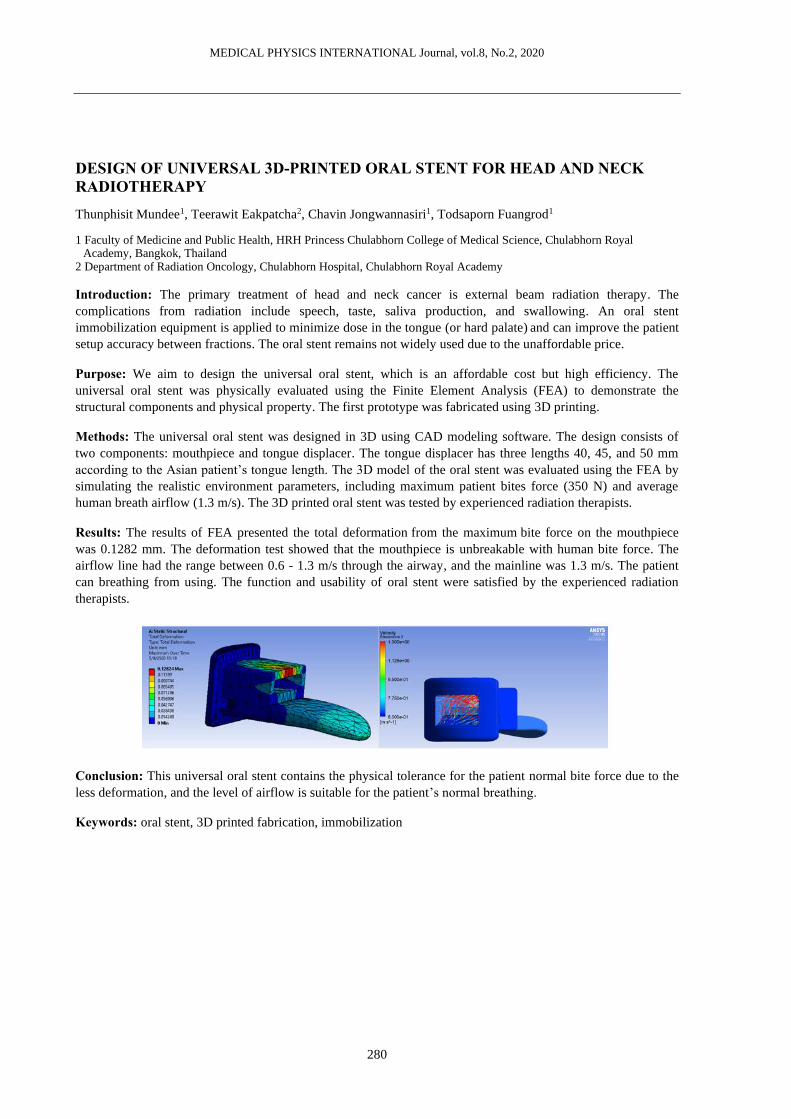
MEDICAL PHYSICS INTERNATIONAL Journal, vol.8, No.2, 2020
280
DESIGN OF UNIVERSAL 3D-PRINTED ORAL STENT FOR HEAD AND NECK RADIOTHERAPY
Thunphisit Mundee1, Teerawit Eakpatcha2, Chavin Jongwannasiri1, Todsaporn Fuangrod1
1 Faculty of Medicine and Public Health, HRH Princess Chulabhorn College of Medical Science, Chulabhorn Royal Academy, Bangkok, Thailand
2 Department of Radiation Oncology, Chulabhorn Hospital, Chulabhorn Royal Academy
Introduction: The primary treatment of head and neck cancer is external beam radiation therapy. The
complications from radiation include speech, taste, saliva production, and swallowing. An oral stent
immobilization equipment is applied to minimize dose in the tongue (or hard palate) and can improve the patient
setup accuracy between fractions. The oral stent remains not widely used due to the unaffordable price.
Purpose: We aim to design the universal oral stent, which is an affordable cost but high efficiency. The
universal oral stent was physically evaluated using the Finite Element Analysis (FEA) to demonstrate the
structural components and physical property. The first prototype was fabricated using 3D printing.
Methods: The universal oral stent was designed in 3D using CAD modeling software. The design consists of
two components: mouthpiece and tongue displacer. The tongue displacer has three lengths 40, 45, and 50 mm
according to the Asian patient’s tongue length. The 3D model of the oral stent was evaluated using the FEA by
simulating the realistic environment parameters, including maximum patient bites force (350 N) and average
human breath airflow (1.3 m/s). The 3D printed oral stent was tested by experienced radiation therapists.
Results: The results of FEA presented the total deformation from the maximum bite force on the mouthpiece
was 0.1282 mm. The deformation test showed that the mouthpiece is unbreakable with human bite force. The
airflow line had the range between 0.6 - 1.3 m/s through the airway, and the mainline was 1.3 m/s. The patient
can breathing from using. The function and usability of oral stent were satisfied by the experienced radiation
therapists.
Conclusion: This universal oral stent contains the physical tolerance for the patient normal bite force due to the
less deformation, and the level of airflow is suitable for the patient’s normal breathing.
Keywords: oral stent, 3D printed fabrication, immobilization

MEDICAL PHYSICS INTERNATIONAL Journal, vol.8, No.2, 2020
281
FABRICATION AND VALIDATION OF CUBIC PHANTOM EMBED WITH AN ACCELEROMETER SENSOR FOR VERIFYING SIX-DEGREES-OF-FREEDOM COUCH
T. Popreeda1, S. Thongsawad2, N. Udee1
1 Department of Radiological Technology, Faculty of Allied Health Sciences, Naresuan University, Mueang, Phitsanulok 65000, Thailand.,
2 Radiation Oncology Unit, Chulabhorn Hospital, HRH Princess Chulabhorn College of Medical Science, Chulabhorn Royal Academy, Laksi, Bangkok 10210, Thailand.
Introduction: In modern medical linacs, the six-degrees‐of‐freedom (6DOF) couch have been implemented to
machine for improving the accuracy of beam delivery. Especially in Stereotactic Radiosurgery (SRS) and
Stereotactic Radiotherapy (SRT), couch can be shifted in pitch, roll, and yaw direction. However, the accuracy
of couch shifted needs to be investigated with comprehensive Quality Assurance (QA).
Purpose: The purpose of this study was to develop cubic phantom embed with an accelerometer sensor for
verifying 6DOF couch.
Methods: Cubic phantom was fabricated by using 3D printing, and an accelerometer sensor was imbedded
inside the phantom to measure couch angle. The study was classified in two main tests: printing accuracy test,
and angle measurement accuracy test. For printing accuracy test, printing parameter included printing speed and
fill density were varied to find optimal parameter. For angle measurement accuracy test, accelerometer sensor
was calibrated with referent spirit level in different angles, and the accuracy was validated in term of precision
and reproducibility. The precision was determined by using difference between cubic phantom measurement and
digital spirit level, whereas the reproducibility was determined by using standard deviation (sd.) of 10-time
repeated measurements.
Results: The optimal parameter was found at printing speed of 50 mm/s, and fill density of 40%. The maximum
difference between cubic phantom measurement and spirit level was -0.05±0.07 degree, and 0.05±0.22 degree
for pitch, and roll direction, respectively. The maximum sd. of 10-time repeated measurements was found at 5
degree with ± 0.09 for pitch direction, and at -4 degree with ±0.19 for roll direction.
Conclusion: In this study, we have developed the new QA tool for verifying 6DOF couch with self-
measurement method, and the accuracy of cubic phantom embed accelerometer sensor was enough for verifying
six-degrees-of-freedom couch.
Keywords: six-degrees‐of‐freedom (6DOF) couch QA, 3D printing, accelerometer sensor

MEDICAL PHYSICS INTERNATIONAL Journal, vol.8, No.2, 2020
282
DAILY MACHINE-SPECIFIC PERFORMANCE MONITORING THROUGH CLINICAL TREATMENT DELIVERY USING STATISTICAL PROCESS CONTROL AND MOBIUSFX
Piyakan Wongsaroj1, Taweap Sanghangthum2, Todsaporn Fuangrod1
1 Medical Physics Program, Faculty of Medicine and Pubic Health, HRH Princess Chulabhorn College of Medical Science, Chulabhorn Royal Academy, Bangkok, Thailand,
2 Division of Radiation Oncology, Department of Radiology, Faculty of Medicine, Chulalongkorn University, Bangkok, Thailand
Introduction: Linear accelerator (linac) quality assurance (QA) aims to check the machine does not
significantly deviate from the controlled values in various both mechanics and dosimetric parts. A machine-
specific performance monitoring through process can enhance the treatment quality, reducing the systematic
errors, and benefit to proactive linac maintenance management.
Purpose: To develop the daily machine-specific performance monitoring using MobiusFX (log-file) with
statistical process control (SPC).
Methods: The daily machine-specific performance monitoring was performed to capture patient delivery data
(machine log-file) in daily bias from Varian Clinac iX linac for 60 days. Total data of 385 log-files were
analysed using MobiusFX (V.2.1.2). SPC in form of X/MR charts was constructed from the first 30 days then
used to monitor linac machine performance including 3D gamma analysis values, root-mean-square (RMS) error
analysis values for gantry, collimator, jaw and MLC motor.
Results: X/MR charts of SPC showed the lower control limits (LCL) of 3D gamma at 95% (as baseline). The
upper control limits (UCL) of RMS error for gantry, collimator, jaws in X1, X2, Y1, Y2 and MLC motor were
1.05 mm, 0.106 mm, 0.33 mm, 0.708 mm, 0.71 mm, 0.607 mm and 1.19 mm, respectively. The LCL of RMS
error values for MLC motor was 0.61 mm. During the linac machine monitoring (since day 31 to 60), the system
flagged the issue twice on Y1-jaw, and once on Y2-jaw. This required setting-up the preventive maintenance for
Y-jaw.
Conclusion: The linac machine performance monitoring through the clinical treatment delivery evaluation using
MobiusFX with SPC demonstrate the potential system to flag the possible malfunction of machine parts
(focusing on finding the systematic errors). This can help medical physicist to manage their preventive
maintenance efficiently and minimizing the unexpected machine downtime in the future.
Keywords: Daily machine-specific performance monitoring, Statistical process control, MobiusFX

MEDICAL PHYSICS INTERNATIONAL Journal, vol.8, No.2, 2020
283
ESTABLISHMENT OF MOH MEDICAL PHYSICS RESEARCH TASK FORCE
Aik Hao Ng1, Hwee Shin Soh2
1 Department of Radiotherapy and Oncology, National Cancer Institute, Putrajaya, Malaysia. 2 Medical Radiation Surveillance Division, Ministry of Health Malaysia.
Introduction: Medical physicist is a relatively small profession in the Malaysian Ministry of Health (MOH)
settings. The core businesses of the profession within the organisation are to provide clinical support and
regulatory services. Some of the medical physicists have limited access to adequate research support and felt
isolated in their work environment.
Purpose: This presentation sets out to report the establishment the research task force. This initiative aims to
cultivate research culture amongst the medical physicists in the MOH and to provide research support.
Methods: The task force was formed in 2019, consisted of 21 volunteered medical physicists nationwide from
both clinical and regulatory sectors. A SWOT analysis was carried out to examine the current status of research
activities within the organisation.
Results: Four sub-groups have been established to execute the aims of the task force, namely research (1)
promotion, (2) support, (3) requirements as well as (4) activities and database. The group outreaches the
profession through scientific meetings, email and mobile instant messaging application (WhatsApp) to
continuously promote research awareness and to share timely information. Workshop and consultation were
organised to provide guidance and support to the young professionals. The group also explored potential
research collaboration with academic and industrial partners.
Conclusion: The initiative offers a new platform to cultivate research culture and encourage more research
activities to be carried out, which could provide added values to the healthcare services. We hope to continue to
explore different ways to succeed this work and encourage more volunteers to join us.
Keywords: research, medical physicists, task force
MEDICAL PHYSICS INTERNATIONAL Journal, vol.8, No.2, 2020
284
CT DENSITY PROFILE AND PHYSICOCHEMICAL STUDY OF SOY-LIGNIN BONDED RHIZOPHORA SPP. PARTICLEBOARD AS PHANTOM MATERIAL IN MEDICAL PHYSICS APPLICATION
Siti Hajar Zuber1, Nurul Ab. Aziz Hashikin1, Mohd Fahmi Mohd Yusof2, Rokiah Hashim3
1 School of Physics, Universiti Sains Malaysia, 11800, Penang, MALAYSIA. 2 School of Health Sciences, Universiti Sains Malaysia, 16150, Kelantan, MALAYSIA. 3 School of Industrial Technology, Universiti Sains Malaysia, 11800, Penang, MALAYSIA.
Introduction: The utilization of natural adhesive in the fabrication of particleboard for medical physics
application has been investigated and many proved capable of replacing the well-integrated formaldehyde
adhesive. Soy flour and lignin had been extensively used as adhesive and more studies had proved the suitability
of both adhesives in the production of sturdier particleboards. CT study can accurately estimate the attenuation
coefficients for soft tissue, thus providing an accurate determination of density, electron density and density
profile of the fabricated particleboard.
Purpose: We aim to evaluate the physical and mechanical properties of the particleboards, the determination of
CT number, electron density and density profile of the material using fabricated density plug phantom.
Methods: Density determination, internal bonding, modulus of rupture, water absorption, and thickness swelling
were performed. Microstructure study using scanning electron microscopy (SEM) and elemental analysis by
carbon hydrogen nitrogen (CHN) analyser were also investigated. We also study the Computed Tomography
(CT) number, relative electron density and the density profile analysis of the phantoms.
Results: Particleboards with adhesives improved internal bond strength. Smaller particle sizes also have shown
to be able to improve the thickness swelling outcomes, with lower hygroscopic properties. The SEM images
showed that smaller particle size allowed better bonding with adhesives and provided superior strength. The
CHN ratio revealed no major difference when compared with the samples which were crucial in the fabrication
of tissue-mimicking phantom. The bonded particleboard showed close similarities with water, based on the
average CT numbers, CT calibration curve, electron density calibration curve and the analysis of CT density
profile, compared to the binderless particleboard.
Conclusion: The CT and physiochemical study of soy-lignin bonded Rhizophora spp. particleboard exhibited its
suitability as an alternative phantom material for applications in medical physics.
Keywords: Physical properties; Mechanical properties; CT Electron Density phantom; particleboard fabrication;
soy-lignin adhesive

MEDICAL PHYSICS INTERNATIONAL Journal, vol.8, No.2, 2020
285
Oral Presentations
Dignostic: Imaging

MEDICAL PHYSICS INTERNATIONAL Journal, vol.8, No.2, 2020
286
CORRELATION OF ULTRASOUND ATTENUATION IMAGING VERSUS MRI PROTON DENSITY FAT FRACTION IN NON-ALCOHOLIC FATTY LIVER
Pantajaree Hiranrat1, B.Sc., Surachate Siripongsakun1, M.D., Kamonwan Soonklang2, M.Sc.,
Chanisa Chotipanich1, M.D.
1 Sonographer School, Faculty of Health Science Technology, Chulabhorn Royal Academy, Bangkok 10210, Thailand, 2 Data Management Unit, HRH Princess Chulabhorn College of Medical Science, Chulabhorn Royal Academy, Bangkok
10210, Thailand,
Introduction: Attenuation Imaging (ATI) is a novel ultrasound method for assessment degree of hepatic
steatosis, based on ultrasound attenuation by calculating attenuation coefficient which will increase in fatty liver
condition. The previous published data comparing ATI and Magnetic Resonance Proton Density Fat Fraction
(MR-PDFF) has moderate to high correlation coefficient (r = 0.66-0.81). However, fatty liver is commonly
associated with obesity which may be an influencing factor of the ATI measurement.
Purpose: The purpose of this study was to evaluate correlations and interobserver liability of ATI comparing
with MRI-PDFF in obese patients.
Methods: In this study, the non-alcoholic fatty liver disease (NAFLD) patients with available ATI and MRI-
PDFF examination, excluding cirrhosis, history of significant alcohol drinking, and chronic liver condition were
evaluated. Correlation of ATI and MRI-PDFF values and interobserver’s concordance of ATI measurements
were evaluated, using Spearman rank correlation and Cohen’s kappa coefficient. Statistical significance is
considered at 2 side p value < 0.05.
Results: There were 62 patients included with average BMI of 27.4 kg/m2. The correlation coefficient (r) of ATI
vs MRI-PDFF were in moderate correlation (r = 0.63-0.69, p<0.001). With the highest correlation coefficient at
right posterior segment measurement (r=0.69). The inter-observer reliability of two observers were 100% with
Cohen kappa coefficient of 1.00 (p < 0.001).
Conclusion: ATI is a novel ultrasound method to quantify degree of fat deposition with good correlation to
MRI-PDFF with high interobservers’ reliability. Obesity commonly associated with fatty liver may be an
interfering factor of ATI measurement.
Keywords: Attenuation Imaging, Fatty liver, Steatosis, MRI-PDFF, MRE, NAFLD

MEDICAL PHYSICS INTERNATIONAL Journal, vol.8, No.2, 2020
287
SONOGRAPHIC PREDICTORS FOR DEVELOPING CHOLANGIOCARCINOMA: A COHORT STUDY FROM AN ENDEMIC AREA.
Natcha Thanakijsombat1,2, Jiraporn Laothamatas1, Kamonwan Soonklang3, Surachate Siripongsakun1
1 Sonographer School, Faculty of Heath Science Technology HRH Princess Chulabhorn College of Medical Science, Bangkok, Thailand
2 Department of Emergency Medicine, Police General Hospital, Bangkok, Thailand 3 Data Management Unit, HRH Princess Chulabhorn College of Medical Science, Chulabhorn Royal Academy, Bangkok,
Thailand
Introduction: Cholangiocarcinoma (CCA) is an aggressive malignancy with rapid progression and poor
prognosis. Bile duct and peribiliary changes related to cholangiocarcinogenesis may present on sonographic
findings for example; periportal fibrosis, and non-specific diffuse bile duct dilatation without obstruction.
Purpose: This study aim is to evaluate ultrasound findings which could be used as predictors for developing
CCA through our surveillance program in an endemic area, Nan province, Thailand.
Methods: The surveillance population-based cohort was 4,337 villagers in Northern Thailand, aged 30-60
years, who consented to a 5-year abdominal ultrasound surveillance program which included interval ultrasound
examinations every 6 months. Patient’s demographics including age and gender, family history of CCA,
ultrasound findings including the presence of calcifications, diffuse biliary dilatation, and periportal fibrosis were
included. A logistic regression model was used to determine significant predictors using forward stepwise
generalized linear model for multivariate analysis.
Results: In this cohort, there were 4,337 patients, between 30-60 years, with average age of 45.37±7.7 years,
consisted of 2,360 females and 1,977 males, respectively. On univariate analysis; the significant predictors with
increased risk of developing CCA included age (relative risk (RR) = 1.11, p < 0.001), family history of CCA
(RR = 2.58, p < 0.001), hepatic calcifications/granuloma (RR = 0.74, P=0.38), diffuse biliary system dilatation
(RR = 5.02-17.62, both p < 0.001, respectively), periportal fibrosis (RR 3.11, p < 0.001). On multivariate
analysis the significant independent predictors associated with developing CCA include age (RR = 1.09, p <
0.001), diffuse biliary system dilatation (RR = 7.03-22.59, p < 0.001)
Conclusion: Sonographic predictors of CCA development including age and diffuse bile duct dilatation may be
helpful in identifying the population at risk in order to target for surveillance in CCAs endemic areas.
Keywords: Cholangiocarcinoma, Predictor, Sonography, Surveillance

MEDICAL PHYSICS INTERNATIONAL Journal, vol.8, No.2, 2020
288
VALUE OF SUPERB MICROVASCULAR IMAGING (SMI) IN CHARACTERIZE OF NODULAR TYPE FOCAL FAT SPARING LESION AND TRUE HEPATIC NODULES IN THE BACKGROUND OF FATTY LIVER
Songpon Srisittimongkon1, Surada Vichitpunt1, Siwat Bhumiwat1, Pantajaree Hiranrat1,
Surachate Siripongsakun1
1 Sonographer School, Faculty of Health Science Technology, Chulabhorn Royal Academy, Bangkok 10210, Thailand,
Introduction: Fatty liver is a common condition which increases liver parenchyma echogenicity. This condition
often associated with an area of focal fat sparring (FFS) due to inhomogenous fatty deposition. Increase
parenchymal background from the fatty liver also alters the echogenic appearance of liver lesions which will be
relatively hypo-echogenic. Superb microvascular imaging (SMI) is a novel microvascular imaging technique
that allows visualization of vascular architectures of the lesions which potentially distinguish true nodules from
FFS pseudo-lesions.
Purpose: The study was to evaluate the value of SMI in characterizing hypoechoic lesion in fatty liver
background.
Methods: This prospective study recruited patients who had fatty liver with hypoechoic nodular lesions,
between 0.8-4.0 cm in size, which had definite characterization on CT or MRI. Size, margin, and SMI patterns,
of hypoechoic lesions of true and FFS psudeo-lesions were evaluated using t-test and Fischer’s exact test. SMI
vascular patterns are classified according to Lee et al.
Results: There were 44 patients with fifty-seven hypoechoic lesions, consisting of 22 FFS lesions and 35 true
hepatic nodules including twenty-two hemangiomas, seven metastases, three focal nodular hyperplasias (FNHs),
two regenerating nodules and two parasitic related abscesses. True nodules and FFS pseudo-lesions showed
well-defined margin in 34/35(97%) and 8/22 (36%, p < 0.001), respectively. Almost all FFS showed non-
specific or no signal pattern (19/22, 85%), which overlapped with true nodules in (17/35, 57%). Rarely FFS has
other patterns including nodular rim and staining (3/22, 15%). Nodular rim with dot-like and spoke-wheel SMI
patterns were found only in hemangiomas and FNHs, respectively.
Conclusion: In fatty liver, FFS and true nodules are overlapped in US appearance. A few patterns of SMI
including nodular-rim with dot-like and spoke-wheel patterns may suggest a specific type of lesions including
hemangiomas and FNHs.
Keywords: Fatty liver, Hypoechoic, Superb Microvascular Imaging, Ultrasound
MEDICAL PHYSICS INTERNATIONAL Journal, vol.8, No.2, 2020
289
PREDICTING TREATMENT RESPONSE IN NASOPHARYNGEAL CANCER USING RADIOMICS: A PRELIMINARY STUDY
Sasawan Wongpisarn1,5, Chawalit Lertbutsayanukul2, Nutchawan Jittapiromsak3, Yothin Rakvongthai4,5
1 Medical Physics Program, Department of Radiology, Faculty of Medicine, Chulalongkorn University, Bangkok, Thailand 2 Division of Radiation Oncology, Department of Radiology, Faculty of Medicine, Chulalongkorn University, Bangkok,
Thailand 3 Division of Diagnostic Radiology, Department of Radiology, Faculty of Medicine, Chulalongkorn University, Bangkok,
Thailand 4 Division of Nuclear Medicine, Department of Radiology, Faculty of Medicine, Chulalongkorn University, Bangkok,
Thailand 5 Chulalongkorn University Biomedical Imaging Group, Department of Radiology, Faculty of Medicine, Chulalongkorn
University, Bangkok, Thailand
Introduction: It is necessary to monitor a treatment for nasopharyngeal cancer (NPC) to assess its response.
Radiomics is a technique that extracts large amount of data from medical images in terms of features which
reflect tumor characteristics.
Purpose: Our hypothesis was that radiomics features from diffusion-weighted imaging (DWI) could be used as
imaging biomarkers for concurrent chemoradiation therapy (CCRT) treatment response. Here we investigated
the use of such radiomics features for treatment response prediction in NPC patients.
Methods: We collected seventeen patient datasets including thirteen complete response (CR) patients and four
partial response (PR) patients where one patient dataset consisted of DWI and apparent diffusion coefficient
(ADC) data acquired before (i.e. pre-treatment) and at five weeks after (i.e. mid-treatment) initiation of CCRT.
For each dataset, we calculated the radiomic feature values using PyRadiomics software from pre-treatment and
mid-treatment, and computed the percentage change of each feature, called Delta Radiomic (∆Radiomic) feature.
To validate the performance in differentiating CR and PR patients, we calculated the mean, standard deviation
(SD) across CR and PR patients, and area under the receiver operating characteristic (ROC) curve (AUC) of
each feature and its ∆Radiomic, where tumor response was from 6-month follow-up data using RECIST1.1
guideline.
Results: There were 1 radiomic feature value at pre-treatment and 32 ∆Radiomic feature values that yielded a
significant difference (p<0.05) between CR and PR groups. The AUC values from these 32 ∆Radiomic feature
values ranged from 0.85 to 0.94, which were all higher than those from percentage change values of
conventional imaging biomarkers such as volume, mean, uniformity and entropy (0.48, 0.81, 0.44, and 0.46,
respectively). ∆Radiomic feature values that yielded the highest AUC value were from sphericity and wavelet-
LLH_glcm_DifferenceVariance.
Conclusion: Radiomics-based biomarkers could potentially be used for early treatment response prediction in
NPC patients.
Keywords: Radiomics, Nasopharyngeal cancer, DWI

MEDICAL PHYSICS INTERNATIONAL Journal, vol.8, No.2, 2020
290
PERFORMANCE OF QUANTITATIVE LESION MEASUREMENT IN HEPATIC DUAL-ENERGY COMPUTED TOMOGRAPHY (DECT): PHANTOM STUDY
Hataipat Jantawong1, Anchali Krisanachinda2
1 Doctor of Philosophy Program in Medical Physics, Faculty of Medicine, Chulalongkorn University, Bangkok, Thailand 2 Department of Radiology, Faculty of Medicine, Chulalongkorn University, Bangkok, Thailand
Introduction: The quantitative measurements of iodine uptake, size and volume of liver lesion are essential for
diagnostic and treatment response assessment of HCC. Recently, the dual energy computed tomography (DECT)
acquisition has been adopted and widely used in abdominal imaging.
Purpose: To evaluate the performance of quantitative lesion measurements (both the iodine enhancement and
size) in hepatic DECT as a function of imaging parameters using 3D-printing liver lesion phantom.
Methods: The three-dimensional printing technique was used to construct liver phantom with various in size of
synthetic hyperenhancement spherical liver lesions; 6,8,10,15,19,20,25 mm in diameter and different iodine
concentrations of 18,20,22,24,26 mg-iodine per mL (mgI/mL). Total of 35 lesions were scanned using DE
acquisition with the third generation of dual source DECT (DS-DECT). The kV-combination was selected as
80/150Sn,90/150Sn and 100/150Sn. The radiation dose levels were varied to achieve targeted CTDIvol of 15,20,
and 25 mGy. The images of low-and high-kV were sent to vendor-specific DE software to measure the iodine
quantification for each lesion size and iodine concentration. Absolute error of iodine quantification was
measured. The virtual monoenergetic images (VMIs) were created at different photon energy from 40- 80-
keV(10 keV interval) images and were sent to commercial software to perform semi-automated measurements of
diameter and volume of various liver lesions and then estimated for the measurement error.
Results: Among different DE parameters, the iodine quantification error was lower when increasing the lesion
size and iodine concentration. However, at higher dose level, size and volume, the errors were improved. In
addition, at the standard and high dose level, lower photon energy of VMIs provided the better performance.
Conclusion: The scanning and post-processing DE parameters have potential impact on iodine quantification
error and volumetric measurement. In addition, the lesion characteristics (size and iodine concentration) affect
the accuracy of iodine quantification and volumetry.
Keywords: dual-energy computed tomography, hepatocellular carcinoma, iodine quantification, virtual
monoenergetic images, liver 3D-priting phantom

MEDICAL PHYSICS INTERNATIONAL Journal, vol.8, No.2, 2020
291
AUTOMATIC QC ANALYSIS PROGRAM IN DIGITAL RADIOGRAPHY SYSTEM
Jongwat Cheewakul1, Natee Ina
1 Department of Radiology Faculty of Medicine Prince of Songkla University, Songkla, Thailand
Introduction: Quality Control of the diagnostic radiology system is the test of the quality and function of the X-
ray machine and to observe the defects, deterioration or damage of the equipment by Medical Physicist. The
developed program analyses the automatic image quality control in digital x-ray systems can help Medical
Physicist to reduce the time spent in the analysis the test results with more convenience.
Purpose: The purpose of this study was to design automatic quality control analysis program and to compare the
test results from this program with the results from three observers.
Methods: This developed analysis program using MATLAB could assess the image quality in terms of the
spatial resolution, low contrast detail detectability and high contrast detail detectability. The Routine Test Object
for conventional and non-subtractive digital radiography, TOR CDR test tool was exposed by three digital X-ray
machines, one image for each X-ray machine was obtained. Then three images were assessed by developed
software and three observers.
Results: The results show that the evaluation from the developed software and three observers are comparable.
The spatial resolution evaluated by the developed software were 3.55, 3.15, 2.80 lp/mm and by three observers
was 3.55, 2.80, 2.80 lp/mm respectively. The low-contrast detectability evaluated by the developed software
was 16, 15, 17 and by three observers were 16, 16, 15 respectively. The high-contrast detectability evaluated by
the developed software was 17, 17, 17, and by three observers were 16, 17, 17 respectively. The average
assessment time evaluated by the developed software was 1.30 min and by manual procedure was 3.15 min.
Conclusion: This automatic QC analysis program can be used with the reduction in the assessment time and be
able to record the results to compare with the previous results.
Keywords: Quality Control, Spatial Resolution, Low and High Contrast Detectability, Digital Radiography
System

MEDICAL PHYSICS INTERNATIONAL Journal, vol.8, No.2, 2020
292
CORRELATION BETWEEN RENAL HISTOLOGICAL FIBROSIS AND RENAL CORTICAL THICKNESS USING SHEAR WAVE ELASTOGRAPHY IN PATIENTS WITH KIDNEY DISEASE
Sook Sam Leong1,2, Jeannie Hsiu Ding Wong1,3, Mohammad Nazri Md Shah1,3, Anushya Vijayananthan1,3,
Maisarah Jalalonmuhali4, Tak Kuan Chow5, Nur Hidayati Mohd Sharif6, Kwan Hoong Ng1,3
1 Department of Biomedical Imaging, University of Malaya, 50603 Kuala Lumpur, Malaysia 2 Department of Biomedical Imaging, University of Malaya Medical Centre, 50603 Kuala Lumpur, Malaysia 3 University of Malaya Research Imaging Centre, University of Malaya, 50603 Kuala Lumpur, Malaysia 4 Department of Medicine, University of Malaya, 50603 Kuala Lumpur, Malaysia 5 Department of Pathology, University of Malaya, 50603 Kuala Lumpur, Malaysia 6 Renal Unit, Faculty of Medicine, University Teknologi MARA, 47000 Sungai Buloh, Malaysia
Introduction: Renal biopsy is the gold standard for the histological characterisation of chronic kidney disease
(CKD), of which renal fibrosis is a dominant component, affecting its stiffness.
Purpose: This study aims to investigate the correlation between kidney stiffness obtained by SWE and renal
histological fibrosis.
Methods: SWE assessments were performed in 75 CKD patients who underwent renal biopsy. The SWE-
derived estimates of the tissue Young’s modulus (YM), given as kilopascals (kPa), were measured. YM was
compared to patients’ renal histological scores, broadly categorised into glomerular, tubulointerstitial and
vascular scores.
Results: YM correlates significantly with tubular interstitial score (= 0.442, p< 0.001) and glomerular score
(= 0.375, p= 0.001). Patients with no glomerular sclerosis showed lower mean YM measurements compared
those with glomerular sclerosis. The mean YM increased as the percentage of interstitial fibrosis and tubular
atrophy increased. The area under the ROC curve for SWE in differentiating between mildly and moderately
impaired kidneys was 0.702.
Conclusion: SWE accurately detects chronic renal damage resulting from glomerular sclerosis, interstitial
fibrosis and tubular atrophy, using the optimal cut-off YM value of 5.81 kPa.
Keywords: Biopsy; Fibrosis; Kidney; Shear wave elastography; Young’s modulus

MEDICAL PHYSICS INTERNATIONAL Journal, vol.8, No.2, 2020
293
AUTOMATIC STRATIFICATION OF PROSTATE CANCER PATIENTS INTO LOW- AND HIGH-GRADE GROUPS BASED ON MULTIPARAMETRIC MR IMAGE FEATURE ANALYSIS
Akimasa Urakami1, Hidetaka Arimura2, Fumio Kinoshita3 Kenta Ninomiya1, Sumiko Watanabe2,
Kenjiro Imada4, Yoshinao Oda3
1 Department of Health Sciences, Graduate School of Medical Sciences, Kyushu University 2 Department of Health Sciences, Faculty of Medical Sciences, Kyushu University 3 Department of Anatomic Pathology, Graduate School of Medical Sciences, Kyushu University 4 Department of Urology, Prostate, Kidney, Adrenal Surgery, Kyushu University Hospital
Introduction: Multiparametric magnetic resonance (MR) imaging could be utilized for non-invasive grading of
prostate cancer1,2.
Purpose: This study aimed to investigate the potential of automatic stratification of prostate cancer patients into
low- and high-grade groups based on multiparametric MR image feature analysis.
Methods: MR images of 66 cancer regions corresponding to histopathological images were identified and
divided into a training (n = 40) and test datasets (n = 26). Each dataset was divided into two groups, low-grade
tumors (≤GG2) and high-grade tumors (GG3≤), based on their grade groups. A total of 4320 MR image features
were calculated from 16 types of images in 5 sequences for each cancer region and some of them were selected
by using a least absolute shrinkage and selection operator (LASSO) and regularized logistic regression using
elastic net algorithm as a signature candidate. Next, a combination strategy was used to extract a signature from
the signature candidate. Finally, support vector machines (SVM) models were built using the signature in the
training dataset. Leave-one-out cross validation was applied to feature extraction and training of the models.
Model performances were evaluated using areas under receiver operating characteristic curves (AUCs).
Results: The AUC based on the SVM models of the stratification of prostate cancer patients into two groups
were 0.993 in the training dataset and 0.550 in the test dataset.
Conclusion: This study showed the potential of automatic stratification of prostate cancer patients into low- and
high-grade groups based on multiparametric MR image feature analysis.
Keywords: prostate cancer, grade group, MR image features, regularized logistic regression using elastic net,
support vector machine
MEDICAL PHYSICS INTERNATIONAL Journal, vol.8, No.2, 2020
294
IMAGE ANALYSIS OF PANCREATIC CANCER TISSUES USING HARALICK FEATURES
Ashleigh Hull1, Yanrui Li1, Dylan Bartholomeusz2,3, William Hsieh1,2 & Eva Bezak1,4
1 Cancer Research Institute & Allied Health and Human Performance Academic Unit, University of South Australia, Adelaide, Australia,
2 Department of PET, Nuclear Medicine & Bone Densitometry, Royal Adelaide Hospital, SA Medical Imaging, Adelaide, Australia,
3 Adelaide Medical School, The University of Adelaide, Adelaide, Australia, 4 Department of Physics, The University of Adelaide, Adelaide, Australia
Introduction: Cellular heterogeneity complicates the development of targeted therapies for pancreatic cancer.
Despite studies assessing the molecular basis of this heterogeneity, few studies have established the implications
of heterogeneity on the overall texture of pancreatic cancer. Haralick Features (HF) provide 14 textural
descriptors, such as contrast and homogeneity, to characterise images based on the spatial distribution of pixel
values.
Purpose: The aim of this study was to identify textural differences between images of normal, pancreatic cancer
and chronic pancreatitis tissues using HF.
Methods: Two-hundred and eighty pancreatic tissue samples consisting of normal (n = 97), primary pancreatic
cancer (n = 106) and chronic pancreatitis (n = 77) pathology were immunohistochemically stained using C595,
an anti-mucin 1 antibody. Stained tissue samples were imaged using a NanoZoomer Digital Slide Scanner
(Hamamatsu Photonics, Japan), with images converted to 8-bit grayscale by Image J (v.1.52, National Institutes
of Health, USA). A gray level co-occurrence matrix was determined for each image, allowing for calculation of
HF values using MATLAB (v. 2020a, MathWorks, USA). Statistical analysis of the HF across the disease
groups was performed using GraphPad Prism (v.8.2.0, GraphPad Software, USA).
Results: Preliminary results demonstrated significant differences for all 14 HF between the pancreatic cancer
and normal pancreatic tissue samples (p < 0.0006). There were significant differences between chronic
pancreatitis and pancreatic cancer tissues for 12 HF (p < 0.03). Correlation (p = 0.36) and Information Measure
of Correlation I (p = 0.7915) demonstrated no significant differences between the chronic pancreatitis and
pancreatic cancer groups.
Conclusion: This study demonstrates quantifiable differences between the texture of pancreatic cancer tissue to
both normal tissue and pancreatitis. Consideration of these textural differences may be valuable for the
development of targeted therapies by improving treatment stratification with potential extension into artificial
intelligence.
Keywords: Haralick Features, pancreatic cancer, textural analysis.

MEDICAL PHYSICS INTERNATIONAL Journal, vol.8, No.2, 2020
295
DIGITAL BREAST TOMOSYNTHESIS (DBT): DOSE AND IMAGE QUALITY ASSESSMENT OF MAN-MADE BREAST PHANTOM
Nur Farrah Ezlyn Mohammad Rusdi1, Nur Afizatul Izni Mohd Jasri1, Yasmin Md Radzi1, Noor Diyana Osman2
1 School of Physics, USM, Malaysia, 2 Advanced Medical and Dental Institute, Universiti Sains Malaysia, Penang, Malaysia.
Introduction: Digital Breast Tomosynthesis (DBT) system offers a gain in sensitivity and specificity in breast
cancer detection of overlying tissues compared to the existing 2D mammography. To truly evaluate the clinical
systems of intended diagnostic tasks, a realistic three dimensional (3D) anthropomorphic breast phantom is
essential but 3D prototyping is very expensive. Therefore, a simple phantom is vital to practice good and
efficient breast screening while capable of providing optimum compromise between dose and image quality.
Purpose: In this study, we aim to optimize the parameters in 3D breast screening with excellent compromise
between dose and image quality in addition to determine the ideal breast thickness to provide good image quality
while minimizing dose exposure to the patient.
Methods: A simple customized gelatine-based2 phantom with two different overlying masses mimicking tumour
and microcalcification was used throughout the study in order to assess the dose and image quality during breast
screening. Mean Glandular Dose and Signal-Noise-Ratio are assessed for the absorbed dose and image quality
assessment at different thicknesses of breast phantom using 2D and 3D tomosynthesis of 45° arc movement
(Siemens Healthineers, USA). Optimum parameters are evaluated, and comparison between both methods are
discussed.
Results: An ideal breast thickness (approximately 36.78mm) was determined from the intercept point of
Average Glandular Dose (AGD) and Optical Density as well as Signal-to-Noise Ratio (SNR). The benefits of
3D imaging compared to 2D imaging were also observed in determining the optimum parameter for a thorough
patient’s breast screening.
Conclusion: Understanding the image creation process as it relates to the interplay of contrast, noise, patient
dose, and diagnostic performance is vital in the practice of modern radiology including mammography. When a
lesion needs to be more visible, this could be achieved by increasing the contrast, reducing noise, or by a
judicious combination of both aspects.
Keywords: Digital breast tomosynthesis (DBT), Average Glandular Dose (AGD), Mean Glandular Dose
(MGD), Signal-to-Noise Ratio (SNR), Optical density,
MEDICAL PHYSICS INTERNATIONAL Journal, vol.8, No.2, 2020
296
GREY MATTER SEGMENTATION OF T1-WEIGHTED MR IMAGE IN ISCHEMIC STROKE PATIENTS USING K-MEANS CLUSTERING
Dian Yuliani Alam
Department of Physics, University of Brawijaya, Malang, Indonesia
Introduction: The grey matter in the brain plays an important role to process the information. In ischemic stroke
patients, it could experience some change. This could be diagnosed through the T1-weighted MR (Magnetic
Resonance) image, since it shows the grey matter brighter. The T1-weighted MR image is based on the
longitudinal relaxation, which the energy of radio-frequency is released to the surrounding tissue.
Purpose: In this research, the grey matter segmentation was conducted to confirm the principle of T1-weighted
image as well as the results of researches in ischemic stroke patients related to the grey matter.
Methods: The MRI data were collected in 10 ischemic stroke patients and 10 healthy controls. The Time Echo
(TE) parameter in the T1-weighted image was analysed. The used image segmentation method was k-means
clustering based on the pixel intensity using MATLAB software.
Results: The obtained result showed that the T1-weighted axial images of all patients had short TE values (less
than 20 ms). The pixel intensity of grey matter was higher than cerebrospinal fluid and white matter. This had a
correlation with short TE values. As for the mean pixel intensity of grey matter in ischemic stroke patients and
healthy controls were 135.53 and 150.76, respectively.
Conclusion: Since the mean pixel intensity of grey matter in ischemic stroke patients was lower than healthy
controls, it could be concluded that the ischemic stroke reduces the grey matter in the brain.
Keywords: Grey matter, T1-weighted image, ischemic stroke, k-means clustering
MEDICAL PHYSICS INTERNATIONAL Journal, vol.8, No.2, 2020
297
Oral Presentations
Diagnostic: Big data, Deep learning, AI and Modeling

MEDICAL PHYSICS INTERNATIONAL Journal, vol.8, No.2, 2020
298
THE EFFECTIVE METHODS FOR LIVER CYST LABELLING IN ULTRASOUND IMAGES BY USING R-CNN
Sutthirak Tangruangkiat1, Anchali Krisanachinda2, Supatana Auetavekiat3
1 Ph.D. program in Medical Physics, Faculty of Medicine, Chulalongkorn University, Thailand. 2 Department of Radiology, Faculty of Medicine, Chulalongkorn University, Thailand. 3 Department of Electrical Engineering, Faculty of Engineering, Chulalongkorn University, Thailand.
Introduction: Ultrasound imaging is one of the most popular medical imaging modality because of its non-
ionizing radiation and real time imaging. Deep learning has been successfully applied in medical imaging as
computer aided diagnosis (CAD). However, the limitation on ultrasound imaging of the same target can be
variable according to the users’ technique and the equipment setup, leading to the inaccurate classification.
Purpose: To design the training data for a region-based convolution neural network (R-CNN) for cyst detection
in liver ultrasound images.
Methods: R-CNN using ResNet50 as a classifier was used as an object detector. The ground truth of a cyst was
provided in two sets. On the first set, only the area of the cyst was used as a ground truth. On the second set, in
addition to the cyst, the acoustic enhancement artefact below the cyst, the distinct feature of the cyst, was
included into the ground truth. The five-fold cross validation was performed on 615 liver cyst images to evaluate
the classification accuracy.
Results: The classification accuracy was 84.39% and 90.73% for the R-CNN trained using only cysts and the
combination of a cyst and its enhancement artefact, respectively. Two main causes of error are: (1) failure to
detect the cyst in a low-resolution image and (2) the detection of a vessel as a cyst. Both errors were reduced
when the artefact was included to the training image.
Conclusion: The distinct feature of a target should be included into the training data of R-CNN to improve
classification accuracy.
Keywords: Deep learning, ultrasound image, liver, cyst, artefact, R-CNN

MEDICAL PHYSICS INTERNATIONAL Journal, vol.8, No.2, 2020
299
QUANTITATIVE ASSESSMENT OF BREAST DENSITY USING PIXEL INTENSITY THRESHOLD
Jaroonroj Wongnil1, Anchali Krisanachinda1, Rajalida Lipikorn2
1 Ph.D.in Medical Physics, Department of Radiology Faculty of Medicine, Chulalongkorn University, Bangkok, Thailand 2 Department of Mathematics and Computer Science, Faculty of Science, Chulalongkorn University, Bangkok, Thailand
Introduction: Breast cancer is the most common cancer with the highest mortality in women worldwide. Breast
density can be used to evaluate the risk of breast cancer and the routine visually assessment. It is difficult to
distinguish variably assigned BI-RADS categories. The visual assessment is still important where automated
assessment systems had not been available.
Purpose: To evaluate quantitative breast density using pixel intensity threshold correlate to visual assessment
method.
Methods: The study consists of 2,000 full-field digital screening mammogram with 1,000 craniocaudal (CC)
and 1,000 mediolateral oblique (MLO) views in Thai patients. Breast density of almost entirely fatty (AF),
scatter areas of fibro-glandular density (SD), heterogeneous dense (HD) and extremely dense (ED) at 400, 600,
600 and 400 images had been collected on both views (CC and MLO). All mammograms were classified using
BI-RADS, along with the radiologist classification and retrieved from PACS (Picture Archiving and
Communication System). The pre-process covers the identification of the breast region, the background removal,
and labelling pectoral muscle areas using MATLAB Image segmentation. The ratio of breast density was
calculated by summation of specific breast areas divided by total breast area. The breast density ratio of each
breast classification was obtained and validated by using the new set of mammographic images.
Results: The average (min-max) breast density ratio of AF, SD, HD and ED were 0.366 (0.319-0.427),
0.392(0.309-0.460), 0.445(0.408-0.491) and 0.446(0.400-0.541), respectively. The results were concordant with
visual assessment by radiologist which are difficult in distinguish between HD and ED.
Conclusion: The quantitative assessment of breast density using pixel intensity threshold conformed to visual
assessment methods. The classification of breast density between HD and ED are still challenging, using both
quantitative and qualitative methods to assess which may improve the accuracy and consistency of breast density
classification.
Keywords: Breast density, histogram, BI-RADS, pixel intensity threshold

MEDICAL PHYSICS INTERNATIONAL Journal, vol.8, No.2, 2020
300
OVERALL SURVIVAL PREDICTION OF NASOPHARYNGEAL CARCINOMA USING RADIOMICS
Sararas Intarak1,2, Yothin Rakvongthai2,3
1 Medical Physics program, Department of Radiology, Faculty of Medicine, Chulalongkorn University 2 Chulalongkorn University Biomedical Imaging Group, Department of Radiology, Faculty of Medicine, Chulalongkorn
University 3 Division of Nuclear Medicine, Department of Radiology, Faculty of Medicine, Chulalongkorn University
Introduction: Nasopharyngeal carcinoma (NPC) is endemic disease in Southeast Asia and Southern China. The
current patient stratification is based on TNM staging and Epstein-Barr Virus (EBV) DNA concentration level
test. Patients are categorized into high-risk and low-risk group to provide appropriate treatment according to
tumor prognosis. However, EBV value undetectable in 41 percent of the patients (1), for which an appropriate
treatment option is difficult to choose. Radiomics, an emerging field for data mining of medical images (2), was
used in this study. The underlying hypothesis is that radiomics features describing size, shape, texture, and voxel
intensity in the images may reflects the characteristics of the tumor. The quantitative analysis may provide better
risk stratification for NPC.
Purpose: To investigate the prognostic value of radiomics in NPC from pre-treatment CT images.
Methods: A total of 197 patients were collected retrospectively. They had CT examinations prior to treatment
and had at least 3 years of follow-up data. The radiomic features were extracted through Pyradiomics version
2.7. The tumor delineation was performed by board-certified radiation oncologists. 158 and 39 patients were
randomly classified into train group and test group, respectively. The radiomic feature selection and model
construction were performed by STATA version 15. The univariate and multi-variable analyses were performed
by logistic regression and ROC analysis was constructed.
Results: The AUC of radiomics model was 0.7927 in train group and 0.7463 in test group.
Conclusion: Our preliminary results demonstrated the usefulness of radiomics in tumor prognosis for
nasopharyngeal cancer. Radiomics could potentially be applied to improve decision making in clinical practice
throughout cancer therapy process at low cost.
Keywords: Radiomics, Overall Survival, Nasopharyngeal Carcinoma

MEDICAL PHYSICS INTERNATIONAL Journal, vol.8, No.2, 2020
301
MACHINE LEARNING MODEL FOR ALZHEIMER’S DISEASE PREDICTION AND CLASSIFICATION USING T1-WEIGHTED MR BRAIN IMAGING BASED ON SVM ALGORITHM
Chayanon Pamarapa1, Yudthaphon Vichianin1
1 Department of Radiological Technology, Faculty of Medical Technology, Mahidol University, Thailand.
Introduction: Nowadays, Alzheimer’s disease is worldwide health issue. The clinical information especially the
imaging data from MRI is one of the key biomarkers for diagnosis and importantly, the early stage detection
could prevent high progression of disease. Moreover, in era of ‘Big data’ and the machine learning, a number of
data could be used to construct a classification model for Alzheimer’s staging prediction.
Purpose: To create a machine learning model for prediction the patient who under an Alzheimer’s Disease
(AD), Mild cognitive implement (MCI) and cognitive normal (CN) patient for patient aged 65-80 by using T1-
weighted MR Brain imaging based on Support Vector Machine (SVM) classification algorithm
Methods: In this work there are three main steps to construct a medical prediction model called ‘Computer-
aided diagnostic’ (CAD) consists of i) Pre-processing, ii) Feature extraction, in the first two steps were using
FreeSurfer software to normalize and extract the feature of interest which are Entorhinal cortex, Hippocampus,
Amygdala, Thalamus, Lateral ventricle and Total gray matter volume iii) Classification, process of classification
performance by statistical analysis based on Support vector machine (SVM) algorithm. The classification
performance was evaluated by using the cross-validation and Principle component analysis (PCA) by MATLAB
classification learner.
Results: The performance of prediction model were evaluated by Receiver Operating Characteristics (ROC)
curve. The model achieved Area under the ROC curve of 0.87 [AD vs. CN with PCA], 0.73 [AD vs. MCI with
non-PCA] and 0.60 [MCI vs. CN with PCA]
Conclusion: By using T1-weighted MR Brain imaging, a machine learning model based on SVM algorithm
designed for prediction of AD, MCI achieved a great performance 0.87 [AD vs. CN] with accuracy, sensitivity,
specification and precision equal to 80.0%, 75.0%, 87.5%, 90.0%, respectively.
Keywords: Alzheimer’s Disease, T1-weighted MR Brain image, Machine Learning, Support Vector Machine
MEDICAL PHYSICS INTERNATIONAL Journal, vol.8, No.2, 2020
302
THE BREAST MICROCALCIFICATION DETECTION IN MAMMOGRAM IMAGE BY USING THE DEEP CONVOLUTIONAL NEURAL NETWORK
Chatsuda Songsaeng 1, Sitthichok Chaichulee 2
1 Department of Radiology, Faculty of Medicine, Prince of Songkla University, Hat Yai, Songkhla 90110, Thailand. 2 Institute of Biomedical Engineering, Department of Biomedical Sciences and Biomedical Engineering, Faculty of
Medicine, Prince of Songkla University, Hat Yai, Songkhla 90110, Thailand.
Introduction: The deep convolutional neural network (DCNN) is one of the methods in artificial intelligence to
automatically recognize and classify the images and help the radiologists to detect the microcalcifications in
mammography assessment.
Purpose: To develop and evaluate the efficiency of the computer model in the detection of microcalcification in
mammogram images and compare with the radiologist’s assessment.
Methods: This retrospective study included 814 mammogram images of the patients who undertaken screening
mammogram between 1 January 2017 to 30 September 2019 at Tanyawej Breast Cancer, Songklanagarind
Hospital. The data was collected 214 mammogram images with breast calcification and 600 mammogram images
without breast calcification. The following data were collected: age (year), breast side, type of
microcalcification, position of calcification and BI-RADS assessment. In this study, the data were divided into 2
sets, the first dataset was used for training model and the second dataset was used for testing model. The training
and testing model of deep learning is the methods to classify the microcalcification in mammogram images and
performed on MATLAB R2019a.
Results: The sensitivity of model 1, 2, 3 was 74.3%, 82.1% and 76.5%, respectively. The specificity of model
1,2,3 was 100%, 87.5% and 100%, respectively. The precision of model 1,2,3 was 100%, 88.5% and 100%,
respectively. The negative value prediction of model 1,2,3 was 65.4%, 80.8% and 69.2%, respectively. The
accuracy of model 1,2,3 was 82.7%, 84.6% and 84.6%, respectively. The area under the receiver operating
characteristic curve (AUC-ROC) of model 1,2,3 was 0.91568, 0.92012 and 0.97781, respectively.
Conclusion: The computer models were efficient to predict the groups of microcalcification in mammogram
images which resembles to the radiologists’ assessments.
Keywords: Microcalcification, Deep Convolutional Neural Network (DCNN), Mammogram images, MATLAB

MEDICAL PHYSICS INTERNATIONAL Journal, vol.8, No.2, 2020
303
PREDICTION OF TUMOR GROWTH TRAJECTORIES DURING MOLECULARLY TARGETED THERAPY BASED ON MATHEMATICAL MODELS
Truong Gia Huy1, Arimura Hidetaka2, Tanaka Kentaro3, Ninomiya Kenta1
1 Division of Medical Quantum Science, Department of Health Sciences, Graduate School of Medical Sciences, Kyushu University
2 Division of Medical Quantum Science, Department of Health Sciences, Faculty of Medical Sciences, Kyushu University 3 Respiratory Medicine, Kyushu University Hospital
Introduction: Tyrosine kinase inhibitors (TKIs) therapy has been widely applied to epidermal growth factor
receptor (EGFR) mutated patients with non-small cell lung cancer (NSCLC) as a molecularly targeted therapy in
order to reduce the tumor growth. However, the outcomes of TKIs therapy for the NSCLC patients depend on
sensitive, resistant, and persister cell populations in the tumors. Therefore, prior to the treatment, it could be
nece ssary to predict whether each patient could be curable with the TKI therapy.
Purpose: This study aims for investigating mathematical models to predict the tumor growth trajectories during
the TKI therapy for the EGFR-mutated NSCLC patients, based on tumor volumes derived from CT images.
Methods: The Gompertz tumor growth model combined with a cell loss (tumor shrinkage) model were
implemented under the assumption that each tumor could contain three components: sensitive, persister and
resistant populations, which have different responses to TKI drugs. The parameters in the tumor growth model
for each tumor was optimized with a Levenberg-Marquardt method to minimize the difference between the
predicted and reference tumor growth trajectories. The model was applied to 10 patients with 10 tumors at stages
III and IV, who were treated by 4 types of TKI drugs (Erlotinib, Gefitinib, Afatinib and Osimertinib).
Results: Seven parameters were employed in the model which had different ranges. The percentage error
between predicted and reference data varied from 0.02 to 46.3%, and the average error was 12.18%.
Conclusion: The mathematical tumor growth model could provide oncologists the information to manage the
TKIs therapy and/or combined therapies with other treatment approaches such as radiotherapy and conventional
chemotherapy.
Keywords: epidermal growth factor receptor (EGFR) mutation, non-small cell lung cancer (NSCLC), tyrosine
kinase inhibitors (TKI), Gompertz tumor growth model, tumor growth trajectory

MEDICAL PHYSICS INTERNATIONAL Journal, vol.8, No.2, 2020
304
QUANTITATIVE EVALUATION OF DEEP CONVOLUTIONAL NEURAL NETWORK BASED DENOISING FOR ULTRA-LOW-DOSE CT
Keisuke Usui1, 2, Kentarou Suzuki3, Akira Isobe3, Kazumi Murata4, Koichi Ogawa4
1 Department of Radiological Technology, Faculty of Health Science, Juntendo University, Tokyo, Japan 2 Department of Radiation Oncology, Faculty of Medicine, Juntendo University, Tokyo, Japan 3 Department of Radiation Oncology, Graduated School of Medicine, Juntendo University, Tokyo, Japan 4 Faculty of Science and Engineering, Hosei University, Tokyo, Japan
Introduction: Dose reduction is important in diagnostic and therapeutic applications of computed tomography
(CT). Reducing the number of X-ray photons using tube current modulation is achieved using low-dose CT.
However, image noise does occur, resulting in degradation of image quality caused by the reduction in X-ray
dose. Various image correction methods have been investigated to obtain a clear reconstructed image. Recently,
deep-learning approaches based on including extensive data and powerful graphical processing units have been
used in image denoising and have achieved great success.
Purpose: This study aimed to evaluate the general convolutional neural network (CNN) for denoising with ultra-
low-dose CT and to compare it with another noise-reduction method against unique CT noise simulation images.
Methods: Dominant noise statistics in X-ray reduction images have a Poisson distribution. Therefore, to
simulate an ultra-low-dose CT image, we added Poisson noise to the normal-dose images based on the CT unit–
specific modulation transfer function. We created simulated low-dose images with a dose reduction of 10–90%
from the original image. These images were denoised using a trained denoising CNN (DnCNN) and assessed
over various dose reduction levels. To determine the image quality quantitatively, we compared the performance
of DnCNN with other methods in Median, Gaussian, Winner, and Wavelet filters.
Results: The DnCNN image provided significant improvement in the signal-to-noise ratio (SNR), especially at
the ultra-low-dose level. With DnCNN denoising on 50%-dose cases, SNR improved by 12 times, and the
contrast-to-noise ratio improved by 14 times. Image noise was improved in other denoising filter methods, and
high-frequency components were degraded simultaneously. In contrast, DnCNN images achieved the highest
structural similarity index and closely matched the original-dose image.
Conclusion: The DnCNN denoising method significantly improves ultra-low-dose CT in quantitative
evaluations.
Keywords: CNN, Low-dose-CT, Denoise, image quality
MEDICAL PHYSICS INTERNATIONAL Journal, vol.8, No.2, 2020
305
A PRELIMINARY STUDY OF VARIOUS AUGMENTATION TECHNIQUES ON DEEP-LEARNING BASED CLASSIFICATION OF LUNG SQUAMOUS CELL CARCINOMA USING CHEST X-RAY
Mohammad Haekal1, Reza Rendian Septiawan2, Freddy Haryanto3, Idam Arif3
1 Laboratory of Medical Physics and Biophysics, Department of Physics, Faculty of Sciences and Data Analytics, Institut Teknologi Sepuluh Nopember, Kampus ITS Sukolilo - Surabaya, Jawa Timur 60111 Indonesia
2 Computer Engineering Study Program, School of Electrical Engineering, Telkom University, Jl. Telekomunikasi No.1, Bandung, Jawa Barat 40257 Indonesia
3 Faculty of Mathematics and Natural Sciences, Institut Teknologi Bandung, Jl. Ganeca 10, Bandung, Jawa Barat 40132 Indonesia
Introduction: There are still many challenges existed in the application of deep learning in classifying medical
images. One of them including image quality difference which may occurred if the test datasets were acquired
with different acquisition protocols from the training datasets. This condition could affect the classification
process of deep learning algorithm. The increase of training datasets number from various image quality are
usually performed to produce better accuracy of the algorithm. However, the number of datasets that could be
used as training datasets are usually limited. Therefore, augmentation process was used to compensate for the
lack of datasets.
Purpose: To analyze the performance of deep learning algorithm in classifying medical images with
augmentation techniques.
Methods: Chest X-rays from patients diagnosed with squamous cell carcinoma in the lung were selected from
The Cancer Imaging Archive (TCIA) database. The datasets were divided randomly between training, validation,
and test datasets. The test datasets were then divided into original and artificially induced noise datasets. The
training dataset were augmented by changing the shape (rotation, skew) and feature of the image. The trained
algorithm were then tested using the test datasets and the results were compared with those trained without
augmentation.
Results: The preliminary results showed that the augmentation increased the performance of the algorithm in
detecting image with noise.
Conclusion: Proper augmentation process would be beneficial in increasing the deep learning algorithm process.
Keywords: Deep learning, medical image, augmentation

MEDICAL PHYSICS INTERNATIONAL Journal, vol.8, No.2, 2020
306
A COMPUTATIONAL STUDY ON DEEP-LEARNING BASED CLASSIFICATION OF LUNG ADENOCARCINOMA USING CHEST X-RAY
Reza Rendian Septiawan1, Mohammad Haekal2, Freddy Haryanto3, Idam Arif3
1 Computer Engineering Study Program, School of Electrical Engineering, Telkom University, Jl. Telekomunikasi No.1, Bandung, Jawa Barat 40257 Indonesia
2 Laboratory of Medical Physics and Biophysics, Department of Physics, Faculty of Sciences and Data Analytics, Institut Teknologi Sepuluh Nopember, Kampus ITS Sukolilo - Surabaya, Jawa Timur 60111 Indonesia
3 Faculty of Mathematics and Natural Sciences, Institut Teknologi Bandung, Jl. Ganeca 10, Bandung, Jawa Barat 40132 Indonesia
Introduction: The difference in imaging modalities and image acquisition protocol may affect the robustness of
deep learning when classifying medical images. The use of X-ray as an early screening process of detecting lung
cancer is preferable due to its low cost. However, the image quality is sometimes poor compared with other
imaging modalities. The use of deep learning can help to better identify and classify the abnormalities in X-ray
image that might be unseen by the naked eye. The difference in image protocol acquisition could sometimes
worsen the performance of the deep learning, therefore, an augmentation process are usually done in the training
process.
Purpose: To analyze the performance of deep learning algorithm in classifying X-ray images of patient
diagnosed with adenocarcinoma.
Methods: Chest X-rays from patients diagnosed with adenocarcinoma in the lung were selected from The
Cancer Imaging Archive (TCIA) database. The datasets were divided randomly between training, validation, and
test datasets. The test datasets were then divided into original and artificially induced noise datasets. The training
dataset were augmented by changing the shape (rotation, skew) and feature of the image. The trained algorithm
were then tested using the test datasets and the results were compared with those trained without augmentation.
Results: The preliminary results showed that the performance of the algorithm is increased in detecting X-rays
of patients with adenocarcinoma.
Conclusion: The deep learning method may be used to identify patients with adenocarcinoma using X-ray
images.
Keywords: Deep learning, medical image, X-ray
MEDICAL PHYSICS INTERNATIONAL Journal, vol.8, No.2, 2020
307
Oral Presentations
Dignostic: Dosimetry
MEDICAL PHYSICS INTERNATIONAL Journal, vol.8, No.2, 2020
308
PATIENT-SPECIFIC ORGAN DOSE CALCULATED USING DOSE TRACKING SOFTWARE BASED ON MONTE CARLO SIMULATION IN PEDIATRIC ABDOMINAL CT
Yuparak Innan1,3,4, Kitiwat Khamwan2,3,4
1 Medical Physics Program, Faculty of Medicine, Chulalongkorn University, Bangkok 10330, Thailand 2 Division of Diagnostic Imaging, Department of Radiology, Praram9 Hospital, Bangkok 10310, Thailand 3 Division of Nuclear Medicine, Department of Radiology, Faculty of Medicine, Chulalongkorn University and King
Chulalongkorn Memorial Hospital, The Thai Red Cross Society, Bangkok 10300, Thailand 4 Chulalongkorn University Biomedical Imaging Group, Department of Radiology, Faculty of Medicine, Chulalongkorn
University, Bangkok 10330, Thailand
Introduction: As the children are more sensitive from ionizing radiation compared to the adults, assessing the
individual organ doses accurately in CT scan is still a challenging task.
Purpose: To determine the patient-specific organ doses using dose tracking software in pediatric abdominal CT.
Methods: The retrospective data were collected from 78 pediatric patients (168 studies, age range 0-15 years-
old), who underwent single phase abdominal CT at Praram9 Hospital. The tube voltages ranged between 80 and
135 kVp were adjusted according to the size and age of patients, and rotation time 0.35-0.5 sec. All of patients
were acquired using the automatic exposure control (AEC) protocol. The organ doses were calculated based on
the Monte Carlo simulation using the Radimetrics dose tracking software in accordance with patient age derived
from stylized computational phantom model. The size-specific dose estimates (SSDE) were calculated based on
the effective diameter method. The organ doses obtained from pediatric abdominal CT were then compared
among age group.
Conclusion: Average patient body weights of 38.6±18.5 kg (range 3.89 - 94.00 kg) were obtained in this study.
Five highest organ doses for 15-yr patients were found in kidneys, urinary bladder, stomach, spleen, and liver
with the values of 16.48, 15.56, 15.39, 14.79, 14.60 mGy, respectively. The average effective dose (ED) for
pediatrics abdominal CT in newborn, 1, 5, 10 and 15 years-old were 2.24±0.07, 3.23, 3.82±1.63, 4.89±2.01 and
9.19±1.46 mSv, respectively. Average SSDE were 2.72±1.14, 4.52, 5.82±2.10, 8.02±2.77 and 13.05±1.83 mGy,
respectively.
Keywords: Dose tracking software, Monte carlo simulation, Effective dose (ED), Size-specific dose estimates
(SSDE), Organ doses
MEDICAL PHYSICS INTERNATIONAL Journal, vol.8, No.2, 2020
309
SIZE-SPECIFIC DOSE ESTIMATES (SSDE) FOR HEAD CT IN PEDIATRIC PATIENTS
Saowapark Poosiri1, Kanokkwan Chuboonlap2, Nuttita Kaewlaied3, Supika Kritsaneepaiboon4
1,2,3 Department of Radiology, Faculty of Medicine, Prince of Songkla University, Hat Yai, Thailand.
Introduction: The dose from CT examination is normally displayed as volume CT dose index (CTDIVOL).
Actually, CTDIvol is estimated by using two cylindrical phantoms of different diameters. In order to estimate
patient radiation dose, CTDIvol is only an indicator as it does not address the patient size. AAPM Report no.2931
on the size-specific dose estimates (SSDEs) for CT head examination provides the conversion factors for the
estimation of the patient radiation dose at higher accuracy.
Purpose: The purpose of this study was to estimate the size-specific dose estimation (SSDE) for head
examination in the Computed Tomography of pediatric patients.
Methods: This study is the retrospective analysis of 191 cases which the inclusion criteria covers the age range
from 0 to 15 years old. The patients had been scanned by CT Toshiba Aquilion Prime with 80 detectors without
contrast media enhancement, from September 2015 to October 2019. The conversion factors from AAPM
No.293 report were selected to calculate SSDEDw by using the equation, SSDEDw = conversion factor x CTDIVOL. The SSDEDw had been compared to CTDIVOL. The average SSDEDw of head partial is calculated by the division
of SSDEDw by the patient age level.
Results: The mean SSDEDw with four age ranges of less than 1, 1 to less than 5, 5 to less than 10 and 10 to 15
years old were 19.38, 22.82, 28.74 and 38.20 mGy, respectively while the mean CTDIVOL were 18.82, 24.36,
31.87, and 43.71 mGy respectively.
Conclusion: The SSDEDw in all age ranges were less than CTDIVOL except at the age of less than 1, the SSDEDw
were greater than CTDIVOL due to the conversion factor that included the patient body composition. The SSDEDw
is more appropriate indicator for CT patient dose because the patient size and composition had been considered
to provide higher accuracy.
Keywords: Size-specific dose estimates (SSDE), Volume CT dose index (CTDIVOL), Radiation dose

MEDICAL PHYSICS INTERNATIONAL Journal, vol.8, No.2, 2020
310
PRELIMINARY INVESTIGATION ON PERFORMANCE OF PHOTODIODE SENSOR AS A DOSIMETER
Nur Farah Huda Zulkafli1, Yasmin Md Radzi1, Ahmad Fairuz Omar1
1 School of Physics, Universiti Sains Malaysia, Penang, Malaysia,
Introduction: Dosimeter is an important device to measure the ionizing radiation exposure while serving as
radiation surveillance in controlled areas to ensure there is no unnecessary radiation exposure to patient and
personnel. There are various dosimeters available in the market nowadays, but their prices are remarkably
expensive. Therefore, many types of diodes were tested as a dosimeter to produce a cost-effective dosimeter1.
Purpose: This study investigates the capabilities of OPT101 (Texas Instruments, USA) monolithic photodiode2
with an on-chip trans-impedance amplifier as a dosimeter for diagnostic radiology.
Methods: An OPT101 photodiode covered with black insulation tape was irradiated with diagnostic x-ray (range
from 40 to 90 kVp) with constant tube current-time product, 50 mAs at 60 cm source-to-detector distance
(SDD)3, 4. Subsequently, exposures of different tube current at the range of 10 to 250 mA with a constant tube
voltage of 70 kVp at the same setup are made. The direct read-output of the photodiode sensor in the millivolt
(mV) and semiconductor detector connected to an electrometer were recorded. The photodiode's energy
dependency, reproducibility, and linearity were evaluated as the preliminary investigation of the photodiode to
use as an immediate dosimeter5,6.
Results: It has been demonstrated in this experiment that linearity of photodiode energy dependency has the
value of R2 =0.9458. While, for the response to increasing tube current with constant tube voltage showed R2
=0.912. However, OPT101 photodiode has failed to show good response during the reproducibility test which a
reliable dosimeter should behave mainly due to its large self-capacitance.
Conclusion: This monolithic photodiode with an on-chip transimpedance amplifier has demonstrated good
results for energy dependency and dose linearity but poor results for reproducibility. However, more detailed
work could be improvised in the future to ensure it is suitability as an immediate dosimeter by compromising
many factors.
Keywords: radiation dosimetry, dosimeter, photodiode, sensor

MEDICAL PHYSICS INTERNATIONAL Journal, vol.8, No.2, 2020
311
EVALUATION OF SCATTERING SPECTRA FROM AN EXAMINEE DURING IN CT SCANS
Kota Tsurusawa1, Daisuke Shimao1,2
1 Hokkaido University of Science Graduate School of Health Science, 2 Hokkaido University of Science
Introduction: Medical staffs occasionally stand by the gantry during X-ray CT scans for patient care or biopsy
under X-ray CT scans. The staffs will be exposed the scattering X-rays from examinees in these cases. It is
important to reveal the characteristics of scattering X-rays from examinees in order to evaluate the influence on
each tissue of the staffs exposed.
Purpose: To measure and characterize scattering X-rays spectra from a subject during X-ray CT scans at various
locations by an X-ray spectrometer.
Methods: Chest level of human phantom manufacturer, Kyoto Kagaku, was scanned by non-helical mode using
CT scanner (4 row MDCT) [Alexion, Canon]. The scan conditions were: tube voltage of 120 kV, tube current of
10 mA, and scan speed of 0.75 sec/rot. The energy spectra of scattering X-rays from the phantom were measured
using an X-ray spectrometer (CdTe) [EMF Japan] by changing the measurement locations during each scan
fixing the distance of 270 cm from isocenter. After the measurements, the effective energies of the scattering X-
rays were calculated from corresponding spectrum.
Results: The spectra and effective energies of scattering X-rays from the phantom did not change significantly at
the location of 45 degrees lateral from long axis of the scanner’s bed even when the height of the measuring
point were varied. When the measurement positions were changed to the gantry or scanner’s bed direction,
differences of their spectra were within 1 keV of the effective energies, however the energy peak at around 17.5
keV was found changing the position to the gantry direction. It is assumed that fluorescent X-rays from a certain
component which includes molybdenum were detected.
Conclusion: The spectra of scattering X-rays from a subject during X-ray CT scans at various locations were
revealed by an X-ray spectrometer. Our result will be useful for evaluating the exposure to medical staffs serving
in the X-ray CT room.
Keywords: X-ray CT, scatter, spectrum
MEDICAL PHYSICS INTERNATIONAL Journal, vol.8, No.2, 2020
312
Oral Presentations
Diagnostic: Radiation Safety / DRL
MEDICAL PHYSICS INTERNATIONAL Journal, vol.8, No.2, 2020
313
THE STUDY OF LOCAL DIAGNOSTIC REFERENCE LEVELS AT UNIT OF VASCULAR AND INTERVENTIONAL RADIOLOGY, KING CHULALONGKORN MEMORIAL HOSPITAL
Kornkamol Prajamchuea1, Anchali Krisanachinda2
1 Master of Science in Medical Physics Program, Faculty of Medicine, Chulalongkorn University, Bangkok 10330, Thailand 2 Department of Radiology, Faculty of Medicine Chulalongkorn University Bangkok 10330, Thailand
Introduction: ICRP Publication 135 title Diagnostic Reference Levels in Medical Imaging recommended on the
DRLs to be an effective tool that aids in optimization of protection of patients for diagnostic and interventional
procedures. Currently, there are no DRLs in interventional radiology procedures in Thailand.
Purpose: To study the local DRLs of patient radiation dose in interventional radiology procedures in standard-
sized of Thai patients.
Methods: Collect the exposure and related parameters of at least 240 cases based on standard size of Thai
patients of 9 body intervention procedures and 3 neuro-intervention procedures to distribute LDRLs based on
75th percentile of the radiation exposure levels of median values. Compare LDRLs to NDRLs.
Results: 398 body intervention and 87 neuro-intervention procedures were reported as median of fluoroscopy
time and DRLs of total KAP as: CT-guided TACE 58.30 min, 459 Gy.cm2, CBCT-guided TACE 53.75 min, 357
Gy.cm2, PICC line 0.80 min, 2 Gy.cm2, Perm cath 1.65 min, 4 Gy.cm2, PCD 1.50 min, 5 Gy.cm2, PTBD 4.65
min, 14 Gy.cm2, Peripheral angiogram 5.61 min, 25 Gy.cm2, Peripheral angioplasty 15.40 min, 16 Gy.cm2, and
CBCT-guided biopsy 2.19 min, 17 Gy.cm2 respectively. For neuro-interventional radiology, Cerebral angiogram
6.53 min, 61 Gy.cm2, Embolization of intracranial aneurysm 39.48 min,144 Gy.cm2, and Embolization of brain
AVM 44.16 min, 224 Gy.cm2 respectively.
Conclusion: The LDRLs for interventional radiology procedures had been reported and compared to Japan
DRLs 2020. Our DRLs on body intervention procedures were higher than Japan DRLs as our procedures using
CT and CBCT guided while Japan used only fluoroscopy guided. As the centre covers both clinical services and
training fellows, the patients received higher doses. For neuro interventional procedures, senior interventional
radiologists play an import role on complicated procedures, therefore, the LDRLs were comparable and lower
than Japan DRLs.
Keywords: Local DRLs, interventional radiology, body intervention, neuro-intervention, 75th percentile
MEDICAL PHYSICS INTERNATIONAL Journal, vol.8, No.2, 2020
314
LOCAL DIAGNOSTIC REFERENCE LEVELS (DRLS) OF INTERVENTIONAL RADIOLOGY PROCEDURES IN THAILAND: A SINGLE CENTER
Rasalin Thana1, Anchali Krisanachinda2
1 Department of Imaging, Bumrungrad International Hospital, Bangkok 10110, Thailand 2 Department of Radiology, Faculty of Medicine, Chulalongkorn University, Bangkok 10330, Thailand
Introduction: The Diagnostic Reference Levels (DRLs) are an important tool for optimizing radiological
diagnosis and interventional radiology (IR). The DRLs has been recommended by the International
Commission on Radiological Protection (ICRP) Publication 135. IR is a medical subspecialty procedure and
offered the opportunity of the patient survival rate. However, IR procedure delivers the high radiation dose as a
complex study inducing skin injury. Therefore, DRLs is recommended for the methodology in patient dose
optimization.
Purpose: To determine the local, LDRLs of interventional radiology procedures at a single center for the
establishment of the National Diagnostic Reference Levels (NDRLs) in Thailand.
Methods: 491 cases (184 female, 307 male) of nonvascular and vascular IR procedures were surveyed from
January 2019 to May 2020. The patient weight ranges from 45.2-74.8 kg for female and 50.9-80 kg for male.
The data includes 59 transarterial chemoembolization (TACE), 27 percutaneous transhepatic biliary drainage
(PTBD), 22 IVC filter placement and 21 percutaneous nephrostomy (PCN). Patient dose surrogates such as
Kerma-area product (KAP), Air-kerma (AK) and Fluoroscopic time (FT) were recorded.
Results: Local DRLs at 75th percentile of total KAP, and cumulative AK in TACE were 374 Gy.cm2, 1.1 Gy
respectively. In PTBD, KAP were 50 Gy.cm2, AK 0.12 Gy respectively. In IVC filter placement, KAP were 67
Gy.cm2, AK 0.26 Gy, respectively and PCN KAP were 37 Gy.cm2, AK 0.11Gy, respectively.
Conclusion: In comparison, DRLs of TACE, PTBD and PCN from our study are lower than US practice2 of
TACE 400 Gy.cm2, PTBD 100 Gy.cm2 and PCN 40 Gy.cm2. Japan DRLs 20203 report DRLs for TACE KAP at
270 Gy.cm2 which is lower than ours, AK at 1.4 Gy which is higher than ours.
Keywords: Local DRLs, Interventional Radiology, TACE, KAP, AK, ICRP Publication 135

MEDICAL PHYSICS INTERNATIONAL Journal, vol.8, No.2, 2020
315
ESTIMATION THE EFFECTIVE AND SKIN DOSES FOR PEDIATRIC AND ADULT PATIENTS UNDERGOING CARDIAC INTERVENTIONAL EXAMINATION USING FIVE PMMA PHANTOMS AND TLD/IONIZATION CHAMBER TECHNIQUE
Keng-Yi Wu1,2,3, Wei-Ting Chen1,4, Hsun-Nan Kuo1,5, Samrit Kittipayak6, Lung-Fa Pan1,2*, Lung-Kwang Pan1*
1 Graduate Institute of Radiological Science, Central Taiwan University of Science and Technology, Takun, Taichung, Taiwan
2 Department of Cardiology, Taichung Armed Forces General Hospital, Taichung, Taiwan 3 Division of Cardiology, Department of Internal Medicine, Tri-Service General Hospital, National Defense Medical Center,
Taipei, Taiwan 4 Department of Psychiatry, Zuoying Branch of Kaohsiung Armed Forces General Hospital, Kaohsiung, Taiwan 5 Division of Cardiology, Yuan Rung Hospital, Yuanlin, Changhua, Taiwan 6 Department of Radiological Technology, Faculty of Medical Technology, Mahidol University, Bangkok, Thailand
Purpose: Doses for pediatric and adult patients undergone cardiac interventional examination were simulated
and evaluated using five PMMA phantoms with various weights and thermoluminescence dosimeter
(TLD)/ionization chamber technique in this work with follow-up clinical verification from the real patients.
Methods: Five PMMA phantoms (different sizes to well simulate the 10, 30, 50, 70, and 90 kg patients) were
customized to represent the trunk (thorax, abdomen, and pelvic cavity) of baby, child, adult female, adult male,
and overweight adult (by Asian complexion standards), respectively, in accordance with the ICRU-48 report.
Each phantom could be disassembled into 31 plates to insert TLD chips for measuring X-ray exposed dose or
assisted with an auxiliary plate to insert high-sensitivity ionization chamber for surveying low-energy
fluoroscopy dose. Specifically, the skin doses were measured only by TLD chips that were attached on the
surface of phantom, whereas the effective dose from either exposed or fluoroscopy were derived mostly from ion
chamber that was cooperated with an auxiliary plate. The empirical data were integrated altogether to construct 4
groups of datasets for estimating the semi-empirical formulas. In doing so, five PMMA phantoms were
converted to different BMIs and cooperated with the assigned DAP to derive the binary quadratic form for skin
or effective dose. Moreover, the estimated doses were categorized into by exposed or fluoroscopy one,
respectively. The estimated doses were verified by 30 follow-up patients undergone real cardiac examination and
had an acceptable accuracy with minor fluctuation.
Results: The data acquired from five phantoms were integrated into four semi-empirical formulas, in order to fit
the binary quadratic form “Dose = A·BMI2+B·DAP2+C·BMI+ D·DAP+E”. The latter linked the X-ray and
fluoroscopy effective/ skin doses, respectively, with a high coefficient of determination R2 (from 0.888 to 0.986).
Conclusion: The model refinement with DAP share adjustment is envisaged in diagnostic coronary angiography
exam.
Keywords: effective dose, TLD, cardiac interventional examination, phantom, semi-empirical formula
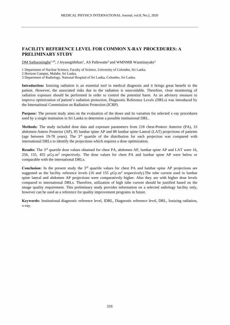
MEDICAL PHYSICS INTERNATIONAL Journal, vol.8, No.2, 2020
316
FACILITY REFERENCE LEVEL FOR COMMON X-RAY PROCEDURES: A PRELIMINARY STUDY
DM Satharasinghe1,2#, J Jeyasugiththan1, AS Pallewatte3 and WMNMB Wanninayake2
1 Department of Nuclear Science, Faculty of Science, University of Colombo, Sri Lanka. 2 Horizon Campus, Malabe, Sri Lanka. 3 Department of Radiology, National Hospital of Sri Lanka, Colombo, Sri Lanka.
Introduction: Ionizing radiation is an essential tool in medical diagnosis and it brings great benefit to the
patient. However, the associated risks due to the radiation is unavoidable. Therefore, close monitoring of
radiation exposure should be performed in order to control the potential harm. As an advisory measure to
improve optimization of patient’s radiation protection, Diagnostic Reference Levels (DRLs) was introduced by
the International Commission on Radiation Protection (ICRP).
Purpose: The present study aims on the evaluation of the doses and its variation for selected x-ray procedures
used by a single institution in Sri Lanka to determine a possible institutional DRL.
Methods: The study included dose data and exposure parameters from 218 chest-Postero Anterior (PA), 33
abdomen-Antero Posterior (AP), 85 lumbar spine AP and 88 lumbar spine-Lateral (LAT) projections of patients
(age between 19-78 years). The 3rd quartile of the distribution for each projection was compared with
international DRLs to identify the projections which requires a dose optimization.
Results: The 3rd quartile dose values obtained for chest PA, abdomen AP, lumbar spine AP and LAT were 16,
256, 155, 455 µGy.m2 respectively. The dose values for chest PA and lumbar spine AP were below or
comparable with the international DRLs.
Conclusion: In the present study the 3rd quartile values for chest PA and lumbar spine AP projections are
suggested as the facility reference levels (16 and 155 µGy.m2 respectively).The tube current used in lumbar
spine lateral and abdomen AP projections were comparatively higher. Also they are with higher dose levels
compared to international DRLs. Therefore, utilization of high tube current should be justified based on the
image quality requirement. This preliminary study provides information on a selected radiology facility only,
however can be used as a reference for quality improvement programs in future.
Keywords: Institutional diagnostic reference level, IDRL, Diagnostic reference level, DRL, Ionizing radiation,
x-ray.

MEDICAL PHYSICS INTERNATIONAL Journal, vol.8, No.2, 2020
317
EVALUATION OF GONAD SHIELDING IN DIGITAL RADIOGRAPHY: BASED ON CLINICALLY ADJUSTABLE CONTROLS.
E.W Kombo1 J. H. D. Wong1,2 MS Nizam1,2 KH Ng1,2
1 Department of Biomedical Imaging, Faculty of Medicine, University of Malaya, Kuala Lumpur 50603, Malaysia 2 University of Malaya Research Imaging Centre, Faculty of Medicine, University of Malaya, Kuala Lumpur 50603,
Malaysia
Introduction: The effectiveness of gonad shielding in general x-ray radiography in recent years has been
questioned because of the risk of obscuring important information. Previous literature studies were based on
Kerma Air Product (KAP) and do not show the absorbed dose-based reductions on the gonads and image quality
considerations for clinical practicability1. With radiographers having the option of choosing to use AEC or not
there is a need to justify the continued practice by some centres.
Purpose: To determine the percentage dose reduction achieved when gonad shields are in place and how it is
affected by varying the parameters of interest, which include tube potential and current-time product (mAs),
AEC.
Methods: An adult anthropomorphic phantom was exposed in anteroposterior pelvic projection study. Exposure
was done for shielded and unshielded gonads in various adjustments. Using tube voltages (70-100kVp) and mAs
(16-25mAs) adjustments when AEC turned off and measured organ absorbed dose at four internal sites and three
on the surface points for male study using nanoDotTM optically stimulated luminescence dosimeter (OSLD).
Results: In males, absorbed dose to the testes was reduced by > 87% (87.9 - 90.8%) and range of values from 60
-70% in females when automated exposure was used. With manual control of mAs and standard voltage (70kVp)
similar dose reduction in males of >88.5% (88.5 - 88.7%) however, in female reduction was slightly lower with
45-70%. Various tube potential values with constant mAs dose reductions with values > 84% (85- 89%) and
female 57-70% range were obtained.
Conclusion: The effectiveness of gonad shielding is affected by the location of gonads. The location of male
gonads, being more easily identified are more easily shielded and benefited from larger dose reduction due to
reduced scattering2. In contrast, the actual positions of the female ovaries are not visible and often estimated.
This resulted in poorer gonad shielding.
Keywords: contact shields, dose reduction factor, mAs, Tube voltage, AEC.

MEDICAL PHYSICS INTERNATIONAL Journal, vol.8, No.2, 2020
318
Oral Presentations
Diagnostic: Others

MEDICAL PHYSICS INTERNATIONAL Journal, vol.8, No.2, 2020
319
EVALUATION OF STUDENT SKILL COMPETENCY ON ACCURACY OF MID-TRIMESTER FETAL SONOGRAPHY SCAN FOLLOWING THE NEWLY-ESTABLISHED MASTER DEGREE IN MEDICAL SONOGRAPHY
Surapa Hirunpijit1,3, Surachate Siripongsakun1, Chanisa Chotipanich1, Pantajaree Hiranrat1,
Amarin Narkwichean2, Wipada Laosooksathit2
1 Sonographer School, Faculty of Health Science Technology, Chulabhorn Royal Academy, Bangkok 10210, Thailand, 2 Faculty of Medicine, Srinakharinwirot University, HRH Maha Chakri Sirindhorn Medical Centre Nakhon Nayok, 26120
Thailand, 3 Department of Radiology, Central Chest Institute of Thailand, Nonthaburi, 11000, Thailand.
Introduction: Mid-trimester scan is recommended in all pregnancies nonetheless the obstetrician’s workload
can be extensive. Thailand first postgraduate, a 2-year master’s degree in medical sonography has only been
established in 2019 aiming to produce sonographers providing support on sonographic examination. Students
underwent 8-week obstetric module during their first-year study, attending lectures, workshops and real clinical
practice. Assessment of student competency is required as parts of the program/module evaluation.
Purpose: To evaluate skills of medical sonography students particularly an accuracy of biometric measurement
and gestational age estimation, in comparison to qualified obstetricians, upon completion of the obstetric module
of the newly-established program
Method: A cross-sectional observational study was conducted on the first batch medical sonographer students.
A total of 81 participating pregnancies in their mid-trimester (18-24 weeks) underwent ultrasound scans firstly
performed by either of 2 students. Parameters; biparietal diameter (BPD) head circumference (HC), abdominal
circumference (AC), femur length (FL), were measured to calculate the gestational age. The scan was confirmed
by a qualified obstetrician on-duty. Comparisons between the first and subsequent scan were made. Recorded
images were then subsequently reviewed by a maternal-fetal-medicine specialist to assess student performance
of the scan and measurement
Results: Although there was a significant difference in HC and AC measurement, Gestational age (mean
difference 0.01± 2.92 days, p=0.969) and some parameters (BPD and FL) measurement were not difference
between the students and obstetricians. Intraobserver and Interobserver (n=2) variability were not observed.
Review performance of students regarding scanning and measuring skills, utilising standard criteria
recommended by the ISUOG, was considered good to excellence (77.5-80%).
Conclusion: The study demonstrates that medical sonography students are competent regarding second trimester
scan in terms of biometric measurement and gestational age estimation. The outcomes help verifying the Master
degree program.
Keyword: Competency, Fetal biometry, Obstetric sonography, Medical sonographer student.

MEDICAL PHYSICS INTERNATIONAL Journal, vol.8, No.2, 2020
320
THE TEACHING AND LEARNING OF POSTGRADUATE MEDICAL PHYSICS USING INTERNET-BASED E-LEARNING DURING THE COVID-19 PANDEMIC
Che Ahmad Azlan1, Jeannie Hsiu Ding Wong1*, Li Kuo Tan1, Muhammad Shahrun Nizam A Daman Huri1,
Ngie Min Ung2, Vinod Pallath3, Christina Phoay Lay Tan 4, Chai Hong Yeong5, Kwan Hoong Ng1
1 Department of Biomedical Imaging, Faculty of Medicine, University of Malaya, 50603 Kuala Lumpur, Malaysia. 2 Clinical Oncology Unit, Faculty of Medicine, University of Malaya, 50603 Kuala Lumpur, Malaysia. 3 Medical Education and Research Development Unit, Faculty of Medicine, University of Malaya, 50603 Kuala Lumpur,
Malaysia. 4 Department of Primary Care Medicine, Faculty of Medicine, University of Malaya, 50603 Kuala Lumpur, Malaysia. 5 School of Medicine, Faculty of Health and Medical Sciences, Taylor’s University, 47500 Subang Jaya, Malaysia
Introduction: Many countries around the world imposed lockdowns or other forms of movement restrictions to
stop the pandemic of COVID-19 in their communities, Malaysia included. These unprecedented measures
caused the closure of school and universities and disrupted the teaching and learning (T&L) activities. To
continue the instructional delivery, most universities implemented Internet-based e-learning.
Purpose: We intend to share our approach in the implementation of Internet-based e-learning in the Master of
Medical Physics programme at the University of Malaya, Malaysia.
Methods: In March 2020, we surveyed the e-learning readiness of the students at the beginning of the
nationwide lockdown. We implemented T&L activities via various virtual learning environments and e-learning
platforms. We present the pros and cons of selected online teaching platforms. An evaluation survey was carried
out after 15 weeks to evaluate the students’ experience during this period of e-learning.
Results: We found that while students still preferred physical face-to-face teaching, they were able to adapt to
the new norm of instructional delivery. More than 60% of the students agreed that the various e-learning
approaches were useful in the learning process. The e-learning and study from home environment allowed most
students the flexibility of learning at any time they preferred. However, for some students, they found it difficult
to focus on their studies because of various distractions and lack of morale. Technical problems such as
intermittent disruption of internet connectivity and limited internet data availability were the challenges of the e-
learning process.
Conclusion: We expect that this method of distance learning (emergency remote learning) will continue to
prevail in the near future and could become a new norm. It is also predicted that blended learning strategies
which include e-learning, would become more common for the T&L of postgraduate medical physics
programmes even after the COVID-19 crisis.
Keywords: Medical physics education, online teaching, virtual learning, COVID-19

MEDICAL PHYSICS INTERNATIONAL Journal, vol.8, No.2, 2020
321
ANALYSIS OF NTCP BASED RADIOBIOLOGICAL MODELS: A SYSTEMATIC REVIEW OF LITERATURES
Rahman M1, Kausar A1, Mondol M1, Sujan M1, Azhari HA1 and Zakaria GA1,2
1 Dept of Medical Physics and Biomedical Engineering, Gono Bishwabidyalay, Savar, Dhaka, Bangladesh 1,2 Dept. of medical Radiation Physics, Gummersbach Hospital, Academic Teaching Hospital of the University of Cologne,
Gummersbach, Germany
Introduction: To achieve the optimal treatment goal, radiobiological parameters has to evaluate and predict the
outcome of this treatment plan in terms of both TCP and NTCP. Different types of radiobiological model were
used to achieve prescribed treatment dose of radiation during the tumor control. Where TCP models play a
important role in order to achieve desired dose to the tumor. A suitable NTCP model was theoretically found
among different models that can be used in treatment plan evaluation.
Materials and Methods: Theoretically, six different radiobiological dose response models such as Lyman–
Kutcher–Burman, Critical element, critical volume, Relative Seriality, Parallel architecture, Weibull distribution
were analyzed in this project. All models were discussed elaborately with its various parameters and were used
in the calculation of normal tissue complication probability during the treatment in radiotherapy. Further, all
models were compared with each other.
Results: The models denote the dose for 50% complication probability (D50) parameters is the most commonly
used radiobiological models for the normal tissues. The functional subunit response models (critical element &
Relative seriality, Critical Volume, parallel architecture) are used in the derivation of the formulae for the normal
tissue. Since all complicated NTCP model predict same as the simple NTCP model that is Lyman–Kutcher–
Burman model as well as it is computationally efficient. Also Lyman–Kutcher–Burman model can be used in
different treatment planning system incorporating with other model. For this reason our suggested model is
Lyman–Kutcher–Burman NTCP model which can be used in treatment plan evaluation.
Conclusion: After analyzing six different model of NTCP, finding of the study is the treatment plan evaluation
in where Lyman–Kutcher–Burman model may be considered as a better option for biological plan evaluation
Keywords: Radiobiological Model, TCP, NTCP
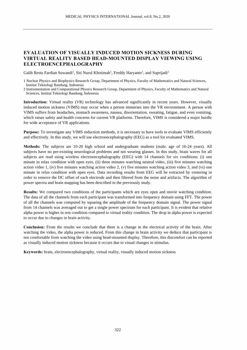
MEDICAL PHYSICS INTERNATIONAL Journal, vol.8, No.2, 2020
322
EVALUATION OF VISUALLY INDUCED MOTION SICKNESS DURING VIRTUAL REALITY BASED HEAD-MOUNTED DISPLAY VIEWING USING ELECTROENCEPHALOGRAPHY
Galih Restu Fardian Suwandi1, Siti Nurul Khotimah1, Freddy Haryanto1, and Suprijadi2
1 Nuclear Physics and Biophysics Research Group, Department of Physics, Faculty of Mathematics and Natural Sciences, Institut Teknologi Bandung, Indonesia
2 Instrumentation and Computational Physics Research Group, Department of Physics, Faculty of Mathematics and Natural Sciences, Institut Teknologi Bandung, Indonesia
Introduction: Virtual reality (VR) technology has advanced significantly in recent years. However, visually
induced motion sickness (VIMS) may occur when a person immerses into the VR environment. A person with
VIMS suffers from headaches, stomach awareness, nausea, disorientation, sweating, fatigue, and even vomiting,
which raises safety and health concerns for current VR platforms. Therefore, VIMS is considered a major hurdle
for wide acceptance of VR applications.
Purpose: To investigate any VIMS reduction methods, it is necessary to have tools to evaluate VIMS efficiently
and effectively. In this study, we will use electroencephalography (EEG) as a tool for evaluated VIMS.
Methods: The subjects are 10-20 high school and undergraduate students (male, age of 16-24 years). All
subjects have no pre-existing neurological problems and not wearing glasses. In this study, brain waves for all
subjects are read using wireless electroencephalography (EEG) with 14 channels for six conditions: (i) one
minute in relax condition with open eyes, (ii) three minutes watching neutral video, (iii) five minutes watching
action video 1, (iv) five minutes watching action video 2, (v) five minutes watching action video 3, and (vi) one
minute in relax condition with open eyes. Data recording results from EEG will be extracted by centering in
order to remove the DC offset of each electrode and then filtered from the noise and artifacts. The algorithm of
power spectra and brain mapping has been described in the previously study.
Results: We compared two conditions of the participants which are eyes open and movie watching condition.
The data of all the channels from each participant was transformed into frequency domain using FFT. The power
of all the channels was computed by squaring the amplitude of the frequency domain signal. The power signal
from 14 channels was averaged out to get a single power spectrum for each participant. It is evident that relative
alpha power is higher in rest condition compared to virtual reality condition. The drop in alpha power is expected
to occur due to changes in brain activity.
Conclusion: From the results we conclude that there is a change in the electrical activity of the brain. After
watching the video, the alpha power is reduced. From this change in brain activity we deduce that participant is
not comfortable from watching the video using head-mounted display. Therefore, this discomfort can be reported
as visually induced motion sickness because it occurs due to visual changes in stimulus.
Keywords: brain, electroencephalography, virtual reality, visually induced motion sickness

MEDICAL PHYSICS INTERNATIONAL Journal, vol.8, No.2, 2020
323
Oral Presentations
Nuclear Medicine: Imaging
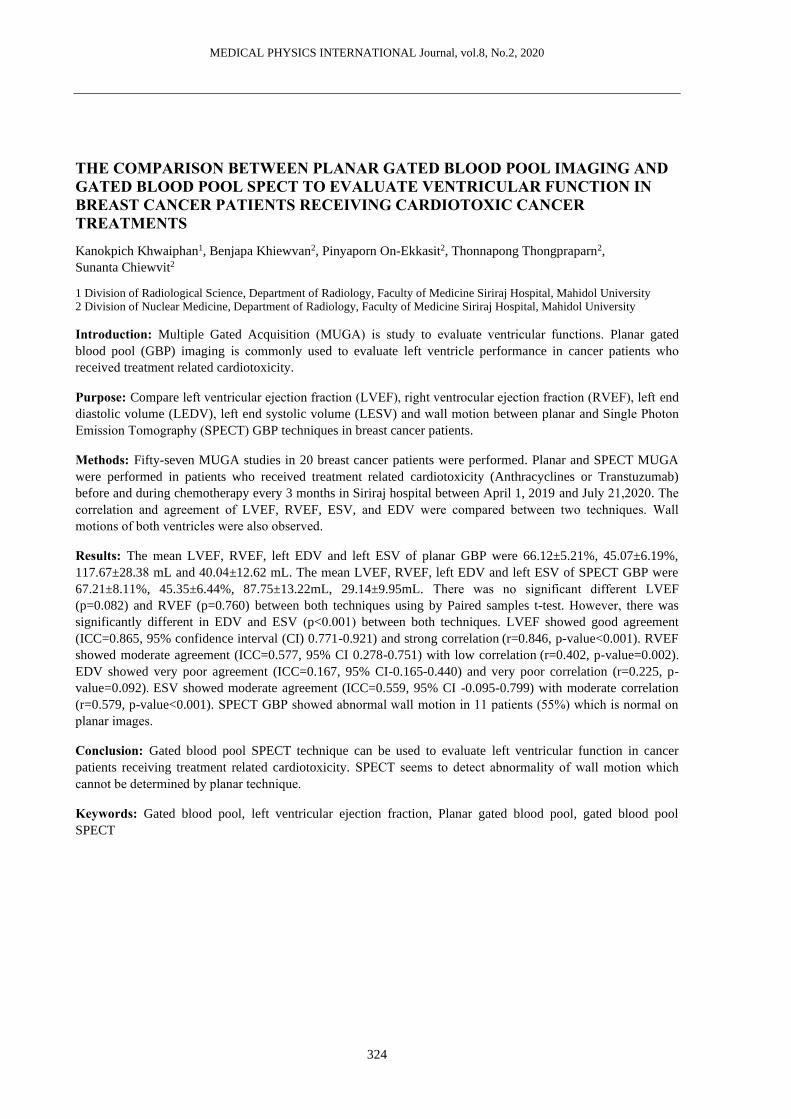
MEDICAL PHYSICS INTERNATIONAL Journal, vol.8, No.2, 2020
324
THE COMPARISON BETWEEN PLANAR GATED BLOOD POOL IMAGING AND GATED BLOOD POOL SPECT TO EVALUATE VENTRICULAR FUNCTION IN BREAST CANCER PATIENTS RECEIVING CARDIOTOXIC CANCER TREATMENTS
Kanokpich Khwaiphan1, Benjapa Khiewvan2, Pinyaporn On-Ekkasit2, Thonnapong Thongpraparn2,
Sunanta Chiewvit2
1 Division of Radiological Science, Department of Radiology, Faculty of Medicine Siriraj Hospital, Mahidol University 2 Division of Nuclear Medicine, Department of Radiology, Faculty of Medicine Siriraj Hospital, Mahidol University
Introduction: Multiple Gated Acquisition (MUGA) is study to evaluate ventricular functions. Planar gated
blood pool (GBP) imaging is commonly used to evaluate left ventricle performance in cancer patients who
received treatment related cardiotoxicity.
Purpose: Compare left ventricular ejection fraction (LVEF), right ventrocular ejection fraction (RVEF), left end
diastolic volume (LEDV), left end systolic volume (LESV) and wall motion between planar and Single Photon
Emission Tomography (SPECT) GBP techniques in breast cancer patients.
Methods: Fifty-seven MUGA studies in 20 breast cancer patients were performed. Planar and SPECT MUGA
were performed in patients who received treatment related cardiotoxicity (Anthracyclines or Transtuzumab)
before and during chemotherapy every 3 months in Siriraj hospital between April 1, 2019 and July 21,2020. The
correlation and agreement of LVEF, RVEF, ESV, and EDV were compared between two techniques. Wall
motions of both ventricles were also observed.
Results: The mean LVEF, RVEF, left EDV and left ESV of planar GBP were 66.12±5.21%, 45.07±6.19%,
117.67±28.38 mL and 40.04±12.62 mL. The mean LVEF, RVEF, left EDV and left ESV of SPECT GBP were
67.21±8.11%, 45.35±6.44%, 87.75±13.22mL, 29.14±9.95mL. There was no significant different LVEF
(p=0.082) and RVEF (p=0.760) between both techniques using by Paired samples t-test. However, there was
significantly different in EDV and ESV (p<0.001) between both techniques. LVEF showed good agreement
(ICC=0.865, 95% confidence interval (CI) 0.771-0.921) and strong correlation (r=0.846, p-value<0.001). RVEF
showed moderate agreement (ICC=0.577, 95% CI 0.278-0.751) with low correlation (r=0.402, p-value=0.002).
EDV showed very poor agreement (ICC=0.167, 95% CI-0.165-0.440) and very poor correlation (r=0.225, p-
value=0.092). ESV showed moderate agreement (ICC=0.559, 95% CI -0.095-0.799) with moderate correlation (r=0.579, p-value<0.001). SPECT GBP showed abnormal wall motion in 11 patients (55%) which is normal on
planar images.
Conclusion: Gated blood pool SPECT technique can be used to evaluate left ventricular function in cancer
patients receiving treatment related cardiotoxicity. SPECT seems to detect abnormality of wall motion which
cannot be determined by planar technique.
Keywords: Gated blood pool, left ventricular ejection fraction, Planar gated blood pool, gated blood pool
SPECT

MEDICAL PHYSICS INTERNATIONAL Journal, vol.8, No.2, 2020
325
DIAGNOSTIC PERFORMANCE OF DUAL-TRACER SUBTRACTION, SINGLE-TRACER DUAL-PHASE 99MTC-MIBI PARATHYROID SCINTIGRAPHY, AND 99MTC-MIBI SPECT/CT FOR PREOPERATIVE LOCALIZATION IN PATIENTS WITH HYPERPARATHYROIDISM.
Warissara Wittayapasitti1, Kijja Rhoongsittichai1, Jiraporn Sriprapaporn1
1 Division of Nuclear Medicine, Department of Radiology, Faculty of medicine, Siriraj hospital, Mahidol University,
Introduction: Preoperative imaging is performed to localize the hyperfunctioning parathyroid glands to
facilitate a minimally invasive surgical approach. The parathyroid glands can be imaged with different
modalities. However, parathyroid scintigraphy has been the investigation of choice for decades. When analyzing
the published data, it can be stated that parathyroid scintigraphy was mainly performed using double-phase, dual-
tracer subtraction, SPECT/CT methods. Furthermore, it is still debated which protocols should be preferred for
localizing abnormal parathyroid glands.
Purpose: To evaluate the diagnostic performance of dual-tracer subtraction (DT), dual-phase 99mTc-MIBI (DP)
and, 99mTc-MIBI SPECT/CT parathyroid scintigraphy for preoperative localization in patients with
hyperparathyroidism.
Methods: This retrospective study of consecutive 100 patients who underwent parathyroid scintigraphy at the
division of nuclear medicine department of radiology Siriraj hospital from 30 July 2013 to 10 September 2019.
All the parathyroid scintigraphy were reinterpreted using nuclear medicine physicians by reading 3 sets of image
data of DT, DP, and SPECT/CT. Finally, all the data were statistically analyzed by using pathological results as
a gold standard and to statistically compare the diagnostic performance among these 3 techniques.
Results: A total of 242 parathyroid lesions were excised. Parathyroid scintigraphy was compared with
pathological results. In this series, most the lesions were parathyroid hyperplasia (204 lesions), 28 were
parathyroid adenomas, and two were carcinoma. The rest eight foci represent normal parathyroid tissue. The
overall sensitivities of DT, DP, and SPECT/CT techniques were 44.4%, 35.5%, and 40.2%, respectively and the
overall specificities of these techniques were 87.5%, 87.5%, and 87.5%, respectively. Nevertheless, in the
subgroup analysis, the sensitivities of adenoma glands were 78.6%, 82.1%, 82.1% and those for hyperplastic
glands were 39.2%, 28.5%, 33.8% respectively.
Conclusion: The 3 imaging techniques have a low sensitivity similarly, but it shows insignificant result in
preoperative localization of parathyroid in hyperparathyroidism. Although the sensitivities of 3 techniques were
low, parathyroid scintigraphy were of benefit in identifying adenoma glands.
Keywrds: Hyperparathyroidism / 99mTc-MIBI / Parathyroid scan / MIBI / SPECT/CT

MEDICAL PHYSICS INTERNATIONAL Journal, vol.8, No.2, 2020
326
CLEARANCE PATTERNS OF TC-99M ECD USING SERIAL SPECT IMAGING TO DEFINE SEIZURE ONSET ZONE
Chanan Sukprakun1, Chusak Limotai2,3, Kitiwat Khamwan4,5,6, Panya Pasawang1, Supatporn Tepmongkol3,4,5,6
1 Division of Nuclear Medicine, Department of Radiology, King Chulalongkorn Memorial Hospital, Thai Red Cross Society, Bangkok, Thailand
2 Division of Neurology, Department of Medicine, Faculty of Medicine, Chulalongkorn University, Bangkok, Thailand 3 Chulalongkorn Comprehensive Epilepsy Center of Excellence (CCEC), King Chulalongkorn Memorial Hospital, Thai Red
Cross Society, Bangkok, Thailand 4 Division of Nuclear Medicine, Department of Radiology, Faculty of Medicine, Chulalongkorn University, Bangkok,
Thailand 5 Chulalongkorn University Biomedical Imaging Group (CUBIG), Faculty of Medicine, Chulalongkorn University,
Bangkok, Thailand 6 Chula Neuroscience Center, King Chulalongkorn Memorial Hospital, Thai Red Cross Society, Bangkok, Thailand
Introduction: If there is a difference of ethyl cysteinate dimer (ECD) clearance pattern between the seizure
onset zone (SOZ) and other brain regions, adding serial single photon emission computed tomography (SPECT)
may give benefit for seizure focus localization.
Purpose: To define the SOZ by clearance patterns and rates of Tc-99m ECD using serial brain SPECT.
Methods: A total of 22 serial brain SPECT scans were prospectively collected from 12 subjects, including 9
patients with drug-resistant epilepsy (DRE) and 3 normal volunteers from Mar 2018 to Sep 2019 at King
Chulalongkorn Memorial Hospital. Slope maps and clearance rates were calculated by regression model
analysis. Visual grading of clearance patterns and statistical analysis of clearance rates in the SOZs were done.
Results: There were 12 SOZs in nine patients. The average Tc-99m ECD clearance rates of all SOZs were +0.46
% ± 2.71 %/hr (wash in), -4.97 % ± 2.83 %/hr (washout), and -5.16% ± 1.81 %/hr (washout) in ictal, aura and
interictal phases, respectively. Matched pair t-tests between the SOZ and contralateral regions showed
statistically significant difference (p = 0.013 and p = 0.009 in ictal and interictal phases, respectively). From 5
slope patterns found in 12 SOZs, which were wash in, mixed wash in and slow washout, slow washout, normal
washout, and fast washout patterns, clearance patterns that can define the SOZs were 1) wash in and slow
washout on ictal slope, 2) slow washout or fast washout on aura slope and 3) fast washout on interictal slope
with percent localization of 100% (10/10), 100% (4/4) and 83.33% (10/12) in ictal, aura, and interictal slope
maps, respectively.
Conclusion: Brain ECD clearance patterns using the slope map can define the SOZs with high percent
localization (83.33%-100%). Thus, clearance pattern methods are potential diagnostic or confirmation tools for
the SOZ localization.
Keywords: epilepsy, ECD, clearance, SPECT, localization

MEDICAL PHYSICS INTERNATIONAL Journal, vol.8, No.2, 2020
327
MONTE CARLO SIMULATION FOR HIGH RESOLUTION IMAGING OF RB-82 WITH BREMSSTRAHLUNG X-RAY CAMERA FOR SMALL ANIMALS
Kouhei Nakanishi1, 2, Seiichi Yamamoto1
1 Radiological and Medical Laboratory Sciences, Nagoya University Graduate School of Medicine, Nagoya, Japan, 2 Department of Radiology, Akita Hospital, Chiryu, Japan,
Introduction: Although Rb-82 is most common tracer for the myocardial perfusion imaging with PET, Rb-82
emits high energy positrons which have long range resulting in blurring of spatial resolution of PET image. Due
to the limitation of spatial resolution, imaging of Rb-82 in mouse is not reported.
Purpose: Our purposes are proposing new method for high resolution imaging of Rb-82 in small animal by
detecting bremsstrahlung X-rays emitted by the positron and validating the feasibility of the method using Monte
Carlo simulation.
Methods: We simulated previously developed pinhole collimator camera based on 0.5-mm thick YAP
scintillator and measured performance of the camera for the bremsstrahlung X-rays emitted by positron of Rb-82.
Also, we simulated imaging of mouse heart phantom filled with Rb-82 of 67 MBq per milliliter in the heart
phantom.
Results: The spatial resolution of simulated camera with 1.0-mm pinhole collimator was 2.6 mm FWHM at 17.5
mm from surface of collimator. We could observe shape of the phantom in the image measured in 4 minutes.
Conclusion: We conclude that high resolution imaging for high-energy positron emitter is possible by detecting
bremsstrahlung X-rays emitted by the positron and the spatial resolution is superior to that of PET images.
Keywords: bremsstrahlung X-ray, Rb-82, high-resolution, scintillation camera, PET

MEDICAL PHYSICS INTERNATIONAL Journal, vol.8, No.2, 2020
328
Oral Presentations
Nuclear Medicine: Radiation Safety / DRL

MEDICAL PHYSICS INTERNATIONAL Journal, vol.8, No.2, 2020
329
SIZE SPECIFIC DOSE ESTIMATE (SSDE) FOR ESTIMATING CT DOSES IN SPECT/CT AND PET/CT EXAMINATIONS
Mayomthong P, Khiewvan B, Tongpraparn T, Mali P, Chaudakshetrin P, Sritongkul N, Tuntawiroon M.
Section of Nuclear Medicine, Department of Radiology, Faculty of medicine Siriraj Hospital, Mahidol University, Bangkok 10700, Thailand
Introduction: CTDIvol and DLP are frequently used parameters for estimation of patient’s absorbed dose from
CT examination. Until recently, the SSDE, which takes into account the patient size in terms of effective
diameter and the output of the CT scanner in terms of CTDIvol has been proposed by the AAPM 204.
Purpose: To find out if there is a relationship between patient size and the parameters, SSDE and CTDIvol.
Methods: CT data from 16 children and 109 adult patients performing PET/CT oncology; SPECT/CT
myocardial perfusion, bone, parathyroid, and infection/inflammation were retrospectively collected for 3 months.
Effective diameter of each patient was calculated as geometric mean of the anterior-posterior (AP) and lateral
dimensions measured on axial CT images. Conversion factors corresponding to effective diameters were
obtained from AAPM report 204 and the radiation dose were converted to SSDE. Linear regression model was
used to evaluate the relationship between patient size and the parameters SSDE and CTDIvol.
Results: The study shows that SSDE was larger than the CTDIvol in all patients. A positive correlation between
SSDE and CTDIvol was observed (r = 0.9730). Patients with small body size have higher SSDE as demonstrated
by the higher SSDE/CTDIvol ratio, more than 1.8-fold for children and lower than 1.8 for adult patients.
Calculated ED based on DLP and SSDE values were found to present no correlation.
Conclusion: CTDIvol is an important indicator for calculating the SSDE. Both CTDIvol and SSDE vary with
patient size. When there was no available organ dose to calculate an ED by SSDE/ICRP method, SSDE is a
better estimate of average patient dose from CT than CTDIvol because the dose not only depends on output dose
but also on the patient's characteristics. However, SSDE cannot be used to compute ED using k-factors for risk
estimation as in DLP method.
Keywords: Size specific dose estimate (SSDE), Volume CT dose index (CTDIvol), CT dose in nuclear medicine

MEDICAL PHYSICS INTERNATIONAL Journal, vol.8, No.2, 2020
330
FLEXIBLE, LIGHTWEIGHT AND LEAD-FREE RADIATION SHIELDING FOR NUCLEAR MEDICINE FROM NATURAL RUBBER WITH BARIUM SULFATE COMPOSITE
Nattha Plangpleng1, Kartpan Sakulkaew2, Nichapa Buasuwan2, Putthiporn Charoenphan3,
Krisanat Chuamsaamarkkee3
1 Master of Science Program in Medical Physics, Department of Diagnostic and Therapeutic Radiology, Faculty of Medicine Ramathibodi Hospital, Mahidol University, Thailand,
2 Department of Science Services, Ministry of Higher Education, Science, Research and Innovation, Thailand, 3 Division of Nuclear Medicine, Department of Diagnostic and Therapeutic Radiology, Faculty of Medicine Ramathibodi
Hospital, Mahidol University, Thailand
Introduction: Radiation shielding is an important tool for the protection of patient and medical staffs from
radiation. Generally, lead provides the efficient high atomic number with greatest attenuated of radiation.
However, the lead shielding is heavy and inflexible leading to inconvenient to use. Furthermore, lead is a highly
toxic substance and a very strong poison which may present an insidious health hazard.
Purpose: The aim of this work was to develop the flexible, lightweight and lead-free shielding from natural
rubber with barium sulfate (BaSO4) composite for radiation shielding in nuclear medicine.
Methods: Natural rubber was filled with BaSO4 of 0, 10, 20, 30 and 50 parts per hundred of natural rubber (phr)
using the two roll mills. The physical and mechanical properties of these shielding have been tested. The
shielding properties were measured using the 3-inch x 3-inch NaI(Tl) scintillation detector with 57Co (122 keV)
and 133Ba (356 keV) for representative of commonly used radionuclides in nuclear medicine 99mTc (140 keV) and 131I (364 keV) respectively.
Results: The results showed that increasing of BaSO4 into natural rubber raised the hardness and density
whereas the flexible properties were reduced. For radiation shielding properties, the mass attenuation coefficient
(m) were found to increase with the BaSO4 concentrations for 57Co and 133Ba radionuclides. The highest mass
attenuation coefficient was observed in natural rubber with BaSO4 50 phr, the m were calculated to be 0.29
cm2/g and 0.21 cm2/g for 57Co and 133Ba respectively.
Conclusion: Natural rubber with BaSO4 showed a promising result for developing potential shielding in nuclear
medicine due to their flexibility, lightweight and lead-free composite. However, it is important to note that this
study used the 57Co and 133Ba to represent the energy peak of 99mTc and 131I respectively. Hence, it might not be
the same in the term of radioactivity. Further study could be focused on enhancing the shielding properties by
increasing the concentration of BaSO4 or adding novel composites such as nanofiller in natural rubber and
testing the shielding with the clinical used radioactive.
Keywords: Natural rubber, Barium Sulfate, Flexible shielding, Radiation protection

MEDICAL PHYSICS INTERNATIONAL Journal, vol.8, No.2, 2020
331
INVESTIGATION OF POSITION AND ANGLE EFFECTS OF NANODOT OSL DOSIMETER FOR THE EYE LENS DOSE MEASUREMENT
Pattaravarin Rattanamongkonkul1, Putthiporn Charoenphun1, Krisanat Chuamsaamarkkee2,
Tanapol Dachviriyakij3, Korawee Saejia3
1 Master of Science Program in Medical Physics, Department of Diagnostic and Therapeutic Radiology, Faculty of Medicine Ramathibodi Hospital, Mahidol University, Thailand,
2 Division of Nuclear Medicine, Department of Diagnostic and Therapeutic Radiology, Faculty of Medicine Ramathibodi Hospital, Mahidol University, Thailand,
3 Regulatory Technical Support Division, Office of Atoms for Peace, Thailand.
Introduction: Radiation dose to the eye lens are more consideration since ICRP decreased the equivalent dose
limit of the eye lens in 2011. The nanoDot dosimeter has been used to measure the eye lens dose due to its
characteristic which should appropriate for point dose measurement in term of Hp(3). However, several studies
exhibited angular dependence of the nanoDot OSL. Moreover, the position of nanoDot could be varied depended
on the individual face.
Purpose: The aim of the study was to investigate simultaneously the effects of position and angle of the
nanoDot OSL for the eye lens dose measurement.
Methods: The responses of nanoDot OSL dosimeters were carried out on the PMMA water-filled cylindrical
phantom behind the build-up region using 137Cs as a calibration and reference source. The position and angular
dependence were tested by setting up the source at 0o to the phantom. The nanoDots were placed at the centre
(0o) and the various angles of 15o, 30o, 45o, 75o and 90o to the left and right side of the 137Cs source. Then Hp(3)
was evaluated correspondence to each dosimeter.
Results: The mean, standard deviation and the range of evaluated Hp(3) at the left and right side were 1137.13
± 74.13 µSv ranging of 988.87 µSv to 1285.39 µSv and 1152.36 ± 51.64 µSv ranging of 1049.08 µSv to
1255.65 µSv, respectively. The Hp(3) results from various positions and angles indicated no difference at 95%
confidence interval.
Conclusion: The nanoDot OSL dosimeter exhibited no effects of position and angle for the eye lens dose
measurement in term of Hp(3). Therefore, nanoDot OSL dosimeter could be feasible for measurement of the eye
lens dose whether the workers wear nanoDots in different positions and angles.
Keywords: nanoDot OSL dosimeter, Hp(3), effect of position and angle

MEDICAL PHYSICS INTERNATIONAL Journal, vol.8, No.2, 2020
332
DETERMINATION FOR CURRENT STATUS OF ENVIRONMENTAL RADIOACTIVITY IN FUKUSHIMA, USING NAI (TL) SCINTILLATION SURVEY METER
Natsuki Miura1, Daisuke Shimao1, Tetsuya Sanada1
1 Department of Radiological Technology, Faculty of Health, Hokkaido University of Science
Introduction: Fukushima Daiichi nuclear power plant accident following the Great East Japan Earthquake and
tsunami on March 11, 2011 caused radiation contamination in Fukushima prefecture. The evaluation of
environmental radiation dose by the artificial radionuclides in Fukushima have been carried out by governments
and many researchers. According to published investigation, in the environmental radiation dose has been
reduced to background level in major cities in Fukushima due to decontamination and reaching half-life.
Nevertheless, someone is concerned about the external radiation exposure visiting Fukushima.
Purpose: The purpose is to evaluate the environmental radiation dose at major sightseeing areas and roadside
stations in Fukushima and to provide current status to public.
Methods: Ambient dose rates were measured in air 1 m above the ground at all sampling points using a NaI (Tl)
scintillation survey meter (TCS-172B, Hitachi-Aloka Medical, Ltd., Tokyo, Japan), which can measure gamma
rays with a time constant of 30 sec. Environmental radioactivity measurements were carried out at 12 areas
which are over 800 thousand people visit annually in Aizu, Iwaki and Northern area. The environmental
radiation dose was derived from 5 measurement points in each 12 areas.
Results: The environmental radiation doses obtained this study suggested that it has become background level
and almost the same as other areas. These results could be useful and available data to obtain understanding
safety of Fukushima.
Conclusion: We measured current environmental radiation doses at major sightseeing areas and roadside
stations in Fukushima. The results indicated that there is no health hazard because the external radiation
exposure is the same level as other cities and no concerned about additional exposure even when someone
visiting Fukushima.
Keywords: Fukushima, environmental radioactivity measurement, NaI (Tl) scintillation survey meter
MEDICAL PHYSICS INTERNATIONAL Journal, vol.8, No.2, 2020
333
Oral Presentations
Nuclear Medicine: Others

MEDICAL PHYSICS INTERNATIONAL Journal, vol.8, No.2, 2020
334
THE LABELING OF 99MTC-PSMA-HBED-CC FOR PROSTATE CANCER IMAGING
Benchamat Phromphao1,3, Shuichi Shiratori2,3
1 Department of Radiology, Faculty of Medicine, Chulalongkorn University, Bangkok 10330, Thailand 2 Department of Radiology, Faculty of Medicine Siriraj Hospital, Mahidol University, Bangkok 10700, Thailand 3 Chulalongkorn University Biomedical Imaging Group, Department of Radiology, Faculty of Medicine, Chulalongkorn
University, Bangkok 10330, Thailand
Introduction: Prostate cancer (PCa) is the second most common cancer and the fifth leading cause of death
worldwide in 2018. Initially, PCa diagnosis based on Prostate-Specific Antigen (PSA) blood test, sonography
guided needle biopsy which are invasive manner. Moreover, these two methods provided low sensitivity and low
specificity. Therefore, molecular imaging recently enrolls as an important technique in PCa diagnosis using
some small molecules which well developed to bind to overexpressed Prostate Specific Membrane Antigen
(PSMA). The small molecules, for example PSMA-HBED-CC, PSMA I&T, chelate with Ga-68 for diagnostic
purpose and Lu-177 for therapeutic purpose under theranostic concept. Herein, we report a new tracer using Tc-
99m labeled with PSMA-HBED-CC aim to alternative option for PCa diagnosis.
Purpose: To develop in-house preparation of 99mTc-PSMA-HBED-CC for prostate cancer imaging.
Methods: 99mTc-pertechnetate 370 MBq was added to mixture solution of PSMA-HBED-CC 10 μg and 4%
SnCl2•2H2O, then heated to 100 oC 15 minutes and incubated while cool down to room temperature. Labeling
parameters were optimized to obtain the maximum radiochemical yield of 99mTc-PSMA-HBED-CC. The
completeness chelation was determined by instant thin layer chromatography (iTLC) and pH of 99mTc-PSMA-
HBED-CC was measured.
Results: 99mTc-PSMA HBED-CC was successfully chelated using 99mTc-pertechnetate solution in high
radiochemical yield and purity which is sufficient to administer to patient for SPECT imaging of PCa diagnosis.
Conclusion: The preliminary labeling method of 99mTc-PSMA-HBED-CC can prepare a new promising SPECT
tracer for PCa diagnosis. Animal experiment using PCa xenograft nude mouses is under investigation in our
laboratory before apply to clinical use of 99mTc-PSMA-HBED-CC in human.
Keywords: Prostate cancer imaging, Technetium-99m, PSMA, 99mTc-PSMA

MEDICAL PHYSICS INTERNATIONAL Journal, vol.8, No.2, 2020
335
GAMMA SCINTIGRAPHY FOR HEPATIC RADIOEMBOLIZATION: COMPARISON BETWEEN DIFFERENT RADIONUCLIDES USING GATE MONTE CARLO SIMULATION
Muhammad Rizal Fahmi1, Nurul Ab. Aziz Hashikin1, Ying Chee Keat2
1 School of Physics, Universiti Sains Malaysia, 11800, Penang, MALAYSIA. 2 Oncological & Radiological Science Cluster, Advanced Medical and Dental Institute, Universiti Sains Malaysia, 11900,
Penang, MALAYSIA.
Introduction: Radioembolization using Yttrium-90 (90Y) allows delivery of high-dose beta radiation to tumour.
Post-treatment imaging is possible via Bremsstrahlung radiation, however with significantly lower quality.
Alternative radionuclides have been investigated as potential substitutes for 90Y, i.e. Lutetium-177 (177Lu),
Holmium-166 (166Ho), and Samarium-153 (153Sm), able to deliver equivalent tumour doses and emit gamma
radiations ideal for post-treatment imaging.
Purpose: We aimed to evaluate the image quality from gamma (planar) scintigraphy following hepatic
radioembolization with different radionuclides, using GATE Monte Carlo simulation.
Methods: A spherical tumour (diameter = 1.0 cm) was created inside a trapezoidal-shaped liver and placed
inside a cylindrical torso phantom using GATE v8.2. For image acquisition, a gamma head (with NaI detector)
was modelled using SPECTHead example from GateContrib(GitHub), with dimension of 21 × 30 × 7 cm3, and
positioned 3.5 cm from the phantom. Technetium-99m (99mTc) source was uniformly distributed within the liver
and tumour volumes (assuming tumour-to-normal ratio of 2:1) during image acquisition. Imaging was later
repeated using 90Y, 153Sm, 166Ho, and 177Lu (using optimised protocols suggested by previous studies). All planar
images were evaluated based on image quality, signal-to-background ratio (SBR) and coefficient of variation
(CV). Image acquired using 99mTc was used as reference.
Results: Results show that the image acquired using 177Lu gave the best SBR (13.23), followed by 99mTc(7.31), 90Y(6.12), 153Sm(5.68), and 166Ho (4.36). For CV, 99mTc(0.46) resulted in the best value, followed by 177Lu(0.50), 153Sm(0.51), 166Ho(0.66), and 90Y(0.80). Higher SBR indicated superior spatial information, while lower CV
indicated lower variation (better estimation).
Conclusion: Image acquired using 177Lu shows the most superior quality compared to other radionuclides, at the
same time able to deliver comparable tumour dose. Nevertheless, all alternative radionuclides offer better
imaging capability via gamma emission compared to bremsstrahlung imaging via 90Y.
Keywords: GATE, radioembolization, Monte Carlo simulation, Yttrium-90.

MEDICAL PHYSICS INTERNATIONAL Journal, vol.8, No.2, 2020
336
POSITRON RANGE CORRECTION IN POSITRON EMISSION TOMOGRAPHY
Rukiah A. Latiff1, 2, Steve R Meikle1, John E Gillam1, 3, Peter L Kench1
1 The Brain and Mind Centre, Faculty of Medical and Health Sciences, The University of Sydney, Australia. 2 Faculty of Health Sciences, The National University of Malaysia, Malaysia. 3 Land Division, Defence Science and Technology Group, Fishermans Bend, Australia.
Introduction: Gallium-68 (68Ga) gives more blur to Positron Emission Tomography (PET) image due to its
higher positron range (PR), compared to 18F. The blurring effect resulting in loss of spatial resolution and
quantitative accuracy. The magnitude of PR blurring is proportional to the energy of the radionuclide used and
inversely proportional to the density of the medium it travels through. The contribution of image blur was
approximately 10% in bone and soft-tissue compared to 100% in lung medium. Besides, the blurring effect is
independent of the acquisition system and parameters.
Purpose: This Monte-Carlo simulation study aims to develop an iterative post-reconstructed tissue-dependant
spatially-variant positron range correction (PRC) deconvolution method and investigate its performance on PET
National Electrical Manufacturer Association (NEMA) image quality phantom.
Methods: The PRC method proposed is implemented directly onto image space. Geant4 Application on
Tomography and Emission (GATE) was used to model the NEMA phantom annihilation image from its PET-
measured image. The simulated annihilation image takes into account the material in which the positron is
originated and annihilated, obtained from a CT-segmented image. The correction factor of PET-measured image
and GATE-annihilation image was calculated and employed in the forward projection to update the image
estimate. We compare the image quality of the images before and after PRC.
Results: The preliminary results demonstrate an improvement in target-to-background ratio, contrast recovery
and resolution after implementation of PRC with manageable noise as the iteration increases. This method also
obtains artefact-free PR-corrected images especially in a lung region.
Conclusion: The performance of proposed PRC methods provides a resolution and quantitative improvement in
PET phantom image. It can be implemented onto any PET image regardless of scanner geometry properties and
type of radionuclides used. Future investigation is required by implementing this PRC method onto patients
image.
Keywords: Positron range, PET, Monte Carlo simulation, NEMA phantom

MEDICAL PHYSICS INTERNATIONAL Journal, vol.8, No.2, 2020
337
EVALUATION OF PATIENT DOSES FROM 177LU-PSMA IN METASTASES PROSTATE CANCER TREATMENT AT KING CHULALONGKORN MEMORIAL HOSPITAL
Kotchakorn Chatachot1,2, Kitiwat Khamwan1,3,4
1 Medical Physics Program, Faculty of Medicine, Chulalongkorn University, Bangkok, Thailand 2 Division of Nuclear Medicine, Department of Radiology, Buddhachinaraj Hospital, Phitsanulok, Thailand 3 Division of Nuclear Medicine, Department of Radiology, Faculty of Medicine, Chulalongkorn University and King
Chulalongkorn Memorial Hospital, The Thai Red Cross Society, Bangkok, Thailand 4 Chulalongkorn University Biomedical Imaging Group, Department of Radiology, Faculty of Medicine, Chulalongkorn
University, Bangkok, Thailand
Introduction: 177Lu-PSMA has increasingly used for targeted radionuclide therapy of prostate cancer and its
metastases. Dosimetric calculation, therefore, is critical to achieve the optimal therapeutic activity with limited
side effects.
Purpose: To perform the image-based absorbed doses calculation to the normal organs for patients who treated
by 177Lu- PSMA for prostate cancer.
Methods: Whole-body planar images and SPECT/CT images were acquired in eight patients at immediately, 4
and 24 h after injection of 177Lu-PSMA (range 4.37 to 8.58 GBq). To generate the time-integrated activity (TIA)
in source organs, region of interests (ROIs) were manually contoured in whole-body, liver, spleen, urinary
bladder, lacrimal gland, and bone marrow using the Osirix MD program. The geometric mean of anterior and
posterior counts was determined through the ROI analysis. The TIA in each source organ was calculated by
integrating area under time-activity curve using MATLAB. The s-values were extracted from OLINDA/EXM
version 2.0. in order to calculate the absorbed dose coefficient in target organs according to the Medical Internal
Radiation Dose (MIRD) scheme. The absorbed doses to bone marrow were estimated using the two-
compartment method by dividing high-uptake and low-uptake compartment. The spherical model was used to
calculate the lacrimal gland absorbed doses.
Results: The average absorbed dose coefficients per cycle were 0.56±0.16 mGy/MBq for the bone marrow [3],
0.62±0.13 mGy/MBq for the kidneys [1,2], 0.11±0.08 mGy/MBq for the liver [1], 0.18±0.14 mGy/MBq for the
urinary bladder wall, 0.14±0.05 mGy/MBq for the spleen, and 2.86±0.78 mGy/MBq for the lacrimal glands
[1,2]. The critical absorbed doses reported for the kidneys of 23 Gy, and for bone marrow of 2 Gy were not
reached in any patients.
Conclusion: Our dosimetry results suggest that 177Lu-PSMA treatment with higher activities and more cycles is
possible without the risk of damaging the critical organ in metastasis prostate cancer patients.
Keywords: PSMA; 177Lu-PSMA; theranostics; MIRD; radionuclide therapy

MEDICAL PHYSICS INTERNATIONAL Journal, vol.8, No.2, 2020
338
DEVELOPMENT OF FLEXIBLE RADIOTHERAPEUTIC BANDAGE CONTAINING SAMARIUM-153 FOR THE TREATMENT OF SKIN CANCER
Yin How Wong1, Azahari Kasbollah2, Basri Johan Jeet Abdullah1 and Chai Hong Yeong1
1 School of Medicine, Faculty of Health and Medical Sciences, Taylor’s University, 47500 Subang Jaya, Selangor 2 Malaysian Nuclear Agency, 43000 Kajang, Selangor, Malaysia
Introduction: Skin cancer is a common type of cancer that emerges from the skin due to the development of
abnormal growth of cells. Radiation therapy is used as a primary treatment for inoperable tumors and in patients
who are unfit for surgery. Radioactive samarium-153 (153Sm) is a viable candidate for use against skin cancer.
Purpose: The aim of this study was to develop a flexible, neutron activatable radiotherapeutic bandage
containing 153Sm for treatment of skin cancer.
Methods: Electrospinning technique was used to prepare the flexible radiotherapeutic bandage from
polyurethane (PU) and samarium-152 acetylacetonate (152SmAcAc) crystal. The 152SmAcAc-labeled PU bandage
was then activated in a nuclear reactor, converting 152Sm to 153Sm (Emax = 807.6 keV, half-life = 46.3 hours) via 152Sm(n,γ)153Sm reaction. The 152SmAcAc-labeled PU bandage before and after neutron activation were
characterized using scanning electron microscope (SEM), energy dispersive X-ray (EDX) spectroscopy, gamma
spectroscopy, Fourier transformed infrared spectroscopy, thermogravimetric analysis (TGA) and differential
scanning calorimetry (DSC) . The in-vitro retention efficiency of 153SmAcAc on the PU bandage was performed
in saline solution over a duration of 120 h.
Results: The 153SmAcAc-labeled PU bandage achieved nominal activity of 272 ± 9.6 µCi/mg or 1680 ± 52.6
µCi/cm2 after 6 h neutron activation. SEM results shows the morphology of the bandage remained the same after
neutron activation. The gamma and EDX spectroscopy indicated that no radionuclide and elemental impurities
was observed after neutron activation. The TGA and DSC analysis suggested that the bandage was stable up to
200°C. By manipulating the neutron activation duration, different amounts of radioactivity were produced to
meet the desired dosage for different stages or conditions of skin cancer. Furthermore, the radioactive bandage
can be cut into specific shapes and sizes to cover only the treatment area and hence minimizing radiation
exposure to the healthy tissues. The in-vitro retention efficiency of 153SmAcAc was more than 95% in saline
solution for 120 h.
Conclusion: The 153SmAcAc-labeled flexible radiotherapeutic bandage are potentially useful for radiation
therapy of skin cancer in view of its favorable physicochemical characteristics and excellent retention efficiency.
Keywords: Samarium-153, Radiotherapeutic Bandage, Skin Cancer, Radiation Therapy, Electrospinning

MEDICAL PHYSICS INTERNATIONAL Journal, vol.8, No.2, 2020
339
E-Poster Presentations
Radiotherapy
MEDICAL PHYSICS INTERNATIONAL Journal, vol.8, No.2, 2020
340
REAL-TIME IN VIVO DOSE VERIFICATION USING METAL-OXIDE SEMICONDUCTOR FIELD EFFECT TRANSISTOR (MOSFET) IN HDR BRACHYTHERAPY
Siti Hajar Abdul Rahman1, Yasmin Md Radzi1, Mohd Zahri Abdul Aziz2, Ahmad Naqiuddin Azahari2
1 School of Physics, Universiti Sains Malaysia, Penang, Malaysia, 2 Advanced Medical and Dental Institute (AMDI), Universiti Sains Malaysia, Penang, Malaysia.
Introduction: High dose rate (HDR) brachytherapy requires a highly conformal dose to the tumour volume.
Hence, in vivo dosimetric for verification during treatment is deeming very important and crucial to provide an
accurate, highly reliable and precise dose delivery.
Purpose: In this work, we introduce the use of real-time in vivo dosimetry using MOSFET as a reliable quality
assurance tool for HDR brachytherapy instead of widely used thermoluminescence dosimeters (TLDs).
Methods: Calibration of MOSFETs detector was done following the vendor’s specification under HDR Ir-192
brachytherapy source available at our centre. The distance of the source-to-detector (SDD) was measured at
3cm. The detectors and source were positioned in a “sandwich set-up”. The positional accuracy of the source
was verified using Gafchromic EBT3 film. A plan was optimised to deliver 100 cGy at 3cm depth using
Oncentra Treatment Planning System (TPS). The detector’s reproducibility, linearity, distance dependency and
in-phantom verification were tested for its suitability as an in vivo detector.
Results: Five individual detectors gave maximum sensitivity deviation of 2.66%. Measured values for the
reference dose data showed a good agreement with the calculated data, with maximum deviation of –2.86%.
The calibration factors were ranged from 1.40 to 1.44 mV/cGy. The MOSFET detector has good reproducibility
(<3%) and excellent dose linearity (R2=1). While distance dependency response showed a linear fit with R2
value of 0.9844 (2cm to 5.5cm). In-phantom dose measurements using MOSFET detectors showed that
maximum deviation was -4.35%. The percentage deviations between the measured doses and the TPS doses
were below 5% for all measurements.
Conclusion: MOSFET has demonstrated as a suitable and good detector of choice for real-time in-vivo
dosimetry in HDR brachytherapy. Additionally, MOSFET can be used as one of the reliable QA tools for HDR
brachytherapy due to its special characteristic of being small in size.
Keywords: MOSFET, HDR brachytherapy, In vivo dosimetry, real-time, quality assurance

MEDICAL PHYSICS INTERNATIONAL Journal, vol.8, No.2, 2020
341
DEVELOPMENT OF EXTERNAL DOSIMETRY AUDIT SYSTEM FOR IMAGE-GUIDED BRACHYTHERAPY IN ASIAN COUNTRIES
Hideyuki Mizuno1, Taku Nakaji1, Shigekazu Fukuda1, Shingo Kato2
1 QST hospital, National Institutes for Quantum and Radiological Science and Technology, 2 Saitama medical university International medical Center
Introduction: Clinical study on standardized radiation therapy for uterine cervical cancer has been carried out within the
frame work of The Forum for Nuclear Cooperation in Asia (FNCA). Eleven Asian countries (Bangladesh, China,
Indonesia, Japan, Kazakhstan, Korea, Malaysia, Mongolia, Philippines, Thailand and Vietnam) participate in this
study. To improve the quality of multi-center clinical study using image-guided brachytherapy (IGBT), QA
activity should be performed.
Purpose: The purpose of this study is to develop the external dosimetry system of IGBT through end-to-end test
among Asian countries.
Methods: Sophisticated water phantom was developed to support various shapes of applicators used in Asian
countries. A tandem holder was inserted into the water phantom to hold the applicator in water. The root part of
the applicator was fixed by an arm. A Pinpoint ionization chamber (PTW, TN31013) and a
radiophotoluminescent glass dosimeter (RPLD; AGC, DoseAce GD-302M) were used as dosimeters. The left
and right A points dose were measured by pinpoint chamber, and the rectal and bladder dose, represented as an
ICRU reference points, were measured by RPLD. In order to precisely calculate the dose points with the IGBT, a
dummy dosimeter which has a cavity with a diameter of 2 mm was placed at a position corresponding to the
effective center of the dosimeter at the CT image acquisition. The cavity center dose is calculated by TPS and
compared with the measured dose by dosimeters which is placed at the IGBT irradiation.
Results: The on-site audit was performed to 5 hospitals from 3 countries, Korea, China and Philippines. All
audit was successfully completed and feasibility of this audit procedure was confirmed. Preliminary results of
the audit will be presented at the conference.
Conclusion: External dosimetry system of IGBT through end-to-end test procedure was developed.
Acknowledgement: This work was supported by JSPS KAKENHI Grant Number JP 17K09075.
Keywords: Audit, Image-guided-brachytherapy, end-to-end test

MEDICAL PHYSICS INTERNATIONAL Journal, vol.8, No.2, 2020
342
COMPARISON OF ICRU POINT DOSES AND VOLUMETRIC DOSES TO THE RECTUM HIGH DOSE RATE COBALT-60 BRACHYTHERAPY OF CERVICAL CANCER
Yun Seang Yi1, Zulaikha Binti Jamalludin2, Rozita Malik1, Ung Ngie Min1
1 Clinical Oncology Unit, Faculty of Medicine, University of Malaya, Kuala Lumpur, Malaysia 2 Medical Physics Unit, University of Malaya Medical Centre, Kuala Lumpur, Malaysia
Introduction: Brachytherapy is a common option to treat cervical cancer. High dose rate (HDR) intracavitary
brachytherapy (ICBT) is one of the currently used techniques for cervical cancer treatment. The volume-based
brachytherapy planning method recommended in the International Commission on Radiation Units and
Measurements (ICRU) report No. 89 is widely believed to be more useful compared to the conventional point-
based method recommended in ICRU report No. 38 in terms of OARs dosimetry. However, more resources in
terms of manpower and time are required to implement volume-based brachytherapy planning.
Purpose: This study aims to evaluate the point-based and volume-based methods for rectal dose estimation in
HDR Co-60 brachytherapy of cervical cancer.
Methods: A total of 150 patients receiving full insertion HDR ICBT treatment in University of Malaya Medical
Centre from 2017 to 2019 were retrospectively selected. The patients' data including rectal volume and rectal
dose parameter calculated using both point-based and volume-based techniques were extracted from the
brachytherapy treatment planning system. Data were compared and investigated for possible correlations using
the SPSS software version 23.0.
Results: The results showed significant differences between rectal point dose (ICRUr) with doses to most
exposed rectal volumes of 0.1 cm3 (D0.1cc), 1 cm3 (D1cc) and 5 cm3 (D5cc) (p<0.05). The ICRUr was shown to
have no significant difference with the most exposed rectal volumes of 2 cm3 (D2cc) (p=0.960). The results of
correlation analyses indicated weak correlations between ICRUr with D0.1cc, D1cc, D2cc, and D5cc with the
correlation coefficients of 0.184, 0.351, 0.371, 0.384, and 0.397 respectively (p<0.01). The strongest correlations
between rectal point and volumetric dose were observed to be between ICRUr and D1cc and D2cc with the
correlation coefficients of 0.674 and 0.669 (p<0.05) respectively.
Conclusion: In conclusion, the D1cc and D2cc were the volumetric rectal doses that can be explained better by the
ICRUr.
Keywords: ICRU point dose, volumetric dose, rectal dose

MEDICAL PHYSICS INTERNATIONAL Journal, vol.8, No.2, 2020
343
CORRELATION ANALYSIS OF CT-BASED RECTAL PLANNING DOSIMETRIC PARAMETERS WITH IN VIVO DOSIMETRY OF MOSKIN AND PTW 9112 DETECTORS IN CO-60 SOURCE HDR INTRACAVITARY CERVIX BRACHYTHERAPY
Zulaikha Jamalludin1,2,3, Rozita Abdul Malik2,3, Zamzarinah KamarulZaman1,3, Munira Rejab1,3,
Nurul Hidayah Moksem1,3 and Ngie Min Ung2,3
1 Medical Physics Unit, University of Malaya Medical Centre, Kuala Lumpur, 59100, Malaysia, 2 Department of Clinical Oncology, University of Malaya Medical Centre, Kuala Lumpur, 59100, Malaysia, 3 Clinical Oncology Unit, Faculty of Medicine, University of Malaya, 50603, Kuala Lumpur, Malaysia
Introduction: Intracavitary brachytherapy (ICBT) for the treatment of cervical cancer while delivering high
doses to the target will also delivered portion of doses to rectal organ due to close proximity to the cervico-uterus
organ. Rectal dose assessment was evaluated with the use planning and in vivo based dose parameters.
Purpose: This study conducted to analyze the correlation between rectal in vivo measured dose with selected
volume and point-based dose parameters.
Methods: In 48 insertions of CT-based high dose rate ICBT, in vivo dose measurement performed with
commercial PTW 9112 semiconductor diode probe. In 18/48 insertions, a single MOSkin detector was attached
on the PTW 9112 probe surface, at 50 mm from the probe tip. Four types of dosimetric parameters were
retrospectively collected from treatment planning system (TPS) in these insertions; a) TPS planned doses from
the maximum reported dose of PTW 9112 diode (RPmax) and single MOSkin detector, b) minimum dose to 2 cc
rectal organ (D2cc), c) ICRU reference point (ICRUr), d) maximum dose from additional rectal points (Rmax). The
measured in vivo doses from both detectors were analyzed for any correlation with the above-mentioned
dosimetric parameters.
Results: There was significant high correlation strength between in vivo measured dose from RPmax and MOSkin
with TPS planned dose with correlation coefficient (r) to be 0.916 and 0.959 respectively. The correlation
between measured RPmax dose with both D2cc and Rmax revealed high correlation strength with r > 0.7 while
resulted in moderate correlation strength with ICRUr of r = 0.525. Analysis of relationship found no significant
correlation (p-value > 0.05) between MOSkin in vivo measured dose with D2cc, ICRUr and Rmax.
Conclusion: The non-significant correlation between parameters ascribable to the differences in both detectors
position within the patient and dosimetric volume and point location determined on 3D images from TPS rather
than detectors uncertainties.

MEDICAL PHYSICS INTERNATIONAL Journal, vol.8, No.2, 2020
344
A DEVELOPMENT OF A PRIMARY STANDARD OF ABSORBED DOSE TO WATER FOR A SMALL FIELD IN HIGH-ENERGY PHOTON BEAM
Masakatsu Takeda1, 2, Morihito Shimizu2, Yuichiro Morishita2, Masao Hoshina1 and Masanori Sato1
1 Graduate School of Health Sciences, Komazawa Univ 2 National Metrology Institute of Japan (NMIJ), AIST
Introduction: The use of a small field photon beam is indispensable in modern high-precision radiotherapy, and
accurate measurement of absorbed dose to water for a small field is important. The NMIJ are developing a
primary standard of absorbed dose to water for a small field in high-energy photon beam.
Purpose: In order to determine the absorbed dose to water for the small field, we developed dose measurement
method using a compact graphite calorimeter.
Methods: The absorbed dose to water for the small field in a 6 MV photon beam from a clinical linac were
determined using the compact graphite calorimeter with core diameter of 2 cm. The field size of the photon
beams was 2 cm × 2 cm − 10 cm × 10 cm. The absorbed dose to graphite calorimeter was converted to dose to
water by Monte Carlo simulation (EGS5). A short Farmer-type ionization chamber (IBA, FC-23C) was
calibrated with the determined dose to water.
Results: The calibration coefficients of the ionization chamber were 137.06±0.51 mGy nC-1 for the field size
from 4 cm × 4 cm to 10 cm × 10 cm. In the field size of 3 cm × 3 cm and 2 cm × 2 cm, the calibration
coefficients were 2 % and 11 % larger than that on 10 cm × 10 cm, respectively. This difference between the
field size might come from the conversion factor was calculated for that of 10 × 10 cm.
Conclusion: The absorbed dose to water calibration coefficient for field size of 4 cm × 4 cm to 10 cm × 10 cm
was determined with a relative standard uncertainty of 0.38 % in the small field photon beams.
Keywords: small field, Absorbed dose to water, high-energy photon beam, graphite calorimeter

MEDICAL PHYSICS INTERNATIONAL Journal, vol.8, No.2, 2020
345
CONSTRUCTION OF REGIONAL NETWORK AND REMOTE SUPPORT SYSTEM FOR LINAC OUTPUT DOSE DIFFERENCE FACILITIES.
Shinji Kawamura1, Yoshinori Tanabe2, Shuichi Ozawa3, Toshiyuki Minemura4, Katsuhisa Narita5,
Kazushige Hayakawa6
1 Graduate school of health sciences, Fukuoka, 836-8505, Japan 2 Yamaguchi University Hospital, Ube, Yamaguchi, 755-8505, Japan 3 Hiroshima high-precision radiation therapy cancer center, Hiroshima, 732-0057, Japan 4 Center for cancer control and information services, Tokyo, 104-0045, Japan 5 Association for Nuclear Technology in Medicine, Chiba, 263-0041, Japan 6 National Hospital Organization Disaster medical center, Tokyo, 190-0014, Japan
Introduction: In Japan, there are some facilities with dose difference exceeding 3% in the linac output dose
audit. Support for these facilities is an issue.
Purpose: We have established a system to provide aid to these facilities by utilizing regional networks and
remote support system. We examined the effectiveness and issues of this system.
Methods: The Japanese Organization of Radiotherapy Quality Management (JORQM) has been conducting a
pilot study of output dosimetry support since 2018. This study is being conducted for each region divided into
nine regional blocks and is scheduled to be completed in 2020. Through this study, a regional network for
quality control of radiation therapy will be established. In addition, the remote support system STD-Audit will be
constructed. We will introduce the efforts to standardize the output dose through the regional network and
remote support in Japan.
Results: As a result of output dose audit in Japan, the dose difference facilities exceeding 3% were about 10% of
the audit facilities. The output doses measured by the two quality managers who visited the facility were
automatically transferred to the remote server, and the cause of the difference in output doses was discussed by
the facility manager, two visiting quality managers, and the remote supporters. Reasonable and optimal support
can be provided by adding a regional network and remote support system.
Conclusion: We constructed a local network and remote support system for facilities with different linac output
dose, and evaluated its effectiveness and issues in a pilot study.
Keywords: Audit, Output dose difference, Regional network, Remote support, STD-audit

MEDICAL PHYSICS INTERNATIONAL Journal, vol.8, No.2, 2020
346
INVESTIGATION FOR MEASUREMENT OF PHOTON DEPTH DOSE USING UVC CAMERA
Kengo Miyazaki1, Atsushi Myojoyama1
1 Graduate School of Human Health Science, Tokyo Metropolitan University
Introduction: When a photon output from a linear accelerator was incident on a UVC (USB Video Class)
camera, noise was detected on the output images. We considered that the detail information of photons or
electrons with a high dose rate can be obtained by analysing the noise because the generated noise increases as
the dose rate.
Purpose: In this study, we investigated the radiation response characteristics of a UVC camera in order to
develop an inexpensive semiconductor detector.
Methods: UVC camera was placed 15 cm below the 137Cs radiation source and connected to the noise analysis
program that we developed. The copper block was placed directly above the UVC camera, and the number of
noise generated for 10 minutes was counted for each 1 mm thickness. In order to investigate the number of noise
in detail, the measured results were compared with EGSnrc simulation. In the simulation, the thickness of the
copper block was increased by 1 µm from 0 cm to 3 cm, and the number and energy of electrons generated from
the bottom surface of the copper block at each thickness were investigated.
Results: It was observed in actual measurement that the noise amount decreases as the copper block becomes
thicker. As a result of comparison between actual measurement and simulation, it was confirmed that the number
of noise generated from the UVC camera conforms to the depth dose obtained from the simulation.
Conclusion: It was suggested that the UVC camera could be a semiconductor detector for depth dose
measurement.
Keywords: UVC camera, noise, depth dose, metal block
MEDICAL PHYSICS INTERNATIONAL Journal, vol.8, No.2, 2020
347
IMPACT OF THE FREQUENCY OF VERIFICATION WITH IN-ROOM CT ON SETUP ERRORS IN CARBON ION RADIOTHERAPY FOR PROSTATE CANCER
Yushi Wakisaka1,2, Hiroyasu Maruo1, Takayoshi Ishii1
1 Department of Radiation Technology, Osaka Heavy Ion Therapy Center, Osaka, Japan, 2 Department of Medical Physics and Engineering, Osaka University Graduate School of Medicine, Suita, Japan
Introduction: In our facility, the position of the prostate was verified by daily in-room CT when the patient was
inapplicable to implement fiducial markers. The reduction of the number of taking in-room CT is desired for the
viewpoint of exposure dose and occupation time of the treatment room within acceptable setup errors.
Purpose: To evaluate the impact of the frequency of verification with in-room CT on setup errors.
Methods: Because of lack of cases taking in-room CT, inter-fractional errors of 350 patients with fiducial
markers were analyzed. Intra-fractional errors were assumed to be 1 mm based on clinical experiments, and
expressed by the random numbers following a normal distribution. When assuming verification with in-room CT,
the inter-fractional error of that day should be zero, while it must be estimated by averaging previous trends for
the other day. Following our treatment protocols, total errors (i.e. the sum of inter-fractional errors and intra-
fractional errors) with 12 fractions were simulated for the different number of verifications with in-room CT.
Results: When in-room CT was not taken at all (i.e. matched only by bone every fractions), the total error was
2.1±1.6 mm. Assuming taking in-room CT for equal intervals, total errors were 1.7±1.4 mm, 1.4±1.3 mm,
1.2±1.1 mm, 0.8±0.6 mm when taking in-room CT 2, 4, 6, 12 (all) times, respectively.
Conclusion: Even if the frequency of verification with in-room CT was decreased, setup errors could be less
than the case that in-room CT was never taken. However, margins of treatment planning might be revised
because the reduction of the number of taking in-room CT leads to the increase of setup error.
Keywords: In-room CT, Prostate cancer, Carbon ion radiotherapy, Setup error, Fiducial marker
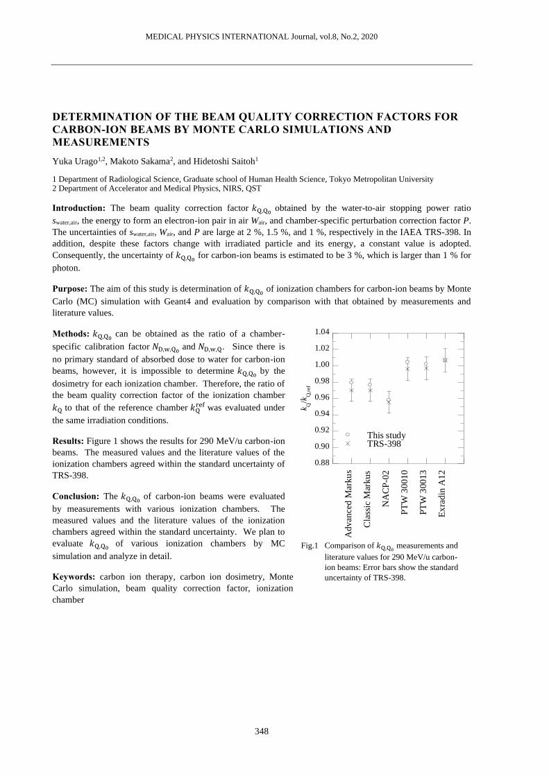
MEDICAL PHYSICS INTERNATIONAL Journal, vol.8, No.2, 2020
348
DETERMINATION OF THE BEAM QUALITY CORRECTION FACTORS FOR CARBON-ION BEAMS BY MONTE CARLO SIMULATIONS AND MEASUREMENTS
Yuka Urago1,2, Makoto Sakama2, and Hidetoshi Saitoh1
1 Department of Radiological Science, Graduate school of Human Health Science, Tokyo Metropolitan University 2 Department of Accelerator and Medical Physics, NIRS, QST
Introduction: The beam quality correction factor 𝑘Q,Q0 obtained by the water-to-air stopping power ratio
swater,air, the energy to form an electron-ion pair in air Wair, and chamber-specific perturbation correction factor P.
The uncertainties of swater,air, Wair, and P are large at 2 %, 1.5 %, and 1 %, respectively in the IAEA TRS-398. In
addition, despite these factors change with irradiated particle and its energy, a constant value is adopted.
Consequently, the uncertainty of 𝑘Q,Q0 for carbon-ion beams is estimated to be 3 %, which is larger than 1 % for
photon.
Purpose: The aim of this study is determination of 𝑘Q,Q0 of ionization chambers for carbon-ion beams by Monte
Carlo (MC) simulation with Geant4 and evaluation by comparison with that obtained by measurements and
literature values.
Methods: 𝑘Q,Q0 can be obtained as the ratio of a chamber-
specific calibration factor 𝑁D,w,Q0 and 𝑁D,w,Q. Since there is
no primary standard of absorbed dose to water for carbon-ion
beams, however, it is impossible to determine 𝑘Q,Q0 by the
dosimetry for each ionization chamber. Therefore, the ratio of
the beam quality correction factor of the ionization chamber
𝑘Q to that of the reference chamber 𝑘Qref was evaluated under
the same irradiation conditions.
Results: Figure 1 shows the results for 290 MeV/u carbon-ion
beams. The measured values and the literature values of the
ionization chambers agreed within the standard uncertainty of
TRS-398.
Conclusion: The 𝑘Q,Q0 of carbon-ion beams were evaluated
by measurements with various ionization chambers. The
measured values and the literature values of the ionization
chambers agreed within the standard uncertainty. We plan to
evaluate 𝑘Q,Q0 of various ionization chambers by MC
simulation and analyze in detail.
Keywords: carbon ion therapy, carbon ion dosimetry, Monte
Carlo simulation, beam quality correction factor, ionization
chamber
0.88
0.90
0.92
0.94
0.96
0.98
1.00
1.02
1.04
Adv
ance
d M
ark
us
Cla
ssic
Mar
ku
s
NA
CP
-02
PT
W 3
001
0
PT
W 3
00
13
Ex
rad
in A
12
This studyTRS-398
k Q/k
Q,r
ef
Fig.1 Comparison of 𝑘Q,Q0 measurements and
literature values for 290 MeV/u carbon-
ion beams: Error bars show the standard
uncertainty of TRS-398.

MEDICAL PHYSICS INTERNATIONAL Journal, vol.8, No.2, 2020
349
EFFECTS OF PARALLAX ERROR AND LIGHT REFRACTION ON ESTIMATION FOR DOSE DISTRIBUTION OF PROTON BEAMS USING LUMINESCENCE IN WATER
Koki Furuo1, Masataka Komori1, Hiroto Ushiba1, Toshiyuki Toshito2
1 Nagoya University Graduate School of Medicine, 1-1-20 Daiko-Minami, Higashi-ku, Nagoya 461-8673, Japan, 2 Nagoya Proton Therapy Center, Nagoya City West Medical Center, 1-1-1, Hirate-cho, Kita-ku, Nagoya 462-8508, Japan
Introduction: A previous study confirmed that weak luminescence was observed by CCD (Charge Coupled
Device) camera when water was irradiated with proton beams having energy lower than the threshold for
Cherenkov-light emission. Furthermore, the luminescence distribution was found out to almost correspond with
the dose distribution. Therefore, the luminescence imaging has great potential to estimate the dose distribution of
proton beam. However, the parallax error of the CCD camera and the refraction of the luminescence at the
surface of the water phantom affects the dose estimation.
Purpose: We evaluated the effects of the parallax error and the refraction of the luminescence for the dose
estimation with uniform-field irradiation of proton beams.
Methods: The calculated dose distribution by treatment planning system (TPS) was corrected for the parallax
and refraction errors and compared with the luminescence distribution measured from lateral side. Field size of
6×6 cm2, range of 12 cm and SOBP of 6 cm were used for the uniform-field irradiation of proton beams. The
parallax error and refraction depend on the distance between the CCD camera and the luminescence emission
point (from 42 to 48 cm). Therefore, the calculated dose distribution was corrected for the parallax error and
refraction at every 1 mm steps of the distance and summed up to one dose distribution.
Results: By correcting the parallax error in the dose distribution, the slope at the end of the ranges became
gentle, and the inclination was 32% improved to the luminescence distribution. Further, the correction of the
refraction was found to 4.5% expand the dose distribution.
Conclusion: The correction of the parallax error and refraction was found to improve the dose estimation by
luminescence imaging for uniform-field irradiation of proton beams.
Keywords: luminescence imaging, dose estimation, parallax error, refraction

MEDICAL PHYSICS INTERNATIONAL Journal, vol.8, No.2, 2020
350
TIME DEPENDENCE OF INTRA-FRACTIONAL MOTION FOR SPINE STEREOTACTIC BODY RADIOTHERAPY
Ryuta Hirai1, Yu Ohkubo2, Mitsunobu Igari1, Yu Kumazaki1, Tomomi Aoshika1, Yasuhiro Ryuno1,
Satoshi Saito1, Takanori Abe1, Shin-ei Noda1, Shingo Kato1
1 Department of Radiation Oncology, Saitama Medical University International Medical Center 2 Department of Radiation Oncology, Saku Central Hospital Advanced Care Center,
Introduction: Recently the usefulness of stereotactic body radiotherapy (SBRT) for spinal metastases has been
reported, however, the treatment could require a long time. Therefore, the intra-fractional spinal motion must be
taken into account.
Purpose: We investigated intra-fractional spinal motion during SBRT and its time dependency.
Methods: Thirty-one patients who received SBRT using CyberKnife at our hospital were enrolled in the study.
Two-dimensional kV X-ray spine images in two directions were taken before and during treatment. Image
acquisition intervals during treatment were set at 35-60 sec. Automatic image matchings were performed
between the reference digital reconstructed radiography (DRR) images and live images, and the spinal position
displacements were recorded on three translations and rotations (Right-Left, Superior-Inferior, Anterior-
Posterior, Roll, Pitch, Yaw). The amount of couch adjustments was also recorded when the spine position was
corrected by moving couch. From these values, the over-time accumulated translational and rotational
displacements without any couch adjustments were calculated. Pearson's correlation coefficients were used to
evaluate the correlations between the displacements and the elapsed treatment time.
Results: Spinal position displacements in all translational and rotational directions were correlated with the
elapsed treatment time. Especially, Right-Left displacements of >1 mm and >2 mm were observed at 4-6
minutes and 8-10 minutes after initiation of treatments, respectively. Rotational displacements in Yaw direction
>1° were observed 8-10 minutes after treatment initiation.
Conclusion: The translational and rotational displacements systematically increased with the elapsed treatment
time. It is suggested that the spine position should be checked every 4-6 minutes or irradiation time should be
limited within 4-6 minutes to assure the irradiation accuracy with a millimeter or submillimeter range.
Keywords: Stereotactic body radiotherapy, Spine metastasis, Intra-fractional motion
MEDICAL PHYSICS INTERNATIONAL Journal, vol.8, No.2, 2020
351
CALCULATION OF AN AUTOMATIC IRRADIATION PATH FOR DYNAMIC-WAVE-ARC IRRADIATION
Kouichi Murashima1, Akira Sawada2, Masaki Sueoka3, Yoshitomo Ishihara4, Masaki Kokubo3,
Masamitsu Moriyama1
1 Kindai University, Osaka, Japan, 2 Kyoto College of Medical Science, Kyoto, Japan, 3 Kobe City Medical Center General Hospital, Hyogo, Japan 4 Japanese Red Cross Wakayama Medical Center, Wakayama, Japan
Introduction: Dynamic-wave-arc irradiation enables 3D rotational irradiation along a unicursal path with
dynamic tumor tracking functionality of Vero4DRT system. It is a burden to determine an irradiation path.
Purpose: The purpose of this study was to calculate a dynamic-wave-arc irradiation path which has passed over
normal tissues reduced as much as possible.
Methods: First, distance from each point inside irradiation area to normal tissue was measured using the
structure data of DICOM. From a starting point to the end, points having the smallest value among 8-
neighborhood points on the above distance map showing risk score were selected successively. The treatment
plan along the calculated irradiation path for a phantom was created and dose distribution was calculated using a
treatment planning system (Eclipse TPS Ver. 15.1 (Varian Medical Systems, Palo Alto, USA)).
Results: For the lung phantom, an irradiation path and the DVH were calculated. It has been observed that
radiation dose to normal tissues was minimized while the prescribed dose was delivered to the target.
Conclusion: From this preliminary phantom study, it will be feasible to calculate a dynamic-wave-arc irradiation
path which has passed over normal tissues reduced as much as possible, further investigation is required though.
Keywords: dynamic-wave-arc, dynamic tumor tracking, Vero4DRT

MEDICAL PHYSICS INTERNATIONAL Journal, vol.8, No.2, 2020
352
DOSIMETRIC EFFECT OF THE BASELINE SHIFT IN THE PHASE AND AMPLITUDE GATED LUNG SBRT
Kenji Yasue1,2, Hiraku Fuse3, Kazuya Shinoda2, Hideaki Ikoma2, Tatsuya Fujisaki3, Yoshio Tamaki4
1 Ibaraki Prefectural University of Health Sciences, Graduate School of Health Sciences 2 Ibaraki Prefectural Central Hospital, Department of Radiation Technology 3 Ibaraki Prefectural University of Health Sciences, Department of Radiation Technology 4 Ibaraki Prefectural Central Hospital, Department of Radiation Oncology
Introduction: The lung stereotactic body radiation therapy (SBRT) must be considered the intra-fractional
motion for prescribing the target with high dose and small field. The previous study was showed about 30%
reduction in the residual motion for three of irregular respiratory patients using amplitude gated, compared with
phase gated. The dosimetric effect of the baseline shift has not been revealed in phase and amplitude gated lung
SBRT, although the baseline shift was reported the target coverage was decrease in lung SBRT to the residual
motion was increase with the magnitude of the baseline shift.
Purpose: To quantify the dosimetric effect of the baseline shift in respiratory gated lung SBRT, the dose
distribution is measured using the motion phantom that reproduced the respiratory motion with the baseline shift.
Methods: The patient specific phantom was designed by QUASARTM Respiratory Motion Platform and IMRT
thorax phantom. The phantom was moved by the sample waveforms reproduced the respiratory motion with the
baseline shift. The shape of target in the IMRT thorax phantom was assumed to be a sphere. The dose
distribution of the lung SBRT was calculated by the treatment planning system and delivered on a TrueBeam
STx and Real-time Positioning Management (RPM) system for respiratory gating.
Results: The target coverage was decrease with the magnitude of the baseline shift in phase and amplitude gated
method. The dose distribution of the amplitude gated method was less effected by the baseline shift since the
residual motion was small, compared with the phase gated method.
Conclusion: The dosimetric effect of the baseline shift was measured using the motion phantom that reproduced
the respiratory motion with the baseline shift in phase and amplitude gated lung SBRT. Amplitude gated method
was more appropriate than phase gated method in lung SBRT with the baseline shift.
Keywords: Respiratory gated method Baseline shift Phase gated Amplitude gated Intra-fractional motion

MEDICAL PHYSICS INTERNATIONAL Journal, vol.8, No.2, 2020
353
THE PROPOSAL OF THE SIMPLE ALTERNATIVE PLANNING METHOD FOR LUNG/ESOPHAGEAL IMRT PLAN IN CASE OF FAILURE IN VARIAN HALCYON
Yuki Tanaka 1, Haruki Tougou 1, Toshiya Rachi1
1 Nationol Cancer Center Hospital East,
Introduction: If a conventional linear accelerator (LINAC) fails, a LINAC with similar or the same mechanical
structure may allow treatment to be performed without changing the plan. In the re-creation of the intensity-
modulated radiotherapy (IMRT) plan using the treatment planning system (TPS), it is considered that there is no
substantial difference in the dose distribution by using the same optimization parameters for similar LINAC.
However, Varian Halcyon has a mechanical structure of flattening filter free (FFF), so it differs from the existing
LINAC. Therefore, to change the IMRT plan created on Halcyon into another LINAC, it is necessary to
reconsider the TPS's optimization carefully.
Purpose: We propose a simple method to convert the lung/esophageal IMRT plan created on Halcyon into
another LINAC.
Methods: Fallback Planning of RayStation v9.0 was used to convert the already created dose distribution for
Halycon 6XFFF into another LINAC. The dose distribution created using Halcyon 6XFFF was converted to that
using Varian TrueBeam 6X. 4 patients with esophageal cancer and 9 patients with lung cancer were planned
using IMRT. Dose-volume histogram (DVH), conformity index (CI), and homogeneity index (HI) in the
converted plans were evaluated. Additionally, the shapes of these isodose curves for each 10% were compared
using the Dice coefficient (DC).
Results: All DVHs evaluated were within the specified values; HI and CI were equivalent or better than the
original plans. The minimum DC in the isodose curve of all patients was 0.86, and the average was 0.90. From
these results, it was considered that the converted plans have the same dose distribution as the original plans
created on Halcyon.
Conclusion: A simple re-planning method was proposed that can convert the dose distribution planned in Varian
Halcyon into the conventional LINAC.
Keywords: Varian Halcyon, IMRT, Lung Cancer, Esopheal Cancer, Plan conversion

MEDICAL PHYSICS INTERNATIONAL Journal, vol.8, No.2, 2020
354
EVALUATE THE DOSE DISTRIBUTION OF MONO-ISOCENTER AND DUAL-ISONCENTER VOLUMETRIC MODULATED ARC THERAPY FOR SYNCHRONOUS BILATERAL BREAST CANCER Chung-Hsien Ho1, Tzu-Hsuan Kuo1, Ming-Feng Lee1, Bi-Ching Hsu1, Chiao-Chi Yang1, Shang-Chien Lin1,
Pai-Hseun Chen1
1 Department of Oncology and Radiology, Lo-Hsu Medical Foundation Lotung Poh-Ai Hospita, Taiwan
Introduction: Lots of reports pointed out that VMAT improved dosimetry and reduced treatment time, but there
were few reports evaluated the difference of dose distribution between mono-isocenter VMAT plan and dual-
isocenter VMAT plan.
Purpose: We modified the tangential based multiple partial arcs VMAT and the purpose of this study is to
evaluate the dose distribution difference of mono-/dual- isocenter VMAT plan.
Methods: Four patients with synchronous bilateral breast cancers after BCS were included. The PTV included
the whole breast and tumor bed, and PTV prescription dose for the whole breast is 50 Gy in 30 fractions and SIB
for tumor bed is 60 Gy in 30 fractions. VMAT plans for mono-isocenter and dual-isocenter contain eight and ten
partial arcs, respectively. DVH of PTV and OAR’s were analyzed for mono-/dual- isocenter plan.
Results: The mono-/dual- isocenter plan had similar PTV CI and HI. The dose distribution of heart and LAD
was no significantly difference. Dual-isocenter plan reduced the total lung dose compared to mono-isocenter
plan. The V5Gy, V10Gy, V20Gy and mean lung dose of dual-isocenter plan were reduced from 45.92%±12.62%
to 29.75%±9.75%, 24.80%±8.00% to 16.61%±7.34%, 11.43%±5.44% to 8.10%±4.69%, and 8.11 Gy±2.37 Gy
to 6.00 Gy±1.93 Gy, respectively.
Conclusion: Dual-isocenter plan significantly reduced the total lung dose for synchronous bilateral breast cancer
patients compared with mono-isocenter plan and did not affecte the PTV CI and HI. With the similar treatment
time but significantly better dose distribution for total lung, we think the dual-isocenter VMAT technique more
suitable for synchronous bilateral breast.
Keywords: mono-isocenter VMAT, dual-isocenter VMAT, synchronous bilateral breast

MEDICAL PHYSICS INTERNATIONAL Journal, vol.8, No.2, 2020
355
REMOTE MONTE CARLO VERIFICATION SYSTEM OF RADIOTHERAPY WITH MULTIPLE PROCESSOR
Hiraku Fuse1, Kenji Yasue2, Tatsuya Fujisaki1, Shinji Abe1
1 Department of Radiological Sciences, Ibaraki Prefectural University of Health Sciences, 2 Graduate School of Health Sciences, Ibaraki Prefectural University of Health Sciences.
Introduction: The indications for radiation therapy are widely used in many areas, and in the body where low
density and high-density substances are mixed, calculation may be difficult with a commercial treatment
planning device, and its verification may be similarly difficult. Several algorithms installed in the general-
purpose treatment planning device, are reported to have 5% uncertainty compared to Monte carlo simulation
(MC). Verification by MC is effective to guarantee the quality of the treatment plan in the heterogeneous area
created by the commercial treatment planning device.
Purpose: To verify the dose distribution created in the treatment planning system, highly accurate remote MC
simulation system developed based on multiple processors server. Also, the interface for sending and receiving
data with DICOM-RT was created so that medical institutions can compare and verify the dose distribution.
Methods: This computing system has two configurations of Intel Xeon Phi Processor mounted cluster with
storage server as compute node as login node accessed by user. The MC code used for dose calculation was
EGSnrc. The linac head model can be selected according to the medical institution's linac by the Web access
interface. Finally, generated dose distribution data (3 ddose file) were used to verify the dose distribution from
treatment planning.
Results: VMAT produces a dose distribution with a 2σ uncertainty of around 3% in the PTV, while for Breast
Tangential irradiation, history is increased due to fewer beams, below 2% in the PTV. It was confirmed that the
dose distributions in heterogeneity differed by up to 6%.
Conclusion: Differences in dose distribution due to inhomogeneity in the head and neck and spread of dose
distribution in the lung were clarified by MC simulation.
Keywords: Remote treatment planning verification, Monte Carlo, Radiotherapy
MEDICAL PHYSICS INTERNATIONAL Journal, vol.8, No.2, 2020
356
DETECTION OF MULTI-LEAF COLLIMATOR ERRORS IN A SMALL-FIELD VOLUMETRIC MODULATED ARC THERAPY PLAN USING PORTAL DOSIMETRY
Yuichi Saito1, Suguru Kimura1, Hiroaki Kobayashi1, Kohei Shibayama1, Takahisa Suzuki1, Koudai Hoshino1,
Yuki Murayama1, Kento Miyata1, Katsumi Shima1
1 National Hospital Organization Hokkaido Cancer Center, Sapporo, Hokkaido, 003-0804, Japan
Introduction: In volumetric modulated arc therapy (VMAT), multi-leaf collimator (MLC) errors cause
significant dose discrepancy, especially in small fields. Therefore, patient-specific quality assurance (QA) using
any metrics should be performed to detect MLC errors in advance.
Purpose: This study was conducted to investigate the effect of MLC errors on dose distribution in small-field
VMAT plans and to determine the suitable criteria for detecting MLC errors using the gamma index in portal
dosimetry (PD) for patient-specific QA.
Methods: A small-field VMAT plan for a sphere considered as a PTV of 1-cm diameter in the phantom was
created as an original plan. The original plan was modified according to the following three error types: (1) both
MLC banks are open, (2) both MLC banks are closed, and (3) both MLC banks are shifted. The sizes of MLC
errors for the original plan were 0.2 and 0.5 mm, respectively. The predicted fluence of the original plan was
calculated in a treatment planning system, and the fluence of the original plan and error plans were measured by
EPID. The consistency of the predicted and measured fluence distribution was evaluated using the gamma index
of 2%/0.5 and 1%/1 mm.
Result: The minimum MLC errors that failed the >95% criteria with the gamma index of 2%/0.5 mm were (1)
0.2 mm, (2) 0.2 mm, and (3) 0.5 mm, corresponding to the error types. In contrast, no MLC errors were detected
with the gamma index of 1%/1 mm because all error plans passed the >95% criteria.
Conclusion: Small-field VMAT plans for a sphere of 1-cm diameter are required to pass the >95% criteria in the
gamma index of 2%/0.5 mm, which was also determined as the criteria for detecting MLC errors in patient-
specific QA with PD.
Keywords: Quality assurance, Portal dosimetry, Gamma-index

MEDICAL PHYSICS INTERNATIONAL Journal, vol.8, No.2, 2020
357
FOCUS CONTROL OF ELECTRON BEAM FOR ARBITRARY FIELD FORMATION
Atsushi Myojoyama1, Minami Nakao1 and Hidetoshi Saitoh1
1 Graduate School of Tokyo Metropolitan University
Introduction: Currently, scanning irradiation is used to form irradiation fields in proton beam and heavy particle
beam therapy. In contrast, electron beam therapy uses a mold and a bolus to form an appropriate dose
distribution. If an equivalent dose distribution can be produced without using them, high-accuracy and short-time
electron beam therapy can be realized. Moreover, if the irradiation field dependence of the electron beam is used,
there is a possibility that the maximum treatment depth can be changed and the treatment depth can be adjusted
by changing the size of the electron beam. In scanning irradiation with an electron beam, it is necessary to realize
convergence and deflection of the electron beam with high accuracy.
Purpose: The purpose of this study was to confirm the convergence of the electron beam using a solenoid and its
effect on the dose distribution by simulation.
Methods: Monte Carlo simulation was performed using Geant4, and the solenoid conditions suitable for
convergence control were obtained. The simulation geometry assumed that a solenoid coil was attached to the
accelerator gantry. When the energy of the electron beam was changed, the magnetic flux density was changed,
and the solenoid shape was devised.
Results: From the result of Monte Carlo simulation, it was confirmed that the electron beam could be converged
by using a solenoid. It was suggested that the conditions suitable for convergence differ depending on the energy
of the electron beam, and the conditions of the appropriate solenoid differ, and that the solenoids of the same size
and shape can be used for all energies by adjusting the applied voltage.
Conclusion: It was confirmed that the electron beam was converged by Lorentz force based on the theoretical
formula when irradiated in a magnetic field. Detailed experiments are ongoing.
Keywords: Monte Carlo, Geant4, electron beam therapy

MEDICAL PHYSICS INTERNATIONAL Journal, vol.8, No.2, 2020
358
FEASIBILITY TEST OF INDEPENDENT DOSE VERIFICATION WITH MONTE CARLO SIMULATION TOOL AT OSAKA HEAVY ION THERAPY CENTER
Taku Nakaji1, Masaaki Takashina2, Masashi Yagi3, Noriaki Hamatani2, Toshiro Tsubouchi2, Tastuaki Kanai2
1 QST hospital, National Institutes for Quantum and Radiological Science and Technology, 2 Osaka Heavy Ion Therapy Center, 3 Graduate School of Medicine, Osaka University
Introduction: Osaka Heavy Ion Therapy Center (HIMAK) is the clinic facility dedicated to carbon ion
radiotherapy (CIRT) and adopts the hybrid depth scanning system. One of our tasks is the development of
effective and efficiency dose verification tool for patient-specific QA for CIRT. It is based on 3D dose
distribution data calculated by the Monte Carlo code. Therapeutic C-ion beams were modelled in PTSim
framework mounted over Monte Carlo toolkit Geant4 (version 10.02.p03) with beam line constructions,
including peculiar ripple filter, wire monitors, etc. Moreover, the development of the system for dose verification
of volume irradiation was attempted.
Purpose: The purpose of this study is the verification of dose distribution based on Monte Carlo simulation
(MCS).
Methods: This feasibility study consists of two steps; one is for MCS of spot beams, other is for volume
irradiation. In the first step, the adjustment of parameters for incident beams was carried out to realize the
measured dose distributions in MCS. This modification was based on measured three profiles, which were the
integrated dose distribution in water with the Stingray ion chamber (IBA dosimetry), lateral dose distribution at
the several depths in air with 2D scintillator XRV-2000 (Logos Systems) and same one in water 3D pinpoint
chamber (PTW) for the therapeutic carbon ion spot beams. In the second step, the comparison between the
measurements and calculations of the point dose at center of pattern irradiations was performed. These pattern
irradiations have rectangular arrangements of irradiation spots with inside length from 0 to 84 mm and outside
length from 18 to 96 mm, and measured at several water depths.
Results: Calculation doses were reproduced by the arrangements of 3D dose distribution of one spot beam
simulated by MCS.
Conclusion: The system for dose verification of volume irradiation in water was developed.
Keywords: Carbon ion radiotherapy, Monte Carlo simulation, Dose verification

MEDICAL PHYSICS INTERNATIONAL Journal, vol.8, No.2, 2020
359
DEVELOPMENT OF METHOD TO CANCEL THE CALCULATION DIRECTION DEPENDENCE FOR IMAGE PROCESSING APPLYING DIFFUSION EQUATION
Kentaro Doi1, Yusuke Anetai2, Hideki Takegawa2, Yuhei Koike2, Satoaki Nakamura2, and Masahiko Koizumi1
1 Department of Medical Physics and Engineering, Osaka University Graduate School of Medicine, Osaka, Japan, 2 Department of Radiation Oncology, Kansai Medical University Hospital, Osaka, Japan
Introduction: Manually extracting bone metastases (BM) lesions from computed tomography (CT) images
generally depends on individual criteria and leads to overlooking lesions. Therefore, a system for efficiently
extracting BM lesions (BML) is needed, and we are developing a BML extraction system for screening. We
reported calculation model applying diffusion equation (DEQ) for BML extraction in the previous study. To
improve the accuracy of calculation model, we developed a method for cancelling calculation direction
dependence (CDD), which is well-known in numerical fluid physics fields.
Methods: DICOM CT images of multiple BM patients were used for DEQ calculation. The cases of calculation
dependency for starting point (CSP) and end point (CEP) were verified. CSP was selected one of four corners
and CEP was defined as the diagonal point of CSP. A total calculated image for evaluation was obtained as
follows: (a) four image sets received DEQ calculation every 20 iterations of each CSP to CEP were combined by
Hadamard products with normalization 0 to 1, and (b) one image set succeeded previous CSP to CEP calculation
with rescaled to original CT value received 20 iterated DEQ calculation at each CSP to CEP respectively. Noise
reduction were compared with from upper-left CSP to CEP DEQ calculated image as a control.
Results and Discussion: By using our methods successfully achieved noise reduction by 26% and 17% against
the control, we achieved to cancel CDD and improved accuracy for our developing DEQ calculation model.
However, further study is needed to differentiate between BML and physiological high CT value regions clearly.
Conclusion: We developed valid calculation methods to cancel CDD for our DEQ calculation system.
Keywords: Image processing, Diffusion equation, Bone metastasis

MEDICAL PHYSICS INTERNATIONAL Journal, vol.8, No.2, 2020
360
IMPACT OF AUTOMATIC IMAGE REGISTRATION WITH CONTRAST ENHANCEMENT FILTER IN HEAD AND NECK CANCER
Kouta Hirotaki1,2, Akita Tsunemichi1, Kitou Satoe1, Moriya Syunsuke3
1 National Cancer Center Hospital East Department of Radiology 2 University of Tsukuba Master’s Program in Medical Sciences 3 Graduate School of Comprehensive Human Sciences, University of Tsukuba
Introduction: Automatic image registration benefits from objectivity in the registrations as the registration
quality is observer independent. Current image-guided proton beam therapy (IGPBT) utilize orthogonal two-
dimensional (2D) kV X-ray images. Automatic image registration performance is dependent on the quality of the
image and there is some preprocessing filter in commercial IGPBT systems.
Purpose: To evaluate the effectiveness of using the contrast enhancement filter for improvement of the matching
accuracy of automatic image registration.
Methods: A retrospective analysis of 20 head and neck cancer patients who received proton beam therapy was
performed. The 2D-kV images were acquired per fraction. Bone registration was performed on each
pretreatment daily 2D-kV image using the VeriSuite software (MedCom, Germany). The algorithms used in the
software were based on a mutual information algorithm. The weights of contrast enhancement filter were set 0%,
60%, 75%, and 95%. The accuracy of automatic image registration using different contrast enhancement filters
were compared with the manual registration. A typically acceptable clinical specifications was set at <1.0 mm
and <0.5 degree.
Results: The automatic image registration without filter (i.e. 0%-contrast enhancement filter) and with filter (i.e.
60%-, 75%-, and 95%-contrast enhancement filter) provided acceptable matches in >35.0% and >92.5% of
registrations, respectively. The computed mean translational difference was 0.65±0.98 (mean±1SD), 0.32±
0.37, 0.34±0.37, and 0.27±0.34 mm and the computed mean rotational difference was 0.71±1.32, 0.18±0.25,
0.16±0.20, and 0.08±0.11 degree in the 0%-, 60%-, 75%-, and 95%-contrast enhancement filter, respectively.
The automatic registration using 95%-contrast enhancement filter achieved most similar registration to the
manual registration.
Conclusion: Automatic image registration with the contrast enhancement filter would achieve a higher accuracy.
The increase of weight may contribute to the improvement of the rotation-error.
Keywords: IGRT, Automatic image registration, 2D-Xray image, Head and Neck cancer

MEDICAL PHYSICS INTERNATIONAL Journal, vol.8, No.2, 2020
361
DEVELOPMENT OF FACIAL RECOGNITION SYSTEM FOR PATIENT SAFETY: ANALYSIS OF BENCHMARK DATA
Takatomo Ezura1,2, Atsushi Myojoyama1, Terufumi Kusunoki2 and Daisaku Yoshida3
1 Graduate School of Human Health Sciences, Tokyo Metropolitan University, Tokyo, JP 2 Division of Radiation Medical Physics, Kanagawa Cancer Center, Kanagawa, JP, 3 Department of Radiation Oncology, Kanagawa Cancer Center, Kanagawa, JP
Introduction: For patient safety, various clinics have implemented individual authentication methods using
several identifiers. Facial recognition method does not require physical contact with the device for authentication
and there is no risk of infection. Therefore, we have developed the facial recognition system using Xbox One
Kinect sensor and facial mapping API. Our system has made it possible to identify individuals with a greater
than 95 % probability. However, it was unclear whether the daily identification would be performed correctly.
Purpose: The purpose of this study is to quantitatively analyze the variability of daily scans as part of
developing the present system.
Methods: Qt (ver.5.7.0) on Visual C++ 2015 was used as the development environment for the system. Kinect
for Windows SDK 2.0, OpenCV 3.1 were applied as development libraries and MySQL was used as a database
management system. For facial identification, arbitrary 31 points were selectively used from 1347 points of
feature points, and the matching algorithm is based on the comparison of coordinates between facial
characteristic points in a reference data and collected real-time data. To verify the accuracy of the system, the
same person was scanned once or twice a day for 2 months and 80 facial mapping data were collected.
Results: In the current system environment, only one case deviated from the threshold range set for personal
identification. The characteristic points with a small variability were located around the inner canthus and from
the upper lip to below the nose, while the points of high movement were located on the whole nose and outer
corners of the mouth.
Conclusion: This study clarified that a facial recognition system using Kinect sensor is correct for day-to-day
identification. The present system was shown to have satisfactory performance for use in clinical practice.
Keywords: Radiation therapy, Patient safety, Facial recognition, Kinect
MEDICAL PHYSICS INTERNATIONAL Journal, vol.8, No.2, 2020
362
DEVELOPMENT OF REAL – TIME RADIOLUMINESCENCE DOSIMETRY SYSTEM FOR EXTERNAL BEAM RADIOTHERAPY: A PRELIMINARY STUDY
Janatul Madinah Wahabi1, Jeannie Wong Hsiu Ding1, Ung Ngie Min2, Ghafour Amouzad Mahdiraji3,4
1 Department of Biomedical Imaging, University of Malaya, Malaysia 2 Clinical Oncology Unit, University of Malaya, Malaysia 3 Dura-Mine Sdn. Bhd., Branang, Selangor, Malaysia 4 Flexilicate Sdn. Bhd., University of Malaya, Malaysia
Introduction: Radioluminescence (RL) dosimetry has been one of the options to determine the accuracy of
radiotherapy beam. When ionising radiation strikes a scintillating material, light photons will be produced and
guided to the photodetector where it will be counted. Hence relationship between ionising radiation and light
signal can be established.
Purpose: This study explored the development of real – time RL dosimetry system by assembling a basic single
point RL dosimetry system and verify the functionality of the assembled system in megavoltage (MV) photon
range.
Methods: The system consists of a photomultiplier (PMT), a counter and a power supply. The sensor was made
out of blue emitting plastic scintillator and the light signal generated was carried to the PMT by using optical
fibre. Multiple doses of MV photon were delivered by using Linear Accelerator (LINAC) and the measured
doses were analysed using Matlab.
Results: Difference of signals between permanent and non – permanent coupling was 21.42 %. Dose reduction
of 95.03 % was observed when changing the fibre’s core from 110 µm to 9 µm. Meanwhile reducing sampling
time and optical fibre length also contributed to dose reduction. The system showed great stability with 0.1 %
variation between readings.
Conclusion: The real – time RL dosimetry system was assembled and able to perform reliable dose
measurement in MV photon range. Further development and verification of the system will be carried out to
study the characteristics of the system.
Keywords: Radioluminescence, Real – time, Scintillator, Radiotherapy

MEDICAL PHYSICS INTERNATIONAL Journal, vol.8, No.2, 2020
363
E-Poster Presentations
Diagnostic

MEDICAL PHYSICS INTERNATIONAL Journal, vol.8, No.2, 2020
364
LORETA AS A METHOD OF BRAIN ACTIVATION ANALYSIS FOR AUTISM SPECTRUM DISORDER: AN EVALUATION USING TRADITIONAL BENCHMARKS
Sra H. Pratama, Nita Handayani, Ernawatil Gani, Freddy Haryanto, and Siti N. Khotimah
Faculty of Mathematics and Natural Sciences, Institut Teknologi Bandung, Indonesia
Introduction: The prevalence of Autism Spectrum Disorder (ASD) is known to increase over the years globally.
Various analysis methods have been developed worldwide to observe ASD using electroencephalography (EEG)
measurement. A system named LORETA (Low-Resolution Brain Electromagnetic Tomography) is known as a
relatively new and progressively developed method to analyse EEG signal. Previously, we have conducted EEG
measurements on children with ASD and performed the analysis using several traditional methods and
algorithms, such as power spectral analysis, interhemisphere coherence, and spectral entropy analysis. In this
research, LORETA was employed to analyse the ASD data and compared with our previous findings as the
benchmarks.
Purpose: This research aims to evaluate LORETA method in comparison to our previously conducted analysis
methods on the ASD analysis.
Methods: For this study, 6 children with ASD and 5 normal children were recruited as the subjects (10-15 years
old). The EEG recordings were conducted using Emotiv EPOC (14 channels). The EEG signal acquisitions were
conducted on resting condition and closed eyes in 5 minutes duration. The acquired data were then normalized
and filtered from unwanted noise and artefacts. The clean data were then analysed using LORETA system to
evaluate the activated brain areas. A voxel-wise t-tests was used on LORETA to examine the ASD data
compared to normal in six frequency bands. The results were then evaluated through comparison analysis with
our previous employed methods and results.
Results: Differences on brain activation of children with ASD compared to normal were detected by LORETA
on certain brain areas. Compared to our previous findings, the results from LORETA were mostly similar. A few
differences of activated areas were found on LORETA.
Conclusion: LORETA was evaluated to work very well and showed close results with popular and traditional
algorithm on EEG analysis for ASD condition, such as power spectral analysis and coherence. With its ability on
detecting activated brain areas, the usage of LORETA for ASD analysis was found to enrich the previously
conducted methods by showing more detail activated areas compared to traditional methods.
Keywords: Electroencephalography, LORETA, Autism Spectrum Disorder, power spectral analysis, functional
connectivity

MEDICAL PHYSICS INTERNATIONAL Journal, vol.8, No.2, 2020
365
MATERIAL DECOMPOSITION OF PHOTON-COUNTING CT SPECTRA WITH MACHINE LEARNING
Kazumi Murata1 and Koichi Ogawa2
1 National Astronomical Observatory of Japan, 2 Faculty of Science and Engineering, Hosei University
Introduction: Photon-counting CT is an emergence technology which measures X-ray intensity with multiple
energy bins. These data enable us to perform material decomposition and multi-contrast-agent imaging.
However, simple material decomposition such as a singular value decomposition method is highly affected by
statistical and systematic noises. Hence, material decomposition with less dependence on these noises should be
provided.
Purpose: The aim of this study is to provide a machine-learning based method which simultaneously performs
both denoising and material decomposition.
Methods: We performed a material-decomposition simulation using a photon-counting CT system with
attenuation spectra with solutions having various concentrations of water, iodine, and gold. Statistical Gaussian
noises with a standard deviation of 1% were also added to the spectra. Material decomposition was conducted on
these spectra with a network similar to a denoising auto encoder. The network compressed input spectra and
estimated concentration of each material, from which predicted spectra were produced. The optimization was
conducted to minimize square of difference between predicted and input spectra.
Results: Our network successfully reduced the spectral noise; from 1.0% to 0.5 %. Furthermore, the
concentrations produced from the network were consistent with the original values; The RMSEs were 0.7-0.8
wt% for all material (i.e. water, iodine, and gold). These results were valid for any concentrations adopted in this
work.
Conclusion: Our results suggest that the material decomposition based on a machine-learning method is
effective even for a spectrum with strong noises.
Keywords: photon-counting CT, material decomposition, machine learning

MEDICAL PHYSICS INTERNATIONAL Journal, vol.8, No.2, 2020
366
THE STUDY OF CORRELATION BETWEEN WATER-EQUIVALENT DIAMETER (DW) AND AGE ON CT EXAMINATION IN PEDIATRIC HEAD PATIENT WITH TUBE CURRENT MODULATION (TCM) TECHNIQUE
Khoerun Nisa Syaja’ah1, Choirul Anam1, Freddy Haryanto1, Idam Arif1
1 Department of Physics, Faculty of Mathematic and Natural Science, Institut Teknologi, Bandung, Indonesia
Introduction: Computed Tomography (CT) examination in pediatric head patient is sensitive to ionizing
radiation with long-term risk effect to cell or tissue injury. In addition, the actual patient dose delivered during
CT examinations has not been well-define especially when using tube current modulation (TCM) technique. The
tube current is dynamically changed after on the patient size (region) along the Z-axis (longitudinal TCM).
Purpose: To calculate and compare the correlation between two parameters, Dw and age in pediatric head
patient for tube current modulation (TCM) technique.
Methods: The data were retrospectively collected from CT head in the DICOM, six pediatric patients 0-12-year-
old age groups were examined in Hasan Sadikin hospital using Somatom Definition Flash with variation in the
number of slices. We calculate using programming python with automated contouring algorithm, there are two
main parts in this step. The first is thresholding process and contouring using morphological gradient method
after images CT data were read and converted to Hounsfield Units (HU). The second is calculating of Dw based
on American Association of Physicists in Medicine (AAPM) report 220. The value of average Dw was
correlated with age of patients using regression analysis.
Results: The result showed the average Dw of the age groups (0-6th years) were 9.6-12 cm, and it increases
compared to the age of 7th to reach 12th years with the average value 13-15.6 cm. The Dw size was linearly
correlated with R2 values of 0.66 with gradient 0.85 and R2 0.83 with gradient 0.06 for age and tube current,
respectively.
Conclusion: The correlation between Dw and age in pediatric head patient for TCM technique indicates that the
average Dw has changed and increased in comparison to every age group and the Dw was linearly correlated
with patient age and average tube current.
Keywords: Water-equivalent Diameter (Dw), Tube Current Modulation (TCM), Age, CT, Automated
Contouring
MEDICAL PHYSICS INTERNATIONAL Journal, vol.8, No.2, 2020
367
SPECTRAL DISTORTION CORRECTION CAUSED BY PULSE-PILEUP EFFECTS WITH A MACHINE LEARNING TECHNIQUE FOR A PHOTON COUNTING X-RAY DETECTOR
Kohei Koyama1, Kazumi Murata2,3, Koichi Ogawa2
1 Graduate School of Science and Engineering, Hosei University, 2 Faculty of Science and Engineering, Hosei University, 3 National Astronomical Observatory Japan
Introduction: Owing to the spectral capabilities, a photon counting CT system has generated considerable
recent research interest. It can distinguish materials of target objects and enables us to perform a K-edge
imaging. However, the photon-counting CT has a serious issue; spectral distortion mainly due to a pulse-pileup
effect, which is caused by multiple photons simultaneously incident on the detector. In this work, we focus on
the capability of nonlinear regression of machine learning and propose a neural network-based method to correct
the spectral distortion.
Purpose: The aim of this study is to develop a neural-network based correction method of spectral distortion due
to pulse-pileup effects for a photon-counting detector.
Methods: We investigated the feasibility of our method with a simulation. We produced a training network
which transformed a distorted spectrum to an original spectrum. The distorted spectra were produced with an
analytical pulse-pileup model for a non-paralyzable detector. The original spectra were obtained by using the
IPEM program. We trained the network using distorted/original spectral pairs with various levels of pulse-pileup
effects. The correction accuracy was investigated via a mean absolute error of output spectra from the trained
network.
Results: Our trained network significantly reduced the spectral distortion and the output spectra were in good
agreement with the original spectra for any pileup levels adopted in our study.
Conclusion: The results demonstrate the feasibility of our neural network-based correction method for spectral
distortion caused by pulse-pileup effects.
Keywords: Photon-counting CT, Pulse-pileup, Distortion correction, Machine learning

MEDICAL PHYSICS INTERNATIONAL Journal, vol.8, No.2, 2020
368
THE METHOD TO ESTIMATE SOURCE POSITION FOR MOBILE TOMOSYNTHESIS
Kensuke Hori1, Takahisa Koike1, Kiichi Tadano1, Takeyuki Hashimoto1
1 Kyorin University Graduate School of Health Sciences
Introduction: Mobile x-ray radiography is often used for follow-ups in clinical. The tomosynthesis can
reconstruct the tomogram and improve the quality of diagnosis. For the tomosynthesis reconstruction, the
geometrical relationship between the source and the detector is critically needed. In mobile imaging, the
geometry is adjusted by radiological technologist, but it is actually too difficult to keep the detector and the x-ray
source in the same position. In this study, we developed the technique that can estimate the x-ray source position
simultaneously with the tomosynthesis scan.
Purpose: The purpose of this study is to estimate the source position from the projection images using the
markers.
Methods: We developed the marker panel that was consisted with a Styrofoam (400×300×10 mm3) and four
tungsten sheets (20×20×1 mm3). While tomosynthesis scanning, the marker panel was attached to the detector
and the position of projected markers was calculated by template matching. The x-ray source position was
estimated by the position of projected markers and original markers. In this study, numerical simulation was
performed.
Results and Discussions: The detector was placed in the xz plane and the y-axis perpendicularly directed to the
source from the detector. The estimation error of source position was 2.08±2.35%, 3.36±1.22% and 0.00±0.00%
in the x, y and z direction when the x-ray source moved along the x axis. The normalized root mean squared
error of reconstructed image was about 20% and there were visually errors only in the edge part.
Conclusion: The method to estimate the x-ray source position was proposed and the error was less than few
percent. This method was considered to be practically useful because it can be performed simultaneously with
the tomosynthesis scan.
Keywords: Tomosynthesis, mobile imaging, estimation of source location
MEDICAL PHYSICS INTERNATIONAL Journal, vol.8, No.2, 2020
369
EVALUATION METHOD OF ISOTROPIC PERFORMANCE OF CT SYSTEM USING “SPIRAL MICRO HOLES PHANTOM”
Katsumi Tsujioka1, Yujiro Doi2, Kyohei Yamada3, Masato Yoshida3, Masayoshi Niwa3
1 Faculty of Radiological Technology, School of Health Sciences, Fujita Health University, 2 Department of Radiology, Fujita Health University Hospital, 3 Department of Radiology, Yokkaichi Municipal Hospital
Introduction: In CT, not only the X-Y plane but the spatial resolution in the Z-axis direction is the target of
image evaluation. However, conventionally, the spatial resolutions in the X-Y plane and the Z-axis direction
have been evaluated separately. We propose isotropic performance as a method to simultaneously evaluate the
spatial resolution in the X-Y plane and the Z-axis direction.
Purpose: We report on evaluation methods of isotropic performance using “Spiral Micro Holes Phantom” for
CT systems.
Methods: This phantom is made of an acrylic Column with 40mm diameter. We used two types of phantoms.
Phantom A has holes with diameter of 0.5mm and phantom B has holes with diameter of 0.3mm. CT scans were
performed with the long axis of phantom placed parallel to the X-Y plane of the CT system. For the experiment,
we used a conventional CT system and an UHR-CT system. MinIP, VE and curved MPR were performed on the
obtained volume data, and the displaying of the holes in each direction was studied.
Results: In the MinIP, since the air layer of the acrylic Column is detected, the comparison between the X-Y
plane to the Z axis was performed accurately. VE and curved MPR were able to be evaluated continuously from
the X - Y plane to the Z axis.
Conclusion: In clinical image diagnosis, MPR, MIP, MinIP, VR and VE are performed for each case.
Evaluation by Spiral Micro Holes Phantom is also useful for explaining the characteristics of each display
method to the radiologist.
Keywords: computed tomography, Image quality, Spatial resolution, Isotropic performance, Phantom

MEDICAL PHYSICS INTERNATIONAL Journal, vol.8, No.2, 2020
370
EFFECT OF VARIOUS HYBRID ITERATIVE RECONSTRUCTION METHOD ON HIGH RESOLUTION SCAN MODE IN ABDOMINAL CT
Masato Yoshida1, Masayoshi Niwa1, Yasukata Takahashi1, Yosuke Kuratani1, Katsumi Tsujioka2
1 Department of Radiology, Yokkaichi Municipal Hospital, 2 Faculty of Radiological Technology, School of Health Sciences, Fujita Health University
Introduction: Discovery CT750HD (GE Healthcare) is equipped with High Resolution scan mode (HRmode).
Recently, it became possible to use new generation adaptive statistical iterative reconstruction (ASiR-V) on the
data scanned not only with conventional scan mode (NRmode) but also with HRmode. If HRmode can provide
good low-contrast detectability by using ASiR-V, we use HRmode in more cases requiring high resolution. [1]
Purpose: The goal of our study was to evaluate image quality improvement by using ASiR-V on the data
scanned with NRmode and HRmode in comparison to conventional Hybrid IR method (ASiR).
Methods: We scanned Catphan604 (Phantom Laboratory) and a columned acryl phantom made for observer test
with NRmode and HRmode, and reconstructed each of the scanned data with ASiR and ASiR-V. We measured
task transfer function (TTF) by applying a radial edge technique to acryl rod inserted in Catphan604. We
measured standard deviation (SD) and noise power spectrum (NPS) in the uniform region. We evaluated low-
contrast detectability by requesting fifteen observers to evaluate the certainty of existence of a low contrast
signal in images of a columned acryl phantom by continuously-distributed method.
Results: In the data scanned with HRmode, the NPS of ASiR-V was lower than that of ASiR at low frequency
regions. Although the TTF of ASiR-V was lower than ASiR at low frequency regions, they coincided at high
frequency regions. Observer test showed that positive certainty degree of ASiR-V was slightly higher than that
of ASiR. When using ASiR-V on both scan modes, HRmode showed higher TTF and NPS than NRmode at high
frequency regions. Positive and negative certainty degree of HRmode and NRmode roughly coincided from the
result of observer test.
Conclusion: Using ASiR-V on the data scanned HRmode brought higher noise reduction rate than ASiR and
good low-contrast detectability similar to NRmode.
Keywords: computed tomography, iteractive reconstruction, high resolution, low contrast

MEDICAL PHYSICS INTERNATIONAL Journal, vol.8, No.2, 2020
371
INVESTIGATION ON DOSE ASSESSMENT AND IMAGE QUALITY EVALUATION OF SELECTED DENTAL FILLINGS IN DENTAL RADIOGRAPHY.
Nur Aina Najwa Mohamad Feriza1, Amira Nabila Yahaya1, Yasmin Md Radzi1, Ramzun Maizan Ramli1
1 School of Physics, Universiti Sains Malaysia, Penang, Malaysia.
Introduction: Dental radiography accounts for the most examination perform in diagnostic x-ray imaging by
giving small dose of ionizing radiation to produce image of the internal structure of the mouth. Variation of tube
voltage affects the radiation dose delivered which correspond to biological risks to adjacent soft tissues and
image quality produced. Hence, optimum tube voltage is needed for producing good quality of dental
radiograph with the lowest possible of absorbed dose by the oral sensitive tissues.
Purpose: This study was to investigate the optimisation parameter between the absorbed dose and image quality
assessment on selected dental fillings (composite and amalgam) under the diagnostic x-ray range and estimate
dose absorbed by the surrounding vital oral soft tissues.
Methods: Studies were conducted by using two types of dental fillings (amalgam and composite) embedded in a
teeth model that was attached with borax slime to represent the surrounding tissues (tongue and lips). Absorbed
dose measurement was performed by varying tube voltage potential using calibrated Thermoluminescence
(TLD) detector chips were placed on the dental fillings and the soft tissues. by each of the material and
analysing the image quality produced in terms of optical density and signal-to-noise-ratio. Evaluation of image
quality in terms of signal-to-noise ratio (SNR) and optical density (OD) were observed respectively.
Results: The absorbed dose to the dental fillings was increased as kVp increased. A similar trend was observed
in the estimation of dose absorbed by the adjacent soft tissues created within the teeth model. For image quality
assessment, the optical density seemed to be increased as the kVp increased while the signal-to-noise ratio
decreased as the kVp increased.
Conclusion: It is essential to determine the optimum tube voltage for good image quality while sparing healthy
oral soft tissues like lips and tongue.
Keywords: Dental radiograph, amalgam restoration, composite restoration, absorbed dose, image quality
MEDICAL PHYSICS INTERNATIONAL Journal, vol.8, No.2, 2020
372
IMAGE QUALITY EVALUATION OF A HYBRID ITERATIVE RECONSTRUCTION ALGORITHM IN THE MULTI-SLICE COMPUTED TOMOGRAPHY SCANNER FOR THE PEDIATRIC HEAD DIAGNOSTIC PROCEDURE
Mark Anthony C. Burgonio1, Princess Jara M. Castillo2, Janet Caraan2, Nathaniel De Vera2,
Roland Cristopher Caballar1,3 and Agnette P. Peralta1
1 The Graduate School, University of Santo Tomas, España Blvd. Sampaloc Manila, 1008 Philippines 2 Ancillary Services, St. Luke’s Medical Center, Quezon City, 1112 Philippines 3 Department of Radiological Sciences, Quirino Memorial Medical Center, Quezon City, 1109 Philippines
Despite high radiation doses from computed tomography (CT), pediatric head CT procedures have increased.
Dose reduction protocols – such as the iterative reconstruction (IR) algorithm – have been developed. Dose
reduction is essential, especially for children. However, it is also necessary to ensure that the implementation of
a dose reduction CT protocol does not degrade CT image quality. This study focused on the image quality
characteristics and dose reduction capability of the hybrid IR algorithm in the 256-slice CT scanner of St. Luke’s
Medical Center-Quezon City for the pediatric head diagnostic procedure. The ACR CT phantom was used for
image quality evaluation. The displayed CTDIvol values were used for dose reduction assessment. For the image
quality evaluation, the calculated objective metric parameters were the contrast-to-noise ratio (CNR), signal-to-
noise ratio (SNR), standard deviation (SD), noise power spectrum (NPS), modulation transfer function (MTF)
and task transfer function (TTF). Three different pediatric age groups were considered. First, the parameters
were determined from the images produced at the tube current-time product (mAs) and the filtered
backprojection (FBP) algorithm currently used. The mAs values were then reduced and the different level
settings of the hybrid IR algorithm were applied. The CNR and SNR increased, the SD and NPSr peak curve
amplitude decreased, and the spatial resolution improved for the images reconstructed using the hybrid IR
algorithm. The noise texture appearance of images reconstructed at different level settings of the hybrid IR
algorithm became smoother and less grainy when compared to the FBP images. The hybrid IR algorithm also
showed a CTDIvol reduction of 8.42% to 26.19% without degrading image quality. Moreover, it has been shown
that reduced mAs setting paired with specific hybrid IR level for each age group could be utilized in a dose-
reduced pediatric brain plain CT protocol.
Keywords: Computed Tomography, Hybrid Iterative Reconstruction Algorithm, Dose Reduction, Image Quality
MEDICAL PHYSICS INTERNATIONAL Journal, vol.8, No.2, 2020
373
DENOISING OF OCT IMAGES WITH A BANDELET TRANSFORM FOR FEATURE EXTRACTIONS
Keito Sato1, Kazumi Murata2,3, Koichi Ogawa2
1 Graduate School of Science and Engineering, Hosei University, 2 Faculty of Science and Engineering, Hosei University, 3 National Astronomical Observatory Japan
Introduction: Optical coherence tomography (OCT) is a diagnostic imaging system for ophthalmologic
applications. Making use of interferometry, the OCT system provides high resolution images, from which retinal
layers are extracted for ophthalmologic diagnostics. However, the images tend to be degraded by speckle noise
produced by coherent light. Although many studies have addressed this issue, the state-of-the-art noise reduction
method is still far from optimal. In this work, we focus on the layer shape as a priori knowledge and produce a
denoising method based on a bandelet transform.
Purpose: The aim of this study is to provide a new bandelet-based denoising method optimized for feature
extractions from OCT images.
Methods: We investigated the feasibility of our method with real OCT images. The proposed method identified
the directions of retinal layers in the OCT images with the bandelet transform and smoothed the images along
with the directions. This analysis produced a flat profile of each layer.
Results: Our method successfully reduced the noise in the OCT images along the direction of each layer without
degradation of detail features. This enables us to easily extract each retinal layer.
Conclusion: The results suggest that out method is appropriate for noise reduction in OCT images for feature
extractions.
Keywords: optical coherence tomography, denoising, bandelet transform, wavelet transform
MEDICAL PHYSICS INTERNATIONAL Journal, vol.8, No.2, 2020
374
COMPARISON OF COMPENSATION EFFECT FOR NON-UNIFORM MAGNETIC FIELD WITH MAJOR GRAINS
Kouhei Nakanishi 1, Keita Someya1
1 Department of Radiology, Akita Hospital, Chiryu, Japan
Introduction: It was reported that rice and wheat flour are useful for compensation of non-uniform fat
suppression in MRI because they compensate non-uniform magnetic field which is due to difference of magnetic
susceptibilities of air and patient’s body. However, availability of other grains for the compensation is not
unclear.
Purpose: The purpose of this study was to clarify availability of soybean, potato and corn for compensation for
non-uniform magnetic field.
Methods: We developed a phantom using agar and purified water. The phantom had cylindrical empty structure
at center. With a 1.5-tesla MR imaging system (Vantage Elan, Canon Medical Systems), we acquired images of
the phantom of which cavity filled with commercial rice, corn starch, potato starch, roasted soybean flour and
barium sulfate, respectively. We compared distortion of images which were acquired with 2-dimensional EPI.
Results: The reduction of susceptibility artifacts in images of phantom filled with corm and potato starch were
similar to that with rice.
Conclusion: Corn and potato can be inexpensive alternative to rice and flour for compensation for non-uniform
magnetic field. The method is promising for not only diagnosis image but also image for treatment planning.
Keywords: MRI, non-uniformity, susceptibility artifact, compensation, magnetic field

MEDICAL PHYSICS INTERNATIONAL Journal, vol.8, No.2, 2020
375
METAL ARTIFACT REDUCTION USING TILT SCAN TECHNIQUE IN CT
Katsumi Tsujioka1, Yujiro Doi2, Kyohei Yamada3, Masato Yoshida3, Masayoshi Niwa3
1 Faculty of Radiological Technology, School of Health Sciences, Fujita Health University, 2 Department of Radiology, Fujita Health University Hospital, 3 Department of Radiology, Yokkaichi Municipal Hospital
Introduction: In clinical CT examination, metal artifacts caused by screw bolt fixed in the posterior lumbar
interbody become a problem. In particular, loose bolts may not be accurately diagnosed due to the occurrence of
metal artifacts. In this case, the major axis of the bolt and the scan plane are often in parallel.
Purpose: In this report, we discuss the reduction of metal artifact by tilting the gantry during CT scan.
Methods: The phantom has a brass rod with a diameter of 8mm and a different length, and an acrylic surface is
placed on the tips of a brass rods. The gap between the brass rod and the acrylic surface can be changed to 0mm,
2mm. We performed a self-made phantom scan at tilt angles of 0 and 30degrees, visual evaluation of metal
artifacts on coronal and sagittal surfaces in MPR display, and measurement of gap between brass rod and acrylic
surface.
Results: The occurrence of metal artifacts due to metal bars is due to beam hardening. Metal artifacts could be
reduced by tilting the CT. Especially, it is clinically effective to be able to accurately perform the gap of the tip
of the brass rod.
Conclusion: Currently, many CT devices cannot perform gantry tilt. However, gantry tilt is effective in reducing
metal artifacts. In addition, gantry tilt is also effective in reducing exposure dose to the patient's eye.
Keywords: computed tomography, Metal Artifact, Gantry tilt, Image quality

MEDICAL PHYSICS INTERNATIONAL Journal, vol.8, No.2, 2020
376
BONE MINERAL DENSITY SCREENING IN A RURAL POPULATION, AYUTTHAYA, THAILAND
Chatnapa Nuntue1, Khemipa Sanklaa1
1 Department of Radiological technology, Faculty of Allied Health Science, Thammasat University
Introduction: Osteoporosis is one of the non-transmissible diseases that turn into a crucial challenge and it is
relevant to the morbidity and increased prevalence fractures in the population.
Purpose: To assess the bone mineral density of rural population in the age group of 25-88 years by using
calcaneus quantitative ultrasound bone densitometer.
Methods: One hundred and twenty-four people whose ages ranged from 25 to 88 years in Ayutthaya, Thailand
participated in the bone mineral density screening using calcaneal ultrasound bone. Age, weight, height, bone
mass index, working status, education, medication condition were recorded.
Results: Out of 75 women, 25.3% and 17.3% had osteopenia and osteoporosis, respectively. Mean age was
53.33±17.63 years (25 to 82 years). Within 49 males, 30.6% and 16.3% had osteopenia and osteoporosis,
respectively. Mean age was 53.10±18.54 years (26 to 88 years). Body Mass Index range between 12.8 to 40.23.
Work is another factor that contributed to low bone mass. Based on 124 subjects collected, approximately 62.1%
who does not work has osteoporosis compared to 38.9% who are working.
Conclusion: Approximately 50% of women and men had low bone mass. The working status and aging were the
main detriments.
Keywords: Bone density, Ultrasound bone densitometer, osteoporosis, aging
MEDICAL PHYSICS INTERNATIONAL Journal, vol.8, No.2, 2020
377
THE STUDY OF POWER SPECTRAL DENSITY VALUE RATIO BETWEEN WET AND DRY ELECTRODES WITH ELECTROENCEPHALOGRAPHY (EEG) USING AUDIO STIMULUS
Nur Faadhilah Afif*, Ernawatil Gani, Sra Harke Pratama, Freddy Haryanto and Siti Nurul Khotimah
Faculty of Mathematics and Natural Sciences, Institut Teknologi Bandung, Indonesia *E-mail: [email protected]
Introduction: Electroencephalography (EEG) electrodes are divided into two types: dry and wet. Wet and dry
electrodes have different amounts of propagation media. Wet electrodes have three propagation mediums which
are the electrodes, the saline liquid and the head skin, while the dry electrodes have two media of propagation
which are the electrodes and the head skin. The difference in the amount of propagation media between the two
types of the electrodes are represented through the characteristic of Power Spectral Density (PSD) calculated
from brain signal which is acquired by the receiver.
Purpose: The focus of this study was to determine the ratio of PSD value between wet and dry electrodes.
Methods: This study was conducted using Emotiv EPOC (wet) and Insight (dry). An audio stimulus that
contains a story was chosen followed by a small task related to the story. The brainwave signals were acquired
from eight males (20-25 years old) as the participant in this experiment. The data acquisition was performed in
36 minutes to maintain the stability of the two types of electrodes. Three minutes of recordings in resting
condition were conducted on both before and after given audio stimulus. Audio stimulation followed by a small
task was performed for 30 minutes. Data analysis methods included the pre-processing using bandpass filter and
processing steps using Periodogram Welch.
Results: The result showed that the PSD of wet and dry electrodes were 1,34 ± 0,55 and 2,12 ± 0,54 (µv2). So,
the ratio of PSD value between wet and dry electrode was 0,63.
Conclusion: It was gathered that the differences of PSD value on both electrodes was high.
Keywords: Wet and Dry electrodes, Power Spectral Density (PSD), Periodogram Welch, Window Hanning
MEDICAL PHYSICS INTERNATIONAL Journal, vol.8, No.2, 2020
378
INVESTIGATING THE CURRENT STATUS OF MEDICAL PHYSICS IN SRI LANKA
Vijitha Ramanathan
Department of Radiography & Radiotherapy, Faculty of Allied Health Sciences, General Sir John Kotelawala Defence University, Werahera, Sri Lanka
Introduction: Medical physics plays a major role in medicine. It uses concepts, theories, and applications of
physics in medicine to prevent, diagnosis, and treat the diseases. And the most important areas of medical
physics are radiation oncology, radiology, nuclear medicine, and radiation protection.
Purpose: To investigate the current practice, education, staffing, continuous professional development program
in medical physics.
Methods: Radiotherapy treatment centres were identified all over the Sri Lanka. All information about medical
physics such as staffing, education, medical physicist requirement, and continuous development programs were
collected by on site visit, conservation over the phone, and from other available cancer resources in Sri Lanka.
Results: There are 7 government and 2 private radiotherapy centres are currently available in Sri Lanka for
about 21 million population. Total number of medical physicists are 38. Only 3 medical physicists are working
in radiology departments as a diagnostic medical physicist. None of the medical physicists are involved directly
in nuclear medicine in Sri Lanka. The minimum education requirement for medical physicist is special degree in
physics which is a four-year program. The medical physicists are recruited by Sri Lanka Scientific Service
(SLSS) in government radiotherapy treatment centres. The medical physicists are rarely involving in research
and continuous development program.
Conclusion: The current status of medical physics in Sri Lanka is far behind the International Atomic Energy
Agency (IAEA) recommendation. It is necessary to expand the medical physics service to diagnostic radiology
and nuclear medicine as these areas also require dosimetry and radiation protection. The research activities need
to be improved. Nationwide professional accreditation system for certification of medical physicist should be
implemented.
Keywords: Medical physics, Radiation oncology, Sri Lanka
MEDICAL PHYSICS INTERNATIONAL Journal, vol.8, No.2, 2020
379
THE ROLE OF A MEDICAL PHYSICIST IN AN ADVANCED EMERGENCY PEDIATRIC HOSPITAL
Manabu Yoshimoto1, Hiroyasu Iwasaki1, Yuji Yonezawa1, Makoto Hayashi1, Masaki Andou1, Manabu Iwata1,
Kazuya Takahashi1, Junpei Kurokawa1, Yuma Shibutami1, Akari Murai1, Ayaka Kumazawa1, Ai Naitou1, and
Kyoka Kobayashi1
1 Aichi Children’s Health and Medical Center, Japan
Introduction, Purpose: Our hospital specializes in advanced pediatric emergency care as a children's hospital,
and promotes advanced team medical care before birth. Taking into account the trends in the radiation
department, we will consider the future involvement, medical cooperation, and hospital contribution in the
charge of the medical physicist.
Methods: Extract the latest trends and current status of each examination in the radiation department over the
past five years, verify the current job description, and examine future job description.
Results: The results for each quarter (three months) show an increasing trend on average for radiological
examinations that are less affected by time frames other than CT, MRI, Radio Isotope, Cardio angiography. The
number of images taken per 100 patients per year is about twice that of 5 years ago.
In the radiation department
・Enhancement of safety management of radiation equipment.
・Selection of radiation dose that understands the output characteristics of each device.
・Reduction of radiation exposure of radiologists in pediatric imaging.
・Participating in conferences before some examinations.
Currently focusing on the above items.
Conclusion: As necessary items for the number of examinations that are increasing year by year, there are
problems of safety management of radiation equipment, proper use of radiation equipment including radiation
dose, and radiation exposure of medical personnel. As a short-term goal, it is considered necessary for radiation
technologists to select an appropriate radiation dose by taking into account factors such as age, body shape, and
past images when taking images by reducing radiation exposure. From a long-term perspective, team medical
care is very important, and strengthening collaboration with various occupations, including participation in
meetings, is a major issue. We would like to continue our efforts as a facility that can efficiently provide high-
quality and appropriate medical care for children.
Keywords: Medical physicist, Reduction of exposure, Pediatric medical

MEDICAL PHYSICS INTERNATIONAL Journal, vol.8, No.2, 2020
380
DEPLOYING LONG SHORT-TERM MEMORY (LSTM) NETWORK TO CLASSIFY EEG SIGNAL FOR EPILEPTIC SEIZURE DETECTION
Achmad M. Gani, Freddy Haryanto, Sra H. Pratama, and Siti N. Khotimah
Faculty of Mathematics and Natural Sciences, Institut Teknologi Bandung, Indonesia
Introduction: Electroencephalography (EEG) is an electrophysiological monitoring method to tracks and record
brain wave patterns. The emerge of epileptic seizure in brain wave is sometimes unidentified by visual
inspection. In this research, multiple domain feature extraction is developed to tackle this problem since it offers
a wide range of important information that is fed into the classification system.
Purpose: The aim of this research is to design a robust signal processing algorithm that extracts epilepsy
information and classify the diagnosed patients according to the epilepsy detection algorithm.
Methods: The subject recordings were based on CHB-MIT datasets, grouped into 23 cases, collected from 22
subjects which consist of 5 males and 17 females in the 3-22 age range. The Discrete Wavelet Transform (DWT)
and Statistical Moments were implemented for extracting the spectral and temporal information of epileptic
seizures. The spectral analysis aimed to extract the brain rhythmic information, while the temporal analysis
aimed to extract the distribution insights between seizure and non-seizures. The wavelet was used to divide a
signal in the sum of scaled and shifted versions of the wavelet function and the wavelet coefficients were
computed. The performance of the designed classification algorithm was then tested to the epilepsy dataset.
Results: The results show that the system achieved high accuracy in determining each class, providing
reasonable seizure prediction sensitivity and specificity.
Conclusion: The developed method and algorithms are reliable as the automatic detection and classification of
epileptic seizure from EEG dataset.
Keywords: Electroencephalography, Epileptic seizure, Long Short-Term Memory, Automatic detection

MEDICAL PHYSICS INTERNATIONAL Journal, vol.8, No.2, 2020
381
USING RADIAL BASIS FUNCTION-KERNEL SUPPORT VECTOR MACHINE WITH TREE-BASED FEATURE SELECTION FOR CANCER TYPE PREDICTION BASED FROM GENE EXPRESSIONS
Adrian S. Remigio
School of Science, Royal Melbourne Institute of Technology, Australia
Introduction: Different treatment responses, survival characteristics and proliferation rate among different
cancer types are attributed to their variation of genotypes. Gene expression analysis in conjunction with machine
learning methods are helpful tools in cancer diagnosis and treatment prognosis.
Purpose: Our study aims to develop a cancer type classifier with reduced gene expressions required using tree-
based feature selection.
Methods: Dataset was obtained from the Pan-cancer analysis project, which contains 801 patients with 5
different cancer types and 20,531 gene expression measurements. Gini impurity function from decision trees was
used to rank the 20,531 in terms of feature importance for the cancer type classification. The top 5 genes with the
smallest impurities were utilized as features for the support vector machine (SVM). Radial basis function (RBF)
kernel was implemented for the hyperplane or decision boundary in the SVM classification method.
Results: Results show that the sets of genes obtained resulted in high accuracy for the SVM. Density distribution
of selected gene expressions provide an illustration of differences in gene expressions for different cancer types,
which supports further the outcome of feature selection.
Conclusion: Although the gene expression feature space was greatly reduced from 20,531 to 5, the accuracy
scores of the developed classifier range from 0.955 to 0.980. Thus, 5 gene expression are sufficient for 5 cancer
type classification, reducing the need for gene expression measurements from thousands of genes.
Keywords: Cancer type, Feature space, Hyperplane

MEDICAL PHYSICS INTERNATIONAL Journal, vol.8, No.2, 2020
382
CT-BASED CONVOLUTIONAL-NEURAL-NETWORK SEGMENTATION OF POORLY DIFFERENTIATED HCCS WITH LUNG-CANCER-BASED TRANSFER LEARNING
Noriyuki Nagami1,2, Hidetaka Arimura3, Junichi Nojiri4, Kenta Ninomiya2, Reika Ohgushi1, Satoshi Takita1,
Manabu Ogata1, Shigetoshi Kitamura1, Hiroyuki Irie4
1 Department of Radiology, Saga University Hospital 2 Graduate School of Medical Sciences, Kyushu University 3 Faculty of Medical Sciences, Kyushu University 4 Department of Radiology, Faculty of Medicine, Saga University
Introduction: According to the guideline for modified response evaluation criteria in solid tumors assessment
for hepatocellular carcinoma (HCC), the one of important factors in decision making of HCC treatment
strategies is the longest viable tumor diameter, which should be correctly and automatically measured on
contrast-enhanced arterial phase computed tomography (CT) images to reduce inter- and intra-observer
variabilities. Transfer learning of convolutional-neural-network (CNN) pretrained by a lung cancer segmentation
task to segmentation of HCC regions may be promising due to the larger number of lung cancer patients than
that of HCC patients.
Purpose: To investigate CT-based CNN segmentation of HCC regions with transfer learning based on lung
cancer data.
Methods: Sixty cases with poorly differentiated HCC, who received treatments based on tumor size, underlying
liver disease and functional status of the patient, were selected from HCC patients. A deep learning architecture
was a tensor-flow-based open-source CNNs (NiftyNet) for researches in medical imaging. The CNN model pre-
trained with lung cancer CT images was retrained as an HCC-CNN segmentation model to segment HCC
regions using CT images in training datasets. An average Dice’s similarity coefficient (DSC) and Hausdorff
distance (HD) were employed for evaluation of the segmentation accuracy based on a 5-fold cross-validation
test. The DSC denotes the degree of region similarity between HCC regions annotated by a radiologist and the
regions estimated with the proposed model. The HD is defined as the distance that measures how far two subsets
of a metric space are from each other.
Results: The proposed segmentation model achieved the average DSC of 0.783 ± 0.09 and HD of 2.15 ± 1.46
mm, whereas the model without pre-training produced the average DSC of 0.665 ± 0.16 and HD of 3.03 ± 2.26
mm.
Conclusion: The proposed approach with lung-cancer-based transfer learning showed the potential to
automatically delineate poorly differentiated HCC regions on CT images.
Keywords: deep learning, segmentation, transfer learning, HCC.

MEDICAL PHYSICS INTERNATIONAL Journal, vol.8, No.2, 2020
383
FABRICATION OF BISMUTH-POLYMER COMPOSITE FOR ADDITIVE MANUFACTURING OF RADIATION SHIELDING
Adam Effendi bin Ashaari1, Yin How Wong2, Hui Leng Choo1 and Chai Hong Yeong2
1 School of Computer Science and Engineering, Taylor’s University, 47500 Subang Jaya, Selangor 2 School of Medicine, Faculty of Health and Medical Sciences, Taylor’s University, 47500 Subang Jaya, Selangor
Introduction: Fused deposition modeling (FDM) 3D printing is an additive manufacturing process capable of
rapidly building three-dimensional computer-modeled objects. The technology offers an inexpensive and
efficient technique to manufacture customized objects with intricate geometries using a simple printing process.
Bismuth-based compounds, which share similar radiation attenuation properties with lead, has the potential to be
used as alternative to the conventional radiation shielding produced from lead-based materials.
Purpose: The aim of this study was to investigate the feasibility of using a bismuth (Bi) and polyactic acid
(PLA) composite material to fabricate customizable radiation shields through material extrusion additive
manufacturing.
Methods: The Bi-PLA composite material with different Bi content has been formulated in this study. The Bi-
PLA composite material then extruded into the 3D printing filament using single screw extruder. The Bi-PLA
composite filament was used to 3D print a CAD designed model with a dimension of 3 x 3 x 1 mm. The X-ray
attenuation ability of the prepared Bi-PLA composite sample were determined at different X-ray tube voltages
(50–140 kV) using a general radiography unit.
Results: The CAD designed sample was successfully printed using the fabricated Bi-PLA composite filament.
The X-ray attenuation ability of the Bi-PLA composite material increased with the increase in the thickness of
the Bi-PLA composite samples. About 80% attenuation was achieved using Bi-PLA composite sample of ~3 mm
thickness at 140 kV.
Conclusion: A novel 3D printing filament made of Bi-PLA (60:40) composite material has been successfully
fabricated in this study. The filament can be used to print customized radiation shielding designs as a substitute
to conventional lead-based materials.
Keywords: Additive manufacture, 3D Printing, Radiation Shielding, Bi-PLA composite

MEDICAL PHYSICS INTERNATIONAL Journal, vol.8, No.2, 2020
384
CHANGE OF CTDIVOL IN MEDICAL EXPOSURE MANAGEMENT OF COMPUTED TOMOGRAPHY
Hiroki Ohtani1, Kazuki Kitayama1, Kojiro Ohno1, Junya Yamamoto1, Ryuki Kikuchi1
1 Teikyo University
Introduction: Despite displaying CTDIvol on equipment, this is not exposure of patient each. CTDIvol shows the
same value of the same radiography conditions even if they differ in the patient's figure.
Purpose: The purpose of this research is to clarify change of CTDIvol by a patient's figure.
Methods: The pillar type acrylics phantom twisted in agar equivalent to water scanned by computed
tomography. CTDIvol was measured using the ionization chamber. Change of CTDIvol at the time of transmuting
the thickness on the surface of a phantom by fluctuating the layer of agar was investigated. The tube voltage for
radiography was changed and same measurement was performed.
Results: CTDIvol of the theoretical value in tube voltage 100 kV and 200 kV did not change. CTDIvol of the
actual measurement decreased with the increasing thickness of agar. The values of CTDIvol displayed on the
screen of computed tomography equipment and CTDIvol measured using the dosimeter was different. As for
reduction of CTDIvol accompanying thickness increase of the agar in an approximate expression, the actual
measurement of 120kV became larger than the actual measurement of 100 kV.
Conclusion: It was confirmed that the difference of CTDIvol displayed on computed tomography equipment and
an actual measurement changes with the differences in a patient's figure. Change appeared in the difference of
CTDIvol and an actual measurement also by the difference in tube voltage.
Keywords: The diagnostic reference level, Radiation Safety, CTDIvol, Medical Exposure, Radiation Protection
MEDICAL PHYSICS INTERNATIONAL Journal, vol.8, No.2, 2020
385
EFFECTS OF QUERCETIN ON ADIPOGENESIS BY STUDYING THE METABOLIC PROFILES BY NMR
Aye Thidar Moe Moe1, Jie Pan2, Montree Tungjai1, Chatchanok Udomtanakunchai1, Suchart Kothan1
1 Department of Radiologic Technology, Faculty of Associated Medical Sciences, Chiang Mai University, Chiang Mai, Thailand, 50200
2 Shandong Provincial Key Laboratory of Animal Resistant Biology, College of Life Sciences, Shandong Normal University, Jinan, China, 250014
Introduction: Obesity is a major health obstacle for the modernized world. It is induced by the hypertrophy of
adipocytes and proliferation of new adipocytes; two processes that are dependent on the regulation of adipocyte
differentiation. Potential therapeutic agents that inhibit adipogenesis or increase adipocyte death by apoptosis
could be important tools in preventing obesity. Quercetin, is one of the most common dietary flavanols with a
well characterized in vitro antioxidant activity. It is found in fruits, vegetables, tea, wine, nuts, and seeds, and
represents an integral part of the human diet. It has been shown to inhibit glucose uptake in isolated rat
adipocytes and to increase lipolysis. It also reduces cell proliferation and causes cell cycle arrest and apoptosis in
vitro experiments with various cell lines, including 3T3-L1 preadipocytes.
Purpose: To study the effects of Quercetin on the differentiation of mature adipocyte by examining the
metabolites found in NMR
Methods: 3T3-L1 fibroblasts were maintained in high glucose complete DMEM containing Sodium Pyruvate at
37°C and 5% CO2. Cells were subcultured at 90% confluence and plated in 12-well plates at a density that
allowed them to reach confluence in 3 days. At this point (day 0), cells were switched to MDI-differentiation
medium (complete DMEM with BS, dexamethasone (1 μM), IBMX (0.5 μM), and insulin (1.5 μM)) for 8 days.
Meanwhile, 6 wells were treated with Quercetin (10 μM). On Day8, MDI was switched to the original medium.
At D16, the experiment was finished. Collected supernatant at D2, D8, D16, and cell palates at D16 were
analyzed by NMR.
Results: The metabolites (glucose, lactate, citrate, acetate) found in quercetin containing supernatant is
significantly lower than that of without quercetin in all D0, D8, and D16. All of the metabolic cells is reduced to
about half of the control.
Conclusion: This finding suggested that quercetin can enhance the metabolism of 3T3-L1 cells in the
adipogenesis process. These results showed that quercetin affected by mitochondrial biogenesis, mitochondrial
membrane potential, oxidative respiration, and ATP anabolism.
Keywords: Quercetin, 3T3L1 preadipocytes, adipogenesis, NMR

MEDICAL PHYSICS INTERNATIONAL Journal, vol.8, No.2, 2020
386
SINGLE-SCAN METHOD FOR THE INNER EAR IN CONE-BEAM COMPUTED TOMOGRAPHY
Yuki HAYAKAWA1, Mitsuharu OOSAWA1, Yuka TAWADA1, Ryousuke SANO1, Masaru NAKAMURA1
1 Department of Radiology, Aichi Medical University Hospital
Introduction: Imaging of the inner ear in cone-beam computed tomography (CBCT) generally involves the
double-scan method, in which the left and right inner ears are separately imaged. However, the CBCT system
introduced by our institute is equipped with a wide range scan mode (field of view [FOV] 170 mm), and it is
possible to scan both inner ears at the same time.
Purpose: This study aimed to use the single-scan method clinically and compare this method with the double-
scan method.
Methods: For physical evaluation, a wire phantom was scanned at the center and the edge in CBCT, and the
10% modulation transfer function was calculated. For visual evaluation, head phantom images from the single-
scan method were evaluated by otolaryngologist using five-level evaluation (excellent, very good, good, poor,
and very poor) compared with the images from the double-scan method. Further, the exposure of the lens was
measured by attaching optically stimulated luminescence dosimeters to the orbit of the head phantom, and the
examination time was calculated from clinical cases. The scan parameters were as follows: double-scan method,
90 kV, 7 mA and FOV 100 mm; single-scan method, 90 kV, 10 mA and FOV 170 mm. The reconstruction
parameters were as follows: double-scan method, slice thickness 0.5 mm; single-scan method, slice thickness 0.4
mm and FOV 100 mm using zoom reconstruction.
Results: There was a 5% decrease at the center and a 19% decrease at the edge for physical evaluation with the
single-scan method compared with the double-scan method, but there was no significant difference for visual
evaluation. Further, the single-scan method decreased the exposure of the lens by 20% and examination time by
28%.
Conclusion: Our findings suggest that the single-scan method for the inner ear in CBCT can be used clinically.
Keywords: inner ear, CBCT, scan method

MEDICAL PHYSICS INTERNATIONAL Journal, vol.8, No.2, 2020
387
E-Poster Presentations
Nuclear Medicine

MEDICAL PHYSICS INTERNATIONAL Journal, vol.8, No.2, 2020
388
DIAGNOSTIC PERFORMANCE OF I-131 MIBG SCINTIGRAPHY IN 101 PATIENTS SUSPECTED FOR PHEOCHROMOCYTOMA/PARAGANGLIOMA: A COMPARISON BETWEEN VISUAL AND SEMI-QUANTITATIVE PLANAR ANALYSES AND A HYBRID SPECT/CT IMAGING
Pikom Sirisopausa, M.D.1, Chulaluk Komontri, PhD.2, Jiraporn Sriprapaporn, M.D.3
1 Diagnostic Radiology and Nuclear Medicine Unit, National Cancer Institute of Thailand, 2 Division of Research Development, Faculty of Medicine, Siriraj Hospital, 3 Division of Nuclear Medicine, Department of Radiology, Faculty of Medicine Siriraj Hospital
Introduction: Pheochromocytoma and paraganglioma (PPGL) are rare diseases. The diagnosis is based on
documentation of catecholamine excess by clinical context, biochemical testing, and imaging localization. I-131
MIBG imaging is a high specific imaging modality for these diseases. Due to impaired image quality of I-131
radionuclide, different techniques of image interpretation were compared.
Purpose: To compare the diagnostic performance of planar I-131 MIBG imaging using visual and semi-
quantitative techniques and visual interpretation of SPECT/CT images in the diagnosis of PPGL.
Methods: This is a cross-sectional retrospective review of 101 consecutive patients, being suspected for PPGL.
I-131 MIBG imaging was performed 24 hours and 48 hours after intravenous administration of 37 MBq I-131
MIBG. All I-131 MIBG images were blindly interpreted by two physicians. The planar images were read by
visual analysis and semi-quantitative scoring (score 0-3, relative to uptake in the liver). In addition, I-131 MIBG
SPECT/CT images were read as positive or negative. Pathological report had been used as a gold standard,
otherwise clinical follow-up combined with a contrast CT scan was incorporated.
Results: Prevalence of PPGL was 45.5%. Total 214 lesions were studied, which 69 were pathologically proved.
The sensitivity, specificity, and accuracy of planar images using visual analysis were 92.59%, 97.5%, and
96.26%, respectively. Those interpreted using semi-quantitative method were 92.59%, 96.88%, and 95.79%,
respectively. In addition, those for I-131 MIBG SPECT/CT imaging were 90.48%, 98.26%, and 96.18%,
respectively. Each technique shows similar diagnostic performance.
Conclusion: This study showed excellent diagnostic performance of I-131 MIBG scan for the diagnosis of
PPGL, both interpreted by visual analysis and semi-quantitative techniques. SPECT/CT imaging provided
additional benefit in a few cases with visualized physiologic adrenal gland uptake.
Keywords: I-131 MIBG; pheochromocytoma; paraganglioma; Semiquantitative; SPECT/CT

MEDICAL PHYSICS INTERNATIONAL Journal, vol.8, No.2, 2020
389
99MTC-LABELED BISMUTH NANOPARTICLES FOR CONTRAST ENHANCEMENT IN SPECT-CT IMAGING: A PHANTOM STUDY
Fazizulafizi Madin1, Azahari Kasbollah2, Siti Selina Abd Hamid2, Khairunisak Ab Razak3, Aimi Anisa Asdi1,
Wan Fatihah Wan Sohaimi4, Wan Nordiana Rahman1
1 School of Health Sciences, Universiti Sains Malaysia, Malaysia. 2 Medical Technology Department, Agensi Nuklear Malaysia, Malaysia 3 School of Materials and Mineral Resources Engineering, Universiti Sains Malaysia, Malaysia. 4 School of Medical Sciences, Universiti Sains Malaysia, Malaysia Corresponding Author: [email protected]
Introduction: Dual modality Single Photon Emission Computed Tomography (SPECT) and computed
tomography (CT) imaging has been playing an important role in nuclear medicine. The development of SPECT-
CT not only provides high contrast anatomical details but also physiological and functional information that
improved the cancer diagnosis. This dual modality imaging technique could be integrated with molecular
imaging agent to enhance the contrast of the image and hence improved disease detection.
Purpose: The purpose of this study is to investigate the potential of 99mTc-Labeled Bismuth Nanoparticles
(99mTc-BiNPs) as contrast agent to enhance SPECT-CT imaging.
Methods: The Bismuth Nanoparticles (BiNPs) were synthesized using hydrothermal process which yields rod
shape BiNPs with diameter of 60 nm and length approximately around 5 µm in powders form. The BiNPs were
diluted in phosphate buffer saline before being tagged to 99mTc. The 99mTc-BiNPs samples in vial were prepared
into a custom-made phantom filled with water. Other samples prepared are 99mTc, BiNPs, H2O and Iodine
contrast agents. The phantom was scanned using Discovery NM/CT 670 Pro (GE Healthcare) with Low Energy
High-Resolution (LEHR) collimator and 512×512 matrix size. CT imaging was carried out with a tube voltage
of 120 kV, smart mA current and a slice thickness of 5 mm. Image reconstruction were conducted with 1.25 mm
thickness and abdomen window. Hounsfield unit (HU) for each sample were analyzed.
Results: Our results show that 99mTc-BiNPs samples indicate higher contrast in term of Hounsfield Unit (HU) in
comparison to SPECT-CT image of 99mTc alone. The maximum contrast with HU of 1135 is found for
concentrated BiNPs samples followed by 99mTc-BiNPs with HU of 812. The HU for 99mTc, water and iodine are
used for references which present with value of 2, 0, and 80 respectively. Additions of BiNPs are found to
increase the opacity of CT image contrast.
Conclusion: Contrast agent offers a potential to assist the image analysis and interpretation by providing high
quality anatomic information and aiding in functional detection. The 99mTc-BiNPs could be applied as contrast
agent for use in SPECT-CT hybrid imaging systems due to their ability to increase contrast information. Further
optimization might facilitate 99mTc-BiNPs as an effective contrast or molecular imaging agent for SPECT-CT
imaging.
Keywords: 99mTc-BiNPs, SPECT-CT, Contrast agent
MEDICAL PHYSICS INTERNATIONAL Journal, vol.8, No.2, 2020
390
IMAGE RECONSTRUCTION METHOD BASED ON A DEEP LEARNING IN A MULTI-PINHOLE SPECT SYSTEM
Hiroaki Konno1, Michi Okoshi1 Kazumi Murata2, Koichi Ogawa2
1 Graduate School of Science and Engineering, Hosei University, 2 Faculty of Science and Engineering, Hosei University
Introduction: A multi-detector SPECT system equipped with multi-pinhole collimators has great advantages
compared with a conventional one owing to a data acquisition without rotating a gamma camera. It reduces the
data acquisition time and obtains dynamic information of tracers. However, it has a problem in the image
reconstruction; reconstructed images are highly degraded when the size of a pinhole is large. This is caused by
the exact detection probability could not be calculated and a correction method such as a 7-rays method could
not compensate the influence of the large pinhole size. Hence, a new image reconstruction method is required for
the case with a large pinhole collimator.
Purpose: We propose a deep-learning based image-reconstruction method for a multi-pinhole static SPECT
system.
Methods: To investigate the feasibility of our method, we performed an image-reconstruction simulation. We
constructed a CNN-based learning network which transforms an input sinogram into a reconstructed image. The
input sinograms were obtained with Monte-Carlo simulations with a multi-pinhole static SPECT system.
Results: Our network successfully reconstructed the images and the spatial resolution was significantly higher
than those with a conventional method.
Conclusion: Our results indicate that the machine-learning based method is effective for the image
reconstruction of a multi-pinhole static SPECT system.
Keywords: SPECT, image reconstruction, deep learning

MEDICAL PHYSICS INTERNATIONAL Journal, vol.8, No.2, 2020
391
IMPROVEMENT OF THE SPATIAL RESOLUTION WITH A DECONVOLUTION METHOD IN A PINHOLE SPECT SYSTEM
Michi Okoshi1, Kazumi Murata2, Koichi Ogawa2
1 Graduate School of Science and Engineering, Hosei University, 2 Faculty of Science and Engineering, Hosei University,
Introduction: As a conventional SPECT system rotates detectors to acquire projection data, it takes long time
and cannot perform a dynamic study of an organ. In contrast, a multi-detector SPECT system with multi-pinhole
collimator requires no detector rotation in data acquisition. In this pinhole SPECT system, the quality of images
strongly depends on a pinhole size, i.e. a large pinhole size leads to high sensitivity but low spatial resolution. To
improve the spatial resolution of reconstructed images, a 7-rays method is sometimes effective, but it requires
large computation load to correct attenuation and scatter effects in the process of image reconstruction.
Purpose: The purpose of our study is to improve the spatial resolution of reconstructed images acquired with a
multi-pinhole SPECT system.
Methods: To improve the spatial resolution, we used a deconvolution method. The deconvolution kernel used
was a point spread function (PSF) of a point source located at the center of the imaging area. For simplicity, we
used a single PSF measured with a pinhole at the center of the detector. The deconvolution process was
performed on the projection plane. Several phantoms were used to confirm the feasibility of our proposed
method.
Results: Reconstructed images were evaluated with a peak signal to noise ratio. The results showed that our
proposed method could improve the spatial resolution of reconstructed images compared with that corrected with
the 7-rays method.
Conclusion: The proposed deconvolution method in projection domain was effective to obtain a high spatial
resolution image in a pinhole SPECT system.
Keywords: static SPECT system, pinhole collimator, deconvolution, point spread function
MEDICAL PHYSICS INTERNATIONAL Journal, vol.8, No.2, 2020
392
ESTIMATED RADIATION DOSE RECEIVED BY RESUSCITATION PROVIDERS FOLLOWING THE CPR OPERATION OF A PATIENT UNDERGOING A NUCLEAR MEDICINE STUDIES
Paya Dardaranonda1, Prapapan Saenbandit2, Benjarat Dardaranonda3
1 Department of Radiation Oncology, Chonburi Cancer Hospital, Chonburi, 2 Department of Diagnostic Radiology and Nuclear Medicine, Chonburi Cancer Hospital, Chonburi, 3 Faculty of Medicine, Burapha University, Chonburi, THAILAND
Introduction: Emergency calls in nuclear medicine studies were reported. The resuscitation were operated by
both radiological staff and the emergency team. This emergency team members were well-trained advance CPR
professional. However, they were considered volunteers and non-radiation workers. Thus, radiation protection
and radiation risk information providing were necessary and should be done by the radiation profession ethically.
Purpose: To estimate the radiation dose received by resuscitation providers from patient undergoing nuclear
medicine studies.
Methods: The study was performed with 75 randomized patients underwent nuclear medicine studies of Tc99m
MDP bone scan or Tc99m Multiple Gated Acquisition (MUGA) scan at the Department of Diagnostic Radiology
and Nuclear Medicine, Chonburi Cancer Hospital. The Instant radiation dose rate in air were measured at 1.0 cm
away from patient’s body at positions according to pit crew CPR method including vertex, chest, lateral of
forearm, and pelvis. Each position was measured at the first minute after Radiopharmaceutical injection, pre-
voiding and post-voiding.
Results: The maximum dose rate at the vertex, chest, forearm, and pelvis were 202.77 µSvh-1, 979.83 µSvh-1,
652.85 µSvh-1, 777.87 µSvh-1 respectively. The pre-voiding measurements showed the dose rate decreased from
the first minute of 22.02%, 47.27%, and 36.15% at the positions of vertex, chest, and forearm respectively while
the dose rate at pelvis was increased to 377.60%. The possible radiation dose received by resuscitation providers
in 40 minutes of typical operation time were 0.135 mSv for the airway position, 0.653 mSv for the chest
compression position, and 0.519 mSv for the medication and monitor position.
Conclusion: The resuscitation providers will receive radiation dose up to 0.653 mSv especially the CPR
provider at the chest compression crew position. The CPR providers who are not radiation workers should be
informed of the received dose and the risk. Pocket dosimeter was recommended for dose monitoring. To avoid
any undesirable result, a specific nuclear medicine CPR guideline was established and practical drill was
recommended. A further studies are recommended.
Keywords: Radiation Safety, Radiation Dose, Pit Crew CPR, Resuscitation Provider

MEDICAL PHYSICS INTERNATIONAL Journal, vol.8, No.2, 2020
393
OPTIMAL ADMINISTERED ACTIVITY FOR TC99M MDP SPECT-BONE SCAN PATIENTS IN RELATION TO BODY MASS INDEX
Wasinee Thiangsook1, Thanyalak Samphantharat1, Siwaporn Rattanamanee1, Anchali Krisanachinda2
1 Department of Radiology, Faculty of Medicine, Prince of Songkla University, 2 Department of Radiology, Faculty of Medicine, Chulalongkorn Hospital
Introduction: Bone scan using radiopharmaceuticals, Tc99m-MDP, administered to the patients remains
clinically useful until now because it provides an earlier diagnosis with more sensitive of cancer detection than
many radiographic procedures. In practice, the administered activities in bone scan patients at the Division of
Nuclear Medicine, Songklanagarind Hospital, is fixed at 740 MBq (20 mCi) for all patients (except in children),
at all weights and sizes. These result in patients received unnecessary higher radiation doses.
Purpose: To determine the optimal administered activity of Tc99m-MDP in relation to BMI of bone scan
patients
Methods: A retrospective study of 180 bone scan patients' data was collected and divided into five groups based
on body mass index criteria. Each group is divided into four subgroups according to the dose levels by
comparing the administered activity to the calculated activity base on the guideline of the IAEA. (1) a group with
administered activity is less than the calculated activity (2) a group with administered activity is more than the
calculated activity in the range of 10% (3) a group with administered activity is higher than the calculated
activity between 10% and 20% and (4) a group with administered activity is higher than the calculated activity
20%. The qualitative and quantitative image quality was assessed.
Results: The result shows that administered activity in each group of BMI did not affect the image quality
evaluated by both methods using two-way ANOVA testing.
Conclusion: The administered activity should be considered to optimize the patient radiation dose.
Consequently, the administered activity can be calculated in relation to BMI without losing image quality.
Keywords: Body Mass Index, Administered Activity, Bone Scan, Technetium-99m MDP

MEDICAL PHYSICS INTERNATIONAL Journal, vol.8, No.2, 2020
394
SHIELDING CALCULATION FOR A DIAGNOSTIC NUCLEAR MEDICINE FACILITY
M.R.Z. Tecson1, 2, A.K.M. Garcia1, J.M.G.F. Gonzales1, 2, J.D.O. De Guzman1, 2, A.B. Galay1, 2,
N.A.A. Madrigal1, 2, & M.M. Ramirez1
1 Medical Physics & Health Physics Services, Inc., 2310 Taft Ave., Malate, Manila, Philippines 1004, 2 The Graduate School, University of Santo Tomas, España Blvd. Sampaloc, Manila, Philippines 1008
Introduction: Nuclear medicine refers to the introduction of unsealed radionuclides into the body mostly to
diagnose organ structures and assess bodily functions. Upon patient administration, the radiopharmaceuticals in
the body are unsealed sources that emit additional doses to nearby areas. Consequently, the dose emitted can
produce unacceptable public and occupational exposure on its surroundings. Therefore, radiation protection
through an appropriate shielding design is necessary to limit the doses to their respective safe limits as
recommended by ICRP 103.
Purpose: This paper aims to demonstrate a method for the shielding calculations, based on AAPM Task Group
108: PET and PET/CT Shielding Requirements and NCRP Report No. 147 recommendations, of a diagnostic
nuclear medicine facility.
Methods: A method for the shielding calculations of a diagnostic nuclear medicine facility, based on the AAPM
Task Group 108: PET and PET/CT Shielding Requirements and NCRP Report No. 147 recommendations, was
demonstrated. It was applied to a proposed floor plan of a facility in conjunction to its proposed nuclear
medicine services: PET/CT imaging, SPECT/CT imaging, DEXA, thread mill myocardial stress tests, and
thyroid uptake probe measurements. The following radiation sources were considered: F-18, Tc-99m, I-131, I-
125 and x-ray sources from CT and DEXA. Typical scan parameters in accordance to international standards and
guidelines for each diagnostic procedure, the physical characteristics of each radiation source, the expected
workload of the facility, and regulatory requirements were considered.
Results: The method was able to provide shielding requirements in terms of pure lead or concrete walls. Also,
the method showed that areas with calculated doses lower than the weekly dose goal do not need radiation
shielding.
Conclusion: The method demonstrated in this paper can be used for shielding calculations in a new nuclear
medicine department. The proposed scan protocols and technical specifications of the equipment should be
considered in performing shielding calculations. Prior to the completion of the construction, an on-site
evaluation of the facility may be conducted. The "inspection is to verify that barriers are properly placed,
contiguous and free of voids or defects; and the evaluation of shielding adequacy is to verify that barriers
adequately attenuate exposures in nearby occupied areas to the relevant shielding design goal".
Keywords: Radiation Shielding, Nuclear Medicine, Radiopharmaceuticals, X-Rays, Shielding Calculation
MEDICAL PHYSICS INTERNATIONAL Journal, vol.8, No.2, 2020
395
INTER-LABORATORY COMPARISONS OF RING DOSIMETERS IN TERM OF HP(0.07)
Nittima Rungpin1, Natch Rattanarungruangchai1, Tanapol Dachviriyakij2, Krittayot Koonkana2,
Tewit Tungkasereeruk3
1 Thailand Institute of Nuclear Technology, Bangkok, Thailand, 2 Office of Atoms for Peace, Thailand 3 Department of Medical sciences, Ministry of Public Health, Thailand
Introduction: The Inter-laboratory comparison programme was organized for setting up quality assurance for
individual monitoring services laboratories to assist in implementation of measurement accuracy and on their
quality control procedures.
Purpose: The overall objective of this inter-laboratory comparison programme was to verify the performance of
ring dosimeters to improve standard and accuracy of personal dose equivalent evaluation to measure extremity
(Hp(0.07)) for gamma and beta radiations.
Methods: OSL, nanoDot dosimeters inserted in ring dosimeters were provided in this inter-laboratory
comparison to compare the assessment data in term of Hp(0.07). Ring dosimeters attached on rod phantom were
irradiated at beam qualities of Cs-137 and Sr-90. Air kerma for Cs-137 can traceable to National Institute
Metrology of Japan (NMIJ) and absorbed dose for Sr-90 can traceable to Physikalisch-Techische Bundesanstalt
(PTB).
Results: The ratios of H measured doses /H reference doses after subtracting their background from three participants were
calculated. For Cs-137 gamma dose comparison, the results were overestimated from all participants with
maximum value around 15%. For Sr-90 beta dose comparison, most of the participant results were overestimated
with maximum value around 10%.
Conclusion: The summary of inter-laboratories comparisons results for each irradiated qualities, reference
doses and uncertainty (k=2), ratios of H measured doses /H reference doses and number of participants that fulfil the
“trumpet curve” limits. The results show a good performance between individual monitoring services
laboratories.
Keywords: Inter-laboratory comparison, Extremity
MEDICAL PHYSICS INTERNATIONAL Journal, vol.8, No.2, 2020
396
AUTHOR INDEX
Afif N.F. ............................................................. 377
Akhtaruzzaman M. ............................................. 240
Alam D.Y............................................................ 296
Arimura H. .................................................. 181, 195
Ashaari A.E. ....................................................... 383
Aye K.T.N. ......................................................... 229
Aye Thidar Moe Moe ......................................... 385
Azhari H.A.......................................................... 173
Azlan C.A. .......................................................... 320
Bezak E. ...................................................... 165, 167
Burgonio M.A.C. ................................................ 372
Carlone M. .......................................................... 187
Chanpanya T. ...................................................... 255
Chantadisai M. .................................................... 179
Chatachot K. ....................................................... 337
Chatchumnan N. ................................................. 210
Cheewakul J. ....................................................... 291
Cheung K.Y. ....................................... 162, 168, 189
Choki S. .............................................................. 215
Chougule A. ................................................ 184, 190
Chuamsaamarkkee K. ......................................... 180
Chumsuwan N. ................................................... 230
Dardaranonda P. ................................................. 392
Das I.J. ................................................................ 183
Deeharing A. ....................................................... 263
Dell’Oro M. ........................................................ 165
Dhungana S......................................................... 213
Doi K. ................................................................. 359
Ezura T. .............................................................. 361
Fahmi M.R. ......................................................... 335
Furuo K. .............................................................. 349
Fuse H. ................................................................ 355
Gani A.M. ........................................................... 380
Gemanam S.J. ..................................................... 231
Gia Huy T. .......................................................... 303
Haekal M. ........................................................... 305
Hanaoka K. ......................................................... 171
Hayakawa Y. ...................................................... 386
Hirai R. ............................................................... 350
Hiranrat P. ........................................................... 286
Hirotaki K. .......................................................... 360
Hirunpijit S. ........................................................ 319
Ho C.H. ............................................................... 354
Hori K. ................................................................ 368
Huang Y. ............................................................. 246
Hull A. ................................................................ 294
Ichihara M........................................................... 226
Innan Y. .............................................................. 308
Inphavong V. ...................................................... 236
Intarak S. ............................................................. 300
Ishida T. .............................................................. 241
Isobe A. ............................................................... 275
Jaikuna T............................................................. 238
Jamalludin Z. ...................................................... 343
Jamil A. ...................................................... 232, 233
Jantawong H. ...................................................... 290
Joan M. ....................................................... 270, 279
Junios J. .............................................................. 276
Kamiguchi N. ..................................................... 261
Kawamura S. ...................................................... 345
Keawsamur M. ................................................... 260
Khwaiphan K. ..................................................... 324
Kingkaew S. ....................................................... 273
Kittiva N. ............................................................ 259
Kombo E.W. ....................................................... 317
Konno H. ............................................................ 390
Koyama K. .......................................................... 367
Krisanachinda A. ........................................ 199, 200
Kron T. ............................................................... 194
Kummanee P. ..................................................... 244
Lamey M. ........................................................... 188
Latiff R.A. .......................................................... 336
Leong S.S. .......................................................... 292
Li C. .................................................................... 264
Liu H................................................................... 245
Loreti G. ............................................................. 172
Madin F. ............................................................. 389
Mahamud S. ........................................................ 274
Matsubara K. .............................................. 169, 176
Mayomthong P. .................................................. 329
Miura N. ............................................................. 332
Miyazaki K. ........................................................ 346
Mizuno H. ........................................................... 341
Mouri S. .............................................................. 247
Mundee T. .......................................................... 280
Murashima K. ..................................................... 351
Murata K. ............................................................ 365
Myojoyama A. .................................................... 357
Nagami N. .......................................................... 382
Nakaji T. ............................................................. 358
Nakanishi K. ............................................... 327, 374
Ng A.H. .............................................................. 283
Nuntue C. ............................................................ 376
Nur Aina Najwa Mohamad Feriza ...................... 371
Nur Diyana Afrina Hizam .................................. 252
Nur Emirah Zain ................................................. 239
Nur Farah Huda Zulkafli .................................... 310
Nur Farrah Ezlyn Mohammad Rusdi .................. 295
Ogata R. .............................................................. 271
Ogawa S. ............................................................ 222
Ohtani H. ............................................................ 384
Okoshi M. ........................................................... 391
Oonsiri S. ............................................................ 207
Pamarapa C. ........................................................ 301
Phongprapun W. ................................................. 214
Phromphao B. ..................................................... 334
Pihusut C. ........................................................... 268
Pishyapan A. ....................................................... 277

MEDICAL PHYSICS INTERNATIONAL Journal, vol.8, No.2, 2020
397
Plangpleng N. ..................................................... 330
Pojanapreecha P. ................................................. 253
Pongnapang N. .................................................... 178
Poolnapol V. ....................................................... 209
Poosiri S. ............................................................. 309
Popreeda T. ......................................................... 281
Poudel S. ............................................................. 223
Prajamchuea K. ................................................... 313
Prasartvit K. ........................................................ 278
Pratama S.H. ............................................... 364, 380
Pyone Y.Y. ......................................................... 242
Radzi Y.M. ......................................................... 251
Rahman M. ......................................................... 321
Rakvongthai Y. ........................................... 186, 206
Ramanathan V. ................................................... 378
Rattanaareeyakorn W. ......................................... 211
Rattanamongkonkul P. ........................................ 331
Rehani M.M. ....................................... 191, 192, 196
Reid P. ................................................................ 167
Remigio A.S. ...................................................... 381
Roskhahar N.N. .................................................. 251
Ruangchan S. ...................................................... 258
Rungpin N. ......................................................... 395
Saito Y. ............................................................... 356
Sakata D. ............................................................. 163
Sarasanandarajah S. ............................................ 185
Satharasinghe D.M. ............................................ 316
Sato K. ................................................................ 373
Septiawan R.R. ................................................... 306
Shazmi M.R. ....................................................... 225
Shiratori S. .................................................. 204, 205
Shunhavanich P. ................................................. 193
Sirisopausa P. ...................................................... 388
Siti Hajar Abdul Rahman .................................... 340
Somanesan S. .............................................. 201, 202
Songsaeng C. ...................................................... 302
Srimahachota S. .......................................... 175, 197
Srisittimongkon S. .............................................. 288
Sritubtim P. ......................................................... 237
Sudchai W........................................................... 197
Sugai Y. .............................................................. 249
Sukprakun C. ...................................................... 326
Suksawang P. ...................................................... 216
Sumanaphan W. .................................................. 217
Suwanbut P. ........................................................ 220
Suwandi G.R.F. .................................................. 322
Suzuki K. .................................................... 256, 266
Syaja’ah K.N. ..................................................... 366
Tabakov S. .......................................................... 161
Tabakova V. ....................................................... 182
Takatsu Y. .......................................................... 170
Takeda M. ........................................................... 344
Talik Sisin N.N. .................................................. 234
Tanabe S. ............................................................ 248
Tanaka Y. ........................................................... 353
Tangruangkiat S. ................................................. 298
Tawara N. ........................................................... 198
Tawonwong T. .................................................... 269
Tecson M.R.Z. .................................................... 394
Thana R. ............................................................. 314
Thanakijsombat N. ............................................. 287
Theerakul K. ....................................................... 199
Thiangsook W. ................................................... 393
Tomihara J. ......................................................... 265
Trinavarat P. ....................................................... 177
Tsujioka K. ................................................. 369, 375
Tsurusawa K. ...................................................... 311
Urago Y. ............................................................. 348
Urakami A. ......................................................... 293
Usui K. ................................................................ 304
Vannavijit C........................................................ 254
Vimlnoch M........................................................ 212
Wahabi J.M................................................. 218, 362
Wakisaka Y. ....................................................... 347
Wittayapasitti W. ................................................ 325
Wong J.H.D. ....................................................... 320
Wong Y.H. ................................................. 338, 383
Wongnil J. .......................................................... 299
Wongpisarn S. .................................................... 289
Wongsaroj P. ...................................................... 282
Wu K.Y. ............................................................. 315
Yabsantia S. ........................................................ 221
Yani S. ................................................................ 227
Yasue K. ............................................................. 352
Yeong C.H. ......................................................... 203
Yi Y.S. ................................................................ 342
Yoshida M. ......................................................... 370
Yoshimoto M. ..................................................... 379
Zakaria G.A. ....................................................... 174
Zuber S.H. .................................................. 224, 284
![Download [ 5,2 MB ] - e-Gyanagar](https://static.fdokumen.com/doc/165x107/6316c6e79076d1dcf80b8d47/download-52-mb-e-gyanagar.jpg)


![Download [ 3.87 MB ] - e-Gyanagar](https://static.fdokumen.com/doc/165x107/63174a78c72bc2f2dd056813/download-387-mb-e-gyanagar.jpg)


![Download [ 2.34 MB ] - e-Gyanagar](https://static.fdokumen.com/doc/165x107/63275f63cedd78c2b50d8b0d/download-234-mb-e-gyanagar.jpg)