Molecular Models of the Interface between Anterior Pharynx-Defective Protein 1 (APH-1) and...
8
DOI: 10.1002/cmdc.200700189 Molecular Models of the Interface between Anterior Pharynx-Defective Protein 1 (APH-1) and Presenilin Involving GxxxG Motifs Krzysztof Jozwiak, [a, b] Krystiana A. Krzysko, [a] Lukasz Bojarski, [a] Magdalena Gacia, [c] and Slawomir Filipek* [a] Introduction Alzheimer’s disease (AD), the most common form of dementia in the elderly, is characterized by accumulation of b-amyloid (Ab) senile plaques in the brain. Ab is produced by a cascade of proteolytic cleavages of amyloid precursor protein (APP), and the last cut is performed by g-secretase. The failure of Ab clearance, or mutations in APP or in g-secretase lead to the pathological aggregation of this peptide. [1–3] g-Secretase is an integral membrane enzymatic complex that contains four protein entities: presenilin (PS), anterior pharynx-defective protein 1 (APH-1), nicastrin (NCT) and prese- nilin enhancer protein 2 (PEN-2). The exact stoichiometry of the components seems to vary depending on the develop- ment and maturation of the complex. [4] Simultaneous overex- pression of these four proteins reconstitutes modest g-secre- tase activity in yeast, Drosophila, and mammalian cells. [5–7] Apart from these crucial proteins, others that may modulate g- secretase activity were also found. [8] Of the g-secretase components, only NCT is a single-span transmembrane protein that contains a massive extramem- brane domain; the other components span the bilayer several times and are predominantly buried within the membrane. Thus, the g-secretase complex residing in the bilayer has rela- tively small extramembrane domains. [9] A proposed model [7] of stepwise assembly suggests that the nascent PS holoprotein is stabilized in an inactive complex by binding to APH-1 and NCT. In the next step PEN-2 binds to this complex and facilitates en- doproteolysis of PS into N- and C-terminal fragments (NTF and CTF, respectively), which in turn induces g-secretase proteolytic activity. Presenilins (PS-1 and PS-2) provide two aspartic acid residues located within the membrane to form a catalytic core for the intramembrane proteolysis of substrates. The first (D257 in PS- 1 and D263 in PS-2) is preceded by an evolutionarily conserved tyrosine residue, and the second (D385 in PS-1 and D366 in PS-2) is a part of a larger GxGD motif necessary for catalytic ac- tivity. Genetic studies have shown that more than 100 muta- tions in the PS-1 protein are associated with familial forms of Alzheimer’s disease (FAD), and new mutations are still being identified. [10] These pathogenic mutations lead to an overpro- duction of highly aggregative forms of Ab within the brain. A vast majority of PS-1 mutations occur within the transmem- brane regions indicating that even minute changes in the structure of these regions may radically change properties of the protein including its catalytic characteristics. [10, 11] Residues associated with these pathological mutations appear to form vertical patterns along the helices when mapped on regular a helices. [12] Recently, Jozwiak et al. [13] used these linear pat- terns of FAD mutations as a guide to propose conceptual models of PS-1 membrane helical bundles. Despite concerted efforts, the molecular structure of preseni- lins is still unknown. Even their membrane topology is a matter of controversy. Several putative topologies of PS-1 are in current discussion, including some with six or eight trans- membrane helices (TMs) with a cytoplasmic location of the N terminus and two 7-TM topologies, in which the N-terminal g-Secretase is an integral membrane protease, which is a com- plex of four membrane proteins. Improper functioning of g-secre- tase was found to be critical in the pathogenesis of Alzheimer’s disease. Despite numerous efforts, the structure of the protease as well as its proteolytic mechanism remains poorly understood. In this work we constructed a model of interactions between two proteins forming g-secretase: APH-1 and presenilin. This interface is based on a highly conserved GxxxGxxxG motif in the APH-1 protein. It can form a tight contact with a small-residue Axx- xAxxxG motif in presenilin. Here, four binding modes based on similar structures involving GxxxG motifs in glycophorin and aquaporin were proposed and verified. The resulting best model employs antiparallel orientations of interacting helices and is in agreement with the currently accepted topology of both proteins. This model can be used for further structural characterization of g-secretase and its components. [a] Dr. K. Jozwiak, K. A. Krzysko, L. Bojarski, Dr. S. Filipek International Institute of Molecular and Cell Biology 4 Ks. Trojdena St., 02-109 Warsaw (Poland) Fax: (+ 48) 22-5970715 E-mail: [email protected] [b] Dr. K. Jozwiak Department of Chemistry, Medical University of Lublin 4 Staszica St., 20-081 Lublin (Poland) [c] M. Gacia Medical Research Center, Polish Academy of Sciences 5 Pawinskiego St., 02-106 Warsaw (Poland) ChemMedChem 2008, 3, 627 – 634 # 2008 Wiley-VCH Verlag GmbH & Co. KGaA, Weinheim 627
-
Upload
independent -
Category
Documents
-
view
0 -
download
0
Transcript of Molecular Models of the Interface between Anterior Pharynx-Defective Protein 1 (APH-1) and...

DOI: 10.1002/cmdc.200700189
Molecular Models of the Interface between AnteriorPharynx-Defective Protein 1 (APH-1) and PresenilinInvolving GxxxG MotifsKrzysztof Jozwiak,[a, b] Krystiana A. Krzysko,[a] Lukasz Bojarski,[a] Magdalena Gacia,[c] andSlawomir Filipek*[a]
Introduction
Alzheimer’s disease (AD), the most common form of dementiain the elderly, is characterized by accumulation of b-amyloid(Ab) senile plaques in the brain. Ab is produced by a cascadeof proteolytic cleavages of amyloid precursor protein (APP),and the last cut is performed by g-secretase. The failure of Ab
clearance, or mutations in APP or in g-secretase lead to thepathological aggregation of this peptide.[1–3]
g-Secretase is an integral membrane enzymatic complexthat contains four protein entities: presenilin (PS), anteriorpharynx-defective protein 1 (APH-1), nicastrin (NCT) and prese-nilin enhancer protein 2 (PEN-2). The exact stoichiometry ofthe components seems to vary depending on the develop-ment and maturation of the complex.[4] Simultaneous overex-pression of these four proteins reconstitutes modest g-secre-tase activity in yeast, Drosophila, and mammalian cells.[5–7]
Apart from these crucial proteins, others that may modulate g-secretase activity were also found.[8]
Of the g-secretase components, only NCT is a single-spantransmembrane protein that contains a massive extramem-brane domain; the other components span the bilayer severaltimes and are predominantly buried within the membrane.Thus, the g-secretase complex residing in the bilayer has rela-tively small extramembrane domains.[9] A proposed model[7] ofstepwise assembly suggests that the nascent PS holoprotein isstabilized in an inactive complex by binding to APH-1 and NCT.In the next step PEN-2 binds to this complex and facilitates en-doproteolysis of PS into N- and C-terminal fragments (NTF andCTF, respectively), which in turn induces g-secretase proteolyticactivity.
Presenilins (PS-1 and PS-2) provide two aspartic acid residueslocated within the membrane to form a catalytic core for theintramembrane proteolysis of substrates. The first (D257 in PS-1 and D263 in PS-2) is preceded by an evolutionarily conserved
tyrosine residue, and the second (D385 in PS-1 and D366 inPS-2) is a part of a larger GxGD motif necessary for catalytic ac-tivity. Genetic studies have shown that more than 100 muta-tions in the PS-1 protein are associated with familial forms ofAlzheimer’s disease (FAD), and new mutations are still beingidentified.[10] These pathogenic mutations lead to an overpro-duction of highly aggregative forms of Ab within the brain. Avast majority of PS-1 mutations occur within the transmem-brane regions indicating that even minute changes in thestructure of these regions may radically change properties ofthe protein including its catalytic characteristics.[10,11] Residuesassociated with these pathological mutations appear to formvertical patterns along the helices when mapped on regulara helices.[12] Recently, Jozwiak et al.[13] used these linear pat-terns of FAD mutations as a guide to propose conceptualmodels of PS-1 membrane helical bundles.
Despite concerted efforts, the molecular structure of preseni-lins is still unknown. Even their membrane topology is amatter of controversy. Several putative topologies of PS-1 arein current discussion, including some with six or eight trans-membrane helices (TMs) with a cytoplasmic location of theN terminus and two 7-TM topologies, in which the N-terminal
g-Secretase is an integral membrane protease, which is a com-plex of four membrane proteins. Improper functioning of g-secre-tase was found to be critical in the pathogenesis of Alzheimer’sdisease. Despite numerous efforts, the structure of the proteaseas well as its proteolytic mechanism remains poorly understood.In this work we constructed a model of interactions between twoproteins forming g-secretase: APH-1 and presenilin. This interfaceis based on a highly conserved GxxxGxxxG motif in the APH-1
protein. It can form a tight contact with a small-residue Axx-xAxxxG motif in presenilin. Here, four binding modes based onsimilar structures involving GxxxG motifs in glycophorin andaquaporin were proposed and verified. The resulting best modelemploys antiparallel orientations of interacting helices and is inagreement with the currently accepted topology of both proteins.This model can be used for further structural characterization ofg-secretase and its components.
[a] Dr. K. Jozwiak, K. A. Krzysko, L. Bojarski, Dr. S. FilipekInternational Institute of Molecular and Cell Biology4 Ks. Trojdena St. , 02-109 Warsaw (Poland)Fax: (+ 48) 22-5970715E-mail : [email protected]
[b] Dr. K. JozwiakDepartment of Chemistry, Medical University of Lublin4 Staszica St. , 20-081 Lublin (Poland)
[c] M. GaciaMedical Research Center, Polish Academy of Sciences5 Pawinskiego St. , 02-106 Warsaw (Poland)
ChemMedChem 2008, 3, 627 – 634 D 2008 Wiley-VCH Verlag GmbH&Co. KGaA, Weinheim 627
end is located on the cytoplasmic side or on the extracellularside of the membrane (reviewed in [14]). However, recent databased on different experimental approaches suggest a 9-TMtopology.[15–17]
In contrast to PS, the topology of APH-1 is experimentallywell determined. It contains seven membrane-spanning heliceswith a topology essentially identical to many other well-stud-ied 7-TM proteins.[18] The APH-1 protein is involved in the stabi-lization of the g-secretase complex, although its precise role re-mains unknown.[19] This protein contains tandem GxxxG motifswithin its fourth transmembrane domain (TM4). Substitution ofthe first glycine residue of this motif is associated with theloss-of-function phenotype (anterior pharynx-defective pheno-type) in C. elegans.[20] Further studies have shown that certainmutations of the two other glycine residues impair the func-tion of g-secretase.[21,22] These observations allowed Lee et al.to suggest that the GxxxGxxxG motif is essential for helix–helixinteraction during formation of the complex.[22] These authorspostulated that A118, a small residue located one turn beforethe glycine motif on the a helix, could additionally reinforcethe postulated helix–helix interactions. Thus, the APH-1 helicalmotif can be extended as follows: (A118)xxxGxxxGxxx ACHTUNGTRENNUNG(G130).[22]
Interaction with other protein components of g-secretase aswell as the fact that APH-1, similarly to PS, undergoes endo-proteolysis,[18] unquestionably affects its structural organiza-tion.
The goals of the work presented herein were to explore thestructure and function of the GxxxGxxxG motif of APH-1 locat-ed on TM4 and to suggest a possible molecular mechanism ofinteraction with the partner helix in the g-secretase complex.Molecular modeling techniques were used to build four alter-native molecular models of helix–helix interaction involvingthis motif of APH-1, and the most probable model was chosen.Genotyping of FAD patients was performed in order to identifyany mutation/polymorphism in the studied region of APH-1TM4.
Results
The putative GxxxG interface of APH-1
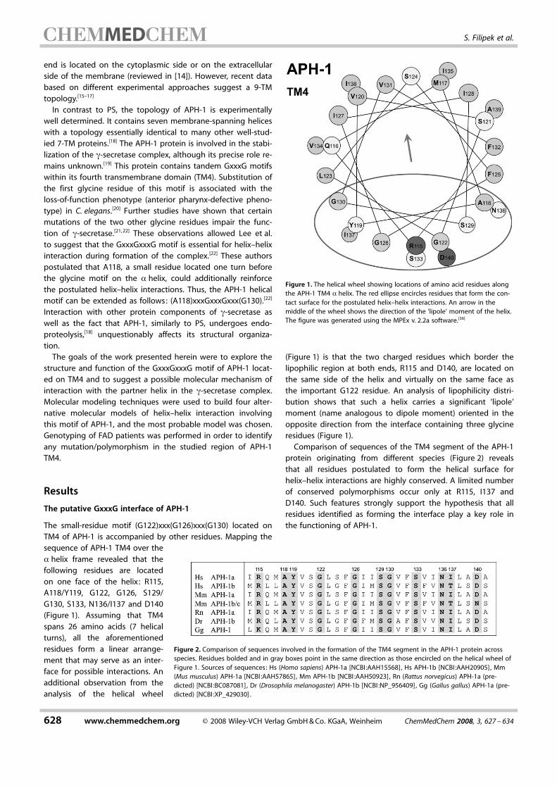
The small-residue motif (G122)xxx ACHTUNGTRENNUNG(G126)xxx ACHTUNGTRENNUNG(G130) located onTM4 of APH-1 is accompanied by other residues. Mapping thesequence of APH-1 TM4 over thea helix frame revealed that thefollowing residues are locatedon one face of the helix: R115,A118/Y119, G122, G126, S129/G130, S133, N136/I137 and D140(Figure 1). Assuming that TM4spans 26 amino acids (7 helicalturns), all the aforementionedresidues form a linear arrange-ment that may serve as an inter-face for possible interactions. Anadditional observation from theanalysis of the helical wheel
(Figure 1) is that the two charged residues which border thelipophilic region at both ends, R115 and D140, are located onthe same side of the helix and virtually on the same face asthe important G122 residue. An analysis of lipophilicity distri-bution shows that such a helix carries a significant ’lipole’moment (name analogous to dipole moment) oriented in theopposite direction from the interface containing three glycineresidues (Figure 1).
Comparison of sequences of the TM4 segment of the APH-1protein originating from different species (Figure 2) revealsthat all residues postulated to form the helical surface forhelix–helix interactions are highly conserved. A limited numberof conserved polymorphisms occur only at R115, I137 andD140. Such features strongly support the hypothesis that allresidues identified as forming the interface play a key role inthe functioning of APH-1.
Figure 1. The helical wheel showing locations of amino acid residues alongthe APH-1 TM4 a helix. The red ellipse encircles residues that form the con-tact surface for the postulated helix–helix interactions. An arrow in themiddle of the wheel shows the direction of the ’lipole’ moment of the helix.The figure was generated using the MPEx v. 2.2a software.[36]
Figure 2. Comparison of sequences involved in the formation of the TM4 segment in the APH-1 protein acrossspecies. Residues bolded and in gray boxes point in the same direction as those encircled on the helical wheel ofFigure 1. Sources of sequences: Hs (Homo sapiens) APH-1a [NCBI:AAH15568], Hs APH-1b [NCBI:AAH20905] , Mm(Mus musculus) APH-1a [NCBI :AAH57865], Mm APH-1b [NCBI :AAH50923], Rn (Rattus norvegicus) APH-1a (pre-dicted) [NCBI :BC087081], Dr (Drosophila melanogaster) APH-1b [NCBI :NP_956409], Gg (Gallus gallus) APH-1a (pre-dicted) [NCBI :XP_429030].
628 www.chemmedchem.org D 2008 Wiley-VCH Verlag GmbH&Co. KGaA, Weinheim ChemMedChem 2008, 3, 627 – 634
MED S. Filipek et al.

APH-1 TM4 genotyping in patients with FAD
The contribution of sequence variation in the APH1a gene frag-ment corresponding to the TM4 domain to FAD developmentwas investigated on a group of Polish FAD patients. Wescreened the DNA fragment of the APH1a gene correspondingto TM4 in 55 patients with FAD. No changes in the analyzedDNA fragment were found. These results are in agreementwith data concerning sporadic Alzheimer’s disease (SAD) in theItalian population.[23]
Possible partner helices for the APH-1 interface
In the next step, the investigations were focused onfinding the helix containing a motif complementaryto GxxxGxxxG in TM4 of APH-1. Such complementari-ty would require the involvement of a similar Glymotif to form very close contacts between interact-ing helices. In order to identify sequence fragmentscontaining small residues (Gly, Ala, or Ser) separatedby the distance of three other residues and locatedwithin TMs, we searched sequences of other compo-nents of g-secretase. Such motifs were found in nei-ther PEN-2 nor NCT proteins, indicating that a partnerhelix of the GxxxGxxxG motif in TM4 of APH-1 waslikely to be found in PS-1.
Six hydrophobic regions (HRs) of PS-1 NTF, as-sumed to be TMs in all published topologies of PS-1,do not carry any motifs that meet the sequence crite-ria described above. On the other hand, three suchmotifs can be found in the PS-1 CTF. The first one,(G378)VKLGLG ACHTUNGTRENNUNG(D385) is present in HR8. It contains anaspartic acid residue in position 385, which is involved in theformation of the catalytic core of the protease. It seems unrea-sonable to expect that this part of the HR8 helix interactsclosely with the APH-1 protein. It is very likely, however, thatthe identified GxxxG motif of PS-1 HR8 is essential for tightpacking interactions with the substrate protein APP (APP con-tains three (small)xxxACHTUNGTRENNUNG(small) motifs within its transmembranedomain).
The next helix of PS-1 that carries a complementary motiffor interaction with APH-1 is HR9. This region contains the se-quence (A409)CFVAILI ACHTUNGTRENNUNG(G417). Such a motif of small residues,each separated by three other residues, would facilitate theformation of an interface with the complementary motif inAPH-1 TM4 analogously to interactions in the transmembranehelix dimer of glycophorin A (GpA) or other systems.[24,25]
Another region of PS-1 that might possibly form a comple-mentary motif of small residues is located in HR10. The se-quence (A431)LPALPISITFACHTUNGTRENNUNG(G442) contains Ala, Ser, and Gly resi-dues separated by three other residues. This highly evolutio-narily conserved region contains a PAL motif common to all in-tramembrane-cleaving aspartic proteases (presenilins and itsanalogues, signal peptide peptidase, and others).[2] Therefore,HR10 does not seem suitable to form an interface to bindAPH-1.
Models of the APH-1–PS-1 interface based on GpA
Homology/comparative modeling with subsequent structureoptimization methods were employed to develop models ofthe interface between the helices of HR9 in PS-1 and TM4 inAPH-1. The most thoroughly investigated intramembrane inter-action mediated by a GxxxG motif is that which occurs indimers of GpA. In the most plausible alignment of PS-1 HR9and APH-1 TM4 sequences over the GpA template (shown inFigure 3), both glycine residues are followed by hydrophobic
and branched valine residues, which were determined to bevery important for proper van der Waals interactions providinga conformationally restricted interface between helices.[26] Inour model of interactions, corresponding Gly or Ala residues ofAPH-1 TM4 or PS-1 HR9 are followed by either Leu or Ile(Figure 3). These residues meet the same criteria as valine(branched and hydrophobic), which suggests that the sametype of helix–helix interaction is plausible.
The crystal structure of the dimer of transmembrane helicesof GpA is known (PDB code: 1AFO).[25] Thus, we used this struc-ture as a template to generate an approximate model of theinteraction between PS-1 HR9 and APH-1 TM4. Subsequentsimulations revealed that a similar network of interactions wasestablished as it is found in the template. The optimizedmodel of the complex is shown in Figure 4a and b. Thissystem is stabilized by hydrophobic forces and also by specifictypes of cross-helix Ca�H···O hydrogen bonds.[26] Such pairingexists between Gly and Ala residues, and the Ile and Leu resi-dues next in sequence from a complementary helix (Figure 4c).The surface contact area is 4.83 nm2 (Table 1), and the anglebetween helices (Figure 4b) is 368. The closest distance be-tween helices (measured in cross– point between helix centers)is 0.70 nm. We also calculated the interaction energy betweentwo helical fragments (18 residues not containing chargedamino acids) in the model to be �152 kJmol�1. This is a signifi-
Figure 3. Two alignments of APH-1 TM4 and PS-1 HR9 using the template sequence ofGpA dimer. Rounded squares mark the sequence area in the modeled helices. Theseareas were used to calculate interaction energies and contact surface areas. Grey boxesdenote the most important residues for alignment.
ChemMedChem 2008, 3, 627 – 634 D 2008 Wiley-VCH Verlag GmbH&Co. KGaA, Weinheim www.chemmedchem.org 629
Models of the APH-1–Presenilin Interface

cantly higher binding energythan the respective value calcu-lated for the GpA dimer(�123 kJmol�1).
We also examined a secondalignment, where the PS-1 HR9sequence is shifted by four resi-
dues. This new alignment also employs the same GxxxG motifon both interacting helices. The contact surface area is slightlysmaller, at 4.65 nm2. The angle between helices and the closestdistance between helices are the same as before because thesame template was used. The calculated binding energy wasless negative, �147 kJmol�1, so this interaction is also morestable than in the GpA template dimer. In both models (Paral-lel 1 and Parallel 2) identical types of interaction are present,that is, branched hydrophobic residues (Leu, Ile, Val) face smallresidues (Gly or Ala). Because of the large angle between heli-ces, the main area of contact in both models involves only oneGxxxG motif from each helix. G122 participates in the maincontact area in the Parallel 1 model but is outside this area inParallel1 2.
The alignment score calculated for the Parallel 1 model was23 for APH-1 TM4 and 24 for PS-1 HR9. For the Parallel 2model the score was 23 for APH-1 TM4 (the same alignment)and �2 for PS-1 HR9. These scores are in agreement with theinteracting energies and contact surfaces, favoring the Paral-lel 1 model.
Models of the APH-1–PS-1 interface based on AqpM
The investigation of the plausible antiparallel orientation byhomology modeling is more difficult because there is no trans-membrane dimer with a GxxxG-based interaction between an-tiparallel helices that could serve as a template. Some clues,however, can be provided by inspection of membrane-embed-ded protein models found in the RCSB protein data bank(PDB),[27] from which examples of analogous interaction can bedrawn. For example, the structure of aquaporin (AqpM, PDBcode: 2F2B), resolved by Lee et al. using X-ray diffraction,[28]
contains a bundle of six TM helices. Two of these helices (TM2and TM5) are tightly packed against each other in an antiparal-lel orientation. Each helix contains a tandem of motifs of twosmall residues separated by three other residues((G59)LAFGFAIACHTUNGTRENNUNG(A67) and (G176)IIIGLTV ACHTUNGTRENNUNG(A184), respectively) andthese motifs directly face each other.
The driving force for interactions between helices TM2 andTM5 in AqpM does not result from interactions between adja-cent helices but from the motifs of small residues GxxxG. Thisis apparent from distances between helices. The smallest dis-tance (0.73 nm), indicating the highest interaction bindingenergy (�327 kJmol�1), is between helices TM2 and TM5(Table 2). Other distances are much larger 0.90 and 0.96 nm,between helices TM1–TM2 and TM5–TM4, respectively. Contactsurfaces are nearly the same because interfaces with adjacenthelices are made of amino acids with larger side chains. The
Figure 4. The model Parallel 1 of the interface between APH-1 TM4 and PS-1HR9 based on the homology to GpA. Ca of Gly and Ala forming the inter-face are shown as shaded spheres. a) Side view: the thick arrows indicate di-rections of helices. b) Side view rotated 908 ; the dashed ellipse marks thecontact area. c) Schematic representation of helix–helix interactions in themodel.
Table 1. Comparison of interaction energies and contact surface areas forwild-type and mutant models of the APH-1 TM4–PS-1 HR9 complex.
Model PS-1 mutation Einteraction
ACHTUNGTRENNUNG[kJmol�1]Contact surfacearea [nm2]
Parallel 1 wild-type �152 4.83
Parallel 2 wild-type �147 4.65
Antiparallel 1 wild-type �218 4.11
Antiparallel 2 wild-type �234 4.33A409T �264 4.24C410T �232 4.39L418F �233 4.38
Antiparallel 2(another
conformation)
wild-type �262 4.39A409T �287 4.26C410T �263 4.37L418F �266 4.46
Table 2. Parameters of the interface between adjacent helices in AqpM.
Interface Einteraction
ACHTUNGTRENNUNG[kJmol�1]Contact surfacearea [nm2]
Helix–helixdistance [nm]
Helix–helixangle [8]
TM1–TM2 �122 4.14 0.90 15TM2–TM5 �327 4.13 0.73 26TM5–TM4 �90 4.41 0.96 15
630 www.chemmedchem.org D 2008 Wiley-VCH Verlag GmbH&Co. KGaA, Weinheim ChemMedChem 2008, 3, 627 – 634
MED S. Filipek et al.
angles between adjacent helices are both small, and accountfor 158, also suggesting a lack of tight packing. The GxxxG-based interface in TM2–TM5 is flanked by two hydrogen bondslocated within these helices, W55–N192 and Y71–I177(carbonyl),which additionally strengthen this contact and contribute tothe interaction energy. No hydrogen bonds were found in theinterfaces with adjacent helices. All these findings additionallyjustify use of the TM2–TM5 pair as a template for protein–pro-tein interactions.
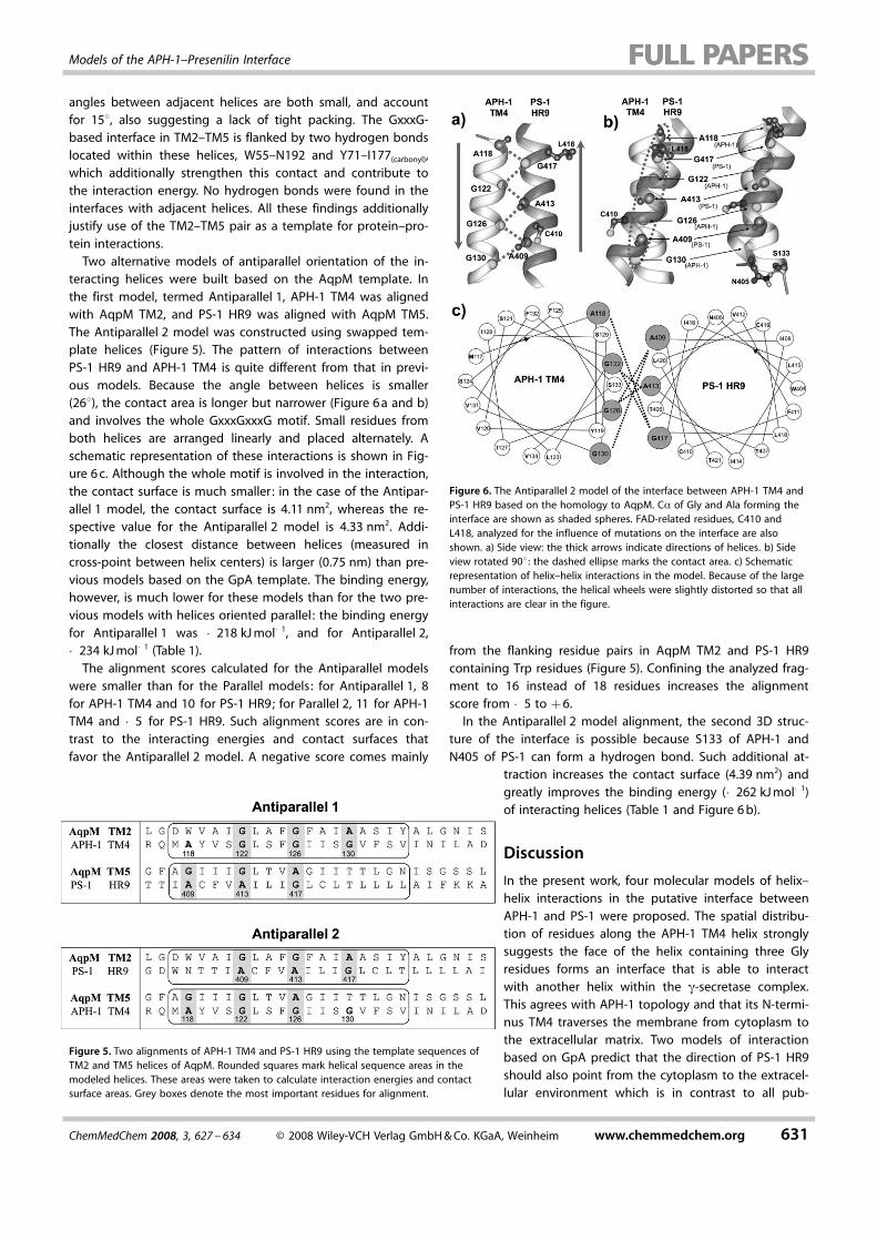
Two alternative models of antiparallel orientation of the in-teracting helices were built based on the AqpM template. Inthe first model, termed Antiparallel 1, APH-1 TM4 was alignedwith AqpM TM2, and PS-1 HR9 was aligned with AqpM TM5.The Antiparallel 2 model was constructed using swapped tem-plate helices (Figure 5). The pattern of interactions betweenPS-1 HR9 and APH-1 TM4 is quite different from that in previ-ous models. Because the angle between helices is smaller(268), the contact area is longer but narrower (Figure 6a and b)and involves the whole GxxxGxxxG motif. Small residues fromboth helices are arranged linearly and placed alternately. Aschematic representation of these interactions is shown in Fig-ure 6c. Although the whole motif is involved in the interaction,the contact surface is much smaller : in the case of the Antipar-allel 1 model, the contact surface is 4.11 nm2, whereas the re-spective value for the Antiparallel 2 model is 4.33 nm2. Addi-tionally the closest distance between helices (measured incross-point between helix centers) is larger (0.75 nm) than pre-vious models based on the GpA template. The binding energy,however, is much lower for these models than for the two pre-vious models with helices oriented parallel : the binding energyfor Antiparallel 1 was �218 kJmol�1, and for Antiparallel 2,�234 kJmol�1 (Table 1).
The alignment scores calculated for the Antiparallel modelswere smaller than for the Parallel models: for Antiparallel 1, 8for APH-1 TM4 and 10 for PS-1 HR9; for Parallel 2, 11 for APH-1TM4 and �5 for PS-1 HR9. Such alignment scores are in con-trast to the interacting energies and contact surfaces thatfavor the Antiparallel 2 model. A negative score comes mainly
from the flanking residue pairs in AqpM TM2 and PS-1 HR9containing Trp residues (Figure 5). Confining the analyzed frag-ment to 16 instead of 18 residues increases the alignmentscore from �5 to +6.
In the Antiparallel 2 model alignment, the second 3D struc-ture of the interface is possible because S133 of APH-1 andN405 of PS-1 can form a hydrogen bond. Such additional at-
traction increases the contact surface (4.39 nm2) andgreatly improves the binding energy (�262 kJmol�1)of interacting helices (Table 1 and Figure 6b).
Discussion
In the present work, four molecular models of helix–helix interactions in the putative interface betweenAPH-1 and PS-1 were proposed. The spatial distribu-tion of residues along the APH-1 TM4 helix stronglysuggests the face of the helix containing three Glyresidues forms an interface that is able to interactwith another helix within the g-secretase complex.This agrees with APH-1 topology and that its N-termi-nus TM4 traverses the membrane from cytoplasm tothe extracellular matrix. Two models of interactionbased on GpA predict that the direction of PS-1 HR9should also point from the cytoplasm to the extracel-lular environment which is in contrast to all pub-
Figure 5. Two alignments of APH-1 TM4 and PS-1 HR9 using the template sequences ofTM2 and TM5 helices of AqpM. Rounded squares mark helical sequence areas in themodeled helices. These areas were taken to calculate interaction energies and contactsurface areas. Grey boxes denote the most important residues for alignment.
Figure 6. The Antiparallel 2 model of the interface between APH-1 TM4 andPS-1 HR9 based on the homology to AqpM. Ca of Gly and Ala forming theinterface are shown as shaded spheres. FAD-related residues, C410 andL418, analyzed for the influence of mutations on the interface are alsoshown. a) Side view: the thick arrows indicate directions of helices. b) Sideview rotated 908 : the dashed ellipse marks the contact area. c) Schematicrepresentation of helix–helix interactions in the model. Because of the largenumber of interactions, the helical wheels were slightly distorted so that allinteractions are clear in the figure.
ChemMedChem 2008, 3, 627 – 634 D 2008 Wiley-VCH Verlag GmbH&Co. KGaA, Weinheim www.chemmedchem.org 631
Models of the APH-1–Presenilin Interface

lished topologies of PS-1. The antiparallel orientation of bothhelices gave rise to two other proposed models of interactionresembling that found in AqpM and formed by a row of smallresidues.
Molecular biological investigations indicate that the initialassembly of g-secretase starts from the formation of the sub-complex between APH-1 and NCT independently of the con-served GxxxGxxxG motif of APH-1.[29] On the other hand, it wasdetermined that certain mutations in this motif affect the inter-actions of the APH-1–NCT sub-complex with presenilin.[19] Animportant insight into these interactions has been made re-cently by two independent research groups, suggesting thatthe APH-1–NCT complex interacts with the C terminus of PS-1rather than with PS-1 NTF.[30,31] Such an interaction was pro-posed based on co-immunoprecipitation of APH-1–NCT withPS-1 CTF in partly detergent-solubilized membranes. All theseresults comply with the notion that the C-terminal region ofPS-1 may interact with the GxxxGxxxG motif of APH-1.
All three HRs (8–10) in PS-1 CTF bear the GxxxG motif. How-ever, HR8 directly forms the catalytic site, and HR10, whichcontains the PAL region, is also recognized as essential for thecatalytic activity of g-secretase. According to Wang et al. , HR10is not involved in the formation or stabilization of the g-secre-tase complex.[32,33] The PAL motif is required by other asparticproteases and most of them do not form a complex with addi-tional proteins for activity.[2] Taking into account the fact thatHR8 and HR10 are involved in substrate processing, HR9 seemsto be the most plausible one to form an interface for interac-tion with APH-1 (Figure 7).
Genotyping of the APH-1 TM4 fragment in our FAD patientsindicated that DNA sequence variation of the fragment doesnot contribute to familial Alzheimer’s disease development.Our data is in agreement with the results presented by Poliet al. ,[23] which indicate that sequence variation in any of thetwo APH genes are not risk factors for SAD in the Italian popu-lation. Three out of six different polymorphisms in the aph1bgene results in amino acid substitutions (T27I, V199L, andF217L), whereas the others are either silent or in noncoding re-
gions. All amino acid polymorphisms identified by Poli et al.are outside TM4.
Calculated alignment scores are mostly positive, indicatingthe feasibility of both chosen templates, GpA and AqpM, forcomparative modeling. Negative scores obtained for the bestmodel, Antiparallel 2, come from two flanking residue pairs inAqpM TM2 and PS-1 HR9 containing Trp residues (Figure 5).Removal of these pairs by confining the analyzed fragment to16 residues instead of 18, increased the alignment score from�5 to +6. This effect can also be achieved by introducing asingle amino acid gap to align two Trp residues from both se-quences. Trp–Trp pair alignment is valued at +17 points inthis score. Flanking Trp residues are very important for theproper anchoring of transmembrane helices in the membrane.
In the GpA-based models created, two interacting helicesare oriented in parallel (that is, with the same membrane-span-ning vector). A lower binding energy obtained for the wild-type (Table 1) suggests that the Parallel 1 model is better (Fig-ure 4a and b). However, the parallel orientation of PS-1 HR9 inrelation to APH-1 TM4 is contrary to most of the experimentalevidence available so far. Although several topologies of PS-1are currently debated, most of them assume PS-1 HR9 directsits C-terminal end toward the cytosolic side. Thus, we exam-ined the antiparallel orientation of PS-1 HR9 in relation to APH-1 TM4 and built the respective molecular models.
In antiparallel orientation, the (A409)xxxAxxx ACHTUNGTRENNUNG(G417) motif lo-cated on the PS-1 HR9 helix can interact with an extended(A118)xxxGxxxGxxx ACHTUNGTRENNUNG(G130) motif found in APH-1 TM4. This isbecause of a much smaller helix–helix angle relative to the par-allel models. Paradoxically this extension diminishes the con-tact surface but also greatly improves the binding energy(Table 1). This strongly suggests that the antiparallel modelsare a better approximation of the interface. Among them theAntiparallel 2 model is characterized by the lowest bindingenergy (Figure 6a and b). This energy may be much lower ifthe additional conformations of the Antiparallel 2 model (Fig-ure 6b) that resulted from our modeling study are taken intoconsideration. The hydrogen bond between S133 of APH-1and N405 of PS-1 greatly improves interactions in the modeland stabilizes the complex. As was found by Schneider and En-gelman[34] in mutational studies of glycophorin A, the motifs oftwo small residues can assist but are not sufficient for trans-membrane helix interactions. The framework of adjacent sidechains is essential for creating stable helix interactions, so thecase of each (small)xxx ACHTUNGTRENNUNG(small) motif must be tested independ-ently. In the analyzed PS-1–APH-1 interface there is a long net-work of small residues (Figure 6a) and a hydrogen bond be-tween interacting helices. Such an arrangement of residues inthe Antiparallel 2 model provides the lowest binding energyand the most probable mode of interaction.
The analyzed fragments were confined to 18 residues to ex-clude all ionic interactions, but such interactions are also veryimportant for a strong interface. In the Parallel models there isa hypothetical possibility of ionic interaction between D140from APH-1 TM4 and K429 (or K430) from PS-1 HR9. However,taking into account the large angle between interacting heli-ces (368), the potentially interacting ionic residues are distant
Figure 7. Scheme of topologies of PS-1 and APH-1 with the suggested inter-face between them highlighted by arrows. The locations of two catalytic as-partic acid residues of PS-1 are also shown.
632 www.chemmedchem.org D 2008 Wiley-VCH Verlag GmbH&Co. KGaA, Weinheim ChemMedChem 2008, 3, 627 – 634
MED S. Filipek et al.
from one another. In the Antiparallel models there are no suchattractive ionic interactions of flanking residues. Instead, in theAntiparallel 1 model there is a possibility of a repulsive interac-tion between K429 (or K430) from PS-1 HR9 and R115 fromAPH-1 TM4. But in the Antiparallel 2 model there is a possibilityof formation of an additional hydrogen bond between D403from PS-1 HR9 and N136 from APH-1 TM4. Further experimen-tal data are required to reveal all details of the binding of bothproteins. Another issue of great importance to the binding isthe influence of adjacent helices in both interacting proteinson the interface. Because of short loops between transmem-brane helices in PS-1 and especially in APH-1, the binding in-terface is composed of more than one helix from each protein.However, the driving force for creation of this interface is hy-drophobic in nature and, in particular, based on (small)xxx-ACHTUNGTRENNUNG(small) motifs.
In the recent paper by Sato et al.[35] confirms the stoichiome-try of g-secretase components PS-1/PEN-2/nicastrin/APH-1 is1:1:1:1, and also that APH-1 is present in this complex as a mo-nomer. The (small)xxxACHTUNGTRENNUNG(small) motifs present in PS-1 and APH-1do not lead to dimerization of both proteins separately. Thisbehavior may be due to hiding of these motifs in immatureforms of both proteins but also due to repulsive interactionsof ionic residues flanking the transmembrane helices contain-ing such motifs in homodimeric interfaces.
The proposed interface fragment of the PS-1 HR9 helix con-tains three FAD-associated mutations (A409T, C410T, andL418F).[13] The influence of these mutations was assessed bycomparison of the binding energies and contact surfaces, andthe results are presented in Table 1. Analysis of this data sug-gests that mutations C410T and L418F introduced into themodels have nearly no effect on binding energy, whereasA409T greatly improved this energy (�264 and �287 kJmol�1
for the first and the second Antiparallel 2 conformations, re-spectively). The rationale behind this is the formation of an ad-ditional hydrogen bond between A409T of PS-1 andG126(carbonyl) of APH-1 (the side chain of S129 is also a potentialpartner). Other mutations are outside the area of the analyzedinterface; however, they may influence the PS-1–APH-1 contactby altering interactions with adjacent helices of respective pro-teins. For analysis of such effects the whole structures of bothinteracting proteins are needed.
Conclusions
The most negative binding energies calculated for the Antipar-allel 2 model as well as its mutants corroborate the mostrecent studies on membrane topology of the PS-1 molecule.This strongly advocates the antiparallel relation between theAPH-1 TM4 and PS-1 HR9 helices. The model containing aGxxxG motif represents the core of the APH-1–PS-1 interface.Analysis of binding energies and contact surfaces of helix–helixinteractions suggests that the Antiparallel 2 model is the mostprobable approximation of the single helix–helix interface be-tween APH-1 and PS-1. The (small)xxxACHTUNGTRENNUNG(small) hydrophobicmotif would be a driving force for the assembly both proteins.Then additional interactions, like flanking residues forming hy-
drogen bonds or ionic interactions and adjacent helices fromboth proteins, would contribute to this interface by modifyingand strengthening it. The created model is only the first stepfor building the whole interface, but it contains the recogni-tion pattern that facilitates the assembly of APH-1 and PS-1.This model can be used in further studies on refinements ofthe molecular constitution of g-secretase and its componentsand possibly for future drug design as well.
Experimental Section
Modeling helix–helix interactions
Homology/comparative modeling was performed based on helix–helix template structures employing GxxxG motifs. We used twoTMs from the dimer of glycophorin A (GpA, PDB code: 1AFO)which served as a template for parallel orientations of interactinghelices. Another system involving TM2 and TM5 from aquaporin(AqpM, PDB code: 2F2B) was used as a template for antiparallelorientations of interacting helices. The alignments were performedmanually based on small-residue motif pairing and maximal over-lapping of transmembrane helices. The alignment scores were cal-culated using PAM250 substitution matrix by summing individualscores from each pair of aligned amino acids. This matrix providesno penalty for Gly/Ala substitution, which is a requirement formodeling (small)xxxACHTUNGTRENNUNG(small) interactions.
In the case of the GpA template two alignments were possiblewith a shift of one target sequence by four residues, so we devel-oped two alternative 3D models of interactions for parallel orienta-tions of interacting helices. In the case of antiparallel orientationstwo alternative models were built based on swapped TM helices ofthe template in sequence alignments. Based on these alignmentsfour different models of helix–helix interface (named Parallel 1, Par-allel 2 and Antiparallel 1, Antiparallel 2) were developed. The use ofa comparative modeling program was not needed because wemodeled two separate helices in the interface and we wanted tohave full control of the optimization process of such a fragilesystem. All amino acid substitutions were done manually based onidentical location of backbone atoms and identical angles.
For energy refinements, helices in each model were end-cappedwith acetyl and N-methyl groups on their N and C termini, respec-tively. These helices were trimmed to 18 amino acids to eliminateany charged residues, which may strongly influence calculations ofinteraction energy between helices. Each model was then subject-ed to energy minimization in vacuum, first using the steepest de-scent method and then simulated annealing. In the secondmethod, the temperature was continuously lowered from 300 to0 K during the molecular dynamics simulation. The simulated an-nealing was applied in three steps by gradual unfreezing, allowingthe motion of 1) hydrogen atoms only, 2) hydrogen + side chainatoms, and 3) all atoms (no constraints).
In order to validate the optimized complexes we calculated the in-teraction energies defined as the difference between the totalenergy of the complex and the sum of total energies of two sepa-rate helices. We also calculated the contact surface of the interfaceas a difference between the sum of solvent-accessible surfaces ofseparated helices and the solvent-accessible surface of the com-plex divided by two. All calculations were performed in Yasara Dy-namics program v. 6.10 (Yasara Biosciences) using the AMBER 99force field. Yasara is a molecular modeling program for buildingproteins and small molecules, energy minimization, and molecular
ChemMedChem 2008, 3, 627 – 634 D 2008 Wiley-VCH Verlag GmbH&Co. KGaA, Weinheim www.chemmedchem.org 633
Models of the APH-1–Presenilin Interface

dynamics. Yasara allows for interactive (involving manual dragging)molecular dynamics, which greatly facilitates finding new confor-mations and exploring the various binding modes. Because ofbuilt-in semiempirical procedures, Yasara is also able to calculatethe partial atomic charges needed for molecular mechanics anddynamics procedures, for small molecules, and nonstandard resi-dues.
APH-1 TM4 genotyping in patients with FAD
The cohort of 55 unrelated patients with FAD diagnosed in theoutpatient clinic of the Department of Neurodegenerative Disor-ders of the Medical Research Centre of the Polish Academy of Sci-ences in Warsaw were investigated in the study. All patients wereexamined by a neurologist, a neuropsychologist, a psychiatrist, andhad a CT scan of the brain. The diagnosis of AD was confirmedusing a standardized protocol according to the National Instituteof Neurological and Communicative Disorders and Stroke–Alzheim-er’s Disease and Related Disorders Association (NINCDS-ADRDA).FAD was diagnosed if at least one additional first-degree relativesuffered from dementia. Written consent was obtained from allparticipants or their relatives. The study was approved by theEthics Committee of the MSWiA Hospital in Warsaw.Genomic DNA was isolated from whole blood by salting out. Thepolymerase chain reaction (PCR) was performed in a volume of25 mL with Qiagen Taq PCR Master Mix (2.5 U Taq DNA polymerase,1O Qiagen PCR buffer containing 1.5 mm MgCl2, 200 mm eachdNTP) and primers (20 pmol each). Amplified fragment, which cor-responds to the fourth transmembrane domain (TM4) in the APH-1a protein, encompasses a part of exon 3, intron 3, and a part ofexon 4 of the APH-1a gene sequence. Sequencing PCR was per-formed in a volume of 10 mL with the ABI PRISM Big Dye Termina-tor v1.1 Cycle Sequencing Kit. The direct sequencing was per-formed on ABI PRISM 310.
Acknowledgements
The work was supported by the Polish Ministry of Science andHigher Education (Grant No: 2P05A 12929) to K.J. K.J. and L.B.thank the Foundation for Polish Science for fellowships support-ing their research. We thank C. Zekanowski for critical reading ofthe manuscript.
Keywords: g-secretase · GxxxG motif · molecular modeling ·molecular recognition · presenilin
[1] A. L. Brunkan, A. M. Goate, J. Neurochem. 2005, 93, 769–792.[2] B. Martoglio, T. E. Golde, Hum. Mol. Genet. 2003, 12, 201R–206R.[3] M. P. Mattson, Nature 2004, 430, 631–639.[4] G. Evin, L. D. Canterford, D. E. Hoke, R. A. Sharples, J. G. Culvenor, C. L.
Masters, Biochemistry 2005, 44, 4332–4341.[5] D. Edbauer, E. Winkler, J. T. Regula, B. Pesold, H. Steiner, C. Haass, Nat.
Cell Biol. 2003, 5, 486–488.[6] W. T. Kimberly, M. J. LaVoie, B. L. Ostaszewski, W. Ye, M. S. Wolfe, D. J.
Selkoe, Proc. Natl. Acad. Sci. USA 2003, 100, 6382–6387.[7] N. Takasugi, T. Tomita, I. Hayashi, M. Tsuruoka, M. Niimura, Y. Takahashi,
G. Thinakaran, T. Iwatsubo, Nature 2003, 422, 438–441.
[8] F. S. Chen, H. Hasegawa, G. Schmitt-Ulms, T. Kawarai, C. Bohm, T. Kataya-ma, Y. J. Gu, N. Sanjo, M. Glista, E. Rogaeva, Y. Wakutani, R. Pardossi-Pi-quard, X. Y. Ruan, A. Tandon, F. Checler, P. Marambaud, K. Hansen, D.Westaway, P. St George-Hyslop, P. Fraser, Nature 2006, 440, 1208–1212.
[9] V. A. Morais, A. S. Crystal, D. S. Pijak, D. Carlin, J. Costa, V. M. Y. Lee, R. W.Doms, J. Biol. Chem. 2003, 278, 43284–43291.
[10] C. Zekanowski, M. P. Golan, K. A. Krzysko, W. Lipczynska-Lojkowska, S.Filipek, A. Kowalska, G. Rossa, B. Peplonska, M. Styczynska, A. Maruszak,D. Religa, M. Wender, J. Kulczycki, M. Barcikowska, J. Kuznicki, Exp.Neurol. 2006, 200, 82–88.
[11] A. Tandon, E. Rogaeva, M. Mullan, P. H. St George-Hyslop, Curr. Opin.Neurobiol. 2000, 13, 377–384.
[12] J. Hardy, R. Crook, Neurosci. Lett. 2001, 306, 203–205.[13] K. Jozwiak, C. Zekanowski, S. Filipek, J. Neurochem. 2006, 98, 1560–
1572.[14] J. Kim, R. Schekman, Proc. Natl. Acad. Sci. USA 2004, 101, 905–906.[15] A. Henricson, L. Kall, E. L. Sonnhammer, FEBS J. 2005, 272, 2727–2733.[16] H. Laudon, E. M. Hansson, K. Melen, A. Bergman, M. R. Farmery, B. Win-
blad, U. Lendahl, G. von Heijne, J. Naslund, J. Biol. Chem. 2005, 280,35352–35360.
[17] D. Spasic, A. Tolia, K. Dillen, V. Baert, B. De Strooper, S. Vrijens, W. An-naert, J. Biol. Chem. 2006, 281, 26569–26577.
[18] R. R. Fortna, A. S. Crystal, V. A. Morais, D. S. Pijak, V. M. Y. Lee, R. W.Doms, J. Biol. Chem. 2003, 279, 3685–3693.
[19] M. Niimura, N. Isoo, N. Takasugi, M. Tsuruoka, K. Ui-Tei, K. Saigo, Y. Moro-hashi, T. Tomita, T. Iwatsubo, J. Biol. Chem. 2004, 280, 12967–12975.
[20] C. Goutte, M. Tsunozaki, V. A. Hale, J. R. Priess, Proc. Natl. Acad. Sci. USA2002, 99, 775–779.
[21] D. Edbauer, C. Kaether, H. Steiner, C. Haass, J. Biol. Chem. 2004, 279,37311–37315.
[22] S. F. Lee, S. Shah, C. Yu, W. C. Wigley, H. Li, M. Lim, K. Pedersen, W. Han,P. Thomas, J. Lundkvist, Y. H. Hao, G. Yu, J. Biol. Chem. 2003, 279, 4144–4152.
[23] M. Poli, L. B. Gatta, S. Archetti, A. Padovani, A. Albertini, D. Finazzi, Neu-rosci. Lett. 2003, 350, 77–80.
[24] A. Senes, D. E. Engel, W. F. DeGrado, Curr. Opin. Struct. Biol. 2004, 14,465–479.
[25] K. R. MacKenzie, J. H. Prestegard, D. M. Engelman, Science 1997, 276,131–133.
[26] A. R. Curran, D. M. Engelman, Curr. Opin. Struct. Biol. 2003, 13, 412–417.[27] H. M. Berman, J. Westbrook, Z. Feng, G. Gilliland, T. N. Bhat, H. Weissig,
I. N. Shindyalov, P. E. Bourne, Nucleic Acids Res. 2000, 28, 235–242.[28] J. K. Lee, D. Kozono, J. Remis, Y. Kitagawa, P. Agre, R. M. Stroud, Proc.
Natl. Acad. Sci. USA 2005, 102, 18932–18937.[29] M. J. LaVoie, P. C. Fraering, B. L. Ostaszewski, W. J. Ye, W. T. Kimberly,
M. S. Wolfe, D. J. Selkoe, J. Biol. Chem. 2003, 278, 37213–37222.[30] A. Bergman, H. Laudon, B. Winblad, J. Lundkvist, J. Naslund, J. Biol.
Chem. 2004, 279, 45564–45572.[31] P. C. Fraering, M. J. LaVoie, W. J. Ye, B. L. Ostaszewski, W. T. Kimberly, D. J.
Selkoe, M. S. Wolfe, Biochemistry 2004, 43, 323–333.[32] J. Wang, A. L. Brunkan, S. Hecimovic, E. Walker, A. Goate, Neurobiol. Dis.
2004, 15, 654–666.[33] J. Wang, D. Beher, A. C. Nyborg, M. S. Shearman, T. E. Golde, A. Goate, J.
Neurochem. 2006, 96, 218–227.[34] D. Schneider, D. M. Engelman, J. Mol. Biol. 2004, 343, 799–804.[35] T. Sato, T. S. Diehl, S. Narayanan, S. Funamoto, Y. Ihara, B. De Strooper,
H. Steiner, C. Haass, M. S. Wolfe, J. Biol. Chem. 2007, DOI: 10.1074/jbc.M705248200.
[36] S. Jaysinghe, K. Hristova, W. Wimley, C. Snider, S. H. White, http://blan-co.biomol.uci.edu/mpex/, 2006 ; last accessed November 12, 2007.
Received: July 31, 2007Revised: October 17, 2007Published online on December 5, 2007
634 www.chemmedchem.org D 2008 Wiley-VCH Verlag GmbH&Co. KGaA, Weinheim ChemMedChem 2008, 3, 627 – 634
MED S. Filipek et al.