Inbreeding and Population Structure In Two Pairs of Cryptic Fig Wasp Species
Upload
independentCategory
view
1download
0
Biochemical Systematics and Ecology 33 (2005) 681e689
www.elsevier.com/locate/biochemsyseco
Molecular identification of four crypticspecies of Mastomys (Rodentia, Murinae)
Emilie Lecompte a,b,c, Carine Brouat d,Jean-Marc Duplantier e, Maxime Galan d,Laurent Granjon a,b,f, Anne Loiseau d,
Karine Mouline f, Jean-Francois Cosson d,*
a Museum National d’Histoire Naturelle, Departement Systematique et Evolution, UMR 5202, CP51,
OSEB, Laboratoire Zoologie Mammiferes et Oiseaux, 55 rue Buffon, 75 005 Paris, Franceb Service de Systematique Moleculaire, 37 rue Cuvier, 75 005 Paris, France
c Institut fur Virologie, AG ter Meulen, Robert-Koch-strasse 17, 35 037 Marburg, Germanyd INRA - IRD - Centre de Biologie et Gestion des Populations,
Campus International de Baillarguet, CS 30016, 34988 Montferrier/Lez cedex, Francee IRD - Centre de Biologie et Gestion des Populations, BP 1386 Dakar, Senegal
f IRD - Laboratoire de Mammalogie, Centre de Biologie et Gestion des Populations,
BP 2528 Bamako, Mali
Received 8 September 2003; accepted 31 December 2004
Abstract
Multimammate rats (genus Mastomys) are abundant in many regions throughout sub-Saharan Africa, and are of high economical and sanitary importance as agricultural pests
as well as reservoir/vectors of human diseases. In pest management and in epidemiologicalstudies, unequivocal species identification of individuals collected in the field is crucial. How-ever, the discrimination among most of the Mastomys species is often difficult, if not
impossible, on the basis of external characters. Karyology provides unambiguous specificassignations, but is not suitable for population studies involving large numbers of individualsbecause it requires fresh material and/or quick transfer from the field to the laboratory. The
purpose of this study was to search for molecular markers allowing a clear discrimination of
* Corresponding author. Tel.: C33 1 99 62 33 01; fax: C33 1 99 62 33 45.
E-mail address: [email protected] (J.-F. Cosson).
0305-1978/$ - see front matter � 2005 Elsevier Ltd. All rights reserved.
doi:10.1016/j.bse.2004.12.015

682 E. Lecompte et al. / Biochemical Systematics and Ecology 33 (2005) 681e689
field collected individuals on the basis of ethanol-preserved samples. Using sequences of thecytochrome b region of mitochondrial DNA, two molecular tests based on species-specific
primers (test 1) and restriction sites generating species-specific profiles (test 2), were designedand evaluated for species identification on a large number of karyotypically or electropho-retically unambiguously determined individuals. The tests clearly discriminate the four most
widespread species. They are easy to perform on a small piece of ear or tail taken from liveanimals, and can probably be adapted to identify museum specimens.� 2005 Elsevier Ltd. All rights reserved.
Keywords: Sibling species; Species identification; Molecular typing; Cytochrome b; mtDNA; Mastomys;
Rodentia; Muridae
1. Introduction
Multimammate rats of the genus Mastomys are widely distributed throughoutsub-Saharan Africa. Species of the genus are often dominant within small mammalcommunities, except in primary forests where they are restricted to humansettlements. They display sporadic population explosions, making them importantagricultural pests (Leirs, 1994). Moreover, they are reservoirs and vectors of varioushuman diseases (Gratz, 1997).
At least fourMastomys species are sibling species with no fully diagnostic externalmorphological criteria (Granjon et al., 1997). Karyotypes (diploid number, 2N andautosomal fundamental number, aFN) remain the most reliable characters to datefor unambiguous species assignation (Green et al., 1980; Duplantier et al., 1990a;Granjon et al., 1997; Lavrenchenko et al., 1998). These species are widelydistributed: Mastomys coucha (2NZ 36, aFNZ 52e56) in Southern Africa,Mastomys huberti (2NZ 32, aFNZ 44e46) in West Africa, Mastomys erythroleucus(2NZ 38, aFNZ 50e56) from Senegal to Ethiopia south to Burundi with a relictpopulation in Morocco, and Mastomys natalensis (2NZ 32, aFNZ 52e54) innearly all sub-Saharan Africa. Locally, two or three species can be found insympatry (Duplantier and Granjon, 1988; Lavrenchenko et al., 1998).
Undoubtedly, the lack of reliable external species-specific characters has impededtaxonomical as well as population studies in Mastomys, especially where severalspecies are sympatric. Several attempts have been made to develop diagnosticmarkers other than karyotypes. In West Africa, haemoglobin variability allowed thedistinction between the 2NZ 38 and 2NZ 32 forms, but with a possible mixture ofM. huberti and M. natalensis among the 2NZ 32 specimens (Dobrokhotov, 1982;Robbins et al., 1983). In Senegal, Duplantier et al. (1990b) failed to find a singlediagnostic allele among 20 allozyme loci studied in M. huberti, M. natalensis and M.erythroleucus. Also, no immunological or electrophoretical variation of albumin wasfound between these same three species (Montgelard, Duplantier and Granjon,unpubl. data). In South Africa, haemoglobin patterns seemed to distinguishsympatric populations of M. coucha and M. natalensis (Green et al., 1980); recently,

683E. Lecompte et al. / Biochemical Systematics and Ecology 33 (2005) 681e689
Smit et al. (2001) found three additional loci with diagnostic alleles between thesetwo species.
Karyology and electrophoresis show important limitations, as they require freshor frozen material and cannot be applied to ethanol-fixed or museum samples. Theyalso imply quite invasive sampling, followed by rather time-consuming experiments.
For these reasons, we developed two molecular tests allowing for an easy andrapid discrimination among the species cited above. The tests were based on the useof mitochondrial cytochrome b sequences to define species-specific primers (test 1)and restriction sites generating species-specific profiles (test 2).
2. Material and methods
We used 20 cytochrome b (cytb) sequences of 1140 pb: two from M. coucha, fivefrom M. erythroleucus, seven from M. natalensis and six from M. huberti. Twosequences from the most closely related species, Mastomys cf kollmannspergeri(nomenclature following Dobigny et al., 2002), were added for comparison. Thesequences were aligned using BioEdit software (Hall, 1999). The sequences aredeposited in Genbank, under the accession numbers AF141220; AF518334eAF518345; AY751287eAY751296.
2.1. Test 1
The strategy used to design the species-specific primers was based on the searchfor mutations specific to only one Mastomys species relative to the others. The cytbsequences were thus closely examined to find short sequences of about 20 nucleotideswith no (or few) variation among specimens of a given species but differing byseveral mutations from specimens of the other species. When possible, species-specific mutations were chosen to be in the 3# end of the potential primers andnucleotidic variation within a species were only admitted in the 5# end.
Once the primers were determined, we optimized the PCR conditions by testingdifferent values of annealing temperature, and primer and MgCl2 concentration.Values for these parameters were adjusted in order to amplify only within the targetspecies when using a set of species-specific primers, with the exclusion of specimensfrom the other species.
2.2. Test 2
We generated restriction maps showing potential restriction sites for 294 enzymesusing BioEdit. Our criteria for selecting target restriction enzymes were threefold: (i)they must generate different restriction profiles for each species; (ii) the restrictionsites must be conserved among individuals of the same species; and (iii) they musthave at least one restriction site in the cytb sequence of each species in order tobe able to distinguish a failed digestion from a valid profile for each individual.Double-stranded PCR amplifications of the entire cytb gene were performed in 15 ml

684 E. Lecompte et al. / Biochemical Systematics and Ecology 33 (2005) 681e689
reaction volumes using the primers L14723 and H15915, following Lecompte et al.(2002). PCR products were used to define digestion reactions.
PCR products were then used to evaluate the specificity of both tests using a largeset of either chromosomally or electrophoretically (for South African specimens)screened individuals collected in different localities scattered over the entire range ofeach species to account for genetic variability. A total of 144 samples were obtainedfrom the 80% ethanol-preserved tissue collection of the Museum National d’HistoireNaturelle, Paris. The origin and number of specimens tested are presented in Table 1.We used 14 M. coucha (COU) from South Africa; 35 M. erythroleucus (ERY) fromMali (12), Senegal (5), Ethiopia (4), Chad (7), Niger (6) and Morocco (1); 66M. natalensis (NAT) from Mali (18), Zimbabwe (2), Senegal (9), Ivory Coast (6),South Africa (20), Tanzania (6), and Chad (5) and 19 M. huberti (HUB) fromMauritania (6), Senegal (7) and Mali (6); 1Mastomys pernanus (PER) from Tanzaniaand 17 M. cf kollmannspergeri (VER) Nigeria (2), Chad (9) and Niger (6).
3. Results and discussion
Genetic divergence within and between species based on our cytb data areindicated in Table 2. The four Mastomys species under study diverge by about 10%,while mean intra-species variation ranges between 1.1 and 3.7% (Table 2). This lowbetween-species divergence relative to what is known for other rodents (seecomments in Lecompte et al., 2002), coupled with the relatively high intra-speciesvariation, considerably complicated the determination of suitable locations forspecies-specific primers and restriction sites.
3.1. Test 1
After several fruitless attempts, four pairs of species-specific primers were selected,one for each of the studied species, situated at different positions along the cytbsequences (Table 3). We had no other choice but to include some site variation inmost of the specific primers. In each case, the majority consensus sequence waschosen. Each species-specific primer pair amplifies different parts of the cytb gene,giving sequences of different sizes (Fig. 1). This facilitates electrophoreticinterpretation, so PCR products obtained with different primer pairs for the sameindividual can be deposited in the same lane.
Double-stranded PCR amplifications were performed in 10 ml reaction volumes.After adjusting the PCR reaction’s conditions, each specific reaction included 0.1 mlof primers for the NAT primers and 0.2 ml for the other primer pairs (10 mM), 0.4 mlof a deoxynucleoside-triphosphate mixture (10 mM), 1 ml of reaction buffer (10!)(Promega), 0.3 ml of MgCl2 (25 mM) for NAT primers, 0.4 ml of MgCl2 (25 mM) forHUB primers and 0.6 ml for the others (Promega), and 0.1 ml of taq DNApolymerase (Promega). We used the following thermal cycling parameters: 4 minat 94 �C, 30e35 cycles (40 s at 94 �C, 45 s at 61 �C, 40 s at 72 �C), with a finalextension of 10 min.
685E. Lecompte et al. / Biochemical Systematics and Ecology 33 (2005) 681e689
3.2. Test 2
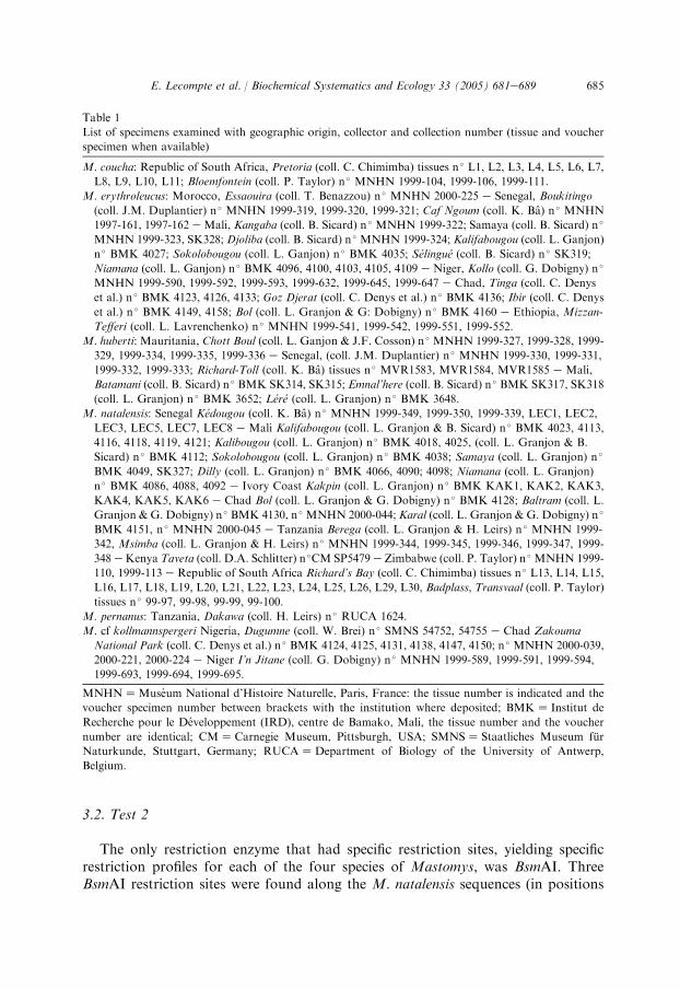
The only restriction enzyme that had specific restriction sites, yielding specificrestriction profiles for each of the four species of Mastomys, was BsmAI. ThreeBsmAI restriction sites were found along the M. natalensis sequences (in positions
Table 1
List of specimens examined with geographic origin, collector and collection number (tissue and voucher
specimen when available)
M. coucha: Republic of South Africa, Pretoria (coll. C. Chimimba) tissues n � L1, L2, L3, L4, L5, L6, L7,
L8, L9, L10, L11; Bloemfontein (coll. P. Taylor) n � MNHN 1999-104, 1999-106, 1999-111.
M. erythroleucus: Morocco, Essaouira (coll. T. Benazzou) n � MNHN 2000-225 e Senegal, Boukitingo
(coll. J.M. Duplantier) n � MNHN 1999-319, 1999-320, 1999-321; Caf Ngoum (coll. K. Ba) n � MNHN
1997-161, 1997-162 e Mali, Kangaba (coll. B. Sicard) n � MNHN 1999-322; Samaya (coll. B. Sicard) n �
MNHN 1999-323, SK328; Djoliba (coll. B. Sicard) n � MNHN 1999-324; Kalifabougou (coll. L. Ganjon)
n � BMK 4027; Sokolobougou (coll. L. Ganjon) n � BMK 4035; Selingue (coll. B. Sicard) n � SK319;
Niamana (coll. L. Ganjon) n � BMK 4096, 4100, 4103, 4105, 4109 e Niger, Kollo (coll. G. Dobigny) n �
MNHN 1999-590, 1999-592, 1999-593, 1999-632, 1999-645, 1999-647 e Chad, Tinga (coll. C. Denys
et al.) n � BMK 4123, 4126, 4133; Goz Djerat (coll. C. Denys et al.) n � BMK 4136; Ibir (coll. C. Denys
et al.) n � BMK 4149, 4158; Bol (coll. L. Granjon & G: Dobigny) n � BMK 4160 e Ethiopia, Mizzan-
Tefferi (coll. L. Lavrenchenko) n � MNHN 1999-541, 1999-542, 1999-551, 1999-552.
M. huberti: Mauritania, Chott Boul (coll. L. Ganjon & J.F. Cosson) n � MNHN 1999-327, 1999-328, 1999-
329, 1999-334, 1999-335, 1999-336 e Senegal, (coll. J.M. Duplantier) n � MNHN 1999-330, 1999-331,
1999-332, 1999-333; Richard-Toll (coll. K. Ba) tissues n � MVR1583, MVR1584, MVR1585 e Mali,
Batamani (coll. B. Sicard) n � BMK SK314, SK315; Emnal’here (coll. B. Sicard) n � BMK SK317, SK318
(coll. L. Granjon) n � BMK 3652; Lere (coll. L. Granjon) n � BMK 3648.
M. natalensis: Senegal Kedougou (coll. K. Ba) n � MNHN 1999-349, 1999-350, 1999-339, LEC1, LEC2,
LEC3, LEC5, LEC7, LEC8 e Mali Kalifabougou (coll. L. Granjon & B. Sicard) n � BMK 4023, 4113,
4116, 4118, 4119, 4121; Kalibougou (coll. L. Granjon) n � BMK 4018, 4025, (coll. L. Granjon & B.
Sicard) n � BMK 4112; Sokolobougou (coll. L. Granjon) n � BMK 4038; Samaya (coll. L. Granjon) n �
BMK 4049, SK327; Dilly (coll. L. Granjon) n � BMK 4066, 4090; 4098; Niamana (coll. L. Granjon)
n � BMK 4086, 4088, 4092 e Ivory Coast Kakpin (coll. L. Granjon) n � BMK KAK1, KAK2, KAK3,
KAK4, KAK5, KAK6 e Chad Bol (coll. L. Granjon & G. Dobigny) n � BMK 4128; Baltram (coll. L.
Granjon & G. Dobigny) n � BMK 4130, n � MNHN 2000-044; Karal (coll. L. Granjon & G. Dobigny) n �
BMK 4151, n � MNHN 2000-045 e Tanzania Berega (coll. L. Granjon & H. Leirs) n � MNHN 1999-
342, Msimba (coll. L. Granjon & H. Leirs) n � MNHN 1999-344, 1999-345, 1999-346, 1999-347, 1999-
348 eKenya Taveta (coll. D.A. Schlitter) n �CM SP5479 e Zimbabwe (coll. P. Taylor) n � MNHN 1999-
110, 1999-113 e Republic of South Africa Richard’s Bay (coll. C. Chimimba) tissues n � L13, L14, L15,
L16, L17, L18, L19, L20, L21, L22, L23, L24, L25, L26, L29, L30, Badplass, Transvaal (coll. P. Taylor)
tissues n � 99-97, 99-98, 99-99, 99-100.
M. pernanus: Tanzania, Dakawa (coll. H. Leirs) n � RUCA 1624.
M. cf kollmannspergeri Nigeria, Dugumne (coll. W. Brei) n � SMNS 54752, 54755 e Chad Zakouma
National Park (coll. C. Denys et al.) n � BMK 4124, 4125, 4131, 4138, 4147, 4150; n � MNHN 2000-039,
2000-221, 2000-224 e Niger I’n Jitane (coll. G. Dobigny) n � MNHN 1999-589, 1999-591, 1999-594,
1999-693, 1999-694, 1999-695.
MNHNZMuseum National d’Histoire Naturelle, Paris, France: the tissue number is indicated and the
voucher specimen number between brackets with the institution where deposited; BMKZ Institut de
Recherche pour le Developpement (IRD), centre de Bamako, Mali, the tissue number and the voucher
number are identical; CMZCarnegie Museum, Pittsburgh, USA; SMNSZ Staatliches Museum fur
Naturkunde, Stuttgart, Germany; RUCAZDepartment of Biology of the University of Antwerp,
Belgium.

686 E. Lecompte et al. / Biochemical Systematics and Ecology 33 (2005) 681e689

687E. Lecompte et al. / Biochemical Systematics and Ecology 33 (2005) 681e689
212e216, 752e756 and 941e945), two of them are available for M. erythroleucus (inpositions 212e216 and 752e756) while M. huberti and M. coucha sequences arecharacterized by only one of these restriction sites (in positions 212e216 and 752e756, respectively). Following amplification of the entire cytb gene, 5 ml of PCRproducts were digested with BsmAI (Fig. 2). The reaction volume (20 ml) contained5 ml PCR product, 0.2 ml (1 U) restriction enzyme, 2 ml (1!) digest buffer,12.8 ml distilled water. Products were digested for 2 h at 55 �C and run in a 1.5%agarose gel.
PCR amplifications and specific assignation efficiency were then tested on all thespecimens in our data set (see above for details). A complete concordance betweenthe molecular typing and the chromosomal/electrophoretical identification wasobserved for both tests.
For test 1, each specimen was tested with the four pairs of specific primers.Attempts to use the four primer pairs in the same PCR reaction with the DNA ofone specimen (multiplex PCR) were not successful, so each primer pair has to beused in separate PCR reactions. Moreover, we wish to draw the attention topotential users to the fact that the quality of results is strongly dependent on a strictcompliance to the recommended PCR conditions. In some instances, higher DNA orprimer concentrations or lower annealing temperatures yielded an amplified productwith two species-specific primer pairs for the same individual. However, even in thesecases, such results did not lead to errors in specific assignation, as they areambiguous by nature (a specimen with bands for two primer pairs). This problemwas always solved by using strict PCR conditions following which only the expectedspecies-specific band was observed.
Fig. 1. Species-specific amplification of cytochrome b in Mastomys specimens. DNA was amplified with
specific primers for M. huberti (501 pb, gel 1), M. erythroleucus (456 pb, gel 2), M. natalensis (206 pb, gel
3) andM. coucha (355 pb, gel 4). Molecular markers (each 200 pb) are deposited in lines 1 and 17. Lanes 3,
7 and 13: M. huberti 99e331, SK314 and 3648. Lanes 4, 8 and 11: M. erythroleucus 4027, 4105 and 4133.
Lanes 5, 6 and 12: M. natalensis 4066, 4119 and 4090. Lanes 2, 9 and 10: M. coucha 99e104, 99e106 and
99e111. Lanes 14, 15 and 17: M. verheyeni 4124, 4131 and 4147.
Table 2
Genetic distances observed between theMastomys species studied,M. verheyeni (VER),M. coucha (COU),
M. erythroleucus (ERY), M. huberti (HUB) and M. natalensis (NAT) using complete cytochrome b
sequences (1140 pb)
Nucleotide difference (%)
Range Mean
Between VER & COU, ERY, NAT, HUB 15.6e18.1 17.1
Between COU & ERY, NAT, HUB 9.0e11.2 10.1
Between HUB & ERY, NAT 8.5e10.9 9.0
Between ERY & NAT 7.1e8.9 8.1
Within ERY 0e4.9 3.7
Within NAT 0e4.3 3.2
Within HUB 0e3.6 2.1
Within COU 0e1.1 1.1

688 E. Lecompte et al. / Biochemical Systematics and Ecology 33 (2005) 681e689
These results indicate that any specimen of the four Mastomys species consideredhere can be reliably identified using the two methods. As our approach requires onlysmall quantities of total DNA, it can be performed on a small piece of ear, tail or toetaken from live animals, and can probably be adapted to identify museumspecimens. Moreover, although our study concerned four Mastomys species, controltests were performed on two other species which can be morphologically identified.Seventeen M. cf kollmannspergeri from Nigeria, Chad and Niger, and oneM. pernanus from Tanzania (cf. Table 1) yielded negative results for each primerpair. This result is not surprising since the latters are phylogenetically more distantlyrelated to the four species studied here than these four species are from each other onthe basis of complete cytb sequences analyses (Lecompte et al., 2002).
In conclusion, these methods are reliable and easy to perform for routineidentification in field studies. They will contribute to increase the reliability of faunal
Table 3
Cytochrome b species-specific primers determined for Mastomys coucha, M. erythroleucus, M. huberti
and M. natalensis
M. coucha F-279: 5# TTG-TTC-CTT-CAC-GTA-GGA-CGG 3#R-635: 5# TTA-GGC-CTG-TTG-GGT-TAT-TGG-AT 3#
M. erythroleucus F-49: 5# CAT-TCA-TTG-ACC-TAC-CTG-CT 3#R-505: 5# AGA-ATC-CCC-CTC-AAA-TTC-AC 3#
M. huberti F-174: 5# TAC-TAT-AAC-AGC-ATT-TTC-ATC-G 3#R-702: 5# AGT-ATA-GTA-TGG-GTG-GAA-TGG-GAT-TTT-G 3#
M. natalensis F-607: 5# CGG-GCT-CTA-ATA-ACC-CAA-CG 3#R-813: 5# TTC-TGG-TTT-GAT-ATG-GGG-AGG-T 3#
Primer name indicates the DNA strand (F, forward or R, reverse) and the nucleotide position of the 5# endof the primer along the cytochrome b gene.
Fig. 2. Agarose gel showing the differences among species of restriction profiles of BsmAI on cytb
sequences. Molecular markers (each 200 pb) are deposited in lines 1 and 14. Lanes 2, 3, 4: M. coucha 99e104, 99e106 and 99e111. Lanes 5, 6, 7: M. erythroleucus 4027, 4105 and 4133. Lanes 8, 9, 10: M. huberti
99e331, SK314 and 3648. Lanes 11, 12, 13: M. natalensis 4066, 4119 and 4090. The highest bands (one for
M. huberti and M. coucha, two for M. erythroleucus and three for M. natalensis) on the profile correspond
to non-digested (or partially digested) DNA.

689E. Lecompte et al. / Biochemical Systematics and Ecology 33 (2005) 681e689
inventories, and allow for accurate population studies for this major rodent genus inAfrica, especially in regions where two or more species occur sympatrically.
Acknowledgements
We are grateful to the generous providers of tissue samples: K. Ba, T. Benazzou,C. Chimimba, G. Dobigny, L. Lavrenchenko, P. Taylor, B. Sicard, and to J. Britton-Davidian for comments on the manuscript.
References
Dobigny, G., Nomao, A., Gautun, J.C., 2002. A cytotaxonomic survey of Rodents from Niger:
implications for systematics, biodiversity and biogeography. Mammalia 66, 495e523.Dobrokhotov, B.P., 1982. The utilization of electrophoresis of haemoglobins for identification of species
of the genus Mastomys from West Africa. Zool. Zh. 61, 290e294.
Duplantier, J.M., Granjon, L., 1988. Occupation et utilisation de l’espace par des populations du genre
Mastomys au Senegal: etude a trois niveaux de perception. Sci. Technol. Anim. Lab. 13, 129e133.Duplantier, J.M., Britton-Davidian, J., Granjon, L., 1990a. Chromosomal characterization of three
species of the genus Mastomys in Senegal. Z. Zool. Syst. Evolutions-forsch. 28, 289e298.
Duplantier, J.M., Granjon, L., Mathieu, E., Bonhomme, F., 1990b. Structures genetiques comparees de
trois especes de rongeurs africains du genre Mastomys au Senegal. Genetica 81, 179e192.
Granjon, L., Duplantier, J.M., Catalan, J., Britton-Davidian, J., 1997. Systematics of the genus Mastomys
(Thomas, 1915) (Rodentia: Muridae). A review. Belg. J. Zool. 127, 7e18.
Gratz, N., 1997. The burden of rodent-borne diseases in Africa south of the Sahara. Belg. J. Zool. 127,
71e84.
Green, C.A., Keogh, H., Gordon, D.H., Pinto, M., Hartwig, E.K., 1980. The distribution, identification,
and naming of the Mastomys natalensis species complex in southern Africa (Rodentia: Muridae). J.
Zool. 192, 17e23.Hall, T.A., 1999. BioEdit: a user-friendly biological sequence alignment editor and analysis program for
Windows 95/98/NT. Nucleic Acids Symp. Ser. 41, 95e98.
Lavrenchenko, L.A., Likhnova, O.P., Baskevich, M.I., Afework Bekele, 1998. Systematics and
distribution of Mastomys (Muridae, Rodentia) from Ethiopia, with the description of a new species.
Z. Saugetierkd. 63, 37e51.
Lecompte, E., Granjon, L., Kerbis-Peterhans, J., Denys, C., 2002. Cytochrome b-based phylogeny of the
Praomys group (Rodentia, Murinae): a new African radiation? C. R. Biol. 325, 827e840.Leirs, H., 1994. Population ecology of Mastomys natalensis (Smith, 1834). Implications for rodent control
in Africa. Belgian Administration for Development Cooperation, Agricultural Ed. N �35, Brussels.Robbins, C.B., Krebs Jr., J.W., Johnson, K.M., 1983. Mastomys (Rodentia: Muridae) species
distinguished by hemoglobin pattern differences. Am. J. Trop. Med. Hyg. 32, 624e630.Smit, A., van der Bank, H., Falk, T., de Castro, A., 2001. Biochemical genetic markers to identify two
morphologically similar South African Mastomys species (Rodentia: Muridae). Biochem. Syst. Ecol.
29, 21e30.
Copyright © 2022 FDOKUMEN