Unimolecular chemistry of metastable dimethyl isophthalate radical cations

Molecular encapsulation of 5-nitroindazole derivatives in2,6-dimethyl-b-cyclodextrin: Electrochemical and spectroscopic studies
Fernanda Pérez-Cruz a, Carolina Jullian a,*, Jorge Rodriguez b,c, Vicente J. Arán d, Claudio Olea-Azar b,*
a Departamento de Química Orgánica y Fisicoquímica, Facultad de Ciencias Químicas y Farmacéuticas, Universidad de Chile, Casilla 233, Santiago 1, Chileb Departamento de Química Inorgánica y Analítica, Facultad de Ciencias Químicas y Farmacéuticas, Universidad de Chile, Casilla 233, Santiago 1, Chilec Departamento de Química, Facultad de Ciencias Básicas, Universidad Metropolitana de Ciencias de la Educación, Santiago, Chiled Instituto de Química Médica, CSISC, Juan de la Cierva 3, 28006 Madrid, Spain
a r t i c l e i n f o a b s t r a c t
Keywords:5-NitroindazoleCyclodextrinMolecular modelingNMRESR
* Corresponding authors.E-mail address: [email protected] (C. Jullian).
Four different 5-nitroindazole derivatives (1–4) and its inclusion with Heptakis(2,6-di-O-methyl)-b-cyclodextrin (DMbCD) were investigated. The stoichiometric ratios and stability constants describingthe extent of formation of the complexes were determined by phase-solubility measurements obtainingin all cases a type-AL diagram. Also electrochemical studies were carried out, where the observed changein the EPC value indicated a lower feasibility of the nitro group reduction. The same behavior wasobserved in the ESR studies. The detailed spatial configuration is proposed based on 2D NMR methods.These results are further interpreted using molecular modeling studies. The latter results are in goodagreement with the experimental data.
1. Introduction
The American trypanozomiasis is a disease that according todata from WHO, over 24 million people are infected or at leastserologically positive for Trypanosoma cruzi.1 The importance ofthe investigations of nitroheterocycles derivatives lie in the exis-tence of the group electroacceptor capable of generate radicals spe-cies (ROS) that act against the parasite producing oxidative stress.Since 2005 there began the study of a family of 5-nitroindazole (NI)derivatives in order to verify their anti-trypanocidal activity. Arecent study has shown the in vitro and in vivo anti-trypanosoma-tide activity,2 besides, the mode of action of the NI against T. cruzihas being reported by the first time.3 Recently, the electrochemicalstudy and the generation of the nitro anion radical of 5-nitroindaz-ole derivative was done.4 Unfortunately many of these compoundsshowed low water solubility, which is a problem for its possibleuse as antichagasic drugs.
Cyclodextrins (cyclic oligosaccharides composed of D-glucoseunits) (Scheme 1) are known for their ability to bind non-cova-lently and forming inclusion complexes with wide variety organiccompounds.5 In these complexes, a guest molecule is held withinthe lipophilic cavity of a cyclodextrin host molecule. The cyclodex-trin cavity has an apolar character similar to an 80% dioxane/water
solution and provides a slightly alkaline environment, because it issurrounded by glycosidic ethers.6 The main driving force of thecomplexation is the release of enthalpy-rich water molecules fromthe cavity.7–11 Replacement of water molecules by more hydropho-bic guest molecules present in the solution results in formation ofan inclusion complex between the host and the guest.12
In a previous work13 we verified that the incorporation of NIderivatives in cyclodextrins improved their solubility. In this sense,encapsulating the drug in the hydrophobic cavity of cyclodextrinseems to be a good approximation in order to increase the bioavail-ability and biological activity.
In this work, inclusion complex of four 5-nitroindazole deriva-tives in Heptakis(2,6-di-O-methyl)-b-cyclodextrin molecule wereprepared in order to improve the aqueous solubility of the drugs.The inclusion complexes were analyzed by UV–vis, NMR spectros-copy, Differential pulse polarography, ESR and molecular modelingtechniques. The structural information obtained and the geometryof the complexes, are necessary to clarify the complexation mech-anism and the importance of the substituent groups on the inda-zole ring.
2. Results and discussion
The stoichiometric ratios and stability constants were derivedfrom the changes in the solubility of the substrates 1–4 (Scheme1) in the presence of increasing amounts of DMbCD, measured by

COMPOUND R1 R2
1
2
3
4
N
N
O2N
OR2
R1
ON
4’ 5’
N4’
5’
6’
CH3
CH3
ON
4’ 5’
N4’
5’
6’
CH2
CH2
DMβCD n = 7 R 2 , R 6 = CH3 and R3 = H
5-nitroindazole
Scheme 1.
0.0 4.0x10⁻³ 8.0x10⁻³ 1.2x10⁻² 1.6x10⁻²
0.00
1.50x10⁻⁴
3.00x10⁻⁴
4.50x10⁻⁴
6.00x10⁻⁴
[ 4] /
M
[ CD] / M
0.0 4.0x10⁻³ 8.0x10⁻³ 1.2x10⁻²
0.0
8.0x10⁻⁵
1.6x10⁻⁴
2.4x10⁻⁴
3.2x10⁻⁴
4.0x10⁻⁴
[ 3 ]
/ M
[ CD ] / M
0.0 4.0x10⁻³ 8.0x10⁻³ 1.2x10⁻² 1.6x10⁻²
0.0
5.0x10⁻⁴
1.0x10⁻³
1.5x10⁻³
2.0x10⁻³
2.5x10⁻³
[ 1] /
M
[ CD] / M
0.0 4.0x10⁻³ 8.0x10⁻³ 1.2x10⁻²
2.3x10⁻³
2.4x10⁻³
2.5x10⁻³
2.6x10⁻³
2.6x10⁻³
2.7x10⁻³
[ 2] /
M
[ CD ] / M
Figure 1. Phase solubility diagrams of compounds 1–4 with DMbCD in water at 30 �C.
F. Pérez-Cruz et al. / 4605

Table 1Association constants for NI–DMbCD complexes
Inclusion complex Ka (M�1) r
1:DMbCD 542 ± 8 0.9852:DMbCD 14 ± 11 0.9853:DMbCD 8955 ± 15 0.9894:DMbCD 2145 ± 12 0.995
-0.3 -0.4 -0.5 -0.6 -0.7 -0.8
ab
c
d
E/V vs Calomelan saturated
igure 3. DPP of 1 mM 4 in 0.1 M of phosphate buffer pH 7.4. Compound 4 in (a)bsence and presence of (b) 2, (c) 6 and (d) 8 mM DMbCD. Conditions: scan rate0 mV s�1, pulse amplitude 50 mV, pulse width 50 ms.
4606 F. Pérez-Cruz et al. /
UV–vis spectroscopy. For all the NI/DMbCD systems, cyclodextrinenhance the aqueous solubility of NI derivatives, as shown inFigure 1. The binding constant Ka of the complexes was calculatedfrom the slopes of the linear phase-solubility plots, Table 1, accord-ing to the methodology described in the experimental part. Thesediagrams showed a linear relationship between the amount of NIsolubilized and the concentration of CD in solution.
The binding constant for the nitroindazole derivatives withDMbCD followed the rank order 3 > 4 > 1 > 2, reflecting anenhancement of binding on the different substituent’s of the nitro-indazole derivative. We observed previously the same trendemploying bCD instead of DMbCD14 (data not shown), but with lessassociation constants, indicating that the complexation ability ofthe b-cyclodextrin is significantly enhanced by its methylation.Apparently, the presence of the methylated groups in the cyclodex-trin, seems to be important for the binding of these compounds inthe cyclodextrin cavity.13,15 These groups enlarge the cyclodextrincavity, making its environment more hydrophobic and favor theadaptability of the cyclodextrin towards a guest, through anenhanced flexibility.
Also, the enhancement of the Ka values can be related to the dif-ferent substituent groups at the positions R1 and R2 of the indazolering. In this regards, morpholine and benzyloxy groups have apositive influence on the association constant and isopropyl andmethoxyl groups have a negative influence on the Ka value.
On the other hand, the ratio R was plotted against the differenceof the absorbance of NI in presence and absence of DMbCD. Theresults of Job’s plot are shown in Figure 2. According to the continu-ous variation method, the maximum concentration of the complexwill be present in the sample where the molar ratio R correspondsto the complexation stoichiometry. The maximum absorbance vari-ation for DMbCD with the nitroindazole 1–4 derivatives wasobserved for R = 0.5, which might indicate that the main stoichiom-etry is 1:1, in agreement with the stoichiometry suggested from thephase-solubility study.
0.0 0.2 0.4 0.6 0.8 1.0
0.0
3.0x10⁻⁷
6.0x10⁻⁷
9.0x10⁻⁷
1.2x10⁻⁶
1.5x10⁻⁶
1.8x10⁻⁶
2.1x10⁻⁶
R
NI-1 NI-2 NI-3 NI-4
ΔA*
[ NI ]
Figure 2. Continuous variation plot for the complexes 1, 2, 3, 4 with DMbCD fromabsorbance measurements.
2.1. Differential pulse polarography (DPP) studies
Electrochemical behavior in protic media for this kind of5-nitroindazole derivatives has been previously studied,3,13 indi-cating that NI derivatives are able to be reduced on the mercuryelectrode due to the four-electron and four-proton irreversiblereduction of the nitroaromatic moiety to yield the hydroxylaminederivative according to the following overall reaction:
R—NO2 þ 4e� þ 4Hþ ! R—NHOHþH2O ð1Þ
Consequently to evaluate changes in the polarograms, due to addi-tion of DMbCD into a solution of NI, we used different NI/DMbCDmixtures in aqueous media at pH 7.4. The addition of DMbCD to asolution of NI causes two main changes in the polarograms. Firstly,the cathodic peak potential (EPC) shifted in a negative direction andsecondly, the cathodic peak current IPC increased. Figures 3 and 4,show the displacement of cathodic peak potential (EPC) for com-pounds 3 and 4, when different concentrations of DMbCD are used.The shift of signals 4-DMbCD and 3-DMbCD to more negativepotentials indicates that the nitro group is reduced less favorablywhen the indazole ring is included in the hydrophobic cavity hin-dering the interaction with the electrode. Complexes 3-DMbCDand 4-DMbCD show very close reduction potentials, �0.57 V and
Fa1
-0.3 -0.4 -0.5 -0.6 -0.7 -0.8
E/V vs Calomelan saturated
a
b
c
d
Figure 4. DPP of 1 mM 3 in 0.1 M of phosphate buffer pH 7.4. Compound 3 in (a)absence and presence of (b) 2, 6 (c) and (d) 8 mM DMbCD. Conditions: scan rate10 mV s�1, pulse amplitude 50 mV, pulse width 50 ms.
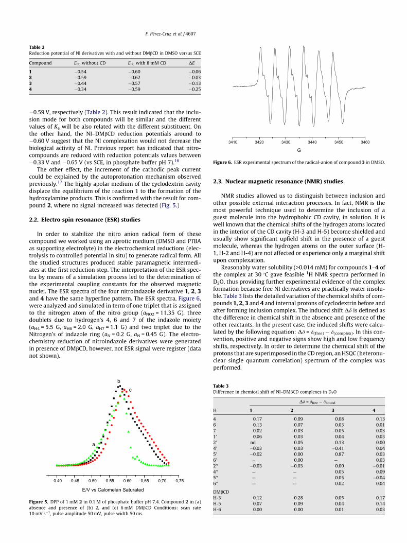
Table 2Reduction potential of NI derivatives with and without DMbCD in DMSO versus SCE
Compound EPC without CD EPC with 8 mM CD DE
1 �0.54 �0.60 �0.062 �0.59 �0.62 �0.033 �0.44 �0.57 �0.134 �0.34 �0.59 �0.25
3410 3420 3430 3440 3450 3460
G
Figure 6. ESR experimental spectrum of the radical-anion of compound 3 in DMSO.
F. Pérez-Cruz et al. / 4607
�0.59 V, respectively (Table 2). This result indicated that the inclu-sion mode for both compounds will be similar and the differentvalues of Ka will be also related with the different substituent. Onthe other hand, the NI–DMbCD reduction potentials around to�0.60 V suggest that the NI complexation would not decrease thebiological activity of NI. Previous report has indicated that nitro-compounds are reduced with reduction potentials values between�0.33 V and �0.65 V (vs SCE, in phosphate buffer pH 7).16
The other effect, the increment of the cathodic peak currentcould be explained by the autoprotonation mechanism observedpreviously.17 The highly apolar medium of the cyclodextrin cavitydisplace the equilibrium of the reaction 1 to the formation of thehydroxylamine products. This is confirmed with the result for com-pound 2, where no signal increased was detected (Fig. 5.)
2.2. Electro spin resonance (ESR) studies
In order to stabilize the nitro anion radical form of thesecompound we worked using an aprotic medium (DMSO and PTBAas supporting electrolyte) in the electrochemical reductions (elec-trolysis to controlled potential in situ) to generate radical form. Allthe studied structures produced stable paramagnetic intermedi-ates at the first reduction step. The interpretation of the ESR spec-tra by means of a simulation process led to the determination ofthe experimental coupling constants for the observed magneticnuclei. The ESR spectra of the four nitroindazole derivative 1, 2, 3and 4 have the same hyperfine pattern. The ESR spectra, Figure 6,were analyzed and simulated in term of one triplet that is assignedto the nitrogen atom of the nitro group (aNO2 = 11.35 G), threedoublets due to hydrogen’s 4, 6 and 7 of the indazole moiety(aH4 = 5.5 G, aH6 = 2.0 G, aH7 = 1.1 G) and two triplet due to theNitrogen’s of indazole ring (aN = 0.2 G, aN = 0.45 G). The electro-chemistry reduction of nitroindazole derivatives were generatedin presence of DMbCD, however, not ESR signal were register (datanot shown).
-0.40 -0.45 -0.50 -0.55 -0.60 -0,65 -0,70 -0,75
a
bc
E/V vs Calomelan Saturated
Figure 5. DPP of 1 mM 2 in 0.1 M of phosphate buffer pH 7.4. Compound 2 in (a)absence and presence of (b) 2, and (c) 6 mM DMbCD Conditions: scan rate10 mV s�1, pulse amplitude 50 mV, pulse width 50 ms.
2.3. Nuclear magnetic resonance (NMR) studies
NMR studies allowed us to distinguish between inclusion andother possible external interaction processes. In fact, NMR is themost powerful technique used to determine the inclusion of aguest molecule into the hydrophobic CD cavity, in solution. It iswell known that the chemical shifts of the hydrogen atoms locatedin the interior of the CD cavity (H-3 and H-5) become shielded andusually show significant upfield shift in the presence of a guestmolecule, whereas the hydrogen atoms on the outer surface (H-1, H-2 and H-4) are not affected or experience only a marginal shiftupon complexation.
Reasonably water solubility (>0.014 mM) for compounds 1–4 ofthe complex at 30 �C gave feasible 1H NMR spectra performed inD2O, thus providing further experimental evidence of the complexformation because free NI derivatives are practically water insolu-ble. Table 3 lists the detailed variation of the chemical shifts of com-pounds 1, 2, 3 and 4 and internal protons of cyclodextrin before andafter forming inclusion complex. The induced shift Dd is defined asthe difference in chemical shift in the absence and presence of theother reactants. In the present case, the induced shifts were calcu-lated by the following equation: Dd = d(free) � d(complex). In this con-vention, positive and negative signs show high and low frequencyshifts, respectively. In order to determine the chemical shift of theprotons that are superimposed in the CD region, an HSQC (heteronu-clear single quantum correlation) spectrum of the complex wasperformed.
Table 3Difference in chemical shift of NI–DMbCD complexes in D2O
Dd = dfree � dbound
H 1 2 3 4
4 0.17 0.09 0.08 0.136 0.13 0.07 0.03 0.017 0.02 �0.03 �0.05 0.0310 0.06 0.03 0.04 0.0320 nd 0.05 0.13 0.0040 �0.03 0.03 �0.41 0.0450 �0.02 0.00 0.87 0.0360 � 0.00 — 0.0320 0 �0.03 �0.03 0.00 �0.0140 0 — — 0.05 0.0950 0 — — 0.05 �0.0460 0 — — 0.02 0.04
DMbCDH-3 0.12 0.28 0.05 0.17H-5 0.07 0.09 0.04 0.14H-6 0.00 0.00 0.01 0.03

m
H-
H-
Figure 7. Partial contour plot of two-dimensional ROESY spectrum of 3-DMbCD inclusion complex.
4608 F. Pérez-Cruz et al. /
All the 1, 2, 3 and 4 protons showed frequency changes in thepresence of DMbCD (Table 3). The signals of H-4 and H-6 displayedupfield frequency changes in more extent for 1, than the othernitroindazole derivative. The movements of these proton signalssuggested that the penetration of the substrate involves insertionof the nitroindazole ring portion inside the cavity. This interpreta-tion is supported by the strong displacement of the H-3 and H-5protons of the annular interior of DMbCD. This displacement isdue to the anisotropic magnetic effect induced by the presence ofthe aromatic group of the guest molecule. Since the signal due toH-6, which is located at the narrow end of the cyclodextrin, isnot significantly shielded by the guest molecule, it is likely thatthe NI molecules enters from the wider end of the cyclodextrinwhere the secondary hydroxyls are located.
Further information about the inclusion mode of NI derivative inthe cyclodextrin cavity can be derived from the evidence of spatialproximities between protons of CD and NI. Two-dimensional (2D)NMR is a powerful tool for investigating inter- and intra-molecularinteraction. The presence of NOE cross-peaks between protons fromtwo species indicates spatial contacts within 0.4 nm. To gain moreconformational information, we used 2D-ROESY to study the inclu-sion complexes and the effects were only qualitatively used. Thesestudies were carried out only for compounds 3 and 4 due to thelow solubility of the complexes 1-DMbCD and 2-DMbCD.
The presence of cross-peak between nitroindazole and cyclo-dextrin hydrogen as detected in the 2D-ROESY experiments, couldonly arise if a 3-DMbCD complex has been formed. The cross-peaksbetween H-4 of 3 with H-3, H-5 hydrogen of DMbCD in Figure 7suggests a geometry of complexation where the aromatic part ofthe nitroindazole ring is inserted in the cyclodextrin cavity bythe wide side. The aliphatic chain with morpholine (R1) is foldedover the aromatic moiety due to the intramolecular interactionobserved between H7 and H-10 which is in accordance with thegeometry obtained by the ROESY spectrum of 3 in solution (data
not shown) and with the large shift changes observed for themorpholine protons due to the anisotropic effect of the nitroindaz-ole ring (Table 3). On the other hand, the cross-peaks observedbetween the benzyloxy protons and H-6/H-5 of the cyclodextrinindicate that this group is near the primary rim.
Figure 8 depicts the ROESY spectrum for 4-DMbCD, where theinteraction between H-4 of the nitroindazole ring and the hydro-gens of the benzyl group with the hydrogen H-5/H-6 of the DMbCDrevealed that the nitroindazole moiety is in the DMbCD. Whereasthe aliphatic chain, R1, is also inserted in the cyclodextrin cavitydue to the interaction observed between the isopropyl hydrogenwith H-5 of the cyclodextrin (data not shown), this means thatthe insertion of compound 4 in the cyclodextrin cavity is deeperthan compound 3, which is in accordance to our DPP results.
In order to rationalize our experimental results described above,we carried out molecular modeling studies of the complexes. Thesestudies revealed a preferred final orientation for all the complexesstudied, in spite of the different initial configuration arbitrarilyimposed.
The conformation obtained by molecular modeling was inagreement with the ROESY results. The 3-DMbCD complex hasthe nitroindazol derivative inserted in the cyclodextrin cavity withthe morpholine group folded over the indazole ring closer the sec-ondary rim, while the benzyloxy ring remain oriented to the pri-mary rim. For instance, H-4 of the compound 3 is nearby to H-3and H-5 of DMbCD which display distances lower than 2.0 Å,Figure 9, this is in agreement with the observed cross-peak ofthe 2D-ROESY spectrum of 3-DMbCD, Figure 7.
For the other complexes 1, 2 and 4 the same form of inclusion isobserved. Concerning R2 group is oriented to the primary rim andR1 is oriented to the secondary rim. This is in agreement with ourprevious work13 where for the same cyclodextrin the methyl group(R2) is oriented to the narrow end and the dimethyl amine (R1) isorientated toward secondary rim.

Figure 8. Partial contour plot of two-dimensional ROESY spectrum of 4-DMbCD inclusion complex.
F. Pérez-Cruz et al. / 4609
3. Conclusion
The aqueous solubility of the four nitroindazole derivativesstudied has been improved in neutral aqueous solution throughcomplexation with DMbCD. These results indicated interactionbetween NI and DMbCD in water forming 1:1 inclusion complex.The binding constant followed the rank order 3 > 4 > 1 > 2, reflect-ing an enhancement of binding on the different substituent’s of thenitroindazole derivative.
The effect of DMbCD on the polarographic behavior of nitroin-dazole derivative can be summarized in a negative shift in thecathodic peak potential for compounds 1, 3 and 4 and with a lessextend compound 2, and an enhancement of the cathodic peakcurrent probably due to an autoprotonation mechanism productof the inclusion of the nitroindazole in the cyclodextrin cavity.
Figure 9. Molecular modeling for the complex 3-DMbCD.
The NMR and molecular modeling studies reveal that the inclu-sion of nitroindazole is by the wide side of the cyclodextrin cavityand the R2 group is oriented to the primary rim and R1 is orientedto the secondary rim.
4. Experimental
4.1. Reagents
The 5-NI derivatives 1–4, Scheme 1 were synthesized accordingto methods described earlier.18 Compound 1: 3-methoxy-1-(2-morpholineethyl)-1H-indazole, compound 2: N-isopropyl-N-(2-(3-methoxy-1H-indazol-1-yl)ethyl)propan-2-amine, compound 3:3-(benzyloxy)-1-(2-morpholinoethyl)-1H-indazole, compound 4:N-(2-(3-(benzyloxy)-1H-indazol-1-yl)ethyl)-N-isopropylpropan-2-amine.
The reagents b-cyclodextrin (bCD), heptakis(2,6-di-O-methyl)-b-cyclodextrin (DMbCD), and deuterated water (D2O) were purchasedfrom Sigma–Aldrich, Inc., St. Louis, MO. Other reagents were allanalytical grade and double distilled water was used throughout.
4.2. Phase-solubility measurements
Phase-solubility measurements were carried out according tothe method of Higuchi and Connors19 excess amount of NI (5 mg)was added to 5 mL of deionized water containing increasingamounts of DMbCD (ranging from 0 to 0.014 M). The resultingmixture was equilibrated in a Julabo thermostatic shaking waterbath for 24 h at 30 �C after which the equilibrium was reached.The suspensions were filtered through 0.45 lm cellulose acetatemembrane filter to remove undissolved solid. An aliquot from eachvial was adequately diluted and spectrophotometrically analyzedat 362 nm. The presence of DMbCD did not interfere in the spectro-photometric assay of NI–DMbCD.
The apparent stability constant (Ka) of the complexes wascalculated from the phase-solubility diagrams according to thefollowing equation:

4610 F. Pérez-Cruz et al. /
Ka ¼slope
Soð1� slopeÞ ð2Þ
where So is the solubility of NI at 30 �C in the absence of cyclodex-trin and slope means the corresponding slope of the phase-solubil-ity diagrams, that is, the slope of the drug molar concentrationversus CDs molar concentration graph.
4.3. Stoichiometry determination by the continuous variationmethod (Job’s plot)
The stoichiometry of inclusion was determined by the methoddeveloped by Job.20 Equimolar solutions of NI and CD were mixedto a standard volume varying the molar ratio but keeping the totalconcentration of the species constant. After stirring for 24 h, theabsorbance at 362 nm was measured for all solutions andDA = A � Ao, the difference in absorbance in the presence and inthe absence of CDs, was plotted against R; R = [NI]/([NI] + [CD]).
4.4. DPP
This technique was used to study the variation of reductionpotential of NI for effect of inclusion of NI in cyclodextrins. In proticmedia, the solutions were prepared starting from a stock solution0.1 M of sample, NI, in DMSO. The final solution was preparedthrough the corresponding dilution to obtain a sample finalconcentration, on the voltammetric cell, of 1.0 mM. DPP experi-ments were carried out using a Metrohm 693VA instrument witha 694VA Stand convertor and a 693VA Processor, keeping theconcentration 1.0 mM of NI in phosphate buffer (0.1 M pH 7.4)constant while varying the concentrations of CDs (0–10 mM),under a nitrogen atmosphere at room temperature, using a threeelectrode cell. A hanging mercury drop electrode was used as theworking electrode, a platinum wire as the auxiliary electrode,and saturated calomel as the reference electrode, 0.1 M KCl usedas supporting electrolyte for PPD experiments.
4.5. NMR spectroscopy
One-dimensional 1H NMR spectra were recorded at 300 K on aBruker Avance DRX operating at a proton NMR frequency of300.13 MHz in unbuffered D2O solutions. Acquisition parametersconsisted of a spectral width of 3000 Hz, an acquisition time of2.67 s and a relaxation delay of 1 s. 128 scans were recorded. FIDswere Fourier transformed with LB = 0.3 Hz and GB = 0. The reso-nance at 4.7 ppm due to residual solvent (HOD) was used as inter-nal reference.
Rotating-frame Overhauser Effect SpectroscopY (ROESY) spectrawere acquired in the phase sensitive mode using the same spec-trometer and Bruker standard parameters (pulse program roes-ygpph19). Each spectrum consisted of a matrix of 16 K (F2) by8 K (F1) points covering a spectral width of 3000 Hz. Spectra wereobtained from the samples solutions prepared for the 1H NMRstudies, using a spin-lock mixing time of 400 ms, relaxation delay2 s, and 32 scans were recorded.
4.6. ESR spectroscopy
ESR spectra were recorded in the X band (9.7 GHz) using aBruker ECS 106 spectrometer with a rectangular cavity and50 kHz field modulation. The hyperfine splitting constants wereestimated to be accurate within 0.05 G. The anion radicals weregenerated by electrolysis to controlled potential in situ usingDMSO as solvent and PTBA as supporting electrolyte. All experi-ments were carried out at room temperature and under nitrogenatmosphere. The concentration of NI used corresponds to
0.001 M. The ESR spectra were simulated using the programWINEPR Simphonia 1.25 version.
4.7. Molecular modeling
In silico build-up of DMbCD was carried out using the Buildermodule of the INSIGHTII program21 by adding to bCD 14 methyl inpositions 2 and 6. The obtained model was subjected to optimiza-tion using a protocol of 300 steps of conjugate gradients to avoidsteric hindrance and clashes that can appear in the building pro-cess. The 5-NI derivatives were built using GaussView 3.0 and thenthey were optimized using a semiempirical method such as AM1 asimplemented in GAUSSIAN0 03 package of programs.22 ESP chargewere obtained employing a single point with B3LYP/6-31G* asmethodology of calculation, implemented in the SPARTAN 020 pro-gram.23,24 The AUTODOCK 04025 parameters used for the global searchwas an initial population of 150 individuals, with a maximal num-ber of energy evaluations of 15,000,000 and a maximal number ofgenerations of 100,000 as an end criterion. An elitism value of 1was used, and a probability of mutation and crossing-over of0.02 and 0.08 was used, respectively.
Acknowledgments
The authors are grateful to CSIC 16/07-08, Fondecyt 1071068and 11080038, UMC-0204 and to CEPEDEQ from Chemical andPharmaceutical Science Faculty of University of Chile for the useof NMR and ESR equipment.
References and notes
1. World Health Organization (OMS), Report of the Scientific Working Group onChagas Disease, Buenos Aires, Argentina, 2005.
2. Boiani, L.; Gerpe, A.; Arán, V. J.; Torres de Ortiz, S.; Serna, E.; Vera de Bilbao, N.;Sanabria, L.; Yaluff, G.; Nakayama, H.; Rojas de Arias, A.; Maya, J. D.; Morello, J.A.; Cerecetto, H.; González, M. Eur. J. Med. Chem. 2008. doi:10.1016/j.ejmech.2008.06.024.
3. Rodríguez, J.; Gerpe, A.; Aguirre, G.; Kemmerling, U.; Piro, O. E.; Arán, V. J.;Maya, J. D.; Olea-Azar, C.; González, M.; Cerecetto, H. Eur. J. Med. Chem. 2008.doi:10.1016/j.ejmech.2008.07.018.
4. Rodríguez, J.; Olea-Azar, C.; Barriga, G.; Folch, C.; Gerpe, A.; Cerecetto, H.;González, M. Spectrochim. Acta Part A 2008, 70, 557.
5. Del Valle, M. E. M. Proc. Biochem. 2004, 39, 1033.6. Bender, M. L., Komiyama, M., Eds.Cyclodextrin Chemistry; Springer: Berlin, 1978.7. Agostiano, A.; Catucci, L.; Cosma, P.; Fini, P. Phys. Chem. Chem. Phys. 2003, 5, 2122.8. Baglole, K. N.; Boland, P. G.; Wagner, B. D. J. Photochem. Photobiol. A 2005, 173, 230.9. Cosma, P.; Catucci, L.; Fini, P.; Dentuto, P. L.; Agostiano, A.; Angelini, N. J.
Photochem. Photobiol. 2006, 82, 563.10. Fini, P.; Loseto, R.; Catucci, L.; Cosma, P.; Agostiano, A. Bioelectrochemistry 2007,
70, 44.11. Wagner, B. D.; MacDonald, P. J. J. Photochem. Photobiol. A 1998, 114, 151.12. Verrone, R.; Catucci, L.; Cosma, P.; Fini, P.; Agostiano, A.; Lippolis, V. J. Inclusion
Phenom. Macrocycl. Chem. 2007, 57, 475.13. Jullian, C.; Morales-Montecinos, J.; Zapata-Torres, G.; Aguilera, B.; Rodriguez, J.;
Arán, V.; Olea-Azar, C. Bioorg. Med. Chem. 2008, 16, 5078.14. Ungraduated Thesis, Fernanda Pérez C. ‘Electrochemical Study of 5-
Nitroindazole Derivatives and Spectroscopic Analysis of their InclusionComplexes with Cyclodextrin’ Chemical and Pharmaceutical Science Facultyof University of Chile.
15. de Melo, N. F. S.; Grillo, R.; Rosa, A. H.; Fraceto, L. F. J. Pharm. Biomed. Anal. 2008,47, 865.
16. Livertoux, M. H.; Lagrange, P.; Minn, A. Brain Res. 1996, 725, 207.17. Olea-Azar, C.; Cerecetto, H.; Gerpe, A.; Gonzalez, M.; Arán, V. J.; Rigol, C.; Opazo,
L. Spectrochim. Acta Part A 2006, 63, 36.18. Rodríguez, J; Arán, V. J.; Aguirre, G.; Olea-Azar, C.; González, M.; Cerecetto, H.;
Maya, J. D.; Carrasco, C.; Speisky, H., in preparation.19. Higuchi, T.; Connors, K. A. Adv. Anal. Chem. Inst. 1965, 4, 117.20. Job, P. Ann. Chim. 1928, 9, 113.21. InsightII, MSI, San Diego, California.22. Frisch, M. J.; Trucks, G. W.; Schlegel, H. B.; Scuseria, G. E.; Robb, M. A.;
Cheeseman, J. R.; Zakrzewski, V. G.; Montgomery, J. A.; Stratmann, R. E.; Burant,J. C.; Dapprich, S.; Millam, J. M.; Daniels, A. D.; Kudin, K. N.; Strain, M. C.; Farkas,O.; Tomasi, J.; Barone, V.; Cossi, M.; Cammi, R.; Mennucci, B.; Pomelli, C.;Adamo, C.; Clifford, S.; Ochterski, J.; Petersson, G. A.; Ayala, P. Y.; Cui, Q.;Morokuma, K.; Rega, N.; Salvador, P.; Dannenberg, J. J.; Malick, D. K.; Rabuck, A.D.; Raghavachari, K.; Foresman, J. B.; Cioslowski, J.; Ortiz, J. V.; Baboul, A. G.;

F. Pérez-Cruz et al. / 4611
Stefanov, B. B.; Liu, G.; Liashenko, A.; Piskorz, P.; Komaromi, I.; Gomperts, R.;Martin, R. L.; Fox, D. J.; Keith, T.; Al-Laham, M. A.; Peng, C. Y.; Nanayakkara, A.;Challacombe, M.; Gill, P. M. W.; Johnson, B.; Chen, W.; Wong, M. W.; Andres, J.L.; Gonzalez, C.; Head-Gordon, M.; Replogle, E. S.; Pople, J. A., Gaussian, Inc.,Pittsburgh, PA, 2001.
23. PC Spartan ’04 User’s Guide; Wavefunction Inc: California.24. Wavefunction Inc: 18401 Von Karman Avenue, Suite 370, Irvine, California
92612, USA.[25]. Morris, G. M.; Goodsell, D. S.; Halliday, R. S.; Huey, R.; Hart, W. E.; Belew, R. K.;
Olson, A. J. Comput. Chem. 1998, 19, 1639.
Copyright © 2022 FDOKUMEN


![1-[2-(2,6-Dichlorobenzyloxy)-2-(2-furyl)ethyl]-1 H -benzimidazole](https://static.fdokumen.com/doc/165x107/63152ec4fc260b71020fe0ce/1-2-26-dichlorobenzyloxy-2-2-furylethyl-1-h-benzimidazole.jpg)