Encapsulation of glucose oxidase (GOD) in polyelectrolyte complexes of chitosan–carrageenan
9
Encapsulation of glucose oxidase (GOD) in polyelectrolyte complexes of chitosan–carrageenan Annabelle V. Briones * , Toshinori Sato Dept. of Biosciences and Informatics, Keio University, 3-14-1 Hiyoshi, Kouhoku-ku, Yokohama 223-8522, Japan article info Article history: Received 17 July 2009 Received in revised form 21 September 2009 Accepted 23 September 2009 Available online 27 September 2009 Keywords: Micro-encapsulation Protein delivery Chitosan Carrageenan Controlled-release system abstract The purpose of this study was to investigate the potential ability of carrageenan (j-, i-, k-) and chitosan to form a controlled-release system for glucose oxidase (GOD). GOD was encapsulated in chitosan/carra- geenan complexes at charge ratios (+/) of 3 and 5 in mildly acidic solution. The encapsulation efficiency and activity of the loaded GOD were investigated. Among the different complexes prepared, chitosan/j- carrageenan complex showed high encapsulation efficiencies of 79% and 62.5% at charge ratios of 3 and 5, respectively. The order of encapsulation efficiency decreases toward chitosan/k-carrageenan complex (j > i > k). After treatment with chitosanase and pepsin solutions, the activity of encapsulated glucose oxidase (GOD) was preserved for all complexes. The chitosan/j-carrageenan complex was able to pre- serve 80.2% of GOD activity in pH 1.2 solution, 73.3% in chitosanase solution and 66.4% in pepsin solution. Controlled release of GOD was observed when the complexes were treated with different physiological and enzyme solutions; the complex of chitosan/j-carrageenan had the lowest release rate of GOD. The simple preparation of chitosan/carrageenan complexes and their ability to protect protein integrity under acidic conditions make them a promising drug delivery system for the oral administration of peptides and proteins. Ó 2009 Elsevier Ltd. All rights reserved. 1. Introduction Oral delivery of peptide and protein drugs has been a highly ac- tive research area [1]. Although many highly sophisticated drug delivery technologies are available, these often fail to produce mar- ketable controlled-release dosage forms because of the physiolog- ical limitations of gastrointestinal (GI) tract and the utilization of non-feasible pharmaceutical components [1]. For effective peptide and protein delivery via the gastrointesti- nal tract, a carrier system should overcome significant enzymatic and diffusion barriers. Various protein formulations have been re- ported [2] to deal with these limitations. These include hydrogels [2], nano/microspheres [3–7] and lipid-based systems such as lip- osomes [8], solid lipid nanoparticles and water-in-oil (W/O) emul- sions [9–11]. Of these methods, the one that is the most widely used for protein formulation technology is encapsulation using polymers which aims to maintain the activity of protein-based drugs and prolong the their circulation time [2]. Proteins immobi- lized in polymeric microparticles have the advantages of improved biological and thermal stabilities, pH suitability, extended thera- peutic effect and the possibility for targeted delivery [2]. The entrapment efficiency and retention of protein activity in poly- meric protein delivery systems are largely dependent on the poly- mer and reagents used, the type of immobilization technique, and other process variables [9,12]. As reported in previous studies [13,14], the presence of a water/organic solvent interface in the case of poly(D,L-lactic-coglycolic acid) (PLGA) microspheres was be- lieved to be responsible for the low encapsulation efficiency, inac- tivation and aggregation of proteins hence, hydrogel systems are more desirable for protein delivery [15]. Hydrogel systems have relatively high water content with a soft consistency similar to nat- ural tissue and are more compatible with proteins [15]. Among the hydrogels used in protein delivery systems, alginate and chitosan are the most widely-used materials. Alginate has been studied for the sustained release or immobilization of bioac- tive agents such as enzymes [16], vaccines [17], insulin [18] and cytokines [19,20]. Chitosan has also been extensively studied as a promising protein carrier [21–25]. There have been no studies reported yet on the use of a chito- san–carrageenan polyelectrolyte complex for controlled release of glucose oxidase. Hence, the objective of this study is to evaluate the possibility of using polyelectrolyte complexes from chitosan and different types of carrageenan (j, i, k) to encapsulate glucose oxidase and control or prolong its release. In this study, a simple coacervation process was used to prepare a chitosan–carrageenan polyelectrolyte complex. This method is a mild process occurring in a pure aqueous environment and is ideal for the in process 1381-5148/$ - see front matter Ó 2009 Elsevier Ltd. All rights reserved. doi:10.1016/j.reactfunctpolym.2009.09.009 * Corresponding author. Tel.: +81 45 566 1771. E-mail address: [email protected] (A.V. Briones). Reactive & Functional Polymers 70 (2010) 19–27 Contents lists available at ScienceDirect Reactive & Functional Polymers journal homepage: www.elsevier.com/locate/react
-
Upload
independent -
Category
Documents
-
view
1 -
download
0
Transcript of Encapsulation of glucose oxidase (GOD) in polyelectrolyte complexes of chitosan–carrageenan

Reactive & Functional Polymers 70 (2010) 19–27
Contents lists available at ScienceDirect
Reactive & Functional Polymers
journal homepage: www.elsevier .com/ locate/ react
Encapsulation of glucose oxidase (GOD) in polyelectrolyte complexes ofchitosan–carrageenan
Annabelle V. Briones *, Toshinori SatoDept. of Biosciences and Informatics, Keio University, 3-14-1 Hiyoshi, Kouhoku-ku, Yokohama 223-8522, Japan
a r t i c l e i n f o a b s t r a c t
Article history:Received 17 July 2009Received in revised form 21 September2009Accepted 23 September 2009Available online 27 September 2009
Keywords:Micro-encapsulationProtein deliveryChitosanCarrageenanControlled-release system
1381-5148/$ - see front matter � 2009 Elsevier Ltd. Adoi:10.1016/j.reactfunctpolym.2009.09.009
* Corresponding author. Tel.: +81 45 566 1771.E-mail address: [email protected] (A.V. B
The purpose of this study was to investigate the potential ability of carrageenan (j-, i-, k-) and chitosan toform a controlled-release system for glucose oxidase (GOD). GOD was encapsulated in chitosan/carra-geenan complexes at charge ratios (+/�) of 3 and 5 in mildly acidic solution. The encapsulation efficiencyand activity of the loaded GOD were investigated. Among the different complexes prepared, chitosan/j-carrageenan complex showed high encapsulation efficiencies of 79% and 62.5% at charge ratios of 3 and 5,respectively. The order of encapsulation efficiency decreases toward chitosan/k-carrageenan complex(j > i > k). After treatment with chitosanase and pepsin solutions, the activity of encapsulated glucoseoxidase (GOD) was preserved for all complexes. The chitosan/j-carrageenan complex was able to pre-serve 80.2% of GOD activity in pH 1.2 solution, 73.3% in chitosanase solution and 66.4% in pepsin solution.Controlled release of GOD was observed when the complexes were treated with different physiologicaland enzyme solutions; the complex of chitosan/j-carrageenan had the lowest release rate of GOD. Thesimple preparation of chitosan/carrageenan complexes and their ability to protect protein integrity underacidic conditions make them a promising drug delivery system for the oral administration of peptides andproteins.
� 2009 Elsevier Ltd. All rights reserved.
1. Introduction
Oral delivery of peptide and protein drugs has been a highly ac-tive research area [1]. Although many highly sophisticated drugdelivery technologies are available, these often fail to produce mar-ketable controlled-release dosage forms because of the physiolog-ical limitations of gastrointestinal (GI) tract and the utilization ofnon-feasible pharmaceutical components [1].
For effective peptide and protein delivery via the gastrointesti-nal tract, a carrier system should overcome significant enzymaticand diffusion barriers. Various protein formulations have been re-ported [2] to deal with these limitations. These include hydrogels[2], nano/microspheres [3–7] and lipid-based systems such as lip-osomes [8], solid lipid nanoparticles and water-in-oil (W/O) emul-sions [9–11]. Of these methods, the one that is the most widelyused for protein formulation technology is encapsulation usingpolymers which aims to maintain the activity of protein-baseddrugs and prolong the their circulation time [2]. Proteins immobi-lized in polymeric microparticles have the advantages of improvedbiological and thermal stabilities, pH suitability, extended thera-peutic effect and the possibility for targeted delivery [2]. Theentrapment efficiency and retention of protein activity in poly-
ll rights reserved.
riones).
meric protein delivery systems are largely dependent on the poly-mer and reagents used, the type of immobilization technique, andother process variables [9,12]. As reported in previous studies[13,14], the presence of a water/organic solvent interface in thecase of poly(D,L-lactic-coglycolic acid) (PLGA) microspheres was be-lieved to be responsible for the low encapsulation efficiency, inac-tivation and aggregation of proteins hence, hydrogel systems aremore desirable for protein delivery [15]. Hydrogel systems haverelatively high water content with a soft consistency similar to nat-ural tissue and are more compatible with proteins [15].
Among the hydrogels used in protein delivery systems, alginateand chitosan are the most widely-used materials. Alginate hasbeen studied for the sustained release or immobilization of bioac-tive agents such as enzymes [16], vaccines [17], insulin [18] andcytokines [19,20]. Chitosan has also been extensively studied as apromising protein carrier [21–25].
There have been no studies reported yet on the use of a chito-san–carrageenan polyelectrolyte complex for controlled releaseof glucose oxidase. Hence, the objective of this study is to evaluatethe possibility of using polyelectrolyte complexes from chitosanand different types of carrageenan (j, i, k) to encapsulate glucoseoxidase and control or prolong its release. In this study, a simplecoacervation process was used to prepare a chitosan–carrageenanpolyelectrolyte complex. This method is a mild process occurringin a pure aqueous environment and is ideal for the in process

20 A.V. Briones, T. Sato / Reactive & Functional Polymers 70 (2010) 19–27
stability of proteins and peptides [26]. Because chitosan is watersoluble, the encapsulation of glucose oxidase was carried out atpH 6.0 as compared to the study of Liu et al. [2] which used pH4.0 to encapsulate GOD. The use of chitosan–carrageenan complexto encapsulate produced nanospheres in contrast with the algi-nate/chitosan microspheres. The most suitable type of carrageenanto form a polyelectrolyte complex with chitosan was also deter-mined in this study.
Published studies on the use of the polyelectolyte complex ofchitosan–carrageenan have focused on the prolonged release ofdiltiazem chlorhydrate [27] and the controlled release of sodiumdiclofenac [28] and theophylline [29] in tablet and capsule forms.It was also used for efficient b-galactosidase immobilization [30].
Carrageenan has also been used in many published studies forcontrolled release of drugs. These include: diltiazem HCl, bupropi-on HCl, metoprolol tartrate, and tramadol HCl [31]; acetaminophen[32]; verapimil HCl and ibuprofen [33]; mefenamic acid [34]; keto-profen [35]; chlorpheniramine maleate [36]; and theophylline[37]. It has also been cross-linked with polyacrylic acid-co-poly-2-acrylamido-2-methylpropanesulfonic acid (PAA-co-PAMPS) toform a biodegradable hydrogel as a candidate for a drug deliverysystem [38]. Carrageenan is classified as a food additive [39] andshows potential use for a new drug delivery system providingmore control over the release rate of drugs.
Studies done on the encapsulation and immobilization of glu-cose oxidase were based on alginate/chitosan microspheres [2];alginate microspheres [3]; sodium alginate–cellulose sulfate–poly(methylene-co-guanidine) capsules [4]; chitosan matrixcross-linked with glyoxal [5]; layer-by-layer films of chitosan [7];alginate microspheres with photoreactive diazoresin nanofilmcoatings [6]; and copolymers of N-pyrrolyl terminated poly-dimethylsiloxane/polypyrrole (PDMS/PPy) matrices via electro-chemical polymerization [40].
2. Materials and methods
2.1. Materials
Chitosan (average MW = 45 kDa, with degree of acetylation of75.4%) was obtained from YSK Yaizu Suisankagaku Industry Co.,Ltd., Japan. j-Carrageenan (Bengel KK-100, Lot No. XO300-2,average MW = 510 kDa), i-carrageenan (Benvisco SI-100, Lot No.M1400-1, average MW = 560 kDa) and k-carrageenan (BenviscoSL-100, Lot No. S2703-2, average MW = 750 kDa) were obtainedfrom Shemberg Biotech Corporation (Carmen, Cebu, Philippines,6005). Glucose oxidase (GOD) and dextran sodium sulfate (averageMW = 5 kDa) were purchased from Wako Pure Chemical Indus-tries, Ltd., Japan. All other chemicals and reagents used were ofanalytical grade.
2.2. Preparation of solution
Chitosan solution was prepared at 1 mg/mL in 2-(N-morpho-lino)ethanesulfonic acid (MES) buffer solution at pH 6.0. Carra-geenan (1 mg/mL), dextran sodium sulfate (1 mg/mL) and GOD(4 mg/mL) solutions were prepared in Milli-Q water.
2.3. Preparation of complex
GOD solution was added to chitosan solution, mixed thoroughlyand allowed to stabilize at room temperature for 15 min. Carra-geenan solution was then slowly added to the mixture, mixedagain followed by another 15 min of stabilization at room temper-ature. The final concentration of GOD in the complex was 1 mg/mL.
Thereafter the final mixture was incubated for 24 h at 4 �C withagitation at 200 rpm.
2.4. Encapsulation efficiency (EE)
After incubation, the solution was centrifuged at 10,000 rpm for20 min at 4 �C using an Allegra 21R centrifuge (Beckman Coulter).The resulting supernatant was then assayed for its protein contentfollowing the manufacturer instruction (Bio-Rad Laboratories, Ja-pan). All experiments were performed in quadruplicate. Encapsula-tion efficiency was determined according to the followingequation:
E:E ¼ C0 � Cr
C0� 100 ð1Þ
where C0 is the initial concentration of the protein, and Cr is the pro-tein reading from the assay.
2.5. Stability at different physiological solutions
After centrifugation of the solution, the supernatant was re-moved, and the resulting precipitate was subjected to various sim-ulated physiological solutions: (a) 35 mM NaCl solution adjustedthe pH to1.2 by adding 1 N HCl solution; (b) 50 mM acetic acid ad-justed the pH to 4.0; (c) 200 mM NaCl in MES buffer adjusted thepH to 6.0 by adding 1 N HCl; (d) 50 mM potassium phosphate buf-fer adjusted to pH 6.8; (e) Milli-Q H2O; (f) phosphate buffered solu-tion (PBS) solution at pH 7.3 and incubated at 37 �C with mildagitation. Absorbance was read at 500 nm.
2.6. Atomic force microscopy (AFM) measurement
After incubation for 24 h at 4 �C with agitation (200 rpm), 10 lLof the solution was diluted with MES buffer (pH 6.0) to make100 lL, then a 20 lL drop was deposited onto the surface of a micadisk as previously described [41]. Following adsorption for twominutes at room temperature, excess fluid was removed byabsorption with filter paper. Then, 50 lL of water was added tothe surface to wash excess salt from the PBS solution. The mica sur-face was subsequently dried at room temperature prior to imaging.A commercial atomic force microscope (SPA-300 system of SeikoInstruments, Inc., Japan) was used for imaging using a Si3N4 tipon the cantilever (100 lm � 400 nm, SN-AF01-A, Olympus OpticalCo.) with tapping mode and non-contact recording under ambientcondition.
2.7. Measurement of zeta potential and particle size
Surface charges of the complexes were evaluated using a Coul-ter� DELSA 440SX Zeta Potential Analyzer. The complex, after incu-bation for 24 h at 4 �C with shaking (200 rpm), was diluted withMES buffer (pH 6.0) to make a final volume of 1.5 mL. Size mea-surement was determined by using a High Performance Particle Si-zer (Malvern Instrument, UK), diluting 3 lL of the complex withMES buffer (pH 6.0) to make a final volume of 1.2 mL.
2.8. Cumulative release of GOD from the complex after treatment withdifferent physiological solutions
The precipitate formed after centrifugation was treated with1 mL each of the different physiological solutions at 37 �C withmild agitation (60 rpm) for 0, 1, 3, 5, and 7 h. After each time inter-val, the treated samples were assayed for their protein contents aspreviously described. Cumulative release of GOD from the complexwas determined according to the following equation:

A.V. Briones, T. Sato / Reactive & Functional Polymers 70 (2010) 19–27 21
CR ½%� ¼ Cc � Cr
Cc� 100 ð2Þ
where Cc is the concentration of the protein in the complex and Cr isthe protein reading from the assay.
2.9. Cumulative release of GOD from the complex after treatment withenzymes (chitosanase and pepsin solution at 0.1 mg/mL)
The same procedure was followed as previously described. Re-lease of GOD from the complex was determined by protein assayof the supernatant collected after each time interval of 1, 3, 5, 7,and 9 h.
2.10. Sodium dodecyl sulfate polyacrylamide gel electrophoresis (SDS–PAGE) analysis
The release and degradation of GOD from the complex, afterexposure to pepsin (1 mg/mL) and 35 mM NaCl solution adjustedto pH 1.2, were monitored by SDS–PAGE electrophoresis usingBio-Rad Ready Gels J (161J341 V) in running buffer (1 � tris–gly-cine, 0.1% SDS) with constant current mode, 20 mA. About 5 lLof sample was applied to each well. A pre-stained protein markerwas also used. Coomassie Brilliant Blue was used as the stainingsolution.
2.11. Determination of glucose oxidase (GOD) activity
The activities of free GOD and encapsulated GOD in chitosan/carrageenan complexes were determined according to manufac-turer instruction (Wako Pure Chemical Industries, Ltd.) using aquick endpoint activity assay to determine the relative activityamong the complexes. The glucose was oxidized by GOD to D-gluc-
Fig. 1. Efficiency of chitosan/carrageenan complex (A) and single polymers (B) to encapschitosan/k-carrageenan; , chitosan/dextran sodium sulphate. The values represent the
ono-d-lactone and hydrogen peroxide. The hydrogen peroxide thenoxidized a reduced dye, o-dianisidine, catalyzed by peroxidase. Theabsorbance change for 5 min at 25 �C was measured in 436 nmwavelength with water as the control. The assay consisted of24 mL of 0.2 mM o-dianisidine solution, 1 mL of 5 mg/mL peroxi-dase solution and 5 mL of 0.1 g/L glucose aqueous solution, whichwas prepared 1 h before use. The GOD activity was measuredimmediately after rinsing the GOD-loaded complex with water.
3. Results and discussion
3.1. Encapsulation efficiency
Chitosan is known to form stable complexes with carrageenans[42]. The maximum yield of complex formation was observed forthe molar mixing ratio of 1:1 which suggests a stoichiometric an-ion–cation interaction [42]. The complexation of chitosan and car-rageenan was due to the opposite charges between the positivelycharged amine groups (NH3) in chitosan and the negativelycharged sulfate groups (SO4) in carrageenan. In this study, chito-san/carrageenan complexes were prepared at charge ratios (+/�)of 3 and 5 at 1 mg/mL. GOD (isolectric point of 4.2 [43]) was neg-atively charged when the pH > 4.2 and could form a complex withchitosan at pH 6.0 through electrostatic interactions. The resultingcationic GOD-loaded chitosan was then combined with carra-geenan to form a complex via electrostatic interaction. As shownin Fig. 1A, higher encapsulation efficiency of GOD in chitosan/car-rageenan complexes was observed as compared to single polymer(Fig. 1B). The efficiency was doubled in polymer blends, whichexhibited greater ability to encapsulate protein compared to a sin-gle polymer. At a charge ratio of ±3, chitosan/j-carrageenan com-plexes showed the highest encapsulation efficiency of 79%,followed by chitosan/i-carrageenan at 54% and chitosan/k-carra-
ulate GOD(1 mg/mL). (A) j, Chitosan/j-carrageenan; h, chitosan/i-carrageenan; ,mean ± SD, n = 4. GOD indicates glucose oxidase.

22 A.V. Briones, T. Sato / Reactive & Functional Polymers 70 (2010) 19–27
geenan at 40.5%. The obtained results were compared to the encap-sulation efficiency of chitosan/dextran sodium sulfate as the posi-tive control, which showed an efficiency of 75%. In the study by Liuet al. [2], using a 1 mg/mL concentration of GOD, the chitosan/algi-nate microspheres had an encapsulation efficiency of 79.8%, whichis slightly higher than that of the chitosan/carrageenan complex. Ata charge ratio of 5 (Fig. 1A), lower efficiency was observed (com-pared with charge ratio of 3) but higher than that of the singleblends. The observed efficiencies are as follows: 62.5% for chito-san/j-carrageenan, 43.5% for chitosan/i-carrageenan and 33.8%for chitosan/k-carrageenan. An increase in the charge ratio indi-cates an increase in the amount of chitosan, which may affectthe encapsulation efficiency. Chitosan concentration had a signifi-cant effect on GOD loading, encapsulation efficiency and the loadedGOD activity according to Liu et al. [2]. They determined that GODloading and encapsulation decreased with increasing chitosan con-centration due to desorption of GOD as a result of competition be-tween the chitosan and GOD for ionic binding sites [2]. Althoughtheir study [2] involved chitosan/alginate microspheres for theencapsulation of GOD, this behavior also applies in chitosan/carrageenan complex because carrageenan and alginate are bothpolyanions. The chemical structure and molecular weight of carra-geenan used in this experiment may also influence the encapsula-tion efficiency. k-Carrageenan had the highest molecular weight of750 kDa and was the most anionic due to its three sulfate groups ofthe alternating 1,3-linked b-D-galactopyranosyl and 1,4-linked a-D-galactopyranosyl sugar units. i-Carrageenan, on the other hand,has two sulfate groups attached to its alternating sugar units[44]. The presence of these excess sulfate groups may cause com-petitive interaction between carrageenan and glucose oxidase,thus lowering the encapsulation efficiency of the complex. j-Car-rageenan, however, has only one sulfate group attached to its alter-nating sugar units thus, competitive interaction was weaker [44].
Fig. 2. AFM images of GOD-loaded chitosan/carrageenan complex. A–D, +/� ratio = 3; E–Hand G, chitosan/k-carrageenan; D and H, chitosan/dextran sodium sulphate; I, chitosan/oxidase (GOD).
3.2. Morphology of the chitosan/carrageenan complex loaded withGOD
AFM images of the polymers (carrageenan, chitosan) and GODare shown in Fig. 2J–N. The complexation of chitosan with carra-geenan through electrostatic interaction forms a good complexwith GOD (Fig. 2A–H). It shows compact, almost uniform circularstructure confirming complex formation. The complexes of chito-san/j-carrageenan (Fig. 2A and B) and chitosan/dextran sodiumsulfate (Fig. 2D and H) show good complex formation with GOD,compared to i-, k-carrageenan and chitosan (Fig. 2B and F, C andG, I), where some GOD could be seen looping out of the complex.These findings conform to the results of the encapsulation effi-ciency (Fig. 1) of the polyion complex. j-Carrageenan (Fig. 2J)forms an aggregated network, in conformity with the results ofMcIntire and Brant [45], who used water to dilute the carrageenansamples and prepared by aerosol spray deposition. There was nodifference noted in this study, which used PBS solution adjustedto pH 6.5 to dilute the carrageenan. The presence of single-stranded chains was also confirmed in the micrograph (Fig. 2J). i-Carrageenan Fig. 2K displays a peculiar structure that is quite dif-ferent from that previously reported by McIntire and Brant in1999 [45]. The molecular structure of i-carrageenan has ‘‘kinks”that increase its flexibility and reduce the space occupied by themolecule as described by Kirby et al. [46]. This might explain theirregularities of the structure. In k-carrageenan (Fig. 2L), the struc-ture exhibits a network of typical chains for the polymeric mole-cule. It has the same morphology as described by Oliva et al. [47]in their studies. A dendritic topography was observed in the AFMimage of chitosan (Fig. 2M). According to Xiaoqing et al. [48], basedon AFM images, chitosan forms a prominent and delicate dendriticstructures, which are fully-simulated and grow in clusters com-posed of nanoparticles with varying sizes. The findings of Xiaoqu-
, +/� ratio = 5; A and E, chitosan/j-carrageenan; B and F, chitosan/i– carrageenan; CGOD; J, j-carrageenan; K, i– carrageenan; L, k-carrageenan M, chitosan; N, glucose

Table 1Zeta potential and particle size of GOD-loaded chitosan–carrageenan complexes at charge (+/�) ratio of 3.
Charge (+/�) ratio = 3 Zeta potential (mV) Particle size (nm) Polydispersity index (SD)
Chitosan/j-carrageenan +34.25 (±1.5) 1288.5 (±140.71) 0.80 (±0.02)Chitosan/i-carrageenan +34.60 (±1.6) 816.5 (±113.35) 0.55 (±0.26)Chitosan/k-carrageenan +35.65 (±4.2) 618.4 (±76.08) 0.22 (±0.10)Chitosan/dextran sodium sulfate +30.70 (±5.6) 1787.3 (±492.88) 0.50 (±0.36)Chitosan +39.80 (±2.3) 360.0 (±5.12) 0.40 (±0.03)
Table 2Zeta potential and particle size of GOD-loaded chitosan–carrageenan complexes at charge (+/�) ratio of 5.
Charge (+/�) ratio = 5 Zeta potential (mV) Particle size (nm) Polydispersity index (SD)
Chitosan/j-carrageenan +36.10 (±1.6) 1386.7 (±190.00) 0.40 (±0.37)Chitosan/i-carrageenan +36.80 (±1.7) 577.5 (±74.95) 0.50 (±0.12)Chitosan/k-carrageenan +37.85 (±3.2) 600.7 (±165.82) 0.36 (±0.18)Chitosan/dextran sodium sulfate +35.80 (±3.7) 1990.0 (±489.32) 0.30 (±0.26)Chitosan +39.80 (±2.3) 360.0 (±5.12) 0.40 (±0.03)
A.V. Briones, T. Sato / Reactive & Functional Polymers 70 (2010) 19–27 23
ing et al. [48] were also observed in this study. Fig. 2N shows animage of GOD. Most of the nanospheres were of similar size, whilesome show a dimeric structure. This observation corresponds tothe folded form of two identical polypeptide chains of the GODmolecule [49].
3.3. Zeta potential and particle size of the chitosan/carrageenancomplex loaded with GOD
Positive zeta potentials were observed for all of the preparedcomplexes. As shown in Tables 1 and 2, there is no significant dif-ference in the zeta potential between the two charge ratios. Com-plexes with charge ratio (+/�) = 3 displayed positive zetapotentials > 30 while at charge ratio (+/�) = 5, the complexesexhibited positive zeta potentials > 35. A high zeta potential value,suggests high stability of the complex. The particle size of chitosan/
Fig. 3. Cumulative release of GOD from chitosan/carrageenan complex prepared atvarious charge ratios in chitosanase solution (0.1 mg/mL). A, (+/�) = 3; B, (+/�) = 5;�, chitosan/j-carrageenan; /, chitosan/i-carrageenan; D, chitosan/k-carrageenan;�, chitosan/dextran sodium sulphate. Incubation temperature: 37 �C. pH: 6.0. Thevalues represent the mean ± SD, n = 3. GOD indicates glucose oxidase.
carrageenan complexes loaded with GOD was higher than that ofchitosan/GOD alone. An increase in the particle size was expectedat charge ratio (+/�) = 3 as compared to charge ratio (+/�) = 5 be-cause the former contains more carrageenan than the latter. Thehigh values of particle size show that the entrapment of GODand the complexation of chitosan with carrageenan were achieved.
3.4. In vitro release of GOD from the chitosan/carrageenan complex
There are three primary mechanisms by which active agentscan be released from a delivery system: diffusion, degradation,and swelling followed by diffusion [50]. In this study, release ofGOD was due to the swelling of chitosan/carrageenan complex.Complexes made up of hydrogels swell without dissolving whenplaced in water or other biological fluids [50]. GOD diffusedthrough the swollen complex into the external environment. In
Fig. 4. Cumulative release of GOD from chitosan/carrageenan complex prepared atvarious charge ratios in pepsin solution (0.1 mg/mL). A, (+/�) = 3; B, (+/�) = 5; �,chitosan/j-carrageenan; /, chitosan/i-carrageenan; D, chitosan/k-carrageenan; �,chitosan/dextran sodium sulphate. Incubation temperature: 37 �C. pH: 6.0. Thevalues represent the mean ± SD, n = 3. GOD indicates glucose oxidase.

24 A.V. Briones, T. Sato / Reactive & Functional Polymers 70 (2010) 19–27
this study, the release profile of GOD from the chitosan/carra-geenan complex was evaluated in 0.1 mg/mL solution of chitosan-ase, pepsin and different physiological solutions. In Figs. 3–5,controlled release of GOD was observed with the lowest rate wasexhibited by the chitosan/j-carrageenan complex. The complexof chitosan and dextran sodium sulfate also showed slow releaserate of GOD as compared to that of the chitosan/j-carrageenancomplex. Chitosan showed the highest release rate of GOD, approx-imately > 60% after an one-hour incubation in chitosanase andpepsin (data not shown). This suggests that a single polymer(chitosan) was easily degraded by the enzymes as compared topolymer blends; thus, high release rate was observed. Using differ-ent physiological solutions (Fig. 5), controlled release of GOD wasalso observed. At low pH (1.2–7.0), the cumulative release rate ofGOD was <20% while at pH of 7.3, an increase in release rate ofGOD was observed. The studies made by Sakiyama et al. [51] forthe swelling mechanism of chitosan/carrageenan complex showedthat in an acidic solution, no swelling was observed. This is due tothe presence of electrostatic bonds between chitosan and carra-geenan. The sulfate groups of carrageenan will remain negativelycharged and interact with the positively charged amino groups ofchitosan hence, the cumulative release rate of GOD was not tooexcessive. In alkaline solution, the amino groups of chitosan willbe neutralized, and the sulfate groups of carrageenan will remainnegatively charged therefore, the electrostatic linkage between
Fig. 5. Cumulative release of GOD from chitosan/carrageenan complex prepared at charchitosan/i-carrageenan; D, chitosan/k-carrageenan; �, chitosan/dextran sodium sulphaindicates glucose oxidase.
the two polymers disappears [51]. The electrostatic repulsion be-tween sulfonate groups will contribute to the swelling mechanism[51]; thus, higher release rate of GOD was observed. In alkalinesolution, GOD is negatively charged; thus, there is no electrostaticinteraction with chitosan. GOD, which has a pI of 4.2 [43], is anio-nic at physiological pH of 7.4 [2]. Among the prepared complexes,chitosan/j-carrageenan complex showed the lowest release rate ofGOD. This behavior might be due to its chemical structure. Since j-carrageenan contains only one sulfate group attached to its alter-nating sugar units, less electrostatic repulsion is expected withfewer ionic binding sites. It also undergoes a helix-coil transition,creating additional cross-links and forming a more rigid system[52]. k-Carrageenan had the highest release rate of GOD amongthe three types. This type contains three sulfate groups of the alter-nating sugar units, creating more electrostatic repulsion and ionicbinding sites. This type does not gel and does not undergo a helix-coil transition, which precludes additional cross-links [52].
3.5. Stability of GOD loaded in the complex
In protein delivery, the protein must be protected by the com-plex formulation during its application. The stability and integrityof GOD in the complex were evaluated by gel electrophoresis. Theuse of pepsin solution and pH 1.2 was employed to investigate theintegrity of GOD. Fig. 6 shows the SDS–PAGE analysis of release
ge ratio (+/�) of 3 in various physiological solutions. �, Chitosan/j-carrageenan; /,te. Incubation temperature: 37 �C. The values represent the mean ± SD, n = 3. GOD

A.V. Briones, T. Sato / Reactive & Functional Polymers 70 (2010) 19–27 25
fractions of GOD in chitosan/carrageenan complexes; free GOD andtreated free GOD were used as controls. Analysis showed that thetreated free GOD (Fig. 6A, Lane 3) was completely degraded ascompared to the GOD released from the polymers and complexes.Pepsin has very high chitosanolytic activity; thus, the degradationof chitosan takes place very quickly during an initial time period
Fig. 6. Sodium dodecyl sulfate–polyacrylamide gel electrophoresis (SDS–PAGE) analysisolution (1 mg/mL) and (B) pH 1.2 solution incubated at 37 �C for 1 h. Lane 1, protein maLanes 5 and 9, chitosan/i-carrageenan; Lanes 6 and 10, chitosan/k-carrageenan; Lanes 7
Fig. 7. Activity of glucose oxidase (GOD) encapsulated in chitosan/carrageenan complexsolution, (C) pepsin solution (1 mg/mL). j, chitosan/j-carrageenan; h, chitosan/i-carragefree GOD; , chitosan/GOD. The values represent the mean ± SD, n = 3.
and then gradually slows down [53] with optimum pH and tem-perature of 4.6 and 55 �C, respectively. Increasing the enzyme orsubstrate concentration led to the degradation of chitosan. TheGOD fractions released from chitosan/carrageenan complexshowed partial degradation at a much slower rate, as observed inFig. 6A. Most of the GOD was still in the well, as indicated by the
s for stability of GOD released from chitosan/carrageenan complex in: (A) pepsinrker; Lane 2, free GOD; Lane 3, treated GOD; Lanes 4 and 8, chitosan/j-carrageenan;
and 11, chitosan/dextran sodium sulphate; GOD indicates glucose oxidase.
at charge ratio (+/�) of 3 treated with (A) chitosanase solution (1 mg/mL), (B) pH 1.2enan; , chitosan/k-carrageenan; , chitosan/dextran sodium sulphate; , treated
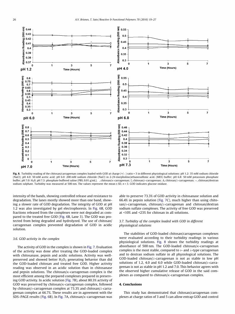
Fig. 8. Turbidity reading of the chitosan/carrageenan complex loaded with GOD at charge (+/�) ratio = 3 in different physiological solutions: pH 1.2: 35 mM sodium chloride(NaCl); pH 4.0: 50 mM acetic acid; pH 6.0: 200 mM sodium chloride (NaCl) in 2-(N-morpholino)ethanesulfonic acid. (MES) buffer; pH 6.8: 50 mM potassium phosphatebuffer; pH 7.0: H20; pH 7.3: phosphate buffered saline (PBS, 0.01 g/mL). �, chitosan/j-carrageenan; /, chitosan/i-carrageenan; D, chitosan/k-carrageenan; �, chitosan/dextransodium sulphate. Turbidity was measured at 500 nm. The values represent the mean ± SD, n = 3. GOD indicates glucose oxidase.
26 A.V. Briones, T. Sato / Reactive & Functional Polymers 70 (2010) 19–27
intensity of the bands, showing controlled release and resistance todegradation. The lanes mostly showed more than one band, show-ing a slower rate of GOD degradation. The integrity of GOD at pH1.2 was also investigated by gel electrophoresis. In Fig. 6B, GODfractions released from the complexes were not degraded as com-pared to the treated free GOD (Fig. 6B, Lane 3). The GOD was pro-tected from being degraded and hydrolyzed. The use of chitosan/carrageenan complex prevented degradation of GOD in acidicsolution.
3.6. GOD activity in the complex
The activity of GOD in the complex is shown in Fig. 7. Evaluationof the activity was done after treating the GOD-loaded complexwith chitosanase, pepsin and acidic solutions. Activity was well-preserved and showed better H2O2 generating behavior than didthe GOD-loaded chitosan and treated free GOD. Higher activityreading was observed in an acidic solution than in chitosanaseand pepsin solutions. The chitosan/j-carrageenan complex is themost efficient among the prepared complexes prepared in preserv-ing GOD activity. In acidic solution (Fig. 7B), about 80.3% activity ofGOD was preserved by chitosan/j-carrageenan complex, followedby chitosan/i-carrageenan complex at 73.3% and chitosan/k-carra-geenan complex at 66.7%. These results are in agreement with theSDS–PAGE results (Fig. 6B). In Fig. 7A, chitosan/j-carrageenan was
able to preserve 73.3% of GOD activity in chitosanase solution and66.4% in pepsin solution (Fig. 7C), much higher than using chito-san/i-carrageenan, chitosan/k-carrageenan and chitosan/dextransodium sulfate complexes. The activity of free GOD was preservedat <10% and <23% for chitosan in all solutions.
3.7. Turbidity of the complex loaded with GOD in differentphysiological solutions
The stabilities of GOD-loaded chitosan/carrageenan complexeswere evaluated according to their turbidity readings in variousphysiological solutions. Fig. 8 shows the turbidity readings atabsorbance of 500 nm. The GOD-loaded chitosan/j-carrageenancomplex is the most stable, compared to i- and k-type carrageenanand to dextran sodium sulfate in all physiological solutions. TheGOD-loaded chitosan/k-carrageenan is not as stable in low pHsolutions of 1.2, 4.0 and 6.0 while GOD-loaded chitosan/i-carra-geenan is not as stable in pH 1.2 and 7.0. This behavior agrees withthe observed higher cumulative release of GOD in the said com-plexes as compared to chitosan/j-carrageenan complex.
4. Conclusions
This study has demonstrated that chitosan/carrageenan com-plexes at charge ratios of 3 and 5 can allow entrap GOD and control

A.V. Briones, T. Sato / Reactive & Functional Polymers 70 (2010) 19–27 27
its release. Electrostatic interaction is the main mechanism in-volved in the incorporation of the protein into the complex. Thecontrolled release of GOD from the complex is due to the swellingmechanism of the chitosan/carrageenan complex. The chitosan/carrageenan complex can be a promising agent for protein deliv-ery. The ease of preparation and the ability to protect the proteinintegrity make chitosan/carrageenan complexes a promising drugdelivery system for oral administration of peptides and proteins.
Acknowledgements
Sincere thanks to Japan Society for the Promotion of Science(JSPS) for the financial support from the grant under the RONPAKUdissertation program and Keio University, as well as to the Indus-trial Technology Development Institute, and the Department of Sci-ence & Technology, Manila, Philippines.
References
[1] J.G. Rocca, K. Paros, Drug Del. Technol. 4 (2004) 52.[2] Q. Liu, A.M. Rauth, X.Y. Wu, Int. J. Pharm. 339 (2007) 148.[3] H. Zhu, R. Srivastava, J.Q. Brown, M.J. McShane, Bioconj. Chem. 16 (2005) 1451.[4] A. Vikartovská, M. Bucko, D. Mislovicová, V. Pätoprsty, I. Igor Lacík, P.
Gemeiner, Enzyme Microb. Techol. 41 (2007) 748.[5] H. Wang, Q. Pan, G. Wang, Sensors 5 (2005) 266.[6] L. Caseli, D. dos Santos Jr., M. Foschini, D. Gonçalves, O. Oliveira Jr., Mater. Sci.
Eng. C – Biomim. 27 (2007) 1108.[7] R. Srivastava, Q.J. Brown, H. Zhu, M. McShane, Biotechnol. Bioeng. 91 (2005)
124.[8] J. Rodriguez-Nogales, M. Pérez-Mateos, M.D. Busto, J. Chem. Technol.
Biotechnol. 79 (2004) 700.[9] W.R. Gombotz, D.K. Pettit, Bioconj. Chem. 6 (1995) 332.
[10] V.L. Alcon, M. Baca-Estrada, M.A. Vega-Lopez, P. Willson, L.A. Babiuk, P. Kumar,M. Foldvari, J. Pharm. Pharmacol. 57 (2005) 955.
[11] M.A. Schubert, C.C. Muller-Goymann, Eur. J. Pharm. Biopharm. 61 (2005) 77.[12] V.R. Sinha, A. Trehan, J. Control Release 90 (2003) 261.[13] M. Van de Weert, J. Hoechstetter, W.E. Hennink, D.J.A. Crommelin, J. Control
Release 3 (2000) 351.[14] C. Perez-Rodriguez, N. Montano, K. Gonzalez, K. Griebenow, J. Control Release
89 (2003) 71.[15] N.A. Peppas, P. Bures, W. Leobandung, H. Ichikawa, Eur. J. Pharm. Biopharm. 50
(2000) 27.[16] E. Hearn, R.J. Neufeld, Proc. Biochem. 35 (2000) 1253.[17] T.L. Bowersock, H. Hogenesch, M. Suckow, P. Guimond, S. Martin, D. Borie, S.
Torregrosa, H. Park, K. Park, Vaccine 17 (1999) 1804.[18] P.R. Hari, T. Chandy, C.P. Sharma, J. Appl. Polym. Sci. 59 (1996) 1795.
[19] R.J. Mumper, A.S. Hoffman, P. Puolakkainen, L.S. Bouch-ard, W.R. Gombotz, J.Control Release 30 (1994) 241.
[20] F. Gu, B. Amsden, R. Neufeld, J. Control Release 96 (2004) 463.[21] L.S. Liu, S.Q. Liu, S.Y. Ng, M. Froix, T. Ohno, J. Heller, J. Control Release 43 (1997)
65.[22] J.E. Lee, K.E. Kim, I.C. Kwon, H.J. Ahn, S.H. Lee, H. Cho, H.J. Kim, S.C. Seong, M.C.
Lee, Biomaterials 25 (2004) 4163.[23] S.B. Wang, A.Z. Chen, L.J. Weng, M.Y. Chen, X.L. Xie, Macromol. Biosci. 4 (2004)
27.[24] M. Kumar, React. Funct. Polym. 46 (2000) 1.[25] J. Du, J. Dai, J. Liu, T. Dankovich, React. Funct. Polym. 66 (2006) 1055.[26] Y. Chen, V.J. Mohanraj, F. Wang, H.A. Benson, AAPS PharmSciTech 8 (2007) 98.[27] C. Tapia, Z. Escobar, E. Costa, J. Sapag-Hagar, F. Valenzuela, C. Basualto, M.N.
Gai, M. Yazdami-Pedram, Eur. J. Pharm. Biopharm. 57 (2004) 65.[28] P. Piyakulawat, N. Praphairaksit, N. Chantarasiri, N. Muangsin, AAPS
PharmSciTech 8 (2007) 120.[29] H. Tomida, C. Nakamura, S. Kiryu, Chem. Pharm. Bull. 42 (1994) 979.[30] M.M. Elnashar, M.A. Yassin, J. Appl. Polym. Sci. 114 (2008) 17.[31] C. Aguzzi, M.C. Bonferoni, M.R.O. Fortich, S. Rossi, F. Ferrari, C. Caramella, AAPS
PharmSciTech 3 (2003) E27.[32] A.M. Garcia, E.S. Ghaly, J. Control Release 40 (1996) 179.[33] O. Sipahigil, B. Dortunc, Int. J. Pharm. 228 (2001) 119.[34] Y. Ozsoy, N. Bergisadi, Boll. Chim. Farm. 139 (2000) 120.[35] M.R. Mangione, D. Giacomazza, G. Cavallaro, D. Bulone, V. Martorana, P.L. San
Biagio, Biophys. Chem. 129 (2007) 18.[36] G. Ruiz, E.S. Ghaly, Vitae-Columbia 13 (2006) 31.[37] V.K. Gupta, M. Hariharan, T.A. Wheatley, J.C. Price, Eur. J. Pharm. Biopharm. 51
(2001) 241.[38] A. Pourjavadi, Sh. Barzegar, F. Zeidabadi, React. Funct. Polym. 67 (2007) 644.[39] Evaluation of certain food additives and contaminants (sixty-eighth report of
the Joint FAO/WHO Expert Committee on Food Additives), WHO TechnicalReport Series 947 (2007) 32.
[40] A. Gürsel, S. Alkan, L. Toppare, Y. Ya, React. Funct. Polym. 57 (2003) 65.[41] E. Balnois, K.J. Wilkinson, Colloid Surface A 207 (2002) 229.[42] C. Mireles, M. Martino, J. Bouzas, J.A. Torres, in: C.J. Brine, P.A. Sandford, J.P.
Zikakis (Eds.), Advances in Chitin and Chitosan, Elsevier Applied Science,London, 1991, p. 506.
[43] B.E.F. Swoboda, V. Massey, J. Biol. Chem. 240 (1965) 2209.[44] K.B. Guisely, N.F. Stanley, P.A. Whitehouse, in: R.L. Davidson (Ed.), Handbook
of Water-Soluble Gums and Resins, Mcgraw-Hill Book Co., New York, 1980, pp.1–5.
[45] T.M. Mcintire, D.A. Brant, Int. J. Biol. Macromol. 26 (1999) 303.[46] A.R. Kirby, A.P. Gunning, V.J. Morris, Biopolymers 38 (1996) 355.[47] M. Oliva, I.D. Perez, P. Gorostiza, C.F. Lastra, I. Oliva, C. Caramella, E.L. Mariño, J.
Pharm. Sci. 92 (2003) 77.[48] H. Xiaoqing, Z. Gucheng, C. Jive, W. Bin, F. Qian, W. Chenxi, L. Zhihong, Chem. J.
Internet 6 (2004) 38.[49] F.R. Fan, A.J. Bard, Proc. Natl. Acad. Sci. USA 96 (1999) 14222.[50] L. Brannon-Peppas, Med. Plast. Biomater. 4 (1997) 34.[51] T. Sakiyama, C. Chu, T. FujiI, T. Yano, J. Appl. Polym. Sci. 50 (1993) 2021.[52] E.V. Shumilina, Y.A. Shchipunov, Colloid J. 64 (2002) 372.[53] H. Tao, W. Wei, Y. Mao, S. Zhang, J. Xia, Anal. Sci. 21 (2005) 1057.