Molecular characterization and complete genome sequence of avian paramyxovirus type 4 prototype...
11
BioMed Central Page 1 of 11 (page number not for citation purposes) Virology Journal Open Access Research Molecular characterization and complete genome sequence of avian paramyxovirus type 4 prototype strain duck/Hong Kong/D3/75 Baibaswata Nayak 1 , Sachin Kumar 1 , Peter L Collins 2 and Siba K Samal* 1 Address: 1 Virginia-Maryland Regional College of Veterinary Medicine, University of Maryland, College Park, Maryland, USA and 2 Laboratory of Infectious Diseases, National Institute of Allergy and Infectious Diseases, Bethesda, USA Email: Baibaswata Nayak - [email protected]; Sachin Kumar - [email protected]; Peter L Collins - [email protected]; Siba K Samal* - [email protected] * Corresponding author Abstract Background: Avian paramyxoviruses (APMVs) are frequently isolated from domestic and wild birds throughout the world. All APMVs, except avian metapneumovirus, are classified in the genus Avulavirus of the family Paramyxoviridae. At present, the APMVs of genus Avulavirus are divided into nine serological types (APMV 1–9). Newcastle disease virus represents APMV-1 and is the most characterized among all APMV types. Very little is known about the molecular characteristics and pathogenicity of APMV 2–9. Results: As a first step towards understanding the molecular genetics and pathogenicity of APMV- 4, we have sequenced the complete genome of APMV-4 strain duck/Hong Kong/D3/75 and determined its pathogenicity in embryonated chicken eggs. The genome of APMV-4 is 15,054 nucleotides (nt) in length, which is consistent with the "rule of six". The genome contains six non- overlapping genes in the order 3'-N-P/V-M-F-HN-L-5'. The genes are flanked on either side by highly conserved transcription start and stop signals and have intergenic sequences varying in length from 9 to 42 nt. The genome contains a 55 nt leader region at 3' end. The 5' trailer region is 17 nt, which is the shortest in the family Paramyxoviridae. Analysis of mRNAs transcribed from the P gene showed that 35% of the transcripts were edited by insertion of one non-templated G residue at an editing site leading to production of V mRNAs. No message was detected that contained insertion of two non-templated G residues, indicating that the W mRNAs are inefficiently produced in APMV-4 infected cells. The cleavage site of the F protein (DIPQR ↓F) does not conform to the preferred cleavage site of the ubiquitous intracellular protease furin. However, exogenous proteases were not required for the growth of APMV-4 in cell culture, indicating that the cleavage does not depend on a furin site. Conclusion: Phylogenic analysis of the nucleotide sequences of viruses of all five genera of the family Paramyxoviridae showed that APMV-4 is more closely related to the APMVs than to other paramyxoviruses, reinforcing the classification of all APMVs in the genus Avulavirus of the family Paramyxoviridae. Published: 20 October 2008 Virology Journal 2008, 5:124 doi:10.1186/1743-422X-5-124 Received: 15 September 2008 Accepted: 20 October 2008 This article is available from: http://www.virologyj.com/content/5/1/124 © 2008 Nayak et al; licensee BioMed Central Ltd. This is an Open Access article distributed under the terms of the Creative Commons Attribution License (http://creativecommons.org/licenses/by/2.0 ), which permits unrestricted use, distribution, and reproduction in any medium, provided the original work is properly cited.
Transcript of Molecular characterization and complete genome sequence of avian paramyxovirus type 4 prototype...
BioMed CentralVirology Journal
ss
Open AcceResearchMolecular characterization and complete genome sequence of avian paramyxovirus type 4 prototype strain duck/Hong Kong/D3/75Baibaswata Nayak1, Sachin Kumar1, Peter L Collins2 and Siba K Samal*1Address: 1Virginia-Maryland Regional College of Veterinary Medicine, University of Maryland, College Park, Maryland, USA and 2Laboratory of Infectious Diseases, National Institute of Allergy and Infectious Diseases, Bethesda, USA
Email: Baibaswata Nayak - [email protected]; Sachin Kumar - [email protected]; Peter L Collins - [email protected]; Siba K Samal* - [email protected]
* Corresponding author
AbstractBackground: Avian paramyxoviruses (APMVs) are frequently isolated from domestic and wildbirds throughout the world. All APMVs, except avian metapneumovirus, are classified in the genusAvulavirus of the family Paramyxoviridae. At present, the APMVs of genus Avulavirus are divided intonine serological types (APMV 1–9). Newcastle disease virus represents APMV-1 and is the mostcharacterized among all APMV types. Very little is known about the molecular characteristics andpathogenicity of APMV 2–9.
Results: As a first step towards understanding the molecular genetics and pathogenicity of APMV-4, we have sequenced the complete genome of APMV-4 strain duck/Hong Kong/D3/75 anddetermined its pathogenicity in embryonated chicken eggs. The genome of APMV-4 is 15,054nucleotides (nt) in length, which is consistent with the "rule of six". The genome contains six non-overlapping genes in the order 3'-N-P/V-M-F-HN-L-5'. The genes are flanked on either side byhighly conserved transcription start and stop signals and have intergenic sequences varying in lengthfrom 9 to 42 nt. The genome contains a 55 nt leader region at 3' end. The 5' trailer region is 17 nt,which is the shortest in the family Paramyxoviridae. Analysis of mRNAs transcribed from the P geneshowed that 35% of the transcripts were edited by insertion of one non-templated G residue at anediting site leading to production of V mRNAs. No message was detected that contained insertionof two non-templated G residues, indicating that the W mRNAs are inefficiently produced inAPMV-4 infected cells. The cleavage site of the F protein (DIPQR↓F) does not conform to thepreferred cleavage site of the ubiquitous intracellular protease furin. However, exogenousproteases were not required for the growth of APMV-4 in cell culture, indicating that the cleavagedoes not depend on a furin site.
Conclusion: Phylogenic analysis of the nucleotide sequences of viruses of all five genera of thefamily Paramyxoviridae showed that APMV-4 is more closely related to the APMVs than to otherparamyxoviruses, reinforcing the classification of all APMVs in the genus Avulavirus of the familyParamyxoviridae.
Published: 20 October 2008
Virology Journal 2008, 5:124 doi:10.1186/1743-422X-5-124
Received: 15 September 2008Accepted: 20 October 2008
This article is available from: http://www.virologyj.com/content/5/1/124
© 2008 Nayak et al; licensee BioMed Central Ltd. This is an Open Access article distributed under the terms of the Creative Commons Attribution License (http://creativecommons.org/licenses/by/2.0), which permits unrestricted use, distribution, and reproduction in any medium, provided the original work is properly cited.
Page 1 of 11(page number not for citation purposes)
Virology Journal 2008, 5:124 http://www.virologyj.com/content/5/1/124
BackgroundThe family Paramyxoviridae contains a large number ofviruses of humans and animals [1]. These viruses havebeen isolated from many species of avian, terrestrial andaquatic animals worldwide. The members of this familyincludes many human pathogens such as measles (MeV),mumps (MuV) and human respiratory syncytial virus(hRSV) and many important animal pathogens such asNewcastle disease virus (NDV), canine distemper (CDV)and rinderpest (RPV) [2]. Some of the members of thefamily Paramyxoviridae are well characterized, while char-acteristics for other members of this family remainunknown. Members of this family are enveloped virusespossessing a non-segmented negative-strand genome [1]and are divided into two subfamilies; Paramyxovirinae andPneumovirinae. Subfamily Paramyxovirinae is divided intofive genera: Rubulavirus [MuV, human parainfluenzaviruses (hPIV) -2 and -4, simian virus type 5 (SV5) andTioman virus (TiV)], Respirovirus [Sendai virus (SeV) andhPIV-1 and -3], Henipavirus [Hendra virus (HeV) andNipah virus (NiV)], Morbillivirus [MeV, CDV and RPV],and Avulavirus [avian paramyxovirus (APMV) serotypes1–9]. Subfamily Pneumovirinae is divided into two genera:Pneumovirus (hRSV and its animal counterparts includingbovine respiratory syncytial virus [bRSV]), and Metapneu-movirus [comprising human metapneumovirus (HMPV)and avian metapneumovirus (AMPV)] [1,3,4].
The genomes of the paramyxoviruses vary in length from13–19 kb and contain 6–10 genes encoding up to 12 dif-ferent proteins. Transcription begins at single promoter atthe 3' leader end and the genes are copied into individualmRNAs by a start-stop-restart mechanism guided by con-served gene-start and gene-end transcription signals thatflank the individual genes [1]. Genome replicationinvolves the synthesis of a complete positive-sense copy ofthe genome that is called the antigenome and serves as atemplate for producing progeny genomes. All members offamily Paramyxoviridae encode a nucleoprotein (N), aphosphoprotein (P), a matrix protein (M), a fusion pro-tein (F), an attachment protein called the hemagglutinin(H) or haemagglutinin-neuraminidase (HN) or glycopro-tein (G), and a large polymerase protein (L) [1,2].
All APMVs have been classified into nine different sero-types based on HI test and all NDV strains belong toAPMV serotype 1 [5]. Since NDV can cause severe diseasein chickens, APMV-1 is the most extensively characterizedserotype of the APMVs. Very little is known about themolecular and biological characteristics and pathogenic-ity of APMV serotypes 2–9. APMV types 2, 3, 6 and 7 havebeen associated with disease in domestic poultry [6-10].The APMV-5 (Kunitachi virus) isolated from budgerigar isknown to cause disease in wild birds [11]. Other sero-types, including APMV-4, -8, and -9, have been isolated
from ducks, waterfowls, and other wild birds with no clin-ical signs of disease [5,12-15]. The strain duck/HongKong/D3/75, isolated from a duck in Hong Kong in 1975,was found to be representative of a distinct serotype ofAPMVs [16], later designated as APMV serotype 4 on thebasis of HI and neuraminidase inhibition (NI) tests [17].Experimental infection of chickens with APMV-4 andAPMV-6 showed mild interstitial pneumonia, catarrhaltracheitis, and BALT or GALT hyperplasia, suggestive ofviral disease [18].
An understanding of the molecular and biological charac-teristics of APMV -2 to-9 is of general interest and isimportant for developing vaccines and diagnostic testsagainst these viruses. To date, the complete genomesequence for representatives of APMV-1 [19], APMV-2[20], APMV-3 [21] and APMV-6 [22] are available. As afirst step towards understanding the molecular biologyand pathogenicity of APMV-4, we have determined thegrowth characteristics and complete genome sequence ofthe APMV-4 prototype strain duck/Hong Kong/D3/75(GenBank accession no. FJ177514). Previously, sequencewas available only for the APMV-4 HN gene (GenBankaccession no. D14031). The sequence of the strain duck/Hong Kong/D3/75 was compared with those of otherAPMV serotypes and other paramyxoviruses in order todetermine phylogenetic relationships.
MethodsVirus and cellsThe APMV-4 prototype strain, duck/Hong Kong/D3/75was obtained from National Veterinary Services Labora-tory, (Ames, IA). The chicken embryo fibroblast (DF-1),Madin-Darby Canine Kidney (MDCK), human epider-moid carcinoma (HEp-2), Baby Hamster Kidney (BHK21), Bovine Turbinate (BTu), Pig Kidney (PK15), Quailfibrosarcoma (QT35), Rabbit Kidney cells (RK13), Africangreen monkey kidney (Vero), Madin-Darby Bovine Kid-ney (MDBK), and duck embryo (CCL-141) cell lines wereobtained from the American Type Culture Collection(ATCC, Manassas, VA), and turkey embryo fibroblast(TEF) primary cells were made in our laboratory. The DF1and QT35 cells were grown in Dulbecco's minimumessential medium containing 10% fetal calf serum, whilethe other cells were grown in Eagle's minimum essentialmedium containing 10% FCS, at 37°C with 5% CO2.
Virus propagation and requirement of exogenous proteaseThe APMV-4 prototype strain, duck/Hong Kong/D3/75was propagated in 9 day-old specific-pathogen-free (SPF)embryonated chicken eggs by inoculation through theallantoic cavity route. The titer of virus was determined byhemagglutination (HA) test using 0.5% chicken RBC atroom temperature. Virus propagation was carried out indifferent cell lines either in the absence or presence of
Page 2 of 11(page number not for citation purposes)
Virology Journal 2008, 5:124 http://www.virologyj.com/content/5/1/124
exogenous protease in order to determine a suitable cellline for virus growth. The cell culture medium was supple-mented with 5% allantoic fluid, or 1–5 μg/ml of acetyltrypsin/ml (Gibco), or 1–5 μg/ml of α-chymotrypsin/ml(Sigma), each of which provided a source of protease forcleavage of the F protein, if necessary. The cell lines sup-porting viral growth were observed by corroborating cyto-pathic effects (CPE) in cells and HA titer in cell culturesupernatant both before and after freeze-thawing cycles.The ability of the virus to produce plaques was tested inthe different cell lines using 1% methylcellulose with andwithout exogenous protease in the overlay.
Isolation of viral RNA and determination of genome sequenceViral genomic RNA was isolated from purified virus,obtained from infected allantoic fluid by discontinuoussucrose gradient centrifugation, using RNeasy mini kit(Qiagen, USA). The genome sequencing was carried outby using HN gene specific primer designed from the pub-lished HN gene sequence (GenBank accession no.D14031). A set of consensus primers were designed forgene start (GS), N and L gene by aligning genomes of pub-lished APMV-1, APMV-2, APMV-3 and APMV-6 sequencesand working consensus primers were mentioned in Table1. Reverse transcription of viral genome was carried out byGS consensus primer and primer HN4-1758F (Table 1). Aportion of the N gene (256 nt) was amplified by usingpositive sense GS consensus primer and antisense N con-sensus primer. The F gene end and HN gene start regionwas also amplified using primer GS consensus and HN4-81 antisense. The HN gene end and L gene start regionswere amplified by sense HN4-1758F primer and L consen-sus A (L-revA) antisense primer. By comparing these two
regions APMV-4 specific gene start and gene end (GE)sequences were identified and APMV-4 specific senseprimer APMV-4 GS-F and antisense APMV-4 GE-R primerswere designed.
For subsequent amplification, reverse transcription wascarried out by APMV-4 GS-F primer. The PCR productamplified by APMV-4 GS-F and APMV-4 GE-R primer wascloned in pCR TOPO TA vector (Invitrogen). Sequencingof these clones indicated PCR amplification by singleAPMV-4 GS-F primer covering the P gene (1616 nt- 2671nt) and L gene (8204 nt-9394 nt) due to presence of acomplimentary sequence in cDNA of APMV-4 at this loca-tions. The sequence of remaining N gene was obtained byamplification and sequencing using sense primer NP4-2Fand P gene antisense APMV4-P-65R primer. The sequenceof M and F genes was obtained by PCR amplificationusing APMV-4 specific P gene sense (APMV4-P-550F) andHN gene antisense (APMV4 HN59-81R AS) primer. Thecomplete sequence of the L gene was obtained by primerwalking using L gene specific forward primer and nonspe-cific reverse primers (NSP1-5) used in APMV-6 genomesequencing [22] or L gene specific consensus antisense (L-revA, L-revB, L-revC and L-revD) primers designed byAPMVs genome sequence alignment (Table 1). For L genesequencing, 25 L gene specific sense primers were usedalong with non specific reverse primers. The sequences ofthe genome termini were determined by 3' and 5' termi-nus RACE (rapid amplification of cDNA ends)[20,21,23,24]. For 3' RACE, viral genomic RNA wasligated to adapter1 (5'-GGTTTTGCGGTAAAGGTGGAA-GAGAAG-3'), using T4 RNA ligase according to the man-ufacturer's instructions (Invitrogen). The ligated RNA waspurified and reverse-transcribed, using a complimentary
Table 1: Primers used in the study
GS consensus (sense) -5'-GAGCAGTAGGAGCGGAA-3'
AN4-1758F (sense) -5'-CATTCAAGATAGTGCCATTCCTC-3'NP consensus (antisense) -5'-GWWSCYAYWCCCATKGCAWA-3'APMV4-HN4-81 (antisense) -5'-ACTTCTGTTGACTTCTCTTGGTA-3'APMV4 GS-F (sense) -5'-CTAGGGTGGGGAAGG-3'APMV4 GE-R (antisense) -5'-CCGTTTTTAATTAAAAA-3'APMV4-NP4-2F (sense) -5'-GAAACTTCCCACACATGTACTCCTATGC-3'APMV4-P-65R (antisense) -5'-GAGTCTATGATTGCCGATGATGATTC-3'APMV4-P-550F (sense) -5'-AACCGGAAAACATTGAACTGGTGGAGTG-3'APMV4 HN59-81R (antisense) -5'-ACTTCTGTTGACTTCTCTTGGTA-3'NSP1 (antisense) -5'-ATCAAAAGCACC-3'NSP2 (antisense) -5'-AGTAGAAACAAGG-3'NSP3 (antisense) -5'-ACAAGGGTGAGG-3'NSP4 (antisense) -5'-GTTTTTTCTTCTTAA-3'NSP5 (antisense) -5'-ATACGGGTAGAA-3'L-revA (antisense) -5'-GGNGCRCACATNSWYTCNCKNAC-3'L-revB (antisense) -5'-GGNGCRCACATYTGNSWNCKNAC-3'L-revC (antisense) -5'-ACYTCYTTYTCYTTNARNSWRTA-3'L-revD (antisense) -5'-GTCATYTTNGCRAADATNCKNCC-3'
Page 3 of 11(page number not for citation purposes)
Virology Journal 2008, 5:124 http://www.virologyj.com/content/5/1/124
adapter 2 (5'-CCAAAACGCCATTTCCACCTTCTCTTC-3')primer. PCR amplification was carried out by sense primeradapter 2 and antisense N gene-specific reverse primer(NP4-192aaR -5'-GGCCTCCCCAGAGC CGTCAATGTTG-3') and (NP4-210aaR – 5'-CCAATTGCAAACTGACGAT-TAAGC-3'). The 5' RACE was carried out by reverse tran-scription of genomic RNA using L gene specific senseprimer (4-PM14464F- 5'-GCGAACCTGGCAGATACATA-CAAAC-3'). The tailing of purified cDNAs with dCTP wascarried out by using terminal deoxynucleotide tranferase(Invitrogen). The cDNAs were then amplified in separatereactions, using an L gene-specific forward primer (4-PM14574F- 5'-AGTAGTCCCCGCTTTCAAC -3') and ananchored G antisense primer.
Sequencing of cloned DNAs or PCR-amplified productswere carried out in 3130xl genetic analyzer by usingBigDye terminator v 3.1 matrix standard kit and 3130xlgenetic analyzer data collection software v3.0 (AppliedBiosystem). The entire genome was sequenced at leastthree times, and at least once from uncloned PCR product,to ensure a consensus sequence.
P gene mRNA editingDF1 cells were infected with APMV-4 virus (AF titer- 27
HAU) at dilution 10-3 per 25 cm2 flask and cells were har-vested at 48 hr post infection for RNA isolation. ThemRNAs were isolated using mRNA isolation kit (Invitro-gen). The viral mRNAs were reverse transcribed usingoligo dT primer. The region flanking the putative P geneRNA editing site was amplified by P gene specific primers(APMV4-P50-74F- 5'-CGGCAATCATAGACTCCATA-CAGC-3' and APMV4-P536-558R- 5'-CAATGTCTCCGGTTGCTTTGTCG-3'). The PCR products were cloned inpCR TOPO TA vector (Invitrogen). The comparison forthe presence of P, V or W mRNAs were carried out by ana-lyzing the sequences from positive clones.
Sequence and phylogenetic tree analysisSequence similarity searches were conducted using thebasic length alignment search tool (BLAST) from theNational Center for Biotechnology Information (NCBI).DNA pair-wise alignment was done using MagAlign (clus-talW) in a Lasergene6 software package. Evolutionary rela-tionships were predicted from the multiple nucleotidesequence alignment of whole genomes of the members ofthe family Paramyxoviridae. The phylogenetic tree was gen-erated by ClustalW program of MegAlign. The amino acidsequence homology and divergence between the genera ofthe subfamily Paramyxovirinae were also obtained by clus-talW multiple alignment algorithms.
Database accession numbersThe complete genome sequence of APMV-4 strain duck/Hong Kong/D3/75 have been submitted in GenBankunder accession number FJ177514. For comparative anal-
ysis different complete genome sequences of subfamilyParamyxovirinae were obtained from GenBank. The data-bank accession number for these complete genomesequence are as follows: subfamily Paramyxovirinae; Avula-virus: NC_002617 for APMV-1, EU338414 for APMV-2,EU403085 for APMV-3, NC_003043 for APMV-6; Rubula-virus: NC_006430 for SV5, NC_003443 for hPIV2,NC_002200 for MuV, NC_004074 for Tioman virus(TiV); Respirovirus: NC_003461 for hPIV1, NC_001552 forSendai virus (SeV), NC_001796 for hPIV3, NC_002161for bovine PIV 3 (bPIV3); Henipavirus: AY988601 forNipah virus (NiV), NC_001906 for Hendra virus (HeV);Morbillivirus: NC_001498 for Measles virus (MeV),NC_001921 for CDV, NC_006383 for Peste des petitsruminants virus (PPRV), NC_006296 for RPV,NC_005283 for Dolphin morbillivirus (DMV); other par-amyxovirus: EF646380 for Atlantic salmon paramyxovi-rus (ASPV), NC_007803 for Beilong virus (BeV),NC_005084 for Fer de Lance virus (FDLV), NC_007454for J virus (JV), NC_007620 for Menangle virus (MenV),NC_005339 for Mossman virus (MoV), NC_002199 forTupaia paramyxovirus (TpV), subfamily Pneumovirinae;Pneumovirus; NC_001989 for bRSV and NC_001781 forhRSV; Metapneumovirus: NC_004148 for HMPV,NC_007652 for AMPV.
ResultsGrowth characteristics of APMV-4APMV-4 strain duck/Hong Kong/D3/75 produced a titerof 27 – 29 HA units in 9 day-old embryonated SPF chickeneggs at 4 days post-inoculation (p.i). The APMV-4 strainduck/Hong Kong/D3/75 was able to grow in the MDBK,BHK21, duck embryo, Vero, DF-1 and QT35 cell lines to atiter of 24 HA units without the addition of exogenousproteases, which is a requirement for efficient F proteincleavage in many paramyxoviruses. The addition of exog-enous protease did not give any difference in titer. Thetypical cytopathic effect (CPE) observed was roundingand detachment of cells. Multicycle growth curves docu-mented virus release at 20, 28 and 36 hr after infections inMDBK, Vero and DF-1 cells, respectively. The virusreached to a maximum titer in cell culture supernatant at36 hr p.i in MDBK and Vero cells, while 72 hr p.i in DF-1cells. The virus did not form any visible plaques in all celllines tested with or without addition of proteases. Meandeath time of the APMV-4 strain duck/Hong Kong/D3/75in embryonated chicken eggs was zero, indicating its avir-ulent nature for chickens. Electron microscopy of partiallypurified virus showed that the virus particles were envel-oped, pleomorphic but mostly spherical in shape, with asize ranging from 150–250 nm (data not shown).
Determination of APMV-4 complete genome sequenceThe genome of APMV-4 strain duck/Hong Kong/D3/75consists of 15,054 nt (GenBank accession no. FJ177514),which follows the "rule of six" common to other members
Page 4 of 11(page number not for citation purposes)
Virology Journal 2008, 5:124 http://www.virologyj.com/content/5/1/124
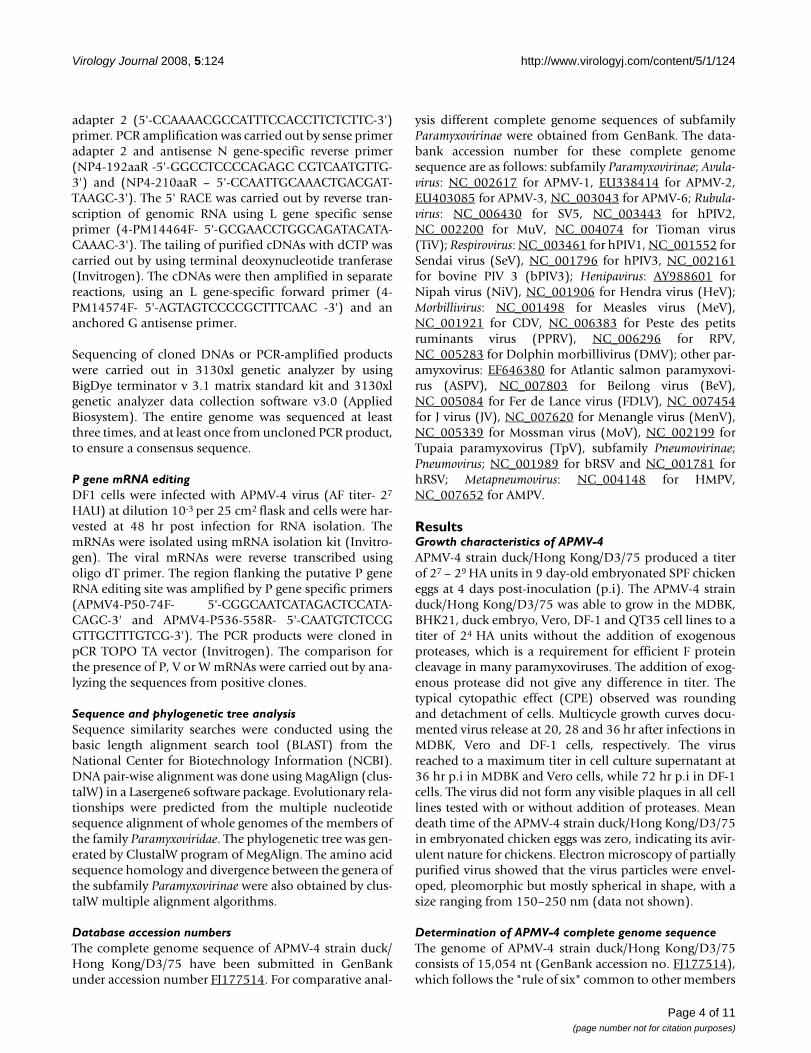
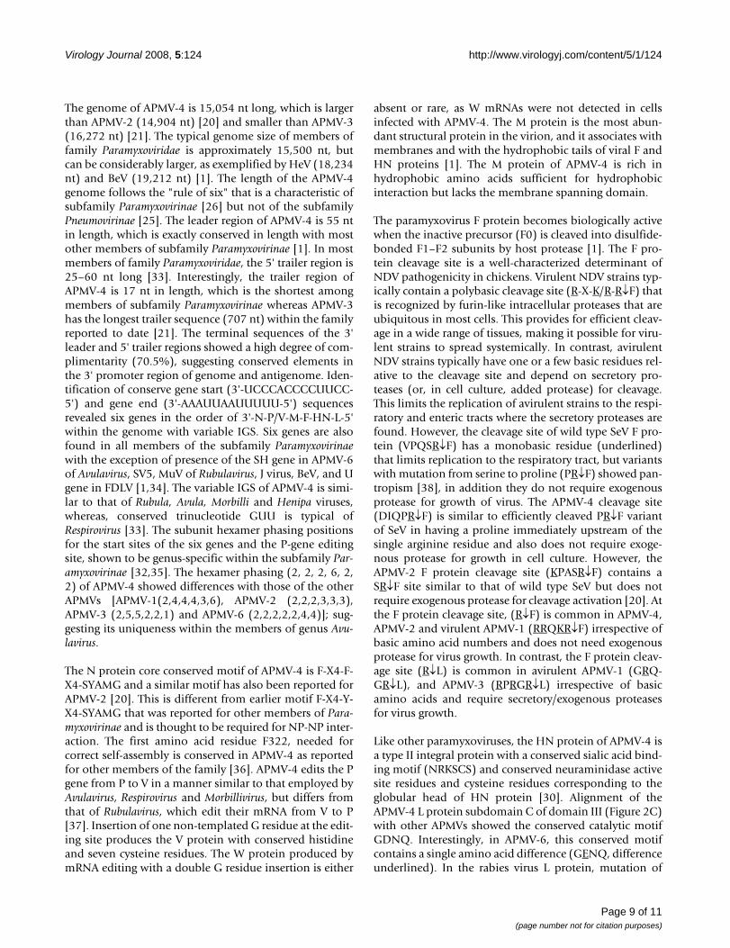
of subfamily Paramyxovirinae [25,26]. The genomic organ-ization of APMV-4 is similar to the other members ofgenus Avulavirus in the order of 3'-N-P-M-F-HN-L-5' (Fig-ure 1A). The genes were flanked by leader sequences at the3' end and trailer sequences at the 5' end. The intergenicsequences (IGS) between genes, which are not copied intomRNAs, varied in length from 9 nt to 42 nt (Table 2). TheIGS between N/P, P/M, M/F, F/HN and HN/L are 9, 34,14, 37 and 42 nt, respectively (Figure 1C). All the genes ofAPMV-4 were positioned at hexamer phase 2 except Fgene that was at hexamer phase 6 (Table 2). The APMV-4phasing pattern is unique among paramyxoviruses. Anal-ysis of deduced amino acid sequences from open readingframes (ORFs) of all the genes revealed 91.09% codingpercentage, which is similar to the coding percentage ofother paramyxoviruses [27].
The 3' leader region of APMV-4 is 55 nt, a length that isgenerally conserved among members of subfamily Para-myxovirinae [19]. Comparison of the nucleotide sequenceof the APMV-4 leader region with the leader sequences ofother paramyxoviruses showed 30–47% percent identity.Surprisingly, the length of the APMV-4 5' trailer region is17 nt, which is the shortest among the members of thefamily Paramyxoviridae sequenced to date. The sequencesof the 3' leader and 5' trailer termini showed a high degreeof complimentarity (70.5% complimentarity) for the ter-minal 17 nt, suggesting the presence of conserved ele-ments in the 3' promoter regions of the genome andantigenome (Figure 1D). The gene-start signal of APMV-4is 3'-UCCCACCCCUUCC-5' and is exactly conservedamong all genes except the N and F genes, in which there
are difference of 3 and 1 nt, respectively (Figure 1C). Theconsensus gene end signal of APMV-4 is 3'-AAAUUAA (U)5 -5', with difference of 3, 2 and 1 nt in the P, M and Lgene, respectively (Figure 1C).
The Nucleoprotein (N) geneThe APMV-4 N gene is 1551 nt and encodes a N protein of457 amino acids. It contains a highly conserved sequencemotif, 322-FAPGNFPHMYSYAMG-336 (F-X4-Y-X3-α-S-α-AMG, where X is any amino acid and α is any aromaticamino acid), that corresponds to a motif previously iden-tified in the central region of the N protein of other mem-bers of Paramyxovirinae and which has been implicated inthe self-assembly of N with RNA or with other N mono-mers [1]. Within the conserved motif of APMV-4 N pro-tein, conserved amino acid Y327 is replaced by F327. TheN protein of APMV-4 has the highest amino acid identity(51.2%) with that of APMV-3, whereas with other mem-bers of Avulavirus it shared 34–38% identity. It shared anamino acid identity of 30–35% with Rubulavirus, 20–22%with Respirovirus, 26–27% with Henipavirus, 21–22% withMorbillivirus, and 22–25% with unclassified paramyxovi-ruses (Table 3).
The Phosphoprotein (P) gene and P/V/W editingThe P gene is 1364 nt long and encodes a predicted P pro-tein of 393 amino acids. The predicted P protein containsmany potential phosphorylation sites with one tyrosine,11 threonine, and 16 serine residues identified as possiblesites using Net Phos 2.0 software. The APMV-4 P proteinamino acid identity varies from 19–20% with members ofAvulavirus, 15–18% with Rubulavirus, 5–9% with Respirovi-rus, 12% with Henipavirus, 10–11% with Morbillivirus and7–14% with other paramyxoviruses (Table 3).
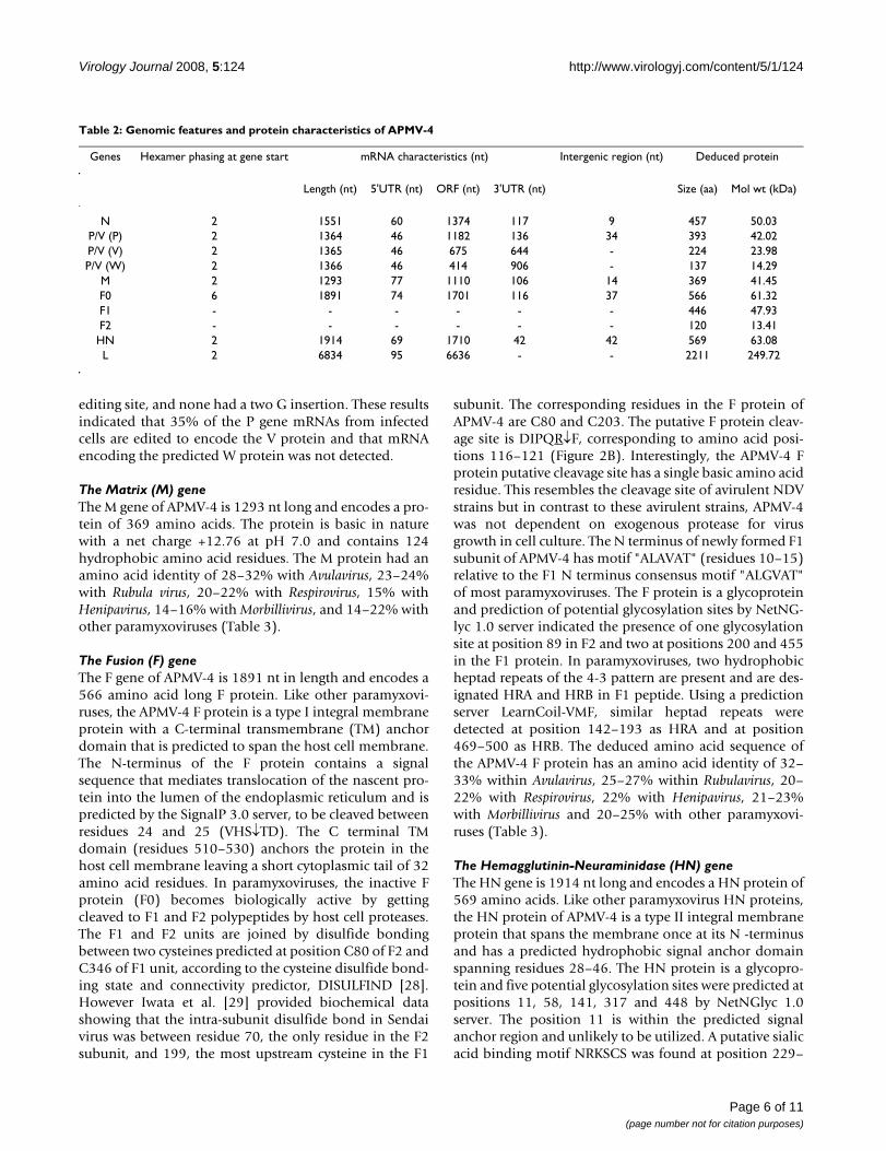
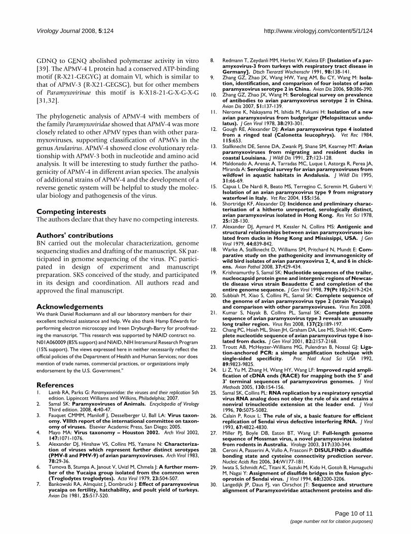
The P gene contains a putative editing site 5'-AAAG-GGGGG-3' (mRNA sense) at positions 442–450 nt of Pgene (positions 2057–2065 nt in the complete genomesequence). The insertion of one non-templated G residuewould encode a 224 amino acid V protein of molecularweight ~23.98 kDa. This V protein shares its N-terminal135 amino acids with the P protein. The V-specific C-ter-minal domain contains seven invariantly spaced cysteineresidues and a histidine residue that are highly conservedwithin paramyxoviruses (Figure 2A). Alternatively, inser-tion of two non-templated G residues would encode aputative W protein (137 amino acids) comprising the N-terminal 135 amino acids of the P protein and C-terminaltwo amino acids unique to the W protein.
In order to confirm P gene editing, total mRNA was iso-lated from APMV-4 infected DF-1 cells. RT- PCR amplifi-cation of sequences encompassing the P gene editing sitewas performed. Sequencing of 28 cDNA clones showedthat 18 were unedited, 10 had single G insertion at the
Schematic diagram of the APMV-4 genome (A) with leader, trailer, gene start, gene end, IGS characteristicsFigure 1Schematic diagram of the APMV-4 genome (A) with leader, trailer, gene start, gene end, IGS characteris-tics. Alignment of conserved gene start and gene end motifs of the APMV-4 genes (B). Comparison of the variable IGS (C). Complementarities between the 3' leader and 5' trailer regions (D). All sequences are negative sense.
Page 5 of 11(page number not for citation purposes)
Virology Journal 2008, 5:124 http://www.virologyj.com/content/5/1/124
editing site, and none had a two G insertion. These resultsindicated that 35% of the P gene mRNAs from infectedcells are edited to encode the V protein and that mRNAencoding the predicted W protein was not detected.
The Matrix (M) geneThe M gene of APMV-4 is 1293 nt long and encodes a pro-tein of 369 amino acids. The protein is basic in naturewith a net charge +12.76 at pH 7.0 and contains 124hydrophobic amino acid residues. The M protein had anamino acid identity of 28–32% with Avulavirus, 23–24%with Rubula virus, 20–22% with Respirovirus, 15% withHenipavirus, 14–16% with Morbillivirus, and 14–22% withother paramyxoviruses (Table 3).
The Fusion (F) geneThe F gene of APMV-4 is 1891 nt in length and encodes a566 amino acid long F protein. Like other paramyxovi-ruses, the APMV-4 F protein is a type I integral membraneprotein with a C-terminal transmembrane (TM) anchordomain that is predicted to span the host cell membrane.The N-terminus of the F protein contains a signalsequence that mediates translocation of the nascent pro-tein into the lumen of the endoplasmic reticulum and ispredicted by the SignalP 3.0 server, to be cleaved betweenresidues 24 and 25 (VHS↓TD). The C terminal TMdomain (residues 510–530) anchors the protein in thehost cell membrane leaving a short cytoplasmic tail of 32amino acid residues. In paramyxoviruses, the inactive Fprotein (F0) becomes biologically active by gettingcleaved to F1 and F2 polypeptides by host cell proteases.The F1 and F2 units are joined by disulfide bondingbetween two cysteines predicted at position C80 of F2 andC346 of F1 unit, according to the cysteine disulfide bond-ing state and connectivity predictor, DISULFIND [28].However Iwata et al. [29] provided biochemical datashowing that the intra-subunit disulfide bond in Sendaivirus was between residue 70, the only residue in the F2subunit, and 199, the most upstream cysteine in the F1
subunit. The corresponding residues in the F protein ofAPMV-4 are C80 and C203. The putative F protein cleav-age site is DIPQR↓F, corresponding to amino acid posi-tions 116–121 (Figure 2B). Interestingly, the APMV-4 Fprotein putative cleavage site has a single basic amino acidresidue. This resembles the cleavage site of avirulent NDVstrains but in contrast to these avirulent strains, APMV-4was not dependent on exogenous protease for virusgrowth in cell culture. The N terminus of newly formed F1subunit of APMV-4 has motif "ALAVAT" (residues 10–15)relative to the F1 N terminus consensus motif "ALGVAT"of most paramyxoviruses. The F protein is a glycoproteinand prediction of potential glycosylation sites by NetNG-lyc 1.0 server indicated the presence of one glycosylationsite at position 89 in F2 and two at positions 200 and 455in the F1 protein. In paramyxoviruses, two hydrophobicheptad repeats of the 4-3 pattern are present and are des-ignated HRA and HRB in F1 peptide. Using a predictionserver LearnCoil-VMF, similar heptad repeats weredetected at position 142–193 as HRA and at position469–500 as HRB. The deduced amino acid sequence ofthe APMV-4 F protein has an amino acid identity of 32–33% within Avulavirus, 25–27% within Rubulavirus, 20–22% with Respirovirus, 22% with Henipavirus, 21–23%with Morbillivirus and 20–25% with other paramyxovi-ruses (Table 3).
The Hemagglutinin-Neuraminidase (HN) geneThe HN gene is 1914 nt long and encodes a HN protein of569 amino acids. Like other paramyxovirus HN proteins,the HN protein of APMV-4 is a type II integral membraneprotein that spans the membrane once at its N -terminusand has a predicted hydrophobic signal anchor domainspanning residues 28–46. The HN protein is a glycopro-tein and five potential glycosylation sites were predicted atpositions 11, 58, 141, 317 and 448 by NetNGlyc 1.0server. The position 11 is within the predicted signalanchor region and unlikely to be utilized. A putative sialicacid binding motif NRKSCS was found at position 229–
Table 2: Genomic features and protein characteristics of APMV-4
Genes Hexamer phasing at gene start mRNA characteristics (nt) Intergenic region (nt) Deduced protein
Length (nt) 5'UTR (nt) ORF (nt) 3'UTR (nt) Size (aa) Mol wt (kDa)
N 2 1551 60 1374 117 9 457 50.03P/V (P) 2 1364 46 1182 136 34 393 42.02P/V (V) 2 1365 46 675 644 - 224 23.98P/V (W) 2 1366 46 414 906 - 137 14.29
M 2 1293 77 1110 106 14 369 41.45F0 6 1891 74 1701 116 37 566 61.32F1 - - - - - - 446 47.93F2 - - - - - - 120 13.41HN 2 1914 69 1710 42 42 569 63.08L 2 6834 95 6636 - - 2211 249.72
Page 6 of 11(page number not for citation purposes)
Virology Journal 2008, 5:124 http://www.virologyj.com/content/5/1/124
234. This protein is acidic in nature with a net charge of -5.696 at pH 7.0. The six conserved neuraminidase activeresidues were identified at positions 169 (R), 398 (E), 413(R), 501(R), 529 (Y), 550 (E) along with 11 conservedcysteine residues (167, 181, 193, 233, 246, 339, 460, 466,470, 534 and 545) corresponding to the globular head ofHN protein [30]. Comparison of amino acid homologyshowed 30–32% identity within Avulavirus, 30–31% withRubulavirus, 21–22% with Respirovirus, 15% with Henipa-
virus, 9–13% with Morbillivirus and 11–22% with otherparamyxoviruses.
The Large polymerase (L) geneThe L gene is 6834 nt long and encodes a L protein of2211 amino acids. This protein has the highest aminoacid identity with APMV-3 (40.8%) as compared to othermembers of genus Avulavirus (32–34%). Deduced aminoacid identity varies from 31–32% with Rubulavirus, 23–24% with Respirovirus, 24% with Henipavirus, 24–25%with Morbillivirus and 23–31% with other paramyxovi-ruses (Table 3). The L protein of paramyxoviruses containsix (I-VI) highly conserved linear domains [31], of whichsubdomain C of domain III is thought to be responsiblefor transcriptional activity. Domain III is located at posi-tions 644 to 835 and the conserved GDNQ motif of sub-domain C is located at position 757–760. The foursubdomains (A-D) of domain III are aligned for con-served motifs in Figure 2C. Domain VI contains a highlyconserved putative ATP-binding motif K-X18-21-G-X-G-X-G in the subfamily Paramyxovirinae [31,32]. A similarconserved motif was found in APMV-4 at position 1767–1793 as motif R-X21-GEGYG with replacement of lysine(K) residue by another basic amino acid arginine (R).
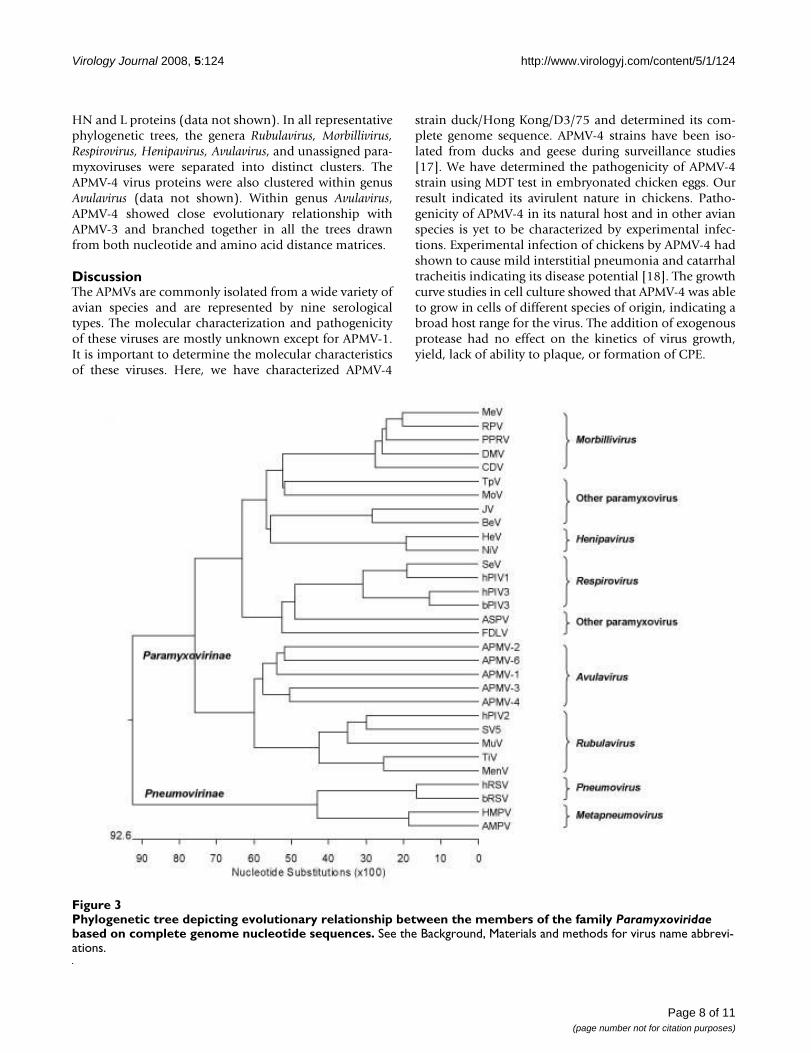
Phylogenetic analysisThe phylogenetic relationship of APMV-4 with othermembers of family Paramyxoviridae was obtained by com-paring nucleotide sequences of entire genomes. Theresulting phylogenetic tree is depicted in Figure 3. Thephylogenetic trees were also obtained from percent diver-gence of deduced amino acid sequences of the N, P, M, F,
Table 3: Percent identity of APMV-4 proteins with the other members of subfamily Paramyxovirinae*.
NP P M F HN L
Avulavirus
APMV-1 34.7 20.2 28.5 32.3 32.9 32.2APMV-2 38.2 20.2 30.8 33.9 30.6 34.2APMV-3 51.2 20.5 32.3 33.1 39.2 40.8APMV-6 38.4 19.8 31.4 32.5 31.6 33.2
Rubulavirus
SV5 30.1 15.2 24.1 27 31.6 31.8hPIV2 31.4 17.8 23.2 25 30.4 31.7MuV 32.3 17.3 24.6 27.3 31.6 32.4TiV 35.8 18.3 21.1 25.6 18.4 31.7
Respirovirus
hPIV1 20.5 6.6 14.6 20.1 22.3 24.1bPIV3 22.3 7.6 14.6 22 21.2 24.4hPIV3 22.7 9.1 15.1 22.2 22.3 24.2SeV 21 5.8 14.6 21.5 22.6 23.4
Henipavirus
HeV 26.6 12.7 15.7 22 15.8 23.9NiV 27.1 12.2 15.1 21.9 15.3 24.7
Morbillivirus
DV 22.1 11.4 14.6 23.5 13.7 25MeV 22.3 10.7 16.5 23.1 12.8 24.7PPRV 22.1 10.2 15.7 21.2 9.5 24.3RPV 21.4 10.7 15.7 21.9 12.3 25.5DolMV 22.1 10.7 14.3 23.6 11.2 24.5
Other paramyxovirus
ASPV 22.3 7.1 15.1 24 23.3 23.6BeV 25.8 11.2 14.3 22.9 23.5 24FDLV 22.7 8.1 16.2 25 23.5 25.4JV 24.2 12.4 14.3 24.9 23.2 24.1MoV 25.5 14.2 16.8 20.8 11.4 24.2MenV 34.3 17.5 22.2 26.5 18.9 31.7TpV 24.2 12.4 14.6 21.2 18.2 24.2
* See the Background and Methods for virus name abbreviations.
Amino acid sequence alignment of features of the APMV-4 V, F and L proteins compared with other members of genus AvulavirusFigure 2Amino acid sequence alignment of features of the APMV-4 V, F and L proteins compared with other members of genus Avulavirus. Sequence alignment of V protein C-terminal region (A), F protein cleavage site (B), and conserved domain III of L protein (C).
Page 7 of 11(page number not for citation purposes)
Virology Journal 2008, 5:124 http://www.virologyj.com/content/5/1/124
HN and L proteins (data not shown). In all representativephylogenetic trees, the genera Rubulavirus, Morbillivirus,Respirovirus, Henipavirus, Avulavirus, and unassigned para-myxoviruses were separated into distinct clusters. TheAPMV-4 virus proteins were also clustered within genusAvulavirus (data not shown). Within genus Avulavirus,APMV-4 showed close evolutionary relationship withAPMV-3 and branched together in all the trees drawnfrom both nucleotide and amino acid distance matrices.
DiscussionThe APMVs are commonly isolated from a wide variety ofavian species and are represented by nine serologicaltypes. The molecular characterization and pathogenicityof these viruses are mostly unknown except for APMV-1.It is important to determine the molecular characteristicsof these viruses. Here, we have characterized APMV-4
strain duck/Hong Kong/D3/75 and determined its com-plete genome sequence. APMV-4 strains have been iso-lated from ducks and geese during surveillance studies[17]. We have determined the pathogenicity of APMV-4strain using MDT test in embryonated chicken eggs. Ourresult indicated its avirulent nature in chickens. Patho-genicity of APMV-4 in its natural host and in other avianspecies is yet to be characterized by experimental infec-tions. Experimental infection of chickens by APMV-4 hadshown to cause mild interstitial pneumonia and catarrhaltracheitis indicating its disease potential [18]. The growthcurve studies in cell culture showed that APMV-4 was ableto grow in cells of different species of origin, indicating abroad host range for the virus. The addition of exogenousprotease had no effect on the kinetics of virus growth,yield, lack of ability to plaque, or formation of CPE.
Phylogenetic tree depicting evolutionary relationship between the members of the family Paramyxoviridae based on complete genome nucleotide sequencesFigure 3Phylogenetic tree depicting evolutionary relationship between the members of the family Paramyxoviridae based on complete genome nucleotide sequences. See the Background, Materials and methods for virus name abbrevi-ations.
Page 8 of 11(page number not for citation purposes)
Virology Journal 2008, 5:124 http://www.virologyj.com/content/5/1/124
The genome of APMV-4 is 15,054 nt long, which is largerthan APMV-2 (14,904 nt) [20] and smaller than APMV-3(16,272 nt) [21]. The typical genome size of members offamily Paramyxoviridae is approximately 15,500 nt, butcan be considerably larger, as exemplified by HeV (18,234nt) and BeV (19,212 nt) [1]. The length of the APMV-4genome follows the "rule of six" that is a characteristic ofsubfamily Paramyxovirinae [26] but not of the subfamilyPneumovirinae [25]. The leader region of APMV-4 is 55 ntin length, which is exactly conserved in length with mostother members of subfamily Paramyxovirinae [1]. In mostmembers of family Paramyxoviridae, the 5' trailer region is25–60 nt long [33]. Interestingly, the trailer region ofAPMV-4 is 17 nt in length, which is the shortest amongmembers of subfamily Paramyxovirinae whereas APMV-3has the longest trailer sequence (707 nt) within the familyreported to date [21]. The terminal sequences of the 3'leader and 5' trailer regions showed a high degree of com-plimentarity (70.5%), suggesting conserved elements inthe 3' promoter region of genome and antigenome. Iden-tification of conserve gene start (3'-UCCCACCCCUUCC-5') and gene end (3'-AAAUUAAUUUUU-5') sequencesrevealed six genes in the order of 3'-N-P/V-M-F-HN-L-5'within the genome with variable IGS. Six genes are alsofound in all members of the subfamily Paramyxovirinaewith the exception of presence of the SH gene in APMV-6of Avulavirus, SV5, MuV of Rubulavirus, J virus, BeV, and Ugene in FDLV [1,34]. The variable IGS of APMV-4 is simi-lar to that of Rubula, Avula, Morbilli and Henipa viruses,whereas, conserved trinucleotide GUU is typical ofRespirovirus [33]. The subunit hexamer phasing positionsfor the start sites of the six genes and the P-gene editingsite, shown to be genus-specific within the subfamily Par-amyxovirinae [32,35]. The hexamer phasing (2, 2, 2, 6, 2,2) of APMV-4 showed differences with those of the otherAPMVs [APMV-1(2,4,4,4,3,6), APMV-2 (2,2,2,3,3,3),APMV-3 (2,5,5,2,2,1) and APMV-6 (2,2,2,2,2,4,4)]; sug-gesting its uniqueness within the members of genus Avu-lavirus.
The N protein core conserved motif of APMV-4 is F-X4-F-X4-SYAMG and a similar motif has also been reported forAPMV-2 [20]. This is different from earlier motif F-X4-Y-X4-SYAMG that was reported for other members of Para-myxovirinae and is thought to be required for NP-NP inter-action. The first amino acid residue F322, needed forcorrect self-assembly is conserved in APMV-4 as reportedfor other members of the family [36]. APMV-4 edits the Pgene from P to V in a manner similar to that employed byAvulavirus, Respirovirus and Morbillivirus, but differs fromthat of Rubulavirus, which edit their mRNA from V to P[37]. Insertion of one non-templated G residue at the edit-ing site produces the V protein with conserved histidineand seven cysteine residues. The W protein produced bymRNA editing with a double G residue insertion is either
absent or rare, as W mRNAs were not detected in cellsinfected with APMV-4. The M protein is the most abun-dant structural protein in the virion, and it associates withmembranes and with the hydrophobic tails of viral F andHN proteins [1]. The M protein of APMV-4 is rich inhydrophobic amino acids sufficient for hydrophobicinteraction but lacks the membrane spanning domain.
The paramyxovirus F protein becomes biologically activewhen the inactive precursor (F0) is cleaved into disulfide-bonded F1–F2 subunits by host protease [1]. The F pro-tein cleavage site is a well-characterized determinant ofNDV pathogenicity in chickens. Virulent NDV strains typ-ically contain a polybasic cleavage site (R-X-K/R-R↓F) thatis recognized by furin-like intracellular proteases that areubiquitous in most cells. This provides for efficient cleav-age in a wide range of tissues, making it possible for viru-lent strains to spread systemically. In contrast, avirulentNDV strains typically have one or a few basic residues rel-ative to the cleavage site and depend on secretory pro-teases (or, in cell culture, added protease) for cleavage.This limits the replication of avirulent strains to the respi-ratory and enteric tracts where the secretory proteases arefound. However, the cleavage site of wild type SeV F pro-tein (VPQSR↓F) has a monobasic residue (underlined)that limits replication to the respiratory tract, but variantswith mutation from serine to proline (PR↓F) showed pan-tropism [38], in addition they do not require exogenousprotease for growth of virus. The APMV-4 cleavage site(DIQPR↓F) is similar to efficiently cleaved PR↓F variantof SeV in having a proline immediately upstream of thesingle arginine residue and also does not require exoge-nous protease for growth in cell culture. However, theAPMV-2 F protein cleavage site (KPASR↓F) contains aSR↓F site similar to that of wild type SeV but does notrequire exogenous protease for cleavage activation [20]. Atthe F protein cleavage site, (R↓F) is common in APMV-4,APMV-2 and virulent APMV-1 (RRQKR↓F) irrespective ofbasic amino acid numbers and does not need exogenousprotease for virus growth. In contrast, the F protein cleav-age site (R↓L) is common in avirulent APMV-1 (GRQ-GR↓L), and APMV-3 (RPRGR↓L) irrespective of basicamino acids and require secretory/exogenous proteasesfor virus growth.
Like other paramyxoviruses, the HN protein of APMV-4 isa type II integral protein with a conserved sialic acid bind-ing motif (NRKSCS) and conserved neuraminidase activesite residues and cysteine residues corresponding to theglobular head of HN protein [30]. Alignment of theAPMV-4 L protein subdomain C of domain III (Figure 2C)with other APMVs showed the conserved catalytic motifGDNQ. Interestingly, in APMV-6, this conserved motifcontains a single amino acid difference (GENQ, differenceunderlined). In the rabies virus L protein, mutation of
Page 9 of 11(page number not for citation purposes)
Virology Journal 2008, 5:124 http://www.virologyj.com/content/5/1/124
GDNQ to GENQ abolished polymerase activity in vitro[39]. The APMV-4 L protein had a conserved ATP-bindingmotif (R-X21-GEGYG) at domain VI, which is similar tothat of APMV-3 (R-X21-GEGSG), but for other membersof Paramyxovirinae this motif is K-X18-21-G-X-G-X-G[31,32].
The phylogenetic analysis of APMV-4 with members ofthe family Paramyxoviridae showed that APMV-4 was moreclosely related to other APMV types than with other para-myxoviruses, supporting classification of APMVs in thegenus Avulavirus. APMV-4 showed close evolutionary rela-tionship with APMV-3 both in nucleotide and amino acidanalysis. It will be interesting to study further the patho-genicity of APMV-4 in different avian species. The analysisof additional strains of APMV-4 and the development of areverse genetic system will be helpful to study the molec-ular biology and pathogenesis of the virus.
Competing interestsThe authors declare that they have no competing interests.
Authors' contributionsBN carried out the molecular characterization, genomesequencing studies and drafting of the manuscript. SK par-ticipated in genome sequencing of the virus. PC partici-pated in design of experiment and manuscriptpreparation. SKS conceived of the study, and participatedin its design and coordination. All authors read andapproved the final manuscript.
AcknowledgementsWe thank Daniel Rockemann and all our laboratory members for their excellent technical assistance and help. We also thank Hamp Edwards for performing electron microscopy and Ireen Dryburgh-Barry for proofread-ing the manuscript. "This research was supported by NIAID contract no. N01A060009 (85% support) and NIAID, NIH Intramural Research Program (15% support). The views expressed here in neither necessarily reflect the official policies of the Department of Health and Human Services; nor does mention of trade names, commercial practices, or organizations imply endorsement by the U.S. Government."
References1. Lamb RA, Parks G: Paramyxoviridae: the viruses and their replication 5th
edition. Lippincott Williams and Wilkins, Philadelphia; 2007. 2. Samal SK: Paramyxoviruses of Animals. Encyclopedia of Virology
Third edition. 2008, 4:40-47.3. Fauquet CMMM, Maniloff J, Desselberger U, Ball LA: Virus taxon-
omy. VIIIth report of the international committee on taxon-omy of viruses. Elsevier Academic Press, San Diego; 2005.
4. Mayo MA: Virus taxonomy – Houston 2002. Arch Virol 2002,147:1071-1076.
5. Alexander DJ, Hinshaw VS, Collins MS, Yamane N: Characteriza-tion of viruses which represent further distinct serotypes(PMV-8 and PMV-9) of avian paramyxoviruses. Arch Virol 1983,78:29-36.
6. Tumova B, Stumpa A, Janout V, Uvizl M, Chmela J: A further mem-ber of the Yucaipa group isolated from the common wren(Troglodytes troglodytes). Acta Virol 1979, 23:504-507.
7. Bankowski RA, Almquist J, Dombrucki J: Effect of paramyxovirusyucaipa on fertility, hatchability, and poult yield of turkeys.Avian Dis 1981, 25:517-520.
8. Redmann T, Zeydanli MM, Herbst W, Kaleta EF: [Isolation of a par-amyxovirus-3 from turkeys with respiratory tract disease inGermany]. Dtsch Tierarztl Wochenschr 1991, 98:138-141.
9. Zhang GZ, Zhao JX, Wang HW, Yang AM, Bu CY, Wang M: Isola-tion, identification, and comparison of four isolates of avianparamyxovirus serotype 2 in China. Avian Dis 2006, 50:386-390.
10. Zhang GZ, Zhao JX, Wang M: Serological survey on prevalenceof antibodies to avian paramyxovirus serotype 2 in China.Avian Dis 2007, 51:137-139.
11. Nerome K, Nakayama M, Ishida M, Fukumi H: Isolation of a newavian paramyxovirus from budgerigar (Melopsittacus undu-latus). J Gen Virol 1978, 38:293-301.
12. Gough RE, Alexander DJ: Avian paramyxovirus type 4 isolatedfrom a ringed teal (Calonetta leucophrys). Vet Rec 1984,115:653.
13. Stallknecht DE, Senne DA, Zwank PJ, Shane SM, Kearney MT: Avianparamyxoviruses from migrating and resident ducks incoastal Louisiana. J Wildl Dis 1991, 27:123-128.
14. Maldonado A, Arenas A, Tarradas MC, Luque I, Astorga R, Perea JA,Miranda A: Serological survey for avian paramyxoviruses fromwildfowl in aquatic habitats in Andalusia. J Wildl Dis 1995,31:66-69.
15. Capua I, De Nardi R, Beato MS, Terregino C, Scremin M, Guberti V:Isolation of an avian paramyxovirus type 9 from migratorywaterfowl in Italy. Vet Rec 2004, 155:156.
16. Shortridge KF, Alexander DJ: Incidence and preliminary charac-terisation of a hitherto unreported, serologically distinct,avian paramyxovirus isolated in Hong Kong. Res Vet Sci 1978,25:128-130.
17. Alexander DJ, Aymard M, Kessler N, Collins MS: Antigenic andstructural relationships between avian paramyxoviruses iso-lated from ducks in Hong Kong and Mississippi, USA. J GenVirol 1979, 44:839-842.
18. Warke A, Stallknecht D, Williams SM, Pritchard N, Mundt E: Com-parative study on the pathogenicity and immunogenicity ofwild bird isolates of avian paramyxovirus 2, 4, and 6 in chick-ens. Avian Pathol 2008, 37:429-434.
19. Krishnamurthy S, Samal SK: Nucleotide sequences of the trailer,nucleocapsid protein gene and intergenic regions of Newcas-tle disease virus strain Beaudette C and completion of theentire genome sequence. J Gen Virol 1998, 79(Pt 10):2419-2424.
20. Subbiah M, Xiao S, Collins PL, Samal SK: Complete sequence ofthe genome of avian paramyxovirus type 2 (strain Yucaipa)and comparison with other paramyxoviruses. Virus Res 2008.
21. Kumar S, Nayak B, Collins PL, Samal SK: Complete genomesequence of avian paramyxovirus type 3 reveals an unusuallylong trailer region. Virus Res 2008, 137(2):189-197.
22. Chang PC, Hsieh ML, Shien JH, Graham DA, Lee MS, Shieh HK: Com-plete nucleotide sequence of avian paramyxovirus type 6 iso-lated from ducks. J Gen Virol 2001, 82:2157-2168.
23. Troutt AB, McHeyzer-Williams MG, Pulendran B, Nossal GJ: Liga-tion-anchored PCR: a simple amplification technique withsingle-sided specificity. Proc Natl Acad Sci USA 1992,89:9823-9825.
24. Li Z, Yu M, Zhang H, Wang HY, Wang LF: Improved rapid ampli-fication of cDNA ends (RACE) for mapping both the 5' and3' terminal sequences of paramyxovirus genomes. J VirolMethods 2005, 130:154-156.
25. Samal SK, Collins PL: RNA replication by a respiratory syncytialvirus RNA analog does not obey the rule of six and retains anonviral trinucleotide extension at the leader end. J Virol1996, 70:5075-5082.
26. Calain P, Roux L: The rule of six, a basic feature for efficientreplication of Sendai virus defective interfering RNA. J Virol1993, 67:4822-4830.
27. Miller PJ, Boyle DB, Eaton BT, Wang LF: Full-length genomesequence of Mossman virus, a novel paramyxovirus isolatedfrom rodents in Australia. Virology 2003, 317:330-344.
28. Ceroni A, Passerini A, Vullo A, Frasconi P: DISULFIND: a disulfidebonding state and cysteine connectivity prediction server.Nucleic Acids Res 2006, 34:W177-181.
29. Iwata S, Schmidt AC, Titani K, Suzuki M, Kido H, Gotoh B, HamaguchiM, Nagai Y: Assignment of disulfide bridges in the fusion glyc-oprotein of Sendai virus. J Virol 1994, 68:3200-3206.
30. Langedijk JP, Daus FJ, van Oirschot JT: Sequence and structurealignment of Paramyxoviridae attachment proteins and dis-
Page 10 of 11(page number not for citation purposes)
http://www.ncbi.nlm.nih.gov/entrez/query.fcgi?cmd=Retrieve&db=PubMed&dopt=Abstract&list_uids=6651534
http://www.ncbi.nlm.nih.gov/entrez/query.fcgi?cmd=Retrieve&db=PubMed&dopt=Abstract&list_uids=6651534
http://www.ncbi.nlm.nih.gov/entrez/query.fcgi?cmd=Retrieve&db=PubMed&dopt=Abstract&list_uids=6651534
http://www.ncbi.nlm.nih.gov/entrez/query.fcgi?cmd=Retrieve&db=PubMed&dopt=Abstract&list_uids=7259687
http://www.ncbi.nlm.nih.gov/entrez/query.fcgi?cmd=Retrieve&db=PubMed&dopt=Abstract&list_uids=7259687
http://www.ncbi.nlm.nih.gov/entrez/query.fcgi?cmd=Retrieve&db=PubMed&dopt=Abstract&list_uids=1829671
http://www.ncbi.nlm.nih.gov/entrez/query.fcgi?cmd=Retrieve&db=PubMed&dopt=Abstract&list_uids=1829671
http://www.ncbi.nlm.nih.gov/entrez/query.fcgi?cmd=Retrieve&db=PubMed&dopt=Abstract&list_uids=1829671
http://www.ncbi.nlm.nih.gov/entrez/query.fcgi?cmd=Retrieve&db=PubMed&dopt=Abstract&list_uids=6098067
http://www.ncbi.nlm.nih.gov/entrez/query.fcgi?cmd=Retrieve&db=PubMed&dopt=Abstract&list_uids=6098067
http://www.ncbi.nlm.nih.gov/entrez/query.fcgi?cmd=Retrieve&db=PubMed&dopt=Abstract&list_uids=2023311
http://www.ncbi.nlm.nih.gov/entrez/query.fcgi?cmd=Retrieve&db=PubMed&dopt=Abstract&list_uids=2023311
http://www.ncbi.nlm.nih.gov/entrez/query.fcgi?cmd=Retrieve&db=PubMed&dopt=Abstract&list_uids=2023311
http://www.ncbi.nlm.nih.gov/entrez/query.fcgi?cmd=Retrieve&db=PubMed&dopt=Abstract&list_uids=7563427
http://www.ncbi.nlm.nih.gov/entrez/query.fcgi?cmd=Retrieve&db=PubMed&dopt=Abstract&list_uids=7563427
http://www.ncbi.nlm.nih.gov/entrez/query.fcgi?cmd=Retrieve&db=PubMed&dopt=Abstract&list_uids=9780047
http://www.ncbi.nlm.nih.gov/entrez/query.fcgi?cmd=Retrieve&db=PubMed&dopt=Abstract&list_uids=9780047
http://www.ncbi.nlm.nih.gov/entrez/query.fcgi?cmd=Retrieve&db=PubMed&dopt=Abstract&list_uids=9780047
http://www.ncbi.nlm.nih.gov/entrez/query.fcgi?cmd=Retrieve&db=PubMed&dopt=Abstract&list_uids=1409706
http://www.ncbi.nlm.nih.gov/entrez/query.fcgi?cmd=Retrieve&db=PubMed&dopt=Abstract&list_uids=1409706
http://www.ncbi.nlm.nih.gov/entrez/query.fcgi?cmd=Retrieve&db=PubMed&dopt=Abstract&list_uids=1409706
http://www.ncbi.nlm.nih.gov/entrez/query.fcgi?cmd=Retrieve&db=PubMed&dopt=Abstract&list_uids=8764015
http://www.ncbi.nlm.nih.gov/entrez/query.fcgi?cmd=Retrieve&db=PubMed&dopt=Abstract&list_uids=8764015
http://www.ncbi.nlm.nih.gov/entrez/query.fcgi?cmd=Retrieve&db=PubMed&dopt=Abstract&list_uids=8764015
http://www.ncbi.nlm.nih.gov/entrez/query.fcgi?cmd=Retrieve&db=PubMed&dopt=Abstract&list_uids=8392616
http://www.ncbi.nlm.nih.gov/entrez/query.fcgi?cmd=Retrieve&db=PubMed&dopt=Abstract&list_uids=8392616
http://www.ncbi.nlm.nih.gov/entrez/query.fcgi?cmd=Retrieve&db=PubMed&dopt=Abstract&list_uids=8151783
http://www.ncbi.nlm.nih.gov/entrez/query.fcgi?cmd=Retrieve&db=PubMed&dopt=Abstract&list_uids=8151783
Virology Journal 2008, 5:124 http://www.virologyj.com/content/5/1/124
Publish with BioMed Central and every scientist can read your work free of charge
"BioMed Central will be the most significant development for disseminating the results of biomedical research in our lifetime."
Sir Paul Nurse, Cancer Research UK
Your research papers will be:
available free of charge to the entire biomedical community
peer reviewed and published immediately upon acceptance
cited in PubMed and archived on PubMed Central
yours — you keep the copyright
Submit your manuscript here:http://www.biomedcentral.com/info/publishing_adv.asp
BioMedcentral
covery of enzymatic activity for a morbillivirus hemaggluti-nin. J Virol 1997, 71:6155-6167.
31. Poch O, Blumberg BM, Bougueleret L, Tordo N: Sequence com-parison of five polymerases (L proteins) of unsegmentednegative-strand RNA viruses: theoretical assignment offunctional domains. J Gen Virol 1990, 71(Pt 5):1153-1162.
32. Harcourt BH, Tamin A, Halpin K, Ksiazek TG, Rollin PE, Bellini WJ,Rota PA: Molecular characterization of the polymerase geneand genomic termini of Nipah virus. Virology 2001, 287:192-201.
33. Nylund S, Karlsen M, Nylund A: The complete genome sequenceof the Atlantic salmon paramyxovirus (ASPV). Virology 2008,373:137-148.
34. Kurath G, Batts WN, Ahne W, Winton JR: Complete genomesequence of Fer-de-Lance virus reveals a novel gene in rep-tilian paramyxoviruses. J Virol 2004, 78:2045-2056.
35. Kolakofsky D, Pelet T, Garcin D, Hausmann S, Curran J, Roux L: Par-amyxovirus RNA synthesis and the requirement for hex-amer genome length: the rule of six revisited. J Virol 1998,72:891-899.
36. Myers TM, Smallwood S, Moyer SA: Identification of nucleocap-sid protein residues required for Sendai virus nucleocapsidformation and genome replication. J Gen Virol 1999, 80(Pt6):1383-1391.
37. Steward M, Vipond IB, Millar NS, Emmerson PT: RNA editing inNewcastle disease virus. J Gen Virol 1993, 74(Pt 12):2539-2547.
38. Tashiro M, James I, Karri S, Wahn K, Tobita K, Klenk HD, Rott R,Seto JT: Pneumotropic revertants derived from a pantropicmutant, F1-R, of Sendai virus. Virology 1991, 184:227-234.
39. Schnell MJ, Conzelmann KK: Polymerase activity of in vitromutated rabies virus L protein. Virology 1995, 214:522-530.
Page 11 of 11(page number not for citation purposes)
http://www.ncbi.nlm.nih.gov/entrez/query.fcgi?cmd=Retrieve&db=PubMed&dopt=Abstract&list_uids=9223510
http://www.ncbi.nlm.nih.gov/entrez/query.fcgi?cmd=Retrieve&db=PubMed&dopt=Abstract&list_uids=9223510
http://www.ncbi.nlm.nih.gov/entrez/query.fcgi?cmd=Retrieve&db=PubMed&dopt=Abstract&list_uids=2161049
http://www.ncbi.nlm.nih.gov/entrez/query.fcgi?cmd=Retrieve&db=PubMed&dopt=Abstract&list_uids=2161049
http://www.ncbi.nlm.nih.gov/entrez/query.fcgi?cmd=Retrieve&db=PubMed&dopt=Abstract&list_uids=2161049
http://www.ncbi.nlm.nih.gov/entrez/query.fcgi?cmd=Retrieve&db=PubMed&dopt=Abstract&list_uids=9444980
http://www.ncbi.nlm.nih.gov/entrez/query.fcgi?cmd=Retrieve&db=PubMed&dopt=Abstract&list_uids=9444980
http://www.ncbi.nlm.nih.gov/entrez/query.fcgi?cmd=Retrieve&db=PubMed&dopt=Abstract&list_uids=9444980
http://www.ncbi.nlm.nih.gov/entrez/query.fcgi?cmd=Retrieve&db=PubMed&dopt=Abstract&list_uids=8277263
http://www.ncbi.nlm.nih.gov/entrez/query.fcgi?cmd=Retrieve&db=PubMed&dopt=Abstract&list_uids=8277263
http://www.ncbi.nlm.nih.gov/entrez/query.fcgi?cmd=Retrieve&db=PubMed&dopt=Abstract&list_uids=1651590
http://www.ncbi.nlm.nih.gov/entrez/query.fcgi?cmd=Retrieve&db=PubMed&dopt=Abstract&list_uids=1651590
http://www.ncbi.nlm.nih.gov/entrez/query.fcgi?cmd=Retrieve&db=PubMed&dopt=Abstract&list_uids=8553554


![D3: e`ad T`ca`cReV ]`R_ hRZgVc ]Zde - SPLessons](https://static.fdokumen.com/doc/165x107/6322423f28c445989105b166/d3-ead-tcacrev-r-hrzgvc-zde-splessons.jpg)