Molecular analysis of the apoptotic effects of BPA in acute myeloid leucemia cells
8
BioMed Central Page 1 of 8 (page number not for citation purposes) Journal of Translational Medicine Open Access Research Molecular analysis of the apoptotic effects of BPA in acute myeloid leukemia cells Paola Bontempo 1,2 , Luigi Mita 1,2,3 , Antonella Doto 1 , Marco Miceli 1 , Angela Nebbioso 1 , Ilaria Lepore 1 , GianLuigi Franci 1 , Roberta Menafra 1 , Vincenzo Carafa 1 , Mariarosaria Conte 1 , Floriana De Bellis 1 , Fabio Manzo 1 , Vincenzo Di Cerbo 1 , Rosaria Benedetti 4 , Loredana D'Amato 1 , Maria Marino 2,5 , Alessandro Bolli 2,5 , Giovanna Del Pozzo 2,6 , Nadia Diano 2,3,6 , Marianna Portaccio 2,3 , Gustavo D Mita 3,4 , Maria Teresa Vietri 1 , Michele Cioffi 1 , Ernesto Nola 1 , Carmela Dell'Aversana 1 , Vincenzo Sica 1 , Anna Maria Molinari 1 and Lucia Altucci* 1,2 Address: 1 Dipartimento di Patologia generale, Seconda Università di Napoli, Via L. De Crecchio 7 Napoli, Italy, 2 Istituto Nazionale di Biostruttura e dei Biosistemi, Viale Medaglie d'Oro,305, 00100 Roma, Italy, 3 Dipartimento di Medicina sperimentale, Seconda Università di Napoli, Via De Crecchio, Napoli, Italy, 4 Dipartimento di Fisica, Università di Napoli 'Federico II', Napoli, Italy, 5 Dipartimento di Biologia, Università Roma Tre, Viale Guglielmo Marconi 446, 00146 Roma, Italy and 6 Istituto di Genetica e Biofisica del CNR, Via P. Castellino 111, 80100 Napoli, Italy Email: Paola Bontempo - [email protected]; Luigi Mita - [email protected]; Antonella Doto - [email protected]; Marco Miceli - [email protected]; Angela Nebbioso - [email protected]; Ilaria Lepore - [email protected]; GianLuigi Franci - [email protected]; Roberta Menafra - [email protected]; Vincenzo Carafa - [email protected]; Mariarosaria Conte - [email protected]; Floriana De Bellis - [email protected]; Fabio Manzo - [email protected]; Vincenzo Di Cerbo - [email protected]; Rosaria Benedetti - [email protected]; Loredana D'Amato - [email protected]; Maria Marino - [email protected]; Alessandro Bolli - [email protected]; Giovanna Del Pozzo - [email protected]; Nadia Diano - [email protected]; Marianna Portaccio - [email protected]; Gustavo D Mita - [email protected]; Maria Teresa Vietri - [email protected]; Michele Cioffi - [email protected]; Ernesto Nola - [email protected]; Carmela Dell'Aversana - [email protected]; Vincenzo Sica - [email protected]; Anna Maria Molinari - [email protected]; Lucia Altucci* - [email protected] * Corresponding author Abstract Background: BPA (bisphenol A or 2,2-bis(4-hydroxy-phenol)propane) is present in the manufacture of polycarbonate plastic and epoxy resins, which can be used in impact-resistant safety equipment and baby bottles, as protective coatings inside metal food containers, and as composites and sealants in dentistry. Recently, attention has focused on the estrogen-like and carcinogenic adverse effects of BPA. Thus, it is necessary to investigate the cytotoxicity and apoptosis-inducing activity of this compound. Methods: Cell cycle, apoptosis and differentiation analyses; western blots. Results: BPA is able to induce cell cycle arrest and apoptosis in three different acute myeloid leukemias. Although some granulocytic differentiation concomitantly occurred in NB4 cells upon BPA treatment, the major action was the induction of apoptosis. BPA mediated apoptosis was Published: 18 June 2009 Journal of Translational Medicine 2009, 7:48 doi:10.1186/1479-5876-7-48 Received: 24 February 2009 Accepted: 18 June 2009 This article is available from: http://www.translational-medicine.com/content/7/1/48 © 2009 Bontempo et al; licensee BioMed Central Ltd. This is an Open Access article distributed under the terms of the Creative Commons Attribution License (http://creativecommons.org/licenses/by/2.0 ), which permits unrestricted use, distribution, and reproduction in any medium, provided the original work is properly cited.
-
Upload
independent -
Category
Documents
-
view
2 -
download
0
Transcript of Molecular analysis of the apoptotic effects of BPA in acute myeloid leucemia cells

BioMed CentralJournal of Translational Medicine
ss
Open AcceResearchMolecular analysis of the apoptotic effects of BPA in acute myeloid leukemia cellsPaola Bontempo1,2, Luigi Mita1,2,3, Antonella Doto1, Marco Miceli1, Angela Nebbioso1, Ilaria Lepore1, GianLuigi Franci1, Roberta Menafra1, Vincenzo Carafa1, Mariarosaria Conte1, Floriana De Bellis1, Fabio Manzo1, Vincenzo Di Cerbo1, Rosaria Benedetti4, Loredana D'Amato1, Maria Marino2,5, Alessandro Bolli2,5, Giovanna Del Pozzo2,6, Nadia Diano2,3,6, Marianna Portaccio2,3, Gustavo D Mita3,4, Maria Teresa Vietri1, Michele Cioffi1, Ernesto Nola1, Carmela Dell'Aversana1, Vincenzo Sica1, Anna Maria Molinari1 and Lucia Altucci*1,2Address: 1Dipartimento di Patologia generale, Seconda Università di Napoli, Via L. De Crecchio 7 Napoli, Italy, 2Istituto Nazionale di Biostruttura e dei Biosistemi, Viale Medaglie d'Oro,305, 00100 Roma, Italy, 3Dipartimento di Medicina sperimentale, Seconda Università di Napoli, Via De Crecchio, Napoli, Italy, 4Dipartimento di Fisica, Università di Napoli 'Federico II', Napoli, Italy, 5Dipartimento di Biologia, Università Roma Tre, Viale Guglielmo Marconi 446, 00146 Roma, Italy and 6Istituto di Genetica e Biofisica del CNR, Via P. Castellino 111, 80100 Napoli, Italy
Email: Paola Bontempo - [email protected]; Luigi Mita - [email protected]; Antonella Doto - [email protected]; Marco Miceli - [email protected]; Angela Nebbioso - [email protected]; Ilaria Lepore - [email protected]; GianLuigi Franci - [email protected]; Roberta Menafra - [email protected]; Vincenzo Carafa - [email protected]; Mariarosaria Conte - [email protected]; Floriana De Bellis - [email protected]; Fabio Manzo - [email protected]; Vincenzo Di Cerbo - [email protected]; Rosaria Benedetti - [email protected]; Loredana D'Amato - [email protected]; Maria Marino - [email protected]; Alessandro Bolli - [email protected]; Giovanna Del Pozzo - [email protected]; Nadia Diano - [email protected]; Marianna Portaccio - [email protected]; Gustavo D Mita - [email protected]; Maria Teresa Vietri - [email protected]; Michele Cioffi - [email protected]; Ernesto Nola - [email protected]; Carmela Dell'Aversana - [email protected]; Vincenzo Sica - [email protected]; Anna Maria Molinari - [email protected]; Lucia Altucci* - [email protected]
* Corresponding author
AbstractBackground: BPA (bisphenol A or 2,2-bis(4-hydroxy-phenol)propane) is present in themanufacture of polycarbonate plastic and epoxy resins, which can be used in impact-resistant safetyequipment and baby bottles, as protective coatings inside metal food containers, and as compositesand sealants in dentistry. Recently, attention has focused on the estrogen-like and carcinogenicadverse effects of BPA. Thus, it is necessary to investigate the cytotoxicity and apoptosis-inducingactivity of this compound.
Methods: Cell cycle, apoptosis and differentiation analyses; western blots.
Results: BPA is able to induce cell cycle arrest and apoptosis in three different acute myeloidleukemias. Although some granulocytic differentiation concomitantly occurred in NB4 cells uponBPA treatment, the major action was the induction of apoptosis. BPA mediated apoptosis was
Published: 18 June 2009
Journal of Translational Medicine 2009, 7:48 doi:10.1186/1479-5876-7-48
Received: 24 February 2009Accepted: 18 June 2009
This article is available from: http://www.translational-medicine.com/content/7/1/48
© 2009 Bontempo et al; licensee BioMed Central Ltd. This is an Open Access article distributed under the terms of the Creative Commons Attribution License (http://creativecommons.org/licenses/by/2.0), which permits unrestricted use, distribution, and reproduction in any medium, provided the original work is properly cited.
Page 1 of 8(page number not for citation purposes)
Journal of Translational Medicine 2009, 7:48 http://www.translational-medicine.com/content/7/1/48
caspase dependent and occurred by activation of extrinsic and intrinsic cell death pathwaysmodulating both FAS and TRAIL and by inducing BAD phosphorylation in NB4 cells. Finally, alsonon genomic actions such as the early decrease of both ERK and AKT phosphorylation wereinduced by BPA thus indicating that a complex intersection of regulations occur for the apoptoticaction of BPA.
Conclusion: BPA is able to induce apoptosis in leukemia cells via caspase activation andinvolvement of both intrinsic and extrinsic pathways of apoptosis.
BackgroundThe Endocrine Disrupting Compounds are defined as"exogenous substances that cause adverse health effects in anintact organism, or its progeny, secondary to changes in endo-crine function" (EEC, 1996). Their effects on humans, wild-life and the environment have been subject of highattention by the scientific community, since concernswere first raised about them by Colborn [1]. Recently, thepotential of certain pesticides to act as EDCs has been con-firmed. These include organometallic compounds, andmany other organochlorine compounds that are also toxicand persistent [2,3], and many have been banned as aresult [2]. Other pesticides such as organophosphates, car-bamates, triazines and pyrethroids that are less persistentand less toxic than the organochlorines, were used toreplace them, but many are now confirmed or suspectedEDCs [4]. Conventional toxicological testing of pesticidescan miss the potential of a substance to disrupt the endo-crine system, especially at the low concentrations likely tobe found in the environment. It is generally assumed thatchemical substances will show a simple monotonic dose-response curve, but some ED pesticides have j-type dose-response curves [5], whereby the toxic effects decrease asthe dose decreases, until at very low doses (often as low asparts per billion or even trillion) their effects increase [5].Of the more than 2,000 high-production volume chemi-cals that are manufactured in or imported many arewidely used in consumer products. Among the manychemicals is bisphenol A [BPA; 2,2-bis(4-hydroxyphe-nyl)propane]. BPA is used in the manufacture of polycar-bonate plastic and epoxy resins, which can be used inimpact-resistant safety equipment and baby bottles, asprotective coatings inside metal food containers, and ascomposites and sealants in dentistry. Exposure to BPA isthought to result primarily from ingestion of food con-taining BPA [6,7]. At high doses, BPA demonstrates estro-gen-like effects on uterine and prostate organ weights inexperimental animals. At doses below the putative lowestobserved adverse effect level, exposure to BPA has resultedin decreased sperm production, increased prostate glandvolume, altered development and tissue organization ofthe mammary gland, altered vaginal morphology andestrous cycles, disruption of sexual differentiation in thebrain, and accelerated growth and puberty [8-16]. BPA isof concern to environmental public health because of the
high potential for exposure of humans to these phenolsand their demonstrated animal toxicity. Recently, atten-tion has focused on the carcinogenic adverse effects ofBPA. Thus, it is important to investigate the cytotoxicityand apoptosis-inducing activity of these compounds[17,18]. In the present manuscript, we decided to investi-gate the effects of different doses of BPA on acute myeloidleukemia models to understand the mechanism(s) of BPAaction in systems not directly related to the endocrine sys-tem. We show indeed that BPA is able to induce apoptosisin leukemia cells by activation of the initiator caspases 8,9 and the effector caspases 37. Moreover we show thatmany genomic and non-genomic players are influencedby the action of BPA and contribute to its adverse effects.
MethodsCell linesAll cell lines have been obtained from ATCC and routinelycultured. NB4, U937, k562, and cells HL60, were grown at37°C in air and 5% CO2 in RPMI 1640 medium(GIBCO), supplemented with 10% heat-inactivated foetalbovine serum (FBS), 1% l-glutamine, 1% ampicillin/streptomycin and 0, 1% gentamicin. BPA (SIGMA) wasresuspended in ethanol and at the final concentration of1 μM. All trans retinoic acid (SIGMA) (RA) was resus-pended in ethanol and at the final concentration of 1 μM.To understand the potential role of BPA leukemia celllines were treated with different concentrations of BPA(10, 30, 60, 100 μM) for different times.
Cell cycle analysis2.5 × 105 cells were collected and resuspended in 500 μl ofa hypotonic buffer (0.1% Triton X-100, 0.1% sodium cit-rate, 50 μg/ml propidium iodide (PI), RNAse A). Cellswere incubated in the dark for 30 min. Samples wereacquired on a FACS Calibur flow cytometer using the CellQuest software (Becton Dickinson) and analysed withstandard procedures using the Cell Quest software (Bec-ton Dickinson) and the ModFit LT version 3 Software(Verity) as previously reported [19]. All the experimentswere performed in triplicate.
FACS analysis of apoptosisApoptosis was measured with Annexin V/PI double stain-ing detection (Roche and Sigma-Aldrich, respectively) as
Page 2 of 8(page number not for citation purposes)
Journal of Translational Medicine 2009, 7:48 http://www.translational-medicine.com/content/7/1/48
Page 3 of 8(page number not for citation purposes)
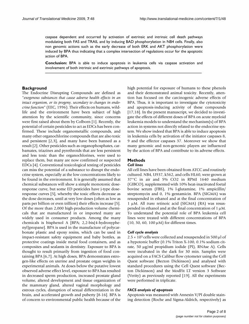
BPA induces dose dependent apoptosis and cell cycle block in acute myeloid leukemia cellsFigure 1BPA induces dose dependent apoptosis and cell cycle block in acute myeloid leukemia cells. (A) Cell cycle and apoptosis in NB4 cells after treatment with 10,30,60 and 100 μM BPA, ATRA (all-trans-retinoic acid) 1 μM and the combination of ATRA 1 μM and BPA, at the indicated concentrations for 48 hrs. (B) Cell cycle analysis and apoptosis in K562 cells after 48 hrs of treatment with 60 and 100 μM BPA. (C) Cell cycle analysis and apoptosis in HL60 cells after treatment with 10, 30, 60 and 100 μM BPA for 48 hrs.
0
10
20
30
40
50
60apoptosis
G2/M
S
G1
A+B
100
A+B
60
A+B
30
AT
RA
1
BP
A 1
00
BP
A 6
0
BP
A 3
0
BP
A 1
0
ctr
% o
f NB
4 ce
lls
0
10
20
30
40
50
60
BP
A 1
00
BP
A 6
0
BP
A 3
0
BP
A 1
0
ctr
% o
f HL6
0 ce
lls
0
10
20
30
40
50
60
ctr
BP
A 1
00
BP
A 6
0
% o
f K56
2 ce
lls
Journal of Translational Medicine 2009, 7:48 http://www.translational-medicine.com/content/7/1/48
recommended by the suppliers; samples were analysed byFACS with Cell Quest technology (Becton Dickinson) aspreviously reported [20,21]. We measured as apoptoticfraction the Annexin V positive, PI negative cells. As sec-ond assays the caspase 8, 9 and 7, 3 detection (B-Bridge)was performed as recommended by suppliers and quanti-fied by FACS (Becton Dickinson). NB4 cells were treatedfor 48 h with 10-60-100 μM BPA.
Granulocytic differentiation assayGranulocytic differentiation was carried out as previouslydescribed [22]. Briefly, NB4 cells, treated for 48 h with 10-30-60-100 μM BPA, ATRA 1 μM or with ATRA 1 μM andBPA at the indicate prima concentrations, were harvestedand resuspended in 10 μl phycoerythrine-conjugatedCD11c (CD11c-PE) (Pharmingen). Control samples wereincubated with 10 μl PE or FITC conjugated mouse IgG1,incubated for 30 min at 4°C in the dark, washed in PBSand resuspended in 500 μl PBS containing PI (0.25 μg/ml). Samples were analysed by FACS with Cell Quest tech-nology (Becton Dickinson). PI positive cells have beenexcluded from the analysis.
Western blot analyses40 micrograms of total protein extracts were separated on a15% polyacrylamide gel and blotted as previouslydescribed [23]. Western blots were shown for p21 (Trans-duction Laboratories, dilution 1:500), p27 c-19 (SantaCruz sc-528 rabbit, dilution 1:500), p16 (Santa Cruz sc-468rabbit, dilution 1:500). For determination of Rb, pRb, p53,ERalpha and cyclin D 35 μg of total protein extracts wereseparated on a polyacrylamide gel and blotted. Antibodieswere: cyclin D (Zymed), pRb, p53, RB and ERalpha (SantaCruz). Total ERKs (Santa Cruz) were used to normalise forequal loading. For quantification of TRAIL protein, 100 μgof total protein extracts were separated on a 10% polyacry-lamide gel and blotted. Western blots were shown forTRAIL (Abcam Ab 16963-1). For determination of FAS,FLIP-L and FLIP-S, BAD, pBAD and BCL2, 35 μg of totalprotein extracts were separated on a 12% polyacrylamidegel and blotted. Antibodies used were: FAS (ProSci xw-7192, dilution 1:500), Flip (Alexis 804-429-C100, dilution1:500), BAD (Cell signalling #9292, dilution 1:500), pBAD(p-Bad ser 136, #9295 cell signalling, dilution 1:500), Bcl2(Bcl2 (Ab-1) Oncogene Science, dilution 1:500). TotalERKs were used to normalise for equal loading.
For determination of ERK2, pERK, Akt and pAkt, 35 μg oftotal protein extracts were separated on a 12% polyacryla-mide gel and blotted. Antibodies used were: ERK2 (SantaCruz sc-154, dilution 1:500), pERK (Santa Cruz sc-7383,dilution 1:200), pAkt (Cell signalling cod 9271, dilution1:1000) and Akt (Cell signalling Akt cod 9272, dilution1:1000). For quantification of histone H3 acetylation, 40 μgof total protein extracts were separated on a 15% polyacryla-mide gel and blotted. Antibodies used were: acetylated his-tone H3 (Upstate cat. 06-599, dilution 1:500). Total ERKswere used to normalise for equal loading.
ResultsBPA induces dose dependent apoptosis in acute myeloid leukemia cellsTo understand the potential role of BPA in biological sys-tems of leukemias we tested the action of BPA in threedifferent acute myeloid leukemia models such as NB4,
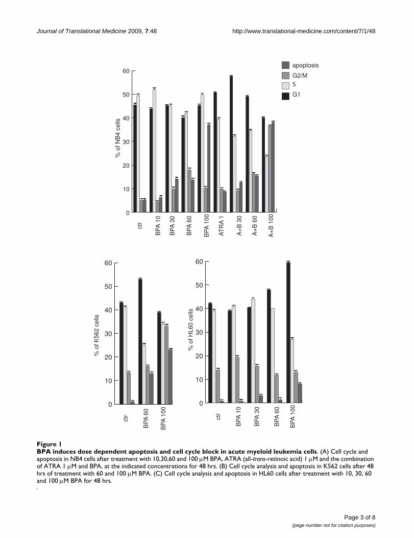
BPA induces dose dependent differentiation in NB4 cellsFigure 2BPA induces dose dependent differentiation in NB4 cells. (A) CD11c expression levels measured by FACS after 48 h of treatment with 10,30,60 and 100 μM BPA. (B) CD11c expression levels after treatment with ATRA 1 μM or with the combination of ATRA 1 μM and BPA at the indicated concentrations for 48 hrs. Note that PI positive cells have been excluded from the analysis.
0
5
10
15
20
BP
A 1
00
BP
A 6
0
BP
A30
BP
A 1
0ctr
% o
f CD
11c+
PI-
NB
4 ce
lls
a
0
20
40
60
80
100
A+B
100
A+B
60
A+B
30
AT
RA
1ctr
% o
f CD
11c+
PI-
NB
4 ce
lls
b
Page 4 of 8(page number not for citation purposes)
Journal of Translational Medicine 2009, 7:48 http://www.translational-medicine.com/content/7/1/48
HL60 and K562 cells. As it is shown in Fig. 1, differentconcentrations of BPA are able to induce an increase ofthe sub-G1 peack in all the cell lines tested, HL60 beingthe most resistant one. In NB4 cells, a model from pro-myelocytic leukemia containing the fusion protein PML-RARα and sensitive to retinoids, the highest concentra-tion of BPA used induces around 38% of apoptosis after48 hrs. This apoptosis is not synergistically modulatedby the double treatment with 1 μM Retinoic Acid (RA) asshown in Fig. 1A. Differently, cell cycle arrest seems to beaffected by the double treatment, showing an increase ofthe G1 peack at low dose BPA (30 μM) and an increaseof the G2-M fraction of cells at the highest concentrationof BPA (100 μM). Differently, in the K562 cells, a modelof AML derived from a CML containing the Philadelphiachromosome, the treatment with BPA showed anincrease of cell death proportional to the dose increase ofBPA, together with a G1 peack at the lower dose and aG2-M increase at the higher dose (Fig. 1B). Finally, HL60cells showed an increase of apoptosis at the higher doseof BPA (100 μM) in agreement with what reported previ-ously [17]. This increase is directly proportional with the
enrichment in G1 phase of HL60 cells upon treatmentwith increasing doses of BPA (Fig. 1C).
BPA induces dose dependent differentiation in NB4 cellsThat BPA was able to induce apoptosis and to influencethe cell cycle of NB4 cells, prompted us to check its effectson granulocytic differentiation of these cells. As shown inFig. 2A by FACS analyses, BPA is able to differentiate NB4cells versus granulocytes in a dose dependent manner.However, the effect was weak if compared with the one ofRA at the same time in the NB4 cells (Fig. 2B), thus show-ing that BPA preferentially activates apoptotic actions inrespect to differentiative effects in these cells.
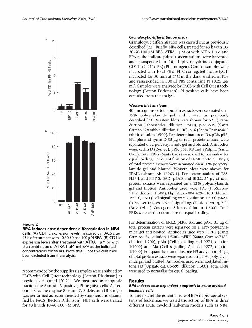
BPA induces apoptosis via caspase activation in NB4 cellsTo better identify which apoptotic pathway is activated byBPA, we tested by FACS analyses the initiator and effectorcaspases activation in NB4 cells after 48 h treatment withBPA. As it is shown in Fig. 3, both caspase 8 (Fig. 3A) and9 (Fig. 3B) are cleaved and active upon BPA treatment.Note that caspase 8 resulted more active, suggesting aprior activity of BPA on the extrinsic pathway of apoptosis
BPA induces apoptosis via caspase activation in NB4 cellsFigure 3BPA induces apoptosis via caspase activation in NB4 cells. Caspase 8, 9 and 37 assays have been carried out by FACS analysis in NB4 cells after 48 h of incubation with the indicated concentrations of BPA.
0
5
10
15
20
BP
A 1
00
BP
A 6
0
BP
A 1
0ctr
% o
f cas
pase
9 a
ctiv
e ce
lls
0
10
20
30
40
BP
A 1
00
BP
A 6
0
BP
A 1
0ctr
% o
f cas
pase
8 a
ctiv
e ce
lls
0
5
10
15
20
BP
A 1
00
BP
A 6
0
BP
A 1
0ctr
% o
f cas
pase
3-7
act
ive
cells
a b c
Page 5 of 8(page number not for citation purposes)
Journal of Translational Medicine 2009, 7:48 http://www.translational-medicine.com/content/7/1/48
Page 6 of 8(page number not for citation purposes)
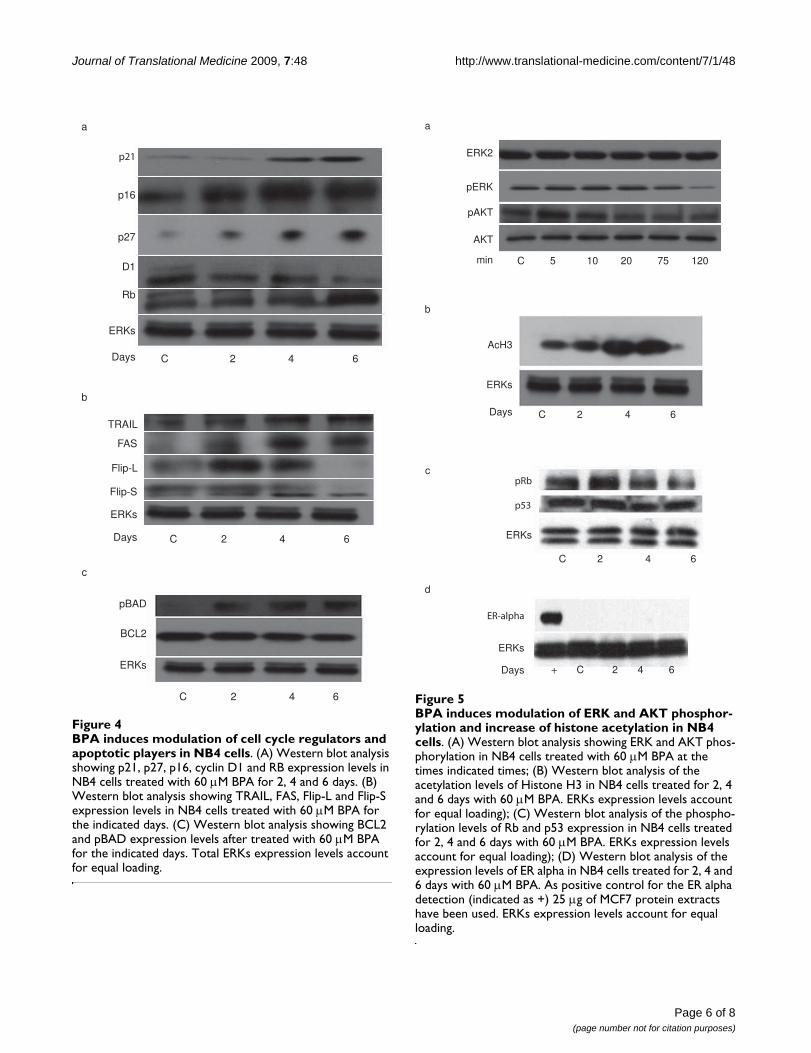
BPA induces modulation of cell cycle regulators and apop-totic players in NB4 cellsFigure 4BPA induces modulation of cell cycle regulators and apoptotic players in NB4 cells. (A) Western blot analysis showing p21, p27, p16, cyclin D1 and RB expression levels in NB4 cells treated with 60 μM BPA for 2, 4 and 6 days. (B) Western blot analysis showing TRAIL, FAS, Flip-L and Flip-S expression levels in NB4 cells treated with 60 μM BPA for the indicated days. (C) Western blot analysis showing BCL2 and pBAD expression levels after treated with 60 μM BPA for the indicated days. Total ERKs expression levels account for equal loading.
p21
p16
p27
D1
Rb
ERKs
C 2 4 6Days
C 2 4 6Days
TRAIL
FAS
Flip-L
Flip-S
ERKs
pBAD
BCL2
ERKs
C 2 4 6
a
b
c
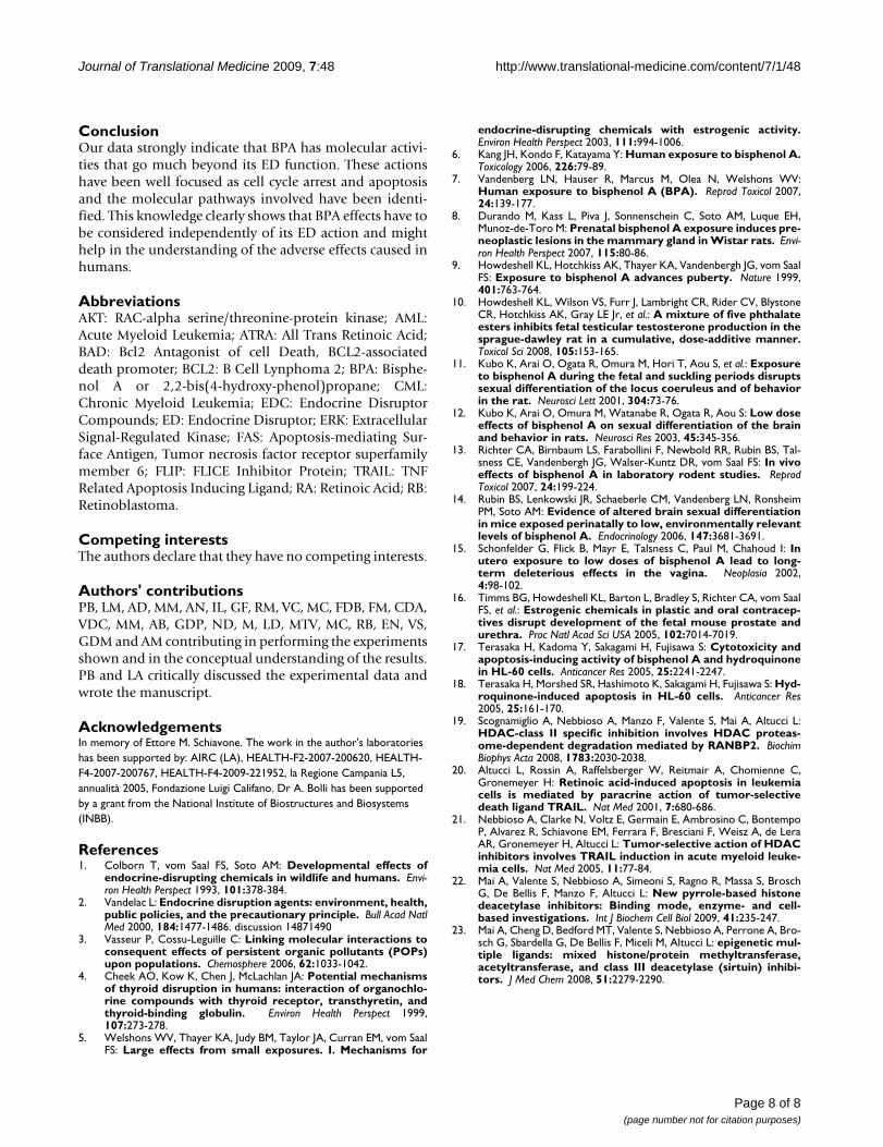
BPA induces modulation of ERK and AKT phosphorylation and increase of histone acetylation in NB4 cellsFigure 5BPA induces modulation of ERK and AKT phosphor-ylation and increase of histone acetylation in NB4 cells. (A) Western blot analysis showing ERK and AKT phos-phorylation in NB4 cells treated with 60 μM BPA at the times indicated times; (B) Western blot analysis of the acetylation levels of Histone H3 in NB4 cells treated for 2, 4 and 6 days with 60 μM BPA. ERKs expression levels account for equal loading); (C) Western blot analysis of the phospho-rylation levels of Rb and p53 expression in NB4 cells treated for 2, 4 and 6 days with 60 μM BPA. ERKs expression levels account for equal loading); (D) Western blot analysis of the expression levels of ER alpha in NB4 cells treated for 2, 4 and 6 days with 60 μM BPA. As positive control for the ER alpha detection (indicated as +) 25 μg of MCF7 protein extracts have been used. ERKs expression levels account for equal loading.
ERK2
pERK
pAKT
AKT
C 5 10 20 75 120min
AcH3
ERKs
C 2 4 6Days
a
b
+ C 2 4 6Days
ERKs
ERKs
c
d
pRb
p53
ER-alpha
C 2 4 6

Journal of Translational Medicine 2009, 7:48 http://www.translational-medicine.com/content/7/1/48
at least as time scale. As expected, caspase 37, which aredownstream of caspase 8 and 9, resulted activated bymedium (60) and high doses (100) of BPA.
BPA induces modulation of cell cycle regulators and apoptotic players in NB4 cellsThat BPA influenced both cell cycle progression and apop-tosis of acute myeloid leukemias has been clarified bythese results. To understand which molecular eventsunderlie to these effects, we have tested its action onknown cell cycle regulators in NB4 cells in a time depend-ent manner. As shown in Fig 4A, p21, p27 and p16together with RB are up-regulated by BPA at the 60 μMdose, whereas cyclin D1 which is known to modulate pro-liferation gets decreased. This scenario is reminiscent of acell cycle block regulated at the molecular level. At thesame time, checking for apoptotic key players we foundthat both FAS and TRAIL are up-regulated already at day 2of treatment, while Flip-L is transiently up-regulated andthen down-regulated, whereas Flip-S is down regulated(Fig. 4B). At the mitochondria cell death level, we couldnot find modulation of BCL2, but we could see increasedphosphorylation of BAD (Fig. 4C) thus confirming thatboth pathways (extrinsic and intrinsic) gets activated byBPA in NB4 cells.
BPA induces modulation of ERK, AKT and Rb phosphorylation and increase of histone acetylation in NB4 cellsTo better focus the activity of BPA in acute myeloid leuke-mia models, we decided to check whether BPA can alsomodulate non genomic actions. As shown in Fig. 5, BPAinduce a decrease of ERK, Rb and AKT phosphorylationthus indicating that anti-proliferative actions occur byinduction of non genomic pathways by 60 μM of BPA inNB4 cells. Note that p53 expression levels stayedunchanged (Fig. 5c). In agreement with these findings,histone H3 acetylation is increased upon BPA treatmentsuggesting an effect (direct or indirect) on chromatinaccessibility of BPA (Fig. 5B).
DiscussionThe Endocrine Disrupting Compounds have been subjectof high attention by the scientific community, since con-cerns have been raised about their actions and potentialtoxicities. Among the many chemicals, BPA is used in theassemble of polycarbonate plastic and epoxy resins, usedin impact-resistant safety equipment and baby bottles, asprotective coatings inside metal food containers, and ascomposite and sealant in dentistry. Exposure to BPA isthought to result primarily from ingestion of food con-taining BPA [6,7]. BPA is of concern to environmentalpublic health because of its toxicity. At high doses, BPAdemonstrates estrogen-like effects in experimental ani-mals, but effects independent from its endocrine modu-
lating function have been poorly investigated. Thus, it iscentral to investigate the cyto-toxicity and apoptosis-inducing activities of BPA at the molecular level. The factthat BPA is able to induce effects on cell cycle and apopto-sis in AML models indicates that BPA actions can gobeyond the endocrine interference. This is also demon-strated by the fact that NB4 cells do not display detectablelevels of ER alpha. Thus suggesting that effects of BPA inthis cells are largely ER independent (Fig. 5d). This notionis a key point considering that BPA is industrially used andthat its effects can cumulate. Although the properties seenon granulocytic differentiation are minor when comparedto those of RA, the fact that BPA is used in equipments andbaby bottles makes also these weak effects of significance.Even more interesting is the induction of cell death whichis clearly specifically regulated at the molecular level.Indeed, the fact that three different cell lines respond withapoptosis to BPA treatment and that this effect seems to bedose dependent indicates that this is a general feature ofBPA treatment and that this might be reproduced in manyother cells. These evidences are exciting from several pointof view: if from one side we might consider the inductionof apoptosis as an interesting anti-cancer action, on theother side we have to keep in mind that these effects mightalso be elicited in normal cells in the different compart-ments of the human body and thus might contribute tothe toxicity of BPA. The regulation of caspase-dependentpathways of apoptosis suggests a specific action on theextrinsic and intrinsic pathways of apoptosis which is con-firmed by the clear induction of Fas and TRAIL and by Flipdown regulation in NB4 cells. Even if our data would sup-port a model in which the extrinsic pathway of apoptosisis more active, we do not exclude the importance of themitochondria de-regulation of apoptosis which is indeedconfirmed by caspase 9 activation and BAD phosphoryla-tion. Considering that many clinical treatments targetapoptosis at the present, our data suggest that the contactor the assumption of BPA might increase the effects of aon-going treatment in humans, apart, of course, havingeffects on its own. Finally, the fact that BPA decreases theactivity of ERK and AKT well integrates with its anti-prolif-erative and apoptotic actions suggesting that the cross-talkof different molecular actions contribute to the cell cyclearrest and to the apoptosis in human biological systems.The hyperacetylating effect shown on histone H3 con-firms the property of BPA to modulate the chromatin in amore accessible state thus corroborating the hypothesisthat BPA contributes with a plethora of different effects tothe induction of cell cycle arrest, weak differentiation andapoptosis in a specific and molecularly defined manner. Ifthe hyperacetylation upon BPA treatment is a direct orindirect effect on chromatin, remains to be established.More characterized studies on BPA exposed population inhealthy or unhealthy status will decipher in the future thereal impact of these molecular actions.
Page 7 of 8(page number not for citation purposes)
Journal of Translational Medicine 2009, 7:48 http://www.translational-medicine.com/content/7/1/48
ConclusionOur data strongly indicate that BPA has molecular activi-ties that go much beyond its ED function. These actionshave been well focused as cell cycle arrest and apoptosisand the molecular pathways involved have been identi-fied. This knowledge clearly shows that BPA effects have tobe considered independently of its ED action and mighthelp in the understanding of the adverse effects caused inhumans.
AbbreviationsAKT: RAC-alpha serine/threonine-protein kinase; AML:Acute Myeloid Leukemia; ATRA: All Trans Retinoic Acid;BAD: Bcl2 Antagonist of cell Death, BCL2-associateddeath promoter; BCL2: B Cell Lynphoma 2; BPA: Bisphe-nol A or 2,2-bis(4-hydroxy-phenol)propane; CML:Chronic Myeloid Leukemia; EDC: Endocrine DisruptorCompounds; ED: Endocrine Disruptor; ERK: ExtracellularSignal-Regulated Kinase; FAS: Apoptosis-mediating Sur-face Antigen, Tumor necrosis factor receptor superfamilymember 6; FLIP: FLICE Inhibitor Protein; TRAIL: TNFRelated Apoptosis Inducing Ligand; RA: Retinoic Acid; RB:Retinoblastoma.
Competing interestsThe authors declare that they have no competing interests.
Authors' contributionsPB, LM, AD, MM, AN, IL, GF, RM, VC, MC, FDB, FM, CDA,VDC, MM, AB, GDP, ND, M, LD, MTV, MC, RB, EN, VS,GDM and AM contributing in performing the experimentsshown and in the conceptual understanding of the results.PB and LA critically discussed the experimental data andwrote the manuscript.
AcknowledgementsIn memory of Ettore M. Schiavone. The work in the author's laboratories has been supported by: AIRC (LA), HEALTH-F2-2007-200620, HEALTH-F4-2007-200767, HEALTH-F4-2009-221952, la Regione Campania L5, annualità 2005, Fondazione Luigi Califano. Dr A. Bolli has been supported by a grant from the National Institute of Biostructures and Biosystems (INBB).
References1. Colborn T, vom Saal FS, Soto AM: Developmental effects of
endocrine-disrupting chemicals in wildlife and humans. Envi-ron Health Perspect 1993, 101:378-384.
2. Vandelac L: Endocrine disruption agents: environment, health,public policies, and the precautionary principle. Bull Acad NatlMed 2000, 184:1477-1486. discussion 14871490
3. Vasseur P, Cossu-Leguille C: Linking molecular interactions toconsequent effects of persistent organic pollutants (POPs)upon populations. Chemosphere 2006, 62:1033-1042.
4. Cheek AO, Kow K, Chen J, McLachlan JA: Potential mechanismsof thyroid disruption in humans: interaction of organochlo-rine compounds with thyroid receptor, transthyretin, andthyroid-binding globulin. Environ Health Perspect 1999,107:273-278.
5. Welshons WV, Thayer KA, Judy BM, Taylor JA, Curran EM, vom SaalFS: Large effects from small exposures. I. Mechanisms for
endocrine-disrupting chemicals with estrogenic activity.Environ Health Perspect 2003, 111:994-1006.
6. Kang JH, Kondo F, Katayama Y: Human exposure to bisphenol A.Toxicology 2006, 226:79-89.
7. Vandenberg LN, Hauser R, Marcus M, Olea N, Welshons WV:Human exposure to bisphenol A (BPA). Reprod Toxicol 2007,24:139-177.
8. Durando M, Kass L, Piva J, Sonnenschein C, Soto AM, Luque EH,Munoz-de-Toro M: Prenatal bisphenol A exposure induces pre-neoplastic lesions in the mammary gland in Wistar rats. Envi-ron Health Perspect 2007, 115:80-86.
9. Howdeshell KL, Hotchkiss AK, Thayer KA, Vandenbergh JG, vom SaalFS: Exposure to bisphenol A advances puberty. Nature 1999,401:763-764.
10. Howdeshell KL, Wilson VS, Furr J, Lambright CR, Rider CV, BlystoneCR, Hotchkiss AK, Gray LE Jr, et al.: A mixture of five phthalateesters inhibits fetal testicular testosterone production in thesprague-dawley rat in a cumulative, dose-additive manner.Toxicol Sci 2008, 105:153-165.
11. Kubo K, Arai O, Ogata R, Omura M, Hori T, Aou S, et al.: Exposureto bisphenol A during the fetal and suckling periods disruptssexual differentiation of the locus coeruleus and of behaviorin the rat. Neurosci Lett 2001, 304:73-76.
12. Kubo K, Arai O, Omura M, Watanabe R, Ogata R, Aou S: Low doseeffects of bisphenol A on sexual differentiation of the brainand behavior in rats. Neurosci Res 2003, 45:345-356.
13. Richter CA, Birnbaum LS, Farabollini F, Newbold RR, Rubin BS, Tal-sness CE, Vandenbergh JG, Walser-Kuntz DR, vom Saal FS: In vivoeffects of bisphenol A in laboratory rodent studies. ReprodToxicol 2007, 24:199-224.
14. Rubin BS, Lenkowski JR, Schaeberle CM, Vandenberg LN, RonsheimPM, Soto AM: Evidence of altered brain sexual differentiationin mice exposed perinatally to low, environmentally relevantlevels of bisphenol A. Endocrinology 2006, 147:3681-3691.
15. Schonfelder G, Flick B, Mayr E, Talsness C, Paul M, Chahoud I: Inutero exposure to low doses of bisphenol A lead to long-term deleterious effects in the vagina. Neoplasia 2002,4:98-102.
16. Timms BG, Howdeshell KL, Barton L, Bradley S, Richter CA, vom SaalFS, et al.: Estrogenic chemicals in plastic and oral contracep-tives disrupt development of the fetal mouse prostate andurethra. Proc Natl Acad Sci USA 2005, 102:7014-7019.
17. Terasaka H, Kadoma Y, Sakagami H, Fujisawa S: Cytotoxicity andapoptosis-inducing activity of bisphenol A and hydroquinonein HL-60 cells. Anticancer Res 2005, 25:2241-2247.
18. Terasaka H, Morshed SR, Hashimoto K, Sakagami H, Fujisawa S: Hyd-roquinone-induced apoptosis in HL-60 cells. Anticancer Res2005, 25:161-170.
19. Scognamiglio A, Nebbioso A, Manzo F, Valente S, Mai A, Altucci L:HDAC-class II specific inhibition involves HDAC proteas-ome-dependent degradation mediated by RANBP2. BiochimBiophys Acta 2008, 1783:2030-2038.
20. Altucci L, Rossin A, Raffelsberger W, Reitmair A, Chomienne C,Gronemeyer H: Retinoic acid-induced apoptosis in leukemiacells is mediated by paracrine action of tumor-selectivedeath ligand TRAIL. Nat Med 2001, 7:680-686.
21. Nebbioso A, Clarke N, Voltz E, Germain E, Ambrosino C, BontempoP, Alvarez R, Schiavone EM, Ferrara F, Bresciani F, Weisz A, de LeraAR, Gronemeyer H, Altucci L: Tumor-selective action of HDACinhibitors involves TRAIL induction in acute myeloid leuke-mia cells. Nat Med 2005, 11:77-84.
22. Mai A, Valente S, Nebbioso A, Simeoni S, Ragno R, Massa S, BroschG, De Bellis F, Manzo F, Altucci L: New pyrrole-based histonedeacetylase inhibitors: Binding mode, enzyme- and cell-based investigations. Int J Biochem Cell Biol 2009, 41:235-247.
23. Mai A, Cheng D, Bedford MT, Valente S, Nebbioso A, Perrone A, Bro-sch G, Sbardella G, De Bellis F, Miceli M, Altucci L: epigenetic mul-tiple ligands: mixed histone/protein methyltransferase,acetyltransferase, and class III deacetylase (sirtuin) inhibi-tors. J Med Chem 2008, 51:2279-2290.
Page 8 of 8(page number not for citation purposes)
http://www.ncbi.nlm.nih.gov/entrez/query.fcgi?cmd=Retrieve&db=PubMed&dopt=Abstract&list_uids=8080506