molecular aggregation of thiols and alcohols: study of
150
PROGRAMA DE DOCTORADO EN FÍSICA DOCTORAL THESIS/TESIS DOCTORAL: MOLECULAR AGGREGATION OF THIOLS AND ALCOHOLS: STUDY OF NON-COVALENT INTERACTIONS BY MICROWAVE SPECTROSCOPY Presented by/Presentada por Marcos Juanes San José in fulfillment of the Degree of/ para optar al Grado de Doctor by the/por la Universidad de Valladolid Supervised by/ Dirigida por: Dr. Alberto Lesarri Gómez Dra. Ruth Pinacho Gómez
-
Upload
khangminh22 -
Category
Documents
-
view
2 -
download
0
Transcript of molecular aggregation of thiols and alcohols: study of
PROGRAMA DE DOCTORADO EN FÍSICA
DOCTORAL THESIS/TESIS DOCTORAL:
MOLECULAR AGGREGATION OF THIOLS AND ALCOHOLS: STUDY OF NON-COVALENT INTERACTIONS BY
MICROWAVE SPECTROSCOPY
Presented by/Presentada por Marcos Juanes San José
in fulfillment of the Degree of/ para optar al Grado de Doctor
by the/por la Universidad de Valladolid
Supervised by/ Dirigida por: Dr. Alberto Lesarri Gómez Dra. Ruth Pinacho Gómez

Preface
We /ˈwi:/ (pronoun): used by a speaker to refer to himself or herself and one or more other
people considered together.
Sometimes it is too difficult to take into account all the people who have been involved in a
deep and personal study. Avoiding forgetting someone (and, above all, avoiding to
underestimate all the support provided by everyone around me), from now on I feel the need
to use the pronoun “We” in the Thesis presented below with the aim of making reference to all
these people present in my life who have made this work posible, including you (maybe the
most important part of these lines) as a reader.

CONTENTS
i
Table of Contents
Preface
Table of Contents ......................................................................................................... i
Abstract ........................................................................................................................ v
Resumen .................................................................................................................... vii
Acknowledgements ................................................................................................... ix
Chapter I. INTRODUCTION ............................................................................................. I.1
I. Non-covalent interactions ................................................................................................. I.3
I.1. Non-covalent interactions. The Hydrogen Bond ................................................................. I.5
I.1.1. Hydrogen Bonds involving Oxygen: The O-H···O bond .......................................... I.8
I.1.2. Hydrogen Bonds involving Sulfur ...............................................................................I.10
I.2. Gas-Phase Rotational Investigations of NCIs ..................................................................I.11
II. Methods ...............................................................................................................................I.13
II.1.Rotational spectroscopy ........................................................................................................I.13
II.1.1. General aspects ............................................................................................................I.13
II.1.2. Theory od molecular rotation .....................................................................................I.14
II.1.3. Balle-Flygare FTMW spectrometer @ UVa ..........................................................I.17
II.1.4. Chiped-Pulse FT-MW spectrometer @ UVa .........................................................I.20
II.2. Computational Methods ......................................................................................................I.22
II.2.1. Density Functional Theory and Ab initio calculations ...........................................I.23
II.2.2. Complementary computational tools .......................................................................I.24
ii
Chapter II. HYDRATION OF CYCLOHEXANOL:
(Cyclohexanol) and (Cyclohexanol··· H2O) .................................................................. II.1
I. Introduction .......................................................................................................................... II.3
II. Cyclohexanol and Cyclohexanol ··· H2O adduct .................................................... II.5
II.1. Experimental set up ............................................................................................................... II.5
II.2. Rotational spectra of CHO .................................................................................................. II.5
II.3. Theoretical calculations ......................................................................................................... II.9
II.4. Molecular Struct. and Potential energy functions of the OH internal rotation ........II.10
II.5. Water dimer geometry and dynamics ...........................................................................II.12
II.6. Discussion and Conclusions ................................................................................................II.13
Chapter III. DIMERIZATION OF CYCLOHEXANOL:
(Cyclohexanol)2 ............................................................................................................ III.1
I. Introduction ......................................................................................................................... III.3
II. Cyclohexanol homodimer .............................................................................................. III.4
II.1. Experimental set up .............................................................................................................. III.4
II.2. About Chirality ....................................................................................................................... III.4
II.3. Rotational spectra of (Cyclohexanol)2 .............................................................................. III.6
II.4. Dispersion model ................................................................................................................... III.7
II.5. Non-covalent interactions ..................................................................................................III.10
Chapter IV. HYDRATION OF FURFURYL COMPOUNDS:
(Furfuryl Mercaptan ··· H2O) and (Furfuryl Alcohol ··· H2O) ..................................... IV.1
I. Introduction ........................................................................................................................ IV.3
II. Furfuryl Mercaptan and Furfuryl alcohol ................................................................ IV.5
II.1. Experimental ........................................................................................................................ IV.5
II.2. Computational methods ..................................................................................................... IV.5
II.3. Rotational spectra of FM and FA .................................................................................... IV.6
II.4. Rotational spectra of the of the FM and FA monohydrates ....................................... IV.7
II.5. Discussion ............................................................................................................................ IV.12

iii
Chapter V. HYDRATION OF THENYL COMPOUNDS:
(Thenyl Mercaptan ··· H2O) and (Thenyl Alcohol ··· H2O) ......................................... V.1
I. Introduction ......................................................................................................................... V.3
II. Thenyl Mercaptan and Thenyl alcohol ...................................................................... V.5
II.1. Experimental set up .............................................................................................................. V.5
II.2. Rotational spectra of TM and TA ..................................................................................... V.6
II.3. Rotational spectra of the monohydrates .......................................................................... V.7
II.4. Molecular structures ........................................................................................................... V.11
II.5. Non-covalent interactions .................................................................................................. V.13
II.6. Conclusion ............................................................................................................................. V.15
Chapter VI. DIMERIZATION OF THENYL AND FURFURYL COMPOUNDS:
(Furfuryl Alcohol)2 and (Thenyl Alcohol)2 ................................................................ VI.1
I. Introduction ........................................................................................................................ VI.3
II. Furfuryl alcohol and Thenyl alcohol homodimers ................................................... VI.5
II.1. Experimental set up ............................................................................................................. VI.5
II.2. Computational methods ..................................................................................................... VI.5
II.3. Conformational landscape ................................................................................................. VI.5
II.4. Rotational Spectrum ......................................................................................................... VI.10
II.5. Non-covalent interactions ................................................................................................. VI.13
II.6. Conclusion ............................................................................................................................ VI.15
Chapter VII. COMPLEMENTARY STUDIES .................................................................... V.1
I. Complementary Studies ............................................................................................... VII.3
I.1. Equilibrium structures of fluorocyclohexane .................................................................. VII.5
I.2. Accurate equilibrium structure of the ethynylcyclohexane ......................................... VII.6
I.3. Intramolecular hydrogen bonding in 1,2-butanedithiol .............................................. VII.7
I.4. Intramolecular hydrogen bonding in 2,3-butanedithiol .............................................. VII.8
I.5. Observation of trans - Matrine and cis - Matrine ........................................................ VII.9
I.5. Perspectives for Rorational Investigations of NCI ...................................................... VII.10

iv
v
Abstract
The study and understanding of non-covalent interactions at molecular level is a field in continuous development and essential to determine the structural behavior of many molecules of chemical, technological or biological interest. In this PhD thesis, the intermolecular interactions involved in the formation of neutral molecular aggregates, both dimers and microsolvation products, have been analyzed in the gas phase. The intermolecular complexes were generated by pulsed supersonic expansions, and later characterized by rotational spectroscopy. This work has used two spectroscopic techniques, including a Balle-Flygare Fourier-Transform Microwave (FTMW) spectrometer in the 8-20 GHz frequency range, and a broadband Chirped-Pulse Fourier Transform Microwave (CP-FTMW) spectrometer covering the 2-8GHz spectral range. The intermolecular complexes studied have included molecules with alcohol and / or thiol groups, in order to analyze the differences between the intermolecular interactions involving oxygen or sulfur atoms, especially hydrogen bonds. Molecules that comprise both aliphatic (cyclohexanol) and aromatic (furfuryl alcohol, furfuryl mercaptan, thenyl alcohol, thenyl mercaptan) ring systems have been studied. The analyzed hydrogen bonds included especially O-H···O, O-H···S and S-H···S interactions. The formation of intermolecular complexes has revealed a great conformational diversity in some of them, such as the observation of six isomers of the cyclohexanol dimer. With regard to the monohydrates, tunnelling splittings associated with internal large amplitude motions have been observed in some cases, such as the rotation of the water molecule in the monohydrates of cyclohexanol, thenyl alcohol and thenyl mercaptan. In the case of chiral molecules, dimerization has made it possible to observe the relative stability of homo- or heterochiral diastereoisomers. The experimental study has been supported by different theoretical molecular orbital calculations, in particular Density Functional Theory (DFT) calculations, in order to characterize the interactions structurally, energetically and by a topological analysis of electron density. The set of experimental and theoretical data will advance the existing information on hydrogen bonds involving sulfur atoms, generally scarcely studied, and their comparison with the oxygenated analogues.

vi
vii
Resumen
El estudio y comprensión de las interacciones no covalentes a nivel molecular es un campo que está en continuo desarrollo y cobra vital importancia para determinar el comportamiento estructural de muchas moléculas de interés químico, tecnológico o biológico. En esta tésis doctoral se han analizado las interacciones intermoleculares implicadas en la formación de agregados moleculares neutros, tanto dímeros como productos de microsolvatación, en fase gas. Los complejos intermoleculares se han generado mediante expansiones supersónicas pulsadas, caracterizándose posteriormente mediante espectroscopía de rotación. Este trabajo ha utilizado dos técnicas espectroscópicas, incluyendo un espectrómetro de microondas con transformada de Fourier (FTMW) de tipo Balle-Flygare en el rango de frecuencias 8-20 GHz, y un espectrómetro de transformada de Fourier de banda ancha con excitación multifrecuencia (CP-FTMW) cubriendo el rango espectral de 2-8 GHz. Los complejos intermoleculares estudiados han incluido moléculas con grupos alcohol y/o tiol, con objeto de analizar las diferencias entre las interacciones intermoleculares que implican átomos de oxígeno o azufre, en especial el enlace de hidrógeno. Se han estudiado moléculas incluyendo tanto sistemas cíclicos alifáticos (ciclohexanol, ciclohexanotiol) como aromáticos (furfuril alcohol, furfuril mercaptano, tienil alcohol, tienil mercaptano). Los enlaces de hidrógeno analizados han comprendido especialmente interacciones de tipo O-H···O, O-H···S y S-H···S. La formación de los complejos intermoleculares ha revelado en algunos de ellos una gran variedad conformacional, como la observación de seis isómeros del dímero de ciclohexanol. En el caso de los monohidratos se han observado en algunos casos desdoblamientos asociados a movimientos internos de gran amplitud, como la rotación de la molécula de agua en los monohidratos de ciclohexanol y tienil mercaptano. En los casos de moléculas quirales la dimerización ha permitido observar la estabilidad relativa de los diastereoisómeros homo o heteroquirales. El estudio experimental se ha completado con diferentes cálculos teóricos de orbitales moleculares, en especial teoría del funcional de la densidad, a fin de caracterizar las interacciones estructuralmente, energéticamente y mediante análisis topológico de la densidad electrónica. El conjunto de datos experimentales y teóricos permite aumentar la información existente sobre enlaces de hidrógeno con átomos de azufre, generalmente poco estudiados, y su comparación con los análogos oxigenados.

viii
ix
Acknowledgements
By respect, honesty and proud, before starting I would like (and I feel it necessary) to highlight that all the time, effort and enthusiasm invested into this work is a direct response of the kindness received and felt since I was welcomed at the Alberto Lesarri’s laboratory and at the electronics department of the ETSIT-UVa (more than four years ago since I decided to start this adventure). This gratitude specifically corresponds to Lourdes, José Emiliano, Martín (without their support most of the theoretical calculations carried out in the following studies presented in this thesis would nothave been possible), to Ruth Pinacho for always being ready to help (both scientifically and personally) at any time and to Alberto Lesarri for showing me that always working with a smile on your face is much more than rewarding. I also want to thank the good treatment and help I have received from all the people who have made it possible to carry out different research periods in laboratories outside my country at the Leibniz Universität Hannover and at the Universität zu Köln (both in Germany) during my PhD period.
When a human being starts a new episode in his/her/its life, it is fair to keep on mind that each lived experience would not be possible without the support of the family. The concept family, from my point of view, is one of the most difficult terms to be defined:
Following a rigorous grammar definition, there is always a blood component that genetically creates a biological link to others who are generally considered family members. Personally, going a couple of steps further, what I understand (specially, what I perceive) as family is the selected set of people who have accompanied me, who accompany me or who will accompany me in the path of my life. I would like to thank strongly the support received from my parents during these 30 years (honestly, without their help, none of the words written in this Thesis would be possible): thank you very much Juan and Sole. Thanks to my brother who have always been a reference in my life (both academically and familiarly). Thanks to Quique, Saul and Cristian who made me understand how important friendship was at the university. Thanks to the mother earth and its nature for allowing me to enjoy its mountains while climbling and hiking every weekend. Thanks to all my Friends (who I consider closer than brothers) for doing everything easier at every time and always being by my side. And, to finish, thank two human beings who (unexpectedly) have become an essential part of my life for giving me the opportunity to live this incredible experience with the feeling of always feeling supported and loved: thank you very much for everything to Alberto Lesarri and Rizalina T. Saragi.
Marcos Juanes San José


Siempre en mi recuerdo, a mi tío Alberto Juanes


Chapter I INTRODUCTION

I.2
INTRODUCTION
I.3
I. Non-covalent interactions
This Thesis contains a molecular study of non-covalent interactions (NCIs) in neutral intermolecular complexes using rotational spectroscopy.
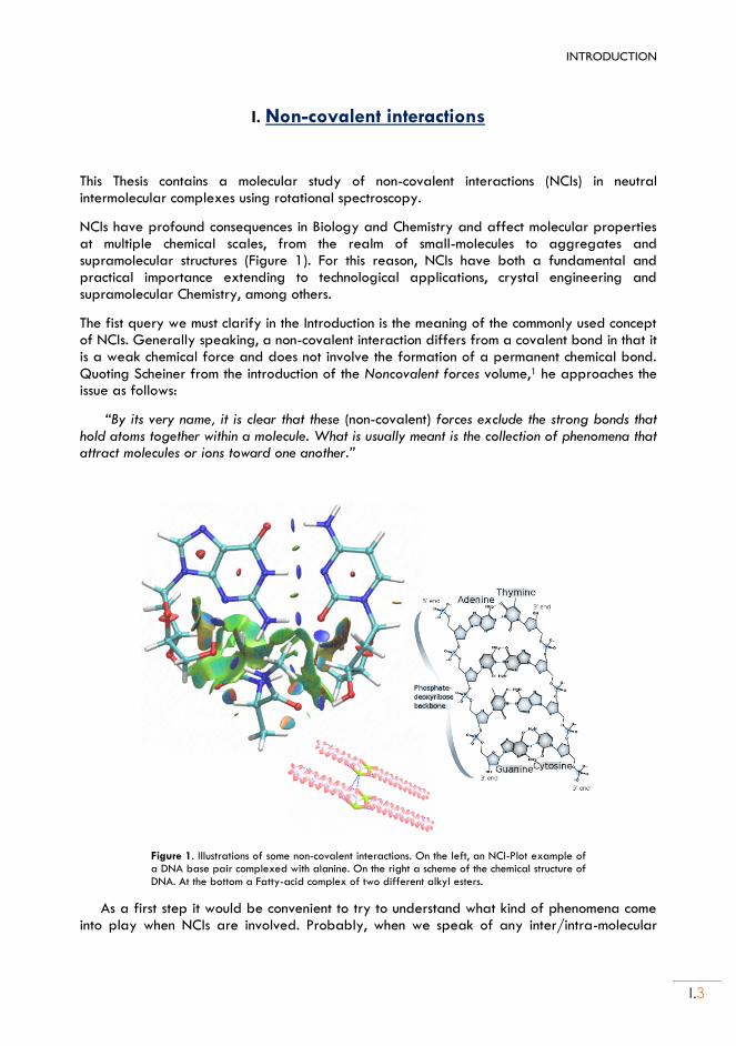
NCIs have profound consequences in Biology and Chemistry and affect molecular properties at multiple chemical scales, from the realm of small-molecules to aggregates and supramolecular structures (Figure 1). For this reason, NCIs have both a fundamental and practical importance extending to technological applications, crystal engineering and supramolecular Chemistry, among others.
The fist query we must clarify in the Introduction is the meaning of the commonly used concept of NCIs. Generally speaking, a non-covalent interaction differs from a covalent bond in that it is a weak chemical force and does not involve the formation of a permanent chemical bond. Quoting Scheiner from the introduction of the Noncovalent forces volume,1 he approaches the issue as follows:
“By its very name, it is clear that these (non-covalent) forces exclude the strong bonds that hold atoms together within a molecule. What is usually meant is the collection of phenomena that attract molecules or ions toward one another.”
Figure 1. Illustrations of some non-covalent interactions. On the left, an NCI-Plot example of a DNA base pair complexed with alanine. On the right a scheme of the chemical structure of DNA. At the bottom a Fatty-acid complex of two different alkyl esters.
As a first step it would be convenient to try to understand what kind of phenomena come into play when NCIs are involved. Probably, when we speak of any inter/intra-molecular
I.4
interaction, the main thought is centered on how “strong” is that interaction. Of course, the “strength” of any chemical-physical interaction is directly connected with how powerful and how stable are those forces, but a better understanding of these non-covalent forces is not that simple and it is also necessary to consider and evaluate the electronic effects, binding preferences, the geometry, the possible solvent or matrix effects, the probability of being formed, how this interaction occurs, and so on. In natural sciences non-covalent intermolecular interactions manifest in the properties and functions of matter under all conditions, from solid-state materials to biological systems (for instance, even at large scales, the Van der Waals dispersion inherent to NCIs has been proposed as an important force behind the formation of the rings around Saturn2 or as the force that enables geckos to stick to walls3).
Non-covalent interactions play a fundamental role in layered materials, and sustain molecular aggregation in biological systems, large molecular complexes and technological processes with a strikingly diverse array of interaction energies and geometries.1,4,5 Diversity arises not only by the multiple combinatorial possibilities of different atomic groups, but also because of the fuzzy character of NCIs, which, quite contrary to covalent bonds, admit broad and diffuse structural and energetic limits. Moreover, in the last decade our vision of non-covalent forces has expanded considerably, moving from the conventional hydrogen bond4 (HB) to a whole new palette of previously unanticipated donor-acceptor interactions.1,5 First, the now centenary HB was redefined by IUPAC in 20116,7 to accommodate weak interactions like aliphatic donors8 (C-H···O, C-H···N, etc.), acceptors9 (C-H···, N-H···, etc.), low-electronegativity donors10 (S-H, P-H, etc.) or H acceptors,11 among others. Then, further developments in crystal engineering5 catalyzed the IUPAC move to define the halogen bond12 (XB) in 2013. In the XB the anisotropic electron distribution within halogen atoms makes terminal -hole13 or lateral -hole14 regions new electrophilic attractors, turning common electron donors into acceptors.15,16 The electrostatic argument was later generalized17,18 to explain experimental observations into the new conceptual frames of chalcogen bonds (CB),19 pnictogen bonds20,21 (NB) or tetrel bonds22,23 (TB), involving atoms of groups 14, 15, 1624 or even coinage-metals25 in R-A···B bridging interactions formally analogue to the HB or XB (R-H/X···B). Legon has noticed that rotational26–28 and vibrational29,30 experiments existed for the new bonds long before they had a name, occasionally being called “anti-hydrogen bond”.31 This extension of the chemical discourse makes NCI more diverse and attractive than ever.
The understanding of non-covalent interactions has been gained from decades of experimental observations supplemented by theoretical predictions.32 To make a proper analysis of the NCIs it would be necessary more time and space than a single PhD thesis and, even so, the subject would not be concluded.33 In order to make sense and try to easily introduce results presented in later chapters, we will focus our introduction on non-covalent forces on those produced by the hydrogen bonds, in particular those containing sulfur and/or oxygen.
1. Non-covalent interactions. The Hydrogen Bond
The HB is a donor-acceptor attractive directional bridging interaction specifically involving hydrogen atoms. Condensing the large volume of literature that the HB entails, we can start from the classical concepts proposed by Pauling in The Nature of the Chemical Bond34 (1939) and established by Pimentel and McClellan35 (1960),
INTRODUCTION
I.5
“A hydrogen bond [A−H···B] exists between a functional group A−H and an atom or a group of atoms B in the same or a different molecule when: (a) there is evidence of bond formation (association or chelation) and (b) there is evidence
that this new bond linking A−H and B specifically involves the hydrogen atom already bonded to A.”
This description was extended by Vinogradov and Linnell (1971),36
“Hydrogen bonding occurs between a proton-donor group A−H and a proton-acceptor group B, where A is an electronegative atom, O, N, S, X (F, Cl, Br, I) or C, and the acceptor group is a lone pair of an electronegative atom or a π bond of a multiple bond (unsaturated) system. Generally, a H-bond can be characterized as a proton shared by two lone electron pairs.”
and was summarized in the IUPAC definition of 1997:
“form of association between an electronegative atom and a hydrogen atom attached to a second, relatively electronegative atom. It is best considered as an electrostatic interaction, heightened by the small size of hydrogen, which permits proximity of the interacting dipoles or charges...”, where “associated energies are less than 21–25 kJ mol-1”.
By 2010 new experimental and theoretical advances required an extension of these concepts to much weaker, broader and diverse (2-170 kJ mol-1) interactions, including atoms different from the first-row, as exposed in the most recent IUPAC’s definition of 2011,7
“The hydrogen bond is an attractive interaction between a hydrogen atom from a molecule or a molecular fragment X–H in which X is more electronegative than H, and an atom or a group of atoms in the same or a different molecule, in which there is evidence of bond formation.”
As seen from the above definitions, to simplify a complex phenomenon like the concept of HB is complicated. We can essentially state that the main feature that characterizes the HB and differs from another donor-acceptor interactions is the sharing of the hydrogen atom between the hydrogen bond donor and acceptor and some form of electron sharing with dominant electrostatic character. That is, we can see this non-covalent force as a shared-proton interaction (on the other hand, the inverse argument may not be true and not all shared-proton interactions necessarily conform a HB). These types of interactions can occur intramolecularly (if the donor and acceptor groups are on the same molecule) and/or intermolecularly (when they are on different molecules).
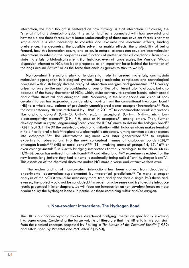
Table 1. Pauling’s dimensionless electronegativities () for the elements of the upper-right corner of the Periodic Table.
H 2.20
B 2.04 C 2.55 N 3.04 O 3.44 F 3.98
Si 1.90 P 2.19 S 2.58 Cl 3.16
Ge 2.01 As 2.18 Se 2.55 Br 2.96
Sb 2.05 Te 2.1 I 2.66
I.6
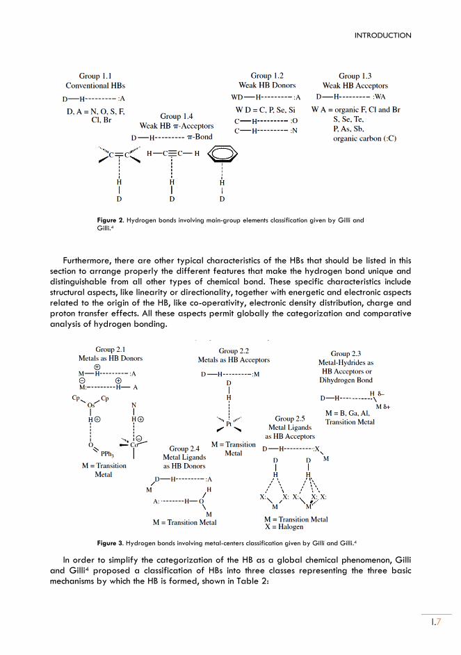
From the earliest studies, the donor/acceptor electronegativities are one of the main determinants of the hydrogen bond strength. The studies on hydrogen bonding initiated by Pauling and the first definitions involved only proton donors with atoms of large electronegativity, like those of the first row34 in Table 1. The palette of atoms that can be considered part of the hydrogen bonding phenomenon has been increasing with time, now achieving a much wider range of possible donors and atoms of low electronegativity (Figures 2 and 3). In parallel, the number of proton acceptor groups has increased. Several reports discussing the donor-acceptor pairs involved in the hydrogen bond interaction can be found in the literature.1,4,5,37,38
A chemical classification of the HB according to The Nature of the Hydrogen Bond of Gilli and Gilli4 can be seen in Figures 2 and 3. The HBs which include in donor/acceptor roles the most electronegative atoms ( > 3.0) like those of the first row (N, O, F) and some halogens (Cl, Br), are classified as “conventional” HBs and, due to their large electronegativities, they are also the ones that can form the stronger hydrogen bonds in neutral molecules. Conventional HBs include most of the interactions analyzed since the beginning of HB studies, comprise a large volume of experimental and theoretical information and often play a determining role in Chemistry, Biology and Biochemistry. In this Thesis we have analyzed in particular several conventional HBs including the oxygen atom, like those involving water, alcohols or heterocyclic aromatic rings. For this reason, section 1.1 will be dedicated to this kind of HBs.
HBs including atoms of lower electronegativity like C or the second-row atoms P and S are generally classified as weak donors or acceptors, and the information available is scarce compared to the conventional HBs. In particular, the HBs involving sulfur as part of the proton donor group or as acceptor has been much less studied that the conventional ones, despite the fact that they display the full spectrum of HBs energies and geometries, from strong, short-linear and proton-centered bonds of covalent nature to weak, long, bent and proton out- centered electrostatic ones. This problem called our attention since the beginning of the Thesis and motivated a series of comparative studies of HBs involving the sulfur atom, mostly in thiol, sulfide, disulfide and aromatic ring molecules.
For this reason, some characteristics and peculiarities of HBs including sulfur-centered atoms will be presented in Section 1.2.
There are other structural and energetic factors to consider when making a consistent HB classification. Concerning energetics, the hydrogen bonds can be categorized as strong bonds (60-160 kJ mol-1), moderate bonds (16-60 kJ mol-1) or weak bonds (2-16 kJ mol-1) attending
to their binding energy.a The main difference between the first two categories is that the strong HBs are groups in which there are permanent electric charges or an important deficiency or excess of electron density in the donor or acceptor groups, while the moderate HBs are formed by neutral donor and acceptor groups, typically with electronegative atoms and lone-pair unshared electrons in the acceptor. As mentioned before, this last category includes the most common or normal HBs in Chemistry and nature because they are essential components of biological molecules. The concept of weak hydrogen bonds includes many other groups with small binding energies, characteristically close to the van der Waals interactions.
a Binding energy refers to the complexation energy relative to the conformations of the monomers in the lowest
energy configuration. Complexation energy refers to the monomers in the geometry of the complex.
INTRODUCTION
I.7
Figure 2. Hydrogen bonds involving main-group elements classification given by Gilli and Gilli.4
Furthermore, there are other typical characteristics of the HBs that should be listed in this section to arrange properly the different features that make the hydrogen bond unique and distinguishable from all other types of chemical bond. These specific characteristics include structural aspects, like linearity or directionality, together with energetic and electronic aspects related to the origin of the HB, like co-operativity, electronic density distribution, charge and proton transfer effects. All these aspects permit globally the categorization and comparative analysis of hydrogen bonding.
Figure 3. Hydrogen bonds involving metal-centers classification given by Gilli and Gilli.4
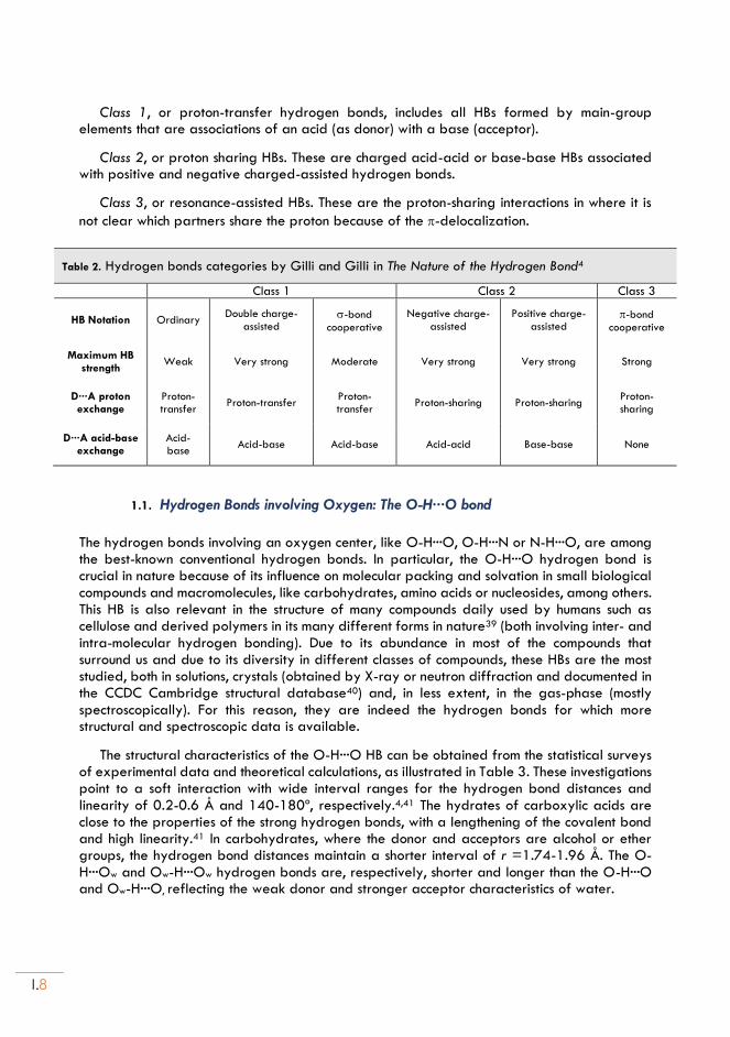
In order to simplify the categorization of the HB as a global chemical phenomenon, Gilli and Gilli4 proposed a classification of HBs into three classes representing the three basic mechanisms by which the HB is formed, shown in Table 2:
I.8
Class 1, or proton-transfer hydrogen bonds, includes all HBs formed by main-group elements that are associations of an acid (as donor) with a base (acceptor).
Class 2, or proton sharing HBs. These are charged acid-acid or base-base HBs associated with positive and negative charged-assisted hydrogen bonds.
Class 3, or resonance-assisted HBs. These are the proton-sharing interactions in where it is
not clear which partners share the proton because of the -delocalization.
Table 2. Hydrogen bonds categories by Gilli and Gilli in The Nature of the Hydrogen Bond4
Class 1 Class 2 Class 3
HB Notation Ordinary Double charge-
assisted -bond
cooperative
Negative charge-assisted
Positive charge-assisted
-bond cooperative
Maximum HB strength
Weak Very strong Moderate Very strong Very strong Strong
D···A proton exchange
Proton-transfer
Proton-transfer Proton-transfer
Proton-sharing Proton-sharing Proton-sharing
D···A acid-base exchange
Acid-base
Acid-base Acid-base Acid-acid Base-base None
1.1. Hydrogen Bonds involving Oxygen: The O-H···O bond
The hydrogen bonds involving an oxygen center, like O-H···O, O-H···N or N-H···O, are among the best-known conventional hydrogen bonds. In particular, the O-H···O hydrogen bond is crucial in nature because of its influence on molecular packing and solvation in small biological compounds and macromolecules, like carbohydrates, amino acids or nucleosides, among others. This HB is also relevant in the structure of many compounds daily used by humans such as cellulose and derived polymers in its many different forms in nature39 (both involving inter- and intra-molecular hydrogen bonding). Due to its abundance in most of the compounds that surround us and due to its diversity in different classes of compounds, these HBs are the most studied, both in solutions, crystals (obtained by X-ray or neutron diffraction and documented in the CCDC Cambridge structural database40) and, in less extent, in the gas-phase (mostly spectroscopically). For this reason, they are indeed the hydrogen bonds for which more structural and spectroscopic data is available.
The structural characteristics of the O-H···O HB can be obtained from the statistical surveys of experimental data and theoretical calculations, as illustrated in Table 3. These investigations point to a soft interaction with wide interval ranges for the hydrogen bond distances and linearity of 0.2-0.6 Å and 140-180º, respectively.4,41 The hydrates of carboxylic acids are close to the properties of the strong hydrogen bonds, with a lengthening of the covalent bond and high linearity.41 In carbohydrates, where the donor and acceptors are alcohol or ether groups, the hydrogen bond distances maintain a shorter interval of r =1.74-1.96 Å. The O- H···Ow and Ow-H···Ow hydrogen bonds are, respectively, shorter and longer than the O-H···O and Ow-H···O, reflecting the weak donor and stronger acceptor characteristics of water.
INTRODUCTION
I.9
Table 3. O-H···O Bond Lengths (Å) from different classes of small molecules.
Carboxylic
acids Amino acids
Carbohydrates Inorganic
salt hydrates
Purines and pyrimidines
Organic hydrates
Nucleosides and
nucleotides
Number
of data[a] 26 26 255 296 66 46 322
min 1.40 1.44 1.74 1.74 1.60 1.60 1.55
max 2.01 2.06 1.96 2.26 2.46 2.25 2.18
mean 1.71 1.74 1.82 1.82 1.83 1.90 1.92
[a]Data based on neutron diffraction analyses (pag. 61 ‘An introduction to Hydrogen Bonding’, 1997, Jeffrey)41.
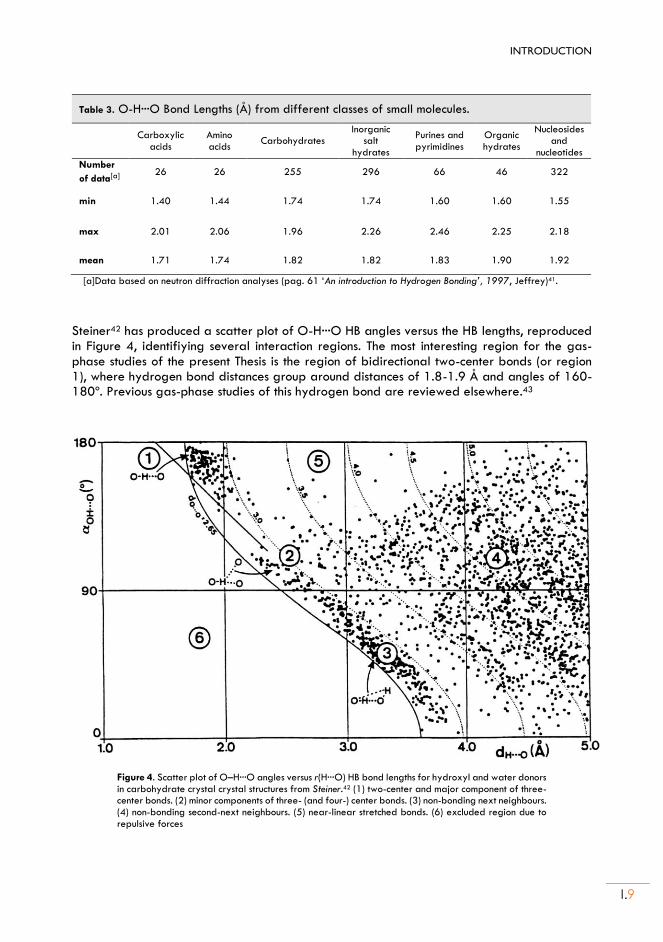
Steiner42 has produced a scatter plot of O-H···O HB angles versus the HB lengths, reproduced in Figure 4, identifiying several interaction regions. The most interesting region for the gas-phase studies of the present Thesis is the region of bidirectional two-center bonds (or region 1), where hydrogen bond distances group around distances of 1.8-1.9 Å and angles of 160-180º. Previous gas-phase studies of this hydrogen bond are reviewed elsewhere.43
Figure 4. Scatter plot of O–H···O angles versus r(H···O) HB bond lengths for hydroxyl and water donors in carbohydrate crystal crystal structures from Steiner.42 (1) two-center and major component of three-center bonds. (2) minor components of three- (and four-) center bonds. (3) non-bonding next neighbours. (4) non-bonding second-next neighbours. (5) near-linear stretched bonds. (6) excluded region due to repulsive forces
I.10
Recent computational predictions have also contributed to the understanding of the physical characteristics of the HB, in particular, the magnitude of the electrostatic contributions.44–47 This characteristic has been useful in several studies48–50 to describe the proton affinity as a hydrogen bond descriptor for the O-H···O hydrogen bonded systems while, on the other hand, there are some cases where the dispersive energy contribution increases notably (together with the increase of the alkyl chain length of the HB acceptors) as in the case of tyrosine and methionine intermolecular complexes with para-Cresol (see Biswal et al. review)51.
Concerning the data presented in this Thesis, the structural influence of the moderately
strong O−H···O hydrogen bond has been observed both in the monomers, monohydrates and homodimers of several molecular systems including aromatic and saturated five- and six-membered rings like cyclohexanol, furfuryl alcohol and thenyl alcohol. More specifically, cyclohexanol ··· H2O and the homodimer (cyclohexanol)2 in Chapters II and III, furfuryl alcohol ··· H2O in Chapter IV, thenyl alcohol ··· H2O in Chapter V, and the homodimers of (furfuryl alcohol) 2 and (thenyl alcohol) 2 in Chapter VI.
1.2. Hydrogen Bond involving Sulfur.
In his book The Hydrogen Bond,35 Pimentel mentioned the thiol group as a possible HB donor,
with the S−H stretching mode broadening, shifting to lower vibrational frequency and becoming much more intense. However, HBs to sulfur centers have been conventionally dismissed as weak interactions of dispersive character, with reduced structural influence compared to conventional first-row HBs like O-H···O, O-H···N or N-H···O.35,37,52 More recently, Biswal51 and Wategaonkar44 have reviewed sulfur HBs, concluding that they are multifaceted interactions
with several points of interest: 1) Sulfur forms and HBs as donor (S-H···O, S-H···S, S-H···, etc) and acceptor (O-H···S, N-H···S, etc), 2) Sulfur HBs can be as strong as conventional HBs, 3) Sulfur HBs may display considerable electrostatic character and 4) Sulfur HBs influence structure and function of many proteins and organic crystals.
Despite these posibilities for sulfur-centered interactions the number of studies on HBs
involving sulfur is still much lower than others involving more electronegative atoms. For this
reason and with the aim of better understanding the properties of the diverse HBs interactions,
the interest in sulfur (and other atoms in groups 14–16) has grown quantitatively in recent
years. As a consequence, molecular studies are justified to contribute information on the
structure, physical nature and balance of electrostatic and dispersive forces in NCIs involving
sulfur centers. In particular, the observation of weakly-bound intermolecular clusters in the gas
phase simultaneously provides empirical data unbiased by crystal or matrix effects and
validation of theoretical models. Vibrational evidence of sulfur hydrogen bonding generally
originates from IR,53,54 and double-resonance (UV–UV or UV–IR) laser spectroscopy, 45,51,55–64
but is mostly of low resolution and not always structurally univocal. High-resolution rotationally-
resolved65,66 studies are still scarce, as illustrated by the fact that the hydrogen sulfide dimer
was reported only in 2018.56 To date, rotational spectroscopy has addressed several intra-67–
69 and intermolecular interactions in hydrogen sulfide dimers or sulfur-containing complexes.
Molecular studies on sulfur hydrogen bonding have mostly observed thiols as proton
acceptors, especially in O-H···S,58,59,62,70 N-H···S,57 F-H···S71 and C-H···S72 hydrogen bonds. The
description of thiol as proton donors in S-H···S56,61 and other weak sulfur interactions like S-

INTRODUCTION
I.11
H···O,63 S-H···N73 and S-H···74 has been far less investigated. Structurally, sulfur HBs are much
longer than the equivalent oxygen HB (r(S-H···S)= 2.778(9) in the H2S dimer).56,64 The structures
also reflect reflect the larger size and polarizability of the sulfur atom and a change in
acceptor directionality associated to the larger inter n-pair angle at S than O in thiols and
thioethers (close to 90º), i.e., 85(3)º in tetrahydrothiophene···water.58
Concerning the physical origin of the sulfur-centered interactions it is convenient to use
energy decomposition to express the energy of the HB as a result of contributions from
electrostatics, polarization, exchange repulsion, charge transfer and dispersion. The information
collected so far corroborates that hydrogen-bonding interactions involving sulfur as acceptor
are primarily dominated by dispersive forces, in some cases accounting for more than 50% of
the total binding energy.45,55,64 At the same time it is observed that they are not much weaker
than the interactions involving oxygen, which have a stronger electrostatic component.51,62
For all these reasons, the gas-phase investigations are thus clearly oriented to complement
previous molecular descriptions relying solely on crystal data and theoretical calculations.75–82
2. Gas-Phase Rotational Investigations of NCIs
The HB and other non-covalent interactions have been mostly investigated in condensed phases, either in crystal structures or liquids. The gas-phase investigations, in particular those of high-resolution rotational or vibrational studies, are much less developed.43,66
The advantage of the gas-phase studies is that they provide disaggregation, i.e., they permit to study isolated molecules without the influence of matrix effects like the solvent or the crystal structure. In particular, the introduction on supersonic jet techniques in the 1980s made possible the generation of weakly-bound intermolecular complexes with specific sizes and functional groups.83–86 In this way it is possible to select specific interactions for study, permitting a systematic and mass-selected analysis of different families of NCIs.
A second difference between solid-state and gas-phase studies is the direct comparison of the isolated complexes with powerful molecular orbital theoretical calculations (either DFT or ab initio). In relation with the investigations in gas-phase, the objective of the research carried out in this Thesis is to contribute to the understanding of hydrogen bonding through MW spectroscopy studies, supplemented by computational studies. The generation of intermolecular complexes in supersonic jets combined with rotational investigations may thus contribute to enlarge the empirical data on HBs, offering benchmark comparisons with the condensed phase and theoretical calculations.
Among the gas-phase techniques totational spectra offer very detailed structural data by obtaining molecular geometries, bond energies, force constants, electric dipole moments, electric charge distribution, etc., easily discriminating the isomeric or isotopic composition. Moreover, the introduction in the last decade of chirped-pulse microwave fast-passage techniques has revolutionized the field of rotational spectroscopy offering broadband capabilities for high-resolution studies in the gas phase. The direct comparison with quantum mechanical predictions provides insight into the origin of the inter- and intramolecular interactions with much greater precision than any other spectroscopic technique, simultaneously
I.12
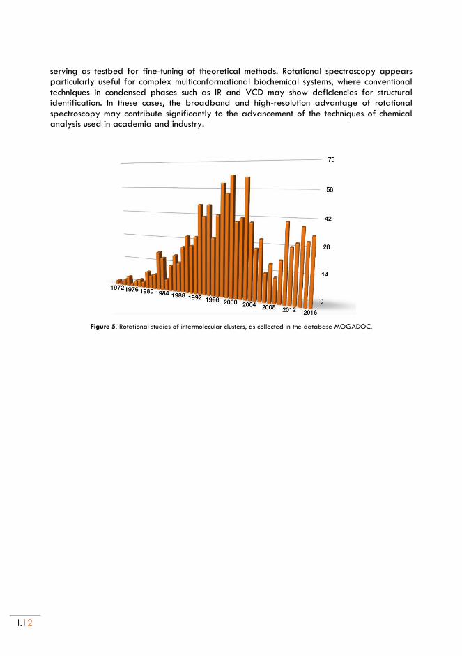
serving as testbed for fine-tuning of theoretical methods. Rotational spectroscopy appears particularly useful for complex multiconformational biochemical systems, where conventional techniques in condensed phases such as IR and VCD may show deficiencies for structural identification. In these cases, the broadband and high-resolution advantage of rotational spectroscopy may contribute significantly to the advancement of the techniques of chemical analysis used in academia and industry.
Figure 5. Rotational studies of intermolecular clusters, as collected in the database MOGADOC.
INTRODUCTION
I.13
II. Methods
The results presented in this PhD thesis are based on the analysis of microwave rotational spectra with the support of molecular orbital calculations. Rotational spectroscopy is a highly sensitive and suitable technique for the study of the gas-phase molecular structure, as the rotational spectrum of a polyatomic molecule is directly related to the geometric distribution and atomic masses through the moments of inertia. Depending on the molecular size and temperature, pure rotational spectra are observed predominantly in the frequency range from the cm- to the mmw- and submmw-wave ranges (1 GHz to 1 THz). In particular, the studies presented in this Thesis have been conducted in the cm-wave region of 2-18GHz. In this section we will summarize some fundamentals of rotational spectroscopy from its historical evolution to the techniques used and the theoretical approaches.
More specific details can be found in the bibliography, including experimental techniques,87,88 theory of rotational spectroscopy,89–91 supersonic jets expansions83–86 or the theoretical computational methods.92
1. Rotational spectroscopy
1.1. General aspects
Rotational spectroscopy has evolved considerably from the first studies in 193493 to the present, developing different complementary techniques. Nowadays, the spectral analysis is considerably improved as a result of the introduction of time-domain and supersonic jet techniques, permitting a great improvement of the spectral resolution, bandwidth, and sensitivity. Rotational spectroscopy uses microwave (MW) radiation to probe the transitions between quantized molecular rotation energy levels in the gas-phase. The MW experiments93 expanded from 1945 thanks to the development of radar during World War II, in particular with the introduction of Wilson’s Stark modulation direct-absorption techniques in the cm-wave range and the development of radiation sources in the mm-wave region. These experiments were done in a static gas cell using frequency scanning techniques, where monochromatic radiation passes through a sample cell and the transmitted intensity is registered versus the frequency. A noticeable step forward came in 1975 with the introduction by Flygare of time-domain rotational spectroscopy94–96 or Fourier-transform microwave spectroscopy (FTMW), following conceptual developments similar to FTNMR. The FTMW technique uses a short (s) MW pulse to produce a transient excitation polarizing the sample, recording in the time-domain the molecular relaxation signal or free-induction-decay. In 1981 Flygare combined the time-domain spectroscopic techniques with a supersonic jet expansion,97 using a tunable high-quality Fabry-Pérot resonator98,99 to couple the radiation to the sample. This spectrometer is known as Balle-Flygare or molecular beam (MB-FTMW) cavity spectrometer.
The Balle-Flygare FTMW spectrometer has been heavily used (even today it is the main technique in many research groups). Because of many modifications and improvements, in particular the coaxial orientation of the beam and the resonator (COBRA setup) implemented by Grabow100 in 1996, the spectrometer produces sub-Doppler resolution (FWHM linewidths of 10 kHz) and very high sensibility, but the operation has limited bandwidth (1 MHz) and
I.14
requires slow scanning. In 2006, Pate brought up the revolutionary chirped-pulse spectroscopy,101 a broadband FTMW spectrometer which has a bandwidth limit that is only imposed by the technical limitations of microwave power amplifiers and high-speed digitizers.102 This instrument uses a fast-passage technique based on a frequency modulated (chirped) excitation pulse, simultaneously exciting the full operating bandwidth. The CP-FTMW technique allowed to exponentially reduce the spectral acquisition time and the amount of sample while increasing the frequency operation bandwidth four orders of magnitude.87 Other designs for broadband spectroscopy have been contributed by Grabow, considerably improving the spectral linewidth (In-phase/quadrature-phase-modulation passage-acquired-coherence technique, IMPACT, by Grabow’s group)103. In 2013 Patterson introduced the detection of molecular chirality by use of a triple resonance or three-wave-mixing. The techniques on chirality detection are presently progressing in several laboratories.
In the experimental work done in this Thesis we have used both a Balle-Flygare FTMW spectrometer (covering the 8-18 GHz frequency range) and a commercial CP-FTMW spectrometer (2-8 GHz) provided by BrightSpec and following Pate’s design.104 Both spectrometers are settled in the Group of Supersonic Jet and Plasma Spectroscopy laboratory at the Department of Physical Chemistry of the University of Valladolid and Institute CINQUIMA.
1.2. Theory of molecular rotation
In order to achieve a quantitative fit of the experimental data to obtain molecular information, it is necessary to set up a quantum mechanical rotational Hamiltonian. Assuming that large-amplitude motions are not present and a complete separation of the vibrational and rotational motions, the rotational Hamiltonian can be treated independently and is based in the model of the rigid rotor. The rigid rotor deals with the mechanics of the general rotation of a rigid molecular set, taking into account the quantization of the angular momentum. Semirigid corrections and electric and magnetic effects can be added at a later stage.
The theory of molecular rotation is described in several references,89–91 so it is only briefly summarized here. A rigid rotor can be defined by its inertia tensor (a symmetric 3x3 matrix). Diagonalization of the inertia tensor leads to a molecular coordinate system referred as principal axes of inertia, with origin at the center of mass and characterized by the three
principal moments of inertia (conventionally 𝐼𝑎 ≤ 𝐼𝑏 ≤ 𝐼𝑐). The classical rotational energy in the principal axes is given by
𝐸𝑟𝑜𝑡 =1
2(𝐽𝑎2
𝐼𝑎+𝐽𝑏2
𝐼𝑏+𝐽𝑐2
𝐼𝑐) (1)
where 𝐽𝑖 represents the angular momentum. The classical Hamiltonian may be converted into the quantum mechanical Hamiltonian using the standard procedures, obtaining
𝑯𝒓𝒐𝒕 = 𝐴 𝑱𝒂2 + 𝐵 𝑱𝒃
2 + 𝐶 𝑱𝒄2 (2)
where the angular momentum operators are represented by 𝑱𝒊 (𝑖 = 𝑎, 𝑏, 𝑐) and 𝐴, 𝐵, 𝐶 are
the rotational constants (inversely proportional to their moment of inertia: 𝐴 = (ℎ2
8𝜋2𝐼𝑎), 𝐵 =
INTRODUCTION
I.15
(ℎ2
8𝜋2𝐼𝑏) and 𝐶 = (
ℎ2
8𝜋2𝐼𝑐), usually expressed in MHz). The expressions and solutions of the
rotational Hamiltonian depend on the type of molecular rotor. A symmetric top has an internal 𝐶𝑛 (𝑛 ≥ 3) rotation symmetry axes so two moments of inertia are equal (oblate top: 𝐼𝑎 = 𝐼𝑏 < 𝐼𝑐; prolate top: 𝐼𝑎 < 𝐼𝑏 = 𝐼𝑐). The asymmetric tops, like all molecules considered in this Thesis, have all moments of inertia different. Linear and top-symmetric rotors have analytical solutions, but the asymmetric rotors are solved numerically. For a symmetric top
the square of the angular momentum 𝑱𝟐 commutes with one of its components (𝑱𝒛 or 𝑱𝒁), so they can be defined simultaneously. In consequence, three quantum numbers are necessary: the total
angular momentum 𝐽 (associated with the operator 𝑱𝟐), the projection of the angular momentum
along the internal symmetry axes 𝐾 (= 0,±1,±2,…± 𝐽, associated with the operator 𝑱𝒛) and
the projection associated with the space-fixed (𝑧) axis, or 𝑀 (= 0,±1,±2,…± 𝐽, associated
with the operator 𝑱𝒁). Concerning the wavefunctions and eigenvalues of the Schrödinger equation, it is convenient to assimilate the problem to a complete and diagonalizable
Hamiltonian matrix by an orthonormal basis |𝐽, 𝐾,𝑀⟩. For an asymmetric top the Schrödinger the situation becomes more complicated, where the Schrödinger wave equation cannot be solved directly but its wave functions may be represented by a linear combination of symmetric
rotor. None of the angular momentum components is a constant of motion, so the 𝑱𝒊 angular momentum operators do not commute with the rotational Hamiltionian. In consequence, the
quantum number 𝐾 is not a good quantum number. Instead, each eigenvalue of the asymmetric Hamiltonian is approached by extrapolating to the oblate and the prolate symmetric top and
is assigned pseudo-quantum numbers 𝐾−1 (𝐾 value in the prolate symmetric top basis) and 𝐾+1 (𝐾 value in the oblate symmetric top basis). In this way, each asymmetric top eigenvalue is
defined by 𝐽, 𝐾−1 and 𝐾+1 and the rotational transitions are indicated as 𝐽′𝐾−1′,𝐾+1′ ←
𝐽′′𝐾−1′′,𝐾+1′′ (with 𝜏 = 𝐾−1 − 𝐾+1). The general procedure for obtaining the asymmetric
wavefunctions consists in expanding the functions in terms of the orthogonal symmetric-rotor wavefunctions:
𝛹𝑱𝜏𝑀 = ∑ 𝑎𝐽𝐾𝑀𝛹𝐽𝐾𝑀𝐽,𝐾,𝑀
(3)
The secular determinant is then set-up for each 𝐽 level and solved numerically by matrix
diagonalization. Only for the low 𝐽 values it is possible to obtain explicit solutions for the rotational energies.
In order to observe a rotational transition, it is necessary to consider the selection rules for electric dipole transitions, requiring a non-zero value for the integral moment of transition in
the space-fixed axes: ⟨𝐽′, 𝜏′, 𝑀′|𝝁|𝐽′′, 𝜏′′, 𝑀′′⟩ ≠ 0. The first requirement is thus that the
molecular system has non-zero permanent dipole moment 𝝁. In the case of an asymmetric top,
is convenient to take into account the components along the principal axes {𝝁𝒂, 𝝁𝒃, 𝝁𝒄} to know which transitions are possible. Since the wavefunctions were expressed as linear combinations
of the symmetric rotor the selection rules for 𝐽 are the same as in a symmetric rotor:
∆𝐽 = 0,±1 (4)
(transitions with ∆𝐽 = −1, ∆𝐽 = 0 and ∆𝐽 = +1 are designated P, Q or R). The selection rules
for the pseudo quantum numbers 𝐾−1, 𝐾+1 may be obtained from the symmetry of the ellipsoid of inertia and are collected in Table 4.
I.16
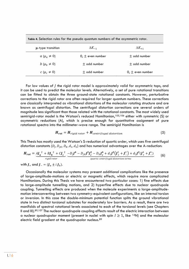
Table 4. Selection rules for the pseudo quantum numbers of the asymmetric rotor.
-type transition 𝐾−1 𝐾+1
𝑎 (𝜇𝑎 ≠ 0) 0, ± even number ± odd number
𝑏 (𝜇𝑏 ≠ 0) ± odd number ± odd number
𝑐 (𝜇𝑐 ≠ 0) ± odd number 0, ± even number
For low values of 𝐽 the rigid rotor model is approximately valid for asymmetric tops, and it can be used to predict the molecular levels. Alternatively, a set of pure rotational transitions can be fitted to obtain the three ground-state rotational constants. However, perturbative corrections to the rigid rotor are often required for larger quantum numbers. These corrections are classically interpreted as vibrational distortions of the molecular rotating structure and are known as centrifugal distortion. The centrifugal distortion corrections are several orders of magnitude less significant than those related with the rotational constants. The most widely used semirigid-rotor model is the Watson's reduced Hamiltonian,105,106 either with symmetric (S) or asymmetric reductions (A), which is precise enough for quantitative assignment of pure rotational spectra into the millimeter-wave range. The semirigid Hamiltonian is
𝑯𝒓𝒐𝒕 = 𝑯𝑟𝑖𝑔𝑖𝑑 𝑟𝑜𝑡𝑜𝑟 +𝑯𝑐𝑒𝑛𝑡𝑟𝑖𝑓𝑢𝑔𝑎𝑙 𝑑𝑖𝑠𝑡𝑜𝑟𝑡𝑖𝑜𝑛 (5)
This Thesis has mostly used the Watson’s S-reduction of quartic order, which uses five centrifugal distortion constants (𝐷𝐽, 𝐷𝐽𝐾 , 𝐷𝐾 , 𝑑1, 𝑑2) and has numerical advantages over the A-reduction:
𝑯𝒓𝒐𝒕 = 𝐴𝑱𝒂2 + 𝐵𝑱
𝒃2 + 𝐶𝑱
𝒄2
⏟
𝑟𝑖𝑔𝑖𝑑 𝑟𝑜𝑡𝑜𝑟
−𝐷𝐽𝑱𝟒 − 𝐷𝐽𝐾𝑱
𝟐𝑱𝒄𝟐 − 𝐷𝐾𝑱𝒄
𝟒 + 𝑑1𝑱𝟐(𝑱
+2 + 𝑱
−2 ) + 𝑑2𝑱
𝟐(𝑱+4 + 𝑱
−4 ) ⏟
𝑞𝑢𝑎𝑟𝑡𝑖𝑐 𝑐𝑒𝑛𝑡𝑟𝑖𝑓𝑢𝑔𝑎𝑙 𝑑𝑖𝑠𝑡𝑜𝑟𝑡𝑖𝑜𝑛 𝑡𝑒𝑟𝑚𝑠
(6)
with 𝑱+ and 𝑱− = (𝑱𝑎 ± 𝑖 𝑱𝑏).
Occasionally the molecular systems may present additional complications like the presence of large-amplitude-motions or electric or magnetic effects, which require more complicated Hamiltonians. During this Thesis we have encountered two particular cases: 1) fine effects due to large-amplitude tunnelling motions, and 2) hyperfine effects due to nuclear quadrupole coupling. Tunnelling effects are produced when the molecule experiments a large-amplitude-motion interconverting between two symmetry-equivalent configurations, like an internal torsion or inversion. In this case the double-minimum potential function splits the ground vibrational state in two distinct torsional substates for moderately low barriers. As a result, there are two manifolds of spectral rotational levels associated to each of the torsional levels (see Chapters II and III).89,107 The nuclear quadrupole coupling effects result of the electric interaction between
a nuclear quadrupolar moment (present in nuclei with spin 𝐼 ≥ 1, like 14N) and the molecular electric field gradient at the quadrupolar nucleus.89
INTRODUCTION
I.17
1.3. Balle-Flygare FTMW spectrometer @ UVa
The experimental data presented in this Thesis have been obtained essentially by using two different but complementary spectrometers at the UVa (the spectrometers used in the research visits done during the Thesis are not included here but are referred in the publications). The UVa spectrometers have in common the use of supersonic jet expansions and include:
- A Balle-Flygare FTMW cavity spectrometer (8-18GHz) - A chiped-pulse FTMW spectrometer (2-8 GHz)
The benefits and characteristics of jet expansions for spectroscopic research are discussed elsewhere.108 Briefly, supersonic jet experiments are based on a near-adiabatic gas expansion through a pin-hole nozzle into an evacuated chamber. The pressurized region and the expansion chamber maintain a huge difference of pressure, so the expanding molecules pass from a situation of high-pressure (1-10 bar) random thermal motion to a directed motion in the low-pressure region (10-5-10-7 mbar). The sudden change in the kinetic conditions turns the broad velocity distribution into a narrow distribution centered around the terminal speed. This process converts the internal molecular energies into kinetic energy, producing a considerable molecular freezing. However, since the different internal motions cannot equilibrate, the translational, rotational and vibrational temperatures are different. The effective rotational temperatures are of 2-3 K, while vibrational temperatures reach 100-150 K, moving the molecular population to the lower rotational levels of the vibronic ground-state and simplifying the spectrum. Another important benefit of the supersonic jet is the reduction of intermolecular collisions, avoiding any mechanism of chemical reaction or decomposition. For this reason, the supersonic jets are used as a source of intermolecular clusters. The clusters are generated by many-body collisions in the first moments of the expansion and are later stabilized in the later expansion by the absence of collisions. For this reason the jet expansions are the most effective way to study weak intermolecular complexes.97 The reduction of intermolecular collisions additionally contributes to the reduction in the spectral linewidth, which may reach sub-Doppler (kHz) levels. These features make this type of expansion a useful tool for spectroscopy investigations characterizing molecular structures in the gas phase (in particular, rotational, vibrational and electronic spectroscopy) and constitute the fundamental principle on which our experimental experiments are conducted.83–86
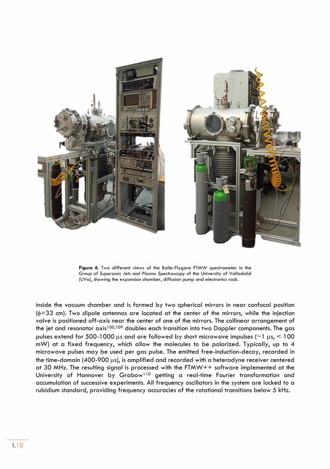
The Balle-Flygare FTMW spectrometer in the Group of Supersonic Jets and Plasma Spectroscopy laboratory is a non-commercial instrument based on the Balle-Flygare99 design, and includes a Fabry-Perot cavity made of two spherical mirrors. Figures 5 and 6 shown the general design of the spectrometer and a functional diagram. The sample can be prepared as a gaseous mixture (typically 0.1-0.5%) in a carrier gas or located in a reservoir nozzle if it
is liquid or solid for vaporization. The spectrometer uses several nozzles (diameters =0.8-1.2 mm) and may be heated to ca. 373 K (depending on the macroscopical properties such as melting and boiling point, vapor pressure, etc.). The sample is entrained in a carrier gas (He, Ne or Ar) at stagnation pressures of 1-5 bar and expanded through a solenoid valve, creating a pulsed supersonic jet probed within the Fabry-Perot MW resonator. The resonator is mounted
I.18
Figure 6. Two different views of the Balle-Flygare FTMW spectrometer in the Group of Supersonic Jets and Plasma Spectroscopy of the University of Valladolid (UVa), showing the expansion chamber, diffusion pump and electronics rack.
inside the vacuum chamber and is formed by two spherical mirrors in near confocal position
(=33 cm). Two dipole antennas are located at the center of the mirrors, while the injection valve is positioned off-axis near the center of one of the mirrors. The collinear arrangement of the jet and resonator axis100,109 doubles each transition into two Doppler components. The gas
pulses extend for 500-1000 s and are followed by short microwave impulses (~1 s, < 100 mW) at a fixed frequency, which allow the molecules to be polarized. Typically, up to 4 microwave pulses may be used per gas pulse. The emitted free-induction-decay, recorded in
the time-domain (400-900 s), is amplified and recorded with a heterodyne receiver centered at 30 MHz. The resulting signal is processed with the FTMW++ software implemented at the University of Hannover by Grabow110 getting a real-time Fourier transformation and accumulation of successive experiments. All frequency oscillators in the system are locked to a rubidium standard, providing frequency accuracies of the rotational transitions below 5 kHz.
INTRODUCTION
I.19
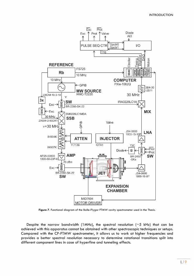
Figure 7. Functional diagram of the Balle-Flygar FTMW cavity spectrometer used in the Thesis.
Despite the narrow bandwidth (1MHz), the spectral resolution (~5 kHz) that can be achieved with this apparatus cannot be obtained with other spectroscopic techniques or setups. Compared with the CP-FTMW spectrometer, it allows us to work at higher frequencies and provides a better spectral resolution necessary to determine rotational transitions split into different component lines in case of hyperfine and tunneling effects.
I.20
1.4. Chirped-Pulse FT-MW spectrometer @ UVa
Some years after the first experiments on FTMW spectroscopy, Flygare proposed a new technique known as fast-passage, introducing a distinction with the processes of transient excitation.111,112 A fast-passage is achieved when a frequency is swept through a two-level resonance in a time much shorter than the relaxation period. Fast-passage was conducted initially with a fast modulating Stark electric field, switching the resonance frequencies. In 2005 Pate implemented fast-passage techniques using a digital frequency sweep carried out with an arbitrary waveform generator (AWG), sweeping at a rate much faster than the dephasing time of the molecular coherence.113 In the present designs the sample is excited with a short
microwave pulse implementing a very fast linear frequency sweep (6 GHz/s) or “chirped” pulse, so this technique is known as chirped-pulse Fourier Transform Microwave (CP-FTMW) spectroscopy. The chirped pulse covers all molecular transitions within the frequency range of the sweep, allowing to coherently excite a large swath of molecular transitions simultaneously. This advance was only possible thanks to the progress in the available digital technology.
These spectrometers combine the AWG radiation source with broadband detection with a high-sampling-rate high-speed digital oscilloscope, simultaneously detecting all molecular emissions. The excitation capabilities of the CP-FTMW spectrometer are limited by the amplification stage, which typically requires a minimum of 20-400 W to cover the region 2-8 GHz. The CP-FTMW technique has revolutionized the field of rotational spectroscopy making its design one of the most followed and influential in MW spectroscopy research.
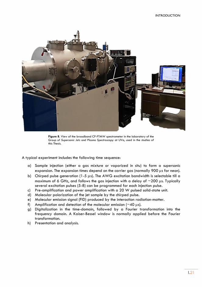
In 2015 the UVa bought a pulsed-jet chiped-pulse FTMW spectrometer from the company BrightSpec, that was installed in the laboratory of the Group of Supersonic Jets and Plasma Spectroscopy. This instrument follows Pate’s direct-digital design covering the 2-8 GHz frequency region,104 and has been used extensively during this Thesis. The CP-FTMW spectrometer is depicted in Fig. 7, with a functional diagram in Fig. 8. The spectrometer uses a single injection system to create a supersonic expansion in the expansion chamber. The sample injection is similar to the FTMW spectrometer, using a solenoid valve attached to a pinhole nozzle. In the case of solid or liquids of low volatility a heating nozzle with an internal reservoir can be used. This instrument has no resonator and the interaction between the radiation and the jet is produced with two (emitting and receiving) horn antennas. For this reason, the jet cannot be installed collinear to the radiation and instead it uses a perpendicular arrangement between the horn antennas and the injection valve. This orientation increases the jet volume probed with the radiation, observing molecules with a larger velocity distribution. As a result, the linewidth increases about an order of magnitude compared with the cavity spectrometer (about 150 kHz). In the present set-up the chirped pulse (1-4 s) created by the AWG (25 GS/s in two- channel) is amplified with a solid-state power amplifier (20 W) and broadcasted into the excitation region. The detection uses a receiving antenna, a diode limiter, a PIN-diode switch (closed during excitation) and a low-noise amplifier. Finally, the time-domain signal (considerably shorter than in the cavity spectrometer) is acquired for about 40 s with a high-speed digital oscilloscope (25 GS/s). The typical operation requires signal averaging and uses a repetition rate of 5 Hz. All frequencies are referenced to a 10 MHz Rb standard.
INTRODUCTION
I.21
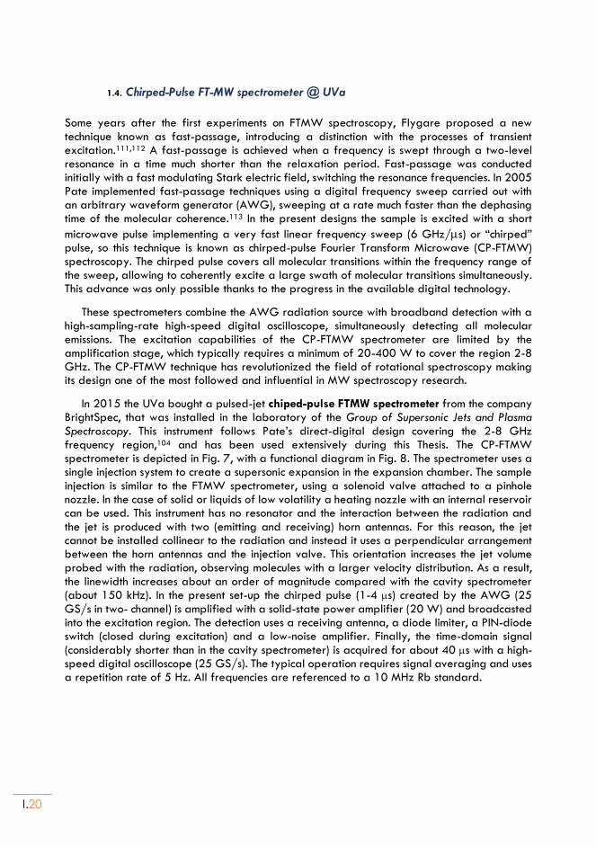
Figure 8. View of the broadband CP-FTMW spectrometer in the laboratory of the Group of Supersonic Jets and Plasma Spectroscopy at UVa, used in the studies of this Thesis.
A typical experiment includes the following time sequence:
a) Sample injection (either a gas mixture or vaporized in situ) to form a supersonic
expansion. The expansion times depend on the carrier gas (normally 900 s for neon).
b) Chirped pulse generation (1-5 s). The AWG excitation bandwidth is selectable till a
maximum of 6 GHz, and follows the gas injection with a delay of ~200 s. Typically several excitation pulses (5-8) can be programmed for each injection pulse.
c) Pre-amplification and power amplification with a 20 W pulsed solid-state unit. d) Molecular polarization of the jet sample by the chirped pulse. e) Molecular emission signal (FID) produced by the interaction radiation-matter.
f) Amplification and detection of the molecular emission (~40 s). g) Digitalization in the time-domain, followed by a Fourier transformation into the
frequency domain. A Kaiser-Bessel window is normally applied before the Fourier transformation.
h) Presentation and analysis.
I.22
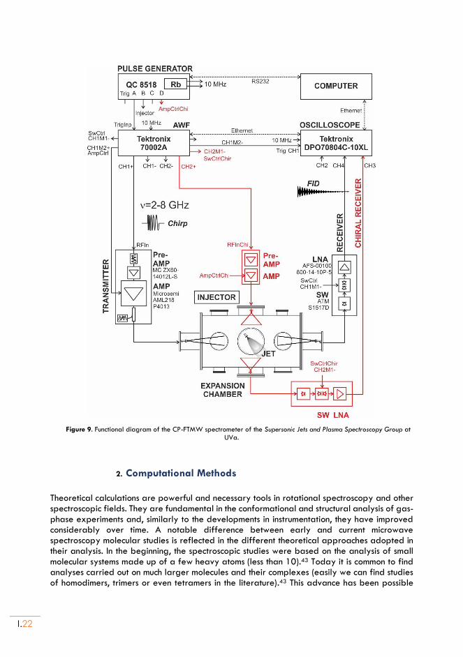
Figure 9. Functional diagram of the CP-FTMW spectrometer of the Supersonic Jets and Plasma Spectroscopy Group at UVa.
2. Computational Methods
Theoretical calculations are powerful and necessary tools in rotational spectroscopy and other spectroscopic fields. They are fundamental in the conformational and structural analysis of gas-phase experiments and, similarly to the developments in instrumentation, they have improved considerably over time. A notable difference between early and current microwave spectroscopy molecular studies is reflected in the different theoretical approaches adopted in their analysis. In the beginning, the spectroscopic studies were based on the analysis of small molecular systems made up of a few heavy atoms (less than 10).43 Today it is common to find analyses carried out on much larger molecules and their complexes (easily we can find studies of homodimers, trimers or even tetramers in the literature).43 This advance has been possible
INTRODUCTION
I.23
not only by the experimental developments, but also thanks to continuous advances in density-functional-theory (DFT) and ab initio molecular orbital calculations, permitting the rationalization and interpretation of the experimental results with a reasonable investment of time and cost in the calculation process.
In this Thesis we have used different computational methods. An initial approach to the structural properties might be a simple molecular mechanics (MM)92 calculation. Despite this calculation uses classical mechanics and ignores electrons, the low computational cost permits a fast scan of the potential energy surface (PES). This kind of calculation, choosing a proper force field, is tipically used to get an initial conformational search of the posible molecular isomers. In the Thesis we performed MM conformational searches with the commonly used Merck force field (MMFF)114 and dedicated Monte Carlo115/low-mode116 search algorithms. These initial calculations are not precise and a bit ambiguous, always requiring further calculations with methods based on quantum mechanics.
2.1. Density Functional Theory and Ab initio calculations
The computational work included in the Thesis has been mostly directed to perform conformational searches, geometry optimizations and vibrational frequency calculations (estimating the ground-state zero-point energy and centrifugal distorsion terms). Additinally, the calculation of intermolecular complexes presents the problem of the basis-set superposition error (BSSE), which has been taking into account with the counterpoise approximation of Boyds and Bernardi. There are two main tipes of quantum mechanical molecular orbital calculations used in the Thesis: those based on DFT methods and the wavefunction methods based on Hartree-Fock theory117 and perturbative corrections, such as Møller-Plesset118 pure ab initio calculations. More advanced calculations, like coupled-clusters methods, have not been used here. Computational methods are widely discussed in the bibliography.92
In rotational spectroscopy investigations the DFT and MP2 ab initio methods generally provide a satisfactory description of the experiment. The pure ab initio calculations (computations of electronic orbitals with no other hypotheses than Coulomb interactions) are those based on the Hartree-Fock theory of perturbation corrections to deal with the correlation of electrons and they are called nth order Møller–Plesset perturbation theory. The second order MP2 method corrects Hartree-Fock by calculating the sum of the amplitudes between all double combinations of occupied and virtual orbitals (it does not differ too much for other higher orders much more “expensive” terms)119. The MP2 method has been used as a theoretical reference in many MW studies, but as the molecular size increases it may not longer be the most appropriate method, both because of the computational cost and calculation accuracy.
In recent works, DFT calculations is increasingly used,120 shaping modern molecular quantum chemistry like no other methodology in recent times. Nowadays, it is by far most frequently applied approach used by computational chemists. As a consequence, hundreds of density functional aproximations have been developed, each with their own advantages and disadvantages, which means that despite DFT calculations are technically easy to perform, it is no trivial task to choose the right functional.121 In this work several DFT computational approximations have been tested to complete our experimental studies, including Minnesota dispersion-corrected MN15-L122 and M06-2X123 methods developed by Truhlar, and the hybrid or double-hybrid B3LYP,124 B2PLYP125 and B97XD126 methods, supplemented by Grimme’s D3127 dispersion corrections and Becke–Johnson damping functions.128
I.24
The atomic basis sets used during the computational work have mostly included triple-zeta quality with polarization and difuse functions, most often the Ahlrich’s polarized basis def2-TZVP,129 Dunning’s augmented correlation-consistent aug-cc-pVTZ and Pople’s 6-311++G(d,p) basis set.130 The computational calculations were implemented mostly in Gaussian16131. All these theoretical methods will be further presented in the following chapters.
2.2. Complementary computational tools
In order to reinforce the understanding of the intermolecular clusters, it is worth to complement the DFT and/or ab initio calculations with other computational analysis tools exploring the properties of the molecular electronic density. In this thesis we have used in particular the topological analyses of the reduced electronic density function known as NCIPlots132 and a method for binding energy decomposition, in particular symmetry-adapted perturbation theory (SAPT).133
Symmetry-adapted perturbation theory computes the noncovalent interaction between two molecules, providing a decomposition of the interaction energy into physically meaningful components: i.e., electrostatic, exchange, induction, and dispersion terms.134 In SAPT, the Hamiltonian of the dimer is partitioned into contributions from each monomer and the interaction. Several truncations of the closed-shell SAPT expansion are available for both the monomers and the cluster, from the simplest zero-order SAPT(0) to SAPT2+(3), with second-order intramonomer correlation corrections and third-order intermonomer dispersion corrections. The calculations may use different basis sets and was used as implemented in PSI4.
The NCIPlot method135 is based in the analysis of the bond critical points associated to the reduced electronic density matrix, permitting a 3D mapping of the regions with attractive or non-attractive interactions and the comparison between different intermolecular clusters. The NCIPlots have been used in chapters Chapter III, Chapter IV, Chapter V and Chapter VI.
INTRODUCTION
I.25
Chapter I. References: 1. Ed.: S. Scheiner. Noncovalent Forces. Springer, Heidelberg (2015).
2. Scheeres, D. J., Hartzell, C. M., Sánchez, P. & Swift, M. Scaling forces to asteroid surfaces: The role of cohesion. Icarus 210, 968–984 (2010).
3. Autumn, K. et al. Evidence for van der Waals adhesion in gecko setae. Proc. Natl. Acad. Sci. 99, 12252–12256 (2002).
4. Gilli, G. & Gilli, P. The Nature of the Hydrogen Bond. (2009).
5. Novoa, J. J. Intermolecular Interactions in Crystals: Fundamentals of Crystal Engineering. (RSC, 2018).
6. Arunan, E. et al. Defining the hydrogen bond: An account (IUPAC Technical Report). Pure Appl. Chem. 83, 1619–1636 (2011).
7. Arunan, E. et al. Definition of the hydrogen bond (IUPAC Recommendations 2011). Pure Appl. Chem. 83, 1637–1641 (2011).
8. Desiraju, G. R. The C− H···O Hydrogen Bond: Structural Implications and Supramolecular Design. Acc. Chem. Res. 29, 441–449 (1996).
9. Nishio, M., Hirota, M. & Umezawa, Y. The CH···p Interaction. (Wiley, 1998).
10. Takahashi, O., Kohno, Y. & Nishio, M. Relevance of Weak Hydrogen Bonds in the Conformation of Organic Compounds and Bioconjugates: Evidence from Recent Experimental Data and High-Level ab Initio MO Calculations. Chem. Rev. 110, 6049–6076 (2010).
11. Bakhmutov, V. I. Dihydrogen Bonds: Principles, Experiments, and Applications. (Wiley, 2008).
12. Desiraju, G. R. et al. Definition of the halogen bond (IUPAC Recommendations 2013). Pure Appl. Chem. 85, 1711–1713 (2013).
13. Clark, T., Hennemann, M., Murray, J. S. & Politzer, P. Halogen bonding: the σ -hole. J. Mol. Model. 13, 291–296 (2007).
14. Murray, J. S., Lane, P., Clark, T., Riley, K. E. & Politzer, P. σ -Holes, π-holes and electrostatically-driven interactions. J. Mol. Model. 18, 541–548 (2012).
15. Cavallo, G. et al. The Halogen Bond. Chem. Rev. 116, 2478–2601 (2016).
16. Metrangolo, P. & Resnati, G. Halogen Bonding I. (Springer, 2015).
17. Murray, J. S., Lane, P., Clark, T. & Politzer, P. σ -hole bonding: molecules containing group VI atoms. J. Mol. Model. 13, 1033–1038 (2007).
18. Murray, J. S., Lane, P. & Politzer, P. Expansion of the σ -hole concept. J. Mol. Model. 15, 723–729 (2009).
19. Minyaev, R. M. & Minkin, V. I. Theoretical study of O-> X (S, Se, Te) coordination in organic compounds. Can. J. Chem. 76, 776–788 (1998).
20. Zahn, S., Frank, R., Hey‐ Hawkins, E. & Kirchner, B. Pnicogen Bonds: A New Molecular Linker? Chem. – A Eur. J. 17, 6034–6038 (2011).
21. Scheiner, S. A new noncovalent force: Comparison of P···N interaction with hydrogen and halogen bonds. J. Chem. Phys. 134, 094315 (2011).
22. Bauzá, A., Mooibroek, T. J. & Frontera, A. Tetrel-Bonding Interaction: Rediscovered Supramolecular
I.26
Force? Angew. Chemie Int. Ed. 52, 12317–12321 (2013).
23. Mani, D. & Arunan, E. The X–C⋯Y (X = O/F, Y = O/S/F/Cl/Br/N/P) ‘carbon bond’ and hydrophobic interactions. Phys. Chem. Chem. Phys. 15, 14377 (2013).
24. Cavallo, G., Metrangolo, P., Pilati, T., Resnati, G. & Terraneo, G. Naming Interactions from the Electrophilic Site. Cryst. Growth Des. 14, 2697–2702 (2014).
25. Legon, A. C. & Walker, N. R. What’s in a name? ‘Coinage-metal’ non-covalent bonds and their definition. Phys. Chem. Chem. Phys. 20, 19332–19338 (2018).
26. Goodwin, E. J. & Legon, A. C. The rotational spectrum and molecular geometry of an antihydrogen‐bonded dimer of sulfur dioxide and hydrogen cyanide. J. Chem. Phys. 85, 6828–6836 (1986).
27. Leopold, K. R., Fraser, G. T. & Klemperer, W. Rotational spectrum and structure of the complex HCNCO 2. J. Chem. Phys. 80, 1039–1046 (1984).
28. Ngari, M. S., Xu, Y. & Jäger, W. Rotational Spectroscopic Investigation of the Weak Interaction between CO and N2O. J. Mol. Spectrosc. 197, 244–253 (1999).
29. Zeng, Y. P., Sharpe, S. W., Shin, S. K., Wittig, C. & Beaudet, R. A. Infrared spectroscopy of CO 2 –D(H)Br: Molecular structure and its reliability. J. Chem. Phys. 97, 5392–5402 (1992).
30. Legon, A. C. & Suckley, A. P. Pulsed-jet, diode-laser IR spectroscopy of the ν=1→0 transition in the
CO2 asymmetric stretching mode of (CO2, HCN). Chem. Phys. Lett. 157, 5–10 (1989).
31. Legon, A. C. Tetrel, pnictogen and chalcogen bonds identified in the gas phase before they had names: a systematic look at non-covalent interactions. Phys. Chem. Chem. Phys. 19, 14884–14896 (2017).
32. Al-Hamdani, Y. S. & Tkatchenko, A. Understanding non-covalent interactions in larger molecular complexes from first principles. J. Chem. Phys. 150, (2019).
33. Alkorta, I., Elguero, J. & Frontera, A. Not only hydrogen bonds: Other noncovalent interactions. Crystals 10, (2020).
34. Pauling, L. The nature of the chemical bond. (CorneUUniv. Press, Ithsea, 1939).
35. Pimentel, G. C. & McClellan, M. A. The Hydrogen Bond. (1960).
36. Vinogradov, S. N. & Linnell, R. H. Hydrogen Bonding. (Van Nostrand-Reinhold Company, 1971).
37. Desiraju, G. R. & Steiner, T. The Weak Hydrogen Bond. (Oxford University Press, 1999).
38. Melandri, S. ‘Union is strength’: How weak hydrogen bonds become stronger. Phys. Chem. Chem. Phys. 13, 13901–13911 (2011).
39. Gardner, K. H. & Blackwell, J. The hydrogen bonding in native cellulose. BBA - Gen. Subj. 343, 232–237 (1974).
40. The Cambridge Crystallographic Data Centre (CCDC). https://www.ccdc.cam.ac.uk/structures/.
41. Jeffrey, G. A. An Introduction to Hydrogen Bonding. (Oxford University Press, 1997).
42. Steiner, T. & Saenger, W. Geometric analysis of non‐ionic O‐H⋯O hydrogen bonds and non‐bonding arrangements in neutron diffraction studies of carbohydrates. Acta Crystallogr. Sect. B 48, 819–827 (1992).
43. Caminati, W. & Grabow, J.-U. Microwave Spectroscopy Molecular Systems. in Frontiers of Molecular Spectroscopy (ed. Laane, J.) 456–552 (Elsevier Inc., 2008).
INTRODUCTION
I.27
44. Biswal, H. S., Shirhatti, P. R. & Wategaonkar, S. O−H···O versus O−H···S Hydrogen Bonding I: Experimental and Computational Studies on the p -Cresol·H2O and p -Cresol·H2S Complexes. J. Phys. Chem. A 113, 5633–5643 (2009).
45. Biswal, H. S. & Wategaonkar, S. OH · · · X (X = O, S) hydrogen bonding in thetrahydrofuran and tetrahydrothiophene. J. Chem. Phys. 135, 134306 (2011).
46. Biswal, H. S., Shirhatti, P. R. & Wategaonkar, S. O−H···O versus O−H···S Hydrogen Bonding. 2. Alcohols and Thiols as Hydrogen Bond Acceptors. J. Phys. Chem. A 114, 6944–6955 (2010).
47. Biswal, H. S. & Wategaonkar, S. O−H···O versus O−H···S Hydrogen Bonding. 3. IR−UV Double Resonance Study of Hydrogen Bonded Complexes of p -Cresol with Diethyl Ether and Its Sulfur Analog. J. Phys. Chem. A 114, 5947–5957 (2010).
48. Jouvet, C., Lardeux-Dedonder, C., Richard-Viard, M., Solgadi, D. & Tramer, A. Reactivity of molecular clusters in the gas phase: proton-transfer reaction in neutral phenol-(ammonia)n and phenol-(ethanamine)n. J. Phys. Chem. 94, 5041–5048 (1990).
49. Iwasaki, A., Fujii, A., Watanabe, T., Ebata, T. & Mikami, N. Infrared Spectroscopy of Hydrogen-
Bonded Phenol−Amine Clusters in Supersonic Jets. J. Phys. Chem. 100, 16053–16057 (1996).
50. Biswas, N., Wategaonkar, S., Watanabe, T., Ebata, T. & Mikami, N. Fluorescence, REMPI, hole-burning, and FDIR spectroscopy of para-cyanophenol–water1 complex. Chem. Phys. Lett. 394, 61–67 (2004).
51. Biswal, H. S. Hydrogen Bonding Involving Sulfur: New Insights from Ab Initio Calculations and Gas Phase Laser Spectroscopy. in Noncovalent Forces (ed. Scheiner, S.) (Springer Int. Pub., 2015).
52. Scheiner, S. Hydrogen Bonding. A Theoretical Perspective. (Oxford University Press, 1997).
53. Goebel, J. R., Ault, B. S. & Del Bene, J. E. Matrix Isolation and ab Initio Study of 1:1 Hydrogen-Bonded Complexes of H 2 O 2 with Phosphorus and Sulfur Bases. J. Phys. Chem. A 105, 11365–11370 (2001).
54. Wierzejewska, M. Infrared matrix isolation studies of complexes formed between dimethylsulfide, dimethyldisulfide and nitrous acid. J. Mol. Struct. 520, 199–214 (2000).
55. Bhattacharyya, S., Bhattacherjee, A., Shirhatti, P. R. & Wategaonkar, S. O–H···S Hydrogen Bonds Conform to the Acid–Base Formalism. J. Phys. Chem. A 117, 8238–8250 (2013).
56. Das, A. et al. The H2S Dimer is Hydrogen‐Bonded: Direct Confirmation from Microwave Spectroscopy. Angew. Chemie Int. Ed. 57, 15199–15203 (2018).
57. Wategaonkar, S. & Bhattacherjee, A. N–H···S Interaction Continues To Be an Enigma: Experimental and Computational Investigations of Hydrogen-Bonded Complexes of Benzimidazole with Thioethers. J. Phys. Chem. A 122, 4313–4321 (2018).
58. Sanz, M. E. et al. Conformation and Stability of Adducts of Sulfurated Cyclic Compounds with Water:
Rotational Spectrum of Tetrahydrothiophene−Water. J. Phys. Chem. A 103, 5285–5290 (1999).
59. Lobo, I. A., Robertson, P. A., Villani, L., Wilson, D. J. D. & Robertson, E. G. Thiols as Hydrogen Bond Acceptors and Donors: Spectroscopy of 2-Phenylethanethiol Complexes. J. Phys. Chem. A 122, 7171–7180 (2018).
60. Martin, D. E., Robertson, E. G., Thompson, C. D. & Morrison, R. J. S. Resonant 2-photon ionization study of the conformation and the binding of water molecules to 2-phenylethanethiol (PhCH2CH2SH). J. Chem. Phys. 128, 164301 (2008).
61. Mishra, K. K. et al. Observation of an Unusually Large IR Red-Shift in an Unconventional S–H···S Hydrogen-Bond. J. Phys. Chem. Lett. 12, 1228–1235 (2021).
62. Bhattacharyya, S. & Wategaonkar, S. ZEKE Photoelectron Spectroscopy of p -Fluorophenol···H 2 S/H
I.28
2 O Complexes and Dissociation Energy Measurement Using the Birge–Sponer Extrapolation Method. J. Phys. Chem. A 118, 9386–9396 (2014).
63. Bhattacherjee, A., Matsuda, Y., Fujii, A. & Wategaonkar, S. Acid–Base Formalism in Dispersion-
Stabilized S–H···Y (Y═O, S) Hydrogen-Bonding Interactions. J. Phys. Chem. A 119, 1117–1126 (2015).
64. Bhattacherjee, A., Matsuda, Y., Fujii, A. & Wategaonkar, S. The Intermolecular S-H⋅⋅⋅Y (Y=S,O) Hydrogen Bond in the H2S Dimer and the H 2 S-MeOH Complex. ChemPhysChem 14, 905–914 (2013).
65. Juanes, M., Saragi, R. T., Caminati, W. & Lesarri, A. The Hydrogen Bond and Beyond: Perspectives for Rotational Investigations of Non-Covalent Interactions. Chem. - A Eur. J. 25, (2019).
66. Caminati, W. & Grabow, J.-U. Advancements in Microwave Spectroscopy. in Frontiers and Advancements in Microwave Spectroscopy (ed. Laane, J.) 569–598 (Elsevier, 2018).
67. Cole, G. C., Møllendal, H. & Guillemin, J.-C. Microwave Spectrum of 3-Butyne-1-thiol: Evidence for
Intramolecular S−H···π Hydrogen Bonding. J. Phys. Chem. A 110, 9370–9376 (2006).
68. Juanes, M. et al. Rotational spectrum and intramolecular hydrogen bonding in 1,2-butanedithiol. J. Mol. Struct. 1211, (2020).
69. Xu, L.-H. et al. Rotational spectra, conformational structures, and dipole moments of thiodiglycol by jet-cooled FTMW and ab initio calculations. J. Mol. Spectrosc. 228, 243–250 (2004).
70. Ghosh, S., Bhattacharyya, S. & Wategaonkar, S. Dissociation Energies of Sulfur-Centered Hydrogen-Bonded Complexes. J. Phys. Chem. A 119, 10863–10870 (2015).
71. Cooke, S. A., Corlett, G. K. & Legon, A. C. Comparisons of the interactions of benzene, furan and
thiophene with Lewis acids: the rotational spectrum of thiophene⋯HF. Chem. Phys. Lett. 291, 269–276 (1998).
72. Cocinero, E. J. et al. Weak hydrogen bonds C–H⋯S and C–H⋯F–C in the thiirane–trifluoromethane dimer. Chem. Phys. Lett. 402, 4–10 (2005).
73. Tubergen, M. J., Flad, J. E. & Del Bene, J. E. Microwave spectroscopic and ab initio studies of the hydrogen-bonded trimethylamine–hydrogen sulfide complex. J. Chem. Phys. 107, 2227–2231 (1997).
74. Wang, D., Chopra, P., Wategaonkar, S. & Fujii, A. Electronic and Infrared Spectroscopy of Benzene-(H 2 S) n ( n = 1 and 2): The Prototype of the SH-π Interaction. J. Phys. Chem. A 123, 7255–7260 (2019).
75. Arunan, E. et al. Rotational spectrum of the weakly bonded C6H6–H2S dimer and comparisons to C6H6–H2O dimer. J. Chem. Phys. 117, 9766–9776 (2002).
76. Goswami, M. & Arunan, E. Microwave spectrum and structure of C6H5CCH⋯H2S complex. J. Mol. Spectrosc. 268, 147–156 (2011).
77. Goswami, M., Neill, J. L., Muckle, M., Pate, B. H. & Arunan, E. Microwave, infrared-microwave double
resonance, and theoretical studies of C2H4 ⋯H2S complex. J. Chem. Phys. 139, 104303 (2013).
78. Crittenden, D. L. A Systematic CCSD(T) Study of Long-Range and Noncovalent Interactions between Benzene and a Series of First- and Second-Row Hydrides and Rare Gas Atoms. J. Phys. Chem. A 113, 1663–1669 (2009).
79. Vila, A. & Mosquera, R. A. Are the hydrogen bonds involving sulfur bases inverse or anomalous? Int. J. Quantum Chem. 106, 928–934 (2006).
INTRODUCTION
I.29
80. Tauer, T. P., Derrick, M. E. & Sherrill, C. D. Estimates of the ab Initio Limit for Sulfur−π Interactions: The
H 2 S−Benzene Dimer. J. Phys. Chem. A 109, 191–196 (2005).
81. Wennmohs, F., Staemmler, V. & Schindler, M. Theoretical investigation of weak hydrogen bonds to sulfur. J. Chem. Phys. 119, 3208–3218 (2003).
82. Kaur, D., Aulakh, D., Khanna, S. & Singh, H. Theoretical study on the nature of S···H and O ··· H hydrogen bonds. J. Sulfur Chem. 35, 290–303 (2014).
83. Pauly, H. Atom, Molecule, and Cluster Beams I. vol. 28 (Springer Berlin Heidelberg, 2000).
84. Campargue, R. Atomic and Molecular Beams. (Springer Berlin Heidelberg, 2001). doi:10.1007/978-3-642-56800-8.
85. Scoles, G. Atomic and Molecular Beam Methods, Vol. 1. (Oxford University Press, 1988).
86. Scoles, G. Atomic and Molecular Beam Methods: Vol. 2. (Oxford University Press, 1992).
87. Shipman, S. T. & Pate, B. H. New Techniques in Microwave Spectroscopy. in Handbook of High-resolution Spectroscopy (eds. Quack, M. & Merk, F.) 801–827 (John Wiley & Sons, Ltd., 2011).
88. Grabow, J.-U. & Caminati, W. MICROWAVE SPECTROSCOPY EXPERIMENTAL TECHNIQUES. in Frontiers of Molecular Spectroscopy (ed. Laane, J.) 383–454 (Elsevier, 2009). doi:10.1016/B978-0-444-53175-9.X0001-3.
89. Gordy, W. & Cook, R. L. Microwave Molecular Spectra. (John Wiley & Sons, Ltd, 1984).
90. Kroto, H. W. Molecular Rotation Spectra. (Dover Publication Inc., 1992).
91. Townes, C. H. & Schawlow, A. L. Microwave Spectroscopy. (Dover Publication Inc., 1975).
92. Jensen, F. Introduction to Computational Chemistry. (John Wiley & Sons, Ltd, 1999).
93. Cleeton, C. E. & Williams, N. H. Electromagnetic Waves of 1.1 cm Wave-Length and the Absorption Spectrum of Ammonia. Phys. Rev. 45, 234–237 (1934).
94. Dicke, R. H. & Romer, R. H. Pulse Techniques in Microwave Spectroscopy. Rev. Sci. Instrum. 26, 915–928 (1955).
95. Ekkers, J. & Flygare, W. H. Pulsed microwave Fourier transform spectrometer. Rev. Sci. Instrum. 47, 448–454 (1976).
96. Somers, R. M., Poehler, T. O. & Wagner, P. E. Microwave time domain Fabry–Perot emission spectrometer. Rev. Sci. Instrum. 46, 719–725 (1975).
97. Legon, A. C. Isolation and characterisation of some transient complexes of chemical interest using pulsed-nozzle, Fourier-transform microwave spectroscopy. J. Mol. Struct. 266, 21–37 (1992).
98. Balle, T. J., Campbell, E. J., Keenan, M. R. & Flygare, W. H. A new method for observing the rotational spectra of weak molecular complexes: KrHCl. J. Chem. Phys. 71, 2723–2724 (1979).
99. Balle, T. J. & Flygare, W. H. Fabry-Perot cavity pulsed Fourier transform microwave spectrometer with a pulsed nozzle particle source. Rev. Sci. Instrum. 52, 33–45 (1981).
100. Grabow, J. U., Stahl, W. & Dreizler, H. A multioctave coaxially oriented beam-resonator arrangement Fourier-transform microwave spectrometer. Rev. Sci. Instrum. 67, 4072–4084 (1996).
101. Grubbs II, G., Moon, N., Persinger, T., Gillcrist, D. & Marshall, F. A chirped pulse fourier transform microwave (cp-ftmw) spectrometer with laser ablation source to search for actinide-containing molecules and noble metal clusters. in Proceedings of the 71st International Symposium on Molecular Spectroscopy 1–1 (University of Illinois at Urbana-Champaign, 2016).
I.30
doi:10.15278/isms.2016.MJ12.
102. Patterson, D., Schnell, M. & Doyle, J. M. Enantiomer-specific detection of chiral molecules via microwave spectroscopy. Nature 497, 475–477 (2013).
103. Grabow, J.-U. Fourier Transform Microwave Spectroscopy Measurement and Instrumentation. in Handbook of High-resolution Spectroscopy (eds. Quack, M. & Merkt, F.) 724–799 (John Wiley & Sons, Ltd, 2011).
104. Neill, J. L. et al. Rotational spectroscopy of iodobenzene and iodobenzene–neon with a direct digital 2–8GHz chirped-pulse Fourier transform microwave spectrometer. J. Mol. Spectrosc. 269, 21–29 (2011).
105. Watson, J. K. G. Simplification of the molecular vibration-rotation hamiltonian. Mol. Phys. 15, 479–490 (1968).
106. Meyer, H. The Molecular Hamiltonian. Annu. Rev. Phys. Chem. 53, 141–172 (2002).
107. Kleiner, I. Asymmetric-top molecules containing one methyl-like internal rotor: Methods and codes for fitting and predicting spectra. J. Mol. Spectrosc. 260, 1–18 (2010).
108. Ruoff, R. S., Klots, T. D., Emilsson, T. & Gutowsky, H. S. Relaxation of conformers and isomers in seeded supersonic jets of inert gases. J. Chem. Phys. 93, 3142–3150 (1990).
109. Grabow, J.-U., Heineking, N. & Stahl, W. The microwave spectrum of tert-butyl isocyanate. J. Mol. Spectrosc. 154, 129–136 (1992).
110. Grabow, J. Chemische Bindung und interne Dynamik in großen isolieren Molekülen: Rotationsspektroskopische Untersuchung. (2004).
111. McGurk, J. C., Mäder, H., Hofmann, R. T., Schmalz, T. G. & Flygare, W. H. Transient emission, off‐resonant transient absorption, and Fourier transform microwave spectroscopy. J. Chem. Phys. 61, 3759–3767 (1974).
112. McGurk, J. C., Schmalz, T. G. & Flygare, W. H. Fast passage in rotational spectroscopy: Theory and experiment. J. Chem. Phys. 60, 4181–4188 (1974).
113. Brown, G. G. et al. A broadband Fourier transform microwave spectrometer based on chirped pulse excitation. Rev. Sci. Instrum. 79, 1–13 (2008).
114. Halgren, T. A. Merck molecular force field. I. Basis, form, scope, parameterization, and performance of MMFF94. J. Comput. Chem. 17, 490–519 (1996).
115. Chang, G., Guida, W. C. & Still, W. C. An internal-coordinate Monte Carlo method for searching conformational space. J. Am. Chem. Soc. 111, 4379–4386 (1989).
116. Kolossváry, I. & Guida, W. C. Low Mode Search. An Efficient, Automated Computational Method for Conformational Analysis: Application to Cyclic and Acyclic Alkanes and Cyclic Peptides. J. Am. Chem. Soc. 118, 5011–5019 (1996).
117. Valatin, J. G. Generalized Hartree-Fock Method. Phys. Rev. 122, 1012–1020 (1961).
118. Møller, C. & Plesset, M. S. Note on an Approximation Treatment for Many-Electron Systems. Phys. Rev. 46, 618–622 (1934).
119. Leininger, M. L., Allen, W. D., Schaefer, H. F. & Sherrill, C. D. Is Mo/ller–Plesset perturbation theory a convergent ab initio method? J. Chem. Phys. 112, 9213–9222 (2000).
120. Becke, A. D. Perspective: Fifty years of density-functional theory in chemical physics. J. Chem. Phys. 140, (2014).
INTRODUCTION
I.31
121. Goerigk, L. et al. A look at the density functional theory zoo with the advanced GMTKN55 database for general main group thermochemistry, kinetics and noncovalent interactions. Phys. Chem. Chem. Phys. 19, 32184–32215 (2017).
122. Yu, H. S., He, X. & Truhlar, D. G. MN15-L: A New Local Exchange-Correlation Functional for Kohn–Sham Density Functional Theory with Broad Accuracy for Atoms, Molecules, and Solids. J. Chem. Theory Comput. 12, 1280–1293 (2016).
123. Zhao, Y. & Truhlar, D. G. The M06 suite of density functionals for main group thermochemistry, thermochemical kinetics, noncovalent interactions, excited states, and transition elements: two new functionals and systematic testing of four M06-class functionals and 12 other function. Theor. Chem. Acc. 120, 215–241 (2008).
124. Raghavachari, K. Perspective on ‘Density functional thermochemistry. III. The role of exact exchange’. Theor. Chem. Accounts Theory, Comput. Model. (Theoretica Chim. Acta) 103, 361–363 (2000).
125. Grimme, S. & Neese, F. Double-hybrid density functional theory for excited electronic states of molecules. J. Chem. Phys. 127, 154116 (2007).
126. Chai, J.-D. & Head-Gordon, M. Long-range corrected hybrid density functionals with damped atom–atom dispersion corrections. Phys. Chem. Chem. Phys. 10, 6615 (2008).
127. Grimme, S., Ehrlich, S. & Goerigk, L. Effect of the damping function in dispersion corrected density functional theory. J. Comput. Chem. 32, 1456–1465 (2011).
128. Johnson, E. R. & Becke, A. D. A post-Hartree-Fock model of intermolecular interactions: Inclusion of higher-order corrections. J. Chem. Phys. 124, 174104 (2006).
129. Weigend, F. & Ahlrichs, R. Balanced basis sets of split valence, triple zeta valence and quadruple zeta valence quality for H to Rn: Design and assessment of accuracy. Phys. Chem. Chem. Phys. 7, 3297 (2005).
130. Frisch, M. J., Pople, J. A. & Binkley, J. S. Self‐consistent molecular orbital methods 25. Supplementary functions for Gaussian basis sets. J. Chem. Phys. 80, 3265–3269 (1984).
131. Frisch, M. J.; Trucks, G. W.; Schlegel, H. B.; Scuseria, G. E.; Robb, M. A.; Cheeseman, J. R.; Scalmani, G.; Barone, V.; Petersson, G. A.; Nakatsuji, H.; Li, X.; Caricato, M.; Marenich, A. V.; Bloino, J.; Janesko, B. G.; Gomperts, R.; Mennucci, B.; Hratch, D. Gaussian 16, Revision C.01. (J. Gaussian, Inc., 2016).
132. Contreras-García, J. et al. NCIPLOT: A Program for Plotting Noncovalent Interaction Regions. J. Chem. Theory Comput. 7, 625–632 (2011).
133. Szalewicz, K. Symmetry-adapted perturbation theory of intermolecular forces. Wiley Interdiscip. Rev. Comput. Mol. Sci. 2, 254–272 (2012).
134. Turney, J. M. et al. Psi4: an open-source ab initio electronic structure program. Wiley Interdiscip. Rev. Comput. Mol. Sci. 2, 556–565 (2012).
135. Laplaza, R. et al. NCIPLOT and the analysis of noncovalent interactions using the reduced density gradient. WIREs Comput. Mol. Sci. 11, (2021).

Chapter II HYDRATION OF CYCLOHEXANOL:
Adapted from:
(*): M. Juanes, W. Li, L. Spada, L. Evangelisti, A. Lesarri and W. Caminati, Phys. Chem. Chem. Phys. 2019, 21, 3676 – 3682 (The Electronic Supporting Information – ESI, can be found in the document: thesis-ESI.pdf)
(Cyclohexanol)* and (Cyclohexanol··· H2O) *

II.2
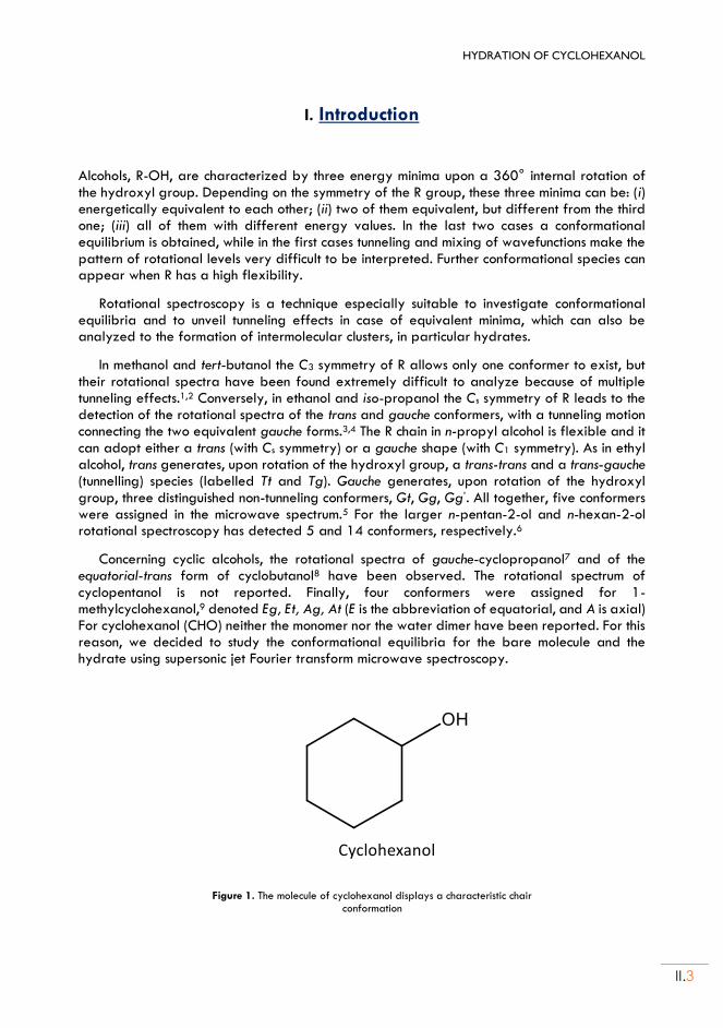
HYDRATION OF CYCLOHEXANOL
II.3
I. Introduction
Alcohols, R-OH, are characterized by three energy minima upon a 360° internal rotation of the hydroxyl group. Depending on the symmetry of the R group, these three minima can be: (i) energetically equivalent to each other; (ii) two of them equivalent, but different from the third one; (iii) all of them with different energy values. In the last two cases a conformational equilibrium is obtained, while in the first cases tunneling and mixing of wavefunctions make the pattern of rotational levels very difficult to be interpreted. Further conformational species can appear when R has a high flexibility.
Rotational spectroscopy is a technique especially suitable to investigate conformational equilibria and to unveil tunneling effects in case of equivalent minima, which can also be analyzed to the formation of intermolecular clusters, in particular hydrates.
In methanol and tert-butanol the C3 symmetry of R allows only one conformer to exist, but their rotational spectra have been found extremely difficult to analyze because of multiple tunneling effects.1,2 Conversely, in ethanol and iso-propanol the Cs symmetry of R leads to the detection of the rotational spectra of the trans and gauche conformers, with a tunneling motion connecting the two equivalent gauche forms.3,4 The R chain in n-propyl alcohol is flexible and it can adopt either a trans (with Cs symmetry) or a gauche shape (with C1 symmetry). As in ethyl alcohol, trans generates, upon rotation of the hydroxyl group, a trans-trans and a trans-gauche (tunnelling) species (labelled Tt and Tg). Gauche generates, upon rotation of the hydroxyl group, three distinguished non-tunneling conformers, Gt, Gg, Gg’. All together, five conformers were assigned in the microwave spectrum.5 For the larger n-pentan-2-ol and n-hexan-2-ol rotational spectroscopy has detected 5 and 14 conformers, respectively.6
Concerning cyclic alcohols, the rotational spectra of gauche-cyclopropanol7 and of the equatorial-trans form of cyclobutanol8 have been observed. The rotational spectrum of cyclopentanol is not reported. Finally, four conformers were assigned for 1-methylcyclohexanol,9 denoted Eg, Et, Ag, At (E is the abbreviation of equatorial, and A is axial) For cyclohexanol (CHO) neither the monomer nor the water dimer have been reported. For this reason, we decided to study the conformational equilibria for the bare molecule and the hydrate using supersonic jet Fourier transform microwave spectroscopy.
Figure 1. The molecule of cyclohexanol displays a characteristic chair
conformation
II.4
Cyclohexanol (CHO) is an important feedstock in the polymer industry, as a precursor to nylons and various plasticizers. It is also used as a solvent. The chair form of the aliphatic ring is the by large the most stable one. However, the orientation of the hydroxyl group gives rise to four possible isomers. The hydroxyl group may be attached in the equatorial (E) or axial (A) positions of the cyclohexyl ring. In both cases R has a Cs symmetry, and it can generate trans and gauche (doubly degenerate) species, for a total of four conforners. James et al.10 concluded from the temperature dependence of the Raman spectra that in the ordered phases the equatorial isomer dominates the structure of the phase. Andre et al.11 found that there is an equal distribution of axial and equatorial forms in the glassy phase. More recently Ibberson et al.12 found that the cyclohexanol molecules adopt only equatorial shapes in crystal, by using high-resolution neutron diffraction of powder, synchrotron X-ray diffraction of powder and single-crystal X-ray diffraction techniques.
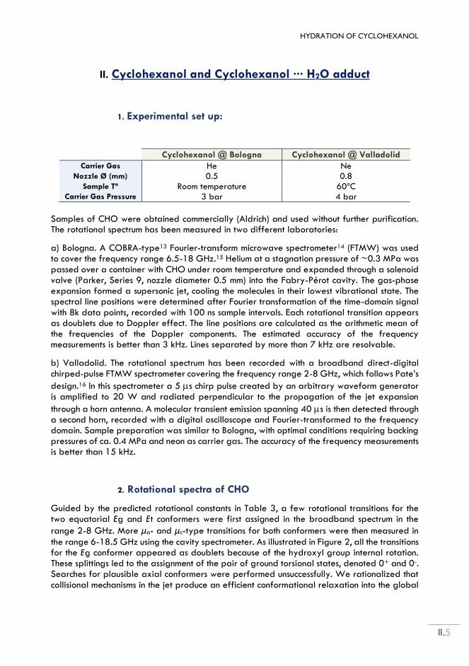
HYDRATION OF CYCLOHEXANOL
II.5
II. Cyclohexanol and Cyclohexanol ··· H2O adduct
1. Experimental set up:
Cyclohexanol @ Bologna Cyclohexanol @ Valladolid
Carrier Gas He Ne Nozzle Ø (mm) 0.5 0.8
Sample Tª Room temperature 60ºC Carrier Gas Pressure 3 bar 4 bar
Samples of CHO were obtained commercially (Aldrich) and used without further purification. The rotational spectrum has been measured in two different laboratories:
a) Bologna. A COBRA-type13 Fourier-transform microwave spectrometer14 (FTMW) was used to cover the frequency range 6.5-18 GHz.15 Helium at a stagnation pressure of ~0.3 MPa was passed over a container with CHO under room temperature and expanded through a solenoid valve (Parker, Series 9, nozzle diameter 0.5 mm) into the Fabry-Pérot cavity. The gas-phase expansion formed a supersonic jet, cooling the molecules in their lowest vibrational state. The spectral line positions were determined after Fourier transformation of the time-domain signal with 8k data points, recorded with 100 ns sample intervals. Each rotational transition appears as doublets due to Doppler effect. The line positions are calculated as the arithmetic mean of the frequencies of the Doppler components. The estimated accuracy of the frequency measurements is better than 3 kHz. Lines separated by more than 7 kHz are resolvable.
b) Valladolid. The rotational spectrum has been recorded with a broadband direct-digital chirped-pulse FTMW spectrometer covering the frequency range 2-8 GHz, which follows Pate’s
design.16 In this spectrometer a 5 s chirp pulse created by an arbitrary waveform generator is amplified to 20 W and radiated perpendicular to the propagation of the jet expansion
through a horn antenna. A molecular transient emission spanning 40 s is then detected through a second horn, recorded with a digital oscilloscope and Fourier-transformed to the frequency domain. Sample preparation was similar to Bologna, with optimal conditions requiring backing pressures of ca. 0.4 MPa and neon as carrier gas. The accuracy of the frequency measurements is better than 15 kHz.
2. Rotational spectra of CHO
Guided by the predicted rotational constants in Table 3, a few rotational transitions for the two equatorial Eg and Et conformers were first assigned in the broadband spectrum in the
range 2-8 GHz. More μa- and μc-type transitions for both conformers were then measured in
the range 6-18.5 GHz using the cavity spectrometer. As illustrated in Figure 2, all the transitions for the Eg conformer appeared as doublets because of the hydroxyl group internal rotation. These splittings led to the assignment of the pair of ground torsional states, denoted 0+ and 0-. Searches for plausible axial conformers were performed unsuccessfully. We rationalized that collisional mechanisms in the jet produce an efficient conformational relaxation into the global
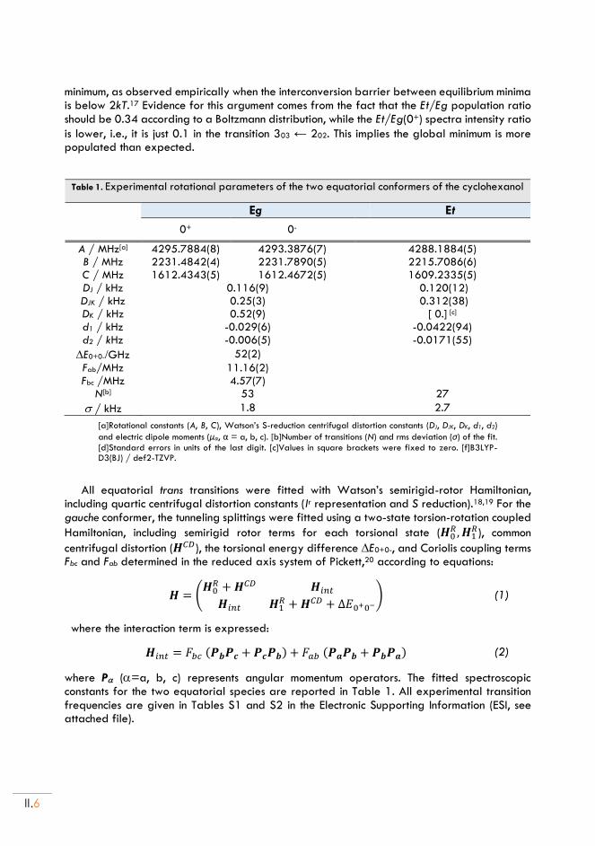
II.6
minimum, as observed empirically when the interconversion barrier between equilibrium minima is below 2kT.17 Evidence for this argument comes from the fact that the Et/Eg population ratio should be 0.34 according to a Boltzmann distribution, while the Et/Eg(0+) spectra intensity ratio
is lower, i.e., it is just 0.1 in the transition 303 ← 202. This implies the global minimum is more populated than expected.
Table 1. Experimental rotational parameters of the two equatorial conformers of the cyclohexanol
Eg Et
0+ 0-
A / MHz[a] 4295.7884(8) 4293.3876(7) 4288.1884(5) B / MHz 2231.4842(4) 2231.7890(5) 2215.7086(6) C / MHz 1612.4343(5) 1612.4672(5) 1609.2335(5) DJ / kHz 0.116(9) 0.120(12) DJK / kHz 0.25(3) 0.312(38) DK / kHz 0.52(9) [ 0.] [c] d1 / kHz -0.029(6) -0.0422(94) d2 / kHz -0.006(5) -0.0171(55)
E0+0-/GHz 52(2)
Fab/MHz 11.16(2) Fbc /MHz 4.57(7)
N[b] 53 27
/ kHz 1.8 2.7
[a]Rotational constants (A, B, C), Watson’s S-reduction centrifugal distortion constants (DJ, DJK, DK, d1, d2)
and electric dipole moments (μα, α = a, b, c). [b]Number of transitions (N) and rms deviation (σ) of the fit. [d]Standard errors in units of the last digit. [c]Values in square brackets were fixed to zero. [f]B3LYP-D3(BJ) / def2-TZVP.
All equatorial trans transitions were fitted with Watson’s semirigid-rotor Hamiltonian, including quartic centrifugal distortion constants (Ir representation and S reduction).18,19 For the gauche conformer, the tunneling splittings were fitted using a two-state torsion-rotation coupled
Hamiltonian, including semirigid rotor terms for each torsional state (𝑯0𝑅 , 𝑯1
𝑅), common
centrifugal distortion (𝑯𝐶𝐷), the torsional energy difference E0+0-, and Coriolis coupling terms Fbc and Fab determined in the reduced axis system of Pickett,20 according to equations:
𝑯 = (𝑯0
𝑅 + 𝑯𝐶𝐷 𝑯𝑖𝑛𝑡
𝑯𝑖𝑛𝑡 𝑯1𝑅 + 𝑯𝐶𝐷 + Δ𝐸0+0−
) (1)
where the interaction term is expressed:
𝑯𝑖𝑛𝑡 = 𝐹𝑏𝑐 (𝑷𝒃𝑷𝒄 + 𝑷𝒄𝑷𝒃) + 𝐹𝑎𝑏 (𝑷𝒂𝑷𝒃 + 𝑷𝒃𝑷𝒂)
(2)
where P (=a, b, c) represents angular momentum operators. The fitted spectroscopic constants for the two equatorial species are reported in Table 1. All experimental transition frequencies are given in Tables S1 and S2 in the Electronic Supporting Information (ESI, see attached file).
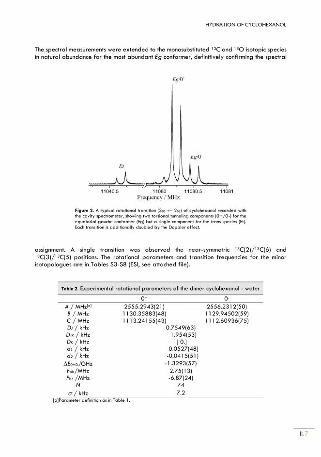
HYDRATION OF CYCLOHEXANOL
II.7
The spectral measurements were extended to the monosubstituted 13C and 18O isotopic species in natural abundance for the most abundant Eg conformer, definitively confirming the spectral
Figure 2. A typical rotational transition (303 ← 202) of cyclohexanol recorded with the cavity spectrometer, showing two torsional tunneling components (0+/0-) for the equatorial gauche conformer (Eg) but a single component for the trans species (Et). Each transition is additionally doubled by the Doppler effect.
assignment. A single transition was observed the near-symmetric 13C(2)/13C(6) and 13C(3)/13C(5) positions. The rotational parameters and transition frequencies for the minor isotopologues are in Tables S3-S8 (ESI, see attached file).
Table 2. Experimental rotational parameters of the dimer cyclohexanol - water
0+ 0-
A / MHz[a] 2555.2943(21) 2556.2312(50) B / MHz 1130.35883(48) 1129.94502(59) C / MHz 1113.24155(43) 1112.60936(75) DJ / kHz 0.7549(63) DJK / kHz 1.954(53) DK / kHz [ 0.] d1 / kHz 0.0527(48) d2 / kHz -0.0415(51)
E0+0-/GHz -1.3293(57)
Fab/MHz 2.75(13) Fbc /MHz -6.87(24)
N 74
/ kHz 7.2 [a]Parameter definition as in Table 1.
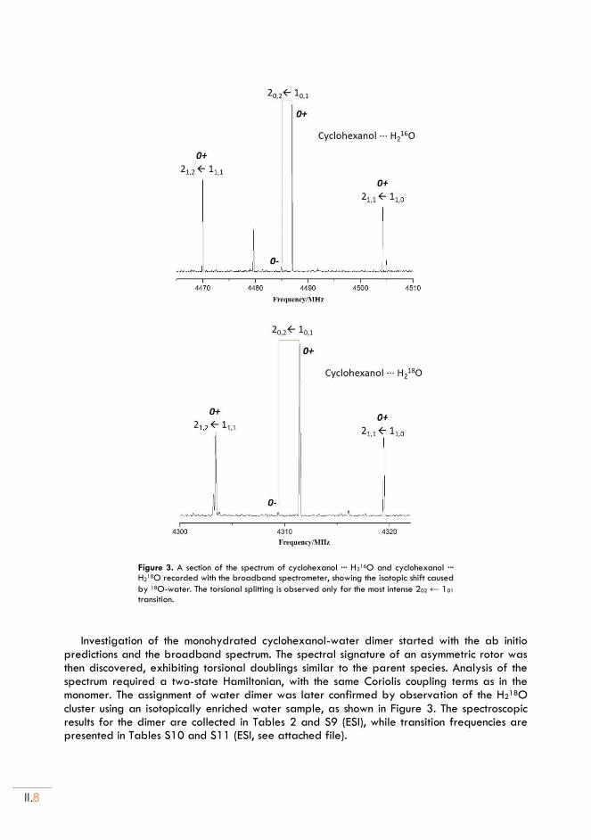
II.8
Figure 3. A section of the spectrum of cyclohexanol ··· H216O and cyclohexanol ···
H218O recorded with the broadband spectrometer, showing the isotopic shift caused
by 18O-water. The torsional splitting is observed only for the most intense 202 ← 101 transition.
Investigation of the monohydrated cyclohexanol-water dimer started with the ab initio predictions and the broadband spectrum. The spectral signature of an asymmetric rotor was then discovered, exhibiting torsional doublings similar to the parent species. Analysis of the spectrum required a two-state Hamiltonian, with the same Coriolis coupling terms as in the monomer. The assignment of water dimer was later confirmed by observation of the H218O cluster using an isotopically enriched water sample, as shown in Figure 3. The spectroscopic results for the dimer are collected in Tables 2 and S9 (ESI), while transition frequencies are presented in Tables S10 and S11 (ESI, see attached file).
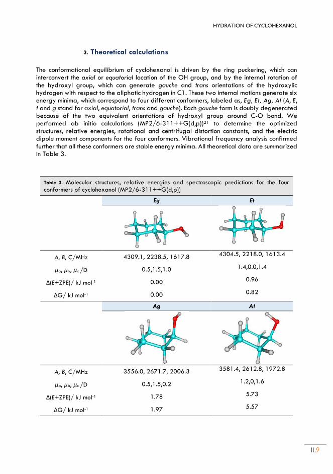
HYDRATION OF CYCLOHEXANOL
II.9
3. Theoretical calculations
The conformational equilibrium of cyclohexanol is driven by the ring puckering, which can interconvert the axial or equatorial location of the OH group, and by the internal rotation of the hydroxyl group, which can generate gauche and trans orientations of the hydroxylic hydrogen with respect to the aliphatic hydrogen in C1. These two internal motions generate six energy minima, which correspond to four different conformers, labeled as, Eg, Et, Ag, At (A, E, t and g stand for axial, equatorial, trans and gauche). Each gauche form is doubly degenerated because of the two equivalent orientations of hydroxyl group around C-O bond. We performed ab initio calculations (MP2/6-311++G(d,p))21 to determine the optimized structures, relative energies, rotational and centrifugal distortion constants, and the electric dipole moment components for the four conformers. Vibrational frequency analysis confirmed further that all these conformers are stable energy minima. All theoretical data are summarized in Table 3.
Table 3. Molecular structures, relative energies and spectroscopic predictions for the four conformers of cyclohexanol (MP2/6-311++G(d,p))
Eg Et
A, B, C/MHz 4309.1, 2238.5, 1617.8 4304.5, 2218.0, 1613.4
μa, μb, μc /D 0.5,1.5,1.0 1.4,0.0,1.4
Δ(E+ZPE)/ kJ mol-1 0.00 0.96
ΔG/ kJ mol-1 0.00 0.82
Ag At
A, B, C/MHz 3556.0, 2671.7, 2006.3 3581.4, 2612.8, 1972.8
μa, μb, μc /D 0.5,1.5,0.2 1.2,0,1.6
Δ(E+ZPE)/ kJ mol-1 1.78 5.73
ΔG/ kJ mol-1 1.97 5.57
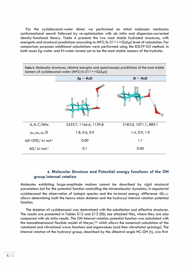
II.10
For the cyclohexanol-water dimer we performed an initial molecular mechanics conformational search followed by re-optimization with ab initio and dispersion-corrected density-functional theory. Table 4 presents the two most stable hydrated structures, with energetic and structural predictions according to MP2/6-311++G(d,p) level of calculation. For comparison purposes additional calculations were performed using the B3LYP-D3 method. In both cases Eg-water and Et-water turned out to be the most stable isomers of the hydrate.
Table 4. Molecular structures, relative energies and spectroscopic predictions of the most stable isomers of cyclohexanol-water (MP2/6-311++G(d,p))
Eg ··· H2O Et ··· H2O
A, B, C/MHz 2553.7, 1166.6, 1139.8 3183.0, 1071.1, 889.1
μa, μb, μc/D 1.8, 0.6, 0.9 1.4, 0.9, 1.9
Δ(E+ZPE)/ kJ mol-1 0.00 1.1
ΔG/ kJ mol-1 0.1 0.00
4. Molecular Structure and Potential energy functions of the OH group internal rotation
Molecules exhibiting large-amplitude motions cannot be described by rigid structural parameters but for the potential function controlling the intramolecular dynamics. In equatorial
cyclohexanol the observation of isotopic species and the torsional energy difference E0+0- allows determining both the heavy-atom skeleton and the hydroxyl internal rotation potential function.
The skeleton of cyclohexanol was determined with the substitution and effective structures. The results are presented in Tables S12 and S13 (ESI, see attached file), where they are also compared with ab initio results. The OH internal rotation potential function was calculated with the monodimensional flexible model of Meyer,22 which allows the numerical calculation of the rotational and vibrational wave functions and eigenvalues (and then vibrational spacings). The
internal rotation of the hydroxyl group, described by the dihedral angle HC-OH (τ), was first
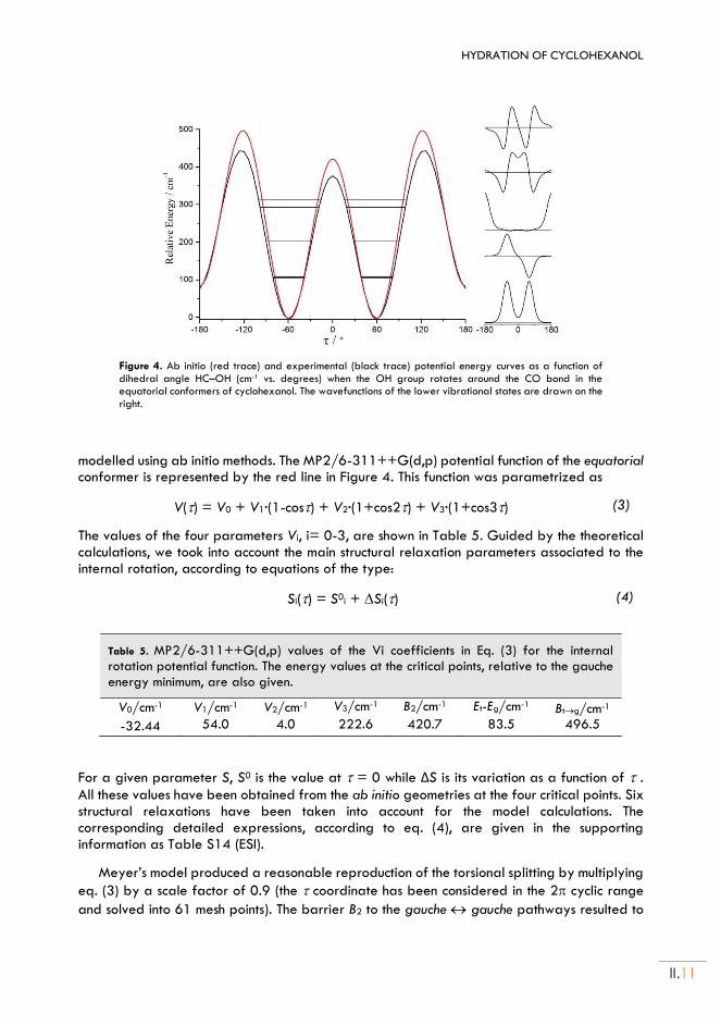
HYDRATION OF CYCLOHEXANOL
II.11
Figure 4. Ab initio (red trace) and experimental (black trace) potential energy curves as a function of dihedral angle HC–OH (cm-1 vs. degrees) when the OH group rotates around the CO bond in the equatorial conformers of cyclohexanol. The wavefunctions of the lower vibrational states are drawn on the right.
modelled using ab initio methods. The MP2/6-311++G(d,p) potential function of the equatorial conformer is represented by the red line in Figure 4. This function was parametrized as
V() = V0 + V1·(1-cos) + V2·(1+cos2) + V3·(1+cos3) (3)
The values of the four parameters Vi, i= 0-3, are shown in Table 5. Guided by the theoretical calculations, we took into account the main structural relaxation parameters associated to the internal rotation, according to equations of the type:
Si() = S0i + Si() (4)
Table 5. MP2/6-311++G(d,p) values of the Vi coefficients in Eq. (3) for the internal rotation potential function. The energy values at the critical points, relative to the gauche energy minimum, are also given.
V0/cm-1 V1/cm-1 V2/cm-1 V3/cm-1 B2/cm-1 Et-Eg/cm-1 Btg/cm-1
-32.44 54.0 4.0 222.6 420.7 83.5 496.5
For a given parameter S, S0 is the value at = 0 while ΔS is its variation as a function of . All these values have been obtained from the ab initio geometries at the four critical points. Six structural relaxations have been taken into account for the model calculations. The corresponding detailed expressions, according to eq. (4), are given in the supporting information as Table S14 (ESI).
Meyer’s model produced a reasonable reproduction of the torsional splitting by multiplying
eq. (3) by a scale factor of 0.9 (the coordinate has been considered in the 2 cyclic range
and solved into 61 mesh points). The barrier B2 to the gauche gauche pathways resulted to
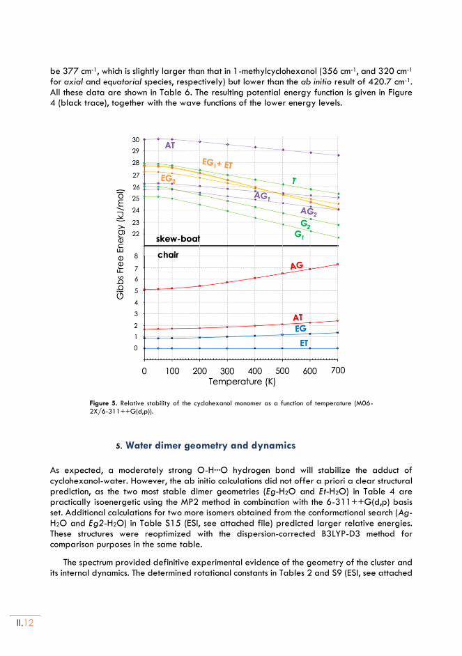
II.12
be 377 cm-1, which is slightly larger than that in 1-methylcyclohexanol (356 cm-1, and 320 cm-1 for axial and equatorial species, respectively) but lower than the ab initio result of 420.7 cm-1. All these data are shown in Table 6. The resulting potential energy function is given in Figure 4 (black trace), together with the wave functions of the lower energy levels.
Figure 5. Relative stability of the cyclohexanol monomer as a function of temperature (M06-2X/6-311++G(d,p)).
5. Water dimer geometry and dynamics
As expected, a moderately strong O-H···O hydrogen bond will stabilize the adduct of cyclohexanol-water. However, the ab initio calculations did not offer a priori a clear structural prediction, as the two most stable dimer geometries (Eg-H2O and Et-H2O) in Table 4 are practically isoenergetic using the MP2 method in combination with the 6-311++G(d,p) basis set. Additional calculations for two more isomers obtained from the conformational search (Ag-H2O and Eg2-H2O) in Table S15 (ESI, see attached file) predicted larger relative energies. These structures were reoptimized with the dispersion-corrected B3LYP-D3 method for comparison purposes in the same table.
The spectrum provided definitive experimental evidence of the geometry of the cluster and its internal dynamics. The determined rotational constants in Tables 2 and S9 (ESI, see attached
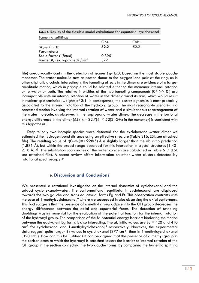
HYDRATION OF CYCLOHEXANOL
II.13
Table 6. Results of the flexible model calculations for equatorial cyclohexanol
Tunneling splittings
Obs. Calc.
E0+0-/ GHz 52.2 52.2
Parameters: Scale factor f (fitted) 0.895 Barrier B2 (extrapolated) /cm-1 377
file) unequivocally confirm the detection of isomer Eg-H2O, based on the most stable gauche monomer. The water molecule acts as proton donor to the oxygen lone pair at the ring, as in other aliphatic alcohols. Interestingly, the tunneling effects in the dimer are evidence of a large-amplitude motion, which in principle could be related either to the monomer internal rotation or to water or both. The relative intensities of the two tunneling components (0+ >> 0-) are incompatible with an internal rotation of water in the dimer around its axis, which would result in nuclear spin statistical weights of 3:1. In consequence, the cluster dynamics is most probably associated to the internal rotation of the hydroxyl group. The most reasonable scenario is a concerted motion involving the internal rotation of water and a simultaneous rearrangement of the water molecule, as observed in the isopropanol-water dimer. The decrease in the torsional
energy difference in the dimer (E0+0-= 32.7(4) < 52(2) GHz in the monomer) is consistent with this hypothesis.
Despite only two isotopic species were detected for the cyclohexanol-water dimer we estimated the hydrogen bond distance using an effective structure (Table S16, ESI, see attached file). The resulting value of r(O-Hw)=1.928(5) Å is slightly larger than the ab initio prediction (1.881 Å), but within the broad range observed for this interaction in crystal structures (1.40-2.18 Å).23 The substitution coordinates of the water oxygen are calculated in Table S17 (ESI, see attached file). A recent review offers information on other water clusters detected by rotational spectroscopy.24
6. Discussion and Conclusions
We presented a rotational investigation on the internal dynamics of cyclohexanol and the adduct cyclohexanol–water. The conformational equilibria in cyclohexanol are displaced towards the two gauche and trans equatorial forms Eg and Et. This observation contrasts with the case of 1-methylcyclohexanol,9 where we succeeded in also observing the axial conformers. This fact suggests that the presence of a methyl group adjacent to the OH group decreases the energy differences between the axial and equatorial forms. The detection of tunneling doublings was instrumental for the evaluation of the potential function for the internal rotation of the hydroxyl group. The comparison of the B2 potential energy barriers hindering the motion between the equivalent Eg forms is also interesting. The ab initio values are B2 = 420 and 410 cm-1 for cyclohexanol and 1-methylcyclohexanol,9 respectively. However, the experimental data suggest quite larger B2 values in cyclohexanol (377 cm-1) than in 1-methylcyclohexanol (320 cm-1). How can this be justified? It can be argued that the presence of a methyl group in the carbon atom to which the hydroxyl is attached lowers the barrier to internal rotation of the OH group in the section connecting the two gauche forms. By comparing the tunneling splitting
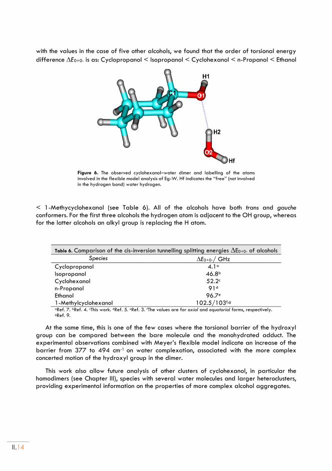
II.14
with the values in the case of five other alcohols, we found that the order of torsional energy
difference E0+0- is as: Cyclopropanol < Isopropanol < Cyclohexanol < n-Propanol < Ethanol
Figure 6. The observed cyclohexanol–water dimer and labelling of the atoms involved in the flexible model analysis of Eg-W. Hf indicates the ‘‘free’’ (not involved in the hydrogen bond) water hydrogen.
< 1-Methycyclohexanol (see Table 6). All of the alcohols have both trans and gauche conformers. For the first three alcohols the hydrogen atom is adjacent to the OH group, whereas for the latter alcohols an alkyl group is replacing the H atom.
Table 6. Comparison of the cis-inversion tunnelling splitting energies E0+0- of alcohols Species E0+0-/ GHz
Cyclopropanol 4.1a
Isopropanol 46.8b
Cyclohexanol 52.2c
n-Propanol 91d
Ethanol 96.7e
1-Methylcyclohexanol 102.5/103f,g aRef. 7. bRef. 4. cThis work. dRef. 5. eRef. 3. fThe values are for axial and equatorial forms, respectively. gRef. 9.
At the same time, this is one of the few cases where the torsional barrier of the hydroxyl group can be compared between the bare molecule and the monohydrated adduct. The experimental observations combined with Meyer’s flexible model indicate an increase of the barrier from 377 to 494 cm-1 on water complexation, associated with the more complex concerted motion of the hydroxyl group in the dimer.
This work also allow future analysis of other clusters of cyclohexanol, in particular the homodimers (see Chapter III), species with several water molecules and larger heteroclusters, providing experimental information on the properties of more complex alcohol aggregates.
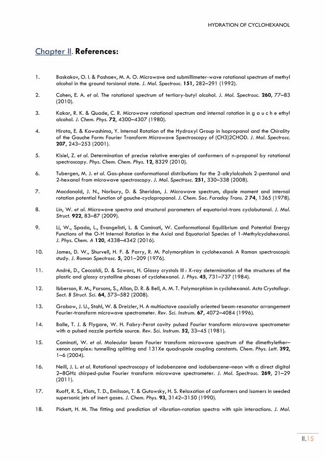
HYDRATION OF CYCLOHEXANOL
II.15
Chapter II. References:
1. Baskakov, O. I. & Pashaev, M. A. O. Microwave and submillimeter-wave rotational spectrum of methyl alcohol in the ground torsional state. J. Mol. Spectrosc. 151, 282–291 (1992).
2. Cohen, E. A. et al. The rotational spectrum of tertiary-butyl alcohol. J. Mol. Spectrosc. 260, 77–83 (2010).
3. Kakar, R. K. & Quade, C. R. Microwave rotational spectrum and internal rotation in g a u c h e ethyl alcohol. J. Chem. Phys. 72, 4300–4307 (1980).
4. Hirota, E. & Kawashima, Y. Internal Rotation of the Hydroxyl Group in Isopropanol and the Chirality of the Gauche Form: Fourier Transform Microwave Spectroscopy of (CH3)2CHOD. J. Mol. Spectrosc. 207, 243–253 (2001).
5. Kisiel, Z. et al. Determination of precise relative energies of conformers of n-propanol by rotational spectroscopy. Phys. Chem. Chem. Phys. 12, 8329 (2010).
6. Tubergen, M. J. et al. Gas-phase conformational distributions for the 2-alkylalcohols 2-pentanol and 2-hexanol from microwave spectroscopy. J. Mol. Spectrosc. 251, 330–338 (2008).
7. Macdonald, J. N., Norbury, D. & Sheridan, J. Microwave spectrum, dipole moment and internal rotation potential function of gauche-cyclopropanol. J. Chem. Soc. Faraday Trans. 2 74, 1365 (1978).
8. Lin, W. et al. Microwave spectra and structural parameters of equatorial-trans cyclobutanol. J. Mol. Struct. 922, 83–87 (2009).
9. Li, W., Spada, L., Evangelisti, L. & Caminati, W. Conformational Equilibrium and Potential Energy Functions of the O-H Internal Rotation in the Axial and Equatorial Species of 1-Methylcyclohexanol. J. Phys. Chem. A 120, 4338–4342 (2016).
10. James, D. W., Shurvell, H. F. & Parry, R. M. Polymorphism in cyclohexanol: A Raman spectroscopic study. J. Raman Spectrosc. 5, 201–209 (1976).
11. André, D., Ceccaldi, D. & Szwarc, H. Glassy crystals III : X-ray determination of the structures of the plastic and glassy crystalline phases of cyclohexanol. J. Phys. 45, 731–737 (1984).
12. Ibberson, R. M., Parsons, S., Allan, D. R. & Bell, A. M. T. Polymorphism in cyclohexanol. Acta Crystallogr. Sect. B Struct. Sci. 64, 573–582 (2008).
13. Grabow, J. U., Stahl, W. & Dreizler, H. A multioctave coaxially oriented beam-resonator arrangement Fourier-transform microwave spectrometer. Rev. Sci. Instrum. 67, 4072–4084 (1996).
14. Balle, T. J. & Flygare, W. H. Fabry-Perot cavity pulsed Fourier transform microwave spectrometer with a pulsed nozzle particle source. Rev. Sci. Instrum. 52, 33–45 (1981).
15. Caminati, W. et al. Molecular beam Fourier transform microwave spectrum of the dimethylether–xenon complex: tunnelling splitting and 131Xe quadrupole coupling constants. Chem. Phys. Lett. 392, 1–6 (2004).
16. Neill, J. L. et al. Rotational spectroscopy of iodobenzene and iodobenzene–neon with a direct digital 2–8GHz chirped-pulse Fourier transform microwave spectrometer. J. Mol. Spectrosc. 269, 21–29 (2011).
17. Ruoff, R. S., Klots, T. D., Emilsson, T. & Gutowsky, H. S. Relaxation of conformers and isomers in seeded supersonic jets of inert gases. J. Chem. Phys. 93, 3142–3150 (1990).
18. Pickett, H. M. The fitting and prediction of vibration-rotation spectra with spin interactions. J. Mol.
II.16
Spectrosc. 148, 371–377 (1991).
19. Watson, J. K. G. Vibrational Spectra and Structure. in (ed. Durig, J. R.) 1–89 (Elsevier Inc., 1977).
20. Pickett, H. M. Vibration—Rotation Interactions and the Choice of Rotating Axes for Polyatomic Molecules. J. Chem. Phys. 56, 1715–1723 (1972).
21. Frisch, M. J., Trucks, G. W., Schlegel, H. B., Scuseria, G. E. & Robb, M. A. et al. Gaussian 09, Revision A.02. (Gaussian, Inc., 2016).
22. Meyer, R. Flexible models for intramolecular motion, a versatile treatment and its application to glyoxal. J. Mol. Spectrosc. 76, 266–300 (1979).
23. Jeffrey, G. A. An Introduction to Hydrogen Bonding. (Oxford University Press, 1997).
24. Caminati, W. & Grabow, J.-U. Advancements in Microwave Spectroscopy. in Frontiers and Advancements in Microwave Spectroscopy (ed. Laane, J.) 569–598 (Elsevier, 2018).
Chapter III DIMERIZATION OF CYCLOHEXANOL:
Adapted from:
(*): M. Juanes, I. Usabiaga, I. León, L. Evangelisti, J. A. Fernández and A. Lesarri, Angew. Chem. Int. Ed., 2020, 59, 12412 – 12421 (The Electronic Supporting Information – ESI, can be found in the document: thesis-ESI.pdf)
(Cyclohexanol)2*

III.2
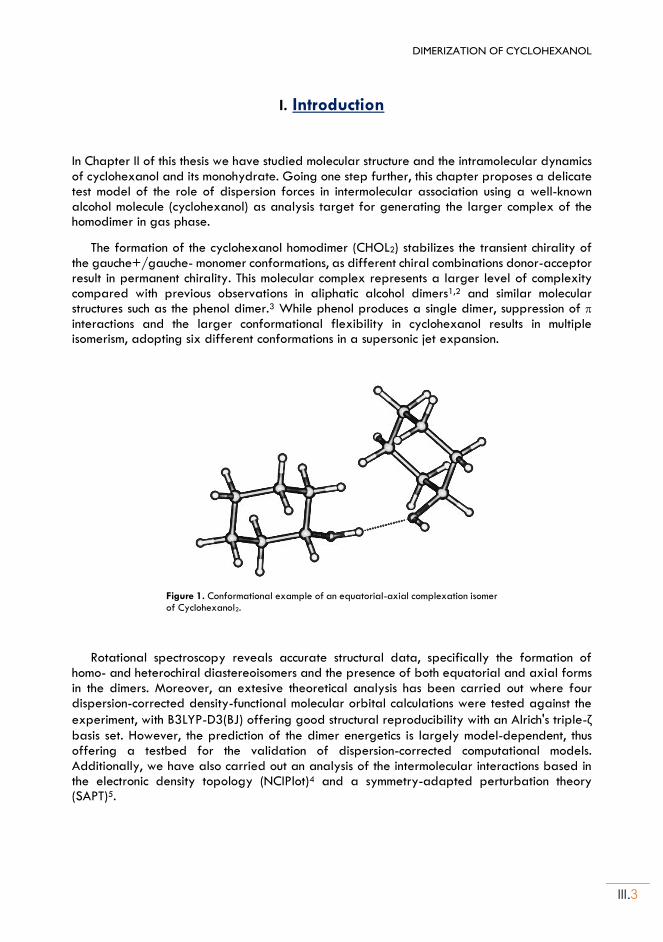
DIMERIZATION OF CYCLOHEXANOL
III.3
I. Introduction
In Chapter II of this thesis we have studied molecular structure and the intramolecular dynamics of cyclohexanol and its monohydrate. Going one step further, this chapter proposes a delicate test model of the role of dispersion forces in intermolecular association using a well-known alcohol molecule (cyclohexanol) as analysis target for generating the larger complex of the homodimer in gas phase.
The formation of the cyclohexanol homodimer (CHOL2) stabilizes the transient chirality of the gauche+/gauche- monomer conformations, as different chiral combinations donor-acceptor result in permanent chirality. This molecular complex represents a larger level of complexity compared with previous observations in aliphatic alcohol dimers1,2 and similar molecular structures such as the phenol dimer.3 While phenol produces a single dimer, suppression of interactions and the larger conformational flexibility in cyclohexanol results in multiple isomerism, adopting six different conformations in a supersonic jet expansion.
Figure 1. Conformational example of an equatorial-axial complexation isomer of Cyclohexanol2.
Rotational spectroscopy reveals accurate structural data, specifically the formation of homo- and heterochiral diastereoisomers and the presence of both equatorial and axial forms in the dimers. Moreover, an extesive theoretical analysis has been carried out where four dispersion-corrected density-functional molecular orbital calculations were tested against the
experiment, with B3LYP-D3(BJ) offering good structural reproducibility with an Alrich's triple-ζ basis set. However, the prediction of the dimer energetics is largely model-dependent, thus offering a testbed for the validation of dispersion-corrected computational models. Additionally, we have also carried out an analysis of the intermolecular interactions based in the electronic density topology (NCIPlot)4 and a symmetry-adapted perturbation theory (SAPT)5.
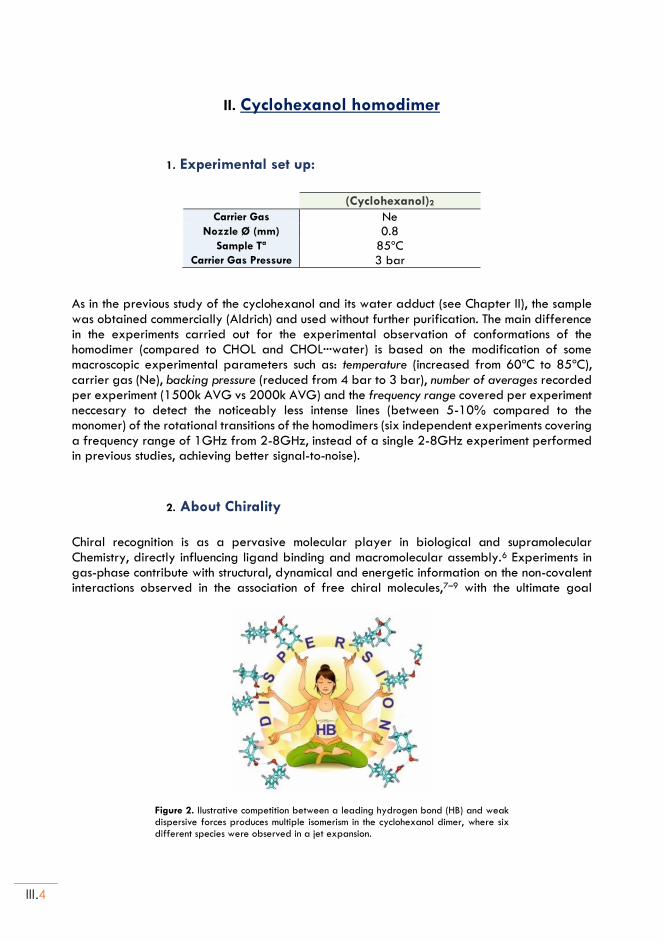
III.4
II. Cyclohexanol homodimer
1. Experimental set up:
(Cyclohexanol)2
Carrier Gas Ne Nozzle Ø (mm) 0.8
Sample Tª 85ºC Carrier Gas Pressure 3 bar
As in the previous study of the cyclohexanol and its water adduct (see Chapter II), the sample was obtained commercially (Aldrich) and used without further purification. The main difference in the experiments carried out for the experimental observation of conformations of the homodimer (compared to CHOL and CHOL···water) is based on the modification of some macroscopic experimental parameters such as: temperature (increased from 60ºC to 85ºC), carrier gas (Ne), backing pressure (reduced from 4 bar to 3 bar), number of averages recorded per experiment (1500k AVG vs 2000k AVG) and the frequency range covered per experiment neccesary to detect the noticeably less intense lines (between 5-10% compared to the monomer) of the rotational transitions of the homodimers (six independent experiments covering a frequency range of 1GHz from 2-8GHz, instead of a single 2-8GHz experiment performed in previous studies, achieving better signal-to-noise).
2. About Chirality
Chiral recognition is as a pervasive molecular player in biological and supramolecular Chemistry, directly influencing ligand binding and macromolecular assembly.6 Experiments in gas-phase contribute with structural, dynamical and energetic information on the non-covalent interactions observed in the association of free chiral molecules,7–9 with the ultimate goal
Figure 2. Ilustrative competition between a leading hydrogen bond (HB) and weak dispersive forces produces multiple isomerism in the cyclohexanol dimer, where six different species were observed in a jet expansion.
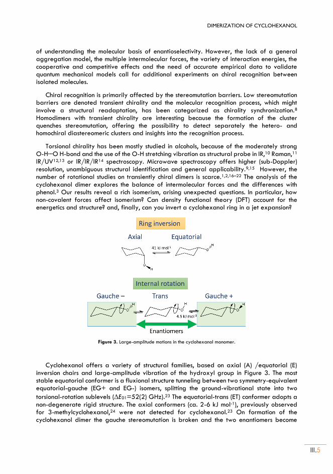
DIMERIZATION OF CYCLOHEXANOL
III.5
of understanding the molecular basis of enantioselectivity. However, the lack of a general aggregation model, the multiple intermolecular forces, the variety of interaction energies, the cooperative and competitive effects and the need of accurate empirical data to validate quantum mechanical models call for additional experiments on chiral recognition between isolated molecules.
Chiral recognition is primarily affected by the stereomutation barriers. Low stereomutation barriers are denoted transient chirality and the molecular recognition process, which might involve a structural readaptation, has been categorized as chirality synchronization.8 Homodimers with transient chirality are interesting because the formation of the cluster quenches stereomutation, offering the possibility to detect separately the hetero- and homochiral diastereomeric clusters and insights into the recognition process.
Torsional chirality has been mostly studied in alcohols, because of the moderately strong O-H···O H-bond and the use of the O-H stretching vibration as structural probe in IR,10 Raman,11 IR/UV12,13 or IR/IR/IR14 spectroscopy. Microwave spectroscopy offers higher (sub-Doppler) resolution, unambiguous structural identification and general applicability.9,15 However, the number of rotational studies on transiently chiral dimers is scarce.1,2,16–22 The analysis of the cyclohexanol dimer explores the balance of intermolecular forces and the differences with phenol.3 Our results reveal a rich isomerism, arising unexpected questions. In particular, how non-covalent forces affect isomerism? Can density functional theory (DFT) account for the energetics and structure? and, finally, can you invert a cyclohexanol ring in a jet expansion?
Figure 3. Large-amplitude motions in the cyclohexanol monomer.
Cyclohexanol offers a variety of structural families, based on axial (A) /equatorial (E) inversion chairs and large-amplitude vibration of the hydroxyl group in Figure 3. The most stable equatorial conformer is a fluxional structure tunneling between two symmetry-equivalent equatorial-gauche (EG+ and EG-) isomers, splitting the ground-vibrational state into two
torsional-rotation sublevels (E01=52(2) GHz).23 The equatorial-trans (ET) conformer adopts a non-degenerate rigid structure. The axial conformers (ca. 2-6 kJ mol-1), previously observed for 3-methylcyclohexanol,24 were not detected for cyclohexanol.23 On formation of the cyclohexanol dimer the gauche stereomutation is broken and the two enantiomers become
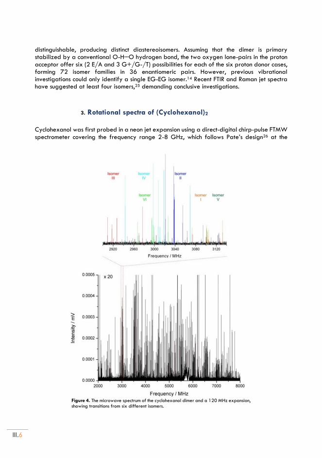
III.6
distinguishable, producing distinct diastereoisomers. Assuming that the dimer is primary stabilized by a conventional O-H···O hydrogen bond, the two oxygen lone-pairs in the proton acceptor offer six (2 E/A and 3 G+/G-/T) possibilities for each of the six proton donor cases, forming 72 isomer families in 36 enantiomeric pairs. However, previous vibrational investigations could only identify a single EG-EG isomer.14 Recent FTIR and Raman jet spectra have suggested at least four isomers,25 demanding conclusive investigations.
3. Rotational spectra of (Cyclohexanol)2
Cyclohexanol was first probed in a neon jet expansion using a direct-digital chirp-pulse FTMW spectrometer covering the frequency range 2-8 GHz, which follows Pate’s design26 at the
Figure 4. The microwave spectrum of the cyclohexanol dimer and a 120 MHz expansion, showing transitions from six different isomers.
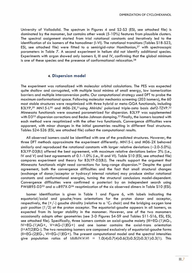
DIMERIZATION OF CYCLOHEXANOL
III.7
University of Valladolid. The spectrum in Figures 4 and S2-S3 (ESI, see attached file) is dominated by the monomer, but contains other weak (5-10%) features from plausible clusters. The spectral assignment started from trial rotational constants and iteratively led to the identification of six isomers of the homodimer (I-VI). The rotational transitions (Tables S18-S23, ESI, see attached file) were fitted to a semirigid-rotor Hamiltonian,27 with spectroscopic parameters in Table 7. A second experiment in helium did not identify additional species. Experiments with argon showed only isomers II, III and IV, confirming that the global minimum is one of these species and the presence of conformational relaxation.28
4. Dispersion model
The experiment was rationalized with molecular orbital calculations. The PES was expected quite shallow and corrugated, with multiple local minima of small energy, low isomerization barriers and multiple interconversion paths. Our computational strategy used DFT to probe the maximum conformational space. Following molecular mechanics screening (203 isomers), the 60 most stable structures were reoptimized with three hybrid or meta-GGA functionals, including B3LYP,29 MN15-L30 and M06-2X,31using Ahlrichs’ polarized triple-zeta basis def2-TZVP.32 Minnesota functionals were assumed parametrized for dispersion. B3LYP was supplemented with D333 dispersion corrections and Becke-Johnson damping.33 Finally, the isomers located with each method were reoptimized with the other two functionals. Convergence difficulties were apparent, with minor changes in the initial geometries resulting in different final structures. Tables S24-S26 (ESI, see attached file) collect the computational results.
All observed isomers could be identified with one of the predicted structures. However, the three DFT methods approximate the experiment differently. MN15-L and M06-2X behaved
similarly and reproduced the rotational constants with larger relative deviations (2.0-5.0%). B3LYP-D3(BJ) offered the best agreement, with maximum relative deviations below 3% (i.e., IV and V) and best agreements of 0.1-1.0% (i.e., III and VI). Table S10 (ESI, see attached file) compares experiment and theory for B3LYP-D3(BJ). The results support the argument that Minnesota functionals might need corrections for long-range dispersion.34 Despite the good agreement, both the convergence difficulties and the fact that small structural changes (exchange of donor/acceptor or hydroxyl internal rotation) may produce similar rotational constants and conformational energies, turning the structural conclusions model-dependent. Convergence difficulties were confirmed a posteriori by an independent search using PW6B95-D335 and a B97X-D36 reoptimization of the six observed dimers in Table S10 (ESI).
Isomer identification is given in Table 1 and Figure 6, with labels indicating the equatorial/axial and gauche/trans orientations for the proton donor and acceptor, respectively, the (+/-) gauche chirality (relative to a 1C4 chair) and the bridging oxygen lone pair position (1/2) at the proton acceptor. The equatorial-gauche appears in all isomers, as expected from its larger stability in the monomer. However, one of the two monomers occasionally adopts other geometries (see 3-D Figures S4-S9 and Tables S11-S16, ESI, ESI, see attached file). Specifically, three isomers contain an axial-gauche moiety (III=EG-(1)AG-, IV=EG-(1)AG+, V=AG-(1)EG+) and one isomer contains the axial-trans orientation (I=AT(2)EG-). The two remaining isomers are composed exclusively of equatorial-gauche forms (II=EG-(2)EG-, VI=EG-(1)EG+). The present computational model and the spectral intensities give population ratios of I:II:III:IV:V:VI = 1.0(4):0.7(4):0.6(3):0.5(2):0.3(1):0.3(1). This
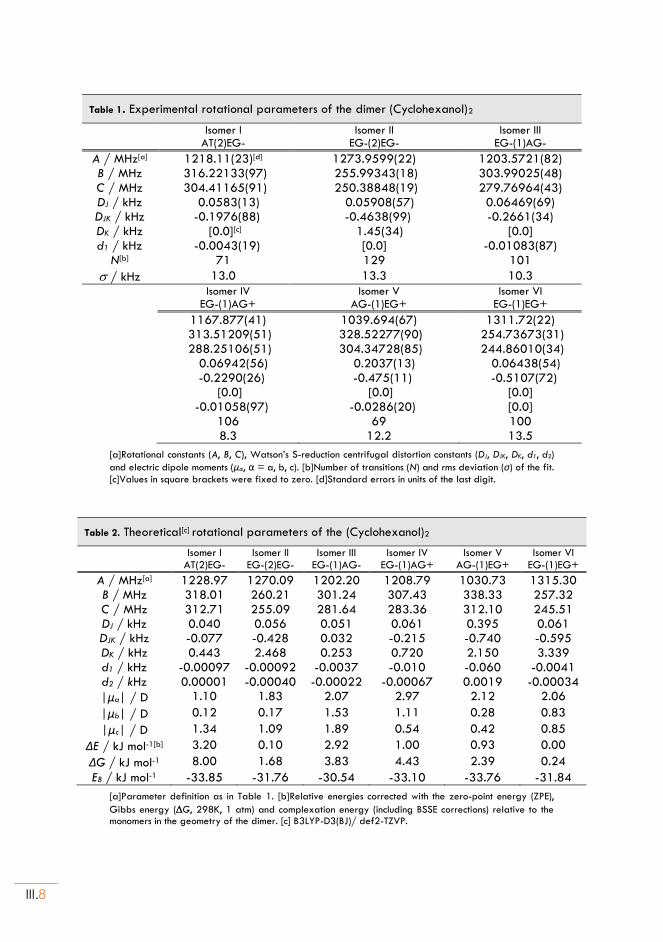
III.8
Table 1. Experimental rotational parameters of the dimer (Cyclohexanol)2
Isomer I
AT(2)EG- Isomer II
EG-(2)EG- Isomer III
EG-(1)AG-
A / MHz[a] 1218.11(23)[d] 1273.9599(22) 1203.5721(82) B / MHz 316.22133(97) 255.99343(18) 303.99025(48) C / MHz 304.41165(91) 250.38848(19) 279.76964(43) DJ / kHz 0.0583(13) 0.05908(57) 0.06469(69) DJK / kHz -0.1976(88) -0.4638(99) -0.2661(34) DK / kHz [0.0][c] 1.45(34) [0.0] d1 / kHz -0.0043(19) [0.0] -0.01083(87)
N[b] 71 129 101
/ kHz 13.0 13.3 10.3
Isomer IV
EG-(1)AG+ Isomer V
AG-(1)EG+ Isomer VI
EG-(1)EG+
1167.877(41) 1039.694(67) 1311.72(22) 313.51209(51) 328.52277(90) 254.73673(31) 288.25106(51) 304.34728(85) 244.86010(34) 0.06942(56) 0.2037(13) 0.06438(54) -0.2290(26) -0.475(11) -0.5107(72) [0.0] [0.0] [0.0] -0.01058(97) -0.0286(20) [0.0] 106 69 100 8.3 12.2 13.5
[a]Rotational constants (A, B, C), Watson’s S-reduction centrifugal distortion constants (DJ, DJK, DK, d1, d2)
and electric dipole moments (μα, α = a, b, c). [b]Number of transitions (N) and rms deviation (σ) of the fit. [c]Values in square brackets were fixed to zero. [d]Standard errors in units of the last digit.
Table 2. Theoretical[c] rotational parameters of the (Cyclohexanol)2
Isomer I
AT(2)EG- Isomer II
EG-(2)EG- Isomer III
EG-(1)AG- Isomer IV
EG-(1)AG+ Isomer V
AG-(1)EG+ Isomer VI
EG-(1)EG+
A / MHz[a] 1228.97 1270.09 1202.20 1208.79 1030.73 1315.30 B / MHz 318.01 260.21 301.24 307.43 338.33 257.32 C / MHz 312.71 255.09 281.64 283.36 312.10 245.51 DJ / kHz 0.040 0.056 0.051 0.061 0.395 0.061 DJK / kHz -0.077 -0.428 0.032 -0.215 -0.740 -0.595 DK / kHz 0.443 2.468 0.253 0.720 2.150 3.339 d1 / kHz -0.00097 -0.00092 -0.0037 -0.010 -0.060 -0.0041
d2 / kHz 0.00001 -0.00040 -0.00022 -0.00067 0.0019 -0.00034
|μa| / D 1.10 1.83 2.07 2.97 2.12 2.06
|μb| / D 0.12 0.17 1.53 1.11 0.28 0.83
|μc| / D 1.34 1.09 1.89 0.54 0.42 0.85
ΔE / kJ mol-1[b] 3.20 0.10 2.92 1.00 0.93 0.00
ΔG / kJ mol-1 8.00 1.68 3.83 4.43 2.39 0.24
EB / kJ mol-1 -33.85 -31.76 -30.54 -33.10 -33.76 -31.84
[a]Parameter definition as in Table 1. [b]Relative energies corrected with the zero-point energy (ZPE),
Gibbs energy (G, 298K, 1 atm) and complexation energy (including BSSE corrections) relative to the monomers in the geometry of the dimer. [c] B3LYP-D3(BJ)/ def2-TZVP.
DIMERIZATION OF CYCLOHEXANOL
III.9
calculation is only approximate, as it additionally assumes a polarization regime proportional to the squared dipole moments and uniform instrumental response.37
The presence of axial forms is noticeable. Unlike 1-methylcyclohexanol, where the bulkier methyl group stabilizes the axial hydroxyl,24 cyclohexanol has a preference for the
Figure 5. Equatorial-to-axial estimated interconversion barriers for the cyclohexanol dimer starting from the six observed dimer geometries. The reaction barriers are estimations, since no transition-state calculation has been performed. Each interconversion barrier is the average over six interconversion paths using as inversion coordinate the dihedrals C1-C2-C3-C4, C2-C3-C4-C5, C3-C4-C5-C6, C4-C5-C6-C1, C5-C6-C1-C2 and C6-C1-C2-C3. The conformational stabilities were calculated with M06-2X/6-311++G(d,p) and gave barriers heights of 44-51 kJ mol-1.
equatorial conformations (Figure 5, Chapter II).23 In order to explain the axial forms we compared the equatorial-to-axial potential barriers both in the monomer and the dimer. The inversion barrier for the cyclohexanol chair was predicted as 47-54 kJ mol-1 in a coordinate scan (no transient-state calculation: Figure S1, ESI, see attached file). A similar calculation in the dimer starting from each of the observed geometries and averaging over six inversion paths rendered comparatively similar barriers of 44-51 kJ mol-1 in Figure 5. We conclude that there are no plausible ring inversion paths of reduced energy, so the presence of axial forms is due to energetic collisions at the initial stages of the expansion. Collisional stabilization of specific isomers undetected in the monomers has been observed in jets for other clusters.38–40
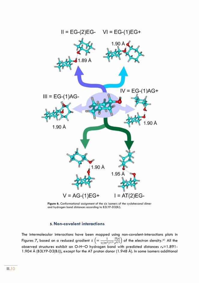
III.10
Figure 6. Conformational assignment of the six isomers of the cyclohexanol dimer and hydrogen bond distances according to B3LYP-D3(BJ).
5. Non-covalent interactions
The intermolecular interactions have been mapped using non-covalent-interactions plots in
Figures 7, based on a reduced gradient 𝑠 (=1
1(3𝜋2)1/3
|∇𝜌|
𝜌4/3) of the electron density.41 All the
observed structures exhibit an O-H···O hydrogen bond with predicted distances re=1.891-1.904 Å (B3LYP-D3(BJ)), except for the AT proton donor (1.948 Å). In some isomers additional
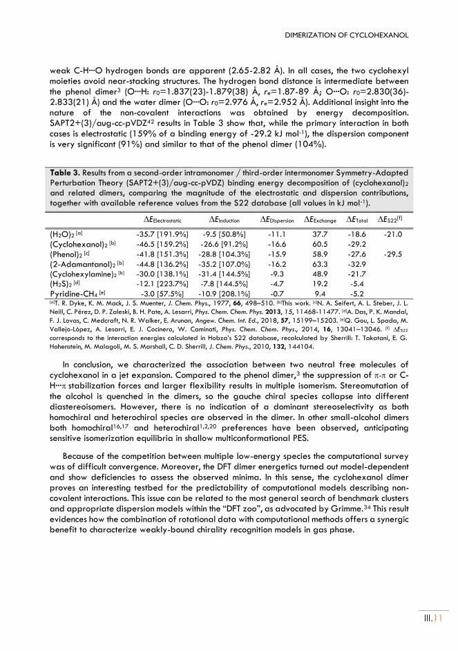
DIMERIZATION OF CYCLOHEXANOL
III.11
weak C-H···O hydrogen bonds are apparent (2.65-2.82 Å). In all cases, the two cyclohexyl moieties avoid near-stacking structures. The hydrogen bond distance is intermediate between the phenol dimer3 (O···H: r0=1.837(23)-1.879(38) Å, re=1.87-89 Å; O···O: r0=2.830(36)-2.833(21) Å) and the water dimer (O···O: r0=2.976 Å, re=2.952 Å). Additional insight into the nature of the non-covalent interactions was obtained by energy decomposition. SAPT2+(3)/aug-cc-pVDZ42 results in Table 3 show that, while the primary interaction in both cases is electrostatic (159% of a binding energy of -29.2 kJ mol-1), the dispersion component is very significant (91%) and similar to that of the phenol dimer (104%).
Table 3. Results from a second-order intramonomer / third-order intermonomer Symmetry-Adapted Perturbation Theory (SAPT2+(3)/aug-cc-pVDZ) binding energy decomposition of (cyclohexanol)2 and related dimers, comparing the magnitude of the electrostatic and dispersion contributions,
together with available reference values from the S22 database (all values in kJ mol-1).
EElectrostatic EInduction EDispersion EExchange ETotal ES22[f]
(H2O)2 [a] -35.7 [191.9%] -9.5 [50.8%] -11.1 37.7 -18.6 -21.0
(Cyclohexanol)2 [b] -46.5 [159.2%] -26.6 [91.2%] -16.6 60.5 -29.2
(Phenol)2 [c] -41.8 [151.3%] -28.8 [104.3%] -15.9 58.9 -27.6 -29.5
(2-Adamantanol)2 [b] -44.8 [136.2%] -35.2 [107.0%] -16.2 63.3 -32.9
(Cyclohexylamine)2 [b] -30.0 [138.1%] -31.4 [144.5%] -9.3 48.9 -21.7
(H2S)2 [d] -12.1 [223.7%] -7.8 [144.5%] -4.7 19.2 -5.4
Pyridine-CH4 [e] -3.0 [57.5%] -10.9 [208.1%] -0.7 9.4 -5.2
[a]T. R. Dyke, K. M. Mack, J. S. Muenter, J. Chem. Phys., 1977, 66, 498–510. [b]This work. [c]N. A. Seifert, A. L. Steber, J. L.
Neill, C. Pérez, D. P. Zaleski, B. H. Pate, A. Lesarri, Phys. Chem. Chem. Phys. 2013, 15, 11468-11477. [d]A. Das, P. K. Mandal,
F. J. Lovas, C. Medcraft, N. R. Walker, E. Arunan, Angew. Chem. Int. Ed., 2018, 57, 15199–15203. [e]Q. Gou, L. Spada, M.
Vallejo-López, A. Lesarri, E. J. Cocinero, W. Caminati, Phys. Chem. Chem. Phys., 2014, 16, 13041–13046. [f] ES22
corresponds to the interaction energies calculated in Hobza’s S22 database, recalculated by Sherrill: T. Takatani, E. G.
Hohenstein, M. Malagoli, M. S. Marshall, C. D. Sherrill, J. Chem. Phys., 2010, 132, 144104.
In conclusion, we characterized the association between two neutral free molecules of cyclohexanol in a jet expansion. Compared to the phenol dimer,3 the suppression of - or C-H···stabilization forces and larger flexibility results in multiple isomerism. Stereomutation of the alcohol is quenched in the dimers, so the gauche chiral species collapse into different diastereoisomers. However, there is no indication of a dominant stereoselectivity as both homochiral and heterochiral species are observed in the dimer. In other small-alcohol dimers both homochiral16,17 and heterochiral1,2,20 preferences have been observed, anticipating sensitive isomerization equilibria in shallow multiconformational PES.
Because of the competition between multiple low-energy species the computational survey was of difficult convergence. Moreover, the DFT dimer energetics turned out model-dependent and show deficiencies to assess the observed minima. In this sense, the cyclohexanol dimer proves an interesting testbed for the predictability of computational models describing non-covalent interactions. This issue can be related to the most general search of benchmark clusters and appropriate dispersion models within the “DFT zoo”, as advocated by Grimme.34 This result evidences how the combination of rotational data with computational methods offers a synergic benefit to characterize weakly-bound chirality recognition models in gas phase.
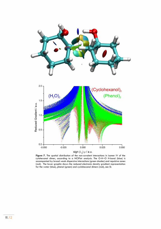
III.12
Figure 7. The spatial distribution of the non-covalent interactions in isomer IV of the cylohexanol dimer, according to a NCIPlot analysis. The O-H···O H-bond (blue) is accompanied by broad weak dispersive interactions (green shades) and repulsive zones (red). The lower graphic shows the reduced electronic density gradient representation for the water (blue), phenol (green) and cyclohexanol dimers (red), see SI.
DIMERIZATION OF CYCLOHEXANOL
III.13
Chapter III. References:
1. Snow, M. S., Howard, B. J., Evangelisti, L. & Caminati, W. From Transient to Induced Permanent Chirality in 2-Propanol upon Dimerization: A Rotational Study. J. Phys. Chem. A 115, 47–51 (2011).
2. King, A. K. & Howard, B. J. A microwave study of the hetero-chiral dimer of butan-2-ol. Chem. Phys. Lett. 348, 343–349 (2001).
3. Seifert, N. A. et al. The interplay of hydrogen bonding and dispersion in phenol dimer and trimer: Structures from broadband rotational spectroscopy. Phys. Chem. Chem. Phys. 15, 11468–11477 (2013).
4. Contreras-García, J. et al. NCIPLOT: A Program for Plotting Noncovalent Interaction Regions. J. Chem. Theory Comput. 7, 625–632 (2011).
5. Garcia, J., Podeszwa, R. & Szalewicz, K. SAPT codes for calculations of intermolecular interaction energies. J. Chem. Phys. 152, 184109 (2020).
6. Morrow, S. M., Bissette, A. J. & Fletcher, S. P. Transmission of chirality through space and across length scales. Nat. Nanotechnol. 12, 410–419 (2017).
7. Zehnacker, A. Chiral Recognition in the Gas Phase. (CRC Press, 2010).
8. Zehnacker, A. & Suhm, M. A. Chirality Recognition between Neutral Molecules in the Gas Phase. Angew. Chemie Int. Ed. 47, 6970–6992 (2008).
9. Juanes, M., Saragi, R. T., Caminati, W. & Lesarri, A. The Hydrogen Bond and Beyond: Perspectives for Rotational Investigations of Non-Covalent Interactions. Chem. - A Eur. J. 25, (2019).
10. Adler, T. B., Borho, N., Reiher, M. & Suhm, M. A. Chirality-Induced Switch in Hydrogen-Bond Topology: Tetrameric Methyl Lactate Clusters in the Gas Phase. Angew. Chemie Int. Ed. 45, 3440–3445 (2006).
11. Wassermann, T. N., Zielke, P., Lee, J. J., Cézard, C. & Suhm, M. A. Structural Preferences, Argon Nanocoating, and Dimerization of n -Alkanols As Revealed by OH Stretching Spectroscopy in Supersonic Jets †. J. Phys. Chem. A 111, 7437–7448 (2007).
12. Usabiaga, I., Camiruaga, A., Calabrese, C., Maris, A. & Fernández, J. A. Exploring Caffeine–Phenol Interactions by the Inseparable Duet of Experimental and Theoretical Data. Chem. – A Eur. J. 25, 14230–14236 (2019).
13. Rijs, A. M. & Oomens, J. Gas-Phase IR Spectroscopy and Structure of Biological Molecules. in Gas-Phase IR Spectrosc. Struct. Biol. Mol. (eds. Rijs, A. M. & Oomens, J.) 1–42 (Springer Int. Pub., 2015).
14. León, I., Montero, R., Longarte, A. & Fernández, J. A. IR mass-resolved spectroscopy of complexes without chromophore: Cyclohexanol·(H 2 O) n , n = 1–3 and cyclohexanol dimer. J. Chem. Phys. 139, 174312 (2013).
15. Caminati, W. & Grabow, J.-U. Advancements in Microwave Spectroscopy. in Frontiers and Advancements in Microwave Spectroscopy (ed. Laane, J.) 569–598 (Elsevier, 2018).
16. Hearn, J. P. I., Cobley, R. V. & Howard, B. J. High-resolution spectroscopy of induced chiral dimers: A study of the dimers of ethanol by Fourier transform microwave spectroscopy. J. Chem. Phys. 123, 1–7 (2005).
17. Loru, D., Peña, I. & Sanz, M. E. Ethanol dimer: Observation of three new conformers by broadband rotational spectroscopy. J. Mol. Spectrosc. 335, 93–101 (2017).
18. Su, Z., Borho, N. & Xu, Y. Chiral self-recognition: Direct spectroscopic detection of the homochiral and
III.14
heterochiral dimers of propylene oxide in the gas phase. J. Am. Chem. Soc. 128, 17126–17131 (2006).
19. Maris, A., Giuliano, B. M., Bonazzi, D. & Caminati, W. Molecular recognition of chiral conformers: A rotational study of the dimers of glycidol. J. Am. Chem. Soc. 130, 13860–13861 (2008).
20. Liu, X., Borho, N. & Xu, Y. Molecular self-recognition: Rotational spectra of the dimeric 2-fluoroethanol conformers. Chem. - A Eur. J. 15, 270–277 (2009).
21. Thomas, J. & Xu, Y. Chirality synchronization in trifluoroethanol dimer revisited: The missing heterochiral dimer. J. Phys. Chem. Lett. 5, 1850–1855 (2014).
22. Seifert, N. A. et al. Chiral recognition and atropisomerism in the sevoflurane dimer. Phys. Chem. Chem. Phys. 17, 18282–18287 (2015).
23. Juanes, M. et al. Internal dynamics of cyclohexanol and the cyclohexanol-water adduct. Phys. Chem. Chem. Phys. 21, (2019).
24. Li, W., Spada, L., Evangelisti, L. & Caminati, W. Conformational Equilibrium and Potential Energy Functions of the O-H Internal Rotation in the Axial and Equatorial Species of 1-Methylcyclohexanol. J. Phys. Chem. A 120, 4338–4342 (2016).
25. Gawrilow, M. & Suhm, M. A. 2-Methoxyethanol: harmonic tricks, anharmonic challenges and chirality-sensitive chain aggregation. Phys. Chem. Chem. Phys. 22, 15303–15311 (2020).
26. Neill, J. L. et al. Rotational spectroscopy of iodobenzene and iodobenzene–neon with a direct digital 2–8GHz chirped-pulse Fourier transform microwave spectrometer. J. Mol. Spectrosc. 269, 21–29 (2011).
27. Watson, J. K. G. Vibrational Spectra and Structure. in (ed. Durig, J. R.) 1–89 (Elsevier Inc., 1977).
28. Godfrey, P. D., Rodgers, F. M. & Brown, R. D. Theory versus Experiment in Jet Spectroscopy: Glycolic Acid. J. Am. Chem. Soc. 119, 2232–2239 (1997).
29. Becke, A. D. Perspective: Fifty years of density-functional theory in chemical physics. J. Chem. Phys. 140, (2014).
30. Yu, H. S., He, X. & Truhlar, D. G. MN15-L: A New Local Exchange-Correlation Functional for Kohn–Sham Density Functional Theory with Broad Accuracy for Atoms, Molecules, and Solids. J. Chem. Theory Comput. 12, 1280–1293 (2016).
31. Zhao, Y. & Truhlar, D. G. The M06 suite of density functionals for main group thermochemistry, thermochemical kinetics, noncovalent interactions, excited states, and transition elements: two new functionals and systematic testing of four M06-class functionals and 12 other function. Theor. Chem. Acc. 120, 215–241 (2008).
32. Weigend, F. & Ahlrichs, R. Balanced basis sets of split valence, triple zeta valence and quadruple zeta valence quality for H to Rn: Design and assessment of accuracy. Phys. Chem. Chem. Phys. 7, 3297 (2005).
33. Grimme, S., Ehrlich, S. & Goerigk, L. Effect of the damping function in dispersion corrected density functional theory. J. Comput. Chem. 32, 1456–1465 (2011).
34. Goerigk, L. et al. A look at the density functional theory zoo with the advanced GMTKN55 database for general main group thermochemistry, kinetics and noncovalent interactions. Phys. Chem. Chem. Phys. 19, 32184–32215 (2017).
35. Bannwarth, C., Ehlert, S. & Grimme, S. GFN2-xTB—An Accurate and Broadly Parametrized Self-Consistent Tight-Binding Quantum Chemical Method with Multipole Electrostatics and Density-Dependent Dispersion Contributions. J. Chem. Theory Comput. 15, 1652–1671 (2019).
DIMERIZATION OF CYCLOHEXANOL
III.15
36. Chai, J.-D. & Head-Gordon, M. Long-range corrected hybrid density functionals with damped atom–atom dispersion corrections. Phys. Chem. Chem. Phys. 10, 6615 (2008).
37. Grabow, J.-U. Fourier Transform Microwave Spectroscopy Measurement and Instrumentation. Handbook of High-resolution Spectroscopy (2011). doi:10.1002/9780470749593.hrs037.
38. Thomas, J., Seifert, N. A., Jäger, W. & Xu, Y. A Direct Link from the Gas to the Condensed Phase: A Rotational Spectroscopic Study of 2,2,2-Trifluoroethanol Trimers. Angew. Chemie Int. Ed. 56, 6289–6293 (2017).
39. Seifert, N. A., Thomas, J., Jäger, W. & Xu, Y. Rotational spectra and theoretical study of tetramers and trimers of 2-fluoroethanol: dramatic intermolecular compensation for intramolecular instability. Phys. Chem. Chem. Phys. 20, 27630–27637 (2018).
40. Oswald, S. et al. The Chiral Trimer and a Metastable Chiral Dimer of Achiral Hexafluoroisopropanol:
A Multi‐Messenger Study. Angew. Chemie Int. Ed. 58, 5080–5084 (2019).
41. Johnson, E. R. et al. Revealing Noncovalent Interactions. J. Am. Chem. Soc. 132, 6498–6506 (2010).
42. Hohenstein, E. G. & Sherrill, C. D. Density fitting of intramonomer correlation effects in symmetry-adapted perturbation theory. J. Chem. Phys. 133, 014101 (2010).

Chapter IV HYDRATION OF FURFURYL COMPOUNDS:
Adapted from:
(*): M. Juanes, A. Lesarri, R. Pinacho, E. Charro, J. E. Rubio, L. Enríquez and M. Jaraíz, Chem. Eur. J. 2018, 24, 6564 – 6571 (The Electronic Supporting Information – ESI, can be found in the document: thesis-ESI.pdf)
(Furfuryl Mercaptan··· H2O) and (Furfuryl Alcohol··· H2O)*

IV.2
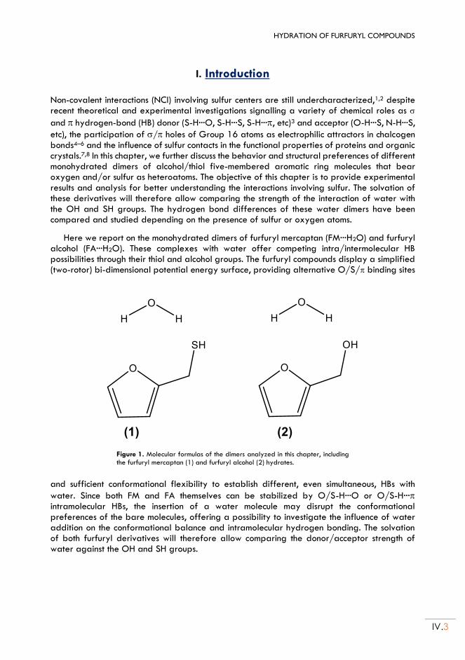
HYDRATION OF FURFURYL COMPOUNDS
IV.3
I. Introduction Non-covalent interactions (NCI) involving sulfur centers are still undercharacterized,1,2 despite recent theoretical and experimental investigations signalling a variety of chemical roles as
and hydrogen-bond (HB) donor (S-H···O, S-H···S, S-H···, etc)3 and acceptor (O-H···S, N-H···S,
etc), the participation of / holes of Group 16 atoms as electrophilic attractors in chalcogen bonds4–6 and the influence of sulfur contacts in the functional properties of proteins and organic crystals.7,8 In this chapter, we further discuss the behavior and structural preferences of different monohydrated dimers of alcohol/thiol five-membered aromatic ring molecules that bear oxygen and/or sulfur as heteroatoms. The objective of this chapter is to provide experimental results and analysis for better understanding the interactions involving sulfur. The solvation of these derivatives will therefore allow comparing the strength of the interaction of water with the OH and SH groups. The hydrogen bond differences of these water dimers have been compared and studied depending on the presence of sulfur or oxygen atoms.
Here we report on the monohydrated dimers of furfuryl mercaptan (FM···H2O) and furfuryl alcohol (FA···H2O). These complexes with water offer competing intra/intermolecular HB possibilities through their thiol and alcohol groups. The furfuryl compounds display a simplified (two-rotor) bi-dimensional potential energy surface, providing alternative O/S/ binding sites
Figure 1. Molecular formulas of the dimers analyzed in this chapter, including the furfuryl mercaptan (1) and furfuryl alcohol (2) hydrates.
and sufficient conformational flexibility to establish different, even simultaneous, HBs with
water. Since both FM and FA themselves can be stabilized by O/S-H···O or O/S-H··· intramolecular HBs, the insertion of a water molecule may disrupt the conformational preferences of the bare molecules, offering a possibility to investigate the influence of water addition on the conformational balance and intramolecular hydrogen bonding. The solvation of both furfuryl derivatives will therefore allow comparing the donor/acceptor strength of water against the OH and SH groups.
IV.4
All the experiments have been assisted by molecular-orbital quantum chemical calculations with different dispersion-corrected density functional theory (DFT) models and/or ab initio methods, simultaneously assessing the theoretical calculations.
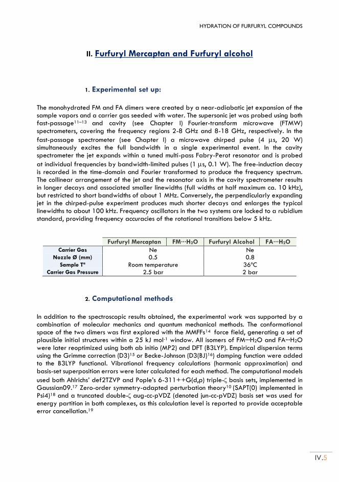
HYDRATION OF FURFURYL COMPOUNDS
IV.5
II. Furfuryl Mercaptan and Furfuryl alcohol
1. Experimental set up:
The monohydrated FM and FA dimers were created by a near-adiabatic jet expansion of the sample vapors and a carrier gas seeded with water. The supersonic jet was probed using both fast-passage11–13 and cavity (see Chapter I) Fourier-transform microwave (FTMW) spectrometers, covering the frequency regions 2-8 GHz and 8-18 GHz, respectively. In the
fast-passage spectrometer (see Chapter I) a microwave chirped pulse (4 s, 20 W) simultaneously excites the full bandwidth in a single experimental event. In the cavity spectrometer the jet expands within a tuned multi-pass Fabry-Perot resonator and is probed
at individual frequencies by bandwidth-limited pulses (1 s, 0.1 W). The free-induction decay is recorded in the time-domain and Fourier transformed to produce the frequency spectrum. The collinear arrangement of the jet and the resonator axis in the cavity spectrometer results in longer decays and associated smaller linewidths (full widths at half maximum ca. 10 kHz), but restricted to short bandwidths of about 1 MHz. Conversely, the perpendicularly expanding jet in the chirped-pulse experiment produces much shorter decays and enlarges the typical linewidths to about 100 kHz. Frequency oscillators in the two systems are locked to a rubidium standard, providing frequency accuracies of the rotational transitions below 5 kHz.
Furfuryl Mercaptan FM···H2O Furfuryl Alcohol FA···H2O
Carrier Gas Ne Ne Nozzle Ø (mm) 0.5 0.8
Sample Tª Room temperature 36ºC Carrier Gas Pressure 2.5 bar 2 bar
2. Computational methods
In addition to the spectroscopic results obtained, the experimental work was supported by a combination of molecular mechanics and quantum mechanical methods. The conformational space of the two dimers was first explored with the MMFFs14 force field, generating a set of plausible initial structures within a 25 kJ mol-1 window. All isomers of FM···H2O and FA···H2O were later reoptimized using both ab initio (MP2) and DFT (B3LYP). Empirical dispersion terms using the Grimme correction (D3)15 or Becke-Johnson (D3(BJ)16) damping function were added to the B3LYP functional. Vibrational frequency calculations (harmonic approximation) and basis-set superposition errors were later calculated for each method. The computational models
used both Ahlrichs’ def2TZVP and Pople’s 6-311++G(d,p) triple-basis sets, implemented in Gaussian09.17 Zero-order symmetry-adapted perturbation theory10 (SAPT(0) implemented in Psi4)18 and a truncated double- aug-cc-pVDZ (denoted jun-cc-pVDZ) basis set was used for energy partition in both complexes, as this calculation level is reported to provide acceptable error cancellation.19
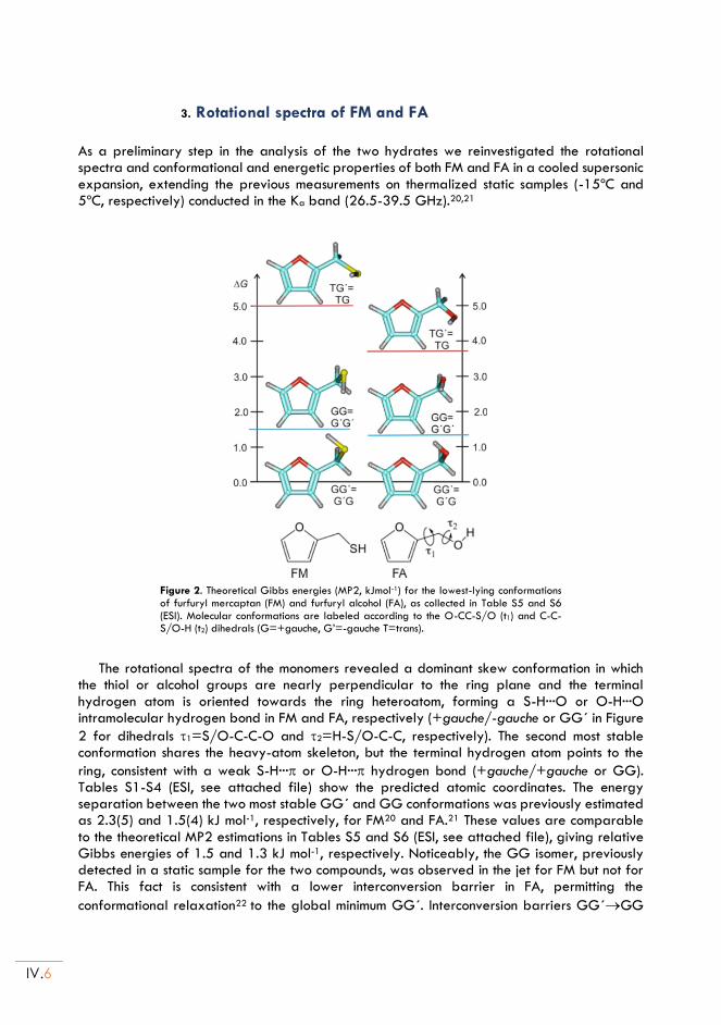
IV.6
3. Rotational spectra of FM and FA
As a preliminary step in the analysis of the two hydrates we reinvestigated the rotational spectra and conformational and energetic properties of both FM and FA in a cooled supersonic expansion, extending the previous measurements on thermalized static samples (-15ºC and 5ºC, respectively) conducted in the Ka band (26.5-39.5 GHz).20,21
Figure 2. Theoretical Gibbs energies (MP2, kJmol-1) for the lowest-lying conformations of furfuryl mercaptan (FM) and furfuryl alcohol (FA), as collected in Table S5 and S6 (ESI). Molecular conformations are labeled according to the O-CC-S/O (t1) and C-C-S/O-H (t2) dihedrals (G=+gauche, G’=-gauche T=trans).
The rotational spectra of the monomers revealed a dominant skew conformation in which the thiol or alcohol groups are nearly perpendicular to the ring plane and the terminal hydrogen atom is oriented towards the ring heteroatom, forming a S-H···O or O-H···O intramolecular hydrogen bond in FM and FA, respectively (+gauche/-gauche or GG´ in Figure
2 for dihedrals 1=S/O-C-C-O and 2=H-S/O-C-C, respectively). The second most stable conformation shares the heavy-atom skeleton, but the terminal hydrogen atom points to the
ring, consistent with a weak S-H··· or O-H··· hydrogen bond (+gauche/+gauche or GG). Tables S1-S4 (ESI, see attached file) show the predicted atomic coordinates. The energy separation between the two most stable GG´ and GG conformations was previously estimated as 2.3(5) and 1.5(4) kJ mol-1, respectively, for FM20 and FA.21 These values are comparable to the theoretical MP2 estimations in Tables S5 and S6 (ESI, see attached file), giving relative Gibbs energies of 1.5 and 1.3 kJ mol-1, respectively. Noticeably, the GG isomer, previously detected in a static sample for the two compounds, was observed in the jet for FM but not for FA. This fact is consistent with a lower interconversion barrier in FA, permitting the
conformational relaxation22 to the global minimum GG´. Interconversion barriers GG´GG
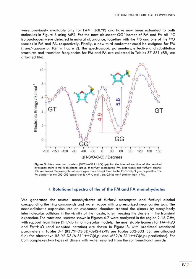
HYDRATION OF FURFURYL COMPOUNDS
IV.7
were previously available only for FA23 (B3LYP) and have now been extended to both molecules in Figure 3 using MP2. For the most abundant GG´ isomer of FM and FA all 13C isotopologues were detected in natural abundance, together with the 34S and one of the 18O species in FM and FA, respectively. Finally, a new third conformer could be assigned for FM (trans/-gauche or TG´ in Figure 2). The spectroscopic parameters, effective and substitution structures and transition frequencies for FM and FA are collected in Tables S7-S31 (ESI, see attached file).
Figure 3. Interconversion barriers (MP2/6-311++G(d,p)) for the internal rotation of the terminal hydrogen atom in the thiol/alcohol group of furfuryl mercaptan (FM, blue trace) and furfuryl alcohol (FA, red trace). The exocyclic sulfur/oxygen atom is kept fixed to the O-C-C-S/O gauche position. The FA barrier for the GG-GG´conversion is 4.9 kJ mol-1, ca. 0.9 kJ mol-1 smaller than in FM.
4. Rotational spectra of the of the FM and FA monohydrates
We generated the neutral monohydrates of furfuryl mercaptan and furfuryl alcohol coexpanding the ring compounds and water vapor with a pressurized neon carrier gas. The near-adiabatic expansion into an evacuated chamber created the dimers by many-body intermolecular collisions in the vicinity of the nozzle, later freezing the clusters in the transient expansion. The rotational spectra shown in Figures 4-7 were analyzed in the region 2-18 GHz, with support from three DFT/ab initio molecular models. The most stable isomers for FM···H2O and FA···H2O (and adopted notation) are shown in Figure 8, with predicted rotational parameters in Tables 3-4 (B3LYP-D3(BJ)/def2-TZVP; see Tables S32-S33 (ESI, see attached file) for alternative B3LYP-D3/6-311++G(d,p) and MP2/6-311++G(d,p) predictions). For both complexes two types of dimers with water resulted from the conformational search:
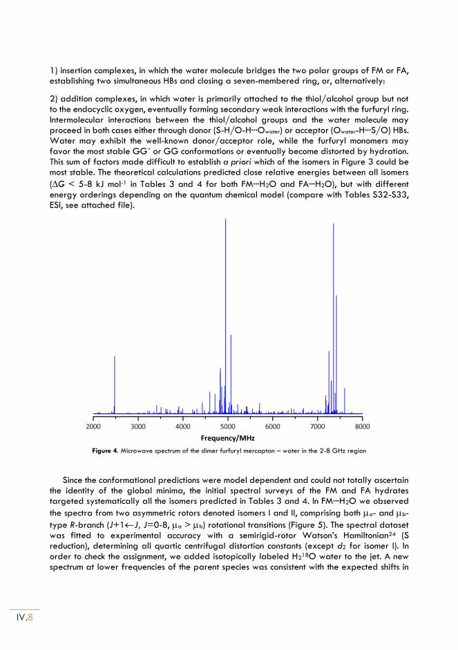
IV.8
1) insertion complexes, in which the water molecule bridges the two polar groups of FM or FA, establishing two simultaneous HBs and closing a seven-membered ring, or, alternatively:
2) addition complexes, in which water is primarily attached to the thiol/alcohol group but not to the endocyclic oxygen, eventually forming secondary weak interactions with the furfuryl ring. Intermolecular interactions between the thiol/alcohol groups and the water molecule may proceed in both cases either through donor (S-H/O-H···Owater) or acceptor (Owater-H···S/O) HBs. Water may exhibit the well-known donor/acceptor role, while the furfuryl monomers may favor the most stable GG´ or GG conformations or eventually become distorted by hydration. This sum of factors made difficult to establish a priori which of the isomers in Figure 3 could be most stable. The theoretical calculations predicted close relative energies between all isomers
(G < 5-8 kJ mol-1 in Tables 3 and 4 for both FM···H2O and FA···H2O), but with different energy orderings depending on the quantum chemical model (compare with Tables S32-S33, ESI, see attached file).
Figure 4. Microwave spectrum of the dimer furfuryl mercaptan – water in the 2-8 GHz region
Since the conformational predictions were model dependent and could not totally ascertain
the identity of the global minima, the initial spectral surveys of the FM and FA hydrates targeted systematically all the isomers predicted in Tables 3 and 4. In FM···H2O we observed
the spectra from two asymmetric rotors denoted isomers I and II, comprising both a- and b-
type R-branch (J+1J, J=0-8, a > b) rotational transitions (Figure 5). The spectral dataset was fitted to experimental accuracy with a semirigid-rotor Watson’s Hamiltonian24 (S reduction), determining all quartic centrifugal distortion constants (except d2 for isomer I). In order to check the assignment, we added isotopically labeled H218O water to the jet. A new spectrum at lower frequencies of the parent species was consistent with the expected shifts in
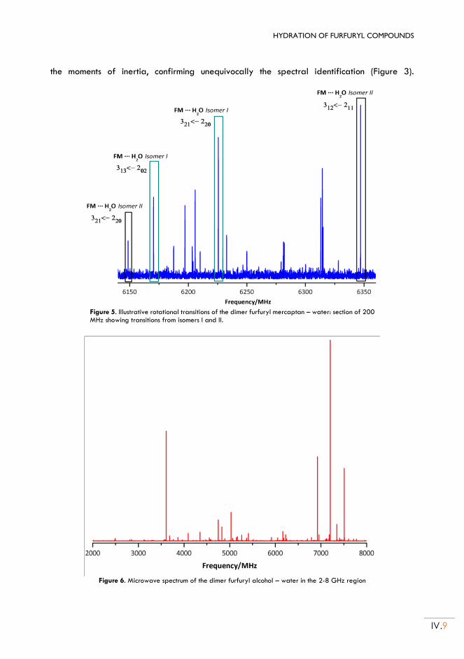
HYDRATION OF FURFURYL COMPOUNDS
IV.9
the moments of inertia, confirming unequivocally the spectral identification (Figure 3).
Figure 5. Illustrative rotational transitions of the dimer furfuryl mercaptan – water: section of 200 MHz showing transitions from isomers I and II.
º Figure 6. Microwave spectrum of the dimer furfuryl alcohol – water in the 2-8 GHz region

IV.10
Figure 7. Illustrative rotational transitions of the dimer furfuryl alcohol – water: section of 140 MHz showing transitions from isomer I.
Conversely, the spectrum of FA···H2O (Figure 6) revealed a single isomer with R-branch
transitions (a > b > c), that was analyzed similarly. The spectral identification was again verified by detection of the H218O isotopologue (Figure S5, ESI, see attached file). The experimental spectroscopic parameters for FM···H2O and FA···H2O are collected in Tables 1 and 2 and S34-S35 (ESI, see attached file), while the observed transitions are listed in Tables S36-S41 (ESI, see attached file). The observation of the H218O isotopic species allowed determining the atomic coordinates of the water oxygen atom in the monohydrate for the three detected species. The resulting coordinates obtained from the Kraitchman25 equations are presented in Tables S42 and S43 (ESI, see attached file).
The conformational assignment relied on a comparison of the experimental rotational constants and water oxygen coordinates with the theoretical DFT/ab initio predictions in Tables 3 and 4, S32-S33 and S42-S43 (ESI, see attached file). The assignment of the furfuryl alcohol monohydrate FA···H2O was relatively straightforward, as the rotational constants clearly pointed to the identification of the observed cluster as the insertion complex GG´-Wda, predicted as global minimum using B3LYP-D3(BJ). This isomer exhibits a network of two consecutive O-H···O HBs, in which the hydroxyl group acts as proton donor to the water oxygen and water similarly behaves as proton donor to the ring oxygen atom. On the other hand, the conformational assignment of the two observed furfuryl mercaptan hydrates FM···H2O I/ II was more difficult, as the theoretical predictions were not totally consistent and the rotational constants of the three predicted insertion complexes were relatively close. Finally, inspection of the experimental evidence led to the identification of isomer I as GG´-Wda and isomer II as GG-Wdd. In both complexes water simultaneously binds to the ring oxygen and to the thiol group, with the thiol group engaging to water either as proton donor (isomer I) or proton acceptor (isomer II). Vibrationally-averaged effective structures were calculated for the three observed dimers by a least-squares fit of the moments of inertia.26,27 For FA···H2O we could fit both the position of the water oxygen and the orientation of the water moiety (using an
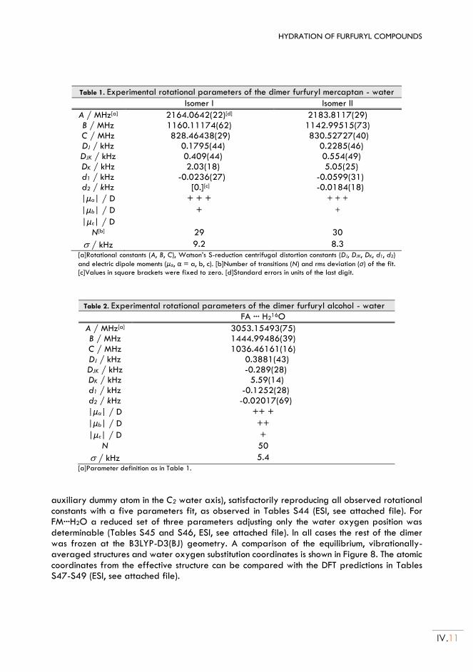
HYDRATION OF FURFURYL COMPOUNDS
IV.11
Table 1. Experimental rotational parameters of the dimer furfuryl mercaptan - water
Isomer I Isomer II
A / MHz[a] 2164.0642(22)[d] 2183.8117(29) B / MHz 1160.11174(62) 1142.99515(73) C / MHz 828.46438(29) 830.52727(40) DJ / kHz 0.1795(44) 0.2285(46) DJK / kHz 0.409(44) 0.554(49) DK / kHz 2.03(18) 5.05(25) d1 / kHz -0.0236(27) -0.0599(31) d2 / kHz [0.][c] -0.0184(18)
|μa| / D + + + + + +
|μb| / D + +
|μc| / D
N[b] 29 30
/ kHz 9.2 8.3 [a]Rotational constants (A, B, C), Watson’s S-reduction centrifugal distortion constants (DJ, DJK, DK, d1, d2)
and electric dipole moments (μα, α = a, b, c). [b]Number of transitions (N) and rms deviation (σ) of the fit. [c]Values in square brackets were fixed to zero. [d]Standard errors in units of the last digit.
Table 2. Experimental rotational parameters of the dimer furfuryl alcohol - water
FA ··· H216O
A / MHz[a] 3053.15493(75) B / MHz 1444.99486(39) C / MHz 1036.46161(16) DJ / kHz 0.3881(43) DJK / kHz -0.289(28) DK / kHz 5.59(14) d1 / kHz -0.1252(28) d2 / kHz -0.02017(69)
|μa| / D ++ +
|μb| / D ++
|μc| / D +
N 50
/ kHz 5.4 [a]Parameter definition as in Table 1.
auxiliary dummy atom in the C2 water axis), satisfactorily reproducing all observed rotational constants with a five parameters fit, as observed in Tables S44 (ESI, see attached file). For FM···H2O a reduced set of three parameters adjusting only the water oxygen position was determinable (Tables S45 and S46, ESI, see attached file). In all cases the rest of the dimer was frozen at the B3LYP-D3(BJ) geometry. A comparison of the equilibrium, vibrationally-averaged structures and water oxygen substitution coordinates is shown in Figure 8. The atomic coordinates from the effective structure can be compared with the DFT predictions in Tables S47-S49 (ESI, see attached file).
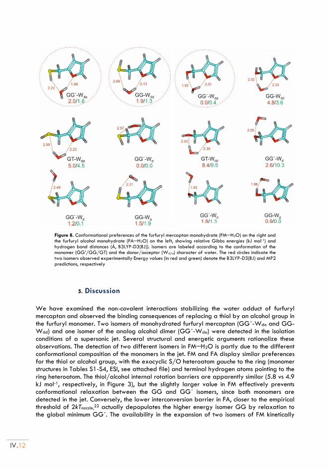
IV.12
Figure 8. Conformational preferences of the furfuryl mercaptan monohydrate (FM···H2O) on the right and the furfuryl alcohol monohydrate (FA···H2O) on the left, showing relative Gibbs energies (kJ mol-1) and hydrogen bond distances (Å, B3LYP-D3(BJ)). Isomers are labeled according to the conformation of the monomer (GG’/GG/GT) and the donor/acceptor (Wd/a) character of water. The red circles indicate the two isomers observed experimentally Energy values (in red and green) denote the B3LYP-D3(BJ) and MP2 predictions, respectively
5. Discussion
We have examined the non-covalent interactions stabilizing the water adduct of furfuryl mercaptan and observed the binding consequences of replacing a thiol by an alcohol group in the furfuryl monomer. Two isomers of monohydrated furfuryl mercaptan (GG´-Wda and GG-Wdd) and one isomer of the analog alcohol dimer (GG´-Wda) were detected in the isolation conditions of a supersonic jet. Several structural and energetic arguments rationalize these observations. The detection of two different isomers in FM···H2O is partly due to the different conformational composition of the monomers in the jet. FM and FA display similar preferences for the thiol or alcohol group, with the exocyclic S/O heteroatom gauche to the ring (monomer structures in Tables S1-S4, ESI, see attached file) and terminal hydrogen atoms pointing to the ring heteroatom. The thiol/alcohol internal rotation barriers are apparently similar (5.8 vs 4.9 kJ mol-1, respectively, in Figure 3), but the slightly larger value in FM effectively prevents conformational relaxation between the GG and GG´ isomers, since both monomers are detected in the jet. Conversely, the lower interconversion barrier in FA, closer to the empirical threshold of 2kTnozzle,22 actually depopulates the higher energy isomer GG by relaxation to the global minimum GG´. The availability in the expansion of two isomers of FM kinetically
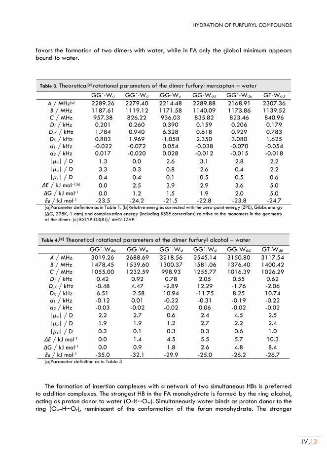
HYDRATION OF FURFURYL COMPOUNDS
IV.13
favors the formation of two dimers with water, while in FA only the global minimum appears bound to water.
Table 3. Theoretical[c] rotational parameters of the dimer furfuryl mercaptan – water
GG´-Wd GG´-Wd GG-Wa GG-Wdd GG´-Wda GT-Wdd
A / MHz[a] 2289.26 2279.40 2214.48 2289.88 2168.91 2307.36 B / MHz 1187.61 1119.12 1171.58 1140.09 1173.86 1139.52 C / MHz 957.38 826.22 936.03 835.82 823.46 840.96 DJ / kHz 0.201 0.260 0.390 0.159 0.206 0.179 DJK / kHz 1.784 0.940 6.328 0.618 0.929 0.783 DK / kHz 0.883 1.969 -1.058 2.350 3.080 1.625 d1 / kHz -0.022 -0.072 0.054 -0.038 -0.070 -0.054 d2 / kHz 0.017 -0.020 0.028 -0.012 -0.015 -0.018
|μa| / D 1.3 0.0 2.6 3.1 2.8 2.2
|μb| / D 3.3 0.3 0.8 2.6 0.4 2.2
|μc| / D 0.4 0.4 0.1 0.5 0.5 0.6
ΔE / kJ mol-1[b] 0.0 2.5 3.9 2.9 3.6 5.0
ΔG / kJ mol-1 0.0 1.2 1.5 1.9 2.0 5.0 EB / kJ mol-1 -23.5 -24.2 -21.5 -22.8 -23.8 -24.7 [a]Parameter definition as in Table 1. [b]Relative energies corrected with the zero-point energy (ZPE), Gibbs energy
(G, 298K, 1 atm) and complexation energy (including BSSE corrections) relative to the monomers in the geometry of the dimer. [c] B3LYP-D3(BJ)/ def2-TZVP.
Table 4.[a] Theoretical rotational parameters of the dimer furfuryl alcohol – water
GG´-Wda GG-Wa GG´-Wd GG´-Wd GG-Wdd GT-Wdd
A / MHz 3019.26 2688.69 3218.56 2545.14 3150.80 3117.54 B / MHz 1478.45 1539.60 1300.37 1581.06 1376.40 1400.42 C / MHz 1055.00 1232.59 998.93 1255.77 1016.39 1026.29 DJ / kHz 0.42 0.92 0.78 2.05 0.55 0.62 DJK / kHz -0.48 4.47 -2.89 12.29 -1.76 -2.06 DK / kHz 6.51 -2.58 10.94 -11.75 8.25 10.74 d1 / kHz -0.12 0.01 -0.22 -0.31 -0.19 -0.22 d2 / kHz -0.03 -0.02 -0.02 0.06 -0.02 -0.02
|μa| / D 2.2 2.7 0.6 2.4 4.5 2.5
|μb| / D 1.9 1.9 1.2 2.7 2.2 2.4
|μc| / D 0.3 0.1 0.3 0.3 0.6 1.0
ΔE / kJ mol-1 0.0 1.4 4.5 5.5 5.7 10.3
ΔG / kJ mol-1 0.0 0.9 1.8 2.6 4.8 8.4
EB / kJ mol-1 -35.0 -32.1 -29.9 -25.0 -26.2 -26.7 [a]Parameter definition as in Table 3
The formation of insertion complexes with a network of two simultaneous HBs is preferred to addition complexes. The strongest HB in the FA monohydrate is formed by the ring alcohol, acting as proton donor to water (O-H···Ow). Simultaneously water binds as proton donor to the ring (Ow-H···Or), reminiscent of the conformation of the furan monohydrate. The stronger
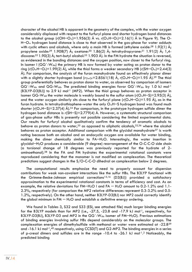
IV.14
character of the alcohol HB is apparent in the geometry of the complex, with the water oxygen considerably displaced with respect to the furfuryl plane and shorter hydrogen bond distances to the alcohol group (r(OH···Ow)=1.956(3) Å vs. r(OwH···Or)=2.16(1) Å in Figure 9). The O-H···Ow hydrogen bond distance is close to that observed in the gas-phase for water adducts with cyclic ethers and alcohols, where only a main HB is formed (ethylene oxide:28 1.92(1) Å; propylene oxide:29 1.908(7) Å; oxetane:30 1.86(2) Å; tetrahydropyrane:31 1.91(2) Å; 1,4-dioxane:32 1.90(3) Å; tert-butyl alcohol:33 1.903 Å). In the FM hydrate the situation is reversed, as evidenced in the bonding distances and the oxygen position, now closer to the furfuryl ring. In isomer I (GG´-Wda) the primary HB is now formed by water acting as proton donor to the ring (r(OwH···Or)=1.99(2) Å), while the thiol forms a weaker secondary HB (r(SH···Ow)=2.44(3) Å). For comparison, the analysis of the furan monohydrate found an effectively planar dimer with a slightly shorter hydrogen bond (rOwOr=2.8561(18) Å, r(OwH···Or)=1.95 Å).29 The thiol group preferentially behaves as proton donor to water, as observed by comparison of isomers GG´-Wda and GG-Wdd. The predicted binding energies favor GG´-Wda by 1.0 kJ mol-1 (B3LYP-D3(BJ)) to 2.9 kJ mol-1 (MP2). When the thiol group behaves as proton acceptor in isomer GG-Wdd the water molecule is weakly bound to the sulfur atom (r(OwH···S)=2.95(3) Å), and the water oxygen similarly sits close to the furfuryl plane (r(OwH···Or)=1.95 Å), as in the furan hydrate. In tetrahydrothiophene-water the only OwH···S hydrogen bond was found much shorter (r(OwH···S)=2.37(4) Å).34 For comparison, in the prototype hydrogen sulfide dimer the hydrogen bond distance is r(SH···S)=2.779(4) Å. However, a systematic structural comparison of gas-phase sulfur HBs is presently not possible considering the limited experimental data. Our results for furfuryl alcohol qualitatively confirm the tendency of aromatic alcohols to behave as proton donors to water,35 as opposed to aliphatic alcohols,32,36 where the alcohol behaves as proton acceptor. Additional comparison with the glycidol monohydrate37 is worth noting because both an alcohol and an endocyclic oxygen are available for water binding, making the dimer chemically similar to FA···H2O. Interestingly, the water insertion in glycidol···H2O produces a considerable (9 degree) rearrangement of the O-C-C-O side chain (a torsional change of 18 degrees was previously reported for the hydrate of 2-aminoethanol).38 In the FA and FM hydrates the experimental rotational constants were reproduced considering that the monomer is not modified on complexation. The theoretical predictions suggest changes in the S/O-C-C-O dihedral on complexation below 2 degrees.
The computational results emphasize the need to properly account for dispersion contributions for weak non-covalent interactions like the sulfur HBs. The B3LYP functional with the Grimme-Becke-Johnson empirical corrections15,16 (D3(BJ)) provided a satisfactory approximation to the experimental rotational constants in terms of efficiency and cost. As an example, the relative deviations for FM···H2O I and FA ··· H2O amount to 0.2-1.2% and 1.1-2.3%, respectively (for comparison the MP2 relative differences represent 0.3-3.2% and 0.5-1.2%, respectively). On the other hand, neither B3LYP-D3(BJ) nor MP2 could correctly identify the global minimum in FM ··· H2O and establish a definitive energy ordering.
We found in Tables 3, S32 and S33 (ESI, see attached file) much larger binding energies for the B3LYP models than for MP2 (i.e., -27.5, -23.8 and -17.9 kJ mol-1, respectively, for B3LYP-D3(BJ), B3LYP-D3 and MP2 in the GG´-Wda isomer of FM···H2O). Previous estimations of binding energies involving sulfur HBs depend considerably on the molecular groups. The complexation energies of dimethylsulfide with methanol or water were estimated at -22.839 and -16.1 kJ mol-1,40 respectively, using CCSD(T) and G2-MP2. The binding energies in a series of p-cresol dimers and sulfides are in the range -10.4 to -26.1 kJ mol-1.2 Noticeably, the predicted binding
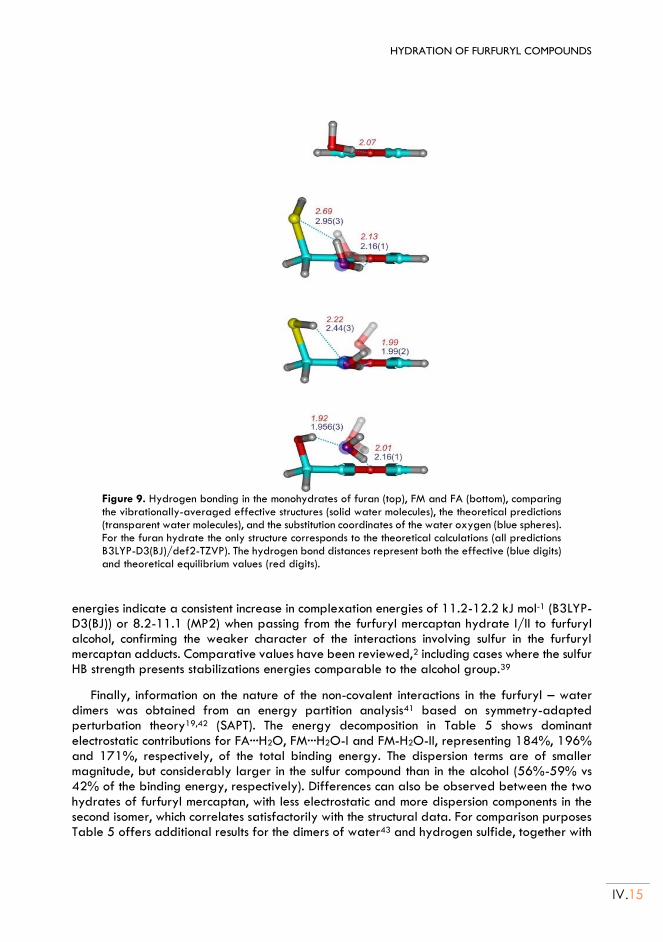
HYDRATION OF FURFURYL COMPOUNDS
IV.15
Figure 9. Hydrogen bonding in the monohydrates of furan (top), FM and FA (bottom), comparing the vibrationally-averaged effective structures (solid water molecules), the theoretical predictions (transparent water molecules), and the substitution coordinates of the water oxygen (blue spheres). For the furan hydrate the only structure corresponds to the theoretical calculations (all predictions B3LYP-D3(BJ)/def2-TZVP). The hydrogen bond distances represent both the effective (blue digits) and theoretical equilibrium values (red digits).
energies indicate a consistent increase in complexation energies of 11.2-12.2 kJ mol-1 (B3LYP-D3(BJ)) or 8.2-11.1 (MP2) when passing from the furfuryl mercaptan hydrate I/II to furfuryl alcohol, confirming the weaker character of the interactions involving sulfur in the furfuryl mercaptan adducts. Comparative values have been reviewed,2 including cases where the sulfur HB strength presents stabilizations energies comparable to the alcohol group.39
Finally, information on the nature of the non-covalent interactions in the furfuryl – water dimers was obtained from an energy partition analysis41 based on symmetry-adapted perturbation theory19,42 (SAPT). The energy decomposition in Table 5 shows dominant electrostatic contributions for FA···H2O, FM···H2O-I and FM-H2O-II, representing 184%, 196% and 171%, respectively, of the total binding energy. The dispersion terms are of smaller magnitude, but considerably larger in the sulfur compound than in the alcohol (56%-59% vs 42% of the binding energy, respectively). Differences can also be observed between the two hydrates of furfuryl mercaptan, with less electrostatic and more dispersion components in the second isomer, which correlates satisfactorily with the structural data. For comparison purposes Table 5 offers additional results for the dimers of water43 and hydrogen sulfide, together with
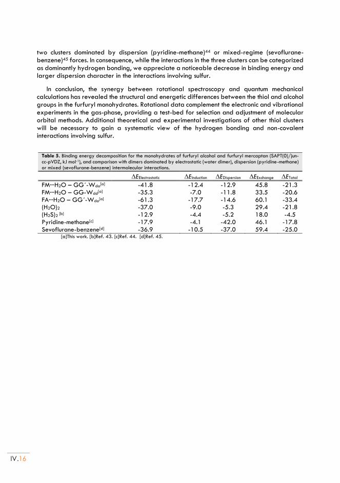
IV.16
two clusters dominated by dispersion (pyridine-methane)44 or mixed-regime (sevoflurane-benzene)45 forces. In consequence, while the interactions in the three clusters can be categorized as dominantly hydrogen bonding, we appreciate a noticeable decrease in binding energy and larger dispersion character in the interactions involving sulfur.
In conclusion, the synergy between rotational spectroscopy and quantum mechanical calculations has revealed the structural and energetic differences between the thiol and alcohol groups in the furfuryl monohydrates. Rotational data complement the electronic and vibrational experiments in the gas-phase, providing a test-bed for selection and adjustment of molecular orbital methods. Additional theoretical and experimental investigations of other thiol clusters will be necessary to gain a systematic view of the hydrogen bonding and non-covalent interactions involving sulfur.
Table 5. Binding energy decomposition for the monohydrates of furfuryl alcohol and furfuryl mercaptan (SAPT(0)/jun-cc-pVDZ, kJ mol-1), and comparison with dimers dominated by electrostatic (water dimer), dispersion (pyridine-methane) or mixed (sevoflurane-benzene) intermolecular interactions.
EElectrostatic EInduction EDispersion EExchange ETotal
FM···H2O – GG´-Wda[a] -41.8 -12.4 -12.9 45.8 -21.3
FM···H2O – GG-Wdd[a] -35.3 -7.0 -11.8 33.5 -20.6
FA···H2O – GG´-Wda[a] -61.3 -17.7 -14.6 60.1 -33.4
(H2O)2 -37.0 -9.0 -5.3 29.4 -21.8 (H2S)2 [b] -12.9 -4.4 -5.2 18.0 -4.5 Pyridine-methane[c] -17.9 -4.1 -42.0 46.1 -17.8 Sevoflurane-benzene[d] -36.9 -10.5 -37.0 59.4 -25.0
[a]This work. [b]Ref. 43. [c]Ref. 44. [d]Ref. 45.
HYDRATION OF FURFURYL COMPOUNDS
IV.17
Chapter IV. References:
1. Biswal, H. S., Bhattacharyya, S., Bhattacherjee, A. & Wategaonkar, S. Nature and strength of sulfur-
centred hydrogen bonds: laser spectroscopic investigations in the gas phase and quantum-chemical calculations. Int. Rev. Phys. Chem. 34, 99–160 (2015).
2. Biswal, H. S. Hydrogen Bonding Involving Sulfur: New Insights from Ab Initio Calculations and Gas Phase Laser Spectroscopy. in Noncovalent Forces (ed. Scheiner, S.) (Springer Int. Pub., 2015).
3. Gilli, G. & Gilli, P. The Nature of the Hydrogen Bond. (2009).
4. Aakeroy, C. B. et al. Definition of the chalcogen bond (IUPAC Recommendations 2019). Pure Appl. Chem. 91, 1889–1892 (2019).
5. Alkorta, I., Elguero, J. & Frontera, A. Not only hydrogen bonds: Other noncovalent interactions. Crystals 10, (2020).
6. Legon, A. C. Tetrel, pnictogen and chalcogen bonds identified in the gas phase before they had names: a systematic look at non-covalent interactions. Phys. Chem. Chem. Phys. 19, 14884–14896 (2017).
7. Zhou, P., Tian, F., Lv, F. & Shang, Z. Geometric characteristics of hydrogen bonds involving sulfur atoms in proteins. Proteins Struct. Funct. Bioinforma. 76, 151–163 (2009).
8. Mundlapati, V. R., Ghosh, S., Bhattacherjee, A., Tiwari, P. & Biswal, H. S. Critical Assessment of the Strength of Hydrogen Bonds between the Sulfur Atom of Methionine/Cysteine and Backbone Amides in Proteins. J. Phys. Chem. Lett. 6, 1385–1389 (2015).
9. Contreras-García, J. et al. NCIPLOT: A Program for Plotting Noncovalent Interaction Regions. J. Chem. Theory Comput. 7, 625–632 (2011).
10. Garcia, J., Podeszwa, R. & Szalewicz, K. SAPT codes for calculations of intermolecular interaction energies. J. Chem. Phys. 152, 184109 (2020).
11. Pérez, C. et al. Broadband Fourier transform rotational spectroscopy for structure determination: The water heptamer. Chem. Phys. Lett. 571, 1–15 (2013).
12. Neill, J. L. et al. Rotational spectroscopy of iodobenzene and iodobenzene–neon with a direct digital 2–8GHz chirped-pulse Fourier transform microwave spectrometer. J. Mol. Spectrosc. 269, 21–29 (2011).
13. Shipman, S. T. & Pate, B. H. New Techniques in Microwave Spectroscopy. in Handbook of High-resolution Spectroscopy (eds. Quack, M. & Merk, F.) 801–827 (John Wiley & Sons, Ltd., 2011).
14. Halgren, T. A. Merck molecular force field. I. Basis, form, scope, parameterization, and performance of MMFF94. J. Comput. Chem. 17, 490–519 (1996).
15. Grimme, S., Antony, J., Ehrlich, S. & Krieg, H. A consistent and accurate ab initio parametrization of density functional dispersion correction (DFT-D) for the 94 elements H-Pu. J. Chem. Phys. 132, 154104 (2010).
16. Grimme, S., Ehrlich, S. & Goerigk, L. Effect of the damping function in dispersion corrected density functional theory. J. Comput. Chem. 32, 1456–1465 (2011).
17. Frisch, M. J., Trucks, G. W., Schlegel, H. B., Scuseria, G. E. & Robb, M. A. et al. Gaussian 09, Revision A.02. (Gaussian, Inc., 2016).
18. Turney, J. M. et al. Psi4: an open-source ab initio electronic structure program. Wiley Interdiscip. Rev.
IV.18
Comput. Mol. Sci. 2, 556–565 (2012).
19. Parker, T. M., Burns, L. A., Parrish, R. M., Ryno, A. G. & Sherrill, C. D. Levels of symmetry adapted perturbation theory (SAPT). I. Efficiency and performance for interaction energies. J. Chem. Phys. 140, 094106 (2014).
20. Marstokk, K.-M. et al. Microwave Spectrum, Conformational Equilibrium, Intramolecular Hydrogen Bonding and Ab Initio Calculations for 2-Furanmethanethiol (Furfuryl Mercaptan). Acta Chem. Scand. 48, 298–305 (1994).
21. Marstokk, K.-M. et al. Microwave Spectrum, Conformational Equilibrium and Ab Initio Calculations for 2-Furanmethanol (Furfuryl Alcohol). Acta Chem. Scand. 48, 25–31 (1994).
22. Ruoff, R. S., Klots, T. D., Emilsson, T. & Gutowsky, H. S. Relaxation of conformers and isomers in seeded supersonic jets of inert gases. J. Chem. Phys. 93, 3142–3150 (1990).
23. Araujo-Andrade, C., Gómez-Zavaglia, A., Reva, I. D. & Fausto, R. Conformers, Infrared Spectrum and UV-Induced Photochemistry of Matrix-Isolated Furfuryl Alcohol. J. Phys. Chem. A 116, 2352–2365 (2012).
24. Watson, J. K. G. Vibrational Spectra and Structure. in (ed. Durig, J. R.) 1–89 (Elsevier Inc., 1977).
25. Kraitchman, J. Determination of Molecular Structure from Microwave Spectroscopic Data. Am. J. Phys. 21, 17–24 (1953).
26. Rudolph, H. D. & Demaison, J. Equilibrium Molecular Structures: From Spectroscopy to Quantum Chemistry. in Equilibrium Molecular Structures: From Spectroscopy to Quantum Chemistry (eds. Demaison, J., Boggs, J. E. & Császár, A. G.) 125–158 (CRC Press, 2011).
27. MOMSTRUC Structural Package.
28. Caminati, W., Moreschini, P., Rossi, I. & Favero, P. G. The O···H− O Hydrogen Bond in the Gas Phase. Microwave Structure of Ethylene Oxide− Water. J. Am. Chem. Soc. 120, 11144–11148 (1998).
29. Su, Z., Wen, Q. & Xu, Y. Conformational Stability of the Propylene Oxide− Water Adduct: Direct Spectroscopic Detection of O− H···O Hydrogen Bonded Conformers. J. Am. Chem. Soc. 128, 6755–6760 (2006).
30. Ottaviani, P., Giuliano, M., Velino, B. & Caminati, W. Interactions Between Organic Molecules and Water: Rotational Spectrum of the 1:1 Oxetane?Water complex. Chem. - A Eur. J. 10, 538–543 (2004).
31. Spoerel, U., Stahl, W., Caminati, W. & Favero, P. G. Jet-Cooled Rotational Spectra and Ab Initio Investigations of the Tetrahydropyran-Water System. Chem. - A Eur. J. 4, 1974–1981 (1998).
32. Caminati, W., Dell’Erba, A., Melandri, S. & Favero, P. G. Conformation and Stability of Ether− Water Adducts: Free Jet Absorption Millimeter Wave Spectrum of 1,4-Dioxane− Water. J. Am. Chem. Soc. 120, 5555–5558 (1998).
33. Evangelisti, L. & Caminati, W. Internal dynamics in complexes of water with organic molecules. Details of the internal motions in tert-butylalcohol–water. Phys. Chem. Chem. Phys. 12, 14433 (2010).
34. Sanz, M. E. et al. Conformation and Stability of Adducts of Sulfurated Cyclic Compounds with Water: Rotational Spectrum of Tetrahydrothiophene− Water. J. Phys. Chem. A 103, 5285–5290 (1999).
35. Melandri, S., Maris, A., Favero, P. G. & Caminati, W. Free jet absorption millimetre-wave spectrum and model calculations of phenol–water. Chem. Phys. 283, 185–192 (2002).
36. Stockman, P. A., Blake, G. A., Lovas, F. J. & Suenram, R. D. Microwave rotation-tunneling spectroscopy of the water–methanol dimer: Direct structural proof for the strongest bound conformation. J. Chem.
HYDRATION OF FURFURYL COMPOUNDS
IV.19
Phys. 107, 3782–3790 (1997).
37. Conrad, A. R., Teumelsan, N. H., Wang, P. E. & Tubergen, M. J. A Spectroscopic and Computational Investigation of the Conformational Structural Changes Induced by Hydrogen Bonding Networks in the Glycidol− Water Complex. J. Phys. Chem. A 114, 336–342 (2010).
38. Tubergen, M. J., Torok, C. R. & Lavrich, R. J. Effect of solvent on molecular conformation: Microwave spectra and structures of 2-aminoethanol van der Waals complexes. J. Chem. Phys. 119, 8397–8403 (2003).
39. Wennmohs, F., Staemmler, V. & Schindler, M. Theoretical investigation of weak hydrogen bonds to sulfur. J. Chem. Phys. 119, 3208–3218 (2003).
40. Kaur, D., Aulakh, D., Khanna, S. & Singh, H. Theoretical study on the nature of S···H and O ··· H hydrogen bonds. J. Sulfur Chem. 35, 290–303 (2014).
41. Scheiner, S. Hydrogen Bonding. A Theoretical Perspective. (Oxford University Press, 1997).
42. Hohenstein, E. G. & Sherrill, C. D. Density fitting of intramonomer correlation effects in symmetry-adapted perturbation theory. J. Chem. Phys. 133, 014101 (2010).
43. Dyke, T. R., Mack, K. M. & Muenter, J. S. The structure of water dimer from molecular beam electric resonance spectroscopy. J. Chem. Phys. 66, 498–510 (1977).
44. Gou, Q. et al. Interactions between alkanes and aromatic molecules: a rotational study of pyridine–methane. Phys. Chem. Chem. Phys. 16, 13041–13046 (2014).
45. Seifert, N. A. et al. Probing the C-H ··· π Weak Hydrogen Bond in Anesthetic Binding: The Sevoflurane-Benzene Cluster. Angew. Chemie Int. Ed. 53, 3210–3213 (2014).

Chapter V HYDRATION OF THENYL COMPOUNDS:
Adapted from:
(*): M. Juanes, R. T. Saragi, R. Pinacho, J. E. Rubio and A. Lesarri, Phys. Chem. Chem. Phys., 2020, 22, 12412 – 12421 (The Electronic Supporting Information – ESI, can be found in the document: thesis-ESI.pdf)
(Thenyl Mercaptan··· H2O) * and (Thenyl Alcohol··· H2O) *

V.2
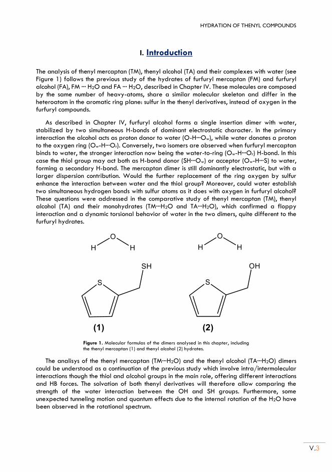
HYDRATION OF THENYL COMPOUNDS
V.3
I. Introduction The analysis of thenyl mercaptan (TM), thenyl alcohol (TA) and their complexes with water (see Figure 1) follows the previous study of the hydrates of furfuryl mercaptan (FM) and furfuryl alcohol (FA), FM ··· H2O and FA ··· H2O, described in Chapter IV. These molecules are composed by the same number of heavy-atoms, share a similar molecular skeleton and differ in the heteroatom in the aromatic ring plane: sulfur in the thenyl derivatives, instead of oxygen in the furfuryl compounds.
As described in Chapter IV, furfuryl alcohol forms a single insertion dimer with water, stabilized by two simultaneous H-bonds of dominant electrostatic character. In the primary interaction the alcohol acts as proton donor to water (O-H···Ow), while water donates a proton to the oxygen ring (Ow-H···Or). Conversely, two isomers are observed when furfuryl mercaptan binds to water, the stronger interaction now being the water-to-ring (Ow-H···Or) H-bond. In this case the thiol group may act both as H-bond donor (SH···Ow) or acceptor (Ow-H···S) to water, forming a secondary H-bond. The mercaptan dimer is still dominantly electrostatic, but with a larger dispersion contribution. Would the further replacement of the ring oxygen by sulfur enhance the interaction between water and the thiol group? Moreover, could water establish two simultaneous hydrogen bonds with sulfur atoms as it does with oxygen in furfuryl alcohol? These questions were addressed in the comparative study of thenyl mercaptan (TM), thenyl alcohol (TA) and their monohydrates (TM···H2O and TA···H2O), which confirmed a floppy interaction and a dynamic torsional behavior of water in the two dimers, quite different to the furfuryl hydrates.
Figure 1. Molecular formulas of the dimers analysed in this chapter, including the thenyl mercaptan (1) and thenyl alcohol (2) hydrates.
The analisys of the thenyl mercaptan (TM···H2O) and the thenyl alcohol (TA···H2O) dimers could be understood as a continuation of the previous study which involve intra/intermolecular interactions though the thiol and alcohol groups in the main role, offering different interactions and HB forces. The solvation of both thenyl derivatives will therefore allow comparing the strength of the water interaction between the OH and SH groups. Furthermore, some unexpected tunneling motion and quantum effects due to the internal rotation of the H2O have been observed in the rotational spectrum.
V.4
All the experiments have been, again, assisted by molecular-orbital quantum chemical calculations with different dispersion-corrected density functional theory (DFT) models and/or ab initio methods, simultaneously assessing the theoretical calculations. Additionally, we have also carried out an analysis of the intermolecular interactions based in the electronic density topology (NCIPlot)1 and a symmetry-adapted perturbation theory (SAPT)2 energy decomposition to examine the underlying basis of the intermolecular interactions originated by the thiol and alcohol groups.
HYDRATION OF THENYL COMPOUNDS
V.5
II. Thenyl Mercaptan and Thenyl alcohol
1. Experimental set up:
Thenyl mercaptan (2-thiophenemethanethiol, >95% GC, b.p. 76°C) and thenyl alcohol (2-thiophenemethanol, >98% GC, b.p. 207°C) were obtained commercially and used without further purification. The vapors of each compound were diluted within a stream of pure neon (backing pressures of 0.25 MPa), forming a supersonic jet by expansion through a pin-hole nozzle (f=0.5-0.8 mm) into a high-vacuum chamber (ultimate pressures of 10-7 hPa). The mercaptan sample was volatile enough to be placed in an external reservoir inserted in the gas line, but the alcohol required a heating nozzle (45°C) to obtain sufficient vapor pressure. For the water clusters either H2O or H218O were added to the gas line. The jet was probed with a broadband chirped-pulse Fourier transform microwave (CP-FTMW) spectrometer, operating in the region 2-8 GHz, and a Balle-Flygare cavity FTMW spectrometer, working in 8-18 GHz.
Thenyl Mercaptan TM···H2O Thenyl Alcohol TA···H2O
Carrier Gas Ne Ne Nozzle Ø (mm) 0.5 0.8
Sample Tª Room temperature 45ºC Carrier Gas Pressure 2.5 bar 2.5 bar
2. Rotational spectra of TM and TA
The experimental work initially examined the conformational landscape and rotational spectrum of the thenyl monomers TM and TA, previously unreported. Similarly to the furfuryl derivatives,3 the bidimensional potential energy surface is defined by two dihedrals describing the elevation of the side chain with respect to the thenyl ring (1(S1- C2-C6-S7/O7) and the orientation of the terminal thiol/alcohol hydrogen atom (2(C2-C6- S7/O7-H). Molecular orbital calculations in Tables S1-S2 (ESI, see attached file) predicted elevation angles close to +60° (1 +gauche or G) for the side chain (as in the furfuryl derivatives) and staggered orientations for the terminal alcohol/thiol. However, the terminal hydrogen atom now preferentially orients to the adjacent carbon-carbon bond in the ring system (2 +gauche or G) of TM or TA instead of pointing to the ring oxygen, inverting the conformational stability of the furfuryl compounds. DFT energetic predictions in Tables S3-S4 (ESI, see attached file) and Figures 2-3 suggest that the second GG´ conformation is 1.2-1.9 kJmol-1 above the global minimum GG at B3LYP-D3(BJ) level.
The jet-cooled microwave spectra of water-seeded TM and TA is shown in Figures 4 and 5. Following the initial predictions the rotational spectra of the two monomers were readily assigned based on their dominant μa and μb selection rule transitions. A smaller set of weaker
μc transitions were added later. The spectrum was analyzed using a conventional Watson’s (S-
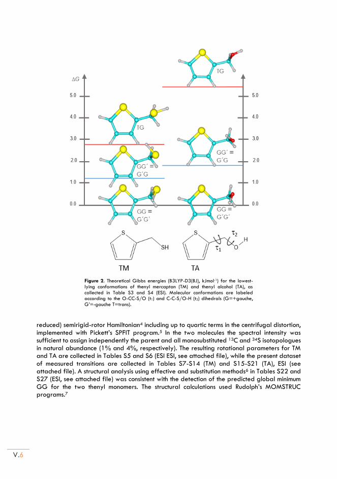
V.6
Figure 2. Theoretical Gibbs energies (B3LYP-D3(BJ), kJmol-1) for the lowest-lying conformations of thenyl mercaptan (TM) and thenyl alcohol (TA), as collected in Table S3 and S4 (ESI). Molecular conformations are labeled according to the O-CC-S/O (t1) and C-C-S/O-H (t2) dihedrals (G=+gauche, G’=-gauche T=trans).
reduced) semirigid-rotor Hamiltonian4 including up to quartic terms in the centrifugal distortion, implemented with Pickett’s SPFIT program.5 In the two molecules the spectral intensity was sufficient to assign independently the parent and all monosubstituted 13C and 34S isotopologues in natural abundance (1% and 4%, respectively). The resulting rotational parameters for TM and TA are collected in Tables S5 and S6 (ESI ESI, see attached file), while the present dataset of measured transitions are collected in Tables S7-S14 (TM) and S15-S21 (TA), ESI (see attached file). A structural analysis using effective and substitution methods6 in Tables S22 and S27 (ESI, see attached file) was consistent with the detection of the predicted global minimum GG for the two thenyl monomers. The structural calculations used Rudolph’s MOMSTRUC programs.7
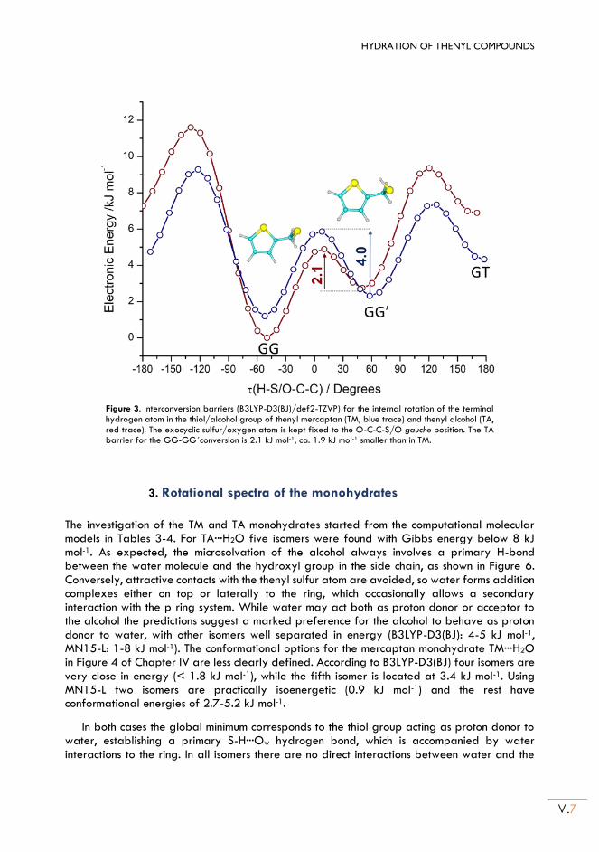
HYDRATION OF THENYL COMPOUNDS
V.7
Figure 3. Interconversion barriers (B3LYP-D3(BJ)/def2-TZVP) for the internal rotation of the terminal hydrogen atom in the thiol/alcohol group of thenyl mercaptan (TM, blue trace) and thenyl alcohol (TA, red trace). The exocyclic sulfur/oxygen atom is kept fixed to the O-C-C-S/O gauche position. The TA barrier for the GG-GG´conversion is 2.1 kJ mol-1, ca. 1.9 kJ mol-1 smaller than in TM.
3. Rotational spectra of the monohydrates
The investigation of the TM and TA monohydrates started from the computational molecular models in Tables 3-4. For TA···H2O five isomers were found with Gibbs energy below 8 kJ mol-1. As expected, the microsolvation of the alcohol always involves a primary H-bond between the water molecule and the hydroxyl group in the side chain, as shown in Figure 6. Conversely, attractive contacts with the thenyl sulfur atom are avoided, so water forms addition complexes either on top or laterally to the ring, which occasionally allows a secondary interaction with the p ring system. While water may act both as proton donor or acceptor to the alcohol the predictions suggest a marked preference for the alcohol to behave as proton donor to water, with other isomers well separated in energy (B3LYP-D3(BJ): 4-5 kJ mol-1, MN15-L: 1-8 kJ mol-1). The conformational options for the mercaptan monohydrate TM···H2O in Figure 4 of Chapter IV are less clearly defined. According to B3LYP-D3(BJ) four isomers are very close in energy (< 1.8 kJ mol-1), while the fifth isomer is located at 3.4 kJ mol-1. Using MN15-L two isomers are practically isoenergetic (0.9 kJ mol-1) and the rest have conformational energies of 2.7-5.2 kJ mol-1.
In both cases the global minimum corresponds to the thiol group acting as proton donor to water, establishing a primary S-H···Ow hydrogen bond, which is accompanied by water interactions to the ring. In all isomers there are no direct interactions between water and the
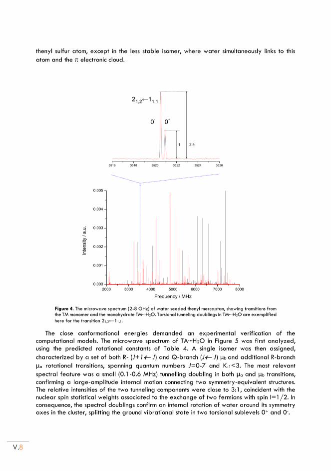
V.8
thenyl sulfur atom, except in the less stable isomer, where water simultaneously links to this
atom and the electronic cloud.
Figure 4. The microwave spectrum (2-8 GHz) of water seeded thenyl mercaptan, showing transitions from the TM monomer and the monohydrate TM···H2O. Torsional tunneling doublings in TM···H2O are exemplified
here for the transition 21,2←11,1.
The close conformational energies demanded an experimental verification of the computational models. The microwave spectrum of TA···H2O in Figure 5 was first analyzed, using the predicted rotational constants of Table 4. A single isomer was then assigned,
characterized by a set of both R- (J+1 J) and Q-branch (J J) μb and additional R-branch
μa rotational transitions, spanning quantum numbers J=0-7 and K-1<3. The most relevant
spectral feature was a small (0.1-0.6 MHz) tunnelling doubling in both μa and μb transitions,
confirming a large-amplitude internal motion connecting two symmetry-equivalent structures. The relative intensities of the two tunneling components were close to 3:1, coincident with the nuclear spin statistical weights associated to the exchange of two fermions with spin I=1/2. In consequence, the spectral doublings confirm an internal rotation of water around its symmetry axes in the cluster, splitting the ground vibrational state in two torsional sublevels 0+ and 0-.
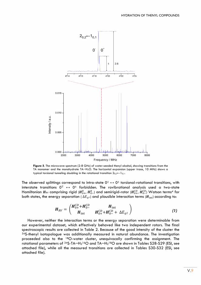
HYDRATION OF THENYL COMPOUNDS
V.9
Figure 5. The microwave spectrum (2-8 GHz) of water-seeded thenyl alcohol, showing transitions from the TA monomer and the monohydrate TA···H2O. The horizontal expansion (upper trace, 10 MHz) shows a
typical torsional tunneling doubling in the rotational transition 20,2←10,1.
The observed splittings correspond to intra-state 0± ↔ 0± torsional-rotational transitions, with
interstate transitions 0∓ ↔ 0± forbidden. The rovibrational analysis used a two-state Hamiltonian 𝑯RV comprising rigid (𝑯0+
𝑅 , 𝑯0−𝑅 ) and semirigid-rotor (𝑯0+
𝐶𝐷, 𝑯0−𝐶𝐷) Watson terms4 for
both states, the energy separation ( ∆𝐸0±) and plausible interaction terms (𝑯int) according to:
𝑯𝑅𝑉 = (𝑯0+
𝐶𝐷+𝑯0−𝐶𝐷 𝑯int
𝑯int 𝑯0+𝐶𝐷+𝑯0−
𝐶𝐷 + ∆𝐸0± ) (1)
However, neither the interaction terms or the energy separation were determinable from our experimental dataset, which effectively behaved like two independent rotors. The final spectroscopic results are collected in Table 2. Because of the good intensity of the cluster the 34S-thenyl isotopologue was additionally measured in natural abundance. The investigation proceeded also to the 18O-water cluster, unequivocally confirming the assignment. The rotational parameters of 34S-TA···H216O and TA···H218O are shown in Tables S28-S29 (ESI, see attached file), while all the measured transitions are collected in Tables S30-S32 (ESI, see attached file).
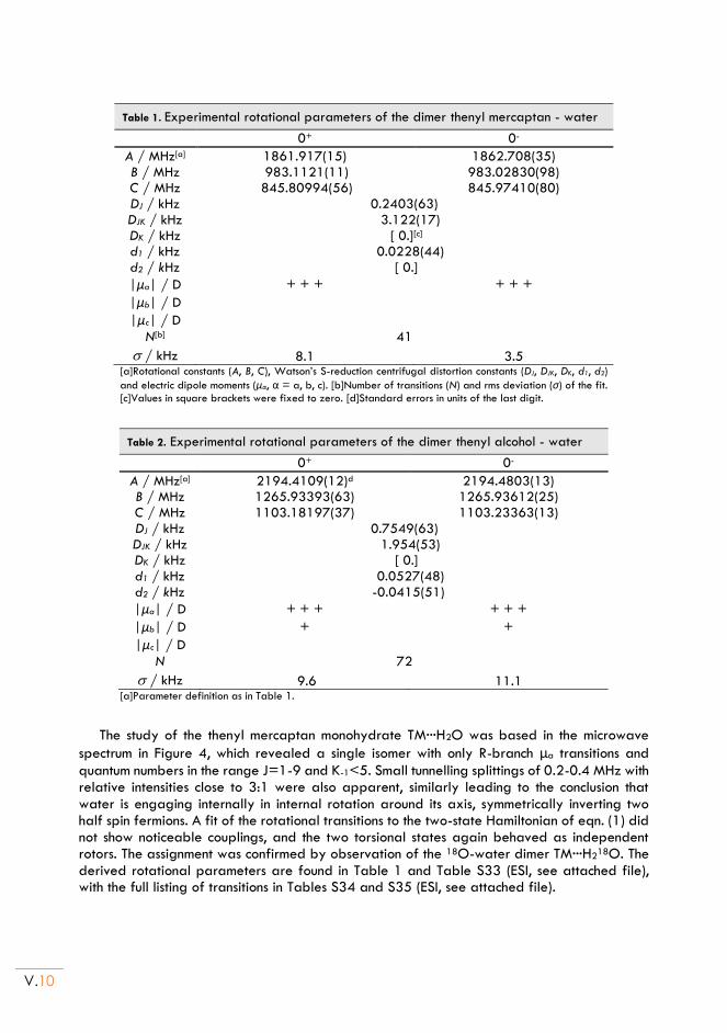
V.10
Table 1. Experimental rotational parameters of the dimer thenyl mercaptan - water
0+ 0-
A / MHz[a] 1861.917(15) 1862.708(35) B / MHz 983.1121(11) 983.02830(98) C / MHz 845.80994(56) 845.97410(80) DJ / kHz 0.2403(63) DJK / kHz 3.122(17) DK / kHz [ 0.][c] d1 / kHz 0.0228(44) d2 / kHz [ 0.]
|μa| / D + + + + + +
|μb| / D
|μc| / D
N[b] 41
/ kHz 8.1 3.5 [a]Rotational constants (A, B, C), Watson’s S-reduction centrifugal distortion constants (DJ, DJK, DK, d1, d2)
and electric dipole moments (μα, α = a, b, c). [b]Number of transitions (N) and rms deviation (σ) of the fit. [c]Values in square brackets were fixed to zero. [d]Standard errors in units of the last digit.
Table 2. Experimental rotational parameters of the dimer thenyl alcohol - water
0+ 0-
A / MHz[a] 2194.4109(12)d 2194.4803(13) B / MHz 1265.93393(63) 1265.93612(25) C / MHz 1103.18197(37) 1103.23363(13) DJ / kHz 0.7549(63) DJK / kHz 1.954(53) DK / kHz [ 0.] d1 / kHz 0.0527(48) d2 / kHz -0.0415(51)
|μa| / D + + + + + +
|μb| / D + +
|μc| / D
N 72
/ kHz 9.6 11.1 [a]Parameter definition as in Table 1.
The study of the thenyl mercaptan monohydrate TM···H2O was based in the microwave
spectrum in Figure 4, which revealed a single isomer with only R-branch μa transitions and
quantum numbers in the range J=1-9 and K-1<5. Small tunnelling splittings of 0.2-0.4 MHz with relative intensities close to 3:1 were also apparent, similarly leading to the conclusion that water is engaging internally in internal rotation around its axis, symmetrically inverting two half spin fermions. A fit of the rotational transitions to the two-state Hamiltonian of eqn. (1) did not show noticeable couplings, and the two torsional states again behaved as independent rotors. The assignment was confirmed by observation of the 18O-water dimer TM···H218O. The derived rotational parameters are found in Table 1 and Table S33 (ESI, see attached file), with the full listing of transitions in Tables S34 and S35 (ESI, see attached file).
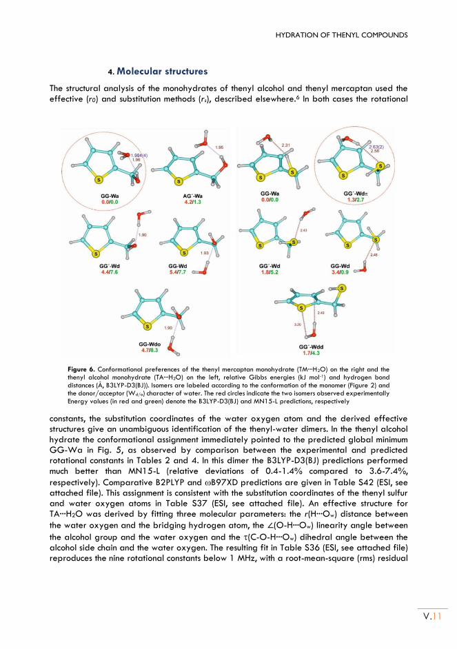
HYDRATION OF THENYL COMPOUNDS
V.11
4. Molecular structures
The structural analysis of the monohydrates of thenyl alcohol and thenyl mercaptan used the effective (r0) and substitution methods (rs), described elsewhere.6 In both cases the rotational
Figure 6. Conformational preferences of the thenyl mercaptan monohydrate (TM···H2O) on the right and the thenyl alcohol monohydrate (TA···H2O) on the left, relative Gibbs energies (kJ mol-1) and hydrogen bond distances (Å, B3LYP-D3(BJ)). Isomers are labeled according to the conformation of the monomer (Figure 2) and the donor/acceptor (Wd/a) character of water. The red circles indicate the two isomers observed experimentally Energy values (in red and green) denote the B3LYP-D3(BJ) and MN15-L predictions, respectively
constants, the substitution coordinates of the water oxygen atom and the derived effective structures give an unambiguous identification of the thenyl-water dimers. In the thenyl alcohol hydrate the conformational assignment immediately pointed to the predicted global minimum GG-Wa in Fig. 5, as observed by comparison between the experimental and predicted rotational constants in Tables 2 and 4. In this dimer the B3LYP-D3(BJ) predictions performed much better than MN15-L (relative deviations of 0.4-1.4% compared to 3.6-7.4%,
respectively). Comparative B2PLYP and B97XD predictions are given in Table S42 (ESI, see attached file). This assignment is consistent with the substitution coordinates of the thenyl sulfur and water oxygen atoms in Table S37 (ESI, see attached file). An effective structure for TA···H2O was derived by fitting three molecular parameters: the r(H···Ow) distance between
the water oxygen and the bridging hydrogen atom, the ∠(O-H···Ow) linearity angle between
the alcohol group and the water oxygen and the (C-O-H···Ow) dihedral angle between the alcohol side chain and the water oxygen. The resulting fit in Table S36 (ESI, see attached file) reproduces the nine rotational constants below 1 MHz, with a root-mean-square (rms) residual
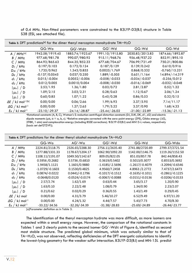
V.12
of 0.4 MHz. Non-fitted parameters were constrained to the B3LYP-D3(BJ) structure in Table S38 (ESI, see attached file).
Table 3. DFT predictions[c] for the dimer thenyl mercaptan monohydrate TM···H2O
GG-Wa GG’-Wd GG’-Wd GG-Wd GG-Wd
A / MHz[a] 1942.58/1919.45 1885.74/1923.67 1991.10/1915.80 2030.83/2013.83 1874.66/1892.87
B / MHz 977.68/981.78 990.07/980.92 915.11/968.76 956.48/937.22 852.91/895.10
C / MHz 864.93/865.63 844.35/852.33 677.68/704.67 706.99/721.49 750.21/800.86
DJ / kHz 0.197/0.103 0.172/0.124 0.187/0.129 0.139/0.242 0.610/0.916
DJK / kHz 1.735/0.962 1.343/0.855 0.0025/1.769 0.868/0.332 -0.760/15.231
DK / kHz -0.137/0.0545 0.037/0.250 1.889/-0.505 0.651/1.164 14.894/-14.319
d1 / kHz 0.015/-0.004 0.0052/-0.006 -0.058/-0.033 -0.034/-0.037 -0.256/0.012
d2 / kHz 0.012/0.005 0.0010/0.004 -0.008/-0.030 -0.016/-0.069 -0.032/-0.048
|μa| / D 2.53/1.95 1.36/1.80 0.03/0.72 2.81/2.87 0.52/1.32
|μb| / D 1.09/2.15 3.03/2.31 0.28/0.63 1.12/0.67 2.06/1.24
|μc| / D 0.60/0.85 1.07/1.22 0.45/0.28 0.86/0.53 0.32/0.15
ΔE / kJ mol-1[b] 0.00/0.00 0.06/2.66 1.99/6.93 3.37/5.95 7.14/11.17
ΔG / kJ mol-1 0.00/0.00 1.27/2.65 1.79/5.22 3.37/0.90 1.68/4.33
EB / kJ mol-1 -23.10/-29.37 -24.14/-28.87 -25.44/-26.02 -21.00/-22.89 -15.36/-21.13 aRotational constants (A, B, C), Watson’s S-reduction centrifugal distortion constants (DJ, DJK, DK, d1, d2) and electric
dipole moments (μα, α = a, b, c). bRelative energies corrected with the zero-point energy (ZPE), Gibbs energy (G,
298K, 1 atm) and complexation energy (including BSSE corrections). cB3LYP-D3(BJ) and MN15-L values, respectively (basis set def2TZVP)).
Table 4. DFT predictions for the dimer thenyl alcohol monohydrate TA···H2O
GG-Wa AG’-Wa GG’-Wd GG-Wd GG-Wdo
A / MHz 2224.81/2118.75 2326.65/2288.30 2756.11/2635.40 2741.88/2720.89 2799.37/2721.54 B / MHz 1277.88/1331.33 1178.67/1204.17 1062.90/1092.20 1142.00/1146.75 1119.26/1152.50 C / MHz 1108.11/1191.07 1049.50/1142.67 809.05/822.05 851.03/857.78 842.44/858.43 DJ / kHz 0.5956 /0.2682 0.5736 /0.6810 0.3619/0.5402 0.5013/0.3077 0.8553/0.3692 DJK / kHz 1.9458/1.1121 1.1601/0.9880 -1.4185/-2.5696 -1.2617/-0.4079 -3.2094/-0.6546 DK / kHz -1.0729/-0.1603 0.1530/0.4825 4.9560/7.2458 4.8963 /2.2772 7.4737/2.6473 d1 / kHz 0.0874/-0.0222 0.0441/-0.1796 -0.1017/-0.1512 -0.1635/-0.1011 -0.2861/-0.1225 d2 / kHz -0.0648/0.0120 -0.0524/-0.0174 -0.0067/-0.0088 -0.0151/-0.0136 -0.0206/-0.0133
|μa| / D 2.57/2.74 1.62/1.69 0.63/0.44 3.65/3.17 1.20/0.90
|μb| / D 1.63/0.10 2.22/2.48 1.08/0.79 1.34/0.90 2.23/2.07
|μc| / D 0.21/0.62 0.02/0.29 0.36/0.55 1.43/1.49 0.29/0.45
ΔE / kJ mol- 0.00/0.00 4.31/3.79 5.46/10.27 6.52/9.49 6.97/11.20
ΔG / kJ mol-1 0.00/0.00 4.24/1.32 4.44/7.57 5.43/7.73 4.70/8.30
EB / kJ mol-1 -33.22/-34.56 -32.26/-34.39 -31.38/-28.83 -25.69/-24.89 -26.44/-23.77 [a]Parameter definition as in Table 3.
The identification of the thenyl mercaptan hydrate was more difficult, as more isomers are
expected within a small energy range. However, the comparison of the rotational constants in
Tables 1 and 3 clearly points to the second isomer GG´-Wd of Figure 6, identified as second most stable structure. The predicted global minimum, which was actually similar to that of TA···H2O, was not observed, reflecting deficiencies of the DFT energetic calculations to identify the lowest-lying geometry for the weaker sulfur interaction. B3LYP-D3(BJ) and MN-15L predict
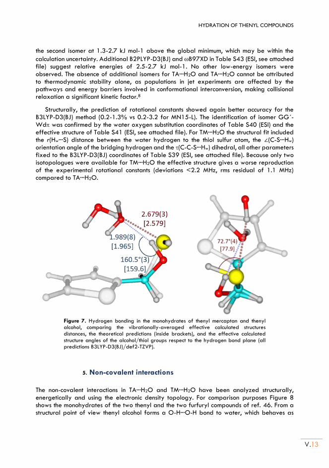
HYDRATION OF THENYL COMPOUNDS
V.13
the second isomer at 1.3-2.7 kJ mol-1 above the global minimum, which may be within the
calculation uncertainty. Additional B2PLYP-D3(BJ) and B97XD in Table S43 (ESI, see attached file) suggest relative energies of 2.5-2.7 kJ mol-1. No other low-energy isomers were observed. The absence of additional isomers for TA···H2O and TA···H2O cannot be attributed to thermodynamic stability alone, as populations in jet experiments are affected by the pathways and energy barriers involved in conformational interconversion, making collisional relaxation a significant kinetic factor.8
Structurally, the prediction of rotational constants showed again better accuracy for the B3LYP-D3(BJ) method (0.2-1.3% vs 0.2-3.2 for MN15-L). The identification of isomer GG´-Wd was confirmed by the water oxygen substitution coordinates of Table S40 (ESI) and the effective structure of Table S41 (ESI, see attached file). For TM···H2O the structural fit included the r(Hw···S) distance between the water hydrogen to the thiol sulfur atom, the ∠(C-S···Hw) orientation angle of the bridging hydrogen and the (C-C-S···Hw) dihedral, all other parameters fixed to the B3LYP-D3(BJ) coordinates of Table S39 (ESI, see attached file). Because only two isotopologues were available for TM···H2O the effective structure gives a worse reproduction of the experimental rotational constants (deviations <2.2 MHz, rms residual of 1.1 MHz) compared to TA···H2O.
Figure 7. Hydrogen bonding in the monohydrates of thenyl mercaptan and thenyl alcohol, comparing the vibrationally-averaged effective calculated structures distances, the theoretical predictions (inside brackets), and the effective calculated structure angles of the alcohol/thiol groups respect to the hydrogen bond plane (all predictions B3LYP-D3(BJ)/def2-TZVP).
5. Non-covalent interactions
The non-covalent interactions in TA···H2O and TM···H2O have been analyzed structurally, energetically and using the electronic density topology. For comparison purposes Figure 8 shows the monohydrates of the two thenyl and the two furfuryl compounds of ref. 46. From a structural point of view thenyl alcohol forms a O-H···O-H bond to water, which behaves as
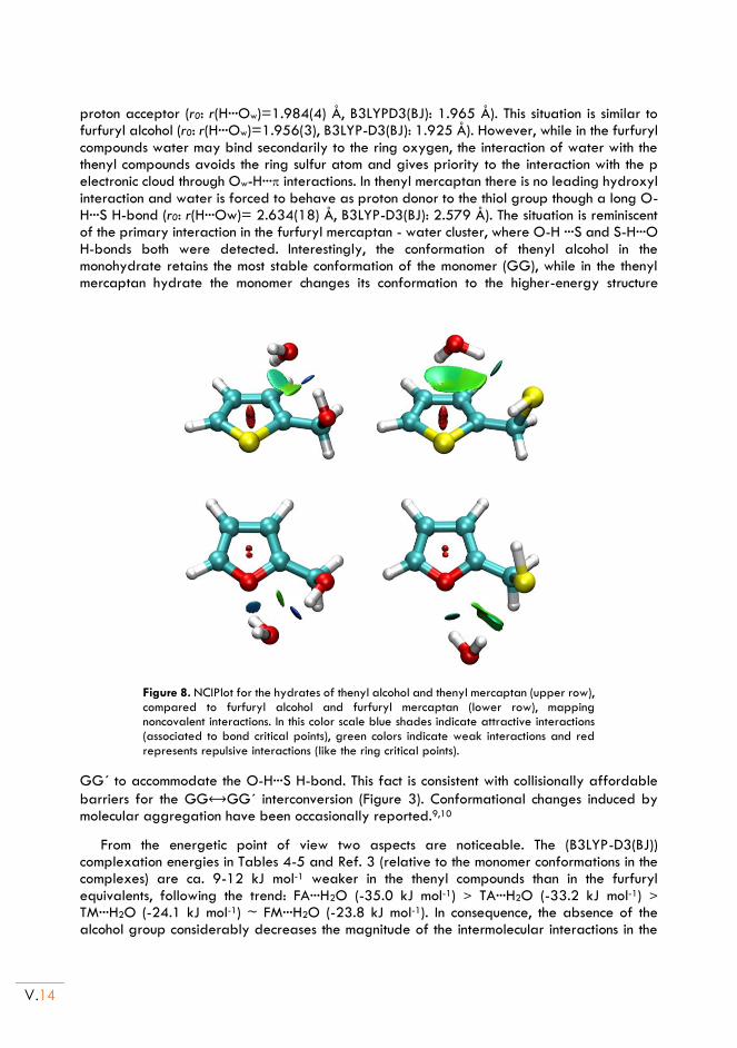
V.14
proton acceptor (r0: r(H···Ow)=1.984(4) Å, B3LYPD3(BJ): 1.965 Å). This situation is similar to furfuryl alcohol (r0: r(H···Ow)=1.956(3), B3LYP-D3(BJ): 1.925 Å). However, while in the furfuryl compounds water may bind secondarily to the ring oxygen, the interaction of water with the thenyl compounds avoids the ring sulfur atom and gives priority to the interaction with the p electronic cloud through Ow-H··· interactions. In thenyl mercaptan there is no leading hydroxyl interaction and water is forced to behave as proton donor to the thiol group though a long O-H···S H-bond (r0: r(H···Ow)= 2.634(18) Å, B3LYP-D3(BJ): 2.579 Å). The situation is reminiscent of the primary interaction in the furfuryl mercaptan - water cluster, where O-H ···S and S-H···O H-bonds both were detected. Interestingly, the conformation of thenyl alcohol in the monohydrate retains the most stable conformation of the monomer (GG), while in the thenyl mercaptan hydrate the monomer changes its conformation to the higher-energy structure
Figure 8. NCIPlot for the hydrates of thenyl alcohol and thenyl mercaptan (upper row), compared to furfuryl alcohol and furfuryl mercaptan (lower row), mapping noncovalent interactions. In this color scale blue shades indicate attractive interactions (associated to bond critical points), green colors indicate weak interactions and red represents repulsive interactions (like the ring critical points).
GG´ to accommodate the O-H···S H-bond. This fact is consistent with collisionally affordable
barriers for the GG⟷GG´ interconversion (Figure 3). Conformational changes induced by molecular aggregation have been occasionally reported.9,10
From the energetic point of view two aspects are noticeable. The (B3LYP-D3(BJ)) complexation energies in Tables 4-5 and Ref. 3 (relative to the monomer conformations in the complexes) are ca. 9-12 kJ mol-1 weaker in the thenyl compounds than in the furfuryl equivalents, following the trend: FA···H2O (-35.0 kJ mol-1) > TA···H2O (-33.2 kJ mol-1) > TM···H2O (-24.1 kJ mol-1) ~ FM···H2O (-23.8 kJ mol-1). In consequence, the absence of the alcohol group considerably decreases the magnitude of the intermolecular interactions in the
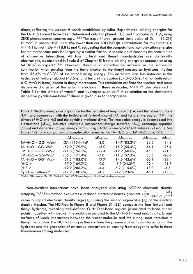
HYDRATION OF THENYL COMPOUNDS
V.15
dimer, reflecting the weaker H-bonds established by sulfur. Experimental binding energies for the O-H···S H-bond have been determined only for phenol···H2S and fluorophenol···H2S, using ZEKE photoelectron spectroscopy.11,12 The experimental ground-state value of D0 = -13.2(4) kJ mol-1 in phenol···H2S is ca. 6% lower than our B3LYP-D3(BJ) calculations for this cluster (D0 = -14.1 kJ mol-1, De = -18.8 kJ mol-1), suggesting that the computational complexation energies for the mercaptans may be larger by a similar factor. A second point concerns the contribution of dispersive interactions. All four furfuryl and thenyl monohydrates are dominantly electrostatic, as observed in Table 5 of Chapter III from a binding energy decomposition using SAPT(0)/jun-cc-pVDZ.13,14 However, there is a considerable increase in the dispersion contribution when passing from the theny alcohol to the thenyl mercaptan hydrates, moving from 55.4% to 83.3% of the total binding energy. This increment was less notorious in the hydrates of furfuryl alcohol (43.6%) and furfuryl mercaptan (57.3-60.6%),3 which both retain a O-H···O H-bond, absent in thenyl mercaptan. This calculation confirms the weaker and more dispersive character of the sulfur interactions in these molecules,11,12,15–20 also observed in Table 5 for the dimers of water21 and hydrogen sulphide.22 A calculation on the dominantly dispersive pyridine-methane23 dimer is given also for comparison.
Table 5. Binding energy decomposition for the hydrates of tenyl alcohol (TA) and thenyl mercaptane (TM), and comparison with the hydrates of furfuryl alcohol (FA) and furfuryl mercaptan (FM), the dimers of H2O and H2S and the pyridine-methane dimer. The interaction energy is decomposed into electrostatic (Eelec), inductive (multipole interactions/charge transfer, Eind), exchange repulsion (Eexch) and dispersion (Edisp) energy terms, using SAPT(0)/jun-cc-pVDZ (all values in kJ mol-1). See Tables 1-2 for a comparison of complexation energies for TA···H2O and TM···H2O using DFT.
EElectrostatic EInduction EDispersion EExchange ETotal
TM···H2O – GG’-Wdb -27.1 [154.4%]f -8.0 -14.7 [83.3%] 32.2 -14.3
TA···H2O – GG-Wab -52.0 [179.9%] -15.0 -15.9 [55.4%] 54.1 -25.4 FM···H2O – GG´-Wda
a -41.8 [196.2%] -12.4 -12.9 [60.6%] 45.8 -21.3 FM···H2O – GG-Wdd
a -35.3 [171.4%] -7.0 -11.8 [57.3%] 33.5 -20.6 FA···H2O – GG´-Wda
a -61.3 [183.0%] -17.7 -14.6 [43.6%] 60.1 -33.4 (H2O)2c -37.0 [169.7%] -9.0 -5.3 [24.3%] 29.4 -21.8 (H2S)2e -12.9 [286.7%] -4.4 -5.2 [115.6%] 18.0 -4.5 Pyridine-methane[d] -17.9 [100.6%] -4.1 -42.0[236%] 46.1 -17.8 aRef.3. bThis work. cRef.21. dRef.23. eRef.22. fPercentage of the total binding energy.
Non-covalent interactions have been analyzed also using NCIPlot electronic density
mapping.24,25 This method evaluates a reduced electronic density gradient 𝑠 (=1
1(3𝜋2)1/3
|∇𝜌|
𝜌4/3)
versus a signed electronic density (sign (𝜆2)𝜌) using the second eigenvalue (𝜆2) of the electron density Hessian. The NCIPlots in Figure 8 and Figure S1 (ESI) compare the four furfuryl and thenyl hydrates, revealing well-defined O-H···O H-bond regions (associated to bond critical points), together with weaker interactions associated to the O-H···S H-bond and, finally, broad surfaces of weak interactions between the water molecule and the ring, most notorious in thenyl mercaptan. The NCIPlot analysis thus confirms the presence of multiple interactions in the hydrates and the gradation of attractive interactions on passing from oxygen to sulfur in these five-membered ring molecules.
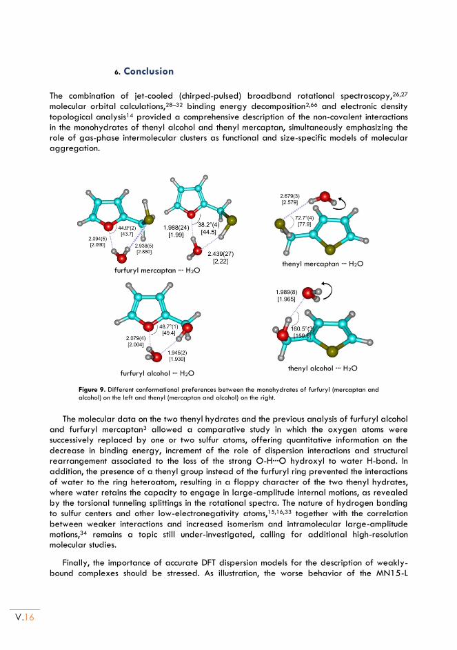
V.16
6. Conclusion
The combination of jet-cooled (chirped-pulsed) broadband rotational spectroscopy,26,27 molecular orbital calculations,28–32 binding energy decomposition2,66 and electronic density topological analysis14 provided a comprehensive description of the non-covalent interactions in the monohydrates of thenyl alcohol and thenyl mercaptan, simultaneously emphasizing the role of gas-phase intermolecular clusters as functional and size-specific models of molecular aggregation.
furfuryl mercaptan ··· H2O
thenyl mercaptan ··· H2O
furfuryl alcohol ··· H2O
thenyl alcohol ··· H2O
Figure 9. Different conformational preferences between the monohydrates of furfuryl (mercaptan and alcohol) on the left and thenyl (mercaptan and alcohol) on the right.
The molecular data on the two thenyl hydrates and the previous analysis of furfuryl alcohol
and furfuryl mercaptan3 allowed a comparative study in which the oxygen atoms were successively replaced by one or two sulfur atoms, offering quantitative information on the decrease in binding energy, increment of the role of dispersion interactions and structural rearrangement associated to the loss of the strong O-H···O hydroxyl to water H-bond. In addition, the presence of a thenyl group instead of the furfuryl ring prevented the interactions of water to the ring heteroatom, resulting in a floppy character of the two thenyl hydrates, where water retains the capacity to engage in large-amplitude internal motions, as revealed by the torsional tunneling splittings in the rotational spectra. The nature of hydrogen bonding to sulfur centers and other low-electronegativity atoms,15,16,33 together with the correlation between weaker interactions and increased isomerism and intramolecular large-amplitude motions,34 remains a topic still under-investigated, calling for additional high-resolution molecular studies.
Finally, the importance of accurate DFT dispersion models for the description of weakly-bound complexes should be stressed. As illustration, the worse behavior of the MN15-L
HYDRATION OF THENYL COMPOUNDS
V.17
functional in the present study may suggest the need to introduce explicit long-range dispersion corrections in the Minnesota functionals.35 This fact illustrates the contribution and complementary role of empirical data from high-resolution rotational data for the development of DFT computational models of molecular aggregation.
V.18
Chapter V. References: 1. Contreras-García, J. et al. NCIPLOT: A Program for Plotting Noncovalent Interaction Regions. J. Chem.
Theory Comput. 7, 625–632 (2011).
2. Garcia, J., Podeszwa, R. & Szalewicz, K. SAPT codes for calculations of intermolecular interaction energies. J. Chem. Phys. 152, 184109 (2020).
3. Juanes, M. et al. Sulfur Hydrogen Bonding in Isolated Monohydrates: Furfuryl Mercaptan versus Furfuryl Alcohol. Chem. - A Eur. J. 24, 6564–6571 (2018).
4. Watson, J. K. G. Vibrational Spectra and Structure. in (ed. Durig, J. R.) 1–89 (Elsevier Inc., 1977).
5. Pickett, H. M. The fitting and prediction of vibration-rotation spectra with spin interactions. J. Mol. Spectrosc. 148, 371–377 (1991).
6. Rudolph, H. D. & Demaison, J. Equilibrium Molecular Structures: From Spectroscopy to Quantum Chemistry. in Equilibrium Molecular Structures: From Spectroscopy to Quantum Chemistry (eds. Demaison, J., Boggs, J. E. & Császár, A. G.) 125–158 (CRC Press, 2011).
7. MOMSTRUC Structural Package.
8. Godfrey, P. D., Rodgers, F. M. & Brown, R. D. Theory versus Experiment in Jet Spectroscopy: Glycolic Acid. J. Am. Chem. Soc. 119, 2232–2239 (1997).
9. Thomas, J., Seifert, N. A., Jäger, W. & Xu, Y. A Direct Link from the Gas to the Condensed Phase: A Rotational Spectroscopic Study of 2,2,2-Trifluoroethanol Trimers. Angew. Chemie Int. Ed. 56, 6289–6293 (2017).
10. Oswald, S. et al. The Chiral Trimer and a Metastable Chiral Dimer of Achiral Hexafluoroisopropanol: A Multi‐ Messenger Study. Angew. Chemie Int. Ed. 58, 5080–5084 (2019).
11. Ghosh, S., Bhattacharyya, S. & Wategaonkar, S. Dissociation Energies of Sulfur-Centered Hydrogen-Bonded Complexes. J. Phys. Chem. A 119, 10863–10870 (2015).
12. Bhattacharyya, S. & Wategaonkar, S. ZEKE Photoelectron Spectroscopy of p -Fluorophenol···H 2 S/H 2 O Complexes and Dissociation Energy Measurement Using the Birge–Sponer Extrapolation Method. J. Phys. Chem. A 118, 9386–9396 (2014).
13. Scheiner, S. Hydrogen Bonding. A Theoretical Perspective. (Oxford University Press, 1997).
14. Jeziorski, B., Moszynski, R. & Szalewicz, K. Perturbation Theory Approach to Intermolecular Potential Energy Surfaces of van der Waals Complexes. Chem. Rev. 94, 1887–1930 (1994).
15. Biswal, H. S. Hydrogen Bonding Involving Sulfur: New Insights from Ab Initio Calculations and Gas Phase Laser Spectroscopy. in Noncovalent Forces (ed. Scheiner, S.) (Springer Int. Pub., 2015).
16. Biswal, H. S., Bhattacharyya, S., Bhattacherjee, A. & Wategaonkar, S. Nature and strength of sulfur-centred hydrogen bonds: laser spectroscopic investigations in the gas phase and quantum-chemical calculations. Int. Rev. Phys. Chem. 34, 99–160 (2015).
17. Goebel, J. R., Ault, B. S. & Del Bene, J. E. Matrix Isolation and ab Initio Study of 1:1 Hydrogen-Bonded Complexes of H 2 O 2 with Phosphorus and Sulfur Bases. J. Phys. Chem. A 105, 11365–11370 (2001).
18. Wierzejewska, M. Infrared matrix isolation studies of complexes formed between dimethylsulfide, dimethyldisulfide and nitrous acid. J. Mol. Struct. 520, 199–214 (2000).
19. Wang, D., Chopra, P., Wategaonkar, S. & Fujii, A. Electronic and Infrared Spectroscopy of Benzene-(H 2 S) n ( n = 1 and 2): The Prototype of the SH-π Interaction. J. Phys. Chem. A 123, 7255–7260
HYDRATION OF THENYL COMPOUNDS
V.19
(2019).
20. Wategaonkar, S. & Bhattacherjee, A. N–H···S Interaction Continues To Be an Enigma: Experimental and Computational Investigations of Hydrogen-Bonded Complexes of Benzimidazole with Thioethers. J. Phys. Chem. A 122, 4313–4321 (2018).
21. Dyke, T. R., Mack, K. M. & Muenter, J. S. The structure of water dimer from molecular beam electric resonance spectroscopy. J. Chem. Phys. 66, 498–510 (1977).
22. Das, A. et al. The H2S Dimer is Hydrogen‐ Bonded: Direct Confirmation from Microwave Spectroscopy. Angew. Chemie Int. Ed. 57, 15199–15203 (2018).
23. Gou, Q. et al. Interactions between alkanes and aromatic molecules: a rotational study of pyridine–methane. Phys. Chem. Chem. Phys. 16, 13041–13046 (2014).
24. Laplaza, R. et al. NCIPLOT and the analysis of noncovalent interactions using the reduced density gradient. WIREs Comput. Mol. Sci. 11, (2021).
25. Lane, J. R., Contreras-García, J., Piquemal, J.-P., Miller, B. J. & Kjaergaard, H. G. Are Bond Critical Points Really Critical for Hydrogen Bonding? J. Chem. Theory Comput. 9, 3263–3266 (2013).
26. Caminati, W. & Grabow, J.-U. Advancements in Microwave Spectroscopy. in Frontiers and Advancements in Microwave Spectroscopy (ed. Laane, J.) 569–598 (Elsevier, 2018).
27. Juanes, M., Saragi, R. T., Caminati, W. & Lesarri, A. The Hydrogen Bond and Beyond: Perspectives for Rotational Investigations of Non-Covalent Interactions. Chem. - A Eur. J. 25, (2019).
28. Raghavachari, K. Perspective on ‘Density functional thermochemistry. III. The role of exact exchange’. Theor. Chem. Accounts Theory, Comput. Model. (Theoretica Chim. Acta) 103, 361–363 (2000).
29. Yu, H. S., He, X. & Truhlar, D. G. MN15-L: A New Local Exchange-Correlation Functional for Kohn–Sham Density Functional Theory with Broad Accuracy for Atoms, Molecules, and Solids. J. Chem. Theory Comput. 12, 1280–1293 (2016).
30. Weigend, F. & Ahlrichs, R. Balanced basis sets of split valence, triple zeta valence and quadruple zeta valence quality for H to Rn: Design and assessment of accuracy. Phys. Chem. Chem. Phys. 7, 3297 (2005).
31. Grimme, S., Ehrlich, S. & Goerigk, L. Effect of the damping function in dispersion corrected density functional theory. J. Comput. Chem. 32, 1456–1465 (2011).
32. Johnson, E. R. & Becke, A. D. A post-Hartree-Fock model of intermolecular interactions: Inclusion of higher-order corrections. J. Chem. Phys. 124, 174104 (2006).
33. Gilli, G. & Gilli, P. The Nature of the Hydrogen Bond. (2009).
34. Pérez, C. et al. Isomerism of the Aniline Trimer. Angew. Chemie Int. Ed. 57, 15112–15116 (2018).
35. Goerigk, L. et al. A look at the density functional theory zoo with the advanced GMTKN55 database for general main group thermochemistry, kinetics and noncovalent interactions. Phys. Chem. Chem. Phys. 19, 32184–32215 (2017).

Chapter VI DIMERIZATION OF FURFURYL AND THENYL
COMPOUNDS:
(Furfuryl Alcohol)2* and (Thenyl Alcohol)2
*
(*): M. Juanes, R. T. Saragi, C. Pérez, R. Pinacho, M. Jaraíz and A. Lesarri, Symmetry, 2021, submitted. (The Electronic Supporting Information – ESI, can be found in the document: thesis-ESI.pdf)
Adapted from:

VI.2
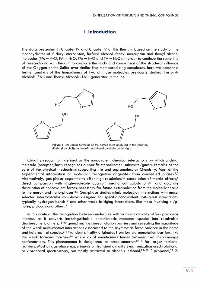
DIMERIZATION OF FURFURYL AND THENYL COMPOUNDS
VI.3
I. Introduction
The data presented in Chapter IV and Chapter V of this thesis is based on the study of the monohydrates of furfuryl mercaptan, furfuryl alcohol, thenyl mercaptan and thenyl alcohol molecules (FM ··· H2O, FA ··· H2O, TM ··· H2O and TA ··· H2O). In order to continue the same line of research and with the aim to conclude the study and comparison of the structural influence of the Oxygen or the Sulfur over similar five membered ring complexes, here we present a further analysis of the homodimers of two of those molecules previously studied: Furfuryl-Alcohol2 (FA2) and Thenyl-Alcohol2 (TA2), generated in the jet.
Figure 1. Molecular formulas of the homodimers analysed in this chapter, (Furfuryl alcohol)2 on the left and (thenyl alcohol)2 on the right.
Chirality recognition, defined as the noncovalent chemical interactions by which a chiral molecule (receptor/host) recognizes a specific stereoisomer (substrate/guest), remains at the core of the physical mechanisms supporting life and supramolecular Chemistry. Most of the experimental information on molecular recognition originates from condensed phases.1,2 Alternatively, gas-phase experiments offer high-resolution,3,4 cancelation of matrix effects,5 direct comparison with single-molecule quantum mechanical calculations6,7 and accurate description of noncovalent forces, necessary for future extrapolation from the molecular scale to the meso- and nano-phases.8,9 Gas-phase studies mimic molecular interactions with mass-selected intermolecular complexes designed for specific noncovalent host–guest interactions, typically hydrogen bonds10 and other weak bridging interactions, like those involving s-/p-holes, p clouds and others.11,12
In this context, the recognition between molecules with transient chirality offers particular interest, as it converts indistinguishable enantiomeric monomer species into resolvable diastereomeric dimers,13–15 quenching the stereomutation barriers and revealing the magnitude of the weak multi-contact interactions associated to the asymmetric force balance in the homo and heterochiral species.3,4 Transient chirality originates from low stereomutation barriers, like the weak torsional barriers16 where axial enantiomers tunnel between two mirror-image conformations. This phenomenon is designated as atropisomerism17,18 for larger torsional barriers. Most of gas-phase experiments on transient chirality synchronization used rotational or vibrational spectroscopy, but mostly restricted to alcohols (ethanol,19–21 2-propanol,22 2-
VI.4
butanol23, glicidol24, cyclohexanol,25 cyclohexylmethanol,26 benzyl alcohol26,27) and fluoroalcohols (2-fluoroethanol28, trifluoroethanol29,30, hexafluoroisopropanol31), because of the relatively strong (and IR-intense) O-H···O hydrogen bond. A few ethers16,32,33 and thiol homodimers34,35 have also been studied recently. The torsional barriers in alcohols are all quite low (3.35 kJ mol-1 in benzyl alcohol,36 4.51 kJ mol-1 in cyclohexanol37), with associated interconversion half-lifetimes in the ns range. Additional experiments with molecules of larger torsional barriers could eventually freeze the two enantiomers within near-isolated potential wells, illustrating the evolution from transient to conventional (kinetically inert) permanent stereocenters. The investigation of stereochemical lability for different stereomutation regimes thus adds molecular information on chiral recognition not restricted to conventional tetrahedral stereogenic centers. Atropisomerism has also been recently recognized as an influential factor for biological compounds and drug development.38,39
We report here a rotational investigation on chiral recognition and dimerization of the molecules of furfuryl alcohol and thenyl alcohol (Figure1) using broadband (chirped-pulsed) microwave spectroscopy.40 Both molecules offer competing binding sites for dimerization and
hydrogen bonding, including the hydroxymethyl sidechain, an aromatic five-membered system and sulfur or oxygen ring heteroatoms. The molecules adopt two preferent mirror gauche orientations of the sidechain, which conceivably would require moderate torsional barriers for interconversion by a ca.n180º rotation of the aromatic ring. The larger barriers respect to the alcohol torsion, despite still categorized as rapidly equilibrating mixtures (Class I: < 80 kJ mol-1) by LaPlante,41 make these systems interesting to examine the preferences for homo- or heterochiral aggregation and to evaluate their energetic difference, or chirodiastaltic energy.42–44 At the same time, the comparison between the noncovalent interactions from the sulfur and oxygen atoms in the furfuryl and thenyl groups will add empirical information on sulfur-centered hydrogen bonds, scarcely investigated.45 This investigation is part of an on-going research on sulfur compounds and was possible only after examining the rotational spectrum of the monomers and their monohydrated species.46,47
DIMERIZATION OF FURFURYL AND THENYL COMPOUNDS
VI.5
II. Furfuryl alcohol and Thenyl alcohol homodimers
1. Experimental set up:
As in previous studies carried out in Chapters IV and V, the samples, furfuryl alcohol (FA) and thenyl alcohol (TA), were obtained commercially and used without further purification. Both FA and TA molecules were measured again with some experimental conditions modifications (specially the heating temperatures of the molecules). In both cases we significantly increase the temperature compared with previous experiments (85ºC and 75ºC respectively) observing the emergence of new groups of spectral lines compared with those of previous experiments. The expanding jet was probed with a broadband chirped-pulsed Fourier transform microwave (CP-FTMW) spectrometer48–50 defined in Chapter I working in the 2-8GHz frequency region.
(Furfuryl Alcohol)2 (Thenyl Alcohol)2
Carrier Gas Ne Ne Nozzle Ø (mm) 0.8 0.8
Sample Tª 85ºC 75ºC Carrier Gas Pressure 2.5 bar 2.5 bar
2. Computational methods
The experiment was supplemented with several theoretical calculations. A first conformational search used molecular mechanics (MMFF51) to generate a dataset of initial structures. In a later stage Grimme’s conformer–rotamer ensemble sampling tool (CREST52) was used to search for additional structures. All starting geometries were reoptimized using density-functional theory ºº(DFT) and ab initio methods. Following different tests, we are reporting exclusively the results with the B3LYP hybrid and B2PLYP53 double-hybrid methods and D3 empirical dispersion terms (Becke-Johnson damping function),54 which appeared more satisfactory. A polarized triple-zeta Ahlrichs’ basis set def2-TZVP55 was used in all cases and were implemented in Gaussian 1656 and ORCA.57 The vibrational frequency calculations used the harmonic approximation at the same level of theory. Basis set superposition errors (BSSE) in the complexation energies used the counterpoise approximation.58 The energy decomposition used symmetry adapted perturbation theory (SAPT59), implemented in PSI4.60 Finally, the analysis of non-covalent interactions used a reduced gradient of the electronic density, included in NCIPlot.61
3. Conformational landscape
The monomers of furfuryl alcohol and thenyl alcohol have been discussed separately in Chapters IV and V. Comparing both, we can observe how they display a two-dimensional
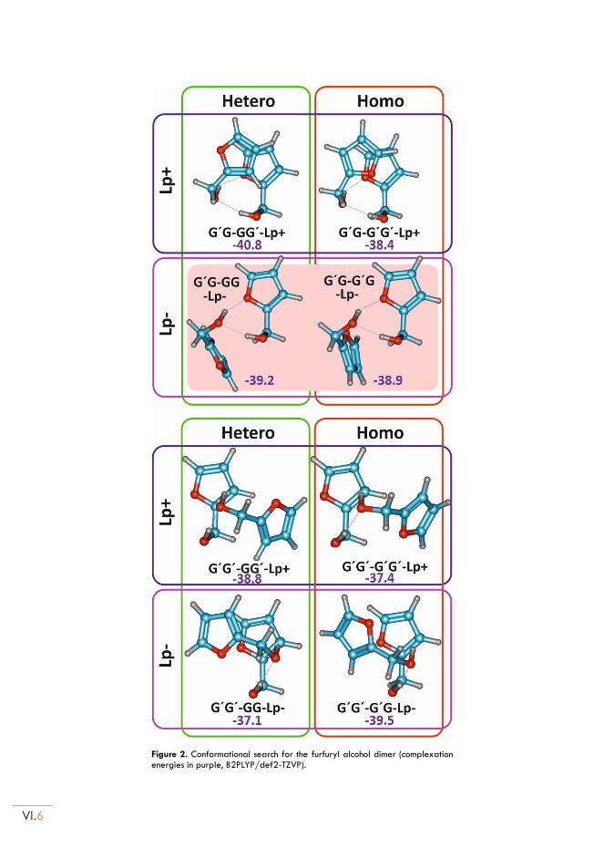
VI.6
Figure 2. Conformational search for the furfuryl alcohol dimer (complexation energies in purple, B2PLYP/def2-TZVP).
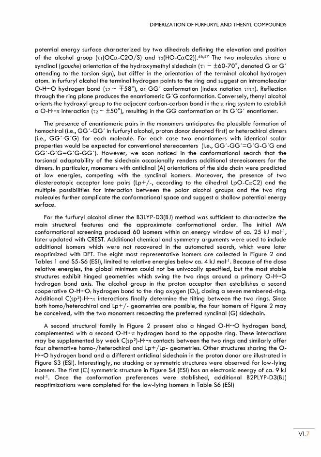
DIMERIZATION OF FURFURYL AND THENYL COMPOUNDS
VI.7
potential energy surface characterized by two dihedrals defining the elevation and position
of the alcohol group (1(OC-C2O/S) and 2(HO-CC2)).46,47 The two molecules share a
synclinal (gauche) orientation of the hydroxymethyl sidechain (1 ~ ±60-70°, denoted G or G´ attending to the torsion sign), but differ in the orientation of the terminal alcohol hydrogen atom. In furfuryl alcohol the terminal hydrogen points to the ring and suggest an intramolecular
O-H···O hydrogen bond (2 ~ ∓58°), or GG´ conformation (index notation 12). Reflection through the ring plane produces the enantiomeric G´G conformation. Conversely, thenyl alcohol orients the hydroxyl group to the adjacent carbon-carbon bond in the ring system to establish a O-H··· interaction (2 ~ ±50°), resulting in the GG conformation or its G´G´ enantiomer.
The presence of enantiomeric pairs in the monomers anticipates the plausible formation of homochiral (i.e., GG´-GG´ in furfuryl alcohol, proton donor denoted first) or heterochiral dimers (i.e., GG´-G´G) for each molecule. For each case two enantiomers with identical scalar properties would be expected for conventional stereocenters (i.e., GG´-GG´=G´G-G´G and GG´-G´G=G´G-GG´). However, we soon noticed in the conformational search that the torsional adaptability of the sidechain occasionally renders additional stereoisomers for the dimers. In particular, monomers with anticlinal (A) orientations of the side chain were predicted at low energies, competing with the synclinal isomers. Moreover, the presence of two diastereotopic acceptor lone pairs (Lp+/-, according to the dihedral LpO-CC2) and the multiple possibilities for interaction between the polar alcohol groups and the two ring molecules further complicate the conformational space and suggest a shallow potential energy surface.
For the furfuryl alcohol dimer the B3LYP-D3(BJ) method was sufficient to characterize the main structural features and the approximate conformational order. The initial MM conformational screening produced 60 isomers within an energy window of ca. 25 kJ mol-1, later updated with CREST. Additional chemical and symmetry arguments were used to include additional isomers which were not recovered in the automated search, which were later reoptimized with DFT. The eight most representative isomers are collected in Figure 2 and Tables 1 and S5-S6 (ESI), limited to relative energies below ca. 4 kJ mol-1. Because of the close relative energies, the global minimum could not be univocally specified, but the most stable structures exhibit hinged geometries which swing the two rings around a primary O-H···O hydrogen bond axis. The alcohol group in the proton acceptor then establishes a second cooperative O-H···Or hydrogen bond to the ring oxygen (Or), closing a seven membered-ring. Additional C(sp3)-H··· interactions finally determine the tilting between the two rings. Since both homo/heterochiral and Lp+/- geometries are possible, the four isomers of Figure 2 may be conceived, with the two monomers respecting the preferred synclinal (G) sidechain.
A second structural family in Figure 2 present also a hinged O-H···O hydrogen bond, complemented with a second O-H··· hydrogen bond to the opposite ring. These interactions may be supplemented by weak C(sp2)-H··· contacts between the two rings and similarly offer four alternative homo-/heterochiral and Lp+/Lp- geometries. Other structures sharing the O-H···O hydrogen bond and a different anticlinal sidechain in the proton donor are illustrated in Figure S3 (ESI). Interestingly, no stacking or symmetric structures were observed for low-lying isomers. The first (Ci) symmetric structure in Figure S4 (ESI) has an electronic energy of ca. 9 kJ mol-1. Once the conformation preferences were stablished, additional B2PLYP-D3(BJ) reoptimizations were completed for the low-lying isomers in Table S6 (ESI)
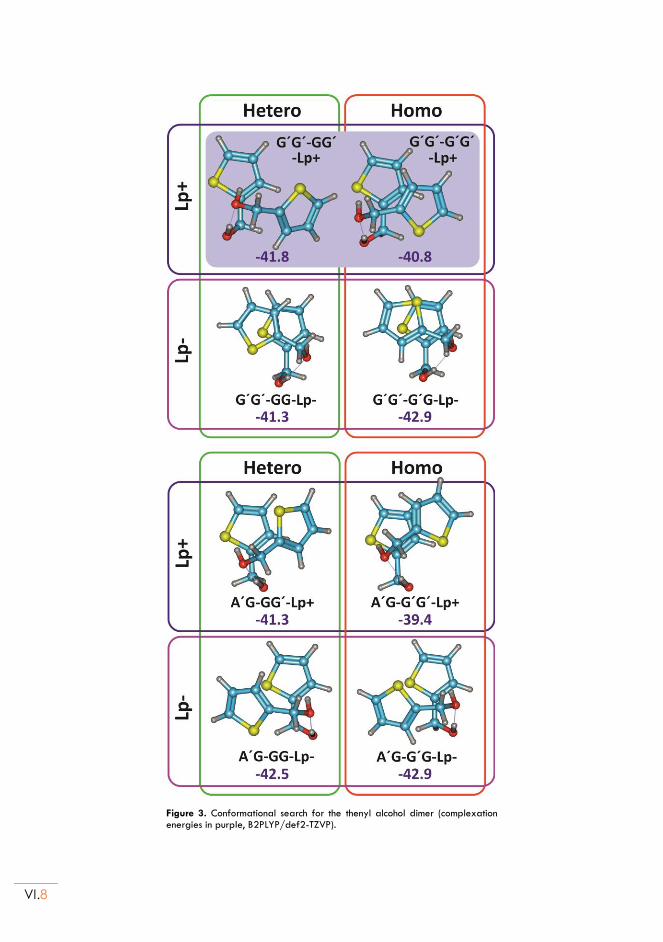
VI.8
Figure 3. Conformational search for the thenyl alcohol dimer (complexation energies in purple, B2PLYP/def2-TZVP).
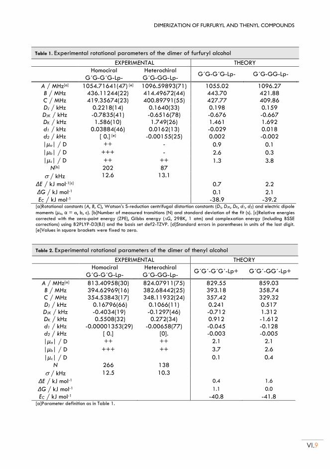
DIMERIZATION OF FURFURYL AND THENYL COMPOUNDS
VI.9
Table 1. Experimental rotational parameters of the dimer of furfuryl alcohol
EXPERIMENTAL THEORY Homociral Heterochiral
G´G-G´G-Lp- G´G-GG-Lp- G´G-G´G-Lp- G´G-GG-Lp-
A / MHz[a] 1054.71641(47) [e] 1096.59893(71) 1055.02 1096.27
B / MHz 436.11244(22) 414.49672(44) 443.70 421.88
C / MHz 419.35674(23) 400.89791(55) 427.77 409.86
DJ / kHz 0.2218(14) 0.1640(33) 0.198 0.159
DJK / kHz -0.7835(41) -0.6516(78) -0.676 -0.667
DK / kHz 1.586(10) 1.749(26) 1.461 1.692
d1 / kHz 0.03884(46) 0.0162(13) -0.029 0.018
d2 / kHz [ 0.] [e] -0.00155(25) 0.002 -0.002
|μa| / D ++ - 0.9 0.1
|μb| / D +++ - 2.6 0.3
|μc| / D ++ ++ 1.3 3.8
N[b] 202 87
/ kHz 12.6 13.1
ΔE / kJ mol-1[c] 0.7 2.2
ΔG / kJ mol-1 0.1 2.1
EC / kJ mol-1 -38.9 -39.2 [a]Rotational constants (A, B, C), Watson’s S-reduction centrifugal distortion constants (DJ, DJK, DK, d1, d2) and electric dipole
moments (μα, α = a, b, c). [b]Number of measured transitions (N) and standard deviation of the fit (s). [c]Relative energies
corrected with the zero-point energy (ZPE), Gibbs energy (G, 298K, 1 atm) and complexation energy (including BSSE corrections) using B2PLYP-D3(BJ) and the basis set def2-TZVP. [d]Standard errors in parentheses in units of the last digit. [e]Values in square brackets were fixed to zero.
Table 2. Experimental rotational parameters of the dimer of thenyl alcohol
EXPERIMENTAL THEORY Homociral Heterochiral
G´G´-G´G´-Lp+ G´G´-GG´-Lp+ G´G-G´G-Lp- G´G-GG-Lp-
A / MHz[a] 813.40958(30) 824.07911(75) 829.55 859.03
B / MHz 394.62969(16) 382.68442(25) 393.18 358.74
C / MHz 354.53843(17) 348.11932(24) 357.42 329.32
DJ / kHz 0.16796(66) 0.1066(11) 0.241 0.517
DJK / kHz -0.4034(19) -0.1297(46) -0.712 1.312
DK / kHz 0.5508(32) 0.272(34) 0.912 -1.612
d1 / kHz -0.00001353(29) -0.00658(77) -0.045 -0.128
d2 / kHz [ 0.] [0]. -0.003 -0.005
|μa| / D ++ ++ 2.1 2.1
|μb| / D +++ ++ 3.7 2.6
|μc| / D 0.1 0.4
N 266 138
/ kHz 12.5 10.3
ΔE / kJ mol-1 0.4 1.6
ΔG / kJ mol-1 1.1 0.0
EC / kJ mol-1 -40.8 -41.8 [a]Parameter definition as in Table 1.
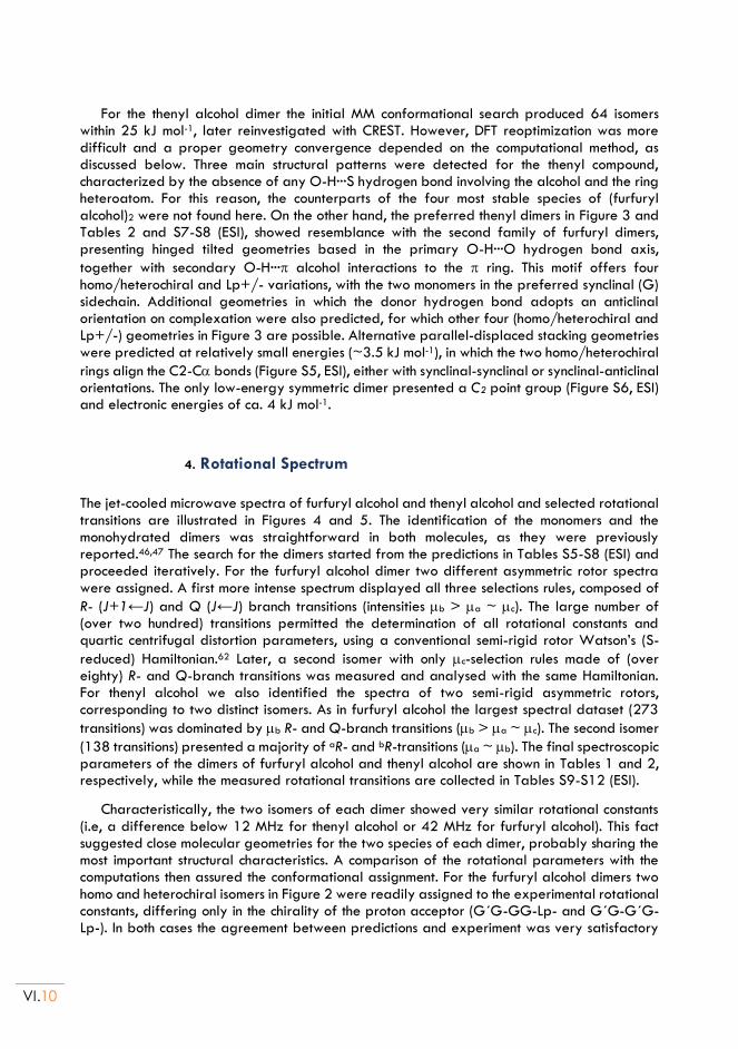
VI.10
For the thenyl alcohol dimer the initial MM conformational search produced 64 isomers within 25 kJ mol-1, later reinvestigated with CREST. However, DFT reoptimization was more difficult and a proper geometry convergence depended on the computational method, as discussed below. Three main structural patterns were detected for the thenyl compound, characterized by the absence of any O-H···S hydrogen bond involving the alcohol and the ring heteroatom. For this reason, the counterparts of the four most stable species of (furfuryl alcohol)2 were not found here. On the other hand, the preferred thenyl dimers in Figure 3 and Tables 2 and S7-S8 (ESI), showed resemblance with the second family of furfuryl dimers, presenting hinged tilted geometries based in the primary O-H···O hydrogen bond axis,
together with secondary O-H··· alcohol interactions to the ring. This motif offers four homo/heterochiral and Lp+/- variations, with the two monomers in the preferred synclinal (G) sidechain. Additional geometries in which the donor hydrogen bond adopts an anticlinal orientation on complexation were also predicted, for which other four (homo/heterochiral and Lp+/-) geometries in Figure 3 are possible. Alternative parallel-displaced stacking geometries were predicted at relatively small energies (~3.5 kJ mol-1), in which the two homo/heterochiral
rings align the C2-C bonds (Figure S5, ESI), either with synclinal-synclinal or synclinal-anticlinal orientations. The only low-energy symmetric dimer presented a C2 point group (Figure S6, ESI) and electronic energies of ca. 4 kJ mol-1.
4. Rotational Spectrum
The jet-cooled microwave spectra of furfuryl alcohol and thenyl alcohol and selected rotational transitions are illustrated in Figures 4 and 5. The identification of the monomers and the monohydrated dimers was straightforward in both molecules, as they were previously reported.46,47 The search for the dimers started from the predictions in Tables S5-S8 (ESI) and proceeded iteratively. For the furfuryl alcohol dimer two different asymmetric rotor spectra were assigned. A first more intense spectrum displayed all three selections rules, composed of
R- (J+1←J) and Q (J←J) branch transitions (intensities b > a ~ c). The large number of (over two hundred) transitions permitted the determination of all rotational constants and quartic centrifugal distortion parameters, using a conventional semi-rigid rotor Watson’s (S-
reduced) Hamiltonian.62 Later, a second isomer with only c-selection rules made of (over eighty) R- and Q-branch transitions was measured and analysed with the same Hamiltonian. For thenyl alcohol we also identified the spectra of two semi-rigid asymmetric rotors, corresponding to two distinct isomers. As in furfuryl alcohol the largest spectral dataset (273
transitions) was dominated by b R- and Q-branch transitions (b > a ~ c). The second isomer
(138 transitions) presented a majority of aR- and bR-transitions (a ~ b). The final spectroscopic parameters of the dimers of furfuryl alcohol and thenyl alcohol are shown in Tables 1 and 2, respectively, while the measured rotational transitions are collected in Tables S9-S12 (ESI).
Characteristically, the two isomers of each dimer showed very similar rotational constants (i.e, a difference below 12 MHz for thenyl alcohol or 42 MHz for furfuryl alcohol). This fact suggested close molecular geometries for the two species of each dimer, probably sharing the most important structural characteristics. A comparison of the rotational parameters with the computations then assured the conformational assignment. For the furfuryl alcohol dimers two homo and heterochiral isomers in Figure 2 were readily assigned to the experimental rotational constants, differing only in the chirality of the proton acceptor (G´G-GG-Lp- and G´G-G´G-Lp-). In both cases the agreement between predictions and experiment was very satisfactory
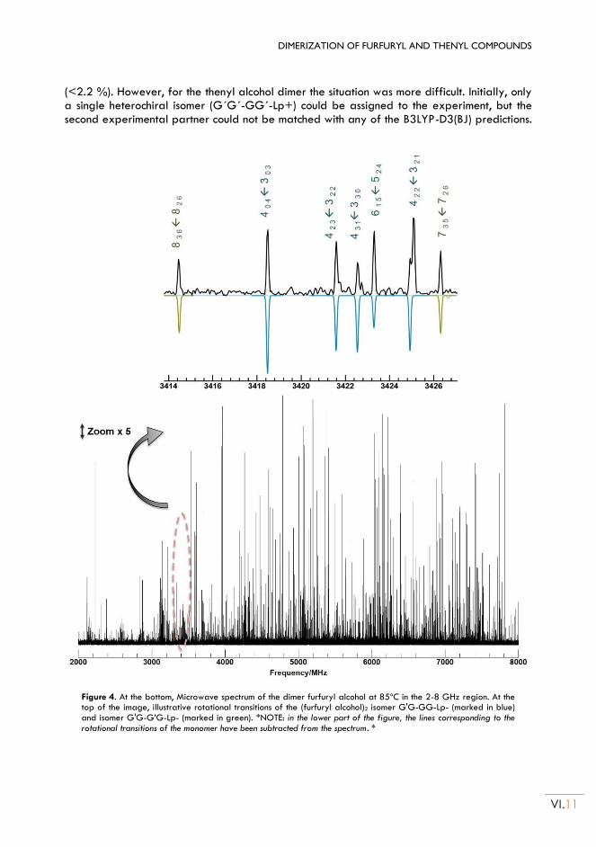
DIMERIZATION OF FURFURYL AND THENYL COMPOUNDS
VI.11
(<2.2 %). However, for the thenyl alcohol dimer the situation was more difficult. Initially, only a single heterochiral isomer (G´G´-GG´-Lp+) could be assigned to the experiment, but the second experimental partner could not be matched with any of the B3LYP-D3(BJ) predictions.
Figure 4. At the bottom, Microwave spectrum of the dimer furfuryl alcohol at 85ºC in the 2-8 GHz region. At the top of the image, illustrative rotational transitions of the (furfuryl alcohol)2 isomer G'G-GG-Lp- (marked in blue) and isomer G'G-G’G-Lp- (marked in green). *NOTE: in the lower part of the figure, the lines corresponding to the rotational transitions of the monomer have been subtracted from the spectrum. *
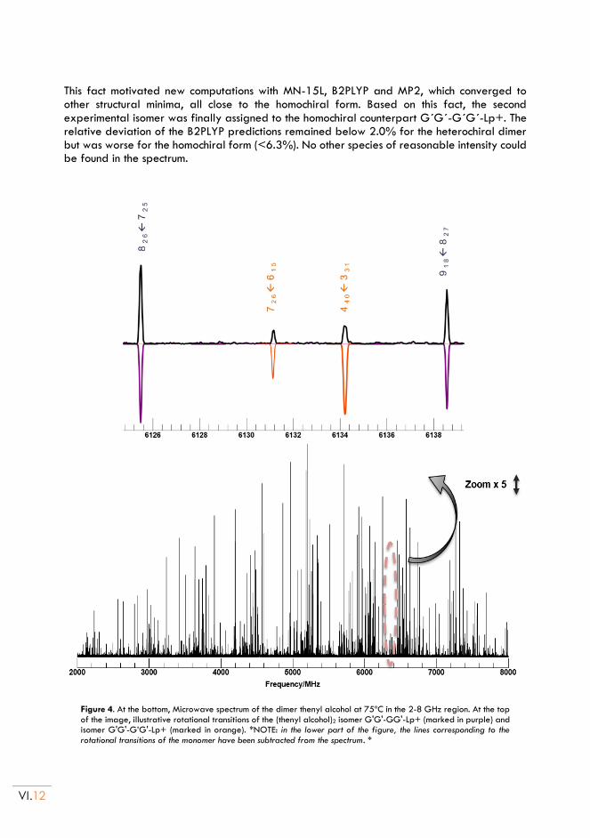
VI.12
This fact motivated new computations with MN-15L, B2PLYP and MP2, which converged to other structural minima, all close to the homochiral form. Based on this fact, the second experimental isomer was finally assigned to the homochiral counterpart G´G´-G´G´-Lp+. The relative deviation of the B2PLYP predictions remained below 2.0% for the heterochiral dimer but was worse for the homochiral form (<6.3%). No other species of reasonable intensity could be found in the spectrum.
Figure 4. At the bottom, Microwave spectrum of the dimer thenyl alcohol at 75ºC in the 2-8 GHz region. At the top of the image, illustrative rotational transitions of the (thenyl alcohol)2 isomer G'G'-GG'-Lp+ (marked in purple) and isomer G'G'-G’G'-Lp+ (marked in orange). *NOTE: in the lower part of the figure, the lines corresponding to the rotational transitions of the monomer have been subtracted from the spectrum. *
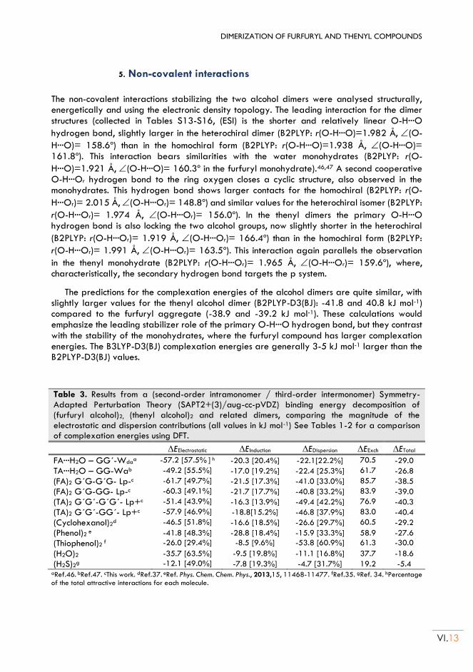
DIMERIZATION OF FURFURYL AND THENYL COMPOUNDS
VI.13
5. Non-covalent interactions
The non-covalent interactions stabilizing the two alcohol dimers were analysed structurally, energetically and using the electronic density topology. The leading interaction for the dimer structures (collected in Tables S13-S16, (ESI) is the shorter and relatively linear O-H···O
hydrogen bond, slightly larger in the heterochiral dimer (B2PLYP: r(O-H···O)=1.982 Å, (O-
H···O)= 158.6º) than in the homochiral form (B2PLYP: r(O-H···O)=1.938 Å, (O-H···O)= 161.8º). This interaction bears similarities with the water monohydrates (B2PLYP: r(O-
H···O)=1.921 Å, (O-H···O)= 160.3º in the furfuryl monohydrate).46,47 A second cooperative O-H···Or hydrogen bond to the ring oxygen closes a cyclic structure, also observed in the monohydrates. This hydrogen bond shows larger contacts for the homochiral (B2PLYP: r(O-
H···Or)= 2.015 Å, (O-H···Or)= 148.8º) and similar values for the heterochiral isomer (B2PLYP:
r(O-H···Or)= 1.974 Å, (O-H···Or)= 156.0º). In the thenyl dimers the primary O-H···O hydrogen bond is also locking the two alcohol groups, now slightly shorter in the heterochiral
(B2PLYP: r(O-H···Or)= 1.919 Å, (O-H···Or)= 166.4º) than in the homochiral form (B2PLYP:
r(O-H···Or)= 1.991 Å, (O-H···Or)= 163.5º). This interaction again parallels the observation
in the thenyl monohydrate (B2PLYP: r(O-H···Or)= 1.965 Å, (O-H···Or)= 159.6º), where, characteristically, the secondary hydrogen bond targets the p system.
The predictions for the complexation energies of the alcohol dimers are quite similar, with slightly larger values for the thenyl alcohol dimer (B2PLYP-D3(BJ): -41.8 and 40.8 kJ mol-1) compared to the furfuryl aggregate (-38.9 and -39.2 kJ mol-1). These calculations would emphasize the leading stabilizer role of the primary O-H···O hydrogen bond, but they contrast with the stability of the monohydrates, where the furfuryl compound has larger complexation energies. The B3LYP-D3(BJ) complexation energies are generally 3-5 kJ mol-1 larger than the B2PLYP-D3(BJ) values.
Table 3. Results from a (second-order intramonomer / third-order intermonomer) Symmetry-Adapted Perturbation Theory (SAPT2+(3)/aug-cc-pVDZ) binding energy decomposition of (furfuryl alcohol)2, (thenyl alcohol)2 and related dimers, comparing the magnitude of the electrostatic and dispersion contributions (all values in kJ mol-1) See Tables 1-2 for a comparison of complexation energies using DFT.
EElectrostatic EInduction EDispersion EExch ETotal
FA···H2O – GG´-Wdaa -57.2 [57.5% ] h -20.3 [20.4%] -22.1[22.2%] 70.5 -29.0
TA···H2O – GG-Wab -49.2 [55.5%] -17.0 [19.2%] -22.4 [25.3%] 61.7 -26.8
(FA)2 G´G-G´G- Lp-c -61.7 [49.7%] -21.5 [17.3%] -41.0 [33.0%] 85.7 -38.5
(FA)2 G´G-GG- Lp-c -60.3 [49.1%] -21.7 [17.7%] -40.8 [33.2%] 83.9 -39.0
(TA)2 G´G´-G´G´- Lp+c -51.4 [43.9%] -16.3 [13.9%] -49.4 [42.2%] 76.9 -40.3
(TA)2 G´G´-GG´- Lp+c -57.9 [46.9%] -18.8[15.2%] -46.8 [37.9%] 83.0 -40.4
(Cyclohexanol)2d -46.5 [51.8%] -16.6 [18.5%] -26.6 [29.7%] 60.5 -29.2
(Phenol)2 e -41.8 [48.3%] -28.8 [18.4%] -15.9 [33.3%] 58.9 -27.6
(Thiophenol)2 f -26.0 [29.4%] -8.5 [9.6%] -53.8 [60.9%] 61.3 -30.0
(H2O)2 -35.7 [63.5%] -9.5 [19.8%] -11.1 [16.8%] 37.7 -18.6
(H2S)2g -12.1 [49.0%] -7.8 [19.3%] -4.7 [31.7%] 19.2 -5.4 aRef.46. bRef.47. cThis work. dRef.37. eRef. Phys. Chem. Chem. Phys., 2013,15, 11468-11477. fRef.35. gRef. 34. hPercentage of the total attractive interactions for each molecule.
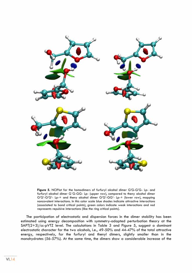
VI.14
Figure 5. NCIPlot for the homodimers of furfuryl alcohol dimer G’G-G’G- Lp- and furfuryl alcohol dimer G´G-GG- Lp- (upper row), compared to theny alcohol dimer G’G’-G’G’- Lp-+ and theny alcohol dimer G’G’-GG’- Lp-+ (lower row), mapping noncovalent interactions. In this color scale blue shades indicate attractive interactions (associated to bond critical points), green colors indicate weak interactions and red represents repulsive interactions (like the ring critical points).
The participation of electrostatic and dispersion forces in the dimer stability has been estimated using energy decomposition with symmetry-adapted perturbation theory at the SAPT(2+3)/cc-pVTZ level. The calculations in Table 3 and Figure 5, suggest a dominant electrostatic character for the two alcohols, i.e., 49-50% and 44-47% of the total attractive energy, respectively, for the furfuryl and thenyl dimers, slightly smaller than in the monohydrates (56-57%). At the same time, the dimers show a considerable increase of the

DIMERIZATION OF FURFURYL AND THENYL COMPOUNDS
VI.15
dispersion contributions, associated to the two aromatic rings (33% and 38-42% for the furfuryl and thenyl dimers, respectively vs. 22-25% in the monohydrates). Calculations on related alcohols25,27 and thiols,36 like the water and hydrogen sulfide dimers,35,66 together with the dispersive pyridine-methane dimer67 are included for comparison.
The presence of non-covalent interactions has been modelled using NCIPlot electronic density
mapping.64 This method calculates a reduced electronic density gradient 𝑠 (=1
1(3𝜋2)1/3
|∇𝜌|
𝜌4/3)
versus the signed electronic density (sign (𝜆2) 𝜌) using the second eigenvalue (𝜆2) of the electron density Hessian (Figure S3, ESI). Non-covalent interactions are then associated to bond critical points. The spatial plots in Figure 6 compare the four observed dimers, revealing interaction patterns well expected in terms of chemical arguments, in particular the (blue) regions associated to hydrogen bonds and other weakly attractive (green) around the two rings.
Figure 5. A radar chart showing the SAPT2+(3) binding energy decomposition (kJ mol-1) for the furfuryl and thenyl dimers and comparison with the dimers of phenol, thiophenol, water and hydrogen sulfide reported in Table 3.
VI.16
6. Conclusion
The conformational landscape and structural properties of the dimers of furfuryl alcohol and thenyl alcohol have been investigated by a combination of jet-cooled (chirped-pulsed) broadband rotational spectroscopy, molecular orbital calculations, binding energy decomposition and electronic density topological analysis. The two alcohol dimers show a combination of conformational issues, particularly the presence of torsional enantiomers, but also the competition of different hydrogen bonds, ring interactions and structural distortions on dimerization. Homochiral and heterochiral structures have been observed experimentally for the two alcohols, which represent a rare occasion where different torsional isomers have been observed independently by spectroscopic means. The close relative energies, even predicted at B2PLYP-D3(BJ) level, were inconclusive on the identification of the global minima, and relied on the experimental findings. The two alcohol dimers were predicted with opposing stereochemical preferences (homochiral for the furfuryl alcohol dimer, heterochiral for the thenyl alcohol dimer) but the small chirodiastaltic energies of 1-2 kJ mol-1 prevent any generalization on the dimerization properties and aggregation preferences. Considering the small interconversion barriers between the torsional isomers new experiments can be designed to explore the transition between torsional labile compounds and permanent stereocenters on molecular aggregation.
This study also provides an accurate description of the intermolecular forces in play in the dimerization process, characterized by a cooperative behaviour of two hydrogen bonds. Furthermore, the comparison between the furfuryl and thenyl compounds explores the different role of the hydrogen bond involving sulfur, which in the thenyl compounds have less priority that the interaction to the p ring system. The molecular description of the two alcohol dimers, while structurally similar to the monohydrates, offers a completely different combination of dispersion effects, calling for adequate dispersion-corrected quantum mechanical models. In this sense we have found interesting issues associated to the structural convergence in the two DFT methods used, emphasizing the importance of the calculation grid, convergence thresholds and other numerical problems. Finally, the role of gas-phase intermolecular clusters as interaction models for molecular aggregation should be stressed, outlining the important collaboration beteeen rotationally resolved experiments and DFT and ab initio methods.
DIMERIZATION OF FURFURYL AND THENYL COMPOUNDS
VI.17
Chapter VI. References:
1. Ariga, K., Ito, H., Hill, J. P. & Tsukube, H. Molecular recognition: From solution science to nano/materials
technology. Chem. Soc. Rev. 41, 5800–5835 (2012).
2. Persch, E., Dumele, O. & Diederich, F. Molecular recognition in chemical and biological systems. Angew. Chemie - Int. Ed. 54, 3290–3327 (2015).
3. Zehnacker, A. Chiral Recognition in the Gas Phase. (CRC Press, 2010).
4. Zehnacker, A. & Suhm, M. A. Chirality Recognition between Neutral Molecules in the Gas Phase. Angew. Chemie Int. Ed. 47, 6970–6992 (2008).
5. Schermann, J.-P. Spectroscopy and Modeling of Biomolecular Building Blocks. (Elsevier, 2008). doi:10.1016/B978-0-444-52708-0.X5001-1.
6. Hobza, P. & Muller-Dethlefs, K. Non-Covalent Interactions. (Royal Society of Chemistry, 2009). doi:10.1039/9781847559906.
7. Noncovalent Forces. vol. 19 (Springer International Publishing, 2015).
8. Liu, M., Zhang, L. & Wang, T. Supramolecular chirality in self-Assembled systems. Chem. Rev. 115, 7304–7397 (2015).
9. Brandt, J. R., Salerno, F. & Fuchter, M. J. The added value of small-molecule chirality in technological applications. Nat. Rev. Chem. 1, (2017).
10. Gilli, G. & Gilli, P. The Nature of the Hydrogen Bond. (Oxford University Press, 2009). doi:10.1093/acprof:oso/9780199558964.001.0001.
11. Alkorta, I., Elguero, J. & Frontera, A. Not only hydrogen bonds: Other noncovalent interactions. Crystals 10, (2020).
12. Juanes, M., Saragi, R. T., Caminati, W. & Lesarri, A. The Hydrogen Bond and Beyond: Perspectives for Rotational Investigations of Non-Covalent Interactions. Chem. - A Eur. J. 25, 11402–11411 (2019).
13. Pierini, M. et al. Gas-Phase Enantiodifferentiation of Chiral Molecules: Chiral Recognition of 1-Phenyl-1-propanol/2-Butanol Clusters by Resonance Enhanced Multiphoton Ionization Spectroscopy. Angew. Chemie - Int. Ed. 36, 1729–1731 (1997).
14. Le Barbu, K., Brenner, V., Millié, P., Lahmani, F. & Zehnacker-Rentien, A. An Experimental and Theoretical Study of Jet-Cooled Complexes of Chiral Molecules: The Role of Dispersive Forces in Chiral Discrimination. J. Phys. Chem. A 102, 128–137 (1998).
15. Borho, N., Häber, T. & Suhm, M. A. Chiral self-recognition in the gas phase: the case of glycidol dimers. Phys. Chem. Chem. Phys. 3, 1945–1948 (2001).
16. Seifert, N. A. et al. Chiral recognition and atropisomerism in the sevoflurane dimer. Phys. Chem. Chem. Phys. 17, 18282–18287 (2015).
17. Oki, M. Recent Advances in Atropisomerism. in Topics in Stereochemistry, vol. 14 (ed. Allinger, Norman L.;Eliel, Ernest L.;Wilen, S. H.) 1–82 (John Wiley & Sons, Inc, 1983).
18. Ottaviani, P., Maris, A. & Caminati, W. Atropisomerism in bisphenols: Free jet absorption millimeter
wave study of 2,2′-biphenol. J. Mol. Struct. 695–696, 353–356 (2004).
19. Hearn, J. P. I., Cobley, R. V. & Howard, B. J. High-resolution spectroscopy of induced chiral dimers: A study of the dimers of ethanol by Fourier transform microwave spectroscopy. J. Chem. Phys. 123, 1–
VI.18
7 (2005).
20. Wassermann, T. N. & Suhm, M. A. Ethanol Monomers and Dimers Revisited: A Raman Study of Conformational Preferences and Argon Nanocoating Effects. J. Phys. Chem. A 114, 8223–8233 (2010).
21. Loru, D., Peña, I. & Sanz, M. E. Ethanol dimer: Observation of three new conformers by broadband rotational spectroscopy. J. Mol. Spectrosc. 335, 93–101 (2017).
22. Snow, M. S., Howard, B. J., Evangelisti, L. & Caminati, W. From Transient to Induced Permanent Chirality in 2-Propanol upon Dimerization: A Rotational Study. J. Phys. Chem. A 115, 47–51 (2011).
23. King, A. K. & Howard, B. J. A microwave study of the hetero-chiral dimer of butan-2-ol. Chem. Phys. Lett. 348, 343–349 (2001).
24. Maris, A., Giuliano, B. M., Bonazzi, D. & Caminati, W. Molecular recognition of chiral conformers: A rotational study of the dimers of glycidol. J. Am. Chem. Soc. 130, 13860–13861 (2008).
25. Juanes, M. et al. The Six Isomers of the Cyclohexanol Dimer: A Delicate Test for Dispersion Models. Angew. Chemie Int. Ed. 59, anie.202005063 (2020).
26. Altnöder, J., Oswald, S. & Suhm, M. A. Phenyl- vs cyclohexyl-substitution in methanol: Implications for the OH conformation and for dispersion-affected aggregation from vibrational spectra in supersonic jets. J. Phys. Chem. A 118, 3266–3279 (2014).
27. Medel, R. & Suhm, M. A. Understanding benzyl alcohol aggregation by chiral modification: The pairing step. Phys. Chem. Chem. Phys. 22, 25538–25551 (2020).
28. Liu, X., Borho, N. & Xu, Y. Molecular self-recognition: Rotational spectra of the dimeric 2-fluoroethanol conformers. Chem. - A Eur. J. 15, 270–277 (2009).
29. Thomas, J. & Xu, Y. Chirality synchronization in trifluoroethanol dimer revisited: The missing heterochiral dimer. J. Phys. Chem. Lett. 5, 1850–1855 (2014).
30. Scharge, T., Häber, T. & Suhm, M. A. Quantitative chirality synchronization in trifluoroethanol dimers. Phys. Chem. Chem. Phys. 8, 4664–4667 (2006).
31. Oswald, S. et al. The Chiral Trimer and a Metastable Chiral Dimer of Achiral Hexafluoroisopropanol:
A Multi‐Messenger Study. Angew. Chemie Int. Ed. 58, 5080–5084 (2019).
32. Su, Z., Borho, N. & Xu, Y. Chiral self-recognition: Direct spectroscopic detection of the homochiral and heterochiral dimers of propylene oxide in the gas phase. J. Am. Chem. Soc. 128, 17126–17131 (2006).
33. Domingos, S. R., Pérez, C., Kreienborg, N. M., Merten, C. & Schnell, M. Dynamic chiral self-recognition in aromatic dimers of styrene oxide revealed by rotational spectroscopy. Commun. Chem. 4, 1–11 (2021).
34. Das, A. et al. The H2S Dimer is Hydrogen‐Bonded: Direct Confirmation from Microwave Spectroscopy. Angew. Chemie Int. Ed. 57, 15199–15203 (2018).
35. Saragi, R. T. et al. Switching Hydrogen Bonding to π-Stacking: The Thiophenol Dimer and Trimer. J. Phys. Chem. Lett. 12, 1367–1373 (2021).
36. Utzat, K. A., Bohn, R. K., Montgomery, J. A., Michels, H. H. & Caminati, W. Rotational spectrum, tunneling motions, and potential barriers of benzyl alcohol. J. Phys. Chem. A 114, 6913–6916 (2010).
37. Juanes, M. et al. Internal dynamics of cyclohexanol and the cyclohexanol-water adduct. Phys. Chem. Chem. Phys. 21, 3676–3682 (2019).
DIMERIZATION OF FURFURYL AND THENYL COMPOUNDS
VI.19
38. Maryanoff, B. E. & Greco, M. N. 1.7 Stereochemical Lability in Drug Molecules: Cases Where Chirality May Not Be Critical for Drug Development. Comprehensive Chirality vol. 1 (Elsevier Ltd., 2012).
39. Toenjes, S. T. & Gustafson, J. L. Atropisomerism in medicinal chemistry: Challenges and opportunities. Future Med. Chem. 10, 409–422 (2018).
40. Park, G. B. & Field, R. W. Perspective: The first ten years of broadband chirped pulse Fourier transform microwave spectroscopy. J. Chem. Phys. 144, 1–10 (2016).
41. Laplante, S. R. et al. Assessing atropisomer axial chirality in drug discovery and development. J. Med. Chem. 54, 7005–7022 (2011).
42. Craig, D. P. & Mellor, D. P. Discriminating interactions between chiral molecules. in Bonding Structure. Topics in Current Chemistry, 63 1–48 (Springer-Verlag, 1976). doi:10.1007/BFb0046191.
43. Portmann, S., Inauen, A., Lüthi, H. P. & Leutwyler, S. Chiral discrimination in hydrogen-bonded complexes. J. Chem. Phys. 113, 9577–9585 (2000).
44. Alkorta, I. & Elguero, J. Discrimination of hydrogen-bonded complexes with axial chirality. J. Chem. Phys. 117, 6463–6468 (2002).
45. Biswal, H. S., Bhattacharyya, S., Bhattacherjee, A. & Wategaonkar, S. Nature and strength of sulfur-centred hydrogen bonds: laser spectroscopic investigations in the gas phase and quantum-chemical calculations. Int. Rev. Phys. Chem. 34, 99–160 (2015).
46. Juanes, M. et al. Sulfur Hydrogen Bonding in Isolated Monohydrates: Furfuryl Mercaptan versus Furfuryl Alcohol. Chem. - A Eur. J. 24, 6564–6571 (2018).
47. Juanes, M., Saragi, R. T., Pinacho, R., Rubio, J. E. & Lesarri, A. Sulfur hydrogen bonding and internal dynamics in the monohydrates of thenyl mercaptan and thenyl alcohol. Phys. Chem. Chem. Phys. 22, 12412–12421 (2020).
48. Shipman, S. T. & Pate, B. H. New Techniques in Microwave Spectroscopy. in Handbook of High-resolution Spectroscopy (eds. Quack, M. & Merk, F.) 801–827 (John Wiley & Sons, Ltd., 2011).
49. Grabow, J.-U. Fourier Transform Microwave Spectroscopy Measurement and Instrumentation. Handbook of High-resolution Spectroscopy (2011). doi:10.1002/9780470749593.hrs037.
50. Caminati, W. & Grabow, J.-U. Advancements in Microwave Spectroscopy. in Frontiers and Advances in Molecular Spectroscopy (ed. Laane, J.) 569–598 (Elsevier Inc., 2018). doi:10.1016/B978-0-12-811220-5.00018-6.
51. Halgren, T. A. MMFF VI. MMFF94s option for energy minimization studies. J. Comput. Chem. 20, 720–729 (1999).
52. Pracht, P., Bohle, F. & Grimme, S. Automated exploration of the low-energy chemical space with fast quantum chemical methods. Phys. Chem. Chem. Phys. 22, 7169–7192 (2020).
53. Grimme, S. Semiempirical hybrid density functional with perturbative second-order correlation. J. Chem. Phys. 124, 034108 (2006).
54. Grimme, S., Ehrlich, S. & Goerigk, L. Effect of the damping function in dispersion corrected density functional theory. J. Comput. Chem. 32, 1456–1465 (2011).
55. Weigend, F. & Ahlrichs, R. Balanced basis sets of split valence, triple zeta valence and quadruple zeta valence quality for H to Rn: Design and assessment of accuracy. Phys. Chem. Chem. Phys. 7, 3297 (2005).
56. Frisch, M. J. et al. Gaussian 16, rev. C.01. (2016).
57. Neese, F. Software update: the ORCA program system, version 4.0. WIREs Comput. Mol. Sci. 8,
VI.20
(2018).
58. Boys, S. F. & Bernardi, F. The calculation of small molecular interactions by the differences of separate total energies. Some procedures with reduced errors. Mol. Phys. 19, 553–566 (1970).
59. Jeziorski, B., Moszynski, R. & Szalewicz, K. Perturbation Theory Approach to Intermolecular Potential Energy Surfaces of van der Waals Complexes. Chem. Rev. 94, 1887–1930 (1994).
60. Parrish, R. M. et al. Psi4 1.1: An Open-Source Electronic Structure Program Emphasizing Automation, Advanced Libraries, and Interoperability. J. Chem. Theory Comput. 13, 3185–3197 (2017).
61. Contreras-García, J. et al. NCIPLOT: A Program for Plotting Noncovalent Interaction Regions. J. Chem. Theory Comput. 7, 625–632 (2011).
62. Watson, J. K. G. Aspects of Quartic and Sextic Centrifugal Effects on Rotational Energy Levels. in Vibrational Spectra and Structure, vol. 6 (ed. Durig, J. R.) 1–89 (Elsevier B.V., 1977).

Chapter VII COMPLEMENTARY STUDIES

VII.2
COMPLEMENTARY STUDIES
VII.3
I. Complementary Studies
During the course of this Thesis and as part of my Doctoral training, I have also participated in other experimental investigations on rotational spectroscopy not directly connected to the study of non-covalent interactions in intermolecular complexes. These investigations include, in particular, the result of international collaborations of our group and the work carried out in two foreing laboratories:
- Institut fur Physikalische Chemie, Universität Hannover - I Physikalisches Institut, Universität Kohln
In this section I collect the published studies on these topics, including:
a) Cyclohexyl compounds:
Fluorocyclohexane; Ethynylcyclohexane (Section 1 and 2 of this Chapter)
Spectroscopic and theoretical molecular orbital calculations.
Conformational comparison due to ring inversion.
Semi-experimental (SE) mixed-estimation structural method as a benchmark for
computational calculations.
b) Aliphatic dithiol compounds:
1,2-Butanedithiol; 2,3-Butanedithiol (Section 3 and 4 of this Chapter)
Intramolecular S-H ··· S hydrogen bonding observation.
Homochiral and heterochiral conformational stability.
Complementary measurments on the cm- and mm-wave regions.
c) Tetracyclic compounds:
trans- and cis- Matrine (Section 5 of this Chapter)
Conformational stability of saturated tetracycles.
Torsional motions in six-membered rings
d) NCI review and its perspective for rotational spectroscopy (Section 6 of this Chapter)
The abstracts of these publication follow.
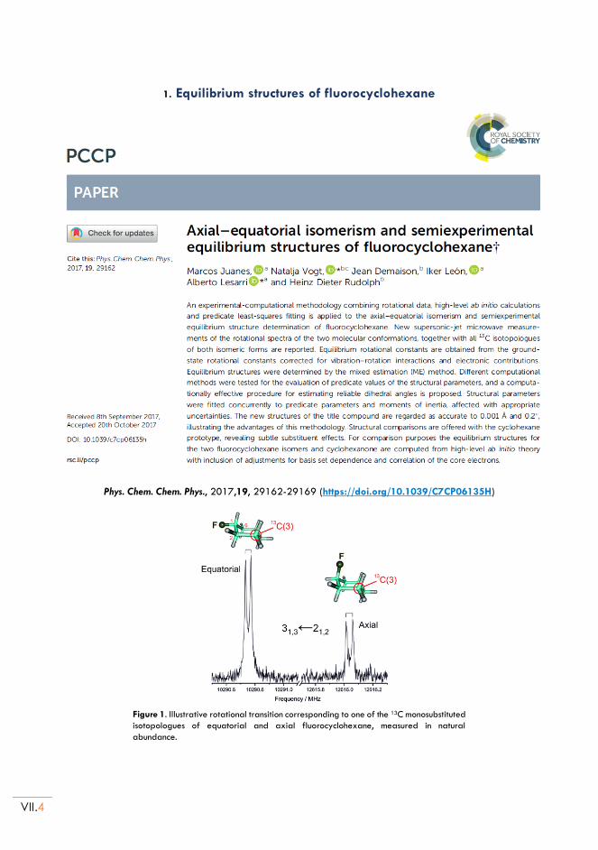
VII.4
1. Equilibrium structures of fluorocyclohexane
Phys. Chem. Chem. Phys., 2017,19, 29162-29169 (https://doi.org/10.1039/C7CP06135H)
Figure 1. Illustrative rotational transition corresponding to one of the 13C monosubstituted
isotopologues of equatorial and axial fluorocyclohexane, measured in natural
abundance.
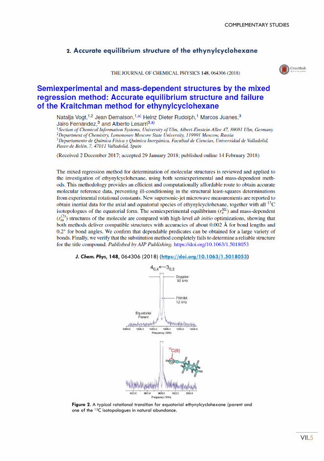
COMPLEMENTARY STUDIES
VII.5
2. Accurate equilibrium structure of the ethynylcyclohexane
J. Chem. Phys, 148, 064306 (2018) (https://doi.org/10.1063/1.5018053)
Figure 2. A typical rotational transition for equatorial ethynylcyclohexane (parent and one of the 13C isotopologues in natural abundance.
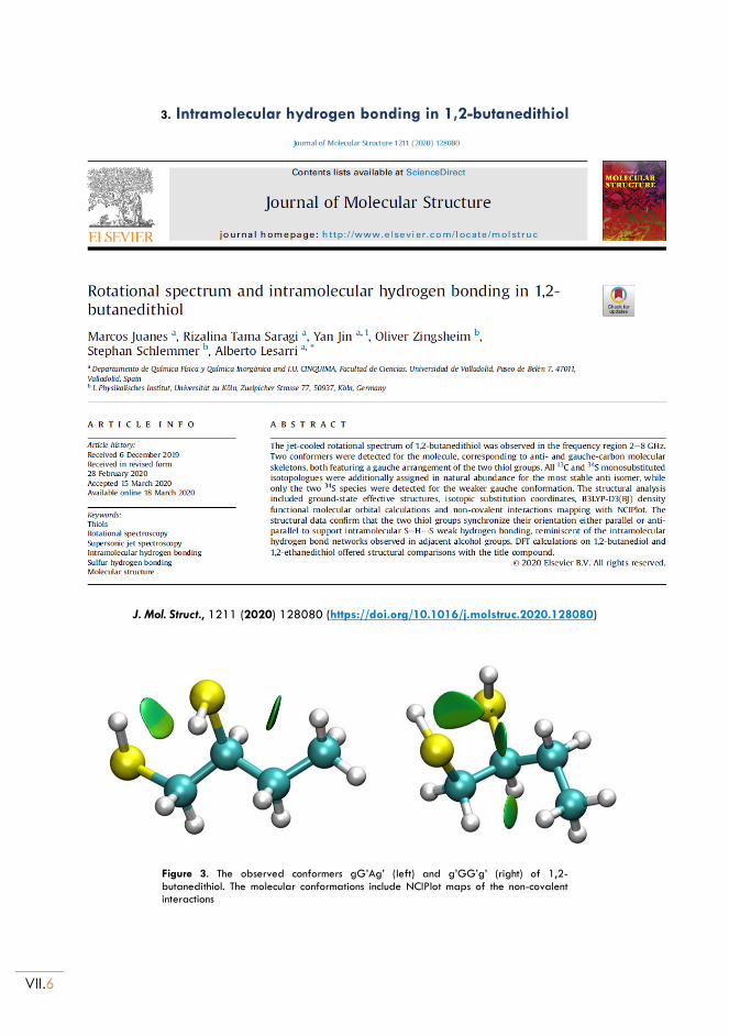
VII.6
3. Intramolecular hydrogen bonding in 1,2-butanedithiol
J. Mol. Struct., 1211 (2020) 128080 (https://doi.org/10.1016/j.molstruc.2020.128080)
Figure 3. The observed conformers gG’Ag’ (left) and g’GG’g’ (right) of 1,2-butanedithiol. The molecular conformations include NCIPlot maps of the non-covalent interactions
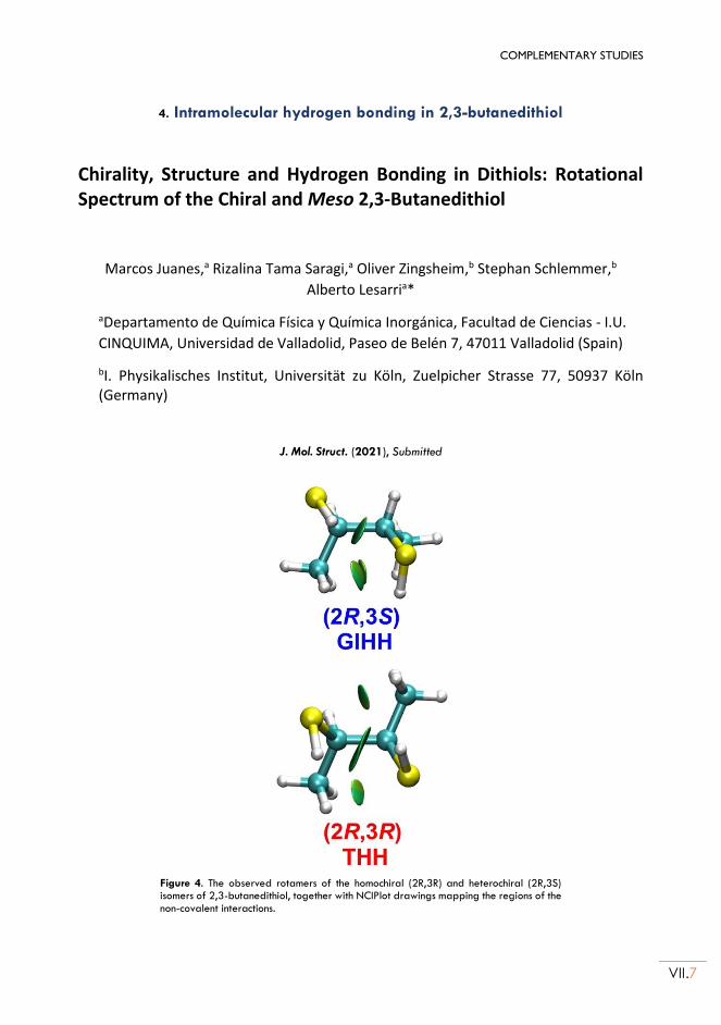
COMPLEMENTARY STUDIES
VII.7
4. Intramolecular hydrogen bonding in 2,3-butanedithiol
Chirality, Structure and Hydrogen Bonding in Dithiols: Rotational Spectrum of the Chiral and Meso 2,3-Butanedithiol
Marcos Juanes,a Rizalina Tama Saragi,a Oliver Zingsheim,b Stephan Schlemmer,b
Alberto Lesarria*
aDepartamento de Química Física y Química Inorgánica, Facultad de Ciencias - I.U.
CINQUIMA, Universidad de Valladolid, Paseo de Belén 7, 47011 Valladolid (Spain)
bI. Physikalisches Institut, Universität zu Köln, Zuelpicher Strasse 77, 50937 Köln (Germany)
J. Mol. Struct. (2021), Submitted
Figure 4. The observed rotamers of the homochiral (2R,3R) and heterochiral (2R,3S) isomers of 2,3-butanedithiol, together with NCIPlot drawings mapping the regions of the non-covalent interactions.
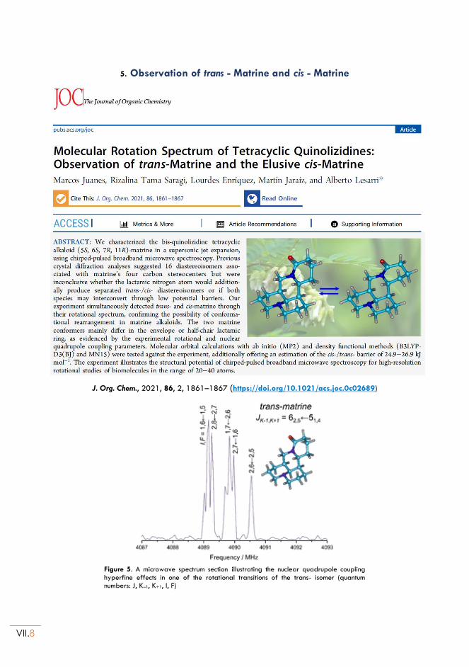
VII.8
5. Observation of trans - Matrine and cis - Matrine
J. Org. Chem., 2021, 86, 2, 1861–1867 (https://doi.org/10.1021/acs.joc.0c02689)
Figure 5. A microwave spectrum section illustrating the nuclear quadrupole coupling hyperfine effects in one of the rotational transitions of the trans- isomer (quantum numbers: J, K–1, K+1, I, F)
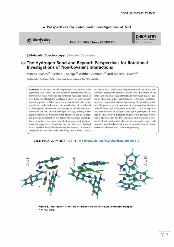
COMPLEMENTARY STUDIES
VII.9
6. Perspectives for Rotational Investigations of NCI
Chem. Eur. J., 2019, 25,11402 –11411 (https://doi.org/10.1002/chem.201901113)
Figure 6. Three isomers of the aniline trimer, with intermolecular interactions mapped with NCI plots.