Search procedure for improving Modified Ambiguity Function Approach
Upload
khangminh22Category
view
1download
0
The PDF of the article you requested follows this cover page.
This is an enhanced PDF from The Journal of Bone and Joint Surgery
2008;90:74-86. doi:10.2106/JBJS.G.01104 J Bone Joint Surg Am.Goo Hyun Baek, Hyun Sik Gong, Moon Sang Chung, Joo Han Oh, Young Ho Lee and Sang Ki Lee
Polydactyly of the Thumb. Surgical TechniqueModified Bilhaut-Cloquet Procedure for Wassel Type-II and III
This information is current as of December 23, 2009
Reprints and Permissions
Permissions] link. and click on the [Reprints andjbjs.orgarticle, or locate the article citation on
to use material from thisorder reprints or request permissionClick here to
Publisher Information
www.jbjs.org20 Pickering Street, Needham, MA 02492-3157The Journal of Bone and Joint Surgery
COPYRIGHT © 2008 BY THE JOURNAL OF BONE AND JOINT SURGERY, INCORPORATED
74
Modified Bilhaut-Cloquet Procedure for Wassel Type-II and III Polydactyly of the ThumbSurgical Technique
By Goo Hyun Baek, MD, Hyun Sik Gong, MD, Moon Sang Chung, MD, Joo Han Oh, MD, Young Ho Lee, MD, and Sang Ki Lee, MD
Investigation performed at the Department of Orthopedic Surgery, Seoul National University College of Medicine, Seoul, South Korea
The original scientific article in which the surgical technique was presented was published in JBJS Vol. 89-A, pp. 534-41, March 2007
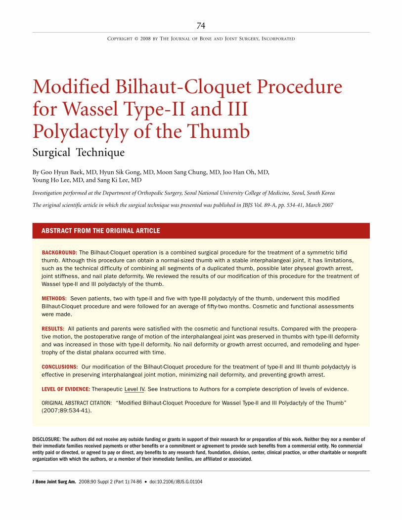
ABSTRACT FROM THE ORIGINAL ARTICLE
BACKGROUND: The Bilhaut-Cloquet operation is a combined surgical procedure for the treatment of a symmetric bifid thumb. Although this procedure can obtain a normal-sized thumb with a stable interphalangeal joint, it has limitations, such as the technical difficulty of combining all segments of a duplicated thumb, possible later physeal growth arrest, joint stiffness, and nail plate deformity. We reviewed the results of our modification of this procedure for the treatment of Wassel type-II and III polydactyly of the thumb.
METHODS: Seven patients, two with type-II and five with type-III polydactyly of the thumb, underwent this modified Bilhaut-Cloquet procedure and were followed for an average of fifty-two months. Cosmetic and functional assessments were made.
RESULTS: All patients and parents were satisfied with the cosmetic and functional results. Compared with the preopera-tive motion, the postoperative range of motion of the interphalangeal joint was preserved in thumbs with type-III deformity and was increased in those with type-II deformity. No nail deformity or growth arrest occurred, and remodeling and hyper-trophy of the distal phalanx occurred with time.
CONCLUSIONS: Our modification of the Bilhaut-Cloquet procedure for the treatment of type-II and III thumb polydactyly is effective in preserving interphalangeal joint motion, minimizing nail deformity, and preventing growth arrest.
LEVEL OF EVIDENCE: Therapeutic Level IV. See Instructions to Authors for a complete description of levels of evidence.
ORIGINAL ABSTRACT CITATION: “Modified Bilhaut-Cloquet Procedure for Wassel Type-II and III Polydactyly of the Thumb” (2007;89:534-41).
DISCLOSURE: The authors did not receive any outside funding or grants in support of their research for or preparation of this work. Neither they nor a member of their immediate families received payments or other benefits or a commitment or agreement to provide such benefits from a commercial entity. No commercial entity paid or directed, or agreed to pay or direct, any benefits to any research fund, foundation, division, center, clinical practice, or other charitable or nonprofit organization with which the authors, or a member of their immediate families, are affiliated or associated.
J Bone Joint Surg Am. 2008;90 Suppl 2 (Part 1):74-86 • doi:10.2106/JBJS.G.01104
Baek.fm Page 74 Friday, February 8, 2008 2:56 PM
75
TH E JO UR N AL O F BO N E & JO IN T SU RG E R Y · SU R G IC A L TE CH N I Q U E S MARCH 2008 · VOLUME 90-A · SUPPLEMENT 2, PART 1 · JBJS.ORG
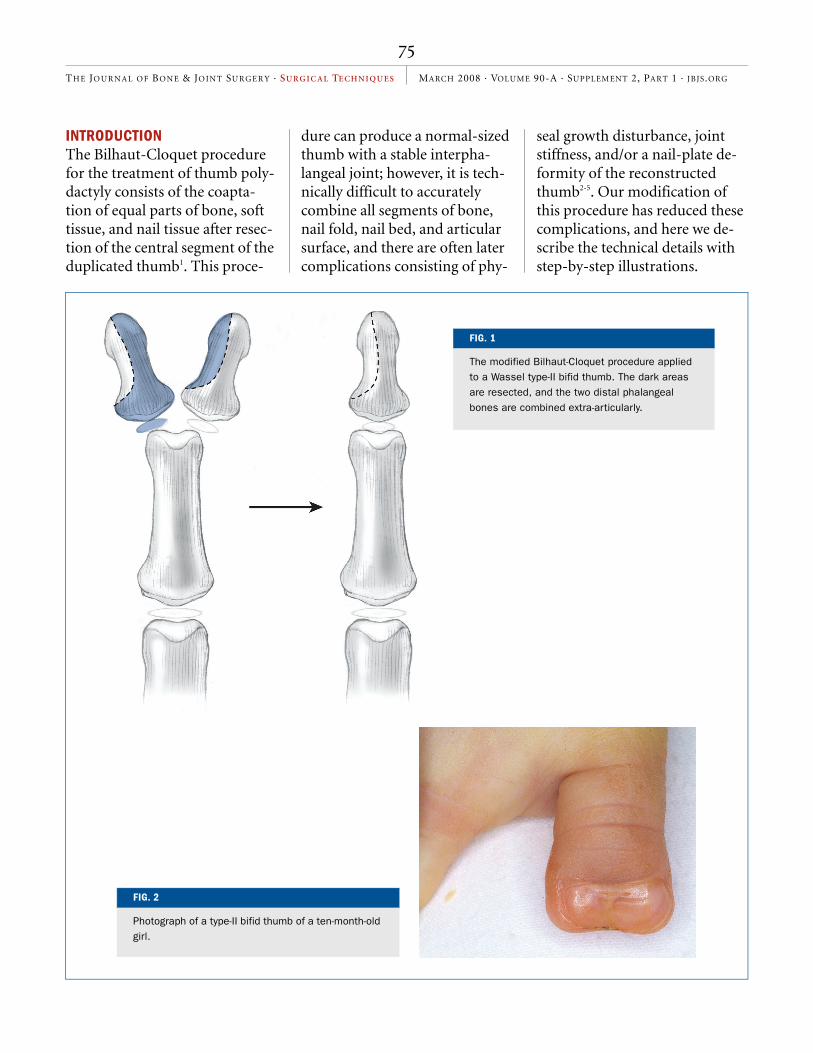
INTRODUCTIONThe Bilhaut-Cloquet procedure for the treatment of thumb poly-dactyly consists of the coapta-tion of equal parts of bone, soft tissue, and nail tissue after resec-tion of the central segment of the duplicated thumb1. This proce-
dure can produce a normal-sized thumb with a stable interpha-langeal joint; however, it is tech-nically difficult to accurately combine all segments of bone, nail fold, nail bed, and articular surface, and there are often later complications consisting of phy-
seal growth disturbance, joint stiffness, and/or a nail-plate de-formity of the reconstructed thumb2-5. Our modification of this procedure has reduced these complications, and here we de-scribe the technical details with step-by-step illustrations.
FIG. 1
The modified Bilhaut-Cloquet procedure applied to a Wassel type-II bifid thumb. The dark areas are resected, and the two distal phalangeal bones are combined extra-articularly.
FIG. 2
Photograph of a type-II bifid thumb of a ten-month-old girl.
Baek.fm Page 75 Friday, February 8, 2008 2:56 PM

76
TH E JO UR N AL O F BO N E & JO IN T SU RG E R Y · SU R G IC A L TE CH N I Q U E S MARCH 2008 · VOLUME 90-A · SUPPLEMENT 2, PART 1 · JBJS.ORG
SURGICAL TECHNIQUEPreoperative EvaluationThe size of the fingernail is com-pared with that on the unaffected side or, in the case of bilateral in-volvement, that of the index fin-
ger. Both digits of the bifid thumb are carefully examined for joint mobility and stability. A true anteroposterior radiograph of the thumb is required to iden-tify the type of bifurcation and to
assess the bone size and the an-gulation of the joint. We use the Wassel classification, which is based on the branching level of the bifid thumb6. The modified Bilhaut-Cloquet procedure is in-
FIG. 3
Radiograph showing hypoplasia of the bifid thumbs when compared with the thumb on the normal side.
FIG. 4-A FIG. 4-B
Dorsal (Fig. 4-A) and volar (Fig. 4-B) incisions. A triangular skin flap at the pulp can prevent scar contracture and make a smooth contour of the reconstructed pulp.
Baek.fm Page 76 Friday, February 8, 2008 2:56 PM
77
TH E JO UR N AL O F BO N E & JO IN T SU RG E R Y · SU R G IC A L TE CH N I Q U E S MARCH 2008 · VOLUME 90-A · SUPPLEMENT 2, PART 1 · JBJS.ORG
dicated for Wassel type-II and III polydactyly, in which the bifid thumbs are symmetric and their nail size is less than two-thirds of that of the normal contralateral thumb, or smaller than the index-finger nail in patients with bilateral thumb involvement.
The family should be in-formed that a combination of digits will never produce an identical match to the normal thumb, and it is not possible to make a thumb with the same nail length as that on the normal side when the initial nail lengths of the duplicated thumb differ from that of the normal thumb.
Patient Positioning and PreparationAfter the administration of gen-eral anesthesia, the patient is placed supine on the operating ta-ble and a pneumatic tourniquet is placed around the arm. The up-per extremity is prepared with standard antiseptic solutions and draped. Special care is taken to ensure that the antiseptic solu-tion does not soak the cotton pad-ding underneath the tourniquet because it may seriously burn the fragile skin of the child. The other hand should be readily available for comparison when necessary during the operation.
Modified Bilhaut-Cloquet ProcedureThe modified Bilhaut-Cloquet procedure differs from the origi-nally described method because it is an extra-articular proce-dure; the interphalangeal joint is reconstructed with tissue from
one thumb, and the other thumb contributes only part of the distal phalanx for stability. Because it is not necessary to approximate the articular surface of the distal phalanx, the nail bed can be su-tured more precisely according to the natural curve of the nail to minimize nail plate deformity (Fig. 1).
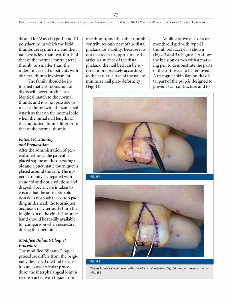
An illustrative case of a ten-month-old girl with type-II thumb polydactyly is shown (Figs. 2 and 3). Figure 4-A shows the incision drawn with a mark-ing pen to demonstrate the parts of the soft tissue to be removed. A triangular skin flap on the dis-tal part of the pulp is designed to prevent scar contracture and to
FIG. 5-B
The nail plates are removed with use of a small elevator (Fig. 5-A) and a mosquito clamp (Fig. 5-B).
FIG. 5-A
Baek.fm Page 77 Friday, February 8, 2008 2:56 PM

78
TH E JO UR N AL O F BO N E & JO IN T SU RG E R Y · SU R G IC A L TE CH N I Q U E S MARCH 2008 · VOLUME 90-A · SUPPLEMENT 2, PART 1 · JBJS.ORG
make a smooth contour of the reconstructed pulp (Fig. 4-B).
Under tourniquet control, the nail plates are removed with use of a small elevator and a mosquito clamp (Figs. 5-A and 5-B). The removed nail plate is preserved in saline solution so
that it can be used later as a tem-porary splint for the recon-structed nail bed. Along the incision line, soft tissues, includ-ing skin and nail bed, are re-moved (Fig. 6). The bases of the two distal phalanges are carefully separated with use of a number-
15 scalpel (Fig. 7). The thumb with the greater range of motion of the interphalangeal joint is chosen to become the main artic-ulating digit (the radial side in the illustrative case), which con-tains the articular surface, the physis, and a major part of the
FIG. 6
Soft tissue between the bifid thumbs is resected along the incision line.
FIG. 7
The bases of the two distal phalanges are separated with use of a number-5 scalpel. The articular cartilage of the base of the proximal phalanx is labeled with an asterisk.
Baek.fm Page 78 Friday, February 8, 2008 2:56 PM
79
TH E JO UR N AL O F BO N E & JO IN T SU RG E R Y · SU R G IC A L TE CH N I Q U E S MARCH 2008 · VOLUME 90-A · SUPPLEMENT 2, PART 1 · JBJS.ORG
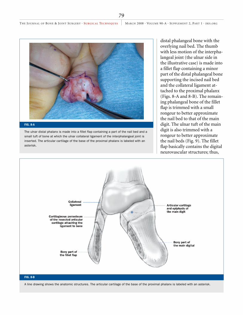
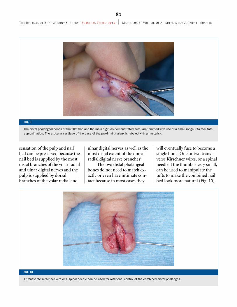
distal phalangeal bone with the overlying nail bed. The thumb with less motion of the interpha-langeal joint (the ulnar side in the illustrative case) is made into a fillet flap containing a minor part of the distal phalangeal bone supporting the incised nail bed and the collateral ligament at-tached to the proximal phalanx (Figs. 8-A and 8-B). The remain-ing phalangeal bone of the fillet flap is trimmed with a small rongeur to better approximate the nail bed to that of the main digit. The ulnar tuft of the main digit is also trimmed with a rongeur to better approximate the nail beds (Fig. 9). The fillet flap basically contains the digital neurovascular structures; thus,
FIG. 8-A
The ulnar distal phalanx is made into a fillet flap containing a part of the nail bed and a small tuft of bone at which the ulnar collateral ligament of the interphalangeal joint is inserted. The articular cartilage of the base of the proximal phalanx is labeled with an asterisk.
FIG. 8-B
A line drawing shows the anatomic structures. The articular cartilage of the base of the proximal phalanx is labeled with an asterisk.
Baek.fm Page 79 Friday, February 8, 2008 2:56 PM
80
TH E JO UR N AL O F BO N E & JO IN T SU RG E R Y · SU R G IC A L TE CH N I Q U E S MARCH 2008 · VOLUME 90-A · SUPPLEMENT 2, PART 1 · JBJS.ORG
sensation of the pulp and nail bed can be preserved because the nail bed is supplied by the most distal branches of the volar radial and ulnar digital nerves and the pulp is supplied by dorsal branches of the volar radial and
ulnar digital nerves as well as the most distal extent of the dorsal radial digital nerve branches7.
The two distal phalangeal bones do not need to match ex-actly or even have intimate con-tact because in most cases they
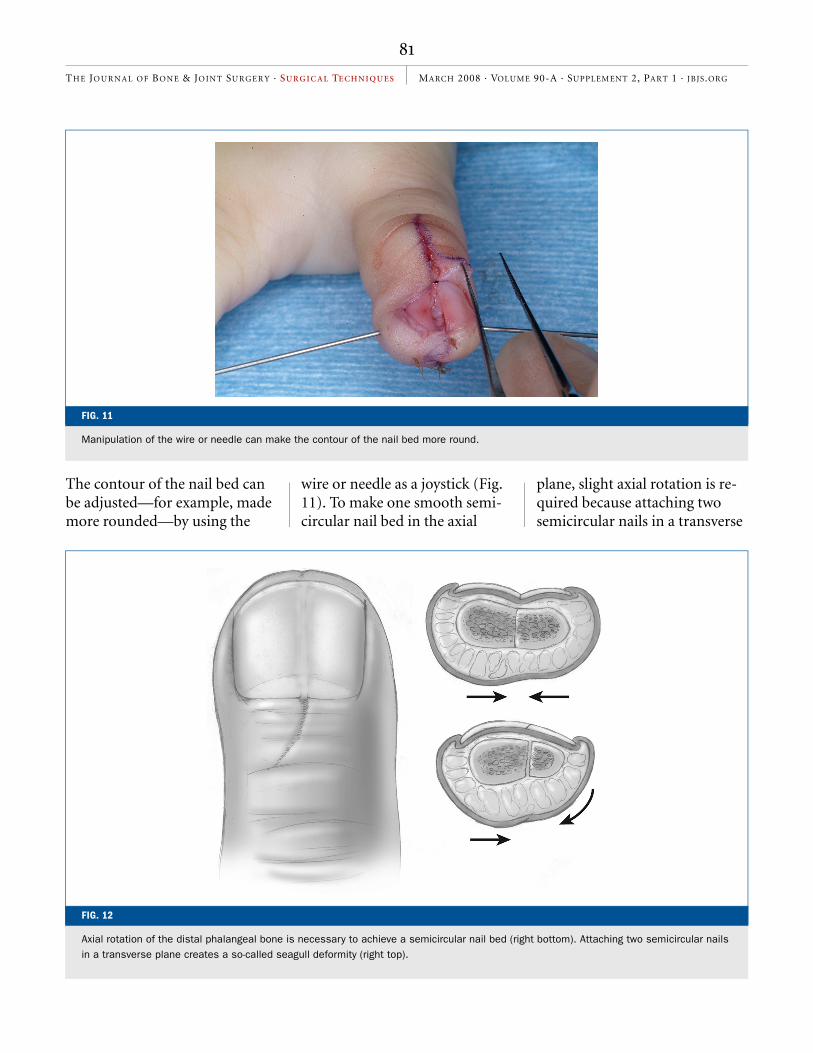
will eventually fuse to become a single bone. One or two trans-verse Kirschner wires, or a spinal needle if the thumb is very small, can be used to manipulate the tufts to make the combined nail bed look more natural (Fig. 10).
FIG. 9
The distal phalangeal bones of the fillet flap and the main digit (as demonstrated here) are trimmed with use of a small rongeur to facilitate approximation. The articular cartilage of the base of the proximal phalanx is labeled with an asterisk.
FIG. 10
A transverse Kirschner wire or a spinal needle can be used for rotational control of the combined distal phalanges.
Baek.fm Page 80 Friday, February 8, 2008 2:56 PM
81
TH E JO UR N AL O F BO N E & JO IN T SU RG E R Y · SU R G IC A L TE CH N I Q U E S MARCH 2008 · VOLUME 90-A · SUPPLEMENT 2, PART 1 · JBJS.ORG
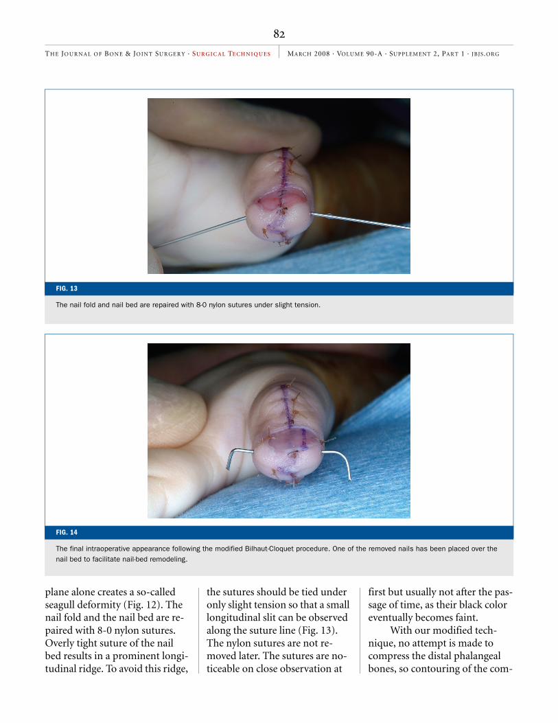
The contour of the nail bed can be adjusted—for example, made more rounded—by using the
wire or needle as a joystick (Fig. 11). To make one smooth semi-circular nail bed in the axial
plane, slight axial rotation is re-quired because attaching two semicircular nails in a transverse
FIG. 11
Manipulation of the wire or needle can make the contour of the nail bed more round.
FIG. 12
Axial rotation of the distal phalangeal bone is necessary to achieve a semicircular nail bed (right bottom). Attaching two semicircular nails in a transverse plane creates a so-called seagull deformity (right top).
Baek.fm Page 81 Friday, February 8, 2008 2:56 PM
82
TH E JO UR N AL O F BO N E & JO IN T SU RG E R Y · SU R G IC A L TE CH N I Q U E S MARCH 2008 · VOLUME 90-A · SUPPLEMENT 2, PART 1 · JBJS.ORG
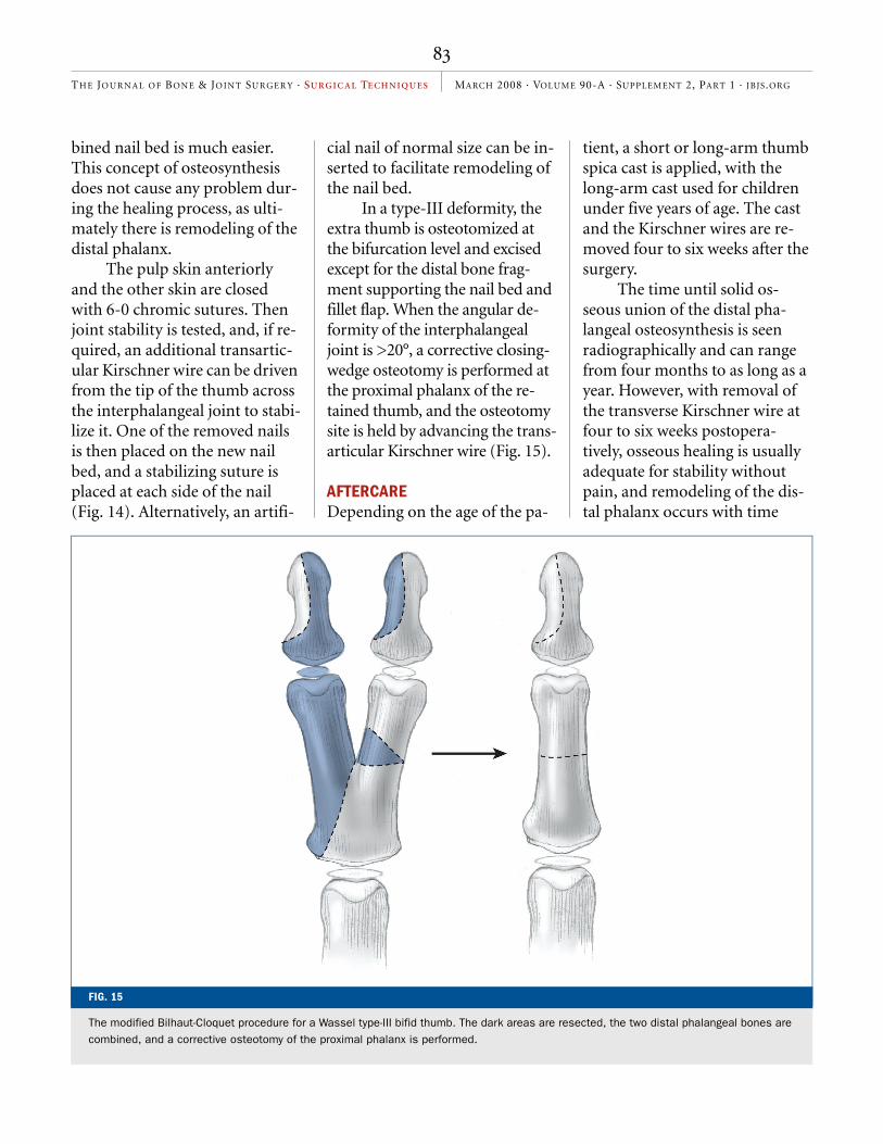
plane alone creates a so-called seagull deformity (Fig. 12). The nail fold and the nail bed are re-paired with 8-0 nylon sutures. Overly tight suture of the nail bed results in a prominent longi-tudinal ridge. To avoid this ridge,
the sutures should be tied under only slight tension so that a small longitudinal slit can be observed along the suture line (Fig. 13). The nylon sutures are not re-moved later. The sutures are no-ticeable on close observation at
first but usually not after the pas-sage of time, as their black color eventually becomes faint.
With our modified tech-nique, no attempt is made to compress the distal phalangeal bones, so contouring of the com-
FIG. 13
The nail fold and nail bed are repaired with 8-0 nylon sutures under slight tension.
FIG. 14
The final intraoperative appearance following the modified Bilhaut-Cloquet procedure. One of the removed nails has been placed over the nail bed to facilitate nail-bed remodeling.
Baek.fm Page 82 Friday, February 8, 2008 2:56 PM
83
TH E JO UR N AL O F BO N E & JO IN T SU RG E R Y · SU R G IC A L TE CH N I Q U E S MARCH 2008 · VOLUME 90-A · SUPPLEMENT 2, PART 1 · JBJS.ORG
bined nail bed is much easier. This concept of osteosynthesis does not cause any problem dur-ing the healing process, as ulti-mately there is remodeling of the distal phalanx.
The pulp skin anteriorly and the other skin are closed with 6-0 chromic sutures. Then joint stability is tested, and, if re-quired, an additional transartic-ular Kirschner wire can be driven from the tip of the thumb across the interphalangeal joint to stabi-lize it. One of the removed nails is then placed on the new nail bed, and a stabilizing suture is placed at each side of the nail (Fig. 14). Alternatively, an artifi-
cial nail of normal size can be in-serted to facilitate remodeling of the nail bed.
In a type-III deformity, the extra thumb is osteotomized at the bifurcation level and excised except for the distal bone frag-ment supporting the nail bed and fillet flap. When the angular de-formity of the interphalangeal joint is >20°, a corrective closing-wedge osteotomy is performed at the proximal phalanx of the re-tained thumb, and the osteotomy site is held by advancing the trans-articular Kirschner wire (Fig. 15).
AFTERCAREDepending on the age of the pa-
tient, a short or long-arm thumb spica cast is applied, with the long-arm cast used for children under five years of age. The cast and the Kirschner wires are re-moved four to six weeks after the surgery.
The time until solid os-seous union of the distal pha-langeal osteosynthesis is seen radiographically and can range from four months to as long as a year. However, with removal of the transverse Kirschner wire at four to six weeks postopera-tively, osseous healing is usually adequate for stability without pain, and remodeling of the dis-tal phalanx occurs with time
FIG. 15
The modified Bilhaut-Cloquet procedure for a Wassel type-III bifid thumb. The dark areas are resected, the two distal phalangeal bones are combined, and a corrective osteotomy of the proximal phalanx is performed.
Baek.fm Page 83 Friday, February 8, 2008 2:56 PM
84
TH E JO UR N AL O F BO N E & JO IN T SU RG E R Y · SU R G IC A L TE CH N I Q U E S MARCH 2008 · VOLUME 90-A · SUPPLEMENT 2, PART 1 · JBJS.ORG
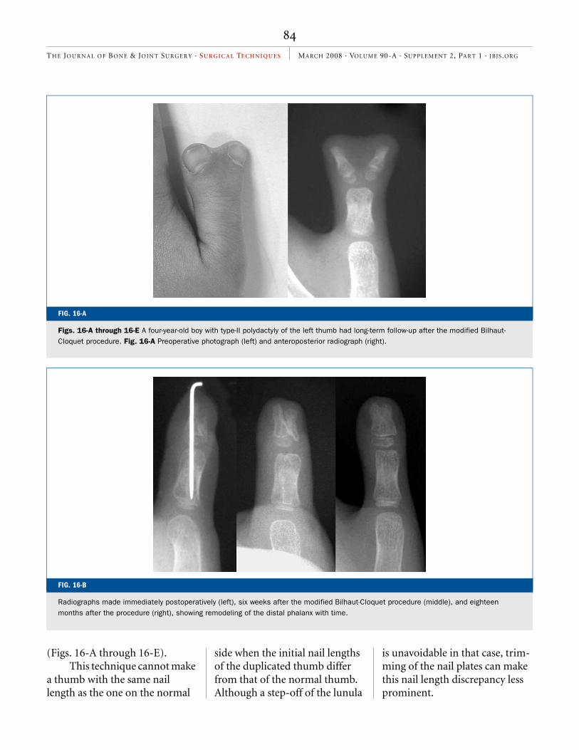
(Figs. 16-A through 16-E).This technique cannot make
a thumb with the same nail length as the one on the normal
side when the initial nail lengths of the duplicated thumb differ from that of the normal thumb. Although a step-off of the lunula
is unavoidable in that case, trim-ming of the nail plates can make this nail length discrepancy less prominent.
FIG. 16-A
Figs. 16-A through 16-E A four-year-old boy with type-II polydactyly of the left thumb had long-term follow-up after the modified Bilhaut-Cloquet procedure. Fig. 16-A Preoperative photograph (left) and anteroposterior radiograph (right).
FIG. 16-B
Radiographs made immediately postoperatively (left), six weeks after the modified Bilhaut-Cloquet procedure (middle), and eighteen months after the procedure (right), showing remodeling of the distal phalanx with time.
Baek.fm Page 84 Friday, February 8, 2008 2:56 PM
85
TH E JO UR N AL O F BO N E & JO IN T SU RG E R Y · SU R G IC A L TE CH N I Q U E S MARCH 2008 · VOLUME 90-A · SUPPLEMENT 2, PART 1 · JBJS.ORG
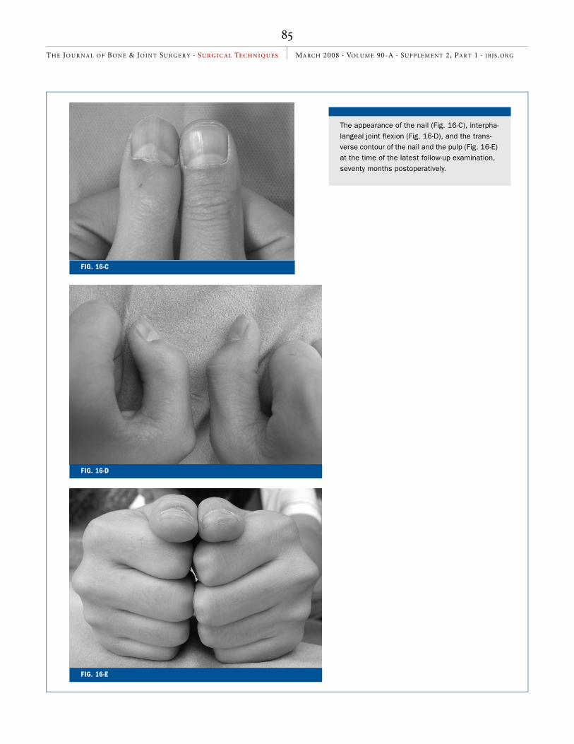
The appearance of the nail (Fig. 16-C), interpha-langeal joint flexion (Fig. 16-D), and the trans-verse contour of the nail and the pulp (Fig. 16-E) at the time of the latest follow-up examination, seventy months postoperatively.
FIG. 16-E
FIG. 16-C
FIG. 16-D
Baek.fm Page 85 Friday, February 8, 2008 2:56 PM

86
TH E JO UR N AL O F BO N E & JO IN T SU RG E R Y · SU R G IC A L TE CH N I Q U E S MARCH 2008 · VOLUME 90-A · SUPPLEMENT 2, PART 1 · JBJS.ORG
Goo Hyun Baek, MDHyun Sik Gong, MDMoon Sang Chung, MDJoo Han Oh, MDYoung Ho Lee, MDSang Ki Lee, MDDepartment of Orthopedic Surgery, Seoul National University College of Medicine, 28 Yongon-dong, Chongno-gu, Seoul 110-744, South Korea. E-mail address for G.H. Baek: [email protected]
The line drawings in this article are the work of Jennifer Fairman ([email protected]).
REFERENCES1. Bilhaut M. Guerison d’un pouce bifide par un nouveau procede operatoire. Congr Fr Chir. 1890;4:576-80.
2. Miura T. Duplicated thumb. Plast Reconstr Surg. 1982;69:470-81.
3. Tada K, Yonenobu K, Tsuyuguchi Y, Kawai H, Egawa T. Duplication of the thumb. A retro-spective review of two hundred and thirty-seven cases. J Bone Joint Surg Am. 1983;65:584-98.
4. Marks TW, Bayne LG. Polydactyly of the
thumb: abnormal anatomy and treatment. J Hand Surg [Am]. 1978;3:107-16.
5. Townsend DJ, Lipp EB Jr, Chun K, Reinker K, Tuch B. Thumb duplication, 66 years’ expe-riences – a review of surgical complications. J Hand Surg [Am]. 1994;19:973-6.
6. Wassel HD. The results of surgery for poly-dactyly of the thumb. A review. Clin Orthop Relat Res. 1969;64:175-93.
7. Zook EG. Anatomy and physiology of the perionychium. Hand Clin. 2002;18:553-9,v.
CRITICAL CONCEPTS
INDICATIONS:
The indication for the modified Bilhaut-Cloquet procedure is a Wassel type-II or III polydactyly, in which the bifid thumbs are symmetric and their nail size is less than two-thirds of that of the normal, contralateral thumb, or smaller than that of the index finger in patients with bilateral involvement.
CONTRAINDICATIONS:
The modified Bilhaut-Cloquet procedure is contraindicated for patients with asymmetric bifid thumbs; ablation of the smaller thumb or use of only its soft parts can obtain better results in such patients. Type-I polydactyly does not require this modified technique because the bifid distal phalanges can be combined with use of the original technique without vi-olating the distal interphalangeal joint. Type-IV and other polydactylies involve the metacarpophalangeal joint, and the re-sults of the combination operation are usually poor in such cases.
PITFALLS:
• A triangular flap created at the distal part of the pulp can prevent scar contracture.
• During approximation of the two distal phalangeal bones, slight axial rotation is required because attaching two semi-circular nails in a transverse plane alone creates a so-called seagull deformity.
• The nail bed is repaired under slight tension, without overlap of the underlying matrix, and this tension suture can pre-vent the formation of a prominent nail ridge.
AUTHOR UPDATE:
There have been no changes in the surgical technique since the time of publication of the original paper.
Baek.fm Page 86 Friday, February 8, 2008 2:56 PM
Copyright © 2022 FDOKUMEN