Social skills training in a depressed, visually impaired older adult
Upload
hsantaluciaCategory
view
0download
0
Mitochondrial Function Is Related to Alterations at Brain SPECT in
Depressed PatientsBy Ann Gardner, MD, PhD, Dario Salmaso, PhD, Davide Nardo, PhD,
Federica Micucci, PhD, Flavio Nobili, MD, Alejandro Sanchez-Crespo, PhD, Hans Jacobsson, MD, PhD, Stig A. Larsson, PhD, and Marco Pagani, MD, PhD
CNS Spectr 13:9 ©MBL Communications, Inc. September 2008805CNS Spectr 13:9 ©MBL Communications, Inc. September 2008805
Original Research
ABSTRACTIntroduction: 99mTc-d,l-hexamethylpropylene
amine oxime (99mTc-HMPAO) retention in brain is
proportional to cerebral blood flow and related to
both the local hemodynamic state and to the cel-
lular content of reduced glutathione. Alterations
of the regional distribution of 99mTc-HMPAO reten-
tion, with discrepant results, have been reported
at functional brain imaging of unipolar depres-
sion. Since mitochondrial involvement has been
reported in depressed patients, the aim of the
study was to explore whether the 99mTc-HMPAO
retention at single-photon emission computed
tomography in depressed patients may relate to
different levels of mitochondrial function.
Methods: All patients had audiological and
muscular symptoms, somatic symptoms that are
common in depression. Citrate synthase (CS) activ-
ity assessed in muscle mitochondria correlated
strongly with the activities of three mitochondrial
respiratory chain enzymes and was used as a
marker of mitochondrial function. K-means cluster-
Needs AssessmentAlthough the literature on functional brain imaging and major depression mostly show decreased radiotracer retention implicating a general decrease of neu-ronal activity, discrepant results have been reported with the commonly used radiotracer hexamethylpropylene amine oxime at single photon emission com-puted tomography. This study aims to explore a possible effect mechanism for this phenomenon.
Learning ObjectivesAt the end of this activity, the participant should be able to: • List two examples of conditions/disorders where the brain retention of
hexamethylpropylene amine oxime at single photon emission computed tomography sometimes is increased.
• Give an example of a possible effect mechanism for this phenomenon.• Appreciate the relationship between mitochondrial dysfunction and
depression.
Target Audience: Neurologists and psychiatrists
CME Accreditation StatementThis activity has been planned and implemented in accordance with the Essentials and Standards of the Accreditation Council for Continuing Medical Education (ACCME) through the joint sponsorship of the Mount Sinai School of Medicine and MBL Communications, Inc. The Mount Sinai School of Medicine is accredited by the ACCME to provide continuing medi-cal education for physicians.
Credit DesignationThe Mount Sinai School of Medicine designates this educational activity for a maximum of 3 AMA PRA Category 1 Credit(s)TM. Physicians should only claim credit commensurate with the extent of their participation in the activity.
This activity has been peer-reviewed and approved by Eric Hollander, MD, chair at the Mount Sinai School of Medicine. Review date: August 15, 2008. Dr. Hollander does not have an affiliation with or financial interest in any orga-nization that might pose a conflict of interest.
To Receive Credit for This ActivityRead this article and the two CME-designated accompanying articles, reflect on the information presented, and then complete the CME posttest and evalu-ation found on page 815. To obtain credits, you should score 70% or better. Early submission of this posttest is encouraged: please submit this posttest by September 1, 2010, to be eligible for credit. Release date: September 1, 2008. Termination date: September 30, 2010. The estimated time to complete all three articles and the posttest is 3 hours.
3CME
Affilations and Disclosures: Please see page 814 for biographies and disclosure information.

ing performed on CS grouped eight patients with
low and 11 patients with normal CS. Voxel-based
analysis was performed on the two groups by sta-
tistical parametric mapping.
Results: Voxel-based analysis showed sig-
nificantly higher 99mTc-HMPAO retention in the
patients with low CS compared with the patients
with normal CS in the posterior and inferior fron-
tal cortex, the superior and posterior temporal
cortex, the somato-sensory cortex, and the asso-
ciative parietal cortex.
Conclusion: Low muscle CS in depressed
patients is related to higher regional 99mTc-HMPAO
retention that may reflect cerebrovascular adap-
tation to impaired intracellular metabolism and/
or intracellular enzymatic changes, as previously
reported in mitochondrial disorder. Mitochondrial
dysfunction in varying proportions of the sub-
jects may explain some of the discrepant results
for 99mTc-HMPAO retention in depression.
CNS Spectr. 2008;13(9):805-814CNS Spectr. 2008;13(9):805-814CNS Spectr
INTRODUCTIONAlterations of the regional distribution of
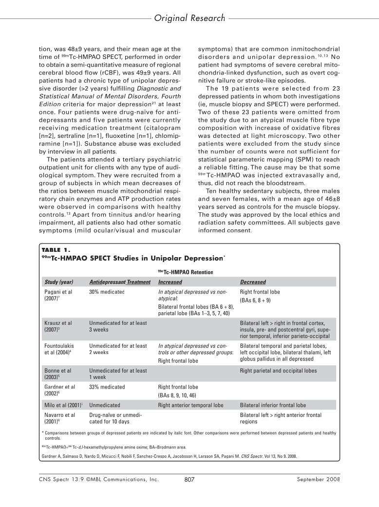
various radiotracers at functional brain imag-ing have been reported in unipolar depression. The results of a metastudy of pooled positron emission tomography (PET) and single photon emission computed tomography (SPECT) stud-ies of unipolar depression implicated a general decrease of neuronal activity similarly affect-ing almost all analyzed cortical and subcortical regions, with ambiguous results for the limbic system.1 Discrepant findings,2-8 with increased as well as decreased retention of 99mTc-d,l-hexa-d,l-hexa-d,lmethylpropylene amine oxime (99mTc-HMPAO) at SPECT, have been reported in unipolar depres-sion. A summary of some recent studies can be seen in Table 1.
Liability to unipolar depression has a sub-stantial heritable component without any evi-dence of a shared family environment.9 A highly increased depression prevalence has been reported in mitochondrial disorders,10 conditions which are oftentimes heritable. The tissues that
are most affected in mitochondrial disorders are those with the highest cellular energy demands, especially brain and muscle. Signs indicating mitochondrial dysfunction have been reported in unipolar depression.11-14 To date, there are no reports about the prevalence of mitochondrial dysfunction in depression, or concerning the issue of whether depression, per se, may affect mitochondrial function.
Intracellular trapping of the lipophilic 99mTc-HMPAO and its conversion to the hydrophilic form have been considered as the basis of reten-tion of this tracer. The conversion from the lipo-philic to the hydrophilic form has been associated to the content of reduced glutathione (GSH).15,16
Increased GSH levels have been reported in the early stages of mitochondrial disorders.17
The aim of the present study was to explore the relationship between 99mTc-HMPAO reten-tion at SPECT and mitochondrial function in a group of depressed patients in order to enlighten the issue of whether a mitochondrial involvement may contribute to the enigmatic discrepant results at 99mTc-HMPAO SPECT in uni-polar depression. Mitochondrial function was assessed by the activity of citrate synthase (CS) in isolated muscle mitochondria. CS is a Krebs cycle enzyme that provides the electrons nec-essary for the mitochondrial respiratory chain. CS activity is considered to be a marker, even if not a direct measure, of oxidative phosphoryla-tion reflecting the production of cellular energy, adenosine triphosphate (ATP).18 A good corre-lation has been observed between CS activity and respiratory chain enzymes in normal rat tis-sues suggesting coordination between CS and respiratory chain enzymes, and confirming the utilization of CS activity as a marker of respira-tory chain content.19 CS activity is oftentimes used as an index of mitochondrial proliferation in muscle homogenate in the assessment of mitochondrial disorders. However, no relation-ship between CS activity levels and morpho-logical evidence of mitochondrial proliferation was found in a study.20
METHODS
SubjectsNineteen depressed patients (10 males; 9
females) were included in the study. The mean age of the patients at the time of muscle biopsy, performed in order to assess mitochondrial func-
Original ResearchOriginal Research
CNS Spectr 13:9 ©MBL Communications, Inc. September 2008806CNS Spectr 13:9 ©MBL Communications, Inc. September 2008806
tion, was 48±9 years, and their mean age at the time of 99mTc-HMPAO SPECT, performed in order to obtain a semi-quantitative measure of regional cerebral blood flow (rCBF), was 49±9 years. All patients had a chronic type of unipolar depres-sive disorder (>2 years) fulfilling Diagnostic and Statistical Manual of Mental Disorders, Fourth Edition criteria for major depression21 at least once. Four patients were drug-naïve for anti-depressants and five patients were currently receiving medication treatment (citalopram [n=2], sertraline [n=1], fluoxetine [n=1], chlomip-ramine [n=1]). Substance abuse was excluded by interview in all patients.
The patients attended a tertiary psychiatric outpatient unit for clients with any type of audi-ological symptom. They were recruited from a group of subjects in which mean decreases of the ratios between muscle mitochondrial respi-ratory chain enzymes and ATP production rates were observed in comparisons with healthy controls.13 Apart from tinnitus and/or hearing impairment, all patients also had other somatic symptoms (mild ocular/visual and muscular
symptoms) that are common inmitochondrial disorders and unipolar depression.10,13 No patient had symptoms of severe cerebral mito-chondria-linked dysfunction, such as overt cog-nitive failure or stroke-like episodes.
The 19 patients were selected from 23 depressed patients in whom both investigations (ie, muscle biopsy and SPECT) were performed. Two of these 23 patients were omitted from the study due to an atypical muscle fibre type composition with increase of oxidative fibres was detected at light microscopy. Two other patients were excluded from the study since the number of counts were not sufficient for statistical parameteric mapping (SPM) to reach a reliable fitting. The cause may be that some 99mTc-HMPAO was injected extravasally and, thus, did not reach the bloodstream.
Ten healthy sedentary subjects, three males and seven females, with a mean age of 46±8 years served as controls for the muscle biopsy. The study was approved by the local ethics and radiation safety committees. All subjects gave informed consent.
Original Research
CNS Spectr 13:9 ©MBL Communications, Inc. September 2008807CNS Spectr 13:9 ©MBL Communications, Inc. September 2008807
TABLE 1.99mTc-HMPAO SPECT Studies in Unipolar Depression*
99mTc-HMPAO Retention
Study (year)Study (year) AntidAntidAnti epressant epressant depressant d Treatment Increased Decreased
Pagani et al (2007)2
30% medicated In atypical depressed vs non-atypical:Bilateral frontal lobes (BA 6 + 8), parietal lobe (BAs 1–3, 5, 7, 40)
Right frontal lobe(BAs 6, 8 + 9)
Krausz et al (2007)3
Unmedicated for at least 3 weeks
Bilateral left > right in frontal cortex, insula, pre- and postcentral gyri, supe-rior temporal, inferior parieto-occipital
Fountoulakis et al (2004)4
Unmedicated for at least 2 weeks
In atypical depressed vs con-trols or other depressed groups:Right frontal lobe
Bilateral temporal and parietal lobes, left occipital lobe, bilateral thalami, left globus pallidus in all depressed
Bonne et al (2003)5
Unmedicated for at least 1 week
Right parietal and occipital lobes
Gardner et al (2002)6
33% medicated Right frontal lobe(BAs 8, 9, 10, 46)
Milo et al (2001)7 Unmedicated Right anterior temporal lobe Bilateral inferior frontal lobe
Navarro et al (2001)8
Drug-naïve or unmedi-cated for 10 days
Bilateral left > right anterior frontal regions
* Comparisons between groups of depressed patients are indicated by italic font. Other comparisons were performed between depressed patients and healthy controls.
99mTc-HMPAO=99mTc-d,l-hexamethylpropylene amine oxime; BA=Brodmann area.d,l-hexamethylpropylene amine oxime; BA=Brodmann area.d,l
Gardner A, Salmaso D, Nardo D, Micucci F, Nobili F, Sanchez-Crespo A, Jacobsson H, Larsson SA, Pagani M. CNS Spectr. Vol 13, No 9. 2008.CNS Spectr. Vol 13, No 9. 2008.CNS Spectr

Assessment of Mitochondrial EnzymesMuscle biopsies were taken from the right
anterior tibial muscle in all subjects. Enzyme activities were spectrophotometrically deter-mined in isolated mitochondria. An aliquot was freeze-thawed in hypotonic medium according to the procedure by Birch-Machin and colleagues22
The mitochondrial respiratory chain enzymes rotenone sensitive nicotinamide adenine dinu-cleotide-cytochrome c reductase (NCR [complex I c reductase (NCR [complex I c+ III) and succinate-cytochrome c reductase (SCR c reductase (SCR c[complex II + III]) were determined according to Sottocasa and colleagues23 and Cooperstein and colleagues,24 respectively. Another aliquot was freeze-thawed in the storage medium, and treated with digitonin 2 gram L–1 before the anal-ysis of the respiratory chain enzyme cytochrome c oxidase (COX, complex IV).c oxidase (COX, complex IV).c 25 The activity of CS was determined according to the method by Alp and colleagues26 after permeabilization of the mitochondria in a medium containing Triton X-100 0.05% (v/v), K2HPO4, 50 mmol 1–1 and EDTA 1 mmol 1–1, pH 7.5. The enzyme activity units are expressed in spectrophotometric values.
Single Photon Emission Computed Tomography
Brain imaging using SPECT was performed using a three-headed Gamma Camera (TRIAD XLT 20, Trionix Research Laboratory, Inc., Twinsburg, Ohio) equipped with low-energy, ultrahigh-resolution collimators. The intrinsic spatial resolution of the camera was 8 mm (full width at half maximum). 99mTc-HMPAO (Ceretec, Exametazine, Amersham International Plc, Little Chalfont, United Kingdom) was injected after 30 minutes rest in a quiet, dim-lighted room. Examinations started between 45 and 60 min-utes after tracer injection. The projection data were acquired for 30 seconds per projection at 90 equal angles of a complete revolution. Between 8 and 10 million total counts were acquired.
Before back-projection we pre-processed the 1-dimensional data with a Hamming smoothing filter with a cut-off frequency of 2.25 cycles/cm. Then SPECT images were reconstructed by fil-tered back projection algorithm using a ramp filter with a cut-off frequency of 0.6 cycles/cm. Attenuation correction was based on a 4-point ellipse.27 No scatter correction was performed. Data were projected into a 128x128 pixel matrix resulting in an isotropic voxel size of 2.2 mm3.
Statistical AnalysisK-means clustering was applied to create
two groups of the patients according to their enzyme activities (patients with lower and higher enzymes activities). K-means clustering splits cases into a selected number of groups by maxi-mizing the variation between groups relative to the variation within groups. It is an iterative procedure that ends when cases are successfully assigned to a specified number of non-over-lapping clusters. χ2 is used to test the distribu-tion differences between the obtained clusters and the clinical diagnosis and type. The mean enzyme activities of the two clusters that were obtained were compared with the mean value of the healthy controls using three-groups compari-sons with analysis of variance and Tukey post-hoc test. The significance level was set at P≤P≤P .05.
Voxel-based AnalysisSPECT raw images were transformed into the
analyze format by XMedCon package. Data were analyzed with SPM2 (Wellcome Department of Cognitive Neurology, London, UK) implemented in Matlab 6.5.1. Images of relative tracer distribu-tion were spatially normalized into the stereotac-tic Montreal Neurological Institute (MNI) space to a predefined SPECT template available in SPM2 (voxel size: 2x2x2 mm), using a 16-param-eter affine (non-linear) transformation. Because this template does not completely match the Talairach brain, it was necessary to correct the SPM{t} coordinates. This was achieved using the t} coordinates. This was achieved using the tsubroutine implemented by Brett.28 Brodmann areas (BAs) where then identified, after import-ing the corrected coordinates.29
After normalization, images were smoothed with a Gaussian filter of 12 mm (full width at half maximum) to account for individual gyral dif-ferences and brain anatomy and to increase the signal-to-noise ratio. Images were globally normal-ized for signal intensity using proportional scal-ing to remove confounding effects due to global CBF changes, with threshold masking for grey matter of 0.8, allowing to include into the analy-sis only those voxels whose intensity exceeded the 80% of the maximal one. Because of the lack of any topographic a priori hypothesis, the sig-nificance of identified regions was assessed using P values corrected for multiple comparisons.P values corrected for multiple comparisons.P 30,31
Significant differences between the groups were set at a threshold of P<.05 for cluster extent and at P<.05 for cluster extent and at Pa threshold of P<.005 for voxel height. Only those P<.005 for voxel height. Only those P
CNS Spectr 13:9 ©MBL Communications, Inc. September 2008808CNS Spectr 13:9 ©MBL Communications, Inc. September 2008808
Original Research

clusters containing >100 voxels were accepted as significant. This was based on the calculation of the partial volume effect resulting from the spatial resolution of the camera system.
The voxel-based analyses were performed using SPM2 with a “one scan per subject, two-sample t-test” design model, and significances t-test” design model, and significances twere sought for the following contrasts: LOW minus NORMAL subtraction, and NORMAL minus LOW subtraction (for the definitions of LOW and NORMAL, see Results).
RESULTS
Creating Two Subgroups of the Patients According to the Citrate Synthase Activity
The activities of the three mitochondrial respi-ratory chain enzymes were significantly correlated with each other in both patients and controls (cor-relation coefficients of 0.66–0.78 and 0.65–0.68, respectively). The activities of CS were also sig-nificantly correlated with the activities of the indi-vidual respiratory chain enzymes in all subjects (correlation coefficients of 0.79–0.93 and 0.70–0.85, respectively) (Table 2).
The statistically most significant separation for K-means clustering was achieved using the CS activities (F(1.17)=19.273, P=.000) and two P=.000) and two Pgroups of patients were created according to their activities of this enzyme. Significant dif-ferences for all enzyme activities were found between the group with lower CS activities (the LOW group) and controls but not between the group with higher CS activities (the NORMAL group) and controls (Table 3).
Relationships Between the 99mTc–HMPAO Retention at SPECT and the Citrate Synthase Activity
At SPM, the LOW minus NORMAL subtrac-tion highlighted two large significant clusters of voxels reflecting higher 99mTc –HMPAO reten-tion in the LOW group. The clusters, bilaterally, included BAs belonging to the posterior and inferior frontal cortex, the superior and posterior temporal cortex, the somato-sensory cortex, and the associative parietal cortex (Figure and Table 4). The magnitude of the effects of SMP statistics at both cluster extent and voxel height levels and reporting (t) and (t) and (t z) statistics is presented in Table 4. The NORMAL minus LOW subtraction highlighted small clusters in the bilateral orbito-frontal cortex (BAs 11 and 47) (ie, in these small regions there was decreased 99mTc –HMPAO retention in the LOW group).
DISCUSSIONThe depressed patients included in this study
were recruited from a specialized psychiatric unit for patients with any type of hearing impairment. Depression and hearing impairment are com-mon manifestations of mitochondrial disorders.10
Since mitochondria have been demonstrated to be the major subcellular fraction for the uptake of 99mTc-HMPAO in brain homogenate,32 we spec-ulated that differences in mitochondrial func-tion might explain some of the discrepant results that have been reported for the retention of this tracer across studies of unipolar depression.
Voxel-based analyses (VBA) of the SPECT data demonstrated higher regional 99mTc-HMPAO retention in large portions of posterior-frontal, temporal and inferior-parietal cortex in
CNS Spectr 13:9 ©MBL Communications, Inc. September 2008809CNS Spectr 13:9 ©MBL Communications, Inc. September 2008809
Original Research
TABLE 2.Correlations Between the Activities of CS and Respiratory Chain Enzymes*
Enzyme
CS NCR SCR
Patients (n=19)Patients (n=19) Controls (n=10)Controls (n=10) Patients (n=19)Patients (n=19) Controls (n=10)Controls (n=10) Patients (n=19)Patients (n=19) Controls (n=10)Controls (n=10)
r P r P r P r P r P r P
NCR 0.79 .000 0.70 .025
SCR 0.84 .000 0.82 .004 0.66 .002 0.65 .043
COX 0.93 .000 0.85 .002 0.74 .000 0.68 .030 0.78 .000 0.68 .031
*The relationships between the enzyme activities were evaluated with the Pearson correlation test.
CS=citrate synthase; NCR=NADH-cytochrome c reductase (complex I + III); SCR=succinate-cytochrome c reductase (complex I + III); SCR=succinate-cytochrome c c reductase (complex II + III); COX=cytochrome c reductase (complex II + III); COX=cytochrome c c oxidase c oxidase c(complex IV).
Gardner A, Salmaso D, Nardo D, Micucci F, Nobili F, Sanchez-Crespo A, Jacobsson H, Larsson SA, Pagani M. CNS Spectr. Vol 13, No 9. 2008.CNS Spectr. Vol 13, No 9. 2008.CNS Spectr

patients with low activities of the mitochondrial enzyme CS compared to patients with normal enzyme activity. There were no overt differences in mood or other depressive symptomatology between the patient groups. No differences between the patients with low and normal CS activities were found in the prefrontal cortical areas considered to be specifically involved in emotional experience and depression: BA 32 in the anterior cingulate cortex, and BA 47 in the lateral orbitofrontal cortex.33
We could not, for obvious reasons, measure the regional mitochondrial enzyme activities in the brain. We assumed that muscle mitochon-drial function may, to some extent, reflect the mitochondrial function in brain, considering its ubiquity within tissues, even if the enzyme activ-ities might change differently in various tissues under certain experimental conditions.34,35
No general pathogenetic susceptibility mech-anism has been found in unipolar depression although a substantial heritable component has been reported.9 A mouse model with mul-tiple deletions of the mitochondrial DNA dem-onstrates both “mood disorder-like phenotypes” and decreased regional brain levels of the neu-rotransmitters serotonin and noradrenalin.36
In muscle, altered cell histochemistry also in unmedicated patients,13,37 decreased respiratory chain enzyme ratios and ATP production rates,13
and an increased prevalence of small deletions of the mitochondrial DNA,13 indicate that unipolar depression may be a systemic disorder. Multiple medically unexplained somatic symptoms, such
as headache, constipation, weakness, or back pain, often conceptualized as “somatization,” are reported by 50% patients with unipolar depres-sion worldwide.38 The audiological symptom tin-nitus was present in 50% of the unmedicated depressed patients in a study.39 A relationship between “somatization” and mitochondrial dys-function, indicating a systemic involvement in cases of depression with somatic symptoms, has been reported.40,41
Decreased ATP content and protein changes suggestive of mitochondrial dysfunction have been reported in brain in unipolar depres-sion.11,12,14 Creatine, an agent buffering cellular ATP resources, is used in the treatment of mito-chondrial disorders.42 Beneficial effect of creatine has been reported in a preliminary study of uni-polar depression refractory to antidepressant or mood-stabilizing therapies.43 These observa-tions, and the fact that lifetime depression was reported by >50% of the patients with mitochon-drial disorders10 suggest that mitochondrial dys-function is among the factors that, at least, incur vulnerability to depression. In families harboring a mitochondrial DNA mutation often found in mitochondrial myopathy, encephalopathy, lac-tic acidosis, and stroke-like episodes (MELAS), depressive traits were reported by 22% of asymptomatic carriers and 29% of oligosymp-tomatic carriers, and depressive symptoms by 42% of fully symptomatic carriers.44
We used the activity of CS to reflect the overall function of the mitochondrial respira-tory chain and to create the two subgroups
CNS Spectr 13:9 ©MBL Communications, Inc. September 2008810CNS Spectr 13:9 ©MBL Communications, Inc. September 2008810
Original Research
TABLE 3.Enzyme Activities in the Patients According to Citrate Synthase Clustering and in the Healthy Controls
Enzyme
Low CS(Cluster 1)
Normal CS (Cluster 2)
Healthy Controls ANOVA Tukey HSD Multiple Comparisons
EnzymeActivityActivity*Activity*Activity
EnzymeActivityActivity*Activity*Activity
EnzymeActivityActivity*Activity*Activity
Low vs Normal
Low vsControls
Normal vs Controls
8 cases 11 cases 10 cases F(2,26) P P P P
CS 1,581±431 2,533±490 2,656±732 9.14 .001 .004 .002 .877
NCR 1,316±524 2,034±932 2,398±702 4.57 .020 .125 .016 .526
SCR 710±343 1,291±310 1,405±490 7.91 .002 .010 .003 .783
COX 2,758±853 4,141±701 3,983±1,377 4.82 .017 .019 .045 .933
* The enzyme activity units are expressed in spectrophotometric values.
CS=citrate synthase; ANOVA=analysis of variance; HSD=honestly significant difference; NCR=NADH-cytochrome c reductase (complex I + III); SCR=succinate-c reductase (complex I + III); SCR=succinate-ccytochrome c reductase (complex II + III); COX=cytochrome c reductase (complex II + III); COX=cytochrome c c oxidase (complex IV). c oxidase (complex IV). c
Gardner A, Salmaso D, Nardo D, Micucci F, Nobili F, Sanchez-Crespo A, Jacobsson H, Larsson SA, Pagani M. CNS Spectr. Vol 13, No 9. 2008.CNS Spectr. Vol 13, No 9. 2008.CNS Spectr

of patients. We based this choice on the find-ing that the best separation into two groups according to K-means clustering at using all enzyme activities was achieved with CS activ-ity. Furthermore, CS activity was highly corre-lated with the activities of the respiratory chain enzymes in all subjects, adding support for the use of CS activity as a marker of the respiratory chain content and ATP production,18,19 at least in normal or near-normal conditions. The mean CS activity in the patient group with lower activities was significantly decreased in the comparison with the healthy controls. Since all medicated patients belonged to the group with normal CS activity, antidepressant could not have been the cause of low CS activities.
Both decreased and increased regional brain distribution of the 99mTc-HMPAO retention have been reported in mitochondrial disorders.45-
50 Diffuse general hyperperfusion has been observed in MELAS patients at times when they
did not suffer an acute stroke episode.51,52
The higher retention of 99mTc-HMPAO in patients with low CS activity may reflect local hemodynamic changes. Intracellular metabolic dysfunction is followed by increased rCBF in severe mitochondrial pathology, such as the MELAS syndrome, thus producing the so-called “luxury perfusion”. This phenomenon has been suggested to represent adaptation to the altered mitochondrial function, leading to impaired intra-cellular oxygen utilization and metabolism. It was speculated that increased CBF might be a com-pensation for increased wash-out of lactate pro-duced by increased anaerobic metabolism,52 or might have been related to decreased pH caused by local increase of lactic acid.48 Quantitative CBF measurements with xenon133 demonstrated diffuse hyperperfusion in a young man affected by MELAS years prior to undergoing a large pos-terior “metabolic” stroke.51 These findings were confirmed by a PET study of a MELAS patient in which decreased cerebral metabolic rate of dioxygen was found along with increased CBF and cerebral metabolic rate of glucose.52
Unevenly distributed activities in the brain have been reported for mitochondrial respira-tory chain enzymes.53 In MELAS specifically, mitochondrial dysfunction is widespread but, in the brain, it predominates in the posterior areas.54 Different cellular thresholds to meta-bolic dysfunction in the various brain areas, according to local dependence on oxidative metabolism, have been suggested to be one of the mechanisms leading to the regional, rather than general, expression of neuropatho-logic lesions in mitochondrial disorders.55-
58 This would result in a subpopulation(s) to be selectively more affected by a generalized impairment and is consistent with the reported quantitative CBF measurements in mitochon-drial disorder in which increased CBF was present in all brain, with regional pronounce-ments.51 Our results suggest that scattered brain regions in the posterior frontal, temporal, and parietal cortices are the most sensitive to mildly impaired mitochondrial function.
The conversion of the lipophilic tracer 99mTc-HMPAO into the hydrophilic form that is retained in the cell has been related to the cellular GSH content.15,16 GSH deficiency has been reported in the most severe cases of mitochondrial dis-orders.59 On the other hand, increased GSH lev-els, considered to reflect GSH upregulation at increased oxidative stress secondary to reduced
CNS Spectr 13:9 ©MBL Communications, Inc. September 2008811CNS Spectr 13:9 ©MBL Communications, Inc. September 2008811
Original Research
FIGURE.Three-dimensional rendering of voxels reflecting higher tracer distribution (red) in the depressed patients with lower muscle activity of citrate synthase com-pared to the depressed patients with normal citrate synthase activity
Gardner A, Salmaso D, Nardo D, Micucci F, Nobili F, Sanchez-Crespo A, Jacobsson H, Larsson SA, Pagani M. CNS Spectr. Vol 13, No 9. 2008.CNS Spectr. Vol 13, No 9. 2008.CNS Spectr

respiratory chain enzyme activity, were found in the early stages of mitochondrial disorders.17
The higher tracer retention that was observed in scattered brain regions in the patients with low CS activity may partially reflect such early increased GSH content.
A limitation of the study is that the approach used did not allow to discriminate to which portion 99mTc-HMPAO retention was related to either CBF or intracellular enzymatic changes. Other limitations are the relatively small num-ber of subjects, and that the muscle biopsies and SPECT investigations were not performed simultaneously. Our subjects were recruited from two separately ethically approved studies
since it would not have been possible to obtain an ethical permission for two invasive trials, and most likely to recruit enough subjects, for such a study. Since the physical activity levels of the depressed subjects did not dif-fer between the investigations 1 year apart as determined by interview, this should not have affected the results. We did not compare the SPECT imaging results to healthy SPECT con-trols since the inclusion of two different control groups in one study is considered inappropri-ate. Comparisons between the SPECT results of our patients and controls are also beyond the purpose of this study.
CNS Spectr 13:9 ©MBL Communications, Inc. September 2008812CNS Spectr 13:9 ©MBL Communications, Inc. September 2008812
Original Research
TABLE 4.SPM Statistics Relative to the Subtraction Image Resulting From LOW Minus NORMAL Groups of Citrate Synthase Activity
Cluster Level Voxel LevelTalairach
CoordinatesAnatomical and Functional
Location
P(cor)(cor)Number
of Voxels P(FDR-cor)(FDR-cor) tz score of Maximum P(unc)(unc) x yy z
.000 1,746 .066.066.066.066.068.068.075.075.076.077.080.080.080.082.091.094
6.165.044.824.444.214.174.014.003.943.913.793.773.743.673.433.33
4.413.893.783.573.443.423.323.313.273.263.183.173.153.112.952.88
.000
.000
.000
.000
.000
.000
.000
.000
.001
.001
.001
.001
.001
.001
.002
.002
–42–48–48–42–59–24–56–53–53–65–59–56–62–59–50–39
–69–47–31 4
–45–23–54–60 7
–11–25–6 9–8–19–70
232152524152831162321–21317311
L Middle temporal gyrus BA 39L Middle temporal gyrus BA 21L Superior temporal gyrus BA 29L Inferior frontal gyrus BA 9L Supramarginal gyrus BA 40L ClaustrumL Supramarginal gyrus BA 40L Superior temporal gyrus BA 39L Inferior frontal gyrus BA 44L Postcentral gyrus BA 3L Postcentral gyrus BA 40L Superior temporal gyrus BA 22L Precentral gyrus BA 44L Postcentral gyrus BA 43L Postcentral gyrus BA 2L Middle occipital gyrus BA 37
.000 1,334 .066.066.066.068.076.077.080.081.083.072.074.076.079.082
5.675.094.654.273.943.883.793.723.613.183.123.072.972.91
4.193.913.683.473.273.243.183.143.062.782.732.702.632.59
.000
.000
.000
.000
.001
.001
.001
.001
.001
.003
.003
.003
.004
.005
62 42 56 45 62 30 42 48 33 39 62 12 24 27
–8–66–34–52–26–68–39
44
–22–30–80–78–32
2023138
–439322230313223232
R Postcentral gyrus BA 43R Middle temporal gyrus BA 39R Superior temporal gyrus BA 42R Superior temporal gyrus BA 39R Middle temporal gyrus BA 21R Precuneus BA 19R Supramarginal gyrus BA 40R Inferior frontal gyrus BA 9R Precentral gyrus BA 6R Postcentral gyrus BA 2R Inferior parietal lobule BA 40R Cuneus BA 18R Cuneus BA 18R Thalamus
SPM=statistical parametric mapping; cor=corrected; FDR-cor=false discovery rate-corrected; unc=uncorrected; BA=Brodmann area; L=left; R=right.
Gardner A, Salmaso D, Nardo D, Micucci F, Nobili F, Sanchez-Crespo A, Jacobsson H, Larsson SA, Pagani M. CNS Spectr. Vol 13, No 9. 2008.CNS Spectr. Vol 13, No 9. 2008.CNS Spectr

CONCLUSION In conclusion, in support of our hypothesis
we found higher retention of the tracer 99mTc-HMPAO in large portions of some brain regions in depressed patients with overall decreased, as opposed to overall normal, mitochondrial enzyme activities. Higher 99mTc-HMPAO reten-tion might reflect a local perfusion increase and higher intracellular levels of GSH, both due to the regional biochemical changes following mito-chondrial dysfunction. Studying the involvement of mitochondrial energy production is a cumber-some endeavor since almost a thousand proteins are involved. An implicit suggestion arising from our study is the use of tracers designed to assess mitochondrial functions in order to estimate the prevalence, the clinical features, and the neuro-biological substrates of depression associated with mitochondrial dysfunction. CNS
REFERENCES1. Nikolaus S, Larisch R, Beu M, Vosberg H, Müller-Gärtner HW. Diffuse cortical
reduction of neuronal activity in unipolar major depression: a retrospective analysis of 337 patients and 321 controls. Nucl Med Commun. 2000;21:1119-1125.
2. Pagani M, Salmaso D, Nardo D, et al. Imaging the neurobiological substrate of atypical depression by SPECT. Eur J Nucl Med Mol Imaging. 2007;34:110-120.
3. Krausz Y, Freedman N, Lester H, et al. Brain SPECT study of common ground between hypothyroidism and depression. Int J Neuropsychopharmacol. 2007;10:99-106.
4. Fountoulakis KN, Iacovides A, Gerasimou G, et al. The relationship of regional cere-bral blood flow with subtypes of major depression. Prog Neuropsychopharmacol Biol Psychiatry. 2004;28:537-546.Biol Psychiatry. 2004;28:537-546.Biol Psychiatry
5. Bonne O, Louzoun Y, Aharon I, et al. Cerebral blood flow in depressed patients: a methodological comparison of statistical parametric mapping and region of interest analyses. Psychiatry Res. 2003;122:49-57.
6. Gardner A, Pagani M, Jacobsson H, et al. Differences in resting state regional cerebral blood flow assessed with 99mTc-HMPAO SPECT and brain atlas match-ing between depressed patients with and without tinnitus. Nucl Med Commun. 2002;23:429-439.
7. Milo TJ, Kaufman GE, Barnes WE, et al. Changes in regional cerebral blood flow after electroconvulsive therapy for depression. J ECT. 2001;17:15-21.J ECT. 2001;17:15-21.J ECT
8. Navarro V, Gastó C, Lomeña F, Mateos JJ, Marcos T. Frontal cerebral perfusion dysfunction in elderly late-onset major depression assessed by 99MTC-HMPAO SPECT. Neuroimage. 2001;14(1 pt 1):202-205.
9. McGuffin P, Katz R, Watkins S, Rutherford J. A hospital-based twin register of the heritability of DSM-IV unipolar depression. Arch Gen Psychiatry. 1996;53:129-136.Arch Gen Psychiatry. 1996;53:129-136.Arch Gen Psychiatry
10. Fattal O, Link J, Quinn K, Cohen BH, Franco K. Psychiatric comorbidity in 36 adults with mitochondrial cytopathies. CNS Spectr. 2007;12:429-438.CNS Spectr. 2007;12:429-438.CNS Spectr
11. Moore CM, Christensen JD, Lafer B, Fava M, Renshaw PF. Lower levels of nucleo-side triphosphate in the basal ganglia of depressed subjects: a phosphorous-31 magnetic resonance spectroscopy study. Am J Psychiatry. 1997;154:116-118.Am J Psychiatry. 1997;154:116-118.Am J Psychiatry
12. Volz HP, Rzanny R, Riehemann S, et al. 31P magnetic resonance spectroscopy in the frontal lobe of major depressed patients. Eur Arch Psychiatry Clin Neurosci. Eur Arch Psychiatry Clin Neurosci. Eur Arch Psychiatry Clin Neurosci1998;248:289-295.
13. Gardner A, Johansson A, Wibom R, et al. Alterations of mitochondrial function and correlations with personality traits in selected major depressive disorder patients. J Affect Disord. 2003;76:55-68.Affect Disord. 2003;76:55-68.Affect Disord
14. Beasley CL, Pennington K, Behan A, Wait R, Dunn MJ, Cotter D. Proteomic analysis of the anterior cingulate cortex in the major psychiatric disorders: evidence for disease-associated changes. Proteomics. 2006;6:3414-3425.
15. Neirinckx RD, Burke JF, Harrison RC, Forster AM, Andersen AR, Lassen NA. The retention mechanism of technetium-99m-HM-PAO: intracellular reaction with glu-tathione. J Cereb Blood Flow Metab. 1988;8:S4-S12.
16. Babich JW. Technetium-99m-HMPAO and the role of glutathione: the debate con-tinues. J Nucl Med. 1991;32:1681-1683.J Nucl Med. 1991;32:1681-1683.J Nucl Med
17. Filosto M, Tonin P, Vattemi G, Spagnolo M, Rizzuto N, Tomelleri G. Antioxidant agents have a different expression pattern in muscle fibers of patients with mito-chondrial diseases. Acta Neuropathol (Berl). 2002;103:215-220.
18. Tanabe K, Masuda K, Hirayama A, Nagase S, Kono I, Kuno S. Effect of spontaneous exercise on antioxidant capacity in rat muscles determined by electron spin reso-
nance. Acta Physiol (Oxf). 2006;186:119-125.19. Benard G, Faustin B, Passerieux E, et al. Physiological diversity of mitochondrial
oxidative phosphorylation. Am J Physiol Cell Physiol. 2006;291:C1172-C1182.20. Miles L, Wong BL, Dinopoulos A, Morehart PJ, Hofmann IA, Bove KE. Investigation
of children for mitochondriopathy confirms need for strict patient selection, improved morphological criteria, and better laboratory methods. Hum Pathol. 2006;37:173-184.
21. Diagnostic and Statistical Manual of Mental Disorders. 4th ed. Washington, DC: American Psychiatric Association; 1994.
22. Birch-Machin MA, Briggs HL, Saborido AA, Bindoff LA, Turnbull DM. An evaluation of the measurement of the activities of complexes I-IV in the respiratory chain of human skeletal muscle mitochondria. Biochem Med Metabol Biol. 1994;51:35-42.
23. Sottocasa GL, Kuylenstierna B, Ernster L, Bergstrand A. An electron-transport sys-tem associated with the outer membrane of liver mitochondria. A biochemical and morphological study. J Cell Biol. 1967;32:415-438.
24. Cooperstein SJ, Lazarow A, Kurfess NJ. A microspectrophotometric method for the determination of succinic dehydrogenase. J Biol Chem. 1950;186:129-139.
25. Cooperstein SJ, Lazarow A. A microspectrophotometric method for the determina-tion of cytochrome oxidase. J Biol Chem. 1951;189:665-670.
26. Alp P, Newsholme E, Zammit V. Activities of citrate synthase and NAD+-linked and NADP+-linked isocitrate dehydrogenase in muscle from vertebrates and inverte-brates. Biochem J. 1976;154:689-700.Biochem J. 1976;154:689-700.Biochem J
27. Chang L-T. A method for attenuation correction in radionuclide computed tomogra-phy. IEEE Trans Nucl Sci. 1978;25:638-643.IEEE Trans Nucl Sci. 1978;25:638-643.IEEE Trans Nucl Sci
28. The MNI brain and the Talairach atlas subroutine. Available at: http://imaging.mrc-cbu.cam.ac.uk/imaging/MniTalairach. Accessed December 8, 2005.
29. The MNI brain and the Talairach atlas. Available at: http://ric.uthscsa.edu/projects/talairachdaemon.htm. Accessed December 8, 2005.
30. Friston KJ, Holmes A, Poline JB, Price CJ, Frith CD. Detecting activations in PET and fMRI: levels of inference and power. Neuroimage. 1996;4:223-235.
31. Worsley KJ, Marret S, Neelin P, Evans AC. Searching scale space for activation in PET images. Hum Brain Map. 1996;4:74-90.
32. Fujibayashi Y, Taniuchi H, Waki A, Yokoyama A, Ishii Y, Yonekura Y. Intracellular metabolism of 99mTc-d,l-HMPAO in vitro: a basic approach for understanding the hyperfixation mechanism in damaged brain. Nucl Med Biol. 1998;25:375-378.
33. Steele JD, Currie J, Lawrie SM, Reid I. Prefrontal cortical functional abnormal-ity in major depressive disorder: a stereotactic meta-analysis. J Affect Disord. J Affect Disord. J Affect Disord2007;101:1-11.
34. Marin-Garcia J, Ananthakrishnan R, Goldenthal MJ. Heart mitochondria response to alcohol is different than brain and liver. Alcohol Clin Exp Res. 1995;19:1463-1466.
35. Cocco T, Sgobbo P, Clemente M, et al. Tissue-specific changes of mitochondrial functions in aged rats: effect of a long-term dietary treatment with N-acetylcyste-ine. Free Radic Biol Med. 2005;38:796-805.Free Radic Biol Med. 2005;38:796-805.Free Radic Biol Med
36. Kasahara T, Kubota M, Miyauchi T, et al. Mice with neuron-specific accumulation of mitochondrial DNA mutations show mood disorder-like phenotypes. Mol Psychiatry. Mol Psychiatry. Mol Psychiatry2006;11:577-593, 523.
37. Ross-Stanton J, Meltzer HY. Skeletal muscle morphology of depressed patients after medication. Muscle Nerve. 1979;2:239-240.
38. Simon GE, VonKorff M, Piccinelli M, Fullerton C, Ormel J. An international study of the relation between somatic symptoms and depression. N Engl J Med. N Engl J Med. N Engl J Med1999;341:1329-1335.
39. Mathew RJ, Weinman ML, Mirabi M. Physical symptoms of depression. Br J Psychiatry. 1981;139:293-296.Psychiatry. 1981;139:293-296.Psychiatry
40. Gardner A, Boles RG. Mitochondrial energy depletion in depression with somatiza-tion. Psychother Psychosom. 2008;77:127-129.
41. Gardner A, Boles RG. Symptoms of somatization as a rapid screening tool for mito-chondrial dysfunction in depression. Biopsychosoc Med. 2008;2:7.Biopsychosoc Med. 2008;2:7.Biopsychosoc Med
42. Rodriguez MC, MacDonald JR, Mahoney DJ, Parise G, Beal MF, Tarnopolsky MA. Beneficial effects of creatine, CoQ10, and lipoic acid in mitochondrial disorders. Muscle Nerve. 2007;35:235-242.
43. Roitman S, Green T, Osher Y, Karni N, Levine J. Creatine monohydrate in resistant depression: a preliminary study. Bipolar Disord. 2007;9:754-758.Bipolar Disord. 2007;9:754-758.Bipolar Disord
44. DiMauro S, Schon EA. Mitochondrial disorders in the nervous system. Annu Rev Neurosci. 2008;31:91-123.Neurosci. 2008;31:91-123.Neurosci
45. Grünwald F, Zierz S, Broich K, Schumacher S, Bockisch A, Biersack HJ. HMPAO-SPECT imaging resembling Alzheimer-type dementia in mitochondrial encepha-lomyopathy with lactic acidosis and stroke-like episodes (MELAS). J Nucl Med. J Nucl Med. J Nucl Med1990;31:1740-1742.
46. Grünwald F, Zierz S, Broich K, Dewes W, Böker T, Biersack HJ. Brain SPECT imaging with Tc-99m HMPAO in ophthalmoplegia plus. Clin Nucl Med. 1991;1:20-23.Clin Nucl Med. 1991;1:20-23.Clin Nucl Med
47. Watanabe Y, Hashikawa K, Moriwaki H, et al. SPECT findings in mitochondrial encephalomyopathy. J Nucl Med. 1998;39:961-964.J Nucl Med. 1998;39:961-964.J Nucl Med
48. Peng NJ, Liu RS, Li JY, et al. Increased cerebral blood flow in MELAS shown by Tc-99m HMPAO brain SPECT. Neuroradiology. 2000;42:26-29.Neuroradiology. 2000;42:26-29.Neuroradiology
49. Amagasaki K, Shimizu T, Suzuki Y, Kakizawa T. Focal hyperperfusion in a patient with mitochondrial myopathy, encephalopathy, lactic acidosis, and strokelike epi-sodes. Case report. J Neurosurg. 2001;94:133-136.
50. Lien LM, Lee HC, Wang KL, Chiu JC, Chiu HC, Wei YH. Involvement of nervous system in maternally inherited diabetes and deafness (MIDD) with the A3243G mutation of mitochondrial DNA. Acta Neurol Scand. 2001;103:159-165.Acta Neurol Scand. 2001;103:159-165.Acta Neurol Scand
CNS Spectr 13:9 ©MBL Communications, Inc. September 2008813CNS Spectr 13:9 ©MBL Communications, Inc. September 2008813
Original Research

51. Rodriguez G, Nobili F, Tanganelli P, Regesta G, Ottonello G. Cerebral hyperperfu-sion antedates by years strokelike episodes in the MELAS syndrome. Stroke. 1996;27:341-342.
52. Nariai T, Ohno K, Ohta Y, Hirakawa K, Ishii K, Senda M. Discordance between cerebral oxygen and glucose metabolism, and hemodynamics in a mitochon-drial encephalomyopathy, lactic acidosis, and strokelike episode patient. J Neuroimaging. 2001;11:325-329.
53. Battino M, Bertoli E, Formiggini G, Sassi S, Gorini A, Villa RF, Lenaz G. Structural and functional aspects of the respiratory chain of synaptic and nonsynaptic mitochondria derived from selected brain regions. J Bioenerg Biomembr. 1991;23:345-363.J Bioenerg Biomembr. 1991;23:345-363.J Bioenerg Biomembr
54. Iizuka T, Sakai F. Pathogenesis of stroke-like episodes in MELAS: analysis of neuro-vascular cellular mechanisms. Curr Neurovasc Res. 2005;2:29-45.
55. Sparaco M, Bonilla E, DiMauro S, Powers JM. Neuropathology of mitochon-drial encephalomyopathies due to mitochondrial DNA defects. J Neuropathol Exp Neurol. 1993;52:1-10.
56. Sparaco M, Simonati A, Cavallaro T, et al. MELAS: clinical phenotype and morpho-logical brain abnormalities. Acta Neuropathol (Berl). 2003;106:202-212.
57. Filosto M, Tomelleri G, Tonin P, et al. Neuropathology of mitochondrial diseases. Biosci Rep. 2007;27:23-30.
58. Blass JP. Mitochondria, neurodegenerative diseases, and selective neuronal vulner-ability. Ann N Y Acad Sci. 1999;893:434-439.Ann N Y Acad Sci. 1999;893:434-439.Ann N Y Acad Sci
59. Hargreaves IP, Sheena Y, Land JM, Heales SJ. Glutathione deficiency in patients with mitochondrial disease: implications for pathogenesis and treatment. J Inherit Metab Dis. 2005;28:81-88.
CNS Spectr 13:9 ©MBL Communications, Inc. September 2008814CNS Spectr 13:9 ©MBL Communications, Inc. September 2008814
Original Research
Dr. Gardner is staff psychiatrist in the Division of Psychiatry at Huddinge University Hospital and affiliated with the Department of Clinical Neuroscience in the Section for Psychiatry at Karolinska University Hospital Huddinge of the Karolinska Institutet, both in Stockholm, Sweden. Dr. Salmaso is neuropsychologist Dr. Gardner is staff psychiatrist in the Division of Psychiatry at Huddinge University Hospital and affiliated with the Department of Clinical Neuroscience in the Section for Psychiatry at Karolinska University Hospital Huddinge of the Karolinska Institutet, both in Stockholm, Sweden. Dr. Salmaso is neuropsychologist Dr. Gardner is staff psychiatrist in the Division of Psychiatry at Huddinge University Hospital and affiliated with the Department of Clinical Neuroscience in the
and a senior researcher at the Institute of Cognitive Sciences and Technologies of the Italian Research Council (CNR) in Rome and Padua, Italy. Dr. Nardo is Section for Psychiatry at Karolinska University Hospital Huddinge of the Karolinska Institutet, both in Stockholm, Sweden. Dr. Salmaso is neuropsychologist and a senior researcher at the Institute of Cognitive Sciences and Technologies of the Italian Research Council (CNR) in Rome and Padua, Italy. Dr. Nardo is Section for Psychiatry at Karolinska University Hospital Huddinge of the Karolinska Institutet, both in Stockholm, Sweden. Dr. Salmaso is neuropsychologist
assistant researcher at the Department of Neurosciences at the Associazione Fatebenefratelli per la Ricerca at Fatebenefratelli Hospital in Rome. Dr. Micucci is and a senior researcher at the Institute of Cognitive Sciences and Technologies of the Italian Research Council (CNR) in Rome and Padua, Italy. Dr. Nardo is assistant researcher at the Department of Neurosciences at the Associazione Fatebenefratelli per la Ricerca at Fatebenefratelli Hospital in Rome. Dr. Micucci is and a senior researcher at the Institute of Cognitive Sciences and Technologies of the Italian Research Council (CNR) in Rome and Padua, Italy. Dr. Nardo is
assistant researcher in the Department of Experimental Medicine and Pathology at the University La Sapienza in Rome. Dr. Nobili is consultant neurologist in assistant researcher at the Department of Neurosciences at the Associazione Fatebenefratelli per la Ricerca at Fatebenefratelli Hospital in Rome. Dr. Micucci is assistant researcher in the Department of Experimental Medicine and Pathology at the University La Sapienza in Rome. Dr. Nobili is consultant neurologist in assistant researcher at the Department of Neurosciences at the Associazione Fatebenefratelli per la Ricerca at Fatebenefratelli Hospital in Rome. Dr. Micucci is
clinical neurophysiology in the Department of Endocrinological and Medical Sciences at S. Martino Hospital and the University of Genoa in Italy. Dr. Sanchez-assistant researcher in the Department of Experimental Medicine and Pathology at the University La Sapienza in Rome. Dr. Nobili is consultant neurologist in clinical neurophysiology in the Department of Endocrinological and Medical Sciences at S. Martino Hospital and the University of Genoa in Italy. Dr. Sanchez-assistant researcher in the Department of Experimental Medicine and Pathology at the University La Sapienza in Rome. Dr. Nobili is consultant neurologist in
Crespo is staff physicist in the Department of Nuclear Medicine at Karolinska University Hospital Solna. Dr. Jacobsson is professor of diagnostic radiology and clinical neurophysiology in the Department of Endocrinological and Medical Sciences at S. Martino Hospital and the University of Genoa in Italy. Dr. Sanchez-Crespo is staff physicist in the Department of Nuclear Medicine at Karolinska University Hospital Solna. Dr. Jacobsson is professor of diagnostic radiology and clinical neurophysiology in the Department of Endocrinological and Medical Sciences at S. Martino Hospital and the University of Genoa in Italy. Dr. Sanchez-
nuclear medicine at Karolinska Institutet and affiliated with the Departments of Radiology and Nuclear Medicine at Karolinska University Hospital Solna in Crespo is staff physicist in the Department of Nuclear Medicine at Karolinska University Hospital Solna. Dr. Jacobsson is professor of diagnostic radiology and nuclear medicine at Karolinska Institutet and affiliated with the Departments of Radiology and Nuclear Medicine at Karolinska University Hospital Solna in Crespo is staff physicist in the Department of Nuclear Medicine at Karolinska University Hospital Solna. Dr. Jacobsson is professor of diagnostic radiology and
Sweden. Dr. Larsson is professor of nuclear medicine technology at Karolinska Institutet and affiliated with the Department of Nuclear Medicine at Karolinska nuclear medicine at Karolinska Institutet and affiliated with the Departments of Radiology and Nuclear Medicine at Karolinska University Hospital Solna in Sweden. Dr. Larsson is professor of nuclear medicine technology at Karolinska Institutet and affiliated with the Department of Nuclear Medicine at Karolinska nuclear medicine at Karolinska Institutet and affiliated with the Departments of Radiology and Nuclear Medicine at Karolinska University Hospital Solna in
University Hospital Solna. Dr. Pagani is senior researcher at the Institute of Cognitive Sciences and Technologies of the CNR and associate researcher in the Sweden. Dr. Larsson is professor of nuclear medicine technology at Karolinska Institutet and affiliated with the Department of Nuclear Medicine at Karolinska University Hospital Solna. Dr. Pagani is senior researcher at the Institute of Cognitive Sciences and Technologies of the CNR and associate researcher in the Sweden. Dr. Larsson is professor of nuclear medicine technology at Karolinska Institutet and affiliated with the Department of Nuclear Medicine at Karolinska
Department of Nuclear Medicine at Karolinska University Hospital Solna.University Hospital Solna. Dr. Pagani is senior researcher at the Institute of Cognitive Sciences and Technologies of the CNR and associate researcher in the Department of Nuclear Medicine at Karolinska University Hospital Solna.University Hospital Solna. Dr. Pagani is senior researcher at the Institute of Cognitive Sciences and Technologies of the CNR and associate researcher in the
Faculty Disclosures: The authors do not have an affiliation with or financial interest in any organization that might pose a conflict of interest.
Funding/Support: This work was funded in part by the Stockholm County Council and by grants from the Swedish Psychiatric Association, Svenska Lundbeckstiftelsen, the Swedish Medical Research Council, and Dipartimento per i Rapporti Internazionali, Reparto I, of the CNR.Funding/Support: This work was funded in part by the Stockholm County Council and by grants from the Swedish Psychiatric Association, Svenska Lundbeckstiftelsen, the Swedish Medical Research Council, and Dipartimento per i Rapporti Internazionali, Reparto I, of the CNR.Funding/Support: This work was funded in part by the Stockholm County Council and by grants from the Swedish Psychiatric Association, Svenska
Acknowledgment: The authors would like to thank Rolf Wibom, PhD, in the Division of Metabolic Diseases at Karolinska University Hospital Huddinge for per-forming the measurements of the enzyme activities; Anne-Marie Danielsson, RN, and Robert Hatherly, RRT, at the Department of Nuclear Medicine, Karolinska Acknowledgment: The authors would like to thank Rolf Wibom, PhD, in the Division of Metabolic Diseases at Karolinska University Hospital Huddinge for per-forming the measurements of the enzyme activities; Anne-Marie Danielsson, RN, and Robert Hatherly, RRT, at the Department of Nuclear Medicine, Karolinska Acknowledgment: The authors would like to thank Rolf Wibom, PhD, in the Division of Metabolic Diseases at Karolinska University Hospital Huddinge for per-
University Hospital Solna for their kind assistance in patient management; and Sabina Pappata, MD, PhD, at the Institute of Biostructure and Bioimaging at the forming the measurements of the enzyme activities; Anne-Marie Danielsson, RN, and Robert Hatherly, RRT, at the Department of Nuclear Medicine, Karolinska University Hospital Solna for their kind assistance in patient management; and Sabina Pappata, MD, PhD, at the Institute of Biostructure and Bioimaging at the forming the measurements of the enzyme activities; Anne-Marie Danielsson, RN, and Robert Hatherly, RRT, at the Department of Nuclear Medicine, Karolinska
CNR in Naples, Italy, for her wise suggestions in data processing and analysis.University Hospital Solna for their kind assistance in patient management; and Sabina Pappata, MD, PhD, at the Institute of Biostructure and Bioimaging at the CNR in Naples, Italy, for her wise suggestions in data processing and analysis.University Hospital Solna for their kind assistance in patient management; and Sabina Pappata, MD, PhD, at the Institute of Biostructure and Bioimaging at the
Submitted for publication: January 2 2008; Accepted for publication: August 15, 2008.
Please direct all correspondence to: Ann Gardner, MD, PhD, Karolinska Institutet, Karolinska University Hospital Huddinge, Department of Clinical Neuroscience, Section of Psychiatry, SE-141 86 Stockholm, Sweden; Tel: 46-08-578-38970; E-mail: [email protected] direct all correspondence to: Ann Gardner, MD, PhD, Karolinska Institutet, Karolinska University Hospital Huddinge, Department of Clinical Neuroscience, Section of Psychiatry, SE-141 86 Stockholm, Sweden; Tel: 46-08-578-38970; E-mail: [email protected] direct all correspondence to: Ann Gardner, MD, PhD, Karolinska Institutet, Karolinska University Hospital Huddinge, Department of Clinical
BIOGRAPHIES AND DISCLOSURE INFORMATION
Copyright © 2022 FDOKUMEN