Mining protein dynamics from sets of crystal structures using “consensus structures
11
Mining protein dynamics from sets of crystal structures using ‘‘consensus structures’’ Gerard J. P. van Westen, 1 Jo ¨ rg K. Wegner, 2 * Andreas Bender, 1 Adriaan P. IJzerman, 1 and Herman W. T. van Vlijmen 1,2 1 Division of Medicinal Chemistry, Leiden/Amsterdam Center for Drug Research, Einsteinweg 55, 2333 CC Leiden, Netherlands 2 Tibotec BVBA, Generaal de Wittelaan L11B3, 2800 Mechelen, Belgium Received 14 July 2009; Revised 13 January 2010; Accepted 19 January 2010 DOI: 10.1002/pro.350 Published online 29 January 2010 proteinscience.org Abstract: In this work, we describe two novel approaches to utilize the dynamic structure information implicitly contained in large crystal structure data sets. The first approach visualizes both consistent as well as variable ligand-induced changes in ligand-bound compared with apo protein crystal structures. For this purpose, information was mined from B-factors and ligand- induced residue displacements in multiple crystal structures, minimizing experimental error and noise. With this approach, the mechanism of action of non-nucleoside reverse transcriptase inhibitors (NNRTIs) as an inseparable combination of distortion of protein dynamics and conformational changes of HIV-1 reverse transcriptase was corroborated (a combination of the previously proposed ‘‘molecular arthritis" and ‘‘distorted site" mechanisms). The second approach presented here uses ‘‘consensus structures" to map common binding features that are present in a set of structures of NNRTI-bound HIV-1 reverse transcriptase. Consensus structures are based on different levels of structural overlap of multiple crystal structures and are used to analyze protein–ligand interactions. The structures are shown to yield information about conserved hydrogen bonding interactions as well as binding-pocket flexibility, shape, and volume. From the consensus structures, a common wild type NNRTI binding pocket emerges. Furthermore, we were able to identify a conserved backbone hydrogen bond acceptor at P236 and a novel hydrophobic subpocket, which are not yet utilized by current drugs. Our methods introduced here reinterpret the atom information and make use of the data variability by using multiple structures, complementing classical 3D structural information of single structures. Keywords: ligand-induced conformational changes; pocket characterization; flexibility; B-factors; working mechanism Introduction The availability of crystal structures in both public ar- chives [such as the Protein Data Bank (PDB) 1 ] as well as proprietary repositories (such as within pharma- ceutical companies) is growing at a phenomenal speed. Crystal structures can provide a wealth of ex- perimental data to the scientist, but the information obtained is static and cannot accurately depict the actual dynamic properties of the protein and its ligand. 2 Additional information that can provide insights into the dynamics is implicitly contained within a larger group of crystal structures of the same protein, as this set of structures captures (part of) the dynamically accessible conformation space of the pro- tein. The challenge resides in how to mine this wealth of data. In the work presented here, we will introduce two different methods for mining large sets of ligand– protein crystal structures. Additional Supporting Information may be found in the online version of this article. *Correspondence to: Jo ¨ rg K. Wegner, Tibotec-Virco BVBA, Generaal de Wittelaan L11B3, 2800 Mechelen. E-mail: [email protected] 742 PROTEIN SCIENCE 2010 VOL 19:742—752 Published by Wiley-Blackwell. V C 2010 The Protein Society
-
Upload
independent -
Category
Documents
-
view
0 -
download
0
Transcript of Mining protein dynamics from sets of crystal structures using “consensus structures

Mining protein dynamics from sets ofcrystal structures using ‘‘consensusstructures’’
Gerard J. P. van Westen,1 Jorg K. Wegner,2* Andreas Bender,1
Adriaan P. IJzerman,1 and Herman W. T. van Vlijmen1,2
1Division of Medicinal Chemistry, Leiden/Amsterdam Center for Drug Research, Einsteinweg 55, 2333 CC Leiden, Netherlands2Tibotec BVBA, Generaal de Wittelaan L11B3, 2800 Mechelen, Belgium
Received 14 July 2009; Revised 13 January 2010; Accepted 19 January 2010DOI: 10.1002/pro.350Published online 29 January 2010 proteinscience.org
Abstract: In this work, we describe two novel approaches to utilize the dynamic structure
information implicitly contained in large crystal structure data sets. The first approach visualizes
both consistent as well as variable ligand-induced changes in ligand-bound compared with apoprotein crystal structures. For this purpose, information was mined from B-factors and ligand-
induced residue displacements in multiple crystal structures, minimizing experimental error and
noise. With this approach, the mechanism of action of non-nucleoside reverse transcriptaseinhibitors (NNRTIs) as an inseparable combination of distortion of protein dynamics and
conformational changes of HIV-1 reverse transcriptase was corroborated (a combination of the
previously proposed ‘‘molecular arthritis" and ‘‘distorted site" mechanisms). The second approachpresented here uses ‘‘consensus structures" to map common binding features that are present in
a set of structures of NNRTI-bound HIV-1 reverse transcriptase. Consensus structures are based
on different levels of structural overlap of multiple crystal structures and are used to analyzeprotein–ligand interactions. The structures are shown to yield information about conserved
hydrogen bonding interactions as well as binding-pocket flexibility, shape, and volume. From the
consensus structures, a common wild type NNRTI binding pocket emerges. Furthermore, we wereable to identify a conserved backbone hydrogen bond acceptor at P236 and a novel hydrophobic
subpocket, which are not yet utilized by current drugs. Our methods introduced here reinterpret
the atom information and make use of the data variability by using multiple structures,complementing classical 3D structural information of single structures.
Keywords: ligand-induced conformational changes; pocket characterization; flexibility; B-factors;working mechanism
IntroductionThe availability of crystal structures in both public ar-
chives [such as the Protein Data Bank (PDB)1] as well
as proprietary repositories (such as within pharma-
ceutical companies) is growing at a phenomenal
speed. Crystal structures can provide a wealth of ex-
perimental data to the scientist, but the information
obtained is static and cannot accurately depict the
actual dynamic properties of the protein and its
ligand.2 Additional information that can provide
insights into the dynamics is implicitly contained
within a larger group of crystal structures of the same
protein, as this set of structures captures (part of) the
dynamically accessible conformation space of the pro-
tein. The challenge resides in how to mine this wealth
of data. In the work presented here, we will introduce
two different methods for mining large sets of ligand–
protein crystal structures.
Additional Supporting Information may be found in the onlineversion of this article.
*Correspondence to: Jorg K. Wegner, Tibotec-Virco BVBA,Generaal de Wittelaan L11B3, 2800 Mechelen. E-mail:[email protected]
742 PROTEIN SCIENCE 2010 VOL 19:742—752 Published by Wiley-Blackwell. VC 2010 The Protein Society
Our first approach mines data from B-factor val-
ues and ligand-induced residue displacements. In a
single structure, information concerning the dynam-
ics is provided by B-factors, which reflect the fluctu-
ations of atoms around their average position in the
crystal.3 However, B-factors are also influenced by
experimental error, temperature and crystal quality.
Therefore, it is per se difficult to distinguish this
dynamic information from measurement errors and
artifacts in situations where only a single structure
is studied. The utilization of multiple structures can
alleviate this problem,4 provided that the B-factor
values are normalized before comparing different
structures.5
A closely related second approach is mapping
ligand-induced changes in residue orientation by
comparing apo structures with ligand bound struc-
tures.6 Similarly, when only one pair of structures,
that is, one apo structure and one ligand-bound
structure are compared, it is difficult to distinguish
useful information from experimental artifacts. Our
hypothesis governing the current work was that the
simultaneous analysis of several apo and ligand-
bound structures will lead to a better understanding
of information common to all structures, highlight-
ing trends and distinguishing them from artifacts or
noise.
The third approach is to analyze the common
spatial and pharmacophoric interaction properties of
the available crystal structures, which we named
‘‘consensus structures.’’ Existing approaches to
derive a consensus structure have been aimed at
mapping common features of a group of known
ligands and creating a consensus pharmacophore.7–9
However, this ligand-based approach does not take
any protein information into account, resulting in
the inability of such approaches to extract protein-
related information. Furthermore, it has already
been shown that consensus information derived from
several protein crystal structures can indeed extrap-
olate beyond the original data.10,11 Consensus struc-
tures are based on the aligned ligand binding pock-
ets of multiple ligand-bound crystal structures and
allow analysis of the shape and pharmacophoric pat-
terns present in all of the structures, as well as the
differences between them. Consensus structures
combine information about different binding site
geometries to identify key features responsible for
ligand binding. Isocontour consensus surfaces that
visualize features common to a minimum percentage
of the total structures used are obtained from these
consensus structures and allow visualization of the
degree of conservation of the protein or protein
features.
In a case study, we applied the above methods
to a data set consisting of human immunodeficiency
virus Type 1 reverse transcriptase (HIV-1 RT) struc-
tures in complex with non-nucleoside reverse tran-
scriptase inhibitors (NNRTIs). HIV-1 RT is one of
the most studied drug targets known today and was
the first target identified in the treatment of infec-
tion with HIV-1.12 As a result, a large number of
crystal structures are available in the PDB, render-
ing this target suitable for our first case study.1
NNRTIs are noncompetitive inhibitors of HIV-1 RT
acting on an allosteric binding pocket with high
specificity. However, the nature of HIV-1 replication
leads to a quick onset of resistance of HIV-1 toward
NNRTIs.13,14 This resistance forms an increasing
problem in the treatment of HIV-1 infection and is
mainly caused by point mutations in the protein.13–
16 HIV-1 RT is a heterodimer, consisting of a large
560-residue subunit (p66) and a smaller 440-residue
subunit (p51). The catalytic site on the p66 unit con-
sists of a conserved YMDD motif and a third aspar-
tic acid (residues D110, Y183, M184, D185, and
D186). The rather flexible pocket is not present in
the apo form of the enzyme and is only created upon
binding of an NNRTI to HIV-1 RT, thus reducing
enzyme flexibility.17 Furthermore, it has been shown
that the flexibility of HIV-1 RT depends on its liga-
tion state, and it is increased upon DNA binding.18
The information mined from the B-factors and
ligand-induced changes of the HIV-RT crystal struc-
tures enabled us to explore the mechanism of
NNRTI inhibition in more detail. The consensus
structure analysis resulted in the identification of
conserved hydrophobic and hydrogen bonding fea-
tures that provided new insights and design options
for HIV-1 RT inhibition by NNRTIs.
Results and Discussion
B-factor analysis
Firstly, normalized B-factors of NNRTI-bound
enzymes were compared with the corresponding val-
ues in apo enzymes. Our results show a significant
decrease in B-factors of the entire pocket upon
NNRTI binding, indicating smaller fluctuations and
a stiffer protein backbone in this region. This reduc-
tion in flexibility is in agreement with earlier MD
simulations.17,19 Although B-factors of most residues
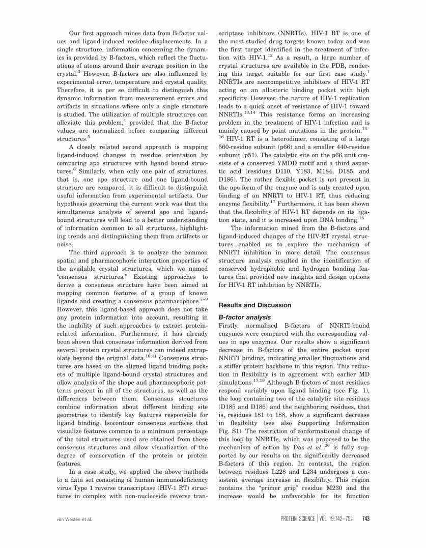
respond variably upon ligand binding (see Fig. 1),
the loop containing two of the catalytic site residues
(D185 and D186) and the neighboring residues, that
is, residues 181 to 188, show a significant decrease
in flexibility (see also Supporting Information
Fig. S1). The restriction of conformational change of
this loop by NNRTIs, which was proposed to be the
mechanism of action by Das et al.,20 is fully sup-
ported by our results on the significantly decreased
B-factors of this region. In contrast, the region
between residues L228 and L234 undergoes a con-
sistent average increase in flexibility. This region
contains the ‘‘primer grip" residue M230 and the
increase would be unfavorable for its function
van Westen et al. PROTEIN SCIENCE VOL 19:742—752 743
retaining the growing DNA strand. A principal com-
ponent analysis (PCA) of the normalized B-factor
distributions over the residues shows that all struc-
tures form a cluster, which shows significant diver-
sity (but no outliers) and that the three apo struc-
tures are nearest neighbors (Supporting Information
Fig. S2). A heat map of the normalized B-factor pro-
files of all structures can be found in Supporting In-
formation Figure S3. To compare our findings to
DNA-bound protein, all crystal structures were com-
pared with three different DNA-bound structures.
However, contrary to the consistent profile observed
in the ligand bound or apo structures the overall B-
factor profile differs between these DNA-bound
structures. Therefore, we do not have enough data
to draw firm conclusions on flexibility changes upon
DNA binding. When we separated all NNRTI-bound
structures into wild type (wt) and mutated struc-
tures a trend was observed that resistance-confer-
ring point mutations lead to a partial restoration of
flexibility of the catalytic site region (Supporting In-
formation Fig. S1). This trend is consistent with the
findings of Zhou et al.17
This type of flexibility information is useful when
implementing protein flexibility in, for example, dock-
ing calculations.21,22 Awareness about the flexible res-
idues enables focusing computational expense on only
these parts of the protein, as still allowing induced fit
to take place to the required degree.
Ligand induced displacement analysis
We next analyzed the residue displacements result-
ing from ligand binding. This analysis confirms that
all NNRTIs induce a similar binding pocket into the
protein when compared to the apo structure. This
holds true for both the backbone carbon alpha atoms
as well as the movement of the center of mass of the
residues (Supporting Information Figs. S4–S11). The
only exception is observed in the Capravirine struc-
ture; however, as we only had one structure avail-
able, we cannot confirm this to be an effect charac-
teristic for the ligand or for this particular crystal
structure. Hence, this structure was removed from
further analysis. Next, the residue center of mass
absolute displacement distances were examined.
Figure 2(A) depicts the pocket backbone colored
according to the values obtained from the calculation
of the displacement upon ligand binding. A PCA
analysis of the displacement vectors shows that the
ligand-bound structures cluster together, even more
than do the apo structures (Supporting Information
Fig. S4), confirming the formation of a common
pocket upon ligand binding.
Summarizing our findings for NNRTI binding,
firstly the shift of the catalytic acid residues in Fig-
ure 2(B) is conserved. Secondly, the known shifts as
a result of the flip of residues Y181, Y188 and the
known upward movement of W229 and M230 are
confirmed for all NNRTIs.23 The entire b12 sheet
(P225–P236) known to be in contact with the grow-
ing DNA template via residue M230,20,24–26 is dis-
placed upward into the DNA binding groove, adapt-
ing to the ligand upon binding. However, the
displacement of the sheet is a rotation pivoting
around residues P226 and L234/H235, which remain
relatively in place. This suggests how the sheet
adapts itself to the size of the bound NNRTI as mov-
ing the primer grip away from the nucleotide
Figure 1. Changes in average B-factor as a result of ligand binding projected on the backbone ribbon. White residues
indicate an increase (�0.2), gray indicates no change (between 0.2 and �0.2), and black indicates a decrease (��0.2)
(PDB:1FK9). (A) The gray volume represents the NNRTI Efavirenz. The entire pocket region has a lower average normalized B-
factor in the ligand-bound compared to the apo form while the profile remains comparable (B). The catalytic residues undergo
a decrease in B-factor (black circles), while the primer grip region containing residue W229 and M230 undergoes an increase
upon ligand binding (dashed black circle). The black bars on the horizontal axis indicate continuous residues, the white filled
bar indicates the residues on the p51 subunit. Each tick marks a separate residue the precise residue numbers can be found
in the materials and methods, p66 and p51 have been separated by a gap.
744 PROTEINSCIENCE.ORG Consensus Structures Applied to HIV-1 RT

binding region. The mutations present in the
mutated RT structures do not appear to have a sig-
nificant influence on residue movement. (For
detailed heat maps indicating the shift per residue
see Supporting Information Figs. S6–S10) Interest-
ingly, the distance measured does not necessarily
correlate with these residues being involved in re-
sistance mutations. However, it is striking that some
of the residues that are detected to move relatively
large distances in all structures are in known loca-
tions for resistance conferring mutations. Because
these residues undergo the largest changes resulting
from NNRTI binding, changes occurring in these
residues are most likely to perturb NNRTI binding.
Upon DNA binding, the position of the catalytic
site loop carrying residues Y181 through Y188 is
consistently shifted (Fig. 3). The loop maintains the
overall conformation suggesting a hinge-like move-
ment during catalysis. Furthermore, the known shift
of the catalytic loop induced by NNRTI binding is in
fact opposite to the path of the downward movement
induced by DNA binding. (Supporting Information
Figs. S6–S11) We compared the shift on each carte-
sian axis as a result of either DNA binding or
NNRTI binding. The figures clearly show that the
catalytic loop and especially residues 184–186 move
in an opposite direction when compared to the apo
structures in both the case of Ca and centroid move-
ment on both the x- and z-axes. In the case of the
y-axis, the result is less pronounced.
NNRTI working mechanismCombining the above findings on the movement and
flexibility of HIV-RT residues upon binding of an
NNRTI allows us to elaborate on previously pro-
posed working mechanisms of NNRTIs. The working
mechanism has been proposed to be either the result
of a catalytic site distortion26–30 or a more rigid pro-
tein.19,29,30 From our flexibility analysis, we observe
that the binding of an NNRTI to HIV-1 RT leads to
a shift in flexibility of residues around the binding
site, with mobile residues become more rigid,
whereas rigid residues becoming more mobile. In
addition, upon NNRTI binding the catalytic triad
residues and their neighboring residues, D110-V111
and Y181-Y188, but also the primer grip region,
M230 and surrounding residues, undergo a consist-
ent displacement opposite to the displacement
Figure 2. The backbone of the NNRTI pocket, colored according to the average residue displacement (PDB:1FK9). Black
indicates a small average displacement, �2 A, gray indicates a medium displacement, between 2 and 4 A, and white
indicates a large displacement �4 A (A). The gray volume represents the NNRTI Efavirenz. Residues Y181, Y188, K223, and
M230 all undergo a relatively large movement upon ligand binding (B, black circles). The residues between L228 and L234 all
undergo a large displacement upon ligand binding, this correlates with the results from the B-factor analysis. The residue axis
is labeled identically to Figure 1.
Figure 3. Overview of the changes occurring at the
catalytic site as a result of DNA binding (three gray
ribbons), NNRTI binding (two black ribbons) compared with
the apo position (two white ribbons). The three aspartic
acids that are visible are part of the DNA bound
conformation (1RTD). In the apo structures, these residue
side chains are placed similarly, however in the NNRTI
bound structures D186 points toward D110, D110 points
outward, and D185 points downward. Upon DNA binding,
the loop containing the residues moves along the path
indicated by the white curve, in the presence of an NNRTI
the loop moves along the path indicated by the dashed
white curve.
van Westen et al. PROTEIN SCIENCE VOL 19:742—752 745
induced by DNA binding. As a result the conforma-
tion of the catalytic loop is distorted, enlarging the
distance between primer grip and the nucleotide
binding site. These changes were found to be con-
sistent among all crystal structures studied. At the
catalytic site, the 4 A average movement of the as-
partic acids is a large distance as the three-dimen-
sional orientation of the catalytic motif and the nu-
cleotide is crucial in catalyzing the reaction.
Mendieta et al.31 have found that the Mg2þ ions sta-
bilize the catalytic complex and lower the catalytic
attack distance to 3 A. These essential Mg2þ ions
were missing in all crystal structures containing an
NNRTI, indicating that the changed orientation of
the aspartic acids might not be able to contain these
ions.
The large movement of the primer grip away
from the catalytic site upon NNRTI binding and the
increase in flexibility are consistent among all struc-
tures. Both of these changes are likely to lead to a
decreased reaction rate as they inhibit the function
of retaining the DNA strand. Therefore, we conclude
that the NNRTIs disrupt both protein conformation
and dynamics and that it is this combination that
inhibits the function of HIV-RT. Thus, we propose a
working mechanism for NNRTIs that is a combina-
tion of both the rigid protein, distorted-catalytic-site
and distorted primer grip region theories. The bound
NNRTI stabilizes the region surrounding the cata-
lytic site in a conformation not able to perform catal-
ysis. The effects on protein conformation and
dynamics cannot be seen independently as one
directly influences the other. Therefore, it could be
speculated that an increase in flexibility as a result
of point mutations allows the primer grip and cata-
lytic site to move closer together restoring catalytic
activity. As a result this could lead to resistance of
the particular HIV-RT mutant form. Our results
support this theory.
When this manuscript was revised, Paris et al.32
published a large scale comparison of NNRTI crystal
structures. Although their superposition is based on
only a subset of residues and we left out the struc-
tures where an NNRTI was soaked out of the pocket,
their main conclusions are generally in line with
ours. Moreover, our results indicate that NNRTI
binding influences not only primer grip movement
but also distortion of the catalytic site and changes
in protein dynamics and that these are all in fact
required for inhibitory activity.
Consensus structures
In the second part of our work on mining informa-
tion from multiple crystal structures, we present the
results from the consensus structures creation. The
difference between two extreme situations of conser-
vation values is visualized in Figure 4. The surface
visualizing low conservation is shown as a green
wire grid. This surface shows all parts of the three-
dimensional space covered by at least 10% of the
protein structures, including different side-chain ori-
entations. This surface features a rather large pro-
tein volume, and consequently the empty pocket vol-
ume in this map is smaller than in the high-
conservation map. The low-conservation surface
thus describes the unity of conformational space ac-
cessible to the protein in any one of the dataset
structures. The surface visualizing high conservation
is shown as a solid green mesh. This surface con-
tains the volume covered by at least 50% of the
structures. It suggests the volume to which the pro-
tein binding pocket can be extended, describing the
most conserved side-chain-occupied space. This vol-
ume does not occur in any of the individual struc-
tures, and this high-conservation surface represents
the largest possible binding pocket a hypothetical
ligand could occupy. In addition, combining high and
low conservation surfaces in one view can be used to
locate regions of flexibility by visually comparing
highly conserved side-chain orientations, where
there is little difference between high- and low-con-
servation surfaces, to side-chains that can move
more freely, showing a large difference between
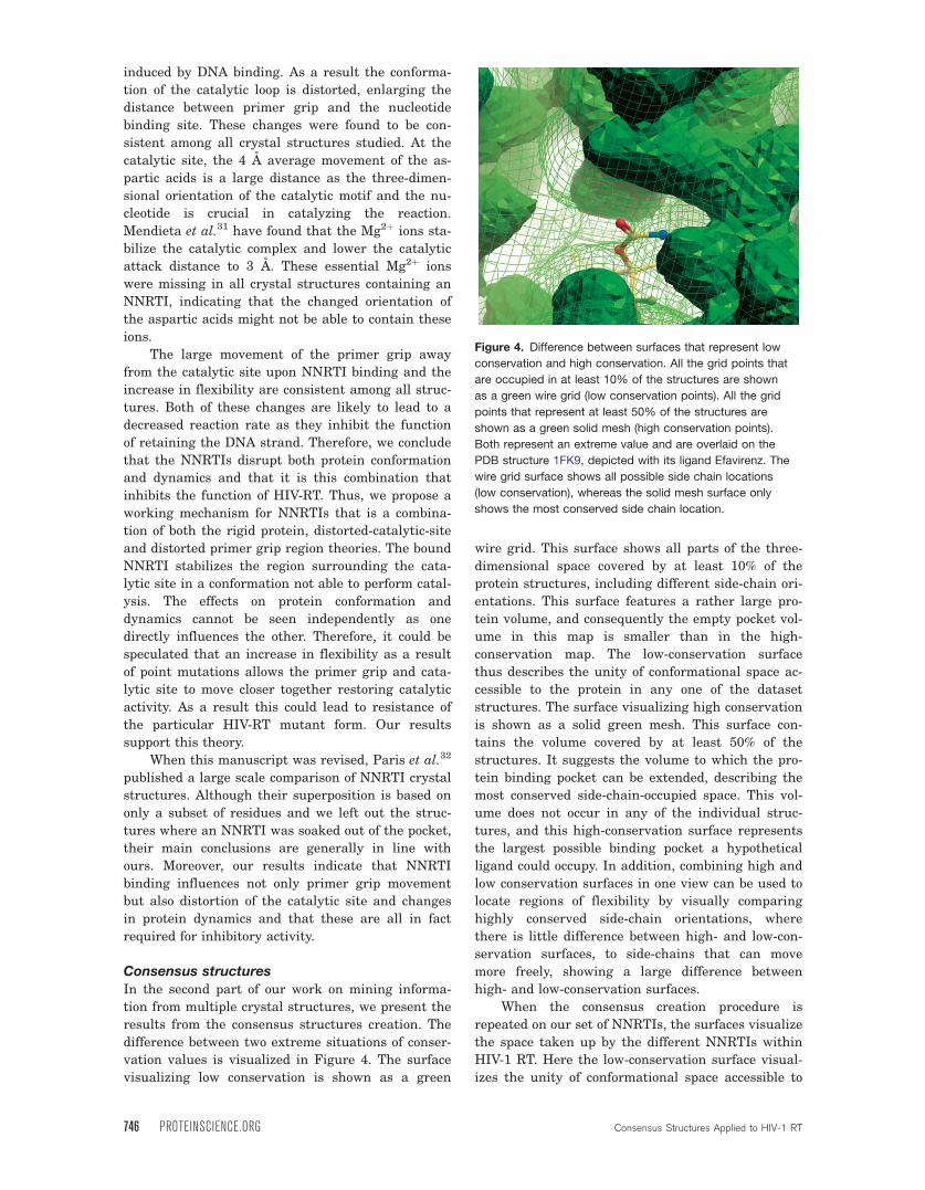
high- and low-conservation surfaces.
When the consensus creation procedure is
repeated on our set of NNRTIs, the surfaces visualize
the space taken up by the different NNRTIs within
HIV-1 RT. Here the low-conservation surface visual-
izes the unity of conformational space accessible to
Figure 4. Difference between surfaces that represent low
conservation and high conservation. All the grid points that
are occupied in at least 10% of the structures are shown
as a green wire grid (low conservation points). All the grid
points that represent at least 50% of the structures are
shown as a green solid mesh (high conservation points).
Both represent an extreme value and are overlaid on the
PDB structure 1FK9, depicted with its ligand Efavirenz. The
wire grid surface shows all possible side chain locations
(low conservation), whereas the solid mesh surface only
shows the most conserved side chain location.
746 PROTEINSCIENCE.ORG Consensus Structures Applied to HIV-1 RT
the NNRTIs in any one of the dataset structures, and
a larger volume than present in any of the individ-
ual structures. The high-conservation surface visu-
alizes the common volume that is used by all
NNRTIs.
Consensus pocket
Van der Waals (VdW) consensus structures can visu-
alize common features present in all structures,
including crystal structures and optionally even
homology models carrying point mutations.33 The
resulting consensus pocket shape can be regarded as
a target shape for high affinity ligands because it
represents the maximal theoretically accessible vol-
ume. As the highly flexible pocket adapts to each dif-
ferent NNRTI and there is no standardized wild-
type pocket, consensus structures can provide this
standardized pocket. Based on the degree of conser-
vation of the surfaces, an estimate can be made of
the space available for NNRTI binding and this can
be related to the actual volume of NNRTIs. From
Table I, it can be concluded that second-generation
NNRTIs, Rilpivirine, Capravirine, and Delavirdine
make better use of the maximal theoretical available
space (66%, 69%, and 75%, respectively). Given our
consensus structures, even larger NNRTIs than
Delavirdine are theoretically possible. Coinciden-
tally, as finalizing this manuscript, this has been
experimentally confirmed by Sweeney et al.34
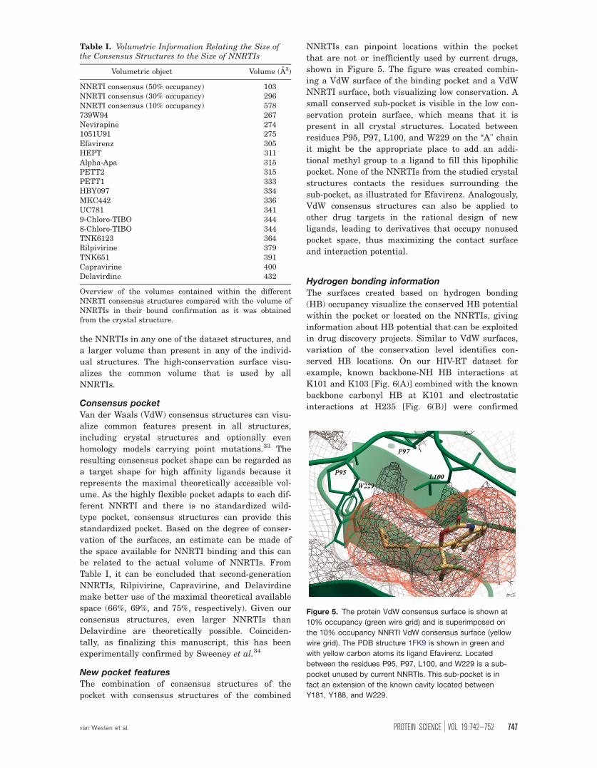
New pocket features
The combination of consensus structures of the
pocket with consensus structures of the combined
NNRTIs can pinpoint locations within the pocket
that are not or inefficiently used by current drugs,
shown in Figure 5. The figure was created combin-
ing a VdW surface of the binding pocket and a VdW
NNRTI surface, both visualizing low conservation. A
small conserved sub-pocket is visible in the low con-
servation protein surface, which means that it is
present in all crystal structures. Located between
residues P95, P97, L100, and W229 on the ‘‘A" chain
it might be the appropriate place to add an addi-
tional methyl group to a ligand to fill this lipophilic
pocket. None of the NNRTIs from the studied crystal
structures contacts the residues surrounding the
sub-pocket, as illustrated for Efavirenz. Analogously,
VdW consensus structures can also be applied to
other drug targets in the rational design of new
ligands, leading to derivatives that occupy nonused
pocket space, thus maximizing the contact surface
and interaction potential.
Hydrogen bonding information
The surfaces created based on hydrogen bonding
(HB) occupancy visualize the conserved HB potential
within the pocket or located on the NNRTIs, giving
information about HB potential that can be exploited
in drug discovery projects. Similar to VdW surfaces,
variation of the conservation level identifies con-
served HB locations. On our HIV-RT dataset for
example, known backbone-NH HB interactions at
K101 and K103 [Fig. 6(A)] combined with the known
backbone carbonyl HB at K101 and electrostatic
interactions at H235 [Fig. 6(B)] were confirmed
Table I. Volumetric Information Relating the Size ofthe Consensus Structures to the Size of NNRTIs
Volumetric object Volume (A3)
NNRTI consensus (50% occupancy) 103NNRTI consensus (30% occupancy) 296NNRTI consensus (10% occupancy) 578739W94 267Nevirapine 2741051U91 275Efavirenz 305HEPT 311Alpha-Apa 315PETT2 315PETT1 333HBY097 334MKC442 336UC781 3419-Chloro-TIBO 3448-Chloro-TIBO 344TNK6123 364Rilpivirine 379TNK651 391Capravirine 400Delavirdine 432
Overview of the volumes contained within the differentNNRTI consensus structures compared with the volume ofNNRTIs in their bound confirmation as it was obtainedfrom the crystal structure.
Figure 5. The protein VdW consensus surface is shown at
10% occupancy (green wire grid) and is superimposed on
the 10% occupancy NNRTI VdW consensus surface (yellow
wire grid). The PDB structure 1FK9 is shown in green and
with yellow carbon atoms its ligand Efavirenz. Located
between the residues P95, P97, L100, and W229 is a sub-
pocket unused by current NNRTIs. This sub-pocket is in
fact an extension of the known cavity located between
Y181, Y188, and W229.
van Westen et al. PROTEIN SCIENCE VOL 19:742—752 747

using the consensus structures. Furthermore, a con-
served carbonyl HB acceptor not used by NNRTIs
can be identified at K102, although this acceptor is
accessible in only a few of the ligand bound struc-
tures. Most interestingly, we found that large
NNRTIs that lead to a large shift of the b12-sheet
(P225–P236) break the HB between backbone-NH at
K103 and backbone carbonyl at P236. Although the
backbone-NH at K103 has been described to partici-
pate in HB interactions to NNRTIs,35 the concur-
rently available carbonyl at P236 has not. Use of
this HB acceptor could provide an additional back-
bone interaction to an NNRTI, which might lead to
an improved resistance profile. As some viral strains
carry a K101P mutation, which does not allow the
backbone-NH HB to be formed,36 it is of high impor-
tance to identify additional common interaction sites
in the NNRTI pocket that can be exploited. Although
a P236L mutant has been identified, this will not
change the potential for a backbone HB interaction.
Additionally, this mutant has been described as hav-
ing low replication fitness.37
Materials and methods
Dataset
A total of 47 crystal structures were used in our
analysis (Table II). Crystal structures were obtained
from the PDB and grouped according to bound
NNRTI. The selection included several structures of
the NNRTIs approved for clinical use, namely,
Nevirapine, Efavirenz, and Delavirdine. Several
structures of apo HIV-1 RT have been used (Group
a) three of which contain a bound DNA fragment
(Group b). Apo structures 1JLE and 1RTJ were
obtained by soaking out the NNRTI and are, there-
fore, not native apo structures. Accordingly, in an
analysis of the structures, the orientation of the
backbone was found to show more similarities with
ligand-bound forms of HIV-RT than with the apo
form. For this reason, structures 1JLE and 1RTJ
were omitted from our dataset. Out of the selected
structures several contained a point mutation in the
NNRTI pocket. From Groups a and c in Table II B-
factors were extracted in Molsoft ICM.38 The consen-
sus structures were created from a sub-selection of
all 40 NNRTI-bound crystal structures, including 23
different NNRTIs (Group c, Table II). For all
NNRTIs, the charge at pH 7, was calculated using
the Marvin Beans pKa prediction tool by Chem-
Axon,39 and all were found to be uncharged.
Computational detailsAll structural experiments and visualizations were
performed using ICM. In all structures the NNRTIs
were checked for inconsistencies and the bond orders
were confirmed (A workflow of the performed experi-
ments is given in Supporting Information Fig. S12).
A multiple sequence alignment was created within
ICM using default options, verifying that no muta-
tions were present around the NNRTI pocket other
than the known single mutations within the PDB
structures. Hydrogens were added to the PDB by
ICM object conversion, which contains hydrogen
bond optimization, protonation state optimization of
His residues, and rotamer optimization of Asn, Gln,
and His residues. Subsequently, all RT crystal struc-
tures were superimposed based on alignment of the
backbone atoms of selected residues. This selection
was made using a 12 A sphere around the largest
NNRTI, Dlv, in PDB structure 1KLM. This included
the following residues on chain A: 90–111, 161, 164,
168, 171, 172, 175–196, 198, 199, 201, 205, 222–240,
242, 315–321, 343, 348–350 and on chain B: 135–
140. Visualizations and calculations were performed
using the superimposed structures.
Figure 6. Consensus surfaces visualizing all conserved HB donors surrounding the pocket (A) and all conserved HB
acceptors (B) among the selection of crystal structures. For reasons of clarity, only surfaces visualizing 20% occupancy are
shown. The three dimensional surfaces are superimposed on the PDB structure 2ZD1, the green ribbon indicating the
backbone, and Rilpivirine depicted by yellow carbons.
748 PROTEINSCIENCE.ORG Consensus Structures Applied to HIV-1 RT

B-factor analysis
The influence of experimental error and conditions
was minimized by normalizing the B-factors of every
residue in the crystal structures similar to Yuan
et al.,5 where we used full residue B-factors instead
of Ca-only B-factors. This normalization uses the
standard deviation over all B-factors per structure
to allow a comparison of the values of different crys-
tal structures.5 After normalization, the B-factors
from all ligand bound structures were combined and
their mean value per residue was calculated, the
same was done for the apo structures. Because of
the very high temperature factors, structures 1HMV
and 2ZE2 were omitted from these experiments. The
average difference values between apo and ligand
bound normalized B-factors per residue were binned
Table II. Summary of the PDB Structures that were Used in all Analyses
PDB Code Group Mutation Drug Class Resolution(A)
1DLO a A172K n/l Apo 2.701HMV a n/p n/l Apo 3.201HQE a K103N n/l Apo 2.701QE1 a M184I n/l Apo 2.851N6Q b n/p n/l DNA Bound 3.001RTD b n/p n/l DNA Bound 3.202HMI b n/p n/l DNA Bound 2.801RTH c n/p 1051U91 1051U91 2.201JLQ c n/p 739W94 739W94 3.001VRU c n/p Alpha-Apa Alpha-Apa 2.401EP4 c n/p Capravirine Capravirine 2.502ZD1 c n/p Rilpivirine DAPY 1.802ZE2 c L100I/K103N Rilpivirine DAPY 2.903BGR c K103N/Y181C Rilpivirine DAPY 2.101KLM c n/p Delavirdine Delavirdine 2.651FK9 c n/p Efavirenz Efavirenz 2.501FKO c K103N Efavirenz Efavirenz 2.901IKW c n/p Efavirenz Efavirenz 3.001JKH c Y181C Efavirenz Efavirenz 2.501BQM c n/p HBY097 HBY097 3.101C1C c n/p TNK6123 HEPT 2.501JLA c Y181C TNK651 HEPT 2.501RT1 c n/p MKC442 HEPT 2.551RT2 c n/p TNK651 HEPT 2.551RTI c n/p HEPT HEPT 3.001FKP c K103N Nevirapine Nevirapine 2.901JLB c Y181C Nevirapine Nevirapine 3.001JLF c Y188C Nevirapine Nevirapine 2.601S1U c L100I Nevirapine Nevirapine 3.001S1X c V108I Nevirapine Nevirapine 2.801VRT c n/p Nevirapine Nevirapine 2.202HND c K101E Nevirapine Nevirapine 2.502HNY c E138K Nevirapine Nevirapine 2.501DTQ c n/p PETT1 PETT 2.801DTT c n/p PETT2 PETT 3.001JLC c Y181C PETT2 PETT 3.001HNV c n/p 8TIBO TIBO 3.001REV c n/p 9TIBO TIBO 2.601TVR c n/p 9TIBO TIBO 3.001UWB c Y181C 8TIBO TIBO 3.201JLG c Y188C UC781 UC 2.601RT4 c n/p UC781 UC 2.901RT5 c n/p UC10 UC 2.901RT6 c n/p UC38 UC 2.801RT7 c n/p UC84 UC 3.001S1T c L100I UC781 UC 2.401S1W c V106A UC781 UC 2.70
The table shows a summary of the PDB structures that were used in the analyses. All structures were subdivided intothree groups: a, which contained apo-enzymes, b, which contained apo enzymes bound to a DNA fragment, and c, whichcontained NNRTI bound structures. Furthermore, mutations present in the NNRTI binding pocket were identified, if nonewere present ‘‘n/p’’ was used. Structures marked with ‘‘n/l’’ did not contain an NNRTI. Finally, all NNRTIs were subdividedinto 13 classes as shown in the table.
van Westen et al. PROTEIN SCIENCE VOL 19:742—752 749
in three classes: lower (��0.2), virtually unchanged
(between �0.2 and 0.2), and higher (�0.2).
Ligand induced displacement analysis
Ligand-induced conformational changes were char-
acterized in three ways. Firstly, for each of the
superimposed structures a scalar representing the
mean distance for each pocket residue between the
centroid in the ligand-bound conformation and the
centroid of the two apo structures was determined.
The obtained average centroid displacement distan-
ces were binned into three classes: small (�2 A), me-
dium (between 2 A and 4 A), and large (�4 A). Sec-
ondly, the three-dimensional displacement vector
between residue Ca positions in the ligand-bound
conformation and the apo structures was split up
over all three Cartesian axes to determine the back-
bone movement on each specific axis. Thirdly, the
procedure was repeated between residue centroid
positions in the ligand-bound conformation and the
apo structures. Thus, the movement of the backbone
and the side chains were both taken into account as
well as changes in orientation of side chains (which
are known to be relatively independent from back-
bone movements).40
Conversion of crystal structures to
three-dimensional occupancy maps
The crystal structures were converted to three-
dimensional occupancy maps by placing the struc-
tures in a grid box. A VdW occupancy value was
assigned to each grid point and was normalized to a
value between 0 and 1 using the internal auto trim
function of ICM. This quasi-binary scaling enabled a
comparison between occupancy maps obtained from
different crystal structures.38 For HB maps, the val-
ues were scaled between 0 and 1 for acceptors and
between 0 and �1 for donors, similar to the VdW
scaling.
Creation of consensus potentials and structures
Consensus structures were created from the initial
superpositions by adding up the individual occu-
pancy maps of the set of crystal structures. This
step was performed separately for the RT binding
pocket residues and the bound NNRTI structures.
When several structures containing the same
NNRTI were present, the average value of all struc-
tures containing that specific NNRTI was used.
Thereby each NNRTI class contributed equally to
the final occupancy map and the domination of a
single class of NNRTIs over the others was avoided.
Occupancy maps for the set of crystal structures
were created for VdW, HB donors and HB acceptors,
from the occupancy maps of each individual struc-
ture and named consensus structures. Isocontour
surfaces were created at different levels, correspond-
ing to visualization of different degrees of
conservation.
Conclusion
In conclusion, both methods presented in this work,
the analysis of B-factors and ligand induced residue
displacement as well as the analysis of steric and
pharmacophoric properties from multiple crystal
structures show how the growing number of crystal
structures available can be mined efficiently to gen-
erate novel hypotheses for lead optimization.
Although the amount of information available in
these analyses increases with the number of struc-
tures included, the use of a handful of structures
can already provide insights. Our findings were also
supported by novel, more sterically demanding
NNRTIs that were published by Sweeney et al. just
as this manuscript had been finalized.
Our methods facilitated the identification of the
working mechanism of NNRTIs as a combination of
two mechanisms that were previously suggested (the
‘‘molecular arthritis’’ and ‘‘distorted catalytic site’’
hypotheses). In addition, our consensus structures
were able to extract conserved locations of interest
from the crystal structures without the need for mo-
lecular dynamics. The different types of consensus
structures can complement each other and provide a
useful overview of the interaction between a class of
compounds and its target protein. Using this
method, we identified a novel backbone hydrogen
bond acceptor at P236 and a novel hydrophobic
subpocket.
Acknowledgments
We thank Andrew Orry (Molsoft L.L.C), Anik Peeters,
Luc Geeraert, Ann Vos, and Carlo Boutton (Tibotec-
Virco BVBA) for their helpful discussions. A.B. was
funded by the Dutch Top Institute Pharma, project
number: 01-105.
References
1. Berman HM, Westbrook J, Feng Z, Gilliland G, BhatTN, Weissig H, Shindyalov IN, Bourne PE (2000) Theprotein data bank.Nucleic Acids Res 28:235–242.
2. Knegtel RM, Kuntz ID, Oshiro CM (1997) Moleculardocking to ensembles of protein structures.J Mol Biol266:424–440.
3. Yuan Z, Bailey TL, Teasdale RD (2005) Prediction ofprotein B-factor profiles.Proteins 58:905–912.
4. Wampler JE (1997) Distribution analysis of the varia-tion of B-factors of X-ray crystal structures; tempera-ture and structural variations in lysozyme.J Chem InfComput Sci 37:1171–1180.
5. Yuan Z, Zhao J, Wang Z-X (2003) Flexibility analysis ofenzyme active sites by crystallographic temperaturefactors.Protein Eng 16:109–114.
6. Keller PA, Leach SP, Luu TTT, Titmuss SJ, Griffith R(2000) Development of computational and graphicaltools for analysis of movement and flexibility in largemolecules.J Mol Graph Model 18:235–241, 299.
750 PROTEINSCIENCE.ORG Consensus Structures Applied to HIV-1 RT

7. Richmond N, Abrams C, Wolohan P, Abrahamian E, Wil-lett P, Clark R (2006) GALAHAD: 1. Pharmacophoreidentification by hypermolecular alignment of ligands in3D.J Comput-Aided Mol Des 20:567–587.
8. Totrov M (2008) Atomic property fields: generalized 3Dpharmacophoric potential for automated ligand super-position, pharmacophore elucidation and 3DQSAR.Chem Biol Drug Des 71:15–27.
9. O’Brien SE, Brown DG, Mills JE, Phillips C, Morris G(2005) Computational tools for the analysis and visual-ization of multiple protein-ligand complexes.J MolGraph Model 24:186–194.
10. Powers RA, Shoichet BK (2002) Structure-basedapproach for binding site identification on AmpC beta-lactamase.J Med Chem 45:3222–3234.
11. Nichols SE, Domaoal RA, Thakur VV, Tirado-Rives J,Anderson KS, Jorgensen WL (2009) Discovery of wild--type and Y181C mutant non-nucleoside HIV-1 reversetranscriptase inhibitors using virtual screening withmultiple protein structures.J Chem Inf Model 49:1272–1279.
12. Mitsuya H, Weinhold KJ, Furman PA,St Clair MH,Lehrman SN, Gallo RC, Bolognesi D, Barry DW,Broder S (1985) 30-Azido-30-deoxythymidine (BWA509U): an antiviral agent that inhibits the infectivityand cytopathic effect of human T-lymphotropic virustype III/lymphadenopathy-associated virus in vitro.ProcNatl Acad Sci USA 82:7096–7100.
13. Richman DD, Havlir D, Corbeil J, Looney D, Ignacio C,Spector SA, Sullivan J, Cheeseman S, Barringer K,Pauletti D (1994) Nevirapine resistance mutations ofhuman immunodeficiency virus type 1 selected duringtherapy.J Virol 68:1660–1666.
14. Havlir DV, Eastman S, Gamst A, Richman DD (1996)Nevirapine-resistant human immunodeficiency virus:kinetics of replication and estimated prevalence inuntreated patients.J Virol 70:7894–7899.
15. Rhee S-Y, Fessel WJ, Zolopa AR, Hurley L, Liu T, Tay-lor J, Nguyen DP, Slome S, Klein D, Horberg M,Flamm J, Follansbee S, Schapiro JM, Shafer RW(2005) HIV-1 protease and reverse-transcriptase muta-tions: correlations with antiretroviral therapy in sub-type B isolates and implications for drug-resistancesurveillance.J Infect Dis 192:456–465.
16. Johnson V, Brun-Vezinet F, Clotet B, Gunthard H, Kur-itzkes D, Pillay D, Schapiro J, Richman D (2008)Update of the drug resistance mutations in HIV-1:December 2008.Top HIV Med 16:138–145.
17. Zhou Z, Madrid M, Evanseck JD, Madura JD (2005)Effect of a bound non-nucleoside RT inhibitor on thedynamics of wild-type and mutant HIV-1 reverse tran-scriptase.J Am Chem Soc 127:17253–17260.
18. Madrid M, Lukin JA, Madura JD, Ding J, Arnold E(2001) Molecular dynamics of HIV-1 reverse transcrip-tase indicates increased flexibility upon DNA binding.-Proteins 45:176–182.
19. Bahar I, Erman B, Jernigan RL, Atilgan AR, CovellDG (1999) Collective motions in HIV-1 reverse tran-scriptase: examination of flexibility and enzyme func-tion.J Mol Biol 285:1023–1037.
20. Das K, Sarafianos SG, Clark AD, Jr, Boyer PL, HughesSH, Arnold E (2007) Crystal structures of clinically rel-evant Lys103Asn/Tyr181Cys double mutant HIV-1reverse transcriptase in complexes with ATP and non--nucleoside Inhibitor HBY 097.J Mol Biol 365:77–89.
21. Carlson HA (2002) Protein flexibility and drug design:how to hit a moving target.Curr Opin Chem Biol 6:447–452.
22. Carlson HA, McCammon JA (2000) Accommodatingprotein flexibility in computational drug design.MolPharmacol 57:213–218.
23. Hsiou Y, Ding J, Das K, Clark AD, Jr, Hughes SH,Arnold E (1996) Structure of unliganded HIV-1 reversetranscriptase at 2.7 A resolution: implications of confor-mational changes for polymerization and inhibitionmechanisms.Structure 4:853–860.
24. Das K, Bauman JD, Clark AD, Jr, Frenkel YV, LewiPJ, Shatkin AJ, Hughes SH, Arnold E (2008) High-re-solution structures of HIV-1 reverse transcriptase/TMC278 complexes: strategic flexibility explains po-tency against resistance mutations.Proc Natl Acad SciUSA 105:1466–1471.
25. Rodriguez-Barrios F, Balzarini J, Gago F (2005) Themolecular basis of resilience to the effect of theLys103Asn mutation in non-nucleoside HIV-1 reversetranscriptase inhibitors studied by targeted moleculardynamics simulations.J Am Chem Soc 127:7570–7578.
26. Maga G, Radi M, Zanoli S, Manetti F, Canciano R,Hubscher U, Spadari S, Falciani C, Terrazas M,Vilarrasa J, Botta M (2007) Discovery of non-nucleosideinhibitors of HIV-1 reverse transcriptase competingwith the nucleoside substrate.Angew Chem Int EdEngl 46:1810–1813.
27. Esnouf R, Ren J, Ross C, Jones Y, Stammers D, StuartD (1995) Mechanism of inhibition of HIV-1 reversetranscriptase by non-nucleoside inhibitors.Nat StructBiol 2:303–308.
28. Balzarini J (2004) Current status of the non-nucleosidereverse transcriptase inhibitors of human immunodefi-ciency virus type 1.Curr Top Med Chem 4:921–944.
29. Kohlstaedt LA, Wang J, Friedman JM, Rice PA, SteitzTA (1992) Crystal structure at 3.5 A resolution ofHIV-1 reverse transcriptase complexed with an inhibi-tor.Science 256:1783–1790.
30. Sarafianos SG, Marchand B, Das K, Himmel DM, Par-niak MA, Hughes SH, Arnold E (2009) Structure andfunction of HIV-1 reverse transcriptase: molecularmechanisms of polymerization and inhibition.J MolBiol 385:693–713.
31. Mendieta J, Cases-Gonzalez CE, Matamoros T, Ram-irez G, Menendez-Arias L (2008) A Mg2þ-induced con-formational switch rendering a competent DNApolymerase catalytic complex.Proteins 71:565–574.
32. Paris KA, Haq O, Felts AK, Das K, Arnold E, Levy RM(2009) Conformational landscape of the human immu-nodeficiency virus type 1 reverse transcriptase non-nu-cleoside inhibitor binding pocket: lessons for inhibitordesign from a cluster analysis of many crystal structur-es.J Med Chem 52:6413–6420.
33. Wegner JK, Vlijmen HV, Boutton CWM (2008) Pheno-type prediction method. PatentWO2008065180A1;EP2007063047.
34. Sweeney ZK, Harris SF, Arora N, Javanbakht H, Li Y,Fretland J, Davidson JP, Billedeau JR, Gleason SK,Hirschfeld D, Kennedy-Smith JJ, Mirzadegan T, RoetzR, Smith M, Sperry S, Suh JM, Wu J, Tsing S, Villase-nor AG, Paul A, Su G, Heilek G, Hang JQ, Zhou AS,Jernelius JA, Zhang F-J, Klumpp K (2008) Design ofannulated pyrazoles as inhibitors of HIV-1 reversetranscriptase.J Med Chem 51:7449–7458.
35. Wang Z, Wu B, Kuhen KL, Bursulaya B, Nguyen TN,Nguyen DG, He Y (2006) Synthesis and biological eval-uations of sulfanyltriazoles as novel HIV-1 non-nucleo-side reverse transcriptase inhibitors.Bioorg Med ChemLett 16:4174–4177.
36. Parkin NT, Gupta S, Chappey C, Petropoulos CJ(2006) The K101P and K103R/V179D mutations in
van Westen et al. PROTEIN SCIENCE VOL 19:742—752 751

human immunodeficiency virus type 1 reverse tran-scriptase confer resistance to nonnucleoside reversetranscriptase inhibitors.Antimicrob Agents Chemother50:351–354.
37. Gerondelis P, Archer RH, Palaniappan C, ReichmanRC, Fay PJ, Bambara RA, Demeter LM (1999) TheP236L delavirdine-resistant human immunodeficiencyvirus type 1 mutant is replication defective and demon-
strates alterations in both RNA 50-end- and DNA30-end-directed RNase H activities.J Virol 73:5803–5813.
38. ICM, Version 3.6d. La Jolla: Molsoft L.L.C.; 2009.39. Marvin Beans pKa Prediction tool, Version 4.1. Buda-
pest: ChemAxon, 2006.40. Najmanovich R (2000) Side-chain flexibility in proteins
upon ligand binding.Proteins 39:261–268.
752 PROTEINSCIENCE.ORG Consensus Structures Applied to HIV-1 RT