Miniature atmospheric pressure glow discharge torch (APGD-t) for local biomedical applications
10
1147 Pure Appl. Chem., Vol. 78, No. 6, pp. 1147–1156, 2006. doi:10.1351/pac200678061147 © 2006 IUPAC Miniature atmospheric pressure glow discharge torch (APGD-t) for local biomedical applications* S. Coulombe ‡ , V. Léveillé, S.Yonson, and R. L. Leask Department of Chemical Engineering, McGill University, Montréal, Québec, Canada Abstract: The operating parameters of a miniature atmospheric pressure glow discharge torch (APGD-t) are optimized for the production of excited atomic oxygen, and the effect of the plasma jet on endothelial cells grown in Petri dishes is studied. We first demonstrate the im- portance of accounting for the effect of the voltage probe used to measure the electrical pa- rameters of the torch on its ignition and operation characteristics. When operated with a main plasma gas flow rate of 1 SLM He and a power level of ~1 W, the torch shows an optimum in the production of excited atomic oxygen for a O 2 flow of ~3.5 SCCM injected downstream from the plasma-forming region through a capillary electrode (i.e., 0.35 v/v % O 2 /He). It is shown that endothelial cells are detached from the Petri dishes surface under the action of the optimized plasma jet and that this effect does not originate from heating and fluid shearing effects. It is postulated that the cell detachment is caused solely by plasma-induced bio- chemical processes taking place at the cell–substrate interface. Keywords: plasma torch; APGD; nonthermal plasma; glow discharge; biomedical applica- tions. INTRODUCTION Nonthermal (“cold”) atmospheric pressure plasma sources offer efficient means for the production of chemically active radicals under low thermal loading conditions [1]. The ability of these devices to op- erate outside vacuum chambers not only makes the overall operation and installation costs lower, but permits the treatment of mechanically sensitive materials, such as biomaterials and human tissues. Various novel biomedical applications have been explored in the recent past, namely, cell detachment (with or without necrosis) [2], sterilization [3], surface functionalization and patterning [4], and film deposition. Most of those novel applications address large surface-areas, though a local treatment ca- pability is also sought. In the former case, the research and development efforts concentrate mostly on large-area, planar uniform glow discharge sources. So far, uniform glow discharges at atmospheric pressure have been observed only in He, Ar, and N 2 , with minute amounts of contaminants added (less than a few v/v %). In the latter case, plasma “spots” and reduced dimensions plasma torch configura- tions are exploited, and the glow discharge regime is also sought since it provides the best excitation conditions. *Paper presented at the 17 th International Symposium on Plasma Chemistry (ISPC 17), Toronto, Ontario, Canada, 7–12 August 2005. Other presentations are published in this issue, pp. 1093–1298. ‡ Corresponding author
-
Upload
independent -
Category
Documents
-
view
0 -
download
0
Transcript of Miniature atmospheric pressure glow discharge torch (APGD-t) for local biomedical applications
1147
Pure Appl. Chem., Vol. 78, No. 6, pp. 1147–1156, 2006.doi:10.1351/pac200678061147© 2006 IUPAC
Miniature atmospheric pressure glow dischargetorch (APGD-t) for local biomedicalapplications*
S. Coulombe‡, V. Léveillé, S. Yonson, and R. L. Leask
Department of Chemical Engineering, McGill University, Montréal, Québec,Canada
Abstract: The operating parameters of a miniature atmospheric pressure glow discharge torch(APGD-t) are optimized for the production of excited atomic oxygen, and the effect of theplasma jet on endothelial cells grown in Petri dishes is studied. We first demonstrate the im-portance of accounting for the effect of the voltage probe used to measure the electrical pa-rameters of the torch on its ignition and operation characteristics. When operated with a mainplasma gas flow rate of 1 SLM He and a power level of ~1 W, the torch shows an optimumin the production of excited atomic oxygen for a O2 flow of ~3.5 SCCM injected downstreamfrom the plasma-forming region through a capillary electrode (i.e., 0.35 v/v % O2/He). It isshown that endothelial cells are detached from the Petri dishes surface under the action of theoptimized plasma jet and that this effect does not originate from heating and fluid shearingeffects. It is postulated that the cell detachment is caused solely by plasma-induced bio-chemical processes taking place at the cell–substrate interface.
Keywords: plasma torch; APGD; nonthermal plasma; glow discharge; biomedical applica-tions.
INTRODUCTION
Nonthermal (“cold”) atmospheric pressure plasma sources offer efficient means for the production ofchemically active radicals under low thermal loading conditions [1]. The ability of these devices to op-erate outside vacuum chambers not only makes the overall operation and installation costs lower, butpermits the treatment of mechanically sensitive materials, such as biomaterials and human tissues.Various novel biomedical applications have been explored in the recent past, namely, cell detachment(with or without necrosis) [2], sterilization [3], surface functionalization and patterning [4], and filmdeposition. Most of those novel applications address large surface-areas, though a local treatment ca-pability is also sought. In the former case, the research and development efforts concentrate mostly onlarge-area, planar uniform glow discharge sources. So far, uniform glow discharges at atmosphericpressure have been observed only in He, Ar, and N2, with minute amounts of contaminants added (lessthan a few v/v %). In the latter case, plasma “spots” and reduced dimensions plasma torch configura-tions are exploited, and the glow discharge regime is also sought since it provides the best excitationconditions.
*Paper presented at the 17th International Symposium on Plasma Chemistry (ISPC 17), Toronto, Ontario, Canada, 7–12 August2005. Other presentations are published in this issue, pp. 1093–1298.‡Corresponding author
A few academic and industrial research groups have developed small-size plasma sources, whosecharacteristics are suitable for biomedical applications. The “Plasma Needle” developed by Stoffels andcolleagues [5] is a unipolar radio frequency (RF) (13.56 MHz) plasma source that uses helium as themain plasma gas and the surrounding environment as the source of reactive species. It produces a“plasma spot” of ~1 mm in diameter at power levels on the order of a few hundred mW. Preliminarytests showed the ability of this novel plasma source to locally deactivate bacteria [6] but more interest-ingly, to cause cell detachment without necrosis [2]. The exact physical and/or biochemical mechanismsresponsible for such effects are still unclear, though the participation of UV photons and active speciessuch as excited O2, radicals O• and OH• is suspected. The main limitations of the plasma needle are re-lated to the fact that one cannot easily supply the source of reactive species to the plasma-forming zone,and to the inherent electrical coupling with the substrate being treated. Considering those two limita-tions, the plasma torch configuration offers great promise. Stonies et al. [7] scaled down the microwave(2.45 GHz) plasma torch (MPT) originally developed by Bilgiç et al. [8] to power levels as low as 2 W.A particularly interesting feature of the original design is the use of a concentric central electrode,which permits the injection of the analytes downstream from the plasma-forming region (He or Ar asplasma-forming gas) and thus, to operate under stable conditions regardless of the nature of this addi-tional gas. Though the intended application for this low-power MPT is as an excitation source foratomic emission spectroscopy, the small size of the jet (~2 mm length) combined with the low powerlevel and the downstream injection capability make this torch potentially appealing as a biomedical tool.Some small-scale and portable atmospheric pressure plasma sources have already been commercializedas biomedical tools. Amongst the most interesting ones we find the Plasma Skin Regeneration (PSR)device from Rhytec, Inc. [9], the Cold Plasma Coagulation (CPC) device from Söring [10], and thePlasmaPenTM from PVA Tepla, Inc. [11].
We recently developed a miniature RF (13.56 MHz) atmospheric pressure glow discharge plasmatorch, the so-called APGD-t, whose characteristics are well suited for biomedical applications [12,13].In the present article, we report on the main characteristics of the APGD-t, on the optimization of theO2/He ratio for the maximum production of O radicals, and preliminary experiments on cell detachmentand removal.
CONSTRUCTION AND MAIN CHARACTERISTICS OF THE APGD-t
Figure 1 shows a schematic and a picture of the miniature APGD-t. A detailed description is presentedin [13]. In essence, the APGD-t construction consists of a quartz confinement tube (i.d. = 2 mm, o.d. =4 mm), a central stainless steel capillary electrode (i.d. = 0.18 mm, o.d. = 0.36 mm), and a 2.5-cm-longsilver epoxy paint deposited on the external surface of the quartz tube serving as ground electrode. Themain plasma gas (the plasma-forming gas) is fed in the annulus region delimited by the capillary elec-trode and the quartz tube, while the source of reactive gaseous species is injected through the centralcapillary electrode. Such a design feature allows the decoupling of the plasma-forming region from thereactive species production and excitation region and, thus, the consistent and optimum operation of thetorch independent of the nature of the source of reactive species. It is suspected that the electron andlong-lived metastable atoms produced in the plasma-forming region will act as excitation sources in theafterglow region (i.e., downstream of the plasma-forming region), resulting in a depletion of their re-spective density. The downstream end of the quartz tube is tapered in order to form a convergent noz-zle (i.d. at throat = 500 µm) used to accelerate the plasma stream and form a laminar afterglow plasmajet. The source of reactive species is injected in the nozzle area where the shear stresses are highest (bestmixing conditions), and where the afterglow begins. The RF (13.56 MHz) power delivered to the torchis amplitude-modulated using a variable-duty-cycle, square-wave function generator. A homemade in-ductor of 6.3 µH mounted in series between the RF amplifier and the torch is used as impedance match-ing element. The power delivered to the torch is determined through careful electric probe measure-ments of the circuit current, torch voltage, and phase angle between both signals. Mass flow controllers
S. COULOMBE et al.
© 2006 IUPAC, Pure and Applied Chemistry 78, 1147–1156
1148

are used to regulate the main plasma gas flow rate (He, 0.5 to 1.5 SLM) and the gas flow rate throughthe capillary electrode (O2, 0–50 SCCM). Power level modulation over the 1–5 W range is achieved byvarying the pulse duty cycle from 10 to 50 %.
The jet temperature at the nozzle exit ranges from 35 to 120 °C over this power range. The break-down voltage in 1 SLM He is low, ~220 V (from zero to peak voltage), thanks in part to the geometricalamplification of the electric field around the central capillary electrode. Comparatively, the AC break-down voltage one estimates by extrapolation of the Paschen curve for parallel plate arrangement is∼360 V (P = 1 atm, d = 1.8 mm) [13]. The power density ranges from 15 to 75 W/cm3 (~80 mm3 plasmavolume) while the reduced energy loading ranges from 60 to 300 J/L. The APGD-t can thus be classi-fied as a relatively high energy density pulsed RF glow discharge [14].
Figure 2 (top) shows a telemicroscopic image of the He plasma jet (~1 W, 1 SLM, no O2 in thecapillary) discharging into ambient air. The characteristic bright central cone is associated with the Heemission while the faint plume surrounding this cone is associated with the excitation of entrained N2and O2 molecules. The axial distribution of the emission lines suggests the occurrence of several im-portant mechanisms. The presence of the strong Hα line along with the N2 lines suggests a significantentrainment and penetration of (humid) air in the jet immediately at the nozzle exit. The emission fromN2
+ (391 nm) suggests that He metastable atoms (Hem) are present far downstream. The faster decayof the He (587 nm) line intensity with respect to the N2
+ (391 nm) line suggests that the electron den-sity decays at a much faster rate than the Hem density. Those metastable species are thus suspected toact as the excitation source far downstream, where the electron-induced excitation has vanished. Theaxial distribution of the N2 (337 nm) emission profile is, on the other hand, puzzling since one wouldexpect this emission line profile to peak near the nozzle, as it is the case for N2
+. This observation mightbe experimental evidence for the formation of larger nitrogen molecules in the high-pressure glow re-gion (i.e., N4). Such a hypothesis needs to be further investigated using numerical simulations.
© 2006 IUPAC, Pure and Applied Chemistry 78, 1147–1156
Plasma sources 1149
Fig. 1 Schematic of the APGD-t (left) and picture (right) taken with He (1 SLM) at ~1 W.

IGNITION AND STABILIZATION
By its construction, the APGD-t is a concentric-electrode capacitor of capacitance value approximatelyequal to CT
cold~1.5 pF in the absence of the plasma (“cold” state). The APGD-t thus represents a highcapacitive impedance load (XC = –(2πf CT
cold)–1 = –7824 Ω at f = 13.56 MHz), larger in magnitude thanthe capacitive impedance of common passive voltage probes (e.g., Tektronix P6139A: CP = 8 pF, RP =10 MΩ, ZP = 0.215–1467j Ω at f =13.56 MHz). This particularity of the torch has two important con-sequences: (i) The circuit current leads the torch voltage signal by a phase angle approaching 90°, bring-ing considerable uncertainties on the power determination from probe measurements. (ii) Probe load-ing effects must be considered in the design of the electric circuit, and in the calculation of the APGD-telectrical parameters (resistance, capacitance, and resistive power dissipation). As an illustrative exam-ple, consider the calculations for the ringing conditions of the RLC circuit formed by the torch, induc-tor, and voltage probe (Fig. 3). The “cold” conditions are modeled by replacing CT and RT (the torchcapacitance and resistance) by CT
cold only. The experimentally determined values of CT and RT ob-tained in [13] are used for the calculations.
We obtain theoretical resonant circuit conditions at f = 20 MHz for the “cold” situation. Note herethat those calculations do not take into account any additional parasitic capacitors and inductors andthus, the resonant frequency does not coincide with the experimental one (13.56 MHz). The calculatedresonance frequency shifts to ~16 MHz when the glow discharge is present in the gap. At this frequency,the voltage applied to the torch is approximately six times larger than the source voltage. Note that theamplification factor at this specific frequency is higher than the one observed under “cold” conditions.When the probe is removed from the model circuit, a flattening of the frequency response is observed,but more importantly, the voltage amplification factor near f = 13.56 MHz becomes very small (~1.2).Though idealized, these calculations reveal the significant effect of the passive voltage probe on thetorch operation. In fact, we observed experimentally that the inductance must be increased substantially(i.e., ~doubled) in order to achieve proper ignition and operating conditions without the passive voltageprobe. This brief study of the effect of the electrical probe on the circuit operation reveals the signifi-
S. COULOMBE et al.
© 2006 IUPAC, Pure and Applied Chemistry 78, 1147–1156
1150
Fig. 2 Telemicroscopic (40X) image of the ~1 W He (1 SLM) plasma jet (top) and axial distribution of the line-of-sight integrated relative intensity of selected emission lines.

cant coupling effect that exists. Such reality is, unfortunately, not always reported in other studies usingsimilarly large capacitive impedance load.
OPTIMIZATION OF THE ATOMIC OXYGEN PRODUCTION
Figure 4 shows the effect of the O2 flow rate in the capillary electrode on the line-of-sight integratedrelative intensity of the atomic O (777 nm) line and jet temperature at the nozzle exit. The torch powerand He flow rate were kept at constant values of ~1 W and 1 SLM, respectively, while the tip of the
© 2006 IUPAC, Pure and Applied Chemistry 78, 1147–1156
Plasma sources 1151
Fig. 3 Schematic of the electrical circuit (top) and Bode diagrams of the voltage amplification factor.
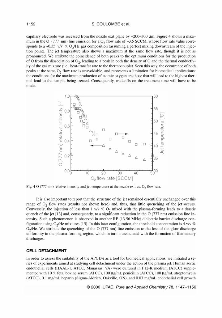
capillary electrode was recessed from the nozzle exit plane by ~200–300 µm. Figure 4 shows a maxi-mum in the O (777 nm) line emission for a O2 flow rate of ~3.5 SCCM, whose flow rate value corre-sponds to a ~0.35 v/v % O2/He gas composition (assuming a perfect mixing downstream of the injec-tion point). The jet temperature also shows a maximum at the same flow rate, though it is not aspronounced. We attribute the coincidence of both peaks to the optimum conditions for the productionof O from the dissociation of O2, leading to a peak in both the density of O and the thermal conductiv-ity of the gas mixture (i.e., heat-transfer rate to the thermocouple). Seen this way, the occurrence of bothpeaks at the same O2 flow rate is unavoidable, and represents a limitation for biomedical applications:the conditions for the maximum production of atomic oxygen are those that will lead to the highest ther-mal load to the sample being treated. Consequently, tradeoffs on the treatment time will have to bemade.
It is also important to report that the structure of the jet remained essentially unchanged over thisrange of O2 flow rates (results not shown here) and, thus, that little quenching of the jet occurs.Conversely, the injection of less than 1 v/v % O2 mixed with the plasma-forming leads to a drasticquench of the jet [13] and, consequently, to a significant reduction in the O (777 nm) emission line in-tensity. Such a phenomenon is observed in another RF (13.56 MHz) dielectric barrier discharge con-figuration using O2/He mixtures [15]. In this later configuration, the threshold concentration is 4 v/v %O2/He. We attribute the quenching of the O (777 nm) line emission to the loss of the glow dischargeuniformity in the plasma-forming region, which in turn is associated with the formation of filamentarydischarges.
CELL DETACHMENT
In order to assess the suitability of the APGD-t as a tool for biomedical applications, we initiated a se-ries of experiments aimed at studying cell detachment under the action of the plasma jet. Human aorticendothelial cells (HAAE-1, ATCC, Manassas, VA) were cultured in F12-K medium (ATCC) supple-mented with 10 % fetal bovine serum (ATCC), 100 µg/mL penicillin (ATCC), 100 µg/mL streptomycin(ATCC), 0.1 mg/mL heparin (Sigma-Aldrich, Oakville, ON), and 0.03 mg/mL endothelial cell growth
S. COULOMBE et al.
© 2006 IUPAC, Pure and Applied Chemistry 78, 1147–1156
1152
Fig. 4 O (777 nm) relative intensity and jet temperature at the nozzle exit vs. O2 flow rate.

supplement (Becton-Dickinson, Bedford, MA) at 37 °C in a 5 % CO2–95 % air-humidified incubator.Cells were seeded into 60 × 15 mm cell culture Petri dishes (Corning, Corning, NY) and allowed toreach confluence. The APGD-t was mounted on a stage, allowing precise vertical displacements. Priorto treatment, the vertical axis was zeroed in relation to an empty Petri dish in order to calculate the dis-tance between the nozzle end and the cells’ exposed plane. The APGD-t was operated using 1 SLM Heas the plasma-forming gas and 3 SCCM O2 through the capillary electrode (0.3 v/v % O2/He corre-sponds to the near optimum gas mixture for the production of excited atomic oxygen). The torch powerwas ~1 W, giving rise to a jet temperature in the cells’ plane of ~40 °C. During the plasma treatment,cells were taken out of the incubator and the height of the media adjusted to 1 mm. Cells were posi-tioned at the specified distance from the tip of the torch nozzle, and the Petri dish moved by hand underthe lit torch, creating a treatment path in the cells. The power to the torch was then turned off, leavingthe gas flow on, and a second path was made through the cells to distinguish the effect of the gas flowon cell detachment. The treated cells were then stained with crystal violet (Becton-Dickinson), rinsedtwice with PBS (Fisher, Whitby, ON), and fixed with formalin (Fisher). The cells were immediately ex-amined under the Leica DM IL inverted light microscope and photos were taken using the Leica DC300digital camera system. The pH of the media was monitored with a phenol red indicator, and no changewas noticed upon exposure to the plasma jet.
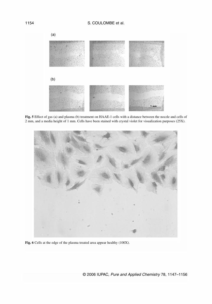
Figure 5 shows the effect of both gas (a) and plasma (b) treatment on the endothelial cells for ex-posure times of ~0.15 s/cell. One sees that the plasma jet causes the detachment of cells from the Petridish in a clear track approximately 0.5 mm wide, the width of the plasma jet at the nozzle exit. The bor-ders of this track are very sharp, with the cells at the very edge of the border appearing healthy and un-affected as can be seen at higher magnification (Fig. 6). Unfortunately, it is impossible at this point toquantify the extent of cell detachment since the uncertainty on the cell counts would be too large.Nevertheless, a significant cell detachment is observed. The effect of gas flow alone was evaluated dur-ing each experiment to ensure that the cells were not being mechanically detached due to the shearstresses imposed by the high gas flow. Additionally, due to the short exposure time to the plasma flow,cell dehydration is not likely to be a factor influencing cell detachment. In fact, the covering media isonly momentarily displaced by the jet, leaving the cells uncovered for a few seconds. Figure 5a demon-strates that the cells exposed exclusively to the gas flow (plasma off) experience minimal detachment.The effect of gas temperature alone was tested by exposing the back side of the glass Petri dish to theplasma jet until the temperature at the cells’ surface became similar to the one observed under the nom-inal treatment conditions. No changes to the cells were observed by inspection through the microscope.Those observations seem to suggest that the observed phenomena originate from biochemical processesinduced by the plasma–cell interactions.
© 2006 IUPAC, Pure and Applied Chemistry 78, 1147–1156
Plasma sources 1153
S. COULOMBE et al.
© 2006 IUPAC, Pure and Applied Chemistry 78, 1147–1156
1154
Fig. 5 Effect of gas (a) and plasma (b) treatment on HAAE-1 cells with a distance between the nozzle and cells of2 mm, and a media height of 1 mm. Cells have been stained with crystal violet for visualization purposes (25X).
Fig. 6 Cells at the edge of the plasma treated area appear healthy (100X).

Reactive oxygen species (ROS) such as the OH• radical, hydrogen peroxide, and singlet oxygenare known to be able to initiate lipid peroxidation, protein degradation, and apoptosis of the cell. TheOH• radical is very reactive, and its short lifetime (~ns) [16] does not suggest a significant penetrationthrough the cell membrane. The chemical attack by this radical would be focused on the exterior of thecell, oxidizing the lipids in the cell membrane and disrupting the adhesion proteins of the cell. TheOH• radical can be produced in the jet due to the presence of humidity in the laboratory air, and couldalso result from the dissociation of water in the cell media. Singlet oxygen has a much longer lifetimeand can diffuse through the cell membrane [17]. It is the primary agent in photodynamic therapy, aphotosensitizing cancer treatment, where it induces cytotoxicity within the cell [18]. Entrainment of airin the plasma jet also creates reactive nitrogen species (RNS), which are known to cause damage to thecell. The ROS and RNS are likely to initiate many oxidation reactions with the cell, resulting in cell de-tachment.
CONCLUSIONS AND OUTLOOK
We presented the general characteristics of a miniature RF (13.56 MHz) atmospheric pressure glow dis-charge torch, the so-called APGD-t, and demonstrated its suitability for use as a biomedical tool. Theminiaturized size of the plasma jet, 500 µm o.d. at the nozzle exit, combined with the low-duty-cycleamplitude modulation of the RF power enables the local treatment capability within the thermal limitsimposed by biomedical applications (<50 °C). We first reported on the difficulties one encounters withthe impedance matching and the monitoring of the electrical parameters of such torch (resistance, ca-pacitance, and resistive power dissipation) due to the high capacitive load it represents.
Our preliminary optimization study with a O2/He mixture revealed that a maximum in atomicoxygen emission (O (777 nm)), whose signal is taken as a measure of the O concentration in the jet, isreached at ~3.5 SCCM O2 fed in the capillary electrode when the plasma-forming gas flow is 1 SLMHe (~0.35 v/v % O2/He), and the torch power is ~1 W. Preliminary experiments conducted with endo-thelial cells demonstrated the ability of the APGD-t to detach cells from a Petri dish. Our preliminarywork seems to indicate that the detachment mechanism takes its roots in biochemical processes inducedby the presence of the plasma.
Other investigations are in progress in order to determine if the cells detached from the Petridishes are still alive and further assess the suitability of the APGD-t for other biomedical applications,namely, local sterilization, local cell lysis, local deposition of temporary organic films, and local sur-face functionalization for grafting and patterning. The plasma and biomedical engineering communitiesare encouraged to develop joint research and development efforts in order to elucidate the basic bio-chemical and physical mechanisms participating in the plasma-biological surface interactions, and tofurther develop this promising technology.
ACKNOWLEDGMENTS
The authors wish to thank the ISPC 17 conference organizers for the invitation made to S. Coulombeto present this research project, and the Natural Sciences and Engineering Research Council of Canada(NSERC), the Fonds Québécois sur la Nature et les Technologies (FQRNT), and the Department ofChemical Engineering, McGill University (EUL Funds) for the financial support. The authors also wishto thank Prof. A. Ricard for his suggestion on the possible formation of larger nitrogen molecules.
© 2006 IUPAC, Pure and Applied Chemistry 78, 1147–1156
Plasma sources 1155

REFERENCES
1. A. Fridman, A. Chirokov, A. Gutsol. J. Phys. D: Appl. Phys. 38, R1 (2005).2. I. E. Kieft, D. Darios, A. J. M. Roks, E. Stoffels. IEEE Trans. Plasma Sci. 33, 771 (2005).3. M. Laroussi. Plasma Process. Polym. 2, 391 (2005).4. P.-L. Girard-Lauriault, F. Mwale, M. Iordanova, C. Demers, P. Desjardins, M. R. Wertheimer.
Plasma Process. Polym. 2, 263 (2005).5. E. Stoffels, A. J. Flikweert, W. W. Stoffels, G. M. W. Kroesen. Plasma Sources Sci. Technol. 11,
383 (2002).6. R. E. J. Sladek, E. Stoffels. J. Phys. D: Appl. Phys. 38, 1716 (2005).7. R. Stonies, S. Schermer, E. Voges, J. A. C. Broekaert. Plasma Sources Sci. Technol. 13, 604
(2004).8. A. Bilgiç, C. Prokisch, J. Broekaert, E. Voges. Spectrochim. Acta B 53, 773 (1998).9. Rhytec, Web site: <www.portraipsr.com>.
10. Söring, Web site: <www.soering.com>.11. PVA Tepla, Web site: <www.pvatepla.com>.12. S. Coulombe, V. Léveillé. “Low-Power Atmospheric Pressure Glow Discharge Torch”, U.S.
Provisional Patent Application US60/705,443, 5 August 2005. 13. V. Léveillé, S. Coulombe. Plasma Sources Sci. Technol. 14, 467 (2005).14. A. P. Napartovich. Plasmas Polymers 6, 1 (2001).15. J. Park, I. Henins, H. W. Herrmann, G. S. Selwyn. J. Appl. Phys. 89, 15 (2001).16. C. Batandier, E. Fontaine, C. Kériel, X. M. Leverve. J. Cell. Mol. Med. 6, 175 (2002).17. J. Moan. J. Photochem. Photobiol. B: Biol. 6, 343 (1990).18. F. S. De Rosa, M. V. L. B. Bentley. Pharm. Res. 17, 1447 (2000).
S. COULOMBE et al.
© 2006 IUPAC, Pure and Applied Chemistry 78, 1147–1156
1156