Doctor Who Classics / Doctor Who 2005 - renouveau et continuité
Upload
khangminh22Category
view
0download
0
Mini-OSCE
SURGERY

حيتوي هذا امللف عىل مجيع الصور اليت اكنت يف مجموعات الفيس بوك اخلاصة ابمليين اوسيك،
. فالشكر موصول للك من سامه يف نرش الصور واملعلومات
ىل وضع كام مقت جبمع الصور واملعلومات اعامتدا عىل ادلوس يات والساليدات، ابلإضافة اإ
. الأس ئةل السابقة
.أأمتىن أأن يكون ذا فائدة وأأعتذر عن وجود أأي خطأأ
ضافة والتعديل ، ابلتوفيق .تواصلوا معي لنقل مالحظاتمك لالإ
رفيف سعيد القوامسة : زميلتمك من الس نة السادسة
26/12/2015

Contents
• Pediatric surgery ( slides 4 – 50 ) • Gastrointestinal surgery ( slides 51 – 118 )
• Oncology ( slides 119 – 141 )
• Plastic surgery ( slides 142 – 208 )
• Cardiovascular surgery ( slides 209 – 233 )
• General surgery ( slides 234 – 253 )
• Endocrine surgery ( slides 254 – 269 )
• Past papers ( slides 270 – end )

Pediatric surgery

Umbilical Hernia
• more common in blacks.
• familial tendency.
• repair is carried out if
closure does not occur by
the end of 2nd year of life.
• repair performed after
the age of 2 and before
the age of 10.
• associated anomalies :
- hypothyroidism.
- hurler syndrome.
- beckwith-wiedman
syndrome.

Patent urachus
• It is a remnant presents as fistula connecting the umbilicus & urinary bladder.
• Patients with prune belly syndrome have a patent urachus.
• Other forms : blind sinus/ cyst/ abscess.

Vesicointestinal fissure The terminal ileum is herniating through the cecum forming the so called elephant trunk deformity.

Omphalitis
• Inflammation of the umbilicus.
• Occurs only in newborns.
• Can be fatal because of portal vein thrombosis.
• Infection can spread to the abdominal wall.
• Antibiotics and intensive care.

Omphalocele
• location : umbilical
ring.
• The protrusion is covered
by peritoneum.
• defect size : >10 cm.
• cord : inserted into the
sac.
• GIT function is normal.
• contents : bowel +/- liver.
•malrotation : present.
• associated anomalies :
common (30-70 % ).

Gastroschisis
• location : lateral to
the umbilicus ( to the
right ).
• defect size : 2-4 cm.
• no sac.
• cord is normally
inserted into umbilicus.
• contents : only bowel
( edematous and matted ).
• GIT function :
prolonged ileus.
• associated anomalies :
infrequent.

Gastroschisis or omphalocele 1st aid management : - Carefully wrap in saline-soaked pads. - Support without tension. - NG tube. - Abdominal ultrasound.

Hydrocele • Fluid filled sac ( fluid in a patent
processus vaginalis or in the tunica vaginalis around the testicle).
• Communicating with the peritoneal cavity VS non communicating.
• Transillumination. • In most infants it will resolve in
the 1st year. • If there is increase in size >>
operation • Any hydrocele appearing after a
1st year must be operated as it will not resolve.

Inguinal hernia
• Due to patent processus vaginalis.
• More common at the right side. • Bilateral hernias occur in 5-15%
of children with hernia. • Uncomplicated hernia will bulge
when the baby cry and reduces when the baby is relaxed , sleeping. Etc.
• Uncomplicated hernia must be operated (herniotomy).
• Herniotomy must be performed ASAP.
• 10-15% of children with on the other side. hernia on one side will develop a hernia
• Complicated hernia presents in the ER with pain/ management : resuscitation, reduce hernia, then repair within 24-48 hrs. ( as we fear strangulation and testicular atrophy).

Undescended testicle • A testicle, which is not in the
scrotum. • Significant risks: infertility/
trauma/ torsion/ hernia/ cancer. • Treatment : orchidopexy by the
age of one year (6-12 months). • After 2 years the testicle is
abnormal and wouldn’t be functioning.
The right scrotum has undescended testicle. It is empty and flat.

Acute scrotum : Scrotal swelling + pain. DDx : 1. Testicular torsion. 2. Torsion of testicular appendages. 3. Epididymorchitis. 4. Scrotal edema. 5. Complicated hernia. ** should be considered as typical testicular torsion unless proven otherwise.
Testicular torsion

Prune Belly Syndrome
• thin flaccid abdominal wall.
• AKA eagle Barrett syndrome.
•absent abdominal wall musculature.
• dilation of bladder, ureter and renal collecting system.
• 95% in Males.

Pentalogy of Cantrell
1. Omphalocele.
2. Anterior
diaphragmatic
hernia.
3. Sternal cleft.
4. Ectopia cordis.
5. Intracrdiac defect.

Bickwith-Wiedman syndrome
1. Macrosomaia.
2. Macroglossia.
3. Organomegaly.
4. Abdominal wall
defects.
5. Embryonal tumors.

Congenital diaphragmatic hernia • X-ray of the abdomen
and chest.
• features :
- scaphoid abdomen.
- bowel is located in the left side of the chest.
- mediastinal shift towards the right.
• mortality is mostly due to pulmonary hypoplasia.
• Diagnosis: In prenatal period (ultrasonography)
• types : 1) bockdalek hernia ( mostly on left side) : posterolateral, most common. 2) Morgangi hernia (mostly on the right side) : retrosternal. 3) Hiatus hernia.

Neonate with a prenatally diagnosed left congenital diaphragmatic hernia pre surgery.

Esophageal atresia and tracheoesophageal fistula

Manifestations of esophageal atresia: 1) Upper part: drooling of saliva/ bubbling of the saliva/
respiratory distress/ choking/ failure to pass nasogastric tube.
2) Lower part: accumulation of secretions which will lead to regurgitation and vomiting/ ischemia>> physiological death>> biological death (necrosis) >> rupture.
* The more distal the obstruction, the more the distention of the lumen and so the more the possibility of rupture.

Neonates with esophageal atresia usually develop copious, fine white frothy bubbles of mucus in the mouth and nose. Secretions recur despite suctioning.

Esophageal atresia and tracheoesophageal fistula atresia of the upper
esophagus evidenced by
failure to pass a
feeding tube.
gas in the abdomen.
These findings are
likely due to a
esophageal atresia with
a distal
tracheoesophageal
fistula (Type C TEF).

Types of intestinal atresia

What is the dx? Jejunal atresia. Age of presentation? Neonate (till one month) How would u manage? Admit to NICU/ fluid resuscitation/ antibiotic/ NG suction and parental nutrition.

-apple peel intestinal atresia ( also
type IIIb or Christmas tree atresia). -Due to vascular accident. -all the intestine is atretic, and forms a loop around the superior mesenteric artery.

Intestinal obstruction
• Abdominal X-ray.
• Double bubble sign.
• represents dilation of the proximal duodenum & stomach.
• DDx : duodenal stenosis (mostly in the 2nd part of duodenum) / duodenal atresia.
Multiple air fluid levels seen in mechanical intestinal obstruction.

Hirschsprung’s disease
• Congenital megacolon.
• It is an absence of ganglion cells distal in the bowel.
• Contracted non-peristaltic affected segment and a dilated hypertrophied proximal segment.
• M:F (4:1)
• Failure to pass meconium in the 1st 24-48 hrs of life.
• When compared to habitual constipation ( no soiling/ no anal fissures).
•DDx : hypothyroidism/ sepsis.
Plain abdominal X-ray : dilated loops of bowel/ air-fluid level.
Barium enema study: funnel shaped appearance of colon ( megacolon – transitional zone- the affected narrowed segment).

Normally, the rectum is larger than the colon. In Hirschsprung Disease, there is an abnormal rectosigmoid ratio with the rectum smaller than the sigmoid due to denervation hyperspasticity. Therefore, one see dilation of large and small bowel proximal to the "transition zone." The "Transition Zone" is the junction between the proximal normally innervated colon and the distal aganglionic segment. The normally innervated proximal colon becomes dilated. In 33% of cases, there is a normal-appearing rectum.
Meconium ileus
• Intestinal obstruction from solid meconium concretions.
• >95% have cystic fibrosis.
• Sx: bilious vomiting/ abdominal distention/ failure to pass meconium.

Intussusception
It is a cause of intestinal obstruction.
M : F ( 3:2)
In a previously healthy infant.
(5 months - 3 yrs) idiopathic / ( >3yrs) 2ry.
m.c.c of I.O in the age of (5 months-3 yrs)
Sudden onset, abdominal colic, vomiting.
begins proximal to ileo-cecal junction.
Ba enema ( diagnostic and therapeutic).
The part that prolapses into the other is called the intussusceptum, and the part that receives it is called the intussuscipient.

Intussusception
Ultrasound signs :
Pseudokidney sign.
( longitudinal
ultrasound appearance
of the intussuscepted
segment of bowel).
Target sign
(doughnut sign).


Intussusception
Plain abdominal X-ray.
Ba enema.
Red currant jelly stool.
( characteristic feature) / late feature.
Target sign on CT

Pyloric stenosis
M : F (4:1)
Age (3-6 wks)
Progressive, persistent,
projectile, non-bilious vomiting.
Succation splash.
Olive sign (enlarged pylorus
is palpable).
Hypochloremic alkalosis.
Dx by abdominal U/S
Higher risk when mother is
affected.
Surgical ttt : Ramstad's
pyloromyotomy.
No recurrence after surgery.
pylorus stomach
Barium study : string sign

Beak sign Shoulder sign

Torticollis • Tilted neck. • Causes: 1) congenital ( due to abnormal position of the fetus in uterus which leads to fibrosis of sternocleidomastoid muscle >> shortness of this muscle) 2) acquired : due to trauma leads to muscle spasm on one side/ fibrosis of SCM due to any cause. 3) infection: lymphadenitis
• Occurs at any age but most common in the 1st few months of life.
• Palpable hard mass in 1/3 of patients. • The baby usually sleeps on the same
side >> craniofacial deformity.
• Treatment : conservative using physiotherapy for 2-3 months.
• If no improvement, surgery is indicated (SCM myotomy).

Thyroglossal duct cyst
• The most common congenital anterior neck mass located at midline.
• Mostly present in the 1st 2 decades of life.
• Males > females. • Movement upward with tongue
protrusion. • Cystic lesion on ultrasound. • Higher risk of malignancy. • Treatment: excision + hyoid bone must
be cut (sistrunk procedure) • If hyoid bone is not removed the
recurrence rate is > 50-60%. • In case of infection and abscess
formation : antibiotics then incision and drainage followed by excision.

Cystic hygroma
• Fluid-filled sacs caused by blockages in the lymphatic system.
• most hygromas appear by age 2.
• soft, non-tender, compressible lump.
• high recurrence rate.
• usually located in the posterior triangle of the neck.
• transillumination.
• DDx: teratoma/ hemangioma/ encephalocele.

Hypospadias • Abnormal urethral opening
on the undersurface of the penis associated with incomplete development of the urethra.
• Deflection of the urinary stream.
• Classifications : anterior(50%), middle (30%), posterior (20%).
• Glanular (opening on the glans) is the most common.
• Surgery is performed 6-18 months of age.


Bladder Extrophy
• defective enfolding of
caudal folds.
• associated with
prolapsed vagina or
rectum / epispadias /
bifid clitoris or penis.
Epispadius : urethral opening is on the dorsal surface with abnormal penis. It is usually a part of a syndrome includes extrophy of the urinary bladder. - Extremely rare.

Vesicoureteral reflux • Retrograde flow of urine from the
bladder into the ureter. • The ratio of tunnel length to
ureteral diameter must be at least 5:1 to prevent reflux.
• Presentation : either antenatal hydronephrosis or clinical UTI.
• Diagnosis : urine culture/ ultrasound/ voiding cystourethrogram.
• Nuclear cystogram for screening. • DMSA scan to detect kidney
scarring. • Urodynamic study for lower urinary
tract abnormalities (neurogenic bladder).
Spot film taken during VCUG shows unilateral grade 4 vesicoureteral reflux

UVR grades
Treatment : - Spontaneous resolution is common in young children (only antibiotics). - Indications for surgery: grade 4 and 5/ poor compliance with medications/
breakthrough febrile UTI despite adequate antibiotic prophylaxis/ poor renal growth/ kidney scars/ mild or moderate reflux in females that persist during puberty despite several yrs of observation.

Imperforate anus • Males > females. • High lesion vs. low lesion. • Meconium or air per urethra or vagina. • One of the common findings that the anal
opening anteriorly located. • Treatment : resuscitation/ the low types
managed by a one stage procedure in the neonatal period (anoplasty).
• Other types treated by colostomy in the
neonatal period followed by a definitive procedure called pull-through (posterior sagittal anorectoplasty).

This chest X-ray shows air trapping indicating foreign body aspiration. It is the most common radiological sign shown on the X-ray after F.B aspiration. Whenever you suspect F.B aspiration you have to do bronchoscopy.

Meckel's diverticulum
-a true congenital diverticulum, is a slight bulge in the small intestine present at birth and a vestigial remnant of the omphalo-mesenteric duct. -A memory aid is the rule of 2s: 2% (of the population). 2 feet (proximal to the ileocecal valve). 2 inches (in length). 2 types of common ectopic tissue (gastric and pancreatic) 2 years is the most common age at clinical presentation 2:1 male: female ratio


Congenital malformations
Preauricular sinus

Gastrointestinal surgery

Malrotation normally the duodenojejunal junction is to the left of the spine. In malrotation it is to the right of the spine .

gross appendicitis: as noticed by: enlarged appendix and engorged blood vessels on it.
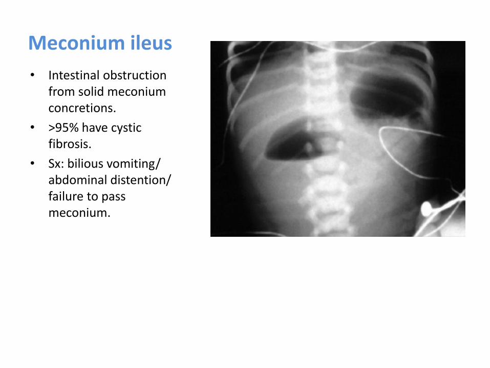
Acute appendicitis • Sx : pain (periumbilical area) >> nausea and
vomiting >> anorexia >> pain migrates to RLQ (constant and intense, usually < 24 hrs.).
• Tenderness maximally at McBurney’s point. • Obturator sign/ psoas sign/ rovsing sign/
valentino sign.
• Appendectomy is the m.c.c of emergent abdominal surgery.
• Dx of ruptured appendix : fever >39 / high WBC/ rebound tenderness/ periappendiceal fluid collection on ultrasound.
• If normal appendix is found upon exploration, take it out ( even in chron’s ).
• Appendiceal abscess : percutaneous drainage/antibiotics / elective surgery 6 wks later.

Achalasia
• On manometry (the definitive diagnostic test) : Failure of the LES to relax during swallowing/ Aperistalsis of the esophageal body.
• Symptoms: dysphagia for both
solids and liquids (worse for liquids)/ regurgitation.
• Treatment: upper endoscopy
and balloon dilation of the LES/ laparoscopic Heller myotomy.
• May lead to esophageal
carcinoma 2ry to Barrett’s esophagus from food stasis.
bird’s beak sign
Barium swallow : best initial test

Diffuse esophageal spasm - Barium swallow showing corkscrew appearance . - Manometry is the most accurate test. - 1st line treatment : diltiazem or nifidipine and nitrates. - Dysphagia for both solids and liquids.
DDx : Nut-cracker esophagus ( peristaltic contractions with high amplitude). But DES there are non-peristaltic contractions with high amplitude. We differentiate between them by manometry.

Schatzki ring (lower esophageal ring) - Intermittent dysphagia for solids only with no pain. - Associated with hiatal hernia. - Dx : barium swallow and endoscopy. - Treatment : dilation by bougie method or through the
scope hydrostatic balloon/ pts placed on PPI after dilation.
Esophageal webs - More proximal/ usually in the
hypopharynx. - Same symptoms as schatzki ring. - E.g. Plummer-Vinson syndrome. - Dx : barium swallow and
endoscopy. - Treatment : dilation.

Plummer-Vinson syndrome: 1. Esophageal web 2. Iron-deficiency anemia 3. Dysphagia. 4. Spoon-shaped nails 5. Atrophic oral and tongue mucosa. * especially occurs in elderly women; 10% develop squamous cell carcinoma. * May respond to treatment of IDA.

Esophageal stricture - Dysphagia : constant/ slowly progressive/ solids then
liquids. - Causes : long history of incomplete treated reflux/
prolonged NG tube placement/ lye ingestion. - Dx : barium swallow. - Treatment: dilation.
Zenker’s diverticulum: - It is a false diverticulum (not involving all layers of
the esophageal wall). - Outpouching of the upper esophagus. - Halitosis / food regurgitation/ dysphagia. - Elderly. - Dx : barium swallow/ endoscopy and NG tube are
contraindicated (risk of perforation). - Treatment : surgical resection.

Mallory Weiss tear syndrome - Tear at gastro-esophageal junction. - Hematemesis. - Dx : history and upper endoscopy. - Resolves spontaneously.
Barrett’s esophagus - Change of cell type from esophageal squamous
to specialized intestinal columnar ( metaplasia). - Cause : chronic GERD. - Dx : endoscopy. - Treatment : PPI and follow up. - Increases the risk of adenocarcinoma.

Esophageal varices -They are most often a consequence of portal hypertension, commonly due to cirrhosis. -patients with esophageal varices have a strong tendency to develop bleeding. -Occur because of a Porto systemic between left gastric vein (Portal circulation) and esophageal veins (Systemic circulation). -Therapeutic endoscopy is considered the mainstay of urgent treatment.
-The two main therapeutic approaches are variceal ligation or banding and sclerotherapy, of course after vigorous resuscitation. -In ideal circumstances, patients with known varices should receive treatment to reduce their risk of bleeding by the non-selective β-blockers (e.g. propranolol).

Hiatal hernia
Type 2 Type 1 (more common)

Para esophageal hernia (2) Sliding hernia (type 1)
Dysphagia/ stasis gastric ulcer/ no reflux
Mostly asymptomatic but can cause reflux
Complications: hemorrhage/obstruction/ strangulation.
Complications :reflux> esophagitis> Barrett's esophagus > cancer/ aspiration pneumonia
Treatment : surgical. Treatment: medical with antacids, PPI, H2 blockers/ if failed : surgical (lap. Nissen fundoplication )

Epiphrenic diverticulum Presentation: Dysphagia to solid foods with upper abdominal discomfort. Often associated with hiatal hernia.

esophageal cancer -is more after 50 years, most between
60-70 years. -more in males. -risk factors: smoking, alcohol, and hot fluid drinkers. -Relevant Hx: GERD and Barrett’s, stricture, Plummer Vinson syndrome, Celiac disease, Esophageal achalasia and diverticulum. -common symptoms are dysphagia, reflux, weight loss, and mediastinal invasion symptoms (chest pain, hoarseness, etc.) -they might also suffer from anemia due to nutritional deficiency. -treatment : surgical resection if small and localized. - If large or Metz: combination of CTX and RTX prior to surgery.

easy mnemonic to remember esophageal CA risk factors ABCDEFGH: A- Achalasia/Alcohol B- Barrett’s esophagus C- Cigarettes D- Diverticula E- Esophageal web, stricture F- Fat/Family hx G- GERD H- Hot liquid

Gastric cancer - adenocarcinoma : m.c type (95% ).
R.F: diet ( smoked meat , high nitrates , low fruits and vegetables) , smoking , family history , blood group type A , H. pylori , prev. partial gastrectomy , adenomatous gastric polyps , atrophic gastritis .
Subtypes: diffuse type:70% ,from lamina propria , proximal , worse than intestinal type , invasive and Metz , in younger pt. intestinal type : 30% ,from gastric mucosa, distal , ass with H.pylori , well formed glandular structures.
Ulcerating adenocarcinoma
Intestinal type

classic physical findings that represent Metz and incurable disease : 1-virchows node enlargement (left supraclavicular nodes). 2-sister merry josephs nodules : infiltration of the umbilicus. 3- blumers shelf :fullness in the pelvic ,cul-de-sac(solid peritoneal deposit anterior to the rectum forming a shelf palpated on PR). 4- krukenburgs tumor :enlarged ovaries on pelvic examination (Metz to ovaries). 5-hepatosplenomegaly with ascites and jaundice. 6- cachexia. 7- irishs node :left axillary adenopathy.
Virchow’s node enlargement
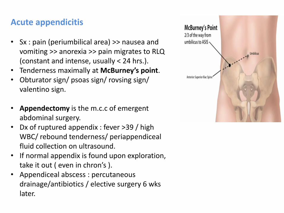
diagnosis : endoscopic with biopsy is the method of choice/ double contrast barium meal . treatment: surgical resection with wide margin >5cm and lymph nodes dissection . If tumor is proximal or midbody do total gastrectomy with roux-en-y ,if tumor is distal do distal subtotal gastrectomy .
A. CT image of Linitis plastic (arrows denotes a thickened gastric wall).
Linitis Plastica (leather bottle): when the entire stomach is involved and looks thickened .

Gastrointestinal stromal tumors (GISTs)
- previously known as leiomyosarcoma.
- Cell of origin : intestinal cells
of Cajal. - Found from esophagus to
rectum, most commonly in stomach.
- Sx: abdominal pain/ nausea/
abdominal mass/ occult GI bleeding.
- Dx: CT / EGD/ colonoscopy. - Tumor marker: C-KIT - Treatment: resection. - Chemotherapy: imatinib (tyrosine
kinase inhibitor).

Bariatric surgery
- Weight reduction surgery for the morbidly obese. - Morbid obesity : BMI > 40 or BMI> 35 with a medical problem
related to morbid obesity (sleep apnea/ CAD/ DM/ HTN/ pulmonary disease/ breast cancer/ colon cancer/ arthritis/ sex hormone abnormalities/ venous stasis ulcers.
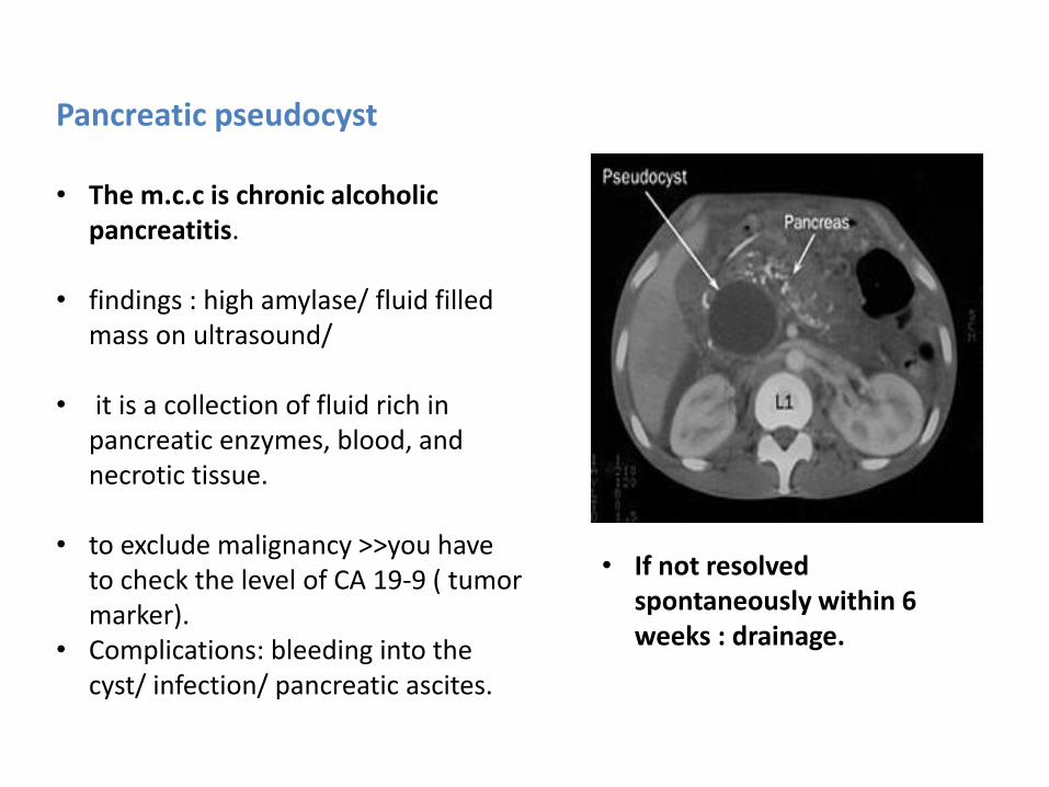
Pancreatic pseudocyst • The m.c.c is chronic alcoholic
pancreatitis. • findings : high amylase/ fluid filled
mass on ultrasound/ • it is a collection of fluid rich in
pancreatic enzymes, blood, and necrotic tissue.
• to exclude malignancy >>you have
to check the level of CA 19-9 ( tumor marker).
• Complications: bleeding into the cyst/ infection/ pancreatic ascites.
• If not resolved spontaneously within 6 weeks : drainage.

Acute Pancreatitis -Cut off sign and Ileus. -White arrow points to Transverse colon cut off at Splenic flexure. -No air in descending colon. -TC: Transverse colon. - I: Represents small bowel loops with air suggestive of Ileus.
Causes : gallstones/ ethanol/ trauma/ steroids/ mumps/ autoimmune/ scorpion bite/ hyperlipidemia/ drugs (diuretics, INH)/ ERCP. Treatment : supportive (90% resolve spontaneously)

Cullen’s sign superficial edema and bruising in the subcutaneous fatty tissue around the umbilicus.
Grey turner’s sign bruising of the flanks.
Acute pancreatitis (In severe cases)

Chronic Pancreatitis The patient experiences intermittent attacks of severe pain, often in the mid-abdomen or left upper abdomen and occasionally radiating in a band like fashion or localized to the midback. The pain may occur either after meals or independently of meals, but it is not fleeting or transient and tends to last at least several hours. Other symptoms associated with chronic pancreatitis include diarrhea and weight loss.
Abdomen x-ray showing pancreatic calcifications.
most common cause is chronic alcoholism.

Pancreatic necrosis - Dx: abdominal CT with
contrast. - Dead pancreatic tissue
doesn’t take up the contrast.

Gallbladder stones (Cholelithiasis)
Acoustic shadow
- 80% of patients are asymptomatic. - Complications: acute and chronic cholecystitis/ CBD stones/ gallstone
pancreatitis/ cholangitis. - U/S detects GB stones in more than 98% of cases. - Abdominal X-ray detects only 15%. - If symptomatic/ complicated / asymptomatic but (sickle cell diseas, DM,
pediatric, porcelain GB, immunosuppression) : cholecystectomy.

acoustic shadow
Acute cholecystitis - HIDA scan (the most accurate test). - U/S (the diagnostic test of choice). - Constant pain (not biliary colic).

emphysematous cholecystitis - Gas forming bacteria ( E.coli). - Often results in perforation. - Usually in males/ elderly/ DM.

Porcelain gallbladder The radiograph reveals a pyriform opaque mass with curvilinear calcification in the right upper quadrant from a porcelain gallbladder. The gallbladder wall may be diffusely calcified or have irregular stippled calcifications. This is usually an incidental finding in asymptomatic patients. Although it was originally thought that there was a high association between porcelain gallbladder and adenocarcinoma, more recent research has revealed a much weaker association and in patients with diffuse calcification there is no increased risk for cancer.

Choledocolithiasis - Common bile duct stones. - ERCP (the diagnostic test of choice, also therapeutic). - If ERCP fails, CBD is opened surgically and stones removed.
The huge tube is the endoscope. It is going down from the esophagus, through the stomach, to the duodenum (1st then 2nd parts), and stops near the ampulla of vater. A tube in the endoscope is pushed into the ampulla and fills the CBD with a dye. X-ray is taken. As you can see, there is a black shadow stone in the CBD.

Gallstone ileus
• occurs when a large
gallbladder stone erodes into the duodenum via a fistula, eventually obstructing the ileal lumen usually some centimeters proximal to the ileocaecal junction.
On the X-ray : 1- radiopaque gallstone in the bowel. 2- gas in the gallbladder. 4- small bowel distention.
1
2
3

Pneumobilia ( Air in the biliary tree ) Causes : -Recent biliary instrumentation (e.g. ERCP or PTC) -Incompetent sphincter of Oddi (e.g. sphincterotomy, following passage of gallstone.) -Biliary-enteric surgical anastomosis. -Spontaneous biliary-enteric fistula (cholecystoduodenal accounts for ~70% ). -Infection (rare) (e.g. ascending cholangitis, anaerobes).
Abdomen CT

Liver abscess
- Pyogenic (bacterial “gram negative’’) / parasitic (amebic) / fungal.
- Most common site is right lobe. - Treatment : pyogenic ( IV
antibiotics + percutaneous drainage) / amebic (metronidazole+ drainage).
- Indications of surgical drainage in pyogenic : multiple lobulated abscesses/ multiple percutaneous attempts failed.
- Indications of surgical drainage in amebic: refractory to metronidazole/ bacterial co-infection/ peritoneal rupture.

Hydatid disease is a parasitic infestation by a tapeworm of the genus Echinococcus. A history of living in or visiting an endemic area must be established. Also, exposure to the parasite through the ingestion of foods or water contaminated by the feces of a definitive host (Dogs) must be determined. Physical examination findings from patients with echinococcosis are nonspecific. Skin : Jaundice could be a sign of biliary obstruction. Spider angiomas are a sign of cirrhosis of the liver. Urticaria and erythema may be seen.
Abdominal CT scan
ultrasound

Vital signs : Fever could be a sign of secondary infection or allergic reaction. Hypotension is observed with anaphylaxis secondary to a cyst leak. Lungs: Decreased breath sounds over the affected area are signs of airway obstruction with consolidation of the affected segment, lobule, lobe, or the whole lung. Abdomen: The most common sign is abdominal tenderness. Hepatomegaly may be present or a mass may be felt. Tender hepatomegaly is a sign of secondary infection of the cyst, especially when coupled with fever and chills.
Ascites is rare. Splenomegaly can be the result of either splenic echinococcosis or portal hypertension.

Extremities: Bone involvement can result in tenderness over the affected area and, rarely, a palpable mass. Muscle involvement is usually characterized by a palpable mass. Peripheral nerve compression can occur, although extremely rarely. It results in nerve-specific sensory and/or motor deficit. Treatment : Albendazole and surgery.

Hepatic Hemangioma Most common benign solid tumor.
Variants:
- Capillary : m.c / <2cm / no need for surgery.
- Cavernous : giant.
Vague upper abdominal tenderness with no mass.
Not premalignant.
Percutaneous biopsy is contraindicated (risk of hemorrhage).
U/S is the first test.
MRI is the most sensitive & specific.

- Until recently, no medical therapy capable of reducing the size of hepatic hemangiomas had been described.
- Surgical treatment may be appropriate in cases of rapidly growing tumors. Surgery may also be warranted in cases where a hepatic hemangioma cannot be differentiated from hepatic malignancy on imaging studies.

Hepatic adenoma Risk factors: Female/ birth control pills/ anabolic steroids/ glycogen storage disease. it is estrogen sensitive (pregnancy may cause it to increase in size, OCP). Complications: rupture with bleeding/ necrosis/ risk of cancer. Treatment: if small, stop pills> it may regress> if not, surgical resection. If large or complicated : surgical resection

Focal nodal hyperplasia Use of estrogen OCP may have a role. Not premalignant. Most are solitary, 20% multiple. Most common indication for surgery is inability to exclude malignancy. LFT : normal. Angiography : hypervascular mass with enlarged peripheral vessels and a single central feeding artery. ttt : nucleation/ diagnostic uncertainty will require an open excisional biopsy. Classic CT finding: liver
mass with central scar.

Hepatocellular carcinoma (hepatoma) - Most common 1ry malignant liver
tumor. - Risk factors: hepatitis B / cirrhosis/
Alfa toxin/ alpha 1 antitrypsin deficiency.
- Painful hepatomegaly. - Tumor marker: alpha fetoprotein. - Dx: needle biopsy with CT or U/S
guidance. - The m.c site of Metz : lungs.
CT : black arrows (hepatoma)

Caroli disease is a congenital disorder comprising of multifocal cystic dilatation of segmental intrahepatic bile ducts. presentation is in childhood or young adulthood. The simple type presents with RUQ pain and recurrent attacks of cholangitis with fever and jaundice. Prognosis is generally poor. If disease is localized, segmentectomy or lobectomy may be offered. In diffuse disease management is generally with conservative measures; liver transplantation may be an option.

Stepladder appearance - Multiple loops of small bowel in the left upper quadrant in a mechanical small bowel obstruction.

Endoscopic image of multiple small ulcers located in the distal duodenum in a patient with gastrinoma (Zollinger-Ellison syndrome)

Colovesical fistula
- the most common cause is diverticulitis and it's the most common fistula formed in DD. - other causes : colon CA , crohn's ,
radiotherapy ,trauma.
- This picture is double contrast barium enema.
diverticula

Sigmoid volvulus • Coffee bean sign on X-ray.
• is a cause of large bowel obstruction.
• occurs when the sigmoid colon twists on the sigmoid mesocolon.
• presentation : constipation/ abdominal bloating/ nausea/vomiting.

Fistula –in- ano - From rectum to anal skin. - Causes: anal crypt infection/
perianal abscess. - Sx : perianal drainage/itching/
diaper rash.

Hemorrhoids - Internal (above dentate line)/ external (
below dentate line). - Risk factors: constipation/ straining/
pregnancy/ ascites/ portal HTN. - Sx: bleeding (usually fresh blood)/ anal mass/
prolapse/ pain/ itching. - Complications: thrombosis/ infection/
ulceration. - Treatment: high fiber diet/ laxatives/ sitz
bath. - Hemorrhoidectomy: * contraindicated in chron’s. * complications: pelvic infection/ anal stricture/ incontinence.


Anal fissure - Hypertonic internal sphincter. - Chron’s disease may cause it. - Very painful. - Posterior fissures more common
than anterior ones. - Signs : sentinel tag/
hypertrophied papilla / blood on toilet paper.
- Surgery indication: chronic fissure / refractory to conservative treatment.
- Surgery: lateral internal sphincterectomy.
- Triad of chronic fissure: sentinel pile/ hypertrophied papilla/hypertonic sphincter.
Sentinel pile
Hypertrophic papilla

Pilonidal sinuses -infected tracts beneath your skin and occur at the end of your tailbone on the natal cleft above your buttocks. -Pilonidal sinuses can also occur between your fingers and your belly button and can have more than one channel. -Pilonidal sinus can start off as a small painless lump and may not cause any symptoms. However, once it becomes infected, it becomes inflamed and fills with pus and develops into a painful cyst.

Treatment may not be necessary if your pilonidal sinus does not become infected. If your pilonidal sinus does get infected, surgery will most likely be recommended and may include the following: 1-Incision and Drainage (A dressing will be applied, and you will have to visit your doctor’s office daily for dressing change). 2-Wide Excision (reduce your chances of a reinfection. However; Your wound may take a long time to heal and you may need to change your dressings daily for two or three months). 3-Excision and Primary Closure (the chances of a reinfection are higher when compared to wide excision )

Perianal warts - Cause : condylomata acuminata
(HPV). - The major risk is SCC. - Treatment : if small, topical
podophyllin/ if large, surgical resection or laser ablation.

Colon cancer -very normal looking colon on the left side. -Two polyps, one small and one large. -Adenomatous polyps are precancerous. -There is a irregular mass (tumor), most probably originating from a similar polyp. -If a biopsy is taken from the large polyp, tumor cells might be found.
IMPORTANT SURGICAL NOTE: when surgically treating a diagnosed colon cancer, the surgeon DOES NOT open the tumor site WHILE IN THE ABDOMEN. He REMOVES the area of the tumor WITH a proximal and distal SAFETY MARGIN (right or left hemicolectomy), then he opens the colon separately and we see this specimen!

Colon cancer gross colon CA notice how on the left side, the haustrations are almost regular. while on the right side, there are no haustrations and there are irregular masses + dilatation.
- Colonoscopy is the screening procedure of choice. - Carcinoembryonic antigen : good in checking for recurrence of cancer.

• Apple core appearance.
• Annular constriction of part of the lumen.
• Carcinoma of the colon.

Familial adenomatous polyposis (FAP)
• AD inherited disorder.
• The genetic defect : adenomatous polyposis coli gene.
• Polyps within the rectum & colon.
• Begin developing at puberty.
• Untreated pts develop cancer by ages 40 to 50.
• Possible associated tumors : duodenal tumors.
• ttt : total proctocolectomy and ileostomy.

Gardner's Syndrome ( AD) a familial adenomatous polyposis syndrome with cutaneous manifestations . 1) Colonic polyps ( hundreds with 100% risk of malignancy if untreated). 2) Ostromas ( the picture of an osteoma of the mandible). 3) Lipomas and epidermoid cysts ( on the forearm )

Peutz jeghers syndrome - autosomal dominant. - hereditary intestinal polyposis
syndrome. - hamartomatous polyps in the GI
tract. - circumoral pigmented nevi.

• Abdominal CT. • similarity between the
thickened edematous wall of pseudomembranous colitis to that of an accordion.
• What is the sign? Accordion sign.
Colonoscopy showing pseudomembranes cause: C. difficle risk factors: use of Antibiotics. diagnosis: toxin assay in stool. treatment: Metronidazole
Pseudomembranous colitis

Diverticulosis or Diverticular
disease of the sigmoid colon is a common disease affecting perhaps every third of the older population in industrialized countries. Often the diverticula (a sac or pouch formed at a weak point(Site of perforators) in the bowel wall) do not cause any symptoms. They can however cause pain most commonly in the left lower quadrant, altered bowel habits and bleeding. A diverticulum may become inflamed, causing diverticulitis.
Colonoscopy is a diagnostic examination, and the diverticulae are also easily seen in a large bowel x-ray examination. A diet rich of fiber is the most important treatment. Surgical treatment is seldom needed.


Crohn's disease ( IBD ) : - Autoimmune disease - can occur anywhere in the GI tract including the colon and frequently perianally , the disease is patchy ( SKIP LESIONS ) , the m.c site is the terminal ileum ,and often no involvement of the rectum ( in UC the rectum is always involved ) - symptoms are very varied including : Abdominal pain , wt loss , sometimes presenting with a picture of acute Appendicitis (right iliac fossa pain, pyrexia and vomiting ) , may also present as acute intestinal obstruction* ,abdominal mass , and with multiple perianal fissures and abscesses . - extraintestinal manifestations: arthritis , pyoderma gangrenosum ,erythema nodosum

- it involves the full thickness of the bowel wall , with the serosa ,mesentery and regional LNs ( while in UC it was only the mucosa that's involved)
- Macroscopically : the bowel wall in thick and red ( in UC its very thin ), the mucosa has a cobblestone appearance with deep fissures , and the mesentery is shortened - Microscopically we will find non- caseating granulomas , with narrow deep fissure ulcers. - Complications : mainly strictures and fistulae ( in UC : hemorrhage , perforation , CA , and toxic megacolon ) - radiology : Barium enema --> STRING SIGN ( indicative of narrowing of the lumen) - surgery plays a minor role in the treatment

Ulcerative colitis ( IBD ) a hx of chronic abdominal pain with bloody diarrhea , age = 30-40 after excluding the possibility of malignancy is highly suggestive of UC . UC is an autoimmune disease ,affects females>males the rectum is always involved but the proximal extent is variable. * high incidence among relatives of pts. * smoking: protective. * clinical features : - bloody diarrhea with pus and mucus . - wt loss ,low grade fever and anorexia. - abdominal pain . - extracolonic manifestations : arthritis ( sacroiliitis and ankylosing spondylitis ) , eyes ( iritis , keratitis ,,, ) , renal ( calculi and pyelonephritis , Skin ( erythema nodosum and pyoderma gangrenosum ) , blood ( anemia and higher risk of DVT ** ) , hepatic disease and cholangitis (PSC)

• investigations: - if perforated --> Air under diaphragm on AXR -in chronic UC --> barium enema will show loss of colonic haustrations ,rigidity and shortening of the colon = LEAD PIPE colon + and TOXIC MEGACOLON on AXR .
• Treatment : - medical : mainly steroids ,/ - surgery ( proctocolectomy with Brooke ileostomy ) is indicated when : medical treatment is failed , toxic megacolon , perforation and subsequent peritonitis , too frequent relapses , duration of more than 10 years ( >15 years --> 5% risk of CA )
Lead pipe colon

Q: what is the finding in this Abdominal X-Ray? And mention 2 causes. A: Air under the right hemi diaphragm. Causes: 1. Perforation of duodenal ulcer. 2.Following Laparoscopic procedure 3.Following Tubal Insufflation Test 4.Infection with gas forming organisms 5.Most common cause is post operative. 6.Chilaiditi's sign-due to interposition of colon between the Diaphragm and the Liver such a gas shadow can be obtained even in a normal individual.

Oncology

Pre-cancerous lesions
1. Leukoplakia of the tongue ( It presents as a white patch on oral or other mucosa/ 15 % malignant transformation to squamous cell carcinoma / DDx: Oral candidiasis, how to differentiate? Candidiasis scrapes off).
2. Colon in FAP.
3. Colon in HNPCC.
4. Thyroid gland in MENS II.
5. Breast in BRCA mutations.
- Surgery has a role in 1ry cancer prevention.
1
2

Fine needle aspiration (FNA)
Advantages : done in office/ minimal discomfort.
Disadvantage : may not always rule out cancer when it’s negative.

Incisional biopsy Local anesthesia, often with mild sedation.
Only part of the tumor is removed for Dx.
Outpatient procedure.
Done when the tumor is large.

Excisional biopsy The most common biopsy procedure.
Outpatient procedure.
The entire lump is taken out using a small incision.

Lumpectomy
Excisional biopsy may be sufficient for the lumpectomy, if the margins were negative.
With radiation therapy, it is as effective as modified radical mastectomy.

Venous access catheter Small, flexible hollow tube.
Surgically placed into a large vein.
Can be left for several months.
Used for repeated infusions of chemotherapy drugs.

Radiotherapy
The preparation for radiotherapy is simulation.
The area of ttt is outlined with ink lines and tattoos.
Radiotherapy generally starts 3 wks after surgery within a day or 2 after simulation.
Side effects (self limited) : skin reddening & irritation/ darkening of the skin/ blistering/ minimal ↓ in blood counts/ mild fatigue/ lymphedema in the arm ( arm sleeves are used to control the swelling).

Chemotherapy
To destroy tumor cells.
side effects : hair loss/ ↓ blood counts/ nausea & vomiting/ ↓ platelet count when high dose is used/ mouth sores/ diarrhea/ loss of appetite/ wt gain/ menopause.
Uses : adjuvant chemotherapy (soon after surgery) / preoperative to shrink the breast size.
The fast growing cells in malignant tissue are the more likely to respond to cytotoxic drugs.
Mouth sores

Breast cancer

Skin dimpling
Peau d’ orange (orange peel).
Nipple retraction (inversion).

duct ectasia :bilateral inversion and displaying transverse slit pattern
Paget disease of the nipple (eczema around the nipple)

Duct ectasia:
-AKA Plasma cell mastitis. -Condition Mimics cancer (nipple retraction, inversion, pain, Nipple discharge). -disorder of peri- or post-menopausal age. -Self-limiting condition.

Mondor's disease -superficial thrombophlebitis
in subcutaneous veins of breast. -presents as a tender cord like subcutaneous structure, pain, swelling and redness. -it is USUALLY BENIGN and self-limiting, but it has been associated with breast cancer. -Treated by NSAIDs

Describe the nipple discharge in this picture and give your differential diagnosis: It is a uniductal bloody discharge. 1. Intraductal papilloma ( the
commonest cause). 2. Ductal ectasia ( the bloody discharge can be from a single duct or more) . 3. Dutal invasive carcinoma ( uniductal spontaneous nipple discharge ).

ACR classification of breast density ACR = American College of Radiology
There are four categories of mammographic density : ACR 1 : almost entirely fatty. ACR 2 : scattered areas of fibroglandular density. ACR 3 : heterogeneously dense. ACR 4 : extremely dense.

Why is breast density important?
Having dense breast tissue may increase your risk of getting breast cancer. Dense breasts also make it more difficult for doctors to spot cancer on mammograms. Dense tissue appears white on a mammogram. Lumps, both benign and cancerous, also appear white. So, mammograms can be less accurate in women with dense breasts.

BI-RADS classification = breast imaging reporting and data system 0 = assessment incomplete 1= negative 2= benign finding 3= probably benign finding 4= suspicious abnormality 5= highly suspicious

On ultrasound, a cyst (containing air or fluid) would be black. A solid nodule would be white.
this is a breast cancer on mammogram: dense mass with a spiculated margin. طالع منها زي أشعة الشمس

clustered microcalcification : five or more calcifications ,each measuring less 1mm in one cubic cm ,the possibility of malignancy increases as a size of individual calcification decreases and the total number of calcification per limit area increases.

The 2 major signs of malignancy in mammography: 1. Mass with spiculated margins or stellate appearance ( the single arrow ). 2. Microcalcifications ( the double arrows ).

Breast Infiltrating ductal cancer ultrasound.
ULTRASOUND for the BREAST follows Mammography, as it defines any nodule as a cyst or a solid mass. This shows an irregular ductal tumor with nodules infiltrating the area around it.

sentinel lymph node : -Aims to accurately stage the axilla without the morbidity of axillary clearance. -Technique used to identify the first nodes that tumor drains to. -Can be located following the injection of either Radioisotope or Blue dye. -Combination of isotope and blue dye Can be injected in peritumoural, sub dermal or subareolar site. -Allows more detailed examination of nodes removed.

Plastic surgery

Electrical burn
Fasciotomy was done to relieve the pressure resulting from compartment syndrome.
• The severity of burn depends on the voltage. • The damaging effect is inversely related to the tissue resistance. • Nerves, muscles and blood vessels have low resistance, so they are affected most. • Skin, bone and tendons have high resistance, hence, they are less burned. Management: Pt should be monitored for cardiac arrhythmias. Good hydration & alkalization of urine to prevent renal impairment. Fluid management couldn’t be based on calculated formula. Observation of limb vascularity & fasciotomy.

Escharotomy VS fasciotomy - fasciotomy is done in management of compartment syndrome after electrical burn. Escharatomy is done to decompress tissues in 3rd degree burns. - Beneath escharotomy you
will see granulation tissue, beneath fasciotomy you will see muscles.
- If ischemia is suspected, escharotomy is indicated.
Escharotomy

Thermal burn - Temperature > 45 degrees. - Duration of exposure is more
important than degree of temperature.
- Classification: 1- direct flame burn 2- scald burn (with hot liquids). 3- contact burn with hot metals. 4- friction burn.
Scald burn
Contact burn
Friction burn

What category of burn does this patient have? What is the main risk of this burn? What should you do? -It's a facial flame burn ( facial edema ). -the patient will have upper airway obstruction ( edema in the oropharynx and vocal cords due to inhalation of hot gases ) and risk of CO poisoning. -The patient should be intubated before reaching to complete obstruction and give 100% oxygen if CO poisoning is suspected.

Chemical burns - Caused by acids or alkali. - Acids produce less damage
and less penetration. - Acids produce coagulative
necrosis. - Alkali produce liquifactive
necrosis. - Management : dilution by
water for 2-4 hrs in alkaline burn, and 30 minutes for burns caused by acids.

1st degree burn - Pain and erythema. - Limited to the dermis. - No contracture. - (1-6) days , heals by
regeneration. - Applies only to thermal
burns.
2nd degree burn - Necrosis of the epidermis and
varying depth of the dermis (superficial/ intermediate/ deep).
- Pain, erythema, blisters, blanching, burned area is wet with exudate.
- Applies only to thermal burns.

3rd degree burn - Full thickness. - Eschar (dead tissue,
insensitive, lethargy, inelastic, hard).
- Applies only to thermal burns.
• Post burn contracture. • a complication of 3rd degree burns. • they should have put skin graft for the
patient to prevent this complication.

Contusion - Bruising injury caused by blunt trauma. - Small hematoma is resorbed by itself (except
on the face; need to be opened and evacuated) - Large hematomas : if <24 hrs managed by
aspiration, if > 24 hrs by incision and drainage.
Abrasion Managed by dressing to prevent 2ry bacterial infection.

What is the type of this wound ? How is it treated? It's an incised wound. Within the first 6 hours (or the first 24 hours in the face) it's treated by primary closure if the edges can be approximated without tension.
Lacerated wound usually caused by blunt objects. First, we clean the edges (wound excision) to transform it to incised wound, then if within first 6 hours without contamination we close it by closure if the edges can be approximated without tension.

Puncture wound - Caused by pointed objects. - Management: tetanus vaccine/ excision/ removal
of foreign bodies.
Avulsion flap - Undermined laceration in the dermis and
subcutaneous tissue. - Management: debridement of edges/ excision
of small avulsion flaps to prevent trap-door effect/ suturing.

pyogenic granuloma -during wound healing if the capillaries grow too vigorously they may form a mass covered with epithelium. -look for a history of trauma -very rapid growth

hypertrophic scar
is a cutaneous condition characterized by deposits of excessive amounts of collagen which gives rise to a raised scar, but not to the degree observed with keloids. They do not extend beyond the boundary of the original wound, but may continue to thicken for up to six months. They usually improve over the one or two years, but may cause distress due to their appearance or the intensity of the itching; they can also restrict movement if they are located close to a joint.

You are more likely to develop a keloid if: You are black, Latino or Asian You are younger than 30 years of age You are pregnant You are a teenager going through puberty You have a history of keloids in your family People who have darker skin are 15% to 20% more likely to develop keloids. Certain areas of the body are more likely to scar than others. Keloids usually develop on the chest, shoulders, earlobes and cheeks.
Keloid scar

Keloid scar Hypertrophic scar
No improvement with time Improves with time (2 years)
Genetic predisposition No genetic predisposition
More collagen Less collagen
More cytokines Less cytokines
Fibers random in orientation fibers parallel to the dermis
Extends beyond the original scar margins
Remains within the borders of the original scar
Regress spontaneously or by medication
Treatment : - Surgery (Z- plasty, W- plasty) / artificial skin/ steroids/ pressure therapy/ topical silicon/ low dose radiation/ laser (CO2 and argon)/ calcium channel blockers/ interferon.

Venous ulcer Most Common site is lower 1/3 of the leg just above the medial malleolus. Incr. Venous pressure of the leg due to destruction of the valve of the veins. The leg is swollen due to venous edema + hyperpigmentation + induration

- what's the type of this ulcer ? Venous ulcer. - why ? mention two findings you
see. Because it's on the lower medial aspect of the leg where the perforators are located. and there is hyperpigmentation around.
- what's the most important pathophysiology of this ? It's valve insufficiency after DVT (post phlepitic syndrome )
Exam question

Venous Ulcer Characteristics : where ? *Lower 1/3 of leg *gaiter area *anterior to medial malleolus. cause? Commonly a history of: *Deep Vein Thrombosis (DVT) *Obesity *Calf muscle pump function deficits *Valve incompetence in superficial perforating veins. description? *Ulcer has uneven edges *Ruddy granulation tissue *No dead tissue. *Reddish brown pigmentation (Hemosiderin) *Evidence of healed ulcers *Edema that may leak and cause maceration, varicose eczema, itchy skin and scale *Dilated and tortuous superficial veins *Leg may be warm *Hair on leg *Normal leg and foot pulses. pain? *Moderate to no pain at all *Pain if present is eased by raising the leg

Arterial ulcer Arterial insufficiency ulcers (also known as Ischemic ulcers) are mostly located on the lateral surface of the ankle or the distal digits. The ulcer has punched-out appearance. It is intensely painful. It has gray or yellow fibrotic base and undermining skin margins. Pulses are not palpable. Associated skin changes may be observed, such as thin shiny skin and absence of hair. They are most common on distal ends of limbs.
The lesion can be easily identified clinically. Arterial Doppler and pulse volume recordings are performed for baseline assessment of blood flow. Radiographs may be necessary to rule out osteomyelitis.

Arterial Leg Ulcer Characteristics where? *At tips of toes or between toes *Over phalangeal heads *Above lateral malleolus, over the metatarsal heads, on the side or sole of feet. cause? Commonly a history of: *Aging *Diabetes *Arteriosclerosis * Smoking *Hypertension. description? *Deep pale base *Well defined edges *Black or necrotic tissue *Minimal / no hair *Thin, dry and shiny skin *Thickened toe nails *Leg may be cool *Leg becomes pale when elevated *May have neuropathy *Nil or diminished leg and foot pulses. Pain? *Very Painful *Pain is reduced by lowering the leg to a dependent position.

Neurotrophic ulcers Neurotrophic ulcers are characterized by a punched-out appearance with a deep sinus. These are often seen underlying calluses or over pressure points (e.g. plantar aspect of the first or fifth metatarsophalangeal joint). They are commonly surrounded by chronic inflammatory tissue. Probing or debriding may lead to brisk bleeding. Because these patients usually have a neuropathy leading to hypoesthesia and diminished positional sense or 2-point discrimination, these ulcers are frequently painless. Muscle atrophy may be noted.

Marjolin ulcer (malignant ulcer) - squamous cell carcinoma arises in a long standing benign ulcer or scar ( long standing venous ulcer or scar of old burn ). - Need 20-30 years to develop.

Granulation tissue : (sign of healing ulcer)


Pressure sores grades 1) erythema for more than an hour after relief of pressure ( Hyperemia). 2) Blisters with break in dermis, erythema requires 36 hours to disappear when relieved. ( Ischemia, pressure 2-6h). 3) SC tissue and muscle involvement, skin is blue and thick ( Necrosis, pressure more than 6 h). 4) Bone and tendon involvement, frank ulcer develops.
Contributing factors : 1- pressure. 2- immobility. 3- shear (tangential pressure). 4- moisture. 5- malnutrition.

Surgical treatment of pressure sores 1- excisional debridement. 2- partial or complete ostectomy. 3- closure of the wound with healthy, durable tissue. Closure can be either : - direct closure (in very small pressure sores). - skin grafts. - flaps.
Flaps : - Local tissue flaps. - Myocutaneous flaps. - Fasciocutaneous flaps.

Ischial pressure sore Flaps could be used : - Medially based thigh
flap. - Gluteus Maximus
muscle flap. - Gluteal island high
flap. - Tensor fascia lata flap. - Gracilis flap.
Sacral pressure sore - Rotation flap: bilateral
gluteus Maximus flap. - Local flaps :
transposition flaps. - Gluteus Maximus
muscle flap. - Myocutaneous flaps.
Trochanteric pressure sore - Closed by a transposition fascio cutaneous flap.

Chilblains a type of non-freezing tissue injury. caused by chronic high humidity and low Temp with normal core Temp. seen commonly in mountain climbers.

Trench foot - The extremities are exposed to
damp environment over long periods at temperatures ( 1- 10 C).
- Numbness/ tingling/ pain/
itching. - The skin initially red and
edematous then gradually turns to gray-blue discoloration.
- Non- tissue freezing injury.

Pernio is an inflammatory skin
condition presenting after exposure to cold as pruritic and/or painful erythematous-to- violaceous acral lesions. Pernio may be idiopathic or secondary to an underlying disease. - Non tissue freezing injury.
Cold urticaria - Familial and acquired. - History of cold stimulation.

Frost bite - Tissue freezing injury. - Most common type of cold injury. - At temperature (-2c). - Intracellular ice crystals and
microvascular occlusion. - Vasoconstriction due to prostaglandins. - Age( elderly and very young). - Blacks more than white. - Medications that increase the risk :
caffeine/ argot alkaloids/ aminophylline. - Treatment: rapid warming (40-42 C)/
debridement of clear blisters whereas hemorrhagic are left intact and aspirated if infected / elevation/ topical thromboxane inhibitor/ NSAID.
- Massage is contraindicated.

Fight bite *a small (3-5 mm) laceration over the dorsal metacarpophalangeal (MCP) joint generated by The teeth of the victim and appears innocuous to the patient and the unwary doctor but it has serious complications . * organism : Eikenella corrodens ( specific to human mouth). *Complications: cellulitis; extensor tenosynovitis; septic arthritis . *Management: 1) exploration (foreign body +extent) 2) local anesthesia 3) debridement 4) admission : drainage + ( IV) antibiotics (amoxicillin +clavulanic acid )

DOG BITE *Management : 1) exploration 2) analgesia 3) IV antibiotics ( clindamycin + penicillin ) 4) elevation 5) tetanus toxoid 6) rabies vaccine

Skin graft What are the signs of graft take? 1. The graft is adherent to the recipient site. 2. Pink color. 3. The graft blanches with pressure ( denotes vascularity ).

Skin grafts 1- split thickness skin grafts : • Epidermis and thin part of dermis. • The donor site heals by
epithelialization within 2 weeks. • Used for large areas. 2- full thickness skin grafts: • Taken from areas of loose skin as
the donor area is closed by approximation of the edges (direct closure).
• Used for small areas.

• This is dermatome. • It's used for taking a split
thickness skin graft.
Split thickness skin graft after it has been meshed, showing the small perforations that allow the graft to be expanded and cover a greater area and also allows any blood/serum to drain away.

Flaps - A flap is a piece of tissue carries its own blood supplies that is moved from its
original site, to cover a defect. - Skin flaps/ muscle flaps/ myocutaneous flaps/ fasciocutaneous flaps/
osseofasciocutaneous flaps. - Flaps are used when grafts are insufficient to cover the defect, or they wouldn’t be
taken. - To cover an avascular area. - When we need a more bulky tissue to deal with the defect and skin is not enough. - The donor area is managed by approximation if it was loose or by skin graft.
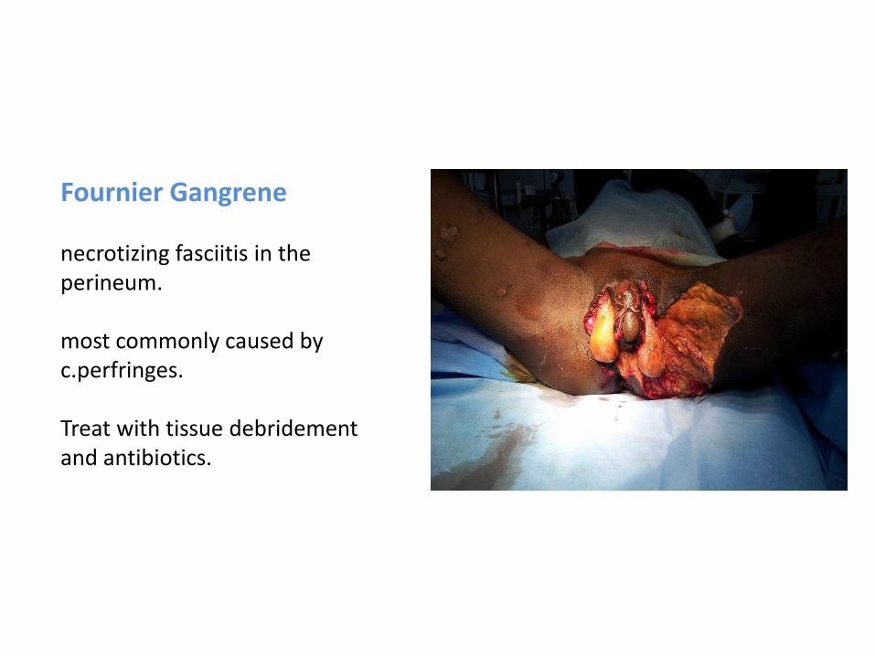
Fournier Gangrene necrotizing fasciitis in the perineum. most commonly caused by c.perfringes. Treat with tissue debridement and antibiotics.

Lipoma -Benign tumor composed of fat cells (adipose tissue). - History: Most individuals report a lump under the skin that may have been present for several years, increasing in size very gradually or growing larger if the individual gained weight. Pain may be reported, depending upon the size and location of the tumor, but the majority of lipomas do not cause pain. On occasion, a lipoma may be painful if it rubs against a nearby nerve. - Physical exam: Most subcutaneous lipomas are dome-shaped and 2 to 10 centimeters in diameter. They can be felt under the skin as a smooth, mobile, rubbery growth. The skin covering the lipoma is normal in appearance and can be moved back and forth over the nodule.

-Tests: Usually no tests are required. If the individual's history or physical examination suggests the possibility of a malignant liposarcoma, microscopic examination of lipoma cells extracted via fine-needle aspiration (biopsy) is indicated. With irregularly-shaped or large lipomas (greater than 5 cm in diameter), CT scan or MRI may be performed to help differentiate a lipoma from a liposarcoma. -Treatment: They may be removed by surgical excision or liposuction.
Lipomatosis is believed to
be an autosomal dominant condition in which multiple lipomas are present on the body.

Sebaceous cyst It is a small sac filled with material from hair follicles, the skin, or an oily substance known as sebum. The cause (etiology) of sebaceous cysts is unknown. History: Individuals may report a hard, usually painless lump under the skin. The lumps are usually found on the scalp, face, ear, and genitals.
Physical exam: The exam may reveal a small/large, smooth nodule under the skin. Skin is not pinchable with an overlying punctum. They can become inflamed and infected. Infected cysts become tender and red. Treatment : Most small cysts do not require treatment. Large or painful cysts may be removed surgically.

sebaceous cyst -Benign subcutaneous cyst filled with sebum. -found in hairy areas (scalp ,scrotum ,neck ,..). -treated by surgical excision.
Important note: if there is a scalp lesion like this it's impossible to be lipoma as a differential diagnosis since lipoma emerges from fat under the skin and scalp area is devoid from fat.

Kaposi sarcoma
-malignant proliferation of endothelial cells, associated with HHV-8. -presents as purple patches and plaques on skin, may extend into visceral organs. -classically seen in three groups: 1) Transplant recipient, early spread, Rx decrease immunosuppression. 2) older eastern European males, remain localized, Rx surgical removal. 3) AIDS( Aids defining disease) - tumor spreads early, Rx increase antiretroviral therapy.
(cutaneous sarcoma appears as red hemispherical nodules or plaques) - is it painful ? no it is painless - usually associated with what ? HIV infection & AIDS

felon (whitlow): distal pulp space infection , if not treated results in osteomyelitis. cause : pricking.
Paronychia: infection of the nail fold , happens due to bad manicure or bad maneuvering of hangnails. Most common hand infection.

Tenosynovitis - Infection of the
synovial sheath surrounding tendon.
• The most causative organism of hand infection (tenosynovitis, felon, paronychia) is staph. Aureus.
• The 2nd is streptococcus. • Initial treatment : oxacillin/
ampicillin. • Then we do culture and give
antibiotics of choice. • If abscess formed, incision and
drainage. • Elevation to decrease the edema. • Resting the organ to decrease the
pain.

Antibioma Hard, edematous swelling containing sterile pus following the treatment of an abscess with long term antibiotics rather than incision and drainage. Treatment: exploration & drainage if it is indistinguishable from a carcinoma, otherwise spontaneous resolution takes place over several weeks.

Exam question 1-identify this picture 2- mention one risk factor or it is more common in? 3-treatment? answers : it is a furuncle which is more common on the back of the neck especially in diabetics and treatment is incision and drainage plus antibiotics.

Carbuncle is an abscess larger than furuncle, usually with one or more openings draining pus onto the skin. The same etiology and sites of furuncle (infection in hair follicles mostly by staphylococcus aureus ). The difference between furuncle and carbuncle is the surface openings that drain pus.

Erysipelas 1. It is an acute infection of the upper dermis and superficial lymphatics. 2. usually caused by streptococcus bacteria ( beta hemolytic group A ). 3. Erysipelas is more superficial than cellulitis. 4. It's typically more RAISED and DEMARCATED. 5. The infection may occur on any part of the skin including the face, arms, fingers, legs and toes, BUT IT TENDS TO FAVOR THE EXTREMITIES. Fat tissue is most susceptible to infection, and facial areas typically around the eyes, ears, and cheeks.

Neurofibromatosis -Autosomal dominant. -genetic disorder characterized by cutaneous neurofibromas /Café au lait spots / optic gliomas (vision problems)/ musculoskeletal deformities (scoliosis)/ learning disability and epilepsy. -Might undergo malignant transformation. -If removal caused a big defect in an area with rebundant tissue we close it by direct closure.
Café au lait spots

Bowen's disease: A rare condition . It presents as a cluster of flat , pink , papular patches which are covered with crusts . The patches and the adjacent skin have a pale brown , thickened appearance.

seborrhoeic keratosis -in the elderly " aka senile warts “. -special diagnostic feature : because they are patches of thick squamous epithelium they can be picked off if you try to pick the edges with a blunt forceps. -when it peals off , it leaves a patch of pale-pink skin with slight bleeding. -no other skin lesion behaves like this. - doesn’t need surgery. Completely benign.

Erythroplakia - Reddish patch that appears on
the oral mucosa. - It has 17 X more risk of
malignancy than leukoplakia.
Leukoplakia - White patch that appears
on the oral or genital mucosa.
- Risk factors : smoking/ القات - Premalignant (transform to
SCC).

Vascular malformation hemangioma
seen at birth but may appear late
Start as small lesions at the age of 3-4 months
Grow parallel to the child’s growth
Grow to reach their maximum size at the age of 1 year then involution
Female to male (1:1) Female to male (3:1)
High flow can lead to destructive changes
Rarely to cause any complications
Treatment : surgery/laser/ embolization
Spontaneous resolution unless complicated you should treat

Capillary hemangioma in the eyelid obstructing the eye , might lead to
Amblyopia "lazy eye" .
The same patient at different ages (hemangioma)

Vascular malformation
Sturge weber syndrome port wine stain vascular malformation involving the ophthalmic division. - Usually not evident at birth. mnemonic : S : seizures / U: unilateral weakness R: retardation ( mental ) / G: Glaucoma E : other eye problems

• If a nevus undergoes changes in the pigmentation or in the shape or ulceration it indicates a melanoma.
• We differentiate the nevus from the vascular anomaly by its color.
Nevus

Hairy nevus • It's premalignant and must be
surgically removed. • Congenital. • Black or brown pigmented
area with excess hair growth.
• In general, hair tuft or lipoma or hairy nevus located at the lower end of the back, it is associated with spina bifida.

Non melanoma skin cancer • The most common type of cancer. • Its mortality is low. • 75% basal cell carcinoma and 25% squamous
cell carcinoma. • BCC is slow growing, locally destructive and
rarely metastasize. • 80% are on head and neck. • Melanin is a protective against tumor so blacks
are less to have skin tumors.

Squamous cell carcinoma - Arising from epidermal cells. - Risk factors: sun exposure/ pale
skin/ arsenic/ xeroderma pigmentosum/ immunosuppression.
- Actinic keratosis : the precursor skin lesion.
- Raised, slightly pigmented skin lesion/ ulceration/ exudate/ itching.
- Dx: excisional biopsy for small lesion/ incisional biopsy for large lesions.
- Most common sites : head, neck and hand.
- Involves the lower lip and BCC involves the upper lip or above this level.

Basal cell carcinoma - Arising in the germinating basal cell
layer of epithelial cells. - Nodular ( ulceration, telangiectasia,
pearls). - Morphea ( many sites at the same
time/ more aggressive than the nodular type).
- Slow growing.
- Local ( rare risk of metastasis).

rodent ulcer complication of BCC
Exam question 1- what is your diagnosis ? BCC 2- do you expect to find enlarged lymph nodes ? NO

Nevoid Basal Cell Syndrome ( AD ) Presentation : 1) multiple BCC mostly on the face 2) Cysts in the jaw. 3) Intracranial calcifications. 4) Rib abnormality ( mostly bifid ribs).

Erythematous scaly lesions on the temple area : typical of actinic keratosis
Keratoacanthoma
self limiting growth and subsequent regression of hair follicle cells

Xeroderma pigmentosa • It might predispose to
squamous cell carcinoma.
• an inherited premalignant condition associated with increase risk of all types of skin tumors.
• defect in the DNA repair genes
• AR

Cleft lip: failure of fusion of frontonasal process and maxillary process. No functional deformity, only cosmetic deformity and surgery is done at age of 3 months. Breast feeding is not contraindicated.
cleft palate: failure of fusion of maxillary processes and lead to functional and cosmetic deformity . baby can’t feed, cant speak and may lose his hearing by time (acquired). surgery is done at age of 1 year as a compromise between not losing his speaking abilities and the normal growth of face.
steps that can be done to feed the baby: semi-sitting position (45 degrees) widening of the bottle milk nipple burping (as baby swallows a lot of gas)

Bilateral cleft lip and palate

Cardiovascular surgery

The presence of a **cervical rib** can cause a form of thoracic outlet syndrome due to compression of the lower trunk of the brachial plexus or subclavian artery. The patient may complain of parasthesias & numbness in his ( neck , shoulder , arm , hand ) and usually in 90% of cases are in the ulnar distribution. Weakness manifested by difficulty grasping or holding a pen , this is a result of arterial and or neural compression. The hand is usually cold.

Tension pneumothorax Chest X-ray Findings : • Massive left pneumothorax. • Complete left lung collapse. • Shift of mediastinum to right. • Depression of left diaphragm. ttt : rapid thoracostomy incision or immediate decompression by needle thoracostomy in the 2nd intercostal space midclavicular line followed by chest tube.

Tension Pneumothorax : The most reliable sign of tension pneumothorax is depression of a hemidiaphragm.
Pneumothorax in the Supine Patient . The ""deep sulcus sign"" is seen here (arrow) in the left lung base.

Chest tube
right-sided pneumothorax with a chest tube inserted. • pneumothorax localizes
more towards the apex of the lung.
• Notice that the markings are absent from the apex down to some degree.
• Notice the white visceral line.

It is unilateral diaphragmatic paralysis.(right) we can still see the costodiaphragmatic angle so it is not effusion or hemothorax.

Surgical emphysema • Subcutaneous emphysema.
• Radiolucent striations outlining pectoralis major due to air surrounding the muscle fiber bundles.
• It is usually benign, and treatment is directed at reversing the underlying cause.

Westermarck's sign : Decreased pulmonary vascular markings on CXR in a patient with pulmonary embolus

Aortic dissection • Double-barrel aorta.
• X-ray findings :
-Widened mediastinum.
- Pleural effusion.
• Aortography : definitive gold standard but time consuming.
• ttt : Stanford type A (surgical)/ Stanford type B (medical ‘control BP’ unless complicated by rupture or significant occlusions).


Mitral stenosis
X-ray findings :
• Enlarged left atrium.
• Signs of pulmonary congestion.
• Straight line sign.
Diagnostic tests :
• Echocardiogram.
• Catheterization.
ttt:
• Open heart surgery.
• Balloon valvoplasty.
• Valve replacement.

Tetralogy of Fallot "boot" shaped heart on chest X-ray.
Transposition of great vessels Egg shaped heart

ARDS ( bilateral diffuse pulmonary infiltrates ) Other DDx: 1-severe pulmonary edema. 2-pulmonary hemorrhage. 3-pulmonary fibrosis. ( history differentiates between these conditions )
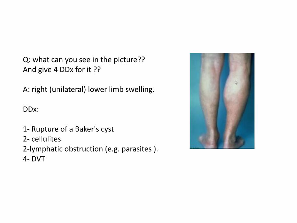
Q: what can you see in the picture?? And give 4 DDx for it ?? A: right (unilateral) lower limb swelling. DDx: 1- Rupture of a Baker's cyst 2- cellulites 2-lymphatic obstruction (e.g. parasites ). 4- DVT

Lymphangiosarcoma
-As a complication of long-standing lymphedema , usually in the edematous arm of post radical mastectomy patient. -to prevent it : use elastic compression stockings.

Mechanical prosthetic valves
Used if the age is < 60 + long life expectancy.

Animal tissue valve
Biological heart valves
Used in the following cases: - Age > 60 - Previous thrombosed mechanical valve. - Limited life expectancy. - If Coagulation is contraindicated. - Young women wishing to get pregnant.

Inferior Vena Cava Filter Mention 3 indications for it's use. 1- Proven venous thromboembolism with contraindication for anticoagulation. 2- Proven VTE with complications of anticoagulation. 3- Recurrent VTE despite adequate anticoagulation. C.I to give warfarin.

Intra-aortic balloon pump The Intra-aortic balloon pump (IABP) is a mechanical device that increases myocardial oxygen perfusion while at the same time increasing cardiac output. Increasing cardiac output increases coronary blood flow and therefore myocardial oxygen delivery. It consists of a cylindrical polyethylene balloon that sits in the aorta, approximately 2 centimeters (0.79 in) from the left subclavian artery and counter pulsates. That is, it actively deflates in systole, increasing forward blood flow by reducing afterload through a vacuum effect. It actively inflates in diastole, increasing blood flow to the coronary arteries via retrograde flow. These actions combine to decrease myocardial oxygen demand and increase myocardial oxygen supply.

Notes : - the polyethylene balloon has a radiopaque tip. -we go through the femoral artery up to the tip of descending aorta distal to Subclavian . -it increases coronary blood flow by decreasing the afterload . -the balloon inflates during diastole and deflates during systole . -Depending upon the patient’s hemodynamic status, the balloon is programmed to assist every beat 1:1 or 1:2. -linked to ECG (deflates prior the ending of QRS complex) ,(inflates in the middle of T wave ) or pressure transducer. -indications :Cardiogenic shock post-MI , (CABG) ,post cardiothoracic surgery, unstable angina . -most important complication is lower limb ischemia, we have to check the pulse and perfusion . -most important contraindication: aortic valve insufficiency (AR) ,aneurysm .

Abdominal x-ray with evidence of the calcified edge of the abdominal aortic aneurysm.

Deep vein thrombosis, or deep venous thrombosis, (DVT) is the formation of a blood clot (thrombus) within a deep vein, predominantly in the legs. Non-specific signs may include pain, swelling, redness, warmness, and engorged superficial veins. Pulmonary embolism, a potentially life-threatening complication, is caused by the detachment (embolization) of a clot that travels to the lungs.

Q: what can you see in this chest X-Ray ? A: 1- sternal wires in the midline (indicate that patient U/W sternotomy). 2- pacemaker.

Cardiac tamponade - Beck’s triad : hypotension/
increased JVP/ muffled heart sounds.
- Pericardial effusion. - Kussmaul’s sign. - Treatment : immediate
decompression via needle pericardiocentesis.


General surgery

Exam question Q: what is this? Why do we use it? A: incentive spirometer , used after surgery to prevent atelectasis . (used while inspiration not expiration).

Indications for chest tube : 1-Pneumothorax: accumulation of air or gas in the pleural space. 2-Pleural effusion: accumulation of fluid in the pleural space. 3-Chylothorax: a collection of lymphatic fluid in the pleural space. 4-Empyema: a pyogenic infection of the pleural space. 5-Hemothorax: accumulation of blood in the pleural space. 6-Hydrothorax: accumulation of serous fluid in the pleural space. 7-Postoperative: for example, thoracotomy, oesophagectomy, cardiac surgery.

Chest tube drain

T-tube used for post operative drainage of common bile duct.

Redivac drain Drains can be: Open or closed: Open drains (Including corrugated rubber or plastic sheets) drain fluid on to a gauze pad or into a stoma bag. They are likely to increase the risk of infection. Closed drains are formed by tubes draining into a bag or bottle. Examples include chest, abdominal and orthopedic drains. Generally, the risk of infection is reduced. Active or passive: Active drains are maintained under suction (which may be low or high pressure). Passive drains have no suction and work according to the differential pressure between body cavities and the exterior.

End ileostomy Loop ileostomy 2 openings
- Usually at the RLQ. - Bag contents : watery stool. - Offensive smell. - Surrounding skin is usually
inflamed (irritated from acid).
- Median or paramedian scar is usually seen.

End colostomy - Sites : LLQ (sigmoid
colon)/ RUQ (transverse colon) / RLQ ( cecostomy)
- Formed stool in bag. - No skin changes. - Sigmoid colostomy expels
stool 1/day.
Double barrel colostomy : together on left picture and separated on right picture.

End Ileostomy -edges are spouted. -site: right iliac fossa.
Stoma prolapse : one of the complications of stoma.

Q: what is this and where do we find it?? A: Suppurative Hydradinitis in axilla Found in sites of apocrine glands : axilla ,buttocks and perineum etc. - caused by staph. Aureus. - Treatment : antibiotics/ excision of
skin with glands for chronic infection.

Myonecrosis AKA gas gangrene infection of the muscle tissue by Clostridium perfringens. surgical emergency.

Flail chest or segment occurs when three or more contiguous ribs are fractured in two or more places. It typically occurs after high impact trauma. Flail segment of chest wall that moves paradoxically (opposite to the rest of chest wall)
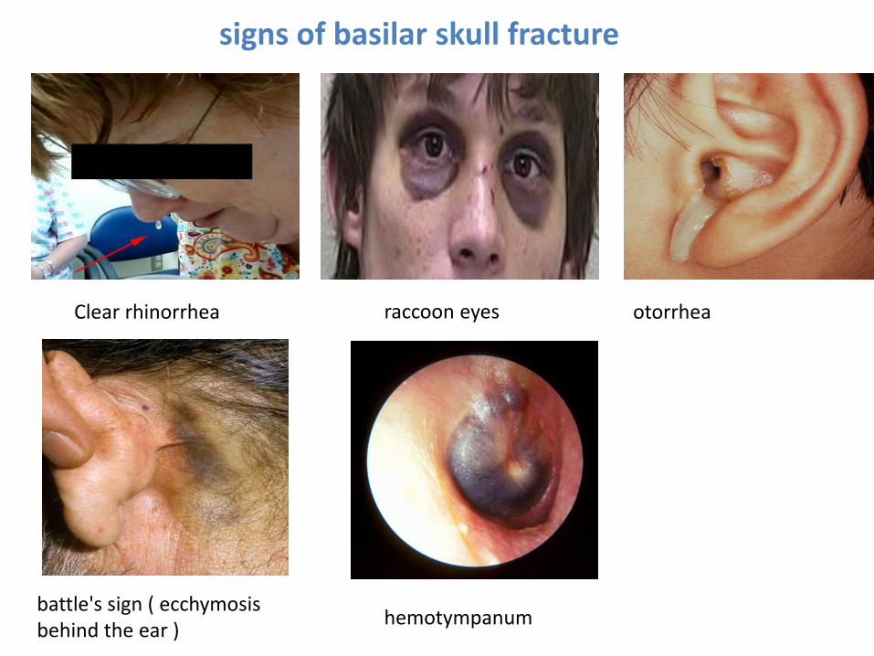
signs of basilar skull fracture
raccoon eyes
Clear rhinorrhea otorrhea
battle's sign ( ecchymosis behind the ear )
hemotympanum

hyphema : blood in the anterior chamber of the eye
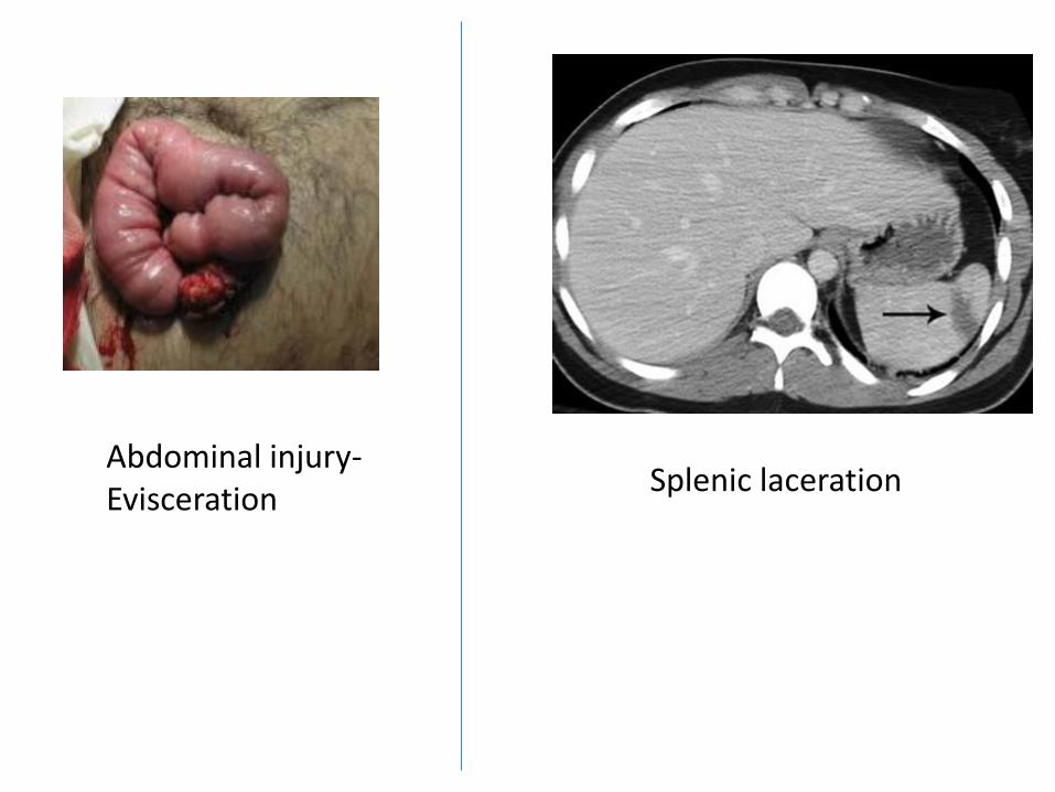
Abdominal injury- Evisceration
Splenic laceration

incisional hernia (notice the surgical scar) m.c.c is wound infection
Femoral hernia -most common hernia in females. - Medial to femoral vessels.

DDx of inguinal hernia : Hydrocele/ saphena varix/ testicular torsion/ psoas abscess .. Etc. - Indirect : most
common type in both males and females.
- Indirect : lateral to the inferior epigastric artery.
- Direct : medial within hesselbach’s triangle.
Inguinal hernia
Herniotomy : only in peds patients. Herniorrhaphy : tension due to approximation/ high recurrence. Hernioplasty : using a mesh/tension free/ open or laparoscopic.

Para umbilical hernias are the adult equivalent of an umbilical hernia where a crescent-shaped bulge develops in the navel.

Ganglion cyst it's a bulge of synovial contents caused by weakening of the retinaculum. is a non-neoplastic soft tissue lump that may occur in any joint, but most often occurs on or around joints and tendons in the hands or feet. It's painful. recurrence may occur after surgery.
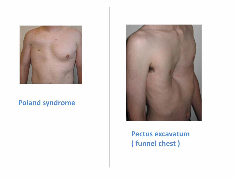
Poland syndrome
Pectus excavatum ( funnel chest )

Endocrine

Sialolithiasis = salivary calculi = salivary stones
Submandibular salivary gland stone
• The stone is located in the Wharton's duct ( most common site) : in the floor of the mouth near the frenulum of the tongue.

Significance of neck triangles 1- Submental triangle: Ludwig's angina (pus in this triangle) 2- Submandibular triangle: LN and glands, facial and lingual arteries, lingual and mandibular nerves. 3- Carotid triangle: carotid art, IJV, and vagus nerve... Carotid body tumor. 4- Muscular triangle: contains the strap muscles 5- Posterior triangle: occipital and supraclavicular triangle

Ludwig angina pus accumulation in the submental triangle. causes pressure on the larynx and epiglottis and suffocation. treated surgically by opening the submental area and draining the pus.

Carotid body tumor : in carotid triangle - moves side by side. - Dx: carotid angiogram. - Surgical excision and preoperative
embolization. - Lateral mass.

Level I: below mylohyoid muscle and above the lower margin of the hyoid bone. anterior to the posterior border of the submandibular glands level Ia: submental nodes - between the anterior bellies of the digastric muscles level Ib: submandibular nodes - posterolateral to the anterior belly of the digastric muscles Level II internal jugular (deep cervical) chain base of skull to inferior border of hyoid bone anterior to the posterior border of sternocleidomastoid (SCM) muscle posterior to the posterior border of the submandibular glands level IIa: anterior, lateral, or medial to the vein or posterior to the internal jugular vein and inseparable from it level IIb: posterior to the internal jugular vein and have a fat plane separating the nodes and the vein

Level III internal jugular (deep cervical) chain lower margin of hyoid to lower margin of cricoid cartilage anterior to the posterior border of SCM lateral to the medial margin of the common carotid artery (CCA)/internal carotid artery (ICA) Level IV internal jugular (deep cervical) chain lower margin of cricoid cartilage to level of the clavicle anterior and medial to an oblique line drawn through the posterior edge of the sternocleidomastoid muscle and the posterolateral edge of the anterior scalene muscle 4 lateral to the medial margin of the CCA Level V posterior triangle (spinal accessory) nodes level Va: superior half, posterior to levels II and III (between base of skull and inferior border of cricoid cartilage) level Vb: inferior half, posterior to level IV (between inferior border of cricoid cartilage and the level of clavicles)

Level VI prelaryngeal/ pretracheal/ Delphian node anterior to visceral space from inferior margin of hyoid bone to manubrium anterior to of levels III and IV Level VII superior mediastinal nodes between CCAs, below superior aspect of manubrium
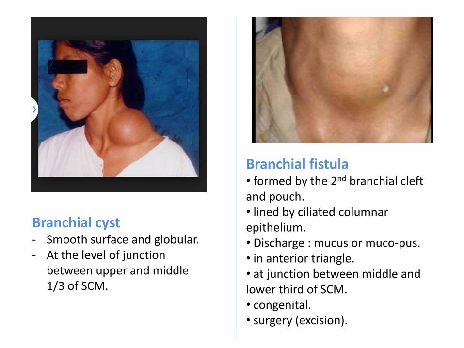
Branchial cyst - Smooth surface and globular. - At the level of junction
between upper and middle 1/3 of SCM.
Branchial fistula • formed by the 2nd branchial cleft and pouch. • lined by ciliated columnar epithelium. • Discharge : mucus or muco-pus. • in anterior triangle. • at junction between middle and lower third of SCM. • congenital. • surgery (excision).

Sublingual dermoid cyst - Medline congenital mass. - Contents : hair follicles/
sebaceous cyst/ sweat glands.
Ranula : cystic mucosa extravasation from sublingual salivary gland. Plunging : if extended through myelohyoid muscle. Treatment : excision.

Pharyngeal pouch - Diverticulation in pharyngeal
mucosa. - Bulge through weakness in the
pharyngeal constrictor muscle on the left side.
- Common in elderly men. - Dysphagia/ halitosis/ swelling
in the neck that gurgles. - Dx : barium swallow.

Pleomorphic adenoma
• Benign salivary gland tumor.
• The most common salivary gland tumor.
• Usual location : parotid gland.
• single firm, mobile, well-circumscribed mass.
• Painless. • Slow growing.

Warthin’s tumor - is the second most
common benign salivary gland tumor.
- More in males. - Associated with smoking. - Only in parotid. - Usually at the tail of
parotid. - Cystic mass.

Trousseau’s sign : Carpal spasm after occlusion of blood to the forearm with a BP cuff in patients with hypocalcaemia.

Charcot foot
• Rocker-bottom appearance.
• Develops as a result of neuropathy such as in diabetic pts.
• ttt : immobilization/ custom shoes & bracing.

Lower extremity amputations Indications : irreversible tissue ischemia & necrotic tissue/ severe infection / severe pain with no bypassable vessels, or if pt is not interested in a bypass procedure.
Bellow knee amputation Above knee amputation
Transmetatarsal amputation
Syme’s amputation Through the articulation of the ankle with removal of the malleoli.
Ray amputation Removal of toe & head of Metatarsal.

Past Years

what is you diagnosis ? FAP(focal adenomatous polyposis). what is the cause of death before the age of 50 ? Cancer.
what is your diagnosis? thyroglossal cyst. give one physical sign to confirm your Dx ? movement with tongue protrusion. give one complication ?

what is the cause of urine color ? Myoglobin. what to do ? Fasciotomy. (electrical burn causes myoglobinuria)
Diagnosis? Neurofibromatosis. what is the mode of inheritance ? AD. What are the skin spots associated with this disease? Café au late spots.

What is this device? Intermittent pneumatic compression technique
(Inflatable leg sleeves).
Uses? To prevent DVT.
give one drug that can be used in this case ? Albendazole. What is the part to be excised?

What is this? split thickness skin graft
give two signs ? exophthalmos and lid retraction . Most common cause? grave's disease.

What is this? Ileostomy. How can you confirm? By its site and skin irritation around the stoma. What is the disease that probably was treated by this? Chron’s disease.
What is this? Raynaud’s phenomenon. diagnosis ? Buerger disease (a disorder in which the blood vessels of the hands and feet become inflamed)

Mini-OSCE surgery 2016-2017
Collected by: Ghaida Khraisat

1- describe 2 findings?
Midline scar , pulging below the scar
2-what is your diagnosis?
Incisional hernia

This 9 months old child, presented with this lesion, his mother enforms you that this lesion has appeared at 4 weeks of age 1- what is the condition that this lesion will cause? Obstruction :Amblyopia( lazy eye) 2- what is your first line of management? Steroids or beta blockers (because it shows one of the complication). (note that heamangioma is not treated and you just observe them until any of the complications occur you treat them by 1-steroid or beta blockers 2- laser ablation 3-surgical excision

This pointed structure was found while laparotomy for another cause in ayoung female: 1-name it? Meckel’s diverticulum. 2- to which part of the bowel does it attached? Terminal Ileum 3- mention 2 complications for this structure? Painless Intestinal hemorrhage,intestinal obstruction(volvulus,intussusception),inflammation,perforation,….

This X-ray is for a male pt , presented with severe abdominal pain and vomiting: 1-what is the pointed structure and why? Jejenum, placia circulares 2- what is your diagnosis? SMALL bowel obstruction

1- identify this structure and what does it contain? Gall bladder , containing gall stone 2- mention one acute condition for this ? Acute cholecystitis,……. 3-Mention 2 predisposing factor for this ? Female, fatty , fertile,…..

This a picture for a patient that was involved in an electrical burn with a high voltage: 1- what is the cause of pain in this case? Ischemia (compartment syndrome) 2-what is the management in this case? Fasciotomy 3- what causes the urine colour in this case? Myoglobin

This is a picture of a wound that was left to heal by secondary intension : 1- name this tissue? Granulation tissue 2-give two components of this ? Fibroblasts, collagen,network of blood vessels,…
Similar to this tissue but the wound was open and granulation tissue was on the peripheries of the wound

1- what is the type of this skin graft? Split thickness skin graft 2- what we can do to increase the size of the graft to cover larger areas? Meshing of the graft. 3-How you can say that this graft is taken? -the graft is pink -the graft is adherent to the reciepent site - blanching with pressure denoting vascularity

This x-ray is for a 18 year old male patient after a road traffic accident : 1- what is your diagnosis? Rt hemothorax 2-what is your management? Chest tube insertion

This is a picture for a diabetic patient showing an ulcer since 10 years to heal : 1- what is your diagnosis? Marjolin ulcer 2-describe two findings? hyperpigmentation on the surrounding skin, necrotic tissue?
The picture was as this but the ulcer on the heel

This a picture of female brast with history of right breast lump: 1-describe 2 findings? peau d'orange nipple retraction

1-why shouldn't we repair it early ( before one year old)? because this will interfere with facial bone growth which leads to retardation of maxillary growth (dish face). 2-what is the early sign on ear ? secretory otitis media 3- how to deal with it? Anti histamines OR by surgical drainage by Gromet tube insertion
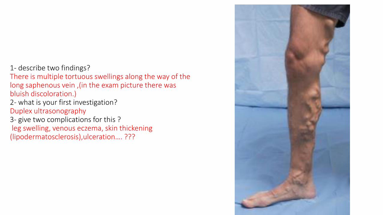
1- describe two findings? There is multiple tortuous swellings along the way of the long saphenous vein ,(in the exam picture there was bluish discoloration.) 2- what is your first investigation? Duplex ultrasonography 3- give two complications for this ? leg swelling, venous eczema, skin thickening (lipodermatosclerosis),ulceration…. ???
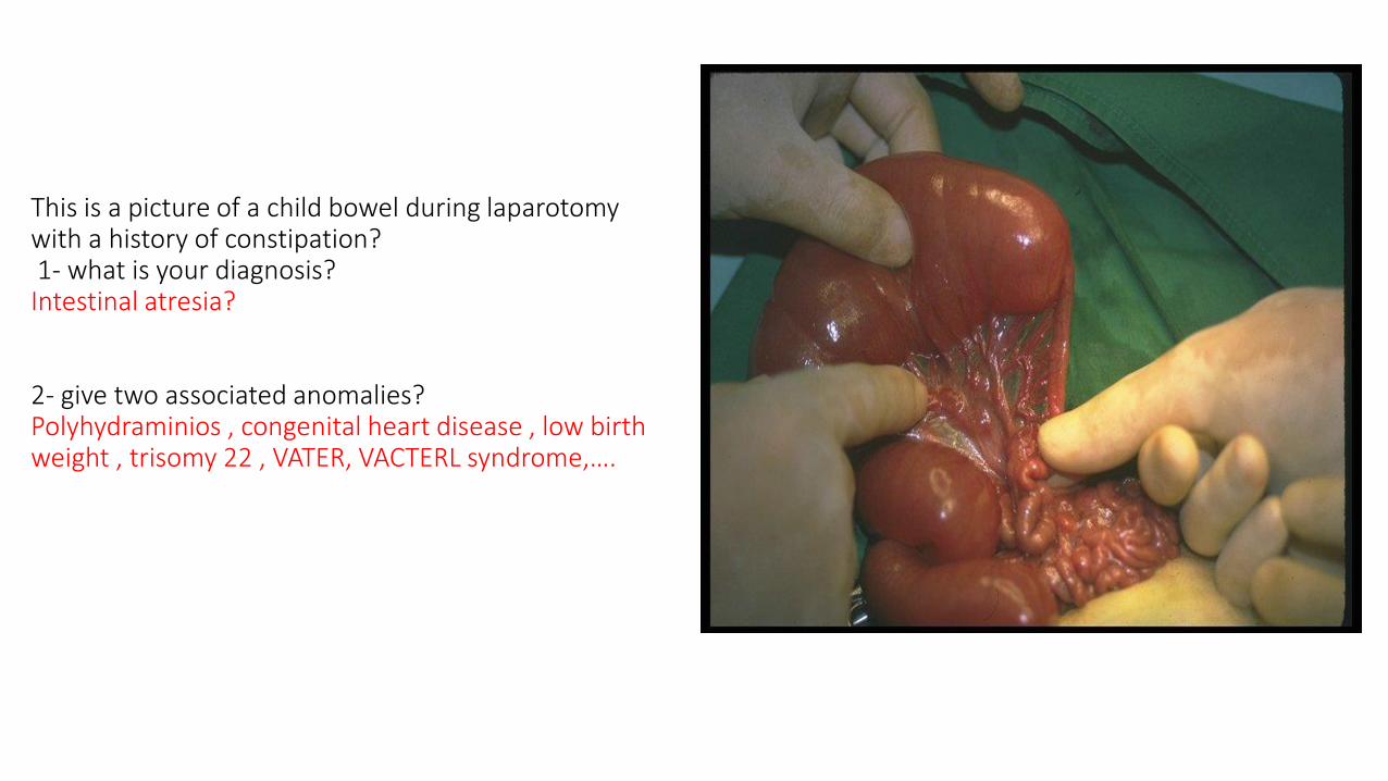
This is a picture of a child bowel during laparotomy with a history of constipation? 1- what is your diagnosis? Intestinal atresia? 2- give two associated anomalies? Polyhydraminios , congenital heart disease , low birth weight , trisomy 22 , VATER, VACTERL syndrome,….

1- Give two clinical findings? Swelling and discoloration 2-what is most probable diagnosis? DVT 3- give two predisposing factors for this? Prolonged bed rest, recent surgery, smoking , ocp, obesity,, setting for long period (long flight for example),…….

A picture for abdomen shows grey turner and cullen’s sign ,the abdomen is distended, ( but the 2 signs weren’t too obvious)
And it was for a female pt , presented to the ER with abdominal pain that radiates to the back and she has a history of gall stones
1-give two clinical findings?
Cullen’s and gray turner
2- what’s your diagnosis?
Acute pancreatitis (biliary)
3- give 2 laparatory findings?
Elevated serum lipase + amylase, elevated WBCs, …..

The picture was for a female patient , with a history of this swelling for 1 year duration: 1- describe what you see? Oval neck swelling in the anterior triangle of the neck (carotid triangle). 2- give two differential diagnosis? carotid body tumor, lipoma, firoma, neuroma…..

It was a picture of a female with a painless mass: 1-what is the most probable diagnosis? pleomorphic adenoma 2-Name this incision? Lazy “S” incision ?

A picture shows beurger's test performance with a pallor in one foot (as in the second picture) the question was: this is a picture for a 53 years old pt : 1- give a clinical finding? Pallor in the right foot 2- what is this test? Buerger’s test + buerger’s angle 3-what is the most common cause of this condition? Atheroscelerosis, DM, HTN, obesity,….

the picture was for a strangulated hernia (most probably ) the question was: an elderly male pt complains of right groin mass and pain: 1- what is your diagnosis? Strangulated inguinal hernia? 2- give two clinical findings? Bleeding or blood while doing PR, intestinal obstruction signs?? 3-give two signs that give us an indication for this condition before the surgery? Fever , tachycardia,sepsis, signs of shock?

GOOD LUCK

Surgery mini-OSCE 2015 questions
By Doctor 2011 Abdullah F. Masri
Mahmoud Mansour Khaled Alhamad

(1) This patient is a 60 year old male who presented with acute lower limb
pain of 12 hours duration.
• What is the gross finding? ___________________________
• What the most probable diagnosis? ___________________________
• What is the most common cause of this condition? ___________________________

(2) A 75 year old male, heavy smoker, presented with this lesion.
• Identify the lesion: ischemic arterial ulcer
• Give two symptoms which might be associated with the condition: claudication/ rest pain

(3) A 60 year old female with RUQ pain and fever.
• Identify this type of image: MRCP
• Give two radiological findings: CBD stone shadow/ CBD dilation.
• What is your diagnosis? Ascending cholangitis.

(4) A 25 year old male presented with anal pain and fresh blood per rectum, the
peri-anal area is shown.
• What is the diagnosis: bleeding hemorrhoids.
• Give two other complications of the same pathology: thrombosis/ infection.

(5) An 80 year old, bedridden male had this lesion in the buttock and lower back
area.
• What is this lesion? Pressure ulcer (bed sore)
• What is the most common cause? Pressure?

(6) This is a 5 year old boy.
• Give two clinical findings: scrotal swelling/ transillumination
• What is the diagnosis? hydrocele

(7) A 65 year old man underwent abdomino-perineal resection 2 years ago
after diagnosis of rectal ca.
• What is the type of his stoma? End colostomy.
• What is the complication shown? Prolapse.

(8) 18 year old male presented with sudden progressive shortness of breath
and underwent this investigation.
• What is your diagnosis? Spontaneous pneumothorax.
• Give two indications for surgery for this case: Hemo-pneumothorax (blood), failure of decompression by needle or chest tube??

(9)
• Name the triangle of the neck in which the lesion is situated: anterior triangle.
• Give two differential diagnoses for the lump: sialodenitis/ lipoma.

(10)
• What are the skin changes indicative of breast cancer in this image? Nipple retraction/ peau dé orange
• What is this procedure? Core needle biopsy (true-cut biopsy)

(11) A 60 year old female with CKD on hemodialysis.
• What is shown in the image? AV fistula hematoma.
• Mention other two complication of this case:
infection/ thrombosis/ heart failure.

(12) A 30 year old female presented with sudden abdominal pain and fever and diffuse tenderness of the abdomen.
• What is the radiological finding? Air under the diaphragm.
• What is the diagnosis? Perforated viscus ( probably perforated PUD )

(13) A 45 year old male presented with RUQ discomfort and pain, this is his
abdominal CT.
• What is the radiological finding? Peri-cyst and daughter cysts (hydatid cyst disease).
• Mention two complications of the case: Rupture and anaphylaxis/ obstructive jaundice.

(14)
• What is the radiological finding? Dilated bowel loops (jejunal) with no air in the colon.
• What is the diagnosis? Small intestinal obstruction.

(15) A 75 year old male farmer, heavy smoker presented with this lesion.
• What is the most probable diagnosis? Squamous cell carcinoma.
• What is the lymph drainage of this area? Submental and submandibular??
• What will you do to confirm your diagnosis? Biopsy for histopathology.

(16) This lady had a flame burn 2 years ago.
• What does the image show? Post-burn fibrosis and contracture. What was the degree of her burn? 3rd degree.
• What is the most suitable type of skin graft to use in reconstruction? Full thickness??

(17) This baby presented to the ER with scald burn.
• What is the degree of burn? 2nd degree.
• Mention three lines of acute management of the burn: Fluid resuscitation/ pain control/ dressing.

(18) This 1 year old baby had this lesion since birth.
• What is the most likely diagnosis? Hemangioma??
• What is the best management? Observation and reassurance.

(19) A new-born baby had inability to swallow milk and frothy mouth
secretions, this is his x-ray.
• Mention two radiological signs? inability to pass nasogastric tube/ air in the stomach.
• What is the diagnosis? Esophageal atresia with tracheo-esophageal fistula.

(20) Intra-op image of a baby with symptoms of obstruction.
• Give two findings: Dilated proximal loop, collapsed distal loop.
• What is the diagnosis? Type 1 intestinal atresia.
Doctor 2010 Mini-OSCE surgery 1- colon with polyps >>> what is you diagnosis ? FAB , what is the cause of death before the age of 50 ? cancer. 2- neck mass in the midline >>> what is your diagnosis ?? thyroglossal cyst , give one physical sign to confirm your Dx ? movement with tongue protrusion , give one complication ? ... 3- colon mass >>> your Dx ? what determine the prognosis ?? 4- omphalocele identification and prognosis.

5- electrical burn with red urine >>> what is the cause of urine color ? myoglobin , what to do ?? Fasciotomy 6- neurofibromatosis pic >>> identify ? what is the mode of inheritence ? o sho el boga3 elle bejo m3aha ? cafe o lete 7- cleft lip and palate >>> when to do surgery ? how to feed this baby ? 8- pneumatic cuff o its uses 9- hydated cyst CT , give one drug that can be used in this case ? o esh el jozo2 elle bne3malloh excision ? 10- Inflamed appendix >>> Dx ? two causes ?

11- patient with fever jaundace and RUQ pain + 9ora Dx ? procedure to be done ? cause ? 12- patient with cholengitis and septic shock and after 24 hour he developes this CXR ??? ARDS 13- arterial ulcer in the middle toe >>> describe what you see ? investigations ? 14- skin pic >>> what is this ?split thickness skin graft 15- face pic >>> give two signs ? exophthalmus and lid retraction , M.C.C ? grave's 16- sister marry joseph and it mechanism 17- paraumbilical hernia and give twi signs 18- identify inguinal hernia and give two indication for emergent surgery

19- 6 months after post. tirangle lymphnodes removal , picture with trapizius muscle atrophy ... 20- breast , give two signs ? peu de orande and nipple retraction

1) Picture of chronic venous insufficiency - What is your diagnosis ? - Give 2 evidences from the picture that support your diagnosis ? Chronic venuos insufficiency , venous ulcer & lipodermatosclerosis(discoloration). 2) Picture of ileostomy - What is it ? - How can you confirm ? - What is the disease that was probably treated by this ? Ileostomy , skin irritation around the stoma & site , crohn’s disease 3) Picture of colon cancer in resected descending colon “I think” - What is the most likely diagnosis - Mention 2 things that support it Colon cancer , ulceration & (presence of a mass or bleeding???) 4) Picture of intussusception - What is the diagnosis ? - What caused it ? Intestinal obstruction by intussusception

5) Picture of gastrostomy or jejunostomy tube - What is it ? - Why is it used ? – What is the mechanism of inserting it ? Gastrostomy tube or jejunostomy(???) , used for enteral feeding , 6) Picture of mass on the neck - What is the triangle ? - What’s the most likely diagnosis - What supports your diagnosis The triangle was the posterior one , “”I think it was a sebaceous cyst because it had a punctum and present for 10 years”” 7) Picture of paraumbilical hernia - what is the diagnosis - What is the evidence that supports your diagnosis Picture of huge neck swelling with scar - Mention 2 things evidenced by the picture

9) Mid-sternal scar with fibrosis - Mention 2 things in the picture - What is the diagnosis.. 10) Picture of extra finger in the hand of a baby - What is the diagnosis ? – Mention one syndrome causing this abnormality ? - What is the treatment ? Polydactyly , down’s ?? syndrome”and others..” , surgical excision 11) Picture of lower limb ischemia “black foot with transmetatarsal amputation” - Mention 2 MAIN clinical findings Dry gangrene , transmetatarsal ambutation , and there was evidence of chornic arterial insufficiency 12) Picture of fasciotomy in the lower limb - What was done ? - Why ? Fasciotomy , to relieve intra-compartment pressure and prevent ischemia

13) Picture of tumor on the upper lib - What is the most probable diagnosis - Mention 2 things that support your diagnosis Basal cell carcinoma (?), ulcer & ??? 14) Red urine - Mention 2 causes of this in electrical-burn pt. Myoglobinuria & hemoglobinuria 15) Breast ca. “gross picture with ulceration and nipple retraction ” - Mention 2 findings - What is the T stage for this tumor Ulcer , nipple retraction , T4 16) Breast lesion - What is the diagnosis ? Paget disease of the nipple

17) Mammogram - What is the most probable diagnosis ? - Mention the finding that supports your diagnosis - What is the BIRAD grade ? Breast cancer , radiopaque speculated lesion (with microcalcification??) , BIRAD 5 ?? 18) Picture of the big toe “swollen and red” - What is the diagnosis ? - Mention 2 Findings Paronychia (??????) , swelling & redness 19) Mass on the back - What is the most probable diagnosis ? - Mention 3 features of this lump evidenced by the picture that support your diagnosis Lipoma , well-circumscribed , circular , normal skin above and surrounding it 20) X-ray for pneumothorax or tension pneumothorax (??) - What is the diagnosis - What is the treatment

ليه أنيب وما ت وا عليه توكا لا ابللا
توفيقي ا (( ))
88الآية : سورة هود

The end
Copyright © 2022 FDOKUMEN