military radiology on the western front during the great war
238
RADIOLOGY INATRENCHCOAT MILITARY RADIOLOGY ON THE WESTERN FRONT DURING THE GREAT WAR RenéVanTiggelen TranslatedbyJanDirckx with contributions about the American, Belgian, British, French and German armies' radiological services
-
Upload
khangminh22 -
Category
Documents
-
view
1 -
download
0
Transcript of military radiology on the western front during the great war

A year from now we commemorate the centenary of The Great War, gone down in history as the fi rst industrial war, a brutal slaughter on a scale never experienced before.
In Flanders' Fields and on the French and German front lines an adolescent medical discipline, barely nineteen years old, reaches full adulthood: RADIOLOGY. This diagnostic specialty's unique signifi cance is recognized by all other medical specialties from the fi rst days of its existence.
The circumstances of the war propel radiology's development in ultra-fast-forward. In addition to the diagnosis of fracture and disease, the localization of projectiles is its outstanding priority. Antibiotics are not yet in existence; thus the immediate removal of a foreign body is extremely critical since preventing infection is practically the sole guarantee for the healing, if not the survival of the wounded soldier.
Amidst the desperation of a stalemate in the war the pioneer-radiologists labor like true heroes and fi ght their way to their patients' salvation, often at the peril of their own health. The “ray of X” becomes a ray of hope. We also remember that radiologists and their assistants are amongst the fi rst “nuclear victims” due to the radiation exposure during their work.
Despite (or thanks to?) this chaotic theatre, radiology takes a big leap forward. By the end of the war it earns its stars & bars side by side with the other medical disciplines. After the war no hospital, no clinic goes without X-rays any longer.
The contributions made by American, British, French, Belgian and German radiologists are extensively discussed and illustrated in a true historic perspective.
From the author's background as a radiologist this lavishly illustrated 220 page book (more than 260 fi gures, some of them in colour) offers an overview on the subject that is probably unique in the world.
About the author The author, René Van Tiggelen graduated in Medicine at the University of Louvain (UCL-1967). He then decided to specialize in radiology under the leadership of Professors P.Bodart (UCL) and G.Cornélis (UCL/KUL).
He obtained simultaneously a degree in social medicine and hospital management. He made his whole career as a radiologist in the Belgian armed forces. As an army medical offi cer with the rank of colonel, he used to be the deputy chief of staff of the medical department. As a senior hospital lecturer he taught bone radiology at the VUB (Brussels Free University, Flemish section) from 1982 to 1996 and has been a guest teacher at the EHSAL since 1998. With a team of volunteers he created the Belgian Museum of Radiology in 1990 and has been its managing director since.
2003 Sarton Award at the University of Ghent. 2011 Laureate of the Frans Jonckheere prize, awarded by the Académie royale de Médecine de Belgique, for his work: “A Transparent Skull. An illustrated history of neuroradiology.”
RADIOLOGYIN A TRENCH COATMILITARY RADIOLOGY ON THE WESTERN FRONT
DURING THE GREAT WAR
René Van Tiggelen
Translated by Jan Dirckx
with contributions about the American, Belgian, British,French and German armies' radiological services
NEW EDITION
FULLY REVISED
and EXPANDED
RADIOLOGY IN A TRENCH COAT M
ILITA
RY RADIOL
OGY ON THE W
ESTERN F
RONT DU
RING THE GR
EAT WA
R
René Van Tiggelen - Translated by Jan Dirckx
cover RX met rug.indd 1 23/01/13 11:33

Radiology in a Trench CoatMilitary radiology on the Western Front during the Great War
“I know that the reader perhaps does not need to know it all, but, I need to tell him”.
J-J Rousseau (1712-1778).

From the same author
“A Transparent Skull. An illustrated history of neuroradiology.”2007 ISBN 2930418281
Lay-out Jacques Louagie
Brussels 2013
René Van Tiggelen
Contributors
Luc De Broe, Jan Dirckx,Walter Esch, Françoise Goetghebuer,
Jeroen Huygelier, Georges Mazy,Lieven Mortelmans, François Olier,
Laurent Provost, Robert Smets, Ronny Van Loon, Luc Viaene
Radiology in a Trench CoatMilitary Radiology on the Western Front
during the Great War

© The Authors© Academia Press Eekhout 2 9000 Gent Tel. 09/233 80 88 Fax 09/233 14 09 [email protected] www.academiapress.be
Distribution:J. Story-Scientia Scientific BooksellersSint-Kwintensberg 87B - 9000 GentBelgiumTel. 00 32 9 225 57 57 Fax 00 32 9 233 14 [email protected] www.story.be
Van Tiggelen RenéRADIOLOGY IN A TRENCH COAT. MILITARY R ADIOLOGY ON THE WESTERN FRONT DURING THE GREAT WAR
Brussels, 2013, 221 pages.
ISBN 978 90 382 1939 4 D/2012/4804/133U 1863
All rights reserved. No part of this book may be reproduced in any form by any electronic or me-chanical means (including photocopying, recording, or information storage and retrieval) without permission in writing from the publisher or the author.
The author has attempted to trace the copyright holder of all the figures reproduced in this publication and apologizes to copyright holders if permission to publish in this form has not been obtained.
Belgian museum of Radiology www.radiology-museum.be [email protected]

I
DE BROE Luc. M. Chemistry. Former employee of the Schering and AstraZeneca companies. Librarian of the Palfyn Foundation, Ghent (Museum for the History of Medicine).
DIRCKX Jan. Nurse. Former Administrator of the Medical Imaging Department at the free University of Brussels (VUB). Dutch-English interpreter for Language Line Services firm in the U.S. Volunteer translator and proofreader for the Belgian Museum of Radiology.
ESCH Walter. BA, BBA. Former manager at Agfa-Gevaert, Healthcare (Mortsel-Belgium).
GOETGHEBUER Françoise. Romanist. Volunteer secretary of the Belgian Museum of Radiology.
HUYGELIER Jeroen. Belgian military historian.
LOUAGIE Jacques. Lay-out informatician. Volunteer of the Belgian Museum of Radiology.
MAZY Georges. Author of monographs on the motorization of the Belgian Army.
MORTELMANS Lieven. Physician-radiologist. Former head of the Radiology Department of the AZ Middelheim Hospital in Antwerp.
OLIER François. Major. NCO French Military Medical Service (Brest). Historian. Co-author of "Hôpitaux militaires dans la guerre 1914-1918".
PROVOST Laurent. Medical imaging technologist (France). Author of numerous articles on the history of radiology.
SMETS Robert. Engineer medical imaging. Former employee of Siemens Healthcare (Belgium).
VAN LOON Ronny. Physicist. Professor emeritus at the Free University of Brussels (VUB).
VIAENE Luc. Physician, Major-General (retd.). Former Chief General-Staff of the Army Medical Service and co-author of books on the history of the Medical Service of the Belgian Army.
Contributors


III
"He who does not honour the past is not worthy of the present".Inscription on the front of the fortress of Douaumont (France)
This book is dedicated to the memory of the Belgian troopsof the Medical Service, deceased during the Great War:
54 Physicians 13 Veterinarians (not incorporated in the Medical Service at the time) 1 Pharmacist 19 Nurses 12 Chaplains 248 Non-commissioned offi cers and soldiers
Dedication

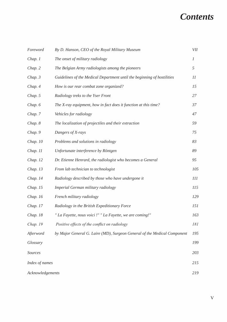
Foreword By D. Hanson, CEO of the Royal Military Museum VII
Chap. 1 The onset of military radiology 1
Chap. 2 The Belgian Army radiologists among the pioneers 5
Chap. 3 Guidelines of the Medical Department until the beginning of hostilities 11
Chap. 4 How is our rear combat zone organized? 15
Chap. 5 Radiology treks to the Yser Front 27
Chap. 6 The X-ray equipment, how in fact does it function at this time? 37
Chap. 7 Vehicles for radiology 47
Chap. 8 The localization of projectiles and their extraction 59
Chap. 9 Dangers of X-rays 75
Chap. 10 Problems and solutions in radiology 83
Chap. 11 Unfortunate interference by Röntgen 89
Chap. 12 Dr. Etienne Henrard, the radiologist who becomes a General 95
Chap. 13 From lab technician to technologist 105
Chap. 14 Radiology described by those who have undergone it 111
Chap. 15 Imperial German military radiology 115
Chap. 16 French military radiology 129
Chap. 17 Radiology in the British Expeditionary Force 151
Chap. 18 " La Fayette, nous voici !" " La Fayette, we are coming!" 163
Chap. 19 Positive effects of the conflict on radiology 181
Afterword by Major General G. Laire (MD), Surgeon General of the Medical Component 195
Glossary 199
Sources 203
Index of names 215
Acknowledgements 219
Contents
V

VII
For more than a decade and with the centennial of the outbreak of hostili-ties just around the corner countless historians have revisited the myth of the “Great War of 1914-1918”. The vision upon the conflict has in that way definitely been adjusted.In this perspective we understand how and why the First World War, as a modern and industrial war about to thoroughly influence the 20th century, was the result of a constant evolution.
Modern indeed, as the 1789 revolution and in particular the 1813 Leipzig battle heralded conflicts between states and/or nations, rather than opposing kings and their armies as was the case during the Ancien Régime.
Industrial also, because of the massive production of weapons, ammunition or uniforms, and because industry managed to produce new weapons such as machine-guns or armoured vehicles.
Without forgetting the American Civil War however, it is now largely ac-cepted that the “Great War” is the first example in history of a fully-fledged industrial war.
Foreword
The Great War has recently been perceived as the conflict supplying a defin-ing element to the entire 20th century. “Quite a discovery!”, the reader will be inclined to think. After the era of the “Sun King” or that of “Enlightenment” the 20th century in its turn receives a denominator: it becomes the “century of brutalisation”.
Both philosophers and historians in that way wish to stress that, from then on-wards, the military aspects fundamentally differed from previous situations: fighting was no longer suspended because of cold weather and the state of war became permanent, with – as a consequence – violence as a total, global and constant reality.
A few days of leave somewhere behind the front line could not erase the fact that for the first time in history the soldier, if not wounded or killed, had to face long months or even years of questionable hygiene and miserable food supplies.
He had to face an interminable separation from family and loved ones and the loss of comrades whose death struggles he could not alleviate. The screaming of the wounded was hardly suppressed by the unending booming of cannons and of shells exploding mere yards away.
The First World War soldier was “brutalised” in surroundings he could not escape; he was hit to the core, not only physically but also mentally. Medical services were in that context of course of the essence. Several innovations appeared, with military medicine exploring the fields of electrotherapy and radiology.
In the following pages Doctor Van Tiggelen assembles numerous pictures and clear diagrams. He provides highly scientific but nevertheless extremely comprehensible explanations about the birth and the development of medical radiology in military surroundings. All aspects are reviewed: the pioneers, the nursing staff, the radiological instruments and their applications in hospitals or “radiology cars” along the front.
VIII

IX
Colonel (retd.) R. Van Tiggelen also explores the changes appearing in the sanitary protocols of military (and more specifically Belgian) authorities. Ini-tially, patients were evacuated as far away from the front as possible. Sani-tary tactics were nonexistent; medical selection at the front unheard of. The wounded arrived in the hospitals days later, after heavy blood loss and with infected wounds. Their lives were in danger. Radically new medical methods were then applied. Hospitals were set up relatively close to the front and ad-vanced surgical antennas saw the light of day, with radiological cars provid-ing liaison.
As the author puts it: “This primary nursing method is still applied at the front today, albeit adapted and updated”. The second part of the title therefore is more than an easy pun. Attention is indeed drawn to all aspects of an evolu-tion. Thousands of people benefitted from that evolution.
The historian reminds us of the fact that the undeniable progress during the First World War actually corresponded to a ruthless finality: patients had to be treated efficiently in order to be once again sent out to the front as quickly as possible…
Dominique HansonCEO of the Royal Military Museum

R. P
akus
ch (1
982)
.172

The onset of military radiology
Already soon after the discovery of W.C. Röntgen in December 1895, the military doctors from different countries become interested in the diagnostic applications of this invention.
In March 1896, the "Kaiser Wilhelm Academy" in Berlin features the first military radiology.
A month later, the German Physician General O. von Schjerning and his collaborator F. Kranzfelder publish an article on the usefulness of X-rays to examine gunshot wounds. 215
In May of that year the Italian Physician Lieutenant Colonel G. Alvaro at the Military Hospital of Naples, uses radiology equipment to examine patients with gunshot wounds suffered in the Ethiopian campaign (Fig. 1). 4
The French army doctors do not allow themselves to go unnoticed. Sure enough, L. Kelsch uses X-rays in 1897 to carry out fluoroscopies to detect pulmonary tuberculosis in the Military Hospital Desgenettes at Lyon. 28
Fig. 1. Probably the oldest radiography on pa-per. This one is of the forearm of a soldier wounded on the battlefield. The arrow indi-cates the shadow of the applied iodine treatment. " I " indicates the shadow of the projectile .
Chapter 1
1

During the Greco-Turkish war of 1897 the fi nished military radiological material is tested by the British and also by German teams. 1, 130
Then it's waiting until the war in India (1897-1898) for the fi rst radiological ap-paratus to appear in the fi eld. This arrangement is successfully used by the British Doctor Major W.C. Beevor. 11, 23
In 1898 also the British Doctor, Major J. Battersby, uses a radiological fi eld de-vice and conducts radiological exams during the war in Sudan. 9 The oldest ra-diographic documents available show an equipment set up on the banks of the Nile (Fig. 2), powered by electric batteries which in turn are charged by means of a dynamo (Fig. 3)!
During the Boer Wars (1899-1902), the equipment is transferred from the Sudan to southern Africa and the power source is improved by the British Physician (Second Lieutenant) F. Bruce. 22
During the Spanish-American War (1898), the American Navy distinguishes it-self, in particular by a surprise, namely the introduction of three radiological ap-paratuses, mounted respectively on the ships Relief, Missouri and Bay State. This is not a fi rst, because that one goes to the Russian cruiser Aurora, (Fig. 4) which is apparently the fi rst warship provided with radiology equipment. The American, Physician W.C. Borden, publishes a treatise in 1898 about his experience using military radiology in wartime (Fig. 5). 19
Fig. 5. Cover of the fi rst treatise on war radiology.
Fig. 3. There is half an hours's 'cycling' needed for one single exposure.
Fig. 2. Major Battersby (left) along the Nile (1899). Note that any protection around the radia-tion tube is lacking!
2
J. B
atte
rsby
(189
9).9
Fig. 4. Cruiser Aurora at the Neva river in St Peterburg, pho-tographed in May 2012. In May 1905 already V.S. Kravchenko used a Siemens-Halske radiological equipment for examina-tion of sailors.
L. M
orte
lman
s.

Fig. 4. Cruiser Aurora at the Neva river in St Peterburg, pho-tographed in May 2012. In May 1905 already V.S. Kravchenko used a Siemens-Halske radiological equipment for examina-tion of sailors.
It is noteworthy that now, in 1912, all German military hospitals are equipped with radiology devices. The fi eld services are increasingly integrated with mobile equipment.
During the Russo-Japanese War (1905-1906), the Japanese use German equipment (Fig. 6). They also perform numerous radiological exams, which are commented upon by the Frenchman, Physician Major J.J. Matignon (1907).153
During the First World War, the vast majority of the combatant parties possess portable equipment, mostly carried in horse-drawn wagons (Fig. 7). These wagons also serve well as darkroom. The equipment is powered by a genera-tor supplied by a petrol engine.
Fig. 7. Radiological equipment in a horse-drawn wagon of the German army between 1905 and 1910.
Fig. 6. German equipment in use by the Japanese.
3
J.J. M
atig
non
(190
6). 15
2
R. P
akus
ch (1
982)
. 172
Siem
ens M
edic
al A
rchi
ves.
187

The Belgian Army radiologists among the pioneers
The discovery of X-rays by W.C. Röntgen goes back to 5 November 1895. The news spreads worldwide, unbeknownst to the scientifi c community, at the beginning of 1896, thanks to the media and the telegraph. This is a world’s fi rst, since before that a discovery always fi rst circulated through the scientifi c journals.
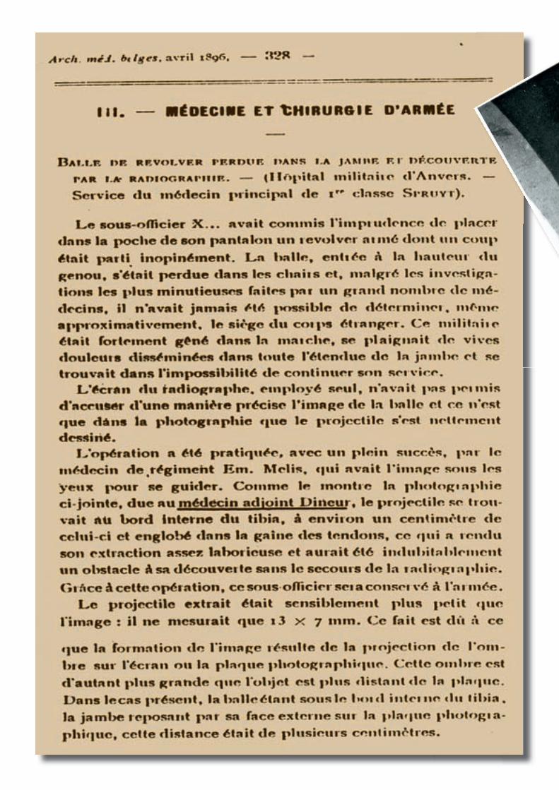
On the occasion of one of the regular meetings of the Scientifi c Associations of the Botanical Garden of Antwerp, informs its director Professor H. Van Heurck, breaks the news. Physician Th. Dineur of the Military Hospital of Antwerp is present at the meeting. He carries a radiographic study (Fig. 8) that he publishes in April 1896. 57, 206, 207
So in early 1896 the fi rst radiology department in the Belgian Army is founded in Antwerp. The fi rst chief of the department is Dr. Th. Dineur (1866-1924).
The capital cannot stay behind. In early 1897 Doctor E. Dupont (1865-1922) establishes the fi rst radiology department in the Military Hospital in Brus-sels.60
Chapter 2
The Belgian Army radiologists among
Fig. 8. X-ray print, probably executed in January 1896, shows a projectile which ended up somewhere in the soft tissue of the lower leg. Until the war of 14-18 it is custom to make a positive print (photo) departing from the negative. Hence, the bony structures and the bullet are dark, while those on a modern fi lm show in white 57, 191
5
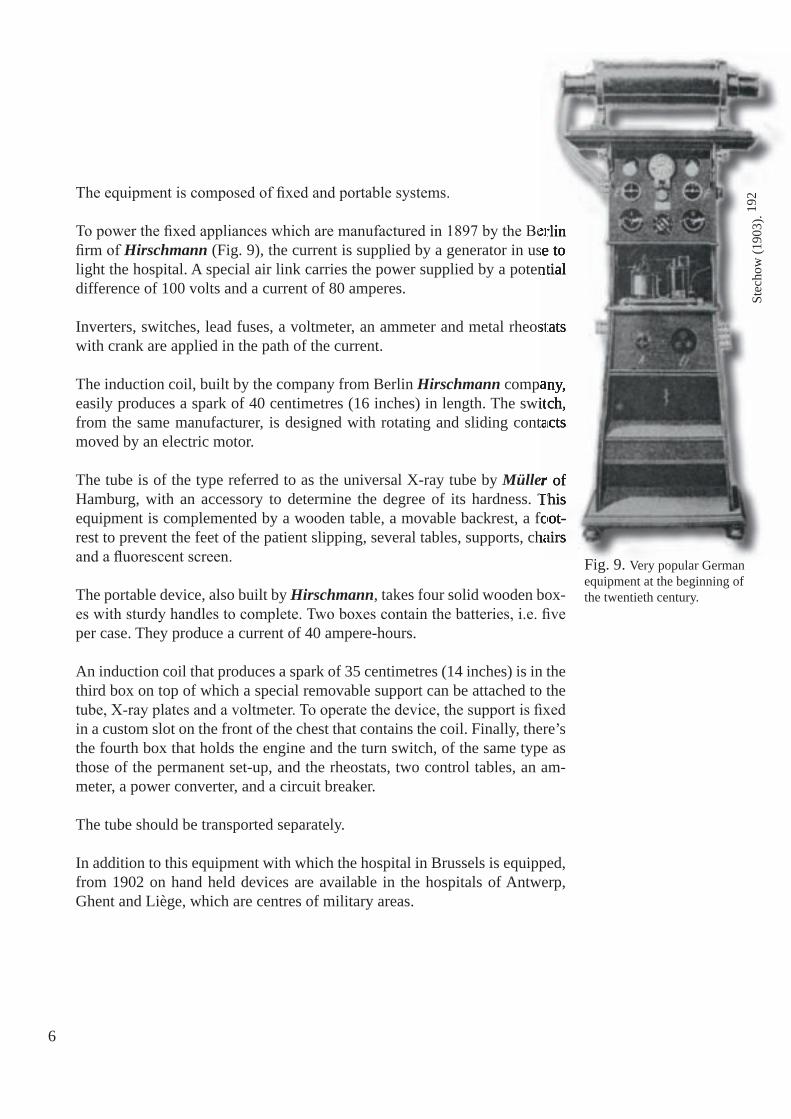
The equipment is composed of fi xed and portable systems.
To power the fi xed appliances which are manufactured in 1897 by the Berlin fi rm of Hirschmann (Fig. 9), the current is supplied by a generator in use to light the hospital. A special air link carries the power supplied by a potential difference of 100 volts and a current of 80 amperes.
Inverters, switches, lead fuses, a voltmeter, an ammeter and metal rheostats with crank are applied in the path of the current.
The induction coil, built by the company from Berlin Hirschmann company, easily produces a spark of 40 centimetres (16 inches) in length. The switch, from the same manufacturer, is designed with rotating and sliding contacts moved by an electric motor.
The tube is of the type referred to as the universal X-ray tube by Müller of Hamburg, with an accessory to determine the degree of its hardness. This equipment is complemented by a wooden table, a movable backrest, a foot-rest to prevent the feet of the patient slipping, several tables, supports, chairs and a fl uorescent screen.
The portable device, also built by Hirschmann, takes four solid wooden box-es with sturdy handles to complete. Two boxes contain the batteries, i.e. fi ve per case. They produce a current of 40 ampere-hours.
An induction coil that produces a spark of 35 centimetres (14 inches) is in the third box on top of which a special removable support can be attached to the tube, X-ray plates and a voltmeter. To operate the device, the support is fi xed in a custom slot on the front of the chest that contains the coil. Finally, there’s the fourth box that holds the engine and the turn switch, of the same type as those of the permanent set-up, and the rheostats, two control tables, an am-meter, a power converter, and a circuit breaker.
The tube should be transported separately.
In addition to this equipment with which the hospital in Brussels is equipped, from 1902 on hand held devices are available in the hospitals of Antwerp, Ghent and Liège, which are centres of military areas.
To power the fi xed appliances which are manufactured in 1897 by the Berlin (Fig. 9), the current is supplied by a generator in use to
light the hospital. A special air link carries the power supplied by a potential
Inverters, switches, lead fuses, a voltmeter, an ammeter and metal rheostats
company, easily produces a spark of 40 centimetres (16 inches) in length. The switch, from the same manufacturer, is designed with rotating and sliding contacts
MüllerHamburg, with an accessory to determine the degree of its hardness. This equipment is complemented by a wooden table, a movable backrest, a foot-rest to prevent the feet of the patient slipping, several tables, supports, chairs
To power the fi xed appliances which are manufactured in 1897 by the Berlin (Fig. 9), the current is supplied by a generator in use to
light the hospital. A special air link carries the power supplied by a potential
Inverters, switches, lead fuses, a voltmeter, an ammeter and metal rheostats
company, easily produces a spark of 40 centimetres (16 inches) in length. The switch, from the same manufacturer, is designed with rotating and sliding contacts
Müller of Müller of MüllerHamburg, with an accessory to determine the degree of its hardness. This equipment is complemented by a wooden table, a movable backrest, a foot-rest to prevent the feet of the patient slipping, several tables, supports, chairs
Fig. 9. Very popular German equipment at the beginning of the twentieth century.
6
Stec
how
(190
3). 19
2

These devices, by the same German manufacturer, differ from the portable device from the hospital in Brussels, because they are of a later date.
Each one is composed of two solid oak chests with strong handles. One of them contains 24 small batteries and a second one the induction coil to pro-duce a 35 cm spark, a capacitor, a switch with platinum contacts an ammeter and a support for a tube.
The dimensions of the latter are relatively small: length 75, width 31 and height 48 centimetres (resp. 30, 12, and 19 inches).
During transportation, the carrier of the irradiation device to be used is folded back underneath the case to which it is attached. The switch is stowed away in a corner of the box. It can be taken out easily through lowering a movable side board by pulling it toward the user.
The ammeter is laid down and clamped along the coil when the unit needs to be transported. To operate, it is fi xed vertically by putting the two metal rods of its chassis in the two holes in the wall at the front of the switch.
Until 1897, all the sick who can be transported, are to be brought to the mil-itary hospital in Antwerp, for examination by X-rays. The physician, who deems the exam necessary for a soldier to undergo, will address an evacuation request for the sick to the hospital of Antwerp, and to the Inspector-General of Health of the Army. This request must be accompanied by a report summary
of the medical history of the patient and it explains the reasons why it is necessary to let him undergo radiography.
As of January 1898 (Fig. 10), the men from the areas one and two are transported to Antwerp, and those of the third and fourth areas to Brussels.
Fig. 10. Guidelines from 1898 that regulate the evacuation of patients to hospitals equipped with radiology. 59
7

After performing the radiographs the evacuated patients rejoin their hospital of origin.
The possibilities are expanded when in 1902 the hospital of Brussels and in 1902 the hospitals of Antwerp, Ghent and Liège are equipped with portable equipment.
The military in treatment in garrisons other than the above, who must under-go a radiographic examination, are evacuated to the hospital of the chief site of their area. In the event that the patient cannot be transported, the apparatus is sent to the garrison, where the soldier is located.
Usually transport is arranged by rail. The doctor, identified by the Inspec-tor General to carry out X-ray examination, brings the device along. This is entrusted prior to the journey to two soldiers-nurses who accompany the physician.
If the X-ray machine has to be used in a place not far off, it is transported by one of the vehicles in service in the garrison. Transport in this case falls under the supervision of a superior of the hospital that owns the device.
In the early twentieth century, the Assistant Physician E. Henrard joins the radiology department of the Military Hospital of Brussels. Besides his nu-merous scientific works in the field of radiology in 1906, he and his army col-leagues radiologists Th. Dineur and E. Dupont, become founding members of the Belgian Society of Radiology.
Also in 1906 along with Dr. E. Dupont, he becomes a founding member and members of the editorial committee of the Belgian Journal of Radiology. During the Great War, Doctor E. Henrard is very active in the radiological detection of foreign objects (projectile shards, bullets, etc.). He ends his ca-reer as Physician Major General. We will also devote chapter 12 to him.
8
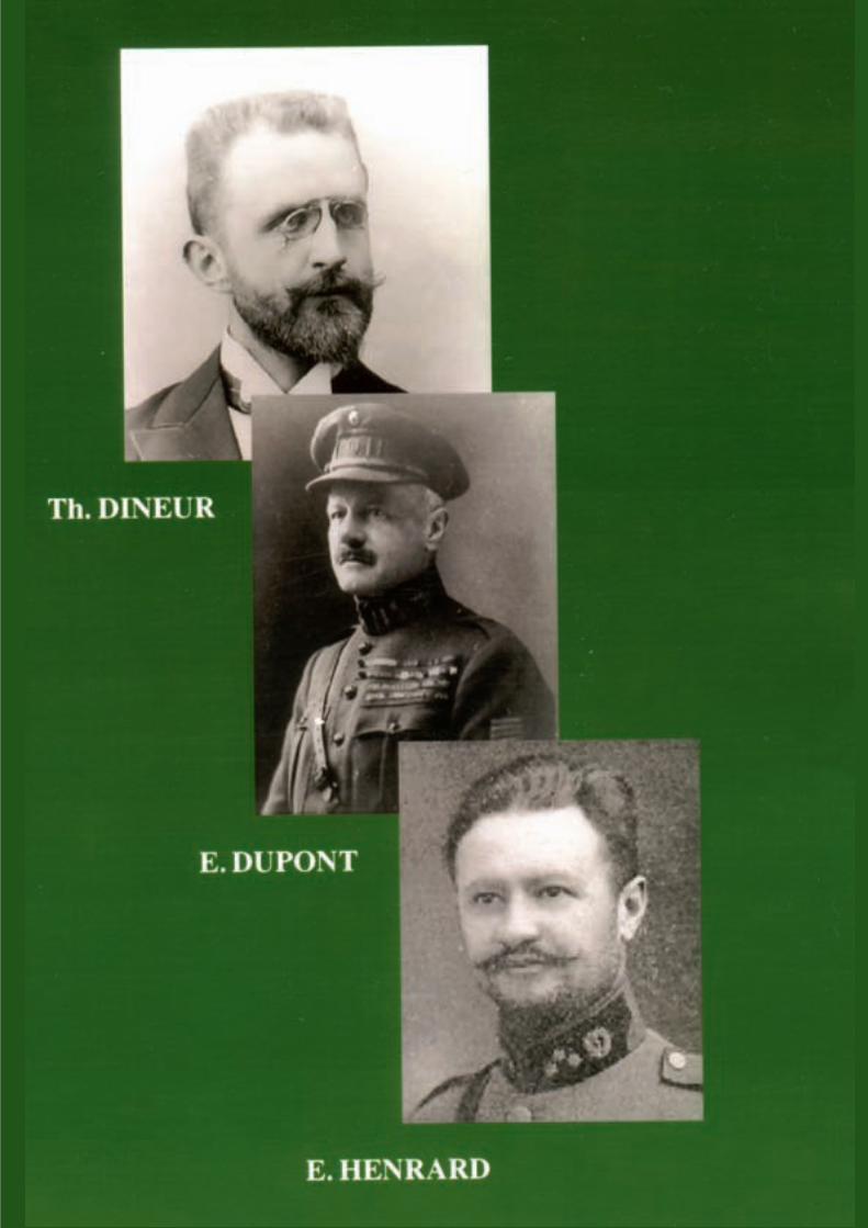

Fig. 11. Royal Decrees and Ministerial Circular regulating the conduct of the Red Cross during confl ict.
10
Mon
iteur
bel
ge (1
892,
189
3 et
189
6).
Chapter 3
Guidelines of the Medical Department until the beginning of hostilities
To all the combatants the old sanitary guidelines direct the patients to be evacuated as far as possible behind the front line before treating them; this statement is shaken by the experience of the Balkan wars. It is beaten all to pieces from the start of the Great War.
In Belgium, the Medical Service exclusively operates afield. Here are the principles of its doctrine: hostilities would take the format of a brief action. The intensity of the firing at the front would not allow to pick up the wound-ed unless until after the fighting. The wounds by "modern" bullets would be little infected.
Consequently, there is no need at the front of combat stretchers. There is equally no reason for transportation - or treatment resources either to rush to the firing line. 52, 55, 71
At division level, the columns of ambulances can be drawn by horses. The urgency requires no automobiles, and the hospitalization sections can be set up anywhere.
The evacuation prevails behind the front, and therefore the rail transport is preferred. Those who conduct the logistics are the masters and the Red Cross carries out (Fig. 11).
11

Behind the front or in the ceded territories, the fortified military hospitals or the hospitals taken over by the Red Cross should suffice.Here there is no health strategy, no triage of patients.
In 1912-13, some university or well-known surgeons had acquired experience during the Balkan wars: A. Depage (on the Turkish side), Ch. Willems and J. Conrad (on the Serbian side). They are not very optimistic but abstain to intervene, perhaps not to damage the morale?
The Medical Service will not be mobilized on 4 August 1914, when the war breaks out in Belgium, while 12 German cavalry regiments followed by 300,000 foot soldiers cross the border, supplied by 600 trains per day! This is the day the vice-president of the Red Cross chooses to notify the Inspector-General of Health, that it cannot meet its obligations...
However, by Royal Decree No. 10199 of 22 January 1892, and particularly by the Ministerial Circular of 25 February 1893 and the Royal Decree No. 11984 of 7 December 1896 (Fig. 11) the Red Cross is responsible to provide for the bulk of the medical assistance to the army in the field.
On 4 August, it becomes obvious that the Red Cross cannot provide, as prom-ised in wartime, the staff nor the essential equipment either, namely: two field hospitals for each division, ambulances, pharmacy trucks, stretchers, band-ages, and physicians for the thirty-three hospital trains (Fig. 12), built in the Arsenal of Antwerp. The Red Cross is at fault, but also the army is committing a serious blunder by ignoring the participation rights with respect to this organization. In fact, to assist the Red Cross with advice in preparation for mobilization, the post of Vice-Chairman of the Red Cross has to be reserved for the Physician General, Inspector-General of Health. This obligation is fulfilled until 1905, the year that the War Department decided that a retired general as well as a Physician-general of the Department of Health are able to manage the business! On the eve of the hostilities, the delegate of the Minister of War to the management of the Belgian Red Cross, is Lieutenant-General of the Gendarmerie V. de Coune.
L. Mélis, Inspector General of Health, foresees difficulties, and subsequently already on 2 and 3 August, calls on Dr. J. Conrad, chief surgeon at St. Eliza-beth Hospital in Antwerp, the professors G. Debaisieux and A. Dandois of the University of Leuven, Ch. Willems, aggregated in Ghent and future pro-fessor in Liège (1919), as well as the Brussels physicians P. Héger, A. De-page, and L. Leboeuf. Upon instigation by the King some of them had been active in the Balkans.
12

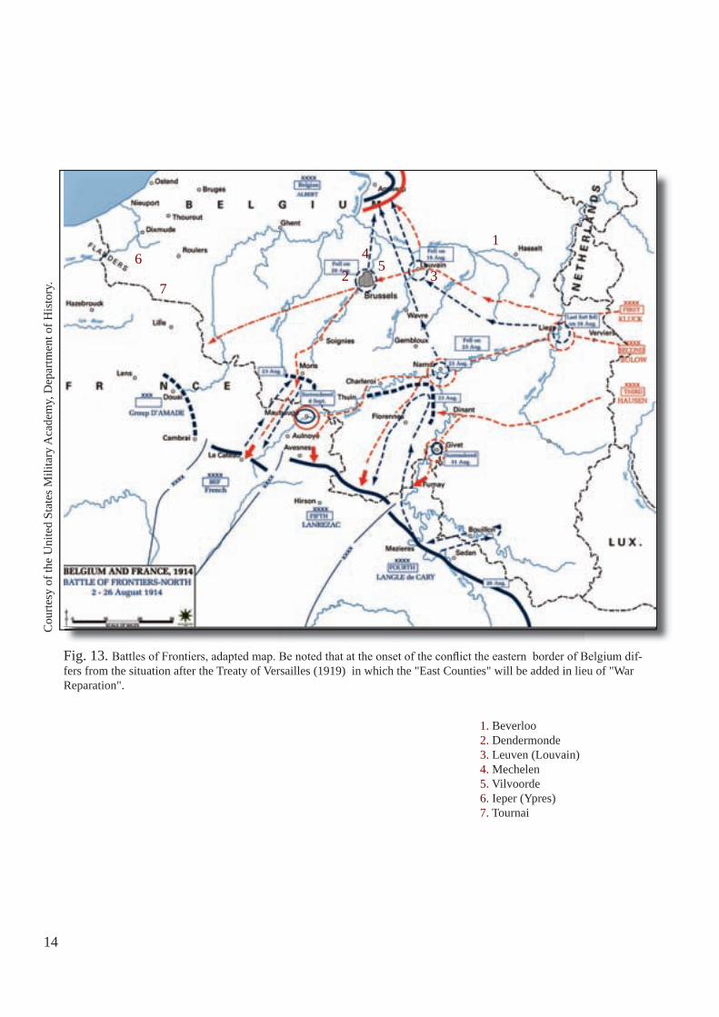
At the time of mobilization the Medical Service ensures the leadership of the military hospitals of Antwerp, Liège, Namur, Dendermonde, Beverloo, Brus-sels, Leuven (Louvain), Mechelen (Malines) and Vilvoorde. Together these provide a reception capacity of 2,320 beds.
All other institutions, that is to say the hospitals of Arlon, Mons, Tournai, Ghent, Bruges, Yper (Ieper) and Ostend, or 1,050 beds, are under the direc-tion of the Belgian Red Cross.
On 4 August 1914, we hope to be able to count on the forts, built in 1881-1890, which are scattered along the Meuse and the Scheldt.Unhappily, these forts have become defi nitely obsolete and cannot resist.... There is an infl ux of wounded.
The German invasion is irresistible: the Belgian army has to withdraw. On 17 August the Medical Service has to hand over to the Red Cross the hospitals of Leuven, Brussels, Vilvoorde, Dendermonde and Mechelen.The German army penetrates to the capital, along the "Chaussée de Louvain", on 19 August 1914. Under the command of A. von Klück it parades in Brus-sels during seventy-two hours! 186
Finally, let's say that just as the Red Cross being "dormant" until 1914, even so the new Red Cross prepares itself in a most improvised way, but without being of signifi cance to the whole country, until after the invasion.
Fig. 12. From 1905 studies L. Mélis, attached to the inspection of the Medical Service, the construction of new ambulances in collaboration with an employee of the railways. 13
Priv
ate
Col
lect
ion.
Cou
rtesy
of t
he U
nite
d St
ates
Mili
tary
Aca
dem
y, D
epar
tmen
t of H
isto
ry.
Fig. 13. Battles of Frontiers, adapted map. Be noted that at the onset of the confl ict the eastern border of Belgium dif-fers from the situation after the Treaty of Versailles (1919) in which the "East Counties" will be added in lieu of "War Reparation".
1. Beverloo2. Dendermonde3. Leuven (Louvain)4. Mechelen5. Vilvoorde6. Ieper (Ypres)7. Tournai
1
2 34
56
7
14

How is our rear combat zone organized?
At the mobilization, end July 1914, the Inspector General of the Medical Service of the Belgian Army has 166 regular medical surgeons, 520 reserve surgeons and 172 medical students at his disposal. To these personnel should be added 148 pharmacists, 965 medical service men and 1,850 stretcher bear-ers, recruited from among those men who are exempted from the army in time of peace. The Medical Service retains the immediate direction of the hospitals of Antwerp, Beverloo, Brussels, Dendermonde, Leuven (Louvain), Liège, Mechelen (Malines), Namur and Vilvoorde (Fig. 13). All the other establish-ments pass under the direction of the Belgian Red Cross. However, during the fi rst weeks of hostilities the Medical Service of the Belgian Army is forced to take over the direction of the hospitals at Bruges, Ghent, Ieper (Ypres), Mons, Ostend and Tournai. On 15 August 1914, 50,000 beds are available for the wounded in the various hospitals which have been organized throughout the country. The fall of Liège and of Namur, the entry of the enemy in Brussels, and the retirement of the Belgian Army to Antwerp, followed by its retreat and stabilization upon the river Yser, cause the loss of all these establish-ments, including most of its radiological equipment, located in the interior of the country. Not to mention the loss of about one fourth of its staff captured or staying behind to take care of the wounded whom cannot be transported.
Chapter 4
15
Fig. 14. After the offensive on the Marne (Sept 1914), the allies bring on commemorative fl owers. Belgium chooses the "daisy", France the "cornfl ow-er", and the United Kingdom the "poppy".
W. L
eliè
vre
(201
2). 13
6

16
From this moment and until the successful advance during the autumn of 1918, the Medical Service of the Belgian Army possesses very meagre re-sources in the non-occupied part of its country. These are used by the ad-vanced medical formations. For the remainder, the Medical Service is obliged to have recourse to the hospitality of Great Britain and France.
About the middle of October 1914, after having abandoned Antwerp, the Bel-gian Army comes into proximity of the coast where Ostend and Zeebrugge have become provisional bases 50, while awaiting the centralization of the Services in the rear which are soon to be established in France. The wound-ed who are in the hospitals in the interior of the country, and who can be moved, pour to the coast where beds have been provided just for two to three thousand men only. In villas and hotels, hastily reopened, are received some 13,000 unfortunate men.
The British Admiralty, on being consulted, requests a ten day period to em-bark the wounded and thus remove them from all danger. But the pressure of the enemy and the uncertainty as to when liaison with the Allies will fi nally be established shows that such a delay is impossible. The assembling at Ostend by 13 October of all the wounded, who are scattered in the various unoc-cupied localities, is therefore considered necessary by the military authori-ties. The same day a thousand wounded are sent to England aboard the state owned mail-boats (Fig. 15) and thus the general evacuation begins. 16, 137, 157 Next day, the necessary means having been assembled, and in the space of sixteen hours, more than 13,000 wounded, sick and lame are evacuated with-out mishap, thanks to the assistance of the French and British. Of this number, 8,500 are brought by rail to the port of Dunkirk, from where they embark for England. Only the ones unfi t for transportation are left behind.
Fig. 15. Beginning 10 October 1914, the "Stad Antwerpen", a Belgian mail-boat, transformed into a hospital ship, takes part in the evacuation, from Ostend.
M. V
anal
derw
eire
ldt (
1997
). 20
2
H.S
. Sou
ttar (
1915
). 19
0

27
Three days later, the Belgian Army takes up its positions in the region of the river Yser. At fi rst the Medical Service cannot depend on any of its own establishments and thus, together with the necessity of not over-loading the entrenched camp at Dunkirk (Fig. 16), which is exposed to immediate attack in the event of the withdrawal of the lines, forces it to place the fi rst installations eighty kilometres behind the front.
During the latter half of October, the majority of Belgian wounded is sent to Dunkirk and to Calais. As mentioned, the uncertainty of future events precludes keeping the wounded at Dunkirk and they have to
H.S
. Sou
ttar (
1915
). 19
0
Fig. 17. The courtyard (A) and a straw ward (B) in the Episcopal College of St Joseph (Veurne).
Fig. 16. Arrival in the snow of Belgian wounded at the ambulance installed at Dunkirk.
continue on their way to Calais. In Calais, everything is lacking, there is not a single Belgian hospital and even indispensable materials cannot be ob-tained for the improvised medical in-stallations. Also, by that time, there are two hospitals established in the non-occupied territory at Veurne (Furnes): the so called ‘Belgian Field Hospital’ (Fig. 17), a British surgical unit which has functioned at Antwerp, and anoth-er unit of Belgian military surgeons at the local civil hospital.
G. D
oum
ergu
e (1
934)
. 58
A
B

18
At Calais, the four French military hospitals are completely full. Wounded who can be moved are brought to the port, by whatever available means, and loaded upon ships going to England or to the French port of Cherbourg. Between 21 and 31 October, 10,634 people are evacuated in this manner in twenty transports.
Nevertheless, there is still a need for the establishment of hospitals to care for the wounded that cannot be transported. On 17 October, a start is made by taking over fi ve private clinics, a dispensary and a few of the beds in the local asylum for old men. Subsequently, the locomotive repair shop at the Calais railway station is transformed into a triage hospital and "hôpital de passage", while on 18 October fi ve more hospitals are opened giving a total of 683 beds.
About this time the battle of the Yser has begun and the fl ow of wounded (Fig. 18) increases day by day, with the result that the establishments which have just been opened are soon overwhelmed.
M. V
anad
erw
eire
ldt (
1997
). 20
2
Fig. 18. Boarding of Belgian wounded, on a makeshift sanitary train.
G. D
oum
ergu
e (1
934)
. 58

19
On 18 and 19 October, eight new hospitals are organized (170 beds); on 22 October one (177 beds); on 24 October four and the next day two. Besides these ‘organized’ hospitals there are numerous additional small makeshift medical installations. In spite of this increase and although transportation by sea becomes regular and frequent - the first sea transport of 500 wounded on board of the Marie-Henriette (Fig. 19), another Belgian mail-boat, leaves on 23 October, followed by 400 on 24 October, 1,200 on 25 October and more than 2,100 on 27 October - the hospital capacity of the Medical Service of the Belgian Army at Calais remains insufficient. Subsequently, seven more hospitals are opened. Most of these hospitals are former school buildings or convents, placed at the disposal of the Belgian Army by the French munici-pal authorities or by the local congregations. At first, most of the wounded lie upon the bare floor, later, straw is obtained. The situation nevertheless improves rapidly. Some of the inhabitants generously lend their mattresses and the French administration places 700 beds at the disposal of the Belgian wounded and four vessels in the docks are transformed into hospital-ships accommodating 1,200 wounded. Early November 1914, the Medical Service of the Belgian Army thus disposes of about 4,000 beds at Calais. During the months following the battle of the Yser, when the influx of wounded again surpasses the capacity of the Calais establishments, recourse is taken to eva-cuations to England.
By the end of 1914 there are also three military hospitals (Beveren, Ca-bour and Hoogstade) and one, later two, Red Cross hospitals (De Panne and Vinkem), known as ‘field hospitals’, established in the unoccupied part of Belgium (see chapter 5). The surgical personnel of these hospitals, is entirely Belgian. These hospitals are gradually provided with the latest equipment, including operating rooms, sterilization rooms, a complete equipment of sur-gical instruments and material, X-ray rooms, research laboratories, etc. They are primarily intended to handle serious cases, requiring urgent surgical care which cannot be given at the more advanced medical stations, and which re-quires an operation before they will be able to reach the hospitals further to the rear.
Fig. 19. The paddle-mail-boat Marie-Henriette, (the fastest in the world at this time) transformed into a hospital ship. During the battle of the river Yser, she is used along with other ships to disencumber the hospitals of Dunkirk and Calais and transport the wounded to Cherbourg. On 24 October 1914, she hits the cliffs of Pointe de Barfleur near Cherbourg. The 650 wounded are narrowly rescued. 48
M. V
anad
erw
eire
ldt (
1997
). 20
2

During World War One, the rules governing the evacuation of wounded Belgian soldiers is based on the principle that evacuation must be effected as rapidly as possible and in the order of the most urgent cases fi rst (Fig. 20). In each Army Division, the medical service is directed by the divisional surgeon attached to headquarters. In the units the services are directed by the regimental surgeon, under whose orders are placed from fi ve to seven surgeons.
Each Army Division has the following means of evacuation and hospitalization: one ambulance column with animal drawn transportation, two hospitalization sections and a motor transport ambulance column. The functions of these medical formations are to follow the army division in its movements and establish, according to circumstances, either an evacuation centre or infi rmaries by means of their hospital material and by the utilization of local resources.
Fig. 20. Flow-chart of the British evacuation and hospitalization system, the British ones are chosen for their simplicity.
Rep
ort o
f the
mili
tary
boa
rd o
f alli
ed su
pply
(192
4). 18
0
21
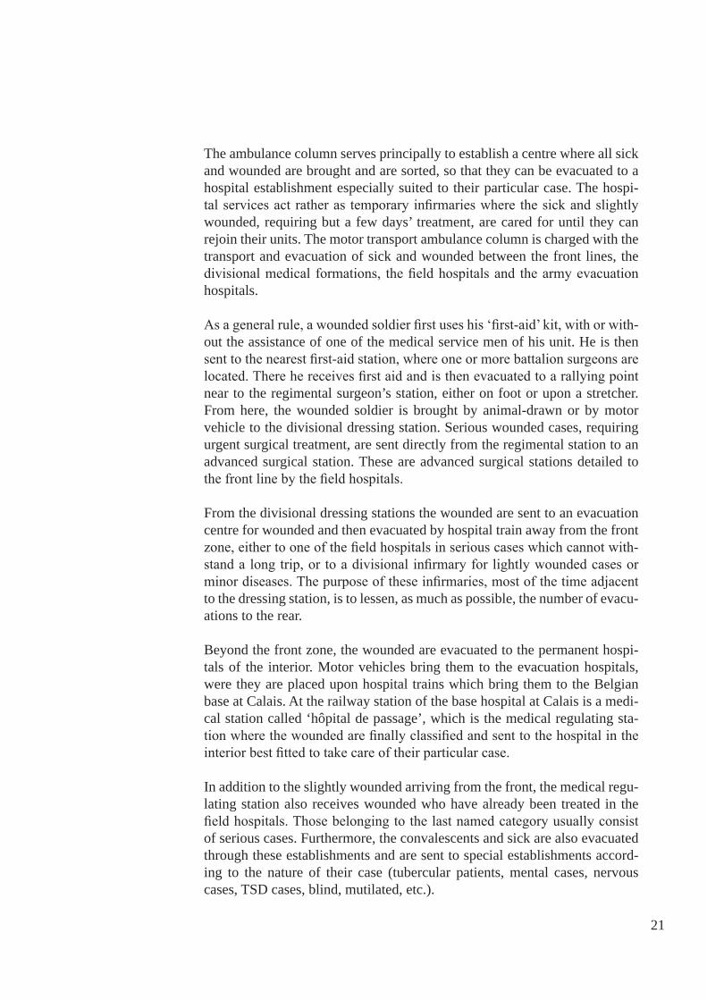
The ambulance column serves principally to establish a centre where all sick and wounded are brought and are sorted, so that they can be evacuated to a hospital establishment especially suited to their particular case. The hospi-tal services act rather as temporary infirmaries where the sick and slightly wounded, requiring but a few days’ treatment, are cared for until they can rejoin their units. The motor transport ambulance column is charged with the transport and evacuation of sick and wounded between the front lines, the divisional medical formations, the field hospitals and the army evacuation hospitals.
As a general rule, a wounded soldier first uses his ‘first-aid’ kit, with or with-out the assistance of one of the medical service men of his unit. He is then sent to the nearest first-aid station, where one or more battalion surgeons are located. There he receives first aid and is then evacuated to a rallying point near to the regimental surgeon’s station, either on foot or upon a stretcher. From here, the wounded soldier is brought by animal-drawn or by motor vehicle to the divisional dressing station. Serious wounded cases, requiring urgent surgical treatment, are sent directly from the regimental station to an advanced surgical station. These are advanced surgical stations detailed to the front line by the field hospitals.
From the divisional dressing stations the wounded are sent to an evacuation centre for wounded and then evacuated by hospital train away from the front zone, either to one of the field hospitals in serious cases which cannot with-stand a long trip, or to a divisional infirmary for lightly wounded cases or minor diseases. The purpose of these infirmaries, most of the time adjacent to the dressing station, is to lessen, as much as possible, the number of evacu-ations to the rear.
Beyond the front zone, the wounded are evacuated to the permanent hospi-tals of the interior. Motor vehicles bring them to the evacuation hospitals, were they are placed upon hospital trains which bring them to the Belgian base at Calais. At the railway station of the base hospital at Calais is a medi-cal station called ‘hôpital de passage’, which is the medical regulating sta-tion where the wounded are finally classified and sent to the hospital in the interior best fitted to take care of their particular case.
In addition to the slightly wounded arriving from the front, the medical regu-lating station also receives wounded who have already been treated in the field hospitals. Those belonging to the last named category usually consist of serious cases. Furthermore, the convalescents and sick are also evacuated through these establishments and are sent to special establishments accord-ing to the nature of their case (tubercular patients, mental cases, nervous cases, TSD cases, blind, mutilated, etc.).
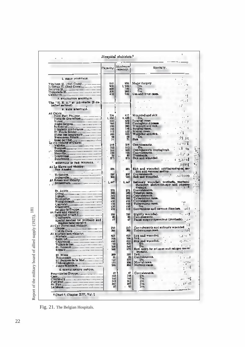
Fig. 21. The Belgian Hospitals.
22
Rep
ort o
f the
mili
tary
boa
rd o
f alli
ed su
pply
(192
5). 18
1

From the end of 1914, and taking advantage of the static scale of warfare on the Belgian front, the Medical Service of the Belgian Army is able to perfect the fi eld hospitals in the in-terior and to extend its installations at Calais (Fig. 22). Amongst others an important hospital is established at Saint-Pair near Granville and Calais with a capacity of more than 1,000 beds and of handling 5,000 cases a year.
In January 1915, still deprived of the necessary means, the hospital organizations of the French 10th Military Region, with headquarters at Rennes, are placed at the disposal of the Belgian Government by the French Government. This region, covering the administrative departments of Côtes-du-Nord, Manche and Ille-et-Vilaine, is at a distance of fi ve hundred kilometres, from the front. Thus 5,000 beds, manned by French staff, distributed among thirty-one hospitals, are placed at the disposal of the Belgian troops (Fig. 21).141, 151, 157, 170, 184
At the same time, the Medical Service of the Belgian Army, in its pursuit to administer its own installations independently, starts to develop its own hospitals at Rouen and in the vicin-ity, and later on also near Cap Ferrat, Le Havre, Mortain and Paris, to be supplemented with its own convalescent depots for the men awaiting their return to the front. Thanks to the help of the British Red Cross, especially Miss D. Maunder and former director of the Military Hospital in Brussels, Med Col A. Deltenre, a major Anglo-Belgian Hospital (Bonsecours) was created in November 1914 in Rouen. Facilities include two major departments: ortho-paedics and physiotherapy. The radiology department is headed by Dr. A. Stouffs. Besides the enormous fi nancial support from abroad this is achieved by the creation of a Belgian medical training camp for medics and nurses at Auvours.
Fig. 22. X-ray room in Petit Fort Philippe (near Calais).
Cou
rtesy
of M
r J-P
. Abs
il.
23

As mentioned before, the fi rst ships deployed in the transport of the wounded to England are the government mail-boats which have been turned over to the British Admiralty for this purpose. Later, the British Admiralty furnishes special ships for this service. The period during which evacuations are made to England is short and only lasts from October 1914 to April 1915. It is estimated that 23,000 men are evacuated during this period. They are led ini-tially to a large number of both civilian and military hospitals. End of 1914, they are mainly regrouped in the "Greater London" area in the hospital King Albert I (the radiologist is the called up physician H. Denoncin) (Fig. 23) and that of Hanwell and Kighate. After April 1915, the Belgian hospitals in England receive only the wounded and sick from the Belgian organizations in Great Britain, such as supply commissions, volunteers, ammunition facto-ries, etc. In total about 28,000 wounded and sick are treated in Great Britain.
Fig. 23. Radiologist reservist attached to the King Albert I Hospital in London in April 1917.
Cen
ter o
f his
toric
al d
ocum
enta
tion
(Eve
re).
31, 1
64
24
Fig. 24. Adapted map showing the major battles and the front line in June 1916 on the Western front.
Map
cou
rtesy
of J
.Leg
g w
ww.
grea
twar
.co.
uk
Dunkirque = DunkirkOstende = OstendHolland = Netherlands

HM King Albert, Commander of the Belgian Forces. Rare 2 Francs stamp.
Marshal F. Foch, Commander in Chief of the Allied Forces. Design and engrav-ing by Achille Ouvré. Stamp released in 1940!
General J. Pershing, Commander of the American Expeditionary Forces.
General D. Haig, Commander of the British Expeditionary Force.
German Marshal Paul von Hindenburg (left) and General in chief Erich Ludendorff.
Fig. 25. Military commanders at this time.
http
://fr.
wik
iped
ia.o
rg/w
iki/P
aul_
von_
Hin
denb
urg

Fig. 26. Organization of the Medical Service of the Army in the fi eld at the front. The location of the clinics, military hospitals (HMB) and the Red Cross and their advanced surgery posts (PCA) are shown (adapted according to Annex X, Mélis 1932).157

Chapter 5
Radiology treks to the Yser Front
When the Belgian Headquarters locate themselves at Nieuwpoort (Nieuport)on 13 October 1914, the King Albert I makes a statement, made famous by the announcement to the troops that their retreat has now ended: "This line will be preserved at all cost."
It is not the intention to present here a detailed story of these battles. We re-member only the terrible fatigue, rain, mud, the shells and machine guns, and moreover, the benefi cial fl ooding in the north-east along the railway Nieuw-poort-Dixmuide overfl owing the corpses and eventually separating the war-ring parties (Fig. 26).
As Dr. J. Mathieu recalls in his book, released in 1997, 151 the medical sup-port is weak, and mediocre. The Medical Service of the army units and divi-sions do what they can. They are still waiting for the miraculous ambulances to get there to transport the victims to Veurne, and there is nothing provided. Specifi cally, the divisions who are overloaded by their missions at the front have not sent their hospital department - as the regulations prescribe - to the rear.
The Physician-Colonel at the Headquarters, Ch. Maistriau (Fig. 27) "makes it very clear" that in order to set up an evacuation centre in Veurne it is manda-tory for the staffs of the active divisions to be moved to the rear. In doing so he has to violate a number of taboos.
27
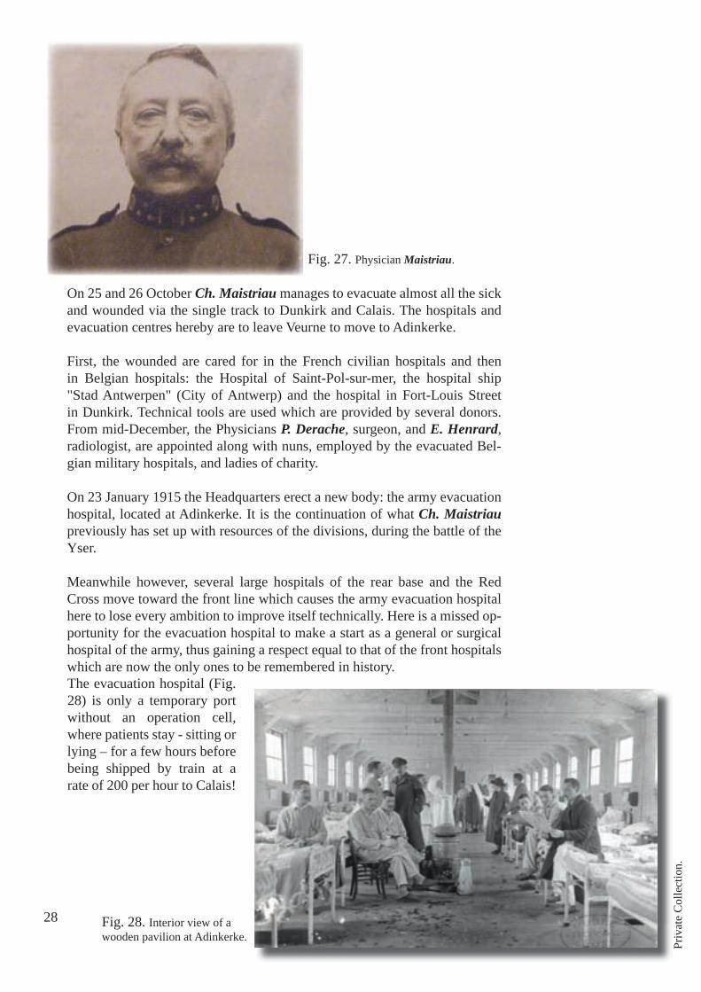
On 25 and 26 October Ch. Maistriau manages to evacuate almost all the sick and wounded via the single track to Dunkirk and Calais. The hospitals and evacuation centres hereby are to leave Veurne to move to Adinkerke.
First, the wounded are cared for in the French civilian hospitals and then in Belgian hospitals: the Hospital of Saint-Pol-sur-mer, the hospital ship "Stad Antwerpen" (City of Antwerp) and the hospital in Fort-Louis Street in Dunkirk. Technical tools are used which are provided by several donors. From mid-December, the Physicians P. Derache, surgeon, and E. Henrard, radiologist, are appointed along with nuns, employed by the evacuated Bel-gian military hospitals, and ladies of charity.
On 23 January 1915 the Headquarters erect a new body: the army evacuation hospital, located at Adinkerke. It is the continuation of what Ch. Maistriau previously has set up with resources of the divisions, during the battle of the Yser.
Meanwhile however, several large hospitals of the rear base and the Red Cross move toward the front line which causes the army evacuation hospital here to lose every ambition to improve itself technically. Here is a missed op-portunity for the evacuation hospital to make a start as a general or surgical hospital of the army, thus gaining a respect equal to that of the front hospitals which are now the only ones to be remembered in history.The evacuation hospital (Fig. 28) is only a temporary port without an operation cell, where patients stay - sitting or lying – for a few hours before being shipped by train at a rate of 200 per hour to Calais!
Fig. 28. Interior view of a wooden pavilion at Adinkerke.
Fig. 27. Physician Maistriau.
28
Priv
ate
Col
lect
ion.

Priv
ate
Col
lect
ion.
Fig. 30. The Inspector-General Mélis.
Fig. 29. Physician Depage in uniform, previously a rare appearance.
29
At the inception of the battle of the Yser, King Albert I senses that it will be bloody. He calls the surgeon A. Depage (Fig. 29) in order to help and do what he had already accomplished in Brussels in August 1914, when he trans-formed the Royal Palace into a hospital. 54
Immediately, in order to avoid legal diffi culties, A. Depage is enlisted in the Army, and immediately promoted to Physician Colonel. He assumes responsibility for the ambulance "Jeanne d 'Arc' at Calais before coming to De Panne, there to set up the l'Océan ambulance (let’s remember that an ambulance is originally a temporary health establishment; after the second World War, this term is used to indicate a vehicle for patient transport). Early surgery is essential for him.
Truly one begins to understand everywhere that he is right, and that calls forth on 30 November 1914 the promulgation of a Royal Decree establishing an "Executive Committee of the Red Cross for the rear guard of the army." Phy-sician Inspector General L. Mélis (Fig. 30) is the chairman and the deputy of the Minister of War, A. Depage is in practice the only other member. His sub-ordination to Mélis is only an appearance: he perseveres. Certainly credited to him, is the establishment of hospitals at the front. These are considered by the Headquarters to be the rear guard departments, "tolerated" by the vanguard.
At the level of the military hospitals at the front all surgeons are inspired by the principles of A. Depage, not however by his organization..

Fig. 32. Probably Physician Peremans, in the centre of the radiology room. Above a 1930 s portrait of Dr. Peremans.
The ambulance of "L'Océan" (December 1914-October 1919)
In Flanders Fields, only the coastal area has buildings that meet the needs and goals of doctor Depage. In De Panne, near the royal residence, a hotel with 150 beds may serve: Hotel l'Océan (Fig. 31). This modest hotel that serves only as a residence for the summer has neither heating nor a lift to connect the four fl oors. Antoine and Marie Depage go quickly into action: with the help of Major Ch. Gordon, liaison offi cer of King George V, they succeed in Britain to acquire a central heating system, operating rooms and radiology equipment, and also to transport this equipment and its fi tters to De Panne where everything is installed.
The fi rst doctors employed by the ambulance are the surgeons A. Depage, F. Neuman, G. Debaisieux and G. Vandevelde to whom L. Mélis adds the very valuable E. Henrard. In 1914 he had succeeded E. Dupont as chief of radiol-ogy in the Military Hospital in Brussels. He is highly valued in the military - but also in civilian circles. Therefore he is seconded by Mélis to Depage at the ambulance l'Océan. The latter serves as a liaison offi cer between the two men. Henrard puts at the service of surgeon Depage and his allied colleagues an original method for the radio-stereographic localization of projectiles (in the wounded). Doctor E. Henrard acquires an international reputation in this fi eld. We will have the opportunity to return to this. Henrard is assisted by another radiologist, physician M. Peremans (Fig. 32) who after the war will
Fig. 31. In the middle of the complex: the hotel "L'Océan" (Four-storey building).
30
Priv
ate
colle
ctio
n
Priv
ate
colle
ctio
n.
become the chief of the radiology department of the city hospitals in Antwerp.

Fig. 34. Radiology equipment of "Cabour Surgery".
Fig. 33. From left to right PhysicianGeneral Mélis, Chief Physician Deracheand Mr. Cabour, owner of the buildings.
Fig. 35. Dr. A. Bienfait.
Cabour Surgery (April 1915 - March 1917)
In April 1915, around a hunting lodge of which Mr. Cabour is the owner, in three weeks’ time the army erects twenty two wooden barracks to accommo-date a surgical hospital, placed under the supervision of the regimental physi-cian P. Derache (Fig. 33). He is the surgical advisor to Mélis. He has led an ambulance at Dunkirk, and Professor Depage recommends him.
The operating room is built in durable materials in the hunting lodge. The total capacity of the hospital is 410 beds. A radiology department is provided (Fig. 34).
Physician Derache surrounds himself with twelve doctors, including the Liège radiologist A. Bienfait (Fig. 35). He had been, in 1906, a founding member of the Belgian Society of Radiology. Most of the doctors involved here originate from units implicated with the front because their skills and also their dedication have been noticed. This is essential to allow for "Cabour surgery" and subsequently Beveren, to ensure a special climate: that of soli-darity of the doctors and their patients. The indispensable weapon brother-hood has marked the medicine in the army. Derache knows how to combine the necessary technical authority with a certain warmth that rouses dedication.
After the fi ghting sector comes under British command, the hospital “Cabour surgery” ceases to function and that of Beveren takes over the watch.
31
Bel
gian
Arm
y M
useu
m 16
4
N. D
esie
re C
olle
ctio
n (A
dink
erke
).56

Fig. 37. Radiological set-up in Beveren.
Fig. 38. Physician Couturier, who later becomes a Colonel.
Beveren on the Yser (March 1917 - February 1920)
It is a model hospital, entirely conceived and directed by physician Derache (Fig. 36), and opening its doors in March 1917. An "Army Miracle" says King Albert I.
By its location in Stavele it is only 9 km away from the fi rst front lines. Each injured person can reach it within three hours of time.
The hospital has thirty portable barracks. Two fi xed parallel corridors each connect 10 barracks, a transverse corridor connects the two wings with a tech-nical centre in the middle of the barracks: room for the incoming, room for the shocked, operating room, four small labs, X-ray room (Fig. 37), dentistry and speciality room.
Here we mention the doctors who later will become department chiefs in or commanders of military hospitals during the interbellum. In Brussels: H. Burger, R. Van Olmen, F. Grutman, R. Foquet, R. Marchal, P. Couturier (the radiologist) (Fig. 38) and A. Gaudissart. In Antwerp: A. Lefebvre and D. Waffelaert. Liège: I. Voncken and A. Reynders.
Fig. 36. Physician Derache becomes Inspector-General (1930-1934).
32
Priv
ate
colle
ctio
nPr
ivat
e cc
olle
ctio
n.

Fig. 40. Radiology equipment of Hoogstade. Probably donated in 1915 by Marie Curie.
Fig. 39. Physician Willems.Hoogstade (May 1916 - November 1918)
The personnel of the Belgian Field Hospital, an institution founded by a Brit-ish committee, and established in Antwerp, manage to escape just before the surrender of the city. They return to Veurne and Poperinge from 21 October 1914.
The opening of the hospital "l’Océan" leads to the establishment of the Bel-gian Field Hospital Hoogstade, in the hospice Clep, on the boundary of the Belgian sector. Four British surgeons operate there, but increasingly nurses are lacking. Reinforcement is provided by Belgian military personnel.
Later the hospital benefi ts from the support of Physician Ch. Willems (Fig. 39), chief surgeon of the Bijloke hospital in Ghent. He participated in the Bal-kan war on the side of the Serbs. He volunteers in 1914 and operates extreme emergency cases in Dunkirk until the bombing of that city in the spring of 1915. The Inspector General Mélis sends him to the front in the south of the Belgian sector. Willems expands the Belgian Field Hospital by adding some larger barracks.
In May 1916, Hoogstade formally becomes a Belgian military hospital, led by Ch. Willems, assisted by N. Goormaghtigh. The Physicians J. Decaestecker and J. Van Meenen are the radiologists. They have one set of - for its time - modern radiology equipment (Fig. 40). Contrary to what is done in Beveren (Abelenhof) and l'Océan (St Jans Molen) an advanced (Grognie) surgical post is established in a concrete bunker. This is especially necessary in this hard to reach sector situated under enemy fi re.
33
His
toric
al D
ocum
enta
tion
(Eve
re) (
1922
). 31
, 164

Cabour Medicine (March 1917 - February 1920)
In April 1917 Derache leaves Cabour, it is transformed into an ordinary gen-eral hospital. The operating room is maintained in the brick pavilion and also three hundred "fl ying" beds that can serve in the event of a large infl ux of wounded.
To the old surgical hospital seventeen new portable pavilions are added.
Physician P. Nolf (Fig. 41) from Ieper leaves the civil hospital of St. Ides-bald that serves as an ambulance unit. Later he becomes a professor at the University of Liège. He is the future Minister of Science and Arts, and future president of the Belgian Red Cross. He leads "Cabour Medicine" in a mas-terly fashion.
Many of his colleagues are from Liège: J. Firket, H. Fredericq, I. Roskam, and L. Christophe, who will become university professors. There are also locals among them as the future Professor P. Spehl, or Physicians A. Colard, P. Pastiels and V. Gallemaerts. The latter will become the chief of the Medi-cal Service of the Belgian Armed Forces in Great Britain from 1942 to 1945.
As of 25 October, 1918 "Cabour Medicine" is transferred to Bruges.
Fig. 41. Physician Nolf.
34 Priv
ate
colle
ctio
n.
Fig. 42. Ambulance vehicle (model 1883!) drawn by "à la Daumont", at the beginning of the war. One notices between the drivers a stretcher-bearer still in clergy dress !

AG
C-C
AA
2. 2
Bourbourg (May 1915 - Mai 1919)
Bourbourg cannot be regarded as a front-line hospital but rather as an area hospital, used as a stopover between the battle zones and France.
At the end of April 1915 the Germans bombard Dunkirk with long-range guns. At that time it also appears necessary to add a Belgian physician as reinforcement to the Hoogstade hospital. Professor Willems, who replaces Derache in the hospital of Fort-Louis in Dunkirk, is assigned.
The three surgical front hospitals do not operate more patients than the emergency services of any category. Since transportation by rail is much faster, the number of wounded to be treated declines. The Inspector-General of the Medical Service leaves Dunkirk and closes all local fi eld hospitals. He settles in Bourbourg and on 18 May, 1915 has a large barracks hospital opened next to Bourbourg station. It is easily ac-cessible by road via Gravelines.
In 1917 in Bourbourg the command is held by the principal physician A. Wilmaers (Fig. 44). The head of the laboratory is the future professor of the Université Libre de Bruxelles, E. Renaux. The surgeon is Dr. D. Ba-ruch, also one from Brussels, and the ophthalmologist is doctor A. Moret. Radiology equipment is supplied here (Fig. 43). The radiologist is physi-cian P. Couturier who moves in 1917 to Beveren. After the war he leads the radiology department in Antwerp and later that in Brussels.
Fig. 43. Radiological room in Bourbourg.
35
In 1917 in Bourbourg the command is held by the principal physician Wilmaersthe Université Libre de Bruxelles, ruchRadiology equipment is supplied here (Fig. 43). The radiologist is physi-cian the radiology department in Antwerp and later that in Brussels.
Fig. 44. A. Wilmaers, who later will become Inspector-General (1922-1926).

''
Chapter 6
The X-ray equipment, how in fact does it function at this time?
In permanent installations, the hospital radiology equipment is composed of a coil of Ruhmkorff with high throughput or an AC transformer, function of the available power. This produces the necessary high voltage to generate X-rays. Auxiliary devices (tube stand, bed, positioning devices, etc.) make the use of X-rays possible.
Figure 45 describes the device:
S is the power source by a battery or accumulator batteries.R is a rheostat, i.e. a device provided with a wire with adjustable resistance that allows increasing or reducing the current.G is an ammeter to measure the fl ow.I is a mercury switch that interrupts the power generated in the coil, twenty times per second. A capacitor in parallel with this switch is designed to pre-vent sparks, which have the tendency to jump from one pole to another dur-ing the interruption.P is the transformer whose primary coil is wrapped with a secondary.
Fig. 45. Electrical circuit diagram.
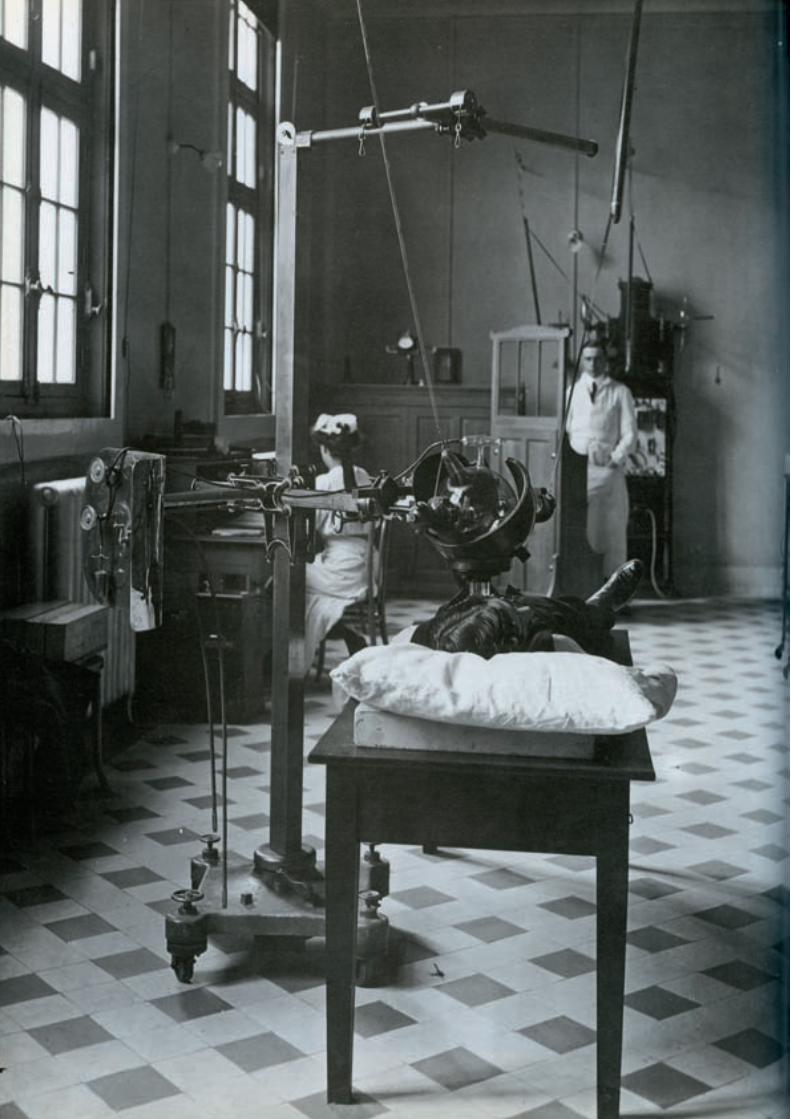
A radiology room prior to 1914. Set up here for radiotherapy treatment.
37
F. Ja
ugea
s (19
13).
123
The current of the secondary coil fl ows successively through a milliammeter G’, that displays the current. A valve A acts as a diode and withholds the closing wave, generated by the coil, and allows passage to the opening wave only, and this one solely is effective. The tube T produces the X-rays and is wrapped in a dome of lead glass E, which protects the operator from harmful radiation. D is a lead diaphragm which restricts the irradiated fi eld.L is a spark source applied to a derivative of the tube (the spintermeter is de-scribed below). This gives the measure of the resistance of the tube T, through bringing the bars closer together until a spark occurs. It also determines the penetrative force of the rays. This resistance is also variable at random.
F. Jaugeas gives a good summary of the use of a radiology set-up from this time.
After a radiant tube has operated for a few moments, it acquires an increas-ing resistance to the passage of the current fl ow. This grows gradually until it reaches a level at which value the electric discharge occurs only inside the tube. By then the tube is unusable. What has happened? The freely moving gas molecules inside an unused tube make the electric discharge possible. During the operation these molecules fi x themselves to the electrodes and the glass wall. This causes a vacuum, hence the electrical resistance. In this case the tube is said to be "hard". To regenerate it or to loosen the frozen gases and make it "soft", the walls need warming up with a soldering lamp. The harden-ing phenomenon repeats itself very quickly.
There is no effort spared to provide the tube with mechanisms in order to reduce the resistance level. These devices belong to two major groups: fi rst, the one to be found in the calibration of the pressure inside the tube cavity, by enclosing in a substance which can free up a certain amount of gas under the infl uence of a small temperature increase, and second, that one using the osmotic properties of platinum.
The current of the secondary coil fl ows successively through a milliammeter that displays the current. A valve
closing wave, generated by the coil, and allows passage to the opening wave
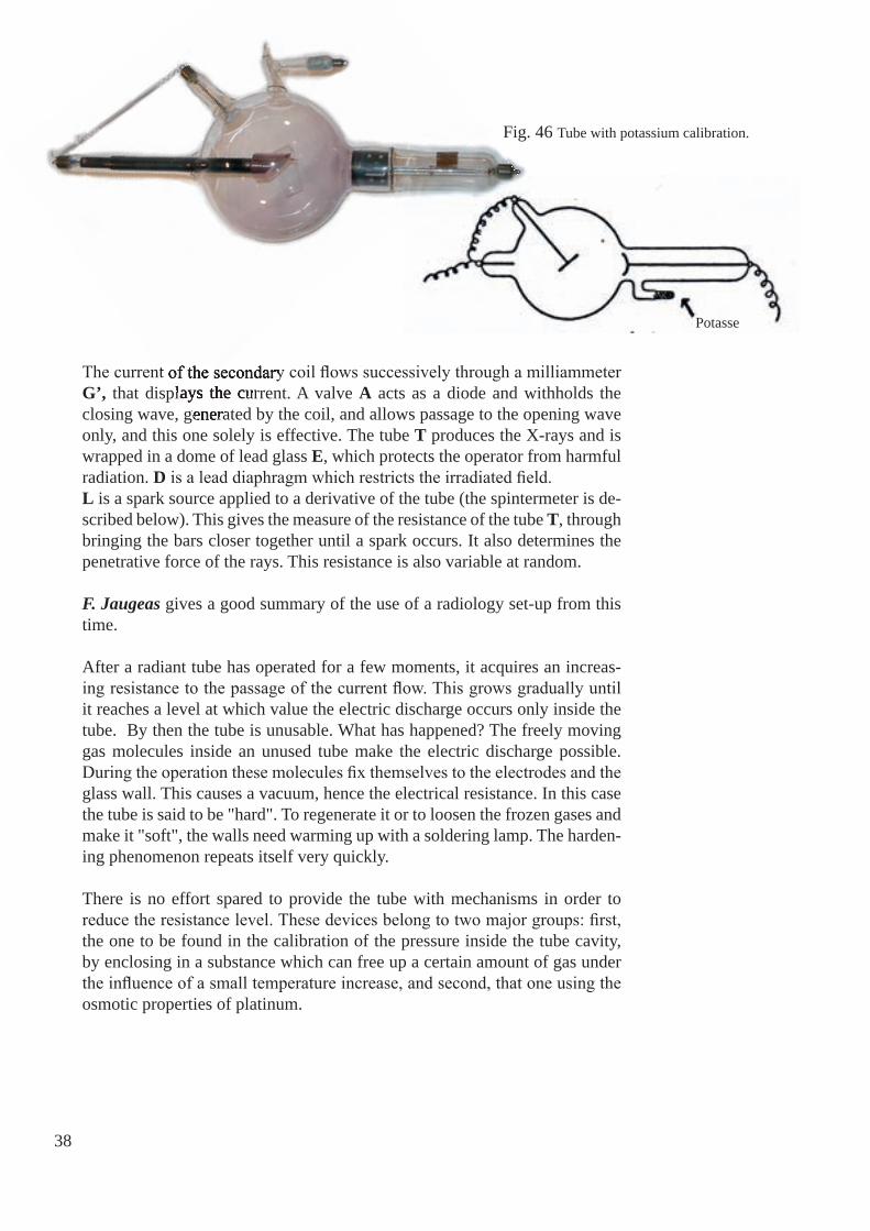
Fig. 46 Tube with potassium calibration.
Potasse
38

Fig. 46 Tube with potassium calibration.
In the fi rst group we fi nd a potassium control (Fig. 46). The tube includes a kind of funnel that contains a certain amount of potassium, a hydrophilic sub-stance that liberates water under the infl uence of very slight warming. When the tube gets too much resistance, becoming too "hard", it suffi ces to regen-erate it by warming the potassium with a low heat source. Release of steam takes place immediately and the electric current passes easily.
The calibration by a spark (Fig. 47), is actually a perfection of the former. A small tube that operates as an auxiliary device, has attached to its ends two hinged antennas that can be brought closer to or farther away from the princi-pal electrodes. It contains an enclosed chemical substance of variable nature, which is composed either of superimposed mica platelets, or of some pre-cisely selected organic. By the current’s activity this matter lets gas to escape which it contains mechanically. The chemicals, whose composition releases gaseous elements like hydrogen, by the activity of electric current, thus pro-vide an alternative.The gas contained mechanically within this substance can escape, either through the action of the current or by chemicals. So when the tube reaches too high a resistance, the manipulation and the adjustment of the antennas in relation to the principal electrodes creates a deviation that generates a current, which acts on the substance of the controller. The tube loses its increased re-sistance and turns “soft” again. To a certain extent tubes fi tted with this tool can be described as self-calibrating, for if one of the antennas is set at a suf-fi cient distance from the electrodes, then the current fl ows through the devia-tion only if the resistance of the lamp is stronger than that of the deviation; subsequently the vacuum of the lamp does not fall below a certain value.
This kind of calibration tool has two defi ciencies: the regeneration of the tube is ensured only for a relatively short period and the retained gas supply is very limited and thus quickly exhausted. These tools do not allow the reversing of the (lamp) calibration, in other words, if the lamp has a low resistance, is emptied too little, in one word: too "soft", they do not provide the means to make it "harder".
Fig. 47. Tube calibration by a spark.
we fi nd a potassium control (Fig. 46). The tube includes a
Tube calibration by a spark.Fig. 47. Tube calibration by a spark.
39

A better method is the calibration based on the osmotic properties of plati-num heated red-hot. This ingenious tool consists of a simple platinum tube soldered shut at one end and welded to the tube wall at the other, in such a manner that its cavity communicates with the X-ray tube (Fig. 48). When the lamp becomes too "hard", the osmotic regulator is heated with a soldering lamp (Bunsen burner) (Fig. 49) and for a few moments one keeps a white-red glow. Under these conditions, the hydrogen resulting from the decomposition of water vapour in the hot part of the fl ame and the lighting gas itself goes through the platinum. It is sucked towards the middle, where the pressure is the weakest, and it remains trapped there as soon as the platinum tube cools. This allows the vacuum level be reduced as needed, as the stock is inexhaust-ible and the osmosis regulator always retains its properties. The tube life is thus noticeably extended.
Furthermore, the osmotic calibration has the advantage of allowing the con-trol in the other sense by offering the possibility to transform a "soft" tube into a "hard" one. This second operation (Fig. 50) is executed by covering the osmosis controller with a platinum-wick and heating as described above. In these new circumstances, the osmosis regulator, as it’s stepped up into a red-white glow, is no longer in contact with the fl ame, nor the platinum wall either, but remains permeable to very highly soluble gases such as hydrogen. This allows the gas in the X-ray tube to escape.
The removal of gases from the tube requires much more time than introducing it. To determine a measurable change in the degree of vacuum of a "soft" tube, the osmosis regulator needs to be heated for one or two hours, protected by the platinum-wick. In practice it is rarely necessary to perform this operation because the operation of the tube always entails an increase in the vacuum.
Fig. 48. Radiation X-ray tube with osmosis calibration.
A better method is the calibration based on the heated red-hot. This ingenious tool consists of a simple platinum tube
soldered shut at one end and welded to the tube wall at the other, in such a manner that its cavity communicates with the X-ray tube (Fig. 48). When the lamp becomes too "hard", the osmotic regulator is heated with a soldering
Fig. 48. with osmosis calibration.
Fig. 50. Uses of osmosis controller:without a wick (left) and with wick (right).
Fig. 49. Tune up by Bunsen burner.
40
G. P
alla
rdy
et c
oll (
1989
). 17
3

The calibration of the more or less penetrating rays (kV) can be done by measuring the potential difference between the tube electrodes. With the as-sumption that the tube is connected to a high voltage generator via conduc-tors that are not provided with an insulating sleeve, we will generate a spark between them and thus cause a short circuit when we bring these conductors towards each other. By controlling the interval between them we can experi-mentally determine the distance at which a spark will occur.
For this measurement to be very easy to perform A. Béclère devised a scale in centimetres, which is therefore called spintermeter (Fig. 51) and which permits at any time to express the length of the corresponding spark in centi-metres. According to what we explained earlier, we can say that if this spark length is large (high kV), the tube produces penetrating rays or is "hard", and vice versa. At this time kilovoltmeters did not exist.It should be noted that the shape of the points affects the distance at which the spark occurs.
Coil
Fig. 51. Spintermeter
40,000 4.2560,000 12.580,000 16.5100,000 22.0120,000 25.0
Fire potential for the fi rst spark, in volts:
Cm length of the sparks between the two points:
41
Bel
gian
Mus
eum
of R
adio
logy
.
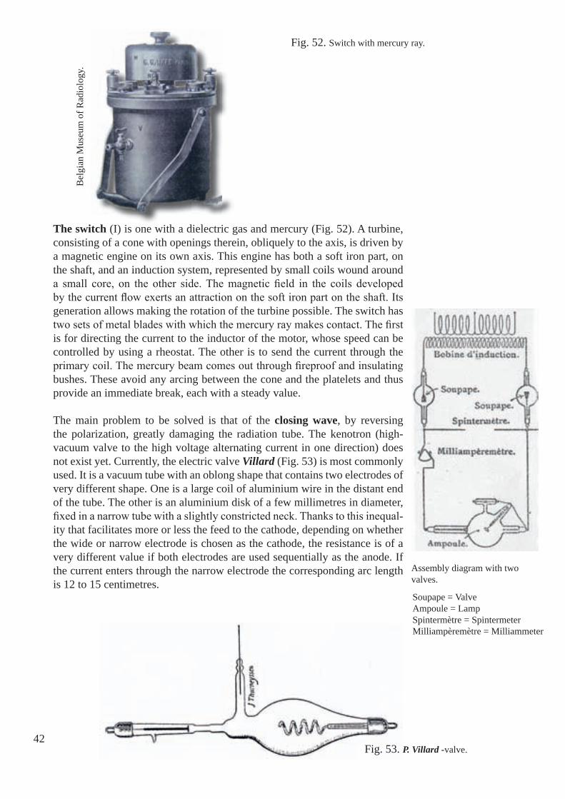
The switch (I) is one with a dielectric gas and mercury (Fig. 52). A turbine, consisting of a cone with openings therein, obliquely to the axis, is driven by a magnetic engine on its own axis. This engine has both a soft iron part, on the shaft, and an induction system, represented by small coils wound around a small core, on the other side. The magnetic fi eld in the coils developed by the current fl ow exerts an attraction on the soft iron part on the shaft. Its generation allows making the rotation of the turbine possible. The switch has two sets of metal blades with which the mercury ray makes contact. The fi rst is for directing the current to the inductor of the motor, whose speed can be controlled by using a rheostat. The other is to send the current through the primary coil. The mercury beam comes out through fi reproof and insulating bushes. These avoid any arcing between the cone and the platelets and thus provide an immediate break, each with a steady value.
The main problem to be solved is that of the closing wave, by reversing the polarization, greatly damaging the radiation tube. The kenotron (high-vacuum valve to the high voltage alternating current in one direction) does not exist yet. Currently, the electric valve Villard (Fig. 53) is most commonly used. It is a vacuum tube with an oblong shape that contains two electrodes of very different shape. One is a large coil of aluminium wire in the distant end of the tube. The other is an aluminium disk of a few millimetres in diameter, fi xed in a narrow tube with a slightly constricted neck. Thanks to this inequal-ity that facilitates more or less the feed to the cathode, depending on whether the wide or narrow electrode is chosen as the cathode, the resistance is of a very different value if both electrodes are used sequentially as the anode. If the current enters through the narrow electrode the corresponding arc length is 12 to 15 centimetres.
Fig. 53. P. Villard -valve.
Fig. 52. Switch with mercury ray.
42
Assembly diagram with two valves.
Bel
gian
Mus
eum
of R
adio
logy
.
Soupape = ValveAmpoule = LampSpintermètre = SpintermeterMilliampèremètre = Milliammeter

Fig. 54. Gaiffe cabinet, model 1909 in Art Nouveau style.S
T
S
Sp
M
I
43
Bel
gian
Mus
eum
of R
adio
logy
.
A
C
R
r
The valve is connected in series with the X-ray tube in such a way that the narrow electrode serves as the anode, which means that its resistance to the wave is minimal. The maximum resistance is now opposed to the inverse wave whose voltage is just insuffi -cient to catch it and to spread in the circuit.
The Villard-valve consists of a highly effective tool for protecting the tube against reverse current. Dur-ing its operation it behaves like an X-ray tube: it "hardens." It is necessary to heat the osmotic control for returning the vacuum to a reasonable level.
A widely distributed model in the radiology services of the Belgian-French allied forces is that manu-factured by the French fi rm Gaiffe. All necessary equipment is housed in an Art Nouveau style cabi-net (Fig. 54). The control apparatus (rheostat, (R, r), ammeter (A), etc...), the switch (I), the transformer (T) and capacitor (C), are placed on shelves so they minimize occupied space to render the operation as convenient as possible and the view as beautiful as can be. On the top shelf are the valve supports (S), and on the rack that carries the transformer are also installed the spintermeter (Sp) and milliamme-ter (M). All connections to the transformer primary coil are made ready to run, so that it is suffi cient to connect the lamp with the two poles of the spinter-meter to operate the equipment.

While, in the beginning a "kitchen table" and an X-ray tube stand (Fig. 55) are suffi cient to perform a radiographic examination, more perfected equipment is being developed by different manufacturers. This wooden equipment al-lows performing radioscopy, the examination of the patient in supine position (Fig. 56) or standing upright (Fig. 57)....after having moved the patient fi rst!Thus, the company Gaiffe develops a "very well perfected" table at the end of the war (Fig. 58).
Fig. 56. Examination of the patient lying down.
Fig. 57. Examination of the upright patient.
Fig. 55. Rudimentary radiology equipment.
44
F. Ja
ugea
s (19
13).
123

Fig. 58. Gaiffe-table, 1918, allows examination in supine and upright position. 45
Bel
gian
Mus
eum
of R
adio
logy
.

46
Fig. 59. German horse-drawn wagon for the transportation of radiology equipment. The sides have been removed to show the radiology equipment and the generator.
Fig. 60. French radiology motor car designed by Rivière et Drault mounted on a chassis by De Dion-Bouton.
Nie
hues
(190
9). 16
7
J. B
oyer
(191
5). 20
G. K
ütte
rer (
2005
). 12
9

Chapter 7
47
Vehicles for radiology
The pioneer motor vehicles and the X-ray equipment of the early twentieth century merge to provide the earliest forms of mobile radiography, which rapidly fi nds applications in the military campaigns up to 1918. 92
The fundamental problems of military radiology are chiefl y the fragility of the apparatus and the need to carry a source of electrical supply. In addition, battle conditions often make it easier to take the surgeon and the X-ray set to the patient rather than to take the patient to a well-equipped base hospital.
For the military expeditions all radiographic apparatus including batteries to generate electricity are to be carried by human effort or on pack animals. The weight of the batteries and the danger of spilled acid make them unsuitable for mobile campaigns.
Batteries require frequent recharging. Human or animal (Fig. 61) muscle power is found wanting, luckily dynamos and petrol engines are obtained.
Before the Great War some of the fi ghting parties already possess horse-drawn wagons for radiology (Fig. 59), later also automobiles (Fig. 60).
Fig. 61. A portable X-ray unit designed for driving by horsepower developed in 1909 by Siemens & Halske (Ber-lin) for the radiologic departments of German fi eld and base hospitals.
Fig. 62. Transportable radiology equipment manufactured by the French fi rm Gaiffe. The coil of Ruhmkorff is replaced by a coil of Rochefort (A), which is better insulated and stronger. We see there the spintermeter. The control panel (C) has reduced dimensions. The radiation tube (D), is a rudimentary gas pipe. The Dessane-visor (B) allows radioscopy.48
(A)
(B)
(C)
(D)
Bel
gian
Mus
eum
of R
adio
logy
.
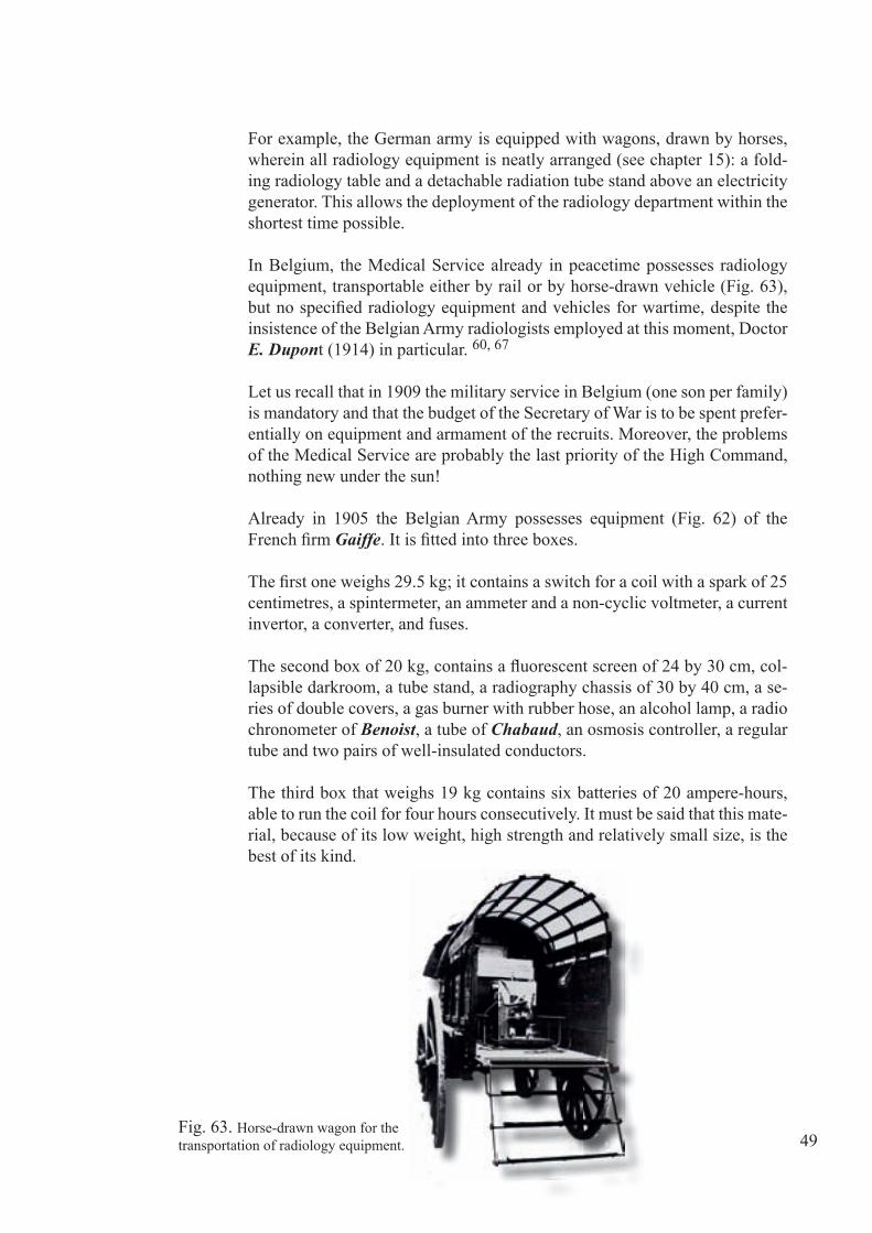
49
For example, the German army is equipped with wagons, drawn by horses, wherein all radiology equipment is neatly arranged (see chapter 15): a fold-ing radiology table and a detachable radiation tube stand above an electricity generator. This allows the deployment of the radiology department within the shortest time possible.
In Belgium, the Medical Service already in peacetime possesses radiology equipment, transportable either by rail or by horse-drawn vehicle (Fig. 63), but no specifi ed radiology equipment and vehicles for wartime, despite the insistence of the Belgian Army radiologists employed at this moment, Doctor E. Dupont (1914) in particular. 60, 67
Let us recall that in 1909 the military service in Belgium (one son per family) is mandatory and that the budget of the Secretary of War is to be spent prefer-entially on equipment and armament of the recruits. Moreover, the problems of the Medical Service are probably the last priority of the High Command, nothing new under the sun!
Already in 1905 the Belgian Army possesses equipment (Fig. 62) of the French fi rm Gaiffe. It is fi tted into three boxes.
The fi rst one weighs 29.5 kg; it contains a switch for a coil with a spark of 25 centimetres, a spintermeter, an ammeter and a non-cyclic voltmeter, a current invertor, a converter, and fuses.
The second box of 20 kg, contains a fl uorescent screen of 24 by 30 cm, col-lapsible darkroom, a tube stand, a radiography chassis of 30 by 40 cm, a se-ries of double covers, a gas burner with rubber hose, an alcohol lamp, a radio chronometer of Benoist, a tube of Chabaud, an osmosis controller, a regular tube and two pairs of well-insulated conductors.
The third box that weighs 19 kg contains six batteries of 20 ampere-hours, able to run the coil for four hours consecutively. It must be said that this mate-rial, because of its low weight, high strength and relatively small size, is the best of its kind.
Fig. 63. Horse-drawn wagon for the transportation of radiology equipment.
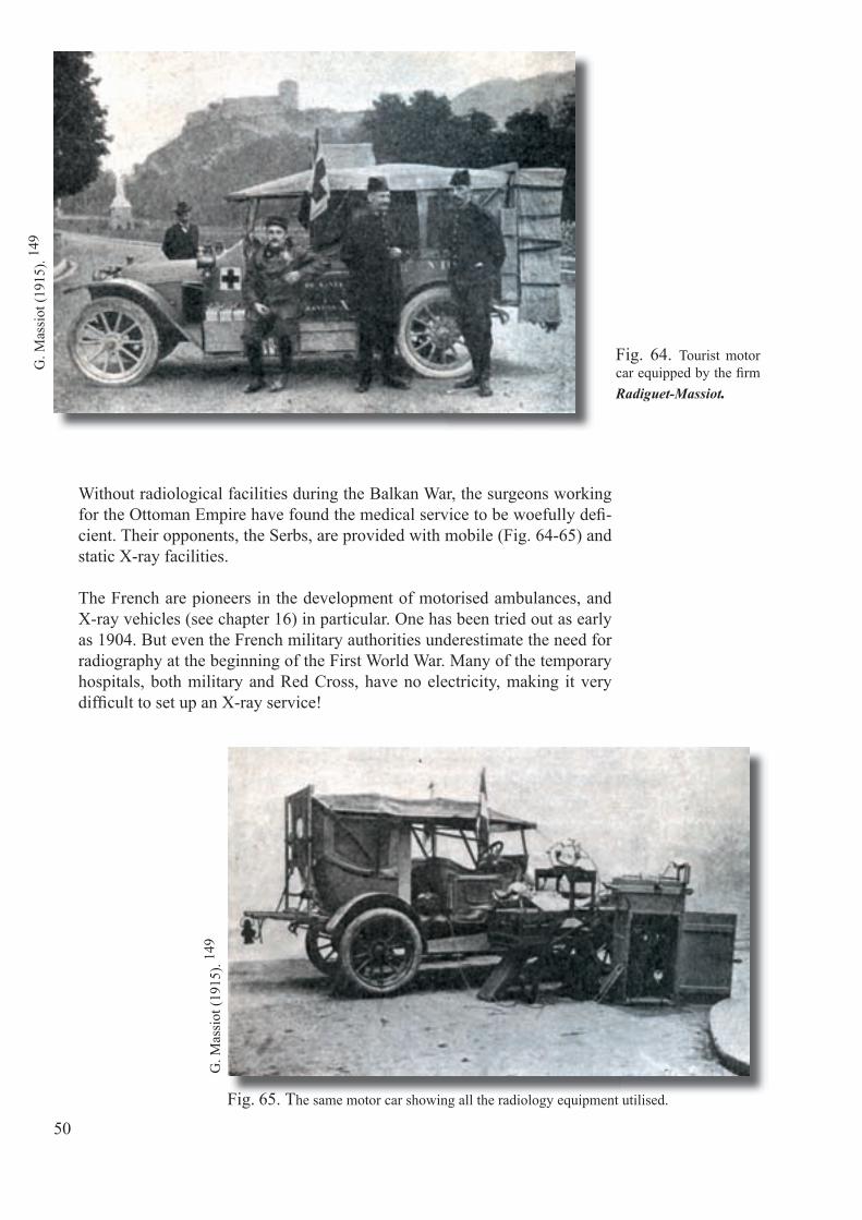
Without radiological facilities during the Balkan War, the surgeons working for the Ottoman Empire have found the medical service to be woefully defi -cient. Their opponents, the Serbs, are provided with mobile (Fig. 64-65) and static X-ray facilities.
The French are pioneers in the development of motorised ambulances, and X-ray vehicles (see chapter 16) in particular. One has been tried out as early as 1904. But even the French military authorities underestimate the need for radiography at the beginning of the First World War. Many of the temporary hospitals, both military and Red Cross, have no electricity, making it very diffi cult to set up an X-ray service!
Fig. 64. Tourist motor car equipped by the fi rm Radiguet-Massiot.
Fig. 65. The same motor car showing all the radiology equipment utilised.
G. M
assi
ot (1
915)
. 149
G. M
assi
ot (1
915)
. 149
J. B
oyer
(191
5). 20
50

Electric current is generated either independently or by the vehicle's own en-gine connected to a dynamo. The body of the vehicle, if large enough, is converted to house the darkroom and to store and transport all the X-ray equipment: tubes, coil, interrupter, plates, chemicals, apparatus for localising foreign bodies and a folding X-ray table. The van is parked outside the hos-pital and generates the power for the radiography within. Some teams have separate radiography tents (Fig. 66, 67, 68). The three person radiological team, with practice, is able to set up the apparatus in half an hour.
Custom-built radiological cars are designed by several French engineers. By the end of the war some of their vehicles have become very sophisticated. A new feature is the mounting of the dynamo on a forward extension of the chassis, with its own bonnet, looking like an extra engine at the front. It has 4.5 kW power and is intended to generate electricity for lighting the mobile hospital unit as well as for X-rays.
The radiology car brings a solution to the problem of providing a technical service when there is a lack of electricity.
Fig. 66. Diagram of the motor car Rivière et Drault showing the dynamo (D) that feeds the radiology apparatus.
J. B
oyer
(191
5). 20
J. B
oyer
(191
5). 20
J. B
oyer
(191
5).
Fig. 67. View of the van, in the back the second door to the dark room.
Fig. 68. Radiology motor car Rivière et Drault. To the left the tent is shut to allow performing radioscopy. To the right a fl ap is rolled back to show the equipment.

52
The arrival of the wounded happens sometimes several days after they have suffered injuries, and prior to treatment! Sometimes they suffer blood loss and show strongly infected wounds.Let's not forget that antibiotics do not exist and that the bullets practically always, and shrapnel almost always cause infection. 194
The medical strategy is now undergoing a dramatic change. In fact, on the Belgian front the military hospitals and those of the Red Cross are setup in an area quite close to the front, which fortunately remains stable for several years.
Also more advanced surgical items (PCA: "poste chirurgical avancé" see Fig. 26, page 26) see the light of day. Each hospital has to assure the support of its outpost. To do this it disposes of a surgery vehicle, a logistics vehicle, a technical and a radiology vehicle as well (Fig. 69) that can be sent out with its personnel, according to the medical necessities. This automobile detachment can occasionally reinforce the clinics.
This directive of fi rst-line treatment at the front is still valid and in use today, albeit improved and perfected.
its outpost. To do this it disposes of a surgery vehicle, a logistics vehicle, a technical and a radiology vehicle as well (Fig. 69) that can be sent out with its personnel, according to the medical necessities. This automobile detachment
This directive of fi rst-line treatment at the front is still valid and in use today,

Fig. 69. Belgian military radiology vehicle Vinot-Deguingeand. On the foreground, atop the detachable radiology table, the Dessane-visor is noticeable as is the Gaiffe radiology equipment, described earlier.
53
Priv
ate
Col
lect
ion.
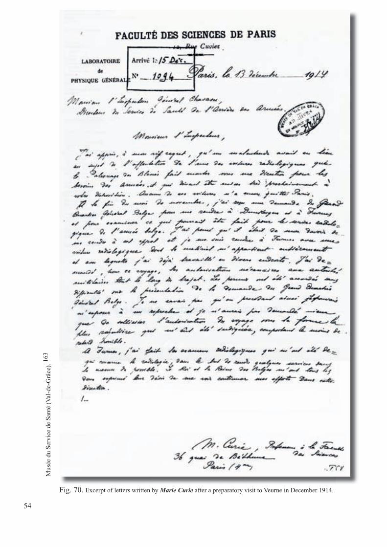
Mus
ée d
u Se
rvic
e de
San
té (V
al-d
e-G
râce
). 16
3
Fig. 70. Excerpt of letters written by Marie Curie after a preparatory visit to Veurne in December 1914.
54

55
Fig. 71. Madame Curie and her daughter Irène Curie visiting an English hospital, the Belgian fi eld Hospital, housed in St. Joseph secondary school Veurne.
H. S
outta
r (19
15).
190
Veurne Henry P., Irène Examinations car December 1914Poperinge, Hesdin Henry P., Irène To convey car n°1 with
Perrincar January 1915
Poperinge Ragot Verifi cation of car N°1 railway February 1915Adinkerque Poperinge
Henry P. Examinations car March 1915
Hoogstade, De Panne Henry P. To convey car N°10 railway and car N°10 August 1915Hoogstade Irène Examinations railway September 1915
Marie Curie's war mission list in Belgium during WWI1914 - 1915
On page 145 we report the important role played by Madame Curie. For Bel-gium her contribution deserves to be mentioned here.
Eventually, and on three occasions, Madame Curie does not hesitate to come up to the Yser Front to deliver her radiological vehicles and equipment. Each time she is accompanied by her daughter Irène.
They come in December 1914 to Veurne (Fig. 70, 71), in January 1915 to Po-peringe and Hesdin, and in September 1915 also to Hoogstade (Fig. 72, 73). It is in Hoogstade that Madame Curie meets King Albert I (Fig. 74).

Fig. 72. Marie Curie and her daughter Irène (left), Hoogstade in September 1915.
Mus
ée C
urie
(Par
is).
56

Fig. 74. King Albert I in conversation with Marie Curie in Hoogstade in September 1915.
Fig. 73. Irène Curie exits the radiology vehicle n° 5 in Hoogstade in September 1915.
Mus
ée C
urie
(Par
is).
Mus
ée C
urie
(Par
is).
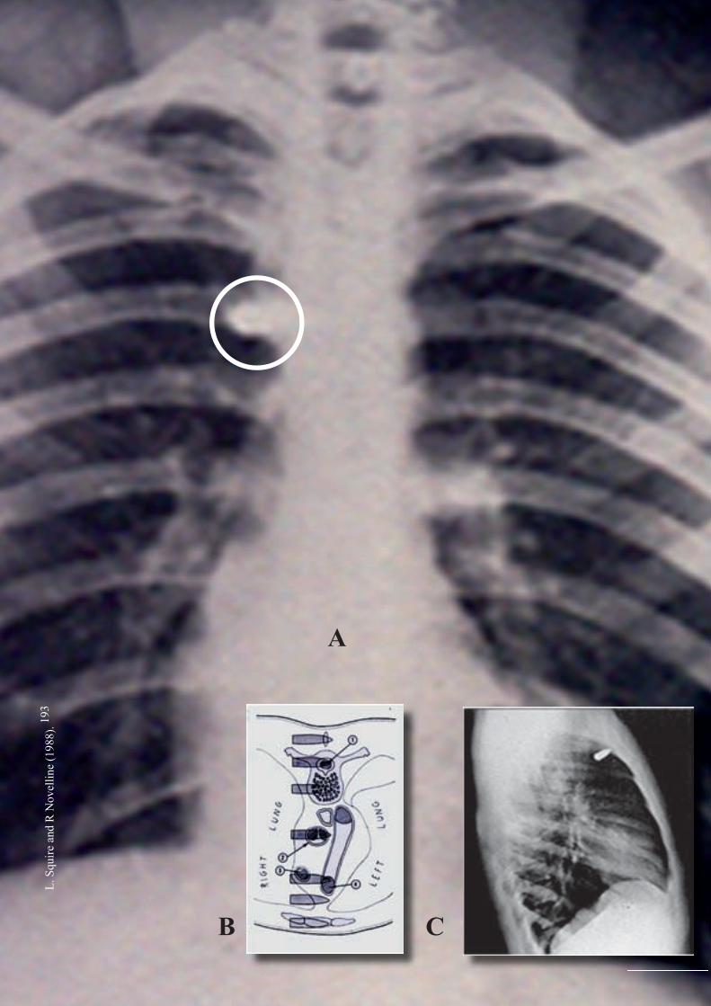
A
B C
L. S
quire
and
R N
ovel
line
(198
8). 19
3
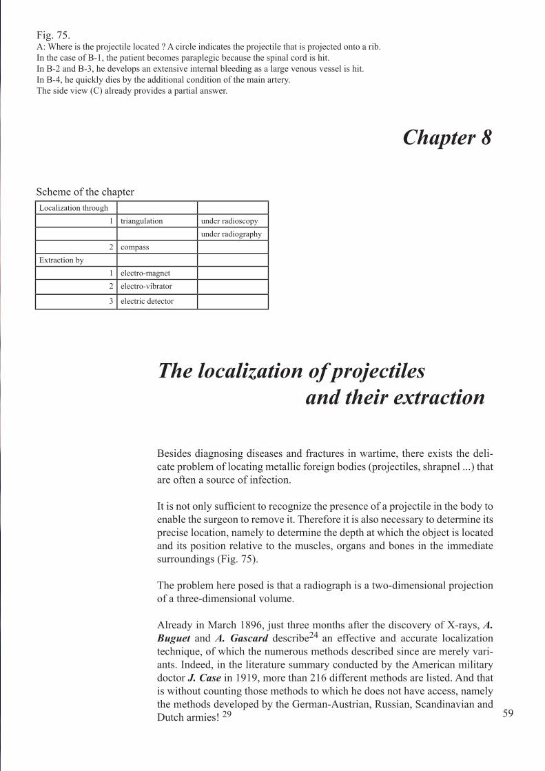
Chapter 8
Fig. 75. A: Where is the projectile located ? A circle indicates the projectile that is projected onto a rib.In the case of B-1, the patient becomes paraplegic because the spinal cord is hit.In B-2 and B-3, he develops an extensive internal bleeding as a large venous vessel is hit.In B-4, he quickly dies by the additional condition of the main artery. The side view (C) already provides a partial answer.
59
The localization of projectiles and their extraction
Besides diagnosing diseases and fractures in wartime, there exists the deli-cate problem of locating metallic foreign bodies (projectiles, shrapnel ...) that are often a source of infection.
It is not only sufficient to recognize the presence of a projectile in the body to enable the surgeon to remove it. Therefore it is also necessary to determine its precise location, namely to determine the depth at which the object is located and its position relative to the muscles, organs and bones in the immediate surroundings (Fig. 75).
The problem here posed is that a radiograph is a two-dimensional projection of a three-dimensional volume.
Already in March 1896, just three months after the discovery of X-rays, A. Buguet and A. Gascard describe24 an effective and accurate localization technique, of which the numerous methods described since are merely vari-ants. Indeed, in the literature summary conducted by the American military doctor J. Case in 1919, more than 216 different methods are listed. And that is without counting those methods to which he does not have access, namely the methods developed by the German-Austrian, Russian, Scandinavian and Dutch armies! 29
Localization through1 triangulation under radioscopy
under radiography2 compass
Extraction by1 electro-magnet2 electro-vibrator
3 electric detector
Scheme of the chapter

Without entering too much into details, we can say that some of the tech-niques determine the exact distance with respect to selected markers on the skin, while others use a metal search device (an often complicated, but accu-rate, compass). This allows reproducing results in the operating room.
Let us fi rst discuss the fi rst category, that of the radiologists who have access to the rather general triangulation, which is put into practice through much different devices.
Here is the explanation of this method in its simplest application under fl uoroscopy (Fig. 76): S is the patient. E is placed behind the screen. P is the projectile. T is the focal point of the X-ray tube which can be moved parallel to the screen opposite T and T '. The length of the displacement of the tube is known. The focus A is on a solid metal cross what’s shadow is projected on the screen in h.
When the tube is located in T, the cross is placed on the body S in such a way that the normal beam goes through projectile P, so is h the projection target of the foreign body.
The tube is then moved from T to T': the shadow of the projectile is now p', the shadow of the cross in a.The distance p'a, shows the depth PA on a scale. This scale is fi xed once and for all life-size, by drawing from point T’ a set of straight lines that run through the natural centimetre divisions of the axis A h.
60
Fig. 76-a. The principle of triangulation.
'

That scale can also be designed for different setups of the screen according to necessity. One can, if preferred, just as well determine the distance from the projectile to the marker B, where the beam leaves the body.
This triangulation is used in very different ways, applied according to the fi xed point from which the authors start; for one that is the distance from the tube to the screen, for the other the distance from the tube to the aiming-cross. One employs a variable displacement, the other a fi xed one. One meas-ures the depth by a wire stretched between T' and p, the other with a scale, yet another with a table, another again makes calculations. The practice is characterized by a range of variants.
The advantage of fl uoroscopy is its speed. There may however arise prob-lems of a biological nature (see next chapter), mainly for the little or not protected experimenter if this technique is frequently repeated.
61
Fig. 76-b. Application diagram of radioscopy in the location of a projec-tile.Here the X-ray tube is placed under the table and the fl uoroscopy screen above the patient.
Fig. 76-c. Radioscopic table "Ledoux-Lebard" of the military hospital Beveren-on-the-Yser.
G. N
iew
engl
owsk
i (19
24).
168
Priv
ate
Col
lect
ion.
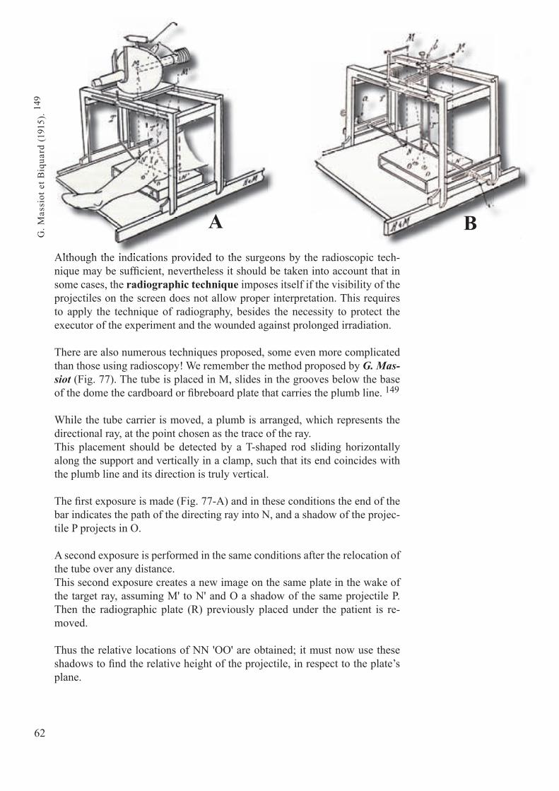
Although the indications provided to the surgeons by the radioscopic tech-nique may be suffi cient, nevertheless it should be taken into account that in some cases, the radiographic technique imposes itself if the visibility of the projectiles on the screen does not allow proper interpretation. This requires to apply the technique of radiography, besides the necessity to protect the executor of the experiment and the wounded against prolonged irradiation.
There are also numerous techniques proposed, some even more complicated than those using radioscopy! We remember the method proposed by G. Mas-siot (Fig. 77). The tube is placed in M, slides in the grooves below the base of the dome the cardboard or fi breboard plate that carries the plumb line. 149
While the tube carrier is moved, a plumb is arranged, which represents the directional ray, at the point chosen as the trace of the ray.This placement should be detected by a T-shaped rod sliding horizontally along the support and vertically in a clamp, such that its end coincides with the plumb line and its direction is truly vertical.
The fi rst exposure is made (Fig. 77-A) and in these conditions the end of the bar indicates the path of the directing ray into N, and a shadow of the projec-tile P projects in O.
A second exposure is performed in the same conditions after the relocation of the tube over any distance.This second exposure creates a new image on the same plate in the wake of the target ray, assuming M' to N' and O a shadow of the same projectile P. Then the radiographic plate (R) previously placed under the patient is re-moved.
Thus the relative locations of NN 'OO' are obtained; it must now use these shadows to fi nd the relative height of the projectile, in respect to the plate’s plane.
Although the indications provided to the surgeons by the radioscopic tech-
62
A B
G. M
assi
ot e
t Biq
uard
(191
5). 14
9

The tube holder is placed on a table for the ensuing actions (Fig. 77-B) since neither the tube nor the dome are needed any longer.The radiographic plate is placed at the height it has been positioned in during the successive exposures. In practice it suffi ces to lay it fl at on the table. And at the ends of the marking rods T and T', one brings on the brackets of which the rings are placed exactly in the points M and M', the points where sequen-tially the emission point of X-rays has been.By means of a plumb line that hangs down from M and M ', the position of the plate is arranged so that is occupies the same place underneath the injured. This is facilitated by orienting this plate until the traces of the plumbs coin-cide with the shadows of the ends of the marking rods. In regards to the free wire ends, these are stopped by a weight in those places where the shadows of the projectile have appeared.The point of intersection of these wires represents the position of the projec-tile.To capture its location in space, in respect to specifi c coordinates, the ends of the three bars a, b and c are fi xed perpendicularly to the three sides of the tube stand in contact with point P.With care the C pointer also is affi xed in a sliding way on each rod at their respective sleeves. These three rods form the plane in which the projectile is located. The wires which have no longer a function are retracted. The three rods are removed from each other without moving the pointer, to allow to place the tube stand back again over the irradiated surface in the same place it fi rst occupied, as shown in Figure 77-C.The three bars a, b, and c, are approached to each other and thus are three marking points determined and marked with a skin pencil. These three points defi ne a plane in which the projectile is located and the three relative distan-ces of each pointer in the bar sleeves correspond to the depths of the projectile with respect to those three points.The indications given by the relative locations of the three bars suffi ce but immobilize the radiography equipment until such time as the surgeon is ready for surgery.
63
Fig. 77. A. The patient is exposed under two angles.B. Locating the position of the projectile in space.C. The stand is again placed over the investigated region.
C

Hence the usefulness of a special localization compass: this completes the radiology equipment ready to use at the time of the operation.
Moreover all know that carrying out the search for projectiles, once the skin is cut it is very diffi cult to penetrate into the deeper tissues in a specifi c direc-tion. Let us note that objects, even of a certain size are often not felt by the exploratory fi nger. And even after a very diffi cult surgery it may happen that one fails while still having been very close to the object. So the surgeon needs a guiding device (compass) which prevents him from straying and guides him accurately to his target.
In this second category numerous compasses have been invented. That of the French physician E. Hirtz, developed in 1907, fi nds a broader application (Fig. 78, 79) than others. 99, 120
64
Fig. 78. Localization compass for the detection of projectiles, from Physician Major E . Hirtz. 120
Fig. 79. Hirtz compass, set up ready for use.
Bel
gian
Mus
eum
of R
adio
logy
G. P
alla
rdy
et c
oll (
1989
). 17
3
E. H
enra
rd (1
929)
. 115

The application requires four successive operations:
1. First radiograph (Fig.80-A). On a horizontally positioned X-ray plate (R), at the intersection of the diagonals, the centre V’ is marked. Above this point, indicated by an opaque point mark, and with the aid of a plumb line the focus of an X-ray tube is installed in V. The distance VV' is immediately measured. On the plate, the area is defined that contains the foreign body P, whose posi-tion should be determined. Three markers A, B and C on the skin are indicated by metal pointers. These points define a random triangular plane respective to the plate R. Whenever possible, the triangle defines the object P. The tube is then, in parallel with one of the sides of the plate, moved towards F and F' and from each point an X-ray is taken. During the image processing the pres-ence of double images of the three markers A, B, C and of the object P are observed.
2. Implementation of the projection drawing (Fig. 80-B). This X-ray image allows the drawing of a graph, which aims to transpose the newly carried out double cone projection into a rectangular projection on a horizontal and a ver-tical plane. We leave the description of the resulting complex graphics aside here; however according to its designer its implementation is quite simple.
3. Setting the compass (Fig. 80-C). According the data on the projection sketch, a localization compass is applied, which is shown schematically in the figure. It consists of three arms (1, 2 and 3) that revolve around a centre O, of three points, perpendicular to the plane of the brackets, and a pin in the centre O, that with gentle frictional motion serves as a sliding positioning probe.
4. Detection of the foreign body (Fig. 80-D). The compass is set up to detect the zone based on a horizontal plane T' . This is exactly the same position in which the X-ray is performed. The points coincide with the markers A, B and C on the skin. With the same accuracy, the positioning probe will coin-cide with P. However, since P is embedded in the tissue, the probe S will be retained by the body surface in i; the distance iP corresponds to the depth of the foreign body. This is measured by the distance between the centre O of the compass and marker r set to S. An incision, made in i, will open the way for the probe S whose end is touching the object P if the marker r coincides with O. The construction and use of the compass are based on a geometric concept. Consequently, one has to ensure that the plane of the three brackets 1, 2 and 3 be horizontal, while the instrument rests on the markers in the position of investigation, and that the pin S is vertical. It materializes the path running through the foreign body.
65Fig. 80. Diagram of the different steps that allow the localization of a projectile, with the Hirtz com-pass. 120
A
B
C
D
E. H
enra
rd (1
929)
. 115
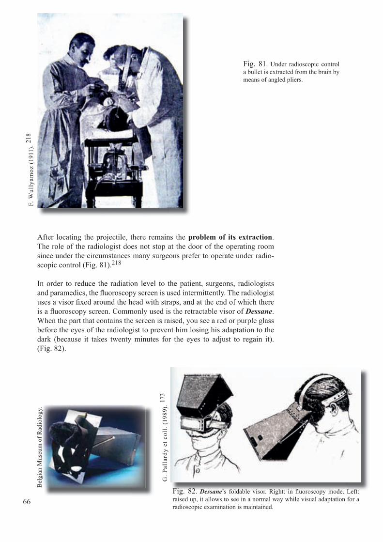
After locating the projectile, there remains the problem of its extraction. The role of the radiologist does not stop at the door of the operating room since under the circumstances many surgeons prefer to operate under radio-scopic control (Fig. 81).218
In order to reduce the radiation level to the patient, surgeons, radiologists and paramedics, the fl uoroscopy screen is used intermittently. The radiologist uses a visor fi xed around the head with straps, and at the end of which there is a fl uoroscopy screen. Commonly used is the retractable visor of Dessane. When the part that contains the screen is raised, you see a red or purple glass before the eyes of the radiologist to prevent him losing his adaptation to the dark (because it takes twenty minutes for the eyes to adjust to regain it).(Fig. 82).
66
Fig. 81. Under radioscopic control a bullet is extracted from the brain by means of angled pliers.
Fig. 82. Dessane’s foldable visor. Right: in fl uoroscopy mode. Left: raised up, it allows to see in a normal way while visual adaptation for a radioscopic examination is maintained.
G. P
alla
rdy
et c
oll.
(198
9). 17
3
F. W
ully
amoz
(191
1). 21
8
Bel
gian
Mus
eum
of R
adio
logy
.
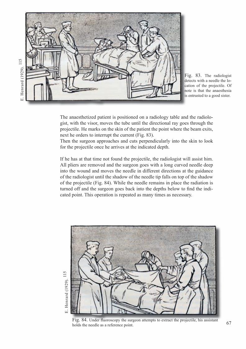
The anaesthetized patient is positioned on a radiology table and the radiolo-gist, with the visor, moves the tube until the directional ray goes through the projectile. He marks on the skin of the patient the point where the beam exits, next he orders to interrupt the current (Fig. 83).Then the surgeon approaches and cuts perpendicularly into the skin to look for the projectile once he arrives at the indicated depth.
If he has at that time not found the projectile, the radiologist will assist him. All pliers are removed and the surgeon goes with a long curved needle deep into the wound and moves the needle in different directions at the guidance of the radiologist until the shadow of the needle tip falls on top of the shadow of the projectile (Fig. 84). While the needle remains in place the radiation is turned off and the surgeon goes back into the depths below to fi nd the indi-cated point. This operation is repeated as many times as necessary.
67
Fig. 83. The radiologist detects with a needle the lo-cation of the projectile. Of note is that the anaesthesia is entrusted to a good sister.
Fig. 84. Under fl uoroscopy the surgeon attempts to extract the projectile, his assistant holds the needle as a reference point.
E. H
enra
rd (1
929)
. 115
E. H
enra
rd (1
929)
. 115

Notwithstanding how geometrically and analytically precise the localization of the projectiles may be carried out in the body using X-rays, nevertheless it happens that when a small projectile is concerned, the surgeon can fi nd it into the surgical wound only with great diffi culty or even cannot fi nd it at all. It goes without saying that many a surgeon breaks his head to prevent such failures, but also though masterful he may be sometimes he simply cannot succeed.
While we remain in the fi eld of electrical medical equipment let us men-tion some most interesting and most characteristic tools, namely the electro-magnets, electro-vibrators (Fig. 85), and electrical pointers. To be useable the projectile has to be ferro-magnetic, this is not always the case!Here follows a summary description of these three instruments.
68
Fig. 85. Operating room of the military hospital to Hoogstade.Probably an electromagnet in A and an electric vibrator in B.
A
AG
C-C
AA
2. 2
B

The electromagnet attracts ferromagnetic bodies, according to their posi-tions relative to the poles. This operation is performed at different distances. When a pole is applied to the skin that encloses a magnetic projectile, it will be attracted. It is thought that in some areas (brain, liver), the attraction is strong enough to cause an elevation of the surface. This “cone” allows small objects to be retracted through a tiny incision at the top. Thought has been given to improving the electromagnet. If during its course to the bottom of a surgical wound of small dimensions a projectile proves to be hard to grasp with pliers, then a probe is attached to the pole itself, which thus plays the role of magnetic core and can facilitate the removal (Fig. 86).
Several models of electromagnets, adapted to the surgery, are built since the start of the war of 1914.Here we mention those proposed by the Belgian physician Dr. Henrard: at the contact of the electromagnet (A) he adds a metal bar to the Hirtz com-pass. This rod (Fig. 87) is directed to the projectile and allows the removal if the foreign body is located in a soft structure (brain, liver) 112, 115
69Fig. 86. Exploration of the brain surface, exposed after trepanation. The electromagnet is wrapped in a sterile cloth.
Fig. 87. Use of the rod of the E. Hirtz compass and the electromagnet to remove a projectile from the brain (A).
E. H
enra
rd (1
917)
. 112
H. L
. Roc
her
(191
6). 18
3

Of a totally different nature: there is the AC electromagnet, or again the electric vibrator by Professor J. Bergonié. The electrovibrator is designed to detect the presence of magnetic projectiles and to determine their loca-tion, but not to remove them by its magnetic attraction. It consists of an electromagnet. Through the inductor an AC-current is sent. When one of the poles approaches the region of a body containing a magnetic projectile, then one notices a very special and typical vibration. This vibration increases in intensity when approaching the skin above the projectile.Figure 88 shows an operation performed using this instrument. The projec-tile is located by means of radiology. Before operating, the surgeon must fi rst know the place where the vibration is the strongest. 173
70
Fig. 88. Professor Bergonié uses his electric vibrator during the extraction of a projectile (1915).
G. P
alla
rdy
et c
oll (
1989
). 17
3

Many a surgeon prefers the remote electric detector based on the principle of the electric balance of Hughes. A famous model in military surgery during the Great War in military surgery is that of Baume-Pluvinel (Fig. 89). It is applied on the index fi nger like a rubber fi ngerstall. Equipped with this fi nger-stall the surgeon explores the surgical site while a listening device in his ear when he informs him a metal body is approaching or delete them.
A similar device, but of larger dimensions, is proposed during the First World War to detect unexploded projectiles in the fi elds: the mine detector.
71
Fig. 89. Exploration device of Baume-Pluvinel. A Box containing the coil. I Handle. L Tumbler of the vibrator. B Compensator with Flywheel V. D Fingerstall with electrical contact. T1 Telephone receiver. T2 Head phones.
E. H
enra
rd (1
929)
. 115
Bel
gian
Mus
eum
of R
adio
logy

This chapter cannot be concluded without a few words about stereoscopy. This does not provide an exact loca-tion but nevertheless it allows being aware of the relative position of a projectile to the bones, or to distinguish if the projectile is in front of or behind some natural mark-er or a surgical drain inserted into the wound. Here also Doctor Captain E. Henrard distinguishes himself with an original method (Fig. 90).
Let us mention only that, to stay in the merely pure ap-plication of the procedure that the distance between the two successive locations of the tube, to obtain two images that render in the stereoscope the impression of relief, is not equal in all cases to the distance between the eyes. One usually admits that distance as six and a half centi-metres. This distance matches the most common distance between the eyes. Let us consider not only the thickness of the irradiated region, but also the distance between the X-ray tube and the part closest to the region. 100
72
Fig. 91. Table showing that the δ (delta) displace-ments to give to the radiant tube to record two con-secutive radiographs depend on the thickness, the dis-tance D and E of the object (centimetre scale).
Fig. 90. First page of the fi rst study on stereoscopy by Dr. Henrard.
G. M
assi
ot e
t Biq
uard
(191
5). 14
9
E. H
enra
rd (1
902)
. 100

73Fig. 92. Stereoscopy equipment Wheatstone for reading large-format images (Westinghouse - USA around 1920).
Bel
gian
mus
eum
of
Rad
iolo
gy.
A table (Fig. 91) shows the distance, which should be determined in function of the distance from the object to the tube and the thickness of the object. In this table, E is the set thickness of the irradiated area; D is the distance from the tube focus to the closest surface of the body. From these two measure-ments one deducts the distance δ (delta) from the displacement which must be applied to the tube.The stereoscopic images thus obtained are read using a stereoscope (Fig. 92).

66
Mus
ée D
upuy
tren
(Pa
ris)
.
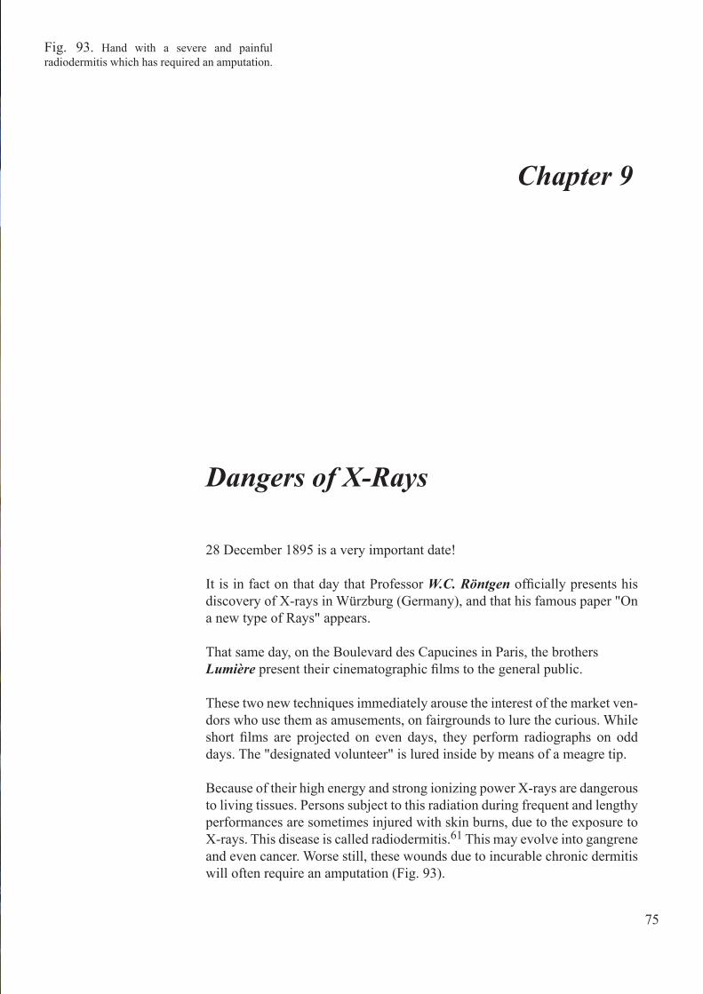
75
Chapter 9
Dangers of X-Rays
28 December 1895 is a very important date!
It is in fact on that day that Professor W.C. Röntgen officially presents his discovery of X-rays in Würzburg (Germany), and that his famous paper "On a new type of Rays" appears.
That same day, on the Boulevard des Capucines in Paris, the brothers Lumière present their cinematographic films to the general public.
These two new techniques immediately arouse the interest of the market ven-dors who use them as amusements, on fairgrounds to lure the curious. While short films are projected on even days, they perform radiographs on odd days. The "designated volunteer" is lured inside by means of a meagre tip.
Because of their high energy and strong ionizing power X-rays are dangerous to living tissues. Persons subject to this radiation during frequent and lengthy performances are sometimes injured with skin burns, due to the exposure to X-rays. This disease is called radiodermitis.61 This may evolve into gangrene and even cancer. Worse still, these wounds due to incurable chronic dermitis will often require an amputation (Fig. 93).
Fig. 93. Hand with a severe and painful radiodermitis which has required an amputation.
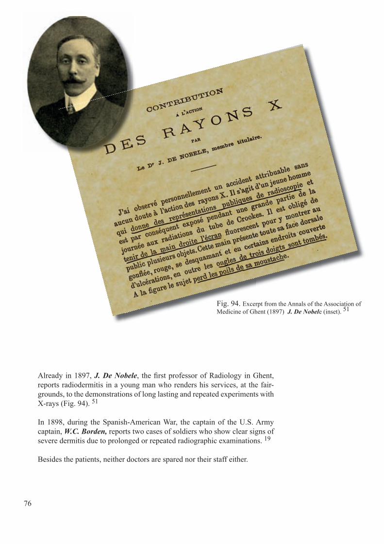
76
Already in 1897, J. De Nobele, the fi rst professor of Radiology in Ghent, reports radiodermitis in a young man who renders his services, at the fair-grounds, to the demonstrations of long lasting and repeated experiments with X-rays (Fig. 94). 51
In 1898, during the Spanish-American War, the captain of the U.S. Army captain, W.C. Borden, reports two cases of soldiers who show clear signs of severe dermitis due to prolonged or repeated radiographic examinations. 19
Besides the patients, neither doctors are spared nor their staff either.
Fig. 94. Excerpt from the Annals of the Association of Medicine of Ghent (1897) J. De Nobele (inset). 51
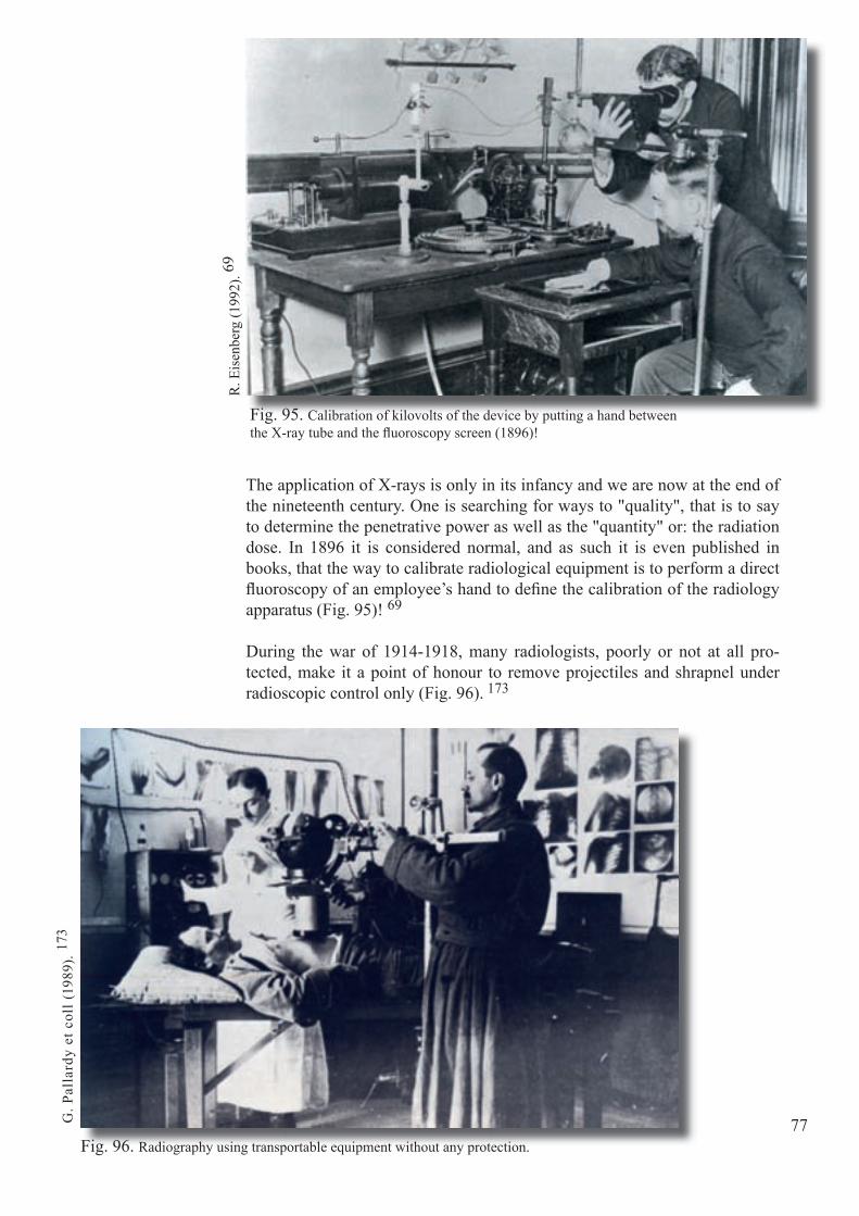
77
The application of X-rays is only in its infancy and we are now at the end of the nineteenth century. One is searching for ways to "quality", that is to say to determine the penetrative power as well as the "quantity" or: the radiation dose. In 1896 it is considered normal, and as such it is even published in books, that the way to calibrate radiological equipment is to perform a direct fl uoroscopy of an employee’s hand to defi ne the calibration of the radiology apparatus (Fig. 95)! 69
During the war of 1914-1918, many radiologists, poorly or not at all pro-tected, make it a point of honour to remove projectiles and shrapnel under radioscopic control only (Fig. 96). 173
R. E
isen
berg
(199
2). 6
9
Fig. 95. Calibration of kilovolts of the device by putting a hand between the X-ray tube and the fl uoroscopy screen (1896)!
Fig. 96. Radiography using transportable equipment without any protection.
G. P
alla
rdy
et c
oll (
1989
). 17
3

Although at this moment some people are already well aware of the dangers of X-rays, surgical teams are mostly spirited by a strong sense of duty. In the years that followed, numerous radiodermites are observed, radionecroses, and incipient cancers, many of which require amputation, sometimes with-out possibly averting death. Let us add to this leukaemia which will only be observed twenty years later...
Resulting from all this, is that the profession pays a heavy toll on this practice with numerous doctors dying in horrible pain. While France proportionately counts the largest number of deceased doctors, Belgium is not spared either (Fig. 97).
In the long list of famous victims of ionizing radiation, appears the name of Marie Curie, who, despite her vast knowledge, is not convinced of the real risk of these rays.From 1897 to 1902, together with her husband she does research on the min-eral "pitchblende" and they discover that it contains two radioactive materi-als. To one they give the name "polonium''; the other becomes the sole object of their study and is named ''radium'' due to its exceptional radioactivity.
78
J. Boine (1883-1935). Ch. Lombard (1871-1934).A. Morlet (1870-1932).
M. Van Roost (1880-1924).
Fig. 97.
Bel
gian
Roy
al L
ibra
ry A
lber
t I.
Arc
hive
s OC
MW
Ant
wer
p.
Bel
gian
Roy
al L
ibra
ry A
lber
t I.
Bel
gian
Roy
al L
ibra
ry A
lber
t I.
W. M
olin
eus
(199
2). 16
0

79
Fig. 98. The X-ray tube is placed in a bowl of lead glass. The tube is not completely protected.
Fig. 99. The diagram shows the direct (pri-mary) radiation and the (secondary) indirectly produced by refl ection on the walls.
Marie Curie wears on her hands the indelible stigmata. Moreover, she ma-nipulates radium tubes for many years, and during WWI performs radio-scopy on numerous injured without suffi cient protection against radiation. This gradually causes disease on her hands and alterations in her blood. This great lady dies of leukaemia in 1934.
It is only after the First World War, due to the appearance of many cases of radiodermitis in all warring parties, that the fi rst measures of radiation protection eventually are applied effi ciently and that the beginning of dose measurement is generally accepted. In 1928 the International Commission for Radiation Protection is established.
The X-ray tubes are then placed in lead glass caps (Fig. 98) as the lead ab-sorbs the rays, and limits the radiation to the investigated area. 123 The meth-od will need to be improved because the protection is still largely shortcom-ing (Fig. 99).72
R.E
. Fok
kem
a (1
993)
. 72
F. Ja
ugea
s (19
13) .
123
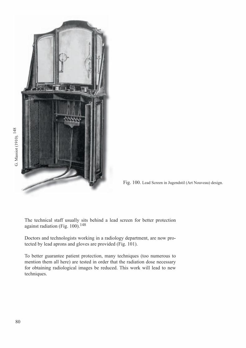
80
Fig. 100. Lead Screen in Jugendstil (Art Nouveau) design.
The technical staff usually sits behind a lead screen for better protection against radiation (Fig. 100).148
Doctors and technologists working in a radiology department, are now pro-tected by lead aprons and gloves are provided (Fig. 101).
To better guarantee patient protection, many techniques (too numerous to mention them all here) are tested in order that the radiation dose necessary for obtaining radiological images be reduced. This work will lead to new techniques.
G. M
assi
ot (1
910)
. 148
AG
C-C
AP2
(199
1). 2

81
A
B
C
Fig. 101. A technologist calibrates a radiation tube with a gas burner (B). The tube is placed in a lead sheath (A), to guarantee a partial protection. The lady wears a lead apron and lead gloves (C) to ensure her protection (Military Hospital of Bourbourg).
AG
C-C
AP2
(199
1). 2

74

Fig. 102. Splendid photo of which the original is unknown to us. One can see a doctor of the time examining a radiographic film.
83
Chapter 10
Problems and solutions in radiology
Numerous steps in the technological progress are taken thanks to or because of the difficulties encountered during armed conflicts. Radiology does not escape this phenomenon.
When X-rays pass through an anatomical region they give a negative image formed on a fluoroscopy screen or photographic glass plate. In the early years of radiology a positive image is then reproduced on paper.
During the First World War, due to the frequent movement of radiology labora-tories at the front one counts a large number of broken photo plates. Moreover most of the photographic glass originates from Belgium and the war and oc-cupation put an abrupt end to its exportation. 69
In 1914 there’s a novelty. The U.S. Company Eastman, later on Kodak, pre-sents a radiological film consisting of a smooth base of cellulose nitrate coated with a photographic emulsion (Fig. 102). The native of Ghent L. Baekeland who emigrates to the USA, sells in 1899 his invention: the photo paper "Velox" and his factory "Nepera Chemical", to Eastman Kodak. This is not entirely foreign to the appearance of the radiographic film!

84
The introduction of this material, in the beginning used in dental radiology, offers a solution in terms of weight, volume and convenience of handling. (Fig. 103). Also, the use of photo paper (positive image) is gradually abandoned.Despite its higher cost, this solution will replace
Fig. 103. Advertisement of the Eastman com-pany showing a radiographic fi lm underneath a broken glass plate!
for good the use of these very fragile glass plates. After the war, the Belgian fi rm Gevaert takes a further decisive step.
On page 38 and following, we have revealed the diffi culties when using the cold cathode radiation tube.The American Army Medical Service in the First World War brings us the radiology tube of W. Coolidge with a hot cathode. This is fi nalized in 1913 and it means a real revolution for the time. 13, 39, 175, 176 A long tube life and especially the constancy of the emission of the rays are now ultimately secured (Fig. 104).
This system is further enhanced in 1929 by the Dutch engineer A. Bouwers. He invents the rotating anode, which is still used today. This latest improve-ment provides a better dissipation of the heat resulting from the collision of the electrons emitted by the cathode.
Fig. 104. Diagram of a tube described in the patent Coolidge:A anticathode.M cathode cylinder that serves to focus the electrons.C cathode fi lament with its conductors.P accumulator intended to heat the fi lament.R rheostat.B transformer coil.I high voltage switch.
Coolidge X-ray tube manufactured under license in 1916 by the French fi rm H. Pilon. 176
Bel
gian
Mus
eum
of R
adio
logy
J. B
elot
et W
. Vig
nal (
1914
). 13
Bel
gian
Mus
eum
of R
adio
logy
.
J. B
elot
(191
6). 14

85
A crucial problem shows up during the Great War as the manufacture and the production of glass for scientifi c purposes are assured by the glassworks in Thuringia, Germany.
The state of war brings a sudden end to the availability of this commodity. The advantage of this glass comes from the fact that it contains few lead salts that stop the X-rays. Moreover, this glass alloy can be welded easily to the essential metal parts that are contained inside the radiation tube.
The suppliers of radiology equipment to the various warring parties fi nd "pa-triotic" (Fig. 105) solutions to overcome this defi ciency. 14
The only signifi cant technical development during the First World War is the invention of conventional tomography.The principle: the radiographic image is a superposition of the shadows of all structures located between the radiation tube and the radiographic fi lm. It is a two-dimensional projection of a three-dimensional volume!The interpretation of such images is diffi cult because one cannot determine at what level these different stacked shadows belong. Radiology can already rely on the side view or the use of fl uoroscopy while the patient is moved. However these two methods are not very accurate. Thus it becomes neces-sary to explore ways to better understand the third dimension.
Fig. 105. Reproduction of a plate, exhibited by the French house H. Pilon that demonstrates the anti-German campaign of this fi rm in 1913-1914.J.
Bel
ot (1
916)
. 14

86
In Chapter 8 we have seen how important it is to be able to determine the loca-tion of projectiles, shrapnel...
During the First World War therefore a stereoscopic method is used and so-phisticated trigonometric calculations too to determine the depth at which a foreign metal object can be found in the body of the patient.
To avoid these extensive and slow calculations, a French military doctor A. Bocage, develops in 1916 a radiographic and photographic technique that al-lows cutting the human body lengthwise (photographically!) in order to select the searched for plane (Fig. 106). A. Bocage perfects his wartime invention during his specialist training in Paris in 1920. He deposits a patent in 1921 and records it in 1922 (Fig. 107).18
Fig. 106. The principle of conventional tomography is based on the principle of a parallel movement in the opposite direction of the X-ray tube and the cassette that contains the fi lm. This movement occurs around a pivot point (fulcrum). This point is placed at the level of the ana-tomical target region of the patient. Only what lies in the horizontal plane of rotation is sharply visible on the fi lm. All objects above or below are blurred and erased.

87
At his period in time, only the national literature is generally consulted. Thus it is not surprising that without the knowledge of the work already done by other colleagues, some authors, in good faith, conceive similar techniques. We mention B. Ziedses des Plantes (Netherlands 1922), 219 J. Kieffer (USA 1928), 125 A. Vallebona (Italy 1930), 201 G. Grosmann (Germany 1934).91
All these methods are successfully and intensively used until the appearance of computed tomography, fi nalized in 1972 by Sir G. Hounsfi eld (U.K.). For this invention he receives the Nobel Prize for medicine in 1979.The latter technique, commonly called X-ray scanner, is the result of joining conventional tomography, invented during the First World War, with the com-puter, invented during World War II. 210, 211 Would war still be the basis of everything as Heraclitus once said?
Fig. 107. A. Bocage and his license. 18.

80

89
Chapter 11
Fig. 108. Manifesto of the 93 German intellectuals drawn up in October 1914 and addressed to the "civilized" world. It appears on 29 May 1915 in the magazine "Die Aktion".
Unfortunate interference by Röntgen
On 29 May 1915 is issued the famous German manifesto: "Call to the civi-lized world" (Fig. 108), signed by 93 (later on 4,000) German academics and artists, in which they reject all the responsibility of Germany in this conflict. This document is part of a German propaganda campaign in reaction to arti-cles appearing in the Allied press and describing the atrocities committed by German troops.
During wars, "harmful effects" unfortunately always remain an issue, espe-cially so during the First World War.
This is also the case in Belgium, as demonstrated in many cities as Aarschot, Andenne, Dinant, Huy, Tamines, and the destruction of nearly one fifth of the buildings in Leuven (Louvain). That is all part of the German intimidation strategy in the occupied territories, with the aim to obtain the maximum of cooperation from the civilian population.
Das
Man
ifest
der
93
(191
4).
46

Fig. 109-a. The Old-Market Square in Leuven! View of the destruction on 25 and 26 August 1914: in the background one can see what is left of the St Peter's Church, and the turrets of the town hall, and right part of the ru-ins of the university library.
As of 25 August 1914, the city of Leuven and its rich university with 1,000 incunabula, along with many other university and civic build-ings, are looted and burned to ashes during fi ve consecutive days (Fig. 109).155
A group of 246 Leuven citizens are executed without distinction of age or gender.
A communiqué issued by the German foreign minister G. Von Jagow (Fig. 110) confi rms that the German soldiers did not deliberately de-stroy the city but that the destruction was caused by a large scale population riot. Minister Von Jagow concludes that this revolt vio-lates the conventions of war.
This communiqué is distributed following a global horror provoked by the murderous destruction of Leuven (Fig. 111).
Fig. 110. Minister of Foreign Affairs G. von Jagow.
Fig. 109-b. Close-up View of St. Peter's Church. Fig. 109-c. Inside view of the university library.
Priv
ate
colle
ctio
n.
90
City
Arc
hive
s Leu
ven
(pho
to a
rchi
ve).
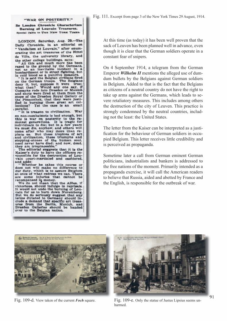
91
At this time (as today) it has been well proven that the sack of Leuven has been planned well in advance, even though it is clear that the German soldiers operate in a constant fear of snipers.
On 4 September 1914, a telegram from the German Emperor Wilhelm II mentions the alleged use of dum-dum bullets by the Belgians against German soldiers in Belgium. Added to that is the fact that the Belgians as citizens of a neutral country do not have the right to take up arms against the Germans, which leads to se-vere retaliatory measures. This includes among others the destruction of the city of Leuven. This practice is strongly condemned by the neutral countries, includ-ing not the least: the United States.
The letter from the Kaiser can be interpreted as a justi-fi cation for the behaviour of German soldiers in occu-pied Belgium. This letter receives little credibility and is perceived as propaganda.
Sometime later a call from German eminent German politicians, industrialists and bankers is addressed to the free nations of the moment. Primarily intended as a propaganda exercise, it will call the American readers to believe that Russia, aided and abetted by France and the English, is responsible for the outbreak of war.
Fig. 111. Excerpt from page 3 of the New York Times 29 August, 1914.
91Fig. 109-d. View taken of the current Foch square. Fig. 109-e. Only the statue of Justus Lipsius seems un-
harmed.

92
This causes a signifi cant negative reaction, especially in the United States. It has certainly infl uenced the decision of this country to take part in the confl ict.
Among the signatories of this manifesto are German names as A. von Bayer, P. Behrens, P. Ehrlich, F. Haber, W. Ostwald, M. Planck, Ph. Lenard, and …W.C. Röntgen. (Fig. 112).
Fig. 112. Wilhelm Conrad Röntgen (1845-1923). Lith-ograph engraved in 1995 by Andrzej Wisniewski on the occasion of the commemoration of the centenary of the discovery of X-rays, organized by the Belgian Museum of Radiology.
Why does Röntgen sign such a statement? Besides his naive nature, his pa-triotism is certainly the main reason.212
Röntgen has a deep sense of national pride, even though he lived the fi rst 25 years of his life outside his native country. In fact, he is born in Remscheid-Lennep, Germany (1845) but attends school in Apeldoorn (1848-1862) and Utrecht (1862-1865) in the Netherlands. Then he studies in Zürich, Switzer-land (1865-1870). After long hesitation, he returns to Germany in 1871.
After the discovery of X-rays Röntgen, receives some awards including the "Rumford Gold Medal of the Royal Society" (Fig. 113) in London in 1896. He is elected an honorary member of the Röntgen Society (1897), and re-ceives the Nobel Prize (1901).
In 1914, after the conquest of Belgium, Röntgen offers his B. Rumford Med-al to help the war effort. He pronounces following words: "I give gold for iron." He keeps all his other medals.
In 1915, the year of his 70th birthday, he is decorated with the "Iron Cross on black and white ribbon" by P. von Hindenburg, head of the German general staff for the help provided by his discovery to all the wounded soldiers.
Röntgen writes on 8 December, 1920, in a letter addressed to Mrs. M. Boveri:
Fig. 113. The same medal as the one that Röntgen received in 1896.
"A while ago I received a pamphlet from a Belgian scientist which embar-rasses me quite a lot, especially since he refers to the proclamation of the 93 intellectuals, which I thoughtlessly signed along with others, advised by and on the insistence of the "Berliners" without having read it beforehand. This is chiefl y the fault of H... ". 82
Fig. 113. The
Roy
al S
ocie
ty /R
icha
rd V
alen
cia.
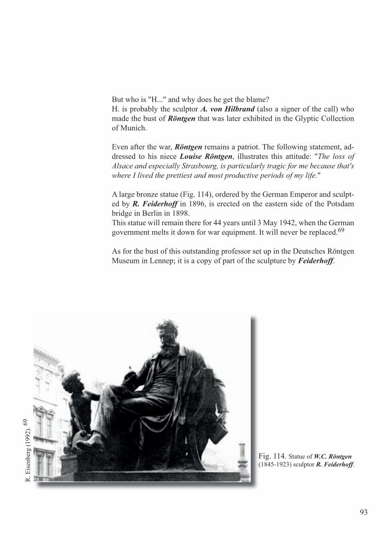
Fig. 113. The same medal as the one that Röntgen received in 1896.
93
Fig. 114. Statue of W.C. Röntgen (1845-1923) sculptor R. Feiderhoff.
But who is "H..." and why does he get the blame?H. is probably the sculptor A. von Hilbrand (also a signer of the call) who made the bust of Röntgen that was later exhibited in the Glyptic Collection of Munich.
Even after the war, Röntgen remains a patriot. The following statement, ad-dressed to his niece Louise Röntgen, illustrates this attitude: "The loss of Alsace and especially Strasbourg, is particularly tragic for me because that's where I lived the prettiest and most productive periods of my life."
A large bronze statue (Fig. 114), ordered by the German Emperor and sculpt-ed by R. Feiderhoff in 1896, is erected on the eastern side of the Potsdam bridge in Berlin in 1898.This statue will remain there for 44 years until 3 May 1942, when the German government melts it down for war equipment. It will never be replaced.69
As for the bust of this outstanding professor set up in the Deutsches Röntgen Museum in Lennep; it is a copy of part of the sculpture by Feiderhoff.
R. E
isen
berg
(199
2). 69

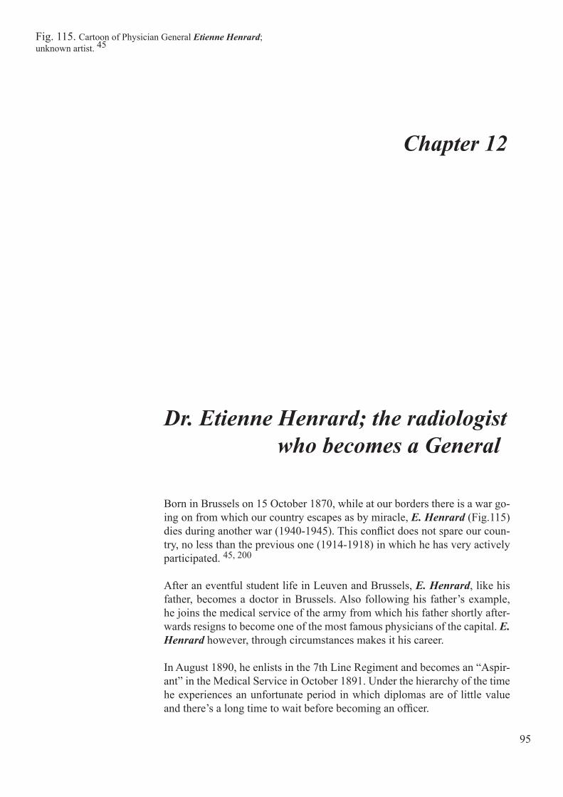
Dr. Etienne Henrard; the radiologist who becomes a General
Born in Brussels on 15 October 1870, while at our borders there is a war go-ing on from which our country escapes as by miracle, E. Henrard (Fig.115) dies during another war (1940-1945). This confl ict does not spare our coun-try, no less than the previous one (1914-1918) in which he has very actively participated. 45, 200
After an eventful student life in Leuven and Brussels, E. Henrard, like his father, becomes a doctor in Brussels. Also following his father’s example, he joins the medical service of the army from which his father shortly after-wards resigns to become one of the most famous physicians of the capital. E. Henrard however, through circumstances makes it his career.
In August 1890, he enlists in the 7th Line Regiment and becomes an “Aspir-ant” in the Medical Service in October 1891. Under the hierarchy of the time he experiences an unfortunate period in which diplomas are of little value and there’s a long time to wait before becoming an offi cer.
Chapter 12
95
Fig. 115. Cartoon of Physician General Etienne Henrard;unknown artist. 45

Military medicine does not quite satisfy the needs of E. Henrard, but that will change after an important scientifi c event: the discovery of X-rays. Clair-voyant as he is he immediately foresees the great future for this discovery in the fi eld of medicine.
Successively becoming deputy service physician of the 2nd and 1st class, then adjunct physician in 1903, he transfers to the popular 9th Line Regi-ment, the local unit in the lower town of Brussels, near to the Avenue du Midi where he also sets up his domicile.
At that time he becomes the adjunct of Dr. E. Dupont at the military hospital in Brussels (Fig. 116) and it turns out that he quickly becomes familiar with radiology and radiation therapy. On 24 November 1903 Henrard presents to the medical-surgical association of Brabant the fi rst case in Belgium of skin cancer cured by X-rays. From this period also dates his fi rst scientifi c work and at once he starts studying stereo-radiography and foreign body localiza-tion of which he remains forever the champion. (Fig. 117).100 to 118
96
Fig. 116. Brussels Military Hospital, Avenue de la Couronne, in 1903.
Fig. 117. Excerpt from the fi rst paper written by Dr. Henrard on stereoscopic radiography (1902).100
Together with Father J.D. Lucas SJ, professor at the Faculty of Sciences in Namur, he establishes and perfects his geomet-ric method for localizing radio-opaque foreign objects.
In Brussels, in 1900, E. Henrard sets up one of the fi rst pri-vate radiology offi ces in Belgium (Fig. 118). Thanks to his strong intelligence, his consistent discretion and skill, he quickly becomes a renowned radiologist.
Col
lect
ion
priv
ée

97Fig. 119. Overview of the hospital L' Océan.
Fig. 118. Part of the radiology equipment of the private of-fi ce of Dr. Henrard. Reconstruction that has been in the mili-tary hospital of Neder-Over-Heembeek (Brussels).
But his practice suffers from his frequent ab-sence during the camp periods and manoeu-vres in which he has to participate. So, shortly before the declaration of war of 14-18 our Battalion Physician of the 2nd class decides on 26 January 1914 to leave the medical ser-vice of the army to dedicate himself entirely to the radiology.
E. E
vrar
d et
col
l. (1
997)
. 70B
elgi
an M
useu
m o
f Rad
iolo
gy.
It would be a mistake to think even just for a moment that Henrard would refer to his retirement license and not sign up again for service during the mobilization. Also, from 4 August 1914, Physician-Captain Henrard (see page 9) is allowed to re-enter the service as Regimental Physician 2nd class. He becomes attached to the 6th Artillery Regiment and is employed in a more useful way in the ambulance column of the Brussels division (6 D. A.).
Towards the end of 1914, our troops protected by the fl ooding of the Yser and no longer having to fear encirclement, the leadership of the medical service can start thinking about the establishment of permanent hospitals.As previously mentioned on page 17, A. Depage leaves Brussels and leaves others in charge of the ambulance at the Royal Palace. In late November 1914, after having presided over the Ambulance Jeanne d' Arc in Calais, he can begin to think about the establishment of the ambulance at De Panne, named "L' Océan" after the hotel that composes its core. But A. Depage, ex-cellent organizer and top-notch surgeon, does not like the burden of red tape and the respect for discipline. Physician-General Mélis, Inspector General of the health service, who knows how to choose his men, appeals to his friend Physician-Captain E. Henrard to act as a conciliator for the new hospital "L' Océan" (Fig. 119).70
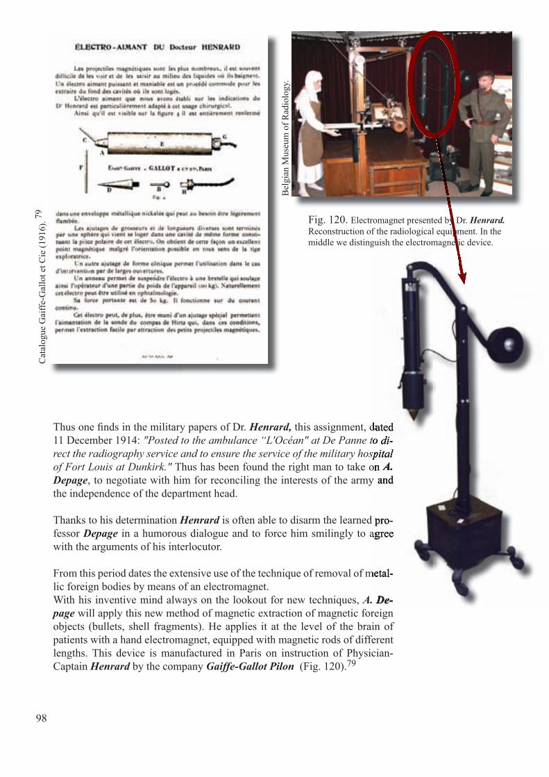
Thus one fi nds in the military papers of Dr. Henrard, this assignment, dated 11 December 1914: "Posted to the ambulance “L'Océan" at De Panne to di-rect the radiography service and to ensure the service of the military hospital of Fort Louis at Dunkirk." Thus has been found the right man to take on A. Depage, to negotiate with him for reconciling the interests of the army and the independence of the department head.
Thanks to his determination Henrard is often able to disarm the learned pro-fessor Depage in a humorous dialogue and to force him smilingly to agree with the arguments of his interlocutor.
From this period dates the extensive use of the technique of removal of metal-lic foreign bodies by means of an electromagnet.With his inventive mind always on the lookout for new techniques, A. De-page will apply this new method of magnetic extraction of magnetic foreign objects (bullets, shell fragments). He applies it at the level of the brain of patients with a hand electromagnet, equipped with magnetic rods of different lengths. This device is manufactured in Paris on instruction of Physician-Captain Henrard by the company Gaiffe-Gallot Pilon (Fig. 120).79
98
Cat
alog
ue G
aiffe
-Gal
lot e
t Cie
(191
6). 79
this assignment, dated "Posted to the ambulance “L'Océan" at De Panne to di-
rect the radiography service and to ensure the service of the military hospital Thus has been found the right man to take on
, to negotiate with him for reconciling the interests of the army and
is often able to disarm the learned pro- in a humorous dialogue and to force him smilingly to agree
From this period dates the extensive use of the technique of removal of metal-
A. De- will apply this new method of magnetic extraction of magnetic foreign
this assignment, dated "Posted to the ambulance “L'Océan" at De Panne to di-
rect the radiography service and to ensure the service of the military hospital Thus has been found the right man to take on A.
, to negotiate with him for reconciling the interests of the army and
is often able to disarm the learned pro- in a humorous dialogue and to force him smilingly to agree
From this period dates the extensive use of the technique of removal of metal-
A. De-
Bel
gian
Mus
eum
of R
adio
logy
.
Fig. 120. Electromagnet presented by Dr. Henrard. Reconstruction of the radiological equipment. In the middle we distinguish the electromagnetic device.

Dr. F.A. Sondervorst tells about the event: "As soon as the unit arrives A. De-page announces for the next day: surgery using a new technique. The message spreads like wildfi re and the French and English surgeons from neighbouring ambulances rush to attend the surgery. In an operating room, too small to receive the throng of visitors, Antoine Depage detects the foreign object by means of the geometrical Hirtz method, quickly performs a trepanation and introduces a metal rod into the brain along the trajectory of the projectile. Then the pole of the electromagnet, in the form of a hollow hemisphere, is put into contact with the ball, the circuit is closed, and rod and projectile, stuck to each other through the magnetic attraction, are simultaneously withdrawn (Fig. 121).
99
Fig. 121. Removal of a magnetic projectile from the brain by means of an electromagnet.
Quick as lightning A. Depage shows the bullet to the rapt audience: "Here it is!" Surprised the visitors ask: "Does that succeed often?" The Master replies: "Al-ways, Gentlemen!" It is the fi rst time he applies this technique, but Depage does not lie, because the future will show that they always will succeed, provided the technique is applied precisely. It is a great advance in the removal of foreign bodies from the brain. 189
Last paper, summarizing the topic written in 1929 by Physician-Colonel Henrard.115

At the hospital L' Océan, Henrard himself is the man of providence for the wounded of whom he takes radiographs and whom he encourages. Providence also for the numerous staff, surgeons and friends who trust him, and whom he willy-nilly defends against the authorities, at the risk of being commented on or to be seen as "scapegoat" in this buzzing hive that is constantly at work.
So for several years at "L'Océan", he is the most popular, the merriest, most important of the offi cers of the staff of A. Depage.
As the war draws to an end in June 1918 E. Henrard spends a short time at the directorate of the hospital of Mortain in Brittany. In October 1918, dur-ing the liberation offensive he returns and goes to the hospital of Vinkem at the front. The ambulance of the Red Cross of De Panne, better known to the veterans and wounded under the name "L'Océan" is transferred to Brussels shortly after the armistice and established on the G. Brugmann Square (Berk-endaelstraat).
The war period gives our retired physician from 1914 a renewed taste for the army and E. Henrard obtains successively the rank of Lieutenant-Colonel in 1917 and Colonel in March 1923. He leads at that time the main military hospital, founded in Woluwe (Fig. 122) for repatriated war invalids; it is the recovery hospital set up next to the plateau of Stockel, in the villa Parmen-tier, a charming site, now developed with villas and cottages. 70
100
Fig. 122. The Parmentier Park is made available to the military by the Princess de Merode, vice-president of the Red Cross.
E. E
vrar
d et
col
l. (1
997)
. 70
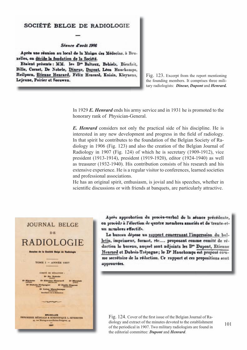
101
In 1929 E. Henrard ends his army service and in 1931 he is promoted to the honorary rank of Physician-General.
E. Henrard considers not only the practical side of his discipline. He is interested in any new development and progress in the fi eld of radiology. In that spirit he contributes to the foundation of the Belgian Society of Ra-diology in 1906 (Fig. 123) and also the creation of the Belgian Journal of Radiology in 1907 (Fig. 124) of which he is secretary (1909-1912), vice president (1913-1914), president (1919-1920), editor (1924-1940) as well as treasurer (1932-1940). His contribution consists of his research and his extensive experience. He is a regular visitor to conferences, learned societies and professional associations.He has an original spirit, enthusiasm, is jovial and his speeches, whether in scientifi c discussions or with friends at banquets, are particularly attractive.
Fig. 123. Excerpt from the report mentioning the founding members. It comprises three mili-tary radiologists: Dineur, Dupont and Henrard.
Fig. 124. Cover of the fi rst issue of the Belgian Journal of Ra-diology and extract of the minutes devoted to the establishment of the periodical in 1907. Two military radiologists are found in the editorial committee: Dupont and Henrard.

102
On each occasion, a speech by Henrard is asked for and he has a very atten-tive audience. He also carries his speech with an enthusiasm of which only he knows the secret, still full of fi re, sometimes very solemnly recited, then rendered very pleasantly with his deep and biting voice.Sometimes he lets his imagination run wild, as in a fugue, with his mind full of humour but sometimes biting, well versed, he can give a pique but is never poisonous. Furthermore, he usually rounds off with a joke that he commences with a straight face and serious mien to turn suddenly to laughter to disarm even the most suspicious minds.
E. Henrard enjoys a beautiful and healthy popularity in many circles. As an established fi gure in the world of radiologists (founding member of the Professional Union of Belgian Radiologists and Electro-Radiologists), he is highly regarded by his colleagues. Proof to that is his appointment as chair-man (1924-1938), then honorary chairman of the "Professional Union of Bel-gian Radiologists and Electro-Radiologists' (Fig. 125) and Honorary Presi-dent of the III Congress of Radiology in the French language (Paris 1931).
The appreciation by his confreres and friends has been reaffi rmed, and in an exciting way by a solemn event in his honour. This would take place in the "Cercle Gaulois' in the presence of over 400 Belgian and foreign guests. It is scheduled for 19 May 1940 ... few days after the outbreak of hostilities of the second world war in Belgium (May 10).
Fig. 125. Handwritten letter from Dr. Henrard, testifying to his interest for the next congress of radiology in the French language, that would take place in 1938.
Bel
gian
Mus
eum
of R
adio
logy
.

103
Hence from sheer necessity one is limited to a meeting in restricted committee on 13 December 1940. In this meeting Dr. Z. Gobeaux, 83 presents him with a bronze plaque by the sculptor Berchmans, along with the list of subscribers (Fig. 126).
On 10 May 1940, he is convinced that he could be useful as in the pre-
Fig. 126-a. Picture of Physician-General Henrard at the end of his life
Fig. 127. Obituary of Dr E. Henrard.
Fig. 126-a. HenrardFig. 126-a. Fig. 126-a. Henrard
vious war. The still very vibrant - though already 70 years old Physician-General Henrard considers it his duty to offer his services to the military. His urgent request cannot be followed upon. And he is heavily disappointed. Persevering in his plans, he counts on his relation-ships with famous French radiologists, goes to Paris, and presents himself to the French army, also without success.Discouraged, and in the impossibility of returning to Brussels, he goes to the south of France. There he is pained by his forced idleness as he longs for his country.
On his return to Brussels he resumes his work. Alas, shortly thereafter he begins to suffer from the disease that takes him away from this world on 14 January 1941 (Fig. 127).
Bel
gian
Mus
eum
of R
adio
logy
.Picture of Physician-General
Fig. 126-b. Plaque that Dr Henrard received at last on De-cember 13th, 1940.
Me
E. H
enra
rd B
ruxe
lles.


105
Chapter 13
From lab technician to technologist
With the discovery of X-ray doctors quickly recognize the potential of X-radiation. Here is a sketch of how the first radiology services are started. The "radiologist" of our time may request the assistance of a laboratory boy to as-sist him in the maintenance of equipment and the processing of image plates.
The processing of these photographs demands an extensive knowledge and expertise to detect exposure errors and compensate them in the darkroom by over- or underdevelopment. Slowly the role of the aide evolves from techni-cal assistant to performer. That allows him just to take some radiographs for the doctor.
From the commencement of the radiological adventure the need to train op-erators appears to be indispensable; the learning of the skills is acquired “on the job”. In Belgium, from 1905 on, the army provides a structured training, proof of which is the first course (Fig. 128), prepared by E. Dupont (the first head of the military hospital in Brussels).64
Fig. 128. Cover of the first course in medical radiology, prepared in 1905 by the Battalion Physician Emile Dupont, in charge of the labora-tory service of the military hospital in Brussels. 64
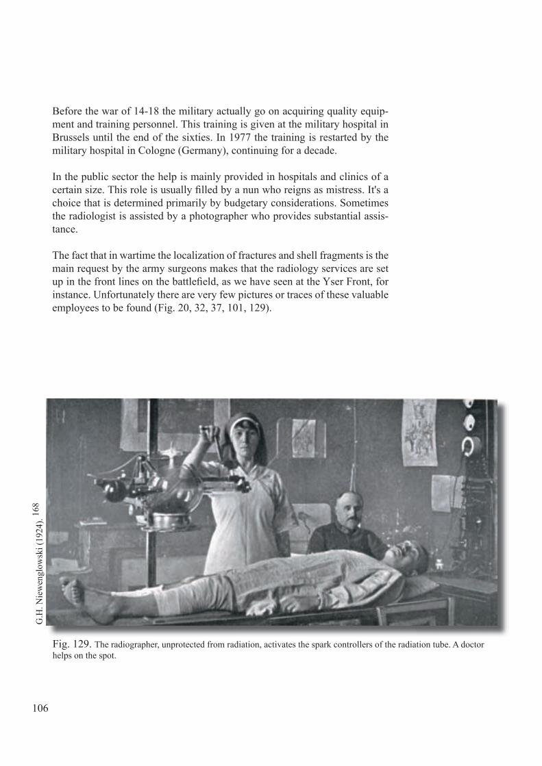
106
Before the war of 14-18 the military actually go on acquiring quality equip-ment and training personnel. This training is given at the military hospital in Brussels until the end of the sixties. In 1977 the training is restarted by the military hospital in Cologne (Germany), continuing for a decade.
In the public sector the help is mainly provided in hospitals and clinics of a certain size. This role is usually fi lled by a nun who reigns as mistress. It's a choice that is determined primarily by budgetary considerations. Sometimes the radiologist is assisted by a photographer who provides substantial assis-tance.
The fact that in wartime the localization of fractures and shell fragments is the main request by the army surgeons makes that the radiology services are set up in the front lines on the battlefi eld, as we have seen at the Yser Front, for instance. Unfortunately there are very few pictures or traces of these valuable employees to be found (Fig. 20, 32, 37, 101, 129).
Fig. 129. The radiographer, unprotected from radiation, activates the spark controllers of the radiation tube. A doctor helps on the spot.
G.H
. Nie
wen
glow
ski (
1924
). 16
8
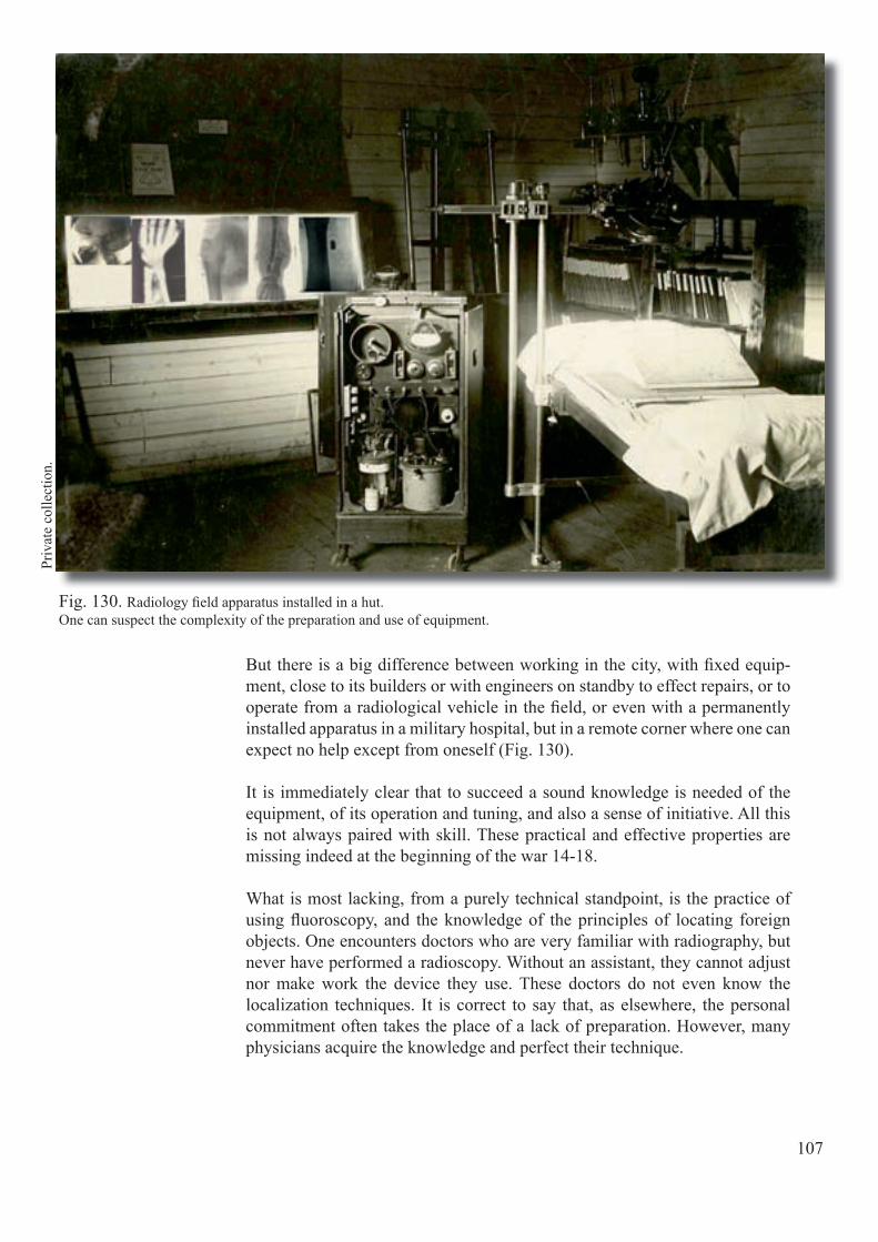
107
But there is a big difference between working in the city, with fi xed equip-ment, close to its builders or with engineers on standby to effect repairs, or to operate from a radiological vehicle in the fi eld, or even with a permanently installed apparatus in a military hospital, but in a remote corner where one can expect no help except from oneself (Fig. 130).
It is immediately clear that to succeed a sound knowledge is needed of the equipment, of its operation and tuning, and also a sense of initiative. All this is not always paired with skill. These practical and effective properties are missing indeed at the beginning of the war 14-18.
What is most lacking, from a purely technical standpoint, is the practice of using fl uoroscopy, and the knowledge of the principles of locating foreign objects. One encounters doctors who are very familiar with radiography, but never have performed a radioscopy. Without an assistant, they cannot adjust nor make work the device they use. These doctors do not even know the localization techniques. It is correct to say that, as elsewhere, the personal commitment often takes the place of a lack of preparation. However, many physicians acquire the knowledge and perfect their technique.
Fig. 130. Radiology fi eld apparatus installed in a hut.One can suspect the complexity of the preparation and use of equipment.
Priv
ate
colle
ctio
n.

108
As the medical staff lacks its quorum and is insuffi ciently prepared, the para-medical one is also low in numbers. At the front there is the lab assistant who not only runs the equipment for the radiologist, but who also maintains the equipment, who ensures its proper functioning and who repairs the defects in the electrical installation. His role is more that of a technical engineer. When attached to a mobile unit, he must, like the doctors, be very active, helpful and resourceful.
In principle the technologists never operate outside the work relationship with the doctors-radiologists. In practice, however, in the absence of the latter, they are sometimes on their own to take care of the radiology service within a department of care. This mission is entrusted to them by the surgeon or the physician-in-charge who can appreciate the value of their cooperation really well (Fig. 131).
Between the technologists, employed on the radiology vehicles, and the doc-tors who are responsible for them there grows in some cases such a close working relationship that, in the case of continuous activity, the service is fully and alternatively insured by each of them.
One sees how the role of the technologist expands in the case of radiology in times of war, sometimes even almost completely independently. This state of affairs that is a totally unusual situation in peacetime has to do with the con-ditions in which doctors and surgeons came into contact with the radiology.
Fig. 131. Preparation of complex radiological instruments for locating a foreign body in the brain.
A. C
harli
er (1
915)
. 34

109
Although there are voices speaking out to advocate the establishment of schools for technologists - including that of Marie Curie in her book published in 1921 (Fig. 132): "La guerre et la radiologie" (War and Radiology)- there is still a long wait before an offi cial training will be set up in our country. 44
The training is always done "on the job", or by the radiologist who trains his technologist according to his own specifi c needs or by fellow technologists who communicate their knowledge.
It will take until the late sixties before some radiology departments start to employ nurses to accomplish the more specifi c tasks of nursing care: they in-tervene when injections are required (urographic, neuroradiological, angio-graphic studies ...).
One has to wait for the Royal Decree of 28 February 1997 (Moniteur belge, equivalent of the Federal Registry, of 7 June 1997 pages 15375-77) to see the professional title of Technologist in Medical Imaging approved.
In Belgium, the title of Technologist in Medical Imaging is fi nally confi rmed by a diploma with legal approval! 209
Fig. 132. Page from the book by Marie Curie (La guerre et la radiologie), which, if prove is still necessary, illustrates the importance of well-trained paramedics. 44
Fig. 132. guerre et la radiologie), which, if prove is still necessary, illustrates the importance of well-trained paramedics.

L. M
orte
lman
s (20
10) .
161

111
Chapter 14
Radiology described by those who have undergone it
There are a lot of books, notes, texts written by doctors, nurses, care givers, and even patients who tell their stories about their experiences during the bru-tal confl ict. However, despite our research work we have not been able to fi nd testimonials from patients about their stay in radiology.
Luckily one member of our team remembers that his grandfather kept a di-ary wherein he describes his visit to a radiology department (Fig. 133). We'll let Eduardus Vanderdonckt, 161 schoolteacher and war volunteer, speak for himself about his experience ...
Literally:
"Sunday, 7 Oct. (1917)Slept very badly because of the bad news last night and feel quite tired. Towards noon, people carry a stretcher to my bed on which I am loaded, wrapped in blankets with a hot bottle at my feet, brought out and slid into a Red Cross car.
Fig. 133. Excerpt from the diary of a war volunteer.Eduardus Vanderdonckt, wounded in 1917, and his referral for a radiological examination. 161

112112
But in a relieved mood because I know I will come back. I go to the big Eng-lish hospital at St. Omer for X-rays. There I am laid on a table and they begin to take photos. Missing are just some candles to make it look entirely as if I were lying in a mortuary. When I am brought out and pushed back into the car, there are a whole lot of people there gaping at me and a few ladies who even begin to ask questions with "Mon petit" (my little boy) etc... I am not up to a conversation and just answer "No compris“(no understood). And now back to my home where dinner is waiting for me, very practically served on a little table that is put on the bed. They must still cut my meat for me but I can eat by myself. In the morning they have to wash me. It's very cold and it rains for three days already."
In the world literature we fi nd a text by the German T. Mann (1875-1955). He, Nobel Prize for Literature in 1929, is enraptured by medicine. A stay in a sanatorium in Davos in 1916 and the catastrophe of the Great War in which he is involved by choosing the side of Imperial Germany, inspires him to his most famous novel, “Der Zauberberg” (The Magic Mountain), published in 1924. 147
We fi nd numerous medical descriptions back in there. Here we render, freely translated, the development of a lung fl uoroscopy (Fig. 134):
"Joachim was seated on a sort of cobbler's stool, they pushed his chest against a board around which he clasps his arms, and the technologist improved the position of the patient by kneading his body, pushing the shoulders of Joachim forward and massaging his back.

113113
The technologist took place behind the unit as any photographer would do, and braced him-self on his legs. He leaned over to interpret the image, expressed his satisfaction and stepping aside he ordered Joachim to take a deep breath, and to keep the air in his lungs until all was over. The round back of Joachim expanded and remained motionless. At that time, the assistant put the control arm in motion. For two seconds hellish forces went into operation, of which the deployment was necessary to penetrate matter: those forces tried to fi nd their way in a convo-luted way. Discharges exploded like gunfi re. A blue spark hissed at the tip of the device. Crack-ling lightning crept up along the walls. Some-where there was a red light entering looking like an eye, it looked calm and threatening into the room and a vial on the back of Joachim, fi lled with a green liquid. Then everything calmed down; the phenomena of light disappeared and Joachim could fi nd back his breath with a big a sigh. It was done." (Fig. 135)
In these glorious times, radiology is a true spec-tacle!
Fig. 135. Excerpt from pages 287-288 of the fi rst edition of the book "Der Zauberberg" (The Magic Mountain) written by T. Mann, telling about a radioscopic lung examination. 147
Fig. 134. Radioscopic lung examination in the French army.
G.H
. Nie
wen
glow
ski (
1924
). 16
8

Fig. 136. First German military report published on 2 April 1896 (fi rst page) about the X-ray use in medicine and surgery. Inset: a photograph of O. von Schjerning, Germany's Chief of the Medical Field Service during the Great War.
http
://ho
me.
com
cast
.net
/~jc
vise
r/aok
/sch
jern
ing.
htm
O. v
on S
chje
rnin
g an
d F.
Kra
nzfe
lder
(189
6).21
5

Imperial German military radiology
Since W.C. Röntgen’s discovery of the X-rays has happened in Würzburg, Germany in December 1895, it is quite natural that this news spreads rapidly in this country, for especially at that time Germany can be considered a "loco-motive" of science and industry.
Thus the German medical service focuses very quickly on this new applica-tion as a very useful tool in medicine and especially surgery (Fig. 136). 215
It is very important to note that the German medical service is an independent branch of the military but is interwoven at many levels with other branches of service. 133
In a few years roentgenology has come of age as up-to-date portable and transportable equipment has become available to the armed services. Induc-tion coil X-ray equipment is made portable to be available for use in the fi eld. Batteries are replaced with dynamos driven either by a separate gasoline en-gine or the motor of the vehicle... or a motorcycle.
Chapter 15
Fig. 137. Blazon with eagle of the German Empire.
ww
w.fr
.wik
iped
ia/w
ik/F
ichi
er:W
appe
n_D
euts
ches
_Rei
ch
Fig. 137.
115
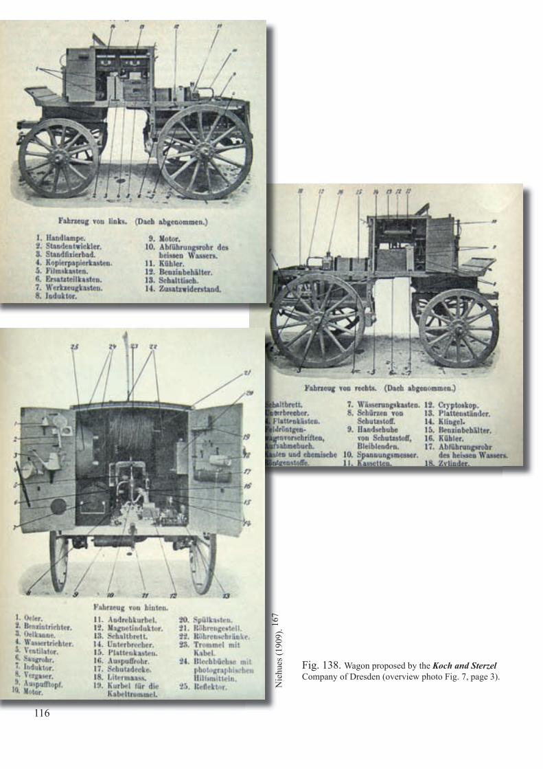
Nie
hues
(190
9). 16
7
Fig. 138. Wagon proposed by the Koch and Sterzel Company of Dresden (overview photo Fig. 7, page 3).
116

Let us recall that in 1902 the German Army, begins to equip horse-drawn carriages wherein all radiologi-cal equipment is transported and arranged in perfect order (Fig. 138): a radiological folding table, a dis-mountable X-ray tube holder and above all a power generator. All this allows the deployment of a radio-logical section in the shortest possible time lapse.
In 1903, W. Stechow (Fig. 140), a German physician general, publishes an important treatise, richly illus-trated on the subject (Fig. 139, 141).
Fig. 139. Cover of the 265 pages treatise, authored by the physician general W. Stechow.
Fig. 141. Portable radiology equipment deployed (1903) Siemens-Halske. An identical equipment was mounted in the cruiser "Aurora" (see Fig. 4, page 2).
W. S
tech
ow..
(190
3). 19
2W
. Ste
chow
(190
3).19
2
In 1907, fi eld X-ray wagons have become standard equipment of the German Army Medical Service and are assigned to the fi eld army to be available on request by any physician. The wagon contains the X-ray equipment (coil and gas tube), the darkroom equipment, four boxes of chemicals, glass plates, spare X-ray tubes and all other requirements down to the last screw. Twelve wagons of this type are available at the start of the war in 1914, and an additional 20 are acquired. 172
Weh
rges
chic
htlic
hen
Lehr
sam
mlu
ng d
es
Sani
täts
dien
stes
der
Bun
desw
ehr,
Mün
chen
.
Fig. 140. W. Stechow (painting from Wilhelm Auberten).

A little later the fi eld-Röntgen-automobile comes into being (Fig. 142, 143). The motor also can provide power for the generator. In both vehicles a petrol engine, early on a one-cylinder, and later a two-cylinder, drives the dynamo. The equipment can be operated at 65 volts, 15 amperes. Also, step up/step down transformers and alternators are available to use with any commercial electricity, direct, alternating or even 3-phases.
The supplying fi rms provide also the necessary technologists. Two hundred and seventy-fi ve X-ray sections are in operation during the war wherever German or German-supported troops may be present. Standardization of equipment and the supplies is encouraged, for instance, the 18 x 24 cm X-ray plate is considered the most practical one (Now it is always so!). Necessary accessories become part of the equipment: stereoscopes, localization devices, X-ray atlases and even silver re-covery units!
Fig. 143. German fi eld-Röntgen-automobile (Siemens-Halske) 1914 in front of "Siemens Stadt" (Berlin).
Siem
ens M
edic
al A
rchi
ves.
Fig. 142. Horse-drawn and automobile vehicles arranged probably in the courtyard of the military hospital of Berlin.
Siem
ens M
edic
al A
rchi
ves.
118
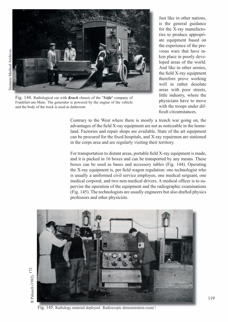
Siem
ens M
edic
al A
rchi
ves.
Just like in other nations, is the general guidance for the X-ray manufacto-ries to produce appropri-ate equipment based on the experience of the pre-vious wars that have ta-ken place in poorly deve-loped areas of the world. And like in other armies, the fi eld X-ray equipment therefore prove working well in rather desolate areas with poor streets, little industry, where the physicians have to move with the troops under dif-fi cult circumstances.
Contrary to the West where there is mostly a trench war going on, the advantages of the fi eld X-ray equipment are not as noticeable in the home-land. Factories and repair shops are available. State of the art equipment can be procured for the fi xed hospitals, and X-ray repairmen are stationed in the corps area and are regularly visiting their territory.
For transportation to distant areas, portable fi eld X-ray equipment is made, and it is packed in 16 boxes and can be transported by any means. These boxes can be used as bases and accessory tables (Fig. 144). Operating the X-ray equipment is, per fi eld wagon regulation: one technologist who is usually a uniformed civil service employee, one medical sergeant, one medical corporal, and two non-medical drivers. A medical offi cer is to su-pervise the operation of the equipment and the radiographic examinations (Fig. 145). The technologists are usually engineers but also drafted physics professors and other physicists.
Fig. 144. Radiological car with Kruch chassis of the "Veifa" company of Frankfurt-am-Main. The generator is powered by the engine of the vehicle and the body of the truck is used as darkroom.
Fig. 145. Radiology material deployed. Radioscopic demonstration exam !
R P
akus
ch (1
982)
. 172
Siem
ens M
edic
al A
rchi
ves.
119

Fully trained roentgenologists are rare in those days. They usually are as-signed to the fi eld Army and travel as advisors. Internists and surgeons usually have had radiological training in their fi elds and together with the technologists they do well. The enlisted personnel perform the radiographic examinations. Of course the fl uoroscopy and the radiographic examination of the severely injured are performed by the physicians. In hospitals and large clinics, nurses and photographers, are available.
The Austro-Hungarian Empire is no exception. Here we record some private initiatives as well as the actions taken by Dr. E. von Gerzö, the assistant sur-geon at the Royal Hungarian University of Budapest. He describes in detail a war automobile with radiological equipment. Without such a machine the medical service has functioned inadequately during the Balkan war of 1912-1913. He uses an Opel chassis and installs X-ray equipment by Reiniger, Gebert and Schall. 214
The Opel chassis of 14-30 hp. is capable of running at the rate of 24 to 30 kilometres per hour with a load of 2,000 kilos (total four tons or less) (Fig. 146). The body of the car is 3 m long, 1.4 wide and 1.7 high, and is divided into two compartments. The front compartment is about 1.2 m in length, serves as a dark room and has its own door on the left side. The other compartment contains the roentgen apparatus with all instruments and spare parts (Fig. 147). The dynamo is driven by the petrol engine and built in under the chauffeur’s seat. A lever makes the connection.
Fig. 146. Radiology vehicle proposed in 1914 by von Gerzö. The equipment is arranged in the vehicle. On the roof, a water reserve is foreseen for fi lm processing.
E. v
on G
erzö
(191
4). 21
4
120

Fig. 147 . Detail of the radiology vehicle of Dr. von Gerzö. A Floor plan. B The darkroom accessed through a side door. C The radiology equipment is accessible through the rear door of the vehicle.
E. v
on G
erzö
(191
4). 21
4
AB
C 121

All manipulations are carried out in the dark room which contains a work-table at the front wall, with a basin for rinsing purposes on the right. A wa-ter reservoir with a capacity of 60 litres sits on the roof. When the basin is covered with a board, there is a surface of 55 x 140 cm available for trays. A folding seat is fi xed at the rear wall. A shelf over the table and a box under the photographer’s seat are used for storing chemicals. Lead-lined boxes in the right corner contain the photographic plates.
The roentgen tubes are above these boxes, suspended by a spring in a woo-den frame which guards them from breakage in transportation. A tarpaulin cover protects them from dust and, incidentally, the operator from fl ying glass in the event of explosions. Windows at opposite sides can be screened or covered with a red pane. There is a fan which does not allow light shining through, and a writing desk.
The rear compartment constitutes the roentgen room . It contains an “Ideal” roentgen apparatus, model No. 2 (Reiniger, Gebbert and Schall), consisting of a motor, transformer and commutator. (Fig. 148, 149, 150) The apparatus is fi xed at the front wall with hinges. There is a portable regulator table, a tube stand, an operating table, a specially constructed device for working the tube under the table, fl uorescent and intensifying screens, a few reserve tubes properly suspended in springs and protected, and all necessary acces-sories. Everything is placed in the order it is used.
Fig. 148. X-ray examination table, arranged for a radiography exam, the tube being placed above the table. Notice the porcelain insulators under the table provided for use when the X-ray tube is placed under the table.
E. v
on G
erzö
(191
4). 21
4
122
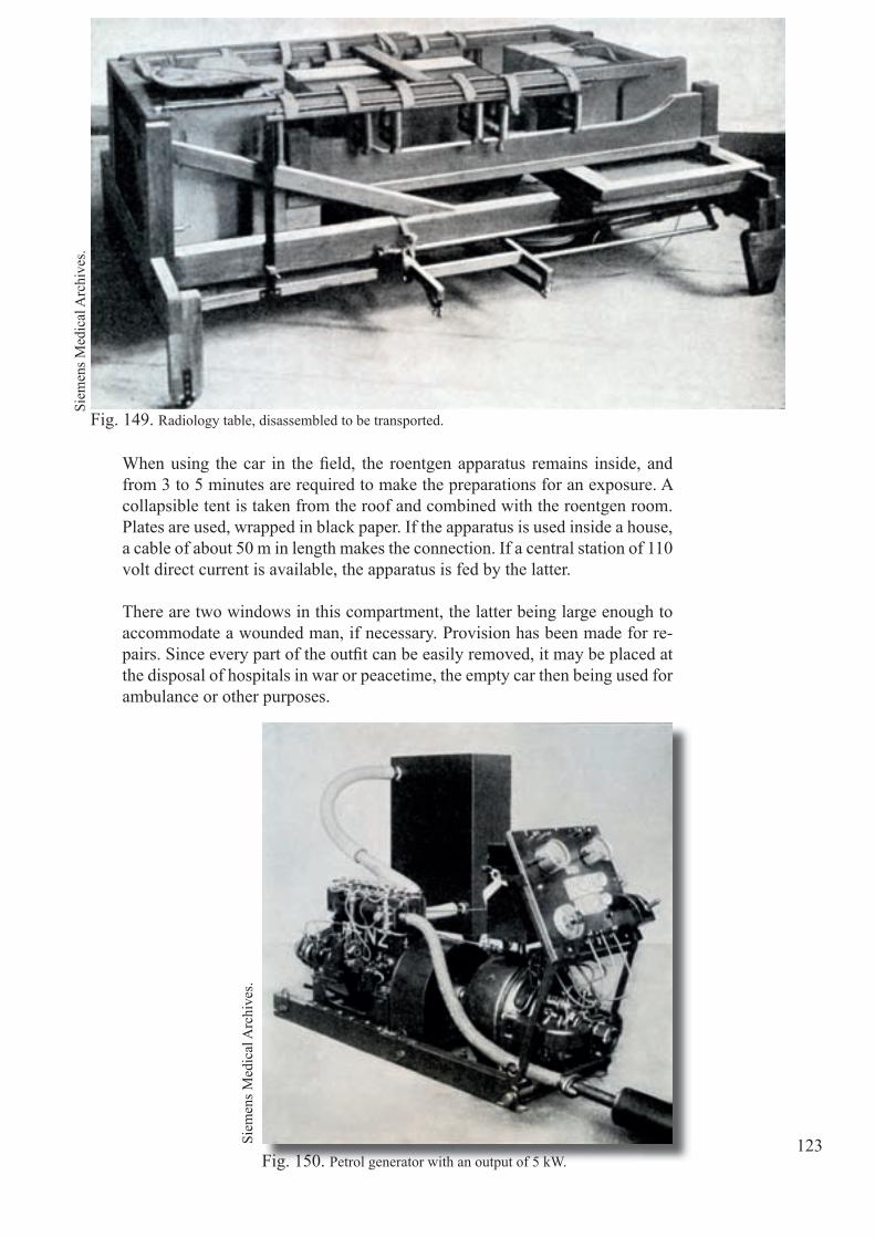
Fig. 150. Petrol generator with an output of 5 kW.
When using the car in the fi eld, the roentgen apparatus remains inside, and from 3 to 5 minutes are required to make the preparations for an exposure. A collapsible tent is taken from the roof and combined with the roentgen room. Plates are used, wrapped in black paper. If the apparatus is used inside a house, a cable of about 50 m in length makes the connection. If a central station of 110 volt direct current is available, the apparatus is fed by the latter.
There are two windows in this compartment, the latter being large enough to accommodate a wounded man, if necessary. Provision has been made for re-pairs. Since every part of the outfi t can be easily removed, it may be placed at the disposal of hospitals in war or peacetime, the empty car then being used for ambulance or other purposes.
Fig. 149. Radiology table, disassembled to be transported.
Siem
ens M
edic
al A
rchi
ves.
Siem
ens M
edic
al A
rchi
ves.
123

Many other initiatives see the light of day. Thus, we mention that of Profes-sor L. Brauer and that of D. F. Haenisch of Hamburg. They use in 1915 vehicles "Adler" and "Fiat" which are fi tted with radiological material from the fi rm Seifert & Co of Hamburg (Fig. 151, 152).
L. B
raue
r, F.
Hae
nisc
h (1
915)
. 21
Fig. 151. Radiological vehicles in marching order.
Fig. 152. Radiological equipment judiciously packed in the vehicles.
L. B
raue
r, F.
Hae
nisc
h (1
915)
21.
124
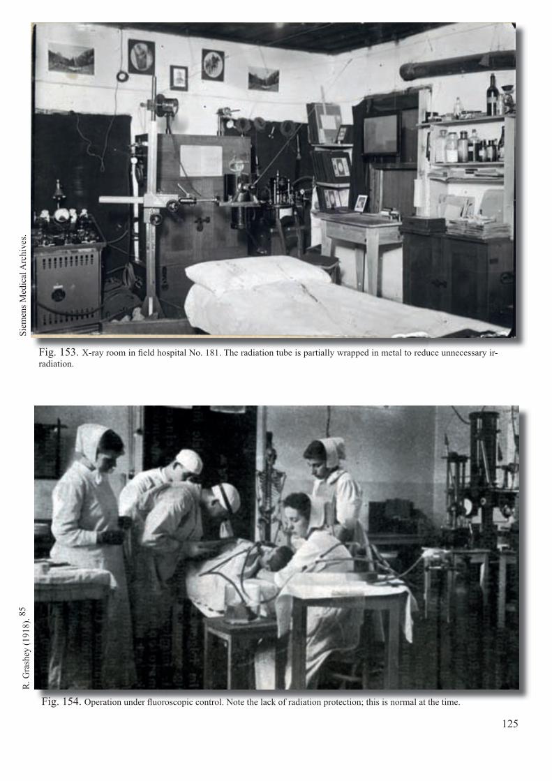
Fig. 154. Operation under fl uoroscopic control. Note the lack of radiation protection; this is normal at the time.
Fig. 153. X-ray room in fi eld hospital No. 181. The radiation tube is partially wrapped in metal to reduce unnecessary ir-radiation.
R. G
rash
ey (1
918)
. 85Si
emen
s Med
ical
Arc
hive
s.
125

Fig. 156. Cover of the pocket treatise of 214 pages, authored by Professor R. Grashey, famous professor of radiology at the University of Cologne.
Fig. 155. Radiography vehicles of the radiological company "Veifa" (Frankfurt-am-Main).
Siem
ens M
edic
al A
rchi
ves.
Bel
gian
Mus
eum
of R
adio
logy
.
126
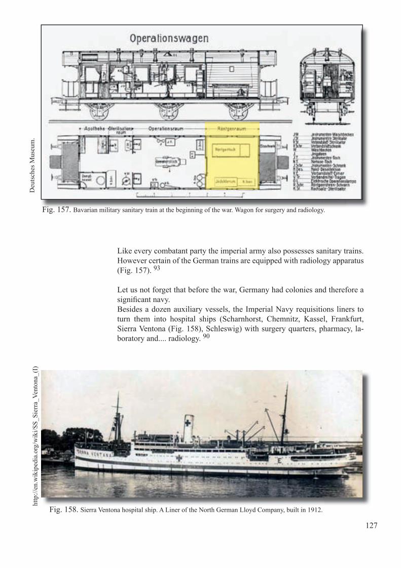
Siem
ens M
edic
al A
rchi
ves.
Like every combatant party the imperial army also possesses sanitary trains. However certain of the German trains are equipped with radiology apparatus (Fig. 157). 93
Let us not forget that before the war, Germany had colonies and therefore a signifi cant navy.Besides a dozen auxiliary vessels, the Imperial Navy requisitions liners to turn them into hospital ships (Scharnhorst, Chemnitz, Kassel, Frankfurt, Sierra Ventona (Fig. 158), Schleswig) with surgery quarters, pharmacy, la-boratory and.... radiology. 90
http
://en
.wik
iped
ia.o
rg/w
iki/S
S_Si
erra
_Ven
tona
_(I)
Deu
tsch
es M
useu
m.
Fig. 157. Bavarian military sanitary train at the beginning of the war. Wagon for surgery and radiology.
Fig. 158. Sierra Ventona hospital ship. A Liner of the North German Lloyd Company, built in 1912.
127
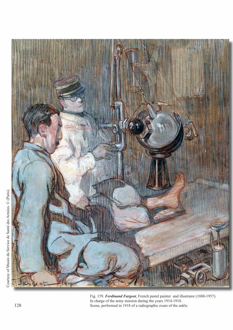
Fig. 159. Ferdinand Fargeot, French pastel painter and illustrator (1880-1957).In charge of the army mission during the years 1914-1918.Scene, performed in 1918 of a radiographic exam of the ankle.
Cou
rtesy
of M
usée
du
Serv
ice
de S
anté
des
Arm
ées.
© (P
aris
).
128
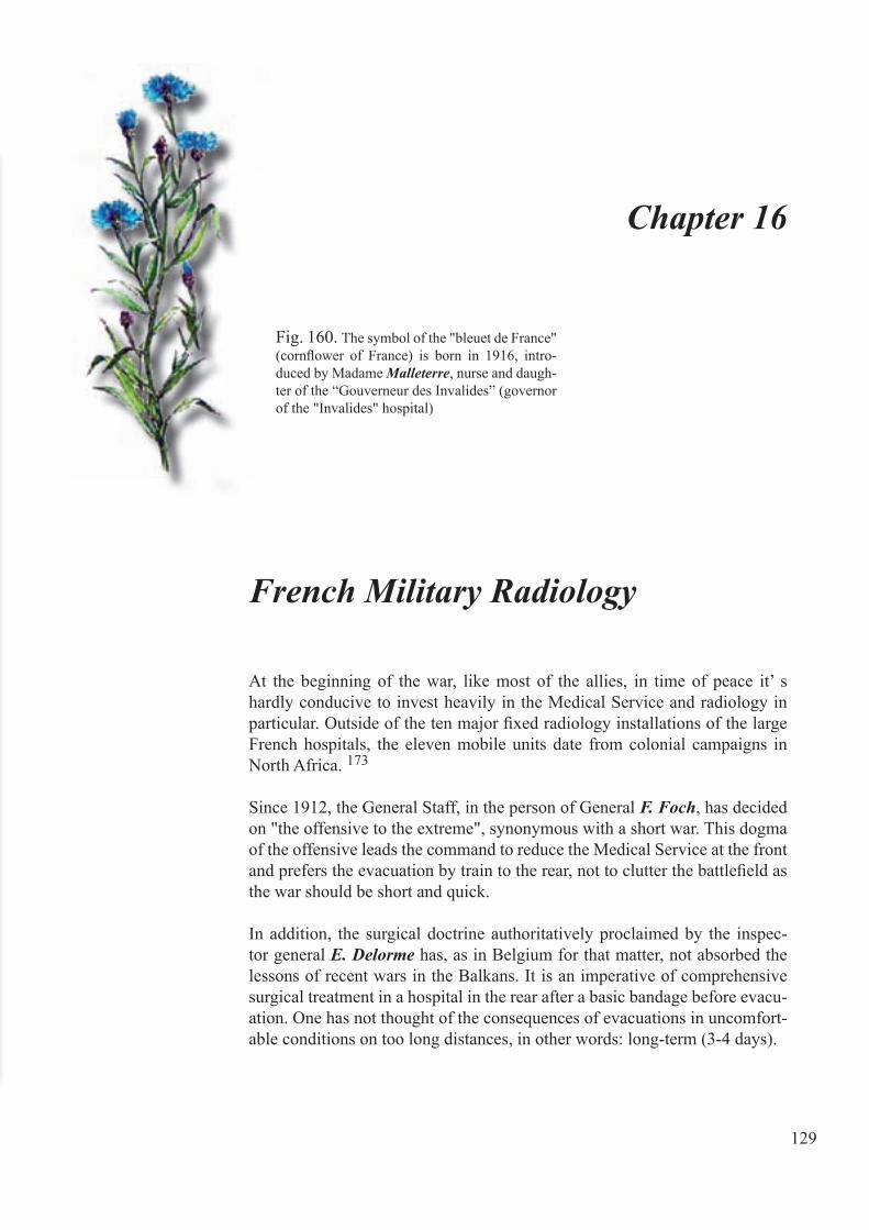
Chapter 16
French Military Radiology
At the beginning of the war, like most of the allies, in time of peace it’ s hardly conducive to invest heavily in the Medical Service and radiology in particular. Outside of the ten major fi xed radiology installations of the large French hospitals, the eleven mobile units date from colonial campaigns in North Africa. 173
Since 1912, the General Staff, in the person of General F. Foch, has decided on "the offensive to the extreme", synonymous with a short war. This dogma of the offensive leads the command to reduce the Medical Service at the front and prefers the evacuation by train to the rear, not to clutter the battlefi eld as the war should be short and quick.
In addition, the surgical doctrine authoritatively proclaimed by the inspec-tor general E. Delorme has, as in Belgium for that matter, not absorbed the lessons of recent wars in the Balkans. It is an imperative of comprehensive surgical treatment in a hospital in the rear after a basic bandage before evacu-ation. One has not thought of the consequences of evacuations in uncomfort-able conditions on too long distances, in other words: long-term (3-4 days).
Fig. 160. The symbol of the "bleuet de France" (cornfl ower of France) is born in 1916, intro-duced by Madame Malleterre, nurse and daugh-ter of the “Gouverneur des Invalides” (governor of the "Invalides" hospital)
129

Fig. 161. Motor vehicle for radiology equipment. The driving is done with chains! In (A), is seen transported equipment. The cut-out in the wheel (B) shows the drive (E) of the bipolar alternator (110V 24 A) by the engine of the vehicle, if the propulsion mechanism is disengaged.
130
Fig. 161 (A)
G. C
halm
arès
(190
5). 32
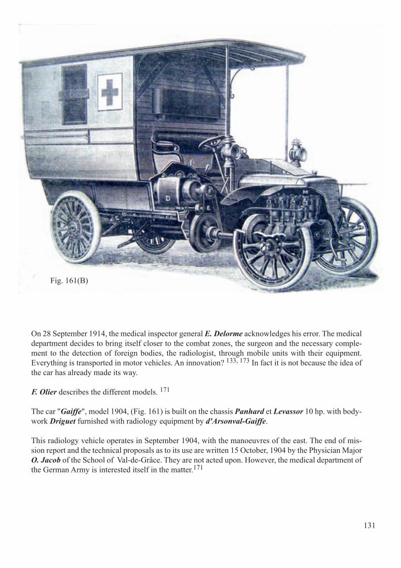
131
Fig. 161(B)
On 28 September 1914, the medical inspector general E. Delorme acknowledges his error. The medical department decides to bring itself closer to the combat zones, the surgeon and the necessary comple-ment to the detection of foreign bodies, the radiologist, through mobile units with their equipment. Everything is transported in motor vehicles. An innovation? 133, 173 In fact it is not because the idea of the car has already made its way.
F. Olier describes the different models. 171
The car "Gaiffe", model 1904, (Fig. 161) is built on the chassis Panhard et Levassor 10 hp. with body-work Driguet furnished with radiology equipment by d'Arsonval-Gaiffe.
This radiology vehicle operates in September 1904, with the manoeuvres of the east. The end of mis-sion report and the technical proposals as to its use are written 15 October, 1904 by the Physician Major O. Jacob of the School of Val-de-Grâce. They are not acted upon. However, the medical department of the German Army is interested itself in the matter.171
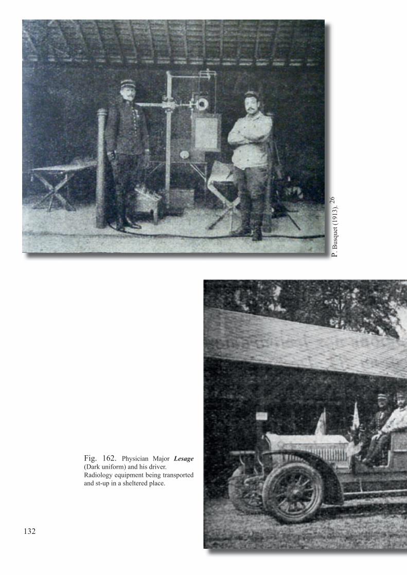
Fig. 162. Physician Major Lesage (Dark uniform) and his driver.Radiology equipment being transported and st-up in a sheltered place.
132
P. B
usqu
et (1
913)
. 26
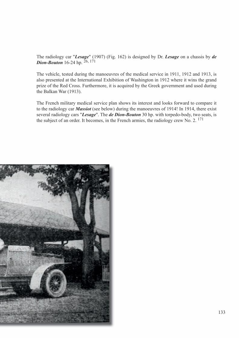
133
The radiology car "Lesage" (1907) (Fig. 162) is designed by Dr. Lesage on a chassis by de Dion-Bouton 16-24 hp. 26, 171
The vehicle, tested during the manoeuvres of the medical service in 1911, 1912 and 1913, is also presented at the International Exhibition of Washington in 1912 where it wins the grand prize of the Red Cross. Furthermore, it is acquired by the Greek government and used during the Balkan War (1913).
The French military medical service plan shows its interest and looks forward to compare it to the radiology car Massiot (see below) during the manoeuvres of 1914! In 1914, there exist several radiology cars "Lesage". The de Dion-Bouton 30 hp. with torpedo-body, two seats, is the subject of an order. It becomes, in the French armies, the radiology crew No. 2. 171

Fig. 163. Radiology vehicle Peugeot 10 hp. built by Massiot and Radiguet on a chassis on by Dietrich. The tent is protected by the roof racks (A).The tent is unfolded sideways to the vehicle (B), and houses the Radiguet-Massiot-bed which radiography and fl uoroscopy can be performed (the tube is set up under the table and the patient).
Fig. 163.
134
Fig. 163-a
G. M
assi
ot e
t Biq
uard
(191
5) .14
9

135
Fig. 163-b.
The radiology car "Massiot" on a chassis of Dietrich 12 hp, 1912 model with equipment Radiguet-Massiot type is built by G. Massiot (Fig. 163) to com-pete with the "Lesage" model.
The car must participate in the manoeuvres of the fi rst army corps (June 15, 1914). At the mobilization, the decision of the Advisory Board of Health is still waiting ... Massiot or Lesage? The decision should be taken in Septem-ber ...!! 171
Although the tests are carried out very successfully with radiological auto-mobiles in 1904 the French army does not have any of them available at the declaration of war in August 1914. Also she commandeers at the outset of hostilities, the few existing demo radiology cars to turn them into military radiological equipment.
At the mobilization, the radiology automobile Massiot n°1 joins the 1st army. Its designer, G. Massiot, accompanies her, being mobilized as technologist with the rank of sergeant. 149 The physician Dechambre will become the responsible radiologist. 173

Fig. 164. Professor A. Béclère in 1915 and his son the trainee artillery offi cer C. Béclère gassed at Verdun, 7 July, 1917. He will become a gynaecologist after the war.
Fig. 166. Saurer 50 hp. truck built by G. Gallot inspired by an idea of Massiot. Demonstration of equipment before leaving in August 1914. One recognizes to the left the medical offi cer G. Haret, former student of Professor A. Bé-clère.
On 6 August 1914, at the request of Physician General A. Troussaint of the 7th general directorate, the Physician Major L. Lamoureux charges the mobilized doctors majors A. Béclère (Fig. 164), Professor of Radiology committed fol-lowing his request despite his 58 years, P. Aubourg and G. Haret (Fig. 165), to organize radiology crews equipped with dismountable material.
From 27 August, P. Aubourg and G. Haret leave the army in two equipped cars (Fig. 166). In January 1915, twenty cars are in service. 7, 8, 35, 94, 95, 140, 158
The equipment includes a generator placed in the front third of the car. The rear two-thirds contain the camera and all radiological equipment. The staff includes a driver-mechanic, the radiologist and the technologist. 173
A. B
éclè
re (1
973)
. 10
J. B
elot
(193
3). 15
G. P
alla
rdy
et C
oll.
(198
9). 17
3
Fig. 165. The doctor G. Haret at the end of his life. He also provides signifi cant support to the Belgian Medical service. Suffering ter-ribly from dermatitis, he dies in 1933.
136
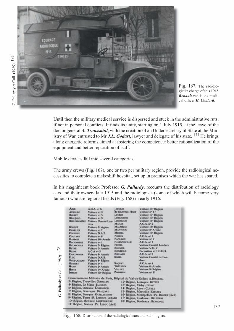
Fig. 167. The radiolo-gist in charge of this 1915 Renault van is the medi-cal offi cer H. Coutard.
Until then the military medical service is dispersed and stuck in the administrative ruts, if not in personal confl icts. It fi nds its unity, starting on 1 July 1915, at the leave of the doctor general A. Troussaint, with the creation of an Undersecretary of State at the Min-istry of War, entrusted to Mr J.L. Godart, lawyer and delegate of his state. 133 He brings along energetic reforms aimed at fostering the competence: better rationalization of the equipment and better repartition of staff.
Mobile devices fall into several categories.
The army crews (Fig. 167), one or two per military region, provide the radiological ne-cessities to complete a makeshift hospital, set up in premises which the war has spared.
In his magnifi cent book Professor G. Pallardy, recounts the distribution of radiology cars and their owners late 1915 and the radiologists (some of which will become very famous) who are regional heads (Fig. 168) in early 1916.
G. P
alla
rdy
et C
oll.
(198
9). 17
3
G. P
alla
rdy
et C
oll.
(198
9). 17
3
Fig. 168. Distribution of the radiological cars and radiologists.
137

Fig. 169. The ambulance Marcille with Saurer truck (C4 type) in the courtyard of Lignereuil (near Hesdin) 10 km from the front. The radiological laboratory is mounted in a tent. Downstairs, the operating room. Note that the surgeon operates under the supervision of a radi-ologist: the tube is on the operating table and the screen set at the head of the operator.
Fig. 170. Overview of a surgical automobile ambulance in marching order.
Surgical automobile ambulances (ACA) referred to in the slang of the front by the term "autochir" have three trucks of equipment and a van and six cars for the transportation of personnel and wounded (Fig. 169). 133, 169, 174 This motor-ized stand-alone formation is strongly contested by some in the beginning. It is designed in November 1914 by a mobilized surgeon, the Physician Major M. Marcille.
Faced with the remarkable results and following improvements made by sur-geons A. Gosset and H. Rouvillois, this mobile unit takes its fi nal form in May 1915: twenty-one copies are made and put into service. 158 The new autochir is redesigned this time, and it contains fi ve trucks Berliet CBA-type, three support vehicles, (Fig. 170) two trucks and four ambulances to transport the injured. The whole support unit, the heart of the formation, comprises truck A for the steam sterilization and heating and the storage of the linens, truck B containing enough to build the operative pavilion, fl uoroscopic and photographic equip-ment, truck C (Fig. 171) carrying the power generator, X-ray equipment, surgi-cal instruments and means for immobilization of body parts during the position-ing; fi nally there is a van containing dressings and various materials of general purpose. 53
La N
atur
e (1
916)
. 131
G. P
alla
rdy
et C
oll.
(198
9). 17
3
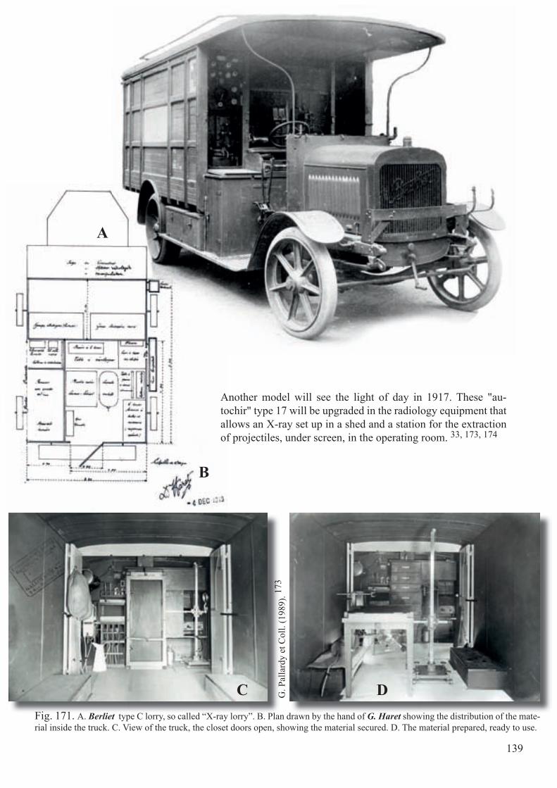
Fig. 171. A. Berliet type C lorry, so called “X-ray lorry”. B. Plan drawn by the hand of G. Haret showing the distribution of the mate-rial inside the truck. C. View of the truck, the closet doors open, showing the material secured. D. The material prepared, ready to use.
G. P
alla
rdy
et C
oll.
(198
9). 17
3
A
B
C D
Another model will see the light of day in 1917. These "au-tochir" type 17 will be upgraded in the radiology equipment that allows an X-ray set up in a shed and a station for the extraction of projectiles, under screen, in the operating room. 33, 173, 174
139

Fig. 172. Truck on a Kelly-Springfi eld chassis performing the additional surgery task.
Fig. 173. Cabinet by Ledoux-Lebard of the Casualty Clearing Stations. Left: measuring devices, mercury turbine and Dessane visor. Right: induction coil, Villard valve, guiding needles, goggles and gloves.
G. P
alla
rdy
et C
oll.
(198
9). 17
3
G. P
alla
rdy
et C
oll.
(198
9). 17
3
The cumbersome size and weight of these A.C.A. trucks make them very little mobile, while the "com-plementary groups of surgery" (Fig. 172) make out the third kind of lighter mobile equipment. Only fl uoroscopic equipment is placed on a car that also carries the sterilization equipment, lighting, and a generator and, on a trailer, a small dismountable operation shack.
One should still talk of "semi-fi xed stations" (182 in 1918) to supply electricity to radiology equipment and evacuation hospitals (HOE) (Fig. 173) foreseen of radio-surgical equipment.
140
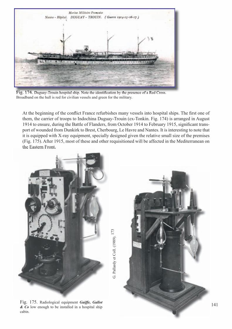
Fig. 174. Duguay-Trouin hospital ship. Note the identifi cation by the presence of a Red Cross. Broadband on the hull is red for civilian vessels and green for the military.Fig. 174. Duguay-Trouin hospital ship. Note the identifi cation by the presence of a Red Cross.
At the beginning of the confl ict France refurbishes many vessels into hospital ships. The fi rst one of them, the carrier of troops to Indochina Duguay-Trouin (ex-Tonkin. Fig. 174) is arranged in August 1914 to ensure, during the Battle of Flanders, from October 1914 to February 1915, signifi cant trans-port of wounded from Dunkirk to Brest, Cherbourg, Le Havre and Nantes. It is interesting to note that it is equipped with X-ray equipment, specially designed given the relative small size of the premises (Fig. 175). After 1915, most of these and other requisitioned will be affected in the Mediterranean on the Eastern Front.
141
the Eastern Front.
Fig. 175. Radiological equipment Gaiffe, Gallot & Co low enough to be installed in a hospital ship cabin.
G. P
alla
rdy
et C
oll.
(198
9). 17
3

142
Fig. 176. X-ray equipment for radiography arranged from top to bottom.
J. G
ouin
(191
5). 84
Fig. 176.
We owe the French Major J. Gouin a very detailed study on the usefulness and radiological technology on board of the hospital ships. To keep the military se-cret, the author omits the name of the vessel, but shows a series of photographs (Fig. 176). 84
In 1916 the hospital ships will be eight (including the "Tchad" (Fig. 177) and "Divona" (Fig. 178). This fi gure will be doubled by the end of hostilities. 135, 173
It is important to note that most of these boats are sometimes used as a sanitary vessel, sometimes to transport troops and equipment. That's why some of them are sunk by German forces, sometimes in violation of Geneva on the Red Cross.

Fig. 177. Hospital ship "Tchad" requisitioned from June 1914 to August 1917.
A. K
luda
s (19
73).
126
Fig. 178. Hospital ship "Divona" (1915-1918). In fact, an English ship "Ormuz" purchased by the South Atlantic Company. in 1912 and requisitioned in 1914.
A. K
luda
s (19
73).
126
143

Fig. 179. Marie Curie drives, sometimes herself, the automobile of radiology. Of the twenty cars the physicist starts up, she reserves one for her own use: an old Renault "crocodile hood" bodied as a delivery truck (October 1917).
Fig. 180. Marie Curie attending radioscopic examination.On the left, Dr. G. Haret, bearded at the time.
Fig. 181. X-ray room of the hospital No. 112 in Amiens, installed in barracks (1916). We see the equipment before the X-ray table, the tube holder and trolley. At right one recognizes Irène Curie.
Mus
ée C
urie
(Par
is).
M. C
urie
(192
1).44
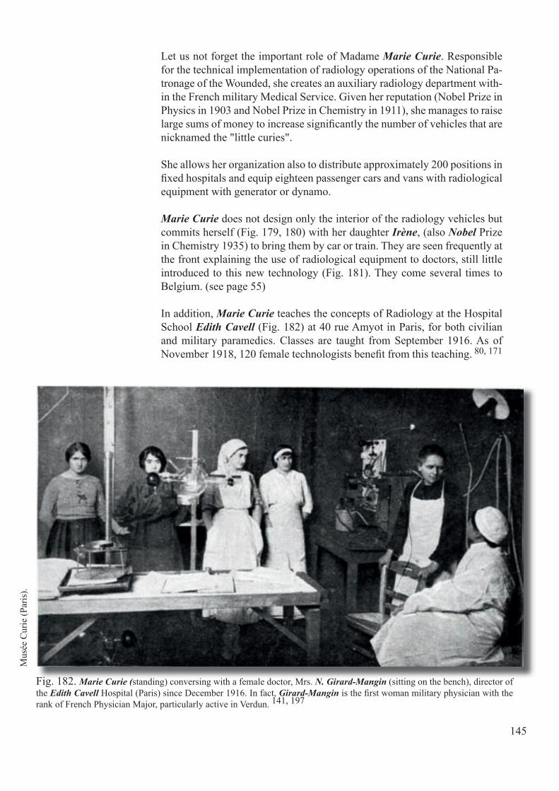
Fig. 182. Marie Curie (standing) conversing with a female doctor, Mrs. N. Girard-Mangin (sitting on the bench), director of the Edith Cavell Hospital (Paris) since December 1916. In fact, Girard-Mangin is the fi rst woman military physician with the rank of French Physician Major, particularly active in Verdun. 141, 197
Let us not forget the important role of Madame Marie Curie. Responsible for the technical implementation of radiology operations of the National Pa-tronage of the Wounded, she creates an auxiliary radiology department with-in the French military Medical Service. Given her reputation (Nobel Prize in Physics in 1903 and Nobel Prize in Chemistry in 1911), she manages to raise large sums of money to increase signifi cantly the number of vehicles that are nicknamed the "little curies".
She allows her organization also to distribute approximately 200 positions in fi xed hospitals and equip eighteen passenger cars and vans with radiological equipment with generator or dynamo.
Marie Curie does not design only the interior of the radiology vehicles but commits herself (Fig. 179, 180) with her daughter Irène, (also Nobel Prize in Chemistry 1935) to bring them by car or train. They are seen frequently at the front explaining the use of radiological equipment to doctors, still little introduced to this new technology (Fig. 181). They come several times to Belgium. (see page 55)
In addition, Marie Curie teaches the concepts of Radiology at the Hospital School Edith Cavell (Fig. 182) at 40 rue Amyot in Paris, for both civilian and military paramedics. Classes are taught from September 1916. As of November 1918, 120 female technologists benefi t from this teaching. 80, 171
Mus
ée C
urie
(Par
is).
145
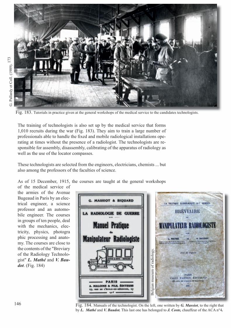
Fig. 183. Tutorials in practice given at the general workshops of the medical service to the candidates technologists.
Fig. 184. Manuals of the technologist. On the left, one written by G. Massiot, to the right that by L. Mathé and V. Baudot. This last one has belonged to J. Coste, chauffeur of the ACA n°4.
The training of technologists is also set up by the medical service that forms 1,010 recruits during the war (Fig. 183). They aim to train a large number of professionals able to handle the fi xed and mobile radiological installations ope-rating at times without the presence of a radiologist. The technologists are re-sponsible for assembly, disassembly, calibrating of the apparatus of radiology as well as the use of the locator compasses.
These technologists are selected from the engineers, electricians, chemists ... but also among the professors of the faculties of science.
As of 15 December, 1915, the courses are taught at the general workshops
G. P
alla
rdy
et C
oll.
(198
9). 17
3
Bel
gian
mus
eum
of r
adio
logy
.
of the medical service of the armies of the Avenue Bugeaud in Paris by an elec-trical engineer, a science professor and an automo-bile engineer. The courses in groups of ten people, deal with the mechanics, elec-tricity, physics, photographic processing and anato-my. The courses are close tothe contents of the "Breviaryof the Radiology Technolo-gist" L. Mathé and V. Bau-dot. (Fig. 184)
146

Fig. 185. Career Radiologist J. Hirtz.
Fig. 186. X-ray room of a hospital located in the temporary military "Petit Palais" in Paris.
At the time of the mobilization, none of the 175 radiologists in France is assigned to a position of radiology. The only radiologist from the Army departs as a medical director of an ambulance deprived of ra-diology! 173
The medical offi cer G. Haret, mobilized to the Undersecretary of State is responsible, remember, not only to create and standardize the radiological equipment but also to assign competent radiologists.
Since there is only a relatively small number of radiologists, of whom not all are mobilized, the training of radiologists, radiographers and technologist aids is given to the medical offi cer career J. Hirtz (Fig. 185)
and the medical offi cer A. Béclère. Both doctors organize accelerated training courses at Val-de-Grâce. And technical and hierarchical coordination is provided by the “Radio-logy" offi ce of the central administration (7th direction) and facilitated by the appoint-ment of army consultants radiologists.
J. Hirtz, future fi rst professor of radiology at the military school of medical application of the Val-de-Grâce, is also appointed consultant radiologist in the armed forces. Several expert radiologists (one per army) assist him.
Finally, mention should be made of the introduction to radiology of the American Expe-ditionary Corps which is entrusted to Professor R. Ledoux-Lebard, medical director of the 9th region, which will be discussed in Chapter 18, page 172.
The medical offi cer G. Haret reports that at the time of the armistice, there are 850 ra-diological stations in total, for 840 radiologists serving all of these stations. 700 of them are educated during the confl ict. 95
We can therefore appreciate the signifi cant effort of the training of radiologists in Paris (Fig. 186) as well as in the armed forces in different parts of the territory.
G. P
alla
rdy
et C
oll.
(198
9). 17
3 C
omité
d'H
isto
ire d
u Se
rvic
e de
San
té (1
987)
.
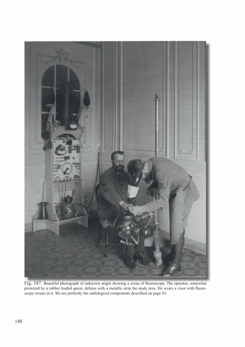
Fig. 187. Beautiful photograph of unknown origin showing a scene of fl uoroscopy. The operator, somewhat protected by a rubber leaded apron, defi nes with a metallic strip the study area. He wears a visor with fl uoro-scopy screen in it. We see perfectly the radiological components described on page 43.
148
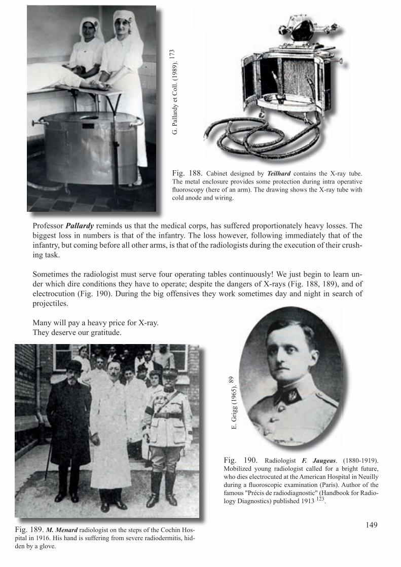
Fig. 190. Radiologist F. Jaugeas. (1880-1919). Mobilized young radiologist called for a bright future, who dies electrocuted at the American Hospital in Neuilly during a fl uoroscopic examination (Paris). Author of the famous "Précis de radiodiagnostic" (Handbook for Radio-logy Diagnostics) published 1913 123.
Fig. 189. M. Menard radiologist on the steps of the Cochin Hos-pital in 1916. His hand is suffering from severe radiodermitis, hid-den by a glove.
Fig. 188. Cabinet designed by Teilhard contains the X-ray tube. The metal enclosure provides some protection during intra operative fl uoroscopy (here of an arm). The drawing shows the X-ray tube with cold anode and wiring.
Professor Pallardy reminds us that the medical corps, has suffered proportionately heavy losses. The biggest loss in numbers is that of the infantry. The loss however, following immediately that of the infantry, but coming before all other arms, is that of the radiologists during the execution of their crush-ing task.
Sometimes the radiologist must serve four operating tables continuously! We just begin to learn un-der which dire conditions they have to operate; despite the dangers of X-rays (Fig. 188, 189), and of electrocution (Fig. 190). During the big offensives they work sometimes day and night in search of projectiles.
Many will pay a heavy price for X-ray.They deserve our gratitude.
G. P
alla
rdy
et C
oll.
(198
9). 17
3
E. G
rigg
(196
5). 89
149

In Flanders Fields
In Flanders fi elds, the poppies blow Between the crosses, row on row, That mark our place and in the sky The larks, still bravely singing, fl y Scarce heard amid the guns below.
We are the dead, short days ago, We lived, felt dawn, saw sunset glow, Loved, and were loved, and now we lie In Flanders fi elds!
Take up our quarrel with the foe: To you from failing hands, we throw The torch; be yours to hold it high. If ye break faith with us who die We shall not sleep, though poppies grow In Flanders fi elds!
http
://en
.wik
iped
ia.o
rg/w
iki/I
n_Fl
ande
rs_F
ield
s
Fig. 191. The poppy.Somewhere between Diksmuide and Ypres (Belgium) the Physician Major J. MacRae, a Canadian of Scottish origin, composes, on 3 May 1915, this poem that will become for the British the sym-bol of sacrifi ce and of remembrance. Chapter 17
Radiology in the British Expeditionary Force
The British Expeditionary Force, most of the time simply referred to as ‘The B.E.F.’, is a force sent to the Western Front during the Great War. Originally made up of the six existing divisions of the regular British Army, the fi rst troops land in France about 12 august 1914, this force is gradually expanded by the creation of new divisions in Great Britain, the recall of British troops stationed overseas and the arrival of troops from the newly created expedi-tionary forces from the Dominions as a response to the call to arms of the motherland.
Under Field Marshal J. French, later to be replaced by Field Marshal D. Haig, the B.E.F. is transformed into an established army organization.
By the end of 1914 the force is divided into the First and the Second Army, eventually to be composed of fi ve armies by the end of the war. From this moment on B.E.F. remains the offi cial name of the British Army in France and Flanders throughout the Great War, covering from one hundred thousand military at the beginning of the war to a maximal strength of two million at the time of the Armistice.
151
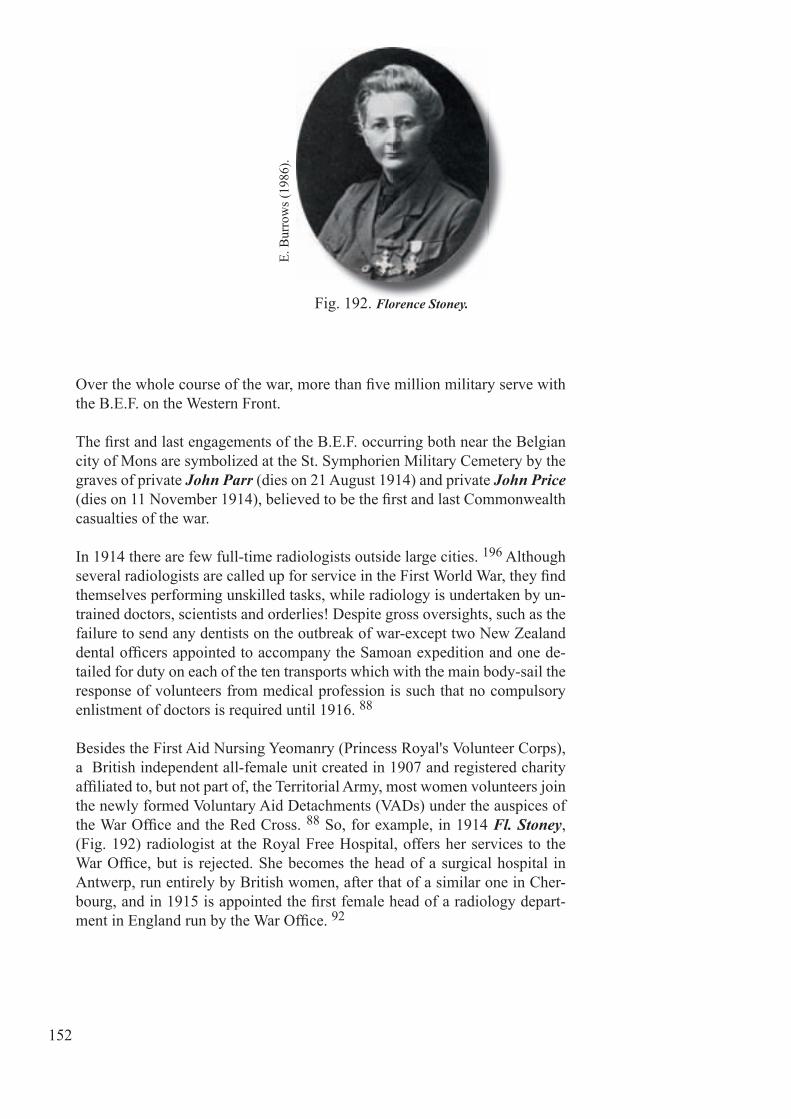
Over the whole course of the war, more than fi ve million military serve with the B.E.F. on the Western Front.
The fi rst and last engagements of the B.E.F. occurring both near the Belgian city of Mons are symbolized at the St. Symphorien Military Cemetery by the graves of private John Parr (dies on 21 August 1914) and private John Price (dies on 11 November 1914), believed to be the fi rst and last Commonwealth casualties of the war.
In 1914 there are few full-time radiologists outside large cities. 196 Although several radiologists are called up for service in the First World War, they fi nd themselves performing unskilled tasks, while radiology is undertaken by un-trained doctors, scientists and orderlies! Despite gross oversights, such as the failure to send any dentists on the outbreak of war-except two New Zealand dental offi cers appointed to accompany the Samoan expedition and one de-tailed for duty on each of the ten transports which with the main body-sail the response of volunteers from medical profession is such that no compulsory enlistment of doctors is required until 1916. 88
Besides the First Aid Nursing Yeomanry (Princess Royal's Volunteer Corps), a British independent all-female unit created in 1907 and registered charity affi liated to, but not part of, the Territorial Army, most women volunteers join the newly formed Voluntary Aid Detachments (VADs) under the auspices of the War Offi ce and the Red Cross. 88 So, for example, in 1914 Fl. Stoney, (Fig. 192) radiologist at the Royal Free Hospital, offers her services to the War Offi ce, but is rejected. She becomes the head of a surgical hospital in Antwerp, run entirely by British women, after that of a similar one in Cher-bourg, and in 1915 is appointed the fi rst female head of a radiology depart-ment in England run by the War Offi ce. 92
Fig. 192. Florence Stoney.E.
Bur
row
s (19
86).
152
J. H
artn
ell e
t H. S
outta
r (19
15).
96

Fig. 193. Excerpt from the British medical journal, Jan, 9-1915
Fig. 194. Vehicle of the Belgian Field Hospital.w
ww
.gut
enbe
rg.o
rg/
J. H
artn
ell e
t H. S
outta
r (19
15).
96
The British Red Cross also provides several X-ray units for use in France and Belgium. 196
The British Field Hospital is fi rst located in Ant-werp (Fig. 193, 194). It evacuates its patients via Ostend, comes back later respectively to Veurne then to Hoogstade before it is transformed into a Belgian Field Hospital. We note that the nurse E. Cavell works at the British Field Hospital prior to being executed by the Germans from espionage on 15 October 1915.
In 1903 the Army has established a school of radiography at the Royal Army Medical School (RAMC) at Netley, Southampton which is believed to be one of the fi rst of its kind in the world. The school is housed in the Royal Victoria Hospital. 25 A civilian formerly a non-commissioned offi cer in the RAMC, Mr. H. Henry, is the fi rst instructor. He writes an article on fi lm processing for army radiographers, (Fig. 195) and in 1922 he is one of the founders of the Society of Radiographers. Henry is given a syllabus to teach the rudiments of radiographic technique. 119
Roy
al A
rmy
Med
ical
Mus
eum
(UK
).
Fig. 195. Excerpt of the fi rst page of an article authored by Mr. H. Henry, instruc-tor in 1905.
153

When the Royal Army Medical College leaves Southampton, the X-ray school follows - fi rst to Savoy Hill, London, and then to the Queen Alexandra Military Hospital, Millbank. In 1910, A.J. Walton succeeds H. Henry and for the fi rst time his post is a designated one of Army X-ray Instructor. Walton remains at Millbank until he leaves the Army in 1922, except for service in France during the First World War. 25
In 1909, the Indian Medical Service establishes an X-ray In-stitute at Debra Dun which offers a specialist qualifi cation in electrical science after a short course of lectures. The direc-tor of the Institute is an Army offi cer, Captain A.E. Walter, the author of an early textbook. "X-rays in General Practice" (1906) (Fig. 196, 197). 216 One of his most promising pupils is a 24-year-old Aberdonian surgeon, destined to return home to be the fi rst specialist in radiology in the British Army D.B. McGrigor (Fig. 198), later a general and president of the Brit-ish Institute of Radiology (1939-1942).25, 143
Brit
isn
Inst
itute
of R
adio
logy
.
Fig. 197. Frontispiece of the treatise au-thored in 1906 by Captain A. Walter. 216
Fig.198. Major General D.B. McGrigor
Fig. 196. Mule carrying an X-ray coil.
Frontispiece of the treatise au-Frontispiece of the treatise au-
Bel
gian
Mus
eum
of R
adio
logy
.
154

Bel
gian
Mus
eum
of R
adio
logy
. Let us remember that already since 1897 the RAMC possesses mobile radiology material used during the campaigns in India, Egypt and South Africa. The electric power is sup-plied by a little dynamo driven by… a bicy-cle (Fig. 199).
One particular X-ray vehicle is described in 1913 by E. Hazleton. 92 It is essentially "home-made". The car’s engine provides both the driving and the generating power (Fig. 200).
In the early part of the 1914-18 War, power supplies are still meagre and primitive: a small ¾ hp. oil engine with blowpipe igni-tion to charge the accumulators is the only source of power. This independent unit, such as it is, compares more than favour-ably with the units that are sent out with the original hospitals of the Expeditionary Force. The constant chase after independent power supplies suitable for X-rays contin-ues. The absence of suitable power supplies is in no way due to industrial laxity but rath-er to the fact that the use of suitable alternat-ing current is not developed by the technical branches of the service.
Fig. 199. Direct powering of the coil by the dynamo, before the appearance of the generator of current.(In the commentaries of the time, one mentions the successful experiments)!
Bel
gian
Mus
eum
of R
adio
logy
.
Fig. 200. Detail of the home-made electrical supply of the X-ray vehicle.
155
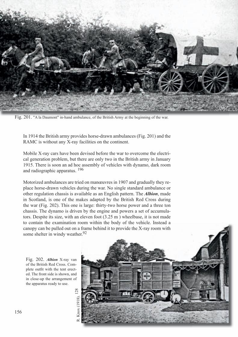
In 1914 the British army provides horse-drawn ambulances (Fig. 201) and the RAMC is without any X-ray facilities on the continent.
Mobile X-ray cars have been devised before the war to overcome the electri-cal generation problem, but there are only two in the British army in January 1915. There is soon an ad hoc assembly of vehicles with dynamo, dark room and radiographic apparatus. 196
Motorized ambulances are tried on manœuvres in 1907 and gradually they re-place horse-drawn vehicles during the war. No single standard ambulance or other regulation chassis is available as an English pattern. The Albion, made in Scotland, is one of the makes adapted by the British Red Cross during the war (Fig. 202). This one is large: thirty-two horse power and a three ton chassis. The dynamo is driven by the engine and powers a set of accumula-tors. Despite its size, with an eleven foot (3.25 m ) wheelbase, it is not made to contain the examination room within the body of the vehicle. Instead a canopy can be pulled out on a frame behind it to provide the X-ray room with some shelter in windy weather.92
R. K
nox
(191
8). 12
8
Fig. 201. "A la Daumont" in-hand ambulance, of the British Army at the beginning of the war.
Fig. 202. Albion X-ray van of the British Red Cross. Com-plete outfi t with the tent erect-ed. The front side is shown, and in close-up the arrangement of the apparatus ready to use.
156

Sir A. Reid (Fig 203) is a very important radiolo-gist during the First World War, through his abil-ity to mobilize radiological resources for mili-tary purposes. Soon after the war break out he is commissioned as a Major and assigned to the Queen Alexandra Military Hospital, Millbank, and to the Second London General Hospital, Chelsea.
From 1914 to 1919 he acts as President of the War Offi ce X-ray Committee, an important mili-tary body with responsibility for the planning, equipping and organising of X-ray services at home and on the battlefi eld.
Being at the centre of military medical affairs, and concerned with the short-age of trained X-ray staff, he institutes also in 1915 a course of training lec-tures at Millbank, assisted by Dr. R. Reynolds (Fig 204) and the physicist, C.E.S. Phillips (Fig 205). Reid’s unique military experience convinces him that national training facilities for radiologists are urgently required, and he is one of those to establish the British Association of Radiology. He helps to cre-ate the Society of Radiographers in 1920, and serves as its fi rst President. 25
Fig. 203. Sir A. Reid. His later years are darkened by severe radiodermitis which contribute to his premature death at the age of 53.
Fig. 204. Radiologist R. Reynolds. Fig. 205. Physicist C.E.S. Phillips.
E. B
urro
ws (
1986
). 25
.E.
Grig
g (1
965)
. 89
E. G
rigg
(196
5). 89
157

The London radiologist A. Reid and his army committee deal with several technical matters including the design of mobile X-ray vehi-cles. Initially, the dynamo is powered by the vehicle’s engine, but this produces unwanted vibrations.
The British army sends 10 mobile X-ray units to France. Some are manufactured by the army’s department of radiology research at Woolwich. 92 One of them (an Austin wagon) is described in detail by H. Head (1918) (Fig. 206).
The Belgians in England provide a fully mobile fi eld hospital for Flanders in 1918, comprising a surgical operating car, X-ray equipment, ice making machine, kitchen, and other facilities, all powered electrically by petrol engines. 92
Fig. 206. Radiological car 20 hp. on an Austin chassis.
J. G
uy (2
000)
. 92
H. H
ead
(191
7). 97
158

More compact, powerful and effi cient apparatus is now available. (Fig. 207) General and base hospitals receive ra-diographic sets early on. Casualty clearing stations are origi-nally considered to be suitable for the type of surgery requir-ing radiological services, but with the onset of static trench warfare the role of surgery and radiology in these forward units, increases in signifi cance. Bedside radiography be-comes routine in hospitals which specialise in the treatment of fractures. 196
Let us listen to Lt Col A. Mayo-Robson (1915) : "In the Ur-gency Cases Hospital attached to the 3rd French Army, we have an excellent X-ray outfi t which was supplied by Wat-son and Sons of Great Portland street, London W; under the kind supervision of Sir J. Mackenzie Davidson, and Mr. J.G. Everitt, our skilful radiographer, carries out the work with great celerity and accuracy. For instance, in the case of a recent shell wound of the shoulder with enormous swelling, I saw the X-ray examination made and localisation of a large fragment of shrapnel casing effected by the method suggest-ed by Sir J. Mackenzie Davidson, and then a radiograph was made within a quarter of an hour, and half an hour later the fragment was removed by Mr. J. Forsyth, F.R.C.S ,the "médecin-chef" of the hospital". 156
Fig. 207. Atlas X-ray and operat-ing table from the British manufac-turer A.E. Dean.
R. K
nox
(191
7). 12
8
J. G
uy (2
000)
. 92
J. G
uy (2
000)
. 92
159

Wounded soldiers and sailors are transported back to Britain by ship. We should not forget that the British Empire possesses the largest fl eet in the world! Hospital ships are requisitioned liners, extensively adapted for the par-ticular purpose of caring for the sick and wounded. They are in effect fl oating hospitals and are medically and surgically equipped to deal with all kinds of cases of injury and disease.
Altogether 77 military hospital ships and transports are commissioned during the war: 22 in 1914, 42 in 1915, 7 in 1916, and 6 in 1917. In-cluded in this number are four Belgian Government Mail Steamers: the Jan Breydel (Fig. 208), Pieter de Coninck, (Fig. 209), Stad Antwerpen (see Fig. 15, page 16) and Ville de Liège ( Fig. 210). 178
The army and navy have separate transports and hospital ships. Many of the latter are equipped with X-ray apparatus. Naval hospitals and the larger bat-tleships possess radiographic apparatus. The navy shares many of the prob-lems of the army; with the additional trial of attempting radiography on a rolling deck. 195
Fig. 208. The Jan Breydel is deployed in April 1915 as a hospital ship out of Calais. At the disposition of the Admiralty she will make 798 voyages until 31 December 1918. In all she transports 18,133 wounded and 16,841 troops.
A. V
anal
derw
eire
ldt (
1997
). 20
2
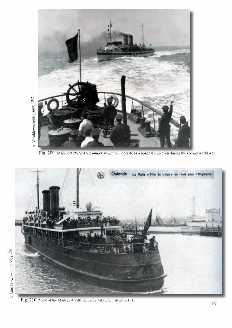
Fig. 210. View of the Mail-boat Ville de Liège, taken in Ostend in 1913.
A. V
anal
derw
eire
ldt (
1997
). 20
2
Fig. 209. Mail-boat Pieter De Coninck which will operate as a hospital ship even during the second world war.
A. V
anal
derw
eire
ldt (
1997
). 20
2
161
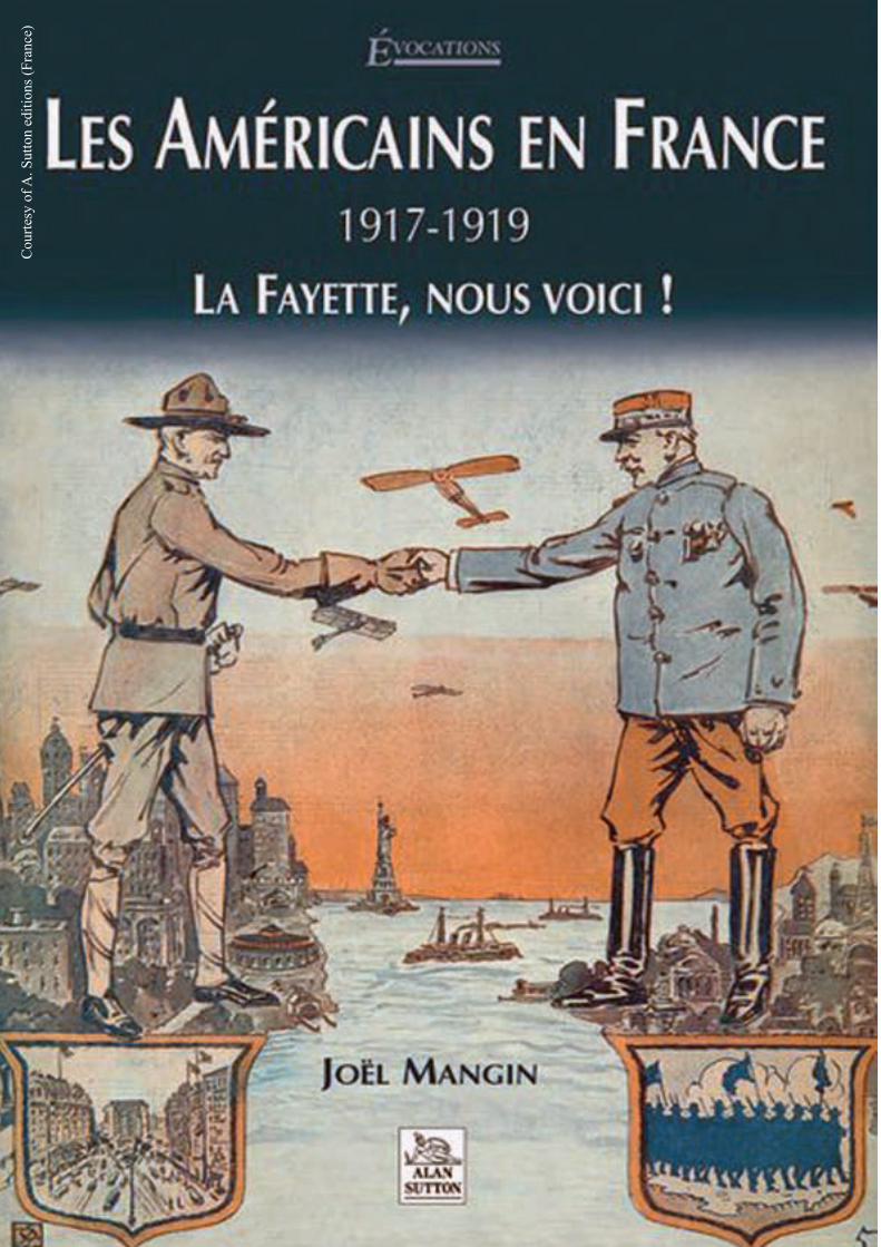
Cou
rtesy
of A
. Sut
ton
editi
ons (
Fran
ce)
Chapter 18
"La Fayette , nous voici !" "La Fayette, we are coming!"
Since the beginning of the Great War, the United States under President W. Wilson have maintained strict neutrality, only providing material assis-tance to the Allies. This allows private institutions and organizations to show their sympathy to the allied cause by providing means of all kind to support or to supplement their material resources and to provide humanitarian assis-tance where needed. 17, 69, 75, 81, 122, 172, 181, 182
As popular support for American participation in the war increases, volun-teer organizations capitalize on that spirit, enrolling many young Americans who serve as ambulance drivers in France and Italy. Until there arises an op-portunity for overseas service in the Army, the volunteers, one after the other, provide proof of America's willingness to support its allies. They receive short orientation courses and, when deployed, they serve under the command of the allied Army units to which they are attached. Volunteer ambulance organizations precede US Army ambulance units in Europe, and their offic-ers are the predecessors of Medical Department commissioned ambulance officers. By the spring of 1917 the American Red Cross Ambulance Service has forty-six ambulance units supporting the allies and the American Field Service is operating 1,220 ambulances in 31 sections supporting sixty-six French divisions by late 1917.
Fig. 211. Cover of the book by Joël Mangin (2006). 146
163
D
Fig. 212. Miss G. Dunn.
ww
w.g
wpd
a.or
g/m
edic
al/x
ray/
dunn
Besides these volunteer groups other initiatives are launched by various civil-ian institutions. A number of specialists from civilian hospitals and universi-ties give up their position and serve as volunteers on the Western Front. As such a surgical team from Harvard University in Boston serves in a British General Hospital, the radiological section being entrusted to Miss G. Dunn, (Fig. 212) an Australian trained in radiology at Stanford University. Depen-ding on their resources, some of the hospitals are even able to establish a ‘subsidiary’ branch as for instance the Lakeside Unit Base Hospital N° 4 originating from Cleveland at Rouen or the Harvard Unit Base Hospital N°5 at Camiers. 87, 122, 141, 185
Although President Wilson tries hard to keep the United States neutral, by the spring of 1917, the situation has changed signifi cantly, and neutrality no longer seems feasible. Germany’s unrestricted submarine warfare is taking its toll, as American ships, both cargo and passenger, are sunk one after another. The exposure of the Zimmermann telegram and other German subterfuge further convince the American public that the war is threatening American interests. Finally, on 2 April 1917, Wilson appears before Congress and re-quests a declaration of war. Congress responds within days, offi cially declar-ing war on Germany on 6 April 1917.
All through the summer of 1917, US troops are transported across the Atlan-tic, fi rst to Britain and then on to France, where they come under the com-mand of General J. Pershing. The fi rst public display of the troops comes on 4 July, when a large US detachment holds a symbolic march through Paris to the grave of the Marquis G. de La Fayette, the French aristocrat who fought alongside the United States during the American Revolution.On this occasion Captain C.E. Stanton pronounces the words that remain famous: "La Fayette, we are here".
To meet the needs of war, the U.S. Army's surgeon-general, Major General W. Gorgas, 40 presides over an enormous expansion of the Army Medical Department. When the United States does enter the war his department con-sists of less than 1,000 personnel, but it numbers over 350,000 when peace returns in November 1918. The Surgeon-General's Offi ce mushrooms from a staff of 153 at the beginning of the war to over 2,100 at its end.
The Medical Department is authorized 444 physicians at the beginning of World War I, but it has 31,530 when the war ends. Nearly one fourth of all American physicians serve in the Army. Increases in the other specialties are also dramatic. By the war's end the department has 4,620 dentists, 21,480 nurses, and 2,234 veterinarians.
164

In addition, there are two new precursors of the Medical Service Corps. The US Army Ambulance Service, formed in 1917, has 209 officers, and the San-itary Corps, formed in the same year, has 2,919. The department is rounded out with 281,341 enlisted soldiers and 10,695 civilian employees.
While volunteers toil in Europe, the Medical Department readies itself for war. Its doctrine for wartime medical support, built on the lessons learned in previous wars, rests upon the work of the staff of the Field Service School for Medical Officers at Fort Leavenworth, Kansas, where the medical planners' foresight during the years of peace pays dividends when the United States again goes to war.
First to go in May 1917 are six base hospitals; some of their personnel are the first US soldiers killed in action. The Medical Department deploys an astonishing array of skilled personnel over the next year and a half to sup-port the American Expeditionary Forces (AEF). At the time of the Armistice 145,000 American soldiers are hospitalized in 152 base hospitals and 101 camp hospitals in France and England, backed up by 7 medical laboratories and 28 medical supply bases, depots, and stations.
Army hospitals in Europe also serve the field medical units of the 43 Ameri-can divisions deployed to France. Each Army division contains 40,000 sol-diers: 28,000 combat soldiers and 12,000 support troops. General Pershing insists on extensive training of the divisions prior to their commitment to battle, and the first ones to be committed do not go into combat until the latter part of April 1918, a year after the United States has entered the war.
As a final remark it can be mentioned that the American doctrine is based on speed of evacuation, a basic philosophy that differs from that of their al-lied partners. The AEF evacuation system moves serious cases as quickly as possible to the rear after the patients have been appropriately stabilized for further movement. AEF evacuation hospitals essentially serve as clearing-houses. Surgery is performed only as necessary to enable further evacuation of the casualties, and patients are held only until they can be safely moved.
The Allied armies, on the other hand, place a more sophisticated capability farther forward than the Americans, and some of their hospitals have as many as 5,000 beds. The larger facilities provide definitive care for the wounded earlier in the evacuation chain. However, their large, immobile hospitals will be at a disadvantage if trench warfare turns into a war of movement.
165

Before the war, the US Army has no X-ray equipment except the machines installed at military hospitals. Although some portable units, that consist for the most part of small battery-powered X-ray machines mounted on old stretchers, are in use in some hospitals (Fig. 213), there is no specifi c military X-ray apparatus!
Patriotic optimism soon turns to offi cial desperation as medical planners realize how few physicians are trained to diagnose and treat battlefi eld trauma. It is a measure of the distance travelled by radiology as a new speciality, that Surgeon General W. Gor-gas 40 recognizes the immediate need for physicians specifi cally trained in the use of the X-ray. Moreover Gorgas seeks help in mobilizing radiologists from the executive council and offi cers of the American Roentgen Ray Society (ARRS).
R. E
isen
berg
(199
2). 69
R. G
aglia
rdi (
1999
). 76
Fig. 213. Bedside radiological unit before the war.
Fig. 214. Col A. Christie (MD). Director of the Division of Roent-genology during WWI.
Most of the offi cers and many of the members of the ARRS enlist in the medical corps. A. C. Christie (Fig. 214), an accomplished radiolo-gist and future president (1921) of the American Roentgen Ray Society, soon to be a Colonel, is tapped by Gorgas to solve shortages in workable X-ray apparatus and trained radiologists. Work-ing with a number of manufacturers, Christie quickly develops a suitable fi eld apparatus, a mo-bile fl uoroscopic unit and X-ray equipment for use in vehicles.Christie has joined the army in 1906 and gets out in 1920 or 21. He then founds a private prac-tice in Washington DC. He is also founder of the American College of Radiology in 1923 and the American Board of Radiology in 1934. 139
Another portable unit accepted by the military is developed by the Picker X-ray Company, at that time owned by J. Picker (Fig. 215), who is a European immigrant to the US. An improved version of it becomes the basic X-ray unit for US military. 139
Fig. 215. J. Picker.
E. G
rigg
(196
5). 89
166
A. C
hris
tie (1
919)
. 42
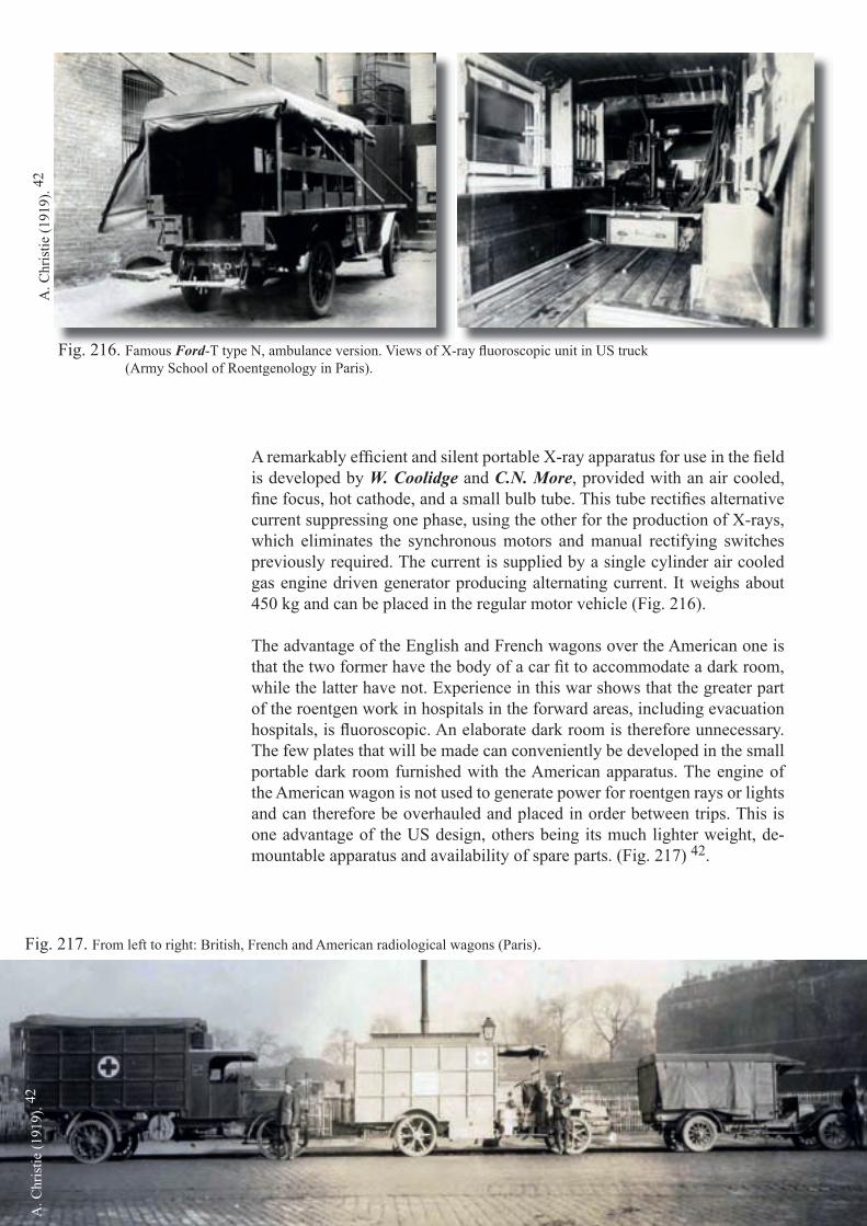
A remarkably effi cient and silent portable X-ray apparatus for use in the fi eld is developed by W. Coolidge and C.N. More, provided with an air cooled, fi ne focus, hot cathode, and a small bulb tube. This tube rectifi es alternative current suppressing one phase, using the other for the production of X-rays, which eliminates the synchronous motors and manual rectifying switches previously required. The current is supplied by a single cylinder air cooled gas engine driven generator producing alternating current. It weighs about 450 kg and can be placed in the regular motor vehicle (Fig. 216).
The advantage of the English and French wagons over the American one is that the two former have the body of a car fi t to accommodate a dark room, while the latter have not. Experience in this war shows that the greater part of the roentgen work in hospitals in the forward areas, including evacuation hospitals, is fl uoroscopic. An elaborate dark room is therefore unnecessary. The few plates that will be made can conveniently be developed in the small portable dark room furnished with the American apparatus. The engine of the American wagon is not used to generate power for roentgen rays or lights and can therefore be overhauled and placed in order between trips. This is one advantage of the US design, others being its much lighter weight, de-mountable apparatus and availability of spare parts. (Fig. 217) 42.
Fig. 216. Famous Ford-T type N, ambulance version. Views of X-ray fl uoroscopic unit in US truck (Army School of Roentgenology in Paris).
Fig. 217. From left to right: British, French and American radiological wagons (Paris).
A. C
hris
tie (1
919)
. 42A
. Chr
istie
(191
9). 42

The bedside unit of Professor J.S. Shearer, a major during the war, manufactured by the Waite and Bartlett company, featuring a Coolidge tube, can be carried by two men and develops 85 kV at 10 mA on alternating current (Fig. 218). 37, 179
The standard US Army X-ray table is rather sturdy and can be assembled quickly (Fig. 219). In addi-tion to being used in the X-ray room, the table top is used as a stretcher for carrying patients and can be applied as a surgical operating table, since its Bakelite covering is impervious to antiseptic solu-tions. The fl uoroscopic screen has wires for foreign body localization. The apparatus is packed in 19 boxes with accessories, practically everything a small X-ray department may require including books, fi lms and darkroom supplies. It weighs about 2,250 kg. 154
So by this time the invention of the Coolidge tube has done away forever with the capriciousness of the gas tube, and in 1916 the introduction of the Potter-Bucky diaphragm 177 has freed the roentgen images of their fogginess and indistinctness. So also does E. Caldwell's (NY City) diaphragm; he has started as an electrical engineer and opts into medicine and radiology. Because most of the efforts occur during WW I, Potter is not aware of Bucky until Caldwell tells him about their comparable approach. And fairly soon, most X-ray manufacturers will offer a suitable grid. 139 Fortunately, as a result of these two great discoveries, the American Expeditionary Force of World War I is supplied with the fi nest roentgen-ray equipment that the world has yet known.
It may be of interest to note at this juncture that the fi rst Coolidge tube which the French see in op-eration, is in the American Military Hospital N° 1 (Vichy), in 1917. Dr. A. Béclère and his group of students have made a special trip to the hospital. 87, 182
R. E
isen
berg
(199
2). 69
Fig. 219. US Army X-ray fi eld unit that can be de-mounted for transportation within 3 minutes! The fl uoroscopic screen is enclosed underneath the X-ray tube in a protective box.
Fig. 218. The US mobile bedside X-ray equipment at Base Hospital No 69 in Savenay (France).
R. P
akus
ch (1
982)
. 172
By the end of the war, 719 US X-ray units are shipped overseas: 150 com-plete sets for base hospitals, 250 bedside machines, 55 X-ray trucks, 264 portable machines, and hundreds of accessory items. 145,179

Since the war takes place on another continent, transportability is a signifi cant consideration. This is not as important for the European combatants. They do not have an ocean to cross to the front line!
Without going into too much detail, we should also mention the importance of seaborne evacuation of the wounded during the Great War.The boats are hospital ships, transformed and adapted to a medical mission and have a radiology department.The most important include the "USS Solace (AH-2)" and "USS Relief" (Fig. 220) both dating from 1896 and especially the "USS Comfort (AH-3)" and "USS Mercy (AH-4 )" the most imposing vessels, dating from1907. 165, 184
The fi rst hospital ship in the world really purpose-built and equipped with all medical equipment and modern facilities is laid down in June 1917 and completed in Philadelphia in 1919. This is the new "USS Relief (AH-1)" (Fig. 221) 140 m long, weighing 9,800 tons and with a speed of 16 knots. 124
Fig. 220. a. USS Relief (1896) serving as a US Army hospital ship transferred to the Navy in November 1902. b . Presence of an old gas X-ray tube used during Spanish-American war.
Fig. 221. USS Relief (AH-1). a. X-ray viewing room. b. Patient wards.
Wik
iped
ia.
L. Jo
hnso
n (1
937)
. 124
Fig. 220. a. USS Relief (1896) serving as a US Army hospital ship transferred to the Navy in November 1902.
A
USS Relief (1896) serving as a US Army hospital ship transferred to the Navy in November 1902.
B
A B
169
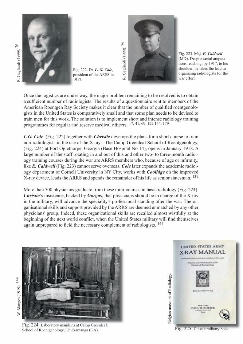
Once the logistics are under way, the major problem remaining to be resolved is to obtain a suffi cient number of radiologists. The results of a questionnaire sent to members of the American Roentgen Ray Society makes it clear that the number of qualifi ed roentgenolo-gists in the United States is comparatively small and that some plan needs to be devised to train men for this work. The solution is to implement short and intense radiology training programmes for regular and reserve medical offi cers. 17, 41, 69, 122 144, 179
L.G. Cole, (Fig. 222) together with Christie develops the plans for a short course to train non-radiologists in the use of the X rays. The Camp Greenleaf School of Roentgenology, (Fig. 224) at Fort Oglethorpe, Georgia (Base Hospital No 14), opens in January 1918. A large number of the staff rotating in and out of this and other two- to three-month radiol-ogy training courses during the war are ARRS members who, because of age or infi rmity, like E. Caldwell (Fig. 223) cannot serve overseas. Cole later expands the academic radiol-ogy department of Cornell University in NY City, works with Coolidge on the improved X-ray device, leads the ARRS and spends the remainder of his life as senior statesman. 139
More than 700 physicians graduate from these mini-courses in basic radiology (Fig. 224). Christie's insistence, backed by Gorgas, that physicians should be in charge of the X-ray in the military, will advance the speciality's professional standing after the war. The or-ganisational skills and support provided by the ARRS are deemed unmatched by any other physicians' group. Indeed, these organizational skills are recalled almost wistfully at the beginning of the next world confl ict, when the United States military will fi nd themselves again unprepared to fi eld the necessary complement of radiologists. 144
Fig. 222. Dr. L. G. Cole, president of the ARSS in 1917.
Fig. 223. Maj. E. Caldwell (MD). Despite serial amputa-tions reaching, by 1917, to his shoulder, he takes the lead in organizing radiologists for the war effort. R
. Gag
liard
i (19
99).
76
R. G
aglia
rdi (
1999
). 76
W. M
ange
s (19
19).
144
Bel
gian
mus
eum
of R
adio
logy
.
Fig. 225. Classic military book. Fig. 224. Laboratory manikins at Camp Greenleaf. School of Roentgenology, Chickamauga (GA).
US
Arm
y X
-ray
man
ual (
1918
). 19
8, 1
99

Another problem that appears to be common to all military ser-vices is the lack of trained technologists. The US Army establishes several schools besides the Roentgen Ray Department of the Army Medical School in Washington DC, to train corpsmen. It also pub-lishes a very well written manual (Fig. 225). Schools of military roentgenology, under the control of the Department of War are opened in Baltimore, Boston, Chicago, Chickamauga (GA), Kan-sas City, Los Angeles, New York, Philadelphia and Pittsburgh. Classes begin promptly in July, shortly after the United States en-ters the War. 144
In early 1918 the Army School of Roentgenology commences at Camp Greenleaf, Georgia, which is then a medical training camp. The technologists are given intensive courses, besides radiography and fi lm processing, also in trouble shooting. The physicians are instructed in technique and fi lm processing to obtain the most sat-isfactory radiographs. (Fig. 226, 227) The most important subject in clinical instruction is the localization of foreign bodies (Fig. 228), and several methods are taught (see chapter 8 ). 81, 144, 172
Fig. 226. Fluoroscopic screen for Hirtz's compass method.
Fig. 228. The technique for the localisation of projectiles in the eye. A tube shift method is used with two radiographs and the W. Sweet device (see detail view).
Fig. 227. A bedside unit with vertical fl uoroscope for teaching chest examination.
US
Arm
y X
-ray
man
ual (
1918
). 19
8, 1
99
US
Arm
y X
-ray
man
ual (
1918
). 19
8, 1
99
US
Arm
y X
-ray
man
ual (
1918
). 19
8, 1
99
Bel
gian
Mus
eum
of R
adio
logy
(O-1
07).
171
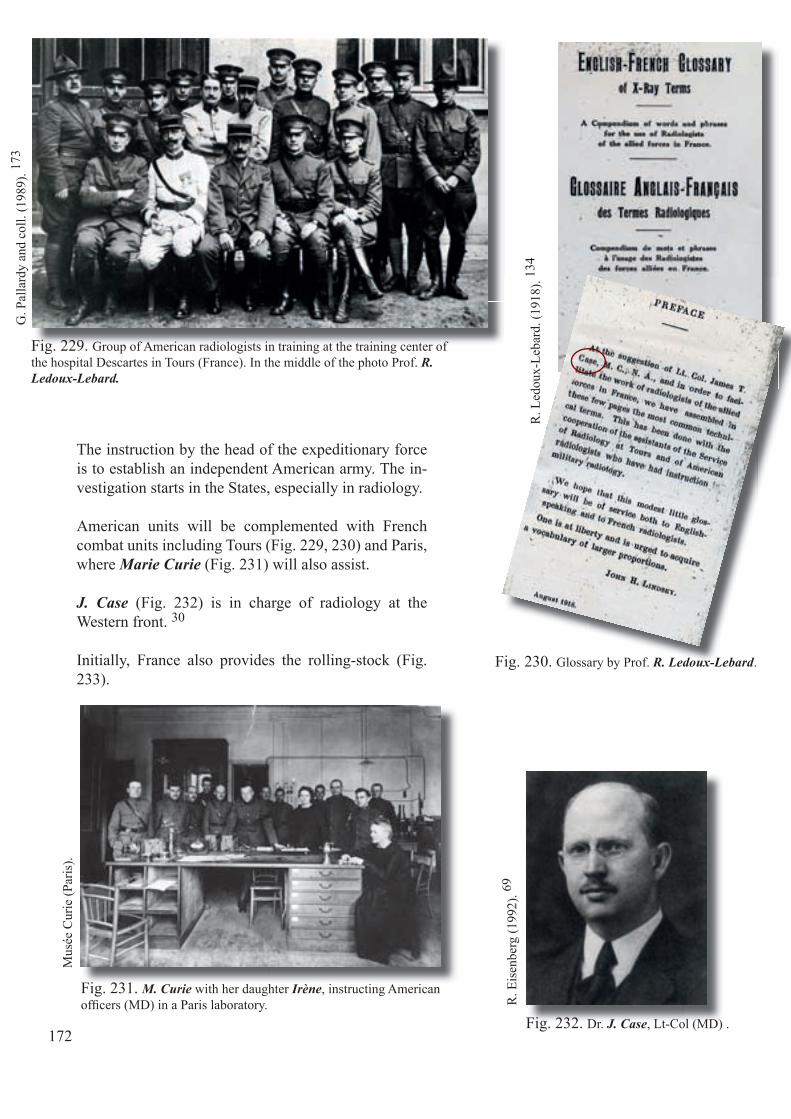
The instruction by the head of the expeditionary force is to establish an independent American army. The in-vestigation starts in the States, especially in radiology.
American units will be complemented with French combat units including Tours (Fig. 229, 230) and Paris, where Marie Curie (Fig. 231) will also assist.
J. Case (Fig. 232) is in charge of radiology at the Western front. 30
Initially, France also provides the rolling-stock (Fig. 233).
Fig. 229. Group of American radiologists in training at the training center of the hospital Descartes in Tours (France). In the middle of the photo Prof. R. Ledoux-Lebard.
Fig. 230. Glossary by Prof. R. Ledoux-Lebard.
Fig. 232. Dr. J. Case, Lt-Col (MD) .
G. P
alla
rdy
and
coll.
(198
9). 17
3
R. L
edou
x-Le
bard
. (19
18).
134
R. E
isen
berg
(199
2). 69
172
Fig. 231. M. Curie with her daughter Irène, instructing American offi cers (MD) in a Paris laboratory.
Mus
ée C
urie
(Par
is).

Nat
iona
l Mus
eum
of t
he H
ealth
and
Med
icin
e (O
tis A
rchi
ves)
.
Fig. 233. Roentgenology school. Training with French equipment.
173

Later the Army School of Roentgenology, established in Paris, trains with US material. Under Christie's guidance, a portable X-ray apparatus is soon pro-duced in quantity. This machine is powered by a single-cylinder, air-cooled gasoline engine and can be carried from building to building and wheeled from bed to bed.
Furthermore, the regular or a sponsored (Fig. 235) army motor ambulance is modifi ed to carry the new portable unit, resulting in a complete mobile roentgen-ray department with facilities for developing plates or fi lms and for doing fl uoroscopy as well as radiographic work (Fig. 236).
Because the glass plates are too heavy and fragile for military use in the fi eld, however, most diagnostic work is done by fl uoroscopy.
The trend toward standardization and portability of equipment signifi cantly infl uences the civilian practice of radiology in the years to come. 75
Fig. 234. Department of Radiology of US military Base Hospital No. 27 in Angers (France). French radiological equipment. (Greenwood).
J. G
reen
woo
d. 87
174

Fig. 235. Radiological wagon built in France on a Renault EP chassis. Offered by a American patron and used by the AEF.
Fig. 236. Radiology school in Paris. A. Close-up of the US apparatus. B. Canopy for the US portable X-ray apparatus.C. Portable processing box (Army School of Roentgenology, Paris).
A B
Radiology school in Paris. A. Close-up of the US apparatus. B. Canopy for the US portable X-ray apparatus.
CNat
iona
l Mus
eum
of t
he H
ealth
and
Med
icin
e (O
tis A
rchi
ves)
.
175
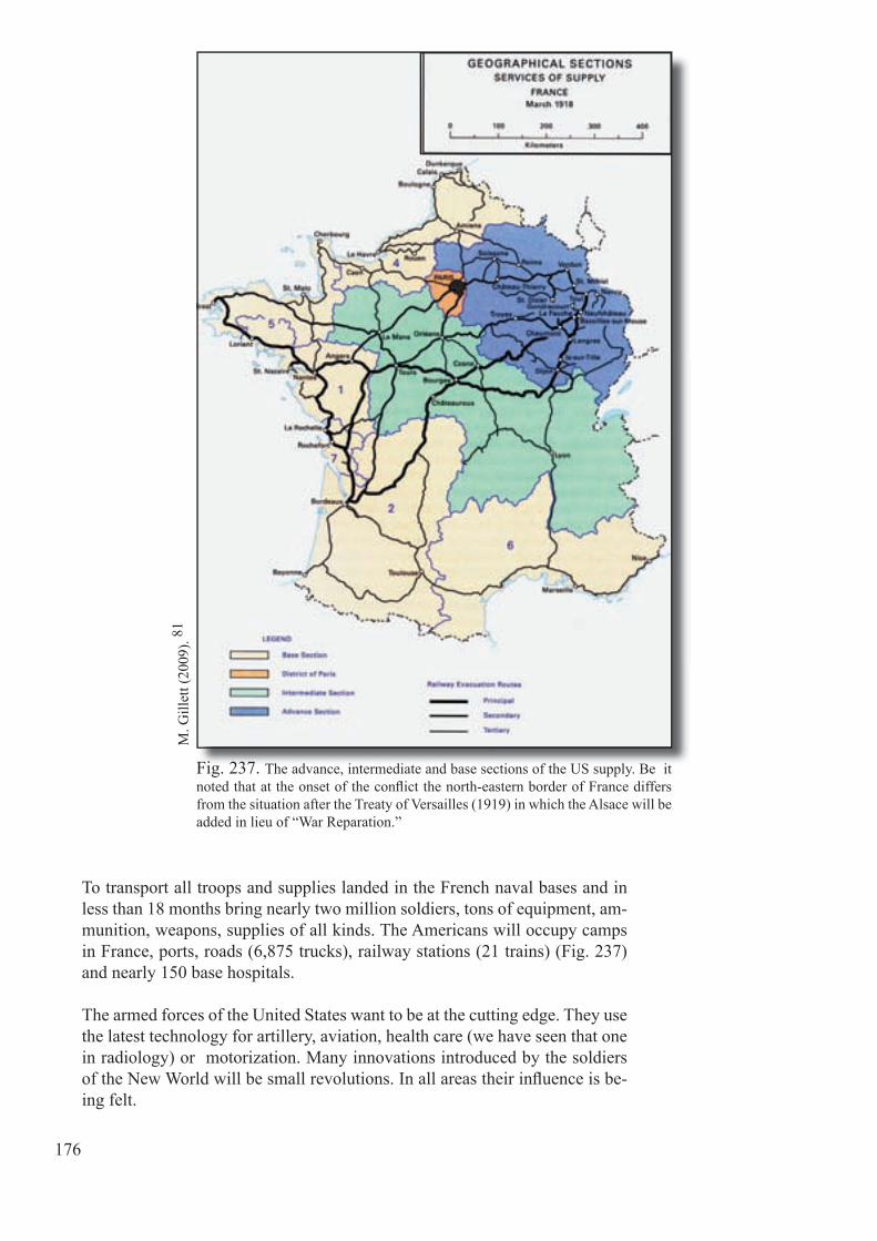
To transport all troops and supplies landed in the French naval bases and in less than 18 months bring nearly two million soldiers, tons of equipment, am-munition, weapons, supplies of all kinds. The Americans will occupy camps in France, ports, roads (6,875 trucks), railway stations (21 trains) (Fig. 237) and nearly 150 base hospitals.
The armed forces of the United States want to be at the cutting edge. They use the latest technology for artillery, aviation, health care (we have seen that one in radiology) or motorization. Many innovations introduced by the soldiers of the New World will be small revolutions. In all areas their infl uence is be-ing felt.
Fig. 237. The advance, intermediate and base sections of the US supply. Be it noted that at the onset of the confl ict the north-eastern border of France differs from the situation after the Treaty of Versailles (1919) in which the Alsace will be added in lieu of “War Reparation.”
M. G
illet
t (20
09). 8
1
176

During World War I the AEF participates in thirteen campaigns on the Western Front, of which one is the Ypres-Lys Campaign (18 August – 11 November) during the Flanders Offensive of 1918 when a number of divisions are engaged as part of an army group under the command of Albert I, King of the Belgians (Fig. 238). 16
On 31 August 1918, during a ‘training’ period with the British II Corps, 27th (US) Division advances on Vierstraat and the following days, supported by the 30th (US) Division both divi-
Fig. 238. American and Allied Attacks on the Western front September 25, 1918, prior to the liberation offensive.
Fig. 239. X-ray truck. Mobile Hospital No 39 St. Mihiel.
US
US
M. I
rela
nd (1
925)
. 121
sions continue the advance towards Voormezele. About six weeks lat-er, in mid-October 1918, Pershing dispatches two other American di-visions - the 37th and 91st - to the French Army of Belgium, at Foch's request, to give impetus to the drive to cross the Scheldt south-west of Ghent. A general attack begins in this area on 31 October and contin-ues intermittently until hostilities end on 11 November.
While the American Expeditionary Force, stays deployed north of St. Mihiel,.... the weather conditions are just so deplorable (Fig. 239)!
177

Let us focus for a moment on the famous Ford T-type N Ambulance. (Fig. 240).Already in 1907 H. Ford, made famous by his method of assembly line work, exports his Ford T to France, fi rst in Bordeaux and then in 1913, in Paris. That same year, the importer of Ford ambulances, H. Depasse, sets up an assembly plant in Bordeaux.
At the beginning of the war a few T-Fords are bodied into ambulance vehi-cles for the French army by a coachbuilder of Levallois-Perret. A team of drivers and Ford employees even participate in the Battle of the Marne in September 1914. For its ability for manoeuvres, reliability and light weight, these vehicles render so many services that the British army (which does not yet manufacture any on its soil) soon orders fi fty of them.
In 1916 the Ford Motor Company buys H. Depasse's assembly plant and cre-ates a French subsidiary, the “Société des automobiles Ford” (Ford Motor Company). More than 11.800 T-Fords, built in Bordeaux by the Ford Motor Company, serve as allied health vehicles. 73
On the sides of the U.S. medical vehicles, one notices the presence of a sign showing two entwined snakes set upon a rod and surmounted by two wings. Since the late 19th century the U.S. medical service uses this symbol to de-termine the ownership of its equipment and vehicles.
178
A. C
hris
tie (1
919)
. 42
Fig. 240. Radiological Ford-T vehicle in the "Army School of Roentgenology" in Paris.

But Asklepios (Aesculapius in Latin) the god of medicine is always portrayed with his stick mace (symbol of life) around which is wound a single snake, a symbol associated with the idea of life and death. However another god, Hermes (Mercury in Latin), the god of commerce, also has a stick on which are wound two snakes, their heads up. 208 In addition, there are two wings attached to the top. These wings symbolize travel, trade ... and have nothing to do with medicine or health. The U.S. pharmaceutical and medical world (both civilian and military) accentuates this confusion by using as an emblem the caduceus of Hermes with two snakes, that is to say, the emblem of Com-merce (Fig. 241). Unfortunately, other medical services have followed suit! Besides that, the US Treasury has seized the medical emblem to stylize it as the dollar sign!
Yet the 10th General Assembly of the World Medical Association (1956) has clearly defi ned the medical emblem: a vertical stick, red and shiny around which is wound one single snake, with two curves on the left and one on the right, all on a white background. (Fig. 242)
Hopefully this confusion of symbols does not infl uence the art of medicine. Medicine, could it turn into a trade?
War can also produce a metonymy as, for instance: "ambulance". An am-bulance is a mobile medical unit, used extensively during the war and obvi-ously equipped with its own vehicles. 205
After the war, when the US medical service returns to the US, it stores its ambulances in the depots, while regularly using only its vehicles which it will call, for short "ambulance". This new sense of the term, limited to the ve-hicle, has been introduced in Europe, with the return of US troops in Europe during World War II... but that's another story.
179
P. D
aves
(199
4). 47
Fig. 241. US caduceus erroneously attributed to medicine.Fig. 242. Rod of Asklepios as defi ned by the "World Medical Association" in Havana.
Laro
usse
uni
vers
el (1
985)
.
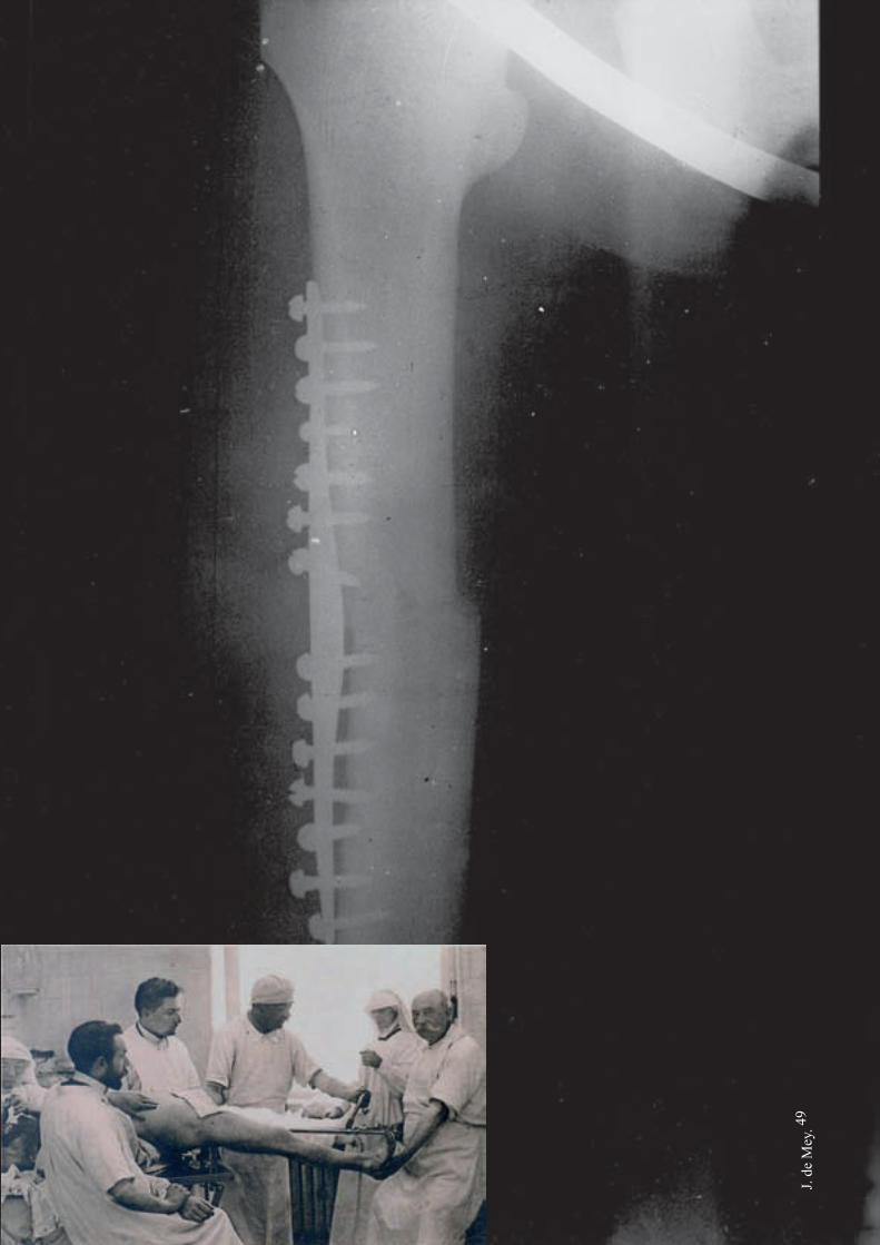
106
J. de
Mey
. 49

181
Chapter 19
Fig. 243. Radiographic glass plate showing a femoral fracture set by "osteosynthesis' (plate and screws) during the 14-18 war. This technique, a true world premiere, is invented in 1902 by the Antwerp surgeon A. Lambotte (medaillon), who also has given it its name. He founds in 1921 the Belgian Society of Orthopaedic Surgery of which he is the fi rst president. The small photo illustrates the orthopaedic treatment of a femoral fracture, possibly carried out by osteosynthesis in the surgical institute in Jumet, Belgium (military ambulance in August 1914).
Positive effects of the confl ict on radiology
In Chapter 8 we have seen the close relationship that has developed between surgery and radiology. 53,84,175 During the war, and even afterwards fractures of the extremities remain serious issues due to severe complications as com-plex fractures often and quickly become infected. They infl uence the further course of life or make further motion often impossible.
Surgeon Ch. Willems, who in his later career becomes an academic at the Universities of Ghent and Liège, and who is director of the military hospital at Hoogstade limits the number of amputations through early mobilization of injured and infl amed joints. We mention here what this important Belgian personality thinks of radiography, "One should never begin to treat a frac-ture unless having it radiographed fi rst. This rule is also essential to monitor fractures by war projectiles as well as fractures in civil practice ... Every institution in which fractures are treated should therefore be furnished with radiographic equipment. " 217
We will not discuss the various new methods of orthopaedic treatment but only emphasize the innovative contribution of our Antwerp compatriot, sur-geon A. Lambotte (Fig. 243). He later has laid the foundation of the surgical technique of osteosynthesis, to which he has not only given the name but for which he also proposes various application methods. 132
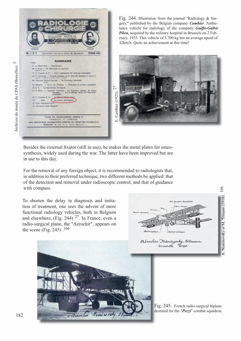
182
Besides the external fi xator (still in use), he makes the metal plates for osteo-synthesis, widely used during the war. The latter have been improved but are in use to this day.
For the removal of any foreign object, it is recommended to radiologists that, in addition to their preferred technique, two different methods be applied: that of the detection and removal under radioscopic control, and that of guidance with compass.
Fig. 244. Illustration from the journal "Radiology & Sur-gery," published by the Belgian company Cambier. Ambu-lance vehicle for radiology of the company Gaiffe-Gallot Pilon, acquired by the military hospital in Brussels on 2 Feb-ruary, 1925. This vehicle of 3,700 kg has an average speed of 32km/h. Quite an achievement at this time!
E. C
ambi
er (1
925)
. 27
Arc
hive
s du
mus
ée d
u C
PAS
(Bru
xelle
s). 5
Fig. 245. French radio-surgical biplane destined for the "Pozzi" combat squadron.
A. N
emiro
vsky
et M
. Tilm
ant (
1918
) . 16
6A
. Nem
irovs
ky e
t M. T
ilman
t (19
18) .
To shorten the delay in diagnosis and initia-tion of treatment, one sees the advent of more functional radiology vehicles, both in Belgium and elsewhere, (Fig. 244) 27. In France, even a radio-surgical plane, the "Aerochir", appears on the scene (Fig. 245) .166
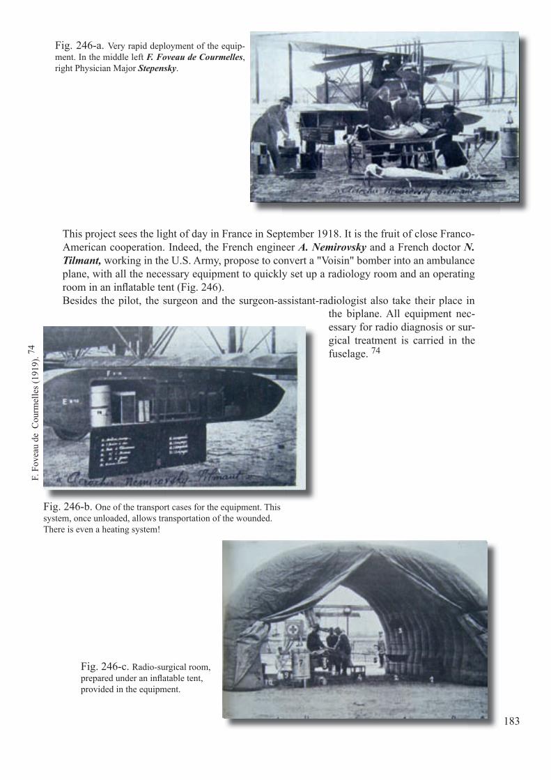
183
This project sees the light of day in France in September 1918. It is the fruit of close Franco-American cooperation. Indeed, the French engineer A. Nemirovsky and a French doctor N. Tilmant, working in the U.S. Army, propose to convert a "Voisin" bomber into an ambulance plane, with all the necessary equipment to quickly set up a radiology room and an operating room in an infl atable tent (Fig. 246).Besides the pilot, the surgeon and the surgeon-assistant-radiologist also take their place in
Fig. 246-a. Very rapid deployment of the equip-ment. In the middle left F. Foveau de Courmelles, right Physician Major Stepensky.
Fig. 246-b. One of the transport cases for the equipment. This system, once unloaded, allows transportation of the wounded. There is even a heating system!
Fig. 246-c. Radio-surgical room, prepared under an infl atable tent, provided in the equipment.
F. F
ovea
u de
Cou
rmel
les (
1919
). 74
the biplane. All equipment nec-essary for radio diagnosis or sur-gical treatment is carried in the fuselage. 74

184
While at the end of the confl ict the radiological air-craft and vehicles are stored in the army depots, it should be emphasized that at the same time each hospital is fi nally being assigned at least one radi-ology unit.
So we make the true transfer from the radiology laboratory to the radiology department, both at the level of the small provincial hospitals and the larg-er city hospitals. Here is a selection, resulting from private collections (Fig. 247, 248).
Fig. 247-a. Fig. 247-b.
Fig. 247-c.
Fig. 248-a. Fig. 248-b.
Fig. 247-a Sanatorium of the "Domaine de La Chise" (Piétrebais). -b Sanatorium Edith Cavell (Obourg). -c Elisabeth Sanatorium (Sijsele).
Fig. 246-a Sainte Thérèse (Montigny-sur-Sambre) Hospital. -b Surgical Institute (Jumet).
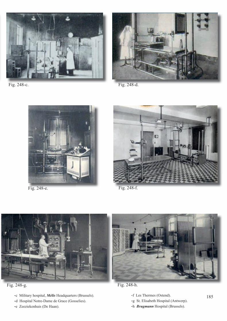
Fig. 246-a Sainte Thérèse (Montigny-sur-Sambre) Hospital. -b Surgical Institute (Jumet).
185
Fig. 248-c. Fig. 248-d.
Fig. 248-e. Fig. 248-f.
Fig. 248-g. Fig. 248-h.
-c Military hospital, Mélis Headquarters (Brussels).-d Hospital Notre-Dame de Grace (Gosselies).-e Zeeziekenhuis (De Haan).
-f Les Thermes (Ostend).-g St. Elisabeth Hospital (Antwerp).-h Brugmann Hospital (Brussels).
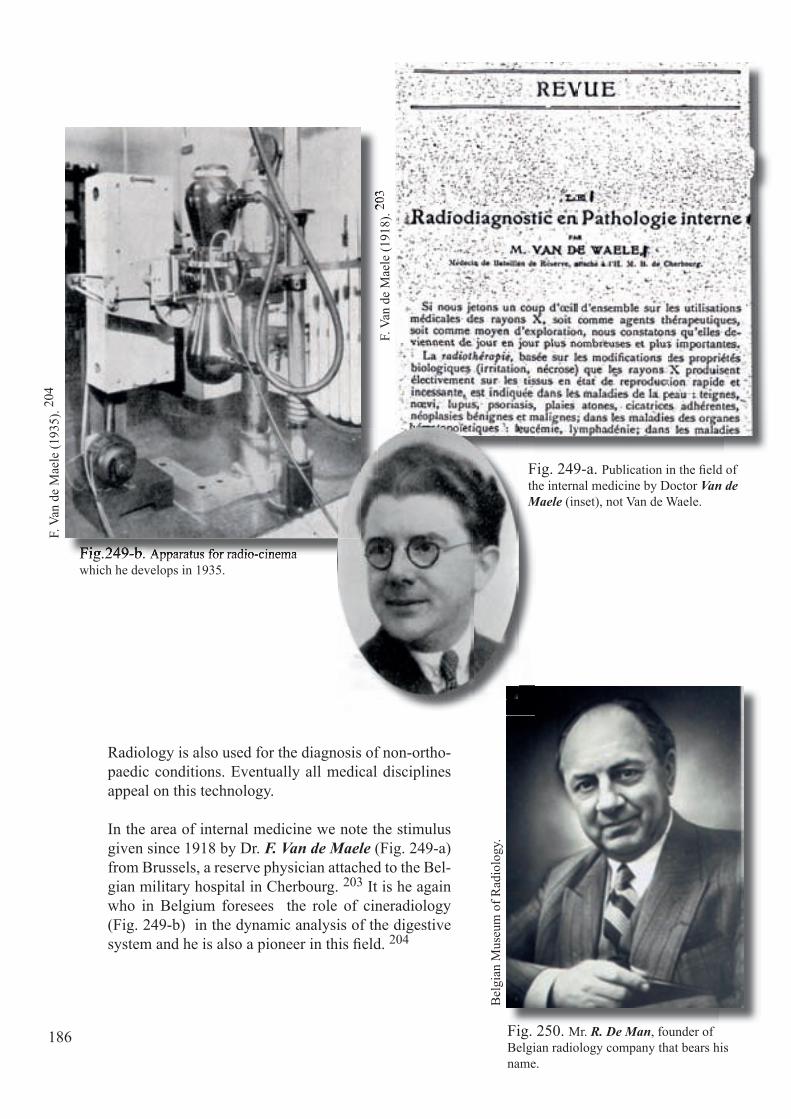
186
Radiology is also used for the diagnosis of non-ortho-paedic conditions. Eventually all medical disciplines appeal on this technology.
In the area of internal medicine we note the stimulus given since 1918 by Dr. F. Van de Maele (Fig. 249-a) from Brussels, a reserve physician attached to the Bel-gian military hospital in Cherbourg. 203 It is he again who in Belgium foresees the role of cineradiology (Fig. 249-b) in the dynamic analysis of the digestive system and he is also a pioneer in this fi eld. 204
Fig. 250. Mr. R. De Man, founder of Belgian radiology company that bears his name.
Fig.249-b. Apparatus for radio-cinemawhich he develops in 1935.
F. V
an d
e M
aele
(191
8). 20
3
Bel
gian
Mus
eum
of R
adio
logy
.
Fig.249-b. Apparatus for radio-cinema
203
F. V
an d
e M
aele
(193
5). 20
4
Fig. 249-a. Publication in the fi eld of the internal medicine by Doctor Van de Maele (inset), not Van de Waele.

187
To cope with the constant and growing demand, expansion of existing fi rms that produce radiology equipment occurs, while new companies see the light of day. In our country we also see the foundation of two important companies, namely Balteau in Liège and De Man in Antwerp. Both are family businesses.
The latter fi rm, established in Antwerp, Langegang, a street now vanished near the Tabakvest, sees the light of day in 1917. From the very beginning he concen-trates on equipment for radiography and fl uoroscopy. For the construction of the latter type, the engineer from Antwerp R. De Man (Fig. 250) calls upon L. Hau-champs, a physician from Brussels, to build a fl uoroscopy unit (Fig. 251) and upon his colleague and friend from Antwerp Dr. L. Deckers to furnish a complete radiology room at the St Elizabeth Hospital in Antwerp (Fig. 252).
Fig. 251. Radioscopy device manufactured by the fi rm of De Man. As recommended by Dr. L. Hauchamps, in private practice in Brussels (Reconstruction in the Bel-gian Museum of Radiology).
Fig. 252. Radiology Room at the St. Elisabeth Hospital (Antwerp).
Bel
gian
Mus
eum
of R
adio
logy
.

188
In the thirties it is this same company that builds one of the fi rst Belgian to-mographs with the cooperation of the army radiologist from Antwerp I. Tous-saint (Fig. 253).
After the second world war the fi rm distinguishes itself with the construction of the Stratomatic which in the sixties is considered the best tomograph in the world.Also at that time the fi rm merges with the French company Chenaille and the Compagnie Générale de Radiologie (CGR), the whole eventually being absorbed by the American company "General Electric".
The rival Liège fi rm starts up at number 91 rue de Serbie, as a small workshop for the construction and repair of electric motors and transformers. It is in 1933 that M. Balteau (Fig. 254) focuses on the medical applications of elec-tricity. Soon there appear some powerful X-ray generators: the Baltosix (Fig. 255). In the fi eld of tomography, M. Balteau and his staff are not idling, so after the second war the Baltomix is born (Fig. 256).Like the Antwerp fi rm, the Balteau company also produces quality material and in 1966 develops the very fi rst remote controlled table: the Prestilix (in 1993 still the most widely sold). In the late '70s, the medical department of the company is incorporated into the American company "General Electric".
Bel
gian
Mus
eum
of R
adio
logy
.B
elgi
an M
useu
m o
f Rad
iolo
gy.
Fig. 253. Prototype for tomography, the Radi-otome, of the company De Man. In the inset one recognizes Dr. I. Toussaint.
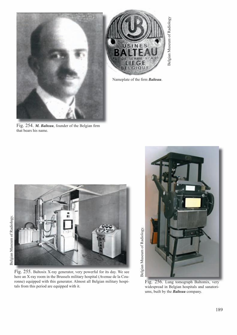
189
Nameplate of the fi rm Balteau.
Fig. 254. M. Balteau, founder of the Belgian fi rm that bears his name.
Fig. 256. Lung tomograph Baltomix, very widespread in Belgian hospitals and sanatori-ums, built by the Balteau company.
Bel
gian
Mus
eum
of R
adio
logy
.
Bel
gian
Mus
eum
of R
adio
logy
Fig. 255. Baltosix X-ray generator, very powerful for its day. We see here an X-ray room in the Brussels military hospital (Avenue de la Cou-ronne) equipped with this generator. Almost all Belgian military hospi-tals from this period are equipped with it.
Fig. 255.
Fig. 254.
Bel
gian
Mus
eum
of R
adio
logy
.

190
After the war the advantages of the new radiology tubes, namely the Coolidge tube, are extensively mentioned in radiological publications, for instance in 1919 in the Belgian Journal of Radiology, by M. Peremans from Antwerp who, during the confl ict has been active in the hospital "L'Océan". 175
In the fi eld of photographic supports new developments occur, given the fragility of the glass plates. Following the example of the U.S. company Eastman Kodak in 1914, L. Gevaert from 1920 also produces cellulose-based fi lm in Antwerp (Fig. 257).
As for radiation protection measures begin to become fa-miliar and generalized. Sometimes they are even dispro-portionately applied (Fig. 258)!
Fig. 258. A somewhat excessive measure of radiopro-tection, taken in the Heyns Daele sanatorium (Ronse).
Fig. 257. Advertising in 1920 from the former Belgian fi rm L. Gevaert.
Priv
ate
colle
ctio
n.
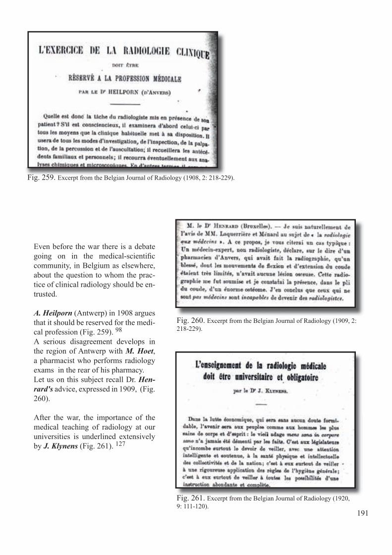
191
Even before the war there is a debate going on in the medical-scientifi c community, in Belgium as elsewhere, about the question to whom the prac-tice of clinical radiology should be en-trusted.
A. Heilporn (Antwerp) in 1908 argues that it should be reserved for the medi-cal profession (Fig. 259). 98
A serious disagreement develops in the region of Antwerp with M. Hoet, a pharmacist who performs radiology exams in the rear of his pharmacy.Let us on this subject recall Dr. Hen-rard's advice, expressed in 1909, (Fig. 260).
After the war, the importance of the medical teaching of radiology at our universities is underlined extensively by J. Klynens (Fig. 261). 127
Fig. 259. Excerpt from the Belgian Journal of Radiology (1908, 2: 218-229).
Fig. 260. Excerpt from the Belgian Journal of Radiology (1909, 2: 218-229).
Fig. 261. Excerpt from the Belgian Journal of Radiology (1920, 9: 111-120).
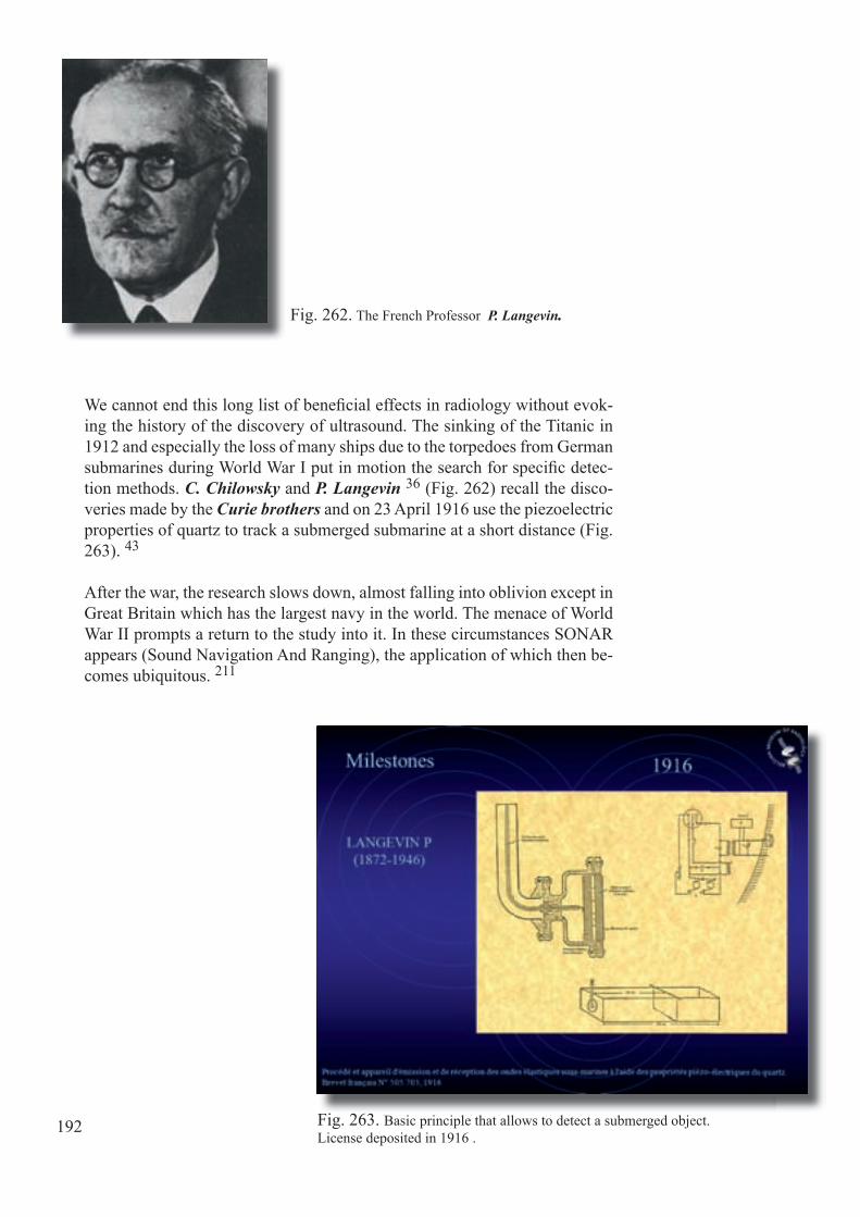
192
We cannot end this long list of benefi cial effects in radiology without evok-ing the history of the discovery of ultrasound. The sinking of the Titanic in 1912 and especially the loss of many ships due to the torpedoes from German submarines during World War I put in motion the search for specifi c detec-tion methods. C. Chilowsky and P. Langevin 36 (Fig. 262) recall the disco-veries made by the Curie brothers and on 23 April 1916 use the piezoelectric properties of quartz to track a submerged submarine at a short distance (Fig. 263). 43
After the war, the research slows down, almost falling into oblivion except in Great Britain which has the largest navy in the world. The menace of World War II prompts a return to the study into it. In these circumstances SONAR appears (Sound Navigation And Ranging), the application of which then be-comes ubiquitous. 211
Fig. 263. Basic principle that allows to detect a submerged object. License deposited in 1916 .
Fig. 262. The French Professor P. Langevin.
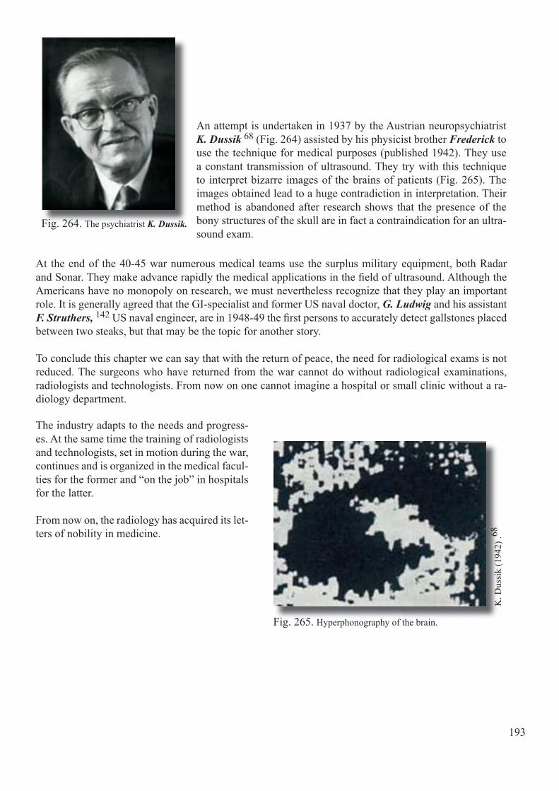
193
An attempt is undertaken in 1937 by the Austrian neuropsychiatrist K. Dussik 68 (Fig. 264) assisted by his physicist brother Frederick to use the technique for medical purposes (published 1942). They use a constant transmission of ultrasound. They try with this technique to interpret bizarre images of the brains of patients (Fig. 265). The images obtained lead to a huge contradiction in interpretation. Their method is abandoned after research shows that the presence of the bony structures of the skull are in fact a contraindication for an ultra-sound exam.
Fig. 264. The psychiatrist K. Dussik.
Fig. 265. Hyperphonography of the brain.
K. D
ussi
k (1
942)
.68
At the end of the 40-45 war numerous medical teams use the surplus military equipment, both Radar and Sonar. They make advance rapidly the medical applications in the fi eld of ultrasound. Although the Americans have no monopoly on research, we must nevertheless recognize that they play an important role. It is generally agreed that the GI-specialist and former US naval doctor, G. Ludwig and his assistant F. Struthers, 142 US naval engineer, are in 1948-49 the fi rst persons to accurately detect gallstones placed between two steaks, but that may be the topic for another story.
To conclude this chapter we can say that with the return of peace, the need for radiological exams is not reduced. The surgeons who have returned from the war cannot do without radiological examinations, radiologists and technologists. From now on one cannot imagine a hospital or small clinic without a ra-diology department.
The industry adapts to the needs and progress-es. At the same time the training of radiologists and technologists, set in motion during the war, continues and is organized in the medical facul-ties for the former and “on the job” in hospitals for the latter.
From now on, the radiology has acquired its let-ters of nobility in medicine.

194
195
I was born and raised in Belgium, in the World War One region south of the river Yser. My grandfather being a decorated veteran of the Great War who had lived and fought for four years in the trenches, I have always been very much interested in the history of the First World War and therefore obviously also by this book.
When I myself joined the world of national defence as a cadet - a student at a military high school - I went looking for more information on that war and I was strongly impressed by horrifying stories and the appalling living condi-tions in the trenches and of terribly wounded soldiers. I initially obtained the information at home, first hand, although it is fitting to mention that those who experienced the war were not inclined to spontaneously tell a lot of stories. Later, as a student of medicine, I thought to recognise a kind of suppressing mechanism that had to protect them from too shocking memories.Finally, as a military doctor I was primarily interested in medical care, at that time provided by the Military Health Service. The book by René Van Tigge-len reveals the origins and growth of the then new discipline of medicine and very well places the use of radiology in its time frame.
After the discovery of the new type of radiation by W.C. Röntgen in Decem-ber 1895, radiology very quickly found its way to military medicine. The impact of radiology on surgery cannot be overrated.
Afterword
196
The morning edition of the Frankfurter Zeitung of the 7th of January 1896, requested the attention of their readers for the importance of the new discov-ery in regard to diagnosing diseases of and injuries to the bones, explaining that from then on the surgeon could determine the extent of complex bone fractures without painful manual examination and moreover he could more easily localize bullets and shrapnel without painful probing wounds.Radiology was relatively quickly used in various armed conflicts such as the Greek-Turkish war in 1897 and the Spanish-American war of 1898. How-ever, it would take quite some time for radiology to acquire a definite place closer to the front line and it can be stated that the real breakthrough took place during the First World War.
The delay in the use of radiology near to the front line can be explained. At that time military medical doctrine did not provide a favourable situation for that kind of use. Triage and forward medical care were not known. Long lasting evacuation of wounded soldiers to rear area medical installations was customary. Therefore it is not surprising that radiology as an aid for surgery was not to be found close to the frontline. In 1899, in an article in the Lancet Francis C. Abbot publishes a generally accepted statement: “if radiographs could be taken on the battlefield, they would give surgeons an incentive to operate under adverse conditions”.Moreover röntgen apparatus were heavy, fragile, and dependent on scarcely available electric power sources. On the other hand, there is no doubt that the attitude of medical people, feeling a kind of resistance against renovation, played a role as well. It took some time for surgeons who were used to diag-nose a fracture based on clinical signs like deformation, abnormal mobility and crepitation to give radiology a place in the diagnostic process.
Nevertheless, radiology very quickly became an indispensable part of mili-tary medicine. In 1998, the British surgeon W.C. Beevor in an article entitled “The working of Roentgen Ray in warfare”, wrote “It is now the duty of eve-ry civilised nation to supply its wounded in war with an X-ray apparatus… not only at the hospitals, but close at hand, wherever they may be fighting and exposing themselves to injury”. When, throughout the chapters of this book, one discovers how radiology already in the period before the First World War gradually had become an indispensable help for military surgery, it is difficult to understand that Defence +/- 75 years later in 1991 in the Operation Provide Comfort in northern Iraq engaged a Surgical Antenna for Quick Intervention (CAST) without mobile radiology facilities. In view of the lack of medical röntgen apparatus the doctors of the CAST used the röntgen apparatus of the bomb disposal unit in order to diagnose fractures.

197
In the nineties of the last century, radiology was no longer considered a necessary medical capacity and investments in training radiologists came to a halt.With the development of teleradiology techniques, interest in radiology once again increased but nevertheless it was initially assumed that, thanks to teleradiology, specialised radiologists would no longer be needed on mis-sions abroad. Very quickly this assumption proved to be wrong, demon-strating there is nothing new under the sun. Like every other technology, military radiology has known its ups and downs.Fortunately, the tide has turned. In the area of formation of personnel as well as acquisition of necessary equipment, the past years showed a hopeful evo-lution as demonstrated by the acquisition of an RIS/PACS-system and the planned acquisition of a new CT-scanner for the Military Hospital Queen Astrid (MHQA). However, it should be noted that in the meantime NMR-apparatus are more and more part of the standard equipment of hospitals, and that since the Operation Desert Storm in 2001 many nations have “field usable” CT-scanners as part of the radiology units of their field hospitals. So there is still room for improvement in the coming years.
To conclude, we are indebted to our colleague René Van Tiggelen and his co-authors, for they described - for the present and future generations - in a fascinating and comprehensible way, the origins of the military radiology and the development of this discipline in the period of the First World War, situating it in the at that time generally accepted military medical doctrine.Understanding the past should help to determine the right course for the future and undoubtedly this book will help to do so.
Major General Geert Laire, MDSurgeon General
Commander of the Medical Component

199
Ambulance: historically a mobile hospital formation that follows the troops during their campaign. During the 14-18 war this concept is taken over by the American troops, and when they come back in the 2nd World War, this term means a vehicle for transporting the sick or injured. The word is now commonly used in this meaning.
Balkan War: 1912-1913, wars in the Balkans: campaigns in the Dardanelles, Serbia and Macedonia.
"Cercle Gaulois": Belgian club, established in Brussels, whose aim was to form a meet-ing place for friends, intellectuals, artists and writers, free from any political, cultural or philosophical opinion.
Coil of Ruhmkorff: induction coil invented by Heinrich Daniel Ruhmkorff, a German builder of scientific instruments. This allows to convert a battery current of very high in-tensity but low voltage, into electricity with high voltage.
Diaphysis: central oblong portion of a long bone.
Dumdum-bullet: this name comes from a factory near Kolkata (Calcutta). This factory supplied to the British a special bullet that was created for the war against the warring tribes of North India, mainly for the campaign of Chitral, now Pakistan. It is later also used in the Great War. A kind of soft-nosed bullet with a lead core that expands on impact and inflicts severe tissue laceration.
Dynamo: name given to a dynamo-electric machine, which transforms the mechanical energy into electrical energy. The dynamo produces direct current; an alternator generates an alternating current.
Glossary
Fulcrum: English term for hinge, in the case of tomography of human body.
Incunabulum: book dating from the beginning of the craft of book printing (before 1500)..
Induction coil: see coil of Ruhmkorff.
Infundibulum: a funnel-shaped object.
Kenotron: a valve or vacuum tube with two electrodes, used in the power supply of X-ray tubes and for the rectification of alternating currents with low intensity and high or very high voltage.
Lab technician: see technologist
Leukaemia: a disease which is also called blood cancer, or acute leukocytosis of the blood forming organs (blood, spleen, lymph nodes, and bone marrow). It is characterized by ex-cessive production of the precursors of white blood cells.
Osmotic characteristic: the pressure needed to be generated in a solution to prevent the absorption of water by osmosis.
Osteosynthesis: a surgical technique using metal devices such as plates, rods, wires or screws to allow reducing and immobilizing a fracture with the purpose of faster healing.
Pechblende: mineral ore used by Marie Curie, which is found to contain a new radioactive substance with chemical properties closely related to those of barium. This substance is subsequently named radium.
Polonium: radioactive substance chemically related to bismuth and named for the country of origin of Marie Curie, who has discovered it.
Radiation tube: glass tube in which a high vacuum prevails, and in which a high-speed electron beam is formed, and produces X-rays upon impact with a modified target.
Radiodermitis: oedema showing itself as an increase in the volume of soft tissues, i.e. a more or less significant swelling caused by the massive accumulation of fluid due to a significant exposure to X-rays (about 8 Gy).
Radiology cassette: flat lightproof but radiation-transparent box containing one or two intensifying screens and in which a photosensitive film is placed. Through the exposure to X-rays a radiological image is formed on the film.
Radionecrosis: by an even greater exposure to radiation (25 Gy approximately) the radio-dermitis persists until the appearance of ulcerations or necrosis of the tissue.
200
Radioscopy: a radiological examination during which a "dynamic" image is obtained by X-rays, projected on a fluorescent screen and instantly viewed. This is opposite to radiog-raphy which concerns an X-ray examination with a stationary image obtained on a photo-graphic film.
Rheostat: variable resistance which is placed into an electric current and allows the current intensity to change.
Shrapnel: named after its inventor, Henry Shrapnel (deceased in 1842, English artillery officer) is a shell loaded with bullets. The name is also used for shell splinters.
Stereography: stereographic projection: the art of determining the location of an object in space by means of multiple projections on a flat surface.
Stereoscopy: technique to obtain an impression of depth/relief from two flat images of the same object taken at slightly different angles and viewed together.
Technologist: the technologist in medical imaging, formerly called lab technician, is a paramedical co-worker who has completed appropriate training, and who assists the medi-cal specialist in diagnostic or interventional imaging and nuclear medicine.
Tomograph: radiological device which allows the execution of tomography (see tomog-raphy).
Tomography: non-destructive technique, by means of X-rays which allows the achieve-ment of images in sections, of a three-dimensional object. This technique has subsequently further evolved using the Computer Tomography (CT) or scanner. The technique is based on the analysis of an X-ray beam, after it has penetrated a body from different directions, and is recorded by the detectors, after which this body is reconstructed in three dimensions.
Trepanation: surgical operation which consists in making an opening in a bone using a cylindrical saw, called a trepan. Best known is the trepanation of the skull.
Ultrasound: imaging by means of ultrasound consists in exposing a part of the body to high frequency sound waves to produce images of the interior of the body. It can show the structure and the movement of internal organs of the body. Ultrasound is a non-destructive and non ionisation diagnostic technique.
X-ray: form of high-frequency electromagnetic radiation with a wavelength between 5 picometres and 10 nanometres. It is an ionizing radiation applied in many areas including spectrometry, medical imaging and therapy.
X-ray tube: see radiation tube.
201


1. Abbot F.C.: Surgery in the Graeco-Turkish war. Lancet, 1899, 1: 80-83.
2. AGC-CAA2 (Evere) since taken over by the "Centre de Documentation Historique" from the ''Musée royal de l'Armée".
3. Allen K.: Pioneer descriptions in Military Roentgenology. In Bruwer A. Classic description in diagnostic roentgenology. Springfield, Illinois USA, Charles C. Thomas, 1964 (2 vol.), pp. 1305-1404.
4. Alvaro G.: I vantaggi pratici della scoperta di Röntgen in chirurgia. Determinazione diagnostica nella ricerca dei proiettili sopra i soldati feriti provenienti dall' Africa. Giornale Medico del Regio Esercito, 1896, 44: 383-394.
5. Archives du Musée du CPAS Bruxelles. Director: Mr. David Guilardian.
6. Association de la Croix-Rouge de Belgique. Almanach royal officiel, 1914: 1446-1447.
7. Aubourg et Barret: Une année de fonctionnement des deux camions radiologiques de la Ne armée. J Radiol Electrol, 1915: 699-708.
8. Aubourg et Barret: Rôle et utilisation des divers types d’installations radiologiques aux armées. J Radiol Electrol, 1917: 548-552.
9. Battersby J.: The present position of the roentgen rays in military surgery. Arch Roentg Ray, 1899, 3: 74-80.
10. Béclère A.: Antoine Béclère. Paris, J.B. Baillière, 1973, 480 pp.
11. Beevor WC.: The working of the roentgen ray in warfare. J Roy United Service Institution, 1898, 42: 1152-1170.
12. Belgian Museum of Radiology. Corporate address: Bruynstraat 2, B-1120 Brussels.
203
Sources

204
13. Belot J., Vignal W.: A propos d’un nouveau tube de Röntgen à vide de Hittorf (tube Coolidge). J Radiol Electrol, 1914: 227-232.
14. Belot J.: Exposition de la société d'encouragement pour l'industrie nationale. J Radiol Electrol, 1916: 237-240.
15. Belot J.: Dr. Georges Haret. J Belg Radiol, 1933: 24-27.
16. Bernard H.: Guerre totale et guerre révolutionnaire. T. I Le XIXe siècle, la Première Guerre Mondiale et la Révolution russe. Bruxelles, Défense Nationale, 1977, 454 pp.
17. Bispham W.N.: The medical department of the United States Army in the World War. Vol. VII Training. Washington, U. S. Government Printing Office, 1927, 1211 pp.
18. Bocage A.: Procédé et dispositif de radiographie sur plaque en mouvement. 1921-2 Brevet français n° 536464.
19. Borden WC.: The use of the Röntgen ray by the Medical Department of the United States Army in War with Spain (1898). Washington. U. S. Government Printing Office, 1900, 98 pp.
20. Boyer J.: La recherche des projectiles dans l’organisme. La Nature, 1915, 2167: 241-248.
21. Brauer L., Haenisch F.: Eine selbständige, transportabele Feldröntgenanlage für interne und chirurgische Untersuchungen. RöFo, 1915-16, XXIII, 1: 38-46.
22. Bruce F.: Experiences of X-ray work during the siege of Ladysmith. Arch Roentg Ray, 1901, 5: 69-74.
23. Bruwer A.J.: Classic descriptions in diagnostic roentgenology. Springfield, Charles C Thomas, 1964 (2 vol), 2059 pp.
24. Buguet A. et Gascard A.: Détermination à l'aide des rayons X de la profondeur où siège un corps étranger dans les tissus. Comptes rendus hebdomadaires des séances de l'académie des sciences, 30 mars 1896, CXXII: 786-787.
25. Burrows E.: Pioneers and early years. A history of British Radiology. St Am, Colophon Limited, 1986, 264 pp.
26. Busquet P.: De la radiologie dans les armées en campagne. La presse médicale (Paris), 1913, 102: 1465-1470.
27. Cambier E.: La voiture radiologique. Radiologie et Chirurgie. 1925, vol I/2: 66.
28. Camelin A.: Les premières radioscopies systématiques par L. Kelsch – Lyon, Hôpital Militaire Desgenettes 1897. Lyon médical, 1969, suppl.: 173-174.
29. Case J.T.: Développement de la localisation des corps étrangers par les rayons X. Histoire succincte et bibliographie. Archives d'électricité médicale et de physiothérapie, 1919: 5-24.
30. Case J. T.: Military Radiology. Radiology, 1940, 35: 634.-665.
31. Centre de documentation historique (Evere). Photo archives, since taken over by the "Musée royal de l'Armée".
32. Chalmarès G.: La radiographie aux armées en campagne. La Nature, 1905, 99-102.
33. Chardin H.: Les nouvelles voitures radiologiques du service de santé. Archives d’élecricité médicale et de physiothérapie, 1918, 26 : 290-292.
34. Charlier A.: Le repérage et l'extraction des corps étrangers par les procédés radiologiques. J Radiol Electr, 1915: 577-587.
205
35. Charlier A.: Les services radiologiques aux armées pendant la guerre actuelle. Arch Med Pharm MiIit, 1917, 67: 638-651.
36. Chilowsky C., Langevin P.: Procédés et appareils pour la production de signaux sous-marins dirigés et pour la localisation à distance d’obstacles sous-marins. 1916, Brevet français n° 502913.
37. Coffin W.: Bedside radiography with a new portable X-ray apparatus. The army bedside X-ray unit adapted as a portable apparatus for civilian practice. Boston Med and Surg J, 1919, CLXXXI, 141: 431-434.
38. Comité d’histoire du service de santé: Histoire de la médecine aux armées. Tome 3. De 1914 à nos jours. Paris-Limoges, Ed La Vauzelle, 1987, 421pp.
39. Coolidge W.D., Moore C.N.: A portable roentgen ray generating outfit. Gen Elec Rev, 1918, 21: 60.
40. Courington F.W., Calverley R.K.: Anesthesia on the Western Front: The Anglo-American Experience of World War I. Anesthesiology, 1986, 642-653.
41. Christie A. C.: Our preparation for the X-ray work of the war. Amer J Roentgenol, 1918: 1-5.
42. Christie A.C.: Mobile Roentgen ray apparatus. Amer J Roentgenol, 1919: 358-367.
43. Curie J., Curie P.: Sur l’électricité polaire dans les cristaux hémièdres à faces inclinés. CR Séances Acad Sci, 1880, 91: 383-386.
44. Curie M.: La radiologie et la guerre. Paris, Felix Alcan, 1921, 143 pp.
45. D.A.L.: En quelques traits le Général Etienne Henrard, radiologue. Courrier Médical et Pharmaceutique, 1963, 2: 79-83.
46. Das Manifest der 93: An die Kulturwelt! Ein Aufrug. Die Aktion, Wochenschrift für Politik, Literatur, Kunst. 1915, 29 Mai: 284-285.
47. Daves M. L.: Snake wars. Radiology, 1994, July, 140.
48. de Burbure de Wesembeek: Le centenaire de la ligne Ostende-Douvres 1846-1946, Anvers, Imprimeries Générales Lloyd, 1946, 164 pp.
49. de Mey J.: Chief of the Department of Radiology UZ Brussel. Personal archives.
50. De Moor J.: Dokter Albert De Moor spoedarts in de Eerste Wereldoorlog, Veurne, Hannibal, 2012, 272 pp.
51. De Nobele J.: Contribution à l’action des rayons X. Annales de la Société de médecine de Gand, 1897: 175-176.
52. de Rijckel: Le Service de Santé militaire en campagne. Gent, 1908, 168 pp.
53. Delorme Ed.: Les enseignements chirurgicaux de la grande guerre. Paris, Maloine, 1919, 654 pp.
54. Depage H.: La vie d'Antoine Depage. Bruxelles, La Renaissance du Livre, 1956, 356 pp.
55. Département de la guerre: Guide pratique du brancardier militaire belge. Bruxelles, G. Deprez, 1900, 330 pp.
56. Desiere N. (Adinkerke). Private Collection. Photographer Tonneau.

206
57. Dineur Th.: Balle de revolver perdue dans la jambe et découverte par la radiographie. Archives médicales belges, 1896: 328-330.
58. Doumergue G.: L’effort belge en France pendant la Guerre (1914-1918), Paris, Firmin Didot et Cie, 1934, 321 pp.
59. Dupont E.: Evacuation sur les hôpitaux d'Anvers et de Bruxelles des malades qu'il serait utile de soumettre à la radiographie. Archives médicales belges, 1898, 13 jan, n°20.
60. Dupont E.: De l'utilisation des rayons Röntgen dans l'Armée. Archives médicales belges, 1900: 330-335.
61. Dupont E.: Brûlures par les rayons Röntgen. Archives médicales belges, 1901: 414-417.
62. Dupont E.: Revue de Radiologie. Archives médicales belges, 1901: 247-251.
63. Dupont E.: Revue de Radiologie. Résumé des progrès réalisés dans le matériel radiologique pendant l'année 1902. Archives médicales belges, 1903: 390-393.
64. Dupont E.: Manuel pratique de radiologie médicale. Bruxelles, Lamertin, 1905, 126 pp.
65. Dupont E.: Notice sur le service radiologique militaire belge. Manuel pratique de radiologie médicale. Bruxelles, Lamertin, 1905: 102-107.
66. Dupont E.: Revue de Radiologie. Résumé des progrès réalisés en radiologie pendant l'année 1904. Archives médicales belges, 1905: 316-319.
67. Dupont E.: Les applications des rayons de Röntgen dans l'armée. Revue de l'armée belge, 1914: 83-97.
68. Dussik K.: Über die Möglichkeit hoch – frequente mechanische Schwingungen als diagnostisches Hilfsmittel zu verwenden. Z. Gesammte Neurol Psych, 1942, 174: 153-168.
69. Eisenberg R.L.: Radiology: an illustrated history. Ed. Mosby Year Book, 1992, 473 pp.
70. Evrard E., Mathieu J., François R.J., Moorthamers R., Dubois P., Viaene L.: Esculape aux armées. 500 ans de médecine militaire en Belgique. Société scientifique du service médical belge, 1997, 512 pp.
71. Evrard et Bergeron: Recueil des règlements, circulaires, arrêtés, lois et instructions concernant le service de santé et vétérinaire de l'armée. Bruges, Van Cappel, 1910, 158 pp.
72. Fokkema R. E.: Schade door Röntgen- en radiumstraling. Een hoofdstuk uit de vroege geschiedenis van de radiologie, 1896-1939. Thesis. Rijksuniversiteit Groningen, 1993, 8: 320 pp.
73. Forissier R.: Les véhicules sanitaires tactiques au cours de la première et de la seconde guerre mondiale et leur évolution ultérieure. Ière partie : évacuations sanitaires par automobile au cours de la Première guerre mondiale. Médecine et armées, 1990: 115-128.
74. Foveau de Courmelles F.: La radiographie par avion, avion radio-médico-chirurgical "Aérochir" Némirovsky – Tilmant. Arch Electr Méd, 1919, 14: 161-171.
75. Gagliardi R. A., McClennan B.L.: A history of the radiological sciences, diagnosis. Reston,V. A., Radiology Centennial, inc, 1996, 673 pp.
76. Gagliardi R. A.: Recollections and reflections. The American Roentgen Ray Society 1900-2000. AJR (supplement), 1999, 174, 102 pp.

207
77. Gaiffe G.: Utilisation des secteurs polyphasés pour le fonctionnement des bobines de Ruhmkorff. Arch Electr Méd, 1910, 27-30.
78. Gaiffe – Gallot & Cie.: Compas localisateur pour la recherche des projectiles de M. le Médecin – Major Hirtz (du Val–de–Grâce). Octobre 1915, 19 pp.
79. Gaiffe – Gallot & Cie.: Electro-aimant du Dr. Henrard. Catalogue, 1918, 4 pp.
80. Gilgenkrantz S.: Personal communication about Nicole Girard Mangin.
81. Gillett M.C.: The Army Medical Department, 1917-1941. Washington D.C., Defense Dept., Army, Center of Military History, 2009, 664 pp.
82. Glasser O.: Wilhelm Conrad Röntgen und die Geschichte der Röntgenstrahlen. Berlin, Göttingen, Heidelberg, Springer Verlag, 1959; 381 pp.
83. Gobeaux Z.: Extrait du livre jubilaire Etienne Henrard. J Belg Radiol. 1946: 3-7.
84. Gouin J.: Utilité des laboratoires de rayons X à bord des navires-hôpitaux. J Radiol Electrol, 1915: 565-568.
85. Grashey R.: Taschenbuch des Feldarztes. Bd. IX; Röntgenuntersuchung bei Kriegsverletzen. München, J.F. Lehmans, 1918, 204 pp.
86. Grashey R.: Handbuch der Ärztlichen Erinnerungen im Weltkriege 1914-18. Röntgenologie IX. Leipzig, von Johann Ambrosius Barth, 1922, 383 pp.
87. Greenwood J.: The story oft he American Expeditionary Forces. Base hospitals of the AEF. www. worldwar1.com/dbc/basehosp.httm.
88. Griffin A.: Medicine during the Great War. N C Med J, 1976, 37/2: 96-98.
89. Grigg E.: The trail of the invisible light. From X-Strahlen to Radio(bio)logy. Springfield Illinois, Charles C. Thomas, 1965, 974 pp.
90. Gröner E.: Die deutschen Kriegsschiffe 1815-1945. Band V. Hilfsschiffe II: Lazarettschiffe. Koblenz, Bernard & Graefe Verlag, 1988, 265 pp.
91. Grossmann G.: Tomographie I. RöFo, 1935, 51: 61-80. Tomographie II. Id.:191–208.
92. Guy J.: Personal communication (2000).
93. Hagmann J. G.: Personal communication (2012), Deutsches Museum.
94. Haret M.: Le rôle de la voiture radiologique du service de santé aux armées. J Radiol Electr 1915: 497-499.
95. Haret M.: La radiologie dans le service de santé de l’armée française pendant la guerre de 1914-1918. Bulletin de l’Académie de médecine, 1919, 21 : 718.
96. Hartnell Beavis J., Soutar H.: A field hospital in Belgium. British Med J, 1915 Jan: 64-66.
97. Head H.: Mobile X-ray waggon unit. Journal of the Röntgen Society, 1917-1918, XIV : 93-103.
208
98. Heilporn A.: L’exercice de la radiologie clinique doit être réservé à la profession médicale. J Belg Radiol, 1908: 218-229.
99. Henrard E.: Individual army file. Centre de Documentation Historique. Quartier Reine Elisabeth (SGR/CDH) 1140 Evere.
100. Henrard E.: Technique de la radiographie stéréoscopique. Ann de la Soc Med Chir du Brabant, 1902: 206-212.
101. Henrard E.: Quelques appareils accessoires de la radiographie stéréoscopique. Presse médicale belge, 1903: 129-139.
102. Henrard E.: De la nécessité de la radiographie stéréoscopique pour la détermination du siège des corps étrangers, notamment des fragments d’aiguille dans la main et dans le pied. Société Médico-Chirurgicale du Brabant, 1905, 4 pp.
103. Henrard E.: De l'emploi de la radiographie stéréoscopique pour la détermination du siège des corps étrangers introduits dans l'organisme. J Belg Radiol, 1907: 70-76.
104. Henrard E.: De la recherche radiographique et radioscopique des corps étrangers. Procédés. Instrumentation. Résultats. Annales d'Electrobiologie et de Radiologie, 1909, 7: 17 pp.
105. Henrard E.: De la recherche radiographique et radioscopique des corps étrangers. Procédés. Instrumentation. Résultats. Archives médicales belges, 1909: 145-174.
106. Henrard E.: Des moyens dont nous disposons pour aller à la recherche des corps étrangers opaques aux rayons X qui ont pénétré dans l’organisme par effraction. Arch Electr Méd, 1909: 371-376.
107. Henrard E.: La localisation des corps étrangers. Archives médicales belges, 1909: 145-174.
108. Henrard E.: L’emploi de la radiographie stéréoscopique est indispensable pour poser certains diagnostics. La Presse médicale belge, 1910, 12: 1-7.
109. Henrard E.: La recherche et l'extraction des corps étrangers opaques aux rayons X. Bruxelles, Ernest – Goossens F., 1910, 54 pp.
110. Henrard E., Hennet: De la détermination de la longueur exacte des fragments d’aiguille introduits dans l’organisme. J Belg Radiol, 1911: 539-544.
111. Henrard E.: La localisation des corps étrangers. Bourbourg, Outteryck-Baelen, 1915, 7 pp.
112. Henrard E. et Janssen: Extraction des projectiles magnétiques intracérébraux au moyen de l'électro–aimant. Bull et Mém de la Soc de Chir de Paris (Rapport de M. Mauclaire), 1917: 1850-1858.
113. Henrard E.: Localisation des corps étrangers. Archives médicales belges, 1917: 135-147.
114. Henrard E.: Corps étrangers et Accidents du Travail. Revue de Médecine et de Chirurgie des Accidents du Travail et des Maladies Professionnelles, 1926: 249-278.
115. Henrard E.: La localisation et l'extraction des corps étrangers. Essai de mise au point. Bruxelles, Imprimerie Médicale et Scientifique, 1929, 51 pp.
116. Henrard E.: Les corps étrangers. In Livre Jubilaire publié en l'honneur du Dr P. Derache. Bruxelles, Vromans, 1933: 35-43.
209
117. Henrard E.: La recherche des corps étrangers. J Belg Radiol, 1938: 45-50.
118. Henrard E.: La localisation et l’extraction des projectiles. J Belg Radiol, 1939: 463-493.
119. Henry H.: Hints for beginners on the development &c., of X-Ray negatives. J Royal Army Medical Corps, 1905, 4: 418-420.
120. Hirtz E.: Un appareil simple pour la location précise des corps étrangers à l'aide des rayons de Röntgen. J Belg Radiol, 1910: 240-249. (Editor’s note : an error has occured in the pagination ; the table of contents mentions 340-345! ).
121. Ireland M.W.: The medical department of the United States army in the World War. Vol VIII Field operations. Washington, Government printing office, 1925, 1097 pp.
122. Jaffin J..H.: Medical support for the American expeditionary forces in France during the First World War. Thesis. Kansas, Forth Leavenworth, 1991, 201 pp.
123. Jaugeas F.: Précis de radiodiagnostic. Technique et clinique. Paris, Masson, 1913, 438 pp.
124. Johnson L.W.: The fleet hospital ship. United States Naval Institute proceedings, 1937, 63, 9: 1225-1234.
125. Kieffer J.: The laminagraph and its variations: Applications and implications of the planigraphic principles. Amer J Roentgenol, 1938, 39: 497-513.
126. Kludas A.: Die grossen Passagierschiffe der Welt, Band II 1913-1923. Oldenburg / Hamburg, Gerhard Stalling Verlag, 1973, 243pp.
127. Klynens J.: L’enseignement de la radiologie médicale doit être universitaire et obligatoire. J Belg Radiol, 1920: 111-120.
128. Knox R.: Radiography and radio-therapeutics. Part I/ Radiography. London, A & C Black, 2th edn., 1917.
129. Kütterer G.: Ach, wenn es doch ein Mittel gäbe den Menschen durchsichtig zu machen wie eine Quallel. Norderstedt, Books on Demand GmbH, 2005, 459 pp.
130. Küttner H.: Ueber die Bedeutung der Röntgenstrahlen für die Kriegschirurgie; nach Erfahrungen im griechisch-türkischen Krieg 1897. Beitr Klin Chir, 1898, 20: 167-230.
131. La Nature : L’évacuation des blessés de la ligne de feu à l’hôpital. 1916, 2207: 33-39.
132. Lambotte A.: Chirurgie opératoire des fractures. Bruxelles, Editions scientifiques, 1924, 557 pp.
133. Larcan A., Ferrandis J.J.: Le service de santé des armées pendant la première guerre mondiale. Paris, LBM, 2008, 596 pp.
134. Ledoux-Lebard R.: English-French Glossary of X-ray terms. A compendium of words and phrases for the use of radiologists of the Allied Forces in France. Tours, Arrault et Cie, 1918, 45 pp.
135. Le Goaer Ch.: Rôle de la marine dans l’évacuation des blessés et des malades pendant la dernière guerre (1914-1918), Bordeaux, Destout, et Cie, 1919, 24 pp.
136. Lelièvre W.: Heemkring Ramscapple aan den Yser, Personal communication (2012).

137. Leman H.: L'organisation et le fonctionnement des services de santé militaires. Bulletin Belge des Sciences Militaires, 1929, 137-164 et 241-258.
138. Letort A.: Essai sur le repérage des projectiles de guerre par la méthode des appareils à aiguille indicatrice et en particulier sur le repéreur du médecin-major Jallot. Thèse de l’université de Toulouse. Laval. Barnéoud & Cie, 1915, 57 pp.
139. Linton O.: Personal communication, 2012.
140. Lobligeois F.: Quelques réflexions sur les voitures radiologiques automobiles. A propos du fonctionnement de l’une d’entre elles. J Radiol Electrol, 1915: 561-564.
141. Loodts P.: Médecins de la Grande Guerre. http:// www.1914-1918.be.
142. Ludwich G.D., Struthers F.W.: Considerations underlying the use of ultrasound to detect gallstones and foreign bodies in tissue. Project MN 004 - 001 Naval Med Res Inst, 1949, 4: 1.
143. McGrigor D.: Radiology (In arduis fidelis) 1898-1948. J of the Royal Army Med Corps, 1948, 90: 334-338.
144. Manges W.F.: The Camp Greenleaf School of Roentgenology. Amer J Roentgenol, 1919, 7: 305-357.
145. Manges W.F.: Military Roentgenology. In: The Science of Radiology. Glasser O., Springfield M., C.C. Thomas, 1933, 187-197.
146. Mangin J.: Les Américains en France (1917-1919), La Fayette nous voici! Saint-Cyr-sur-Loire, Alan Sutton, Collection “Evocations”, 2006, 160 pp.
147. Mann Th.: Der Zauberberg. 1924, 1. Band. Frankfurt am Main, S. Fisher Verlag, 578 pp.
148. Massiot G.: Cabine radiologique protectrice Massiot. Arch Electr Méd, 1910: 189-190.
149. Massiot G. et Biquard: La radiologie de guerre. Manuel Pratique du Manipulateur Radiologiste. Paris, Maloine A, 1915, 224 pp.
150. Mathé L. et Baudot V.: Bréviaire du manipulateur radiologiste. Paris, Vigot, 1917, 200 pp.
151. Mathieu J.: La première guerre mondiale et ses suites. In Evrard E., Mathieu J., François R. J, Moorthamers R. Esculape aux armées. 500 ans de médecine militaire en Belgique. Bruxelles, Société scientifique du service médical. 1997: 233-257.
152. Matignon J.-J.: L'appareil à rayons X de l'armée Japonaise en campagne. Arch Electr Méd, 1906,14: 455-457.
153. Matignon J.-J.: Enseignements Médicaux de la Guerre Russo-Japonaise. Paris, Maloine A, 1907, 162-165.
154. Maxwell Cl.: Roentgenology in the military service. Southern Med J, 1928, 21: 477-481.
155. Mayence F.: La légende des francs-tireurs de Louvain. Presse communale de Louvain, 1928: 62 pp.
156. Mayo-Robson A.: Radioscopy with the 3rd French Army. Lancet, 1915, May: 906-908.
157. Mélis L.: Contribution à l'Histoire du Service de Santé de l'Armée au cours de la guerre 1914-1918. Bruxelles, Inst Cart Mil, 1932, 545 pp.
210
158. Mignon A.: Le service de santé pendant la guerre 1914-1918. Tome Premier Années 1914-1918. Paris, Masson, 1926, 697 pp.
159. Miller M.: www.vlib.us/medical/mgmiller.htm.
160. Molineurs W., Holthussen H., Meyer H.: Ehrenbuch der Radiologen aller Nationen,. Berlin, Blackwell Wissenschaft, 1992, 292 pp.
161. Mortelmans L., Antwerp. Personal archives.
162. Mould R. F.: A century of X-rays and radioactivity in Medicine. With emphasis on photographic records of the early years. Bristol and Philadelphia, Institute of Physics Publishing, 1993, 217 pp.
163. Musée du Service de Santé des Armées. Fonds réservé 1914-1918. Paris (Val-de-Grâce).
164. Musée royal de l'Armée et d'Histoire militaire. Photothèque. Bruxelles – Koninklijk museum van het leger en de militaire geschiedenis. Foto-archief. Brussel.
165. Navy department, Office of the chief naval operations; Naval History division. Dictionary of American naval fighting ships. Washington, 1969, IV, 6, 745 pp.
166. Nemirovsky M. A., Tilmant M.: L'avion radio-chirurgical "Aérochir". Bull Acad Natl Méd, 1918; 202-207.
167. Niehues: Ueber den heutigen Stand der Verwendung von Röntgenstrahlen im Kriege. Berl Klin Wochenschr, 1909, 51: 2293-2296.
168. Niewenglowski G. H.: Les rayons X et le radium. Paris, Hachette, 1924, 184 pp.
169. Olier F.: Les autochirs (1914-1918) ; Genèse d’une époque. Médecine et armées, 2002, 3 : 299-320.
170. Olier F. et Quénec'hdu J.- L.: Hôpitaux militaires dans la Guerre 1914-1918 (Tome 1: Nord - Ouest), Louviers, Ysec, 2009, 300 pp.
171. Olier F.: Notes sur l'origine des équipages radiologiques dans l'armée française en 1914-1918. Personal archives.
172. Pakusch R.: Eighty-five years military roentgenology. In: Present concepts in diagnostic radiology. Pakusch R. et Kumar D. Letterman Army Medical Centre San Francisco, California, May 1982, 17: 1-54.
173. Pallardy G., Pallardy M.-J., Wackenheim A.: Histoire illustrée de la Radiologie. Paris, Roger Dacosta, 1989, 542 pp.
174. Pallardy G.: Equipes mobiles radio-chirurgicales de la guerre 1914-1918. Chirurgie, 1996, 121: 419-421.
175. Peremans M.: Le tube Coolidge en radiothérapie. J Belg Radiol, 1919: 62-68.
176. Pilon H.: Le tube "Coolidge". Catalogue, 1961, 15 pp.
177. Potter H. E.: Diaphragming roentgen rays; studies and experiments. Amer J Roentgenol, 1916: 142-145.
211

178. Plumridge J.H.: Hospital ships and Ambulance Trains. London, Seeley, Service & Co, 1975, 203 pp.
179. Rapp C.: Radiology and X-ray technology: a legacy of the Great War. Thesis. Amherst College, 1993, 133 pp.
180. Report of the Military Board of Allied Supply. Charts and Maps. Vol I, Part II, Washington, 1924.
181. Report of the Military Board of Allied Supply, Vol. II, Washington, 1925, 1173 pp.
182. Reynolds L.: The history of the use of the Röntgen ray in warfare. Amer J Roentgenol, 1945: 649-672.
183. Rocher H.- L.: Extraction magnétique des projectiles intra-cérébraux. Recherches expérimentales – 4 Observations. J Radiol Electr, 1926: 163-172.
184. Roussseau J.: Les feuilles marcophiles, 1978, 214 : 22-31.
185. Rutkow E.I., Rutkow I.M.: George Crile, Harvey Cushing and the Ambulance Américaine. Military Medical Preparedness in World War I. Arch Surg , 2004, 139: 678-685.
186. Scheerlinck H.: L'hôpital militaire de Bruxelles pendant les deux guerres mondiales. Dans 1888 - 1980. Le quartier Lt. Gen. Médecin Mélis. N° spécial des Acta Belgica de arte medicinali et pharmaceutica militare, 1980, 3, 99 pp.
187. Siemens: Medical Archives. Henkestrasse 114, D-91052 Erlangen.
188. Silverstone P.H.: U.S. Warships of World War I. London, Ian Allan, 1970, 304 pp.
189. Sondervorst F.-A.: Histoire de la Médecine belge. Zaventem, Sequoia Elsevier Librico, 1981, 326 pp.
190. Souttar H.S.: A surgeon in Belgium. London, Edward Arnold, 1915, 217 pp.
191. Spruyt.: Balle de revolver perdue dans la jambe et découverte par radiographie. Archives médicales belges, 1896: 328-329.
192. Stechow W.: Das Röntgen-Verfahren mit besonderer Berücksichtigung der militärischen Verhältnisse. Berlin, Verlag von August Hirschwald, 1903, 284 pp.
193. Squire L., Novelline R.: Fundamentals of Radiology. Cambridge, Massachusetts and London, Harvard University Press, 1988, 355 pp.
194. Thiery M.: Carrel A. (1873-1944) en de wondbehandeling van Carrel. Tijdschrift voor Geneeskunde, 2009, 65: 154-157.
195. Thomas A., Isherwood I., Wells P.: The invisible light; 100 years of medical radiology. Oxford, Blackwell Science, 1995, 109 pp.
196. Thomas A.: The first 50 years of military radiology 1895-1945. Europ J Radiol, 2007: 214-219.
197. L’Union: Nicole Girard-Mangin, médecin militaire à Verdun et en Argonne en 14-18. Un dévouement à toute épreuve. 2010, 9 mai.
198. U.S. Army Division of Roentgenology: United States Army X-ray manual. 1918, New York, Hoeber, 1918, 506p.
199. U.S. Army Division of Roentgenology: United States Army X-ray manual. 1918, New York, Hoeber, 484 pp. Reedited, Pravana Books, 2009, 503 pp.
212

200. V.d.S.: Le docteur Etienne Henrard. Bull Coll Méd, 1941, 2.
201. Vallebona A.: Una modalità di tecnica per la dissociazione radiografica delle ombre applicata allo studio del cranio. Radiol Med, 1930, 17: 1090-1097.
202. Vanalderweireldt M.: Het gulden boek van de maildienst. Korte historiek van de lijn Oostende-Dover in 250 unieke foto’s. Koekelaere, Devriendt, 1997, 175 pp.
203. Van de Maele F.: Le radiodiagnostic en pathologie interne. Archives médicales belges, 1918: 279-299.
204. Van de Maele F.: Evolution et état actuel de la radiocinématographie. J Belg Radiol, 1935: 265-300.
205. Van Tiggelen R.: Notes sur l’histoire des véhicules sanitaires dits “Voitures d’Ambulances” ou “Ambulances”. Ann Med Milit Belg, 1995, 5 (1): 45-48.
206. Van Tiggelen R.: The early history of radiology in Belgium. J Belg Radiol, 1992: 416-422.
207. Van Tiggelen R., Pringot J.: Hundred years radiology in Belgium. Brussels, Belgium Museum of Radiology, 1995, 533 pp.
208. Van Tiggelen R., Derleyn P.: La guerre des serpents n’aura pas lieu... ou le caducée est-il caduque? Ann Med Mil Belg, 1996: 124-126.
209. Van Tiggelen R.: Du garçon de laboratoire au technologue en imagerie médicale. Nursing today, 2002, 33: 28-29.
210. Van Tiggelen R.: In search for the third dimension: from radiostereoscopy to three-dimensional imaging. J Belg Radiol, 2002: 266-270.
211. Van Tiggelen R., Pouders E.: Ultrasound and computed tomography: spin-offs of the world wars. J Belg Radiol, 2003: 235-241.
212. Van Tiggelen R., Pouders E.: 1914: Why Röntgen made a “clash” 90 years ago. J Belg Radiol, 2004: 265-268.
213. Viaene L.: Private collection.
214. von Gergö E.: Neue Type eines Feltröntgenautomobils. RöFo , 1914-1915, 400-405.
215. von Schjerning O., Kranzfelder Fr.: Über die von der Medizinalabteilungen des Kriegsministeriums angestellten Versuche zur Feststellung der Verwerthbarkeit Röntgen’scher Strahlen für medizinisch-chirurgische Zwecke. Deutsche Medizinische Wochenschrift, 1896, 2 April: 211-213.
216. Walter A. E.: X-Rays in general practice. London, John Lane, The Bodley Head, 1906, 170 pp.
217. Willems Ch.: Manuel de chirurgie de guerre. Paris, Maloine, 1917, 400 pp.
218. Wullyamoz Fr.: Extraction des corps étrangers sous le contrôle radioscopique, au moyen de la pince à angle droit. Paris, Maloine, 1911, 54 pp.
219. Ziedses des Plantes B.G.: Een bijzondere methode voor het maken van röntgenphotos van schedel en wervelkolom. Ned Tijdschr Geneesk, 1931, 75: 5218-5222.
213


215
Index of names
Adler 124Albert I (King of Belgium) 25, 27, 29, 32, 55, 57, 177Albion 156Alvaro G. 1Asklepios 179Auberten W. 117Aubourg P. 136Austin 158Baekeland L. 83Balteau M. 187, 188, 189Baruch D. 35Battersby J. 2Baudot V. 146Baume-Pluvinel 71Béclère A. 41, 136, 147, 168Béclère C. 136Beevor W.C. 2Behrens P. 92Benoist 49Berchmans J. 103Bergonié J. 70Berliet 138, 139
Bienfait A. 31Bocage A. 86, 87Boine J. 78Borden W.C. 2, 76Bouwers A. 84Boveri M. 92Brauer L. 124Breydel J. 160Bruce F. 2Brugmann G. 100, 185Bucky G. 168Buguet A. 59Burger H. 32Cabour 31Caldwell E. 168, 170Cambier Company 182Case J. 59, 172Cavell E. 145, 153, 184Chabaud 49Chenaille 188Chilowsky C. 192Christie A.C. 166, 170, 174Christophe L. 34Colard A. 34

216
Cole L.G. 170Conrad J. 12Coolidge W. 84, 167, 168, 170,190Coste J. 146Coutart H. 137Couturier P. 32, 35Curie I. 55, 56, 57, 144, 145, 172Curie J. 192Curie M. 33, 54, 55, 56, 57, 78, 79, 109, 144,145, 172Curie P. 192d’Arsonval-Gaiffe 131Dandois A. 12De Coninck P. 160, 161de Coune V. 12de Dietrich 134, 135De Dion-Bouton 46, 133De Man R. 186, 187, 188de Merode 100De Nobele J. 76Dean A.E. 159Debaisieux G. 12, 30Decaestecker J. 33Dechambre 135Deckers L. 187Delorme E. 129, 131Deltenre A. 23Denoncin H. 24Depage A. 12, 29, 30, 31, 97, 98, 99, 100Depage M. 30Depasse H. 178Derache P. 28, 31, 32, 34, 35Dessane 48, 53, 66, 140Dineur Th. 5, 8, 9, 101Driguet 131Dunn G. 164
Dupont E. 5, 8, 9, 30, 49, 96, 101, 105Dussik Fr. 193Dussik K. 193Eastman 83, 84, 116, 190Erhlich P. 92Everitt G. 159Fargeot F. 128Feiderhoff R. 93Fiat 124Firket J. 34Foch F. 25, 91, 129, 177Foquet R. 32Ford 167, 178Forsyth J. 159Foveau de Courmelles M. 109, 183Fredericq H. 34French J. 151Gaiffe G. 43, 44, 45, 48, 49, 53, 131Gaiffe-Gallot-Pilon 98, 141, 182Gallemaerts V. 34Gallot Company 136, 141Gallot G. 136Gascard A. 59Gaudissart A. 32George V (King of UK) 30Gevaert L. 84, 190Girard-Mangin N. 145Gobeaux Z. 103Goormaghtigh N. 33Gordon Ch. 30 Godart J.L. 137Gorgas W. 164, 166, 170Gosset A. 138Gouin J. 142Grashey R. 126Grosmann G. 87Grutman F. 32Haber Fr. 92

217
Haenisch D. F. 124Haig D. 25, 151Haret G. 136, 139, 144, 147Hauchamp L. 187Hazleton E. 155Head H. 158Héger P. 12Heilporn A. 191Henrard E. 8, 9, 28, 30, 69, 72, 95, 96, 97, 98, 99, 100, 101, 102, 103, 191Henry H. 153, 154Heraclitus 87Hermes 179Hirschmann W. 6Hirtz E. 64, 65, 69, 99, 147, 171Hoet M. 191Hounsfield G. 87Hughes F. 71Jacob O. . 131Jaugeas F. 38, 149Kelly & Springfield 140Kelsch L. 1Kieffer I. 87Klynens J. 191Koch & Sterzel Company 116Kodak 83, 190Kransfelder F. 1Kravchenko V.S. 2Kruch 119La Fayette G. 162, 163, 164Lambotte A. 181Lamoureux L. 136Langevin P. 192Leboeuf L. 12Ledoux-Lebard R. 61, 140, 147, 172Lefebvre A. 32
Lenard Ph. 84, 92Lesage R. 132, 133, 135Levallois-Perret 178Levassor 131Lombard Ch. 78Lucas J.D. 96Ludendorff E. 25Ludwig G. 193Lumière A. 75Lumière L. 75Mackenzie Davidson J. 159MacRAe J. 150, 151Maistriau Ch. 27, 28Malleterre 129Mangin J. 163Mann Th. 112, 113Marchal R. 32Marcille M. 138, 139Massiot G. 62, 133, 134, 135, 136, 146Mathé L. 146Mathieu J. 27Matignon J.J. 3Maunder D. 23Mayo-Robson A . 159McGrigor D.B. 154Mélis L. 12, 13, 26, 29, 30, 31, 33, 97, 185Menard M. 149More C.N. 167Moret A. 35Morlet A. 78Müller C. 6Nemirovsky A. 183Neuman F. 30Nobel A. 87, 92, 145Nolf P. 34Olier F. 131Opel 120Ostwald W. 92Pallardy G. 137, 149

Panhard 131Parmentier 100Parr J. 152Pastiels P. 34Peremans M. 30, 190Pershing J. 25, 164, 165, 177Peugeot 134Phillips C. E. S. 157Picker J. 166Pilon H. 84, 85Planck M. 92Potter-Bucky 168Potter H. 168Pozzi 182Price J. 152Radiguet-Massiot 50, 134, 135Reid A. 157, 158Reiniger, Gebert & Schall 120, 122Renault 137, 144, 175Renaux E. 35Reynders A. 32Reynolds R. 157Rivière & Drault 46, 51Rochefort 48Röntgen L. 93Röntgen W.C. 1, 5, 75, 88, 89, 92, 93, 115Roskam I. 34Rouvillois H. 138Ruhmkorff H. 37, 48Rumford B. 92Saurer 136, 138Seifert Company 124Shearer J. S. 168Siemens & Halske 2, 47, 117, 118Sondervorst F.- A. 99Spehl P. 34Stanton C.E. 164Stechow W. 117Stepensky 183
Stoney F. 152Stouffs A. 23Struthers F. 193Sweet W. 171Teilhard 149Tilmant M. 183Toussaint I. 188Troussaint A. 136, 137Vallebona A. 87Van de Maele F. 186Van Heurck H. 5Van Meenen J. 33Van Olmen R. 32Van Roost M. 78Vanderdonckt E. 111Vandevelde G. 30Veifa 119, 126Villard P. 42, 43, 140Vinot-Deguingeand 53von Bayer A. 92von Gerzö 120, 121von Hilbrand A. 93von Hindenburg P. 25, 92von Jagow G. 90von Klück A. 13von Schjerning O. 1, 114Voncken I. 32Waffelaert D. 32Waite & Barlett Company 168Walter A. 154Walton A.J. 154Watson & Sons 159Westinghouse 73Wheatstone Ch. 73Wilhelm II (German Emperor) 91Willems Ch. 12, 33, 35, 181Wilmaers A. 35Wilson W. 163, 164Wisnievski A. 92Ziedses des Plantes B. 87Zimmermann 164
218

219
I want to thank from the heart all who provided me with documentation, information, or illustrations.
At the risk of forgetting some persons, I would like to acknowledge the help received from the following individuals:
Mr. M. Beaumelle, (Musée du Service de Santé du Val-de-Grâce de Paris, France).
Mr. J.-L. Berrier, (Siemens Healthcare, Belgium).
Mr. L. Bonte, (Alveringem, Belgium).
Mr. U. Busch, Director ( Deutsches Röntgen-Museum, Germany).
Mr. H. Cassiau, Colonel MD (France).
Mrs. L. Cutter, Assistant Librarian (National Museum of Health and Medicine, USA).
Mr. L. De Munck, Archivist of Red Cross (Flanders).
Mr. N. Desiere, (Museum Cabour, Adinkerke, Belgium).
Mr. F. Dussein, Lieutenant-Colonel (retd.) (Belgium).
Mr. B. Delcourt, (Belgian Military Archives, Belgium).
Acknowledgements

Mr. J.-C. Echement, (Conservart, Brussels, Belgium).
Mr. J. Felix, Major (retd.), Former director of the Belgian Military Archives, Belgium.
Mr. E.-J. Finke, Colonel (retd.), MD Editor-in-chief of Wehrmedizinische Monatsschrift (Germany).
Mr. L. Garreta, General (retd.), MD (France).
Mrs. S. Gilgenkrantz, Professor (em.), MD (France).
Mr. J. Guy, MD (dec.) (UK).
Mr. I. Hostens, Warrant Officer (International Committee of Military Medicine, Brussels, Belgium).
Mr. E. Lacroix, Professor (em.), MD (Palfyn Foundation Ghent, Belgium).
Mrs. J. Legg, (www.greatwar.co.uk).
Mr. W. Lelièvre, (Heemkring Ramscapple aan den Yser).
Mr. F. Lemmens, Lieutenant-Colonel (retd.), MD. (Germany).
Mr. O. W. Linton, Director (International Society of Radiology, USA).
Mr. P. Loodts, MD (Belgium) (www.1914-1918.be).
Mr. G. Mahieu, ( Museum Cabour, Adinkerke, Belgium).
Mr. J.-Ph. Michaux, (Library of the Belgian Military Medical Service, Belgium).
Mr. M. Michels, (Siemens Medical Archives, Erlangen, Germany).
Mr. G. Miller, (Medical Library, USA).
Mrs. K. Mohammed, (British Institute of Radiology, UK).
Mr. M. Morillon, General, MD (France).
Mr. G. Pallardy, Professor (em.), MD (dec.) & Mrs. G. Pallardy- Vandekerckhove (France).
Mrs. N. Pigeard, (Librarian, Curie Museum, Paris, France).
Mr. M. Rhode, (Former Archivist, National Museum of Health and Medicine, Washington DC, USA).
220
Mr M. Rogg, Wehrgeschichtlichen Lehrsammlung des Sanitätsdienstes der Bundeswehr, München (Germany).
Mr. P. Starling, (Army Medical Services Museum, UK).
Mr. A. Thomas, Professor, FRCP FRCR, Chairman, International Society for the History of Radiology and President, British Society for the History of Medicine, UK).
Mr. L. van Bergen, Medical historian (KITLV, Leiden, and VUmc, Amsterdam, Netherlands).
Mr. P. Vanleene, Project Manager Nieuwpoort 2014-2018 (Nieuwpoort, Belgium).
Mrs. D. Vittinghoff, (Siemens Medical Solutions Archives, Erlangen, Germany).
Mr. Fr.-W. Zonneveld, Professor (em.) ir (Utrecht, Netherlands).
I cannot omit to mention Mrs. Nadia Maes, director for the Benelux of Bracco Imaging Europe; her assistance for countless years has allowed us to obtain numerous documents and to process much preparatory research which has been indispensable to the edition of this work.
From the bottom of my heart I thank the dear friends who have shared with me their critique, their judicious suggestions: Luc De Broe, Jan Dirckx, Walter Esch, Françoise Goetghebuer, Jeroen Huygelier, Georges Mazy, François Olier, Laurent Provost, Ronny Van Loon and Luc Viaene. Thanks to Robert Smets who has put the glossary together and who has partaken in the proof reading of the texts.
I’d fall short of my duties if I didn’t express my acknowledgement to Mr. Dominique Hanson, Director-general of the Army Museum (Brussels), and to the Major General Geert Laire, MD, commander of the Belgian Medical Component. Respectively they wrote the fore- and afterword of this work.
And how should I not express my whole gratitude to Jan Dirckx who has translated this book and to Lieven Mortelmans and Luc De Broe who have assured its most meticulous proof reading.
I insist tho thank warmly my co-worker Jacques Louagie who solely has executed the lay-out of this book.
A particular thank you goes to my spouse Françoise who never ceased to support me with conviction… and who has taken good care of the repetitive typing work.
221

Support by
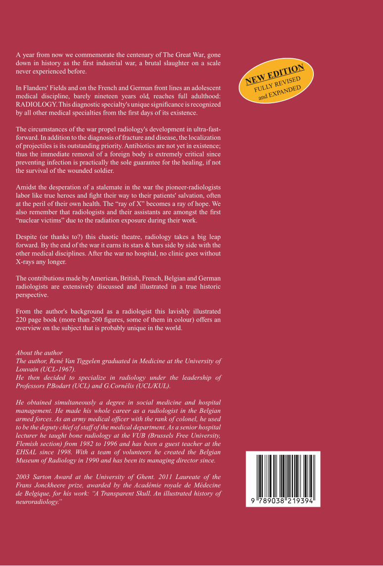
A year from now we commemorate the centenary of The Great War, gone down in history as the fi rst industrial war, a brutal slaughter on a scale never experienced before.
In Flanders' Fields and on the French and German front lines an adolescent medical discipline, barely nineteen years old, reaches full adulthood: RADIOLOGY. This diagnostic specialty's unique signifi cance is recognized by all other medical specialties from the fi rst days of its existence.
The circumstances of the war propel radiology's development in ultra-fast-forward. In addition to the diagnosis of fracture and disease, the localization of projectiles is its outstanding priority. Antibiotics are not yet in existence; thus the immediate removal of a foreign body is extremely critical since preventing infection is practically the sole guarantee for the healing, if not the survival of the wounded soldier.
Amidst the desperation of a stalemate in the war the pioneer-radiologists labor like true heroes and fi ght their way to their patients' salvation, often at the peril of their own health. The “ray of X” becomes a ray of hope. We also remember that radiologists and their assistants are amongst the fi rst “nuclear victims” due to the radiation exposure during their work.
Despite (or thanks to?) this chaotic theatre, radiology takes a big leap forward. By the end of the war it earns its stars & bars side by side with the other medical disciplines. After the war no hospital, no clinic goes without X-rays any longer.
The contributions made by American, British, French, Belgian and German radiologists are extensively discussed and illustrated in a true historic perspective.
From the author's background as a radiologist this lavishly illustrated 220 page book (more than 260 fi gures, some of them in colour) offers an overview on the subject that is probably unique in the world.
About the author The author, René Van Tiggelen graduated in Medicine at the University of Louvain (UCL-1967). He then decided to specialize in radiology under the leadership of Professors P.Bodart (UCL) and G.Cornélis (UCL/KUL).
He obtained simultaneously a degree in social medicine and hospital management. He made his whole career as a radiologist in the Belgian armed forces. As an army medical offi cer with the rank of colonel, he used to be the deputy chief of staff of the medical department. As a senior hospital lecturer he taught bone radiology at the VUB (Brussels Free University, Flemish section) from 1982 to 1996 and has been a guest teacher at the EHSAL since 1998. With a team of volunteers he created the Belgian Museum of Radiology in 1990 and has been its managing director since.
2003 Sarton Award at the University of Ghent. 2011 Laureate of the Frans Jonckheere prize, awarded by the Académie royale de Médecine de Belgique, for his work: “A Transparent Skull. An illustrated history of neuroradiology.”
RADIOLOGYIN A TRENCH COATMILITARY RADIOLOGY ON THE WESTERN FRONT
DURING THE GREAT WAR
René Van Tiggelen
Translated by Jan Dirckx
with contributions about the American, Belgian, British,French and German armies' radiological services
NEW EDITION
FULLY REVISED
and EXPANDED
RADIOLOGY IN A TRENCH COAT M
ILITA
RY RADIOL
OGY ON THE W
ESTERN F
RONT DU
RING THE GR
EAT WA
R
René Van Tiggelen - Translated by Jan Dirckx
cover RX met rug.indd 1 23/01/13 11:33