MILITARY PHARMACY AND MEDICINE
148
Volume V • No. 4 October – December • 2012 ISSN 1898-6498 MILITARY PHARMACY AND MEDICINE QUARTERLY INTERDISCIPLINARY JOURNAL • PHARMACY • MEDICINE • MEDICAL TECHNIQUE • ENVIRONMENT AND HEALTH • EDUCATION The Staff of the Military Center of Pharmacy and Medical Technology in Celestynow – Poland MILITARY PHARMACY AND MEDICINE • Volume V • No. 4 • 2012
-
Upload
khangminh22 -
Category
Documents
-
view
3 -
download
0
Transcript of MILITARY PHARMACY AND MEDICINE

Volume V • No. 4 October – December • 2012 ISSN 1898-6498
MILITARY PHARMACY AND MEDICINE
QUARTERLY INTERDISCIPLINARY JOURNAL
• PHARMACY
• MEDICINE
• MEDICAL TECHNIQUE
• ENVIRONMENT AND HEALTH
• EDUCATION
The Staff of the Military Center of Pharmacy and Medical Technologyin Celestynow – Poland
MIL
ITA
RY
PH
AR
MA
CY
AN
D M
ED
ICIN
E
• V
olum
e V
• N
o. 4
• 2
012

Druga
stro
na ok
ładki
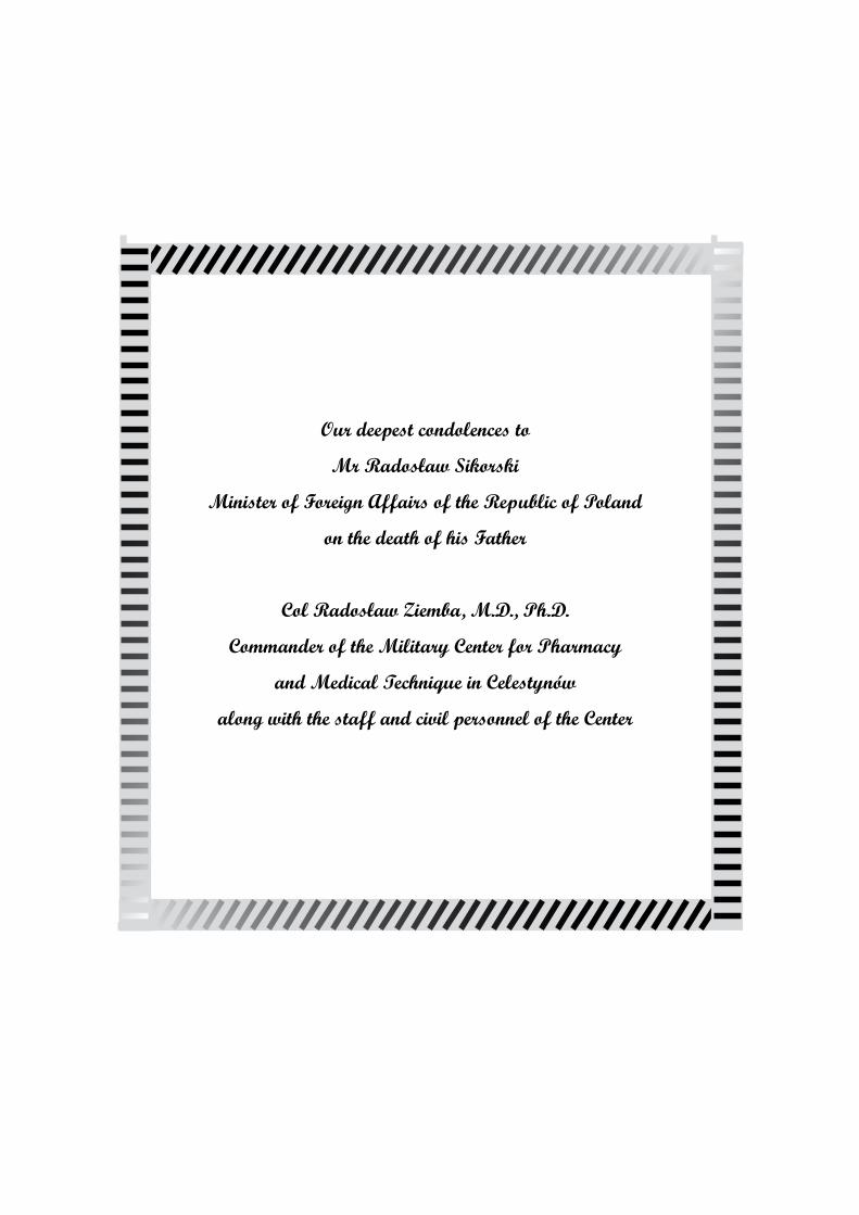
Our deepest condolences to
Mr Radosław Sikorski
Minister of Foreign Affairs of the Republic of Poland
on the death of his Father
Col Radosław Ziemba, M.D., Ph.D.
Commander of the Military Center for Pharmacy
and Medical Technique in Celestynów
along with the staff and civil personnel of the Center


MILITARY PHARMACYAND MEDICINE
Quarterly Interdisciplinary Journalof Military Centre of Pharmacy and Medical Technique in Celestynów
on scientific socio-professional and training issues of Military Pharmacy and Medicine
ISSN 1898-6498
October–December, 2012Volume 5, No 4

MILITARY PHARMACYAND MEDICINE
ii
SCIENTIFIC BOARD
Anyakora Chimezie, Nigeria
Balbaa Mahmoud, Egypt
prof. dr hab. Michał Bartoszcze, Poland
prof. dr hab. inż. Stanisław Bielecki, Poland
Bisleri Gianluigi, Italy
Blumenfeld Zeev, Israel
dr hab. Kazimiera H. Bodek, Poland
Boonstra Tjeerd W, Netherlands
Borcic Josipa, Croatia
Cappelli Gianni, Italy
Yifeng Chai, China
Chowdhury Pritish K, India
Costa Milton, Brasil
prof. dr hab. inż. Krzysztof Czupryński, Poland
Deckert Andreas, Germany
Demeter Pal, Hungary
prof. dr hab. Adam Dziki, Poland
Ermakova Irina, Russia
prof. dr hab. Zbigniew Fijałek, Poland
Florence Sudha Gnanou, India
Fontoura Paulo, Portugal
dr hab. Ryszard Gajdosz, Poland
Ning Gao, China
dr hab. Tomasz Gaszyński, Poland
prof. dr hab. Paweł Górski, Poland
prof. dr hab. Bożenna Gutkowska, Poland
Holko Ivan, Slovakia
Zhenlin Hu, China
Huang Feng, USA
dr hab. Czesław Jeśman, Poland
prof. dr hab. Wiesław Jędrzejczak, Poland
Kaubrys Gintaras, Lithuania
Kashanian Maryam, Iran
prof. dr hab. Andrzej Klimek, Poland
dr hab. Józef Knap, Poland
Korshunov Andrey, Russia
Kusec Sanja, Croatia
Shan Lei, China
dr hab. Julian Maj, Poland
prof. dr hab. Jerzy Mierzejewski, Poland
prof. dr hab. Elżbieta Mikiciuk-Olasik, Poland
Newcomb Andrew, Canada
prof. dr hab. Jerzy Z. Nowak, Poland
dr hab. Romuald Olszański, Poland
prof. dr hab. Daria Orszulak-Michalak, Poland
prof. dr hab. Krzysztof Owczarek, Poland
prof. dr hab. Marek Paradowski, Poland
Perotti Daniela, Italy
Pivac Nela, Croatia
Pizzuto Francesco, Italy
prof. dr hab. Janusz Pluta, Poland
Polat Gurbuz, Turkey
Popescu Irinel, Romania
Reddy G. Bhanuprakash, India
prof. dr hab. Juliusz Reiss, Poland
Rodrigues Gabriel Sunil, India
Rossi Roberto, Italy
Samarkos Michael, Greece
Shen Hui-Liang, China
Shevchuk Nikolai, Russia
Xianglin Shi, USA
Skultetyova Dana, Slovakia
Strumylaite Loreta, Lithuania
dr Piotr Siermontowski, Poland
prof. dr hab. Marek Sosnowski, Poland
prof. dr hab. Andrzej Stańczak, Poland
prof. dr hab. Zbigniew Lew-Starowicz, Poland
dr hab. inż. Marek Strzelczyk, Poland
Ding-Feng Su, China
dr hab. Janusz Świniarski, Poland
Tchetina Elena, Russia
Tomoum Hoda, Egypt
Tufekcioglu Omac, Turkey
prof. dr hab. Jarosław Wysocki, Poland
Wang FuZhou, China
Wei-dong Zhang, China
Zarkovic Neven, Croatia
Ruixin Zhu, China

MILITARY PHARMACY AND MEDICINE
iii
EDITORIAL BOARD
EDITOR-IN-CHIEFprof. Piotr Fiedor, Warsaw, Poland
DEPUTY EDITORprof. Jarosław Wysocki, Warsaw, Poland
SECTION EDITORS
Biochemistrydr hab. inż. Marek Strzelczyk, Poland
Bioethics & Medical Lawprof. dr hab. Hieronim Bartel, Poland
Biologyprof. Lidia Chomicz, Poland
Catastrophe MedicineAdam Pietrzak, Poland
Emergency Medicine dr hab. Tomasz Gaszyński, Poland
Epidemiologydr Witold Gnitecki, Poland
Forensic Medicinedr hab. Paweł Krajewski, Poland
Hematologyprof. dr hab. Wiesław Jędrzejczak, Poland
History of Medicine & Pharmacydr Zdzisław Jezierski, Poland
Infectious Diseasesdr hab. Józef Knap, Poland
Linguistic EditorMirosław Termar, USA
Maritime & Tropical Medicinedr hab. Romuald Olszański, Poland
Military Medicinedr Marek Skalski, Poland
Neurologyprof. dr hab. Andrzej Klimek, Poland
Neurosurgeryprof. dr hab. Jan Podgórski, Poland
OphthalmologyPiotr Michałowski, Poland
Orthopedics and Traumatologydr Wojciech Glinkowski, Poland
Patomorfologydr Piotr Siermontowski, Poland
Pharmaceuticlal Scienceprof. dr hab. Aleksander P. Mazurek, Poland
Pharmacology & Pharmacyprof. dr hab. Bożenna Gutkowska, Poland
Physiologyprof. dr hab. Józef Kędziora, Poland
Psychiatryprof. dr hab. Józef Kocur, Poland
Psychologyprof. dr hab. Krzysztof Owczarek, Poland
Sexologyprof. dr hab. Zbigniew Lew-Starowicz, Poland
Statistical Editordr Janusz Śmigielski, Poland
Stomatologydr Stanisław Żmuda, Poland
Surgeryprof. dr hab. Adam Dziki, Poland
Toxicologydr Wotold Kurnatowski, Poland
Urologyprof. dr hab. Eugeniusz Miękoś, Poland

MILITARY PHARMACYAND MEDICINE
iv
EDITORIAL OFFICESecretary of the Editorial OfficeKrzysztof Barczewski, PolandRemigiusz Radziszewski, Poland
Statistical Editordr Janusz Śmigielski, Poland
Technical EditorRemigiusz Radziszewski, Poland
English Language Professional ServiceMiroslaw Termar, USA
Public RelationsKrzysztof Barczewski, Poland
PUBLISHERMilitary Centre of Pharmacy and Medical Technique in CelestynówWojska Polskiego 5705-430 Celestynow, Polandphone +48 22 689 40 70, fax +48 22 689 40 91e-mail: http://military.isl-journals.com/
PUBLISHED BYInternational Scientific Literature, Inc361 Forest Lane,Smithtown, New York 11787, USAphone +1 516 874 4341e-mail: [email protected]
Interdisciplinary journal of Military Centre of Pharmacy and Medical Technique in Celestynów, Polandhttp://military.isl-journals.com/
© MILITARY PHARMACY AND MEDICINE. All rights reserved. No part of this publication may be reproduced, stored in retrieval system, or transmitted, in any form or
by any means, electronic, mechanical, photocopying, recording or otherwise without the prior written permission.
ISSN 1898-6498 quarterly Indexed in: MNiSW, Index Copernicus 130 copies
prof. dr hab. Hieronim Bartel, Poland
dr Przemysław Biliński, Poland
dr hab. Romana Bogusławska-Walecka, Poland
prof. dr hab. Andrzej Buczyński, Poland
prof. dr hab. Marian Brocki, Poland
Patrick Cadet, USA
dr hab. Andrzej Chciałowski, Poland
dr Wiesław Chudzik, Poland
dr Jan Czarnecki, Poland
dr Maria Dziedziczak-Buczyńska, Poland
prof. dr hab. Adam Dziki, Poland
prof. dr hab. Wojciech Gaszyński, Poland
dr hab. Czesław Jeśman, Poland
prof. dr hab. Józef Kędziora, Poland
prof. dr hab. Józef Kocur, Poland
dr Marek Kołodziejczyk, Poland
Richard Kream, USA
prof. dr hab. Krzysztof Kwiatkowski, Poland
dr hab. Julian Maj, Poland
Kirk Mantione, USA
prof. dr hab. Eugeniusz Miękoś, Poland
dr Dariusz Piotrowski, Poland
prof. dr hab. Jan Podgórski, Poland
dr hab. Wiesław Raszewski, Poland
dr Barbara Sadowska, Poland
George B. Stefano, USA
dr hab. Antoni Szymański, Poland
dr Zbigniew Teter, Poland
dr Wiesława Trendak, Poland
dr hab. Jadwiga Turło, Poland
dr Elżbieta Wojtasik, Poland
REVIEWERS

http://military.isl-journals.com/ v
© Military Pharmacy and Medicine • 2012 • 4
Table of Contents
Age-related macular degeneration (AMD): a critical appraisal of diet and dietary supplements as therapeutic modalities 1Jerzy Z. Nowak
Assessment of the energy expenditure of soldiers of the Representative Battalion of the Polish Army during three days of drill training as part of preparations for the celebration of the National Independence Day of November 11th 17
Jerzy Bertrandt, Anna Kłos, Roman Łakomy
Assessment of microbial quality of drinks not included in the hospital diet as consumed by patients during hospitalization and the assessment of microbial contamination of hospital air 21Jaśmina Żwirska, Wanda Jabłońska, Paweł Jagielski, Stanisław Stępniewski, Małgorzata Schlegel Zawadzka
Cholesterol oxyethyleneation products as modifiers of the absorption base in anti-inflammatory ointments 27Justyna Kołodziejska, Marian Mikołaj Zgoda, Katarzyna Ejzenchart, Magdalena Piechota-Urbańska
Non-steroidal anti-inflammatory drugs (NSAIDs) in ophthalmology: pharmacological and clinical characteristics 33Jerzy Z. Nowak
The role of paramedics in British emergency aid system 51Dorota Kołodziej, Radosław Ziemba
Energy expenditure as the basis for determination of nutritional demand in soldiers 57Jerzy Bertrandt, Anna Kłos, Bartosz Bertrandt
Sodium and potassium content of daily food rations of students of the Main School of Fire Service in Warsaw 61Anna Kłos, Maryla Długaszek, Jerzy Bertrandt , Wiesława Szymańska
Cardiac arrest under special circumstances. Part II: poisoning, …, anaphylactic reaction, …, traumatic injuries 67Radosław Ziemba
Prevalence of atrial fibrillation in emergency medicine practice 77Łukasz Szarpak, Dariusz Timler
Autopsy marks on anthropological material contributing to research on history of anatomy in Poland 81Magdalena Cybulska, Agnieszka Adamczyk, Agnieszka Młudzik, Czesław Jesman

vi http://military.isl-journals.com/
© Military Pharmacy and Medicine • 2012 • 4
Table of Contents
Infusion solutions supply in critical circumstances and disasters 87Katrzyna Parzuchowska, Radosław Ziemba, Jan Hołyński, Adam Ziemba, Jarosław Hołyński, Ewa Ziemba
Scope of knowledge regarding administration ofoxygen therapy among firefighter rescue teams from Volunteer Fire Departments (OSP) 91Łukasz Szarpak
Sustainable development in light of international cooperation 97Radosław Ziemba
Anthropological and archeological sources in medical historian’s studies 105Magdalena Cybulska, Agnieszka Adamczyk, Agnieszka Młudzik, Czesław Jesman
Selected aspects of communication between emergency services and the mass media in crisis situations 113Łukasz Szarpak, Marcin Madziała
Bioterrorism — nature of the problem 119Radosław Ziemba
Anti-drown ring — a patented drowning protection device (patent no. 197623) 123Jerzy Mierzejewski
Speech made by Commandant of the Military Centre for Pharmacy and Medical Technology in Celestynów, Colonel DMSc Radosław Ziemba 127Radosław Ziemba
Editorial policy and general information 133

© Military Pharmacy and Medicine • 2012 • 4 • 1 – 16
1http://military.isl-journals.com
Jerzy Z. Nowak: Age-related macular degeneration (AMD): a critical appraisal …
Ophthalmology
Age-related macular degeneration (AMD): a critical appraisal of diet and dietary supplements as therapeutic modalitiesJerzy Z. Nowak
Department of Pharmacology, Chair of Pharmacology and Clinical Pharmacology of Medical University of Lodz, Poland
Author’s address:Jerzy Z. Nowak, Department of Pharmacology, Medical University, ul. Żeligowskiego 7/9, 90-752 Lodz, Poland; phone: ( + 48) 42 6393270, e–mail: [email protected]
Received: 2012.10.12 • Accepted: 2012.11.22 • Published: 2012.12.08
Summary:
Age-related macular degeneration (AMD) is a vision-threatening ocular disease, affecting the central region of the retina — the macula — and manifesting in the elderly. Its pathogenesis is multifactorial, molecularly complex and only poorly recognized. AMD is a degenerative disease, and the degeneration affects primarily the retinal pigment epithelial (RPE) cells and secondarily the photoreceptors, which leads to disturbances or partial loss of central vision and legal blindness. Principal processes contributing to the development of the disease include: oxidative stress, lipofuscinogenesis, drusogenesis and local inflammation (so-called para-inflammation). A severe complication of clinically recognized dry form AMD (geographic atrophy) is the aggressive neovascularization originating from choriocapillary system (CNV; wet form AMD). At present, there are no therapeutic agents capable of slowing or inhibiting degeneration process in the photoreceptors-RPE complex, therefore preventive rather than therapeutic modalities are under consideration; they include properly adjusted everyday diet and intake of dietary supplements, both rich in antioxidants of which macu-lar pigments (lutein, zeaxanthin, meso-zeaxanthin) are of particular importance. Long-chain unsaturated omega-3 fatty acids (PUFA-ω3) are also recommended to people with already progressing AMD and at an increased risk of the disease development. Despite wide commercial offer of “ophthalmic” antioxidative and PUFA-ω3-rich preparations, no convincing evidence is available to date to support their protective activ-ity in AMD patients. AREDS-2 clinical trials that actually approach completion may likely provide more conclusive answer whether macular pigments (lutein, zeaxanthin) and PUFA-ω3 (EPA, DHA) can really be useful for AMD patients. The aim of this article is twofold: 1. it presents molecular mechanisms underlying the early stages of AMD pathogenesis, which form a platform for the disease development, and 2. it provides a critical appraisal of the prophylactic/therapeutic value of properly profiled diet and the intake of “ophthal-mic” dietary supplements rich in macular pigments and omega-3 PUFAs.
Key words: Age-related macular degeneration, AMD, oxidative stress, omega-3 fatty acids, macular pigments, prevention, diet, dietary supplements.
Introduction
This article refers to an earlier article by the same author, entitled Dry form AMD — do we know how to treat it?, published in 2011 in issue 5(1)
of Magazyn Lekarza Okulisty [1] — a Polish-language journal specialized in ophthalmology and widely distributed among ophthalmologists. There are no drugs specific to this condition; the

Review article© Military Pharmacy and Medicine • 2012 • 4 • 2 – 16
2 http://military.isl-journals.com
therapeutic approach focuses on prophylaxis in the form of properly profiled diet and the intake of dietary supplements. The number of these sup-plements is constantly growing, and all of them are freely available, i.e. without prescription. The available supplements differ in both qualitative and quantitative composition. A detailed analy-sis of supplements available at Polish market as described by the author in 2010 in an article entitled Ophthalmic antioxidant preparations: a survey and supportive arguments for their use in AMD [2], included 73 products — all of them contained the macular pigment — lutein, some contained also zeaxanthin, and only one con-tained three macular pigments, namely lutein, zeaxanthin and meso-zeaxanthin.
Currently nearly 100 ophthalmic antioxidant preparations are available only at Polish mar-ket. This does not include numerous prepara-tions containing polyunsaturated fatty acids of the omega-3 series (PUFA-ω3), discussed by the author in his earlier study entitled Omega-3 polyunsaturated fatty acids in retina and medical practice — pros and cons [3]. Ophthalmologists, as well as patients, may have and, according to author’s knowledge, indeed have problems with choosing an appropriate preparation. However, resorting to dietary supplement may be per-haps unnecessary if appropriate choice of food products is made for one’s everyday diet. The author hopes that this study, confronting the dietary and supplementation aspects of prophy-laxis will contribute to appreciation of the role of everyday diet rich in selected products in AMD prophylaxis and acknowledging the value of such diet as not inferior to that of pharmaceutical supplementation.
AMD – origin and development of the pathology
Age-related macular degeneration (AMD) devel-ops inconspicuously over many years. Despite numerous studies conducted worldwide, the pathogenesis of the disease is not fully under-stood. Due to the plurality of factors, both endog-enous (e.g. genetic predispositions) and exog-enous (history of exposure to light) or behavioral (e.g. smoking, improper diet), which might pre-dispose, or even contribute to the development of the disease, the critical physiological processes responsible for the vision process (e.g. the visual
excitation cascade) move beyond the limits of homeostasis, creating a biochemical platform for the future pathology [4–9]. This involves inten-sified lipofuscinogenesis in retinal pigment epi-thelium (RPE) cells, formation of drusen and pseudodrusen — the former occurring under the RPE monolayer (i.e. in the direction of Bruch’s membrane), the latter above the RPE monolayer (i.e. towards photoreceptors) and next, a chronic inflammatory process known as para-inflam-mation. All these phenomena contribute to the development of AMD. Clinically, AMD can be divided into the dry, or atrophic, form (known as geographic atrophy), and wet — exudative, or neovascular form; the latter can be considered a complication of the atrophic form manifesting as choroidal neovascularization (CNV) [1,4,5].
Aging favors the development of AMD, particu-larly in predisposed individuals (due to concomi-tant presence of the risk factors listed above); the term “age-related” in the disease name – AMD is fully justified, as the age is the major (albeit indi-vidual, i.e. specific to each subject) and unavoid-able determinant of various dysfunctions at the cellular and organ level, including these associ-ated with the deficiency of necessary microele-ments/nutrients or with the loss of function in the aging cells — particularly post-mitotic, i.e. non-regenerable cells. While the lacking micro-elements/nutrients may be supplied from the outside, thus attempting to compensate for their deficiency in particular cells/tissues, we are una-ble until now to stop the systemic aging process.
Our bodies contain many non-regenerable (post-mitotic) cells, particularly within the central nervous system. These include the photorecep-tors (only the external segments that contain photopigments are subject to regeneration) and retinal pigment epithelium (RPE) cells. In the pathogenesis of AMD, RPE cells are the first cells that become metabolically inefficient and thus undergo degeneration; dysfunction and atrophy of photoreceptors is secondary, as they are una-ble to function and survive without functionally efficient RPE cells, and therefore also undergo degeneration. The pathological process involves mostly a small region of the retina, known as the macula, where cone photoreceptors, respon-sible for acute and color vision, are predomi-nant. Therefore, first clinical symptoms of AMD include blurred vision, defects of central vision of

© Military Pharmacy and Medicine • 2012 • 4 • 3 – 16
3http://military.isl-journals.com
Jerzy Z. Nowak: Age-related macular degeneration (AMD): a critical appraisal …
various intensity or gradual loss of color vision, “warping” of perceived images [10,11]. Early form of AMD is often referred to as age-related macu-lopathy (ARM).
When the eyes are open, photoreceptors are continuously working, absorbing light photons and “recording” the image of the environment. Similarly to TV cameras being turned on, eyes record this image in an automatic fashion, gener-ating the first signal of a complex, multisynaptic vision process. Photoreceptors’ outer segments (POS), filled with visual pigment molecules, are characterized by significant functional dynam-ics; they wear off upon continuous function and, as a consequence, the apical fragments are con-stantly shed and captured by neighboring RPE cells. At the same time, POS is being rebuilt in order to maintain appropriate size (which is an important parameter determining the efficacy and survival of photoreceptors!). Regeneration proceeds from the perikaryon, i.e. the photore-ceptor inner segment (PIS) and requires numer-ous building blocks, including docosahexaenoic acid (DHA). These building blocks are supplied by the RPE cells and originate partly from the captured POS fragments and partly from circu-lation (consumed food) [9].
One of the many important roles played by the RPE cells is “digestion” of the absorbed (and con-tinuously being absorbed) photoreceptor mate-rial stored in phagolysosomes. Despite the fact that phagocytosis and enzymatic degradation occurring as a result of the activity of numerous lysosomal enzymes are physiological processes that had developed over thousands of years in creatures dwelling on Earth and making use of the visual organ system (retinal processes that govern visual perception in many vertebrates, including humans, are generally very similar), they seem to be of limited efficacy, at least in humans. This claim is supported by systematic accumulation of lipofuscin, known as the age pigment, in RPE cells.
Lipofuscin and the process of its formation, i.e. lipofuscinogenesis, are not the attributes of RPE cells and connections between photoreceptors and RPE, as they are also present in other non-renewable cells, such as neurons, cardiomyocytes or skeletal muscle cells. However, it is in this region of the eye, or more precisely, of the retina,
i.e. in the photoreceptors/RPE cells region, where a unique characteristics of lipofuscin accumu-lated in RPE cells becomes evident: lipofuscin contains retinoids (vitamin A derivatives) origi-nating from the visual cycle, particularly bis-retinoids — products of spontaneous fusion of two molecules of all-trans-retinal, generated via photoreaction (i.e. by absorption of photons) by 11-cis-retinal, a cofactor of the visual pigment.
The cis → trans retinal transformation is the cru-cial first step of visual cycle, initiating further conformational transformations of opsin (i.e. the visual pigment protein) into its active forms (e.g. meta-rhodopsin II), capable of progressing the visual cycle with the final effect consisting in the closure of cGMP-dependent cation chan-nel in the cellular membrane of photoreceptors and quenching the so-called darkness current. In this time, the cellular membrane of photore-ceptors is hyperpolarized only to regain the state of being ready to absorb another photon, i.e. the depolarization state; the active pigment, capable of absorbing photons, is a molecule, e.g. rho-dopsin, that contains a light sensitive co-factor, 11-cis-retinal [5,6].
The all-trans-retinal formed following photon absorption is completely dissociated from the vis-ual pigment and undergoes further physiological transformations in the retinoid cycle that takes place in both photoreceptors and RPE cells. How-ever, part of all-trans-retinal that does not bind the ABCA4 (ATP-binding cassette [transporter] A4 type, also known as ABCR) transporting the retinoid into the sites with all-trans-retinal dehydroghenase activity, “falls out” the cycle and spontaneously dimerizes (using ethanolamine as a “linker”) into a highly phototoxic bisretin-oid known as A2E. There may be more similar and toxic bisretinoids; however, A2E, which has been studied in more detail, seems to represent an established stress-inducing product. Furano – and peroxy –mtabolites of A2E have signifi-cant ability to activate the complement system (an alternative pathway) which represents the innate immunity, capable of efficiently and auto-matically acting in system’s defense, including destruction of “own” cells [5,12].
In addition, the photoreceptor cell membranes contain exceptionally high amounts of polyun-saturated fatty acids (PUFAs), particularly the

Review article© Military Pharmacy and Medicine • 2012 • 4 • 4 – 16
4 http://military.isl-journals.com
most-unsaturated docosahexaenoic acid (DHA). It is this high unsaturation (six double C=C bonds in the hydrocarbon chain) makes DHA particularly susceptible to spontaneous peroxi-dation and fragmentation into cytotoxic com-pounds, including 4-hydroxy-2-hexenal (HHE) or 4-hydroxy-7-oxyhept-5-enoic acid (HOHA); the latter may additionally form immunogenic carboxyethylpyrrole-protein (e.g. CEP-albu-min) adducts. More details regarding formation of the products of peroxidation of long-chain PUFAs and the properties of carboxyethylpyr-role-protein adducts are available in other arti-cles by the same author [2,5].
Accumulation of lipofuscin deposits in RPE cells is a manifestation of metabolic inefficacy of their lysosomal compartment, character-ized by reduced autophagy. The reason for this inefficacy remains unknown, although consid-ering molecular complexity of autophagic pro-cesses, the reasons may be multiple, including hypofunction or insufficient quantity/activity of lysosomal enzymes – cathepsins being most predominant in normal conditions. Lipofuscin deposits accumulate with age, and the adverse effect of accumulating oxidative stress that accompanies lipofuscinogenesis is intensified. Local inflammatory reaction that develops at certain moment and is manifested by an atypi-cal process referred to as para-inflammation, as well as drusogenesis (drusens, pseudodrusens), become the driving forces of the developing AMD pathology [4-7, 13].
One should not forget that the supply of oxygen (and microelements/nutrients) via the choriocap-illary system into the photoreceptors-RPE cells complex is one of the largest in primates. Tak-ing into account the functional specificity of the retina, particularly of photoreceptors (photosen-sitivity, extensive metabolism, high partial pres-sure of oxygen being the substrate for the forma-tion of oxygen radicals), one may suspect that retina is particularly well predisposed for forma-tion of reactive oxygen species (ROS) [14].
The nature must have predicted the potentially adverse, propathogenic processes, such as oxida-tive stress or lipofuscinogenesis, in the retina, as the tissue, and particularly the macular region, very important for acute and color vision in primates, has been equipped with an array of
antioxidative defense systems, including specific macular pigments (MPs) – lutein and zeaxanthin and meso-zeaxanthin produced from lutein [15].
It should also be mentioned that the classical antioxi-dative systems present within the body are classified as either enzymatic or non-enzymatic. Enzymatic systems include three basic enzymes: superoxide dys-mutase (SOD), catalase and glutathione peroxidase, which are dependent on metal ions, such as zinc, copper, manganium or selenium. The non-enzy-matic system consists of carotenoids (including the aforementioned macular pigments), vitamins E and C and glutathione. The enzymatic system is endog-enous; however, the metal ions that are required for its proper function are exogenous, i.e. must be sup-plied with food. As far as the non-enzymatic system is concerned, only glutathione is an endogenous anti-oxidant. The remaining elements of the system, i.e. carotenoids and vitamins E and C are exogenous fac-tors supplied with food or appropriate dietary supple-ments. The role of carotenoids is to neutralize singlet oxygen and reactive free radicals. What’s interesting, the mechanisms underlying these two activities of carotenoids are different and neutralization of free radicals may, in certain conditions (e.g. those when excessive amounts of radicals are present), change the antioxidative activity of these compounds into prooxidative activity.
Proper functioning of retinal antioxidation defense systems is believed to avert potential early propathogenic changes that may lead to AMD. These propathogenic changes are in fact physiological reactions that become function-ally (chemically) impaired due to their intensity and the accompanying overproduction of reac-tive oxygen species (ROS), crucial transforma-tions of “visual” retinoids and peroxidation and fragmentation of long-chain PUFAs. This may be generally due to indisposition of the aging body – its organs, tissues and cells, and hypofunction of the aforementioned antioxidative defense sys-tems, both enzymatic and non-enzymatic. Since these systems are dependent on the supply of exogenous nutrients, thus their activity is diet-dependent [2,14–17].
A diversified diet, containing vegetables rich in carotenoids, or — more precisely — oxycarotenoids or xanthophylls (mainly lutein and zeaxanthin; meso-zeaxanthin is not of dietary origin, being produced locally from lutein) — should be the

© Military Pharmacy and Medicine • 2012 • 4 • 5 – 16
5http://military.isl-journals.com
Jerzy Z. Nowak: Age-related macular degeneration (AMD): a critical appraisal …
first recommendation for patients in whome AMD development is suspected on the basis of gener-ally discrete symptoms, albeit disturbing visual sensations.
Diet
Specific dietary recommendations are crucial in many ailments and diseases. For instance, in diabetic patients, diet is an integral element of therapy, as no therapeutic success can be achieved in these patients without an appropri-ately profiled diet. Gluten-free diet is necessary in individuals, particularly children, with gluten intolerance. Patients with arterial hypertension should avoid salt, i.e. sodium chloride, in their diet. Excess sodium ions retain water in the body via a kidney-based mechanism; thus the daily dietary content of sodium should be well below 85 mmoles (less than 5 g NaCl) — in practice, one should follow the rule “the less salt, the better”. Many examples may be provided, but how about a diet for AMD patients?
Indeed, appropriate dietary recommendations for AMD patients are well justified, although one should expect no miraculous effects of such diets. Individuals who rigorously follow dietary recom-mendations (abundance of selected vegetables rich in lutein, marine fish rich in PUFA-ω3, etc.) also suffer from AMD. This is the nature of the disease, as it develops inconspicuously over many years and becomes symptomatic after the age of 50, most frequently after the age of 60. Its back-ground is multifactorial, including both environ-mental/genetic and inflammatory/immune fac-tors; in general opinion, there is no way to avoid it, as reported by the author in numerous stud-ies [1, 2, 4, 5, 18]. Nonetheless, appropriate diet is important in AMD patients.
Considering the threat of AMD or active AMD, one should recommend vegetables that con-tain high amounts of macular pigments (MPs), i.e. lutein and zeaxanthin [19]. Recommended vegetables include spinach, corn, pumpkin, red grapes (particularly seedless grapes), broccoli etc. (a more detailed list is presented in Table 1), as well as egg yolk which contains high amount of zeaxanthin in addition to lutein [15, 20]. It was calculated that the average Western diet content of lutein and zeaxanthin is between 1.3 and 3 mg, with lutein to zeaxanthin ratio of 7:1 [21, 22]. A
properly profiled diet may contain more macu-lar pigments, and therefore be more beneficial for individuals with AMD. Macular pigments are guards of metabolic order in the macular region and play a dual role of a filter, as they absorb blue light which is most dangerous for eyes and of a scavenger of free radicals, as they neutralize sin-glet oxygen and continuously generated free oxy-gen radicals.
Table 1: The content of lutein and zeaxanthin in widely used vegetables and fruits, given in mol %.
Products Lutein and zeaxanthin Lutein Zeaxanthin
Egg yolk 89 54 35Corn 86 60 25Kiwi fruit 54 54 0Red seed-less grapes 53 43 10
Pumpkin 49 49 0Spinach 47 47 0Cucumber 42 38 4Celery 34 3 2White grapes 31 25 7Brussels sprouts 29 27 2
Green peas 25 22 3Broccoli 22 22 0Mango 18 2 16Lettuce 15 15 0
Properly selected daily rations of selected veg-etables provide the body not only with macular pigments, but also with microelements (includ-ing zinc, copper, manganese, selenium, vitamins E and C) required for proper functioning of anti-oxidative enzymes (superoxide dysmutase, cata-lase, glutathione peroxidase).
One should not forget other important dietary ele-ments, i.e. unsaturated fatty acids of the omega-3 series, particularly DHA[1], abundantly present in marine fish[2]. Two or three fish-based meals per
[1] DHA is an example of a compound of dual nature, both beneficial and adverse. On one hand, DHA is required for the function of many cells, tissues and organs; on the other hand, due to its unsaturation, it is associated with the risk of adverse phenomena; at the same time, its functional indispensability is associated with structural unsaturation. The pros and cons of DHA were discussed in more detail in [3]. A disadvantage of DHA, as well as of other long-chain polyunsaturated fats, including the omega-3 eicosapentaenoic acid (EPA), omega-6 arachidonic acid (ARA or AA), or docosapentaenoic acid (DPA) is that in the presence of oxygen they easily undergo peroxidation and cleavage leading to propagation of oxidative stress. Despite the dual nature of the omega-3 fatty acids (DHA, EPA, DPA), one should not avoid their presence in food, particularly in the context of AMD prevention. The presence of vitamin E in food/supplement protects long chain polyunsaturated fats from oxidation.
[2] Marine fish contain high amounts of long-chain polyunsaturated fatty acids (PUFAs), including acids of the omega-3 series (PUFA-ω3). Of tested and commonly eaten fish

Review article© Military Pharmacy and Medicine • 2012 • 4 • 6 – 16
6 http://military.isl-journals.com
week (e.g. salmon, herring, mackerel) provide the body with these fatty acids in amounts sufficient not only for needs of the organs of vision, but also of the entire system.
Polyunsaturated fatty acids of the omega-6 series (PUFA-ω6), also essential for human body, are usually consumed in high amounts, as they are present in commonly used vegetable oils – e.g. linoleic acid (C18:2ω6 or C18:2n-6, which is the first acid in the omega-6 series) is abundant in grape seed oil and sunflower oil (63–66%), as well as in corn oil and soybean oil (55–56%); other oils have much lower contents of linoleic acid (rape oil, linen oil, olive oil — 10–21%). In case of PUFA-ω3, the case is more difficult, as EPA and DHA are virtually absent in vegetable oils, while alpha-linolenic acid (ALA; C18:3ω3 or C18:3n-3, being the first acid in the omega-3 series) is present only in moderate amounts in rape oil and soybean oil (7–11%), as well as in large amounts (!) in linen oil (55%)[3]. For the sake of comparison, the overall PUFA-ω3 (ALA, EPA and DHA) content in fish oil (herring oil) is > 28%, while the linoleic acid (ω6) content is slightly above 12%. Cold-pressed linen oil is available in an increasingly broad commercial offer. Although not everyone finds it tasty, linen oil is a recommended addition to the diet, not only of AMD patients. However, one should remember that conversion of ALA into EPA or DHA in human body is very low, and therefore the supply of ALA may not substitute for direct supply of DHA and DPA (which are abundant in marine fish).
species, highest amounts of DHA and EPA (expressed overall in g/100 g of weight) can be found in Atlantic salmon (> 2; farm-raised and wild caught) and herring (mainly Atlantic herring ≈ 2), then in mackerel and tuna (1.2–1.5); the popular canned tuna contains much smaller amounts (0,3–0,8); less DHA + EPA can be found in halibut and cod (0,2–0,6), while a higher content can be found in trout (farm-raised and wild caught) (≈ 1). As far as DHA is concerned, the highest content can be found in salmon, herring and tuna (excluding canned tuna in brine) and trout. Although fish contain particularly high amounts of EPA, DHA and DPA, other natural sources of these acids are human milk, farm-grown marine algae, marine mammals and krill; as mentioned in the article, some vegetable oils, including linen oil, do not contain EPA and DHA, although they may contain large amounts of alpha-linolenic acid (PUFA-ω3).
[3] The percentage composition of selected fatty acids in the linen oil is as follows: alpha-linonenic acid (18:3ω3) – 54.5%, oleic acid (18:1ω9) – 19.7%, linoleic acid (18:2ω6) – 16.2%, palmitic acid (16:0) – 5.1%, stearic acid (18:0) – 3.7%, other acids – 0,8%. The linen oil contains no EPA or DHA. The ω6/ω3 ratio is 0.3. For the sake of comparison, the percentage composition of fish oil (herring oil) is as follows: eicosapentaenoic acid (EPA; 20:5ω3) – 17.2%, palmitic acid (16:0) – 13.9%, palmitoleic acid (16:1ω7) – 13.1%, linoleic acid (18:2ω6) – 12.4%, oleic acid (cis-Δ9-octadecenoic acid; 18:1ω9) – 11.6%, docosahexaenoic acid (DHA; 22:6ω3) – 9%, myristic acid (tetradecanoic acid; 14:0) – 7.4%, stearic acid (octadecanoic acid; 18:0) – 2.7%, linolenic acid (18:3ω3) – 2.1%, elaidic acid (trans-Δ9-octadecenoic acid; 18:1ω9) – 2%, gadoleic acid (cis-Δ11-icosenoic acid; 20:1ω9) – 1.5%; ω6/ω3 ratio = 0.4.
Modern diet, particularly the Western diet, is rich in the fatty acids of the omega-6 series, and the ratio of these acids to the omega-3 (ω3) acids may be as high as 20:1, or even higher! The proper ratio should be about 4:1, with a trend towards balanced supply of both types of acids; this leads to the natural need for omega-3 acids supplemen-tation (EPA, DHA, ALA and docosapentaenoic acid — DPA-ω3); in case of AMD, DHA is of the highest importance. Despite its disadvantages (easy peroxidation and fragmentation), DHA is absolutely necessary for regeneration of pho-toreceptor outer segments worn off in the pro-cess of vision as well as to maintain appropriate plasticity/susceptibility of the cell membrane in rods and cones. In addition, EPA and DHA are substrates for production of anti-inflammatory resolvins and maresins (the latter are formed only from DHA) which are very important for the photoreceptors-RPE cells complex. DHA is also a substrate for production of neuroprotec-tin, which is involved in many protective, anti-inflammatory and cytoprotective mechanisms [8]. More information on the pros and cons of DHA may be found in a recent article by the same author [3].
The advantage of thus-profiled diet (as mentioned above) is that the elements valuable, among oth-ers, for intraocular metabolism, are delivered to the organism in natural, purely physiological fashion, which guarantees optimum gastrointes-tinal absorption and transport to target tissues/cells as long as a diversified and well-balanced diet is maintained. One should remember that these microelements, being so important not only for AMD patients, are absorbed into circu-lation from the gastrointestinal tract in a diverse manner, as they represent different types of chemical structures and molecular mass ranges. Well-balanced diet containing diverse proteins, carbohydrates and all types of fats (long – and short-chain, saturated and unsaturated) estab-lishes within the stomach and the intestines a natural chemical environment that favors pas-sive or active absorption of microelements sup-plied with food.
This natural, physiological situation is very much different from situation taking place in the stom-ach (oftentimes an empty one) after ingestion of dietary supplement tablets/capsules followed by a glass of water! Microelements contained in the

© Military Pharmacy and Medicine • 2012 • 4 • 7 – 16
7http://military.isl-journals.com
Jerzy Z. Nowak: Age-related macular degeneration (AMD): a critical appraisal …
supplements, albeit selected, have no optimum conditions for absorption. This accounts for the superiority of comprehensive natural nutrition over the intake of selected substances contained in dietary supplements, which do not have to be (and, in fact are not) fully absorbed from the gastrointestinal tract. A separate issue relates to the substances being delivered just where they are needed, which is a problem not less impor-tant from the standpoint of efficacy and expected results, and providently passed over in silence by the producers or suppliers of dietary supplements.
However, some individuals may, for various rea-sons, including the fact that preparation of appro-priate meal requires time and effort, while swal-lowing a capsule “solves the problem”, may prefer ready-made dietary supplements. Therefore, the dietary supplements must also be included in our considerations.
Dietary supplements and the efficacy of AMD prophylaxis/treatmentFrom the medical standpoint, the issue as stated in the heading is of primary importance for a wide group of mature population at risk of dis-comforts associated with the developing or potential AMD. As mentioned before, pathogen-esis of the disease is still unknown, which makes it impossible to both early diagnose the devel-oping pathology and to efficiently treat it using appropriate medications.
This does not pertain to the neovascular form of AMD, originating from choroidal neovascularization and considered by many researchers, including the author of this article, to be a serious complication of advanced AMD. This form, or more precisely, the dynamic neo-vascularization that accompanies AMD, may cur-rently be treated pharmacologically using agents that neutralize the main proangiogenic factor, i.e. the vascular endothelial growth factor (VEGF). Follow-ing agents are available: monoclonal anti-VEGF-A antibodies (Avastin – bevacizumab, Lucentis – ranibi-zumab), the recently registered soluble decoy receptor for the factors of the VEGF-A family, VEGF-B and placenta growth factor – PlGF (Eylea – aflibercept), as well as the less commonly at present used modified pegylated aptamer, an oligonucleotide strongly and selectively binding the VEGF-A165 protein, thus inhib-iting its activity (Macugen – pegaptanib sodium) – the
first registered drug for the treatment of neovascular AMD [23].
However, when starting the treatment of CNV, one should keep in mind, that:
1) the use of anti-VEGF medications will be efficacious only in VEGF-dependent neovascularization (luckily, in a large group of patients CNV is started from a VEGF-dependent process; however, a blockade, particularly a prolonged blockade of this angiogenic pathway may lead to spontaneous switch to another angiogenic pathway, which may depend on PDGF, FGF, CEP, or other factors; also possible is that a non-VEGF-dependent mechanism of neovascularization is activated first – in this case, the CNV process will be refractory to anti-VEGF medications); and
2) fighting neovascularization is a symptomatic treatment , as the AMD pathology continues to develop despite pharmacological inhibition of neovascularization and/or elimination of already formed pathological vessels using verteporfin-based photodynamic therapy (PDT).
How should one therefore manage AMD, par-ticularly the dry (atrophic) form of AMD? There are no appropriate drugs or reliable diagnostic methods for early stages of the disease. What remains is only physician’s intuition and knowl-edge, and prophylactic rather than therapeutic actions. Since the diet has been discussed above, let’s focus on dietary supplements.
The dietary supplements or, more precisely, “oph-thalmic” supplements, as their trade names often refer, either explicitly or implicitly, to the eye or the retina, include ophthalmic antioxidant prep-arations (OAPs) and preparations containing PUFA-ω3. Recently, a trend is observed to combine the active ingredients so that one capsule/tablet con-tains both macular pigments (lutein ± zeaxanthin), microelements, and PUFA-ω3. Thus, the capsules become ever bigger in size, making them hard to swallow without plenty of water.
This, however, is not as important as the qualita-tive and quantitative composition of the offered products/ingredients. This may be completely arbitrary, including specific substances (such as lutein and zeaxanthin, vitamins, metal salts) and less precisely defined ingredients such as plant extracts with presumed antioxidative

Review article© Military Pharmacy and Medicine • 2012 • 4 • 8 – 16
8 http://military.isl-journals.com
or cytoprotective properties or, as in the case of PUFA-ω3 – extracts from the livers of e.g. shark, cod or other fish, without any quantita-tive specification with regard to DHA, EPA and other fatty acids possibly included in the extract. Many producers seem to adhere to the motto: “the more, the better”, and therefore the dosage of the active ingredients is higher than in com-peting products: some products contain as much as 20 or more milligrams of lutein per capsule or, besides plant extracts, contain substances which have no chance of arriving at ocular tissues in untransformed, active form (e.g. the tripeptide glutathion) — the goal of all these endeavors is to differentiate the product from other products available at the market and thus promote the purchase.
However, the question whether the therapeutic efficacy of dietary supplements, including OAPs, is an established fact or wishful thinking remains unanswered. What complicates the problem are varied clinical data being published: according to some studies, macular pigments (lutein, zeax-anthin) may have effects protecting against the developing AMD [24–28], while other authors suggest that no such effects can be observed [29–33]. The former suggest that the use of OAPs in AMD (both in prevention and treatment) is justi-fied, while the latter would preclude such conclu-sion or suggest to think over the decision before pharmaceutical supplementation.
As mentioned earlier, macular pigments are chemical substances present in large quanti-ties in the retina, particularly in the macular region. Exogenous supply of these compounds (e.g. as part of the diet or as dietary supplements) is assumed to lead to the increase in their reti-nal levels/concentrations. Indeed, the intake of lutein-containing dietary supplements leads to an increase in both the serum lutein level and the macular pigment optical density (MPOD) – both parameters are increased in parallel [34]. The lat-ter value, i.e. the increased MPOD, is ascribed to be associated with the positive effect of supple-mentation on the improvement of vision; how-ever, there are still no clinical data that would justify this opinion!
Many researchers measure the MPOD – some-times in addition to clinical evaluation of vision in patients developing AMD, and sometimes as the only
measurement ever made. One should keep in mind that reliable MPOD readout is subject to certain limi-tations stemming from the measurement method, as endogenous lipofuscin also emits light, which might interfere with the measurement. For instance, lipofus-cin may be excited by light in the wavelength range of 400-580 nm to emit fluorescent light in the spectral range of 500-800 nm; on the other hand, the macular pigments absorb blue light at wavelengths shorter than 550 nm, with the absorbance peak at 460 nm [35]. In order for the obtained MPOD readout to be reliable, i.e. free of interferences from (auto)fluorescence of lipofuscin, a special methodological approach should be made, such as the use of dual wavelengths to excite the macular pigments, e.g. 488 nm (well absorbed by MPs) and 514 (minimally absorbed by the MPs) [33], and conclusions should be drawn from the differences in measured values. Another factor very important for interpretation of the MPOD results, particularly in AMD patients, is that macular pigments are usu-ally characterized by very slow turnover rates. This means, that in patients taking dietary supplements increased levels of macular pigments in serum and macula (MPOD) would persist for long periods after the dietary supplements, e.g. lutein-rich supplements, are discontinued. Sometimes, such periods may last several months or longer.
In the context of increasing popularity of PUFA-ω3 dietary supplements being recommended to AMD patients, Delyfer et al. [37] studied the potential rela-tionship between MPOD and serum PUFA-ω3 levels in 107 healthy volunteers (PIMAVOSA study). The authors confirmed the correlation between MPOD and serum lutein/zeaxanthin, and additionally iden-tified high correlation between MPOD and serum PUFA-ω3 (overall levels). When considering the main 3 acids of the omega-3 series separately, observations regarding correlations between the acid levels and MPOD were as follows: DPA — high correlation, EPA – intermediate correlation, DHA – no statistically sig-nificant correlation. Thus, the French authors pointed out another factor that might affect the MPOD value, i.e. the PUFA-ω3, levels/concentrations of which (e.g. in serum) increase after the intake of meals or dietary supplements rich in these acids.
Among recently published data on macular pigments supplementation and its influence on quality of vision in AMD patients, the author would like to take as an example one article, which appeared in PubMed database on August 1, 2012, and was published only in November

© Military Pharmacy and Medicine • 2012 • 4 • 9 – 16
9http://military.isl-journals.com
Jerzy Z. Nowak: Age-related macular degeneration (AMD): a critical appraisal …
issue of Ophthalmology [38] — it presents data on the effect of supplementation with lutein (10 and 20 mg per day) and zeaxanthin (10 mg per day) for 48 weeks on MPOD and best corrected visual acuity (BCVA) in 108 patients with early AMD. The results are similar to those pub-lished earlier by other authors, and therefore it is worth to present the conclusions drawn from these results (as the conclusions may as well pertain to the earlier works): „Among patients with early AMD, supplementation with lutein and zeaxanthin improved macular pigment, which played a causative role in boosting visual function and might prevent [highlighted by the author] the progression of AMD. Future studies are required to evaluate the effect of these carot-enoids on the incidence of late AMD”. In other words, lutein and zeaxanthin, as expected, increased the value of MPOD (i.e. increased the pigment levels), while their effect on vision was comparatively small, and the preventive effect with regard to AMD was debatable.
Recent Cochrane review, based on randomized, controlled studies in more than 62,500 subjects also reports the lack of effects of some widely recommended antioxidant supplements [39]. The author’s conclusions are as follows: “There is accumulating evidence that taking vitamin E or beta-carotene supplements will not prevent or delay the onset of AMD. There is no evidence with respect to other antioxidant supplements, such as vitamin C, lutein and zeaxanthin, or any of the commonly marketed multivitamin combinations. Although generally regarded as safe, vitamin supplements may have harmful effects and clear evidence of benefit is needed before they can be recommended.”
Ophthalmologists are divided in their practical observations in AMD patients receiving OAPs. Many of them ask the well-justified question: Can OAPs cure AMD, delay its progression, or better yet, prevent its development? Considering the marketing activity of OAP manufacturers, as well as the ever-increasing number of commer-cially available “antioxidant” preparations with varied qualitative and quantitative compositions, one may have serious doubts regarding the actual therapeutic value of OAPs. The most impor-tant factors affecting the “aye” or “nay” decision for ophthalmic antioxidant preparations and PUFA-ω3 preparations are presented below.
Dietary supplements vs. drugsContrary to drugs, i.e. registered medications with precise and constant composition of active substance(s) and excipient(s) and documented therapeutic potential, dietary supplements may have variable compositions and information leaflets, if included, are often laconic or lacking detail. Self-respectful manufacturers present composition, and even intended use/indications for their products, sometimes referring to pub-lished results of research suggesting potentially beneficial effects of the components of these components. And this is pretty much all. Conclu-sion: There is no equals sign between a dietary sup-plement and drug, although many manufacturers would like their products being treated as drugs.
Dietary supplements are sold freely, and there-fore everyone may purchase them and use them, either recommended by the doctor or at one’s own discretion, oftentimes prompted by loud, albeit imprecise and sometimes groundless information/messages regarding the efficacy of these supplements presented by the mass media. Dietary supplements must not contain ingredi-ents which, when used in excess (i.e. in a reason-able excess, as nearly everything consumed in appropriate excess might have detrimental effects on health) might have any adverse effects. Die-tary supplements should therefore be absolutely safe, as manufacturers want to avoid medical problems resulting from the intake of their prod-uct and potentially leading to elimination of such product from the market.
Many physicians, while recommending dietary supplements, including OAPs, to their patients, do not fully believe in therapeutic effects thereof. Some even say that such preparations do no harm while they might actually help (!) etc., so that patients may take them safely hoping they would really help. However, the despaired patients expect the purchased product(s) to have ben-eficial effects — they trust their physicians and believe, or try to believe, that the product will be efficacious. Patients perceive the paeans pre-sented by the mass media to be reliable informa-tion, not marketing tricks, which, unfortunately, is not always true.
Let’s assume that a patient purchased a packaging with a month’s supply of the dietary supplement

Review article© Military Pharmacy and Medicine • 2012 • 4 • 10 – 16
10 http://military.isl-journals.com
recommended to him/her by his/her physician and that he/she started the treatment. How long should the patient take the recommended prod-uct? And, what’s more important, when should the patient expect to experience an improvement in vision? There are no satisfactory answers to these well justified (as the patient does not get the products for free — he/she buys them) questions, as the beneficial effect will surely not be observed after the first month, or even after the first sev-eral months of using the dietary supplement(s); in some patients, the effect may be observed after a long-term use that lasts many months, and even years. What’s more, there may be no effect at all!
Patients may feel disappointed with such “supple-mentation therapy”, while the manufacturer of the supplement holds no responsibility whatso-ever, as they may always bring up what is already well known, i.e. that the product is a dietary sup-plement, and not a drug! A question/statement often raised in such cases by the supporters of “supplementation therapy”, i.e. what if the sup-plements had not been taken, is purely rhetorical one. Indeed, there is no answer to this question.
Patients disappointed with their hitherto ineffec-tive therapy, i.e. patients with conditions refractory to the established pharmaceuticals or patients with conditions with no pharmacological treatment available are particularly susceptible to the use of dietary supplements. Cases where no specific drugs are available — as in the case of AMD — favors all kinds of therapeutic speculations and para-medical activities, opening a lucrative area for the manufacturers and suppliers of dietary supple-ments — appropriately promoted dietary supple-ments may even be perceived as drugs!
The abundant offer of dietary supplements avail-able at Polish market and targeted at individuals suffering from AMD or at risk of AMD includes nearly 100 products of the OPA type. In a recent study, published in 2010 [2], the author analyzed 73 ophthalmic antioxidant preparations, i.e. all products commercially available in Poland at that time. The call sign of all OAPs is the presence of the macular pigment–lutein; some products con-tain also another macular pigment, i.e. zeaxan-thin (e.g., in alphabetical order, Klarin Perfekt, MaxiVision Total, Nutrof Total, Ocuvite Lutein Forte, Vislea), and one product (Macushield) contains three pigments–lutein, zeaxanthin and
meso-zeaxanthin in one capsule. The analysis led to the following conclusions:
• All 73 preparations contained lutein in the amounts of 0.125/6–10/50 mg (the range of 6–10 mg was most common), with 7 prepara-tions not stating the precise dose;
• 30 preparations contained also zeaxanthin in the amounts of 0.12–2.4 mg, with no data regarding the dose provided for 3 preparations;
• 1 preparation contained meso-zeaxanthin in addition to the two above pigments;
• 52 products contained vitamin E; • 42 products contained zinc and/or selenium; • 9 products contained PUFA-ω3; • 3 products contained glutathione • 39 products contained vitamin A or β-carotene.
As mentioned above, patients may currently choose from about 100 preparations; however, let us restrict our considerations to the 73 prepara-tions that were analyzed in detail by the author. When purchasing a product, the patient must make a choice, and thus, what factors should the patient be guided by when making this choice? Using the simplest exclusion criteria, such as presence of vitamin A or β-carotene, symbolic dose (< 1 mg) of lutein, lack of details regarding the source/quantity of macular pigments, or the presence of ineffective compounds, such as glu-tathione, at most 20 out of the total of 73 prepara-tions may be selected. When applying additional criteria (e.g. the seniority and importance of the manufacturer in the drug and/or dietary supple-ment market), the number of products that could be recommended to patients would not exceed 10. However, for an individual patient who would like to purchase an inexpensive but “good” prod-uct, 10 products is simply 9 too many!
The trend to enrich the OAP-type supplements with PUFA-ω3 has already been mentioned. Manufacturers of such “combination” supple-ments argue that they provide AMD patients with everything they need in a single capsule! Another, more convincing arguments (com-pared to the “everything you need in one” mes-sage, as mentioned above) are arriving from the nearly completed Age-Related Eye Disease Study 2 (AREDS-2), which, contrary to the AREDS-1 (or simply AREDS) study, test the use of macular

© Military Pharmacy and Medicine • 2012 • 4 • 11 – 16
11http://military.isl-journals.com
Jerzy Z. Nowak: Age-related macular degeneration (AMD): a critical appraisal …
pigments and PUFA-ω3 in AMD patients. It should be mentioned that the AREDS study is a multicenter, 5-year clinical study conducted in more than 3,000 patients. The objective of the study was to assess the effect of high doses of antioxidants (these included vitamin C, vitamin E and β-carotene) and zinc on the progression of AMD. As shown by the published reports, the tested AREDS formula did not inhibit vision loss, although it had some beneficial effects reducing the risk of further development of AMD. Authors of the promising article titled “New approaches and potential treatments for dry age-related mac-ular degeneration”, published in 2012, make ref-erence to the AREDS study reports of 2001 and the Erratum of 2008 — report as follows: “the AREDS formula does not prevent GA [geographic atrophy] from forming or progressing” [40].
AREDS-2 (ClinicalTrials.gov; Identifier NTC 00345176; sponsored by National Eye Institute (NEI) with collaboration from National Heart, Lung, and Blood Institute – NHLBI), is, similarly to AREDS, a 5-to-6-year (2007–December 2012), multicenter (82 clinical centers in the United States), randomized clinical study to evaluate the effect of oral supplementation with macular xan-thophylls (lutein, zeaxanthin) and omega-3 poly-unsaturated acids (PUFA-ω3) (DHA, EPA) on AMD progression. Below is the list of agents tested in AREDS (AREDS-1) and AREDS-2 studies:
AREDS-1: vitamin C — 500 mg, vitamin E — 400 IU, beta-carotene — 15 mg, zinc (as zinc oxide) — 25 and 80 mg, copper (as cupric oxide) — 2 mg;AREDS-2: lutein — 10 mg, zeaxanthin — 2 mg, vitamin C — 500 mg, vitamin E — 400 IU, copper (as cupric oxide) — 2 mg, EPA – 650 mg, DHA – 350 mg.
Smaller studies (Secondary Randomization Agents — AREDS-Type Supplement) were also conducted to examine the effects of zinc (as zinc oxide) at doses of 25 and 80 mg, with par-ticular focus on the lower dose, and elimina-tion of β-carotene form the AREDS formula.
Of note is the lack of beta-carotene and zinc in the Primary Randomization Agents of AREDS-2 study. As shown by the alpha-tocopherol, beta-carotene cancer prevention study (ATBC), beta-carotene contributes to the development of lung cancer in smokers [41]; in addition, beta-carotene is a precursor of vitamin A, and the visual cycle
retinoids may act as substrates for formation of photocytotoxic bis-retinoids. Beneficial effects of zinc have been known for a long time, and there was no need for another detailed verifica-tion of its advantages. However, a question arose with regard to the dose — it would be better if the lower dose (25, not 80 mg) was efficient. As far as the PUFA-ω3 doses used in AREDS-2 study are concerned, the author agrees with the total dose of 1 g, although he is not certain whether 650 mg of EPA and 350 of DHA is a good dose ratio (1.86) for AMD patients. Considering the role of DHA in the photoreceptors–RPE cells complex [3], one might expect a higher dose of DHA (possibly at the cost of a lower amount of EPA).
Combination preparations (“all in one”: macu-lar pigments, microelements, PUFA-ω3) may be convenient for patients; however, the manufac-turers are well aware that the presence of reason-able amounts of PUFA-ω3 in capsules requires enlarged capsule size, which is not always associ-ated with greater convenience of use. One gram of PUFA-ω3 (as used in AREDS-2) takes up a considerable volume, thus a “combination” cap-sule containing such an amount should be appro-priately large. When trying to adhere to the trend of administering one capsule of a dietary supple-ment per day, the capsule should contain “daily” doses of all ingredients which would obviously be translated into capsule size.
However, there is one argument that does not support combining PUFA-ω3 with the remain-ing ingredients in preparations recommended to AMD patients. AMD patients are usually at an advanced age, and PUFA-ω3 are often recom-mended to them also by physicians of other (non-ophthalmological) specializations. For example, cardiologists and psychiatrists (as well as other physicians, although these two specializations are predominant) nearly routinely recommend PUFA-ω3-rich dietary supplements at daily doses that often exceed 1 g. Facing different trade names of various “cardiologic”, “psychiatric” and “oph-thalmic” dietary supplements, patients may be unaware that they actually take the same chemical entities! And all these entities are accumulated in the same system! Therefore, it might happen that a patient would take not 1, but 2, and perhaps 3 or more grams of PUFA-ω3 per day while being unaware of this fact! Taking such scenario into consideration, one should not forget preparations

Review article© Military Pharmacy and Medicine • 2012 • 4 • 12 – 16
12 http://military.isl-journals.com
which contain antioxidants and microelements separately from PUFA-ω3, and which could be taken by the patient in line with the overall picture of his ailments and treatment.
It should be mentioned that many national and worldwide medical organizations (including the WHO) recommend daily intake of EPA and DHA in total amounts of 400–600 mg/day, while cer-tain guidelines mention the dose of 1 g/day, and the European Food Safety Authority (EFSA) holds the position that there is no scientifically sup-ported recommended daily dose of PUFA-ω3 for humans. However, EFSA recommends the DHA dose of 100 mg/day in infants, on the premise that DHA contributes to the development and func-tional maturation of vision system; EFSA also rec-ommends a daily dose of ca. 250 mg (EPA + DHA) to children and adolescents aged 2-18 years[4].
On the other hand, a statement published in 2010 by the French Safe Food Agency (AFSSA, currently MSNA — National Agency for Drugs and Health Products) recommends a daily dose of ca. 500 mg of EPA and DHA combined as a preventive measure in AMD patients. As seen from the presented data, the recommended daily doses of PUFA-ω3 are diverse, cover a wide range of 100–1000 mg and depend on the patient’s age and health status.
What’s interesting, in case of cardiovascular condi-tions[5], the recommended dosage of omega-3 fatty
[4] By request of the European Commission, the EFSA Panel on Dietetic Products, Nutrition and Allergies (NDA) performed an analysis and issued a scientific opinion regarding acceptable maximum daily intake of long-chain PUFA-ω3, i.e. EPA, DHA and DPA in humans. A study entitled “Scientific opinion on the tolerable upper intake level of eicosapentaenoic acid (EPA), docosahexaenoic acid DHA) and docosapentaenoic acid (DPA)” was published in the recent issue of EFSA Journal [2012; 10(7): 2815]. In final conclusions, one reads: “The Panel concludes that the available data are not sufficient to establish a tolerable upper intake level for n-3 LCPUFA (DHA, EPA, and DPA, individually or combined) for any population group. The Panel considers that supplemental intakes of EPA and DHA combined at doses up to 5 g/day, and supplemental intakes of EPA alone up to 1.8 g/day, do not raise safety concerns for the adult population. The Panel also considers supplemental intakes of DHA alone up to 1 g/day do not raise safety concerns for the general population. No data are available for DPA when consumed alone. The Panel notes that in the majority of the human studies considered, fish oils, which also contained DPA in generally unknown (but relatively low) amounts, were the source of EPA and DHA.”
[5] According to the European and American Heart Associations (EHA, AHA), all patients with a history of myocardial infarction and patients with stable coronary disease should take PUFA-ω3 at the dose of 1 g per day. According to AHA, supplementation using omega-3 acids should not be associated with rigorous elimination of omega-6 acids (e.g. linoleic acid, C18:2-ω6, which accounts for 85-90% of dietary omega-6 acids) which cannot be synthesized by human body and which, according to numerous opinion-forming sources, including the WHO, should account for 3-10% of daily energy demand. An interesting preparation that meets the AHA recommendations (although its omega-3 acid content is relatively small) is the “EYE Q capsules” preparation by a Swiss company Vifor SA distributed in Poland by Qpharma; the product contains EPA and DHA (marine fish oil) and primrose oil (omega-6).
acids (EPA + DHA + DPA) may be two-, three-, or even four times higher than the usually recom-mended maximum doses. When taken to reduce the triglyceride levels in hypertriglyceridemia, the recommended dose of PUFA-ω3 (with or without statin or fibrate) may be up to 3-4 g per day.
Regardless whether macular pigments and PUFA-ω3 are supplied together or as separate preparations, below are the daily dose ranges (to be contained in one capsule taken once daily or in two capsules taken twice a day) of the major ingredients of “ophthalmic” dietary supplements which, according to the author of this article, should be beneficial for AMD patients:
• Lutein — 10–12 mg • Zeaxanthin — 1–2 mg • Vitamin E — 30–60 mg • Vitamin C — 60–250 mg • Zinc — 10–20 mg • PUFA-ω3 — 500–800 mg (overall, with DHA ≥ EPA)
Optionally: meso-zeaxanthin (up to 10 mg), copper, selenium, manganese.
The doses or dose ranges listed above are rela-tive and refer to both the compositions of prod-ucts available at Polish market and the experi-mental and clinical data published on the topic. Products containing PUFA-ω3 should necessar-ily contain vitamin E. The author’s opinion is that there is no need to expand the composition of an ophthalmic preparation by ingredients, e.g. vitamins, other than these listed above. The addition of meso-zeaxanthin, a macular pig-ment characterized by potency equal to that of zeaxanthin, may be beneficial for patients. A comment should also be made with regard to the content of lycopene, which is not discussed in this article. Although lycopene is not a mac-ular pigment, it is worth to remember its role, as it is involved in repair of the three oxycarot-enoids (lutein, zeaxanthin, meso-zeaxanthin) after they react with free radicals. Lycopene is a carotene featuring 11 conjugated double bonds, abundantly present in tomatoes.[6]
With regard to the PUFA-ω3 content and DHA to EPA ratio in supplement preparations, the
[6] For more information on lycopene in patients with AMD see Nowak, Wiktorowska-Owczarek, “AMD, Stargardt’s disease, and carotenoids as components of ophthalmic antioxidant preparations used in AMD” (Mag Lek Okul. 2012; 6(4): 185-98).

© Military Pharmacy and Medicine • 2012 • 4 • 13 – 16
13http://military.isl-journals.com
Jerzy Z. Nowak: Age-related macular degeneration (AMD): a critical appraisal …
author is aware of four products available in Poland, in which the DHA dose is larger than that of EPA. This makes the products worth men-tioning — they include:
• Ocuvite Reti-NAT forte (Bausch&Lomb); 1 capsule contains: 900 mg of fish oil, including 540 mg PUFA-ω3 – 420 mg DHA and 120 mg EPA — ratio 3.5; 1 capsule per day (the product has been withdrawn from the market by the manufacturer; perhaps some physicians still retained some supplies);
• Omega 3 (Ophtagen); 1 capsule contains: 500 mg of fish oil, including 250 mg DHA and 85 mg EPA (ratio 2.94), 1-3 capsules per day;
• MaxiVision Total (ASA); 1 capsule contains: 500 mg of fish oil, including 250 mg DHA and 34.6 mg EPA (ratio 7.22), 1 capsule per day; and
• Ocuvite Complete (Bausch&Lomb); 1 capsule contains (according to information on the packaging): 421.5 mg of fish oil, including 175 mg DHA (no information on the content of EPA), 2 capsules per day.[7]
While the two first preparations contain fish oil and no macular pigments (i.e. they are focused on PUFA-ω3), compositions of the two remain-ing products include also macular pigments, vitamins and minerals. MaxiVision Total — 20% lutein extract (corresponding to 20 mg of lutein), 1 mg of zeaxanthin, vitamins E and C, zinc and selenium; Ocuvite Complete — lutein 5 mg, zeax-anthin 1 mg, vitamins E and C, and zinc.
Ocuvite Reti-NAT forte contains definitely the largest amount of DHA (420 mg compared to 250 mg in MaxiVision and Omega-3 and ca. 175 mg in Ocuvite Complete), while the DHA to EPA ratio in individual supplements is as follows: 7.22 (MaxiVi-sion), 7.19 (Ocuvite Complete), 3.50 (Ocuvite Reti-Nat) and 2.94 (Omega-3). The author is curious with regard to the origin of the fish oil in MaxiVision
[7] Ocuvite Complete has only recently been introduced to the Polish market of dietary supplements and probably replaced similar products offered by Bausch&Lomb before its introduction. The product does not feature an accompanying leaflet, and relevant data on its composition are provided on the packaging. According to the information available for the user, the composition of the supplement is as follows (the presented values refer to two capsules recommended as a daily dose): fish oil 843 mg, including 350 mg DHA, vitamin C 180 mg, vitamin E 30 mg, zinc (as zinc sulfate) 15 mg, lutein 10 mg, zeaxanthin 2 mg (macular pigments originate from Tagetes erecta flower extract); no EPA content is provided. However, the Polish supplier of the product has made available more detailed data on the content of PUFA-ω3. According to these data, 1 capsule of Ocuvite Complete contains 421.71 mg of fish oil, including: 181.5 mg of DHA-TG (corresponding to 174.4 mg DHA) and 25.3 mg EPA-TG (24.3 mg EPA); thus, the DHA to EPA ratio is 7.19.
Total and Ocuvite Complete products, character-ized by such high DHA to EPA ratios — no relevant information is provided for Ocuvite Complete, while the information leaflet to MaxiVision Total, which also does not provide this information, men-tions that „The unsaturated omega-3 fatty acids (DHA, EPA) in MaxiVision Total are derived from a high grade fish oil…”, which is perhaps imprecise, but nonetheless reassuring.
As mentioned before in case of properly profiled diet, also pharmaceutical supplementation may, but does not have to help the AMD patients. The potential helpful effects might be suggested by the recently published experimental and clinical studies, which were quite numerous in recent years [24–28, 38]. Also the results of the AREDS-2 studies, which should be published starting from 2013[8], should be — or so it seems — suggestive in this matter.
While browsing the published data, there is no missing the fact that results presented in most studies are not definitely convincing (at least, not to the author of the article), although they some-times meet the threshold of statistical signifi-cance. More detailed analysis of these data leads to conclusion that further studies and clinical observations are required to assess the clinical efficacy of dietary supplements in general, and ophthalmic preparations in particular. However, one should not cease being optimistic; optimism should be cherished by both the physicians when recommending appropriate dietary supplements to patients, and patients who would be regularly taking it; otherwise, the supplementation would serve no purpose whatsoever.
As a concluding remark, it should be stated that when deciding to use dietary supplements, AMD patients should nonetheless remember to adhere to proper diet, including, in addition to selected fruits and vegetables, at least one (or better yet, two) high-fat marine fish meals per week.
Acknowledgments:
Article funded by Medical University of Lodz within the statutory activity (503/1-023-01/503-01).
[8] On 26 July, 2012, the first report from the AREDS-2 studies, titled: „The age-related eye disease study 2 (AREDS2): study design and baseline characteristics (AREDS2 Report number 1)” – Ophthalmology 2012 Nov; 119(11): 2282-9, presenting the study design and basic characteristics of the project was published as an e-pub in PubMed database.

Review article© Military Pharmacy and Medicine • 2012 • 4 • 14 – 16
14 http://military.isl-journals.com
References:1. Nowak JZ: [Dry form AMD – do we know how to
treat it?]. Mag Lek Okul. 2011; 5: 44–8.2. Nowak JZ: [Ophthalmic antioxidant
preparations: a survey and supportive arguments for their use in AMD]. Mag Lek Okul. 2010; 4: 185–98.
3. Nowak JZ: [Omega-3 polyunsaturated fatty acids in retina and medical practice – pros and cons]. Mag Lek Okul. 2009; 3: 208–20.
4. Nowak JZ: Age-related macular degeneration (AMD): pathogenesis and therapy. Pharmacol Rep. 2006; 58: 353–63.
5. Nowak JZ: [In search of biomarkers for age-related macular degeneration (AMD)]. Mag Lek Okul. 2009; 3: 132–43.
6. Nowak JZ: [Role of lipofuscin in pathogenesis of age-related macular degeneration (AMD)]. Magazyn Okulistyczny. 2005, 2: 103–14.
7. Nowak JZ: [Drusens, basal deposits, inflam-mation and age-related macular degeneration (AMD)]. Magazyn Okulistyczny. 2005; 2: 174–86.
8. Nowak JZ: [Inflammation: acute, chronic and para-inflammation – new ideas, new drugs]. Mag Lek Okul. 2011; 5: 194–211.
9. Bhutto I, Lutty G: Understanding age-related macular degeneration (AMD): relationships between the photoreceptor/retinal pigment epithelium/Bruch’s membrane/choriocapillaris complex. Mol Aspects Med. (2012); 33: 295-317.
10. Fine SL, Berger JW, Maguire MG, Ho AC: Age-related macular degeneration. N Engl J Med. 2000; 342: 483–92.
11. McConnell V, Silvestri G: Age-related macular degeneration. Ulster Med J. 2005; 74: 82–92.
12. Klaska I, Nowak JZ: [The role of complement in physiology and pathology]. Post Hig Med Dosw (Online). 2007; 61: 167–77.
13. Anderson DH, Radeke MJ, Gallo NB, Chapin EA, Johnson PT, Curletti CR et al.: The pivotal role of the complement system in aging and age-related macular degeneration: hypothesis re-visited. Prog Retin Eye Res. 2010; 29: 95–112.
14. Wiktorowska-Owczarek A, Nowak JZ: [AMD and oxidative stress. 1. The analysis of factors influencing the generation of reactive oxygen species in the retina]. Mag Lek Okul. 2008; 2: 200–4.
15. Wiktorowska-Owczarek A, Nowak JZ: [Lutein and zeaxanthin – two carotenoids serving the protection against age-related macular degenera-tion (AMD)]. Okulistyka. 2004; 7: 29–34.
16. Wiktorowska-Owczarek A, Nowak JZ: [AMD and oxidative stress. 2. The analysis of compo-nents of pharmaceutical preparations used in prophylaxis of AMD]. Mag Lek Okul. 2008; 2: 205–12.
17. Wiktorowska-Owczarek A, Nowak JZ: [Patho-genesis and prophylaxis of AMD: focus on oxida-
tive stress and antioxidants]. Postepy Hig Med Dosw (online). 2010; 64: 333–43.
18. Nowak JZ: [Age-related macular degeneration and immunological system: how much immunology is in AMD?]. Mag Lek Okul. 2009; 3: 102–14.
19. Khoo HE, Prasad KN, Kong KW, Jiang Y, Ismail A: Carotenoids and their isomers: color pigments in fruits and vegetables. Molecules. 2011; 16: 1710–38.
20. Sommerburg O, Keunen JE, Bird AC, van Kuijk FJ: Fruits and vegetables that are sources for lutein and zeaxanthin: the macular pigment in human eyes. Br J Ophthalmol. 1998; 82: 907–10.
21. Nebeling LC, Forman MR, Graubard BI, Snyder RA: Changes in carotenoid intake in the United States: the 1987 and 1992 National Health Interview Surveys. J Am Diet Assoc. 1997; 97: 991–6.
22. Nebeling LC, Forman MR, Graubard BI, Snyder RA: The impact of lifestyle characteristics on carotenoid intake in the United States: the 1987 National Health Interview Survey. Am J Public Health. 1997; 87: 268–71.
23. Nowak JZ, Koszela K: [EyleaTM (VEGF-Trap-Eye) – New „antiVEGF” drug: therapeutic applications and significance for the Avastin-Lucentis dilemma]. Mag Lek Okul. 2011; 5: 324–36.
24. Loane E, Kelliher C, Beatty S, Nolan JM: The rationale and evidence base for a protective role of macular pigment in age-related maculopathy. Br J Ophthalmol. 2008; 92: 1163–8.
25. Bone RA, Landrum JT, Mayne ST, Gomez CM, Tibor SE, Twaroska EE: Macular pigment in donor eyes with and without AMD: a case-control study. Invest Ophthalmol Vis Sci. 2001; 42: 235–40.
26. Bernstein PS, Zhao DY, Wintch SW, Ermakov IV, McClane RW, Gellermann W: Resonance Raman measurement of macular carotenoids in normal subjects and in age-related macular degeneration patients. Ophthalmology. 2002; 109: 1780–7.
27. Obana A, Hiramitsu T, Gohto Y, Ohira A, Mizuno S, Hirano T et al.: Macular carotenoid levels of normal subjects and age-related maculopathy patients in a Japanese population. Ophthalmology. 2008; 115: 147–57.
28. Sabour-Pickett S, Nolan JM, Loughman J, Beatty S: A review of the evidence germane to the putative protective role of the macular carotenoids for age-related macular degeneration. Mol Nutr Food Res. 2011; 55: 1-17.
29. Sabour-Pickett S, Nolan JM, Loughman J, Beatty S: A review of the evidence germane to the putative protective role of the macular carotenoids for age-related macular degeneration. Mol Nutr Food Res. 2012; 56: 270–86.
30. Kanis MJ, Berendschot TT, van Norren D: Influ-ence of macular pigment and melanin on inci-

© Military Pharmacy and Medicine • 2012 • 4 • 15 – 16
15http://military.isl-journals.com
Jerzy Z. Nowak: Age-related macular degeneration (AMD): a critical appraisal …
dent early AMD in a white population. Graefes Arch Clin Exp Ophthalmol. 2007; 245: 767–73.
31. Jahn C, Wüstemeyer H, Brinkmann C, Traut-mann S, Mössner A, Wolf S: Macular pigment density in age-related maculopathy. Graefes Arch Clin Exp Ophthalmol. 2005; 243: 222–7.
32. Berendschot TT, Willemse-Assink JJ, Bastiaanse M, de Jong PT, van Norren D: Macular pigment and melanin in age-related maculopathy in a general population. Invest Ophthalmol Vis Sci. 2002; 43: 1928–32.
33. LaRowe TL, Mares JA, Snodderly DM, Klein ML, Wooten BR, Chappell R; CAREDS Macular Pigment Study Group: Macular pigment density and age-related maculopathy in the Carotenoids in Age-Related Eye Disease Study. An ancillary study of the women’s health initiative. Ophthal-mology. 2008; 115: 876–83.
34. Dietzel M, Zeimer M, Heimes B, Claes B, Pau-leikhoff D, Hense HW: Determinants of macular pigment optical density and its relation to age-related maculopathy: results from the Muenster Aging and Retina Study (MARS). Invest Oph-thalmol Vis Sci. 2011; 52: 3452–7.
35. Burke JD, Curran-Celentano J, Wenzel AJ: Diet and serum carotenoid concentrations affect macular pigment optical density in adults 45 years and older. J Nutr. 2005; 135: 1208–14.
36. Trieschmann M, Heimes B, Hense HW, Pauleik-hoff D: Macular pigment optical density mea-surement in autofluorescence imaging: com-parison of one – and two-wavelength methods.
Graefes Arch Clin Exp Ophthalmol. 2006; 244: 1565–74.
37. Trieschmann M, Beatty S, Nolan JM, Hense HW, Heimes B, Austermann U et al.: Changes in macular pigment optical density and serum concentrations of its constituent carotenoids fol-lowing supplemental lutein and zeaxanthin: the LUNA study. Exp Eye Res. 2007; 84: 718–28.
38. Delyfer MN, Buaud B, Korobelnik JF, Rougier MB, Schalch W, Etheve S et al.: Association of macular pigment density with plasma ω-3 fatty acids: the PIMAVOSA study. Invest Ophthalmol Vis Sci. 2012; 53: 1204–10.
39. Ma L, Yan SF, Huang YM, Lu XR, Qian F, Pang HL et al.: Effect of Lutein and Zeaxanthin on Macular Pigment and Visual Function in Pa-tients with Early Age-Related Macular Degenera-tion. Ophthalmology. 2012; 119: 2290-7.
40. Evans JR, Lawrenson JG: Antioxidant vitamin and mineral supplements for preventing age-re-lated macular degeneration. Cochrane Database Syst Rev. 2012; 6: CD000253.
41. Damico FM, Gasparin F, Scolari MR, Pedral LS, Takahashi BS: New approaches and potential treatments for dry age-related macular degenera-tion. Arq Bras Oftalmol. 2012; 75: 71–6.
42. Albanes D, Heinonen OP, Taylor PR, Virtamo J, Edwards BK, Rautalahti M et al.: Alpha-Tocopherol and beta-carotene supplements and lung cancer incidence in the alpha-tocopherol, beta-carotene cancer prevention study: effects of base-line characteristics and study compliance. J Natl Cancer Inst. 1996; 88: 1560–70.

© Military Pharmacy and Medicine • 2012 • 4 • 17 – 20
17http://military.isl-journals.com
Jerzy Bertrandt at al.: Assessment of the energy expenditure of soldiers of …
Physiology
Assessment of the energy expenditure of soldiers of the Representative Battalion of the Polish Army during three days of drill training as part of preparations for the celebration of the National Independence Day of November 11th
Jerzy Bertrandt, Anna Kłos, Roman Łakomy1 Department of Hygiene and Physiology, Military Institute of Hygiene and Epidemiology, Warsaw, Poland.
Author’s address:Jerzy Bertrandt, Military Institute of Hygiene and Epidemiology, Kozielska 4, 01-163 Warsaw, Poland; phone: (+48) 22 685 31 34, 601 853 117, e–mail: [email protected].
Received: 2012.09.27 • Accepted: 2012.11.22 • Published: 2012.12.08
Summary:Introduction: The main mission of the Representative Battalion of the Polish Army consists of stately representation of the Polish Army during state celebrations, various state and military holidays or patri-otic and religious events, as well as performing representative tasks at the Presidential Residence or dur-ing official visits hosted by the Prime Minister.Aim of the work: The objective of this study was to assess the energy expenditure of soldiers during a three-day grill training as part of preparations for the celebration of the National Independence Day of November 11th.Methods: The measurements were made on the basis of systolic heart rate as recorded by Polar Sport Tester 810 pulse meters. The energy expenditure was calculated from the relationship between the sys-tolic heart rate and oxygen consumption.Result: The mean energy expenditure of tested soldiers was 4.93±1.59 kcal/min.Conclusion: Energy expenditure of soldiers during 7 hours of effective drill training as part of prepara-tions for the celebration of the National Independence Day of November 11th was different and allowed the workload to be classified as light to very heavy.
Key words: energy expenditure, Representative Battalion of the Polish Army, military service.
Introduction
Current history of the representative units of the Polish Army dates back to 1944. In that year, on September 14th, the first post-war Warsaw Gar-rison Headquarters was established in the Praga district. The headquarters fulfilled representative and garrison tasks which were later taken over by the Warsaw Garrison Command.
As a result of organizational changes in the structure of units under the Warsaw Garrison
Command, the Capital City of Warsaw Gar-rison Headquarters, including representative subunits, was disbanded as of December 31st, 2000. The representative tasks were taken over by the Representative Battalion of the Polish Army. The new unit became operative as of Jan-uary 1st, 2001. The newly formed unit consisted of three Representative Companies of the Polish Army, the Polish Armed Forces Representative Band, a salute platoon, a protection company and staff. Starting from January 1st, 2009, the

Original article© Military Pharmacy and Medicine • 2012 • 4 • 18 – 20
18 http://military.isl-journals.com
Battalion comprises also a Cavalry Squadron of Polish Armed Forces as a mounted representa-tive subunit.
The main mission of the battalion consists of stately representation of the Polish Army during state celebrations, various state and military holi-days or patriotic and religious events, as well as performing representative tasks at the Presiden-tial Residence or during official visits hosted by the Prime Minister [1].
Every year, soldiers of the battalion provide ceremonial setting to ca. 1200 events, mainly state, military, or patriotic and religious celebra-tions both home and abroad, as well as perform the guard of honour duties at the Tomb of the Unknown Soldier and the Presidential Palace. Representative Battalion soldiers are well-known for their mastery of parade drill, presented to much acclaim during state and military holidays, band festivals or other events.
The objective of this study was to assess the energy expenditure of soldiers during a three-day drill training as part of preparations for the celebration of the National Independence Day of November 11th.
Material and methodsThe study was conducted in 22 soldiers serv-ing in the Representative Battalion of the Pol-ish Army The energy expenditure was meas-ured over three days of intense drill training taking up 8 hours per day. The measurements were made on the basis of systolic heart rate as recorded by Polar Sport Tester 810 pulse meters. The energy expenditure was calculated from the relationship between the systolic heart rate and oxygen consumption. The energy expenditure was measured in soldiers performing differ-ent ceremonial tasks under weather conditions consisting of the air temperature of 7–9 °C and drizzle precipitation.
ResultsThe average age of the tested soldiers was 25.3±2.5y, ranging from 22 to 31 years. Mean height of the soldiers was 182.7±3.7 cm (176-191 cm), while mean body weight was 87.9±7.5 kg (67-101 kg).
The energy expenditure during preparations to the National Independence Day of November 11th
is presented in Table 1.
Mean energy expenditure in subjects was 4.93±1.59 kcal/min. Depending on the ceremonial activity being trained, the energy expenditure ranged from 3.013 kcal to 8.711 kcal/min. According to Chris-tensen’s classification of workload, the measured mean energy expenditure associated with the activities performed by the soldiers of the Rep-resentative Battalion of the Polish Army as part of preparations to the celebration of the National Independence Day of November 11th allows the workload to be classified as light [2]. Also indica-tive of light workload is the mean systolic heart rate of 99.1±11.4 bpm [3]. During the three-day concentration, the training sessions were held for 8 hours/day, with the effective drill training time of 7 hours. Mean energy expenditure of soldiers
Table 1: Energy expenditure during preparations to the National Independence Day of November 11th.
Subject no.
Energy ex-penditurekcal/min
Heart rate
Minimum Maximum Mean
1 3.013 80 180 104
2 3.105 52 142 80
3 3.122 59 121 84
4 3.454 60 144 86
5 3.665 53 132 84
6 3.689 69 145 87
7 3.830 55 177 118
8 3.852 57 161 89
9 3.872 45 225 91
10 3.938 47 161 118
11 4.036 83 163 111
12 4.676 69 148 94
13 5.191 68 159 96
14 5.359 63 160 97
15 5.510 65 158 98
16 5.848 68 159 102
17 5.924 70 208 102
18 6.647 81 161 105
19 6.551 64 222 102
20 6.928 83 173 107
21 7.465 79 227 111
22 8.711 86 176 114
Mean: 4.93±1.59 66.1±12.1 168.2±29.1 99.1±11.4

© Military Pharmacy and Medicine • 2012 • 4 • 19 – 20
19http://military.isl-journals.com
Jerzy Bertrandt at al.: Assessment of the energy expenditure of soldiers of …
during a 7-hour training was 2068.9±671.2 kcal, with actual values ranging from 1265.5 to 3658.6 kcal depending on the activity per-formed. Although according to the classifi-cation of workload depending on the energy expenditure per labour shift, can be classified as heavy on the basis of mean energy expenditure [4], the actual values of energy expenditure were diverse and depended on the type of ceremonial activities performed. As shown by the obtained results, work performed by some soldiers was classified as light, while energy expenditure of others was typical for heavy, or even very heavy workloads. The results of earlier studies of the
energy burden of soldiers of the Representative Company of the Polish Army suggested that the daily energy expenditure was 4,648 kcal, which classified the work performed by the soldiers as very heavy [5].
Conclusion The energy burden of the soldiers of the Repre-sentative Battalion of the Polish Army during the drill training held as part of preparation for the celebration of the National Independence Day of November 11th was varied and allowed the work-load to be classified as light to very heavy.
References:1. www.brepr.wp.mil.pl2. Christensen C.G., Frey H.M., Foenstelinen E. A.: A
critical evaluation of energy expenditure estimates based on individual O2 consumption/heart rate curves and average daily heart rate. Am. J. Clin. Nutr. 1983, 37, 468-472
3. Klonowicz S.: Wybrane zagadnienia fizjologii pracy w: J. Rosner Ergonomia. Zagadnienia przystosowania pracy do człowieka. Warszawa, 1974, 139.
4. Bugajska J.: Ocena obciążenia pracą fizyczną dynamiczną na stanowisku pracy. Pakiet edukacyjny „Nauka o pracy – bezpieczeństwo, higiena, ergonomia”. CIOP, www.cop.pl
5. Kłos A., Szymański A.: Ocena wydatku energetycznego młodych mężczyzn pełniących służbę wojskową w Kompanii Reprezentacyjnej WP, Lekarz Wojskowy 1996, 9-10, 522-525


© Military Pharmacy and Medicine • 2012 • 4 • 21 – 26
21http://military.isl-journals.com
Jaśmina Żwirska at al.: Assessment of microbial quality of drinks not included …
Epidemiology
Assessment of microbial quality of drinks not included in the hospital diet as consumed by patients during hospitalization and the assessment of microbial contamination of hospital air
Jaśmina Żwirska 1, Wanda Jabłońska 1, Paweł Jagielski 1, Stanisław Stępniewski 2, Małgorzata Schlegel Zawadzka 1
1 Department of Human Nutrition, Institute of Public Health, Jagiellonian University Medical College, Krakow, Poland
2 St. Louis Voivodship Specialist Pediatric Hospital in Krakow, Poland
Author’s address:Jaśmina Żwirska, Department of Human Nutrition, Institute of Public Health, Jagiellonian University Medical College, ul. Grzegórzecka 20, 31-531 Krakow, Poland; phone: (+48) 426393270, e–mail: [email protected]
Received: 2012.10.18 • Accepted: 2012.11.22 • Published: 2012.12.08
Summary:
Introduction: According to epidemiological data from different countries, the incidence of nutrition-related poisonings and infections is constant, and even growing in some regions. Currently, there are no standards defining acceptable levels of airborne microorganisms in indoor air (hospital facilities) in PolandMaterial and methods: 100 juice samples collected from previously opened packagings between March 2007 and December 2009 in the Voivodship Specialist Hospital in Krakow were subjected to microbial examination. Open juice packagings were kept for 48 hours at room temperature in patient rooms at the hospital wards. Ninety-one air quality measurements were also conducted at different locations in the hospital to monitor microbial infections. The control group consisted of 50 juice samples stored in home conditions.Conclusions: Significant transgression of the acceptable air contamination limits in hospital rooms, together with the transgressed limits of microbial contamination in juices suggest that the juices might be a potential source of nosocomial infections. Opened juices should not be stored at room temperature for periods longer than 24 h.
Key words: microbial contamination, juices, air, nosocomial infections.
IntroductionAccording to epidemiological data from differ-ent countries, the incidence of nutrition-related intoxications and infections is constant, and even growing in some regions. In the past, juices were excluded from food safety studies due to their low pH values resulting from the presence
of organic acids with antibacterial properties. However, recent studies suggested a possibil-ity of food poisoning induced by contaminated juices [1-3].
Intestinal infections occurring in different groups of patients during hospitalization may

Original article© Military Pharmacy and Medicine • 2012 • 4 • 22 – 26
22 http://military.isl-journals.com
be not only due to patient-to-patient transmis-sion, but also to the consumption of food of poor microbial quality. The threat may origi-nate mainly from products brought to patients by visitors [1]. It is common for the visitors to bring drinks in large packagings, both carton and plastic bottles. Patients, particularly chil-dren and the elderly, have difficulties using such packagings. This also pertains to patients who cannot move easily after procedures they underwent. With water consumption recom-mendations suggesting that a single serving should not exceed ca. 100 mL (in adults), deple-tion of a 1.5 L, or sometimes even a 2 L takes a significant amount of time [4]. This poses a sig-nificant threat of microbial infection with bac-teria, fungi and molds present in the hospital environment [5].
Currently, there are no standards defining acceptable levels of airborne microorganisms in indoor air (hospital facilities) in Poland The Ordinance of the Minister of Health of 26 June 2012 on detailed requirements for the facilities and equipments in entities engaged in medi-cal activities, being the executive act for the Act of 15 April 2011 on medical activities (Journal of Laws of 2011, No. 112 item 654) contains only one paragraph regarding air quality. Paragraph 37 of Section 6 of the Ordinance, titled System require-ments reads that In operating, theaters, isolation wards and immunocompromised patient rooms, ventilation systems should be used that ensure air quality parameters suited to the function of these facilities. There are no numerical values indicat-ing air quality assessments [6,7].
French standard NF S 90-351: 2003 classifies the facilities into 4 infection risk zones. The stand-ard’s guidelines classify corridors, elevators, staircases, waiting rooms, physician’s offices accessible to outpatients, rehabilitation rooms, pregnancy wards, facilities for patients requiring long or medium hospitalization periods, mental health care facilities, central sterilization facilities (washing zones), drugstores, laundries and toilets as zone 2 facilities. According to the authors of the guidelines, zone 2 is a medium risk level zone [8]. Of highest interest to microbiologists is the quality of air in operating rooms [9-11]. Studies in residential facilities may provide basis to com-parisons of conditions in the areas where patients are staying.
The study of hazardous factors published by J.L. Górny in 2004, which included standards, rec-ommendations and proposed acceptable level fully illustrates the vastness of public health issues which have to be considered, including issues regarding patients staying at healthcare facilities [15].
No studies of the contaminating effect of hos-pital air on food brought to patients’ rooms and consumed by patients were found in the avail-able literature. Thus, fruit juices were selected for analysis, as they are the drinks that are most commonly consumed by both children and adults staying in hospital facilities (unpublished data from own observations).
The objective of the study was to evaluate the microbial risks for hospitalized patients in rela-tion to juices brought to the hospital by visitors.
Material and methodsThe study took place in one of the Voivodship Specialist Hospitals in Krakow. Selection of juices followed an analysis of consumption pat-terns in patients staying in patient rooms and surveys regarding the frequency of drink con-sumption (unpublished data from own obser-vations). A total of 100 juice samples were collected for microbial analysis from previ-ously opened carton, glass or plastic packag-ings between March 2007 and December 2009. Opened juices were stored in patient rooms for 48 hours at room temperature. Ninety-one air quality measurements were also conducted at different locations in the hospital to monitor microbial infections.
The control group consisted of 50 juice samples stored in home conditions. Microbial quanti-tative analysis of juices was performed by the Koch’s plate dilution method. Bacterial counts were performed after 72 h of incubation at 37 ° C.
Air samples were collected for microbial analyses using a MAS-100 Microbial Air Monitoring Sys-tem by MERCK. The system automatically sampled a pre-defined volume of air into the instrument head containing a sterile single-use Petri dish with agar medium suitable for tested microbial groups. Following solid selective media were used for quan-titative determinations of microorganisms:

© Military Pharmacy and Medicine • 2012 • 4 • 23 – 26
23http://military.isl-journals.com
Jaśmina Żwirska at al.: Assessment of microbial quality of drinks not included …
1) Total bacterial count — Columbia agar medium.
2) Staphylococci — Chapman ager medium.3) Gram-negative rods — MacConkey agar.4) Fungi — Sabourand agar medium.
Petri dishes with air samples and selective culture media were incubated in appropriate conditions.
The results of analyses were compared to relevant standards and literature data.
Results and discussionMicrobial contamination of foods (including drinks) may be due to both improper pasteuriza-tion process, and to contamination with patho-genic bacteria after pasteurization. As mentioned before, large-size juice packagings purchased by visitors are commonly found in patient rooms. They are usually stored at patients’ beds. Micro-bial purity of these juices may be doubtful, par-ticularly after a prolonged time of storage at room temperature. Food safety monitoring procedures might reduce hospitalization costs as well as to
potentially allow to avoid e.g. nosocomial infec-tious diarrhea, which is a factor that prolongs hospitalization of children [16,17].
Of all biological risk factors, bacteria and molds are characterized by the furthest range of aller-gic and toxic effects. They are also emitted with dust into the air, which might lead to chronic organ diseases, such as respiratory or gastroin-testinal diseases [18].
Microbial evaluation of juice samples collected at hospitals revealed the total bacterial count standard limits being exceeded in 56% of sam-ples. Fungal count standard limits were exceeded in 23% of samples. Evaluation of control samples revealed the total bacterial count standard lim-its being exceeded in 21 cases (42%), with the remaining samples being within limits. Fungal count standard limits were exceeded in 5 (10%) control samples (Table 1).
In 26 cases, juices standing at patients’ bedsides were not contaminated with either bacteria or fungi. Statistical analysis revealed that compared
Table 1: Number of pathogenic bacteria and fungi in juices.
Sampling site
Pathogenic bacteria(Columbia agar medium)
Pathogenic fungi(Sabourand agar medium)
None 0 < ..< Normal Above normal None 0 < ..< Normal Above normalHospitalN=100
n (%)
28 (28)
16 (16)
56 (56)
64 (64)
13 (13)
23 (23)
X±SD - 70.92±44.6* 151,467.5 ±307,313.8* - 55.5±54.8* 261,929.7
±345,018.2*
Median - 71.25 9,875 - 45 72,000
Minimum - 10 280 - 1 600
Maximum - 155 1,724,000 - 158 1,280,000
Control — private housesN=50
n (%)
23 (46)
6 (12)
21 (42)
39 (78)
6 (12)
5 (10)
X±SD - 89.2±56.9** 44,438.6 ±191,496.2** - 35.0±52.8** 318,572.6
±537,984.4**
Median - 85 2,023 - 13 146
Minimum - 10 350 - 2 2,733
Maximum - 155 880,000 - 140 1,250,000
n – number of samples, X±SD - arithmetic mean ± standard deviation; * ,** statistically significant differences between groups.PN-A-79034 Non-carbonated soft drinks. Bacterial counts determined after 72 h of incubation not larger than 200 per 1 mL.Mold content per 1 mL: unacceptable.Yeast counts not larger than 200 per 1 mL [19].

Original article© Military Pharmacy and Medicine • 2012 • 4 • 24 – 26
24 http://military.isl-journals.com
to juices stored in home conditions, juices opened by patients in hospital wards significantly more often contained pathogenic bacteria and fungi (p<0.05). However, the latter were also observed in ranges exceeding the normal limits in home conditions (p<0.05).
All objects surrounding the patients may be potential sources of nosocomial pathogens. Patients themselves are most responsible for contamination of hospital surfaces. This is par-ticularly true in children who show significant mobility and willingness to explore their sur-roundings typical for their age [20].
Staphylococcal count limits were exceeded in 87% of 91 air quality measurements. Total bac-terial counts were exceeded in 63% of samples, while fungal counts were exceeded in 16% of samples (Table 2).
Both internal and external factors have signifi-cant impact on airborne microbial counts. Most commonly, microorganisms migrate on dust particles. Air dustiness is several times higher in large urban areas than in green regions. Seasonal
changes and precipitations may periodically reduce this difference [22,23].
The degree of microbial air contamination depends on various factors. Literature reports report the role of facility population, sanitary and hygienic conditions and intensity of air exchange. The type of airborne microorganisms also depends on the type of the surrounding environment [24].
One of the possible approaches is to reduce patients’ exposure to airborne microbial patho-gens. An efficient way to obtain air character-ized by low microbial levels in hospital facilities is the use of state of the art ventilation and air conditioning systems. In many hospitals, there is virtually no collaboration or communication between epidemiology physicians and nurses and the technical staff.
This is largely due to the lack of appropriate knowledge and care for proper exploitation of ventilation/air conditioning system in hos-pital administration, technical and medical staff [25,26].
Table 2: Prevalence of microbial contamination of air in hospital facilities.
Sampling site
Number of measurements
n (%)
Prevalence of microbial contamination of air
Number of measurements – bacteria
(Columbia agar medium) n (%)
Number of measure-ments – fungi
(Sabourand agar medium)
n (%)
Number of measurements – pathogenic staphylococci (Chapman agar medium)
n (%)Patient room 1 table 12 (100%) 9 (75%) 2 (17%) 11 (92%)
Patient room 2 floor 13 (100%) 8 (62%) 3 (23%) 11 (85%)
Patient room neighbouring the toilet
13 (100%) 9 (69%) 0 (0%) 9 (69%)
Corridor near the toilet 12 (100%) 9 (75%) 4 (33%) 12 (100%)
Corridor near patient rooms 19 (100%) 12 (63%) 3 (16%) 15 (79%)
Corridor at main entrance 22 (100%) 19 (86%) 3 (14%) 21 (95%)
Total 91 (100%) 57 (63%) 15 (16%) 79 (87%)
n – number of samplesAcceptable microbial contamination of air in service facilities (accd. to B. Krzysztofik)Acceptable microbial counts per 1 m3 of airPatient roomMPA total bacterial count – 1,000Number of microorganisms causing hemolysis in blood-containing agar (e.g. pathogenic staphylococci) – 50Total fungal count – 200 [21]

© Military Pharmacy and Medicine • 2012 • 4 • 25 – 26
25http://military.isl-journals.com
Jaśmina Żwirska at al.: Assessment of microbial quality of drinks not included …
Bacterial cultures
Conclusions
Transgression of acceptable limits of air purity in hospital rooms, as well as of the microbial con-tamination of juices, is alarming.
Significant transgression of the acceptable air contamination limits in hospital rooms, together with the transgressed limits of microbial con-tamination in juices suggest that the juices might be a potential source of nosocomial infections.
Opened juices should not be stored at room tem-perature for periods longer than 24 h.
Figure 1: Total number of bacteria in the hospital air (Columbia agar medium).
Figure 2: Total number of staphylococci in the hospital air (Chapman agar medium).
Figure 3: Total number of fungi in a juice sample (Sabourand agar medium).
References:1. Windyga B, Ścieżyńska H, Grochowska A et al.: Ocena
zagrożeń zdrowotnych wynikająca z zanieczyszczenia żywności drobnoustrojami chorobotwórczymi. Bromat Chem Toksykol Supl, 2003; 36: 281-284.
2. Visser H, Verhoef L, Schop W et al.: Outbreak investigation in two groups of coach passengers with gastroenteritis returning from Germany to the Netherlands in February 2009. Euro Surveill, 2010; 15(28).pii: 19615.
3. Noel H, Hofhuis A, De Jonge R et al.: Consumption of fresh fruit juice: how a healthy food practice caused a national outbreak of Salmonella Panama gastroenteritis. Foodborne Pathog Dis, 2010; 7(4): 375-381.
4. Jarosz M, Bułhak-Jachymczyk B: Normy żywienia człowieka. Podstawy prewencji otyłości i chorób niezakaźnych. PZWL Warsaw, 2008.
5. Charkowska A: Czystość mikrobiologiczna i pyłowa środowiska szpitalnego (cz. II). Chłodnict Klimatyz, 2006; 2: 28 (http://archiwum.chlodnictwoiklimatyzacja. pl/artykuly/2006_02/28.htm Accessed on: 20.11.2012).
6. Ordinance of the Minister of Health of 26 June 2012 on detailed requirements for the facilities and equipments in entities engaged in medical activities.
7. Act of 15 April 2011 on medical activities. Journal of. Laws 2011 no. 112 item 654.
8. NF S 90-531 standard for healthcare centers in France (http://www.armstrong.pl/assets/global/commclgeu/flash/healthcare-01-2011/pl/pdf/PL_NORMA%20NF%20S%2090-351%20DLA%20PLACOWEK%20SLUZBY%20ZDROWIA%20WE%20FRANCJI.pdf Accesed on: 20.11.2012)
9. Charkowska A: Jakość i czystość powietrza wewnętrznego w salach operacyjnych w szpitalach. Educational Meeting at the Chief Sanitary Inspectorate, Warsaw 8.12.2011 r. (http://www.gis.gov.pl/ckfinder/userfiles/files/Departament%20Higieny%20%C5%9Arodowiska/Wydzia%C5%82%20ds%20Nadzoru%20Sanitarnego/Nadz%C3%B3r%20Zapobiegawczy/Anna%20Charkowska%20-%20Wyk%C5%82ad%20szpitale%202011%20cz%C4%99%C5%9B%C4%87%201.pdf Accessed on: 20.11.2012).

Original article© Military Pharmacy and Medicine • 2012 • 4 • 26 – 26
26 http://military.isl-journals.com
10. Gregorowicz-Warpas D, Pałubicka A, Wolski A, Kaiser K: Czyste powietrze w salach operacyjnych. Materiały szkoleniowe dla pielęgniarek epidemiologicznych. Zesz. IV, Wrocław 2005.
11. Napoli C, Marcotrigiano V, Montagna MT: Air sampling procedures to evaluate microbial contamination: a comparison between active and passive methods in operating theatres. BMC Public Health, 2012; 12:594 doi: 10.1186/1471-2458-12-594.
12. Rokitka H, Krajewska-Kułak E, Szepietowski J et al.: Analiza występowania grzybów w pomieszczeniach bloku operacyjnego. Mikol Lek, 2006; 13(4): 301-305.
13. Krajewska-Kułak E, Łukaszuk C, Gniadek A et al.: Zanieczyszczenie powietrza w pomieszczeniach mieszkalnych, ze szczególnym uwzględnieniem roli grzybów. Mikol Lek, 2011; 18(3): 135-139.
14. Krogulski A: Metody oznaczania ogólnej liczby bakterii w powietrzu atmosferycznym i wewnątrz pomieszczeń. Roczn PZH, 2006; 57(1): 1-7.
15. Górny RL: Biologiczne czynniki szkodliwe: normy, zalecenia i propozycje wartości dopuszczalnych. Podstawy i Metody Środów Pracy, 2004; 3(41): 17-39.
16. Abushelaibi AA, Sofos NJ, Samelis J et al.: Survival and growth of Salmonella in reconstituted infant cereal hydrated with water, milk or apple juice and stored at 4º C, 15º C and 25º C. Food Microbiol, 2003; 20: 17-25.
17. Neuhoff–Murawska J, Socha P, Socha J: Soki: zalety i zagrożenia w żywieniu dzieci i młodzieży. Stand Med, 2007; 4: 91-99.
18. Sadowski T, Czuba Z, Król W: Mikrobiologiczna ocena produktów żywnościowych w magazynach szpitalnych. Zdr Publ, 2006; 116(3): 512-514.
19. Napoje bezalkoholowe niegazowane wraz ze zmiana-mi. PN-A-79034/A1: 1997 — z wyłączeniem p. 5.1.
20. Krzysztofik B: Mikrobiologia powietrza. Wyd. Po-litechniki Warszawskiej, Warszawa 1992.
21. Czyżanowska J, Bulanda M: Czynniki ryzyka zakażeń szpitalnych u dzieci. Pediatr Pol, 2007; 11: 873-878.
22. Krogulski A: Lokalizacja szpitali a czystość mikrobio-logiczna powietrza w pomieszczeniach. Roczn PZH, 2008; 59: 97-102.
23. Krogulski A: Czystość mikrobiologiczna powietrza sal operacyjnych – wybrane aspekty. Roczn PZH, 2006; 57: 277-282.
24. WHO Guidelines for indoor dampness and mould. Executive Summary. WHO, Copenhagen Danmark, 2009.
25. Łukaszuk C, Krajewska–Kułak E, Wrońska I et al.: Badania powietrza w oddziałach onkologicznych w Białymstoku i Lublinie. Onkol Pol, 2002; 5(3-4): 147-152.
26. Krogulski A., Szczotko M.: Czystość mikrobiologi- czna powietrza w szpitalach. Sale operacyjne klima-tyzowane. Roczn PZH, 2010; 61(4): 425-429.

© Military Pharmacy and Medicine • 2012 • 4 • 27 – 32
27http://military.isl-journals.com
Justyna Kołodziejska at al.: Cholesterol oxyethyleneation products as …
Pharmacology & Pharmacy
Cholesterol oxyethyleneation products as modifiers of the absorption base in anti-inflammatory ointmentsJustyna Kołodziejska 1, Marian Mikołaj Zgoda 1, Katarzyna Ejzenchart 1, Magdalena Piechota-Urbańska 2
1 Chair of Applied Pharmacy, Department of Drug Formulation Technology, Medical University of Lodz, Poland2 Chair of Applied Pharmacy, Department of Retail Pharmacy,Medical University of Lodz, Poland
Author’s address:Justyna Kołodziejska, Chair of Applied Pharmacy, Department of Drug Formulation Technology, Medical University of Lodz, ul. J. Muszyńskiego 1, 90-151 Lódź, Poland; e–mail: [email protected]
Received: 2012.10.08 • Accepted: 2012.11.22 • Published: 2012.12.08
Summary:
Introduction: The assumption behind the study was to attempt modification of pharmacopoeial anhy-drous absorption base, i.e. hydrophilic vaseline, by the addition of novel cholesterol oxyethyleneation products. Novel base with variant compositions was proposed as a carrier for ketoprofen – a medicinal substance with analgesic, anti-inflammatory and antipyretic activity.Material and methods: 5 model ointments were prepared with the absorption base consisting of hydro-philic vaseline modified with cholesterol oxyethyleneation products. Extensometric method was used to test spreadness of the preparations, gravimetric method to determine the rate of volatile components loss, while viscosity parameters were determined with cone-plate digital rheometer. The test for keto-profen pharmaceutical availability was performed with spectrophotometric method. Results: Modification of the absorption base (hydrophilic vaseline) by introduction of novel cholesterol oxyethyleneation products increased the extensibility parameters of all prepared ointments. Viscosity tests showed that all hydrophilic vaseline-based ointments with variant compositions were tixothropic and rheologically unstable. Introduction of cholesterol derivatives into the formulae of hydrophilic vaseline-based ointments reduces their structural viscosity values. Comparison of the areas under the curves of the release of ketoprofen lysinate showed that the active substance was best released from oint-ments with cholesterol oxyethylenates with the lowest n umber of segments (nTE), regardless of the type of the catalyst used to produce these oxyethylenates (M-Ch-10 Na and M-Ch-10 Ca).Conclusions: As shown by the conducted studies, the use of novel oxyethylate-containing products affects optimization of the rheological parameters of the ointments and the efficacy of the release of ketoprofen lysinate into a model recipient fluid.
Key words: ointments, skin inflammation, hydrophilic vaseline, cholesterol oxyethyleneation products.
Introduction
The objective of rheological studies in drug for-mulation technology is to determine correla-tions between structure-related physicochemi-cal properties of the drugs and the usability
of these drugs [1,2,3]. Rheological parameters of topical pharmaceuticals, such as ointments, creams, gels, or pastes, are determined by components included in the base [4]. Appro-priate selection of these components impacts

Original article© Military Pharmacy and Medicine • 2012 • 4 • 28 – 32
28 http://military.isl-journals.com
pharmaceutical availability of medicinal sub-stances, and thus the efficacy of treatment.
The assumption behind the study was to attempt modification of pharmacopoeial anhydrous absorption base, i.e. hydrophilic vaseline, by the addition of novel cholesterol oxyethyleneation products manufactured by the Surface Active Agents Production Plant ICSO Blachownia in Kędzierzyn-Koźle [5,6,7]. The novel base with variant compositions was proposed as a carrier for ketoprofen — a medicinal substance with analge-sic, anti-inflammatory and antipyretic activity [8].
The main indications for local application of ketoprofen include muscle and joint pains, inflammatory conditions caused by injuries such as joint dislocations, sprains or sports injuries, and tendonitis [9]. Local administration of keto-profen determines its activity in pathological tis-sues and minimizes the risks of adverse events reported for oral use [10].
ObjectivesThe goal of the study was to assess the effects of cholesterol oxyethyleneation products on rheo-logical parameters of hydrophilic vaseline-based ointments and the efficacy of the release of a non-steroidal anti-inflammatory drug available as lysinate salt into the external compartment.
Reagents and equipmentReagents:
• hydrophilic vaseline (Coel); • ketoprofen lysinate (Sigma); • cholesterol oxyethyleneation products con-taining the following numbers of oxyethylenic segments (nTE); 10, 20, 30, 40 (products manufacture using a sodium catalyst) and 10 for calcium catalyst (Surface Active Agents Production Plant ICSO Blachownia in Kędzierzyn-Koźle);
• distilled water.
Instrumentation: • MR 200 formulation mixer (Alpina); • water bath MLL 147/6 AJL (Electronic Krakow, Poland);
• Cone/plate DV-III digital rheometer, version 3.0 (Brookfield);
• bath thermostat PGW E1 (Medingen);
• extensometer, pH-meter N5170E with ERH-131 electrode (Hydromet Gliwice, Poland);
• drug release apparatus Erweka DT 600 (Erweka), apparatus accd. to Mutimer et al.;
• Visking Dialysis Tubing C/100 membrane, wall thickness 74 μm and pore diameter 75 μm (Serva Electrophoresis GmbH);
• spectrophotometer Nicolet Evolution 300, version 1.0 (Spectro-Lab);
• technical balance (Radwag, Precision Mechanics Plant in Radom, Poland), analyti-cal balance (Radwag, Precision Mechanics Plant in Radom, Poland).
Experimental methodDevelopment of model formulae
5 model ointments were prepared with the absorption base consisting of hydrophilic vase-line modified with cholesterol oxyethyleneation products. The formula of the ointments is pre-sented in Table 1.
For comparison, an ointment containing no cho-lesterol oxyethyleneation products was also pre-pared (M-0 ointment). The formula of the oint-ments is presented in Table 2.
Ointment extensibility testThe test was conducted at 25 °C using the exten-sometric method.
Table 1: Formula of the ointments with the absorption base consisting of hydrophilic vaseline modified with cholesterol oxyethyleneation products.
Ingredient Quantity (g)
Ketoprofen lysinateHydrophilic vaselineCholesterol oxyethyleneation product *Distilled water
2.576.5 1.0
20.0
* Oxyethyleneation products used included products prepared using a sodium catalyst had the following numbers of oxyethylenic segments: nTE=10 (M-Ch-10 Na ointment), nTE=20 (M-Ch-20 ointment), nTE=30 (M-Ch-30 Na ointment), nTE=40 (M-Ch-40 Na ointment) and the product of nTE=10, prepared using a calcium catalyst (M-Ch-10 Ca ointment).
Table 2: The formula of the ointments with hydrophilic vaseline base.
Ingredient Quantity (g)Ketoprofen lysinateHydrophilic vaselineDistilled water
2.577.520.0

© Military Pharmacy and Medicine • 2012 • 4 • 29 – 32
29http://military.isl-journals.com
Justyna Kołodziejska at al.: Cholesterol oxyethyleneation products as …
Determination of ointment viscosity parametersOintment viscosity tests were conducted at 32 °C±0.1 °C using a cone/plate digital rheometer coupled with a bath thermostat [12].
Determination of the kinetics of the loss of volatile ointment componentsPlates with the diameter of d=9cm and total surface area of P=63.59 cm2 were covered with uniform ointment layers. Thus prepared samples were placed in a dryer-balance at 32±0.1 °C for 2.5 and the percentage weight loss readings were taken every 15 minutes.
Determination of the kinetics of the release of lysi-nate from the ointmentThe test was conducted using the technique used for transdermal therapeutic systems according to Ph.Eur. 6 requirements [13].
A 5 g ointment sample was placed in a dialy-sis container with the mass exchange surface P=19.625 cm2 (apparatus accd. to Mutimer et al.). Next, the ointment surface was covered by an appropriately prepared Visking dialysis mem-brane. Before the test, the dialysis membrane was exposed to distilled water over 24 h. The entire system was closed with the lid and tightened with nuts. Thus prepared dialysis container was placed in a thermostated vessel (32°±0.1 °C), containing 0.25 dm3 of distilled water as a recipient liquid. The solution above the container was put in con-tinuous rotational motion using a stirrer rotating at 100 rpm. The mass exchange rate was meas-ured by spectrophotometric analysis of ketopro-fen lysinate released from the ointment. Meas-urements were made at nine time points over 6 hours (samples collected at 40-minute intervals). The amount of the released ketoprofen lysinate was determined at λ=260 nm using the following equation: A= 0.4249·c+ 0.0677 (r=0.9991), where: A is absorbance and c is the concentration of the pharmaceutical substance.
Results and DiscussionResults of extensometric testsExtensometric tests (measurements of exten-sibility) allowed to assess products’ capacity to increase its surface area under the pressure force. Figure 1 presents an example extensibility curve (relationship between the extended ointment surface area and the force load applied).
Figure 2 compares the extensibilities of all tested ointments under the highest load applied during the test (227 g).
Extensibility is the measure of product’s capac-ity to increase its surface area under increasing pressure force. Products of high extensibility are easily distributed over the administration site. This affects the quality of application. Adminis-tration of ointments on inflamed tissues should not increase the pain sensation. Good extensibil-ity of the ointment increases the rate of release of the active substance at the site of administration. Diffusion of the active substance into the exter-nal compartment may occur over a large oint-ment surface area that has been spread using a low pressure force.
Modification of the absorption base (hydrophilic vaseline) by introduction of novel cholesterol oxy-ethyleneation products increased the extensibil-ity parameters of all prepared ointments (Fig. 2). In case of ointments prepared using novel oint-ments, and in case of the highest loads applied during the extensometric tests, the surface areas of extended ointments ranged from: 3.61 to 4.52 c.u. In case of the ointment with unmodified for-mula (M-0), the value was 3.15 c.u.
Figure 1: The relationship between the applied load and the ob-served increase in M-Ch-30 Na ointment surface area.
Figure 2: Comparison of extensibilities of all tested ointments under the highest load applied during the extensomet-ric test.

Original article© Military Pharmacy and Medicine • 2012 • 4 • 30 – 32
30 http://military.isl-journals.com
Viscosity test results
Viscosity tests showed that all hydrophilic vase-line-based ointments with variant compositions were tixothropic and rheologically unstable, as confirmed by the hysteresis loop test [14]. Meas-urements of shear stress depending on shear rate
were performed by increasing the shear rate from zero to a pre-defined maximum value and than back to zero immediately after reaching the max-imum point. Figures 3 and 4 present example hysteresis loops obtained for the M-0 ointment and for an ointment containing a cholesterol oxyethyleneation product (M-Ch-10 Na).
Positive tixotrophy was observed for all prepared products. Upon isothermal flow of the fluid that has previously been in stasis for a prolonged time, the shear stress was reversibly reduced over time in these systems.
Structural viscosity of the tested ointments was compared at the ascending hysteresis loop curve fragments for three arbitrarily selected shear rates of 12.2, 24.2 and 30.2 1/s. The results are listed in Table 3.
Introduction of cholesterol derivatives into the formulae of hydrophilic vaseline-based oint-ments reduces their structural viscosity values, as observed at all three shear rates tested in the study. Based on the Einstein-Smoluchowski equation: D=kT/6πrη, (where: D — diffusivity of the medicinal substance, k — Boltzmann con-stant, T — temperature in Kelvins, r — observed radius of the molecule of the medicinal substance, η — viscosity) [15], one may expect that the reduction in viscosity parameters would enhance diffusibility of the medicinal sub-stance (ketoprofen lysinate) from the ointment
into the external compartment, which is associated with increased anti-inflamma-tory efficacy of the product.
Water loss kinetics test results
The measurements of water loss kinetics constitute a supplement to the rheological tests (extensibility, structural viscosity). The ointment’s tendency to lose water affects its structural viscosity following application on the skin, and thus, the kinetics of the release of the active substance. From this standpoint, slight viscosity changes are preferred over time. The measurements of the water loss kinetics may also be used to assess the rheological stability of the prod-uct as part of stability tests. Slight changes in the ointment mass occurring over time
Figure 3: Hysteresis loop for the M-0 ointment.
Figure 4: Hysteresis loop for the M-Ch-10 Na ointment.
Ascending curve
Descending curve
Ascending curve
Descending curve
Table 3: Structural viscosity parameters for the model ointments.
Oint-ment
Shear rate12.2 1/s
Shear rate24.2 1/s
Shear rate30.2 1/s
Shear stress [N/m2]
Viscosity [mPa·s]
Shear stress [N/m2]
Viscosity [mPa·s]
Shear stress [N/m2]
Viscosity [mPa·s]
M-0 52.5 4286 49.7 2054 50.5 1672
M-Ch-10 Na 47.5 3895 24.5 1010 23.9 789.9
M-Ch-20 Na 42.3 3471 30.4 1257 30.2 994
M-Ch-30 Na 44.1 3618 28.2 1167 29.6 980.8
M-Ch-40 Na 52.1 4269 39.2 1618 42.9 1422
M-Ch-10 Ca 51.1 4188 40.0 1651 38.8 1284

© Military Pharmacy and Medicine • 2012 • 4 • 31 – 32
31http://military.isl-journals.com
Justyna Kołodziejska at al.: Cholesterol oxyethyleneation products as …
affect the stability of its physicochemical param-eters upon storage.
Figure 5 presents an example curve of ointment water loss.
Table 4: Parameters of the regression equation of the type y=ax+b describing the kinetics of the loss of water from the ointment
OintmentRegression equa-tion coefficients
Correla-tion coef-ficientr
Surface area [c.u.]
a bM-0 0.1653 0.4036 0.9916 1895.5
M-Ch-10 Na 0.1721 0.4951 0.9912 1983.6
M-Ch-20 Na 0.1560 0.4693 0.9871 1800.8
M-Ch-30 Na 0.1875 0.4776 0.9945 2152.8
M-Ch-40 Na 0.1760 0.4549 0.9921 2021.6
M-Ch-10 Ca 0.1744 0.4520 0.9927 2003.4
The relationship between the loss in the mass of the tested ointments [%] and time [min.] was described at the significance level of p=0.05 by a regression equation of the type y=ax+b. Parameters a and be were used to calculate the surface areas P under the water loss curves, expressed in conventional units [c.u.], using an integration method. The obtained values are listed in Table 4.
It was shown that introduction of cho-lesterol oxyethyleneation products into the ointment formula did not sig-nificantly affect the loss of water from the products. Surface areas under the curves of the loss of water from the ointment containing different choles-terol derivatives ranged from 1800.8 to
2152.8 c.u. In case of the ointment with unmodi-fied formula (M-0), the value was 1895.5 c.u. (Table 4). As shown by the calculations, the tested ointments would be characterized by comparable changes in viscosity parameters during applica-tion on the tissue affected by inflammation. The diffusibility of ketoprofen lysinate would be at similar levels throughout the contact with the application site, as follows from th e Einstein-Smoluchowski equation (D=kT/6πrη).
Results of determination of the kinetics of the release of lysinate from the ointment
Figure 6 presents an example of the relationship between the quantity of the released ketoprofen lysinate (in mg per cm2 of the dialysis mem-brane) as a function of the square root of time.
The obtained relationships were described by correlation equations of the types y=ax+b and lg(y)=a·lgx+b (a logarithmic form of the exponential
Table 5: Regression equations describing the kinetics of the release of ketoprofen lysinate from model ointments
Oint-ment
Regression equation type
Regression equation coefficients
Correlation coefficientr
Surface area [c.u.]a b
M-0 y=ax+blg(y)=a·lgx+b
4.1365·10-30.6435
-2,0335·10-2-1.8417
0.97310.9651 0.4044
M-Ch-10 Na
y=ax+blg(y)=a·lgx+b
5.160·10-31.0252
-0.0451·10-2-2.3003
0.97460.9668 0.9088
M-Ch-20 Na
y=ax+blg(y)=a·lgx+b
5.1299·10-31.6352
-2.2820·10-2-3.2015
0.99400.9971 0.5319
M-Ch-30 Na
y=ax+blg(y)=a·lgx+b
5.4611·10-31.1811
-1.0226·10-2-2.5368
0.99910.9998 0.7442
M-Ch-40 Na
y=ax+blg(y)=a·lgx+b
6.0644·10-31.5758
-2.7472·10-2-3.0646
0.98810.9975 0.6225
M-Ch-10 Ca
y=ax+blg(y)=a·lgx+b
5.5206·10-31.4582
-2.3623·10-2-2.9553
0.98460.9909 0.9746
Figure 6: Kinetics of the release of ketoprofen lysinate from the M-Ch-30 Na ointment.
Figure 5: Kinetics of water loss for the M-Ch-10 Ca ointment.

Original article© Military Pharmacy and Medicine • 2012 • 4 • 32 – 32
32 http://military.isl-journals.com
equation y=axb). Regression equations and areas under the curves of ketoprofen lysinate release expressed in conventional units are listed in Table 5.
As shown by the high correlation coefficient for the linear equation y=ax+b at p=0.05, the pro-cess of release of ketoprofen lysinate from the ointment follows a zero-order kinetics. The exact kinetic equations based on the analysis of the dif-fusion process are usually complex and have the form of a sum of exponential functions [16].
The areas under the curves of release of the active substance from cholesterol derivatives-contain-ing ointments are larger than the area obtained for the ointment obtained from hydrophilic vase-line only. For the cholesterol derivatives-contain-ing ointments, the values ranged from 0.5319 to 0.9476, while for the M-0 ointment, the value was 0.4044 c.u.
Comparison of the areas under the curves of the release of ketoprofen lysinate showed that the active substance was best released from oint-ments with cholesterol oxyethylenates with the lowest n umber of segments (nTE), regardless of the type of the catalyst used to produce these
oxyethylenates (M-Ch-10 Na and M-Ch-10 Ca). More than a twofold increase in the efficacy of release of ketoprofen lysinate was achieved from ointments containing cholesterol derivatives including nTE=10 oxyethylenic fragments.
ConclusionsNovel formulae for the ointment base, obtained after introducing oxyethyleneated cholesterol derivatives into hydrophilic vaseline may com-prise a potential vehiculum for anti-inflammatory ointments. Regardless of their chemical structure (the number of oxyethylenic fragments) and the type of catalyst used in the synthesis of the oxy-ethyleneate, its presence in the ointment con-tributes to the favourable change in rheological parameters (increased surface area of extended ointment under pressure force, reduced struc-tural viscosity with a negligible effect on the effect of the water loss). Introduction of cholesterol derivatives into the hydrophilic vaseline-based ointments enhances the efficacy of the release of ketoprofen lysinate from the ointment. The high-est areas under the release curved were obtained for products containing cholesterol derivatives featuring n=10 oxyethylenic fragments.
References:1. Górecki M: Reologia farmaceutyczna - perspektywy
rozwoju. Farm Pol, 1996; 52(16): 739-743.2. Górecki M, Zalewska A: Reometryczna analiza
farmaceutycznych układów rozproszonych. Farm Pol, 2000; 56(15): 748-752.
3. Górecki M, Zalewska A: Reologia farmaceutyczna w analizowaniu układów nienewtonowskich. Farm Pol, 2001; 57(9): 417-419.
4. Lee ChH, Moturi V, Lee Y: Thixotropic property in pharmaceutical formulations. J Control Release, 2009; 136 (2): 88-98.
5. Zgoda MM, Nachajski M, Kołodziejczyk M, Woskowicz M, Lukosek M.: Właściwości solubilizacyjne i litogenolityczne wodnych roztworów niejonowych związków powierzchniowo aktywnych produktów oksyetylenowania cholesterolu. Polim Med, 2007; 37(4): 39-57.
6. Kołodziejczyk M., Zgoda MM: Oksyetylenowany cholesterol jako składnik modelowych płynów akceptorowych w ocenie dostępności farmaceutycznej liofilowych środków leczniczych. Farm Pol, 2007; 63(8): 342-348.
7. Kołodziejczyk M, Zgoda MM: Ocena właściwości fizykochemicznych produktów oksyetylenowania cholesterolu w aspekcie micelarnej solubilizacji liofilowych środków leczniczych. Pol J Cosmetol, 2006; 9(4): 250-263.
8. Korzeniowska K, Jankowski J, Jabłecka A: Niesteroidowe leki przeciwzapalne. Farm Współcz, 2010; 3: 192-197.
9. Sarzi-Puttini P, Atzeni F, Lanata L, Bagnasco M, Colombo M, Fischer F, D'Imporzano M: Pain and ketoprofen: What is its role in clinical practice. Reumatismo, 2010; 62(3): 172-178.
10. Coaccioli S: Ketoprofen 2,5% gel: a clinical overview. Eur Rev Med Pharmacol Sci, 2011; 15(8): 943-949.
11. Samczewska G, Zgoda MM: Solubilizacyjne właściwości wodnych roztworów siarczanu neomycyny. Pol J Cosmetol, 2002; 1: 49-63.
12. Kołodziejska J: Parametry reologiczne past do zębów a dostępność farmaceutyczna jonu fluorkowego. Bromat Chem Toksykol, 2004; 36 (1): 77-83.
13. Zgoda MM., Ogiński M: Przydatność wybranych membran dializacyjnych do oceny warunkach in vitro procesu wymiany masy na granicy faz z preparatu typu hydrożel. Pol J Cosmetol, 1999; 3: 197-208.
14. Tamburic S: The aging of polimer- stabilized creams: the rheological viewpoint. C&T, 2000; 115(10): 43-49.
15. Poradnik fizykochemiczny, Wydawnictwo Naukowo-Techniczne, Warszawa 1974.
16. Bodek KH: Badania porównawcze kinetyki uwalniania diklofenaku z hydrożelu mikrokrystalicznego chitozanu i hydrofilowej maści farmakopealnej. Farm Pol, 2000; 56 (15): 753-762.

© Military Pharmacy and Medicine • 2012 • 4 • 33 – 50
33http://military.isl-journals.com
Jerzy Z. Nowak: Non-steroidal anti-inflammatory drugs (NSAIDs) in …
Ophthalmology
Non-steroidal anti-inflammatory drugs (NSAIDs) in ophthalmology: pharmacological and clinical characteristicsJerzy Z. Nowak
Department of Pharmacology, Chair of Pharmacology and Clinical Pharmacology of Medical University of Lodz, Poland
Author’s address:Jerzy Z. Nowak, Department of Pharmacology, Medical University, ul. Żeligowskiego 7/9. 90-752 Łódź, Poland; phone: (+48) 42 6393290, e–mail: [email protected]
Received: 2012.10.28 • Accepted: 2012.11.22 • Published: 2012.12.08
Summary:
Non-steroidal anti-inflammatory drugs (NSAIDs) are widely used in the treatment of inflammation and pain of different origins. Although NSAIDs differ in their structures, their mechanism of action is simi-lar. The therapeutic target of NSAIDs is cyclooxygenase (COX), occurring as two isoenzymes: COX-1 (a constitutive enzyme) and COX-2 (an inducible enzyme, expressed in the course of the inflamma-tory process). Being a component of prostaglandin H synthase (PGHS), COX catalyzes the first step of transformations of arachidonic acids into prostaglandins (of the D, E and F series), prostacyclin (PGI2) and thromboxanes — all products characterized by diverse biological activities; some of them having pro-inflammatory action, some being involved in pain mediation. The registered NSAIDs are a numer-ous family of drugs, with vast majority available as products for systemic use (per os, per rectum, intra-muscular or intravenous injections) and external use (ointments); only a few products are intended for intraconjunctival administration (ophthalmic products). Active substances used in ophthalmic NSAIDs include indomethacin (the active substance in the first ophthalmic drug), suprofen (currently not used), flurbiprofen, pranoprofen, ketorolac, diclofenac, bromfenac and nepafenac. Ophthalmic NSAIDs cur-rently available in Poland include: Indocollyre (indomethacin; at present rarely used drug), Dicloabak, Difadol 0,1% and Naclof (all containing diclofenac), Yellox (bromfenac) and Nevanac (nepafenac); the two latter compounds have only recently become available in Poland. Therapeutic indications may differ slightly between individual drugs, but generally they include prevention and treatment of cystoid macu-lar edema after cataract surgery, inhibition of intra-operative miosis during cataract surgery, reduction of pain and photophobia after refractive surgery, and, in addition, treatment of allergic conjunctivitis (mainly ketorolac-containing products). This article provides a critical review of NSAIDs used in medi-cal therapy with particular focus on ophthalmic preparations.
Key words: Non-steroidal anti-inflammatory drugs, NSAID, ophthalmic preparations, therapeutic indications.
Introduction
Non-steroidal anti-inflammatory drugs (NSAID) are popular medications commonly prescribed by physicians and well known to patients. Many
of such drugs are available without prescription, which contributes to the massive use of such products as aspirin, ibuprofen and paraceta-mol — a drug closely related to the NSAID family,

Review article© Military Pharmacy and Medicine • 2012 • 4 • 34 – 50
34 http://military.isl-journals.com
particularly to reduce or relieve pain and fever, as well as to improve certain less precisely defined ail-ments that reduce the comfort and quality of life.
The progenitor of the NSAID family is acetylsalicylic acid — aspirin, a drug with a history of more than 100 years and enormous worldwide popularity. Aspirin was introduced into the drug market in 1899 and has been extensively used ever since, although many other analgesic, antipyretic and anti-inflammatory drugs have also became available in that period. The word aspirin contains the stem spir, referring to the Latin term Spirea ulmaria, i.e. meadowsweet (modern name is Filipendula ulmaria) — a plant from which the glycoside salicin, characterized by analgesic activity, was initially obtained. Salicin generates analgesic salicylic acid which is transformed by acetylation into acetylsalicylic acid (ASA), i.e. aspirin. The first letter of the word aspirin, i.e. the letter a (preceding the stem, i.e. spir) stood for acetylation, while the suffix in was commonly added to the drug names at that time.
Aspirin’s mechanism of action was elucidated in early 1970s, when Sir John R. Vane — an English pharmacologist and physician, to-gether whith two Swedish researchers, Sune K. Bergstrom, Bengt I. Samuelsson — discov-ered a cyclooxygenase-dependent pathway of transformations of arachidonic acid into pros-taglandins (for which they were later awarded the Nobel Prize in physiology and medicine in 1982) [1]. The researchers demonstrated that as-pirin causes acetylation of cyclooxygenase and inhibits the synthesis of prostaglandins — en-dogenous mediators of inflammatory reactions, pain sensation and pathologically elevated body temperature. These observations turned out to have far reaching consequences, as they became the point of departure for the research of other compounds that would inhibit cy-clooxygenase activity, with hopes that at least some of these compounds would become more efficient and safer than salicylates (including aspirin), expressing mainly anti-inflammatory and analgesic activity. This initiated the era of NSAIDs — ones of the most common drugs in today’s medical therapy, used mainly in the re-lief of pains of various origins and diverse in-flammatory conditions.
The mechanism of action of NSAIDsAll NSAIDs exert their activity in the process of conversion of arachidonic acid (AA) into prostaglandin H–PGH2 [2]. This step is catalyzed by prostaglandin H synthase (PGHS), having a dual enzymatic activity of cyclooxygenase and peroxidase. The AA → PGH2 conversion consists of a sequence of two reactions: first, cyclization of AA into an unstable 15-hydroxyperoxide (PGG2) followed by double oxidation in positions 9-11 by means of the cyclooxygenase component and next, reduction of the 15-hydroxypero-xide group in PGG2 molecule, leading to an equally unstable PGH2; this step is achieved by means of the peroxidase activity of PGHS [1]. Prostaglandin H2 is a substrate for specific syntha-ses, tissue-dependent isomerases that cata- lyze its transformations into various endoge-nous regulators, such as prostaglandins of the D (PGD2), E (PGE2), and F (PGF2) series, prostacyclin (PGI2) and thromboxanes (TXA2 and TBX2) — all products characterized by diverse biological activities, with some of them having pro-inflammatory action (Fig. 1).
One should keep in mind that arachidonic acid (AA) is a substrate for many other important, biologically active molecules, to mention only the pro-inflammatory leukotrienes (resulting from AA molecules undergoing transformation by the activity of lipoxygenase [LOX]) or anti-inflammatory
[1] Conversion of arachidonic acid (AA) into prostaglandin H2 (PGH2) consists of two reactions: the first reaction involves an oxygen molecule being incorporated into the AA molecule and the resultant structure undergoing cyclization into an unstable prostaglandin G2 (PGG2), while the other reaction involves reduction of PGG2 to its 15-hydroxy analog, i.e. PGH2. These reactions are catalyzed by prostaglandin H synthase — a bifunctional enzyme exerting both cyclooxygenase and peroxidase activities. Some textbooks suggest that the cyclooxygenation is achieved by the activity of cyclooxygenase (COX) while peroxidation is achieved by hydroperoxidase (HPOX); however, PGHS is most commonly identified as COX. Cyclooxygenase was described in 1976, and its amino acid sequence was determined 12 years later. The spatial structure of COX was elucidated as late as in years 1994–1996 — by that time, it was already known that two isoforms of the enzyme, COX-1 and COX-2, existed, the latter one being inducible by inflammatory stimuli (including pro-inflammatory cytokines such as IL-1β or TNFα). Following cell activation, COX-2 gene activity reaches its peak value within a dozen or so minutes, and is later reduced to non-determinable levels within one hour. The activity of COX-2 increases about 1 hour after the inflammatory stimulus and is maintained at a high level for several hours after that. The increased activity of COX-2 leads to rapid formation of prostaglandins within the pericellular space and development of an inflammatory reaction. The main difference in the spatial structure of the two isoforms COX-1 and COX-2 is that the COX-2 molecule has a wider cyclooxygenase activity channel and features a side pocket associated with the presence of valine instead of isoleucine at position 523 of the COX-2 molecule. Molecules of selective COX-2 antagonists blocking the channels containing the active sites of the enzyme anchor their side chains in said pocket.

© Military Pharmacy and Medicine • 2012 • 4 • 35 – 50
35http://military.isl-journals.com
Jerzy Z. Nowak: Non-steroidal anti-inflammatory drugs (NSAIDs) in …
lipoxins (resulting from AA molecules undergoing transformation by the activity of acetyl-COX-2, i.e. the enzyme acetylated with aspirin, ASA-COX2) [3]. One should also remember that AA is a constant ingredient of membrane phospholipids and, in order to become a substrate for COX, LOX and ASA-COX2 it must be extracted from the phospholipid pool into its free form, which is achieved by means of a specific phospholipase A2 (PLA2) enzyme [4]. This step is the stage for anti-inflammatory activity of glucocorticosteroids, where endogenous cortisol and its synthetic analogs, all of them used in medicine, inhibit the PLA2 activity, blocking the supply of the AA substrate for the synthesis of pro-inflammatory mediators, both COX-dependent (mostly prostaglandins) and LOX-dependent (leukotrienes) (Fig. 1). It should be highli-ghted that the main anti-inflammatory effects of glucocorticosteroids are achieved by means of a receptor-dependent genomic mechanism as a result of inhibition of the expression of genes encoding inflammatory mediators (transsuppression), while the adverse effects of this class of drugs are due to activation of the transactivation mechanism.
The findings of the Nobel Prize winners as men-tioned in Introduction did not differentiate be-tween individual COX isoforms, as the second isoenzyme, known as COX-2, was identified only as late as in early 1990s [6]. Experiments in dog tissues revealed the existence of a third COX iso-form, COX-3, characterized by particular sensi-tivity to paracetamol. However, as soon became evident, such paracetamol-sensitive COX-3 iso-form is absent from human body, and the human equivalent of dog COX-3 gene, present in some tissues, particularly the tissues of the central nervous system (CNS) is an alternatively spliced variant of COX-1, without preferential sensitiv-ity to paracetamol, encoding a protein with the amino acid sequence different from that of COX and exerting no COX-like activity. Thus, contri-bution of COX-3 to the mechanism of action of the popular drug paracetamol in humans, as pro-posed by some authors, is not substantiated, as confirmed by Kis [7] and the recent detailed anal-yses conducted by Hinz and Brune’s group [8] [2].[2] The exact mechanism of action of paracetamol is unknown, despite the long history of its use in pharmacology. Discovered more than 100 years ago and extensively used in medicine for more than half a century, paracetamol
Figure 1: Metabolizm of arachidonic acid (AA) with the aid of cyclooxygenases (COX) and lipoxygenases (LOX) and sites of actions of non-steroidal anti-inflammatory drugs (NSAIDs), and glucocorticosteroids in comparison.
NSAIDs inhibit COX activity (the COX-1 and COX-2 isoenzymes); depending on the drug the inhibitory effect on COX-1 and COX-2 is varied (COX-1 > COX-2, COX-2 > COX-1). Glucocorticos-teroids inhibit phospholipase A2 – PLA2 (this enzyme releases AA from the pool of membrane phospholipids, which makes the free AA a substrate for COX and LOX). The PLA2-related effect of glucocorticosteroids refers to rapid nongenomic action, which in fact involves an indirect mechanism via lipocortin-1 and EGF receptor-driven signaling pathway. However, the main anti-inflammatory and immunosuppressive effects of glucocorticosteroids, as well as their unwanted effects, result from the specific glucocorticosteroid receptor-mediated genomic mechanisms involving trans-suppression or inhibition of the expression of genes encoding pro-inflammatory mediators (main therapeutic effect), and trans-activation, i.e. stimulation of the expression of genes encoding various proteins with varied biological activity (most of the effects are unwanted).

Review article© Military Pharmacy and Medicine • 2012 • 4 • 36 – 50
36 http://military.isl-journals.com
Both COX-1 and COX-2 catalyze the conversion of arachidonic acid (AA) to prostaglandins and thromboxanes, and the difference between them, besides the fact of both isoforms being encoded by separate genes (located at chromosomes 9 and 11, respectively) and besides the structural differ-ences (MW 70 kDa and 70-72 kDa; number of amino acid residues: 599 and 604; 60% homol-ogy) consists in the fact that COX-1 is mainly a constitutive enzyme, i.e. an enzyme that is pres-ent and active all time, while COX-2 is mainly an inducible enzyme, becoming active in certain circumstances, e.g. in the course of inflamma-tion. Therapeutic NSAIDs have different affini-ties towards COX-1 and COX-2, which is due both to the structural differences between the drug molecules, and to the structural and con-formational differences between the molecules of
(synonymous term: acetaminophen) was and remains one of the most common analgesic and antipyretic drugs worldwide, available without prescription both as a single-agent product or combined with other agents. The anti-inflammatory effect of paracetamol is assessed as poor or non-existing, and therefore, the drug has never been considered a member of the NSAID family; what’s interesting, however, it has always been and remains discussed together with this class of drugs. Some authors define paracetamol as an atypical NSAID. In recent decades, the prevalent opinion was that paracetamol exerted its analgesic and antipyretic effect via a central mechanism, and that paracetamol’s effect on the activity of COX-1 and COX-2, and thus on the synthesis of prostaglandins, is insignificant. Paracetamol was shown not to inhibit the synthesis of prostaglandins in a tissue/cell homogenate, while exerting such effect in functionally efficient cells (see the comprehensive discussion on the topic in the article by Graham and Scott, 2005 [9]). Graham and Scott argued that the analgesic effect of paracetamol might be centrally-mediated by activation of descending serotonergic pathways; however, the authors add that the principal stage for the action of the drug might be the inhibition of prostaglandin synthesis. The authors further ponder on the possibility of formation of reactive metabolites at the molecular level as a result of activation of the peroxidase function of COX-2. Such metabolites would lead to degradation/inactivation of glutathione — a co-factor of enzymes involved in the synthesis of PGE, and thus to inhibition of the synthesis of PGE2. The concept of paracetamol action involving only the central-mediated, COX-dependent mechanisms does not stand the time test [7]; what is, therefore, the mechanism of paracetamol’s action? The beneficial clinical effects of the drug are beyond all doubt, albeit the knowledge regarding the mechanism of its action is still incomplete. Contribution from the central serotonergic, or even cannabinoid system, is suggested in the effects of paracetamol. The role of cerebral vessel endothelium behind the beneficial therapeutic effects of paracetamol within the CNS is also suggested. Studies conducted in recent years revealed that paracetamol has an inhibitory effect on the activity of both COX-1 and COX-2 in peripheral tissues, although to a different degree — a stronger effect was observed always in relation to COX-2, particularly in vascular endothelial cells. Articles published in years 2006-2012 and discussing the results of the extensive studies conducted by Hinz and Brune reveal that paracetamol is a preferential inhibitor of the COX-2 isoenzyme, although its effect is largely dependent on the environmental redox status. The opinion held by the German authors is important enough to require verification in other centers worldwide, as it is not only the mechanism of paracetamol’s therapeutic effect is just concerned, but also the increasingly often-reported cases of intoxication with this drug, particularly of pronounced hepatotoxicity resulting from overdosage (intake of more than >4 g/day), which is not difficult to achieve as numerous paracetamol-containing products are available everywhere and prescription-free.
both enzymes. Active sites of COX molecules (i.e. substrate binding sites and catalytic domains) are slightly different in both isoenzymes — they are contained in hydrophobic substrate channels at the core of the enzyme molecule. In COX-2, the substrate channel is larger — more spacious and more flexible; thus, COX-2 inhibitors may enter the channel of the COX-2 molecule (where they are able to exert their effects), while being too large to enter the COX-1 channel to block the catalytic center.
This fact translates into the biological activ-ity profiles of the NSAIDs, particularly in the context of their adverse effects. This relates ob-viously to the systemic drugs, not local drugs as ophthalmic NSAIDs, as the side effects if ex-tensive or long-term therapies with the drugs of this group are mostly gastrointestinal (includ-ing serious ulceration effects → onset or compli-cations of gastric or duodenal ulcers, including hemorrhage and perforation) or cardiovascular (thrombotic complications in patients with car-diovascular disorders and atherosclerosis). The list of the adverse effects of NSAIDs is longer, albeit it seems to be not of such importance for topical treatment; therefore, these consider-ations will not be pursued in this article, and in-terested readers may find relevant information in the recently published article by the same au-thor, titled New NSAIDs and modern forms of anti-inflammatory drugs (Puls Medycyny — ed-ucational issue, 2012).
NSAIDs — Characteristics and classificationThere is a huge number of NSAIDs available at the market — they include both the original and generic products in formulations suitable e.g. for oral (tablets, capsules), intramuscular and intra-venous (liquids for injection), or rectal (supposi-tories) administrations, as well as ophthalmic preparations (eye drops).
According to the latest edition of the Polish-language edited guide-book on drugs currently available in Poland: Leki Współczesnej Terapii [Medications in Modern Therapy] (20th ed., Med-ical Tribune 2010), the most numerous NSAID products contain ibuprofen — 77 simple and 12 combination products or diclofenac — 66 sim-ple and 3 combination products; less numerous
© Military Pharmacy and Medicine • 2012 • 4 • 37 – 50
37http://military.isl-journals.com
Jerzy Z. Nowak: Non-steroidal anti-inflammatory drugs (NSAIDs) in …
are products containing ketoprofen — 26 sim-ple products and 1 combination product. Many of these drugs are available without prescrip-tion. These products are outnumbered only by medications containing paracetamol. i.e. an analgesic and antipyretic drug. It is available in 92 products, including 39 simple and as much as 53 combination products, all available without prescription.
NSAIDs are a structurally heterogeneous family of drugs, spanning from simple chemical structures like aspirin to complex, often polycyclic struc-tures of relatively high molecular weights. Such a numerous and diverse family of medications with similar therapeutic indications and a wide spec-trum of potentially adverse events requires some order being introduced by means of classification that would take into consideration different prop-erties of NSAIDs as regards their chemical struc-tures and biological activity. However, there is no uniform and worldwide classification of NSAIDs. Later on in the article, two most popular classifica-tions will be presented, based on either the chemi-cal structure of drugs, or their affinity to individual cyclooxygenase subtypes. [3] Both classifications are important, since understanding of the chemical structure and biological role of COX isoenzymes allows the assessment of particular drugs in func-tional terms, i.e. from the standpoint of both thera-peutic, and potential adverse effects thereof.
NSAIDs are classified by their chemical struc-tures into four groups of carboxylic acids, enolic acids, naphtyl ketone derivatives and coxibs.
Carboxylic acids include the derivatives of: • salicylic acid — various salicylates and acetyl-salicylic acid (aspirin);
[3] As on case of other drugs, NSAIDs may also be classified in sequential generations; such classification highlights certain structural innovations or upgrades, which are the effects of researchers’ strive for drugs characterized by better safety (in terms of the adverse events profiles) or better bioavailability and pharmacodynamic parameters. The classification of NSAIDs into three generations is mentioned by some studies on the topic; however, such classification may not replace either of the two classifications mentioned above. The generation-based classification is as follows:
1st generation — drugs that preferentially inhibit COX-1 (COX-1>COX-2) — Vane et al. group 1 drugs.
2nd generation — drugs relatively selective towards COX-2 (COX-2>COX-1), e.g. etodolac, meloxicam, nabumetone, nimesulide.
3rd generation — coxibs (selective drugs) characterized by >200 times higher affinity towards COX-2 compared to COX-1.
• acetic acid — e.g. bromfenac, diclofenac, ketorolac, nepafenac, sulindac;
• propionic acid — flurbiprofen, suprofen, prano-profen, flurbiprofen, ibuprofen, ketoprofen, tiaprofenic acid, naproxene;
• anthranilic acid — flufenamic acid, mef-enamic acid, meclofenamic acid, niflumic acid;
• indole — indomethacin, acemetacin
Enolic acids include: • Pyrrazolone derivatives, e.g. aminophenazone, phenylbutazone, metamizole (pyralgin), oxyphenbutazone;
• Oxicams, e.g. meloxicam, pyroxicam;Naphtyl ketones — e.g. nabumetone.
Coxibs — celecoxib (available in Poland under trade name Celebrex), the only coxib currently used in therapy. Other coxibs, available in the drug market until recently, such as valdecoxib (Bextra by Pfizer; removed in 2005), and lumira-coxib, etoricoxib shared the fate of the first coxib recalled from the market in 2004, i.e. rofecoxib (Vioxx by Merck) due to their potential cardio-vascular adverse effects.
When discussing this classification, one should also mention drugs related to NSAIDs, having the analgesic and antipyretic activity and devoid of anti-inflammatory effects, such as paraceta-mol (acetaminophen) and phenacetin (not longer in the pharmacopoeia, previously known as the ingredient in popular APC — aspirin/phenace-tin/caffeine — tablets), which are the derivatives of 4-aminophenol.
The widely used classification of NSAIDs based on their affinity to individual COX isoenzymes was proposed by Vane et al.; it divides all NSAIDs into 4 groups:
1) Drugs that completely inhibit COX-1 and COX-2 with low selectivity but a pronounced preference towards COX-1; e.g.. aspirin (ASA), diclofenac, ibuprofen, indomethacin, naproxen, piroxicam, as well as bromfenac, flurbiprofen, ketorolac, nepafenac, suprofen pranoprofen, fenoprofen.
2) Drugs that inhibit COX-2 with a selectiv-ity that is 5–50 times higher compared to COX-1, e.g. celecoxib, meloxicam, nimesulide.

Review article© Military Pharmacy and Medicine • 2012 • 4 • 38 – 50
38 http://military.isl-journals.com
3) Drugs that inhibit COX-2 with a selectivity that is >50 times higher compared to COX-1, e.g. refecoxib (Vioxx, withdrawn from market).
4) Drugs that are weak inhibitors of both COX isoforms: 5-aminosalicylic acid (known as mesalazine or mesalamine), sodium salicy-late, sulfosalazine.
The active substances listed above in italics are available in both non-ophthalmic and oph-thalmic products. With regard to the first, i.e. structural classification, of note is the fact that ophthalmic NSAIDs are listed in two groups: acetic acid derivatives, or, more precisely, het-eroaryl — and/or phenylacetic acid derivatives (most of the listed compounds, including indo-methacin that contains indole moiety) and ar-ylpropionic acid derivatives (flurbiprofen as sodium salt dihydrate). With regard to the sec-ond classification, based on the effects against COX-1 and COX-2, all ophthalmic preparations are in Group 1, encompassing inhi-bitors of both isoenzymes, with preference towards COX-1.
Ophthalmic NSAIDsCompared to all available NSAIDs, ophthalmic preparations are a small group of products — cur-rently, only six such products are available at Polish market (according to the latest ophthalmic drugs guide-book: Pharmindex-Okulistyka [Ophthalmology] 2012): Indocollyre contains indomethacin, Dicloabak, Di-fadol 0,1% and Naclof contain the same active substance — diclofenac, while the other two ophthalmic drugs, Nevanac and Yellox contain nepafenac and bromfenac, respectively.
Other ophthalmic prepa-rations are available outside Poland, including Acular, Acular-LS, Acular-PF, and Acuvail (all containing keto-rolac), as well as Ocufen and Ocuflur containing flu-rbipro-fen; the suprofen—containing product Profenal is not used as a medicinal product any
more [10-12]. In the past, one other compound, fenoprofen, was tested in ophthalmic preclinical and clinical trials; however, these did not lead to the drug being registered.
Table 1 presents ophthalmic NSAIDs currently available in Poland, as well as other compounds of this class available elsewhere in the world.
Indomethacin was the first member of the family of ophthalmic NSAIDs, introduced in the early 1980s — it was widely used in ophthalmological practice (and is still available in the European market), but it has never been registered by FDA to be sold within the US. Another non-steroidal compounds included flurbiprofen, suprofen, di-clofenac and ketorolac; the therapeutic poten-tial of these compounds in ophthalmology was described by Abelson and Sloan in 1994 [13]. At that time, the first two of these drugs were used mostly for prevention of miosis during ocular procedures, diclofenac was used in the treat-ment of post-operative inflammation follow-ing cataract removal, and ketorolac was used to treat itching occurring in the course of seasonal allergic conjunctivitis (SAC). Another ophthal-mic NSAIDs contained fenacs — nepafenac and
Table 1: Ophthalmic NSAIDs currently available in Poland and other compounds of this class available elsewhere in the world.

© Military Pharmacy and Medicine • 2012 • 4 • 39 – 50
39http://military.isl-journals.com
Jerzy Z. Nowak: Non-steroidal anti-inflammatory drugs (NSAIDs) in …
bromfenac which, together with their extensively studied and clinically effective (not only in oph-thalmology) progenitor, diclofenac, are currently the most common drugs in this category.
A detailed characteristics of ophthalmic NSAIDs is presented below. The first part discusses drugs currently available in Poland (Indocollyre, Di-cloabak, Difadol 0,1%, Naclof, Nevanac, Yellox), while the second part discusses other ophthalmic NSAIDs — both drugs used in the past (Profenal, fenoprofen) and currently used in other coun-tries Acular/Acular-LS/Acular-PF/Acuvail and Ocufen/Ocuflur, Niflan).
Indomethacin
1-(4-chlorobenzoyl)-5-methoxy-2-methyl-1H-indol-3-acetic acid [C19H16ClNO4; MW 357.80 g/mol] — an indole derivative of acetic acid (Fig. 2).
In the early 1960s, Hart and Boardman were the first to demonstrate that indomethacin (code no. MK 615) efficiently reduced joint edemas in patients with active rheumatoid arthritis [14]. Two years later (1965), indo-methacin was approved for marketing by the US FDA and became the first non-steroidal anti-inflammatory drug available. The mechanism of indomethacin’s ac-tion, i.e. inhibition of prostaglandin synthesis — was described by Ferreira, Moncada and Vane in 1971 [15]. Indomethacin was also the first ophthalmic NSAID available, and a review of early initial clinical obser-vations regarding its efficacy in patients with post-op-erative cystoid macular edema following lens extrac-tion and retinal detachment surgery was published in 1984 [16].
Indocollyre (Chauvin/Bausch&Lomb) 0.1% ophthalmic drops (1 mg indomethacin/mL), bottle of 5 mL.
Indications and dosage [4] (according to Pharmi-ndex-Okulistyka [Ophthalmology], 2012):the drug is intended for use during ophthalmic procedures and in post-operative settings to counteract miosis, as well as an anti-inflammatory agent after cataract removal procedures or surgeries of the anterior ocular segment and an analgesic following photorefractive keratectomy on first days following the procedure.
The dosage depends on the objective of treatment: • prevention of miosis during surgical proce-dures: 4 drops on the day before the procedure and 4 drops 3 h before the procedure;
• prevention of inflammatory conditions due to cataract surgeries or surgeries in the anterior ocular segment: 1 drop 4-6x/day, starting 24 hours before the procedure and continued until complete resolution of the symptoms of inflammation;
• treatment of pain after photorefractive kera-tectomy: 1 drop 4x/day on first days after the surgery.
Currently, Indocollyre is used less and less com-monly, as newer ophthalmic NSAIDs, discussed below, have been introduced.
Earlier, eye drops with trade names of Indoptol and Chibro-Amuno contained 10-fold higher concentrations of indomethacin (1%; 10 mg/mL); however, solubility and pH-dependent stability are significant problems in the case of this agent. Indomethacin itself is practically insoluble in wa-ter (while being soluble in alcohol) and was used in ophthalmic preparations (eye drops) only as sodium or tromethamine salts. Indomethacin undergoes decomposition in alkaline solution, while being only slightly soluble in acidic solu-tions, precipitating when the pH value drops be-low 6. The drug, formulated as ophthalmic sus-pension buffered at pH of 5.6, was stable in the presence of polyvinyl alcohol (PVA) or hydroxy-propylmethylcellulose (HPMC). Therefore, the later formulation of the drug (0.1% solution) con-tained Poloxamer-407 as a solvent. Indometha-cin’s penetration of the cornea increases signifi-cantly (compared to ophthalmic solutions) when the drug has the form of oil-based suspension, and particularly emulsion (the difference being [4] All ophthalmic NSAIDs are intended for intraconjunctival administration — this information shall not be mentioned again when describing the use of individual products.
Figure 2: Chemical structure of indomethacin.

Review article© Military Pharmacy and Medicine • 2012 • 4 • 40 – 50
40 http://military.isl-journals.com
nearly 4-fold), which, at relatively low pH, as-sumes the non-ionized and lipophilic form. Low pH of the aqueous phase (<4) would, however, have its consequences, as intraconjunctival in-stillation of acidic drug would lead to reduction in the pH of the lachrymal fluid, and thus to in-creased lachrymation, which would in turn result in the drug being washed out the conjunctival sac faster, thus reducing its bioavailability. Restora-tion of physiological pH of the lachrymal fluid would in turn reduce the ocular permeability due to indomethacin’s ionization. Thus, the circle is closed, creating no chances for better in vivo ab-sorption of indomethacin after using the seem-ingly beneficial emulsion-based formulation.
Indomethacin has a high COX inhibition po-tential, which is the basis for its strong anti-in-flammatory effect; therefore, many researchers attempted to find an ophthalmic formulation of the drug that would warrant better bioavailabil-ity of the drug inside the eye. In years 2003-2004, ocular inserts and sclerotic implants containing indomethacin and appropriate blends of low-and high-molecular polyvinyl alcohol (PVA) to in-crease the time of release and effect of the drug were produced and tested, but without satisfactory results [17]. However, new formulations of indo-methacin-based ophthalmic preparations are still produced and tested in the clinic. For example, recent direct comparison in rabbits of two prepa-rations containing HPMC (e.g. IndomTM, Alfa-In-tes) and hydroxypropyl-β-cyclodextrin (Indocol-lirioTM, Bausch&Lomb) showed the former drug, i.e. indomethacin-HPMC formulation, has “good ocular distribution reaching relevant indometha-cin levels in the back of the eye, suggesting that this formulation may be very useful for clinicians to manage retinal conditions” [18]. Furthermore, very recent report by Weber et al. [19] showed that indomethacin 0.1% eye drops displayed equal or better clinical efficacy than ketorolac 0.5% eye drops in the management of ocular inflammation after cataract surgery in patients.
Considerations on the acceptable formulation for an ophthalmic preparation containing indomethacin will be completed with the mention of acemethacin, which is the ester of indomethacin and glycolic acid. Acemethacin, present in the drug Rantudil (Forte and Retard; Bayer), is a prodrug converted into indomethacin in the body. Acemethacin has considerably
better pharmacokinetic properties and a more favorable adverse effects profile compared to indomethacin. It is possible that acemethacin might be a better alternative to indomethacin in ophthalmic preparations, however, the author is unaware of relevant studies and, as of yet, Indocollyre remains the inly indomethacin-containing drug at ophthalmologist’s disposal.
Diclofenac
2-(2-(2,6-dichlorophenylamino)phenylacetic acid [C14H11Cl2NO2; MW 296.15 g/mol] — a derivative of phenylacetic (arylacetic) acid (Fig. 3).
The name of the active substance is derived from the initial fragments/letters of terms contained in its chemical name: 2-(2,6-dichloranilino)phenylacetic acid → dichlophenac). Currently, products available at the pharmaceutical market contain diclofenac as diclofenac sodium or diclo-fenac potassium, depending on the country and/or manufacturer. The compound was developed in 1973 in the laboratories of Ciba-Geigy pha-rmaceutical corporation (which, following the merger with Sandoz, in 1996, has been active at the market to date under the name of Novartis corporation). The product Voltaren received marketing authorization in 1979 in the United Kingdom, and later on in other countries. Diclofenac-containing products are available worldwide under numerous trade names, offering a wide assortment of prescription-only (mainly) and over-the counter (some) products to be applied by nearly all application route available. Main therapeutic indications include the treatment of various pains and inflammatory conditions (including the leading ailments such as rheumatoid arthritis and osteoarthritis) and painful menstruation. Although diclofenac is well tolerated when used either in a single or in several doses (acute administration) or in prolonged or long-term administration, it has side effects in at least 20% of patients, requiring treatment discontinuation only in several percent of patients. The adverse effects include mostly symptoms typical for all NSAIDs, including gastrointestinal intolerance symptoms.
Figure 3: Chemical structure of sodium diclofenac.

© Military Pharmacy and Medicine • 2012 • 4 • 41 – 50
41http://military.isl-journals.com
Jerzy Z. Nowak: Non-steroidal anti-inflammatory drugs (NSAIDs) in …
Ophthalmic diclofenac preparations include:Dicloabak (Thea; bottle of 10 mL), Difadol 0.1% (Polfa Warsaw; bottle of 5 ml), Naclof (Novartis; bottle of 5 mL) — all products contain diclofenac sodium (0.1% ophthalmic drops;1 mg/mL).Dicloabak does contain no preservatives (it is distributed in a multi-dose bottle with 0.2 μm filter membrane to protect contamination upon use); the remaining two products, i.e. Difadol 0.1% and Naclof contain the preservative — benzal-konium chloride.Other substances present in the aforementioned drugs: Dicloabak — castor oil, tromethamine (also known as tromethamol or Tris, i.e. tris (hydroxymethyl) aminomethane), boric acid; Difadol 0.1% — polysorbate-80, boric acid, borax; Naclof — polyoxyethylenated castor oil-35, tro- methamine, boric acid, sorbic acid (2 mg/mL), sodium edetate (1 mg/mL) — composition ide-ntical to that of Voltaren Ophthalmic.
Indications (accd. to Pharmindex — Okulistyka, 2012):
Dicloabak — inhibition of miosis during cataract surgeries prevention of inflammation in the surgeries of cataract and anterior ocular segment; relief of ocular pain due to photorefractive keratectomy within 24 hours after the procedure.
Difadol 0.1% — inflammation following cataract surgery or other surgical procedures; fighting symptoms of eye pain and photophobia, inhibition of miosis in the course of cataract surgery, prevention of cystoid macular edema following cataract surgery with lens implantation.
Naclof — post-operative inflammatory conditions after removal of cataract and other surgical procedures, prevention of cystoid macular edema following cataract surgery with lens implantation, post-traumatic inflammation in injuries without perforation of the eyeball; inhibition of miosis, fighting symptoms of eye pain and photophobia.
Although the three aforementioned products slightly differ in formal therapeutic indications, there are no rational premises capable of shaking an opinion that the three compounds can be used interchangeably, as they are practically identical.
Dosage (accd. toPharmindex — Okulistyka, 2012):
Dicloabac: • Inhibition of miosis during cataract surgery and prevention of inflammation in cataract surgeries and the surgeries of the anterior ocular segment: before surgery — 1 drop up to 5x within 3 h before the surgery; after surgery — 1 drop 3x immediately after surgery, and next 1 drop 3-5x/day, for as long as required.
• Fighting ocular pain in photorefractive kera-tectomy within the first 24 h after the surgery: before surgery — 2 drops within 1 h before surgery; after surgery — 2 drops within 1 h after the surgery followed by 4 drops within 24 h after the surgery.
Difadol 0,1%: • Ocular surgery and complications: before a surgical procedure — 1 drop 5x within 3 h; after surgical procedure — 1 drop 3x during the surgery and then 1 drop 3-5x per day, for as long as required.
• Pain and photophobia: 1 drop every 4-6 h. • Prevention of pain associated with surgical procedures — 1-2 drops within 1 h before the procedure, 1-2 drops within 15 min. after the procedure and then 1 drop every 4-6 hours over the following 3 days.
Naclof: • Ocular surgery and complications: before a surgical procedure — 1 drop 5x within 3 h; after surgical procedure — 1 drop 3x during the day after the surgery and 1 drop 3-5x per day over the following days, for as long as required.
• Pain and photophobia: 1 drop every 4-6 h. • Pain resulting from surgical procedures — 1-2 drops within 1 h before the surgery, 1-2 drops within 15 min after the surgery and 1 every 4-6 h over 3 days after the surgery.
As in the case of therapeutic indications of the aforementioned drugs, which should be iden-tical due to the similarity of products, also the dosage of individual products in particular situ-ations should be identical. Diverse summaries of therapeutic indications and dosage results in introduction of unnecessary confusion, implying that the products are used for different purposes, which is not true.
As mentioned above, diclofenac is the origi-nal product by Ciba-Geigy (currently Novartis),

Review article© Military Pharmacy and Medicine • 2012 • 4 • 42 – 50
42 http://military.isl-journals.com
registered under the trade name Voltaren, used in numerous products containing this active sub-stance for different uses. Voltaren Ophthalmic or Voltaren Ophthalmic Solution is an ophthalmic product very popular in the US, while its equiva-lent in Poland (and other countries, including the Central/Eastern European countries) is Naclof. The dosage of the American product is as follows: cataract removal — 24 h after the surgery — 1 drop of the solution 4x a day for a 2-week post-operative period; refractive surgery — 1 drop of the drug one hour before the procedure and 1 drop 15 minutes after the surgery, followed by 1 drop 4x a day for 3 days. The differences in the recommended dos-age of Voltaren 0.1% ophthalmic solution and its sibling product Naclof remain the manufac-turer’s secret.
Nepafenac
2-amino-3-benzoylbenzeneacetamide or 2-amino-3-benzoylphenylacetamide (nepafenac or amfenac-amide; pro-drug) [C15H14N2O2; MW 254,28 g/mol] → 2-amino-3-benzoylbenzeneacetic or 2-amino-3-benzoylphenylacetic acid (amfenac; the active agent) [C15H13NO3; MW 255.27 g/mol] — a derivative of phenylacetic (arylacetic) acid (Fig. 4).
Nevanac (Nepafenac ophthalmic suspension; Alcon):
• is a 0.1% suspension containing nepafenac (1 mg/mL) as the active ingredient; in addi-tion, the product composition includes a preservative — benzalkonium chloride (0.05 mg/mL = 0.005%) and ingredients con-sidered to be inactive, e.g. mannitol, carbomer 974P, tyloxapol (a non-ionic surfactant), disodium edetate. The pH of the solution is 7.4 and the osmolarity is 305 mOsmol/kg. Following intraconjunctival administration, nepafenac quickly penetrates through the cornea and is hydrolyzed into the active form of amfenac, reaching the peak concentration in the aqueous humor after 1 hour [20].
Indications (accd. toPharmindex-Okulistyka, 2012):prevention and treatment of post-operative pain and inflammation associated with surgical cataract removal; reduction of the risk of post-opera-tive macular edema associated with surgical cata-ract removal in diabetic patients.
Dosage: • Prevention and treatment of post-operative pain and inflammation associated with surgi-cal cataract removal — 1 drop 3x/day. The product is first applied on the day before the procedure, continued on the day of the pro-cedure and for up to a 21-day post-operative period, or up to as much as 60 days in diabetic patients, as recommended by the physician. Additional drop of the drug should be admin-istered 30-120 minutes before the procedure.
• Reduction of the risk of post-operative macular edema associated with surgical cataract removal in diabetic patients — 1 drop 3x/day starting from the day before the surgical cataract removal, continued on the day of the procedure and for up to a 21-day post-operative period, or up to as much as 60 days in diabetic patients, as recommended by the physician; additional drop of the drug should be administered 30-120 minutes before the procedure.
As mentioned above, nepafenac is a prodrug [5], or a precursor for the formation of the active substance, amfenac. Intraocular hydrolases cata-lyze the conversion of nepafenac to amfenac — a potent COX-1 and COX-2 inhibitor. Therefore, the condition for making nepafenac active is its penetration into the ocular structures, which oc-curs following intraconjunctival instillation. The first structure to be reached by nepafenac is the cornea, followed by aqueous humor in the ante-rior, and then in the posterior chamber. Prodrug’s penetration and transformation into amfenac is fast — peak concentrations of amfenac in the aqueous humor are observed 1 h after instillation.
[5] Traditionally, the term “prodrug”, introduced in 1958 by Albert, refers to compounds that have no biological activity (or low biological activity) and undergo enzymatic or chemical transformations (e.g. hydrolysis) inside the system, producing drugs that exert specific pharmacological actions [24, 25]. The process of secretion/formation of the active substance from the prodrug occurs before, during or after its absorption. In case of some prodrugs, the secretion/formation of the active substance occurs only after the prodrug reaches the planned target site. Prodrugs are estimated to account for about 5-7% of all drugs currently used in medicine. The development of prodrugs has a three objectives: a pharmaceutical, a pharmacokinetic and a pharmacodynamic one. The pharmaceutical objective pertains to e.g. reduction of problems associated with formulation technology, improvement of solubility or stability; the pharmacokinetic objective focuses on e.g. improvement of absorption, reduction of the metabolism of the drug before it reaches the target site(s), increase in the rate of penetration through biological barriers or optimization of the duration of the therapeutic effect; the pharmacodynamic objective focuses on e.g. reduction of toxicity, improvement of the therapeutic index or activation of the prodrug into the active compound. In case of Nevanac, the pharmacokinetic and pharmacodynamic aspects are of highest importance.

© Military Pharmacy and Medicine • 2012 • 4 • 43 – 50
43http://military.isl-journals.com
Jerzy Z. Nowak: Non-steroidal anti-inflammatory drugs (NSAIDs) in …
Compared to the cornea, higher concentrations of the drug are reached in the ciliary body and the retina (the rate of conversion of nepafenac to amfenac expressed in pM/min/mg of human tis-sue was 0.26 for cornea and 0.39 for the iris/ciliary body complex; at higher concentrations of the sub-strate (nepafenac), the respective values were 107 and 454, while the value for the retina/uvea com-plex was 135 [21]. Applying the drug three times a day guarantees the achievement and maintenance of amfenac levels that effectively inhibit the COX activity and prostaglandin production within the ocular structures.
Many years before ophthalmic nepafenac (Ne-vanac), amfenac sodium (AHR-5850) was reg-istered under the trade name Fenazox in Japan (1986) for systemic use in patients with rheu-matic diseases. Several analogs of amfenac (or 2-amino-3-benzoylbenzeneacetic acid) were syn-thesized to improve its biological activity profile, i.e. increase the therapeutic index and reduce the adverse effects; these analogs included the amide derivative — 2-amino-3-benzoylbenzeneacet-amide (or nepafenac). However, it turned out that nepafenac’s effect on COX was very weak, but its metabolite formed in the body, amfenac, had a stronger in vivo inhibitory activity against PGHS, with IC50 values (in µM) against COX-1 being 0.25 for amfenac and 64.3 for nepafenac (in parallel studies, the IC50 for diclofenac was 0.12 µM); amfenac’s IC50 against COX-2 was 0.15 [22]. It should be mentioned that amfenac, with its analgesic effect stronger that that of phenylb-utazone and aspirin in animals, was considered as a potential oral analgesic at the dose of 100 mg in humans, with rapid onset and many hours’ duration of therapeutic action [23].
Thus, amfenac (sodium) was the original drug, while nepafenac was developed as an improved version — a precursor of amfenac sodium (AHR-5850). In early 1980s, amfenac was tested in de-tailed toxicological and comparative studies (with indomethacin, acemethacin, diclofenac and keto-profen) in rodents, which served as the basis for the assessment of safety and dosage in humans.
Bromfenac
2-[2-amino-3-(4-bromobenzoyl)phenyl]acetic acid [C15H12BrNO3; MW 334.16 g/mol] — a derivative of phenylacetic (arylacetic) acid (Fig. 4).
The history of bromfenac is short but interesting and didactic, and therefore deserves being mentioned. Be-fore the compound became an active substance in oph-thalmic products, it was originally an ingredient of oral drugs. It was available in oral drugs for a short but dra-matic period. In July 1997, US FDA registered a drug named Duract by Wyeth-Ayerst (capsules containing 25 mg of bromfenac) for short-term (lasting up to 10 days) treatment of various pain conditions (headaches, mus-cles, teeth, menstrual pains, post-traumatic pains) and reduction of inflammation symptoms. Due to its strong analgesic effect, Duract (Bromfenac-Oral), used in the regimen of 1 capsule or 2 capsules taken with meals every 6-8 h, with total daily dose not exceeding 150 mg — was to offer an alternative to the abused opiate an-algesics in cases of severe pains. Duract grew on popu-larity and quickly made it to the top of the list of the new painkillers; according to IMS America, nearly 1.3 mil-lion prescriptions were issued in the US within less than one year [The Associated Press, Washington, February 11, 1998]. Contrary to numerous well-known adverse effects of opiate treatments, short-term use of Duract was expected to be safe, and the side effects stated by the manufacturer included only less important symptoms such as stomach upset as the most common ailment, possible abdominal pains and headaches, nausea and vomiting; less common effects dizziness, somnolence and blurred vision. In the meantime, US FDA pointed out that jaundice, hepatitis, and even severe hepatic in-sufficiency requiring liver transplant were observed in some Duract (Bromfenac-Oral) recipients, particularly in patients taking the drug form more than 10 days. In consequence, Duract (Bromfenac-Oral) was withdrawn from the pharmaceutical market by FDA’s decision dated 22 June, 1998, due to the risk of severe hepatic complications, after only 11 months on the market.
Figure 4: Chemical structure of nepafenac, amfenac and bromfenac.
As a result of action of tissue hydrolases, nepafenac (with minor effect on cyclooxy-genase – COX) is converted to the biologically active amfenac, a strong inhibitor of COX-1 and COX-2 activity. It is interesting to note that bromfenac differs from amfenac structure with the presence of a bromine atom at the 4th position in benzene ring. Like amfenac, bromfenac is a strong inhibitor of COX activity.

Review article© Military Pharmacy and Medicine • 2012 • 4 • 44 – 50
44 http://military.isl-journals.com
However, bromfenac was not completely abando-ned — after two years of absence, it reappeared in the pharmaceutical market — this time as an ophthalmic drug: bromfenac ophthalmic solution.
Yellox (Croma Pharma/Bausch&Lomb) • 0,09% ophthalmic drops (0.9 mg of bromfenac/mL; bottle of 5 mL; active sub-stance — bromfenac sodium as sesquihydrate (C15H11BrNNaO3 x 1½ H2O). In addition, the product contains a preservative — benzalko-nium chloride (0.05 mg/mL = 0.005%), and ingredients considered to be inactive, e.g.boric acid, sodium edetate (0.2 mg/mL), emulsi-fier — polysorbate-80 (1.5 mg/mL), povidone (20 mg/mL). The pH of the solution is 8.3 and the osmolarity is 300 mOsmol/kg [26, 27].
The chemical structure of bromfenac is nearly identical to that of amfenac (the active compound formed of nepafenac), the only difference being a bromine atom at C-4 carbon, hence the name of the compound. The presence of bromine has beneficial effects on the properties of the mol-ecule: it enhances its lipophilicity and facilitates penetration through cell membranes of various eye tissues, contributing to elongation of drug’s inhibitory effect on the activity of COX enzymes, particularly of the COX-2 isoform.
Therapeutic indications(accd. to Pharmindex–Okulistyka, 2012):treatment of post-operative inflammation of the eye following cataract removal in adults.
Dosage: 1 drop 2x a day, starting on the day after the cataract surgery; the drug should be used for the first two weeks of the post-operative period. The treatment duration should not exceed 2 weeks, as there are no safety data available regarding longer-term treatment.
Following instillation onto the eye surface, Yel-lox reaches its peak concentration in the aqueous humor after 2.5-3 h; this concentration is then maintained for 12 h.
Yellox is a safe medication, with adverse effects observed in 2-7% patients including conjunctival hyperemia, pain, burning, or itching sensation and/or vision disorders, iritis (generally mild and usually transient, principally with no effect on the course and the success of the treatment) [26, 27].
According to Cho et al. [27], bromfenac’s in vitro inhibitory effect on COX-2 was stronger than that of diclofenac (3.7x), amfenac (6.5x) and ketorolac (18x); however, it must be emphasized that these values were obtained in in vitro studies and do not necessarily reflect the relationships observed in vivo; they are only suggestive of bromfenac’s therapeutic benefits over the listed products.
Although Yellox has only recently become avail-able in Polish pharmaceutical market, it had been registered as bromfenac sodium ophthalmic so-lution 0.1% and under the trade name of Bronuck (Senju Pharmaceutical Co., Osaka, Japan) in Ja-pan in May 2000, with the recommendations for use in the treatment of post-operative inflamma-tion, blepharitis, conjunctivitis and scleritis.
Five years later (March 2005), bromfenac was registered by the US FDA under the trade name of Xibrom, in the form of 0.09% solution of bromfenac sesquihydrate, with the recommen-dations for use in the treatment/prevention of post-operative inflammation in patients after cataract removal. Recommended dosage is 1 drop into the affected eye(s), 2x a day 24 h after the procedure, continued for 2 weeks. In October 2010, US FDA approved a new formula named Bromday, to be used once daily as opposed to Xibrom and Yellox, which require a twice-daily administration. European registration of Yellox (May 2011) was initiated in 2009 by an applica-tion submitted to EMEA by Austrian company Croma-Pharma GmbH, which had obtained the relevant license from the Japanese company Senju in 2005. Based on the agreement between Croma Pharmaceuticals and Bausch&Lomb, both companies distribute the drug in the coun-tries of the Central and Eastern Europe.
Other ophthalmic NSAIDsActive substances of drugs listed below are phenylalkanoic (propionic) acid derivatives which are, by their nature,well soluble in water and thus easier used in ophthalmic products. These include suprofen, flurbiprofen, ketorolac and pranoprofen.
Suprofen
(RS)-α-methyl-2-[4-(2-thienylcarbonyl]propionic acid [C14H12O3S; MW 260.31 g/mol] — a derivative of arylpropionic acid.

© Military Pharmacy and Medicine • 2012 • 4 • 45 – 50
45http://military.isl-journals.com
Jerzy Z. Nowak: Non-steroidal anti-inflammatory drugs (NSAIDs) in …
Before suprofen was marketed as ophthalmic prepa-ration, it had been available at pharmaceutical mar-ket under the trade name of Suprol (200 mg modified release tablets/capsules). However, the manufacture and distribution of the drug was discontinued due to potential toxic effects manifested as acute lumbar pain syndrome and reversible renal insufficiency, most commonly manifested as urate nephropathy. Thus, the only product available at the market is the ophthalmic product Profenal.
Profenal (Alcon) — 1% Ophthalmic Solution con-tains 10 mg of suprofen per 1 mL and, additionally, thiomerosal (0.005%; 0.05 mg/mL), caffeine (2%; 20 mg/mL) and other, less important ingredients.
Profenal was approved for medical marketing by the US FDA in 1987 as a drug to inhibit intra-operative miosis. According to manufacturer’s instructions, the drug was to be administered ac-cording to the following schedule: 2 drops (equiv-alent to 1 mg of suprofen) into one eye 5x on the day before surgery, and then 3x on the day of the procedure (the total dose received by the patient during the two days is ca. 25 times smaller that a single oral dose of 200 mg).
Profenal was available as an ophthalmic prod-uct at least until 2007; afterwards, its production was discontinued.
Flurbiprofen
(RS)-α-methyl-2-fluorobiphenyl-4-yl)propionic acid [C15H13FO2; MW 244.26 g/mol] — a deriva-tive of arylpropionic acid.
Flurbiprofen is present in products with different trade names, (e.g. Rubifen, Ansaid, Flurwood, Froben) and is used to treat inflammation and pain in the joints. It is also present in popular throat lozenges Strepsils Intensive. In the available preparations, flurbiprofen is present as a racemic mixture (a mixture of levoro-tational and dextrorotational forms: R,S, ±). What’s interesting, contrary to the levorotational isomer, i.e. S(+)-flurbiprofen, the dextrorotational form, i.e. R(–)-flurbiprofen, has no anti-inflammatory activ-ity and does not inhibit either COX-1, or COX-2. The dextrorotational isomer, known as tarenflurbir, was recently tested under an advanced clinical study pro-gram as a potential drug named Flurizan (by Myriad Genetics) to be used in Alzheimer’s disease; however,
having completed phase III studies in nearly 2,000 patients, the manufacturer announced suspension of further registration process. Tarenflurbir is currently tested in clinical studies in patients with metastatic prostate cancer.
Ocufen/Ocuflur (Allergan) are ophthalmic prep-arations containing RS(±)-flurbiprofen at con-centration of 0.03% (0.3 mg/mL); in addition, the product contains 0.005% of thimerosal as a preservative, 1.4% of polyvinyl alcohol (PVA) and a number of less crucial components, used mostly to stabilize the solution. After being ap-proved by the US FDA as an agent against intra-operative miosis, Ocufen (flurbiprofen sodium ophthalmic solution, USP, 0.03%) entered the medical market in the US in January 1987; in some European countries, as well as in India, it is available under the trade name Ocuflur. Ocu-flur available in Belgium is indicated, except for the indication mentioned above, in the treat-ment of inflammation following surgical inter-vention, laser trabeculoplasty and in prevention of cystoid macular edema after cataract surgery. Comparative clinical studies in patients with post-operative inflammation following cata-ract surgery performed by Diestelhorst et al. [28] showed that the anti-inflammatory effect of 0.03% flurbiprofen was weaker than that of 0.1% diclofenac and 1% indomethacin, which led to reduced interest in the drug. Today, flurbipro-fen, although still commercially available in many countries, does not measure up to compe-tition from other, newer products and is a drug that is relatively rarely used in clinical setting.
Ketorolac
(RS,+/–)-5-benzoyl-2,3-dihydro-1H-pyrrolysine-1-carboxylic acid [C15H13NO3; MW 255.27 g/mol] — a derivative of arylacetic acid
Ketorolac, present as tromethaminium salt in products with different trade names (e.g. Toradol, Acular, Mi-nolac), as well as in Sprix Nasal Spray, is an NSAID used for short-term treatment of moderate to severe pain.
Acular, Acular LS, Acular PF, Acuvail are oph-thalmic products by Allergan. Acular and Acular LS contain respectively 0.5% and 0.4% solution of ketorolac with tromethamine (NH2-C[CH2OH]3); in addition, both prod-ucts include benzalkonium chloride 0.006%

Review article© Military Pharmacy and Medicine • 2012 • 4 • 46 – 50
46 http://military.isl-journals.com
(0.06 mg/mL) and octoxynol-40 (chemically inert detergent, also known as Triton X-100). Acular PF contains a 0,5% solution of ketorolac/tromethamine, without preservatives, i.e. ben-zalkonium chloride and octoxynol-40; it is available as single use 0.4 mL vials (12 vials per pack). Acuvail (registered by US FDA in July 2009 r.) contains a 0.45% solution of ketorolac/tromethamine without preservatives and with the addition of carboxymethyl cellulose (CMC) which enhances the adhesion of drops to the conjunctiva and cornea [29, 30].
Acular (0.5% Ophthalmic Solution) was ap-proved by the US FDA to be used after cataract surgeries: 1 drop 4 times per day starting 24 h after the procedure for 2 weeks. The official indications of the drug include also the treat-ment of itching that accompanies allergic con-junctivitis. Acular LS is officially registered for use in reduction of ocular pain and burning after cataract surgeries, while the newest prod-uct, Acuvail, is used in the treatment of pain and inflammation after cataract removal. The drug may also be used to relieve ocular itch-ing due to allergic conjunctivitis (dosage: 1 drop — 0.25 mg of the drug 4 times a day) and to treat post-operative inflammation in patients undergoing surgeries for cataract (1 drop 4 times a day starting from the second day after the procedure for 2 weeks). According to the manufacturer, the strength of the anti-inflam-matory effect of ketorolac is comparable to that of 0.1% diclofenac.
Pranoprofen
α-methyl-5H-[1]benzopyrano[2,3-b]pyridine-7-acetic acid or α-methyl-2-(5H-chromeno[2,3-b]pyridin-7-yl)propanoic acid [C15H13NO3; MW 255,27 g/mol] — a derivative of arylpropionic acid.
Pranoprofen (an original product of Yoshitomi Phar-maceuticals, Osaka, Japan) is characterized by a strong anti-inflammatory and analgesic and a weaker antipy-retic action. It was taken by cooperating pharmaceuti-cal companies Yoshitomi and Senju (Osaka) to produce ophthalmic drug under the name Niflan.
Niflan (Senju, Osaka; the drug known out-side Japan under various names: Oftalar, Pranofen, Pranoflog, Pranox) — 0.1% eye drops (1 mg pranoprofen/mL). The drug
was registered in Japan in 1988 and is avail-able until now in some Asian and European countries (Japan, China, Belgium, Italy, Portugal, Spain and Turkey). The drug does not possess US FDA registration and is not used in the US. In clinical studies before registration, pranoprofen was shown to pro-duce irritation of conjunctiva, the effect be-ing eventually eliminated using a combina-tion of components in future drug formula of which boric acid appeared to be a crucial component (it is worth of mention that boric acid occurs in formulas of many ophthal-mic NSAIDs); other components include: polysorbat-80 and benzalkonium chloride (0.007% = 0.07 mg/mL) and disodium ede-tate [31].
Therapeutic indications for Niflan include in-flammation following eye surgery, blepharitis, conjunctivitis and keratitis. Dosage: 1-2 drops 4x during the day for a period necessary for complete resolution of the symptoms of inflammation.
Fenoprofen
α-methyl-2-(3-phenoxyphenyl)propionic acid [C15H14O3; MW 242,27 g/mol] — a derivative of arylpropionic acid.
As mentioned before, fenoprofen ophthalmic drops were tested in preclinical and clinical studies as an ophthalmic anti-inflammatory and analgesic agent. Eye drops containing 1% solution of fenoprofen as hydrated sodium salt were tested mostly for prevention of uveitis. Al-though the efficacy of ophthalmic fenoprofen was comparable to that of 1% dexamethazone in a rabbit uveitis model [32], the obtained re-sults were not very encouraging and the stud-ies showed no progress. No positive results were also obtained in the clinical trials of 1% feno-profen sodium solution in patients with aphakic eyes and chronic cystoid macular edema [33]. In consequence, fenoprofen was not introduced as an ophthalmic drug, although its dihydrate calcified form is registered in the US under the trade name of Nalfon (fenoprofen calcium cap-sules, USP; 200 mg and 400 mg capsules; Pedi-nol Pharmacal, Inc.) for use in e.g. rheumatoid arthritis, osteoarthritis or acute gout episodes.

© Military Pharmacy and Medicine • 2012 • 4 • 47 – 50
47http://military.isl-journals.com
Jerzy Z. Nowak: Non-steroidal anti-inflammatory drugs (NSAIDs) in …
ConclusionPolish ophthalmologists have currently six NSAID products at their disposal. Three prod-ucts contain diclofenac and, from medical stand-point, are practically identical (although it should be mentioned that one of these products contains no preservative) and can be used interchangeably. One medication contains indomethacin — an ac-tive substance that has been known in medical practice for the longest time. The list is completed by two relatively new fenacs: nepafenac and bro-mfenac. In the Pharmindex–Okulistyka [Oph-thalmology] 2012 handbook, the latter two drugs contained in Nevanac and Yellox products are announced in the part titled New registrations, although, to be exact, both were some time ago the pioneer drugs in the worldwide market of ophthalmic preparations.
However, in Poland, Nevanac (nepafenac) and Yellox (bromfenac) are new drugs, and there-fore most commonly referred to in current dis-cussions, although the remaining compounds (mainly diclofenac products) are still extensively used in worldwide practice.
However, let us focus on these two products that are new to the Polish market. Nevanac contains the active substance amfenac administered as a precursor compound, nepafenac. Yellox con-tains the active substance bromfenac, which differs from amfenac by a bromine atom in the benzene ring. In other words, bromfenac is a brominated amfenac. The difference may seem small, but is a significant one in practical terms. Similarly to other halogens, i.e. fluorine, chlo-rine or iodine, the non-metal bromine modi-fied the properties of the structure it is bound to — for example, halogenated structures usu-ally penetrate cell membranes better. In con-sequence, bromfenac, when administered in-traconjunctivally, would easier/faster penetrate into the eye than amfenac, which would af-fect the concentrations achieved by both com-pounds in ocular structures and fluids (cornea, aqueous humor, iris/ciliary body, uvea). Thus, bromfenac would faster reach higher concentra-tions at target site (of potential or ongoing in-flammation) compared to amfenac. However, it is not amfenac that is the ingredient of the Ne-vanac drug — it is nepafenac, which is an amide derivative of amfenac, converted to amfenac in
ocular tissues by means of ubiquitous hydrolase enzymes. Nepafenac is, by comparison, a poor PGHS inhibitor (IC50 = 64 μM against COX-1), but it penetrates into ocular tissues faster than amfenac; there it is transformed into amfenac, which is a highly active PGHS inhibitor (IC50 = 0.25 μM — COX-1 and 0.15 μM — COX-2) [22].
To sum up these considerations, one might say that the use of a prodrug formula in Nevanac functionally balances out the presence of bromine atom in the structure of bromfenac (Yellox). Is therefore the therapeutic efficacy of Yellox (aque-ous solution) and Nevanac (suspension) clini-cally comparable? Theoretically, yes, although ophthalmologists should remember that every patient is a non-fully-predictable individual who does not have to respond in the same manner even to very similar drugs. This is due to the fact that drugs contain not only active substances, but also excipients which might have multidi-rectional, non-specific effects. With regard to the latter, both Nevanac and Yellox contain benza-lkonium chloride (0.05%) and disodium edetate preservatives, but differ in all other excipients: Nevanac contains mannitol, carbomer 974P, and tyloxapol, while Yellox contains boric acid, poly-sorbate 80, and povidone. In addition, both com-pounds have different pH values: 7.4 vs. 8.3 and slightly different (albeit comparable) osmolarity: 305 vs. 300 mOsmol/kg.
With reference to the discussion on nepafenac and bromfenac, one should also ask whether the diclofenac products (Dicloabac, Difadol 0.1%, Naclof) are therapeutically different from Yellox and Nevanac? There is no definitive answer to this question. In the author’s opinion, the newer fenacs have advantages over the older ones, but the treatment success is determined by patient’s response, both in the terms of therapeutic activ-ity and adverse events. The planned duration of therapy and frequency of application may also provide a hint when making therapeutic deci-sions, as both these parameters prefer safer drugs, i.e., in this case, the newer products. However, it must also be stressed that the therapeutic, i.e. anti-inflammatory and analgesic effects will be achieved with all currently available ophthalmic NSAIDs. Short-term treatments are not as rigor-ous as long-term ones; the armory of drugs avail-able for use over several or a dozen or so days is thus very wide.

Review article© Military Pharmacy and Medicine • 2012 • 4 • 48 – 50
48 http://military.isl-journals.com
Literature contains comparative data from par-allel group studies evaluating clinical efficacy of various ophthalmic NSAIDs in various oph-thalmic conditions (including prevention and treatment of postoperative inflammation after cataract surgery). These included comparisons of diclofenac 0,1% — flurbiprofen 0.03% — indo-methacin 1% [28], ketorolac 0.45% — bromfenac 0.09% — nepafenac 0.1% [34], as well as other sets of drugs, including glucocorticosteroids, tested in humans [35-37] and in animal models [38].
There are numerous works of this type, however, they were not included in this survey for various reasons, including clinical picture being blurred due to the lack of clear differences between drugs or the results originating from small patient groups which might suggest random character of the clinical picture. In order for an analysis of such factual material to be reasonable and justified, it should compare possibly the largest number of published/available studies, including the goals of the overall treatment and the treatment with indi-vidual drugs. This, obviously, would be out of the scope of this study.
I want to conclude this article with a known opin-ion shared by the physicians with regard to phar-macological therapy: Oftentimes, therapeutic efficacy of drugs within a particular group is gen-erally similar, and the fact, whether a particular
drug is more or less suitable for a particular pa-tient, is determined by the adverse (side) effects of that drug. This is also the case for systemic drugs. No patient suffering from inflammatory or pain-ful conditions of joints, muscles and tendons and chronically receiving NSAIDs would question the therapeutic efficacy of drugs containing di-clofenac or meloxicam, although when faced with the risk of adverse effects, e.g. gastrointestinal ef-fects, the latter product may be a more reasonable choice (though, on the other hand, the range of available diclofenac-based drugs includes newer and safer formulas characterized by modified ac-tive substance release profiles, biphasic activity and combinations of diclofenac with agents that protect gastric mucosa).
Now returning to ophthalmic NSAIDs — look-ing forward and taking into consideration the development in the systemic NSAIDs, one may expect that completely novel structures with anti-inflammatory and analgesic potential and potential intraconjunctival application should emerge, or that previously-known compounds would be “revitalized” in the therapeutic sense, enhancing the range of available therapies.
AcknowledgementsThe work was funded by the Medical University of Łódź (Grant No. 503/1-023-01/503-01).
4. Nowak JZ: [Anti-inflammatory pro-resolving derivatives of omega-3 and omega-6 polyunsaturated fatty AIDS]. Post Hig Med Dosw (Online). 2010; 64: 115-32.
5. Barnes PJ: Glucocorticosteroids: current and future directions. Br J Pharmacol. 2011; 163: 29-43.
6. Fu JY, Masferrer JL, Seibert K, Raz A, Needleman P: The induction and suppression of prostaglandin H2 synthase (cyclooxygenase) in human monocytes. J Biol Chem. 1990; 265:16737-40.
7. Kis B, Snipes JA, Busija DW: Acetaminophen and the cyclooxygenase-3 puzzle: sorting out facts, fictions, and uncertainties. J Pharmacol Exp Ther. 2005; 315: 1-7.
8. Hinz B, Brune K: Paracetamol and cyclooxygenase inhibition: is there a cause for concern? Ann Rheum Dis. 2012; 71: 20-5.
9. Graham GG, Scott KF: Mechanism of action of paracetamol. Am J Ther. 2005; 12: 46-55.
References:
Besides the bibliography listed below, information used in this review is based on registration data for individual drugs (FDA, EMEA), information provided by manufacturers in product information leaflets and other data published in pharmacological handbooks specialized in ophthalmic drugs, including Pharmin-dex – Okulistyka, 2012; Leksykon – Vademecum Oku-listy, 2011; Leki po Dyplomie — Okulistyka, 2010/2011, and Leki Współczesnej Terapii, 20th Ed., 2010.
1. Vane JR: Inhibition of prostaglandin synthesis as a mechanism of action of aspirin-like drugs. Nature (New Biology). 1971; 231: 232-5.
2. Botting RM: Vane’s discovery of the mechanism of action of aspirin changed our understanding of its clinical pharmacology. Pharmacol Rep. 2010; 62: 518-25.
3. Ryan A, Godson C: Lipoxins: regulators of resolution. Curr Opin Pharmacol. 2010; 10: 166-72.

© Military Pharmacy and Medicine • 2012 • 4 • 49 – 50
49http://military.isl-journals.com
Jerzy Z. Nowak: Non-steroidal anti-inflammatory drugs (NSAIDs) in …
10. Schalanus R: Topical nonsteroidal anti-inflammatory therapy in ophthalmology. Ophthalmological. 2003; 217: 89-98.
11. Kim SJ, Flach SJ, Jampol LM: Nonsteroidal anti-inflammatory drugs in ophthalmology. Surv Ophthalmol. 2010; 55: 108-33.
12. Colin J: The role of NSAIDs in the management of postoperative ophthalmic inflammation. Drugs. 2007; 67: 1291-308.
13. Abelson MB, Sloan J: Nonsteroidal anti-inflammatory drugs. Current ophthalmic therapy. J Fla Med Assoc. 1994; 81: 261-3.
14. Hart FD, Boardman PL: Indomethacin: a new non-steroid anti-inflammatory agent. Br Med J. 1963; 2: 965-70.
15. Ferreira SH, Moncada S, Vane JR: Indomethacin and aspirin abolish prostaglandin release from the spleen. Nat New Biol. 1971; 231: 237-9.
16. Miyake K: Indomethacin in the treatment of postoperative cystoid macular edema. Surv Ophthalmol. 1984; 28 (Suppl.): 554-68.
17. Ahuja M, Dhake AS, Sharma SK, Majumdar DK: Topical ocular delivery of NSAIDs. AAPS J. 2008; 10: 229-41.
18. Bucolo C, Melilli B, Piazza C, Zurria M, Drago F: Ocular pharmacokinetics profile of different indomethacin topical formulations. J Ocul Pharmacol Ther. 2011; 27: 571-6.
19. Weber M, Kodjikian L, Kruse FE, Zagorski Z, Allaire CM: Efficacy and safety of indomethacin 0.1% eye drops compared with ketorolac 0.5% eye drops in the management of ocular inflammation after cataract surgery. Acta Ophthalmol. 2012; doi: 10.1111/j.1755-3768.2012.02520.x.
20. Lindstrom R, Kim T: Ocular permeation and inhibition of retinal inflammation: an examination of data and expert opinion on the clinical utility of nepafenac. Curr Med Res Opin. 2006; 22: 397-404.
21. Ke TL, Graff G, Spellman JM, Yanni JM: Nepafenac, a unique nonsteroidal prodrug with potential utility in the treatment of trauma-induced ocular inflammation: II. In vitro bioactivation and permeation of external ocular barriers. Inflammation. 2000; 24: 371-84.
22. Gamache DA, Graff G, Brady MT, Spellman JM, Yanni JM: Nepafenac, a unique nonsteroidal prodrug with potential utility in the treatment of trauma-induced ocular inflammation: I. Assessement of anti-inflammatory efficacy. Inflammation. 2000; 24: 357-70.
23. Jain AK, Hunley CC, Kuebel J, McMahon FG, Ryan JJ: Analgesic efficacy of amfenac, aspirin and placebo after extraction of impacted teeth. Pharmacotherapy. 1986; 6: 236-40.
24. Rautio J, Laine K, Gynther M, Savolainen J: Prodrug approaches for CNS delivery. The AAPS J. 2008; 10: 92-102.
25. Olejniczak AB, Zawilska JB: [Future therapies – prodrugs]. Farm Pol. 2012; 68: 350-7.
26. Jones J, Francis P: Ophthalmic utility of topical bromfenac, a twice-daily nonsteroidal anti-inflammatory agent. Expert Opin Pharmacother. 2009; 10: 2379-85.
27. Cho H, Wolf KJ, Wolf EJ: Management of ocular inflammation and pain following cataract surgery: focus on bromfenac ophthalmic solution. Clin Ophthalmol. 2009; 3: 199-210.
28. Diestelhorsta M, Schidl B, Konen W, Mester U, Raj PS: Efficacy and tolerance of diclofenac sodium 0.1%, flurbiprofen 0.03%, and indomethacin 1.0% In controlling postoperative inflammation. J Cataract Refract Surg. 1996; 22 Suppl 1: 788-93.
29. McCormack PL: Ketorolac 0.45% ophthalmic solution. Drugs Aging. 2011; 28: 583-9.
30. Yilmaz T, Cordero-Coma M, Gallagher MJ: Ketorolac therapy for the prevention of acute pseudophakic cystoid macular edema: a systematic review. Eye (Lond). 2012; 26: 252-8.
31. United States Patent Number 4,607,038 – Ophthalmic Pranoprofen Compositions (Ogata K, Yamamoto Y, Ozaki Y): Date of Patent: 1986 Aug 19.
32. Harris LS, Kahanowicz Y, Hughes J: Ocular anti-inflammatory effects of fenoprofen. Arch Ophthalmol. 1974; 92: 506-8.
33. Burnett J, Tessler H, Isenberg S, Tso MO: Double-masked trial of fenoprofen sodium: treatment of chronic aphakic cystoid macular edema. Ophthalmic Surg. 1983; 14: 150-2.
34. Bucci FA, Waterbury LD: Prostaglandin E2 inhibition of ketorolac 0.45%, bromfenac 0.09%, and nepafenac 0.1% in patients undergoing phacoemusification. Adv Ther. 2011; 28: 1089-95.
35. Sivaprasad S, Bunce C, Crosby-Nwaobi R: Non-steroidal anti-inflammatory agents for treating cystoid macular oedema following cataract surgery. The Cochrane Library 2012, Issue 2.
36. Brennan KM, Brown RM, Roberts CW: A comparison of topical non-steroidal anti-inflammatory drugs to steroids for control of post cataract inflammation. Insight. 1993; 18: 8-9.
37. Roberts CW, Brennan KM: A comparison of topical diclofenac with prednisolone for postcataract inflammation. Arch Ophthalmol. 1995; 113: 725-7.
38. Stroobants A, Fabre K, Maudgal PC: Effect of non-steroidal anti-inflammatory drugs (NSAID) on the rabbit corneal epithelium studied by scanning electron microscopy. Bull Soc belge Ophthalmol. 2000; 276: 73-81.
Additional reference:39. Nowak JZ, Dzielska-Olczak M: [New NSAIDs and
modern forms of anti-inflammatory drugs]. Puls Medycyny. 2012 (educational issue).


© Military Pharmacy and Medicine • 2012 • 4 • 51 – 56
51http://military.isl-journals.com
Dorota Kołodziej, Radosław Ziemba: The role of paramedics in British emergency …
Emergency Medicine
The role of paramedics in British emergency aid system
Dorota Kołodziej, Radosław Ziemba
Military Centre for Pharmacy and Medical Technology in Celestynow, Poland
Author’s address:Radosław Ziemba, Military Centre of Pharmacy and Medical Technique, ul. Wojska Polskiego 57, 05–430 Celestynów, Poland; e–mail: [email protected]
Received: 2012.09.21 • Accepted: 2012.11.22 • Published: 2012.12.08
Summary:
The article presents the role of paramedics in the United Kingdom’s emergency aid system, including differences in the British programs of training of technicians and paramedics with particular focus on paramedic qualification degrees. In addition, the article presents the United Kingdom’s pre-hospital care system in cases of serious disorders and injuries
Key words: emergency calls, technician, paramedic, training programs, United Kingdom paramedic qualification degrees, pre-hospital care.
Paramedics in the United Kingdom’s emergency aid systemThe Emergency Medical Service (EMS) is a stan-dalone organizational unit of the United King-dom’s emergency aid system. First aid provided by two-person field teams consisting of medical technicians and paramedics. The team is highly trained in emergency aid and authorized to:
— administer certain drugs, assess and monitor vital parameters, including heart rate and func-tion, respiratory rate; — use specialist medical equipment, such as semiautomatic external defibrillators (SAEDs).
The scope of activities of the Emergency Medical Service includes;
1) departures to emergency cases;2) transportation of patients with medical
indications to hospitals;3) emergency transportation of patients
between hospitals;4) Transportation of disabled individuals
requiring specialist or nursing care.
The following are considered emergency cases:1) accidents;2) poisonings;3) gastrointestinal bleeding;4) risk of abortion, deliveries;5) suspected insufficiency of coronary arteries
and circulatory insufficiency.
Emergency calls include transportation orders placed by physicians and midwives regarding transportation of patients between hospitals. Sanitary transportation is considered an integral part of the healthcare system. Transportation of patients for diagnostic or treatment procedures that do not require hospitalization, as well as transportation of physicians to home visits is car-ried out in a non-emergency setting.
The organizational development of the EMS is aimed at:
1) providing medical care of multi-site and multi-organ injuries;

Review article© Military Pharmacy and Medicine • 2012 • 4 • 52 – 56
52 http://military.isl-journals.com
2) shortening the time to reach the injured patients;
3) establishing specialist centers for the treat-ment of such injuries. Following the delivery of patients with multi-organ injuries to the nearest hospital, the care of these patients is taken over by emergency care units in hospital emergency wards. These are mul-tidisciplinary teams of physicians, nurses and paramedics, who perform a wide-scale diagnostics of the patient. According to the British physicians, this allows to provide aid in a timely manner and avoids potential omission of any procedural stage, thus leading to fast initiation of causal treatment.
Paramedics are independent individuals work-ing by themselves or in groups with patients of all ages, depending of their professional compe-tence. They are the main members of interdisci-plinary teams who belong to different organiza-tions. Successful performance of one’s duties as a paramedic requires identification and under-standing of social and economical conditions of patients. This helps in planning, providing and improving medical care.
Paramedics’ duties are classified according to a five-grade system including the following grades:
— Emergency Medical Technician (EMT); — Paramedic; — Paramedic Practitioner (incl. Emergency Medicine Practitioner); — Advanced Paramedic Practitioner; — Consultant Paramedic.
The term “paramedic” and its meaning are subject to legal protection. Career development is a life-long concept. In-service training creates basis for the development of college-level training methods.
Paramedics are capable of acting upon first con-tact with the patient without seeking help from other healthcare professionals. They are also responsible for the quality of care they provide to patients by adhering to principles and applying medical knowledge in their practice.
Paramedics of all grades should:1) have the knowledge and understanding of
changes occurring in the human body from neonatal to elderly age;
2) be capable of delivering aid to individuals and groups in a wide variety of situations, including aid in acute, primary and critical care conditions presenting complex and variable problems as resulting from multi-pathology disorders and injuries;
3) be capable to combine theoretical knowledge with practical skills and to develop problem solving schemes;
4) be able of critical self-assessment and drawing appropriate conclusions;
5) be able to apply clinical examinations and case studies in their paramedical practice in order to provide patients with optimum care;
6) be able to work in teams and cooperate with other professionals;
7) be able to understand patient’s autonomy, internal reservations and rights as well as be able of providing support to patients;
8) be capable of referring the patient to a center of appropriate reference level in case the patient’s needs are beyond the paramedic’s capabilities.
Differences in the training programs of technicians and paramedics in the United Kingdom
The requirement for the training of technicians and paramedics are different. Part of the training is provided in ambulances and part is provided in hospitals; the training also includes a master-degree program.
The candidates go through an intensive initial training of about 12 weeks, including classes on anatomy, physiology, intensive care and ambu-lance driving. The theoretical part of the training is prepared by the Institute of Health Care Devel-opment (IHCD).
The clinical care is provided by technicians and paramedics in line with national clinical proto-cols (procedural standards). These are translated into local protocols which may be approved by local committees of hospital consultants, fam-ily practitioners, pharmacists, senior emergency service managers supervised by medical emer-gency executives. The list of skills and the list of drugs that can be administered by technicians is provided in schedule A.

© Military Pharmacy and Medicine • 2012 • 4 • 53 – 56
53http://military.isl-journals.com
Dorota Kołodziej, Radosław Ziemba: The role of paramedics in British emergency …
After the basic training and after passing theo-retical and practical exams, technicians work for one year under supervision of an experiences, trained technician or paramedic. At some cent-ers, technicians keep books to record their prac-tical skills. After a one-year training, technicians receive licenses for independent practice; how-ever, they should take part in subsequent train-ings every 3 years.
Technicians striving to obtain the paramedic degree must have at least 12 months of profes-sional experience as qualified technicians and must be selected by their employing institution.
Today, paramedics in the United Kingdom are subjected to an at least two-month long, inten-sive training in:
1) anatomy;2) physiology;3) trauma surgery;4) procedures to follow in emergency cases:
including:a) pregnancy,b) pediatrics,c) psychiatry.
In addition, paramedics undergo practical train-ing in emergency wards, operating rooms and invasive cardiology labs. During the training, paramedics have to
• perform at least: 25 intravenous punctures • 25 intubations • interpretations of ECG records. Next, they undergo training in:
— Advanced Life Support (ALS); — Advanced Cardiac Life Support (ACLS); — Advanced Trauma Life Support (ATLS); — different medical scenarios in adults and chil-dren. These are preceded by practical training and serve as a basis.
to apply for a paramedic certificate.
Degrees of paramedic qualifications in the United Kingdom
Ambulance Clinician/Technician; Student Paramedic
Ambulance clinicians/technicians and student paramedics should have completed high school education and knowledge of the basic concepts of patient care. In addition, they must complete the
basic training program organized by the employ-ing institution in cooperation with High Educa-tion Institution (HEI) partners.
At this level, the staff is capable of issuing accu-rate initial diagnosis and plan further actions. Thanks to their knowledge and skills, the para-medics are capable of differentiating between life-threatening and non-life-threatening condi-tions, interpret and record basic observations and patient’s personal, family and social history while providing aid to the patient. Using the informa-tion obtained from the patient, paramedics for-mulate conclusions regarding the nature of the disorder or injury and take actions to manage the patient and stabilize their conditions according to clinical guidelines. The scope of paramedics’ activities includes protection of respiratory tract, defibrillation and pharmacotherapy.
In more complex cases requiring higher skills, paramedics should seek advice from more expe-rienced colleagues.At this stage of the training, high school gradu-ates should focus on gaining their own experi-ence and ability to draw conclusions so as to be able to face the upcoming challenges. In addition, they should be well-coordinated and efficient while providing aid to the patients.
The paramedics have to be able to work in teams and establish contacts with specialists in differ-ent areas. Thanks to the professional supervi-sion, own reflections and discussions on every encountered case, the paramedics systematically improve their professional skills and as part of the life-long competence — improving process.
Registered paramedic
The registered paramedic must have the knowl-edge, understanding and capability of practical application of the principles or paramedic ser-vice as developed in cooperation with the High Education Institution (HEI), and taught to them while studying at that institution.
They should be ready for unassisted work as a team member. At this grade, a paramedic is capable of using their knowledge in unassisted work aimed at providing patients with best aid possible. This includes advanced airway patency protection procedures, intravenous fluid therapy

Review article© Military Pharmacy and Medicine • 2012 • 4 • 54 – 56
54 http://military.isl-journals.com
and pharmacotherapy using medicines available to registered paramedics, as well as other inva-sive procedures. Paramedics must also be able to foresee the future development of patient’s condi-tion and decide on the appropriate referral hospi-tal depending on needs.
They should be proficient, co-ordinated and confident of their skills while delivering care to patients. It is important that a paramedic is capa-ble to make an unassisted assessment of patient’s condition and, based on own experience, to make a decision that would be best for the patients’ needs. If needed, they should be able to explain the measures taken, step by step.
Through studying and clinical practice they should be able not only to improve their skills, but also to evaluate their personal strengths. They should also be able to embrace the role of a supervisor and mentor of others,
Paramedic assistant
A paramedic assistant should have bachelor’s degree education compliant with the curriculum developed by the employing institution in col-laboration with the HEI. This professional grade requires level 6 academic education.
Education at this level is associated with profi-ciency, co-ordination and confidence in deliv-ering care. While observing appropriate proce-dural standards, paramedic assistants should be able to work alone or as team leaders.
Paramedic assistant’s duties include physical examination and collection of the medical his-tory of a patient. Being up-to-date with the results of scientific studies and capable of appropriately assessing clinical condition, paramedic assistants may provide advice regarding health promotion, prophylactics and prevention of injuries.
Thus-trained paramedics are prepared for unas-sisted preparation of healthcare programs. They may also act as mentors and supervisors of younger colleagues, as well as to provide trainings.
Advanced Paramedic Practitioner
The grade of Advanced Paramedic Practitioner requires a grade 6-7 academic education compliant
with the curriculum developed by the employing institution in collaboration with the HEI.
Candidates are required to have obtained a mas-ter’s degree.A paramedic with such background will provide complete clinical safety to victims while working alone or as a team leader.
Advanced practitioners should be able to exam-ine and manage patients in acute and chronic and to complete patients’ full medical, social and family history.
With their knowledge and skills they should be able to develop care plans that might make it unnecessary for the patient to be hospitalized if there is no specific need.
Advanced practitioner should be up-to-date with the results of recent scientific studies and capable of appropriately assessing patient’s clinical image. They may provide advice regarding health pro-motion, prophylactics and prevention of injuries.
Thus-trained paramedics are prepared for unas-sisted preparation of healthcare programs. They may also prescribe drugs not available to grade 2/grade 3 paramedics. Thanks to their knowledge and experience, they may also act as mentors and lecturers
Consultant Paramedic
Consultant paramedics are organizational lead-ers. They must be professionally registered para-medics and have a minimum of 10 years of pro-fessional experience as paramedics.
The consultant paramedics embrace four areas:1) Clinical practice experts – working at the
forefront of their fields, leading clinical examinations and planning individual ele-ments of examinations both by themselves and in teams. They work with professional organizations with the aim to develop guidelines to higher-grade paramedics. In addition, they develop clinical trials in col-laboration with academic centers.
2) They are involved in research and other activities to support training, propose tasks to involve wider circles of medical profes-sionals and carry out audits at all levels.

© Military Pharmacy and Medicine • 2012 • 4 • 55 – 56
55http://military.isl-journals.com
Dorota Kołodziej, Radosław Ziemba: The role of paramedics in British emergency …
3) By combining education with training (e.g. by holding meetings), they promote evidence based clinical information across the whole range of clinical services and encourage professional culture.
4) They are professional leaders capable of incorporating conclusions regarding patients’ condition with national health-care system guidelines and collaboration with various organizations, including Department of Health, Health Promotion Council, Quality Assessment Agency or the Commission for Health.
Consultant Paramedics may specialize in any branch of clinical care, in particular in:
— emergency pre-hospital and hospital care — (including emergency medicine); — critical and unscheduled medical care; — ground and air rescue operations, typically — provided by ambulance personnel.
In either branch, paramedic consultants should be involved in the development of standards for the functioning of rescue services.
The initial concept of emergency services included prompt response to emergencies, deliv-ery of first medical aid and transportation of patients to hospitals.
Over recent 25 years, this role has changed dra-matically and now includes pre-hospital care, oftentimes requiring advanced clinical skills. Current requirements oblige paramedics to constantly improve their skills which elevates required educational level to academic levels.
The clinical rescue personnel may be divided into medical care assistants, technicians and paramedics. Most ambulance personnel start their work as assistants, later on being trained to be technicians. After another twelve months of practice they may start training to acquire the paramedic degree.After this training, they may be registered as paramedics.
The existing training curriculum is verified via Edexcel. The system encompasses data from practical training and academic-level education.
Pre-hospital care in serious ailments and injuries in the UK
Patients with serious ailments or injuries are considered a priori as requiring transportation to hospital (for further treatment).
Ambulance care is available at telephone num-bers 999 or 112.
After determination of the accident location, ambulances are notified by short wave radio or mobile phones from the Emergency Notification Centre (ENC). Many ambulances are equipped with geolocation systems allowing the dispatch-ers to identify units (mostly ground units) closest to the accident site.
Digital terminals are also used to record regular ambulance tasks, such as times of departure to and return from the accident site or the hospital, thus reducing the load on the radio band, which is required for transmission of other data.
The calls are classified into three categories: — “A” — if the condition may be life-threatening; — “B” — if the condition is serious, but not imme-diately life-threatening; — “C” — if the condition is neither serious, nor life-threatening.
The category is determined by the Advanced Med-ical Priority Determination System (AMPDS) used by the dispatchers to receive alarm calls.
The British government has defined standards for ambulance services. According to these standards:
1) 75% of category A calls should be received and provided for within 8 minutes after determination of accident site;
2) 95% of category B and C calls should be pro-vided for within 14 minutes in urban areas and 19 minutes in non-urban areas (urban areas are defined as areas with population density of more than 2.5 person/acre).
The personnel of call-receiving ambulances con-sists usually of paramedics and technicians. The decision to mobilize a particular team is mate by senior dispatcher who has to have skills but does not have to have experience in delivering first pre-hospital care (this role is not performed by physicians).
Review article© Military Pharmacy and Medicine • 2012 • 4 • 56 – 56
56 http://military.isl-journals.com
Besides mobilization of an appropriately equipped and manned ambulance, it is possible to mobilize a single paramedic riding a motorcycle or driving a car, particularly in urban agglomerations or in cases of remote locations.
Both in case of category A, as in case of category B/C calls, the response to the call may be provided by immediate response vehicle manned by para-medics or emergency care technicians equipped with devices to provide the patient at the accident site, or by a first aid unit comparable to ambulance. Aid may be provided by a physician, fire brigade personnel or the police. If the patient is provided for within 8 minutes by non-medical person-nel and a fully manned and equipped personnel should arrive within 14-19 minutes (depending on the area), the dispatcher may decide that the ambulance arrival is redundant.
Many emergency services are supported by dis-patchers present at sites of more complex events, e.g. accidents on major roads.
In order to reduce the time required to respond to a call, special software is used to plan and deploy teams (motorway junctions, railroad sta-tions) at sites with large numbers of calls. This offers better chances to patients in densely popu-lated areas.
Volunteer first aid units are used to provide prompt arrival of caregivers to victims in some suburban areas.
They are comprised of personnel not employed in emergency care, but trained by the ambu-lance personnel and equipped with additional gear (oxygen, airway clearing devices, automatic external defibrillators (AEDs)).
If available, Air Ambulance units are also used to rescue the victims. Currently, there are 12 such units in the UK. All units except for London Air Ambulance are manned with paramedics or technicians.
The London Air Ambulance teams always include a physician (completing their specialization or having 2nd degree specialization in anesthesiol-ogy and intensive care), who had completed an intensive training in accident site management and pre-hospital care.
The Air Ambulance takes part in rescuing victims with extensive injuries and provides immediate transportation to hospital following resuscitation procedures typical for resuscitation rooms per-formed at accident sites. The dispatcher may also mobilize a volunteer physician from the Immedi-ate Medical Care system.
References:1. Black John J. M., Davies Gareth D.: “Resuscitation”,
“International EMS Systems: United Kingdom”, 2005, no. 64, p. 21-29.
2. British Paramedic Association College of Paramedics: A Curriculum Framework for Ambulance Education, February 2006, Official Website of BPO.
3. Jakubaszko J.: „Założenia organizacyjne systemu medycyny ratunkowej”, – artykuł” Procedury organizacyjno – ratownicze i lecznicze w nagłych stanach zagrożenia życia”, POZKAL, Inowrocław 1996 , s.11-17.
4. Koronkiewicz A., Murakowski M., Nowacki W.: „Systemy organizacji i finansowania pomocy doraźnej i transportu sanitarnego w wybranych krajach Europy na tle sytuacji w Polsce”, Centrum Organizacji i Ochrony Zdrowia, Warszawa 1994.
5. Zawadzki A.: „Medycyna ratunkowa i katastrof:, PZWL, Warszawa 2006.
6. Doraźna pomoc lekarska, pod red.: Kamiński B., Dziak A. PZWL Warszawa 1988.

© Military Pharmacy and Medicine • 2012 • 4 • 57 – 60
57http://military.isl-journals.com
Jerzy Bertrandt at al.: Energy expenditure as the basis for determination …
Physiology
Energy expenditure as the basis for determinationof nutritional demand in soldiers
Jerzy Bertrandt 1, Anna Kłos1, Bartosz Bertrandt 2
1 Department of Hygiene and Physiology, Military Institute of Hygiene and Epidemiology, Warsaw, Poland.2 Military Research and Deployment Center for Food Services, Warsaw, Poland.
Author’s address:Jerzy Bertrandt, Military Institute of Hygiene and Epidemiology, Kozielska 4, 01-163 Warsaw, Poland; phone: (+48) 22 685 31 34, 601 853 117, e–mail: [email protected].
Received: 2012.10.29 • Accepted: 2012.11.22 • Published: 2012.12.08
Summary:
The study summarizes the history of studies to determine the nutritional demand in soldiers of different armies, both in Poland and worldwide. Standard energy values of food rations as used for planning and providing nutrition to soldiers over the last century were presented.
Key words: energy expenditure, food rations, military service .
Introduction
Good nutrition is one of the fundamental fac-tors allowing soldiers to maintain high physical fitness and good mental condition. Such condi-tions may only be achieved by proper nutrition that covers all nutritional demands of the body.
The main principle of rational nutrition is that the food should deliver appropriate amount of energy to match the energy expenditure. In nutrition, the energy expenditure values are the measure of calorific demand of human body that may be covered only by the food intake. Thus, the knowl-edge of daily energy loss is the basis for determi-nation of energy values of nutritional standards for populations homogenous in terms of gender, age, body weight and height, performing similar works and living in similar conditions.
The question of daily energy expenditure, energy value of food and, in consequence, the energy balance, are of particular importance in the
army. Knowledge of the physical burden associ-ated with soldiers’ training, type of military unit and the specificity of service should be an essen-tial element in nutrition planning which has to cover the energy demand and provide the body with all required nutrients in appropriate quanti-ties and ratios.
History of the studies of energy demand in soldiersHistory of determination of nutritional demands and in particular the energy demand in soldiers dates back to the 19th century. Soldiers’ nutri-tion was usually the subject of interest for mili-tary healthcare services, as it directly affected the soldier’s health and the efficacy of their action. Hence, in 1863, responsibility for nutrition in the US Army was bestowed by law to the military healthcare service [1]. The regulations were veri-fied in 1877 and have remained in force ever since, including appropriate amendments. According

Review article© Military Pharmacy and Medicine • 2012 • 4 • 58 – 60
58 http://military.isl-journals.com
to these regulations, Medical Corps officers are also responsible for soldiers’ nutrition:
“The officers of the Medical Department of the Army shall unite with the officers of the line (under such rules and regulations as shall be pre-scribed by the Secretary of War) in superintend-ing the cooking done by the enlisted men; and the Surgeon General shall promulgate to the officers of said Corps such regulations and instructions as may tend to insure the proper preparation of the ration of the soldier.” [2]
About 1870, first studies were conducted in Europe to measure the physiological load of sol-diers burdened by the accouterment of different weight, training at different ambient tempera-tures. These and subsequent studies conducted in Europe and the US led to determination of energy expenditure of humans undertaking various forms of physical activity, e.g. march-ing, including the carried load. The above stud-ies, albeit encumbered with large margin of error, showed that soldiers may be burdened with energy expenditure in the range of 5,000-6,000 kilocalories per day, depending on the accouter-ment weight and outside temperature [3]. How-ever, determination of energetic and nutritional needs of soldiers was at that time based mainly on the average consumption of food. The food ration proposed at the end of World War I by the Quartermaster General of the US Army Medical Corps was based on the results of the studies on the quantities of standardized food consumed by the soldiers in 400 canteens in years 1917-1918.
The results of the studies allowed to determine the mean energy value of food consumed at the level of 3,633 kcal with the actual values falling within the range of 3000 to 4000 kcal [4,5]. Sub-sequent study of the energy demand in soldiers, based on calculations of the energy values of food rations were conducted in the US in 1941. The study covered a total of several hundred units of infantry, air force, armored troops, artillery, engineering troops, chemical troops, cavalry, quartermaster troops, logistic services troops, medical troops, military training centers and others. The mean daily energy value of food con-sumed by soldiers was 3,694, ranging from 3,132 to 4,135 kcal. The highest energy value was meas-ured for food rations dispensed in autumn sea-son (September-November — 3,960 kcal), while
the lowest energy value was measured for rations dispensed in spring season (March-May — 3,570 kcal) [6,7,8,].
The results did not differ significantly from these obtained in years 1917-1918. A study con-ducted in 1943 in 99 canteens of the US Army ground forces and encompassing 130,000 food rations showed that the mean energy value of a daily ration was 3,468 kcal, ranging from 2,774 to 4,644 kcal [9]. Finally, a study to determine the energy demand in all US Army troops was undertaken and completed in the spring of 1945 [10]. The mean energy value of the food rations consumed by soldiers was 3,744 kcal and ranged from 3,471 to 4,078 kcal. The mean energy value determined in the study was very similar to the values obtained for food rations in the studies conducted during both World war I (3,633 kcal), and World War II (3,694 kcal).
During the World War II, the energy value of food rations in the British Army was higher than that in the American food rations and amounted to 5,127 kcal in January 1942. Due to the overly high weight of the rations, the energy value was reduced to 4,562 kcal in May 1942 [11].
Results of subsequent studies served as basis for revisions of nutritional standards for the Ameri-can society; changes in the nutritional recom-mendations for soldiers followed these revisions.
Studies of the energy expenditure of soldiers in PolandAlso in Poland, studies of energy expenditure of soldiers in active service were conducted as early as before World War II [12,13].
The nutritional tables for Polish soldiers, devel-oped in early 1920s, determined the minimum nutritional demand of soldiers. However, the energy and nutritional value of rations actually used was insufficient, leading to hunger among soldiers. Therefore, the nutritional standard was revised and the energy value was established at 2,900 kcal and later at 3,800 kcal [14].
The history of studies of the energy expenditure of soldiers of the Polish Army dates back to 1925, when major dr. Gustaw Szulc determined the energy load of soldiers in military service.

© Military Pharmacy and Medicine • 2012 • 4 • 59 – 60
59http://military.isl-journals.com
Jerzy Bertrandt at al.: Energy expenditure as the basis for determination …
These studies led to publication of first energy expenditure tables for soldiers performing vari-ous tasks during service-related military train-ings. The value of the daily energy expenditure was determined at the level of 3,876 kcal which gave a slightly negative energy balance compared to the energy value of the food ration being 3,800 kcal [12]. During the first twenty years after World War II, food rations of Polish soldiers were based on data from the Soviet Army.
Studies on the daily energy expenditure of the soldiers of the Polish Army were taken up again in the late 1960s in the Military Institute of Hygiene and Epidemiology. The studies were initially of evaluative character, and the obtained results served as basis for revision of soldiers’ food rations. More than ten years of studies were concluded by the collective study titled “Tabele wydatków energetycznych żołnierzy pol-skich różnych rodzajów wojsk i służb” [Energy
expenditure tables for Polish soldiers of different arms and services], published in 1982 and being the only publication of this type to date [16]. Data in tables allowed for easy calculation of the daily energy expenditures of soldiers as well as classify the respective workload.
In the last two decades of the 20th century, stud-ies of daily energy expenditure encompassed most training procedures compulsory for sol-diers in mandatory military service, soldiers in training centers and students of military acad-emies. The studies were conducted by the Mili-tary Institute of Hygiene and Epidemiology, and the obtained results served as basis for revision of energy values being in force at that time. The values of the energy expenditure of soldiers during a typical training day depended on the type and character of the unit and fell in the range of 3,339.5-4,651.6 kcal (13.99-19.49 MJ) (Table II [14].
Table 1: Changes in the nutrition standards in the US Army (1943–1985) [15].
Normativedocument
Stand-ard *
Energy † (kcal)
Protein (g)
Fat (%kcal)
Calcium (mg)
Iron (mg)
Vit. A (IU)
Thiamin (mg)
Ribofla-vin (mg)
Niacin (mg)
Vit. C (mg)
FNB RDA‡ 1943 3.000 70 — 800 12 5.000 1.8 2.7 18 75
1945 3.000 70 — 800 12 5.000 1.5 2.0 15 75
AR 40-250§ 1947 3.600 100 — 700 — 5.000 1.6 2.2 16 50
1949 3.600 ¶ 100 — 700 — 5.000 1.6 2.2 16 50
3.000# 100 — 700 — 5.000 1.6 2.2 16 50
Tri-Service Regulation ** 1968 3.400 100 <40%
††‡‡ 1.400 18 5.000 1.4 2.0 22 60
1969 3.400 100 <40%††‡‡ 800 14 5.000 1.7 2.0 22 60
1970 3.400 100 <40% 800 14 5.000 1.7 2.0 22 60
1976 3.200 100 <40% 800 18 5.000 1.6 2.0 21 60
MRDAsAR 40-25 ** 1985 2,800-
3,600 100 <35% 800-1,200 10-18 1,000
(µg RE) 1.6 1.9 21 60
MRDAsAR 40-25 ** 1985
Na (mg/1.000 kcal)
P (mg) Mg (mg) Zn (mg) I (µg) Vit. B6 (mg)
Folacin (µg)
Vit. B12 (µg)
1,700 “goal”
800-1,200 350-400 15 150 2.2 400 3.0
* Male personnel: † — Moderate climate; ‡‡ — National Research Council (1941, 1945); § U.S. Department of the Army (1947, 1949); ¶ — Physically active; # Sedentary-type service.** U.S. Departments of the Army, the Navy, and the Air Force (1968, 1969, 1970, 1976, 1985);†† — < 40% supply; ‡‡ — < 45% per ration (1968); < 42% per ration (1969); < 40% per ration (1970).

Review article© Military Pharmacy and Medicine • 2012 • 4 • 60 – 60
60 http://military.isl-journals.com
Conclusion:Currently, the service in the Polish Armed Forces is a professional service, and thus, the results of the studies on the energy expenditure resulting from military training, which served as basis for determination of the energy values of food rations used in soldiers’ nutrition have lost some rel-evance. However, studies of the energy expendi-ture associated with military training are still being conducted in students of military schools and academies as well as in soldiers at service in some air force units and Polish Navy battle-ships, as well as during military range trainings. The results of these studies serve as a basis for determination and/or revision of the energy and nutritional values of the food rations used in the nutrition of soldiers in the Polish Army.
References:1. Rev. Stat. 1174, February 27, 1877, sec 1174.2. Act of March 3, 1863, 12, Stat. 744: U.S. Code
1863,10:104.3. Dezel W.L. Battelfield Mobility and the soldiers load,
USMC CSC 1992.4. Murlin J.R. Miller C.W.: Preliminary results of
nutritional surveys in United States Army Camps. Am. J. Pub. Heath, 1919, 9, 401-411.
5. Chamberlain W.P, Durham G.C.: The well balanced ration. The Quotermaster Review 1, 1922, 16/17, 52-58.
6. Howe P.E.: Nutritional problems of the Army. Mil. Surgeon 1942, 90, 253-266.
7. Howe P.E.: The dietaries of our military forces. An. Am. Acad. Polit. & Social Sc., January 1943, 72-79.
8. Effect of recent developments in nutrition on the rationing of the Army. Connecticut M. J. Mar 1942, 6, 157-161.
Table 2: Mean daily energy expenditure values for soldiers of different types of units of the Polish Army.
Unit type Daily energy expenditureKcal MJ
Ground troops 4270.5 17.89
Mechanized infantry 4274.0 17.89
Armored troops 4319.0 18.10
Mountain infantry 4062.5 17.02
Air cavalry 4100.0 17.18
Air cavalry — military range 4594.4 19.25
Representative Company 4550.3 19.06
Battleships at docks 4000.0 16.76
Battleships at sea 4200 – 4700 17.60–19.00.
Missile base ships 4507.1 18.88
Divers’ training 4282.9 17.94
Students in military acad-emies 3535.0 14.81
Students of the Military Medi-cal Academy 3339.5 13.99
Students of the Military Medi-cal Academy — military range 4121.3 17.27
Students of the Military Uni-versity of Technology 3737.3 15.66
Pilots at training camps 4651.6 19.49
9. Howe P.E., Berryman G.H: Average food consumption in the training camps of the United States Army, 1941-1943. Am. J. Physiol. 1945, 144, 588-594.
10. Simultanous survey of food consumption in various camps in United States Army, 22-28 May 45. AFML., RPT. Nr 36, Mred Nutr Lab. Apr 48.
11. Coates J.B.: Troop rations in the United Kingom. Personal heath measures and immunization. w: Hoff E.C., Hoff P.M. Preventive medicine In Word War II, Office of the Surgeon General Departament of the Army, Washington 1955, vol. III, 124-131.
12. Szulc G.: Wydatek energii żołnierza polskiego i jego potrzeby pokarmowe. Lekarz Wojskowy, 1925, 6, 2.
13. Szulc G. Higiena odżywiania lotnika. W: Fiumel A., Higiena Lotnika — Podręcznik Zbiorowy do Użytku Personelu Latającego. Warsaw1938.
14. Bertrandt J. Kłos A.: Wydatek energetyczny podstawą planowania żywienia w Wojsku Polskim. Żywienie człowieka i metabolizm, 2001, XXVIII, Supl., 185-191.
15. Schnakenberg D.D.: Nutritional Criteria for Development and Testing of Military Field Rations. An Historical Perspective. In: Not Eating Enough: Overcoming Underconsumption of Military Operational Rations. National Academy Press, Washington, D.C. 1995, 91–107.
16. Rdzanek J., Jędrasiewicz T., Kłos A., Frańczuk H., Karpińska H., Krzyżański B, Bertrandt J.: Tabele wydatków energetycznych żołnierzy polskich różnych rodzajów wojsk i służb. WIHiE, Warszawa 1982.

© Military Pharmacy and Medicine • 2012 • 4 • 61 – 66
61http://military.isl-journals.com
Jerzy Bertrandt at al.: Sodium and potassium content of daily food rations …
Physiology
Sodium and potassium content of daily food rations of studentsof the Main School of Fire Service in Warsaw
Anna Kłos1, Maryla Długaszek2, Jerzy Bertrandt 1, Wiesława Szymańska 3
1 Department of Hygiene and Physiology, Military Institute of Hygiene and Epidemiology, Warsaw, Poland.2 Institute of Optoelectronics, Military Institute of Technology, Warsaw, Poland.3 Department of Health, Ministry of Interior, Warsaw, Poland.
Author’s address:Jerzy Bertrandt, Military Institute of Hygiene and Epidemiology, Kozielska 4, 01-163 Warsaw, Poland; phone: (+48) 22 685 31 34, 601 853 117, e–mail: [email protected].
Received: 2012.11.10 • Accepted: 2012.11.22 • Published: 2012.12.08
Summary:
Introduction:Besides carbon, hydrogen, nitrogen and oxygen, human body contains about 60 other ele-ments, most of which are classified as minerals. They account for ca. 3% of body weight in neonates and about 4% of body weight in adults. Since human body is unable to synthesize minerals, they have to be supplied with food in appropriate amounts and ratios. Among macroelements, i.e. elements with human body content of more than 0.01% and safe or recommended intake of more than 100 mg, sodium (Na) and potassium (K) play a particular physiological role.Material and methods: The goal of the study was to assess the content of potassium and sodium in daily food rations (DFRs) dispensed for consumption and actually consumed by the students of the Main School of Fire Service (MSFS) in Warsaw. The study material consisted of daily food rations dispensed for consumption to MSFS students. Sodium and potassium content in analyzed samples was deter-mined by atomic absorption spectrometry (AAS).Results: Mean sodium content of the analyzed DFRs was 7,039.4 ± 1,097.6 mg, compared to 6,271.1 ± 996.2 mg in actually consumed meals. Mean potassium content of the analyzed DFRs was 3,477.7 ± 637.2 mg Actually consumed rations included 3,099.1 ± 550.6 mg of potassium. Sodium and potassium supply was different in individual months of the study.Conclusions: The sodium content of daily food rations both dispensed for consumption and actually consumed was several times higher than the recommended standards. The potassium content of daily food rations was lower than the standard value. Rations dispensed for consumption covered 99.4%, while rations actually consumed covered 88.9% of the standard demand. Attempts should be made to reduce the sodium intake by raising consumers' awareness of the detrimental health effects of excess salt and by appropriately selecting the products included in the diet.
Key words: sodium, potassium, food ration.
IntroductionCivilization brings many threats to the quality of human nutrition and contributes to the develop-ment of bad nutritional habits in the population.
According to the World Health Organization, inappropriate nutrition at young age signifi-cantly impacts the development of numerous chronic non-infectious diseases at adult age. This

Original article© Military Pharmacy and Medicine • 2012 • 4 • 62 – 66
62 http://military.isl-journals.com
pertains to cardiovascular and gastrointestinal diseases, as well as to some cancers [1].
Minerals are not synthesized in human body They must be delivered to the body with food and drinks in appropriate quantities and ratios. In special cases, they may be supplied as dietary supplements to the food rations. Minerals play dual role in human body: they are either build-ing materials (calcium, phosphorus, iron) or they regulate the biochemical processes in the body (potassium, sodium, magnesium, copper, zinc) [2]. Many authors observed too low supply of calcium, magnesium and iron accompanied by excess amounts of sodium and phosphorus in daily food rations of students [3,4].
In the context of the important role of minerals in the maintenance of good health, as well as preva-lent abnormalities consisting in their too-low (cal-cium, magnesium, potassium, iron) or too-high (sodium, phosphorus) intake, appropriate supply of these elements in food rations is reasonable. As shown by the studies of the nutritional habits of Poles, these habits are far from current rec-ommendations, also with regard to the supply of appropriate standard amounts of minerals [5].
Sodium and potassium are elements found in many food products. Salt, i.e. sodium chloride, is widely used to enhance the taste of food prod-ucts; in addition, being a good preservative, it is widely used in food processing. The intake of sodium is an important element of public health, as reduction of this intake allows to reduce the mean arterial pressure in the population.
Potassium, originating mostly from fruits and vegetables, plays many physiological roles and is involved in similar processes as sodium. There-fore, the sodium-potassium balance is essential for normal functioning of the system.
The goal of the study was to assess the content of potassium and sodium in daily food rations dis-pensed for consumption and actually consumed by the students of the Main School of Fire Service in Warsaw.
Material and methodsThe study was conducted between November and July during the academic year 2011/2012.
The study material consisted of daily food rations dispensed for consumption to MSFS students. Sodium and potassium content was determined in DFRs dispensed for consumption and in plate leftovers. Actual intake of sodium and potassium was the difference between the content of these elements in DFRs dispensed for consumption and the content of these elements in the leftovers, Daily food rations collected for analysis were weighed, shred and homogenized. The samples were collected from the homogenate [6]. Sodium and potassium content in analyzed samples was determined by atomic absorption spectrometry (AAS) using a GBC AVANTA Σ apparatus. The sodium content was determined by flame tech-nique at λ = 330.2 nm. The accuracy of the analy-sis of certified standard was 94.2%. Potassium was determined at λ = 766.5 nm. The accuracy of the analysis of certified standard was 99.4%. CsCl was used as the deionizing buffer in concentra-tion of 2000 μg/mL [7]. The obtained results were subjected to statistical analysis [8].
ResultsThe mean energy value of the tested daily food rations dispensed for consumption was 3,211.2 ± 362.5 kcal. The protein content in the DFRs was 107.7 ± 18.5 g accounting for 13.4% of total ration energy. The fat in the amount of 103.6 ± 22.1 g accounted for 29%, and car-bohydrates in the amount of 462.0 ± 362.6 g accounted for 57.6% of the energy value in the daily food rations. The energy value of actually consumed daily food rations, i.e. rations dis-pensed for consumption minus the plate lefto-vers was 2,944.8 ± 338.5 kcal. Protein, fat and carbohydrates accounted respectively for 13.3%, 27.7% and 59% of total energy value.
Mean sodium content in the analyzed DFRs dis-pensed for consumption was 7,039.4 ± 1,097.6 mg The sodium content of the leftovers accounted for 10.9% of total sodium content; thus, the actually consumed daily food ration contained 6,271.1 ± 996.2 mg of sodium. (Table 1).
Mean potassium content in DFR dispensed for consumption was 3,477.7 ± 637,2 mg, ranging from 2,441.7 mg to 4,773.1 mg. Similarly as in the case of sodium, potassium content of plate lefto-vers accounted for 10.9% of total potassium con-tent in the dispensed DFR. Therefore, the actually
© Military Pharmacy and Medicine • 2012 • 4 • 63 – 66
63http://military.isl-journals.com
Jerzy Bertrandt at al.: Sodium and potassium content of daily food rations …
consumed rations delivered 3,099.1 ± 550.6 mg of potassium.
Variability in the sodium and potassium supply was observed in the dispensed DFRs in individ-ual months (Table 2).
Considering the seasonal changes in the supply of elements, the lowest and the highest sodium content in the consumed DFRs was observed
in January and June, respectively. The lowest potassium content was also observed in Janu-ary, while the highest potassium content was measured in November.
The adequate intake (AI) is 1,500 mg/individual/day for sodium and 4,700 mg individual/day for potassium [9].
Comparison of the obtained contents of ana-lyzed elements with the adequate intake values revealed that the sodium content in the dis-pensed and consumed DFRs exceeded the AI by the factor of 4.7 and 4.2, respectively, while the potassium content did not cover the adequate intake and accounted for 74% and 65.9% of the AI value for the dispensed and consumed rations, respectively. Sodium and potassium content of dietary food rations was determined for differ-ent populations, including students of various Polish universities. Studies conducted by other authors regarding the sodium and potassium content in daily food rations of students of vari-ous universities in Poland suggested the adequate intake being exceeded for sodium and not met for potassium (Table 3) [3, 4, 10, 11, 12].
Sodium is added to food products because of its taste, as well as to enhance other tastes, to pre-serve food products by inhibiting the growth of microorganisms that make food go bad, and to achieve appropriate food texture. Excess salt is a risk factor of arterial hypertension and its compli-cations (brain stroke, myocardial infarction, cir-culatory insufficiency), as well as atherosclerosis,
Table 3: Table 3. Sodium and potassium content of the food rations of students of various universities in Poland.
School Year of the study
Sodium contentin male students’ food rations
Potassium content in male students’ food rations
Sodium contentin female stu-dents’ food rations
Potassium content in female stu-dents’ food rations
Department of Pharmacy, Jag-iellonianUniversity Medi-cal College
20032004
1940.9 ± 1150.81678.6 ± 1120.8
3168.2 ± 1205.72962.8 ± 1284.3
1791.0 ± 1059.71551.0 ± 1032.7
3011 ± 1109.92816 ± 1181.2
Warsaw Uni-versity of Life Sciences
2005 3971 ± 901.2 3277 ± 894.7 3070 ± 991 2789.9 ± 710.1
Medical Univer-sity of Warsaw 2003/2004 3263 ± 1460 3719 ± 1194 2074 ± 860 3089 ± 819
Medical Univer-sity of Bialystok
2003/20042008/2009
3710 ± 16942302 ± 687
3364 ± 15902115 ± 716
2110 ± 9702910 ± 1178
2870 ± 14052346 ± 780
Wroclaw Univer-sity of Econom-ics
2008 autumn2008 winter
2981 ± 22413811 ± 2434
3086 ± 13943882 ± 1670
2328 ± 13981789 ± 1141
2599 ± 9692518 ± 933
Table 2: The supply of sodium and potassium in the consumed daily food rations in individual months of the study.
Study month
Mean content of sodium in indi-vidual months
Mean content of potassium in individual months
November 6,686.3 3,880.3December 5,582.7 2,692.2January 4,365.1 2,486.0February 5,627.4 2853.9March 6,716.5 2,855.5April 5,943.7 3,198.3May 6,460.5 3,592.0June 7,168.9 3,449.3July 6,862.9 3,224.8
Table 1: Mean sodium and potassium content of the daily food rations of the MSFS students, in mg.
Element (mg)
Per DFR dispensed for consumption
Per DFR actu-ally consumed
Adequate intake (AI)
Sodium (Na) 7,039.4 ± 1097.6 6,271 ± 996.2 1,500
Potas-sium (K) 3,477.7 ± 637.2 3,099.1 ± 550.6 4,700

Original article© Military Pharmacy and Medicine • 2012 • 4 • 64 – 66
64 http://military.isl-journals.com
osteoporosis and some cancer diseases, i.e. major non-infectious chronic diseases. The World Health Organization (WHO) recommends that adult salt intake does not exceed 5 g per individual per day; despite this, actual salt intake in Europe is much higher, reaching as much as 8-12 g. [13]. Also in the US the dietary sodium supply significantly exceeds the established norms; in years 2007-2008, the average sodium intake was 3,266 mg/d. [14].
According to WHO’s recommendations, daily intake of sodium from all sources should be reduced to less than 2 g of sodium (5 g of salt). This pertains to both the salt content in ready-made food products (cured meat, bread, cheese, processed food, food mixes etc.), and salt added to food prepared by consumers themselves. Poland is a country with high salt consumption. It is estimated that the intake of sodium chlo-ride in Poland exceeds current WHO’s recom-mendation of 5 g of salt/day as much as three times. A panel study of households conducted in 2009 revealed that the [monthly] salt purchase was 0.26 kg per individual, which translates into daily intake of 8.5 g. This was only the salt that was used in the household for cooking and add-ing salt to cured meat, vegetables, dairy as well as to the prepared dishes, such as soups, sauces and meats. Estimation should also include salt contained in ready-made food products. Results of all-Polish studies show that cured meat, bread, processed food and frozen food delivers another 4.4 g of salt per day. For comparison, the daily
intake of sodium in Belgium is 4.15 ± 1.01 g/day (3.8 ± 1.2 – 4.9 ± 1.2 g) [15].
Considering the high intake of salt and its adverse health effects, programs aimed at reduc-ing the sodium chloride intake were initiated in many countries. In Finland, where the program to reduce the salt intake has been in place since 1975, the average salt intake was reduced from 12.0 g to 9.3 g/d in males and from 9.3 to 6,8 g/d in females. A similar trend was observed in the United Kingdom, where a program to reduce salt consumption was introduced in 2003. A reduc-tion in the salt intake from 9.5 g to 8.6 g/d was observed by 2008 [16].
Conclusions1) The content of sodium in both rations dis-
pensed for consumption and actually consumed by students exceeded the adequate intake norms several times which, upon long-term intake, might be a cause of diet-dependent civilization metabolic diseases.
2) The potassium content of daily food rations was lower than the standard value.
3) Far-flung activities should be undertaken to raise the health awareness regarding the adverse effects of dietary salt on human body in both personnel responsible for planning and provid-ing students’ board and the MSFS students.
References:1. Diet, nutrition and the prevention of chronic diseases.
Report of a Joint WHO/FAO Expert Consultation. World Health Organization, Geneva 2003.
2. Brzozowska A.: Składniki mineralne w żywieniu człowieka. W Gawęcki J., Hryniewiecki L.: Żywienie Człowieka . Podstawy Nauki o Żywieniu. PWN Warszawa,2000.
3. Paśko P., Krośniak M., Chłopicka J., Zagrodzki I., Zachwieja Z.: Ocena sposobu żywienia studentów Wydziału Farmacji Collegium Medium Uniwersytetu Jagiellońskiego w latach 2003-2004 Żyw. Człow. Metab. 2005, supl.1 660-667.
4. Czapska D., Ostrowska L., Stefańska E., Karczewski J.: Ocena zawartości wybranych składników mineralnych w całodziennych racjach pokarmowych studentów Uczelni Medycznej w latach 2003/2004 i 2008/2009. Brom. Chem. Toksykol. 2009, 3, 725-727.
5. Górnicka M., Gronowska-Senger A.: Jakość wyżywienia wybranymi składnikami mineralnymi
w gospodarstwach domowych. Żyw. Człow. Metab. 2005, supl. 1, 9-17.
6. Rutkowska U. Wybrane metody badania składu i wartości odżywczej żywności. PZWL W-wa 1983.
7. Poradnik użytkownika GBC SCIENTIFIC Equipment Pty Ltd 1996.
8. Patrie A., Sabin C.: Statystyka Medyczna w zarysie. PZWL Warszawa 2006.
9. Jarosz M., Szponar L., Rychlik E.: Woda i Elektrolity. w: Jarosz M., Bułhak-Jachymczyk B. Normy żywienia człowieka. Podstawy prewencji otyłości i chorób niezakaźnych., PZWL, Warszawa 2008.
10. Przysiężna E., Wasilewska A.: Realizacja norm żywieniowych na wybrane składniki mineralne i grupy produktów spożywczych w dietach studentów Akademii Ekonomicznej we Wrocławiu. Bromat. Chem. Toksykol. 2008, 2, 151-159.

© Military Pharmacy and Medicine • 2012 • 4 • 65 – 66
65http://military.isl-journals.com
Jerzy Bertrandt at al.: Sodium and potassium content of daily food rations …
11. Bujko J., Myszkowska-Ryciak J., Nitka I.; Ocena spożycia składników mineralnych wśród studentów SGGW w Warszawie. Żyw. Człow. Metab 2005, supl 1. 655-667.
12. Ziółkowska A., Ostrowska A.: Porównanie spozycia wybranych pierwiastków w całodziennych racjach pokarmowych warszawskich studentów medycyny w latach 2001-2004. Żyw. Człow. Metab. 2005, supl 1, 646-650.
13. Busch J et al. (2010). Salt reduction and the consumer perspective. New Food 2/10:36-39.
14. US Department of Health and Human Services, US Department of Agriculture. Dietary guidelines for Americans, 2010. 7th ed. Washington DC: US Department of Health and Human Services, US Department of Agriculture; 2011.
15. Sekuła W.: Badania indywidualnego spożycia żywności. 2010, www.gis.gov.pl.
16. Bibbins-Domingo K et al. (2010). Projected effect of dietary salt reductions on future cardiovascular disease. New England Journal of Medicine 362(7):590-599.


© Military Pharmacy and Medicine • 2012 • 4 • 67 – 76
67http://military.isl-journals.com
Radosław Ziemba: Cardiac arrest under special … Part II: poisoning …
Emergency Medicine
Cardiac arrest under special circumstances. Part II: poisoning, …, anaphylactic reaction, …, traumatic injuries
Radosław Ziemba
Military Centre for Pharmacy and Medical Technology in Celestynow, Poland
Author’s address:Radosław Ziemba, Military Centre of Pharmacy and Medical Technique, ul. Wojska Polskiego 57, 05–430 Celestynów, Poland; e–mail: [email protected]
Received: 2012.07.20 • Accepted: 2012.11.22 • Published: 2012.12.08
Summary:
In this publication, we discuss several issues related to cardiac arrest occurring under special circum-stances such as: hypothermia, near drowning, poisoning, pregnancy, electric shock, anaphylactic reac-tion, episode of acute, severe asthma, traumatic injuries.
Key words: Safar’s ABC scheme, resuscitation, rescue, dealing with an unconscious person.
Introduction
This publication is a continuance of an article titled Cardiac arrest under special circumstances. Part I: hypothermia, near drowning published in the previous number of Military Pharmaceutics and Medicine (Issue V, No. 3). In this section, we discussed the rules of management of car-diac arrest due to: poisoning, pregnancy, electric shock, anaphylactic reaction, acute episode of severe asthma and traumatic injuries.
Drug overdose and poisoningPoisoning rarely leads to cardiac arrest, but is one of the main causes of death in people under 40 years of age. It is also the most frequent cause of non-traumatic coma in this age group. Sui-cidal poisoning with medicaments or addictive substances constitutes the main reason for hos-pitalizations. Accidental poisoning is most com-mon among children. Criminal poisoning is rare. At times, it is not possible to immediately state
whether loss of consciousness or cardiac arrest is a result of poisoning. Therefore, this cause of come is important to exclude.
Large-scale exposition to chemicals or radiation may occur as a consequence of industrial acci-dents or warfare. In such cases, it is important that rescue services are not exposed to dangers of contamination. Proper personal protective equipment should be used. Decontamination and transporting victims into safer areas is usu-ally the task of special services.
ResuscitationThe basics of suicidal poisoning management (“overdose”) involve supportive treatment based on the ABC scheme directed at preventing circu-latory and respiratory arrest in hope that, in time, the substance will be eliminated from the system. Airway occlusion and respiratory arrest second-ary to disrupted consciousness is a frequent cause

Review article© Military Pharmacy and Medicine • 2012 • 4 • 68 – 76
68 http://military.isl-journals.com
of death. Suicidal poisonings are often associated with excessive alcohol consumption.
After clearing the airway and restoring airway patency, the presence of respiration and pulse is checked. Mouth-to-mouth ventilation should not be performed when dealing with poisonings due to agents such as cyanides, hydrogen sulfide, corrosives or organophosphorous substances. Patient’s lungs are ventilated through a pocket mask or a facemask set using the highest possi-ble oxygen concentrations. Precautions should be taken in paraquat poisoning, as high oxygen concentrations can exacerbate lung damage.
Aspiration of stomach content to the lungs occurs in a considerable proportion of poison-ings. Therefore, unconscious patients without pharyngeal reflexes should be intubated early. So-called fast induction with cricoid cartilage compression is performed in order to reduce the risk of aspiration. Preferentially, it should be per-formed by a trained anesthesiologist or a person with appropriate experience.
BLS and ALS should be commenced during car-diac arrest. Pulseless electrical activity (PEA) usually results from use of medicines exerting negative inotropic effect, but is associated with better prognosis than PEA from primary cardiac causes. Cardioversion is indicated in life-threat-ening tachyarrhythmias with the exception of Torsades de Pointes (look below).
Drug hypotension is a frequent phenomenon in suicidal poisoning. It usually responds to filling of vascular bed with fluids, although it some-times requires use of inotropic drugs.
During resuscitation, we must undertake actions to identify the poison (or poisons). Patient’s rela-tives, friends and the ambulance team can usu-ally provide important information. Physical examination can reveal diagnostic clues (smell, puncture marks, tablets left in the oral cavity).
Specific therapeutic actionsIn poisoning management, we can formu-late few specific therapeutic guidelines useful in emergency situations. Particular emphasis should be put on maintenance of vital functions,
oxygenation, compensating acid-base and elec-trolyte imbalances.
Gastric lavage with addition of activated carbon is justified up to 1 hour after poison ingestion. It is usually performed following intubation. Late gastric lavage exerts little influence on poison absorption and can even induce its movement further along the gastrointestinal tract. Elimina-tion of poison from the system can be accelerated through hemodialysis or hemoperfusion.
Effective specific antidotes include:1) N-acetylcysteine in paracetamol poisoning;2) high doses of atropine in organophospho-
rous insecticide poisoning;3) sodium nitrate, sodium thiosulfate or EDTA
in cyanide poisoning, digoxin-specific anti-bodies in digoxin poisoning;
4) flumazenil in benzodiazepine poisoning, naloxone in opioid overdose.
Tricyclic antidepressantsSuicidal tricyclic antidepressant overdoses are frequent and may lead to convulsions and arrhythmias. Threat to patient’s life exists within the first 6 hours following ingestion. Widening of QRS complexes (over 0.16 seconds) indicates ele-vated risk of arrhythmias. Sodium bicarbonate can provide some cardiac protection and prevent arrhythmias in high-risk patients.
OpiatesOpiate overdose causes respiratory depres-sion, pinpoint constriction of pupils and coma. Pethidine overdose may result in convulsions. Naloxone is a specific opiate antagonist. The recommended dose is 0.4-0.8 mg i.v. (drug is administered slowly in the amount that is needed to obtained an effect) or 0.8 to 1.2 mg in an intramuscular or subcutaneous injection (which is easier in drug abusers with difficult venous access). Time of action of naloxone is shorter (45-70 minutes) than opiates (up to several hours), necessitating administration of additional doses at times.
Cocaine
The following may occur in cocaine poison-ing due to excessive sympathetic stimulation:

© Military Pharmacy and Medicine • 2012 • 4 • 69 – 76
69http://military.isl-journals.com
Radosław Ziemba: Cardiac arrest under special … Part II: poisoning …
tachycardia, hypertensive crisis and cardiac ischemia. Small doses of benzodiazepines (mida-zolam, diazepam, lorazepam) constitute first-line treatment. Nitrates are used as second-line treatment – they counteract cardiac ischemia. Tachycardia and sudden blood pressure elevation caused by toxic effect of cocaine can be alleviated by labetalol (alfa and beta receptor blocker).
Drug-induced bradycardias
They may respond to intravenous atropine at doses that do not exceed 3 mg (although higher doses are needed in organophosphate poisoning) or temporary external electrostimulation. Gluca-gon can be used in bradycardia induced by beta-blockers, improving cardiac contractility and increasing the heart rate.
Torsades de Pointes
This phenomenon is associated with toxicity of various substances administered for therapeutic or suicidal purposes. The most important princi-ples of management of such cases include intra-venous administration of magnesium, correction of electrolyte imbalance and overdrive pacing.
Further management and prognosisPersisting loss of consciousness without chang-ing body position can lead to sore formation and rhabdomyolysis. Electrolyte (particularly potassium) and glucose concentrations, as well as arterial blood gases should be closely moni-tored. Body temperature should also be overseen, as disturbances of thermoregulation occur fre-quently. Overdose of some substances may lead to either hypo – or hyperthermia. It is important to preserve blood and urine samples for further biochemical tests. We should be constantly pre-pared for prolonged resuscitation, particularly in young people, as the poison can be metabolized or excreted during that time.
PregnancyResuscitation of a pregnant woman involves two people. However, the emphasis is put on effec-tive actions aimed at saving the life of a mother. At the same time, it is the best mode of action to maintain the wellbeing of a fetus. Sudden cardiac arrest in a mother is most often associated with
changes occurring in woman’s organism in the third trimester of pregnancy. Causes of cardiac arrest in a mother include bleeding, pulmonary embolism, amniotic fluid embolism, premature placental detachment, eclampsia and drug toxic-ity. Cooperation with an obstetrician and a neo-natologist should be established early.
Course of resuscitationAll rules of BLS and ALS apply to pregnant patients. Delayed stomach emptying occurs in the first trimester of pregnancy, which increases the risk of aspiration of gastric contents. There-fore, early intubation is recommended, prefera-bly with an assistant applying pressure on cricoid cartilage. Intubation may be sometimes difficult due to anatomical changes taking place during pregnancy (short and wide neck, large mammary glands, edema of epiglottis). Diaphragm is ele-vated and its mobility is reduced by the enlarged uterus during the last trimester of pregnancy and higher ventilation pressures are required for effective ventilation.
In order to improve venous return and cardiac output, it is necessary to reduce the pressure exerted by the uterus on inferior vena cava and aorta (aortocaval compression) through:
1) placing a sand-filled sac, a pillow or a prefab-ricated wedge (Cardiff type) under the right buttock and lumbar area;
2) manually moving the uterus leftward;3) tilting the patient to the left on an operating
table or a long board.
Chest compressions are performed in a stand-ard manner, although they are more difficult to execute due to mammary gland enlargement and diaphragmatic stiffening.
Circulating blood volume in a pregnant woman is large, but cardiac arrest may occur as a result of hypovolemia due to an occult internal hem-orrhage. Blood is drawn for cross matching and intravenous fluid administration is commenced. Early surgical treatment aimed at stopping the hemorrhage is of most importance.
Arrhythmias
Cardiac arrhythmias are treated according to standard management schemes.

Review article© Military Pharmacy and Medicine • 2012 • 4 • 70 – 76
70 http://military.isl-journals.com
Further management
Immediate cesarean section is indicated fol-lowing five minutes of ineffective resuscita-tion, improving the likelihood of survival of the mother as well as the fetus. It is a difficult deci-sion, but it must be made without unnecessary delay. Extraction of a fetus removes the aorto-caval compression. ALS should be continued during and after the operation.
Electric shock
Electric shock can occur at home, in a factory or as a result of a lightning strike. Most traumatic injuries caused by electricity in adults take place at work. On the other hand, children exposed to the greatest risk at home. In any given moment, there are 2000 thunderstorms around the globe and 1000 people around the world die because of it every year.
Severity of injury caused by electricity depends on the type of current (alternating or direct), its voltage, amount of energy it produces, resistance to current flow, path of the current through the patient as well as the surface area and time of contact. Skin resistance decreases due to mois-ture, increasing the likelihood of injury.
Contact with alternating current can lead to tetanic skeletal muscle spasm. It prevents detach-ment from the source of current and may lead to respiratory arrest. Alternating current is able to induce ventricular fibrillation if it acts on a car-diac muscle during a vulnerable period, analo-gously to a phenomenon called R-on-T. Some-times, electrical current causes cardiac ischemia due to coronary artery constriction. Flow of cur-rent across the chest (from one upper limb to another) is more often fatal than a vertical flow path (from an arm to a foot) or astride (from one foot to another). Diffuse tissue damage may occur on the path the flowing current.
A lightening strike causes acute and massive discharge of DC current, leading to depolari-zation of the entire cardiac muscle. It poses a threat of asystole or ventricular fibrillation. Due to heart’s automatism, hemodynamically effec-tive sinus rhythm returns sometimes. Respira-tory muscle paresis may be the reason for respir-atory arrest and secondary cardiac arrest occurs
if appropriate actions are not taken. Lightning may also cause diffuse neurological dam-age, including encephalopathy and peripheral nerve damage.
Diagnosis
Circumstances of an accident are not always clear to a rescuer and he should pay special attention to the presence of contact burns at the point of cur-rent entrance and exit.
Rescue actions
The rescue team must make sure that all sources of electrical current are turned off and cannot approach the victim until it is completely safe. One should remember that high-voltage current (higher than that in home power outlets) might flow through the ground within a diameter few meters from the victim. On the other hand, it is safe to approach victims of a lightning strike, although it is reasonable to move them to a safer place.
Course of resuscitationBLS and ALS should be commenced immedi-ately. Restoration of airway patency is sometimes difficult if there are electrical burns around the face and neck. In such cases, early intubation should be performed, as diffuse soft tissue edema develops quickly, leading to airway obstruction. Electrocution can result in head and vertebral injury. Therefore, the vertebra should be immo-bilized until full clinical assessment. Muscle paresis, especially following high-voltage current discharge (industrial conditions), can persist up to 30 minutes and ventilatory support may be necessary during that time.
The most common initial arrhythmia follow-ing high-voltage alternating current discharge is ventricular fibrillation, which needs to be treated with a defibrillation attempt. Direct cur-rent discharge more frequently leads to asystole. Standard management should be undertaken in arrhythmias. Smoldering clothing and footwear should be removed in order to avoid further thermal damage. In case of diffuse tissue injury, it is sometimes necessary to commence intense intravenous fluid resuscitation. It is important to maintain proper urine excretion, which enables

© Military Pharmacy and Medicine • 2012 • 4 • 71 – 76
71http://military.isl-journals.com
Radosław Ziemba: Cardiac arrest under special … Part II: poisoning …
systemic excretion of myoglobin, potassium and other products released by damaged tissues.
Patients with serious thermal injuries often require surgical intervention.
Further management and prognosis
Immediate commencement of resuscitation in young patients with cardiac arrest caused by elec-tric shock often brings positive outcome. There are reports of effective resuscitation even after prolonged ALS. All patients after serious electric shock and patients with circulatory or respiratory problems, loss of consciousness, cardiac arrest, electrocardiographic abnormalities, soft tissue injuries and burns require hospital monitoring.
Anaphylactic shockIt seems that phenomena related to anaphylaxis are increasingly more common. It is certainly associated with growing frequency of allergies over the course of two or three past decades.
Diagnosis of anaphylactic reactions
There is no generally accepted definition of ana-phylactic reaction. The term “anaphylaxis” usu-ally refers to immoglobulin E (IgE)-mediated hypersensitivity reactions occurring in typical situations. Anaphylactoid reactions are similar, but are not associated with hypersensitivity. For simplicity, we will use the term anaphylaxis for both types of reactions unless they are clearly distinguished. Their symptoms and management are similar, so this distinction is only impor-tant when considering further treatment. Both of those reactions may be associated with vari-ous degrees of angioedema, urticaria, dyspnea and hypotension. Some patients die due to acute, irreversible bronchospasm or laryngeal edema. Among other symptoms are the following: rhi-nitis, conjunctivitis, abdominal pain, vomiting, diarrhea, sense of unrest. There is usually skin discoloration: patient’s face becomes red or pale.
Cardiovascular depression is a common symp-tom, particularly when it comes to reactions to intravenous agents or insect stings. It is caused by vascular dilatation and movement of plasma into the extravascular space. Circulatory failure or arrhythmias are associated mainly with a drop in
blood pressure and are rarely caused by primary heart disease or intravenous administration of adrenaline. Anaphylactic reactions present with various degrees of severity and can develop quickly, slowly or, rarely, in a biphasic manner. Rarely, symptoms may be delayed (it happens in case of latex allergies) or persist over 24 hours. Such reactions may be associated with exposi-tion to various agents. The most frequent causes include insect bites, reactions to drugs, contrast agents or some foods. Peanut and hazelnut aller-gies are particularly dangerous.
Muscle relaxants can induce anaphylaxis and anesthetic agents constitute an important cause of anaphylactoid reactions. Absence of estab-lished symptoms and wide scope of clinical pic-ture can pose diagnostic difficulties. In any case, it is necessary to acquire full medical history (with particular focus on past allergic reactions) and perform physical examination. Special atten-tion should be paid to the condition of the skin, heart rate, blood pressure, upper airways and auscultation.
If possible, peak expiratory flow should be meas-ured and documented. Distinguishing between anaphylaxis, panic attack or vasovagal episode can be sometimes difficult. All of these phenom-ena can occur, e.g. after vaccination. Full clinical assessment facilitates making this distinction.
Comments on management
There is a common agreement that adrenaline is the most important drug used in management of anaphylactic reactions. As an alfa receptor antagonist, it abolishes peripheral vessel dilata-tion and reduces the edema. Its activity toward beta-receptors causes airway dilatation, increases contractility of cardiac muscle and inhibits hista-mine and leukotriene release.
Adrenaline is most effective if administered immediately after the occurrence of a reaction, but is not devoid of risk, particularly when given intravenously. Intramuscular adrenaline is a very safe drug. Undesirable effects are incred-ibly rare and the only case of myocardial infarc-tion following its intramuscular administration involved a patient with high risk of coronary artery disease. At times, there is doubt whether the complication (e.g. myocardial ischemia) is a

Review article© Military Pharmacy and Medicine • 2012 • 4 • 72 – 76
72 http://military.isl-journals.com
result of the allergen itself or adrenaline admin-istered for therapeutic purposes.
In rare cases adrenaline may fail to abolish clini-cal symptoms of anaphylaxis, particularly in late reactions or in patients treated with beta-block-ers. In such instance, other means of manage-ment grow in significance, especially replenish-ing of circulating blood volume. Antihistamines (H receptor blockers) should be routinely used in all cases of anaphylactic reaction, which facili-tates attenuation of vasodilatation occurring as an effect of histamine action. These drugs may not be effective in some anaphylactoid reactions that are partially induced by other mediators, but their use is safe. It should be emphasized that use of antihistamines only may not be sufficient for saving patient’s life. Administration of H2 recep-tor blockers should also be considered.
Corticosteroids are thought to work too slowly, as the effect of their administration may appear as long as 4-6 hours following intravenous admin-istration. However, they may help in immediate control of an acute episode and play a significant role in prevention or shortening the time of pro-longed reactions.
ResuscitationAll victims should be placed in a comfortable, supine position. Suspected allergen is removed (e.g. drug infusion or blood transfusion). Supine position with or without leg elevation may facili-tate correction of hypotension, but makes breath-ing difficult. If conditions allow, high-flow oxy-gen should be administered (10-15 l/min).
BLS or ALS is commenced in case of cardiac arrest. During resuscitation, it may be necessary to administer higher doses of adrenaline. Infusion of large amounts of fluids is sometimes necessary.
In all patients with clinical signs of shock, air-way edema or apparent breathing disturbances, adrenaline should be administered intramuscu-larly, which accelerates its absorption. Signs such as inspiratory wheezing, rhonchi, cyanosis, severe tachycardia and poor capillary return should evoke a suspicion of severe reaction. Adults should receive 0.5 ml of adrenaline in a 1:1000 (500 micrograms) solution. This dose should be repeated after about 5 minutes if clinical signs
persist or become more severe (particularly when disturbances of consciousness are present as a result of hypotension). In many cases there may be a necessity of administering several doses, especially when improvement is short-lasting.
Intravenous administration of adrenaline in a 1:10 000 (under no circumstances should it be 1:1000) solution is associated with complications and it should be reserved for patients in deep shock posing an immediate threat to life, and to special situations, e.g. during anesthesia. Even greater dilution of adrenaline in a ratio of 1: 100 000 allows for more precise dosing and increases the safety, as the risk of unwanted effects is reduced. This drug is given under constant heart rate and ECG supervision, which constitutes the minimum monitoring. Doctors experienced at intravenous adrenaline administration may choose this route of administration in patients with signs of severe anaphylaxis.
Airway occlusion may occur as a result of soft tis-sue edema. Early intubation should be performed in such cases. Any delay can make it extremely difficult. Antihistamine drug acting on H1 receptors [e.g. dimetindene (Fenistil®)] should be administered in slow intravenous injection, but H2 blockers can also be used (e.g. ranitidine).
Hydrocortisone (sodium succinate prepara-tion) should be used following a severe episode and prevents the late sequelae from occurring. It is particularly important for patients suffering from asthma (who are at greater risk of severe or even fatal anaphylaxis) if they were treated with corticosteroids before. Hydrocortisone is admin-istered in a slow intravenous injection. Fluid infusion should be commenced if there is signifi-cant blood pressure reduction and patient does not respond quickly to administered medicines.
Anaphylactic reactions in adults – treatment by first-aid providersPatients who suffered an episode of anaphylaxis, even of moderate degree, should be warned against the possibility of early return of symp-toms and, in some circumstances, they need to be hospitalized for observation for next 8-24 hours. It refers especially to cases of:
1) severe reactions with slow onset – evidence of idiopathic anaphylaxis,

© Military Pharmacy and Medicine • 2012 • 4 • 73 – 76
73http://military.isl-journals.com
Radosław Ziemba: Cardiac arrest under special … Part II: poisoning …
2) reactions in patients with severe asthma or strong asthmatic component,
3) reactions associated with the possibility of continuous exposure to the allergen,
4) patients who underwent biphasic reactions in the past.
If there is bronchospasm not responding to stand-ard management, inhalation of beta-2 receptor antagonist, e.g. salbutamol, may be of some help.
Additional tests and further management
Measuring mast cell tryptase levels can retro-spectively facilitate the diagnosis of anaphylaxis. Ten milliliters of blood should be drawn to a tube containing clot activator between 45 minutes to 6 hours after the episode.
Following successful management of anaphylac-tic reaction, it is important to identify the aller-gen in order to avoid recurrence of the condi-tion in the future. Therefore, patient should be referred to a specialist outpatient clinic. Patients at great risk of anaphylactic reaction can keep at all times a special, adrenaline-filled syringe for self-administration and wear a warning bracelet.
Acute episode of severe asthmaAcute episode of severe asthma is almost always a reversible state and we must assume that such patients can be saved. The majority of deaths occur outside of the hospital. Several factors con-tribute to it, such as:
1) patient and his relatives do not recognize the severity of asthmatic episode and turn to medical help too late;
2) emergency services and family doctors do not always act fast enough;
3) patients with mild asthmatic episodes are discharged home after being provided with medical assistance and suffer from sudden deterioration of their condition.
It is important to treat all asthmatic exacerba-tions aggressively in order to prevent future life-threatening recurrences and cardiac arrest. National guidelines were published on manage-ment of acute asthma exacerbations based on early oxygen administration, use of beta-2 recep-tor blockers (salbutamol), corticosteroids and aminophylline.
Cardiac arrest in patients with severe asthma may occur as a result of:
1) hypoxia due to severe bronchospasm and airway obstruction by secretions;
2) arrhythmias due to hypoxia or beta receptor agonist and aminophylline toxicity;
3) tension pneumothorax.
Life-threatening asthmatic episode is recognized based on the absence of breath sounds, cyanosis and poor respiratory drive. It may be accompa-nied by bradycardia and hypotension. Patient appears exhausted, confused or falls into a coma. Arterial blood gas measurement reveals hypoxia, acidosis accompanied by normal or elevated car-bon dioxide partial pressures.
Acute management
Patient status will quickly deteriorate, leading to respiratory arrest and secondary cardiac arrest if proper management is not immediately com-menced. Such patient should be quickly moved to a facility where proper care and monitoring is available. High oxygen concentrations are administered. First-line therapy in acute asthma is inhalation of beta-2 agonists. It usually begins with administration of salbutamol (5 mg in 5 ml of saline) in nebulization mixed with oxygen, or in 4-6 puffs using an inhaler with a spacer. This dose can be repeated at 15-minute intervals or, if necessary, it can be administered continuously. Corticosteroid therapy should be commenced at an early stage (during first 30 minutes). Predni-solone at a dose of 30-60 mg orally, 200 mg of hydrocortisone i.v. or both drugs should be given if patient’s condition is very severe.
If this treatment gives no effects, subcutaneous administration of adrenaline (0.3 mg) can pre-vent the necessity of artificial ventilation. The same dose of adrenaline (0.3 mg) can be repeated twice in 20-minute intervals.
If drugs administered so far are not effective, other actions are taken such as: inhalations with anticholinergic drugs (ipratropium 0.5 mg in nebulization), intravenous infusion of amino-phylline (5 mg/kg infusion in 30-45 minutes), intravenous magnesium sulfate (2-3g) or admin-istration of a breathing mixture containing helium and oxygen in a 70:30 ratio. Performing chest x-ray examination early is instrumental.

Review article© Military Pharmacy and Medicine • 2012 • 4 • 74 – 76
74 http://military.isl-journals.com
Such patients often suffer from substantial dehy-dration and intravenous infusion of fluids is beneficial.
Mechanical ventilation is only considered when all conservative methods fail. Decision regarding commencement of mechanical ventilation should be based on the degree of patient exhaustion, not on blood gas analysis. Noninvasive ventilation may prevent the necessity of tracheal intubation and invasive mechanical ventilation.
Achieving normal blood gas values during mechanical ventilation can be difficult due to high airway resistance. It is sometimes necessary to use special techniques of assisted ventilation with patient sedation (e.g. using anesthetic gases or ketamine).
Resuscitation
BLS and ALS rules apply during cardiac arrest. However, there are some additional require-ments, which should be remembered: patient ventilation is sometimes difficult due to high air-way resistance. Under such circumstances, ven-tilation with a facemask is associated with high risk of stomach distension. Therefore, it is impor-tant to perform tracheal intubation early. High airway pressures necessary to maintain proper minute ventilation increase the risk of tension pneumothorax. Prolonging inspiration and expi-ration time is often necessary in order to avoid increase in intrinsic end-expiratory pressure (i-PEEP or auto PEEP). Indirect cardiac massage is difficult or impossible in a patient with hyper-inflated chest. Prolonging the expiration time may partially overcome this difficulty. If experi-enced personnel is present, opening of thoracic cavity for direct cardiac massage should be con-sidered. Arrhythmias are treated according to standard therapeutic schemes.
Traumatic injuriesCardiac arrest secondary to blunt trauma is asso-ciated with very poor prognosis. In cases of car-diac arrest after penetrating trauma, patient can be sometimes saved if conditions for undertak-ing therapy by personnel experienced in direct cardiac massage (with opening of thoracic cav-ity) are met.
Causes of cardiac arrest following trauma include:1) severe brain injury,2) hypovolemia due to massive blood loss,3) hypoxia secondary to respiratory arrest,4) direct injury to vital organs (heart or great
vessels),5) comorbidities (e.g. cardiac arrest in a driver,
which preceded the traffic accident),6) tension pneumothorax,7) cardiac tamponade.
Resuscitation
Early assessment and commencement of appro-priate actions can prevent cardiac arrest. It is important to identify and undertake appropriate management of life-threatening injuries as soon as they are diagnosed. Fast transport to a hospital is crucial, as immediate surgery is often neces-sary. Rules of BLS and ALS in trauma patients are the same as in cardiac arrest due to other causes. However, one should remember that: cervical spine should be protected during restoration of airway patency.
It is important to exclude the presence of tension pneumothorax. This complication may be indi-cated by worsening lung compliance or hyper-resonant percussion sound. In such instance, pleural cavity should be immediately punctured with a needle (in the second intercostal space, in the midclavicular line).
Pulseless electrical activity (PEA) due to hypoxia, hypovolemia or both, constitutes the most com-mon mechanism of cardiac arrest in trauma patients. Therefore, administration of 100% oxy-gen, replacement of circulating blood volume or attempt at stopping the hemorrhage (direct com-pression, surgery) are necessary.
Thoracic cavity can be opened in a small propor-tion of patients with penetrating chest trauma and PEA in order to commence direct cardiac massage and simultaneous management of car-diac tamponade and control of hemorrhage. Immediate and proper management of the dis-cussed conditions can prevent occurrence of car-diac arrest. Resuscitation method may have to be modified if cardiac arrest takes place in special situations described above.

© Military Pharmacy and Medicine • 2012 • 4 • 75 – 76
75http://military.isl-journals.com
Radosław Ziemba: Cardiac arrest under special … Part II: poisoning …
References:1. R.G. Wilcox: How to cope with cardiac arrest.
Żródło: http://www.libramed.com.pl/wpg/NumeryArchiwalne/05/04.html
2. Marzena Wojewódzka-Żelezniakowicz, Sławomir Lech Czaban, Piotr Szczesiul, Anna Nielepiec-Jałosińska, Jerzy Robert Ładny: Hipotermia poresuscytacyjna – wskazania, sposób prowadzenia, skuteczność kliniczna, powikłania stosowania, Postępy Nauk Medycznych, 12/2009
3. White L. Audit of cardiac arrest procedure and outcome. A thesis submitted in partial fulfilment of the requirements for the degree of Bachelor Medical
Sciences, University of Nottingham Medical School 1992.
4. European Resusciation Council Working Party, Adult advanced cardiac life support: the European Resuscitation Council Guidelines 1992 (abridged). BMJ1993; 306: 1589
5. Niemann JT. Cardiopulmonary resuscitation. N Engl J Med. 1992; 327: 1075-1080.
6. Praca zbiorowa Anestezjologia i intensywna opieka, red. Laura Wołowicka, Danuta Dyk, Wydawnictwo Lekarskie PZWL, 2008.


© Military Pharmacy and Medicine • 2012 • 4 • 77 – 80
77http://military.isl-journals.com
Łukasz Szarpak, Dariusz Timler: Prevalence of atrial fibrillation in emergency …Original article
Emergency medicine
Prevalence of atrial fibrillation in emergency medicine practice
Łukasz Szarpak 1, Dariusz Timler 2
1 Collegium Masoviense – College of Health Sciences in Żyrardów, Poland2 The Institute of Emergency and Catastrophe Medicine, Medical University of Lodz, Poland
Author’s address:Łukasz Szarpak, Collegium Masoviense–College of Health Science, ul. G. Narutowicza 35, 96-300 Żyrardów, Poland; phone: (+48) 500 186 225, e–mail: [email protected]
Received: 2012.08.12 • Accepted: 2012.11.22 • Published: 2012.12.08
Summary:
Introduction: Atrial fibrillation is a condition of heterogeneous etiology and diverse clinical picture. It is a challenge for the system of Emergency Services.Aim of the study: The aim of this study was to analyze selected epidemiological data of patients with atrial fibrillation treated at the Emergency Department of the M. Kopernik Provincial Specialist Hospi-tal in Lodz between 01.2010 and 12.2010.Material and methods: Study included 422 patients with atrial fibrillation (237 women and 185 men) aged 25 to 96 years (mean age — 73.51 years).Results: Atrial fibrillation was more common in women than in men (56% vs. 44%). Mean age was higher in the female than the male population (76.5 vs. 69.3 years). The majority of medical interven-tions concerned patients aged 80-89 years.Conclusions: In our material, atrial fibrillation involved mainly patients older than 70 years. Atrial fibril-lation is more common in women than in men. Atrial fibrillation occurs most often during winter sea-son, between 10 and 12 o’clock.
Key words: atrial fibrillation, seasonality, medical emergency response team.
Introduction
Despite considerable advancements in the diag-nostics and treatment that took place in the recent few years, atrial fibrillation (AF) still poses a serious clinical and social problem [1]. It is the most common form of arrhythmia encountered by the Medical Emergency Response Teams [2].
According to the guidelines of European Soci-ety of Cardiology, atrial fibrillation is a type of arrhythmia that presents on an ECG as absolute rhythm irregularity, i.e. completely irregular RR intervals. Moreover, there are no discernible P waves on an ECG, atrial cycle length between
the following atrial activations is variable and less than 200 ms, leading to hemodynamic dis-turbances [2, 3]. These changes often result from irregular and excessive ventricular rates as well as atrioventricular dyssynchrony.
Prevalence of AF in general population is esti-mated at 0.4-1.0% and increases with age, reach-ing 8% in people over 80 years old. Median age of patients with AF is about 75 years. Results of prospective studies indicate that yearly incidence of AF increases with age from less than 0.1% in people below 40 years old, to 1.5% in women and 2% in men above 80 years old [3].

Original article© Military Pharmacy and Medicine • 2012 • 4 • 78 – 80
78 http://military.isl-journals.com
Aim of the studyThe goal of the study was to assess selected epi-demiological parameters associated with atrial fibrillation among patients of hospital Emer-gency Department (ED).
Material and methodsIn this study we retrospectively analyzed patients with atrial fibrillation treated at the Emergency Department of M. Kopernik Provincial Specialist Hospital between 01.01.2010 and 31.12.2010.
Analyses were conducted using Statistica v. 8.0 software. A t-student test, rank Wilcoxon test, chi-square match test, chi-square test for contin-gency tables and Cramer’s V association coeffi-cient were used for analysis. All tests were per-formed at a significance level α=0.05.
ResultsIn 2010, during the time of the study, 422 patients were treated for atrial fibrillation (AF) at the Emergency Department of the M. Kopernik Pro-vincial Specialist Hospital.
Women constituted 52% of 422 patients with AF. Atrial fibrillation was noted in 185 men, which constituted 46% of the study group. Analysis revealed statistically significant influence of gen-der on the diagnosis of atrial fibrillation (p<0.01).
In the studied group patient age ranged from 25 to 96 years. The majority of AF cases occurred in the 80-89 age group (n=139; 33%), followed by the group aged 70-79 years (n=125; 30%). Patients aged less than 40 years comprised only 2% of the entire study group (n=6). There is a statisti-cally significant difference in the ages of patients treated in EM due to atrial fibrillation (p<0.001) (Fig. 1).
Mean age of patients was 73.51 years (SD 12.65), mean age of women being 7 years higher (76.5 years) than that of men (69.3 years). The above results were statistically significant (H=27.62940; p = 0.0000). Figure 2 depicts mean ages of men and women in the studied group.
In order to illustrate the yearly cycle of atrial fibril-lation occurrence, the study group was divided
depending on the month of hospitalization (Fig. 3). Most cases of atrial fibrillation were noted in Feb-ruary (n=48; 11%), followed by October (n=39; 9%) and April (n=38; 9%). In August (n=29; 7%), July (n=30; 7%) and March (n=31; 7%) we observed a reduction in the number of diagno-ses. Statistical analysis revealed significant dif-ferences in the incidence of atrial fibrillation epi-sodes in a yearly cycle (p<0.01).
Figure 1: Patient ages in the studied group.
Figure 2: A box-and-whisker plot depicting mean ages of men and women in the studied group.
Figure 3: Incidence of atrial fibrillation in a yearly cycle.

© Military Pharmacy and Medicine • 2012 • 4 • 79 – 80
79http://military.isl-journals.com
Łukasz Szarpak, Dariusz Timler: Prevalence of atrial fibrillation in emergency …
Additional analysis was conducted on months grouped into seasons. Analysis revealed increased incidence of atrial fibrillation during winter (n=117; 28%), followed by autumn and spring (Fig. 4). During summer, we observed a reduc-tion in the number of AF patients (n=91; 22%). The above results are also statistically significant (p<0.01).
Incidence of episodes of atrial fibrillation among ED patients was analyzed in relation to a daily
cycle. The majority of admissions to ED occurred between 10:00 and 11:59 (n=59; 14%) as well as between 12:00 and 13:59 (n=51; 12%). There were only 27 cases noted between 0:00 and 3:59, which constituted 6% of all patients (Fig. 5). There is a statistically significant association between occurrence of AF episode and the time of day (p<0.001).
Pharmacological cardioversion was used in 265 cases to restore sinus rhythm. Effectiveness of this type of cardioversion was 33%. Electrical cardioversion was performed in 6 cases and it was effective in 66% of cases. Together, sinus rhythm was restored in 140 cases (33%) (p<0.001).
Mean length of stay at the ED was 10 hours (p=0.001). A detailed distribution of treatment and observation time of patients at the Emer-gency department is shown in Figure 6.
In case of most patients therapy at the hospi-tal Emergency Department ended in discharge home (n=215; 51%). One hundred and eighty-one patients were admitted to a different ward and 19 patients were transported to a different hos-pital. In 7 cases treatment ended in death (Fig. 7) (p<0.001).
DiscussionAtrial fibrillation is the most common form of arrhythmia. The task of Medical Emer-gency Response Teams and hospital Emergency Departments is to quickly perform the ini-tial diagnostics and stabilize vital functions of
Figure 4: Prevalence of atrial fibrillation depending on the season.
Figure 5: Daily cycle of atrial fibrillation episodes.
Figure 6: Length of patient stay at the Emergency Department [hours] .
Figure 7: Incidence of atrial fibrillation in a yearly cycle.Legend:1 – discharge home.2 – admission to a hospital ward.3 – transfer to a different hospital.4 – death.

Original article© Military Pharmacy and Medicine • 2012 • 4 • 80 – 80
80 http://military.isl-journals.com
References:1. Valderrama, A. L., S. B. Dunbar, and G. A. Mensah.
“Atrial fibrillation: public health implications.” Am.J.Prev.Med. 29.5 Suppl 1 (2005): 75-80.
2. Abarbanell, N. R., et al. “Prehospital management of rapid atrial fibrillation: recommendations for trea-tment protocols.” Am.J.Emerg.Med. 19.1 (2001): 6-9.
3. Arribas-Leal, J. M., et al. “[Epidemiology and new predictors of atrial fibrillation after coronary surgery].” Rev.Esp.Cardiol. 60.8 (2007): 841-47.
4. Conen, D., S. Osswald, and C. M. Albert. “Epidemiology of atrial fibrillation.” Swiss.Med.Wkly. 139.25-26 (2009): 346-52.
5. Friedlander, A. H., et al. “Atrial fibrillation: pathogenesis, medical-surgical management and dental implications.” J.Am.Dent.Assoc. 140.2 (2009): 167-77.
6. Vinson, D. R., et al. “Managing Emergency Department Patients with Recent-onset Atrial Fibrillation.” J.Emerg.Med. (2010): 45:51.
7. Ustawa z dnia 8 września 2006r o Państwowym Ratownictwie Medycznym (Dz.U. 2006 nr 191 poz. 1410).
8. Gluszak, A., et al. “Episodes of atrial fibrillation and meteorological conditions.” Kardiol.Pol. 66.9 (2008): 958-63.
9. Lubitz, S. A., et al. “Association between familial atrial fibrillation and risk of new-onset atrial fibrillation.” JAMA 304.20 (2010): 2263-69.
patients in life-threatening or health-threatening conditions [7].
In the analyzed material atrial fibrillation was more common in women (56%). Other authors also noted the predominance of women among patients with atrial fibrillation, e.g. Gluszak et al. — 52% [8], Lubitz et al. — 54% [9], Mashal et al. — 55.3% [10] and Lengyel et al. — 70% [11]. Male predominance was shown in studies by, i.a. Olsson et al. — 56.5% [12] and Arribas-Leal et al. — 72% [3].
Studies conducted by the Gluszak research team showed that atrial fibrillation is most common in winter [8]. Our own observations corroborate this tendency. In this study, the highest incidence of atrial fibrillation was noted in February, and the lowest in March, July and August. On the other hand, Murphy et al. showed that atrial fibrillation episodes occur most often in Decem-ber and are rarest in June [16].
The peak of admissions due to atrial fibrilla-tion falls between 10:00 and 11:59. A statisti-cally significant reduction in AF admissions was observed during early morning hours — between 0:00 and 3:59.
Electrical and pharmacological cardioversion was the reason for the return of sinus rhythm in 140 patients. In our study, effectiveness of electri-cal cardioversion reached 66% and was higher than that noted in the studies by Kanji et al. (27%) [17].
Conclusions1) In our material, atrial fibrillation mainly
concerned patients above 70 years old.
2) Atrial fibrillation is more common in women than in men.
3) Atrial fibrillation occurs more often during winter season and between 10 and 12 o’clock.
10. Mashal, A., A. Katz, and P. Shvartzman. “Atrial fibrillation: a primary care cross-sectional study.” Isr.Med.Assoc.J. 13.11 (2011): 666-71.
11. Lengyel, M., et al. “[Use of anticoagulant therapy in non-valvular atrial fibrillation. Analysis of a Hungarian cohort].” Orv.Hetil. 145.10 (2004): 517-20.
12. Olsson, L. G., et al. “Trends in stroke incidence after hospitalization for atrial fibrillation in Sweden 1987 to 2006.” Int.J.Cardiol. (2012): 23-27.
13. Watanabe, E., et al. “Seasonal variation in paroxysmal atrial fibrillation documented by 24-hour Holter electrocardiogram.” Heart Rhythm. 4.1 (2007): 27-31.
14. Coulibaly, I., et al. “[Atrial fibrillation: epidemiological data from the Cardiology Institute in Abidjan, Cote d’Ivoire].” Med.Trop.(Mars.) 70.4 (2010): 371-74.
15. Schneider, A., et al. “Epidemiology and patient outcome after medical emergency team calls triggered by atrial fibrillation.” Resuscitation 82.4 (2011): 410-14.
16. Murphy, N. F., et al. “Seasonal variation in morbidity and mortality related to atrial fibrillation.” Int.J.Cardiol. 97.2 (2004): 283-88.
17. Kanji, S., et al. “Epidemiology and management of atrial fibrillation in medical and noncardiac surgical adult intensive care unit patients.” J.Crit Care 27.3 (2012): 326-28..

© Military Pharmacy and Medicine • 2012 • 4 • 81 – 86
81http://military.isl-journals.com
Magdalena Cybulska at al.: Autopsy marks on anthropological material …
History of medicine
Autopsy marks on anthropological material contributing to research on history of anatomy in PolandMagdalena Cybulska, Agnieszka Adamczyk, Agnieszka Młudzik, Czesław Jesman
History of Science and Military Medicine Department, Medical University of Lodz, Poland
Author’s address:Magdalena Cybulska, History of Science and Military Medicine Department, Medical University of Lodz, ul. Żeligowskiego 7/9, 90-643 Łódź, Poland; phone: (+48) 426393270, e–mail: [email protected]
Received: 2012.09.19 • Accepted: 2012.11.22 • Published: 2012.12.08
Summary:
In this publication on the development of anatomical studies in Poland we used anthropological (from archeological excavation sites in Poland) as well as written sources. Bone material found in our region bears signs of removal (sawing off) of the cranial vault. It is difficult to determine whether these autop-sies were judicial or anatomopathological. Few of such skulls were examined, but there is data indicat-ing that post-mortem examinations were commenced in the late middle Ages. They were probably not public, as such autopsies have been conducted in Poland since the 17th century. Doctors and barbers were undoubtedly curious what the human body holds. Anatomical textbooks from the middle Ages showed how to conduct an autopsy. Most skeletons with traces of post-mortem examination are dated to modern times, period of greatest development of surgery and anatomopathology itself. The need for anatomical studies for scientific as well as didactic purposes has been noted since renaissance.
Key words: anthropology, anatomopathology, anatomical studies, history of medicine, post-mortem examinations.
Human body, physique and functions of specific organs have been the subject of interest for doc-tors and philosophers since antiquity. Initially, in prehistoric times, people involved in therapeutics could acquire anatomical knowledge during man-agement of companions’ wounds and traumatic injuries. Such information can be also obtained from the corpses of hunted animals. By observ-ing animal anatomy, the hunters concluded that a human must be built in a similar fashion [1].
In the ancient Egypt, internal organs were removed from the body using special tools dur-ing mummification. However, ancient Egyp-tians’ knowledge on the location of organs in a human body was rather ritual and they did not take advantage of it during treatment. It was
mainly supposed to preserve the body, so that the deceased Pharaoh or other official could use it after death [2].
Alcmaeon of Croton (4th century B.C.) stated, that philosophical considerations are not sufficient to understand anatomy and post-mortem examina-tions are necessary. He performed animal autop-sies. Based on his observations, he came to a conclusion that all thoughts and impressions are formed in the brain.
Rapid development of anatomical studies occurred in the 3rd century B.C. in Alexandria, which became the focus of the scientific world in the ancient times. Autopsies as well as vivisec-tions were conducted on convicts. Two famous

Review article© Military Pharmacy and Medicine • 2012 • 4 • 82 – 86
82 http://military.isl-journals.com
anatomists-doctors, Herophilus and Erasistratos, worked there.
In the Roman world, autopsies of human remains were prohibited due to ethnic and religious rea-sons. Anatomy known from the writings of Galen (a doctor, who lived in the 2nd century A.D.) is that of animals: pigs and apes.
In the Middle Ages, knowledge of anatomy was based on schematic pictures and writings by Hyp-pocrates and Galen. A papal or royal permission was required for performing an autopsy. A human being was considered a microcosmos — reflec-tion of the surrounding macrocosmos [4].
Thanks to artists, painters and sculptors, inter-est in human physique increased during renais-sance. In the history of medicine, the 16th century is known as a “century of anatomists.” In 1543, Andreas Vesalius published his work: De humani corporis fabrica libri septem, where he pointed out two hundred of Galen’s errors. This date is con-sidered the border between medieval and modern anatomy. Vesalius’ drawings portray the skeletal, cardiovascular systems and musculature, with the corpse depicted in a vertical position instead of lying down on the examination table [5].
At the turn of the 15th and 16th century the first anatomical theaters are created in Padua, Mont-pellier and Basil. They were particularly numer-ous in the 17th century.
In this publication, based on anthropological data (acquired from archeological excavation sites), we will try to demonstrate how anatomi-cal studies developed in the region of Poland and when they were commenced. We will try to com-pare these data with written sources. Traces of post-mortem examinations found on the skulls and other parts of the skeleton prove that bar-bers and doctors were interested in how a human being is built.
Skulls found during archeological excavations with openings encompassing the entire or almost entire skull vaults are examples of probable post-mortem examinations. We possess several such skulls from Poland, but most of them are not pre-cisely dated. It is difficult to unequivocally assess whether these autopsies were anatomopathologi-cal or judicial.
Names of the medics appear for the first time in the written sources from 13th century. There were 25 medics and physicians noted during that time. Wroclaw was the greatest medical center of the 13th century. There are several doctors from this city mentioned in the published docu-ments. There were also doctors in Cracow, Leg-nica and Poznan. The number of qualified med-ics increased in the 14th century. Written sources list doctors in Cracow, Wroclaw, Glogow, Bytom, Gniezno, Wloclawek as well as in Pomerania. Physicians focused around princely and bishops’ courts, as well as Jagiellonian University in the 15th century [6].
In the 14th century, the first post-mortem exami-nations were performed in Italy: Padua, Venice, Florence and in France: Montpellier. In renais-sance, many artists, including Leonardo da Vinci, were interested in studies on human anat-omy. His biographers report that he witnessed autopsies in the Santa Maria Novella hospital and perhaps performed some of them person-ally. He undertook the trouble to study all stages of human development, beginning with infancy and ending with senescence. He described the skeletal, cardiovascular, nervous systems and musculature [7].
According to the written sources, the first known public autopsy in Poland was performed in 1613 in a village called Pruszcz, located near Gdansk, on a deformed newborn baby. In the 16th cen-tury, a gymnasium in Gdansk was the only place where regular lectures on anatomy took place. In 1580, a separate chair of anatomy and medicine was formed in Gdansk [8]. The first autopsy was performed at the Jagiellonian University in Cra-cow by Rafal Jozef Czerwiakowski in 18th century [9]. Thus, written data indicate that post-mortem examinations were first performed in Poland at the turn of 17th and 18th century.
The first skull to be discussed was found in Opole, in a cemetery dated to the 14th century. Articles by prof. Adam Paluch describe it as a trepanation skull [10, 11]. However, it stands out between other trepanation skulls due to orifice dimensions and technique of its execution. This opening is particularly wide and was performed after death. The skull was found during excava-tions in the Opole Market Square and its features

© Military Pharmacy and Medicine • 2012 • 4 • 83 – 86
83http://military.isl-journals.com
Magdalena Cybulska at al.: Autopsy marks on anthropological material …
indicate male sex of the deceased. Unfortunately, no postcranial skeleton was found [12].
A large bone fragment was removed posthu-mously from the skull: a fragment of coronal suture (coronal suture forms a border of the ori-fice, suture pattern is visible at the superior mar-gin), part of the right parietal bone and a large fragment of left occipital bone, reaching all the way to the left temporal bone. Opening encom-passes almost entire sagittal suture (a fragment of sagittal suture is visible as it joins with lamb-doid suture) and has the following dimensions: 102x94mm (external lamina) and 95x84mm (internal lamina). Possibly, such large skull frag-ment was removed for bone amulets or a drink-ing goblet (such practice was known in the Mid-dle Ages and previous eras). Thus, this procedure was performed after persons’ death for magi-cal reasons. In Middle Ages, bones of the dead and pieces of their clothing were often used for magical or therapeutic purposes. They were used for bewitchments. We can present an example of such procedure from Lithuania, where four female skulls (14th-16th century) were found with round orifices performed posthumously. Bone fragments serving as amulets were acquired this way [13]. There is a suspicion that in folk medi-cine, human skull grated into powder, served with food or drinks, was considered treatment for some aliments [14].
There is also another possible explanation. Such large opening was done in order to look inside the human head and curiosity was the reason for post-mortem examination. The margin of the orifice is uneven and jagged, particularly on the right side, while the inferior orifice margin is evenly filed off. Part of calvaria was probably removed using a saw (sharp and smooth) and the bone was broken at the coronal suture.
A canon from Opole, master Pawel, who served as a town physician in 1261, was mentioned in published documents [15]. Opole lies near Wro-claw, which in the Middle Ages was the focus of qualified physicians. The doctors acknowledged in written sources practiced in Cracow, Bytom and Glogow in the 14th century. Names of doc-tors who practiced in Silesia were also preserved: Tomasz — the titular Bishop of Serepta, Jan of Grodkow, Jan of Glogau. There were two centers
of development of Polish medicine in the 14th-15th century – Cracow and Wroclaw [9].
The second skull in question (also contain-ing a large opening) comes from the St. Nicho-las Church in Torun. Skeletons found during archeological excavations are dated to 14th and 18th century. A large, oval fragment of calvaria, including a fragment of left and right parietal bones and posterior part of frontal bone were removed from a male skull. Body of the sternum was also cut with a sharp instrument, which may be an evidence of attempted removal of internal organs [16]. The person performing the autopsy was probably not very experienced in such maneuvers. Orifice margins are quite even, thus a sharp tool with a smooth blade was possibly used and tool marks are visible on the edges of the opening. Superior part of the skull (entire cranial vault) was sawn off, while the posterior portion of frontal bone lamina was broken off. In this case, the resected bone was found together with the skull and only the fragment of broken off fron-tal bone is lacking. The 15th century documents mention a doctor who practiced in the region of Torun. This autopsy may be also related to the activity of the Academic Gymnasium in Torun, which offered anatomy lectures. This facility was created in the 16th century, but its development falls on the period of 17th century.
Similar skulls from 16th and 18th century bear-ing traces of autopsies (the superior part of cal-varia was also removed) were discovered at the excavation site in Holy Spirit in Brzesc Kujawski (Kujawsko-Pomorskie Province). There were a total of 5 such skulls excavated at cemeteries near the hospital and the church. An iron saw was probably used here to remove calvaria [17]. Func-tioning of the first hospital in Brzesc is dated to 13th-14th century. Medieval hospitals functioned mainly as shelters, taking care of the poor and ill travelers. With time, since renaissance, they gained increasingly more medical character.
Another skull, which is an example of post-mor-tem examination, was dug out from a cemetery in Dabrowna (Warmia and Mazury Province) dated to 14th-17th century. It belonged to an adult male. Calvaria was removed (part of frontal bone, large fragments of parietal bones, part of occipi-tal bone) using a metal saw — the cut ran over the

Review article© Military Pharmacy and Medicine • 2012 • 4 • 84 – 86
84 http://military.isl-journals.com
supraorbital arches. Bones were not broken off in this case [18].
Three skeletons found in a cemetery in Sandomi-erz dated to the end of 18th and mid-19th century also wore the signs of post-mortem examina-tions, as indicated by the marks found on skulls – cut-off cranial vaults as well as rib cuts (tho-racic cavity was opened in this fashion). The poor and the homeless who died in the hospital or in prison were buried in this cemetery [19]. In such cases autopsy could be performed in order to identify the cause of death (when it was dif-ficult to determine it otherwise – such autop-sies were judicial) or to train future doctors. These autopsies could be related to the activity of the provincial physician in Sandomierz or the Jagiellonian University.
Three skulls with removed calvarias were found during rescue works conducted in the cemetery in Wroclaw, which was used from the 2nd half to the end of 19th century. Due to poorly preserved material, it is difficult to draw more precise con-clusions with regard to these remains [20]. Hos-pital care has been developing in Wroclaw since medieval times. In the 19th century, some of Wro-claw hospitals contained autopsy rooms [21, 22]. Anatomopathological studies developed particu-larly rapidly during this period. History of Fac-ulty of Medicine in Wroclaw reaches the begin-nings of 19th century; therefore these autopsies could be didactic in character.
Openings in the skulls collected in Torun, Brzesc Kujawski and Dabrowna are similar in shapes; similar autopsy techniques were used here with cranial vaults likely removed using a saw. Skulls found at the two latter sites have cal-varias removed above the supraorbital arches. In case of the skull found in Opole, this procedure was conducted carelessly and the orifice is asym-metric. Skulls found in Torun and Dabrowna bear cutting marks at the bone edges, indicat-ing little experience of the operator. Skulls from Opole and Torun bear signs of broken off bone fragments. Since there were hospitals in Brzesc Kujawski and Dabrowna, they could constitute sources of anatomical knowledge but it is also possible that these autopsies were judicial.
In the surgical school in Boulogne post-mortem examinations were conducted as early as in 13th
century. Anatomy handbook by professor of medicine, Mondina de Luizzi (1270-1326) from Boulogne, became the fundamental literature used for teaching anatomy and autopsies were performed based on it. Mondina de Luizzi con-ducted the first autopsy in the presence of students in 1315 on the corpse of a woman condemned to death. His publication does not resemble mod-ern anatomy handbooks, which describe specific organs. It is rather a guide to performing a post-mortem examination and gaining access to the organs of the abdomen, thoracic cavity and head [23]. One of the illustrations from a book “Ana-thomia” by Guido da Vigevano (1280-1349) from 1345 depicts a doctor performing an autopsy and opening the skull. He removes the skull vault by making an incision above supraorbital arches, through parietal bones and occipital bone. He uses a knife (perhaps a kind of chisel) and a ham-mer. He hits the end of the knife with a hammer in order to remove the superior portion of the skull [24]. Interestingly, the corpse is depicted in a vertical, not horizontal position (as in the later work by Vesalius). Six of eighteen illustra-tions from this handbook concern neuroanat-omy. Frontal and sagittal sutures are shown on the skull; dura mater and brain are also revealed. Guido da Vigevano probably did not distinguish the arachnoid in his studies [23].
Such autopsy technique described in this hand-book had to be widespread. Skulls from Torun, Dabrowno and Brzesc Kujawski bear signs of autopsies conducted in such manner, although the last incision ran throughout the entire had, from supraorbital arches, through parietal bones to the occiput. On the skulls from Opole and Torun, incisions were made along coronal suture or by the posterior part of frontal bone lamina and do not pass through occipital bone. This technique was also used in the skull from Opole, but symmetry was not preserved while cutting off the cranial vault.
It is worth noting that anthropological material bearing signs of autopsy was also found during excavations in other European countries. Mate-rial from England is most often dated to 18th and 19th century and comes from cemeteries located by shelters and hospitals for the poor as well as cemeteries located near medical schools. Bodies of criminals were often subjected to post-mortem examinations. Signs of autopsies were noted on

© Military Pharmacy and Medicine • 2012 • 4 • 85 – 86
85http://military.isl-journals.com
Magdalena Cybulska at al.: Autopsy marks on anthropological material …
the skulls (removed calvaria), ribs, clavicles and vertebral bodies.
Bone marks indicate that autopsies were per-formed in order to establish the cause of death, but also served the purpose of educating future doctors. For example, limb amputations were practiced on cadavers. Autopsies were con-ducted on female, male cadavers, but also on bodies of children and fetuses. There is a lot of anthropological material from 18th and 19th cen-tury London showing that autopsies were fre-quent atthat time [25].
Italian anatomist, Giovanni Battista Morgagni (1682-1771) thought that it is a duty of a doctor to perform post-mortem examinations of his patients. He supposed that disease is localized in a particular organ [4].
Great development of anatomical studies occurred from the second half of 20th century, as indicated by anthropological excavation data as well as written sources. This fact is related to progress in the field of surgery and anatomo-pathology itself. General anesthesia and intro-duction of antiseptics and aseptics to operating rooms, improved hemostasis led the surgeons to broaden the range of procedures and advance deeper into human tissues. The significance of anatomopathological studies in clinical medi-cine was also emphasized. Doctors linked dis-ease symptoms they saw in their patients with anatomopathological picture seen on an autopsy table. Therefore, post-mortem examinations were supposed to be a way to explain pathologi-cal changes. Karl von Rokitansky (1804-1878), a Czech who co-created the new Viennese school also known as anatomopathology school in the history of medicine, had great contributions to pathological anatomy [4, 26].
Modern medical textbooks pertaining to the issue of post-mortem examinations also describe cranial opening. Soft tissues should be first sepa-rated: skin, dense connective tissue layer, ten-dinous cap of the epicranial muscle, loose con-nective tissue layer and periosteum of calvaria. Incision runs about 2.5 cm above the superior margin of the orbit and posteriorly about 1 cm above the external occipital tuberosity. Exter-nal lamina is cut with a saw and separated from the diploe using a chisel. External and internal
lamina can be also simultaneously cut with an oscillation saw [27]. Therefore, in the medieval times soft tissues also had to be removed before uncovering the bone. Saws and chisels were used for cutting bones.
Autopsy techniques evolved together with the progress of medical sciences. Particularly the 19th century is related to development of post-mortem examinations. During that time, Rudolf Virchow (1821-1902) — the creator of cellular pathol-ogy — and previously mentioned Karl Rokitan-sky (1804-1878) exerted great influence on the examination technique [28].
Medical historians also use art for their stud-ies. In the 17th century, many painters depicted doctors performing autopsies. The painting by Rembrandt “The Anatomy Lesson of Dr Joan Deyman” (1656) depicts a medic performing an autopsy of human brain using a scalpel. An assistant standing to the right of doctor Joan Deyman holds calvaria, removed (sawn-off) in order to uncover the brain. We find signs of such procedures in anthropological material. Unfor-tunately, only the middle portion of the painting remained, while three quarters of it was burnt in the anatomical theater fire [29, 30].
Osteologic data suggest that autopsies were com-menced in Poland during late Middle Ages. They were not public. Material acquired from histori-cal cemeteries indicates (we analyzed six archeo-logical sites for the purpose of this publication) that sections were carried out earlier than sug-gested by written sources. However, develop-ment of anatomical studies in Poland as well as in other European countries falls on 18th and 19th century. Possibly, cadavers for the autopsy came from cemeteries by the hospitals and churches. First autopsies resulted from scientists’ curiosity, need to search for the cause of the disease and death. Didactics played a smaller role.
At the turn of Middle Ages and renaissance as well as in renaissance, people began to realize the necessity of performing anatomical stud-ies for educational purposes. Until 18th century, public autopsies excited emotions from both municipal and church officials and they usually involved convicts’ bodies. Cadavers for anato-mopathologic autopsies could come from war-fare, they could belong to the homeless, prisoners

Review article© Military Pharmacy and Medicine • 2012 • 4 • 86 – 86
86 http://military.isl-journals.com
or victims of epidemics. However, acquiring cadavers for autopsies without school’s approval was not easy.
Judicial autopsies were quite superficial. They were conducted by surgeons (with guild edu-cation) under supervision of medical doctors. Skulls bearing signs of autopsy from the region
of Poland are dated from the 14th until 19th cen-tury, i.e. from Middle Ages until modern times. Anatomy and its teachings came a long way dur-ing that time. In the Middle Ages, people still based on the teachings of Galen. Beginning with renaissance, judicial as well as anatomopatholog-ical autopsies provided increasingly more knowl-edge on human physique.
References:1. Gulczyński J, Iżycka-Świeszewska E, Grzybiak M.
Short history of the Autopsy. Part I. From prehistory to the middle of the 16th century. Pol J Pathol. 2009; 3: 109-114.
2. Elhadi MA, Kalb S, Perez-Orribo L, Little SA, Spetzler FR, Preul CM. The journey of discovering skull base anatomy in ancient Egypt and the special influence of Alexandria. Neurosurg Focus. 33 (2); 2012: 1-13.
3. Debernardi A, Sala E, D’Aliberti G, Talamonti G, Franchini FA, Collice M. Alcmaeon of Croton. Neurosurgery. 2010; 66 (2): 247-252.
4. Brzeziński T. Historia medycyny. PZWL; Warszawa 1988.
5. Vesalius A. De humani corporis fabrica. Corvina/Magyar Helikon; Budapest 1972.
6. Jankowski J. Historia medycyny średniowiecznej w Polsce. Człowiek, populacja, środowisko. Prace Dolnośląskiego Centrum Diagnostyki Medycznej Dolmed we Wrocławiu. Wrocław 1988.
7. Vallentin A. Leonardo da Vinci. Książka i Wiedza; Warszawa 1951.
8. Sokół S. Historia chirurgii w Polsce. Cz. 1. Chirurgia okresu cechowego. Wydawnictwo PAN; Wrocław 1967.
9. Seyda B. Dzieje medycyny w zarysie. Tom II. PZWL; Warszawa 1973.
10. Paluch A. Ślady występowania zabiegów trepanacyjnych na ziemiach Polski i Czechosłowacji w starożytności i średniowieczu. Archeologia Polski. 1975; 20: 411-454.
11. Paluch A. Trepanacja-zabieg leczniczy i magiczny. Z otchłani wieków. 1971; 37: 37-40.
12. Gawlik S. Zagadnienia trepanacji czaszek w populacjach pradziejowych z uwzględnieniem trepanowanej średniowiecznej czaszki z Opola. Maszynopis pracy magisterskiej. Uniwersytet Opolski. Zakład Biologii Komórki, Katedra Biologii Stosowanej i Eksperymentalnej. Promotor: prof. Adam Latała; Opole 2003.
13. Talko-Hryncewicz J . O trepanowanych czaszkach XIV-XVI w. z cmentarzyska w Łankiszkach pod Naczą na Litwie. Rozprawy Wydziału Mat-Przyrodniczego AU w Krakowie. 1918; LVIII:161-164.
14. Jaguś I. Lecznictwo ludowe w Królestwie Polskim na przełomie XIX i XX wieku. Kieleckie Towarzystwo Naukowe; Kielce 2002.
15. O zepsuciach w ogólności czyli z dziejów higieny i medycyny – informator z wystawy. Opole 1997.
16. Kozłowski T. Trepanowane czaszki ze zbiorów Zakładu Antropologii UMK w Toruniu. In: Rożnowski F, editor. Biologia populacji ludzkich współczesnych i pradziejowych; Słupsk 1992.
17. Kapica Z. Człowiek w regionie Brześcia Kujawskiego. Studium archeologiczno-antropologiczne. In: Głębowicz B, editor. Monografia Brześcia Kujawskiego; Włocławek 1970.
18. 18. Łuczak B, Lorkiewicz W. Świętosławski W. Przypadek sekcji pośmiertnej z cmentarzyska nowożytnego (XIV-XVII w.) w Dąbrównie. Zmienność biologiczna człowieka; 1995; 2:79-83.
19. Walewski P, Krzemińska A. Żywi i martwi. Co ma człowiek w środku? Polityka. 2004; 35: 72-74.
20. Guszpit P, Kitliński B, Kwiatkowska B, Roczek M. Wyniki ratowniczych badań archeologiczno-antropologicznych w związku z remontem i adaptacją nowej siedziby MPK Sp. Z o. o. przy ul. B. Prusa 75-79 we Wrocławiu; Wrocław 2006.
21. Słoń M. Szpitale średniowiecznego Wrocławia. Wydawnictwo Neriton; Warszawa 2000.
22. Wójtowicz M. Dawne szpitale Wrocławia. Muzeum Przemysłu i Kolejnictwa; Wrocław 2006.
23. Rengachary SS, Colen Ch, Dass K, Guthikonda M. Development of anatomic science in the late middle ages: the roles played by Mondino de Liuzzi and Guido da Vigevano. Neurosurgery, 2009; 65(4):787-794.
24. Lyons AS, Petrucelli RJ. Ilustrowana historia medycyny. Wydawnictwo Penta; Warszawa 1991.
25. Mitchell DP, Boston C, Chamberlain TA, Chaplin S, Chauhan V, Evans J, Fowler L, Powers N, Walker D,Webb H, Witkin A. The study of anatomy in England from 1700 to the early 20th century. J. Anat. 2011; 219: 91–99.
26. Prichard R. Selected Items From the History of Pathology. Am J Pathol. 1979; 97(2): 276.
27. Weber CJ. Sekcja zwłok. Podręcznik Shearera. PZWL; Warszawa 2000.
28. Skowronek R, Chowaniec Cz. Ewolucja techniki sekcyjnej – od Virchowa do Virtopsy®.Arch. Med. Sąd. Krym. 2010; LX: 48-54.
29. Cabane P. Rembrandt. Wydawnictwo Imbir; Warszawa 2010.
30. Salcman M. The Anatomy Lesson of Dr. Deyman, Rembrandt van Rijn. Neurosurgery. 1995; 36(4):865–866.
© Military Pharmacy and Medicine • 2012 • 4 • 87 – 90
87http://military.isl-journals.com
Radosław Ziemba at al.: Infusion solutions supply in critical …
Emergency Medicine
Infusion solutions supply in critical circumstances and disasters
Katrzyna Parzuchowska 1, Radosław Ziemba 1, Jan Hołyński 2, Adam Ziemba 3, Jarosław Hołyński 4, Ewa Ziemba 5
1 Military Centre for Pharmacy and Medical Technology in Celestynow, Poland2 Military Centre for Pharmacy and Medical Technology in Celestynow, Poland3 Łódź, Poland4 Military Centre for Pharmacy and Medical Technology in Celestynow, Poland5 Institute of Psychiatry and Neurology in Warsaw, Poland
Author’s address:Radosław Ziemba, Military Centre of Pharmacy and Medical Technique, ul. Wojska Polskiego 57, 05–430 Celestynów, Poland; e–mail: [email protected]
Received: 2012.09.27 • Accepted: 2012.11.22 • Published: 2012.12.08
Summary:
Proper supply of infusion fluids to appropriate destinations during critical situations and catastro-phes depends on meeting the requirements of distribution and logistics. The need for central storage of fluid reserves and subsequent decentralization of medical supplies for immediate casualty manage-ment under extreme circumstances is directly related to appropriate quantitative and qualitative supply. Effectiveness of emergency rescue services and appropriate medical services depends on gathered fluid reserves, particularly blood products, which should be immediately delivered to the victims of catastro-phe according to their needs. Created norms for the use of infusion fluids during mass events as well as their distribution should fulfill international standards and criteria developed for the rescue services.
Key words: infusion fluids — critical circumstances,disasters.
Standards of conduct and practice in critical circumstances and during catastrophes devel-oped by appropriate military services and special emergency rescue services ensure effective supply of goods based on operating medical equipment that proves effective in all conditions. The system of supply of infusion fluids is based on the norms of consumption of blood replacement and blood-based products. It should fulfill the fundamental requirements for infusion into human circula-tion. Infusion fluids are administered in order to replenish the intravascular volume, e.g.: follow-ing a massive hemorrhage in a form of crystal-loids and/or colloids (sterile aqueous solution of chemical substances devoid of pyrogens, non-toxic, iso – or hyperosmolar to blood plasma).
Infusion fluids may be divided into crystalloids or colloids.
Crystalloids
Crystalloids are aqueous solutions, inexpensive to produce, easily available, free of allergens, containing mineral salts: sodium chloride, potas-sium chloride, calcium chloride, magnesium chloride, sodium acetate or sodium lactate in proportions allowing for intravenous infusion in men. The most frequently used fluids are: normal saline [0.9% sodium chloride solution], multi-electrolyte solution [PWE], Ringer and lactated Ringer solution and a mixture of normal saline and 5% glucose solution [in 2:1 proportion].

Review article© Military Pharmacy and Medicine • 2012 • 4 • 88 – 90
88 http://military.isl-journals.com
Administration: for short-lasting replacement of plasma volume, as they quickly diffuse into the extravascular space following intravenous infusion.
Colloids
Colloids are aqueous solutions of multiparticle substances – usually glucose polymers or gelatin derivatives – their production is more expensive than crystalloids. Colloids may disrupt coagu-lation or cause allergic reactions [anaphylactic shock]. The most commonly used fluids include HES [hydroxyethyl starch, molecular weight of 45000], Voluven [6% and 10%] and Gelafusin – 3-5.5% solutions of synthetically modified gelatin with molecular weight of 25000-35000.
Use: to restore plasma volume deficiencies, as they are slower to diffuse into the tissues and fill the vascular bed well by maintaining osmotic pressure. Colloid osmotic pressure of dextran 70 is about 8.0 kPa and that of dextran 40 is about 23 kPa [it is 3.5-4 kPa in case of plasma]. A 3.5% dextran 70 solution and a 2.5% dextran 40 solu-tion are isosmotic to plasma, while water binding capacity in the circulation amounts to 20-25 ml/g of dextran.
Oxygen-carrying products [so-called, true blood replacement products]
Oxygen-carrying products are compounds of recombined hemoglobin and perfluorocarbons [Fluosol DA], possessing the ability to bind oxygen and carry it to the tissues. As this preparation is non-toxic, its characteristics and clinical useful-ness for life-saving purposes was tested on severely wounded soldiers with traumatic chest and lung injuries, accompanied by massive hemorrhages. They remain at a phase of clinical studies.
According to numerous publications based on experiences in critical circumstances, including mass catastrophes and results of warfare, provi-sion of infusion fluids during hazardous times and in special situations should take into account the most common injuries.
Supply of infusion fluids encompasses three most important issues:
1) storage of large amounts of infusion fluids ready for immediate use and/or,
2) rapid initiation of fluid production in the absence of production in the existing factories that have storage capabilities in pharmaceutical warehouses,
3) logistics and organization of distribution points.
The most severe and most common traumatic injuries requiring administration of infusion fluids include massive hemorrhages and burns. Massive injuries constitute about 20% of cases, 25-35% of which require immediate filling of the vascular bed in order to maintain adequate intra-vascular pressure and restore proper tissue per-fusion in the presence of progressing ischemia, as well as to secure renal filtration pressures.
Massive hemorrhage always requires administra-tion of blood replacement fluids to fill the vas-cular bed and maintain systolic blood pressure of 85 mmHg until blood or plasma preparations may be given.
A safe reserve that should be secured for the casualties amounts to about 4.5 liters of fluid per person [blood and/or blood replacement fluids], including:
• 1-1.5 l of plasma/person, • 0.5-1.0 l of full blood/person, • 2-3 l of blood replacement fluids/person.
Experiences acquired to date indicate that, in case of mass injuries, provision of plasma prod-ucts and blood constitute one of the largest prob-lems. The amount of infusion fluid needed per person is the source of discrepancies in available literature. Blood replacement fluids constitute an alternative during life-threatening situations, critical circumstances and large-scale catastro-phes, when we deal with signs of shock, includ-ing dehydration, hemorrhage with loss of <1000 ml of blood volume.
Supply of blood and blood products is always insufficient under life-threatening circumstances. Therefore, infusion fluids constitute fundamen-tal measures of securing mass events. Predicted demand for infusion fluids in critical situations is about 5 mln liters for a period of 10 weeks, which should be secured by the national pharmaceuti-cal industry. The time of storage for infusion flu-ids depends on the type and packaging, as well as

© Military Pharmacy and Medicine • 2012 • 4 • 89 – 90
89http://military.isl-journals.com
Radosław Ziemba at al.: Infusion solutions supply in critical …
means of storage of strategic reserves for a period of 3-5 years.
The need for constant replenishing infusion fluids during critical situations may not be pos-sible to accomplish. Therefore, it is strategically important to fulfill the following criteria:
1) The necessity to store infusion fluids – storage decentralization
2) Diversification of production and logistics as well as infusion fluid storage should be the decision of central crisis management supply and be available to all subordinate rescue services.
3) Provision of supply fluids is particularly important during mass bodily traumas caused by burns and radiation injuries. It
increases to 100% compared with other injuries.
4) The most important and most relevant for clinical practice infusion fluids are the fol-lowing solutions: dextrans, Ringer, lactated Ringer solutions, colloid solutions.
5) Coordination of actions and cooperation with countries possessing readily available reserves and/or production lines ready to replenish storage deficiencies within an inte-grated logistics network of NATO member countries.
6) Joint multinational strategic programs and missions for gaining practical experience, resulting in adequate supply and reaction time during critical situations and/or catastrophes.
References:1. Hołyński J, Hołyński J: Organizacja zaopatrzenia
medycznego w medycynie ratunkowej i katastrof, Łódź, 2000,T.CCXL.
2. Mosiniak T: Medycyna katastrof-system zapatrywania w płyny infuzyjne i leki. Biuletyn WAM, 1993; 1.
3. Holm C: Resuscitation in shock associated with Burns. Tradition or evidence-based medicine? Resuscitation, 2000; 44(3): 157–64.
4. Czermak C I wsp: Burn shock fluid resuscitation and hemodynamic monitoring. Chirurg, 2004l 75(6): 599–604.
5. Hemington-Gorse SJ: Colloid or crystalloid for resuscitation of major burns. J. Wound Care, 2005; 14(6): 256–58.

© Military Pharmacy and Medicine • 2012 • 4 • 91 – 96
91http://military.isl-journals.com
Łukasz Szarpak: Scope of knowledge regarding administration of oxygen therapy …
Emergency medicine
Scope of knowledge regarding administration ofoxygen therapy among firefighter rescue teams from Volunteer Fire Departments (OSP)
Łukasz Szarpak
Collegium Masoviense – College of Health Sciences in Żyrardów, Poland
Author’s address:Łukasz Szarpak, Collegium Masoviense–College of Health Science, ul. G. Narutowicza 35, 96-300 Żyrardów, Poland; phone: (+48) 500186225, e–mail: [email protected]
Received: 2012.07.15 • Accepted: 2012.11.22 • Published: 2012.12.08
Summary:
Introduction. A fire brigade is often the first rescue service at the scene of an accident. Until arrival of emergency medical services securing casualties, including oxygen supplementation to those who require it, remains the responsibility of firefighters.Aim of the study. To evaluate the knowledge regarding administration of oxygen therapy to accident victims among firefighter rescue teams.Material and methods. The study was conducted in 2011 and included 100 firefighters working in Vol-unteer Fire Departments in the area of mazovian and lodzkie provinces. We used questionnaires and statistical methods.Results. Almost 60% of respondents knew when to use the oropharyngeal tube. As much as 61% were able to indicate that a passive oxygen therapy set is included in a PSP R1 kit. Knowledge on the follow-ing issues was inadequate: time designated to assessment of patient’s respiratory rate (43%), definition of saturation (30%), causes of unreliable pulse oximetry results (25%), proper adult ventilation volume using a self-inflating ambu bag (15).Conclusions. Due to the specific nature of firefighter rescue teams’ work, evaluation of knowledge on principles of oxygen therapy is justified, as it may influence patient survival. Knowledge of firefighter rescue teams on patient oxygen supplementation is inadequate.
Key words: firefighter, medical response, oxygen, casualty, knowledge.
Introduction
Fire Department is unquestionably the leading rescue service in Poland. Authors’ own expe-rience shows that, in some situations, State or Volunteer Fire Department teams arrive at the scene of the event before medical emergency response teams. We have to realize that every minute of delay in providing aid, even at a basic level, to the victim reduces the chances
of recovery [1, 2]. Firefighter rescue teams, due to the actions they perform, are trained in pro-viding first aid. Training programs also include principles of administering oxygen therapy to the patients who need it [3]. Supplementa-tion of oxygen via nasal cannula and assisted breathing using a self-inflating (resuscitation) ambu bag are only some of the major methods
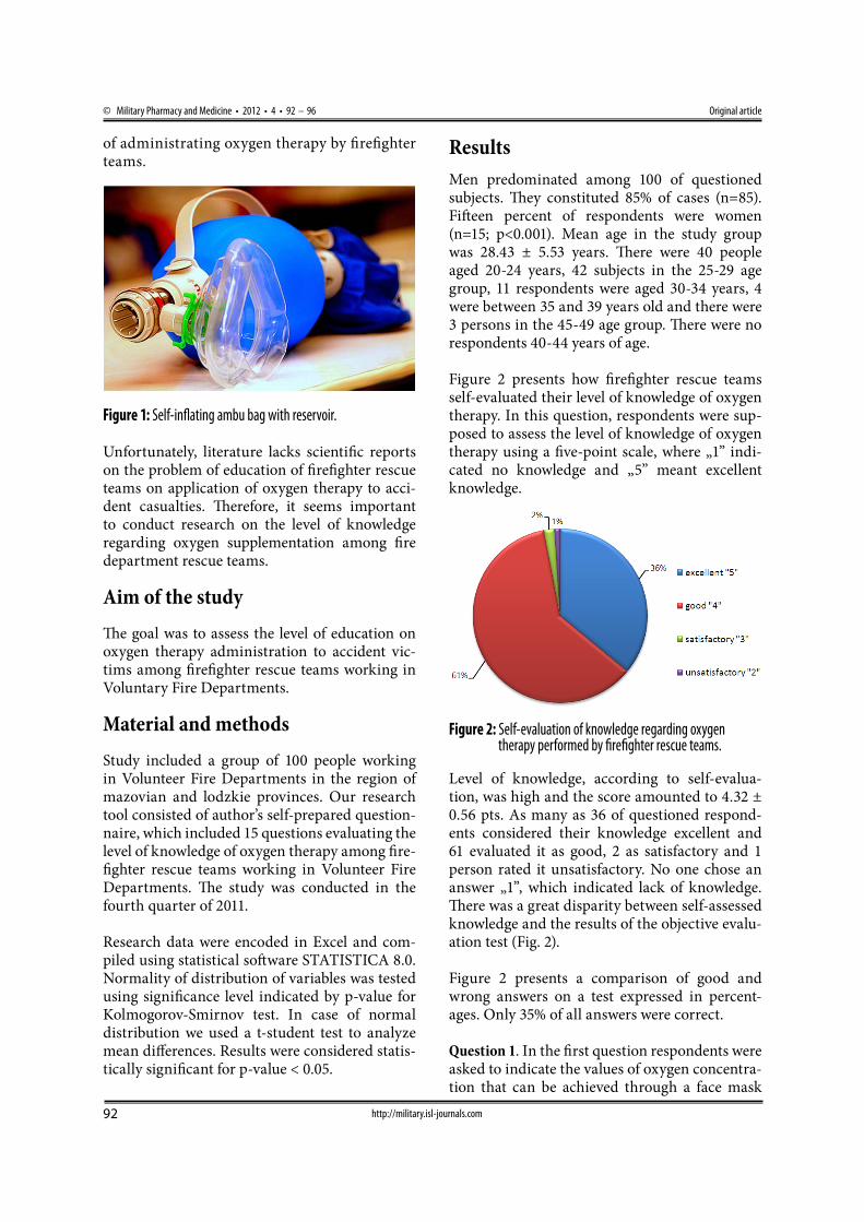
© Military Pharmacy and Medicine • 2012 • 4 • 92 – 96
92 http://military.isl-journals.com
Original article
of administrating oxygen therapy by firefighter teams.
Unfortunately, literature lacks scientific reports on the problem of education of firefighter rescue teams on application of oxygen therapy to acci-dent casualties. Therefore, it seems important to conduct research on the level of knowledge regarding oxygen supplementation among fire department rescue teams.
Aim of the studyThe goal was to assess the level of education on oxygen therapy administration to accident vic-tims among firefighter rescue teams working in Voluntary Fire Departments.
Material and methodsStudy included a group of 100 people working in Volunteer Fire Departments in the region of mazovian and lodzkie provinces. Our research tool consisted of author’s self-prepared question-naire, which included 15 questions evaluating the level of knowledge of oxygen therapy among fire-fighter rescue teams working in Volunteer Fire Departments. The study was conducted in the fourth quarter of 2011.
Research data were encoded in Excel and com-piled using statistical software STATISTICA 8.0. Normality of distribution of variables was tested using significance level indicated by p-value for Kolmogorov-Smirnov test. In case of normal distribution we used a t-student test to analyze mean differences. Results were considered statis-tically significant for p-value < 0.05.
ResultsMen predominated among 100 of questioned subjects. They constituted 85% of cases (n=85). Fifteen percent of respondents were women (n=15; p<0.001). Mean age in the study group was 28.43 ± 5.53 years. There were 40 people aged 20-24 years, 42 subjects in the 25-29 age group, 11 respondents were aged 30-34 years, 4 were between 35 and 39 years old and there were 3 persons in the 45-49 age group. There were no respondents 40-44 years of age.
Figure 2 presents how firefighter rescue teams self-evaluated their level of knowledge of oxygen therapy. In this question, respondents were sup-posed to assess the level of knowledge of oxygen therapy using a five-point scale, where „1” indi-cated no knowledge and „5” meant excellent knowledge.
Level of knowledge, according to self-evalua-tion, was high and the score amounted to 4.32 ± 0.56 pts. As many as 36 of questioned respond-ents considered their knowledge excellent and 61 evaluated it as good, 2 as satisfactory and 1 person rated it unsatisfactory. No one chose an answer „1”, which indicated lack of knowledge. There was a great disparity between self-assessed knowledge and the results of the objective evalu-ation test (Fig. 2).
Figure 2 presents a comparison of good and wrong answers on a test expressed in percent-ages. Only 35% of all answers were correct.
Question 1. In the first question respondents were asked to indicate the values of oxygen concentra-tion that can be achieved through a face mask
Figure 1: Self-inflating ambu bag with reservoir.
Figure 2: Self-evaluation of knowledge regarding oxygen therapy performed by firefighter rescue teams.

© Military Pharmacy and Medicine • 2012 • 4 • 93 – 96
93http://military.isl-journals.com
Łukasz Szarpak: Scope of knowledge regarding administration of oxygen therapy …
with reservoir. Only 35% of respondents pointed to the correct answer – „100%”.
Question 2. This question inquired what kind of oxygen therapy is provided through nasal can-nulas. In this case, nearly a half of respondents (46%) gave correct answers by indicating passive oxygen therapy.
Question 3. This question asked respondents to point to indications for oxygen therapy. Of all respondents, 32 persons were able to choose cor-rect indications for administration of oxygen therapy to patients during a rescue operation.
Question 4. In this question respondents were supposed to answer by what percentage the con-centration of oxygen in the breathing mixture raises for every 1 l/min of increased oxygen flow through nasal cannulas. Only 11% of all respond-ents gave correct answers (p<0.001).
Question 5. Firefighter rescue teams were asked to identify which emergency kit includes a pas-sive oxygen therapy set. Sixty-one percent cor-rectly indicated the PSP R1 kit. Distribution of provided answers is presented in Figure 3.
Question 6. In the entire studied group 52% of respondents were not able to tell the distance used for fitting of the oropharyngeal tube. The correct answer „the distance from incisors to the angle of jaw” was indicated by only 48% of subjects.
Question 7. This question was also related to the use of oropharyngeal tube. The respondent was asked to indicate the state, in which this instrument may be applied for restoring airway patency. Subjects could choose from the follow-ing answers: „confused patient,” „somnolent
patient” and „ deeply unconscious patient.” The majority of respondents, as much as 59%, answered correctly that oropharyngeal tube should be only used in case of deeply uncon-scious patients.
Question 8. From all available answers („using suction to aspirate fluid content from oral cavity,” „passive oxygen therapy,” and „inserting an oro-pharyngeal tube”) participants were to point to the action, which is not a part of the procedure of establishing airway patency. Forty-three subjects correctly indicated „passive oxygen therapy,” while 57 people gave wrong answers (p<0.001).
Question 9. This question was designed to test respondent’s knowledge of the use of a self-inflat-ing ambu bag. The respondent was supposed to identify an incorrect statement regarding an ambu bag. Correct answer – „a self-inflating ambu bag allows for achieving maximum oxy-gen concentrations of 75%” – was given by 46% of respondents.
Answer 10. Similar to the previous question, this one was related to the use of self-inflating ambu bag. Respondents were supposed to indicate the proper ambu bag ventilation volume in an adult. The correct answer was 500-700 ml and it was given by only 15% of respondents (p<0.001).
Question 11. Cardiopulmonary resuscitation is one of the most stress-inducing experiences a firefighter may come in contact with. In this question, the respondent was to indicate the time required for assessment of patient’s breathing according to current guidelines. Thirty-nine sub-jects answered correctly „10 seconds” (p<0.001).
Figure 3: . Assessment of knowledge regarding oxygen therapy among firefighter rescue teams
Figure 4: Answers provided by respondents to question 5.

© Military Pharmacy and Medicine • 2012 • 4 • 94 – 96
94 http://military.isl-journals.com
Original article
Question 12. In the twelfth question we asked respondents about the meaning of the term „sat-uration.” Only 30% of them were able to identify saturation as blood oxygen concentration. As much as 70% of subjects gave wrong answers. A detailed distribution of answers given by respondents is depicted in Figure 4.
Question 13. This question also referred to assess-ment of saturation level. Among the provided answers („carbon monoxide poisoning,” “cyanide poisoning,” „hyperthermia”) respondents were to choose the state, in which saturation measure-ment would be unreliable. Only 25% gave a correct answer, while 75 people responded incorrectly.
DiscussionOxygen, an element necessary for human exist-ence, is also used as a drug [4, 5, 6]. Just like in any other case of drug administration, supple-mentation of oxygen should be given according to indications and with careful dosing. For that purpose firefighter rescue teams, who are often the ones to administer first aid to victims before arrival of medical emergency services, undergo first aid training. First aid course, which lasts 66 hours (involving 25 hours of theory and 41 hours of practical training), includes issues related to supplementation of oxygen therapy to casualties [3].
Unfortunately, there are no reports in the lit-erature on the problem of knowledge regard-ing principles of oxygen therapy among firefighter rescue teams. Therefore, it is not pos-sible to compare our results to those obtained by other authors.
The level of self-confidence among firefighters from the studied group was high and amounted
to 4.32 points. There was a great disparity between their self-evaluated knowledge and test results, as evidenced by a 61% rate of incorrect answers given by respondents. At this point, it is safe to say that members of firefighter rescue teams overstate their knowledge of oxygen ther-apy administration.
At the scene of rescue operation firefighter rescue teams may administer oxygen therapy to the patient according to first aid proce-dures. Oxygen therapy may be divided into passive and active types [7]. In passive therapy oxygen is inhaled due to patient’s preserved ventilatory function, while in case of active oxygen therapy forced ventilation (replace-ment breathing) is used [7, 8]. In our material, only 46% of respondents knew the difference
between methods of passive and active oxygen therapy, as demonstrated by indicating patient ventilation via nasal cannulas as an element of passive oxygen therapy.
Symptoms of hypoxia include: skin cyanosis (if deoxygenated hemoglobin concentration falls below 5 g/l) [4, 8]. Moreover, we may observe excessive respiratory muscle work, which is a sign of dyspnea. In case of central nervous sys-tem hypoxia patient may be restless or aggra-vated. Severe hypoxia may be associated with loss of consciousness. The simplest, non-invasive method of evaluating the level of blood oxygen saturation at the site of rescue operation is pulse oximetry. Only 30% of respondents in our study group possessed the knowledge that saturation is a measure of oxygen content in arterial blood.
Despite pulse oximetry being a routinely used method of saturation measurement, in some situ-ations this measurement may be unreliable (erro-neous). An example of such situation is carbon monoxide poisoning. As carbon monoxide has almost 250-fold greater affinity to hemoglobin than oxygen, bonds created as a result of coupling of hemoglobin with carbon monoxide and car-boxyhemoglobin formation are extremely strong. It leads to tissue hypoxia. In the above situation, pulse oximeter may show normal values despite the true level of arterial blood oxygenation being significantly lower. Carbon monoxide poisoning is dangerous, as the most susceptible organs such as the cardiovascular system and central nerv-ous system are damaged first. In severe carbon
Figure 5: .Answers provided by respondents to question 12.

© Military Pharmacy and Medicine • 2012 • 4 • 95 – 96
95http://military.isl-journals.com
Łukasz Szarpak: Scope of knowledge regarding administration of oxygen therapy …
References:1. Szarpak, Ł. ”Zintegrowany system ratownictwa
elementem bezpieczeństwa państwa”, [w:] „Katastrofy naturalne i cywilizacyjne. Dylematy współczesnego bezpieczeństwa”, Wyższa Szkoła Oficerska Wojsk Lądowych im. gen. T. Kościuszki, Wrocław 2011, s.127-138.
2. Ziemba R. “Criteria of procedures in life-threatening states”. Military pharmacy and medicine 2012, 5(2): 62-68.
3. Rozporządzenie Ministra Spraw Wewnętrznych oraz Ministra Obrony Narodowej z dnia 23 grudnia 2011 r. zmieniające rozporządzenie w sprawie szkoleń w zakresie kwalifikowanej pierwszej pomocy (Dz.U. 2011 nr 299 poz. 1778).
4. Powrie, K. and S. M. Smith. “Editorial: Emergency oxygen for adults guideline--a change in oxygen therapy practice?” J.Clin.Nurs. 19.5-6 (2010): 601-02.
monoxide poisoning therapy in a hyperbaric chamber is the treatment of choice [9]. From our study group, 25% of subjects were familiar with situations, in which pulse oximetry could be unreliable. Due to the possibility of airway obstruction, manual methods of establishing air-way patency applied first and followed by instru-mental methods, firefighter rescue teams should possess the knowledge of oropharyngeal tube insertion. Guedel tube is supposed to prevent tongue slipping toward the posterior pharyngeal wall and causing airway obstruction. A tube that is either too large or too small may cause obstruc-tion; hence it is important to know how to choose a proper one. Size of the tube should be fitted to each particular patient by placing the tube against patient’s cheek. Tube collar (at the inlet) should be located near the incisors, while its end should reach mandibular angle. A tube fitted in such manner ensures airway patency, but dos not
prevent from regurgitation – passive movement of stomach contents into the esophagus without a gag reflex – or from vomiting. Fifty-two per-cent of respondents knew how to appropriately fit an oropharyngeal tube. A rescue worker should remember that oropharyngeal tube should only be used in deeply unconscious victims. Most of the surveyed firefighters were familiar with this principle, as much as 59% answered correctly.
ConclusionsDue to the character of firefighter rescue teams’ work, it is justified to examine the knowledge regarding administration of oxygen therapy among firefighters, as it may influence patient survival.
Knowledge of oxygen therapy among firefighters is inadequate.
5. O’Driscoll, R. “A breath of fresh air: a new UK guideline for emergency oxygen therapy.” Br.J.Hosp.Med.(Lond) 69.12 (2008): 670-71.
6. Ziemba R (ed): ABC Ratownika – pierwsza pomoc w nagłych, IndexCopernicus, 2011, pp. 55.
7. Austin, M. and R. Wood-Baker. “Oxygen therapy in the pre-hospital setting for acute exacerbations of chronic obstructive pulmonary disease.” Cochrane.Database.Syst.Rev.3 (2006): CD005534.
8. Small, G. and P. Barsby. “How much is enough? Emergency oxygen therapy with COPD.” Emerg.Nurse 8.8 (2000): 20-24.
9. Van, Meter K. “Hyperbaric Oxygen Therapy as an Adjunct to Pre-hospital Advanced Trauma Life Support.” Surg.Technol.Int. XXI (2011): 61-73.


© Military Pharmacy and Medicine • 2012 • 4 • 97 – 104 Review article
97http://military.isl-journals.com
Emergency Medicine
Sustainable development in light of international cooperation
Radosław Ziemba
Military Centre for Pharmacy and Medical Technology in Celestynow, Poland
Author’s address:Radosław Ziemba, Military Centre of Pharmacy and Medical Technique, ul. Wojska Polskiego 57, 05–430 Celestynów, Poland; e–mail: [email protected]
Received: 2012.09.19 • Accepted: 2012.11.22 • Published: 2012.12.08
Summary:
Numerous developed countries as well as those entering this path have established strategies of sustain-able development. Areas of activity presented in this work illustrate the wide scope of parameters and challenges for maintaining sustainable development, both on a local as well as global scale.
Key words: hsustainable development, European Union directive, areas of action, eliminating the ef-fects of environmental pollution, Rio declaration.
Balanced development –scope of the concept
Sustainable development is defined as a broadly understood social goal, encompassing balanced environmental quality accompanied by achieve-ment of social and economical goals. The term “sustainable” may be defined as maintaining environmental capacity in time. It also encom-passes aspects such as: availability of natural resources, waste assimilation, cultural values, heritage values, climate stability and maintain-ing biodiversity. Such environmental capacity may be measured using environmental indexes such as: size of protected areas, size of ecosys-tems, number of species, level of pollution and size of resources, particularly with regard to irre-placeable materials.
This term is a result of previously mentioned report created by the former Norwegian prime minister, current director of World Health Organization, Gro Harlem Brundtland, titled “Our common future.” The English term “Sus-tainable development” was used there. Finding an
analogous Polish expression that would properly reflect the sense of words contained in the men-tioned document was somewhat troublesome. Following numerous discussions, we translated these words as: “trwały i zrównoważony rozwój” (steady and balanced development), although there are some in favor of “ekorozwój” (ecodevel-opment) or “trwały rozwój” (steady progress).
A concept of “balanced development” as a shorter version of the previous expression also appears in various documents and reports. This general concept lies at the foundations of environmen-tal politics of many countries. However, some scientific uncertainty and diverse opinions indi-cate, that there is still a problem concerning the value of environmental capacity. However, there are indexes, which may prove as useful operative instruments for comparison with policy goals and environmental standards, both domestic and international.
Strategic environmental evaluation is an instru-ment for comparison of analyzed plans and

© Military Pharmacy and Medicine • 2012 • 4 • 98 – 104
98 http://military.isl-journals.com
Radosław Ziemba: Sustainable development in light of international cooperation
programs with environmental politics and strat-egies on a national level.
Special value of such evaluation is increasingly more often recognized among European coun-tries. As an example, the goal of European Union directive on strategic environmental evaluation was to ensure proper level of assessment at a stage of strategic decisions in all member coun-tries. This project is considered a step toward ensuring balanced development and there-fore, means of achieving goals of the European Union Fifth Environmental Action Program: “Towards Sustainability.”
It is an effect of the accepted Rio Declaration on “Environment and Development” and, above all, Agenda 21 on actions undertaken at the turn of the 20th and 21st century in order to ensure sustainable development. It is supposed to optimally balance the current status with the assumed, undertaken for the future. Therefore, strategic evaluation is considered an instru-ment allowing for inclusion of environmen-tal aspects and sustainable development into the mainstream decision-making processes regarding development. As opposed to indi-vidual projects, strategic evaluation method is directed toward programs at early stages of planning. Such approach provides substantial benefits in terms of strategic evaluation of the program at an early stage of its development. Conditions necessary for balancing the devel-opment goals (particularly regarding the envi-ronment) are analyzed as an element of strate-gic evaluation.
Many developed countries, as well as those enter-ing this path, have strategies of sustainable devel-opment already behind them in terms of their planning as well as describing specific dimen-sions of their environmental policies.
At a political level, goal descriptions are least detailed. Therefore, strategic evaluations are performed at the same level. They concentrate mainly around the analysis of political goals, and their environmental cohesion. In the sustain-able development document, it signifies ensuring consistency with the goals of international and global policies expressed through agreements, conventions and other documents.
Why then, are strategic evaluations so impor-tant? They include aspects of sustainable growth into internal decision-making processes. Sustain-able growth is directed at fulfilling the needs of a contemporary man in such manner that would not prevent the future generations from utiliz-ing environmental resources. This requirement results in the necessity of maintaining balance between goals and environmental, economical and social criteria.
Effects analyzed as a part of strategic evaluation may be divided into four groups:
• traditional environmental factors, such as ecological effects, quality of water and soil, quality of the air, noise levels, landscape, con-sequences for the population – analyzed with regard to their international, domestic and regional significance;
• outcomes associated with imposing balance, which encompass the threat o irreversible, cumulative or secondary effects such as: exploitation of non-renewable resources, loss of biodiversity, valuable natural ecosystems, as well as long-term productivity of forests or rural areas;
• induced changes in use of the land, particu-larly in relation to urbanization and expansion of municipal peripheries;
• political effects of breaching international agreements and other domestic policies.
Assessment of significance (or importance) of predicted effects is a key element of strategic environmental assessment process. However, up to date, no index was agreed upon, which would measure the degree of attained sustainable pro-gress, encompassing various indexes and deter-mining how a given factor should be weighted against other ones. Sustainable balance does not only determine beneficial effects. They may be unfavorable or, at the least, of little benefit. All of those effects should be considered through an advanced grading system, even more so when various factors undergo evaluation. Evaluation criteria must be established for selected problems in order to determine the significance of pre-dicted outcomes.
At a strategic level, it is indicated to assess the risk of occurrence of significant outcomes (ben-efits, losses, conflicts) instead of making prog-noses, which in turn should be performed at a

© Military Pharmacy and Medicine • 2012 • 4 • 99 – 104 Review article
99http://military.isl-journals.com
level of evaluation of a particular investment. For example, it concerns the risk of effects leading to secondary enterprises or conflicts with various development plans. In this context, the risk of occurrence of outcomes derived from secondary projects was defined as a product of probability of effect occurrence and intensity of this effect.
While determining the outcome risk, the follow-ing aspects should be taken into consideration:
• environmental absorption capacity or sensitivity;
• the scale or extent of the effect, e.g. the degree to which environmental quality may be disrupted;
• will the effects lead to sustained or temporary changes?;
• domestic or international obligations, goals and environmental quality standards,
• to what degree is reduction of the effects possible?;
• significance of the effect on a national level; • Strategic evaluations allow for inclusion of environmental considerations at a possibly earliest stage of planning, as in the case of social and economical aspects.
Principle no. 4 of the preamble “Environment and Development” states the following: “In order to achieve sustainable development, environ-mental protection shall constitute an integral part of the development process and cannot be considered in isolation from it.” Principle no. 8 strengthens those ideas, particularly in the frag-ment pertaining to the necessity to “reduce and eliminate unsustainable patterns of production and consumption” by some countries. Economi-cal instruments are vital to realization of those goals (Principle no. 16), according to the princi-ple: “the polluter should, in principle, bear the cost of pollution, with due regard to the public interest and without distorting international trade and investment.”
Areas of action for sustainable development
In order to face the challenges of the environ-ment and growth, cosignatory nations decided on a new form of universal cooperation. It compels them to get involved in constant and constructive international dialogue inspired by the necessity of establishing more effective and
just world economy, taking into consideration the growing co-dependence of societies and the fact that sustainable development should be a priority for those societies. Conference’s “Documents” state that: “in order to achieve this new cooperation, it is important to avoid confrontation and create an atmosphere of true cooperation and unity…” (5). In this mission, particular emphasis is put on strengthening of internal and international policy and establish-ing multidirectional cooperation adjusted to new conditions.
National economical policies and international economic relations are associated with sustain-able development. It should be noted that, in this regard, its reactivation and acceleration requires both a dynamic international supportive action, as well as decisive national policy. Both of those factors should act in parallel. A dynamic and stable world economy based on safe founda-tions gives a chance for progress. Therefore, support of international economic circle is a determining factor.
The role of developing countries, free of excessive external debt is noted in this process. Therefore, financial policy must serve it, without restriction of access to other markets.
Data obtained in the 80’s regarding this aspect are fundamentally negative in all cases. There-fore, a change of this state is encouraged. For that purpose, an international atmosphere supportive toward national efforts for growth should be built up. It is the only requirement for healthy internal economic policies of developed and developing countries toward achieving global progress in the area of sustainable development.
Accordingly, global economy should create an atmosphere promoting achievement of goals stated in Agenda 21 regarding environmental protection and development through:
• promotion of sustainable growth as a result of trade liberalization;
• mutual dependence of trade and environmen-tal protection;
• ensuring proper funds for developing coun-tries and taking care of international debts;
• support for macroeconomic enterprises toward environmental protection and development.

© Military Pharmacy and Medicine • 2012 • 4 • 100 – 104
100 http://military.isl-journals.com
Radosław Ziemba: Sustainable development in light of international cooperation
National efforts must be directed toward mutual dependence of the elements of international eco-nomical system and needs of humanity in the field of safe and stable natural environment. Therefore, it remains their intention to establish agreement at the tangent points in areas such as: environmental protection, trade and develop-ment and took place during meetings of exist-ing international organizations as well as in their individual internal policies. An optimal, just, safe, non-discriminatory and far-sighted, multi-lateral trading system concordant with sustaina-ble development may lead to optimal distribution of global production. It is in the interest of all participating partners, but providing developing countries with access to the world market and export, together with healthy macroeconomic and ecologic policy, will exert positive influence on environmental protection.
Thus, it will contribute to the field of sustain-able development. Healthy economic policy and management, effective and far-sighted public administration, consideration of envi-ronmental problems during a decision-making process as well as progress-oriented and demo-cratic government also act in its favor. It allows for participation in economic management of all involved parties under specific conditions of individual countries. Removal of the exist-ing anomalies in international trade is of great significance here, as it allows the developing countries to access funds needed for financing investments necessary for ensuring sustain-able development. Therefore, significant and progressive increase of preferences for agricul-ture (including internal regulations, access to the market and export subsidies), industry and other economical sectors seems necessary for prevention of large losses generated by more efficient producers, particularly in the develop-ing countries, when different preferences are used. Therefore, there is a place in agriculture, industry and other branches of economy for initiatives for trade liberalization and creation of an economic policy, in which production would be more closely related to the needs of environmental protection and development. Liberalization of trade should be considered on a global scale, so that particular sectors of the economy could contribute toward achieving sustainable development.
International trade, influenced by various fac-tors creating new areas of activity as well as new possibilities in this domain, determined an even greater significance of international economic cooperation. In the past few years, world trade developed faster than world production. How-ever, it should be noted that the expansion of world trade was unevenly distributed and only a small number of developing countries was able to achieve a perceptible increase in export. Pressures from the protectionists and unilateral economic policy restrain functioning of open, multilateral trade system, exerting a negative influence, particularly on the export of the devel-oping countries. It should be noted in this regard, that economic integration intensified during the past few years, which should boost the world trade and increase the trading as well as growth capabilities of developing countries. Also, a great number of those countries introduced bold reform policies, including ambitious, autonomic trade liberalization during the past several years. Simultaneously, far fetched reforms and deep restructuration processes taking place in Central and Eastern European countries, pave their way to integration with the world market and inter-national trade. Growing attention is directed at increasing the role of enterprise in economy and promotion of competition. Generalized System of Preferences (GSP) turned out to be a useful instrument of economic policy despite the fact that its goals were not entirely fulfilled and a system of trade facilitation based on electronic data interchange (EDI) increased the commer-cial effectiveness of public and private sectors. Solutions between environmental protection and forms of trade are diverse and were not specified to date.
Rapid implementation of various agreements of the Uruguay Round regarding multilateral trade negotiations will result in further liberalization and expansion of global trade. It will augment trading and growth capabilities of the coun-tries entering the path toward development and ensure greater security and farsightedness of international trade.
Goals, recommendations and feasibility were determined in light of the described areas of action. It also included mutual dependence of trade and environmental protection, support for economic actions promoting sustainable growth.

© Military Pharmacy and Medicine • 2012 • 4 • 101 – 104 Review article
101http://military.isl-journals.com
Another area of action that could contribute to the safety of long-term progress is the fight against poverty. It may be divided into the fol-lowing sections:
• providing the poor with sustainable access to the means of supporting their basic needs;
• facilitating integration of sustainable access to the means of support with environmental protection.
Changing the consumption model, which is a broad topic and its problems are mentioned in numerous chapters of Agenda 21, serves those areas. They are primarily located in the chap-ters related to the use of energy, transport, waste as well as economic instruments and transfer of technologies. The areas of action include the following:
• balancing the models of consumption and production,
• undertaking appropriate policies and eco-nomic strategies by individual countries in order to eliminate the imbalanced consump-tion models. The goals of the former area include the following:
• promoting a model of consumption and pro-duction, which would not lead to ecological damage and fulfill the needs of humanity,
• achieving better understanding of the role of consumption in the process of sustainable development as well as introduction of more balanced consumption models.
Poverty and degradation of natural environment are closely related. If poverty leads to ecological crises, the majority of cases of sustained environ-mental degradation result from an imbalanced model of production and consumption. There-fore, in the coming years, governments together with appropriate supporting organizations should strive to achieve broad goals, including:
• promotion of effective production processes and balanced consumption in the process of economic growth, taking into consideration the needs of the developing countries,
• developing strategies of internal actions, which will allow for introduction of balanced production and consumption models,
• strengthening the factors that support a production – consumption model consistent with sustainable development and promotion of actions for transfer of environment-friendly technologies to developing countries.
These goals pertain to the latter area of action.
In several chapters, areas of action are directly conformed to the security issues:
• environment-safe application of biotechnology (16),
• environment-safe utilization of toxic and dangerous chemicals, fighting illegal trading of those chemicals (19),
• environment-safe management of dangerous waste
• preventing illegal trade of dangerous materials (20),
• environment-safe management of solid waste and residues from wastewater treatment plans (21),
• safe and environmentally friendly manage-ment of radioactive waste (22),
Biotechnology, as a combination of genetic engi-neering, biochemistry and microbiology, is an area of intensely developing science, constituting a compilation of methods of genetic engineering. Not all fundamental environmental problems may be solved using biotechnological processes to attain sustainable growth. The reality must diminish the expectations. However, biotechnol-ogists announce expectations of great contribu-tions to, e.g. progress in health care, increase in food safety by introducing methods of sustained and ecologically sustainable agriculture as well as detoxication of dangerous waste. It should be accompanied by general partnership between countries with rich biological resources, but lack-ing the experience and investment funds neces-sary for utilization of those resources through biotechnological processes and countries with biotechnological experience. This experience refers to the scope of transformation of biologi-cal resources in a manner, which would allow them to contribute to sustainable development. Described areas of action promote the princi-ples of creating environmentally healthy uses for biotechnology agreed upon by the international community, in order to raise public confidence and convince the community to support ecologi-cally safe uses of biotechnology.
General policy indicates, that chemicals can be used to meet the economic and social needs of people around the world in a rational and largely safe manner. However, there is still a lot to do to ensure environmentally safe conduct with toxic

© Military Pharmacy and Medicine • 2012 • 4 • 102 – 104
102 http://military.isl-journals.com
Radosław Ziemba: Sustainable development in light of international cooperation
chemical substances. Two main problems arise in this area, particularly when it comes to develop-ing countries:
• absence of sufficient scientific information allowing for assessment of risk associated with use of large numbers of chemical substances;
• lack of means for assessment of chemical substances owned and used by people.
The processes of severe chemical environmental pollution take place in various industrial centers around the world in the recent years. It is associ-ated with threat to human health, genetic struc-ture and reproduction, as well as to the environ-ment. Elimination of the effects of contamination requires large expenditures and development of new technologies. Long-term effects of chemi-cal environmental contamination leading to cli-matic changes were only recently recognized and understood. Also recently, the humanity realized the extent of those effects. A large number of international institutions are involved in works concerning chemical safety. Programs for its promotion were implemented in many countries. These works have international implications, as dangerous effects of chemicals know no national boundaries. However, significant aid for national and international enterprises in this area is nec-essary in order to work out methods of environ-mentally safe use of chemical substances.
Seven areas of action are proposed in this area: • broadening and acceleration of international evaluations of chemical threats,
• unification of classification and labeling of chemicals,
• exchange of information regarding toxic chemicals and chemical threats,
• establishing programs for risk reduction, • strengthening national capabilities and skills for effective toxic substance management,
• preventing illegal international trade of toxic and dangerous substances,
• deepening international cooperation in some areas of actions.
Effective control of production, storage, han-dling, recycling, transport, retrieval of raw mate-rials and management of dangerous wastes are important for human wellbeing, environmental protection, as well as management of natural resources and sustainable development. Prevent-ing formation of dangerous materials requires
knowledge, experienced staff, resources and financial means, as well as technological and sci-entific potential. International trade of danger-ous wastes, with partial breaching of national legislation and international policies, causing harm to the environment and public health in all countries, particularly the developing ones, is a matter of concern for the international society.
The master task in terms of management of vital processes involves the greatest possible minimi-zation of dangerous waste production, as well as such management of waste products so that they would not cause harm to health and the environ-ment. It encompasses four areas of action:
• promoting prevention of dangerous waste formation and minimization of the amounts of those products,
• promoting and strengthening international cooperation in management of cross-border movement of dangerous wastes,
• promoting and strengthening institutional powers with regard to dangerous waste management,
• preventing illegal international trade of dan-gerous waste products.
The General Assembly concurred that envi-ronmentally safe waste management is one of the main actions undertaken in order to main-tain the wellbeing of global environment and to attain environmentally healthy, stable growth of all countries.
Areas of action described in this chapter of Agenda 21 are closely related to the following areas of action contained in other chapters:
• quality protection and use of inland water supplies (ch. 18);
• promoting sustainable development of human habitats (ch. 7);
• protection and promotion of human health (ch. 6);
• changing the model of consumption (ch. 4).
According to this, the extent of necessary actions should be based on a hierarchy of goals and con-centrate on four main fields of action related to waste products. They are mutually intercon-nected and supportive of each other. They come down to:
• minimizing the amount of waste;

© Military Pharmacy and Medicine • 2012 • 4 • 103 – 104 Review article
103http://military.isl-journals.com
• maximization of environmentally safe use of secondary materials and waste recycling;
• promotion of environmentally safe elimina-tion and processing of waste products;
• broadening the scope of services with respect to elimination and processing of waste products.
Radioactive waste is formed both during use of nuclear fuel as well as radioactive materials (in medicine, science and industry) Radiological risk and safety threats are diverse on the part of radio-logical waste: they range from very small to great.
Safe and environmentally harmless management of radioactive materials, including their minimi-zation, transport and utilization is of great impor-tance. Most countries with leading nuclear energy
References:1. Janikowski R., Wielokrotny model decyzyjny, jako
narzędzie oceny oddziaływania projektowanej działalności człowieka na środowisko. JETU, Katowice, 1993.
2. Jendrośka J. Rządowy projekt ustawy o postępowaniu w sprawie ocen oddziaływania na środowisko oraz o dostępie do informacji o środowisku i jego ochronie, (w:), Problemy ocen środowiskowych, kwartalnik Nr 1(8), 2000.
3. Kozłowski S., Koncepcja ekorozwoju jako podstawa polityki ekologicznej państwa.(w:) B.Poskrobko
programs have undertaken administrative and technological systems of waste management. In many other countries, still preparing for nuclear programs or only using radioactive materials, there is still a need for creation of such systems, which is becoming increasingly urgent.
A fundamental goal of such program is to ensure safe management, transport, storage and utiliza-tion of radioactive wastes. It is directed at protec-tion of human and environmental health and is contained within a broader framework of interac-tive and integrated approach toward their manage-ment within a broadly understood safety system.
The described areas of action illustrate the extent of parameters and challenges for maintaining sustainable development.
(red.), Działalność gospodarcza a ochrona środowiska przyrodniczego, Biuro Badań i Wdrożeń Ekologicznych, Białystok 1992.
4. Kozłowski S., W drodze do ekorozwoju, PWN, Warszawa, 1997.
5. Patrz: Dokumenty końcowe Konferencji Narodów Zjednoczonych..., cyt. wyd., słowo wstępne.


© Military Pharmacy and Medicine • 2012 • 4 • 105 – 110
105http://military.isl-journals.com
Łukasz Szarpak: Selected aspects of communication between emergency services …
Emergency medicine
Selected aspects of communication between emergency services and the mass media in crisis situationsŁukasz Szarpak, Marcin Madziała
Collegium Masoviense – College of Health Sciences in Żyrardów, Poland
Author’s address:Łukasz Szarpak, Collegium Masoviense – College of Health Science, ul. G. Narutowicza 35, 96-300 Żyrardów, Poland; phone: (+48) 500 186 225, e–mail: [email protected]
Received: 2012.07.18 • Accepted: 2012.09.11 • Published: 2012.09.28
Summary:
We live in times where information is the supreme value. The mass media seek sensation and often misinterpret the situation, which usually occurs in the case of limited access to information. Due to their mediagenic nature, crisis situations as well as emergency actions taken to eliminate their conse-quences are undoubtedly a treat for journalists. Correctly performed rescue operations are essential during events that make people suffer or feel endangered. Nonetheless, appropriate conveying informa-tion about performed actions to the media, and then to the society, is a crucial element of rescue opera-tions and influences both perception of emergency services and prevention from spreading speculation and panic in the society.
Key words: media, crisis situation, rescue operation, emergency services, panic.
IntroductionContinuous civilization development and grow-ing population make us live in expanding agglomerations, which results in the fact that the issue of disasters is becoming increasingly important. Phenomena bearing marks of crisis, although varied and reaching different extents, which makes it difficult to define them precisely, have some common features [1-3]. Firstly, in view of the fact that nobody can expect crisis situations at a particular time and in a particular place, the affected population is usually helpless. Secondly, the extent of damage and number of people in need exceed the ability to satisfy all expectations. Another feature of crisis situations is their medi-agenic nature [2,3].
For hundreds of years, since humans started to deal with disastrous events, the concept of
catastrophe has evolved. At present, there are numerous definitions of “catastrophe”[4]. The most accurate one, with regard to the way of thinking in any situation that surpasses in extent the abilities of rescuers and bears the features of catastrophe, is a definition approved in the USA by the Federal Emergency Management Agency (FEMA) [5]. It defines catastrophe as an event causing death, injuries or damage to posses-sions, to an extent that makes routine emergency actions insufficient. According to this definition, catastrophe occurs suddenly and requires imme-diate, coordinated actions taken by many indi-viduals and institutions.
Efficient crisis management is not only a medi-cal emergency assistance provided to casual-ties. One of the basic conditions of effective

Review article© Military Pharmacy and Medicine • 2012 • 4 • 106 – 110
106 http://military.isl-journals.com
actions reducing the consequences of the event is a well-organised communication between emergency services units, including the fire bri-gade, the police, the medical rescue, etc., as well as between emergency services and the public. Due to technical advancements it is possible to exchange information via the electronic media, however, the basic way of contact between the emergency services, the authorities and the soci-ety is undoubtedly public media, including press, radio and television stations (TV and radio are electronic media) [3,6].
Communication with the media in crisis situa-tions may be divided into three stages. The first of them includes actions preceding a crisis situ-ation, and next two stages – actions during and after eliminating such a situation [1,2,6,7].
Actions preceding catastrophes
Communication infrastructure, consolidating all emergency services and the public, is an inher-ent element of the efficient security system. The above mentioned mass media, i.e. radio, televi-sion, press, and nowadays also the Internet, are main channels of conveying information. Lack of proper circulation of information between the emergency services leads to chaos, whereas, dis-turbed communication with the media results in misinterpretation of the event, which may raise many unfavourable interactions in the public, including panic.
Actions preceding a crisis situation are inher-ent preparatory stages for various emergency services and crisis panels. Continuous updating of knowledge by people who coordinate actions of emergency and crisis services, results in pro-gressing improvement of professional skills [8,9]. Constant training, exercises and simulations help these services, to a greater or lesser extent, prepare for possible crisis scenarios.
Exercises should not only involve rescue services, but they should also include elements of coop-eration with media [7,10]. In the future it will help avoid information chaos. It is important to become acquainted with the rules of the public media. It is also worth establishing and practis-ing the method of appointing persons responsi-ble for contacts with the media (spokespersons), creating healthy relationship with the media and
preparing (adequately to the situation) neces-sary information for the public. Therefore, estab-lishing procedures for crisis situations seems indispensable.
Media communication and legal provisions
Circulation of information between public ser-vices and media is regulated by legal acts, ethi-cal codes and internal regulations. Thereby, such regulations impose duties on the emergency services workers, authorities and journalists. Informing the public about states of danger is defined by, among others, a notation in the UN Universal Declaration of Human Rights of 1984 [11]. In the face of a crisis situation, people would feel much safer to know that they receive reliable information about current actions and changes that may affect their lives and environment.
Act of 26 January 1984 – Press Law, conferred powers on journalists to obtain information about actions of government agencies, pub-lic companies and other organisational units, whereas, heads of such units, spokespersons or other persons authorized to convey informa-tion to media are responsible for providing such information [12]. However, according to the above Act, journalists should make every effort to be careful, reliable and honest while collect-ing and conveying press materials; they should check credibility of all obtained information and publish its sources. According to the Act, the media are obliged to publish (free of charge) statements issued on the basis of acts, announce-ments, ordinances or resolutions of the central administration authorities and government administration authorities in the province, and, what is important for this paper, they are obliged to publish (free of charge) announcements issued by the government administration authorities or local governments and concerning crisis situ-ations defined in the Act on crisis management of 2007 [13].
Legal provisions concerning reliable conveying information about events and phenomena in the country and abroad, are also included in the Act on the state of natural disaster [14] or in the Act on National Broadcasting Council. Principles of correct collection of and publication of press materials may be found not only in the above acts

© Military Pharmacy and Medicine • 2012 • 4 • 107 – 110
107http://military.isl-journals.com
Łukasz Szarpak: Selected aspects of communication between emergency services …
but also in ethical codes, including the Polish Code of Ethics for Public Relations Association and the Ethical Code of Journalists.
A great example of internal acts referring to the role of communication between the emergency (rescue) services and the media may be internal regulations of the National Fire Service of 1998, defining press-information activity within the NFS structures, including the actions of spokes-persons and cooperation between NFS and journalists during crisis situations. In 2004, the Headquarters of the National Fire Service estab-lished principles of proper conduct for the com-manding officer during a rescue operation. The above-mentioned guidelines categorically forbid the commanding officer to inform the media in the event place about the reasons, identity data and names of casualties. Guidelines also clearly define the way of taking over informa-tional function by a person appointed to a post of a spokesperson.
Appointment of spokespersons
A deficit of information is commonly associ-ated with crisis situations. From a point of view of the media, a crisis situation is a special opportunity to develop their own image among media market participants while gaining as much information as possible and reporting it to the public [15] . In order to avoid informa-tion chaos, it is important to take actions which guarantee that statements issued by all persons authorised to convey information are consist-ent, relevant and answer the questions instead of ignoring them using a phrase “no comments”. A requirement of consistent communication is one of the most important elements connected with the entire management of communication in crisis situations. Consistency is necessary for information exchange between particu-lar emergency services, organizations and the media. Therefore, it seems important to develop a strategy of communication with both inter-nal (services) and external (media and public opinion) environment.
Misinterpretation of the event is a common action taken by the media, which results from shortage of information or conflicting data presented to the public. Thus, it is essential to appoint a person who will solely inform the
media about progression of rescue actions from the onset of crisis. Such a person should be cho-sen much earlier and systematically prepared for his/her role, since in order to increase the level of reliability of the released statement, the speaker must show competence, enjoy respect, be reliable and dynamic, and enjoy a positive opinion. Such a person should have several crucial abilities:
• must be resistant to stress and control his/her own emotions;
• must have extensive substantive knowledge about activities of the emergency services and know the services which he/she is talking about;
• must have great interpersonal abilities and make contact easily.
Professional actions of a spokesperson build up trust of the media in emergency services and strengthen positive public opinion about their hard work [16]. Thus, it is so important to obey the rules defining what a spokesperson should or should not do.
Table 1: Principles of spokesperson’s work.
Allowed Not allowed
Release information only via a press spokesperson responsible for public rela-tions.
Speculate on:
• possible causes of the crisis situation; • number of fatalities; • further scenarios of events; • date of termination of the actions.
Create equal opportunities to obtain information by various types of media.
Provide information that can hinder the progress of rescue operations, raise panic or bring about any new threat.
Release only proven infor-mation
Issue official statements to incompetent persons
Supervise information out-flow by:
• own, frequent contacts with the media; • collecting all information in a version reported by the media to the public; • controlling movement of journalists in the endan-gered area.
Evaluate rescue operations when they are in progress.
Lay the blame for the situa-tion on anyone.
Misinform the media.
Review article© Military Pharmacy and Medicine • 2012 • 4 • 108 – 110
108 http://military.isl-journals.com
Preparation of informationDuring a rescue operation, a lot of information asked by the media is unpredictable. However, there is still considerable amount of informa-tion that can be prepared earlier and successively released to the public. Such information includes:
• alarm information; • instructions for behaviour in crisis situations, • data concerning places and sources of infor-mation and help;
• data concerning services involved in the rescue operation;
• appeal to support services in relevant situations.
When preparing rules for cooperation with the media, all forms of communication should be taken into consideration [10,17]. The most popu-lar forms of communication in crisis situations include: press note, statement, information, tel-ephone response, written response, interview, talk, utterance and press conference.
Actions during a crisis situation
Communication with the media during rescue operations mainly includes: appointment of a 24-h duty spokesperson, collection of informa-tion from various services, preparation and conveying information to journalists, informa-tion management for the regional and national media, checking and correction of information that was released to the media, organisation of press conferences, responding and release of statements on the current situation, and coordi-nation of journalists work in the area involved in rescue operations.
It is important to establish an information cen-tre near the place of rescue actions [3,4,9]. Per-sons responsible for contact with the media should all the time monitor the situation and respond immediately to the needs reported by the media, if it is possible and not against the res-cue operation.
Another important element regarding participa-tion of the media in a crisis situation is to sepa-rate and adapt for journalists the area around the event site. This area is separated according to two important principles. The first of them is
the superiority of rescue operations interest. This principle forbids the media to enter places, where it could pose a risk to the rescue action or to the media. The second principle, used while separat-ing areas for the media during catastrophe, is the best possible availability of the media. The media should have possibly the best, acceptable “viewpoint” for observation of the rescue actions, to assure that when looking for a good place for taking pictures the media will not hinder actions taken by emergency services.
Persons responsible for contacts with the media are obliged to separate such areas, in agreement with persons in charge of the rescue operation.
Actions after eliminating a crisis situation
After termination of the rescue actions, per-sons responsible for contacts with the media are obliged to inform the local community about the current situation and taken precautions, and to inform the entire country about a situation in the particular region.
Unfortunately, it is often seen that the end of the rescue operation is the end of all actions aimed to eliminate a crisis situation. However, it should be an introduction to preparations connected with future actions. Conclusions should be drawn from all experiences, regardless of whether they were positive or negative; all actions should also be thoroughly analysed. Efforts put into ana-lysing the particular action should help revise arrangements and prepare for future events.
Summary
When analysing this paper, it may be hypothe-sized that the public authorities, emergency ser-vices and mass media are all responsible for reli-able conveying the current information in crisis situations.
However, cooperation with the media is not easy. Journalists are characterised by natural scepti-cism. They are more interested in criticising the services and government authorities responsible for controlling the crisis situation than in defend-ing or justifying such services and authorities. Probability of negative comments given by the media during a crisis situation is almost 100%.

© Military Pharmacy and Medicine • 2012 • 4 • 109 – 110
109http://military.isl-journals.com
Łukasz Szarpak: Selected aspects of communication between emergency services …
Although several acts, including the Act on the state of natural disaster and crisis management, imposed on the media an obligation to report any threat (in agreement with the local authori-ties), we should remember about the role of the way how information reaches editorial offices. Thus, it is important to work out methods of
presentation to the media a problem of catastro-phe or emergency.
Taking the above-mentioned actions increases the chances for peaceable, and at the same time, effective participation of journalists in a crisis situation.
References:1. Beaudoin, C. E. “Assessment of a media campaign and
related crisis help line following Hurricane Katrina.” Public Health Rep. 123.5 (2008): 646-51.
2. Arnold, J. L. “Disaster myths and hurricane Katrina 2005: can public officials and the media learn to provide responsible crisis communication during disasters?” Prehosp.Disaster.Med. 21.1 (2006): 1-3.
3. Ng, K. H. and M. L. Lean. “The fukushima nuclear crisis reemphasizes the need for improved risk communication and better use of social media.” Health Phys. 103.3 (2012): 307-10.
4. Barr, P. “Staying connected. Social media put to work when disaster strikes.” Mod.Healthc. 41.36 (2011): 33.
5. Keim, M. E. and E. Noji. “Emergent use of social media: a new age of opportunity for disaster resilience.” Am.J.Disaster.Med. 6.1 (2011): 47-54.
6. Schlecker, M. and E. Hirsch. “Incomplete knowledge: ethnography and the crisis of context in studies of media, science and technology.” Hist Human Sci. 14.1 (2001): 69-87.
7. McLearn, D. “FDA crisis management and the media.” PDA.J.Pharm.Sci.Technol. 50.3 (1996): 144-46.
8. Misterska, E., M. Glowacki, and Z. Wlodarczyk. “Mass-media and the transplantation crisis: the example of Poland.” Med.Sci.Monit. 16.8 (2010): RA171-RA176.
9. Petriuk, V. A. and E. I. Pokrovskaia. “[Role of the mass media in eradication of the consequences of a natural
disaster in summer 2002 in Karachai-Chercassian Republic].” Zh.Mikrobiol.Epidemiol.Immunobiol.6 (2003): 124-26.
10. Rich, E. “’I see her being obesed!’: Public pedagogy, reality media and the obesity crisis.” Health (London) 15.1 (2011): 3-21.
11. Uchwała Senatu Rzeczypospolitej Polskiej z dnia 10 grudnia 1997 r. w sprawie obchodów 50 rocznicy uchwalenia Powszechnej Deklaracji Praw Człowieka (M.P. 1997 nr 85 poz. 851).
12. Ustawa z dnia 26 stycznia 1984 r. Prawo prasowe (Dz.U. 1984 nr 5 poz. 24).
13. Ustawa z dnia 26 kwietnia 2007 r. o zarządzaniu kryzysowym (Dz.U. 2007 nr 89 poz. 590).
14. Ustawa z dnia 18 kwietnia 2002 r. o stanie klęski żywiołowej (Dz.U. 2002 nr 62 poz. 558).
15. Stokstad, E. “Gulf oil disaster. Hunting for plumes, learning to live in a media spotlight.” Science 329.5987 (2010): 22-23.
16. Saxena, D. B., H. M. Shah, and P. Mishra. “Media response to disaster.” Indian J.Med.Sci. 63.1 (2009): 28-29.
17. Spence, P. R., et al. “Media use and information needs of the disabled during a natural disaster.” J.Health Care Poor Underserved 18.2 (2007): 394-404..


© Military Pharmacy and Medicine • 2012 • 4 • 111 – 118
111http://military.isl-journals.com
Magdalena Cybulska at al.: Anthropological and archeological sources …
History of medicine
Anthropological and archeological sources in medical historian’s studiesMagdalena Cybulska, Agnieszka Adamczyk, Agnieszka Młudzik, Czesław Jesman
History of Science and Military Medicine Department, Medical University of Lodz, Poland
Author’s address:Magdalena Cybulska, History of Science and Military Medicine Department, Medical University of Lodz, ul. Żeligowskiego 7/9, 90-643 Łódź, Poland; phone: (+48) 426393270, e–mail: [email protected]
Received: 2012.10.21 • Accepted: 2012.11.22 • Published: 2012.12.08
Summary:
Medical historians most often refer to written sources in their studies on the matters of health and sickness over the centuries, but also take advantage of the materials acquired through excavations – in other words anthropological and archeological sources. Skeletal material may exhibit signs of sickness, traumatic injuries, therapeutic procedures such as trepanations or amputations, as well as procedures emphasizing one’s social status in a society (e.g. skull deformities). Various items for use in medical procedures may be found during excavations. Archeobotanical material contains remnants of medical plants. Immovable sources: remnants of medieval hospitals or baths also point to the means of treat-ment utilized by given populations. Anthropological and archeological sources are often difficult to interpret. The description of medicine on Polish territories made on their basis is certainly fragmentary. Despite that, anthropological and archeological data are important sources of knowledge for a medical historian, as they allow investigation into human past all the way to the times when written sources were not available.
Key words: history of medicine, anthropology, archeology, skull trepanations, historical sources.
There are many multidisciplinary publications appearing in the current scientific literature. Many scientists representing various fields com-pile a common research project. In their work, medical historians also avail from various fields of knowledge, utilize research methodology not only from the area of history, but also ethnogra-phy, archeology and anthropology. Each histori-cal work must be based on sources. In literature, we may find several definitions of a “historical source.” According to a definition published by a historian, Jerzy Topolski, “the source” encom-passes all information (in an information theory sense) on social history regardless where they come from, including the mode of information transmission (information channel) [1]. Based on
the sources, one may attempt to reconstruct the lives of people in the past. Studies on the past, such as history and archeology are mutually complementary and overlapping.
In this publication we took interest in archeologi-cal and anthropological sources utilized by med-ical historians in their work. In our deliberations, we focused on the material obtained in Poland. The outline of periodization from neolithic to medieval times mentioned in our work also con-cerned Polish territories [2].
Archeological sources are material signs of human activity extracted from the earth or water (acquired through archeological methods). They

Review article© Military Pharmacy and Medicine • 2012 • 4 • 112 – 118
112 http://military.isl-journals.com
allow for reconstruction of various fields of life. During excavations we find items, which could have served during medical or hygienic proce-dures. Not many archeologists are concerned with medical or surgical instruments from pre-historical and medieval times. Hygienic items: knives, razors, pincers, and mirrors are more broadly described. There are numerous publica-tions in foreign literature discussing surgical and medical instruments from the Roman period of Iron Age (since the 1st century A.D. until about 375 or the end of Roman Empire) found by the archeologists within the region of Roman Empire.
Anthropological sources (bone material) allow for observing pathological changes (injuries, ill-ness) and traces of intentional procedures per-formed by the first “surgeons.” Paleoanthropol-ogy is concerned with morphological analysis of human skeletal remains. The goal of those studies is to determine important biological features (sex, age, height, body mass) of individual and entire groups of people. Paleopathology is concerned with disease lesions, developmental changes and traumatic injuries visible on excavated skel-etal material. It should be noted that this field of knowledge is at the border of paleoanthropology and medicine. We are mainly interested in the structure of health in a given population [3].
Very little room is devoted in Polish scientific literature to prehistoric medicine. It is probably a result of scarce sources and difficult access to them. There are publications concerning devel-opment of medicine in the Middle Ages. Here, we can already encounter written sources: chroni-cles, annals and letters. They provide information on the first Polish physicians, famines and epi-demics that tormented the contemporary society.
Authors of numerous publications [4,5,6] pointed to the need for further studies on history of med-icine based on anthropological sources. Finding the signs of diseases besetting people ages ago in skeletal material or soft tissues may help us under-stand pathological phenomena occurring in the body, as many diseases have been known since neolithic era (since about 5400 until 2200 /2100 B.C.). Going back in time allows for noting dis-ease symptoms and investigating the mode of transmission of some diseases, particularly the ones causing epidemics, with groups of people.
Immunological changes occurring in a human as a result of civilization progress as well as envi-ronmental and social changes constitute another curious phenomenon. In prehistorical times, only biologically strong individuals reached adult age. Mortality of women in childbearing age was high and mean lifespan was shorter for women than for men. Today the situation became reversed, as we know from demographic studies [7].
Among the Slavs, as in case of other nations, medicine probably grew from everyday experi-ences, beliefs and customs. The cause of the dis-ease was seen in natural factors such as: poor nutrition, dirtiness, infections, as well as super-natural factors: witchcraft and enchantments. According to the beliefs of ancient Slavs, deities were also responsible for health or sickness [8].
Archeological sources on medicine are scarce. First of all, we would like to understand how, with what and where people were treated. The most archeological sources, on which we may base our deliberations, come from early Iron Age (from the beginning of 1st century B.C. until about 4th century B.C.) and the Middle Ages (from 7th century until half of 16th century). Here, we can mention immobile sources: remnants of medieval hospitals and baths, as well as mobile ones: medi-cal instruments, medicine containers (e.g. made from antlers), pharmaceutical containers. From the earliest times, people attributed therapeutic properties to various talismans made of bone, stone or fossils. Belemnites – fossilized mol-lusk shells (cephalopods), also called “lightning arrows”, were particularly valuable. They were applied to the affected areas, but also ground to powder and given to drink. Belemnites were used for treatment of: rheumatism, colic, eye diseases and during childbirth; they also protected from bewitchment [9]. Use of talismans was popular in folk medicine. Animal horns and bones, as well as roots of particular plant species were most fre-quently used as materials.
Archeobotanical studies of plant fossils pro-vide information on plants present in human environment, including edible ones, grown or derived from natural positions. Among the archeobotanical material, we may also distin-guish therapeutic plants. Plants used in medi-cine could be grown in home gardens or gath-ered near human settlements, cities or villages.

© Military Pharmacy and Medicine • 2012 • 4 • 113 – 118
113http://military.isl-journals.com
Magdalena Cybulska at al.: Anthropological and archeological sources …
According to the data from archeological exca-vation sites in the area of Poland, the following plants were used in medicine: poppy (hypnotic and sedative properties), Lithospermum offici-nale (antidiabetic properties, treatment of hyper-tension, hyperthyroidism, contraception), Sam-bucus nigra (diuretic, diaphoretic, antipyretic actions), Agrimonia eupatoria (antidiarrheal, antirheumatic, antihemorrhagic properties), Valeriana officinalis (sedative, hypnotic), Filipen-dula ulmaria (antipyretic, diaphoretic, diuretic and antirheumatic properties), Hyoscamus niger (anesthetic, analgesic, hallucinogenic properties) [10, 11, 12]. Many plants such as: red raspberry, black raspberry, blackthorn, wild strawberry, bog blueberry or cranberry, were acquired from natural habitats for consumption since prehis-torical times. They were processed and turned into juices. They were rich sources of vitamins for contemporary people. Oil plants (common flax, hemp, opium poppy) could be also used for therapeutic purposes. Plant leafs (common hazel, broadleaf plantain), sprouts (common yarrow), flowers (mead wort, common elder), rootstalks (gromwell), roots (valerian, common chicory) and fruits (common elder) were used. Pepper-mint or cumin, spices used in medieval times, also had therapeutic properties. Use of some of these plants was continued in folk medicine and they are still utilized in modern medicine, thus their application had reasonable grounds. In folk medicine, some of them (opium poppy, common St. John’s wort, Sambucus nigra) were ascribed magical properties.
The issue of medical instruments used from prehistoric times until Middle Ages is difficult to describe. On the basis of discovered archeo-logical items we think, that stone or flint instru-ments (scrapers, recloirs, chipped stones), as well as tools made of organic materials such as bone and antler blades were used in skull trepanations in the neolithic era. In the following eras, peo-ple started making medical tools out of bronze and iron. With the evolution of other tools came the evolution of surgical instruments, although according to our knowledge, they derive from craft tools [13]. We possess the most information on medical tools from the following periods: La Tene (from 400 B.C. until the beginning of 1st century A.D.) and Roman period (from the begin-ning of 1st century A.D. until about 375). There are many publications in archeological literature
on surgical and medical instruments from the period of roman influences, while descriptions of La Tene period are significantly scarcer. There-fore, we find it easier to classify antique medical instruments gathered in museum collections coming from those two periods. However, there are few classified medical tools from La Tene or Roman periods coming from Poland. Certainly, Polish museums may contain these kinds of items, which were not correctly identified. All medical tools dated to the La Tene or Roman periods were found in southern Poland, mostly in the Silesian region. In prehistoric times, Silesia was an area under Celtic expansion. The Amber Route ran through this region, along which we found numerous antiques from Rome and its provinces. Mercantile expeditions from Southern Europe traveled this road to the southern Baltic Sea and further, toward the northeast [14]. Over the centuries people probably used tools meant for other purposes, such as food preparation, hunting, leather dressing and magical or ritual acts to perform medical activities and minor sur-gical procedures. Medical tools from La Tene and subsequent Roman periods were decorated (with engravings, notches and ornaments) indicating that, despite the rational foundations of interven-tional medicine, it was still influenced by magic and rituals. Esthetic appearance of surgical tools was supposed to inspire patient’s confidence. The art of decorating gradually progressed through the ages. Shapes of medical instruments from the Iron Age often resemble the 19th century or modern tools. A knife from La Tene period – “trephine,” with a semicircular working surface, resembles the 19th century trepanation knives illustrated in contemporary medical books [15]. Trepanation drills from 16th century driven by a chord tied around the working part resemble “modiolus,” used for the same purpose in the Roman period. Current surgical instrumenta-tion includes types of tools similar to those from the Roman period such as: exploration probes, scalpels, catheters and specula. During the Iron Age we may note progress in the field of medi-cal instruments. Roman medical tools are more diverse and more precise than those made in the La Tene period.
Like in the modern era, disease morbidity depended on host factors (given organism), its genetic makeup, environmental factors and features associated with particular pathogens.

Review article© Military Pharmacy and Medicine • 2012 • 4 • 114 – 118
114 http://military.isl-journals.com
Disorders observed on osteological material may be divided into several groups: injuries, specific and non-specific inflammatory diseases, degen-erative changes, systemic diseases, tumors and tumor-like lesions, developmental changes, mas-tication organ disorders. Improper diet, vitamin deficiency as well as periods of starvation and parasitic diseases leave traces on the bones (par-ticularly in young individuals, whose bones are still growing). Here, we can mention changes in the orbital floor, cribra orbitalia, frequently observed since the Neolithic Age, rickets – also occurring since Neolithic era, although less fre-quent, enamel hypoplasia or scurvy. Children suffering from the above disorders certainly had lesser chances of surviving to adult age.
Degenerative and deforming, age-related (older age category) and work-related (overload) lesions are also often frequently observed on the skel-etons. Such changes are particularly common in the Medieval Age. It may also be related to grad-ual prolongation of human lifespan. There are lesions seen on vertebrae, including osteophytes, flattening of vertebral bodies, Schmorl nodules, and deformations of articular surfaces. Tubercu-losis is an infectious disease noted in bone mate-rial since Neolithic age. Its prevalence was influ-enced by poor hygienic conditions, malnutrition and domestic animals. Signs of other infectious diseases, leprosy and syphilis, which occurred endemically at that time, started to show in the Middle Ages. In prehistoric populations, caries did not pose as big a problem as currently. Car-bohydrate intake increased as late as in the Mid-dle Ages. However, other diseases of the masti-cation organ were also quite common, including: greater teeth effacement compared to modern times, dental calculus and paradontosis [16, 17, 18]. In prehistoric and historic times, many dis-eases ran different courses than nowadays, were characterized by different range of occurrence and severity in given populations. Introduction of new medicines, particularly appearance of antibiotics, decreased mortality from infectious and inflammatory diseases. One should note that not all disorders leave traces on the skel-eton and many diseases might be missed during such studies.
Skeletal material from Poland bears signs of intentional procedures: amputations and trep-anations. While examining anthropological
material, we can observe the process of develop-ment of interventional medicine. Trepanations constitute a broad issue and skulls with signs of trepanation coming from Poland are dated back to Neolithic Age, through the Bronze Age, early Iron Age, up to the Middle Age. However, signs of amputations are present only on the skel-etons from Middle Age due to the fact that it was a very dangerous procedure associated with high risk of infection and there were no effec-tive methods of stopping hemorrhages. Another group of intentional procedures are permanent deformations, which in Poland involved the skull. Two deliberately deformed skulls were found – one coming from Neolithic Age (the New Stone Era), the other from medieval times. They were both elongated in the lateral aspect, the forehead shifted posteriorly. Therefore, this custom was incredibly rare in Poland. Possibly, those individuals did not belong to local socie-ties. Using bandages or wooden boards to exert pressure against the occipital, frontal or both skull regions altered the shape of the head. Per-formed in children, deformation changed skull proportions, particularly the length-to-width as well as height ratios [19].
Skeletal material obtained from medieval cem-eteries indicates that people were able to reduce and stabilize fractures at that time. Certainly, the state and extent of such knowledge depended on given human populations. Undoubtedly, it was passed orally from one generation to another. Properly reduced fractures provide evidence of appropriate therapeutic techniques. Fractures were stabilized in wooden slates padded with moss and tied with a bandage [20].
In modern neurosurgery, skull trepanation involves drilling a hole to uncover the meninges and brain in order to perform a specific surgical procedure or evacuation of hematoma. Trepa-nation holes are made using a trepanation drill. One or more orifices are drilled during lobar skull opening [21]. In anthropology, trepanation is understood as intentional opening of cranial cavity through drilling, scraping or cutting four incisions. The reason for opening the cranial cav-ity was not to perform neurosurgical procedures, although it was probably a way of evacuating hematomas. In Poland, eight skulls bearing signs of trepanation — 5 male and 2 female — come from Neolithic era (the new Stone Age — from
© Military Pharmacy and Medicine • 2012 • 4 • 115 – 118
115http://military.isl-journals.com
Magdalena Cybulska at al.: Anthropological and archeological sources …
about 5400 until 2200 /2100 B.C.), five — all male — from the Bronze Age (from about 2200 /2100 until half of 8th century B.C.), six – 4 male and 2 female – from Early Iron Age (from the second half of 8th century B.C. until the end of 7th century A.D.), and about twenty-eight — 16 male and 6 female — come from the Middle Ages (from the 7th century until half of 14th century A.D.). It should be noted that determining the sex of the buried person was not always possible. Larger number of skulls with signs of trepana-tion probably resulted from increasing popula-tion density, formation of large settlements and cities and, therefore, greater need for medical aid.
Most trepanations were performed in men. It was probably related to the fact that men were at greater risk of head traumas and injuries, e.g. during hunting, battles and fights. It is consist-ent with earlier epidemiological studies show-ing that, both in the Middle Ages and nowadays, men are more prone to traumatic injuries. In the medieval times, the number of skull inju-ries in men increased almost threefold.However, this relationship was not observed in women [22]. Skulls with trepanation holes were most often adult, although there were few belonging to young people or the elderly. Naturally, one should emphasize that the ages of these individu-als were determined at the time of death. If the orifice is not obliterated, one may conclude that the person died during the procedure and the age at the time of death is consistent with the age, at which trepanation was performed. However, if the opening is well obliterated, it is then difficult to estimate the age range, within which trepana-tion was performed. Obliteration may be a proof of patient’s survival for several months or years.
There are no children’s’ skulls bearing signs of trepanation in the materials obtained in our country. Possibly, this kind of procedure was rarely performed in young individuals. Fragile children’s skeletons are quickly decomposed, which is the reason why the signs of trepanation are poorly visible. It is also likely that children did not suffer from conditions, which could be considered indications for such procedures. Possibly, children less often fell victims to severe head injuries in the past. However, such proce-dures were performed during the Greek-Roman times. In this region of the world, doctors were familiar with the works of Hippocrates and
Galen, who mentioned trepanation as one of therapeutic methods [23].
There are also very few trepanation skulls from the Bronze Age and Early Iron Age found in Poland. It could be related to the custom of body burning (cremated human remains are placed in an urn or directly in the grave), as it is much more difficult to recognize changes related to dis-ease or surgical procedures in burnt remains.
Trepanation was most often performed in the area of parietal (more often on the left side) and frontal bones, rarely on the occiput. Trepanation holes rarely involved more than one bone. In the studied cases, holes were located at the top of the head or at a lateral surface of the skull – in such cases they involved the squama of temporal bone and occiput. Trepanation holes rarely overlapped with cranial sutures, although there were few such cases collected in Poland. Surgeons probably dis-tinguished between cranial sutures and bone frac-tures. Sizes of trepanation holes vary. Some are extensive (5-6 cm in diameter), while other ones are small (less than 1 cm). Trepanned skulls col-lected from Poland area most often contain only one hole. In two cases from neolithic times and two cases from the Middle Ages, we were dealing with two holes. No skulls with multiple trepana-tion holes were obtained from Polish region. The size and positioning of trepanation hole was cer-tainly related to the location and extent of injury (in posttraumatic trepanations), but it could be related to the site, where the patient experienced pain, treatment method and applied tools.
In Poland, since the neolithic period until Mid-dle Ages, trepanations were performed through scraping and cutting. The first method was safer, as it was associated with lower risk of brain dam-age. Tools with a cutting edge were used, but only for scraping the bone. Tool motion could be performed in a vertical or horizontal motion. Sometimes, both methods were used together and both neolithic and medieval skulls bear signs of such procedures. Literature emphasizes that the cutting method was used for acquisi-tion of bone talismans (from the dead), although there are skulls found in Poland with trepana-tion holes indicating that this method was also used for therapeutic purposes, particularly in the medieval period and at the turn of Middle Ages and the modern era.

Review article© Military Pharmacy and Medicine • 2012 • 4 • 116 – 118
116 http://military.isl-journals.com
Skulls containing trepanation holes constitute a subject of scientific studies since 19th century. Literature cites, that the most frequent bodily injuries are head traumas, which involve 60% of general population [24]. In the prehistorical and medieval periods head traumas were probably also very common, which was likely related to battles, fights and hunting.
Often, due to poor skull condition, it is dif-ficult to establish the indications for trepana-tion, particularly when the skull does not con-tain any external signs of trauma. Also, illness does not always leave marks on the skeleton. However, most trepanation procedures were probably performed for therapeutic reasons, in order to remove the fractured bone. Such procedures were often performed intuitively. Leverage and removal of fractured bone frag-ment or fragments, as well as polishing of sharp wound edges could bring immediate effects such as regression of neurological disorders or return of consciousness, as fragments of the damaged bone ceased to exert pressure against brain tissue.
Therefore, procedures were repeated in subse-quent cases. First of all, treatment was aimed at removing the source of pain, which could have been caused by various pathological changes. Medieval skulls bear the signs of trauma par-ticularly often. Such procedure was not easy to perform. It must have been done by a medic experienced in this field and equipped with appropriate tools. Indications for skull trepa-nation also included: pericerebral hematomas, inflammatory lesions or brain tumors.
Several skulls described in Polish anthropolog-ical and archeological literature as trepanned, are not such. Holes present on those skulls could have been made during excavations (skull damage by a probe), as a result of processes tak-ing place in the soil, traumatic injuries, gun-shot wounds or post mortem examinations (if the hole involves the entire or almost entire skull cap). Therefore, not all skull apertures should be considered purposeful therapeutic procedures. Particularly, very small openings, 2-3 mm in diameter [25], or very large ones involving several skull bones indicate that they have not been made during an intentional sur-gical procedure.
Another interesting issue concerns the problem of patient survival following trepanation. Many publications indicate that the fraction of success-ful procedures was high. Data on trepanation skulls from South America point to osteal reac-tions (signs of obliteration) in 55% to 70% of tre-panned skulls [26]. In cases of trepanation from the area of Poland, Czech Republic and Slovakia, professor Adam Paluch states that the success rate amounted to about 81.7% [27].
Authors of publications on trepanations from the neolithic times performed in Denmark and Germany report 80% (Denmark) and 88% (Ger-many) survival following trepanation [28]. Pos-sibly, such high survival rates were related to patient care following the procedure, wound dressing and use of newly made flint and stone tools, which constituted sterile medical instru-ments. The reasons for this include strong immu-nological systems of individuals subjected to the procedure (weaker individuals died in child-hood). Survival after surgery also depended on patient’s general condition, extent of injury or disorder constituting an indication for trepana-tion. Smaller openings certainly posed a smaller risk of wound infection. It is also possible that high survival rates following these procedures are associated with the fact that most of histori-cal trepanations were epidural. Complications of this procedure included wound infection, but also intraoperative bleeding and meningeal or brain injury. They resulted from unskillful tool use and poor surgical technique. In the 19th and at the beginning of the 20th century trepanations were carried out in hospitals, which were charac-terized by poor hygienic and sanitary conditions. Therefore, mortality among patients undergoing this procedure was high.
One could also ask, who performed such proce-dures in the prehistorical and historical times? In large societies, knowledge of medicine and surgical procedures was passed from one gen-eration to another. Experiences gained in per-forming such kinds of surgeries allowed for avoiding brain and meningeal injuries or infec-tions. During the Iron Age (La Tene period) trepanations were most likely performed by “warrior surgeons,” as evidenced by their funer-ary items, which included medical tools, beside armament. They were probably able to perform other minor surgical procedures. In the La Tene

© Military Pharmacy and Medicine • 2012 • 4 • 117 – 118
117http://military.isl-journals.com
Magdalena Cybulska at al.: Anthropological and archeological sources …
period, the wounded and sick warriors were also cared for by their companions. Based on archeological findings, we may conclude that a “warrior-surgeon” was equipped with medi-cal instruments, including the following tools: bone knives, exploration probes and hooks. In ancient civilizations, with development of the art of war and formation of armies, comes the gradual progress in the areas of medicine related to military actions and frequent injuries, partic-ularly surgery. Surgical skills and treatment of injuries and wounds were especially important in carrying for wounded warriors. During the Roman period, trepanations were performed by military doctors (specially trained) that accom-panied the legions.
Military surgery went through many stages of development. In the Roman army, a qualified military doctor replaced a warrior-surgeon of the La Tene period. When it comes to the Roman Empire, we are able to identify the doctor or a medic based on archeological studies and funer-ary equipment. However, in the Barbarian region identification is very difficult. Three burial sites from the Iron Age were identified in Poland, which contained tools described by the archeolo-gists as medicine-related. Two graves belonged to warriors and one is thought to be a burial site belonging to a midwife [29, 30]. There was prob-ably also folk knowledge regarding trepanations and other surgical procedures. People involved in the surgical craft benefited from this knowl-edge. In the Middle Ages, as a result of separat-ing surgery from medicine, surgical occupation could be undertaken by people without medi-cal education, who only possessed some practi-cal skills. However, it is emphasized that barbers performed mainly minor surgical procedures, healed wounds, reduced dislocations and frac-tures or opened abscesses. They could also learn about medicine from the clergy, as their work also included shaving tonsures. In the Middle Ages, demand for surgeons was high. They worked in the cities and princes’ courts. Ethnographic sources inform us that some occupations were also predestined to performing therapeutic or even surgical procedures, including: blacksmiths, shepherds, carpenters and beekeepers [31].
People who cared for animals could use their medical knowledge to perform certain proce-dures on men. The tools used by the mentioned
craftsmen for their everyday work could also be useful for various medical procedures. In case of a blacksmith, the transformation of metals into specific items was particularly important, as it was attributed somewhat mystical properties, as in the case of transition from illness to health.
Trepanation skulls, dated back to 11th and 14th centuries, were also found in the medieval Cra-cow area. In case of a 14th century skull, the procedure could have been performed by either an experienced surgeon or a craftsman. Between the 16th and 18th century, guild surgeons were familiar with the trepanation technique, but rarely used it. It probably resulted from fear of patient’s death. The only indication for trepana-tion included severe head injuries [32]. It was a way to decrease intracranial pressure or remove an epidural hematoma. In case of trepanation skulls from the turn of medieval and modern times, we may suspect that a guild surgeon or a craftsman performed that procedure.
Skeletons bearing signs of amputations also come from medieval cities: Wroclaw and Gdansk. Amputations performed by guild sur-geons were also considered dangerous and were rarely performed.
The picture of medicine in Poland, which may be reconstructed on the basis of archeologi-cal and anthropological material indicates that, since Neolithic period, people tried to use eve-rything from human environment for medical purposes: curative water springs (votive offerings found there indicate that contemporary societies ascribed the springs healing and magical proper-ties), medical plants. They also made the first flint and stone tools for trepanation and other minor procedures. Therefore, we may note the evidence that surgery, herbal medicine and hydrotherapy have been shaping since prehistoric times. Arche-ological relics and skeletal material certainly give only a fragmentary picture of medicine in Poland in the prehistoric and medieval times. Despite that, they constitute an important source in the studies of medical historians. Knowledge of medicine was passed from one generation to another in small, often isolated, societies. Medi-cine, magic and religion coexisted throughout the ages and some magical/therapeutic practices were still popular among rural populations at the beginning of 20th century.

Review article© Military Pharmacy and Medicine • 2012 • 4 • 118 – 118
118 http://military.isl-journals.com
References:1. Topolski J. Metodologia historii. PWN; Warszawa 1968.2. Kaczanowski P, Kozłowski K. J. Wielka historia
Polski. Najdawniejsze dzieje ziem polskich. Tom1.Wydawnictwo Fogra; Kraków 1998.
3. Piontek J. Biologia populacji pradziejowych. Wydawnictwo Naukowe UAM; Poznań 1999.
4. Lisiewicz J. Zagadnienia prehistorii medycyny w świetle antropologii archeologicznej i paleopatologii. Archiwum Historii i Filozofii Medycyny. 1970; 33:145-161.
5. Wierzbicka D. Zagadnienia paleomedycyny na Śląsku. Prace Medyczne 1977-1978. Opolskie Towarzystwo Przyjaciół Nauk. Wydział Nauk Medycznych. 1980: 5.
6. Gryglewski W. R. Paleopatologia w badaniach historyka medycyny. In: Meissner R, editor. Medycyna i farmacja XIX i XX wieku; Poznań 2007.
7. Malinowski A. Zmienność chorób zębów u mieszkańców ziem polskich w przeszłości. Archiwum Historii i Filozofii Medycyny. 1979; 42: 463.
8. Gładykowska-Rzeczycka J. Lecznictwo u Słowian. Sztuka i Medycyna. 1993; 3(1): 4-5.
9. Biegeleisen H. Lecznictwo ludu polskiego. PAU; Kraków 1929.
10. 10. Latałowa M, Jarosińska J, Badura M. Elbląg średniowieczny w świetle dotychczasowych materiałów archeobotanicznych. Archeologia Polski. 1998; 43: 145-166.
11. 11. Zemanek A, Wasylikowa K. Historia botaniki i archeobotanika w poszukiwaniu danych o użytkowaniu roślin w średniowiecznym Krakowie. Analecta. Studia i materiały z dziejów nauki. 1996; 5:123-138.
12. Badura M, Latałowa M, Jarosińska J, Święta J. Rośliny użytkowe w średniowiecznych i nowożytnych materiałach archeobotanicznych z miast północnej Polski (Kołobrzeg, Gdańsk, Elbląg). In: Czaia R, editor. Archaeologia et Historia Urbana; Elbląg 2004.
13. Brongers JA. Ancient Old-World Trepanning Instruments. Berichten ven de Rijksdienst voor het Oudheidkundig Bodemonderzoek. 1969; 19: 7-16.
14. Woźniak Z. Celtowie w Polsce. PWN; Kraków 1968.15. Kirkup J. The evolution of the surgical instruments
from ancient times to the twentieth century. Norman Publishing. California 2006.
16. Gładykowska-Rzeczycka J. Schorzenia swoiste ludności z dawnych cmentarzysk Polski. Przegląd Antropologiczny. 1982; 48: 44.
17. Gładykowska-Rzeczycka J. Schorzenia ludności prahistorycznej na ziemiach polskich. Gdańsk 1989.
18. Kwiatkowska B. Mieszkańcy średniowiecznego Wrocławia. Ocena warunków życia i stan zdrowia w ujęciu antropologicznym. Wydawnictwo Uniwersytetu Wrocławskiego; Wrocław 2005.
19. Tubbs SR, Salter GE, Oakes JW. Artificial deformation of the human skull: a review. Clinical Anatomy. 2006; 19: 372-377.
20. Jankowski J. Historia medycyny średniowiecznej w Polsce. Człowiek, populacja, środowisko. Prace Dolnośląskiego Centrum Diagnostyki Medycznej Dolmed we Wrocławiu. Wrocław 1988.
21. Mazurowski W, Ząbek M. Neurochirurgia. In: Bielecki K, editor. Narzędzia, protezy i szwy chirurgiczne; Warszawa 1995.
22. Strona internetowa http://global.sbs.ohio-state.edu Global History of Health Project
23. Mariani-Costantini R, Catalano P, di Gennaro F, di Tota G, Angeletti LR. New light on cranial surgery in ancient Rome. Lancet. 2000; 22(355): 305-307.
24. Chmurzyńska A, Kantor I, Jurkiewicz D. Urazy kości skroniowej. Lekarz Wojskowy. 1999; 75(3-4):173.
25. Andrzejowski J, Stanaszek ŁM, Mańkowska-Pliszka H. Modła, grób 169-pierwsza trepanacja czaszki z okresu wpływów rzymskich z ziem polskich. Wiadomości Archeologiczne. 2006; 48: 185-199.
26. Żukiel R, Jankowski R, Stachowska-Tomczak B, Blok T, Wieloch M. Operacyjne leczenie urazów czaszkowo-mózgowych w Ameryce przedkolumbijskiej. Neuroskop. 2001; 1(3): 74.
27. Paluch A. Ślady występowania zabiegów trepanacyjnych na ziemiach Polski i Czechosłowacji w starożytności i średniowieczu. Archeologia Polski. 1975; 20: 411-454.
28. Piek J, Lidke G, Terberger T, Von Smekal U, Gaab MR. Stone Age surgery in Macklenburg-Vorpommern: a systematic study. Neurosurgery. 1999; 45: 147-151.
29. Biborski M. Dalsze ratownicze badania wykopaliskowe na cmentarzysku kultury przeworskiej z późnego okresu wpływów rzymskich i wczesnej fazy wędrówek ludów w Mokrej, woj. śląskie. Badania archeologiczne na Górnym Śląsku i ziemiach pogranicza w latach 2001-2002; Katowice 2004:125-130.
30. Demidziuk K, Kokowski A. Żukowice, pow. Głogowski, woj. dolnośląskie, grób 41. In: Wandalowie strażnicy bursztynowego szlaku. Lublin-Warszawa 2004, s.290.
31. Półtorak Z., Lecznictwo ludowe rodzinnej ludności Opolszczyzny. Instytut Śląski; Opole 1989.
32. Sokół S. Historia chirurgii w Polsce. Cz. 1. Chirurgia okresu cechowego. Wydawnictwo PAN; Wrocław 1967.
© Military Pharmacy and Medicine • 2012 • 4 • 119 – 122
119http://military.isl-journals.com
Radosław Ziemba: Bioterrorism — nature of the problem
Emergency medicine
Bioterrorism — nature of the problem
Radosław Ziemba
Military Centre for Pharmacy and Medical Technology in Celestynow, Poland
Author’s address:Radosław Ziemba, Military Centre of Pharmacy and Medical Technique, ul. Wojska Polskiego 57, 05–430 Celestynów, Poland; e–mail: [email protected]
Received: 2012.07.12 • Accepted: 2012.11.22 • Published: 2012.12.08
Summary:
In the 20h century, terrorism became a threat to international safety. Over the past 100 years, terrorism became transformed and became a factor to a large extent determining the sphere of political, economi-cal and social relations on local, regional and international levels. Advancing globalization and accom-panying progress in the field of IT and telecommunication technologies facilitated emergence of new forms of this phenomenon and widening of its spatial scope.
Key words: terrorism, biological weapons, biological weapons development programs.
We should seek the genesis and beginnings of bioterrorism much earlier, although all forms of its employment fall, in a more intense shape, at the turn of the 20th and 21st century. Its issues, essence and nature should rather be combined with, rather than detached from, generally under-stood terrorism. Experts on the issue associate this phenomenon, which is now widespread and considered one of the main global problems, with a symptom of “degeneration of war.” A growing number of civilian casualties constitute its illus-tration. Degeneration of war is accompanied by a tendency described by Boleslaw Balcerowicz as “lowering of war threshold” [1]. It is an inclina-tion toward conducting warfare against a popula-tion of people completely uninvolved, intention-ally at least, in any military conflict. Such actions are called “quasi wars” and their toll is based on the lack or low level of awareness among civilian population in a given region, city or territory of a given country. Destructive actions are no longer reserved for states, as main players in interna-tional relations and subjects of international public law. Suitably prosperous, although poorly
armed ideologist or even an ideological maniac playing on fanaticism can evoke a conflict on an international scale. With that, he can perform a detonation not only in its mechanical sense, but also detonate the sense of common, individual and personal safety and introduce a broadly defined psychosis of fear.
Despite multiple publications on terrorism, it is dif-ficult to establish a uniform definition of this phe-nomenon. It should be noted that there are about two hundred expressions in the whole literature. Phonetically, it is almost identical in all European languages and therefore, understood by everyone (fr. terrorisme, sp. terrorismo, rus. terrorizm).
Two definitions should be mentioned in relation to the problem of bioterrorism as an accepted façade of broadly defined terrorism. One of them is taken from an encyclopedia (published by PWN in Warsaw in 1993, page 864), defining ter-rorism as: “ an activity of small extremist groups that, through murder, death threats, political murders, kidnapping of hostages or planes and

Review article© Military Pharmacy and Medicine • 2012 • 4 • 120 – 122
120 http://military.isl-journals.com
similar means despised by the international community, try to turn public attention to their statements or to force governments to succumb to their demands.”
The other definition was created in 1996 by the Federal Bureau of Investigation and defined ter-rorism as: “unlawful use of force and violence against people and possessions (ownership) in order to frighten or force the government, civil-ian population or any other sector of the state system to support political or social demands.”
All definitions contain two fundamental ele-ments: “fear” and “forcing behavior.” Moreover, a definition of a terrorist used by the Oxford dictionary emphasizes that a terrorist act is: “planned, calculated and premeditated” [2].
Due to a growing demand among terrorists for attacks associated with an increasing number of casualties and easier access to materials and technologies used for production of weapons of mass destruction, we should conclude that the probability of using chemical, atomic or biologi-cal weapons by terrorists is growing and becomes more and more substantial.
In contrast to chemical substances, biological material (viruses and bacteria) is characterized by the ability to reproduce and mutate. As a result, they become increasingly more dangerous and resistant with time. A single bacteria is capable of dividing every 20 minutes. After 10 hours, we can obtain a billion of daughter cells. It directly fol-lows that it is relatively easy to produce a weapon capable of killing millions of people.
Anthrax bacilli (Bacillus anthracis) attack mainly sheep, cows and goats, which become infected through the oral route. Bacteria is transmitted through water, insects, wild animals or birds and their excretions. Human infections are rare. About 95% of cases constitute lighter forms of the disease involving the skin. When (5% is sufficient) anthrax invades the lungs, patient usually dies. Bacteria are not transmitted from one human to another and patient isolation is not necessary. Vaccines partially protect from anthrax, but they must be administered repeatedly.
On the basis of American simulations regarding the toxicity of anthrax it was concluded, that an
airplane drop of 50 kg of bacteria from 2000 m onto a city with a population of 500 thousand people would result in 135 thousand of infected patients, 90 thousand of which would die [3].
Relatively simple equipment, such as ordinary fermenters, is needed for growing bacterial cul-tures and obtaining pathogens and toxins does not pose any problems.
All of these “qualities” were and are known to terrorists and attempts were made to obtain the resources for bioterrorist attacks. Armed Islamic Group (GIA) conducted experiments in this respect in a biological mini-laboratory in Paris. The AUM sect was preparing for a large-scale attack with, among other things, biological agents. They employed scientists and technicians from Japan and Russia. Professional laboratories were arranged. The sect was partially ready for produc-tion of biological weapons, including anthrax, a very contagious disease called Q fever and prob-ably the deadly Ebola virus. Seriousness of the threat is evidenced by the fact that it owned a spe-cial, double-turbine helicopter Mi-117 equipped with instrumentation for chemical spraying. It also conducted first biological attacks.
In 1990, using a sprayer of their own construc-tion, terrorists attacked Tokyo downtown with botulin toxin. It turned out to be ineffective, but there were two repeated attempts the same year. The scope of biological weapons used by terror-ists includes, beside the previously mentioned anthrax, botulin (also known as “sausage poi-son”), plague and smallpox. The latter, eradicated around the world in 1980, is still stored in two places: Atlanta (USA) and Kolcovo (Russia).
There are currently several visions of the world threatened by lab-modified microbes. Profes-sors: Claire Fraser from Institute for Genomics Research in Rockville (Maryland) and Malcolm Dando from the University of Bradford in Great Britain are warning on the pages of “Nature Genetics” that rapidly progressing works on decoding microbial DNA can serve development of new varieties of difficult to detect microorgan-isms, for which there are no vaccines or drugs. It is estimated that as soon as this year or in two years at the latest we should learn the sequence of nucleotides of over 70 viruses and bacteria. Last year, molecular biologists drew a DNA

© Military Pharmacy and Medicine • 2012 • 4 • 121 – 122
121http://military.isl-journals.com
Radosław Ziemba: Bioterrorism — nature of the problem
map of cholera, listeria (causing gastrointesti-nal infections), leprosy (evoking fear in the antic and medieval times) as well as one of the most dangerous bacteria, Streptococcus pneumoniae – an etiological factor of pneumonia, meningi-tis, otitis media and peritonitis, which kills over 3 mln children each year and increasingly often becomes resistant to antibiotics [4].
These studies can be used for development of new drugs as well as for better understanding of evo-lutionary mechanisms. Similar to atomic energy, it can be used both for the benefit of people as well as against them – e.g. development of lethal biological weapons. Such experiments are already being conducted. As early as in 1999, Professor Malcolm Dando cautioned in his book “Biotech-nology Weapons and Humanity” that develop-ment of a biotechnological bomb directed against specific populations (e.g. Arabs, Serbs, Black men, Kurds or Jews) would become possible dur-ing the next 5-10 years. Such microorganisms, like cruise missiles, can be designed for destruc-tion of selected biological targets, e.g. people with particular cell receptors or DNA sequences. It suffices to appropriately remodel genomes of bacteria or viruses causing cholera, plague, flu or hemorrhagic fever to turn the grim visions of science-fiction authors into a real threat.
Unfortunately, progress in the field of genetics is increasingly more often used for development of biological weapons. In 1998, David Kelly, a lead-ing United Nations specialist, warned about his suspicions that works on such type of biological weapon had been conducted in Iraq under code-name Camel Pox. They were directed at producing pathogens, to which only Arabs would be resistant. Effect of this research is unknown. Wonter Basson, a military biologist from South Africa accused of conducting studies on organisms directed specifi-cally against the black population, gained a nick-name “Dr. Death.” In this case, the commentators emphasized a clearly emerging threat. There are multiple such causes for concern. Even follow-ing the attacks on New York and Washington and in cases of sending anthrax by post, it seems unlikely for the terrorists to have access to more advanced biological technologies. However, it must be a cause for concern that genetic modifi-cation of pathogens is easier than anticipated. For years, multiple biological weapons experts argued that the more modifications are introduced, the
greater the risk of pathogens becoming less viable and no longer posing a presumed threat. However, it is sometimes sufficient to introduce even small genetic changes to transform a harmless microbe into a lethal pathogen.
Research conducted only several months ago by Australian scientists corroborated that. They accidentally created a particularly dangerous poxvirus following introduction of only one gene into its genome. Scientists from the Austral-ian National University tested a pregnancy vac-cine. They used a murine poxvirus transfected with interleukin IL-4 gene. It was supposed to induce formation of ovulation-inhibiting anti-bodies. Instead, they completely blocked rodents’ immunological response protecting them against infections. Until now, it seemed that such manip-ulations could only weaken microorganisms.
Worldwide arsenals contain 43 types of biologi-cal weapons. Viruses, bacteria, rickettsiae (intra-cellular microorganisms) are used for their pro-duction, including genetically modified lethal pathogens, more dangerous than those discov-ered by scientists in a natural environment. In 1997, anthrax bacilli containing an additional gene were developed in Russia, against which available vaccines do not provide any protection. The most dangerous virus called Ebolapox was also developed by crossing poxvirus with Ebola. This pathogen combines exceptional infectivity and huge mortality. Ken Alibek, the former head of secret Soviet laboratory, Biopreparat, located in 40 cities and employing 70 thousand people, confirmed those facts during his stay in the USA.
Americans conducted similar research dur-ing the Cold War. In 1986, as a part of over fifty biological weapon development programs lead by the Pentagon, American scientists induced harmless E. coli bacteria to produce anthrax toxin within the intestine. Journalists from “The New York Times” exposed this fact in a bestseller titled “Germs: Biological Weapons and America’s Secret War.” It is worth noting that this research is still continued. On that occasion, “Der Spiegel” disclosed that there are experiments conducted on behalf of the Bundeswehr regarding produc-tion of Yersinia pestis resistant to antibiotics [5].
A Russian biologist, Siergiej Popov, who emi-grated from Russia to Great Britain in 1992 and

Review article© Military Pharmacy and Medicine • 2012 • 4 • 122 – 122
122 http://military.isl-journals.com
References:1. B. Balcerowicz, Obronność RP a dialektyka wojny
i pokoju na progu XXI wieku, cz.I, AON, Warszawa, 2000.
2. Mały oxfordzki słownik historii świata w XXw., Puls, Londyn 1992; Raport (No 2000/02. Chemical, Biologica], Radiological and Nuclear (CBRN) Terrorism, publikacja: Comadian Security Intelligence Service (dost. on-line: www.fas.org).
later traveled to the USA, revealed that he had conducted research on microorganisms designed to confuse victim’s immunological system and lead to central nervous system auto-destruction. He took advantage of the mechanism of multiple sclerosis development, which is one of the most dangerous disorders of central nervous system. It is an autoimmunological disease. Instead of destroying only foreign pathogens, immunologi-cal system causes destruction of myelin, which is an important component of nervous sheaths facilitating proper functioning of nerves.
Is it likely that terrorists could blackmail the world with such pathogens? Experts from “Brit-ish Medical Journal” persuade that biological weapons, similar to chemical weapons, are pri-marily instruments of inducing fear.
A new stage of bioterrorism expansion was initi-ated at the end of September 2001. Terrorists began to distribute anthrax bacilli in the USA (Florida, New Jersey) via mail. On the 12th of October, anthrax struck New York. Attacks were directed at people representing the media (employees of NCB, AMI, “The Sun,” “New York Times”) and the government (offices of United States senators). Thirty-six anthrax infections, including one fatal case, were noted until the 23rd of October 2001. Despite difficulties associated with use of bio-logical weapons as weapons of mass destruction, it evoked panic among American community as well as in Europe. United States government even considered early weaving of patent rights of Bayer Co. – the only producer of ciprofloxacin, an anti-biotic effective against anthrax.
Not even the most absurd threat should be ignored nowadays. Bioterrorism grows. The
reasons should be traced to the fact that bio-logical weapons are easy to apply and transport. Their low cost can also be encouraging, as men-tioned by professor Wieslaw Magdzik from the National Institute of Hygiene in Warsaw, who warned against it as far back as may 2001.
Experiences from previous terrorist attacks noted by the Monterey Institute of International Studies show that incompetent authorities, dis-organization of rescue services, panic and poor level of social education are greatest allies of the attackers. To some degree, it is the price we pay for contemporary civilization. In case of ecologi-cal disaster, we usually know what substances we are dealing with.
A terrorist attack involves a series of unknowns. Rapid identification of the resources used by the aggressor is crucial to achieving success. Cur-rently, hospitals introduce an electronic disease registration system equipped with special soft-ware. Its job is to identify anomalies (e.g. exces-sive number of infections with particular bacte-ria or viruses in a given time). A single case of anthrax infection might not arise the vigilance of hospital staff. However, if it appears that similar information flows from several hospitals, the sys-tem will alert the crisis management center.
Words suggesting the use of various bacteria, often implying alleged possession, are most dan-gerous in the context of this atmosphere. Like toxins, they invade the sense of security in all dimensions. Terrorists’ goals are often directed not so much at large numbers of casualties, but at gaining publicity and transmitting their message through the media, which may bring fatal conse-quences in an era of information society.
3. Strach przed bioterroryzmem, "Newsweek Polska", nr 7, s. 151-154.
4. Z. Henisz, Biobomby, „Polityka", nr 46 z 17 listopada 2001, s. 85.
5. TERRORYZM – ZAGROŻENIE GLOBALNE I LOKALNE, Biuro Bezpieczeństwa Narodowego, www.bbn.gov.pl.

© Military Pharmacy and Medicine • 2012 • 4 • 123 – 126 Review article
123http://military.isl-journals.com
Emergency Medicine
Anti-drown ring – a patented drowning protection device (patent no. 197623)Jerzy Mierzejewski
Emeritus Professor at the Military Institute of Hygiene and Epidemiology, Pulawy, Poland. Professor at Kazimierz Pułaski Technical University of Radom, Faculty of Materials Science, Technology and Design, Chair of Chemistry, Radom, Poland
Author’s address:Jerzy Mierzejewski, Kazimierz Pułaski Technical University of Radom, Faculty of Materials Science, Technology and Design, Chair of Chemistry, ul. Chrobrego 27, 26-600 Radom, Poland; phone: (+48) 608286044, e–mail: [email protected]
Received: 2012.09.28 • Accepted: 2012.11.22 • Published: 2012.12.08
Summary:
The anti-drown ring is a device that does not restrict the user’s movements and, when activated, brings the critical drowning hazard situation under control and allows the user to safely remain in water.
The anti-drown ring, being an inexpensive drowning protection device, should be universally available for water lifeguards, swimming schools, aquatic sports or open-air recreation centers, fishery and other aquatic industries, as well as for military personnel training or operating in areas that require crossing water barriers
Key words: aquatics, equipment, rescue.
Introduction
The beginning of the summer holiday season is always accompanied by media warnings to main-tain caution when bathing in open reservoirs. Most cases of drowning are recorded at non-attended beaches. These cases pertain mostly to the youth, but also to adults, oftentimes taking a swim after drinking alcohol in quantities that increase risk-taking, bravado and courage show-off behaviors.
Several hundred drowning cases are recorded every year across the country, with the maxi-mum value reaching as much as 600 drowning cases during a single season.
Methods of aquatic rescue to date
Compared to the development of technical devices that facilitate everyday life in all areas, methods of aquatic rescue used to date should be judged in a very critical manner. Even the most efficient lifeguards are not always able to provide timely rescue aid to the drowning person, as the equipment they have at their disposal is usually limited to the anachronistic life buoys and boats.
This form of rescue is often to no avail, as the time between the alarm and the rescue is too long.
Therefore, there is a need to revise the rescue methods available to date. We should strive to be
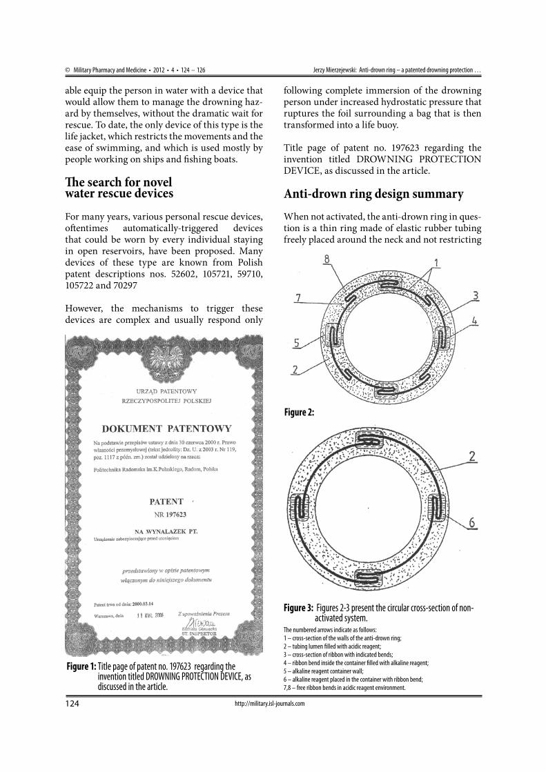
© Military Pharmacy and Medicine • 2012 • 4 • 124 – 126
124 http://military.isl-journals.com
Jerzy Mierzejewski: Anti-drown ring – a patented drowning protection …
able equip the person in water with a device that would allow them to manage the drowning haz-ard by themselves, without the dramatic wait for rescue. To date, the only device of this type is the life jacket, which restricts the movements and the ease of swimming, and which is used mostly by people working on ships and fishing boats.
The search for novel water rescue devicesFor many years, various personal rescue devices, oftentimes automatically-triggered devices that could be worn by every individual staying in open reservoirs, have been proposed. Many devices of these type are known from Polish patent descriptions nos. 52602, 105721, 59710, 105722 and 70297
However, the mechanisms to trigger these devices are complex and usually respond only
following complete immersion of the drowning person under increased hydrostatic pressure that ruptures the foil surrounding a bag that is then transformed into a life buoy.
Title page of patent no. 197623 regarding the invention titled DROWNING PROTECTION DEVICE, as discussed in the article.
Anti-drown ring design summaryWhen not activated, the anti-drown ring in ques-tion is a thin ring made of elastic rubber tubing freely placed around the neck and not restricting
Figure 1: Title page of patent no. 197623 regarding the invention titled DROWNING PROTECTION DEVICE, as discussed in the article.
Figure 2:
Figure 3: Figures 2-3 present the circular cross-section of non-activated system.
The numbered arrows indicate as follows:1 – cross-section of the walls of the anti-drown ring;2 – tubing lumen filled with acidic reagent;3 – cross-section of ribbon with indicated bends;4 – ribbon bend inside the container filled with alkaline reagent;5 – alkaline reagent container wall;6 – alkaline reagent placed in the container with ribbon bend;7,8 – free ribbon bends in acidic reagent environment.

© Military Pharmacy and Medicine • 2012 • 4 • 125 – 126 Review article
125http://military.isl-journals.com
the user’s movements in water. The diameter of the ring is slightly smaller than the diameter of human head, which prevents the device slipping off neck and head while making rapid move-ments in water.
Inside the tubing, there is a loose-fitted ribbon made of material characterized by high resist-ance to tear and to various ambient conditions.
Besides the ribbon, the inside of the tubing is filled with reagent 1, which is a solution of an acidic substance. The ribbon includes several tightly insulated bends comprising containers filled with reagent 2, which is a powdered alka-line substance.
The walls of containers filled with reagent 2 are made of material impenetrable to both materials, albeit relatively easy to tear.
Explanations to figures 2-5: Figures present-ing schematic cross-sections of the anti-drown device of the invention.
The principle of operationThe anti-drown ring is slightly extended and placed on the neck by sliding it over one’s head. When at risk of drowning, the user holds on to the device and tears it so as to unfold the folds of the ribbon inside the containers filled with reagent 2. Unfolding the ribbon leads to the con-tainer walls being ruptured, leading to the release of the alkaline reagent (reagent 2) from the con-tainers into the acidic reagent (reagent 1) that fills the tubing.
Mixing both reagents leads to an instantane-ous chemical reaction with the release of gas which automatically transforms the tubing into a pumped bladder. The quantities and rations of both reagents should be matched so as the formed gas extends the tubing to a diameter of ca. 10 cm. Thus, the bladder buoy formed around the neck provides displacement sufficient to maintain one’s head above water while allowing to main-tain free movements of entire body.
Loose fitting of the ribbon inside the tubing is provided by ribbon bends not placed in con-tainers with reagent 2 (free bends). This solution allows for slight expansion and multiple putting on and removing the anti-drown ring without damaging the walls of containers with reagent 2 and thus triggering the unwanted chemical reac-tion generating gas when putting on the device.
Such a design solution allows the resistance to be felt twice: the first time when the ribbon is straight-ened out at free bends, and the second involving the rupture of container walls, leading to contact between both reagents and generation of gas.
Leak protectionThe anti-drown ring made of thin rubber tubing may be at risk of accidental puncturing. In order to avoid such risk, the ring may be made of two parallel tubings fitted with identical ribbons and identical reagents.
The benefits of the anti-drown ringThe anti-drown ring is characterized by the sim-plicity of technical solution, capability of being put on and removed multiple times before a
Figure 6: Figure presents a transverse cross-section including the ribbon cross section indicated by arrow 3
Figure 4: Figure presents a schematic circular cross-section of the anti-drown ring filled with gas. Of note are the wider device lumen and straightened ribbon bends.
Figure 5: Figure presents a transverse cross-section of an anti-drown ring consisting of two parallel tubings.

© Military Pharmacy and Medicine • 2012 • 4 • 126 – 126
126 http://military.isl-journals.com
Jerzy Mierzejewski: Anti-drown ring – a patented drowning protection …
critical situation is encountered that requires the device to be activated.
The incentives to start production of the anti-drown ring should with no doubt include low costs of materials required for production (rub-ber tubings, ribbons, reagents) and, as a conse-quence, low prices that would make the device available to everyone.
Production and marketing of the anti-drown ringDesign preparation, production of a prototype batch and initiation of production would require efforts to obtain funds from state or European Union budget allocated to water rescue, or from private sponsors.
The prototype series should be made in order to raise interest of sponsors so as to acquire funds required for the development of techni-cal solutions for serial production of the anti-drown rings. Semi-technical scale implementa-tion works should be made in institutes (plants) developing rubber industry machinery.
The next marketing stage would involve the pro-motional campaign, mostly in the media as well as in the water industry aquatic sports and recre-ation centers, the military, the police and public schools of all levels.
The promotional campaign should include pres-entations in shopping centers and at the aquatic sports and water rescue equipment trade fairs.
After the anti-drown rings are well accepted by the general public, technical solutions should be developed so as to allow automated mass produc-tion of the devices.
Mass marketing of anti-drown ringsWith mass-production capabilities available, promotional campaigns should be organized at
the national and international aquatic sports and water rescue equipment trade fairs.
Likewise, long-term educational campaign should be started to promote the benefits of such a simple drowning protection device. Trainings should be provided mostly to individuals spending free time at or working on open water reservoirs.
Strategic goals of the promotional campaignDissemination of social awareness of the benefits of using the anti-drown rings.
Introduction of legal obligation to use anti-drown rings by individuals staying in open water reservoirs.
The ultimate goal would be to reduce the annual number of statewide drowning cases from sev-eral hundred to not more than several dozen accidents (wintertime and flood drowning).
The obligation to use the anti-drown rings would lead to a great change in the profile of water res-cue activities by limiting the latter to educational and preventive activities, including the control whether individuals staying in open water reser-voir wear the rings.
The planned strategic goals may be achieved by convincing entrepreneurship-supporting institu-tions and decision makers about the attractivity of the device, obtaining funds for prototyping, pro-motion, trainings and initial production stages.
Depending on the societal interest and demand for anti-drown rings, mass-scale production should be undertaken so as to meet the market demands.
As the experience in using the anti-drown rings would grow, the proposed model might be modi-fied provided that the general principle of opera-tion and use remain unchanged.

© Military Pharmacy and Medicine • 2012 • 4 • 127 – 132 Short Communication
127http://military.isl-journals.com
History of medicine
Speech made by Commandant of the Military Centre for Pharmacy and Medical Technology in Celestynów, Colonel DMSc Radosław Ziemba
Radosław Ziemba
Military Centre for Pharmacy and Medical Technology in Celestynow, Poland
Author’s address:Radosław Ziemba, Military Centre of Pharmacy and Medical Technique, ul. Wojska Polskiego 57, 05–430 Celestynów, Poland; e–mail: [email protected]
Received: 2012.12.022 • Accepted: 2012.12.02 • Published: 2012.12.08
Figure 1: Col Radosław ZIEMBA, M.D., Ph.D., the Commander of the Military Center for Pharmacy and Medical Technique, awards a saber on behalf of the staff of the Center to Col Krzysztof Kondracki, M.Sc. (Pharm.), as a symbol of bravery of the Polish army and of military duty and loyalty.
Ladies and gentlemen!We have an honourable opportunity to attend the ceremony of Mr Colonel Krzysztof KONDRACKI
parting with the uniform of the Polish Army. This ceremony is all the more solemn as it falls on 29 November – Day of Officer Cadet, which

© Military Pharmacy and Medicine • 2012 • 4 • 128 – 132
128 http://military.isl-journals.com
Radosław Ziemba: Speech made by Commandant of the Military …
Figure 3: The Commander of the Military Center for Pharmacy and Medical Technique awards an encyclopedia to Col Krzysztof Kondracki.
Figure 2: Col Krzysztof Kondracki, distinguished by the Commander with a photo against the background of the Banner of the Military Center for Pharmacy and Medical Technique.

© Military Pharmacy and Medicine • 2012 • 4 • 129 – 132 Short Communication
129http://military.isl-journals.com
Figure 4: The retiring Col Kondracki gives a speech to the guests attending the event.
is a special day for every officer. The officer cadet period is an introduction to an officer epic, and Mr Colonel started the latter on 31 June 1978 with studies at the gen. Bolesław Szarecki Medi-cal Military Academy in Łódź at Pharmaceutical Faculty. It’s worth adding that he simultaneously studied pharmacy at the then existing Medical Academy (civilian university) in Łódź.
He began the studies after obtaining a certificate of secondary education at Frédéric Chopin Sec-ondary School in Sochaczew.
I wish to add that Mr Colonel Kondracki has continued the traditions of the officer ethos, as he was born into the family of an officer who served in 32nd regiment of fighting and recon-naissance air forces in Sochaczew. It is with this town that Mr Krzysztof bound his life from his birth, through Primary School, until he obtained his certificate of secondary education.
On Łódź Soil he made important decisions of his life, as in January 1982 he married Wanda, here
his son Maciej was born, now also a holder of an MPharm degree. From 10 May 2010 to 6 May 2011, his son participated in the 7th change of Polish contingent in Afghanistan. Thus, Mr Kon-dracki’s son is also continuing the family tradi-tions in some way.
As far as the children are concerned, the hero of today’s celebrations adopted gender parity a few years ago, as in April 1985 his daughter Agata was born in Celestynów – now as a physiotherapist.
It is with Celestynów, or the Military Centre for Pharmacy and Medical Technology to be precise, that he bound his first eight years after complet-ing the studies and gaining the rank of an officer on 26 June 1983. In 1986, he was commissioned as a lieutenant (as a section manager), and in 1989 – as a captain. From 18 November 1991 he bound his life with the Central Clinical Hospital of Medical Military Academy at the post of a sen-ior pharmacist where in 1993 he was promoted to the rank of major at the post of deputy manager of the pharmacy and from 23 February 1996 he

© Military Pharmacy and Medicine • 2012 • 4 • 130 – 132
130 http://military.isl-journals.com
Radosław Ziemba: Speech made by Commandant of the Military …
took up the post of the manager of the pharmacy and was promoted to the rank of lieutenant-colo-nel in 1998.
On 12 May 2003 he became the Manager of the Base at the already existing Military Institute of Medicine. Then, on 1 April 2007 he joined the Inspection Office of Military Health Care at the post of deputy Chief Pharmaceutical Inspector of Polish Army and was commissioned as colonel. At this post, at this pace and with this rank, he served until 15 October 2012.
Mr Colonel earned high recognition and respect through his self-discipline, tact, loyalty, author-ity among colleagues, principled attitude to his service, tactful style of behaviour among co-workers. Always modest, genuine, without exces-sive pretensions or elation. He always showed
helpfulness and responsibility, fulfilling a discur-sive approach to the idea of officer and pharma-ceutical ethos. You are, Mr Colonel, a Paracelsus in the uniform for us.
Mr Colonel, a specialist in many areas of phar-macy – especially military one – we have immor-talized you in a photograph against a background of the banner of the Military Centre for Phar-macy and Medical Technology in Celestynów. It is there that you started your initiation into the service.
We also have the great honour to hand this piece of cold steel called a sabre. Having different pat-terns, through centuries it lighted with the flash of the blade often heroic deeds of the Polish army, accompanied cavalry troops, and today adds col-our to parades, state and military celebrations. So let it be in your hands the symbol of fulfill-ing a soldier’s duty and loyalty in the officer community.
Let the motto engraved at its handle:
“A reliable friend is recognized in an uncertain sit-uation…” determine your acts in this stage of life.

© Military Pharmacy and Medicine • 2012 • 4 • 131 – 132 Short Communication
131http://military.isl-journals.com
IMĆ Panu Pułkownikowi Krzysztofowi KONDRACKIEMU idącemu w nowy seans życia „pod kapeluszem”Akt one, scena pierwsza
Przez dyskretny otwór w kurtyniewejrzeć tylko w zakulisy wierszaz historii losu spojrzeć o pustej godziniekiedy widownią są oczy, a uszy słuchowiskiemi cienie rzęs na uboczuwięc rozprawimy się z jednym nazwiskiemza którym stoi ten oto…
Krzysztof zawinął bieg swojej historiichoć wszystko w niej bywa odmienne.Tak pewien dzień październikaw tysiąc dziewięćset pięćdziesiątym dziewiątymwyłonił go nam na tę ziemię.Waga mu rysy rzeźbiła na twarzykiedy wodził oczyma po sochaczewskim niebie.
Tamte twarze szkolnego ornamentutamta pamięć ust jak pieczęci,grona pedagogów, edukacji momentów.W cenzurze świadectw różne uczniowskie chęci,Tropem w czasy dojrzałe, lato ciała,spokój pajęczany, krzewy obute w kiścikołysały beztrosko – niebieskie lata młodości.
W szkolnych wierszach bagnetów strome błyskawicetam nasza chluba i naszych możliwości granice,co noc pośród latarni szukało się srebrnych kawiarniw dzień nad biurkiem trzeba było się zgiąć,pod kluczem szopenowskich nut ksiąg…
Przyszedł dzień, kiedy strugi mazowieckiego śwituwysłały w misję wyroku Dioskurydesa przesłaniena łódzkim bruku podchorążacko — studenckie ślubowaniesłowa Gaudeamus, żołnierskiej przysięgicyzelowały drogę do etosu oficerskiej wstęgi.W panteonie dwudziestego wieku.
Powroty z balów, dyskotek, klęsk, wypraw krzyżowychskacząc przez Pietrynę jak po kolorowych krach,unosząc potem drewniane powieki,przechodząc niebieską granicę w alchemii farmaceutyc-znych praw.Od Hipokratesa idei do FarmakopeiPrzy egzaminie – Panie Boże zbaw.
Akt two, scena drugaNa ozłoconym przez bliskość balkoniePani Wanda założyła dłonieniech mnie Krzysztof z nadziei wybawi w przestworzach mojego świata się zjawizimowego stycznia szesnastegozłotych obrączek ikonaoświetlała marsza Mendelsona…
W sierpniu zszedł z niebiosaMaciej, syn łaskawy, pociecha rodzicówjako w maju rosa co pada na trawy,cicho zszedł w objęcia do komnaty matkijak majowa rosa co pada na kwiatkinazwisko jak totem utrwalone potem.
Nie zapominaj jaka to siław czerwcowe słońce gwiazdki zdobyłaetos olśniła i animuszu do farmacjidodała Panu od wtedy co chwila.Z tego posłania od łódzkiej ziemiw Celestynowie między nowemi.
Akt three, scena trzeciaTaki to czas był, co dzierżył w swej treścimłodości radości i dobytek mnogidziecięciom płaci w lasach mazowieckichodtąd z Agatką przechodziłeś wszystkie drogi.Czasy edukacji, tym razem dla dziecico troską ojcowską trzeba było cieszyć.
Można by wieczność zobaczyć wśród nocykiedy się z wojskową farmacją połączyłjak wielki pierścień życia nieskończonej mocyna tło ogromnych cieni do tej właśnie sprawyrzucono kapitana w konszachty Warszawy.Jak u światłych ludzi czasem bywa,przyszła kariera nawet całkiem błyskotliwa.
Czasem tony przedziwne bezrozumnej skargi,obok nich jak z lutni fantazje i fraszkiróżnej cierpkości dowcipnego lotu igraszki.Obok nich pokłony i uciech zasadzki wszelakie,cierpkie lecz drogie, jakby skarby jakierozpraszają człowieka , z doświadczeń bogaty.
Nadziejo święta, pokoro wysokajak szczyt niebios wśród planet pochoduTwój wielki szlak jest wskazany dla okaTy znajdziesz gniazdo jak ptak opierzonywyfruniesz łatwo, sam to dostrzec zdołasz.Powracaj znów zatem w Celestynowskie strony…
Krzysztof BarczewskiAnno domini 29 listopada 2012

© Military Pharmacy and Medicine • 2012 • 4 • 132 – 132
132 http://military.isl-journals.com
Radosław Ziemba: Speech made by Commandant of the Military …

© Military Pharmacy and Medicine EDITORIAL POLICY AND GENERAL INFORMATION
133http://military.isl-journals.com/
Military Pharmacy and Medicine is an international, peer-reviewed scientific journal that publish original articles based on own research, as well as review articles and case reports in the field of pharmacy and military medicine, and modern solutions in the field of military and civilian healthcare based on the latest national and international achievements.
Military Pharmacy and Medicine is quarterly interdisciplinary journal of Military Centre of Pharmacy and Medical Technique in Celestynów, Poland, published in English on scientific, socio- professional and training issues of Military Pharmacy and Medi-cine. Journal appears continuously and systematically in printed (primary version) and on-line version since 2008 at: http://mili-tary.isl-journals.com/ and information contained therein are con-tinuously updated, but not less frequently than quarterly.
The editors endorse the principles embodied in the Declaration of Helsinki and expect that all investigations involving humans will have been performed in accordance with these principles. For animal exper-imentation reported in the journal, it is expected that investigators will have observed the Interdisciplinary Principles and Guidelines for the Use of Animals in Research, Testing, and Education issued by the New York Academy of Sciences Adhoc Committee on Animal Research. All human and animal studies must have been approved by the investiga-tor’s Institutional review board. It is recommended to enclose a copy of that document to a submitted manuscript.
Editors Military Pharmacy and Medicine in the daily practice refer to the guidelines of the Committee on Publications Ethics concerning Code of Conduct and Best Practice Guidelines for Journal Editors (http://publicationethics.org/resources/guidelines).
1. Review process and submission rulesEditors consider only submissions in English. Manuscripts are evalu-ated on the basis that they present new insights to the investigated topic, are likely to contribute to a research progress or change in clinical practice or have the desirable teaching/training value. The correctness ensures Editor-in-Chief, Deputy Editor, Section Editors, Statistical Edi-tor, reviewers and Linguistic Editors.
The signature of the corresponding author on the letter of submis-sion signifies that:
1) paper is original and created by you (not copied),2) paper has not been published previously or submitted elsewhere
for review and a copyright transfer,3) it is understood that all authors listed on a manuscript have
agreed to its submission.
Received manuscripts are first examined by the Military Pharmacy and Medicine editors due to preparation of the manuscript, pho-tographic documentation, and all authors consent to publication. Manuscripts with insufficient priority for publication are rejected promptly. Incomplete packages or manuscripts not prepared in the advised style will be sent back to authors without scientific review.
The authors are notified with the reference number upon manu-script registration at the Editorial Office. The registered manuscripts are sent to at least two independent experts for scientific evaluation. Competent reviewers designate Editor-in-Chief. Reviewers prepare opinions that contain reasoned recommendations and suggestions of corrections and additions to content and form of the article.
In case of papers written in a foreign language at least one of the reviewers is affiliated to a foreign institution. Reviewed paper and reviewers did not come from the same institution.
The author and the reviewer are anonymous to each other according to double-blind review policy.Rejection requires two negative reviews. Editor-in-Chief reserves the right to refuse to print a paper containing the results of studies in which ethical principles are not respected according to the Declara-tion of Helsinki in 1964, Tokyo in 1975 and the recommendations of the World Health Organization in 1982.
Submitted papers are accepted for publication after a two posi-tive opinion of the independent reviewers, who agreed that the paper can be published in present form. If the reviewers differ in their opinions, or feel that the manuscript should be accepted only after the corrections, editors may take a decision to send paper to another reviewer in order to settle or return it to the authors for correction.The final decision on acceptance for publication or to reject belongs to competences of the Editorial Board and is not subject to appeal. Editorial Board decisions do not have to justify.The reviewing process usually takes 3-6 weeks, however Editors can-not guarantee the date of publishing.Military Pharmacy and Medicine publishes an updated list of review-ers on the website, as well as an annual list of reviewers in the last issue of the journal (every year).
2. Conflict of interestsAuthors should disclose contribution of individual authors to prepa-ration of manuscript (with a list of their affiliations) in detail, i.e. provide information who is the author of concept, premises, meth-ods, protocol etc.Authors of research articles should disclose at the time of submis-sion any financial arrangement they may have with a company whose product figures prominently in the submitted manuscript or with a company making a competing product. Such informa-tion will be held in confidence while the paper is under review and will not influence the editorial decision, but if the article is accepted for publication, the editors will usually discuss with the authors the manner in which such information is to be communi-cated to the reader.
Because the essence of reviews and editorials is selection and inter-pretation of the literature, the Military Pharmacy and Medicine expects that authors of such articles will not have any financial inter-est in a company (or its competitor) that makes a product discussed in the article.Military Pharmacy and Medicine policy requires that reviewers, associate editors, editors, and senior editors reveal in a letter to the Editor-in-Chief any relationships that they have that could be con-strued as causing a conflict of interest with regard to a manuscript under review. The letter should include a statement of any financial relationships with commercial companies involved with a product under study.
3. PermissionsMaterials taken from other sources must be accompanied by a writ-ten statement from both author and publisher giving permission to the Military Pharmacy and Medicine for reproduction. Obtain
Editorial policy and general information

© Military Pharmacy and Medicine
134 http://military.isl-journals.com/
EDITORIAL POLICY AND GENERAL INFORMATION
permission in writing from at least one author of papers still in press, unpublished data, and personal communications.
4. Patients confidentialityChanging the details of patients in order to disguise them is a form of data alteration. However authors of papers are obliged to ensure patients privacy rights. Only clinically or scientifically important data are permitted for publishing. Therefore, if it is possible to iden-tify a patient from a case report, illustration or paper, Military Phar-macy and Medicine Editors ask for a written consent of the patient or his/her guardian to publish their data, including photograms prior to publication. The description of race, ethnicity or culture of a study subject should occur only when it is believed to be of strong influ-ence on the medical condition in the study. When categorizing by race, ethnicity or culture, the names should be as illustrative as pos-sible and reflect how these groups were assigned.
5. Copyright transferUpon acceptance, authors transfer copyright to the Military Phar-macy and Medicine. Once an article is accepted for publication, the information therein is embargoed from reporting by the media until the mail date of the issue in which the article appears.
Upon acceptance all published manuscripts become the permanent property of the Military Centre of Pharmacy and Medical Technique in Celestynów, Poland as the Publisher of the Military Pharmacy and Medicine, and may not be published elsewhere without written per-mission from the Military Centre of Pharmacy and Medical Tech-nique in Celestynów, Poland.
The date of acceptance for printing shall be the date of sending the final version of the article. Editorial provides one copy printed arti-cle for the correspondence author.
6. DisclaimerEvery effort is made by the Publisher and Editorial Board to see that no inaccurate or misleading data, opinion or statement appear in the Military Pharmacy and Medicine. However, they wish to make it clear that the data and opinions appearing in the articles and adver-tisements herein are the responsibility of the contributor, sponsor or advertiser concerned. Accordingly, the Publisher and the Edito-rial Board accept no liability whatsoever for the consequences of any such inaccurate of misleading data, opinion or statement. Every effort is made to ensure that drug doses and other quantities are pre-sented accurately. Nevertheless, readers are advised that methods and techniques involving drug usage and other treatments described in this Military Pharmacy and Medicine, should only be followed in conjunction with the drug or treatment manufacturer’s own pub-lished literature in the readers own country.
7. Qualification criteria for manuscripts
Editorial Board of Military Pharmacy and Medicine takes under consideration for publication original articles in experimental and clinical medicine and related disciplines with the understanding that neither the manuscript nor any part of its essential substance, tables or figures have been published previously in print form or electronically and are not taken under consideration by any other publication or electronic medium. Copies of any closely related manuscripts should be submitted to the Editor along with the
manuscript that is to be considered by the Military Pharmacy and Medicine. The Editor discourages the submission of more than one article dealing with related aspects of the same study.
Each submission packet should include the statement signed by the first author that the work has not been published previously or sub-mitted elsewhere for review and a copyright transfer.
8. Categories of articlesAccepted manuscripts are published in the following journal sections:
1) Original articles: reports of previously unpublished results from scientific experiments conducted by the authors in order to confirm or refute a clearly identified hypothesis. Most of the articles published in a given issue will belong to this category.
2) Review articles: reports on the current state of knowledge in a given area or field of study, especially current controversies, theoretical and practical approaches to the issues, unresolved problems, etc., with carefully selected references to the litera-ture. Such articles are typically commissioned by the editors of Military Pharmacy and Medicine, though an unsolicited review article may be accepted if it is exceptionally interesting and carefully prepared.
3) Case Reports: detailed description of the diagnosis and/or treat-ment of 1-3 individual patients, with particular emphasis on any atypical or difficult aspects of therapy in this particular case that may be of interest to readers.
4) Short Communications: brief descriptions of selected clinical solutions to particular problems; possibly also new discoveries not yet experimentally confirmed.
5) Opinion articles: authorial discussions of important issues, controversies, and schools of thought in the area of physi-otherapy; also, educational (training) articles.
9. Preparation of manuscriptGuidelines for submission in Military Pharmacy and Medicine are in accordance with: Uniform Requirements for Manuscripts Submit-ted to Biomedical Journals (N Eng J Med, 1997; 336: 309-15. www.acponline.org/journals/resource/unifreqr.htm).
The submitted manuscript should be:
1) Original and prepared according to the current spelling and terminology. Sent to editing in electronic form (by e-mail or by regular post on CD/DVD) in one of the following formats: *.doc, *.docx, *.rtf, *.odt, *.sxw, *.sdw.
2) Electronic file should require the following format (without spaces between last names): •LastNameFirstNameInitial-ArticleTitle i.e. SmithJ-Recent advances in clinical… or in case of multi-authorship submission
• (FirstAuthor)LastNameFirstNameInitial_et al-ArticleTitle i.e. SmithJ_et al-Recent advances in clinical…
3) Title page should have the following information: •Manuscript full title – 12-point typeface, bold; •Full names of all authors; •Type of article (original, review, case report etc.); •Affiliations of the authors; •Full name, address, phone number, fax number and e-mail of the

© Military Pharmacy and Medicine EDITORIAL POLICY AND GENERAL INFORMATION
135http://military.isl-journals.com/
corresponding author responsible for manuscript preparation, in the following format:
Antoni Penc MD PhD, Department of Radiology, University Hospi-tal, Dobra 22, 01-153 Warsaw, POLAND; phone (+48)227786734, fax: (+48) 227776671; e-mail: [email protected];
•Summary - no more than 15 lines, single-space; •Key words (5 to 10) or short phrases should be written at the bottom of the page including summary. The use of the items included in Index Medicus (Medical Subject Headings) is required;
•Source(s) of support in the form of grants (quote the number of the grant) equipment, drugs etc;
• Statement that neither this manuscript nor one with substantially similar content or research under my (our) authorship has been published or was sent for publication elsewhere.
4) Structured Summary: (up to 250 words), consisting of the follow-ing sections: Background and study aim, Material and methods, Results, Conclusions:e) Introduction (or Background): should contain scientific
rationale and the aim of the study or (in case of a review) purpose of the article;
f) Material and methods: brief description of the study; in the case of review article - characteristics of the literature; for a case study - a brief description of the patient, the main parameters, etc.
g) Results: concisely and reasonably summarize the findingsh) Conclusions: the principal conclusions (in Summary: 1-2)
drawn by the authors of the presented results. For review papers the above-mentioned structure is not required.
9) TEXT. The text of the article should be divided to six paragraphs labeled: Introduction (or Background), Material and Methods, Results, Discussion, Conclusions, References. Prior references, if necessary, you can attach Acknowledgements, and at the end of work - Appendix. Each of these sections must be clearly separated with the bold title.
Where appropriate, depending on the content of the article, you can use a different layout, however, on condition that the struc-ture of work is clear, transparent and consistent. The editors reserve the right to request the author(s) to improve the structure of manuscript.
10) Introduction (or Background) should give the scientific and/or clinical rationale for researching the given topic, the primary issues and controversies, an explanation of the aim of the study and the primary thesis.
11) Material and Methods should contain essential information regarding how the experiment or research was conducted, including the essential characteristics of the experimental and control groups (age, gender), inclusion and exclusion criteria, and the randomization and masking (blinding) method used. The protocol of data acquisition, procedures, investigated parameters, methods of measurements and apparatus should be described in sufficient detail to allow other scientists to reproduce the results. In the case of published methods, the names with appropriate references should be given. References and a brief description should be provided for methods that have been published but are not well known, whereas new or substantially modified methods should be described in detail. The rationale for using such new or unknown methods should be discussed, along with a balanced evaluation of these methods, not omitting their limitations. Drugs and other chemicals should be precisely identified, including the generic name, dosage, and route of administration.
The statistical methods should be described in detail to enable verification of the reported results.Information regarding the patients’ informed consent should be included in the text of the article (see above: Patient confi den- tiality). Study subjects should be identified only by arbitrarily assi-gned initials or numbers. Any information contained in photo-graphs, images, or other illustrations that could serve to reveal the person’s identity should be thoroughly camouflaged or concealed. The faces of persons appearing in photographs should be masked or covered with a black band, unless for compelling reasons this is impossible.
12) Results concisely and reasonably summarize the findings in the form of text, tables and figures arranged in a logical and internally self-consistent manner. The number of tables and figures should be limited to those absolutely needed to confirm or refute the thesis. Data given in graphs and tables should not be automatically repeated in the text. The number of observations should be clearly indicated, as well as exclusions or losses to observation. Any complications that may occur in treatment or examination should be reported.
13) Discussion should deal only with new and/or important aspects of the results obtained, without repeating in detail data or other material previously presented in Background or Results. The Discussion should focus on the theoretical implications and/or practical consequences of the findings, including suggestions for further research. The Discussion should compare the results of the present study to those obtained by other investigators mentioned in the text.
14) Conclusions must be linked with the goals of the study. New hypotheses with recommendations for further research should be advanced only when fully warranted and explicitly justi-fied. Include recommendations when appropriate. Unqualified statements and conclusions not supported by the data obtained should be avoided.
15) Acknowledgements list all those who have contributed to the research but do not meet the criteria for authorship, such as assistants, technicians, or department heads who provided only general support. Financial and other material support should be disclosed and acknowledged. References, chosen for their importance and accessibility, are numbered consecutively in the order of their occurrence in the text. References first cited in tables or figure legends must be numbered in such a way as to maintain numerical sequence with the refer-ences cited in the text. The style of references is that of Index Medicus. When an article has six or fewer authors, all should be listed; when there are seven or more, only the first three are listed, then “et al.”
16) Original papers and review papers may not exceed the standard typewritten pages 10-20, and case studies – 4 pages, including references, summary, tables and figures.
Editors may agree to exceed the number of pages in case of: sum-maries of habilitation dissertation and the habilitation dissertation on degree of doctor of pharmaceutical and medical sciences.
17) One page of manuscript should contain 30 lines, with about 60 characters each (approx. 1800 characters per page). The text must be written in Times New Roman 12-point, double-spaced (except references, tables, captions, etc.), with the left margin, 2.5 cm wide, but without the right margin, or the comment. Do not center the title and heading, do not use tabs and blank lines between paragraphs or calculations. Use only bold and italic.

© Military Pharmacy and Medicine
136 http://military.isl-journals.com/
EDITORIAL POLICY AND GENERAL INFORMATION
18) Tables and illustrations. Number tables consecutively in the order of their first citation in the text, and supply a brief title for each. Give each column a short or abbreviated heading. It is recommended the simplest possible arrangement of the table, without unnecessary horizontal or vertical rules. Place explanatory matter in footnotes, not in the heading. The footnotes should be numbered separately, starting with 1 for each table. Explain in footnotes all nonstandard abbreviations that are used in each table. Type or print out each table on a separate sheet of paper. Be sure that each table is cited in the text. Tables, Illustrations, Figures, Photographs etc. should be numbered and describedType or print out each Tables, Illustrations, Figures, Photo-graphs etc. on a separate sheet of paper. In the main text should be noted the place of insertion of each Tables, Illustrations, Fig-ures, Photographs etc. The number of tables should be reduced to minimum. Figures (including maps), and photographs are placed in a separate file(s).
If the Figures and Photograph contain text to be translated, the file(s) containing must be editable or author(s) should send them in English language. Figures and photographs should be professionally drawn and photographed; freehand or typewritten lettering is unacceptable. Titles and detailed explanations belong in the legends for illus-trations, not on the illustrations themselves. Each figure should have a label pasted on its back indicating the number of the figure, author’s name, and top of the figure. Do not write on the back of figures or scratch or mar them by using paper clips. Do not bend figures or mount them on cardboard.Figures should be numbered consecutively according to the order in which they have been first cited in the text. If a figure has been published, acknowledge the original source and sub-mit written permission from the copyright holder to reproduce the material. Permission is required irrespective of authorship or publisher, except for documents in the public domain. Photo-graphs should be color or black & white glossy prints with num-bers and descriptions on the back, following the pattern: title, authors, number of the photograph, its description. All photo-graphs are printed as standard black and white. You can print photos in full color, for an additional fee.Photomicrographs should have internal scale markers. Symbols, arrows, or letters used in photomicrographs should contrast with the background. If photographs of people are used, either the subjects must not be identifiable or their pictures must be accom-panied by written permission to use the photograph.
19) References. In all cases correct punctuation should be used to divide the parts of the reference to the cited position. Because of the possibility of modifications or amendments, references to materials from Internet should include the file viewing or downloading date. If there are more than three authors, the names of the first three should be listed and then „et al.“ should be used. Abbreviated titles of journals cited should be consistent with MEDLINE.
Papers published in journals: 12. Pui CH, Behm FG, Raimondi SC et al: Secondary acute myeloid leukemia in children treated for acute lymphoid leu-kemia. N Eng J Med, 1989; 321(3): 136–42.
Book chapters: 3. Kowalczyk JR: Cytogenetics of secondary leukemias. In: Becher R, Sandberg AA, Schmidt CG (eds.): Chromo-somes in Hematology. W. Zuckschwerdt Verlag, Munchen, 1986,pp. 125–45.
Electronic materials (Internet): 6. Martin JM: A software for the description of workplaces in the PRS system. http://www.matforsk.no/ola/fisher.htm (accessed 29.08.2002).
Avoid using abstracts or review papers as references. Unpub-lished observations and personal communications cannot be used as references. If essential, such material may be incorporated in the appropriate place in the text.
20) Units of Measurement. Measurements of length, height, weight, and volume should be reported in metric units (meter, kilogram, or liter) or their decimal multiples. Temperatures should be given in degrees Celsius. Blood pressures should be given in millimeters of mercury.
All hematological and clinical chemistry measurements should be reported in the metric system in terms of the International Sys-tem of Units (SI). Alternative or non-SI units should be added in parenthesis. Identify statistical measures of variations such as standard deviation and standard error of the mean. If you use data from another pub-lished or unpublished source, obtain permission and acknowledge them fully
21) Abbreviations and Symbols. Use only standard abbreviations. Avoid abbreviations in the title and abstract. The full term for which an abbreviation stands should precede its first use in the text unless it is a standard unit of measurement.
10. Sending the manuscript to the journal
Authors are requested to submit the manuscript in electronic form on e-mails: [email protected] or http://military.isl-journals.com/index.php?/authors/login/
Manuscripts must be accompanied by a covering letter signed by all co-authors. This must include:
1) information on prior or duplicate publication or submission else-where of any part of the work as defined earlier in this document;
2) disclose contribution of individual authors to preparation of a publication (with a list of their affiliations); editors make an effort to prevent cases of misconduct (ghostwriting, guest authorship);
3) a statement of financial or other relationships that might lead to a conflict of interest (see below);
4) a statement that the manuscript has been read and approved by all the authors, that the requirements for authorship as stated earlier in this document have been met.
The manuscript must be accompanied by copies of any permission to reproduce published material, to use illustrations or report information about identifiable people, or to name people for their contributions.Editors consider the above conditions to be fulfilled if the signature of the first author was made.
11. Final remarks: 1)The editors reserve the right to correction of grammatical, stylistic
defects or shortening paper without the agreement with authors. The paper does not qualify for the print may be returned at the request of the author.
2)Translations from Polish into English are made by the publisher. Individual translation is allowed. Then the author(s) should clearly indicate that the paper require or not language correction.
3)Authors, members of the Scientific Board, members of the Editorial Board, reviewers and libraries of medical universities receive one copy of the Military Pharmacy and Medicine. Copy in PDF format is allowed.

Trze
cia st
rona
okład
ki