
Enhancement of Hepatitis C Virus RNA Replication by Cell Culture-Adaptive Mutations
Upload
khangminh22Category
view
2download
0
Micro-RNA Modulation of Insect Virus
Replication
Verna Monsanto-Hearne and Karyn N. Johnson*
School of Biological Sciences, University of Queensland, Brisbane, Australia.
*Correspondence: [email protected]
https://doi.org/10.21775/cimb.034.061
AbstractThe outcome of virus infection in insects is impacted by regulation of both host and virus gene expres-sion. A class of small RNAs called micro-RNAs (miRNA) have emerged as important regulators of gene expression that can influence the outcome of virus infection. miRNA regulation occurs at a com-paratively late stage of gene expression, allowing for rapid control and fine-tuning of gene expression levels. Here we discuss the biogenesis of miRNAs from both host and virus genomes, the interactions that lead to regulation of gene expression, and the miRNA–mRNA interactions that lead to either antivirus or provirus consequences in the course of virus infection in insects.
Introduction: insect–virus
interaction and microRNAs
The course and outcome of virus infection is reliant on intricate and complex host–virus interactions (Rapicetta et al., 2002; Whitton et al., 2005; Clyde et al., 2006). With small genomes and limited coding capacity, viruses hijack host factors for almost every step of virus infection while simultaneously evading the host antiviral system (Ploegh, 1998; Ahlquist et al., 2003; Cherry and Silverman, 2006). The virus exploits the host’s cell factors and the host coun-ters the viral assault by restricting viral access to cell factors and/or mounting its antiviral immune response (Ahlquist et al., 2003, 2005; Cherry and Silverman, 2006; Huszar and Imler, 2008; Kemp and Imler, 2009; Delpeut et al., 2012; Vodovar and
Saleh, 2012; Xu and Cherry, 2014; Mussabekova et al., 2017). Many molecular components medi-ate and are mediated by this host–virus cross-talk, including microRNAs.
microRNAs (miRNAs) are a large class of highly conserved, ≈ 22 nt non-coding RNAs that regulate gene expression. Compared to the more upstream regulatory mechanisms such as transcriptional regulation and chromatin remodelling, regulation by miRNA occurs at a later stage of gene expres-sion, allowing for rapid control and fine-tuning of gene expression levels (Chen et al., 2013). Comple-mentary binding of at least the seed region (second to eighth nucleotide from the 5′-end) of the ≈ 22 nt long miRNA with target mRNAs influences mRNA stability and/or translational efficiency and conse-quently modulates genes involved in a spectrum of important cellular processes (Bartel, 2009; Fabian et al., 2010; Pasquinelli, 2012), including immune response and host–virus interactions (Lindsay, 2008; Lodish et al., 2008; Tsitsiou and Lindsay, 2009; Xiao and Rajewsky, 2009; O’Connell et al., 2010; Li and Rana, 2014).
Changes in miRNA abundance during the host–virus interaction can direct the course of virus pathogenesis (Monsanto-Hearne and Johnson, 2018). There are two distinct motifs in terms of miRNA effect on virus infection: while regulation of some miRNAs have antivirus consequences, regulation of others have provirus consequences. Antivirus consequences can arise by either one of the two mechanisms: binding of host miRNA-host mRNA or host miRNA-virus mRNA. On the other
Curr. Issues Mol. Biol. (2020) Vol. 34 caister.com/cimb
Monsanto-Hearne and Johnson62 |
hand, provirus consequences can occur as a result of any of four binding events: host miRNA-host mRNA, host miRNA-virus mRNA, virus miRNA-virus mRNA, and finally virus miRNA-host mRNA. The growing body of literature on insect-virus infection in the context of miRNA regulation shows that there are many differentially regulated miRNAs that can have either antiviral or provi-ral outcomes. The net effect of cooperative and opposing impacts of the suite of miRNAs on the host–virus interaction then influences whether or not virus infection ensues. This chapter starts with a discussion on miRNA genomics, biogenesis, and regulation of target mRNAs, many of which have been established through the study of insect model Drosophila melanogaster. The chapter then details miRNA–mRNA interactions leading to either anti-virus or provirus consequences that determine the course of virus infection in insects.
miRNA: genomics, biogenesis to
regulation of target
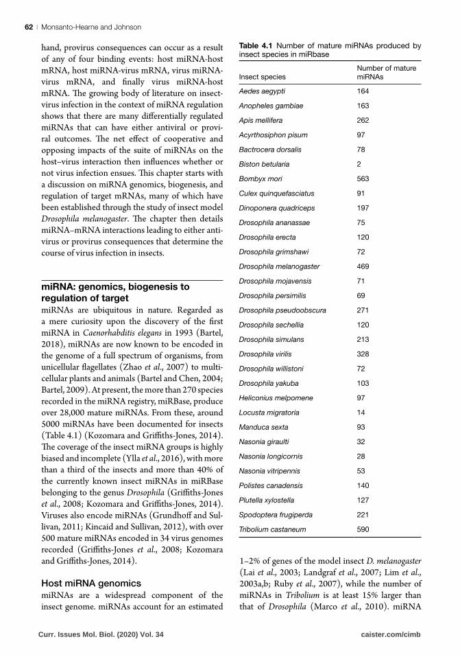
miRNAs are ubiquitous in nature. Regarded as a mere curiosity upon the discovery of the first miRNA in Caenorhabditis elegans in 1993 (Bartel, 2018), miRNAs are now known to be encoded in the genome of a full spectrum of organisms, from unicellular flagellates (Zhao et al., 2007) to multi-cellular plants and animals (Bartel and Chen, 2004; Bartel, 2009). At present, the more than 270 species recorded in the miRNA registry, miRBase, produce over 28,000 mature miRNAs. From these, around 5000 miRNAs have been documented for insects (Table 4.1) (Kozomara and Griffiths-Jones, 2014). The coverage of the insect miRNA groups is highly biased and incomplete (Ylla et al., 2016), with more than a third of the insects and more than 40% of the currently known insect miRNAs in miRBase belonging to the genus Drosophila (Griffiths-Jones et al., 2008; Kozomara and Griffiths-Jones, 2014). Viruses also encode miRNAs (Grundhoff and Sul-livan, 2011; Kincaid and Sullivan, 2012), with over 500 mature miRNAs encoded in 34 virus genomes recorded (Griffiths-Jones et al., 2008; Kozomara and Griffiths-Jones, 2014).
Host miRNA genomicsmiRNAs are a widespread component of the insect genome. miRNAs account for an estimated
1–2% of genes of the model insect D. melanogaster (Lai et al., 2003; Landgraf et al., 2007; Lim et al., 2003a,b; Ruby et al., 2007), while the number of miRNAs in Tribolium is at least 15% larger than that of Drosophila (Marco et al., 2010). miRNA
Table 4.1 Number of mature miRNAs produced by insect species in miRbase
Insect species
Number of mature miRNAs
Aedes aegypti 164
Anopheles gambiae 163
Apis mellifera 262
Acyrthosiphon pisum 97
Bactrocera dorsalis 78
Biston betularia 2
Bombyx mori 563
Culex quinquefasciatus 91
Dinoponera quadriceps 197
Drosophila ananassae 75
Drosophila erecta 120
Drosophila grimshawi 72
Drosophila melanogaster 469
Drosophila mojavensis 71
Drosophila persimilis 69
Drosophila pseudoobscura 271
Drosophila sechellia 120
Drosophila simulans 213
Drosophila virilis 328
Drosophila willistoni 72
Drosophila yakuba 103
Heliconius melpomene 97
Locusta migratoria 14
Manduca sexta 93
Nasonia giraulti 32
Nasonia longicornis 28
Nasonia vitripennis 53
Polistes canadensis 140
Plutella xylostella 127
Spodoptera frugiperda 221
Tribolium castaneum 590
Curr. Issues Mol. Biol. (2020) Vol. 34 caister.com/cimb
miRNA Modulation of Virus Replication | 63
loci are found throughout the genome: inter-genic miRNAs are embedded between clusters of genes, while intronic miRNAs are found within introns of protein-coding regions. The former is transcribed independently by their own transcrip-tion units, whereas the latter are processed either by their host transcription units or by their own transcription units. Some miRNAs are encoded individually, while others are found in clusters with other miRNAs (Lee et al., 2002; Kim and Kim, 2007; Carthew and Sontheimer, 2009).
miRNAs are transcribed from the genome using similar mechanisms as mRNAs. miRNA promoters are typically the same as mRNA-encoding promot-ers in terms of distance from the miRNA-encoding sequence and presence of recognition elements. The miRNA transcription initiation site can be as close as a few hundreds of base pairs (bp) to the miRNA-encoding sequence or can be as far as 20 kb. miRNA recognition promoter elements can include TATA box, transcription factor II B recognition element (BRE), initiator (Inr) motif 10 element, and down-stream promoter element (Ozsolak et al., 2008).
miRNA biogenesis to recognition of targets
Canonical biogenesis of host miRNA
miRNA biogenesis canonically begins with the transcription of miRNA-encoding genomic regions (Rodriguez et al., 2004) by RNA polymerase II (Lee et al., 2004a) (Fig. 4.1). The Pol II-generation of the miRNA primary transcripts (pri-miRNAs) is coupled with pri-miRNA processing. Similar to most other Pol II-generated products, pri-miRNAs are methylguanosine-capped at the 5′-end and polyadenylated at the 3′-end (Bracht et al., 2004; Cai et al., 2004; Lee et al., 2004a). The pri-miRNA folds back on itself to form a hairpin structure and then undergoes sequential nuclear and cytoplasmic processing to yield mature duplex miRNA.
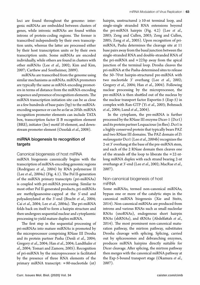
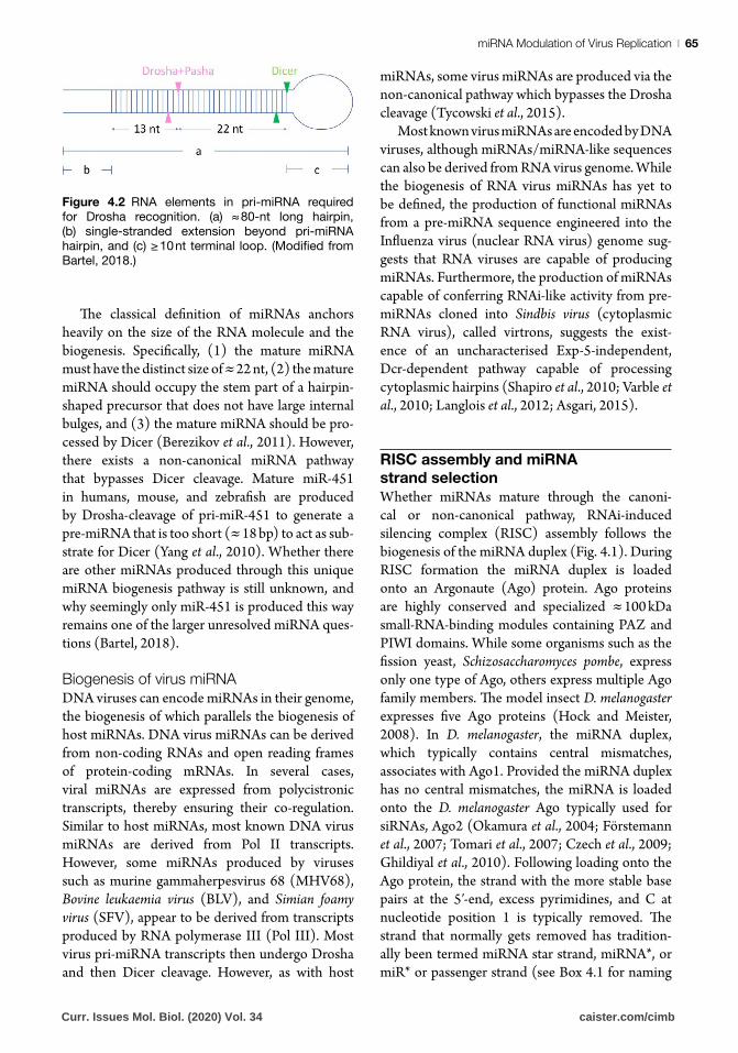
The first step in the sequential processing of pri-miRNAs into mature miRNAs is promoted by the microprocessor comprising RNase III Drosha and its protein partner Pasha (Denli et al., 2004; Gregory et al., 2004; Han et al., 2004; Landthaler et al., 2004; Tomari and Zamore, 2005). Recognition of pri-miRNA by the microprocessor is facilitated by the presence of three RNA elements of the primary miRNA transcript: ≈ 80-nucleotide (nt)
hairpin, unstructured ≥ 10-nt terminal loop, and single-single stranded RNA extensions beyond the pri-miRNA hairpin (Fig. 4.2) (Lee et al., 2003; Zeng and Cullen, 2003; Zeng and Cullen, 2005; Zeng et al., 2005). Upon recognition of pri-miRNA, Pasha determines the cleavage site at 11 base pairs away from the basal junction between the single-stranded RNA and double-stranded RNA of the pri-miRNA and ≈ 22 bp away from the apical junction of the terminal loop. Drosha cleaves the pri-miRNA at the Pasha-determined sites to release the 50–70 nt hairpin-structured pre-miRNA with two nucleotide 3′ overhang (Lee et al., 2002; Gregory et al., 2004; Han et al., 2004). Following nuclear processing by the microprocessor, the pre-miRNA is then shuttled out of the nucleus by the nuclear transport factor Exportin-5 (Exp-5) in complex with Ran-GTP (Yi et al., 2003; Bohnsack et al., 2004; Lund et al., 2004).
In the cytoplasm, the pre-miRNA is further processed by the RNase III enzyme Dicer-1 (Dcr1) and its protein partner Loquacious (in flies). Dcr1 is a highly conserved protein that typically bears PAZ and two RNase III domains. The PAZ domain of D. melanogaster Dcr1 (Lee et al., 2004b) recognizes the 2-nt 3′ overhang at the base of the pre-miRNA stem, and each of the 2 RNase domain then cleaves one of the strands off the loop to liberate the ≈ 22-nt-long miRNA duplex with each strand bearing 2-nt overhangs at 3′-end (Lee et al., 2002; MacRae et al., 2007).
Non-canonical biogenesis of host
miRNA
Some miRNAs, termed non-canonical miRNAs, bypass one or more of the catalytic steps in the canonical miRNA biogenesis (Xie and Steitz, 2014). Non-canonical miRNAs are produced from introns and various RNAs such as small nucleolar RNAs (snoRNAs), endogenous short hairpin RNAs (shRNAs), and tRNAs (Abdelfattah et al., 2014). The most prominent non-canonical matu-ration pathway, the mirtron pathway, substitutes Drosha cleavage with splicing. Splicing, carried out by spliceosomes and debranching enzymes, produces miRNA hairpins directly suitable for Dicer cleavage. After splicing, the mirtron pathway then merges with the canonical miRNA pathway at the Exp-5-bound transport stage (Okamura et al., 2007).
Curr. Issues Mol. Biol. (2020) Vol. 34 caister.com/cimb
Monsanto-Hearne and Johnson64 |
Figure 4.1 From canonical miRNA biogenesis to regulation of virus replication. The canonical biogenesis
of miRNA begins with RNA polymerase II (RNA pol II)-mediated transcription of a miRNA gene. The
resulting hairpin-containing primary transcript, pri-miRNA, spontaneously folds into a hairpin structure and
is methylguanosine-capped at the 5′-end and may be polyadenylated at the 3′-tail. The pri-miRNA is then cleaved by Drosha, in association with Pasha, to generate the pre-miRNA. The pre-miRNA is shuttled out
of the nucleus by the nuclear transport factor Exportin-5 (Exp-5), in association with Ran-GTP, for further
processing by the cytoplasm-located Dicer-1 (Dcr1) and its partner Loquacious. Dcr1 cleaves the hairpin loop
off the pre-miRNA to generate a ≈ 22 nt long miRNA duplex. The miRNA duplex is then loaded onto the effector molecule Argonaute (Ago) to form the RNA induced silencing complex (RISC). In the RISC, one strand of the
miRNA duplex is unwound and discarded, while the other is retained to guide the effector complex to target mRNA. miRNA target recognition occurs through perfect or near perfect complementary binding between
miRNA seed region (second to eighth nucleotides from the 5′ end of the miRNA) and the target mRNA. In the host–virus interaction, the target can be either host mRNA or virus mRNA. Upon binding, target mRNA stability
and/or translational efficiency is modified leading to regulation of gene expression of virus or host factors which consequently impacts virus replication and/or pathogenesis. Where target regulation is provirus, virus
replication is enhanced, conversely, where target regulation is antivirus, virus replication is restricted.
Curr. Issues Mol. Biol. (2020) Vol. 34 caister.com/cimb
miRNA Modulation of Virus Replication | 65
The classical definition of miRNAs anchors heavily on the size of the RNA molecule and the biogenesis. Specifically, (1) the mature miRNA must have the distinct size of ≈ 22 nt, (2) the mature miRNA should occupy the stem part of a hairpin-shaped precursor that does not have large internal bulges, and (3) the mature miRNA should be pro-cessed by Dicer (Berezikov et al., 2011). However, there exists a non-canonical miRNA pathway that bypasses Dicer cleavage. Mature miR-451 in humans, mouse, and zebrafish are produced by Drosha-cleavage of pri-miR-451 to generate a pre-miRNA that is too short (≈ 18 bp) to act as sub-strate for Dicer (Yang et al., 2010). Whether there are other miRNAs produced through this unique miRNA biogenesis pathway is still unknown, and why seemingly only miR-451 is produced this way remains one of the larger unresolved miRNA ques-tions (Bartel, 2018).
Biogenesis of virus miRNA
DNA viruses can encode miRNAs in their genome, the biogenesis of which parallels the biogenesis of host miRNAs. DNA virus miRNAs can be derived from non-coding RNAs and open reading frames of protein-coding mRNAs. In several cases, viral miRNAs are expressed from polycistronic transcripts, thereby ensuring their co-regulation. Similar to host miRNAs, most known DNA virus miRNAs are derived from Pol II transcripts. However, some miRNAs produced by viruses such as murine gammaherpesvirus 68 (MHV68), Bovine leukaemia virus (BLV), and Simian foamy virus (SFV), appear to be derived from transcripts produced by RNA polymerase III (Pol III). Most virus pri-miRNA transcripts then undergo Drosha and then Dicer cleavage. However, as with host
miRNAs, some virus miRNAs are produced via the non-canonical pathway which bypasses the Drosha cleavage (Tycowski et al., 2015).
Most known virus miRNAs are encoded by DNA viruses, although miRNAs/miRNA-like sequences can also be derived from RNA virus genome. While the biogenesis of RNA virus miRNAs has yet to be defined, the production of functional miRNAs from a pre-miRNA sequence engineered into the Influenza virus (nuclear RNA virus) genome sug-gests that RNA viruses are capable of producing miRNAs. Furthermore, the production of miRNAs capable of conferring RNAi-like activity from pre-miRNAs cloned into Sindbis virus (cytoplasmic RNA virus), called virtrons, suggests the exist-ence of an uncharacterised Exp-5-independent, Dcr-dependent pathway capable of processing cytoplasmic hairpins (Shapiro et al., 2010; Varble et al., 2010; Langlois et al., 2012; Asgari, 2015).
RISC assembly and miRNA
strand selection
Whether miRNAs mature through the canoni-cal or non-canonical pathway, RNAi-induced silencing complex (RISC) assembly follows the biogenesis of the miRNA duplex (Fig. 4.1). During RISC formation the miRNA duplex is loaded onto an Argonaute (Ago) protein. Ago proteins are highly conserved and specialized ≈ 100 kDa small-RNA-binding modules containing PAZ and PIWI domains. While some organisms such as the fission yeast, Schizosaccharomyces pombe, express only one type of Ago, others express multiple Ago family members. The model insect D. melanogaster expresses five Ago proteins (Hock and Meister, 2008). In D. melanogaster, the miRNA duplex, which typically contains central mismatches, associates with Ago1. Provided the miRNA duplex has no central mismatches, the miRNA is loaded onto the D. melanogaster Ago typically used for siRNAs, Ago2 (Okamura et al., 2004; Förstemann et al., 2007; Tomari et al., 2007; Czech et al., 2009; Ghildiyal et al., 2010). Following loading onto the Ago protein, the strand with the more stable base pairs at the 5′-end, excess pyrimidines, and C at nucleotide position 1 is typically removed. The strand that normally gets removed has tradition-ally been termed miRNA star strand, miRNA*, or miR* or passenger strand (see Box 4.1 for naming
Figure 4.2 RNA elements in pri-miRNA required
for Drosha recognition. (a) ≈ 80-nt long hairpin, (b) single-stranded extension beyond pri-miRNA
hairpin, and (c) ≥ 10 nt terminal loop. (Modified from Bartel, 2018.)
Curr. Issues Mol. Biol. (2020) Vol. 34 caister.com/cimb
Monsanto-Hearne and Johnson66 |
conventions). The strand with the lesser inter-strand hydrogen bonding resistance, excess purines, and U at nucleotide position 1 is normally retained. This strand becomes the guide miRNA strand, also known as the miRNA, or miR (Khvorova et al., 2003; Czech et al., 2009; Hu et al., 2009; Okamura et al., 2009; Ghildiyal et al., 2010).
miRNA recognition of targets
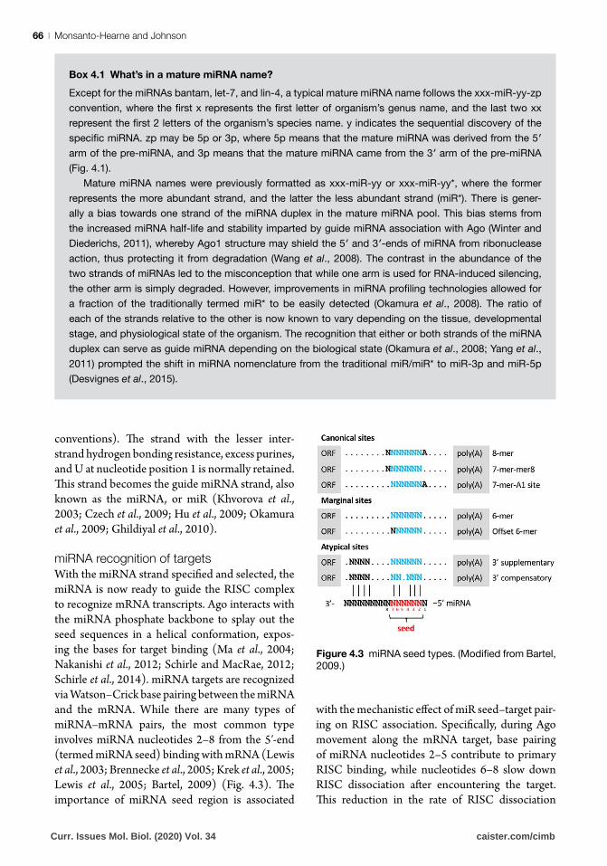
With the miRNA strand specified and selected, the miRNA is now ready to guide the RISC complex to recognize mRNA transcripts. Ago interacts with the miRNA phosphate backbone to splay out the seed sequences in a helical conformation, expos-ing the bases for target binding (Ma et al., 2004; Nakanishi et al., 2012; Schirle and MacRae, 2012; Schirle et al., 2014). miRNA targets are recognized via Watson–Crick base pairing between the miRNA and the mRNA. While there are many types of miRNA–mRNA pairs, the most common type involves miRNA nucleotides 2–8 from the 5′-end (termed miRNA seed) binding with mRNA (Lewis et al., 2003; Brennecke et al., 2005; Krek et al., 2005; Lewis et al., 2005; Bartel, 2009) (Fig. 4.3). The importance of miRNA seed region is associated
with the mechanistic effect of miR seed–target pair-ing on RISC association. Specifically, during Ago movement along the mRNA target, base pairing of miRNA nucleotides 2–5 contribute to primary RISC binding, while nucleotides 6–8 slow down RISC dissociation after encountering the target. This reduction in the rate of RISC dissociation
Box 4.1 What’s in a mature miRNA name?
Except for the miRNAs bantam, let-7, and lin-4, a typical mature miRNA name follows the xxx-miR-yy-zp
convention, where the first x represents the first letter of organism’s genus name, and the last two xx represent the first 2 letters of the organism’s species name. y indicates the sequential discovery of the specific miRNA. zp may be 5p or 3p, where 5p means that the mature miRNA was derived from the 5′ arm of the pre-miRNA, and 3p means that the mature miRNA came from the 3′ arm of the pre-miRNA (Fig. 4.1).
Mature miRNA names were previously formatted as xxx-miR-yy or xxx-miR-yy*, where the former represents the more abundant strand, and the latter the less abundant strand (miR*). There is gener-ally a bias towards one strand of the miRNA duplex in the mature miRNA pool. This bias stems from
the increased miRNA half-life and stability imparted by guide miRNA association with Ago (Winter and
Diederichs, 2011), whereby Ago1 structure may shield the 5′ and 3′-ends of miRNA from ribonuclease action, thus protecting it from degradation (Wang et al., 2008). The contrast in the abundance of the
two strands of miRNAs led to the misconception that while one arm is used for RNA-induced silencing,
the other arm is simply degraded. However, improvements in miRNA profiling technologies allowed for a fraction of the traditionally termed miR* to be easily detected (Okamura et al., 2008). The ratio of
each of the strands relative to the other is now known to vary depending on the tissue, developmental stage, and physiological state of the organism. The recognition that either or both strands of the miRNA
duplex can serve as guide miRNA depending on the biological state (Okamura et al., 2008; Yang et al.,
2011) prompted the shift in miRNA nomenclature from the traditional miR/miR* to miR-3p and miR-5p (Desvignes et al., 2015).
Figure 4.3 miRNA seed types. (Modified from Bartel, 2009.)
Curr. Issues Mol. Biol. (2020) Vol. 34 caister.com/cimb
miRNA Modulation of Virus Replication | 67
strengthens the interaction between the miRNA, target, and RISC, resulting in increased efficacy of RISC (Brennecke et al., 2005; Grimson et al., 2007).
miRNA regulation of target
Following mRNA target recognition, miRNA facilitated target regulation occurs. Repression of gene expression is the most common mode of regulation. In the rare case of full complementa-rity between the miRNA and target, the mRNA is endonucleolitically cleaved by Ago, as is common in plants. In animals, however, the complementarity is most often imperfect, and target repression occurs through mRNA decay and/or mRNA translational repression. mRNA decay has been shown to account for 66–90% of the miRNA-mediated repression, thus contributing substantially to overall silenc-ing ( Jonas and Izaurralde, 2015). mRNA decay involves mRNA deadenylation, followed by decap-ping, and final (in the current model) degradation by cytoplasmic nuclease 5′-to-3′ exoribonuclease (Guo et al., 2010; Huntzinger and Izaurralde, 2011; Djuranovic et al., 2012; Wilczynska and Bushell, 2015; Jonas and Izaurralde, 2015). Mounting evidence suggests that translational repression in association with a 182 kDa phosphoprotein bear-ing glycine-tryptophan repeats (GW182) precedes mRNA decay (Baek et al., 2008; Selbach et al., 2008; Ingolia et al., 2009; Huntzinger and Izaur-ralde, 2011; Bazzini et al., 2012; Djuranovic et al., 2012; Wilczynska and Bushell, 2015), however the mechanism(s) through which miRNAs repress translation of mRNAs is still unclear (Iwakawa and Tomari, 2015). Current models propose several concurrent and overlapping mechanisms that could account for translational repression, including: GW182-mediated recruitment of translational repressors and GW182-mediated displacement of poly(A)-binding proteins (PABP), which subse-quently interferes with the mRNA circularization and therefore translation (Eulalio et al., 2009; Lian et al., 2009).
While most examples of miRNA-mediated reg-ulation show target repression, miRNAs can also stimulate gene expression (Vasudevan et al., 2007; Ørom et al., 2008; Bruno et al., 2011; Hussain et al., 2011; Vasudevan, 2012). Positive regulation of genes by miRNAs can occur as a result of miRNA targeting RNA decay machineries (Bruno et al.,
2011) or as a result of miRNA associations that decrease levels of GW182 (Vasudevan, 2012).
miRNA roles in host–virus
interaction
miRNA-mediated differential gene expression is a cornerstone event of biological processes. Each miRNA can potentially target hundreds of mRNAs (Enright et al., 2004; Farh et al., 2005; Krek et al., 2005; Stark et al., 2005; Sood et al., 2006; Baek et al., 2008; Betel et al., 2010; Hashimoto et al., 2013), and miRNA-mediated changes in gene expression have been implicated in a wide variety of functions. In insects, miRNAs have been implicated in growth, muscle and wing development, neurogenesis, apoptosis, sex determination, phenotypic plasticity, oogenesis and embryogenesis, and host–pathogen interactions and immunity (Asgari, 2013; Lucas et al., 2015)
Virus infection changes the host miRNA pro-file such that the miRNA abundance in uninfected and infected cells, tissues and organisms differ (Scaria et al., 2006; Dykxhoorn, 2007; Pedersen et al., 2007; Gottwein and Cullen, 2008; Lindsay, 2008; Lodish et al., 2008; Tsitsiou and Lindsay, 2009; Xiao and Rajewsky, 2009; O’Connell et al., 2010; Cullen, 2011; Libri et al., 2013; Asgari, 2015; Trobaugh and Klimstra, 2017; Monsanto-Hearne and Johnson, 2018). Indeed, differential abundance of miRNAs during viral infection is often used to identify which miRNAs may func-tion in host–virus interactions (Asgari, 2015; Monsanto-Hearne and Johnson, 2018). The role of the differentially abundant miRNAs is verified based on the impact of miRNA regulation on the host and the virus (Fig. 4.4). The impact of miRNA abundance changes during virus infection is assessed by manipulation of miRNA levels via loss-of-function (LOF) or gain-of-function (GOF) methods (Min and Yoon, 2010; Martinez-Sanchez and Murphy, 2013) often employing miRNA inhibitors and miRNA mimics, respectively. miRNA inhibitors are chemically synthesized single-stranded RNA molecules designed to specifi-cally bind to, inhibit, and artificially down-regulate endogenous miRNAs, while miRNA mimics are chemically synthesized double-stranded RNAs that artificially simulate up-regulation of miRNAs by augmenting endogenous miRNAs (Kuhn et al.,
Curr. Issues Mol. Biol. (2020) Vol. 34 caister.com/cimb

Monsanto-Hearne and Johnson68 |
2008; Thomson et al., 2011; Chugh and Dittmer, 2012; Ekimler and Sahin, 2014). Other common methods for LOF use miRNA sponges, tough decoys, target protectors, miRNA activity sensors, and target sensors (Chugh and Dittmer, 2012; Steinkraus et al., 2016). Other methods for GOF use expression vectors containing mature miRNA, precursor miRNA, or pri-miRNA sequences (Min and Yoon, 2010; Thomson et al., 2011). To test the effect of changes in miRNA abundance on host–virus interactions, miRNA inhibitors and mimics are introduced into cells by transfection, or to whole organisms per os or by injection. Involvement of a miRNA in host–virus interaction is evidenced by LOF and/or GOF resulting in changes in parameters such as cytopathic effects, host survival, and virus titres (Ekimler and Sahin, 2014) (see Box 4.2).
miRNAs exert their physiological effects through target mRNAs. Once a specific miRNA’s change in abundance has been confirmed to affect host-virus replication, the next step in functional analysis typically involves identification of putative targets using bioinformatics. Several computa-tional filtering prediction tools that generate a list of putative targets based on miRNA-target seed
match and other experimentally defined features are now available. Different miRNA-target predic-tion algorithms generate different output lists with their associated false-positive and false-negative rates (Ritchie et al., 2009; Thomson et al., 2011). Identifying putative targets that are commonly identified by two or more algorithms is commonly employed to reduce the false-positive rates (Min and Yoon, 2010; Witkos et al., 2011; Martinez-Sanchez and Murphy, 2013; Ekimler and Sahin, 2014; Riffo-Campos et al., 2016). Putative targets are then experimentally validated using small-scale or genome-wide experimental approaches. In small-scale studies, RT-qPCR, in situ hybridization, northern blot, western blot, and protein arrays are used to measure changes in the target gene or protein after perturbation in miRNA levels. Because miRNAs potentially regulate hundreds of mRNA targets, large-scale transcriptional and proteomic profiling allows for a simultaneous and global assessment of targets following modulation of miRNA activity. Differences in target levels can then be quantitatively compared between control samples and LOF/GOF samples, and between con-trol samples and virus-infected samples (Steinkraus et al., 2016).
Figure 4.4 Pipeline and criteria for analysis of miRNA function in the host–virus interaction. Comparison of
miRNA profiles of uninfected and virus-infected host identifies miRNAs which are differentially regulated during virus infection. Once a miRNA of interest has been identified (left panel), putative targets are identified using various bioinformatics approaches (right panel). Once miRNA–target interaction is confirmed, a closed-loop inter-relationship amongst the (1) miRNA, (2) target gene, (3) virus, and (4) host has to be confirmed (centre panel) so that the miRNA–target pair can be categorically identified as being relevant to the host–virus interaction. Where LOF/GOF mutants exist, identification of relevant miRNAs can start with genetic screens (left panel). (Modified from Monsanto-Hearne and Johnson, 2018.)
Curr. Issues Mol. Biol. (2020) Vol. 34 caister.com/cimb
miRNA Modulation of Virus Replication | 69
The final step of miRNA functional analysis is the evaluation of the functional impact of the target mRNA in the host–virus interaction. This requires manipulation of the target mRNA levels by GOF or LOF. The use of RNAi to knock down the target gene is the most commonly used technique in insect miRNA functional studies. Confirmation that the identified target mRNA functions in host–virus interaction occurs when the knockdown of the target mRNA results in changes in determinants of pathogenesis.
Host miRNA roles in insect–virus
interactions
miRNA profiling and functional analysis of insect host–virus interactions have shown that miRNA abundance changes in the host can influence the outcome of virus pathogenesis (Asgari, 2015). The most thoroughly investigated insect hosts in the context of virus infection are mosquitoes of the Aedes and Culex genera. The involvement of miRNAs during virus infection has also been explored in the silkworm Bombyx mori, the moths Helicoverpa armigera, Heliothis virescens, Spodop-tera frugiperda, and Spodoptera litura, a bumble bee Bombus terrestis, a plant hopper Laodelphax striatellus, the cabbage looper Trichoplusia ni, and in vinegar flies D. melanogaster. Different insect hosts, tissue sample sources, viruses, methods of infection, and sampling times post-infection have been used in miRNA profiling studies. Despite this, common patterns from the literature on the insect–virus interaction have emerged: (1) some miRNAs
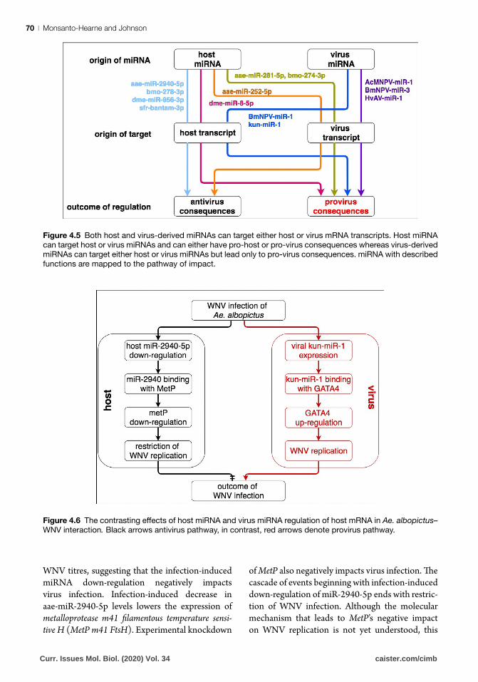
are commonly altered by virus infection, (2) the number of differentially regulated miRNAs changes over the course of virus infection, (3) the direction of regulation of a miRNA can change depending on the time post-infection, and (4) the general direction of change in miRNA abundance can vary depending on tissue sample source. Further functional analyses of the differentially abundant miRNAs show that insect host miRNAs can target either host mRNA or virus mRNA and can either have antivirus or provirus consequences (Fig. 4.5) (Monsanto-Hearne and Johnson, 2018). Details on the host miRNAs that have been functionally char-acterized in insect–virus interactions are described below.
Host miRNA-host mRNA interaction with antivirus consequenceHost miRNAs are first and foremost for the host to use. Various insect miRNAs inhibit viral replication and protect the host from viral attack. Here we sum-marize the literature on the involvement of insect host miRNA in antiviral mechanisms.
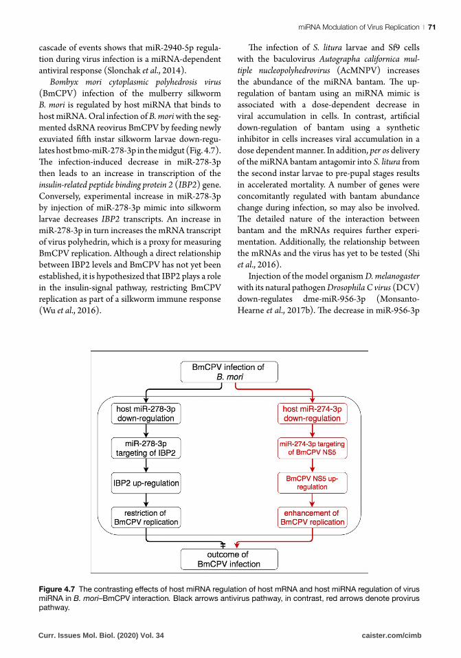
Infection of the Asian tiger mosquito Aedes albop-ictus with the medically important positive-sense ssRNA flavivirus West Nile virus (WNV) is regu-lated by mosquito host miRNA aae-miR-2940-5p. Mosquito aae-miR-2940-5p is selectively down-regulated during virus infection of the Ae. albopictus C6/36 cell line. Artificial miRNA down-regulation using synthetic aae-miR-2940-5p inhibitors represses WNV replication (Fig. 4.6). Conversely, artificial miRNA up-regulation using synthetic aae-miR-2940-5p mimic facilitates increase in
Box 4.2 Functional analysis in vivoThe use of cell cultures has substantially contributed to the functional analysis of miRNA roles in host–virus
interaction. In particular, ex vivo approaches have been useful for documenting the physical interactions
between miRNAs and their targets (Steinkraus et al., 2016). However, disrupting miRNA activity in vivo
has allowed for spatially and temporally synchronized data, providing more evidence on the definitive biological functions of miRNAs (Monsanto-Hearne and Johnson, 2018).
The development of miRNA LOF and GOF in whole organisms already has some success stories. Delivery of inhibitors and mimics per os (Jayachandran et al., 2012) or by injection (Joo et al., 2007) have
both successfully resulted in change in the magnitude of miRNA effects on targets. There is also now an extensive collection of D. melanogaster miRNA stocks with miRNA LOF and GOF. For LOF, knockout mutant flies and lines with transgenes that ‘sponge’ or sequester miRNAs have been produced. For GOF, lines that use the Gal4-UAS system for miRNA expression have been developed (Bejarano et al., 2012;
Schertel et al., 2012).
Curr. Issues Mol. Biol. (2020) Vol. 34 caister.com/cimb
Monsanto-Hearne and Johnson70 |
WNV titres, suggesting that the infection-induced miRNA down-regulation negatively impacts virus infection. Infection-induced decrease in aae-miR-2940-5p levels lowers the expression of metalloprotease m41 filamentous temperature sensi-tive H (MetP m41 FtsH). Experimental knockdown
of MetP also negatively impacts virus infection. The cascade of events beginning with infection-induced down-regulation of miR-2940-5p ends with restric-tion of WNV infection. Although the molecular mechanism that leads to MetP’s negative impact on WNV replication is not yet understood, this
Figure 4.5 Both host and virus-derived miRNAs can target either host or virus mRNA transcripts. Host miRNA
can target host or virus miRNAs and can either have pro-host or pro-virus consequences whereas virus-derived
miRNAs can target either host or virus miRNAs but lead only to pro-virus consequences. miRNA with described
functions are mapped to the pathway of impact.
Figure 4.6 The contrasting effects of host miRNA and virus miRNA regulation of host mRNA in Ae. albopictus–
WNV interaction. Black arrows antivirus pathway, in contrast, red arrows denote provirus pathway.
Curr. Issues Mol. Biol. (2020) Vol. 34 caister.com/cimb
miRNA Modulation of Virus Replication | 71
cascade of events shows that miR-2940-5p regula-tion during virus infection is a miRNA-dependent antiviral response (Slonchak et al., 2014).
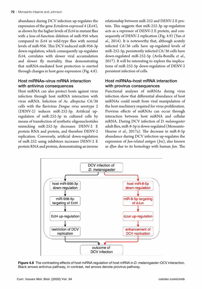
Bombyx mori cytoplasmic polyhedrosis virus (BmCPV) infection of the mulberry silkworm B. mori is regulated by host miRNA that binds to host miRNA. Oral infection of B. mori with the seg-mented dsRNA reovirus BmCPV by feeding newly exuviated fifth instar silkworm larvae down-regu-lates host bmo-miR-278-3p in the midgut (Fig. 4.7). The infection-induced decrease in miR-278-3p then leads to an increase in transcription of the insulin-related peptide binding protein 2 (IBP2) gene. Conversely, experimental increase in miR-278-3p by injection of miR-278-3p mimic into silkworm larvae decreases IBP2 transcripts. An increase in miR-278-3p in turn increases the mRNA transcript of virus polyhedrin, which is a proxy for measuring BmCPV replication. Although a direct relationship between IBP2 levels and BmCPV has not yet been established, it is hypothesized that IBP2 plays a role in the insulin-signal pathway, restricting BmCPV replication as part of a silkworm immune response (Wu et al., 2016).
The infection of S. litura larvae and Sf9 cells with the baculovirus Autographa californica mul-tiple nucleopolyhedrovirus (AcMNPV) increases the abundance of the miRNA bantam. The up-regulation of bantam using an miRNA mimic is associated with a dose-dependent decrease in viral accumulation in cells. In contrast, artificial down-regulation of bantam using a synthetic inhibitor in cells increases viral accumulation in a dose dependent manner. In addition, per os delivery of the miRNA bantam antagomir into S. litura from the second instar larvae to pre-pupal stages results in accelerated mortality. A number of genes were concomitantly regulated with bantam abundance change during infection, so may also be involved. The detailed nature of the interaction between bantam and the mRNAs requires further experi-mentation. Additionally, the relationship between the mRNAs and the virus has yet to be tested (Shi et al., 2016).
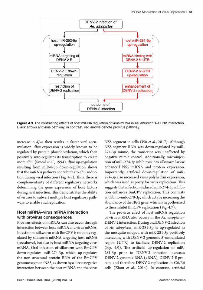
Injection of the model organism D. melanogaster with its natural pathogen Drosophila C virus (DCV) down-regulates dme-miR-956-3p (Monsanto-Hearne et al., 2017b). The decrease in miR-956-3p
Figure 4.7 The contrasting effects of host miRNA regulation of host mRNA and host miRNA regulation of virus miRNA in B. mori–BmCPV interaction. Black arrows antivirus pathway, in contrast, red arrows denote provirus pathway.
Curr. Issues Mol. Biol. (2020) Vol. 34 caister.com/cimb
Monsanto-Hearne and Johnson72 |
abundance during DCV infection up-regulates the expression of the gene Ectoderm-expressed 4 (Ect4), as shown by the higher levels of Ect4 in mutant flies with a loss-of-function deletion of miR-956 when compared to Ect4 in wild-type flies with normal levels of miR-956. This DCV-induced miR-956-3p down-regulation, which consequently up-regulates Ect4, correlates with slower viral accumulation and slower fly mortality, thus demonstrating that miRNA-mediated host protection is exerted through changes in host gene expression (Fig. 4.8).
Host miRNAs–virus mRNA interaction with antivirus consequencesHost miRNA can also protect hosts against virus infection through host miRNA interaction with virus mRNA. Infection of Ae. albopictus C6/36 cells with the flavivirus Dengue virus serotype 2 (DENV-2) induces miR-252-5p. Artificial up-regulation of miR-252-5p in cultured cells by means of transfection of synthetic oligonucleotides mimicking miR-252-5p decreases DENV-2 E protein RNA and protein, and therefore DENV-2 replication. Conversely, artificial down-regulation of miR-252 using inhibitors increases DENV-2 E protein RNA and protein, demonstrating an inverse
relationship between miR-252 and DENV-2 E pro-tein. This suggests that miR-252-3p up-regulation acts as a repressor of DENV-2 E protein, and con-sequently of DENV-2 replication (Fig. 4.9) (Yan et al., 2014). It is noteworthy that, although acutely infected C6/36 cells have up-regulated levels of miR-252-5p, persistently infected C6/36 cells have down-regulated miR-252-5p (Avila-Bonilla et al., 2017). It will be interesting to explore the implica-tions of miR-252-5p down-regulation of DENV-2 persistent infection of cells.
Host miRNAs–host mRNA interaction with provirus consequencesFunctional analyses of miRNAs during virus infection show that differential abundance of host miRNAs could result from viral manipulation of the host machinery required for virus proliferation. Provirus effects of miRNAs can occur through interaction between host miRNA and cellular mRNA. During DCV infection of D. melanogaster adult flies, miR-8-5p is down-regulated (Monsanto-Hearne et al., 2017a). The decrease in miR-8-5p abundance during DCV infection up-regulates the expression of Jun-related antigen (Jra), also known as dJun due to its homology with human Jun. The
Figure 4.8 The contrasting effects of host miRNA regulation of host mRNA in D. melanogaster–DCV interaction.
Black arrows antivirus pathway, in contrast, red arrows denote provirus pathway.
Curr. Issues Mol. Biol. (2020) Vol. 34 caister.com/cimb
miRNA Modulation of Virus Replication | 73
increase in dJun then results in faster viral accu-mulation. dJun expression is widely known to be regulated by protein phosphorylation, which then positively auto-regulates its transcription to create more dJun (Smeal et al., 1994). dJun up-regulation resulting from miR-8-5p down-regulation shows that the miRNA pathway contributes to dJun induc-tion during viral infection (Fig. 4.8). Thus, there is complementarity of different regulatory networks determining the gene expression of host factors during viral infection. This demonstrates the ability of viruses to subvert multiple host regulatory path-ways to enable viral replication.
Host miRNA–virus mRNA interaction with provirus consequencesProvirus effects of miRNAs can also occur through interaction between host miRNA and virus mRNA. Infection of silkworm with BmCPV is not only reg-ulated by silkworm miRNA targeting host mRNA (see above), but also by host miRNA targeting virus mRNA. Oral infection of silkworm with BmCPV down-regulates miR-274-3p, which up-regulates the non-structural protein RNA of the BmCPV genome segment NS5, as shown by a direct negative interaction between the host miRNA and the virus
NS5 segment in cells (Wu et al., 2017). Although NS5 segment RNA was down-regulated by miR-274-3p mimic, the transcript was unaffected by negative mimic control. Additionally, microinjec-tion of miR-274-3p inhibitors into silkworm larvae enhanced NS5 mRNA and protein expression. Importantly, artificial down-regulation of miR-274-3p also increased virus polyhedrin expression, which was used as proxy for virus replication. This suggests that infection-induced miR-274-3p inhibi-tion enhances BmCPV replication. This contrasts with bmo-miR-278-3p, which acts by increasing the abundance of the IBP2 gene, which is hypothesized to then inhibit BmCPV replication (Fig. 4.7).
The provirus effect of host miRNA regulation of virus mRNA also occurs in the Ae. albopictus–DENV-2 interaction. During oral DENV-2 infection of Ae. albopictus, miR-281-5p is up-regulated in the mosquito midgut, with miR-281-5p positively interacting with DENV-2 genomic 5′-untranslated region (UTR) to facilitate DENV-2 replication (Fig. 4.9). The artificial up-regulation of miR-281-5p prior to DENV-2 infection increases DENV-2 genomic RNA (gRNA), DENV-2 E pro-tein, and therefore DENV-2 replication in C6/36 cells (Zhou et al., 2014). In contrast, artificial
Figure 4.9 The contrasting effects of host miRNA regulation of virus mRNA in Ae. albopictus–DENV interaction.
Black arrows antivirus pathway, in contrast, red arrows denote provirus pathway.
Curr. Issues Mol. Biol. (2020) Vol. 34 caister.com/cimb
Monsanto-Hearne and Johnson74 |
down-regulation of miR-281-5p prior to infection reduces viral gDNA levels, DENV-2E protein, and DENV-2 titres in cells. Knock-down of miR-281-5p in mosquitoes in vivo through antagomir-281 injec-tion also reduces viral gDNA, demonstrating that the miR-281-5p impact on DENV-2 occurs in both cells and whole organisms.
Host miRNA–virus mRNA interaction with unknown consequencesDuring early infection of a Heliothis virescens fat body cell line with Heliothis virescens ascovirus 3a (HvAV-3a), H. virescens miRNA Hz-miR-24 is down-regulated in cells; however, the same miRNA is up-regulated in late infection (Hussain and Asgari, 2010). During late infection, the transcript levels of DNA-dependent RNA polymerase (DdRP, ORF64) and DdRPβ (ORF82) are decreased, cor-responding to higher levels of miR-24-5p. Artificial increase of miR-24-5p by cloning the pre-miRNA sequence of miR-24-5p and ectopically expressing the miRNA decreased the transcript levels of DdRP and DdRPβ compared to the levels in control trans-fection. Conversely, artificial decrease of miR-24-5p by synthetic miR-24-5p inhibitor increased DdRP expression. Neither artificial miR-24-5p regula-tion nor silencing of DdRP and DdRPβ impacted virus replication, suggesting that miR-24-5p and its two identified targets are not essential for in vitro HvAV-3 replication. It has been hypothesized that the target genes may be important for the expres-sion of late genes and production of virions in vivo, but this requires testing (Hussain and Asgari, 2010).
Virus miRNA roles in host–virus
interaction
In contrast to host miRNAs, which can either be antivirus or provirus, all virus miRNAs thus far discovered, regardless of whether the target is virus mRNA or host mRNA, are provirus ele-ments. The provirus effects of virus miRNAs occur either through direct regulation of viral products or through directly regulating host transcripts (Aguado and tenOever, 2018).
Virus miRNA–virus mRNA interactionVirus miRNA can bind to the virus mRNAs to exert provirus functions. This can result in enhanced
virus replication. Alternatively, virus miRNA–virus mRNA interaction can regulate virus replication by facilitating transitions from one viral replication stage to another or by preventing early host recog-nition and immune response. Virus miRNA–virus mRNA binding could also potentially prevent the rapid death of the host, providing the virus with a viable host/virus reservoir for a longer period of time.
HvAV-3 produces HvAV-miR-1 from the 3′ stem-loop (3′SL) in the ORF coding for capsid protein at 96 hours post-infection. At the same time point, the viral DNA pol I (orf1) transcript is sharply down-regulated as a result of experi-mentally validated, direct binding of HvAV-miR-1 and the orf1 transcript. Ectopic expression of HvAV-miR-1 in cells decreases viral DNA levels, suggesting that HvAV-miR-1 mediates inhibition of viral replication by specifically binding with orf1 transcript. It is hypothesized that virus HvAV-miR-1 down-regulation of its own gene and its own replication delays host death, therefore conferring an advantage to long-term virus propagation (Hus-sain et al., 2008).
The baculovirus AcMNPV also produces a miRNA, AcMNPV-miR-1. First identified bio-informatically, AcMNPV-miR-1 production was later experimentally validated. The experimentally validated miRNA perfectly matches and directly negatively targets a segment in the viral gene ODV-E25, reducing the level of infectious budded virions (Zhu et al., 2013).
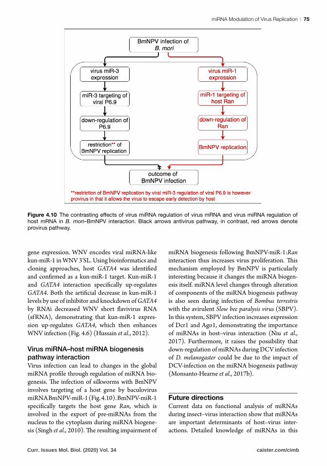
Finally, Bombyx mori nucleopolyhedrovirus (BmNPV) produces BmNPV-miR-3 during the early stage of infection of silkworm larvae. BmNPV-miR-3 negatively affects P6.9 expression and restricts viral replication such that P6.9 levels and viral replication decrease upon synthetic up-regulation of BmNPV-3. In contrast, P6.9 levels and virus replication increase upon synthetic down-regulation of the miRNA (Fig. 4.10). The control of P6.9 (and other late genes) by virus miRNA helps the virus escape early detection by the host, and therefore early immune response attack (Singh et al., 2014).
Virus miRNA–host mRNA interactionWhile some virus miRNAs bind to viral mRNA to exert provirus functions, other virus miRNAs achieve their provirus functions by regulating host
Curr. Issues Mol. Biol. (2020) Vol. 34 caister.com/cimb
miRNA Modulation of Virus Replication | 75
gene expression. WNV encodes viral miRNA-like kun-miR-1 in WNV 3′SL. Using bioinformatics and cloning approaches, host GATA4 was identified and confirmed as a kun-miR-1 target. Kun-miR-1 and GATA4 interaction specifically up-regulates GATA4. Both the artificial decrease in kun-miR-1 levels by use of inhibitor and knockdown of GATA4 by RNAi decreased WNV short flavivirus RNA (sf RNA), demonstrating that kun-miR-1 expres-sion up-regulates GATA4, which then enhances WNV infection (Fig. 4.6) (Hussain et al., 2012).
Virus miRNA–host miRNA biogenesis pathway interactionVirus infection can lead to changes in the global miRNA profile through regulation of miRNA bio-genesis. The infection of silkworms with BmNPV involves targeting of a host gene by baculovirus miRNA BmNPV-miR-1 (Fig. 4.10). BmNPV-miR-1 specifically targets the host gene Ran, which is involved in the export of pre-miRNAs from the nucleus to the cytoplasm during miRNA biogene-sis (Singh et al., 2010). The resulting impairment of
miRNA biogenesis following BmNPV-miR-1:Ran interaction thus increases virus proliferation. This mechanism employed by BmNPV is particularly interesting because it changes the miRNA biogen-esis itself. miRNA level changes through alteration of components of the miRNA biogenesis pathway is also seen during infection of Bombus terrestris with the avirulent Slow bee paralysis virus (SBPV). In this system, SBPV infection increases expression of Dcr1 and Ago1, demonstrating the importance of miRNAs in host–virus interaction (Niu et al., 2017). Furthermore, it raises the possibility that down-regulation of miRNAs during DCV infection of D. melanogaster could be due to the impact of DCV-infection on the miRNA biogenesis pathway (Monsanto-Hearne et al., 2017b).
Future directions
Current data on functional analysis of miRNAs during insect–virus interaction show that miRNAs are important determinants of host–virus inter-actions. Detailed knowledge of miRNAs in this
Figure 4.10 The contrasting effects of virus miRNA regulation of virus mRNA and virus miRNA regulation of host mRNA in B. mori–BmNPV interaction. Black arrows antivirus pathway, in contrast, red arrows denote provirus pathway.
Curr. Issues Mol. Biol. (2020) Vol. 34 caister.com/cimb
Monsanto-Hearne and Johnson76 |
context may lead to important future applications. Examples include: interventions to address virus-infection of economically important insects such as silkworm and bees; as the basis for design of interventions for vector-borne diseases in humans, livestock, and crops; and finally, particularly for the insect model D. melanogaster, as the potential basis for understanding host–virus interaction in humans.
ReferencesAbdelfattah, A.M., Park, C., and Choi, M.Y. (2014). Update
on non-canonical microRNAs. Biomol. Concepts 5, 275–287. https://doi.org/10.1515/bmc-2014-0012
Aguado, L.C., and tenOever, B. (2018). RNA virus building blocks-miRNAs not included. PLOS Pathog. 14, e1006963. https://doi.org/10.1371/journal.ppat.1006963
Ahlquist, P., Noueiry, A.O., Lee, W.M., Kushner, D.B., and Dye, B.T. (2003). Host factors in positive-strand RNA virus genome replication. J. Virol. 77, 8181–8186. https://doi.org/10.1128/JVI.77.15.8181
Ahlquist, P., Schwartz, M., Chen, J., Kushner, D., Hao, L., and Dye, B.T. (2005). Viral and host determinants of RNA virus vector replication and expression. Vaccine 23, 1784–1787. https://doi.org/10.1016/j.vaccine.2004.11.005
Asgari, S. (2013). MicroRNA functions in insects. Insect Biochem. Mol. Biol. 43, 388–397. https://doi.org/10.1016/j.ibmb.2012.10.005
Asgari, S. (2015). Regulatory role of cellular and viral microRNAs in insect-virus interactions. Curr. Opin. Insect Sci. 8, 104–110. https://doi.org/10.1016/j.cois.2014.12.008
Avila-Bonilla, R.G., Yocupicio-Monroy, M., Marchat, L.A., De Nova-Ocampo, M.A., Del Ángel, R.M., and Salas-Benito, J.S. (2017). Analysis of the miRNA profile in C6/36 cells persistently infected with dengue virus type 2. Virus Res. 232, 139–151. https://doi.org/10.1016/j.virusres.2017.03.005
Baek, D., Villén, J., Shin, C., Camargo, F.D., Gygi, S.P., and Bartel, D.P. (2008). The impact of microRNAs on protein output. Nature 455, 64–71. https://doi.org/10.1038/nature07242
Bartel, D.P. (2009). MicroRNAs: target recognition and regulatory functions. Cell 136, 215–233. https://doi.org/10.1016/j.cell.2009.01.002
Bartel, D.P. (2018). Metazoan MicroRNAs. Cell 173, 20–51. https://doi.org/10.1016/j.cell.2018.03.006
Bartel, D.P., and Chen, C.Z. (2004). Micromanagers of gene expression: the potentially widespread influence of metazoan microRNAs. Nat. Rev. Genet. 5, 396–400. https://doi.org/10.1038/nrg1328
Bazzini, A.A., Lee, M.T., and Giraldez, A.J. (2012). Ribosome profiling shows that miR-430 reduces translation before causing mRNA decay in zebrafish. Science 336, 233–237. https://doi.org/10.1126/science.1215704
Bejarano, F., Bortolamiol-Becet, D., Dai, Q., Sun, K., Saj, A., Chou, Y.T., Raleigh, D.R., Kim, K., Ni, J.Q.,
Duan, H., et al. (2012). A genome-wide transgenic resource for conditional expression of Drosophila microRNAs. Development 139, 2821–2831. https://doi.org/10.1242/dev.079939
Berezikov, E., Robine, N., Samsonova, A., Westholm, J.O., Naqvi, A., Hung, J.H., Okamura, K., Dai, Q., Bortolamiol-Becet, D., Martin, R., et al. (2011). Deep annotation of Drosophila melanogaster microRNAs yields insights into their processing, modification, and emergence. Genome Res. 21, 203–215. https://doi.org/10.1101/gr.116657.110
Betel, D., Koppal, A., Agius, P., Sander, C., and Leslie, C. (2010). Comprehensive modeling of microRNA targets predicts functional non-conserved and non-canonical sites. Genome Biol. 11, R90. https://doi.org/10.1186/gb-2010-11-8-r90
Bohnsack, M.T., Czaplinski, K., and Gorlich, D. (2004). Exportin 5 is a RanGTP-dependent dsRNA-binding protein that mediates nuclear export of pre-miRNAs. RNA 10, 185–191. https://doi.org/10.1261/rna.5167604
Bracht, J., Hunter, S., Eachus, R., Weeks, P., and Pasquinelli, A.E. (2004). Trans-splicing and polyadenylation of let-7 microRNA primary transcripts. RNA 10, 1586–1594. https://doi.org/10.1261/rna.7122604
Brennecke, J., Stark, A., Russell, R.B., and Cohen, S.M. (2005). Principles of microRNA-target recognition. PLOS Biol. 3, e85. https://doi.org/10.1371/journal.pbio.0030085
Bruno, I.G., Karam, R., Huang, L., Bhardwaj, A., Lou, C.H., Shum, E.Y., Song, H.W., Corbett, M.A., Gifford, W.D., Gecz, J., et al. (2011). Identification of a microRNA that activates gene expression by repressing nonsense-mediated RNA decay. Mol. Cell 42, 500–510. https://doi.org/10.1016/j.molcel.2011.04.018
Cai, X., Hagedorn, C.H., and Cullen, B.R. (2004). Human microRNAs are processed from capped, polyadenylated transcripts that can also function as mRNAs. RNA 10, 1957–1966. https://doi.org/10.1261/rna.7135204
Carthew, R.W., and Sontheimer, E.J. (2009). Origins and mechanisms of miRNAs and siRNAs. Cell 136, 642–655. https://doi.org/10.1016/j.cell.2009.01.035
Chen, C.Z., Schaffert, S., Fragoso, R., and Loh, C. (2013). Regulation of immune responses and tolerance: the microRNA perspective. Immunol. Rev. 253, 112–128. https://doi.org/10.1111/imr.12060
Cherry, S., and Silverman, N. (2006). Host-pathogen interactions in Drosophila: new tricks from an old friend. Nat. Immunol. 7, 911–917. https://doi.org/10.1038/ni1388
Chugh, P., and Dittmer, D.P. (2012). Potential pitfalls in microRNA profiling. Wiley Interdiscip. Rev. RNA 3, 601–616. https://doi.org/10.1002/wrna.1120
Clyde, K., Kyle, J.L., and Harris, E. (2006). Recent advances in deciphering viral and host determinants of dengue virus replication and pathogenesis. J. Virol. 80, 11418–11431. https://doi.org/10.1128/JVI.01257-06
Cullen, B.R. (2011). Viruses and microRNAs: RISCy interactions with serious consequences. Genes Dev. 25, 1881–1894. https://doi.org/10.1101/gad.17352611
Czech, B., Zhou, R., Erlich, Y., Brennecke, J., Binari, R., Villalta, C., Gordon, A., Perrimon, N., and Hannon, G.J. (2009). Hierarchical rules for Argonaute loading
Curr. Issues Mol. Biol. (2020) Vol. 34 caister.com/cimb
miRNA Modulation of Virus Replication | 77
in Drosophila. Mol. Cell 36, 445–456. https://doi.org/10.1016/j.molcel.2009.09.028
Delpeut, S., Noyce, R.S., Siu, R.W., and Richardson, C.D. (2012). Host factors and measles virus replication. Curr. Opin. Virol. 2, 773–783. https://doi.org/10.1016/j.coviro.2012.10.008
Denli, A.M., Tops, B.B., Plasterk, R.H., Ketting, R.F., and Hannon, G.J. (2004). Processing of primary microRNAs by the Microprocessor complex. Nature 432, 231–235. https://doi.org/10.1038/nature03049
Desvignes, T., Batzel, P., Berezikov, E., Eilbeck, K., Eppig, J.T., McAndrews, M.S., Singer, A., and Postlethwait, J.H. (2015). miRNA nomenclature: a view incorporating genetic origins, biosynthetic pathways, and sequence variants. Trends Genet. 31, 613–626. https://doi.org/10.1016/j.tig.2015.09.002
Djuranovic, S., Nahvi, A., and Green, R. (2012). miRNA-mediated gene silencing by translational repression followed by mRNA deadenylation and decay. Science 336, 237–240. https://doi.org/10.1126/science.1215691
Dykxhoorn, D.M. (2007). MicroRNAs in viral replication and pathogenesis. DNA Cell Biol. 26, 239–249. https://doi.org/10.1089/dna.2006.0559
Ekimler, S., and Sahin, K. (2014). Computational methods for microrna target prediction. Genes 5, 671–683. https://doi.org/10.3390/genes5030671
Enright, A.J., John, B., Gaul, U., Tuschl, T., Sander, C., and Marks, D.S. (2003). MicroRNA targets in Drosophila. Genome Biol. 5, R1. https://doi.org/10.1186/gb-2003-5-1-r1
Eulalio, A., Helms, S., Fritzsch, C., Fauser, M., and Izaurralde, E. (2009). A C-terminal silencing domain in GW182 is essential for miRNA function. RNA 15, 1067–1077. https://doi.org/10.1261/rna.1605509
Fabian, M.R., Sonenberg, N., and Filipowicz, W. (2010). Regulation of mRNA translation and stability by microRNAs. Annu. Rev. Biochem. 79, 351–379. https://doi.org/10.1146/annurev-biochem-060308-103103
Farh, K.K., Grimson, A., Jan, C., Lewis, B.P., Johnston, W.K., Lim, L.P., Burge, C.B., and Bartel, D.P. (2005). The widespread impact of mammalian MicroRNAs on mRNA repression and evolution. Science 310, 1817–1821. https://doi.org/10.1126/science.1121158
Förstemann, K., Horwich, M.D., Wee, L., Tomari, Y., and Zamore, P.D. (2007). Drosophila microRNAs are sorted into functionally distinct argonaute complexes after production by dicer-1. Cell 130, 287–297. https://doi.org/10.1016/j.cell.2007.05.056
Ghildiyal, M., Xu, J., Seitz, H., Weng, Z., and Zamore, P.D. (2010). Sorting of Drosophila small silencing RNAs partitions microRNA* strands into the RNA interference pathway. RNA 16, 43–56. https://doi.org/10.1261/rna.1972910
Gottwein, E., and Cullen, B.R. (2008). Viral and cellular microRNAs as determinants of viral pathogenesis and immunity. Cell Host Microbe 3, 375–387. https://doi.org/10.1016/j.chom.2008.05.002
Gregory, R.I., Yan, K.P., Amuthan, G., Chendrimada, T., Doratotaj, B., Cooch, N., and Shiekhattar, R. (2004). The microprocessor complex mediates the genesis of microRNAs. Nature 432, 235–240.
Griffiths-Jones, S., Saini, H.K., van Dongen, S., and Enright, A.J. (2008). miRBase: tools for microRNA genomics. Nucleic Acids Res. 36, D154–D158. https://doi.org/10.1093/nar/gkm952
Grimson, A., Farh, K.K., Johnston, W.K., Garrett-Engele, P., Lim, L.P., and Bartel, D.P. (2007). MicroRNA targeting specificity in mammals: determinants beyond seed pairing. Mol. Cell 27, 91–105.
Grundhoff, A., and Sullivan, C.S. (2011). Virus-encoded microRNAs. Virology 411, 325–343. https://doi.org/10.1016/j.virol.2011.01.002
Guo, H., Ingolia, N.T., Weissman, J.S., and Bartel, D.P. (2010). Mammalian microRNAs predominantly act to decrease target mRNA levels. Nature 466, 835–840. https://doi.org/10.1038/nature09267
Han, J., Lee, Y., Yeom, K.H., Kim, Y.K., Jin, H., and Kim, V.N. (2004). The Drosha-DGCR8 complex in primary microRNA processing. Genes Dev. 18, 3016–3027. https://doi.org/10.1101/gad.1262504
Hashimoto, Y., Akiyama, Y., and Yuasa, Y. (2013). Multiple-to-multiple relationships between microRNAs and target genes in gastric cancer. PLOS ONE 8, e62589. https://doi.org/10.1371/journal.pone.0062589
Hu, H.Y., Yan, Z., Xu, Y., Hu, H., Menzel, C., Zhou, Y.H., Chen, W., and Khaitovich, P. (2009). Sequence features associated with microRNA strand selection in humans and flies. BMC Genomics 10, 413. https://doi.org/10.1186/1471-2164-10-413
Huntzinger, E., and Izaurralde, E. (2011). Gene silencing by microRNAs: contributions of translational repression and mRNA decay. Nat. Rev. Genet. 12, 99–110. https://doi.org/10.1038/nrg2936
Hussain, M., and Asgari, S. (2010). Functional analysis of a cellular microRNA in insect host-ascovirus interaction. J. Virol. 84, 612–620. https://doi.org/10.1128/JVI.01794-09
Hussain, M., Taft, R.J., and Asgari, S. (2008). An insect virus-encoded microRNA regulates viral replication. J. Virol. 82, 9164–9170. https://doi.org/10.1128/JVI.01109-08
Hussain, M., Frentiu, F.D., Moreira, L.A., O’Neill, S.L., and Asgari, S. (2011). Wolbachia utilizes host microRNAs to manipulate host gene expression and facilitate colonization of the dengue vector Aedes aegypti. Proc. Natl. Acad. Sci. U.S.A. 108, 9250–9255. https://doi.org/10.1073/pnas.1105469108
Hussain, M., Torres, S., Schnettler, E., Funk, A., Grundhoff, A., Pijlman, G.P., Khromykh, A.A., and Asgari, S. (2012). West Nile virus encodes a microRNA-like small RNA in the 3’ untranslated region which up-regulates GATA4 mRNA and facilitates virus replication in mosquito cells. Nucleic Acids Res. 40, 2210–2223. https://doi.org/10.1093/nar/gkr848
Huszar, T., and Imler, J.L. (2008). Drosophila viruses and the study of antiviral host-defense. Adv. Virus Res. 72, 227–265. https://doi.org/10.1016/S0065-3527(08)00406-5
Ingolia, N.T., Ghaemmaghami, S., Newman, J.R., and Weissman, J.S. (2009). Genome-wide analysis in vivo of translation with nucleotide resolution using ribosome profiling. Science 324, 218–223. https://doi.org/10.1126/science.1168978
Curr. Issues Mol. Biol. (2020) Vol. 34 caister.com/cimb
Monsanto-Hearne and Johnson78 |
Iwakawa, H.O., and Tomari, Y. (2015). The functions of microRNAs: mRNA decay and translational repression. Trends Cell Biol. 25, 651–665.
Jayachandran, B., Hussain, M., and Asgari, S. (2013). An insect trypsin-like serine protease as a target of microRNA: utilization of microRNA mimics and inhibitors by oral feeding. Insect Biochem. Mol. Biol. 43, 398–406. https://doi.org/10.1016/j.ibmb.2012.10.004
Jonas, S., and Izaurralde, E. (2015). Towards a molecular understanding of microRNA-mediated gene silencing. Nat. Rev. Genet. 16, 421–433. https://doi.org/10.1038/nrg3965
Joo, C.H., Shin, Y.C., Gack, M., Wu, L., Levy, D., and Jung, J.U. (2007). Inhibition of interferon regulatory factor 7 (IRF7)-mediated interferon signal transduction by the Kaposi’s Sarcoma-associated herpesvirus viral IRF homolog vIRF3. J. Virol. 81, 8282–8292. https://doi.org/10.1128/JVI.00235-07
Kemp, C., and Imler, J.L. (2009). Antiviral immunity in Drosophila. Curr. Opin. Immunol. 21, 3–9. https://doi.org/10.1016/j.coi.2009.01.007
Khvorova, A., Reynolds, A., and Jayasena, S.D. (2003). Functional siRNAs and miRNAs exhibit strand bias. Cell 115, 209–216. https://doi.org/10.1016/S0092-8674(03)00801-8
Kim, Y.K., and Kim, V.N. (2007). Processing of intronic microRNAs. EMBO J. 26, 775–783. https://doi.org/10.1038/sj.emboj.7601512
Kincaid, R.P., and Sullivan, C.S. (2012). Virus-encoded microRNAs: an overview and a look to the future. PLOS Pathog. 8, e1003018. https://doi.org/10.1371/journal.ppat.1003018
Kozomara, A., and Griffiths-Jones, S. (2014). miRBase: annotating high confidence microRNAs using deep sequencing data. Nucleic Acids Res. 42, D68–D73. https://doi.org/10.1093/nar/gkt1181
Krek, A., Grün, D., Poy, M.N., Wolf, R., Rosenberg, L., Epstein, E.J., MacMenamin, P., da Piedade, I., Gunsalus, K.C., Stoffel, M., et al. (2005). Combinatorial microRNA target predictions. Nat. Genet. 37, 495–500. https://doi.org/10.1038/ng1536
Kuhn, D.E., Martin, M.M., Feldman, D.S., Terry, A.V., Nuovo, G.J., and Elton, T.S. (2008). Experimental validation of miRNA targets. Methods 44, 47–54. https://doi.org/10.1016/j.ymeth.2007.09.005
Lai, E.C., Tomancak, P., Williams, R.W., and Rubin, G.M. (2003). Computational identification of Drosophila microRNA genes. Genome Biol. 4, R42. https://doi.org/10.1186/gb-2003-4-7-r42
Landgraf, P., Rusu, M., Sheridan, R., Sewer, A., Iovino, N., Aravin, A., Pfeffer, S., Rice, A., Kamphorst, A.O., Landthaler, M., et al. (2007). A mammalian microRNA expression atlas based on small RNA library sequencing. Cell 129, 1401–1414. https://doi.org/10.1016/j.cell.2007.04.040
Landthaler, M., Yalcin, A., and Tuschl, T. (2004). The human DiGeorge syndrome critical region gene 8 and Its D. melanogaster homolog are required for miRNA biogenesis. Curr. Biol. 14, 2162–2167. https://doi.org/10.1016/j.cub.2004.11.001
Langlois, R.A., Shapiro, J.S., Pham, A.M., and tenOever, B.R. (2012). In vivo delivery of cytoplasmic RNA
virus-derived miRNAs. Mol. Ther. 20, 367–375. https://doi.org/10.1038/mt.2011.244
Lee, Y., Jeon, K., Lee, J.T., Kim, S., and Kim, V.N. (2002). MicroRNA maturation: stepwise processing and subcellular localization. EMBO J. 21, 4663–4670. https://doi.org/10.1093/emboj/cdf476
Lee, Y., Ahn, C., Han, J., Choi, H., Kim, J., Yim, J., Lee, J., Provost, P., Rådmark, O., Kim, S., et al. (2003). The nuclear RNase III Drosha initiates microRNA processing. Nature 425, 415–419. https://doi.org/10.1038/nature01957
Lee, Y., Kim, M., Han, J., Yeom, K.H., Lee, S., Baek, S.H., and Kim, V.N. (2004a). MicroRNA genes are transcribed by RNA polymerase II. EMBO J. 23, 4051–4060. https://doi.org/10.1038/sj.emboj.7600385
Lee, Y.S., Nakahara, K., Pham, J.W., Kim, K., He, Z., Sontheimer, E.J., and Carthew, R.W. (2004b). Distinct roles for Drosophila Dicer-1 and Dicer-2 in the siRNA/miRNA silencing pathways. Cell 117, 69–81.
Lewis, B.P., Shih, I.H., Jones-Rhoades, M.W., Bartel, D.P., and Burge, C.B. (2003). Prediction of mammalian microRNA targets. Cell 115, 787–798 https://doi.org/10.1016/S0092-8674(03)01018-3
Lewis, B.P., Burge, C.B., and Bartel, D.P. (2005). Conserved seed pairing, often flanked by adenosines, indicates that thousands of human genes are microRNA targets. Cell 120, 15–20. https://doi.org/10.1016/j.cell.2004.12.035
Li, Z., and Rana, T.M. (2014). Therapeutic targeting of microRNAs: current status and future challenges. Nat. Rev. Drug Discov. 13, 622–638. https://doi.org/10.1038/nrd4359
Lian, S.L., Li, S., Abadal, G.X., Pauley, B.A., Fritzler, M.J., and Chan, E.K. (2009). The C-terminal half of human Ago2 binds to multiple GW-rich regions of GW182 and requires GW182 to mediate silencing. RNA 15, 804–813. https://doi.org/10.1261/rna.1229409
Libri, V., Miesen, P., van Rij, R.P., and Buck, A.H. (2013). Regulation of microRNA biogenesis and turnover by animals and their viruses. Cell. Mol. Life Sci. 70, 3525–3544. https://doi.org/10.1007/s00018-012-1257-1
Lim, L.P., Glasner, M.E., Yekta, S., Burge, C.B., and Bartel, D.P. (2003a). Vertebrate microRNA genes. Science 299, 1540. https://doi.org/10.1126/science.1080372
Lim, L.P., Lau, N.C., Weinstein, E.G., Abdelhakim, A., Yekta, S., Rhoades, M.W., Burge, C.B., and Bartel, D.P. (2003b). The microRNAs of Caenorhabditis elegans. Genes Dev. 17, 991–1008. https://doi.org/10.1101/gad.1074403
Lindsay, M.A. (2008). microRNAs and the immune response. Trends Immunol. 29, 343–351. https://doi.org/10.1016/j.it.2008.04.004
Lodish, H.F., Zhou, B., Liu, G., and Chen, C.Z. (2008). Micromanagement of the immune system by microRNAs. Nat. Rev. Immunol. 8, 120–130. https://doi.org/10.1038/nri2252
Lucas, K.J., Zhao, B., Liu, S., and Raikhel, A.S. (2015). Regulation of physiological processes by microRNAs in insects. Curr. Opin. Insect Sci. 11, 1–7. https://doi.org/10.1016/j.cois.2015.06.004
Lund, E., Güttinger, S., Calado, A., Dahlberg, J.E., and Kutay, U. (2004). Nuclear export of microRNA precursors. Science 303, 95–98. https://doi.org/10.1126/science.1090599
Curr. Issues Mol. Biol. (2020) Vol. 34 caister.com/cimb
miRNA Modulation of Virus Replication | 79
Ma, J.B., Ye, K., and Patel, D.J. (2004). Structural basis for overhang-specific small interfering RNA recognition by the PAZ domain. Nature 429, 318–322. https://doi.org/10.1038/nature02519
MacRae, I.J., Zhou, K., and Doudna, J.A. (2007). Structural determinants of RNA recognition and cleavage by Dicer. Nat. Struct. Mol. Biol. 14, 934–940. https://doi.org/10.1038/nsmb1293
Marco, A., Hui, J.H., Ronshaugen, M., and Griffiths-Jones, S. (2010). Functional shifts in insect microRNA evolution. Genome Biol. Evol. 2, 686–696. https://doi.org/10.1093/gbe/evq053
Martinez-Sanchez, A., and Murphy, C.L. (2013). MicroRNA Target Identification-Experimental Approaches. Biology 2, 189–205. https://doi.org/10.3390/biology2010189
Min, H., and Yoon, S. (2010). Got target? Computational methods for microRNA target prediction and their extension. Exp. Mol. Med. 42, 233–244. https://doi.org/10.3858/emm.2010.42.4.032
Monsanto-Hearne, V., and Johnson, K.N. (2018). miRNAs in insects infected by animal and plant viruses. Viruses 10, E354. https://doi.org/10.3390/v10070354
Monsanto-Hearne, V., Asad, S., Asgari, S., and Johnson, K.N. (2017a). Drosophila microRNA modulates viral replication by targeting a homologue of mammalian cJun. J. Gen. Virol. 98, 1904–1912. https://doi.org/10.1099/jgv.0.000831
Monsanto-Hearne, V., Tham, A.L.Y., Wong, Z.S., Asgari, S., and Johnson, K.N. (2017b). Drosophila miR-956 suppression modulates Ectoderm-expressed 4 and inhibits viral replication. Virology 502, 20–27. https://doi.org/10.1016/j.virol.2016.12.009
Mussabekova, A., Daeffler, L., and Imler, J.L. (2017). Innate and intrinsic antiviral immunity in Drosophila. Cell. Mol. Life Sci. 74, 2039–2054. https://doi.org/10.1007/s00018-017-2453-9
Nakanishi, K., Weinberg, D.E., Bartel, D.P., and Patel, D.J. (2012). Structure of yeast Argonaute with guide RNA. Nature 486, 368–374. https://doi.org/10.1038/nature11211
Niu, J., Meeus, I., De Coninck, D.I., Deforce, D., Etebari, K., Asgari, S., and Smagghe, G. (2017). Infections of virulent and avirulent viruses differentially influenced the expression of dicer-1, ago-1, and microRNAs in Bombus terrestris. Sci. Rep. 7, 45620. https://doi.org/10.1038/srep45620
O’Connell, R.M., Rao, D.S., Chaudhuri, A.A., and Baltimore, D. (2010). Physiological and pathological roles for microRNAs in the immune system. Nat. Rev. Immunol. 10, 111–122. https://doi.org/10.1038/nri2708
Okamura, K., Ishizuka, A., Siomi, H., and Siomi, M.C. (2004). Distinct roles for Argonaute proteins in small RNA-directed RNA cleavage pathways. Genes Dev. 18, 1655–1666. https://doi.org/10.1101/gad.1210204
Okamura, K., Hagen, J.W., Duan, H., Tyler, D.M., and Lai, E.C. (2007). The mirtron pathway generates microRNA-class regulatory RNAs in Drosophila. Cell 130, 89–100. https:/https://doi.org/10.1016/j.cell.2007.06.028
Okamura, K., Phillips, M.D., Tyler, D.M., Duan, H., Chou, Y.T., and Lai, E.C. (2008). The regulatory activity of microRNA* species has substantial influence on
microRNA and 3’ UTR evolution. Nat. Struct. Mol. Biol. 15, 354–363. https://doi.org/10.1038/nsmb.1409
Okamura, K., Liu, N., and Lai, E.C. (2009). Distinct mechanisms for microRNA strand selection by Drosophila Argonautes. Mol. Cell 36, 431–444. https://doi.org/10.1016/j.molcel.2009.09.027
Ørom, U.A., Nielsen, F.C., and Lund, A.H. (2008). MicroRNA-10a binds the 5’UTR of ribosomal protein mRNAs and enhances their translation. Mol. Cell 30, 460–471. https://doi.org/10.1016/j.molcel.2008.05.001
Ozsolak, F., Poling, L.L., Wang, Z., Liu, H., Liu, X.S., Roeder, R.G., Zhang, X., Song, J.S., and Fisher, D.E. (2008). Chromatin structure analyses identify miRNA promoters. Genes Dev. 22, 3172–3183. https://doi.org/10.1101/gad.1706508
Pasquinelli, A.E. (2012). MicroRNAs and their targets: recognition, regulation and an emerging reciprocal relationship. Nat. Rev. Genet. 13, 271–282. https://doi.org/10.1038/nrg3162
Pedersen, I.M., Cheng, G., Wieland, S., Volinia, S., Croce, C.M., Chisari, F.V., and David, M. (2007). Interferon modulation of cellular microRNAs as an antiviral mechanism. Nature 449, 919–922. https://doi.org/10.1038/nature06205
Ploegh, H.L. (1998). Viral strategies of immune evasion. Science 280, 248–253. https://doi.org/10.1126/science.280.5361.248
Rapicetta, M., Ferrari, C., and Levrero, M. (2002). Viral determinants and host immune responses in the pathogenesis of HBV infection. J. Med. Virol. 67, 454–457. https://doi.org/10.1002/jmv.10096
Riffo-Campos, Á.L., Riquelme, I., and Brebi-Mieville, P. (2016). Tools for sequence-based miRNA target prediction: What to choose? Int. J. Mol. Sci. 17, E1987. https://doi.org/10.3390/ijms17121987
Ritchie, W., Flamant, S., and Rasko, J.E. (2009). Predicting microRNA targets and functions: traps for the unwary. Nat. Methods 6, 397–398. https://doi.org/10.1038/nmeth0609-397
Rodriguez, A., Griffiths-Jones, S., Ashurst, J.L., and Bradley, A. (2004). Identification of mammalian microRNA host genes and transcription units. Genome Res. 14, 1902–1910. https://doi.org/10.1101/gr.2722704
Ruby, J.G., Stark, A., Johnston, W.K., Kellis, M., Bartel, D.P., and Lai, E.C. (2007). Evolution, biogenesis, expression, and target predictions of a substantially expanded set of Drosophila microRNAs. Genome Res. 17, 1850–1864.
Scaria, V., Hariharan, M., Maiti, S., Pillai, B., and Brahmachari, S.K. (2006). Host-virus interaction: a new role for microRNAs. Retrovirology 3, 68. https://doi.org/10.1186/1742-4690-3-68
Schertel, C., Rutishauser, T., Förstemann, K., and Basler, K. (2012). Functional characterization of Drosophila microRNAs by a novel in vivo library. Genetics 192, 1543–1552. https://doi.org/10.1534/genetics.112.145383
Schirle, N.T., and MacRae, I.J. (2012). The crystal structure of human Argonaute2. Science 336, 1037–1040. https://doi.org/10.1126/science.1221551
Schirle, N.T., Sheu-Gruttadauria, J., and MacRae, I.J. (2014). Structural basis for microRNA targeting. Science 346, 608–613. https://doi.org/10.1126/science.1258040
Curr. Issues Mol. Biol. (2020) Vol. 34 caister.com/cimb
Monsanto-Hearne and Johnson80 |
Selbach, M., Schwanhäusser, B., Thierfelder, N., Fang, Z., Khanin, R., and Rajewsky, N. (2008). Widespread changes in protein synthesis induced by microRNAs. Nature 455, 58–63. https://doi.org/10.1038/nature07228
Shapiro, J.S., Varble, A., Pham, A.M., and Tenoever, B.R. (2010). Noncanonical cytoplasmic processing of viral microRNAs. RNA 16, 2068–2074. https://doi.org/10.1261/rna.2303610
Shi, X., Ran, Z., Li, S., Yin, J., and Zhong, J. (2016). The effect of microRNA bantam on Baculovirus AcMNPV Infection in vitro and in vivo. Viruses 8, E136. https://doi.org/10.3390/v8050136
Singh, C.P., Vaishna, R.L., Kakkar, A., Arunkumar, K.P., and Nagaraju, J. (2014). Characterization of antiviral and antibacterial activity of Bombyx mori seroin proteins. Cell. Microbiol. 16, 1354–1365. https://doi.org/10.1111/cmi.12294
Singh, J., Singh, C.P., Bhavani, A., and Nagaraju, J. (2010). Discovering microRNAs from Bombyx mori nucleopolyhedrosis virus. Virology 407, 120–128. https://doi.org/10.1016/j.virol.2010.07.033
Slonchak, A., Hussain, M., Torres, S., Asgari, S., and Khromykh, A.A. (2014). Expression of mosquito microRNA aae-miR-2940-5p is down-regulated in response to West Nile virus infection to restrict viral replication. J. Virol. 88, 8457-8467. https://doi.org/10.1128/JVI.00317-14
Smeal, T., Hibi, M., and Karin, M. (1994). Altering the specificity of signal transduction cascades: positive regulation of c-Jun transcriptional activity by protein kinase A. EMBO J. 13, 6006–6010.
Sood, P., Krek, A., Zavolan, M., Macino, G., and Rajewsky, N. (2006). Cell-type-specific signatures of microRNAs on target mRNA expression. Proc. Natl. Acad. Sci. U.S.A. 103, 2746–2751.
Stark, A., Brennecke, J., Bushati, N., Russell, R.B., and Cohen, S.M. (2005). Animal microRNAs confer robustness to gene expression and have a significant impact on 3’UTR evolution. Cell 123, 1133–1146.
Steinkraus, B.R., Toegel, M., and Fulga, T.A. (2016). Tiny giants of gene regulation: experimental strategies for microRNA functional studies. Wiley Interdiscip. Rev. Dev. Biol. 5, 311–362. https://doi.org/10.1002/wdev.223
Thomson, D.W., Bracken, C.P., and Goodall, G.J. (2011). Experimental strategies for microRNA target identification. Nucleic Acids Res. 39, 6845–6853. https://doi.org/10.1093/nar/gkr330
Tomari, Y., and Zamore, P.D. (2005). MicroRNA biogenesis: drosha can’t cut it without a partner. Curr. Biol. 15, R61–R64. https://doi.org/10.1016/j.cub.2004.12.057
Tomari, Y., Du, T., and Zamore, P.D. (2007). Sorting of Drosophila small silencing RNAs. Cell 130, 299–308. https://doi.org/10.1016/j.cell.2007.05.057
Trobaugh, D.W., and Klimstra, W.B. (2017). MicroRNA regulation of RNA virus replication and pathogenesis. Trends Mol. Med. 23, 80–93. https://doi.org/10.1016/j.molmed.2016.11.003
Tsitsiou, E., and Lindsay, M.A. (2009). MicroRNAs and the immune response. Curr. Opin. Pharmacol. 9, 514–520. https://doi.org/10.1016/j.coph.2009.05.003
Tycowski, K.T., Guo, Y.E., Lee, N., Moss, W.N., Vallery, T.K., Xie, M., and Steitz, J.A. (2015). Viral noncoding RNAs: more surprises. Genes Dev. 29, 567–584. https://doi.org/10.1101/gad.259077.115
Varble, A., Chua, M.A., Perez, J.T., Manicassamy, B., García-Sastre, A., and tenOever, B.R. (2010). Engineered RNA viral synthesis of microRNAs. Proc. Natl. Acad. Sci. U.S.A. 107, 11519–11524. https://doi.org/10.1073/pnas.1003115107
Vasudevan, S. (2012). Posttranscriptional upregulation by microRNAs. Wiley Interdiscip. Rev. RNA 3, 311–330. https://doi.org/10.1002/wrna.121
Vasudevan, S., Tong, Y., and Steitz, J.A. (2007). Switching from repression to activation: microRNAs can up-regulate translation. Science 318, 1931–1934. https://doi.org/10.1126/science.1149460
Vodovar, N., and Saleh, M.C. (2012). Of insects and viruses: the role of small RNAs in insect defence. In Small RNAs: Their Diversity, Roles and Practical Uses, E.L. Jockusch, ed. (Academic Press, London), pp. 1–36.
Wang, Y., Sheng, G., Juranek, S., Tuschl, T., and Patel, D.J. (2008). Structure of the guide-strand-containing argonaute silencing complex. Nature 456, 209–213. https://doi.org/10.1038/nature07315
Whitton, J.L., Cornell, C.T., and Feuer, R. (2005). Host and virus determinants of picornavirus pathogenesis and tropism. Nat. Rev. Microbiol. 3, 765–776. https://doi.org/10.1038/nrmicro1284
Wilczynska, A., and Bushell, M. (2015). The complexity of miRNA-mediated repression. Cell Death Differ. 22, 22–33. https://doi.org/10.1038/cdd.2014.112
Winter, J., and Diederichs, S. (2011). Argonaute proteins regulate microRNA stability: Increased microRNA abundance by Argonaute proteins is due to microRNA stabilization. RNA Biol. 8, 1149–1157. https://doi.org/10.4161/rna.8.6.17665
Witkos, T.M., Koscianska, E., and Krzyzosiak, W.J. (2011). Practical aspects of microRNA target prediction. Curr. Mol. Med. 11, 93–109. https://doi.org/10.2174/156652411794859250
Wu, P., Qin, G., Qian, H., Chen, T., and Guo, X. (2016). Roles of miR-278-3p in IBP2 regulation and Bombyx mori cytoplasmic polyhedrosis virus replication. Gene 575, 264–269. https://doi.org/10.1016/j.gene.2015.09.009
Wu, P., Jiang, X., Sang, Q., Annan, E., Cheng, T., and Guo, X. (2017). Inhibition of miR-274-3p increases BmCPV replication by regulating the expression of BmCPV NS5 gene in Bombyx mori. Virus Genes 53, 643–649. https://doi.org/10.1007/s11262-017-1466-7
Xiao, C., and Rajewsky, K. (2009). MicroRNA control in the immune system: basic principles. Cell 136, 26–36. https://doi.org/10.1016/j.cell.2008.12.027
Xie, M., and Steitz, J.A. (2014). Versatile microRNA biogenesis in animals and their viruses. RNA Biol. 11, 673–681. https://doi.org/10.4161/rna.28985
Xu, J., and Cherry, S. (2014). Viruses and antiviral immunity in Drosophila. Dev. Comp. Immunol. 42, 67–84. https://doi.org/10.1016/j.dci.2013.05.002
Yan, H., Zhou, Y., Liu, Y., Deng, Y., and Chen, X. (2014). miR-252 of the Asian tiger mosquito Aedes albopictus regulates dengue virus replication by suppressing the expression of the dengue virus envelope protein. J.
Curr. Issues Mol. Biol. (2020) Vol. 34 caister.com/cimb
miRNA Modulation of Virus Replication | 81
Med. Virol. 86, 1428–1436. https://doi.org/10.1002/jmv.23815
Yang, J.S., Maurin, T., Robine, N., Rasmussen, K.D., Jeffrey, K.L., Chandwani, R., Papapetrou, E.P., Sadelain, M., O’Carroll, D., and Lai, E.C. (2010). Conserved vertebrate mir-451 provides a platform for Dicer-independent, Ago2-mediated microRNA biogenesis. Proc. Natl. Acad. Sci. U.S.A. 107, 15163–15168. https://doi.org/10.1073/pnas.1006432107
Yang, J.S., Phillips, M., Betel, D., Mu, P., Ventura, A., Siepel, A., Chen, K., and Lai, E. (2011). Widespread regulatory activity of vertebrate microRNA* species. RNA 17, 312–326. https://doi.org/10.1261/rna.2537911
Yi, R., Qin, Y., Macara, I.G., and Cullen, B.R. (2003). Exportin-5 mediates the nuclear export of pre-microRNAs and short hairpin RNAs. Genes Dev. 17, 3011–3016. https://doi.org/10.1101/gad.1158803
Ylla, G., Fromm, B., Piulachs, M.D., and Belles, X. (2016). The microRNA toolkit of insects. Sci. Rep. 6, 37736. https://doi.org/10.1038/srep37736
Zeng, Y., and Cullen, B.R. (2003). Sequence requirements for micro RNA processing and function in human cells. RNA 9, 112–123. https://doi.org/10.1261/rna.2780503
Zeng, Y., and Cullen, B.R. (2005). Efficient processing of primary microRNA hairpins by Drosha requires flanking nonstructured RNA sequences. J. Biol. Chem. 280, 27595–27603. https://doi.org/10.1074/jbc.M504714200
Zeng, Y., Yi, R., and Cullen, B.R. (2005). Recognition and cleavage of primary microRNA precursors by the nuclear processing enzyme Drosha. EMBO J. 24, 138–148. https://doi.org/10.1038/sj.emboj.7600491
Zhao, T., Li, G., Mi, S., Li, S., Hannon, G.J., Wang, X.J., and Qi, Y. (2007). A complex system of small RNAs in the unicellular green alga Chlamydomonas reinhardtii. Genes Dev. 21, 1190–1203.
Zhou, Y., Liu, Y., Yan, H., Li, Y., Zhang, H., Xu, J., Puthiyakunnon, S., and Chen, X. (2014). miR-281, an abundant midgut-specific miRNA of the vector mosquito Aedes albopictus enhances dengue virus replication. Parasit. Vectors 7, 488. https://doi.org/10.1186/s13071-014-0488-4
Zhu, M., Wang, J., Deng, R., Xiong, P., Liang, H., and Wang, X. (2013). A microRNA encoded by Autographa californica nucleopolyhedrovirus regulates expression of viral gene ODV-E25. J. Virol. 87, 13029–13034. https://doi.org/10.1128/JVI.02112-13
Curr. Issues Mol. Biol. (2020) Vol. 34 caister.com/cimb
Copyright © 2022 FDOKUMEN