Integrative Transcriptomic and Metabolomic Analysis at Organ ...
Upload
independentCategory
view
1download
0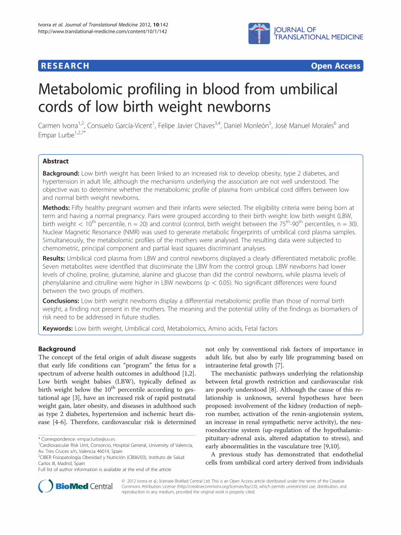
Ivorra et al. Journal of Translational Medicine 2012, 10:142http://www.translational-medicine.com/content/10/1/142
RESEARCH Open Access
Metabolomic profiling in blood from umbilicalcords of low birth weight newbornsCarmen Ivorra1,2, Consuelo García-Vicent1, Felipe Javier Chaves3,4, Daniel Monleón5, José Manuel Morales6 andEmpar Lurbe1,2,7*
Abstract
Background: Low birth weight has been linked to an increased risk to develop obesity, type 2 diabetes, andhypertension in adult life, although the mechanisms underlying the association are not well understood. Theobjective was to determine whether the metabolomic profile of plasma from umbilical cord differs between lowand normal birth weight newborns.
Methods: Fifty healthy pregnant women and their infants were selected. The eligibility criteria were being born atterm and having a normal pregnancy. Pairs were grouped according to their birth weight: low birth weight (LBW,birth weight < 10th percentile, n = 20) and control (control, birth weight between the 75th-90th percentiles, n = 30).Nuclear Magnetic Resonance (NMR) was used to generate metabolic fingerprints of umbilical cord plasma samples.Simultaneously, the metabolomic profiles of the mothers were analysed. The resulting data were subjected tochemometric, principal component and partial least squares discriminant analyses.
Results: Umbilical cord plasma from LBW and control newborns displayed a clearly differentiated metabolic profile.Seven metabolites were identified that discriminate the LBW from the control group. LBW newborns had lowerlevels of choline, proline, glutamine, alanine and glucose than did the control newborns, while plasma levels ofphenylalanine and citrulline were higher in LBW newborns (p < 0.05). No significant differences were foundbetween the two groups of mothers.
Conclusions: Low birth weight newborns display a differential metabolomic profile than those of normal birthweight, a finding not present in the mothers. The meaning and the potential utility of the findings as biomarkers ofrisk need to be addressed in future studies.
Keywords: Low birth weight, Umbilical cord, Metabolomics, Amino acids, Fetal factors
BackgroundThe concept of the fetal origin of adult disease suggeststhat early life conditions can “program” the fetus for aspectrum of adverse health outcomes in adulthood [1,2].Low birth weight babies (LBW), typically defined asbirth weight below the 10th percentile according to ges-tational age [3], have an increased risk of rapid postnatalweight gain, later obesity, and diseases in adulthood suchas type 2 diabetes, hypertension and ischemic heart dis-ease [4-6]. Therefore, cardiovascular risk is determined
* Correspondence: [email protected] Risk Unit, Consorcio, Hospital General, University of Valencia,Av. Tres Cruces s/n, Valencia 46014, Spain2CIBER Fisiopatología Obesidad y Nutrición (CB06/03), Instituto de SaludCarlos III, Madrid, SpainFull list of author information is available at the end of the article
© 2012 Ivorra et al.; licensee BioMed CentralCommons Attribution License (http://creativecreproduction in any medium, provided the or
not only by conventional risk factors of importance inadult life, but also by early life programming based onintrauterine fetal growth [7].The mechanistic pathways underlying the relationship
between fetal growth restriction and cardiovascular riskare poorly understood [8]. Although the cause of this re-lationship is unknown, several hypotheses have beenproposed: involvement of the kidney (reduction of neph-ron number, activation of the renin-angiotensin system,an increase in renal sympathetic nerve activity), the neu-roendocrine system (up-regulation of the hypothalamic-pituitary-adrenal axis, altered adaptation to stress), andearly abnormalities in the vasculature tree [9,10].A previous study has demonstrated that endothelial
cells from umbilical cord artery derived from individuals
Ltd. This is an Open Access article distributed under the terms of the Creativeommons.org/licenses/by/2.0), which permits unrestricted use, distribution, andiginal work is properly cited.

Ivorra et al. Journal of Translational Medicine 2012, 10:142 Page 2 of 10http://www.translational-medicine.com/content/10/1/142
of LBW exhibit a different phenotype when compared tothat from individuals of higher birth weight [11]. Thisobservation could imply that it is possible to identifyfunctional and/or structural differences at birth whichcan contribute to providing better information with re-spect to the risk of developing disease later in life. A fur-ther approach to studying differences between low andnormal birth weight newborns is to assess their metabo-lomic profiles.Metabolomics, the study of small-molecule metabo-
lites, is a discipline that focuses on the measurement ofthe relative concentrations of endogenous small mole-cules in biofluids. This technology may be useful forunderstanding metabolic imbalances and for diagnosinghuman diseases. Recently, studies using metabolomicshave been used to identify a discriminatory metaboliteprofile in a number of areas: preeclampsia in early preg-nancy [12], diabetes [13], obesity [14], [15], risk for pre-term delivery [16]. It has been suggested they predict thepresence and severity of cardiovascular disease [17-20].There are no data about its use to identify a metaboliteprofile in umbilical cord from LBW children. The ob-jective of the present study was to analyse the metabolo-mic profile at birth in newborns with LBW, and tocompare it with those from normal birth weight chil-dren. Simultaneously, the metabolomic profiles of theirmothers were analysed. The identification of deregulatedmetabolites would potentially give clues for betterunderstanding the abnormalities associated with LBW.
MethodsSubjects and sample collectionNewborns were included if born at term (gestational age≥37 weeks), after uncomplicated pregnancies ascertainedaccording to the Ballard method [21], and by normal de-livery or Cesarean section in the absence of perinatal ill-ness at the Hospital General Universitario of Valencia,Spain. All the mothers were healthy and had no cardio-vascular risk factors. Fifty newborns and their mothersof European origin were selected to participate in thestudy. Umbilical cord blood samples were obtained fromthe clamped umbilical cord immediately after delivery.The samples of their mothers were drawn at 2–4 h afterdelivery in non-fasting conditions, but with at least 8hours having passed since the last meal and blood sam-pling. Two groups of newborns were created accordingto birth weight: lower than the 10th percentile (lowerthan 2.5 kg; LBW group, n = 20) or appropriate birthweight for gestational age (control group, birth weightbetween the 75th-90th percentiles, n = 30) [22]. Parentsgave their consent for the study after they were giveninformed in writing of the objectives of the research pro-ject and of the samples that would be taken. The studywas approved by the hospital’s review board and was
carried out in accordance with the Declaration of Hel-sinki. Both the samples (plasma from umbilical cord ofthe newborns and plasma from peripheral veins fromthe mothers), as well as the collected data, were storedaccording to the directives dictated by the law of Bio-medical Investigation of 2007 (Law 14/2007) and all ap-plicable rules.
Metabolomic profileStorage, preparation and 1 H Nuclear magnetic resonancespectroscopic analysis of blood plasmaUmbilical venous cord blood and maternal peripheralvenous blood were collected in EDTA-tubes, centrifugedto yield plasma, stored at −80 °C and thawed before use.For Nuclear Magnetic Resonance (NMR) analysis, 500 μlof plasma were mixed with 50 μl of D2O (as a field lock).A total of 500 μl of the mixture of each sample was thenindividually transferred into a 5 mm high quality NMRtube. All 1 H NMR spectra were acquired using a stand-ard one-dimensional pulse sequence with water suppres-sion (Bruker Avance 600 spectrometer operating at600.13 MHz with a 1 mm 1 H/13 C/15 N TXI probe). Atotal of 256 FIDs (free induction decay) were collectedinto 64 k data points with a spectral width of 14 ppmand a recycle delay (RD) of 1 s. The water signal wassaturated with weak irradiation during the recycle delay.Before Fourier transformation, the free induction decaywas multiplied by a 0.3 Hz exponential line broadening.Spectral chemical shift referencing on the Alanine
CH3 doublet signal at 1.475 ppm was performed in allspectra. Resonances in these spectral regions wereassigned using the literature [23] and two-dimensionalspectra were selected (2D NMR, especially for longchain metabolites). Spectral regions between 0.5 and 4.5ppm and between 5.5 and 9.5 ppm were binned in seg-ments of 0.01 ppm width (6 Hz) for multivariate ana-lysis. Data binning in NMR spectra for multivariateanalysis is a common pre-processing practice [24,25],which, although it may decrease the effect of minorobservations, allows for the detection of relevantregions, decreases the risk of model overfitting and arti-ficially increases the signal to noise-ratio. Data binningreduces the impact of read noise on the processed dataat the cost of a lower resolution. We used binning onlyfor the pattern recognition. There are two benefits asso-ciated with the use of pattern recognition approaches inthe binning of spectral data. First, the signal-to-noiseratio increases. Second, there is a reduction in the possi-bility of overfitting. Once the bucket with the relevantpeaks in each spectra were identified, they were quanti-fied. Therefore, the values reported are quantified peaksby spectral integration. The binned data was normalisedto the total spectral area. Thus, the individual peak in-tensities were normalized to total metabolite content for

Ivorra et al. Journal of Translational Medicine 2012, 10:142 Page 3 of 10http://www.translational-medicine.com/content/10/1/142
better a comparison between samples. Spectral regionsbelonging to the EDTA resonances (2.52 to 2.57 ppmand 3.06 to3.17 ppm) were removed from the spectra forsubsequent analysis. The available spectral databases and2D NMR experiments were used to aid in structuralidentification of the relevant metabolites. All spectrawere processed using Topspin 1.3 (Bruker BiospinGmbH, Germany) and transferred to MATLABW (Math-Works Inc, 2006) using in-house scripts for data ana-lysis. Signals belonging to selected metabolites wereintegrated and quantified using semi-automated in-house MATLAB peak-fitting routines. These fitting rou-tines were based on Levenburg-Marquard optimizationprocedures. The target function for the optimizationincluded experimental spectra measured for standardsolutions of selected metabolites with complex multipletpatterns and theoretically generated Lorentzian-shapesignals for those metabolites with simpler spectral pat-terns. One way analysis of variance (ANOVA) was usedfor the determination of statistical significance betweengroup means of the corresponding integrals.
Multivariate analysis of nuclear magnetic resonancespectraIn the present study, binned spectral regions for bloodplasma were mean-centered chemometric analyses. Rele-vant bins were identified in our chemometric models.The relevant spectral regions within these bins wereidentified and quantified by spectral integration. Chemo-metric and statistical analyses were performed using in-house MATLAB scripts and the PLS Toolbox (Eigen-vector Research, Inc.). Principal Component Analysis(PCA) and Projection to Latent Structures for Discrim-inant Analysis (PLS-DA) were applied to NMR spectradata sets. PCA is able to find low dimensional embed-dings of multivariate data in a way that optimally pre-serves the structure of the data. The PCA techniquetransforms the variables of a data set into a smallernumber of new latent variables called principal compo-nents (PCs) which are uncorrelated to each other andaccount for decreasing proportions of the total varianceof the original variables. Each new PC is a linear com-bination of the original variation so that a compact de-scription of the variation within the data set isgenerated. Observations are assigned scores according tothe variation measured by the principal component X,with those having similar scores clustering together.Where PCA proved inadequate to define clustering, asupervised approach was used.PLS-DA is a classification technique that combines the
properties of Partial Least Squares regression with thediscrimination power of discriminant analysis [26]. Themain advantage of PLS-DA models is that the mainsources of variability in the data are modeled by the so-
called latent variables (LVs) and, consequently, in theirassociated scores and loadings. This allows for thevisualization and understanding of different patterns andrelations in the data. PLS-DA is a supervised extensionof PCA that is used to distinguish two or more classesby searching for variables (X matrix) that are correlatedto class membership (Y matrix). In this approach, theaxes are calculated to maximize class separation and canbe used to examine separation that would otherwise beacross three or more principal components [26]. Inorder to discriminate between samples associated withlow weight and normal weight at birth, a PLS-DA modelwas devised. The PLS-DA model discriminating betweencontrols and LBW was cross-validated by technical repli-cation of the whole process on 25 random data splits.To validate the model, important quality parameterswere calculated: R2X, R2Y, Q2 and RMSP. R2X and R2Ydescribe the variation in the X and Y variables, respect-ively. Q2 describes predictability. RMSP (Root meansquared error of prediction) gives a direct indication ofthe error in the predicted value.
Statistical analysisValues were expressed as mean ± SD for each studygroup. The differences in the mean values of anthropo-metric parameters between the groups were assessed byanalysis of variance (ANOVA) adjusted by sex. The nor-mality of the data was verified using the Kolmogorov-Smirnov test. Choline, glutamine, alanine and glycogenin the control group do not have a normal distribution,and a Kruskal-Wallis test was used for these variables.An outlier analysis was performed, and those samplesdeviating from the mean by more than 2.5 times thestandard deviation were excluded. A significant differ-ence was considered present if p < 0.05. Pearson’s cor-relation coefficients were used to examine the relationsbetween variables. Statistical analyses were performedusing SPSS 15.0. (SPSS Inc, Chicago, Illinois, USA) andGraphPad Statmate 5.0 (GraphPad Software, La Jolla,California, USA) software graphs.
ResultsClinical characteristics of mothers and newbornsThe general characteristics of mothers and their new-borns are shown in Table 1. No differences wereobserved in maternal age, maternal BMI, pregnancyweight gain or type of delivery between the two groups.Although no significant differences were observed inheart rate and diastolic blood pressure between new-borns groups (Table 1), differences appeared in sex,height, head circumference and blood pressure. Asexpected, the LBW group had systolic values signifi-cantly lower than those for the control birth weightgroup [10]. All comparisons between newborns groups

Ivorra et al. Journal of Translational Medicine 2012, 10:142 Page 4 of 10http://www.translational-medicine.com/content/10/1/142
were adjusted by sex, and no changes were noted whencomparing boys with girls.
Metabolomic profile in umbilical cord blood in low birthweight and control newbornsThe standard 1D NMR spectrum is shown in Figure 1A.PCA and PLS-DA scatter plot were performed on thepre-processed NMR spectra comparing plasma fromLBW versus control newborns and were used to identifymetabolite clusters. Although an unsupervised analysisby PCA does not show differences between groups,Figure 1B, a supervised classification method (PLS-DA,3 Latent Variables, see Additional file 1: Figure S1) pro-vides a differential global metabolic profile, Figure 1C.The model had a R2X (cumulative) of 0.623, a R2Y (cu-mulative) of 0.871, a Q2 (cumulative) of 0.523 (a Q2
value superior to 0.5 is generally considered to be a goodpredictor) and a RMSP of 0.417 (medium predictive cap-acity). Spectral regions and peaks with the highest con-tribution to the loadings plot from this model werefurther quantified and analysed. Many metabolites maycontribute to these loading plots and to the separationbetween classes. A total of thirty-four metabolites weretested in fifty-eight spectral regions, and the differenceswere evaluated by the T-Student test with the Kruskal-Wallis correction. However, only those that differed withp < 0.05 were taken into consideration.Of the 34 metabolic regions studied, 17 compounds
were quantified according to the loadings of the multi-variate analysis. Among the 17 metabolites, significantdifferences were observed for seven (proline, free cho-line, glutamine, alanine, glucose, phenylalanine and
Table 1 Anthropometric and clinical characteristics of mother
Control (n = 30)
Maternal Characteristics
Maternal age (years) 30.22 ± 5.65
Pregnancy weight gain (g) 12581 ± 428
Delivery (vaginal/Cesarean section) 21/9 (70%)
Maternal Body Mass Index 24.0 ± 3.1
Newborns Characteristics
Gestational age at delivery (weeks) 38.95 ± 0.90
Sex (male/female) 20/10 (66.7%)
Birth weight (g) 3756 ± 148
Height (cm) 50.46 ± 1.45
Head circumference (cm) 34.70 ± 1.00
Systolic blood pressure (mmHg) 73.88 ± 13.20
Diastolic blood pressure (mmHg) 44.20 ± 11.22
Heart rate (bpm) 130.14 ± 16.30
The values are expressed as mean ± SD; p-value, statistical significance of the differ
citrulline) between the LBW and the control group,while no differences were observed in the other seven(spectral region containing lipoproteins peaks, in carbo-hydrates and glycogen fragments, in ketone bodies likeacetoacetate and hydroxybutyrate, in total lipids and infatty acids both saturated and unsaturated) (data notshown).Table 2 shows median values ± SD for arbitrary units
of intensity (AUIx10-3) from identified metabolites, andFigure 2 provides the individual levels of 7 metabolitesthat display significant level differences between the twogroups. In the LBW group, all metabolites have a normaldistribution, and samples are more homogeneous thanin the control group. In the control group, choline, glu-tamine and alanine do not have a normal distribution.An outlier analysis was carried out, and two subjectswere excluded from the control group.Five metabolites (proline, choline, glutamine, alanine
and glucose) were reduced in the LBW group as com-pared to those for the controls. In the LBW group, pro-line values were 38.1% less (21.74 ± 9.80 AUI vs 13.00 ±7.5 AUI; p-value = 0.002) while free choline was 31.4%less (3.76 ± 0.17 AU vs 2.58 ± 0.07 AU; p-value = 0.005).Glutamine (3.68 ± 0.17 vs 2.59 ± 0.07; p-value = 0.004),alanine (9.49 ± 4.1 vs 7.00 ± 2.2; p-value = 0.008) andglucose levels (2.15 ± 0.98 vs 1.59 ± 0.50; p-value =0.023) were significantly lower in LBW newborns aswell. In addition, LBW newborns exhibited a 15.7% in-crease in values of phenylalanine (1.07 ± 0.03 vs 1.27 ±0.03 p-value = 0.018) and a 20.2% increase in values ofcitrulline (0.67 ± 0.02 vs 0.84 ± 0.002; p-value = 0.008).No difference in profiles between sexes was observed.
s and newborns grouped by birth weight
LBW (n = 20) p-value
30.50 ± 6.10 NS
11025 ± 301 NS
12/8 (60%) NS
22.8 ± 2.1 NS
37.64 ± 1.50 p < 0.05
5/15 (33.3%) p < 0.01
2371 ± 177 p < 0.0001
45.55 ± 1.14 p < 0.0001
32.10 ± 1.80 p < 0.0001
65.15 ± 11.50 p < 0.05
40.70 ± 8.70 NS
128.33 ± 17.90 NS
ences among groups; NS, Non-significant; LBW, low birth weight.

Figure 1 Umbilical cord blood profile. Superposed 1 H NMR spectra for the spectra of umbilical cord blood plasma (Panel A), PrincipalComponent Analysis scores plot (PCA, panel B), and Projection to Latent Structures for Discriminant Analysis scores plot (PLS-DA, panel C)showing the global metabolic differences between low birth weight (open circles, blue spectra) and control birth weight (black triangles and redspectra) newborns. Although an unsupervised analysis by PCA does not show differences between groups, a supervised classification method(PLS-DA) provides a differential global metabolic profile. The cross-validated error percentage of the model for the classification of low weight atbirth samples is 8 %. Metabolites exhibiting largest differences between groups have been listed in Table 2.
Ivorra et al. Journal of Translational Medicine 2012, 10:142 Page 5 of 10http://www.translational-medicine.com/content/10/1/142
Maternal metabolic profile in venous peripheral bloodNuclear Magnetic Resonance data from mothers wereprocessed to give the initial PCA plot after a partial leastsquares-discriminate analysis (Figure 3, PLS-DA, 3 La-tent Variables, see Additional file 1: Figure S2). ThePLS-DA model had a R2X (cumulative) of 0.512, a R2Y(cumulative) of 0.611, a Q2 (cumulative) of 0.502 (a Q2
value superior to 0.5 is generally considered to be a goodpredictor) and a RMSP of 0.282 (low predictive cap-acity). The loadings plot showed 14 spectral regions withhigh relative weight belonging to 10 metabolites. Table 2shows median values ± SD for arbitrary units of intensity
(AUIx10-3) from the identified metabolites. From a totalof 10 metabolites, 6 were related to the amino acidgroup (proline, phenylalanine, alanine, glutamine, citrul-line and free choline), 2 were related to the carbohydrategroup (glucose and glycogen fragments), and 2 to thelipid group (spectral region containing large LDL +VLDL lipoproteins peaks and polyunsaturated fatty acids(PUFAs)). The results of the metabolomic profiling inmaternal blood showed no significant differences be-tween the two groups of mothers at either the PLS-DAscore plot level and at the individual identified metabol-ite level (Table 2, p-value M-C vs M-LBW).

Table 2 List of identified metabolites in umbilical cord plasma and in maternal peripheral blood
NEWBORNS p-value MOTHERS p-value Feto-maternal ratio p-value
Control(n = 30)
LBW(n = 20)
C vs LBW M-Control(n = 30)
M-LBW(n = 20)
M-C vsM-LBW
Control LBW Control vsLBW
Amino Acidsand related
Proline 21.74 ± 9.80 13.00 ± 7.50 0.002 12.22 ± 0.92 12.10 ± 0.56 NS 1.73 ± 0.83 1.26 ± 0.62 0.023
Free Choline 3.76 ± 0.17 2.58 ± 0.07 0.005 5.73 ± 0.79 5.80 ± 0.76 NS 0.56 ± 0.17 0.46 ± 0.17 0.040
Citrulline 0.67 ± 0.02 0.84 ± 0.02 0.008 15.16 ± 1.09 15.34 ± 1.22 NS 0.04 ± 0.01 0.05 ± 0.02 0.022
Glutamine 3.68 ± 0.17 2.59 ± 0.07 0.004 0.85 ± 0.06 0.85 ± 0.07 NS 3.68 ± 1.16 3.07 ± 0.81 0.024
Alanine 9.49 ± 4.10 7.00 ± 2.20 0.008 0.90 ± 0.03 0.96 ± 0.20 NS 9.28 ± 3.29 7.66 ± 2.64 0.041
Phenylalanine 1.07 ± 0.03 1.27 ± 0.03 0.018 9.78 ± 1.13 9.97 ± 0.96 NS 0.12 ± 0.03 0.13 ± 0.03 NS
Carbohydrates
Glucose 2.15 ± 0.98 1.59 ± 0.50 0.023 1.12 ± 0.08 1.10 ± 0.09 NS 1.65 ± 0.58 1.43 ± 0.42 NS
Glycogen fragments 2.70 ± 1.11 2.01 ± 0.76 0.011 2.95 ± 0.33 2.82 ± 0.24 NS 0.81 ± 0.24 0.71 ± 0.27 NS
Lipids
LDL + VLDL enrichedspectral region
7.22 ± 1.25 7.60 ± 0.99 NS 5.01 ± 0. 50 4.96 ± 0.52 NS 1.52 ± 0.33 1.55 ± 0.23 NS
PUFA N/D N/D – 4.19 ± 0.49 4.38 ± 0.38 NS – – –
The values are expressed as mean ± SD; p-value, statistical significance of the differences among groups; N/D, not detected; LBW, low birth weight. Meanmetabolite abundance is indicated by arbitrary units of intensity (AUIx10-3).
Ivorra et al. Journal of Translational Medicine 2012, 10:142 Page 6 of 10http://www.translational-medicine.com/content/10/1/142
The feto-maternal ratio for carbohydrates (glucose andglycogen fragments), lipids (spectral region containing largeLDL + VLDL lipoprotein peaks) and phenylalanine wascomparable between the two groups (Table 2, p-valueControl vs LBW). In contrast, the feto-maternal ratio forproline, free choline, glutamine and alanine was signifi-cantly lower (p < 0.05) in the LBW group, whereas that forcitrulline was significantly higher.
Figure 2 Metabolites distinguishing LBW and control newborns. Glucocontrol newborns. Significantly higher levels of citrulline and phenylalaninemetabolite abundance is indicated by line. Each point represents a single n
Relationship between metabolites and birth weightPearson correlations were performed for each of the 7aforementioned metabolites versus birth weight. Out ofthe 7 metabolites, the analysis identified a significantpositive correlation between birth weight and freecholine, proline, and glutamine (free choline r = 0.50,p-value <0.01; proline r = 0.29, p-value < 0.05;glutamine r = 0.36, p-value < 0.01). A significant
se, Proline, Glutamine, Choline, and Alanine levels were higher inwere detected in umbilical cord plasma from LBW newborns. Meanewborn.

Glucose
CONTROL SGA0
1
2
3
4
5
AU
I
Proline
CONTROL SGA0
10
20
30
40
50
AU
I
Glutamine
CONTROL SGA0
2
4
6
8
AU
I
Choline
CONTROL SGA0
2
4
6
8
10
AU
I
Alanine
CONTROL SGA0
5
10
15
20
AU
I
Phenylalanine
CONTROL SGA0.0
0.5
1.0
1.5
2.0
AU
I
Citrulline
CONTROL SGA0.0
0.5
1.0
1.5
AU
I
Figure 3 Spectra of maternal venous plasma. Principal Component Analysis (PCA, panel A) and Projection to Latent Structures forDiscriminant Analysis (PLS-DA, panel B) scores plot for the spectra of maternal venous plasma showing the global metabolic differences betweenlow birth weight (open circles) and control birth weight (black triangles ) newborns. Apparently, there are no global metabolic trends in thematernal blood associated with the weight of the newborns.
Ivorra et al. Journal of Translational Medicine 2012, 10:142 Page 7 of 10http://www.translational-medicine.com/content/10/1/142
inverse association was found between citrulline andbirth weight (r = −0.40, p-value <0.01) and betweenphenylalanine and birth weight (r = −0.34, p-value<0.05); i.e., phenylalanine and citrulline increase asbirth weight decreases. No relationships were identi-fied when correlating these metabolites with bloodpressure. No significant associations were found be-tween alanine and glucose with birth weight.
DiscussionThe present study is the first to utilize metabolomic ana-lysis in umbilical cord blood samples from LBW chil-dren in order to identify metabolites that could berelated to the abnormalities associated with the long-term development of the increased cardio-metabolic riskassociated with LBW. The metabolomic profile of LBWnewborns differed from that observed in a group of
newborn controls. Higher values of phenylalanine andcitrulline and lower levels of proline, choline, glutamine,alanine and glucose were observed. A significant rela-tionship between some of these metabolites and birthweight was also present. Furthermore, the differencesobserved in the newborns were not observed in theirmothers, all of whom were healthy and had similarweight gains during pregnancy. Although the signifi-cance of the observed metabolomic values is not wellunderstood, interesting information can be derived fromthe present results since some of the observed data havebeen linked previously to the abnormal metabolic statesassociated with LBW.Even though the impact of fetal metabolic program-
ming on adult health is well documented, the underlyingmechanisms are poorly understood. Animal models havebeen used extensively to investigate the mechanism by

Ivorra et al. Journal of Translational Medicine 2012, 10:142 Page 8 of 10http://www.translational-medicine.com/content/10/1/142
which the early-life environment induces persistentalterations in the metabolism of offspring [27-30]. Thesestudies support the hypothesis that nutritional imbalanceduring prenatal and early postnatal life can induce longterm metabolic changes and increase susceptibility tometabolic diseases in later life [31-33].The absence of differences found for lipoproteins, car-
bohydrates, ketone bodies and fatty acids suggests thatfatty acid oxidation and glucose metabolism remain un-altered. Changes, however, in the spectral region be-tween 7.27 and 5.36 ppm, which has been assigned toglycogen fragments, may suggest alterations in carbohy-drate storage. The large molecular size of glycogenmakes it very difficult to observe its 1 H resonance sig-nals. Longer NMR T2 relaxation times, which suggestsmaller molecule sizes, produce the narrow signalsobserved for that spectral region. Perhaps some macro-molecule degradation of glycogen into smaller molecularsize fragments is the cause.In the present study, the mismatch between the
mother and the newborn in terms of the metabolomicprofile is worth taking into consideration. While a differ-ent pattern was observed in the LBW newborns com-pared to controls, the mothers of both groups had asimilar pattern. A trend towards reduced levels of aminoacids in the umbilical cord was observed in LBW, some-thing not observed in the mothers. The reason for thediscrepancies is not well known, although suboptimalplacental transport of amino acids could be involved.Impaired placental amino acid transport is often seen inassociation with IUGR, yet it is unclear whether thechanges in amino acid transport are a cause or a conse-quence of IUGR [34,35]. Experimental models in rats,however, have suggested a down-regulation of aminoacid transporters as a cause rather than a consequence[36]. Consistent with impaired placental amino acidtransport as a potential cause of IUGR in humans, um-bilical cord blood (but not maternal blood) concentra-tion of most essential amino acids is less in pregnancieswith IUGR fetuses [34], [37]. This interesting findingshould be addressed in future studies.In contrast to the reduction of the majority of the
amino acid detected are the increased values of phenyl-alanine, a neutral branched amino acid, in LBW new-born. Higher levels of amino acids, branched and/oraromatic, have been associated with insulin resistance[38,39], and with the risk to develop type 2 diabetes inthe Framingham offspring cohort [19]. The potential foramino acids to induce insulin resistance has been recog-nized for some time [40]. In a low-calorie environment,it is not surprising that large neutral amino acids wouldpromote an anabolic state by inhibiting proteolysis andby directly stimulating protein synthesis [41]. That not-withstanding, the detection of significant spectral
regions in the broad protein amide region (between 7.80and 7.86 ppm) seems to support a potential difference inthe anabolic rate between LBW and control newborns.One of the greatest differences is the free choline, with
low levels in the LBW group. The spectral region be-tween 4.06 and 4.08 ppm, belonging to fragments ofphosphoethanolamine [42], also shows statistically sig-nificant differences in the LBW group. During gestationthere is a high demand for this essential nutrient [43].Choline, via its metabolite betain, serves as a donor of me-thyl groups for the production of S-adenosylmethionine(SAM), a substrate of DNA and histone methyltrans-ferases [44]. DNA-methylation is catalyzed by DNAmethyltransferases that transfer methyl groups from SAMto cytosine located in CpG dinucleotides. Different studieshave shown that the prenatal choline supply affects the ex-pression of multiple genes whose expression is regulatedvia epigenetic marks [45], [46]. Maternal choline supplyduring pregnancy modifies fetal histone and DNA methy-lation in rat fetal liver and brain [47]. Conversely, animalsfed with diets deficient in choline and in methionine havealtered global and gene-specific DNA and histone methy-lation [48]. These studies have demonstrated that mater-nal choline alterations during pregnancy modify fetalhistone and DNA methylation, suggesting that a concertedepigenomic mechanism contributes to the long-term de-velopmental effects of varied choline intake in utero.Finding that the lower levels of choline are associated
with LBW is compatible with the epigenetic programminghypothesis. Interactions of nutrients with the epigeneticmachinery lead to changes associated with the regulationof gene expression that underlies the developmental pro-gramming consequences in adult life [49]. The epigeneticprogramming of the fetus in utero is under the influenceof the altered metabolic milieu of pregnancy and the typesof nutrients available through the maternal diet [50].Alterations in nutrients, above all those related to DNAmethylation, can modify the levels of DNA methylationand fetal programming [51]. Disease propensity in LBWchildren can be epigenetically programmed by intrauterineexposures that have the capacity to modify fetal genesthrough alterations in DNA methylation [52]. Deregula-tion of the epigenome may explain changes that are main-tained throughout adult life. Indeed, lower levels ofcholine can explain the link between altered epigeneticregulation and environmental influence.The differences found in choline, proline, alanine, glu-
tamine, glucose, citrulline and phenylalanine at birth showthat metabolic alterations are present in LBW. Eventhough it will be interesting and useful to clarify themechanisms by which these metabolic changes lead toLBW consequences later in life, they are difficult to estab-lish at present. Metabolic profile may be useful, however,in detecting differential phenotypes and in identifying

Ivorra et al. Journal of Translational Medicine 2012, 10:142 Page 9 of 10http://www.translational-medicine.com/content/10/1/142
those populations with the greatest potential risk. Theidentification of these differences can facilitate the searchfor alterations in tracking studies. These metabolites couldbe promising candidates in the search for the moleculardifferences capable of explaining the increased risk thatLBW subjects exhibit for developing cardio-metabolic dis-eases later in life. That notwithstanding, whether or notthese abnormalities are the source of low birth weight isdifficult to establish.Our findings suggest that a substantial component of
metabolic disease risk has a prenatal developmentalbasis. Finding discriminatory metabolites in umbilicalcord blood plasma from LBW newborns with a higherrisk to develop cardio-metabolic disease may lead totheir being used as potential biomarkers for the earlyprediction of risk and with the concomitant clinicalimplications for follow-up.
ConclusionsBoth the presence of the differential metabolic profile, aswell as the relationship of these metabolites with birthweight demonstrate that metabolic alterations arepresent and detectable at birth in LBW subjects. Begin-ning at birth, metabolic abnormalities provide informa-tion on the impact of intrauterine life. Whether theseearly disturbances in metabolism contribute to the de-velopment of cardio-metabolic diseases in these subjectslater in life needs to be assessed in prospective studies.
Additional file
Additional file 1: Figure S1. Loadings plot of the PLS-DA model fordiscrimination between spectra of umbilical cord blood plasma from lowvs normal weight babies at birth. Figure S2. Loadings plot of the PLS-DAmodel for discrimination between spectra of blood plasma from mothersof low vs normal weight babies at birth.
Competing interestsThe authors declare that they have no competing interests.
AcknowledgmentsThis work was supported by the Instituto de Salud Carlos III (Spain) Grant543 PI08/1277), By the Minsiterio de Ciencia e Innovacion off Spain (GrantSAF2011-23029), by the Conselleria de Sanidad (Comunitat Valenciana. Grant544 GE010/15), and by CIBEROBN and CIBERDEM (both are initiatives of the545 Instituto de Salud Carlos III). CI is the recipient of a contract from theSara 546 Borrell program (CD06/0111) and CIBEROBN. The authors would liketo thank 547 Francisco Ponce Zanón, of the Laboratory of the PediatricCardiovascular Risk 548 Unit, Pediatric Department, for his technicalassistance.
Author details1Cardiovascular Risk Unit, Consorcio, Hospital General, University of Valencia,Av. Tres Cruces s/n, Valencia 46014, Spain. 2CIBER Fisiopatología Obesidad yNutrición (CB06/03), Instituto de Salud Carlos III, Madrid, Spain. 3Unidad deGenotipado y Diagnóstico Genético, Fundación Investigación, HospitalClínico Universitario de Valencia/INCLIVA Valencia, Av. Blasco Ibáñez, 17,Valencia 46010, Spain. 4CIBER de Diabetes y Enfermedades Metabólicas(CIBERDEM), Madrid, Spain. 5Fundación Investigación Hospital ClínicoUniversitario de Valencia / INCLIVA, Av. Blasco Ibáñez, 17, Valencia, Spain.
6Unidad Central Investigación Medicina, Universidad de Valencia /INCLIVA,Av. Blasco Ibáñez, 17, Valencia 46014, Spain. 7Department of Pediatrics,Consorcio Hospital General, University of Valencia, Av. Tres Cruces s/n,Valencia 46014, Spain.
Authors’ contributionsEL and CI conceived the study, designed it and wrote the manuscript. CGVinformed the parents of the objectives of the research project, took theanthropometric measurements at birth and obtained the UCB samples. FJCparticipated in the experimental design and biochemical interpretations. DMand JMM performed the NMR measurements, NMR data analysis andbiochemical interpretation of metabolomic data. All authors have read andapproved the final manuscript.
Received: 3 April 2012 Accepted: 13 June 2012Published: 9 July 2012
References1. Barker DJ, Gluckman PD, Godfrey KM, Harding JE, Owens JA, Robinson JS:
Fetal nutrition and cardiovascular disease in adult life. Lancet 1993,341:938–941.
2. Burdge GC, Lillycrop KA: Nutrition, epigenetics, and developmentalplasticity: implications for understanding human disease. Annu Rev Nutr2010, 30:315–339.
3. Lubchenco Lo, Hansman C, Dressler M, Boyd E: Intrauterine growth asestimated from liveborn birth-weight data at 24 to 42 weeks ofgestation. Pediatrics 1963, 32:793–800.
4. Godfrey KM, Barker DJ: Fetal nutrition and adult disease. Am J Clin Nutr2000, 71(Suppl 5):1344S–1352S.
5. Hales CN, Ozanne SE: The dangerous road of catch-up growth. J Physiol2003, 547(Pt 1):5–10.
6. Gluckman PD, Hanson MA, Cooper C, Thornburg KL: Effect of in utero andearly-life conditions on adult health and disease. N Engl J Med 2008,359:61–73.
7. Singhal A, Lucas A: Early origins of cardiovascular disease: is there aunifying hypothesis? Lancet 2004, 363:1642–1645.
8. Crispi F, Bijnens B, Figueras F, Bartrons J, Eixarch E, Le Noble F, Ahmed A,Gratacós E: Fetal growth restriction results in remodeled and lessefficient hearts in children. Circulation 2010, 121:2427–2436.
9. Ligi I, Grandvuillemin I, Andres V, Dignat-George F, Simeoni U: Low birthweight infants and the developmental programming of hypertension: afocus on vascular factors. Semin Perinatol 2010, 34:188–192.
10. Lurbe E, Garcia-Vicent C, Torro I, Fayos JL, Aguilar F, de Llano JM, Fuertes G,Redón J: First-year blood pressure increase steepest in low birthweightnewborns. J Hypertens 2007, 25:81–86.
11. de Martín Llano JJ, Fuertes G, Torró I, García Vicent C, Fayos JL, Lurbe E:Birth weight and characteristics of endothelial and smooth muscle cellcultures from human umbilical cord vessels. J Transl Med 2009, 7:30.
12. Kenny LC, Broadhurst DI, Dunn W, Brown M, North RA, McCowan L, RobertsC, Cooper GJ, Kell DB, Baker PN: Screening for Pregnancy EndpointsConsortium. Robust early pregnancy prediction of later preeclampsiausing metabolomic biomarkers. Hypertension 2010, 56:741–749.
13. Suhre K, Meisinger C, Döring A, Altmaier E, Belcredi P, Gieger C, Chang D,Milburn MV, Gall WE, Weinberger KM, Mewes HW, Hrabé de Angelis M,Wichmann HE, Kronenberg F, AdamsKi J, LLLiq T: Metabolic footprint ofdiabetes: a multiplatform metabolomics study in an epidemiologicalsetting. PLoS One 2010, 5:e13953.
14. Kim JY, Park JY, Kim OY, Ham BM, Kim HJ, Kwon DY, Jang Y, Lee JH:Metabolic profiling of plasma in overweight/obese and lean men usingultra performance liquid chromatography and Q-TOF mass spectrometry(UPLC-Q-TOF MS). J Proteome Res 2010, 9:4368–4375.
15. Mutch DM, Fuhrmann JC, Rein D, Wiemer JC, Bouillot JL, Piotou C, ClémentK: Metabolite profiling identifies candidate markers reflecting the clinicaladaptations associated with Roux-en-Y gastric bypass surgery. PLoS One2009, 4:e7905.
16. Romero R, Mazaki-Tovi S, Vaisbuch E, Kusanovic JP, Chaiworapongsa T,Gomez R, Nien JK, Yoon BH, Mazor M, Luo J, Banks D, Ryals J, Beecher C:Metabolomics in premature labor: a novel approach to identify patientsat risk for preterm delivery. J Matern Fetal Neonatal Med 2010,23:1344–1359.

Ivorra et al. Journal of Translational Medicine 2012, 10:142 Page 10 of 10http://www.translational-medicine.com/content/10/1/142
17. Sabatine MS, Liu E, Morrow DA, Heller E, McCarroll R, Wiegand R, Berriz GF,Roth FP, Gerszten RE: Metabolomic identification of novel biomarkers ofmyocardial ischemia. Circulation 2005, 112:3868–3875.
18. Brindle JT, Antti H, Holmes E, Tranter G, Nicholson JK, Bethell HW, Clarke S,Schofield PM, McKilligin E, Mosedale DE, Grainger DJ: Rapid andnoninvasive diagnosis of the presence and severity of coronary heartdisease using 1 H-NMR-based metabonomics. Nat Med 2002,8:1439–1444.
19. Wang TJ, Larson MG, Vasan RS, Cheng S, Rhee EP, McCabe MC, Lewis GD,Fox CS, Jacques PF, Fernandez C, O’Donnell CJ, Carr SA, Mootha VK, FlorezJC, Souza A, Melander O, Clish CB, Gerszten RE: Metabolite profiles and therisk of developing diabetes. Nat Med 2011, 17:448–453.
20. De Meyer T, Sinnaeve D, Van Gasse B, Tsiporkova E, Rietzschel ER, DeBuyzere ML, Gillebert TC, Bekaert S, Martins JC, Van Criekinge W: NMR-based characterization of metabolic alterations in hypertension using anadaptive, intelligent binning algorithm. Anal Chem 2008, 80:3783–3790.
21. Ballard JL, Novak KK, Driver M: A simplified score for assessment of fetalmaturation of newly born infants. J Pediatr 1979, 95:769–774.
22. Battaglia FC, Lubchenco LO: A practical classification of newborn infantsby weight and gestational age. J Pediatr 1967, 71:159–163.
23. Nicholson JK, Foxall PJ, Spraul M, Farrant RD, Lindon JC: 750 MHz 1 H and1 H-13C NMR spectroscopy of human blood plasma. Anal Chem 1995,67:793–811.
24. Viant MR, Lyeth BG, Miller MG, Berman RF: An NMR metabolomicinvestigation of early metabolic disturbances following traumatic braininjury in a mammalian model. NMR Biomed 2005, 18:507–516.
25. Bertini I, Calabrò A, De Carli V, Luchinat C, Nepi S, Porfirio B, Renzi D,Saccenti E, Tenori L: The metabonomic signature of celiac disease. JProteome Res 2009, 8:170–177.
26. Trygg J, Holmes E, Lundstedt T: Chemometrics in metabonomics. JProteome Res 2007, 6:469–479.
27. Cleal JK, Poore KR, Newman JP, Noakes DE, Hanson MA, Green LR: Theeffect of maternal undernutrition in early gestation on gestation lengthand fetal and postnatal growth in sheep. Pediatr Res 2007, 62:422–427.
28. Nissen PM, Nebel C, Oksbjerg N, Bertram HC: Metabolomics revealsrelationship between plasma inositols and birth weight: possiblemarkers for fetal programming of type 2 diabetes. J Biomed Biotechnol2011, 2011:378268.
29. García AP, Palou M, Sánchez J, Priego T, Palou A, Picó C: Moderate caloricrestriction during gestation in rats alters adipose tissue sympatheticinnervation and later adiposity in offspring. PLoS One 2011, 6:e17313.
30. He Q, Ren P, Kong X, Xu W, Tang H, Yin Y, Wang Y: Intrauterine growthrestriction alters the metabonome of the serum and jejunum in piglets.Mol Biosyst 2011, 7:2147–2155.
31. Godfrey K, Robinson S, Barker DJ, Osmond C, Cox V: Maternal nutrition inearly and late pregnancy in relation to placental and fetal growth. BMJ1996, 312:410–414.
32. Kwon H, Ford SP, Bazer FW, Spencer TE, Nathanielsz PW, Nijland MJ, HessBW, Wu G: Maternal nutrient restriction reduces concentrations of aminoacids and polyamines in ovine maternal and fetal plasma and fetalfluids. Biol Reprod 2004, 71:901–908.
33. Tamashiro KL, Moran TH: Perinatal environment and its influences onmetabolic programming of offspring. Physiol Behav 2010, 100:560–566.
34. Cetin I, Ronzoni S, Marconi AM, Perugino G, Corbetta C, Battaglia FC, PardiG: Maternal concentrations and fetal-maternal concentration differencesof plasma amino acids in normal and intrauterine growth-restrictedpregnancies. Am J Obstet Gynecol 1996, 174:1575–1583.
35. Cleal JK, Lewis RM: The mechanisms and regulation of placental aminoacid transport to the human foetus. J Neuroendocrinol 2008, 20:419–426.
36. Jansson N, Pettersson J, Haafiz A, Ericsson A, Palmberg I, Tranberg M,Ganapathy V, Powell TL, Jansson T: Down-regulation of placental transportof amino acids precedes the development of intrauterine growthrestriction in rats fed a low protein diet. J Physiol 2006, 576:935–946.
37. Bajoria R, Sooranna SR, Ward S, Hancock M: Placenta as a link betweenamino acids, insulin-IGF axis, and low birth weight: evidence from twinstudies. J Clin Endocrinol Metab 2002, 87:308–315.
38. Huffman KM, Shah SH, Stevens RD, Bain JR, Muehlbauer M, Slentz CA,Tanner CJ, Kuchibhatla M, Houmard JA, Newgard CB, Kraus WE:Relationships between circulating metabolic intermediates and insulinaction in overweight to obese, inactive men and women. Diabetes Care2009, 32:1678–1683.
39. Newgard CB, An J, Bain JR, Muehlbauer MJ, Stevens RD, Lien LF, Haqq AM,Shah SH, Arlotto M, Slentz CA, Rochon J, Gallup D, Llkayeva O, Wenner BR,Yancy WS Jr, Heisenson H, Musante G, Surwit RS, Millington DS, Butler MD,Svetkey LP: A branched-chain amino acid-related metabolic signaturethat differentiates obese and lean humans and contributes to insulinresistance. Cell Metab 2009, 9:311–326.
40. Felig P, Marliss E, Cahill GF Jr: Plasma amino acid levels and insulinsecretion in obesity. N Engl J Med 1969, 281:811–816.
41. Krebs M, Krssak M, Bernroider E, Anderwald C, Brehm A, Meyerspeer M,Nowotny P, Roth E, Waldhäusl W, Roden M: Mechanism of amino acid-induced skeletal muscle insulin resistance in humans. Diabetes 2002,51:599–605.
42. Monleón D, Morales JM, Gonzalez-Segura A, Gonzalez-Darder JM, Gil-BensoR, Cerdá-Nicolás M, López-Ginés C: Metabolic aggressiveness in benignmeningiomas with chromosomal instabilities. Cancer Res 2010,70:8426–8434.
43. Zeisel SH: Choline: critical role during fetal development and dietaryrequirements in adults. Annu Rev Nutr 2006, 26:229–250.
44. Mehedint MG, Niculescu MD, Craciunescu CN, Zeisel SH: Choline deficiencyalters global histone methylation and epigenetic marking at the Re1 siteof the calbindin 1 gene. FASEB J 2010, 24:184–195.
45. Wolff GL, Kodell RL, Moore SR, Cooney CA: Maternal epigenetics andmethyl supplements affect agouti gene expression in Avy/a mice. FASEBJ 1998, 11:949–957.
46. Niculescu MD, Zeisel SH: Diet, methyl donors and DNA methylation:interactions between dietary folate, methionine and choline. J Nutr 2002,132(Suppl 8):2333S–2335S.
47. Davison JM, Mellott TJ, Kovacheva VP, Blusztajn JK: Gestational cholinesupply regulates methylation of histone H3, expression of histonemethyltransferases G9a (Kmt1c) and Suv39h1 (Kmt1a), and DNAmethylation of their genes in rat fetal liver and brain. J Biol Chem 2009,284:1982–1989.
48. Niculescu MD, Craciunescu CN, Zeisel SH: Dietary choline deficiency altersglobal and gene-specific DNA methylation in the developinghippocampus of mouse fetal brains. FASEB J 2006, 20:43–49.
49. Lillycrop KA, Slater-Jefferies JL, Hanson MA, Godfrey KM, Jackson AA, BurdgeGC: Induction of altered epigenetic regulation of the hepaticglucocorticoid receptor in the offspring of rats fed a protein-restricteddiet during pregnancy suggests that reduced DNA methyltransferase-1expression is involved in impaired DNA methylation and changes inhistone modifications. Br J Nutr 2007, 97:1064–1073.
50. Burdge GC, Hanson MA, Slater-Jefferies JL, Lillycrop KA: Epigeneticregulation of transcription: a mechanism for inducing variations inphenotype (fetal programming) by differences in nutrition during earlylife? Br J Nutr 2007, 97:1036–1046.
51. Thompson RF, Fazzari MJ, Niu H, Barzilai N, Simmons RA, Greally JM:Experimental intrauterine growth restriction induces alterations in DNAmethylation and gene expression in pancreatic islets of rats. J Biol Chem2010, 285:15111–15118.
52. Einstein F, Thompson RF, Bhagat TD, Fazzari MJ, Verma A, Barzilai N, GreallyJM: Cytosine methylation dysregulation in neonates followingintrauterine growth restriction. PLoS One 2010, 5:e8887.
doi:10.1186/1479-5876-10-142Cite this article as: Ivorra et al.: Metabolomic profiling in blood fromumbilical cords of low birth weight newborns. Journal of TranslationalMedicine 2012 10:142.
Copyright © 2022 FDOKUMEN