The Clinical Implication of the Vocal Cords-Carina Distance in ...
24
The Clinical Implication of the Vocal Cords-Carina Distance in Anaesthetised Chinese Adults during Orotracheal Intubation D.Y.C. Chong M.B., B.S., F.H.K.A.M. (Anaes.) 1 , K.B. Greenland M.B., B.S., F.A.N.Z.C.A., F.H.K.A.M. (Anaes.) 2 , S.T. Tan M.B., B.S, F.F.A.R.C.S.I., F.H.K.A.M. (Anaes.) 3 , M.G. Irwin M.B., Ch.B., M.D., F.R.C.A., F.H.K.A.M. (Anaes.) 4 , and C.T. Hung M.B., B.S., F.A.N.Z.C.A., F.H.K.A.M. (Anaes.) 5 1 Medical Officer, Department of Anaesthesiology, Queen Elizabeth Hospital, Kowloon, Hong Kong SAR 2 Staff Anaesthetist, Department of Anaesthesia and Perioperative Medicine, Royal Brisbane & Women’s Hospital, Butterfield St., Herston, Brisbane, Queensland, Australia 3 Staff Anaesthetist, Department of Anaesthesiology, Queen Elizabeth Hospital, Kowloon, Hong Kong SAR 4 Associate Professor and Head, Department of Anaesthesiology, University of Hong Kong, Room 424 K Block, Queen Mary Hospital, Pokfulam Road, Hong Kong SAR 5 Chief of Service and Consultant, Department of Anaesthesiology, Queen Elizabeth Hospital, Kowloon, Hong Kong SAR Correspondence to: Dr. K.B. Greenland Staff Specialist Department of Anaesthesia and Perioperative Medicine Royal Brisbane and Women’s Hospital Butterfield Street, Herston, Brisbane, Qld., Australia. 4006. Telephone: 7 36368111, Fax: 7 36361308 e-mail: [email protected] This work attributed to: Department of Anaesthesiology, Queen Elizabeth Hospital, Kowloon, Hong Kong SAR. Sources of financial support: Department of Anaesthesiology, Queen Elizabeth Hospital, Kowloon, Hong Kong SAR. Acknowledgment: Ms JSF Man B.A.(Hons), MStat (H.K.), Technician, Department of Anaesthesiology, University of Hong Kong, for statistical analysis Abbreviated title: Tracheal length in anaesthetised Chinese patients. Summary Statement: Measurement of vocal cords-carina distance in Chinese patients showed a significant number had tracheas that were short enough to create a high risk of endobronchial intubation after orotracheal intubation with a sample of commonly used 1
-
Upload
khangminh22 -
Category
Documents
-
view
3 -
download
0
Transcript of The Clinical Implication of the Vocal Cords-Carina Distance in ...

The Clinical Implication of the Vocal Cords-Carina Distance in Anaesthetised Chinese Adults during Orotracheal Intubation D.Y.C. Chong M.B., B.S., F.H.K.A.M. (Anaes.)
1, K.B. Greenland M.B., B.S.,
F.A.N.Z.C.A., F.H.K.A.M. (Anaes.) 2, S.T. Tan M.B., B.S, F.F.A.R.C.S.I., F.H.K.A.M.
(Anaes.) 3, M.G. Irwin M.B., Ch.B., M.D., F.R.C.A., F.H.K.A.M. (Anaes.)
4, and C.T.
Hung M.B., B.S., F.A.N.Z.C.A., F.H.K.A.M. (Anaes.) 5
1 Medical Officer, Department of Anaesthesiology, Queen Elizabeth Hospital, Kowloon, Hong Kong SAR 2 Staff Anaesthetist, Department of Anaesthesia and Perioperative Medicine, Royal Brisbane & Women’s Hospital, Butterfield St., Herston, Brisbane, Queensland, Australia 3 Staff Anaesthetist, Department of Anaesthesiology, Queen Elizabeth Hospital, Kowloon, Hong Kong SAR 4 Associate Professor and Head, Department of Anaesthesiology, University of Hong Kong, Room 424 K Block, Queen Mary Hospital, Pokfulam Road, Hong Kong SAR 5 Chief of Service and Consultant, Department of Anaesthesiology, Queen Elizabeth Hospital, Kowloon, Hong Kong SAR Correspondence to: Dr. K.B. Greenland Staff Specialist Department of Anaesthesia and Perioperative Medicine Royal Brisbane and Women’s Hospital Butterfield Street, Herston, Brisbane, Qld., Australia. 4006. Telephone: 7 36368111, Fax: 7 36361308 e-mail: [email protected] This work attributed to: Department of Anaesthesiology, Queen Elizabeth Hospital, Kowloon, Hong Kong SAR. Sources of financial support: Department of Anaesthesiology, Queen Elizabeth Hospital, Kowloon, Hong Kong SAR. Acknowledgment: Ms JSF Man B.A.(Hons), MStat (H.K.), Technician, Department of Anaesthesiology, University of Hong Kong, for statistical analysis Abbreviated title: Tracheal length in anaesthetised Chinese patients. Summary Statement: Measurement of vocal cords-carina distance in Chinese patients showed a significant number had tracheas that were short enough to create a high risk of endobronchial intubation after orotracheal intubation with a sample of commonly used
1

tracheal tubes. No surface anatomy measurements made during this study provided good correlation with patients’ trachea length.
2

Summary Background: Previous studies have identified a correlation between patients’ height and tracheal length. We have, therefore, attempted to assess vocal cords-carina distance (VCD) and then compare these results with the dimensions of five commonly used tracheal tubes. In addition, we attempted to find a surface anatomy measurement that would identify patients with “short tracheas”. Methods: We measured the VCD in 130 anaesthetised Chinese patients with a fibreoptic bronchoscope. Also measurements were taken of the distal ends of five commonly used tracheal tubes. We attempted to correlate various surface anatomy measurements on the patients’ chest and neck region, as well as height, to predict those patients with short tracheas. Results: The VCD averaged 12.6 cm (s.d. ± 1.4 cm). In seven patients (5.38%), this distance was particularly short at 8.8 to 10.4 cm. Many of the commonly used tracheal tubes would place them close to or beyond the carina when the black intubation guide mark(s) is (are) at the level of the vocal cords. Predictors of patients with a VCD length ≤ 11 cm were a height of ≤167.5cm and a thyrosternal distance of ≤ 28.5cm. Conclusions: There were a significant number of patients with short VCD in our study group who would be at risk of endobronchial intubation after intubation with many of the tracheal tubes studied here. Although height and thyrosternal distance may be useful in predicting patients with short tracheas their use as a reliable bedside test is limited (correlation coefficients approximately 0.5 for both). Keywords Equipment, tubes tracheal Lung, trachea Complications, intubation endobronchial Measurement techniques, fibreoptic Airway, anatomy Anatomy, chest wall Asian Continental Ancestory Group
3

Introduction The optimal placement of a tracheal tube (TT) should ensure that the tip is sufficiently distant from the carina to avoid endobronchial intubation, especially during movement of the head and neck 1,2. In addition the TT cuff should be distal to the cricoid ring to avoid damage to the vocal cords, and inadvertent extubation 3,4. In an attempt to reduce the risk of such events, many TT manufacturers place one or two black mark(s) proximal to the cuff to guide the practitioner during the intubation as to the correct depth of tube insertion. The TT is placed so that these marks are level with the vocal cords or, where there are two marks, the vocal cords are between them. Previous studies have suggested formulae to guide optimal depth of insertion 5,6 but there appears to be a poor correlation between these formulae and actual tracheal dimensions in many anaesthetized patients. The trachea starts at the lower end of the cricoid ring and ends at the carina. This length is said to be 10-15 cm in adults with 5 cm of this above the suprasternal notch 7,8. In this study we have measured from the vocal cords to the carina, which yields a slightly longer airway length than the anatomical distance. However, it is this measurement that is important during clinical airway management. In previous studies, tracheal length in anaesthetized adult patients averaged approximately 12 cm 9,10 and, unfortunately, there was only a slight to moderate correlation with height (Pearson’s correlation coefficient ranging from 0.33 to 0.63). It is, therefore, difficult to predict when the trachea will be short and, consequently, when patients will have an increased risk of endobronchial intubation. Identifying a surface anatomy measurement that could predict patients with a high likelihood of having a short trachea would be useful in identifying those at risk of endobronchial intubation. In addition, it is important to be aware of the length of the TT that needs to pass through the vocal cords (with or without the assistance of surface markings), which will allow safe distances between the TT tip and the carina as well as between the TT cuff and the vocal cords. This study was undertaken to determine the appropriateness of five commonly used tracheal tubes in the Hong Kong Chinese population. The second part of our investigation attempted to ascertain a surface anatomy measurement that would be clinically useful for highlighting those patients who may have a particularly short trachea.
4

Materials & Methods Our local institutional ethics review board approved this study and written informed consent was obtained from all participants. American Society of Anesthesiologists grade 1 to 3 patients were included. All patients were permanent residents of Hong Kong and of self-declared Chinese racial descent. Patients with a history of or features suggestive of difficult intubation (including Mallampati grade 3 or 4, limited mouth opening, thyromental distance < 4 cm, limited neck movement or upper airway disease), patients with distorted anatomy of the trachea, and those at high risk of aspiration were excluded. Surface anatomy measurements were taken on all patients preoperatively. All measurements were conducted by one investigator (DC) in order to reduce inter-observer variation. Height, weight and surface anatomy measurements with clearly defined landmarks were made. The cricoid ring and sternal angle are the accepted surface anatomical landmarks of the beginning and end of the trachea. However, we found the sternal angle to be an ill-defined landmark in the majority of the patients and recorded more distinct surface anatomy features as possible replacements. These consisted of the inferior border of the mentum, the thyroid notch, the base of the sternal notch, and the xiphisternum. The thyromental distance (TMD) and sternomental distance (SMD) were measured with the neck in maximal extension. The sternothyroid length (STL) and sternal length (SL) were measured with the head in the neutral position (i.e. forward facing gaze to a distant point on the horizon). The combination of the latter two gave the thyrosternal length (TSL). After routine non-invasive monitoring (including ECG, non-invasive blood pressure measurement, pulse oximetry and end tidal CO2 analysis) was established, the patient was placed in the classic ‘sniffing position’ with a small pillow under the occiput, which the operator felt was optimal for intubation. General anaesthesia was induced and full muscle relaxation was monitored with a peripheral nerve stimulator (TOF Watch (R) SX Organon Teknika BV, Bosend 15 NL-5281 RM Boxtel, The Netherlands) using train-of-four stimulation of the ulnar nerve at the wrist. Intubation was performed using a Profile Soft-Seal Cuff (Portex Ltd, Hythe, Kent, UK) tracheal tube with the black intubating guide mark placed at the vocal cords. The size 7mm and 8mm I.D. (internal diameter) tracheal tubes were used in females and males respectively. Unlike a previous study 10 we found that it was difficult to view the vocal cords through the wall of the TT due to respiratory condensation. It was, therefore, decided that the tracheal tube would be inserted until the intubation guide mark was level with the vocal cords and the head and neck was strictly maintained in the neutral position throughout the intubation by an anaesthetic assistant to avoid any movement of the tube while the laryngoscope was being removed. All patients were graded Cormack-Lehane 1 or 2 in this study which ensured that the anaesthetist performing the orotracheal intubation could position the intubation guide mark was at the level of the vocal cords. After the vital signs had stabilised and the volatile anaesthetic had reached an adequate concentration, the TT was disconnected from the anaesthetic circuit and a fibreoptic bronchoscopy was performed through it. The measurements were, therefore, all taken at the end of expiration.
5

The tip of the fibreoptic bronchoscope was inserted to two separate depths – the carina and the tip of the tracheal tube. At each depth an adherent marker was placed on the bronchoscope where it entered the connector of the tracheal tube. Measurements from the tip of the fibreoptic bronchoscope to each of the markers were made and from this measurement the distance from the tip of the tracheal tube to the carina was ascertained. The VCD was then calculated by adding the distance of the tip to carina to the length of the tracheal tube from the tip to the black intubating mark (8.8 cm and 9.5 cm for size I.D. 7mm and 8mm respectively – see table 3). The fibreoptic examination and measurements were completed within 30 seconds with no adverse events occurring. In addition we checked the dimensions of five commonly used TTs to determine whether their size is appropriate for this patient population (Table 3 & Photograph 1). These brands were Portex Profile Soft-Seal Cuff (Portex Ltd, Hythe, Kent, UK), Mallinckrodt Hi-Lo™ (Mallinckrodt Ltd., Tucson, AZ, USA), Rusch Ruschelit® Super Safety Clear (Teleflex Inc., Limerick, PA), Kendall Curity® (Tyco Healthcare, Mansfield, MA, USA) and PharmaPlast Murphy (Pharmplast Ltd., Redditch, UK). Fifteen samples of size I.D.7.0, 7.5, 8.0 and 8.5 mm were reviewed. The TTs were measured with metal callipers with the tube straightened for the measurement. All were measured along the line of the air channel for cuff inflation with the cuffs inflated to allow clear identification of the proximal edge.
6

Statistical Methods: Correlation coefficients and multiple regression modelling were used to investigate the relationships between the anatomy measurements and tracheal length. Area under Receiver Operating Characteristic (ROC) curves, sensitivity, specificity and positive predictive values were applied to evaluate the detection power of the various anatomy measurements for short tracheal lengths. The statistical software used was SPSS for Windows, Release 13.00 (SPSS Inc., USA).
7

Results One hundred and thirty patients were studied during a one-month period. The patient demographics and measurements are presented in table 1. The VCD measurements in our study are shown in Table 2 with seven patients (5.38%) studied having a distance of 10.5 cm or less. In one female patient who was 156 cm tall, the tip of the size 7mm internal diameter (ID) Portex tracheal tube was already partially into the right main bronchus with a measured carina to vocal cord length of 8.8 cm. One male patient who was 159 cm tall had a VCD of 10.6 cm. In measuring the TTs, the Portex Profile Soft-Seal Cuff, Kendall Curity, and PharmaPlast Murphy tubes were found to be accurately manufactured with little variation in the measurements. The measured lengths of various tracheal tubes are shown in Table 3. The correlation of the different measurements with the tracheal length is shown in table 5. The two variables that most correlated with VCD were height and TSL (r = 0.5495 and 0.5057 respectively). Stepwise multiple linear regressions yielded the following model: – Predicted tracheal length = -2.01597 + (0.06314 × Height) + (0.16033×TSL) R2 = 0.3381 (r = 0.5815) The definition of a “short” VCD for tracheal intubation depends upon the dimensions of the particular tracheal tube used at the time. Taking two standard deviations (± 1.4 cm) from the mean value of all VCD (12.6 cm) in Table 1, gives a value of 11.2 cm for the lower end of the “normal” VCD. Therefore ≤ 11 cm is a reasonable measure for a “short” VCD and this compares favourably with the measurements used in Table 4 that indicates the tracheal lengths that are too short for the TTs studied in this series. To detect a VCD of ≤ 11 cm, Receiver Operating Characteristic (ROC) curves (graph 1) were constructed for individual measurements as well as for the multiple linear regression model. The area under the curve and the 95% confidence intervals of the three curves are shown in table 6. A VCD of ≤ 10 cm occurred in individuals with a height of ≤159 cm and a thyrosternal length of ≤ 24.5cm and an equated value < 12.0 cm from the multiple linear regression model. At these levels, there was a sensitivity of 100%. However the positive predictive value was only 5.1%, 23.1%, and 8.8% for the three different predictors respectively. Looking for predictors of VCD of ≤ 10.5 cm only improves the prediction marginally to 15.9% for the multiple linear regression model. Table 7 gives the positive predictive value of the various measurements for different VCD predictions with a sensitivity of 100%. For detecting a VCD ≤ 11 cm, the cut off of Ht ≤ 167.5 cm and TSL ≤ 28.5 cm gave the best diagnostic results among the three standards (Spec = 46.96% and PPV = 19.74%) examined.
8

Discussion Tracheal intubation is a common medical intervention performed within and outside hospital by both medical and paramedical personnel. The ability to do so requires the ability to perform laryngoscopy and choose an appropriate TT that will be ideally positioned within the trachea. The mean VCD in this study was 12.0 (± 1.2) cm in females and 13.4 (± 1.3) cm in males with an overall measurement for both sexes of 12.6 (± 1.4) cm. A similar study by Cherng and co-workers 10 found a mean height for their patients of 163.7 (± 8.9) cm which was similar to the mean value in our study and their measured tracheal length of 12.1 (± 1.8) cm closely matched our figure. Cherng however revealed shorter tracheas of 8.5 cm and 9.2 cm for females and males respectively at the lowest end. In our study the shortest VCD in the female group was 8.8 cm and in the male group, 10.6 cm. It is difficult to position the TT in patients with such short tracheas, providing adequate clearance of the vocal cords by the tracheal cuff above while not causing endobronchial intubation by inserting the tracheal tube too deeply. Features of TT Design. Subglottic space and Intubation guide mark-cuff distance The distance between the intubation guide mark and the proximal edge of the tracheal cuff should be adequate to avoid critical incidents such as inadvertent extubation and recurrent laryngeal nerve compression by the cuff. To that end, Cavo 3 suggested an intubation guide mark 1.5 cm proximal to the cuff where as Mehta 11 recommended a clearance of 2.25 cm and 2.5 cm for females and males respectively, allowing for a further 0.5 cm caudal shift after moving the head into the neutral position post-intubation. Hartrey and Kestin 12 considered 3 cm with a total of 8 cm below the vocal cords adequate while McCoy, Russell and Webb 1 suggested having the cuff below the cricoid ring with a mark 2.8-3.2 cm proximal to the cuff. Bennett, Guha and Sankar 13 examined thirteen fresh cadavers and found that the midline distance from the upper limit of the cricothyroid membrane (inferior border of the thyroid cartilage) to the vocal cords was 9.78 mm (s.d. ± 0.52) and the distance from the top to the bottom of the cricothyroid membrane was 13.69 mm (s.d. ± 0.96). From measurements performed by Randestad, Lindholm and Fabian 14, a derived figure of 7.2 mm for the height of the cricoid ring anteriorly may be obtained. By using both these studies, the tracheal cuff should be approximately 30.7 mm below the vocal cords before it is clear of the cricoid ring. It seems prudent to avoid the cricoid ring, as this part of the airway is a complete ring as opposed to the trachea, which has a membranous posterior wall that may act to relieve the pressure of an over inflated tracheal tube cuff. As the amount of contact between the tracheal cuff and the tracheal mucosa varies depending on various factors including cuff profile, intracuff inflation pressures and tracheal cross sectional area, we have taken a view that if the proximal edge of the cuff is distal to the cricoid ring, then subglottic damage by the tracheal cuff will be minimised. From the tracheal tubes examined, the PC (or intubation guide mark-cuff) measurements for the Portex and Rusch tubes ranged from 27 to 35 mm and would, therefore, be
9

generally adequate in this aspect. However, the PC measured on the Kendall and PharmaPlast brand tubes ranged from 19 to 24 mm, which may be precariously short, and possibly bringing the cuff too close to the vocal cords. Although the Mallinckrodt brand tracheal tubes have the distance from the tip marked on them in centimetres, there are no intubation guide marks proximal to the cuff. This omission would present problems for appropriate placement of the cuff in relation to the vocal cords during its insertion into the trachea. TT tip to Carina The placement of black intubating marks at the level of the vocal cords may result in the tip of the tracheal tube being in close proximity to the carina in some Chinese patients. We took the measurement from the distal edge of the intubation guide to the tip of the tube (DM in Table 3) and added 2.5 cm as a reasonable clearance above the carina to avoid endobronchial intubation 1,15-18 that may occur in such situations as neck flexion, head-down position and pneumoperitoneum. This derived figure was then compared to the VCD in our patients and the number of patients with a VCD that was less than this figure was calculated (Table 4). In females, the Portex, Kendall and Rusch tubes appeared to be too long in 26.4%, 16.7% and 8.3% respectively. Likewise in males these tubes were excessively long in 13.8%, 3.4% and 3.4% of cases. In contrast the PharmaPlast tube was too long in 4.2% of female patients and was never too long in males. As mentioned previously, the lack of an intubation guide on the Mallinckrodt brand may present problems for correct placement of this tube. Assuming that a mark was present 3 cm proximal to the edge of the cuff then, based on a comparison of the distances from the proximal edge of the cuff to the tracheal tube tip (measurement “C”), the Mallinckrodt brand may be expected to be too long in a similar incidence as the PharmaPlast Brand (C value 55 and 54 respectively) in females and somewhere between Kendall and PharmaPlast (Mallinckrodt’s C value of 63 being between Kendall’s and PharmaPlast’s values of 66 and 60 respectively) in the case of males. During this study while using the Portex brand exclusively for measuring tracheal length, we found that, in some patients, the tip of the tube was near the carina and that intermittent positive pressure ventilation caused the carina to move up and down causing the tube tip to repeatedly strike the carina. This low safety margin may be further compromised in circumstances where the mediastinum may shift such as during head down tilt and pneumoperitoneum. It would certainly be impossible to implement Goodman’s 19 criteria of 5 ±2 cm above the carina and still not endanger the vocal cords. In one patient the TT was already partially into the right main bronchus. The patient was to undergo laparoscopic surgery, which would further increase the risk of endobronchial intubation. We had to change the tube for this patient. The Portex Profile Soft-Seal size I.D. 6 mm was measured at that time to be 8 cm from the intubation guide mark to the tip, and 3 cm from the proximal part of the cuff to the intubation guide mark. Direct laryngoscopy during the change of tube revealed no change in the position of the original
10

size 7 mm ID tracheal tube. The size 6 mm ID tube was placed with the cuff 2.5 cm below the vocal cords, thus allowing slightly more than 1cm distance from the carina. Pneumoperitoneum was limited to 10 cmH2O, and bronchoscopy was done to ensure no endobronchial intubation after Trendelenburg positioning 18. Recommendations for Tracheal tube Manufacturers: - Based on the results of this study, we recommend that tracheal tube manufacturers make the following alterations to their designs (photograph 2):
1. Tracheal tube cuff should be coloured for easy identification. 2. The proximal edge of the cuff should be marked for easy identification. 3. Distance markers should be placed at 2 and 4 cm from the proximal end of the
cuff. These two marks cover the range that previous authors have recommended for the distance between the tracheal cuff and the vocal cords.
4. Shorten the tracheal cuff to provide an overall shorter intra-tracheal tube length. This shortening of the cuff may be balanced by an increase in the cuff size in an attempt to maintain its low pressure/high volume characteristics.
5. Shorten the post cuff distance while still including a “Murphy eye”. The shortening of this distance, however, should not increase the risk of cuff herniation blocking the tube distal opening.
Recommendations for Clinicians Performing Orotracheal Intubation on Chinese Patients: -
1. The use of PharmaPlast tubes may be more appropriate than the other tubes studied in this population.
2. Orotracheal intubation of patients with a height of ≤167.5cm and a thyrosternal distance of ≤ 28.5cm should be performed cautiously and with a heightened awareness that endobronchial intubation is quite possible especially when procedures such as head-down position increase this risk further.
3. The use of smaller tracheal tubes such as 6 mm and 7 mm I.D. in females and males respectively should be considered.
4. Careful examination of the chest following intubation should be performed to exclude endobronchial intubation and, if necessary, the use of a fibreoptic bronchoscope should be considered to check the relationship of the tracheal tube tip and the carina.
Surface Anatomy Measurements Unlike previous studies3,9, in this study the correlation with height and VCD was poor. Measurements not previously studied such as thyromental distance, sternomental distance, sternothyroid length, and thyrosternal length all provided mild to moderate correlation only (Table 5). Despite having ROC curves of high area under the curve and achieving significance, the positive predictive values of all tests were poor. This aspect of the study was surprising, as there seems to be only moderate correlation between external chest measurements and VCD in the patients studied here. Possibly measuring other surface
11

anatomy dimensions may improve the chances of determining a reasonable bed side test that will accurately indicate those individuals with short VCD. Griscom and Wohl 20 studied children and adolescents under 20 years old in the U.S.A. using radiological means and showed a high correlation coefficient of 0.88 for height and tracheal length. The ninety subjects in their study were conscious and able to inspire to full or near full inspiration. The length measured was from the lowest point of the carina to the inferior surface of the true vocal cords. They provided linear (0.0846 x height in cm – 2.32) and logarithmic equations (0.0224 x height in cm1.22) for calculating the tracheal length from height to produce tracheal lengths that varied widely from those actually measured in our anaesthetized subjects. The mean was 1.3 ±1.3 cm and 1.5 ±1.29 cm shorter than those actually measured. This was surprising, as Griscom and Wohl had done their measurements at full or near full inspiration whereas our study was done at end-expiration. What was noted was a speeding up of tracheal growth disproportionate to height in the later teens, which may account for the longer end-expiratory tracheas in our adult population. It seems prudent to have a high level of vigilance to reduce the risk of endobronchial intubation when patients have a height ≤ 159 cm or a thyrosternal length ≤ 24.5 cm. The regression model may also be used with slightly higher predictive power. When other contributing factors to endobronchial intubation exist such as flexion of the neck 1,12, increased intra-abdominal pressure from distended bowel 19 or pneumoperitoneum 15-17,21, subdiaphragmatic surgical packs 18, subcostal retractors 18, or surgical positioning such as Trendelenburg 18, then the operator performing the intubation may need to take further steps such as flexible bronchoscopy to confirm optimal placement of the TT or use the shortest sub-vocal cord tube possible. There may also be a need to check that the placement of the TT in relation to the vocal cord has not shifted. The presence of an easily visible intubating guide mark at or above the vocal cords will allow verification of correct TT placement. In conclusion it would appear that there is some risk of endobronchial intubation if the TT is placed with the proximal edge of the cuff 2.5 cm below the vocal cords in circumstances when the patient’s head remains in a neutral position. In addition, aligning the black mark(s) that is present on most commonly used tracheal tubes with the vocal cords during intubation in Chinese patients may result in malpositioning of the tracheal tube tip especially in situations that predispose to cephalic shift of the carina or caudal shift of the tip after intubation. Declaration The authors have no affiliation (financial or otherwise) with any tracheal tube manufacturers.
12

Table 1: Patient demographics and measurements. Values are in mean ±SD [95%C.I.] Female
(n=72) Male
(n=58) Total
(n=130) p
(H0: F=M) Age 48.9 ± 13.4
[45.8, 52.0]
54.3 ± 16.5 [49.9, 58.6]
51.3 ± 15.1 [48.7, 53.9]
0.0426
Weight 55.7 ± 10.2 [53.3, 58.1]
64.8 ± 10.9 [62.0, 67.7]
59.8 ± 11.8 [57.8, 61.8]
<.0001
Height 156.1 ± 6.5 [154.6, 157.7]
166.7 ± 7.1 [164.8, 168.5]
160.8 ± 8.7 [159.3, 162.3]
<.0001
BMI 22.9 ± 4.3 [21.9, 23.9]
23.3 ± 3.4 [22.4, 24.2]
23.1 ± 3.9 [22.4, 23.8]
0.5812
Thyromental distance (TMD)
6.5 ± 0.9 [6.3, 6.7]
7.6 ± 1.0 [7.3, 7.8]
7.0 ± 1.1 [6.8, 7.2]
<.0001
Sternomental distance (SMD)
16.3 ± 1.8 [15.9, 16.7]
17.2 ± 2.2 [16.6, 17.7]
16.7 ± 2.0 [16.3, 17.0]
0.0152
Sternothyroid distance (STL)
7.4 ± 1.3 [7.1, 7.7]
7.6 ± 1.4 [7.2, 8.0]
7.5 ± 1.4 [7.2, 7.7]
0.3320
Sternal length (SL)
19.6 ± 1.4 [19.2, 19.9]
21.5 ± 1.5 [21.1, 21.9]
20.4 ± 1.7 [20.1, 20.7]
<.0001
Thyroid notch to xiphiternum (TSL)
26.9 ± 2.0 [26.5, 27.4]
29.1 ± 1.9 [28.6, 29.6]
27.9 ± 2.2 [27.5, 28.3]
<.0001
Tracheal length (cm)
12.0 ± 1.2 [11.7, 12.3]
13.4 ± 1.3 [13.0, 13.7]
12.6 ± 1.4 [12.4, 12.9]
<.0001
13

Table 2: Frequencies of vocal cords-carina distance Tracheal length (cm)
Frequency Percentage Cumulative Frequency
Cumulative Percentage
≤9.5 2 1.54 2 1.54
>9.5 – 10.0 1 0.77 3 2.31
>10.0 – 10.5 4 3.08 7 5.38
>10.5 – 11.0 8 6.15 15 11.54
>11.0 – 11.5 18 13.85 33 25.38
>11.5 – 12.0 15 11.54 48 36.92
>12.0 – 12.5 11 8.46 59 45.38
>12.5 – 13.0 26 20.00 85 63.85
>13.0 – 13.5 12 9.23 97 65.38
>13.5 – 14.0 13 10.00 110 84.62
>14.0 – 14.5 11 8.46 121 93.08
>14.5 – 15.0 2 1.54 123 94.62
>15.0 – 15.5 4 3.08 127 97.69
>15.5 – 16.0 1 0.77 128 98.46
>16.0 2 1.54 130 100.00
14

Table 3: Lengths of 5 different brands of tracheal tubes median [range] (mm) Brand / Size of Tracheal tubes
7mm ID 7.5mm ID 8mm ID 8.5mm ID
Portex Profile Soft-Seal Cuff DM C PC
88 [88-89] 58 [58-59] 30 [30-31]
88 [88] 58 [58-59] 30 [29-30]
95 [94-95] 60 [60-61] 35 [34-35]
96 [95-96] 62 [61-62] 34 [34-35]
Mallinckrodt Hi-Lo™* C
55 [54-57]
55 [52-58]
63 [62-65]
66 [66-67]
Rusch Ruschelit® Super Safety Clear PB DM C PC PC2
91 [90-92] 79 [79-80] 61 [61-62] 18 [17-19] 30 [28-30]
89 [88-91] 80 [79-81] 63 [61-64] 17 [16-18] 27 [25-28]
96 [96-97] 86 [85-86] 69 [67-70] 17 [16-18] 28 [26-29]
97 [97-99] 86 [86-88] 67 [65-69] 20 [19-21] 31 [30-32]
Kendall Curity® PM DM C PC PC2
95 [94-95] 85 [84-85] 64 [64-65] 20 [19-21] 30 [29-31]
96 [96] 86 [86] 65 [64-65] 21 [21-22] 31 [31-32]
100 [100-101] 90 [90-91] 66 [66-68] 24 [22-25] 34 [32-35]
102 [101-102]92 [91-92] 69 [69-70] 23 [22-23] 33 [32-33]
PharmaPlast Murphy PM DM C PC PC2
95 [95] 75 [75-76] 54 [53-54] 21 [21-23] 41 [41-42]
92 [92] 74 [74] 54 [52-54] 20 [20-22] 38 [38-40]
96 [96-97] 78 [78-79] 60 [59-61] 19 [17-19] 37 [35-37]
Not available
DM: distal edge of intubation guide mark (distal intubation mark in Kendall and Pharmaplast) tip C: proximal edge of cuff tip PC: distal edge of intubation guide mark proximal edge of cuff PB: proximal edge of intubation guide mark (Rusch) tip (This is the maximal length of tube beyond the vocal cords with the intubation guide mark still visible at the vocal cords for checking with laryngoscopy) PM: distal edge of proximal intubation guide mark tip PC2: proximal intubation guide mark proximal edge of cuff * In Mallinckrodt tubes, no intubation guide marks were present
15

Photograph 1: Distal ends of the five tracheal tubes studied.
16

Table 4: Number and percentage of patients with vocal cords-carina distance shorter than [intubation guide mark to carina + clearance above carina i.e. 2.5cm] who would be at risk of endobronchial intubation.
TT
Brand TT Size
(I.D.) (mm)
DM (cm)
[a]
Minimal Clearance of TT tip above
Carina to avoid
endobronchial intubation
(cm) [b]
Required Distance
from Intubation guide mark
to carina (cm)
[a+b]
No. patients with vocal
cord-carina distance < total
distance
[No. patients < a+b]
% patients with vocal
cord-carina distance < total
distance [% patients
< a+b]
7 8.8 2.5 11.3 19/72 26.4 Portex 8 9.5 2.5 12.0 8/58 13.8 7 7.9 2.5 10.4 6/72 8.3 Rusch 8 8.6 2.5 11.1 2/58 3.4 7 8.5 2.5 11.0 12/72 16.7 Kendall 8 9.0 2.5 11.5 2/58 3.4 7 7.5 2.5 10.0 3/72 4.2 Pharma
-Plast 8 7.8 2.5 10.3 0/58 0
17

Photograph 2: A tracheal tube showing the suggested modifications.
18
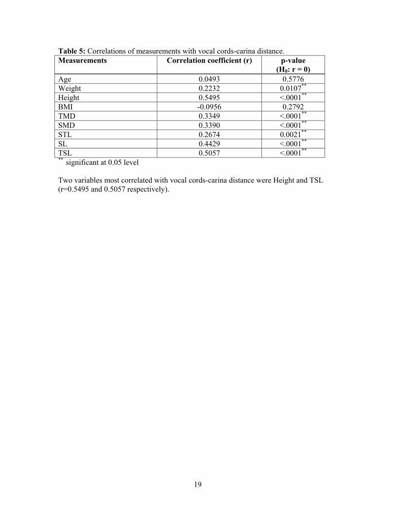
Table 5: Correlations of measurements with vocal cords-carina distance. Measurements Correlation coefficient (r) p-value
(H0: r = 0) Age 0.0493 0.5776 Weight 0.2232 0.0107** Height 0.5495 <.0001** BMI -0.0956 0.2792 TMD 0.3349 <.0001** SMD 0.3390 <.0001** STL 0.2674 0.0021** SL 0.4429 <.0001** TSL 0.5057 <.0001** ** significant at 0.05 level Two variables most correlated with vocal cords-carina distance were Height and TSL (r=0.5495 and 0.5057 respectively).
19

Table 6: Areas under the ROC curves (detecting vocal cords-carina distance ≤ 11.0 cm) Variable Area 95% CI p-value
(H0: area=0.5) Age 0.601 [0.470, 0.732] 0.205 Weight 0.646 [0.497, 0.795] 0.067 Height 0.781 [0.671, 0.891] <.001** TMD 0.623 [0.471, 0.774] 0.123 SMD 0.618 [0.478, 0.757] 0.139 STL 0.652 [0.509, 0.796] 0.056 SL 0.741 [0.608, 0.875] 0.002** TSL 0.795 [0.692, 0.899] <.001** Model 0.815 [0.715, 0.916] <.001** ** significant at 0.05 level
20

Graph 1: ROC Curves for detecting VCD ≤ 11cm
1 - Specificity
0.0 0.2 0.4 0.6 0.8 1.0
Sens
itivi
ty
0.0
0.2
0.4
0.6
0.8
1.0
HeightThyrosternal lengthRegression model
21

Table 7: Cut-off value and (Specificity and positive predictive values) of the various measurements when sensitivity of the test is 100%. VCD length
Height TSL Combination Regression Model
10.0cm ≤159cm (Spec =55.91% PPV =5.08%)
≤24.5cm (Spec =92.13%PPV =23.08%)
Ht ≤159cm & TSL ≤24.5cm (Spec =92.13%PPV =23.08%)
<11.96cm (Spec =75.59% PPV =8.82%)
10.5cm ≤159cm (Spec =57.72% PPV =11.86%)
≤28.5cm (Spec =36.59% PPV =8.24%)
Ht ≤159cm & TSL ≤28.5cm (Spec =63.41% PPV =13.46%)
< 12.28cm (Spec =69.92%PPV =15.91%)
11.0cm ≤167.5cm (Spec =23.48% PPV =14.56%)
≤28.5cm (Spec =39.13% PPV =17.65%)
Ht ≤167.5cm & TSL ≤28.5cm (Spec =46.96%PPV =19.74%)
<13.13cm (Spec =27.83% PPV =15.31%)
12.0cm ≤170cm (Spec =21.95% PPV =42.86%)
≤31.0cm (Spec =12.20% PPV =40%)
Ht ≤170cm & TSL ≤31.0cm (Spec=28.05% PPV=44.86%)
<13.37cm (Spec =25.61% PPV =44.04%)
22

Reference: 1. McCoy EP, Russell WJ, Webb RK: Accidental bronchial intubation. An analysis of AIMS incident reports from 1988 to 1994 inclusive. Anaesthesia 1997; 52: 24-3 2. Owen RL, Cheney FW: Endobronchial intubation: a preventable complication. Anesthesiology 1987; 67: 255-7 3. Cavo JW: True Vocal Cord Paralysis Following Intubation. Laryngoscope 1985; 95: 1352-58 4. Weber S: Traumatic complications of airway management. Anesthesiology Clinics of North America 2002; 20: 503-12 5. Ong KC, A'Court GD, Eng P, Ong YY: Ideal Endotracheal Tube Placement by Referencing Measurements on the Tube. Annals Academy of Medicine Singapore 1996; 25: 550-2 6. Sosis MB, Harbut RE: A Caution on the Use of Routine Depth of Insertion of Endotracheal Tubes. Anesthesiology 1991; 74: 961-2 7. Ellis H, Feldman S, Harrop-Griffiths W: Anatomy for the Anaesthetist, 8th Edition. Oxford, Blackwell Publishing, 2004 8. Sinnatamby CS: Last's Anatomy, 10th Edition. Edinburgh, London, New York, Churchill Livingstone, 1999 9. Eagle CCP: The Relationship Between a Person's Height and Appropriate Endotracheal Tube Length. Anaesthesia & Intensive Care 1992; 20: 156-160 10. Cherng CH, Wong CS, Hsu CH, Ho ST: Airway length in adults:estimation of the optimal endotracheal tube length for orotracheal intubation. Journal of Clinical Anesthesia 2002; 14: 271-4 11. Mehta S: Intubation guide marks for correct tube placement - a clinical study. Anaesthesia 1991; 46: 306-308 12. Hartrey R, Kestin IG: Movement of oral and nasal tracheal tubes as a result of changes in head and neck position. Anaesthesia 1995; 50: 682-7 13. Bennett JD, Guha SC, Sankar AB: Cricothyrotomy: the anatomical basis. Journal of the Royal College of Surgeons Edinburgh 1996; 41: 57-60 14. Randestad A, Lindholm C-E, Fabian P: Dimensions of the Cricoid Cartilage and the Trachea. The Laryngoscope 2000; 110: 1957-1961 15. Chen PP, Chui PT: Endobronchial intubation during laparoscopic cholecystectomy. Anaesthesia and Intensive Care 1992; 20: 537-8 16. Iwama H, Nakane M, Aoki K, Watanabe K, Komatsu T, Kaneko T: Abdominal Insufflation Pressure during Laparoscopic Cholecystectomy Shifts the Tracheal Carina Cephalad. Anesthesiology 1996; 84: 491-2 17. Mackenzie M, MacLeod K: Repeated inadvertent endobronchial intubation during laparoscopy. British Journal of Anaesthesia 2003; 91: 297-8 18. Heinonen J, Takki S, Tammisto T: Effect of the Trendelenburg Tilt and other Procedures on the Position of Endotracheal Tubes. Lancet 1969; 1: 850-3 19. Goodman LR, Conrardy P, Laing F, Singer MM: Radiographic Evaluation of Endotracheal Tube Position. American Journal of Roentgenology 1976; 127: 433-4
23

24
20. Griscom NT, Wohl MEB: Dimensions of the growing trachea related to body height. Length, anteroposterior and transverse diameters, cross-sectional area, and volume in subjects younger than 20 years of age. American Review of Respiratory Disease 1985; 131: 840-4 21. Inada T, Uesugi F, Kawachi S, Takubo K: Changes in tracheal tube position during laparoscopic cholecystectomy. Anaesthesia 1996; 51: 823-6