
Philippus Feselius. Biographische Notizen zum unbekannten Medicus aus Keplers Tertius Interveniens
Upload
khangminh22Category
view
1download
0
MEDICUSISSN 1409-6366 UDC 61 Vol · 18 (2) · Shtator/September 2013
Punime origjinale shkencore
Punime reviale
Prezantime rasti
Kumtesë e shkurtër
9 MIX WARM BLOOD CARDIOPLEGIA (MWBC)Saimir Kuci, Alfred Ibrahimi, Jonela Burimi, Andi Kacani, Ermal Likaj, Ali Refatllari, Edvin Prifti, Arben Fino, Ilir Ohri, Efrosina Kajo,
14 INCIDENTAL PROSTATE CANCER IN PATIENTS UNDERGOING RADICAL CYSTOPROSTATECTOMY IN TREATMENT FOR BLADDER CANCER: OUR FIVE-YEAR RESULTSSkender Saidi, Sotir Stavridis , Sasho Dohcev, Oliver Stankov, Selim Komina, Gordana Petrushevska, Zhivko Popov
19 PROGNOSTIC MODEL FOR STRATIFICATION OF PATIENTS WITH CHRONIC LYMPHOCYTIC LEUKEMIATrajkova Sanja, Cevreska Lidija, Ivanovski Martin, Dukovski Dusko, Simjanovska-Popova Marija, Cadievski Lazar, Pivkova Aleksandra, Stankovik Svetlana, Panovska-Stavridis Irina
25 CREATINE KINASE ACTIVITY IN HUMAN SEMINAL FLUIDZafer Gashi, Afrim Zeqiraj, Shkelzen Elezaj, Selim Kolgeci, Albert Lila
32 KIRURGJIA E VALVULËS SË AORTËS: REZULTATET E KIRURGJISË SË VALVULËS SË AORTËS TË IZOLUAR APO TË KOMBINUAR ME BY-PASS AORTO KORONAR.Selman Dumani, Ermal Likaj, Andi Kaçani, Laureta Dibra ,Endri Harja, Sajmir Kuçi, Dervish Hasi, Elizana Petrela, Ali Refatllari .
39 ЕВАЛУАЦИЈА НА ЕФЕКТОТ НА ПОКАЧЕНИОТ ИНТРААБДОМЕНАЛЕН ПРИТИСОК НА ВИСЦЕРАЛНАТА ПЕРФУЗИЈААлександра Гавриловска-Брзанов, Мирјана Шошолчева, Зорка Николова - Тодорова, Гордана Талеска, Маја Мојсова Мијовска, Марија Јовановски-Срцева, Вања Џамбазовска Трајковска, Никола Брзанов
47 LIDHJA MIDIS RETINOPATISË DIABETIKE DHE HEMOGLOBINËS SË GLUKOZUAR NË PACIENTËT DIABETIKËKavaja Silvana, Ceka Xheladin, Spahiu Eduard, Kavaja Genti, Xinxo Sonela
52 THE PANDEMIC OF A(H1N1) INFLUENZA IN ALBANIA COMPARED WITH THE FIRST POST-PANDEMIC INFLUENZA SEASON 2010 - 2011.Entela Kolovani, Dhimitër Kraja, Ergys Ramosaço, Arjan Harxhi, Arben Ndreu, Genti Stroni, Eli Kallfa, Gjeorgjina Kuli-Lito, Sashenka Sallabanda, Alban Hatibi, Pellumb Pipero, Artan Simaku, Iris Hatibi, Majlinda Kota, Dritan Ulqinaku, Eugena Tomini, Alma Robo, Adela Vasili, Alban Ylli, Eduard Kakarriqi, Silva Bino.
61 PROKALCITONINA SI TEST DIAGNOSTIK NE PACIENTET ME SEPSISEdmond Puca, Pellumb Pipero, Arben Pilaca, Ilir Akshija, Entela Y Puca, Dhimiter Kraja
67 PJEKURIA PULMONARE, NDIKIMI NE PARAQITJEN E SINDROMES SE DISTRESIT RESPIRATOR(RDSY)Sani Bajrami, Florin Besimi , Nagip Rufati , Meral Rexhepi , Arian Alili, Merie Vejseli , Gjuzvete Tairi, Zllata Trimçevska, Antigona Trenevska, Bashkim Ismaili , Besa Islami-Pocesta, Drita Tefiku , Afërdita Ismaili, Mirsada Emini, Bujar Abdiu, Savica Markovic, Nexhibe Nuhii
71 ЕВАЛУАЦИЈА НА ВОЛУМЕНОТ НА ХЕМАТОМОТ, ВОЛУМЕНОТ НА ЕДЕМОТ И Ц-РЕАКТИВЕН ПРОТЕИН ВО АКУТНАТА ФАЗА НА ПРИМАРНА СУПРАТЕНТОРИЈАЛНА ИНТРАЦЕРЕБРАЛНА ХЕМОРАГИЈАПетровска-Цветковска Д., Долненец-Банева Н., Алексовски В., Зафировска-Ивановска Б.
75 ATEROSKLEROZA AKCELERUESE DHE NDIKIMI I ABNORMALITETEVE TË LIPO/APOPROTEINEVE NË PARAQITJENE E MANIFESTIMEVE VASKULARE ME KALCIFIKATE TE PACIENTËT UREMIKËLutfi Zylbeari , Elita M., Gazmend Z., Zamira B., Driton Selmani.
85 NDJESHMËRIA E LARTË E METODËS FISH (FLUORESCENCE IN SITU HYBRIDIZATION) NË NDJEKJEN E TË SËMURËVE ME LEUKOZË MIELOIDE KRONIKE TË TRAJTUAR ME GLIVEKDorina Roko, Anila Babameto–Laku, Pal Xhumari, Donjeta Bali, Vahe Mokini, Maksim Cikuli
92 РАНИ КОМПЛИКАЦИИ ПО ОПЕРАТИВНИОТ ТРЕТМАН НА ФРАКТУРИ НА ПРОКСИМАЛЕН ФЕМУРС. Трпески, И.Кафтанџиев, Г.Велковски, И.Орана, З.Темелковски, А.Ќаев
96 TIPARET PSIKOPATIKE TË PACIENTËVE ME SKIZOFRENI DHE NDËRLIDHJA ME SJELLJEN AGRESIVE RECIDIVUESE NË SHËRBIMIN E PSIKIATRISË FORENZIKE NË PRISHTINËNazmie F Ibishi, Nebi Musliu, Zylfije Hundozi, Bekim Haxhiu
105 MBIPESHA DHE OBEZITETI TEK ADOLESHENTËT NE SHQIPERI Enkelejda Shkurti, Diamant Shtiza , Angela Andreoli, Diederik Aarendonk
110 VLERËSIMI I CILËSISË SË VAJIT TË ULLIRIT ‘‘EKSTRA I VIRGJER’’NË TREGUN SHQIPTAR Çakraj R., Prifti D.
119 NDIKIMI I CILESISE SE UJRAVE TE PISHINAVE TE TIRANES, NE SHENDETIN DHE SIGURINE E PERDORUESVE TE TYRE Drita Mema, Donika Bocari, Arben Luzati, Elida Mataj, Oltiana Petri
125 MBIKËQYRJA BAKTERIOLOGJIKE DHE FIZIKOKIMIKE E UJIT TË PIJES NË KOMUNËN E PRIZRENITFadil Kryeziu, Tahire Maloku, Violeta Kryeziu, Sejran Abdushi, Nurishahe Hulaj,
130 УПОТРЕБА НА АУТОГРАФТ ЗЕМЕН ОД ФЕМОРАЛНА ГЛАВА КАЈ ВГРАДУВАЊЕ НА БЕСЦЕМЕНТНА АЦЕТАБУЛАРНА КОМПОНЕНТА ПРИ КОНГЕНИТАЛНА ДИСЛОКАЦИЈА НА КОЛККамнар Виктор, Атанасов Ненад, Попоска Анастасика, Богојевска Милена
134 NUCHAL TRANSLUCENCY AND NORMAL KARYOTYPE CORRELATIONS AND POSSIBILITIESAна Данева Маркова, Марија Хаџи Лега , Арта Бина1, Тања Николова
137 SYSTEMIC MANIFESTATIONS OF COPD – IS THERE A CONNECTION BETWEEN COPD AND RHINOSINUSITIS?Tanya Tacheva, Dimo Dimov, Iliya Popov, Mariana Penkova Radicheva, Tatyana Vlaykova
141 THE ENORMOUS SIZE OF THE GALLBLADER – A REASON FOR CONVERSION TO OPEN SURGERY IN ACUTE CHOLECYSTITISRexhep Selmani, Arben Karpuzi, Atip Ramadani
145 DIAGNOSTIKIMI ANTENATAL I SYNDROMËS SË HIPOPLASIONIT TË VENTRIKULLIT TË MAJTËHana Bejiqi, Ramush Bejiqi, Ragip Retkoceri, Naim Zeka, Njazi Gashi,
150 ENECFALITI LIMBIKKamber U. Zeqiraj, Enver D. Isaku, Nazim Q. Dakaj , Nexhat R. Shatri, Fisnik B. Jashari
154 BREAST CANCER IN PREGNANCY - CASE REPORTJovanova D., Kocoski G., A. Selami
160 OTOLITOPATIA (BPPV - BENIGN PAROXYSMAL POSITIONAL VERTIGO)Labinot Bekteshi, Hasan Bekteshi

MEDICUSReviste Mjekesore | Medical Journal
ISSN 1409-6366 UDC 61 Vol · 18 (2) · Shtator/September 2013
Punime origjinale shkencore
Punime reviale
Prezantime rasti
Kumtesë e shkurtër
9 MIX WARM BLOOD CARDIOPLEGIA (MWBC)Saimir Kuci, Alfred Ibrahimi, Jonela Burimi, Andi Kacani, Ermal Likaj, Ali Refatllari, Edvin Prifti, Arben Fino, Ilir Ohri, Efrosina Kajo,
14 INCIDENTAL PROSTATE CANCER IN PATIENTS UNDERGOING RADICAL CYSTOPROSTATECTOMY IN TREATMENT FOR BLADDER CANCER: OUR FIVE-YEAR RESULTSSkender Saidi, Sotir Stavridis , Sasho Dohcev, Oliver Stankov, Selim Komina, Gordana Petrushevska, Zhivko Popov
19 PROGNOSTIC MODEL FOR STRATIFICATION OF PATIENTS WITH CHRONIC LYMPHOCYTIC LEUKEMIATrajkova Sanja, Cevreska Lidija, Ivanovski Martin, Dukovski Dusko, Simjanovska-Popova Marija, Cadievski Lazar, Pivkova Aleksandra, Stankovik Svetlana, Panovska-Stavridis Irina
25 CREATINE KINASE ACTIVITY IN HUMAN SEMINAL FLUIDZafer Gashi, Afrim Zeqiraj, Shkelzen Elezaj, Selim Kolgeci, Albert Lila
32 KIRURGJIA E VALVULËS SË AORTËS: REZULTATET E KIRURGJISË SË VALVULËS SË AORTËS TË IZOLUAR APO TË KOMBINUAR ME BY-PASS AORTO KORONAR.Selman Dumani, Ermal Likaj, Andi Kaçani, Laureta Dibra ,Endri Harja, Sajmir Kuçi, Dervish Hasi, Elizana Petrela, Ali Refatllari .
39 ЕВАЛУАЦИЈА НА ЕФЕКТОТ НА ПОКАЧЕНИОТ ИНТРААБДОМЕНАЛЕН ПРИТИСОК НА ВИСЦЕРАЛНАТА ПЕРФУЗИЈААлександра Гавриловска-Брзанов, Мирјана Шошолчева, Зорка Николова - Тодорова, Гордана Талеска, Маја Мојсова Мијовска, Марија Јовановски-Срцева, Вања Џамбазовска Трајковска, Никола Брзанов
47 LIDHJA MIDIS RETINOPATISË DIABETIKE DHE HEMOGLOBINËS SË GLUKOZUAR NË PACIENTËT DIABETIKËKavaja Silvana, Ceka Xheladin, Spahiu Eduard, Kavaja Genti, Xinxo Sonela
52 THE PANDEMIC OF A(H1N1) INFLUENZA IN ALBANIA COMPARED WITH THE FIRST POST-PANDEMIC INFLUENZA SEASON 2010 - 2011.Entela Kolovani, Dhimitër Kraja, Ergys Ramosaço, Arjan Harxhi, Arben Ndreu, Genti Stroni, Eli Kallfa, Gjeorgjina Kuli-Lito, Sashenka Sallabanda, Alban Hatibi, Pellumb Pipero, Artan Simaku, Iris Hatibi, Majlinda Kota, Dritan Ulqinaku, Eugena Tomini, Alma Robo, Adela Vasili, Alban Ylli, Eduard Kakarriqi, Silva Bino.
61 PROKALCITONINA SI TEST DIAGNOSTIK NE PACIENTET ME SEPSISEdmond Puca, Pellumb Pipero, Arben Pilaca, Ilir Akshija, Entela Y Puca, Dhimiter Kraja
67 PJEKURIA PULMONARE, NDIKIMI NE PARAQITJEN E SINDROMES SE DISTRESIT RESPIRATOR(RDSY)Sani Bajrami, Florin Besimi , Nagip Rufati , Meral Rexhepi , Arian Alili, Merie Vejseli , Gjuzvete Tairi, Zllata Trimçevska, Antigona Trenevska, Bashkim Ismaili , Besa Islami-Pocesta, Drita Tefiku , Afërdita Ismaili, Mirsada Emini, Bujar Abdiu, Savica Markovic, Nexhibe Nuhii
71 ЕВАЛУАЦИЈА НА ВОЛУМЕНОТ НА ХЕМАТОМОТ, ВОЛУМЕНОТ НА ЕДЕМОТ И Ц-РЕАКТИВЕН ПРОТЕИН ВО АКУТНАТА ФАЗА НА ПРИМАРНА СУПРАТЕНТОРИЈАЛНА ИНТРАЦЕРЕБРАЛНА ХЕМОРАГИЈАПетровска-Цветковска Д., Долненец-Банева Н., Алексовски В., Зафировска-Ивановска Б.
75 ATEROSKLEROZA AKCELERUESE DHE NDIKIMI I ABNORMALITETEVE TË LIPO/APOPROTEINEVE NË PARAQITJENE E MANIFESTIMEVE VASKULARE ME KALCIFIKATE TE PACIENTËT UREMIKËLutfi Zylbeari , Elita M., Gazmend Z., Zamira B., Driton Selmani.
85 NDJESHMËRIA E LARTË E METODËS FISH (FLUORESCENCE IN SITU HYBRIDIZATION) NË NDJEKJEN E TË SËMURËVE ME LEUKOZË MIELOIDE KRONIKE TË TRAJTUAR ME GLIVEKDorina Roko, Anila Babameto–Laku, Pal Xhumari, Donjeta Bali, Vahe Mokini, Maksim Cikuli
92 РАНИ КОМПЛИКАЦИИ ПО ОПЕРАТИВНИОТ ТРЕТМАН НА ФРАКТУРИ НА ПРОКСИМАЛЕН ФЕМУРС. Трпески, И.Кафтанџиев, Г.Велковски, И.Орана, З.Темелковски, А.Ќаев
96 TIPARET PSIKOPATIKE TË PACIENTËVE ME SKIZOFRENI DHE NDËRLIDHJA ME SJELLJEN AGRESIVE RECIDIVUESE NË SHËRBIMIN E PSIKIATRISË FORENZIKE NË PRISHTINËNazmie F Ibishi, Nebi Musliu, Zylfije Hundozi, Bekim Haxhiu
105 MBIPESHA DHE OBEZITETI TEK ADOLESHENTËT NE SHQIPERI Enkelejda Shkurti, Diamant Shtiza , Angela Andreoli, Diederik Aarendonk
110 VLERËSIMI I CILËSISË SË VAJIT TË ULLIRIT ‘‘EKSTRA I VIRGJER’’NË TREGUN SHQIPTAR Çakraj R., Prifti D.
119 NDIKIMI I CILESISE SE UJRAVE TE PISHINAVE TE TIRANES, NE SHENDETIN DHE SIGURINE E PERDORUESVE TE TYRE Drita Mema, Donika Bocari, Arben Luzati, Elida Mataj, Oltiana Petri
125 MBIKËQYRJA BAKTERIOLOGJIKE DHE FIZIKOKIMIKE E UJIT TË PIJES NË KOMUNËN E PRIZRENITFadil Kryeziu, Tahire Maloku, Violeta Kryeziu, Sejran Abdushi, Nurishahe Hulaj,
130 УПОТРЕБА НА АУТОГРАФТ ЗЕМЕН ОД ФЕМОРАЛНА ГЛАВА КАЈ ВГРАДУВАЊЕ НА БЕСЦЕМЕНТНА АЦЕТАБУЛАРНА КОМПОНЕНТА ПРИ КОНГЕНИТАЛНА ДИСЛОКАЦИЈА НА КОЛККамнар Виктор, Атанасов Ненад, Попоска Анастасика, Богојевска Милена
141 THE ENORMOUS SIZE OF THE GALLBLADER – A REASON FOR CONVERSION TO OPEN SURGERY IN ACUTE CHOLECYSTITISRexhep Selmani, Arben Karpuzi, Atip Ramadani
145 DIAGNOSTIKIMI ANTENATAL I SYNDROMËS SË HIPOPLASIONIT TË VENTRIKULLIT TË MAJTËHana Bejiqi, Ramush Bejiqi, Ragip Retkoceri, Naim Zeka, Njazi Gashi,
150 ENECFALITI LIMBIKKamber U. Zeqiraj, Enver D. Isaku, Nazim Q. Dakaj , Nexhat R. Shatri, Fisnik B. Jashari
154 BREAST CANCER IN PREGNANCY - CASE REPORTJovanova D., Kocoski G., A. Selami
160 OTOLITOPATIA (BPPV - BENIGN PAROXYSMAL POSITIONAL VERTIGO)Labinot Bekteshi, Hasan Bekteshi
134 NUCHAL TRANSLUCENCY AND NORMAL KARYOTYPE CORRELATIONS AND POSSIBILITIESAна Данева Маркова, Марија Хаџи Лега , Арта Бина1, Тања Николова
137 SYSTEMIC MANIFESTATIONS OF COPD – IS THERE A CONNECTION BETWEEN COPD AND RHINOSINUSITIS?Tanya Tacheva, Dimo Dimov, Iliya Popov, Mariana Penkova Radicheva, Tatyana Vlaykova

Betimi i HipokratitNë зastin kur po hy në radhët e anëtarëve të profesionit mjekësor premtoj solemnisht se jetën time do ta vë në shërbim të humanitetit. Ndaj mësuesve do ta ruaj mirënjohjen dhe respektin e duhur. Profesionin tim do ta ushtroj me ndërgjegje e me dinjitet. Shëndeti i pacientit tim do të jetë brenga ime më e madhe. Do t’i respektoj e do t’i ruaj fshehtësitë e atij që do të më rrëfehet. Do ta ruaj me të gjitha forcat e mia nderin e traditës fisnike të profesionit të mjekësisë.
Kolegët e mi do t’i konsideroj si vëllezër të mi.
Në ushtrimin e profesionit ndaj të sëmurit tek unë nuk do të ndikojë përkatësia e besimit, e nacionalitetit, e racës, e politikës, apo përkatësia klasore. Që nga fillimi do ta ruaj jetën e njeriut në mënyrë apsolute. As në kushtet e kërcënimit nuk do të lejoj të keqpërdoren njohuritë e mia mjekësore që do të ishin në kundërshtim me ligjet e humanitetit. Këtë premtim po e jap në mënyrë solemne e të lirë, duke u mbështetur në nderin tim personal.
The Oath of HippocratesUpon having conferred on me the high calling of physician and entering medical practice, I do solemnly pledge myself to consecrate my life to the service of humanity. I will give my teachers the respect and gratitude which is their due. I will practice my profession with conscience and dignity. The health of my patient will be my first consideration. I will respect the secrets which are confided in me, even after the patient has died. I will maintain by all the means in my power, the honor and the noble traditions of the medical profession.
My colleagues will be my brothers.
I will not permit considerations of religion, nationality, race, party politics or social standing to intervene between my duty and my patient. I will maintain the utmost respect for human life from its beginning even under threat and I will not use my medical knowledge contrary to the laws of humanity. I make these promises solemnly, freely and upon my honor
KryeredaktoriProf. Dr. Aziz K. Pollozhani
RedaktorëtDr. Sci. Besnik Bajrami, Boston, SHBA
Dr. Sci. Atilla Rexhepi, Tetovë, MaqedoniLul Raka, MD,PhD, Prishtinë, Kosovë
Doc. Dr. Arben Taravari, Shkup, Maqedoni
Këshilli RedaktuesNobelisti Prof. Dr. Ferid Murad, Hjuston, SHBA
Prof. Dr. Rifat Latifi, Arizona, SHBAProf. Dr. Alex Leventa, Jerusalem, IzraelProf. Dr. Sedat Üstündağ, Edirne, Turqi
Prof. asoc. dr. Avdyl Krasniqi, Prishtinë, KosovëProf. dr. sci. Kirk Milhoan,Texas, SHBA
Dr. sci. Minir Hasani, GjermaniProf. dr sci. Alfred Priftanji, Tiranë, Shqipëri
Prof. dr. sci. Naser Ramadani, Prishtinë, KosovëProf. dr Yovcho Yovchev, Stara Zagora, BullgariKadri Haxhihamza, MD, PhD, Shkup, Maqedoni
Prof. dr. sci. Elena Qoseska, Shkup, MaqedoniProf. dr Gentian Vyshka, Tiranë,Shqipëri
Prim. dr Gani Karamanaga, Ulqin, Mali ZiProf. dr Sylejman Rexhepi, Prishtinë, KosovëDr. Shenasi Jusufi , Koordinator, Maqedoni
Editor-in-ChiefAziz K. Pollozhani, MD. PhD
EditorsBesnik Bajrami, MD, PhD, Boston, USAAtilla Rexhepi, MD, PhD, Tetovo, MacedoniaLul Raka, MD,PhD, Prishtina, KosovaDoc. Dr. Arben Taravari, Skopje, Macedonia
Editorial BoardNobel Laureate Ferid Murad, MD, PhD, Houston, USARifat Latifi, MD, PhD, Arizona, USAAlex Leventa, MD, PhD Jerusalem, IsraelSedat Ustundağ, Edirne, TurkiyeAvdyl Krasniqi, MD, PhD, Prishtina, KosovaKirk Milhoan, MD, PhD, Texas, USAMinir Hasani, MD, PhD, GermanyAlfred Priftanji, MD, PhD, Tirana, AlbaniaNaser Ramadani, MD, PhD, Prishtina, KosovaYovcho Yovchev, MD, PhD, Stara Zagora, BulgariaKadri Haxhihamza, MD, PhD, Skopje, MacedoniaElena Kosevska, MD, PhD, Skopje, MacedoniaGentian Vyshka, MD, PhD, Tirana, AlbaniaGani Karamanaga, MD, Ulcinj, MontenegroSylejman Rexhepi, MD, PhD, Prishtina, KosovaShenasi Jusufi, MD, Coordinator, Macedonia
Revistë Shkencore Nderkombëtare e Shoqatës së Mjekëve Shqiptarë të MaqedonisëInternational Journal of Medical Sciences of the Association of the Albanian Doctors from Macedonia
Botues/ Publisher: SHMSHM / AAMDTel. i Kryeredaktorit / Contact: +389 (0)31 25 044
Zhiro llogaria / drawing account: 200-000031528193Numri tatimor / tax number: 4028999123208
Adresa e Redaksisë-Editorial Board Address: 50 Divizija, No 6, 1000 Shkupe-mail: [email protected]
ISSN 1409-6366 UDC 61 Vol · 18 (2) · Shtator/September 2013
Redaksia | Editorial Board
MEDICUSReviste Mjekesore | Medical Journal
Redaksia | Editorial Board
Bordi KëshillëdhënësProf. Dr. Remzi Izairi,
Prof. dr. Shpëtim Telegrafi, Nju Jork, SHBAProf. dr. Gëzim Boçari, Tiranë, Shqipëri
Prof. dr. Donço Donev, Shkup, MaqedoniProf. Dr. Isuf Dedushaj, Prishtinë, Kosovë
Prof. Dr. Ramadan Jashari, BelgjikëProf. Dr. Holger Tietzt, Gjermani
Prof. Dr. Vjollca Meka-SahatçiuProf. Dr. Florin Ramadani, Austri
Sekretariati i redaksisëDr. Besnik Hamiti, Maqedoni
Dr. Sead Zeynel, Maqedoniz. Armend Iseni, Maqedoni
Këshilli BotuesProf. Dr. Nevzat Elezi
Prim. Dr. Ali DalipiPrim. Dr. Ferit Muça
Prim. Dr. Lavdërim SelaDr. Bekim IsmailiDr. Nadi Rustemi
Dr. Bedri VeliuDr. Arif Latifi
Dr. Gafur PolisiDr. Valvita Reçi
Dr. Xhabir BajramiDr. Gazi Mustafa
Prim. Dr. Beqir AdemiDr. Murat Murati
Dr. Dukagjin OsmaniDr. Bari Abazi
Dr. Atip Ramadani
Dizajni & PamjaBesnik Hamiti
Shtypur nëShtypshkronjen “Aj-Graf MATBAA”, Shkup
Medicus shtypet në tirazh: 600 ekzemplarëRevista shperndahet falas
Advisory BoardRemzi Izairi, MD, PhDShpetim Telegrafi, MD, PhD, New York, USAGezim Bocari, MD, PhD, Tirana, AlbaniaDonco Donev, MD, PhD, Skopje, MacedoniaIsuf Dedushaj, MD, PhD, Prishtina, KosovaRamadan Jashari, MD, PhD, BelgjumHolger Tietzt, MD, PhD, GermanyVjollca Meka-Sahatciu, MD, PhDFlorin Ramadani, MD, PhD, Austria
Editorial SecretariatBesnik Hamiti, MD, MacedoniaSead Zeynel, MD, MacedoniaArmend Iseni, BSc. Macedonia
Editorial CouncilNevzat Elezi, MD, PhDAli Dalipi, MDFerit Muca, MDLavderim Sela, MDBekim Ismaili, MDNadi Rustemi, MDBedri Veliu, MDArif Latifi, MDGafur Polisi, MDValvita Reci, MDXhabir Bajrami, MDGazi Mustafa, MDBeqir Ademi, MDMurat Murati, MDDukagjin Osmani, MDBari Abazi, MDAtip Ramadani, MD
Design & LayoutBesnik Hamiti
Printed in:Print House “Aj-Graf MATBAA”, Skopje
The Jounal Medicus is printed and distributed freeof charge with a circulation of 600 copies.
7 | Medical Journal - MEDICUS
Editorial | Editorial
How much the publications are being read?
The scientific work, writing and publication of the papers, today are the main reference by which the level of professional and scientific achievements
of the individual, as well as the institution (institute, faculty or university) are measured, whilst building the foundations of a knowledge-based society.
Hereby, I would like to emphasize to additional points out of the topic that I would elaborate on below. In many renowned journals’ editorials or letters to the editor you can read about the heating debate over the reasonability of the ‘publish or perish’ attitude and the publication of articles containing ‘scientific novelties’ (solely for the sake of the formal requirements for their careers’ advancement), thus imposing to the young scientists a model that doesn’t meet the needs for their development as a healthcare professionals and therefore fostering a debate with a lot’s of pro’s and con’s.
The second point which usually incites debate is the pressing issue of ‘how much the publications are being read’? There is another viewpoint regarding this issue, i.e. ‘how much the published articles have contributed for the distribution of the experience and the acquired knowledge of the respective fields’. A debate of this kind has been developed in two out of three recent international conferences. The issue doesn’t only comprises the ‘quotes’, according to Science Citation Index (SCI), presenting a well-known and verifying system for citation measurement, but the issue is mostly seen from the reader’s aspect, as a way for exchanging and communication of the newly acquired knowledge, ultimately for the patients’ benefit.
Despite the possibilities and high achievements in this respect, an uncontested statement was made that the published work is barely read, furthermore, that we should find other modes and opportunities in order to facilitate and stimulate the usage of these information as frequently as possible for those being interested. Without doubt, the Information Technologies’ era has enabled the electronic communication in every household, and almost everybody is skilled to use it.
Sa lexohen botimet e publikuara?
Puna kërkimëre shkencore, shkruajtja dhe publikimi i punimeve sot janë bëre referneca kryseore e nivelit profesional dhe shkencor të
individit,njësive ku ato punojnë(intitutit, fakultetit apo universitetit) duke shtruar, kështu, bazën për mjë shoqëri të bazuar ne dije. Ky fakt e bën sferën e botimit të punimeve shkencore, motivim dhe piksynim për punë dhe arritje të mëtejme për hulumtuesit e të gjitha fushave, kurse botuesit i vendon para përgjegjësive të reja.
Me këte rast do të doja të veja në pah edhe dy momente plotësuase të debatit në vijim . Në editorialet dhe letrat për editorët në shumë revista me nam botëror mund të gjinden shpesh artikuj ku debatohet arsueshmëra e “ngarendjes me çdo kusht” botime të artikullit me përmbajtje “të reja” shkencore (me qëllim të realizimit të kërkesave për ndërtimin e kariesës) duke u imponiuar shkencëtarëve të rinj një praktikë që jo gjithmonë përputhet me nevojat themelore për ndërtimin e tyre si klinicist (!), me ç’rast palët argumentojnë “pro et contra”.
Momenti i dytë për të cilin shpesh mund të ndëgjohen opinione në formë të shqetësimit, lidhet me pyetjen “sa lexohen botimet e publikuara?”. Kjo pyetje mund të shtrohet edhe ndryshe, p.sh. ”me botimet e publikuara, sa kontribohet në shpërndarjen e përvojës dhe afirmimin e njohurive të reja në fushat përkatëse”. Një debat i tille ka qenë prezent në dy nga tre konferanca ndërkombëtare që së fundi janë mbajtur në këtë temë. Kuptohet që kjo pyetje nuk shtrohet vetëm nga kënveshtrimi i “citimeve”, sipas Science Citation Index (SCI) që paraqet një sistem të njohur dhe të verifikuar të matshëm të citimve, por çështja më shumë shtrohet nga aspekti i leximit të punimeve si një mënyrë të ndërrimit të arritjeve dhe komunikimin e njohurive me qëllim shfrytëzimi dhe benefiti për pacintët nga të njëjtat.
Përkundër mundësive dhe arritjeve të mëdha në këtë aspekt, ishte konstatim i pakontestushem që këto punime lexohen shumë pak, se duhet gjetë forma dhe mundësi për lehtësimin dhe stimulimin
Revistë mjekësore - MEDICUS | 8
Editorial | Editorial
Taking into account this fact, as well as the need for the advancement of the editorial procedures, and the increased usage of the IMJ Medicus’ published work, the Association of the Albanian Medical Doctors from Macedonia (AAMDM) through the IMJM’s Editorial Board, with the current issuance starts the web site that will be the electronic platform for on-line manuscript submission, on-line publication and sharing other needed information. Additionally, AAMDM as a publisher of the IMJM, will continue with the publication of the Journal’s hard-copy, warranting the continuity of the collaboration with our respected readers.
As we have collaborated up to this day, hereby, I invite you to communicate on-line for a more efficient collaboration in the future.
IMJ Medicus’ web page:
www.imjm.mk
Azis. K. Pollozhani, MD. PhDEditor-in-chief International Medical Journal Medicus (IMJM)
e të interesuarve për shfrytëzim sa më të shpeshtë të këtyre infomacioneve. Padyshim që era e tekonlogjisë informatike, komunikimin elektronik e ka bërë presentnt në çdo shtëpi, dhe shkathtësitë për përdorimin e tij i kanë gati të gjithë.
Nisur nga ky fakt, por edhe nga nevoja për të avancuar procedurat editoriale si dhe shfrytëzimin sa më të shpeshtë dhe praktik të botimeve të IMJ Medicus, Shoqata e Mjekëve Shqiptarë të Maqedonisë (SHMSHM), respektivisht Bordi Editorial i IMJM, nga ky numër fillon me përdorimin on-line të web faqes, që do të jetë platforma elektronike për dorëzimin e dorëshkrimeve on-line, leximin e punimeve të publikuara dhe infrmacioneve tjera të nevojshme. Në mënyre plotësuesë, SHMSHM, si botuesi IMJM, do të vazhdojë me publikimin e Revistës (hard-copy) si deri tani, duke garantuar kështu vazhdimësinë e bashkëpunimit me lexuesit tanë të nderuar.
Duke ju faleminderuar për bashkëpunimin e deritan-ishëm ju ftojmë në komunikim on-line dhe bashkëpunim edhe më efektiv në të ardhmen.
Web faqja e IMJ Medicus:
www.imjm.mk
Prof dr.Azis. K. PollozhaniKrzeredaktori i IMJM:
9 | Medical Journal - MEDICUS
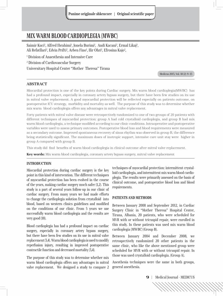
MIX WARM BLOOD CARDIOPLEGIA (MWBC)Saimir Kuci1, Alfred Ibrahimi1, Jonela Burimi1, Andi Kacani2, Ermal Likaj2, Ali Refatllari2, Edvin Prifti2, Arben Fino1, Ilir Ohri1, Efrosina Kajo2,1 Division of Anaesthesia and Intensive Care2 Division of Cardiovascular Surgery
Universitary Hospital Center “Mother Theresa” Tirana
ABSTRACT
Miocardial protection is one of the key points during Cardiac surgery. Mix warm blood cardioplegia(MWBC) has had a profound impact, especially in coronary artery bypass surgery, but there have been few studies on its use in mitral valve replacement. A good myocardial protection will be reflected especially on patients outcome, on postoperative ICU strategy, morbidity and mortality as well. The purpose of this study was to determine whether mix warm blood cardioplegia offers any advantages in mitral valve replacement.
Forty patients with mitral valve disease were retrospectively randomized to one of two groups of 20 patients with different techniques of myocardial protection: group A had cold crystalloid cardioplegia, and group B had mix warm blood cardioplegia, a technique modified according to our clinic conditions. Intraoperative and postoperative variables were used to assess primary outcomes. Postoperative blood loss and blood requirements were measured as a secondary outcome. Improved spontaneous recovery of sinus rhythm was observed in group B, the difference being statistically significant. The maximum doses of inotropic support, intensive care unit stay were higher in group A compared with group B.
This study did find benefits of warm blood cardioplegia in clinical outcome after mitral valve replacement.
Key words: Mix warm blood cardioplegia, coronary artery bypass surgery, mitral valve replacement
INTRODUCTION
Miocardial protection during cardiac surgery is the key point in this kind of intervention. The different techniques of myocardial protection has been evolved in the course of the years, making cardiac surgery much safer (1,2). This study is a part of several years follow-up in our clinic of cardiac surgery. From many years we had made efforts to change the cardioplegia solution from crystalloid into blood, based on western clinics guidelines and modified on the conditions of our clinic. From 5 years we use successfully warm blood cardioplegia and the results are very good (18).
Blood cardioplegia has had a profound impact on cardiac surgery, especially in coronary artery bypass surgery, but there have been few studies on its use in mitral valve replacement (3,4). Warm blood cardioplegia is used to modify reperfusion injury, resulting in improved postoperative contractile function and decreased mortality (3,4).
The purpose of this study was to determine whether mix warm blood cardioplegia offers any advantages in mitral valve replacement. We designed a study to compare 2
techniques of myocardial protection: intermittent crystal-loid cardioplegia, and intermittent mix warm blood cardio-plegia. The results were primarily assessed on the basis of clinical outcome, and postoperative blood loss and blood requirements.
PATIENTS AND METHODS
Between January 2008 and September 2012, in Cardiac Surgery Clinic in “Mother Theresa” Hospital Center, Tirana, Albania, 20 patients, who were scheduled for MVR with or without tricuspid repair, were enrolled in this study. In these patients was used mix warm blood cardioplegia (MWBC) (Group B).
Between January 2004 and December 2008, we retrospectively randomized 20 other patients in the same clinic, who like the above mentioned group were scheduled for MVR with or without tricuspid repair. In those was used crystalloid cardioplegia. (Group A).
Anesthesia techniques were the same in both groups, general anesthesia.
Medicus 2013, Vol. 18 (2): 9 -13
1Punime origjinale shkencore | Original scientific paper
Punime origjinale shkencore | Original scientific paper
Revistë mjekësore - MEDICUS | 10
In group A, intermittent antegrade cold crystalloid cardioplegia (4 grade C) was used.
In group B, was used mix warm blood cardioplegia (MWBC).
Randomization was performed in blocks of 20, with a plan for interim analysis after 40 patients.
Initial power calculations were hampered by the lack of preliminary data defining the effect of MWBC on MVR.
The study was approved by our Institutional Ethics Committee, and informed consent of the patients was obtained.
All operations were performed using cardiopulmonary bypass (CPB) with ascending aortic and bicaval cannulation. Systemic hypothermia was between 32°C and 34°C in Group A and 33°C to 36°C in Group B.
Antegrade cold crystalloid cardioplegia was injected im-mediately after aortic cross-clamping at 15-20mL·kg-1
and then at 30 minutes intervals. In Group B the tech-nique MWBC was realized second the modifications of our clinic;18 the first dose was cold crystalloid (15-120 ml/kg/weight), after 30 minutes a second dose of cardio-plegia was made by mixing 400-500 ml oxygenated blood from oxygenator and 10ml KCL 7.5%, and a third dose of the same solution after 20minutes. The MWBC tempera-ture was gradually increased from 33°C to 36°C by the end of the infusion.
Electrical defibrillation was applied if ventricular fibrilla-tion persisted beyond 2min after aortic declamping, and a temporary pacemaker was used if there was no spon-taneous rhythm or if the patient’s heart rate was less than 50 beats·min-1. After the operation, if systolic blood pressure was lower than 90 mm Hg and urine output less than 1 mL·kg-1·h-1 with central venous pressure between 10 and 12 mm Hg, inotropic support was started. Our first choice of inotropic agent was adrenaline.
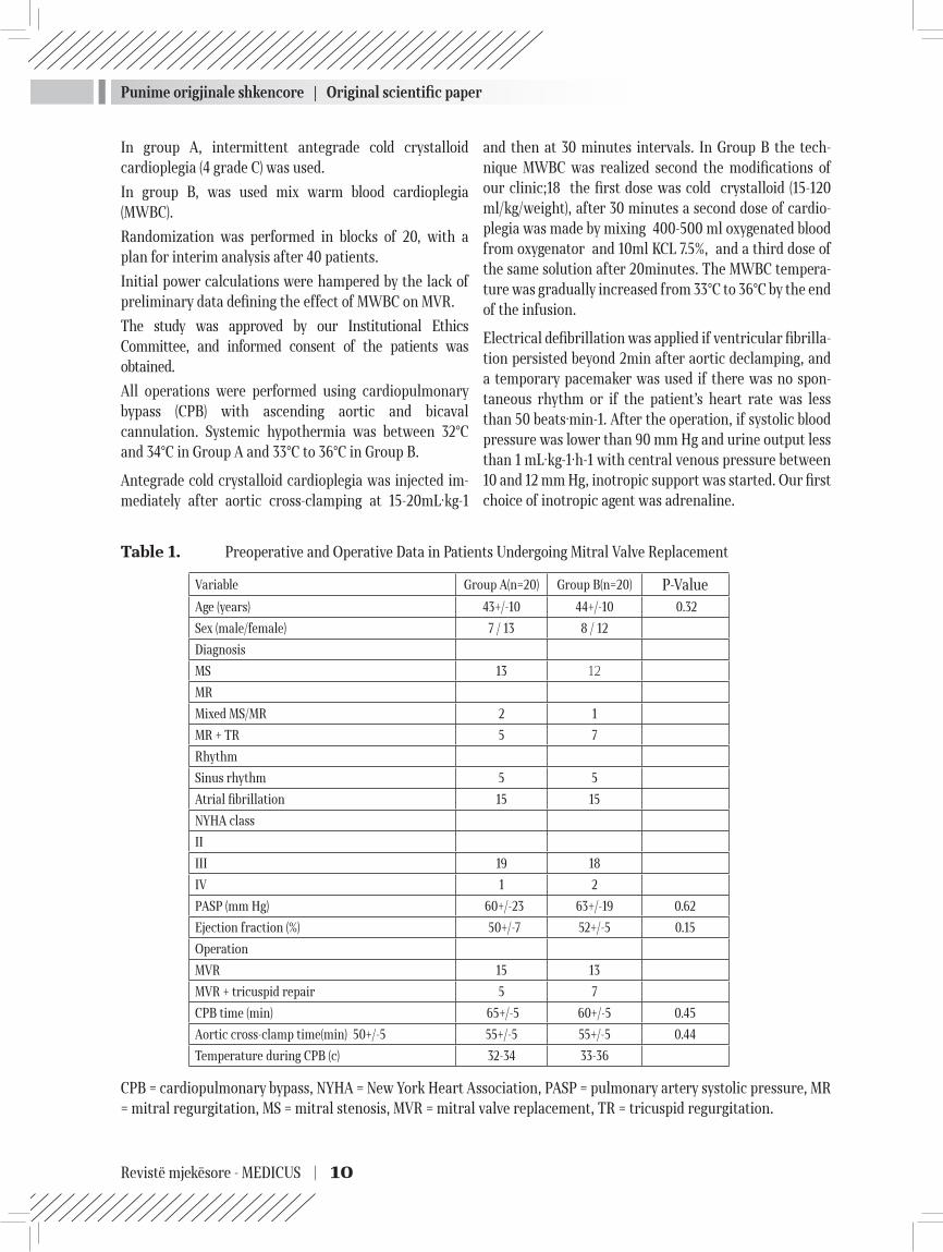
Table 1. Preoperative and Operative Data in Patients Undergoing Mitral Valve Replacement
CPB = cardiopulmonary bypass, NYHA = New York Heart Association, PASP = pulmonary artery systolic pressure, MR = mitral regurgitation, MS = mitral stenosis, MVR = mitral valve replacement, TR = tricuspid regurgitation.
Variable Group A(n=20) Group B(n=20) P-ValueAge (years) 43+/-10 44+/-10 0.32
Sex (male/female) 7 / 13 8 / 12
Diagnosis
MS 13 12
MR
Mixed MS/MR 2 1
MR + TR 5 7
Rhythm
Sinus rhythm 5 5
Atrial fibrillation 15 15
NYHA class
II
III 19 18
IV 1 2
PASP (mm Hg) 60+/-23 63+/-19 0.62
Ejection fraction (%) 50+/-7 52+/-5 0.15
Operation
MVR 15 13
MVR + tricuspid repair 5 7
CPB time (min) 65+/-5 60+/-5 0.45
Aortic cross-clamp time(min) 50+/-5 55+/-5 55+/-5 0.44
Temperature during CPB (c) 32-34 33-36
Punime origjinale shkencore | Original scientific paper
11 | Medical Journal - MEDICUS
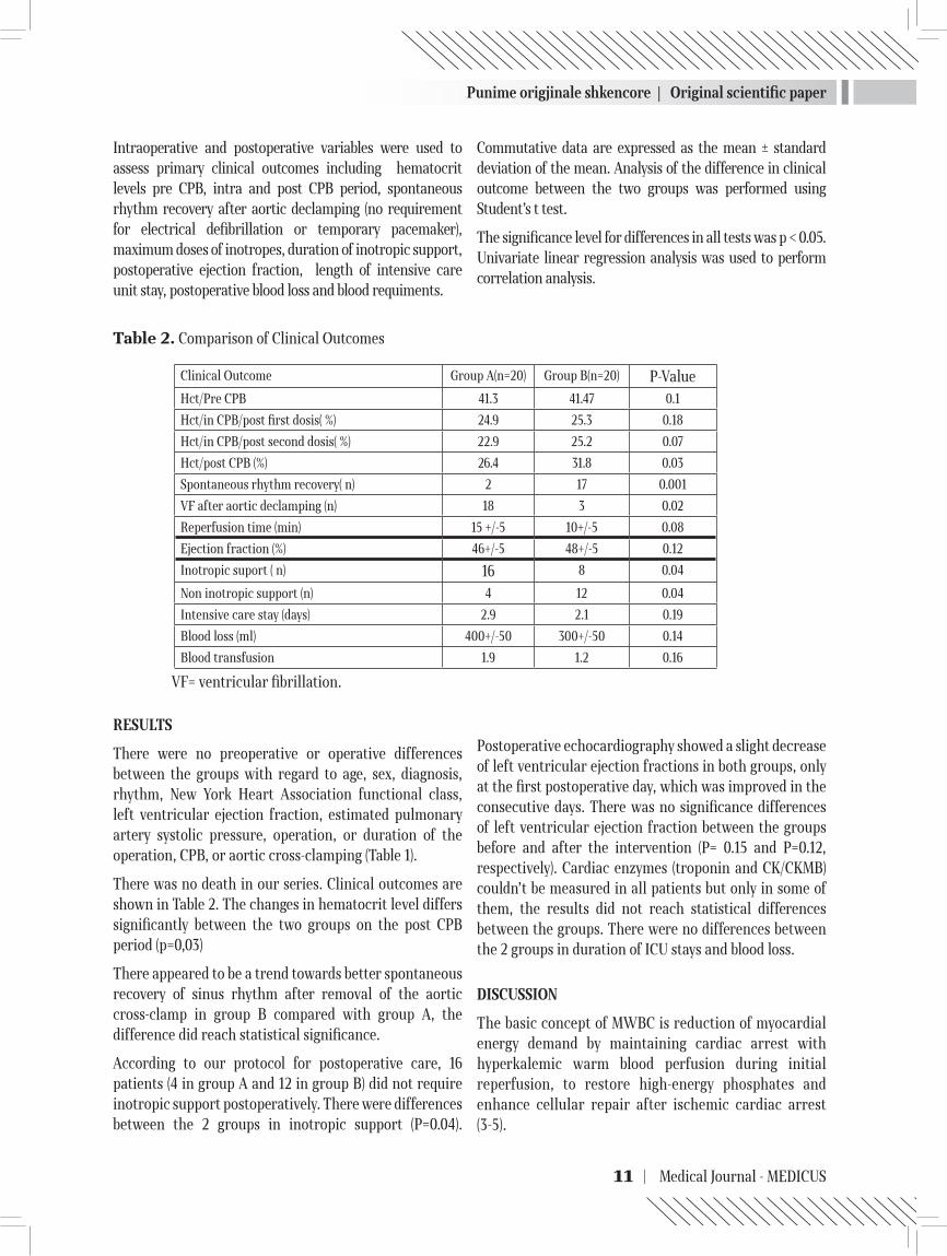
Intraoperative and postoperative variables were used to assess primary clinical outcomes including hematocrit levels pre CPB, intra and post CPB period, spontaneous rhythm recovery after aortic declamping (no requirement for electrical defibrillation or temporary pacemaker), maximum doses of inotropes, duration of inotropic support, postoperative ejection fraction, length of intensive care unit stay, postoperative blood loss and blood requiments.
Commutative data are expressed as the mean ± standard deviation of the mean. Analysis of the difference in clinical outcome between the two groups was performed using Student’s t test.
The significance level for differences in all tests was p < 0.05. Univariate linear regression analysis was used to perform correlation analysis.
Table 2. Comparison of Clinical Outcomes
VF= ventricular fibrillation.
RESULTS
There were no preoperative or operative differences between the groups with regard to age, sex, diagnosis, rhythm, New York Heart Association functional class, left ventricular ejection fraction, estimated pulmonary artery systolic pressure, operation, or duration of the operation, CPB, or aortic cross-clamping (Table 1).
There was no death in our series. Clinical outcomes are shown in Table 2. The changes in hematocrit level differs significantly between the two groups on the post CPB period (p=0,03)
There appeared to be a trend towards better spontaneous recovery of sinus rhythm after removal of the aortic cross-clamp in group B compared with group A, the difference did reach statistical significance.
According to our protocol for postoperative care, 16 patients (4 in group A and 12 in group B) did not require inotropic support postoperatively. There were differences between the 2 groups in inotropic support (P=0.04).
Postoperative echocardiography showed a slight decrease of left ventricular ejection fractions in both groups, only at the first postoperative day, which was improved in the consecutive days. There was no significance differences of left ventricular ejection fraction between the groups before and after the intervention (P= 0.15 and P=0.12, respectively). Cardiac enzymes (troponin and CK/CKMB) couldn’t be measured in all patients but only in some of them, the results did not reach statistical differences between the groups. There were no differences between the 2 groups in duration of ICU stays and blood loss.
DISCUSSION
The basic concept of MWBC is reduction of myocardial energy demand by maintaining cardiac arrest with hyperkalemic warm blood perfusion during initial reperfusion, to restore high-energy phosphates and enhance cellular repair after ischemic cardiac arrest (3-5).
Clinical Outcome Group A(n=20) Group B(n=20) P-ValueHct/Pre CPB 41.3 41.47 0.1
Hct/in CPB/post first dosis( %) 24.9 25.3 0.18
Hct/in CPB/post second dosis( %) 22.9 25.2 0.07
Hct/post CPB (%) 26.4 31.8 0.03
Spontaneous rhythm recovery( n) 2 17 0.001
VF after aortic declamping (n) 18 3 0.02
Reperfusion time (min) 15 +/-5 10+/-5 0.08
Ejection fraction (%) 46+/-5 48+/-5 0.12
Inotropic suport ( n) 16 8 0.04
Non inotropic support (n) 4 12 0.04
Intensive care stay (days) 2.9 2.1 0.19
Blood loss (ml) 400+/-50 300+/-50 0.14
Blood transfusion 1.9 1.2 0.16
Punime origjinale shkencore | Original scientific paper
Revistë mjekësore - MEDICUS | 12
The beneficial effects of MWBC have been studied both experimentally and clinically. They include better preservation of high-energy phosphates and endogenous amino acids, less anaerobic metabolic activity on reperfusion, reduced release of cardiac troponin T, and improved post-ischemic functional recovery (3,4,6-8).These effects facilitate coronary vasodilatation and accelerate early myocardial tissue oxygen saturation during warm reperfusion (4,6).
Tenpaku and colleagues (7) demonstrated complete microtubule repolymerisation after 10 min of reperfusion with warm blood. This mechanism may be responsible for the early and improved recovery of cardiac function associated with MWBC.
Most clinical studies of MWBC have been undertaken on coronary artery bypass surgery, with more recent investigations on MWBC in congenital heart surgery (3,4,9,10).
Most results have showed definite advantages in the use of MWBC, although there have been some that showed no beneficial effect of MWBC (11-13).
Modi and colleagues10 demonstrated a superior biochemical outcome from MWBC in patients with longer cross-clamp times, a finding also supported by Hayashi and colleagues (14).
In this study, the mean cross-clamp time was more than 50 min, which may have been long enough to demonstrate the advantage of MWBC. The duration of TWBC infusion may be an important factor (6,15).
There is some evidence that a glutamate-aspartate supplement to the MWBC (substrate-enriched cardioplegia) may reduce reperfusion injury and improve both metabolic and myocardial function recovery (8,13). Glutamate and aspartate are not available at our institute, so our MWBC was not a substrate-enriched solution, and it is possible that the MWBC benefits would be more obvious if this technique was employed. Improved spontaneous rhythm recovery has also been observed with the use of MWBC (6,9). It appeared to be a trend towards a better cardiac rhythm recovery in the MWBC group (less requirement of electrical defibrillation and temporary pacemakers), the difference did reach statistical significance when compared with patients in the non-MWBC group.
Troponin T and troponin I are unique biochemical markers of myocardial damage because of their high level
of sensitivity and specificity. These characteristics make troponin T and troponin I ideal markers for myocardial cell damage in patients undergoing cardiac surgery, and useful for comparison of different myocardial protective techniques during routine cardiac operations (16,17).We found a slightly higher troponin T concentration at 0 and 6 h postoperatively in a part of patients given TWBC, although the difference was not significant when compared with the non-TWBC group. Our finding was similar to that reported by Edwards and colleagues (12).
REFERENCES
1. Cohen G, Borger MA, Weisel RD, Rao V. Intraoperative myocardial protection: current trends and future per-spectives. Ann Thorac Surg 1999;68:1995–2001.
2. Nicolini F, Beghi C, Muscari C, Agostinelli A, Maria Budillon A, Spaggiari I, et al. Myocardial protection in adult cardiac surgery:current options and future chal-lenges. Eur J Cardiothorac Surg 2003;24:986–93.
3. Teoh KH, Christakis GT, Weisel RD, Fremes SE, Mickle DA, Romaschin AD, et al. Accelerated myocardial meta-bolic recovery with terminal warm blood cardioplegia. J Thorac Cardiovasc Surg 1986;91:888–95.
4. Caputo M, Dihmis WC, Bryan AJ, Suleiman MS, Angelini GD. Warm blood hyperkalaemic reperfusion (‘hot shot’) prevents myocardial substrate derangement in patients undergoing coronary artery bypass surgery. Eur J Car-diothorac Surg 1998;13:559–64.
5. Follette DM, Fey KH, Steed DL, Foglia RP, Buckberg GD. Reducing reperfusion injury with hypocalcemic, hyper-kalemic, alkalotic blood during reoxygenation. Surg Fo-rum 1978;29:284–6.
6. Kawasuji M, Tomita S, Yasuda T, Sakakibara N, Take-mura H, Watanabe Y. Myocardial oxygenation during terminal warm blood cardioplegia. Ann Thorac Surg 1998;65:1260–4.
7. Tenpaku H, Onoda K, Imanaka-Yoshida K, Yoshida T, Shimono T, Shimpo H, et al. Terminal warm blood cardi-oplegia improves cardiac function through microtubule repolymerization. Ann Thorac Surg 1998;65:1580–7.
8. Kronon MT, Allen BS, Rahman S, Wang T, Tayyab NA, Bolling KS, et al. Reducing postischemic reperfusion damage in neonates using a terminal warm substrate-enriched blood cardioplegia reperfusate. Ann Thorac Surg 2000;70:765–70.
9. Toyoda Y, Yamaguchi M, Yoshimura N, Oka S, Okita Y. Cardioprotective effects and the mechanisms of
Punime origjinale shkencore | Original scientific paper
13 | Medical Journal - MEDICUS
terminal warm blood cardioplegia in pediatric cardiac surgery. J Thorac Cardiovasc Surg 2003;125:1242–51.
10. Modi P, Suleiman MS, Reeves B, Pawade A, Parry AJ, Angelini GD, et al. Myocardial metabolic changes dur-ing pediatric cardiac surgery: a randomized study of 3 cardioplegic techniques. J Thorac Cardiovasc Surg 2004;128:67–75.
11. Chocron S, Alwan K, Yan Y, Toubin G, Kaili D, Anguenot T, et al. Warm reperfusion and myocardial protection. Ann Thorac Surg 1998;66:2003–7.
12. Edwards R, Treasure T, Hossein-Nia M, Murday A, Kantidakis GH, Holt DW. A controlled trial of substrate-enhanced, warm reperfusion (“hot shot”) versus simple reperfusion. Ann Thorac Surg 2000;69:551–5.
13. Wallace AW, Ratcliffe MB, Nose PS, Bellows W, Moores W, McEnany MT, et al. Effect of induction and reperfu-sion with warm substrate-enriched cardioplegia on ven-tricular function. Ann Thorac Surg 2000;70:1301–7.
14. Hayashi Y, Sawa Y, Fukuyama N, Miyamoto Y, Takahashi T, Nakazawa H, et al. Leukocyte-depleted terminal blood cardioplegia provides superior myocardial protec-tive effects in association with myocardium-derived ni-
tric oxide and peroxynitrite production for patients un-dergoing prolonged aortic crossclamping for more than 120 minutes. J Thorac Cardiovasc Surg 2003;126:1813–21.
15. Allen BS, Okamoto F, Buckberg GD, Leaf J, Bugyi H. Ef-fects of “duration” of reperfusate administration versus reperfusate “dose” on regional functional, biochemical, and histochemical recovery. J Thorac Cardiovasc Surg 1986;92(3 Pt 2):594–604.
16. Caputo M, Dihmis W, Birdi I, Reeves B, Suleiman MS, Angelini GD, et al. Cardiac troponin T and troponin I release during coronary artery surgery using cold crys-talloid and cold blood cardioplegia. Eur J Cardiothorac Surg 1997;12:254–60.
17. 17. Mair J, Wieser C, Seibt I, Arther-Dworzak E, Furt-wangler W, Waldenberger F, et al. Troponin T to diag-nose myocardial infarction in bypass surgery. Lancet 1991;337:434–5.
18. Ruajtja e hematokritit gjate QEK tek pacientet qe i nenshtrohen revaskularizimit koronar kirurgjikal. Kra-hasimi i mbushjes se oksigjenatorit me prime standart dhe me mbushje retrograde me gjak autolog (RAP) ,Dr. Saimir Kuci , Prof.Ali Refatllari
MARDIOPEGJIA MIKE ME GJAK TË NGROHTË
Saimir Kuci1, Alfred Ibrahimi1, Jonela Burimi1, Andi Kacani1, Ermal Likaj1, Ali Refatllari1, Edvin Prifti1, Arben Fino1, Ilir Ohri1, Efrosina Kajo1, 1 Qendra Spitalore Universitare “Nënë Tereza”, Tiranë
ABSTRAKTI
Proteksioni i miokardit është një nga pikat kryesore në kirurgjinë kardiake.Kardioplegjia me gjak ka patur një impakt të fortë në kirurgjinë kardiake, veçanërisht në bypass-in e arterieve koronare, por ka pak studime në përdorimin e saj në kirurgjinë e valvulës mitrale. Një proteksion i mirë i miokardit gjatë kirurgjisë do të reflektohet veçanerisht në dekursin postoperator të pacientit, në strategjitë reanimatore si dhe në morbiditet dhe mortalitet. Qëllimi i këtij studimi është të përcaktojë avantazhet e kardioplegjisë me gjak në kirurgjinë e valvulës mitrale. U morën në studim 40 pacientë që iu nënshtruan kirurgjisë së valvulës mitrale,të cilët u ndanë në 2 grupe me nga 20 pacientë, ku u përdorën teknika të ndryshme për mbrojtjen e miokardit: në grupin A u përdor kardioplegji kristaloide dhe grupi B u përdor kardioplegji mikse e ngrohtë e modifikuar sipas kushteve të klinikës tonë. Variablat intra dhe postoperatore u morën si përfundime primare, humbjet e gjakut postoperatore dhe kërkesat për transfusion u morën si përfundime sekondare. Rimarrja spontane e ritmit sinusal u vu re më tepër në grupin B, ku diferenca ishte statistikisht e rëndësishme krahasuar me grupin A. Dozat e inotropeve, ditëqendrim në terapinë intensive ishin më të lartë në grupin A krahasuar me grupin B. Ky studim përcaktoi disa benefite klinike të përdorimit të kardioplegjisë me gjak të ngrohtë në kirurgjinë e valvulës mitrale.
Falët kyç: Kardioplegjia me gjak, bypass i arterieve koronare, kirurgjia e valvulës mitrale.
Revistë mjekësore - MEDICUS | 14
INCIDENTAL PROSTATE CANCER IN PATIENTS UNDERGOING RADICAL CYSTOPROSTATECTOMY IN TREATMENT FOR BLADDER CANCER: OUR FIVE-YEAR RESULTSSkender Saidi1, Sotir Stavridis1 , Sasho Dohcev1, Oliver Stankov1, Selim Komina2, Gordana Petrushevska2, Zhivko Popov1
1 University Clinic of Urology, Medical Faculty, University ‘’Ss Cyril and Methodius”, Skopje, Republic of Macedonia
2 Department of Pathology
ABSTRACT
The aim of the paper was to verify the incidence and features of incidental prostate cancer in patients who underwent radical cystoprostatectomy for invasive bladder cancer.
Methods and results: A total of 96 patients underwent radical cystoprostatectomy between January 2006 and December 2010 in the University Clinic of Urology in Skopje. 10 patients were excluded for incomplete data. The average age of the study group was 61.2 years (range 32-78). Prostate cancer was found in 10 (11.6%) cases. Seven patients were clinically insignificant.
Conclusion: Incidentally diagnosed prostate cancer was frequently insignificant. Digital rectal examination and prostate specific antigen should be part of the diagnostic procedure in patients who undergo cystoprostatectomy. Standard RCP which include removal of bladder with prostate gland and seminal vesicles is safer for radicality and prevention of residual prostate cancer.
Key words: Bladder cancer, Cystoprostatectomy, prostate cancer
Medicus 2013, Vol. 18 (2): 14-18
INTRODUCTION
Bladder cancer (BC) is the second most common cancer of urinary tract after prostate cancer and the fourth most common malignancy in men (1). In developed European Union country members, the incidence is 34.4, up to 5 in China and highest with 37 cases pmp in Egypt (2). According to the Registry for cancer diseases in R. Macedonia in 2006 the rate of incidence was 6.5 (9.5 in male and 3.4 in female population) (3). Radicalcystoprostatectomy (RCP) is the standard treatment for patients with muscle invasive cancer and treatment failure in high risk non-muscle invasive bladder cancers (4,5). This procedure consists of removing the bladder, prostate gland with seminal vesicles and locoregional lymph nodes. However, this treatment involves the risk of complication like incontinence, sexual and metabolic disorders (6,7). In order to preserve the erectile function, urination and the quality of life associated with RCP urologists search the
new sparing operative techniques, namely sparing the prostate capsule, its apex or the whole prostate gland (8).
On the other hand, autopsy examinations in 30 % of men up to the age of 50 revealed prostate cancer (9).
The aim of this paper was to verify the incidence and features of incidental prostate cancer in patients who underwent radical cystoprostatectomy for invasive bladder cancer.
On the other hand, in case of incidental prostate cancer organ sparing procedures might help in losing the radicality during RCP.
PATIENTS AND METHODS
The study was done on specimen from 96 patients who underwent RCP with no history and clinical evidence for PCa before surgery, between January 2006 and December 2010 in the University Clinic of Urology in Skopje. The
2 Punime origjinale shkencore | Original scientific paper
Punime origjinale shkencore | Original scientific paper
15 | Medical Journal - MEDICUS
mean age of the patients was 61.2 years (range 32-78). Routine evaluation before RCP included digital rectal examinations (DRE), Prostatic specific antigen (PSA) level, ultrasonography, CT urography, histopathologic results from transurethral resection of bladder tumor. Bone scan was done only in patients suspected of having metastasis. Nine patients were excluded from the study for incomplete data and one for suspicious result in DRE. Operative procedure consists of a standard procedure for RCP with bilateral lymphadenectomy and urinary derivation. In pathologic sampling techniques, slices are taken every 3 mm from the base toward the apex of the prostate.
Pathohistological results were analyzed for each patient. PCa was considered clinically significant if any of the following criteria were present: total tumor volume, 0.5 cc or more; Gleason grade, 6 or more; extraprostatic extension; seminal vesicle invasion; lymph node metastasis (of PCa); or positive surgical margins according to the criterion advocated by Epstein et al (10).
The numeric variables were presented as mean + SD. For comparison of categorical variables a Chi-square test was used. Values of p< 0.05 were considered statistically significant.
RESULTS
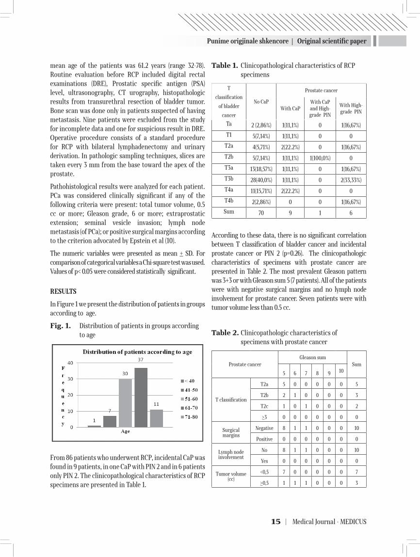
In Figure 1 we present the distribution of patients in groups according to age.
Fig. 1. Distribution of patients in groups according to age
From 86 patients who underwent RCP, incidental CaP was found in 9 patients, in one CaP with PIN 2 and in 6 patients only PIN 2. The clinicopathological characteristics of RCP specimens are presented in Table 1.
Table 1. Clinicopathological characteristics of RCP specimens
T
classification
of bladder
cancer
No CaP
Prostate cancer
With CaPWith CaP and High-grade PIN
With High-grade PIN
Ta 2 (2,86%) 1(11,1%) 0 1(16,67%)
T1 5(7,14%) 1(11,1%) 0 0
T2a 4(5,71%) 2(22.2%) 0 1(16,67%)
T2b 5(7,14%) 1(11,1%) 1(100,0%) 0
T3a 13(18,57%) 1(11,1%) 0 1(16,67%)
T3b 28(40,0%) 1(11,1%) 0 2(33,33%)
T4a 11(15,71%) 2(22.2%) 0 0
T4b 2(2,86%) 0 0 1(16,67%)
Sum 70 9 1 6
According to these data, there is no significant correlation between T classification of bladder cancer and incidental prostate cancer or PIN 2 (p=0.26). The clinicopathologic characteristics of specimens with prostate cancer are presented in Table 2. The most prevalent Gleason pattern was 3+3 or with Gleason sum 5 (7 patients). All of the patients were with negative surgical margins and no lymph node involvement for prostate cancer. Seven patients were with tumor volume less than 0.5 cc.
Table 2. Clinicopathologic characteristics of specimens with prostate cancer
Prostate cancerGleason sum
Sum
5 6 7 8 9 10
T classification
T2a 5 0 0 0 0 0 5
T2b 2 1 0 0 0 0 3
T2c 1 0 1 0 0 0 2
>3 0 0 0 0 0 0 0
Surgical margins
Negative 8 1 1 0 0 0 10
Positive 0 0 0 0 0 0 0
Lymph node involvement
No 8 1 1 0 0 0 10
Yes 0 0 0 0 0 0 0
Tumor volume (cc)
<0,5 7 0 0 0 0 0 7
>0,5 1 1 1 0 0 0 3
Punime origjinale shkencore | Original scientific paper
Revistë mjekësore - MEDICUS | 16
DISCUSSION
RCP is the standard and more effective treatment for muscle invasive bladder cancer and superficial BC with highest progression risk. This procedure consists of removing the bladder, prostate gland, seminal vesicles, along with regional lymphadenectomy. Usually RCP is associated with a sexual and urinary dysfunction. In order to prevent/preserve these functions and quality of life new organ sparing procedures (prostatic apex, prostatic capsule, seminal vesicle and neurovascular bundle) have been described (11, 12). However, all these procedures are associated with loss of radicality in cases with incidental prostate cancer. Relation between these two cancers can be explained by genetic factors such as p53 and RB genes (13). The frequency of PCa incidentally detected in RCP specimens is variable, ranging from 4% to nearly 60 % (14,15,16). This variability can be explained by several factors such as pathologic sampling techniques, preoperative diagnostic procedures for diagnosis of prostate cancer etc. In our study the frequency of incidentally detected prostate cancer in RCP specimens was 11.6%. Also in 6 samples (7%) high-grade prostatic intraepithelial neoplasia (PIN) were found. This percentage was lower than reported in other studies (17). Montironi R. reported that high-grade PIN was present in 54% of cystoprostatectomies without prostate cancer (18). This low rate in our study can be explained by the moderate number of cases, preoperatively diagnostic procedures for prostate cancer and excluded cases with suspicious prostate cancer. Furthermore, cases with verified prostate cancer and bone metastasis have been treated with radiation and hormonal therapy. Also it is well known that the incidence of PCa is higher in the West than in the Asian countries (19). According to the criterion advocated by Epstein et al. for biologic significance, 3 of patients (30%) were with significant prostate cancer (3 with volume >0.5 cc, two of them with Gleason sum >6). This rate was in range with previously presented studies (20,21). Many studies have examined the impact of incidentally diagnosed prostate cancer in RCP on patients’ survival. Some studies found that combination of both cancers has no influence on patients’ survival (22, 23). Sruogis et al. showed that incidentally found prostate cancer influenced on the overall survival, hence indicating that more attention has to be paid to this pathology (24). Wolters et al. reported that screen detected prostate cancers had presented more aggressive features than incidentally prostate cancers (25).
CONCLUSION
The reported incidence of incidental prostate cancer in our series was in lower ranges of presented studies. We recommend digital rectal examination and PSA measurement to be part of preoperative preparation of patients with bladder cancer. In addition, we also claim that the standard RCP, which include removal of bladder with prostate gland and seminal vesicles, is safer for radicality and prevention of residual prostate cancer.
REFERENCES
1. Irani j, Mottet N, Ribal Caparros M.J, and Teillac P. New trends in bladder cancer management. European Urol-ogy 2007; vol 6:4 p.p. 388-395.
2. Parkin DM, Bray F, Ferlay J, Pisani P. Global Cancer Sta-tistics, 2002. Cancer J Clin. 2005; 55:74-108.
3. Registry of cancer in R. Macedonia, Institute of public health-Skopje 2006.
4. Hautmann RE, Abol-Enein H, Hafez K, et al. Urinary di-version. Urology 2007 Jan;69(1 Suppl):17-49.
5. http://www.ncbi.nlm.nih.gov/pubmed/17280907.6. Babjuk M, Oosterlinck W, Sylvester R, et al. EAU Guide-
lines on TaT1 (Non-muscle-invasive Bladder Cancer). In: EAU Guidelines. Edition presented at the 24th EAU Congress, Stockholm, Sweden, 2009. ISBN-978-90-79754-09-0.
7. http://www.uroweb.org/guidelines/online-guidelines/.8. Hautmann RE, Gschwend JE, de Petriconi RC, Kron M,
Volkmer BG. Cystectomy for transitional cell carcinoma of the bladder: results of a surgery only series in the neobladder era. J Urol 2006;176:486-92.
9. Ghoneim MA, Abdel-Latif M, el-Mekresh M, et al. Radi-cal cystectomy for carcinoma of the bladder: 2,720 con-secutive cases 5 years later. J Urol 2008;180:121-7.
10. Srougi M, Dall’Oglio M, Nesrallah LJ, Arruda HO: Radi-cal cystectomy with preservation of sexual function and urinary continence: description of a new technique. Int Braz J Urol 2003,29:336-44.
11. Scott R Jr, Matchnik DH, Laskowski TZ, Schmalfrost W: Carcinoma of the prostate in the elderly men: incidence, growth characteristics and clinical significance. J Urol 1969,101:602-7.
12. Epstein JI, Walsh PC, Carmichael M, Brendler CB: Path-ologic and clinical findings to predict tumor extent of nonpalpable (stage T1c) prostate cancer. JAMA 1994, 271:368-74.
Punime origjinale shkencore | Original scientific paper
17 | Medical Journal - MEDICUS
13. Muto G, Bardari F, D’Urso L, Giona. Seminal sparing cys-tectomy and ileocapsuloplasty: long-term followup re-sults. J Urol 2004;172(1):76-80.
14. Nieuwenhuijzen JA, Meinhardt W, Horenblas S. Clini-cal outcomes after sexuality pre4serving cystectomy and neobladder (prostate sparing cystectomy) in 44 pa-tients. J Urol 2008,179:535-8.
15. Singh A, Jones RF, Friedman H, Hathir S, Soos G, Zabo A, Haas GP. Expression of p53 and pRb in bladder and prostate cancers of patients having both cancers. Anti-cancer Res 1999,19:5415-7.
16. Lee SH, Chang PL, Chen SM, Sun GH, Chen CL, Shen BY, Wu YS, Tsui KH. Synchronous primary carcino-mas of the bladder and prostate. Asian J Androl. 2006 May;8(3):357-9.
17. Autorino R, Di Lorenzo G, Damiano R, et al. Pathology of the prostate in radical cystectomy specimens: a critical review. Surg Oncol. 2009 Mar;18(1):73-84.
18. Damiano R, Di Lorenzo G, Cantiello F, et al. Clinico-pathologic features of prostate adenocarcinoma inci-dentally discovered at the time of radical cystectomy: an evidence-based analysis. Eur Urol. 2007;52:648-657.
19. Prange W, Erbersdobler A, Hammerer P, et al. High-grade prostatic intraepithelial neoplasia in cystoprosta-tectomy specimens. Eur Urol. 2001;39suppl 430–31
20. Montironi R, Mazzucchelli R, Santinelli A, Scarpelli M, Beltran AL, Bostwick DG. Incidentally detected pros-tate cancer in cystoprostatectomies: pathological and morphometric comparison with clinically detected can-
cer in totally embedded specimens. Hum Pathol. 2005 Jun;36(6):646-54.
21. Hsing AW, Tsao L, Devesa SS. International trends and patterns of prostate cancer incidence and mortality. Int J Cancer. 2000;85:60–67.
22. Esgueva R, Lorente JA, Mojal S, et al. Incidental prostat-ic adenocarcinoma in radical cystoprostatectomy speci-mens: the impact of embedding protocols. Mod Pathol. 2008; 21(suppl 1):155A.
23. Revelo MP, Cookson MS, Chang SS, et al. Incidence and location of prostate and urothelial carcinoma in pros-tates from cystoprostatectomies: implications for pos-sible apical sparing surgery. J Urol 2004;171:646-651.
24. Abdelhady M, Abusamra A, Pautler SE, Chin JL, Izawa JI. Clinically significant prostate cancer found incidentally in radical cystoprostatectomy specimens. BJU Interna-tional 2007;99:326–9.
25. Kouriefs C, Fazili T, Masood S, Naseem MS, Mufti GR. In-cidentally detected prostate cancer in cystoprostatec-tomy specimens. Urologia Internationali 2005;75:213–216.
26. Sruogis A, Ulys A, Smailyte G, Kardelis Z, Kulboka A, Anglickienė G, Samalavicius N, Anglickis M. Inciden-tally found prostate cancer and influence on overall sur-vival after radical cystoprostatectomy. Prostate Cancer 2012;2012:690210.
27. Aytac B, Vuruskan H. Clinicopathologic features of in-cidental prostatic adenocarcinoma in radical cystopros-tatectomy specimens. World J Surg Oncol 2011, 20;9:81.
KANCERI INCIDENTAL I PROSTATËS TE PACIENTËT TË NËNSHTRUAR CISTOPROSTATEKTOMISË RADIKALE GJATË TRAJTIMIT TË KANCERIT TË FSHIKËS URINARE REZULTATET TONA 5 VJEÇARE
Skender Saidi1, Sotir Stavridis1 , Sasho Dohcev1, Oliver Stankov1, Selim Komina2, Gordana Petrusevska2, Zhivko Popov1
1 Klinika universitare e urologjisë, Fakulteti i Mjekësisë, Universiteti “Shën Kirili dhe Metodi”, Shkup, Republika e Maqedonisë.
2 Instituti i patologjisë
ABSTRAKTI
Qëllimi i punimit është të përcaktojë incidencën dhe pasojat e kancerit incidental të prostatës te pacientët të nënshtuar cistoprostatektomisë radikale në shërimin e kancerit të fshikës urinare.
Metodat dhe rezultatet: Në periudhen janar 2006 dhe dhjetor 2010 në Klinikën universitare të urologjisë në Shkup, 96 pacientë i janë nënshtruar cistoprostatektomisë radikale për trajtimin e kancerit të fshikës urinare.
Punime origjinale shkencore | Original scientific paper
Revistë mjekësore - MEDICUS | 18
Për shkak të dhënave jo të plota, 10 pacientë janë përjashtuar prej studimit. Mosha mesatare e grupit të studjuar ishte 61.2 vjet (32-78). Kanceri i prostatës është identifikuar te 10 (11.6%) raste. Shtatë pacientë janë klinikisht jo signifikantë.
Konkludim: Kanceri incidental i prostatës në të shumtën e rasteve është josignifikant. Egzaminimi digjito-rektal dhe antigjeni specific prostatës duhet të jenë pjesë e procedurës diagnostifikuese për pacientët që do ti nënshtrohen cistoprostatektomisë. Cistoprostatektomia radikale standard që kupton largimin e fshikës urinare, prostatës dhe fshikëzave farore është procedurë me siguri më të madhe për radikalitetin e procedurës dhe parandalimin e karcinomës reziduale të prostatës.
Fjalët kyç:Kanceri i meshikzes urinare,Cistoprostatektomia,kanceri i prostates.
19 | Medical Journal - MEDICUS
PROGNOSTIC MODEL FOR STRATIFICATION OF PATIENTS WITH CHRONIC LYMPHOCYTIC LEUKEMIATrajkova Sanja 1, Cevreska Lidija1, Ivanovski Martin1, Dukovski Dusko1, Simjanovska-Popova Marija1, Cadievski Lazar1, Pivkova Aleksandra1, Stankovik Svetlana1, Panovska-Stavridis Irina1 1 University Clinic for Hematology, Medical Faculty, University St.Ciril and Metodius,Skopje - R.Macedonia
ABSTRACT
Introduction: The clinical course for patients with chronic lymphocytic leukemia is extremely heterogeneous, some patients have indolent disease, never needing treatment, whereas others have aggressive disease requiring early treatment. Wierda proposed to combine a set of clinical risk factors, to develop a prognostic index (PI) stratifying patients in three risk groups with different expected median survival, and a nomogram, estimating individual patient survivals. Here we report the initial results from a study designed to evaluate clinical and biological prognostic factors in patients risk stratification.
Material and methods: Traditional laboratory, clinical prognostic, and biological prognostic factors were evaluated at first patient visit to University Clinic of Hematology -Skopje Macedonia. We used Wierda`s prognostics index and a nomogram, to see 5- and 10-year survival probability and estimated median survival time.
Results: There were 115 previously untreated patients who had traditional and biological prognostic factors evaluated. According to prognostic index a classification tree was built that identified three subsets of patients. Estimated median survival at low risk subset of patients was 22,5 years, 10, 8 and 4years respectively at intermediate and high risk subsets of patients. Projected survival in respectively low, intermediate and high-risk groups was 70%, 92, 5%, 100%, and 100%, 99%, 60% at 5-year and10-year, respectively. Conclusion: We use this model to identify patients at high risk for progression to treatment. This prognostic model may help patients and clinicians in clinical decision making as well as in clinical research and clinical trial design.
Key words: CLL, prognostic index, nomogram, prognosis
Medicus 2013, Vol. 18 (2): 19-24
INTRODUCTION
Chronic lymphocytic leukemia (CLL) is the most common adult leukemia in the Western world. The clinical course is variable, some patients live for decades without ever requiring treatment, whereas others have rapidly progressive disease requiring treatment within months of diagnosis. For more than 30 years the Rai (1,2) and Binet (3) clinical staging systems broadly identify risk groups based on clinical and laboratory characteristics. Overall, stage correlates with survival, however, for each stage there is still heterogeneity, limiting utility in predicting survival. In addition to factors used in clinical staging, several other patient characteristics and laboratory tests have been correlated with overall survival, including age,(4) sex,(4) pattern of bone marrow involvement,(5) lymphocyte doubling time,(6) and the presence of prolymphocytes in blood or bone marrow(7). Other factors that can be measured in the laboratory
have also been correlated with poor prognosis, including the presence of chromosome abnormalities such as 17p deletion and 11q deletion,(8) elevated serum levels of β-2 microglobulin (β-2M), thymidine kinase, soluble CD23,(9) unmutated immunoglobulin heavy chain variable gene (IgVH),(10) and expression of ZAP-70(11) and CD38(12) by leukemia cells. Alone, each of these prognostic factors has limited utility in predicting overall survival.
To address this problem, Wierda et all.(13) analyzed the clinical outcomes of a large series of patients cared at The University of Texas M.D Anderson Cancer Center(MDACC) during a period of 25 years to determine whether routinely available clinical and laboratory features could enhance the utility of clinical staging. The MD Anderson analysis identified 6 factors (age, Beta2-microglobulin, clinical stage (Rai), the number of lymph node regions(LNR), absolute lymphocyte count(ALC)) that were independently associated with patient survival and
3Punime origjinale shkencore | Original scientific paper
Punime origjinale shkencore | Original scientific paper
Revistë mjekësore - MEDICUS | 20
that can be combined in a prognostic index to predict survival. The investigators also developed nomogram to estimate the 5-year survival and 10-years survival probability for every patient. The prognostic index and nomogram were evaluated by Shanafelt et all. at Mayo Clinic Rochester(14). The latter study confirmed the value of the prognostic index as predictor of overall survival.
In the present study, we used the MDACC prognostic index and nomogram in population of 115 CLL patients. We report the initial results from a study designed to evaluate clinical and biological prognostic factors in patients risk stratification.
MATERIALS AND METHODS
Patient population
The clinical and biological data of 115 untreated CLL patients were retrospectively collected in University Clinic of Hematology in the period of the last 2 years. For all patients complete data on age, Beta2-microglobulin, ALC, sex, Rai staging system and LNR were available.
Nomogram and Prognostic index
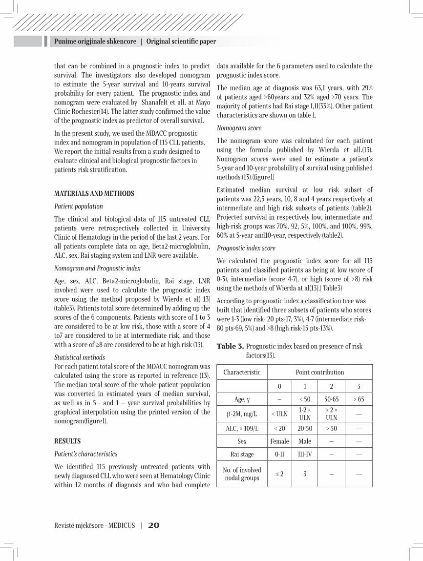
Age, sex, ALC, Beta2-microglobulin, Rai stage, LNR involved were used to calculate the prognostic index score using the method proposed by Wierda et al( 13) (table3). Patients total score determined by adding up the scores of the 6 components. Patients with score of 1 to 3 are considered to be at low risk, those with a score of 4 to7 are considered to be at intermediate risk, and those with a score of ≥8 are considered to be at high risk (13).
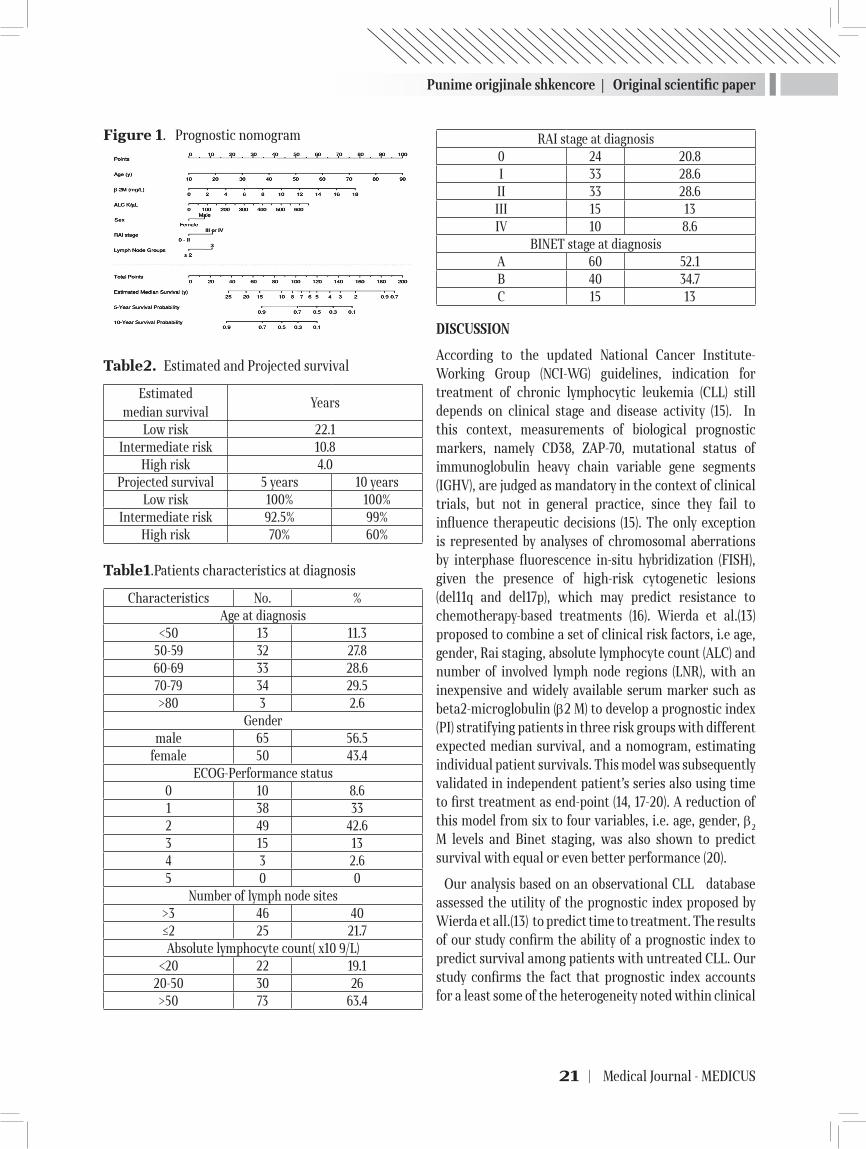
Statistical methodsFor each patient total score of the MDACC nomogram was calculated using the score as reported in reference (13). The median total score of the whole patient population was converted in estimated years of median survival, as well as in 5 - and 1 — year survival probabilities by graphical interpolation using the printed version of the nomogram(figure1).
RESULTS
Patient’s characteristics
We identified 115 previously untreated patients with newly diagnosed CLL who were seen at Hematology Clinic within 12 months of diagnosis and who had complete
data available for the 6 parameters used to calculate the prognostic index score.
The median age at diagnosis was 63,1 years, with 29% of patients aged >60years and 32% aged >70 years. The majority of patients had Rai stage I,II(33%). Other patient characteristics are shown on table 1.
Nomogram score
The nomogram score was calculated for each patient using the formula published by Wierda et all.(13). Nomogram scores were used to estimate a patient`s 5-year and 10-year probability of survival using published methods (13).(figure1)
Estimated median survival at low risk subset of patients was 22,5 years, 10, 8 and 4 years respectively at intermediate and high risk subsets of patients (table2). Projected survival in respectively low, intermediate and high-risk groups was 70%, 92, 5%, 100%, and 100%, 99%, 60% at 5-year and10-year, respectively (table2).
Prognostic index score
We calculated the prognostic index score for all 115 patients and classified patients as being at low (score of 0-3), intermediate (score 4-7), or high (score of >8) risk using the methods of Wierda at al(13).( Table3)
According to prognostic index a classification tree was built that identified three subsets of patients who scores were 1-3 (low risk- 20 pts-17, 3%), 4-7 (intermediate risk-80 pts-69, 5%) and >8 (high risk-15 pts-13%).
Table 3. Prognostic index based on presence of risk factors(13).
Characteristic Point contribution
0 1 2 3
Age, y — < 50 50-65 > 65
β-2M, mg/L < ULN1-2 × ULN
> 2 × ULN
—
ALC, × 109/L < 20 20-50 > 50 —
Sex Female Male — —
Rai stage 0-II III-IV — —
No. of involved nodal groups
≤ 2 3 — —
Punime origjinale shkencore | Original scientific paper
21 | Medical Journal - MEDICUS
Figure 1. Prognostic nomogram
Table2. Estimated and Projected survival
Estimated median survival
Years
Low risk 22.1Intermediate risk 10.8
High risk 4.0Projected survival 5 years 10 years
Low risk 100% 100%Intermediate risk 92.5% 99%
High risk 70% 60%
Table1.Patients characteristics at diagnosis
Characteristics No. %Age at diagnosis
<50 13 11.350-59 32 27.860-69 33 28.670-79 34 29.5>80 3 2.6
Gendermale 65 56.5
female 50 43.4ECOG-Performance status
0 10 8.61 38 332 49 42.63 15 134 3 2.65 0 0
Number of lymph node sites>3 46 40≤2 25 21.7Absolute lymphocyte count( x10 9/L)
<20 22 19.120-50 30 26>50 73 63.4
RAI stage at diagnosis0 24 20.8I 33 28.6II 33 28.6III 15 13IV 10 8.6
BINET stage at diagnosisA 60 52.1B 40 34.7C 15 13
DISCUSSION
According to the updated National Cancer Institute-Working Group (NCI-WG) guidelines, indication for treatment of chronic lymphocytic leukemia (CLL) still depends on clinical stage and disease activity (15). In this context, measurements of biological prognostic markers, namely CD38, ZAP-70, mutational status of immunoglobulin heavy chain variable gene segments (IGHV), are judged as mandatory in the context of clinical trials, but not in general practice, since they fail to influence therapeutic decisions (15). The only exception is represented by analyses of chromosomal aberrations by interphase fluorescence in-situ hybridization (FISH), given the presence of high-risk cytogenetic lesions (del11q and del17p), which may predict resistance to chemotherapy-based treatments (16). Wierda et al.(13)proposed to combine a set of clinical risk factors, i.e age, gender, Rai staging, absolute lymphocyte count (ALC) and number of involved lymph node regions (LNR), with an inexpensive and widely available serum marker such as beta2-microglobulin (β2 M) to develop a prognostic index (PI) stratifying patients in three risk groups with different expected median survival, and a nomogram, estimating individual patient survivals. This model was subsequently validated in independent patient’s series also using time to first treatment as end-point (14, 17-20). A reduction of this model from six to four variables, i.e. age, gender, β2
M levels and Binet staging, was also shown to predict survival with equal or even better performance (20).
Our analysis based on an observational CLL database assessed the utility of the prognostic index proposed by Wierda et all.(13) to predict time to treatment. The results of our study confirm the ability of a prognostic index to predict survival among patients with untreated CLL. Our study confirms the fact that prognostic index accounts for a least some of the heterogeneity noted within clinical
Punime origjinale shkencore | Original scientific paper
Revistë mjekësore - MEDICUS | 22
stage categories. The prognostic index is better predictor of patient’s survival than Rai or Binet risk. The studies published by Shanafelt at all.(14) et Bulian at all. (20) extended the utility of the index by demonstrating that it is useful at the time of diagnosis, retains prognostic value when applied exclusively to patients with Rai stage 0 disease and also predict TTT in addition to survival.
The 6 parameters used to calculate the prognostic index score rely on clinical characteristics and laboratory parameters that are available to all CLL patients. The 5-year overall survival rates from the study of Shanafelt at all.(14) are similar to those observed in MDACC study(13) and that proved that the index is reproducible.
Our analyses were limited to newly diagnosed patients, the time at which risk stratification is needed.
We had interesting situation in the beginning, in our study 17,3% of patients was at low risk group according to prognostic index score and 70% of the patient were at intermediate risk group. When we use Rai risk some of them had 3 or 4 Rai and they were assigned to receive standard chemotherapy by their doctors, but according to prognostic index score they were assigned to watch and wait strategy because they were low or intermediate risk group and still they are on the same strategy.
The prognostic index leads to more precise prediction of patient’s outcome than either approach alone.
CONCLUSIONES
Using the prognostic index (PI) we stratified patients in three risk groups with different expected median survival, also using the nomogram, we estimated individual patient survivals. The Wierda`s prognostic index appears to be a powerful tool to help predict risk in patients with untreated CLL. Addition of molecular and biological prognostic parameters will improve this tool and help patients plan their lives, and develop and test risk-adapted treatment strategies. We use this model that incorporates clinical and laboratory prognostic factors to identify patients at high risk for progression to treatment. This prognostic model may help patients and clinicians in clinical decision making as well as in clinical research and clinical trial design.
REFERENCES
1. Rai KR,Sawitsky A,Cronkite EP,Chanana AD,Levy RN,Pasternack BS. Clinical staging of chronic lympho-cytic leukemia. Blood 1975;46:219-234.
2. Rai KR. A critical analysis of staging in CLL. In: Gale RP, Rai KR, editors. Chronic Lymphocytic Leukemia: Recent Progress, Future Direction. Vol. 253. New York, NY: Liss; 1987.
3. Binet JL,Auquier A,Dighiero G,et al. A new prognos-tic classification of chronic lymphocytic leukemia derived from a multivariate survival analysis. Cancer 1981;48:198-206
4. Catovsky D,Fooks J,Richards S. Prognostic factors in chronic lymphocytic leukaemia: the importance of age, sex and response to treatment in survival. A report from the MRC CLL 1 trial. MRC Working Party on Leukaemia in Adults. Br J Haematol 1989;72:141-149.
5. Rozman C,Montserrat E,Rodriguez-Fernandez JM,et al. Bone marrow histologic pattern—the best single prog-nostic parameter in chronic lymphocytic leukemia: a multivariate survival analysis of 329 cases. Blood 1984;64:642-648.
6. Vinolas N,Reverter JC, Urbano-Ispizua A,Montserrat E,Rozman C. Lymphocyte doubling time in chronic lym-phocytic leukemia: an update of its prognostic signifi-cance. Blood Cells 1987;12:457-470.
7. Melo JV,Catovsky D, Galton DA. Chronic lymphocytic leukemia and prolymphocytic leukemia: a clinicopatho-logical reappraisal. Blood Cells 1987;12:339-353.
8. Dohner H,Stilgenbauer S,Benner A, et al. Genomic aber-rations and survival in chronic lymphocytic leukemia. N Engl J Med 2000;343:1910-1916.
9. Hallek M, Wanders L,Ostwald M, et al. Serum beta(2)-mi-croglobulin and serum thymidine kinase are independ-ent predictors of progression-free survival in chronic lymphocytic leukemia and immunocytoma. Leuk Lym-phoma 1996;22:439-447.
10. Hamblin TJ, Davis Z, Gardiner A,Oscier DG, Stevenson FK. Unmutated Ig V(H) genes are associated with a more aggressive form of chronic lymphocytic leukemia. Blood 1999;94:1848-1854.
11. Crespo M, Bosch F,Villamor N, et al. ZAP-70 expression as a surrogate for immunoglobulin-variable-region mu-
Punime origjinale shkencore | Original scientific paper
23 | Medical Journal - MEDICUS
tations in chronic lymphocytic leukemia. N Engl J Med 2003;348:1764-1775.
12. D’Arena G, Musto P,Cascavilla N, et al. CD38 expres-sion correlates with adverse biological features and predicts poor clinical outcome in B-cell chronic lympho-cytic leukemia. Leuk Lymphoma 2001;42:109-114.
13. Wierda WG, O’Brien S, Wang X, Faderl S,Ferrajoli A, Do KA, et al. Prognostic nomogramand index for overall survival in previously untreated patients with chronic lymphocytic leukemia. Blood.2007;109(11):4679-85.
14. Shanafelt TD, Jenkins G, Call TG, Zent CS,Slager S, Bowen DA, et al. Validation of anew prognostic index for patients with chronic lymphocytic leukemia.Can-cer.2009;115(2):363-72.
15. Hallek M, Cheson BD, Catovsky D, Caligaris-Cappio F, Dighiero G, Dohner H,Hillmen P, Keating MJ, Montserrat E, Rai KR, Kipps TJ, International Workshop on Chronic Lymphocytic Leukemia: Guidelines for the diagno-sis and treatment of chronic lymphocytic leukemia: a report from the International Workshop on Chronic Lymphocytic Leukemia updating the National Cancer Institute-Working Group 1996 guidelines. Blood 2008, 111(12):5446-5456.
16. Zenz T, Eichhorst B, Busch R, Denzel T, Habe S, Win-kler D, Buhler A, Edelmann J, Bergmann M, Hopfinger G, Hensel M, Hallek M, Dohner H, Stilgenbauer S: TP53
mutation and survival in chronic lymphocytic leukemia. J Clin Oncol 2010, 28(29):4473-4479.3.
17. Gonzalez Rodriguez AP, Gonzalez Garcia E, Fernandez Alvarez C, Gonzalez Huerta AJ, Gonzalez Rodriguez S: B-chronic lymphocytic leukemia: epidemiological study and comparison of MDACC and GIMENA pronostic in-dexes. Med Clin (Barc) 2009, 133(5):161-166.
18. Molica S, Di Raimondo F, Cutrona G, Fabris S, Mauro F, Brugiatelli M, Baldini L, Musto P, Sacchi S, Cortelezzi A, Foa R, Neri A, Federico M, Ferrarini M, Morabito F, Grup-po Italiano Studio Linfomi (GISL): Clinical categories identified by a new prognostic index reflect biological characteristics of patients in early chronic lymphocytic leukemia: The Gruppo Italiano Studio Linfomi (GISL) ex-perience. Leuk Res 2010, 34(8): e217-e218.
19. Molica S, Mauro FR, Callea V, Giannarelli D, Lauria F, Rotoli B, Cortelezzi A, Liso V, Foa R: The utility of a prognostic index for predicting time to first treatment in early chronic lymphocytic leukemia: the GIMEMA experience. Haematologica 2010, 95(3):464-469.
20. Bulian P, Tarnani M, Rossi D, Forconi F, Del Poeta G, Ber-toni F, Zucca E, Montillo M, Pozzato G, Deaglio S, D’Arena G, Efremov D, Marasca R, Lauria F, Gattei V, Gaidano G, Laurenti L: Multicentre validation of a prognostic index for overall survival in chronic lymphocytic leukaemia. Hematol Oncol 2011, 29(2):91-99.
ПРОГНОСТИЧКИ МОДЕЛ ЗА СТРАТИФИКАЦИЈА НА ПАЦИЕНТИ СО ХРОНИЧНА ЛИМФОЦИТНА ЛЕУКЕМИЈА
Трајкова Сања1, Чевреска Лидија1, Ивановски Мартин1, Дуковски Душко1, Симјановска-Попова Марија1, Чадиевски Лазар1, Пивкова-Вељановска Александра1, Станковиќ Светлана1, Пановска-Ставридис Ирина1
1 Универзитетска Клиника за Хематологија. Медицински факултет, Универзитет Св.Кирил и Методиј - Скопје - Македонија
РЕЗИМЕ
Вовед: Клиничкиот тек кај пациенти со хронична лимфоцитна леукемија(ХЛЛ) е хетероген, некои пациенти имаат индолентна болест , никогаш не им е потребен третман, додека други имаат агресивна болест која бара ран третман. Wierda предложи да се комбинираат клинички фактори на ризик, за да се развие прогностички индекс (ПИ) кој ги стратифицира пациенти во три ризични групи со различно очекувано средното преживување, и номограм namenet за проценување на преживувањето на пациентите. Gи прикажуваме резултати од студијата za оцена na клиничките и биолошките прогностички фактори при стратификација на пациентите со ХЛЛ според ризик. Материјал и методи: Традиционалните лабораториски, клинички и биолошки прогностички
Punime origjinale shkencore | Original scientific paper
Revistë mjekësore - MEDICUS | 24
фактори се оценуваат на првата посета на пациентот во Универзитетска клиника за хематологија –Скопје, Македонија. Ние go koristevme Wierda -овиот прогностички индекс и номограм, што еовозможува да се предвиди севкупното преживување, и веројатност за 5 - и 10-годишно преживување и средното време на преживување.Резултати: Студијата вклучи 115 претходно нетретирани пациенти. Iзградено е класификационо дрво коe ги идентификуваше трите подгрупи на пациенти . Проценето средното преживување кај пациентите со мал ризик беше 22,5, 10, 8 и 4 години соодветно на средно и висок ризик. Проектираното преживување во ризик групи – низок, среден и висок ризик- беше 70%, 92, 5%, 100%, и 100%, 99%, 60% на 5- и 10 години. Заклучок: Modelot e koristen за да се идентификуваат пациентите со висок ризик за прогресија на болеста и потреба од третман. Pрогностички модел им помогa na пациентите, лекарите во клиничките одлуки, како и во клиничките истражувања.
Клучни зборови: ХЛЛ, прогностички индекс, номограм, прогноза
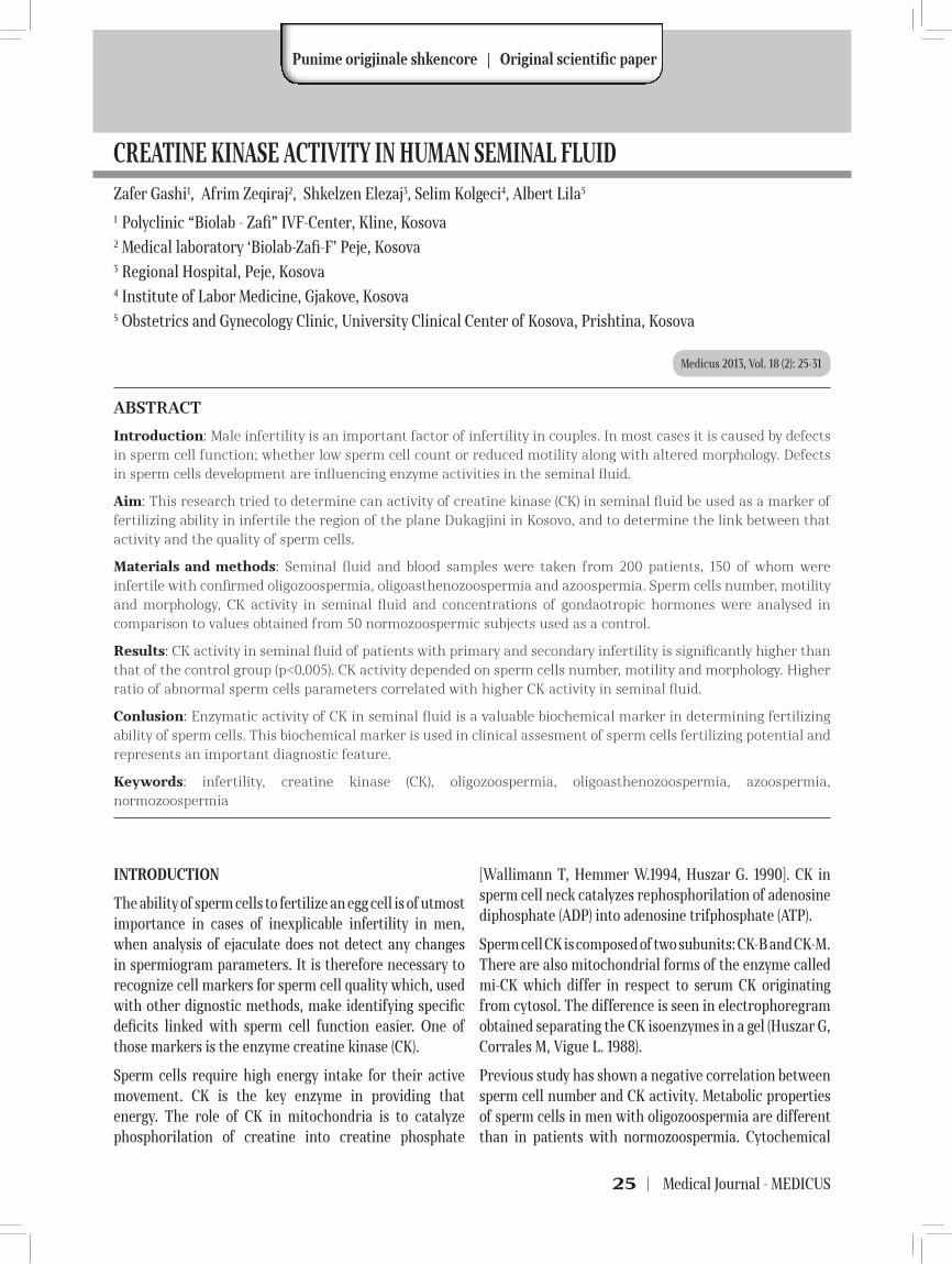
25 | Medical Journal - MEDICUS
CREATINE KINASE ACTIVITY IN HUMAN SEMINAL FLUIDZafer Gashi1, Afrim Zeqiraj2, Shkelzen Elezaj3, Selim Kolgeci4, Albert Lila5
1 Polyclinic “Biolab - Zafi” IVF-Center, Kline, Kosova2 Medical laboratory ‘Biolab-Zafi-F’ Peje, Kosova3 Regional Hospital, Peje, Kosova4 Institute of Labor Medicine, Gjakove, Kosova5 Obstetrics and Gynecology Clinic, University Clinical Center of Kosova, Prishtina, Kosova
ABSTRACT
Introduction: Male infertility is an important factor of infertility in couples. In most cases it is caused by defects in sperm cell function; whether low sperm cell count or reduced motility along with altered morphology. Defects in sperm cells development are influencing enzyme activities in the seminal fluid.
Aim: This research tried to determine can activity of creatine kinase (CK) in seminal fluid be used as a marker of fertilizing ability in infertile the region of the plane Dukagjini in Kosovo, and to determine the link between that activity and the quality of sperm cells.
Materials and methods: Seminal fluid and blood samples were taken from 200 patients, 150 of whom were infertile with confirmed oligozoospermia, oligoasthenozoospermia and azoospermia. Sperm cells number, motility and morphology, CK activity in seminal fluid and concentrations of gondaotropic hormones were analysed in comparison to values obtained from 50 normozoospermic subjects used as a control.
Results: CK activity in seminal fluid of patients with primary and secondary infertility is significantly higher than that of the control group (p<0,005). CK activity depended on sperm cells number, motility and morphology. Higher ratio of abnormal sperm cells parameters correlated with higher CK activity in seminal fluid.
Conlusion: Enzymatic activity of CK in seminal fluid is a valuable biochemical marker in determining fertilizing ability of sperm cells. This biochemical marker is used in clinical assesment of sperm cells fertilizing potential and represents an important diagnostic feature.
Keywords: infertility, creatine kinase (CK), oligozoospermia, oligoasthenozoospermia, azoospermia, normozoospermia
INTRODUCTION
The ability of sperm cells to fertilize an egg cell is of utmost importance in cases of inexplicable infertility in men, when analysis of ejaculate does not detect any changes in spermiogram parameters. It is therefore necessary to recognize cell markers for sperm cell quality which, used with other dignostic methods, make identifying specific deficits linked with sperm cell function easier. One of those markers is the enzyme creatine kinase (CK).
Sperm cells require high energy intake for their active movement. CK is the key enzyme in providing that energy. The role of CK in mitochondria is to catalyze phosphorilation of creatine into creatine phosphate
[Wallimann T, Hemmer W.1994, Huszar G. 1990]. CK in sperm cell neck catalyzes rephosphorilation of adenosine diphosphate (ADP) into adenosine trifphosphate (ATP).
Sperm cell CK is composed of two subunits: CK-B and CK-M. There are also mitochondrial forms of the enzyme called mi-CK which differ in respect to serum CK originating from cytosol. The difference is seen in electrophoregram obtained separating the CK isoenzymes in a gel (Huszar G, Corrales M, Vigue L. 1988).
Previous study has shown a negative correlation between sperm cell number and CK activity. Metabolic properties of sperm cells in men with oligozoospermia are different than in patients with normozoospermia. Cytochemical
Medicus 2013, Vol. 18 (2): 25-31
4Punime origjinale shkencore | Original scientific paper
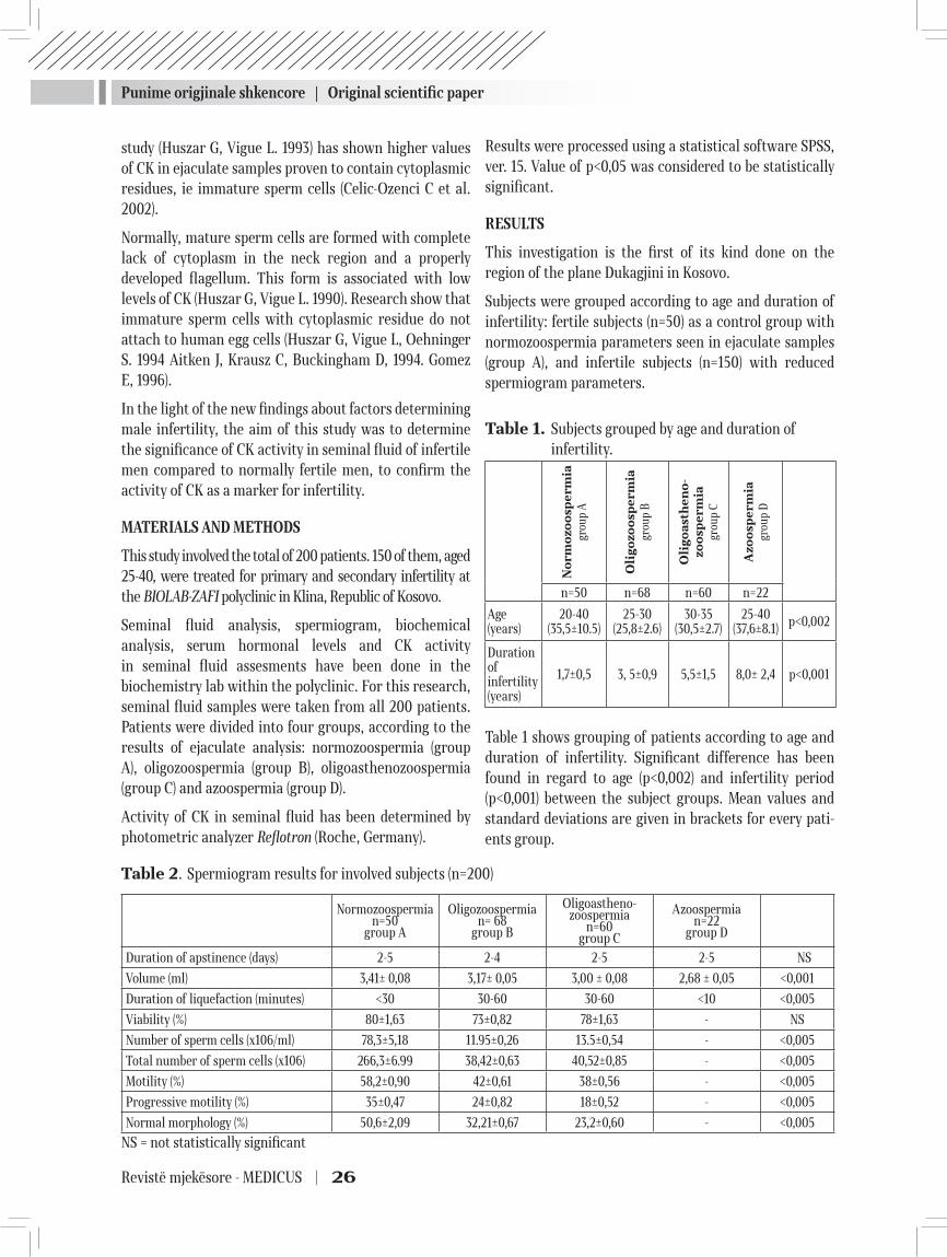
Punime origjinale shkencore | Original scientific paper
Revistë mjekësore - MEDICUS | 26
study (Huszar G, Vigue L. 1993) has shown higher values of CK in ejaculate samples proven to contain cytoplasmic residues, ie immature sperm cells (Celic-Ozenci C et al. 2002).
Normally, mature sperm cells are formed with complete lack of cytoplasm in the neck region and a properly developed flagellum. This form is associated with low levels of CK (Huszar G, Vigue L. 1990). Research show that immature sperm cells with cytoplasmic residue do not attach to human egg cells (Huszar G, Vigue L, Oehninger S. 1994 Aitken J, Krausz C, Buckingham D, 1994. Gomez E, 1996).
In the light of the new findings about factors determining male infertility, the aim of this study was to determine the significance of CK activity in seminal fluid of infertile men compared to normally fertile men, to confirm the activity of CK as a marker for infertility.
MATERIALS AND METHODS
This study involved the total of 200 patients. 150 of them, aged 25-40, were treated for primary and secondary infertility at the BIOLAB-ZAFI polyclinic in Klina, Republic of Kosovo.
Seminal fluid analysis, spermiogram, biochemical analysis, serum hormonal levels and CK activity in seminal fluid assesments have been done in the biochemistry lab within the polyclinic. For this research, seminal fluid samples were taken from all 200 patients. Patients were divided into four groups, according to the results of ejaculate analysis: normozoospermia (group A), oligozoospermia (group B), oligoasthenozoospermia (group C) and azoospermia (group D).
Activity of CK in seminal fluid has been determined by photometric analyzer Reflotron (Roche, Germany).
Results were processed using a statistical software SPSS, ver. 15. Value of p<0,05 was considered to be statistically significant.
RESULTS
This investigation is the first of its kind done on the region of the plane Dukagjini in Kosovo.
Subjects were grouped according to age and duration of infertility: fertile subjects (n=50) as a control group with normozoospermia parameters seen in ejaculate samples (group A), and infertile subjects (n=150) with reduced spermiogram parameters.
Table 1. Subjects grouped by age and duration of infertility.
No
rmo
zoo
sper
mia
grou
p A
Oli
go
zoo
sper
mia
grou
p B
Oli
go
ast
he
no
-zo
osp
erm
iagr
oup
C
Azo
osp
erm
iagr
oup
D
n=50 n=68 n=60 n=22
Age (years)
20-40(35,5±10.5)
25-30(25,8±2.6)
30-35(30,5±2.7)
25-40(37,6±8.1) p<0,002
Duration of infertility(years)
1,7±0,5 3, 5±0,9 5,5±1,5 8,0± 2,4 p<0,001
Table 1 shows grouping of patients according to age and duration of infertility. Significant difference has been found in regard to age (p<0,002) and infertility period (p<0,001) between the subject groups. Mean values and standard deviations are given in brackets for every pati-ents group.
Table 2. Spermiogram results for involved subjects (n=200)
Normozoospermian=50
group A
Oligozoospermian= 68
group B
Oligoastheno-zoospermia
n=60group C
Azoospermian=22
group D
Duration of apstinence (days) 2-5 2-4 2-5 2-5 NS
Volume (ml) 3,41± 0,08 3,17± 0,05 3,00 ± 0,08 2,68 ± 0,05 <0,001
Duration of liquefaction (minutes) <30 30-60 30-60 <10 <0,005
Viability (%) 80±1,63 73±0,82 78±1,63 - NS
Number of sperm cells (x106/ml) 78,3±5,18 11.95±0,26 13.5±0,54 - <0,005
Total number of sperm cells (x106) 266,3±6.99 38,42±0,63 40,52±0,85 - <0,005
Motility (%) 58,2±0,90 42±0,61 38±0,56 - <0,005
Progressive motility (%) 35±0,47 24±0,82 18±0,52 - <0,005
Normal morphology (%) 50,6±2,09 32,21±0,67 23,2±0,60 - <0,005
NS = not statistically significant
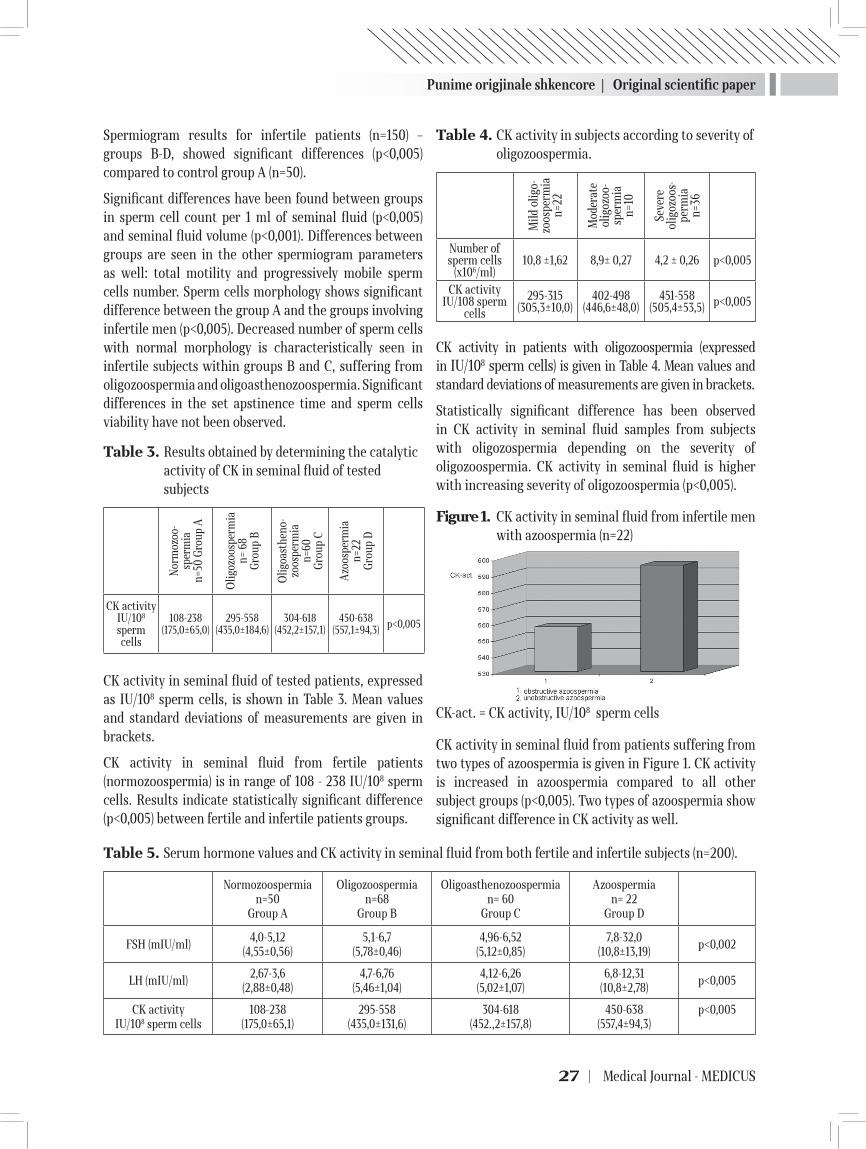
Punime origjinale shkencore | Original scientific paper
27 | Medical Journal - MEDICUS
Spermiogram results for infertile patients (n=150) – groups B-D, showed significant differences (p<0,005) compared to control group A (n=50).
Significant differences have been found between groups in sperm cell count per 1 ml of seminal fluid (p<0,005) and seminal fluid volume (p<0,001). Differences between groups are seen in the other spermiogram parameters as well: total motility and progressively mobile sperm cells number. Sperm cells morphology shows significant difference between the group A and the groups involving infertile men (p<0,005). Decreased number of sperm cells with normal morphology is characteristically seen in infertile subjects within groups B and C, suffering from oligozoospermia and oligoasthenozoospermia. Significant differences in the set apstinence time and sperm cells viability have not been observed.
Table 3. Results obtained by determining the catalytic activity of CK in seminal fluid of tested subjects
Nor
moz
oo-
sper
mia
n=50
Gro
up A
Olig
ozoo
sper
mia
n= 6
8G
roup
B
Olig
oast
heno
-zo
ospe
rmia
n=
60
Gro
up C
Azo
ospe
rmia
n=22
Gro
up D
CK activityIU/108 sperm cells
108-238(175,0±65,0)
295-558(435,0±184,6)
304-618(452,2±157,1)
450-638(557,1±94,3) p<0,005
CK activity in seminal fluid of tested patients, expressed as IU/108 sperm cells, is shown in Table 3. Mean values and standard deviations of measurements are given in brackets.
CK activity in seminal fluid from fertile patients (normozoospermia) is in range of 108 - 238 IU/108 sperm cells. Results indicate statistically significant difference (p<0,005) between fertile and infertile patients groups.
Table 4. CK activity in subjects according to severity of oligozoospermia.
Mild
olig
o-zo
ospe
rmia
n=22
Mod
erat
e ol
igoz
oo-
sper
mia
n=10
Seve
reol
igoz
oos-
perm
ian=
36
Number of sperm cells
(x106/ml)10,8 ±1,62 8,9± 0,27 4,2 ± 0,26 p<0,005
CK activityIU/108 sperm
cells
295-315(305,3±10,0)
402-498(446,6±48,0)
451-558(505,4±53,5) p<0,005
CK activity in patients with oligozoospermia (expressed in IU/108 sperm cells) is given in Table 4. Mean values and standard deviations of measurements are given in brackets.
Statistically significant difference has been observed in CK activity in seminal fluid samples from subjects with oligozospermia depending on the severity of oligozoospermia. CK activity in seminal fluid is higher with increasing severity of oligozoospermia (p<0,005).
Figure 1. CK activity in seminal fluid from infertile men with azoospermia (n=22)
CK-act. = CK activity, IU/108 sperm cells
CK activity in seminal fluid from patients suffering from two types of azoospermia is given in Figure 1. CK activity is increased in azoospermia compared to all other subject groups (p<0,005). Two types of azoospermia show significant difference in CK activity as well.
Table 5. Serum hormone values and CK activity in seminal fluid from both fertile and infertile subjects (n=200).
Normozoospermian=50
Group A
Oligozoospermian=68
Group B
Oligoasthenozoospermian= 60
Group C
Azoospermian= 22
Group D
FSH (mIU/ml)4,0-5,12
(4,55±0,56)5,1-6,7
(5,78±0,46)4,96-6,52
(5,12±0,85)7,8-32,0
(10,8±13,19)p<0,002
LH (mIU/ml)2,67-3,6
(2,88±0,48)4,7-6,76
(5,46±1,04)4,12-6,26
(5,02±1,07)6,8-12,31
(10,8±2,78)p<0,005
CK activityIU/108 sperm cells
108-238(175,0±65,1)
295-558(435,0±131,6)
304-618(452.,2±157,8)
450-638(557,4±94,3)
p<0,005
Punime origjinale shkencore | Original scientific paper
Revistë mjekësore - MEDICUS | 28
Table 5 shows the correlation between CK activity and hormone concentrations in the patients blood. CK activity is expressed in IU/108 sperm cells. Mean values and standard deviations of measurements are given in brackets.
Results indicate that serum concentration of gonadotropins (FSH, LH) are significantly higher (p<0,005) in infertile men with azoospermia. CK activity is in range of 295-638 IU/108 sperm cells for groups B, C and D, in contrast to gonadotropin values elevated only in group D, azoospermic men.
Table 6. CK activity in subjects with primary and secondary infertility
Primary infertility
Secondary infertility
Number of subjects
125 25 p<0,005
CK activityIU/108 sperm
cells
304-638(625,8±189,4)
295-618(445,2±161,3)
p<0,005
Table 6 shows CK activity depending on the type of infertility. CK activity is expressed in IU/108 sperm cells. Mean values and standard deviations of measurements are given in brackets.
Significant differences are observed in CK enzimatic activity in seminal fluid samples from patients with primary and secondary infertility (p<0,005).
DISCUSSION
Sperm cells are male reproductive cells. The ultimate test for fertilizing ability of sperm cells is by all means pregnancy (WHO 2010). It is therefore important to find quality markers for sperm cells which, taken together with other diagnostic tools, can help discover specific disorders linked to sperm cells function (Huszar et al. 2006).
According to WHO data from 2010, a male with less than 15 million sperm cells per 1 ml of ejaculate is considered to be potentially infertile. Various factors affect the fertilizing ability of the sperm cells; morphology, motility and genetic defects of the sperm cells, mechanical trauma, various infections and other testicular factors (Cooper et al. 2010). Although rapid and easily done, seminal fluid analysis is not the only test in determining the fertilizing ability of a male patient.
Besides the routine seminal fluid analysis, further testing is required in assessing sperm cells functional status (Huszar & Vigue 1994). Various studies indicate how sperm cells function is often linked to increased activity of key enzymes, one of those enzymes being CK (Huszar, Vigue, Morshedi 1992, Geraci & Giudice 2005, Menkveld 2007).
Enzymes in seminal fluid are not directly responsible for loss of sperm cells function, but act as biochemical markers for normal differentiation of sperm cells. Ezymes present in the seminal fluid are therefore good indicator of functional metabolic activity of the sperm cells (Guerin et al. 1979).
From our study it is clear how CK activity in seminal fluid does represent a valid biochemical marker for decreased sperm cell count, decreased sperm cell motility and decreased fertilizing potential in men. Higher CK activity in human seminal fluid is a marker for cytoplasmic residues in immature sperm cells. Higher CK activity in seminal fluid has been previously found in sperm cells with cytoplasmic residues (Huszar & Vigue 1993, Dokras et al. 1999). CK activity in seminal fluid is several times higher than that of serum, which indicates CK is locally created by gonads also (Huszar & Vigue 1993).
Our results obtained with 200 subjects divided by age show how patients aged 25-30 years are mostly oligozoospermic, representing the largest group of infertile subjects. As the results of this study relate only to patients of Albanian ethnicity, results differ from previously published (Jale 2008). Previously published results showed infertility in oligozoospermic men is highest at 20 to 29 years of age.
There is a prolonged period of infertility in men with lower spermiogram parameters. The lowest those parameters are, infertility period is longer and the condition is harder to treat. Subjects age and duration of infertility are positively correlated with changes in spermiogram parameters. Results linked to infertiliy period are given in Table 1, and are in accordance with the results of other authors (Geraci i Giudice, 2005).
Results from ejaculate analysis are in full accordance with 2010 WHO criteria describing normal parameters for spermiogram analysis of normospermic ejaculate samples. Other subject groups; B, C, and D; are categorized as infertile based on the same criteria, with reduced sperm cell count (oligozoospermia), reduced sperm cells number and motility (oligoasthenozoospermia) and absence of sperm cells in seminal fluid (azoospermia).
Punime origjinale shkencore | Original scientific paper
29 | Medical Journal - MEDICUS
Statistically significant difference is observed between seminal fluid samples from above mentioned subject groups of infertile patients based on sperm cells number, seminal fluid volume and sperm cells motility and morphology.
Results of this study regarding the CK activity in seminal fluid indicate there is a correlation between reduced spermiogram parameters and the measured CK activity in seminal fluid of fertile and infertile subjects. As the normal values of CK activity in seminal fluid of fertile subjects, values of 108-238 IU/108 sperm cells are taken. Results show statistically significant differences (p<0,005) between groups of fertile and infertile subjects. CK activity in samples of azoospermic patients is highest compared to the other infertile subject groups, which may be caused by CK locally synthesized by gonads, mostly prostate, so the CK activity in seminal fluid could be used as a prostate fuction marker. These results are similar to those published by Hallak et al.
With lower sperm cells number, CK activity in seminal fluid is higher (p<0,005). In our study a difference in CK activity is observed between patients with mild, moderate and severe oligozoospermia. Results confirming high CK activity in different stages of oligozoospermia are also published by Huszar G, Corrales M. and Vigue L. (1988).
Azoospermia represents an absence of sperm cells in ejaculate. Based on etiology, it can be obstructive and unobstructive (WHO, 2010). We have analyzed the CK activity in seminal fluid of patients with azoospermia in regards to the type, and we found both the higher CK activity in azoospermia compared to other groups of infertile patients and the statistically significant difference in CK activity between different types of azoospermia (p<0,005). The measured CK activity is significantly higher in cases of unobstructivne azoospermia (582,0±29,5 IU/108 sperm cells), compared to cases with obstructive azoospermia (557,4±18,8 IU/108 sperm cells). Similar results are published by Hallak et al. (2001).
Normal values for gonadotropic hormone concentrations have been observed in fertile subjects with normozoospermic seminal fluid samples, and with oligozoospermia and asthenozoospermia those values are higher compared to fertile subjects group. The highest FSH and LH concentrations were found in patients with azoospermia. Contrary to hormone concentrations, CK activity was found to be increased in all three groups of
infertile subjects, in accordance to previously published data by Jalala (2008).
Infertility as defined by WHO is classified as primary or secondary infertility. Most infertile subjects involved in this research suffered from the primary infertility. They have been unable to conceive a pregnancy after two years of regular intercourses (Sigman et al. 1997, WHO 1999, WHO 2010). Significant differences have been observed in CK activity in seminal fluid samples between patients with primary and secondary infertility. Subjects with primary infertility had increased CK activity in the seminal fluid, compared to those with secondary infertility. Primary infertility patients included patients with azoospermia, oligozoospermia and oligoasthenozoospermia. Patients with secondary infertility included patients with oligozoospermia and oligoasthenozoospermia. This is in accordance with the results published previously by Hallak et al.
Biochemical method used in this research measures CK enzymatic activity in human seminal fluid and is done using dry chemistry principles. The method is simple, gives results in short period of time, is cost effective and reliable. Therefore, assessing the CK activity in seminal fluid constitutes a useful additional biochemical marker in determining the patient’s fertility potential.
REFERENCES
1. Aitken J, Krausz C, Buckingham D. Relationships be-tween biochemical markers for residual sperm cyto-plasm, reactive oxygen species generation, and the pres-ence of leukocytes and precursor germ cells in human sperm suspensions. Mol Reprod Dev. 1994;39(3):268-279.
2. Celic-Ozenci C, Jakab A, Vigue L, Demir R, Huszar G. Mature and fertile sperm selectivity bind to hyakuronic acid : cytoplasmic content, HspA2 levels, chromatin maturity, shape and ICSI sperm selection. J Soc Gynecol Invest. 2002;9(Suppl 1):849.
3. Cooper TG et al. World Health Organization reference values for human semen characteristics. Hum. Reprod. Update 2010; 16: 231–2453.
4. Geraci F, Giudice G. Factors which influence sperm abil-ity to fertilize. J. Submicrosc. Cytol. Pathol. 2005; 37: 215–222.
5. Guerin JF, Menezo Y, Czyba JC. Enzyme comparative study of spermatozoa and seminal plasma in normal and subfertile men. Arch. of Androl. 1979; 3: 251-257.
Punime origjinale shkencore | Original scientific paper
Revistë mjekësore - MEDICUS | 30
6. Dokras A, Giraldo JL, Habana A, Erel T, Kovanci E, Huszar G. Sperm cellular maturity and the treat-ment choice of IVF or intracytoplasmic sperm injec-tion: the contributions of sperm creatine kinase M-isoform ratio measurements. u: Programme of ASRM Annual Meeting; 1999 Toronto, Ontario, Canada.
7. Gomez E, Buckingham D, Brindle J, Lanzafame F, Ir-vine DS, Aitken RJ. Development of an image analy-sis system to monitor the retention of residual cy-toplasm by human spermatozoa: correlation with biochemical markers of the cytoplasmic space, and sperm function. J Androl. 1996;17(3):276-287.
8. Jalal AN. Assessment of sera prolactin hormone in infertile males using enzyme-linked immunosorb-ent assay. 2008. University of Sulaimania College of Science Menkveld R. The basic semen analysis. In: Oehninger S, Kruger TF, eds. Male Infertility. Oxon: Informa Healthcare 2007: 141-170.
9. Hallak J, Sharma RK, Pasqualotto FF, Ranganathan P, Thomas AJ Jr, Agarwal A. Creatine kinaze as an indi-Creatine kinaze as an indi-cator of sperm quality and maturity in men with oli-gospermia. Urology 2001; 58: 446-51.
10. 10.Huszar G, Ozkavukcu S, Jakab A, Celik-Ozenci C, Sati GL, Cayli S. Hyaluronic acid binding ability of human sperm reflects cellular maturity and fertilizing potential: se-lection of sperm for intracytoplasmic sperm injection. Curr. Opin. Obstet. Gynecol. 2006; 18: 260-267.
11. Huszar G., Vigue L, Morshedi M. Sperm creatine phos-phokinase M-isoform ratios and fertilizing potential of
men: a blinded study of 84 couples treated with in vitro fertilization. Fertil. Steril. 1992; 57: 882–888.
12. Huszar G. The role of sperm creatine kinase in the asesse-ment of male fertility. Reprod Med Rev. 1994;3:179-197.
13. Huszar G, Vigue L, Coralles M. Sperm creatine kinase ac-tivity in fertile and infertile oligospermic men. J Androl. 1990;11(1):40-46.
14. Huszar G, Corrales M, Vigue L. Correlation between sperm creatine phosphokinase activity and sperm con-centrations in normospermic and oligospermic men. Gamete Res. 1988;19(1):67-7 .
15. Huszar G, Vigue L. Incomplete development of human spermatozoa is associated with increased creatine phosphokinase concentrations and abnormal head mor-phology. Mol Reprod Dev. 1993;34(3):292-298.
16. Huszar G, Vigue L. Spermatogenesis-related change in the synthesis of the creatine kinase B-type and M-type isoforms in human spermatozao. Mol Reprod Dev. 1990;25(3):258-262.
17. Wallimann T, Hemmer W. Creatine kinase in non-muscule tissues and cells. Mol Cell Biochem. 1994;133-134:193-220.
18. World Health Organization. 5th ed. Geneva: World Health Organization; 2010. WHO Laboratory Manual for the Examination and Processing of Human Semen.
19. World Health Organization. 4th ed. Cambridge: Cam-bridge University Press; 1999. WHO Laboratory Manual for the Examination of Human Semen and Sperm-cervi-cal Mucus Interaction.
AKTIVITETI I ENZIMIT KREATIN KINAZË NË PLAZMËN SEMINALE
Zafer Gashi1, Afrim Zeqiraj2, Shkelzen Elezaj3, Selim Kolgeci4, Abert Lila5
1 Poliklinika ‘’Biolab – Zafi’’ IVF-Center„ Klinë, Kosovë2 Laboratori mjekesor ‘Biolab-Zafi-F’ Pejë, Kosovë3 Spitali Rajonal, Pejë, Kosovë4 Klinika e Obstetikës dhe Gjinekologjisë, Qendra Klinike Universitare e Kosoves, Prishtinë5 Instituti i Mjekësisë së Punës, Gjakovë
ABSTRAKTI
Hyrje: Infertiliteti mashkullor në shumicën e rasteve është shkaktuar nga defektet në funksionin e qelizës spermatozoide. Defektet në qelizat e spermës janë të influencuar me rritjen e aktivitetit të enzimit në lëngun seminal.
Qëllimi: Të përcaktoj aktivitetin e enzimit kreatin kinazë në lëngun seminal i cili përdoret si një tregues i aftësisë fertilizue tek meshkujt infertil të regjonit të rrafshit të Dukagjinit në Kosovë , dhe për të përcaktuar lidhjen mes
Punime origjinale shkencore | Original scientific paper
31 | Medical Journal - MEDICUS
aktivitetit të enzimit kreatin kinazë dhe cilësise së qelizës spermatezoide.
Materiali dhe Metodat : Analiza e lëngut seminal dhe mostrat e gjakut janë marrë nga 200 pacient, 150 prej të cilëve janë përcaktuar infertil me oligoasthenozoospermi oligozoospermi dhe azoospermi, janë analizuar numri i qelizave spermatezoide, levizëshmeria, morfologjia, aktiviteti i enzimit kreatin kinazë në lëngun seminal dhe përqendrimi i hormoneve gonadotropike, krahasuar me vlerat e fituara nga 50 pacientë kontrollë të përcaktuar me normozoospermi.
Rezultatet: Aktiviteti i CK në lëngun seminal të pacientëve me infertilitet primar dhe sekondar është dukshëm më i lartë në shkallë signifikante krahasuar me grupin kontrollë (p <0.005). Aktiviteti i CK varet nga numri i qelizave spermatezoide, levizshmerisë dhe morfologjisë. Parametrat e qelizave spermatezoide abnormale në shkallë më të lartë lidhen me aktivitet të lartë te CK në lëngun seminal.
Konkluzioni: Aktiviteti i enzimit CK në lëngun seminal është një marker biokimik i vlefshëm në përcaktimin e aftësisë fertilizuese të qelizave spermatezoide. Ky marker biokimik është përdorur për vlersimin e potencilait fertilizues të qelizave spermatezoide, dhe përfaqëson një tipar të rëndësishëm diagnostik.
Fjalët kyçe: Infertiliteti, Kreatin kinaza (CK), Oligozoospermia,Oligoasthenozoospermia, Azoospermia, Normo-zoospermia.
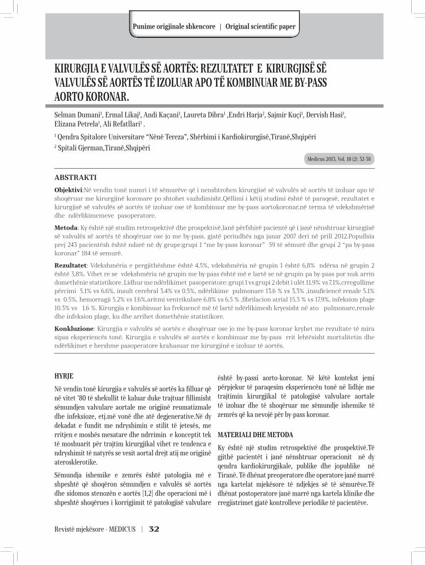
Revistë mjekësore - MEDICUS | 32
KIRURGJIA E VALVULËS SË AORTËS: REZULTATET E KIRURGJISË SË VALVULËS SË AORTËS TË IZOLUAR APO TË KOMBINUAR ME BY-PASS AORTO KORONAR.Selman Dumani1, Ermal Likaj1, Andi Kaçani1, Laureta Dibra1 ,Endri Harja2, Sajmir Kuçi1, Dervish Hasi1, Elizana Petrela1, Ali Refatllari1 .1 Qendra Spitalore Universitare “Nënë Tereza”, Shërbimi i Kardiokirurgjisë,Tiranë,Shqipëri 2 Spitali Gjerman,Tiranë,Shqipëri
ABSTRAKTI
Objektivi:Në vendin tonë numri i të sëmurëve që i nenshtrohen kirurgjisë së valvulës së aortës të izoluar apo të shoqëruar me kirurgjinë koronare po shtohet vazhdimisht.Qëllimi i këtij studimi është të paraqesë, rezultatet e kirurgjisë së valvulës së aortës të izoluar ose të kombinuar me by-pass aortokoronar,në terma të vdekshmërisë dhe ndërlikimemeve pasoperatore.
Metoda: Ky është një studim retrospektivë dhe prospektivë.Janë përfshirë pacientë që i janë nënshtruar kirurgjisë së valvulës së aortës të shoqëruar ose jo me by-pass, gjatë periudhës nga janar 2007 deri në prill 2012.Popullsia prej 243 pacientësh është ndarë në dy grupe:grupi 1 “me by-pass koronar” 59 të sëmurë dhe grupi 2 “pa by-pass koronar” 184 të semurë.
Rezultatet: Vdekshmëria e pergjithëshme është 4,5%, vdekshmëria në grupin 1 është 6,8% ndërsa në grupin 2 është 3,8%. Vihet re se vdekshmëria në grupin me by-pass është më e lartë se në grupin pa by-pass por nuk arrin domethënie statistikore. Lidhur me ndërlikimet pasoperatore: grupi 1 vs grupi 2 debit i ulët 11.9% vs 7.1%,crregullime përcimi 5.1% vs 6.6%, insult cerebral 3.4% vs 0.5%, ndërlikime pulmonare 13.6 % vs 3.3% ,insuficiencë renale 5.1% vs 0.5%, hemorragji 5.2% vs 1.6%,aritmi ventrikulare 6.8% vs 6.5 % ,fibrilacion atrial 15.3 % vs 17.9%, infeksion plage 10.3% vs 1.6 %. Kirurgjia e kombinuar ka frekuencë më të lartë ndërlikimesh kryesisht në ato pulmonare,renale dhe infeksion plage, ku dhe arrihet domethënie statistikore.
Konkluzione: Kirurgjia e valvulës së aortës e shoqëruar ose jo me by-pass koronar kryhet me rezultate të mira sipas eksperiencës tonë. Kirurgjia e valvulës së aortës e kombinuar me by-pass rrit lehtësisht mortalitetin dhe ndërlikimet e hershme pasoperatore krahasuar me kirurgjinë e izoluar të aortës.
HYRJE
Në vendin tonë kirurgjia e valvulës së aortës ka filluar që në vitet ’80 të shekullit të kaluar duke trajtuar fillimisht sëmundjen valvulare aortale me origjinë reumatizmale dhe infeksioze, etj.më vonë dhe atë degjenerative.Në dy dekadat e fundit me ndryshimin e stilit të jetesës, me rritjen e moshës mesatare dhe ndrrimin e konceptit tek të moshuarit për trajtim kirurgjikal vihet re tendenca e ndryshimit të natyrës se vesit aortal drejt atij me origjinë aterosklerotike.
Sëmundja ishemike e zemrës është patologjia më e shpeshtë që shoqëron sëmundjen e valvulës së aortës dhe sidomos stenozën e aortës [1,2] dhe operacioni më i shpeshtë shoqërues i korrigjimit të patologjisë valvulare
është by-passi aorto-koronar. Në këtë kontekst jemi përpjekur të paraqesim eksperiencën tonë në lidhje me trajtimin kirurgjikal të patologjisë valvulare aortale të izoluar dhe të shoqëruar me sëmundje ishemike të zemrës që ka nevojë për by-pass koronar.
MATERIALI DHE METODA
Ky është një studim retrospektivë dhe prospektivë.Të gjithë pacientët i janë nënshtruar operacionit në dy qendra kardiokirurgjikale, publike dhe jopublike në Tiranë. Të dhënat preoperatore dhe operatore janë marrë nga kartelat mjekësore të ndjekjes së të sëmurëve.Të dhënat postoperatore janë marrë nga kartela klinike dhe rregjistrimet gjatë kontrolleve periodike të pacientëve.
Medicus 2013, Vol. 18 (2): 32-38
5 Punime origjinale shkencore | Original scientific paper
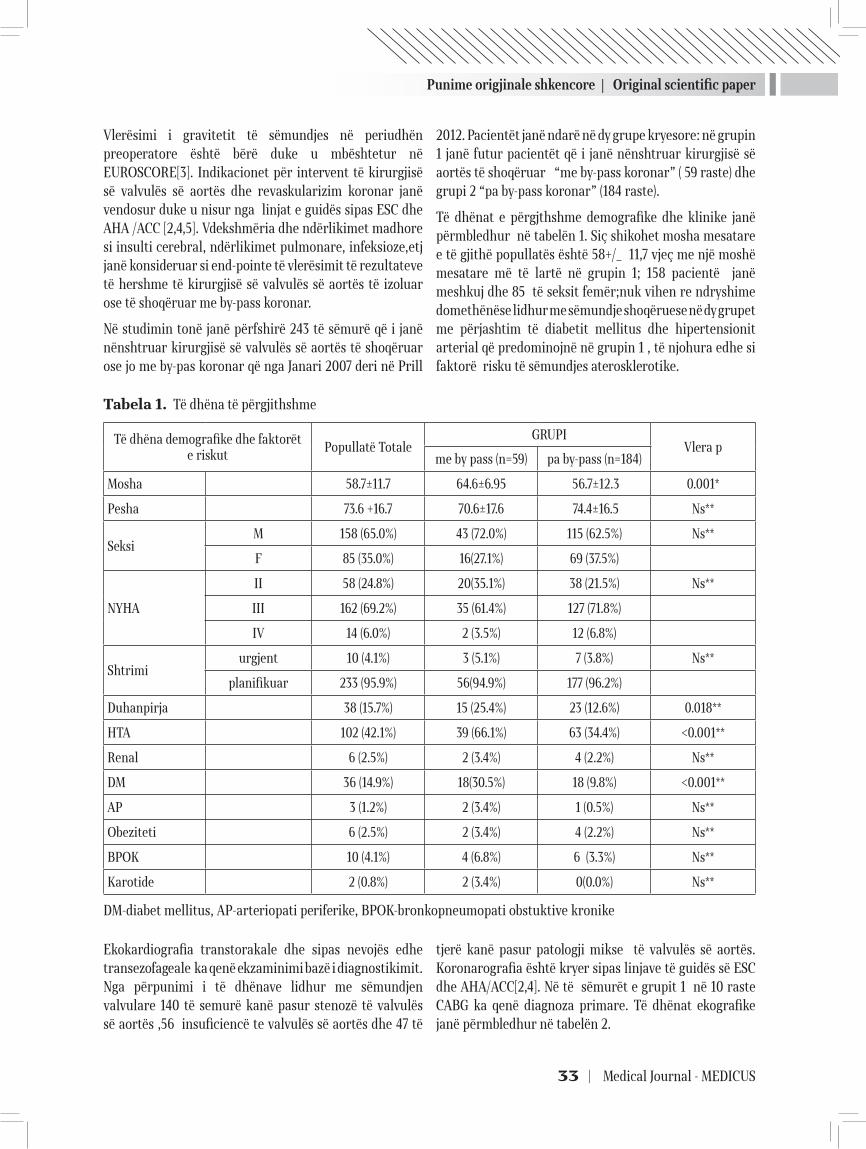
Punime origjinale shkencore | Original scientific paper
33 | Medical Journal - MEDICUS
Vlerësimi i gravitetit të sëmundjes në periudhën preoperatore është bërë duke u mbështetur në EUROSCORE[3]. Indikacionet për intervent të kirurgjisë së valvulës së aortës dhe revaskularizim koronar janë vendosur duke u nisur nga linjat e guidës sipas ESC dhe AHA /ACC [2,4,5]. Vdekshmëria dhe ndërlikimet madhore si insulti cerebral, ndërlikimet pulmonare, infeksioze,etj janë konsideruar si end-pointe të vlerësimit të rezultateve të hershme të kirurgjisë së valvulës së aortës të izoluar ose të shoqëruar me by-pass koronar.
Në studimin tonë janë përfshirë 243 të sëmurë që i janë nënshtruar kirurgjisë së valvulës së aortës të shoqëruar ose jo me by-pas koronar që nga Janari 2007 deri në Prill
2012. Pacientët janë ndarë në dy grupe kryesore: në grupin 1 janë futur pacientët që i janë nënshtruar kirurgjisë së aortës të shoqëruar “me by-pass koronar” ( 59 raste) dhe grupi 2 “pa by-pass koronar” (184 raste).
Të dhënat e përgjthshme demografike dhe klinike janë përmbledhur në tabelën 1. Siç shikohet mosha mesatare e të gjithë popullatës është 58+/_ 11,7 vjeç me një moshë mesatare më të lartë në grupin 1; 158 pacientë janë meshkuj dhe 85 të seksit femër;nuk vihen re ndryshime domethënëse lidhur me sëmundje shoqëruese në dy grupet me përjashtim të diabetit mellitus dhe hipertensionit arterial që predominojnë në grupin 1 , të njohura edhe si faktorë risku të sëmundjes aterosklerotike.
Tabela 1. Të dhëna të përgjithshme
Të dhëna demografike dhe faktorët e riskut
Popullatë Totale GRUPI
Vlera pme by pass (n=59) pa by-pass (n=184)
Mosha 58.7±11.7 64.6±6.95 56.7±12.3 0.001*
Pesha 73.6 +16.7 70.6±17.6 74.4±16.5 Ns**
SeksiM 158 (65.0%) 43 (72.0%) 115 (62.5%) Ns**
F 85 (35.0%) 16(27.1%) 69 (37.5%)
NYHA
II 58 (24.8%) 20(35.1%) 38 (21.5%) Ns**
III 162 (69.2%) 35 (61.4%) 127 (71.8%)
IV 14 (6.0%) 2 (3.5%) 12 (6.8%)
Shtrimiurgjent 10 (4.1%) 3 (5.1%) 7 (3.8%) Ns**
planifikuar 233 (95.9%) 56(94.9%) 177 (96.2%)
Duhanpirja 38 (15.7%) 15 (25.4%) 23 (12.6%) 0.018**
HTA 102 (42.1%) 39 (66.1%) 63 (34.4%) <0.001**
Renal 6 (2.5%) 2 (3.4%) 4 (2.2%) Ns**
DM 36 (14.9%) 18(30.5%) 18 (9.8%) <0.001**
AP 3 (1.2%) 2 (3.4%) 1 (0.5%) Ns**
Obeziteti 6 (2.5%) 2 (3.4%) 4 (2.2%) Ns**
BPOK 10 (4.1%) 4 (6.8%) 6 (3.3%) Ns**
Karotide 2 (0.8%) 2 (3.4%) 0(0.0%) Ns**
DM-diabet mellitus, AP-arteriopati periferike, BPOK-bronkopneumopati obstuktive kronike
Ekokardiografia transtorakale dhe sipas nevojës edhe transezofageale ka qenë ekzaminimi bazë i diagnostikimit.Nga përpunimi i të dhënave lidhur me sëmundjen valvulare 140 të semurë kanë pasur stenozë të valvulës së aortës ,56 insuficiencë te valvulës së aortës dhe 47 të
tjerë kanë pasur patologji mikse të valvulës së aortës.Koronarografia është kryer sipas linjave të guidës së ESC dhe AHA/ACC[2,4]. Në të sëmurët e grupit 1 në 10 raste CABG ka qenë diagnoza primare. Të dhënat ekografike janë përmbledhur në tabelën 2.
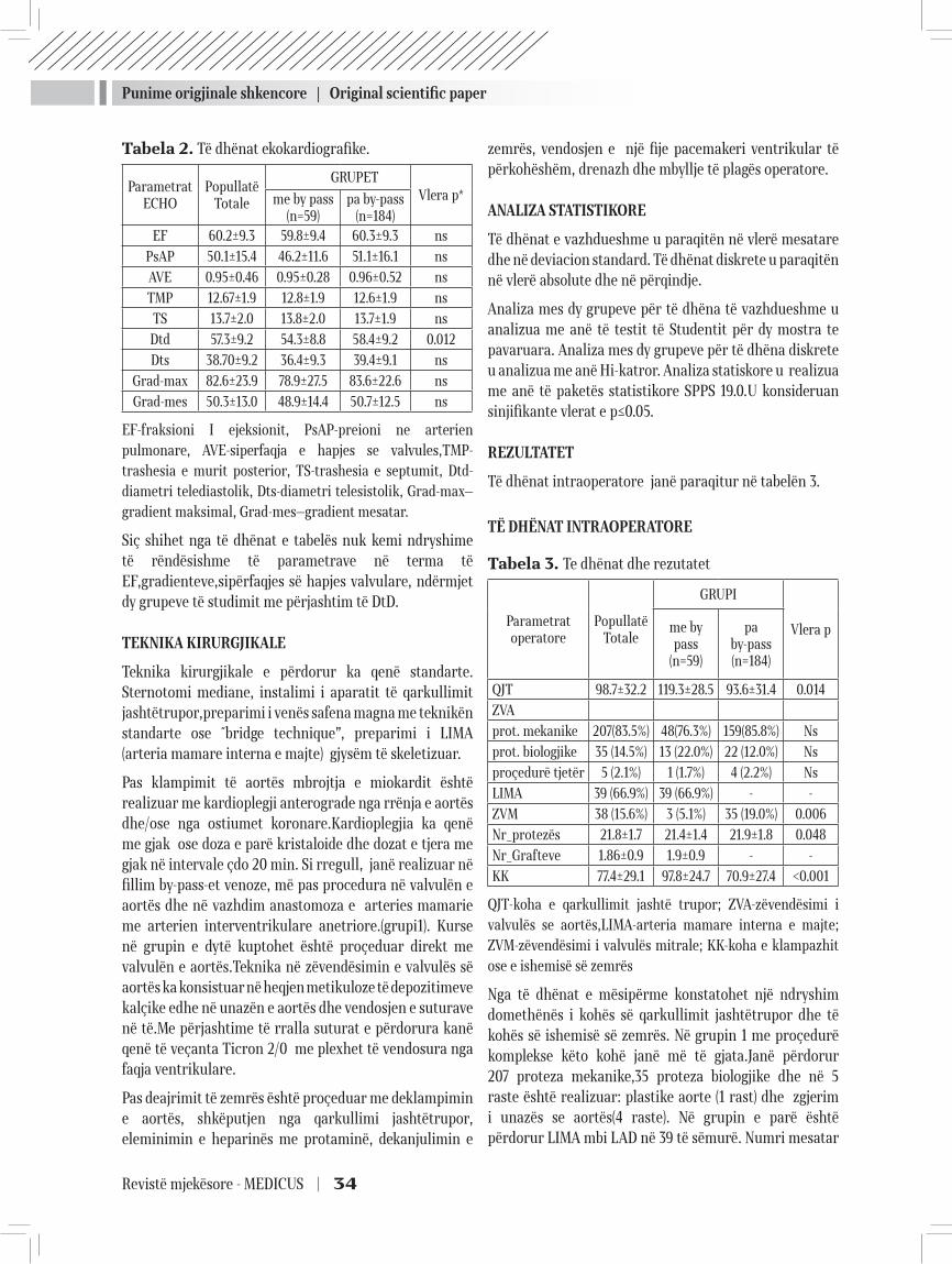
Punime origjinale shkencore | Original scientific paper
Revistë mjekësore - MEDICUS | 34
Tabela 2. Të dhënat ekokardiografike.
Parametrat ECHO
Popullatë Totale
GRUPETVlera p*me by pass
(n=59)pa by-pass
(n=184)EF 60.2±9.3 59.8±9.4 60.3±9.3 ns
PsAP 50.1±15.4 46.2±11.6 51.1±16.1 ns
AVE 0.95±0.46 0.95±0.28 0.96±0.52 ns
TMP 12.67±1.9 12.8±1.9 12.6±1.9 ns
TS 13.7±2.0 13.8±2.0 13.7±1.9 ns
Dtd 57.3±9.2 54.3±8.8 58.4±9.2 0.012
Dts 38.70±9.2 36.4±9.3 39.4±9.1 ns
Grad-max 82.6±23.9 78.9±27.5 83.6±22.6 ns
Grad-mes 50.3±13.0 48.9±14.4 50.7±12.5 ns
EF-fraksioni I ejeksionit, PsAP-preioni ne arterien pulmonare, AVE-siperfaqja e hapjes se valvules,TMP-trashesia e murit posterior, TS-trashesia e septumit, Dtd-diametri telediastolik, Dts-diametri telesistolik, Grad-max—gradient maksimal, Grad-mes—gradient mesatar.
Siç shihet nga të dhënat e tabelës nuk kemi ndryshime të rëndësishme të parametrave në terma të EF,gradienteve,sipërfaqjes së hapjes valvulare, ndërmjet dy grupeve të studimit me përjashtim të DtD.
TEKNIKA KIRURGJIKALE
Teknika kirurgjikale e përdorur ka qenë standarte. Sternotomi mediane, instalimi i aparatit të qarkullimit jashtëtrupor,preparimi i venës safena magna me teknikën standarte ose ˝bridge technique”, preparimi i LIMA (arteria mamare interna e majte) gjysëm të skeletizuar.
Pas klampimit të aortës mbrojtja e miokardit është realizuar me kardioplegji anterograde nga rrënja e aortës dhe/ose nga ostiumet koronare.Kardioplegjia ka qenë me gjak ose doza e parë kristaloide dhe dozat e tjera me gjak në intervale çdo 20 min. Si rregull, janë realizuar në fillim by-pass-et venoze, më pas procedura në valvulën e aortës dhe në vazhdim anastomoza e arteries mamarie me arterien interventrikulare anetriore.(grupi1). Kurse në grupin e dytë kuptohet është proçeduar direkt me valvulën e aortës.Teknika në zëvendësimin e valvulës së aortës ka konsistuar në heqjen metikuloze të depozitimeve kalçike edhe në unazën e aortës dhe vendosjen e suturave në të.Me përjashtime të rralla suturat e përdorura kanë qenë të veçanta Ticron 2/0 me plexhet të vendosura nga faqja ventrikulare.
Pas deajrimit të zemrës është proçeduar me deklampimin e aortës, shkëputjen nga qarkullimi jashtëtrupor, eleminimin e heparinës me protaminë, dekanjulimin e
zemrës, vendosjen e një fije pacemakeri ventrikular të përkohëshëm, drenazh dhe mbyllje të plagës operatore.
ANALIZA STATISTIKORE
Të dhënat e vazhdueshme u paraqitën në vlerë mesatare dhe në deviacion standard. Të dhënat diskrete u paraqitën në vlerë absolute dhe në përqindje.
Analiza mes dy grupeve për të dhëna të vazhdueshme u analizua me anë të testit të Studentit për dy mostra te pavaruara. Analiza mes dy grupeve për të dhëna diskrete u analizua me anë Hi-katror. Analiza statiskore u realizua me anë të paketës statistikore SPPS 19.0.U konsideruan sinjifikante vlerat e p≤0.05.
REZULTATET
Të dhënat intraoperatore janë paraqitur në tabelën 3.
TË DHËNAT INTRAOPERATORE
Tabela 3. Te dhënat dhe rezutatet
Parametrat operatore
Popullatë Totale
GRUPI
Vlera pme by pass
(n=59)
pa by-pass (n=184)
QJT 98.7±32.2 119.3±28.5 93.6±31.4 0.014
ZVA
prot. mekanike 207(83.5%) 48(76.3%) 159(85.8%) Ns
prot. biologjike 35 (14.5%) 13 (22.0%) 22 (12.0%) Ns
proçedurë tjetër 5 (2.1%) 1 (1.7%) 4 (2.2%) Ns
LIMA 39 (66.9%) 39 (66.9%) - -
ZVM 38 (15.6%) 3 (5.1%) 35 (19.0%) 0.006
Nr_protezës 21.8±1.7 21.4±1.4 21.9±1.8 0.048
Nr_Grafteve 1.86±0.9 1.9±0.9 - -
KK 77.4±29.1 97.8±24.7 70.9±27.4 <0.001
QJT-koha e qarkullimit jashtë trupor; ZVA-zëvendësimi i valvulës se aortës,LIMA-arteria mamare interna e majte; ZVM-zëvendësimi i valvulës mitrale; KK-koha e klampazhit ose e ishemisë së zemrës
Nga të dhënat e mësipërme konstatohet një ndryshim domethënës i kohës së qarkullimit jashtëtrupor dhe të kohës së ishemisë së zemrës. Në grupin 1 me proçedurë komplekse këto kohë janë më të gjata.Janë përdorur 207 proteza mekanike,35 proteza biologjike dhe në 5 raste është realizuar: plastike aorte (1 rast) dhe zgjerim i unazës se aortës(4 raste). Në grupin e parë është përdorur LIMA mbi LAD në 39 të sëmurë. Numri mesatar
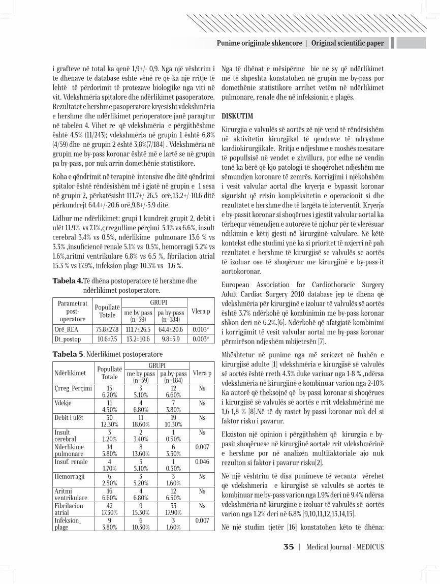
Punime origjinale shkencore | Original scientific paper
35 | Medical Journal - MEDICUS
i grafteve në total ka qenë 1,9+/- 0,9. Nga një vështrim i të dhënave të database është vënë re që ka një rritje të lehtë të përdorimit të protezave biologjike nga viti në vit. Vdekshmëria spitalore dhe ndërlikimet pasoperatore. Rezultatet e hershme pasoperatore kryesisht vdekshmëria e hershme dhe ndërlikimet perioperatore janë paraqitur në tabelën 4. Vihet re që vdekshmëria e përgjithëshme është 4,5% (11/243); vdekshmëria në grupin 1 është 6,8% (4/59) dhe në grupin 2 është 3,8%(7/184) . Vdekshmëria në grupin me by-pass koronar është më e lartë se në grupin pa by-pass, por nuk arrin domethënie statistikore.
Koha e qëndrimit në terapinë intensive dhe ditë qëndrimi spitalor është rëndësishëm më i gjatë në grupin e 1 sesa në grupin 2, përkatësisht 111.7+/-26.5 orë,13.2+/-10.6 ditë përkundrejt 64.4+/-20.6 orë,9.8+/-5.9 ditë.
Lidhur me ndërlikimet: grupi 1 kundrejt grupit 2, debit i ulët 11.9% vs 7.1%,çrregullime përçimi 5.1% vs 6.6%, insult cerebral 3.4% vs 0.5%, ndërlikime pulmonare 13.6 % vs 3.3% ,insuficiencë renale 5.1% vs 0.5%, hemorragji 5.2% vs 1.6%,aritmi ventrikulare 6.8% vs 6.5 %, fibrilacion atrial 15.3 % vs 17.9%, infeksion plage 10.3% vs 1.6 %.
Tabela 4. Të dhëna postoperatore të hershme dhe ndërlikimet postoperatore.
Parametrat post-
operatore
Popullatë Totale
GRUPIVlera pme by pass
(n=59)pa by-pass
(n=184)
Orë_REA 75.8±27.8 111.7±26.5 64.4±20.6 0.003*
Dt_postop 10.6±7.5 13.2±10.6 9.8±5.9 0.003*
Tabela 5. Ndërlikimet postoperatore
NdërlikimetPopullatë
Totale
GRUPIVlera pme by pass
(n=59)pa by-pass
(n=184)Çrreg_Përçimi 15
6.20%3
5.10%12
6.60%Ns
Vdekje 114.50%
46.80%
73.80%
Ns
Debit i ulët 3012.30%
1118.60%
1910.30%
Ns
Insult cerebral
31.20%
23.40%
10.50%
Ns
Ndërlikime pulmonare
145.80%
813.60%
63.30%
0.007
Insuf. renale 41.70%
35.10%
10.50%
0.046
Hemorragji 62.50%
35.20%
31.60%
Ns
Aritmi ventrikulare
166.60%
46.80%
126.50%
Ns
Fibrilacion atrial
4217.30%
915.30%
3317.90%
Ns
Infeksion_plage
93.80%
610.30%
31.60%
0.007
Nga të dhënat e mësipërme bie në sy që ndërlikimet më të shpeshta konstatohen në grupin me by-pass por domethënie statistikore arrihet vetëm në ndërlikimet pulmonare, renale dhe në infeksionin e plagës.
DISKUTIM
Kirurgjia e valvulës së aortës zë një vend të rëndësishëm në aktivitetin kirurgjikal të qendrave të ndryshme kardiokirurgjikale. Rritja e ndjeshme e moshës mesatare të popullsisë në vendet e zhvillura, por edhe në vendin tonë ka bërë që kjo patologji të shoqërohet ndjeshëm me sëmundjen koronare të zemrës. Korrigjimi i njëkohshëm i vesit valvular aortal dhe kryerja e bypassit koronar sigurisht që rrisin kompleksitetin e operacionit si dhe rezultatet e hershme dhe të largëta të interventit. Kryerja e by-passit koronar si shoqërues i gjestit valvular aortal ka tërhequr vëmendjen e autorëve të njohur për të vlerësuar ndikimin e këtij gjesti në kirurgjinë valvulare. Në këtë kontekst edhe studimi ynë ka si prioritet të nxjerri në pah rezultatet e hershme të kirurgjisë se valvulës se aortës të izoluar ose të shoqëruar me kirurgjinë e by-pass-it aortokoronar.
European Association for Cardiothoracic Surgery Adult Cardiac Surgery 2010 database jep të dhëna që vdekshmëria për kirurgjinë e izoluar të valvulës së aortës është 3.7% ndërkohë që kombinimin me by-pass koronar shkon deri në 6.2%.[6]. Ndërkohë që afatgjatë kombinimi i korrigjimit të vesit valvular aortal me by-pass koronar përmirëson ndjeshëm mbijetesën [7].
Mbështetur në punime nga më seriozet në fushën e kirurgjisë adulte [1] vdekshmëria e kirurgjisë së valvulës së aortës është rreth 4.3% duke variuar nga 1-8 % ,ndërsa vdekshmëria në kirurgjinë e kombinuar varion nga 2-10% Ka autorë që theksojnë që by-passi koronar si shoqërues i kirurgjisë së valvulës së aortës e rrit vdekshmërinë me 1,6-1,8 % [8].Në të dy rastet by-passi koronar nuk del si faktor risku i pavarur.
Ekziston një opinion i përgjithshëm që kirurgjia e by-pasit shoqëruese në kirurgjinë aortale rrit vdekshmërinë e hershme por në analizën multifaktoriale ajo nuk rezulton si faktor i pavarur risku[2].
Në një vështrim të disa punimeve të vecanta vërehet që vdekshmeria e kirurgjisë së valvulës së aortës të kombinuar me by-pass varion nga 1.9% deri në 9.4% ndërsa vdekshmëria në kirurgjinë e izoluar të valvulës së aortës varion nga 1.2% deri në 6.8% [9,10,11,12,13,14,15].
Në një studim tjetër [16] konstatohen këto të dhëna:
Punime origjinale shkencore | Original scientific paper
Revistë mjekësore - MEDICUS | 36
vdekshmëria e përgjithëshme është 5.3 %, vdekshmëria e grupit me kirugji të izoluar të valvulës së aortës 4.3 %, vdekshmëria e grupit me kirurgji të kombinuar 7.2 %, ndërkohë që ndërlikimet janë: debit i ulët 4.8% vs 18.6 %, fibrilacion atrial 34 % vs 44.3 %, insult cerebral 2.7 vs 3.1%,hemoragji 5.3 vs 7.2 %, insuficiencë renale 12.8 vs 16.5%, ndërlikime pulmonare 6.9 vs 10.3 %.
Në studimin tonë shikohet se vdekshmëria e përgjithëshme shkon deri 4.5% , në grupin e kirurgjisë së izoluar të aortës është 3.8% ndërsa në grupin me kirurgji të kombinuar shkon deri 6.8%.Në të njëjtën kohë vihet re që kemi një incidencë më të lartë të ndërlikimeve perio-peratore ku disa prej tyre siç janë ato renale, pulmonare dhe infeksioni i plagës, arrijnë domethënie statistikore. Jemi të mendimit që këto rezultate mund te shpjego-hen duke u nisur nga të dhënat klinike të të sëmurëve edhe nga vetë specifikat e proçedurës kirurgjikale. Në të dhënat e përgjithëshme dhe klinike mund të permendim moshën, duhanpirjen,hipertensionin arterial,diabetin,të dhënat për arteriopati periferike,obezitetin të cilat janë më të shprehura në grupin e të sëmurëve me proçedure të kombinuar. Duke i shtuar këtyre edhe të dhënat e proçedurës operatore ku kemi kohë më të gjata qarkulli-mi jashtëtrupor dhe klampazhi, trauma operatore më të madhe,etj,mund të konkludohet se në grupin e kirurgjisë së kombinuar kemi të semurë me më shumë semundje shoqëruese dhe procedurë kirurgjikale me komplekse.
Duke u nisur nga rezultatet dhe ballafaqimi tyre me punime të tjera në të njëjtin kontekst vihet re se rezultatet tona janë të krahasueshme. Edhe në eksperiencën tonë kirurgjia e by-passit e rrit vdekshmërinë e hershme pasoperatore por pa arritur domethënie statistikore.
Por në literaturën bashkëkohore ka edhe opinione të ndryshme nga ato që përmendëm më lart: Kolh [17] jep vdekshmëri spitalore 13 % në total, 9% për kirurgjinë e izoluar të aortës dhe 24 % për kirurgjinë e kombinuar. By-passi koronar del si faktor risku i pavarur. Ndërsa Melby [18] demonstron mortalitet më të ulët në kirurgjinë e kombinuar 6% vs 10% për kirurgjinë e izoluar duke na paraqitur by-passin koronar si faktor mbrojtës për proçedurën e kombinuar.
Përdorimi i protezave biologjike po zë një vend gjithnjë e më të rëndësishëm në korrigjimin e patologjisë së valvulës së aortës.Tashmë, në botën e zhvilluar, është konfirmuar një tendencë në rritje e përdorimit të protezave biologjike në përputhje me indikacionet kirurgjikale[19,20,21]. Edhe në eksperiencën tonë
konstatohet një rritje graduale e përdorimit të protezave biologjike sidomos në grupmoshat e mëdha.
KONKLUZION
Kirurgjia e valvulës së aortës e izoluar apo e kombinuar me kirurgjinë e by-pass- it koronar në vendin tone realizohet me rezultate shumë te mira dhe të krahasueshme me ato të vendeve të zhvilluara.
Kirurgjia e by-ass-it koronar si shoqëruese e kirurgjisë valvulare aortale rrit vdekshmërinë e hershme pasopera-tore por pa domethënie statistikore.
REFERENCAT
1. Le Boutillier M,DiSesa JV , Cohn LH. Cardiac surgery in the adult. Third edition 2008,1177-1181
2. Bonow RO, Carabello BA, Chatterjee K, De Leon AC Jr,Faxon DP, FreedMD,Gaasch WH, Lytle BW, Rick A.Nishimura RA, O’Gara PT, O’Rourke RA, Otto CM, Shah PM, Shanewise JS . 2008 Focused Update Incorpo-rated Into the ACC/AHA 2006 Guidelines for the Man-agement of Patients With Valvular Heart Disease: A Report of the American College of Cardiology/American Heart Association Task Force on Practice Guidelines (Writing Committee to Revise the 1998 Guidelines for the Management of Patients With Valvular Heart Dis-ease) Endorsed by the Society of Cardiovascular anes-thesiologists, Society for Cardiovascular Angiography and Interventions, and Society of Thoracic Surgeons.J. Am. Coll. Cardiol. 2008;52;e1-e142
3. Nashef SA,Roques F, Michel P,Gauducheau E,Lemeshow S, Salamon.European system for cardiac operative risk evaluation (Euro SCORE).Eur J Cardiothoracic Surg 1999;16:9–13.
4. Vahanian A,Baumgartner H,Vienna ,Bax J, Butchart E, Dion R,Filippatos G, Flachskampf F, Hall R, Iung B, Kasprzak J, Nataf P, Tornos P, Torracca L, Wenink A Guidelines on the management of valvular heart dis-ease; The Task Force on the Management of Valvular Heart Disease of the European Society of Cardiology.European Heart Journal (2007) 28, 230–268
5. Hillis D, Smith PK, Anderson JL, Bittl JA, Bridges CR, Byrne JG, Cigarroa JE, Di Sesa VJ, Hiratzka LF, Hutter AM Jr, Jessen ME, Keeley EC, Lahey SJ,Lange RA, London MJ, Mack MJ, Patel MR,Puskas JD,Sabik JF, Selnes O, Shahian DM, Trost JC and Winniford MD 2011. ACCF/AHA Guideline for Coronary Artery Bypass Graft
Punime origjinale shkencore | Original scientific paper
37 | Medical Journal - MEDICUS
Surgery : A Report of the American College of Cardiology Foundation/American Heart Association Task Force on Practice Guidelines .Circulation. 2011;124:000–000.
6. Bridgewater B, Kinsman R, Walton P, Gummert J, Kap-petein AP .The 4thEuropean Association for Cardio-Thoracic Surgery adult cardiac surgery database report.Inter act CardiovascThoracSurg 2011, 12:4-5.
7. Kurlansky PA, Williams DB, Traad EA, Carrillo RG, Schor JS, Zucker M, Ebra G. The influence of coronary artery disease on quality of life after mechanical valve replacement. J Heart Valve Dis 2004, 13:260-71.
8. Wijns W , Kolh P, Danchin N , Di Mario C, Falk V , Fol-liguet T , Garg S, Huber K, James S , Knuuti J, Lopez-Sendon J, Marco J, Menicanti L , Miodrag Ostojic M, Piepoli MF, Pirlet C, Pomar JL, Reifart N, Ribichini FL, Schalij MJ , Sergeant P ,Serruys PW,Silber S, Uva MS, Taggart D. The Task Force on Myocardial Revasculari-zation of the European Society of Cardiology (ESC) and the European Association for Cardio-Thoracic Surgery (EACTS) Developed with the special contribution of the European Association for Percutaneous Cardiovascular Interventions (EAPCI) Authors/Task Force. European Heart Journal (2010) 31, 2501–2555.
9. Zapolanski A,Mak AWC,Ferrari G, Johnson C,Shawa RA, Brizzio ME,Sperlinga JS , Graua JB.Impact of New York Heart Association classification, advanced ageand pa-tient-prosthesis mismatch on outcomes in aortic valve replacement surgery. Interactive CardioVascular and Thoracic Surgery 0 (2012) 1–6.
10. Mølstad P,Veel T,Rynning S.Long-term survival after aortic valve replacement in octogenarians and high-risk subgroups.European Journal of Cardio-Thoracic Surgery 0 (2012) 1–7.
11. Kobayashi KJ, Williams JA, Nwakanma L, Gott VL, Baumgartner WA, Conte JV. Aortic Valve Replace-ment and Concomitant Coronary Artery Bypass: As-sessing the Impact of Multiple Grafts.Ann ThoracSurg 2007;83:969-978.
12. Sareyyupoglu B,Sundt TM, III, Schaff HV,Enriquez-Sara-no M, Greason KL, Suri RM, Burkhart HM, Park SJ, Dea-rani JA, RDaly RC , Orszulak TA.Management of Mild Aortic Stenosis at the Time of Coronary Artery Bypass Surgery: Should the Valve Be Replaced?Ann Thorac-Surg 2009;88:1224-1231.
13. Hannan EL, Samadashvili Z, Lahey SJ, Smith CR, Cul-liford AT, Higgins RSD, Gold GP, Jones RH. Aortic Valve
Replacement for Patients With Severe Aortic Stenosis: Risk Factors and Their Impact on 30-Month Mortality.Ann Thorac Surg 2009;87:1741-1749.
14. 14 –Yilmaz A , Sjatskiga J,van BoveN WJ, Waanders FG, Kelder JC,Sonker U, Kloppenburg GTL.Combined coronary artery bypass grafting and aortic valve re-placement with minimal extracorporeal closed circuit circulation versus standard cardiopulmonary bypass.Interactive CardioVascular and Thoracic Surgery 11 (2010) 754–757.
15. Folkmann S,Gorlitzer M, Weiss G, Harrer M, Thalmann M, Poslussny P, Grabenwoger M. Quality-of-life in octo-genarians one year after aortic valvereplacement with or without coronary artery bypass surgery.Interactive CardioVascular and Thoracic Surgery 11 (2010) 750–753.
16. Dell’Amore A, Aquino TM, Pagliaro M, Lamarra M , Zusaa C.Aortic valve replacement with and without combined coronary bypass grafts in very elderly patients: early and long-term results.European Journal of Cardio-Tho-racic Surgery 41 (2012) 491–498 .
17. Kolh P, Kerzmann A, Honore C, Comte L, Limet R.Aortic valve surgery in octogenarians: predictive factors for operativeand long-term results.European Journal of Cardio-thoracic Surgery 31 (2007) 600—606.
18. Melby SJ, Zierer A, Kaiser SP, Guthrie TJ, Keune JD, Schuessler RB, Pasque MK, Lawton JS, Moazami N, Moon MR , Damiano RJ.Aortic Valve Replacement in Octogenarians: Risk Factors for Early and Late Mortal-ity.Ann ThoracSurg 2007;83:1651-1657.
19. Barnett SD,Ad N.Surgery for aortic and mitral valve disease in the United States: A trend of change in sur-gical practice between 1998 and 2005.J ThoracCardio-vascSurg 2009;137:1422-1429.
20. McClure RS, Narayanasamy N, Wiegerinck E, Lipsitz S, Maloney A, Byrne JG, Aranki SF, Couper GS , Cohn LH.Late Outcomes for Aortic Valve Replacement With the Carpentier-Edwards Pericardial Bioprosthesis: Up to 17-Year Follow-Up in 1,000 Patients.Ann ThoracSurg 2010;89:1410-1416.
21. Brown JM,O’Brien SM, Wu C,Sikora JAH, CRNP, Grif-fith BP,Gammie JS. Isolated aortic valve replacement in North America comprising 108,687 patients in 10 years: Changes in risks, valve types, and outcomes in the So-ciety of Thoracic Surgeons National Database.J Thorac Cardiovasc Surg 2009;137:82-90
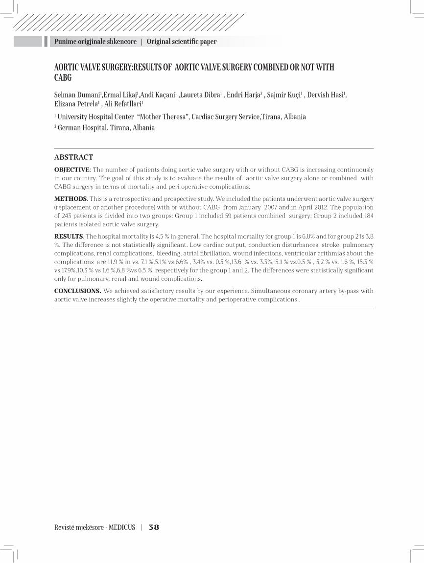
Punime origjinale shkencore | Original scientific paper
Revistë mjekësore - MEDICUS | 38
AORTIC VALVE SURGERY:RESULTS OF AORTIC VALVE SURGERY COMBINED OR NOT WITH CABG
Selman Dumani1,Ermal Likaj1,Andi Kaçani1 ,Laureta Dibra1 , Endri Harja2 , Sajmir Kuçi1 , Dervish Hasi1, Elizana Petrela1 , Ali Refatllari1 1 University Hospital Center “Mother Theresa”, Cardiac Surgery Service,Tirana, Albania2 German Hospital. Tirana, Albania
ABSTRACT
OBJECTIVE: The number of patients doing aortic valve surgery with or without CABG is increasing continuously in our country. The goal of this study is to evaluate the results of aortic valve surgery alone or combined with CABG surgery in terms of mortality and peri operative complications.
METHODS. This is a retrospective and prospective study. We included the patients underwent aortic valve surgery (replacement or another procedure) with or without CABG from January 2007 and in April 2012. The population of 243 patients is divided into two groups: Group 1 included 59 patients combined surgery; Group 2 included 184 patients isolated aortic valve surgery.
RESULTS. The hospital mortality is 4,5 % in general. The hospital mortality for group 1 is 6,8% and for group 2 is 3,8 %. The difference is not statistically significant. Low cardiac output, conduction disturbances, stroke, pulmonary complications, renal complications, bleeding, atrial fibrillation, wound infections, ventricular arithmias about the complications are 11.9 % in vs. 7.1 %,5.1% vs 6.6% , 3.4% vs. 0.5 %,13.6 % vs. 3.3%, 5.1 % vs.0.5 % , 5.2 % vs. 1.6 %, 15.3 % vs.17.9%,10.3 % vs 1.6 %,6.8 %vs 6.5 %, respectively for the group 1 and 2. The differences were statistically significant only for pulmonary, renal and wound complications.
CONCLUSIONS. We achieved satisfactory results by our experience. Simultaneous coronary artery by-pass with aortic valve increases slightly the operative mortality and perioperative complications .
39 | Medical Journal - MEDICUS
ЕВАЛУАЦИЈА НА ЕФЕКТОТ НА ПОКАЧЕНИОТ ИНТРААБДОМЕНАЛЕН ПРИТИСОК НА ВИСЦЕРАЛНАТА ПЕРФУЗИЈААлександра Гавриловска-Брзанов1, Мирјана Шошолчева2, Зорка Николова - Тодорова1, Гордана Талеска3, Маја Мојсова Мијовска1, Марија Јовановски-Срцева1, Вања Џамбазовска Трајковска1, Никола Брзанов1
1 ЈЗУ Универзитетска Клиника за трауматологија, ортопедски болести, анестезија реанимација и интензивно лекување и Ургентен центар, Скопје,
2 Специјална Болница за Хируршки болести “Св. Наум Охридски”, Скопје3 Медицински факултет, Универзитет “Св.Кирил и Методиј” Скопје, Македонија
РЕЗИМЕ:
Вовед: Евалуација на ефектот на покачениот интраабдоменален притисок (ИАП) на висцералната перфузија и претставување на абдоменлниот перфузиски притисок (АПП) како супериорен во однос на средниот артериски притисок при проценување на висцералната перфузија.
Материјал и методи: Во студијата беа вклучени 60 пациенти поделени во три групи (според класификацијата од WSACS): група 1. (N=20) пациенти со ИАП 12-15 mmHg, група 2. (N=20) пациенти со ИАП 16-20 mmHg и група 3. (N=20) пациенти со ИАП 21-25 mmHg. Пациентите беа стратифицирани во една од трите групи зависно од вредноста на ИАП добиена во две последователни мерења. Анализирани беа следните параметри: параметар кој се мереше: ИАП, параметар кој се отчитуваше (од мониторот за хемодинамски мониторинг во моментот кога се мереше ИАП):средниот артериски притисок (САП) и параметар кој се пресметуваше по формула: АПП.
Резултати: Според демографските податоци не постои статистички сигнификантна разлика односно се работи за хомогена група. Беше регистрирана статистички сигнификантна негативна корелација помеѓу вредностите на ИАП со вредностите на САП и АПП во втората и третата група за p<0.05 и статистички сигнификантна позитивна корелација помеѓу вредностите на САП и АПП во трите групи p<0.05.
Заклучоци: Мониторингот на ИАП игра значајна улога во подобрувањето на клиничкиот менаџмент на пациентите. Нормална вредност на САП не е предиктор за соодветна абдоменална органска перфузија, АПП е попрецизен предиктор на висцералната перфузија и супериорен во однос на средниот артериски притисок при проценување на висцералната перфузија.
Клучни зборови: Интраабдоменален притисок, абдоменален перфузиски притисок, среден артериски притисок.
Medicus 2013, Vol. 18 (2): 39-46
ВОВЕД
Инциденцата на мултиорганската дисфункција и морталитетот кај пациентите со покачен интраабдоменален притисок (ИАП) сè уште се високи како во светот така и кај нас (1). Затоа кај сите критично болни пациенти, особено кај оние кои не се подобруваат хемодинамски покрај адекватната ресусцитација, треба да се пристапи кон мерење и иследување на ИАП (2). Многуте научни истражувања и големото клиничко искуство го потврдија значењето на интраабдоменалната хипертензија (ИАХ) како вредност изведена од ИАП која предизвикува патофизиолошки ефект на органските системи (3). ИАП е дефиниран
како патолошки ИАП, кој е континуирано незначително покачен без клинички сигнификантни штетни ефекти, додека значително покачениот ИАП дава сериозни последици по сите органски системи во човековото тело (4-10). ИАХ пак е дефинирана како продолжено или повторувано патолошко зголемување на ИАП повисоко од 12 mmHg (11,12). Според моменталните разбирања за ИАХ/ абдоменален компартмент синдром (АКС), направена е класификација на ИАХ од страна на Светската Организација за АКС (WSACS) (13,14) според степенот на ИАП во четири подгрупи:
� I степен; ИАП 12-15 mmHg
Punime origjinale shkencore | Original scientific paper
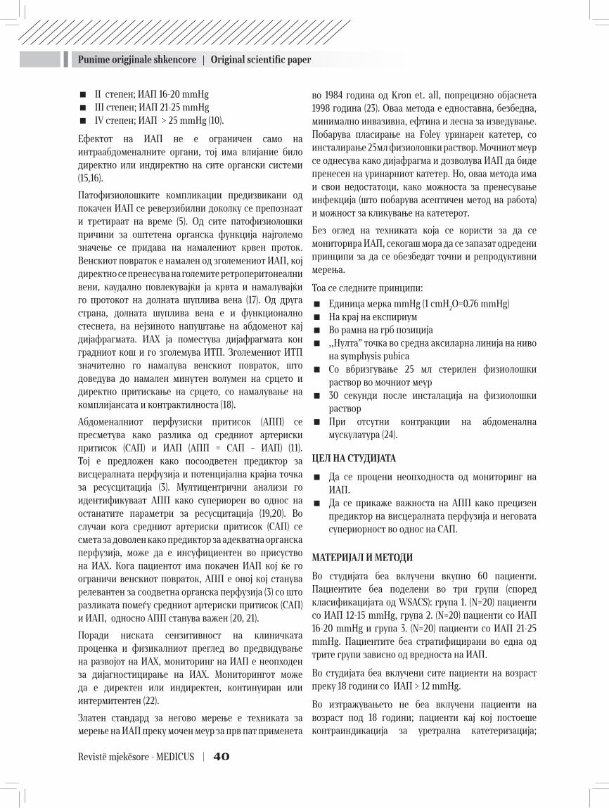
Punime origjinale shkencore | Original scientific paper
Revistë mjekësore - MEDICUS | 40
� II степен; ИАП 16-20 mmHg � III степен; ИАП 21-25 mmHg � IV степен; ИАП > 25 mmHg (10).
Ефектот на ИАП не е ограничен само на интраабдоменалните органи, тој има влијание било директно или индиректно на сите органски системи (15,16).
Патофизиолошките компликации предизвикани од покачен ИАП се реверзибилни доколку се препознаат и третираат на време (5). Од сите патофизиолошки причини за оштетена органска функција најголемо значење се придава на намалениот крвен проток. Венскиот повраток е намален од зголемениот ИАП, кој директно се пренесува на големите ретроперитонеални вени, каудално повлекувајќи ја крвта и намалувајќи го протокот на долната шуплива вена (17). Од друга страна, долната шуплива вена е и функционално стеснета, на нејзиното напуштање на абдоменот кај дијафрагмата. ИАХ ја поместува дијафрагмата кон градниот кош и го зголемува ИТП. Зголемениот ИТП значително го намалува венскиот повраток, што доведува до намален минутен волумен на срцето и директно притискање на срцето, со намалување на комплијансата и контрактилноста (18).
Абдоменалниот перфузиски притисок (АПП) се пресметува како разлика од средниот артериски притисок (САП) и ИАП (АПП = САП – ИАП) (11). Тој е предложен како посоодветен предиктор за висцералната перфузија и потенцијална крајна точка за ресусцитација (3). Мултицентрични анализи го идентификуваат АПП како супериорен во однос на останатите параметри за ресусцитација (19,20). Во случаи кога средниот артериски притисок (САП) се смета за доволен како предиктор за адекватна органска перфузија, може да е инсуфициентен во присуство на ИАХ. Кога пациентот има покачен ИАП кој ќе го ограничи венскиот повраток, АПП е оној кој станува релевантен за соодветна органска перфузија (3) со што разликата помеѓу средниот артериски притисок (САП) и ИАП, односно АПП станува важен (20, 21).
Поради ниската сензитивност на клиничката проценка и физикалниот преглед во предвидување на развојот на ИАХ, мониторинг на ИАП е неопходен за дијагностицирање на ИАХ. Мониторингот може да е директен или индиректен, континуиран или интермитентен (22).
Златен стандард за негово мерење е техниката за мерење на ИАП преку мочен меур за прв пат применета
во 1984 година од Kron et. аll, попрецизно објаснета 1998 година (23). Оваа метода е едноставна, безбедна, минимално инвазивна, ефтина и лесна за изведување. Побарува пласирање на Foley уринарен катетер, со инсталирање 25мл физиолошки раствор. Мочниот меур се однесува како дијафрагма и дозволува ИАП да биде пренесен на уринарниот катетер. Но, оваа метода има и свои недостатоци, како можноста за пренесување инфекција (што побарува асептичен метод на работа) и можност за кликување на катетерот.
Без оглед на техниката која се користи за да се мониторира ИАП, секогаш мора да се запазат одредени принципи за да се обезбедат точни и репродуктивни мерења.
Тоа се следните принципи:
� Единица мерка mmHg (1 cmH2O=0.76 mmHg) � На крај на експириум � Во рамна на грб позиција � ,,Нулта” точка во средна аксиларна линија на ниво
на symphysis pubica � Со вбризгување 25 мл стерилен физиолошки
раствор во мочниот меур � 30 секунди после инсталација на физиолошки
раствор � При отсутни контракции на абдоменална
мускулатура (24).
ЦЕЛ НА СТУДИЈАТА
� Да се процени неопходноста од мониторинг на ИАП.
� Да се прикаже важноста на АПП како прецизен предиктор на висцералната перфузија и неговата супериорност во однос на САП.
МАТЕРИЈАЛ И МЕТОДИ
Во студијата беа вклучени вкупно 60 пациенти. Пациентите беа поделени во три групи (според класификацијата од WSACS): група 1. (N=20) пациенти со ИАП 12-15 mmHg, група 2. (N=20) пациенти со ИАП 16-20 mmHg и група 3. (N=20) пациенти со ИАП 21-25 mmHg. Пациентите беа стратифицирани во една од трите групи зависно од вредноста на ИАП.
Во студијата беа вклучени сите пациенти на возраст преку 18 години со ИАП > 12 mmHg.
Во изтражувањето не беа вклучени пациенти на возраст под 18 години; пациенти кај кој постоеше контраиндикација за уретрална катетеризација;
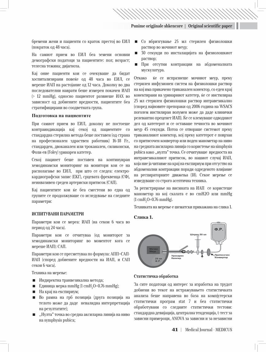
Punime origjinale shkencore | Original scientific paper
41 | Medical Journal - MEDICUS
бремени жени и пациенти со краток престој во ЕИЛ (пократок од 48 часа).
На самиот прием во ЕИЛ беа земени основни демографски податоци за пациентите: пол; возраст; телесна тежина; дијагноза.
Кај оние пациенти кои се очекуваше да бидат хоспитализирани повеќе од 48 часа во ЕИЛ, се мереше ИАП на растојание од 12 часа. Доколку во два последователни наврати беше измерен покачен ИАП (> 12 mmHg), односно пациентот развиеше ИАХ во зависност од добиените вредности, пациентите беа стратифицирани во соодветната група.
Подготовка на пациентитеПри самиот прием во ЕИЛ, доколку не постоеше контраиндикација кај секој од пациентите со стандардна стерилна метода беше поставен (од страна на професионален здраствен работник) 16-18 Fr., стандарден, двоканален или троканален, силиконски, Фоли-ев (Foley) уринарен катетер.
Секој пацинет беше поставен на континуиран хемодинамски мониторинг на монитори кои се на располагање во ЕИЛ, при што се следеа: електро-кардиографски запис (EKГ), срцевата фреквенца (СФ), неинвазивен среден артериски притисок (САП).
Кај пациентите кои ќе беа сместени во една од групите се продолжуваше со иследување на следните параметри:
ИСПИТУВАНИ ПАРАМЕТРИ
Параметри кои се мереа: ИАП (на секои 6 часа во период од 24 часа).
Параметри кои се отчитуваа (од мониторот за хемодинамски мониторинг во моментот кога се мереше ИАП): САП.
Параметри кои се пресметуваа по формула: АПП=САП-ИАП (според добиените вредности на ИАП, и САП секои 6 часа).
Техника на мерење:
� Индиректна транвезикална метода; � Единица мерка mmHg (1 cmH2O=0.76 mmHg); � На крај на експириум; � Во рамна на грб позиција (друга позиција на
телото може да даде невалидна интерпретација на резултатите);
� ,,Нулта” точка во средна аксиларна линија на ниво на symphysis pubica;
� Со вбризгување 25 мл стерилен физиолошки раствор во мочниот меур;
� 30 секунди по инсталацијата на физиолошкиот раствор;
� При отсутни контракции на абдоменалната мускулатура.
Откако ќе се испразнеше мочниот меур, преку стерилен инфузионен систем на физиолошки раствор на кој има прикачено триканален конектор, со еден крај конектирани на уринарниот катетер, ќе се инстилираа 25 мл стерилен физиолошки раствор интравезикално (според најновите препораки од 2006 година на WSACS поголем инстилиран волумен може да даде клинички релевантно преценет ИАП). Ќе се клемуваше одводниот дел од катетерот и се оставаше течноста во мочниот меур 45 секунди. Потоа се отвораше системот преку триканалниот конектор, кој преку катетерот е поврзан со притисочен конвертор или воден манометар на ниво на средната аксиларна линија со користење на simphysis pubica како ,,нулта” точка. Се отчитуваше вредноста на интравезикалниот притисок, во нашиот случај ИАП, која ние ја читавме на крај на експириум при отсуство на абдоменални контракции поради одреденото влијание на респираторните движења (18). Секое мерење се изведуваше со строго асептична техника.
За регистрирање на висината на ИАП се користеше манометар на кој скалата е во cmH2O или mmHg (1 cmH2O=0.76 mmHg).
Техниката на мерење е шематски прикажана на слика 1.
Слика 1.
Статистичка обработка
За сите податоци од интерес за изработка на трудот добиени во текот на истражувањето статистичката анализа беше направена во база на компјутерски статистички програм stat 7 и беа статистички обработувани со следните статистички тестови: стандардна девијација, централна тенденција, t-тест за зависни примероци, ANOVA за зависни и за независни
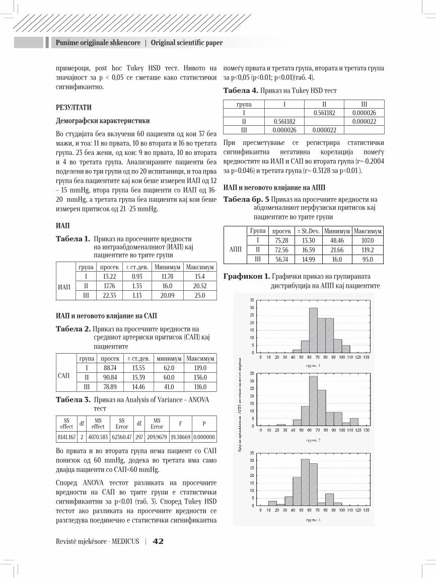
Punime origjinale shkencore | Original scientific paper
Revistë mjekësore - MEDICUS | 42
примероци, post hoc Tukey HSD тест. Нивото на значајност за p < 0,05 се сметаше како статистички сигнификантно.
РЕЗУЛТАТИ
Демографски карактеристики
Во студијата беа вклучени 60 пациенти од кои 37 беа мажи, и тоа: 11 во првата, 10 во втората и 16 во третата група. 23 беа жени, од кои: 9 во првата, 10 во втората и 4 во третата група. Анализираните пациенти беа поделени во три групи од по 20 испитаници, и тоа прва група беа пациентите кај кои беше измерен ИАП од 12 – 15 mmHg, втора група беа пациенти со ИАП од 16-20 mmHg, a третата група беа пациенти кај кои беше измерен притисок од 21 -25 mmHg.
ИАП
Табела 1. Приказ на просечните вредности на интраабдоменалниот (ИАП) кај пациентите во трите групи
ИАП
група просек ± ст.дев. Минимум Максимум
I 13.22 0.93 11.78 15.4
II 17.76 1.35 16.0 20.52
III 22.35 1.13 20.09 25.0
ИАП и неговото влијание на САП
Табела 2. Приказ на просечните вредности на средниот артериски притисок (САП) кај пациентите
САП
група просек ± ст.дев. минимум Максимум
I 88.74 13.55 62.0 119.0
II 90.84 15.39 60.0 136.0
III 78.89 14.46 41.0 116.0
Табела 3. Приказ на Analysis of Variance – ANOVA тест
SSeffect df MS
effectSS
Error df MSError F P
8141.167 2 4070.583 62360.47 297 209.9679 19.38669 0.000000
Во првата и во втората група нема пациент со САП понизок од 60 mmHg, додека во третата има само двајца пациенти со САП<60 mmHg.
Според ANOVA тестот разликата на просечните вредности на САП во трите групи е статистички сигнификантни за p<0.01 (таб. 3). Според Tukey HSD тестот ако разликата на просечните вредности се разгледува поединечно е статистички сигнификантна
помеѓу првата и третата група, втората и третата група за p<0,05 (p<0.01; p<0.01)(таб. 4).
Табела 4. Приказ на Tukey HSD тест
група I II IIII 0.561182 0.000026II 0.561182 0.000022III 0.000026 0.000022
При пресметување се регистрира статистички сигнификантна негативна корелација помеѓу вредностите на ИАП и САП во втората група (r=-0.2004 за p=0.046) и третата група (r=-0.3128 за p<0.01 ).
ИАП и неговото влијание на АПП
Табела бр. 5 Приказ на просечните вредности на абдоменалниот перфузиски притисок кај пациентите во трите групи
АПП
Група просек ± St.Dev. Минимум МаксимумI 75.28 13.30 48.46 107.0II 72.56 16.59 21.66 119.2III 56.74 14.99 16.0 95.0
Графикон 1. Графички приказ на групираната дистрибуција на АПП кај пациентите

Punime origjinale shkencore | Original scientific paper
43 | Medical Journal - MEDICUS
која се користи за негово мерење, одредени принципи да се следат за да се обезбедат соодветни и репродуктивни мерења од пациент на пациент. Во нашата студија, ги применивме сите напатствија предложени на оваа конференција, за иследување на ИАП.
Влијанието на ИАП односно ИАХ на САП испитувано во повеќе клинички и експериментални студии (26,27,28). Nguyen и соработниците (26) го евалуирале ефектот на покачениот ИАП од пнеумоперитонеум при лапароскопски интервенции кај обезни пациенти, додека во студиите на Quintel (27) и Quevedo (28) е испитувано влијанието на покачениот ИАП и АПП кај експериментални животни. Сите погоре наведени покажале дека при умерено покачен ИАП до 20 mmHg се зголемува САП. Што се смета дека е последица на зголемениот интраторакален волумен, предизвикан од преместување на волуменот од компресираниот абдомен кон некомпресираниот граден кош. Како друго можно објаснување, се наведува одговор на ЦНС поради нарушена венска дренажа.
Во нашата студија резултатите го покажаа следното: Кај пациентите од втора и трета група со средно и високо покачен ИАП, со средна вредност на ИАП од 17,76 ± 1,35 mmHg за втората група, и 22,35 ± 1,13 mmHg за третата група, постои статистички сигнификантна разлика со САП за p<0,05. Поединечно разгледуваната просечната вредност на САП ги даде следните резултати: Кај пациентите од прва група со умерено покачен ИАП со средна вредност 13,22 ± 0,93 mmHg, не постои статистички сигнификантна негативна корелација со ИАП. Добивме и значително зголемување на САП 90,84 ± 15,39 кај пациентите со средно покачен ИАП, додека во третата група вредноста на САП значително се намалува 78,89 ± 14,46 mmHg. Односно постои статистичка сигнификантност помеѓу првата и третата група, и втората и третата група за p<0,05, кога разликата на просечните вредности се разгледува поединечно. Се регистрираше и статистички сигнификантна негативна корелација помеѓу вредностите на ИАП и САП и тоа слаба во втората група, умерена во третата група.
Што се однесува до АПП, аналогно на општо познатиот и широко применетиот концепт за ЦПП, пресметувањето на АПП како разлика изведена од САП и ИАП е параметар со кој се проценува не само сериозноста на ИАП, туку и адекватната абдоменална органска перфузија (3,11). ,,Критичниот” ИАП е различен од пациент до пациент, зависно од
Табела 6. Приказ на Analysis of Variance – ANOVA тест
SSeffect df MS
effectSS
Error Df MSError F P
20047.40 2 10023.70 67033.69 297 225.7027 44.41109 0.000000
Референтна вредност за АПП е ≥ 60 mmHg. Во првата група има еден пациент со просечна вредност под референтната, во втората група има двајца пациенти, додека во третата група 50% од пациентите односно десет пациенти имаат вредност пониска од референтната. Според ANOVA тестот разликата на просечните вредности на АПП во трите групи е статистички сигнификантна за p<0.01 (таб. 6). Според Tukey HSD тест ако разликата на просечните вредности се разгледува поединечно постои статистичка сигнификантност помеѓу првата и третата и втората и третата група за p<0.01 (таб. 7).
Табела бр. 7 Приказ на Tukey HSD тест
група I II IIII 0.406836 0.000022II 0.406836 0.000022III 0.000022 0.000022
При пресметувањето се регистрира статистички сигнификантна негативна корелација помеѓу вредностите на ИАП и АПП во втората (r=-0.2362 за p<0.01) и третата група (r=-0.3833 зa p<0.01).
При пресметувањето се регистрира и статистички сигнификантна позитивна корелација помеѓу вредностите на САП и АПП во трите групи (r=0.9955 за p<0.01; r=-0.9506 за p<0.01; r=-0.9879 за p<0.01;).
ДИСКУСИЈА
Оваа студија покажува дека абдоменалниот перфузиски притисок е попрецизен предиктор на висцерална перфузија во однос на интраабдоменалниот притисок кај пациенти со интраабдоменална хипертензија.
ИАХ е дефинирана како продолжено или повторувано патолошко зголемување на ИАП повисоко од 12 mmHg (11,25). Преваленцата на ИАХ кај критично болни пациенти е доста висока (1), затоа од круцијална важност e да се мери ИАП, кај пациентите со ризик фактори. Malbrain и соработниците (11) во нивната студија, односно извештај од 2004 година на Конференцијата за Интернационален АКС Консензус Дефиниции потпишан од WSAC предлагаат без оглед на техниката
Punime origjinale shkencore | Original scientific paper
Revistë mjekësore - MEDICUS | 44
патофизиологијата и постоечките коморбидитети. Нормална вредност на САП не е предиктор за соодветна абдоменална органска перфузија, затоа што вредноста на ИАП која всушност го претставува ограничениот венски повраток, може да даде несоодветна органска перфузија (3,18,19). Во истите студии, Gheatham и соработниците (3,18,19) препорачуваат соодветна цел при ресусуцитација на пациентите да е вредност за АПП ≥ 60 mmHg. Сето погоре наведено го потврдуваат и Al-Dorzi со соработниците во група пациенти со септичен шок и коморбидитет цироза покажувајќи дека ИАП и АПП ≥ 60 mmHg како крајни точки на ресусцитација диференцираат преживеани од непреживеани (29).
Во нашата студија што се однесува до резултатите добиени за разликата на просечните вредности на АПП во трите групи, најдена е статистички сигнификантна негативна разлика за p<0.01 во сите три групи. Доколку разликата на просечните вредности се разгледува поединечно постои статистичка сигнификантност помеѓу првата и третата, и втората и третата група за p<0.01. При пресметувањето, регистрирана е и статистички сигнификантна негативна слаба корелација помеѓу вредностите на ИАП и АПП во втората и статистички сигнификантна негативна умерена корелација во третата група. Споредено пак со вредноста на САП се регистрира статистички сигнификантна позитивна многу јака корелација помеѓу вредностите на САП и АПП во трите групи.
Што се однесува до референтите вредности за АПП кај пациентите во првата и втората група на ИАХ, АПП е во нормални граници со средна вредност за АПП од 75,28 ± 13,30 mmHg за првата и 72,56 ± 16,59 mmHg за АПП во втората група. Додека кај пациентите од третата група на ИАХ вредностите на АПП се пониски од дозволените со средна вредност од 56,74 ± 14,99 mmHg, максимална 90 mmHg и минимална 16 mmHg за АПП.
Пациенти со САП понизок од 60 mmHg, во првата и втората група нема додека во третата група има само двајца, со што може да се заклучи дека кај сите пациенти од првата и втората група, и осумнаесет од третата ќе има добра органска перфузија но тоа не е така во случај на ИАХ. Затоа што во случај на ИАХ потребно е АПП > 60 mmHg, за да се обезбеди соодветна органска перфузија. А во нашата студија АПП понизок од 60 mmHg има во сите три групи: 5% во првата, 10% во втората група и дури 50% во третата група, со што може да се заклучи дека АПП е посоодветен предиктор за органската перфузија.
Дали ИАХ е маркер или причина за сериозна болест останува нејасно. Иако Malbrain и соработниците во нивната студија пронаоѓаат дека ИАХ е причина за морталитет (15), во поново време студијата од страна на Balogh изведена на пациенти со сериозен шок и траума не успева пак да ја прикаже важноста на ИАХ како предиктор за мултиорганска слабост. Во нивната студија не постоела разлика во исходот кај пациентите со ИАХ и траума или шок и без траума и шок (30). Затоа мултицентрични анализи го идентификуваат АПП како супериорен во однос на останатите параметри за ресусцитација, вклучувајќи ги тука и артериската Ph., базниот дефицит, артериските лактати и сатната диуреза. АПП од минимум 60 mmHg како крајна точка на ресусцитација е асоциран со зголемено преживување кај пациентите со ИАХ (19, 20, 31).
Прикажаната студија ги има следните ограничувања: Примерокот е мал, основната болест која водеше до ИАХ се разликува од пациент до пациент и исклучени се пациентите кои се отпуштени пред 24 часа од ЕИЛ.
ЗАКЛУЧОЦИ
1. Мониторинг на ИАП е неопходен не само од научна, туку и од клиничка гледна точка. Неговото иследување, игра значајна улога во подобрувањето на клиничкиот менаџмент на пациентите.
2. АПП е попрецизен предиктор на висцералната перфузија и супериорен во однос на останатите параметри за дефинирање на ИАХ. Нормална вредност на САП не е предиктор за соодветна абдоменална органска перфузија. Во присуство на покачен ИАП, кој всушност го претставува ограничениот венски повраток, нормалниот САП може да даде несоодветна органска перфузија.
ЛИТЕРАТУРА:
1. Malbrain ML, Chiumello D, Pelosi P, Wilmer A, Brienza N, Malcangi V, et al. Prevalence of intra-abdominal hy-pertension in critically ill patients: a multicentre epide-miological study. Intensive Care Med 2004, 30:822-829.
2. Ivankovich AD, Miletich DJ, Albrecht RF, Heyman HJ, Bonnet RF. Cardiovascular eff ects of intraperitoneal in-. Cardiovascular eff ects of intraperitoneal in-Cardiovascular effects of intraperitoneal in-sufflation with carbon dioxide and nitrous oxide in the dog. Anesthesiology. 1975;42:281-87.
3. Daniela BP, Vianja MK. Intraabdominal hypertension and abdominal compartment syndrome in the intensive care unit. Signa vitae 2006;1(1):13-15.
Punime origjinale shkencore | Original scientific paper
45 | Medical Journal - MEDICUS
4. Schein M. Abdominal Compartment Syndrome: Histori-. Abdominal Compartment Syndrome: Histori- Abdominal Compartment Syndrome: Histori- Histori-Histori-cal Background. In Abdominal Compartment Syndrome Edited by: Ivatury R, Cheatham M, Malbrain M, Sugrue M. Georgetown, Texas: Landes Biosciences; 2006:1-7.
5. Muckart DJ, Ivatury R, Leppaniemi A, Smith S. Defi ni- Smith S. Defi ni-Smith S. Defi ni-. Defi ni-Defini-tions. In: Ivatury R, Cheatham M, Malbrain M, Sugrue M (eds) Abdominal compartment syndrome. Landes Bio- syndrome. Landes Bio-syndrome. Landes Bio-science, Georgetown, 2006; pp 8–18.
6. Sugrue M, Hallal A, D’Amours S. Intra-abdominal pressure hypertension and the kidney. In: Ivatury R, Cheatham M, Malbrain M, Sugrue M, Editors. Abdomi-nal compartment syndrome. Georgetown (TX): Landes Bioscience; 2006; pp 119-28.
7. Raeburn CD, Moore EE. Abdominal compartment syn-. Abdominal compartment syn-Abdominal compartment syn-drome provokes multiple organ failure: animal and human supporting evidence. In: Ivatury R, Cheatham M, Malbrain M, Sugrue M (eds) Abdominal compartment syndrome. Landes Bioscience, Georgetown,2006; pp 157–169.
8. Mertens zur Borg IR, Verbrugge SJ, Olvera C. Pathophys- Olvera C. Pathophys-Olvera C. Pathophys-. Pathophys-Pathophys-iology: respiratory. In: Ivatury R, Cheatham M, Malbrain M, Sugrue M (eds) Abdominal compartment syndrome. Landes Bioscience, Georgetown, 2006; pp 105–118.
9. Ivatury R, Diebel L. Intraabdominal hypertension and the splanchnic bed. In: Ivatury R, Cheatham M, Malbrain M, Sugrue M (eds) Abdominal compartment syndrome. Landes Bioscience, Georgetown, 2006; pp 129–137.
10. Citerio G, Berra L. Intraabdominal hypertension and the central nervous system. In: Ivatury R, Cheatham M, Malbrain M, Sugrue M, Editors. Abdominal compart-ment syndrome. Georgetown (TX): Landes Bioscience; 2006; pp. 144-56.
11. Malbrain ML, Cheatham ML, Kirkpatrick A, et al. Re-sults from the International Conference of Experts on Intra-abdominal Hypertension and Abdominal Com-partment Syndrome. I. Definitions. Intensive Care Med 2006;32:1722-32.
12. Carlotti A, Carvalho W: Abdominal compartment syn-drome: A review. Pediatr Crit Care Med 2009;10:115-20.
13. Cheatham ML, Malbrain ML, Krikpatrick A, et all. Re-sults from the International Conference of Experts on Intra-abdominal Hypertension and Abdominal Com-partment syndrome. II. Recommendations. Intensive Care Med, 2007.33(6):p. 951-62.
14. World Society of the Abdominal Compartment Syndrome http://www.wsacs.org/ Assessed January 12.01.2013.
15. Malbrain ML, Chiumello D, Pelosi P, et al. Incidence and prognosis of intraabdominal hypertension in a mixed
population of critically ill patients: A multiple-center epidemiological study. Crit Care Med 2005;33:315-22.
16. Cheatham ML. Abdominal Compartment Syndrome. Pathophysiology and Definitions. Scandinavian Jour-nal of Trauma Resuscitation and Emergency Medicine 2009; 17-10.
17. Rubinson RM, Vasko JS, Doppman JL, Morrow AG. Infe-. Infe-Infe-rior caval obstruction from increased intra-abdominal pressure. Arch Surg. 1967;94:766-70.
18. Ridings PC, Bloomfield GL, Blocher CR, Sugerman HJ. Cardiopulmonary effects of raised intra-abdominal pressure before and after intravascular volume expan-sion. J Trauma 1995;39:1071-5.
19. Cheatham ML, White MW, Sagraves SG, Johnson JL, Block EF. Abdominal perfusion pressure: A superior parameter in the assessment of intra-abdominal hyper-tension. J Trauma 2000;49:621-6.
20. Walker J. and Laura MC. Pathophysiology and Manage- J. and Laura MC. Pathophysiology and Manage- and Laura MC. Pathophysiology and Manage-C. Pathophysiology and Manage-Pathophysiology and Manage-ment of Abdominal Compartment Syndrome. Am J Crit Care 2003;12:367-371.
21. Nebelkopf H. Abdominal compartment syndrome. Am J Nurs. November 1999;99:53-56, 58, 60.
22. Deenichin GP.Abdominal Compartment Syndrome. Surg Today 2008; 38:5–19.
23. Priluck IA, Blodgett DW. The effects of increased intra-abdominal pressure on the eyes: Nebr Med J 1996;81(1):8–9.
24. Malbrain МL. Diff erent techniques to measure intra-ab-. Diff erent techniques to measure intra-ab-Different techniques to measure intra-ab- intra-ab-intra-ab-dominal pressure (IAP): time for a critical re-appraisal. Intensive Care Med 2004; 30:357–371.
25. Saggi, BH, Sugerman HJ, Ivatury RR, Bloomfield GL. Ab-. Ab-Ab-dominal compartent syndrome. J. Trauma, 1998, 45:597.
26. Nguyen N.T, Wolf B.M. MD. The Physiologic Effects of Pneumoperitoneum in the Morbidly Obese: Ann Surg: 2005 February; 241(2): 219–226
27. Quintel M, Pelosi P, Caironi P, et al. An increase of abdominal pressure increases pulmonary edema in oleic acid-induced lung injury. Am J Respir Crit Care Med 2004, 169:534-541.
28. Quevedo AI, Revilla FJ, Sanz JJ, et al. Comparative study on renal blood flow (RBF) and renal hemodynamics dur-ing open or laparoscopic nephrectomy in an experimen-tal model. Actas Urol Esp. 2007;31(4):382-393.
29. Al-Dorzi HM, Tamim HM, Rishu AH, Aljumah A, and Ara-bi Y. Intra-abdominal pressure and abdominal perfusion pressure in cirrhotic patients with septic shock. Publish. ed online 2012 July 5. doi: 10.1186/2110-5820-2-S1-S4.
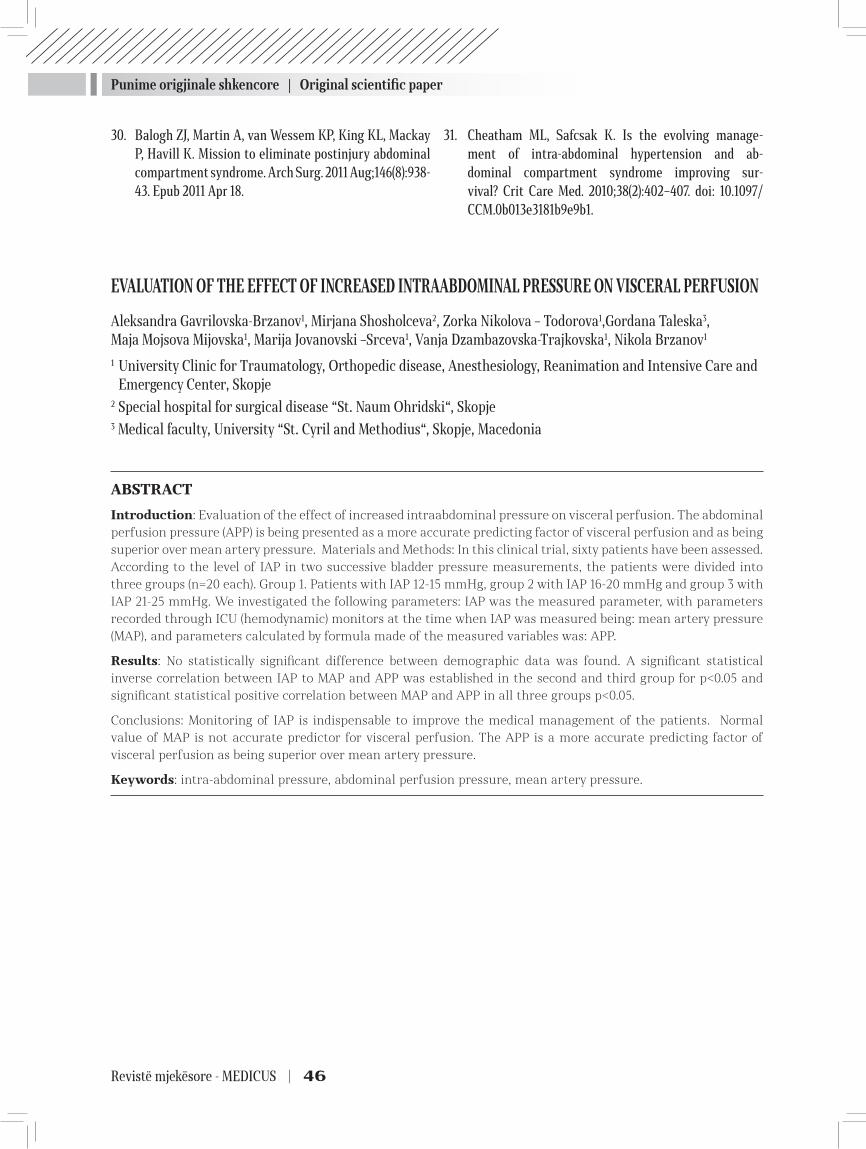
Punime origjinale shkencore | Original scientific paper
Revistë mjekësore - MEDICUS | 46
30. Balogh ZJ, Martin A, van Wessem KP, King KL, Mackay P, Havill K. Mission to eliminate postinjury abdominal compartment syndrome. Arch Surg. 2011 Aug;146(8):938-43. Epub 2011 Apr 18.
31. Cheatham ML, Safcsak K. Is the evolving manage-ment of intra-abdominal hypertension and ab-dominal compartment syndrome improving sur-vival? Crit Care Med. 2010;38(2):402–407. doi: 10.1097/CCM.0b013e3181b9e9b1.
EVALUATION OF THE EFFECT OF INCREASED INTRAABDOMINAL PRESSURE ON VISCERAL PERFUSION
Aleksandra Gavrilovska-Brzanov1, Mirjana Shosholceva2, Zorka Nikolova – Todorova1,Gordana Taleska3, Maja Mojsova Mijovska1, Marija Jovanovski –Srceva1, Vanja Dzambazovska-Trajkovska1, Nikola Brzanov1
1 University Clinic for Traumatology, Orthopedic disease, Anesthesiology, Reanimation and Intensive Care and Emergency Center, Skopje
2 Special hospital for surgical disease “St. Naum Ohridski“, Skopje3 Medical faculty, University “St. Cyril and Methodius“, Skopje, Macedonia
ABSTRACT
Introduction: Evaluation of the effect of increased intraabdominal pressure on visceral perfusion. The abdominal perfusion pressure (APP) is being presented as a more accurate predicting factor of visceral perfusion and as being superior over mean artery pressure. Materials and Methods: In this clinical trial, sixty patients have been assessed. According to the level of IAP in two successive bladder pressure measurements, the patients were divided into three groups (n=20 each). Group 1. Patients with IAP 12-15 mmHg, group 2 with IAP 16-20 mmHg and group 3 with IAP 21-25 mmHg. We investigated the following parameters: IAP was the measured parameter, with parameters recorded through ICU (hemodynamic) monitors at the time when IAP was measured being: mean artery pressure (MAP), and parameters calculated by formula made of the measured variables was: APP.
Results: No statistically significant difference between demographic data was found. A significant statistical inverse correlation between IAP to MAP and APP was established in the second and third group for p<0.05 and significant statistical positive correlation between MAP and APP in all three groups p<0.05.
Conclusions: Monitoring of IAP is indispensable to improve the medical management of the patients. Normal value of MAP is not accurate predictor for visceral perfusion. The APP is a more accurate predicting factor of visceral perfusion as being superior over mean artery pressure.
Keywords: intra-abdominal pressure, abdominal perfusion pressure, mean artery pressure.
47 | Medical Journal - MEDICUS
LIDHJA MIDIS RETINOPATISË DIABETIKE DHE HEMOGLOBINËS SË GLUKOZUAR NË PACIENTËT DIABETIKËKavaja Silvana1, Ceka Xheladin2, Spahiu Eduard1, Kavaja Genti1, Xinxo Sonela3
1 Spitali Rajonal Durrës, Durrës2 Departamenti i Morfologjisë, Fakulteti Mjekësisë, Universiteti Tiranë, Tiranë3 Instituti i Shendetit Publik, Tiranë
ABSTRAKTI
Hyrje: Retinopatia diabetike (RD) është një komplikacion kronik mikrovaskular që dëmton rëndë shikimin tek adultët me diabet mellitus.
Qëllimi: Të vlerësojë prevalencën e RD dhe lidhjen midis vlerave të HbA1c, kohëzgjatjes së diabetit me RD tek diabetikët.
Materiali dhe metodat: Në studim u përfshinë 700 pacientë diabetikë gjatë periudhës Janar 2010-Janar 2012 në Spitalin e Durrësit. Prania e RD u vlerësua përmes oftalmoskopit nga mjeku okulist. Bazuar në të dhënat në fundus okuli, pacientët u ndanë në tre grupe: pa retinopati, me retinopati jo-proliferative (RDJP) dhe me retinopati proliferative (RDP). Pacientët u klasifikuan sipas nivelit të HbA1c ( poshtë 6.0%; 6.1 deri 6.9% ; 7.0 deri 9.9%; mbi 10.0%); sipas kohëzgjatjes së diabetit (diabet deri në 5 vjet, histori 6 - 10 vjet, histori për diabet mbi 10 vjet).
Rezultatet: Prevalenca e RD ishte 45.1 % (33.2 % me RDJP dhe 11.9% me RDP). U vu re një korrelacion statistikisht sinjifikant midis vlerave të HbA1c dhe presencës së RD (p < 0.01, r = 0.8), kohëzgjatjes së diabetit dhe RD (p < 0.01, r = 0.7).
Konkluzionet: Kohëzgjatja e diabetit dhe HbA1c janë faktorë risku të rëndësishëm për presencën dhe progresin e RD në pacientët diabetikë. Mbajtja nën kontroll e vlerave të HbA1c mund të parandalojë ose të vonojë shfaqjen ose progresionin e RD.
Fjalë kyç: Diabet mellitus, retinopatia diabetike, hemoglobina e glukozuar, HbA1c
Medicus 2013, Vol. 18 (2): 47-51
HYRJE
Retinopatia diabetike (RD) është një crregullim vaskular që prek mikroqarkullimin e retinës. Është vlerësuar që diabeti mellitus prek rreth 8.3% të popullatës botërore, ku pothuajse gjysma e tyre kanë praninë e retinopatisë diabetike në një moment të dhënë(1) Retinopatia diabetike prek të dy tipet e diabetit (tipi 1 dhe tipi 2).
Retinopatia diabetike është një nga shkaqet më të shpeshta të verbërisë në adultët e moshës 20 deri në 75 vjec dhe mbetet një problem shëndetësor i rëndësishëm në gjithe botën(2). Përmirësimet në kujdesin ndaj diabetit dhe diagnostikimi i hershëm i sëmundjes mund të reduktojë incidencën e dëmtimeve të shikimit dhe të verbërisë (3). Në kohën e vendosjes së diagnozës klinike
të tipit të diabetit, disa pacientë mund të kenë të dhëna për retinopati diabetike, duke treguar në këtë mënyrë që diabeti mellitus ka qënë i pranishëm shumë vite më përpara (4).
Kohëzgjatja e diabetit, kontrolli i glicemisë, hipertensioni, dislipidemia, obeziteti, gjendja social ekonomike luajnë një rol të rëndësishëm për zhvillimin e RD. Më të rëndësishmit janë kohëzgjatja e diabetit dhe kontrolli jo i duhur i glicemisë (5). Monitorimi i vlerave të hemoglobinës së glukozuar është një metodë efikase për vlerësimin e përqëndrimit mesatar të glukozës në gjak për tre muaj (6).
Qëllimi i studimit është të vlerësohet frekuenca e RD si dhe lidhja midis vlerave të HbA1c, kohëzgjatjes së diabetit dhe RD në pacientët me diabet mellitus.
Punime origjinale shkencore | Original scientific paper
Punime origjinale shkencore | Original scientific paper
Revistë mjekësore - MEDICUS | 48
MATERIALI DHE METODAT
Në studim transversal (cross-sectional) u përfshinë pacientë të diagnostikuar me diabetes mellitus (tipi 1 dhe tipi 2) nga shërbimi i endokrinologjisë pranë Spitalit Rajonal Durrës. Për një periudhë 2 vjecare (Janar 2010 – Janar 2012), të gjithë pacientët diabetikë e marrë në studim vinin për herë të parë në klinikën e okulistikës. Numri i pacientëve diabetikë ( të dy tipet) ishte 699 me moshë mesatare 60.2 ± 10.4 vjec, me kohëzgjatje të diabetit 10.4 ± 6.9 vite. Kriteret e përfshirjes për klasifikimin dhe diagnostikimin e DM ishin kriteret e pranuara nga ADA (shoqata amerikane e diabetit) në vitin 1997 (7). Pacientët me diabet sekondar (akromegalia, sindorma Cushing) nuk ishin përfshirë në studim. Në ekzaminimin e parë, për seclin pacient u realizua ekzaminimi direkt oftalmoskopik me dilatacion të pupilës nga mjeku okulist. Gjithashtu u kërkuan vlerat e HbA1c.
Lezionet e hershme të para me oftalmoskop u emërtuan retinopati diabetike jo-proliferative (RDJP) ku përfshihen mikroaneurizmat, hemorragjitë, eksudatet e forta, njollat e pambukut, crregullimet mikrovaskulare retinale dhe përdredhja e venave. Retinopatia proliferative (RDP) karakterizohet përvec më sipër edhe nga rritja e enëve të reja të gjakut, proliferimi vitroretinal, tërheqja e vitreosit dhe deri në shkolitje retinale e cila con në verbëri të plotë.
Bazuar në të dhënat nga fundus okuli, pacientët u ndanë në tre grupe: pa retinopati diabetike, grupi me retinopati diabetike jo-proliferative (RDJP) dhe grupi me retinopati diabetike proliferative (RDP). Pacientët realizuan metodën e shpejtë me gjak venoz të matjes së HbA1c. Bazuar në vlerat e marra të HbA1c u krijuan katër grupe: poshtë 6.0%; 6.1 deri në 6.9% ; 7.0 deri në 9.9% dhe mbi 10.0%. sipas kohëzgjatjes së diabetit pacientët u ndanë në tre grupe. Grupi i parë me pacientë me kohëzgjatje të diabetit më pak se 5 vjet, grupi i dytë me kohëzgjatje 6 deri në 10 vjet dhe grupi i tretë me kohëzgjatje më shumë se 10 vjet.
Vlerat e hemoglobinës së glukozuar (HbA1c) u matën duke përdorur pajisjen BioRad in2it HbA1c analyzers (metoda me kollonë).
ANALIZA STATISTIKORE
Te dhenat jane perpunuar ne SPSS 16. Per te vleresuar lidhjen midis HbA1c, kohezgjajtes se semundjes dhe RD eshte përdorur teknika e korrelacionit (Kendall’s tau_b dhe Spearman ). Chi square eshte perdorur per te vleresuar
madhesine e impaktit të seksit dhe tipit te diabetit në ecurine e RD. Nje p < 0.05 eshte konsideruar sinjifikante.
REZULTATET
Pacientët e marë në studim ishin 699 pacientë diabetikë ( të dy tipet) me moshë mesatare 60.2 ± 10.4 vjec, me kohëzgjatje të diabetit 10.4 ± 6.9 vite. 227 pacientë ishin me diabet tipi 1 dhe 472 me diabet tipi 2. Karakteristikat klinike të pacientëve janë si më poshtë. Kohëzgjatja e diabetit ishte 10.4 ± 6.9 vjet dhe sipas tipit të diabetit kemi këtë kohëzgjatje: për tipin e parë 12.79 ± 7.4 vjet dhe për tipin e dytë 9.2 ± 6.3 vjet. Në të gjithë pacientët vlera mesatare e HbA1c ishte 7.2 ± 1.2 %. 159 pacientë (22.7%) kishin vlera të HbA1c nën 6%; 208 pacientë (29.8%) kishin vlera të HbA1c nga 6.1 deri 6.9%; 324 pacientë (46.4%) kishin vlera të HbA1c nga 7.0 deri në 9.9% dhe vetëm 8 pacientë (1.1%) kishin vlera të HbA1c mbi 10.0%.
Në këtë studim frekuenca e RD ishte 45.1% (33.2 % me RDJP dhe 11.9% me RDP). Frekuenca e retinopatisë diabetike (jo-proliferative dhe proliferative) rritej me rrtijen e vlerave të HbA1c, ku 0 % në grupin e diabetikëve me vlerat e HbA1c poshtë 6%; 0 % në grupin me vlera të HbA1c 6.1 deri në 6.9%; 90.4 % në grupin me vlera të HbA1c 7 deri në 9.9% dhe 9.6 % në grupin me vlera të HbA1c mbi 10.0%. Sic shihet nga të dhënat, në pacientët që kishin vlera të HbA1c nga 7 deri në 9.9% kishte lidhje sinjifikante të fortë midis RD dhe vlerave të HbA1c (p < 0.01).
Sipas kohëzgjatjes së diabetit në grupin e parë frekuenca e RD ishte 3.3%, me 6 pacientë me RDJP dhe 1 me RDP; në grupin e dytë frekuenca e RD ishte më e lartë në 24% me 34 pacientë me RDJP dhe 2 pacientë me RDP; në grupin e tretë frekuenca e RD ishte më e larta 80.4 % ku 192 pacientë me RDJP dhe 80 pacientë me RDP. Rezultatet tona tregojnë që ka një lidhje sinjikative midis kohëzgajtjes së diabetit dhe RD (p < 0.01).
Nga të dhënat rezultoi se femrat kanë më shumë gjasa të bëjnë RDJP sesa RDP në krahasim me meshkujt (OD = 1.7 CI95% 1.04-2.8 p < 0.05). Mosha mesatare e pacienteve femra ishte 59.67 vjet dhe e pacientëve meshkuj ishte 60.76 vjet. Pacientët e të dy sekseve kishin kohëzgjatje të ngjashme të sëmundjes ( femrat 10.00 vjet dhe meshkujt 10.92 vjet. Vlera mesatare e HbA1c në pacientët femra ishte 7.1% dhe në pacientët meshkuj ishte 7.2%. Gjithashtu pacientët me diabet mellitus tipi i parë kanë me shumë gjasa të zhvillojnë RDP sesa RDJP në krahasim me pacientët me diabet mellitus tipi i dytë (OD = 7 CI95% 3.8-13.3 p < 0.01).
Punime origjinale shkencore | Original scientific paper
49 | Medical Journal - MEDICUS
DISKUTIMI
Retinopatia diabetike është një komplikacion specifik mikrovaskular në të dy tipet e diabetit. Kohëzgjatja e diabetit dhe hipeglicemia janë dy faktorë risku të njohur për zhvillimin e RD. Përqëndrimet e larta të glukozës dhe hiperglicemia kronike është pranuar nga të gjithë si një nga shkaktarët që të cojnë në RD.
Ka të dhëna të forta që sugjerojnë që zhviillimi dhe avancimi i RD ndikohet nga niveli i hiperglicemisë (8). Ekzistojnë një numër mekanizmash të besueshëm që lidhin metabolizmin e glukozës direkt me shfaqjen dhe avancimin e RD: aktivizimi i aldolazë reduktazës rrit aktivitetin e proteinë kinazë C shoqëruar me rritje të prodhimit të prostaglandinave vazodilatatore, rritje të glikozilimit jo-enzimatik dhe nxitje të dëmtimeve auto-oksidative nga glukoza (9). Rritja e permeabilitetit të barrierës të qarkullimit retinal dhe ndryshimet në fluksin e qarkullimit retinal mund të jenë të rëndësishme në patogjenezën e RD. Pra mekanizmat biokimikë, hemodinamikë dhe hormanalë mund të veprojnë së bashku për të prodhuar dëmtimet tipike të okluzionit vaskular, mikroaneurizmat, hemorragjitë, eksudatet e forta dhe enët e reja të gjakut (neovaskularizimi) (10).
Nga rezultate tregohet që në studimin tonë frekuenca e RD ishte 45.1% (33.2% kishin RDJP dhe 11.9% kishin RDP). Numri i pacientëve me retinopati diabetike jo-proliferative ishte më i madh se numri i pacientëve me retinopati diabetike proliferative (232 pacientë krahasuar me 83 pacientë). Kjo vjen si rezultat sepse numri i pacientëve me diabet mellitus tipi 2 është më i madh se numri i pacientëve me diabet mellitus tipi 1 (përkatësisht 472 pacientë ndaj 227 pacientë). Sipas tipit të diabetit, numri më i madh i pacientëve me retinopati diabetike i përkiste tipit 1 (69.6% krahasuar me 33.3%). Tipi 1 i diabetit zakonisht shfaqet në moshë më të re sesa tipi 2, prandaj ata kanë edhe një moshë më të re të shfaqjes së retinopatisë. Në SHBA retinopatia diabetike varionte nga 40-45%(1). Në bazë ndërkombëtare, prevalenca e retinoaptisë varet gjerësisht nga metodologjia dhe tipi i popullatës.
Kohëzgjatja e diabetit mellitus është një nga faktorët më të rëndësishëm përcaktuesë të pranisë së RD. Në studimin tonë numri i pacientëve me RDJP, me kohëzgjatje të sëmundjes mbi 10 vjet ishte 192 (82.8%) dhe me RDP ishte 80 (96.4%). Të dhënat tona tregojnë një shoqërim midis kohëzgjatjes më të madhe të diabetit dhe rritjes së frekuencës së retinopatisë. Frekuenca e RDJP dhe RDP rritet në grupin e diabtikëve që
vuanin prej 10 vitesh ose më tepër (p < 0.01). Në mënyrë të ngjashme Vinker et al (11) kanë treguar që sa më e gjatë koha me diabet, aq më tepër rritet RD.
Të dhënat tona demonstrojnë që RD është një problem shëndetësor dhe një nga shkaktarët e verbërisë. Është përcaktuar që pothuajse të gjithë pacientët diabetikë (tipi 1 dhe tipi 2) janë në risk të lartë për këtë komplikacion të rrezikshëm. Shumë studime klinike të realizuara nga qendra e kontrollit të diabetit dhe komplikacioneve si dhe të dhënat epidemiologjike nga studimet epidemiologjike Wisconsin të RD, kanë treguar lidhjen e fortë midis kontrollit te glicemisë dhe zhvillimin e progresionit të RD (12). Në një studim tjetër, të shoqates Australiane të diabetit, është raportuar që pacientët me RD kanë vlera të larta të HbA1c(13)
Sot, matja e vlerave të HbA1c, konsiderohet si tregues etalon për kontrollin e glicemisë në pacientët diabetikë, duke pasqyruar nivelet e glukozës për një periudhë 2-3 muaj (14). Shoqata amerikane e diabetit rekomandon që një vlerë mesatare e HbA1c duhet mbajtur poshtë 7% për të parandaluar komplikacionet diabetike mikro dhe makrovaskulare(15).
Në këtë studim frekuenca e retinopatisë (joproliferative dhe proliferative) mungonte në grupin me vlera të HbA1c më të ulta se 6.0%. Kohëzgjatja e sëmundjes së diabetit në këtë grup ishte 4.36 vjet. Kjo frekuencë rritej ndjeshëm me rritjen e vlerave mesatare të HbA1c mbi 10.0%. Të dhënat tona tregojnë një korrelacion midis vlerave të ulta të HbA1c dhe frekuencës së ulët të RD. Ulja e vlerave të glicemisë ose përqëndrimit të HbA1c nëpërmjet kontrollit të rreptë të glukozës në gjak në njerëzit me diabet, redukton ritmin e progresionit të komplikacioneve mikrovaskulare sic është RD, neuropatia dhe nefropatia(16,17).
KONKLUZIONI
Ky studim tregoi që frekuenca e RD ishte 45.1% (33.2% kanë RDJP dhe 11.9% kanë RDP). Kohëzgjatja e sëmundjes si dhe kontrolli i mirë i sëmundjes (vlerësuar me anë të HbA1c) ndikojnë në ndodhjen e retinopatisë diabetike. Pacientët meshkuj dhe me diabet tipi 1 janë më në risk për të patur retinopati diabetike proliferative.Të dhënat tona sugjerojnë që duke ulur vlerat e HbA1c ose duke përmbushur kriteret e shoqatës amerikane të diabetit (ADA) mund të parandalohet ose të vonohet shfaqja dhe avancimi i komplikacioneve mikrovaskulare sic është retinopatia. Meqënëse retinopatia diabetike është një komplikacion mikrovaskular serioz i diabetit, kontrolli
Punime origjinale shkencore | Original scientific paper
Revistë mjekësore - MEDICUS | 50
periodik për retinopatinë diabetike dhe kontrolli i rrepte i glicemisë mund të reduktojë numrin e njerëzve që mund të pësojnë retinopati me rrezik për shikimin.
BIBLIOGRAFIA
1. IDF diabetes atlas fifth edition 2012. 2. Fong DS, Aiello L, Gardner TW, King GL, Blankenship G,
Cavallerano JD, Ferris III FL, Klein R, Diabetic Retinopa-thy . Diabetes Care 2003; 26 (Suppl 1): s99-s102.
3. Diabetes Control and Complications Trial Research Group: The effect of intensive treatment of diabetes on the development and progression of long-term compli-cations in insulin-dependent diabetes mellitus. N Engl J Med 1993; 329: 977-86.
4. Tapp RJ, Shaw JE, Harper CA, Balkau B, McCarty DJ, Taylor HR, Welborn TA, Zimmet PZ, The prevalence of and Factors Associated with diabetic retinopathy in the Australian Population, Diabetes Care 2003; 26: 1731-7.
5. Fong DS, Aiello L, Gardner TW, King GL, Blankenship G, Cavallerano JD, Ferris FL, Klein R, Retinopathy in diabe-tes. Diabetes Care 2004; 27: s84-s7.
6. Evans J, Newton R, Ruta D, Macdonald T, Stevenson R, Morris A. Frequency of blood glucose monitoring in re-lation to glycemic control. BMJ 1999; 319: 83-6.
7. American Diabetes Association; Standards of medical care for patients with diabetes mellitus. Position state-ment. Diabetes Care 1999; 21 Suppl 1: S32-41.
8. Klein R, Klein BE, Moss SE, Cruickshanks KJ. The Wis-consin Epidemiologic Study of Diabetic Retinopathy: XVII. The 14-year incidence and progression of diabetic
retinopathy and associated risk factors in Type 1 diabe-tes. Ophthalmology 1998; 105 : 1801-15.
9. Larkins RG, Dunlop ME, Johnson EIM, The pathogenesis of diabetic retinopathy. Aust N Z J Opthalmol 1996; 24: 97-104.
10. Kohner EM, Aldinton SJ, Stratton IM, Manley SE, Hol-man RR, Turner RC. U.K. Prospective Diabetes Study, 30: diabetic retinopathy at diagnosed of NIDDM and as-sociated risk factors. Arch Opthalmol 1998; 116; 297- 303.
11. Vinker S, Shpiz M, Elhayany A, Nakar S, Impovment of early detection of diabetic retinopathy-a primary care intervention sudy. Harefuah 2003; 142: 826-8.
12. Klein R, Klein B, Moss S.The Wisconsin Epidemiological Study of Diabetic Retinopathy :a review. Diabetes Me-tab Rev 1992; 5: 1369-77.
13. Retinopathy Sub-committee. Diabetes and Eye:Professional Guidelines. Australian Diabetes Soci-ety 1996; 3-30.
14. American Diabetes Association. Postprandial blood glu-cose. Diabetes Care 2001; 24:775-8.
15. American Diabetes Association Standards of medical care in diabetes. Diabetes Care 2004; 27(Suppl.1): S15-35.
16. Knuiman MW, Welborn TA, MacCann VJ, Stanton KG, Constable IJ: Prevalence of diabetic complications in relation to risk factors. Diabetes 1986; 35: 1332-9.
17. The Diabetes Control and Complications Trial Research Group. The Relationship of Glycemic Exposure (HbA1c) to the Risk of Development and Progression of Retin-opathy in the Diabetes Control and Complications Trial. Diabetes, 44:968-983, 1995.
THE DIABETIC RETINOPATHY: CORRELATIONS WITH GLYCATED HAEMOGLOBIN AND DURATION OF DIABETES.
Kavaja Silvana MD1, Ceka Xheladin Prof2, Spahiu Eduard MD1, Kavaja Genti MD1, Xinxo Sonela MD3
1 Regional Hospital Durres Albania2 Medical Faculty, University of Tirana Albania3 Institute of Public Health ,Tirana ,Albania
ABSTRACT
Background: Diabetic retinopathy (DR) is a chronic microvascular complication which affects the visus of adults with diabetes mellitus.
Aim: To evaluate the prevalence of DR and relationship between DR and the values of HbA1c, duration of diabetes among the diabetic patients
Punime origjinale shkencore | Original scientific paper
51 | Medical Journal - MEDICUS
Material and methods: The study included 700 diabetic patients during the period January 2010-January 2012 in Regional Hospital of Durres. Presence of diabetic retinopathy was evaluated by ophthalmologist and based on the results of the fundus oculi, the patients were divided into three groups: no retinopathy, non-proliferative retinopathy with (NPR) and proliferative retinopathy (PR). Patients were classified according to the level of HbA1c (below 6.0%, 6.1 to 6.9%, 7.0 to 9.9% and about 10.0%); the duration of diabetes (history of diabetes up to 5 years, history of diabetes 6-10 years, history of diabetes > 10 years).
Results: The prevalence of DR was 45.1% (33.2% to 11.9% with NPDR and PDR). It is observed a statistically significant correlation between HbA1c values and DR presence (p <0.01 r = 0.8), duration of diabetes and DR (p <0.01 r = 0.7).
Conclusion: The duration of diabetes and HbA1c were significant prognostic factors for presence and progress of RD in patients with diabetes mellitus. Controlling of HbA1c values can prevent or delay the development and progress of DR.
Key words: diabetes mellitus, diabetic retinopathy, glycated haemoglobin HbA1c
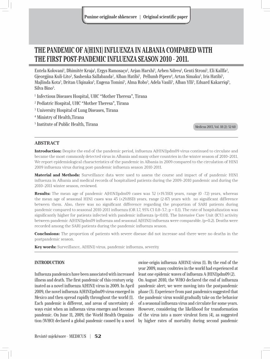
Revistë mjekësore - MEDICUS | 52
8
THE PANDEMIC OF A(H1N1) INFLUENZA IN ALBANIA COMPARED WITH THE FIRST POST-PANDEMIC INFLUENZA SEASON 2010 - 2011.Entela Kolovani1, Dhimitër Kraja1, Ergys Ramosaço1, Arjan Harxhi1, Arben Ndreu1, Genti Stroni1, Eli Kallfa2, Gjeorgjina Kuli-Lito2, Sashenka Sallabanda2, Alban Hatibi3, Pellumb Pipero4, Artan Simaku5, Iris Hatibi5, Majlinda Kota5, Dritan Ulqinaku5, Eugena Tomini5, Alma Robo5, Adela Vasili5, Alban Ylli5, Eduard Kakarriqi5, Silva Bino5.1 Infectious Diseases Hospital, UHC “Mother Theresa”, Tirana2 Pediatric Hospital, UHC “Mother Theresa”, Tirana3 University Hospital of Lung Diseases, Tirana4 Ministry of Health,Tirana5 Institute of Public Health, Tirana
ABSTRACT
Introduction: Despite the end of the pandemic period, influenza A(H1N1)pdm09 virus continued to circulate and became the most commonly detected virus in Albania and many other countries in the winter season of 2010–2011.We report epidemiological characteristics of the pandemic in Albania in 2009 compared to the circulation of H1N1 2009 influenza virus during post-pandemic influenza season 2010-2011.
Material and Methods: Surveillance data were used to assess the course and impact of of pandemic H1N1 influenza in Albania and medical records of hospitalized patients during the 2009–2010 pandemic and during the 2010–2011 winter season, reviewed.
Results: The mean age of pandemic A(H1N1)pdm09 cases was 32 (±19.3SD) years, range (0 -72) years, whereas the mean age of seasonal H1N1 cases was 43 (±29.8SD) years, range (2-87) years with no significant difference between them. Also, there was no significant difference regarding the proportion of SARI patients during pandemic compared to seasonal 2010-2011 influenza (OR 1.7, 95% CI 0.8–3.7; p = 0.1). The rate of hospitalization was significantly higher for patients infected with pandemic influenza (p<0.01). The Intensive Care Unit (ICU) activity between pandemic A(H1N1)pdm09 influenza and seasonal A(H1N1) influenza were comparable. (p=0.2). Deaths were recorded among the SARI patients during the pandemic influenza season.
Conclusions: The proportion of patients with severe disease did not increase and there were no deaths in the postpandemic season.
Key words: Surveillance, A(HIN1) virus, pandemic influenza, severity
INTRODUCTION
Influenza pandemics have been associated with increased illness and death. The first pandemic of this century orig-inated as a novel influenza A(H1N1) virus in 2009. In April 2009, the novel influenza A(H1N1)pdm09 virus emerged in Mexico and then spread rapidly throughout the world (1).Each pandemic is different, and areas of uncertainty al-ways exist when an influenza virus emerges and becomes pandemic. On June 11, 2009, the World Health Organiza-tion (WHO) declared a global pandemic caused by a novel
swine-origin influenza A(H1N1) virus (1). By the end of the year 2009, many coubtries in the world had experienced at least one epidemic waves of influenza A (H1N1)pdm09 (2). On August 2010, the WHO declared the end of influenza pandemic alert; we were moving into the postpandemic phase (3). Experience from past pandemics suggested that the pandemic virus would gradually take on the behavior of a seasonal influenza virus and circulate for some years. However, considering the likelihood for transformation of the virus into a more virulent form (4), as suggested by higher rates of mortality during second pandemic
Medicus 2013, Vol. 18 (2): 52-60
Punime origjinale shkencore | Original scientific paper
Punime origjinale shkencore | Original scientific paper
53 | Medical Journal - MEDICUS
waves in Copenhagen (1918), the United States (1957), and Eurasia (1968–1970) (4,6), the World Health Organization acknowledged the unpredictability of pandemic viruses; recommended continued surveillance; and issued advice and guidelines on surveillance of severe cases, vaccina-tion, and appropriate clinical management of cases dur-ing the postpandemic phase (3). Despite the end of the pandemic period, influenza A(H1N1)pdm09 (2009 H1N1) virus continued to circulate and became the most com-monly detected virus in many northern hemisphere coun-tries in the winter season of 2010–2011 (7-11). The impact of 2009 H1N1 in the 2010–2011 season in some countries was greater than in the previous year, most notably in the United Kingdom (UK) where intensive care units were stressed by large numbers of patients requiring ventilator support (6,7) raising the possibility at the time of a change in the virulence of the virus. A range of surveillance sys-tems were used to assess the course and impact of the first wave of pandemic H1N1 influenza in Albania during the fall and winter (12,13).Surveillance methods included laboratory confirmation of specimens collected from patients suspected with influenza. Participating GPs report the total number of consultations per week, of any patients presenting with ILI/ARI (14,15). The ILI/ARI rates were calculated as the number of ILI/ARI patients per 100,000 population.
METHODS
Influenza in Albania is subject to several Nationwide Sur-veillance Systems:
� ALERT Syndrome-Based Surveillance: ARI and Unexplained death
� It is an Early Warning Surveillance System GP and Emergency Room based throughout the country. It consists of weekly mandatory notification of ARI consultation
� Case-Based-Hospital-Based Flu Sentinel Surveillance System (ILI)
� Hospitals and Primary health Care services all over the country
� Sentinel Seasonal Influenza Surveillance (ILI) by the network of sentinel practitioners’ in Tirana
� Surveillance of SARI (Severe Acute Respiratory Infections) related to H1N1 influenza virus, to better monitor the severity of the pandemic, to detect changes in the population groups affected by severe outcomes following the infection (16,17).
DATA COLLECTION
A structured questionnaire, was distributed to the hos-pitals that were included in the surveillance, filled in by treating physician, and collected by the IPH study center. The structured questionnaire included patient informa-tion; data on the hospital stay; clinical signs and symp-toms of illness; clinical and laboratory diagnosis; specific treatments;underlying chronic medical conditions (chronic respira-tory diseases, immunodeficiency and neurodevelopmen-tal disorders; cardiac diseases, status of influenza vacci-nation; and disease complications. Answer options were provided, but also free text could additionally be specified by the respondents in regard with concurrent conditions or diagnosis.
� Virological surveillance was implemented to collect specimens and provide detailed and timely information on circulating influenza virus strains.
STATISTICAL ANALYSIS
Descriptive statistics comprised the calculation of mean and ranges for continuous variables. Comparative analy-ses were performed by means of t-test for continuous variables, χ2 and Fisher exact test for categorical vari-ables. Odds ratios (ORs) and 95% CIs were calculated. All reported p values were two-sided, significance level was set for α≤0.05. Statistical analyses were performed using Epi Info software.
Results
DATA FROM SURVEILLANCE OF ILI/ARI
Weekly ILI/ARI consultation rates in 2009 were compared with the rates observed in the same period in the previous 10 years (1999-2008) of influenza season: weeks 40-20. Start-ing from 13 of April (week 16) to 1st of November (week 44) the influenza activity remained low or fell below the national baseline levels according to percentile-based intensity levels generated from historical weekly ILI/ARI consultation. Unlike previous years’ pattern the rate of reported ILI/ARI increased sharply from 45th week, and peaked nation-ally at week 47, starting 16 of November and representing 30% increase compared to previous week, 46. This rate was the highest observed compared to the same period of past 10 years of influenza surveillance and exceeded the 95th percentile of expected rates, thus, suggesting the circulation of a novel virus.
Punime origjinale shkencore | Original scientific paper
Revistë mjekësore - MEDICUS | 54
Children aged 0-14 years had the highest incidence rates of ILI/ARI.On 2010 the increase of ILI/ARI rates followed the previ-ous years’ pattern starting by the end of December 2010 and reaching the peak on first week of February 2011. (Figure 1).
Figure 1. Comparison of ILI/ARI rates and epidemic threshhold of influenza seasons, weeks 40-20.
VIROLOGICAL SURVEILLANCE
Nasopharingeal swabs were collected from patients suspected with influenza from hospitals and primary health care facilities throughout the country.
� Confirmed cases were considered all cases that met the WHO case definition and were confirmed infected with pandemic influenza A(H1N1)pdm09 virus at laboratory of virology lab, IPH.
� Suspected cases were considered all cases that met the case definition.
During the first month of confirmation of pandemic H1N1 virus in Albania the majority of cases referred a travel history abroad. Early in November clusters of influenza cases were detected in schools, day care centers and within families and a remarkable increase of confirmed cases was noted, highlighting the intensive circulation of the pandemic virus as seen in epidemic curve.
Widespread influenza activity was reported during No-vember and December from all geographical areas of the country which denotes the circulation of the virus in the whole country. It is important to emphasize that the in-crease in numbers of confirmed A(H1N1)pdm09 cases cor-relates with the population density and reflects the in-tense levels of influenza activity occurring in main urban areas. (Figure 2)
Modelling of the pandemic wave and data from syndro-mic surveillance were used to estimate the community burden of pandemic influenza infections in Albania.
Figure 2. Reported cases of laboratory-confirmed A(H1N1)pdm09 influenza by date of onset and ILI/ARI rate, June 2009 – March 2010.
PERSONS AFFECTEDPersons of age group 15-24 years were most affected by the pandemic A(H1N1)pdm09 influenza, followed by age group 25-44 years. The mean age of cases was 26 years (±17.8SD). The first case of <1 year of age was recorded on 12 November as epidemic was heading towards its peak. During the influenza season 2010 - 2011 the most affected age group was 25 – 44 year old. The mean age of patients was 27 year (±19.7SD). Results did not indicate a significant statistical difference in regard with age between pandemic A(H1N1)pdm09 influenza season and post pandemic H1N1influenza season, t=-0.765 p=0.4. (Figure 3). From December 2010 though March 2011, 145 (22%) of sentinel specimens were confirmed with influenza.
The main viruses circulating were A(H1N1) virus in 102 (70%) of the 145 positive specimens, influenza B virus in 25 (17%) specimens and influenza A(H3N2) virus in 18 (12%) specimens.
Figure 3. Number of confirmed influenza H1N1 cases by age group; Season 2009 – 2010 and season 2010 - 2011
Punime origjinale shkencore | Original scientific paper
55 | Medical Journal - MEDICUS
The majority of laboratory-confirmed cases of influenza notified to IPH were the pandemic influenza A(H1N1) strain.
SURVEILLANCE OF HOSPITAL EMERGENCY ROOM AND ICU ACTIVITY
The hospitalization rates were reported every week during the influenza season providing also information on the number of people with influenza-related complication and outpatients consultation.
After a period of a stable trend from week 28 to week 44, the trend of hospitalizations and outpatients consultation for ILI/ARI went upward from week 46 in middle of November 2009 and reached a peak in week 50 of December. (Figure 5)
153 (32%) hospitalizations were recorded among confirmed patients.
Rates of hospital admission were highest in age group 25-44 yrs old. Children less than five years old, had higher hospitalization rates than 10-14-year-olds.
Compared with other age groups, adults aged 60 years and over had lower rates of hospital admission.
12 (80%) hospitalizations took place on January and 3 (20%) on February.
The rate of hospitalization was significantly higher for patients infected with pandemic influenza (χ2 = 11.2. p<0.01). But results did not indicate a significant difference regarding ICU activity between pandemic A(H1N1)pdm09 influenza and seasonal A(H1N1) influenza. (χ2 = 1.5. p=0.2).
Figure 5. Patients with confirmed pandemic A(H1N1)pdm09 influenza who were admitted to hospital or ICU by week of hospitalization (n= 153)
* A severe case was defined as a patient with laboratory-con-firmed pandemic H1N1 influenza who was
* ICU admissions and deaths.
HOSPITAL SURVEILLANCE OF SEVERE ACUTE RESPIRATORY INFECTION (SARI)
Of a total of 3122 patients suspected for pandemic influenza through March 2010, 99 or (3%) of them were considered severe cases. 62 (63%) of severe cases were laboratory-confirmed with pandemic H1N1 influenza. Of confirmed cases, 53 (85%) of them were reported from the beginning of December 2009. 12 (19%) of confirmed SARI patients belong to age group 0-14 years old. Of children, two (17%) were aged less than 12 months. Since week 32 in August, 14 (24%) deaths were recorded among severe cases infected with pandemic A(H1N1)pdm09 influenza. Two of the patients were pregnant women, while the rest presented with underlying condition. Of deceased patients nine (64%) were adults aged 45 years and older. The number of deaths peaked in 45-59-year-olds. (Figure 6). Results did not indicate a significant statistical difference regarding the proportion of SARI patients during pandemic compared to seasonal 2010-2011 influenza (OR 1.7, 95% CI 0.8–3.7; p = 0.1).
Of a total of 660 patients suspected for influenza from December 2010 through March 2011, 44 or (7%) of them were considered severe cases. 8 (18%) of severe cases were laboratory-confirmed with H1N1 influenza. The first confirmed case was reported on second week of January 2011 to reach the peak on the first week of February with 5 (60%) of the cases. 2 (5%) of SARI cases were infected with influenza B viruses and 2 (5%) with influenza AH3 viruses.
The mean age of pandemic A(H1N1)pdm09 cases was 32 (±19.3SD) years, range (0 -72) years, whereas the mean age of seasonal H1N1 cases was 43 (±29.8SD) years, range (2-87) years. There is no significant statistical difference in re-gard with age of SARI patients between pandemic A(H1N1)pdm09 influenza season and post pandemic A(H1N1) in-fluenza season, t=1.45 p=0.14. (Figure 3)
Figure 6. Reported severe and fatal cases, June 2009 – March 2010
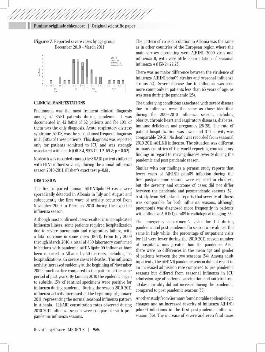
Punime origjinale shkencore | Original scientific paper
Revistë mjekësore - MEDICUS | 56
Figure 7. Reported severe cases by age group, December 2010 – March 2011
CLINICAL MANIFESTATIONS
Pneumonia was the most frequent clinical diagnosis among 62 SARI patients during pandemic. It was documented in 42 (68%) of 62 patients and for 10% of them was the only diagnosis. Acute respiratory distress syndrome (ARDS) was the second most frequent diagnosis in 31 (50%) of these patients. This diagnosis was reported only for patients admitted to ICU and was strongly associated with death (OR 8.4, 95% CI, 1.2–69.2; p = 0.02).
No death was recorded among the 8 SARI patients infected with H1N1 influenza virus, during the annual influenza season 2010-2011, (Fisher’s exact test p=0.6) .
DISCUSSION
The first imported human A(H1N1)pdm09 cases were sporadically detected in Albania in July and August and subsequently the first wave of activity occurred from November 2009 to February 2010 during the expected influenza season.
Although most confirmed cases resulted in uncomplicated influenza illness, some patients required hospitalization due to severe pneumonia and respiratory failure, with a fatal outcome in some cases (18-21). From July 2009 through March 2010 a total of 480 laboratory confirmed infections with pandemic A(H1N1)pdm09 influenza have been reported in Albania by 30 districts, including 153 hospitalizations, 62 severe cases 14 deaths. The influenza activity increased suddenly at the beginning of November 2009, much earlier compared to the pattern of the same period of past years. By January 2010 the epidemic began to subside. 15% of sentinel specimens were positive for influenza during pandemic. During the season 2010-2011 influenza activity increased at the beginning of January 2011, representing the normal seasonal influenza pattern in Albania. ILI/ARI consultation rates observed during 2010-2011 influenza season were comparable with pre-pandemic influenza seasons.
The pattern of virus circulation in Albania was the same as in other countries of the European region where the main viruses circulating were A(H1N1) 2009 virus and influenza B, with very little co-circulation of seasonal influenza A (H3N2) (22,23).
There was no major difference between the virulence of influenza A(H1N1)pdm09 strains and seasonal influenza strains (24). Severe disease due to influenza was seen more commonly in patients less than 65 years of age, as was seen during the pandemic (25).
The underlying conditions associated with severe disease due to influenza were the same as those identified during the 2009-2010 influenza season, including obesity, chronic heart and respiratory diseases, diabetes, immune deficiency and pregnancy (26-28). The rate of patient hospitalization was lower and ICU activity was comparable (29-31). No death was recorded from seasonal 2010-2011 A(H1N1) influenza. The situation was different in many countries of the world reporting contradictory findings in regard to varying disease severity during the pandemic and post pandemic season.
Similar with our findings a german study reports that fewer cases of A(H1N1) pdm09 infection during the first postpandemic season, were reported in children, but the severity and outcome of cases did not differ between the pandemic and postpandemic seasons (32). A study from Netherlands reports that severity of illness was comparable for both influenza seasons, although pneumonia was diagnosed more frequently in patients with influenza A(H1N1)pdm09 in radiological imaging (33).
The emergency department’s visits for ILI during pandemic and post pandemic flu season were almost the same in Italy while the percentage of outpatient visits for ILI were lower during the 2010-2011 season number of hospitalizations greater than the pandemic. Also, there were no differences in the mean age and gender of patients between the two seaseons (34). Among adult inpatients, the A(H1N1) pandemic season did not result in an increased admission rate compared to pre pandemic seasons but differed from seasonal influenza in ICU admission, age of patients, vaccination and antiviral use. 30-day mortality did not increase during the pandemic, compared to post pandemic seasons (35).
Another study from Germany found notable epidemiologic changes and an increased severity of influenza A(H1N1)pdm09 infections in the first postpandemic influenza season (36). The increase of severe and even fatal cases
Punime origjinale shkencore | Original scientific paper
57 | Medical Journal - MEDICUS
in hospitalized patients with influenza A(H1N1) pdm09 infection in the subsequent season of the pandemic has also been reported in Spain and England. Severe clinical courses have been described for patients with underlying conditions, pregnant women, and obese patients (37, 38).
A shift to older ages in the age distribution of hospitalized and fatal patients were observed during the winter season of 2010–2011, which was consistent with data from the United Kingdom, Greece and Taiwan (39-42). Mexico did not report any deaths caused by influenza AH1N1 2009 virus and reported a low morbidity, just confirming the presence of influenza AH1N1 2009 virus in circulation (43). An Indian study did not observe any differences in disease severity between the first and second pandemic outbreaks in 2009 (44),another U.S study showed a remarkable increase in hospitalization and in number of deaths in the second wave (45). In Greece (41) and New-Zealand (46), according to the respective national surveillance systems in intensive care unit settings, case-fatality ratios among all age groups were also similar for the two seasons. In Croatia, the mortality in the 2010/2011 season season until March 14, 2011 was similar to that in 2009/2010. Greece reported a higher mortality rate than in Croatia in 2009/2010 and also in 2010/2011 (47). The total number of laboratory-confirmed deaths in the Northern hemisphere was only 19.3% of the previous season. Overall, in the world, the laboratory-confirmed influenza mortality reached 22.2% of that in the previous season. In Europe, all countries recorded lower laboratory-confirmed mortality in in the 2010/2011 season.
The percentage of laboratory confirmed deaths was 57.1% of that for the 2009/10 season (47). In the 2009/2010 season, South Eastern European (SEE) countries and Southern hemisphere countries had similar influenza AH1N1 2009 mortality rates. In the 2010/2011 season, the total mortality in SEE countries and Europe compared with the 2009/2010 season was significantly lower, while the expected impact of type specific vaccines against influenza AH1N1 2009 was not achieved (47, 48-56).
CONCLUSIONS
The proportion of patients with severe disease did not increase and there were no deaths in the postpandemic season. Since seasonal and pandemic influenza viruses undergo constant antigenic drift and may change in virulence, WHO recommends that countries maintain
pandemic monitoring systems to detect changes in severity or characteristics of disease and therefore to allow for appropriate targeting of prevention and control and treatment of influenza.
REFERENCES
1. World Health Organization (2010) World now at the start of 2009 influenza pandemic.Available: http://www.who.int/mediacentre/news/statements/2009/h1n1_pandemic_phase6_20090611/en/index.html.Accessed 2010 Mar 27.
2. World Health Organization (2010) Evolution of a pandemic: A(H1N1) 2009, April 2009–March 2010. Geneva: World Health Organization.
3. World Health Organization. H1N1 in post-pandemic period [cited 2012 May 2]. http://www.who.int/mediacentre/news/statements/ 2010/h1n1_vpc_20100810/en/index.html
4. Morens DM, Taubenberger JK, Harvey HA, Memoli MJ. The 1918 infl uenza pandemic: lessons for 2009 and the future. Crit Care Med. 2010;38(Suppl):e10–20. http://dx.doi.org/10.1097/ CCM.0b013e3181ceb25b
5. Miller MA, Viboud C, Balinska M, Simonsen L. The signature features of influenza pandemics–implications for policy. N Engl J Med. 2009;360:2595–8. http://dx.doi.org/10.1056/NEJMp0903906
6. Andreasen V, Viboud C, Simonsen L. Epidemiologic characterization of the 1918 infl uenza pandemic summer wave in Copenhagen: implications for pandemic control strategies. J Infect Dis. 2008;197:270–8. http://dx.doi.org/10.1086/524065
7. EuroFlu Weekly Electronic Bulletin-Week 2 (2011) Available: http://www.euroflu.org/cgi-files/bulletin_v2.cgi.Accessed 2011 Nov 22.
8. Infectious Disease Surveillance Center (2011) Infectious Agents Surveillance Report- The Topic of This Month- 2010/11 influenza season, Japan. Available:http://idsc.nih.go.jp/iasr/32/381/tpc381.html. Accessed 2011 Nov 21.
9. Hong Kong Center for Health Protection (2011) Flu Express-Volume 8, Number 5. Available: http://www.chp.gov.hk/files/pdf/flu_express_week5_2_2_2011.pdf. Accessed 2011 Nov 21.
10. World Health Organization (2011) Review of the 2010–2011 winter influenza season, northern hemisphere. Weekly Epidemiological Record 86: 221–232.
Punime origjinale shkencore | Original scientific paper
Revistë mjekësore - MEDICUS | 58
11. Health Protection Agency (2011) Weekly National Influenza Report- Week 4. Available: http://www.hpa.org.uk/webc/HPAwebFile/HPAweb_C/1294740643915. Accessed 2011 Nov 21.
12. Josseran L, Nicolau J, Caillere N, Astagneau P, Brucker G. Syndromic surveillance based on emergency department activity and crude mortality: two examples. Euro Surveill. 2006;11(12):225-9.
13. D’Ortenzio E, Do C, Renault P, Weber F, Filleul L. Enhanced influenza surveillance on Réunion Island (southern hemisphere) in the context of the emergence of influenza A(H1N1)v. Euro Surveill. 2009; 14(23):19239.
14. Muscatello DJ, Morton PM, Evans I, Gilmour R. Prospective surveillance of excess mortality due to influenza in New South Wales: feasibility and statistical approach. Commun Dis Intell. 2008;32(4):435-42.
15. Centers for Disease Control and Prevention (CDC). Swine influenza A (H1N1) infection in two children--Southern California, March-April 2009. MMWR Morb Mortal Wkly Rep. 2009; 58(15):400-2.
16. Muscatello DJ, Morton PM, Evans I, Gilmour R. Prospective surveillance of excess mortality due to influenza in New South Wales: feasibility and statistical approach. Commun Dis Intell. 2008;32(4):435-42.
17. Centers for Disease Control and Prevention (CDC). Swine influenza A (H1N1) infection in two children--Southern California, March-April 2009. MMWR Morb Mortal Wkly Rep. 2009; 58(15):400-2.
18. Arben Ndreu, Dhimitër Kraja, Silva Bino, et al., “Clinical Findings in Albanian Patients with 2009 Influenza AH1N1 Admitted to the Intensive Care Unit,” ISRN Emergency Medicine, vol. 2013, Article ID 914840, 6 pages, 2013. doi:10.1155/2013/914840
19. Ergys Ramosaço, Dhimiter Kraja, Pellumb Pipero, Entela Kolovani, Arben Ndreu, Iris Hatibi, Silva Bino. Evaluation of the Impact of Influenza A(H1n1) Infection on Pregnancy Outcome in Albania. Medicus 2012, Vol. 17 (2): 33-36
20. Vladimir Petrović, Zorica Šeguljev, Gorana Ćosić, Mioljub Ristić, Jasminka Nedeljković, Nataša Dragnić, Snežana Ukropina. Overview of the winter wave of 2009 pandemic influenza A(H1N1)v in Vojvodina, Serbia. Croat Med J. 2011; 52: 141-50
21. Perez-Padilla R, De la Rosa-Zamboni D, Ponce de Leon S, Hernandez M, Quiñones-Falconi F, Bautista E, Ramirez-Venegas A, Rojas-Serrano J, Ormsby CE, Corrales A, Higuera A, Mondragon E, Cordova-Villalobos
JA: Pneumonia and respiratory failure from swine-origin influenza A (H1N1) in Mexico. N Engl J Med 2009, 361:680-689.
22. Chinese National Influenza Center (2011) Chinese Influenza Weekly Report (Week 7: Feb 14–20, 2011). Available: http://www.cnic.org.cn/eng/show.
23. WHO. Global influenza virological surveillance. Available from: http://www.who.int/csr/disease/influenza/influenzanetwork/en/ index.html. Accessed: April 10, 2011.
24. York I, Donis RO. The 2009 pandemic influenza virus: where did it come from, where is it now, and where is it going? Curr Top Microbiol Immunol. 2012; 26.
25. Influenza A(H1N1)v investigation teams. Modified surveillance of influenza A(H1N1)v virus infections in France. Euro Surveill. 2009; 14(29):pii=19276.
26. Kelly H, Grant K. Interim analysis of pandemic influenza (H1N1) 2009 in Australia: surveillance trends, age of infection and effectiveness of seasonal vaccination. Euro Surveill. 2009; 14(31):pii=19288.
27. Centers for Disease Control and Prevention (CDC). Surveillance for the 2009 pandemic influenza A (H1N1) virus and seasonal influenza viruses – New Zealand, 2009. MMWR Morb Mortal Wkly Rep. 2009; 58(33):918-21.
28. Cire Réunion-Mayotte. Cas humains de grippe à nouveau virus A(H1N1) -Situation au 06 juillet 2009 à 11h00. [Human cases of influenza due to the new A(H1N1) virus – situation on 6 July, 11 am]. [French]. Point epidémiologique no. 49; 6 July 2009.
29. Centers for Disease Control and Prevention. Outbreak of swine-origin influenza A (H1N1) virus infection - Mexico, March-April 2009. MMWR Morb Mortal Wkly Rep. 2009;58(17):467-70.
30. Baker MG, Wilson N, Huang QS, Paine S, Lopez L, Bandaranayake D, et al. Pandemic influenza A(H1N1)v in New Zealand: the experience from April to August 2009. Euro Surveill. 2009; 14(34):pi=19319.
31. World Health Organization. Pandemic (H1N1) 2009 - update 62 (revised 21 August 2009). 2009 [accessed 29 September 2009].
32. Mathias Altmann, Lena Fiebig, Silke Buda, Rüdiger von Kries, Manuel Dehnert, and Walter Haas. Unchanged Severity of Infl uenza A(H1N1)pdm09 Infection in Children during First Postpandemic SeasonEmerging Infectious Diseases • www.cdc.gov/eid • Vol. 18, No. 11, November 2012
Punime origjinale shkencore | Original scientific paper
59 | Medical Journal - MEDICUS
33. Rahamat-Langendoen, J Tutuhatunewa, E.D. Schölvinck, E.H. Hak, E. Koopmans, M.P.G. * Niesters, H.G.M. * Riezebos-Brilman, A. Influenza in the immediate post-pandemic era: A comparison with seasonal and pandemic influenza in hospitalized patients. Erasmus MC: University Medical Center Rotterdam Virology Journal of Clinical Virology 54/2, p135-140 (2012)
34. Elena Di Tondo, Anna Marigliano, Ilaria Pellegrini, Francesco Di Stanislao, Marcello M. D’Errico. Trend of emergency department presentations for influenza like illness: Differences between pandemic (2009-2010) and post-pandemic (2010-2011) season. Vol.4, No.12, 1256-1262 (2012) Health doi:10.4236/health.2012.412185.
35. R. Mitchell, G. Taylor*, B Amihod, K. Suh, A. McGeer, A. Wong, C. Frenette, K. Katz, D. Gravel on behalf of the Canadian Nosocomial Infection Surveillance Program O612 Abstract (oral session) Understanding the burden of influenza infection among adults in Canadian hospitals: a comparison of the 2009/10 pandemic season to the pre-pandemic and post-pandemic seasons. www.escmid.org/escmid_library/online
36. Nicola Lehners, Steffen Geis, Christoph Eisenbach, Kai Neben, Paul Schnitzler. Changes in Severity of Influenza A(H1N1)pdm09 Infection from Pandemic to First Postpandemic Season, Germany. Emerging Infectious Diseases • www.cdc.gov/eid • Vol. 19, No. 5, May 2013
37. Viasus D, Cordero E, Rodriguez-Bano J, Oteo JA, Fernandez- Navarro A, Ortega L, et al. Changes in epidemiology, clinical features and severity of influenza A (H1N1) 2009 pneumonia in the first post-pandemic influenza season. Clin Microbiol Infect. 2012;18:E55– 62.
38. Mytton OT, Rutter PD, Donaldson LJ. Influenza A(H1N1)pdm09 in England, 2009 to 2011: a greater burden of severe illness in the year after the pandemic than in the pandemic year. Euro Surveill. 2012;17(14):pii=20139. Available online: http://www.eurosurveillance.org/ViewArticle.aspx?ArticleId=20139
39. Cuiling Xu1, A. Danielle Iuliano2, Min Chen1, Po-Yung Cheng2, Tao Chen1, Jinghong Shi1, Jing Yang1, Lijie Wang1, Fan Yuan1, Marc-Alain Widdowson2, Yuelong Shu1. Characteristics of Hospitalized Cases with Influenza A (H1N1)pdm09 Infection during First Winter Season of Post-Pandemic in China. PLOS ONE. February 2013 | Volume 8 | Issue 2 | e55016
40. Bolotin S, Pebody R, White PJ, McMenamin J, Perera L, et al. (2012) A New Sentinel Surveillance System for Severe Influenza in England Shows a Shift in Age Distribution
of Hospitalised Cases in the Post-Pandemic Period. PLoS ONE 7: e30279. doi:10.1371/journal.pone.0030279.
41. Athanasiou M, Baka A, Andreopoulou A, Spala G, Karageorgou K, et al. (2011) Influenza surveillance during the post-pandemic influenza 2010/11 season in Greece, 04 October 2010 to 22 May 2011. Euro Surveill. 16:pii = 20004.Ava i l a b l e : http://www. eurosurveillance. org/ 2012 Feb 10.
42. Chuang J-H, Huang AS, Huang W-T, Liu M-T, Chou J-H, et al. (2012) Nationwide Surveillance of Influenza during the Pandemic (2009–10) and Post- Pandemic (2010–11) Periods in Taiwan. PLoS ONE 7: e36120. doi:10.1371/ journal.pone.0036120.
43. Estados Unidos Mexicanos, Estadisticas de la Epidemia Influenza A (H1N1). Available from: http://portal.salud.gob.mx/contenidos/ noticias/influenza/estadisticas.html. Accessed: April 10, 2011.
44. Ramakrishna K, Peter JV, Karthik G, Abraham AM, Surekha V, Karthik R, et al. Influenza A (H1N1) 2009 pandemic: was there a difference in the two waves in patients requiring admission to the intensive-care unit? Clin Microbiol Infect. 2011;17:1355–8.
45. Truelove SA, Chitnis AS, Heffernan RT, Karon AE, Haupt TE, Davis JP. Comparison of patients hospitalized with pandemic 2009 influenza A (H1N1) virus infection during the first two pandemic waves in Wisconsin. J Infect Dis. 2011;203:828–37. http://dx.doi.org/10.1093/infdis/jiq117
46. Bandaranayake D, Jacobs M, Baker M, Hunt D, Wood T, Bissielo A, et al. The second wave of 2009 pandemic infl uenza A(H1N1) in New Zealand, January-October 2010. Euro Surveill. 2011;16:19788
47. Mladen Smoljanović, Ankica Smoljanović, Marijana Mlikotić. Predicting AH1N1 2009 influenza epidemic in Southeast Europe. Croat Med J. 2011 April; 52(2): 115–125
48. Institute for Public Health FB&H. Available from.: http://www.zzjzfbih.ba/tag/gripa/.Accessed: April 10, 2011.
49. Croatian Institute of Public Health. Pandemic influenza caused by new virus A/H1N1. Available from: http://www.hzjz.hr/epidemiologija/svinjska_gripa.htmAccessed: April 10, 2011.
50. National Institute of Public Health of Kosova. Available from: http://www.niph-kosova.org/index. Accessed: April 10, 2011.
51. Hellenic Center for Disease Control and Prevention, Department of Epidemiological Surveillance. Athens: KEELPNO. Weekly epidemiological report. Available from: http://www.keelpno.grAccessed: April 10, 2011.
Punime origjinale shkencore | Original scientific paper
Revistë mjekësore - MEDICUS | 60
52. Institute for Public Health of the Republic of Macedonia. Available from: http://www.iph.mk/index.php?lang=en.Accessed: April 10, 2011.
53. Crna Gora Instiute of Public Health. Available from: http://www.ijzcg.me/.Accessed: April 10, 2011.
54. Serbian Instiute of Public Health. “Dr Milan Jovanović Batut.” Pandemic influenza A (H1N1). Available from:
http://www.batut.org.rs/ah1n1.htmlAccessed: April 10, 2011.
55. European Centre for Disease Prevention and Control (ECDC). Available from: http://www.ecdc.europa.eu/en/Pages/home.aspxAccessed: April 10, 2011.
56. Institute of Public Health, Albania. http://www.ishp.gov.al. Accessed: April 10, 2011.
PANDEMIA E GRIPIT A(H1N1) NË SHQIPËRI KRAHASUAR ME SEZONIN E PARË PAS PANDEMISË, 2010-2011.
Entela Kolovani1, Dhimitër Kraja1, Ergys Ramosaço1, Arjan Harxhi1, Arben Ndreu1, Genti Stroni1, Eli Kallfa2, Gjeorgjina Kuli-Lito2, Sashenka Sallabanda2, Alban Hatibi3, Pellumb Pipero4, Artan Simaku5, Iris Hatibi5, Majlinda Kota5, Dritan Ulqinaku5, Eugena Tomini5, Alma Robo5, Adela Vasili5, Alban Ylli5, Eduard Kakarriqi5, Silva Bino5.1 Spitali i Sëmundjeve Infektive, QSU “Nënë Tereza”, Tiranë2 Spitali Pediatrik, QSU “Nënë Tereza”, Tiranë
3 Spitali Universitar i Sëmundjeve të Mushkrive, Tiranë4 Ministria e Shëndetësisë, Tiranë5 Instituti i Shëndetit Publik, Tiranë
ABSTRAKTI
Hyrje: Pavarësisht përfundimit të periudhës pandemike, virusi A(H1N1)pdm09 i gripit vazhdoi të qarkullojë me shpesh se viruset e tjetë në Shqipëri dhe në shumë vende të tjera në stinën e dimrit 2010-2011. Ne studim raportohen karakteristikat epidemiologjike të pandemisë në Shqipëri në vitin 2009 në krahasim me qarkullimin e virusit A(H1N1) 2009 të gripit gjatë stinës post-pandemike të gripit 2010-2011.
Materiali dhe Metodat: Janë përdorur të dhënat e Survejances për të vlerësuar ecurinë dhe ndikimin e gripit pandemik nga virusi A(H1N1)pdm09 në Shqipëri dhe janë shqyrtuar të dhënat mjekësore të pacientëve të hospitalizuar gjatë sezonit të pandemisë 2009-2010 dhe gjatë sezonit të dimrit 2010-2011.
Rezultatet: Mosha mesatare e pacienteve gjate pandemisë ishte 32 (±19.3) vjeç, rangu (0-72) vjeç, ndërsa mosha mesatare e pacientëve gjate sezonit post-pandemik ishte 43 (±29.8) vjeç, rangu (2-87) vjet, pa ndryshim të rëndësishëm midis tyre. Gjithashtu, nuk kishte ndryshim të rëndësishëm në lidhje me përqindjen e pacientëve me semundje të rëndë respiratore gjatë pandemisë krahasim me sezonin gripal në 2010-2011 (OR= 1.7, 95% CI 0,8-3,7; p=0.1). Niveli i hospitalizimit ishte më i lartë për pacientët gjatë pandemise (p<0.01). Aktiviteti në njësinë e kujdesit intensiv gjatë pandemisë dhe periudhës post-pandemike ishte i krahasueshëm (p = 0.2). Gjatë pandemisë nga virusi A(H1N1)pdm09 janë regjistruar vdekjet midis pacientëve me semundje të rëndë respiratore.
Konkluzione: Në sezonin post-pandemik 2010-2011 nuk është rritur përqindja e pacientëve me sëmundje të rëndë respiratore dhe nuk kanë ndodhur vdekje.
Fjalët kyçe: Survejanë, A (HIN1) virus, gripi pandemik, ashpërsia
61 | Medical Journal - MEDICUS
PROKALCITONINA SI TEST DIAGNOSTIK NE PACIENTET ME SEPSISEdmond Puca¹, Pellumb Pipero¹, Arben Pilaca¹, Ilir Akshija², Entela Y Puca³, Dhimiter Kraja¹1 Shërbimi i Sëmundjeve Infektive, Qendra Spitalore Universitare “Nënë Tereza” Tiranë. 2 Shërbimi i Statistikes, Qendra Spitalore Universitare “Nënë Tereza” Tiranë.3 Spitali Amerikan, Tirane.
ABSTRAKTI
Rezyme: Paraqitja e pacientëve me sepsis apo me klinikë të ngjashme me sepsisin në shërbimin e urgjencave tashmë është e zakonshme. Diferencimi i një rasti me temperaturë si pasojë e një shkaktari me natyrë infeksioze apo nga ndonjë patologji tjetër jo infektive, është tepër e vështirë për mjekun klinicist dhe madje kërkon edhe kohë. Në një situatë të tillë lind nevoja e testimit të markuesve të rinj të sepsisit për një menaxhim sa me të shpejtë dhe adekuat të pacientit.
Objektivat: Qëllimi i këtij studimi, është të vlërësojë rolin e prokalcitoninës (PCT), si një markues ndihmues për mjekun klinicist në shërbimin e urgjencës, për diagnostikimin dhe diferencimin sa më të shpejtë të infeksioneve bakterialë sistemike.
Materialet dhe metodat: Studimi përfshinte 92 pacientë të paraqitur në urgjencën e Shërbimit të Sëmundjeve Infektive, Qendra Spitalore Universitare, Tiranë. Vlerësimi i PCT është bërë me metodën Elycsys BRAHMS PCT test Electro Chemiluminescent Immunoassay (ECLIA).
Rezultatet: Nga 92 pacientë që u morën në studim 55.4% (51) ishin meshkuj dhe 44.5% (41) femra, mosha varionte nga 14- 89 vjec. Duke u bazuar në të dhenat klinike, parametrat vital dhe ekzaminimet laboratorike, ne kishim 34.7% (32) pacientë me sepsis dhe 65.2% (60) pacientë me sindromin e përgjigjes inflamatore sistemike (SIRS). Koncentrime të larta të PCT u vunë re në pacientët me sepsis dhe sidomos në ata me disfunksion multiorganor. Nuk u vunë re diferenca përsa i përket numrit të leukociteve, ndërsa diferenca e koncentrimit të PCT në të dy grupet ishte sinjifikative. Konkluzione: Në pacientët me sepsis u vunë re vlera shumë të larta të PCT. Sidoqoftë, ne akoma nuk mund të japim një opinjon përfundimtar për vrerësimin adekuat të PCT, pamvarësisht se korrelacionet e saj me infeksionin janë mjaft domethënëse.
Fjalë kyce: sepsis, SIRS, prokalcitonina.
HYRJE
Numri i pacientëve që paraqiten për vizitë në shërbimin e urgjencës të sëmundjeve infektive është mjaft variabël, kjo për shkak të shume faktorëve, por një pjesë e mirë e pacientëve si ankesë kryesore kanë temperaturën e lartë dhe në mjaft raste pothuajse të pa menaxhueshme nga ana e tyre. Tradicionalisht temperatura ka qenë shoqëruar me një infeksion bakterial. Sidoqoftë, në mëse 50% të pacientëve febrilë, temperatura mund të jetë si pasojë e patologjivë të tjera joinfektive me natyrë autoimmune apo dhe malinje (1).
Identifikimi nga ana e mjekut të shërbimit të urgjencës nëse kemi të bëjmë me një infeksion viral, bakterial apo
mykotik që në momentin e paraqitjes së pacientit është mjaft e vështirë (1,2). Madje tepër e vështirë është që të diferencojmë nëse kemi të bëjmë me një sëmundje infektive apo ndonjë patologji tjetër të ngjashme me të në aspektin klinik. Kohët e fundit PCT është shtuar në përcaktimin e diagnosës së pacientëve febrilë. Nivelet e PCT, një prohormon i kalcitoninës normalisht i prodhuar nga gjëndra tiroide në kushte fiziologjike, rriten shumë shpejt gjatë infeksioneve bakteriale, ndërsa nivele të ulta janë depistuar gjatë infeksioneve virale ose gjendjeve febrile jo infektive (1). Në këto kushte një grup mjekësh të këtij shërbimi kemi filluar të testojmë këtë markues të ri të inflamacionit sic është prokalcitonina.
Testet rutinë laboratorikë kanë një sensitivitet dhe specificitet të ulur për të diagnostikuar se cili pacient
Medicus 2013, Vol. 18 (2): 61-66
Punime origjinale shkencore | Original scientific paper
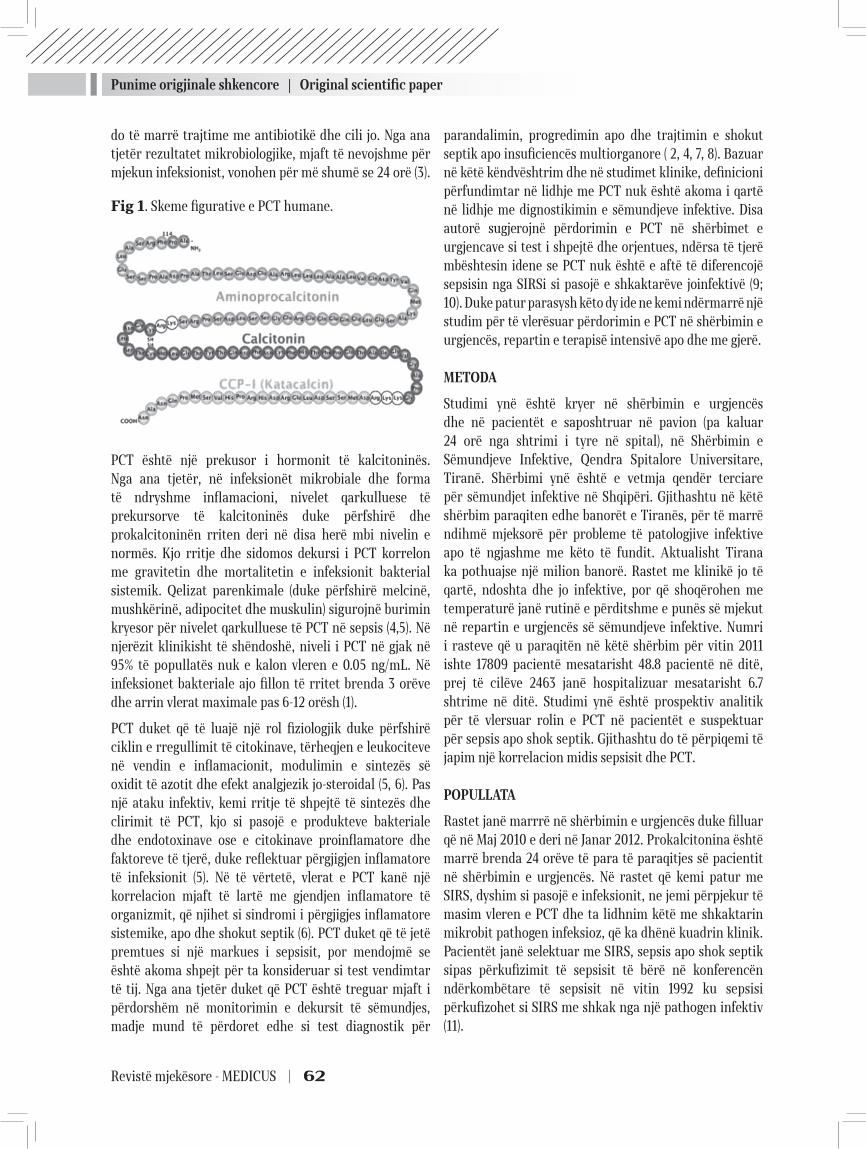
Punime origjinale shkencore | Original scientific paper
Revistë mjekësore - MEDICUS | 62
do të marrë trajtime me antibiotikë dhe cili jo. Nga ana tjetër rezultatet mikrobiologjike, mjaft të nevojshme për mjekun infeksionist, vonohen për më shumë se 24 orë (3).
Fig 1. Skeme figurative e PCT humane.
PCT është një prekusor i hormonit të kalcitoninës. Nga ana tjetër, në infeksionët mikrobiale dhe forma të ndryshme inflamacioni, nivelet qarkulluese të prekursorve të kalcitoninës duke përfshirë dhe prokalcitoninën rriten deri në disa herë mbi nivelin e normës. Kjo rritje dhe sidomos dekursi i PCT korrelon me gravitetin dhe mortalitetin e infeksionit bakterial sistemik. Qelizat parenkimale (duke përfshirë melcinë, mushkërinë, adipocitet dhe muskulin) sigurojnë burimin kryesor për nivelet qarkulluese të PCT në sepsis (4,5). Në njerëzit klinikisht të shëndoshë, niveli i PCT në gjak në 95% të popullatës nuk e kalon vleren e 0.05 ng/mL. Në infeksionet bakteriale ajo fillon të rritet brenda 3 orëve dhe arrin vlerat maximale pas 6-12 orësh (1).
PCT duket që të luajë një rol fiziologjik duke përfshirë ciklin e rregullimit të citokinave, tërheqjen e leukociteve në vendin e inflamacionit, modulimin e sintezës së oxidit të azotit dhe efekt analgjezik jo-steroidal (5, 6). Pas një ataku infektiv, kemi rritje të shpejtë të sintezës dhe clirimit të PCT, kjo si pasojë e produkteve bakteriale dhe endotoxinave ose e citokinave proinflamatore dhe faktoreve të tjerë, duke reflektuar përgjigjen inflamatore të infeksionit (5). Në të vërtetë, vlerat e PCT kanë një korrelacion mjaft të lartë me gjendjen inflamatore të organizmit, që njihet si sindromi i përgjigjes inflamatore sistemike, apo dhe shokut septik (6). PCT duket që të jetë premtues si një markues i sepsisit, por mendojmë se është akoma shpejt për ta konsideruar si test vendimtar të tij. Nga ana tjetër duket që PCT është treguar mjaft i përdorshëm në monitorimin e dekursit të sëmundjes, madje mund të përdoret edhe si test diagnostik për
parandalimin, progredimin apo dhe trajtimin e shokut septik apo insuficiencës multiorganore ( 2, 4, 7, 8). Bazuar në këtë këndvështrim dhe në studimet klinike, definicioni përfundimtar në lidhje me PCT nuk është akoma i qartë në lidhje me dignostikimin e sëmundjeve infektive. Disa autorë sugjerojnë përdorimin e PCT në shërbimet e urgjencave si test i shpejtë dhe orjentues, ndërsa të tjerë mbështesin idene se PCT nuk është e aftë të diferencojë sepsisin nga SIRSi si pasojë e shkaktarëve joinfektivë (9; 10). Duke patur parasysh këto dy ide ne kemi ndërmarrë një studim për të vlerësuar përdorimin e PCT në shërbimin e urgjencës, repartin e terapisë intensivë apo dhe me gjerë.
METODA
Studimi ynë është kryer në shërbimin e urgjencës dhe në pacientët e saposhtruar në pavion (pa kaluar 24 orë nga shtrimi i tyre në spital), në Shërbimin e Sëmundjeve Infektive, Qendra Spitalore Universitare, Tiranë. Shërbimi ynë është e vetmja qendër terciare për sëmundjet infektive në Shqipëri. Gjithashtu në këtë shërbim paraqiten edhe banorët e Tiranës, për të marrë ndihmë mjeksorë për probleme të patologjive infektive apo të ngjashme me këto të fundit. Aktualisht Tirana ka pothuajse një milion banorë. Rastet me klinikë jo të qartë, ndoshta dhe jo infektive, por që shoqërohen me temperaturë janë rutinë e përditshme e punës së mjekut në repartin e urgjencës së sëmundjeve infektive. Numri i rasteve që u paraqitën në këtë shërbim për vitin 2011 ishte 17809 pacientë mesatarisht 48.8 pacientë në ditë, prej të cilëve 2463 janë hospitalizuar mesatarisht 6.7 shtrime në ditë. Studimi ynë është prospektiv analitik për të vlersuar rolin e PCT në pacientët e suspektuar për sepsis apo shok septik. Gjithashtu do të përpiqemi të japim një korrelacion midis sepsisit dhe PCT.
POPULLATA
Rastet janë marrrë në shërbimin e urgjencës duke filluar që në Maj 2010 e deri në Janar 2012. Prokalcitonina është marrë brenda 24 orëve të para të paraqitjes së pacientit në shërbimin e urgjencës. Në rastet që kemi patur me SIRS, dyshim si pasojë e infeksionit, ne jemi përpjekur të masim vleren e PCT dhe ta lidhnim këtë me shkaktarin mikrobit pathogen infeksioz, që ka dhënë kuadrin klinik. Pacientët janë selektuar me SIRS, sepsis apo shok septik sipas përkufizimit të sepsisit të bërë në konferencën ndërkombëtare të sepsisit në vitin 1992 ku sepsisi përkufizohet si SIRS me shkak nga një pathogen infektiv (11).
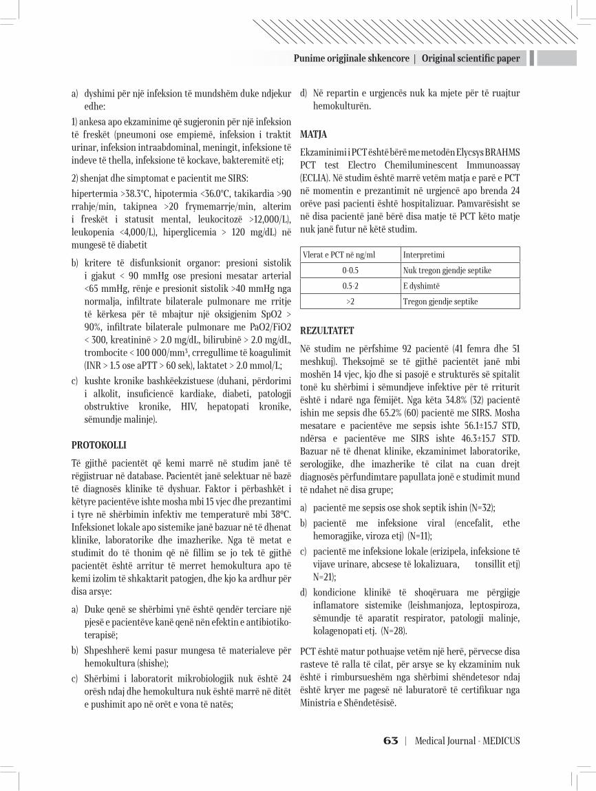
Punime origjinale shkencore | Original scientific paper
63 | Medical Journal - MEDICUS
a) dyshimi për një infeksion të mundshëm duke ndjekur edhe:
1) ankesa apo ekzaminime që sugjeronin për një infeksion të freskët (pneumoni ose empiemë, infeksion i traktit urinar, infeksion intraabdominal, meningit, infeksione të indeve të thella, infeksione të kockave, bakteremitë etj;
2) shenjat dhe simptomat e pacientit me SIRS:
hipertermia >38.3°C, hipotermia <36.0°C, takikardia >90 rrahje/min, takipnea >20 frymemarrje/min, alterim i freskët i statusit mental, leukocitozë >12,000/L), leukopenia <4,000/L), hiperglicemia > 120 mg/dL) në mungesë të diabetit
b) kritere të disfunksionit organor: presioni sistolik i gjakut < 90 mmHg ose presioni mesatar arterial <65 mmHg, rënje e presionit sistolik >40 mmHg nga normalja, infiltrate bilaterale pulmonare me rritje të kërkesa për të mbajtur një oksigjenim SpO2 > 90%, infiltrate bilaterale pulmonare me PaO2/FiO2 < 300, kreatininë > 2.0 mg/dL, bilirubinë > 2.0 mg/dL, trombocite < 100 000/mm³, crregullime të koagulimit (INR > 1.5 ose aPTT > 60 sek), laktatet > 2.0 mmol/L;
c) kushte kronike bashkëekzistuese (duhani, përdorimi i alkolit, insuficiencë kardiake, diabeti, patologji obstruktive kronike, HIV, hepatopati kronike, sëmundje malinje).
PROTOKOLLI
Të gjithë pacientët që kemi marrë në studim janë të rëgjistruar në database. Pacientët janë selektuar në bazë të diagnosës klinike të dyshuar. Faktor i përbashkët i këtyre pacientëve ishte mosha mbi 15 vjec dhe prezantimi i tyre në shërbimin infektiv me temperaturë mbi 38ºC. Infeksionet lokale apo sistemike janë bazuar në të dhenat klinike, laboratorike dhe imazherike. Nga të metat e studimit do të thonim që në fillim se jo tek të gjithë pacientët është arritur të merret hemokultura apo të kemi izolim të shkaktarit patogjen, dhe kjo ka ardhur për disa arsye:
a) Duke qenë se shërbimi ynë është qendër terciare një pjesë e pacientëve kanë qenë nën efektin e antibiotiko-terapisë;
b) Shpeshherë kemi pasur mungesa të materialeve për hemokultura (shishe);
c) Shërbimi i laboratorit mikrobiologjik nuk është 24 orësh ndaj dhe hemokultura nuk është marrë në ditët e pushimit apo në orët e vona të natës;
d) Në repartin e urgjencës nuk ka mjete për të ruajtur hemokulturën.
MATJA
Ekzaminimi i PCT është bërë me metodën Elycsys BRAHMS PCT test Electro Chemiluminescent Immunoassay (ECLIA). Në studim është marrë vetëm matja e parë e PCT në momentin e prezantimit në urgjencë apo brenda 24 orëve pasi pacienti është hospitalizuar. Pamvarësisht se në disa pacientë janë bërë disa matje të PCT këto matje nuk janë futur në këtë studim.
Vlerat e PCT në ng/ml Interpretimi
0-0.5 Nuk tregon gjendje septike
0.5-2 E dyshimtë
>2 Tregon gjendje septike
REZULTATET
Në studim ne përfshime 92 pacientë (41 femra dhe 51 meshkuj). Theksojmë se të gjithë pacientët janë mbi moshën 14 vjec, kjo dhe si pasojë e strukturës së spitalit tonë ku shërbimi i sëmundjeve infektive për të rriturit është i ndarë nga fëmijët. Nga këta 34.8% (32) pacientë ishin me sepsis dhe 65.2% (60) pacientë me SIRS. Mosha mesatare e pacientëve me sepsis ishte 56.1±15.7 STD, ndërsa e pacientëve me SIRS ishte 46.3±15.7 STD. Bazuar në të dhenat klinike, ekzaminimet laboratorike, serologjike, dhe imazherike të cilat na cuan drejt diagnosës përfundimtare papullata jonë e studimit mund të ndahet në disa grupe;
a) pacientë me sepsis ose shok septik ishin (N=32);
b) pacientë me infeksione viral (encefalit, ethe hemoragjike, viroza etj) (N=11);
c) pacientë me infeksione lokale (erizipela, infeksione të vijave urinare, abcsese të lokalizuara, tonsillit etj) N=21);
d) kondicione klinikë të shoqëruara me përgjigje inflamatore sistemike (leishmanjoza, leptospiroza, sëmundje të aparatit respirator, patologji malinje, kolagenopati etj. (N=28).
PCT është matur pothuajse vetëm një herë, përvecse disa rasteve të ralla të cilat, për arsye se ky ekzaminim nuk është i rimbursueshëm nga shërbimi shëndetesor ndaj është kryer me pagesë në laburatorë të certifikuar nga Ministria e Shëndetësisë.
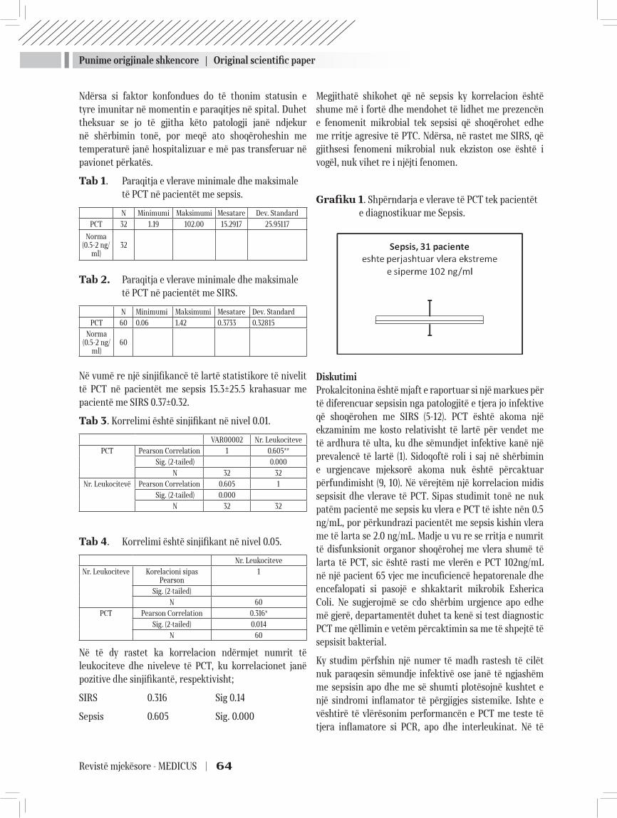
Punime origjinale shkencore | Original scientific paper
Revistë mjekësore - MEDICUS | 64
Ndërsa si faktor konfondues do të thonim statusin e tyre imunitar në momentin e paraqitjes në spital. Duhet theksuar se jo të gjitha këto patologji janë ndjekur në shërbimin tonë, por meqë ato shoqëroheshin me temperaturë janë hospitalizuar e më pas transferuar në pavionet përkatës.
Tab 1. Paraqitja e vlerave minimale dhe maksimale të PCT në pacientët me sepsis.
N Minimumi Maksimumi Mesatare Dev. StandardPCT 32 1.19 102.00 15.2917 25.95117
Norma (0.5-2 ng/
ml)32
Tab 2. Paraqitja e vlerave minimale dhe maksimale të PCT në pacientët me SIRS.
N Minimumi Maksimumi Mesatare Dev. StandardPCT 60 0.06 1.42 0.3733 0.32815
Norma (0.5-2 ng/
ml)60
Në vumë re një sinjifikancë të lartë statistikore të nivelit të PCT në pacientët me sepsis 15.3±25.5 krahasuar me pacientë me SIRS 0.37±0.32.
Tab 3. Korrelimi është sinjifikant në nivel 0.01.
VAR00002 Nr. LeukocitevePCT Pearson Correlation 1 0.605**
Sig. (2-tailed) 0.000 N 32 32
Nr. Leukocitevë Pearson Correlation 0.605 1 Sig. (2-tailed) 0.000
N 32 32
Tab 4. Korrelimi është sinjifikant në nivel 0.05.
Nr. LeukociteveNr. Leukociteve Korelacioni sipas
Pearson1
Sig. (2-tailed)N 60
PCT Pearson Correlation 0.316*Sig. (2-tailed) 0.014
N 60
Në të dy rastet ka korrelacion ndërmjet numrit të leukociteve dhe niveleve të PCT, ku korrelacionet janë pozitive dhe sinjifikantë, respektivisht;
SIRS 0.316 Sig 0.14
Sepsis 0.605 Sig. 0.000
Megjithatë shikohet që në sepsis ky korrelacion është shume më i fortë dhe mendohet të lidhet me prezencën e fenomenit mikrobial tek sepsisi që shoqërohet edhe me rritje agresive të PTC. Ndërsa, në rastet me SIRS, që gjithsesi fenomeni mikrobial nuk ekziston ose është i vogël, nuk vihet re i njëjti fenomen.
Grafiku 1. Shpërndarja e vlerave të PCT tek pacientët e diagnostikuar me Sepsis.
DiskutimiProkalcitonina është mjaft e raportuar si një markues për të diferencuar sepsisin nga patologjitë e tjera jo infektive që shoqërohen me SIRS (5-12). PCT është akoma një ekzaminim me kosto relativisht të lartë për vendet me të ardhura të ulta, ku dhe sëmundjet infektive kanë një prevalencë të lartë (1). Sidoqoftë roli i saj në shërbimin e urgjencave mjeksorë akoma nuk është përcaktuar përfundimisht (9, 10). Në vërejtëm një korrelacion midis sepsisit dhe vlerave të PCT. Sipas studimit tonë ne nuk patëm pacientë me sepsis ku vlera e PCT të ishte nën 0.5 ng/mL, por përkundrazi pacientët me sepsis kishin vlera me të larta se 2.0 ng/mL. Madje u vu re se rritja e numrit të disfunksionit organor shoqërohej me vlera shumë të larta të PCT, sic është rasti me vlerën e PCT 102ng/mL në një pacient 65 vjec me incuficiencë hepatorenale dhe encefalopati si pasojë e shkaktarit mikrobik Esherica Coli. Ne sugjerojmë se cdo shërbim urgjence apo edhe më gjerë, departamentët duhet ta kenë si test diagnostic PCT me qëllimin e vetëm përcaktimin sa me të shpejtë të sepsisit bakterial.
Ky studim përfshin një numer të madh rastesh të cilët nuk paraqesin sëmundje infektivë ose janë të ngjashëm me sepsisin apo dhe me së shumti plotësojnë kushtet e një sindromi inflamator të përgjigjes sistemike. Ishte e vështirë të vlërësonim performancën e PCT me teste të tjera inflamatore si PCR, apo dhe interleukinat. Në të
Punime origjinale shkencore | Original scientific paper
65 | Medical Journal - MEDICUS
dy rastet ka korrelacion ndërmjet numrit të leukociteve dhe niveleve të PCT, ku korrelacionet janë pozitivë dhe sinjifikantë, respektivisht. Megjithatë shikohet që në sepsis ky korrelacion është shumë më i fortë dhe mendohet të lidhet me prezencën e fenomenit mikrobial tek sepsisi që shoqërohet edhe me rritje agresive të PTC. Ndërsa, në raste me SIRS, që gjithsesi fenomeni mikrobial nuk ekziston ose është i vogël, nuk vihet re i njëjti fenomen.
Është për tu theksuar se mjaft studime e shohin PCT si test me specifik se PCR (1, 5, 6). Ne nuk arritëm të bëjmë analizë kosto-efektivitet midis matjes së PCT, hemokulturave, fillimit të antibiotikoterapisë dhe kohëzgjatjes së saj apo dhe të ditëqëndrimit në spital. Sidoqoftë, pamvarësisht se studimi ynë nuk arrin të japë një konkluzion përfundimtar në lidhje me aplikimin e PCT si metodë diagnostikuese e sepsisit bakterial, ne mendojmë se PCT ka më shumë përparsi në lidhje me kohën e marrjes, koston dhe vlerësimin progresiv të sëmundjes, në krahasim me hemokulturën që është dhe si standarti i artë në vendosjen e diagnosës së sepsisit kur plotësohen parametrat klinik të SIRSit. Gjithashtu të dhënat tona sugjerojnë për përdorimin e PCT si test diagnostik midis një SIRSi dhe sepsisit. Sidoqoftë roli ekzakt i PCT në sepsis ngelet akoma për tu testuar dhe ndoshta në përfundim të punës sonë do të jemi në gjëndje të japim një vlerësim me cilësor të PCT në lidhje me sepsisin.
REFERENCAT:
1. Limper M, de Kruif M. D, Ajubi N. E, van Zanten A. P, Brandjes D. P, Duits A. J. et al Procalcitonin as a potent marker of bacterial infection in febrile Afro-Caribbean patients at the emergency department. Eur J Clin Mi-crobiol Infect Dis (2011) 30:831–836.
2. de Kruif M. D, Limper M, Gerristen H, Spek CA, Brandjes DP, ten Cate H, Additionav value of procalcitoninfor di-agnosis of infection in patients with fever in emergency department. Crit Care Med. 2010 Feb;38(2):457-463.
3. Hausfater P, Garric S, Ben Ayed S, Rosenheim M, Ber-nard M, and Riou B. Usefulness of Procalcitonin as a Marker of Systemic Infection in Emergency Depart-ment Patients: A Prospective Study. Clinical Infectious Diseases 2002; 34:895–901.
4. Christ-Crain M, Müller B. Procalcitonin in bacterial infections – hype, hope, more or less? Swiss Med Wkly 2005,135:451-460.
5. Carrol ED, Thomson APJ, Hart CA. Procalcitonin as a marker of sepsis. Int J Antimicrobial Agents 2002;20:1-9.
6. Meisner M. Biomarkers of sepsis: clinically useful? Curr Opin Crit Care 2005;11:473-480.
7. Clec’h C, Ferriere F, Karoubi P, Fosse JP, Cupa M, Hoang P, Cohen Y. Diagnostic and prognostic value of procal-citonin in patients with septic shock. Crit Care Med 2004;32:1166-1169.
8. Christ-Crain M, Jaccard-Stolz D, Bingisser R, Gencay MM, Huber PR, Tamm M, Müller B. Effect of procalciton-in-guided treatment on antibiotic use and outcome in lower respiratory tract infections: cluster-randomised, single-blinded intervention trial. Lancet 2004;363:600-607.
9. Jones AE, Fiechtl JF, Brown MD, Ballew JJ, Kline JA. Pro-calcitonin Test in the Diagnosis of Bacteremia: A Meta-analysis. Ann Emerg Med 2007;50(1):34-41.
10. Tang BM, Eslick GD, Craig JC, McLean AS. Accuracy of procalcitonin for sepsis diagnosis in critically ill pa-tients: systematic review and metaanalysis. Lancet In-fect Dis 2007;7:210-217
11. Bone RC, Balk RA, Cerra FB, Dellinger RP, Fein AM, Knaus WA, Schein RM, Sibbald WJ. Definitions for sep-sis and organ failure and guidelines for the use of inno-vative therapies in sepsis. The ACCP (American College of Chest Physicians)/SCCM (Society of Critical Care Medicine) Consensus Conference Committee. Chest 1992;101:1644-1652.
12. Guven H, Altintop L, Baydin A, Esen S, Aygun D, Hokelek M, Doganay Z, Bek Y. Diagnostic value of procalcitonin levels as an early indicator of sepsis. Am J Emerg Med 2002;20:202-206.
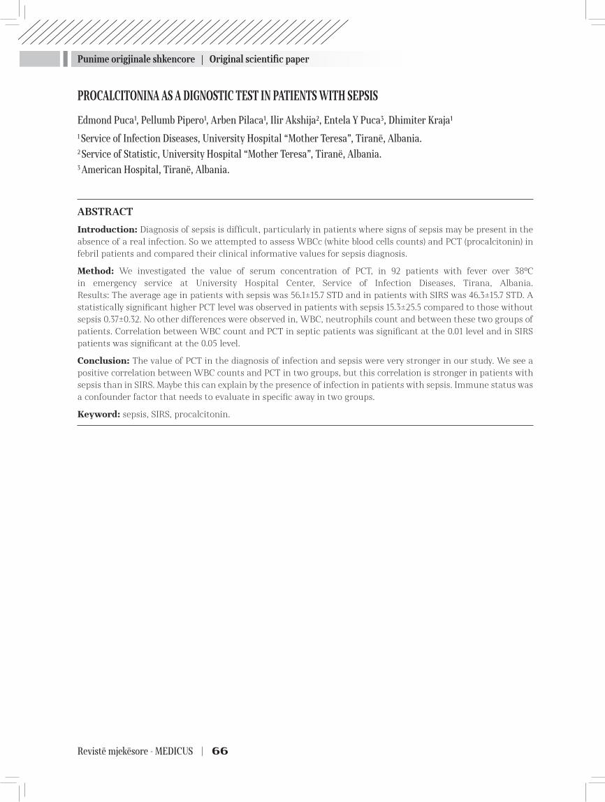
Punime origjinale shkencore | Original scientific paper
Revistë mjekësore - MEDICUS | 66
PROCALCITONINA AS A DIGNOSTIC TEST IN PATIENTS WITH SEPSIS
Edmond Puca¹, Pellumb Pipero¹, Arben Pilaca¹, Ilir Akshija², Entela Y Puca³, Dhimiter Kraja¹1 Service of Infection Diseases, University Hospital “Mother Teresa”, Tiranë, Albania.2 Service of Statistic, University Hospital “Mother Teresa”, Tiranë, Albania.3 American Hospital, Tiranë, Albania.
ABSTRACT
Introduction: Diagnosis of sepsis is difficult, particularly in patients where signs of sepsis may be present in the absence of a real infection. So we attempted to assess WBCc (white blood cells counts) and PCT (procalcitonin) in febril patients and compared their clinical informative values for sepsis diagnosis.
Method: We investigated the value of serum concentration of PCT, in 92 patients with fever over 38ºC in emergency service at University Hospital Center, Service of Infection Diseases, Tirana, Albania. Results: The average age in patients with sepsis was 56.1±15.7 STD and in patients with SIRS was 46.3±15.7 STD. A statistically significant higher PCT level was observed in patients with sepsis 15.3±25.5 compared to those without sepsis 0.37±0.32. No other differences were observed in, WBC, neutrophils count and between these two groups of patients. Correlation between WBC count and PCT in septic patients was significant at the 0.01 level and in SIRS patients was significant at the 0.05 level.
Conclusion: The value of PCT in the diagnosis of infection and sepsis were very stronger in our study. We see a positive correlation between WBC counts and PCT in two groups, but this correlation is stronger in patients with sepsis than in SIRS. Maybe this can explain by the presence of infection in patients with sepsis. Immune status was a confounder factor that needs to evaluate in specific away in two groups.
Keyword: sepsis, SIRS, procalcitonin.
67 | Medical Journal - MEDICUS
PJEKURIA PULMONARE, NDIKIMI NE PARAQITJEN E SINDROMES SE DISTRESIT RESPIRATOR(RDSY)Sani Bajrami1, Florin Besimi 1, Nagip Rufati1 , Meral Rexhepi1 , Arian Alili1, Merie Vejseli1 , Gjuzvete Tairi 1, Zllata Trimçevska1, Antigona Trenevska1, Bashkim Ismaili 1 , Besa Islami-Pocesta2, Drita Tefiku 1 , Afërdita Ismaili1, Mirsada Emini1, Bujar Abdiu1, Savica Markovic1, Nexhibe Nuhii3
1 Spitali Klinik, Tetovë, Maqedoni 2 Spitali per Gjinekologji-Akusheri, Çair, Shkup3 Fakulteti i Mjekesise-Universiteti Shteterore i Tetoves
ABSTRAKTI
RDSy paraqet kolapsin progresiv të alveolave dhe me zhvillim progresiv të mikroatelektatik si pasojë e sintezes së pamjaftushme të surfaktantit. Prematuriteti është faktori bazë i zhvillimit te RDSy. Pjekuria pulmonare neonatale me kortikosteroide tek të porsalindurit premature është masë shumë e rëndësishme për të parandaluar paraqitjen e RDSy.
Qëllimi i punimit është të tregohet përqindja e shfaqjes së RDSy tek të porsalindurit tek të cilët është realizuar pjekuria pulmonare neonatale dhe tek të porsalindurit tek të cilët nuk është realizuar pjekuria pulmonare neonatale, të lindur në periudhë 34,35 javë gestacionale.
Materiali dhe metodat: Punimi është realizuar në Repartin Gjinekologji-Obstetri-Neonatologji pranë Spitalit Klinik-Tetovë, ku janë mare të dhënat e të gjithë të porsalindurve në gestacionin 34,35 javë, dhe janë ndarë në dy grupe grupi i parë tek i cili është realizuar pjekuri dhe grupi i dyte tek të cilët nuk është realizuar pjekurime.
Rezultatet: Tek të gjithë 33 lindjet RDSy është shfaqur në 8 të porsalindur.Dhe atë në 4 raste tek pacientë tek të cilat është ordinuar pjekuri dhe në 4 raste tek të porsalindur ku nuk është realizuar pjekuri. Rrjedhimisht përqindja RDSy në të gjithë të porsalindurit në gestacion 34,35 javë është (24,2 %), dhe atë në ato të porsalindur tek të cilët është realizuar pjekuri është (14,8 %) e të porsalindurve(grafikon 3a), dhe tek ata tek të cilët nuk është ordinuar pjekuri (66,6 %) të porsalindurve.
Përfundimi: Nga të dhënat e analizuara mund të thuhet se në repartin tonë përqindja e të porsalindurve me RDSy pa pjekuri pulmonare është më e lartë në krahasim me ato tek të cilët është realizuar pjekuri pulmonare.
Fjalë kyce: Pjekuria pulmonare neonatale, RDSy.
Medicus 2013, Vol. 18 (2): 67-70
HYRJE:
Syndroma e disstresit respirator është një nga pa-tologjitë më të shpeshta në moshën e neonatit e cila paraqitet tek të porsalindurit të lindur para javës së 37 lunare,dhe gjithashtu një nga faktorët më të shpeshtë të mortalitetit neonatal. RDSy paraqet kolapsin pro-gresiv të alveolave dhe me zhvillim progresiv të mik-roatelektatik si pasojë e sintezës së pamjaftushme të surfaktantit (1). RDSy karakterizohet nga: cijanoza, renkimi, tahipnea, dhe tërheqja e murit torakal. RDSY paraqitet në periudhën nga 27-36 javë gestacionale, por
paraqitet gjithashtu edhe tek të porsalindur me peshë lindje më të ulët se 2500 gr (1,3,4). Prematuriteti është faktori bazë i zhvillimit të RDSy(2). Pjekuria e këtyre të porsalindurve është më specifike në traktin respirator e cila karakterizohet me mungesën e surfaktantit. Ne lindjet para kohe duhet të realizohet pjekuria pulmo-nare me kortikosteroid(5). Në repartin tone të Gjine-kologji – Akusheri-Neonatologjisë si terapi parandalu-ese për zhvillimin e RDSy te të gjitha shtatëzënet që kanë risk për lindje të parakoheshme ordinohet kor-tiko terapi dhe atë në këtë formë: amp. Dexamethason
Punime origjinale shkencore | Original scientific paper
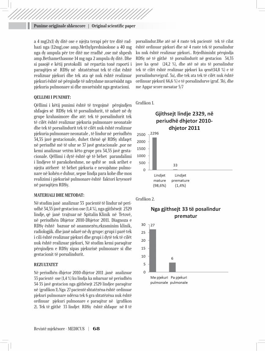
Punime origjinale shkencore | Original scientific paper
Revistë mjekësore - MEDICUS | 68
a 4 mg(2x1) dy ditë ose e njejta terapi për tre ditë rad-hazi nga (12mg),ose amp.Methylprednisolone a 40 mg nga dy ampula për tre ditë me rradhë ,ose më shpesh amp.Bethamethasone 14 mg nga 2 ampula dy ditë. Dhe si pasojë e këtij protokolli në repartin tonë raporti i paraqitjes së RDSy në shtatëzënat tek të cilat është realizuar pjekuri dhe tek ata që nuk është realizuar pjekuri është në përqindje të ndryshme mvarësisht nga pjekuria pulmonare si dhe mvarësisht nga gestacioni.
QELLIMI I PUNIMIT:
Qëllimi i këtij punimi është të tregojmë përqindjen shfaqjes së RDSy tek të porsalindurit, të ndarë në dy grupe krahasimore dhe atë: tek të porsalindurit tek të cilët është realizuar pjekuria pulmonare neonatale dhe tek të porsalindurit tek të cilët nuk është realizuar pjekuria pulmonare neonatale , të lindur në periudhën 34,35 javë gestacionale, duhet thënë që RDSy shfaqet në periudhë më të ulur se 37 javë gestacionale ,por ne kemi analizuar vetëm këto grupe pra 34,35 javë gesta-cionale. Qëllimi i dytë është që të bëhet parandalimi i lindjeve të parakoheshme, ne qoftë se nuk arihet e njejta atëherë të bëhet pjekuria e nevojshme pulmo-nare në kohën e duhur, sepse lindja para kohe dhe mos realizimi i pjekurisë pulmonare është faktori kryesorë në paraqitjen RDSy.
МATERIALI DHE METODAT:
Në studim janë analizuar 33 pacientë të lindur në peri-udhë 34,35 javë gestacion ose (1,4 %), nga gjithësejt 2329 lindje, që janë trajtuar në Spitalin Klinik në Tetovë, në periudhën Dhjetor 2010-Dhjetor 2011. Diagnoza e RDSy është bazuar në anamnezën,ekzaminim klinik, radiologjik. dhe janë ndarë në dy grupe: grupi i parë tek i cili është realizuar pjekuri dhe grupi i dytë tek të cilët nuk është realizuar pjekuri, Në studim kemi paraqitur përqindjen e RDSy sipas pjekurisë pulmonare si dhe gestacionit të porsalindurit.
REZULTATET
Në periudhën dhjetor 2010-dhjetor 2011 ,janë analizuar 33 pacientë ose (1,4 %) ku lindja ka mbaruar në periudhën 34-35 javë gestacion nga gjithësejt 2329 lindjee paraqitur në (grafikon 1).Nga 27 pacientë shtatëzëna është ordinuar pjekuri pulmonare ndërsa tek 6 gra shtatëzëna nuk është ordinuar pjekuri pulmonare e paraqitur në (grafikon 2). Tek të gjithë 33 lindjet RDSy është shfaqur në 8 të
porsalindur.Dhe atë në 4 raste tek pacientë tek të cilat është ordinuar pjekuri dhe në 4 raste tek të porsalindur ku nuk është realizuar pjekuri.. Rrjedhimisht përqindja RDSy në të gjithë të porsalindurit në gestacion 34,35 jave ka qenë (24,2 %), dhe atë në ato të porsalindur tek të cilët është realizuar pjekuri ka qenë(14,8 %) e të porsalindurve(graf. 3a), dhe tek ata tek të cilët nuk është ordinuar pjekuri( 66,6 %) e të porsalindurve (graf. 3b), dhe me Apgar score mesatar 5/7
Grafikon 1.
Grafikon 2.
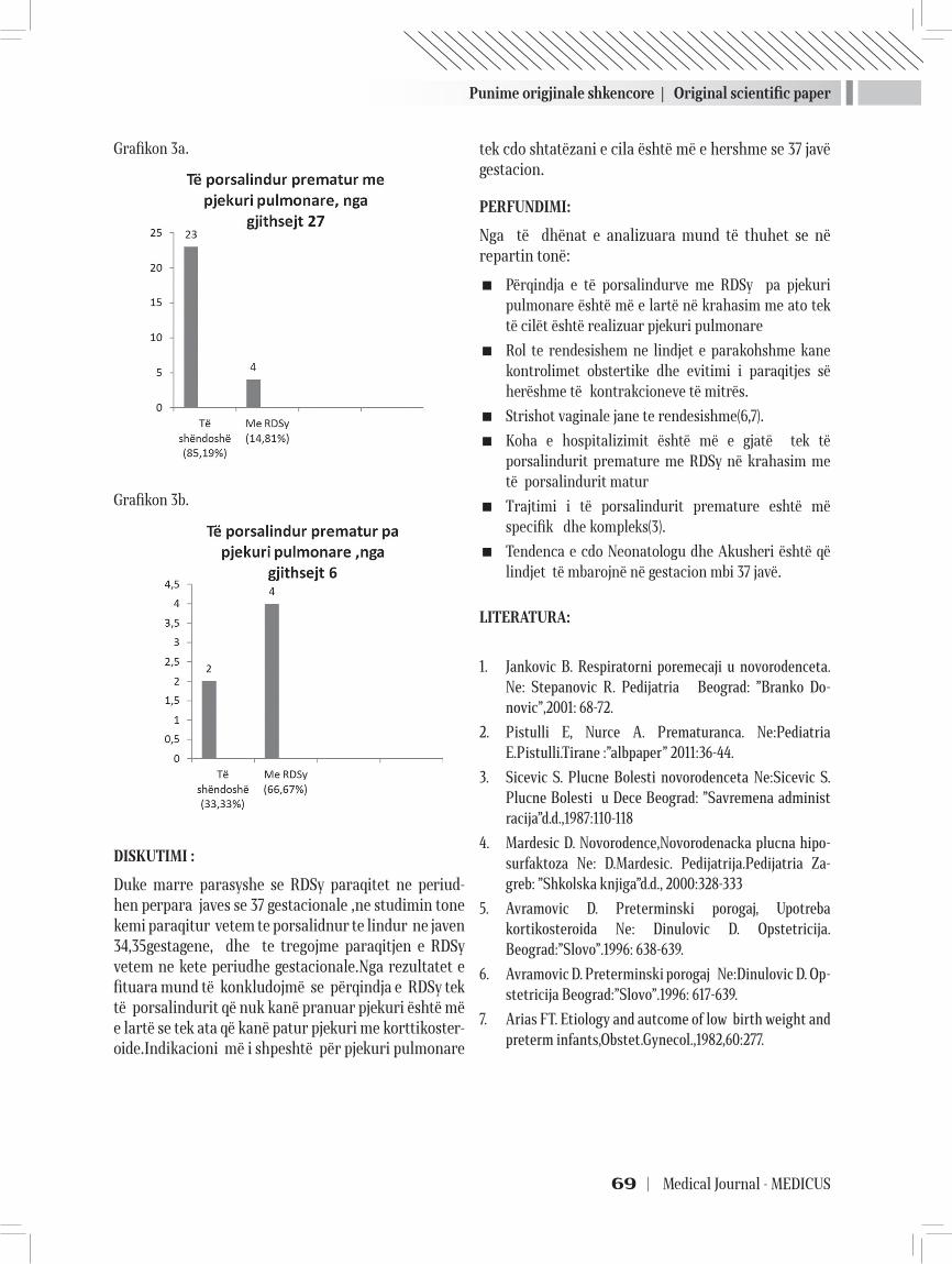
Punime origjinale shkencore | Original scientific paper
69 | Medical Journal - MEDICUS
Grafikon 3a.
Grafikon 3b.
DISKUTIMI :
Duke marre parasyshe se RDSy paraqitet ne periud-hen perpara javes se 37 gestacionale ,ne studimin tone kemi paraqitur vetem te porsalidnur te lindur ne javen 34,35gestagene, dhe te tregojme paraqitjen e RDSy vetem ne kete periudhe gestacionale.Nga rezultatet e fituara mund të konkludojmë se përqindja e RDSy tek të porsalindurit që nuk kanë pranuar pjekuri është më e lartë se tek ata që kanë patur pjekuri me korttikoster-oide.Indikacioni më i shpeshtë për pjekuri pulmonare
tek cdo shtatëzani e cila është më e hershme se 37 javë gestacion.
PERFUNDIMI:
Nga të dhënat e analizuara mund të thuhet se në repartin tonë:
� Përqindja e të porsalindurve me RDSy pa pjekuri pulmonare është më e lartë në krahasim me ato tek të cilët është realizuar pjekuri pulmonare
� Rol te rendesishem ne lindjet e parakohshme kane kontrolimet obstertike dhe evitimi i paraqitjes së herëshme të kontrakcioneve të mitrës.
� Strishot vaginale jane te rendesishme(6,7).
� Koha e hospitalizimit është më e gjatë tek të porsalindurit premature me RDSy në krahasim me të porsalindurit matur
� Trajtimi i të porsalindurit premature eshtë më specifik dhe kompleks(3).
� Tendenca e cdo Neonatologu dhe Akusheri është që lindjet të mbarojnë në gestacion mbi 37 javë.
LITERATURA:
1. Jankovic B. Respiratorni poremecaji u novorodenceta. Ne: Stepanovic R. Pedijatria Beograd: ”Branko Do-novic”,2001: 68-72.
2. Pistulli E, Nurce A. Prematuranca. Ne:Pediatria E.Pistulli.Tirane :”albpaper” 2011:36-44.
3. Sicevic S. Plucne Bolesti novorodenceta Ne:Sicevic S. Plucne Bolesti u Dece Beograd: ”Savremena administracija”d.d.,1987:110-118
4. Mardesic D. Novorodence,Novorodenacka plucna hipo-surfaktoza Ne: D.Mardesic. Pedijatrija.Pedijatria Za-greb: ”Shkolska knjiga”d.d., 2000:328-333
5. Avramovic D. Preterminski porogaj, Upotreba kortikosteroida Ne: Dinulovic D. Opstetricija.Beograd:”Slovo”.1996: 638-639.
6. Avramovic D. Preterminski porogaj Ne:Dinulovic D. Op-stetricija Beograd:”Slovo”.1996: 617-639.
7. Arias FT. Etiology and autcome of low birth weight and preterm infants,Obstet.Gynecol.,1982,60:277.
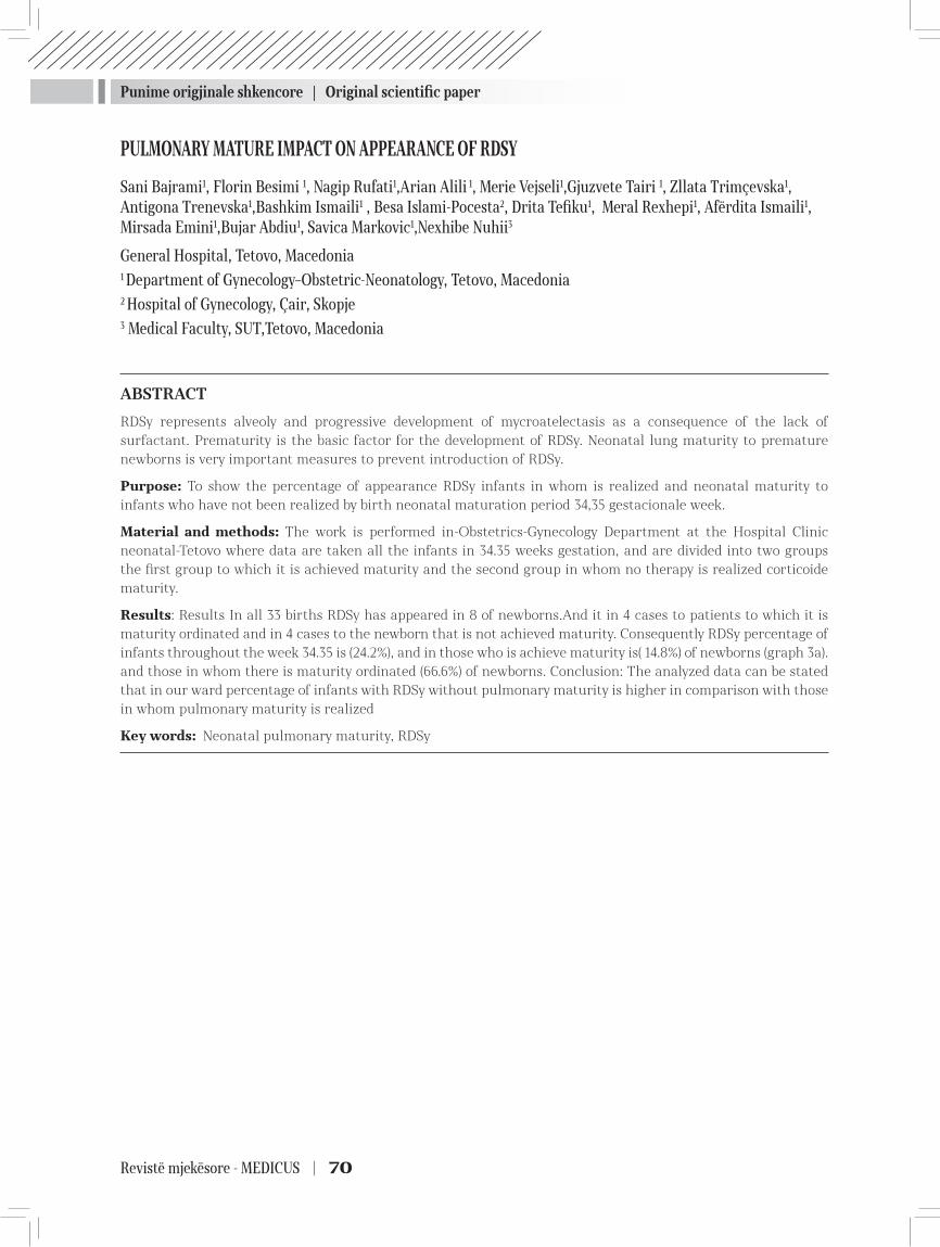
Punime origjinale shkencore | Original scientific paper
Revistë mjekësore - MEDICUS | 70
PULMONARY MATURE IMPACT ON APPEARANCE OF RDSY
Sani Bajrami1, Florin Besimi 1, Nagip Rufati1,Arian Alili 1, Merie Vejseli1,Gjuzvete Tairi 1, Zllata Trimçevska1, Antigona Trenevska1,Bashkim Ismaili1 , Besa Islami-Pocesta2, Drita Tefiku1, Meral Rexhepi1, Afërdita Ismaili1, Mirsada Emini1,Bujar Abdiu1, Savica Markovic1,Nexhibe Nuhii3
General Hospital, Tetovo, Macedonia1 Department of Gynecology–Obstetric-Neonatology, Tetovo, Macedonia2 Hospital of Gynecology, Çair, Skopje3 Medical Faculty, SUT,Tetovo, Macedonia
ABSTRACT
RDSy represents alveoly and progressive development of mycroatelectasis as a consequence of the lack of surfactant. Prematurity is the basic factor for the development of RDSy. Neonatal lung maturity to premature newborns is very important measures to prevent introduction of RDSy.
Purpose: To show the percentage of appearance RDSy infants in whom is realized and neonatal maturity to infants who have not been realized by birth neonatal maturation period 34,35 gestacionale week.
Material and methods: The work is performed in-Obstetrics-Gynecology Department at the Hospital Clinic neonatal-Tetovo where data are taken all the infants in 34.35 weeks gestation, and are divided into two groups the first group to which it is achieved maturity and the second group in whom no therapy is realized corticoide maturity.
Results: Results In all 33 births RDSy has appeared in 8 of newborns.And it in 4 cases to patients to which it is maturity ordinated and in 4 cases to the newborn that is not achieved maturity. Consequently RDSy percentage of infants throughout the week 34.35 is (24.2%), and in those who is achieve maturity is( 14.8%) of newborns (graph 3a). and those in whom there is maturity ordinated (66.6%) of newborns. Conclusion: The analyzed data can be stated that in our ward percentage of infants with RDSy without pulmonary maturity is higher in comparison with those in whom pulmonary maturity is realized
Key words: Neonatal pulmonary maturity, RDSy
71 | Medical Journal - MEDICUS
ЕВАЛУАЦИЈА НА ВОЛУМЕНОТ НА ХЕМАТОМОТ, ВОЛУМЕНОТ НА ЕДЕМОТ И Ц-РЕАКТИВЕН ПРОТЕИН ВО АКУТНАТА ФАЗА НА ПРИМАРНА СУПРАТЕНТОРИЈАЛНА ИНТРАЦЕРЕБРАЛНА ХЕМОРАГИЈАПетровска-Цветковска Д.¹, Долненец-Банева Н.¹, Алексовски В.¹, Зафировска-Ивановска Б.²1 Универзитетска Клиника за Неврологија-Скопје 2 Институт за Епидемиологија и Статистика, Медицински Факултет-Скопје²
АБСТРАКТ
Интрацеребралните хеморагии (ИЦХ) се застапени со 10-15% од мозочните удари. Битна карактеристика на ИЦХ е настанувањето на околниот перифокален едем (МЕ), бидејќи ја зголемува васкуларната лезија создадена од екстравазираната крв. МЕ се создава уште во првиот час од крварењето, а максималната големина ја достигнува 3-5 ден од неговото настанување.
Методи: Студијата беше проспективна, и во неа беа вклучени 62 пациенти со ИЦХ. Невролошкиот дефицит на пациентите беше евалуиран со скалата за удари од националниот институт за здравје (NIHSS). Вредностите на ЦРП во серум се изразени во mg/L. Детекцијата, визуелизацијата и димензионирањето на хематомот и перихеморагичниот едем беше изведена со Компјутеризирана Томографија на Мозокот (КТМ) Невролошкиот и радиолошкиот скрининг беше реализиран во три фази: прв, 3-ти ден и 7-ми ден од ИЦХ.
Резултати: Просечната старост кај пациентите изнесуваше 68.1 ± 5.1. Во периодот на следење, резултатите покажаа сигнификантно влошување на невролошкиот дефицит и пораст на скорот од скорот од NIHSS скалата. Се следеше статистички значаен пораст на волуменот на перихеморагичниот едем и на вредностите на ЦРП во серум. Вредностите на волуменот на хематомот во целиот период на опсервација беа без значајни промени
Заклучок: Студијата покажа дека во првите денови од настанување на ИЦХ расте невролошкиот дефицит и нивото на ЦРП во серум, со порастот на волуменот на перифокалниот едем кај пациенти со интрацеребрална хеморагија.
ВОВЕД
Интрацеребралната хеморагија (ИЦХ) е акутна цереброваскуларна болест, која настанува со руптура на мозочна артерија и екстравазација на крв во околниот мозочен паренхим. Застапени се во 10-15% од сите мозочните удари (1). Интрацеребралните хеморагии се делат на два типа: примарни и секундарни. Примарни ИЦХ се 70-90% од сите ИЦХ. Според локализацијата ИЦХ се делат на:супратенторијални инфратенторијални (2).
Битна карактеристика на ИЦХ е настанувањето на околниот перифокален мозочен едем (МЕ), бидејќи ја зголемува паренхиматозната васкуларна лезија создадена од дејството на екстравазираната крв.
МЕ се создава уште во првиот час од крварењето, со максимална големина 3-5 ден од настанување на крварењето. Многу често крвта и МЕ имаат компресивно однесување кон соседното здраво ткиво но и на дел од вентрикуларниот систем, го зголемуваат интракранијалниот притисок. Во периодот на порастот на едемот се продлабочува и невролошкиот дефицит (3). При процес на регресија на интрацеребралниот хематом и МЕ почнува да се намалува (4).
При контактот мозочно ткиво-екстравазирана крв, се активираат неколку механизми во образување на МЕ. Секој од нив има своја улога во долгиот синџир на процеси на формирање на истиот. Тие меѓусебно
Medicus 2013, Vol. 18 (2): 71-74
Punime origjinale shkencore | Original scientific paper
Punime origjinale shkencore | Original scientific paper
Revistë mjekësore - MEDICUS | 72
зависат, се надополнуваат, потикнуваат или надоврзуваат. Научните истражувања последната деценија упатуваат на инволвираноста на каскада инфламаторни процеси во создавањето на МЕ. Проинфламаторните цитокини, интерлеукин 1, тумор некротизирачкиот фактор, ММП-9 се биомаркери и предиктори во настанувањето и понатамошниот пораст на МЕ (5). Во акутната фаза на настанување на мозочниот едем, едноставен и достапен биомаркер во следење на појавата и порастот на МЕ, како и прогноза на исходот од ИЦХ е и Ц-реактивен протеин (ЦРП).
ЦЕЛ НА СТУДИЈАТА
Да се одредат вредностите на ЦРП во серум, волуменот на хематомот и перихеморагичниот едем, и да се постави нивна взаемна зависност, кај пациенти со акутна, примарна, супратенторијална интрацеребрална хеморагија.
МЕТОДИ
Студијата беше проспективна, спроведена на Клиника за Неврологија, и во неа беа вклучени 62 пациенти со ИЦХ. Невролошкиот дефицит на пациентите беше евалуиран со скалата за удари од националниот институт за здравје (NIHSS). Скорот на невролошкиот дефицит е од 0 до 36. Детекцијата, визуелизацијата и димензионирањето на хематомот и перихеморагичниот едем беше изведена со Компјутеризирана Томографија на Мозокот (КТМ), а пресметката направена со математичката формула V=AxBxC/2. Перихеморагичниот мозочен едем е разликата на волуменот од целокупната промена (В на хематом+перихеморагичниот исхемичен обрач) и волуменот на хематомот. Вредностите на волуменот на хематомот и МЕ се изразени во цм 3. Анализата на ЦРП во серум е направена во Биохемиска Лабораторија на Клиника за Неврологија, а вредностите се иразени во mg/L. CRP беше измерен во серум со користење на високосензитивен latex-enhanced turbidimetric immunoassay (Roche Diagnostics GmbH, Vienna, Austria), а добиените резултати беа изразени во mg/L.
Нормални вредности на ЦРП во серум се < 5 mg/L.
Невролошкиот, биохемискиот и радиолошкиот скрининг беше реализиран во три фази: прием, 3 ден и 7 ден од ИЦХ.
РЕЗУЛТАТИ
Во Табела 1 е претставена просечната возраст на вклучените пациенти со ИЦХ во студијата.
Во Графикон 1 е претставена дистрибуцијата по пол.
Табела 2 го презентира просечниот невролошки дефицит (по NIHSS скала) на пациентите со ИЦХ во трите фази на следење (1,3,7 ден од ИЦХ).
Во Табела 3 се претставени вредностите на ЦРП во серум во три опсервациски фази од студијата (1,3,7 ден од ИЦХ).
Табела 4 го покажува просечниот волумен на мозочниот едем (пресметан како разлика на волуменот на целокупниот супстрат и хематомот) на пациентите со ИЦХ во трите фази на следење (1,3,7 ден од ИЦХ).Вредностите се презентирани во cm³.
Во Табела 5 е презентиран просечниот волумен на интрацеребралниот хематом (пресметан според формулата V=AxBxCx0.05) на пациентите со ИЦХ во трите фази на следење (1,3,7 ден од ИЦХ).Вредностите се презентирани во cm³.
ДИСКУСИЈА
Просечната старост кај пациентите изнесуваше 68.1 ± 5.1 (табела 1). Од вкупно 62 пациенти вклучени во студијата мажи беа 33, а жени 29 (Графикон 1).
Во периодот на следење, резултатите покажаа сигнификантно влошување на невролошкиот дефицит и пораст на скорот од скорот од NIHSS скалата. Најголемо влошување се следеше третиот ден од настанувањето на ИЦХ (табела 2).
Серумскиот биомаркер ЦРП во првите две фази на следење во студија имаше покачени вредностите уште при првиот ден од ИЦХ, и вредностите беа повисоки во првите две во однос на третата евалуациска фаза (табела 3).
Во студијата се покажа статистички значаен пораст на волуменот на перихеморагичниот едем, со најголеми вредности третиот ден од следење (табела 4). Вредностите на волуменот на хематомот во целиот период на опсервација беа без значајни промени (табела 3).
Во опсервацискиот период на следење на пациентите со ИЦХ, степенот на зависност на ЦРП со невролошкиот дефицит и вредностите на едемот на хематомот е висок за R=0.4 и R=0.52 (6).
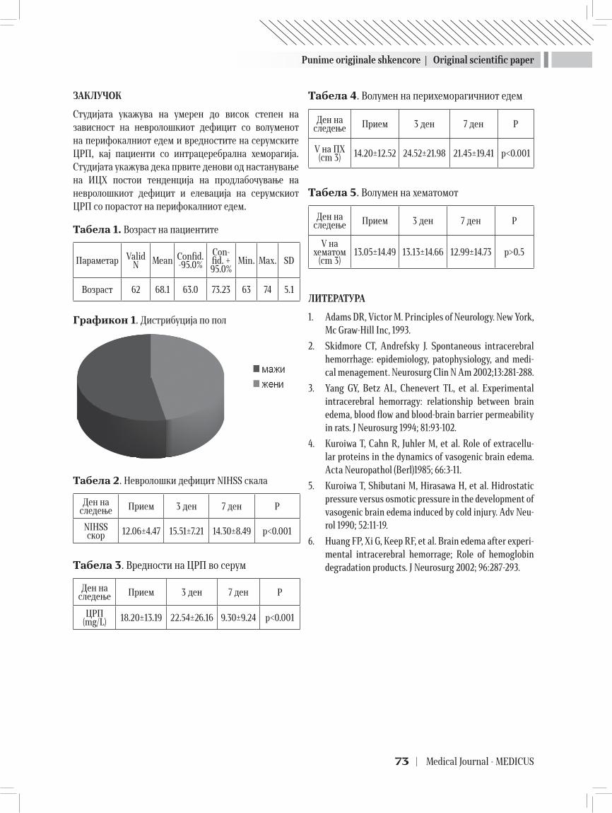
Punime origjinale shkencore | Original scientific paper
73 | Medical Journal - MEDICUS
ЗАКЛУЧОК
Студијата укажува на умерен до висок степен на зависност на невролошкиот дефицит со волуменот на перифокалниот едем и вредностите на серумските ЦРП, кај пациенти со интрацеребрална хеморагија. Студијата укажува дека првите денови од настанување на ИЦХ постои тенденција на продлабочување на невролошкиот дефицит и елевација на серумскиот ЦРП со порастот на перифокалниот едем.
Табела 1. Возраст на пациентите
Параметар Valid N Mean Confid.
-95.0%Con-fid. + 95.0%
Min. Max. SD
Возраст 62 68.1 63.0 73.23 63 74 5.1
Графикон 1. Дистрибуција по пол
Табела 2. Невролошки дефицит NIHSS скала
Ден на следење Прием 3 ден 7 ден Р
NIHSS скор 12.06±4.47 15.51±7.21 14.30±8.49 p<0.001
Табела 3. Вредности на ЦРП во серум
Ден на следење Прием 3 ден 7 ден Р
ЦРП (mg/L) 18.20±13.19 22.54±26.16 9.30±9.24 p<0.001
Табела 4. Волумен на перихеморагичниот едем
Ден на следење Прием 3 ден 7 ден Р
V на ПХ (cm 3) 14.20±12.52 24.52±21.98 21.45±19.41 p<0.001
Табела 5. Волумен на хематомот
Ден на следење Прием 3 ден 7 ден Р
V на хематом
(cm 3)13.05±14.49 13.13±14.66 12.99±14.73 p>0.5
ЛИТЕРАТУРА
1. Adams DR, Victor M. Principles of Neurology. New York, Mc Graw-Hill Inc, 1993.
2. Skidmore CT, Andrefsky J. Spontaneous intracerebral hemorrhage: epidemiology, patophysiology, and medi-cal menagement. Neurosurg Clin N Am 2002;13:281-288.
3. Yang GY, Betz AL, Chenevert TL, et al. Experimental intracerebral hemorragy: relationship between brain edema, blood flow and blood-brain barrier permeability in rats. J Neurosurg 1994; 81:93-102.
4. Kuroiwa T, Cahn R, Juhler M, et al. Role of extracellu-lar proteins in the dynamics of vasogenic brain edema. Acta Neuropathol (Berl)1985; 66:3-11.
5. Kuroiwa T, Shibutani M, Hirasawa H, et al. Hidrostatic pressure versus osmotic pressure in the development of vasogenic brain edema induced by cold injury. Adv Neu-rol 1990; 52:11-19.
6. Huang FP, Xi G, Keep RF, et al. Brain edema after experi-mental intracerebral hemorrage; Role of hemoglobin degradation products. J Neurosurg 2002; 96:287-293.
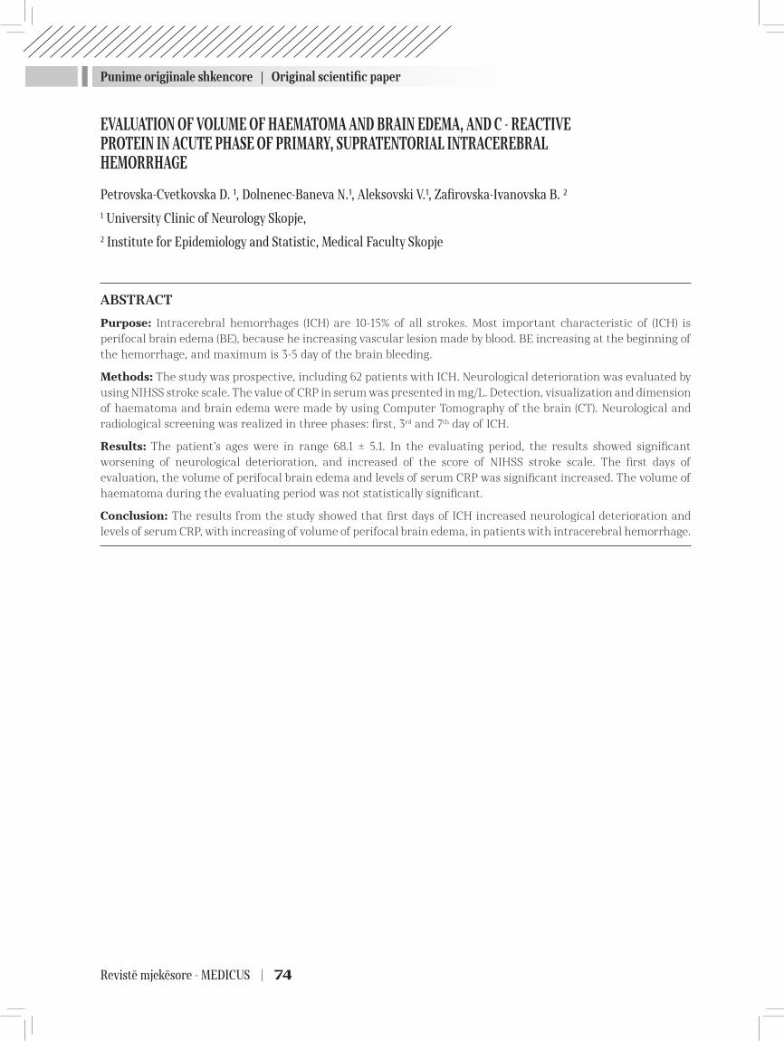
Punime origjinale shkencore | Original scientific paper
Revistë mjekësore - MEDICUS | 74
EVALUATION OF VOLUME OF HAEMATOMA AND BRAIN EDEMA, AND C - REACTIVE PROTEIN IN ACUTE PHASE OF PRIMARY, SUPRATENTORIAL INTRACEREBRAL HEMORRHAGE
Petrovska-Cvetkovska D. ¹, Dolnenec-Baneva N.¹, Aleksovski V.¹, Zafirovska-Ivanovska B. ²1 University Clinic of Neurology Skopje, 2 Institute for Epidemiology and Statistic, Medical Faculty Skopje
ABSTRACT
Purpose: Intracerebral hemorrhages (ICH) are 10-15% of all strokes. Most important characteristic of (ICH) is perifocal brain edema (BE), because he increasing vascular lesion made by blood. BE increasing at the beginning of the hemorrhage, and maximum is 3-5 day of the brain bleeding.
Methods: The study was prospective, including 62 patients with ICH. Neurological deterioration was evaluated by using NIHSS stroke scale. The value of CRP in serum was presented in mg/L. Detection, visualization and dimension of haematoma and brain edema were made by using Computer Tomography of the brain (CT). Neurological and radiological screening was realized in three phases: first, 3rd and 7th day of ICH.
Results: The patient’s ages were in range 68.1 ± 5.1. In the evaluating period, the results showed significant worsening of neurological deterioration, and increased of the score of NIHSS stroke scale. The first days of evaluation, the volume of perifocal brain edema and levels of serum CRP was significant increased. The volume of haematoma during the evaluating period was not statistically significant.
Conclusion: The results from the study showed that first days of ICH increased neurological deterioration and levels of serum CRP, with increasing of volume of perifocal brain edema, in patients with intracerebral hemorrhage.
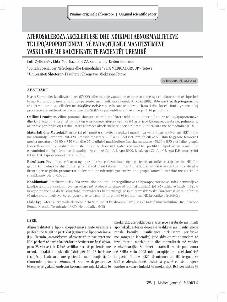
75 | Medical Journal - MEDICUS
ATEROSKLEROZA AKCELERUESE DHE NDIKIMI I ABNORMALITETEVE TË LIPO/APOPROTEINEVE NË PARAQITJENE E MANIFESTIMEVE VASKULARE ME KALCIFIKATE TE PACIENTËT UREMIKËLutfi Zylbeari1,2 , Elita M.1, Gazmend Z.1, Zamira B.1, Driton Selmani2. 1 Spitali Special për Nefrologjië dhe Hemodializs “VITA MEDICAL GROUP”- Tetovë 2 Universiteti Shtetëror- Fakulteti i Shkencave Mjekësore-Tetovë
ABSTRAKT
Hyrje: Sëmundjet kardiovaskulare (SMKV) edhe më tejë vazhdojnë të mbeten si një nga shkaktarët më të shpeshtë të morbiditetit dhe mortalitetit tek pacientët me Insuficience Renale Kronike (IRK). Mekanizmat dhe etiopatogjeneza me të cilët vetë uremia sjellë deri në kalcifikimet vaskulare pa edhe ato të indeve të buta si dhe korelacioni i tyre me ndaj proceseve aterosklerotike premature dhe SMKV te pacientët uremikë ende janë të panjohura.
Qëllimi i Punimit:Qëllim i punimit ishte që të zbardhen efektet e ndikimit të abnormaliteteve të lipo/apoproteinave dhe korelacioni i tyre në paraqitjen e proceseve aterosklerotike (të arterieve koronare, cerebrale, pulmonale, arterieve periferike etj ) si dhe aterosklerozës akceleruese te pacientë uremik të trajtuar me hemodializë (HD).
Materiali dhe Metodat:Si material për punë u shfrytëzua gjaku i marrë nga vena e pacientëve me IRKT dhe me sëmundje koronare- N0=120, (mosha mesatare = 60.40 ± 6.50 vjet, prej të cilëve 55 ishin të gjinisë femrore ( mosha mesatare =58.90 ± 7.40 vjet) dhe 65 të gjinisë mashkullore (mosha mesatare =59.60 ± 8.70 vjet ) dhe grupit kontrollues prej 120 individëve të shëndoshë. Njëkohësisjt gjatë ekzaminit të profilit të lipideve na bëmë edhe ekzaminimi e pëqëndrimeve të apolipoproteineve (Apo-A 1, Apo-B100, Lp(a), Apo-C2, ApoC3, Apo-E,Homocisteini total-Hcyt, Lipioprotein Lipazën-LPL)].
Rezultatet: Rezultatet e fituara nga parametrat e ekzaminuar nga pacientët uremikë të trajtuar me HD dhe grupit kontrolues të shëndoshë janë paraqitur në tabelën numër 1 dhe 2. Dallimi që u evidentua nga vlerat e fituara për të gjitha parametrat e ekzaminuar ndërmjet pacientëve dhe grupit kontrollues është me statistikë signifikante për p=0.0001.
Konkluzioni: Detektimi i risk faktorëve dhe ndikimi i ërregullimeve të lipo/apoproteinave ndaj sëmundjeve kardiovaskulare-kalcifikimeve vaskulare në stadet e hershme të pamjaftueshmërisë së veshkave është më se e nevojshme me çka do të zvogëlohej mortaliteti i hershëm nga pasojat aterosklerotike, kardiovaskulare, infarktit të miokardit, insulteve cerebrovaskular te paceintët uremikë të trajtuar me HD kroonike përsëritëse
Flalë kyç: Ateroskleroza akceleruese (Ath), Sëmundjet kardiovaskulare (SMKV), Kalcifikime vaskulare, Insuficience Renale Kronike Terminale (IRKT), Hemodializa (HD)
HYRJE
Abnormalitetet e lipo / apoproteinave gjatë uremisë i përfëshijnë të gjithë partiklat (grimcat) e lipoproteinave (Lp). Termin „aterosklerozë akceleruese“ te pacientët me IRK përherë të parë e ka përdorur Scribner me bashkëpun. para 25 viteve ( 1). Është verifikuar se të pacientët me uremi, infrakti i miokardit është për 10 -18 herë me i shpësht krahasuar me pacientët me ndonjë tjetër sëmu-ndje primare. Sëmundjet kronike degjenerative të enëve të gjakut( sindromi koronar me infarkt akut të
miokardit, ateroskleroza e arterieve cerebrale me insult apoplektik, arterioskleroza e veshkëve me insuficienecë renale kronike, insuficienca cirkulatore periferike me gangrenë ishemike) janë shkakta-rët themelorë të invaliditetit, morbiditetit dhe mortalitetit në vendet e zhvilluara(6). Studimet statistikore të publikuara në SHBA vitin 2008 mbi paraqitjen e vdekshmërisë te pacientët me IRKT të mjekura me HD treguan se 63% e vdekshmërisë është si pasojë e sëmundjeve kardiovaskulare (infarkt të miokardit), 16% për shkak të
Medicus 2013, Vol. 18 (2): 75-84
Punime origjinale shkencore | Original scientific paper

Punime origjinale shkencore | Original scientific paper
Revistë mjekësore - MEDICUS | 76
infeksioneve, 4% nga karcinomet dhe 27% nga shkaqe tjera tek pacientët gjer në moshën 64 vjeçare. Faktorë themelor dhe esencijal i progresit të Ath është çrregullimi i metaolizmit të lipoproteinave (2,3,4) i cili manifestohet me ndryshim të raportit të llojeve të lipideve dhe përbërjes së apoproteinave në përbërjen e molekulave lipoproteinemike(të ashtuquajtura dislipidemi (36, 37, 38). Prediktorë gjenetik për paraqitjen e hershme të predispozi-cionit familjar ndaj Ath është çrregullimi i transportit reverz i HDL-Ch dhe ekspresioni i pamjaftu-ehsëm i receptorëve B dhe E me zvogëlim të konvertimit të VLDL në IDL dhe më në fund në LDL-ch (39). Pacientët me pamjaftueshmëri të veshkave prezantojnë pasqyrë të Ath së hershme me kalcifikime të shumta krahasuar me popullatën e shëndoshë e cila është verifikuar me paraqitjen e pllakave dhe lezioneve atherosklerotike në tunica intima dhe tunica media të arterieve koronare, aortës,valvulave të zemrës me zhvillim të mediokalcinozës (40). Zavendësimi i apolipoproteinës fiziologjike me ate patologjike, shkalla e lartë e aterogjenitetit të tyre si dhe ndikimi plotësues i toksineve uremike ndaj strukturës dhe funkcionit të apolproteinave në ambient uremik ende janë fenomene të panjohura dhe të pa hulumtuara prandaj janë të nevojshme studime të shumta eksperimentale dhe klinike për zbardhjen e dukurive të lartëpërmendura. Supozohet se ndryshimet kualitative suptile të morfologjisë së grimcave lipoproteinemike te pacienët uremikë në mënyrë të dukëshme e rrisin aterogjenitetin e tyre(afinitet irritur për ngjitje-fiksim të LDL kolesterolit të oksiduar (LDL-chox) për murin endotelial të arterieve, me çka krijohen kushte më të volitshme për paraqitjen e ateromave të arterieve koronare dhe cerbrovaskulare.Nën ndikimin e Lecitin-Choleste-rol-Acil-Transferazës(LCAT), β1-HDL kalon në frakcionin a-migrues të HDL-ch. LCAT në plaz-mën normale ndikon në maturacionin e HDL duke e transformuar frakcionin e HDL të vobegët me lipide në frakcion sferik të tijë tepër të pasur me yndyrëra. Te pacientët me IRK aktiviteti dhe prodhimi i LCAT është i zvogëluar për » 38 %) dhe konverzioni i lartëpërmendur është i kompro-mituar. Aanlizat eksperimentale me anë të inkubacionit të plazmës te pacientët uremikë me inhibitor apo pa inhibitor të LCAT e mbështesin teorinë se Ath uremike akceleruese është e varur edhe nga katbolizmi i çrregulluar i frakcionit β1-HDL dhe aktiviteti teper i zvogëluar i Lipazës Tregliceride Hepatale(LTGH) për 36% (41,42 ) Ekzistojnë të dhëna që mbështesin teorinë se vlerat e ulëta plazmatike të HDL-ch tek pacientët me IRK janë në lidhje me uljen e sintezës së Apo A1/HDL-Ch.
Efekti i përmendur i HDL-ch kundër aterosklerozës vjen nga roli i dyfishtë në mekanizmin e transportit reverzë të kolesterolit deri te VLDL dhe LDL me ndihmën e koleteril ester transfer prote-inës.
Nga që se transporti reverzë i Ch është i ngadalësuar ateherë rritet akumulimi i tijë në inde i cili defekt dhe mekanizën dukshëm i ndihmon proçeset aterosklerotike te pacientët uremikë. Këto dukuri janë verifikuar me ndihmën e ekzaminimieve ultrasonografike.Të gjithë rezultatet e ekzaminuara me ndihmën e Ekokardiogarafisë dëshmuan se ndryshime aterosklerotike të arterieve koro-nare, karotide dhe aortës torakale më të shpeshta dhe më sinjifikante janë verifikuar te pacientët uremik krahasuar me grupin kontrollues të individëve të shëndoshë. Te pacientët uremikë në tunica media dhe tunica intima të aortës janë vërejtur pllaka ateromatoze krahasuar me grupën kontrolluese të individëve të shëndoshë. Sëmundjet kardiovaskulare (SMKV) janë përgjegjëse për mortalitetin e mbi 60% të pacientëve me IRKT (43) Aktualisht rreziku nga SMKV te pacientët me IRK është për 80% më i rritur krahasuar me popullatën e shëndoshë. Prevalenca nga SMKV e pacientëve me IRK është për 55 % që nga fillimi i trajtimit me hemodializë (5). Frekuenca e lartë nga paraqitja e lezioneve aterosklerotike të arterieve koronare te pacientët me pamjaftueshmëri të veshkave është verifikuar nga shumë studime retrospective (7). Studime të shumta si Framingham-studimi mbi rrezikun e lartë të paraqitjes së SMKV te pacientët uremikë të randomizuar sipas viteve, gjinisë, HTA,dislipidemisë ( me HDL-ch të ulët dhe LDL-ch me tregliceridet e rritura),diabeti me nefropa-tinë diabetike,anamneza për sëmundje koronare, konzumimi i duhanit, menopauza, stresi oksidativ, malnutricioni, përqendrimet e larta të biomarkereve të infalamcionit,ndikimi i citokineve infla-matore: Interleukin-1(In-1,6,In1α, In1b,vlerat e rritura të CRP,sedanteriteti, adipo-ziteti, hiperho-mocisteinemia, çrregullimet e meta-bolizmit të Ca(hypocalcemia),hyperphosphatemia, ndikimi i toksineve uremike, hyperurikemia etj. numërohen si faktorë të lartë të rrezikut nga Ath e arterieve koronare, ishemisë,nekrozës dhe infarkit akut të miokardit te pacientët uremikë dhe ato janë faktorë përgjegjës për rritjen e shkallës së mortalitetit te kjo grupë pacientësh (8). Kalcifikimet e shpeshta të arterieve koronare janë si pasojë edhe e mekanizmave pasiv të kalciumit dhe fosforit nga që te pacientët me IRK dominon një çrregullim i lartë i biomekanizmit të Ca dhe fosforit. Në bazë të shumë studimeve të verifikuara mbi Ath akceleruese te pacientët me IRK parashtrohet një
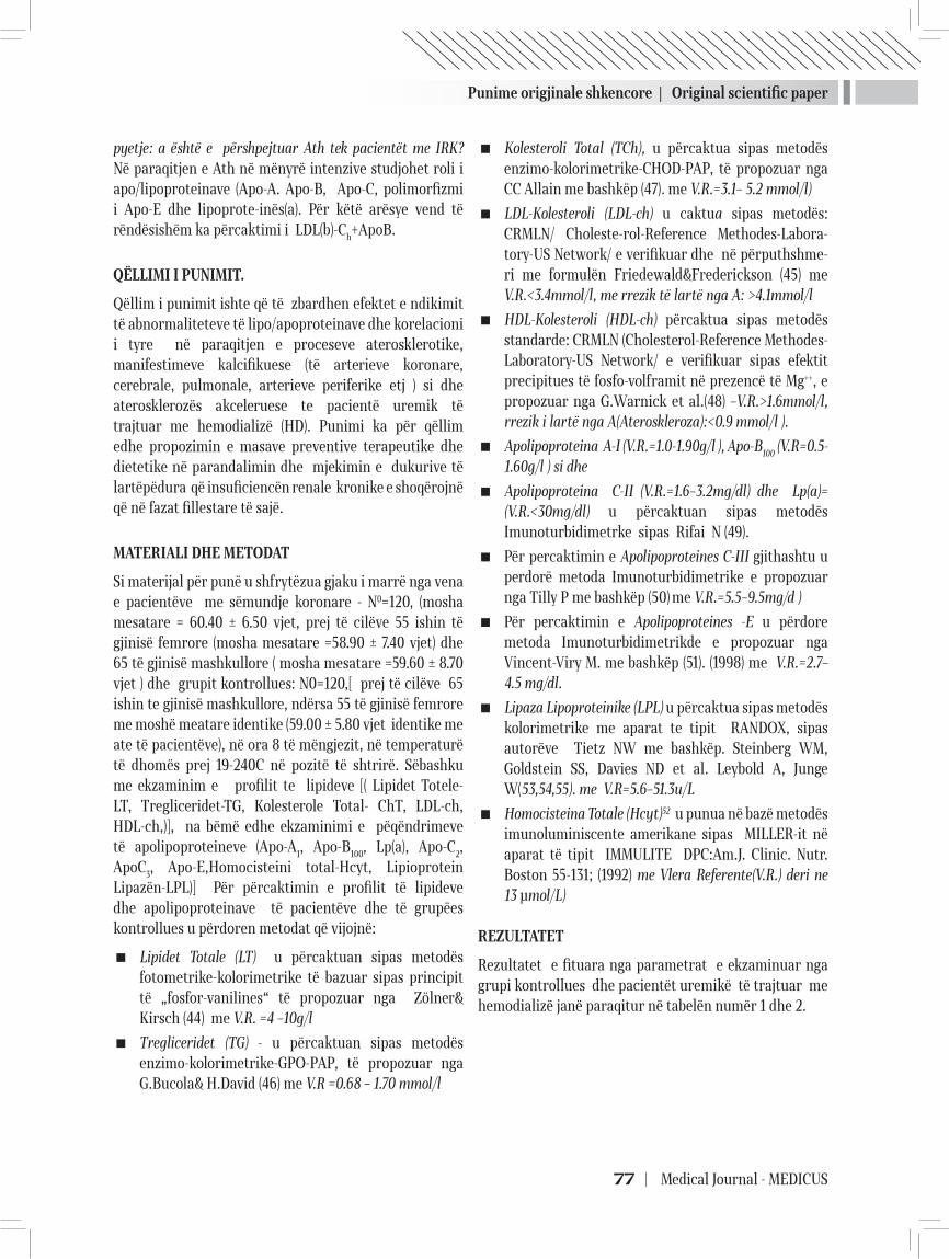
Punime origjinale shkencore | Original scientific paper
77 | Medical Journal - MEDICUS
pyetje: a është e përshpejtuar Ath tek pacientët me IRK? Në paraqitjen e Ath në mënyrë intenzive studjohet roli i apo/lipoproteinave (Apo-A. Apo-B, Apo-C, polimorfizmi i Apo-E dhe lipoprote-inës(a). Për këtë arësye vend të rëndësishëm ka përcaktimi i LDL(b)-Ch+ApoB.
QËLLIMI I PUNIMIT.
Qëllim i punimit ishte që të zbardhen efektet e ndikimit të abnormaliteteve të lipo/apoproteinave dhe korelacioni i tyre në paraqitjen e proceseve aterosklerotike, manifestimeve kalcifikuese (të arterieve koronare, cerebrale, pulmonale, arterieve periferike etj ) si dhe aterosklerozës akceleruese te pacientë uremik të trajtuar me hemodializë (HD). Punimi ka për qëllim edhe propozimin e masave preventive terapeutike dhe dietetike në parandalimin dhe mjekimin e dukurive të lartëpëdura që insuficiencën renale kronike e shoqërojnë që në fazat fillestare të sajë.
MATERIALI DHE METODAT
Si materijal për punë u shfrytëzua gjaku i marrë nga vena e pacientëve me sëmundje koronare - N0=120, (mosha mesatare = 60.40 ± 6.50 vjet, prej të cilëve 55 ishin të gjinisë femrore (mosha mesatare =58.90 ± 7.40 vjet) dhe 65 të gjinisë mashkullore ( mosha mesatare =59.60 ± 8.70 vjet ) dhe grupit kontrollues: N0=120,[ prej të cilëve 65 ishin te gjinisë mashkullore, ndërsa 55 të gjinisë femrore me moshë meatare identike (59.00 ± 5.80 vjet identike me ate të pacientëve), në ora 8 të mëngjezit, në temperaturë të dhomës prej 19-240C në pozitë të shtrirë. Sëbashku me ekzaminim e profilit te lipideve [( Lipidet Totele-LT, Tregliceridet-TG, Kolesterole Total- ChT, LDL-ch, HDL-ch,)], na bëmë edhe ekzaminimi e pëqëndrimeve të apolipoproteineve (Apo-A1, Apo-B100, Lp(a), Apo-C2, ApoC3, Apo-E,Homocisteini total-Hcyt, Lipioprotein Lipazën-LPL)] Për përcaktimin e profilit të lipideve dhe apolipoproteinave të pacientëve dhe të grupëes kontrollues u përdoren metodat që vijojnë:
� Lipidet Totale (LT) u përcaktuan sipas metodës fotometrike-kolorimetrike të bazuar sipas principit të „fosfor-vanilines“ të propozuar nga Zölner& Kirsch (44) me V.R. =4 –10g/l
� Tregliceridet (TG) - u përcaktuan sipas metodës enzimo-kolorimetrike-GPO-PAP, të propozuar nga G.Bucola& H.David (46) me V.R =0.68 – 1.70 mmol/l
� Kolesteroli Total (TCh), u përcaktua sipas metodës enzimo-kolorimetrike-CHOD-PAP, të propozuar nga CC Allain me bashkëp (47). me V.R.=3.1– 5.2 mmol/l)
� LDL-Kolesteroli (LDL-ch) u caktua sipas metodës: CRMLN/ Choleste-rol-Reference Methodes-Labora-tory-US Network/ e verifikuar dhe në përputhshme-ri me formulën Friedewald&Frederickson (45) me V.R.<3.4mmol/l, me rrezik të lartë nga A: >4.1mmol/l
� HDL-Kolesteroli (HDL-ch) përcaktua sipas metodës standarde: CRMLN (Cholesterol-Reference Methodes-Laboratory-US Network/ e verifikuar sipas efektit precipitues të fosfo-volframit në prezencë të Mg++, e propozuar nga G.Warnick et al.(48) –V.R.>1.6mmol/l, rrezik i lartë nga A(Ateroskleroza):<0.9 mmol/l ).
� Apolipoproteina A-I (V.R.=1.0-1.90g/l ), Apo-B100 (V.R=0.5-1.60g/l ) si dhe
� Apolipoproteina C-II (V.R.=1.6–3.2mg/dl) dhe Lp(a)= (V.R.<30mg/dl) u përcaktuan sipas metodës Imunoturbidimetrke sipas Rifai N (49).
� Për percaktimin e Apolipoproteines C-III gjithashtu u perdorë metoda Imunoturbidimetrike e propozuar nga Tilly P me bashkëp (50) me V.R.=5.5–9.5mg/d )
� Për percaktimin e Apolipoproteines -E u përdore metoda Imunoturbidimetrikde e propozuar nga Vincent-Viry M. me bashkëp (51). (1998) me V.R.=2.7–4.5 mg/dl.
� Lipaza Lipoproteinike (LPL) u përcaktua sipas metodës kolorimetrike me aparat te tipit RANDOX, sipas autorëve Tietz NW me bashkëp. Steinberg WM, Goldstein SS, Davies ND et al. Leybold A, Junge W(53,54,55). me V.R=5.6–51.3u/L
� Homocisteina Totale (Hcyt)52 u punua në bazë metodës imunoluminiscente amerikane sipas MILLER-it në aparat të tipit IMMULITE DPC:Am.J. Clinic. Nutr. Boston 55-131; (1992) me Vlera Referente(V.R.) deri ne 13 μmol/L)
REZULTATET
Rezultatet e fituara nga parametrat e ekzaminuar nga grupi kontrollues dhe pacientët uremikë të trajtuar me hemodializë janë paraqitur në tabelën numër 1 dhe 2.
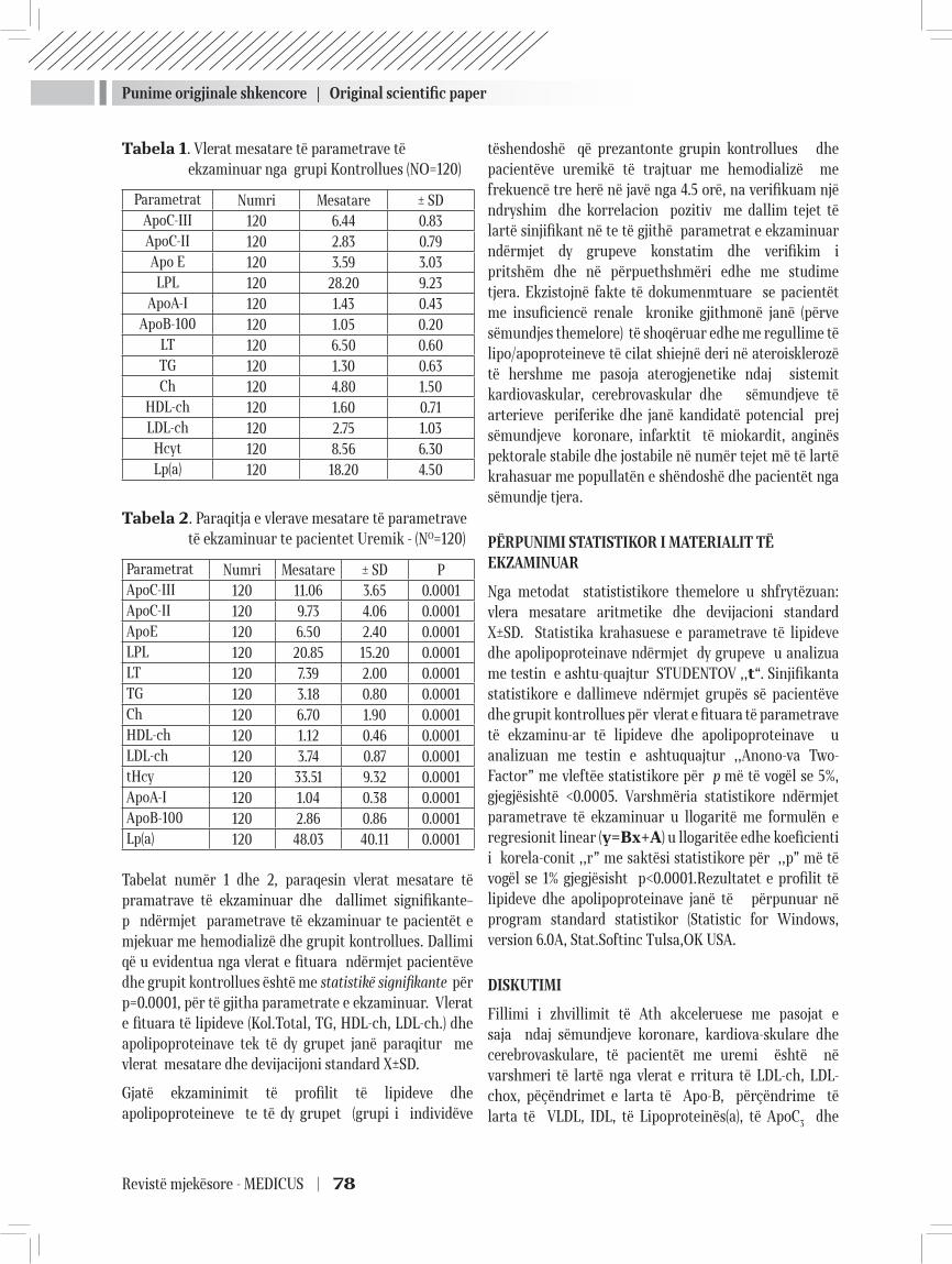
Punime origjinale shkencore | Original scientific paper
Revistë mjekësore - MEDICUS | 78
Tabela 1. Vlerat mesatare të parametrave të ekzaminuar nga grupi Kontrollues (NO=120)
Parametrat Numri Mesatare ± SDApoC-III 120 6.44 0.83ApoC-II 120 2.83 0.79Apo E 120 3.59 3.03LPL 120 28.20 9.23
ApoA-I 120 1.43 0.43ApoB-100 120 1.05 0.20
LT 120 6.50 0.60TG 120 1.30 0.63Ch 120 4.80 1.50
HDL-ch 120 1.60 0.71LDL-ch 120 2.75 1.03
Hcyt 120 8.56 6.30Lp(a) 120 18.20 4.50
Tabela 2. Paraqitja e vlerave mesatare të parametrave të ekzaminuar te pacientet Uremik - (NO=120)
Parametrat Numri Mesatare ± SD PApoC-III 120 11.06 3.65 0.0001ApoC-II 120 9.73 4.06 0.0001ApoE 120 6.50 2.40 0.0001LPL 120 20.85 15.20 0.0001LT 120 7.39 2.00 0.0001TG 120 3.18 0.80 0.0001Ch 120 6.70 1.90 0.0001HDL-ch 120 1.12 0.46 0.0001LDL-ch 120 3.74 0.87 0.0001tHcy 120 33.51 9.32 0.0001ApoA-I 120 1.04 0.38 0.0001ApoB-100 120 2.86 0.86 0.0001Lp(a) 120 48.03 40.11 0.0001
Tabelat numër 1 dhe 2, paraqesin vlerat mesatare të pramatrave të ekzaminuar dhe dallimet signifikante– p ndërmjet parametrave të ekzaminuar te pacientët e mjekuar me hemodializë dhe grupit kontrollues. Dallimi që u evidentua nga vlerat e fituara ndërmjet pacientëve dhe grupit kontrollues është me statistikë signifikante për p=0.0001, për të gjitha parametrate e ekzaminuar. Vlerat e fituara të lipideve (Kol.Total, TG, HDL-ch, LDL-ch.) dhe apolipoproteinave tek të dy grupet janë paraqitur me vlerat mesatare dhe devijacijoni standard X±SD.
Gjatë ekzaminimit të profilit të lipideve dhe apolipoproteineve te të dy grupet (grupi i individëve
tëshendoshë që prezantonte grupin kontrollues dhe pacientëve uremikë të trajtuar me hemodializë me frekuencë tre herë në javë nga 4.5 orë, na verifikuam një ndryshim dhe korrelacion pozitiv me dallim tejet të lartë sinjifikant në te të gjithë parametrat e ekzaminuar ndërmjet dy grupeve konstatim dhe verifikim i pritshëm dhe në përpuethshmëri edhe me studime tjera. Ekzistojnë fakte të dokumenmtuare se pacientët me insuficiencë renale kronike gjithmonë janë (përve sëmundjes themelore) të shoqëruar edhe me regullime të lipo/apoproteineve të cilat shiejnë deri në ateroisklerozë të hershme me pasoja aterogjenetike ndaj sistemit kardiovaskular, cerebrovaskular dhe sëmundjeve të arterieve periferike dhe janë kandidatë potencial prej sëmundjeve koronare, infarktit të miokardit, anginës pektorale stabile dhe jostabile në numër tejet më të lartë krahasuar me popullatën e shëndoshë dhe pacientët nga sëmundje tjera.
PËRPUNIMI STATISTIKOR I MATERIALIT TË EKZAMINUAR
Nga metodat statististikore themelore u shfrytëzuan: vlera mesatare aritmetike dhe devijacioni standard X±SD. Statistika krahasuese e parametrave të lipideve dhe apolipoproteinave ndërmjet dy grupeve u analizua me testin e ashtu-quajtur STUDENTOV ,,t“. Sinjifikanta statistikore e dallimeve ndërmjet grupës së pacientëve dhe grupit kontrollues për vlerat e fituara të parametrave të ekzaminu-ar të lipideve dhe apolipoproteinave u analizuan me testin e ashtuquajtur ,,Anono-va Two-Factor” me vleftëe statistikore për p më të vogël se 5%, gjegjësishtë <0.0005. Varshmëria statistikore ndërmjet parametrave të ekzaminuar u llogaritë me formulën e regresionit linear (y=Bx+A) u llogaritëe edhe koeficienti i korela-conit ,,r” me saktësi statistikore për ,,p” më të vogël se 1% gjegjësisht p<0.0001.Rezultatet e profilit të lipideve dhe apolipoproteinave janë të përpunuar në program standard statistikor (Statistic for Windows, version 6.0A, Stat.Softinc Tulsa,OK USA.
DISKUTIMI
Fillimi i zhvillimit të Ath akceleruese me pasojat e saja ndaj sëmundjeve koronare, kardiova-skulare dhe cerebrovaskulare, të pacientët me uremi është në varshmeri të lartë nga vlerat e rritura të LDL-ch, LDL-chox, pëçëndrimet e larta të Apo-B, përçëndrime të larta të VLDL, IDL, të Lipoproteinës(a), të ApoC3 dhe
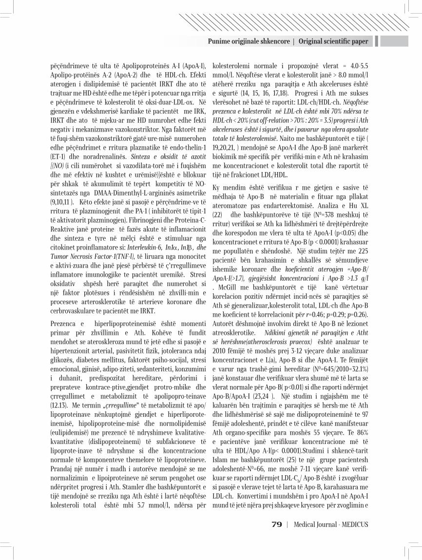
Punime origjinale shkencore | Original scientific paper
79 | Medical Journal - MEDICUS
pëçëndrimeve të ulta të Apolipoproteinës A-I (ApoA-I), Apolipo-protëinës A-2 (ApoA-2) dhe të HDL-ch. Efekti aterogjen i dislipidemisë të pacientët IRKT dhe ato të trajtuar me HD është edhe me tëpër i potencuar nga rritja e pëçëndrimeve të kolesterolit të oksi-duar-LDL-ox. Në gjenezën e vdekshmerisë kardiake të pacientët me IRK, IRKT dhe ato të mjeku-ar me HD numrohet edhe fekti negativ i mekanizmave vazokonstriktor. Nga faktorët më të fuqi-shëm vazokonstriktorë gjatë ure-misë numerohen edhe pëçëndrimet e rritura plazmatike të endo-thelin-1 (ET-1) dhe noradrenalinës. Sinteza e oksidit të azotit [(NO) (i cili numërohet si vazodilata-torë më i fuqishëm dhe më efektiv në kushtet e urëmisë)]është e bllokuar për shkak të akumulimit të tepërt kompetitiv të NO-sintetazës nga DMAA-Dimenthyl-L-argininës asimetrike (9,10,11 ). Këto efekte janë si pasojë e përçëndrime-ve të rritura të plazminogjenit dhe PA-1 ( inhibitorët të tipit-1 të aktivatorit plazminogjen). Fibrinogjeni dhe Proteina-C- Reaktive janë proteine të fazës akute të inflamacionit dhe sinteza e tyre në mëlçi është e stimuluar nga citokinet proinflamatore si: Interleukin-6, In1α, In1β, dhe Tumor Necrosis Factor-1(TNF-l), të liruara nga monocitet e aktivi-zuara dhe janë pjesë përbërsë të ç’rregullimeve inflamatore imunologjike te pacientët uremikë. Stresi oksidativ shpësh herë paraqitet dhe numerohet si një faktor plotësues i rëndësishëm në zhvilli-min e proceseve arterosklerotike të arterieve koronare dhe cerbrovaskulare te pacientët me IRKT.
Prezenca e hiperlipoproteinemisë është momenti primar për zhvillimin e Ath. Kohëve të fundit mendohet se ateroskleroza mund të jetë edhe si pasojë e hipertenzionit arterial, pasivitetit fizik, jotoleranca ndaj glikozës, diabetes mellitus, faktorët psiho-socijal, stresi emocional, gjinisë, adipo-ziteti, sedanteriteti, konzumimi i duhanit, predispozitat hereditare, përdorimi i preprateve kontrace-ptive,gjendjet protro-mbike dhe çrregullimet e metabolizmit të apolipopro-teinave (12.13). Me termin „çrregullime” të metabolizmit të apo/lipoproteinave nënkuptojmë gjendjet e hiperliporote-inemisë, hipolipoproteine-misë dhe normolipidemisë (eulipidemisë) me prezencë të ndryshimeve kvalitative-kvantitative (dislipoproteinemi) të subfakcioneve të lipoprote-inave të ndryshme si dhe koncentracione normale të komponenteve themelore të lipoproteineve. Prandaj një numër i madh i autorëve mendojnë se me normalizimin e lipoiproteineve në serum pengohet ose ndërpritet progresi i Ath. Stamler dhe bashkëpuntorët e tijë mendojnë se rreziku nga Ath është i lartë nëqoftëse kolesteroli total është mbi 5.7 mmol/l, ndërsa për
kolesterolemi normale i propozojnë vlerat = 4.0-5.5 mmol/l. Nëqoftëse vlerat e kolesterolit janë > 8.0 mmol/l atëherë rreziku nga paraqitja e Ath akceleruses është e sigurtë (14, 15, 16, 17,18). Progresi i Ath me sukses vlerësohet në bazë të raportit: LDL-ch/HDL-ch. Nëqoftëse prezenca e kolesterolit në LDL-ch është mbi 70% ndërsa te HDL-ch < 20% (cut off-relation > 70% : 20% = 3.5) progresi i Ath akceleruses është i sigurtë, dhe i pavarur nga vlera apsolute totale të kolesterolemisë. Naito me bashkëpuntorët e tijë ( 19,20,21, ) mendojnë se ApoA-I dhe Apo-B janë markerët biokimik më specifik për verifiki-min e Ath në krahasim me koncentracionet e kolesterolit total dhe raportit të tijë në frakcionet LDL/HDL.
Ky mendim është verifikua r me gjetjen e sasive të mëdhaja të Apo-B në materialin e fituar nga pllakat ateromatoze pas endarterektomisë. Analiza e Hu XL (22) dhe bashkëpuntorëve të tijë (N0=378 meshkuj të rritur) verifikoi se Ath ka lidhëshmëri të drejtëpërdrejte dhe korespodon me vlera të ulta të ApoA-I (p<0.05) dhe koncentracionet e rritura të Apo-B (p < 0.0001) krahasuar me popullatën e shëndoshë. Një studim tejtër me 225 pacientë bën krahasimin e shkallës së sëmundjeve ishemike koronare dhe koeficientit aterogjen =Apo-B/ApoA-I(>1.7), gjegjësisht koncentracioni i Apo-B >1.3 g/l . McGill me bashkëpuntorët e tijë kanë vërtetuar korelacion pozitiv ndërmjet incid-ncës së paraqitjes së Ath së gjeneralizuar,kolesterolit total, LDL-ch dhe Apo-B me koeficient të korrelacionit për r=0.46; p=0.29; p=0.26). Autorët dëshmojnë involvim direkt të Apo-B në lezionet atreosklerotike. Ndikimi gjenetik në paraqitjen e Atht së herëshme(atherosclerosis praecox) është analzuar te 2010 fëmijë të moshës prej 3-12 vjeçare duke analizuar koncentracionet e L(a), Apo-B si dhe ApoA-I. Te fëmijët e varur nga trashë-gimi hereditar (N0=645/2010=32.1%) janë konstauar dhe verifikuar vlera shumë më të larta se vlerat normale për Apo-B( p<0.01) si dhe raporti ndërmjet Apo-B/ApoA-I (23,24 ). Një studim i ngjajshëm me të kaluarën bën trajtimin e paraqitjes së hersh-me të Ath dhe lidhëshmërisë së sajë me dislipoproteineminë te 97 fëmijë adoleshentë, prindët e të cilëve kanë manifsteuar Ath organo-specifike para moshës 55 vjeçare. Te 86% e pacientëve janë verifikuar koncentracione më të ulta të HDL/Apo A-I(p< 0.0001).Studimi i shkencë-tarit Islam me bashkëpuntorët (25) te një grupe pacientesh adoleshentë-N0=66, me moshë 7-11 vjeçare kanë verifi-kuar se raporti ndërmjet LDL-Ch/ Apo-B është i zvogëluar si pasojë e vlerave tejet të larta të Apo-B, karahasuara me LDL-ch. Konvertimi i mundshëm i pro ApoA-I në ApoA-I mund të jetë njëra prej shkaqeve kryesore për zvoglimin e
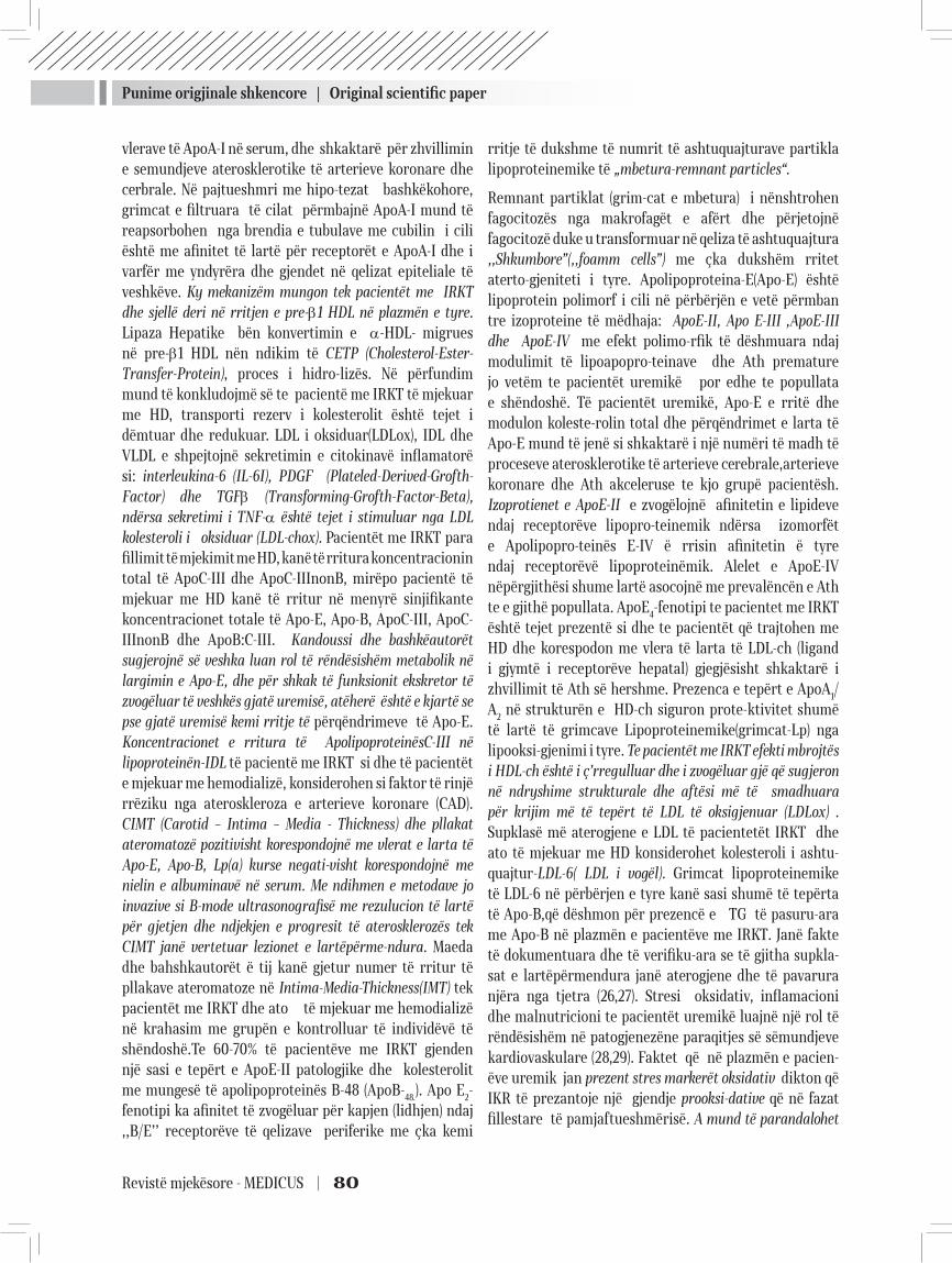
Punime origjinale shkencore | Original scientific paper
Revistë mjekësore - MEDICUS | 80
vlerave të ApoA-I në serum, dhe shkaktarë për zhvillimin e semundjeve aterosklerotike të arterieve koronare dhe cerbrale. Në pajtueshmri me hipo-tezat bashkëkohore, grimcat e filtruara të cilat përmbajnë ApoA-I mund të reapsorbohen nga brendia e tubulave me cubilin i cili është me afinitet të lartë për receptorët e ApoA-I dhe i varfër me yndyrëra dhe gjendet në qelizat epiteliale të veshkëve. Ky mekanizëm mungon tek pacientët me IRKT dhe sjellë deri në rritjen e pre-β1 HDL në plazmën e tyre. Lipaza Hepatike bën konvertimin e α-HDL- migrues në pre-β1 HDL nën ndikim të CETP (Cholesterol-Ester-Transfer-Protein), proces i hidro-lizës. Në përfundim mund të konkludojmë së te pacientë me IRKT të mjekuar me HD, transporti rezerv i kolesterolit është tejet i dëmtuar dhe redukuar. LDL i oksiduar(LDLox), IDL dhe VLDL e shpejtojnë sekretimin e citokinavë inflamatorë si: interleukina-6 (IL-6I), PDGF (Plateled-Derived-Grofth-Factor) dhe TGFβ (Transforming-Grofth-Factor-Beta), ndërsa sekretimi i TNF-α është tejet i stimuluar nga LDL kolesteroli i oksiduar (LDL-chox). Pacientët me IRKT para fillimit të mjekimit me HD, kanë të rritura koncentracionin total të ApoC-III dhe ApoC-IIInonB, mirëpo pacientë të mjekuar me HD kanë të rritur në menyrë sinjifikante koncentracionet totale të Apo-E, Apo-B, ApoC-III, ApoC-IIInonB dhe ApoB:C-III. Kandoussi dhe bashkëautorët sugjerojnë së veshka luan rol të rëndësishëm metabolik në largimin e Apo-E, dhe për shkak të funksionit ekskretor të zvogëluar të veshkës gjatë uremisë, atëherë është e kjartë se pse gjatë uremisë kemi rritje të përqëndrimeve të Apo-E. Koncentracionet e rritura të ApolipoproteinësC-III në lipoproteinën-IDL të pacientë me IRKT si dhe të pacientët e mjekuar me hemodializë, konsiderohen si faktor të rinjë rrëziku nga ateroskleroza e arterieve koronare (CAD). CIMT (Carotid – Intima – Media - Thickness) dhe pllakat ateromatozë pozitivisht korespondojnë me vlerat e larta të Apo-E, Apo-B, Lp(a) kurse negati-visht korespondojnë me nielin e albuminavë në serum. Me ndihmen e metodave jo invazive si B-mode ultrasonografisë me rezulucion të lartë për gjetjen dhe ndjekjen e progresit të aterosklerozës tek CIMT janë vertetuar lezionet e lartëpërme-ndura. Maeda dhe bahshkautorët ë tij kanë gjetur numer të rritur të pllakave ateromatoze në Intima-Media-Thickness(IMT) tek pacientët me IRKT dhe ato të mjekuar me hemodializë në krahasim me grupën e kontrolluar të individëvë të shëndoshë.Te 60-70% të pacientëve me IRKT gjenden një sasi e tepërt e ApoE-II patologjike dhe kolesterolit me mungesë të apolipoproteinës B-48 (ApoB-48.). Apo E2-fenotipi ka afinitet të zvogëluar për kapjen (lidhjen) ndaj ,,B/E’’ receptorëve të qelizave periferike me çka kemi
rritje të dukshme të numrit të ashtuquajturave partikla lipoproteinemike të „mbetura-remnant particles“.
Remnant partiklat (grim-cat e mbetura) i nënshtrohen fagocitozës nga makrofagët e afërt dhe përjetojnë fagocitozë duke u transformuar në qeliza të ashtuquajtura ,,Shkumbore”(,,foamm cells”) me çka dukshëm rritet aterto-gjeniteti i tyre. Apolipoproteina-E(Apo-E) është lipoprotein polimorf i cili në përbërjën e vetë përmban tre izoproteine të mëdhaja: ApoE-II, Apo E-III ,ApoE-III dhe ApoE-IV me efekt polimo-rfik të dëshmuara ndaj modulimit të lipoapopro-teinave dhe Ath premature jo vetëm te pacientët uremikë por edhe te popullata e shëndoshë. Të pacientët uremikë, Apo-E e rritë dhe modulon koleste-rolin total dhe përqëndrimet e larta të Apo-E mund të jenë si shkaktarë i një numëri të madh të proceseve aterosklerotike të arterieve cerebrale,arterieve koronare dhe Ath akceleruse te kjo grupë pacientësh. Izoprotienet e ApoE-II e zvogëlojnë afinitetin e lipideve ndaj receptorëve lipopro-teinemik ndërsa izomorfët e Apolipopro-teinës E-IV ë rrisin afinitetin ë tyre ndaj receptorëvë lipoproteinëmik. Alelet e ApoE-IV nëpërgjithësi shume lartë asocojnë me prevalëncën e Ath te e gjithë popullata. ApoE4-fenotipi te pacientet me IRKT është tejet prezentë si dhe te pacientët që trajtohen me HD dhe korespodon me vlera të larta të LDL-ch (ligand i gjymtë i receptorëve hepatal) gjegjësisht shkaktarë i zhvillimit të Ath së hershme. Prezenca e tepërt e ApoA1/A2 në strukturën e HD-ch siguron prote-ktivitet shumë të lartë të grimcave Lipoproteinemike(grimcat-Lp) nga lipooksi-gjenimi i tyre. Te pacientët me IRKT efekti mbrojtës i HDL-ch është i ç’rregulluar dhe i zvogëluar gjë që sugjeron në ndryshime strukturale dhe aftësi më të smadhuara për krijim më të tepërt të LDL të oksigjenuar (LDLox) . Supklasë më aterogjene e LDL të pacientetët IRKT dhe ato të mjekuar me HD konsiderohet kolesteroli i ashtu-quajtur-LDL-6( LDL i vogël). Grimcat lipoproteinemike të LDL-6 në përbërjen e tyre kanë sasi shumë të tepërta të Apo-B,që dëshmon për prezencë e TG të pasuru-ara me Apo-B në plazmën e pacientëve me IRKT. Janë fakte të dokumentuara dhe të verifiku-ara se të gjitha supkla-sat e lartëpërmendura janë aterogjene dhe të pavarura njëra nga tjetra (26,27). Stresi oksidativ, inflamacioni dhe malnutricioni te pacientët uremikë luajnë një rol të rëndësishëm në patogjenezëne paraqitjes së sëmundjeve kardiovaskulare (28,29). Faktet që në plazmën e pacien-ëve uremik jan prezent stres markerët oksidativ dikton që IKR të prezantoje një gjendje prooksi-dative që në fazat fillestare të pamjaftueshmërisë. A mund të parandalohet
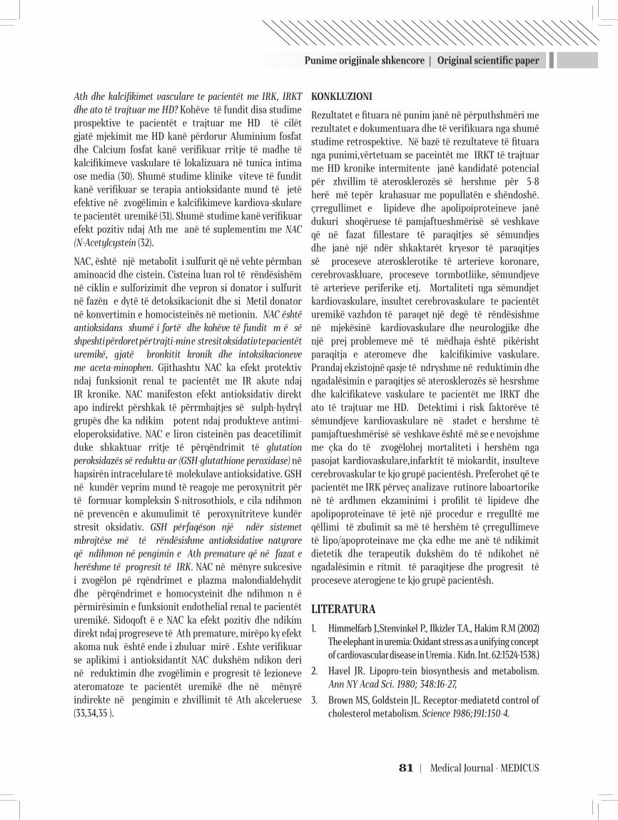
Punime origjinale shkencore | Original scientific paper
81 | Medical Journal - MEDICUS
Ath dhe kalcifikimet vasculare te pacientët me IRK, IRKT dhe ato të trajtuar me HD? Kohëve të fundit disa studime prospektive te pacientët e trajtuar me HD të cilët gjatë mjekimit me HD kanë përdorur Aluminium fosfat dhe Calcium fosfat kanë verifikuar rritje të madhe të kalcifikimeve vaskulare të lokalizuara në tunica intima ose media (30). Shumë studime klinike viteve të fundit kanë verifikuar se terapia antioksidante mund të jetë efektive në zvogëlimin e kalcifikimeve kardiova-skulare te pacientët uremikë (31). Shumë studime kanë verifikuar efekt pozitiv ndaj Ath me anë të suplementim me NAC (N-Acetylcystein (32).
NAC, është një metabolit i sulfurit që në vehte përmban aminoacid dhe cistein. Cisteina luan rol të rëndësishëm në ciklin e sulforizimit dhe vepron si donator i sulfurit në fazën e dytë të detoksikacionit dhe si Metil donator në konvertimin e homocisteinës në metionin. NAC është antioksidans shumë i fortë dhe kohëve të fundit m ë së shpeshti përdoret për trajti-min e stresit oksidativ te pacientët uremikë, gjatë bronkitit kronik dhe intoksikacioneve me aceta-minophen. Gjithashtu NAC ka efekt protektiv ndaj funksionit renal te pacientët me IR akute ndaj IR kronike. NAC manifeston efekt antioksidativ direkt apo indirekt përshkak të përrmbajtjes së sulph-hydryl grupës dhe ka ndikim potent ndaj produkteve antimi-eloperoksidative. NAC e liron cisteinën pas deacetilimit duke shkaktuar rritje të përqëndrimit të glutation peroksidazës së reduktu-ar (GSH-glutathione peroxidase) në hapsirën intracelulare të molekulave antioksidative. GSH në kundër veprim mund të reagoje me peroxynitrit për të formuar kompleksin S-nitrosothiols, e cila ndihmon në prevencën e akumulimit të peroxynitriteve kundër stresit oksidativ. GSH përfaqëson një ndër sistemet mbrojtëse më të rëndësishme antioksidative natyrore që ndihmon në pengimin e Ath premature që në fazat e herëshme të progresit të IRK. NAC në mënyre sukcesive i zvogëlon pë rqëndrimet e plazma malondialdehydit dhe përqëndrimet e homocysteinit dhe ndihmon n ë përmirësimin e funksionit endothelial renal te pacientët uremikë. Sidoqoft ë e NAC ka efekt pozitiv dhe ndikim direkt ndaj progreseve të Ath premature, mirëpo ky efekt akoma nuk është ende i zbuluar mirë . Eshte verifikuar se aplikimi i antioksidantit NAC dukshëm ndikon deri në reduktimin dhe zvogëlimin e progresit të lezioneve ateromatoze te pacientët uremikë dhe në mënyrë indirekte në pengimin e zhvillimit të Ath akceleruese (33,34,35 ).
KONKLUZIONI
Rezultatet e fituara në punim janë në përputhshmëri me rezultatet e dokumentuara dhe të verifikuara nga shumë studime retrospektive. Në bazë të rezultateve të fituara nga punimi,vërtetuam se paceintët me IRKT të trajtuar me HD kronike intermitente janë kandidatë potencial për zhvillim të aterosklerozës së hershme për 5-8 herë më tepër krahasuar me popullatën e shëndoshë. çrregullimet e lipideve dhe apolipoiproteineve janë dukuri shoqëruese të pamjaftueshmërisë së veshkave që në fazat fillestare të paraqitjes së sëmundjes dhe janë një ndër shkaktarët kryesor të paraqitjes së proceseve aterosklerotike të arterieve koronare, cerebrovaskluare, proceseve tormbotliike, sëmundjeve të arterieve periferike etj. Mortaliteti nga sëmundjet kardiovaskulare, insultet cerebrovaskulare te pacientët uremikë vazhdon të paraqet një degë të rëndësishme në mjekësinë kardiovaskulare dhe neurologjike dhe një prej problemeve më të mëdhaja është pikërisht paraqitja e ateromeve dhe kalcifikimive vaskulare. Prandaj ekzistojnë qasje të ndryshme në reduktimin dhe ngadalësimin e paraqitjes së aterosklerozës së hesrshme dhe kalcifikateve vaskulare te pacientët me IRKT dhe ato të trajtuar me HD. Detektimi i risk faktorëve të sëmundjeve kardiovaskulare në stadet e hershme të pamjaftueshmërisë së veshkave është më se e nevojshme me çka do të zvogëlohej mortaliteti i hershëm nga pasojat kardiovaskulare,infarktit të miokardit, insulteve cerebrovaskular te kjo grupë pacientësh. Preferohet që te pacientët me IRK përveç analizave rutinore laboartorike në të ardhmen ekzaminimi i profilit të lipideve dhe apolipoproteinave të jetë një procedur e rregulltë me qëllimi të zbulimit sa më të hershëm të çrregullimeve të lipo/apoproteinave me çka edhe me anë të ndikimit dietetik dhe terapeutik dukshëm do të ndikohet në ngadalësimin e ritmit të paraqitjese dhe progresit të proceseve aterogjene te kjo grupë pacientësh.
LITERATURA
1. Himmelfarb J.,Stenvinkel P., Ilkizler T.A., Hakim R.M (2002) The elephant in uremia: Oxidant stress as a unifying concept of cardiovascular disease in Uremia . Kidn. Int. 62:1524-1538.)
2. Havel JR. Lipopro-tein biosynthesis and metabolism. Ann NY Acad Sci. 1980; 348:16-27,
3. Brown MS, Goldstein JL. Receptor-mediatetd control of cholesterol metabolism. Science 1986;191:150-4.
Punime origjinale shkencore | Original scientific paper
Revistë mjekësore - MEDICUS | 82
4. Blanchette-Mackie EJ, Scow RD. Sites of lipoprotein lipaze activity in adipose tissue perfused with chylomi-crons. Electron microscope-cytochemical stady. J Cell Biol. 1971; 51:1-25 .
5. MGerard M. London , Sylvain J. Bruno P.Fabien M. Hassan A. Inflammation , arteriosclerosis and cardio-vascular therapy in haemodialysis patients. Kidn Int.Vol.63(2003); f.s88-93.
6. Rifai N, Chapnan JF, Silverman LN, Gwynnes JT. Review of serum lipids and apolipoproteins in risk assess-ment of coronary heart disease. Ann Clin Labor Sci. 1988;18:429-39.
7. Carmine Z. Franceska M. Giovanni T. Novel cardiovascu-lar risk factors in End-stage-Renale Disease. J.Am Soc Nephrol 15; S:77-80,2004.
8. Schwarz U., Buzello M.,Ritz E.,Stein G., Raabe G ., Wiest G., Mall G.,Amann K. (2000): Morphology of coronary atherosclerosis lesions in patient with End-stage renal failure. Nephrol Dial Transpl. 15:218-223.
9. Vallance P, Leone A, Calver A, et al. Accumulation of an endogenous inhibitor of nitric oxide synthesis in chron-ic renal failure. Lancet. 1992;399:572-5.
10. Kielstein JT, Boger RH, Bode-Boger SM, et al. Asym-Asym-metric dimethylarginine plasma concentrations dif-fer in patients with end-stage renal disease: relation-ship to treatment method and atherosclerosis disease. J Am Soc Nephrol. 1999;10:594-600. R= 13.Loscalzo J.(2001);Nitric oxide insufficiency ,platelet actiation and arterial thrombosis .Circ Res ;88:756-762.
11. Trimarchi H., Mongitore M.R ,Baglioni P., et al. (2003): N-acetylcysteine reduces malondialdehyde levels in chro-nic haemodialysis patients –a pilot study Clin Nephrol ; 59:441-446.
12. Muldoon MF, Manuck SB, Matthews KA. Lowering cholesterol concentrations and mortality: a quanti-tative review of primary prevention trials. Br Med J. 1990;301:309-14.
13. Kanne JP, Malloy MJ. Disorders of lipoprotein metabo-lism. In: Greenspan FS, editor. Basic and Clinical En-docrinology. East Norwalk: Apleton and Lange; 1991. pp.663-76.
14. Ilingworth DR, Connor WE. Disorders of lipid metabo-lism. In: Felig P, editor. Endocrinology and Metabolism. New York: Mc Graw-HilI; 1987. pp. 389-95.
15. Bard JM, Osel-Hagen E, Duriez P, Pfister P. Changes in plasma apolipoproitein B-containing lipoparticle levels
following therapy with fluvastatin and cholestyramine. Am J Cardiol. 1995;76(2):65A-70A.
16. Stamler J, Wentworth D, Neaton JD. Is relationship be-tween serum cholesterol and risk premature death from coronary heart disease continous and graded? Findings in 356,222 primary screenees of the Multiple Risk Factor Intervention Trial (MRFITV). JAMA. 1986; 256: 2823-8.
17. Gordon T, Kannel WB, Castelli VP, Dawber TR. Lipopro-teins cardiovascular disease and death. The Framing-ham Study. Arch Intern Med. 1981;141:1128-31.
18. Skinner ER, Wilson HM. High lipoprotein subrafc-tions and cardiovascular disease. Biochem Soc Trans. 1990;18:1074-1076.
19. Naito KH. Serum apolipoprotein measurements: An improved discriminator for assessing coronary heart disease risk. Compar Ther. 1987;13(11): 43-52.)
20. Hoff HJ, Heidemann CL, Ganbaz IW. Quantitation of apoB in human aortic fatty streaks: A comparison with grosisly normal intima and fibrous plaques, Atherosc-lerosis. 1987;30: 263-72.
21. Trieu VN, Me Contahy WJ. Lipoprotein(a) binding to other apolipoprotein B containing lipoproteins. Bioche-mistry. 1990;29:5919-24.
22. Hu XL, Bao WD, Song FR. The relationship of apolipo-protein A-I and B to cardiovascular disease in aging males. J Epidemiol. 1995;16(1):11-3 ).
23. McGill DA, Talsma P, Ardlie NG. Relationship of blood cholesterol and apoprotein B levels to angiographically defined coron. artery disease in young males. Coron Ar-tery Dis. 1993;4(3):361-9.
24. Wilcken DE, Wang XL, Greenwood J, Lynch J. Lipoprotein(a) and apolipoprotein B and A-I in children and coronary vascular events in their grandparents. J Pediatrics 1993;123(4):519-26.
25. Islam S, Gotin B, Manos T, Smith C, Treiber F. Low den-sity lipoprotein cholesterol/apolipoprotein B-100 ratio: interaction of family history of premature atheroscle-rotic coronary artery disease with race and gender in 7 to 11 year olds. Pediatrics.1994; 94 (4 Pt 1):494-9.
26. Morena M, et al. Protective effects of high-density lipo-protein against oxidative stress are impaired in haemodi-alysis patients. Nephrol Dial Transplant. 2000;15:389-95.
27. Wanner C, Krane V, Metzger T, Quaschning T. Lipid changes and statins in chronic renal insufficiency and dialysis. J Nephrol. 2001;14(S 4):S76-80.
Punime origjinale shkencore | Original scientific paper
83 | Medical Journal - MEDICUS
28. Himmelfarb J.,Stenvinkel P., Ilkizler T.A., Hakim R.M (2002) The elephant in uremia : Oxidant stress as a unifying concept of cardiovascular disease in Uremia . Kidn. Int. 62:1524-1538.
29. Massy Z.A., Nguyen-Khoa T. (2002) : Oxidative stress and chronic renal failure ;markers and management .J Ne-phr. 15:336-341.
30. Massy Z.,Ivanovski O., Nguyen-Khoa T., Angulo J., et al: (2005);Uremia accelerates both atherosclerosis and ar-terial calcification in apolipoprotein E knockout mice.J Am Soc Nephrol;16:109-116.
31. Tepel M.,Giet M., Statz M., Jankowski J.,Zidek W.(2003) ; The antioxidant acetylcysteine reduces cardiovascular events in patients with end-stage Renal failure :a rand-omized, controlled trial . Circul.107:992-995.
32. Scholze A.,Rinder C., Beige J., Riezler R., Tepel M. (2004); Acetylcysteine reduces plasma homocysteine concentra-cion and improves pulse pressure and endothelial function in patients with End-stage renal failure . Circ. 109:369-374.
33. Block G.A., Spiegel D.M.,Ehrlich J.,Mehta R., et al(2005): Effects of sevelamer and calcium on coronary artery calcification in patient new to haemodialysis .Kidney Int ; 68:1815-1824,
34. Chertow G.M,Burke S.K., Raggi P. (2002) :Sevelamer at-tenuates the progression of coronary and aortic calcifi-cation in haemodialysis patients. Kindey Int;62:245-252.
35. Ivanovski O.,Szumilak D., Ngyen-Khoa T., Ruellan N., Phan O., et al. (2005);The antioxidant N-Acetylcysteine Prevents accelerated atherosclerosis in uremic apolipo-protein E knockout mice. Kidney Int ; 67:2288-2294.
36. Havel JR, Yamada N, Shames DM. Role of apolipoprotein E in lipoprotein metabolism. Am Heart J. 1987;113:470-4.
37. Brown WV, Baginski ML. Inhibition of lipoprotein lipa-sa by an apoprotein of human very low density lipopro-tein. Biochem Biophys Res Comm. 1972;46:375-82.
38. Streja D, Kallai MA, Steiner G. The metabolic heteroge-neity of human very low density lipoprotein triglycer-ide. Metabolism 1977; 26:1333-44.
39. Brown MS, Goldstein J. A receptor-mediated pathway for cholesterol homeostasis. Science 1986;232: 34-47.)
40. Lutfi Zylbeari. Profili i Dislipidemisë dhe Aberacionet e Apoproteineve te pacientët e Mjekuar me Hemodializë Përsëritëse. Disertacioni i Doktoraturës. Shkup,2009
41. Miida T, et al. LCAT-dependent conversion of pre β1-HDL
into a-migrating HDL is severely delayed in haemodi-alysis patients. J Am Soc Nephrol. 2003;14:732-8.
42. Okubo K, Ikewaki K, Sakai S, Tada N, Kawaguchi Y, Mochizuki S. Abnormal HDL apolipoprotein A-I and A-II kinetics in haemodialysis patients: a stable isotope study. J Am Soc Nephrol. 2004;15:1008-15.
43. Janak R DE ZOYSA. Nephrology 2004;9, 83-88.44. Zölner N. Kirchs KZ. Fotometriska-oboena metoda. Ges
Exp Med. 1962; 135: 545.45. Friedewald WT, Levy RJ, Fredrickson DS. Estimation of
concentration of low density lipoprotein cholesterol without the use of the preparative ultracentrifuge. Clin Chem. 1972;18:499-502.
46. Bucola G, David H. Quantitative determination of serum triglycerides by use of enzymes. Clin Chem. 1973;19:476-82.
47. Allain CC, Poon LS, Chan CS, Richmond W. Enzymatic determination of total serum cholesterol. Clin Chem. 1974;20:470-5.
48. Wamick G, Benderson J, Allbers J. Quantitation of high density lipoprotein subclasses after separation by dextran sulfate and Mg+ precipitation. Clin Chem. 1982;28:1574-61.
49. Rifai N, King ME. Immunoturbidimetric assays of apolipoproteins A-I, A-II and B in serum. Clin Chem. 1986;23(6): 957-61.
50. Tilly P, et al. Biological and genetic determinants of se-rum apo C-III concentration: reference limits from the STANISLAS cohort study. J Lipid Res. 2003;44 :430-6.
51. Vincent-Viry M, et al. Biological variations and genetic reference values for apolipoprotein E serum concentra-tions: results from the STANISLAS cohort study. Clin Chem. 1998;44:957-65.
52. Selhub J, Miller JW. The pathogenesis of homocystein-emia: interruption of the coordinate regulation by S-ad-enosylmethionine of the remethylation and transsulfu-ration of homocysteine. Am J Clin Nutr. 1992;55:131-8.
53. Tietz NW, et al. Lipase in serum-the elusive enzyme: an overview. Clin Chem. 1993;39:746-56.
54. Steinberg WM, Goldstein SS, Davies ND, et al. Diag-Diag-nostic assays in acute pancreatitis. Ann Intern Med. 1985;102:576-80.
55. Leybold A, Junge W. Importance of colipase for the mea-surment of serum lipase activity. Adv Clin Enzymol. 1986;4:60-7.
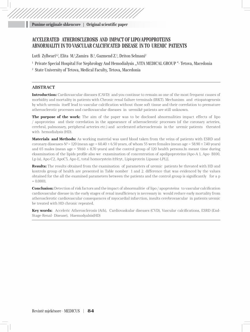
Punime origjinale shkencore | Original scientific paper
Revistë mjekësore - MEDICUS | 84
ACCELERATED ATHEROSCLEROSIS AND IMPACT OF LIPO/APPOPROTEINS ABNORMALITY IN TO VASCULAR CALCIFICATED DISEASE IN TO UREMIC PATIENTS
Lutfi Zylbeari1,2, Elita M.1,Zamira B.1, Gazmend Z.1, Driton Selmani2
1 Private Special Hospital For Nephrology And Hemodialysis ,,VITA MEDICAL GROUP ”- Tetova, Macedonia 2 State University of Tetova, Medical Faculty, Tetova, Macedonia
ABSTRACT
Introduction: Cardiovascular diseases (CAVD) and you continue to remain as one of the most frequent causes of morbidity and mortality in patients with Chronic renal failure terminals (IRKT). Mechanism and etiopatogenesis by which uremia itself lead to vascular calcification without those soft tissue and their correlation to premature atherosclerotic processes and cardiovascular diseases in uremikë patients are still unknown.
The purpose of the work: The aim of the paper was to be disclosed abnormalities impact effects of lipo / apoproteins and their correlation in the appearance of atherosclerotic processes (of the coronary arteries, cerebral, pulmonary, peripheral arteries etc.) and accelerated atherosclerosis in the uremic patients threated with hemodialysis (HD).
Materials and Methods: As working material was used blood taken from the veins of patients with ESRD and coronary disseases-N0 = 120 (mean age = 60.40 ± 6.50 years, of whom 55 were females (mean age = 58.90 ± 7.40 years) and 65 males (mean age = 59.60 ± 8.70 years) and the control group of 120 health persons.In meant time during eksamination of the lipids profile also we exzamination of concentration of apolipoproteins (Apo-A 1, Apo- B100, Lp (a), Apo-C2, ApoC3, Apo-E, total homocystein (tHcyt, Lipioprotein Lipasae-LPL)].
Results: The results obtained from the examination of parameters of uremic patients be threated with HD and kontrols group of health are presented in Table nomber 1 and 2. difference that was evidenced by the values obtained for the all the examined parameters between the patients and the control group is significantly for a p = 0.0001.
Conclusion: Detection of risk factors and the impact of abnormalitie of lipo / apoproteins to vascular calcification cardiovascular disease in the early stages of renal insufficiency is necessary in would reduce early mortality from atherosclerotic cardiovascular consequences of myocardial infarction, insults cerebrovascular in patients uremic be treated with HD chronic repeated.
Key words: Acceleric Atherosclerosis (Ath), Cardiovaskular diseases (CVD), Vascular calcifications, ESRD (End-Stage-Renal- Disease), Haemodyalsis(HD)
85 | Medical Journal - MEDICUS
NDJESHMËRIA E LARTË E METODËS FISH (FLUORESCENCE IN SITU HYBRIDIZATION) NË NDJEKJEN E TË SËMURËVE ME LEUKOZË MIELOIDE KRONIKE TË TRAJTUAR ME GLIVEKDorina Roko¹, Anila Babameto–Laku¹, Pal Xhumari², Donjeta Bali³, Vahe Mokini¹, Maksim Cikuli¹
¹ Shërbimi i Gjenetikës Mjekësore, QSU “Nënë Tereza”, Fakulteti i Mjeksisë, Tiranë
² Shërbimi i Hematologjisë, QSU “Nënë Tereza”, Fakulteti i Mjeksisë, Tiranë
³ Shërbimi i Onkohematologjisë, QSU “Nënë Tereza”, Fakulteti i Mjeksisë, Tiranë
ABSTRAKTI
Hyrje: Leukoza mieloide kronike (LMC) është një sëmundje malinje që shkaktohet nga translokacioni midis kromozomeve 9 dhe 22 (kromozomit Philadelphia). Nga pikpamja molekulare, rezultat i translokacionit është aktivizimi i onkogjenit bcr-abl, i cili ka aktivitet të lartë tirozin kinazik. Imatinib mesylate (Glivek) është një inhibitor specifik, i fuqishëm i bcr-abl që jep rezultate pozitive tek pacientët me LMC. (1, 2).
Qëllimi: Qëllimi i këtij studimi është përcaktimi i efektshmërisë së Glivekut nëpërmjet vlerësimit të arritjes së përgjigjes citogjenetike me anë të dy metodave (analiza kromozomike me banda G dhe metoda FISH (fluorescence in situ hybridization ) dhe krahasimi i tyre (15, 17).
Materiali dhe metoda: Është marrë në studim një grup prej 52 të sëmurësh me LMC të diagnostikuar pranë Shërbimit të Hematologjsë QSU “Nënë Tereza” Tiranë në periudhën prill 2011 - prill 2012 me grupmoshën (18-70 vjeç). Të gjithë pacientët janë ndjekur paralelisht me analizën citogjenetike dhe FISH duke përdorur Glivekun si trajtim terapeutik fillestar.
Rezultatet: Nga rezultatet e analizave citogjenetike dhe FISH pas 12 muajsh terapi rezultoi se 50 pacientë ose (96%) kishin arritur përgjigje hematologjike komplete. Nga keta 34 ose (68%) kishin arritur përgjigje citogjenetike komplete e vlerësuar kjo nga analiza citogjenetike. Në 4 pacientë të cilët rezultuan negativ për Ph+ në kariotip gjeni bcr-abl+ u identifikua me metodën FISH.
Konkluzione: Analiza citogjenetike dhe FISH japin rezultate të besueshme, por në studimin tonë u konfirmua se FISH është teknikë më e ndjeshme në vlerësimin e përgjigjeve citogjenetike duke identifikuar raste me gjenin bcr-abl të cilat rezultojnë negative nga ekzamnimi i kariotipit.
Fjalë kyç: Leukoza mieloide kronike, përgjigje molekulare, përgjigje citogjenetike, glivek, FISH.
HYRJE
Leukoza mieloide kronike (LMC) është një sëmundje që karakterizohet nga një rritje klonale e qelizave pararendëse hematopoetike të transformuara që proliferojnë në mënyrë malinje duke formuar një popullatë qelizash tumorale të cilat gradualisht zëvendësojnë hematopoiezën normale me një hiperplazi mieloide (7).
Nga pikpamja citogjenetike LMC karakterizohet nga prania e një translokacioni ndërmjet zonave 9q34 dhe 22q11 duke formuar kromozomin Philadelphia (3).
Nga pikpamja molekulare, rezultat i translokacionit është aktivizimi i onkogjenit abl i lokalizuar në 9q34, i cili translokohet në kromozomin 22 në skajin 5’ të gjenit bcr të lokalizuar në 22q11.
Aktiviteti themelor i bcr/abl favorizon rritjen e qelizave të transformara. Ky veprim realizohet nëpërmjet tre mënyrave:
Rritje e tejçimit te sinjalit në rrugën e proliferimit
Rritje e pavarësisë ndaj citokinave
Frenon rrugën normale të vdekjes të programuar qelizore dhe anomali të tjera citoskeletore (8)
Medicus 2013, Vol. 18 (2): 85-91
13Punime origjinale shkencore | Original scientific paper
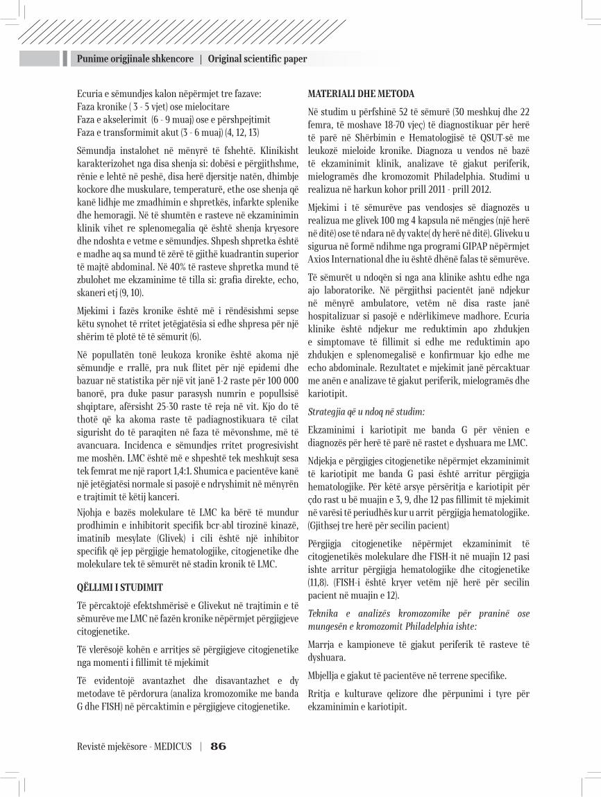
Punime origjinale shkencore | Original scientific paper
Revistë mjekësore - MEDICUS | 86
Ecuria e sëmundjes kalon nëpërmjet tre fazave:Faza kronike ( 3 - 5 vjet) ose mielocitareFaza e akselerimit (6 - 9 muaj) ose e përshpejtimitFaza e transformimit akut (3 - 6 muaj) (4, 12, 13)
Sëmundja instalohet në mënyrë të fshehtë. Klinikisht karakterizohet nga disa shenja si: dobësi e përgjithshme, rënie e lehtë në peshë, disa herë djersitje natën, dhimbje kockore dhe muskulare, temperaturë, ethe ose shenja që kanë lidhje me zmadhimin e shpretkës, infarkte splenike dhe hemoragji. Në të shumtën e rasteve në ekzaminimin klinik vihet re splenomegalia që është shenja kryesore dhe ndoshta e vetme e sëmundjes. Shpesh shpretka është e madhe aq sa mund të zërë të gjithë kuadrantin superior të majtë abdominal. Në 40% të rasteve shpretka mund të zbulohet me ekzaminime të tilla si: grafia direkte, echo, skaneri etj (9, 10).
Mjekimi i fazës kronike është më i rëndësishmi sepse këtu synohet të rritet jetëgjatësia si edhe shpresa për një shërim të plotë të të sëmurit (6).
Në popullatën tonë leukoza kronike është akoma një sëmundje e rrallë, pra nuk flitet për një epidemi dhe bazuar në statistika për një vit janë 1-2 raste për 100 000 banorë, pra duke pasur parasysh numrin e popullsisë shqiptare, afërsisht 25-30 raste të reja në vit. Kjo do të thotë që ka akoma raste të padiagnostikuara të cilat sigurisht do të paraqiten në faza të mëvonshme, më të avancuara. Incidenca e sëmundjes rritet progresivisht me moshën. LMC është më e shpeshtë tek meshkujt sesa tek femrat me një raport 1,4:1. Shumica e pacientëve kanë një jetëgjatësi normale si pasojë e ndryshimit në mënyrën e trajtimit të këtij kanceri.
Njohja e bazës molekulare të LMC ka bërë të mundur prodhimin e inhibitorit specifik bcr-abl tirozinë kinazë, imatinib mesylate (Glivek) i cili është një inhibitor specifik që jep përgjigje hematologjike, citogjenetike dhe molekulare tek të sëmurët në stadin kronik të LMC.
QËLLIMI I STUDIMIT
Të përcaktojë efektshmërisë e Glivekut në trajtimin e të sëmurëve me LMC në fazën kronike nëpërmjet përgjigjeve citogjenetike.
Të vlerësojë kohën e arritjes së përgjigjeve citogjenetike nga momenti i fillimit të mjekimit
Të evidentojë avantazhet dhe disavantazhet e dy metodave të përdorura (analiza kromozomike me banda G dhe FISH) në përcaktimin e përgjigjeve citogjenetike.
MATERIALI DHE METODA
Në studim u përfshinë 52 të sëmurë (30 meshkuj dhe 22 femra, të moshave 18-70 vjeç) të diagnostikuar për herë të parë në Shërbimin e Hematologjisë të QSUT-së me leukozë mieloide kronike. Diagnoza u vendos në bazë të ekzaminimit klinik, analizave të gjakut periferik, mielogramës dhe kromozomit Philadelphia. Studimi u realizua në harkun kohor prill 2011 - prill 2012.
Mjekimi i të sëmurëve pas vendosjes së diagnozës u realizua me glivek 100 mg 4 kapsula në mëngjes (një herë në ditë) ose të ndara në dy vakte( dy herë në ditë). Gliveku u sigurua në formë ndihme nga programi GIPAP nëpërmjet Axios International dhe iu është dhënë falas të sëmurëve.
Të sëmurët u ndoqën si nga ana klinike ashtu edhe nga ajo laboratorike. Në përgjithsi pacientët janë ndjekur në mënyrë ambulatore, vetëm në disa raste janë hospitalizuar si pasojë e ndërlikimeve madhore. Ecuria klinike është ndjekur me reduktimin apo zhdukjen e simptomave të fillimit si edhe me reduktimin apo zhdukjen e splenomegalisë e konfirmuar kjo edhe me echo abdominale. Rezultatet e mjekimit janë përcaktuar me anën e analizave të gjakut periferik, mielogramës dhe kariotipit.
Strategjia që u ndoq në studim:
Ekzaminimi i kariotipit me banda G për vënien e diagnozës për herë të parë në rastet e dyshuara me LMC.
Ndjekja e përgjigjes citogjenetike nëpërmjet ekzaminimit të kariotipit me banda G pasi është arritur përgjigja hematologjike. Për këtë arsye përsëritja e kariotipit për çdo rast u bë muajin e 3, 9, dhe 12 pas fillimit të mjekimit në varësi të periudhës kur u arrit përgjigja hematologjike. (Gjithsej tre herë për secilin pacient)
Përgjigja citogjenetike nëpërmjet ekzaminimit të citogjenetikës molekulare dhe FISH-it në muajin 12 pasi ishte arritur përgjigja hematologjike dhe citogjenetike (11,8). (FISH-i është kryer vetëm një herë për secilin pacient në muajin e 12).
Teknika e analizës kromozomike për praninë ose mungesën e kromozomit Philadelphia ishte:
Marrja e kampioneve të gjakut periferik të rasteve të dyshuara.
Mbjellja e gjakut të pacientëve në terrene specifike.
Rritja e kulturave qelizore dhe përpunimi i tyre për ekzaminimin e kariotipit.
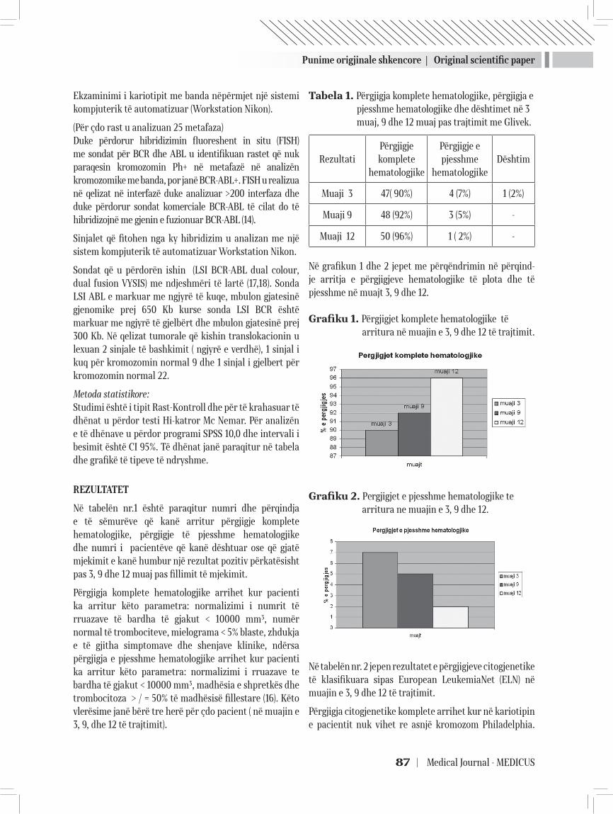
Punime origjinale shkencore | Original scientific paper
87 | Medical Journal - MEDICUS
Ekzaminimi i kariotipit me banda nëpërmjet një sistemi kompjuterik të automatizuar (Workstation Nikon).
(Për çdo rast u analizuan 25 metafaza)Duke përdorur hibridizimin fluoreshent in situ (FISH) me sondat për BCR dhe ABL u identifikuan rastet që nuk paraqesin kromozomin Ph+ në metafazë në analizën kromozomike me banda, por janë BCR-ABL+. FISH u realizua në qelizat në interfazë duke analizuar >200 interfaza dhe duke përdorur sondat komerciale BCR-ABL të cilat do të hibridizojnë me gjenin e fuzionuar BCR-ABL (14).
Sinjalet që fitohen nga ky hibridizim u analizan me një sistem kompjuterik të automatizuar Workstation Nikon.
Sondat që u përdorën ishin (LSI BCR-ABL dual colour, dual fusion VYSIS) me ndjeshmëri të lartë (17,18). Sonda LSI ABL e markuar me ngjyrë të kuqe, mbulon gjatesinë gjenomike prej 650 Kb kurse sonda LSI BCR është markuar me ngjyrë të gjelbërt dhe mbulon gjatesinë prej 300 Kb. Në qelizat tumorale që kishin translokacionin u lexuan 2 sinjale të bashkimit ( ngjyrë e verdhë), 1 sinjal i kuq për kromozomin normal 9 dhe 1 sinjal i gjelbert për kromozomin normal 22.
Metoda statistikore:Studimi është i tipit Rast-Kontroll dhe për të krahasuar të dhënat u përdor testi Hi-katror Mc Nemar. Për analizën e të dhënave u përdor programi SPSS 10,0 dhe intervali i besimit është CI 95%. Të dhënat janë paraqitur në tabela dhe grafikë të tipeve të ndryshme.
REZULTATET
Në tabelën nr.1 është paraqitur numri dhe përqindja e të sëmurëve që kanë arritur përgjigje komplete hematologjike, përgjigje të pjesshme hematologjike dhe numri i pacientëve që kanë dështuar ose që gjatë mjekimit e kanë humbur një rezultat pozitiv përkatësisht pas 3, 9 dhe 12 muaj pas fillimit të mjekimit.
Përgjigja komplete hematologjike arrihet kur pacienti ka arritur këto parametra: normalizimi i numrit të rruazave të bardha të gjakut < 10000 mm³, numër normal të trombociteve, mielograma < 5% blaste, zhdukja e të gjitha simptomave dhe shenjave klinike, ndërsa përgjigja e pjesshme hematologjike arrihet kur pacienti ka arritur këto parametra: normalizimi i rruazave te bardha të gjakut < 10000 mm³, madhësia e shpretkës dhe trombocitoza > / = 50% të madhësisë fillestare (16). Këto vlerësime janë bërë tre herë për çdo pacient ( në muajin e 3, 9, dhe 12 të trajtimit).
Tabela 1. Përgjigja komplete hematologjike, përgjigja e pjesshme hematologjike dhe dështimet në 3 muaj, 9 dhe 12 muaj pas trajtimit me Glivek.
RezultatiPërgjigje komplete
hematologjike
Përgjigje e pjesshme
hematologjikeDështim
Muaji 3 47( 90%) 4 (7%) 1 (2%)
Muaji 9 48 (92%) 3 (5%) -
Muaji 12 50 (96%) 1 ( 2%) -
Në grafikun 1 dhe 2 jepet me përqëndrimin në përqind-je arritja e përgjigjeve hematologjike të plota dhe të pjesshme në muajt 3, 9 dhe 12.
Grafiku 1. Përgjigjet komplete hematologjike të arritura në muajin e 3, 9 dhe 12 të trajtimit.
Grafiku 2. Pergjigjet e pjesshme hematologjike te arritura ne muajin e 3, 9 dhe 12.
Në tabelën nr. 2 jepen rezultatet e përgjigjeve citogjenetike të klasifikuara sipas European LeukemiaNet (ELN) në muajin e 3, 9 dhe 12 të trajtimit.
Përgjigja citogjenetike komplete arrihet kur në kariotipin e pacientit nuk vihet re asnjë kromozom Philadelphia.
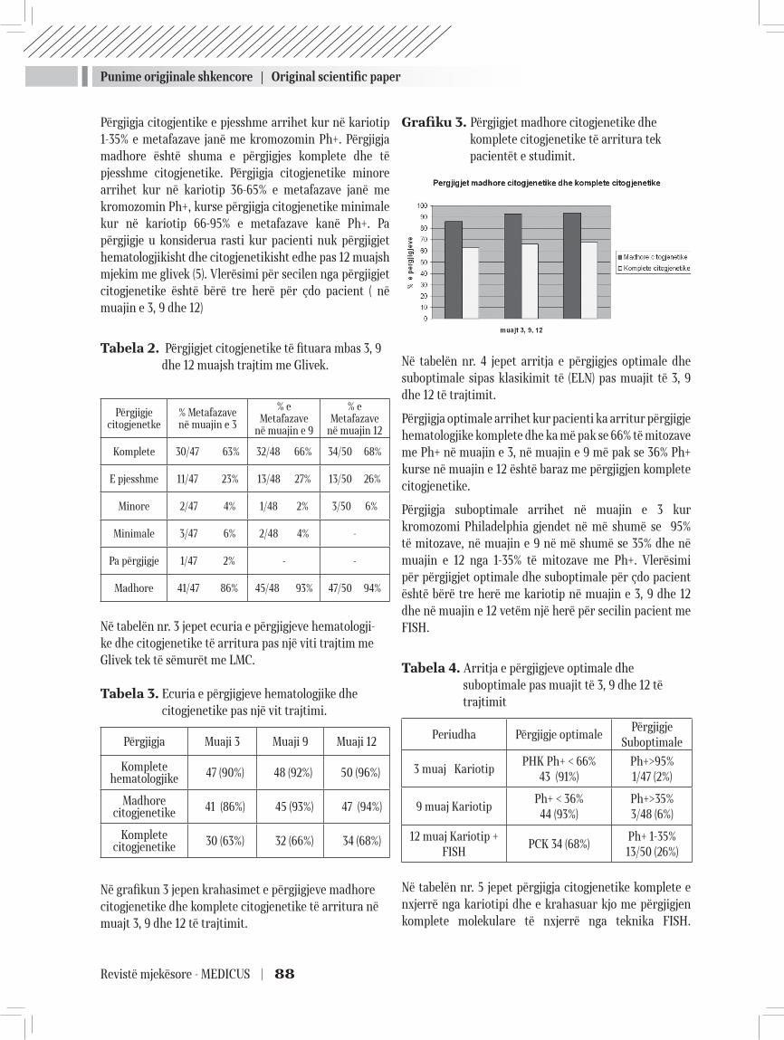
Punime origjinale shkencore | Original scientific paper
Revistë mjekësore - MEDICUS | 88
Përgjigja citogjentike e pjesshme arrihet kur në kariotip 1-35% e metafazave janë me kromozomin Ph+. Përgjigja madhore është shuma e përgjigjes komplete dhe të pjesshme citogjenetike. Përgjigja citogjenetike minore arrihet kur në kariotip 36-65% e metafazave janë me kromozomin Ph+, kurse përgjigja citogjenetike minimale kur në kariotip 66-95% e metafazave kanë Ph+. Pa përgjigje u konsiderua rasti kur pacienti nuk përgjigjet hematologjikisht dhe citogjenetikisht edhe pas 12 muajsh mjekim me glivek (5). Vlerësimi për secilen nga përgjigjet citogjenetike është bërë tre herë për çdo pacient ( në muajin e 3, 9 dhe 12)
Tabela 2. Përgjigjet citogjenetike të fituara mbas 3, 9 dhe 12 muajsh trajtim me Glivek.
Përgjigje citogjenetke
% Metafazave në muajin e 3
% e Metafazave
në muajin e 9
% e Metafazave
në muajin 12
Komplete 30/47 63% 32/48 66% 34/50 68%
E pjesshme 11/47 23% 13/48 27% 13/50 26%
Minore 2/47 4% 1/48 2% 3/50 6%
Minimale 3/47 6% 2/48 4% -
Pa përgjigje 1/47 2% - -
Madhore 41/47 86% 45/48 93% 47/50 94%
Në tabelën nr. 3 jepet ecuria e përgjigjeve hematologji-ke dhe citogjenetike të arritura pas një viti trajtim me Glivek tek të sëmurët me LMC.
Tabela 3. Ecuria e përgjigjeve hematologjike dhe citogjenetike pas një vit trajtimi.
Përgjigja Muaji 3 Muaji 9 Muaji 12
Komplete hematologjike 47 (90%) 48 (92%) 50 (96%)
Madhore citogjenetike 41 (86%) 45 (93%) 47 (94%)
Komplete citogjenetike 30 (63%) 32 (66%) 34 (68%)
Në grafikun 3 jepen krahasimet e përgjigjeve madhore citogjenetike dhe komplete citogjenetike të arritura në muajt 3, 9 dhe 12 të trajtimit.
Grafiku 3. Përgjigjet madhore citogjenetike dhe komplete citogjenetike të arritura tek pacientët e studimit.
Në tabelën nr. 4 jepet arritja e përgjigjes optimale dhe suboptimale sipas klasikimit të (ELN) pas muajit të 3, 9 dhe 12 të trajtimit.
Përgjigja optimale arrihet kur pacienti ka arritur përgjigje hematologjike komplete dhe ka më pak se 66% të mitozave me Ph+ në muajin e 3, në muajin e 9 më pak se 36% Ph+ kurse në muajin e 12 është baraz me përgjigjen komplete citogjenetike.
Përgjigja suboptimale arrihet në muajin e 3 kur kromozomi Philadelphia gjendet në më shumë se 95% të mitozave, në muajin e 9 në më shumë se 35% dhe në muajin e 12 nga 1-35% të mitozave me Ph+. Vlerësimi për përgjigjet optimale dhe suboptimale për çdo pacient është bërë tre herë me kariotip në muajin e 3, 9 dhe 12 dhe në muajin e 12 vetëm një herë për secilin pacient me FISH.
Tabela 4. Arritja e përgjigjeve optimale dhe suboptimale pas muajit të 3, 9 dhe 12 të trajtimit
Periudha Përgjigje optimalePërgjigje
Suboptimale
3 muaj KariotipPHK Ph+ < 66%
43 (91%)Ph+>95%1/47 (2%)
9 muaj KariotipPh+ < 36%
44 (93%)Ph+>35%3/48 (6%)
12 muaj Kariotip +FISH
PCK 34 (68%)Ph+ 1-35%13/50 (26%)
Në tabelën nr. 5 jepet përgjigja citogjenetike komplete e nxjerrë nga kariotipi dhe e krahasuar kjo me përgjigjen komplete molekulare të nxjerrë nga teknika FISH.
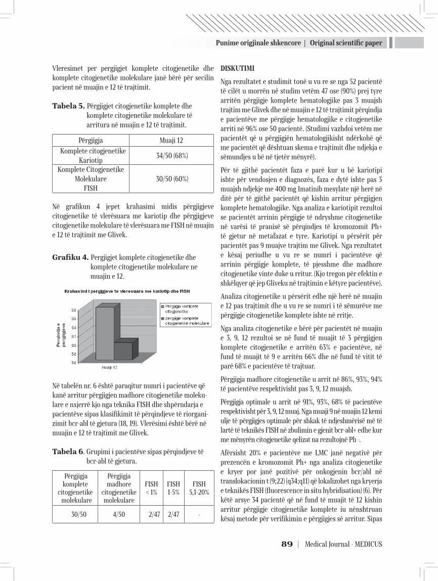
Punime origjinale shkencore | Original scientific paper
89 | Medical Journal - MEDICUS
Vleresimet per pergjigjet komplete citogjenetike dhe komplete citogjenetike molekulare janë bërë për secilin pacient në muajin e 12 të trajtimit.
Tabela 5. Përgjigjet citogjenetike komplete dhe komplete citogjenetike molekulare të arritura në muajin e 12 të trajtimit.
Përgjigja Muaji 12
Komplete citogjenetikeKariotip
34/50 (68%)
Komplete Citogjenetike Molekulare
FISH 30/50 (60%)
Në grafikun 4 jepet krahasimi midis përgjigjeve citogjenetike të vlerësuara me kariotip dhe përgjigjeve citogjenetike molekulare të vlerësuara me FISH në muajin e 12 të trajtimit me Glivek.
Grafiku 4. Pergjigjet komplete citogjenetike dhe komplete citogjenetike molekulare ne muajin e 12.
Në tabelën nr. 6 është paraqitur numri i pacientëve që kanë arritur përgjigjen madhore citogjenetike moleku-lare e nxjerrë kjo nga teknika FISH dhe shpërndarja e pacientëve sipas klasifikimit të përqindjeve të riorgani-zimit bcr-abl të gjetura (18, 19). Vlerësimi është bërë në muajin e 12 të trajtimit me Glivek.
Tabela 6. Grupimi i pacientëve sipas përqindjeve të bcr-abl të gjetura.
Përgjigja komplete
citogjenetike molekulare
Përgjigjamadhore
citogjenetikemolekulare
FISH< 1%
FISH1-5%
FISH5,1-20%
30/50 4/50 2/47 2/47 -
DISKUTIMI
Nga rezultatet e studimit tonë u vu re se nga 52 pacientë të cilët u morrën në studim vetëm 47 ose (90%) prej tyre arritën përgjigje komplete hematologjike pas 3 muajsh trajtim me Glivek dhe në muajin e 12 të trajtimit përqindja e pacientëve me përgjigje hematologjike e citogjenetike arriti në 96% ose 50 pacientë. (Studimi vazhdoi vetëm me pacientët që u përgjigjën hematologjikisht ndërkohë që me pacientët që dështuan skema e trajtimit dhe ndjekja e sëmundjes u bë në tjetër mënyrë).
Për të gjithë pacientët faza e parë kur u bë kariotipi ishte për vendosjen e diagnozës, faza e dytë ishte pas 3 muajsh ndjekje me 400 mg Imatinib mesylate një herë në ditë për të gjithë pacientët që kishin arritur përgjigjen komplete hematologjike. Nga analiza e kariotipit rezultoi se pacientët arrinin përgjigje të ndryshme citogjenetike në varësi të pranisë së përqindjes të kromozomit Ph+ të gjetur në metafazat e tyre. Kariotipi u përsërit për pacientët pas 9 muajve trajtim me Glivek. Nga rezultatet e kësaj periudhe u vu re se numri i pacientëve që arrinin përgjigje komplete, të pjesshme dhe madhore citogjenetike vinte duke u rritur. (Kjo tregon për efektin e shkëlqyer që jep Gliveku në trajtimin e këtyre pacientëve).
Analiza citogjenetike u përsërit edhe një herë në muajin e 12 pas trajtimit dhe u vu re se numri i të sëmurëve me përgjigje citogjenetike komplete ishte në rritje.
Nga analiza citogjenetike e bërë për pacientët në muajin e 3, 9, 12 rezultoi se në fund të muajit të 3 përgjigjen komplete citogjenetike e arritën 63% e pacientëve, në fund të muajit të 9 e arritën 66% dhe në fund të vitit të parë 68% e pacientëve të trajtuar.
Përgjigja madhore citogjenetike u arrit në 86%, 93%, 94% të pacientëve respektivisht pas 3, 9, 12 muajsh.
Përgjigja optimale u arrit në 91%, 93%, 68% të pacientëve respektivisht për 3, 9, 12 muaj. Nga muaji 9 në muajin 12 kemi ulje të përgjigjes optimale për shkak të ndjeshmërisë më të lartë të teknikës FISH në zbulimin e gjenit bcr-abl+ edhe kur me mënyrën citogjenetike qelizat na rezultojnë Ph -.
Afërsisht 20% e pacientëve me LMC janë negativë për prezencën e kromozomit Ph+ nga analiza citogjenetike e kryer por janë pozitivë për onkogjenin bcr/abl në translokacionin t (9;22) (q34;q11) që lokalizohet nga kryerja e teknikës FISH (fluorescence in situ hybridisation) (6). Për këtë arsye 34 pacientë që në fund të muajit të 12 kishin arritur përgjigje citogjenetike komplete iu nënshtruan kësaj metode për verifikimin e përgjigjes së arritur. Sipas
Punime origjinale shkencore | Original scientific paper
Revistë mjekësore - MEDICUS | 90
teknikës FISH rezultoi se në 4 nga këta pacientë u gjendën riorganizimet bcr-abl dhe se rezultatet e nxjerra prej saj nuk ishin të njëjta për të gjithë pacientët. Kjo tregon për lidhjen e ngushtë midis analizës citogjenetike standarte (kariotip) dhe teknikës FISH, por njëkohësisht edhe për ndjeshmërinë më të lartë të FISH-it krahasuar me kariotipin për shkak se identifikohen ndryshime të cilat nga kariotipi nuk mund të nxirren.
PËRFUNDIMET
Ekzaminimi me anën e kariotipit mbetet metoda e artë për vendosjen e diagnozës dhe ndekjen e të sëmurëve të trajtuar me Glivek.
Trajtimi me anën e tij është më efektivi në stadin kronik të LMC dhe se rezultatet më të mira arrihen kur përdoret si linja e parë e trajtimit.
Gliveku mund të shkaktojë reagime të qëndrueshme të lidhura me mbijetesën e zgjatur në pacientët me LMC.
Ndjeshmëria e tumoreve në përqindje dhe saktësia në vlerësimin e tyre sipas metodës FISH është më e lartë krahasuar me kariotipin, për këtë ajo shërben si një parameter diagnostik dhe prognostik më i saktë se analiza kromozomike.
Për kryerjen e kariotipit duhen disa ditë punë dhe kërkohen qeliza në fazën e metafazës së mitozës, ndërsa për FISH- in kërkohet më pak kohë dhe qeliza në interfazë.
Negativiteti dhe pozitiviteti i rremë janë më të larta gjatë kryerjes së kariotipit sesa gjatë kryerjes së FISH-it, por përparësia e citogjenetikës qëndron në faktin e zbulimit të anomalive të tjera kromozomike në kariotipin e pacientëve në krahasim me metodën citogjenetike molekulare.
REFERENCA
1. Lucy C. Crossman, MD, Stephen O’Brien, MD, PhD. Imatinib therapy in chronic myeloid leukemia. Hematol Oncol Clin N Am 2004; 605 – 17
2. Faderi S, Talpaz M, Estrov Z et al. Chronic myeloid leuke-mia, biology and therapy. Ann Intern Med 1999; 207- 19
3. Ganesan TS, Rassool R, Guo A-P, Th’ng KH, Dowding C, Hibbin JA, Young BD, White H, Kumaran TO, Galton DAG, Goldman JM. Rearrangement of the bcr gene in Philadelphia chromosome-negative chronic myeloid leukemia. Blood 1986; 957- 60
4. Druker BJ,Talpaz M,Resta DJ, et al. Efficacy and safety of a specific inhibitor of the BCR-ABL tyrosine kinase in chronic myeloid leukemia. N Engl J Med 2001; 344: 1031–7
5. Hochhaus A, Weisser A, La Rosée P, Emig M, Müller MC, Saussele S, Reiter A, Kuhn C, Berger U, Hehlmann R, Cross NCP. Detection and quantifi cation of residual dis-Detection and quantification of residual dis-ease in chronic myelogenous leukemia. Leukemia 2000; 14: 998-1005
6. Nanjangud G, Kadam PR, Saikia T, Bhisey AN, Kumar A, Gopal R, Chopra H, Nair CN, Advani SH. Karyotypic fi nd-Karyotypic find-ings as an independent prognostic marker in chronic myeloid leukaemia blast crisis. Leuk Res 1994; 18: 385-92
7. Drunker BJ, Tamura S, Buchdunger E, Ohno S, Segal GM, Fanning S, et al Effects of a selective inhibitor of the tyrosine on the groëth of Bcr-Abl positive cells. Nat Med 1996; 2:561-6
8. Ganesan TS, Rassool R, Guo A-P, Th’ng KH, Doëding C, Hibbin JA, Young BD, White H, Kumaran TO, Galton DAG, Goldman JM. Rearrangement of the bcr gene in Philadelphia chromosome-negative chronic myeloid leukemia. Blood 1986; 68: 957- 60
9. Mitelman F. The cytogenetic scenario of chronic my-eloid leukemia. Leuk Lymphoma 1993; 11: 11-5
10. Cross NC. Minimal residual disease in chronic myeloid leukaemia. Hematol Cell Ther 1998; 40: 224- 8
11. Deininger M.W, Goldman J.M, Melo JV. The molecular bi-ology of chronic myeloid leukemia Blood 2000; 3343-56
12. Rowley JD. A new consistent chromosomal abnormality in chronic myeloid leukemia identified by fluorescence and giemsa staining. Nature. 1973;243: 290-3
13. Deininger M. W. Goldman J. M, Lyndon N, Melo J.V The tyrosine inhibitor CGP 57148B selectively inhibits the groëth of BCR- ABL positive cells. Blood; 1997; 90: 3691 – 8
14. Tkachuk DC, Westbrook CA, Andreeff M, Donlon TA, Cleary ML, Suryanarayan K, Homge M, Redner A, Gray J, Pinkel D. Detection of bcr-abl fusion in chronic myelo-geneous leukemia by in situ hybridization. Science 1990; 250: 559-62
15. 12 Seong DC, Kantarjian HM, Ro JY, Talpaz M, Xu J, Rob-inson JR, Deisseroth AB, Champlin RE, Siciliano MJ. Hypermetaphase fluorescence in situ hybridization for quantitative monitoring of Philadelphia chromosome-positive cells in patients with chronic myelogenous leu-kemia during treatment. Blood 1995; 86: 2343-9
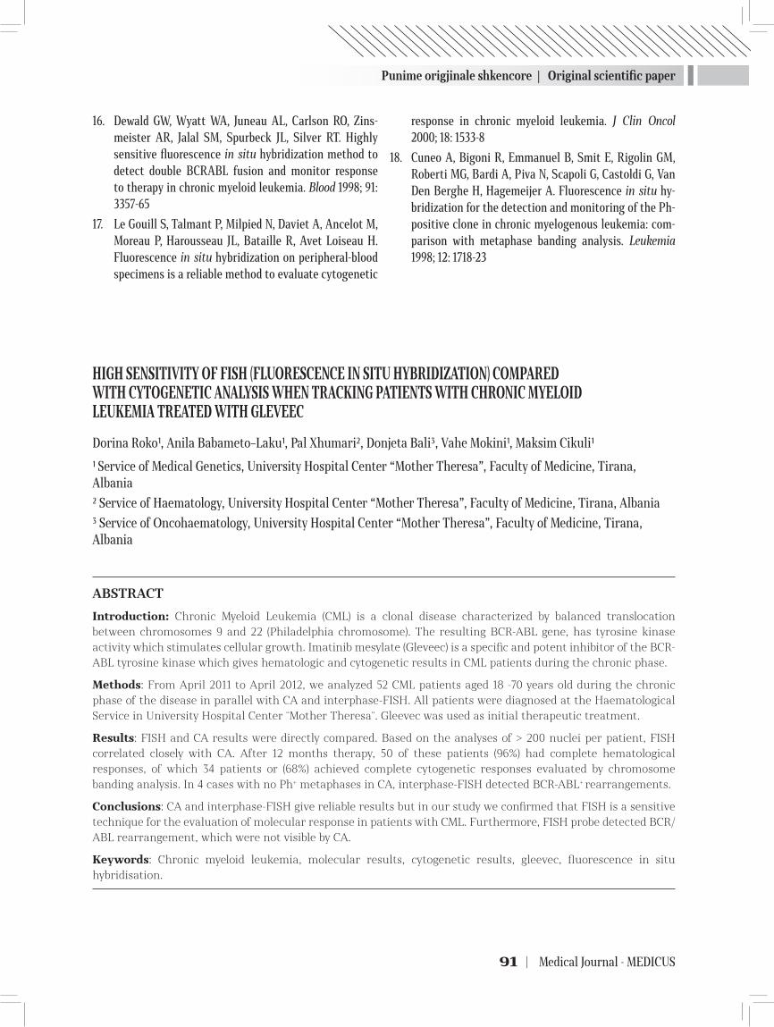
Punime origjinale shkencore | Original scientific paper
91 | Medical Journal - MEDICUS
16. Dewald GW, Wyatt WA, Juneau AL, Carlson RO, Zins-meister AR, Jalal SM, Spurbeck JL, Silver RT. Highly sensitive fluorescence in situ hybridization method to detect double BCRABL fusion and monitor response to therapy in chronic myeloid leukemia. Blood 1998; 91: 3357-65
17. Le Gouill S, Talmant P, Milpied N, Daviet A, Ancelot M, Moreau P, Harousseau JL, Bataille R, Avet Loiseau H. Fluorescence in situ hybridization on peripheral-blood specimens is a reliable method to evaluate cytogenetic
response in chronic myeloid leukemia. J Clin Oncol 2000; 18: 1533-8
18. Cuneo A, Bigoni R, Emmanuel B, Smit E, Rigolin GM, Roberti MG, Bardi A, Piva N, Scapoli G, Castoldi G, Van Den Berghe H, Hagemeijer A. Fluorescence in situ hy-bridization for the detection and monitoring of the Ph-positive clone in chronic myelogenous leukemia: com-parison with metaphase banding analysis. Leukemia 1998; 12: 1718-23
HIGH SENSITIVITY OF FISH (FLUORESCENCE IN SITU HYBRIDIZATION) COMPARED WITH CYTOGENETIC ANALYSIS WHEN TRACKING PATIENTS WITH CHRONIC MYELOID LEUKEMIA TREATED WITH GLEVEEC
Dorina Roko¹, Anila Babameto–Laku¹, Pal Xhumari², Donjeta Bali³, Vahe Mokini¹, Maksim Cikuli¹
¹ Service of Medical Genetics, University Hospital Center “Mother Theresa”, Faculty of Medicine, Tirana, Albania
² Service of Haematology, University Hospital Center “Mother Theresa”, Faculty of Medicine, Tirana, Albania
³ Service of Oncohaematology, University Hospital Center “Mother Theresa”, Faculty of Medicine, Tirana, Albania
ABSTRACT
Introduction: Chronic Myeloid Leukemia (CML) is a clonal disease characterized by balanced translocation between chromosomes 9 and 22 (Philadelphia chromosome). The resulting BCR-ABL gene, has tyrosine kinase activity which stimulates cellular growth. Imatinib mesylate (Gleveec) is a specific and potent inhibitor of the BCR-ABL tyrosine kinase which gives hematologic and cytogenetic results in CML patients during the chronic phase.
Methods: From April 2011 to April 2012, we analyzed 52 CML patients aged 18 -70 years old during the chronic phase of the disease in parallel with CA and interphase-FISH. All patients were diagnosed at the Haematological Service in University Hospital Center ¨Mother Theresa¨. Gleevec was used as initial therapeutic treatment.
Results: FISH and CA results were directly compared. Based on the analyses of > 200 nuclei per patient, FISH correlated closely with CA. After 12 months therapy, 50 of these patients (96%) had complete hematological responses, of which 34 patients or (68%) achieved complete cytogenetic responses evaluated by chromosome banding analysis. In 4 cases with no Ph+ metaphases in CA, interphase-FISH detected BCR-ABL+ rearrangements.
Conclusions: CA and interphase-FISH give reliable results but in our study we confirmed that FISH is a sensitive technique for the evaluation of molecular response in patients with CML. Furthermore, FISH probe detected BCR/ABL rearrangement, which were not visible by CA.
Keywords: Chronic myeloid leukemia, molecular results, cytogenetic results, gleevec, fluorescence in situ hybridisation.
Revistë mjekësore - MEDICUS | 92
14
РАНИ КОМПЛИКАЦИИ ПО ОПЕРАТИВНИОТ ТРЕТМАН НА ФРАКТУРИ НА ПРОКСИМАЛЕН ФЕМУРС. Трпески1, И.Кафтанџиев1, Г.Велковски1, И.Орана,1 З.Темелковски1, А.Ќаев1
1 ЈЗУ Универзитетска Клиника за Трауматологија, Медицински Факултет, Универзитет Св.Кирил и Методиј, Скопје, Македонија
АБСТРАКТ
Вовед: DHS и PFNA представуваат ефективен начин за хируршко решавање на стабилни и нестабилни фрактури на проксималниот фемур.
Цел: Главна цел на ова истражување представува анализа на споредбата на раните компликаци кај двата најчесто користени импланти за остеосинтеза, DHS и PFNa.
Пациенти и методи: Студијата беше спроведена на Клиниката за Трауматологија во Скопје, во периодот помеѓу Мај 2010 и Јуни 2012 година, при што се третирани 120 пациенти на возраст над 65 години со трохантерни скршеници на фемурот. Просечната возраст беше 71,5 години. Временскиот период на студијата и периодот на следење на испитаниците изнесуваше 30 дена.
Резултати: Во постоперативниот период кај 18 (15%) пациенти се јавиле рани компликации. Кај 50% од испитаниците, т.е. кај 9 пациенти, се јавила слабост на кардио-респираторниот систем, потоа следи појавата на хипертермија кај 6 пациенти (33,33%), а кај вкупно 3 пациенти (16,67%) има појава на локална инфекција со парцијална дехисценција на оперативната рана.
Заклучок: PFNa има minimalnо-invaziven pristap koj ima mал rez, pomalo o{tetuvawe na lokalnoto tkivo, помала загуба на крв i poniska појава na hirur{ka infekcija. So upotrebata na PFNa se dobija pomal broj na rani komplikacii vo odnos na upotrebata na DHS.
Клучни зборови: DHS, рани компликации, PFNa, фрактура на колк
ВОВЕД
DHS и PFNA представуваат ефективен начин за хируршко решавање на стабилни и нестабилни фрактури на проксималниот фемур. DHS е создаден со цел да овозможи цврста и стабилна внатрешна фиксација на интертрохантерните и пертрохантерни фрактури. Регијата на проксималниот фемур е заштитенa со мускулно - тетивно - лигаментaрен систем; слабоста во трохантерната регија и нејзината подложност кон фрактури се зголемува значително со возраста (1). Поради тоа, едноставен пад е доволен за да предизвика фрактура во регионот на проксималниот фемур кај постарите лица, со што ги прават овие фрактури едни од најчестите повреди во поодминатата возраст.
Употребата на ДХС е веќе стандардизирана метода која дава добри резултати во решавањето на
фрактурите на проксималниот фемур. Предностите на интрамедуларна фиксација на пертрохантерните фрактури на колкот во однос на DHS уредот во клинички услови се уште е предмет на дебата (2,3,4). Проксималниот Феморален Клин – Антиротационен (PFNа), дизајниран од АО, е интрамедуларен клин со хеликсовидно сечиво наместо завртка за подобра апликација (вметнување) во феморалната глава. PFNA беше дизајниран со намера да ги реши проблемите поврзани со употребата на интрамедуларни фиксациони уреди, како што се пероперативните и постоперативни технички компликации кои се појавуваат во одредени случаи кои имаат потреба од реоперација (5,6).
Главна цел на ова истражување представува анализа на споредбата на раните компликаци кај двата најчесто користени импланти за остеосинтеза, ДХС и ПФНА.
Medicus 2013, Vol. 18 (2): 92-95
Punime origjinale shkencore | Original scientific paper
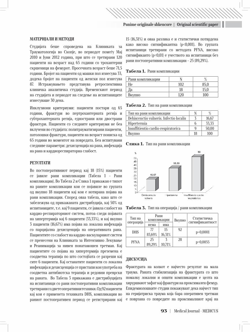
Punime origjinale shkencore | Original scientific paper
93 | Medical Journal - MEDICUS
МАТЕРИЈАЛИ И МЕТОДИ
Студијата беше спроведена на Клиниката за Трауматологија во Скопје, во периодот помеѓу Мај 2010 и Јуни 2012 година, при што се третирани 120 пациенти на возраст над 65 години со трохантерни скршеници на фемурот. Просечната возраст беше 71,5 години. Бројот на пациенти од машки пол изнесува 33, додека бројот на пациенти од женски пол изнесува 87. Истражувањето представува ретроспективна клиничка аналитичка студија. Временскиот период на студијата и периодот на следење на испитаниците изнесуваше 30 дена.
Инклузиони критериуми: пациенти постари од 65 години, фрактури во пертрохантерната регија и субтрохантерната регија, еднострани или двострани фрактури. Пациенти со следните критериуми не беа вклучени во студијата: политрауматизирани пациенти, патолошки фрактури, пациенти на возраст пониска од 65 години во моментот на повредата. Беа испитувани следниве параметри: дехисценција на рана, инфекција на рана и кардиореспираторна слабост.
РЕЗУЛТАТИ
Во постоперативниот период кај 18 (15%) пациенти се јавиле рани компликации (Табела 1 – Рани компликации). Во Табела 2 и Слика 1 прикажан е типот на раните компликации кои се појавиле во групата од вкупно 18 пациенти кај кои е нотирана појава на рани компликации. Според оваа табела, како што се забележува од прикажаната дистрибуција, кај 50% од испитаниците, т.е. кај 9 пациенти, се јавила слабост на кардио-респираторниот систем, потоа следи појавата на хипертермија кај 6 пациенти (33,33%), и кај вкупно 3 пациенти (16,67%) има појава на локална инфекција со парцијална дехисценција на оперативната рана. Пациентите со слабост на кардио-васкуларниот систем се пренесени на Клиниката за Интензивно Лекување и Реанимација за нивен понатамошен третман. Кај пациентите со појава на хипертермија преземена е соодветна терапија по што состојбата се разреши кај сите 6 пациенти. Кај останатите пациенти со локална инфекција и дехисценција се пристапи кон употреба на соодветна антибиотска терапија и редовни преврски на раната. Во Табела 3 прикажана е дистрибуцијата на испитаници со рани постоперативни компликации третирани со двете оперативни техники. Од 92 пациенти кај кои е применета техниката DHS, компликации во раниот постоперативен период се регистрирани кај
15 (16,31%) и оваа разлика е и статистички потврдена како високо сигнификантна (p<0,001). Во групата испитаници третирани со методата PFNA, високо сигнификанто (p<0,01) е учеството на испитаници без рани постоперативни компликации – 25 (89,29%).
Табела 1. Рани компликации
Рани компликации N %Не 102 85,0Да 18 15,0Вкупно 120 100
Табела 2. Тип на рани компликации
Тип на рани компликации N %Dehiscenctio vulneris. Infectio localis 3 16,67Hipertermia 6 33,33Insuffitientio cardio-respiratorica 9 50,00Вкупно 18 100
Слика 1. Тип на рани компликации
Табела 3. Тип на операција / рани компликации
Тип на операција
Рани компликации Вкупно Статистичка
сигнификантностнема има
DHS77
83,69%15
16,31%92
p=0,0001
PFNA25
89,29% 3
10,71%28
p=0,0055
ДИСКУСИЈА
Фрактурата на колкот е најчесто резултат на мала траума. Раната стабилизација на фрактурата со што помалку локални и општи компликации е целта на хируршкиот зафат кај фрактури на проксимален фемур. Епидемиолошките студии покажуваат дека најчест тип на геријатриска траума која бара оперативен третман е поврзана со повредите на проксималниот крај на
Punime origjinale shkencore | Original scientific paper
Revistë mjekësore - MEDICUS | 94
фемурот - пертрохантерни и субтрохантерни фрактури (7,8). DHS системите се стандардни средства за фиксирање на пертрохантерни фрактури во последните неколку децении (9,10,11), иако во однос на употребата кај нестабилиа фрактури тие се поврзани со стапка на неуспех која достигнува и до 23% (12,13,14).
Интрамедуларните системи добиваат на значење некаде во осумдесеттите години од минатиот век со воведување во употреба на gamma nail (15,16,17). Интрамедуларните системи ја комбинираат добрата механичката стабилност со минималната хируршка изложеност. Компликации на DHS имплантација за фиксација на фрактури на проксимален фемур вклучуваат остеонекроза на феморалната глава, расклопување на завртката и плоча, cut-out на навојот од феморалната глава, крварење, хематом и инфекција на раната. Инциденцата на инфекција на рана по фиксација со DHS се движи помеѓу 5 и 16%, иако употребата на профилактички антибиотици ја намали стапката до 1% (18).
Според некои трудови од светската литература, PFNa дава помалку локални компликации во однос на DHS поради техниката на оперативниот зафат (9, 19).
ЗАКЛУЧОК
PFNa има минималено-инвазивен пристап кој има мал рез, помало оштетување на локалното ткиво, помала загуба на крв и пониска појава на хируршка инфекција. Техниката на DHS има подолго оперативно време, поголем хируршки рез а како последица на тоа поголема манипулација и оштетување на локалното меко ткиво со последователна поголема крвозагуба. Со употребата на PFNa се добија помал број на рани компликации во однос на употребата на DHS.
ЛИТЕРАТУРА
1. Schwab CW, Kauder DR. Trauma in the geriatric patient. Arch Surg.2002;127:701-706.
2. Fogagnolo F, Kfuri M Jr, Paccola CA. Intramedullary fixation of pertrochanteric hip fractures with the short AO-ASIF proximal femoral nail. Arch Orthop Trauma Surg. 2004 Jan;124(1):31-7.
3. Saudan M et al. Pertrochanteric fractures: is there an advantage to an intramedullary nail? A randomized, prospective study of 206 patients comparing the dy-namic hip screw and proximal femoralnail. J Orthop Trauma 2002; 16: 386 – 393.
4. Herrera A, Domingo LJ, Calvo A, Martinez A. A com-parative study of trochanteric fractures treated with Gamma nail or the proximal femoral nail. Int Orthop. 2002;26:365-9.
5. Трпески С., Кафтанџиев И., Неделкова М, Ќаев А., Темелковска-Стефановска М. Првични резултати во третманот на субтрохантерни и пертрохантарни скршеници со ПФНА методот. Physioacta, 2011; Vol.5-No.1:119-129.
6. Albareda J, Laderiga A, Palanca D, et al. Complications and technical problems with gamma nail. Int Orthop. 1996;20:4750.
7. Tinetti ME, Falls In: Geriatric medicine 4th ed. New York; Springer-Verlag, 2000;528-534.
8. Mann NC, Cahn RN, Mullins RJ, Brand DM, Jurkovich GJ. Survival among injured geriatric patients during con-struction of a state wide trauma system. J Trauma. 2001; 50: 1111-1116.
9. Hrubina M, Skoták M, Běhounek J. Complications of dynamic hip screw treatment for proximal femoral frac-tures. Acta Chir Orthop Traumatol Cech. 2010 Oct; 77 (5):395-401.
10. Pioli G, Barone A, Giusti A, et al. Predictors of mortality after hip fracture: results from 1-year follow-up. Aging Clin Exp Res 2006; 18:381-7.
11. Симон Трпески, Игор Кафтанџиев, Оливер Арсовски, Алеко Ќаев. Резултати од 364 оперативно третирани геријатриски пациенти со трохантерни скршеници на надколеницата. MEDICUS, 2011; Vol XV (1): 297-303.
12. Gundle R, Gargan MF, Simpson HRW (1995) How to min-imize failures of fixation of unstable intertrochanteric fractures. Injury 26:611–614.
13. Simpson AHRW, Varty K, Dodd CAF (1989) Sliding hip screws: modes of failure. Injury 20:227–231.
14. Lorich DG, Geller DS, Nielson JH: Osteoporotic pertro-chanteric hip fractures. Management and current con-troversies. J Bone Joint Surg Am 2004; 86: 398 – 410.
15. Butt MS, Krikler SJ, Nafie S, Ali MS (1995) Comparison of dynamic hip screw and gamma nail: a prospective, rand-omized, controlled trial. Injury 26:615–618.
16. Calvert PT (1992) The gamma nail—a significant advance or a passion fashion? J Bone Joint Surg Br 74:329–331.
17. Curtis MJ, Jinnah RH, Wilson V, Cunningham BW (1994) Proximal femoral fractures: a biomechanical study to compare intramedullary and extramedullary fixation. Injury 25:99–104.

Punime origjinale shkencore | Original scientific paper
95 | Medical Journal - MEDICUS
18. Bodoky A, Neff U, Heberer M, Harder F. Antibiotic proph-ylaxis with two doses of cephalosporin in patients man-aged with internal fixation for a fracture of the hip. J Bone Joint Surg Am 1993;75(1):61-65.
19. Parker MJ, Handoll HHG. Gamma an other cephalo-condylic intreamedullary nails versus extramedullary implants for extracapsular hip fraktures. Cochrane Muskuloskeletal injures Group Cochrane Database of Systematic rewiews. 3, 2003.
EARLY COMPLICATIONS AFTER OPERATIVE TREATMENT OF PROXIMAL FEMUR FRACTURES
S. Trpeski1, I. Kaftandziev1, G. Velkovski1, I. Orana1, Z. Temelkovski1, A. Kjaev1
1 University Clinic of Traumatology, Medical faculty, University St.. Cyril and Methodius, Skopje, Macedonia
ABSTRACT
Background: DHS and PFNA represent an effective way for the surgical treatment of stable and unstable fractures of the proximal femur.
Aim: The main goal of this research presents an analysis of the comparison of early complications in the two most used implants, DHS and PFNa.
Patients and Methods: The study was conducted at the University Clinic for Traumatology, Medical Faculty, University “Ss. Cyril and Methodius” in Skopje in the period between May 2010 and June 2012. A total of 120 patients with hip fractures with age over 65 years were treated. Patient mean age was 71.5 years. The time frame for the study was 30 days.
Results: In the early postoperative period a total of 18 patients (15%) had complications. In 50% of the patients (9 patients) a cardio-respiratory system failure occured, followed by hyperthermia in 6 patients (33.33%), and a total of 3 patients (16.67%) had an operative wound infection and partial dehiscence of the operative wound.
Conclusion: The number of early complications is lower in the patient population with the use of PFNa in comparison with the use of DHS.
Key words: DHS, early complications, hip fractures, PFNa.
Revistë mjekësore - MEDICUS | 96
15
TIPARET PSIKOPATIKE TË PACIENTËVE ME SKIZOFRENI DHE NDËRLIDHJA ME SJELLJEN AGRESIVE RECIDIVUESE NË SHËRBIMIN E PSIKIATRISË FORENZIKE NË PRISHTINËNazmie F Ibishi1, Nebi Musliu1, Zylfije Hundozi1, Bekim Haxhiu1
1 Qendra Klinike Universitare e Kosovës
ABSTRAKT
Hyrje: Studimet e gjertanishme në lidhje me rolin e tipareve psikopatike të personalitetit dhe agresivitetit te pacientët me çrregullim të skizofrenisë janë të pakta.
Qëllimi: Që të bëhet evaluimi i tipareve psikopatike të pacientëve me skizofreni kryesve të veprave të ndryshme penale si dhe ndërlidhja me sjellje agresive recidivuese.
Metoda: Në këtë studim prospektiv janë përfshirë 65 pacientë meshkuj me moshë mesatare 39,68±8,74 , vite mesatare të sëmundjes 15,54±10,42 e të diagnostikuar me diagnozën e skizofrenisë bazuar në DSM IV,të cilët janë vlerësuar në periudhë kohore dyvjecare duke përdorë PANSS, BPRS, MMSE, PCL-R dhe HCR-20. Subjektet janë ndarë në dy grupe bazuar në sjellje agresive, recidivues gjegjësisht jorecidivues.
Rezultatet: Nga 31 pacientë që kanë prezentuar me psikopati mbi ≥17 të pikëve nga PCL-R testi kanë recidivuar me sjellje agresive 18 apo 55,0% kurse në 13 raste apo 41,9% nuk kanë recidivuar me sjellje agresive.Nga të dhënat e dokumentacionit 12,3% kanë qenë vrasje, 40% dhunë familjare dhe 47,6% kërcënim fizik . Grupet kanë prezentuar me dallime signifikante në matjet e faktorëve psikopatik F1 interpersonal/ afektiv në nivel të p<0.003 si dhe në parametrat klinik të psikopatologjisë pozitive dhe negative bazuar në PANSS testin . Analiza e regresionit të thjeshtë linear tregon për një lidhje të dobët mes veprimit të faktorëve psikopatik F1 në shtimin e numrit të incidenteve ku vlera e koeficientit Beta ishte R-0.376 me një signifikancë prej p<0.002, kurse regresioni i thjeshtë linear mes veprimit të faktorëve psikopatik F2 në numrin e incidenteve tregon për një lidhje të fortë ku vlera e koeficientit Beta ishte R-0.857 me signifikancë të p<0.0001.
Konkludim: Violenca te pacientët e grupit me rrezik të lartë për sjellje agresive është e ndërlidhur jo vetëm nga simptomet aktive të sëmundjes së skizofrenisë por edhe nga tiparet psikopatike.Te skiozofrenët me rrezikshmëri të lartë për recidivizëm është e mundur që të përmirësohen shkathtësitë interpersonale të komunikimit, kontrolla e hidhërimit, empatia ndaj viktimës si dhe redukimit të shtrembërimeve kognitive e që sjellin deri te sjelljet agresive.
Fjalët kyqe: tipare psikopatike, skizofrenia,sjellja agresive
Medicus 2013, Vol. 18 (2): 96-104
HYRJE
Psikopatia është definuar si manifestim i karakteristikave interpersonale - afektive dhe të sjelljes përfshirë impulsivitetin, papërgjegjësinë, mungesën e empatisë apo të fajit, gënjeshtrave patologjike dhe dhunë persistente ndaj normave sociale. (1,2,3)
Në aspektin e tipareve interpersonale individët psikopatik janë përshkruar si madhështor, arrogant, dominant,
sipërfaqësor dhe manipulative kurse në aspektin e tipareve afektive janë afatshkurtër dhe të paaftë që të ndërtojnë kufinjë emocional me të tjerët me mungesë të ndjenjës së fajit. Këto tipare interpersonale-afektive janë të ndërlidhura me devijim social që përfshijnë sjelljen e papërgjegjshme dhe tendenca që të injorojnë dhe sjellen dhunshëm ndaj shoqërisë (2) edhe pse jo të gjithë personat me psikopati bien në kundërshtim me ligjin dhe sistemin
Punime origjinale shkencore | Original scientific paper
Punime origjinale shkencore | Original scientific paper
97 | Medical Journal - MEDICUS
ligjor, tiparet e tyre i kategorizojnë si grup me nivel të lartë për agresivitet dhe dhunë.(4)
Kryesit e veprave penale me psikopati fillojnë veprimet kriminale dhe antisociale në moshë relativisht të re dhe vazhdojnë gjatë jetës me recidiva.(5)
Viktimat e kryesve të veprave me psikopati janë më rrallë anëtarët e familjes dhe më shpesh të huajtë gjë që nuk ndodhë te kryesit tjerë të veprave penale me skizofreni dhe se psikopatia shpesh ndodhë që të ndërlidhet me abuzim të substancave dhe alkoolit.(6,7) Prevalenca e psikopatisë në grupin e kryesve të veprave serioze penale me skizofreni është rritur dhe arrinë deri në 20%.(8,9,10,11)
Studimet e gjertanishme në lidhje me rolin e tipareve të personalitetit sikurse është psikopatia dhe koomorbiditeti me abuzim të substancave te pacientët me skizofreni janë të pakta. Gjithashtu roli i këtyre faktorëve si ndërmjetësues në sjellje agresive recidivuese te ky grup i pacientëve është pak i studjuar.
Violenca e kryer nga personat me skizofreni është një fenomen heterogjen. Sjellja violente mundet me qenë e ndërlidhur me simptome specifike psikotike sikurse janë deluzionet apo mendimet e kontrolluara dhe të persekutimit (12) halucinacionet komanduese (13) por lidhja mes violencës dhe simptomeve psikotike mundet me qenë edhe minimale. (14)
Pra simptomet psikotike nuk janë tërësisht shkaktare për violencën te skizofrenia.Tiparet psikopatike munden me qenë shkak për disa sjellje violente te pacientët me skizofreni,sidoqoftë prevalenca e koomorbiditetit të skizofrenisë dhe psikopatisë nuk është ende e qartë. Në komunitetin forenzik ky koomorbiditet nuk është më shumë se 4% sipas studimeve të Hart SD et al.Hodgins S et al.&Rice ME et al. (15,16,17) kurse në të kundërtën e këtyre studimeve në një studim të Rasmussen K et al.(18) koomorbiditetit mes subjekteve të cilat kanë shfaqur violencë ekstreme ky komorbiditet mes skizofrenisë dhe të psikopatisë ishte prezent në 17% të rasteve.
Përvoja klinike dhe literatura e përkrahin lidhjen mes simptomeve aktive të skizofrenisë dhe sjelljes antisocial por disa nga studimet nuk përkrahin rolin e fenomenit të deluzioneve dhe halucinacioneve (19,20,21) dhe se simptomet negative te skizofrenia munden me qenë faktorë mbrojtës. (22)
Tiparet psikopatike të kryesve të veprave penale recidivuese janë iritabiliteti, vetitë antisociale, mungesa
e ndjenjave ndaj të tjerëve, grandioziteti, dyshimi, bindjet joreale dhe dështimi të mësojnë nga përvoja e tyre. Aktualisht ka një evidencë të bazuar se faktorët e personalitetit ndërmjetësojnë në sjelljen kriminale në skizofreni (9,23,24,25).
QËLLIMI
Bazuar në studimet e gjertanishme e të cilat janë të pakta qëllimi i këtij studimi ishte që të bëhet evaluimi i tipareve psikopatike të pacientëve me skizofreni kryesve të veprave të ndryshme penale si dhe ndërlidhja me sjellje agresive recidivuese te ky subjekt brenda një periudhe kohore dyvjeqare të këtij studimi prospektiv.
MATERIALI DHE METODA
Në këtë studim prospektiv u përfshinë 65 pacientë të gjinisë mashkullore të moshës mbi 18 vjeq, të hospitali-zuar në shërbimin e Forenzikës për meshkuj kurse pa-cientet e gjinisë femrore nuk ishin përfshirë në mungesë të shërbimit të veçantë për femra, dhe që plotësonin kri-teret diagnostike për skizofreni bazuar në Klasifikimin Ndërkombëtar sipas DSM-IV.
Kriteret përfshirëse për studim ishin: pacientët klinikisht stabil dhe të aftë që të japin pëlqimin, skizofrenia e diagnostikuar të gjitha nëntipet e saj paranoide, dezorganizuar, katatonike, e paspecifikuar dhe reziduale.
Kriteret përjashtuese ishin: çrregullimet neurologjike dhe prapambetjet mendore.
Pacientët ishin të hospitalizuar në periudhën Janar, 2010 gjer në Dhjetor, 2011 .
Të dhënat për variablet psikopatike, simptomeve klinike dhe sjelljes agresive ishin grumbulluar gjatë evaluimit psikiatrik forenzik për nevoja të gjykatës .
Matjet kanë përfshirë të dhënat sociodemografike të cilat ishin grumbulluar nga dokumentacioni mjekësor dhe intervistimi klinik, tiparet psikopatike ishin vlerësuar duke përdorë instrumentin matës PCL-R (Testi i psikopatise sipas Hare) MMSE (Ekzaminimi mini-mental i gjendjes mendore) skrining test për përjashtimin e deficitit kognitiv, vlerësimi i simptomeve klinike ishte i bazuar në instrumentet matëse PANSS (Shkalla për simptome pozitive dhe negative të skizofrenisë) dhe BPRS (Shkalla e vlerësimit të shpejtë psikiatrik) si dhe vlerësimi i sjelljes agresive duke u bazuar në testin HCR-20 (Testi i vlerësimit historik, klinik dhe menagjimit të rrezikshmërisë).
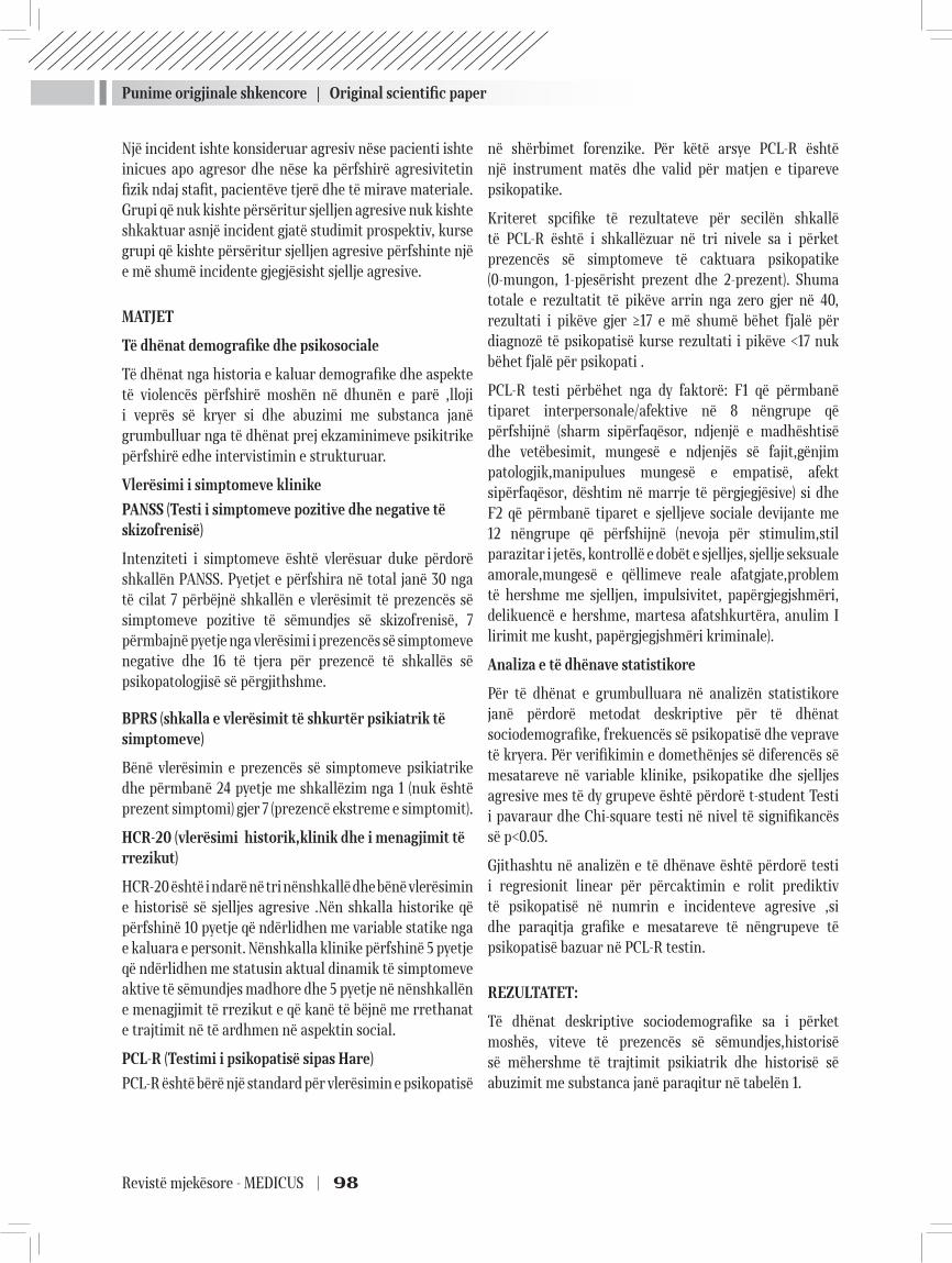
Punime origjinale shkencore | Original scientific paper
Revistë mjekësore - MEDICUS | 98
Një incident ishte konsideruar agresiv nëse pacienti ishte inicues apo agresor dhe nëse ka përfshirë agresivitetin fizik ndaj stafit, pacientëve tjerë dhe të mirave materiale. Grupi që nuk kishte përsëritur sjelljen agresive nuk kishte shkaktuar asnjë incident gjatë studimit prospektiv, kurse grupi që kishte përsëritur sjelljen agresive përfshinte një e më shumë incidente gjegjësisht sjellje agresive.
MATJET
Të dhënat demografike dhe psikosociale
Të dhënat nga historia e kaluar demografike dhe aspekte të violencës përfshirë moshën në dhunën e parë ,lloji i veprës së kryer si dhe abuzimi me substanca janë grumbulluar nga të dhënat prej ekzaminimeve psikitrike përfshirë edhe intervistimin e strukturuar.
Vlerësimi i simptomeve klinike
PANSS (Testi i simptomeve pozitive dhe negative të skizofrenisë)
Intenziteti i simptomeve është vlerësuar duke përdorë shkallën PANSS. Pyetjet e përfshira në total janë 30 nga të cilat 7 përbëjnë shkallën e vlerësimit të prezencës së simptomeve pozitive të sëmundjes së skizofrenisë, 7 përmbajnë pyetje nga vlerësimi i prezencës së simptomeve negative dhe 16 të tjera për prezencë të shkallës së psikopatologjisë së përgjithshme.
BPRS (shkalla e vlerësimit të shkurtër psikiatrik të simptomeve)
Bënë vlerësimin e prezencës së simptomeve psikiatrike dhe përmbanë 24 pyetje me shkallëzim nga 1 (nuk është prezent simptomi) gjer 7 (prezencë ekstreme e simptomit).
HCR-20 (vlerësimi historik,klinik dhe i menagjimit të rrezikut)
HCR-20 është i ndarë në tri nënshkallë dhe bënë vlerësimin e historisë së sjelljes agresive .Nën shkalla historike që përfshinë 10 pyetje që ndërlidhen me variable statike nga e kaluara e personit. Nënshkalla klinike përfshinë 5 pyetje që ndërlidhen me statusin aktual dinamik të simptomeve aktive të sëmundjes madhore dhe 5 pyetje në nënshkallën e menagjimit të rrezikut e që kanë të bëjnë me rrethanat e trajtimit në të ardhmen në aspektin social.
PCL-R (Testimi i psikopatisë sipas Hare)
PCL-R është bërë një standard për vlerësimin e psikopatisë
në shërbimet forenzike. Për këtë arsye PCL-R është një instrument matës dhe valid për matjen e tipareve psikopatike.
Kriteret spcifike të rezultateve për secilën shkallë të PCL-R është i shkallëzuar në tri nivele sa i përket prezencës së simptomeve të caktuara psikopatike (0-mungon, 1-pjesërisht prezent dhe 2-prezent). Shuma totale e rezultatit të pikëve arrin nga zero gjer në 40, rezultati i pikëve gjer ≥17 e më shumë bëhet fjalë për diagnozë të psikopatisë kurse rezultati i pikëve <17 nuk bëhet fjalë për psikopati .
PCL-R testi përbëhet nga dy faktorë: F1 që përmbanë tiparet interpersonale/afektive në 8 nëngrupe që përfshijnë (sharm sipërfaqësor, ndjenjë e madhështisë dhe vetëbesimit, mungesë e ndjenjës së fajit,gënjim patologjik,manipulues mungesë e empatisë, afekt sipërfaqësor, dështim në marrje të përgjegjësive) si dhe F2 që përmbanë tiparet e sjelljeve sociale devijante me 12 nëngrupe që përfshijnë (nevoja për stimulim,stil parazitar i jetës, kontrollë e dobët e sjelljes, sjellje seksuale amorale,mungesë e qëllimeve reale afatgjate,problem të hershme me sjelljen, impulsivitet, papërgjegjshmëri, delikuencë e hershme, martesa afatshkurtëra, anulim I lirimit me kusht, papërgjegjshmëri kriminale).
Analiza e të dhënave statistikore
Për të dhënat e grumbulluara në analizën statistikore janë përdorë metodat deskriptive për të dhënat sociodemografike, frekuencës së psikopatisë dhe veprave të kryera. Për verifikimin e domethënjes së diferencës së mesatareve në variable klinike, psikopatike dhe sjelljes agresive mes të dy grupeve është përdorë t-student Testi i pavaraur dhe Chi-square testi në nivel të signifikancës së p<0.05.
Gjithashtu në analizën e të dhënave është përdorë testi i regresionit linear për përcaktimin e rolit prediktiv të psikopatisë në numrin e incidenteve agresive ,si dhe paraqitja grafike e mesatareve të nëngrupeve të psikopatisë bazuar në PCL-R testin.
REZULTATET:
Të dhënat deskriptive sociodemografike sa i përket moshës, viteve të prezencës së sëmundjes,historisë së mëhershme të trajtimit psikiatrik dhe historisë së abuzimit me substanca janë paraqitur në tabelën 1.
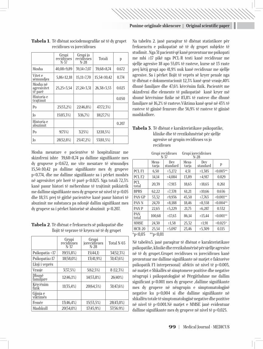
Punime origjinale shkencore | Original scientific paper
99 | Medical Journal - MEDICUS
Tabela 1. Të dhënat sociodemografike në të dy grupet recidivues vs jorecidivues
Grupi recidivues
N-37
Grupi jo recidivues
N-28Totali p
Mosha 40,08±9,89 39,14±7,07 39,68±8,74 0.672
Vitet e sëmundjes 5,86±12,18 15,11±7,70 15,54±10,42 0.774
Mosha në agresivitet të parë
25,25±3,54 27,24±3,31 26,38±3,53 0.023
Historia e trajtimit 0.050
Po 25(53,2%) 22(46,8%) 47(72,3%)
Jo 15(83,3%) 3(16,7%) 18(27,7%)
Historia e abuzimit 0.207
Po 9(75%) 3(25%) 12(18,5%)
Jo 28(52,8%) 25(47,2%) 53(81,5%)
Mosha mesatare e pacientëve të hospitalizuar me skizofreni ishte 39,68±8,74 pa dallime signifikante mes dy grupveve p<0.672, me vite mesatare të sëmundjes 15,54±10,42 pa dallime signifikante mes dy grupeve p<0.774, dhe me dallime signifikante sa i përket moshës në agresivitet për herë të parë p<0.023. Nga totali 72,3% kanë pasur histori të mëhershme të trajtimit psikiatrik me dallime signifikante mes dy grupeve në nivel të p<0.05 dhe 18,5% prej të gjithë pacientëve kanë pasur histori të abuzimit me substanca pa ndonjë dallim signifikant mes dy grupeve sa i përket historisë së abuzimit p<0.207.
Tabela 2. Të dhënat e frekuencës së psikopatisë dhe llojit të veprave të kryera në të dy grupet
Grupi recidivues
N-37
Grupi jorecidivues
N-28Total N-65
Psikopatia <17 19(55,8%) 15(44,1) 34(52,3%)Psikopatia≥17 18(58,0%) 13(41,9%) 31(47,6%)Lloji i veprës Vrasje 3(37,5%) 5(62,5%) 8 (12,3%)Dhunë familjare 12(46,1%) 14(53,8%) 26(40%)
Kërcënim fizik 11(35,4%) 20(64,5%) 31(47,6%)
Gjinia e viktimësFemër 13(46,4%) 15(53,5%) 28(43,0%)Mashkull 20(54,0%) 17(45,9%) 37(56.9%)
Na tabelën 2. janë paraqitur të dhënat statistikore për frekuencën e psikopatisë në të dy grupet subjekte të studimit . Nga 31 pacientë që kanë prezentuar me psikopati me mbi ≥17 pikë nga PCL-R testi kanë recidivuar me sjellje agresive 18 apo 55,0% të rasteve, kurse në 13 raste prej këtij grupi apo 41,9% nuk kanë recidivuar me sjellje agresive. Sa i përket llojit të veprës së kryer penale nga të dhënat e dokumentacionit 12,3% kanë qenë vrasje,40% dhunë familjare dhe 47,6% kërcënim fizik. Pacientët me skizofreni dhe elemente të psikopatisë kanë kryer më shumë kërcënime fizike në 83,8% të rasteve dhe dhunë familjare në 16,2% të rasteve.Viktima kanë qenë në 43% të rasteve të gjinisë femrore dhe 56,9% të rasteve të gjinisë mashkullore.
Tabela 3. Të dhënat e karakteristikave psikopatike, klinike dhe të rrezikshmërisë për sjellje agresive në grupin recidivues vs jo recidivues
Grupi recidivues N-37
Grupi jorecidiviues N-28
Mesa-tarja
Dev standard
Mesa-tarja
Dev standard p
PCL F1 6,50 ±3,272 4,51 ±1,385 <0.003**
PCL F2 14,14 ±4,084 13,89 ±4,917 0.829PCL total 20,39 ±7,913 18,65 ±18,65 0.261
BPRS 62,22 ±7,378 61,21 ±10,66 0.656
PAN GP 53,32 ±9,936 45,50 ±7,763 <0.001**
PAN N 24,70 ±8,188 18,46 ±8,558 <0.004**
PAN P 22,65 ±5,229 21,75 ±6,287 0.532PAN total 100,68 ±17,63 86,14 ±13,44 <0.001**
MMSE 24,30 ±1,58 25,32 ±1,91 <0.025*
HCR-20 25,54 ±5,097 23,46 ±5,309 0.115
*p<0,05 **p<0,01
Në tabelën3. janë paraqitur të dhënat e karakteristikave psikopatike, klinike dhe rrezikshmërisë për sjellje agresive në të dy grupet.Grupet recidivues vs jorecidivues kanë prezentuar me dallime signifikante në matjet e faktorëve psikopatik F1 interpersonal/ afektiv në nivel të p<0.003, në matjet e Shkallës së simptomeve pozitive dhe negative nëngrupi i psikopatologjisë së Përgjithshme me dallim significant p<0.001 mes dy grupeve ,dallime signifikante mes dy grupeve në nëngrupin e simptomatologjisë negative ku p<0,004 si dhe dallime signifikante në shkallën totale të simptomatologjisë negative dhe pozitive në nivel të p<0.001.Në matjet e MMSE janë evidentuar dallime signifikante mes dy grupeve në nivel të p<0,025.
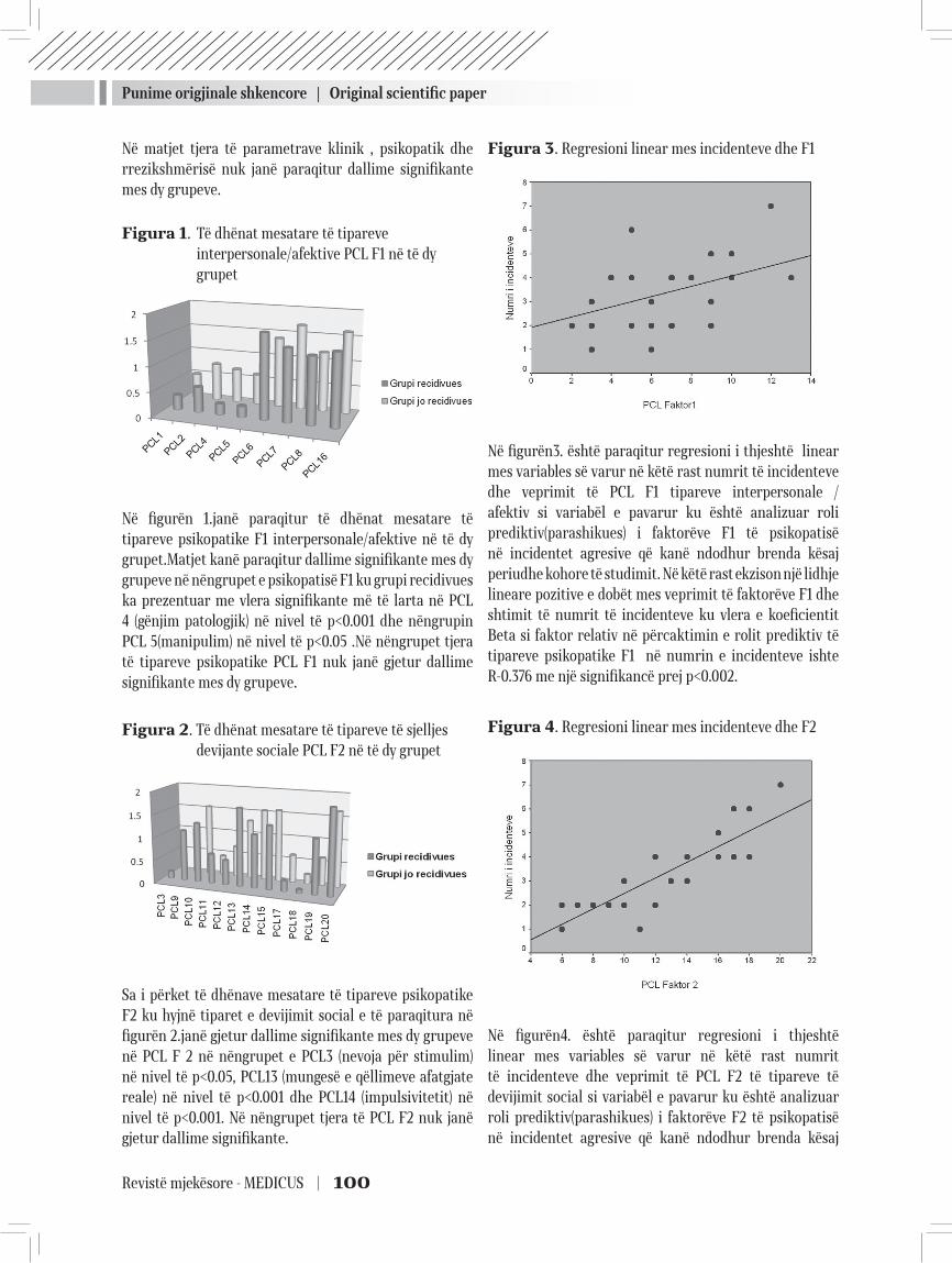
Punime origjinale shkencore | Original scientific paper
Revistë mjekësore - MEDICUS | 100
Në matjet tjera të parametrave klinik , psikopatik dhe rrezikshmërisë nuk janë paraqitur dallime signifikante mes dy grupeve.
Figura 1. Të dhënat mesatare të tipareve interpersonale/afektive PCL F1 në të dy grupet
Në figurën 1.janë paraqitur të dhënat mesatare të tipareve psikopatike F1 interpersonale/afektive në të dy grupet.Matjet kanë paraqitur dallime signifikante mes dy grupeve në nëngrupet e psikopatisë F1 ku grupi recidivues ka prezentuar me vlera signifikante më të larta në PCL 4 (gënjim patologjik) në nivel të p<0.001 dhe nëngrupin PCL 5(manipulim) në nivel të p<0.05 .Në nëngrupet tjera të tipareve psikopatike PCL F1 nuk janë gjetur dallime signifikante mes dy grupeve.
Figura 2. Të dhënat mesatare të tipareve të sjelljes devijante sociale PCL F2 në të dy grupet
Sa i përket të dhënave mesatare të tipareve psikopatike F2 ku hyjnë tiparet e devijimit social e të paraqitura në figurën 2.janë gjetur dallime signifikante mes dy grupeve në PCL F 2 në nëngrupet e PCL3 (nevoja për stimulim) në nivel të p<0.05, PCL13 (mungesë e qëllimeve afatgjate reale) në nivel të p<0.001 dhe PCL14 (impulsivitetit) në nivel të p<0.001. Në nëngrupet tjera të PCL F2 nuk janë gjetur dallime signifikante.
Figura 3. Regresioni linear mes incidenteve dhe F1
Në figurën3. është paraqitur regresioni i thjeshtë linear mes variables së varur në këtë rast numrit të incidenteve dhe veprimit të PCL F1 tipareve interpersonale /afektiv si variabël e pavarur ku është analizuar roli prediktiv(parashikues) i faktorëve F1 të psikopatisë në incidentet agresive që kanë ndodhur brenda kësaj periudhe kohore të studimit. Në këtë rast ekzison një lidhje lineare pozitive e dobët mes veprimit të faktorëve F1 dhe shtimit të numrit të incidenteve ku vlera e koeficientit Beta si faktor relativ në përcaktimin e rolit prediktiv të tipareve psikopatike F1 në numrin e incidenteve ishte R-0.376 me një signifikancë prej p<0.002.
Figura 4. Regresioni linear mes incidenteve dhe F2
Në figurën4. është paraqitur regresioni i thjeshtë linear mes variables së varur në këtë rast numrit të incidenteve dhe veprimit të PCL F2 të tipareve të devijimit social si variabël e pavarur ku është analizuar roli prediktiv(parashikues) i faktorëve F2 të psikopatisë në incidentet agresive që kanë ndodhur brenda kësaj
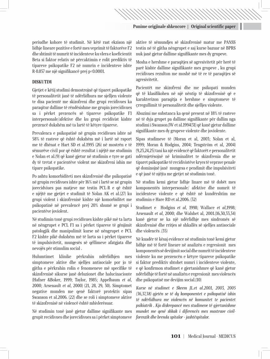
Punime origjinale shkencore | Original scientific paper
101 | Medical Journal - MEDICUS
periudhe kohore të studimit. Në këtë rast ekzison një lidhje lineare pozitive e fortë mes veprimit të faktorëve F2 dhe shtimit të numrit të incidenteve ku vlera e koeficientit Beta si faktor relativ në përcaktimin e rolit prediktiv të tipareve psikopatike F2 në numrin e incidenteve ishte R-0.857 me një signifikancë prej p<0.0001.
DISKUTIM
Gjetjet e këtij studimi demostrojnë që tiparet psikopatike të personalitetit janë të ndërlidhura me sjelljen violente te disa pacientë me skizofreni dhe grupi recidivues ka paraqitur dallime të rëndësishme me grupin jorecidivues sa i përket prezencës së tipareve psikopatike F1 interpersonale/afektive dhe ku grupi recidivist kishte prezencë dukshëm më ta lartë të këtyre tipareve.
Prevalenca e psikopatisë në grupin recidivues ishte në 58% të rasteve që është dukshëm më i lartë në raport me të dhënat e Hart SD et al.1995 (26) në mostrën e të sëmurëve civil por që është rezultat i njëjtë me studimin e Nolan et al.(9) që kanë gjetur në studimin e tyre se gati dy të tretat e pacientëve violent me skizofreni ishin me tipare psikopatike.
Po ashtu komorbiditeti mes skizofrenisë dhe psikopatisë në grupin recidivues ishte për 16% më i lartë se në grupin jorecidviues pas matjeve me testin PCL-R e që është e njëjtë me gjetjet e studimit të Nolan AK et al.(27) ku grupi violent i skizofrenisë kishte një komorbiditet me psikopatinë në prevalencë prej 20% shumë se grupi i pacientëve joviolent.
Në studimin tonë grupi recidivues kishte pikë më ta larta në nëngrupet e PCL F1 sa i përket tipareve të gënjimit patologjik dhe manipulimit kurse në nëngrupet e PCL F2 kishte pikë dukshëm më të larta sa i përket tipareve të impulsivitetit, mungesës së qëllimeve afatgjata dhe nevojës për stimulim social .
Hulumtimet klinike përkrahin ndërlidhjen mes simptomeve aktive dhe sjelljes antisociale por jo të gjitha e përkrahin rolin e fenomeneve më specifike të skizofrenisë sikurse janë deluzionet dhe halucinacionte (Hafner &Boker, 1999; Taylor, 1985; Appelbaum et al, 2000; Arsenault et al, 2000) (21, 28, 29, 30). Simptomet negative munden me qenë faktorë protektiv sipas Swanson et al.2006. (22) dhe se roli i simptomeve aktive të skizofrenisë në violencë është mbivlerësuar.
Në studimin tonë janë gjetur dallime signifikante mes grupit recidivues dhe jorecidivues sa i përket simptomeve
aktive të sëmundjes së skizofrenisë matur me PANSS testin në të gjitha nëngrupet e saj kurse bazuar në BPRS nuk janë gjetur dallime signifikante mes dy grupeve.
Mosha e hershme e paraqitjes së agresivitetit për herë të parë kishte dallime signifikante mes grupeve , ku grupi recidivues rezulton me moshë më të re të paraqitjes së agresivitetit.
Pacientët me skizofreni dhe me psikopati munden që të klasifikohen në një nëntip të skizofrenisë që e karakterizon paraqitja e hershme e simptomeve të çrregullimit të personalitetit dhe sjelljes violente.
Abuzimi me substanca ka qenë present në 18% të rasteve në të dyja grupet pa dallime signifikante për dallim nga studimi i Swanson JW et al.1994(31) që kanë gjetur dallime signifikante mes dy grupeve violente dhe joviolente.
Sipas studimeve të (Moran et al, 2003; Nolan et al, 1999; Moran & Hodgins, 2004; Tengström et al, 2004) (9,23,24,25) tani ka një evidencë që faktorët e personalitetit ndërmjetësojnë në kriminalitet te skizofrenia dhe se tiparet psikopatike të recidivistëve kryes të veprave penale që dominojnë janë mungesa e pendimit dhe impulsiviteti e që janë të njëjta me gjetjet në studimin tonë.
Në studim kemi gjetur lidhje linare më të dobët mes komponentës interpersonale/ afektive dhe numrit të incidenteve violente e që është në kundërshtim me studimin e Hare RD et al.2006. (32)
Studimet e Hodgins et al, 1998; Wallace et al,1998; Arsenault et al, 2000; dhe Walshet al, 2001.(16,30,33,34)kanë gjetur se ka një ndërlidhje mes sindromës së skizofrenisë dhe rritjes së shkallës së sjelljes antisociale dhe violencës .(35)
Në kuadër të kësaj evidence në studimin tonë kemi gjetur lidhje më të fortë lineare në analizën e regresionit mes komponentës së devijimit social dhe numrit të incidenteve violente ku me prezencën e këtyre tipareve psikopatike si faktor prediktiv shtohet numri i incidenteve violente, e që konfirmon studimet e gjertanishmre që kanë gjetur ndërlidhje të fortë në analizën e regresionit mes violencës dhe psikopatisë me devijim social.(10)
Kurse në studimet e Skeem JL.et al.2001, 2003, 2005 (36,37,38) gjetën se të dy komponentet e psikopatisë ishin të ndërlidhura me violencën në komunitet te pacientet psikiatrik . Kjo diskrepancë mes studimeve të gjertanishme mundet me qenë shkak i diferencës mes mostrave civil- forenzik dhe brenda spitalor - jashtëspitalor.
Punime origjinale shkencore | Original scientific paper
Revistë mjekësore - MEDICUS | 102
KONKLUDIM
Predispozitat gjenetike dhe procesi i skizofrenisë ndikon që te disa të sëmurë të mbizotërojnë tipare të personalitetit dhe qëndrime që munden të klasifikohen si kriminogjene.
Violenca te pacientët e grupit me rrezik të lartë është e ndërmjetësuar jo vetëm nga simptomet aktive të sëmundjes por edhe nga faktorët e personalitetit vulnerabil dhe tipareve psikopatike.
Trajtimi i simptomeve aktive të sëmundjes së skizofrenisë nuk do ta zvogëlonte mundësinë e sjelljes violente te meshkujt me profil të lartë të tipareve psikopatike.
Redukimi i mundësisë së violencës varet në shkallë të madhe nga modifikimi i këtyre faktorëve dhe sjelljes që gjenerojnë ato. Çrregullimet e personalitetit si etnitete munden me qenë të patrajtueshme por disa elemente të tyre kanë mundësi të modifikohen dhe përmirësohen . Te skizofrenët me rrezikshmëri të lartë për recidivizëm është e mundur që të përmirësohen shkathtësitë interpersonale të komunikimit, kontrolla e hidhërimit, empatia ndaj viktimës si dhe redukimi i shtrembërimeve kognitive që sjellin deri te sjelljet agresive.
Prevenca e violencës në të ardhmen kërkon një çasje që ka për objektiv të trajtojë faktorët kriminogjen të personalitetit si dhe inkurajimin e rrjetit social dhe relacioneve sociale.
REFERENCAT:
1. Cleckley H: The Mask of Sanity 5th edition. St. Louis: MO; 1976.
2. Hare RD: The Hare Psychopathy Checklist – Revised To-ronto: Multi-Health Systems; 1991.
3. Hare RD: Psychopaths and their nature: Implications for the mental health and criminal justice systems. In Psychopathy: Antisocial,Criminal, and Violent Behav-iour Edited by: Millon T, Simonson E,Burket-Smith M, Davis R. Neë York: Guilford Press; 1998:188-212.
4. Hart SD, Hare RD: Psychopathy: assessment and asso-ciation with criminal conduct. In Handbook of Antiso-cial Behavior Edited by:Stoff DM, Maser J, Brieling J. Neë York: Ëiley; 1997:22-35.
5. Salekin RT, Rogers R, Sewell KË: A review and meta-analysis of the Psychopathy Checklist and Psychopathy Checklist-Revised: Predictive validity of dangerous-ness. Clin Psychol 1996, 3:203-215.
6. Hildebrand M, de Ruiter C: PCL-R psychopathy and its relation to DSM-IV Axis I and II disorders in a sample of male forensic psychiatric patients in The Netherlands. Int J Laë Psychiatry 2004, 27:233-248.
7. Söderström H, Nilsson T, Sjodin AK, Carstedt A, Fors-man A: The childhood-onset neuropsychiatric back-ground to adulthood psychopathic traits and personal-ity disorders. Compr Psychiatry 2005, 46:111-116.
8. Hart SD, Hare RD: Discriminant validity of the Psy-chopathy Checklist in a forensic psychiatric population. Psychol Assess 1989, 1:211-218.
9. Nolan KA, Volavka J, Mohr P, Czobor P: Psychopathy and violent behavior among patients ëith schizophrenia or schizoaffective disorder. Psychiatr Serv 1999, 50:787-792.
10. Abushua’leh K, Abu-Akel A: Association of psychopath-ic traits and symptomatology ëith violence in patients ëith schizophrenia.Psychiatry Res 2006, 143:205-211.
11. Tengström A, Grann M, Långström N, Kullgren G: Psy-chopathy (PCL-R) as a predictor of violent recidivism among criminal offenders ëith schizophrenia. Laë Hum Behav 2000, 24:45-58.
12. Link BG, Stueve A: Psychotic symptoms and the vio-lent/illegal behavior of mental patients compared to community controls, ibid
13. Junginger J: Command hallucinations and the prediction of dangerousness. Psychiatric Services46:911-914, 1995
14. Junginger J, Parks-Levy J, McGuire L: Delusions and symptom-consistent violence. Psychiatric Servic-es49:218-220, 1998
15. Hart SD, Hare RD: Discriminant validity of the Psychop-athy Checklist in a forensic psychiatric population. Jour-nal of Consulting and Clinical Psychology1:211-218, 1989
16. Hodgins S, Cote G, Toupin J: Major mental disorder and crime: an etiological hypothesis, in Psychopathy: Theory, Research, and Implications for Society. Edited by Cooke DJ, Forth AE, Hare RD. Dordrecht, The Nether-lands, Kluëer Academic, 1998
17. Rice ME, Harris GT: Psychopathy, schizophrenia, alcohol abuse, and violent recidivism. International Journal of Laë and Psychiatry18:333-342, 1995
18. Rasmussen K, Levander S: Symptoms and personality characteristics of patients in a maximum security psy-chiatric unit. International Journal of Law and Psychia-try19:27-37, 1996
19. Hafner, H. & Boker, W. (1982) Crimes of Violence by
Punime origjinale shkencore | Original scientific paper
103 | Medical Journal - MEDICUS
Mentally Abnormal Offenders (ed. F. H. Marshall). Cam-bridge, Cambridge University Press.
20. Taylor, P. J. (1985) Motives for offending among violent and psychotic men. British Journal of Psychiatry, 147, 491–498.
21. Appelbaum, P. S., Robbins, P. C., Monahan, J. (2000) Vio-lence and delusions. Data from the MacArthur Violence Risk Assessment Study. American Journal of Psychia-try, 157, 566–572.
22. Sëanson, J.W. Swartz, M. S., Van Dorn, R. A., et al (2006) A national study of violent behaviour in persons ëith schizophrenia. Archives of General Psychiatry, 63, 490–499.
23. Moran, P, Walsh, E., Tyrer, P., et al (2003) Impact of co-morbid personality disorder on violence in psychosis. Report from the UK700 trial. British Journal of Psychia-try, 182, 129–134.
24. 24. Moran, P. & Hodgins, S. (2004) The correlates of co-morbid antisocial personality disorder in schizophre-nia. Schizophrenia Bulletin, 30, 791–802.
25. Tengström, A., Hodgins, S., Grann, M., et al (2004) Schizo-phrenia and criminal offending: the role of psychopathy and substance misuse. Criminal Justice and Behaviour, 31, 1–25.
26. Hart SD, Cox DN, Hare RD: The Hare PCL:SV: Psychopa-thy Checklist: Screening Version. North Tonoëanda, NY, Multi-Health Systems, 1995
27. Karen A. Nolan, Jan Volavka, Pavel Mohr,Pál Czobor. Psychopathy and Violent Behavior Among Patients with Schizophrenia or Schizoaffective Disorder , Psy-chiatric services ,June 1999 Vol. 50 No. 6
28. Hafner, H. & Boker, W. (1982) Crimes of Violence by Mentally Abnormal Offenders (ed. F. H. Marshall). Cam-bridge, Cambridge University Press.Ph.D. Psychiatric Services .1999
29. Taylor, P. J. (1985) Motives for offending among violent
and psychotic men. British Journal of Psychiatry, 147, 491–498.
30. Arseneault, L., Moffitt, T., Caspi, A., et al (2000) Mental disorders and violence in a total birth cohort. Results from the Dunedin Study. Archives of General Psychiatry, 57, 979–986.
31. Swanson JW (1994): Mental disorder, substance abuse, and community violence: an epidemiological approach, in Violence and Mental Disorder: Developments in Risk Assessment. Edited by Monahan J, Steadman HJ. Chi-cago, University of Chicago Press, 1994
32. Hare RD2006)(39). Psychopathy: a clinical and forensic overvieë. Psychiatr Clin North Am 2006; 29: 709– 24)
33. Wallace, C., Mullen, P. E. & Burgess, P. (2004) Criminal offending in schizophrenia over a 25 year period marked by deinstitutionalization and increasing prevalence of comorbid substance use disorders. American Journal of Psychiatry, 161, 716–727.
34. Walsh, E., Buchanan, A. & Fahy, T. (2001) Violence and schizophrenia: examining the evidence. British Journal of Psychiatry, 180, 490–495.
35. Hodgins, S. & Müller-Isberner, R. (2004) Preventing crime by people ëith schizophrenic disorders: the role of psychiatric services. British Journal of Psychiatry, 185, 245–250.
36. Skeem JL. Mulvey EP. Psychopathy and community vio-lence among civil psychiatric patients. Results from the MacArthur Violence Risk Assessment Study. J Consult Clin Psychol 2001; 69: 358– 74.
37. Skeem JL, Mulvey EP, Grisso T. Applicability of tradi-tional and revised models of psychopathy to the Psy-chopathy Checklist: screening version. Psychol Assess 2003; 15: 41– 55.
38. Skeem JL, Miller JD, Mulvey E, Tiemann J, Monahan J. Using a five-factor lens to explore the relation betëeen personality traits and violence in psychiatric patients. J Consult Clin Psychol 2005; 73: 454– 65
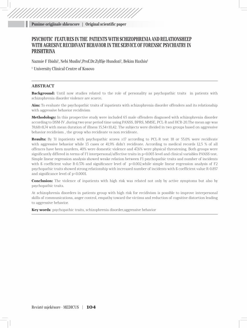
Punime origjinale shkencore | Original scientific paper
Revistë mjekësore - MEDICUS | 104
PSYCHOTIC FEATURES IN THE PATIENTS WITH SCHIZOPHRENIA AND RELATIONSHEEP WITH AGRESIVE RECIDIVANT BEHAVIOR IN THE SERVICE OF FORENSIC PSYCHIATRY IN PRISHTRINA
Nazmie F Ibishi1, Nebi Musliu1,Prof.Dr.Zylfije Hundozi1, Bekim Haxhiu1
1 University Clinical Centre of Kosovo
ABSTRACT
Background: Until now studies related to the role of personality as psychopathic traits in patients with schizophrenia disorder violence are scarce.
Aim: To evaluate the psychopathic traits of inpatients with schizophrenia disorder offenders and its relationship with aggressive behavior recidivism
Methodology: In this prospective study were included 65 male offenders diagnosed with schizophrenia disorder according to DSM-IV ,during two year period time using PANSS, BPRS, MMSE, PCL-R and HCR-20.The mean age was 39,68±8,74 with mean duration of illness 15,54±10,42. The subjects were divided in two groups based on aggressive behavior recidivism , the group who recidivate vs non recidivate.
Results: By 31 inpatients with psychopathic scores ≥17 according to PCL-R test 18 or 55.0% were recidivate with aggressive behavior while 13 cases or 41,9% didn’t recidivate. According to medical records 12,5 % of all offences have been murders, 40% were domestic violence and 47,6% were physical threatening. Both groups were significantly differed in terms of F1 interpersonal/affective traits in p<0.003 level and clinical variables PANSS test. Simple linear regression analysis showed weake relation between F1 psychopathic traits and number of incidents with ß coefficient value R-0.376 and significance level of p<0.002,while simple linear regression analysis of F2 psychopathic traits showed strong relationship with increased number of incidents with ß coefficient value R-0.857 and significance level of p<0.0001.
Conclusion: The violence of inpatients with high risk was related not only by active symptoms but also by psychopathic traits.
At schizophrenia disorders in patients group with high risk for recidivism is possible to improve interpersonal skills of communications, anger control, empathy toward the victims and reduction of cognitive distortion leading to aggressive behavior.
Key words: psychopathic traits, schizophrenia disorder,aggressive behavior
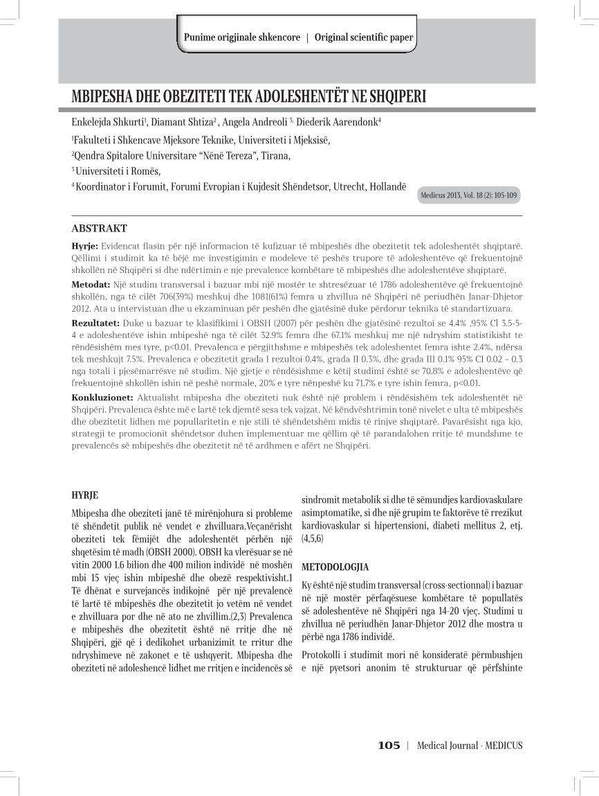
105 | Medical Journal - MEDICUS
MBIPESHA DHE OBEZITETI TEK ADOLESHENTËT NE SHQIPERI Enkelejda Shkurti1, Diamant Shtiza2 , Angela Andreoli 3, Diederik Aarendonk4
1Fakulteti i Shkencave Mjeksore Teknike, Universiteti i Mjeksisë, 2Qendra Spitalore Universitare “Nënë Tereza”, Tirana,3 Universiteti i Romës, 4 Koordinator i Forumit, Forumi Evropian i Kujdesit Shëndetsor, Utrecht, Hollandë
ABSTRAKT
Hyrje: Evidencat flasin për një informacion të kufizuar të mbipeshës dhe obezitetit tek adoleshentët shqiptarë. Qëllimi i studimit ka të bëjë me investigimin e modeleve të peshës trupore të adoleshentëve që frekuentojnë shkollën në Shqipëri si dhe ndërtimin e nje prevalence kombëtare të mbipeshës dhe adoleshentëve shqiptarë.
Metodat: Një studim transversal i bazuar mbi një mostër te shtresëzuar të 1786 adoleshentëve që frekuentojnë shkollën, nga të cilët 706(39%) meshkuj dhe 1081(61%) femra u zhvillua në Shqipëri në periudhën Janar-Dhjetor 2012. Ata u intervistuan dhe u ekzaminuan për peshën dhe gjatësinë duke përdorur teknika të standartizuara.
Rezultatet: Duke u bazuar te klasifikimi i OBSH (2007) për peshën dhe gjatësinë rezultoi se 4.4% ,95% Cl 3.5-5-4 e adoleshentëve ishin mbipeshë nga të cilët 32.9% femra dhe 67.1% meshkuj me një ndryshim statistikisht te rëndësishëm mes tyre, p<0.01. Prevalenca e përgjithshme e mbipeshës tek adoleshentet femra ishte 2.4%, ndërsa tek meshkujt 7.5%. Prevalenca e obezitetit grada I rezultoi 0.4%, grada II 0.3%, dhe grada III 0.1% 95% CI 0.02 – 0.3 nga totali i pjesëmarrësve në studim. Një gjetje e rëndësishme e këtij studimi është se 70.8% e adoleshentëve që frekuentojnë shkollën ishin në peshë normale, 20% e tyre nënpeshë ku 71.7% e tyre ishin femra, p<0.01.
Konkluzionet: Aktualisht mbipesha dhe obeziteti nuk është një problem i rëndësishëm tek adoleshentët në Shqipëri. Prevalenca ështe më e lartë tek djemtë sesa tek vajzat. Në këndvështrimin tonë nivelet e ulta të mbipeshës dhe obezitetit lidhen me popullaritetin e nje stili të shëndetshëm midis të rinjve shqiptarë. Pavarësisht nga kjo, strategji te promocionit shëndetsor duhen implementuar me qëllim që të parandalohen rritje të mundshme te prevalencës së mbipeshës dhe obezitetit në të ardhmen e afërt ne Shqipëri.
HYRJE
Mbipesha dhe obeziteti janë të mirënjohura si probleme të shëndetit publik në vendet e zhvilluara.Veçanërisht obeziteti tek fëmijët dhe adoleshentët përbën një shqetësim të madh (OBSH 2000). OBSH ka vlerësuar se në vitin 2000 1.6 bilion dhe 400 milion individë në moshën mbi 15 vjeç ishin mbipeshë dhe obezë respektivisht.1 Të dhënat e survejancës indikojnë për një prevalencë të lartë të mbipeshës dhe obezitetit jo vetëm në vendet e zhvilluara por dhe në ato ne zhvillim.(2,3) Prevalenca e mbipeshës dhe obezitetit është në rritje dhe në Shqipëri, gjë që i dedikohet urbanizimit te rritur dhe ndryshimeve në zakonet e të ushqyerit. Mbipesha dhe obeziteti në adoleshencë lidhet me rritjen e incidencës së
sindromit metabolik si dhe të sëmundjes kardiovaskulare asimptomatike, si dhe një grupim te faktorëve të rrezikut kardiovaskular si hipertensioni, diabeti mellitus 2, etj.(4,5,6)
METODOLOGJIA
Ky është një studim transversal (cross-sectionnal) i bazuar në një mostër përfaqësuese kombëtare të popullatës së adoleshentëve në Shqipëri nga 14-20 vjeç. Studimi u zhvillua në periudhën Janar-Dhjetor 2012 dhe mostra u përbë nga 1786 individë.
Protokolli i studimit mori në konsideratë përmbushjen e një pyetsori anonim të strukturuar që përfshinte
Medicus 2013, Vol. 18 (2): 105-109
Punime origjinale shkencore | Original scientific paper
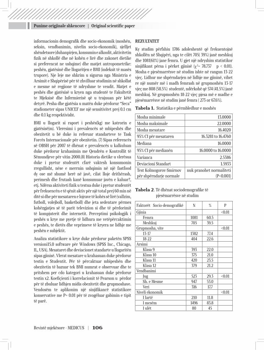
Punime origjinale shkencore | Original scientific paper
Revistë mjekësore - MEDICUS | 106
informacionin demografik dhe socio-ekonomik (moshën, seksin, vendbanimin, nivelin socio-ekonomik), sjelljet shëndetsore (duhanpirjen, konsumin e alkoolit, aktivitetin fizik në shkollë dhe në kohën e lirë dhe zakonet dietike si preferencat ne ushqime) dhe matjet antropometrike: peshën, gjatësinë dhe llogaritjen e BMI (indeksit të masës trupore). Nje leje me shkrim u sigurua nga Ministria e Arsimit e Shqipërisë për të zhvilluar studimin në shkollat e mesme në regjione të ndryshme te vendit. Matjet e peshës dhe gjatësisë u kryen nga studentë te Fakultetit te Mjeksisë dhe Infermierisë që u trajnuan për këtë detyrë. Pesha dhe gjatësia u matën duke përdorur “Seca” stadiometer sipas UNICEF me një sensitivitet prej 0.1 cm dhe 0.1 kg respektivisht.
BMI u llogarit si raport i peshës(kg) me katrorin e gjatësisë(m). Vleresimi i prevalencës së mbipeshës dhe obezitetit u bë duke iu referuar standarteve te Task Forcës Internacionale për obezitetin. (7) Sipas referencës së OBSH për 2007 të dhënat e prevalencës u kalkuluan duke përdorur krahasimin me Qendrën e Kontrollit të Sëmundjeve për vitin 2000.(8) Historia dietike u vlerësua duke i pyetur studentët cfarë vaktesh konsumonin rregullisht, nëse e merrnin ushqimin në një fastfood dy ose më shumë herë në javë, cilat lloje drithërash, perimesh dhe frutash kanë konsumuar javën e kaluar?, etj. Ndërsa aktiviteti fizik u testua duke i pyetur studentët për frekuencën e të qënit aktiv për një total prej 60 min në ditë si dhe për mesataren e lojrave të kohës së lirë (vallzim, futboll, volejboll, basketboll) dhe jeta sedentare përmes kohëzgjatjes së të parit televizion si dhe të përdorimit të kompjuterit dhe internetit. Perceptimi psikologjik i peshës u krye me pyetje të lidhura me vetëpërcaktimin e peshës, te dietës dhe veprimeve të kryera ne lidhje me peshën e subjektit.
Analiza statistikore u krye duke përdorur paketën SPSS versioni15.0 software për Windows (SPSS Inc., Chicago, IL, USA). Mesataret dhe deviacionet standarte u llogaritën sipas gjinisë. Vlerat mesatare u krahasuan duke përdorur testin e Studentit. Për të përcaktuar mbipeshën dhe obezitetin të bazuar tek BMI numrat e observuar dhe te pritshem per cdo kategori u krahasuan duke përdorur testin x2. Koefiçienti i korrelacionit të Pearson u përdor për të zbuluar lidhjen midis obezitetit dhe grupmoshave. Vendosëm te aplikonim një sinjifikancë statistikore konservative me P= 0.01 për të zvogëluar gabimin e tipit të parë.
REZULTATET
Ky studim përfshiu 1786 adoleshentë që frekuentojnë shkollën në Shqipëri, nga te cilët 705( 39%) janë meshkuj dhe 1081(61%) jane femra. U gjet një ndryshim statistikor sinjifikant përsa i përket gjinisë (χ2= 78.737 p < 0.01). Mosha e pjesëmarrësve në studim ishte në rangun 13-22 vjeç. Lidhur me shpërndarjen në lidhje me gjininë, vihet re një numër më i madh femrash në grupmoshën 13-17 vjeç me 808 (58.5%) studentë, ndërkohë që 574 (41.5%) janë meshkuj. Në grupmoshën 18-22 vjeç pjesa më e madhe e pjesëmarrësve në studim janë femra ( 273 or 67.6%).
Tabela 1. Statistika e përmbledhur e moshës
Mosha minimale 13.0000
Mosha maksimale 22.0000
Mosha mesatare 16.4020
95% CI për mesataren 16.3281 to 16.4760
Mediana 16.0000
95% CI për medianën 16.0000 to 16.0000
Varianca 2.5386
Deviacioni Standart 1.5933
Test Kolmogorov-Smirnov për shpërndarje normale
nuk pranohet normaliteti (P<0.001)
Tabela 2. Të dhënat sociodemografike të pjesëmarrësve në studim
Faktorët Socio-demografikë N % P
Gjinia <0.01 Femra 1081 60.5 Meshkuj 705 39.5
Grupmosha, vite <0.01 13-17 1382 77.4 18-22 404 22.6
Arsimi Klasa 9 393 22.0 Klasa 10 375 21.0 Klasa 11 420 23.5 Klasa 12 379 21.2
Vendbanimi Jug 523 29.3 <0.01 Sh. e Mesme 947 53.0 Veri 316 17.7
Niveli ekonomik <0.01 I lartë 210 11.8 I mesëm 1496 83.8 I ulët 80 45
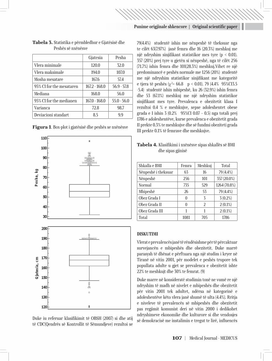
Punime origjinale shkencore | Original scientific paper
107 | Medical Journal - MEDICUS
Tabela 3. Statistika e përmbledhur e Gjatësisë dhe Peshës së nxënësve
Gjatesia Pesha
Vlera minimale 120.0 32.0
Vlera maksimale 194.0 107.0
Mosha mesatare 167.6 57.4
95% CI for the mesataren 167.2 - 168.0 56.9 - 57.8
Mediana 168.0 56.0
95% CI for the medianen 167.0 - 168.0 55.0 - 56.0
Varianca 72.8 98.7
Deviacioni standart 8.5 9.9
Figura 1. Box-plot i gjatësisë dhe peshës se nxënësve
Duke iu referuar klasifikimit të OBSH (2007) si dhe atij të CDC(Qendrës së Kontrollit të Sëmundjeve) rezultoi se
79(4.4%) studentë ishin me nënpeshë të theksuar nga te cilët 63(7.97%) janë femra dhe 16 (20.3%) meshkuj me një ndryshim sinjifikant statistikor mes tyre (p < 0.01). 357 (20%) prej tyre u gjetën si nënpeshë, nga të cilët 256 (71.7%) ishin femra dhe 101(28.3%) meshkuj.Vihet re një predominancë e peshës normale me 1256 (20%) studentë me një ndryshim statistikor sinjifikant me kategoritë e tjera të peshës (χ2= 66.8 p < 0.01). 79 (4.4% 95%CI3.5 -5.4) studentë ishin mbipeshë, ku 26 (32.9%) ishin femra dhe 53 (67.1%) meshkuj me një ndryshim statistikor sinjifikant mes tyre. Prevalenca e obezitetit klasa I rezultoi 0.4 % e meshkujve, sepse adoleshentet obese grada e I ishin 3 (0.2% 95%CI 0.07 – 0.5) nga totali prej 1786 e adoleshentëve, kurse prevalenca e obezitetit grada II prekte 0.3% te meshkujve dhe së fundmi obeziteti grada III prekte 0.1% të femrave dhe meshkujve.
Tabela 4. Klasifikimi i nxënësve sipas shkallës së BMI dhe sipas gjinisë
Shkalla e BMI Femra Meshkuj Total
Nënpeshë i theksuar 63 16 79 (4.4%)
Nënpeshë 256 101 357 (20.0%)
Normal 735 529 1264 (70.8%)
Mbipeshë 26 53 79 (4.4%)
Obez Grada I 0 3 3 (0.2%)
Obez Grada II 0 2 2 (0.1%)
Obez Grada III 1 1 2 (0.1%)
Total 1081 705 1786
DISKUTIMI
Vlerat e prevalencës janë të rëndësishme për të përcaktuar survejancën e mbipeshës dhe obezitetit. Duke marrë parasysh të dhënat e përftuara nga një studim i kryer në Tiranë në vitin 2001, për modelet e peshës trupore tek popullata adulte u gjet se prevalenca e obezitetit ishte 22% te meshkujt dhe 30% te femrat. (9)
Duke marre në konsideratë studimin tonë ne vumë re një ndryshim të madh në nivelet e mbipeshës dhe obezitetit për vitin 2001 tek adultet, ndërsa në kategorinë e adoleshentëve këto vlera janë shumë të ulta (4.4%). Rritja e niveleve të prevalencës së mbipeshës dhe obezitetit pas regjimit komunist deri në vitin 2000 i dedikohet ndryshimeve ekonomike dhe kulturore si dhe vendosjes së demokracisë me instalimin e tregut te lirë, influencës
Punime origjinale shkencore | Original scientific paper
Revistë mjekësore - MEDICUS | 108
së medias dhe ndryshimeve nutricionale si dhe rritjes së aksesit ndaj fastfood-eve. Bazuar në të dhënat e vendeve të tjera në tranzicion10,11 ka mundësi që urbanizimi i rritur dhe modernizimi i stilit te jetesës (ndryshimet dietike, inaktiviteti i rritur, duhani) kane luajtur një rol kyç në këtë drejtim.
Duke pasur parasysh studimin tonë duhet të themi me krenari se nivelet e mbipeshës dhe obezitetit tek adoleshentët në Shqipëri respektivisht (4.4%; 0.4%) janë nga më të ultat ne Evropë, duke iu referuar të dhënave të Task Forces Internacionale për Obezitetin (IOTF), që paraqet trendin global për obezitetin. Pra, në këtë aspekt ne mund të krahasojmë të dhënat tona për mbipeshën dhe obezitetin me ato të Estonisë. Letonisë dhe Turqisë.
Në vende si Kina dhe Tajlanda (12,13), prevalenca e mbipeshës dhe obezitetit është më frekuente tek djemtë sesa tek vajzat ashtu si dhe në Shqipëri sipas të dhënave të studimit tonë.
Një nga pikat e forta të studimit është se ai ka qenë anonim bazuar në një mostër përfaqësuese të madhe kombëtare. Ndërsa një limit i tij është natyra kros-seksionale e studimit që përjashton konkluzione të lidhura me rastësinë dhe mospërfshirjen e adoleshentëve të cilët kanë lënë shkollën dhe mund të jenë shtresa më vulnerabël. Si rezultat një kërkim i mëtejshëm nevojitet për të përfshirë në studim dhe adoleshentët që nuk kanë frekuentuar shkollën.
KONKLUZIONE
Një argument lidhur me nivelet e ulta të mbipeshës dhe obezitetit tek adoleshentët në Shqipëri do të ishte dieta mediterrane me fruta dhe perime, vaktet e konsumuara në shtëpi apo më shumë ushqime të shëndetshme, rritja e aktivitetit fizik, gjimnastika, aerobia, tendenca e djemve dhe e vajzave për t’u kujdesur për imazhin e tyre, influencuar nga media dhe televizioni që promovojnë individët elegantë si një mënyrë për të qenë të suksesshëm në jetë. Vihet re së fundmi rritja e presionit social mbi meshkujt që ka vendosur një ideal jorealist trupor me yndyrna të pakta dhe një figurë muskulore të fuqishme që mund te çojnë në një rritje të konsiderueshme të çrregullimimeve të ushqyerjes.14Aktualisht shqetësimet për peshën trupore janë konsideruar në mënyrë universale si probleme tipike femërore. Si rezultat presioni për të qenë e dobët do të kishte një ndikim më të madh tek vajzat. Nje gjetje interesante e studimit tonë është se një përqindje e lartë e adoleshentëve që
frekuentojnë shkollën rreth 70.8% janë në peshë normale. Identifikimi i shtigjeve për të dizenjuar programe në fushën e promocionit shëndetsor dhe të shëndetit pozitiv është esencial. Sipas opinionit tonë nivelet e ulta te mbipeshës dhe obezitetit dhe proporcioni i madh i peshës normale brenda popullatës së adoleshentëve i dedikohet popullaritetit të stilit të shëndetshëm të jetës midis të rinjve.
Megjithatë një konkluzion tjetër interesant i studimit tonë është se 20% e pjesëmarrsve kryesisht vajza reflektuan probleme të nënpeshës. Kjo është nje çështje serioze, si pasojë duhet të implementohen strategji që prekin këtë problem në Shqipëri, në të ardhmen e afërt.
Konflikt interesi: Asnjë.
LITERATURA
1. WHO Media Centre, author. Fact sheet: Obesity and overweight. 2006.
2. Popkin BM. The nutrition transition and obesity in the developing world. J Nutr. 2001;131:871S–873S.
3. Mohammadpour−Ahranjani B, Rashidi A, Karandish M, Eshraghian MR, Kalantari N. Prevalence of overweight and obesity in adolescent Tehrani students, 2000−2001: an epidemic health problem. Public Health Nutr. 2004;7:645–648.
4. Smoak CG, Burke GL, Webber LS, Harsha DW, Sriniva-san SR, Berenson GS et al. Relation of obesity to clus-tering of cardiovascular disease risk factors in children and young adults. The Bogalusa Heart Study. Am J Epi-demiol. 1987 Mar;125(3):364–372. PubMed.
5. Demerath E, Muratova V, Spangler E, Li J, Minor VE, Neal WA et al. School-based obesity screening in rural Appa-School-based obesity screening in rural Appa-lachia. Prev Med. 2003 Dec;37(6 Pt 1):553–560. PubMed.
6. Modan M HH. Hyperinsulinemia: a link between hyper-tension, obesity and glucose intolerance. J Clin Invest. 1985;75:809–817.
7. Dietz WH. Overweight in childhood and adolescence. N Engl J Med. 2004;350:855–7.
8. Rebecca M. Evaluation and treatment of childhood obe-sity. Am Fam Physicians. 1999;59:861–71.
9. World Health Organization (WHO). Obesity. Preventing and managing the Global Epidemic. Geneva: WHO, 1997.
10. Cole TJ, Bellizzi MC, Flegal KM, Dietz WH. Establishing a standard definition for child overweight and obesity worldwide: international survey. BMJ. 2000;320:1–6.

Punime origjinale shkencore | Original scientific paper
109 | Medical Journal - MEDICUS
11. Body weight patterns in a country in transition: a population-based survey in Tirana City, Albania Laidon Shapo1,*, Joceline Pomerleau1, Martin McKee1, Richard Coker1 and Agron Ylli 2 1European Centre on Health of Societies in Transition, London School of Hygiene and Tropical Medicine, Keppel Street, London WC1E 7HT, UK: 2Endocrinology Department, University Hospital Centre ‘Mother Theresa’, Dibra Street, 370 Tirana, Alba-nia, Submitted 17 September 2002: Accepted 5 Decem-ber 2002
12. Pudule I, Grinberga D, Kadziauskiene K, Abaravicius A,Vaask S, Robertson A, et al. Patterns of smoking in the Baltic Republics. Journal of Epidemiology and Com-munity Health 1999; 53: 277–83.
13. Pomerleau J, McKee M, Robertson A, Vaask S, Kadzi-auskiene K, Abaravicius A, et al. Physical inactivity in the Baltic countries. Preventive Medicine 2000; 31: 665–72.
14. Nguyen DM, El-Serag HB. The big burden of obesity. Gas-trointest Endosc. 2009;70:752–7. Germany.
OVERWEIGHT AND OBESITY IN ALBANIAN ADOLESCENTS
Enkelejda Shkurti1, Diamant Shtiza2, Diederik Aarendonk3, Angela Andreoli4,1Faculty of Medical Technical Sciences, 2University Hospital Centre “Mother Theresa”, Tirana, 3Forum Coordinator of the European Forum of Primary Care, 4University of Tor Vergata, Rome
ABSTRACT
The aim of the study was to investigate the body weight models among Albanian adolescents school students and the prevalence of overweight and obesity.
Methods
A cross-sectional study on a stratified sample of 1786 adolescents, 705( 39%) males and 1081(61%) females was conducted in Albania in January-December 2011. They were interviewed and examined for weight and height using standardized techniques.
Results
Regarding 2007 WHO classification for BMI scale it resulted that 4.4% 95% CI 3.5 -5.4 adolescents were overweight (32.9% females,67.1% males) with a statistically significant difference among them, p<0.01. The prevalence of overweight in adolescent females was 2.4%, in males was 7.5% . The prevalence of obesity class I resulted 0.4%, the obesity class II 0.3% and obesity class III 0.1% 95% CI 0.02 – 0.3 from the total of participants. A remarkable finding of our study was that 70.8% of adolescent students were in normal weight, 20% of them underweight, 71.7% were females, p<0.01.
Conclusion
Actually overweight and obesity isn’t a big public health problem among adolescents students in Albania. The problem is more prevalent among boys. In our opinion, the low levels of overweight and obesity are related to a popularity of healthy lifestyle among the youngsters. However, strategies to prevent the possible increase of the prevalence of overweight and obesity should be implemented in the near future in our country.
Keywords: Adolescents, overweight, obesity, Albania
Revistë mjekësore - MEDICUS | 110
17
VLERËSIMI I CILËSISË SË VAJIT TË ULLIRIT ‘‘EKSTRA I VIRGJER’’NË TREGUN SHQIPTAR Çakraj R.1, Prifti D.2
1 Ministria e Bujqësisë, Ushqimit dhe Mbrojtjes së Konsumatorit, Departamenti i Sigurisë Ushqimore 2 Universiteti i Tiranës, Fakulteti i Shkencave të Natyrës, Departamenti i Kimisë Industriale
ABSTRAKTI
Hyrje: Vaji i ullirit (VU) është një ushqim me vlera të larta ushqyese, të cilat i dedikohen përmbajtjes së acideve yndyrore, në veçanti acidit oleik dhe pranisë së antioksidantëve natyrale, polifenoleve. Shqipëria ka potenciale të mëdha për prodhimin e VU, prandaj vitet e fundit ka qënë prioritet i politikave bujqësore të qeverisë.
Qëllimi: Vlerësimi i cilësisë së VU, të deklaruar si “ekstra i virgjër” (EV), bazuar në disa tregues të cilësisë dhe natyralitetit të përcaktuar në legjislacionin kombëtar, harmonizuar me atë Europian.
Materiale dhe metoda: Mostrat janë marrë në fabrika prodhimi dhe në treg, gjatë sezonit 2011-2012. Analizimi i treguesve bazë të cilësisë (aciditeti, peroksidet, koeficientët e përthithjes K270 dhe K232 dhe ΔK) u krye në laboratorin e akredituar “ADAMA” në Tiranë, ndërsa përmbajtja e acideve yndyrore (PAY) dhe polifenoleve (PP), në laboratorin e Universitetit Kombëtar të Athinës.
Rezultatet : Vajrat nga prodhimi, rezultuan 100 % origjinal “vaj ulliri”, me përmbajtje të acidit oleik relativisht të lartë (70-75%) dhe PP të mesme, ndërsa 35 % e tyre rezultuan jo konform standartit për kategorinë “EV”. Në treg (20%) rezultuan vajra “të përzier” jo origjinal, (60%) me tregues të cilësisë jo konform standartit dhe me PP shumë të ulët.
Konkluzione: Vajrat nga prodhimi janë origjinale dhe më cilësore, ndërsa në treg konstatohen edhe vajra jo origjinal, të ndryshuar gjatë ruajtjes ose të përzier me vajra me kosto të ulët dhe që paraqesin cilësi më të dobët.
Fjalëkyçe: acide yndyrore, tregues cilësie, kritere natyraliteti, vaj ulliri ekstra i virgjër, polifenole.
Medicus 2013, Vol. 18 (2): 110-118
HYRJE
Vitet e fundit zhvillimi i sektorit të prodhimit të ullirit dhe përpunimit të tij, ka qënë prioritet i politikave bujqësore të qeverisë. Këto politika kanë çuar në rritjen e prodhimit të vajit të ullirit dhe konsumit të tij nga konsumatori shqiptar. Por, duke qënë një produkt ushqimor me përfitime të larta ekonomike, vaji i ullirit ka qënë objekt i fallsifikimeve nga operatorët e biznesit ushqimor, duke e deklaruar si vaj cilësor të kategorisë “ekstra i virgjër” ose përzier me vajra të tjerë me kosto të ulët. Për të siguruar një vaj cilësor dhe të sigurt në treg për konsumatorin dhe parandalimin e konkurencës së pandershme, janë përcaktuar rregullore strikte nga COOI (Këshilli Ndërkombëtar i Vajit të Ullirit) dhe Bashkimi Europian (BE) në lidhje me kriteret e cilësisë dhe natyralitetit të tij, të cilat po transpozohen në legjislacionin kombëtar, përkatësisht:
Ligji, nr. 87/2013,“Për kategorizimin e prodhimit, emërtimin dhe tregtimin e vajit të ullirit dhe vajit të bërsisë së ullirit”; Vendimi i Këshillit të Ministrave, Nr.475, datë 10.7.2003 “Për miratimin e rregullores “Për karakteristikat e vajrave të ullirit”, e cila është përafruar me Regulloren e BE-së, 2568/91 dhe është në proces miratimi të afërt.
Ky studim ka për qëllim vlerësimin e cilësisë së vajrave të ullirit në tregun shqiptar, të dëklaruar si “ekstra të virgjër”, bazuar në disa tregues kryesorë të cilësisë dhe natyralitetit të përcaktuar në legjislacion [1],[2],[3]. Nga monitorimet, konstatohet që vajrat cilësore tregëtohen kryesisht me porosi direkt nga fabrika dhe nuk arrijnë të dalin në treg tek konsumatori i fundit, për arsye të konkurencës së pandershme, kostos reale më të lartë se vajrat në treg. Prandaj vlerësimi i cilësisë së vajrave që mbizotërojnë në tregun shqiptar, tek konsumatori i fundit
Punime origjinale shkencore | Original scientific paper
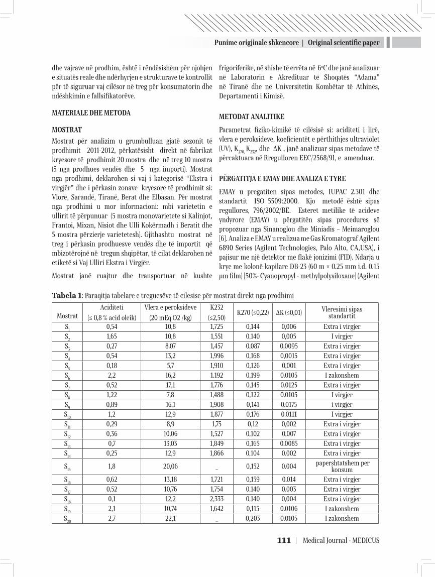
Punime origjinale shkencore | Original scientific paper
111 | Medical Journal - MEDICUS
dhe vajrave në prodhim, është i rëndësishëm për njohjen e situatës reale dhe ndërhyrjen e strukturave të kontrollit për të siguruar vaj cilësor në treg për konsumatorin dhe ndëshkimin e fallsifikatorëve.
MATERIALE DHE METODA
MOSTRAT
Mostrat për analizim u grumbulluan gjatë sezonit të prodhimit 2011-2012, përkatësisht direkt në fabrikat kryesore të prodhimit 20 mostra dhe në treg 10 mostra (5 nga prodhues vendës dhe 5 nga importi). Mostrat nga prodhimi, deklarohen si vaj i kategorisë “Ekstra i virgjër” dhe i përkasin zonave kryesore të prodhimit si: Vlorë, Sarandë, Tiranë, Berat dhe Elbasan. Për mostrat nga prodhimi u mor informacioni: mbi varietetin e ullirit të përpunuar (5 mostra monovarietete si Kalinjot, Frantoi, Mixan, Nisiot dhe Ulli Kokërmadh i Beratit dhe 5 mostra përzierje varietetesh). Gjithashtu mostrat në treg i përkasin prodhuesve vendës dhe të importit që mbizotërojnë në tregun shqipëtar, të cilat deklarohen në etiketë si Vaj Ulliri Ekstra i Virgjër.
Mostrat janë ruajtur dhe transportuar në kushte
frigoriferike, në shishe të errëta në 6oC dhe janë analizuar në Laboratorin e Akredituar të Shoqatës “Adama” në Tiranë dhe në Universitetin Kombëtar të Athinës, Departamenti i Kimisë.
METODAT ANALITIKE
Parametrat fiziko-kimikë të cilësisë si: aciditeti i lirë, vlera e peroksideve, koeficientët e përthithjes ultraviolet (UV), K270, K232, dhe ΔK , janë analizuar sipas metodave të përcaktuara në Rregulloren EEC/2568/91, e amenduar.
PËRGATITJA E EMAY DHE ANALIZA E TYRE
EMAY u pregatiten sipas metodes, IUPAC 2.301 dhe standartit ISO 5509:2000. Kjo metodë është sipas regullores, 796/2002/BE. Esteret metilike të acideve yndyrore (EMAY) u përgatitën sipas procedures së propozuar nga Sinanoglou dhe Miniadis – Meimaroglou [6]. Analiza e EMAY u realizua me Gas Kromatograf Agilent 6890 Series (Agilent Technologies, Palo Alto, CA,USA), i pajisur me një detektor me flakë jonizimi (FID). Ndarja u krye me kolonë kapilare DB-23 (60 m × 0.25 mm i.d. 0.15 μm film) [50%- Cyanopropyl - methylpolysiloxane] (Agilent
Tabela 1: Paraqitja tabelare e treguesëve të cilesise për mostrat direkt nga prodhimi
Mostrat
Aciditeti
(≤ 0,8 % acid oleik)
Vlera e peroksideve
(20 mEq O2 /kg)
K232
(≤2,50)K270 (≤0,22) ΔK (≤0,01) Vleresimi sipas
standartit
S1 0,54 10,8 1,725 0,144 0,006 Extra i virgjerS2 1,65 10,8 1,551 0,140 0,005 I virgjerS3 0,27 8.07 1,457 0,087 0,0095 Extra i virgjerS4 0,54 13,2 1,996 0,168 0,0015 Extra i virgjerS5 0,18 5,7 1,910 0,126 0,001 Extra i virgjerS6 2,2 16,2 1.192 0,199 0.0105 I zakonshemS7 0,52 17,1 1,776 0,145 0.0125 Extra i virgjerS8 1,22 7,8 1,488 0,122 0.0105 I virgjerS9 0,89 16,1 1,908 0,141 0.0175 i virgjerS10 1,2 12,9 1,877 0,176 0.0111 I virgjerS11 0,29 8,9 1,75 0,12 0,002 Extra i virgjerS12 0,36 10,06 1,527 0,102 0,007 Extra i virgjerS13 0,7 13,03 1,849 0,165 0.0085 Extra i virgjerS14 0,25 12,9 1,866 0,104 0.002 Extra i virgjer
S15 1,8 20,06 _ 0,152 0.004 papershtatshem per konsum
S16 0,62 13,18 1,721 0,159 0.014 Extra i virgjerS17 0,52 10,76 1,754 0,140 0.003 Extra i virgjerS18 0,1 12,2 2,333 0,140 0,004 Extra i virgjerS19 2,1 10,74 1,642 0,115 0.0106 I zakonshemS20 2,7 22,1 _ 0,203 0.0105 I zakonshem
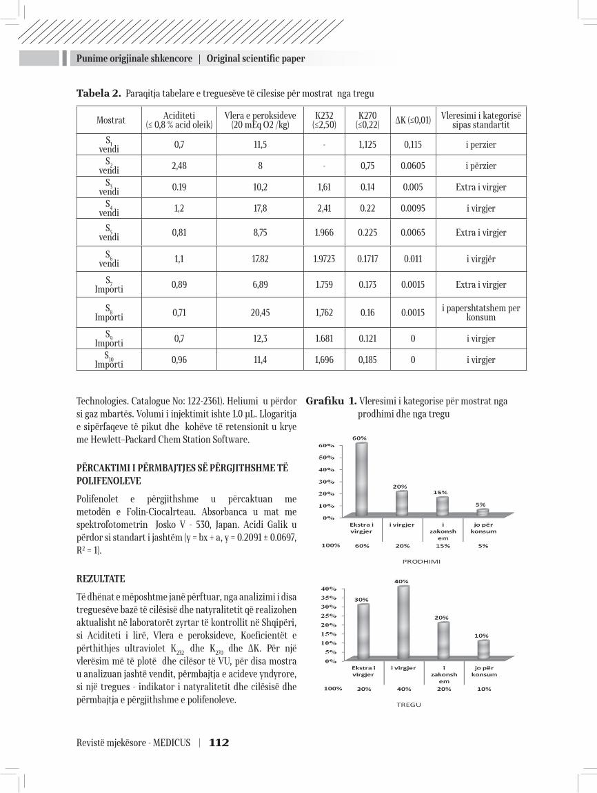
Punime origjinale shkencore | Original scientific paper
Revistë mjekësore - MEDICUS | 112
Technologies. Catalogue No: 122-2361). Heliumi u përdor si gaz mbartës. Volumi i injektimit ishte 1.0 μL. Llogaritja e sipërfaqeve të pikut dhe kohëve të retensionit u krye me Hewlett–Packard Chem Station Software.
PËRCAKTIMI I PËRMBAJTJES SË PËRGJITHSHME TË POLIFENOLEVE
Polifenolet e përgjithshme u përcaktuan me metodën e Folin-Ciocalrteau. Absorbanca u mat me spektrofotometrin Josko V - 530, Japan. Acidi Galik u përdor si standart i jashtëm (y = bx + a, y = 0.2091 ± 0.0697, R2 = 1).
REZULTATE
Të dhënat e mëposhtme janë përftuar, nga analizimi i disa treguesëve bazë të cilësisë dhe natyralitetit që realizohen aktualisht në laboratorët zyrtar të kontrollit në Shqipëri, si Aciditeti i lirë, Vlera e peroksideve, Koeficientët e përthithjes ultraviolet K232 dhe K270 dhe ΔK. Për një vlerësim më të plotë dhe cilësor të VU, për disa mostra u analizuan jashtë vendit, përmbajtja e acideve yndyrore, si një tregues - indikator i natyralitetit dhe cilësisë dhe përmbajtja e përgjithshme e polifenoleve.
Grafiku 1. Vleresimi i kategorise për mostrat nga prodhimi dhe nga tregu
Tabela 2. Paraqitja tabelare e treguesëve të cilesise për mostrat nga tregu
Mostrat Aciditeti(≤ 0,8 % acid oleik)
Vlera e peroksideve(20 mEq O2 /kg)
K232(≤2,50)
K270 (≤0,22) ΔK (≤0,01) Vleresimi i kategorisë
sipas standartit
S1vendi 0,7 11,5 - 1,125 0,115 i perzier
S2vendi 2,48 8 - 0,75 0.0605 i përzier
S3vendi 0.19 10,2 1,61 0.14 0.005 Extra i virgjer
S4vendi 1,2 17,8 2,41 0.22 0.0095 i virgjer
S5vendi 0,81 8,75 1.966 0.225 0.0065 Extra i virgjer
S6vendi 1,1 17.82 1.9723 0.1717 0.011 i virgjër
S7Importi 0,89 6,89 1.759 0.173 0.0015 Extra i virgjer
S8Importi 0,71 20,45 1,762 0.16 0.0015 i papershtatshem per
konsum
S9Importi 0,7 12,3 1.681 0.121 0 i virgjer
S10Importi 0,96 11,4 1,696 0,185 0 i virgjer
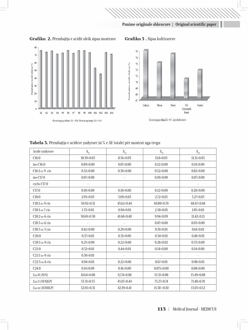
Punime origjinale shkencore | Original scientific paper
113 | Medical Journal - MEDICUS
Grafiku 2. Përmbajtja e acidit oleik sipas mostrave
Grafiku 3 . Sipas kultivareve
Tabela 3. Përmbajtja e acideve yndyrore (si % e AY totale) për mostrat nga tregu
Acide yndyrore S11 S12 S13 S14
C16:0 10.59±0.03 8.56±0.03 11.8±0.03 11.11±0.03
iso-C16:0 0.09±0.00 0.07±0.00 0.12±0.00 0.14±0.00
C16:1 ω-9 cis 0.52±0.00 0.30±0.00 0.52±0.00 0.82±0.00
iso-C17:0 0.07±0.00 - 0.10±0.00 0.07±0.00
cyclo-C17:0 - - - -
C17:0 0.10±0.00 0.10±0.00 0.12±0.00 0.20±0.00
C18:0 2.95±0.03 3.09±0.03 2.72±0.03 3.27±0.03
C18:1 ω-9 cis 50.92±0.51 43.62±0.44 69.89±0.70 68.47±0.68
C18:1 ω-7 cis 1.33±0.01 0.94±0.01 2.58±0.01 1.85±0.01
C18:2 ω-6 cis 30.69±0.30 41.68±0.40 9.94±0.09 11.42±0.11
C18:3 ω-6 cis - - 0.07±0.00 0.03±0.00
C18:3 ω-3 cis 0.42±0.00 0.29±0.00 0.70±0.01 0.61±0.01
C20:0 0.37±0.01 0.31±0.00 0.34±0.01 0.48±0.01
C20:1 ω-9 cis 0.23±0.00 0.22±0.00 0.28±0.02 0.33±0.00
C22:0 0.32±0.01 0.44±0.01 0.14±0.00 0.14±0.00
C22:1 ω-9 cis 0.30±0.01 - - -
C22:5 ω-6 cis 0.94±0.01 0.22±0.00 0.67±0.01 0.98±0.01
C24:0 0.14±0.00 0.16±0.00 0.075±0.00 0.08±0.00
Σω:0 (AYN) 14.64±0.08 12.74±0.08 15.31±0.08 15.49±0.08
Σω:1 (AYNJLP) 53.31±0.53 45.07±0.44 73.27±0.74 71.48±0.70
Σω:n (AYSHLP) 32.05±0.31 42.19±0.41 11.38 ±0.10 13.03±0.12
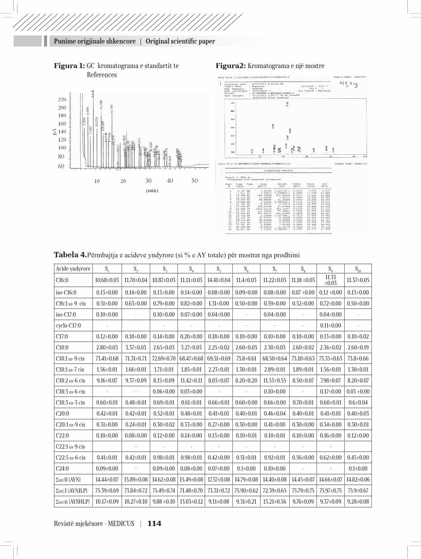
Punime origjinale shkencore | Original scientific paper
Revistë mjekësore - MEDICUS | 114
Figura 1: GC kromatograma e standartit te References
Tabela 4.Përmbajtja e acideve yndyrore (si % e AY totale) për mostrat nga prodhimi
Acide yndyrore S1 S2 S3 S4 S5 S6 S7 S8 S9 S10
C16:0 10.68±0.03 11.70±0.04 10.87±0.03 11.11±0.03 14.41±0.04 11.4±0.03 11.22±0.03 11.18 ±0.03 11.33 ±0.03 11.37±0.05
iso-C16:0 0.13±0.00 0.14±0.00 0.13±0.00 0.14±0.00 0.08±0.00 0.09±0.00 0.08±0.00 0.07 ±0.00 0.12 ±0.00 0.13±0.00
C16:1 ω-9 cis 0.51±0.00 0.63±0.00 0.79±0.00 0.82±0.00 1.31±0.00 0.50±0.00 0.59±0.00 0.52±0.00 0.72±0.00 0.50±0.00
iso-C17:0 0.10±0.00 - 0.10±0.00 0.07±0.00 0.04±0.00 - 0.04±0.00 - 0.04±0.00 -
cyclo-C17:0 - - - - - - - 0.11±0.00 -
C17:0 0.12±0.00 0.18±0.00 0.14±0.00 0.20±0.00 0.18±0.00 0.10±0.00 0.10±0.00 0.10±0.00 0.13±0.00 0.10±0.02
C18:0 2.80±0.03 3.37±0.03 2.65±0.03 3.27±0.03 2.25±0.02 2.60±0.03 2.30±0.03 2.60±0.02 2.36±0.02 2.60±0.19
C18:1 ω-9 cis 73.41±0.68 71.31±0.71 72.69±0.70 68.47±0.68 69.51±0.69 73.8±0.61 68.50±0.64 73.10±0.63 73.35±0.63 73.8±0.66
C18:1 ω-7 cis 1.56±0.01 1.66±0.01 1.71±0.01 1.85±0.01 2.23±0.01 1.30±0.01 2.89±0.01 1.89±0.01 1.56±0.01 1.30±0.01
C18:2 ω-6 cis 9.16±0.07 9.37±0.09 8.15±0.09 11.42±0.11 8.03±0.07 8.20±0.20 11.53±0.55 8.50±0.07 7.98±0.07 8.20±0.07
C18:3 ω-6 cis - - 0.06±0.00 0.03±0.00 - - 0.10±0.00 - 0.17±0.00 0.03 ±0.00
C18:3 ω-3 cis 0.60±0.01 0.48±0.01 0.69±0.01 0.61±0.01 0.66±0.01 0.60±0.00 0.66±0.00 0.70±0.01 0.60±0.01 0.6±0.04
C20:0 0.42±0.01 0.42±0.01 0.52±0.01 0.48±0.01 0.41±0.01 0.40±0.01 0.46±0.04 0.40±0.01 0.41±0.01 0.40±0.03
C20:1 ω-9 cis 0.31±0.00 0.24±0.01 0.30±0.02 0.33±0.00 0.27±0.00 0.30±0.00 0.41±0.00 0.30±0.00 0.34±0.00 0.30±0.01
C22:0 0.10±0.00 0.08±0.00 0.12±0.00 0.14±0.00 0.13±0.00 0.10±0.01 0.10±0.01 0.10±0.00 0.16±0.00 0.12±0.00
C22:1 ω-9 cis - - - - - - - -
C22:5 ω-6 cis 0.41±0.01 0.42±0.01 0.98±0.01 0.98±0.01 0.42±0.00 0.51±0.01 0.92±0.01 0.56±0.00 0.62±0.00 0.45±0.00
C24:0 0.09±0.00 - 0.09±0.00 0.08±0.00 0.07±0.00 0.1±0.00 0.10±0.00 - - 0.1±0.00
Σω:0 (AYN) 14.44±0.07 15.89±0.08 14.62±0.08 15.49±0.08 17.57±0.08 14.79±0.08 14.40±0.08 14.45±0.07 14.66±0.07 14.82±0.06
Σω:1 (AYNJLP) 75.39±0.69 73.84±0.72 75.49±0.74 71.48±0.70 73.31±0.72 75.90±0.62 72.39±0.65 75.79±0.75 75.97±0.75 75.9±0.67
Σω:n (AYSHLP) 10.17±0.09 10.27±0.10 9.88 ±0.10 13.03±0.12 9.11±0.08 9.31±0.21 13.21±0.56 9.76±0.09 9.37±0.09 9.28±0.08
Figura2: Kromatograma e një mostre
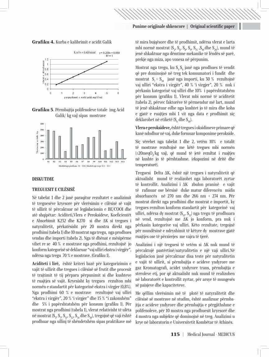
Punime origjinale shkencore | Original scientific paper
115 | Medical Journal - MEDICUS
Grafiku 4. Kurba e kalibrimit e acidit Galik
Grafiku 5. Përmbajtja polifenoleve totale (mg Acid Galik/ kg vaj) sipas mostrave
DISKUTIME
TREGUESIT E CILËSISË
Në tabelat 1 dhe 2 janë paraqitur rezultatet e analizimit të treguesëve kryesore për vlerësimin e cilësisë së vajit të ullirit të përcaktuar në legjislacionin e BE/COOI dhe atë shqipëtar: Aciditeti,Vlera e Peroksideve, Koeficientët e Absorbimit K232 dhe K270 si dhe ΔK si tregues i natyralitetit, përkatësisht për 20 mostra direkt nga prodhimi (tabela 1) dhe 10 mostrat nga tregu, nga prodhues vendas dhe importi (tabela 2). Nga të dhënat e mësipërme vihet re se 40 % e mostrave nga prodhimi, rezultojnë jo konform kategorisë së deklaruar “vaj ulliri ekstra i virgjër”, ndërsa nga tregu 70 % e mostrave. (Grafiku 1).
Aciditeti i lirë, është kriteri bazë për kategorizimin e vajit të ullirit dhe tregues i cilësisë së frutit dhe procesit të trajtimit të tij përpara përpunimit si dhe kushteve të ruajtjes së vajit. Kryesisht ky tregues rezulton mbi normën e standartit për kategorinë ekstra i virgjer (0,8%). Nga prodhimi 60 % e mostrave rezultojnë vaj ulliri “ekstra i virgjër”, 20 % ‘i virgjer” dhe 15 % “i zakonshëm” dhe 5% i papërshtatshëm për konsum (grafiku 1). Për mostrat nga prodhimi (tabela 1), vlerat relativisht të ulëta në mostrat (S3, S5, S11, S12, S14 dhe S18), tregojnë që vaji është prodhuar nga ullinj të shëndetshëm sipas praktikave më
të mira bujqësore dhe të prodhimit, ndërsa vlerat e larta mbi normë mostrat (S2, S6, S8, S9, S15 ,S19 dhe S20), mund të jenë shkaktuar nga dëmtime mekanike të lëndës së parë, prekje nga miza, apo vonesa në përpunim.
Mostrat nga tregu, ku S1÷S6 janë nga prodhues të vendit që pre dominojnë në treg tek konsumatori i fundit dhe mostrat S7 ÷ S10 janë nga importi, ku 30 % rezultojnë vaj ulliri “ekstra i virgjër”, 40 % ‘i virgjer”, 20 % nuk i përkasin kategorisë vaj ulliri dhe 10% i papërshtatshëm për konsum (grafiku 1). Vlerat mbi normë të aciditetit (tabela 2), përvec faktorëve të përmendur më lart, mund të jenë shkaktuar edhe nga kushtet jo të mira dhe koha e gjatë e ruajtjes mbi 1 vit nga data e prodhimit siç deklarohet në etiketë (S6 dhe S10).
Vlera e peroksideve, është tregues i oksidimeve primare që kanë ndodhur në vaj, duke formuar komponime perokside.
Siç vërehet nga tabelat 1 dhe 2, vetëm 10% e totalit të mostrave rezultojnë me këtë tregues mbi normën (≤20meqO2/kg vaj), që mund të jetë rezultat i ruajtjes në kushte jo të përshtashme. (ekspozimi në dritë dhe temperaturë).
Treguesi Delta ΔK, është një tregues i natyralitetit që aktualisht mund të realizohet nga laboratorët zyrtar të kontrollit. Analizimi i ΔK zbulon praninë e vajit të rafinuar ose bërsisë duke matur diferencën midis absorbancës në 270 nm dhe 266 nm + 274 nm. Për mostrat direkt nga prodhimi dhe mostrat e importit, ky tregues rezulton konform standartit për kategorinë vaj ulliri, ndërsa dy mostrat (S11, S12) nga tregu të prodhuara në vend, rezultojnë me ΔK jo konform, pra nuk i përkasin kategorive vaj ulliri. Këto rezultate, tregojnë për mundësinë e ndryshimit të këtyre dy mostrave gjatë ruajtjes ose të përzierjes me vajra të tjerë.
Analizimi i një treguesi të vetëm si ΔK nuk mund të përcaktojë pastërtinë/natyralitetin e një vaji ulliri.Në legjislacion janë përcaktuar disa teste për natyralitetin e vajit të ullirit, si përmbajtja e acideve yndyrore me gaz Kromatografi, acidet yndyrore trans, përmbajtja e steroleve etj, por që aktualisht nuk mund të realizohen në laboratorët e kontrollit zyrtar, për arsye të mungesës së paisjeve dhe kapaciteteve.
Me qëllim vlerësimin më të plotë të natyralitetit dhe cilësisë së mostrave në studim, është analizuar përmba-jtja e acideve yndyrore dhe përmbajtja e përgjithshme e polifenoleve, për 10 mostra nga prodhuesit kryesorë dhe 4 mostra nga subjekte që dominojnë në treg. Analizimi u krye në laboratorin e Universitetit Kombëtar të Athinës.

Punime origjinale shkencore | Original scientific paper
Revistë mjekësore - MEDICUS | 116
Përmbajtja e acideve yndyrore
Bazuar në origjinën e tij, një vaj vegjetal ka një profil të caktuar të acideve yndyrore. Karakteristike për vajin e ullirit është përmbajtja e lartë e acidit oleik dhe nivele minimale të acideve linoleik dhe linolenik. Analizimi i profilit të acideve yndyrore është me rëndësi, për vlerësimin e vlerës ushqyese të vajit të ullirit, stabilitetit dhe origjinalitetit të tij.
Rezultatet e përftuara nga analizimi i përmbajtjes së AY për mostrat në studim, paraqiten përkatësisht, për mostrat nga tregu në tabelën 3 dhe për mostrat direkt nga prodhimi në tabelën 4.
Për mostrat direkt nga prodhimi (tabela 4) përmbajtja e acideve yndyrore rezulton brënda standartit të përcaktuar nga COOI/BE dhe legjislacioni shqipëtar [1],[2],[3], për kategorinë “vaj ulliri ekstra i virgjër”, ndërsa për dy mostra nga tregu S11 dhe S12 (tabela 3), përmbajtja e disa acideve yndyrore (oleik, linoleik dhe linolenik) rezulton jashtë standartit.
Këto rezultate, mbi profilin e acideve yndyrore, konfirmojnë që mostrat S11 dhe S12 nuk i përkasin kategorisë vaj ulliri dhe mundësinë e përzierjes me vajra të rafinuar, sic rezultoi dhe nga vlerësimi i ΔK.
Gjithashtu, përmbajtja e acideve yndyrore të pangopura me një lidhje dyfishe dhe në veçanti përmbajtja e acidit oleik është e rëndësishme si nga pikpamja ushqyese dhe e stabilitetit oksidativ të vajit të ullirit. Vlerat e përmbajtjes së këtij acidi për të gjitha mostrat nga prodhimi dhe dy mostrat nga tregu (S13 dhe S14), rezultojnë relativisht të larta nga ~70 ÷75% (Grafiku 2).
Në qoftë se i vlerësojmë sipas kultivarëve, për mostrat S1÷S5 nga prodhimi, të cilat janë prodhuar nga monokultivarë (Grafiku 3), kultivarët autoktonë paraqesin vlera relativisht të larta. Kultivari Kalinjot rezulton me përmbajtjen më të lartë të acidit oleik (74.97%) krahasuar me kultivarët e tjerë. Në figurën 1, paraqitet kromatograma e standartit të referencës së përdorur për llogaritjen, Supelco 37 component FAME Mix C4-C24,(Merck,Darmstadt,Germany),me nje kolone kapilare DB -23 (60 m X 0.25 mm i.d 0.15 μm film). Në figurën 2, paraqitet kromatograma e një mostre të analizuar.
Përmbajtja e përgjithshme e fenoleve
“Vaji ullirit ekstra i virgjër” përmban sasi të konsi-derueshme të polifenoleve, të cilat janë antioksidantë natyrale, shumë të rëndësishme nga pikpamja ushqyese dhe që i japin vajit stabilitet ndaj oksidimit. Përmbajtja
fenolike varion në varësi të klimës, varietetit, kohës së vjeljes, metodës së ekstraktimit dhe praktikave bujqësore të aplikuara. (Cinquanta et al.,1997; Kiritsakis,1998). Gjithashtu, përmbajtja e fenoleve reduktohet gjatë ruajtjes për një kohë të gjatë të vajit të ullirit. (Frankel,E.N. ;Mailer,R.J.;Shoemaker,C.F.;Wang, S.C.;Flynn,J.D (2010).
Në grafikun 5, paraqitet përmbajtja e përgjithshme e fenoleve e përcaktuar me metodën kolorimetrike (Folin-Ciocalteau) dhe rezultati shprehet në mg acid Galik / kg vaj ulliri. Grafiku 4, paraqet kurbën e kalibrimit me tretësira standarte të acidit galik (nga 10 – 100 mg/10 ml) dhe nga kurba e ndërtuar u gjetën përqëndrimet e mostrave në studim sipas absorbancës së matur (grafiku 5).
Siç vërehet në grafikun 5, nëse analizojmë vajrat nga prodhimi, përmbajtja e fenoleve është relativisht e lartë krahasuar me vajrat në treg. Studimet e kryera (Uceda 2004) i klasifikojnë vajrat e ullirit sipas përmbajtjes së fenoleve me përmbajtje : shumë të ulët: < 150 ppm, të ulët:150-300 ppm, të mesme: 300-450 ppm dhe të lartë: 450-600 ppm . Kështu, vajrat direkt nga prodhimi, rezultojnë kryesisht me përmbajtje të mesme, ndërsa vajrat nga tregu janë me përmbajtje të ulët. Këto rezultate reflektojnë ndikimin e faktorëve të ndryshëm në përmbajtjen fenolike. Për vajrat nga prodhimi, përmbajtja relativisht e lartë shpjegohet, sepse janë prodhuar nga ullinj të vjelur në kohë optimale (deklarimet e prodhueseve), me përjashtim të mostrave S4 dhe S6 të cilat janë prodhuar nga ullinj shumë të pjekur të cilët rezultojnë me përmbajtje të ulët.
Përsa i përket mostrave nga tregu, që rezultojnë me përmbajtje të ulët të fenoleve krahasuar me vajrat në prodhim, për mostrat (S11,S12,) rezultatet përforcojnë mundësinë e përzierjes me vajra të rafinuar, ndërsa për dy mostrat e tjera (S13 dhe S14 ) përmbajtja e ulët shpjegohet me kohë ruajtjen e gjatë (në etiketë: prodhimi Janar 2011, mostrat u analizuan në Mars 2012).
PËRFUNDIME
Shqipëria ka potenciale për të prodhuar vajra ulliri cilësorë, dedikuar klimës së përshtatshme dhe cilësisë së varieteteve autoktone.
Vajrat e ullirit direkt nga prodhimi, të deklaruar si vaj ekstra i virgjër, rezultojnë origjinalë përsa i përket kategorizimit si vaj ulliri por në 40 % të mostrave cilësia nuk është konform kategorisë së deklaruar “ekstra i virgjër”, ku 20 % rezultojnë në kategorinë “i virgjër” dhe 15 % në kategorinë më të dobët “ i zakonshëm” dhe 5% i papërshtatshëm për konsum.
Punime origjinale shkencore | Original scientific paper
117 | Medical Journal - MEDICUS
Vajrat cilësore tregëtohen kryesisht direkt nga fabrikat e prodhimit dhe nuk arrijnë të dalin në treg tek konsumatori i fundit, për arsye të kostos reale më të lartë se vajrat në treg dhe konkurencës së pandershme.
Në treg tek konsumatori i fundit, konstatohen edhe vajra ulliri jo origjinalë, që nuk i ërkasin kategorive tëvajit të ullirit, të deklaruar në etiketë ‘vaj ulliri ekstra i virgjër”, të cilët mund të kenë ndryshuar gjatë ruajtjes në kushte jo të përshtatshme ose janë përzier me vajra të tjerë me kosto më të ulët.
Përcaktimi i përmbajtjes së acideve yndyrore me Gaz-kromatografi, është një tregues - indikator i rëndësishëm për vlerësimin e natyralitetit dhe të cilësisë së vajit të ullirit.
Cilësia e vajit të ullirit varet nga një numër faktorësh si: varieteti i ullirit, gjeografia e kultivimit, koha e vjeljes dhe e përpunimit, kushtet e trajtimit të ullirit para përpunimit, teknologjia e ekstraktimit dhe kushtet e ruajtjes dhe ambalazhimit.
REKOMANDIME
Zbatimi i praktikave të mira bujqësore (PMB), në lidhje me ujitjen, kohën optimale të vjeljes dhe kushtet e transportit nga fermerët që prodhojnë ullinj për vaj.
Zbatimi i praktikave të mira të prodhimit (PMP) , si përpunimi pa vonesa i ullinjve, respektimi i parametrave teknologjikë gjatë përpunimit (koha dhe temperatura) dhe kushtet e ruajtjes dhe magazinimit nga operatorët e përpunimit të vajit të ullirit.
Monitorimi i vazhdueshëm i vajit të ullirit në gjithë hallkat e tregëtimit, nga strukturat shtetërore të kontrollit të cilësisë dhe sigurisë ushqimore, me qëllim parandalimin e praktikave të fallsifikimit dhe konkurencës së pandershme në treg.
Paisjen e laboratorëve të kontrollit zyrtar me Gaz- Kromatograf, për kryerjen e profilit të acideve yndyrore, si test – indikator i cilësisë dhe natyralitetit, në pamundësi të ngrejnë gjithë infrastrukturën e nevojshme laboratorike për realizimin e gjithë testeve të natyrshmërisë dhe cilësisë.
LITERATURA
1. Commision Regulation (EEC) No 2568/91: On the char-acteristics of olive oil and olive-residue oil and on the relevant methods of analysis (OJ L 248, 5.9.1991, p. 1).
2. Ligji, nr. 87/2013,“Për kategorizimin e prodhimit, emër-timin dhe tregtimin e vajit të ullirit dhe vajit të bërsisë së ullirit”(Fletore zyrtare, Nr.20, datë 28.02.2013) .
3. Vendimi i Këshillit të Ministrave, Nr.475, datë 10.7.2003 “Për miratimin e rregullores “Për karakteristikat e va-jrave të ullirit” (Fletore zyrtare: nr.58; date 17.07.2003).
4. V.J. Sinanoglou, S. Miniadis-Meimaroglou: Food Res. Intern. 31 (1998), Pages 467-473.
5. Vassiliki T. Papoti, Maria Z. Tsimidou: Food Chemistry, 112(1), 2009, Pages 246-252,
6. V. J. Sinanoglou and S. Miniadis-Meimaroglou, “Fatty acid of neutral and polar lipids of (edible) Mediterranean cephalopods,” Food Research International, vol. 31, no. 6-7, pp. 467–473, 1998.
7. Uceda, M., and Frías, L. (1975): Harvest dates. Evolution of the fruit oil content, oil composition and oil quality.In: Proceedings Del Segundo Seminario Oleicola Inter-nacional Córdoba: COI pp. 125 – 128.
8. Food Chemistry, 112(1), 2009, Pages 246-252, Vassiliki T. Papoti, Maria Z. Tsimidou Marino Uceda Ojeda (2006): Efecto de los factores de producción en la calidad físico química y sensorial de los aceites de olive. Pages 4-5.
9. Aparicio, R., Luna G. (2002). Characterizacion of mono-varietal virgin olive oils. Eur. J. Lipid Sci. Technol. 104; 614 – 627.
10. Owen, R.W., Giacosas, A., Hull, W.E., Haubner, R., Würtele, G., Spiegelhalder, B., Bartsch, H. (2000): Olive oil consumption and health: The possible role of anti-oxidants. Lancet Oncology. 1; 107 – 112.
11. Owen, R.W., Mier, W., Giacosas, A., Hull, W.E., Spiegel-halder, B., Bartsch, H. (2000a): The antioxidant/antican-cer potential of phenolic compounds isolated from olive oil. European Journal of Cancer. 36; 1235 – 1247.
12. Aparicio, R. and Luna, G. (2002): Characterisation of monovarietal Virgin Olive Oils. European Journal of Li-pid Science and Technology. 104: 614–627.
13. Cohen, Z.,Vonshak, A., & Richmond, A. (1988): Effect of environmental conditions on fatty acid composition of the red algae Porphyridium cruentum: Correlation to growth rate. Journal of Phycology, 24, 328-332.
14. Frankel, E. N.; Mailer, R.J; Shoemaker,C.F.; Wang,S.C.; Flynn,J.D (2010): Tests indicate that imported “extra virgin”olive oil often fails international and USDA standards - UC Davis Olive Center, July 2010.
Punime origjinale shkencore | Original scientific paper
Revistë mjekësore - MEDICUS | 118
EVALUATION OF THE QUALITY OF “EXTRA VIRGIN OLIVE OIL” IN THE ALBANIAN MARKET
Rudina ÇAKRAJ1, Donika PRIFTI2 ,1 Ministry of Agriculture, Food and Consumer Protection, Food Safety Directory2 University of Tirana, Faculty of Natural Sciences, Department of Industrial Chemistry
ABSTRACT
Introduction: Olive oil (OO) is a food with high nutritional values, which refer the content of fatty acids, in particular the oleic acid and the presence of natural antioxidants, polyphenols. Albania has great potentials for the production of OO, so in recent years it has been a priority of the government’s agricultural policies.
Purpose: Evaluation of OO, declared as extra virgin olive oil (EVOO), based on some quality and purity indicators defined in national legislation, harmonized with the Europian one.
Materials and methods: Samples were taken in the production and in the market during the 2011-2012 season. Analysis of the basic indicators of quality (acidity, peroxides, absorption coefficients K270 and K232 and ΔK) were performed in the accredited laboratory “Adama” in Tirana, while the content of fatty acids (FAC) and polyphenols, in the laboratory of the National University of Athens.
Results: Oils from production, resulted 100% original as “olive oil”, with a relatively high oleic acid content (70-75%) and medium content of polyphenols , while 35% of them were non-conform to category “EVOO”. On the market (20%) of samples resulted “mixed” not original, (60%) with quality indicators non-conform to standard and with too low polyphenol content.
Conclusion: Oils from the production are original and qualitative, while in the market are found adulterated/not original olive oils, that may have altered during the storage or may be mixed with low cost oils and have poor quality.
Keywords: fatty acids, quality characteristics, extra virgin olive oil, purity criteria, polyphenols.
119 | Medical Journal - MEDICUS
NDIKIMI I CILESISE SE UJRAVE TE PISHINAVE TE TIRANES, NE SHENDETIN DHE SIGURINE E PERDORUESVE TE TYRE Drita Mema¹, Donika Bocari ¹, Arben Luzati ¹, Elida Mataj1, Oltiana Petri¹
¹ Departamenti Shëndeti dhe Mjedisi , Instituti i Shëndetit Publik, Tirane, Albania
ABSTRAKTI
Qëllimi: Vlerësimi i indekseve të ujërave të pishinave që ndikojnë direkt në shëndetin dhe sigurinë e përdorueseve të tyre. Kjo është arritur nëpërmjet vlerësimit të treguesëve fiziko-kimik, mikrobiologjik dhe ato të mirëmbajtjes, në disa pishina që përdorin si element disifektimi klorin në mënyra të ndryshme.
Materiali dhe metoda. Pishinat e marra në studim janë: një pishinë e hapur dhe tre të mbyllura në qytetin e Tiranes. Studimi shtrihet në një afat kohor dy vjeçar. Këto pishina janë të frekuentuara nga grup mosha të ndryshme.
Treguesit fiziko-kimik të analizuar janë: temperatura, klori i lirë, pH, lëndet pezull, turbulenca, amonjaku, alkaliteti, fortësia në kalcium, lëndet e ngurta të tretshme (TDS) sipas metodave ISO, SSH, EN. Treguesit mikrobiologjik të analizuar janë: coliforme total, coliforme fekal, streptoccocus fecal dhe pseudomonas aeruginosa.
Rezultatet: Nga studimi, 65% e vlerave të klorit të lirë janë jashtë vlerave standarde. Vlerat e pH në 63% të vlerave të matura janë jashtë intervalit 7.2-7.6, ku procesi i klorinimit është efektiv. coliforme total dhe fekale në 70 % të mostra të analizuara janë jashtë vlerave standarde.
Përfundimi: Menaxhimi i mirë i pishinave si: përdorimi i sistemeve automatike, stafi i trajnuar mirë, trajtimi i ujërave të tyre dhe përdorues të pishinave të edukuar dhe informuar, krijojnë kushte për shëndet dhe siguri në pishina.
Fjalë kyçet: klori i lirë, mikrobiologjik, shëndet, standard,
HYRJA
Lëvizja demografike e popullsisë në vendin tonë u shoqërua me rritjen e kërkesave për vende rekreative. Qyteti i Tiranës vitet e fundit ka arritur në popullsinë prej 830.000 banorë nga 244.200 banorë qe ka qënë në vitin 1990. (1)
Ndotja e zonës bregdetare të qytetit të Durrësit krijoi mundësi që shumë nga frekuentuesit e ujërave të larjes detare po përdorin pishinat publike në qytetin e Tiranës. Klima nentropikale-mesdhetare që mbizotëron në Tiranë me temperatura mesatare vjetore korrik + 24° Celcius dhe janar +7° Celsius e favorizon përdorimin e pishinave. Në qytetin e Tiranës është rritur shumë numri i frekuentuesëve dhe përdorueseve të pishinave.
Kjo rritje kërkoi domosdoshmërinë e një vlerësimi të aspekteve të projektimit, ndërtimit, mirëmbajtjes dhe vlerësimit sanitar të pishinave të larjes, për shëndetin publik të sigurt. Risku në pishina vjen nga: Risku kimik dhe Risku mikrobiologjik.
1 Risku kimik përfshin:
� Kimikatet të cilat mund të derivohen nga burimi i furnizimit të ujërave, nga banjatorët (urinimi, përshtyma) nga menaxhimi i disinfektimit, trajtimet me kimikate për rregullimin e pH dhe nënproduktet e disinfektimit (kloraminat ).
� Kimikatet e derivuara nga banjatoret (Urinimi, amonjaku)
Mekanizmi kryesor i formimit të amonjakut në pishina është biologjik, nga hidroliza e ures nën veprimin e enzimës ureazë sipas ekuacionit të mëposhtëm:
(NH2)2CO + H2O → CO2 + 2NH3
Urina është burimi kryesor i përbësve të azotit në ujin e pishinave.( 2)
Kimikatet e derivuara nga proceset e klorinimit. Për disinfektim në këto pishina është përdorur klori por në mënyra të ndryshme të dozimit të tij manual dhe
Medicus 2013, Vol. 18 (2): 119-124
18Punime origjinale shkencore | Original scientific paper
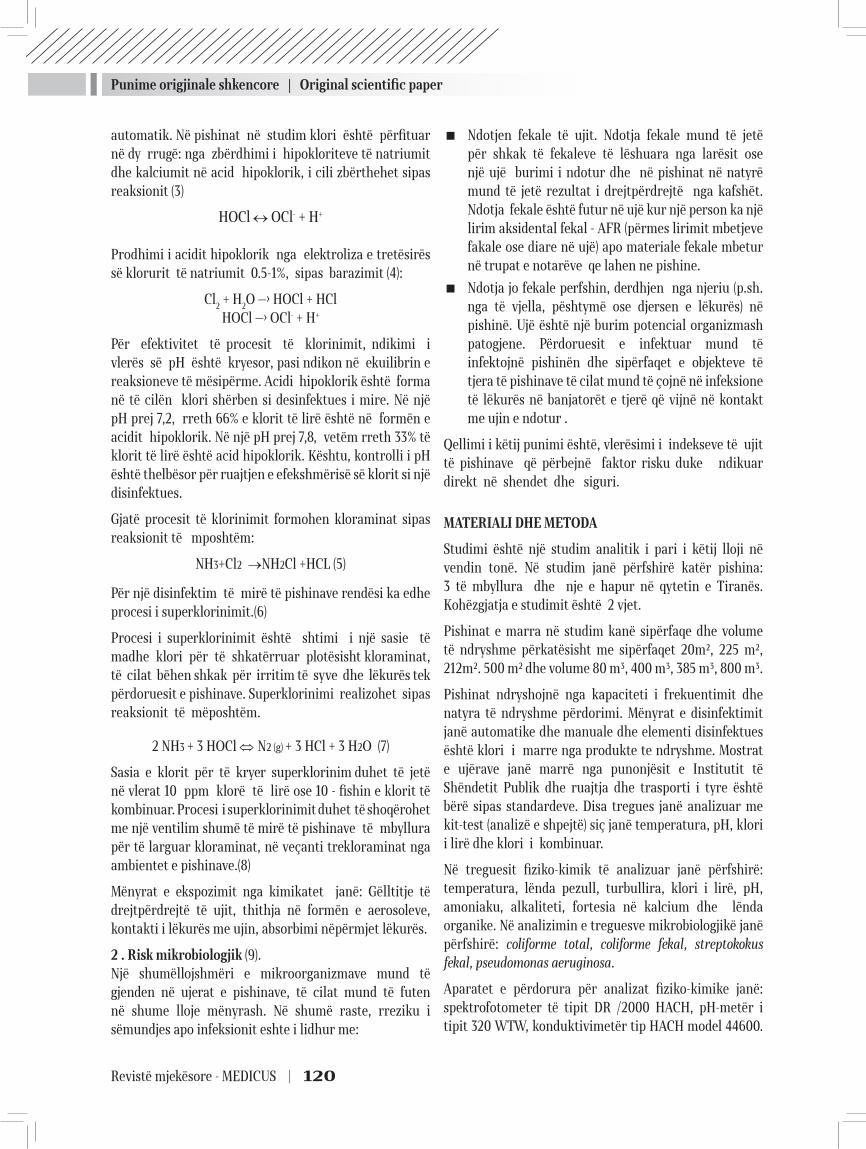
Punime origjinale shkencore | Original scientific paper
Revistë mjekësore - MEDICUS | 120
automatik. Në pishinat në studim klori është përfituar në dy rrugë: nga zbërdhimi i hipokloriteve të natriumit dhe kalciumit në acid hipoklorik, i cili zbërthehet sipas reaksionit (3)
HOCl ↔ OCl- + H+
Prodhimi i acidit hipoklorik nga elektroliza e tretësirës së klorurit të natriumit 0.5-1%, sipas barazimit (4):
Cl2 + H2O —› HOCl + HCl HOCl —› OCl- + H+
Për efektivitet të procesit të klorinimit, ndikimi i vlerës së pH është kryesor, pasi ndikon në ekuilibrin e reaksioneve të mësipërme. Acidi hipoklorik është forma në të cilën klori shërben si desinfektues i mire. Në një pH prej 7,2, rreth 66% e klorit të lirë është në formën e acidit hipoklorik. Në një pH prej 7,8, vetëm rreth 33% të klorit të lirë është acid hipoklorik. Kështu, kontrolli i pH është thelbësor për ruajtjen e efekshmërisë së klorit si një disinfektues.
Gjatë procesit të klorinimit formohen kloraminat sipas reaksionit të mposhtëm:
NH3+Cl2 →NH2Cl +HCL (5)
Për një disinfektim të mirë të pishinave rendësi ka edhe procesi i superklorinimit.(6)
Procesi i superklorinimit është shtimi i një sasie të madhe klori për të shkatërruar plotësisht kloraminat, të cilat bëhen shkak për irritim të syve dhe lëkurës tek përdoruesit e pishinave. Superklorinimi realizohet sipas reaksionit të mëposhtëm.
2 NH3 + 3 HOCl ⇔ N2 (g) + 3 HCl + 3 H2O (7)
Sasia e klorit për të kryer superklorinim duhet të jetë në vlerat 10 ppm klorë të lirë ose 10 - fishin e klorit të kombinuar. Procesi i superklorinimit duhet të shoqërohet me një ventilim shumë të mirë të pishinave të mbyllura për të larguar kloraminat, në veçanti trekloraminat nga ambientet e pishinave.(8)
Mënyrat e ekspozimit nga kimikatet janë: Gëlltitje të drejtpërdrejtë të ujit, thithja në formën e aerosoleve, kontakti i lëkurës me ujin, absorbimi nëpërmjet lëkurës.
2 . Risk mikrobiologjik (9).Një shumëllojshmëri e mikroorganizmave mund të gjenden në ujerat e pishinave, të cilat mund të futen në shume lloje mënyrash. Në shumë raste, rreziku i sëmundjes apo infeksionit eshte i lidhur me:
� Ndotjen fekale të ujit. Ndotja fekale mund të jetë për shkak të fekaleve të lëshuara nga larësit ose një ujë burimi i ndotur dhe në pishinat në natyrë mund të jetë rezultat i drejtpërdrejtë nga kafshët. Ndotja fekale është futur në ujë kur një person ka një lirim aksidental fekal - AFR (përmes lirimit mbetjeve fakale ose diare në ujë) apo materiale fekale mbetur në trupat e notarëve qe lahen ne pishine.
� Ndotja jo fekale perfshin, derdhjen nga njeriu (p.sh. nga të vjella, pështymë ose djersen e lëkurës) në pishinë. Ujë është një burim potencial organizmash patogjene. Përdoruesit e infektuar mund të infektojnë pishinën dhe sipërfaqet e objekteve të tjera të pishinave të cilat mund të çojnë në infeksione të lëkurës në banjatorët e tjerë që vijnë në kontakt me ujin e ndotur .
Qellimi i këtij punimi është, vlerësimi i indekseve të ujit të pishinave që përbejnë faktor risku duke ndikuar direkt në shendet dhe siguri.
MATERIALI DHE METODA
Studimi është një studim analitik i pari i këtij lloji në vendin tonë. Në studim janë përfshirë katër pishina: 3 të mbyllura dhe nje e hapur në qytetin e Tiranës. Kohëzgjatja e studimit është 2 vjet.
Pishinat e marra në studim kanë sipërfaqe dhe volume të ndryshme përkatësisht me sipërfaqet 20m², 225 m², 212m². 500 m² dhe volume 80 m³, 400 m³, 385 m³, 800 m³.
Pishinat ndryshojnë nga kapaciteti i frekuentimit dhe natyra të ndryshme përdorimi. Mënyrat e disinfektimit janë automatike dhe manuale dhe elementi disinfektues është klori i marre nga produkte te ndryshme. Mostrat e ujërave janë marrë nga punonjësit e Institutit të Shëndetit Publik dhe ruajtja dhe trasporti i tyre është bërë sipas standardeve. Disa tregues janë analizuar me kit-test (analizë e shpejtë) siç janë temperatura, pH, klori i lirë dhe klori i kombinuar.
Në treguesit fiziko-kimik të analizuar janë përfshirë: temperatura, lënda pezull, turbullira, klori i lirë, pH, amoniaku, alkaliteti, fortesia në kalcium dhe lënda organike. Në analizimin e treguesve mikrobiologjikë janë përfshirë: coliforme total, coliforme fekal, streptokokus fekal, pseudomonas aeruginosa.
Aparatet e përdorura për analizat fiziko-kimike janë: spektrofotometer të tipit DR /2000 HACH, pH-metër i tipit 320 WTW, konduktivimetër tip HACH model 44600.

Punime origjinale shkencore | Original scientific paper
121 | Medical Journal - MEDICUS
Për analizat mikrobiologjike u përdorën këto aparate: numëruesi i kolonive i tipit Stjuart, kuti llambe ultraviolet CAMAG dhe Stereomikroskop-Nikon.
Metodat e përdorura për analizimin e këtyre treguesëve janë metoda ISO, EN dhe SSH përkatësisht; për treguesit fiziko-kimik; SSH ISO EN7393-1; SSH ISO 10523; SSHEN 27888; SSH ISO EN 7027 dhe për analizat mikrobiologjike u përdor metodat me membranë filtrante sipas standardeve ISO 9308-2:1990; ISO 9308 -1; ISO 9308 – 3. Kontrolli i cilësisë së analizave është bërë nëpërmjet kontrollit të jashtëm duke marrë pjesë në ushtrime laboratorike për disa tregues si pH,TDS, fortësia në kalcium. Vlerësimi i përformancës së laboratorit u bë në përputhje me standardin ISO/IEC 17043:2010 sipas Z- score, ku treguesit e mësipërm rezultuan sipas Z ≤ 2, rezultat i kënaqshëm. Gjatë analizave janë përdorur vetëm reagent me gradë analitike të njohur.
REZULTATET
Nivelet e treguesve fiziko-kimik dhe bakteriologjikë të ujërave të analizuar, tregohen nëpërmjet figurave të mëposhtme, duke i krahasuar ato dhe me normat shqiptare te aplikuara dhe atyre që OBSH rekomandon për një trajtim adekuat të këtyre ujërave .
Pishinat në studim janë: me sistem manual klorinimi, gjysmë automatik dhe sistem automatik. Disa nga treguesit e analizuar si temperatura, lënda pezull, alkaliteti, fortësia, janë me te qëndrueshëm, prandaj studimi është përqendruar në treguesit e mëposhtëm:
Figura 1. ̀Vlerat e klorit të lirë te pishinave në studim
Nga figura e mësipërme shohim qartë se një numër i konsiderueshëm matjesh janë poshtë vlerës standarde më të ulët të lejuar (0.7 mg/l) (10).
Figura 2: Vlerat e pH në pishinat në studim.
Nga figura e mësipërme shihet qartë se, shumica e vlerave të pH janë jashtë vlerave optimale krahasuese 7.2 -7.6 për pishinat nr 2 dhe 4. Në intervalin e vlerave krahasuese procesi i klorinimit është efiçent.
Figura 3: Vlerat e amoniakut për pishinat në stu-dim.
Nga figura e mësiperme duket qartë se vlerat e amonjakut janë mbi vlerat e standardit,sidomos ne pishinat 2 dhe 4 (10).
Korrelacionet midis indekseve kimike për një nga pishinat në studim janë si mëposhtë:
Figura 4. Korrelacioni i Cl2 me NH4 për pishinën 4
Verehet korrelacion i moderuar negativ dhe statistikisht i rëndesishëm ndërmjet sasisë së Cl2 dhe sasisë së NH4. Me rritjen e sasisë së Cl2 të lirë ulet sasisa e NH4. (Spearman’s coefficient of rank correlation (rho) =-0.38 p=0.003).
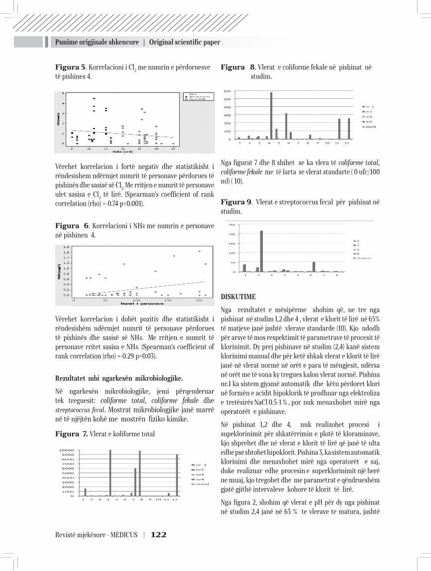
Punime origjinale shkencore | Original scientific paper
Revistë mjekësore - MEDICUS | 122
Figura 5. Korrelacioni i Cl2 me numrin e përdoruesve të pishines 4.
Vërehet korrelacion i fortë negativ dhe statistikisht i rëndesishem ndërmjet numrit të personave përdorues të pishinës dhe sasisë së Cl2. Me rritjen e numrit të personave ulet sasisa e Cl2 të lirë. (Spearman’s coefficient of rank correlation (rho) =-0.74 p<0.001).
Figura 6. Korrelacioni i NH4 me numrin e personave në pishinen 4.
Vërehet korrelacion i dobët pozitiv dhe statistikisht i rëndesishëm ndërmjet numrit të personave përdorues të pishinës dhe sasisë së NH4. Me rritjen e numrit të personave rritet sasisa e NH4. (Spearman’s coefficient of rank correlation (rho) =-0.29 p=0.03).
Rezultatet mbi ngarkesën mikrobiologjike.
Në ngarkesën mikrobiologjike, jemi përqenderuar tek treguesit: coliforme total, coliforme fekale dhe streptococcus fecal. Mostrat mikrobiologjike janë marrë në të njëjtën kohë me mostrën fiziko-kimike.
Figura 7. Vlerat e koliforme total
Figura 8. Vlerat e coliforme fekale në pishinat në studim.
Nga figurat 7 dhe 8 shihet se ka vlera të coliforme total, coliforme fekale me të larta se vlerat standarte ( 0 ufc/100 ml) ( 10).
Figura 9. Vlerat e streptococcus fecal për pishinat në studim.
DISKUTIME
Nga rezultatet e mësipërme shohim që, ne tre nga pishinat në studim 1,2 dhe 4 , vlerat e klorit të lirë në 65% të matjeve janë jashtë vlerave standarde (10). Kjo ndodh për arsye të mos respektimit të parametrave të procesit të klorinimit. Dy prej pishinave në studim (2,4) kanë sistem klorinimi manual dhe për ketë shkak vlerat e klorit të lirë janë në vlerat normë në orët e para të mëngjesit, ndërsa në orët me të vona ky tregues kalon vlerat normë. Pishina nr.1 ka sistem gjysmë automatik dhe këtu përdoret klori në formën e acidit hipoklorik të prodhuar nga elektroliza e tretësirës NaCl 0.5-1 % , por nuk menaxhohet mirë nga operatorët e pishinave.
Në pishinat 1,2 dhe 4, nuk realizohet procesi i supeklorinimit për shkatërrimin e plotë të kloraminave, kjo shprehet dhe në vlerat e klorit të lirë që janë të ulta edhe pse shtohet hipoklorit. Pishina 3, ka sistem automatik klorinimi dhe menaxhohet mirë nga operatorët e saj, duke realizuar edhe procesin e superklorinimit një herë ne muaj, kjo tregohet dhe me parametrat e qëndrueshëm gjatë gjithë intervaleve kohore të klorit të lirë.
Nga figura 2, shohim që vlerat e pH për dy nga pishinat në studim 2,4 janë në 63 % te vlerave te matura, jashtë
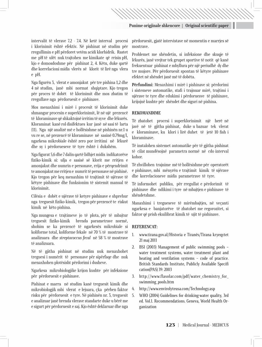
Punime origjinale shkencore | Original scientific paper
123 | Medical Journal - MEDICUS
intervalit të vlerave 7.2 - 7.4. Në ketë interval procesi i klorinimit është efektiv. Në pishinat në studim për rregullimin e pH përdoret vetëm acidi klorhidrik. Rastet me pH të ulët nuk trajtohen me kimikate që rrisin pH, kjo e domosdoshme për pishinat 2, 4. Këtu, duke qartë dhe korrelacioni midis vlerës së klorit të lirë nga vlera e pH.
Nga figurën 3, vlerat e amonjakut për tre pishina 1,2 dhe 4 në studim, janë mbi normat shqiptare. Kjo tregon për proces të dobët të klorinimit dhe mos zbatim të rregullave nga përdoruesit e pishinave.
Mos menaxhimi i mirë i procesit të klorinimit duke shmangur procesin e superklorinimit, lë në ujë prezencë të kloraminave që shkaktojnë irritim të syve dhe lëkurës. Kloraminat kanë rol disifektues kur janë në sasi të larta (11). Nga një analizë më e hollësishme në pishinën nr.1 u vu re se, në prezencë të kloraminave në sasinë 0.79mg/l, ngarkesa mikrobiale është zero por irritimi në lëkurë dhe sy i përdorueseve të tyre është i dukshëm.
Nga figurat 5,6 dhe 7 dalin qartë lidhjet midis indikatorëve fiziko-kimik si: ulja e sasisë së klorit me rritjen e amonjakut dhe numrin e personave, rrija e përqendrimit te amonjakut me rritjen e numrit të personave në pishinë. Kjo tregon për keq menaxhim të trajtimit të ujërave të këtyre pishinave dhe funksionim të sistemit manual të klorinimit.
Cilësia e dobët e ujërave të ketyre pishinave e shprehur nga treguesit fiziko-kimik, tregon për prezencë te riskut kimik në këto pishina.
Nga mungesa e trajtimeve jo të plota, për të mbajtur treguesit fiziko-kimik brenda parametrave normë, shohim se ka prezencë të ngarkesës mikrobiale si koliforme total, koliforme fekale në 70 % të mostrave të analizuara dhe streptococcus fecal në 58 % të mostrave të analizuara.
Në të gjitha pishinat në studim nuk menaxhohet treguesi i numërit të personave për sipërfaqe dhe nuk menaxhohen plotësisht përdorimi i dusheve.
Ngarkesa mikrobiologjike krijon kushte për infeksione për përdoruesit e pishinave.
Pishinat e marra në studim kanë treguesit kimik dhe mikrobiologjik mbi vlerat e lejuara, çka përben faktor risku për përdoruesit e tyre. Në pishinën nr. 3, treguesit e analizuar janë brenda vlerave standarte duke u bërë me e sigurt për përdoruesit e saj. Kjo është deklaruar dhe nga
përdoruesit, gjatë intervistave në momentin e marrjes së mostrave.
Problemet me shëndetin, si infeksione dhe skuqje të lëkurës, janë vrejtur tek grupet sportive të notit që kanë frekuentuar pishinat e mbyllura për një periudhë dy dhe tre mujore. Për përdoruesit spontan të këtyre pishinave efektet në shëndet janë më të dobëta.
Përfundimi: Menaxhimi i mirë i pishinave si: përdorimi i sistemeve automatike, stafi i trajnuar mirë, trajtimi i ujërave te tyre dhe edukimi i përdoruesve të pishinave, krijojnë kushte për shëndet dhe siguri në pishina.
REKOMANDIME
Të zbatohet procesi i superklorinimit një herë në javë në të gjitha pishinat, duke u bazuar tek vlerat e kloraminave, ku klori i lirë duhet të jetë 10 fish i kloraminave.
Të instalohen sistemet automatike për të gjitha pishinat të cilat mundësojnë parametra normë në cdo interval kohor.
Të zhvillohen trajnime më të hollësishme për operatorët e pishinave, mbi mënyrën e trajtimit kimik të ujërave dhe korrelacioneve midis parametrave të tyre.
Të informohet publiku, për rregullat e përdorimit të pishinave dhe ndikimi i tyre në mbajtjen e pishinave të shëndetshme.
Manaxhimi i tregueseve të mirëmbajtjes, në veçanti ngarkesa e banjatorëve të zbatohet me regorozitet, si faktor që prish ekuilibrat kimik të ujit të pishinave.
REFERENCAT:
1. www.tirana.gov.al/Historia e Tiranës/Tirana kryeqytet 21 maj 2011
2. BSI (2003) Management of public swimming pools – water treatment systems, water treatment plant and heating and ventilation systems – code of practice. British Standards Institute, Publicly Available Specifi cation(PAS) 39: 2003
3. http://www.flasolar.com/pdf/water_chemistry_for_swimming_pools.htm
4. http://www.envirolyteusa.com/Technology.asp5. WHO (2004) Guidelines for drinking-water quality, 3rd
ed. Vol.1. Recommendations. Geneva, World Health Or-ganization
Punime origjinale shkencore | Original scientific paper
Revistë mjekësore - MEDICUS | 124
6. http://www.flasolar.com/pdf/Water_Chemistry_and_Pool_Water_Balance.pdf
7. Water Chemistry and Pool Water Balance http://www.flasolar.com/pdf/Water_Chemistry_and_Pool_Water_Balance.pdf
8. Water Chemistry and Pool Water Balance http://www.in.gov/isdh/files/How_To_Shock_The_Pool.pdf
9. Borsányi M (1998) THMs in Hungarian swimming pool waters. Budapest, National Institute of Environmental-Health, Department of Water Hygiene (unpublished).
10. Rregullorja “ Për kërkesa higjono sanitare të pishinave” 2011
11. Dr.Steve Hankin Epidemiologjist Chemical Incident Stottish Centre for Infection & Eveniromental Health.“Chemicals in drinking water, Chloramines“ July 2001
THE EFFECT OF QUALITY OF THE SWIMMING POOLS WATER IN THE HEALTH AND SAFETY OF THEIR USERS, IN TIRANA
Drita Mema1, Donika Bocari1, Arben Luzati1, Elida Mataj1, Oltiana Petri1
1 Health and Environment Departament, Public Health Institute, Tirane, Albania
ABSTRACT
The purpose of this study is to assess the quality of the pool water, based on their treatment ways, as a key factor for healthy pools for their users.
Scope. Assessment of water indexes of pools that directly affecting the health and safety of their users.
This is accomplished by assessing the physical-chemical, microbiological indicators, and maintenance in some swimming pools that use chlorine disinfectant element in different ways disinfection and implement of the super chlorination process.
Materials and methods. Pools included in the study are: an outdoor and three indoor swimming pools in the city of Tirana. Study has been carried during two-years, 2011-2012. These pools are frequented by different age groups. Physical-chemical indicators analyzed are: temperature, free chlorine, pH, suspended, turbulence, ammonia, alkalinity, calcium hardness, soluble solids (TDS) according to ISO methods, SSH, EN. The Microbiological indicators analyzed are: total coliform. faecal coliform, streptoccocus faecal, and Pseudomonas aeruginosa.
Results: From the study, 65% of free chlorine values are outside the standard values, pH values in 63% of the measured values are outside the range of 7.2-7.6, where chlorination process is effective. Coliforme total and faecal in 70% of the analyzed samples are outside the standard values.
Conclusion: The use of automated systems, well-trained staff for management, pool water treatment and educated and informed users, create conditions for health and safety in swimming pools.
Keywords: free chlorine, microbiological, health, standard,
125 | Medical Journal - MEDICUS
MBIKËQYRJA BAKTERIOLOGJIKE DHE FIZIKOKIMIKE E UJIT TË PIJES NË KOMUNËN E PRIZRENITFadil Kryeziu1, Tahire Maloku1, Violeta Kryeziu1, Sejran Abdushi2, Nurishahe Hulaj1,1 Instituti Kombëtar i Shëndetësisë Publike të Kosovës2 Spitali rajonal “Prim.Dr.Daut Mustafa” në Prizren, Republika e Kosovës
ABSTRAKTI
Qëllimi i punimit. Është përcaktimi i nivelit të mbikëqyrjes së pavarur të kualitetit fiziko-kimik dhe mikrobiologjik të ujit të pijes të papërpunuar dhe pas përpunimit nga Instituti Kombëtar i Shëndetësisë Publike të Kosovës, në Komunën e Prizrenit.
Materiali dhe metoda. Për këtë qëllim kemi përdorë metodën deskriptive, analitike retrospektive, ndërsa për material kemi shfrytëzuar të dhënat nga disa burime, si IKSHP në Prizren, Hidroregjioni, Kuvendi i Komunës së Prizrenit dhe Entit të Statistikave të Kosovës. Të dhënat janë grumbulluar, përpunuar, analizuar dhe krahasuar sipas rekomandimeve për publikimin e punimeve shkencore-hulumtuese.
Rezultatet. Gjatë vitit 2010, IKSHP-Shërbimi i Ekologjisë Humane ka realizuar 1718 mostra, prej të cilave 1142 mostra me target mikrobiologjik dhe 576 mostra me target fiziko-kimik. Prej 1142 mostrave mikrobiologjike 9.19 % e tyre kanë rezultuar jashtë niveleve të lejuara me Udhëzimin administrativ 2/99, sikurse nga 576 mostra fizikokimike me parametra po kështu mbi normat e lejuara me të njëjtin Udhëzim Administrativ 2/99, kanë qenë 13.02% .
Përfundimi. Banorët e Komunës së Prizrenit konsumojnë ujë të pijes relativisht të sigurt nga aspekti fiziko-kimik dhe mikrobiologjik, ndërsa IKSHP kryen me korrektësi mbikëqyrjen e ujit të pijes. Megjithatë, duhet zgjeruar mbikëqyrjen e ujit edhe ne pjesën tjetër të pambuluar.
Fjalët kyçe: mikrobiologjik, fizikokimik,uji, mbikëqyrje, udhëzimi administrativ
Medicus 2013, Vol. 18 (2): 125-129
HYRJE
Uji për pije është domosdoshëm për jetën. Ai luan rol kryesor pothuajse në secilin organ dhe funksion të organizmit tonë. Uji përmirëson shëndetin, gjendjen tonë fizike, sikurse dhe pamjen tonë. Uji i pijes duhet të mbikëqyret vazhdimisht në planin mikrobiologjik dhe fizikokimik, në mënyrë që ai të jetë i sigurt dhe i shëndetshëm për konsum. ‘Mbikëqyrja e furnizimit me ujë të pijshëm “paraqet vlerësim të vazhdueshëm dhe vigjilencë të shëndetit publik, vlerësim i sigurisë dhe pranueshmërisë së furnizimit me ujë të pijes”(WHO, 1976)’(4).
Furnizimi me ujë të pijes, të sigurt në aspektin shëndetësor, por edhe të mjaftueshëm në aspektin sasior (3), është imperativ i secilës qeveri dhe në përputhje me
objektivat e Kombeve të Bashkuara për zvogëlimin e përqindjes së banorëve të globit pa qasje në ujë të sigurt të pijes deri në vitin 2015.
Sëmundje të ndryshme ndërlidhen me ndotjen mikrobiale dhe fizikokimike të ujit të pijes, prandaj çdo angazhim në përmirësimin e kualitetit shëndetësor të tij krijon përfitime të rëndësishme për shëndetin dhe ekonominë e vendit në përgjithësi.
Përgjegjësia për mbikëqyrjen e ujit të pijes saktësohet në Udhëzuesin e OBSH-së dhe Direktivën e EU-së 98/83/EC (4,21). Autoriteti përgjegjës për mbikëqyrje të pavarur të kualitetit të ujit të pijes në Kosovë është Ministria e Shëndetësisë, respektivisht Instituti Kombëtar i Shëndetësisë Publike të Kosovës, si agjencion i saj (7). Mbikëqyrjen e ujit të pijes në Komunën dhe Rajonin
19Punime origjinale shkencore | Original scientific paper
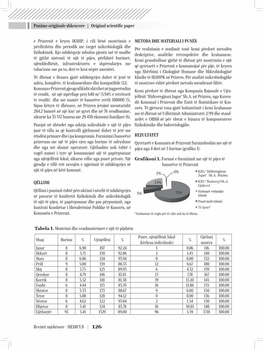
Punime origjinale shkencore | Original scientific paper
Revistë mjekësore - MEDICUS | 126
e Prizrenit e kryen IKSHP, i cili bënë mostrimin e përditshëm dhe periodik me target mikrobiologjik dhe fizikokimik. Kjo mbikëqyrje mbulon pjesën më të madhe të gjithë sistemit të ujit të pijes, përfshirë burimet, ujëmbledhësit, infrastrukturën e shpërndarjes me tubacione ose pa to, deri te kroi nëpër amvisëri.
Të dhënat e fituara gjatë mbikëqyrjes duhet të jenë të sakta, komplete, të krahasueshme dhe kompatibile (12).Komuna e Prizrenit gjeografikisht shtrihet në jugperëndim të vendit, në një sipërfaqe prej 640 m2 (5.94% e territorit te vendit) dhe me numër të banorëve rreth 180000 (5). Sipas këtyre të dhënave, në Prizren jetojnë mesatarisht 284.2 banorë në një km2 në qytet dhe në 76 vendbanime, sikurse ka 35 557 banesa me 29 458 ekonomi familjare (6).
Pasojat në shëndet nga ndotja mikrobiale e ujit të pijes janë të tilla sa që kontrolli gjithmonë duhet të jetë me rëndësi primare dhe i pa kompromis. Furnizimi i banorëve prizrenas me ujë të pijes vjen nga burime të ndryshme dhe nga më shumë operatorë. Gjithashtu nuk është i vogël numri i tyre që konsumojnë ujë të papërpunuar nga ujësjellësit lokal, sikurse edhe nga puset private. Një gjendje e tillë rrit nevojën e zgjerimit të mbikëqyrjes së ujit të pijes në këtë komunë.
QËLLIMI
Qëllimi i punimit është përcaktimi i nivelit të mbikëqyrjes së pavarur të kualitetit fizikokimik dhe mikrobiologjik të ujit të pijes, të papërpunuar dhe pas përpunimit, nga Instituti Kombëtar i Shëndetësisë Publike të Kosovës, në Komunën e Prizrenit.
METODA DHE MATERIALI I PUNËS
Për realizimin e studimit tonë kemi përdorë metodën deskriptive, analitike retrospektive dhe krahasuese. Kemi grumbulluar gjithë të dhënat për mostrimin e ujit që qytetarët e Prizrenit e konsumojnë për pije, të kryera nga Shërbimi i Ekologjisë Humane dhe Mikrobiologjisë klinike të IKSHPK në Prizren. Për analizë mikrobiologjike të mostrave është përdorë metoda membranë filtër.
Kemi përdorë të dhënat nga Kompania Rajonale e Ujës-jellësit ‘Hidroregjioni Jugor’ Sh.A. në Prizren, nga Kuven-dit Komunal i Prizrenit dhe Entit të Statistikave të Kos-ovës. Të gjeturat tona gjatë hulumtimit i kemi krahasuar me të dhënat në Udhëzimit Administrativ 2/99 dhe stand-ardet e OBSH-së për vlerat e lejuara të komponenteve fizikokimike dhe bakteriologjike.
REZULTATET
Qytetarët e Komunës së Prizrenit furnizoheshin me ujë të pijes nga 4 deri në 5 burime (grafiku 1):
Grafikoni 1. Format e furnizimit me ujë të pijes të banorëve të Prizrenit
*Venbanime të vogla për të cilat nuk ka të dhëna
Tabela 1. Mostrimi dhe vendmostrimet e ujit të pijshëm
Muaj Burimi % Ujësjellësi %Puset, ujësjellësit lokal
(kërkesa individuale)%
Gjithsej mostra
%
Janar 8 6.90 107 92.24 1 0.86 116 100.00Shkurt 8 5.71 130 92.86 2 1.43 140 100.00Mars 8 6.06 124 93.94 0 0.00 132 100.00Prill 9 5.00 159 88.33 12 6.67 180 100.00Maj 8 5.75 125 89.93 6 4.32 139 100.00Qershor 8 4.79 146 87.43 13 7.78 167 100.00Korrik 8 5.52 118 81.38 19 13.10 145 100.00Gusht 6 4.44 113 83.70 16 11.86 135 100.00Shtator 8 5.33 133 88.67 9 6.00 150 100.00Tetor 8 5.88 128 94.12 0 0.00 136 100.00Nëntor 6 4.62 122 93.84 2 1.54 130 100.00Dhjetor 8 5.41 124 83.78 16 10.81 148 100.00Gjithsejtë 93 5.41 1529 89.00 96 5.59 1718 100.00
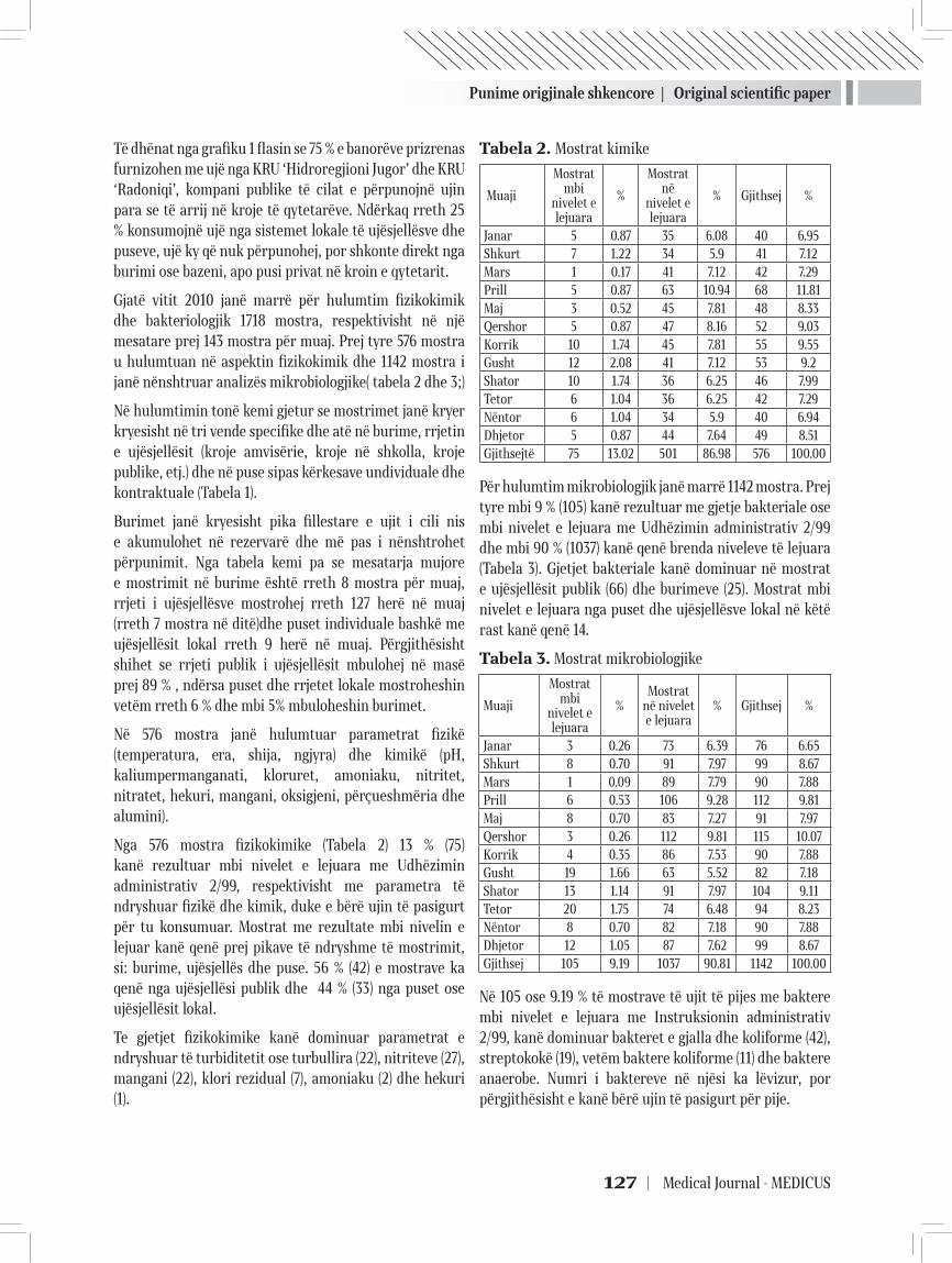
Punime origjinale shkencore | Original scientific paper
127 | Medical Journal - MEDICUS
Të dhënat nga grafiku 1 flasin se 75 % e banorëve prizrenas furnizohen me ujë nga KRU ‘Hidroregjioni Jugor’ dhe KRU ‘Radoniqi’, kompani publike të cilat e përpunojnë ujin para se të arrij në kroje të qytetarëve. Ndërkaq rreth 25 % konsumojnë ujë nga sistemet lokale të ujësjellësve dhe puseve, ujë ky që nuk përpunohej, por shkonte direkt nga burimi ose bazeni, apo pusi privat në kroin e qytetarit.
Gjatë vitit 2010 janë marrë për hulumtim fizikokimik dhe bakteriologjik 1718 mostra, respektivisht në një mesatare prej 143 mostra për muaj. Prej tyre 576 mostra u hulumtuan në aspektin fizikokimik dhe 1142 mostra i janë nënshtruar analizës mikrobiologjike( tabela 2 dhe 3;)
Në hulumtimin tonë kemi gjetur se mostrimet janë kryer kryesisht në tri vende specifike dhe atë në burime, rrjetin e ujësjellësit (kroje amvisërie, kroje në shkolla, kroje publike, etj.) dhe në puse sipas kërkesave undividuale dhe kontraktuale (Tabela 1).
Burimet janë kryesisht pika fillestare e ujit i cili nis e akumulohet në rezervarë dhe më pas i nënshtrohet përpunimit. Nga tabela kemi pa se mesatarja mujore e mostrimit në burime është rreth 8 mostra për muaj, rrjeti i ujësjellësve mostrohej rreth 127 herë në muaj (rreth 7 mostra në ditë)dhe puset individuale bashkë me ujësjellësit lokal rreth 9 herë në muaj. Përgjithësisht shihet se rrjeti publik i ujësjellësit mbulohej në masë prej 89 % , ndërsa puset dhe rrjetet lokale mostroheshin vetëm rreth 6 % dhe mbi 5% mbuloheshin burimet.
Në 576 mostra janë hulumtuar parametrat fizikë (temperatura, era, shija, ngjyra) dhe kimikë (pH, kaliumpermanganati, kloruret, amoniaku, nitritet, nitratet, hekuri, mangani, oksigjeni, përçueshmëria dhe alumini).
Nga 576 mostra fizikokimike (Tabela 2) 13 % (75) kanë rezultuar mbi nivelet e lejuara me Udhëzimin administrativ 2/99, respektivisht me parametra të ndryshuar fizikë dhe kimik, duke e bërë ujin të pasigurt për tu konsumuar. Mostrat me rezultate mbi nivelin e lejuar kanë qenë prej pikave të ndryshme të mostrimit, si: burime, ujësjellës dhe puse. 56 % (42) e mostrave ka qenë nga ujësjellësi publik dhe 44 % (33) nga puset ose ujësjellësit lokal.
Te gjetjet fizikokimike kanë dominuar parametrat e ndryshuar të turbiditetit ose turbullira (22), nitriteve (27), mangani (22), klori rezidual (7), amoniaku (2) dhe hekuri (1).
Tabela 2. Mostrat kimike
Muaji
Mostrat mbi
nivelet e lejuara
%
Mostrat në
nivelet e lejuara
% Gjithsej %
Janar 5 0.87 35 6.08 40 6.95Shkurt 7 1.22 34 5.9 41 7.12Mars 1 0.17 41 7.12 42 7.29Prill 5 0.87 63 10.94 68 11.81Maj 3 0.52 45 7.81 48 8.33Qershor 5 0.87 47 8.16 52 9.03Korrik 10 1.74 45 7.81 55 9.55Gusht 12 2.08 41 7.12 53 9.2Shator 10 1.74 36 6.25 46 7.99Tetor 6 1.04 36 6.25 42 7.29Nëntor 6 1.04 34 5.9 40 6.94Dhjetor 5 0.87 44 7.64 49 8.51Gjithsejtë 75 13.02 501 86.98 576 100.00
Për hulumtim mikrobiologjik janë marrë 1142 mostra. Prej tyre mbi 9 % (105) kanë rezultuar me gjetje bakteriale ose mbi nivelet e lejuara me Udhëzimin administrativ 2/99 dhe mbi 90 % (1037) kanë qenë brenda niveleve të lejuara (Tabela 3). Gjetjet bakteriale kanë dominuar në mostrat e ujësjellësit publik (66) dhe burimeve (25). Mostrat mbi nivelet e lejuara nga puset dhe ujësjellësve lokal në këtë rast kanë qenë 14.
Tabela 3. Mostrat mikrobiologjike
Muaji
Mostrat mbi
nivelet e lejuara
%Mostrat
në nivelet e lejuara
% Gjithsej %
Janar 3 0.26 73 6.39 76 6.65Shkurt 8 0.70 91 7.97 99 8.67Mars 1 0.09 89 7.79 90 7.88Prill 6 0.53 106 9.28 112 9.81Maj 8 0.70 83 7.27 91 7.97Qershor 3 0.26 112 9.81 115 10.07Korrik 4 0.35 86 7.53 90 7.88Gusht 19 1.66 63 5.52 82 7.18Shator 13 1.14 91 7.97 104 9.11Tetor 20 1.75 74 6.48 94 8.23Nëntor 8 0.70 82 7.18 90 7.88Dhjetor 12 1.05 87 7.62 99 8.67Gjithsej 105 9.19 1037 90.81 1142 100.00
Në 105 ose 9.19 % të mostrave të ujit të pijes me baktere mbi nivelet e lejuara me Instruksionin administrativ 2/99, kanë dominuar bakteret e gjalla dhe koliforme (42), streptokokë (19), vetëm baktere koliforme (11) dhe baktere anaerobe. Numri i baktereve në njësi ka lëvizur, por përgjithësisht e kanë bërë ujin të pasigurt për pije.
Punime origjinale shkencore | Original scientific paper
Revistë mjekësore - MEDICUS | 128
DISKUTIMI
Objektivat e zhvillimit të mijëvjeçarit (MDG) synojnë zvogëlimin e numrit të banorëve pa qasje në ujë të sigurt të pijes dhe higjienë bazike deri në vitin 2015. Ndërkaq Udhëzuesi për kualitet të ujit të pijes i OBSH shpjegon një varg faktorësh me ndikim të drejtpërdrejtë në kualitetin e ujit të pijes, rrjedhimisht në shëndetin publik(4). Ky udhëzues gjithashtu përcakton Ministrinë e Shëndetësisë në nivel vendi si përgjegjëse për mbikëqyrje të pavarur ose të jashtme të kualitetit të ujit të pijes për qytetarët.
Ngjashëm përcaktohet në Kosovë me Ligjin për Shëndetësi Publike(7) , ku në nenin 4 pika ‘i’ ngarkon me përgjegjësi direkte IKSHP të Kosovës për ‘analizimin dhe vlerësimin e kualitetit të ujit për pije’. Mbi këtë bazë, Instituti Rajonal i Shëndetit Publik në Prizren aktualisht është i vetmi institucion me kompetencë profesionale dhe të autorizuar që bënë mbikëqyrjen e ujit të pijes në nivel të komunës dhe rajonit të saj(7,8).
Sipas rezultateve të prezantuara, Instituti i Shëndetit Publik në Prizren gjatë vitit 2010, ka bërë mostrimin e ujit të pijes në pika të ndryshme për analizë mikrobiologjike dhe fiziko-kimike. Kjo mbikëqyrje, bëhet mbi bazë ditore, ndërsa merren mesatarisht 6-8 mostra për analizë, e që është brenda standardeve të përshkruara për analizën bazë ose hulumtimin e vogël të kualitetit të ujit (deri 200000 banorë).
IKSHP, sipas mënyrës së mostrimit dhe analizës së tyre, përcjellë parametrat bazikë mikrobiologjik dhe fiziko-kimik, që e bëjnë të pranueshëm ujin e pijes në pajtueshmëri me Udhëzimin administrativ 2/99(8) dhe me WSP (water safety plans) sipas OBSH-së.
Furnizimi i banorëve me ujë të pijes të përpunuar dhe nga rrjetet e ujësjellësve të kontrolluar është gjithnjë e më i madh. Sipas rezultateve tona kjo gjendje shkon deri në 75 % të banorëve të komunës së Prizrenit, ndërkaq mjaft mirë qëndrojnë edhe komunat e afërta si Gjakova (89%) dhe Peja (64%), ndërsa më keq qëndrojnë Ferizaj (42%) dhe Gjilani (31%)(20).
Gjetjet mikrobiologjike mbi vlerat e lejuara sipas Udhëzimit administrativ 2/99 (8) kanë dominuar në burime të ujit, në ujësjellës lokal dhe të pa mbikëqyrur si dhe puse private. Ky dominim nuk është në proporcion me numrin e mostrimeve në rrjetin e ujit të trajtuar dhe atë të patrajtuar( 16.9:1). Gjetjet e parametrave fizikokimik mbi nivelet e lejuara përputhen me ndotjet mikrobiologjike të gjetura dhe reagimi ka qenë i shpejtë e profesional.
Në vitin 2010 në komunën e Prizrenit nuk është regjistruar asnjë epidemi nga ndotja e ujit të cilës do natyrë qoftë ai.
Në Kosovë aktualisht po punohet në transpozimin e Direktivës Europiane 98/83/EC në legjislacionin vendor, për kontrollimin dhe parametrat e ujit të pijes në vend
Asnjëherë nuk mund të themi me siguri të plotë se uji për pije është tërësisht i pastër, pavarësisht rezultateve të mostrimit, sepse ai mund të ndotet në çdo moment. Pikërisht për këtë arsye, uji që konsumohet për pije duhet të mbikëqyret në mënyrë të vazhdueshme, profesionale dhe të pakompromis, duke siguruar kualitet higjieno-sanitar dhe shëndetësor të tij.
PËRFUNDIMI DHE REKOMANDIME
Duke analizuar rezultatet dhe mënyrën e furnizimit me ujë të pijes të banorëve të komunës së Prizrenit, vijmë në përfundim se ka mjaft punë për tu bërë që të arrihet një mbikëqyrje ose survejim i plotë i ujit që konsumohet për pije. Edhe më tej mbeten rreth 25% e banorëve pa ujë të trajtuar dhe të përpunuar i cili do të ishte i sigurt për tu konsumuar.
Instituti Kombëtar i Shëndetit Publik të Kosovës në Prizren duhet të ndihmohet materialisht dhe me kuadro profesioniste për të rritur kapacitetet e mbikëqyrjes së pavarur të kualitetit të ujit të pijes. Për më tepër kur kjo degë e Institutit bënë survejimin në nivel rajoni në të cilin përfshihen edhe 5 komuna tjera, obligon institucionet qendrore për reflektim shtesë.
Kuvendi komunal i Prizrenit, duhet të rrisë angazhimin për furnizim të mirë, kualitativ dhe të sigurt me ujë të pijes të banorëve të tij. Në këtë plan duhet të përfshijë nën mbikëqyrje të gjitha zonat rurale, përmes IKSHP në Prizren.
LITERATURA
1. Ramadani N. Epidemiologjia moderne. Prishtinë, 2005 2. Dedushaj I, Humolli I. Epidemiologjia e sëmundjeve
ngjitëse3. Pollozhani, A, Kendrovski V, Krasniqi S. Ekologjia hu-
mane dhe shëndeti publik. Shkup, 20094. WHO. Guidelines for Drinking-water Quality’ Third edi-
tion, Vol. 1 Recommenndations, Geneva 2004 (84-98)5. http://kk.rks-gov.net/prizren/City-guide/Geography.aspx6. http://esk.rks-gov.net/rekos2011/7. Ligji për shëndetësi publike nr. 02/L-78, (http://www.
kuvendikosoves.org/?cid=1,191,122;) 8. Udhëzimi administrativ 2/99 i UNMIK-ut
Punime origjinale shkencore | Original scientific paper
129 | Medical Journal - MEDICUS
9. Zarkovic G, Sergije R. ‘Komunalna Higijena’ Medicinska Knjiga Beograd-Zagreb, 1953
10. Jankovic P. et al. Higijena s’epidemiologijom i socijal-nom medicinom Medicinska Knjiga Beograd-Zagreb, 1985;
11. http://www.dcp2.org/pubs/DCP12. Kristoforovic-Ilic M. Higijena sa medicinskom ekologi-
jom. Ortomedics, Novi Sad, 200313. Food and Agriculture Organization (FAO), Chemical
analysis manual for food and water, 5th Ed, FAO ROME. 1997, 20-26.
14. Environment Protection Agency (EPA), US Environmen-tal Protection Agency Safe Drinking Water Act. 2003, EPA 1012-F-04-12.
15. Welch, P. et al. Microbial quality of water in rural com-munities of Trinidad. Pan American Journal of Public Health 8(3):172-80
16. Schaetz, R. J and Arbogast, A.F. People and environ-
ment. Simon and Shuster Custom Publishing, U.S.A.; 2003, pp. 245-269;
17. CAWST ‘Introduction to drinking water quality testing, Centre for Affordable Water and Sanitation Technol-ogy’, Edition 2009;
18. Rhoul-Amin et al. Microbial Analysis of Drinking Water and Water Distribution System in New Urban Peshawar, Current Research Journal of Biological Science 4(6):731-737, 2012
19. O.B. Shitu, J.O. Olaitan, T.S. Amusa, Physico-chemical and bacteriological analyses of water used for drinking and swimming purposes in Abeokuta, Nigeria. African Journal of Biomedical Researche. 2008, 1:285-290
20. Programi i IPA-s i Bashkimit Evropian për Kosovën. Studimi i Fizibilitetit për Kualitetin e Ujit të Pijshëm në Kosovë (RKSOKB 1244). Vëllimi I. Raporti përfundimtar i fizibilitetit, 2009.
21. EU Council ‘Drinking Water Directive (98/83/EC) .
BACTERIOLOGICAL AND PHYSICOCHEMICAL SURVEILLANCE OF DRINKING WATER IN THE MUNICIPALITY OF PRIZREN
Fadil Kryeziu1, Tahire Maloku1, Violeta Kryeziu1, Sejran Abdushi2, Nurishahe Hulaj1,1 National Institut of Public Health of Kosova2 Regional Hospital “Prim.Dr.Daut Mustafa” Prizren, Republic of Kosova
ABSTRACT
Aim: Aim of this study was to determine the level of independent surveillance to the physic-chemical and microbiological quality of drinking water from the National Institute of Public Health, whether raw or processed in the Municipality of Prizren.
Materials and methods: In this retrospective, descriptive and analytical study we have used materials from several resources, as are: National Institute of Public Health (NIPH) in Prizren, Hidroregion, Municipality of Prizren, and from Statistical Office of Kosovo. Data were collected, processed and analyzed, and compared with data from comparative literature to set the standards for our concerns.
Results: During 2010 year, NIPH – Service for Human Ecology has implemented 1718 samples in total, of which 1142 samples with microbiological target and 576 samples with target physic- chemical. From 1142 microbiological samples, 9.19% (105) of them have resulted out levels allowed by the AI 2/99; from 576 physicochemical samples without of levels allowed with AI 2/99 are 13.02% (75)
Conclusions: Citizens of Prizren consume relatively safe drinking water in terms of physicochemical and microbiological quality and NIPH perform proper monitoring of drinking water. However, is needed to increase the range of parameters investigated and the number of staff, to achieve greater health and hygienic safety of drinking water.
Key words: microbiological, physicochemical, surveillance, drinking water, administrative instruction
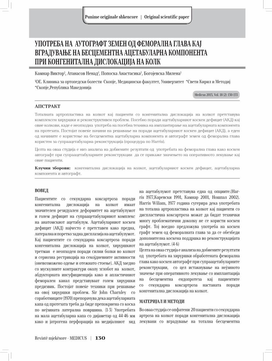
Revistë mjekësore - MEDICUS | 130
25
УПОТРЕБА НА АУТОГРАФТ ЗЕМЕН ОД ФЕМОРАЛНА ГЛАВА КАЈ ВГРАДУВАЊЕ НА БЕСЦЕМЕНТНА АЦЕТАБУЛАРНА КОМПОНЕНТА ПРИ КОНГЕНИТАЛНА ДИСЛОКАЦИЈА НА КОЛККамнар Виктор1, Атанасов Ненад1, Попоска Анастасика1, Богојевска Милена1
1ОЕ. Клиника за ортопедски болести Скопје, Медицински факултет, Универзитет “Свети Кирил и Методиј “Скопје,Република Македонија
АПСТРАКТ
Тоталната артропластика на колкот кај пациенти со конгенитална дислокација на колкот претставува комплексен хируршки и реконструктивен проблем. Посебно поради ацетабуларниот коскен дефицит (АКД) кај овие колкови, каде е неопходна употреба на посебна техника на имплантирање на ацетабуларната компонента на протезата. Постојат повеќе начини на решавање на поради ацетабуларниот коскен дефицит (АКД), а еден од начините е користење на бесцементна ацетабуларна компонента и автографт земен од феморална глава користен за супраацетабуларна реконструкција (процедура по Harris).
Целта на оваа студија е низ анализа на добиените резултати од употребата на феморална глава како коскен автографт при супраацетабуларните реконструкции да се прикаже значењето на оперативното лекување кај овие пациенти.
Клучни зборови: конгенитална дислокација на колкот, ацетабуларниот коскен дефицит, ацетабуларна компонента и автографт.
Medicus 2013, Vol. 18 (2): 130-133
ВОВЕД
Пациентите со секундарна коксартроза поради конгенитална дислокација на колкот имаат значителен резидуален деформитет на ацетабулумот и голем дефицит на супраацетабуларниот комплекс на анатомскиот ацетабулум. Ацетабуларниот коскен дефицит (АКД) најчесто е претставен како предна, латерална и поретко задна дисплазија на ацетабулумот. Кај пациентите со секундарна коксартроза поради конгенитална дислокација на колкот, хируршкиот третман е неопходен поради силни болки во колкот и сериозна рестрикција на секојдневните активности (оневозможено одење и отежнато стоење). АКД заедно со мускулните контрактури околу зглобот на колкот, абдукторната инсуфициенција како и апластичниот феморален канал представуваат голем хируршки предизвик. Постојат повеќе техники при решавање на овој хируршки проблем. Sir John Charnley со соработниците (1970) препорачува дека ацетабуларната капа од протезата треба да биде препокриена со коска по нејзината латерална површина. (1-3) Употребата на мала ацетабуларна капа со дијаметар од 44-46 мм како и јатрогена перфорација на медијалниот ѕид
на ацетабулумот претставува една од опциите.(Har-ris 1977,Каревски 1991, Камнар 2001, Ношпал 2002). Harris Wiliam, 1977 година сугерира дека употребата на тотална артропластика на колкот кај пациенти со диспластична коксартроза можат да бидат технички многу проблематични доколку не се користи коскен графт. Тој воедно предложува употреба на коскен графт земен од феморалната глава за да се обезбеди дополнителна коскена поддршка во реконструкцијата на ацетабулумот. (4-6)Целта на оваа студија е анализа на добиените резултати од употребата на хируршки обработената феморална глава како коскен автографт при супраацетабуларните реконструкции, со цел истакнување на нејзиното значење при оперативното лекување со имплантација на бесцементна ендопротеза кај пациентите со секундарна коксартроза настаната поради конгенитална дислокација на колкот.
МАТЕРИЈАЛ И МЕТОДИ
Во оваа студија се опфатени 20 пациенти со секундарна артроза на колкот поради конгенитална дислокација лекувани со вградување на тотална бесцементна
Punime origjinale shkencore | Original scientific paper
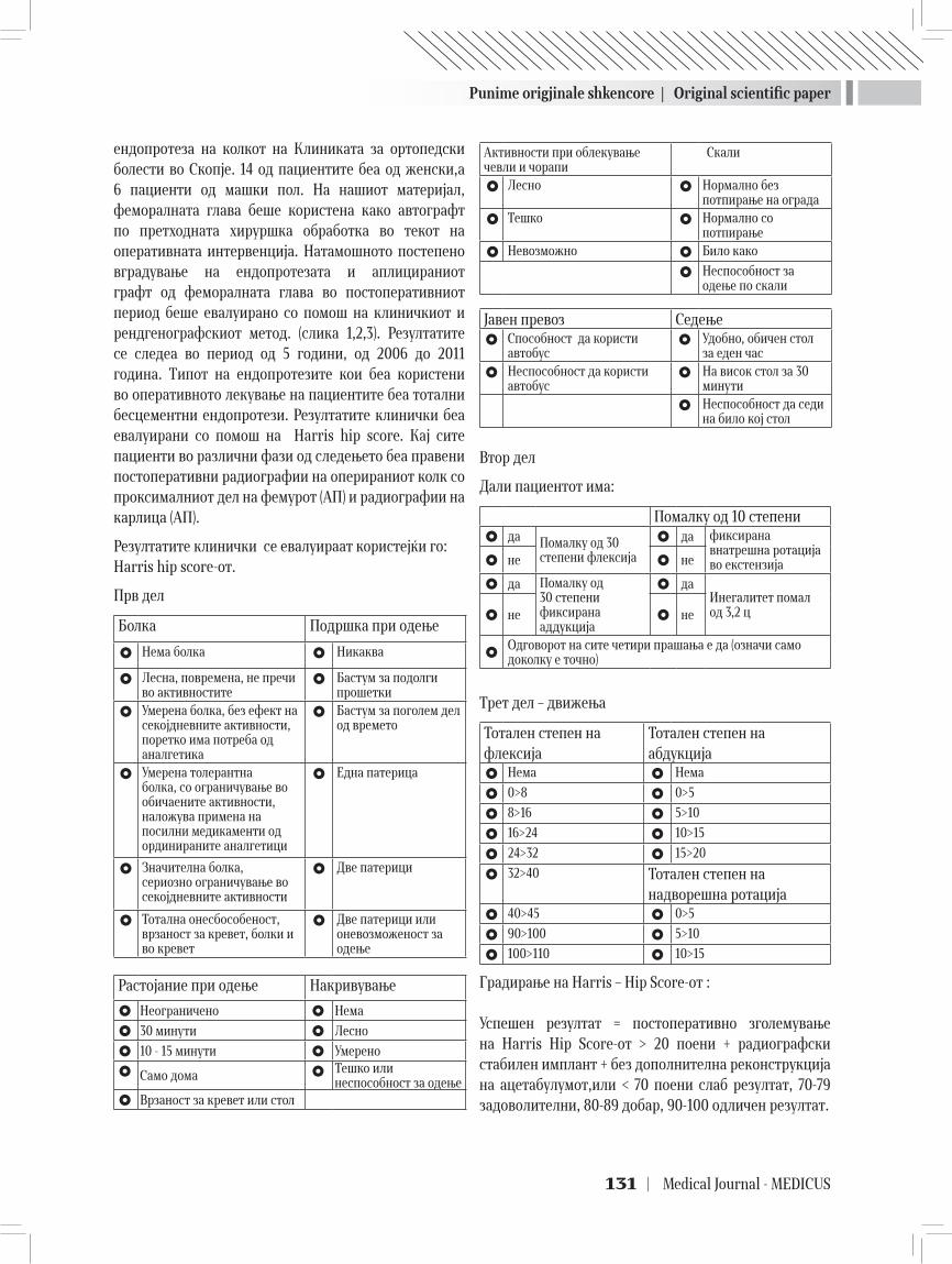
Punime origjinale shkencore | Original scientific paper
131 | Medical Journal - MEDICUS
ендопротеза на колкот на Клиниката за ортопедски болести во Скопје. 14 од пациентите беа од женски,а 6 пациенти од машки пол. На нашиот материјал, феморалната глава беше користена како автографт по претходната хируршка обработка во текот на оперативната интервенција. Натамошното постепено вградување на ендопротезата и аплицираниот графт од феморалната глава во постоперативниот период беше евалуирано со помош на клиничкиот и рендгенографскиот метод. (слика 1,2,3). Резултатите се следеа во период од 5 години, од 2006 до 2011 година. Типот на ендопротезите кои беа користени во оперативното лекување на пациентите беа тотални бесцементни ендопротези. Резултатите клинички беа евалуирани со помош на Harris hip score. Кај сите пациенти во различни фази од следењето беа правени постоперативни радиографии на оперираниот колк со проксималниот дел на фемурот (АП) и радиографии на карлица (АП).
Резултатите клинички се евалуираат користејќи го:Harris hip score-от.
Прв дел
Болка Подршка при одење
Нема болка Никаква
Лесна, повремена, не пречи во активностите
Бастум за подолги прошетки
Умерена болка, без ефект на секојдневните активности, поретко има потреба од аналгетика
Бастум за поголем дел од времето
Умерена толерантна болка, со ограничување во обичаените активности, наложува примена на посилни медикаменти од ординираните аналгетици
Една патерица
Значителна болка, сериозно ограничување во секојдневните активности
Две патерици
Тотална онесбособеност, врзаност за кревет, болки и во кревет
Две патерици или оневозможеност за одење
Растојание при одење Накривување
Неограничено Нема
30 минути Лесно
10 - 15 минути Умерено
Само дома Тешко или неспособност за одење
Врзаност за кревет или стол
Активности при облекување чевли и чорапи
Скали
Лесно Нормално без потпирање на ограда
Тешко Нормално со потпирање
Невозможно Било како
Неспособност за одење по скали
Јавен превоз Седење Способност да користи
автобус Удобно, обичен стол
за еден час
Неспособност да користи автобус
На висок стол за 30 минути
Неспособност да седи на било кој стол
Втор дел
Дали пациентот има:
Помалку од 10 степени да Помалку од 30
степени флексија
да фиксирана внатрешна ротација во екстензија не не
да Помалку од 30 степени фиксирана аддукција
даИнегалитет помал од 3,2 ц не не
Одговорот на сите четири прашања е да (означи само доколку е точно)
Трет дел – движења
Тотален степен на флексија
Тотален степен на абдукција
Нема Нема
0>8 0>5
8>16 5>10
16>24 10>15
24>32 15>20
32>40 Тотален степен на надворешна ротација
40>45 0>5
90>100 5>10
100>110 10>15
Градирање на Harris – Hip Score-от :
Успешен резултат = постоперативно зголемување на Harris Hip Score-от > 20 поени + радиографски стабилен имплант + без дополнителна реконструкција на ацетабулумот,или < 70 поени слаб резултат, 70-79 задоволителни, 80-89 добар, 90-100 одличен резултат.
Punime origjinale shkencore | Original scientific paper
Revistë mjekësore - MEDICUS | 132
РЕЗУЛТАТИ
Од клинички аспект користејки го Harris hip scor-ot се стигна до следниве резултати: Предоперативна средна вредност на HHS 40 (9-57), првата година постоперативно 72 се до средна вредност на скорот од 75,5 во петата постоперативна година. Инегалитетот на екстремитетите се подобри на средна вредност од 9 мм постоперативно, имајки ја во предвид предоперативната дискрепанца од 21 мм. Кај сите пациенти во различни фази од следењето беа правени постоперативни радиографии на оперираниот колк (АП) и радиографии на карлица (АП).
Радиографската анализа покажа дека 16 од 20 феморални графтови беа вградени со коската домаќин од карлицата (слика 1,2,3), но 4 графта покажаа ресорпција целосна со миграција на целиот ацетабуларен комплекс према проксимално. Кај сите пациенти кај кои графтот беше вграден на стандардните радиографии после првата година од операцијата, беше забележано постепено ремоделирање на коскените трабекули по должината на линиите на оптоварување.
ДИСКУСИЈА
Уште од самиот почеток на развој на алоартро-пластиката на големите зглобови преку напредокот на базичната наука, развојот на дизајните на ендопротезите како и усовршувањето на хируршката техника доведоа до драматично подобри функционал-ни резултати кај пациентите. (6,7)
Често, примената на оперативното лекување со имплантација на тоталната ендопротеза кај пациенти со секундарна артроза како резултат на развојни пореметувања на колкот, бара еден комплексен пристап во реконструкцијата на ацетабуларниот комплекс, иако резултатите кај одреден број на случаи не се толку задоволителни како на пр. кај пациенти со примарна коксартроза, лекувани истотака со примена на тотална алоартропластика. Автогеното коскено графтирање кај ацетабуларни коскени дефицити, користејќи хируршки обработена глава на бутната коска, го наоѓа своето место во големи проспективни серии и резултира со суфициентно коскено враснување и ремоделација во еден подолг постоперативен временски период. (8,9)
Гореопишаниот хируршки пристап, комбиниран со брзата и стабилна биолошка фиксација со коскено враснување на бесцементните ендопротези овозможува долго и квалитетно живеење со вештачки зглоб. (10)
ЗАКЛУЧОК
При добра примарна фиксација и адекватно биолошко вградување на ацетабуларната компонента како и автографтот добиен со обработка на феморалната глава после првата година од операцијата, враќањето на мускулната сила, решавањето на проблемот со накривувањето како и егализацијата на екстермитетите доведуваат до оптимален динамички и статички капацитет на зглобот на колкот, а со тоа и долго и квалитетно живеење со вештачки зглоб.
ЛИТЕРАТУРА
1. Wheeless Textbook of Orthopaedics,July 24,2011 8:19 pm2. Buchardt H.Enekin W.F:Transplantation of bone, Sim-
posium of organ transplantation. Surg. Clin. North. Am. 1978; 58:403
3. Willert G.H.,:Biocompatibility of endoprothesis mate-rials.In.Morsher E,The cementhless fixation of the hip endoprothesis.Springer,Berlinp. 1984; 29.38
4. Каревски Љ.: Предности на коскената трансплантација во биолошката и механичката стабилизација на тоталните протези на колкот кај коскениот дефицит на ацетабулумот. 1991, Скопје
5. Harris WH. Traumatic arthritis of the hip after disloca-tion and acetabular fractures: treatment by mold arthro-plasty. An end-result study using a new method of result evaluation. J Bone Joint Surg Am. 1969;Jun;51(4):737-55.
6. Sanchez-Sotelo J, Berry DJ, Trousdale RT, Cabanela ME. Surgical development of developmental dysplasia of the hip in the adults. II: arthroplasty options. J Am Acad Orthop Surg 2002;10:334-40.
7. Numair J, Joshi AB, Murphy JC, Porter ML, Hardinge K. Total hip arthroplasy for congenital dysplasia or dislo-cation of the hip: survivorship analysis and long term results. J Bone Joint Surg [Am]1996;79-A:1352-60.
8. MacKenzie JR, Kelley SS, Johnston RC. Total hip replace-ment for coxarthrosis secondary to congenital dyspla-sia and dislocation of the hip: long-term results. J Bone Joint Surg [Am] 1996;78-A:55-
Punime origjinale shkencore | Original scientific paper
133 | Medical Journal - MEDICUS
THE USE OF FEMORAL HEAD AUTOGRAFT AND CEMENTHLESS ACETABULAR COMPONENT IN CONGENITAL DISLOCATION OF THE HIP
Kamnar Viktor1, Atanasov Nenad1, Poposka Anastasika1, Bogojevska Milena1
1 University Clinic for orthopaedic surgery, Medical Faculty University “St. Cyril and Methodius”- Skopje, Republic of Macedonia
ABSTRACT
The patients with congenital hip dislocation have severe residual deformity of the acetabulum and large deficit of the supraacetabular complex on the anatomical acetabulum. Acetabular bone deficiency is commonly presented and the specific technique for acetabular cup insertion with femoral head autograft used for supraacetabular reconstruction is needed. (Harris procedure) The aim of this study is to analyse the importance of using femoral head autograft in supraacetabular reconstruction using special technique and to affirm the importance of this medical treatment in these patients.
Key words: congenital hip dislocation, acetabular bone deficiency, femoral head autograft.
9. Klapach AS, Callaghan JJ, Miller KA, et al. Total hip ar-throplasty with cement and without acetabular bone graft for severe graft dysplasia: a concise follow-up, at a minimum of twenty years, of a previous report. J Bone Joint Surg [Am] 2005; 87-A:280-5.
10. Chougle A, Hemmady MV, Hodgkinson JP. Long-term sur-vival of the acetabular component after total hip arthro-plasty with cement in patients with developmental dys-plasia of the hip. J Bone Joint surg [Am] 2006;88-A:71-9.
Revistë mjekësore - MEDICUS | 134
21
NUCHAL TRANSLUCENCY AND NORMAL KARYOTYPE CORRELATIONS AND POSSIBILITIESAna Daneva Markova 1,Marija Hadzi-Lega1, Arta Bina1, Tanja Nikolova1
1 University Clinic of Obstetrics & Gynecology, Medical Faculty, Ss. Cyril and Methodius University, Skopje, Republic of Macedonia
ABSTRACT
Nuchal translucency is an ultrasound marker of first trimester of pregnancy, widely used and accepted for screening of chromosomal abnormalities. It is more likely to be a manifestation of various development disorders. According to Snijders at al. in 1998, around 5% of fetuses which will present with NT>95 percentile, will have a normal karyotype. Both in unselected populations and high risk populations, increased NT≥3mm seems to increase the risk of malformation 10f fold and the risk of miscarriage about 5 fold ( Bilardo at al. 2007).
There is a growing body of literature that highlights a spectrum of possible events in the fetuses with increased nuchal translucency and normal karyotype like fetal loss, structural defects and genetic syndromes with development delay. Declaring to parents that the baby with increased NT has a normal karyotype, it is only the beginning of a full diagnostic work up likes a two-step ultrasound investigation at 13-18 and 20-24 weeks gestation, managing plan, and a long list of explanations regarding possible outcomes for baby.
Although the unfortunate events for the babies with increased Nuchal translucency and normal karyotype should not be underestimated, it is important to emphasize to the parents that almost 80% of this pregnancies is likely to be the same as for those pregnancies with a normal NT at this stage. If the two step ultrasound examination is normal, then the chance of fetus to have an underlying abnormality is about 2%.
NUCHAL TRANSLUCENCY AND NORMAL KARYOTYPE CORRELATIONS AND POSSIBILITIES
LOSS OF FETUS
According Bilardo at al, 2007 (2), at fetuses with NT> 6.5mm and normal karyotype, the percentage of spontaneous fetal loss is high. It manifests in spontaneous abortion, in utero death and can be explained by the severe developmental disorders that cause the loss of the fetus due to cardiac insufficiency. In certain cases, such a loss of the fetus is high because of the demands of the parents to terminate the pregnancy without ultrasound examinations that would confirm “normalization” of the edema. Also, Souka at al (3), in 1998 found that the chances of survival decrease with increasing the nuchal translucency. This position is determined with the help of several other examinations, suggesting the further investigations to be carried out mainly for the fetuses with increased nuchal translucency over 99% and over 3.5 mm. Fetuses with mild increase of the nuchal translucency i.e. an increase of 3 mm and 3.5 mm can survive in 96% of the cases.
Meanwhile, Michailidis and Echonomides in 2001 found that for the nuchal translucency over 99 percentiles the risk of miscarriage, intrauterine death or termination of pregnancy because of fetal anomalies was about 12 times higher compared with fetuses with normal values NT (4).
Another study, Westin at al. 2007 (5), did not find a connection between the increase of NT values and perinatal death, and found only a connection between elevated NT values and abortion. During these studies a number of pregnancies were suspended at the request of parents. If the pregnancies would not be suspended because of the desire of the parents, the condition with the relationship between the elevated NT values and the loss of the fetuses might have been quite different. Also, some of the terminations of the pregnancy may be affected by Amniocentesis.
Basically, larger number of authors confirm that the increases in NT values are important, and how large are their indicators, the forecast is worse for them.
After determination of elevated nuchal translucency
Medicus 2013, Vol. 18 (2): 134-136
Punime reviale | Revial work
Punime reviale | Revial work
135 | Medical Journal - MEDICUS
during the first trimester and normal karyotype, further procedure provides echo i.e. review in the period from 13th to 18th week and then in the period from 20th to 24th week, even it comes to the smallest indication from the beginning of the echo, with the exception of abortion or intrauterine fetal death.
GENETIC SYNDROMES AND OTHER ABNORMALITIES
It is difficult to talk about the possibilities of genetic syndromes in cases of elevated NT values with normal karyotype because of their heavy diagnosis and low prevalence at the population. Also, the difficulties become even greater when some of them appear a few months after birth or during early childhood. We should also mention the case of Noonan syndrome, which is mentioned in all performed tests and can be determined a few months after birth because of changes made to the person. Also it can be mentioned the fetal akinesia or some other skeletal dysplasia that can cause an increase of NT values due to mediastinal compression or due to differentiation of the expression of collagen (Hyett at al. 1997) (6). In his study, Bilardo at al. 2007 (2) found that most of the identified genetic syndromes were “de novo” and are manifested mainly in muscle or skeletal disorders. In such cases the accumulation of the liquid in the nuchal point could be caused due to disturbance of the venous return to the heart from the small thorax, the altered composition of the extracellular matrix or due to disturbances in lymphatic drainage. According Senat at al. 2002 (7) in fetuses with normal karyotype, the danger of neonatal malformations or severe disability during the early childhood amounts 6.5% and 11%.
The previous studies had much influence on the choice of cut-off values of NT values and the limited number of cases. Recent studies have found that for NT values <4mm there are greater opportunities for establishing more normal fetuses. It is chosen NT value > 4mm because this value is below 99 percentiles and can help more in determining the relationship between the elevated NT values and the illness during the early childhood, Souka at al. 2008 (8).
The other anomalies are mentioned last, but they are not few in number. The list of such anomalies is long, but the actual connection is established only with a few studies (3, 8). The most common structural defect upon cardiac anomalies is found to be palatoschisis (2).
Also can be mentioned exomphalos, hernia of the diaphragm, problems with the abdominal wall, light pyelectasia etc.
CARDIAC DEFECTS
Early studies associated with elevated NT values established connection between these values and the structural defects of the heart and the major arteries. Hyett at al. 1999 found that in a one group of 29154 separate pregnancies, elevated NT values above the 95 percentile was observed in 56% of fetuses with cardiac abnormalities (8).
Also, this study is confirmed by Bilardo at al. 2007 (2), which identified cardiac structural defects as the most common (55%) of all other structural defects in the fetus with increased NT values, normal karyotype and abnormal ultrasound examination. In the same study, the cardiac defects were identified in 30% of the cases of fetuses with elevated values of NT, normal karyotype and normal ultrasound examination, i.e. without other structural defects.
The possible link between elevated NT values and structural cardiac defects directed a number of studies to such relationship as an indirect mean of diagnosing cardiac structural abnormalities. Two recent studies (Michailidis and Economides 2001(4), Mavrides at al. 2001 (9)) announced the effectiveness of the measurements of the NT values as a mean of determining the major cardiac defects. The percentage of determining the major cardiac defects associated with NT values, using percentile 95 and percentile 99 was 36% and 27%.
However, these findings do not support the measurement of the NT values as the only measure for detection of major cardiac defects. Measurement of NT values can not replace the standard examination of the heart with morphological echography and currently it is considered as a warning sign for further and detailed examination of the heart to the standard images of the morphological echography.
CONCLUSION
The elevated nuchal translucency in fetuses with normal karyotype is associated with structural cardiac defects, loss of the fetus, other structural anomalies and in some cases with genetic syndromes, which are often difficult to determine.
Punime reviale | Revial work
Revistë mjekësore - MEDICUS | 136
The second step in the plan of the management is detecting possible structural anomalies using echography, from 13-18 week and 20-24 week of pregnancy.
Although it should not be underestimated the potential negative results for certain baby, for the parents it is important to be noted that approximately 80% of such pregnancies would have a similar future with the pregnancies with normal nuchal translucency over the same period of pregnancy. If the echo mentioned in the above text in two steps will show normal values, the chances of appearance of fetal anomalies would be 2%.
REFERENCES:
1. Snijders at al. 1998. UK multicenter project on assess-ment of risk of trisomy 21b maternal age and fetal nuchal translucency thickness at 10-14 weeks of gesta-tion. Lancet 351:343-346
2. Bilardo at al. 2007. Increased nuchal translucency and normal karyotype: time for parenteral reassurance. Ultrasound in Obstetrics and Gynecology, 2007, vol 30, Issue 1, 11-18
3. Souka at al. 1998. Defects and syndromes in chromosomally normal fetuses with increased nuchal
translucency thickness at 10-14 weeks of gestation. Ultrasound Obst-Gynecology 11; 391-400
4. Michalididis, Economides, 2001. Nuchal translucency measurements and pregnancy outcome in karyotypi-cally normal fetuses. Ultrasound Obstetrics and Gyne-cology 17; 102-105.
5. Hyett at al. Lethal congenital arthrogryposis presents with increased nuchal translucency and normal karyo-type. Prenatal Diagnosis 2002, 22: 345-349.
6. Senat at al. 2002. Pregnancy outcome in fetuses with increased nuchal translucency and normal karyotype. Prenatal Diagnosis 2002, 22: 345-349.
7. Souka 2001. Outcome of pregnancy in chromosomally normal fetuses with increased nuchal translucency in the first trimester. Ultrasound Obst-Gynecology 2001, 18; 7-9.
8. Hyett JA. 1999. Screening for congenital heart disease with fetal nuchal translucency at 10-14 weeks of gesta-tion/ BMJ 318:81-85
9. Mavrides at al. 2001. Limitations of using first trimester nuchal translucency measurement in routine screening for major congenital heart defects. Ultrasound Obstet-ric Gynecology 17:106-110.
ЗГОЛЕМЕНА НУХАЛНА ТРАНСЛУЦЕНЦА И НОРМАЛЕН КАРИОТИП- РЕЛАЦИИ И МОЖНОСТИ
Aна Данева Маркова 1, Марија Хаџи Лега1 , Арта Бина1, Тања Николова1
1 Универзитетска Клиника за Гинекологија и Акушерство, Скопје
ABSTRACT
Nuchal translucency is an ultrasound marker of first trimester of pregnancy, widely used and accepted for screening of chromosomal abnormalities. It is more likely to be a manifestation of various development disorders. According to Snijders at al. in 1998, around 5% of fetuses which will present with NT>95 percentile, will have a normal karyotype. Both in unselected populations and high risk populations, increased NT≥3mm seems to increase the risk of malformation 10f fold and the risk of miscarriage about 5 fold ( Bilardo at al. 2007).
There is a growing body of literature that highlights a spectrum of possible events in the fetuses with increased nuchal translucency and normal karyotype like fetal loss, structural defects and genetic syndromes with development delay. Declaring to parents that the baby with increased NT has a normal karyotype, it is only the beginning of a full diagnostic work up likes a two-step ultrasound investigation at 13-18 and 20-24 weeks gestation, managing plan, and a long list of explanations regarding possible outcomes for baby.
Although the unfortunate events for the babies with increased Nuchal translucency and normal karyotype should not be underestimated, it is important to emphasize to the parents that almost 80% of this pregnancies is likely to be the same as for those pregnancies with a normal NT at this stage. If the two step ultrasound examination is normal, then the chance of fetus to have an underlying abnormality is about 2%.
137 | Medical Journal - MEDICUS
SYSTEMIC MANIFESTATIONS OF COPD – IS THERE A CONNECTION BETWEEN COPD AND RHINOSINUSITIS?Tanya Tacheva1, Dimo Dimov2, Iliya Popov3, Mariana Penkova Radicheva2, Tatyana Vlaykova1 1Dept. Chemistry and Biochemistry, 2Dept. Internal Medicine, Medical Faculty, Trakia University, 3MHAT “Prof. Kirkovich”, Stara Zagora, Bulgaria.
ABSTRACT
Chronic obstructive pulmonary disease (COPD) is a leading cause of death worldwide. Although COPD and rhinosinusitis are respiratory diseases, the relationship between them is poorly understood. The most important risk factors for developing COPD are common also for rhinosinusitis: cigarette smoke, contaminated ambient air, frequent respiratory infections, acute and prolonged exposure dusts and chemicals. COPD is associated with a number of comorbidities, which include: skeletal muscle weakness, cachexia, diabetes, hypertension, coronary artery disease, heart failure, chronic nephritis, osteoporosis, depression. It has been suggested that chronic inflammation links COPD and comorbidities. Along with heart failure, diabetes and arterial stiffness, chronic nephritis coexists with COPD. In this review, we will briefly discuss the systemic manifestations and comorbidities in COPD and we will try to find out a connection between it and rhinitis.
Key words: COPD, rhinosinusitis, chronic nephritis, chronic inflammation, comorbidities.
Medicus 2013, Vol. 18 (2): 137-140
INTRODUCTION
COPD is a leading cause of death worldwide and it also has been estimated that COPD will be the third leading cause of death worldwide by 2020 [1]. There is close relationship between asthma and rhinosinusitis, but the relationship between COPD and rhinitis is still poorly studied, regardless of high prevalence of inflammation of upper airways in patients with COPD (2, 3).
COPD is chronic condition characterized by a progressive airflow limitation, which in many cases is not affected by the ongoing therapy. There are two major components – chronic bronchitis (narrowed and stuff bronchi) and emphysema (disintegration of the alveolar septum causes an inability of alveoli to hold their functional shape upon exhalation and shortness of breath). COPD is progressive and associated with inflammatory response of the lung to noxious particles and gases (1).
The most important risk factors for developing COPD are cigarette smoke, contaminated ambient air, frequent respiratory infections, acute and prolonged exposure dusts and chemicals, low dietary intake of vitamins A, C and E, gender, age etc. (2, 4, 5). It is estimated that more than 90% of patients with COPD are current or former
smokers, with proven link between the duration of smoking and severity of lung injury (3). Cigarette smoke causes oxidative stress, which has been confirmed by many studies with smokers and nonsmokers. In addition, in nonsmoking patients with COPD, there is considerable evidence for increased oxidative stress, which proves its role in the pathogenesis of the disease. Oxidative stress also influences the molecular mechanisms involved in proinflammatory gene expression (6-8).
In the scientific literature there are discusses of the participation of different groups of genes acting by different mechanisms in the development of COPD. These are some genes encoding proteins with proteolytic and antiproteolytic action (a1-antitrypsin, and α1-antichimotripsin, α2-macroglobulin, matrix metalloproteinases), enzymes involved in the metabolism of toxic substances of cigarette smoke (microsomal epoxide hydrolase enzymes cytochrome P450, glutathione-S-transferases), antioxidant enzymes (hem oxygenase 1) proteins responsible for the increased reactivity of the airways (cystic fibrosis transmembrane conductance regulator) and involved in the inflammatory response to cigarette smoke (vitamin D binding protein, TNF-α, IL-1, IL-1 receptor antagonist) (9, 10).
Punime reviale | Revial work

Punime reviale | Revial work
Revistë mjekësore - MEDICUS | 138
COPD AND INFFLAMMATION
Based on number of studies on the markers of systemic inflammation (molecular and cellular) it has been suggested that chronic inflammation links COPD and co-morbidities. For example in 14 studies it has been demonstrated that increased levels of C-reactive protein (CRP), tumor necrosis factor-α (TNF-a), fibrinogen and the numbers of leukocytes are associated with severity of COPD (1). In patients with COPD blood neutrophils are activated to release reactive oxygen species and demonstrate extracellular proteolysis, mechanisms that are involved in the pathogenesis of atherosclerosis (11). These associations may be explained by the exposure to cigarette smoke, but some data show the significant interaction of reduced lung function and cigarette smoking on systemic inflammation (12).
COPD, SYSTEMIC EFFECTS AND COMORBIDITIES
A number of diseases coexist with COPD and there are suggestions that patients affected by COPD die because of other chronic diseases more frequently than patients not affected by COPD (12).
Muscle dysfunction
Skeletal muscle weakness is one of the main systemic effects of COPD (13, 14). Muscle weakness may precede cachexia in COPD patients. Skeletal muscle weakness is associated with slow but substantial loss of muscle mass that can be found during ageing. This process is called sarcopenia [13] .
The analyses performed in variety of studies have shown that there are significant alterations of skeletal muscle function and structure in COPD patients. It has been shown that in COPD there is atrophy of skeletal muscles especially of muscle fibre type IIA/IIx (15). There are data that the skeletal muscle atrophy is related to respiratory function, exercise intolerance, health status, mortality and healthcare resource utilization (13, 16). In addition, the loss of muscle mass has been identified as a significant determinant of mortality in COPD, which is independent of lung function, smoking and Body mass index (BMI) (13, 17).
COPD and Arterial Stiffness
In recent years cases of cardiovascular diseases (CVD) are increased and major risk factor in the development of the disease is arterial stiffness as so increasing of aortic pulse velocity and augmentation index (measures of the arterial stiffness). In number of studies it has been shown that
COPD coexists with arterial stiffness and associated with it systolic heart failure (1). Systemic inflammation which is associated with COPD can modify arterial function by endothelium according to recent evidences (11).
COPD and osteoporosis
Since the prevalence rates of osteoporosis, hypertension, CVD, and COPD all increase with age, it is likely that these conditions will coexist (1). Aging is an important determinant in arterial stiffening and development of pulmonary diseases and so to osteoporosis, that is why there is increasing interest in aging as mechanism of disease in patients with COPD. In a research made with animal models with defects in genes responsible for ageing it was found that they develop osteoporosis, emphysema and arterial stiffness (11). Hypoxia occurs in COPD and negatively affects the balance between osteoblastic and osteoclastic activity, whereas respiratory acidosis, being buffered also by the bone, promotes osteoporosis (12).
Osteoporosis is characterized by decreased bone mineral density, which results in increased bone fragility and fracture risk. Molecular mediators of inflammation associated with atherosclerosis, which also play a role in vascular calcification (e.g., CRP, IL-6, and TNF-α), have also been shown to predict changes in bone mineral density. The high prevalence of osteoporosis in patients with COPD may be explained by low-grade systemic inflammation (1).
Furthermore, another common factor between COPD and osteoporosis might be the depressed renal α1-hydroxylase activity. It was considered that the depressed renal α1-hydroxylase activity might add to the list of the factors considered to underlie the association between COPD and osteoporosis/fractures. (18).
COPD and chronic nephritis
Along with heart failure, diabetes and arterial stiffness, chronic nephritis coexists with COPD and rises with ageing. The sharing of same pathogenetic mechanisms explains the association (12, 19). Coronary artery disease and vascular kidney disease, both nicotine and selected heavy metals, which are components of smoke, are risk factors for kidney disease (18). Several studies have reported a relation between hypoxia and microalbuminuria in normal subjects at high altitude (20). The potential mechanism could be an increase in renal permeability due to inflammatory mediators, such as TNF-α, interleukins, and free oxygen radicals.
Punime reviale | Revial work
139 | Medical Journal - MEDICUS
Microalbuminuria frequently occurs in hypoxemic patients and reverts with oxygen therapy. In a study covering healthy patients and patients with COPD microalbuminuria was observed in 24% of the patients compared with 6% of non-COPD control subjects. The level of MAB was significantly increased only in the group with exacerbation (12, 21).
RHINOSINUSITIS IN COPD
Recent studies suggest that the degree of nasal airway obstruction reflects the severity of pulmonary airflow limitation and the degree of upper airway and systemic inflammation correlates with that of lower airway inflammation (22). There are also data which show that treating nasal and sinus disease can improve the global quality of life and decrease lower airway symptoms in patients with chronic obstructive pulmonary disease (2).
Nasal cavities have complex anatomical structure that has a different resistance level more meaningful degree of nasal breathing. It depends on the functional state of the nose, diseases of the nasal mucosa and underlying tissues. Rhinitis is a medical term for irritation and inflammation of the mucous membrane inside the nose. Common symptoms of rhinitis are a stuffy nose, runny nose, and post-nasal drip.
In patients with chronic obstructive pulmonary disease, nasal inflammation often coexists with lower respiratory disease (22). The incidence of rhinitis and COPD over the last years is increased, so it might be attributed to an increase in common etiologic factors, such as environmental pollution and smoking (3). Tobacco smoke can alter mucociliary clearance and passive exposure to tobacco smoke has been shown to cause eosinophilic and ‘‘allergic-like’’ inflammation in the nasal mucosa of non-atopic children (23, 24). The most frequent symptoms, cough and lack of good night’s sleep/tiredness/ fatigue may be related more to COPD rather than to upper airway disease. When these symptoms are excluded, the typical rhinitis symptoms become most prevalent. The least frequent symptoms are related to sinus and ear [2]. Because the inflammatory responses of the upper and lower airways are similar, and both sites have a similar exposure to allergens and irritants, it is not surprising that rhinosinusitis would coexist with COPD (25).
In a research made in Greece, it was found that the prevalence of rhinitis and COPD was significantly higher in smokers and ex-smokers. Also females suffering from
rhinitis present significantly lower mid-expiratory flows in comparison with those without rhinitis. The results showed that the presence of COPD and rhinitis is related to age, sex and smoking (3).
In studies of Nihlen at al., it was found that stuffy nose and a yellow nasal discharge are associated, respectively, with 1.8 (1.1-2.8) and 2.3 (1.2-4.2) times higher risk of developing COPD than no nasal symptoms (26). Furthermore, the presence of nasal symptoms provoked by exposure to moist / cold air and smoke predict even higher risk for COPD, respectively, 3.4 (1.9-6.0) and 2.5 (1.4-4.2) times (26). There may be a relationship between nasal obstruction and Forced expiratory volume in one second (FEV1) in COPD. Also it has been shown that nasal problems in COPD is inversely proportional to pulmonary airflow obstruction, and therefore to COPD disease severity (27).
CONCLUSION
In the current literature there are only limited number of studies focused on the relationship between COPD and rhinitis which have presented positive association between these two diseases. In this respect larger with longer follow-up periods are requested to confirm the connection. Nevertheless in patients with COPD it seems to be important test for rhinosinusitis to be performed in order to improve the diagnosis and therapy. Therapy directed at treating the upper airway may help to improve overall health in patients with COPD.
REFERENCES
1. Cockcroft JR, Pedersen ME: Beta-blockade: benefits be-yond blood pressure reduction? J Clin Hypertens 2012, 14:112-120.
2. Piotrowska VM, Piotrowski WJ, Kurmanowska Z, Marc-zak J, Gorski P, Antczak A: Rhinosinusitis in COPD: symp-toms, mucosal changes, nasal lavage cells and eicosa-noids. Int J Chron Obstruct Pulmon Dis 2010, 5:107-117.
3. Sichletidis L, Tsiotsios I, Gavriilidis A, Chloros D, Kot-takis I, Daskalopoulou E, Konstantinidis T: Prevalence of chronic obstructive pulmonary disease and rhinitis in northern Greece. Respiration 2005, 72:270-277.
4. Bousquet J, Kiley J, Bateman ED, Viegi G, Cruz AA, Khal-taev N, Ait Khaled N, Baena-Cagnani CE, Barreto ML, Billo N, et al: Prioritised research agenda for prevention and control of chronic respiratory diseases. Eur Respir J 2010, 36:995-1001.
Punime reviale | Revial work
Revistë mjekësore - MEDICUS | 140
5. Yelin E, Katz P, Balmes J, Trupin L, Earnest G, Eisner M, Blanc P: Work life of persons with asthma, rhinitis, and COPD: a study using a national, population-based sam-ple. J Occup Med Toxicol 2006, 1:2.
6. MacNee W, Tuder RM: New paradigms in the pathogen-esis of chronic obstructive pulmonary disease I. Proc Am Thorac Soc 2009, 6:527-531.
7. Dimov D, Vlaykova T: [Polymorphisms of genes encod-ing antioxidant- and xenobiotic-metabolizing enzymes in Bronchial asthma and COPD - Part I] Allergy and hy-persensitivity, asthma, 2009, 6:22-30.
8. Dimov D, Vlaykova T: [Polymorphisms of genes encod-ing antioxidant- and xenobiotic-metabolizing enzymes in Bronchial asthma and COPD - Part II] Allergy and hy-persensitivity, asthma, 2010, 7:3-8.
9. Fujita M, Nakanishi Y: The pathogenesis of COPD: les-sons learned from in vivo animal models. Med Sci Monit 2007, 13:RA19-24.
10. Joos L, Pare PD, Sandford AJ: Genetic risk factors of chronic obstructive pulmonary disease. Swiss Med Wkly 2002, 132:27-37.
11. Macnee W, Maclay J, McAllister D: Cardiovascular injury and repair in chronic obstructive pulmonary disease. Proc Am Thorac Soc 2008, 5:824-833.
12. Corsonello A, Antonelli Incalzi R, Pistelli R, Pedone C, Bustacchini S, Lattanzio F: Comorbidities of chronic ob-structive pulmonary disease. Curr Opin Pulm Med 2011, 2011:S21-28.
13. Barnes PJ, Celli BR: Systemic manifestations and co-morbidities of COPD. Eur Respir J 2009, 33:1165-1185.
14. Nussbaumer-Ochsner Y, Rabe KF: Systemic manifesta-tions of COPD. Chest 2011, 139:165-173.
15. Gosker HR, Kubat B, Schaart G, van der Vusse GJ, Wout-ers EF, Schols AM: Myopathological features in skeletal muscle of patients with chronic obstructive pulmonary disease. Eur Respir J 2003, 22:280-285.
16. Montes de Oca M, Torres SH, Gonzalez Y, Romero E, Her-nandez N, Mata A, Talamo C: Peripheral muscle compo-sition and health status in patients with COPD. Respir Med 2006, 100:1800-1806.
17. Swallow EB, Reyes D, Hopkinson NS, Man WD, Porcher R, Cetti EJ, Moore AJ, Moxham J, Polkey MI: Quadriceps
strength predicts mortality in patients with moderate to severe chronic obstructive pulmonary disease. Tho-rax 2007, 62:115-120.
18. Incalzi RA, Corsonello A, Pedone C, Battaglia S, Paglino G, Bellia V: Chronic renal failure: a neglected comorbid-ity of COPD. Chest 2009, 137:831-837.
19. 19. Mavanur M, Sanders M, Unruh M: Sleep disordered breathing in patients with chronic kidney disease. In-dian J Med Res 2010, 131:277-284.
20. Hansen JM, Olsen NV, Feldt-Rasmussen B, Kanstrup IL, Dechaux M, Dubray C, Richalet JP: Albuminuria and overall capillary permeability of albumin in acute alti-tude hypoxia. J Appl Physiol 1994, 76:1922-1927.
21. Casanova C, de Torres JP, Navarro J, Aguirre-Jaime A, Toledo P, Cordoba E, Baz R, Celli BR: Microalbuminu-ria and hypoxemia in patients with chronic obstructive pulmonary disease. Am J Respir Crit Care Med 2010, 182:1004-1010.
22. Kim JS, Rubin BK: Nasal and sinus involvement in chronic obstructive pulmonary disease. Curr Opin Pulm Med 2008, 14:101-104.
23. Vachier I, Vignola AM, Chiappara G, Bruno A, Meziane H, Godard P, Bousquet J, Chanez P: Inflammatory features of nasal mucosa in smokers with and without COPD. Thorax 2004, 59:303-307.
24. Pinnock H, Thomas M, Tsiligianni I, Lisspers K, Ostrem A, Stallberg B, Yusuf O, Ryan D, Buffels J, Cals JW, et al: The International Primary Care Respiratory Group (IP-CRG) Research Needs Statement 2010. Prim Care Respir J 2010, 2010:S1-20.
25. Kim JS, Rubin BK: Nasal and sinus inflammation in chronic obstructive pulmonary disease. Copd 2007, 4:163-166.
26. Nihlen U, Montnemery P, Andersson M, Persson CG, Nyberg P, Lofdahl CG, Greiff L: Specific nasal symptoms and symptom-provoking factors may predict increased risk of developing COPD. Clin Physiol Funct Imaging 2008, 28:240-250.
27. Hurst JR, Kuchai R, Michael P, Perera WR, Wilkinson TM, Wedzicha JA: Nasal symptoms, airway obstruction and disease severity in chronic obstructive pulmonary disease. Clin Physiol Funct Imaging 2006, 26:251-256.
141 | Medical Journal - MEDICUS
THE ENORMOUS SIZE OF THE GALLBLADER – A REASON FOR CONVERSION TO OPEN SURGERY IN ACUTE CHOLECYSTITISRexhep Selmani 1, Arben Karpuzi 2, Atip Ramadani3
1 University Clinic of Digestive Surgery, Skopje, R.Macedonia2 General City Hospital “September the 8th”, Skopje, R.Macedonia3 University Clinic of Gastroenterohepathology, Skopje, R.Macedonia
ABSTRACT
Introduction: Laparoscopic cholecystectomy is considered the treatment of choice for cholelithiasis. Laparosopic cholecystectomy can be safely performed in patients with acute cholecystitis, but there is a difference between conversion rates in patients operated within 72 hours from the onset of the symptoms and those after. The main reason for conversion on early laparoscopic cholecystectomy is the inflammation that interferes and makes the anatomy of the Calot’s triangle less visible, while other factors for the conversion of laparoscopic cholecystectomy in acute cholecystitis are the timing of the operation, age, BMI, CRP, white blood cell count (WBC), fever, tenderness in the right upper abdomen and ultrasonographic finding of extremely thickened gallbladder wall, close relation of the Hartmann’s pouch with hepaticoduodenal ligament, the gallbladder size and the number and size of stones.
Case presentation: Here we present a case of 74 year old female patient, who presented at our institution with 6 day history of abdominal pain, nausea and fever, with physical, laboratory and ultrasound signs of acute cholecystitis. She underwent an laparoscopic exploration of abdominal cavity in order to perform laparoscopic cholecystectomy. Because of extremely large and thickened gallbladder wall and short xyphoid-umbilicus distance, conversion was mandated. Conclusion: The enormous size of gallbladder in patients with acute cholecystitis, accompanied with short xyphoid-umbilicus distance can be a reason for conversion to open surgery during
laparoscopic cholecystectomy.
Key words: acute cholecystitis, laparoscopic cholecystectomy, size of the gallbladder, conversions
INTRODUCTION
Mouret introduced laparoscopic cholecystectomy in 1987. It has rapidly replaced open cholecystectomy as the standard treatment(1). Laparascopic cholecystectomy is considered the treatment of choice for cholelithiasis. It has advantages over traditional open cholecystectomy in terms of minimal post operative pain, shorter hospital stay, better cosmesis and earlier recovery(2,3). With growing experience and overcoming the learning curve, a selection criterion has become more liberal. Most of the previous contraindications such as morbid obesity, previous upper abdominal surgery and acute cholecystitis are no longer absolute. Attempts can be made in all cases of gall stone diseases with laparoscopic procedure except for patients with bleeding diathesis, carcinoma gallbladder and patient not fit for general anaesthesia(4).
However, of all LC 1-13% requires conversion to an open if the anatomy of Calot’s triangle is not clear or an uncontrolled bleeding occurs(5).
Laparoscopic cholecystectomy can be safely performed in patients with acute cholecystitis; however, the rate of conversion remains higher when compared with patients having chronic cholecystitis(6,7), but without statistically significant difference(8). However, there is a difference between conversion rates in patients operated within 72 hours from the onset of the symptoms and those after(8). Adhesions are amongst the common reasons for conversion of laparoscopic cholecystectomy(9). The main reason for conversion on early laparoscopic cholecystectomy is the inflammation that interferes and makes the anatomy of the Calot’s triangle less visible(8,10). Thus, for surgeons it would be helpful to
Medicus 2013, Vol. 18 (2): 141-144
Prezantime rasti | Presentation case
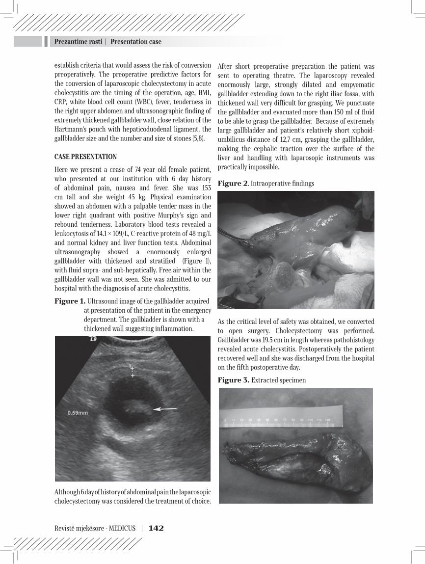
Prezantime rasti | Presentation case
Revistë mjekësore - MEDICUS | 142
20
establish criteria that would assess the risk of conversion preoperatively. The preoperative predictive factors for the conversion of laparoscopic cholecystectomy in acute cholecystitis are the timing of the operation, age, BMI, CRP, white blood cell count (WBC), fever, tenderness in the right upper abdomen and ultrasonographic finding of extremely thickened gallbladder wall, close relation of the Hartmann’s pouch with hepaticoduodenal ligament, the gallbladder size and the number and size of stones (5,8).
CASE PRESENTATION
Here we present a cease of 74 year old female patient, who presented at our institution with 6 day history of abdominal pain, nausea and fever. She was 153 cm tall and she weight 45 kg. Physical examination showed an abdomen with a palpable tender mass in the lower right quadrant with positive Murphy’s sign and rebound tenderness. Laboratory blood tests revealed a leukocytosis of 14.1 × 10⁹/L, C-reactive protein of 48 mg/L and normal kidney and liver function tests. Abdominal ultrasonography showed a enormously enlarged gallbladder with thickened and stratified (Figure 1), with fluid supra- and sub-hepatically. Free air within the gallbladder wall was not seen. She was admitted to our hospital with the diagnosis of acute cholecystitis.
Figure 1. Ultrasound image of the gallbladder acquired at presentation of the patient in the emergency department. The gallbladder is shown with a thickened wall suggesting inflammation.
Although 6 day of history of abdominal pain the laparosopic cholecystectomy was considered the treatment of choice.
After short preoperative preparation the patient was sent to operating theatre. The laparoscopy revealed enormously large, strongly dilated and empyematic gallbladder extending down to the right iliac fossa, with thickened wall very difficult for grasping. We punctuate the gallbladder and evacuated more than 150 ml of fluid to be able to grasp the gallbladder. Because of extremely large gallbladder and patient’s relatively short xiphoid-umbilicus distance of 12,7 cm, grasping the gallbladder, making the cephalic traction over the surface of the liver and handling with laparosopic instruments was practically impossible.
Figure 2. Intraoperative findings
As the critical level of safety was obtained, we converted to open surgery. Cholecystectomy was performed. Gallbladder was 19.5 cm in length whereas pathohistology revealed acute cholecystitis. Postoperatively the patient recovered well and she was discharged from the hospital on the fifth postoperative day.
Figure 3. Extracted specimen
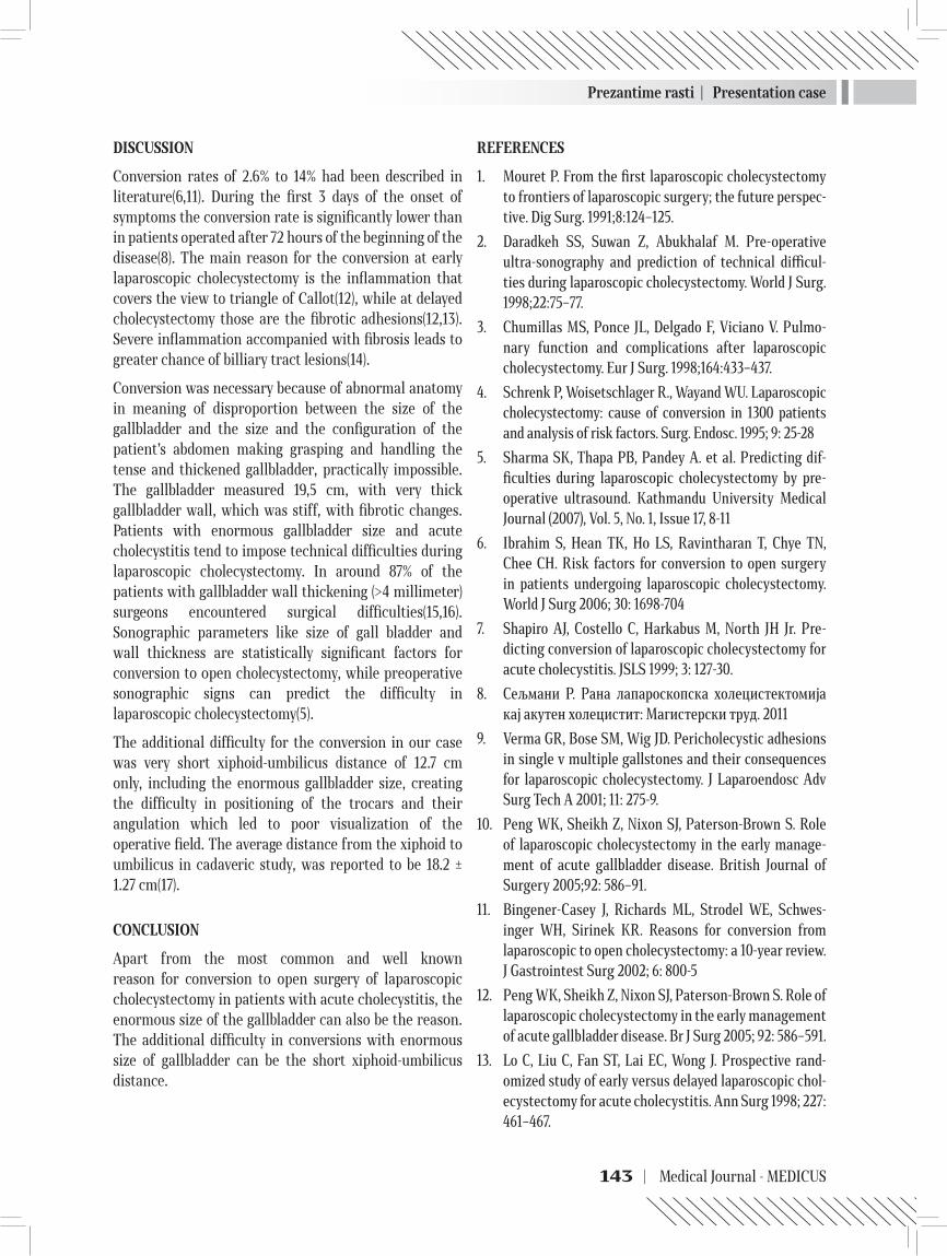
Prezantime rasti | Presentation case
143 | Medical Journal - MEDICUS
DISCUSSION
Conversion rates of 2.6% to 14% had been described in literature(6,11). During the first 3 days of the onset of symptoms the conversion rate is significantly lower than in patients operated after 72 hours of the beginning of the disease(8). The main reason for the conversion at early laparoscopic cholecystectomy is the inflammation that covers the view to triangle of Callot(12), while at delayed cholecystectomy those are the fibrotic adhesions(12,13). Severe inflammation accompanied with fibrosis leads to greater chance of billiary tract lesions(14).
Conversion was necessary because of abnormal anatomy in meaning of disproportion between the size of the gallbladder and the size and the configuration of the patient’s abdomen making grasping and handling the tense and thickened gallbladder, practically impossible. The gallbladder measured 19,5 cm, with very thick gallbladder wall, which was stiff, with fibrotic changes. Patients with enormous gallbladder size and acute cholecystitis tend to impose technical difficulties during laparoscopic cholecystectomy. In around 87% of the patients with gallbladder wall thickening (>4 millimeter) surgeons encountered surgical difficulties(15,16). Sonographic parameters like size of gall bladder and wall thickness are statistically significant factors for conversion to open cholecystectomy, while preoperative sonographic signs can predict the difficulty in laparoscopic cholecystectomy(5).
The additional difficulty for the conversion in our case was very short xiphoid-umbilicus distance of 12.7 cm only, including the enormous gallbladder size, creating the difficulty in positioning of the trocars and their angulation which led to poor visualization of the operative field. The average distance from the xiphoid to umbilicus in cadaveric study, was reported to be 18.2 ± 1.27 cm(17).
CONCLUSION
Apart from the most common and well known reason for conversion to open surgery of laparoscopic cholecystectomy in patients with acute cholecystitis, the enormous size of the gallbladder can also be the reason. The additional difficulty in conversions with enormous size of gallbladder can be the short xiphoid-umbilicus distance.
REFERENCES
1. Mouret P. From the first laparoscopic cholecystectomy to frontiers of laparoscopic surgery; the future perspec-tive. Dig Surg. 1991;8:124–125.
2. Daradkeh SS, Suwan Z, Abukhalaf M. Pre-operative ultra-sonography and prediction of technical difficul-ties during laparoscopic cholecystectomy. World J Surg. 1998;22:75–77.
3. Chumillas MS, Ponce JL, Delgado F, Viciano V. Pulmo-nary function and complications after laparoscopic cholecystectomy. Eur J Surg. 1998;164:433–437.
4. Schrenk P, Woisetschlager R., Wayand WU. Laparoscopic cholecystectomy: cause of conversion in 1300 patients and analysis of risk factors. Surg. Endosc. 1995; 9: 25-28
5. Sharma SK, Thapa PB, Pandey A. et al. Predicting dif-ficulties during laparoscopic cholecystectomy by pre-operative ultrasound. Kathmandu University Medical Journal (2007), Vol. 5, No. 1, Issue 17, 8-11
6. Ibrahim S, Hean TK, Ho LS, Ravintharan T, Chye TN, Chee CH. Risk factors for conversion to open surgery in patients undergoing laparoscopic cholecystectomy. World J Surg 2006; 30: 1698-704
7. Shapiro AJ, Costello C, Harkabus M, North JH Jr. Pre-dicting conversion of laparoscopic cholecystectomy for acute cholecystitis. JSLS 1999; 3: 127-30.
8. Сељмани Р. Рана лапароскопска холецистектомија кај акутен холецистит: Магистерски труд. 2011
9. Verma GR, Bose SM, Wig JD. Pericholecystic adhesions in single v multiple gallstones and their consequences for laparoscopic cholecystectomy. J Laparoendosc Adv Surg Tech A 2001; 11: 275-9.
10. Peng WK, Sheikh Z, Nixon SJ, Paterson-Brown S. Role of laparoscopic cholecystectomy in the early manage-ment of acute gallbladder disease. British Journal of Surgery 2005;92: 586–91.
11. Bingener-Casey J, Richards ML, Strodel WE, Schwes-inger WH, Sirinek KR. Reasons for conversion from laparoscopic to open cholecystectomy: a 10-year review. J Gastrointest Surg 2002; 6: 800-5
12. Peng WK, Sheikh Z, Nixon SJ, Paterson-Brown S. Role of laparoscopic cholecystectomy in the early management of acute gallbladder disease. Br J Surg 2005; 92: 586–591.
13. Lo C, Liu C, Fan ST, Lai EC, Wong J. Prospective rand-omized study of early versus delayed laparoscopic chol-ecystectomy for acute cholecystitis. Ann Surg 1998; 227: 461–467.
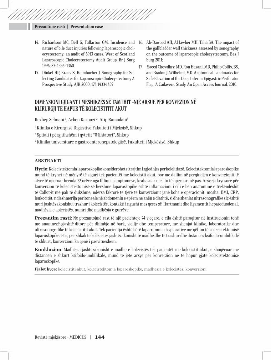
Prezantime rasti | Presentation case
Revistë mjekësore - MEDICUS | 144
14. Richardson MC, Bell G, Fullarton GM. Incidence and nature of bile duct injuries following laparoscopic chol-ecystectomy: an audit of 5913 cases. West of Scotland Laparoscopic Cholecystectomy Audit Group. Br J Surg 1996; 83: 1356–1360.
15. Dinkel HP, Kraus S, Heimbucher J. Sonography for Se-lecting Candidates for Laparoscopic Cholecystectomy A Prospective Study. AJR 2000; 174:1433-1439
16. Ali-Dawood AH, Al Jawher MH, Taha SA. The impact of the gallbladder wall thickness assessed by sonography on the outcome of laparocopic cholecystectomy. Bas J Surg 2011;
17. Saeed Chowdhry, MD, Ron Hazani, MD, Philip Collis, BS, and Bradon J. Wilhelmi, MD. Anatomical Landmarks for Safe Elevation of the Deep Inferior Epigastric Perforator Flap: A Cadaveric Study. An Open Access Journal. 2010.
DIMENSIONI GJIGANT I MESHIKZËS SË TAMTHIT –NJË ARSUE PER KONVEZION NË KLIRURGJI TË HAPUR TË KOLECISTITIT AKUT
Rexhep Selmani 1, Arben Karpuzi 2, Atip Ramadani3
1 Klinika e Kirurgjisë Digjestive,Fakulteti i Mjeksisë, Shkup 2 Spitali i përgjithshëm i qytetit “8 Shtatori”, Shkup3 Klinika universitare e gastroenterohepatologjisë, Fakulteti i Mjekësisë, Shkup
ABSTRAKTI
Hyrje: Kolecistektomia laparoskopike konsiderohet si trajtim i zgjedhjes per kolelitiazë. Kolecistektomia laparoskopike mund të kryhet në mënyrë të sigurt tek pacientët me kolecistit akut, por me dallim në perqindjen e konverzionit të atyre të operuar brenda 72 orëve nga fillimi i simptomeve, krahasuar me ato të operuar më pas. Arsyeja kryesore për konverzion të kolecistektomisë së hershme laparoskopike është inflamacioni i cili e bën anatominë e trekëndëshit të Callot-it më pak të dukshme, ndërsa faktorë të tjerë të konverzionit janë koha e operacionit, mosha, BMI, CRP, leukocitët, ndjeshmerija peritoneale në abdomenin e epërm ne anën e djathtë, si dhe shenjat ultrasonografike siç është muri jashtëzakonisht i trashur i kolecistës, kontakti i ngusht mes qeses së Hartmanit dhe ligamentit hepatoduodenal, madhësia e kolecistës, numri dhe madhësia e gurrëve.
Prezantim rasti: Ne prezantojmë rast të një pacienteje 74 vjeçare, e cila është paraqitur në institucionin tonë me anamnezë gjashtë-ditore për dhimbje në bark, vjellje dhe temperature, me shenjat klinike, laboratorike dhe ultrasonografike të kolecistitit akut. Tek pacientja është bërë laparotomia eksplorative me qëllim të kolecistektomisë laparoskopike. Por, për shkak të kolecistës jashtëzakonisht të madhe dhe të trashur dhe distancës ksifoido-umbilikale të shkurt, konverzioni ka qenë i paevitueshëm.
Konkluzion: Madhësia jashtëzakonisht e madhe e kolecistës tek pacientët me kolecistit akut, e shoqëruar me distancën e shkurt ksifoido-umbilikale, mund të jetë arsye për konverzion në të hapur gjatë kolecistektomisë laparoskopike.
Fjalët kyçe: kolecistiti akut, kolecistektomia laparoskopike, madhesia e kolecistës, konverzioni
145 | Medical Journal - MEDICUS
DIAGNOSTIKIMI ANTENATAL I SYNDROMËS SË HIPOPLASIONIT TË VENTRIKULLIT TË MAJTËHana Bejiqi1, Ramush Bejiqi2, Ragip Retkoceri2, Naim Zeka2, Njazi Gashi2, 1 Qendra Kryesore e Mjekësisë Familjare, Prishtinë, Republika e Kosovës2 QKUK, Klinika e Pediatrisë, Prishtinë, Republika e Kosovës
ABSTRAKTI
Në 20 vitet e fundit te asnjë keqformim i lindur i zemrës nuk janë bërë ndryshime aq dramatike në qasjen diagnostike, trajtimin, udhëheqjen dhe ecurinë e sëmundjes, sa për sindromën e hipoplasionit të ventrikullit të majtë (SHVM). Në krahasim me 30 vite më heret kur mënyra e vetme e trajtimit të këtyre fëmijëve ka qenë kujdesi i mirë pediatrik, sot ekzistojnë opcione të ndryshme kardiologjike dhe kardiokirurgjike, të cilat janë fushë e gjërë e diskutimeve dhe konfrontimeve profesionale. Gjatë kësaj periudhe, mbijetesa deri në moshën 5 vjeçare, përfshirë këtu edhe fëmijët me intervenim sipas procedurës Fontan, është rritur prej 20 % në afër 50 deri 69 %, kurse raportet e fundit tregojnë se 70 % të këtyre fëmijëve, falë ndryshimeve në trajtimin kardiokirurgjik, mund ta arrijnë moshën adulte.(1,2,3)
Kontribut të posaqëm në përparimin e trajtimit të fëmijëve me SHVM ka dhënë diagnostikimi antenatal, i cili mundëson planifikimin e lindjes në nivelin terciar dhe parapëragditjen e një “armate” profesionistësh në trajtimin e fëmijës prej lindjes deri në intervenimin kardiokirurgjik, i cili sot bëhet në disa etapa.(1)
Qëllimi i punimit Në punim është prezantuar një fetus me SHVM, i diagnostikuar në tremujorin e dytë të shtatzënsisë, mënyrën e udhëheqjes së shtatzënsisë dhe mënyrën e trajtimit pas lindjes, në kushte të mungesës së infrastrukturës bashkohore kardiologjike dhe kardiokirurgjike.
Fjalet kyq hipoplasioni i ventrikullit të majtë, diagnostikimi antenatal, Fontan procedura, Prostaglandinet
PREZANTIMI I RASTIT
Te shtatzëna e moshës 28 vjeçare, shtatzënsia e parë, e rregullt, rregullisht e kontrolluar, gjatë ekzaminimit rutinor ultrasonografik nga obstetri në javën e 20-të të shtatzënsisë, regjistrohet asimetri e barkushave të zemrës, me dominim të barkushës së djathtë. Të gjitha matjet obstetrike të fetusit janë në korelim me moshën gjestative të frytit. Kjo është arsyeja që shtatzëna të udhëzohet per ekzaminim ekokardiografik fetal në nivelin terciar.
GJETJA EKOKARDIOGRAFIKE
Ne ekzaminimin ekokardiografik antenatal regjistrohet pozicioni normal i zemrës dhe organeve torako-abdominale (Foto 1.). Derdhja e venave sistemike dhe pulmonare është në rregull. Foramen ovale është me pozicion, madhësi dhe qarkullim të rregullt. Ekziston konkordancë atrio-ventrikulare dhe ventrikulo-
arteriale. Në të dy nivelet fletëzat atrioventrikulare janë morfologjikisht në rregull, me qarkullim laminar anterograd dhe regurgjitim minimal në nivel të valvulës mitrale (VM). Barkusha e majtë është lehtë e dilatuar dhe me hiperekogjenitet të endokardit. Valvula e aortes prezantohet hipoplastike si dhe pjesa ascendente e aortes kurse harku është në rregull. Dalja e ventrikullit të djathtë dhe arteria pulmonare (AP) janë në rregull. Nuk regjistrohet çrregullim i ritmit. Nuk ka lengë të lirë në qesën perikardiale.
Në javën e 22 të shtatzënsisë është bërë amniocenteza dhe ekzaminimi gjenetik nuk tregon prezencë të ndonjë kromozomopatie.
Në javën e 30 të shtatzënsisë (Foto 2) ekzaminimi ekokardiografik antenatal prezanton asimetri të shprehur ndërmjet barkushave, me dominim të barkushës së djathtë dhe hipoplasion të barkushës së majtë. Në nivel të VM, me Doppler dhe kollor Doppler, regjistrohet jett i
Medicus 2013, Vol. 18 (2): 145-149
Prezantime rasti | Presentation case
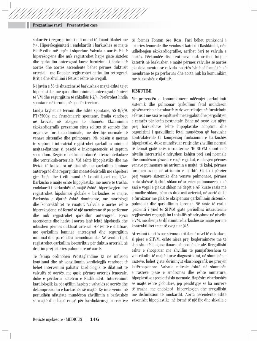
Prezantime rasti | Presentation case
Revistë mjekësore - MEDICUS | 146
26
shkurtër i regurgjitimit i cili mund të kuantifikohet me ¼+. Hiperekogjeniteti i endokardit i barkushës së majtë është edhe më tepër i shprehur. Valvula e aortës është hiperekogjene dhe nuk regjistrohet hapje gjatë sistoles dhe qarkullim anterograd kurse furnizimi i harkut të aortës dhe aortës ascendente bëhet përmes duktusit arterial – me Doppler regjistrohet qarkullim retrograd. Rritja dhe zhvillimi i fetusit është në rregull.
Në javën e 38 të shtatzënsisë barkusha e majtë është tejet hipoplastike, me qarkullim minimal anterograd në nivel të VM dhe regurgjitim të shkallës 1-2/4. Preferohet lindja spontane në termin, në qendër terciare.
Lindja kryhet në termin dhe është spontane, AS=8/9/9, PT=3500g, me frymëmarrje spontane, fëmija vendoset në krevat, në oksigjen te dhomës. Ekzaminimi ekokardiografik prezanton situs solitus të zemrës dhe organeve torako-abdominale, me derdhje normale të venave sistemike dhe pulmonare. Në pjesën e mesme te septumit interatrial regjistrohet qarkullim minimal majtas-djathtas si pasojë e inkompetencës së septum secundum. Regjistrohet konkordancë atrioventrikulare dhe ventrikulo-arteriale. VM është hipoplastike dhe me lëvizje të kufizuara në diastolë, me qarkullim laminar anterograd dhe regurgjitim mesotelesistolik me shpejtësi gjer 3m/s dhe i cili mund të kvantifikohet me 2/4+. Barkusha e majtë është hipoplastike, me mure të trasha, endokardi i barkushës së majtë është hiperekogjen dhe regjistrohet hipokinezi globale e barkushës së majtë. Barkusha e djathë është dominante, me morfologji dhe kontraktilitet të ruajtur. Valvula e aortës është hiperekogjene, në formë të një membrane të pa perforuar dhe nuk regjistrohet qarkullim anterograd. Pjesa ascendente dhe harku i aortes janë lehtë hipolastik dhe mbushen përmes duktusit arterial. AP është e dilatuar, me qarkullim laminar anterograd dhe regurgjitim minimal dhe pa rëndësi hemodinamike. Në vendin tipik regjistrohet qarkullim jorestriktiv për duktus arterial, në drejtim prej arteries pulmonare në aortë.
Te fëmija ordinohen Prostaglandine E1 në infusion kontinual dhe në konziliumin kardiologjik vendoset të bëhet intervenimi paliativ kardiologjik të dilatimit të valvulës së aortës, me qasje përmes arteries femorale, duke e përdorur katetrin e Rashkind-it. Intervenimit kardiologjik ka për qëllim hapjen e valvulës së aortës dhe dekompresionin e barkushës së majtë. Ky intervenim në periudhën afatgjate mundëson zhvillimin e barkushës së majtë dhe hapë rrugë për kardiokirurgji korrektive
të formës Fontan ose Ross. Pasi bëhet punksioni i arteries femorale dhe vendoset katetri i Rashkindit, nën udhëheqjen ekokardiografike, arrihet deri te valvula e aortës. Përkundër disa tentimeve nuk arrihet futja e katetrit në barkushën e majtë përmes valvulës së aortës çka dokumenton se valvula e aortës është në formë të një membrane të pa perforuar dhe aorta nuk ka komunikim me barkushën e djathtë.
DISKUTIMI
Me prezencën e komunikimeve ndërmjet qarkullimit sistemik dhe pulmonar qarkullimi fetal mundëson pjesëmarrjen e barabartë ty dy ventrikujve në furnizimin e fetusit me sasi të mjaftueshme të gjakut dhe përgaditjen e zemrës për jetën postnatale. Edhe në raste kur njëra prej barkushave është hipoplastike adoptimi dhe organizimi i qarkullimit fetal mundëson që barkusha kontralaterale ta kompensoj funksionin e barkushës hipoplastike, duke mundësuar rritje dhe zhvillim normal të fetusit gjatë jetës intrauterine. Te SHVM shunt-i në nivelin interatrial e ndryshon kahjen prej asaj normale dhe mundëson që sasia e vogël e gjakut, e cila vjen përmes venave pulmonare në atriumin e majtë, të kaloj, përmes foramen ovale, në atriumin e djathtë. Gjaku i përzier prej venave sistemike dhe venave pulmonare, përmes barkushës së djathtë, shkon në arterien pulmonare ku një sasi e vogël e gjakut shkon në degët e AP kurse sasia më e madhe shkon, përmes duktusit arterial, në aortë duke e furnizuar me gjak të oksigjenuar qarkullimin sistemik, pulmonar dhe qarkullimin koronar. Në raste të rralla (pacienti i ynë) të SHVM gjatë periudhës intrauterine regjistrohet regurgjitim i shkallës së ndryshme në nivelin e VM, me shenja të dilatimit të barkushës së majtë por me kontraktilitet tejet të zvogluar.(4,5)
Atresioni i aortës ose stenoza kritike në nivel të valvulave, si pjesë e SHVM, është njëra prej keqformimeve më të shpeshta të diagnostikuara në moshën fetale. Rregullisht është e shoqëruar me zhvillim të pamjaftueshëm të ventrikullit të majtë kurse diagnostikimi, në shumicën e rasteve, bëhet gjatë skriningut ekosonografik në prerjen katërhapsinore. Valvula mitrale është në shumicën e rasteve pjesë e sindromës dhe është miniature, hipoplastike apo plotësisht normale. Hapësira e barkushës së majtë është globulare, jep përshtypje se ka mureve të trasha, me endokard hiperekogjen dhe rregullisht me disfunksion të miokardit. Aorta ascendente është zakonisht hipoplastike, në formë të një fije dhe shkalla e
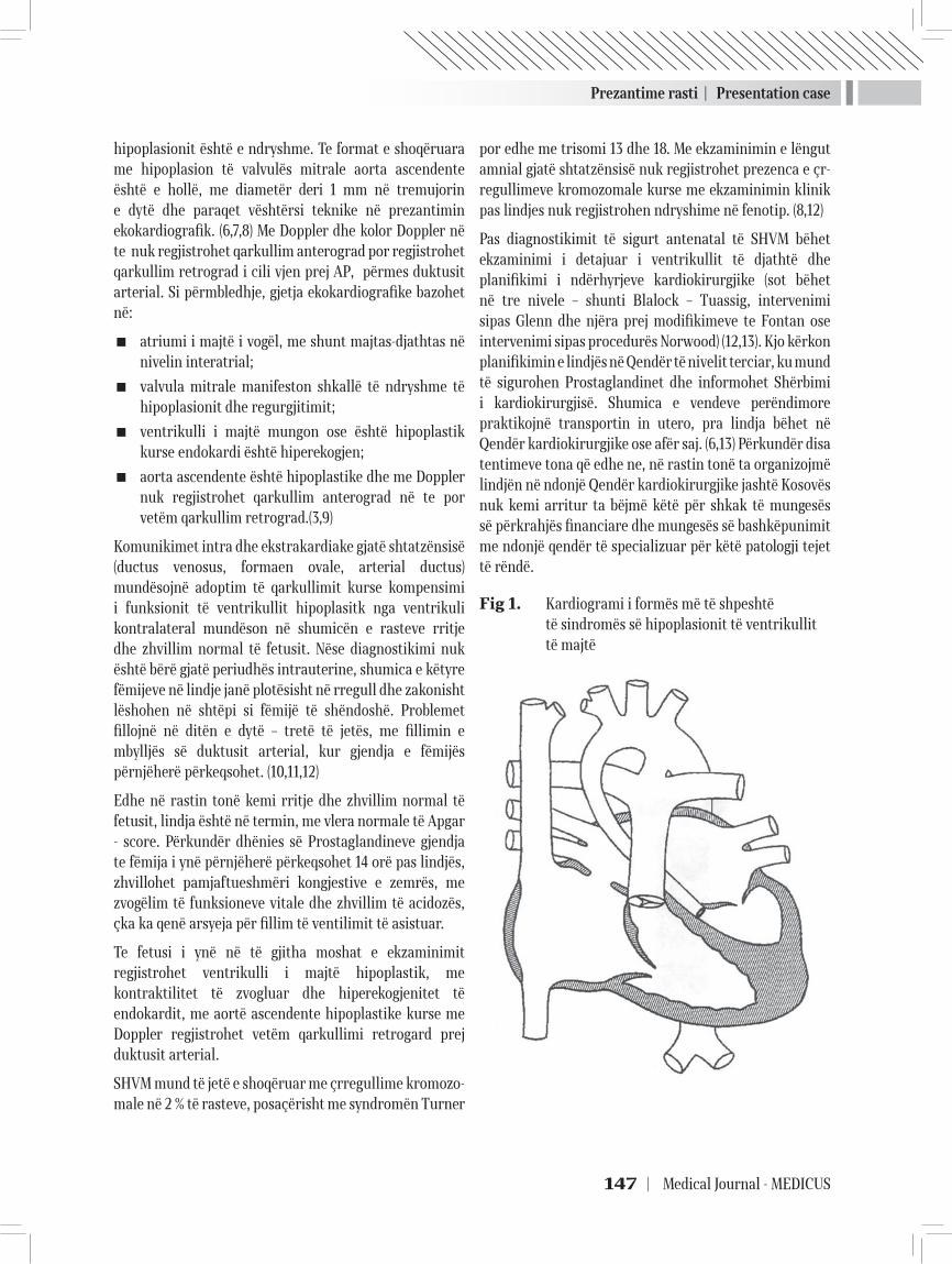
Prezantime rasti | Presentation case
147 | Medical Journal - MEDICUS
hipoplasionit është e ndryshme. Te format e shoqëruara me hipoplasion të valvulës mitrale aorta ascendente është e hollë, me diametër deri 1 mm në tremujorin e dytë dhe paraqet vështërsi teknike në prezantimin ekokardiografik. (6,7,8) Me Doppler dhe kolor Doppler në te nuk regjistrohet qarkullim anterograd por regjistrohet qarkullim retrograd i cili vjen prej AP, përmes duktusit arterial. Si përmbledhje, gjetja ekokardiografike bazohet në:
� atriumi i majtë i vogël, me shunt majtas-djathtas në nivelin interatrial;
� valvula mitrale manifeston shkallë të ndryshme të hipoplasionit dhe regurgjitimit;
� ventrikulli i majtë mungon ose është hipoplastik kurse endokardi është hiperekogjen;
� aorta ascendente është hipoplastike dhe me Doppler nuk regjistrohet qarkullim anterograd në te por vetëm qarkullim retrograd.(3,9)
Komunikimet intra dhe ekstrakardiake gjatë shtatzënsisë (ductus venosus, formaen ovale, arterial ductus) mundësojnë adoptim të qarkullimit kurse kompensimi i funksionit të ventrikullit hipoplasitk nga ventrikuli kontralateral mundëson në shumicën e rasteve rritje dhe zhvillim normal të fetusit. Nëse diagnostikimi nuk është bërë gjatë periudhës intrauterine, shumica e këtyre fëmijeve në lindje janë plotësisht në rregull dhe zakonisht lëshohen në shtëpi si fëmijë të shëndoshë. Problemet fillojnë në ditën e dytë – tretë të jetës, me fillimin e mbylljës së duktusit arterial, kur gjendja e fëmijës përnjëherë përkeqsohet. (10,11,12)
Edhe në rastin tonë kemi rritje dhe zhvillim normal të fetusit, lindja është në termin, me vlera normale të Apgar - score. Përkundër dhënies së Prostaglandineve gjendja te fëmija i ynë përnjëherë përkeqsohet 14 orë pas lindjës, zhvillohet pamjaftueshmëri kongjestive e zemrës, me zvogëlim të funksioneve vitale dhe zhvillim të acidozës, çka ka qenë arsyeja për fillim të ventilimit të asistuar.
Te fetusi i ynë në të gjitha moshat e ekzaminimit regjistrohet ventrikulli i majtë hipoplastik, me kontraktilitet të zvogluar dhe hiperekogjenitet të endokardit, me aortë ascendente hipoplastike kurse me Doppler regjistrohet vetëm qarkullimi retrogard prej duktusit arterial.
SHVM mund të jetë e shoqëruar me çrregullime kromozo-male në 2 % të rasteve, posaçërisht me syndromën Turner
por edhe me trisomi 13 dhe 18. Me ekzaminimin e lëngut amnial gjatë shtatzënsisë nuk regjistrohet prezenca e çr-regullimeve kromozomale kurse me ekzaminimin klinik pas lindjes nuk regjistrohen ndryshime në fenotip. (8,12)
Pas diagnostikimit të sigurt antenatal të SHVM bëhet ekzaminimi i detajuar i ventrikullit të djathtë dhe planifikimi i ndërhyrjeve kardiokirurgjike (sot bëhet në tre nivele – shunti Blalock – Tuassig, intervenimi sipas Glenn dhe njëra prej modifikimeve te Fontan ose intervenimi sipas procedurës Norwood) (12,13). Kjo kërkon planifikimin e lindjës në Qendër të nivelit terciar, ku mund të sigurohen Prostaglandinet dhe informohet Shërbimi i kardiokirurgjisë. Shumica e vendeve perëndimore praktikojnë transportin in utero, pra lindja bëhet në Qendër kardiokirurgjike ose afër saj. (6,13) Përkundër disa tentimeve tona që edhe ne, në rastin tonë ta organizojmë lindjën në ndonjë Qendër kardiokirurgjike jashtë Kosovës nuk kemi arritur ta bëjmë këtë për shkak të mungesës së përkrahjës financiare dhe mungesës së bashkëpunimit me ndonjë qendër të specializuar për këtë patologji tejet të rëndë.
Fig 1. Kardiogrami i formës më të shpeshtë të sindromës së hipoplasionit të ventrikullit të majtë
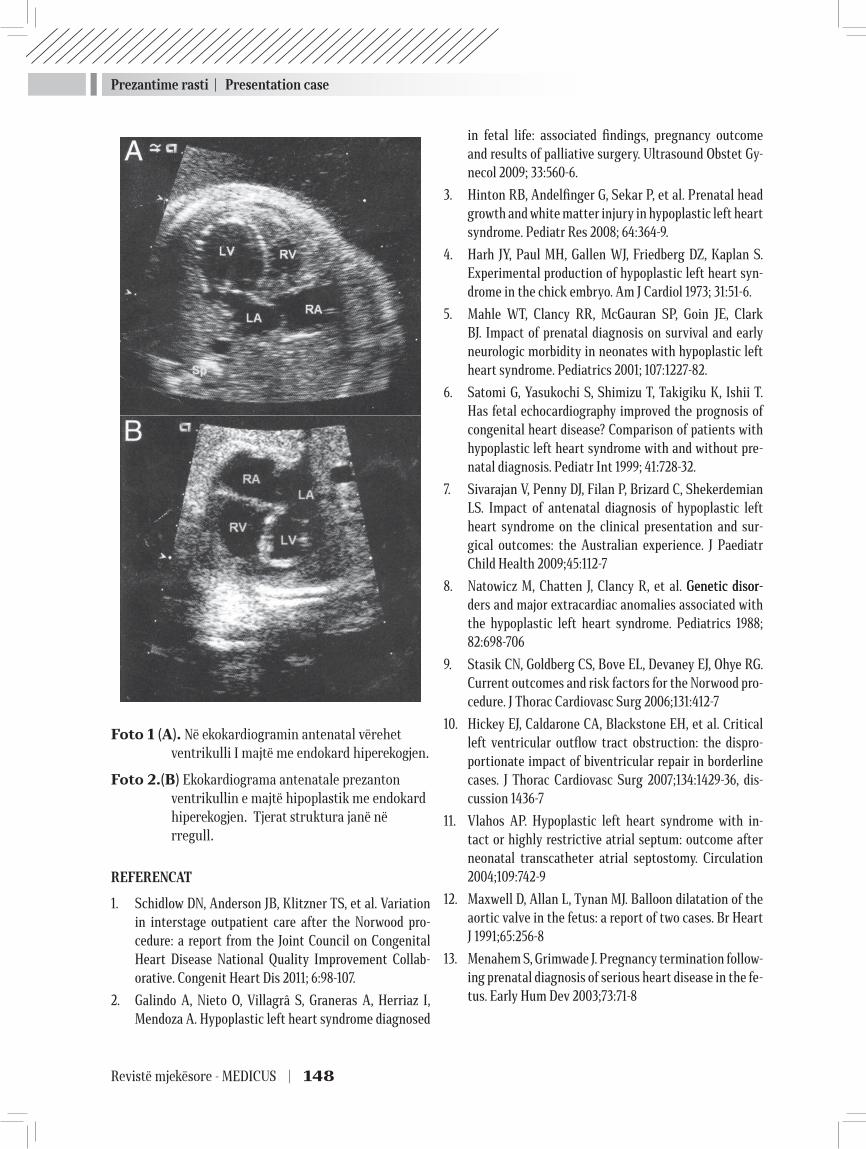
Prezantime rasti | Presentation case
Revistë mjekësore - MEDICUS | 148
Foto 1 (А). Në ekokardiogramin antenatal vërehet ventrikulli I majtë me endokard hiperekogjen.
Foto 2.(B) Ekokardiograma antenatale prezanton ventrikullin e majtë hipoplastik me endokard hiperekogjen. Tjerat struktura janë në rregull.
REFERENCAT
1. Schidlow DN, Anderson JB, Klitzner TS, et al. Variation in interstage outpatient care after the Norwood pro-cedure: a report from the Joint Council on Congenital Heart Disease National Quality Improvement Collab-orative. Congenit Heart Dis 2011; 6:98-107.
2. Galindo A, Nieto O, Villagrâ S, Graneras A, Herriaz I, Mendoza A. Hypoplastic left heart syndrome diagnosed
in fetal life: associated findings, pregnancy outcome and results of palliative surgery. Ultrasound Obstet Gy-necol 2009; 33:560-6.
3. Hinton RB, Andelfinger G, Sekar P, et al. Prenatal head growth and white matter injury in hypoplastic left heart syndrome. Pediatr Res 2008; 64:364-9.
4. Harh JY, Paul MH, Gallen WJ, Friedberg DZ, Kaplan S. Experimental production of hypoplastic left heart syn-drome in the chick embryo. Am J Cardiol 1973; 31:51-6.
5. Mahle WT, Clancy RR, McGauran SP, Goin JE, Clark BJ. Impact of prenatal diagnosis on survival and early neurologic morbidity in neonates with hypoplastic left heart syndrome. Pediatrics 2001; 107:1227-82.
6. Satomi G, Yasukochi S, Shimizu T, Takigiku K, Ishii T. Has fetal echocardiography improved the prognosis of congenital heart disease? Comparison of patients with hypoplastic left heart syndrome with and without pre-natal diagnosis. Pediatr Int 1999; 41:728-32.
7. Sivarajan V, Penny DJ, Filan P, Brizard C, Shekerdemian LS. Impact of antenatal diagnosis of hypoplastic left heart syndrome on the clinical presentation and sur-gical outcomes: the Australian experience. J Paediatr Child Health 2009;45:112-7
8. Natowicz M, Chatten J, Clancy R, et al. Genetic disor-Genetic disor-ders and major extracardiac anomalies associated with the hypoplastic left heart syndrome. Pediatrics 1988; 82:698-706
9. Stasik CN, Goldberg CS, Bove EL, Devaney EJ, Ohye RG. Current outcomes and risk factors for the Norwood pro-cedure. J Thorac Cardiovasc Surg 2006;131:412-7
10. Hickey EJ, Caldarone CA, Blackstone EH, et al. Critical left ventricular outflow tract obstruction: the dispro-portionate impact of biventricular repair in borderline cases. J Thorac Cardiovasc Surg 2007;134:1429-36, dis-cussion 1436-7
11. Vlahos AP. Hypoplastic left heart syndrome with in-tact or highly restrictive atrial septum: outcome after neonatal transcatheter atrial septostomy. Circulation 2004;109:742-9
12. Maxwell D, Allan L, Tynan MJ. Balloon dilatation of the aortic valve in the fetus: a report of two cases. Br Heart J 1991;65:256-8
13. Menahem S, Grimwade J. Pregnancy termination follow-ing prenatal diagnosis of serious heart disease in the fe-tus. Early Hum Dev 2003;73:71-8
Prezantime rasti | Presentation case
149 | Medical Journal - MEDICUS
ANTENATAL DIAGNOSIS OF THE SYNDROME OF LEFT VENTRIVULAR HIPOPLASION
Hana Bejiqi1, Ramush Bejiqi2, Ragip Retkoceri2, Naim Zeka2, Njazi Gashi2, 1 Center of Main Family Medicine, Prishtina, Republic of Kosova
2 QKUK, Department of Pediatrics, Pristina, Republic of Kosovo
ABSTRACT
In the recent era, no congenital heart defects have undergone a more dramatic change in diagnostic approach, management, and outcomes than hypoplastic left heart syndrome (HLHS). Although just over 30 years ago, comfort care was the only option, there are now numbers of therapeutic options available for families, though there continues to be a debate as to the optimal treatment approach. During this time, survival to the age of 5 years (including Fontan) has ranged from 50 % to 69 %, but current expectations are that 70 % of newborns born today with HLHS may rich adulthood.(1,2,3)
Particular contribution in advancing the treatment of children with antenatal diagnosis SHVM provided, which enables planning birth at tertiary level and involvement of an “army” of professionals in the treatment of children from birth to the cardiosurgical intervention, which today is in some stages.(1)
Aim of presentation We presented a fetus with HLHS diagnosed in the second trimester of pregnancy, following and treatment after delivery in condition of absence cardio logical and cardio surgical infrastructure.
Key words hypoplastic left heart, antenatal diagnosis, Fontan procedure, Prostaglandins
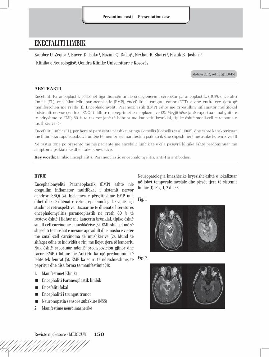
Revistë mjekësore - MEDICUS | 150
ENECFALITI LIMBIKKamber U. Zeqiraj 1, Enver D. Isaku 1, Nazim Q. Dakaj1 , Nexhat R. Shatri 1, Fisnik B. Jashari 1
1 Klinika e Neurologjisë, Qendra Klinike Universitare e Kosovës
ABSTRAKTI
Encefaliti Paraneoplastik përbëhet nga disa sëmundje si degjenerimi cerebelar paraneoplastik, (DCP), encefaliti limbik (EL), encefalomieliti paraneoplastic (EMP), encefaliti i trungut trunor (ETT) si dhe entiteteve tjera që manifestohen më rrallë (1). Encephalomyeliti Paraneoplastik (EMP) është një çrregullim inflamator multifokal i sistemit nervor qendro (SNQ) i lidhur me veprimet e neoplazmave (2). Megjithëse janë raportuar malignitete te ndryshme te EMP, 80 % te rasteve janë të lidhura me kancerin bronkial, tipike është small-cell carcinome e mushkërive (3).
Encefaliti limbic (EL), për here të parë është përshkruar nga Corsellis [Corsellis et al. 1968], dhe është karakterizuar me fillim akut apo subakut, humbje të memories, manifestim psikiatrik dhe shpesh herë me atake konvulzive. (1)
Në rastin tonë po prezentojmë një paciente me encefalit limbik te e cila pasqyra klinike është predominuar me simptoma psikiatrike dhe atake konvulzive.
Key words: Limbic Encephalitis, Paraneoplastic encephalomyelitis, anti-Hu antibodies.
Medicus 2013, Vol. 18 (2): 150-153
HYRJE
Encephalomyeliti Paraneoplastik (EMP) është një cregullim inflamator multifokal i sistemit nervor qendror (SNQ) (4). Incidenca e përgjithshme EMP nuk dihet dhe të dhënat e vetme epidemiologjike vijnë nga studimet retrospektive. Bazuar në të dhënat e literaturës encephalomyelitis paraneoplastik në rreth 80 % të rasteve është i lidhur me kancerin bronkial, tipike është small-cell carcinome e mushkërive (3). EMP shfaqet më së shpeshti te moshat e mesme apo adult dhe mosha e vjetër me small-cell carcinoma të mushkërive (2). Mund të shfaqet edhe te individët e rinj me llojet tjera të kancerit. Nuk është raportuar ndonjë predispozicion gjinor dhe racor. EMP i lidhur me Anti-Hu ka një predominim të lehtë tek femrat (5). EMP ka ecuri të ndryshueshme, të papritur dhe disa forma te manifestimit (4):
1. Manifestimet Klinike: � Encephaliti Paraneoplastik limbik
� Encefaliti fokal
� Encephaliti i trungut trunor
� Neuronopatia sensore subakute (NSS)
2. Manifestime neuroimazherike
Neuropatologjia imazherike kryesisht është e lokalizuar në lobet temporale mesiale dhe pjesët tjera të sistemit limbic (1). Fig. 1, 2 dhe 3.
Fig. 1
Fig. 2
27Prezantime rasti | Presentation case
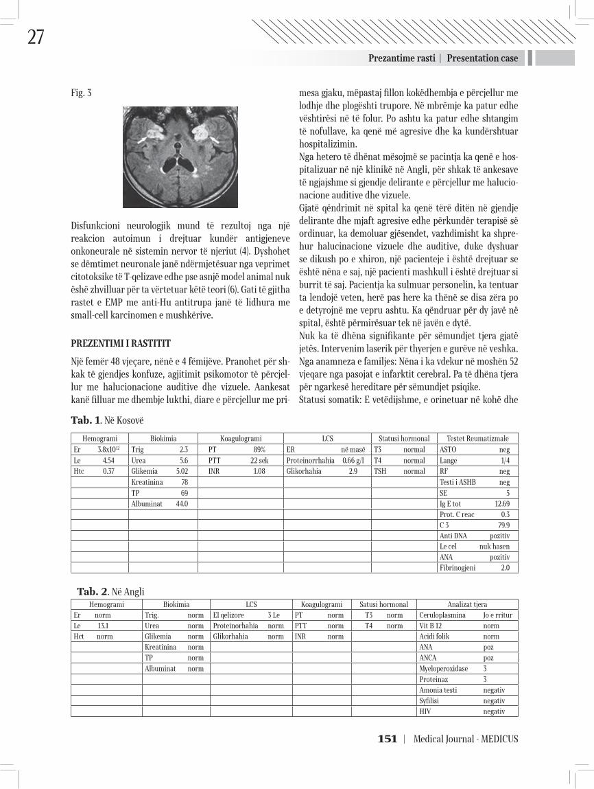
Prezantime rasti | Presentation case
151 | Medical Journal - MEDICUS
27
Fig. 3
Disfunkcioni neurologjik mund të rezultoj nga një reakcion autoimun i drejtuar kundër antigjeneve onkoneurale në sistemin nervor të njeriut (4). Dyshohet se dëmtimet neuronale janë ndërmjetësuar nga veprimet citotoksike të T-qelizave edhe pse asnjë model animal nuk ëshë zhvilluar për ta vërtetuar këtë teori (6). Gati të gjitha rastet e EMP me anti-Hu antitrupa janë të lidhura me small-cell karcinomen e mushkërive.
PREZENTIMI I RASTITIT
Një femër 48 vjeçare, nënë e 4 fëmijëve. Pranohet për sh-kak të gjendjes konfuze, agjitimit psikomotor të përcjel-lur me halucionacione auditive dhe vizuele. Aankesat kanë filluar me dhembje lukthi, diare e përcjellur me pri-
mesa gjaku, mëpastaj fillon kokëdhembja e përcjellur me lodhje dhe plogështi trupore. Në mbrëmje ka patur edhe vështirësi në të folur. Po ashtu ka patur edhe shtangim të nofullave, ka qenë më agresive dhe ka kundërshtuar hospitalizimin.Nga hetero të dhënat mësojmë se pacintja ka qenë e hos-pitalizuar në një klinikë në Angli, për shkak të ankesave të ngjajshme si gjendje delirante e përcjellur me halucio-nacione auditive dhe vizuele.Gjatë qëndrimit në spital ka qenë tërë ditën në gjendje delirante dhe mjaft agresive edhe përkundër terapisë së ordinuar, ka demoluar gjësendet, vazhdimisht ka shpre-hur halucinacione vizuele dhe auditive, duke dyshuar se dikush po e xhiron, një pacienteje i është drejtuar se është nëna e saj, një pacienti mashkull i është drejtuar si burrit të saj. Pacientja ka sulmuar personelin, ka tentuar ta lendojë veten, herë pas here ka thënë se disa zëra po e detyrojnë me vepru ashtu. Ka qëndruar për dy javë në spital, është përmirësuar tek në javën e dytë.Nuk ka të dhëna signifikante për sëmundjet tjera gjatë jetës. Intervenim laserik për thyerjen e gurëve në veshka.Nga anamneza e familjes: Nëna i ka vdekur në moshën 52 vjeqare nga pasojat e infarktit cerebral. Pa të dhëna tjera për ngarkesë hereditare për sëmundjet psiqike.Statusi somatik: E vetëdijshme, e orinetuar në kohë dhe
Tab. 1. Në Kosovë
Hemogrami Biokimia Koagulogrami LCS Statusi hormonal Testet Reumatizmale
Er 3.8x10¹² Trig 2.3 PT 89% ER në masë T3 normal ASTO neg
Le 4.54 Urea 5.6 PTT 22 sek Proteinorrhahia 0.66 g/l T4 normal Lange 1/4Htc 0.37 Glikemia 5.02 INR 1.08 Glikorhahia 2.9 TSH normal RF neg
Kreatinina 78 Testi i ASHB neg
TP 69 SE 5Albuminat 44.0 Ig E tot 12.69
Prot. C reac 0.3C 3 79.9Anti DNA pozitivLe cel nuk hasenANA pozitivFibrinogjeni 2.0
Tab. 2. Në Angli Hemogrami Biokimia LCS Koagulogrami Satusi hormonal Analizat tjera
Er norm Trig. norm El qelizore 3 Le PT norm T3 norm Ceruloplasmina Jo e rriturLe 13.1 Urea norm Proteinorhahia norm PTT norm T4 norm Vit B 12 normHct norm Glikemia norm Glikorhahia norm INR norm Acidi folik norm
Kreatinina norm ANA pozTP norm ANCA poz
Albuminat norm Myeloperoxidase 3
Proteinaz 3Amonia testi negativSyfilisi negativHIV negativ
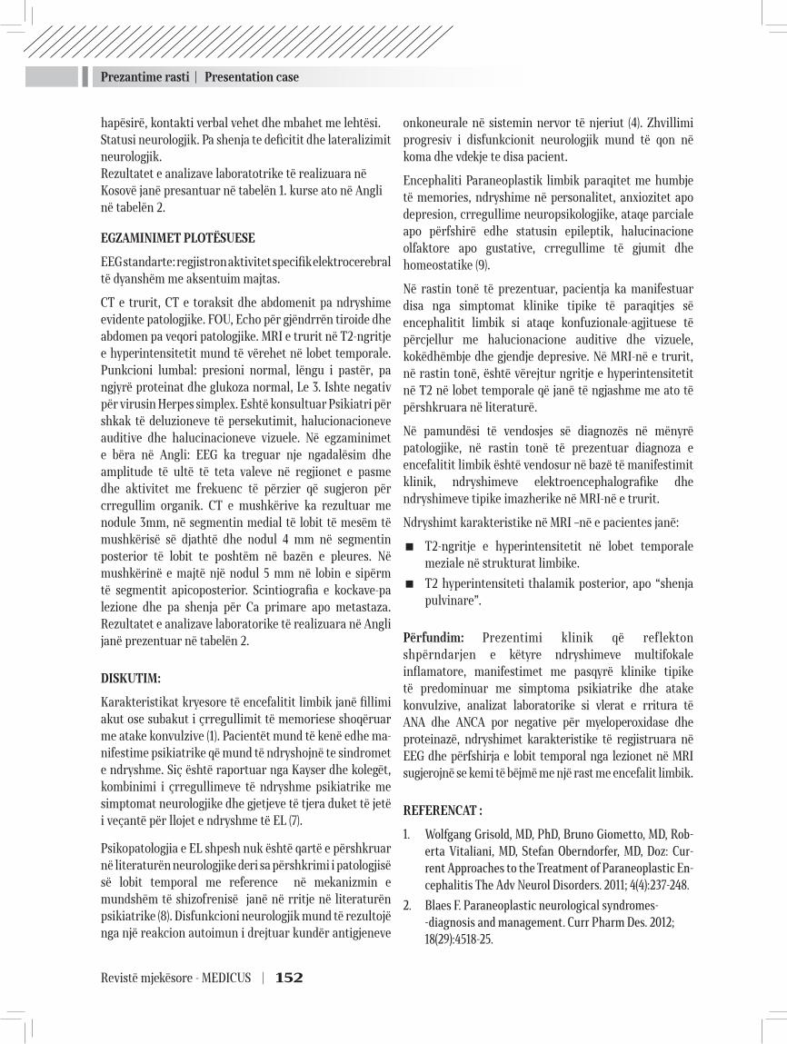
Prezantime rasti | Presentation case
Revistë mjekësore - MEDICUS | 152
hapësirë, kontakti verbal vehet dhe mbahet me lehtësi. Statusi neurologjik. Pa shenja te deficitit dhe lateralizimit neurologjik.Rezultatet e analizave laboratotrike të realizuara në Kosovë janë presantuar në tabelën 1. kurse ato në Angli në tabelën 2.
EGZAMINIMET PLOTËSUESE
EEG standarte: regjistron aktivitet specifik elektrocerebral të dyanshëm me aksentuim majtas.
CT e trurit, CT e toraksit dhe abdomenit pa ndryshime evidente patologjike. FOU, Echo për gjëndrrën tiroide dhe abdomen pa veqori patologjike. MRI e trurit në T2-ngritje e hyperintensitetit mund të vërehet në lobet temporale. Punkcioni lumbal: presioni normal, lëngu i pastër, pa ngjyrë proteinat dhe glukoza normal, Le 3. Ishte negativ për virusin Herpes simplex. Eshtë konsultuar Psikiatri për shkak të deluzioneve të persekutimit, halucionacioneve auditive dhe halucinacioneve vizuele. Në egzaminimet e bëra në Angli: EEG ka treguar nje ngadalësim dhe amplitude të ultë të teta valeve në regjionet e pasme dhe aktivitet me frekuenc të përzier që sugjeron për crregullim organik. CT e mushkërive ka rezultuar me nodule 3mm, në segmentin medial të lobit të mesëm të mushkërisë së djathtë dhe nodul 4 mm në segmentin posterior të lobit te poshtëm në bazën e pleures. Në mushkërinë e majtë një nodul 5 mm në lobin e sipërm të segmentit apicoposterior. Scintiografia e kockave-pa lezione dhe pa shenja për Ca primare apo metastaza. Rezultatet e analizave laboratorike të realizuara në Angli janë prezentuar në tabelën 2.
DISKUTIM:
Karakteristikat kryesore të encefalitit limbik janë fillimi akut ose subakut i çrregullimit të memoriese shoqëruar me atake konvulzive (1). Pacientët mund të kenë edhe ma-nifestime psikiatrike që mund të ndryshojnë te sindromet e ndryshme. Siç është raportuar nga Kayser dhe kolegët, kombinimi i çrregullimeve të ndryshme psikiatrike me simptomat neurologjike dhe gjetjeve të tjera duket të jetë i veçantë për llojet e ndryshme të EL (7).
Psikopatologjia e EL shpesh nuk është qartë e përshkruar në literaturën neurologjike deri sa përshkrimi i patologjisë së lobit temporal me reference në mekanizmin e mundshëm të shizofrenisë janë në rritje në literaturën psikiatrike (8). Disfunkcioni neurologjik mund të rezultojë nga një reakcion autoimun i drejtuar kundër antigjeneve
onkoneurale në sistemin nervor të njeriut (4). Zhvillimi progresiv i disfunkcionit neurologjik mund të qon në koma dhe vdekje te disa pacient.
Encephaliti Paraneoplastik limbik paraqitet me humbje të memories, ndryshime në personalitet, anxiozitet apo depresion, crregullime neuropsikologjike, ataqe parciale apo përfshirë edhe statusin epileptik, halucinacione olfaktore apo gustative, crregullime të gjumit dhe homeostatike (9).
Në rastin tonë të prezentuar, pacientja ka manifestuar disa nga simptomat klinike tipike të paraqitjes së encephalitit limbik si ataqe konfuzionale-agjituese të përcjellur me halucionacione auditive dhe vizuele, kokëdhëmbje dhe gjendje depresive. Në MRI-në e trurit, në rastin tonë, është vërejtur ngritje e hyperintensitetit në T2 në lobet temporale që janë të ngjashme me ato të përshkruara në literaturë.
Në pamundësi të vendosjes së diagnozës në mënyrë patologjike, në rastin tonë të prezentuar diagnoza e encefalitit limbik është vendosur në bazë të manifestimit klinik, ndryshimeve elektroencephalografike dhe ndryshimeve tipike imazherike në MRI-në e trurit.
Ndryshimt karakteristike në MRI –në e pacientes janë:
� T2-ngritje e hyperintensitetit në lobet temporale meziale në strukturat limbike.
� T2 hyperintensiteti thalamik posterior, apo “shenja pulvinare”.
Përfundim: Prezentimi klinik që reflekton shpërndarjen e këtyre ndryshimeve multifokale inflamatore, manifestimet me pasqyrë klinike tipike të predominuar me simptoma psikiatrike dhe atake konvulzive, analizat laboratorike si vlerat e rritura të ANA dhe ANCA por negative për myeloperoxidase dhe proteinazë, ndryshimet karakteristike të regjistruara në EEG dhe përfshirja e lobit temporal nga lezionet në MRI sugjerojnë se kemi të bëjmë me një rast me encefalit limbik.
REFERENCAT :
1. Wolfgang Grisold, MD, PhD, Bruno Giometto, MD, Rob-erta Vitaliani, MD, Stefan Oberndorfer, MD, Doz: Cur-rent Approaches to the Treatment of Paraneoplastic En-cephalitis The Adv Neurol Disorders. 2011; 4(4):237-248.
2. Blaes F. Paraneoplastic neurological syndromes--diagnosis and management. Curr Pharm Des. 2012; 18(29):4518-25.
Prezantime rasti | Presentation case
153 | Medical Journal - MEDICUS
3. Shirafuji T, Kanda F, Sekiguchi K, Higuchi M, Yokozaki H, Tanaka K, et al. Anti-Hu-associated paraneoplastic encephalomyelitis with esophageal small cell carcino-ma. Intern Med. 2012;51(17):2423-7.
4. David S. Liebeskind, MD; Stephan A Berman, MD, PhD, MBA; Francisco Talavera, PharmD, PhD; Jorge C Kattah, MD; Selim R Benbadis, MD: Paraneoplastic Encephalo-myelitis Clinical Presentation. Medscape Reference.
5. Foster AR, Caplan JP. Paraneoplastic limbic encephali-tis. Psychosomatics. Mar-Apr 2009;50(2):108-13.
6. Graus F, Saiz A, Lai M, Bruna J, López F, Sabater L, et al. Neuronal surface antigen antibodies in limbic encepha-litis: clinical-immunologic associations. Neurology. Sep 16 2008;71(12):930-6.
7. Kayser, C.G., Kohler, C.G. and Dalmau, J. (2010) Psychi-atric manifestations of paraneoplastic disorders. Am J Psychiatry 167: 1039–1050.
8. Zandi, M.S., Irani, S.R., Follows, G., Moody, A.M., Moly-neux, P. and Vincent, A. (2009) Limbic encephalitis as-sociated with antibodies to the NMDA receptor in Hodg-kin lymphoma. Neurology 73: 2039–2040.
9. Adam VN, Marin D, Budincevic H, Mrsic V, Goranovic T, Tonkovic D. Paraneoplastic limbic encephalitis.Acta Med Croatica. Mar 2012;66(1):29-32.
LIMBIC ENCEPHALITIS
Kamber U. Zeqiraj 1, Enver D. Isaku 1, Nazim Q. Dakaj1 , Nexhat R. Shatri 1, Fisnik B. Jashari 1
1 University Clinic of Neurology, University Clinical Center of Kosova
ABSTRACT
Paraneoplastic encephalitis comprises several diseases like paraneoplastic cerebellar degeneration (CPD), limbic encephalitis (LE), paraneoplastic encephalomyelitis (PEM), brain steam encephalitis brain and other less frequently acquiring entities (1). Paraneoplastic encephalomyelitis (PEM) is an multifocal inflammatory disorder of the nervous system (CNS) associated with remote neoplasm (2). Although there are different malignancies reported with PEM, 80% of cases are associated with bronchial cancer, typical is small-cell lung carcinoma (3).
Limbic encephalitis (EL), for the first time is described by Corsellis [Corsellis et al. 1968], and is characterized by acute or sub-acute beginning, memory loss, and psychiatric manifestations often with seizures. (1)
In our case we present a patient with limbic encephalitis where psychiatric symptoms and seizures are predominant manifestation of clinical presentation.
Key words: Limbic Encephalitis, Paraneoplastic encephalomyelitis, anti-Hu antibodies.
Revistë mjekësore - MEDICUS | 154
BREAST CANCER IN PREGNANCY - CASE REPORTJovanova D.1, Kocoski G.1, A. Selami2
1 University clinic for obstetrics and gynecology - Skopje, R. Macedonia2 Genaral hospital - Gostivar, R. Macedonia
ABSTRACT
Breast cancer in pregnancy (BCP) is relatively rare with an incidence of 1:3000 to 1:10 000 births. Breast cancer in pregnancy is second most frequent among malignancies in pregnancy, right behind cervical cancer. The diagnose of BCP is made if the condition appears during the pregnancy, or during the first year after delivery, with some variations in the duration of the postpartal period that is taken into concideratrion, varying from 6 months to 2 years. Most of the tumors are adenocarcinomas, scirus, colloid or anaplastic. As in all cases of breast cancer outside pregnancy, biopsy is a “gold standard” in the diagnosis of this condition during pregnancy. Immaging diagnostic techniques have limited aplication either because of the increased faulse positive rate or lower sensitivity. Pregnancy termination does not represent an efficiant terapeutic alternative. Surgical treatment is a supreme therapeutic procedure in the treatment of BCP. In patients that have been diagnosed during the late second trimester, optimal treatment comprises of disection of the tumor mass and axillar lymph nodes, followed by delivery and radiation therapy.
We are presenting a case of 34 years old patient with poorly differentiated invasive breast carcinoma, Stage IIIC, during the late second trimester. After the surgeri the delivery was postponed until the 37th week. Through presentation of this case we’ve tried to show, that there is an open posibility to postpone the delivery in patients in which the diagnosis was made in the late second trimester for until the late third trimester or even until the EDD, and yet not to compromise overall prognosis for the patient .
Key words: Breast cancer , pregnancy, biopsy, delivery , radiation therapy.
INTRODUCTION
Diagnosis of breast cancer in pregnancy (BCP) is relatively rare. The incidence of this entity ranges within the limits of 1:3000 to 1:10 000 births (1), with an increasing rate, wich can be explaned by the fact that increasing number of women by choise or by circumstance are becoming pregnant later during their reproductive period. According to the statistical data, breast cancer in pregnancy is second most frequent among malignancies in pregnancy, right behind cervical cancer (2,3,24).
In most of the cases, breast cancer in pregnancy is presented as a painless palpable mass in the gland tissue, associated with niple disscarge that emerges form time to time. However, one should have in mind that in almost 80 % of the cases, manifested changes are of benign nature (4).
Most of the authors think that the diagnose of BCP is made if the condition appears during the pregnancy, or during the first year after delivery, with some variations in the duration of the postpartal period that is taken into
concideratrion, varying from 6 months to 2 years (5,6).
If you consider the histological changes in the brest tissue that appear during pregnancy by the influence of the pregnancy hormones, and if we take into account that the overall mass of the brests is doubled, than it becomes very clear that making a diagnose in these patients is quite difficult. Hence, the diagnose is delayed for 1-2 months comparing to the non pregnant women. On the other hand, breast cancer can grow rapidly during pregnancy, and metastases in the regional lymph nodes can appear very early. It is yet not clear, whether it is a consequence of the delaying the diagnose, increased vascularisation, hormonal influence or immunosuppression activated during pregnancy. According to the results of some studies, possibility for presence of distant metastases by the time the diagnose is made, is 2.5 times higher. In 56-83% of these women byd not b the time the diagnose is made, affected lymph nodes can be found. (2,6-11)
Most of the tumors are adenocarcinomas, scirus, colloid or anaplastic (75-90% are ductal carcionmas). In the
Medicus 2013, Vol. 18 (2): 154-159
Prezantime rasti | Presentation case
Prezantime rasti | Presentation case
155 | Medical Journal - MEDICUS
28
differential diagnosis galactocelle, cystic disease, lobular hyperplasia, lipomas, hamartomas and fibrocystic disease can be considered. Lymphomas, sarcomas and neurinomas are rearlly sean, but should not be discareded, and the same goes for the tuberculosis of the breast (9,10).
MAKING THE DIAGNOSE
Different imaging techniques can be used in order to diagnose BCP, but attention should be paied to their sensitivity in pregnant women. Negative influence to the fetus shoul also be concidered when using this techniques.
Mammography during pregnancy is associated with increased rate of faulse positive findings due to the changes, increased density of the gland tissue. This means that there is a limitation of the use of this method in pregnant women. However, mammography is considered safe during pregnancy, since the maximal exposure of the fetus in standard two directional mammography is 0.4 mrad (0,004 Gy) (12,13,14).
Ultrasound is cheap and can be used as a first line diagnostic procedure. It provides possibility for differentiation between solid and cystic massses in up to 97% of cases (15).
MRI in non pregnanat women is highly sensitive but with limited specificity. During pregnancy, the use of MRI is considered to be related with two possible riscs to the fetus: cavitations and thermal effects. It should be kept in mind that Gadolinium crosses placental barrier and can be associated with fetal anomalies which puts it in the C class (substances and medications which can be used only if the benefit outweighed the potential risks). (14,16,17) The use of MRI outweighes the use of ultrasound in detekting liver metastases, and the same goes for other imaging techniques in detecting brain metastases (14).
Radiography of the lungs, in two standard directions, lateral and antero-posterior, is considered to be safe, since the maximal exposure of the fetus is 0-0,01 rad (0-0,0001 Gy) (14,18).
As in all cases of breast cancer outside pregnancy, biopsy is a “gold standard” in the diagnosis of this condition during pregnancy. So called “core needle” biopsy is a method of choise, but fine-needle aspiration biopsy can also be used. In order to prevent complications such as formation of haemathomas, it is recommended that the procedure should be performed after previous of the breast, especially in lactating women. When sending the material for histology, it is mandatory to note that the tissue sample comes from a pregnant or lactating woman,
so that the pathologist can take into consideration the appearing physiological hyper proliferation (19-23).
TREATMENT
Pregnancy termination does not represent an efficiant terapeutic alternative. Up until now experience and data from studies have shown that overall prognosis doesn’t improve with termination of pregnancy and by applying standard therapeutic protocols. Even in cases with advanced disease, terminating pregnancy and performing bilateral oophorectomy for reduction of hormonal stimulation of the malignant tissue, didn’t produced expected improvement in the survival rate (25,26,27).
Surgical treatment is a supreme therapeutic procedure in the treatment of BCP. Risks associated with this therapeutic modality include possibility for miscarriage and preterm delivery. Approximately 0.5-2% of all pregnant women are submitted to invasive procedures during pregnancy, starting with biopsy ending with major surgical procedures (28).
Breast surgery is considered to be relatively safe during pregnancy. Mastectomy in combination with armpit lymph nodes dissection is an optimal surgical solution in patients with stage I and II of the disease, as well as in patients with advanced stage disease (Stage III) that want to continue their pregnancy. Mastectomy eliminates the necessity for radiation treatment in early stage disease, considering the existing risk to the fetus. On the other hand, dissection of the axillar lymph nodes, provides a possibility for determination of the nodal status which is very important in making decision for the postoperative chemotherapeutic protocol (14,29).
In patients that have been diagnosed during the late second trimester, optima treatment comprises of disection of the tumor mass and axillar lymph nodes, followed by delivery and radiation therapy. If the diagnose is made early in pregnancy it is opcional to perform so called breast-conserving surgery, and than, after the first trimester is over to apply chemotherapy, followe by radiotherapy in the postpartal period (30,31,34).
According to the individual approach, it is posible to deliver the baby by elective C-Section, when appropriate fetal maturation, mostly reffering to fetal lungs matiration, is achieved.
Radiation therapy, by defualt, is not applied during pregnancy since it represents a significant risk to the fetus: first, because of the posible teratogenic effect, including
Prezantime rasti | Presentation case
Revistë mjekësore - MEDICUS | 156
the possibility for development of fetal malignant tumors and blood malignancies in the newborn, and than because of the toxic effec of the radiation which depends on the stage of fetal developement, applied dose, intensity and distribution of the radiation.
Posibility for fetal loss is at its highest point during the preimplantation period (0-9 days after conception), and radiation induced malformation appear if the exposure takes place during the period of organogenesis (15-50 days after conception). Intrauterine grouth restriction, mental dissability and radiation induced carcinogenesis appear in corelation with radiation exposure during the first trimester.
If stantdard radiation therapy protocols of 5 000 rad (50 Gy) are applied, the dose of fetal exposure goes from 3,9 – 15 rad (0.039-0.15 Gy) during the first trimester, up to 200 rad (2 Gy) by the end of the pregnancy, when the growing uterus gets closer to the diaphragm. In any case these doses of fetal exposition are higher than those concidered to be safe (0.05 Gy) (32,33,35).
As far as chemotherapy is concerned, all of the chemotherapeutic agents are classified as Class D, according to the US FDA regulations for safe medications in pregnancy, which practicaly means that they belong to a group of drugs that are concidered to be dangerous and with teratogenic effect. Still, if the period of organogenesis is passed over, large number of studies, up until now, have show rather high safety profile if used during pregnancy (36-40). Most frequent complications associated with the use of chemotherapeutic agents during pregnancy are: preterm delivery, intrauterine growth restriction, low birth weight, transitury apnea and transitory leukemia of the newbon. Long term effects of the chemotherapeutic agents to the offspring are not well defined yet (37,40-42). Standard therapeutic protocols for treatment of BCP include Doxorubicin, Cyclophosphamide and Fluorouracil. Defining the dose of the agents during pregnancy is aditionaly complicated by the increase of the plasma volumen, kidney-liver function, decrease of the plasma concentration of the albumen fraction and reduction of the gastric secretion, as well as with the possible influence of the amniotic space as an additional comaprtment(36,44 ).
PROGNOSIS
Prognosis of the BCP has changed radically compared with the early beginings and first published cases. Today,
it is considered that tha final outcome is as same as in the non pregnant population, modified according to age and stage of the disease. The rate of five year survival is 57-82% in pregnant women compared to 56-82% in non pregnant women. Minimal differences appear when lymph nodes status is discussed (47% of pregnant women and 59% of non pregnantwomen are found to have positive lymph nodes)( 2,9-11,15,45-47). The question of pregnancy following previous pregnancy complicated by BCP stays still unanswered, because there are no data from relevant studies that can put some light on the subject. Data from limited number of cases which were published up untill now, suggest that the pause between the pregnancy complicated by BC and following pregnancy should not be shorter than 2 years. Some oncologists recommend that the pause should be at least 5 years in patients who have had stage III disease, and do not recommend getting pregnant ever in patients with stage IV disease. (6,48-50)
Anyway, decision sholud be based on the posible wors case scenarion in case of reappearence of the disease during or after the following pregnancy.
CASE REPORT
Our patient is a 34 years old G3 P2 Ab0.
During the eigth month of her pregnancy (28-32 w.g) she has noticed a presence of a lump in her right brest, and after consulting her family physician, she was referred to a radiologist. Ultrasound of the brest was done, and the finding was as follows: In the lower lateral quadrant of the brest, large heterogenous formation with iregular shape is present, 4x2.5 cm in diametter. No changes in the ipsilateral axilla were detected. The contralateal, left breast appeared nomal on US. Because of the actual pregnancy, mammography was not performed.
After the ultrasound examination, fine needle aspiration biopsy was performed. Hystopathological analysis of the tissue semple confrmed the suspition of malignancy. Large number of single cells and cell groups were seen in the tissue semple with distigushed caracteristics of malignancy (Cytology gruop V).
After the hystologyc verification, patient was admitted at the university clinic for obstetrics and gynecology in Skopje, with primry intention for the pregnancy to be terminated by giving birth, so that surgery on the primary process can be performed, by recomendation of the surrgeon. The patient was in her 34th week of pregnancy. Detailed evaluation of the pregnancy was done. Since there
Prezantime rasti | Presentation case
157 | Medical Journal - MEDICUS
was no obstetrical indication for delivery, consultation with cardio-thoracic surgeon was made, the decision for delivery was postponed and the patient was refered to the university clinic for cardio-thoracic surgery in Skopje. The ide a was to make a mastectomy with axillar lymph node dissection, and after the surgery to evaluate the condition of the pregnancy and to postpone the delivary for as far as posible to the estimated date of delivery. Aditional oncologycal treatment of the carcinoma was planed for after the delivery.
Following adequate preoperative preparation operative intervention was performend. Radical mastectomy, Madden modification has been performed at the University clinic for thoraco-vascular surgery in Skopje. No intraoperative nor postoperative compplications were registred. Patient was released at the third postoperative day with good general condition. Tissue material that was extirpated during operation comprised of mammary tissue of the right breast with overlying skin, 13x7 cm in diameter, fibrous and fatty tissue 4 cm high and muscle tissue with dimensions 10 x 6 x 2.5 cm attached to it. Tissue sample from the right armpit comprised of fatty tissue with dimensions 9 x 4 x 3.5 cm, containing 14 lymph nodes, 2 x 0.4 cm. Dissection of the mammary glandular tissue showed a presnce of a gray tumor mass in the lateral quadrants 4.5 x 4.3 x 3 cm. With microscopical analysis, this tumor tissue presented as a poorly differentiated invasive breast carcinoma, whereas the microscopical evaluation of the lymhoglandular tissue of the right axilla showed a presence of metastases in all of the 14 lymph modes one of which had a pennetration through the capsule. According to this finding the disease was in Stage IIIC, and the deffinitive postoperative clasiffication was declared as it follows: pTNM = pT2 pN3 pMx G3 C4, Stage IIIC.
Followint the opperation the patient was admitted at the University clinic for obstertics and gynecology in Skopje. According to the data on last menstrual period (LMP) the pregnancy was in its 36th week of geastation. Ultrasound examination was performed. A singelton pregnancy was confirmed. Fetal biometry results were as follows: BPD=101 mm, AC=326 mm, FL=73 mm, HL = 62 mm, placenta lieing on the posterior uterine wall. The fetus was in vertex presentation. After 7 days spontaneus contractions were established and the patient gave birth to a healty baby boy weighing 3450 gr and 50 cm in length. Apgar score, 8 following 1’ and 9 in 5’. 48 hours after the delivery, the mother ant the child were released from
the hospital. The mother was advised to take ablactation therapy (Bromocriptine 1.25 mg / 12h).
Two weeks after the delivery ultrasound of the abdomen and radiography of the chest and the spine were performed. No evidence of metastatic deposition in the liver, lung or spine were found.
By the period when this report was finished, the patient was in good general condition with no sings of recurence of the primay illnes.
DISCUSION
As mentioned erlier in this paper, supreme therapeutical option for BCP is surgery, consisting of radical mastectomy with lymphadenectomy in the ipsilateral axilla. (14,28,29) Most of the autors reccomend postponing the delivery after the surgery until fetal lung maturation is achieved, espetialy in patients in which the diagnosis is made in the late second trimester. However, there is no general standing on the appropriate gestational age for delivery once maturity is reached. Never the less, in most of the cases delivery is acomplished right after maturation in order to provide conditions for begining of the radiotherapy and chemotherapy as sson as posilble.
Through presentation of this case we’ve tried to show, that there is an open posibility to postpone the delivery in patients in which the diagnosis was made in the late second trimester for until the late third trimester or even until the EDD, and yet not to compromise overall prognosis for the patient. Wheter there is a posibility for postponing the delivery util spontaneus onset of contractions, as in our patient, or actions of induction of labour should be undertaken EDD is aproaching, is still to be debated. However, in our opinion, this posibility shoud not be dissmissed, espetialy when the patient is a multiparous.
REFERENCE:
1. Donegan WL: Breast carcinoma and pregnancy, in Don-egan WL, Sprett JS (eds): Cancer of the breast (ed 4). Philadelphia, PA, Saunders, 1995, pp 732-741
2. Nugent P., O’Connell T. Breast cancer and pregnancy. Arch Surg. 1985; 120: 1221-1224
3. Markel D. Pregnency and breast cancer. Semin Surg On-col. 1996; 12:370-375
4. Byrd BJ, Bayer D, Robertson J. Tretatment of breast tu-mors associated with pregnancy and lactation. Ann. Surg. 1962; 155:940-947
Prezantime rasti | Presentation case
Revistë mjekësore - MEDICUS | 158
5. Pennti M. Rissanen, Carcinoma of the breast during pregnancy and lactation
6. King R., Welch J., Martin JJ., Coulam C. Carcinoma of the breast associated with pregnancy. Surg Gynecol Obstet. 1985; 160: 228-232
7. tduring pregnancy: quantifying the risk of treatment delay. Obstet Gynecol. 1996; 87: 414-418
8. Appelwhite R., Smith L., DiVincenti F. Carcinoma of the breast associaten with pregnancy and lactation. Am Surg. 1973; 39: 101-104
9. Bonnier P., Romain S., Dilhuydy J., et al. Influence of pregnancy on the outcome of breast cancer: case-con-trol study. Int j Cancer. 1997; 72:720-727
10. Petrek J., Dukoff R., Rogatko A. Prognosis of pregnancy-associated breast cancer. Cancer. 1991;67:869-872
11. Guinee VF., Olsson H., Moller T., et al. Effect of pregnan-cy on prognosis for young women with breast cancer. Lancet.1994; 343:1587-1589
12. Max M., Klamer T. Pregnancy and breast cancer. South Med J. 1983; 76:1088-1090
13. Liberman L., Giess C., Dershaw D., Deutch B., Petrek J. Immaging of pregnancy-associated breast cancer. Radi-ology. 1994; 191: 245-248
14. Niklas A., Baker M. Imaging strategies in pregnant can-cer patients. Semin Oncol. 2000; 27: 623-632l
15. Ishida T., Yokoe F. et al. Clinicopathological characteris-tics and prognosis of breast cancer patients associated with pregnancy and lactation: analysis of case-control study in Japan. Jpn J Cancer Res. 1992; 83: 1143-1149
16. Garel C., Brisse H., Sebag G., Elmaleh M., Oury J., Hassan M. Magnetic resonance imaging of the fetus. Pediatr Radiol. 1998; 28: 201-2011
17. Pelsang R. Diagnostic imaging modalities during preg-nancy. Obstet Gynecol Clin North Am. 1998; 25:287-300
18. Diethelm L, Xu H. Diagnostic imaging of the lung dur-ing pregnancy. Clin Obstet Gynecol. 1996;3936- 55
19. Westenend PJ, Sever AR, Beekman-De Volder HJ, Liem SJ. A comparison of aspiration cytology and core needle biopsy in the evaluation of breast lesions. Can-cer. 2001;93146- 150
20. Shannon J, Douglas-Jones AG, Dallimore NS. Con-version to core biopsy in preoperative diagnosis of breast lesions: is it justified by results? J Clin Pathol. 2001;54762- 765
21. Brenner RJ, Bassett LW, Fajardo LL. et al. Stereotactic core-needle breast biopsy: a multi-institutional prospective trial. Radiology. 2001;218866- 872
22. Novotny D., Maygarden S., Shermer R., Frable W. Fine needle aspiration of benign and malignant breast masses associated with pregnancy. Acta Cytol. 1991;35676- 686
23. Mitre B., Kanbour A., Mauser N. Fine needle aspira-tion biopsy of breast carcinoma in pregnancy and lacta-tion. Acta Cytol. 1997;411121- 1130
24. White TT. Prognosis for breast cancer for pregnant and nursing women: Analysis of 1413 cases. Surg Gynecol Obstet 100:661-666, 1955
25. Bush H., McCredie JA. Carcinoma of the breast during pregnancy and lactation, in Allen HH, Nisker JA (eds): Cancer in Pregnancy: Therapeutic Guidelines. Mount Kisco, NY, Futura, 1986, pp 91-101
26. Holleb AI., Farrow JH. The relation of carcinoma of the breast and pregnancy in 283 patients. Surg Gynecol Ob-stet 115:65-71, 1962
27. Bunker ML., Peters MV. Breast cancer associated with pregnancy or lactation. Am J Obstet Gynecol 85:312-321, 1963
28. Duncan P., Pope W., Cohen M., Greer N. Fetal risk of anesthesia and surgery during pregnancy. Anesthesiol-ogy. 1986;64790- 794
29. Petrek J. Breast cancer during pregnancy. Cancer. 1994;74 (suppl 1) 518- 527
30. Gwyn K., Theriault R. Breast cancer during pregnan-cy. Oncology (Huntingt). 2001;1539- 46
31. Kuerer HM., Cunningham JD., Brower ST., Tart-ter PI. Breast carcinoma associated with pregnancy and lactation. Surg Oncol. 1997;693- 98
32. Antypas C., Sandilos P., Kouvaris J. et al. Fetal dose evaluation during breast cancer radiotherapy. Int J Ra-diat Oncol Biol Phys. 1998;40995- 999
33. Fenig E., Mishaeli M., Kalish Y., Lishner M. Pregnancy and radiation. Cancer Treat Rev. 2001;271- 7
34. Mazze RI., Kallen B. Reproductive outcome after anes-thesia and operation during pregnancy: A registry study of 5405 cases. Am J Obstet Gynecol 161:1178-1185, 1989
35. Brent RL. Ionizing radiation. Contemp Ob/Gyn 30:20-29, 1987
36. Berry D., Theriault R., Holmes F. et al. Management of breast cancer during pregnancy using a standardized protocol. J Clin Oncol. 1999;17855- 861
37. Zemlickis D., Lishner M., Degendorfer P., Panzarella T., Sutcliffe S., Koren G. Fetal outcome after in utero exposure to cancer chemotherapy. Arch Intern Med. 1992;152573- 576
Prezantime rasti | Presentation case
159 | Medical Journal - MEDICUS
38. Murray C., Reichert J., Anderson J., Twiggs L. Mul-timodal chemotherapy for breast cancer in the first trimester of pregnancy: a case report. JAMA. 1984;2522607- 2608
39. Williams S., Schilsky R. Antineoplastic drugs adminis-tered during pregnancy. Semin Oncol. 2000;27618- 622
40. Ebert U., Loffler H., Kirch W. Cytotoxic therapy and pregnancy. Pharmacol Ther. 1997;74207- 220
41. Nicholson HO. Cytotoxic drugs in pregnancy: review of reported cases. J Obstet Gynaecol Br Commonw. 1968;75307- 312
42. Giacalone P-L., Laffargue F., Benos P. Chemotherapy for breast carcinoma in pregnancy: a French national survey. Cancer. 1999;862266- 2272
43. Gwyn K., Theriault R., Sahin A., et al. Tretametn of breast cancer during pregnancy using standard proto-col: Update of the M.D Anderson experience. Proc Am Soc Clin Oncol, 20:18b, 2001 (absrt 1821)
44. Anderson B., Petrek J., Byrd D., Senie R., Bor-gen P. Pregnancy influences breast cancer stage at diagnosis in women 30 years of age and younger. Ann Surg Oncol. 1996;3204- 211
45. Albrektsen G., Heuch I., Kvale G. The short-term and long-term effect of a pregnancy on breast cancer risk: a prospective study of 802,457 parous Norwegian wom-en. Br J Cancer. 1995;72480- 484
46. Lambe M., Hsieh C-C., Trichopoulos D., Ekbom A., Pa-via M., Adami H. Transient increase in the risk of breast cancer after giving birth. N Engl J Med. 1994;3315- 9
47. Bunker M., Peters M. Breast cancer associated with pregnancy or lactation. Am J Obstet Gynecol. 1963;85312- 321
48. Rosemond G. Carcinoma of the breast during pregnan-cy. Clin Obstet Gynecol. 1963;6994- 1001
49. Averette H., Mirhashemi R., Moffat F. Pregnancy after breast carcinoma: the ultimate medical challenge [edi-torial]. Cancer. 1999;852301- 2304
КАРЦИНОМ НА ДОЈКА ВО ТЕК НА БРЕМЕНОСТ - ПРИКАЗ НА СЛУЧАЈ
Јованова Д.1, Кочоски Г.1, Сељами А.2
1 Универзитетска клиника за гинекологија и акушерство - Скопје, Р. Македонија2 Општа болница - Гостивар, Р. Македонија
АБСТРАКТ
Карциномот на дојка во тек на бременост е релативно редок, со инциденца од 1:3000 до 1: 10 000 породувања. По својата фреквенција се наоѓа на второто место помеѓу малигните заболувања во текот на бременоста, веднаш зад цервикалниот карцином. Дијагнозата на заболувањето се поставува доколку истото се манифестира во текот на бременоста или во текот на првата година по породувањето, со варијации кои се однесуваат на постпарталниот период во итервал од 6 месеци до 2 години. Најголемиот број од карциномите се аденокарциноми, скирозни, колоидни или анапластични. Како и во случаите на мамарен карцином кај небремените жени, така и овде, „златен стандард“ во дијагнозата претставува тенко иглената биопсија. Визуелизациските дијагностички методи имаат ограничена апликативна вредност како заради ниската сензитивност, така и заради зголемувањето на стапката на лажно позитивни резултати. Прекинот на бременоста не представува ефикасна тераписка алтернатива. Хируршкиот третман представува императив во лекувањето. Кај пациентките, кај кои дијагнозата е поставена во текот на доцниот втор триместар од бременоста, оптималниот тераписки пристап го сочинуваат дисекција на туморската маса и аксиларните лимфни јазли, на што ќе се надоврзе породување и радиотерапија.
Овој приказ на случај опишува 34 годишна пациентка со лошо диференциран инвазивен мамарен карцином, Стадиум IIIC, дијагностициран во доцниот втор триместар. По спроведената хируршка итервенција, породувањето беше одложено до 37-та гестациска недела. Со презентацијата на овој случај, се обидовме да покажеме дека постои отворена можност за одложување на породувањето кај оваа група на пациенти до доцниот трет триместар па дури и до веројатниот термин за породување, без при тоа да се загрози прогнозата
и позитивниот исход кај пациентката.
Kлучни зборови: Карциномот на дојка, бременост, тенко иглената биопсија, породување , радиотерапија.
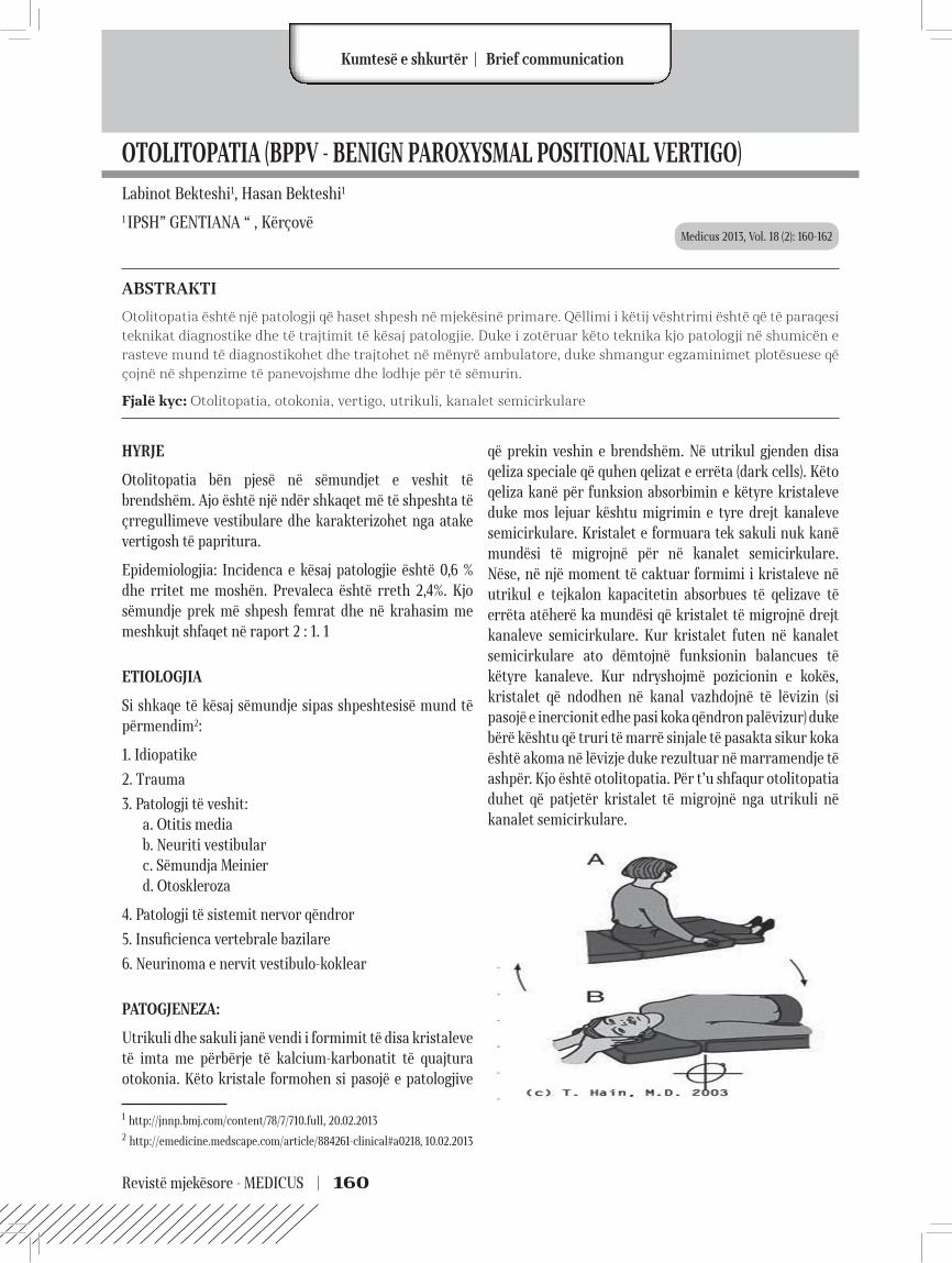
Revistë mjekësore - MEDICUS | 160
OTOLITOPATIA (BPPV - BENIGN PAROXYSMAL POSITIONAL VERTIGO)Labinot Bekteshi1, Hasan Bekteshi1
1 IPSH” GENTIANA “ , Kërçovë
ABSTRAKTI
Otolitopatia është një patologji që haset shpesh në mjekësinë primare. Qëllimi i këtij vështrimi është që të paraqesi teknikat diagnostike dhe të trajtimit të kësaj patologjie. Duke i zotëruar këto teknika kjo patologji në shumicën e rasteve mund të diagnostikohet dhe trajtohet në mënyrë ambulatore, duke shmangur egzaminimet plotësuese që çojnë në shpenzime të panevojshme dhe lodhje për të sëmurin.
Fjalë kyc: Otolitopatia, otokonia, vertigo, utrikuli, kanalet semicirkulare
HYRJE
Otolitopatia bën pjesë në sëmundjet e veshit të brendshëm. Ajo është një ndër shkaqet më të shpeshta të çrregullimeve vestibulare dhe karakterizohet nga atake vertigosh të papritura.
Epidemiologjia: Incidenca e kësaj patologjie është 0,6 % dhe rritet me moshën. Prevaleca është rreth 2,4%. Kjo sëmundje prek më shpesh femrat dhe në krahasim me meshkujt shfaqet në raport 2 : 1. 1
ETIOLOGJIA
Si shkaqe të kësaj sëmundje sipas shpeshtesisë mund të përmendim2:
1. Idiopatike
2. Trauma
3. Patologji të veshit: a. Otitis media b. Neuriti vestibular c. Sëmundja Meinier d. Otoskleroza
4. Patologji të sistemit nervor qëndror
5. Insuficienca vertebrale bazilare
6. Neurinoma e nervit vestibulo-koklear
PATOGJENEZA:
Utrikuli dhe sakuli janë vendi i formimit të disa kristaleve të imta me përbërje të kalcium-karbonatit të quajtura otokonia. Këto kristale formohen si pasojë e patologjive
1 http://jnnp.bmj.com/content/78/7/710.full, 20.02.20132 http://emedicine.medscape.com/article/884261-clinical#a0218, 10.02.2013
që prekin veshin e brendshëm. Në utrikul gjenden disa qeliza speciale që quhen qelizat e errëta (dark cells). Këto qeliza kanë për funksion absorbimin e këtyre kristaleve duke mos lejuar kështu migrimin e tyre drejt kanaleve semicirkulare. Kristalet e formuara tek sakuli nuk kanë mundësi të migrojnë për në kanalet semicirkulare. Nëse, në një moment të caktuar formimi i kristaleve në utrikul e tejkalon kapacitetin absorbues të qelizave të errëta atëherë ka mundësi që kristalet të migrojnë drejt kanaleve semicirkulare. Kur kristalet futen në kanalet semicirkulare ato dëmtojnë funksionin balancues të këtyre kanaleve. Kur ndryshojmë pozicionin e kokës, kristalet që ndodhen në kanal vazhdojnë të lëvizin (si pasojë e inercionit edhe pasi koka qëndron palëvizur) duke bërë kështu që truri të marrë sinjale të pasakta sikur koka është akoma në lëvizje duke rezultuar në marramendje të ashpër. Kjo është otolitopatia. Për t’u shfaqur otolitopatia duhet që patjetër kristalet të migrojnë nga utrikuli në kanalet semicirkulare.
Medicus 2013, Vol. 18 (2): 160-162
Kumtesë e shkurtër | Brief communication
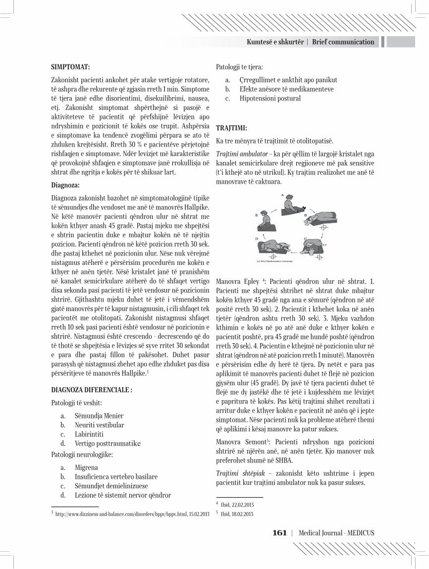
Kumtesë e shkurtër | Brief communication
161 | Medical Journal - MEDICUS
SIMPTOMAT:
Zakonisht pacienti ankohet për atake vertigoje rotatore, të ashpra dhe rekurente që zgjasin rreth 1 min. Simptome të tjera janë edhe disorientimi, disekuilibrimi, nausea, etj. Zakonisht simptomat shpërthejnë si pasojë e aktiviteteve të pacientit që përfshijnë lëvizjen apo ndryshimin e pozicionit të kokës ose trupit. Ashpërsia e simptomave ka tendencë zvogëlimi përpara se ato të zhduken krejtësisht. Rreth 30 % e pacientëve përjetojnë rishfaqjen e simptomave. Ndër levizjet më karakteristike që provokojnë shfaqjen e simptomave janë rrokullisja në shtrat dhe ngritja e kokës për të shikuar lart.
Diagnoza:
Diagnoza zakonisht bazohet në simptomatologjinë tipike të sëmundjes dhe vendoset me anë të manovrës Hallpike. Në këtë manovër pacienti qëndron ulur në shtrat me kokën kthyer anash 45 gradë. Pastaj mjeku me shpejtësi e shtrin pacientin duke e mbajtur kokën në të njejtin pozicion. Pacienti qëndron në këtë pozicion rreth 30 sek. dhe pastaj kthehet në pozicionin ulur. Nëse nuk vërejmë nistagmus atëherë e përsërisim procedurën me kokën e kthyer në anën tjetër. Nësë kristalet janë të pranishëm në kanalet semicirkulare atëherë do të shfaqet vertigo disa sekonda pasi pacienti të jetë vendosur në pozicionin shtrirë. Gjithashtu mjeku duhet të jetë i vëmendshëm gjatë manovrës për të kapur nistagmusin, i cili shfaqet tek pacientët me otolitopati. Zakonisht nistagmusi shfaqet rreth 10 sek pasi pacienti është vendosur në pozicionin e shtrirë. Nistagmusi është crescendo - decrescendo që do të thotë se shpejtësia e lëvizjes së syve rritet 30 sekondat e para dhe pastaj fillon të pakësohet. Duhet pasur parasysh që nistagmusi zbehet apo edhe zhduket pas disa përsëritjeve të manovrës Hallpike.3
DIAGNOZA DIFERENCIALE :
Patologji të veshit:
a. Sëmundja Menierb. Neuriti vestibularc. Labirintitid. Vertigo posttraumatike
Patologji neurologjike:
a. Migrenab. Insuficienca vertebro basilarec. Sëmundjet demielinizuesed. Lezione të sistemit nervor qëndror
3 http://www.dizziness-and-balance.com/disorders/bppv/bppv.html, 15.02.2013
Patologji te tjera:
a. Çrregullimet e ankthit apo panikutb. Efekte anësore të medikamentevec. Hipotensioni postural
TRAJTIMI:
Ka tre mënyra të trajtimit të otolitopatisë.
Trajtimi ambulator – ka për qëllim të largojë kristalet nga kanalet semicirkulare drejt regjioneve më pak sensitive (t’i kthejë ato në utrikul). Ky trajtim realizohet me anë të manovrave të caktuara.
Manovra Epley 4: Pacienti qëndron ulur në shtrat. 1. Pacienti me shpejtësi shtrihet në shtrat duke mbajtur kokën kthyer 45 gradë nga ana e sëmurë (qëndron në atë positë rreth 30 sek). 2. Pacientit i kthehet koka në anën tjetër (qëndron ashtu rreth 30 sek). 3. Mjeku vazhdon kthimin e kokës në po atë anë duke e kthyer kokën e pacientit poshtë, pra 45 gradë me hundë poshtë (qëndron rreth 30 sek). 4. Pacientin e kthejmë në pozicionin ulur në shtrat (qëndron në atë pozicion rreth 1 minutë). Manovrën e përsërisim edhe dy herë të tjera. Dy netët e para pas aplikimit të manovrës pacienti duhet të flejë në pozicion gjysëm ulur (45 gradë). Dy javë të tjera pacienti duhet të flejë me dy jastëkë dhe të jetë i kujdesshëm me lëvizjet e papritura të kokës. Pas këtij trajtimi shihet rezultati i arritur duke e kthyer kokën e pacientit në anën që i jepte simptomat. Nëse pacienti nuk ka probleme atëherë themi që aplikimi i kësaj manovre ka patur sukses.
Manovra Semont5: Pacienti ndryshon nga pozicioni shtrirë në njërën anë, në anën tjetër. Kjo manover nuk preferohet shumë në SHBA.
Trajtimi shtëpiak – zakonisht këto ushtrime i jepen pacientit kur trajtimi ambulator nuk ka pasur sukses.
4 Ibid, 22.02.20135 Ibid, 18.02.2013
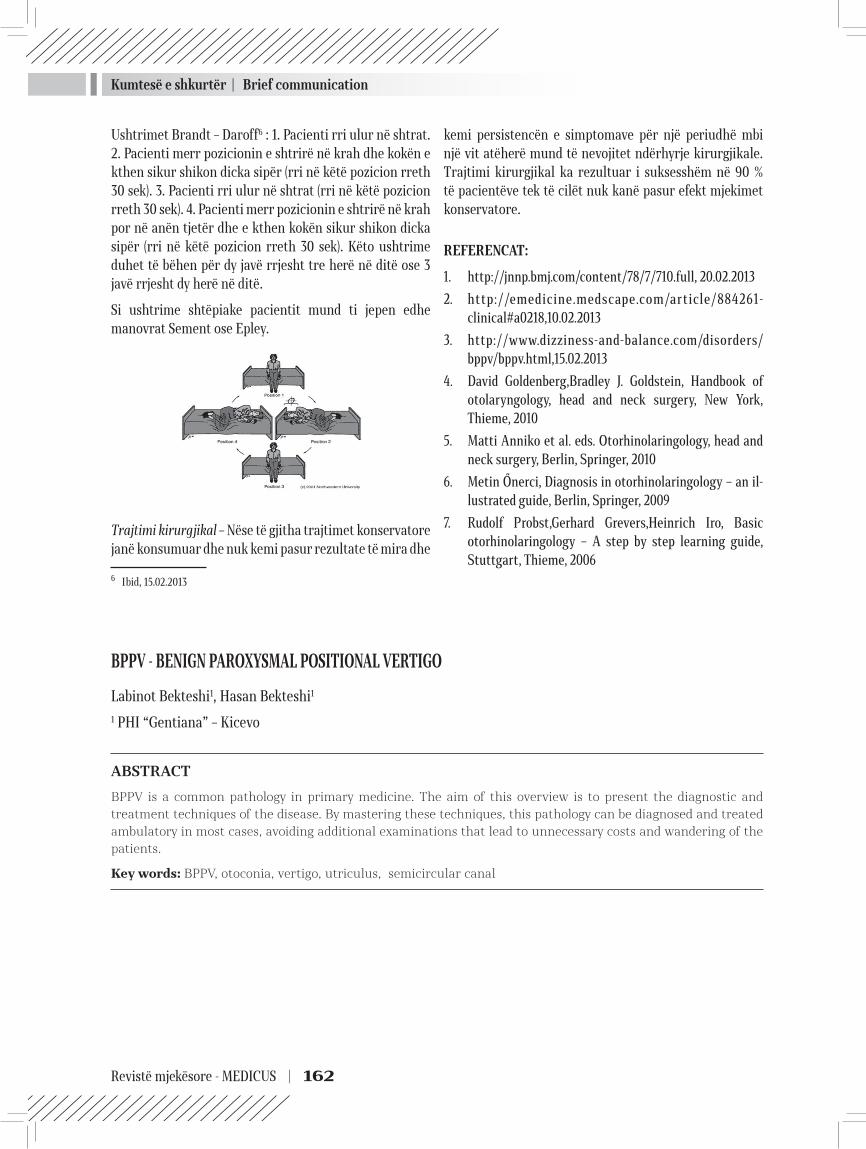
Kumtesë e shkurtër | Brief communication
Revistë mjekësore - MEDICUS | 162
Ushtrimet Brandt – Daroff6 : 1. Pacienti rri ulur në shtrat. 2. Pacienti merr pozicionin e shtrirë në krah dhe kokën e kthen sikur shikon dicka sipër (rri në këtë pozicion rreth 30 sek). 3. Pacienti rri ulur në shtrat (rri në këtë pozicion rreth 30 sek). 4. Pacienti merr pozicionin e shtrirë në krah por në anën tjetër dhe e kthen kokën sikur shikon dicka sipër (rri në këtë pozicion rreth 30 sek). Këto ushtrime duhet të bëhen për dy javë rrjesht tre herë në ditë ose 3 javë rrjesht dy herë në ditë.
Si ushtrime shtëpiake pacientit mund ti jepen edhe manovrat Sement ose Epley.
Trajtimi kirurgjikal – Nëse të gjitha trajtimet konservatore janë konsumuar dhe nuk kemi pasur rezultate të mira dhe
6 Ibid, 15.02.2013
kemi persistencën e simptomave për një periudhë mbi një vit atëherë mund të nevojitet ndërhyrje kirurgjikale. Trajtimi kirurgjikal ka rezultuar i suksesshëm në 90 % të pacientëve tek të cilët nuk kanë pasur efekt mjekimet konservatore.
REFERENCAT:
1. http://jnnp.bmj.com/content/78/7/710.full, 20.02.20132. http://emedicine.medscape.com/article/884261-
clinical#a0218,10.02.20133. http://www.dizziness-and-balance.com/disorders/
bppv/bppv.html,15.02.20134. David Goldenberg,Bradley J. Goldstein, Handbook of
otolaryngology, head and neck surgery, New York, Thieme, 2010
5. Matti Anniko et al. eds. Otorhinolaringology, head and neck surgery, Berlin, Springer, 2010
6. Metin Őnerci, Diagnosis in otorhinolaringology – an il-lustrated guide, Berlin, Springer, 2009
7. Rudolf Probst,Gerhard Grevers,Heinrich Iro, Basic otorhinolaringology – A step by step learning guide, Stuttgart, Thieme, 2006
BPPV - BENIGN PAROXYSMAL POSITIONAL VERTIGO
Labinot Bekteshi1, Hasan Bekteshi1
1 PHI “Gentiana” – Kicevo
ABSTRACT
BPPV is a common pathology in primary medicine. The aim of this overview is to present the diagnostic and treatment techniques of the disease. By mastering these techniques, this pathology can be diagnosed and treated ambulatory in most cases, avoiding additional examinations that lead to unnecessary costs and wandering of the patients.
Key words: BPPV, otoconia, vertigo, utriculus, semicircular canal
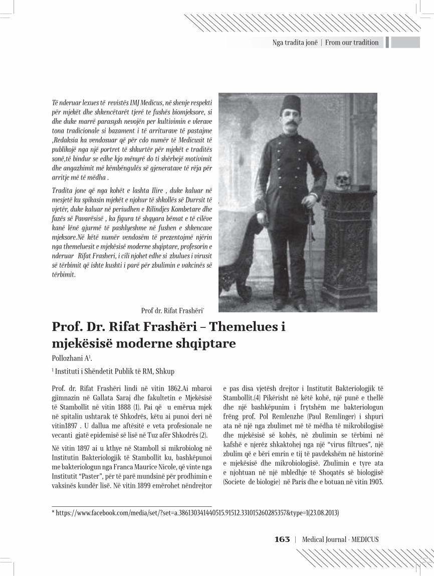
Nga tradita jonë | From our tradition
163 | Medical Journal - MEDICUS
Të nderuar lexues të revistës IMJ Medicus, në shenje respekti për mjekët dhe shkencëtarët tjerë te fushës biomjeksore, si dhe duke marrë parasysh nevojën per kultivimin e vlerave tona tradicionale si bazament i të arriturave të pastajme ,Redaksia ka vendosuar që për cdo numër të Medicusit të publikojë nga një portret të shkurtër për mjekët e traditës sonë,të bindur se edhe kjo mënyrë do ti shërbejë motivimit dhe angazhimit më këmbëngulës së gjeneratave të rëja për arritje më të mëdha .
Tradita jone që nga kohët e lashta Ilire , duke kaluar në mesjetë ku spikasin mjekët e njohur të shkollës së Durrsit të vjetër, duke kaluar në periudhen e Rilindjes Kombetare dhe fazës së Pavarësisë , ka figura të shqyara bëmat e të cilëve kanë lënë gjurmë të pashlyeshme në fushen e shkencave mjeksore.Në këtë numër vendosëm të prezentojmë njërin nga themeluesit e mjekësisë moderne shqiptare, profesorin e nderuar Rifat Frasheri, i cili njohet edhe si zbulues i virusit së tërbimit që ishte kushti i parë për zbulimin e vakcinës së tërbimit.
e pas disa vjetësh drejtor i Institutit Bakteriologjik të Stambollit.(4) Pikërisht në këtë kohë, një punë e thellë dhe një bashkëpunim i frytshëm me bakteriologun frëng prof. Pol Remlenzhe (Paul Remlinger) i shpuri ata në një nga zbulimet më të mëdha të mikrobilogjisë dhe mjekësisë së kohës, në zbulimin se tërbimi në kafshë e njerëz shkaktohej nga një “virus filtrues”, një zbulim që e bëri emrin e tij të pavdekshëm në historinë e mjekësisë dhe mikrobiologjisë. Zbulimin e tyre ata e njohtuan në një mbledhje të Shoqatës së biologjisë (Societe de biologie) në Paris dhe e botuan në vitin 1903.
Prof. Dr. Rifat Frashëri – Themelues i mjekësisë moderne shqiptarePollozhani A1.1 Instituti i Shëndetit Publik të RM, Shkup
Prof. dr. Rifat Frashëri lindi në vitin 1862.Ai mbaroi gjimnazin në Gallata Saraj dhe fakultetin e Mjekësisë të Stambollit në vitin 1888 (1). Pai që u emërua mjek në spitalin ushtarak të Shkodrës, këtu ai punoi deri në vitin1897 . U dallua me aftësitë e veta profesionale ne vecanti gjatë epidemisë së lisë në Tuz afër Shkodrës (2).
Në vitin 1897 ai u kthye në Stamboll si mikrobiolog në Institutin Bakteriologjik të Stambollit ku, bashkëpunoi me bakteriologun nga Franca Maurice Nicole, që vinte nga Institutit “Paster”, për të parë mundsinë për prodhimin e vaksinës kundër lisë. Në vitin 1899 emërohet nëndrejtor
Prof dr. Rifat Frashëri*
* https://www.facebook.com/media/set/?set=a.386130341440515.91512.331015260285357&type=1(23.08.2013)
Nga tradita jonë | From our tradition
Revistë mjekësore - MEDICUS | 164
Pas kësaj R. Frashëri u emërua profesor në Universitetin e Stambollit. Për një periudhë ai shërbeu edhe si kryetar i organizatëspatriotike shqiptare “Bashkimi” të Stambollit. Në këtë kohë prej tij u kërkiua të angazhohet , nëpërmjet të lifdhjeve të tij dhe me autoritetin që kishtë, në kampanjën kundër pavarësisë së Shqipërise, gjë që ai nuk e pranoi. Për këtë qëndrim dhe për aktivitetin e tij patriotik prof. dr. Rifat Frashëri, në vitin 1913, u përjashtua nga Universiteti i Stambollit. Për disa kohë ai mbeti i papunë e pastaj u dërgua në një gjendje gjysmë internimi në Tripolisë e më vonë si kryetar i bashkisë së atij qyteti. Pas Luftës së Parë Botërore, në vitin 1920 me ftesën e qeverisë shqiptare erdhi në Shqipëri dhe u emërua drejtor i përgjithshëm i shëndetësisë pranë qeverise shqiptare,ku dha kontributin e vet ne themelimin e bazës profesiknale dhe infrastruklturote te shëndetësisë shqiptare deri sa doli në pension. Vdiq në Tiranë më 15 nëntor 1934.
Emri i Rifat Frashërit është i lidhur me zbulimin e virusit të tërbimit. Me këtë zbulim shkenca shqiptare për herë të parë në historinë e shkencës moderne del në ballin më të përparuar të kërkimit shkencor eksperimental e dha një kontribut udhëhapës, që nuk mund të ënvlerësohet, në zhvillimin e mikrobiologjisë. Rëndësia reale e këtij zbulimi nuk mund të kuptohet jashtë kuadrit të përgjithshëm të zhvillimit të mikrobiologjisë dhe biologjisë nga fundi i shekullit të kaluar dhe fillimi i shekullit tonë.
Në të tilla rrethana, në vitin 1903, vetëm 5 vjet pas zbulimit të të parit virus, shkaktar të një sëmundjeje infektive te kafshët (virusi i aftës epizootike të gjedit) nga Loffler dhe Frosch dhe tre vjet pas zbulimit të virusi të etheve të verdha, në Institutin Bakteriologjik të Stambollit R. Frashëri dhe Pol Remlenzhe zbuluan të parin virus që shkaktonte sëmundje në njerëz e në kafshë, virusin e tërbimit(4).
Zbulimi nxiti kërkime intensive edhe për gjetjen e shkaktarëve të tjerë viralë të sëmundjeve infektive të njeriut. Zbuluesit Rifat Frashëri dhe Pol Remlenzhe krahas Valter Reed etj., hodhën themelet e virologjisë mjekësore. Zbulimit peshën ia shton fakti që ky u realizua në Institutin Bakteriologjik të Stambollit, një qendër pa tradita të shquara shkencore dhe larg qendrave të tilla të mëdha të kërkimeve mikrobiologjike si ato të Francës dhe Gjermanisë së asaj kohe. Mungesa e një mjedisi shkencor të standardit të kohës në këtë Institut nxjerr më në pah madhështinë e zbulimit dhe njohuritë e aftësitë e zbuluesve R. Frashëri dhe P. Remlenzhe.
Zbulimi i Rifat Frashërit është një nga kontributet më të mëdha të shkencës shqiptare në zhvillimin e shkencës botërore. Karriera shkencore e R. Frashërit u ndërpre herët, për shkak të ndjekjeve nga autoritetet turke (4). Edhe me kthimin në Atdhe, ai nuk gjeti asnjë kusht të përshtatshëm pune në fushën e mikrobiologjisë, ndërsa projekti i tij për një qendër shqiptare të kërkimeve mjekësore, mbeti vetëm një ëndërr e bukur gjatë gjithë jetës së tij.
Ndënësë në shkallë ndërkombëtare ai njihet si një nga themeluesit e virologjisë mjekësore, Prof. Frashëri,me plot të drejë, njëherit, cilësohet edhe si themeluesi I mjekësisë modern shqiptare.
Literatura:
1. AASHK:Kosova(Veshtrim monografik)2011, Prishtine
2. ASHSH: Fjalor enciklopedik shqiptar 1-2 ,Tirane
3. Pollozhani A.:Shendeti publik, 2013, Prishtine
4. https://groups.google.com/forum/#!topic/bit.listserv.albanian/-qhQDWwL5Vo (23.08.2013)
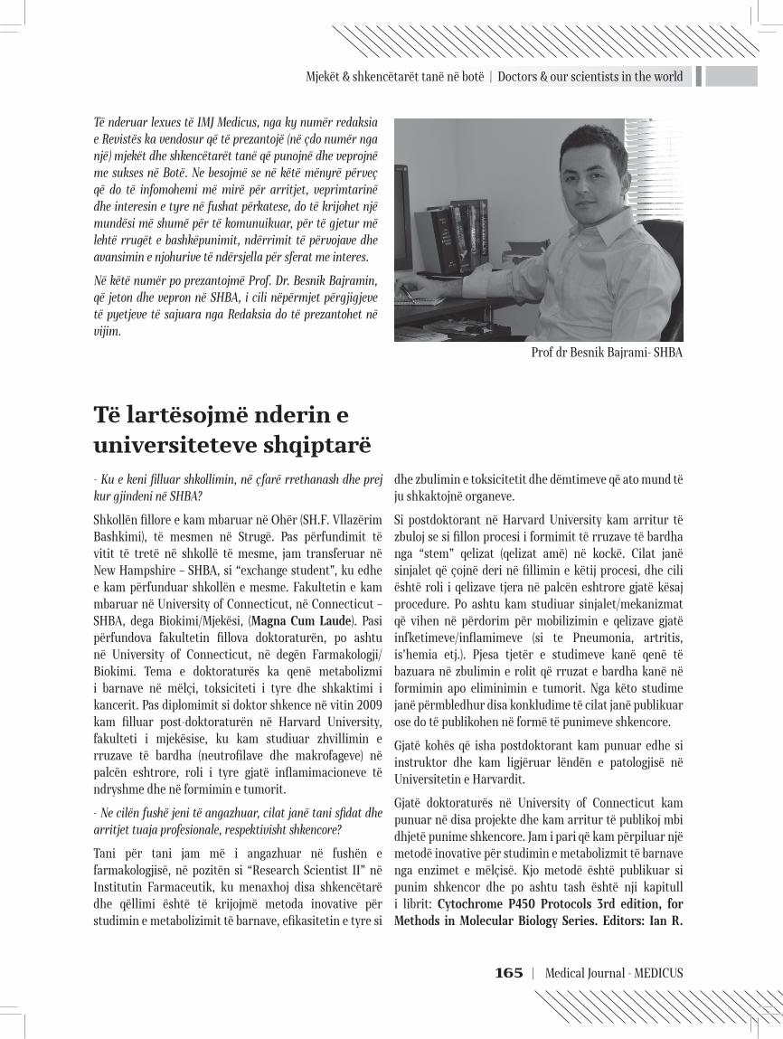
Mjekët & shkencëtarët tanë në botë | Doctors & our scientists in the world
165 | Medical Journal - MEDICUS
Të nderuar lexues të IMJ Medicus, nga ky numër redaksia e Revistës ka vendosur që të prezantojë (në çdo numër nga një) mjekët dhe shkencëtarët tanë që punojnë dhe veprojnë me sukses në Botë. Ne besojmë se në këtë mënyrë përveç që do të infomohemi më mirë për arritjet, veprimtarinë dhe interesin e tyre në fushat përkatese, do të krijohet një mundësi më shumë për të komunuikuar, për të gjetur më lehtë rrugët e bashkëpunimit, ndërrimit të përvojave dhe avansimin e njohurive të ndërsjella për sferat me interes.
Në këtë numër po prezantojmë Prof. Dr. Besnik Bajramin, që jeton dhe vepron në SHBA, i cili nëpërmjet përgjigjeve të pyetjeve të sajuara nga Redaksia do të prezantohet në vijim.
- Ku e keni filluar shkollimin, në çfarë rrethanash dhe prej kur gjindeni në SHBA?
Shkollën fillore e kam mbaruar në Ohër (SH.F. Vllazërim Bashkimi), të mesmen në Strugë. Pas përfundimit të vitit të tretë në shkollë të mesme, jam transferuar në New Hampshire – SHBA, si “exchange student”, ku edhe e kam përfunduar shkollën e mesme. Fakultetin e kam mbaruar në University of Connecticut, në Connecticut – SHBA, dega Biokimi/Mjekësi, (Magna Cum Laude). Pasi përfundova fakultetin fillova doktoraturën, po ashtu në University of Connecticut, në degën Farmakologji/Biokimi. Tema e doktoraturës ka qenë metabolizmi i barnave në mëlçi, toksiciteti i tyre dhe shkaktimi i kancerit. Pas diplomimit si doktor shkence në vitin 2009 kam filluar post-doktoraturën në Harvard University, fakulteti i mjekësise, ku kam studiuar zhvillimin e rruzave të bardha (neutrofilave dhe makrofageve) në palcën eshtrore, roli i tyre gjatë inflamimacioneve të ndryshme dhe në formimin e tumorit.
- Ne cilën fushë jeni të angazhuar, cilat janë tani sfidat dhe arritjet tuaja profesionale, respektivisht shkencore?
Tani për tani jam më i angazhuar në fushën e farmakologjisë, në pozitën si “Research Scientist II” në Institutin Farmaceutik, ku menaxhoj disa shkencëtarë dhe qëllimi është të krijojmë metoda inovative për studimin e metabolizimit të barnave, efikasitetin e tyre si
dhe zbulimin e toksicitetit dhe dëmtimeve që ato mund të ju shkaktojnë organeve.
Si postdoktorant në Harvard University kam arritur të zbuloj se si fillon procesi i formimit të rruzave të bardha nga “stem” qelizat (qelizat amë) në kockë. Cilat janë sinjalet që çojnë deri në fillimin e këtij procesi, dhe cili është roli i qelizave tjera në palcën eshtrore gjatë kësaj procedure. Po ashtu kam studiuar sinjalet/mekanizmat që vihen në përdorim për mobilizimin e qelizave gjatë infketimeve/inflamimeve (si te Pneumonia, artritis, is’hemia etj.). Pjesa tjetër e studimeve kanë qenë të bazuara në zbulimin e rolit që rruzat e bardha kanë në formimin apo eliminimin e tumorit. Nga këto studime janë përmbledhur disa konkludime të cilat janë publikuar ose do të publikohen në formë të punimeve shkencore.
Gjatë kohës që isha postdoktorant kam punuar edhe si instruktor dhe kam ligjëruar lëndën e patologjisë në Universitetin e Harvardit.
Gjatë doktoraturës në University of Connecticut kam punuar në disa projekte dhe kam arritur të publikoj mbi dhjetë punime shkencore. Jam i pari që kam përpiluar një metodë inovative për studimin e metabolizmit të barnave nga enzimet e mëlçisë. Kjo metodë është publikuar si punim shkencor dhe po ashtu tash është nji kapitull i librit: Cytochrome P450 Protocols 3rd edition, for Methods in Molecular Biology Series. Editors: Ian R.
Të lartësojmë nderin e universiteteve shqiptarë
Prof dr Besnik Bajrami- SHBA
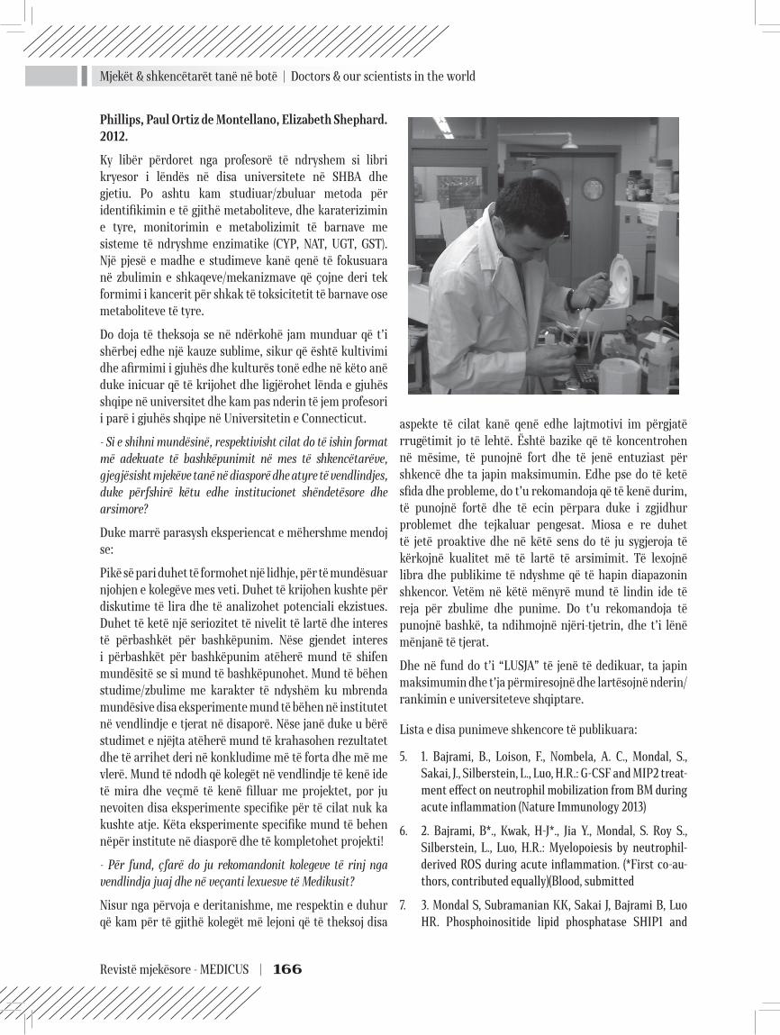
Mjekët & shkencëtarët tanë në botë | Doctors & our scientists in the world
Revistë mjekësore - MEDICUS | 166
Phillips, Paul Ortiz de Montellano, Elizabeth Shephard. 2012.
Ky libër përdoret nga profesorë të ndryshem si libri kryesor i lëndës në disa universitete në SHBA dhe gjetiu. Po ashtu kam studiuar/zbuluar metoda për identifikimin e të gjithë metaboliteve, dhe karaterizimin e tyre, monitorimin e metabolizimit të barnave me sisteme të ndryshme enzimatike (CYP, NAT, UGT, GST). Një pjesë e madhe e studimeve kanë qenë të fokusuara në zbulimin e shkaqeve/mekanizmave që çojne deri tek formimi i kancerit për shkak të toksicitetit të barnave ose metaboliteve të tyre.
Do doja të theksoja se në ndërkohë jam munduar që t’i shërbej edhe një kauze sublime, sikur që është kultivimi dhe afirmimi i gjuhës dhe kulturës tonë edhe në këto anë duke inicuar që të krijohet dhe ligjërohet lënda e gjuhës shqipe në universitet dhe kam pas nderin të jem profesori i parë i gjuhës shqipe në Universitetin e Connecticut.
- Si e shihni mundësinë, respektivisht cilat do të ishin format më adekuate të bashkëpunimit në mes të shkencëtarëve, gjegjësisht mjekëve tanë në diasporë dhe atyre të vendlindjes, duke përfshirë këtu edhe institucionet shëndetësore dhe arsimore?
Duke marrë parasysh eksperiencat e mëhershme mendoj se:
Pikë së pari duhet të formohet një lidhje, për të mundësuar njohjen e kolegëve mes veti. Duhet të krijohen kushte për diskutime të lira dhe të analizohet potenciali ekzistues. Duhet të ketë një seriozitet të nivelit të lartë dhe interes të përbashkët për bashkëpunim. Nëse gjendet interes i përbashkët për bashkëpunim atëherë mund të shifen mundësitë se si mund të bashkëpunohet. Mund të bëhen studime/zbulime me karakter të ndyshëm ku mbrenda mundësive disa eksperimente mund të bëhen në institutet në vendlindje e tjerat në disaporë. Nëse janë duke u bërë studimet e njëjta atëherë mund të krahasohen rezultatet dhe të arrihet deri në konkludime më të forta dhe më me vlerë. Mund të ndodh që kolegët në vendlindje të kenë ide të mira dhe veçmë të kenë filluar me projektet, por ju nevoiten disa eksperimente specifike për të cilat nuk ka kushte atje. Këta eksperimente specifike mund të behen nëpër institute në diasporë dhe të kompletohet projekti!
- Për fund, çfarë do ju rekomandonit kolegeve të rinj nga vendlindja juaj dhe në veçanti lexuesve të Medikusit?
Nisur nga përvoja e deritanishme, me respektin e duhur që kam për të gjithë kolegët më lejoni që të theksoj disa
aspekte të cilat kanë qenë edhe lajtmotivi im përgjatë rrugëtimit jo të lehtë. Është bazike që të koncentrohen në mësime, të punojnë fort dhe të jenë entuziast për shkencë dhe ta japin maksimumin. Edhe pse do të ketë sfida dhe probleme, do t’u rekomandoja që të kenë durim, të punojnë fortë dhe të ecin përpara duke i zgjidhur problemet dhe tejkaluar pengesat. Miosa e re duhet të jetë proaktive dhe në këtë sens do të ju sygjeroja të kërkojnë kualitet më të lartë të arsimimit. Të lexojnë libra dhe publikime të ndyshme që të hapin diapazonin shkencor. Vetëm në këtë mënyrë mund të lindin ide të reja për zbulime dhe punime. Do t’u rekomandoja të punojnë bashkë, ta ndihmojnë njëri-tjetrin, dhe t’i lënë mënjanë të tjerat.
Dhe në fund do t’i “LUSJA” të jenë të dedikuar, ta japin maksimumin dhe t’ja përmiresojnë dhe lartësojnë nderin/rankimin e universiteteve shqiptare.
Lista e disa punimeve shkencore të publikuara:
5. 1. Bajrami, B., Loison, F., Nombela, A. C., Mondal, S., Sakai, J., Silberstein, L., Luo, H.R.: G-CSF and MIP2 treat-ment effect on neutrophil mobilization from BM during acute inflammation (Nature Immunology 2013)
6. 2. Bajrami, B*., Kwak, H-J*., Jia Y., Mondal, S. Roy S., Silberstein, L., Luo, H.R.: Myelopoiesis by neutrophil-derived ROS during acute inflammation. (*First co-au-thors, contributed equally)(Blood, submitted
7. 3. Mondal S, Subramanian KK, Sakai J, Bajrami B, Luo HR. Phosphoinositide lipid phosphatase SHIP1 and
Mjekët & shkencëtarët tanë në botë | Doctors & our scientists in the world
167 | Medical Journal - MEDICUS
PTEN coordinate to regulate cell migration and adhe-sion. Mol Biol Cell. 2012 Apr; 23(7):1219-30.
8. 4. Zhao L, Bajrami B, Rusling JF. Rapid LC-MS Drug Me-tabolite Profiling Using Bioreactor Particles. Methods Mol Biol. 2013; 987:129-34
9. 5. Rusling, J.F., Bajrami, B., Zhao, L.: Cytochrome P450 Protocols 3rd edition, for Methods in Molecular Biology Series. Editors: Ian R. Phillips, Paul Ortiz de Montel-lano, Elizabeth Shephard. 2012.
10. 6. Sakai, J.; Li, J.; Subramanian, K.; Bajrami, B.; Mondal, S.; Jia, Y.; Zhong, J.; Ye, K.; Chang, C.; Ho, Y.; Luo, H.: Reac-tive oxygen … key physiological regulatory mechanisms controlling actin dynamics in neutrophils. Immunity, 2012
11. 7. Luo, H.R., and Bajrami, B. Regulation of neutrophil homeostasis by spontaneous apoptotic death. In the book: Neutrophils: Lifespan, Functions and Roles in Dis-ease (Nova Science Publishers, Inc.). 2010.
12. 8. Bajrami, B.; Zhao, L.; Schenkman, J.; Rusling, J.: LC-MS Based Rapid Drug Metabolite Profiling Using
Bioreactors in 96-well plate format. Anal. Chem., 2009, 81, 9921–9929.
13. 9. Krishnan, S.; Hvastkovs, E.; Bajrami, B.; Schenkman, J.; Rusling, J.: Metabolic Toxicity of 4-(methylnitrosamino)-1-(3-pyridyl)-1-butanone (NNK) evaluated using Electro-chemiluminescent Arrays and Human Cyt P450s. Mo-lecular Biosystems. 2009, (2),163-169.
14. 10. Hull, D.; Bajrami, B.; Jansson, I.; Schenkman, J.; Ruls-ing J.: Characterizing Metabolic Inhibition Using Elec-trochemical Enzyme-DNA Biosensors; Anal. Chem. 2009, 81 (2), pp 716–724.
15. 11. Bajrami*, B; Krishnan*, S.; Hvaskos* E. G.; Choudhary, D.; Schenkman, J. B.; Rusling, J. F.: Synergistic Toxicity Screening Using Microsome/DNA Electrochemilumi-nescent Arrays and Nanoreactors; Anal. Chem.; (Ac-celerated Article); 2008; 80(14); 5279-5285. (Cover) (*Au-thors contributed equally.)
16. 12. Bajrami, B.; Krishnan, S.; Rusling J. F.,”Microsome biocolloids for rapid drug metabolism and inhibition assessment by LC-MS,” Drug Metabolism Letters; 2008, 2 (3); 158-162.
Arsimi | Education
Revistë mjekësore - MEDICUS | 168
Mbi rrjеdhat në arsimin tonë
Të nderuar lexues të IMJ Medicus, në perputhje me përcaktimin paraprak, që nëpermjet t rëvistës tonë të afirmojmë edhe aspekte që lidhen me arsimin , si bazë e pashmagshme për arritje në të gjitha fusht tjera, në këte numër pranum letrën e gazetares tonë të mirenjohur Laura PAPRANIKU e cila për shume vjet e ndjek problematiken e arsimit në Maqedni dhe më gëjrë, me të cilën ajo reflekton shqetsimin jo vetëm të saj për rrjedhat në sferen e arsimit që së voni ishin objekt edhe i observimeve dhe vlërsimeve të instrumeteve ndërkomëtare për vlerësimin e cilësisë në arsim. Redaksia e faleminderon autoren e këtij vështrimi duke shpreh gadishmërine për të vazhduar me këte formë të bashkëpunimit, për të cilin jemi të sigurtë që i shërben interesit të vëtetë publik.
Goditja që kanë marrë universitete tona në “Word Rangink” është e rëndë. Keq është pozicionuar deri edhe Universiteti i Tiranës (UT) ai, që në të vërtetë do të duhej të ishte “ajka e shqiptarëve”. Por ja që për shkak të performancave të dobëta në parametrat me të cilat kjo Agjenci matën cilësinë e shkollimit të lartë, UT ka rënë në vendin 8 012 në botë. E krahasuar me UKM-në, ajo gjendet 6 137 vende më poshtë. Edhe në renditjen e universiteteve në nivel të Evropës, UT që është i pari në mesin e universiteteve të Shqipërisë, qëndron shumë poshtë UKM-së. Kjo e fundit është pozicionuar në vendin e 652, kurse UT-ja në pozitën 2 079. Më mirë se Tirana, por jo edhe se Shkupi, qëndron Universiteti i Prishtinës (UP), i cili është renditur i 4 416-ti në klasifikimin e universiteteve në nivel botëror. “Web Ranking” matën cilësinë bazuar në numrin e programeve studimore, numrin e hulumtimeve të botuara në revista me faktorin e impaktit, metodologjinë e studimeve, numrin e të diplomuarve dhe buxhetin që shpenzohet për hulumtime shkencore. I pari në listën e këtij klasifikimi qëndron Universiteti i Harvardit, i pasuar nga Instituti Teknologjik i Masaçutit, Universiteti i Stanfodit, Universitet i Kalifornisë në Berkeley, Universiteti i Kalifornisë në Los Angjellos, Universiteti i Washingtonit, Universiteti i Miçiganit, Universiteti i Kornellit, Universiteti i Miniasotës dhe Universiteti i Pensilvanisë. Është pra një listë që kryesohet nga universitete amerikane dhe angleze dhe që askush nuk kërkon prej universitete shqiptare të bëjë konkurrencë në top 10-shin e vendeve të para. Ajo që kërkohet prej tyre, është të mos gjenden mijëra vende më poshtë se sa universitete e rajonit dhe të mos ndjehen aspak mirë(mbase dikush duhet edhe të skuqet) që në “Word Rangink” janë me nga tetë-nëntë mijë vende më poshtë se Universiteti i Shkupit.
Shqiptarët në përgjithësi e sidomos ato të Maqedonisë duhet urgjentisht të ndërmarrin masa për të përmirësuar cilësinë e institucioneve arsimore. Nuk është e tepërt të iniciohet hartimi i një strategji kombëtare të arsimit apo edhe të kërkohet me ngulm angazhimi aktiv i elitave intelektuale dhe akademike, deri sa mundemi t’i gjejmë ende. Nëse lejojmë që të vazhdohet edhe më tej me trendin e devalvimit të institucioneve arsimore, përfundimisht do të jemi të pa shpresë. Thjeshtë, do të shndërrohemi në një popull me ardhmëri të diskutueshme... Pa njw shkollimi cilësor nuk mund tw synohet përparimi i një populli. Alarmi për pozitën e mjerueshme në të cilën ndodhen shkollat dhe universitete tona tanimë është dhënë dhe assesi nuk duhet lejuar të mbesim edhe më tej në fundin e listave që rendisin institucionet sipas cilësisë.
Sipas rezultateve të maturës shtetërore shkollat e mesme me mësim në gjuhën shqipe, në Maqedoni, janë katandisur si mos më keq. Nota më e lartë mesatare, nga lënda e gjuhës amtare dhe nga një lëndë që nxënësit e zgjedhin vetë, fillon me 3. Nuk janë aspak më të mira rezultatet e universiteteve. Në ranglistën që në muajin gusht u publikua nga Agjiencia prestigjioze e rangimit të shkollave të larta në botë, “Word Rangink”, universitete tona janë pozicionuar në fund të listave. Nga 21 248 shkolla të larta në mbarë botën, dy universitetet e shqiptarëve të Maqedonisë gjenden në vendin e 9 304 (Universiteti i Evropës Juglindore në Tetovë) dhe në vendin 10 894 (Universiteti Shtetëror i Tetovës). Kjo renditje është padyshim tregues i qartë se sa shumë ka devalvuar cilësia në arsimin e lartë. Për dallim prej tyre Universiteti “Shën Kirli dhe Metodi” i Shkupit (UKM), gjendet në vendin e 1878-të, të së njëjtës ranglistë. Që alarmi të jetë më tronditës, në pozitat aspak më të mira gjenden edhe universitetet e Shqipërisë dhe Kosovës.
Arsimi | Education
169 | Medical Journal - MEDICUS
Pasojat nga mos marrja e masave janë drastike. Kështu për shembull, universitete e Shqipërisë kanë pësuar rënie prej rreth 800 vendeve vetëm për një vit. Universiteti i Tiranës, i cili sivjet gjendet i pari në listën e universiteteve të Shqipërisë në klasifikimin botëror, me vendin e 8 012-të, vitin e kaluar radhitej i dyti dhe ishte ne vendin e 8 735 të klasifikimit botëror. Ndërkohë që një vit më parë lista për Shqipërinë kryesohej nga Shkolla e lartë jopublike “Epoka”, në vendin e 7 299-të. Këtë vit, ka zbritur në vendin e 9 874-të në klasifikimin botëror. Në vendin e tretë të listës për universitetet e Shqipërisë gjendet Universiteti Politeknik i Tiranës (10 049), të katërtin Universiteti i “New Yorkut” në Tiranë (10.460), ndërsa në të pestin Universiteti Bujqësor i Kamzës (10 855).
Për universitet “të mira” shqiptarë, padyshim se nuk mund të bëhet fjalë, ashtu sikurse është vështirë të gjendet edhe shkolla të mira në nivelin e arsimit të mesëm dhe fillor. Gjashtë vite, prej se në Maqedoni ka nis të matet cilësia e shkollimit maturës shtetërore apo testeve të vlerësimit ekstren, shkollat me mësim në gjuhën shqipe nuk lëshojnë pozitat e fundit. Asnjëra prej tyre nuk ka arritur të ngjitet në top 5-shen, 10-shen apo
15-shen e shkollave më të mira në Maqedoni. Përkundrazi, shënojnë regres. Gjimnazi “Zef Lush Marku”, që në vitin 2012 ishte ne vendin e 36, sivjet ra 14 vende më poshtë. Konkretisht, ai që dikur ishte krenaria e shqiptarëve të Shkupit, sot gjendet në vendin e 50 të listës me 95 shkolla të mesme publike në Maqedoni. Me këta tregues, që padyshim se janë rezultat i degradimit të vazhdueshëm të arsimit shqip, po vëmë në pikëpyetje ardhmërinë tonë. Me kuadro gjysmë të arsimuar, thjeshtë rrezikojmë mbijetesën tonë kulturore, ekonomike dhe politike. Prandaj është obligimi moral i secilit që të kontribuojnë në mënyrën e vet për ndryshimin e gjendjes së vështirë në të cilën ka rënë arsimi në gjuhën shqipe. Hapi duhet bërë nga vetja, në emër të ardhmërisë. Pa arsim cilësor, pa shkolla të etabluara në sistemin e vlerave evropiane, pa programe kompatibile me nevojat e tregut asnjë popull dhe shtet nuk mund të synojnë zhvillimin dhe përparimin e vetës. Shqiptarët, nuk mund të bëjnë përjashtim, prej rregullave të ditura botërisht. Prandaj, sot o kurrë, nevojitet mobilimizim për ndërtimin e institucioneve të qëndrueshme shkollave dhe universiteteve elite, në të cilat mund të mbështesim ardhmërinë tonë.
Laura PAPRANIKU
Aktivitete | Activity
Revistë mjekësore - MEDICUS | 170
Nga Kongresi i X-të i Shoqatës të Oftalmologëve të Europës Juglindore (SEEOS) dhe Kongresi i III-të i Shoqatës të Oftalmologëve të Maqedonisë
Republika e Maqedonisë pati nderin të organizojë Kongresin e X-të të Shoqatës të Oftalmologëve të Europës Juglindore (SEEOS), sëbashku me Kongresin e III-të të Shoqatës të Oftalmologëve të Maqedonisë me pjesëmarrje ndërkombëtare dhe Simpoziumi i Motrave medicinale në Oftalmologji. Kongresi u mbajtë nga data 20-23 qershor, 2013 në Ohër.
Ky Kongres jubilar SEEOS-it ishte rast i mirë për përmbledhje dhe vlerësime të qëllimeve dhe detyrave që ishin vendosur para SEOOS-it që nga vetë fillimi i formimit në vitin 2004, kur edhe u mbajt Kongresi i I-rë në kryeqytetin e Bosnjës dhe Hercegovinës, Sarajeva, si asociacion i oftalmologëve të 18 shteteve të Europës Juglindore.
Nga prespektiva e distancës historike dhjetëvjeçare, mund të pohojmë se asociacioni e plotësoi qëllimin e tij kryesor- ta përmisojë komunikimin dhe shkëmbimin e ideve të reja shkencore, dijes dhe përvojës ndërmjet oftalmologëve nga vëndet anëtare.
Njikohësisht, SEEOS-i bëri nji hap kolosal duke ndërtuar ura mirëkuptimi, bashkëpunimi dhe miqësie ndërmjet vëndeve të ndryshme e duke mundësuar njoftim me kultura, tradita dhe zakone të reja si dhe mikpritje tradicionale.
Bordin e SEEOS-it e përbëjnë oftalmolog të spikatur të regjionit por edhe më gjërë. Prof.dr.Marko Havlina nga Slovenia, njiherit edhe kryetar, pastaj Prof.dr.Mustafa Sefiq-Bosnje dhe Hercegovinë, dr.Athanasios Nikolakopulos-Greqi, Prof.dr.Petja Vasileva-Bullgari, Prof.dr. Sllobodanka Latinoviq-Serbi, Prof.dr.Nikica Gabriq-Kroaci, Prof.dr. Janos Nemeth-Hungari, Prof.dr.Cesare Forlini-Itali, Prof.dr.Nadiya Bobrova-Ukrainë, Prof.dr.Pajtim Lutaj-Shqipëri, Prof.dr.Daniela Selaru-Romani, Dr.Titos Christoforidis-Qipro, Dr.Eugeniu Bendelic-Moldavi, Prof.dr. Magdalena Antova-Velevska-Maqedoni, Prof.dr. Gazmend Kaçaniku-Kosovë dhe dr.Edita Files-Bradariq-Mali i Zi.
Ne Ohër ishin prezent mbi 300 mjekë-oftalmolog, optom-etristë, ortoptist, motra medicinale, firma farmaceutike,
gazetarë etj. Nikoqirët, në krye me Prof.dr.Vesna Dimovs-ka-Jordanova, kryetare e Shoqatës të Oftalmologëve të Maqedonisë, treguan nivel të lartë të aftësisë për organi-zim dhe mikpritje të ngrohtë.
Në programin shkencor u prezentuan mbi 50 autorë me punime të ndryshme dhe me sukses u prezentuan gjatë 3 ditëve të kongresit. Ligjeruesit kryesore ishin Prof. Leonidas Zografos nga Zvicra, i cili prezantoj temën “Tumoret e irisit dhe trupit ciliar dhe pseudotumoret”, pastaj Prof. Keith Barton, nga Britania e Madhe, Prof. Juan Duran De la Colina, nga Spanja, Prof. Marie-Jose Tassignon nga Belgjika dhe Prof. Hans Lemij, nga Holanda.
Katarakti, kornea dhe glaukomi ishin tema dominante të ditës të parë. Shumë prezantues dëshmuan se era e operacioneve të kataraktit me metodën e vjetër të ECCE-së (extracapsular cataract extraction) ka marrë fund dhe se konverzioni në metodën e Phaco kirurgjisë (me ultratingull), ku hapja e kornesë është zvogëluar nga 10 mm në 3.2mm ose deri në 1.8 mm, është realizuar me përjashtime të vogla. Aplikimi i lazerit në fushën e kataraktit po merr hov të madh dhe se siguria e operacionit është rritur dukshëm e njikohësisht numri i komplikacioneve është zvogëluar në minimum.
Në fushën e glaukomit u fol për rëndësinë e prevencës të kësaj sëmundje të rëndë si dhe risit në kombinimin e terapisë për stabilizimin e tensionit intraokular. Kirurgjia e glaukomit ishte temë që zgjoi interes. U prezentuan punime me tema për inplantimin e shanteve moderne. Kornea dhe transplantimi i kornesë poashtu kishte punime të jashtëzakonshme. Transplantimi i kornesë u tregua si operacion i rëndësishëm për zbutjen e verbërimit korneal.
Ditën e dytë temat që u prezentuan ishin nga fusha e retinës medicinal dhe kirurgjisë vitreoretinale si dhe oftalmologjia pediatrike e strabizmi. Retinopatia diabetike mbetet edhe më tej problem i rëndë dhe me ndikim të madh në rritjen e përqindjes të verbërimit te popullata e moshuar. Rëndësia e skriningut dhe prevencës zuri vend në diskutimet e gjata dhe kualitative midis pjesmarrësve. U vërejt se edhe terapia me anti-VEGF dhe
Aktivitete | Activity
171 | Medical Journal - MEDICUS
lazerfotokoagulacion është zhvilluar me hapa të shpejta dhe kolosale. Poashtu edhe operacionet vitreoretinale nuk janë më si më parë. Rezultatet e operacioneve të ablacionit, sidomos ato të njollës të verdhë janë evidente dhe shpresdhënëse. Për këtë meritë kanë edhe aparatet e fuqishme që i posedojnë disa klinika siç është edhe ajo e Shkupit.
Oftalmologjia pediatrike dhe strabizmi poashtu zunë vend në këtë eveniment. Për here të pare u mbajt edhe Simpozium për Retinopatinë Prematurike me insistim të prof.Nemeth nga Hungaria. Kjo sëmundje është e re dhe shkaktare kryesore për verbërimin e fëmijëve në shtetet në zhvillim. Temat e prezentuara treguan statistika që janë brengosëse për ardhmërinë e kësaj kategorie të fëmijëve, por edhe më tej skriningu dhe lazerfotokoagulacioni janë standard të arta për prevenimin dhe mjekimin e retinopatisë prematurike.
Strabizmi dhe operacionet e strabizmit poashtu zgjuan interes. Skriningu i amblopisë u tregua mjaft i dobishëm. Kroacia tregoi se me organizim dhe pak mjete mund të parandalohet ky armik i zhvillimit të shikimit te fëmijët. Operacionet e strabizmit përjetojnë rilindje. Hapja e konjunktivës nga 5 deri në 10mm dëshmuan rezultate të mira por edhe komfor më të madh te pacientët e operuar.
Në këtë Kongres prezentime kishin edhe specijalizantët dhe oftalmologët e ri, të cilët treguan se janë në hap me kohën dhe trended e oftalmologjisë moderne duke na dhënë shpresë për ardhmëri të mire të kësaj fushe të rëndësishme në mjekësi.
Ditën e fundit Simpozium organizuan edhe Shoqata e Motrave Medicinale në Oftalmologji, ku me sukses prezantuan tema nga sterilizimi dhe mirëmbajtja e instrumenteve oftalmologjike, si dhe kujdesi ndaj pacientëve dhe rëndësia e instrumentareve gjatë operacioneve oftalmologjike.
Gjatë mbylljes të kongresit, nikoqirët pranuan kom-plimente për organizimin perfekt ndërsa prezantuesit urime për nivelin e lartë shkencor të punimeve të tyre që dëshmon se dhe ky regjion përveç në aspect gjeografik duhet të integrohet në Europë ku edhe e ka vendin. Bordi I SEEOS-it shpalli Kongresin e mbyllur dhe informoi se Kongresi I XI-të I SEEOS-it do të mbahet ne Bukuresht të Romanisë, në Tetor të 2014-ës.
Në Shkup, Qershor, 2013.
Ass.dr.Bekim Tateshi
oftalmokirurg
Aktivitete | Activity
Revistë mjekësore - MEDICUS | 172
Nga Konferenca “Lidershipi dhe menaxhimi në shëndetësi”, Strugë, 23-24 Gusht
Muaji Gusht ishte përplot i mbush me aktivitete edukative, seminare, konferenca dhe të ngjashme. Edhe uikendin e kaluar kishim mundësinë që të vizitojmë konferencën nacionale që u organizua nga Shoqata “Blue Cell International” (BCI) në bashkëpunim me Qendrën Rajonale për Edukim Mjekësor “Medica”.
Puna në grup dhe lidershipi janë komponentë të rëndësishëm në praktikën efektive mjekësore. Në të vërtetë, ato janë esenciale në reformat e reja të sistemit të kujdesit shëndetësor. Organizatorët kishin parasysh këtë, andaj kishin spikatur më shumë rolet në lidership që mund të jetë pjesë e karrierës së të rinjve në të ardhmen.
Për të shtuar vëmendjen e studentëve të mjekësisë dhe mjekëve të rinj ndaj punës efektive në grupe dhe lidershipit dhe për të diskutuar shkathtësitë relevante të studentëve që janë gati të diplomojnë dhe të marrin përgjegjësi trajnimi në çdo specializim, BCI dhe Medica, planifikuan dhe mbajtën një organizim nacional në lidership dhe networking më 24 Gusht, në Hotelin Solferino, Strugë.
Si ligjërues në këtë eveniment ishin paraparë Prof. Dr. Azis Pollozhani, Doc. Dr. Arben Taravari, Asst. Dr. Naser Durmishi, Mr. Shkodrane Dardhishta, Dr. Harkan Memedi, Dr. Aida Dalipi, Avdi Murtezani, Pale Budiq dhe të tjerë.
Me një agjendë të pasur, programi filloi me një diskutim interaktiv nga paneli diskutues udhëhequr nga Prof. Azis Pollozhani, si një folës mjaft inspirues, duke i inkurajuar të pranishmit në diskutimin e temës përkatëse. Pas kësaj pasoi networking, ku iu mundësua pjesmarrësve të takojnë kolegë të rinj dhe të shkëmbejnë eksperienca. Të gjithë të pranishmit u ndanë mëse të kënaqur nga ky debat.
Programi vazhdoi me workshopin mbi aftësitë komunikuese, përgatitur dhe prezantuar në mënyrë të shkëlqyer nga Dr. Arben Taravari. Kjo puntori i bëri të pranishmit akoma më të etur për workshope të tilla.
Serialin e workshopeve mjaft të sukseshme e vazhdoi Dr. Naser Durmishi me një prezantim të shkëlqyer nga lëmia e menaxhimit të stresit, duke prezantuar dhe përfshirë ushtrime me situata hipotetike me raportim të fokusuar. Përveç kësaj, disa aktivitete energjizuese dhe rekreacionale u bënë që të rrisin interaksionin dhe dimensionet e bashkëpunimit në takim.
Në orët e paraditës, ishin paraparë të mbahen dy sesione prezantimi: Panairi i Projekteve dhe Mundësitë ndërkombëtare për studentët e mjekësisë dhe mjekët e rinjë. Këto prezantime shtuan kërshërinë dhe dëshirën e studentëve për të shikuar mundësitë e tyre ndërkombëtare.
Dita mbaroi me darkën dhe një varg aktivitetesh shoqërore në qytetin e bukur të Strugës.Për të vlerësuar ndikimin e trajnimit, pjesëmarrësit plotësuan pyetësorët duke i treguar përvojat e tyre në organizimin e bërë.
“Kishim një ide, morëm iniciativë, gjetëm udhëzime të përshtatshme dhe kishim besimin të shihnim përtej. Pranuam këshilla nga ata që kishin më shumë përvojë dhe ekspertizë, dhe i morëm të gjitha përparësitë e të gjitha burimeve të disponueshme për ne, që siç e shihni rezultoi me një rezultat të shkëlqyer.”, tha mes tjerash kryetari i shoqatës, Dr. Besnik Hamiti
“Ishim të privilegjuar të mbështesnim, ndihmonim dhe të shihnim kolegët tanë tek përmirësonin shkathtësitë e tyre bazike dhe vetëbesimin e përgjithshëm pas trajnimit, diskutimet interesante dhe udhëzimet e ekipit lehtësues.”, shtoi sekretari gjeneral, Dr. Sead Zejnel.
Në fund, edhepse çështja e lidershipit dhe shkathtësive për punën në grupe nga ana e studentëve dhe mjekëve të rinj, të cilët më së miri e dijnë a janë zhvilluar aftësitë e tyre, është e rëndësishme që ato të përparojnë dhe ata të marrin përgjegjësitë për ekipet mjekësore.
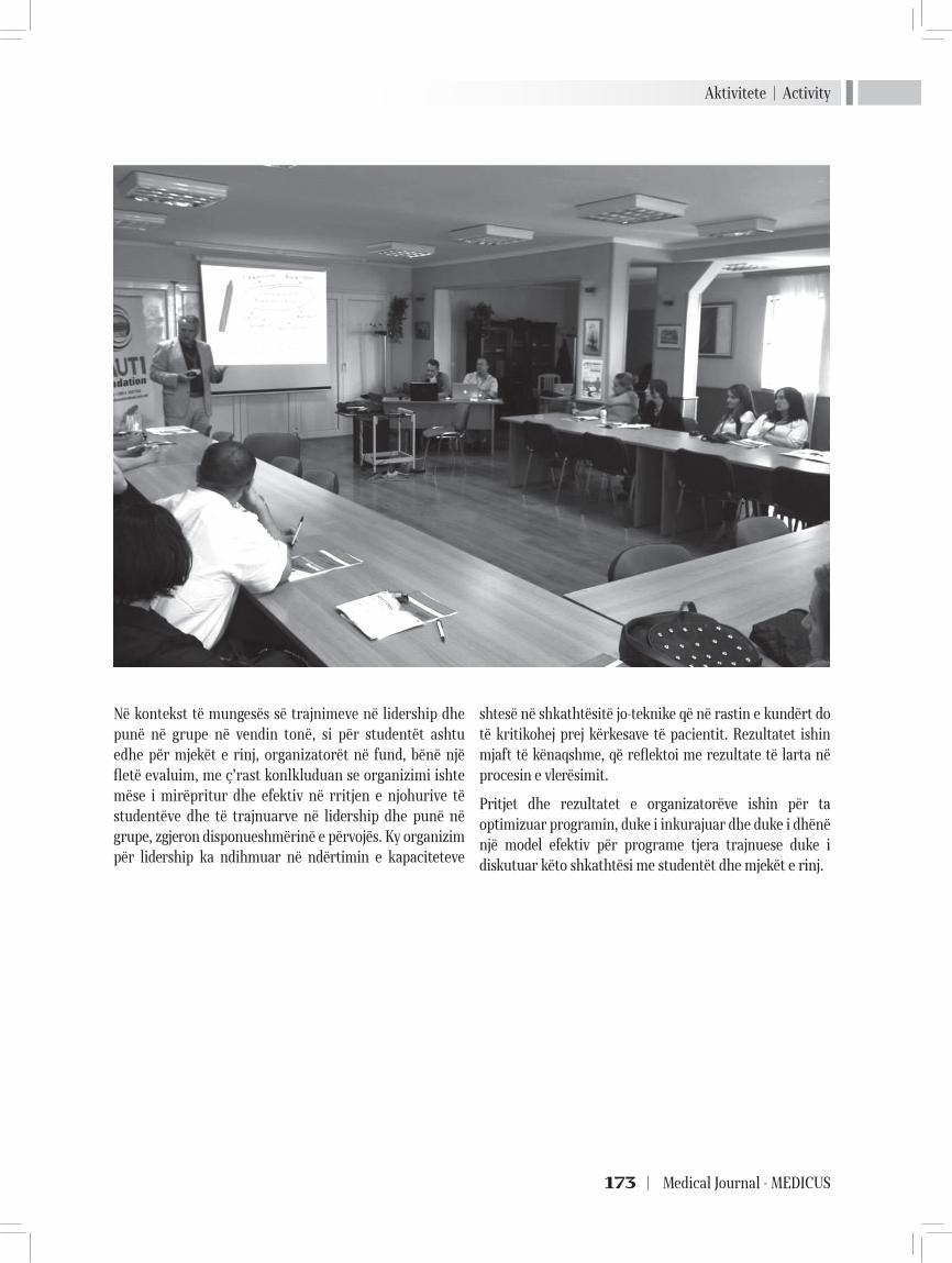
Aktivitete | Activity
173 | Medical Journal - MEDICUS
Në kontekst të mungesës së trajnimeve në lidership dhe punë në grupe në vendin tonë, si për studentët ashtu edhe për mjekët e rinj, organizatorët në fund, bënë një fletë evaluim, me ç’rast konlkluduan se organizimi ishte mëse i mirëpritur dhe efektiv në rritjen e njohurive të studentëve dhe të trajnuarve në lidership dhe punë në grupe, zgjeron disponueshmërinë e përvojës. Ky organizim për lidership ka ndihmuar në ndërtimin e kapaciteteve
shtesë në shkathtësitë jo-teknike që në rastin e kundërt do të kritikohej prej kërkesave të pacientit. Rezultatet ishin mjaft të kënaqshme, që reflektoi me rezultate të larta në procesin e vlerësimit.
Pritjet dhe rezultatet e organizatorëve ishin për ta optimizuar programin, duke i inkurajuar dhe duke i dhënë një model efektiv për programe tjera trajnuese duke i diskutuar këto shkathtësi me studentët dhe mjekët e rinj.
Revistë mjekësore - MEDICUS | 174
IN MEMORIAM
Dr Astrit Saidi (1957-2013)
Dr. Astrit Ismail Saidi u lind më 3 Gusht të vitit 1957 në një familje bujare dhe me traditë kombëtare në Tetovë. Shkollën fillore e kreu mes viteve 1964-1972 në Shkollën Fillore “Liria” në Tetovë, por gjithnjë nën edukimin dhe arsimimin e mbështetur edhe nga i ati , pedagogu i njohur Ismail Saidi. Shkollen e mesme e kreu në vitin 1976 në gjimnazin e Tetovës. Në vitin 1976 u regjistrua në fakultetin e mjekësisë pranë Universitetit të Prishtinës, të cilin e kreu me sukses në vitin 1982. Pas përfundimit të fakultetit të mjekësisë disa vite punoi si mjek i ri në fshatrat e Tetovës që më pas të punësohet si mjek i rregullt në ndihmën e shpejtë në spitalin e Tetovës, ku më vonë u be edhe shef i të njëjtës. Në vitin 1995 e filloi specializimin në degën e mjekesisë interne pranë klinikës universitare të Universitetit “Shen Kirili “ në Shkup. Në vitin 1999 specializoi dhe kaloi të punojë në repartin internistik në spitalin e Tetovës. Dr. Astrit Saidi ka marre pjesë në shumë seminare dhe kongrese të mjejkësisë në vend dhe jashtë vendit dhe posedon mirenjohje të shumta për arritjet dhe sukseset e tij në lëmin e mjeksësisë. Vlen të përmendet mirenjohja nga Kryqi i kuq i Maqedonisë , nga Oda e mjekëve të Maqedonisë , mirenjohjen nga organizata e NATO-s dhënë ambulancës ‘’VITALIS’’ ku edhe kontribonte , mirënjohja nga kryeministri i parë i R.KOSOVËS dhënë për kontributin për kujdesin shëndetësor dhënë të përndjekurve nga Kosova në vitin 1999. Njëkohësisht Dr. Astriti ka shkruar edhe shumë punime shkencore në fushën e mjekesisë si autor dhe si koautor.
Vdekja e parakohshme e Dr Astritit më 12 Korrik 2013, do të len një shpraztirë të përheshme për familjen, koleget dhe miqte e shumtë që i kishte, por kujtimi i tij do të jetë i përjtshëm duke mbetur shembëlltyra e profesionalizmit, ndershmërise dhe korrektësisë ndër gjeneratat që do vijnë.
Udhëzime për autorët | Information for authors
175 | Medical Journal - MEDICUS
UDHËZIME PËR AUTORËT
Këto të dhëna janë në pajtim me “Kërkesat uniforme për Dorëshkrimet e Pranuara në
Revistat Biomjekësore”Dokumentin komplet mund ta gjeni në www.icmje.org)
Medicus është revistë ndërkombëtare që boton punime origjinale shkencore, vështrime revyale, punime profesionale, prezentime rasti, kumtesa të shkurtra, recenzione librash, raporte nga tubime shkencore, letra dhe editoriale nga fusha e mjekësisë, stomatologjisë, farmakologjisë si dhe nga fusha tjera të përaferta bio-mjeksore.
Revista është organ i “Shoqatës së Mjekëve Shqiptarë në Maqedoni.”
Gjuha e botimeve është në Gjuhë Shqipe dhe Angleze (këshilli redaktues mund të vendosë nëse botimet do të jenë edhe në gjuhë tjera). Autorëve u kërkohet të lektorojnë dhe të redaktojnë unimin e tyre vetë, në gjuhën përkatëse.
Ju lutemi përdoreni madhësinë standarde të punimit në format: Word për Windows, Times New Roman 12.
Dorëshkrimet dërgohen në format elektronik, qoftë me
CD ose përmes e-mailit tek Kryeredaktori,Prof. Dr. Azis K. Pollozhani,Zyra e Redaksisë, rr. 50 Divizija, No 6, 1000 Shkup, apo nëe-mail: [email protected]
Revista për një numër pranon jo më shumë se një artikull nga një autor, dhe jo më shumë se dy si ko-autor.
Autorët duhet të deklarojnë se kontributi i tyre nuk është publikuar apo pranuar për publikim diku tjetër, përderisa nuk përfundon procedura vlerësuese ne Revistën tonë.
Autorët gjatë aplikimit duhet të përmbushin formen e kerkuar nga Komiteti Ndërkombëtar i Redaktorëve të Revistave Mjekësore (ICMJE) për kriteret e autorësisë, respektivisht “Kërkesave uniforme për Dorëshkrimet e Pranuara në Revistat Biomjekësore”, cilën mund ta gjeni në www.icmje.org .
Revista do të njoftojë pranimin e artikullit tuaj brenda shtatë ditësh dhe do t’ju bëjë me dije se kur do të informoheni për vendimin e këshillit redaktues.
Artikujt për t’u botuar në Medicus do të recensohen. Këshilli redaktues do të marrë parasysh komentet e recensuesit dhe pastaj mund të kërkojë nga autori ndryshime apo plotësim të punimit.
INFORMATION FOR AUTHORS
These guidelines are in accordance with the “Uniform Requirements for Manuscripts Submitted
to Biomedical Journals”(The complete document appears at www.icmje.org)
Medicus is an international journal of that publishes papers from all areas of medical research. Furthermore, the journal indends to bring educational material of high quality to its members for continuous medical education (CME), by publishing original research, professional and review papers, case reports, brief communications, literature summary articles and editorials.
The Journal is official organ of the »Association of Albanian Medical Doctors from Macedonia«.
The language of publication is Albanian and English (the editorial board may decide whether other language will be used for publications). Authors are requested to have their paper proof-readed and edited for the respective language.
Please use standard-sized paper and submit your article in the following formate: Word for Windows, Times New Roman 12.
Manuscripts should be submitted in electronic format, either on disc or by e-mail to the Editor-in-Chief,
Aziz K. Pollozhani, MD. PhD
Editorial Office, Str. 50-ta Divizija, No 6, 1000 Skopje,
Email: [email protected]
The Journal allows submission of no more than one article as an author, and at most two, being a co-author per issuance.
The authors attest that their contribution has neither been published nor submitted for publication elsewhere, untill the editorial procedure is over.
Authors should adhere to the International Committee of Medical Journal Editors (ICMJE) authorship criteria in so far as they apply. These can be found at www.icmje.org.
The Journal will acknowledge receipt of your article within seven days and let you know when you will be informed of the editorial board’s decision.
Articles to be published in Medicus will be peer-reviewed. The editorial board will take into account the reviewer’s comments and may then promt the author for changes or further work.
Udhëzime për autorët | Information for authors
Revistë mjekësore - MEDICUS | 176
Numri i faqeve (përfshirë tabelat dhe/ose figurat/ilustrimet) varet nga lloji i artikullit:
punim origjinal hulumtues –deri ne12 faqe dhe jo më shumë se 6 tabela dhe/ose grafikone/fotografi;
punim profesional ose punim revyal – deri ne 8 faqe dhe jo më shumë se 4 tabela dhe/ose figura/imazhe;
prezantim rasti apo kumtesë e shkurtër – deri 6 faqe dhe maksimum 3 tabela dhe/ose figura/imazhe.
Letër redaksisë - deri 2 faqe
Së bashku me dorëshkrimin, dorëzoni një faqe me titullin e artikullit; emrin/at e autorit/ve, duke përfshirë emrin me jo më shumë se dy tituj shkencor; emrin e departamentit dhe institucionit në të cilin është bërë punimi; institucioni ku punon ( për secilin autor); si dhe emri dhe adresa e autorit të cilit do ti adresohen kërkesat nga ana e Redaksisë (shihni Informacionet plotësuese për autorët)
Abstrakti duhet te jete me jo më shumë se 250 fjalë. Duhet të konsistojë në katër paragrafë, i klasifikuar në Hyrje, Metodat, Rezultatet dhe Diskutimi (Përfundimet). Ato duhet të përshkruhen shkurt, respektivisht, problem qenësor i studimit, se si është kryer studimi, rezultatet e fituara, dhe perfundimi.
Tabelat, figurat dhe legjendat (shihni Informacionet plotësuese për autorët)
Fjalët kyqe -Tri deri pesë flaje apo fraza te shkurtëra duhet t’i shtohen pjesës së fundme të faqes së abstraktit.
Citatet e referencave në tekst duhet fillimisht të jenë nga revistat e indeksuara në PubMed. Stili i referencave që kërkohet nga Medicus është i formatit Vancouver (shihni Informacionet plotësuese për autorët).
Shkurtimet (akronimet) përdoren për njësitë matëse, kurse në raste tjera kur përmendet për herë të parë, ai duhet të jetë i sqaruar me fjalën bazë bashkangjitur.
Për të gjitha barnat duhet të përdoren emrat gjenerik ndërkombëtar. Nëse në hulumtim janë të përdorura brendet e patentuara, përfshini emrin e brendit në kllapa në paragrafin e Metodave.
Dorëshkrimi i dërguar tek botuesi duhet të shënohet nga autorët , nëse janë në seksionin e “punimeve origjinale shkencore” apo në pjeset tjera përmbajtesore të revistës.
Autorët marrin dy kopje të botimit përkatës.
The number of pages (including tables and/or figures/ illustrations) is dependent upon the type of the article:
original research paper - up to 12 pages and no more than 6 tables and / or graphs / pictures;
professional or review paper - up to 8 pages and no more than 4 tables and / or figures / images;
case report or brief communication - up to 6 pages and a maximum of 3 tables and / or figures/images.
Leter up to 2 pages
With the manuscript, provide a page giving the title of the paper; the name(s) of the author(s), including the first name(s) and no more than two graduate degrees; the name of the department and institution in which the work was done; the institutional affiliation of each author; and the name and address of the author to whom reprint requests should be addressed. (see Additional Information for Authors)
Provide an abstract of not more than 250 words. It should consist of four paragraphs, labeled Backround, Methods, Results and Conclusions. They should briefly describe, respectively, the problem being in the study, how the study was performed, the salient results, and what the authors conclude from the results.
Tables, figures and legends (see Additional Information for Authors)
Three to five key words or short phrases should be added to the bottom of the abstract page.
Quotations of references in the text should primarily be from journals indexed in PubMed which have proven their significance. The style of references required by Medicus is the Vancouver format (see Additional Information for Authors).
Except for units of measurement, abbreviations are discouraged. The first time an abbreviation appears it should be preceded by the words for which it stands.
The international generic names should be used for all drugs. When proprietary brands are used in research, include the brand name in parentheses in the Methods section.
All manuscript sent to the editor should be noted by the authors whether they are meant for the “original research papers” section or the rest of the journal’s content.
The authors receive two copies of the relevant issue.
Udhëzime për autorët | Information for authors
177 | Medical Journal - MEDICUS
Informacione plotësuese për autorët
I. Faqja e parë – ballina: Duhet të përmbajë: (a) titullin e punimit, të shkurtër, por informativ; (b) emri, inicialet e emrit të mesëm dhe mbiemrit të secilit autor; (c) institucioni; (d) emri i departamentit që i atribuohet punës shkencore; (e) emri dhe adresa e autorit për t’iu përgjigjur në lidhje me dorëshkrimin; (f) burimi/përkrahja në formë të granteve, paisjeve, barnave dhe në përgjithësi.
II. Faqja e dytë – abstrakti dhe fjalët kyqe: Abstrakti duhet të shkruhet me maksimum prej 150 fjalësh për abstraktet e pastrukturuara, dhe me 250 fjalë për abstraktet e strukturuara (pjesët përmbajtësore: objekti/ete studimit ose hulumtimit, procedurat bazë, siç është përzgjedhja e subjekteve apo kafshët laboratorike, metodat vrojtuese dhe analitike, pastaj, rezultatet/gjetjet përfundimtare (të dhënat dhe rëndësia e tyre statistikore, nëse është e mundur), dhe konkluzionet kryesore. Vini theksin mbi aspektet e reja dhe të rëndësishme të studimit apo vrojtimit. Nën abstraktin indentifikoni dhe shkruani fjalët kyqe: 3-5 fjalë apo fraza të shkurtëra që do të ndihmojnë në paisjen me tregues të punimit dhe publikimit të abstraktit. Përdorni terme nga lista e Index Medikus për Nëntituj Mjekësor (Medical Sub-Headings [MeSH]); nëse nuk ka term të përshtatshëm në MeSH për disa terme të reja, mund të përdorni termet e dhëna.
III. Faqja e tretë dhe të tjerat – teksti i plotë i artikullit: Teksti i plotë I artikujve hulumtues ose vrojtues normalisht, por jo domosdoshmërisht, duhet të jetë i ndarë në paragraf me këta nëntituj: hyrja, metodat dhe materialet, rezultatet dhe diskutimi.
1. Hyrja: Krijoni një kontekst apo prapavijë(trualli) të studimit (që në fakt është natyra e problemit dhe rëndësia e tij). Për të bërë këtë duhet të bëni një hulumtim të literaturës – duke kërkuar, gjetur dhe lexuar punimet përkatëse, që duhet të jenë si referencë në dorëshkrimin tuaj. Sqaroni hipotezat tuaja dhe planifikoni t’i testoni ato, si dhe përshkruani qëllimet tuaja. Kini qëndrim të qartë se çka prisni të gjeni dhe arsyet që ju udhëhoqën tek hipotezat që keni krijuar. Objekti i hulumtimit më së shpeshti fokusohet kur parashtrohet si pyetje. Mos përfshini të dhëna apo rezultate nga puna që do të raportohet.
2. Metodat & Materialet: Ky paragraf duhet të përfshijë atë informacion që ishte në dispozicion në kohën që plani apo protokoli i studimit po shkruhej. Të gjitha informacionet e marra gjatë studimit i takojnë paragrafit të Rezultateve.
Përshkruani përzgjedhjen tuaj të pjesëmarrësve së vrojtimit ose eksperimentit (pacientët ose kafshët laboratorike, përfshirë kontrollat) qartë, duke përfshirë kriteret e përshtatshme (inkluzive) dhe përjashtuese (ekskluzive).
Parimi udhëheqës duhet të jetë i qartë se si dhe pse studimi është bërë në një mënyrë të caktuar. Jepni detaje të mjaftueshme për metodat, mjetet dhe materialet (jepni emrin dhe adresën e prodhuesit në kllapa), dhe procedurat për të lejuar të tjerët të kuptojnë dhe riprodhojnë rezultatet tuaja.
Nëse një metodë e caktuar që është përdorur është e njohur, atëherë nuk është e nevojshme të jepet përshkrim komplet i saj. Mund t’i referoheni punimit në të cilin së pari herë është përshkruar dhe të
Additional Information for Authors
I. First page - front page: It should contain: (a) title of paper, a short, but informative; (b) the first name, initials of middle name and last name of each author; (c) the institution; (d) the name of the department that is attributable to the scientific work; (e) the name and address of the author with whom to correspond about the manuscript (f) source/support in the form of grants, equipment, drugs, or all.
II. Second page - abstract and keywords: The abstract should be written with a maximum of 150 words for unstructured abstracts and 250 words for structured abstracts (containing parts: objective(s) of study or research, basic procedures, such as selection of subjects or laboratory animals, observational and analytical methods, then, the main findings/results (data and their statistical significance, if possible), and the main conclusions. Emphasize the new and important aspects of the study or observation.
Below the abstract identify and write the keywords: 35 words or short phrases that will assist in indexing the paper and publication of the abstract.
Use terms from the list of Index Medicus for Medical Sub-Headings (MeSH); if there is no appropriate MeSH term for some newly introduced terms, we can use the given terms.
III. Third and further pages – full text of the article: The full text of research or observational articles should normally be, but not necessarily, divided into sections with the following headings: introduction, material and methods, results and discussion.
1. Introduction: Provide a context or background for the study (that is, the nature of the problem and its significance). To do this you must complete a literature review – searching for, finding and reading relevant papers, which must be referenced in your manuscript. Explain your hypotheses and the plan to test them, and describe your aims. Clearly state what you expect to find and the reasoning that led you to the hypotheses that you have made. The research objective is often more sharply focused when stated as a question. Do not include data or conclusions from the work being reported.
2. Methods & Material: This section should include only information that was available at the time the plan or protocol for the study was being written. All information obtained during the study belongs in the Results section.
Describe your selection of the observational or experimental participants (patients or laboratory animals, including controls) clearly, including eligibility and exclusion criteria.The guiding principle should be clarity about how and why a study was done in a particular way.
Give sufficient details of the methods, apparatus and materials (give the manufacturer’s name and address in parentheses), and procedures to allow others to understand and reproduce your results.
If a particular method used is well known then there is no need to give a complete description. You can reference the paper in
Udhëzime për autorët | Information for authors
Revistë mjekësore - MEDICUS | 178
përmendni ndonjë modifikim/ndryshim që keni bërë. Jepni arsyet për përdorimin e tyre dhe vlerësoni kufizimet e tyre. Në fund, përshkruani se si i keni analizuar të dhënat tuaja, duke përfshirë metodat statistikore dhe pakon programore që keni përdorur.
Autorët e dorëshkrimeve të rishqyrtuara duhet të përfshijnë një paragraf që përshkruajnë metodat që kanë përdorur për lokalizimin, përzgjedhjen, ekstrahimin dhe sintetizimin e të dhënave. Përdorni formën joveprore të foljes, në vetën e tretë, kur dokumentoni metodat, gjë që do të fokusonte vëmendjen e lexuesit tek puna që është bërë e jo tek hulumtuesi (P.sh. Janë marrë, janë realizuar, janë prezantuar etj.)
2. a) Statistikat: Përshkruani metodat statistikore me detaje të mjaftueshme për t’ia mundësuar një lexuesi me njohje në atë fushë t’i qaset të dhënave origjinale për të verifikuar rezultatet e raportuara. Kur është e mundur, përcaktoni sasinë e zbulimeve dhe prezantoni ato me indikatorë përkatës të gabimeve në matje apo pasiguri (siç janë inter-valet e besueshmërisë). Evitoni mbështetjen vetëm në testet statistikore të hipotezave, siç janë vlerat p, që dështojnë të transmetojnë informacion të rëndësishëm mbi madhësinë e efektit. Jepni detaje rreth përzgjedhjes së rasteve (randomizimi) dhe përshkruani metodat dhe sukseset e vro¬jtimit gjatë realizimit të studimeve të verbuara. Definoni termet statis¬tikore, shkurtesat dhe më së shumti simbolet. Specifikoni programin kompjuterik që është përdorur.
3. Rezultatet: Ky paragraf duhet t’i bëjë gjetjet tuaja të qarta. Prezantoni rezultatet tuaja në rend logjik në tekst, tabela dhe ilustrime, duke dhënë së pari rezultatet kryesore ose më të rëndësishme. Mos i përsërisni të gjitha të dhënat në tabela apo ilustrime, në tekst. Nënvizoni ose përm-bledhni shkurtimisht vetëm vrojtimet më të rëndësishme.
Kur të dhënat përmblidhen në paragrafin e Rezultateve, jepni rezultate numerike jo vetëm si derivate (për shembull, përqindja) por gjithashtu si numra absolut nga të cilët derivatet janë llogaritur, dhe specifikoni metodat statistikore që janë përdorur për t’i analizuar ato.
Kufizoni tabelat dhe figurat në aq sa janë të nevojshme për të sqaruar argumentin e punimit dhe për të vlerësuar të dhënat ndihmëse. Duke përdorur grafikonet për të reprezentuar të dhënat tuaja si alternativë e tabelave, do të rrisë kuptueshmërinë e lexuesit. Mos i dyfishoni të dhënat në grafikone dhe tabela. Duhet të jeni të qartë se cili lloj i grafikoneve është i përshtatshëm për informacionet tuaja. Për shembull, për të reprezentuar korelimin mes dy ndryshoreve, preferohet grafiku vijëzor, krahasuar me grafikun rrethor apo në formë shtyllash.
Sa i përket të gjitha paragrafeve, qartësia dhe të qënit i thuktë është kyçe. Mos prezantoni të njëjtat të dhëna më shumë se një herë. Kufizojeni veten në të dhënat që ndihmojnë në adresimin e hipotezave tuaja. Kjo është e rëndësishme edhe nëse të dhënat i aprovojnë ose nuk i pranojnë ato. Nëse keni bërë analiza statistikore, duhet të jepni vlerën e proba¬bilitetit (p) dhe të tregoni se është shprehës (sinjig në nivelin që ju po testoni. Varësisht nga analizat e përdorura, gjithashtu mund të jetë e rëndësishme të jepni intervalet e besueshmërisë së rezultateve (Confidence Interval –
which it was first described and mentioned any modifications you have made. Give the reasons for using them, and evaluate their limitations. Finally,, describe how you analysed your data, including the statistical methods and software package used.
Authors submitting review manuscripts should include a section describing the methods used for locating, selecting, extracting, and synthesizing data.
Use the third person passive voice when documenting methods which would focus the readers’ attention on the work rather than the investigator.(e.g. Were taken, was performed, were presented itd.)
2. a) Statistics: Describe statistical methods with enough detail to enable a knowledgeable reader with access to the original data to verify the reported results. When possible, quantify findings and present them with appropriate indicators of measurement error or uncertainty (such as confidence intervals). Avoid relying solely on statistical hypothesis testing, such as p values, which fail to convey important information about effect size. Give details about the ran-domization and describe the methods and success of observations while using blinded trials. Define statistical terms, abbreviations, and most symbols. Specify the computer software used.
3. Results: This section should make your findings clear. Present your results in logical sequence in the text, tables, and illustrations, giving the main or most important findings first. Do not repeat all the data in the tables or illustrations in the text. Emphasize or summarize only the most important observations.
When data are summarized in the Results section, give numeric results not only as derivatives (for example, percentages) but also as the absolute numbers from which the derivatives were calculated, and specify the statistical methods used to analyze them.
Restrict tables and figures to those needed to explain the argument of the paper and to assess supporting data. Using graphs to represent your data as an alternative to tables will improve the reader’s understanding. Do not duplicate data in graphs and tables. You need to be clear what type of graphs is suitable for your information. For example, to represent the correlation between two variables, a line graph is preferred to a pie chart or a bar chart.
As with all sections, clarity and conciseness is vital. Don’t present the same data more than once. Restrict yourself to the data that helps to address your hypotheses. This is important whether the data supports or disproves them. If you have carried out a statistical analysis, you should give the probability (P) value and state it is significant at the level you are testing. Depending on the analysis used, it may also be important to give the confidence intervals of the results, or the statistical parameters such as the odds ratios. Provide a caption for each figure making the gen¬eral meaning clear without reference to the main text, but don’t discuss the results. Let the readers decide for themselves what they think of the data. Your chance to say what you think comes next, in the discussion.
3. Tables: Each table should be inserted at the point of the text where they have to be placed logically, typed by the same rules
Udhëzime për autorët | Information for authors
179 | Medical Journal - MEDICUS
CI), ose parametrat statistikorë si proporcionet e rastit (odds ratio). Bëni përshkrimin tek secila figurë duke bërë të qartë domethënien e përgjithshme pa referencë në tekstin kryesorë, por mos diskutoni rezultatet në të. Lëreni lexuesin të vendosë vetë se çfarë men-don për të dhënat. Mundësia juaj për të thënë se çfarë mendoni, është në vazhdim, tek diskutimi.
3. Tabelat: Secila tabelë duhet të vendoset në vendin e tekstit ku duhet të vihet logjikisht, e plotësuar me të njëjtat rregulla sikur teksti i plotë. Mos i dërgoni tabelat si fotografi. Secila tabelë duhet të citohet në tekst. Tabelat duhet të jenë me numra ashtu që të jenë në koordinim me refer-encat e cituara në tekst. Shkruani një përshkrim të shkurtë të tabelës nën titullin. Çdo sqarim shtesë, legjendë ose sqarim i shkurtesave jostan¬darde, duhet të vendoset menjëherë poshtë tabelës.
4. Diskutimi: Ky paragraf është pjesa ku ju mund të interpretoni të dhë-nat tuaja dhe të diskutoni duke ballafaquar dhe krahasuar gjetjet tuaja me ato të hulumtuesve të mëparshëm. Rishikoni referencat e literaturës dhe shihni nëse mund të përfundoni se si të dhënat tuaja përkojnë me atë që keni gjetur.
Ju gjithashtu duhet të llogarisni rezultatet, duke u fokusuar në mekaniz¬mat në prapavij të vrojtimit. Diskutoni nëse rezultatet tuaja mbështesin hipotezat tuaja origjinale. Gjetjet negative janë aq të rëndësishme në zhvillimin e ideve të ardhshme sikur gjetjet pozitive.
E rëndësishme është se, nuk ka rezultate të këqija. Shkenca nuk të bëjë me të drejtën dhe të gabuarën, por merret me zgjerimin e njohjeve të reja.
Diskutoni si janë paraqitur gabimet në studimin tuaj dhe çfarë hapa keni ndërmarrë për të minimizuar ato, kështu duke treguar se ju çmoni ku¬fizimet e punës tuaj dhe fuqinë e përfundimeve tuaja. Duhet gjithashtu të merrni në konsideratë ndërlikimet e gjetjeve për hulumtimet në të ardhmen dhe për praktikën klinike. Lidhni përfundimet me qëllimet e studimit, por evitoni qëndrimet dhe përfundimet e pakualifikuara, që nuk mbështeten në mënyrë adekuate nga të dhënat. Shmangni prioritetet deklarative apo të aludoni në punën që nuk është krahasuar.
5. Referencimi: Referencat janë baza mbi të cilën është ndërtuar raporti juaj. Shqyrtimi i literaturës dhe leximi i referencave gjithmonë duhet të jetë pikë fillestare e projektit tuaj. Ky paragraf duhet të jetë i saktë dhe të përfshijë të gjitha burimet e informacionit që keni përdorur.
Në formatin “ Vancouver” , referencat numërohen një nga një, sikur që shfaqen në tekst dhe identifikohen me numra në bibliografi..
Shënoni të gjithë autorët kur janë gjashtë e më pak; kur janë shtatë ose më tepër, shënoni tre të parët, pastaj shtoni “et.al.” Pas emrave të autorëve shkruhet titulli i artikullit; emri i revistës i shkurtuar sipas mënyrës së Index Medicus; viti i botimit; numri i vëllimit; dhe numri i faqes së parë dhe të fundit.
Referencat e librave duhet të jepen sipas emrit të autorit, titulli i librit (mund të citohet edhe titulli i kapitullit para titullit), vendi i botimit, botuesi dhe viti.
as for the full text. Do not send tables as photographs. Each table should be cited in the text.Tables should be numbered so that they will be in sequence with references cited in the text. Provide a brief explanation of the table below the title. Any additional explanations, legends or explanations of non¬standard abbreviations, should be placed immediately below the table.
4. Discussion: This section is where you interpret your data and discuss how your findings compare with those of previous researchers. Go over the references of your literature review and see if you can determine how your data fits with what you have found.
You also need to account for the results, focusing on the mechanisms be¬hind the observation. Discuss whether or not your results support your original hypotheses. Negative findings are just as important to the de¬velopment of future ideas as the positive ones.
Importantly, there are not bad results. Science is not about right or wrong but about the continuing development of knowledge.
Discuss how errors may have been introduced into your study and what steps you took to minimise them, thus showing that you appreciate the limitations of your work and the strength of your conclusions. You should also consider the implications of the findings for future research and for clinical practice. Link the conclusions with the goals of the study but avoid unqualified statements and conclusions not adequately supported by the data. Avoid claiming priority or alluding to work that has not been compared.
5. Referencing: The references are the foundation on which your report is built. Literature searches and reading of references should always be the starting point of your project. This section must be accurate and in¬clude all the sources of information you used.
In the Vancouver format, references are numbered consecutively as they appear in the text and are identified in the bibliography by numerals.
List all authors when there are six or fewer; when there are seven or more, list the first three, then add “et al.” The authors’ namer are fol-lowed by the title of the article; the title of the journal abbreviated ac-cording to the style of Index Medicus; the year of publication; the volume number; and the first and last page numbers.
References to books should give the names of any editors, place of publication, editor, and year.
In the text, reference numbers are given in superscript. Notice that issue number is omitted if there is continuous pagination through¬out a volume, there is space between volume number and page numbers, page numbers are in elided form (51-4 rather than 51-54) and the name of journal or book is in italics.The following is a sample reference:
Udhëzime për autorët | Information for authors
Revistë mjekësore - MEDICUS | 180
Në tekst, numrat e referencave jepen me indeks të sipërm. Vëreni se çështja e numrave neglizhohet nëse ka numërtim të vazhdueshëm përg¬jatë gjithë vëllimit, ka hapësirë mes numrit të vëllimit dhe numrit të faqes, numrat e faqeve janë në këtë formë: 51-4 në vend të 51-54, dhe emri i revistës ose librit është në italic. Në vazhdim është një shembull i referencës:
Artikujt e revistave:
1. Lahita R, Kluger J, Drayer DE, Koffler D, Reidenberg MM. Antibod-ies to nuclear antigens in patients treated with procainamide or acetylpro¬cainamide. N Engl J Med 1979;301:1382-5.
2. Nantulya V, Reich M. The neglected epidemic: road traffic inju-ries in developing countries. BMJ 2002;324: 1139.
3. Murray C, Lopez A. Alternative projections of mortality and dis-ability by cause 1990-2020: global burden of disease study. Lan-cet 1997;349: 1498-504.
Librat dhe tekste tjera:
4. Colson JH, Tamour NJJ. Sports in juries and their treatment. 2nd
ed. London: S. Paul, 2006. 5. Department of Health. National service framework for coronary
heart disease. London: DoH, 2000.
www.doh.gov.uk/nsf/coronary.htm (accessed 6 Jun 2003).
6. Kamberi A, Kondili A, Goda A, dhe bp; Udhërrëfyes i shkurtër i Shoqatës Shqiptare të Kardiologjisë për parandalimin e Sëmundjes Aterosklerotike Kardiovaskulare në praktikën klinike, Tiranë, 2006
7. Azemi M, Shala M, dhe bp. Pediatria sociale dhe mbrojtja shënde-tësore e fëmijëve dhe nënave. Pediatria, Prishtinë 2010; 9-25
Shmangni përdorimin e abstrakteve si referenca; “të dhëna të papub-likuara” dhe “komunikime personale”. Referencat e pranueshme, por ende të papublikuara lejohet të merren, vetëm nëse shënoni se janë “në shtyp”.
6. Mirënjohjet: Ju mund të keni dëshirë të falënderoni njerëzit që ju kanë ndihmuar. Këto mund të rangohen prej atyre që ju kanë përkrahur me teknika eksperimentale deri tek ata që ju kanë këshilluar deri në bër¬jen e dorëshkrimit final.
7. Formati i fajllit të të dhënave për ilustrimet (figurat): JPG
Nëse përdoren fotografitë e pacientëve, qoftë subjekti, qoftë fotografitë e tyre nuk duhet të jenë të identifikuara, ato duhet të shoqërohen me lejen e shkruar nga ta për përdorimin e figurës. Format e lejuara janë në dispozicion nga redaksia.
Nëse fajllet e të dhënave janë shumë të mëdha për t’u dërguar me e-mail, rekomandohet dërgimi me CD në adresën tonë.
8. Legjendat për Ilustrimet (Figurat)
Legjenda e tabelës duhet të vendoset mbi tabelë. Referenca e një tabeleje, e cila është marrë nga ndonjë publikim tjetër, duhet të vendoset poshtë tabelës. (Është përgjegjësi e autorit të sigurojë lejen e ribotimit nga botuesit e atij botimi) Legjenda e figurës duhet të vendoset në fund të faqes. Referenca e figurës e marrë nga ndonjë tjetër publikim vendoset në fund të legjendës. (Leja e ribotimit duhet të sigurohet nga botuesi i këtij botimi).
Journal articles:
1. Lahita R, Kluger J, Drayer DE, Koffler D, Reidenberg MM. Antibod-ies to nuclear antigens in patients treated with procainamide or acetylpro¬cainamide. N Engl J Med 1979;301:1382-5.
2. Nantulya V, Reich M. The neglected epidemic: road traffic inju-ries in developing countries. BMJ 2002;324: 1139.
3. Murray C, Lopez A. Alternative projections of mortality and dis-ability by cause 1990-2020: global burden of disease study. Lan-cet 1997;349: 1498-504.
Books and other monographs:
4. Colson JH, Tamour NJJ. Sports in juries and their treatment. 2nd
ed. London: S. Paul, 2006.
5. Department of Health. National service framework for coronary heart disease. London: DoH, 2000.
www.doh.gov.uk/nsf/coronary.htm (accessed 6 Jun 2003).
6. Osler AG. Complement: mechanisms and functions. Englewood Cliffs: Prentice-Hall, 1976.
Avoid using as references abstracts; “unpublished data” and “personal communications”. References to accepted but yet unpublished articles are allowed to be made, only if you note “in press”.
6. Acknowledgements: You may wish to acknowledge people who have helped you. These can range from those who supported you with exper¬imental techniques to those who read or offered advice on your final manuscript.
7. Data file format for illustrations (figures): JPG
If photographs of patients are used, either the subjects should not be identifiable or their pictures must be accompanied by written permis-sion to use the figure. Permission forms are available from the Editor.
If data files are too big for transmission as an Email attachment submis¬sion of a CD to our address is recommended.
8. Legends for Illustrations (Figures)
The legend of a table has to be placed above the table. The reference of a table, which has been taken from another publication, must be placed below the table. (It is the author’s responsibility to obtain the permission of reproduction from the publishers of the publication.) Figure legends are to be placed at the end of the paper. The reference of a figure taken from another publication stands at the end of the legend. (Permission of reproduction must be obtained from the publishers of this publication).


Copyright © 2022 FDOKUMEN