Maddi Duran Varlıklar (TMS 16) Standardının Üretim İşletmelerinde Satışların Maliyetine Etkileri
Upload
independentCategory
view
4download
0
Mechanisms of Short-Term Training-Induced ReachingImprovement in Severely Hemiparetic Stroke Patients: A TMSStudy
Michelle L. Harris-Love, PhD1, Susanne M. Morton, PhD2, Monica A. Perez, PhD3, andLeonardo G. Cohen, MD4
1National Rehabilitation Hospital, Georgetown University, Washington, DC, USA2University of Iowa, Iowa City, IA, USA3University of Pittsburgh, Pittsburgh, PA, USA4Human Cortical Physiology & Stroke Neurorehabilitation Section, National Institute ofNeurological Disorders and Stroke, National Institutes of Health, Bethesda, MD, USA
AbstractBackground—The neurophysiological mechanisms underlying improved upper-extremity motorskills have been partially investigated in patients with good motor recovery but are poorlyunderstood in more impaired individuals, the majority of stroke survivors.
Objective—The authors studied changes in primary motor cortex (M1) excitability (motorevoked potentials [MEPs], contralateral and ipsilateral silent periods [CSPs and ISPs] usingtranscranial magnetic stimulation [TMS]) associated with training-induced reaching improvementin stroke patients with severe arm paresis (n = 11; Upper-Extremity Fugl-Meyer score (F-M) = 27± 6).
Methods—All patients underwent a single session of reaching training focused on moving theaffected hand from a resting site to a target placed at 80% of maximum forward reachingamplitude in response to a visual “GO” cue. Triceps contribute primarily as agonist and bicepsprimarily as antagonist to the trained forward reaching movement. Response times were recordedfor each reaching movement.
Results—Preceding training (baseline), greater interhemispheric inhibition (measured by ISP) inthe affected triceps muscle, reflecting inhibition from the nonlesioned to the lesioned M1, wasobserved in patients with lower F-M scores (more severe motor impairment). Training-inducedimprovements in reaching were greater in patients with slower response times at baseline.Increased MEP amplitudes and decreased ISPs and CSPs were observed in the affected triceps butnot in the biceps muscle after training.
Conclusion—These results indicate that along with training-induced motor improvements,training-specific modulation of intrahemispheric and interhemispheric mechanisms occurs afterreaching practice in chronic stroke patients with substantial arm impairment.
© The Author(s) 2011Corresponding Author: Leonardo G. Cohen, MD, 10 Center Dr, MSC 1428, Bethesda, MD 20892, USA [email protected] of Conflicting InterestsThe author(s) declared no potential conflicts of interest with respect to the authorship and/or publication of this article.
NIH Public AccessAuthor ManuscriptNeurorehabil Neural Repair. Author manuscript; available in PMC 2012 June 1.
Published in final edited form as:Neurorehabil Neural Repair. 2011 June ; 25(5): 398–411. doi:10.1177/1545968310395600.
NIH
-PA Author Manuscript
NIH
-PA Author Manuscript
NIH
-PA Author Manuscript

Keywordsstroke; arm movements; transcallosal inhibition; stroke rehabilitation; transcranial magneticstimulation
IntroductionReaching represents a fundamentally important human behavior and a key building blockfor functional independence in activities of daily living. Single-neuron and neural populationactivity studies have documented the role of the primary motor cortex (M1) in the planningand execution of reaching movements,1,2 including the transformation from a relativelyabstract representation of the motor plan to the “mechanical details of theirimplementation.”3 This involvement includes feed-forward control of dynamics as well asonline control of reaching direction and speed.4-6 In addition, M1 also contributes tolearning of novel reaching movements.2,7,8
After a relatively severe stroke, abnormal M1 function is often accompanied bydysfunctional reaching and consequently poststroke motor disability.9 The motorimpairments that contribute to dysfunctional reaching after stroke include difficultyactivating elbow extensors (triceps) and shoulder flexors together in the implementation of afunctional forward reaching movement10 and abnormal co-contraction of the biceps duringsuch movements.11,12 Motor training in the setting of rehabilitative treatments can partiallyameliorate reaching deficits even in patients with severe impairment.13,14 During the past 20years, extensive research has been conducted on the ability of motor training to improveupper-extremity motor function after stroke.15,16 Noninvasive neuroimaging andneurophysiological techniques have allowed improved understanding of the mechanismsunderlying these behavioral improvements, most often related to distal hand function and inpatients with predominantly mild to moderate motor deficits. The cortical mechanisms oftraining-induced reaching improvements in patients with more severe hemiparesis have beenless commonly studied. Yet this group of patients represents a substantial proportion ofstroke patients17,18 and the one with the greatest need for effective treatments.
Evaluation of training-induced modulation of cortical physiology in severely involved strokepatients is particularly challenging and, consequently, underinvestigated. Contributingreasons include the inability of these patients to perform distal hand movements as requiredfor many functional magnetic resonance imaging (fMRI) and transcranial magneticstimulation (TMS) investigations, the difficulty of carrying out fMRI studies of proximalarm movements because of motion artifacts in the scanner, and high motor thresholds andfrequently absent motor evoked potentials (MEPs) to TMS. In these situations, it is also notexpedient to evaluate intracortical physiology using paired-pulse TMS techniques, such asshort interval intracortical inhibition19 and paired-pulse measurements of, for example,interhemispheric inhibition between primary motor areas,20,21 which require the presence ofrelatively stable and reliable test MEPs.
Here, we studied the cortical mechanisms of training-induced improvements in forwardreaching in patients with relatively severe hemiparesis, using a combination of TMSmeasurements recorded during low-level isometric motor activation. We hypothesized thattraining-induced improvements in reaching would be accompanied by modulation ofintracortical functions that is specific to the muscle being actively trained. When patientswith significant arm paresis attempt forward reaching movements, the biceps are excessivelyactivated,10,22-24 and the triceps are underactivated.25 In addition, greater triceps activationis correlated with better reaching performance,25 and practice-induced improvements in
Harris-Love et al. Page 2
Neurorehabil Neural Repair. Author manuscript; available in PMC 2012 June 1.
NIH
-PA Author Manuscript
NIH
-PA Author Manuscript
NIH
-PA Author Manuscript

reaching are accompanied by improved triceps activation and larger elbow extensionamplitude during reaching.13,14,25,26 We therefore consider the triceps to be a prime moverin the forward reaching task we studied27 and a key muscle targeted by the reachingpractice. Given these assumptions, we predicted increased corticospinal excitability anddecreased interhemispheric and intracortical inhibition targeting the affected triceps muscle(partial agonist to the trained task) and to a lesser extent the affected biceps muscle (partialantagonist to the trained task).
MethodsParticipants
Participants provided written informed consent according to the Declaration of Helsinkiunder a study protocol approved by the NIH/NINDS Institutional Review Board, and allstudy procedures were conducted in accordance with institutional guidelines. A total of 11individuals with chronic hemispheric stroke participated in the study (5 female; 54 ± 6 yearsold; 7 ± 1 years poststroke; 6 with left hemisphere lesions; all with sparing of M1, Figure 1).
Table 1 displays the clinical characteristics of each participant. Participants had little or noactive finger or wrist movements and scored 26.7 ± 6.0 (40.5% ± 9.1% of the maximumscore) on the Upper-Extremity Fugl-Meyer Assessment (F-M).
The key inclusion criterion was the ability, while seated at a table, to move the affected handforward at least 8 cm without leaning forward with the trunk. We also required thatparticipants had lesions with sparing of M1. The key exclusion criterion was voluntaryaffected hand wrist and finger extension of >10°, a criterion that would make the patienteligible for rehabilitation interventions aimed at patients with more mild motor impairments,such as constraint-induced movement therapy, which have previously been studied.28 Wealso excluded patients with any disability that would interfere with performance of thereaching task and any contraindications to TMS.
Study DesignThe study was performed over the course of 4 sessions (Figure 2C). On the first day (pretest1), participants performed the reaching response time test described below. In addition,physiological measurements were made to familiarize the participant with the TMSprocedures. On the second day, reaching training was immediately preceded (pretest 2) andfollowed (posttest) by measurements of cortical function and reaching response time. Tomeasure any behavioral retention effects, we performed follow-up tests of reaching responsetime 24 hours (24-hour retention) and 30 days (30-day retention) after the single trainingsession.
Reaching Response TimeParticipants were seated in a high-backed chair at an adjustable-height table that waspositioned just above the participant's thighs (Figure 2A). Crossed nonelastic nylon strapswere used to prevent forward leaning of the trunk. Both hands were placed at the front edgeof the table on targets marking the starting position. A response pad with an adjustable 6-cmdiameter circular button was placed at 80% of the maximum forward distance that could bevoluntarily reached by the affected arm. A monitor in front of the participant displayed arandomly timed “Go!” command. Superlab (Cedrus Corporation, San Pedro, CA) was usedto deliver the “Go” signals and to calculate the reaching response time from the “Go” signalto button press (sample rate = 100 Hz). Participants were given the following instructions:“When you see the Go signal, quickly reach out and press the button with your (left or right)hand.” All testing was performed on the affected arm. The primary behavioral outcome
Harris-Love et al. Page 3
Neurorehabil Neural Repair. Author manuscript; available in PMC 2012 June 1.
NIH
-PA Author Manuscript
NIH
-PA Author Manuscript
NIH
-PA Author Manuscript

measure was affected-arm response time measured in 2 blocks of 10 trials. We consideredtriceps and biceps as contributing agonist and antagonist, respectively, to the elbowextension required for this forward reaching task. Although both muscles also cross theshoulder joint, their primary function is at the elbow, and their long heads are generallyagreed to have only a stabilizing role at the shoulder.29,30 Patients with severe motorimpairments often have electromyograph (EMG) activity at rest and virtually always showless brisk changes in EMG when voluntarily starting movements, making determination ofEMG onset and termination required for reaction and movement time measurementsunreliable. To avoid these pitfalls, we chose overall response time (from the “Go” signal tobutton press) as our primary end-point measure.
EMG RecordingsThe skin over the biceps and triceps muscles of both arms was prepared with mild abrasionand cleansed with alcohol. Disposable self-adhesive electrodes with conductive gel wereapplied 2 cm apart to the short and long head of the biceps and triceps, respectively, over thearea of greatest muscle bulk in a tendon-muscle montage. Amplified and filtered EMGsignals were recorded at 2 kHz in 5-s epochs beginning 1 s before the delivery of the TMSpulse using Signal software (CED, Cambridge). All tests of M1 excitability were conductedon the affected arm's biceps or triceps muscle separately. Because no MEP could be elicitedin these muscles at rest even at the highest output of the stimulator, all physiological testingprocedures were conducted with low-level background activation of the target muscle(~10% maximum voluntary isometric contraction [MVIC]). To keep background activationlevels stable, participants viewed a monitor that provided online visual feedback of currentbackground EMG activity as well as a fixed target level of activity (Figure 2B). Rest periodswere given whenever the EMG began to either substantially drop from the target level orbecome otherwise unstable, as determined by the investigator, and whenever requested bythe patient. Measurements of M1 excitability were obtained immediately before andfollowing a session of affected-arm reaching training.
Motor Cortical FunctionMEP amplitude—A figure-8-shaped TMS coil (each wing 9 cm in diameter) attached to aMagstim 200 stimulator (The Magstim Company Ltd, Wales, UK) was used to deliver TMSpulses. Because affected-arm MEPs could not be elicited at rest, the optimal coil position foreach muscle was determined by identifying the location where an MEP or silent period waselicited during background activation of the target muscle. Additionally, we required that thelesioned hemisphere coil position could not vary more than 1 cm from the mirror location ofthe nonlesioned hemisphere M1 hot-spot, consistent with previous investigations31 andconfirmed that the hotspot was located on the precentral gyrus (Figure 2B) using astereotactic neuronavigation system and the participant's own MRI (Nexstim Inc, Helsinki,Finland). TMS stimuli were delivered at 80%, 90%, and 100% of maximum stimulatoroutput (in fixed order, 10 trials per intensity) to the lesioned hemisphere M1 hotspot duringvoluntary activation of the target muscle, as described above. To monitor cortico-spinalexcitability of the nonlesioned hemisphere, recruitment curves from this hemisphere werecollected with both arms at rest. Stimuli were delivered from 90% to 150% of resting motorthreshold of the unaffected arm's target muscle (biceps and triceps, 10 trials at eachintensity) and were collected before and after training.
Contralateral silent period (CSP)—The CSP is measured in the context of backgroundactivation of the target muscle and is a period of reduced EMG activity, which follows anypoststimulus facilitation. The CSP was calculated from the same trials as those used tocalculated MEP amplitude (details of data analysis below).
Harris-Love et al. Page 4
Neurorehabil Neural Repair. Author manuscript; available in PMC 2012 June 1.
NIH
-PA Author Manuscript
NIH
-PA Author Manuscript
NIH
-PA Author Manuscript

Ipsilateral silent period (ISP)—Once again the inability to reliably elicit MEPs in theaffected arm at rest or even in some cases with background activation prevented the use ofcommon paired-pulse measurements of interhemispheric inhibition.20,21 Instead, wemeasured the ISP, a period of inhibition of ongoing EMG activity that has been shown to betranscallosally mediated and convey information in this case on inhibition from thenonlesioned to the lesioned M1.20,21,32,33 To obtain this measure, a TMS stimulus wasapplied to the nonlesioned hemisphere M1 hotspot during contraction of the affected arm.Stimuli were applied at 5 intensities from 110% to 150% of resting motor threshold of theunaffected arm's target muscle (biceps or triceps). A total of 20 trials were collected at eachof the 5 intensities in randomized order.
Training ProceduresReaching training with trunk restraint, similar to that used by others,13,14 was performedwith the participant positioned in the same manner as for the reaching response time testingdescribed above. In all, 200 repetitions of the reaching task with the affected arm wereperformed in 20 blocks of 10 trials. Patients were instructed to quickly reach out and pressthe button after the “Go” signal appeared. Participants were allowed to initiate each newblock of 10 trials at will by pressing a “Ready” button with their unaffected hand. In thisway, rest periods were allowed between blocks of trials as needed. On average, trainingsessions lasted 35 to 50 minutes.
Data AnalysisResponse time was calculated as the time elapsing between the “Go” signal and the buttonpress. For each testing session, 20 trials were recorded and the median value used forsubsequent analysis. Accuracy was quantified as the percentage of successful button pressesout of the 20 attempted trials.
For analysis of EMG data, 5-s windows, which included 1 s of prestimulus data, wereaveraged together and the resulting waveform used for subsequent analysis. MEP amplitudewas measured as the peak-to-peak amplitude of the EMG activity occurring between 15 and55 ms after the delivery of the TMS pulse in the averaged, nonrectified trials. Affected-armbiceps MEP amplitudes from 2 participants were not included because the EMG signalduring the MEP time window was obscured by prolonged stimulus artifact.
To quantify CSP, the point where the averaged, rectified EMG level dropped and stayedbelow prestimulus levels (defined as the average activity recorded from a 100 ms period[150 to 50 ms] prior to stimulus onset) for at least 10 ms was considered to be the CSPonset, and the time point when it returned to and stayed at or above prestimulus levels for atleast 10 ms was considered the offset. CSP duration was defined as the time betweenstimulus delivery and the CSP offset and is the most widely accepted method of quantifyingthe CSP.34-42 We also measured the percentage inhibition during the CSP for descriptivepurposes. Affected-arm biceps CSP from 2 participants were not included in the analysisbecause the EMG during the expected CSP time window was obscured by prolongedstimulus artifact.
The ISP was quantified by measuring ISP inhibition or the degree to which the backgroundEMG was suppressed during the silent period, the most commonly used and best validatedapproach for this measure.20,21,32,33 ISP inhibition is the area below the prestimulus averagetaken between the silent period onset and offset, expressed as a percentage of prestimulusEMG area calculated over an equal duration. ISP inhibition is therefore the percentagesuppression of the background EMG, calculated as [1 – (ISP Area/Prestimulus Area)] × 100.
Harris-Love et al. Page 5
Neurorehabil Neural Repair. Author manuscript; available in PMC 2012 June 1.
NIH
-PA Author Manuscript
NIH
-PA Author Manuscript
NIH
-PA Author Manuscript

For descriptive purposes, we also measured ISP latency and the duration from ISP onset tooffset.
Statistical AnalysisA 1-way repeated measures analysis of variance (ANOVA) was performed to test reachingresponse time across test sessions. A 2-way repeated measures ANOVA with the factorsStimulation Intensity and Test Session was performed to test for differences in affected-armMEP amplitude, CSP duration, and ISP inhibition as well as possible differences in the levelof background EMG activity during testing. Finally, we tested for baseline differencesbetween biceps and triceps MEP amplitude, CSP duration, and ISP inhibition using aStudent's paired t test. For between-muscle comparisons, MEP amplitudes were normalizedto the level of prestimulus EMG (defined as the average rectified activity recorded over the100 ms before stimulus onset). We calculated Pearson product moment coefficients todescribe any baseline relationships between these physiological measures and armimpairment severity and between practice-induced changes in behavior and changes in thephysiological measurements. Significance levels were set at .05. All statistical analyses wererun using SPSS 14.0 (SPSS Inc, Chicago, IL). Data are reported below as mean ± standarderror.
ResultsMotor Cortical Function and Arm Impairment at Baseline
Figure 3 displays the physiological measurements in the affected-arm biceps compared withtriceps (averaged across stimulation intensities) at baseline, before any training occurred,and shows their respective relationships to the severity of arm impairment observed atbaseline. Inspection of this figure reveals a tendency toward greater interhemisphericinhibition (ISP) in triceps than biceps (P = .09; Figure 3B) and that patients with greaterinterhemispheric inhibition in the triceps muscle were those with more severe armimpairment (P < .05; Figure 3E). Patients with longer CSP durations in the triceps musclealso tended to be more severely impaired (P = .09; Figure 3F).
Training EffectsReaching response time—Reaching training resulted in faster affected-arm reachingresponse times, an effect that remained present 24 hours later (day 3; Figure 4A; P < .05) butnot 30 days later (day 30). We used paired t tests for post hoc comparisons of reachingresponse time between the average of the 2 pretests (day 1 and day 2 pretest) and (1) day 2post-training (P = .062), (2) day 3 (P = .004), and (3) day 30 (P = .162). The comparisonbetween response time at pretest and day 3 remains significant after a Bonferroni correctionfor multiple comparisons (α = .017). On average, response times were decreased by 14.3% ±4.2% or, in absolute time, 253 ± 87 ms after training. Accuracy (the percentage of successfulbutton presses) was high and not significantly changed after reaching training (averagepretraining and posttraining values = 94.2% ± 1.5% and 95.0% ± 2.0%, respectively).Interestingly, training-induced improvement in response time, calculated as ([Post – Pre]/Pre) × 100, correlated with baseline response time, such that participants with the slowestresponse times at baseline showed the largest training-induced improvements (Figure 4B; r= 0.74; P < .05).
Corticospinal excitability—As depicted in a single participant in Figure 5A, higherpeak-to-peak MEP amplitudes were observed in the affected triceps muscle after training,indicating increased corticospinal excitability in this muscle. Average triceps MEPamplitudes pretraining versus posttraining were 91.3 ± 17.0 and 109.2 ± 20.1 μV,respectively, when averaged across the 3 stimulation intensities. The results of the repeated-
Harris-Love et al. Page 6
Neurorehabil Neural Repair. Author manuscript; available in PMC 2012 June 1.
NIH
-PA Author Manuscript
NIH
-PA Author Manuscript
NIH
-PA Author Manuscript
measures ANOVA indicated a significant main effect of Intensity, indicating higher MEPamplitudes at higher stimulation intensities (F = 9.84; P < .01) and, more important, a maineffect of Time, indicating significantly higher MEP amplitudes at posttesting versuspretesting (F = 8.37; P < .05; Figure 5B). Figure 5C displays affected biceps MEPamplitudes for the same participant shown in Figure 5A. For the group, average biceps MEPamplitudes pretraining versus posttraining were 102.2 ± 33.4 and 88.7 ± 19.0 μV,respectively. Repeated measures ANOVA indicated no significant effect of StimulationIntensity or Time and no significant Time × Intensity interaction effect (P = ns; Figure 5D).
On average, triceps MEP latencies were 13.3 ± 0.8 and 14.8 ± 1.1 ms, and biceps MEPlatencies were 16.6 ± 1.6 and 17.2 ± 1.7 ms immediately before and after training,respectively (P = ns for each).
Ipsilateral silent period—Repeated-measures ANOVA showed a significant main effectof Time for triceps ISP, indicating a significant difference between pretesting versusposttesting (F = 16.90; P < .01). The magnitude of ISP inhibition targeting the affectedtriceps muscle averaged across all stimulation intensities decreased from 39.7% ± 3.0% to29.7% ± 4.3% after training, an effect that was observed at all 5 stimulation intensities tested(Figure 6B). The main effect of Intensity and the Time × Intensity interaction were notsignificant. On average, the triceps ISP latencies were 33.5 ± 1.4 and 34.2 ± 1.4 msimmediately before and after training, respectively (P = ns). Triceps ISP durations(measured from ISP onset to offset) were 38.8 ± 4.5 and 46.9 ± 7.4 ms immediately beforeand after training, respectively (P = ns).
In contrast to the consistent and relatively strong ISP inhibition observed in the triceps, ISPinhibition targeting the affected biceps muscle tended to be less pronounced and wasunchanged after training (Pre = 24.3% ± 5.8%; Post = 21.8% ± 6.6%). Repeated-measuresANOVA indicated no significant effect of Time, Stimulation Intensity, or Time × Intensityinteraction (P = ns; Figures 6C and 6D). On average, the biceps ISP latencies were 47.2 ±3.3 and 46.1 ± 3.6 ms immediately before and after training, respectively (P = ns). BicepsISP durations were 45.5 ± 4.4 and 47.5 ± 4.9 ms immediately before and after training,respectively (P = ns).
Contralateral silent period—The CSP duration targeting the affected triceps muscleincreased with increasing stimulus intensity (F = 9.67; P < .01) and, more important, wasshorter in duration after training (F = 5.79; P < .05). For the group, the duration of the CSPdecreased in the affected triceps muscle from an average at baseline of 184.8 ± 22.0 ms to150.5 ± 16.0 ms after training (Figures 7A and 7B). The percentage inhibition ofbackground EMG during the triceps CSP averaged across the 3 stimulation intensities was38.9% ± 4.1% before and 31.1% ± 4.4% after training. Repeated-measures ANOVArevealed a trend toward a posttraining decrease in this measure as well (F = 5.06; P = .07).Biceps CSP duration tended to increase from an average at baseline of 138.0 ± 6.3 to 160.9± 13.9 ms after training (P > .1; Figures 7C and 7D). The percentage inhibition ofbackground EMG during the biceps CSP averaged across the 3 stimulation intensities was11.7% ± 5.5% before and 20.9% ± 5.9% after training (P = ns).
Control DataNo difference in corticospinal excitability of the nonlesioned hemisphere M1 (as measuredby the slope of the recruitment curve) was observed after training for either biceps or triceps(Table 2, row A). The amount of background EMG produced was ~10% of that producedduring a maximum voluntary isometric contraction. There were no significant differences inthe level of background EMG activity produced before versus after training or between tests
Harris-Love et al. Page 7
Neurorehabil Neural Repair. Author manuscript; available in PMC 2012 June 1.
NIH
-PA Author Manuscript
NIH
-PA Author Manuscript
NIH
-PA Author Manuscript

at different stimulation intensities (Table 2, rows B and C). In addition, unaffected-arm“mirror” activation during contralesional and ipsilesional stimulation when present wascomparable for pretraining versus posttraining measurements (Table 2, rows D and E).
DiscussionPatient Population
The patient group included in the present study is not customarily targeted for mechanisticstudies of rehabilitation training effects. They had substantial motor impairments (Upper-Extremity F-M score = 26.7 ± 6.0, corresponding to 40.5% ± 9.1% of the maximum score)and little or no voluntary finger or wrist movement but were able to perform somemovements of the more proximal elbow and shoulder joints. In contrast, previousinvestigations into the mechanisms of upper-extremity rehabilitative training strategies havelargely focused on relatively mildly affected and/or well-recovered individuals performingmotor tasks with the wrist, hand, or fingers.15,16
Behavioral ResultsA major challenge for the patient group we studied is moving the stroke-affected hand outaway from the body and into a more functional workspace. The affected elbow tends toremain flexed, often with overactivation of the affected biceps and underactivation of theaffected triceps muscle.12,43,44 Therefore, we chose to evaluate and train a reaching taskbecause it is both functionally important and because it engages proximal arm muscles overwhich patients with severe motor deficits still exert some control. We observed a significantimprovement in reaching response time after training, with no decrement in reachingaccuracy. The relative improvement in response time for the group was 14.3% ± 4.2%, inline with the amount of improvement reported in previous single-session training studiesperformed in less-impaired stroke patients practicing a distal arm task.45-47 Patients who hadthe most impaired reaching response times at baseline made the largest training-inducedimprovements, a finding that further supports the potential usefulness of rehabilitativetraining strategies in more impaired individuals after stroke.13,14 We do not believe that thisfinding was a result of a ceiling effect of the task because our pilot recordings of healthyvolunteers performing this task demonstrated much shorter response times despite muchlarger reach amplitudes.
Motor Cortex Function at BaselineWe found that patients with greater interhemispheric inhibition (ISP) at baseline, partlyreflecting stronger inhibition from the nonlesioned to the lesioned hemisphere, had moresevere arm impairment as measured by the F-M score (Figure 3E). This suggestion of amechanistic link seems to be present also in patients with more successful motor recovery inwhom abnormal persistence of premovement interhemispheric inhibition targeting theaffected hand was present among those with less finger strength (lower MRC scores) andslower finger tapping speeds.48,49 These correlations identify an exciting area ofinvestigation but stop short of identifying a cause–effect link. They provide further supportfor the notion that interhemispheric inhibition targeting the affected arm, alone or incombination with intracortical GABAergic circuits within the lesioned hemisphere,50,51 is afactor that may influence the severity of motor impairment after stroke.
Changes in Motor Cortex Function With TrainingCorticospinal excitability—Training-induced improvements in reaching wereaccompanied by an increase in lesioned hemisphere M1 corticospinal excitability targetingthe triceps, but no such increase was observed in the biceps muscle, a differential training
Harris-Love et al. Page 8
Neurorehabil Neural Repair. Author manuscript; available in PMC 2012 June 1.
NIH
-PA Author Manuscript
NIH
-PA Author Manuscript
NIH
-PA Author Manuscript
effect that is consistent with previous findings in healthy participants performing a distalhand training task52 and that may have contributed to training-dependent performanceimprovements in this study. This finding is in agreement with previous work conducted inless-impaired patients performing distal hand tasks.45,46,53-55 Trained-induced changes incorticospinal excitability, or the lack thereof, in muscles antagonist to those engaged inperformance of these distal tasks in chronic stroke patients have not, to our knowledge, beenpreviously reported. Therefore, an additional conclusion from our data is that excitabilitychanges demonstrated after motor training in stroke patients with more severe impairmentare likely to be specific to the role that each muscle plays in relation to the trained motortasks.
Interhemispheric inhibition—Training-induced improvements in affected-arm reachingwere also accompanied by decreased ISP, partially reflecting interhemispheric inhibitiontargeting the affected triceps, whereas no change was detected in the biceps. This finding, toour knowledge, represents the first demonstration of a training-induced modulation of thenonlesioned- to lesioned-hemisphere inhibitory influence in stroke patients. The slope of therecruitment curve for the unaffected triceps muscle showed no changes, indicating that therewas no overall decrease in excitability in the nonlesioned M1 that could have accounted forthis pre–post difference (Table 2, row A). Previous work has demonstrated that 1 month ofmotor training accompanied by a priming task induced changes in interhemisphericinhibition from the lesioned to the nonlesioned M1 in stroke patients who were betterrecovered than those in our study,56 but no data were reported on inhibition from thenonlesioned to the lesioned M1, the focus of our investigation. Though not training related, adecrease in interhemispheric inhibition from the nonlesioned to the lesioned M1 has beenreported after a single session of transient ischemic cutaneous anesthesia of the unaffectedhand57 and after a single session of inhibitory repetitive TMS applied to the contralesionalhemisphere.58
Intracortical inhibition—Because affected-arm MEPs could not be elicited at rest andwere not consistently elicited even with background activation, commonly used paired-pulseapproaches to measuring intracortical inhibition could not be implemented in this patientgroup. Instead, we measured the CSP because changes in the CSP reflect changes in corticalinhibitory mechanisms, including intracortical inhibition (CSP).36,42 Although there isprobably a spinal inhibitory component during the first 50 to 100 ms of the CSP, there isstrong evidence that the CSP is largely mediated by GABAB intracortical inhibitory circuits,and it is an accepted method of measuring the excitability of these circuits.36,37,41,42 Similarto other measures of intracortical inhibition, the CSP has been shown to be disrupted afterstroke.59-61 Reaching training led to decreases in the CSP consistent with decreasedintracortical inhibition in the affected triceps but not the biceps muscle. Previous work hasshown that CSP targeting distal muscles in the affected arm, which reflects activity inGABAB inhibitory circuits,40 is prolonged in acute stroke patients59,62,63 relative to healthycontrols.60 Over time, CSP shortens in association with functional recovery.59
A comparison of our physiological findings with those in healthy volunteers or with otherreports on stroke patients is challenging because few other studies have reported thesemeasurements from the more proximal arm muscles studied here. It is, however, well knownthat in healthy individuals, MEP amplitudes are lower and motor thresholds higher inproximal than in distal arm muscles. Previous studies have reported average peak-to-peakamplitudes of 150 to 200 μV in the biceps and triceps muscles of healthy controls.64,65
Gerachshenko et al43 were unable to elicit MEPs in the affected biceps muscle of strokepatients (F-M = 37.4; 16 participants; data included from 8). Instead, they applied TMS justbefore EMG onset while participants were performing repetitive, rhythmic, externally pacedmovements. The MEP amplitudes recorded were surprisingly large (480 ± 130 μV prior to
Harris-Love et al. Page 9
Neurorehabil Neural Repair. Author manuscript; available in PMC 2012 June 1.
NIH
-PA Author Manuscript
NIH
-PA Author Manuscript
NIH
-PA Author Manuscript

flexor and 350 ± 140 μV prior to pronator activation). What was interesting was that thevalues in healthy volunteers were lower (210 ± 70 μV before flexor and 30 ± 7 μV beforepronation), leading the authors to suggest possible abnormalities in inhibition of the bicepsmuscle after stroke. In the present study, with low-level background activation and highstimulus intensities, MEP amplitudes recorded from the biceps and triceps were around 85to 105 μV—somewhat lower than what has been reported in healthy volunteers.
CSP duration in the biceps of healthy volunteers has been reported to be around 190 to 200ms,66,67 similar to values reported in more distal muscles. Abnormally long CSPs have beenreported in the hand muscles of patients with stroke-related motor impairment, andshortening of the CSP tends to occur in parallel with clinical recovery.68 The average CSPswe recorded from the biceps and triceps were around 150 to 200 ms, apparently withinnormal limits yet malleable to training. The area of the ISP expressed as a percentage ofprestimulus EMG activity has been reported to be around 40% to 60% inhibition in the distalmuscles of healthy volunteers.20,21,69 Similar levels have been reported in the unaffectedhand muscles of stroke patients.70 We recorded 20% to 40% inhibition in the affected bicepsand triceps of significantly impaired stroke patients.
Although it has been shown that even severely impaired patients can make improvementswith intensive training,13,71,72 the mechanisms of training-induced upper-extremity motorrecovery remain poorly explained for these patients because they are difficult to evaluateappropriately with fMRI and TMS investigations that require intact MEP responses to TMS.Here, we demonstrate that even in stroke patients with relatively severe motor impairment,plastic changes can be elicited in association with a single session of intense, repetitivemotor practice. Previous studies have shown that changes in affected-hemisphere physiologyin response to a single training session could predict response to longer-term training.73 Theplastic changes we observed were similar to those of use-dependent plasticity studies inwhich the kinematic details of the trained movement seem to become encoded in M1, andexcitability of the trained (agonist) muscle is enhanced.74 In addition, there appears to be aparticularly prominent role of interhemispheric inhibition targeting the affected triceps inthese patients as it (1) correlates with arm impairment severity and (2) significantly changeswith triceps motor practice. Because our primary behavioral measure was response time,which encompasses movement and reaction time, other brain areas, such as those involvedin motor preparation and planning (prefrontal and premotor areas), may also havecontributed.
In summary, our findings suggest that training-induced improvements in functional reachingmovements come about in association with training-specific changes in intracortical circuitsof M1 consisting of corticospinal excitability and diminished interhemispheric andintracortical inhibition targeting the predominantly agonist triceps muscle with no suchchanges observed in the primarily antagonist biceps. More broadly, our findings suggest thatintracortical mechanisms controlling different muscle groups in a stroke-affected arm canshow training-related changes in one muscle group and not another, based on the trainingtask performed—an effect that could have contributed to the significant variability identifiedin some of these measurements in previous investigations.
AcknowledgmentsWe thank the study participants for their time and effort as well as Michelle Sarin, Chris Nicholas, TreZimmermann, and Reginald Kapteyn for their assistance with the project. We also thank Dr Michael Dimyan forhelpful comments on an earlier version of the manuscript.
Funding
Harris-Love et al. Page 10
Neurorehabil Neural Repair. Author manuscript; available in PMC 2012 June 1.
NIH
-PA Author Manuscript
NIH
-PA Author Manuscript
NIH
-PA Author Manuscript
The author(s) disclosed receipt of the following financial support for the research and/or authorship of this article:this work was supported by the National Institutes of Health, National Institute of Neurological Disorders andStroke (NINDS), Intramural Program (MH-L, MAP, LGC), and the National Institute of Child Health andDevelopment K01 HD050369 (SMM).
References1. Crammond DJ, Kalaska JF. Differential relation of discharge in primary motor cortex and premotor
cortex to movements versus actively maintained postures during a reaching task. Exp Brain Res.1996; 108:45–61. [PubMed: 8721154]
2. Kawashima R, Roland PE, O'Sullivan BT. Fields in human motor areas involved in preparation forreaching, actual reaching, and visuomotor learning: a positron emission tomography study. JNeurosci. 1994; 14:3462–3474. [PubMed: 8207466]
3. Kalaska JF, Sergio LE, Cisek P. Cortical control of whole-arm motor tasks. Novartis Found Symp.1998; 218:176–190. discussion 190-201. [PubMed: 9949821]
4. Johnson MT, Ebner TJ. Processing of multiple kinematic signals in the cerebellum and motorcortices. Brain Res Brain Res Rev. 2000; 33:155–168. [PubMed: 11011063]
5. Schwartz AB, Moran DW. Arm trajectory and representation of movement processing in motorcortical activity. Eur J Neurosci. 2000; 12:1851–1856. [PubMed: 10886326]
6. Sergio LE, Hamel-Paquet C, Kalaska JF. Motor cortex neural correlates of output kinematics andkinetics during isometric-force and arm-reaching tasks. J Neurophysiol. 2005; 94:2353–2378.[PubMed: 15888522]
7. Gandolfo F, Li C, Benda BJ, Schioppa CP, Bizzi E. Cortical correlates of learning in monkeysadapting to a new dynamical environment. Proc Natl Acad Sci U S A. 2000; 97:2259–2263.[PubMed: 10681435]
8. Richardson AG, Overduin SA, Valero-Cabre A, et al. Disruption of primary motor cortex beforelearning impairs memory of movement dynamics. J Neurosci. 2006; 26:12466–12470. [PubMed:17135408]
9. Desrosiers J, Noreau L, Rochette A, Bourbonnais D, Bravo G, Bourget A. Predictors of long-termparticipation after stroke. Disabil Rehabil. 2006; 28:221–230. [PubMed: 16467057]
10. Zackowski KM, Dromerick AW, Sahrmann SA, Thach WT, Bastian AJ. How do strength,sensation, spasticity and joint individuation relate to the reaching deficits of people with chronichemiparesis? Brain. 2004; 127(pt 5):1035–1046. [PubMed: 14976070]
11. Canning CG, Ada L, O'Dwyer NJ. Abnormal muscle activation characteristics associated with lossof dexterity after stroke. J Neurol Sci. 2000; 176:45–56. [PubMed: 10865092]
12. Dewald JP, Pope PS, Given JD, Buchanan TS, Rymer WZ. Abnormal muscle coactivation patternsduring isometric torque generation at the elbow and shoulder in hemiparetic subjects. Brain. 1995;118(pt 2):495–510. [PubMed: 7735890]
13. Michaelsen SM, Dannenbaum R, Levin MF. Task-specific training with trunk restraint on armrecovery in stroke: randomized control trial. Stroke. 2006; 37:186–192. [PubMed: 16339469]
14. Michaelsen SM, Levin MF. Short-term effects of practice with trunk restraint on reachingmovements in patients with chronic stroke: a controlled trial. Stroke. 2004; 35:1914–1919.[PubMed: 15192250]
15. Hodics T, Cohen LG, Cramer SC. Functional imaging of intervention effects in stroke motorrehabilitation. Arch Phys Med Rehabil. 2006; 87(suppl 2):S36–S42. [PubMed: 17140878]
16. Richards LG, Stewart KC, Woodbury ML, Senesac C, Cauraugh JH. Movement-dependent strokerecovery: a systematic review and meta-analysis of TMS and fMRI evidence. Neuropsychologia.2008; 46:3–11. [PubMed: 17904594]
17. Dhamoon MS, Moon YP, Paik MC, et al. Long-term functional recovery after first ischemicstroke: the Northern Manhattan Study. Stroke. 2009; 40:2805–2811. [PubMed: 19556535]
18. Nakayama H, Jorgensen HS, Raaschou HO, Olsen TS. Compensation in recovery of upperextremity function after stroke: the Copenhagen Stroke Study. Arch Phys Med Rehabil. 1994;75:852–857. [PubMed: 8053790]
19. Kujirai T, Caramia MD, Rothwell JC, et al. Corticocortical inhibition in human motor cortex. JPhysiol. 1993; 471:501–519. [PubMed: 8120818]
Harris-Love et al. Page 11
Neurorehabil Neural Repair. Author manuscript; available in PMC 2012 June 1.
NIH
-PA Author Manuscript
NIH
-PA Author Manuscript
NIH
-PA Author Manuscript

20. Blicher JU, Jakobsen J, Andersen G, Nielsen JF. Cortical excitability in chronic stroke andmodulation by training: a TMS study. Neurorehabil Neural Repair. 2009; 23:486–493. [PubMed:19221005]
21. Ferbert A, Priori A, Rothwell JC, Day BL, Colebatch JG, Marsden CD. Interhemispheric inhibitionof the human motor cortex. J Physiol. 1992; 453:525–546. [PubMed: 1464843]
22. Cirstea MC, Levin MF. Compensatory strategies for reaching in stroke. Brain. 2000; 123(pt 5):940–953. [PubMed: 10775539]
23. Trombly CA. Deficits of reaching in subjects with left hemiparesis: a pilot study. Am J OccupTher. 1992; 46:887–897. [PubMed: 1463060]
24. Wolf SL, Catlin PA, Blanton S, Edelman J, Lehrer N, Schroeder D. Overcoming limitations inelbow movement in the presence of antagonist hyperactivity. Phys Ther. 1994; 74:826–835.[PubMed: 8066109]
25. Barker RN, Brauer S, Carson R. Training-induced changes in the pattern of triceps to bicepsactivation during reaching tasks after chronic and severe stroke. Exp Brain Res. 2009; 196:483–496. [PubMed: 19504088]
26. Woodbury ML, Howland DR, McGuirk TE, et al. Effects of trunk restraint combined withintensive task practice on post-stroke upper extremity reach and function: a pilot study.Neurorehabil Neural Repair. 2009; 23:78–91. [PubMed: 18812433]
27. Prange GB, Jannink MJ, Stienen AH, van der Kooij H, Ijzerman MJ, Hermens HJ. Influence ofgravity compensation on muscle activation patterns during different temporal phases of armmovements of stroke patients. Neurorehabil Neural Repair. 2009; 23:478–485. [PubMed:19190089]
28. Wolf SL, Winstein CJ, Miller JP, et al. Effect of constraint-induced movement therapy on upperextremity function 3 to 9 months after stroke: the EXCITE randomized clinical trial. JAMA. 2006;296:2095–2104. [PubMed: 17077374]
29. Levy AS, Kelly BT, Lintner SA, Osbahr DC, Speer KP. Function of the long head of the biceps atthe shoulder: electromyographic analysis. J Shoulder Elbow Surg. 2001; 10:250–255. [PubMed:11408907]
30. Halder AM, Halder CG, Zhao KD, O'Driscoll SW, Morrey BF, An KN. Dynamic inferiorstabilizers of the shoulder joint. Clin Biomech. 2001; 16:138–143.
31. Shimizu T, Hosaki A, Hino T, et al. Motor cortical disinhibition in the unaffected hemisphere afterunilateral cortical stroke. Brain. 2002; 125(pt 8):1896–1907. [PubMed: 12135979]
32. Meyer BU, Roricht S, Grafin von Einsiedel H, Kruggel F, Weindl A. Inhibitory and excitatoryinterhemispheric transfers between motor cortical areas in normal humans and patients withabnormalities of the corpus callosum. Brain. 1995; 118(pt 2):429–440. [PubMed: 7735884]
33. Chen R, Yung D, Li JY. Organization of ipsilateral excitatory and inhibitory pathways in thehuman motor cortex. J Neurophysiol. 2003; 89:1256–1264. [PubMed: 12611955]
34. Dimyan MA, Cohen LG. Contribution of transcranial magnetic stimulation to the understanding offunctional recovery mechanisms after stroke. Neurorehabil Neural Repair. 2010; 24:125–135.[PubMed: 19767591]
35. Curra A, Modugno N, Inghilleri M, Manfredi M, Hallett M, Berardelli A. Transcranial magneticstimulation techniques in clinical investigation. Neurology. 2002; 59:1851–1859. [PubMed:12503582]
36. Inghilleri M, Berardelli A, Cruccu G, Manfredi M. Silent period evoked by transcranial stimulationof the human cortex and cervicomedullary junction. J Physiol. 1993; 466:521–534. [PubMed:8410704]
37. Davey NJ, Romaiguere P, Maskill DW, Ellaway PH. Suppression of voluntary motor activityrevealed using transcranial magnetic stimulation of the motor cortex in man. J Physiol. 1994;477(pt 2):223–235. [PubMed: 7932215]
38. Priori A, Berardelli A, Inghilleri M, Accornero N, Manfredi M. Motor cortical inhibition and thedopaminergic system. Pharmacological changes in the silent period after transcranial brainstimulation in normal subjects, patients with Parkinson's disease and drug-induced parkinsonism.Brain. 1994; 117(pt 2):317–323. [PubMed: 8186958]
Harris-Love et al. Page 12
Neurorehabil Neural Repair. Author manuscript; available in PMC 2012 June 1.
NIH
-PA Author Manuscript
NIH
-PA Author Manuscript
NIH
-PA Author Manuscript

39. Ziemann U, Lonnecker S, Paulus W. Inhibition of human motor cortex by ethanol: a transcranialmagnetic stimulation study. Brain. 1995; 118(pt 6):1437–1446. [PubMed: 8595475]
40. Siebner HR, Dressnandt J, Auer C, Conrad B. Continuous intrathecal baclofen infusions induced amarked increase of the transcranially evoked silent period in a patient with generalized dystonia.Muscle Nerve. 1998; 21:1209–1212. [PubMed: 9703450]
41. Werhahn KJ, Kunesch E, Noachtar S, Benecke R, Classen J. Differential effects on motorcorticalinhibition induced by blockade of GABA uptake in humans. J Physiol. 1999; 517(pt 2):591–597.[PubMed: 10332104]
42. Ni Z, Gunraj C, Chen R. Short interval intracortical inhibition and facilitation during the silentperiod in human. J Physiol. 2007; 583(pt 3):971–982. [PubMed: 17656435]
43. Gerachshenko T, Rymer WZ, Stinear JW. Abnormal corticomotor excitability assessed in bicepsbrachii preceding pronator contraction post-stroke. Clin Neurophysiol. 2008; 119:683–692.[PubMed: 18164237]
44. Lum PS, Burgar CG, Shor PC. Evidence for strength imbalances as a significant contributor toabnormal synergies in hemiparetic subjects. Muscle Nerve. 2003; 27:211–221. [PubMed:12548529]
45. Liepert J, Bauder H, Wolfgang HR, Miltner WH, Taub E, Weiller C. Treatment-induced corticalreorganization after stroke in humans. Stroke. 2000; 31:1210–1216. [PubMed: 10835434]
46. Muellbacher W, Richards C, Ziemann U, et al. Improving hand function in chronic stroke. ArchNeurol. 2002; 59:1278–1282. [PubMed: 12164724]
47. Zittel S, Weiller C, Liepert J. Citalopram improves dexterity in chronic stroke patients.Neurorehabil Neural Repair. 2008; 22:311–314. [PubMed: 18219053]
48. Murase N, Duque J, Mazzocchio R, Cohen LG. Influence of interhemispheric interactions onmotor function in chronic stroke. Ann Neurol. 2004; 55:400–409. [PubMed: 14991818]
49. Duque J, Hummel F, Celnik P, Murase N, Mazzocchio R, Cohen LG. Transcallosal inhibition inchronic subcortical stroke. Neuroimage. 2005; 28:940–946. [PubMed: 16084737]
50. Daskalakis ZJ, Christensen BK, Fitzgerald PB, Roshan L, Chen R. The mechanisms ofinterhemispheric inhibition in the human motor cortex. J Physiol. 2002; 543(pt 1):317–326.[PubMed: 12181302]
51. Perez MA, Cohen LG. Mechanisms underlying functional changes in the primary motor cortexipsilateral to an active hand. J Neurosci. 2008; 28:5631–5640. [PubMed: 18509024]
52. Duque J, Mazzocchio R, Dambrosia J, Murase N, Olivier E, Cohen LG. Kinematically specificinterhemispheric inhibition operating in the process of generation of a voluntary movement. CerebCortex. 2005; 15:588–593. [PubMed: 15342437]
53. Liepert J, Uhde I, Graf S, Leidner O, Weiller C. Motor cortex plasticity during forced-use therapyin stroke patients: a preliminary study. J Neurol. 2001; 248:315–321. [PubMed: 11374097]
54. Sawaki L, Butler AJ, Leng X, et al. Constraint-induced movement therapy results in increasedmotor map area in subjects 3 to 9 months after stroke. Neurorehabil Neural Repair. 2008; 22:505–513. [PubMed: 18780885]
55. Wittenberg GF, Chen R, Ishii K, et al. Constraint-induced therapy in stroke: magnetic-stimulationmotor maps and cerebral activation. Neurorehabil Neural Repair. 2003; 17:48–57. [PubMed:12645445]
56. Stinear CM, Barber PA, Coxon JP, Fleming MK, Byblow WD. Priming the motor system enhancesthe effects of upper limb therapy in chronic stroke. Brain. 2008; 131(pt 5):1381–1390. [PubMed:18356189]
57. Floel A, Hummel F, Duque J, Knecht S, Cohen LG. Influence of somatosensory input oninterhemispheric interactions in patients with chronic stroke. Neurorehabil Neural Repair. 2008;22:477–485. [PubMed: 18645188]
58. Takeuchi N, Chuma T, Matsuo Y, Watanabe I, Ikoma K. Repetitive transcranial magneticstimulation of contralesional primary motor cortex improves hand function after stroke. Stroke.2005; 36:2681–2686. [PubMed: 16254224]
59. Classen J, Schnitzler A, Binkofski F, et al. The motor syndrome associated with exaggeratedinhibition within the primary motor cortex of patients with hemiparetic. Brain. 1997; 120(pt 4):605–619. [PubMed: 9153123]
Harris-Love et al. Page 13
Neurorehabil Neural Repair. Author manuscript; available in PMC 2012 June 1.
NIH
-PA Author Manuscript
NIH
-PA Author Manuscript
NIH
-PA Author Manuscript

60. Kukowski B, Haug B. Quantitative evaluation of the silent period, evoked by transcranial magneticstimulation during sustained muscle contraction, in normal man and in patients with stroke.Electromyogr Clin Neurophysiol. 1992; 32:373–378. [PubMed: 1526218]
61. Liepert J, Storch P, Fritsch A, Weiller C. Motor cortex disinhibition in acute stroke. ClinNeurophysiol. 2000; 111:671–676. [PubMed: 10727918]
62. Liepert J, Hamzei F, Weiller C. Motor cortex disinhibition of the unaffected hemisphere after acutestroke. Muscle Nerve. 2000; 23:1761–1763. [PubMed: 11054757]
63. Ahonen JP, Jehkonen M, Dastidar P, Molnar G, Hakkinen V. Cortical silent period evoked bytranscranial magnetic stimulation in ischemic stroke. Electroencephalogr Clin Neurophysiol. 1998;109:224–229. [PubMed: 9741788]
64. Krutky MA, Perreault EJ. Motor cortical measures of use-dependent plasticity are graded fromdistal to proximal in the human upper limb. J Neurophysiol. 2007; 98:3230–3241. [PubMed:17942623]
65. Fujiwara K, Tomita H, Kunita K. Increase in corticospinal excitability of limb and trunk musclesaccording to maintenance of neck flexion. Neurosci Lett. 2009; 461:235–239. [PubMed:19545609]
66. Loscher WN, Nordlund MM. Central fatigue and motor cortical excitability during repeatedshortening and lengthening actions. Muscle Nerve. 2002; 25:864–872. [PubMed: 12115976]
67. Todd G, Petersen NT, Taylor JL, Gandevia SC. The effect of a contralateral contraction onmaximal voluntary activation and central fatigue in elbow flexor muscles. Exp Brain Res. 2003;150:308–313. [PubMed: 12677313]
68. van Kuijk AA, Pasman JW, Geurts AC, Hendricks HT. How salient is the silent period? The roleof the silent period in the prognosis of upper extremity motor recovery after severe stroke. J ClinNeurophysiol. 2005; 22:10–24. [PubMed: 15689709]
69. Cincotta M, Borgheresi A, Balestrieri F, et al. Mechanisms underlying mirror movements inParkinson's disease: a transcranial magnetic stimulation study. Mov Disord. 2006; 21:1019–1025.[PubMed: 16547917]
70. Boroojerdi B, Diefenbach K, Ferbert A. Transcallosal inhibition in cortical and subcortical cerebralvascular lesions. J Neurol Sci. 1996; 144:160–170. [PubMed: 8994119]
71. Lo AC, Guarino PD, Richards LG, et al. Robot-assisted therapy for long-term upper-limbimpairment after stroke. N Engl J Med. 2010; 362:1772–1783. [PubMed: 20400552]
72. Thrasher TA, Zivanovic V, McIlroy W, Popovic MR. Rehabilitation of reaching and graspingfunction in severe hemiplegic patients using functional electrical stimulation therapy. NeurorehabilNeural Repair. 2008; 22:706–714. [PubMed: 18971385]
73. Koski L, Mernar TJ, Dobkin BH. Immediate and long-term changes in corticomotor output inresponse to rehabilitation: correlation with functional improvements in chronic stroke.Neurorehabil Neural Repair. 2004; 18:230–249. [PubMed: 15537994]
74. Duque J, Mazzocchio R, Stefan K, Hummel F, Olivier E, Cohen LG. Memory formation in themotor cortex ipsilateral to a training hand. Cereb Cortex. 2008; 18:1395–1406. [PubMed:17928331]
Harris-Love et al. Page 14
Neurorehabil Neural Repair. Author manuscript; available in PMC 2012 June 1.
NIH
-PA Author Manuscript
NIH
-PA Author Manuscript
NIH
-PA Author Manuscript

Figure 1.Lesion location: structural magnetic resonance images (MRI; T1-weighted) from 11participants with chronic stroke. All participants had a single stroke that occurred more than1 year prior to participating in the study. Images for participant 6 are postcontrast. Note thatthe lesion extends through at least 3 slices in all patients
Harris-Love et al. Page 15
Neurorehabil Neural Repair. Author manuscript; available in PMC 2012 June 1.
NIH
-PA Author Manuscript
NIH
-PA Author Manuscript
NIH
-PA Author Manuscript

Figure 2.Study design: tests of affected-arm reaching response time and primary motor cortex (M1)physiology were performed before and after a period of repetitive reaching practice (200repetitions). (A) Reaching response time was measured with the participant seated in a high-backed chair with the trunk restrained. A 6-cm diameter button placed on the table in frontof the participant served as the reaching target. Verbal instruction: “When you see the Gosignal, quickly reach out with your (left or right) hand to press the button.” (B) M1physiology was measured with constant, low-level background activation of the biceps ortriceps in the affected arm. Visual feedback of current muscle activation and a target levelwas displayed on a monitor in front of the participant. Transcranial magnetic stimulation(TMS) was applied to the lesioned and nonlesioned hemispheres. Stereotactic coregistrationof the TMS coil with the patient's MRI was used to verify that the hotspot was located on theprecentral gyrus in the area of the hand knob. (C) The precise sequence of testing andtraining procedures across the testing days is depicted, including measurement of behavioralretention 24 hours and 30 days after training
Harris-Love et al. Page 16
Neurorehabil Neural Repair. Author manuscript; available in PMC 2012 June 1.
NIH
-PA Author Manuscript
NIH
-PA Author Manuscript
NIH
-PA Author Manuscript

Figure 3.Physiology and arm impairment at baseline: baseline (pretraining) physiologicalmeasurements (averaged across stimulation intensities) in the triceps and biceps muscles andtheir relationship to baseline upper-extremity motor impairment (Fugl-Meyer score) aredepicted. Normalized baseline motor-evoked potential (MEP) amplitudes (A) and corticalsilent period (CSP) durations (C) were comparable in the biceps and triceps. Although therewas a tendency for the ipsilateral silent period (ISP, B) to be of larger magnitude in thetriceps than biceps, this trend did not reach statistical significance (P = .09). Greater ISPinhibition in the triceps muscle was significantly correlated with lower baseline Fugl-Meyerscores (ie, more impairment; P < .05; E), and there was a trend toward longer CSP durationsbeing associated with lower baseline Fugl-Meyer scores as well (P = .09; F). No significantrelationship was detected between MEP amplitude and baseline arm impairment (D). n = 11except where otherwise indicated
Harris-Love et al. Page 17
Neurorehabil Neural Repair. Author manuscript; available in PMC 2012 June 1.
NIH
-PA Author Manuscript
NIH
-PA Author Manuscript
NIH
-PA Author Manuscript

Figure 4.Affected-arm reaching response time: affected-arm reaching response times were fasterimmediately after training (P = .06) and 24 hours later (P < .01) but not 30 days aftertraining (P = .16; A). Patients with the slowest response times pretraining showed thegreatest practice-induced improvements (B) *P < .05
Harris-Love et al. Page 18
Neurorehabil Neural Repair. Author manuscript; available in PMC 2012 June 1.
NIH
-PA Author Manuscript
NIH
-PA Author Manuscript
NIH
-PA Author Manuscript

Figure 5.Affected-arm motor evoked potentials (MEPs): MEPs from the affected arm withstimulation of the lesioned hemisphere and background activation of the target musclebefore and after a session of affected-arm reaching practice. The average of 10 trials isshown for the triceps (A) and biceps (C) for a single participant before (thick line) and after(thin line) training. Mean values (±standard error) for the group significantly increased inthe triceps (B), but no significant pre–post difference was detected in the biceps (D)
Harris-Love et al. Page 19
Neurorehabil Neural Repair. Author manuscript; available in PMC 2012 June 1.
NIH
-PA Author Manuscript
NIH
-PA Author Manuscript
NIH
-PA Author Manuscript
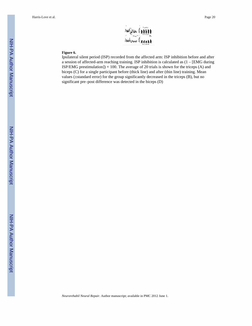
Figure 6.Ipsilateral silent period (ISP) recorded from the affected arm: ISP inhibition before and aftera session of affected-arm reaching training. ISP inhibition is calculated as (1 – [EMG duringISP/EMG prestimulation]) × 100. The average of 20 trials is shown for the triceps (A) andbiceps (C) for a single participant before (thick line) and after (thin line) training. Meanvalues (±standard error) for the group significantly decreased in the triceps (B), but nosignificant pre–post difference was detected in the biceps (D)
Harris-Love et al. Page 20
Neurorehabil Neural Repair. Author manuscript; available in PMC 2012 June 1.
NIH
-PA Author Manuscript
NIH
-PA Author Manuscript
NIH
-PA Author Manuscript

Figure 7.Cortical silent period (CSP) recorded from affected arm: duration of the CSP with lesionedhemisphere stimulation. (A) The average of 10 trials in the affected triceps muscle is shownfor a single patient before and after training. Whereas the levels of prestimulus EMG weresimilar before and after training, the duration of the CSP decreased after training. (B)Displays the group means for intracortical inhibition targeting the affected triceps muscle.The triceps CSP duration significantly decreased after training (P < .05). (C) The average of10 trials in the biceps muscle of the same patient displayed in A. (D) The group means forCSP in the affected biceps muscle before and after training. Though there was a tendencyfor longer biceps CSP durations after training, this tendency did not reach statisticalsignificance (P > .10)
Harris-Love et al. Page 21
Neurorehabil Neural Repair. Author manuscript; available in PMC 2012 June 1.
NIH
-PA Author Manuscript
NIH
-PA Author Manuscript
NIH
-PA Author Manuscript

NIH
-PA Author Manuscript
NIH
-PA Author Manuscript
NIH
-PA Author Manuscript
Harris-Love et al. Page 22
Tabl
e 1
Patie
nt C
hara
cter
istic
s
Patie
ntA
ge (y
ears
)G
ende
rT
ime
Sinc
eSt
roke
(yea
rs)
Les
ione
d H
emis
pher
eSt
roke
Etio
logy
UE
Fug
l-Mey
er S
core
UE
Fug
l-M
eyer
(Per
cent
age
ofM
axim
umSc
ore)
Han
dD
omin
ance
Prio
r to
Stro
ke
MR
CSc
ore
(Fin
gers
and
Wri
st)
Sens
ory
Impa
irm
ent
Hyp
er-R
efle
xia
158
M3.
6Le
ftH
emor
rhag
ic22
33R
ight
0-1
++
252
M4.
3Le
ftH
emor
rhag
ic21
32R
ight
1-2
++
358
M9.
0Le
ftIs
chem
ic29
44R
ight
0+
+
462
F3.
4R
ight
Isch
emic
3350
Rig
ht0-
1+
+
556
M9.
4Le
ftIs
chem
ic22
33R
ight
0-1
++
644
F3.
0R
ight
Isch
emic
2741
Rig
ht0-
1+
+
750
F11
.8R
ight
Isch
emic
2335
Rig
ht0-
1+
+
860
M12
.1Le
ftH
emor
rhag
ic20
30R
ight
0+
+
950
F5.
5R
ight
Isch
emic
3756
Rig
ht1-
2+
+
1060
M1.
8Le
ftIs
chem
ic33
50R
ight
1-3
-+
1153
F8.
2R
ight
Isch
emic
N/A
N/A
Rig
ht2-
4+
+
Mea
n54
.96.
5526
.740
.5%
SD3.
73.
676.
09.
1%
Abb
revi
atio
ns: U
E, u
pper
ext
rem
ity; M
RC
, Med
ical
Res
earc
h C
ounc
il; N
/A, n
ot a
vaila
ble;
SD
, sta
ndar
d de
viat
ion.
Neurorehabil Neural Repair. Author manuscript; available in PMC 2012 June 1.

NIH
-PA Author Manuscript
NIH
-PA Author Manuscript
NIH
-PA Author Manuscript
Harris-Love et al. Page 23
Tabl
e 2
Con
trol D
ata
Tri
ceps
Bic
eps
Pre
Post
Pre
Post
Con
trol
Var
iabl
eM
ean
SDM
ean
SDP
Val
ueM
ean
SDM
ean
SDP
Val
ue
A. S
lope
of u
naff
ecte
d-ar
m re
crui
tmen
t cur
ve1.
571.
201.
541.
44ns
4.32
3.55
7.63
9.08
ns
B. A
ffec
ted-
arm
bac
kgro
und
EMG
(con
trale
sion
al st
imul
atio
n)6.
410.
986.
361.
26ns
5.31
1.82
6.23
1.99
ns
C. A
ffec
ted-
arm
bac
kgro
und
EMG
(ips
ilesi
onal
stim
ulat
ion)
9.71
2.09
9.69
2.21
ns8.
263.
438.
834.
15ns
D. U
naff
ecte
d-ar
m m
irror
EM
G (c
ontra
lesi
onal
stim
ulat
ion;
n =
3)
3.47
4.25
3.24
2.54
ns11
.25
15.9
27.
046.
93ns
E. U
naff
ecte
d-ar
m m
irror
EM
G (i
psile
sion
al st
imul
atio
n; n
= 3
)3.
423.
634.
073.
27ns
5.85
6.48
9.02
10.3
8ns
Abb
revi
atio
ns: P
re, i
mm
edia
tely
bef
ore
train
ing;
Pos
t, im
med
iate
ly a
fter t
rain
ing;
EM
G, m
ean
rect
ified
ele
ctro
myo
grap
hic
activ
ity (i
n μV
) cal
cula
ted
over
100
ms b
efor
e st
imul
us d
eliv
ery;
SD
, sta
ndar
dde
viat
ion.
Neurorehabil Neural Repair. Author manuscript; available in PMC 2012 June 1.
Copyright © 2022 FDOKUMEN