Mechanisms Of RAD51D-Dependent Repair Of DNA And ...
245
University of South Carolina University of South Carolina Scholar Commons Scholar Commons Theses and Dissertations 2018 Mechanisms Of RAD51D-Dependent Repair Of DNA And Telomere Mechanisms Of RAD51D-Dependent Repair Of DNA And Telomere Damage Induced By Interstrand Crosslinking Agents And Damage Induced By Interstrand Crosslinking Agents And Thiopurines Thiopurines Nicole M. Reilly University of South Carolina Follow this and additional works at: https://scholarcommons.sc.edu/etd Part of the Pharmacy and Pharmaceutical Sciences Commons Recommended Citation Recommended Citation Reilly, N. M.(2018). Mechanisms Of RAD51D-Dependent Repair Of DNA And Telomere Damage Induced By Interstrand Crosslinking Agents And Thiopurines. (Doctoral dissertation). Retrieved from https://scholarcommons.sc.edu/etd/4594 This Open Access Dissertation is brought to you by Scholar Commons. It has been accepted for inclusion in Theses and Dissertations by an authorized administrator of Scholar Commons. For more information, please contact [email protected].
-
Upload
khangminh22 -
Category
Documents
-
view
1 -
download
0
Transcript of Mechanisms Of RAD51D-Dependent Repair Of DNA And ...
University of South Carolina University of South Carolina
Scholar Commons Scholar Commons
Theses and Dissertations
2018
Mechanisms Of RAD51D-Dependent Repair Of DNA And Telomere Mechanisms Of RAD51D-Dependent Repair Of DNA And Telomere
Damage Induced By Interstrand Crosslinking Agents And Damage Induced By Interstrand Crosslinking Agents And
Thiopurines Thiopurines
Nicole M. Reilly University of South Carolina
Follow this and additional works at: https://scholarcommons.sc.edu/etd
Part of the Pharmacy and Pharmaceutical Sciences Commons
Recommended Citation Recommended Citation Reilly, N. M.(2018). Mechanisms Of RAD51D-Dependent Repair Of DNA And Telomere Damage Induced By Interstrand Crosslinking Agents And Thiopurines. (Doctoral dissertation). Retrieved from https://scholarcommons.sc.edu/etd/4594
This Open Access Dissertation is brought to you by Scholar Commons. It has been accepted for inclusion in Theses and Dissertations by an authorized administrator of Scholar Commons. For more information, please contact [email protected].
MECHANISMS OF RAD51D-DEPENDENT REPAIR OF DNA AND TELOMERE
DAMAGE INDUCED BY INTERSTRAND CROSSLINKING AGENTS AND
THIOPURINES
by
Nicole M. Reilly
Bachelor of Science
Appalachian State University, 2012
Submitted in Partial Fulfillment of the Requirements
for the Degree of Doctor of Philosophy in
Pharmaceutical Sciences
College of Pharmacy
University of South Carolina
2018
Accepted by:
Douglas L. Pittman, Major Professor
Michael D. Wyatt, Chair, Examining Committee
Kim E. Creek, Committee Member
Danyelle M. Townsend, Committee Member
Lydia E. Matesic, Committee Member
Cheryl L. Addy, Vice Provost and Dean of the Graduate School

ii
© Copyright by Nicole M. Reilly, 2018
All Rights Reserved.

iii
DEDICATION
This work is dedicated to my grandmother, Colleen Winifred O’Dougherty Melville, for
her eternal love that guided and protected me through this journey.

iv
ACKNOWLEDGEMENTS
Firstly, I would like to thank my advisor, Dr. Douglas L. Pittman, for his limitless support
and enthusiasm that were instrumental to my success. The experience and knowledge that
I gained by working alongside him is invaluable to me, and I will always be grateful. I
also thank Dr. Michael D. Wyatt for acting as a second mentor to me and for giving me
the opportunity to work on the 6-thioguanine project. I am thankful to my committee,
Drs. Kim E. Creek, Danyelle M. Townsend, and Lydia E. Matesic, for their guidance. I
am especially grateful to my colleagues: Ms. Latarsha L. Porcher, Mr. Michael G.
Marone, Ms. Erin L. Anderson, Ms. Deborah O. Adedokun, Ms. Claire L. Chabot, and
Ms. Morgan Ingerson for their technical assistance and friendship. I am also very
appreciative of the support of other faculty members in the Department of Drug
Discovery and Biomedical Sciences: Dr. Phillip J. Buckhaults for his assistance on the
RNA Seq portion of my gene expression project and for recommending me to Dr.
Alberto Bardelli as a post-doctoral research fellow, Dr. Diego Altomare, for his
collaborative efforts on the microarray aspect of my gene expression project, and Dr. Jill
R. Turner and Dr. Jason A. Stewart for technical assistance.
I would like to thank my family – Pamela, Carl, Chris, and Kaitlin – for their
love, support, and patience as I worked to complete this goal, and to my fellow graduate
students – Anusha, Katie, Pamela, Sara, Tim – for their support and friendship.
Finally, to Murphy Cameron and Spud Bear, for the love and snuggles that kept
me going for the past five years. E ora qualcosa di completamente diverso...
v
ABSTRACT
Mutations in homologous recombination (HR) genes increase genomic instability, an
enabling characteristic of cancer. However, the status of these same genes can also
determine chemotherapy outcomes. RAD51D is a breast and ovarian cancer susceptibility
gene that is an important component of HR. Mammalian cells defective for RAD51D
have extensive chromosomal aberrations and are more sensitive to the interstrand
crosslink-inducing agent mitomycin C (MMC) and the thiopurine 6-thioguanine (6TG).
Previously, the RNF138 E3 ubiquitin ligase was identified to promote RAD51D
ubiquitination, and loss of RNF138 also increased cellular sensitivity to MMC.
Ubiquitination assays were used to show that a 3-ubiquitin modification occurs along the
RAD51D wild-type protein. To identify potential sites of ubiquitination, amino acid
substitutions were generated at all thirteen lysine residues along RAD51D. Arginine
substitutions at K235 (K235R) and K298 (K298R) were found to confer cellular
sensitivity to MMC. In addition, protein stability of K235R and K298R were 2 to 3-fold
higher as compared with wild-type RAD51D.
RAD51D is also known to contribute to telomere maintenance, although its
precise function at the telomeres remains unclear. In this dissertation, I investigated the
activity of RAD51D at telomeres and the contribution of RAD51D to protect against
6TG-induced telomere damage. As measured by γ-H2AX induction and foci formation,
the extent of γ-H2AX telomere localization following 6TG treatment was higher in
Rad51d-deficient cells than in Rad51d-proficient cells. In the final portion of this
vi
dissertation, Rad51d-deficient cells were used as a model for genome unstable
mammalian cells to identify genetic compromises that support cell proliferation. Gene
expression profiles of Rad51d-proficient and -deficient primary mouse embryonic
fibroblasts were analyzed by microarray and RNA Seq. In both analyses, the highest
proportion of genes were associated with cellular growth and proliferation. In summary,
the data presented in this dissertation identified potential regulatory sites along RAD51D
that mediate its function during ICL repair, elucidated the role of RAD51D in
maintaining telomere integrity in the presence of thiopurine-induced DNA damage, and
revealed genetic compromises in Rad51d-deficient cells that promote cell proliferation.
vii
TABLE OF CONTENTS
Dedication .......................................................................................................................... iii
Acknowledgements ............................................................................................................ iv
Abstract ...............................................................................................................................v
List of Tables ..................................................................................................................... ix
List of Figures .....................................................................................................................x
List of Abbreviations ........................................................................................................ xii
Chapter 1: Introduction .......................................................................................................1
Chapter 2: Literature ...........................................................................................................5
Chapter 3: RNF138 zinc finger motifs mediate its interaction with RAD51D ................. 42
Chapter 4: RAD51D lysine residues 235 and 298 are required for DNA
interstrand crosslink repair ............................................................................. 54
Chapter 5: Thiopurine-induced telomeric damage in Rad51d-deficient
mammalian cells ............................................................................................ 88
Chapter 6: Genome-wide identification and expression analysis of gene
differences between Rad51d-proficient and -deficient primary
mouse embryonic fibroblasts ....................................................................... 110
Chapter 7: Discussion ..................................................................................................... 143
References ....................................................................................................................... 160
Appendix A: Genes with differential expression in the absence of Rad51d
identified by microarray ........................................................................... 182
Appendix B: Genes with differential expression in the absence of Rad51d
identified by RNA Seq ............................................................................. 200
Appendix C: RAD51D and RNF138 expression constructs ........................................... 219

viii
Appendix D: Permission to reprint data in Chapter 3 ..................................................... 224
Appendix E: Permission to reprint data in Chapter 5 ..................................................... 226
ix
LIST OF TABLES
Table 2.1 Risk factor mutant alleles of RAD51 genes ...................................................... 29
Table 4.1 Primer sequences used for MmRad51d cDNA site-directed
Mutagenesis ...................................................................................................... 76
Table 5.1 Cytological analysis of MEFs treated with 6TG .............................................104
Table 6.1 Genes associated with “cellular growth and proliferation” identified
by microarray ..................................................................................................128
Table 6.2 Genes associated with “cell cycle progression” identified by microarray.......130
Table 6.3 Genes associated with “cellular growth and proliferation” identified
by RNA Seq .....................................................................................................131
Table 6.4 Chromosome fusions identified in the RNA Seq samples by TopHat-
Fusion ...............................................................................................................134
Table 6.5 Genes identified by both microarray and RNA Seq ........................................135
Table 6.6 Genes associated with “cellular growth and proliferation” identified
by both microarray and RNA Seq ...................................................................138
Table 6.7 Genes associated with “cell cycle progression” identified by both
microarray and RNA Seq ................................................................................139
Table A.1. Genes identified by microarray ......................................................................183
Table B.1. Genes identified by RNA Seq ........................................................................201
Table C.1. Prokaryotic expression constructs ..................................................................220
Table C.2. Eukaryotic expression constructs ...................................................................221
Table C.3. Yeast expression constructs ...........................................................................222
.
Table C.4. Fluorescent expression constructs ..................................................................223
x
LIST OF FIGURES
Figure 2.1 Overview of consequences of DNA damage.....................................................6
Figure 2.2 DNA break induced by ionizing radiation.........................................................8
Figure 2.3 Platinum compounds binding to guanine nucleotides .......................................9
Figure 2.4 Metabolism of 6TG into cytotoxic nucleotides ............................................... 10
Figure 2.5 Model of homologous recombination-mediated double strand
break repair ...................................................................................................... 15
Figure 2.6 Key proteins in multiple pathways that repair DNA interstrand
crosslinks......................................................................................................... 17
Figure 2.7 Structure of RAD51 and its paralogs indicating known domains ................... 22
Figure 2.8 Ubiquitin modification of target proteins ........................................................ 31
Figure 2.9 Model of the SUMOylation cascade ............................................................... 35
Figure 2.10 Post-translational modifications along RAD51 and its paralogs ................... 38
Figure 3.1 Predicted structure of RNF138 ........................................................................ 51
Figure 3.2 Summary of Mus musculus Rnf138 splice variants ......................................... 52
Figure 3.3 RAD51D is ubiquitinated in the absence of over-expressed
RNF138 and in the presence of MG132 .......................................................... 53
Figure 4.1 Alignment between Homo sapiens RAD51D (328 amino acids) and Mus
musculus (C56BL/6) RAD51D (329 amino acids) .......................................... 77
Figure 4.2 Conserved lysine residues along Mus musculus RAD51 family members ..... 78
Figure 4.3 Complementation analysis of RAD51D lysine to arginine substitution
Alleles ............................................................................................................. 79
Figure 4.4 Intracellular localization of RAD51D variants................................................ 80
xi
Figure 4.5 Yeast-two-hybrid protein interaction analysis between RAD51D variants
and RAD51C, XRCC2, and RNF138 ............................................................. 81
Figure 4.6 Stability of RAD51D variants ......................................................................... 82
Figure 4.7 Ubiquitination of RAD51D variants ............................................................... 83
Figure 4.8 Recruitment of FANCD2, Ku86, and RAD51 to MMC-induced DNA
Damage ............................................................................................................ 84
Figure 4.9 Measurement of HR by reconstitution of GFP fluorescence in HeLa
DRGFP cells .................................................................................................... 85
Figure 4.10 Proposed model of RAD51D ubiquitination during interstrand
crosslink repair ................................................................................................. 87
Figure 5.1 Induction of γ-H2AX following treatment with 6TG .....................................105
Figure 5.2 Co-localization of γ-H2AX with telomeres following treatment
with 6TG .........................................................................................................106
Figure 5.3 6TG induced chromosome fusions in Rad51d-deficeint
immortalized MEFs .......................................................................................107
Figure 5.4 6TG treatment induces increased multinucleation in Rad51d-
deficient and Mlh1-proficient MEFs ..............................................................108
Figure 5.5 6TG treatment does not affect telomere distribution across nuclei
within multinucleated cells ............................................................................109
Figure 6.1 Rad51d expression and RNA quality assessment ..........................................140
Figure 6.2 Biological functions as identified by Ingenuity Pathway Analysis
(IPA) in microarray data .................................................................................141
Figure 6.3 Biological functions as identified by IPA of RNA Seq data set .....................142
Figure 7.1 Proposed RAD51 paralog complexes that function in response to
DNA interstrand crosslinks and double strand breaks ....................................147
Figure 7.2 Stability of RAD51D protein variants ............................................................150
Figure 7.3 Proposed model of RAD51D ubiquitination during DNA inter-
strand crosslink repair .....................................................................................158
xii
LIST OF ABBREVIATIONS
6TG ................................................................................................................ 6-thioguanine
Bp .......................................................................................................................... Base pair
DSB .......................................................................................................Double strand break
dsDNA ...............................................................................................Double stranded DNA
ICL .............................................................................................. DNA interstrand crosslink
E1 ........................................................................................... Ubiquitin activating enzyme
E2 ........................................................................................ Ubiquitin conjugating enzyme
E3 .................................................................................................Ubiquitin ligating enzyme
FA ...............................................................................................................Fanconi Anemia
HECT ........................................................... Homologous to the E6AP carboxyl terminus
HR .............................................................................................Homologous recombination
IR............................................................................................................. Ionizing radiation
kDA ......................................................................................................................Kilodalton
MEF ....................................................................................... Mouse embryonic fibroblast
MMR ...........................................................................................................Mismatch repair
MMC ............................................................................................................... Mitomycin C
NER............................................................................................. Nucleotide excision repair
NHEJ ......................................................................................Non-homologous end joining
ONPG ............................................................................. Ortho-nitrophenyl-β-galactosidase
PTM ..................................................................................... Post-translational modification
RING .........................................................................................Really interesting new gene
xiii
SDM .............................................................................................Site-directed mutagenesis
SSB ........................................................................................................ Single strand break
ssDNA ................................................................................................. single stranded DNA
SUMO .................................................................................... Small ubiquitin-like modifier
UIM ............................................................................................ Ubiquitin interacting motif
Y2H ........................................................................................................... Yeast-two-hybrid
ZF .........................................................................................................................Zinc finger
1
CHAPTER 1
INTRODUCTION
Six identifiable biological characteristics of tumor development termed the “hallmarks of
cancer” were classified by Hanahan and Weinberg (Hanahan and Weinberg 2000). In
addition to these features, cancer cells acquire ‘enabling characteristics’ that contribute to
carcinogenesis. In 2011, ‘genome instability’ was recognized as an enabling
characteristic, and Hanahan and Weinberg argued that tumor growth can often be
attributed to acquisition of mutations that promote cell proliferation and inhibit cell death
(Hanahan and Weinberg 2011). The idea that genome instability contributes to cancer
development was actually first proposed in 1914 by Theodor Boveri (Boveri 2008), and
studies throughout the 21st century strongly support this theory. Boveri’s observations
that abnormal chromosomal arrangements are passed to sea urchin off-spring lead to the
hypothesis that tumor development was a cellular problem and that cancer is, in fact, a
genetic disease (Boveri 2008; Hansford and Huntsman 2014). Similarly, the observation
that cancer is a mutation-driven disease led to the “Mutator Phenotype Hypothesis.” First
described by Lawrence Loeb, this hypothesis states that “mutations occur randomly
throughout the genome, and among these would be mutations in genes that guarantee the
fidelity of DNA replication… and repair” (Loeb, Springgate, and Battula 1974).
Together, these ideas have led to the current belief that “defects in genome maintenance
are… instrumental for tumor progression” (Hanahan and Weinberg 2011). The data
2
presented in this dissertation offer insights into how the RAD51D DNA repair protein
contributes towards maintaining genomic integrity.
Chapter 2 provides an overview of literature discussing types of DNA damage
that are recognized and repaired by the homologous recombination (HR) proteins, that
specifically includes double strand breaks (DSBs), interstrand crosslinks (ICLs), and
thiopurine-induced base pair mismatches. The RAD51 family of proteins – RAD51,
RAD51B, RAD51C, RAD51D, XRCC2, and XRCC3 – are described, and their activity
during HR-mediated repair is discussed. Post-translational modifications (PTMs) regulate
DNA repair pathways. Ubiquitination and SUMOylation are described, and the function
of these PTMs during DNA repair is highlighted. Finally, PTMs that occur along RAD51
proteins are discussed. Experiments in subsequent chapters focus on the RAD51D HR
protein.
Chapter 3 presents work that was published in the research article entitled
“RNF138 interacts with RAD51D and is required for DNA interstrand crosslink repair
and maintaining chromosome integrity” in DNA Repair in April 2016 (Yard et al. 2016).
The data in this paper demonstrated that RAD51D directly interacts with the E3 ubiquitin
ligase RNF138, and that this interaction is mediated by the regions encoded by exon 5
and exon 7 along RNF138. RNF138 ubiquitinates RAD51D, and data presented in this
Chapter also demonstrate that this modification occurs along RAD51D and not the
RAD51C protein.
Chapter 4 presents work that identified two lysine residues along RAD51D –
K235 and K298 – that are critical for ICL repair. In this study, single point mutations
were generated in lysine codons along the MmRad51d gene to introduce arginine at those
3
locations. Substitution of two lysines – K235R and K298R –conferred cellular sensitivity
to mitomycin C (MMC). Yeast-two-hybrid analysis demonstrated that these residues are
not required for RAD51D interaction with RAD51C, XRCC2, or RNF138. A lysine-null
mutant (K0) was 3 times more stable than wild-type RAD51D, and stability of K235R
and K298R was increased 2- and 3-fold, respectively, compared with wild-type. In vivo
ubiquitination assays detected a band corresponding to 3 ubiquitin molecules was present
in wild-type, but not K0 samples, suggesting loss of a short ubiquitin chain along the
protein in the absence of lysine residues. Furthermore, homology-directed repair assays
suggest that neither K235 nor K298 is required for repair of SceI induced DSBs.
Chapter 5 presents data that were published in the research article entitled
“Thiopurine-induced mitotic catastrophe in Rad51d-deficient mammalian cells” in
Environmental and Molecular Mutagenesis in September 2017 (Wyatt et al. 2017). The
focus of this work was RAD51D function in response to 6-thioguanine (6TG)-induced
base pair mismatches. In Rad51d-deficient cells, there was increased co-localization of
telomere probes with γ-H2AX foci compared to Rad51d-proficient cells, which further
increased upon treatment with 6TG. Chromosome fusions following 6TG treatment were
detected, and telomere positive staining was observed at fusion points. These findings
demonstrate that RAD51D provides a protective role against the telomeric DNA damage
and chromosomal instability caused by thiopurine treatment.
Rad51d-deficient cells have extensive chromosomal aberrations, such as fusions,
translocations, and telomere defects, that are often observed in ovarian cancer cells. For
this reason, Rad51d-deficient mouse embryonic fibroblasts (MEFs) can be used as a
model for genomic unstable ovarian cancers. In Chapter 6, gene expression profiles of
4
Rad51d+/+
Trp53-/-
(Rad51d-proficient) and Rad51d-/-
Trp53-/-
(Rad51d-deficient) primary
MEF cell lines were assessed by microarray and RNA Seq analyses. Six hundred
eighteen genes with differential expression between the Rad51d-proficient and -deficient
cell lines were identified by microarray. Twenty-one of the identified genes are
associated with cell cycle progression, and included: Id1, Id2, and Cdkn1a(p21). RNA
Seq analysis identified 928 genes that were differentially expressed. In addition, five gene
fusions were identified in the Rad51d-proficient cell lines, and one of these fusions was
also present in the Rad51d-deficient samples. Comparison between the two data sets
identified 111 genes that were differentially expressed between Rad51d-proficient and -
deficient cell lines. Together these data provide insight into gene expression compromises
that support cell division in a chromosomal unstable cell line.
In Chapter 7, a model of RAD51D ubiquitination during interstrand crosslink
repair is proposed, and I hypothesize that ubiquitination at K235 and K298 is required for
RAD51 recruitment to DSBs. Follow-up experiments to better elucidate the role of K235
and K298 for RAD51D function are proposed, and I predict that these residues will also
be necessary for cellular resistance to 6TG. Finally, mass spectrometry analysis should be
performed to identify specific PTMs that occur along the RAD51D protein.
5
CHAPTER 2
LITERATURE
Accurate repair of DNA damage is essential for maintaining genomic integrity, and
accumulation of mutations is one of the early steps that lead to cancer development.
Boveri’s observations that abnormal chromosomal arrangements were passed to sea
urchin off-spring and Loeb’s “Mutator Hypothesis” support the idea that “defects in
genome maintenance are… instrumental for tumor progression” (Hanahan and Weinberg
2011; Loeb, Springgate, and Battula 1974; Boveri 2008). Mutations in several key DNA
damage response genes, including BRCA1, BRCA2, and the RAD51 family of proteins,
are associated with increased cancer risk (Prakash et al. 2015). In addition to protecting
the cell from genomic insult, RAD51 and its paralogs – RAD51B, RAD51C, RAD51D,
XRCC2, and XRCC3 – function during the homologous recombination pathway that
recognizes and repairs DNA double strand breaks. These lesions can be directly
introduced through exogenous agents, such as ionizing radiation, or through the repair of
other forms of damage, such as cisplatin-induced DNA interstrand crosslinks and
thiopurine-induced base pair mismatches (Figure 2.1) (Karran 2006; Suchankova et al.
2012).
The following sections of this introduction will describe three types of DNA
damage – DNA double strand breaks, DNA interstrand crosslinks, and thiopurine-
induced base pair mismatches – that affect both strands of the DNA double helix. I will
focus on the RAD51 family of proteins and discuss current knowledge regarding the
6
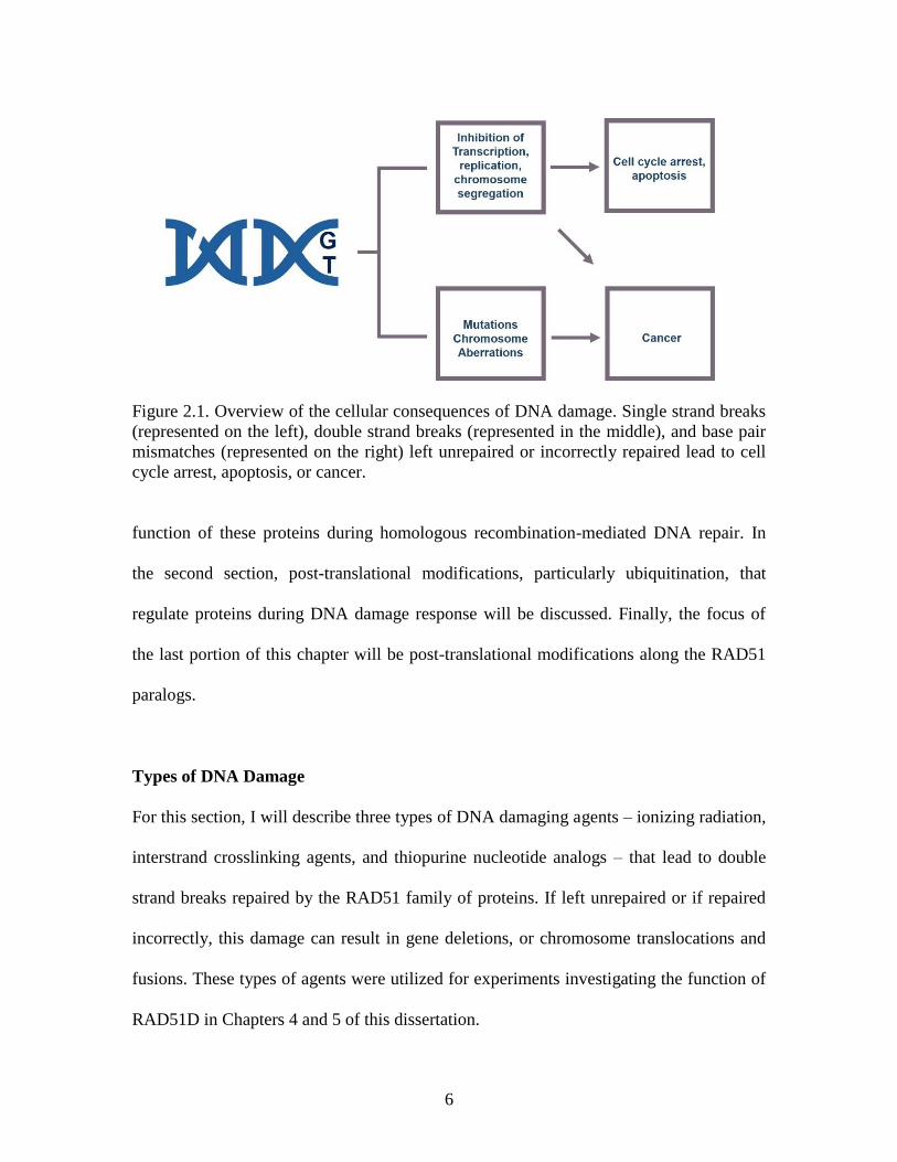
Figure 2.1. Overview of the cellular consequences of DNA damage. Single strand breaks
(represented on the left), double strand breaks (represented in the middle), and base pair
mismatches (represented on the right) left unrepaired or incorrectly repaired lead to cell
cycle arrest, apoptosis, or cancer.
function of these proteins during homologous recombination-mediated DNA repair. In
the second section, post-translational modifications, particularly ubiquitination, that
regulate proteins during DNA damage response will be discussed. Finally, the focus of
the last portion of this chapter will be post-translational modifications along the RAD51
paralogs.
Types of DNA Damage
For this section, I will describe three types of DNA damaging agents – ionizing radiation,
interstrand crosslinking agents, and thiopurine nucleotide analogs – that lead to double
strand breaks repaired by the RAD51 family of proteins. If left unrepaired or if repaired
incorrectly, this damage can result in gene deletions, or chromosome translocations and
fusions. These types of agents were utilized for experiments investigating the function of
RAD51D in Chapters 4 and 5 of this dissertation.
7
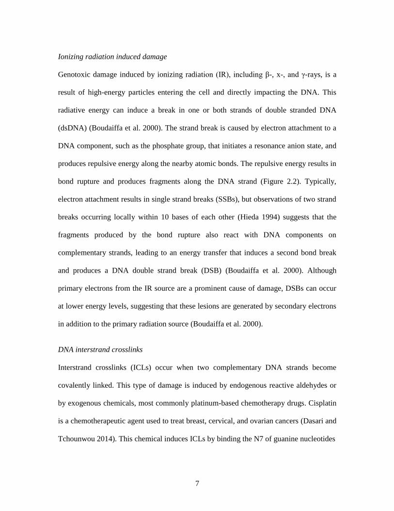
Ionizing radiation induced damage
Genotoxic damage induced by ionizing radiation (IR), including β-, x-, and γ-rays, is a
result of high-energy particles entering the cell and directly impacting the DNA. This
radiative energy can induce a break in one or both strands of double stranded DNA
(dsDNA) (Boudaiffa et al. 2000). The strand break is caused by electron attachment to a
DNA component, such as the phosphate group, that initiates a resonance anion state, and
produces repulsive energy along the nearby atomic bonds. The repulsive energy results in
bond rupture and produces fragments along the DNA strand (Figure 2.2). Typically,
electron attachment results in single strand breaks (SSBs), but observations of two strand
breaks occurring locally within 10 bases of each other (Hieda 1994) suggests that the
fragments produced by the bond rupture also react with DNA components on
complementary strands, leading to an energy transfer that induces a second bond break
and produces a DNA double strand break (DSB) (Boudaiffa et al. 2000). Although
primary electrons from the IR source are a prominent cause of damage, DSBs can occur
at lower energy levels, suggesting that these lesions are generated by secondary electrons
in addition to the primary radiation source (Boudaiffa et al. 2000).
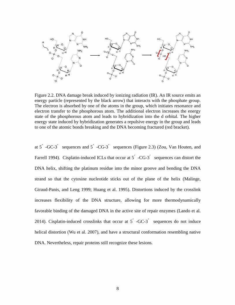
DNA interstrand crosslinks
Interstrand crosslinks (ICLs) occur when two complementary DNA strands become
covalently linked. This type of damage is induced by endogenous reactive aldehydes or
by exogenous chemicals, most commonly platinum-based chemotherapy drugs. Cisplatin
is a chemotherapeutic agent used to treat breast, cervical, and ovarian cancers (Dasari and
Tchounwou 2014). This chemical induces ICLs by binding the N7 of guanine nucleotides
8
Figure 2.2. DNA damage break induced by ionizing radiation (IR). An IR source emits an
energy particle (represented by the black arrow) that interacts with the phosphate group.
The electron is absorbed by one of the atoms in the group, which initiates resonance and
electron transfer to the phosphorous atom. The additional electron increases the energy
state of the phosphorous atom and leads to hybridization into the d orbital. The higher
energy state induced by hybridization generates a repulsive energy in the group and leads
to one of the atomic bonds breaking and the DNA becoming fractured (red bracket).
at 5՛ -GC-3՛ sequences and 5՛ -CG-3՛ sequences (Figure 2.3) (Zou, Van Houten, and
Farrell 1994). Cisplatin-induced ICLs that occur at 5՛ -CG-3՛ sequences can distort the
DNA helix, shifting the platinum residue into the minor groove and bending the DNA
strand so that the cytosine nucleotide sticks out of the plane of the helix (Malinge,
Giraud-Panis, and Leng 1999; Huang et al. 1995). Distortions induced by the crosslink
increases flexibility of the DNA structure, allowing for more thermodynamically
favorable binding of the damaged DNA in the active site of repair enzymes (Lando et al.
2014). Cisplatin-induced crosslinks that occur at 5՛ -GC-3՛ sequences do not induce
helical distortion (Wu et al. 2007), and have a structural conformation resembling native
DNA. Nevertheless, repair proteins still recognize these lesions.
9
Figure 2.3 Platinum compounds binding guanine nucleotides. Cisplatin binds the N7
position on two guanine nucleotides in the same DNA strand.
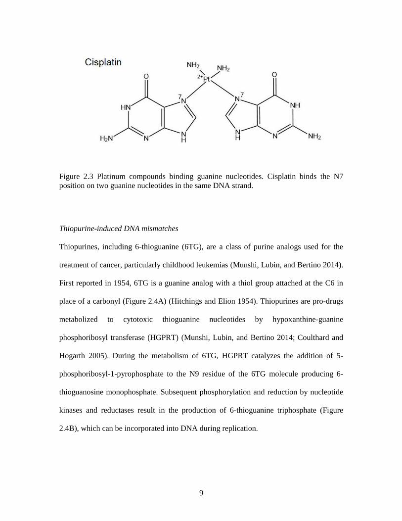
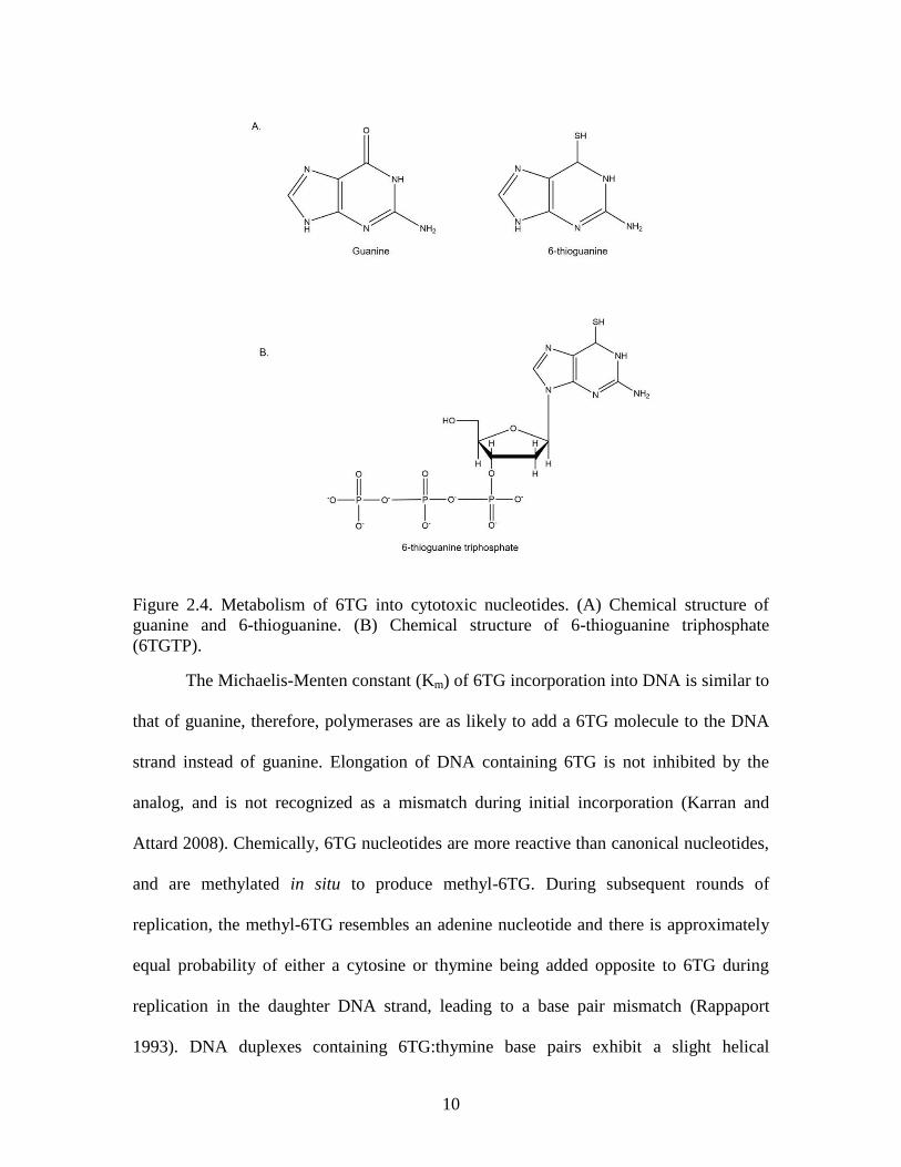
Thiopurine-induced DNA mismatches
Thiopurines, including 6-thioguanine (6TG), are a class of purine analogs used for the
treatment of cancer, particularly childhood leukemias (Munshi, Lubin, and Bertino 2014).
First reported in 1954, 6TG is a guanine analog with a thiol group attached at the C6 in
place of a carbonyl (Figure 2.4A) (Hitchings and Elion 1954). Thiopurines are pro-drugs
metabolized to cytotoxic thioguanine nucleotides by hypoxanthine-guanine
phosphoribosyl transferase (HGPRT) (Munshi, Lubin, and Bertino 2014; Coulthard and
Hogarth 2005). During the metabolism of 6TG, HGPRT catalyzes the addition of 5-
phosphoribosyl-1-pyrophosphate to the N9 residue of the 6TG molecule producing 6-
thioguanosine monophosphate. Subsequent phosphorylation and reduction by nucleotide
kinases and reductases result in the production of 6-thioguanine triphosphate (Figure
2.4B), which can be incorporated into DNA during replication.
10
Figure 2.4. Metabolism of 6TG into cytotoxic nucleotides. (A) Chemical structure of
guanine and 6-thioguanine. (B) Chemical structure of 6-thioguanine triphosphate
(6TGTP).
The Michaelis-Menten constant (Km) of 6TG incorporation into DNA is similar to
that of guanine, therefore, polymerases are as likely to add a 6TG molecule to the DNA
strand instead of guanine. Elongation of DNA containing 6TG is not inhibited by the
analog, and is not recognized as a mismatch during initial incorporation (Karran and
Attard 2008). Chemically, 6TG nucleotides are more reactive than canonical nucleotides,
and are methylated in situ to produce methyl-6TG. During subsequent rounds of
replication, the methyl-6TG resembles an adenine nucleotide and there is approximately
equal probability of either a cytosine or thymine being added opposite to 6TG during
replication in the daughter DNA strand, leading to a base pair mismatch (Rappaport
1993). DNA duplexes containing 6TG:thymine base pairs exhibit a slight helical
11
distortion (Bohon and de los Santos 2005, 2003) that shifts the thymine into the major
groove of the DNA duplex and the 6TG into the minor groove (Bohon and de los Santos
2005; Somerville et al. 2003). Interestingly, thermodynamic analysis demonstrates that
6TG:thymine base pairs are more stable than 6TG:cytosine pairs (Bohon and de los
Santos 2005), and suggest that the minor distortion does not destabilize the DNA helix.
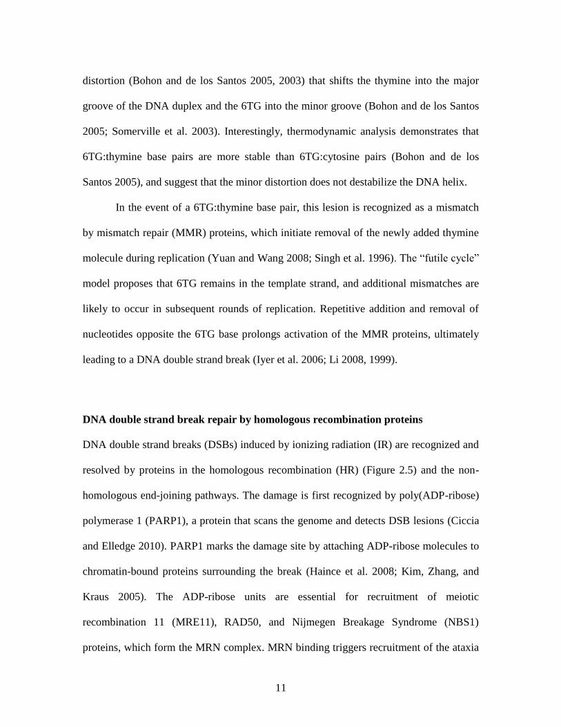
In the event of a 6TG:thymine base pair, this lesion is recognized as a mismatch
by mismatch repair (MMR) proteins, which initiate removal of the newly added thymine
molecule during replication (Yuan and Wang 2008; Singh et al. 1996). The “futile cycle”
model proposes that 6TG remains in the template strand, and additional mismatches are
likely to occur in subsequent rounds of replication. Repetitive addition and removal of
nucleotides opposite the 6TG base prolongs activation of the MMR proteins, ultimately
leading to a DNA double strand break (Iyer et al. 2006; Li 2008, 1999).
DNA double strand break repair by homologous recombination proteins
DNA double strand breaks (DSBs) induced by ionizing radiation (IR) are recognized and
resolved by proteins in the homologous recombination (HR) (Figure 2.5) and the non-
homologous end-joining pathways. The damage is first recognized by poly(ADP-ribose)
polymerase 1 (PARP1), a protein that scans the genome and detects DSB lesions (Ciccia
and Elledge 2010). PARP1 marks the damage site by attaching ADP-ribose molecules to
chromatin-bound proteins surrounding the break (Haince et al. 2008; Kim, Zhang, and
Kraus 2005). The ADP-ribose units are essential for recruitment of meiotic
recombination 11 (MRE11), RAD50, and Nijmegen Breakage Syndrome (NBS1)
proteins, which form the MRN complex. MRN binding triggers recruitment of the ataxia
12
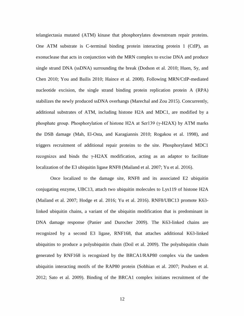
telangiectasia mutated (ATM) kinase that phosphorylates downstream repair proteins.
One ATM substrate is C-terminal binding protein interacting protein 1 (CtIP), an
exonuclease that acts in conjunction with the MRN complex to excise DNA and produce
single strand DNA (ssDNA) surrounding the break (Dodson et al. 2010; Huen, Sy, and
Chen 2010; You and Bailis 2010; Haince et al. 2008). Following MRN/CtIP-mediated
nucleotide excision, the single strand binding protein replication protein A (RPA)
stabilizes the newly produced ssDNA overhangs (Marechal and Zou 2015). Concurrently,
additional substrates of ATM, including histone H2A and MDC1, are modified by a
phosphate group. Phosphorylation of histone H2A at Ser139 (γ-H2AX) by ATM marks
the DSB damage (Mah, El-Osta, and Karagiannis 2010; Rogakou et al. 1998), and
triggers recruitment of additional repair proteins to the site. Phosphorylated MDC1
recognizes and binds the γ-H2AX modification, acting as an adaptor to facilitate
localization of the E3 ubiquitin ligase RNF8 (Mailand et al. 2007; Yu et al. 2016).
Once localized to the damage site, RNF8 and its associated E2 ubiquitin
conjugating enzyme, UBC13, attach two ubiquitin molecules to Lys119 of histone H2A
(Mailand et al. 2007; Hodge et al. 2016; Yu et al. 2016). RNF8/UBC13 promote K63-
linked ubiquitin chains, a variant of the ubiquitin modification that is predominant in
DNA damage response (Panier and Durocher 2009). The K63-linked chains are
recognized by a second E3 ligase, RNF168, that attaches additional K63-linked
ubiquitins to produce a polyubiquitin chain (Doil et al. 2009). The polyubiquitin chain
generated by RNF168 is recognized by the BRCA1/RAP80 complex via the tandem
ubiquitin interacting motifs of the RAP80 protein (Sobhian et al. 2007; Poulsen et al.
2012; Sato et al. 2009). Binding of the BRCA1 complex initiates recruitment of the
13
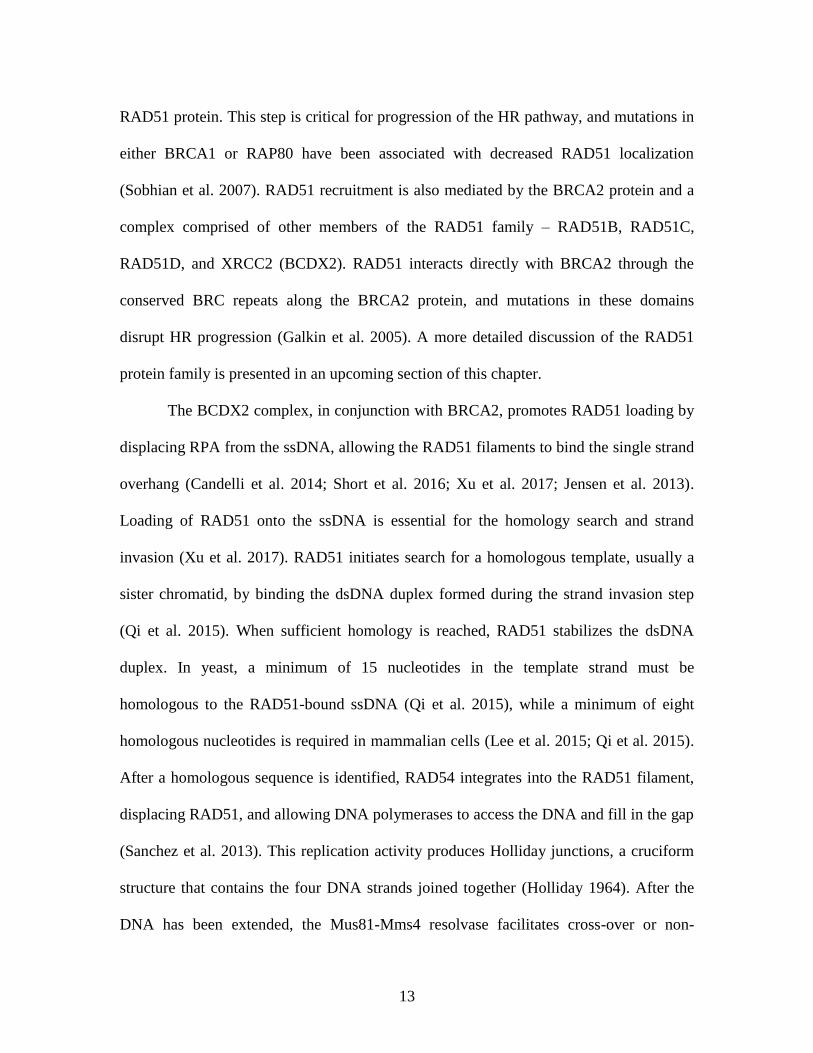
RAD51 protein. This step is critical for progression of the HR pathway, and mutations in
either BRCA1 or RAP80 have been associated with decreased RAD51 localization
(Sobhian et al. 2007). RAD51 recruitment is also mediated by the BRCA2 protein and a
complex comprised of other members of the RAD51 family – RAD51B, RAD51C,
RAD51D, and XRCC2 (BCDX2). RAD51 interacts directly with BRCA2 through the
conserved BRC repeats along the BRCA2 protein, and mutations in these domains
disrupt HR progression (Galkin et al. 2005). A more detailed discussion of the RAD51
protein family is presented in an upcoming section of this chapter.
The BCDX2 complex, in conjunction with BRCA2, promotes RAD51 loading by
displacing RPA from the ssDNA, allowing the RAD51 filaments to bind the single strand
overhang (Candelli et al. 2014; Short et al. 2016; Xu et al. 2017; Jensen et al. 2013).
Loading of RAD51 onto the ssDNA is essential for the homology search and strand
invasion (Xu et al. 2017). RAD51 initiates search for a homologous template, usually a
sister chromatid, by binding the dsDNA duplex formed during the strand invasion step
(Qi et al. 2015). When sufficient homology is reached, RAD51 stabilizes the dsDNA
duplex. In yeast, a minimum of 15 nucleotides in the template strand must be
homologous to the RAD51-bound ssDNA (Qi et al. 2015), while a minimum of eight
homologous nucleotides is required in mammalian cells (Lee et al. 2015; Qi et al. 2015).
After a homologous sequence is identified, RAD54 integrates into the RAD51 filament,
displacing RAD51, and allowing DNA polymerases to access the DNA and fill in the gap
(Sanchez et al. 2013). This replication activity produces Holliday junctions, a cruciform
structure that contains the four DNA strands joined together (Holliday 1964). After the
DNA has been extended, the Mus81-Mms4 resolvase facilitates cross-over or non-
14
crossover events that resolve the Holliday junctions and produce two intact DNA strands
with no errors (Wyatt and West 2014).
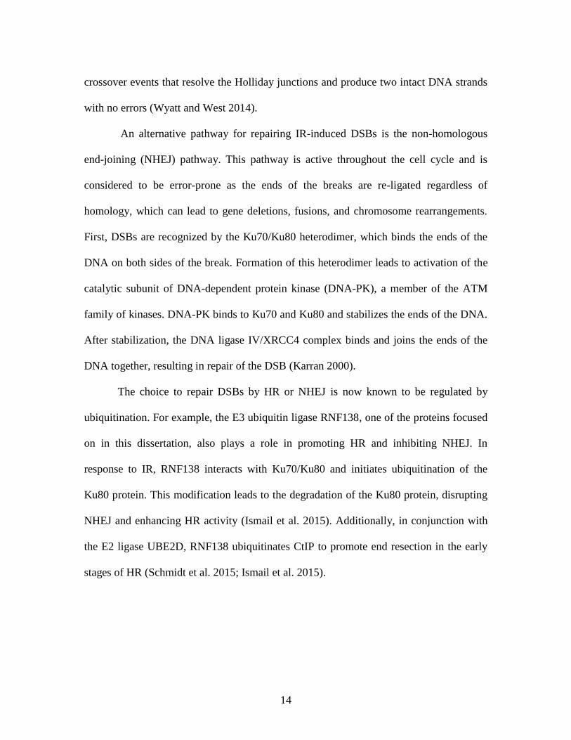
An alternative pathway for repairing IR-induced DSBs is the non-homologous
end-joining (NHEJ) pathway. This pathway is active throughout the cell cycle and is
considered to be error-prone as the ends of the breaks are re-ligated regardless of
homology, which can lead to gene deletions, fusions, and chromosome rearrangements.
First, DSBs are recognized by the Ku70/Ku80 heterodimer, which binds the ends of the
DNA on both sides of the break. Formation of this heterodimer leads to activation of the
catalytic subunit of DNA-dependent protein kinase (DNA-PK), a member of the ATM
family of kinases. DNA-PK binds to Ku70 and Ku80 and stabilizes the ends of the DNA.
After stabilization, the DNA ligase IV/XRCC4 complex binds and joins the ends of the
DNA together, resulting in repair of the DSB (Karran 2000).
The choice to repair DSBs by HR or NHEJ is now known to be regulated by
ubiquitination. For example, the E3 ubiquitin ligase RNF138, one of the proteins focused
on in this dissertation, also plays a role in promoting HR and inhibiting NHEJ. In
response to IR, RNF138 interacts with Ku70/Ku80 and initiates ubiquitination of the
Ku80 protein. This modification leads to the degradation of the Ku80 protein, disrupting
NHEJ and enhancing HR activity (Ismail et al. 2015). Additionally, in conjunction with
the E2 ligase UBE2D, RNF138 ubiquitinates CtIP to promote end resection in the early
stages of HR (Schmidt et al. 2015; Ismail et al. 2015).
15
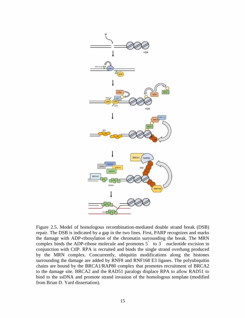
Figure 2.5. Model of homologous recombination-mediated double strand break (DSB)
repair. The DSB is indicated by a gap in the two lines. First, PARP recognizes and marks
the damage with ADP-ribosylation of the chromatin surrounding the break. The MRN
complex binds the ADP-ribose molecule and promotes 5՛ to 3՛ nucleotide excision in
conjunction with CtIP. RPA is recruited and binds the single strand overhang produced
by the MRN complex. Concurrently, ubiquitin modifications along the histones
surrounding the damage are added by RNF8 and RNF168 E3 ligases. The polyubiquitin
chains are bound by the BRCA1/RAP80 complex that promotes recruitment of BRCA2
to the damage site. BRCA2 and the RAD51 paralogs displace RPA to allow RAD51 to
bind to the ssDNA and promote strand invasion of the homologous template (modified
from Brian D. Yard dissertation).
16
DNA interstrand crosslink repair during S phase
During DNA replication, two replication forks encounter an interstrand crosslink (ICL),
resulting in a stalled replication fork. To remove the lesion, a core complex comprised of
Fanconi Anemia (FA) proteins (FANCA, -B, -C, -E, -F, -G, -L, -M) is recruited and binds
DNA strands surrounding the lesion (Figure 2.6). The FA core complex stabilizes the
lesion and initiates recruitment of the FANCD2/FANCI heterodimer. A crucial step in
FA-mediated ICL repair is mono-ubiquitination of FANCD2 and FANCI by the FANCL
E3 ligase, a modification that activates the complex (Kim and D'Andrea 2012; Rickman
et al. 2015; Liang et al. 2016). Activation of FANCD2/FANCI coordinates recruitment of
FANCP/SLX4 and the endonucleases XPF, MUS81/ERCC1, and SLX1. Together, these
proteins catalyze the incision of one DNA strand on both sides of the ICL lesion,
producing a double strand break and leaving the crosslink as an overhang on the opposite
strand (Kottemann and Smogorzewska 2013; Jo and Kim 2015; Knipscheer et al. 2009).
The lesion overhang is displaced from the DNA helix and excised by nucleotide excision
repair (NER) proteins, and translesion synthesis proteins fill in the gap across the strand
(Haynes et al. 2015; Muniandy et al. 2010). Following FA function, the exonuclease CtIP
interacts with FANCD2 and excises the DNA surrounding the break to produce a single
strand overhang (Unno et al. 2014). BRCA2 and the BCDX2 complex coordinate
recruitment of RAD51 to the overhang (Clauson, Scharer, and Niedernhofer 2013).
RAD51 initiates strand invasion of the complementary intact dsDNA, allowing DNA
polymerases to fill in the gap.
17
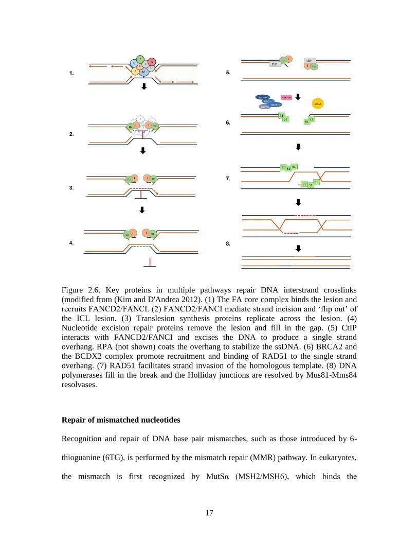
Figure 2.6. Key proteins in multiple pathways repair DNA interstrand crosslinks
(modified from (Kim and D'Andrea 2012). (1) The FA core complex binds the lesion and
recruits FANCD2/FANCI. (2) FANCD2/FANCI mediate strand incision and ‘flip out’ of
the ICL lesion. (3) Translesion synthesis proteins replicate across the lesion. (4)
Nucleotide excision repair proteins remove the lesion and fill in the gap. (5) CtIP
interacts with FANCD2/FANCI and excises the DNA to produce a single strand
overhang. RPA (not shown) coats the overhang to stabilize the ssDNA. (6) BRCA2 and
the BCDX2 complex promote recruitment and binding of RAD51 to the single strand
overhang. (7) RAD51 facilitates strand invasion of the homologous template. (8) DNA
polymerases fill in the break and the Holliday junctions are resolved by Mus81-Mms84
resolvases.
Repair of mismatched nucleotides
Recognition and repair of DNA base pair mismatches, such as those introduced by 6-
thioguanine (6TG), is performed by the mismatch repair (MMR) pathway. In eukaryotes,
the mismatch is first recognized by MutSα (MSH2/MSH6), which binds the
18
mismatch(Casorelli, Russo, and Bignami 2008). Binding of MutSα initiates recruitment
of MutLα (MLH1/PMS2) to form a MutSα/MutLα ternary complex (Li and Modrich
1995; Wang and Edelmann 2006). The complex scans along the DNA duplex until it
encounters PCNA/RFC that can be either upstream or downstream of the mismatch.
Binding activates the endonuclease activity of MutLα which generates an incision in the
daughter strand (Kadyrov et al. 2006). The exonuclease 1 (EXO1) protein recognizes the
nick and excises the strand in a 5՛ to 3՛ direction through the mismatch (Li 2008;
Kadyrov et al. 2006). RPA binds the ssDNA overhang to stabilize strand that is produced
by EXO1. DNA polymerase δ fills in the gap, and DNA ligase I seals the nick (Pena-Diaz
and Jiricny 2012; Jiricny 2006). A new mismatch re-activates the MMR proteins, and the
process repeats itself (York and Modrich 2006). Prolonged activation of MMR proteins
can result in a futile cycle that leads to a double strand break that is repaired by HR
proteins (Karran 2001).
The RecA and RAD51 connection
The Escherichia coli protein, RecA, is a highly conserved recombinase that promotes
recombination-mediated repair of DNA damage (Bell and Kowalczykowski 2016). In
vivo, RecA forms nucleoprotein filaments that preferentially bind single strand DNA
(ssDNA) generated by resection of double strand DNA (dsDNA) during DNA damage
response (Bell and Kowalczykowski 2016). RecA nucleofilament formation creates
tension along the strand and results in unwinding of the DNA (Singleton and Xiao 2001).
Binding of RecA to sites of damage is an essential step in the homologous recombination
19
(HR) repair pathway (Bell and Kowalczykowski 2016), and this protein functions to
facilitate strand invasion of the homologous template during DNA repair (Singleton and
Xiao 2001).
The yeast RecA homolog, RAD51, is essential for maintaining genome stability
and integrity throughout the mitotic cell cycle and during meiosis (Krogh and Symington
2004; Shinohara, Ogawa, and Ogawa 1992). Similar to RecA, yeast RAD51, forms
helical nucleoprotein filaments along ssDNA that promote strand exchange activity in an
ATP-dependent manner (Shinohara, Ogawa, and Ogawa 1992; Ogawa et al. 1993;
Shinohara and Ogawa 1999; Sung 1994; Conway et al. 2004; Chen et al. 2010). Alanine
substitution at lysine 191 in yeast RAD51 (K191A)1 diminished ATPase function, and
decreased mitotic recombination activity of the protein. Additionally, cells expressing the
RAD51-K191A mutant were more sensitive to ionizing radiation than wild-type
expressing cells (Morgan, Shah, and Symington 2002). Another mutant, RAD51-H352Y,
displayed similar ssDNA binding activity as wild-type RAD51, but was defective for
nucleotide exchange and strand exchange activity. Structural analysis determined that
RAD51-H352Y binding to ssDNA stabilized a nearby phenylalanine (F187) residue and
blocked the γ-phosphate binding site of a Walker Box A motif (described below),
impairing ATPase activity of the protein (Chen et al. 2010). These data further
demonstrated that RAD51 activity is ATP-dependent.
A mammalian homolog of the RecA and yeast RAD51 protein, also named
RAD51, is essential for HR in response to ultraviolet radiation (Morita et al. 1993). Early
1 Note that the nomenclature used to represent amino acid substitutions is the “wild-type single
letter amino acid designation” followed by the “residue number” then the “substituted amino acid
designation” (e.g. K191A). This nomenclature will be used throughout this dissertation.
20
studies demonstrated that expressing Mus musculus (MmRad51) in RAD51-deficient
yeast restored cell survival, particularly in response to double strand breaks (DSBs). The
mouse Rad51 gene has approximately 83% and 55% identity with the yeast RAD51 and
E. coli RecA genes, respectively (Morita et al. 1993). RAD51 monomers interact to form
oligomers in free solution prior to binding to ssDNA, and the size of the oligomer affects
DNA binding, with smaller oligomers binding more readily to ssDNA than larger
oligomers (Sung et al. 2003; Candelli et al. 2014). Structural analysis using cryo-electron
microscopy found that RAD51 nucleofilaments form a helical structure around the
ssDNA (Xu et al. 2017; Short et al. 2016).
Although RAD51 oligomers have a higher affinity for ssDNA, the ability to bind
to dsDNA is essential for promoting homology search and strand exchange during repair
(Danilowicz et al. 2014). When bound to dsDNA, RAD51 filaments extend the DNA
strand and create tension along the helix and unwinding the DNA (Benson, Stasiak, and
West 1994). This tension exposes Watson-Crick base pairings and allows for brief
bonding of non-homologous ssDNA to the dsDNA during the homology template search
(Danilowicz et al. 2014).
Mammalian RAD51 Paralogs
During homologous recombination (HR), the five RAD51 paralogs – RAD51B,
RAD51C, RAD51D, XRCC2, and XRCC3 – alleviate competition between RAD51 and
RPA for DNA binding, promoting RAD51 loading onto single stranded DNA
(Sigurdsson et al. 2001), and assist in Holliday junction resolution (Liu et al. 2007;
21
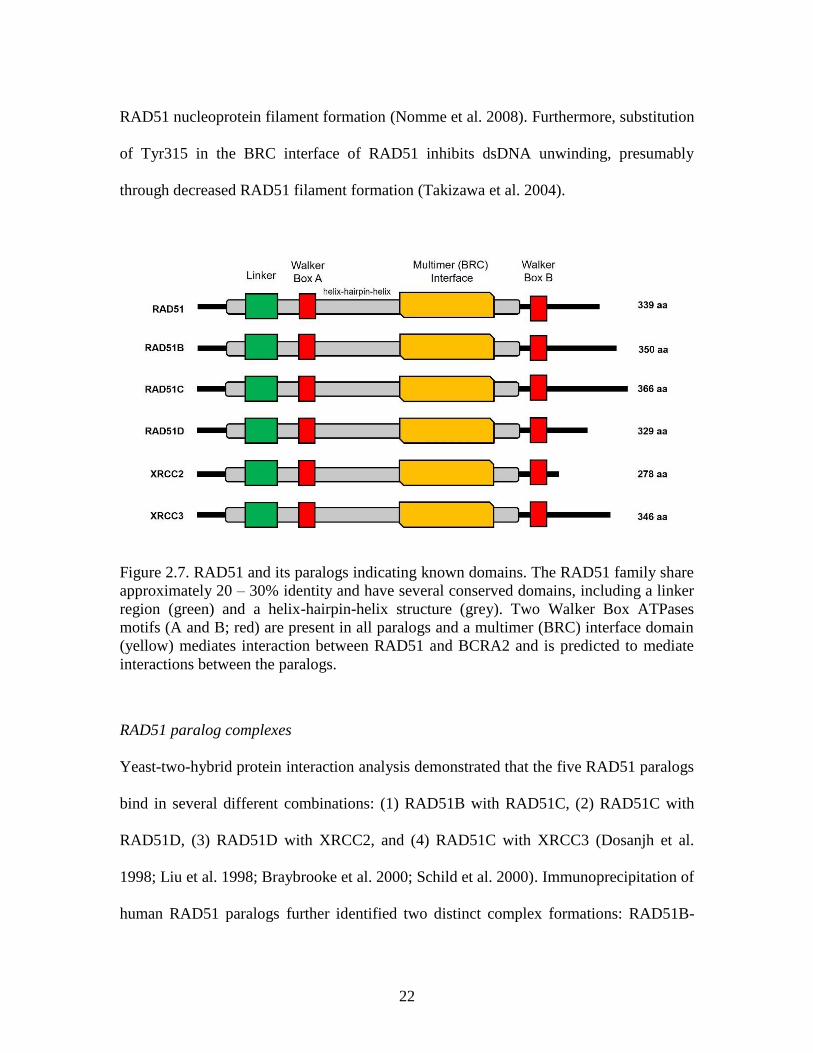
Compton, Ozgur, and Griffith 2010). Each paralog has the conserved Walker Box A and
B ATPase motifs, multimer (BRC) interface, and helix-hairpin-helix region discussed
below (Figure 2.7) (Miller et al. 2004; Kawabata, Kawabata, and Nishibori 2005).
The Walker Box A and B motifs are ATP binding sites that catalyze the
hydrolysis of ATP to promote ssDNA binding activity of the paralogs (Braybrooke et al.
2000; Chen et al. 2010; Gruver et al. 2005; Kawabata, Kawabata, and Nishibori 2005),
and mutations in the Walker Box A motif have been shown to decrease ATPase activity
and to increase cellular sensitivity to DNA damaging agents. Arginine substitution at a
conserved lysine residue in the Walker Box A motif of the RAD51 paralogs decreases
recombination activity. Substitution at K113 (K113R) in RAD51D lead to cell death in
response to DNA damaging agents in mouse embryonic fibroblasts and Chinese hamster
ovarian (CHO) cells (Wiese et al. 2006; Gruver et al. 2005). K113R interaction with
RAD51C and XRCC2 was 8- and 2-fold lower than wild-type RAD51D, respectively
(Gruver et al. 2005). Substitutions at K113 in Walker Box A of XRCC3 eliminated
ATPase activity and lead to prolonged association between XRCC3 and RAD51C,
suggesting that ATP hydrolysis activity is required to regulate paralog complex formation
(Yamada et al. 2004). Loss of function Walker Box B mutants fail to complement
Rad51d-deficiency in CHO cells motif in the presence of MMC (Wiese et al. 2006).
The BRC interface is a region of homology between the RAD51 paralogs and the
breast cancer associated 2 (BRCA2) protein (Pellegrini et al. 2002; Lo et al. 2003).
Interestingly, RAD51 interacts with BRCA2 through this interface, but none of the other
paralogs have been shown to bind BRCA2 (Lo et al. 2003). Peptide fragments from the
BRC region of BRCA2 act as an inhibitor of RAD51 binding to BRCA2 and prevent
22
RAD51 nucleoprotein filament formation (Nomme et al. 2008). Furthermore, substitution
of Tyr315 in the BRC interface of RAD51 inhibits dsDNA unwinding, presumably
through decreased RAD51 filament formation (Takizawa et al. 2004).
Figure 2.7. RAD51 and its paralogs indicating known domains. The RAD51 family share
approximately 20 – 30% identity and have several conserved domains, including a linker
region (green) and a helix-hairpin-helix structure (grey). Two Walker Box ATPases
motifs (A and B; red) are present in all paralogs and a multimer (BRC) interface domain
(yellow) mediates interaction between RAD51 and BCRA2 and is predicted to mediate
interactions between the paralogs.
RAD51 paralog complexes
Yeast-two-hybrid protein interaction analysis demonstrated that the five RAD51 paralogs
bind in several different combinations: (1) RAD51B with RAD51C, (2) RAD51C with
RAD51D, (3) RAD51D with XRCC2, and (4) RAD51C with XRCC3 (Dosanjh et al.
1998; Liu et al. 1998; Braybrooke et al. 2000; Schild et al. 2000). Immunoprecipitation of
human RAD51 paralogs further identified two distinct complex formations: RAD51B-
23
RAD51C-RAD51D-XRCC2 (BCDX2) and RAD51C-XRCC3 (CX3) (Masson et al.
2001; Rajesh et al. 2009). Structural analysis using transmission electron microscopy
revealed that both the BCDX2 and CX3 complexes form a multimeric ring structure
arranged in a flat disc around DNA Holliday junctions (Compton, Ozgur, and Griffith
2010).
The BCDX2 complex preferentially binds to two distinct DNA structures: Y-
shaped DNA and DNA Holliday junctions (Yokoyama et al. 2004), and is required for
RAD51 foci formation in response to IR-induced DSBs (Chun, Buechelmaier, and
Powell 2013). Depletion of the RAD51D gene decreased RAD51 foci formation, but
depletion of XRCC3 did not affect RAD51 foci formation in response to IR suggesting
that the BCDX2, not the CX3 complex, is responsible for recruiting RAD51 to damage
sites following IR treatment (Chun, Buechelmaier, and Powell 2013). Additionally, the
BCDX2 complex stabilizes Holliday junctions and promotes proper resolution of the
DNA strands (Liu et al. 2004; Liu et al. 2007; Chun, Buechelmaier, and Powell 2013).
Deletion/disruption mutations of RAD51 paralogs in the mouse genetic model
To date, deletions of the RAD51 protein family have only been generated in mouse
embryonic fibroblasts (MEFs), DT40 avian cells, or Chinese hamster ovarian cells, and
loss of each gene results in an embryo lethal phenotype (Takata et al. 2001; Deans et al.
2003; Hinz et al. 2006; Griffin et al. 2000; Shu et al. 1999; Pittman and Schimenti 2000;
Lim and Hasty 1996; Tsuzuki et al. 1996; Kuznetsov et al. 2009). For the purposes of this
dissertation, only mouse gene deletions of the RAD51 paralogs will be discussed.

24
To study the Rad51 gene deletion, heterozygous Rad51 (Rad51+/-
) mice were
intercrossed, and out of 148 offspring, zero pups were Rad51-null (Rad51-/-
) (Tsuzuki et
al. 1996). Early development analysis of potential Rad51-/-
embryos found that one out of
nine 2-cell stage embryos, and one out of 109 4- to 8-cell stage embryos were Rad51-null
(Tsuzuki et al. 1996). Additionally, blastocysts isolated from Rad51-/-
embryos failed to
divide in culture (Tsuzuki et al. 1996; Lim and Hasty 1996). To prolong embryo
development, Rad51+/-
mice were intercrossed with heterozygous Trp53 (Trp53+/-
) mice,
and it was observed that the embryo lethal phenotype can be partially rescued when the
Rad51 gene deletion is generated on a Trp53-null (Trp53-/-
) background (Shu et al. 1999;
Lim and Hasty 1996). Rad51-/-
Trp53-/-
embryos were slightly smaller than control
littermates (Lim and Hasty 1996), and developed to 8.5 days post conception (dpc)
(Tsuzuki et al. 1996). However, out of 41 pups, zero were Rad51-/-
Trp53-/-
, demonstrating
that the Trp53-/-
background did not restore offspring survival (Lim and Hasty 1996).
Despite embryo survival being extended by the concurrent deletion of Trp53, cells
derived from Rad51-/-
Trp53-/-
embryos did not proliferate in cell culture (Lim and Hasty
1996).
To generate Rad51c-deficient mice, heterozygous Rad51c (Rad51c+/-
) mice were
intercrossed, and no viable pups were obtained. It was also observed that the ratio of live
births for wild-type versus Rad51c+/-
deviated from the Mendelian 2:1 ratio, suggesting
that loss of one Rad51c allele might affect embryo development (Kuznetsov et al. 2009).
Rad51c embryos were phenotypically abnormal at 7.5 and 8.5 dpc compared with wild-
type Rad51c embryos, and TUNEL staining of cells isolated from these embryos showed
increased levels of apoptosis. Concurrent deletion of the Trp53 gene extended embryo
25
development to 10.5 dpc. These embryos were smaller than littermate controls and did
not appear to develop further. MEF cell lines were successfully generated from Rad51c-/-
Trp53-/-
embryos (Kuznetsov et al. 2009).
Deletion of the Rad51d gene has only been successful in MEFs (Smiraldo et al.
2005) and Chinese hamster ovarian cells (Hinz et al. 2006). Heterozygous Rad51d
(Rad51d+/-
) mice were intercrossed and out of 102 live births, none were Rad51d-
deficient (Rad51d-/-
). It was determined that embryo death occurred between 8.5 and 11.5
dpc in Rad51d-/-
embryos (Pittman and Schimenti 2000). Deletion of the Trp53 gene in
Rad51d-/-
embryos extended embryo development to 15.5 dpc but did not result in live
pups (Smiraldo et al. 2005). Rad51d-/-
embryos exhibit severe developmental and
chromosomal defects compared to Rad51d+/-
littermates (Smiraldo et al. 2005). Similar to
cells isolated from Rad51-/-
embryos, Rad51d-/-
Trp53+/+
cells did not grow in culture
(Pittman and Schimenti 2000), but Rad51d-/-
Trp53-/-
cells were able to proliferate in
culture (Smiraldo et al. 2005).
Consistent with deletion of other RAD51 paralogs, loss of Xrcc2 in mice resulted
in embryo lethality. Embryo death occurred throughout gestation between 9.5 – 18.5 dpc.
The observed neonatal lethality in these embryos appeared to be due to respiratory failure
and was attributed to a high frequency of apoptosis in post-mitotic neurons (Deans et al.
2000). Deletion of the Trp53 gene in Xrcc2-/-
mice extended embryo development from
12.5 dpc to 18.5 dpc but did not result in any live births. Cells isolated from Xrcc2-/-
embryos did not proliferate in culture, but consistent with Rad51c-/-
and Rad51d-/-
cells,
deletion of the Trp53 gene allowed for Xrcc2-/-
Trp53-/-
MEF cell lines to be grown in
culture (Adam, Deans, and Thacker 2007).
26
Given the importance of RAD51 paralogs in embryo development and cell
survival, it follows that deletion of these genes in human cells would provide further
insight into their function. However, to date, no successful attempts to generate RAD51
paralog gene deletions in human cells has been reported. RNA interference has been used
to transiently decrease gene expression of RAD51 paralogs in human U2OS, MCF7,
HT1080, HeLa, and T84 cells (Jensen et al. 2013; Chun, Buechelmaier, and Powell 2013;
Lio et al. 2004; Lee et al. 2014; Wang et al. 2014; Loignon et al. 2007). In human WI38-
VA13/2RA cells, depletion of RAD51D by two separate siRNAs resulted in apoptosis
within seven days of transfection (Tarsounas et al. 2004). A different siRNA used in the
same study resulted in cell death at day 5 when only 50% of the RAD51D protein was
depleted (Tarsounas et al. 2004). Depletion of the RAD51D gene by 84% using siRNA in
HT1080 and HEK293 cells increased chemosensitivity of these cells to the DNA damage
agent mitomycin C (Jensen et al. 2013). Depletion of RAD51D in human U20S and
MCF7 cells also significantly decreased repair of DNA double strand breaks induced by
the SceI endonuclease (Chun, Buechelmaier, and Powell 2013).
Disease phenotypes associated with RAD51 genes
The National Institutes of Health provides the ClinVar database, a publicly accessible
archive of reports designed to support the evolution of understanding of the relationship
between genotypes and clinically observed phenotypes, and to establish the clinical
validity of an identified gene variant (Landrum et al. 2016). The mutant alleles listed in
the ClinVar database have been identified through clinical testing, research, or extracted
from the literature, and have been associated with disease phenotypes. Germline
mutations in Homo sapiens RAD51, RAD51C, RAD51D, and XRCC3, have been
27
reported to the NIH ClinVar database and are classified as risk factors for breast, ovarian,
and melanoma cancers (Table 2.1).
An intron variant of RAD51 increases disease risk in breast cancer patients that
also carried BRCA2, but not BRCA1, mutations (Levy-Lahad et al. 2001). In this study,
patients homozygous for the G>C single nucleotide polymorphism had a significant
increase in disease risk compared to patients that were heterozygous G/C or homozygous
G/G. Risk was further increased in patients that carried mutations in the BRCA2 gene.
This mutation modifies splicing in the 5՛ UTR of the RAD51 gene and influences
RAD51 expression levels in patients (Antoniou et al. 2007). Interestingly, the increased
risk in BRCA2-mutant patients was only associated with breast but not ovarian cancer
risk (Levy-Lahad et al. 2001).
Three mutations in the RAD51C genes have been linked with an increased risk of
cancer, most notably breast and ovarian cancers (Loveday et al. 2011; Loveday et al.
2012; Meindl et al. 2010; Pelttari et al. 2011; Thompson et al. 2012; Kuschel et al. 2002)
(Table 2.1). RAD51C germline mutations have long been associated with increased risk
for ovarian cancer (Loveday et al. 2012; Meindl et al. 2010; Pelttari et al. 2011; Song et
al. 2015), while a connection with breast cancer risk has been debated in the literature
(Schnurbein et al. 2013; Thompson et al. 2012). Novel splice-variant mutations that result
in truncated RAD51C protein have been identified in both breast and ovarian cancer
patients (Neidhardt et al. 2017), and pathogenic RAD51C variants have been detected in
patients with a personal history of triple negative breast cancer (Buys et al. 2017). Six
RAD51D mutations that increased a patient’s risk for developing ovarian cancer, but not
breast cancer, have been identified (Kraus et al. 2017; Song et al. 2015; Loveday et al.
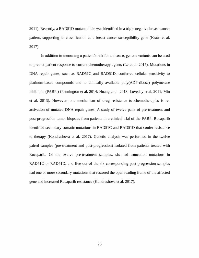
28
2011). Recently, a RAD51D mutant allele was identified in a triple negative breast cancer
patient, supporting its classification as a breast cancer susceptibility gene (Kraus et al.
2017).
In addition to increasing a patient’s risk for a disease, genetic variants can be used
to predict patient response to current chemotherapy agents (Le et al. 2017). Mutations in
DNA repair genes, such as RAD51C and RAD51D, conferred cellular sensitivity to
platinum-based compounds and to clinically available poly(ADP-ribose) polymerase
inhibitors (PARPi) (Pennington et al. 2014; Huang et al. 2013; Loveday et al. 2011; Min
et al. 2013). However, one mechanism of drug resistance to chemotherapies is re-
activation of mutated DNA repair genes. A study of twelve pairs of pre-treatment and
post-progression tumor biopsies from patients in a clinical trial of the PARPi Rucaparib
identified secondary somatic mutations in RAD51C and RAD51D that confer resistance
to therapy (Kondrashova et al. 2017). Genetic analysis was performed in the twelve
paired samples (pre-treatment and post-progression) isolated from patients treated with
Rucaparib. Of the twelve pre-treatment samples, six had truncation mutations in
RAD51C or RAD51D, and five out of the six corresponding post-progression samples
had one or more secondary mutations that restored the open reading frame of the affected
gene and increased Rucaparib resistance (Kondrashova et al. 2017).
29
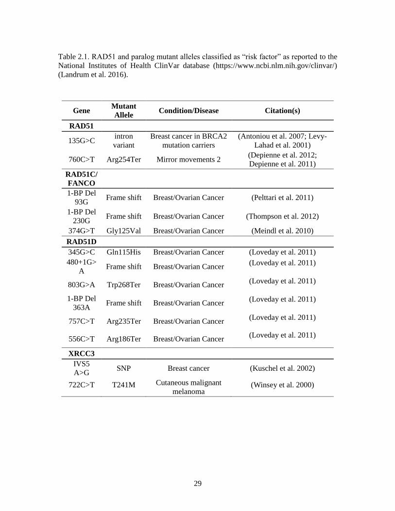
Table 2.1. RAD51 and paralog mutant alleles classified as “risk factor” as reported to the
National Institutes of Health ClinVar database (https://www.ncbi.nlm.nih.gov/clinvar/)
(Landrum et al. 2016).
Gene Mutant
Allele Condition/Disease Citation(s)
RAD51
135G>C intron
variant
Breast cancer in BRCA2
mutation carriers
(Antoniou et al. 2007; Levy-
Lahad et al. 2001)
760C>T Arg254Ter Mirror movements 2 (Depienne et al. 2012;
Depienne et al. 2011)
RAD51C/
FANCO
1-BP Del
93G Frame shift Breast/Ovarian Cancer (Pelttari et al. 2011)
1-BP Del
230G Frame shift Breast/Ovarian Cancer (Thompson et al. 2012)
374G>T Gly125Val Breast/Ovarian Cancer (Meindl et al. 2010)
RAD51D
345G>C Gln115His Breast/Ovarian Cancer (Loveday et al. 2011)
480+1G>
A Frame shift Breast/Ovarian Cancer
(Loveday et al. 2011)
803G>A Trp268Ter Breast/Ovarian Cancer (Loveday et al. 2011)
1-BP Del
363A Frame shift Breast/Ovarian Cancer
(Loveday et al. 2011)
757C>T Arg235Ter Breast/Ovarian Cancer (Loveday et al. 2011)
556C>T Arg186Ter Breast/Ovarian Cancer (Loveday et al. 2011)
XRCC3
IVS5
A>G SNP Breast cancer (Kuschel et al. 2002)
722C>T T241M Cutaneous malignant
melanoma (Winsey et al. 2000)
30
Ubiquitin modification of Proteins
DNA damage repair pathways are regulated by post-translational modifications (PTMs).
Ubiquitination events during homologous recombination (HR)-mediated double strand
break (DSB) repair are essential for pathway progression and accurate repair of the
damage. Ubiquitin modification can activate proteins, initiate recruitment and binding of
downstream proteins to a damage site, or signal proteasomal-mediated protein
degradation (Akutsu, Dikic, and Bremm 2016). A small ubiquitin-like modifier (SUMO)
molecule can also be added to proteins, and crosstalk between ubiquitin modifications
and SUMO-modifications promotes DNA damage response and repair. These regulatory
modifications will be discussed in this section.
Ubiquitin is a conserved 76 amino acid protein expressed throughout the cell.
Modifications occur when the ubiquitin molecule is covalently linked at its C-terminus
glycine residue (Gly76) to the ε-amine group of a lysine residue or to the N-terminus of
the substrate (Busch and Goldknopf 1981). Ubiquitins are attached to a target protein by
a three-step enzymatic reaction performed by E1 (ubiquitin-activating), E2 (ubiquitin-
conjugating), and E3 (ubiquitin-ligating) enzymes (Figure 2.8A and B) (Pickart 2004;
Hershko 1983). Binding of the ubiquitin molecule to an E1 enzyme consumes ATP and
activates the ubiquitin by generating a high-energy thioester bond between the C-
terminus of the ubiquitin and the cysteine in the E1 active site (Pickart 2004). Following
activation, the ubiquitin molecule is transferred to an E2 enzyme. E2s have a core
ubiquitin-conjugating domain that forms an active site to bind an activated ubiquitin
molecule via a highly conserved cysteine residue. Ubiquitin-bound E2s interact with two
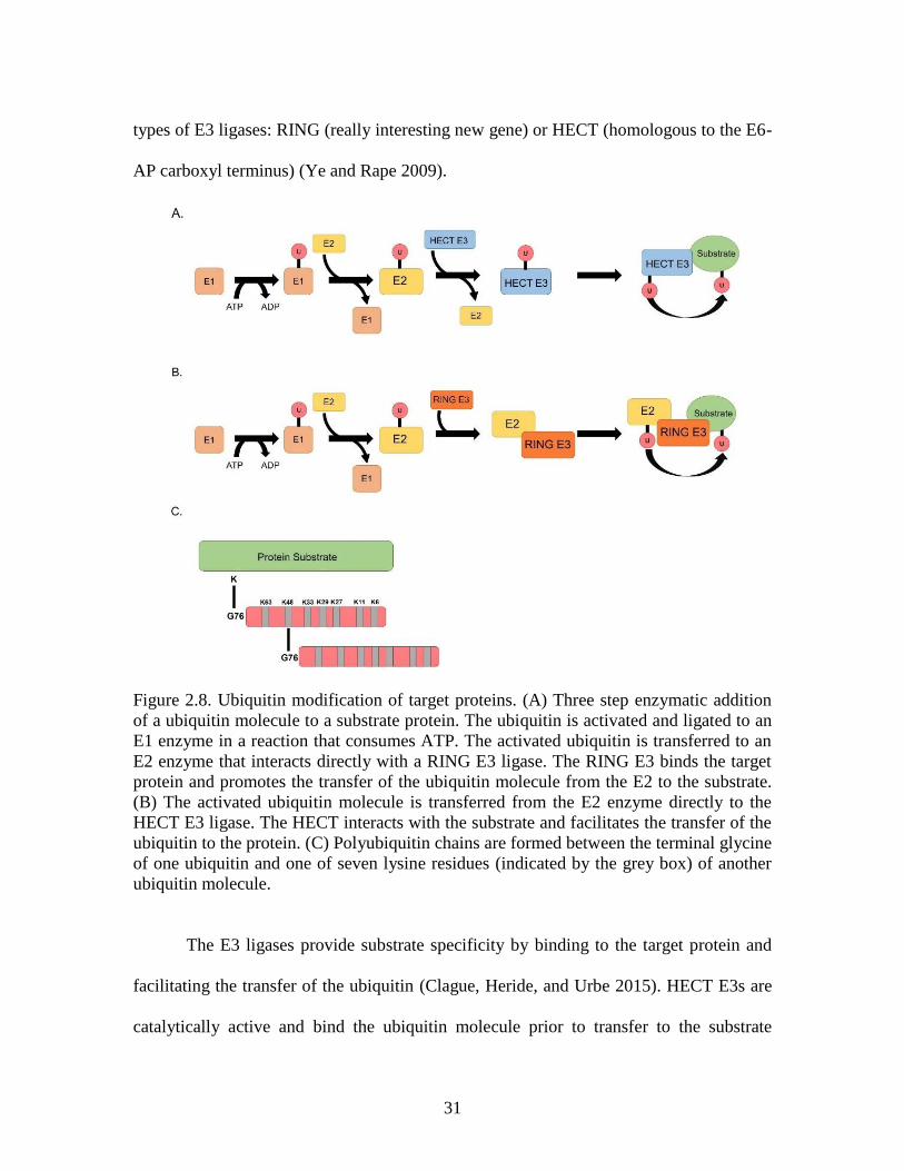
31
types of E3 ligases: RING (really interesting new gene) or HECT (homologous to the E6-
AP carboxyl terminus) (Ye and Rape 2009).
Figure 2.8. Ubiquitin modification of target proteins. (A) Three step enzymatic addition
of a ubiquitin molecule to a substrate protein. The ubiquitin is activated and ligated to an
E1 enzyme in a reaction that consumes ATP. The activated ubiquitin is transferred to an
E2 enzyme that interacts directly with a RING E3 ligase. The RING E3 binds the target
protein and promotes the transfer of the ubiquitin molecule from the E2 to the substrate.
(B) The activated ubiquitin molecule is transferred from the E2 enzyme directly to the
HECT E3 ligase. The HECT interacts with the substrate and facilitates the transfer of the
ubiquitin to the protein. (C) Polyubiquitin chains are formed between the terminal glycine
of one ubiquitin and one of seven lysine residues (indicated by the grey box) of another
ubiquitin molecule.
The E3 ligases provide substrate specificity by binding to the target protein and
facilitating the transfer of the ubiquitin (Clague, Heride, and Urbe 2015). HECT E3s are
catalytically active and bind the ubiquitin molecule prior to transfer to the substrate
32
(Figure 2.7A) (Scheffner and Kumar 2014). RING E3 ligases are catalytically inactive,
and act as scaffolds between the E2 enzyme and the substrate, promoting E2-dependent
ubiquitination of target proteins (Figure 2.7B) (Lorick et al. 1999). The RING domain
serves as the interacting region between the E3 and the E2 enzymes. The structure of this
domain is a double loop that coordinates binding of two zinc ions, producing a surface for
the E2 to bind (Metzger et al. 2014).
Ubiquitins can be added to single or multiple lysine residues along a target protein
to produce mono- and multi-mono modifications, or ubiquitin chains can be generated
along a single lysine residue. Polyubiquitin chains are formed when multiple ubiquitins
are attached directly to each other through isopeptide bonds between a lysine residue on
the previous molecule and Gly76 on the subsequent molecule (Figure 2.7C) (Akutsu,
Dikic, and Bremm 2016; Swatek and Komander 2016). There are seven lysine residues
along the protein that can be used to generate poly-ubiquitin chains (Sloper-Mould et al.
2001). The function of a polyubiquitin chain is determined by the lysine residue along
each ubiquitin that forms the isopeptide bond. For example, chains generated between
Lys48 of ubiquitin molecules (referred to as K48 linked chains) target a substrate for
proteasomal degradation (Akutsu, Dikic, and Bremm 2016).
Ubiquitin chains have characteristic structural conformations that mediate
recognition by chain-specific enzymes and binding proteins (Thach et al. 2016; Ikeda,
Crosetto, and Dikic 2010). The ‘closed’ conformation occurs when two ubiquitin
moieties directly interact via a hydrophobic patch in the middle of the ubiquitin protein.
The ‘open’ conformation is observed when two ubiquitins are only linked by the
isopeptide bond between the terminal glycine of one molecule and the lysine residue of
33
the second molecule (Ye et al. 2012). In solution, di-ubiquitin K63 chains adopt both
‘open’ and compact ‘closed’ conformations that are recognized by ubiquitin interacting
motifs of DNA damage response proteins (Sato et al. 2009). Di-ubiquitin K48 chains
predominantly adopt a ‘closed’ conformation recognized by the 19S subunit of the
proteasome and target a protein for proteasomal degradation (Thach et al. 2016; Varadan
et al. 2004; Ye et al. 2012; Chau et al. 1989).
The proteolytic component of the proteasome is a 20S unit is comprised of four
homologous rings stacked together. The top and bottom rings are formed by seven α-
subunits that recognize unfolded protein peptide chains. The two inner rings consist of
seven β-subunits and form the proteolytic chamber of the proteasome (Cromm and Crews
2017). In addition to the core particle, there are various ‘cap’ structures that bind the
proteasome. The 19S regulatory particle binds to one or both of the α-subunit rings of the
20S core particle to form the 26S proteasome (Guo and Peng 2013; Cromm and Crews
2017). The 19S subunit recognizes and cleaves ubiquitin chains along a substrate in an
ATP-dependent manner. After removal of the ubiquitins, the substrate is shuttled into the
proteolytic chamber and is degraded. The active site of the 19S subunit is specific for the
‘closed’ conformation of K48-linked ubiquitin chains, therefore, only these chains lead to
proteasomal degradation (Ye et al. 2012; Chau et al. 1989).
Proteasome inhibition has been a mechanism of disease treatment for several
decades. Bortezomib is a proteasome inhibitor approved by the United States Food and
Drug Administration for the treatment of multiple myeloma since 2003, and remains a
first-line treatment for the disease (Dou and Zonder 2014; Grosicki et al. 2014; Kane et
al. 2003; Kouroukis et al. 2014; Schlafer et al. 2017). MG132 is a peptide aldehyde and a
34
naturally occurring proteasome inhibitor derived from a Chinese medicinal plant. This
compound binds the active site of the β-subunits of the 26S proteasome, preventing
proteolysis of ubiquitinated substrates (Guo and Peng 2013; Rock et al. 1994).
SUMOylation
Protein modification with SUMO (small ubiquitin-like modifier) molecules has emerged
as another regulatory mechanism of DNA damage repair pathways (Nie and Boddy 2016;
Pichler et al. 2017). There are five known SUMO paralogs expressed in mammalian
cells: SUMO1, SUMO2/3, SUMO4, and SUMO5. SUMO1 has approximately 50%
identity with SUMO2 and SUMO3, while the latter two paralogs have above 97%
identity, and comprise the majority of the cell pool (Saitoh and Hinchey 2000; Pichler et
al. 2017). Before being attached to a substrate, SUMO molecules must be processed by
SUMO-specific proteases that cleave the C-terminus to produce a di-glycine motif that
can be attached to ε-amine of lysine residues along target proteins (Eifler and Vertegaal
2015). Unlike SUMO1 and SUMO2/3, SUMO4 has a proline residue that prevents
processing by any known SUMO-specific proteases and is not thought to modify proteins
(Owerbach et al. 2005). SUMO molecules attached to target proteins at a SUMO
consensus sequence (ψKxE where ψ is a bulky hydrophobic residue and E is an acidic
residue) (Pichler et al. 2017; Hendriks et al. 2017; Bernier-Villamor et al. 2002), often
found near phosphorylation sites along substrates (Hendriks et al. 2017).
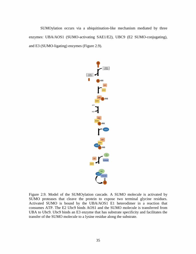
35
SUMOylation occurs via a ubiquitination-like mechanism mediated by three
enzymes: UBA/AOS1 (SUMO-activating SAE1/E2), UBC9 (E2 SUMO-conjugating),
and E3 (SUMO-ligating) enzymes (Figure 2.9).
Figure 2.9. Model of the SUMOylation cascade. A SUMO molecule is activated by
SUMO proteases that cleave the protein to expose two terminal glycine residues.
Activated SUMO is bound by the UBA/AOS1 E1 heterodimer in a reaction that
consumes ATP. The E2 Ubc9 binds AOS1 and the SUMO molecule is transferred from
UBA to Ubc9. Ubc9 binds an E3 enzyme that has substrate specificity and facilitates the
transfer of the SUMO molecule to a lysine residue along the substrate.
36
First, SUMO is bound by the UBA/AOS1 heterodimer and ATP hydrolysis activates the
SUMO molecule. Activated SUMO forms a high-energy thioester bond with a cysteine
residue in the active site of the AOS1 enzyme. AOS1 binds the UBC9 E2 enzyme, and
SUMO is transferred to the cysteine residue in the active site of UBC9. Unlike in the
ubiquitin-proteasome pathway, UBA/AOS1 and UBC9 are the only SUMO-associated E1
and E2 ligases identified in humans (Pichler et al. 2017). The SUMO-bound UBC9
protein interacts with one of several E3 enzymes, which have target specificity, and
facilitates SUMO transfer from UBC9 to the substrate (Pichler et al. 2017).
Post-translational modifications along RAD51 proteins
Post-translational modifications (PTMs) play a vital role in promoting accurate repair of
DNA damage and are prominent in the homologous recombination (HR) pathway. The
six RAD51 paralog proteins are essential for progression of HR, therefore, modifications
along these proteins are predicted to be required for HR progression. Proteomics analyses
of the RAD51 proteins identified PTMs along each paralog (Figure 2.10) (Mertins et al.
2016; Mertins et al. 2013; Kim et al. 2011; Zhou et al. 2013; Somyajit et al. 2013;
Slupianek et al. 2001; Takizawa et al. 2004; Yata et al. 2014; Yata et al. 2012; Yuan et al.
1998). Despite these data, limited experiments have been performed under DNA damage
conditions. Given the prevalence of phosphorylation and ubiquitin modifications in DNA
damage response, particularly in the HR pathway, and the importance of each paralog in
promoting accurate DNA repair, it follows that investigation into these modifications is
necessary.
37
In response to IR, RAD51 activity is regulated by phosphorylation. During S
phase, phosphorylation along RAD51 is mediated by c-Abl at Tyr54 and Tyr315 (Yuan et
al. 1998; Chen et al. 1999; Subramanyam et al. 2016; Takizawa et al. 2004). RAD51 is
activated at the S/G2 junction of the cell cycle by the Chk1-mediated phosphorylation at
Thr309 to promote HR (Sorensen et al. 2005). At the G2/M checkpoint, phosphorylation
at Thr13 and Ser14 initiates RAD51 binding with the NBS1 protein to recruit RAD51 to
the DSB site (Yata et al. 2014).
RAD51 activity during S phase is regulated by c-Abl-mediated phosphorylation at
Tyr54 and Tyr315 (Yuan et al. 1998; Chen et al. 1999; Subramanyam et al. 2016;
Takizawa et al. 2004). These modifications inhibit RAD51 oligomerization and can
enhance its strand exchange activity (Subramanyam et al. 2016; Alligand et al. 2017). In
addition to interacting with RAD51, one study identified a direct interaction between
BCR-Abl and RAD51B, and found that BCR-Abl phosphorylates RAD51B (Slupianek et
al. 2009).
The cell cycle checkpoint kinase Chk1 functions during S phase of the cell cycle
(Arnaudeau, Lundin, and Helleday 2001), and directly interacts with RAD51 in response
to hydroxyurea (HU)-induced DSBs (Sorensen et al. 2005). Chk1-mediated
phosphorylation of RAD51 at Thr309 activates the protein and initiates HR (Sorensen et
al. 2005). Cells deficient for Chk1 have decreased RAD51 foci formation in response to
HU, and cells expressing a RAD51-T309A mutant that cannot be phosphorylated are
hypersensitive to DNA damaging agents including HU and thymidylate synthase
inhibitors (Sorensen et al. 2005; Yang, Waldman, and Wyatt 2012).
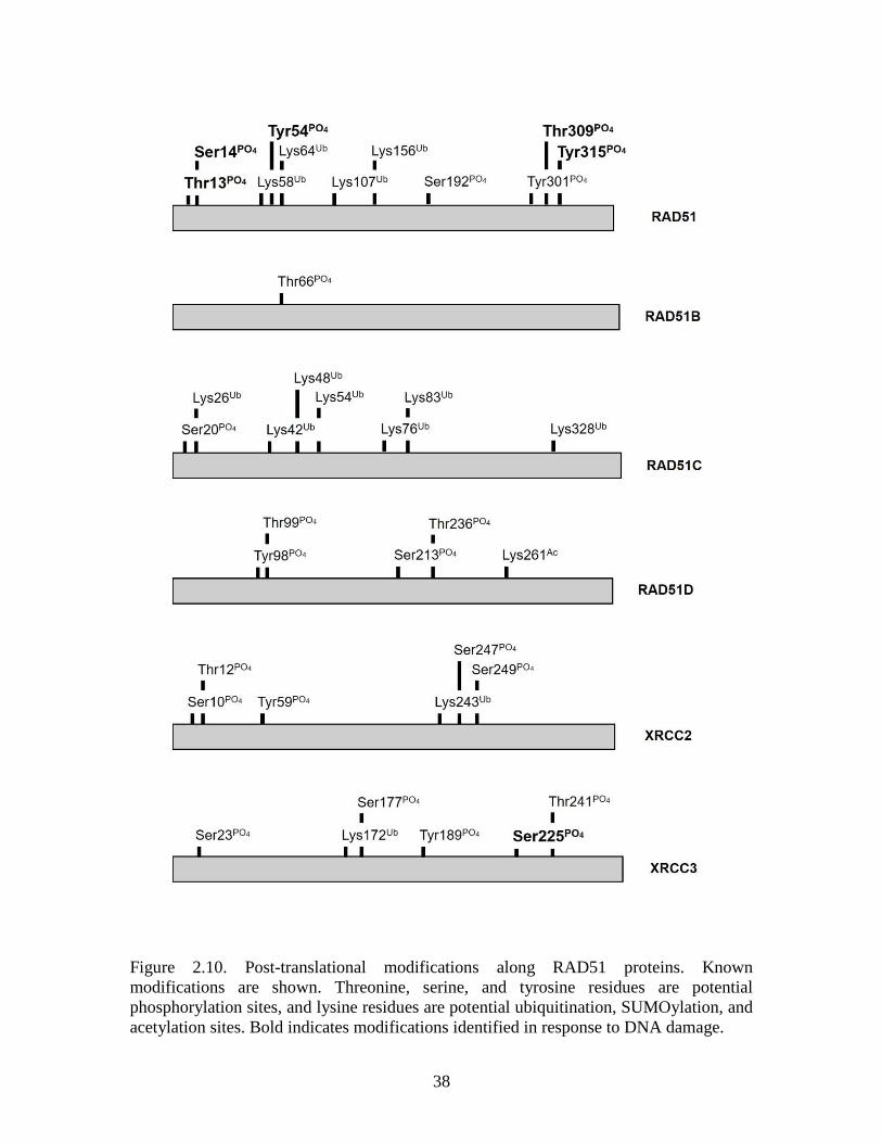
38
Figure 2.10. Post-translational modifications along RAD51 proteins. Known
modifications are shown. Threonine, serine, and tyrosine residues are potential
phosphorylation sites, and lysine residues are potential ubiquitination, SUMOylation, and
acetylation sites. Bold indicates modifications identified in response to DNA damage.
39
BRCA2, a known RAD51 binding partner, coordinates the phosphorylation of
RAD51 at Ser14 by polo-like kinase 1 (PLK1) in response to DNA damage at the G2/M
junction of the cell cycle (Yata et al. 2014). Phosphorylation at Ser14 in turn initiates
phosphorylation at Thr13 by casein kinase 2 (CK2), a modification that promotes RAD51
interaction with NBS1, a core component of the MRN complex (Yata et al. 2012).
Interaction between RAD51 and NBS1 is important for RAD51 recruitment and binding
to DNA damage sites (Yata et al. 2014).
Ubiquitination is another PTM present along RAD51. A novel E3 ubiquitin
ligase, RFWD3 ubiquitinates RAD51 in response to DSBs induced by mitomycin C
(MMC) as a mechanism of removing the protein from the single strand overhangs (Inano
et al. 2017). Inactivation of RFWD3 results in persistent RAD51 foci at the damage site
and increased cellular sensitivity to MMC. Additionally, RING mutants of RFWD3 are
defective in promoting RAD51 ubiquitination (Inano et al. 2017).
In response to IR, XRCC3 activity during S phase is regulated by ATR-mediated
phosphorylation at Ser225. interaction between RAD51C and XRCC3 is required for this
modification, and depletion of RAD51C inhibits phosphorylation of XRCC3. This
modification is dependent on ATM and is necessary for XRCC3-mediated recruitment of
RAD51 to DSBs (Somyajit et al. 2013).
All investigations into the PTMs along RAD51 proteins have been performed in
response IR-induced DSBs, and only limited studies have been performed in the presence
of other types of damage. RAD51 paralogs, RAD51D specifically, are essential for cell
survival in response to DNA ICL-inducing agents, yet knowledge about the PTM
regulation of the paralogs in response to ICLs is limited. Further investigation into PTMs
40
along RAD51 paralogs in response to ICLs and other types of DNA damage will provide
insight into the regulatory mechanisms of the HR pathway and offers the potential to
identify new targets for clinical therapies.
Summary
Accurate repair of DNA double strand breaks is essential for maintaining genomic
integrity and preventing carcinogenesis. The RAD51 family of proteins are key
components of the homologous recombination (HR) pathway that functions to recognize
and repair DNA DSBs induced by ionizing radiation and through the repair of cisplatin-
induced DNA interstrand crosslinks. Post-translational modifications regulate activity of
repair proteins, particularly in the HR pathway. Phosphorylation of RAD51 at Thr13 and
Ser14 by polo-like kinase 1 and casein kinase 2, respectively, are essential for promoting
RAD51 interaction with the NBS1 protein and recruitment of RAD51 to sites of DNA
damage. Phosphorylation at Tyr54 and Tyr315 along RAD51 by the c-Abl kinase acts as
a negative regulator of RAD51 activity and prevents hyperrecombination that can also
lead to detrimental genetic mutations. Ubiquitin modification serves a key regulator of
HR activity, and RAD51D has been identified as a ubiquitination target of the HR-
associated E3 ubiquitin ligase, RNF138. Both RAD51D and RNF138 are essential for
cell survival and promoting HR, suggesting that this modification is another key regulator
of HR activity.
Data in Chapter 3 identified regions along RNF138 that mediate its interaction
with RAD51D and demonstrated that RAD51D is ubiquitinated. The work presented in
Chapter 4 identifies two essential lysine residues (K235 and K298) along RAD51D that
41
are required for cell survival in response to MMC-induced ICLs and are potential
ubiquitination sites. The ubiquitin linkage arrangement along RAD51D is determined,
and the stability of RAD51D in the absence of ubiquitinated lysine residues is
established. In addition, RAD51D functions downstream of FANCD2 and Ku86 in
response to mitomycin C-induced interstrand crosslinks. Chapter 5 demonstrates that
RAD51D is required for maintaining telomere stability in response to 6TG, and the work
presented in Chapter 6 identifies gene expression differences between Rad51d-proficient
and Rad51d-deficient primary mouse embryonic fibroblasts as determined by microarray
and RNA Seq. Together, the data presented in this dissertation will help to establish how
RAD51D functions to maintain genome integrity in response to DNA damage, at the
telomere region of chromosomes, and during embryogenesis.
42
CHAPTER 3
RNF138 ZINC FINGER MOTIFS MEDIATES ITS INTERACTION WITH RAD51D2
2Yard BD, Reilly NM, Bedenbaugh MK, Pittman DL. RNF138 interacts with RAD51D
and is required for DNA interstrand crosslink repair and maintaining chromosome
integrity. DNA Repair, 2016, 42: 82 – 93.
43
Preface
Data presented in Chapter 3 were published in or are in addition to the research article
entitled “RNF138 Interacts with RAD51D and is Required for DNA Interstrand Crosslink
Repair and Maintaining Chromosome Integrity” appearing in DNA Repair on April 15,
2016 (Yard et al. 2016). For this manuscript, I generated the data demonstrating these
RNF138 splice variants lacking exons 5 and 7 do not interact with RAD51D, and
provided data showing that RAD51C is not ubiquitinated.
Introduction
DNA damage repair pathways are regulated by post-translational modifications, and
ubiquitination events during homologous recombination (HR)-mediated double strand
break repair are essential for pathway progression (Hodge et al. 2016; Mailand et al.
2007; Nakada, Yonamine, and Matsuo 2012; Poulsen et al. 2012). Ubiquitin
modifications activate proteins, recruit downstream proteins to damage sites, and signal
for proteasomal-mediated protein degradation (Akutsu, Dikic, and Bremm 2016). The
RAD51 family of proteins – RAD51, RAD51B, RAD51C, RAD51D, XRCC2, and
XRCC3 – function during HR. Five RAD51 paralogs bind in different combinations: (1)
RAD51B with RAD51C, (2) RAD51C with RAD51D, (3) RAD51D with XRCC2, and
(4) RAD51C with XRCC3 (Dosanjh et al. 1998; Liu et al. 1998; Braybrooke et al. 2000;
Schild et al. 2000). Immunoprecipitation of human RAD51 paralogs identified two
distinct complex formations: RAD51B-RAD51C-RAD51D-XRCC2 (BCDX2) and
RAD51C-XRCC3 (CX3) (Masson et al. 2001; Rajesh et al. 2009).
44
A yeast-two-hybrid (Y2H) screen was performed in the Pittman laboratory to
identify additional RAD51D interacting partners (Yard 2011; Yard et al. 2016). The
screen identified 42 clones, two of which – RNF138 and UBC13 – participate in the
ubiquitin proteasome pathway (Hodge et al. 2016; Kim and D'Andrea 2012; Nakada
2016; Nakada, Yonamine, and Matsuo 2012; van Twest et al. 2017; Xie et al. 2015).
Neither RNF138 nor UBC13 interact with RAD51C or XRCC2, indicating that this
interaction is unique to RAD51D (Yard 2011).
RNF138 belongs to a family of E3 ligases characterized by an N-terminal “really
interesting new gene” (RING) catalytic domain, three zinc finger (ZF) motifs, and a
ubiquitin interacting motif (UIM) (Giannini, Gao, and Bijlmakers 2008).
Immunoprecipitation of over-expressed RAD51D and mutant RNF138 (RNF138-
H36AC39S) demonstrated that the activity of the RING domain is not required for this
interaction (Yard et al. 2016). Expression analysis of mouse tissue samples identified four
distinct RNF138 splice variants: full-length, a deletion of exon 5 (RNF138Δ5), a deletion
of exon 7 (RNF138Δ7), and a variant retaining intron four with a deletion of exon 7
(RNF138+int4Δ7) (Yard et al. 2016). Expression of the splice variants was similar to
full-length in all tissue types except testis, in which expression of RNF138Δ5 and
RNF138Δ7 was higher than full length RNF138. RNF138Δ5 is predicted to encode a 226
amino acid protein that remains in-frame and retains all five domains, and RNF138Δ7 is
predicted to encode a 209 amino acid protein that lacks one of the C2H2 ZF domains.
Y2H analysis demonstrated that the regions encoded by exon 5 and exon 7 of RNF138
are necessary for interaction with RAD51D.
45
RAD51D forms a complex with other RAD51 paralogs, including RAD51C, and
proteomics studies have indicated that RAD51C could also be ubiquitinated (Mertins et
al. 2013). To determine whether RAD51C is modified with ubiquitin, in vivo
ubiquitination assays were performed using both RAD51D and RAD51C. Minimal
ubiquitin signal was detected along RAD51C, suggesting that RAD51D is the primary
target for this modification.
Materials and Methods
Yeast two hybrid assay
The cDNA of two RNF138 splice variants, one lacking exon 5 (MmRnf138Δ5) and one
lacking exon 7 (MmRnf138Δ7), were cloned into the BamHI and EcoRI restriction
enzyme sites of pGADT7 and pGBKT7 yeast expression vectors. Previously described
RAD51D wild-type yeast expression constructs were used for this study (Yard et al.
2016). The AH109 yeast strain was used for all experiments described here, and the
genotype is as follows: MATa, trp1-901, leu2-3, 112, ura3-52, his3-200, gal4Δ, gal80Δ,
LYS2::GAL1UAS-GAL1TATA-HIS3, MEL1, GAL2UAS-GAL2TATA-ADE2,
URA3::MEL1UASMEL1TATA-lacZ (Clontech Laboratories 2009). Each RNF138 splice
variant was co-transformed with wild-type RAD51D using the Frozen EZ Yeast
Transformation II kit (Zymo Research) per manufacturer’s instructions (Schild et al.
2000). The strength of protein interactions was quantified using a liquid β-galactosidase
assay (Serebriiskii and Golemis 2000; Gruver et al. 2009). The ortho-Nitrophenyl-β-
galactoside (ONPG) substrate is added to yeast cell extracts that express the bait and prey
proteins. Direct interaction between the two proteins initiates expression of the LacZ gene
46
which encodes the β-galactosidase protein. This enzyme catalyzes the hydrolysis of the
ONPG substrate and produces two products: galactoside and ortho-Nitrophenol (ONP).
Release of ONP produces a yellow color that was measured using a spectrophotometer
(Beckman, DU 650). The absorbance of ONP was correlated to the strength of the
interaction using the following equation:
𝜷 − 𝒈𝒂𝒍𝒂𝒄𝒕𝒐𝒔𝒊𝒅𝒂𝒔𝒆 𝒖𝒏𝒊𝒕𝒔 = (𝟏𝟎𝟎𝟎 ∗ 𝑶𝑫𝟒𝟐𝟎)
(𝒕 ∗ 𝑽 ∗ 𝑶𝑫𝟔𝟎𝟎)
where OD420 is the absorbance of the ONP solution, t is the reaction time, V is the total
volume of the yeast liquid culture, and OD600 is the absorbance of the yeast liquid culture.
Immunoprecipitations
Plasmids containing Myc-tagged RAD51D or RAD51C, and HA-tagged ubiquitin were
co-transfected into HeLa cells. Cells were treated with 25 μM MG132 (Sigma) 4 h prior
to harvesting. Whole cell extracts were prepared after 24 h using 1X Cell Lysis Buffer
(20 mM Tris, 150 mM NaCl, 1 mM EDTA, 1 mM EGTA, 1 mM PMSF, 1% TritonX-
100) containing a protease inhibitor cocktail (Complete Mini; Roche Life Sciences).
Three to five hundred micrograms of total protein was incubated with anti-myc magnetic
beads (9E10; Thermo-Scientific) for 16 h at 4°C with gentle rocking in 1X Cell Lysis
Buffer. Precipitated proteins were washed 3 times with 1X Cell Lysis Buffer, eluted by
boiling in Laemmli buffer for 10 min, and resolved on 4–20% SDS-PAGE (Bio-Rad).
Immunoblotting
Western blot analysis was performed using mouse monoclonal anti-HA (3F10; Roche) or
mouse monoclonal anti-Myc (9E10; Santa Cruz Biotechnology) primary antibodies.
47
Primary antibody incubations were followed by incubation with the appropriate species-
specific IRDye 800CW secondary antibody (Licor). Detection of Myc-tagged RAD51D
and RAD51C, and HA-tagged ubiquitin was performed using the Licor Odyssey Sa
Imaging System, and densitometry measurements were performed using Licor Image
Studio software (v4.0).
Results
Interaction between RAD51D and RNF138 splice variants
To date, a crystal structure of RNF138 has not been produced, therefore, a three-
dimensional predicted structure was generated using the I-TASSER prediction program
(Yang et al. 2015; Yang and Zhang 2015). This software implements a hierarchical
approach to identify template proteins from the Protein Data Bank (PDB) using a
multiple threading approach. Iterative template fragment assembly simulations produced
full-length atomic models and threading the models through the protein functional
database BioLIP derived functional predictions. Full-length RNF138 is predicted to have
a linear conformation with the RING domain at the N-terminal, followed by the ZF
motifs in the middle of the protein, and the UIM at the end (Figure 3.1A). The predicted
structure for the RNF138Δ5 splice variant adopts a circular conformation that appears to
disrupt formation of the ZF motifs (Figure 3.1B). The RNF138Δ7 predicted structure
maintains a linear conformation similar to full-length but lacks one of the C2H2 ZF
motifs (Figure 3.1C).
Yeast-two-hybrid (Y2H) was performed using RNF138 splice variants with a
deletion of either exon 5 (Rnf138Δ5) or exon 7 (Rnf138Δ7) (Figure 3.2A) fused with the
48
GAL4 DNA binding domain (DBD) or the GAL4 activation domain (AD). Two wild-
type RAD51D constructs, one fused with the DBD and one fused with the AD were used.
RNF138-DBD splice variants were co-transformed with RAD51D-AD constructs, and
RNF138-AD variants were co-transformed with RAD51D-DBD constructs into the
AH109 yeast strain. RNF138Δ5 and RNF138Δ7 fused with DBD displayed a 7- and 2-
fold decrease in the level of interaction with RAD51D-AD compared to full-length
RNF138, respectively. Both RNF138Δ5 and RNF138Δ7 fused with the AD displayed a
5-fold decrease in the level of interaction with RAD51D-DBD compared with RNF138-
full length (Figure 3.2B).
RAD51C is not ubiquitinated
RAD51D ubiquitination is mediated, at least in part, by RNF138, and this modification
was previously observed when RAD51D was co-expressed with RNF138 and detection
was performed using x-ray film (Yard et al. 2016). Here, using the Licor Odyssey system,
ubiquitination of RAD51D can now be detected even when RNF138 is not co-transfected
(Figure 3.3A). Ubiquitination of the Myc-RAD51D protein was also confirmed using an
antibody against ubiquitin (Figure 3.3A; lower blot).
RAD51D directly interacts with two other RAD51 paralogs, RAD51C and
XRCC2, to form the BCDX2 complex (Rajesh et al. 2009). Although RNF138 is not
known to directly interact with either protein (Yard et al. 2016), RAD51C or XRCC2
could still be ubiquitinated by other E3 ligases or by RNF138 via its interaction with
RAD51D. In fact, proteomics studies have identified seven lysine residues along
RAD51C as potential ubiquitination sites (Kim et al. 2011; Mertins et al. 2013). To
determine if RAD51C is ubiquitinated, Myc-tagged RAD51C was co-expressed in HeLa
49
cells with HA-tagged ubiquitin. Ubiquitin signal intensity along Myc-RAD51D was
nearly 3-fold higher than Myc-RAD51C (Figure 3.3B). These data suggest that RAD51C
is not ubiquitinated under these conditions.
Discussion
The RAD51 proteins are essential components of the homologous recombination (HR)
pathway, and the fourth member, RAD51D, is required for cell survival in response to
ionizing radiation (IR) and DNA interstrand crosslinks (ICLs) (Gruver et al. 2005). A
yeast-two-hybrid (Y2H) screen identified a direct interaction with the E3 ubiquitin ligase
RNF138 (Yard et al. 2016). Four distinct RNF138 splice variants – full-length, a deletion
of exon 5 (RNF138Δ5), a deletion of exon 7 (RNF138Δ7), and a variant retaining intron
four and a deletion of exon 7 (RNF138+int4Δ7) – have been identified in mouse tissues
(Yard et al. 2016). Y2H analysis of two variants (RN138Δ5 and RNF138Δ7)
demonstrated that the regions encoded by these exons mediate the interaction between
RNF138 and RAD51D.
RNF138 is required for resistance in response to mitomycin C-induced interstrand
crosslinks (ICLs), and loss of RNF138 diminished RAD51 localization (Yard et al.
2016). Ubiquitination is a regulatory mechanism of DNA repair, and RNF138 interaction
with RAD51D suggests that this modification may regulate RAD51D function during
HR-mediated repair. The stability of RAD51D was increased when RNF138 expression
was suppressed by siRNA, demonstrating that RAD51D ubiquitination is mediated by
RNF138, and that this modification targets the protein for proteasomal degradation (Yard
et al. 2016). Given that both RAD51D and RNF138 are required for resistance to
50
mitomycin C (MMC) and for RAD51 recruitment, I propose that ubiquitination of
RAD51D by RNF138 plays a role in promoting RAD51-mediated HR DNA damage
repair.
This modification occurs along lysines, and identification of residues essential for
RAD51D function may provide insight into ubiquitination sites along the protein. In
conclusion, the data presented in Chapter 3 and published in DNA Repair (Yard et al.
2016) provide evidence for RAD51D ubiquitination promoting ICL repair and follow-up
investigations into specific lysines along RAD51D required for this function are
discussed in Chapter 4.
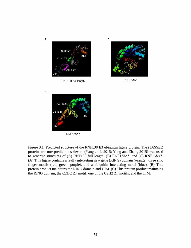
51
Figure 3.1. Predicted structure of the RNF138 E3 ubiquitin ligase protein. The iTASSER
protein structure prediction software (Yang et al. 2015; Yang and Zhang 2015) was used
to generate structures of (A) RNF138-full length, (B) RNF138Δ5, and (C) RNF138Δ7.
(A) This ligase contains a really interesting new gene (RING) domain (orange), three zinc
finger motifs (red, green, purple), and a ubiquitin interacting motif (blue). (B) This
protein product maintains the RING domain and UIM. (C) This protein product maintains
the RING domain, the C2HC ZF motif, one of the C2H2 ZF motifs, and the UIM.
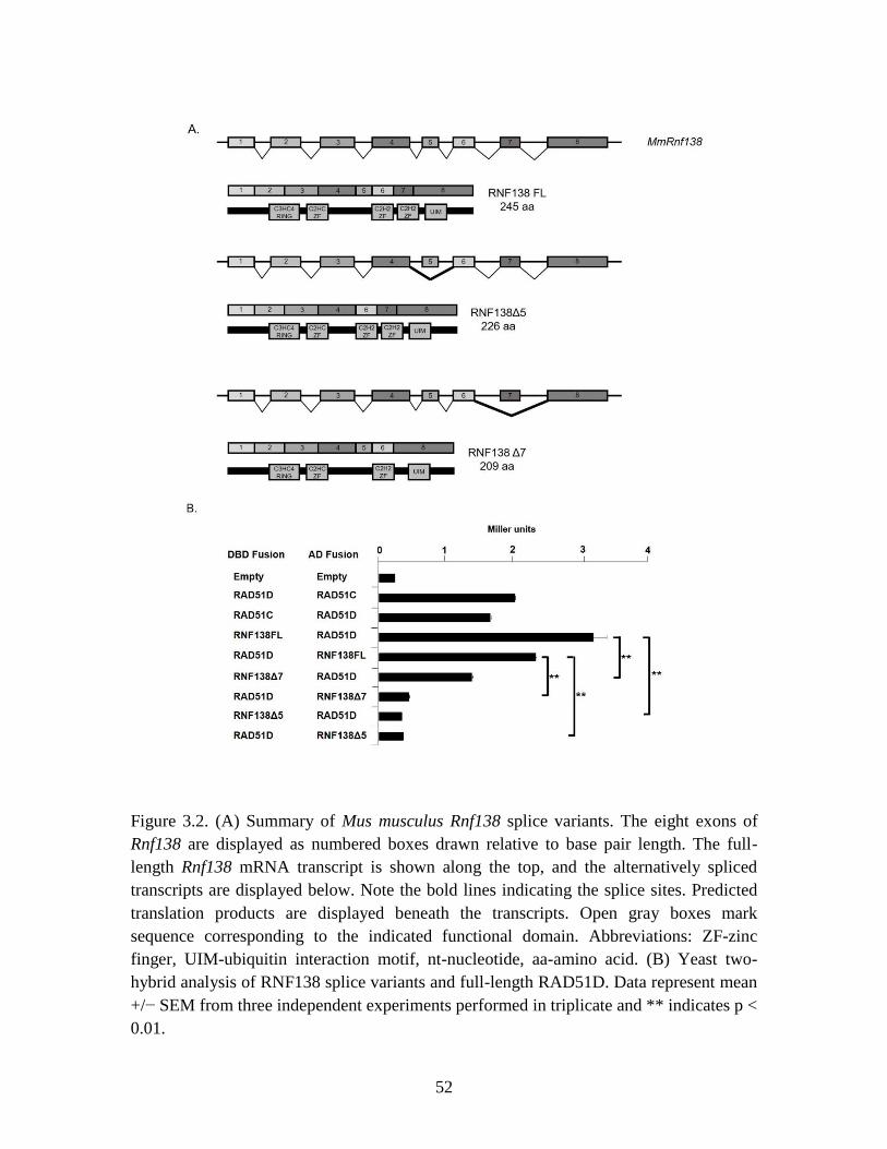
52
Figure 3.2. (A) Summary of Mus musculus Rnf138 splice variants. The eight exons of
Rnf138 are displayed as numbered boxes drawn relative to base pair length. The full-
length Rnf138 mRNA transcript is shown along the top, and the alternatively spliced
transcripts are displayed below. Note the bold lines indicating the splice sites. Predicted
translation products are displayed beneath the transcripts. Open gray boxes mark
sequence corresponding to the indicated functional domain. Abbreviations: ZF-zinc
finger, UIM-ubiquitin interaction motif, nt-nucleotide, aa-amino acid. (B) Yeast two-
hybrid analysis of RNF138 splice variants and full-length RAD51D. Data represent mean
+/− SEM from three independent experiments performed in triplicate and ** indicates p <
0.01.
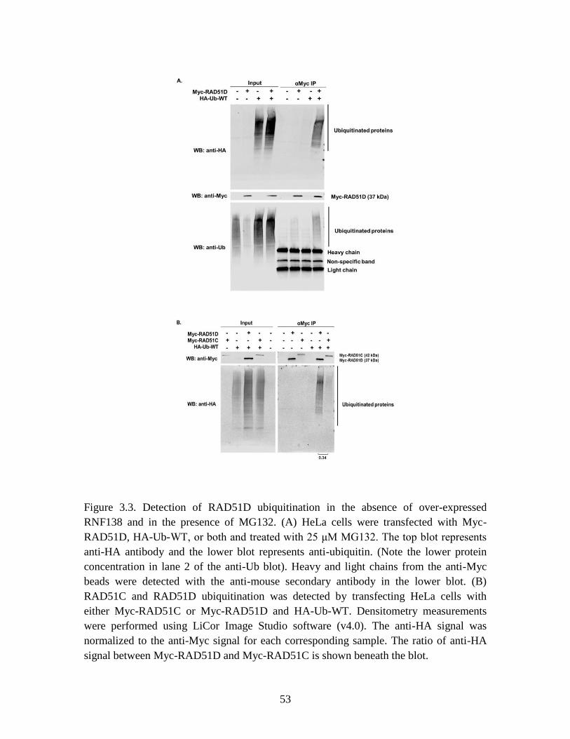
53
Figure 3.3. Detection of RAD51D ubiquitination in the absence of over-expressed
RNF138 and in the presence of MG132. (A) HeLa cells were transfected with Myc-
RAD51D, HA-Ub-WT, or both and treated with 25 μM MG132. The top blot represents
anti-HA antibody and the lower blot represents anti-ubiquitin. (Note the lower protein
concentration in lane 2 of the anti-Ub blot). Heavy and light chains from the anti-Myc
beads were detected with the anti-mouse secondary antibody in the lower blot. (B)
RAD51C and RAD51D ubiquitination was detected by transfecting HeLa cells with
either Myc-RAD51C or Myc-RAD51D and HA-Ub-WT. Densitometry measurements
were performed using LiCor Image Studio software (v4.0). The anti-HA signal was
normalized to the anti-Myc signal for each corresponding sample. The ratio of anti-HA
signal between Myc-RAD51D and Myc-RAD51C is shown beneath the blot.

54
CHAPTER 4
RAD51D LYSINE RESIDUES 235 AND 298 ARE REQUIRED FOR DNA
INTERSTRAND CROSSLINK REPAIR
55
Abstract
RAD51 proteins are essential components of the homologous recombination (HR)
pathway that repairs DNA double strand breaks (DSBs), which can be induced directly
by radiation sources or generated during the repair of DNA interstrand crosslinks (ICLs).
Deletion of the Rad51d HR gene confers increased cellular sensitivity to the ICL-
inducing agent mitomycin C (MMC). Previously, a direct interaction between RAD51D
and the RNF138 E3 ubiquitin ligase was identified. RNF138 promoted RAD51D
ubiquitination, a post-translational modification that occurs primarily along lysine
residues, and loss of RNF138 increased RAD51D protein stability. In this study, the
lysine residues along RAD51D that are required for protein function and are potential
ubiquitin modification sites were identified. Arginine substitution mutations were
introduced at each lysine position along RAD51D, and complementation assays were
performed using Rad51d-deficient mouse embryonic fibroblasts grown in the presence of
MMC. Lysine residues 235 (K235R) and 298 (K298R) were essential for ICL repair, but
not for interaction with RAD51C, XRCC2, or RNF138 as measured by the yeast-two-
hybrid assay. Stability of a lysine-null (K0) mutant was 3-fold higher than wild-type, and
K235R and K298R protein stability was 2 to 3-fold higher compared with wild-type
RAD51D. Ubiquitination assays identified a 3-ubiquitin modification present along the
wild-type but not K0. Finally, neither K235 nor K298 were required for homology-
directed repair following SceI-induced DSBs. Overall, these data suggest that K235 and
K298 along RAD51D are required for homologous recombination-mediated repair of
DNA interstrand crosslinks.
56
Introduction
Ovarian cancer is the deadliest gynecological cancer (Kuschel et al. 2002; Levy-Lahad et
al. 2001; Loveday et al. 2011; Loveday et al. 2012; Meindl et al. 2010; Pelttari et al.
2011; Thompson et al. 2012). Mutations in the RAD51 gene family increase risk for
breast and ovarian cancer (Coulet et al. 2013; Kraus et al. 2017; Loveday et al. 2011;
Meindl et al. 2010; Pelttari et al. 2011; Song et al. 2015; Thompson et al. 2013), and the
proteins encoded by these genes function during homologous recombination (HR)
mediated repair of DNA double strand breaks (DSBs) and interstrand crosslinks (ICLs).
Mutations in repair genes also confer increased sensitivity to chemotherapy agents, such
as platinum-based drugs (Pennington et al. 2014; Minckwitz et al. 2014; Topp et al.
2014). Cisplatin and mitomycin C (MMC), commonly used treatments for late stage
ovarian cancer, and generate DNA intra- and interstrand crosslinks (Dasari and
Tchounwou 2014).
Interstrand crosslinks result in a covalent linkage between two complementary
DNA strands that can disrupt replication and transcription (Zou, Van Houten, and Farrell
1994). During the S/G2 stages of the cell cycle, Fanconi Anemia (FA) and HR proteins
repair ICL damage (Michl, Zimmer, and Tarsounas 2016). A crosslink is first recognized
by the FA core complex, which binds to one of the DNA strands on either side of the ICL
lesion. Recruitment and ubiquitination of the FANCD2/FANCI heterodimer initiates
cleavage of the DNA strand, producing a DSB. Removal of the FANCD2/FANCI
complex allows for recognition and binding of HR proteins that use the complementary
DNA strand as a homologous template to repair the DSB (Kim and D'Andrea 2012).
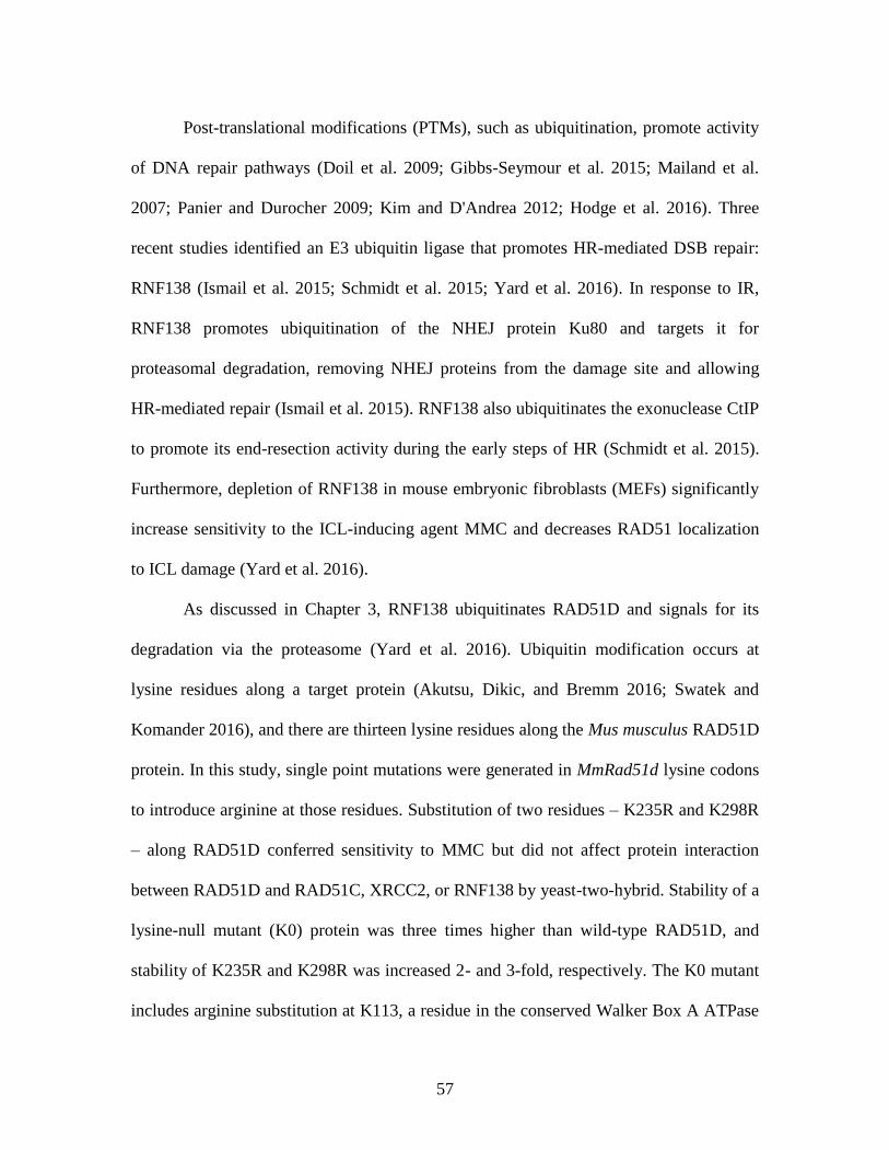
57
Post-translational modifications (PTMs), such as ubiquitination, promote activity
of DNA repair pathways (Doil et al. 2009; Gibbs-Seymour et al. 2015; Mailand et al.
2007; Panier and Durocher 2009; Kim and D'Andrea 2012; Hodge et al. 2016). Three
recent studies identified an E3 ubiquitin ligase that promotes HR-mediated DSB repair:
RNF138 (Ismail et al. 2015; Schmidt et al. 2015; Yard et al. 2016). In response to IR,
RNF138 promotes ubiquitination of the NHEJ protein Ku80 and targets it for
proteasomal degradation, removing NHEJ proteins from the damage site and allowing
HR-mediated repair (Ismail et al. 2015). RNF138 also ubiquitinates the exonuclease CtIP
to promote its end-resection activity during the early steps of HR (Schmidt et al. 2015).
Furthermore, depletion of RNF138 in mouse embryonic fibroblasts (MEFs) significantly
increase sensitivity to the ICL-inducing agent MMC and decreases RAD51 localization
to ICL damage (Yard et al. 2016).
As discussed in Chapter 3, RNF138 ubiquitinates RAD51D and signals for its
degradation via the proteasome (Yard et al. 2016). Ubiquitin modification occurs at
lysine residues along a target protein (Akutsu, Dikic, and Bremm 2016; Swatek and
Komander 2016), and there are thirteen lysine residues along the Mus musculus RAD51D
protein. In this study, single point mutations were generated in MmRad51d lysine codons
to introduce arginine at those residues. Substitution of two residues – K235R and K298R
– along RAD51D conferred sensitivity to MMC but did not affect protein interaction
between RAD51D and RAD51C, XRCC2, or RNF138 by yeast-two-hybrid. Stability of a
lysine-null mutant (K0) protein was three times higher than wild-type RAD51D, and
stability of K235R and K298R was increased 2- and 3-fold, respectively. The K0 mutant
includes arginine substitution at K113, a residue in the conserved Walker Box A ATPase

58
motif. Substitution at this residue eliminates catalytic activity of the protein (Gruver et al.
2005), therefore, the K0 mutant is predicted to be catalytically inactive. In vivo
ubiquitination assays demonstrated that a band corresponding to three ubiquitin
molecules was present in wild-type, but not K0 samples, suggesting a short ubiquitin
chain along the protein is no longer present. Foci formation following MMC treatment
demonstrated that RAD51D acts downstream of FANCD2 and Ku86, and upstream of
RAD51 in response to MMC. Furthermore, homology-directed repair assays
demonstrated that neither K235 nor K298 is required for repair of SceI induced DSBs.
Overall, these data suggest RAD51D ubiquitination at K235 and K298 is required for
repair of DNA interstrand crosslinks.
Materials and Methods
Site-directed mutagenesis
Lysine to arginine substitution point mutations were introduced by PCR-based site-
directed mutagenesis using the Mus musculus Rad51d cDNA cloned into the pUC19
expression vector (New England BioLabs) (Smiraldo et al. 2005). The primer sequences
used are listed in Table 4.1. Each mutation was individually generated to produce single
lysine to arginine amino acid substitutions. A lysine-null RAD51D (K0) construct was
generated by changing all lysine codons to arginine. A RAD51D-Cpeptide mutant
construct representing amino acids 225 to 329 was generated by PCR amplification of the
MmRad51d wild-type cDNA using the following primers: RAD51D-CpepKpnI Forward
and RAD51D-CpepBclI Reverse (Table 4.1). DNA sequencing (Eton Bioscience)
confirmed all clones and base substitutions.
59
Complementation Assays
Each individual RAD51D lysine to arginine mutant construct was cloned into the
pcDNA3.1 mammalian expression vector (Invitrogen) containing either the N-terminal
Myc or HA epitope tags at the KpnI and BamHI sites. Rad51d-deficient (Rad51d-/-
Trp53-
/-) mouse embryonic fibroblasts (MEFs) (Smiraldo et al. 2005) were maintained at 37°C
with 5% CO2 in Dulbecco’s Modified Eagle’s Medium (DMEM; Hyclone) supplemented
with 10% fetal bovine serum (Hyclone), 1% penicillin/streptomycin, and 1% glutamine.
Rad51d-deficient MEFs were transfected with Myc-RAD51D constructs using
Lipofectamine LTX Plus reagent (Thermo Fisher). Twenty-four hours following
transfection, cells were divided evenly between two 150 mm dishes. Selection was
performed using 200 ug/mL hygromycin B (Sigma), and half of the plates were treated
with 4 ng/mL mitomycin C as previously described (Gruver et al. 2009). Media was
replaced every 3 days for 14 days following initiation of treatment, and both hygromycin
B selection and MMC treatment were maintained for the duration of the experiment.
Following treatment, the plates were rinsed with 1X PBS, fixed with methanol, and
colonies stained with Giemsa. Percent resistance was calculated by dividing the total
number of colonies surviving selection with MMC by the number of colonies that grew in
the presence of hygromycin B alone.
Protein localization
To generate enhanced green fluorescent protein (EGFP) fusions of the lysine to arginine
mutants, each construct was cloned into the KpnI and BamHI sites of the pEGFP-C1
vector (Clontech). EGFP-RAD51D fusion constructs were transiently expressed in
Rad51d-deficient MEFs grown on glass coverslips as described (Smiraldo et al. 2005).
60
Twenty-four hours post-transfection, the coverslips were washed with 1X PBS, fixed
with 4% paraformaldehyde for 20 minutes at room temperature, and permeabilized with
0.3% Triton-X 100 for 5 minutes. The coverslips were mounted on glass slides with
Prolong Gold + DAPI mounting agent (Life Technologies). Slides were viewed using an
Evos fluorescent microscope and images captured using a 60x oil immersion objective.
Yeast-two hybrid assays
Each RAD51D lysine construct was cloned into pGADT7 and PGBKT7 (Clontech) at the
EcoRI and BamHI restriction enzyme sites. Transformations into the AH109 yeast strain
was performed using the Frozen EZ Yeast Transformation II kit (Zymo Research) per
manufacturer’s instructions. Liquid β-galactosidase assays were performed using ortho-
nitrophenyl-β-galactopyranoside (ONPG; Sigma) as previously described (Gruver et al.
2009).
Immunoprecipitation and in vivo ubiquitination assays
HEK293T and HeLa cells were transfected with Myc-RAD51D or EGFP-RAD51D-Cep
and HA-Ubiquitin constructs using TransIT-LTI reagent (Mirus Bio) per manufacturer’s
instructions. Four hours prior to harvesting, cells were treated with 25 µM MG132
(Selleckchem). Twenty-four hours after transfection, the cells were harvested, and the
proteins were extracted using 1X Cell Lysis Buffer (20 mM Tris, 150 mM NaCl, 1 mM
EDTA, 1 mM EGTA, 1 mM PMSF, 1% TritonX-100) containing a protease inhibitor
cocktail (Thermo Scientific). For protein stability experiments, cycloheximide (Sigma)
was added to a final concentration of 10 μg/mL for the indicated times. Three hundred
micrograms of whole cell extract were incubated with anti-Myc magnetic beads (9E10;
61
Thermo-Scientific) or anti-GFP magnetic beads (Chromotek) for 16 h at 4C with gentle
rocking. Precipitated proteins were washed 3 times with dilution buffer (10 mM Tris/Cl,
150 mM NaCl, 0.5 mM EDTA, protease inhibitor cocktail), eluted by boiling in 3X SDS-
PAGE buffer (187.5 mM Tris-HCl, 6% w/v SDS, 30% glycerol, 150 mM DTT, 0.03%
bromophenol blue) for 7 minutes, and resolved on a 4 - 20% SDS-PAGE (Bio-Rad).
For western blot analysis, primary anti-Myc (9E10; Santa Cruz) mouse
polyclonal, anti-Myc (A-14; Santa Cruz) rabbit polyclonal, anti-HA (3F10; Roche) rat
polyclonal, anti-β-tubulin (PA5-16863; Invitrogen) rabbit monoclonal, and anti-GFP
(SAB4301138; Sigma) rabbit monoclonal antibodies were used. Primary antibody
incubations were followed by incubation with the appropriate species-specific IRDye
800CW secondary antibody (Licor). Detection was performed using the Licor Odyssey
Sa Imaging System, and densitometry measurements were performed using Licor Image
Studio software (v4.0).
Immunofluorescence
Rad51d-proficient and -deficient MEFs were seeded at 3 x 10⁵ cells per well on glass
coverslips in a 6-well dish. Twelve hours after plating, the cells were treated with either
200 ng/mL (IC50 Rad51d-proficient) or 2 ng/mL (IC50 Rad51d-deficient) MMC. Twenty-
hours after initiation of treatment, the coverslips were washed with 1X PBS, pre-
extracted with 0.5% Triton-X 100 for 4 minutes, fixed with 4% paraformaldehyde for 20
minutes at room temperature, and permeabilized with 0.3% Triton-X 100 for 5 minutes.
To block, coverslips were incubated in 5% milk/PBS for 1 hour at room temperature,
then washed with 1X PBS three times. Primary antibody incubation was performed in 5%
milk/PBS at indicated dilutions in a humid chamber overnight at 4°C. After the primary
62
incubation, the cells were washed with 1X PBS. Secondary antibody incubations were
performed in 5% milk/PBS at the indicated dilutions in a humid chamber for 1 hour at
room temperature. The cells were then washed with 1X PBS, and the coverslips were
mounted on glass slides with Prolong Gold + DAPI mounting agent (Life Technologies).
For immunofluorescence, primary anti-FANCD2 (ab2187; Abcam; 1:200) rabbit
polyclonal, anti-Ku86 (C-20; Santa Cruz; 1:00) goat polyclonal, anti-RAD51 (H-92;
Santa Cruz; 1:100) rabbit polyclonal, and secondary antibodies AlexaFluor488 (Sigma;
1:1000) and AlexFluor688 (Sigma; 1:200) were used. Slides were viewed using an Evos
fluorescent microscope and images captured using a 60x oil lens objective. Foci per
nuclei were scored for each cell line, and nuclei with foci n ≥ 5 were scored as positive. A
minimum of 200 nuclei were scored for each treatment.
Homology directed repair assay
For recombination measurements, 5 x 10⁶ HeLa DRGFP cells (Pierce et al. 1999) were
co-transfected with 50 ug pCBASce (Rajesh, Baker, et al. 2011) and 25 ug Myc-
RAD51D-WT, Myc-RAD51D-K0, Myc-RAD51D-K235R, or Myc-RAD51D-K298R
plasmid by electroporation (BTX Harvard Apparatus ECM 630 Electro Cell Manipulator)
using the following settings: low voltage, 230V, 500 μF. The medium was replaced every
twenty-four hours following transfection. Forty-eight hours after SceI transfection, the
cells were viewed on an Evos fluorescent microscope, and images were captured using a
40X objective. Cells were collected and resuspended in 1X PBS for GFP expression
analysis using an FC 500 flow cytometer and quantitated using CXP analysis software
(Beckman Coulter) (Yang, Waldman, and Wyatt 2012).
63
Results
Complementation screening of RAD51D lysine to arginine mutants
Our previous work demonstrated that RAD51D is ubiquitinated by the E3 ligase RNF138
(Yard et al. 2016), a modification that occurs at lysine residues (Zee and Garcia 2012).
The Mus musculus RAD51D protein has thirteen lysine residues, ten of which are
conserved with Homo sapiens RAD51D (Figure 4.1). Sequence identity between the
RAD51 paralogs is approximately 40%, and seven of the lysine residues along
MmRAD51D are conserved with another paralog (Figure 4.2). To determine which
lysine residues are required for RAD51D function during DNA damage repair, site-
directed mutagenesis was performed to individually substitute each lysine codon with an
arginine, and a lysine-null (K0) was generated by changing all lysine residues to
arginines (Figure 4.3A). The RAD51D constructs are referenced by each lysine to
arginine substitution; for example, substitution of lysine 235 and 298 are referred as
K235R and K298R, respectively.
Complementation assays were performed to identify the lysine residues essential
for resistance to DNA interstrand crosslinks (Smiraldo et al. 2005). Each mutant plasmid
was transiently expressed in Rad51d-deficient mouse embryonic fibroblasts (MEFs). For
each experiment, transfected populations were equally divided and maintained in either
the presence of hygromycin B alone or with the addition of the DNA ICL agent
mitomycin C (MMC) (Figure 4.3B). K0, K235R, and K298R decreased cellular
resistance to MMC by up to 90% compared to wild-type, and expression of K261R
decreased cellular resistance by 30%. K24R, K26R, K42R, K48R, and K327R increased
cellular resistance to MMC between 10 and 20%, K159R and K201R increased by
64
approximately 40%, and K76R and K91R were approximately the same as wild-type.
Given these data, K235R and K298R were the focus of the subsequent work.
One potential explanation for increased sensitivity in the absence of either residue
is that arginine substitution disrupts nuclear localization of the proteins. Using EGFP-
tagged RAD51D constructs, cellular localization of RAD51D was not affected by
substitution at either K235 or K298 (Figure 4.4).
K235R and K298R interact with RAD51C, XRCC2, and RNF138
RAD51D forms a complex with the paralogs RAD51B, RAD51C, and XRCC2 (BCDX)
through direct interactions with RAD51C and XRCC2 (Masson et al. 2001; Rajesh et al.
2009). To determine the effects on protein interaction, yeast-two-hybrid analysis was
performed with each lysine mutant and RAD51C (Figure 4.5A, D) or XRCC2 (Figure
4.5B, E). Replica plating results suggest that both RAD51C and XRCC2 interact with
K235R and K298R, but not K0. As demonstrated by ONPG analysis, K235R and K298R
showed a level of interaction with either RAD51C or XRCC2 similar to wild-type
RAD51D. Interestingly, interaction between K201R and RAD51C or XRCC2 was
decreased 3- and 4-fold, respectively. Despite decreased interaction with these paralogs,
K201R complemented Rad51d-deficiency in the presence of MMC.
Previous studies demonstrated that RAD51D directly interacts with the E3
ubiquitin ligase RNF138 (Yard et al. 2016). Replica plating results suggest that RNF138
interacts with K235R and K298R, but not K0. As demonstrated by ONPG analysis,
K235R and K298R had a level of interaction with RNF138 similar to wild-type
RAD51D, and RNF138 interaction with K0 was decreased to background levels (Figure
4.5C, F). K0 includes arginine substitution at K113 along RAD51D, which was
65
previously shown to be required for interaction between RAD51D and RNF138 (Gruver
et al. 2005; Yard et al. 2016).
Increased stability of K0, K235R, and K298R
To determine if K235 or K298 is a site for degradation specific ubiquitin modification,
the stability of Myc-tagged RAD51D mutant proteins after treatment with cycloheximide
(CHX) was measured (Figure 4.6A). Two hours after initiation of CHX block, the
stability of K0 was 3-fold higher than wild-type (p<0.05), and stability of K235R, and
K298R were 2-fold and 1.5-fold higher, respectively (p<0.05). Four hours after CHX
addition, the stability of K0, K235R, and K298R was increased 3-fold, 2-fold, and 2.5-
fold, respectively, compared with wild-type RAD51D (p<0.05). Six hours after CHX
treatment, the protein levels of K0, K235R, and K298R were similar to wild-type,
suggesting that degradation of these proteins is delayed, rather than completely
eliminated. The stability of HA-tagged RAD51D wild-type and K0 proteins were
consistent with these results (Figure 4.6A; lower blots).
The C-terminal region of the RAD51D gene encoding amino acids 225 to 329
(RAD51D-Cpep) was amplified and fused to the gene sequence for enhanced green
fluorescent protein. This generated a RAD51D-Cpep-EGFP fusion protein used for
subsequent experiments. Stability of Cpep-K0, Cpep-K235R, and Cpep-K298R was
decreased to similar levels as wild-type RAD51D-Cpep 4 hours after initiation of a CHX
block (Figure 4.6C). This treatment time was selected based on the significant increase in
protein stability of the full-length K0, K235R, and K298R mutants (Figure 4.6A). These
data are in contrast with the full-length RAD51D that showed increased stability of the
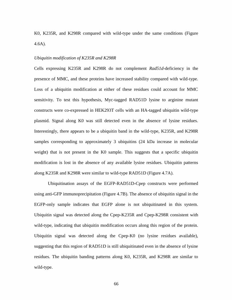
66
K0, K235R, and K298R compared with wild-type under the same conditions (Figure
4.6A).
Ubiquitin modification of K235R and K298R
Cells expressing K235R and K298R do not complement Rad51d-deficiency in the
presence of MMC, and these proteins have increased stability compared with wild-type.
Loss of a ubiquitin modification at either of these residues could account for MMC
sensitivity. To test this hypothesis, Myc-tagged RAD51D lysine to arginine mutant
constructs were co-expressed in HEK293T cells with an HA-tagged ubiquitin wild-type
plasmid. Signal along K0 was still detected even in the absence of lysine residues.
Interestingly, there appears to be a ubiquitin band in the wild-type, K235R, and K298R
samples corresponding to approximately 3 ubiquitins (24 kDa increase in molecular
weight) that is not present in the K0 sample. This suggests that a specific ubiquitin
modification is lost in the absence of any available lysine residues. Ubiquitin patterns
along K235R and K298R were similar to wild-type RAD51D (Figure 4.7A).
Ubiquitination assays of the EGFP-RAD51D-Cpep constructs were performed
using anti-GFP immunoprecipitation (Figure 4.7B). The absence of ubiquitin signal in the
EGFP-only sample indicates that EGFP alone is not ubiquitinated in this system.
Ubiquitin signal was detected along the Cpep-K235R and Cpep-K298R consistent with
wild-type, indicating that ubiquitin modification occurs along this region of the protein.
Ubiquitin signal was detected along the Cpep-K0 (no lysine residues available),
suggesting that this region of RAD51D is still ubiquitinated even in the absence of lysine
residues. The ubiquitin banding patterns along K0, K235R, and K298R are similar to
wild-type.
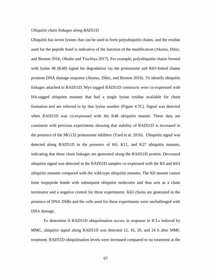
67
Ubiquitin chain linkages along RAD51D
Ubiquitin has seven lysines that can be used to form polyubiquitin chains, and the residue
used for the peptide bond is indicative of the function of the modification (Akutsu, Dikic,
and Bremm 2016; Ohtake and Tsuchiya 2017). For example, polyubiquitin chains formed
with lysine 48 (K48) signal for degradation via the proteasome and K63-linked chains
promote DNA damage response (Akutsu, Dikic, and Bremm 2016). To identify ubiquitin
linkages attached to RAD51D, Myc-tagged RAD51D constructs were co-expressed with
HA-tagged ubiquitin mutants that had a single lysine residue available for chain
formation and are referred to by that lysine number (Figure 4.7C). Signal was detected
when RAD51D was co-expressed with the K48 ubiquitin mutant. These data are
consistent with previous experiments showing that stability of RAD51D is increased in
the presence of the MG132 proteasome inhibitor (Yard et al. 2016). Ubiquitin signal was
detected along RAD51D in the presence of K6, K11, and K27 ubiquitin mutants,
indicating that these chain linkages are generated along the RAD51D protein. Decreased
ubiquitin signal was detected in the RAD51D samples co-expressed with the K0 and K63
ubiquitin mutants compared with the wild-type ubiquitin mutants. The K0 mutant cannot
form isopeptide bonds with subsequent ubiquitin molecules and thus acts as a chain
terminator and a negative control for these experiments. K63 chains are generated in the
presence of DNA DSBs and the cells used for these experiments were unchallenged with
DNA damage.
To determine if RAD51D ubiquitination occurs in response to ICLs induced by
MMC, ubiquitin signal along RAD51D was detected 12, 16, 20, and 24 h after MMC
treatment. RAD51D ubiquitination levels were increased compared to no treatment at the
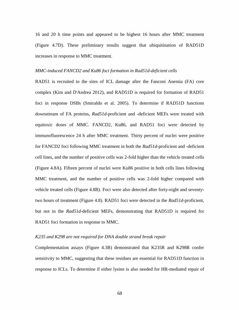
68
16 and 20 h time points and appeared to be highest 16 hours after MMC treatment
(Figure 4.7D). These preliminary results suggest that ubiquitination of RAD51D
increases in response to MMC treatment.
MMC-induced FANCD2 and Ku86 foci formation in Rad51d-deficient cells
RAD51 is recruited to the sites of ICL damage after the Fanconi Anemia (FA) core
complex (Kim and D'Andrea 2012), and RAD51D is required for formation of RAD51
foci in response DSBs (Smiraldo et al. 2005). To determine if RAD51D functions
downstream of FA proteins, Rad51d-proficient and -deficient MEFs were treated with
equitoxic doses of MMC. FANCD2, Ku86, and RAD51 foci were detected by
immunofluorescence 24 h after MMC treatment. Thirty percent of nuclei were positive
for FANCD2 foci following MMC treatment in both the Rad51d-proficient and -deficient
cell lines, and the number of positive cells was 2-fold higher than the vehicle treated cells
(Figure 4.8A). Fifteen percent of nuclei were Ku86 positive in both cells lines following
MMC treatment, and the number of positive cells was 2-fold higher compared with
vehicle treated cells (Figure 4.8B). Foci were also detected after forty-eight and seventy-
two hours of treatment (Figure 4.8). RAD51 foci were detected in the Rad51d-proficient,
but not in the Rad51d-deficient MEFs, demonstrating that RAD51D is required for
RAD51 foci formation in response to MMC.
K235 and K298 are not required for DNA double strand break repair
Complementation assays (Figure 4.3B) demonstrated that K235R and K298R confer
sensitivity to MMC, suggesting that these residues are essential for RAD51D function in
response to ICLs. To determine if either lysine is also needed for HR-mediated repair of
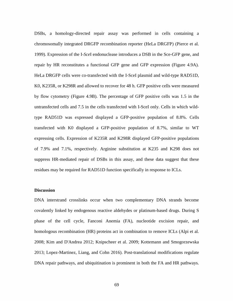
69
DSBs, a homology-directed repair assay was performed in cells containing a
chromosomally integrated DRGFP recombination reporter (HeLa DRGFP) (Pierce et al.
1999). Expression of the I-SceI endonuclease introduces a DSB in the Sce-GFP gene, and
repair by HR reconstitutes a functional GFP gene and GFP expression (Figure 4.9A).
HeLa DRGFP cells were co-transfected with the I-SceI plasmid and wild-type RAD51D,
K0, K235R, or K298R and allowed to recover for 48 h. GFP positive cells were measured
by flow cytometry (Figure 4.9B). The percentage of GFP positive cells was 1.5 in the
untransfected cells and 7.5 in the cells transfected with I-SceI only. Cells in which wild-
type RAD51D was expressed displayed a GFP-positive population of 8.8%. Cells
transfected with K0 displayed a GFP-positive population of 8.7%, similar to WT
expressing cells. Expression of K235R and K298R displayed GFP-positive populations
of 7.9% and 7.1%, respectively. Arginine substitution at K235 and K298 does not
suppress HR-mediated repair of DSBs in this assay, and these data suggest that these
residues may be required for RAD51D function specifically in response to ICLs.
Discussion
DNA interstrand crosslinks occur when two complementary DNA strands become
covalently linked by endogenous reactive aldehydes or platinum-based drugs. During S
phase of the cell cycle, Fanconi Anemia (FA), nucleotide excision repair, and
homologous recombination (HR) proteins act in combination to remove ICLs (Alpi et al.
2008; Kim and D'Andrea 2012; Knipscheer et al. 2009; Kottemann and Smogorzewska
2013; Lopez-Martinez, Liang, and Cohn 2016). Post-translational modifications regulate
DNA repair pathways, and ubiquitination is prominent in both the FA and HR pathways.
70
For example, ubiquitin modification of the FANCD2 and FANCI activate the proteins to
allow cleavage of the DNA strands flanking an ICL lesion (Kim and D'Andrea 2012;
Rickman et al. 2015; Liang et al. 2016). This cleavage generates a DNA double strand
break (DSB) that is recognized and repaired by HR proteins (Kim and D'Andrea 2012).
During HR-mediated repair of DSBs, the E3 ubiquitin ligase RNF138 ubiquitinates the
CtIP endonuclease to promote end resection and HR progression (Schmidt et al. 2015).
We previously demonstrated that loss of RNF138 increases cellular sensitivity to
ICL agents, reduces RAD51 foci formation after MMC induced damage, and
ubiquitinates RAD51D (Yard et al. 2016). K113 in the Walker Box A motif of RAD51D
is also required for cell survival in the presence of MMC (Gruver et al. 2005). In this
study, we show that three lysine residues – K235, K261, and K298 – are required for
cellular resistance to MMC. K235 is predicted to be in the BRC Interface region along
the RAD51D protein, a motif conserved among the RAD51 family members. The BRC
interface mediates oligomerization between RAD51 monomers (Pellegrini et al. 2002)
and is a potential interaction platform between RAD51 paralogs. K261 is in the mid-
region and K298 is located towards the C-terminus of the RAD51D protein. Neither
residue is predicted to be near any known motifs or domains.
RNF138-mediated ubiquitination of RAD51D promotes its degradation by the
proteasome (Yard et al. 2016). K235R and K298R did not affect interaction between
RAD51D and RNF138 but did increase protein stability, suggesting that these residues
are sites of proteasomal specific ubiquitin modifications. K0 did not interact with
RNF138, consistent with previous work that demonstrated K113 (which is substituted
with arginine in K0) is required for this interaction (Yard et al. 2016). Stability of the K0
71
mutant was increased 3-fold compared to wild-type, although the protein was ultimately
degraded. Increased stability of RAD51D could delay or inhibit DNA damage repair in
response to MMC and may explain why cells expressing these mutants do not restore
cellular resistance to MMC.
RAD51B, RAD51C, RAD51D, and XRCC2 interact to form the BCDX2
complex, and of these proteins, RAD51D directly interacts with RAD51C and XRCC2
(Rajesh et al. 2009). Yeast-two-hybrid analysis demonstrated that K235R and K298R
directly interact with RAD51C and XRCC2. Interestingly, K201R did not interact with
RAD51C or XRCC2, but complemented the Rad51d-deficiency in the presence of MMC,
suggesting that the interaction between RAD51C and XRCC2 may not mediate ICL
repair.
Ubiquitin signal was detected along the K235R and K298R proteins suggesting
that RAD51D is still ubiquitinated in the absence of these residues. Ubiquitin addition
along lysine residues is dependent on the availability of the residues, rather than a
specific amino acid sequence surrounding the residue and K235 or K298 may not be the
only residues available for modification. These data suggest that RAD51D is
ubiquitinated in the absence of lysine 235 or 298 and may indicate additional
modifications along RAD51D that are required for promoting DNA ICL repair. Ubiquitin
signal was detected along the K0 mutant, which has no lysines available for modification,
suggesting that non-lysine ubiquitin modifications may be generated along the RAD51D
protein.
Increased protein stability in the presence of K0, K235R, and K298R suggests one
or more ubiquitin modifications along RAD51D are lost in the absence of lysine residues.
72
An alternative method for investigating post-translational modifications along proteins
using tandem mass spectrometry (MS/MS). This method can be used to identify
modifications that occur along specific regions of a protein (Parker et al. 2010). MS/MS
analysis can be performed on the wild-type, K0, K235R, and K298R proteins to identify:
1. lysine residues that are modified with ubiquitin, and 2: modifications that are lost in
the absence of lysine residues.
To identify specific lysine residues that are required for RAD51D function during
DSB repair, I performed a homology-directed repair assay (Pierce et al. 1999) in the
presence of K0, K235R, and K298R. Over-expression of wild-type RAD51D increased
HR capacity by 2.7% compared with the SceI-only control. HR-capacity of cells
expressing K0, K235R, and K298R was similar to wild-type.
Here, we show that two lysine residues along RAD51D – K235 and K298 –are
required for cell survival in response to DNA ICLs. I propose that these residues are the
sites of degradation specific ubiquitin modifications that remove RAD51D from the site
of damage to allow RAD51 to bind and promote HR (Figure 4.10). Following,
recognition of a DNA ICL lesion, FA proteins, specifically the FANCD2/FANCI
heterodimer, in conjunction with the exonuclease CtIP, function at the site of damage to
excise the DNA to produce DNA single strand overhangs surrounding a DNA double
strand break. In response to IR, CtIP directly interacts with the E3 ligase RNF138 to
promote HR (Schmidt et al. 2015), and a similar mechanism in response to ICLs. In
addition to binding CtIP, RNF138 directly interacts with RAD51D of the BCDX2
paralog complex, localizing the complex to the damage. The RAD51 paralog complex
initiates recruitment of RAD51 to the site of damage (Chun, Buechelmaier, and Powell
73
2013). Finally, to remove the BCDX2 complex, RNF138 mediates ubiquitination of
RAD51D that targets it to the proteasome for degradation, thereby removing the proteins.
Displacement of the BCDX2 complex allows RAD51 to bind single strand DNA
overhangs generated by CtIP and for HR-mediated repair of the ICL damage to progress.
Future studies will identify lysine residues along RAD51D necessary for RAD51
foci formation in the presence of MMC. Wild-type RAD51D, K0, K235R, and K298R
can be expressed in Rad51d-deficient MEFs treated with non-lethal doses of IR and
RAD51 foci formation measured using immunofluorescence. K0 and K298R are
predicted to be needed for RAD51 foci formation in response to IR-induced DSBs given
that HDR is suppressed in the presence of these mutants, and over-expression of K235R
is not expected to affect RAD51 foci formation. K201R should also be included in these
experiments, since this mutant had decreased interaction with both RAD51C and XRCC2
by the yeast-two-hybrid assay, suggesting that the BCDX2 complex does not properly
form when Lys201 is not present. Loss of RAD51 foci in the presence of K201R would
suggest that the function of RAD51D differs in response to ICLs versus ‘traditional’
DSBs.
Acknowledgements
The HeLa DRGFP cells were a generous gift from Dr. Andrew Pierce. We thank Dr.
Jason Stewart for assistance with fluorescence microscopy, Dr. Lydia E. Matesic and Dr.
Heather Mentrup for assistance with ubiquitin linkage experiments, Dr. Alex Gasparian
for assistance with EGFP immunoprecipitation experiments, and Dr. Chang-Uk Lim for
assistance with flow cytometry. This research was supported primarily by the National
74
Institutes of Health under Award Number R15GM110615. Additional support was
provided by a Support to Promote Advancement of Research and Creativity (SPARC)
Graduate Research Grant from the Office of the Vice President for Research at the
University of South Carolina to NMR and Magellan Scholarship Awards from the Office
of Undergraduate Research at the University of South Carolina to MGM, CLC, and MEI.
DOA and JG were supported by a Summer Minority Undergraduate Research Fellowship
in the Center for Colon Cancer Research at USC (P30 GM103336).
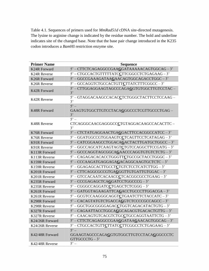
75
Table 4.1. Sequences of primers used for MmRad51d cDNA site-directed mutagenesis.
The lysine to arginine change is indicated by the residue number. The bold and underline
indicates site of the changed base. Note that the base pair change introduced in the K235
codon introduces a BamHI restriction enzyme site.
Primer Name Sequence
K24R Forward 5’ – CTTCTCAGAGGCCGAAGGATAAAAACAGTGGCAG – 3’
K24R Reverse 5’ – CTGCCACTGTTTTTATCCTTCGGCCTCTGAGAAG – 3’
K26R Forward 5’ – GGCCGAAAGATAAGAACAGTGGCAGACCTGGC – 3’
K26R Reverse 5’ – GCCAGGTCTGCCACTGTTCTTATCTTTCGGCC – 3’
K42R Forward 5’ – CTTGGAGGAAGTAGCCCAGAGGTGTGGCTTGTCCTAC –
3’
K42R Reverse 5’ – GTAGGACAAGCCACACCTCTGGGCTACTTCCTCCAAG –
3’
K48R Forward 5’ –
GAAGTGTGGCTTGTCCTACAGGGCCCTCGTTGCCCTGAG –
3’
K48R Reverse 5’ –
CTCAGGGCAACGAGGGCCCTGTAGGACAAGCCACACTTC –
3’
K76R Forward 5’ – CTCTATGAGGAACTGAGGACTTCCACGGCCATCC – 3’
K76R Reverse 5’ – GGATGGCCGTGGAAGTCCTCAGTTCCTCATAGAG – 3’
K91R Forward 5’ – CATCGGAAGCCTGGACAGACTACTTGATGCTGGCC – 3’
K91R Reverse 5’ – GGCCAGCATCAAGTAGTCTGTCCAGGCTTCCGATG – 3’
K113R Forward 5’ – GCCCAGGTAGCGGCAGAACCCAGGTGTGTCTCTG – 3’
K113R Reverse 5’ – CAGAGACACACCTGGGTTCTGCCGCTACCTGGGC – 3’
K159R Forward 5’ – CCCAAGATGAGGAGAGACAGGCAAGTGCTCTC – 3’
K159R Reverse 5’ – GGAGAGCACTTGCCTCTGTCTCCTCATCTTGG – 3’
K201R Forward 5’ – CTTCAGGCGCCGTGAGGGTTGTGATTGTGGAC – 3’
K201R Reverse 5’ – GTCCACAATCACAACCCTCACGGCGCCTGAAG – 3’
K235R Forward 5’ – CCCGAGAGCTCAGGATCCTGGCCCG – 3’
K235R Reverse 5’ – CGGGCCAGGATCCTGAGCTCTCGGG – 3’
K261R Forward 5’ – GATGGTAGAAGATTCAGACCTGCCCTTGGACGA – 3’
K261R Reverse 5’ – GCGTCCAAGGGCAGGTCTGAATCTTCTACCATC – 3’
K298R Forward 5’ – CACAGTATGTCTGACCAGGTCTCCCCGCCAGCC – 3’
K298R Reverse 5’ – GGCTGGCGGGGAGACCTGGTCAGACATACTGTG – 3’
K327R Forward 5’ – CAGAATTACCTGGCAGGCAGACGTGACACTGTTG – 3’
K327R Reverse 5’ – CAACAGTGTCACGTCTGCCTGCCAGGTAATTCTG – 3’
K24/26R Forward 5’ – CTTCTCAGAGGCCGAAGGATAAGAACAGTGGCAG – 3’
K24/26R Reverse 5’ – CTGCCACTGTTCTTATCCTTCGGCCTCTGAGAAG – 3’
K42/48R Forward 5’ –
GGAAGTAGCCCAGAGGTGTGGCTTGTCCTACAGGGCCCTC
GTTGCCCTG – 3’
K42/48R Reverse 5’ –

76
CAGGGCAACGAGGGCCCTGTAGGACAAGCCACACCTCTGG
GCTACTTCC – 3’
CpepKpnI Forward 5’ – CGTATCGGTACCGCCTTGATGATGCAGCTGG – 3’
CpepBclI Reverse 5’ – GTGGCCTTGATCAACTGGACTAGTGGATCCCAATC – 3’
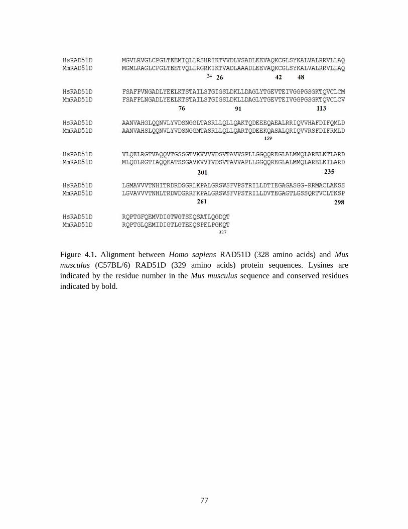
77
Figure 4.1. Alignment between Homo sapiens RAD51D (328 amino acids) and Mus
musculus (C57BL/6) RAD51D (329 amino acids) protein sequences. Lysines are
indicated by the residue number in the Mus musculus sequence and conserved residues
indicated by bold.
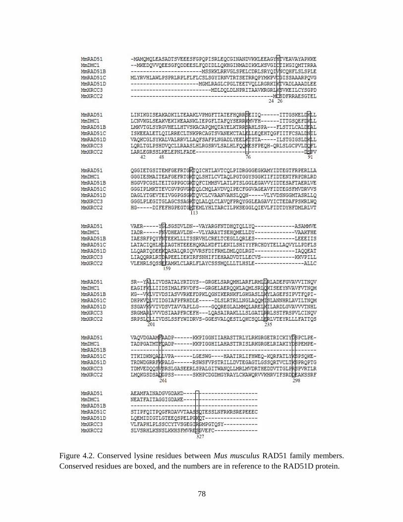
78
Figure 4.2. Conserved lysine residues between Mus musculus RAD51 family members.
Conserved residues are boxed, and the numbers are in reference to the RAD51D protein.
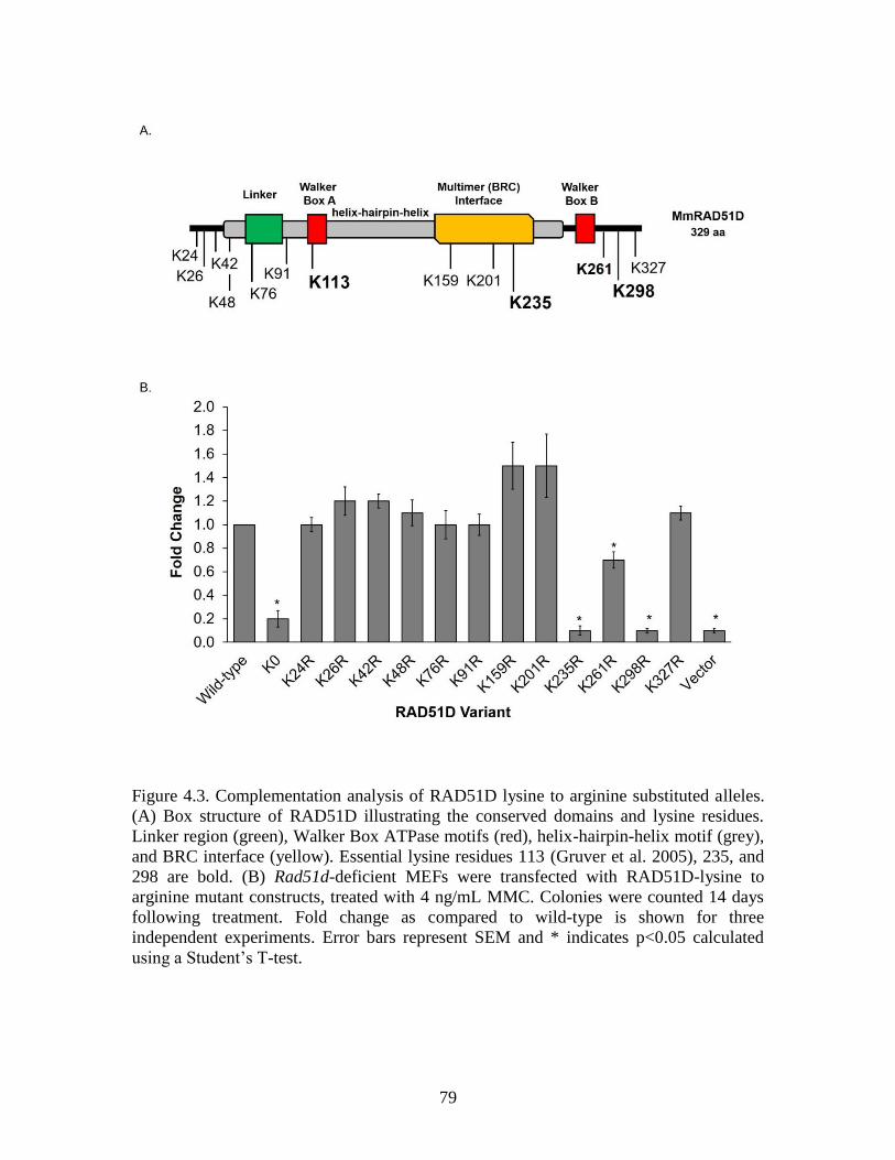
79
Figure 4.3. Complementation analysis of RAD51D lysine to arginine substituted alleles.
(A) Box structure of RAD51D illustrating the conserved domains and lysine residues.
Linker region (green), Walker Box ATPase motifs (red), helix-hairpin-helix motif (grey),
and BRC interface (yellow). Essential lysine residues 113 (Gruver et al. 2005), 235, and
298 are bold. (B) Rad51d-deficient MEFs were transfected with RAD51D-lysine to
arginine mutant constructs, treated with 4 ng/mL MMC. Colonies were counted 14 days
following treatment. Fold change as compared to wild-type is shown for three
independent experiments. Error bars represent SEM and * indicates p<0.05 calculated
using a Student’s T-test.
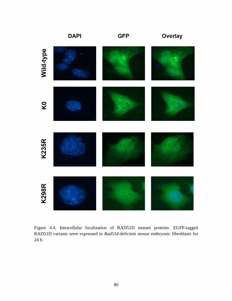
80
Figure 4.4. Intracellular localization of RAD51D mutant proteins. EGFP-tagged
RAD51D variants were expressed in Rad51d-deficient mouse embryonic fibroblasts for
24 h.
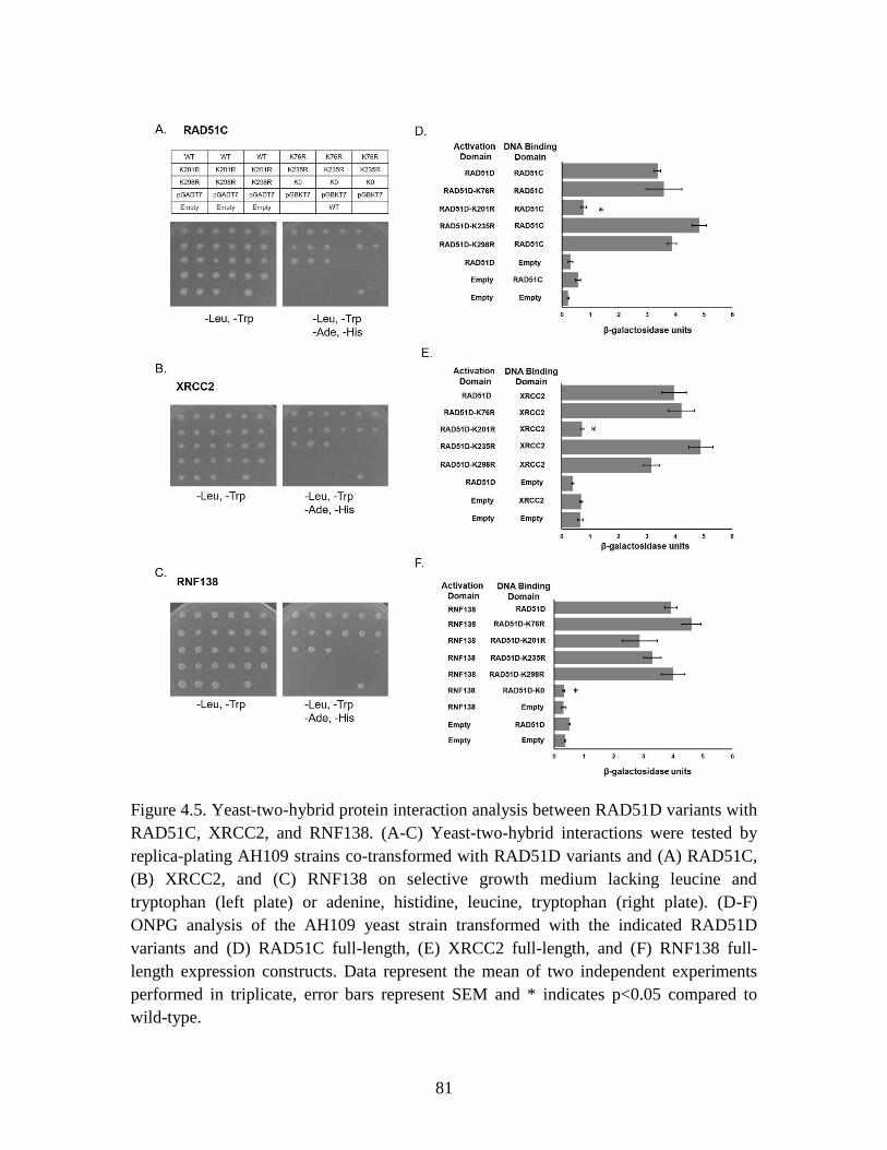
81
Figure 4.5. Yeast-two-hybrid protein interaction analysis between RAD51D variants with
RAD51C, XRCC2, and RNF138. (A-C) Yeast-two-hybrid interactions were tested by
replica-plating AH109 strains co-transformed with RAD51D variants and (A) RAD51C,
(B) XRCC2, and (C) RNF138 on selective growth medium lacking leucine and
tryptophan (left plate) or adenine, histidine, leucine, tryptophan (right plate). (D-F)
ONPG analysis of the AH109 yeast strain transformed with the indicated RAD51D
variants and (D) RAD51C full-length, (E) XRCC2 full-length, and (F) RNF138 full-
length expression constructs. Data represent the mean of two independent experiments
performed in triplicate, error bars represent SEM and * indicates p<0.05 compared to
wild-type.
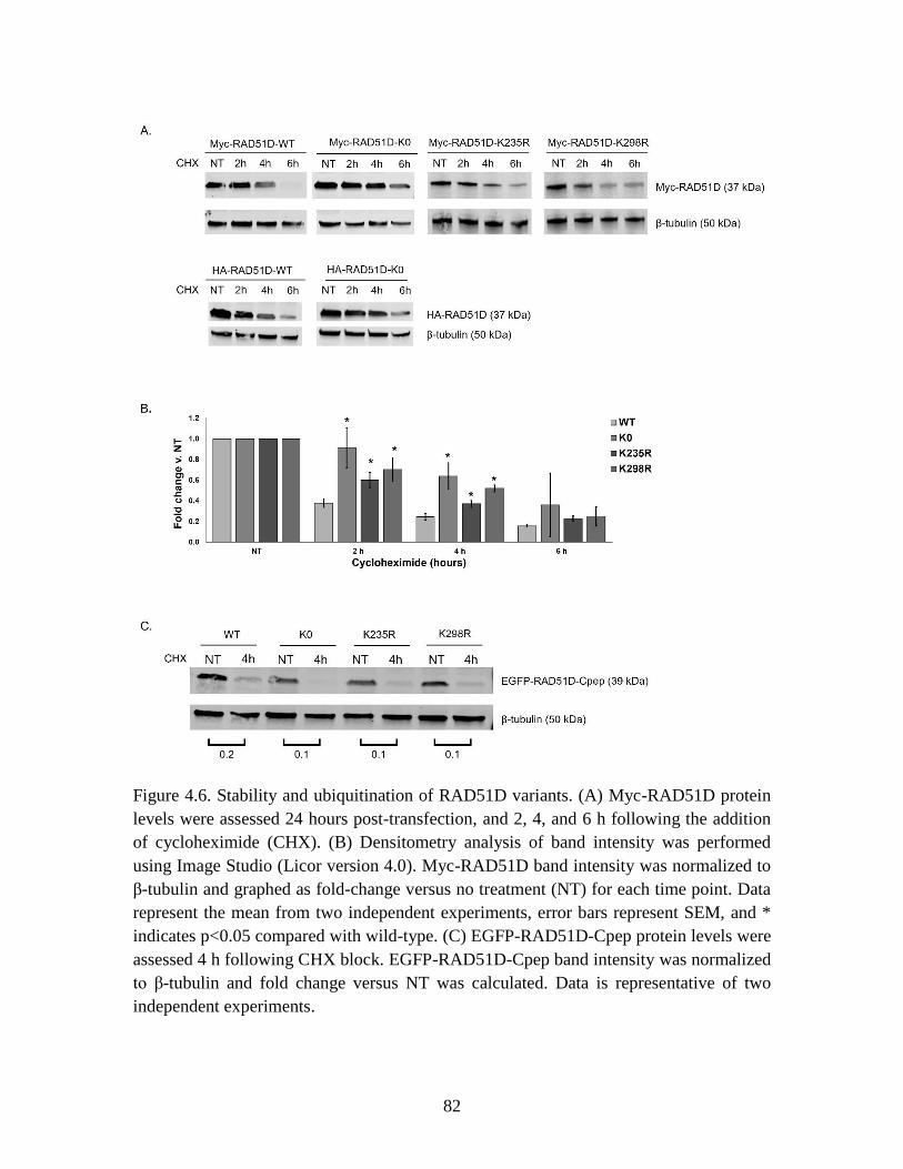
82
Figure 4.6. Stability and ubiquitination of RAD51D variants. (A) Myc-RAD51D protein
levels were assessed 24 hours post-transfection, and 2, 4, and 6 h following the addition
of cycloheximide (CHX). (B) Densitometry analysis of band intensity was performed
using Image Studio (Licor version 4.0). Myc-RAD51D band intensity was normalized to
β-tubulin and graphed as fold-change versus no treatment (NT) for each time point. Data
represent the mean from two independent experiments, error bars represent SEM, and *
indicates p<0.05 compared with wild-type. (C) EGFP-RAD51D-Cpep protein levels were
assessed 4 h following CHX block. EGFP-RAD51D-Cpep band intensity was normalized
to β-tubulin and fold change versus NT was calculated. Data is representative of two
independent experiments.
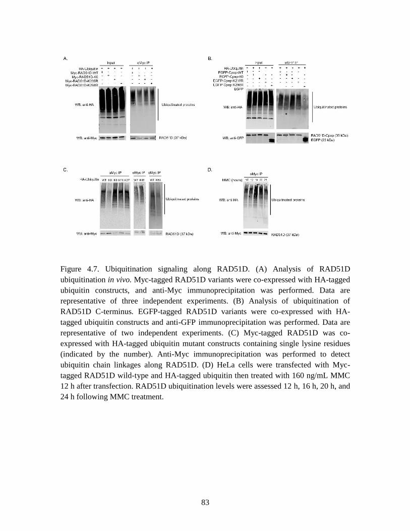
83
Figure 4.7. Ubiquitination signaling along RAD51D. (A) Analysis of RAD51D
ubiquitination in vivo. Myc-tagged RAD51D variants were co-expressed with HA-tagged
ubiquitin constructs, and anti-Myc immunoprecipitation was performed. Data are
representative of three independent experiments. (B) Analysis of ubiquitination of
RAD51D C-terminus. EGFP-tagged RAD51D variants were co-expressed with HA-
tagged ubiquitin constructs and anti-GFP immunoprecipitation was performed. Data are
representative of two independent experiments. (C) Myc-tagged RAD51D was co-
expressed with HA-tagged ubiquitin mutant constructs containing single lysine residues
(indicated by the number). Anti-Myc immunoprecipitation was performed to detect
ubiquitin chain linkages along RAD51D. (D) HeLa cells were transfected with Myc-
tagged RAD51D wild-type and HA-tagged ubiquitin then treated with 160 ng/mL MMC
12 h after transfection. RAD51D ubiquitination levels were assessed 12 h, 16 h, 20 h, and
24 h following MMC treatment.
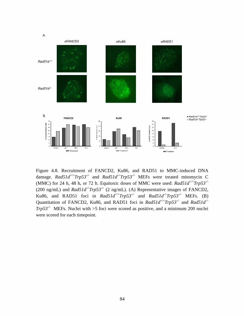
84
Figure 4.8. Recruitment of FANCD2, Ku86, and RAD51 to MMC-induced DNA
damage. Rad51d+/+
Trp53-/-
and Rad51d-/-
Trp53-/-
MEFs were treated mitomycin C
(MMC) for 24 h, 48 h, or 72 h. Equitoxic doses of MMC were used: Rad51d+/+
Trp53-/-
(200 ng/mL) and Rad51d-/-
Trp53-/-
(2 ng/mL). (A) Representative images of FANCD2,
Ku86, and RAD51 foci in Rad51d+/+
Trp53-/-
and Rad51d-/-
Trp53-/-
MEFs. (B)
Quantitation of FANCD2, Ku86, and RAD51 foci in Rad51d+/+
Trp53-/-
and Rad51d-/-
Trp53-/-
MEFs. Nuclei with >5 foci were scored as positive, and a minimum 200 nuclei
were scored for each timepoint.
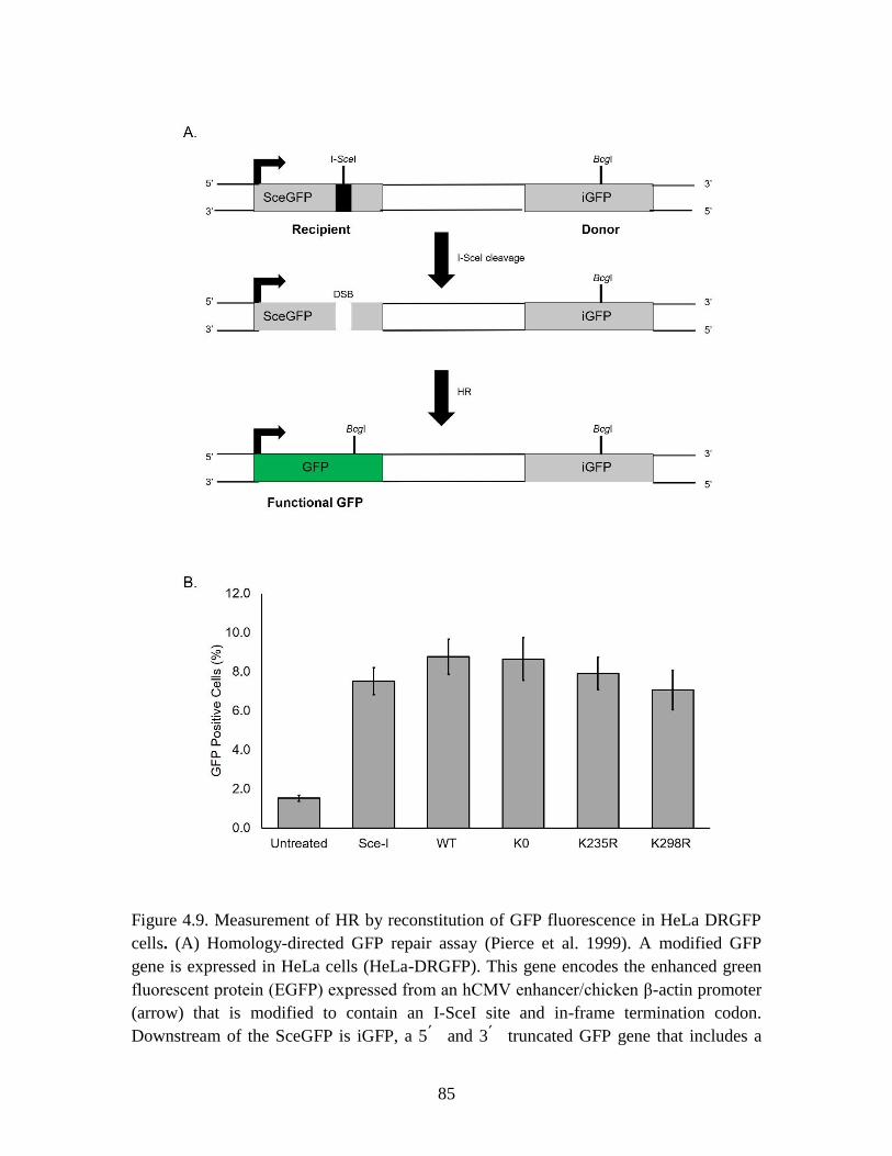
85
Figure 4.9. Measurement of HR by reconstitution of GFP fluorescence in HeLa DRGFP
cells. (A) Homology-directed GFP repair assay (Pierce et al. 1999). A modified GFP
gene is expressed in HeLa cells (HeLa-DRGFP). This gene encodes the enhanced green
fluorescent protein (EGFP) expressed from an hCMV enhancer/chicken β-actin promoter
(arrow) that is modified to contain an I-SceI site and in-frame termination codon.
Downstream of the SceGFP is iGFP, a 5՛ and 3՛ truncated GFP gene that includes a
86
BcgI restriction enzyme site. HeLa-DRGFP cells are transfected with an I-SceI plasmid
that encodes the I-SceI enzyme, leading to DNA cleavage within the SceGFP gene and a
double stranded break. Homologous recombination proteins repair the break using the
iGFP gene as a homologous template, producing an intact GFP gene. (B) Bar graph
showing the percentage of GFP positive cells (two experiments performed in triplicate)
after electroporation with buffer (untreated), Sce-I plasmid only, or each Myc-tagged
RAD51D construct and Sce-I plasmid. Statistical significance was not achieved using
one-way ANOVA analysis.
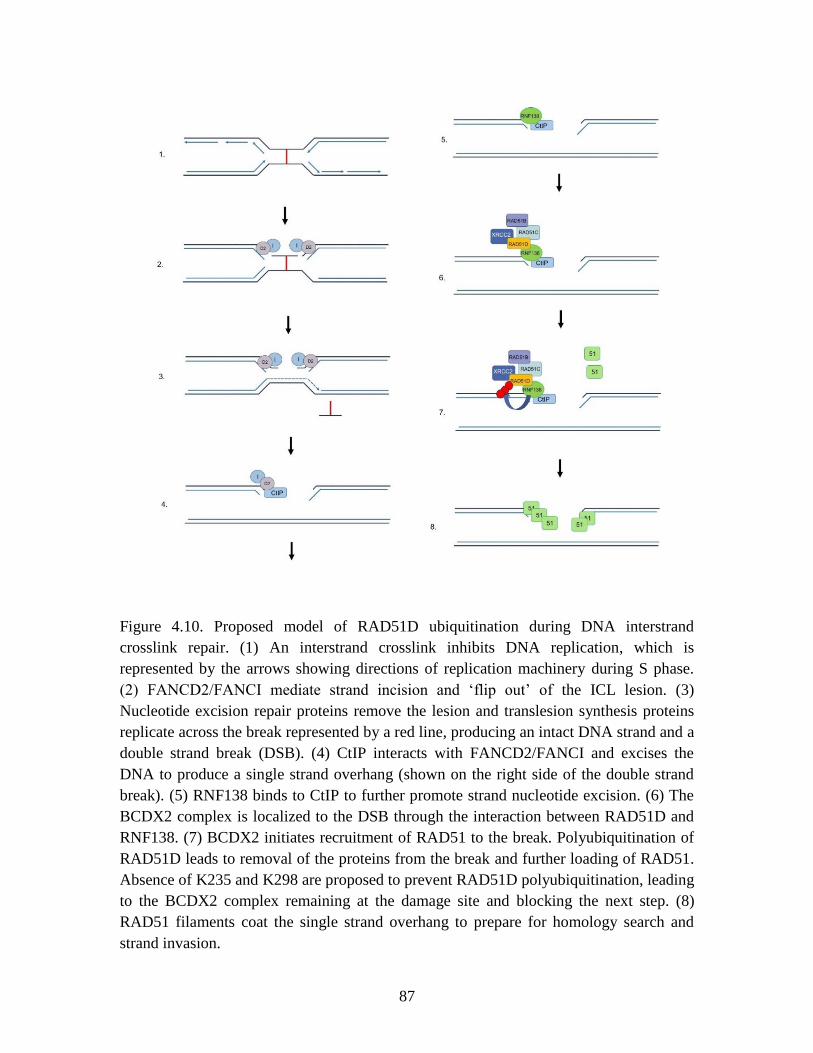
87
Figure 4.10. Proposed model of RAD51D ubiquitination during DNA interstrand
crosslink repair. (1) An interstrand crosslink inhibits DNA replication, which is
represented by the arrows showing directions of replication machinery during S phase.
(2) FANCD2/FANCI mediate strand incision and ‘flip out’ of the ICL lesion. (3)
Nucleotide excision repair proteins remove the lesion and translesion synthesis proteins
replicate across the break represented by a red line, producing an intact DNA strand and a
double strand break (DSB). (4) CtIP interacts with FANCD2/FANCI and excises the
DNA to produce a single strand overhang (shown on the right side of the double strand
break). (5) RNF138 binds to CtIP to further promote strand nucleotide excision. (6) The
BCDX2 complex is localized to the DSB through the interaction between RAD51D and
RNF138. (7) BCDX2 initiates recruitment of RAD51 to the break. Polyubiquitination of
RAD51D leads to removal of the proteins from the break and further loading of RAD51.
Absence of K235 and K298 are proposed to prevent RAD51D polyubiquitination, leading
to the BCDX2 complex remaining at the damage site and blocking the next step. (8)
RAD51 filaments coat the single strand overhang to prepare for homology search and
strand invasion.

88
CHAPTER 5
THIOPURINE-INDUCED TELOMERIC DAMAGE IN RAD51D-DEFICIENT
MAMMALIAN CELLS3
3Michael D. Wyatt, Nicole M. Reilly, Shikha Patel, Preeti Rajesh, Gary P. Schools, Phillip G.
Smiraldo, Douglas L. Pittman. Thiopurine-induced mitotic catastrophe in Rad51d-deficient
mammalian cells. Environmental and Molecular Mutagenesis, 2017, 59: 38 – 48.
89
Preface
The data in this Chapter were published in the research article titled “Thiopurine-induced
mitotic catastrophe in Rad51d-deficient mammalian cells” appearing in Environmental
and Molecular Mutagenesis on September 25, 2017 (Wyatt et al.). For the purposes of
this dissertation, the data that I contributed to the manuscript are reported.
Abstract
Thiopurines are part of a clinical regimen used for the treatment of autoimmune disorders
and childhood acute lymphoblastic leukemia. However, despite these successes, there are
also unintended consequences such as therapy-induced cancer in long-term survivors.
Therefore, a better understanding of cellular responses to thiopurines will lead to
improved and personalized treatment strategies. RAD51D is an important component of
homologous recombination (HR), and our previous work established that mammalian
cells defective for RAD51D are more sensitive to the thiopurine 6-thioguanine (6TG) and
have dramatically increased numbers of multinucleated cells and chromosome instability.
6TG is capable of being incorporated into telomeres, and interestingly, RAD51D
contributes to telomere maintenance, although the precise function of RAD51D at the
telomeres remains unclear. We sought here to investigate: 1) the activity of RAD51D at
telomeres, 2) the contribution of RAD51D to protect against 6TG-induced telomere
damage, and 3) the fates of Rad51d-deficient cells following 6TG treatment. These
results demonstrate that RAD51D is required for maintaining the telomeric 3´ overhangs.
As measured by γ-H2AX induction and foci formation, 6TG-induced DNA damage in
Rad51d-proficient and Rad51d-deficient cells. However, the extent of γ-H2AX telomere
90
localization following 6TG treatment was higher in Rad51d-deficient cells than in
Rad51d-proficient cells. Using live-cell imaging of 6TG-treated Rad51d-deficient cells,
two predominant forms of mitotic catastrophe were found to contribute to the formation
of multinucleated cells, failed division and restitution. Collectively, these findings
provide a unique window into the role of the RAD51D HR protein during thiopurine
induction of mitotic catastrophe.
Introduction
Thiopurines have a long history of clinical usage as immunosuppressants and in cancer
chemotherapy. Combination therapy including mercaptopurine for childhood acute
lymphoblastic leukemia (ALL) treatment is an amazing success story, with cure or long-
term remission rates now being greater than 90%. Although the metabolism of
thiopurines is well known (Krynetski and Evans 2003), surprisingly little is understood
about the mechanism by which thiopurines induce DNA damage and kill cancer cells.
When thiopurines are metabolized into active nucleotide forms, the predominant
mechanism of action is incorporation into DNA (Karran 2006). During replication, the
thiopurine 6-thioguanine (6TG) causes base mispairing, which is then recognized by
DNA mismatch repair (MMR) proteins. As part of a poorly understood process, the
MMR machinery causes DNA strand breaks and invokes homologous recombination
(HR) mediated repair. Inactivation or loss of MMR thus alleviates killing and
chromosomal instability caused by thiopurines (Armstrong and Galloway 1997;
Buermeyer et al. 1999; Rajesh, Litvinchuk, et al. 2011).
91
RAD51D is one of the RAD51 family members indispensable for HR. RAD51D
is now an established ovarian cancer susceptibility gene (Loveday et al. 2011; Song et al.
2015; Thompson et al. 2013), and BRCA2-defective cancer cells are also sensitized to
6TG (Issaeva et al. 2010). Indeed, a recent phase II clinical trial, NCT01432145, explored
the use of a thiopurine in BRCA2-defective tumors; based upon genetic signatures,
thiopurines might be used in other HR-defective cancers. At this time, there have been no
reports of the successful generation of human knockout cells for any of the RAD51
paralogs. In fact, vertebrate cells deficient in any of the RAD51 paralogs have only been
generated in mouse embryonic fibroblasts, DT40 avian cells, and Chinese hamster ovary
cells (Deans et al. 2003; Hinz et al. 2006; Lim and Hasty 1996; Takata et al. 2001;
Tsuzuki et al. 1996). Even though RNA interference mediated knockdowns of several of
the RAD51 family members in human cancer cells have been attempted, none achieved a
substantially reduced expression (e.g., RAD51 by Wyatt and coworkers (Yang,
Waldman, and Wyatt 2008)). In all cases examined, decreased expression of the RAD51
paralogs exhibited a similar sensitivity to DNA damaging agents and chromosome
instability.
Our previous work established that RAD51D-dependent HR is protective
downstream of MMR following 6TG treatment (Rajesh, Litvinchuk, et al. 2011).
Specifically, Rad51d-deficient cells were extremely sensitive to 6TG, and there was a
substantial increase in the frequency of chromosomal aberrations, particularly radials.
There was also an increase in multinucleation and chromosomal aneuploidy in the
Rad51d-deficient cells following 6TG treatment. The loss of MLH1 alleviated these
phenotypes, demonstrating that the induced damage depends on functional MMR. The
92
roles of RAD51D in genomic maintenance include telomere stability (Tarsounas et al.
2004). In this regard, it is interesting to note that deoxy-thioguanine nucleotides can be
incorporated into DNA by telomerase (Marathias, Sawicki, and Bolton 1999; Mender et
al. 2015; Tendian and Parker 2000). These observations prompted our investigation into
three related topics. First, we sought to better understand the specific telomeric defect
associated with Rad51d-deficient cells. Here, it was found that RAD51D is required for
maintaining the telomeric 3´ overhangs in mammalian cells. Second, we investigated
telomeric DNA damage caused by 6TG. Chromosome fusions were induced by 6TG,
some of which contained telomeric labeling. In Rad51d-deficient cells, there was
increased co-localization of telomere probes with γ-H2AX foci compared to Rad51d-
proficient cells, which further increased upon treatment with 6TG. Lastly, we
investigated via live cell imaging multi-nucleation induced by 6TG treatment of Rad51d-
deficient cells. Note that, similar to this Rad51d-deficient cellular phenotype, different
leukemias and lymphomas are known to have increased aneuploidy and multinucleation,
and also telomere defects (Knecht et al. 2009; Knecht et al. 2010). These findings provide
a unique window into the formation of multinucleated Rad51d-deficient cells and
demonstrate that RAD51D provides a protective role against the telomeric DNA damage
and chromosomal instability that thiopurine treatment causes.
93
Materials and methods
Cell lines
Mice heterozygous for a mutation in the Mlh1 gene and mice heterozygous for null-
alleles in Rad51d and Trp53 were crossed to generate murine embryonic fibroblasts
(MEFs) with different combinations of the three mutated genes (Rajesh, Litvinchuk, et al.
2011; Rajesh et al. 2010; Smiraldo et al. 2005). Primary and immortalized mouse
embryonic fibroblasts (MEFs) were grown in DMEM (Corning, Corning, NY USA)
supplemented with 7.5% fetal bovine serum (Atlanta Biologicals, Flowery Branch, GA
USA), 7.5% newborn calf serum (GE Life Sciences, Pittsburgh, PA USA ), and
antibiotics (GE) as previously described (Smiraldo et al. 2005). The following
immortalized cell lines used for these studies were Rad51d+/+
Trp53-/-
(C53), Rad51d-/-
Trp53-/-
(310), and Rad51d-/-
Trp53-/-
Mlh1-/-
(T3) cells. Note that because it was only
possible to generate immortalized MEFs that were Rad51d-/-
on a Trp53-/-
background
(Smiraldo et al. 2005), all work in immortalized MEFs occurred in Trp53-/-
cells. For
simplicity, genotypes of the immortalized MEFs are referred to throughout the
manuscript as Rad51d or Mlh1 status.
Western blot analysis of γ-H2AX induction
Immortalized Rad51d+/+
, Rad51d-/-
, and Rad51d-/-
Mlh1-/-
MEFs were plated in a 6-well
dish at a concentration of 6 x 10⁴ cells per well and, after 24 h, treated with 50 or 100 nM
6TG (Sigma Aldrich, St. Louis, MO USA) for 48 and 72 h. Following treatment, cells
were trypsinized and proteins extracted in 1X cell lysis buffer (20 mM Tris, 150 mM
NaCl, 1 mM EDTA, 1 mM EGTA, 1 mM PMSF, 1% Triton X-100) containing protease
inhibitor cocktail (Thermo Fisher, Waltham, MA USA). Thirty micrograms of whole-cell
94
protein extracts were separated using a 4–20% gradient gel (Bio-Rad, Hercules, CA
USA). Western blot analysis was performed using rabbit polyclonal anti-γ-H2AX (A300-
081, Bethyl, Montgomery, TX USA) or rabbit monoclonal anti-GAPDH (D16H11, Cell
Signaling, Danvers, MA USA). Primary incubations were followed with species specific
IR Dye 800CW secondary antibody (Licor, Lincoln, NE USA), and signal detection was
performed using a Licor Odyssey Sa Imaging System. Quantitative analysis of band
intensity was performed using Image Studio software (LiCor, version 4.0, Lincoln NE,
USA).
Immunofluorescence, telomere staining, and chromosome fusions
For the detection of γ-H2AX foci and telomere co-localization, sub-confluent cells,
grown on sterile glass microscope slides or cover slips (VWR, Radnor, PA USA), were
treated for the indicated times with 6TG. Following treatment, cells were fixed in 4%
paraformaldehyde (Affymetrix, Santa Clara, CA USA), permeabilized with a 0.2% Triton
X-100 solution, and incubated in block solution (5% dry milk in 1x PBS) at room
temperature. This was followed by incubation with the anti-phospho-Histone H2AX
(Ser139) mouse monoclonal antibody (1:600; Bethyl) and Oregon Green 488 goat anti-
mouse IgG secondary (1:1000; Molecular Probes (Thermo Fisher), Waltham, MA USA).
Telomeres were visualized with the peptide nucleic acid probe Cy3-(CCCTAA)3 (PNA
Bio, Thousand Oaks, CA USA) and chromatin visualized by DAPI (Sigma).
For studies using primary MEFs to detect γ-H2AX foci, cells containing ≥ 5
distinct γ-H2AX foci were defined as foci-positive, and the percentage of γ-H2AX foci at
telomeres was calculated as [(number of γ-H2AX foci at telomeres) / (number of γ-
H2AX foci)]* 100% for each cell. Statistical significance was determined by comparing
95
the mean number of γ-H2AX foci at telomeres per cell for each genotype by ANOVA.
Follow-up comparisons were performed using the Tukey HSD post hoc test. For studies
using immortalized MEFs and 6TG-induced damage, cells containing ≥ 10 distinct γ-
H2AX foci were defined as foci-positive, and the percentage of γ-H2AX positive cells
was calculated as [(number of γ-H2AX positive cells)/(total number of cells)]*100. An
EvosFL fluorescence microscope (Life Technologies, Carlsbad, CA USA) under a 60X
oil objective was used to detect γ-H2AX foci. Individual cells were manually scored
through depth-of-field for foci, identified based upon signal intensity above general
background staining levels and present within the nucleus as assessed by DAPI staining.
Metaphase chromosome spreads were prepared as described previously (Smiraldo et al.
2005). The percent of fusions per chromosome was calculated as [(number of
fusions)/(number of chromosomes)]*100 per metaphase spread. Statistical significance of
the experimental data was determined by calculating a z-score. The presence of a
telomere at a fusion was scored positive, and the percent telomere associated fusions was
calculated as [(number of telomere positive fusions)/(number of fusions)]*100.
For detection of telomeres at chromosome fusions, an Axiovert 200 with
Axiovision (Zeiss, Oberkochen, Germany) fluorescence microscope and 100X oil
objective was used. For detection of γ-H2AX foci at telomeres, cells were treated as
described above and the telomere (CCCTAA)3 probe added after γ-H2AX antibody
incubation. Individual cells were scored for co-localized foci by manual scanning through
the cellular depth. Cells were scored positive when overlapping signals were observed
within the nucleus. The number of γ-H2AX foci co-localized with telomere signal was
counted per nuclei and grouped into three categories: 0 to 2 co-localized foci, 3 to 7 co-
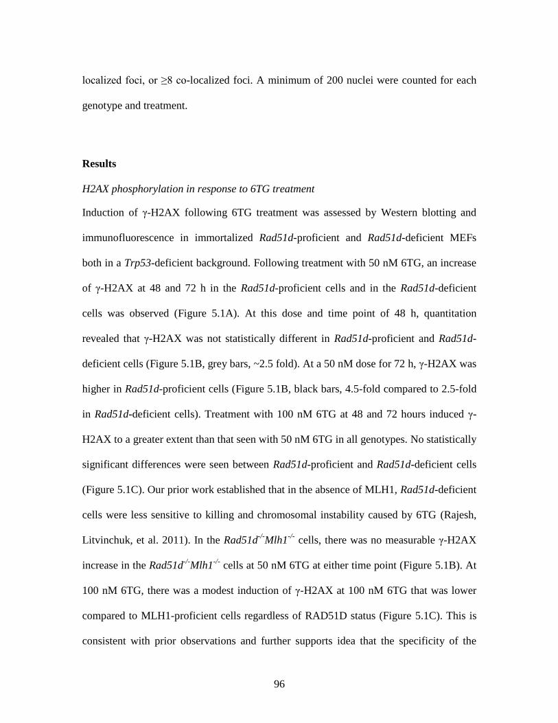
96
localized foci, or ≥8 co-localized foci. A minimum of 200 nuclei were counted for each
genotype and treatment.
Results
H2AX phosphorylation in response to 6TG treatment
Induction of γ-H2AX following 6TG treatment was assessed by Western blotting and
immunofluorescence in immortalized Rad51d-proficient and Rad51d-deficient MEFs
both in a Trp53-deficient background. Following treatment with 50 nM 6TG, an increase
of γ-H2AX at 48 and 72 h in the Rad51d-proficient cells and in the Rad51d-deficient
cells was observed (Figure 5.1A). At this dose and time point of 48 h, quantitation
revealed that γ-H2AX was not statistically different in Rad51d-proficient and Rad51d-
deficient cells (Figure 5.1B, grey bars, ~2.5 fold). At a 50 nM dose for 72 h, γ-H2AX was
higher in Rad51d-proficient cells (Figure 5.1B, black bars, 4.5-fold compared to 2.5-fold
in Rad51d-deficient cells). Treatment with 100 nM 6TG at 48 and 72 hours induced γ-
H2AX to a greater extent than that seen with 50 nM 6TG in all genotypes. No statistically
significant differences were seen between Rad51d-proficient and Rad51d-deficient cells
(Figure 5.1C). Our prior work established that in the absence of MLH1, Rad51d-deficient
cells were less sensitive to killing and chromosomal instability caused by 6TG (Rajesh,
Litvinchuk, et al. 2011). In the Rad51d-/-
Mlh1-/-
cells, there was no measurable γ-H2AX
increase in the Rad51d-/-
Mlh1-/-
cells at 50 nM 6TG at either time point (Figure 5.1B). At
100 nM 6TG, there was a modest induction of γ-H2AX at 100 nM 6TG that was lower
compared to MLH1-proficient cells regardless of RAD51D status (Figure 5.1C). This is
consistent with prior observations and further supports idea that the specificity of the
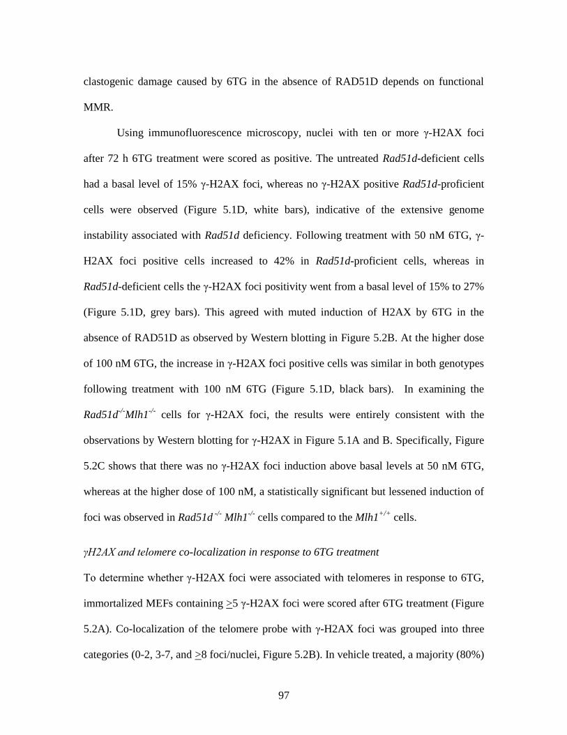
97
clastogenic damage caused by 6TG in the absence of RAD51D depends on functional
MMR.
Using immunofluorescence microscopy, nuclei with ten or more γ-H2AX foci
after 72 h 6TG treatment were scored as positive. The untreated Rad51d-deficient cells
had a basal level of 15% γ-H2AX foci, whereas no γ-H2AX positive Rad51d-proficient
cells were observed (Figure 5.1D, white bars), indicative of the extensive genome
instability associated with Rad51d deficiency. Following treatment with 50 nM 6TG, γ-
H2AX foci positive cells increased to 42% in Rad51d-proficient cells, whereas in
Rad51d-deficient cells the γ-H2AX foci positivity went from a basal level of 15% to 27%
(Figure 5.1D, grey bars). This agreed with muted induction of H2AX by 6TG in the
absence of RAD51D as observed by Western blotting in Figure 5.2B. At the higher dose
of 100 nM 6TG, the increase in γ-H2AX foci positive cells was similar in both genotypes
following treatment with 100 nM 6TG (Figure 5.1D, black bars). In examining the
Rad51d-/-
Mlh1-/-
cells for γ-H2AX foci, the results were entirely consistent with the
observations by Western blotting for γ-H2AX in Figure 5.1A and B. Specifically, Figure
5.2C shows that there was no γ-H2AX foci induction above basal levels at 50 nM 6TG,
whereas at the higher dose of 100 nM, a statistically significant but lessened induction of
foci was observed in Rad51d -/-
Mlh1-/-
cells compared to the Mlh1+/+
cells.
γH2AX and telomere co-localization in response to 6TG treatment
To determine whether γ-H2AX foci were associated with telomeres in response to 6TG,
immortalized MEFs containing >5 γ-H2AX foci were scored after 6TG treatment (Figure
5.2A). Co-localization of the telomere probe with γ-H2AX foci was grouped into three
categories (0-2, 3-7, and >8 foci/nuclei, Figure 5.2B). In vehicle treated, a majority (80%)
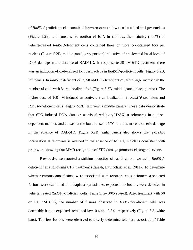
98
of Rad51d-proficient cells contained between zero and two co-localized foci per nucleus
(Figure 5.2B, left panel, white portion of bar). In contrast, the majority (>60%) of
vehicle-treated Rad51d-deficient cells contained three or more co-localized foci per
nucleus (Figure 5.2B, middle panel, grey portion) indicative of an elevated basal level of
DNA damage in the absence of RAD51D. In response to 50 nM 6TG treatment, there
was an induction of co-localized foci per nucleus in Rad51d-proficient cells (Figure 5.2B,
left panel). In Rad51d-deficient cells, 50 nM 6TG treatment caused a large increase in the
number of cells with 8+ co-localized foci (Figure 5.3B, middle panel, black portion). The
higher dose of 100 nM induced an equivalent co-localization in Rad51d-proficient and
Rad51d-deficient cells (Figure 5.2B, left versus middle panel). These data demonstrate
that 6TG induced DNA damage as visualized by γ-H2AX at telomeres in a dose-
dependent manner, and at least at the lower dose of 6TG, there is more telomeric damage
in the absence of RAD51D. Figure 5.2B (right panel) also shows that γ-H2AX
localization at telomeres is reduced in the absence of MLH1, which is consistent with
prior work showing that MMR recognition of 6TG damage promotes clastogenic events.
Previously, we reported a striking induction of radial chromosomes in Rad51d-
deficient cells following 6TG treatment (Rajesh, Litvinchuk, et al. 2011). To determine
whether chromosome fusions were associated with telomere ends, telomere associated
fusions were examined in metaphase spreads. As expected, no fusions were detected in
vehicle treated Rad51d-proficient cells (Table 1; n=1005 scored). After treatment with 50
or 100 nM 6TG, the number of fusions observed in Rad51d-proficient cells was
detectable but, as expected, remained low, 0.4 and 0.8%, respectively (Figure 5.3, white
bars). Too few fusions were observed to clearly determine telomere association (Table
99
5.1). In the vehicle-treated Rad51d-deficient cells, a basal level of 1.1% chromosome
fusions was detected, which increased to 2.7% and 6.9% following treatment with 50 and
100 nM 6TG, respectively (Figure 5.3, grey bars, p<0.05). In vehicle-treated Rad51d-
deficient cells, telomere-associated fusions were detectable and do appear to increase
following 6TG treatment, but the level of fusions regardless of telomere association was
too few to make any statistically meaningful conclusions (Table 5.1). Lastly, a basal level
of 0.6% chromosome fusions was detected in the Rad51d-/-
Mlh1-/-
cells, which increased
by a statistically insignificant amount to 0.9% and 1.6% following treatment with 50 and
100 nM 6TG, respectively (Figure 5.3, black bars). The data showing that 6TG-induced
fusions was reduced in Mlh1-deficient cells agreed with prior data from us and others that
the cytogenetic damage caused by 6TG is dependent at least in part on functional MMR
(Armstrong and Galloway 1997; Rajesh, Litvinchuk, et al. 2011).
6-Thioguanine induces multinucleation in Rad51d-deficient cells
Our prior work demonstrated that treatment with 100 nM 6TG induced aneuploidy and
multinucleation in Rad51d-deficient cells (Rajesh, Litvinchuk, et al. 2011). Here, the
induction of multinucleated cells was verified following treatment with a lower dose of
50 nM 6TG (Figure 5.4). Note also we investigated whether there was an unequal
distribution of telomeric DNA in the multinucleated cells because of such observations in
a prior report (Knecht et al. 2009). However, we show that the multinucleated cells
contained an equal distribution of telomeric DNA in this experimental system (Figure
5.5).
100
Discussion
Thiopurines induce a G2 arrest, which presumably prevents cells with damaged DNA
from entering what would otherwise become an abnormal mitosis (Armstrong and
Galloway 1997; Buermeyer et al. 1999; Rajesh, Litvinchuk, et al. 2011). However, co-
treatment with caffeine or UCN-01 to block ATM/ATR signaling can override the G2
arrest, from which cells subsequently enter tetraploid G1 arrest (Rajesh, Litvinchuk, et al.
2011). In Rad51d-deficient cells, the G2 arrest caused by 6TG was heightened but
transient; by 72 h, both Rad51d-deficient and Rad51d-proficient cells progressed into
mitosis, as demonstrated by flow cytometry and phospho-histone H3 staining (Yan et al.
2004). One implication is that release from this G2 checkpoint does not require the
resolution/completion of HR, and as such might help explain how cancer cells achieve
aneuploidy. 6TG treatment induced aneuploidy and multinucleated cells (Rajesh,
Litvinchuk, et al. 2011). Similar to this Rad51d-deficient cellular phenotype, different
leukemia and lymphomas are known to have increased aneuploidy and multinucleation.
One notable example is Reed-Sternberg (RS) cells associated with the pathology of
Hodgkin’s lymphoma (Mauch 2006). The data presented here demonstrates that
multinucleation following 6TG treatment occurs via mitotic catastrophe in two ways:
failed division and restitution. In this system, mitotic catastrophe resulted in
multinucleated cells that do not undergo apoptosis over the duration of the observations
here. When treated with the 50 nM 6TG dose, a larger percentage of multinucleated
Rad51d-deficient cells undergo mitotic catastrophe, which was not observed in cells at
the 100 nM dose presumably because of the heightened arrest. These results suggest that
a percentage of cells progress into mitosis at the lower dose of 6TG. In fact,
101
multinucleation and mitotic catastrophe is proposed to be a favored cell death mechanism
after cell cycle arrest (Castedo et al. 2004; Fragkos and Beard 2011).
Phosphorylation of Serine-139 on H2AX was measured as a marker of DNA
damage, and 6TG induced γ-H2AX as measured by Western blotting and
immunofluorescence in both Rad51d-deficient and Rad51d-proficient cells. It was
interesting to note that, as detected by Western blotting and immunofluorescence, γ-
H2AX induction seemed muted in the Rad51d-deficient cells at the lower dose of 50 nM.
It is tempting to speculate that the absence of RAD51D disrupts not just HR repair but
also associated DNA damage signaling. In fact, a role for the paralog RAD51C in
checkpoint signaling was demonstrated (Badie et al. 2009). However, note that γ-H2AX
induction is a highly dynamic process intertwined with yet to be clarified connections
with the induction of apoptosis and a broader epigenetic reprogramming in cancer (Cook
et al. 2009; Liu et al. 2016; Lu et al. 2006; Monteiro et al. 2014; Xiao et al. 2009).
RAD51D, a protein required for HR, was demonstrated previously to have a role
in telomere protection (Tarsounas et al. 2004). Loss of RAD51D conferred extensive
chromosome instability, increased chromosome fusions, and accelerated telomere
attrition (Smiraldo et al. 2005; Tarsounas et al. 2004). To further examine the role of
RAD51D at telomeres, we analyzed the length of the 3´ telomeric overhanging tail in
Rad51d-deficient MEFs. Rad51d-deficient cells had an approximately 40 percent
increase in overhang signal intensity. These data demonstrate that loss of RAD51D
affects the length of the 3´ telomeric overhang and suggest that RAD51D is required for
the regulation of telomere termini. Telomere dysfunction is known to activate DNA
damage responses, and loss of murine exonuclease 1 (Exo1) alleviated deleterious
102
cellular responses in telomere dysfunctional mice (Schaetzlein et al. 2007). This implies
that damage disrupting the normal telomere protective mechanisms can expose
chromosome ends to exonucleolytic processing that can promote chromosome fusions.
Exo1 deletion also conferred cellular resistance to killing by 6TG (Schaetzlein et al.
2007), which implicates telomeric damage as a mechanism of action for 6TG. Because
mammalian telomeres contain a G3 repeat in its canonical sequence, 6TG (as the
deoxynucleoside triphosphate) can become incorporated into telomeric DNA by
telomerase (Marathias, Sawicki, and Bolton 1999; Tendian and Parker 2000). It was more
recently reported that the deoxynucleoside of 6TG can directly damage telomeric DNA,
and that hTERT positive cells are sensitized to this treatment (Mender et al. 2015). We
measured the distribution of telomeres in the multinucleated Rad51d-deficient cells
because prior reports have shown structural telomeric defects and unequal distribution in
multinucleated Reed-Sternberg cells (Knecht et al. 2009; Knecht et al. 2010). However,
no unequal telomere distribution was observed in the Rad51d-deficient MEFs after 6TG
treatment. Collectively, this work contributes to the understanding of 6TG-induced
telomere damage and the negative consequences for chromosomal instability in the
absence of RAD51D-dependent processes at telomeres.
RAD51D is a RAD51 family member broadly appreciated to be indispensable for
HR; yet, the specialized functions of the individual protein products have evaded full
elucidation. Components of HR are recruited to stalled replication forks and inter-strand
crosslinks (ICLs), as well as DNA double strand breaks (DSBs). Recently, a dominant
RAD51 mutation in a patient with Fanconi anemia-like phenotypes was characterized and
uncovered a role for RAD51 in ICL repair independent of HR (Wang et al. 2015),
103
suggesting there is much more to learn about these highly related yet potentially
separable processes. Human variant alleles of the RAD51 paralogs confer cancer
susceptibility, for example RAD51B mutations are associated with breast cancer
(Golmard et al. 2013; Pelttari et al. 2016), RAD51C mutations are associated with breast
and ovarian cancer , and RAD51D mutations are associated with ovarian cancer (Coulet
et al. 2013; Meindl et al. 2010; Song et al. 2015; Vaz et al. 2010). There are many more
mutations of unknown significance in these paralogs. Our results suggest that clinical
variability in how patients respond to thiopurine treatment, as well as their potential risk
for a future, therapy-related secondary dysplasia might include the status of RAD51D-
dependent processing of telomeres.
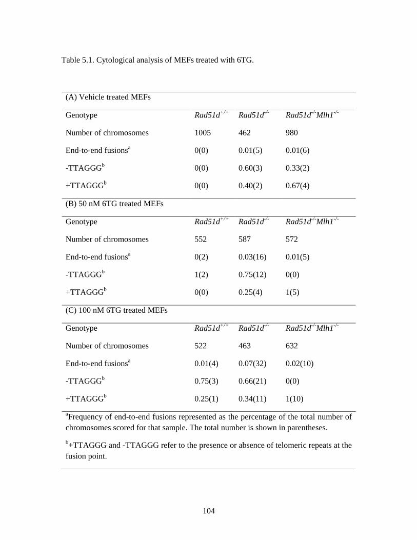
104
Table 5.1. Cytological analysis of MEFs treated with 6TG.
(A) Vehicle treated MEFs
Genotype Rad51d+/+
Rad51d-/-
Rad51d-/-
Mlh1-/-
Number of chromosomes 1005 462 980
End-to-end fusionsa
0(0) 0.01(5) 0.01(6)
-TTAGGGb
0(0) 0.60(3) 0.33(2)
+TTAGGGb
0(0) 0.40(2) 0.67(4)
(B) 50 nM 6TG treated MEFs
Genotype Rad51d+/+
Rad51d-/-
Rad51d-/-
Mlh1-/-
Number of chromosomes 552 587 572
End-to-end fusionsa
0(2) 0.03(16) 0.01(5)
-TTAGGGb
1(2) 0.75(12) 0(0)
+TTAGGGb
0(0) 0.25(4) 1(5)
(C) 100 nM 6TG treated MEFs
Genotype Rad51d+/+
Rad51d-/-
Rad51d-/-
Mlh1-/-
Number of chromosomes 522 463 632
End-to-end fusionsa
0.01(4) 0.07(32) 0.02(10)
-TTAGGGb
0.75(3) 0.66(21) 0(0)
+TTAGGGb
0.25(1) 0.34(11) 1(10)
aFrequency of end-to-end fusions represented as the percentage of the total number of
chromosomes scored for that sample. The total number is shown in parentheses.
b+TTAGGG and -TTAGGG refer to the presence or absence of telomeric repeats at the
fusion point.
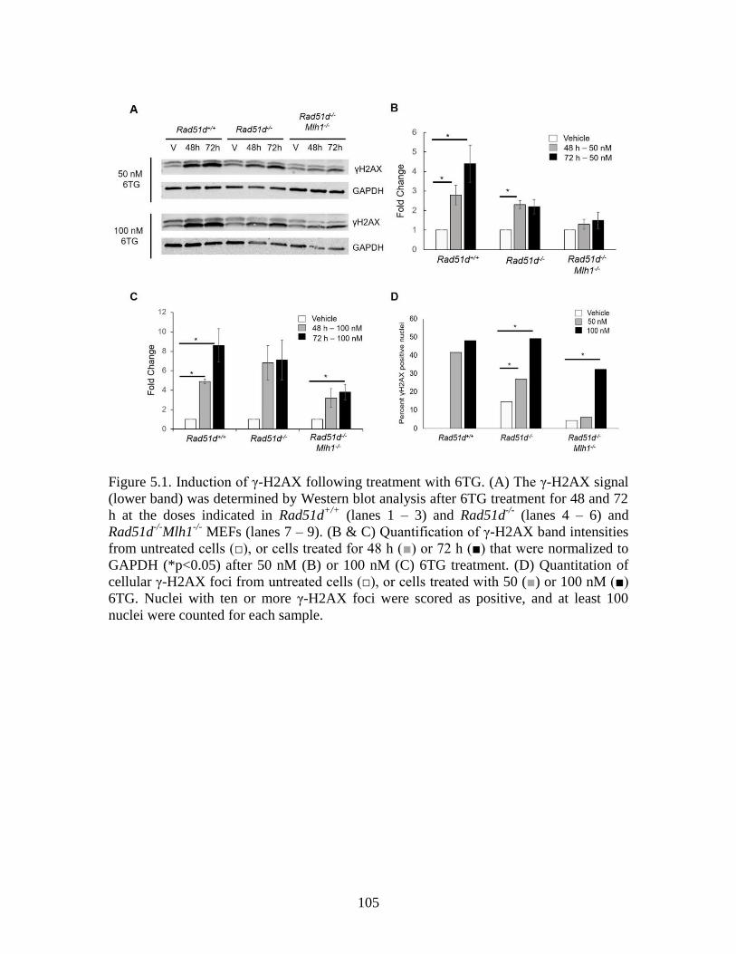
105
Figure 5.1. Induction of γ-H2AX following treatment with 6TG. (A) The γ-H2AX signal
(lower band) was determined by Western blot analysis after 6TG treatment for 48 and 72
h at the doses indicated in Rad51d+/+
(lanes 1 – 3) and Rad51d-/-
(lanes 4 – 6) and
Rad51d-/-
Mlh1-/-
MEFs (lanes 7 – 9). (B & C) Quantification of γ-H2AX band intensities
from untreated cells (□), or cells treated for 48 h (■) or 72 h (■) that were normalized to
GAPDH (*p<0.05) after 50 nM (B) or 100 nM (C) 6TG treatment. (D) Quantitation of
cellular γ-H2AX foci from untreated cells (□), or cells treated with 50 (■) or 100 nM (■)
6TG. Nuclei with ten or more γ-H2AX foci were scored as positive, and at least 100
nuclei were counted for each sample.
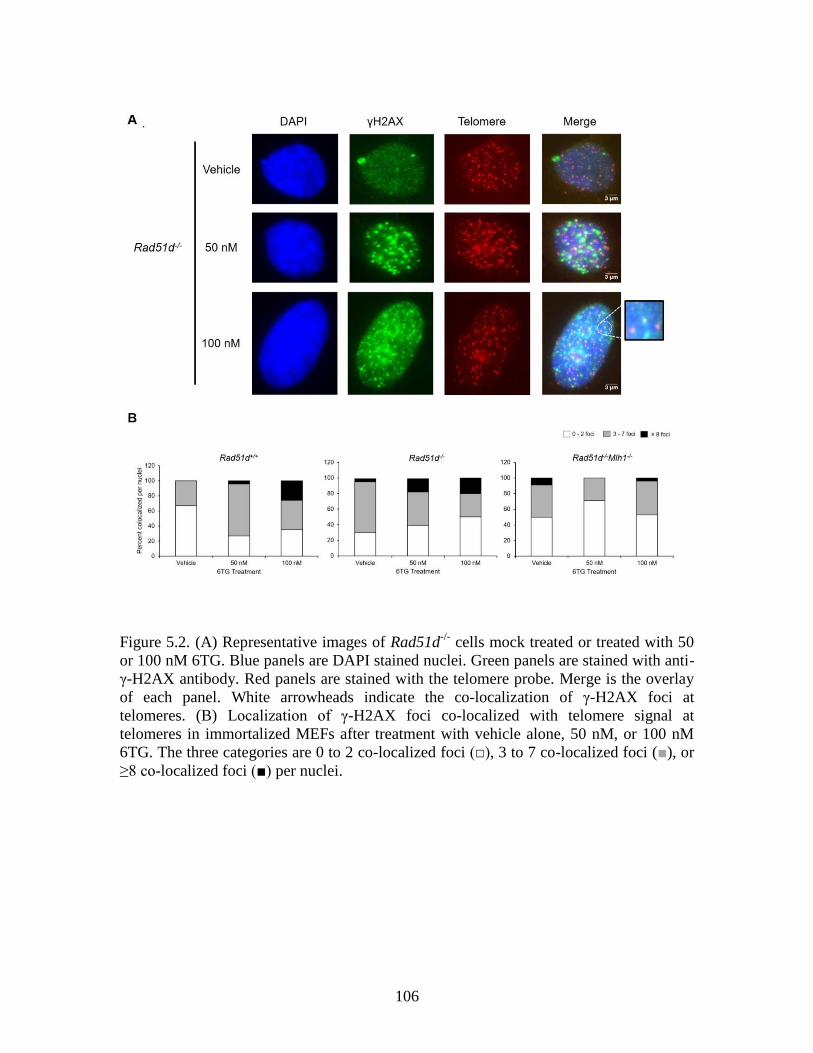
106
Figure 5.2. (A) Representative images of Rad51d-/-
cells mock treated or treated with 50
or 100 nM 6TG. Blue panels are DAPI stained nuclei. Green panels are stained with anti-
γ-H2AX antibody. Red panels are stained with the telomere probe. Merge is the overlay
of each panel. White arrowheads indicate the co-localization of γ-H2AX foci at
telomeres. (B) Localization of γ-H2AX foci co-localized with telomere signal at
telomeres in immortalized MEFs after treatment with vehicle alone, 50 nM, or 100 nM
6TG. The three categories are 0 to 2 co-localized foci (□), 3 to 7 co-localized foci (■), or
≥8 co-localized foci (■) per nuclei.
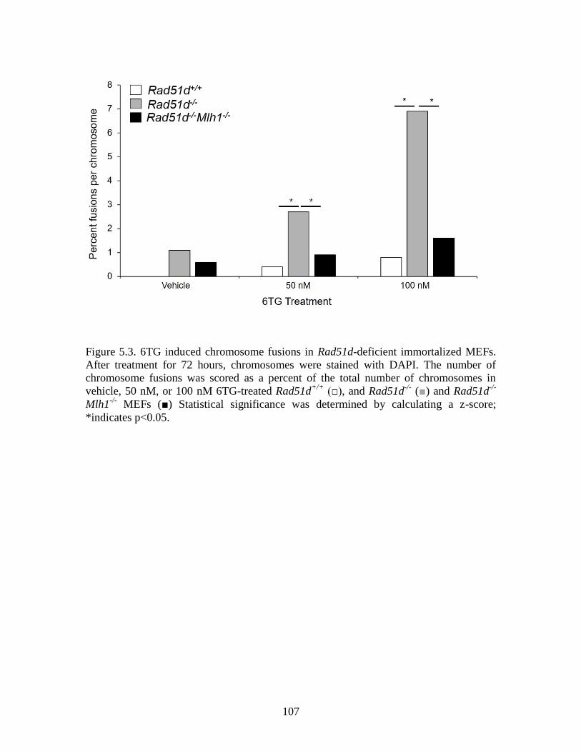
107
Figure 5.3. 6TG induced chromosome fusions in Rad51d-deficient immortalized MEFs.
After treatment for 72 hours, chromosomes were stained with DAPI. The number of
chromosome fusions was scored as a percent of the total number of chromosomes in
vehicle, 50 nM, or 100 nM 6TG-treated Rad51d+/+
(□), and Rad51d-/-
(■) and Rad51d-/-
Mlh1-/-
MEFs (■) Statistical significance was determined by calculating a z-score;
*indicates p<0.05.
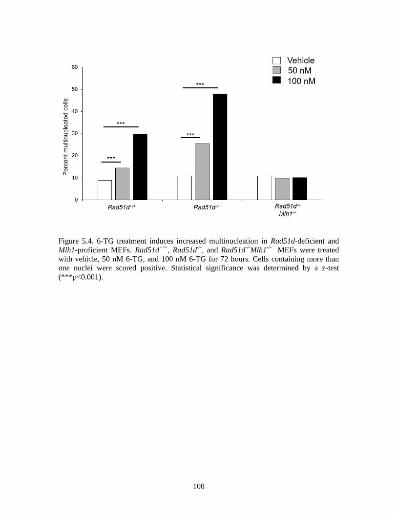
108
Figure 5.4. 6-TG treatment induces increased multinucleation in Rad51d-deficient and
Mlh1-proficient MEFs. Rad51d+/+
, Rad51d-/-
, and Rad51d-/-
Mlh1-/-
MEFs were treated
with vehicle, 50 nM 6-TG, and 100 nM 6-TG for 72 hours. Cells containing more than
one nuclei were scored positive. Statistical significance was determined by a z-test
(***p<0.001).
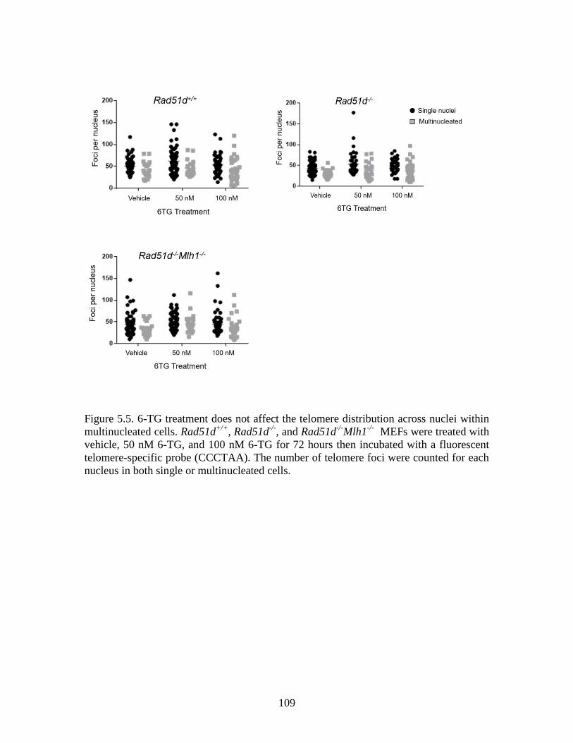
109
Figure 5.5. 6-TG treatment does not affect the telomere distribution across nuclei within
multinucleated cells. Rad51d+/+
, Rad51d-/-
, and Rad51d-/-
Mlh1-/-
MEFs were treated with
vehicle, 50 nM 6-TG, and 100 nM 6-TG for 72 hours then incubated with a fluorescent
telomere-specific probe (CCCTAA). The number of telomere foci were counted for each
nucleus in both single or multinucleated cells.
110
CHAPTER 6
GENOME-WIDE IDENTIFICATION AND EXPRESSION ANALYSIS OF GENE
DIFFERENCES BETWEEN RAD51D PROFICIENT AND DEFICIENT PRIMARY
MOUSE EMBRYONIC FIBROBLASTS
111
Abstract
One of the hallmarks of cancer is replicative immortality, and an enabling characteristic
is genome instability that increases the prevalence of acquired gene mutations. Mutations
in genes associated with the homologous recombination (HR) repair pathway increases
genomic instability and are found in approximately 50% of ovarian cancers. Patients that
carry mutations in the RAD51D HR gene are up to 12 times more likely to develop this
cancer. Loss of Rad51d in mouse embryos confers mid-gestation embryo death, and cells
isolated from Rad51d-/-
embryos fail to proliferate in culture. Concurrent deletion of the
Trp53 gene extends embryo development up to 7 days, and Rad51d-/-
Trp53-/-
(Rad51d-
deficient) MEF cell lines have been successfully generated. Rad51d-deficient cells have
extensive chromosomal aberrations, such as fusions and telomere shortening, and similar
defects are often observed in ovarian cancer cells. For this reason, Rad51d-deficient
MEFs were used as a model for genomic unstable ovarian cancers. The gene expression
profiles of primary MEFs derived from Rad51d+/+
Trp53-/-
(Rad51d-proficient) and
Rad51d-deficient embryos were analyzed by both microarray and RNA Seq. Microarray
identified 489 genes with higher expression and 129 genes with lower expression in
Rad51d-deficient MEFs compared with Rad51d-proficient MEFs. RNA Seq analysis
identified 449 genes with higher expression, and 479 genes with lower expression in the
absence of Rad51d. In both analyses, the highest proportion of genes were associated
with cellular growth and proliferation. Twenty-one genes associated with cell cycle
progression were identified by microarray including Id1, Id2, and Cdkn1a(p21) that had
higher expression in Rad51d-deficient MEFs. Analysis using the TopHat function on the
Illumina platform identified two intra-chromosomal gene fusions, one along
112
Chromosome 4 and one along Chromosome 17, in the Rad51d-proficient cell line, and
three inter-chromosomal fusions all involving Chromosome 10. Only the fusion
involving Chromosome 4 was also identified in the Rad51d-deficient MEFs, and no
fusions were identified only in the Rad51d-deficient cell lines. Together these data
provide insight into gene expression compromises that support cell proliferation in the
absence of Rad51d.
Introduction
Approximately 50% of ovarian cancers carry mutations in genes associated with the
homologous recombination (HR) pathway, and exhibit extensive chromosome instability
(Coulet et al. 2013; Eoh et al. 2016; Konstantinopoulos et al. 2015; Konstantinopoulos et
al. 2014; Loveday et al. 2011; Meindl et al. 2010; Prakash et al. 2015; Song et al. 2015;
Tedaldi et al. 2017; Thompson et al. 2013). The RAD51 gene family – RAD51, RAD51B,
RAD51C, RAD51D, XRCC2, and XRCC3 – encode proteins that are part of the HR
pathway. These proteins are essential for maintaining chromosome integrity and repairing
DNA damage, and loss of any RAD51 paralog confers cellular sensitivity to DNA
damaging agents (Andreassen and Ren 2009; Baumann and West 1998; Kim and
D'Andrea 2012; Chirnomas et al. 2006; Rajesh, Litvinchuk, et al. 2011;
Konstantinopoulos et al. 2014). RAD51D, the fourth member of the RAD51 family of
genes, is a known breast and ovarian cancer susceptibility gene (Loveday et al. 2011;
Song et al. 2015; Tedaldi et al. 2017; Thompson et al. 2013).
113
Early investigations into Mus musculus Rad51d demonstrated that loss of the gene
confers an embryo lethal phenotype beginning at 8.5 days post conception (dpc) (Pittman
and Schimenti 2000). A concurrent deletion of the Trp53 gene extends embryo
development to 15.5 dpc (Smiraldo et al. 2005). In addition to embryo defects, null
alleles of Rad51d lead to chromosomal defects in the form of breaks, gaps, and
translocations, telomere shortening, and telomere-specific end-to-end fusions (Tarsounas
et al. 2004; Smiraldo et al. 2005). Spectral karyotype analysis of cells isolated from a
Rad51d-deficient embryo identified a large number of fusions – for example, between
chromosomes 9 and 12, and chromosomes 2 and 19 (Smiraldo et al. 2005). Despite the
extensive genomic instability and embryo lethality observed when the Rad51d gene is
deleted, mouse embryonic fibroblasts (MEFs) were able to proliferate in culture when the
Trp53 gene was deleted concurrently (Smiraldo et al. 2005).
In this study, gene expression profiles of Rad51d+/+
Trp53-/-
(Rad51d-proficient)
and Rad51d-/-
Trp53-/-
(Rad51d-deficient) primary MEF cell lines were assessed by
microarray and RNA Seq analyses. Microarray identified 618 genes with differential
expression between the Rad51d-deficient and -proficient cell lines, and 21 of the
identified genes are associated with cell cycle progression. RNA Seq analysis of Rad51d-
proficient and -deficient cell lines identified 928 genes that were differentially expressed.
Five gene fusions were identified in the Rad51d-proficient cell lines, but only one of
these fusions was also present in the Rad51d-deficient samples. Comparison between the
two data sets identified 135 genes that were differentially expressed between Rad51d-
proficient and -deficient cell lines. Together these data provide insight into gene
expression compromises that support cell division in a chromosomal unstable cell line.
114
Materials and Methods
Cell Culture
Rad51d+/-
Trp53+/-
mice were intercrossed, and primary MEFs were generated from mid-
gestation embryos (Smiraldo et al. 2005). MEFs isolated from Rad51d+/+
Trp53-/-
(MEFPR1) and two independent Rad51d-/-
Trp53-/-
(MEFT11 and MEFT34) embryos
were maintained at 37°C with 5% CO2 in Dulbecco’s Modified Eagle’s Medium
(DMEM; Fisher Scientific) supplemented with 10% fetal bovine serum (Atlanta
Biologicals), 1% penicillin/streptomycin, and 1% glutamine. For these experiments,
primary MEFs were used only up until three passages.
RNA Isolation and RT-PCR of Rad51d
Total RNA was isolated using the miRNeasy Mini Kit (Cat. #: 217004; Qiagen)
according to the manufacturer’s protocol. RNA quality was assessed using an Agilent
2100 Bioanalyzer and RNA Integrity Numbers for all samples used ranged from 9.0 to
10.0. First-strand reverse transcription for each RNA sample was performed using a
ProScriptII First Strand DNA Synthesis Kit (NEB). Gene specific primers for MmRad51d
spanning exon 1 to exon 4, and for MmGapdh were used: Rad51d SS1 5’-
(GCGAGCGCCCAAGTGACAGA)-3’, Rad51d SS2 (5’-
GCTACCTGGGCCACCCACAA-3’), Gapdh left (5’-AACTTTGGCATTGTGGAAG-
3’), Gapdh right (5’-GGATGCAGGGATGATGTTCT-3’). PCR reactions were
performed under the following conditions: 30 cycles at 94°C for 30 seconds, 57°C for 30
seconds, and 72°C for 1 minute.
115
Microarray Analysis
Microarray experiments were performed using the Affymetrix platform. Total RNA
samples were amplified and biotinylated using GeneChip WT PLUS Reagent Kit
(Affymetrix, Santa Clara, CA). One hundred nanograms of total RNA per sample was
reverse transcribed into ds-cDNA using random hexamers, and the remaining RNA was
degraded using RNase H. Single strand cDNA was then fragmented and labelled with
biotin. The amplified and labeled samples were hybridized to Mouse Transcriptome
Arrays 1.0 (Affymetrix, Santa Clara, CA) for 16 h at 45°C using a GeneChip
Hybridization Oven 640 and a GeneChip Hybridization, Wash, and Stain Kit
(Affymetrix, Santa Clara, CA). Hybridized arrays were washed and stained using a
GeneChip Fluidics Stations 450. Arrays (8 total) were scanned using a GeneChip Scanner
3000 7G system and computer workstation equipped with GeneChip Command Console
4.0 software (Affymetrix, Santa Clara, CA).
Following completion of array scans, probe cell intensity (CEL) files were
imported into Expression Console Software (Affymetrix, Santa Clara, CA) and processed
at the gene-level using the Robust Multichip Analysis (RMA) algorithm to generate CHP
files. After confirming data quality within Expression Console, CHP files containing log2
expression signals for each probe were imported into Transcriptome Analysis Console
Software version 3.0.0.466 (Affymetrix, Santa Clara, CA) to analyze cell type specific
transcriptional responses using one-way between-subject analysis of variance (ANOVA).
A p-value of 0.05 and a fold change of 1.5 were used as cutoff parameters. Subsequently,
pathway analysis of the differentially expressed genes was performed using Ingenuity
Pathway Analysis software (Qiagen, Hilden, Germany).
116
RNA Sequencing Library Preparation
RNA-seq libraries were constructed using TruSeq® Stranded mRNA LT (Cat. #: RS-
122-2101; Illumina) according to the manufacturer’s protocol. Briefly, the mRNA was
purified by two rounds of polyA selection from the total RNA. The mRNA was reverse
transcribed into first strand cDNA using reverse transcriptase and random primers. The
second strand cDNA was synthesized using PCR. A single “A” nucleotide was added to
the 3’ ends of the blunt fragments and ligated with multiple single “T” indexing adapters
to the ends of the double strand cDNA. DNA libraries were enriched by PCR
amplification, qualified using an Agilent Technologies 2100 Bioanalyzer, and quantitated
by qPCR in a Bio-Rad iCycler using a Bio-Rad iCyclerTM qPCR Master Mix (Cat. #:
KK4844; Kapa Biosystems). After denaturation, libraries were diluted to 1.8 pM with
hybridization buffer. Paired end 75 bp sequencing was performed on the Illumina
NextSeq 500 using NSQ® 500 High Output KT v2 (150 CYS; Cat. #: FC-404-2002;
Illumina) per manufacturer’s protocol.
RNA Sequencing Analysis
Expression analysis was performed on the Illumina BaseSpace platform (Illumina). The
iGenome reference dataset used for analysis was the University of California – Santa
Cruz (UCSC) Mus musculus reference genome (mm10). Uniquely mapped reads were
assembled into transcripts guided by the UCSC Mus musculus mm10 RefSeq & Gencode
gene annotation using the TopHat function on the Illumina platform. Expression
differences between conditions were evaluated using DESeq2 (Love, Huber, and Anders
2014; Schurch et al. 2016). The analysis was generated using Partek® software (St. Louis,
MO, USA). Annotation of significantly different transcripts and enrichment analysis was
117
performed with DAVID. This is a bioinformatic resource, supported by the National
Institutes of Allergy and Infectious Disease, that uses integrated biological
knowledgebases to systematically extract biological meaning from large data sets (Huang
da, Sherman, and Lempicki 2009). The hierarchical clustering analysis of the global gene
expression pattern in different samples was carried out using heatmap.2 function (gplots
package) in R. Gene set enrichment was analyzed with GSEA software (Subramanian et
al. 2005).
Results
Microarray gene expression analysis of primary mouse embryonic fibroblasts
The main goal of this study was to identify expression profile differences between low-
passage primary mouse embryonic fibroblasts (MEFs) generated from individual
Rad51d-deficient and Rad51d-proficient embryos (described previously (Smiraldo et al.
2005)) using an Affymetrix microarray platform. RNA was isolated, and the Rad51d
status of each cell line was confirmed by RT-PCR (Figure 6.1A). Differences in gene
expression levels were determined by comparing the Rad51d-deficient sample with the
Rad51d-proficient (control) sample. Genes with greater than 1.5-fold difference are
reported. Of the 65,956 genes represented on the array, 618 were differentially expressed
between the samples (Table A.1); 489 genes had higher expression, and 129 had lower
expression in the Rad51d-deficient cell line compared with the Rad51d-proficient cell
line.
The transcripts represented on the array encoded only for known protein products
and predicted genes based on Ensembl data sets. Of the genes with higher expression in
118
the Rad51d-deficient cell line, 207 encode known protein products. A proteomics screen
performed by the Pittman laboratory identified 75 proteins that co-precipitate with
RAD51D (Rajesh et al. 2009). The genes that encode two of the proteins identified in this
screen – Ifit1 and Rnf213 – had lower expression in the Rad51d-deficient cell line.
Expression of Ifit1 was 2.35 times lower in the absence of Rad51d. In the Mus musculus
genome, this gene maps to chromosome 19 and encodes for the interferon-induced
protein with tetratricopeptide repeats 1 (IFIT1) protein that is expressed in mouse large
intestine, liver, small intestine, and bladder (Yue et al. 2014). In a study of breast cancer
patients, expression of IFIT1 was associated with improved local relapse-free survival
and has been shown to mediate chemotherapy and radiation resistance (Danish et al.
2013). Rnf213 maps to chromosome 11 and is most highly expressed in ovary, mammary
gland, and thymus tissues (Yue et al. 2014). This gene encodes for the RING finger 213
(RNF213) E3 ubiquitin ligase protein. In humans, small nucleotide polymorphisms in this
gene is associated with increased risk for moyamoya disease (Hu, Luo, and Chen 2017).
The Ingenuity Pathway Analysis (IPA) tool was used to sort genes into known
biological functions (Figure 6.2). This software is designed to “integrate previously
observed cause-effect relationships reported in the literature” with a gene expression data
set using Ingenuity Knowledge Base, a “collection of observations from various
experimental contexts curated from biomedical literature” (Kramer et al. 2014). For this
study, IPA was used to categorized genes into known biological functions. Ninety-one
genes with differential expression between the Rad51d-deficient and Rad51d-proficient
cell lines were associated with “cellular growth and proliferation” (Table 6.1).
119
One of the main goals of this project was to understand how expression
compromises in the absence of Rad51d promote cell division and proliferation when the
genome is unstable. Twenty-one genes associated with “cell cycle progression” (18
included in the “cellular growth and proliferation” group) had altered gene expression in
the absence of Rad51d (Table 6.2). Expression of Stat1 was 1.6-fold lower in the
Rad51d-deficient cell line than the Rad51d-proficient. The STAT1 protein encoded by
this gene is activated in response to extracellular signals, including cytokines and growth
factors, and acts as a transcription factor that activates expression of over 1000 gene
targets (Calo et al. 2003; Satoh and Tabunoki 2013). Two members of the DNA binding
and/or differentiation (Id) family – Id1 and Id2 – had 1.5- and 2.3-fold higher expression
in the absence of Rad51d, respectively. The proteins encoded by these genes, ID1 and
ID2, block binding of helix-loop-helix (HLH) transcription factors during the S phase of
the cell cycle to prevent cell differentiation (Jogi et al. 2002). Expression of the Cdkn1a
gene was 1.6-fold higher in the Rad51d-deficient cell line. This gene encodes the cell-
cycle dependent kinase p21 that can function independently of p53 to initiate cell cycle
arrest in response to DNA damage and cellular stress (Georgakilas, Martin, and Bonner
2017).
RNA Seq gene expression analysis of primary mouse embryonic fibroblasts
Microarray is a useful tool for identifying genes with differential expression between cell
lines, however, analysis is limited to gene transcripts represented on the array. RNA Seq
allows for genome-wide detection of gene expression levels with low background
compared to microarray (Wang, Gerstein, and Snyder 2009). For this reason, we chose to
120
analyze gene expression in an independent Rad51d-deficient and the Rad51d-proficient
cell lines using RNA Seq.
Nine hundred twenty-eight genes were differentially expressed between the
Rad51d-deficient and Rad51d-proficient samples (Table A.2). Of the genes identified,
449 genes had higher expression and 479 had lower expression in the absence of Rad51d.
The Cd52 gene had 11.56 times lower expression in the Rad51d-deficient cell line
compared with the Rad51d-proficient cell line. This gene maps to Chromosome 4 and
encodes the CD52 glycoprotein that is expressed on the surface of normal and leukemic
immune cells (Vojdeman et al. 2017). The Stra6 gene had 3.17 times lower expression in
the Rad51d-deficient cell line. This gene maps to Chromosome 9 and encodes the integral
membrane receptor protein STRA6 that mediates uptake of vitamin A (Chen et al. 2016).
IPA was used to sort the genes identified by RNA Seq into categories based on
known biological functions (Figure 6.3). The categories are defined based on literature
data, and 156 genes with differential expression between the Rad51d-deficient and
Rad51d-proficient cell lines were associated with “cellular growth and proliferation”
(Table 6.3), and 146 genes were associated with “cell death and survival.” These data
suggest that the loss of Rad51d affects expression of genes that regulate cell proliferation.
Chromosome fusion points identified by RNA Seq
One advantage of RNA Seq is the ability to identify transcriptome boundaries, such as
the connectivity between two exons (Wang, Gerstein, and Snyder 2009). The TopHat-
Fusion algorithm is designed to identify both intra- and inter-chromosome fusions (Kim
and Salzberg 2011), and was used to identify gene fusions in the Rad51d-proficient and
Rad51d-deficient samples (Table 6.4). Five gene fusions were identified in the Rad51d-
121
proficient sample; two intra- and three inter-chromosome gene fusions. The first intra-
chromosome fusion was along Chromosome 4 and resulted in a fusion between the
coding region for the Faf1 gene and a non-coding region within the chromosome. The
second was along Chromosome 17 and resulted in a fusion between the coding regions
for the Acat3 and Acat2 genes. The other three fusions were inter-chromosome fusions
that all involved different regions along Chromosome 10. The fusions occurred at
different points along Chromosome 10, suggesting that multiple breaks had occurred. The
first inter-chromosome fusion occurred at the coding region for the Nap1l1 gene along
Chromosome 10 and a non-coding region of the Chromosome 1. The second fusion
occurred at the coding regions for the Hmga2 gene along Chromosome 10 and the
Sdccag8 gene along Chromosome 1. The third fusion occurred along the coding region
for H2afy2 along Chromosome 10 and the Pvt1 gene along Chromosome 15.
One intra-chromosome fusion was identified in the Rad51d-deficient sample
along Chromosome 4 between the coding region for the Faf1 gene and a non-coding
region. This same fusion was identified in the Rad51d-proficient. We hypothesized that
more gene fusions would be identified in the Rad51d-deficient cell line, and that these
fusions would be unique to the Rad51d-/-
genotype. However, this analysis did not
identify any novel fusions in the Rad51d-deficient sample compared with the Rad51d-
proficient cell line.
Genes with altered expression identified by both microarray and RNA Seq
Microarray and RNA Seq analysis identified 618 and 928 genes, respectively, that were
differentially expressed between Rad51d-deficient and Rad51d-proficient cell lines. Of
those genes, 111 were identified in both analyses (Table 6.5). The genes are listed by
122
increasing fold change (Rad51d-deficient v. Rad51d-proficient) as determined by the
microarray, and only genes that code for a known protein product are included. IPA
identified 91 and 156 genes by microarray and RNA Seq, respectively, that are associated
with the “cellular growth and proliferation.” Of these, 18 genes were identified by both
analyses (Table 6.6). Of the 21 cell cycle regulatory genes identified by microarray, 8
were also identified by RNA Seq, including Cdkn1a (Table 6.7). The Ifit1 gene was
identified by microarray and by RNA Seq as being differentially expressed in the absence
of Rad51d. In a proteomics study, the IFIT1 protein was identified as an interacting
protein with RAD51D (Rajesh et al. 2009). Interestingly, the expression differences were
inconsistent between the independent Rad51d-deficient samples. Expression of Ifit1 was
determined to be 2.35 times lower by microarray and 1.09 times higher by RNA Seq,
respectively, in the Rad51d-deficient cell line compared with the Rad51d-proficient cell
line.
Discussion
Hanahan and Weinberg classified six identifiable biological characteristics of tumor
development that they termed the “hallmarks of cancer” (Hanahan and Weinberg 2000).
In addition to these features, cancer cells acquire ‘enabling characteristics’ that contribute
to carcinogenesis. In 2011, Hanahan and Weinberg classified ‘genome instability’ as an
enabling characteristic, and argued that tumor growth can often be attributed to
acquisition of mutations that promote cell proliferation and inhibit cell death (Hanahan
and Weinberg 2011). The observation that cancer is a mutation-driven disease also led to
the “Mutator Phenotype Hypothesis.” First described by Lawrence Loeb, this hypothesis
123
stated that “mutations occurred randomly throughout the genome, and among these
would be mutations in genes that guarantee the fidelity of DNA replication… and repair”
(Loeb, Springgate, and Battula 1974). Together, these ideas have led to the current belief
that “defects in genome maintenance are… instrumental for tumor progression”
(Hanahan and Weinberg 2011).
The RAD51D gene encodes a protein that functions during homologous
recombination (HR)-mediated repair of DNA double strand breaks and interstrand
crosslinks, and is essential for maintaining chromosome and telomere stability (Gruver et
al. 2005; Hinz et al. 2006; Rajesh, Litvinchuk, et al. 2011; Rajesh et al. 2010; Smiraldo et
al. 2005; Tarsounas et al. 2004; Wyatt et al. ; Yard et al. 2016). Loss of the Rad51d gene
induces an embryo lethal phenotype in mice, and cells isolated from these embryos
exhibit extensive chromosomal defects (Pittman and Schimenti 2000; Smiraldo et al.
2005). Despite chromosome instability, Rad51d-/-
cells proliferate in culture when the
Trp53 gene is also deleted (Smiraldo et al. 2005). For these reasons, Rad51d-/-
Trp53-/-
(Rad51d-deficient) mouse embryonic fibroblast (MEF) cell lines serve as a model of
chromosomally unstable mammalian cells and can be utilized to identify expression
compromises that promote cell growth under genome unstable conditions. For this study,
gene expression analysis of Rad51d-deficient and Rad51d-proficient primary mouse
embryonic fibroblast cell lines was performed using both microarray and RNA Seq
technologies.
Microarray is a high-throughput bioinformatics technique that boasts the ability to
analyze expression of tens of thousands of genes at once (Govindarajan et al. 2012). In
this study, microarray analysis identified 618 genes with differential expression between
124
Rad51d-deficient and Rad51d-proficient cell lines. Ingenuity Pathway Analysis (IPA)
performed on the microarray data set identified 91 genes that are associated with “cellular
growth and proliferation,” and 21 genes associated with cell cycle progression. The cell
cycle genes include Id1, Id2, and Cdkn1a(p21) are discussed below.
The Id1 and Id2 genes encode proteins that promote abnormal cell proliferation.
ID1 forms a heterodimer with the ETS-1 transcription factor to preventing binding and
activation of target genes. One target of ETS-1 is p16, a tumor suppressor that activates
cell cycle arrest. Binding of ID1 to ETS-1 suppresses transcription of p16, leading to
increased expression of CDKs that promote cell cycle progression and cell differentiation
(Perk, Iavarone, and Benezra 2005). Overexpression of ID1 has been linked with
increased development of several cancer types, and approximately 70% of ovarian
cancers have abnormal expression of ID1 that has been correlated with cancer cell
proliferation and resistance to apoptosis (Zhang et al. 2004). Consistent with these
previous observations, increased expression of Id1 and Id2 in Rad51d-deficient cells may
contribute to cell proliferation and growth, despite the extensive genomic instability.
The Cdkn1a(p21) gene encodes the p21 protein that induces cell growth arrest and
inhibits cell cycle progression in response to DNA damage and cellular stress (Abbas and
Dutta 2009). The cell lines used for these studies are Trp53-null, therefore, expression of
Cdkn1a(p21) in these cells is independent of p53. In a p53-deficient environment, p21
leads to dysregulation of replication machinery, increased genome instability, and high
levels of cell proliferation (Georgakilas, Martin, and Bonner 2017). Furthermore, these
data provide an explanation for how ovarian cancers that carry mutations in the RAD51D
gene proliferate in the presence of genome instability.
125
Advancement in bioinformatics technologies have led to the rise of an alternative
high-throughput method for analyzing gene expression: RNA Seq. This method provides
more precise measurements of gene expression levels than microarray, and is not limited
to detecting transcripts that correspond to reference sequences on an array (Wang,
Gerstein, and Snyder 2009). In this study, 928 genes with altered expression in the
Rad51d-deficient cell line compared with the Rad51d-proficient cell line were identified
by RNA Seq. Previous studies showed increased levels of chromosome breaks, fusions,
and translocations in Rad51d-deficient cells (Smiraldo et al. 2005), and we hypothesized
that certain regions of the genome would be more susceptible to fusions in the absence of
Rad51d. Using the TopHat-Fusion algorithm in the Illumina platform, gene fusions
present in the Rad51d-deficient and -proficient cell lines were identified. Two intra-
chromosomal and three inter-chromosomal gene fusions were detected in the Rad51d-
proficient sample, and only one intra-chromosomal fusion was observed in the Rad51d-
deficient sample. The fusion identified in the Rad51d-deficient sample occurred along
Chromosome 4 between the coding region for Faf1 and a non-coding region. This fusion
has never been reported but may be present in the mouse germline as it was identified in
both the Rad51d-proficient and -deficient cells. Previous spectral karyotyping of Rad51d-
deficient cells identified chromosomal fusions, for example between chromosomes 9 and
12, and chromosomes 2 and 19 (Smiraldo et al. 2005); however, these specific
translocations were not detected in this study. These data did not support my assertion
that loss of Rad51d would affect specific regions along the genome.
Future studies would complement the data presented here by investigating how
loss of other RAD51 paralogs affects genome stability. Loss of either Rad51c or Xrcc2 in
126
embryos also disrupts development mid-gestation and induces an embryo lethal
phenotype. Consistent with Rad51d-deficient cells, loss of Rad51c or Xrcc2 inhibits
proliferation in culture unless Trp53 is deleted concurrently (Kuznetsov et al. 2009;
Adam, Deans, and Thacker 2007). Analyzing gene expression profiles of primary MEF
cell lines that lack Rad51c or Xrcc2 could provide additional insight into how loss of
other HR DNA repair genes affects gene expression and promotes tumor growth.
Additional future studies could include testing inhibitors of p21 in the Rad51d-
proficient and -deficient cell lines. The McInnes laboratory at the University of South
Carolina has successfully synthesized a 16-mer peptide that competes with the p21
binding site along the proliferating cell nuclear antigen (PCNA) protein and provides
evidence for a potential target site that will disrupt this interaction (Kontopidis et al.
2005). Interaction between p21 and PCNA delays replication in the presence of DNA
damage. Inhibitors that disrupt p21 binding with PCNA will prevent DNA repair and
promote apoptosis (Abbas and Dutta 2009). If increased expression of p21 is a
compensatory mechanism that promotes cell survival in the absence of Rad51d, then
inhibiting the activity of this protein offers the potential to induce apoptosis in these cells.
This type of therapeutic approach would offer a myriad of benefits in the clinic,
particularly for treating ovarian cancer patients that increased p21 expression in
conjunction with mutations in RAD51D and/or TP53.
Acknowledgements
NMR performed RNA isolation, data analysis, and contributed to writing the manuscript.
DA performed the RNA quality analysis and ran the microarray. CL prepared the samples
127
for RNA Seq and ran the analysis. PR generated the Rad51d-proficient and Rad51d-
deficient primary cell lines used in this paper. PJB performed data analysis using Partek
software. DLP contributed to experimental design and manuscript preparation. This
research was supported by a South Carolina IDeA Networks of Biomedical Research
Excellence (INBRE) Grant to DLP, the National Institutes of Health under Award
Number R15GM110615, and a Support to Promote Advancement of Research and
Creativity (SPARC) Graduate Research Grant from the Office of the Vice President for
Research at the University of South Carolina to NMR. The content is solely the
responsibility of the authors and does not necessarily represent the official views of the
National Institutes of Health.
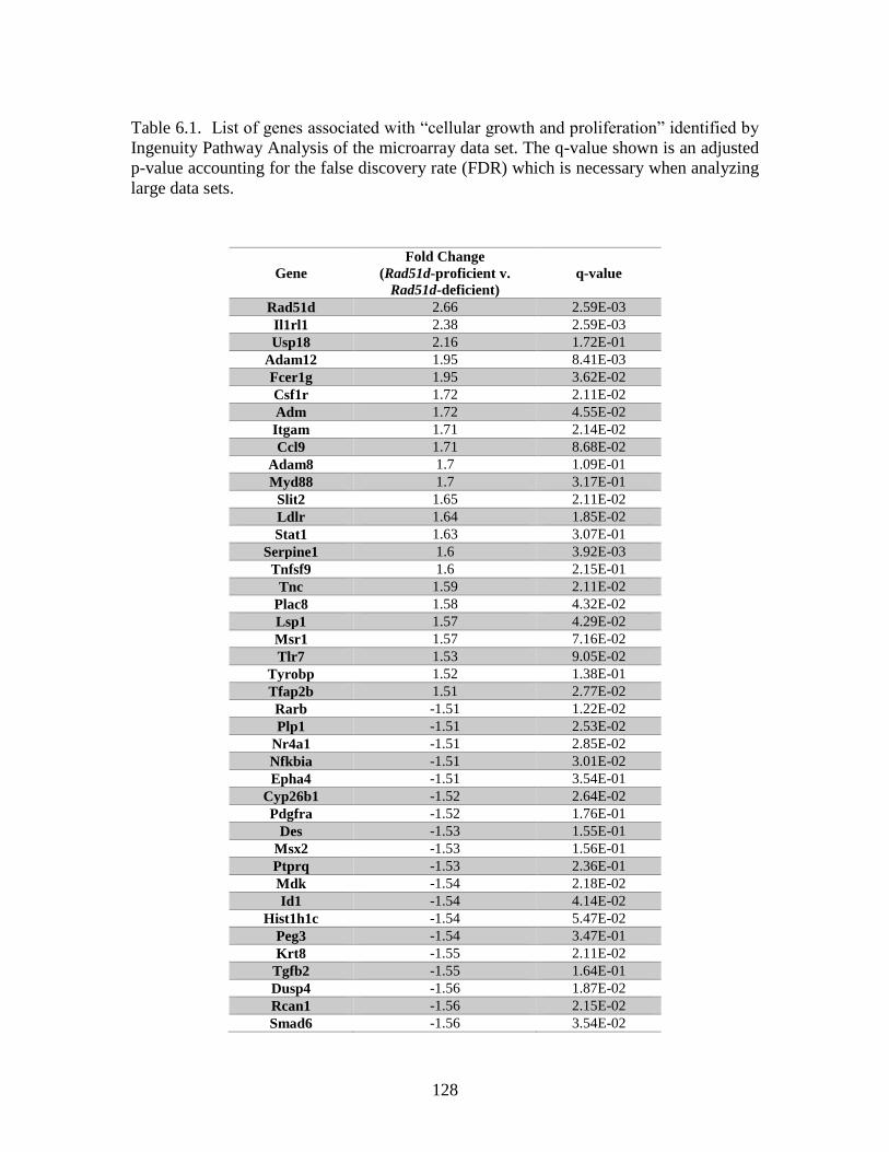
128
Table 6.1. List of genes associated with “cellular growth and proliferation” identified by
Ingenuity Pathway Analysis of the microarray data set. The q-value shown is an adjusted
p-value accounting for the false discovery rate (FDR) which is necessary when analyzing
large data sets.
Gene
Fold Change
(Rad51d-proficient v.
Rad51d-deficient)
q-value
Rad51d 2.66 2.59E-03
Il1rl1 2.38 2.59E-03
Usp18 2.16 1.72E-01
Adam12 1.95 8.41E-03
Fcer1g 1.95 3.62E-02
Csf1r 1.72 2.11E-02
Adm 1.72 4.55E-02
Itgam 1.71 2.14E-02
Ccl9 1.71 8.68E-02
Adam8 1.7 1.09E-01
Myd88 1.7 3.17E-01
Slit2 1.65 2.11E-02
Ldlr 1.64 1.85E-02
Stat1 1.63 3.07E-01
Serpine1 1.6 3.92E-03
Tnfsf9 1.6 2.15E-01
Tnc 1.59 2.11E-02
Plac8 1.58 4.32E-02
Lsp1 1.57 4.29E-02
Msr1 1.57 7.16E-02
Tlr7 1.53 9.05E-02
Tyrobp 1.52 1.38E-01
Tfap2b 1.51 2.77E-02
Rarb -1.51 1.22E-02
Plp1 -1.51 2.53E-02
Nr4a1 -1.51 2.85E-02
Nfkbia -1.51 3.01E-02
Epha4 -1.51 3.54E-01
Cyp26b1 -1.52 2.64E-02
Pdgfra -1.52 1.76E-01
Des -1.53 1.55E-01
Msx2 -1.53 1.56E-01
Ptprq -1.53 2.36E-01
Mdk -1.54 2.18E-02
Id1 -1.54 4.14E-02
Hist1h1c -1.54 5.47E-02
Peg3 -1.54 3.47E-01
Krt8 -1.55 2.11E-02
Tgfb2 -1.55 1.64E-01
Dusp4 -1.56 1.87E-02
Rcan1 -1.56 2.15E-02
Smad6 -1.56 3.54E-02
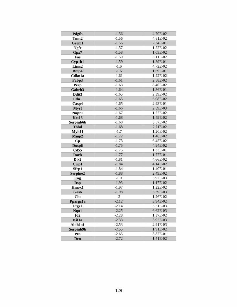
129
Pdgfb -1.56 4.70E-02
Tnnt2 -1.56 4.81E-02
Grem1 -1.56 2.34E-01
Ngfr -1.57 1.22E-02
Gpx7 -1.58 1.03E-02
Fas -1.59 3.11E-02
Cyp1b1 -1.59 1.89E-01
Lims2 -1.6 4.72E-02
Bmp4 -1.6 1.69E-01
Cdkn1a -1.61 1.22E-02
Fabp3 -1.61 2.58E-02
Perp -1.63 8.40E-02
Gabrb3 -1.64 1.36E-01
Ddit3 -1.65 2.39E-02
Edn1 -1.65 6.08E-02
Casp4 -1.65 2.93E-01
Myrf -1.66 2.59E-03
Nupr1 -1.67 1.22E-02
Krt18 -1.68 1.49E-02
Serpinb6b -1.68 3.57E-02
Thbd -1.68 7.71E-02
Myh11 -1.7 1.20E-02
Mmp2 -1.72 1.46E-02
Cp -1.73 6.45E-02
Dusp6 -1.75 4.94E-02
Cd55 -1.75 1.33E-01
Rorb -1.77 1.77E-01
Dlx2 -1.81 4.66E-02
Crip1 -1.84 4.14E-02
Sfrp1 -1.84 1.40E-01
Serpine2 -1.88 2.49E-02
Eng -1.9 3.92E-03
Dsp -1.93 1.17E-02
Hmox1 -1.97 1.22E-02
Gas6 -1.98 5.39E-03
Clu -2 1.26E-02
Ppargc1a -2.12 3.94E-02
Ptgs1 -2.14 3.51E-03
Nqo1 -2.25 6.62E-03
Id2 -2.28 1.37E-02
Kif1a -2.33 3.92E-03
Aldh1a1 -2.53 2.91E-03
Serpinb9b -2.55 1.91E-02
Ptn -2.65 3.87E-01
Dcn -2.72 1.51E-02
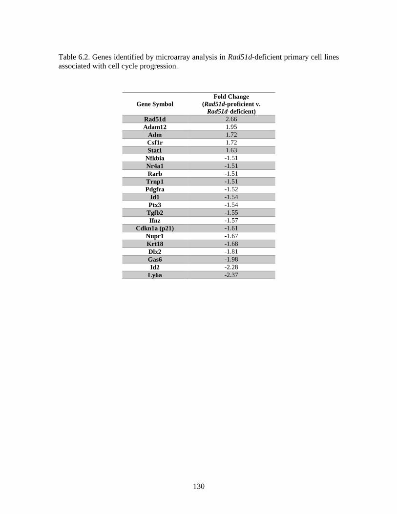
130
Table 6.2. Genes identified by microarray analysis in Rad51d-deficient primary cell lines
associated with cell cycle progression.
Gene Symbol
Fold Change
(Rad51d-proficient v.
Rad51d-deficient)
Rad51d 2.66
Adam12 1.95
Adm 1.72
Csf1r 1.72
Stat1 1.63
Nfkbia -1.51
Nr4a1 -1.51
Rarb -1.51
Trnp1 -1.51
Pdgfra -1.52
Id1 -1.54
Ptx3 -1.54
Tgfb2 -1.55
Ifnz -1.57
Cdkn1a (p21) -1.61
Nupr1 -1.67
Krt18 -1.68
Dlx2 -1.81
Gas6 -1.98
Id2 -2.28
Ly6a -2.37

131
Table 6.3. List of genes associated with “cellular growth and proliferation” identified by
Ingenuity Pathway Analysis of the RNA Seq data set. The q-value shown is an adjusted
p-value taking into account the false discovery rate (FDR) and is necessary when
analyzing large sets of data.
Gene Symbol
Fold Change
(Rad51d-proficient v.
Rad51d-deficient)
q value
Vav1 5.38 2.72E-03
Spi1 4.91 1.42E-02
Laptm5 4.88 1.51E-03
Trem2 4.87 1.51E-03
Dock2 4.87 2.72E-03
Itgam 4.76 1.51E-03
C5ar1 4.75 1.51E-03
Tyrobp 4.69 1.51E-03
Fcer1g 4.59 1.51E-03
Inpp5d 4.58 1.51E-03
Col2a1 4.41 1.51E-03
Ncf1 4.35 1.51E-03
Cd37 4.32 3.20E-02
Itgal 4.17 1.51E-03
Csf1r 3.60 1.51E-03
Pik3ap1 3.54 1.51E-03
C3ar1 3.36 1.51E-03
Irf8 2.81 1.51E-03
Nckap1l 2.73 1.51E-03
Plac8 2.48 1.51E-03
Dok2 2.46 1.51E-03
Slfn2 2.21 1.51E-03
Ptpn6 2.19 1.51E-03
Zic1 2.04 1.51E-03
Itgb2 1.80 1.51E-03
C1qtnf3 1.79 1.51E-03
Foxc2 1.74 1.51E-03
Coro1a 1.70 1.51E-03
Ptpre 1.51 1.51E-03
Krt7 1.50 1.51E-03
Hoxb4 1.49 1.51E-03
Egr2 1.37 1.51E-03
Ret 1.36 1.51E-03
Cspg4 1.33 1.51E-03
Ucp2 1.31 1.51E-03
Dmrta2 1.26 1.51E-03
Skap1 1.25 1.51E-03
Ctsh 1.19 1.51E-03
Lims2 1.15 1.51E-03
Serpine1 1.14 1.51E-03
Egr1 1.08 1.51E-03
Ctgf 1.01 1.51E-03
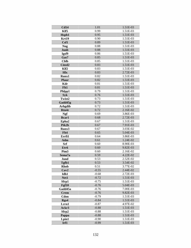
132
Cd14 1.01 1.51E-03
Klf5 0.99 1.51E-03
Hspb1 0.95 1.51E-03
Krt19 0.90 1.51E-03
Csf1 0.89 1.51E-03
Nog 0.88 1.51E-03
Junb 0.88 1.51E-03
Igsf9 0.86 1.51E-03
Gas7 0.85 1.51E-03
Cblb 0.85 1.51E-03
Cited2 0.83 1.51E-03
Klf2 0.83 1.51E-03
Hlx 0.83 2.72E-03
Runx1 0.82 1.51E-03
Plaur 0.82 1.51E-03
Kdr 0.81 1.51E-03
Flt1 0.81 1.51E-03
Phlpp1 0.79 1.51E-03
Syk 0.76 1.51E-03
Twist2 0.73 1.51E-03
Gadd45g 0.73 1.51E-03
Arhgdib 0.72 1.51E-03
Rtn4r 0.72 2.16E-02
Ngf 0.69 3.86E-03
Bcar1 0.68 2.72E-03
Epha2 0.67 1.51E-03
Ptk2b 0.67 7.91E-03
Runx3 0.67 3.03E-02
Flt4 0.65 5.04E-03
Errfi1 0.64 3.86E-03
Adm 0.61 1.24E-02
Srf 0.60 8.90E-03
Etv6 0.60 9.82E-03
Pim3 0.60 2.16E-02
Sema7a 0.59 4.22E-02
Jund 0.53 2.52E-02
Tgfb1 0.53 3.54E-02
Rhob 0.51 3.77E-02
Cav2 -0.55 2.44E-02
Idh1 -0.68 2.72E-03
Net1 -0.72 1.51E-03
Sfrp1 -0.75 1.51E-03
Fgf10 -0.76 5.04E-03
Gadd45a -0.76 7.00E-03
Crem -0.76 9.82E-03
Cdon -0.79 1.51E-03
Rgs4 -0.84 1.51E-03
Lrrn1 -0.87 4.97E-02
Ackr3 -0.88 1.51E-03
Sfrp2 -0.88 1.51E-03
Pappa -0.88 1.51E-03
Lpin1 -0.90 1.51E-03
Irf1 -0.99 1.51E-03
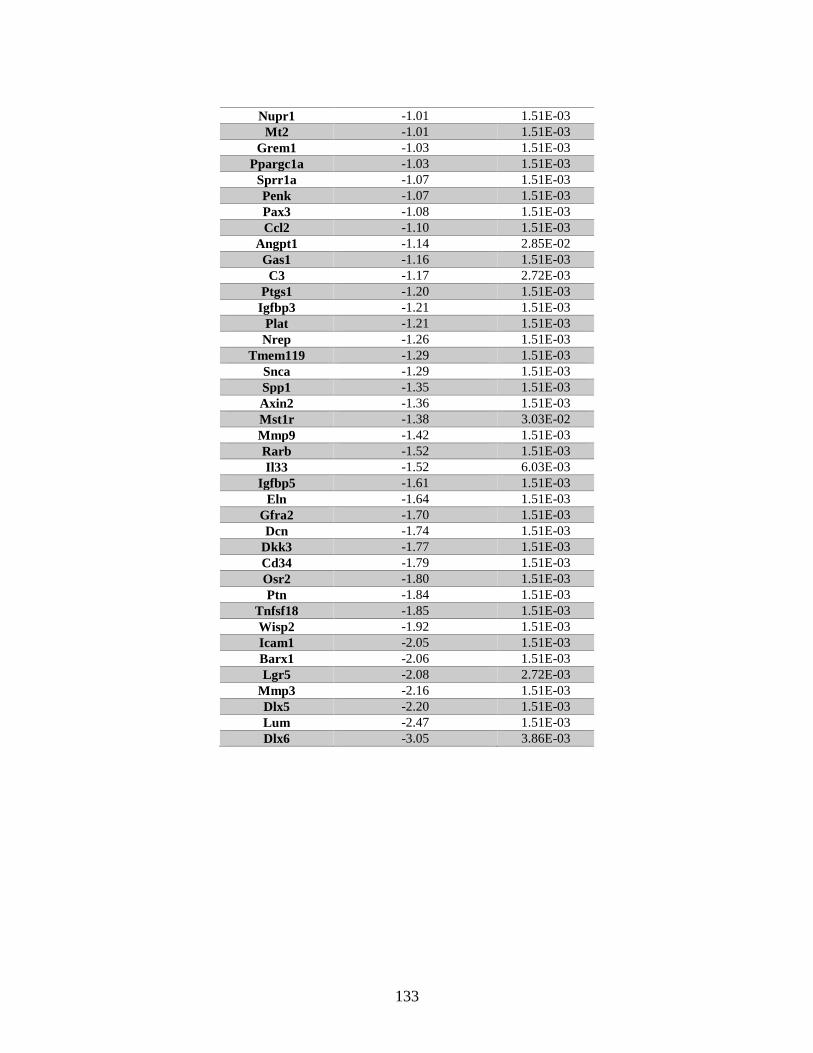
133
Nupr1 -1.01 1.51E-03
Mt2 -1.01 1.51E-03
Grem1 -1.03 1.51E-03
Ppargc1a -1.03 1.51E-03
Sprr1a -1.07 1.51E-03
Penk -1.07 1.51E-03
Pax3 -1.08 1.51E-03
Ccl2 -1.10 1.51E-03
Angpt1 -1.14 2.85E-02
Gas1 -1.16 1.51E-03
C3 -1.17 2.72E-03
Ptgs1 -1.20 1.51E-03
Igfbp3 -1.21 1.51E-03
Plat -1.21 1.51E-03
Nrep -1.26 1.51E-03
Tmem119 -1.29 1.51E-03
Snca -1.29 1.51E-03
Spp1 -1.35 1.51E-03
Axin2 -1.36 1.51E-03
Mst1r -1.38 3.03E-02
Mmp9 -1.42 1.51E-03
Rarb -1.52 1.51E-03
Il33 -1.52 6.03E-03
Igfbp5 -1.61 1.51E-03
Eln -1.64 1.51E-03
Gfra2 -1.70 1.51E-03
Dcn -1.74 1.51E-03
Dkk3 -1.77 1.51E-03
Cd34 -1.79 1.51E-03
Osr2 -1.80 1.51E-03
Ptn -1.84 1.51E-03
Tnfsf18 -1.85 1.51E-03
Wisp2 -1.92 1.51E-03
Icam1 -2.05 1.51E-03
Barx1 -2.06 1.51E-03
Lgr5 -2.08 2.72E-03
Mmp3 -2.16 1.51E-03
Dlx5 -2.20 1.51E-03
Lum -2.47 1.51E-03
Dlx6 -3.05 3.86E-03
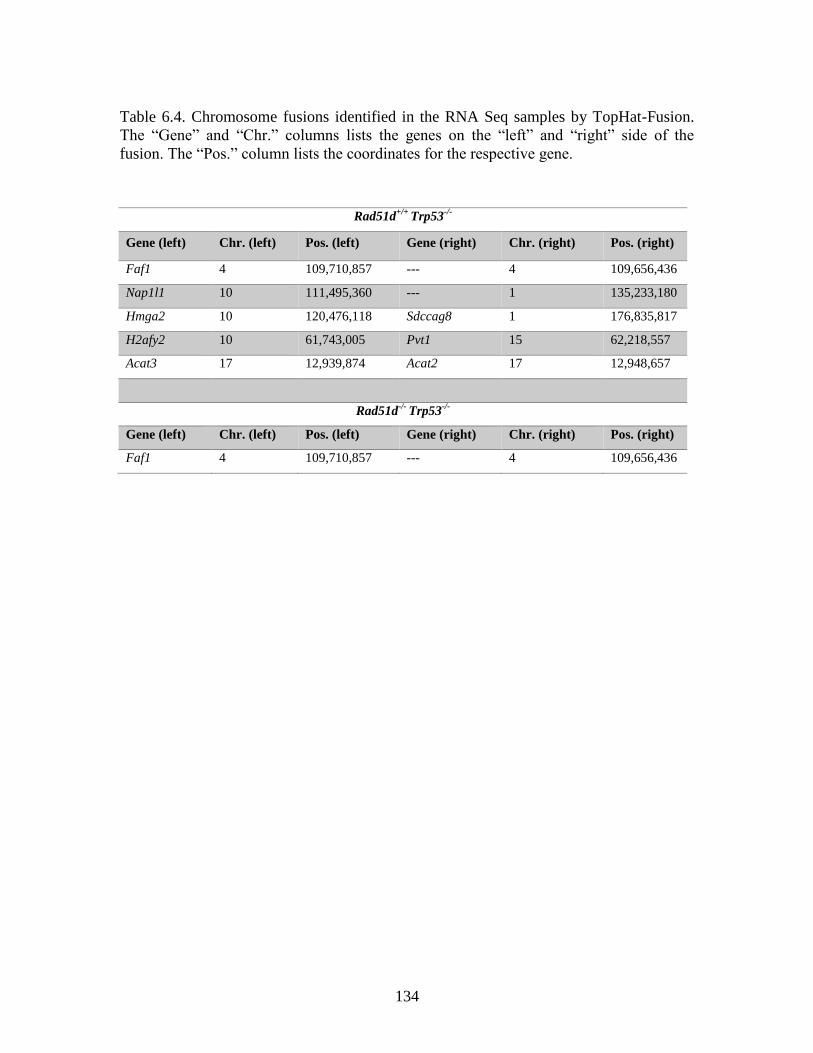
134
Table 6.4. Chromosome fusions identified in the RNA Seq samples by TopHat-Fusion.
The “Gene” and “Chr.” columns lists the genes on the “left” and “right” side of the
fusion. The “Pos.” column lists the coordinates for the respective gene.
Rad51d+/+
Trp53-/-
Gene (left) Chr. (left) Pos. (left) Gene (right) Chr. (right) Pos. (right)
Faf1 4 109,710,857 --- 4 109,656,436
Nap1l1 10 111,495,360 --- 1 135,233,180
Hmga2 10 120,476,118 Sdccag8 1 176,835,817
H2afy2 10 61,743,005 Pvt1 15 62,218,557
Acat3 17 12,939,874 Acat2 17 12,948,657
Rad51d-/-
Trp53-/-
Gene (left) Chr. (left) Pos. (left) Gene (right) Chr. (right) Pos. (right)
Faf1 4 109,710,857 --- 4 109,656,436
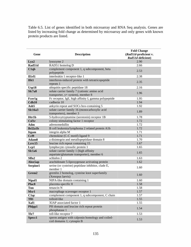
135
Table 6.5. List of genes identified in both microarray and RNA Seq analysis. Genes are
listed by increasing fold change as determined by microarray and only genes with known
protein products are listed.
Gene Description
Fold Change
(Rad51d-proficient v.
Rad51d-deficient)
Lyz2 lysozyme 2 2.90
Rad51d RAD51 homolog D 2.66
C1qb complement component 1, q subcomponent, beta
polypeptide 2.53
Il1rl1 interleukin 1 receptor-like 1 2.38
Ifit1 interferon-induced protein with tetratricopeptide
repeats 1 2.35
Usp18 ubiquitin specific peptidase 18 2.16
Slc7a8 solute carrier family 7 (cationic amino acid
transporter, y+ system), member 8 1.96
Fcer1g Fc receptor, IgE, high affinity I, gamma polypeptide 1.95
Cdh10 cadherin 10 1.94
Asb5 ankyrin repeat and SOCs box-containing 5 1.92
Slc16a3 solute carrier family 16 (monocarboxylic acid
transporters), member 3 1.89
Htr1b 5-hydroxytryptamine (serotonin) receptor 1B 1.78
Csf1r colony stimulating factor 1 receptor 1.72
Adm adrenomedullin 1.72
Bcl2a1b B cell leukemia/lymphoma 2 related protein A1b 1.72
Itgam integrin alpha M 1.71
Ccl9 chemokine (C-C motif) ligand 9 1.71
Adam8 a disintegrin and metallopeptidase domain 8 1.70
Lrrc15 leucine rich repeat containing 15 1.67
Lcp1 lymphocyte cytosolic protein 1 1.65
Slc1a6 solute carrier family 1 (high affinity
aspartate/glutamate transporter), member 6 1.65
Slfn2 schlafen 2 1.63
Alox5ap arachidonate 5-lipoxygenase activating protein 1.62
Serpine1 serine (or cysteine) peptidase inhibitor, clade E,
member 1 1.60
Grem2 gremlin 2 homolog, cysteine knot superfamily
(Xenopus laevis) 1.60
Nipal1 NIPA-like domain containing 1 1.60
Plac8 placenta-specific 8 1.58
Tnn tenascin N 1.58
Msr1 macrophage scavenger receptor 1 1.57
C1qc complement component 1, q subcomponent, C chain 1.57
Tll1 tolloid-like 1.55
Xaf1 XIAP associated factor 1 1.55
Phlpp1 PH domain and leucine rich repeat protein
phosphatase 1 1.54
Tlr7 toll-like receptor 7 1.53
Specc1 sperm antigen with calponin homology and coiled-
coil domains 1; cytospin B 1.53
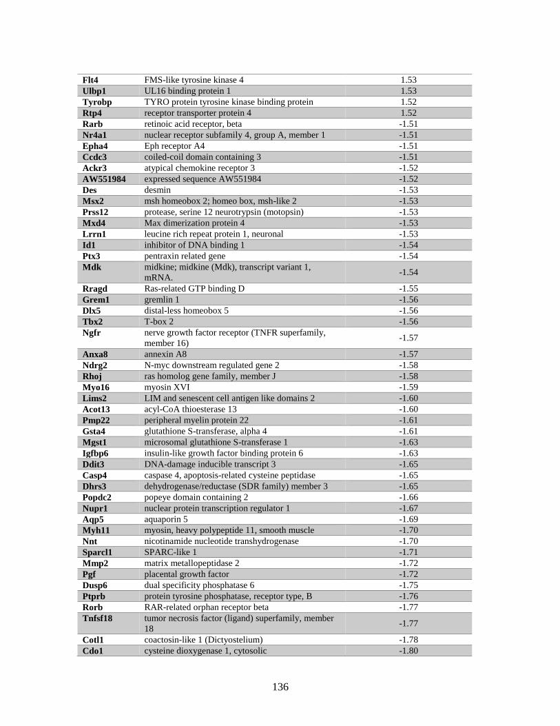
136
Flt4 FMS-like tyrosine kinase 4 1.53
Ulbp1 UL16 binding protein 1 1.53
Tyrobp TYRO protein tyrosine kinase binding protein 1.52
Rtp4 receptor transporter protein 4 1.52
Rarb retinoic acid receptor, beta -1.51
Nr4a1 nuclear receptor subfamily 4, group A, member 1 -1.51
Epha4 Eph receptor A4 -1.51
Ccdc3 coiled-coil domain containing 3 -1.51
Ackr3 atypical chemokine receptor 3 -1.52
AW551984 expressed sequence AW551984 -1.52
Des desmin -1.53
Msx2 msh homeobox 2; homeo box, msh-like 2 -1.53
Prss12 protease, serine 12 neurotrypsin (motopsin) -1.53
Mxd4 Max dimerization protein 4 -1.53
Lrrn1 leucine rich repeat protein 1, neuronal -1.53
Id1 inhibitor of DNA binding 1 -1.54
Ptx3 pentraxin related gene -1.54
Mdk midkine; midkine (Mdk), transcript variant 1,
mRNA. -1.54
Rragd Ras-related GTP binding D -1.55
Grem1 gremlin 1 -1.56
Dlx5 distal-less homeobox 5 -1.56
Tbx2 T-box 2 -1.56
Ngfr nerve growth factor receptor (TNFR superfamily,
member 16) -1.57
Anxa8 annexin A8 -1.57
Ndrg2 N-myc downstream regulated gene 2 -1.58
Rhoj ras homolog gene family, member J -1.58
Myo16 myosin XVI -1.59
Lims2 LIM and senescent cell antigen like domains 2 -1.60
Acot13 acyl-CoA thioesterase 13 -1.60
Pmp22 peripheral myelin protein 22 -1.61
Gsta4 glutathione S-transferase, alpha 4 -1.61
Mgst1 microsomal glutathione S-transferase 1 -1.63
Igfbp6 insulin-like growth factor binding protein 6 -1.63
Ddit3 DNA-damage inducible transcript 3 -1.65
Casp4 caspase 4, apoptosis-related cysteine peptidase -1.65
Dhrs3 dehydrogenase/reductase (SDR family) member 3 -1.65
Popdc2 popeye domain containing 2 -1.66
Nupr1 nuclear protein transcription regulator 1 -1.67
Aqp5 aquaporin 5 -1.69
Myh11 myosin, heavy polypeptide 11, smooth muscle -1.70
Nnt nicotinamide nucleotide transhydrogenase -1.70
Sparcl1 SPARC-like 1 -1.71
Mmp2 matrix metallopeptidase 2 -1.72
Pgf placental growth factor -1.72
Dusp6 dual specificity phosphatase 6 -1.75
Ptprb protein tyrosine phosphatase, receptor type, B -1.76
Rorb RAR-related orphan receptor beta -1.77
Tnfsf18 tumor necrosis factor (ligand) superfamily, member
18 -1.77
Cotl1 coactosin-like 1 (Dictyostelium) -1.78
Cdo1 cysteine dioxygenase 1, cytosolic -1.80
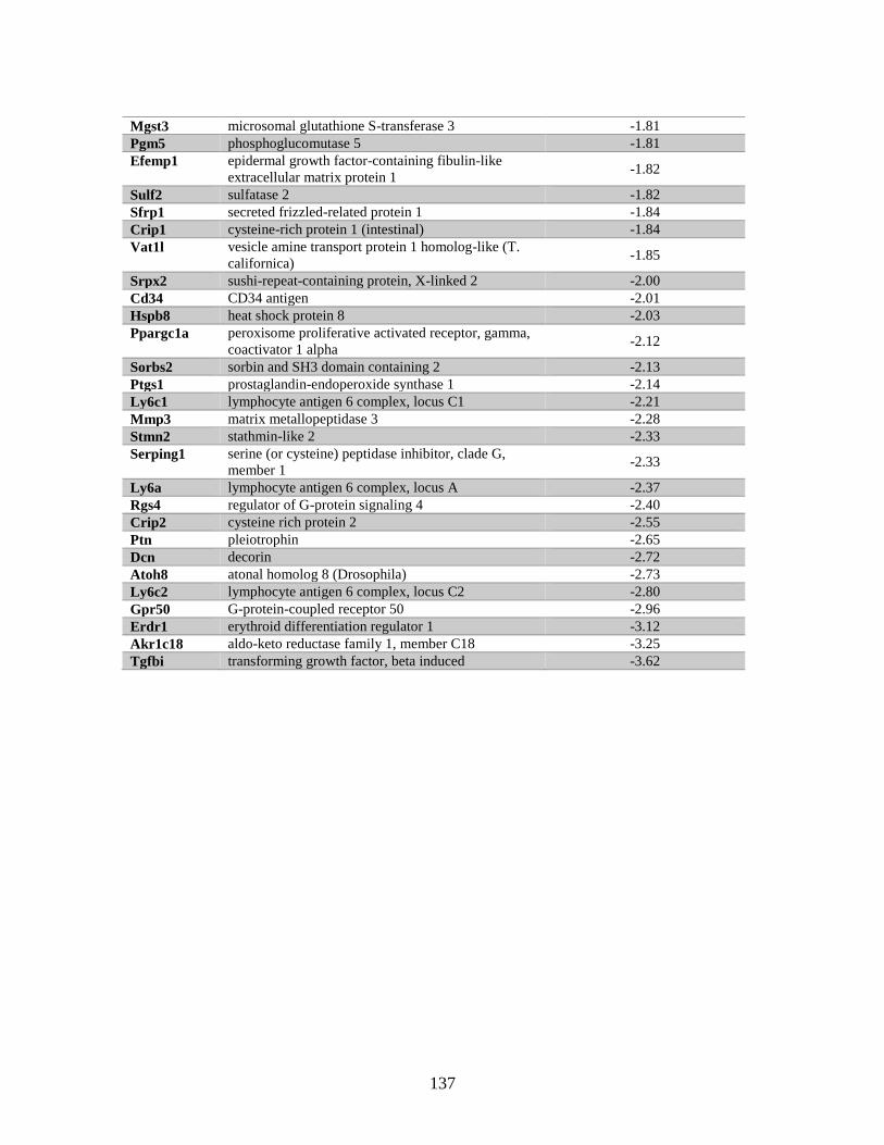
137
Mgst3 microsomal glutathione S-transferase 3 -1.81
Pgm5 phosphoglucomutase 5 -1.81
Efemp1 epidermal growth factor-containing fibulin-like
extracellular matrix protein 1 -1.82
Sulf2 sulfatase 2 -1.82
Sfrp1 secreted frizzled-related protein 1 -1.84
Crip1 cysteine-rich protein 1 (intestinal) -1.84
Vat1l vesicle amine transport protein 1 homolog-like (T.
californica) -1.85
Srpx2 sushi-repeat-containing protein, X-linked 2 -2.00
Cd34 CD34 antigen -2.01
Hspb8 heat shock protein 8 -2.03
Ppargc1a peroxisome proliferative activated receptor, gamma,
coactivator 1 alpha -2.12
Sorbs2 sorbin and SH3 domain containing 2 -2.13
Ptgs1 prostaglandin-endoperoxide synthase 1 -2.14
Ly6c1 lymphocyte antigen 6 complex, locus C1 -2.21
Mmp3 matrix metallopeptidase 3 -2.28
Stmn2 stathmin-like 2 -2.33
Serping1 serine (or cysteine) peptidase inhibitor, clade G,
member 1 -2.33
Ly6a lymphocyte antigen 6 complex, locus A -2.37
Rgs4 regulator of G-protein signaling 4 -2.40
Crip2 cysteine rich protein 2 -2.55
Ptn pleiotrophin -2.65
Dcn decorin -2.72
Atoh8 atonal homolog 8 (Drosophila) -2.73
Ly6c2 lymphocyte antigen 6 complex, locus C2 -2.80
Gpr50 G-protein-coupled receptor 50 -2.96
Erdr1 erythroid differentiation regulator 1 -3.12
Akr1c18 aldo-keto reductase family 1, member C18 -3.25
Tgfbi transforming growth factor, beta induced -3.62
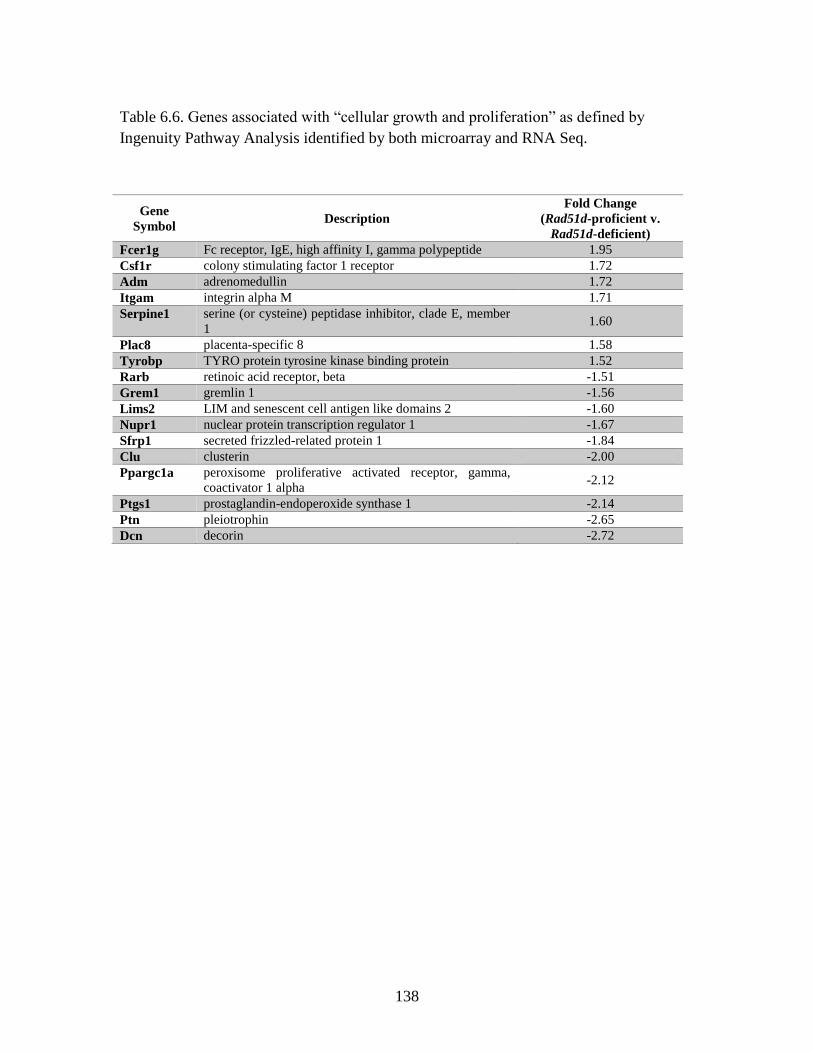
138
Table 6.6. Genes associated with “cellular growth and proliferation” as defined by
Ingenuity Pathway Analysis identified by both microarray and RNA Seq.
Gene
Symbol Description
Fold Change
(Rad51d-proficient v.
Rad51d-deficient)
Fcer1g Fc receptor, IgE, high affinity I, gamma polypeptide 1.95
Csf1r colony stimulating factor 1 receptor 1.72
Adm adrenomedullin 1.72
Itgam integrin alpha M 1.71
Serpine1 serine (or cysteine) peptidase inhibitor, clade E, member
1 1.60
Plac8 placenta-specific 8 1.58
Tyrobp TYRO protein tyrosine kinase binding protein 1.52
Rarb retinoic acid receptor, beta -1.51
Grem1 gremlin 1 -1.56
Lims2 LIM and senescent cell antigen like domains 2 -1.60
Nupr1 nuclear protein transcription regulator 1 -1.67
Sfrp1 secreted frizzled-related protein 1 -1.84
Clu clusterin -2.00
Ppargc1a peroxisome proliferative activated receptor, gamma,
coactivator 1 alpha -2.12
Ptgs1 prostaglandin-endoperoxide synthase 1 -2.14
Ptn pleiotrophin -2.65
Dcn decorin -2.72
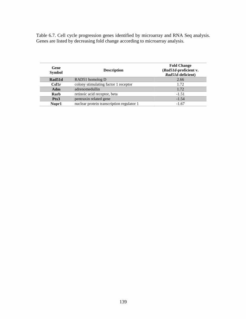
139
Table 6.7. Cell cycle progression genes identified by microarray and RNA Seq analysis.
Genes are listed by decreasing fold change according to microarray analysis.
Gene
Symbol Description
Fold Change
(Rad51d-proficient v.
Rad51d-deficient)
Rad51d RAD51 homolog D 2.66
Csf1r colony stimulating factor 1 receptor 1.72
Adm adrenomedullin 1.72
Rarb retinoic acid receptor, beta -1.51
Ptx3 pentraxin related gene -1.54
Nupr1 nuclear protein transcription regulator 1 -1.67
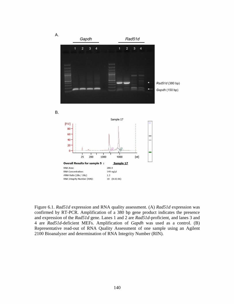
140
Figure 6.1. Rad51d expression and RNA quality assessment. (A) Rad51d expression was
confirmed by RT-PCR. Amplification of a 380 bp gene product indicates the presence
and expression of the Rad51d gene. Lanes 1 and 2 are Rad51d-proficient, and lanes 3 and
4 are Rad51d-deficient MEFs. Amplification of Gapdh was used as a control. (B)
Representative read-out of RNA Quality Assessment of one sample using an Agilent
2100 Bioanalyzer and determination of RNA Integrity Number (RIN).
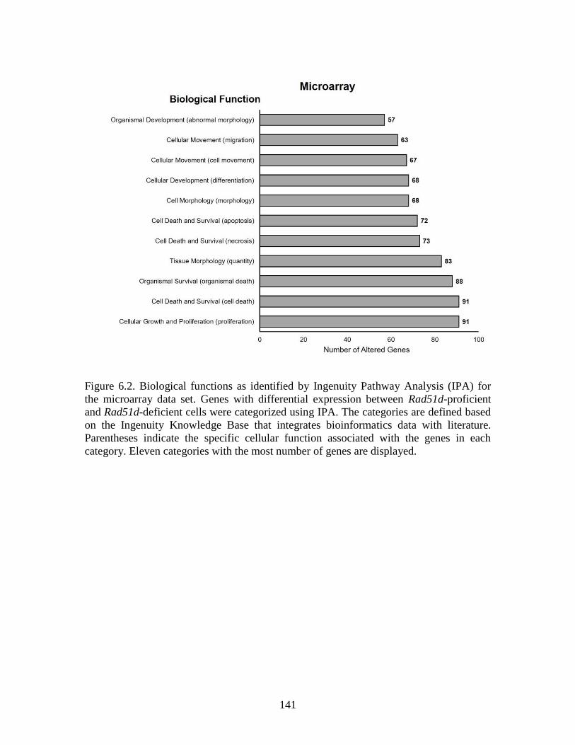
141
Figure 6.2. Biological functions as identified by Ingenuity Pathway Analysis (IPA) for
the microarray data set. Genes with differential expression between Rad51d-proficient
and Rad51d-deficient cells were categorized using IPA. The categories are defined based
on the Ingenuity Knowledge Base that integrates bioinformatics data with literature.
Parentheses indicate the specific cellular function associated with the genes in each
category. Eleven categories with the most number of genes are displayed.
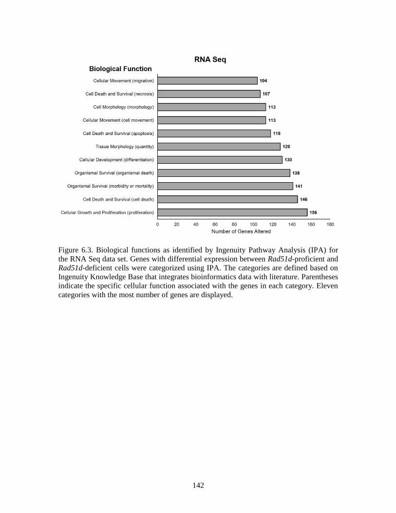
142
Figure 6.3. Biological functions as identified by Ingenuity Pathway Analysis (IPA) for
the RNA Seq data set. Genes with differential expression between Rad51d-proficient and
Rad51d-deficient cells were categorized using IPA. The categories are defined based on
Ingenuity Knowledge Base that integrates bioinformatics data with literature. Parentheses
indicate the specific cellular function associated with the genes in each category. Eleven
categories with the most number of genes are displayed.
143
CHAPTER 7
DISCUSSION
Ovarian cancer is the deadliest of the five main types of gynecological cancer and leads
to over 14,000 deaths per year in the United States (American Cancer Society, 2017).
One characteristic of ovarian cancer is genome instability associated with mutations in
DNA repair genes (Wang et al. 2012). In 2012 when these projects were initiated, there
were three known ovarian cancer susceptibility genes: BRCA1, BRCA2, and the newly
identified RAD51D. The two BRCA genes were also associated with an increased risk for
breast cancer, but RAD51D was only recently confirmed as a breast cancer susceptibility
gene (Kraus et al. 2017). Mutations in other DNA repair genes were also more recently
associated with breast and ovarian cancers, including RAD51C (Coulet et al. 2013;
Loveday et al. 2011; Pelttari et al. 2011; Thompson et al. 2012).
The data presented in this dissertation elucidate mechanisms by which RAD51D
functions to maintain genome integrity. In Chapter 3, I presented data that were published
in the research article entitled “RNF138 interacts with RAD51D and is required for DNA
interstrand crosslink repair.” This work identified a direct interaction between RAD51D
and the novel E3 ubiquitin ligase RNF138 and demonstrated that the regions encoded by
exons 5 and 7 along RNF138 are required for this interaction. In Chapter 4, I
demonstrated that expression of two RAD51D missense mutations – K235R and K298R
– confer cellular sensitivity to mitomycin C (MMC) in Rad51d-deficient mouse
embryonic fibroblast (MEFs). Arginine substitution at these residues increased RAD51D
144
stability, suggesting that these residues are sites of ubiquitin modifications that target
RAD51D for proteasomal degradation. Neither K235 nor K298 was required for
homology directed repair of SceI induced double strand breaks, suggesting that these
residues are required for RAD51D function specifically in response to crosslinks. In
Chapter 5, I investigated how RAD51D protects the telomeres specifically in the
presence of the DNA damaging agent 6-thioguanine (6TG). γ-H2AX foci were detected
at the telomere regions more frequently in Rad51d-deficient compared with Rad51d-
proficient MEFs, and a higher frequency of 6TG-induced chromosome fusions were
observed in Rad51d-deficient MEFs. In Chapter 6, gene expression profiles between
Rad51d-proficient and -deficient MEFs were analyzed by microarray and RNA Seq.
Ninety-one genes associated with “cell growth and proliferation” were differentially
expressed in the Rad51d-deficient cells compared with the Rad51d-proficient cells.
Further sorting of the data identified 21 genes that were associated specifically with “cell
cycle progression.”
Identification of lysine residues that confer sensitivity to thiopurine damage
The complementation assays presented in Chapter 4 provide evidence that K235 and
K298 along RAD51D promote HR-mediated repair of ICLs. I also investigated the
function of RAD51D in response to 6TG induced damage, based on previous work
demonstrating that Rad51d-deficient cells are more sensitive to 6TG treatment (Rajesh,
Litvinchuk, et al. 2011). Follow-up experiments should include performing the
complementation assays in the presence of 6TG to identify lysine residues along
RAD51D necessary for function in response to these types of damage. I expect that
145
expression of K235R, but not K113R or K298R, in Rad51d-deficient MEFs will restore
cellular resistance in the presence of 6TG.
Lysine residues along RAD51D mediate interaction with RAD51C and XRCC2
I hypothesized that increased MMC sensitivity in the presence of the K235R and K298R
variants was due to disruption of the BCDX2 complex. As discussed in Chapter 2, the
five RAD51 paralogs – RAD51B, RAD51C, RAD51D, XRCC2, and XRCC3 – form two
distinct complexes in mammalian cells, and RAD51D specifically interacts with
RAD51C and XRCC2 (Masson et al. 2001). Yeast-two-hybrid (Y2H) analysis was
performed to determine if any of the variants fail to interact with either RAD51C or
XRCC2. Arginine substitution at K235 and K298 did not affect interaction with RAD51C
and XRCC2, suggesting that the BCDX2 complex is formed even in the absence of these
residues. K201R did not interact with XRCC2 but possibly maintains interaction with
RAD51C, and K261R interacted with RAD51C but not with XRCC2. These data offer
insight into specific residues along RAD51D that mediate the interactions with other
paralogs that has not previously been reported (Figure 7.1). Validation by co-
immunoprecipitation methods in mammalian cells is necessary.
Interestingly, these data present a conundrum. Y2H data suggest the BCDX2
complex is not formed in the presence of K261R, however, cellular resistance to MMC
was decreased by 30%, but not eliminated, when K261R was expressed in Rad51d-
deficient MEFs. The data suggest that disruption of the interaction between RAD51D and
XRCC2 is sufficient to decrease HR-mediated repair of ICLs, but not to fully eliminate
repair. Therefore, these data are hinting towards a potential separation of function and
suggest an alternative complex might form during HR-mediated repair of ICLs.
146
The RAD51 paralogs have approximately 30% identity between them, and only
three lysine residues are conserved between RAD51D and other proteins in the BCDX2
complex: K201 and K235 are conserved with RAD51B, and K298 is conserved with
RAD51C. To further elucidate the function of each paralog within this complex, arginine
substitutions in RAD51B and RAD51C at these positions can be introduced.
Complementation assays using Rad51b- and Rad51c-deficient cells can be performed to
determine if these conserved residues are required for cellular resistance to DNA
damaging agents, and Y2H analysis would determine if these residues mediate interaction
between the paralogs.
Lysine residues are not required for repair of Sce-I induced double strand breaks
Data in Chapter 4 present evidence that HR activity is similar in the presence of over-
expressed K0, K235R, and K298R compared with wild-type (Figure 4.9B), suggesting
that lysine residues along RAD51D are not required for HR-mediated repair of double
strand breaks (DSBs). K261R and K201R had decreased interaction with XRCC2 but not
with RAD51C, and complemented Rad51d-deficiency in the presence of MMC. Follow-
up experiments should include these variants to confirm that HR-activity is not affected
in the presence of these mutant proteins. I propose that arginine substitution at these
residues leads to formation of a new RAD51 paralog complex comprised of RAD51B-
RAD51C-RAD51D (BCD) (Figure 7.1E). Exclusion of XRCC2 from this complex is
predicted to diminish the activity of the paralogs during crosslink repair but may not be
necessary for HR-mediated repair of DSBs. I would hypothesize that the activity of the
BCD complex will be sufficient to repair SceI-induced DSBs.
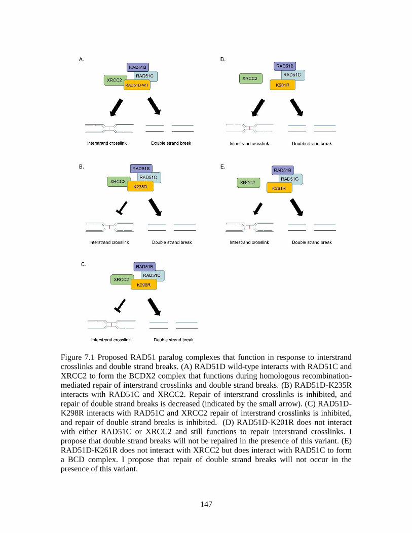
147
Figure 7.1 Proposed RAD51 paralog complexes that function in response to interstrand
crosslinks and double strand breaks. (A) RAD51D wild-type interacts with RAD51C and
XRCC2 to form the BCDX2 complex that functions during homologous recombination-
mediated repair of interstrand crosslinks and double strand breaks. (B) RAD51D-K235R
interacts with RAD51C and XRCC2. Repair of interstrand crosslinks is inhibited, and
repair of double strand breaks is decreased (indicated by the small arrow). (C) RAD51D-
K298R interacts with RAD51C and XRCC2 repair of interstrand crosslinks is inhibited,
and repair of double strand breaks is inhibited. (D) RAD51D-K201R does not interact
with either RAD51C or XRCC2 and still functions to repair interstrand crosslinks. I
propose that double strand breaks will not be repaired in the presence of this variant. (E)
RAD51D-K261R does not interact with XRCC2 but does interact with RAD51C to form
a BCD complex. I propose that repair of double strand breaks will not occur in the
presence of this variant.
148
Lysine combinations that restore RAD51D function
Lysine residues can be reintroduced along K0 in different combinations to further
elucidate the function of these residues during DNA repair. Site-directed mutagenesis
should be performed to reintroduce K113 (RAD51D-SingleK113), K235 (RAD51D-
SingleK235), and K298 (RAD51D-SingleK298) into K0. A second lysine residue can be
reintroduced in each SingleK mutant in the following combinations: K113 and K235
(RAD51D-DoubleK113235), K113 and K298 (RAD51D-DoubleK113298), and K235
and K298 (RAD51D-DoubleK235298). Finally, a RAD51D-TripleK can be generated by
re-introducing K113 in RAD51D-DoubleK235298. Complementation assays using the
newly generated lysine combination mutants can be performed in Rad51d-deficient
MEFs as described in Chapter 4. It is predicted that none of the SingleK or DoubleK
mutants will restore cellular resistance to MMC since each of them retains arginine
substitution at one or more essential lysine residue. Complementation with the TripleK in
the presence of MMC is predicted to restore cellular sensitivity to MMC. These data
would indicate that only K113, K235, and K298 are required for RAD51D function
during ICL repair.
I also investigated ubiquitination along RAD51D as a regulatory mechanism of
RAD51D. This post-translational modification (PTM) occurs along substrate proteins at
lysine residues and at the N-terminus (Akutsu, Dikic, and Bremm 2016). RAD51D is
ubiquitinated by the E3 ligase RNF138 (Yard et al. 2016), and K235 and K298 are
potential ubiquitination sites. Lysine residue(s) targeted with the proteasome specific
ubiquitin modification can be identified by measuring the stability of each RAD51D-
SingleK and -DoubleK variant. I propose that K235 and K298 along RAD51D are the
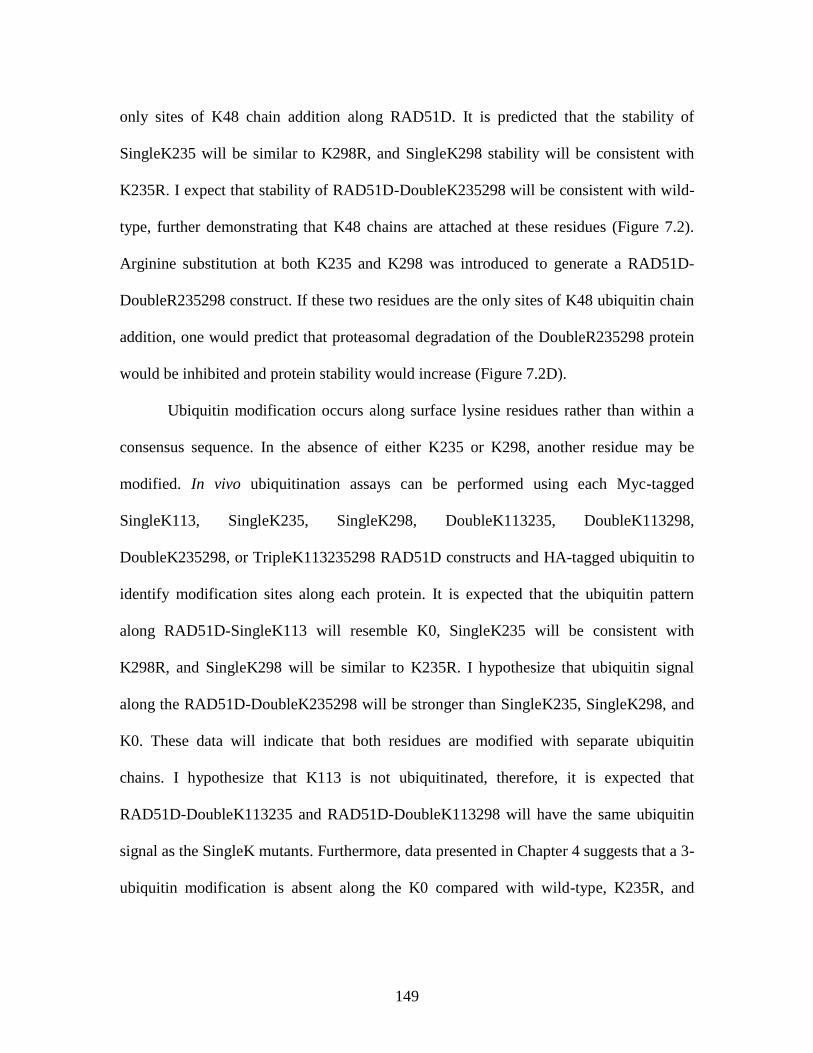
149
only sites of K48 chain addition along RAD51D. It is predicted that the stability of
SingleK235 will be similar to K298R, and SingleK298 stability will be consistent with
K235R. I expect that stability of RAD51D-DoubleK235298 will be consistent with wild-
type, further demonstrating that K48 chains are attached at these residues (Figure 7.2).
Arginine substitution at both K235 and K298 was introduced to generate a RAD51D-
DoubleR235298 construct. If these two residues are the only sites of K48 ubiquitin chain
addition, one would predict that proteasomal degradation of the DoubleR235298 protein
would be inhibited and protein stability would increase (Figure 7.2D).
Ubiquitin modification occurs along surface lysine residues rather than within a
consensus sequence. In the absence of either K235 or K298, another residue may be
modified. In vivo ubiquitination assays can be performed using each Myc-tagged
SingleK113, SingleK235, SingleK298, DoubleK113235, DoubleK113298,
DoubleK235298, or TripleK113235298 RAD51D constructs and HA-tagged ubiquitin to
identify modification sites along each protein. It is expected that the ubiquitin pattern
along RAD51D-SingleK113 will resemble K0, SingleK235 will be consistent with
K298R, and SingleK298 will be similar to K235R. I hypothesize that ubiquitin signal
along the RAD51D-DoubleK235298 will be stronger than SingleK235, SingleK298, and
K0. These data will indicate that both residues are modified with separate ubiquitin
chains. I hypothesize that K113 is not ubiquitinated, therefore, it is expected that
RAD51D-DoubleK113235 and RAD51D-DoubleK113298 will have the same ubiquitin
signal as the SingleK mutants. Furthermore, data presented in Chapter 4 suggests that a 3-
ubiquitin modification is absent along the K0 compared with wild-type, K235R, and
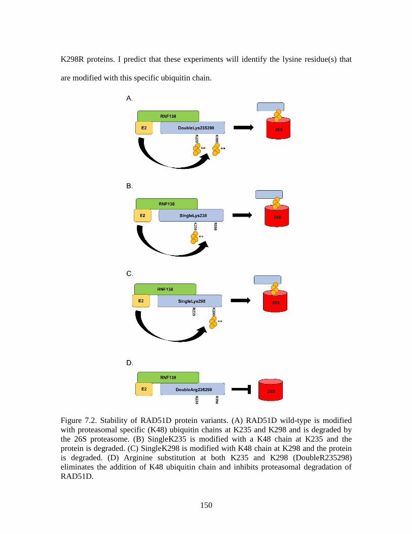
150
K298R proteins. I predict that these experiments will identify the lysine residue(s) that
are modified with this specific ubiquitin chain.
Figure 7.2. Stability of RAD51D protein variants. (A) RAD51D wild-type is modified
with proteasomal specific (K48) ubiquitin chains at K235 and K298 and is degraded by
the 26S proteasome. (B) SingleK235 is modified with a K48 chain at K235 and the
protein is degraded. (C) SingleK298 is modified with K48 chain at K298 and the protein
is degraded. (D) Arginine substitution at both K235 and K298 (DoubleR235298)
eliminates the addition of K48 ubiquitin chain and inhibits proteasomal degradation of
RAD51D.
151
Post-translational modifications along RAD51D by mass spectrometry
Of the 20 amino acids, lysine is modified with the highest number of possible PTMs (Zee
and Garcia 2012). One method for identifying ubiquitinated residues along a protein is
tandem mass spectrometry (MS/MS) (Calderon-Celis, Encinar, and Sanz-Medel 2017). I
propose that MS/MS analysis be performed on K0, K235R, and K298R to identify
alternative modifications that occur in the absence of one or more lysine residues.
Analysis of these RAD51D variants will confirm loss of a specific modification when a
lysine residue is substituted with an arginine. Protein stability experiments demonstrated
that K235R and K298R are more stable than wild-type, suggesting that a degradation
specific ubiquitin modification has been lost. It is predicted that MS/MS analysis will
identify a ubiquitin molecule attached to each of these lysine residues in the wild-type
protein.
To perform these experiments, RAD51D constructs can be expressed in
mammalian cells, isolated by co-immunoprecipitation, trypsin digested, and analyzed
using MS/MS. The trypsin digestion will cleave the isopeptide bond along a protein after
lysine residues, producing protein fragments that have only a single lysine residue. When
a ubiquitinated fragment is digested, a portion of the ubiquitin molecule remains on the
residue, producing two β and γ ions for that fragment rather than one produced by an
unmodified peptide. Thus, the fragmentation pattern of RAD51D can be used to identify
lysine residues that have a ubiquitin molecule attached. MS/MS analysis of RAD51D
wild-type will identify lysine residues along the protein that are modified with ubiquitin. I
hypothesize that K235 and K298 will be modified with a ubiquitin molecule.
152
One limitation of MS/MS is that it cannot identify the chain type that is attached
to a specific residue. To identify specific chains attached to lysine residues, in vitro
ubiquitination assays could be performed. Myc-tagged RAD51D can be incubated with
ubiquitin mutants that have only a single lysine residue available for chain elongation.
RAD51D can be immunoprecipitated and analyzed by MS/MS. RAD51D is targeted for
degradation by the proteasome and is ubiquitinated with K48 specific chain linkages,
therefore, I expect one lysine residue along wild-type RAD51D to be modified with a
K48 ubiquitin molecule. To further elucidate the dynamics of this modification, K0,
K235R, and K298R can also be analyzed to determine if the K48 ubiquitin is added to an
alternative lysine residue in the absence of either K235 or K298. Additionally, it will be
interesting to test the K113R variant to determine if loss of ATPase activity affects the
proteasomal specific ubiquitin modification.
Ubiquitination is not the only PTM that can occur at lysine residues along a
protein. Other modifications include SUMOylation, acetylation, and glycosylation, and
can be identified by MS/MS analysis (Zee and Garcia 2012). K261 along RAD51D is
predicted to be acetylated (Mertins et al. 2013). The data presented in Chapter 4
demonstrated that K261R decreases cellular resistance to MMC by 30% and disrupts
RAD51D interaction with XRCC2. K261 can be substituted with glutamine to generate a
K261Q mutant that will mimic acetylation at this residue (Kamieniarz and Schneider
2009) and can be tested in the complementation assay. These data will determine if
acetylation at this residue is required for cellular resistance to MMC.
153
Identification of E2 ligases that interact with RNF138 to ubiquitinate RAD51D
In addition to being modified by K48-linked chains and targeted for proteasomal
degradation, RAD51D is also modified with other chain linkage types (Figure 4.7C). The
E2 enzymes that interact with RNF138 to promote these alternative linkage arrangements
remain unknown and performing a screen of E2 enzymes will provide further information
into RAD51D ubiquitination. A screen performed in the laboratory of Stephen Jackson
identified the UBE2D E2 enzyme as directly interacting with RNF138 (Schmidt et al.
2015). The UBE2D family of E2 conjugating enzymes promotes several ubiquitin
linkages along target proteins. For example, UBE2N catalyzes the addition of K63-linked
ubiquitin chains to promote DNA repair (Sato et al. 2012). The yeast-two-hybrid (Y2H)
screen that identified RNF138, also identified UBE2N/UBC13 as a direct interacting
protein with RAD51D (Yard 2011), and I hypothesize that this E2 promotes K63-
linkages along RAD51D, which could be tested in future studies.
K235 and K298 requirement for RAD51 and BRCA1 localization sites of damage
Data presented in Chapter 4 demonstrated that loss of Rad51d decreases RAD51 foci
formation in the presence of MMC. It is predicted that K235 and K298 fail to
complement Rad51d-deficiency in the presence of MMC because they are required for
RAD51 recruitment to the site of damage, and disruption of RAD51 localization prevents
HR progression. To test this hypothesis, K235R and K298R can be expressed in Rad51d-
deficient MEFs and RAD51 foci can be detected by immunofluorescence 24 h, 48 h, and
72 h after MMC treatment. It is expected that cells expressing K235R and K298R will
have less RAD51 foci positive nuclei compared with cells expressing wild-type
RAD51D. These experiments will determine if each lysine is required specifically for
154
RAD51 recruitment to damage sites. K113R can be included in these experiments to
determine if the ATPase activity of RAD51D mediates RAD51 recruitment in response to
ICLs.
In addition to the BCDX2 complex, other proteins function during HR to recruit
RAD51 to damage. In response to DNA crosslinks, BRCA1 is required for RAD51
recruitment to damage (Bhattacharyya et al. 2000), and it is hypothesized that the
BCDX2 complex functions upstream of BRCA1. To investigate this, BRCA1 foci
formation after MMC treatment can be detected in Rad51d-deficient and -proficient
MEFs. It is expected that the number of BRCA1 positive nuclei will be higher in Rad51d-
proficient MEFs compared with Rad51d-deficient MEFs when challenged with MMC.
K235R and K298R can be expressed in Rad51d-deficient MEFs treated with MMC and
BRCA1 foci can be detected. These experiments will determine if K235 or Ly298
specifically mediate BRCA1 localization in the presence of interstrand crosslinks.
Generating K235R and K298R substitutions in RAD51D using CRISPR-Cas9
Within the past 5 years, a new scientific tool – known as CRISPR (clustered regularly
interspaced short palindromic repeats) – has become available that allows for mutations
to be introduced in genome of a cell (Cong et al. 2013). This technology uses an
endonuclease, Cas9, to generate nicks in DNA strands at a specific locus. Briefly,
eukaryotic cells are transfected with a plasmid encoding the Cas9 enzyme and a short
guide RNA (sgRNA) complementary to a sequence along the genome is targeted. The
sgRNA localizes the Cas9 enzyme where it generates a nick in the DNA and introduces
the desired point mutations within a gene. Mutations can now be introduced into a locus
without generating a nick in the DNA strand using a catalytically inactive Cas9 (Gaudelli
155
et al. 2017). For applications in my work, CRISPR can be used to introduce arginine
substitution at the codons along RAD51D for K113 (MEF-RAD51DK113R), K235
(MEF-RAD51DK235R), K298 (MEF-RAD51DK298R), and all lysines (MEF-
RAD51DK0) in the Rad51d gene of Rad51d-proficient MEFs. As a positive control cell
line, arginine substitution can be introduced at Lys159 (MEF-K159R) along RAD51D;
this residue is non-conserved between Mus musculus and Homo sapiens and is predicted
to be non-essential for RAD51D function.
First, cell survival assays in the presence of DNA damaging agents, such as MMC
and 6TG, could be performed in these newly generated cell lines. Second, to identify
lysine residue(s) required for RAD51 recruitment, each cell line could be challenged with
MMC and IR, and foci formation detected as described in Chapter 4. My hypothesis is
that RAD51 foci will only form in the MEF-K159R cell line, and not in the other mutant
cell lines. Loss of RAD51 foci in the MEF-K113R cell lines will indicate that ATPase
activity of RAD51D is necessary for RAD51 recruitment. Each lysine mutant cell line
can be challenged with 6TG and telomere stability can be measured as described in
Chapter 5. I hypothesize that MEFs expressing K0 and K113R will have telomeric
defects, such as fusions, the presence of 6TG. These data would determine if ATPase
activity of RAD51D is necessary for its activity in maintaining telomere integrity.
Investigations into RNF138 mediated interstrand crosslink repair
Most of the work presented in this dissertation has focused on lysine residues along
RAD51D that are required for cellular resistance to DNA damaging agents, and on
detecting ubiquitination of RAD51D, a PTM mediated by the E3 ligase RNF138. The
Pittman laboratory and two other groups have demonstrated that RNF138 promotes HR-
156
mediated repair of ICLs and DSBS, and further investigations into RNF138 will provide
insight into how it functions during DNA damage repair. RNF138 contains a really
interesting new gene (RING) catalytic domain, three zinc finger (ZF) motifs, and a
ubiquitin interacting motif (UIM). Mutations in the RING domain decrease RAD51D
ubiquitination and increase its stability (Yard et al. 2016), and deletion of the ZFs inhibits
RNF138-mediated ubiquitination of Ku80 (Ismail et al. 2015).
To date, no investigations into the UIM of RNF138 have been reported. This
motif is characterized by a 20-amino acid protein sequence: X-Ac-Ac-Ac-Ac-ϕ-X-X-Ala-
X-X-X-Ser-X-X-Ac-X-X-X-X, where ϕ represents a hydrophobic residue, Ac represents
an acidic residue, and X represents non-conserved residues (Fisher et al. 2003; Hofmann
and Falquet 2001). UIMs bind mono-ubiquitin modifications along proteins (Fisher et al.
2003; Woelk et al. 2006), a function important for E3 ligase activity. For example,
mutations in the UIM domains of BRCA1/RAP80 prevent its binding to K63-linked
chains generated by RNF168 and inhibits HR progression (Kim, Chen, and Yu 2007). I
propose that the UIM along RNF138 has a similar function and is required for RNF318
interaction with the CtIP endonuclease. Site-directed mutagenesis can be performed to
introduce amino acid substitutions at Ala236 and Ser240 in the UIM of RNF138.
Immunoprecipitation experiments can be performed to determine if RNF138 UIM
mutants fail to interact with CtIP. To determine if the UIM is required for cellular
resistance to interstrand crosslinks, each UIM mutant can be expressed in MEFs that do
not express endogenous RNF138 in the presence of MMC. Together, these data will
determine 1. if the UIM along RNF138 mediates its interaction with CtIP, and 2. if this
motif is required for crosslink repair.
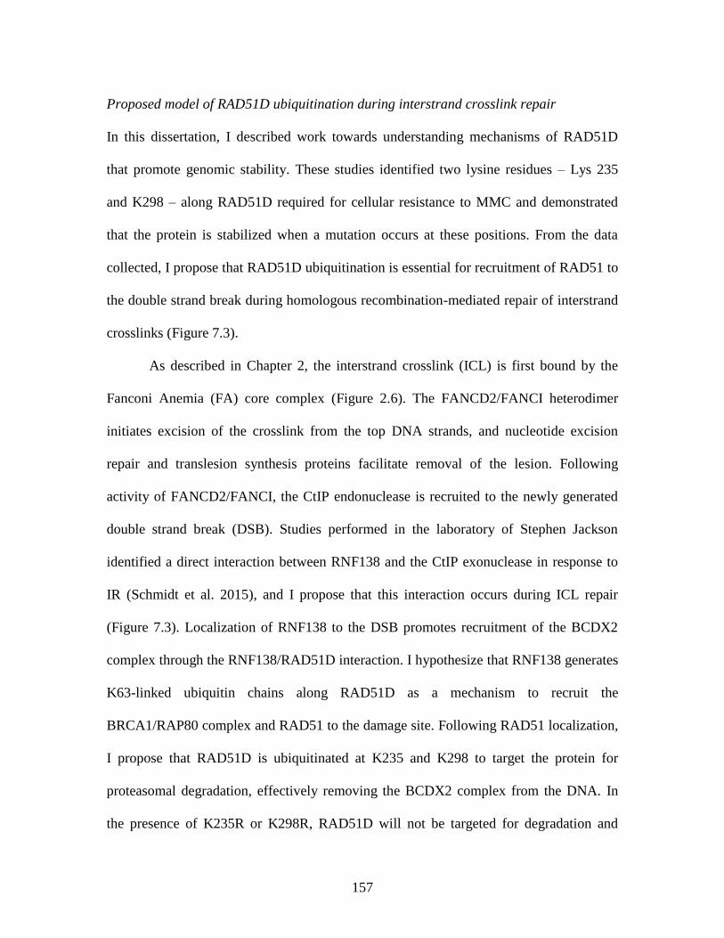
157
Proposed model of RAD51D ubiquitination during interstrand crosslink repair
In this dissertation, I described work towards understanding mechanisms of RAD51D
that promote genomic stability. These studies identified two lysine residues – Lys 235
and K298 – along RAD51D required for cellular resistance to MMC and demonstrated
that the protein is stabilized when a mutation occurs at these positions. From the data
collected, I propose that RAD51D ubiquitination is essential for recruitment of RAD51 to
the double strand break during homologous recombination-mediated repair of interstrand
crosslinks (Figure 7.3).
As described in Chapter 2, the interstrand crosslink (ICL) is first bound by the
Fanconi Anemia (FA) core complex (Figure 2.6). The FANCD2/FANCI heterodimer
initiates excision of the crosslink from the top DNA strands, and nucleotide excision
repair and translesion synthesis proteins facilitate removal of the lesion. Following
activity of FANCD2/FANCI, the CtIP endonuclease is recruited to the newly generated
double strand break (DSB). Studies performed in the laboratory of Stephen Jackson
identified a direct interaction between RNF138 and the CtIP exonuclease in response to
IR (Schmidt et al. 2015), and I propose that this interaction occurs during ICL repair
(Figure 7.3). Localization of RNF138 to the DSB promotes recruitment of the BCDX2
complex through the RNF138/RAD51D interaction. I hypothesize that RNF138 generates
K63-linked ubiquitin chains along RAD51D as a mechanism to recruit the
BRCA1/RAP80 complex and RAD51 to the damage site. Following RAD51 localization,
I propose that RAD51D is ubiquitinated at K235 and K298 to target the protein for
proteasomal degradation, effectively removing the BCDX2 complex from the DNA. In
the presence of K235R or K298R, RAD51D will not be targeted for degradation and
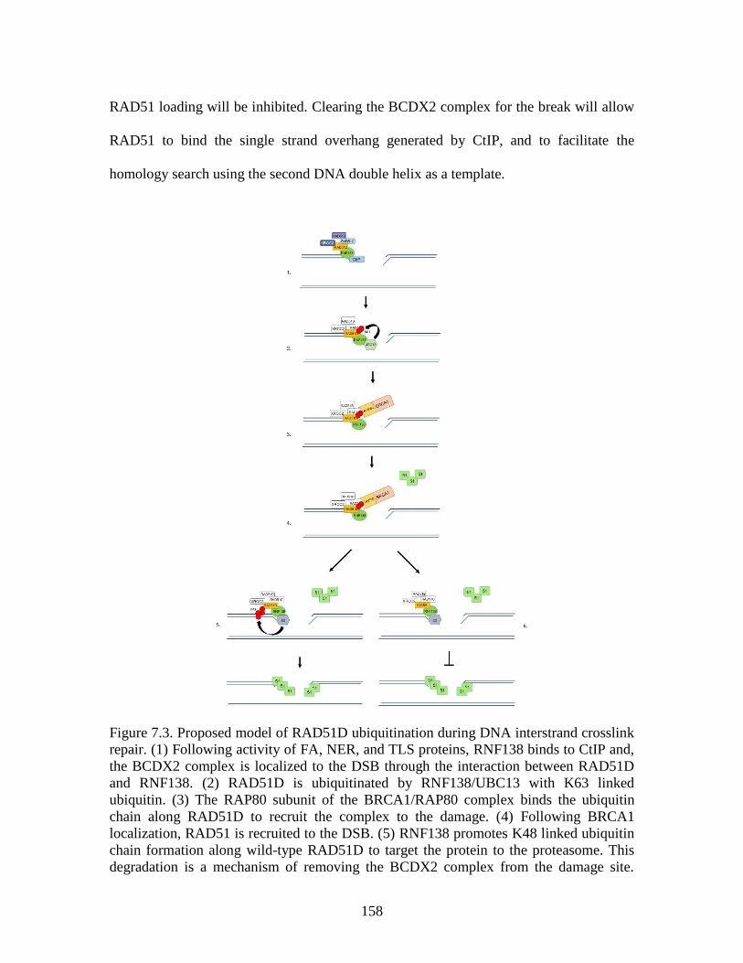
158
RAD51 loading will be inhibited. Clearing the BCDX2 complex for the break will allow
RAD51 to bind the single strand overhang generated by CtIP, and to facilitate the
homology search using the second DNA double helix as a template.
Figure 7.3. Proposed model of RAD51D ubiquitination during DNA interstrand crosslink
repair. (1) Following activity of FA, NER, and TLS proteins, RNF138 binds to CtIP and,
the BCDX2 complex is localized to the DSB through the interaction between RAD51D
and RNF138. (2) RAD51D is ubiquitinated by RNF138/UBC13 with K63 linked
ubiquitin. (3) The RAP80 subunit of the BRCA1/RAP80 complex binds the ubiquitin
chain along RAD51D to recruit the complex to the damage. (4) Following BRCA1
localization, RAD51 is recruited to the DSB. (5) RNF138 promotes K48 linked ubiquitin
chain formation along wild-type RAD51D to target the protein to the proteasome. This
degradation is a mechanism of removing the BCDX2 complex from the damage site.

159
RAD51 filaments coat the single strand overhang to prepare for homology search and
strand invasion. (6) Arginine substitution at K235 (K235R) or K298 (not shown) prevents
ubiquitination of RAD51D, preventing the BCDX2 complex from being cleared from the
break and inhibiting RAD51-mediated homology search.
160
REFERENCES
Abbas, T., and A. Dutta. 2009. 'p21 in cancer: intricate networks and multiple activities',
Nat Rev Cancer, 9: 400-14.
Adam, J., B. Deans, and J. Thacker. 2007. 'A role for Xrcc2 in the early stages of mouse
development', DNA Repair (Amst), 6: 224-34.
Akutsu, M., I. Dikic, and A. Bremm. 2016. 'Ubiquitin chain diversity at a glance', J Cell
Sci, 129: 875-80.
Alligand, B., M. Le Breton, D. Marquis, F. Vallette, and F. Fleury. 2017. 'Functional
effects of diphosphomimetic mutations at cAbl-mediated phosphorylation sites on
Rad51 recombinase activity', Biochimie.
Alpi, A. F., P. E. Pace, M. M. Babu, and K. J. Patel. 2008. 'Mechanistic insight into site-
restricted monoubiquitination of FANCD2 by Ube2t, FANCL, and FANCI', Mol
Cell, 32: 767-77.
Andreassen, P. R., and K. Ren. 2009. 'Fanconi anemia proteins, DNA interstrand
crosslink repair pathways, and cancer therapy', Curr Cancer Drug Targets, 9:
101-17.
Antoniou, A. C., O. M. Sinilnikova, J. Simard, M. Leone, M. Dumont, S. L. Neuhausen,
J. P. Struewing, D. Stoppa-Lyonnet, L. Barjhoux, D. J. Hughes, I. Coupier, M.
Belotti, C. Lasset, V. Bonadona, Y. J. Bignon, T. R. Rebbeck, T. Wagner, H. T.
Lynch, S. M. Domchek, K. L. Nathanson, J. E. Garber, J. Weitzel, S. A. Narod,
G. Tomlinson, O. I. Olopade, A. Godwin, C. Isaacs, A. Jakubowska, J. Lubinski,
J. Gronwald, B. Gorski, T. Byrski, T. Huzarski, S. Peock, M. Cook, C. Baynes, A.
Murray, M. Rogers, P. A. Daly, H. Dorkins, R. K. Schmutzler, B. Versmold, C.
Engel, A. Meindl, N. Arnold, D. Niederacher, H. Deissler, A. B. Spurdle, X.
Chen, N. Waddell, N. Cloonan, T. Kirchhoff, K. Offit, E. Friedman, B.
Kaufmann, Y. Laitman, G. Galore, G. Rennert, F. Lejbkowicz, L. Raskin, I. L.
Andrulis, E. Ilyushik, H. Ozcelik, P. Devilee, M. P. Vreeswijk, M. H. Greene, S.
A. Prindiville, A. Osorio, J. Benitez, M. Zikan, C. I. Szabo, O. Kilpivaara, H.
Nevanlinna, U. Hamann, F. Durocher, A. Arason, F. J. Couch, D. F. Easton, and
G. Chenevix-Trench. 2007. 'RAD51 135G-->C modifies breast cancer risk among
BRCA2 mutation carriers: results from a combined analysis of 19 studies', Am J
Hum Genet, 81: 1186-200.
Armstrong, M. J., and S. M. Galloway. 1997. 'Mismatch repair provokes chromosome
aberrations in hamster cells treated with methylating agents or 6-thioguanine, but
not with ethylating agents', Mutat Res, 373: 167-78.
Arnaudeau, C., C. Lundin, and T. Helleday. 2001. 'DNA double-strand breaks associated
with replication forks are predominantly repaired by homologous recombination
involving an exchange mechanism in mammalian cells', J Mol Biol, 307: 1235-45.
161
Badie, S., C. Liao, M. Thanasoula, P. Barber, M. A. Hill, and M. Tarsounas. 2009.
'RAD51C facilitates checkpoint signaling by promoting CHK2 phosphorylation',
J Cell Biol, 185: 587-600.
Baumann, P., and S. C. West. 1998. 'Role of the human RAD51 protein in homologous
recombination and double-stranded-break repair', Trends Biochem Sci, 23: 247-
51.
Bell, J. C., and S. C. Kowalczykowski. 2016. 'RecA: Regulation and Mechanism of a
Molecular Search Engine', Trends Biochem Sci, 41: 491-507.
Benson, F. E., A. Stasiak, and S. C. West. 1994. 'Purification and characterization of the
human Rad51 protein, an analogue of E. coli RecA', Embo j, 13: 5764-71.
Bernier-Villamor, V., D. A. Sampson, M. J. Matunis, and C. D. Lima. 2002. 'Structural
basis for E2-mediated SUMO conjugation revealed by a complex between
ubiquitin-conjugating enzyme Ubc9 and RanGAP1', Cell, 108: 345-56.
Bhattacharyya, A., U. S. Ear, B. H. Koller, R. R. Weichselbaum, and D. K. Bishop. 2000.
'The breast cancer susceptibility gene BRCA1 is required for subnuclear assembly
of Rad51 and survival following treatment with the DNA cross-linking agent
cisplatin', J Biol Chem, 275: 23899-903.
Bohon, J., and C. R. de los Santos. 2003. 'Structural effect of the anticancer agent 6-
thioguanine on duplex DNA', Nucleic Acids Res, 31: 1331-8.
———. 2005. 'Effect of 6-thioguanine on the stability of duplex DNA', Nucleic Acids
Res, 33: 2880-86.
Boudaiffa, B., P. Cloutier, D. Hunting, M. A. Huels, and L. Sanche. 2000. 'Resonant
formation of DNA strand breaks by low-energy (3 to 20 eV) electrons', Science,
287: 1658-60.
Boveri, T. 2008. 'Concerning the origin of malignant tumours by Theodor Boveri.
Translated and annotated by Henry Harris', J Cell Sci, 121 Suppl 1: 1-84.
Braybrooke, J. P., K. G. Spink, J. Thacker, and I. D. Hickson. 2000. 'The RAD51 family
member, RAD51L3, is a DNA-stimulated ATPase that forms a complex with
XRCC2', J Biol Chem, 275: 29100-6.
Buermeyer, A.B., C. Wilson-Van Patten, S. M. Baker, and R. M. Liskay. 1999. 'The
human MLH1 cDNA complements DNA mismatch repair defects in Mlh1-
deficient mouse embryonic fibroblasts', Cancer Res, 59: 538-41.
Busch, H., and I. L. Goldknopf. 1981. 'Ubiquitin - protein conjugates', Mol Cell Biochem,
40: 173-87.
Buys, S. S., J. F. Sandbach, A. Gammon, G. Patel, J. Kidd, K. L. Brown, L. Sharma, J.
Saam, J. Lancaster, and M. B. Daly. 2017. 'A study of over 35,000 women with
breast cancer tested with a 25-gene panel of hereditary cancer genes', Cancer.
Calderon-Celis, F., J. R. Encinar, and A. Sanz-Medel. 2017. 'Standardization approaches
in absolute quantitative proteomics with mass spectrometry', Mass Spectrom Rev.
Calo, V., M. Migliavacca, V. Bazan, M. Macaluso, M. Buscemi, N. Gebbia, and A.
Russo. 2003. 'STAT proteins: from normal control of cellular events to
tumorigenesis', J Cell Physiol, 197: 157-68.
Candelli, A., J. T. Holthausen, M. Depken, I. Brouwer, M. A. Franker, M. Marchetti, I.
Heller, S. Bernard, E. B. Garcin, M. Modesti, C. Wyman, G. J. Wuite, and E. J.
Peterman. 2014. 'Visualization and quantification of nascent RAD51 filament
162
formation at single-monomer resolution', Proc Natl Acad Sci U S A, 111: 15090-
5.
Casorelli, I., M. T. Russo, and M. Bignami. 2008. 'Role of mismatch repair and MGMT
in response to anticancer therapies', Anticancer Agents Med Chem, 8: 368-80.
Castedo, M., J. L. Perfettini, T. Roumier, K. Andreau, R. Medema, and G. Kroemer.
2004. 'Cell death by mitotic catastrophe: a molecular definition', Oncogene, 23:
2825-37.
Chau, V., J. W. Tobias, A. Bachmair, D. Marriott, D. J. Ecker, D. K. Gonda, and A.
Varshavsky. 1989. 'A multiubiquitin chain is confined to specific lysine in a
targeted short-lived protein', Science, 243: 1576-83.
Chen, G., S. S. Yuan, W. Liu, Y. Xu, K. Trujillo, B. Song, F. Cong, S. P. Goff, Y. Wu, R.
Arlinghaus, D. Baltimore, P. J. Gasser, M. S. Park, P. Sung, and E. Y. Lee. 1999.
'Radiation-induced assembly of Rad51 and Rad52 recombination complex
requires ATM and c-Abl', J Biol Chem, 274: 12748-52.
Chen, J., N. Villanueva, M. A. Rould, and S. W. Morrical. 2010. 'Insights into the
mechanism of Rad51 recombinase from the structure and properties of a filament
interface mutant', Nucleic Acids Res, 38: 4889-906.
Chen, Y., O. B. Clarke, J. Kim, S. Stowe, Y. K. Kim, Z. Assur, M. Cavalier, R. Godoy-
Ruiz, D. C. von Alpen, C. Manzini, W. S. Blaner, J. Frank, L. Quadro, D. J.
Weber, L. Shapiro, W. A. Hendrickson, and F. Mancia. 2016. 'Structure of the
STRA6 receptor for retinol uptake', Science, 353.
Chirnomas, D., T. Taniguchi, M. de la Vega, A. P. Vaidya, M. Vasserman, A. R.
Hartman, R. Kennedy, R. Foster, J. Mahoney, M. V. Seiden, and A. D. D'Andrea.
2006. 'Chemosensitization to cisplatin by inhibitors of the Fanconi anemia/BRCA
pathway', Mol Cancer Ther, 5: 952-61.
Chun, J., E. S. Buechelmaier, and S. N. Powell. 2013. 'Rad51 paralog complexes BCDX2
and CX3 act at different stages in the BRCA1-BRCA2-dependent homologous
recombination pathway', Mol Cell Biol, 33: 387-95.
Ciccia, A., and S. J. Elledge. 2010. 'The DNA damage response: making it safe to play
with knives', Mol Cell, 40: 179-204.
Clague, M. J., C. Heride, and S. Urbe. 2015. 'The demographics of the ubiquitin system',
Trends Cell Biol, 25: 417-26.
Clauson, C., O. D. Scharer, and L. Niedernhofer. 2013. 'Advances in understanding the
complex mechanisms of DNA interstrand cross-link repair', Cold Spring Harb
Perspect Biol, 5: a012732.
Clontech Laboratories, Inc. 2009. Yeast Protocols Handbook.
Compton, S. A., S. Ozgur, and J. D. Griffith. 2010. 'Ring-shaped Rad51 paralog protein
complexes bind Holliday junctions and replication forks as visualized by electron
microscopy', J Biol Chem, 285: 13349-56.
Cong, L., F. A. Ran, D. Cox, S. Lin, R. Barretto, N. Habib, P. D. Hsu, X. Wu, W. Jiang,
L. A. Marraffini, and F. Zhang. 2013. 'Multiplex genome engineering using
CRISPR/Cas systems', Science, 339: 819-23.
Conway, A. B., T. W. Lynch, Y. Zhang, G. S. Fortin, C. W. Fung, L. S. Symington, and
P. A. Rice. 2004. 'Crystal structure of a Rad51 filament', Nat Struct Mol Biol, 11:
791-6.
163
Cook, P. J., B. G. Ju, F. Telese, X. Wang, C. K. Glass, and M. G. Rosenfeld. 2009.
'Tyrosine dephosphorylation of H2AX modulates apoptosis and survival
decisions', Nature, 458: 591-6.
Coulet, F., A. Fajac, C. Colas, M. Eyries, A. Dion-Miniere, R. Rouzier, S. Uzan, J. P.
Lefranc, M. Carbonnel, F. Cornelis, A. Cortez, and F. Soubrier. 2013. 'Germline
RAD51C mutations in ovarian cancer susceptibility', Clin Genet, 83: 332-6.
Coulthard, S., and L. Hogarth. 2005. 'The thiopurines: An update', Investigational New
Drugs, 23: 523-32.
Cromm, P. M., and C. M. Crews. 2017. 'The Proteasome in Modern Drug Discovery:
Second Life of a Highly Valuable Drug Target', ACS Cent Sci, 3: 830-38.
Danilowicz, C., A. Peacock-Villada, J. Vlassakis, A. Facon, E. Feinstein, N. Kleckner,
and M. Prentiss. 2014. 'The differential extension in dsDNA bound to Rad51
filaments may play important roles in homology recognition and strand exchange',
Nucleic Acids Res, 42: 526-33.
Danish, H. H., S. Goyal, N. K. Taunk, H. Wu, M. S. Moran, and B. G. Haffty. 2013.
'Interferon-induced protein with tetratricopeptide repeats 1 (IFIT1) as a prognostic
marker for local control in T1-2 N0 breast cancer treated with breast-conserving
surgery and radiation therapy (BCS + RT)', Breast J, 19: 231-9.
Dasari, S., and P. B. Tchounwou. 2014. 'Cisplatin in cancer therapy: molecular
mechanisms of action', Eur J Pharmacol, 740: 364-78.
Deans, B., C. S. Griffin, M. Maconochie, and J. Thacker. 2000. 'Xrcc2 is required for
genetic stability, embryonic neurogenesis and viability in mice', Embo j, 19:
6675-85.
Deans, B., C. S. Griffin, P. O'Regan, M. Jasin, and J. Thacker. 2003. 'Homologous
recombination deficiency leads to profound genetic instability in cells derived
from Xrcc2-knockout mice', Cancer Res, 63: 8181-7.
Depienne, C., D. Bouteiller, A. Meneret, S. Billot, S. Groppa, S. Klebe, F. Charbonnier-
Beaupel, J. C. Corvol, J. P. Saraiva, N. Brueggemann, K. Bhatia, M. Cincotta, V.
Brochard, C. Flamand-Roze, W. Carpentier, S. Meunier, Y. Marie, M. Gaussen,
G. Stevanin, R. Wehrle, M. Vidailhet, C. Klein, I. Dusart, A. Brice, and E. Roze.
2012. 'RAD51 haploinsufficiency causes congenital mirror movements in
humans', Am J Hum Genet, 90: 301-7.
Depienne, C., M. Cincotta, S. Billot, D. Bouteiller, S. Groppa, V. Brochard, C. Flamand,
C. Hubsch, S. Meunier, F. Giovannelli, S. Klebe, J. C. Corvol, M. Vidailhet, A.
Brice, and E. Roze. 2011. 'A novel DCC mutation and genetic heterogeneity in
congenital mirror movements', Neurology, 76: 260-4.
Dodson, G. E., O. Limbo, D. Nieto, and P. Russell. 2010. 'Phosphorylation-regulated
binding of Ctp1 to Nbs1 is critical for repair of DNA double-strand breaks', Cell
Cycle, 9: 1516-22.
Doil, C., N. Mailand, S. Bekker-Jensen, P. Menard, D. H. Larsen, R. Pepperkok, J.
Ellenberg, S. Panier, D. Durocher, J. Bartek, J. Lukas, and C. Lukas. 2009.
'RNF168 binds and amplifies ubiquitin conjugates on damaged chromosomes to
allow accumulation of repair proteins', Cell, 136: 435-46.
Dosanjh, M. K., D. W. Collins, W. Fan, G. G. Lennon, J. S. Albala, Z. Shen, and D.
Schild. 1998. 'Isolation and characterization of RAD51C, a new human member
of the RAD51 family of related genes', Nucleic Acids Res, 26: 1179-84.
164
Dou, Q. P., and J. A. Zonder. 2014. 'Overview of proteasome inhibitor-based anti-cancer
therapies: perspective on bortezomib and second generation proteasome inhibitors
versus future generation inhibitors of ubiquitin-proteasome system', Curr Cancer
Drug Targets, 14: 517-36.
Eifler, K., and A. C. Vertegaal. 2015. 'Mapping the SUMOylated landscape', Febs j, 282:
3669-80.
Eoh, K. J., H. S. Park, J. S. Park, S. T. Lee, J. Han, J. Y. Lee, S. W. Kim, S. Kim, Y. T.
Kim, and E. J. Nam. 2016. 'Comparison of Clinical Outcomes of BRCA1/2
Pathologic Mutation, Variants of Unknown Significance, or Wild Type Epithelial
Ovarian Cancer Patients', Cancer Res Treat.
Fisher, R. D., B. Wang, S. L. Alam, D. S. Higginson, H. Robinson, W. I. Sundquist, and
C. P. Hill. 2003. 'Structure and ubiquitin binding of the ubiquitin-interacting
motif', J Biol Chem, 278: 28976-84.
Fragkos, M., and P. Beard. 2011. 'Mitotic catastrophe occurs in the absence of apoptosis
in p53-null cells with a defective G1 checkpoint', PLoS One, 6: e22946.
Galkin, V. E., F. Esashi, X. Yu, S. Yang, S. C. West, and E. H. Egelman. 2005. 'BRCA2
BRC motifs bind RAD51-DNA filaments', Proc Natl Acad Sci U S A, 102: 8537-
42.
Gaudelli, N. M., A. C. Komor, H. A. Rees, M. S. Packer, A. H. Badran, D. I. Bryson, and
D. R. Liu. 2017. 'Programmable base editing of A*T to G*C in genomic DNA
without DNA cleavage', Nature, 551: 464-71.
Georgakilas, A. G., O. A. Martin, and W. M. Bonner. 2017. 'p21: A Two-Faced Genome
Guardian', Trends Mol Med, 23: 310-19.
Giannini, A. L., Y. Gao, and M. J. Bijlmakers. 2008. 'T-cell regulator RNF125/TRAC-1
belongs to a novel family of ubiquitin ligases with zinc fingers and a ubiquitin-
binding domain', Biochem J, 410: 101-11.
Gibbs-Seymour, I., Y. Oka, E. Rajendra, B. T. Weinert, L. A. Passmore, K. J. Patel, J. V.
Olsen, C. Choudhary, S. Bekker-Jensen, and N. Mailand. 2015. 'Ubiquitin-SUMO
circuitry controls activated fanconi anemia ID complex dosage in response to
DNA damage', Mol Cell, 57: 150-64.
Golmard, L., V. Caux-Moncoutier, G. Davy, E. Al Ageeli, B. Poirot, C. Tirapo, D.
Michaux, C. Barbaroux, C. D. d'Enghien, A. Nicolas, L. Castera, X. Sastre-Garau,
M. H. Stern, C. Houdayer, and D. Stoppa-Lyonnet. 2013. 'Germline mutation in
the RAD51B gene confers predisposition to breast cancer', BMC Cancer, 13: 484.
Govindarajan, R., J. Duraiyan, K. Kaliyappan, and M. Palanisamy. 2012. 'Microarray and
its applications', J Pharm Bioallied Sci, 4: S310-2.
Griffin, C. S., P. J. Simpson, C. R. Wilson, and J. Thacker. 2000. 'Mammalian
recombination-repair genes XRCC2 and XRCC3 promote correct chromosome
segregation', Nat Cell Biol, 2: 757-61.
Grosicki, S., A. Barchnicka, A. Jurczyszyn, and A. Grosicka. 2014. 'Bortezomib for the
treatment of multiple myeloma', Expert Rev Hematol, 7: 173-85.
Gruver, A. M., K. A. Miller, C. Rajesh, P. G. Smiraldo, S. Kaliyaperumal, R. Balder, K.
M. Stiles, J. S. Albala, and D. L. Pittman. 2005. 'The ATPase motif in RAD51D is
required for resistance to DNA interstrand crosslinking agents and interaction
with RAD51C', Mutagenesis, 20: 433-40.
165
Gruver, A. M., B. D. Yard, C. McInnes, C. Rajesh, and D. L. Pittman. 2009. 'Functional
characterization and identification of mouse Rad51d splice variants', BMC Mol
Biol, 10: 27.
Guo, N., and Z. Peng. 2013. 'MG132, a proteasome inhibitor, induces apoptosis in tumor
cells', Asia Pac J Clin Oncol, 9: 6-11.
Haince, J. F., D. McDonald, A. Rodrigue, U. Dery, J. Y. Masson, M. J. Hendzel, and G.
G. Poirier. 2008. 'PARP1-dependent kinetics of recruitment of MRE11 and NBS1
proteins to multiple DNA damage sites', J Biol Chem, 283: 1197-208.
Hanahan, D., and R. A. Weinberg. 2000. 'The hallmarks of cancer', Cell, 100: 57-70.
———. 2011. 'Hallmarks of cancer: the next generation', Cell, 144: 646-74.
Hansford, S., and D. G. Huntsman. 2014. 'Boveri at 100: Theodor Boveri and genetic
predisposition to cancer', J Pathol, 234: 142-5.
Haynes, B., N. Saadat, B. Myung, and M. P. Shekhar. 2015. 'Crosstalk between
translesion synthesis, Fanconi anemia network, and homologous recombination
repair pathways in interstrand DNA crosslink repair and development of
chemoresistance', Mutat Res Rev Mutat Res, 763: 258-66.
Hendriks, I. A., D. Lyon, C. Young, L. J. Jensen, A. C. Vertegaal, and M. L. Nielsen.
2017. 'Site-specific mapping of the human SUMO proteome reveals co-
modification with phosphorylation', Nat Struct Mol Biol, 24: 325-36.
Hershko, A. 1983. 'Ubiquitin: roles in protein modification and breakdown', Cell, 34: 11-
2.
Hieda, K. 1994. 'DNA-DAMAGE INDUCED BY VACUUM AND SOFT-X-RAY
PHOTONS FROM SYNCHROTRON-RADIATION', International Journal of
Radiation Biology, 66: 561-67.
Hinz, J. M., R. S. Tebbs, P. F. Wilson, P. B. Nham, E. P. Salazar, H. Nagasawa, S. S.
Urbin, J. S. Bedford, and L. H. Thompson. 2006. 'Repression of mutagenesis by
Rad51D-mediated homologous recombination', Nucleic Acids Res, 34: 1358-68.
Hitchings, G. H., and G. B. Elion. 1954. 'The chemistry and biochemistry of purine
analogs', Ann N Y Acad Sci, 60: 195-9.
Hodge, C. D., I. H. Ismail, R. A. Edwards, G. L. Hura, A. T. Xiao, J. A. Tainer, M. J.
Hendzel, and J. N. Glover. 2016. 'RNF8 E3 Ubiquitin Ligase Stimulates Ubc13
E2 Conjugating Activity That Is Essential for DNA Double Strand Break
Signaling and BRCA1 Tumor Suppressor Recruitment', J Biol Chem, 291: 9396-
410.
Hofmann, K., and L. Falquet. 2001. 'A ubiquitin-interacting motif conserved in
components of the proteasomal and lysosomal protein degradation systems',
Trends Biochem Sci, 26: 347-50.
Holliday, R. 1964. 'THE INDUCTION OF MITOTIC RECOMBINATION BY
MITOMYCIN C IN USTILAGO AND SACCHAROMYCES', Genetics, 50: 323-
35.
Hu, J., J. Luo, and Q. Chen. 2017. 'The Susceptibility Pathogenesis of Moyamoya
Disease', World Neurosurg, 101: 731-41.
Huang da, W., B. T. Sherman, and R. A. Lempicki. 2009. 'Systematic and integrative
analysis of large gene lists using DAVID bioinformatics resources', Nat Protoc, 4:
44-57.
166
Huang, H., L. Zhu, B. R. Reid, G. P. Drobny, and P. B. Hopkins. 1995. 'Solution
structure of a cisplatin-induced DNA interstrand cross-link', Science, 270: 1842-5.
Huang, J. W., Y. Wang, K. K. Dhillon, P. Calses, E. Villegas, P. S. Mitchell, M. Tewari,
C. J. Kemp, and T. Taniguchi. 2013. 'Systematic screen identifies miRNAs that
target RAD51 and RAD51D to enhance chemosensitivity', Mol Cancer Res, 11:
1564-73.
Huen, M. S., S. M. Sy, and J. Chen. 2010. 'BRCA1 and its toolbox for the maintenance of
genome integrity', Nat Rev Mol Cell Biol, 11: 138-48.
Ikeda, F., N. Crosetto, and I. Dikic. 2010. 'What determines the specificity and outcomes
of ubiquitin signaling?', Cell, 143: 677-81.
Inano, S., K. Sato, Y. Katsuki, W. Kobayashi, H. Tanaka, K. Nakajima, S. Nakada, H.
Miyoshi, K. Knies, A. Takaori-Kondo, D. Schindler, M. Ishiai, H. Kurumizaka,
and M. Takata. 2017. 'RFWD3-Mediated Ubiquitination Promotes Timely
Removal of Both RPA and RAD51 from DNA Damage Sites to Facilitate
Homologous Recombination', Mol Cell, 66: 622-34.e8.
Ismail, I. H., J. P. Gagne, M. M. Genois, H. Strickfaden, D. McDonald, Z. Xu, G. G.
Poirier, J. Y. Masson, and M. J. Hendzel. 2015. 'The RNF138 E3 ligase displaces
Ku to promote DNA end resection and regulate DNA repair pathway choice', Nat
Cell Biol, 17: 1446-57.
Issaeva, N., H. D. Thomas, T. Djurenovic, J. E. Jaspers, I. Stoimenov, S. Kyle, N. Pedley,
P. Gottipati, R. Zur, K. Sleeth, V. Chatzakos, E. A. Mulligan, C. Lundin, E.
Gubanova, A. Kersbergen, A. L. Harris, R. A. Sharma, S. Rottenberg, N. J.
Curtin, and T. Helleday. 2010. '6-thioguanine selectively kills BRCA2-defective
tumors and overcomes PARP inhibitor resistance', Cancer Res, 70: 6268-76.
Iyer, R. R., A. Pluciennik, V. Burdett, and P. L. Modrich. 2006. 'DNA mismatch repair:
functions and mechanisms', Chem Rev, 106: 302-23.
Jensen, R. B., A. Ozes, T. Kim, A. Estep, and S. C. Kowalczykowski. 2013. 'BRCA2 is
epistatic to the RAD51 paralogs in response to DNA damage', DNA Repair
(Amst), 12: 306-11.
Jiricny, J. 2006. 'MutLalpha: at the cutting edge of mismatch repair', Cell, 126: 239-41.
Jo, U., and H. Kim. 2015. 'Exploiting the Fanconi Anemia Pathway for Targeted Anti-
Cancer Therapy', Mol Cells, 38: 669-76.
Jogi, A., P. Persson, A. Grynfeld, S. Pahlman, and H. Axelson. 2002. 'Modulation of
basic helix-loop-helix transcription complex formation by Id proteins during
neuronal differentiation', J Biol Chem, 277: 9118-26.
Kadyrov, F. A., L. Dzantiev, N. Constantin, and P. Modrich. 2006. 'Endonucleolytic
function of MutLalpha in human mismatch repair', Cell, 126: 297-308.
Kamieniarz, K., and R. Schneider. 2009. 'Tools to tackle protein acetylation', Chem Biol,
16: 1027-9.
Kane, R. C., P. F. Bross, A. T. Farrell, and R. Pazdur. 2003. 'Velcade: U.S. FDA approval
for the treatment of multiple myeloma progressing on prior therapy', Oncologist,
8: 508-13.
Karran, P. 2000. 'DNA double strand break repair in mammalian cells', Curr Opin Genet
Dev, 10: 144-50.
———. 2001. 'Mechanisms of tolerance to DNA damaging therapeutic drugs',
Carcinogenesis, 22: 1931-7.
167
———. 2006. 'Thiopurines, DNA damage, DNA repair and therapy-related cancer', Br
Med Bull, 79-80: 153-70.
Karran, P., and N. Attard. 2008. 'Thiopurines in current medical practice: molecular
mechanisms and contributions to therapy-related cancer', Nat Rev Cancer, 8: 24-
36.
Kawabata, M., T. Kawabata, and M. Nishibori. 2005. 'Role of recA/RAD51 family
proteins in mammals', Acta Med Okayama, 59: 1-9.
Kim, D., and S. L. Salzberg. 2011. 'TopHat-Fusion: an algorithm for discovery of novel
fusion transcripts', Genome Biol, 12: R72.
Kim, H., J. Chen, and X. Yu. 2007. 'Ubiquitin-binding protein RAP80 mediates BRCA1-
dependent DNA damage response', Science, 316: 1202-5.
Kim, H., and A. D. D'Andrea. 2012. 'Regulation of DNA cross-link repair by the Fanconi
anemia/BRCA pathway', Genes Dev, 26: 1393-408.
Kim, M. Y., T. Zhang, and W. L. Kraus. 2005. 'Poly(ADP-ribosyl)ation by PARP-1:
'PAR-laying' NAD+ into a nuclear signal', Genes Dev, 19: 1951-67.
Kim, W., E. J. Bennett, E. L. Huttlin, A. Guo, J. Li, A. Possemato, M. E. Sowa, R. Rad, J.
Rush, M. J. Comb, J. W. Harper, and S. P. Gygi. 2011. 'Systematic and
quantitative assessment of the ubiquitin-modified proteome', Mol Cell, 44: 325-
40.
Knecht, H., B. Sawan, D. Lichtensztejn, B. Lemieux, R. J. Wellinger, and S. Mai. 2009.
'The 3D nuclear organization of telomeres marks the transition from Hodgkin to
Reed-Sternberg cells', Leukemia, 23: 565-73.
Knecht, H., B. Sawan, Z. Lichtensztejn, D. Lichtensztejn, and S. Mai. 2010. '3D
Telomere FISH defines LMP1-expressing Reed-Sternberg cells as end-stage cells
with telomere-poor 'ghost' nuclei and very short telomeres', Lab Invest, 90: 611-9.
Knipscheer, P., M. Raschle, A. Smogorzewska, M. Enoiu, T. V. Ho, O. D. Scharer, S. J.
Elledge, and J. C. Walter. 2009. 'The Fanconi anemia pathway promotes
replication-dependent DNA interstrand cross-link repair', Science, 326: 1698-701.
Kondrashova, O., M. Nguyen, K. Shield-Artin, A. V. Tinker, N. N. H. Teng, M. I.
Harrell, M. J. Kuiper, G. Y. Ho, H. Barker, M. Jasin, R. Prakash, E. M. Kass, M.
R. Sullivan, G. J. Brunette, K. A. Bernstein, R. L. Coleman, A. Floquet, M.
Friedlander, G. Kichenadasse, D. M. O'Malley, A. M. Oza, J. X. Sun, L.
Robillard, L. Maloney, D. D. L. Bowtell, H. Giordano, M. J. Wakefield, S. H.
Kaufmann, A. D. Simmons, T. C. Harding, M. Raponi, I. A. McNeish, E. M.
Swisher, K. Lin, and C. L. Scott. 2017. 'Secondary Somatic Mutations Restoring
RAD51C and RAD51D Associated with Acquired Resistance to the PARP
Inhibitor Rucaparib in High-grade Ovarian Carcinoma', Cancer Discov.
Konstantinopoulos, P. A., R. Ceccaldi, G. I. Shapiro, and A. D. D'Andrea. 2015.
'Homologous Recombination Deficiency: Exploiting the Fundamental
Vulnerability of Ovarian Cancer', Cancer Discov, 5: 1137-54.
Konstantinopoulos, P. A., A. J. Wilson, J. Saskowski, E. Wass, and D. Khabele. 2014.
'Suberoylanilide hydroxamic acid (SAHA) enhances olaparib activity by targeting
homologous recombination DNA repair in ovarian cancer', Gynecol Oncol, 133:
599-606.
Kontopidis, G., S. Y. Wu, D. I. Zheleva, P. Taylor, C. McInnes, D. P. Lane, P. M.
Fischer, and M. D. Walkinshaw. 2005. 'Structural and biochemical studies of
168
human proliferating cell nuclear antigen complexes provide a rationale for cyclin
association and inhibitor design', Proc Natl Acad Sci U S A, 102: 1871-6.
Kottemann, M. C., and A. Smogorzewska. 2013. 'Fanconi anaemia and the repair of
Watson and Crick DNA crosslinks', Nature, 493: 356-63.
Kouroukis, C. T., F. G. Baldassarre, A. E. Haynes, K. Imrie, D. E. Reece, and M. C.
Cheung. 2014. 'Bortezomib in multiple myeloma: a practice guideline', Clin
Oncol (R Coll Radiol), 26: 110-9.
Kramer, A., J. Green, J. Pollard, Jr., and S. Tugendreich. 2014. 'Causal analysis
approaches in Ingenuity Pathway Analysis', Bioinformatics, 30: 523-30.
Kraus, C., J. Hoyer, G. Vasileiou, M. Wunderle, M. P. Lux, P. A. Fasching, M.
Krumbiegel, S. Uebe, M. Reuter, M. W. Beckmann, and A. Reis. 2017. 'Gene
panel sequencing in familial breast/ovarian cancer patients identifies multiple
novel mutations also in genes others than BRCA1/2', Int J Cancer, 140: 95-102.
Krogh, B. O., and L. S. Symington. 2004. 'Recombination proteins in yeast', Annu Rev
Genet, 38: 233-71.
Krynetski, E., and W. E. Evans. 2003. 'Drug methylation in cancer therapy: lessons from
the TPMT polymorphism', Oncogene, 22: 7403-13.
Kuschel, B., A. Auranen, S. McBride, K. L. Novik, A. Antoniou, J. M. Lipscombe, N. E.
Day, D. F. Easton, B. A. Ponder, P. D. Pharoah, and A. Dunning. 2002. 'Variants
in DNA double-strand break repair genes and breast cancer susceptibility', Hum
Mol Genet, 11: 1399-407.
Kuznetsov, S. G., D. C. Haines, B. K. Martin, and S. K. Sharan. 2009. 'Loss of Rad51c
leads to embryonic lethality and modulation of Trp53-dependent tumorigenesis in
mice', Cancer Res, 69: 863-72.
Lando, D. Y., C. L. Chang, A. S. Fridman, I. E. Grigoryan, E. N. Galyuk, Y. W. Hsueh,
and C. K. Hu. 2014. 'Comparative thermal and thermodynamic study of DNA
chemically modified with antitumor drug cisplatin and its inactive analog
transplatin', J Inorg Biochem, 137: 85-93.
Landrum, M. J., J. M. Lee, M. Benson, G. Brown, C. Chao, S. Chitipiralla, B. Gu, J. Hart,
D. Hoffman, J. Hoover, W. Jang, K. Katz, M. Ovetsky, G. Riley, A. Sethi, R.
Tully, R. Villamarin-Salomon, W. Rubinstein, and D. R. Maglott. 2016. 'ClinVar:
public archive of interpretations of clinically relevant variants', Nucleic Acids Res,
44: D862-8.
Le, D. T., J. N. Durham, K. N. Smith, H. Wang, B. R. Bartlett, L. K. Aulakh, S. Lu, H.
Kemberling, C. Wilt, B. S. Luber, F. Wong, N. S. Azad, A. A. Rucki, D. Laheru,
R. Donehower, A. Zaheer, G. A. Fisher, T. S. Crocenzi, J. J. Lee, T. F. Greten, A.
G. Duffy, K. K. Ciombor, A. D. Eyring, B. H. Lam, A. Joe, S. P. Kang, M.
Holdhoff, L. Danilova, L. Cope, C. Meyer, S. Zhou, R. M. Goldberg, D. K.
Armstrong, K. M. Bever, A. N. Fader, J. Taube, F. Housseau, D. Spetzler, N.
Xiao, D. M. Pardoll, N. Papadopoulos, K. W. Kinzler, J. R. Eshleman, B.
Vogelstein, R. A. Anders, and L. A. Diaz, Jr. 2017. 'Mismatch repair deficiency
predicts response of solid tumors to PD-1 blockade', Science, 357: 409-13.
Lee, J. Y., T. Terakawa, Z. Qi, J. B. Steinfeld, S. Redding, Y. Kwon, W. A. Gaines, W.
Zhao, P. Sung, and E. C. Greene. 2015. 'DNA RECOMBINATION. Base triplet
stepping by the Rad51/RecA family of recombinases', Science, 349: 977-81.
169
Lee, P. S., J. Fang, L. Jessop, T. Myers, P. Raj, N. Hu, C. Wang, P. R. Taylor, J. Wang, J.
Khan, M. Jasin, and S. J. Chanock. 2014. 'RAD51B Activity and Cell Cycle
Regulation in Response to DNA Damage in Breast Cancer Cell Lines', Breast
Cancer (Auckl), 8: 135-44.
Levy-Lahad, E., A. Lahad, S. Eisenberg, E. Dagan, T. Paperna, L. Kasinetz, R. Catane,
B. Kaufman, U. Beller, P. Renbaum, and R. Gershoni-Baruch. 2001. 'A single
nucleotide polymorphism in the RAD51 gene modifies cancer risk in BRCA2 but
not BRCA1 carriers', Proc Natl Acad Sci U S A, 98: 3232-6.
Li, G. M. 1999. 'The role of mismatch repair in DNA damage-induced apoptosis', Oncol
Res, 11: 393-400.
———. 2008. 'Mechanisms and functions of DNA mismatch repair', Cell Res, 18: 85-98.
Li, G. M., and P. Modrich. 1995. 'Restoration of mismatch repair to nuclear extracts of
H6 colorectal tumor cells by a heterodimer of human MutL homologs', Proc Natl
Acad Sci U S A, 92: 1950-4.
Liang, C. C., Z. Li, D. Lopez-Martinez, W. V. Nicholson, C. Venien-Bryan, and M. A.
Cohn. 2016. 'The FANCD2-FANCI complex is recruited to DNA interstrand
crosslinks before monoubiquitination of FANCD2', Nat Commun, 7: 12124.
Lim, D. S., and P. Hasty. 1996. 'A mutation in mouse rad51 results in an early embryonic
lethal that is suppressed by a mutation in p53', Mol Cell Biol, 16: 7133-43.
Lio, Y. C., D. Schild, M. A. Brenneman, J. L. Redpath, and D. J. Chen. 2004. 'Human
Rad51C deficiency destabilizes XRCC3, impairs recombination, and
radiosensitizes S/G2-phase cells', J Biol Chem, 279: 42313-20.
Liu, F., L. Wang, F. Perna, and S. D. Nimer. 2016. 'Beyond transcription factors: how
oncogenic signalling reshapes the epigenetic landscape', Nat Rev Cancer, 16: 359-
72.
Liu, N., J. E. Lamerdin, R. S. Tebbs, D. Schild, J. D. Tucker, M. R. Shen, K. W.
Brookman, M. J. Siciliano, C. A. Walter, W. Fan, L. S. Narayana, Z. Q. Zhou, A.
W. Adamson, K. J. Sorensen, D. J. Chen, N. J. Jones, and L. H. Thompson. 1998.
'XRCC2 and XRCC3, new human Rad51-family members, promote chromosome
stability and protect against DNA cross-links and other damages', Mol Cell, 1:
783-93.
Liu, Y., J. Y. Masson, R. Shah, P. O'Regan, and S. C. West. 2004. 'RAD51C is required
for Holliday junction processing in mammalian cells', Science, 303: 243-6.
Liu, Y., M. Tarsounas, P. O'Regan, and S. C. West. 2007. 'Role of RAD51C and XRCC3
in genetic recombination and DNA repair', J Biol Chem, 282: 1973-9.
Lo, T., L. Pellegrini, A. R. Venkitaraman, and T. L. Blundell. 2003. 'Sequence
fingerprints in BRCA2 and RAD51: implications for DNA repair and cancer',
DNA Repair (Amst), 2: 1015-28.
Loeb, L. A., C. F. Springgate, and N. Battula. 1974. 'Errors in DNA replication as a basis
of malignant changes', Cancer Res, 34: 2311-21.
Loignon, M., L. Amrein, M. Dunn, and R. Aloyz. 2007. 'XRCC3 depletion induces
spontaneous DNA breaks and p53-dependent cell death', Cell Cycle, 6: 606-11.
Lopez-Martinez, D., C. C. Liang, and M. A. Cohn. 2016. 'Cellular response to DNA
interstrand crosslinks: the Fanconi anemia pathway', Cell Mol Life Sci.
170
Lorick, K. L., J. P. Jensen, S. Fang, A. M. Ong, S. Hatakeyama, and A. M. Weissman.
1999. 'RING fingers mediate ubiquitin-conjugating enzyme (E2)-dependent
ubiquitination', Proc Natl Acad Sci U S A, 96: 11364-9.
Love, M. I., W. Huber, and S. Anders. 2014. 'Moderated estimation of fold change and
dispersion for RNA-seq data with DESeq2', Genome Biol, 15: 550.
Loveday, C., C. Turnbull, E. Ramsay, D. Hughes, E. Ruark, J. R. Frankum, G. Bowden,
B. Kalmyrzaev, M. Warren-Perry, K. Snape, J. W. Adlard, J. Barwell, J. Berg, A.
F. Brady, C. Brewer, G. Brice, C. Chapman, J. Cook, R. Davidson, A. Donaldson,
F. Douglas, L. Greenhalgh, A. Henderson, L. Izatt, A. Kumar, F. Lalloo, Z.
Miedzybrodzka, P. J. Morrison, J. Paterson, M. Porteous, M. T. Rogers, S.
Shanley, L. Walker, D. Eccles, D. G. Evans, A. Renwick, S. Seal, C. J. Lord, A.
Ashworth, J. S. Reis-Filho, A. C. Antoniou, and N. Rahman. 2011. 'Germline
mutations in RAD51D confer susceptibility to ovarian cancer', Nat Genet, 43:
879-82.
Loveday, C., C. Turnbull, E. Ruark, R. M. Xicola, E. Ramsay, D. Hughes, M. Warren-
Perry, K. Snape, D. Eccles, D. G. Evans, M. Gore, A. Renwick, S. Seal, A. C.
Antoniou, and N. Rahman. 2012. 'Germline RAD51C mutations confer
susceptibility to ovarian cancer', Nat Genet, 44: 475-6; author reply 76.
Lu, C., F. Zhu, Y. Y. Cho, F. Tang, T. Zykova, W. Y. Ma, A. M. Bode, and Z. Dong.
2006. 'Cell apoptosis: requirement of H2AX in DNA ladder formation, but not for
the activation of caspase-3', Mol Cell, 23: 121-32.
Mah, L. J., A. El-Osta, and T. C. Karagiannis. 2010. 'gammaH2AX: a sensitive molecular
marker of DNA damage and repair', Leukemia, 24: 679-86.
Mailand, N., S. Bekker-Jensen, H. Faustrup, F. Melander, J. Bartek, C. Lukas, and J.
Lukas. 2007. 'RNF8 ubiquitylates histones at DNA double-strand breaks and
promotes assembly of repair proteins', Cell, 131: 887-900.
Malinge, J. M., M. J. Giraud-Panis, and M. Leng. 1999. 'Interstrand cross-links of
cisplatin induce striking distortions in DNA', J Inorg Biochem, 77: 23-9.
Marathias, V. M., M. J. Sawicki, and P. H. Bolton. 1999. '6-Thioguanine alters the
structure and stability of duplex DNA and inhibits quadruplex DNA formation',
Nucleic Acids Res, 27: 2860-7.
Marechal, A., and L. Zou. 2015. 'RPA-coated single-stranded DNA as a platform for
post-translational modifications in the DNA damage response', Cell Res, 25: 9-23.
Masson, J. Y., M. C. Tarsounas, A. Z. Stasiak, A. Stasiak, R. Shah, M. J. McIlwraith, F.
E. Benson, and S. C. West. 2001. 'Identification and purification of two distinct
complexes containing the five RAD51 paralogs', Genes Dev, 15: 3296-307.
Mauch, P. M., Weiss, L., Armitage, J.O. 2006. 'Hodgkin Disease.' in D. W. Kufe, Bast,
R.C., Hait, W. N, Hong, W. K., Pollock, R. E., Weichselbaum, R.R., Holland, J.
F., Frei, E. (ed.), Cancer Medicine (BC Decker: Hamilton, Ontario Canada).
Meindl, A., H. Hellebrand, C. Wiek, V. Erven, B. Wappenschmidt, D. Niederacher, M.
Freund, P. Lichtner, L. Hartmann, H. Schaal, J. Ramser, E. Honisch, C. Kubisch,
H. E. Wichmann, K. Kast, H. Deissler, C. Engel, B. Muller-Myhsok, K. Neveling,
M. Kiechle, C. G. Mathew, D. Schindler, R. K. Schmutzler, and H. Hanenberg.
2010. 'Germline mutations in breast and ovarian cancer pedigrees establish
RAD51C as a human cancer susceptibility gene', Nat Genet, 42: 410-4.
171
Mender, I., S. Gryaznov, Z. G. Dikmen, W. E. Wright, and J. W. Shay. 2015. 'Induction
of telomere dysfunction mediated by the telomerase substrate precursor 6-thio-2'-
deoxyguanosine', Cancer Discov, 5: 82-95.
Mertins, P., D. R. Mani, K. V. Ruggles, M. A. Gillette, K. R. Clauser, P. Wang, X. Wang,
J. W. Qiao, S. Cao, F. Petralia, E. Kawaler, F. Mundt, K. Krug, Z. Tu, J. T. Lei,
M. L. Gatza, M. Wilkerson, C. M. Perou, V. Yellapantula, K. L. Huang, C. Lin,
M. D. McLellan, P. Yan, S. R. Davies, R. R. Townsend, S. J. Skates, J. Wang, B.
Zhang, C. R. Kinsinger, M. Mesri, H. Rodriguez, L. Ding, A. G. Paulovich, D.
Fenyo, M. J. Ellis, and S. A. Carr. 2016. 'Proteogenomics connects somatic
mutations to signalling in breast cancer', Nature, 534: 55-62.
Mertins, P., J. W. Qiao, J. Patel, N. D. Udeshi, K. R. Clauser, D. R. Mani, M. W.
Burgess, M. A. Gillette, J. D. Jaffe, and S. A. Carr. 2013. 'Integrated proteomic
analysis of post-translational modifications by serial enrichment', Nat Methods,
10: 634-7.
Metzger, M. B., J. N. Pruneda, R. E. Klevit, and A. M. Weissman. 2014. 'RING-type E3
ligases: master manipulators of E2 ubiquitin-conjugating enzymes and
ubiquitination', Biochim Biophys Acta, 1843: 47-60.
Michl, J., J. Zimmer, and M. Tarsounas. 2016. 'Interplay between Fanconi anemia and
homologous recombination pathways in genome integrity', Embo j, 35: 909-23.
Miller, K. A., D. Sawicka, D. Barsky, and J. S. Albala. 2004. 'Domain mapping of the
Rad51 paralog protein complexes', Nucleic Acids Res, 32: 169-78.
Min, A., S. A. Im, Y. K. Yoon, S. H. Song, H. J. Nam, H. S. Hur, H. P. Kim, K. H. Lee,
S. W. Han, D. Y. Oh, T. Y. Kim, M. J. O'Connor, W. H. Kim, and Y. J. Bang.
2013. 'RAD51C-deficient cancer cells are highly sensitive to the PARP inhibitor
olaparib', Mol Cancer Ther, 12: 865-77.
Minckwitz, Gunter Von, Eric Hahnen, Peter A. Fasching, Jan Hauke, Andreas
Schneeweiss, Christoph Salat, Mahdi Rezai, Jens U. Blohmer, Dirk Michael
Zahm, Christian Jackisch, Bernd Gerber, Peter Klare, Sherko Kummel, Holger
Eidtmann, Stefan Paepke, Valentina Nekljudova, Sibylle Loibl, Michael Untch,
Rita K. Schmutzler, GBG, and AGO-B Study Groups. 2014. 'Pathological
complete response (pCR) rates after carboplatin-containing neoadjuvant
chemotherapy in patients with germline BRCA (gBRCA) mutation and triple-
negative breast cancer (TNBC): Results from GeparSixto', Journal of Clinical
Oncology, 32: 1005-05.
Monteiro, F. L., T. Baptista, F. Amado, R. Vitorino, C. Jeronimo, and L. A. Helguero.
2014. 'Expression and functionality of histone H2A variants in cancer',
Oncotarget, 5: 3428-43.
Morgan, E. A., N. Shah, and L. S. Symington. 2002. 'The requirement for ATP
hydrolysis by Saccharomyces cerevisiae Rad51 is bypassed by mating-type
heterozygosity or RAD54 in high copy', Mol Cell Biol, 22: 6336-43.
Morita, T., Y. Yoshimura, A. Yamamoto, K. Murata, M. Mori, H. Yamamoto, and A.
Matsushiro. 1993. 'A mouse homolog of the Escherichia coli recA and
Saccharomyces cerevisiae RAD51 genes', Proc Natl Acad Sci U S A, 90: 6577-80.
Muniandy, P. A., J. Liu, A. Majumdar, S. T. Liu, and M. M. Seidman. 2010. 'DNA
interstrand crosslink repair in mammalian cells: step by step', Crit Rev Biochem
Mol Biol, 45: 23-49.
172
Munshi, P. N., M. Lubin, and J. R. Bertino. 2014. '6-Thioguanine: A Drug With
Unrealized Potential for Cancer Therapy', Oncologist, 19: 760-65.
Nakada, S. 2016. 'Opposing roles of RNF8/RNF168 and deubiquitinating enzymes in
ubiquitination-dependent DNA double-strand break response signaling and DNA-
repair pathway choice', J Radiat Res, 57 Suppl 1: i33-i40.
Nakada, S., R. M. Yonamine, and K. Matsuo. 2012. 'RNF8 regulates assembly of RAD51
at DNA double-strand breaks in the absence of BRCA1 and 53BP1', Cancer Res,
72: 4974-83.
Neidhardt, G., A. Becker, J. Hauke, J. Horvath, N. Bogdanova Markov, S. Heilmann-
Heimbach, H. Hellebrand, H. Thiele, J. Altmuller, P. Nurnberg, A. Meindl, K.
Rhiem, B. Blumcke, B. Wappenschmidt, R. K. Schmutzler, and E. Hahnen. 2017.
'The RAD51C exonic splice-site mutations c.404G>C and c.404G>T are
associated with familial breast and ovarian cancer', Eur J Cancer Prev, 26: 165-
69.
Nie, M., and M. N. Boddy. 2016. 'Cooperativity of the SUMO and Ubiquitin Pathways in
Genome Stability', Biomolecules, 6: 14.
Nomme, J., Y. Takizawa, S. F. Martinez, A. Renodon-Corniere, F. Fleury, P. Weigel, K.
Yamamoto, H. Kurumizaka, and M. Takahashi. 2008. 'Inhibition of filament
formation of human Rad51 protein by a small peptide derived from the BRC-
motif of the BRCA2 protein', Genes Cells, 13: 471-81.
Ogawa, T., X. Yu, A. Shinohara, and E. H. Egelman. 1993. 'Similarity of the yeast
RAD51 filament to the bacterial RecA filament', Science, 259: 1896-9.
Ohtake, F., and H. Tsuchiya. 2017. 'The emerging complexity of ubiquitin architecture', J
Biochem, 161: 125-33.
Owerbach, D., E. M. McKay, E. T. Yeh, K. H. Gabbay, and K. M. Bohren. 2005. 'A
proline-90 residue unique to SUMO-4 prevents maturation and sumoylation',
Biochem Biophys Res Commun, 337: 517-20.
Panier, S., and D. Durocher. 2009. 'Regulatory ubiquitylation in response to DNA
double-strand breaks', DNA Repair (Amst), 8: 436-43.
Parker, C. E., V. Mocanu, M. Mocanu, N. Dicheva, and M. R. Warren. 2010. 'Frontiers in
Neuroscience
Mass Spectrometry for Post-Translational Modifications.' in O. Alzate (ed.),
Neuroproteomics (CRC Press/Taylor & Francis
Llc.: Boca Raton (FL)).
Pellegrini, L., D. S. Yu, T. Lo, S. Anand, M. Lee, T. L. Blundell, and A. R.
Venkitaraman. 2002. 'Insights into DNA recombination from the structure of a
RAD51-BRCA2 complex', Nature, 420: 287-93.
Pelttari, L. M., T. Heikkinen, D. Thompson, A. Kallioniemi, J. Schleutker, K. Holli, C.
Blomqvist, K. Aittomaki, R. Butzow, and H. Nevanlinna. 2011. 'RAD51C is a
susceptibility gene for ovarian cancer', Hum Mol Genet, 20: 3278-88.
Pelttari, L. M., S. Khan, M. Vuorela, J. I. Kiiski, S. Vilske, V. Nevanlinna, S. Ranta, J.
Schleutker, R. Winqvist, A. Kallioniemi, T. Dork, N. V. Bogdanova, J. Figueroa,
P. D. Pharoah, M. K. Schmidt, A. M. Dunning, M. Garcia-Closas, M. K. Bolla, J.
Dennis, K. Michailidou, Q. Wang, J. L. Hopper, M. C. Southey, E. H. Rosenberg,
173
P. A. Fasching, M. W. Beckmann, J. Peto, I. Dos-Santos-Silva, E. J. Sawyer, I.
Tomlinson, B. Burwinkel, H. Surowy, P. Guenel, T. Truong, S. E. Bojesen, B. G.
Nordestgaard, J. Benitez, A. Gonzalez-Neira, S. L. Neuhausen, H. Anton-Culver,
H. Brenner, V. Arndt, A. Meindl, R. K. Schmutzler, H. Brauch, T. Bruning, A.
Lindblom, S. Margolin, A. Mannermaa, J. M. Hartikainen, G. Chenevix-Trench,
L. Van Dyck, H. Janssen, J. Chang-Claude, A. Rudolph, P. Radice, P. Peterlongo,
E. Hallberg, J. E. Olson, G. G. Giles, R. L. Milne, C. A. Haiman, F. Schumacher,
J. Simard, M. Dumont, V. Kristensen, A. L. Borresen-Dale, W. Zheng, A.
Beeghly-Fadiel, M. Grip, I. L. Andrulis, G. Glendon, P. Devilee, C. Seynaeve, M.
J. Hooning, M. Collee, A. Cox, S. S. Cross, M. Shah, R. N. Luben, U. Hamann,
D. Torres, A. Jakubowska, J. Lubinski, F. J. Couch, D. Yannoukakos, N. Orr, A.
Swerdlow, H. Darabi, J. Li, K. Czene, P. Hall, D. F. Easton, J. Mattson, C.
Blomqvist, K. Aittomaki, and H. Nevanlinna. 2016. 'RAD51B in Familial Breast
Cancer', PLoS One, 11: e0153788.
Pena-Diaz, J., and J. Jiricny. 2012. 'Mammalian mismatch repair: error-free or error-
prone?', Trends Biochem Sci, 37: 206-14.
Pennington, K. P., T. Walsh, M. I. Harrell, M. K. Lee, C. C. Pennil, M. H. Rendi, A.
Thornton, B. M. Norquist, S. Casadei, A. S. Nord, K. J. Agnew, C. C. Pritchard,
S. Scroggins, R. L. Garcia, M. C. King, and E. M. Swisher. 2014. 'Germline and
somatic mutations in homologous recombination genes predict platinum response
and survival in ovarian, fallopian tube, and peritoneal carcinomas', Clin Cancer
Res, 20: 764-75.
Perk, J., A. Iavarone, and R. Benezra. 2005. 'Id family of helix-loop-helix proteins in
cancer', Nat Rev Cancer, 5: 603-14.
Pichler, A., C. Fatouros, H. Lee, and N. Eisenhardt. 2017. 'SUMO conjugation - a
mechanistic view', Biomol Concepts, 8: 13-36.
Pickart, C. M. 2004. 'Back to the future with ubiquitin', Cell, 116: 181-90.
Pierce, A. J., R. D. Johnson, L. H. Thompson, and M. Jasin. 1999. 'XRCC3 promotes
homology-directed repair of DNA damage in mammalian cells', Genes Dev, 13:
2633-8.
Pittman, D. L., and J. C. Schimenti. 2000. 'Midgestation lethality in mice deficient for the
RecA-related gene, Rad51d/Rad51l3', Genesis, 26: 167-73.
Poulsen, M., C. Lukas, J. Lukas, S. Bekker-Jensen, and N. Mailand. 2012. 'Human
RNF169 is a negative regulator of the ubiquitin-dependent response to DNA
double-strand breaks', J Cell Biol, 197: 189-99.
Prakash, R., Y. Zhang, W. Feng, and M. Jasin. 2015. 'Homologous recombination and
human health: the roles of BRCA1, BRCA2, and associated proteins', Cold Spring
Harb Perspect Biol, 7: a016600.
Qi, Z., S. Redding, J. Y. Lee, B. Gibb, Y. Kwon, H. Niu, W. A. Gaines, P. Sung, and E.
C. Greene. 2015. 'DNA sequence alignment by microhomology sampling during
homologous recombination', Cell, 160: 856-69.
Rajesh, C., D. K. Baker, A. J. Pierce, and D. L. Pittman. 2011. 'The splicing-factor
related protein SFPQ/PSF interacts with RAD51D and is necessary for homology-
directed repair and sister chromatid cohesion', Nucleic Acids Res, 39: 132-45.
174
Rajesh, C., A. M. Gruver, V. Basrur, and D. L. Pittman. 2009. 'The interaction profile of
homologous recombination repair proteins RAD51C, RAD51D and XRCC2 as
determined by proteomic analysis', Proteomics, 9: 4071-86.
Rajesh, P., A. V. Litvinchuk, D. L. Pittman, and M. D. Wyatt. 2011. 'The homologous
recombination protein RAD51D mediates the processing of 6-thioguanine lesions
downstream of mismatch repair', Mol Cancer Res, 9: 206-14.
Rajesh, P., C. Rajesh, M. D. Wyatt, and D. L. Pittman. 2010. 'RAD51D protects against
MLH1-dependent cytotoxic responses to O(6)-methylguanine', DNA Repair
(Amst), 9: 458-67.
Rappaport, H. P. 1993. 'Replication of the base pair 6-thioguanine/5-methyl-2-pyrimidine
with the large Klenow fragment of Escherichia coli DNA polymerase I',
Biochemistry, 32: 3047-57.
Rickman, K. A., F. P. Lach, A. Abhyankar, F. X. Donovan, E. M. Sanborn, J. A.
Kennedy, C. Sougnez, S. B. Gabriel, O. Elemento, S. C. Chandrasekharappa, D.
Schindler, A. D. Auerbach, and A. Smogorzewska. 2015. 'Deficiency of UBE2T,
the E2 Ubiquitin Ligase Necessary for FANCD2 and FANCI Ubiquitination,
Causes FA-T Subtype of Fanconi Anemia', Cell Rep, 12: 35-41.
Rock, K. L., C. Gramm, L. Rothstein, K. Clark, R. Stein, L. Dick, D. Hwang, and A. L.
Goldberg. 1994. 'Inhibitors of the proteasome block the degradation of most cell
proteins and the generation of peptides presented on MHC class I molecules',
Cell, 78: 761-71.
Rogakou, E. P., D. R. Pilch, A. H. Orr, V. S. Ivanova, and W. M. Bonner. 1998. 'DNA
double-stranded breaks induce histone H2AX phosphorylation on serine 139', J
Biol Chem, 273: 5858-68.
Saitoh, H., and J. Hinchey. 2000. 'Functional heterogeneity of small ubiquitin-related
protein modifiers SUMO-1 versus SUMO-2/3', J Biol Chem, 275: 6252-8.
Sanchez, H., A. Kertokalio, S. van Rossum-Fikkert, R. Kanaar, and C. Wyman. 2013.
'Combined optical and topographic imaging reveals different arrangements of
human RAD54 with presynaptic and postsynaptic RAD51-DNA filaments', Proc
Natl Acad Sci U S A, 110: 11385-90.
Sato, Y., A. Yamagata, S. Goto-Ito, K. Kubota, R. Miyamoto, S. Nakada, and S. Fukai.
2012. 'Molecular basis of Lys-63-linked polyubiquitination inhibition by the
interaction between human deubiquitinating enzyme OTUB1 and ubiquitin-
conjugating enzyme UBC13', J Biol Chem, 287: 25860-8.
Sato, Y., A. Yoshikawa, H. Mimura, M. Yamashita, A. Yamagata, and S. Fukai. 2009.
'Structural basis for specific recognition of Lys 63-linked polyubiquitin chains by
tandem UIMs of RAP80', Embo j, 28: 2461-8.
Satoh, J., and H. Tabunoki. 2013. 'A Comprehensive Profile of ChIP-Seq-Based STAT1
Target Genes Suggests the Complexity of STAT1-Mediated Gene Regulatory
Mechanisms', Gene Regul Syst Bio, 7: 41-56.
Schaetzlein, S., N. R. Kodandaramireddy, Z. Ju, A. Lechel, A. Stepczynska, D. R. Lilli,
A. B. Clark, C. Rudolph, F. Kuhnel, K. Wei, B. Schlegelberger, P. Schirmacher,
T. A. Kunkel, R. A. Greenberg, W. Edelmann, and K. L. Rudolph. 2007.
'Exonuclease-1 deletion impairs DNA damage signaling and prolongs lifespan of
telomere-dysfunctional mice', Cell, 130: 863-77.
175
Scheffner, M., and S. Kumar. 2014. 'Mammalian HECT ubiquitin-protein ligases:
biological and pathophysiological aspects', Biochim Biophys Acta, 1843: 61-74.
Schild, D., Y. C. Lio, D. W. Collins, T. Tsomondo, and D. J. Chen. 2000. 'Evidence for
simultaneous protein interactions between human Rad51 paralogs', J Biol Chem,
275: 16443-9.
Schlafer, D., K. S. Shah, E. H. Panjic, and S. Lonial. 2017. 'Safety of proteasome
inhibitors for treatment of multiple myeloma', Expert Opin Drug Saf, 16: 167-83.
Schmidt, C. K., Y. Galanty, M. Sczaniecka-Clift, J. Coates, S. Jhujh, M. Demir, M.
Cornwell, P. Beli, and S. P. Jackson. 2015. 'Systematic E2 screening reveals a
UBE2D-RNF138-CtIP axis promoting DNA repair', Nat Cell Biol, 17: 1458-70.
Schnurbein, G., J. Hauke, B. Wappenschmidt, N. Weber-Lassalle, S. Engert, H.
Hellebrand, L. Garbes, A. Becker, G. Neidhardt, K. Rhiem, A. Meindl, R. K.
Schmutzler, and E. Hahnen. 2013. 'RAD51C deletion screening identifies a
recurrent gross deletion in breast cancer and ovarian cancer families', Breast
Cancer Res, 15: R120.
Schurch, N. J., P. Schofield, M. Gierlinski, C. Cole, A. Sherstnev, V. Singh, N. Wrobel,
K. Gharbi, G. G. Simpson, T. Owen-Hughes, M. Blaxter, and G. J. Barton. 2016.
'How many biological replicates are needed in an RNA-seq experiment and which
differential expression tool should you use?', RNA, 22: 839-51.
Serebriiskii, I. G., and E. A. Golemis. 2000. 'Uses of lacZ to study gene function:
evaluation of beta-galactosidase assays employed in the yeast two-hybrid system',
Anal Biochem, 285: 1-15.
Shinohara, A., H. Ogawa, and T. Ogawa. 1992. 'Rad51 protein involved in repair and
recombination in S. cerevisiae is a RecA-like protein', Cell, 69: 457-70.
Shinohara, A., and T. Ogawa. 1999. 'Rad51/RecA protein families and the associated
proteins in eukaryotes', Mutat Res, 435: 13-21.
Short, J. M., Y. Liu, S. Chen, N. Soni, M. S. Madhusudhan, M. K. Shivji, and A. R.
Venkitaraman. 2016. 'High-resolution structure of the presynaptic RAD51
filament on single-stranded DNA by electron cryo-microscopy', Nucleic Acids
Res, 44: 9017-30.
Shu, Z., S. Smith, L. Wang, M. C. Rice, and E. B. Kmiec. 1999. 'Disruption of
muREC2/RAD51L1 in mice results in early embryonic lethality which can Be
partially rescued in a p53(-/-) background', Mol Cell Biol, 19: 8686-93.
Sigurdsson, S., S. Van Komen, W. Bussen, D. Schild, J. S. Albala, and P. Sung. 2001.
'Mediator function of the human Rad51B-Rad51C complex in Rad51/RPA-
catalyzed DNA strand exchange', Genes Dev, 15: 3308-18.
Singh, K., B. GrothVasselli, P. N. Farnsworth, and D. K. Rai. 1996. 'Effect of thiobase
incorporation into duplex DNA during the polymerization reaction', Research
Communications in Molecular Pathology and Pharmacology, 94: 129-40.
Singleton, S. F., and J. Xiao. 2001. 'The stretched DNA geometry of recombination and
repair nucleoprotein filaments', Biopolymers, 61: 145-58.
Sloper-Mould, K. E., J. C. Jemc, C. M. Pickart, and L. Hicke. 2001. 'Distinct functional
surface regions on ubiquitin', J Biol Chem, 276: 30483-9.
Slupianek, A., S. K. Jozwiakowski, E. Gurdek, and T. Skorski. 2009. 'BCR/ABL kinase
interacts with and phosphorylates the RAD51 paralog, RAD51B', Leukemia, 23:
2308-10.
176
Slupianek, A., C. Schmutte, G. Tombline, M. Nieborowska-Skorska, G. Hoser, M. O.
Nowicki, A. J. Pierce, R. Fishel, and T. Skorski. 2001. 'BCR/ABL regulates
mammalian RecA homologs, resulting in drug resistance', Mol Cell, 8: 795-806.
Smiraldo, P. G., A. M. Gruver, J. C. Osborn, and D. L. Pittman. 2005. 'Extensive
chromosomal instability in Rad51d-deficient mouse cells', Cancer Res, 65: 2089-
96.
Sobhian, B., G. Shao, D. R. Lilli, A. C. Culhane, L. A. Moreau, B. Xia, D. M. Livingston,
and R. A. Greenberg. 2007. 'RAP80 targets BRCA1 to specific ubiquitin
structures at DNA damage sites', Science, 316: 1198-202.
Somerville, L., E. Y. Krynetski, N. F. Krynetskaia, R. D. Beger, W. X. Zhang, C. A.
Marhefka, W. E. Evans, and R. W. Kriwacki. 2003. 'Structure and dynamics of
thioguanine-modified duplex DNA', Journal of Biological Chemistry, 278: 1005-
11.
Somyajit, K., S. Basavaraju, R. Scully, and G. Nagaraju. 2013. 'ATM- and ATR-
mediated phosphorylation of XRCC3 regulates DNA double-strand break-induced
checkpoint activation and repair', Mol Cell Biol, 33: 1830-44.
Song, H., E. Dicks, S. J. Ramus, J. P. Tyrer, M. P. Intermaggio, J. Hayward, C. K.
Edlund, D. Conti, P. Harrington, L. Fraser, S. Philpott, C. Anderson, A.
Rosenthal, A. Gentry-Maharaj, D. D. Bowtell, K. Alsop, M. S. Cicek, J. M.
Cunningham, B. L. Fridley, J. Alsop, M. Jimenez-Linan, E. Hogdall, C. K.
Hogdall, A. Jensen, S. K. Kjaer, J. Lubinski, T. Huzarski, A. Jakubowska, J.
Gronwald, S. Poblete, S. Lele, L. Sucheston-Campbell, K. B. Moysich, K.
Odunsi, E. L. Goode, U. Menon, I. J. Jacobs, S. A. Gayther, and P. D. Pharoah.
2015. 'Contribution of Germline Mutations in the RAD51B, RAD51C, and
RAD51D Genes to Ovarian Cancer in the Population', J Clin Oncol, 33: 2901-7.
Sorensen, C. S., L. T. Hansen, J. Dziegielewski, R. G. Syljuasen, C. Lundin, J. Bartek,
and T. Helleday. 2005. 'The cell-cycle checkpoint kinase Chk1 is required for
mammalian homologous recombination repair', Nat Cell Biol, 7: 195-201.
Subramanian, A., P. Tamayo, V. K. Mootha, S. Mukherjee, B. L. Ebert, M. A. Gillette,
A. Paulovich, S. L. Pomeroy, T. R. Golub, E. S. Lander, and J. P. Mesirov. 2005.
'Gene set enrichment analysis: a knowledge-based approach for interpreting
genome-wide expression profiles', Proc Natl Acad Sci U S A, 102: 15545-50.
Subramanyam, S., M. Ismail, I. Bhattacharya, and M. Spies. 2016. 'Tyrosine
phosphorylation stimulates activity of human RAD51 recombinase through
altered nucleoprotein filament dynamics', Proc Natl Acad Sci U S A, 113: E6045-
e54.
Suchankova, T., K. Kubicek, J. Kasparkova, V. Brabec, and J. Kozelka. 2012. 'Platinum-
DNA interstrand crosslinks: molecular determinants of bending and unwinding of
the double helix', J Inorg Biochem, 108: 69-79.
Sung, P. 1994. 'Catalysis of ATP-dependent homologous DNA pairing and strand
exchange by yeast RAD51 protein', Science, 265: 1241-3.
Sung, P., L. Krejci, S. Van Komen, and M. G. Sehorn. 2003. 'Rad51 recombinase and
recombination mediators', J Biol Chem, 278: 42729-32.
Swatek, K. N., and D. Komander. 2016. 'Ubiquitin modifications', Cell Res, 26: 399-422.
Takata, M., M. S. Sasaki, S. Tachiiri, T. Fukushima, E. Sonoda, D. Schild, L. H.
Thompson, and S. Takeda. 2001. 'Chromosome instability and defective
177
recombinational repair in knockout mutants of the five Rad51 paralogs', Mol Cell
Biol, 21: 2858-66.
Takizawa, Y., T. Kinebuchi, W. Kagawa, S. Yokoyama, T. Shibata, and H. Kurumizaka.
2004. 'Mutational analyses of the human Rad51-Tyr315 residue, a site for
phosphorylation in leukaemia cells', Genes Cells, 9: 781-90.
Tarsounas, M., P. Munoz, A. Claas, P. G. Smiraldo, D. L. Pittman, M. A. Blasco, and S.
C. West. 2004. 'Telomere maintenance requires the RAD51D
recombination/repair protein', Cell, 117: 337-47.
Tedaldi, G., M. Tebaldi, V. Zampiga, R. Danesi, V. Arcangeli, M. Ravegnani, I. Cangini,
F. Pirini, E. Petracci, A. Rocca, F. Falcini, D. Amadori, and D. Calistri. 2017.
'Multiple-gene panel analysis in a case series of 255 women with hereditary breast
and ovarian cancer', Oncotarget.
Tendian, S. W., and W. B. Parker. 2000. 'Interaction of deoxyguanosine nucleotide
analogs with human telomerase', Mol Pharmacol, 57: 695-9.
Thach, T. T., D. Shin, S. Han, and S. Lee. 2016. 'New conformations of linear
polyubiquitin chains from crystallographic and solution-scattering studies expand
the conformational space of polyubiquitin', Acta Crystallogr D Struct Biol, 72:
524-35.
Thompson, E. R., S. E. Boyle, J. Johnson, G. L. Ryland, S. Sawyer, D. Y. Choong,
kConFab, G. Chenevix-Trench, A. H. Trainer, G. J. Lindeman, G. Mitchell, P. A.
James, and I. G. Campbell. 2012. 'Analysis of RAD51C germline mutations in
high-risk breast and ovarian cancer families and ovarian cancer patients', Hum
Mutat, 33: 95-9.
Thompson, E. R., S. M. Rowley, S. Sawyer, kConfab, D. M. Eccles, A. H. Trainer, G.
Mitchell, P. A. James, and I. G. Campbell. 2013. 'Analysis of RAD51D in ovarian
cancer patients and families with a history of ovarian or breast cancer', PLoS One,
8: e54772.
Topp, M. D., L. Hartley, M. Cook, V. Heong, E. Boehm, L. McShane, J. Pyman, O.
McNally, S. Ananda, M. Harrell, D. Etemadmoghadam, L. Galletta, K. Alsop, G.
Mitchell, S. B. Fox, J. B. Kerr, K. J. Hutt, S. H. Kaufmann, Study Australian
Ovarian Cancer, E. M. Swisher, D. D. Bowtell, M. J. Wakefield, and C. L. Scott.
2014. 'Molecular correlates of platinum response in human high-grade serous
ovarian cancer patient-derived xenografts', Mol Oncol, 8: 656-68.
Tsuzuki, T., Y. Fujii, K. Sakumi, Y. Tominaga, K. Nakao, M. Sekiguchi, A. Matsushiro,
Y. Yoshimura, and MoritaT. 1996. 'Targeted disruption of the Rad51 gene leads
to lethality in embryonic mice', Proc Natl Acad Sci U S A, 93: 6236-40.
Unno, J., A. Itaya, M. Taoka, K. Sato, J. Tomida, W. Sakai, K. Sugasawa, M. Ishiai, T.
Ikura, T. Isobe, H. Kurumizaka, and M. Takata. 2014. 'FANCD2 binds CtIP and
regulates DNA-end resection during DNA interstrand crosslink repair', Cell Rep,
7: 1039-47.
van Twest, S., V. J. Murphy, C. Hodson, W. Tan, P. Swuec, J. J. O'Rourke, J. Heierhorst,
W. Crismani, and A. J. Deans. 2017. 'Mechanism of Ubiquitination and
Deubiquitination in the Fanconi Anemia Pathway', Mol Cell, 65: 247-59.
Varadan, R., M. Assfalg, A. Haririnia, S. Raasi, C. Pickart, and D. Fushman. 2004.
'Solution conformation of Lys63-linked di-ubiquitin chain provides clues to
functional diversity of polyubiquitin signaling', J Biol Chem, 279: 7055-63.
178
Vaz, F., H. Hanenberg, B. Schuster, K. Barker, C. Wiek, V. Erven, K. Neveling, D. Endt,
I. Kesterton, F. Autore, F. Fraternali, M. Freund, L. Hartmann, D. Grimwade, R.
G. Roberts, H. Schaal, S. Mohammed, N. Rahman, D. Schindler, and C. G.
Mathew. 2010. 'Mutation of the RAD51C gene in a Fanconi anemia-like disorder',
Nat Genet, 42: 406-9.
Vojdeman, F. J., S. E. M. Herman, N. Kirkby, A. Wiestner, M. B. van T' Veer, G. E.
Tjonnfjord, M. A. Itala-Remes, E. Kimby, M. Z. Farooqui, A. Polliack, K. L. Wu,
J. K. Doorduijn, W. G. Alemayehu, S. Wittebol, T. Kozak, J. Walewski, M. C. J.
Abrahamse-Testroote, M. H. J. van Oers, C. H. Geisler, and C. U. Niemann.
2017. 'Soluble CD52 is an indicator of disease activity in chronic lymphocytic
leukemia', Leuk Lymphoma, 58: 2356-62.
Wang, A. T., T. Kim, J. E. Wagner, B. A. Conti, F. P. Lach, A. L. Huang, H. Molina, E.
M. Sanborn, H. Zierhut, B. K. Cornes, A. Abhyankar, C. Sougnez, S. B. Gabriel,
A. D. Auerbach, S. C. Kowalczykowski, and A. Smogorzewska. 2015. 'A
Dominant Mutation in Human RAD51 Reveals Its Function in DNA Interstrand
Crosslink Repair Independent of Homologous Recombination', Mol Cell, 59: 478-
90.
Wang, J. Y., and W. Edelmann. 2006. 'Mismatch repair proteins as sensors of alkylation
DNA damage', Cancer Cell, 9: 417-8.
Wang, Q., Y. Wang, L. Du, C. Xu, Y. Sun, B. Yang, Z. Sun, Y. Fu, L. Cai, S. Fan, F.
Fan, and Q. Liu. 2014. 'shRNA-mediated XRCC2 gene knockdown efficiently
sensitizes colon tumor cells to X-ray irradiation in vitro and in vivo', Int J Mol
Sci, 15: 2157-71.
Wang, Z. C., N. J. Birkbak, A. C. Culhane, R. Drapkin, A. Fatima, R. Tian, M. Schwede,
K. Alsop, K. E. Daniels, H. Piao, J. Liu, D. Etemadmoghadam, A. Miron, H. B.
Salvesen, G. Mitchell, A. DeFazio, J. Quackenbush, R. S. Berkowitz, J. D.
Iglehart, D. D. Bowtell, and U. A. Matulonis. 2012. 'Profiles of genomic
instability in high-grade serous ovarian cancer predict treatment outcome', Clin
Cancer Res, 18: 5806-15.
Wang, Z., M. Gerstein, and M. Snyder. 2009. 'RNA-Seq: a revolutionary tool for
transcriptomics', Nat Rev Genet, 10: 57-63.
Wiese, C., J. M. Hinz, R. S. Tebbs, P. B. Nham, S. S. Urbin, D. W. Collins, L. H.
Thompson, and D. Schild. 2006. 'Disparate requirements for the Walker A and B
ATPase motifs of human RAD51D in homologous recombination', Nucleic Acids
Res, 34: 2833-43.
Winsey, S. L., N. A. Haldar, H. P. Marsh, M. Bunce, S. E. Marshall, A. L. Harris, F.
Wojnarowska, and K. I. Welsh. 2000. 'A variant within the DNA repair gene
XRCC3 is associated with the development of melanoma skin cancer', Cancer
Res, 60: 5612-6.
Woelk, T., B. Oldrini, E. Maspero, S. Confalonieri, E. Cavallaro, P. P. Di Fiore, and S.
Polo. 2006. 'Molecular mechanisms of coupled monoubiquitination', Nat Cell
Biol, 8: 1246-54.
Wu, Y., D. Bhattacharyya, C. L. King, I. Baskerville-Abraham, S. H. Huh, G. Boysen, J.
A. Swenberg, B. Temple, S. L. Campbell, and S. G. Chaney. 2007. 'Solution
structures of a DNA dodecamer duplex with and without a cisplatin 1,2-d(GG)
179
intrastrand cross-link: comparison with the same DNA duplex containing an
oxaliplatin 1,2-d(GG) intrastrand cross-link', Biochemistry, 46: 6477-87.
Wyatt, H. D., and S. C. West. 2014. 'Holliday junction resolvases', Cold Spring Harb
Perspect Biol, 6: a023192.
Wyatt, M. D., N. M. Reilly, S. Patel, P. Rajesh, G. P. Schools, P. G. Smiraldo, and D. L.
Pittman. 2017. 'Thiopurine-induced mitotic catastrophe in Rad51d-deficient
mammalian cells', Environ Mol Mutagen.
Wyatt, Michael D., Nicole M. Reilly, Shikha Patel, Preeti Rajesh, Gary P. Schools,
Phillip G. Smiraldo, and Douglas L. Pittman. 'Thiopurine-induced mitotic
catastrophe in Rad51d-deficient mammalian cells', Environmental and Molecular
Mutagenesis: n/a-n/a.
Xiao, A., H. Li, D. Shechter, S. H. Ahn, L. A. Fabrizio, H. Erdjument-Bromage, S.
Ishibe-Murakami, B. Wang, P. Tempst, K. Hofmann, D. J. Patel, S. J. Elledge,
and C. D. Allis. 2009. 'WSTF regulates the H2A.X DNA damage response via a
novel tyrosine kinase activity', Nature, 457: 57-62.
Xie, J., H. Kim, L. A. Moreau, S. Puhalla, J. Garber, M. Al Abo, S. Takeda, and A. D.
D'Andrea. 2015. 'RNF4-mediated polyubiquitination regulates the Fanconi
anemia/BRCA pathway', J Clin Invest, 125: 1523-32.
Xu, J., L. Zhao, Y. Xu, W. Zhao, P. Sung, and H. W. Wang. 2017. 'Cryo-EM structures
of human RAD51 recombinase filaments during catalysis of DNA-strand
exchange', Nat Struct Mol Biol, 24: 40-46.
Yamada, N. A., J. M. Hinz, V. L. Kopf, K. D. Segalle, and L. H. Thompson. 2004.
'XRCC3 ATPase activity is required for normal XRCC3-Rad51C complex
dynamics and homologous recombination', J Biol Chem, 279: 23250-4.
Yan, T., A. B. Desai, J. W. Jacobberger, R. M. Sramkoski, T. Loh, and T. J. Kinsella.
2004. 'CHK1 and CHK2 are differentially involved in mismatch repair-mediated
6-thioguanine-induced cell cycle checkpoint responses', Mol Cancer Ther, 3:
1147-57.
Yang, J., R. Yan, A. Roy, D. Xu, J. Poisson, and Y. Zhang. 2015. 'The I-TASSER Suite:
protein structure and function prediction', Nat Methods, 12: 7-8.
Yang, J., and Y. Zhang. 2015. 'Protein Structure and Function Prediction Using I-
TASSER', Curr Protoc Bioinformatics, 52: 5.8.1-15.
Yang, Z., A. S. Waldman, and M. D. Wyatt. 2008. 'DNA damage and homologous
recombination signaling induced by thymidylate deprivation', Biochem
Pharmacol, 76: 987-96.
———. 2012. 'Expression and regulation of RAD51 mediate cellular responses to
chemotherapeutics', Biochem Pharmacol, 83: 741-6.
Yard, B. D. 2011. 'Regulation of RAD51D in Homologous Recombination DNA Repair
by Alternative Splicing and Ubiquitin Modification', Dissertation, University of
South Carolina.
Yard, B. D., N. M. Reilly, M. K. Bedenbaugh, and D. L. Pittman. 2016. 'RNF138
interacts with RAD51D and is required for DNA interstrand crosslink repair and
maintaining chromosome integrity', DNA Repair (Amst), 42: 82-93.
Yata, K., J. Y. Bleuyard, R. Nakato, C. Ralf, Y. Katou, R. A. Schwab, W. Niedzwiedz, K.
Shirahige, and F. Esashi. 2014. 'BRCA2 coordinates the activities of cell-cycle
kinases to promote genome stability', Cell Rep, 7: 1547-59.
180
Yata, K., J. Lloyd, S. Maslen, J. Y. Bleuyard, M. Skehel, S. J. Smerdon, and F. Esashi.
2012. 'Plk1 and CK2 act in concert to regulate Rad51 during DNA double strand
break repair', Mol Cell, 45: 371-83.
Ye, Y., G. Blaser, M. H. Horrocks, M. J. Ruedas-Rama, S. Ibrahim, A. A. Zhukov, A.
Orte, D. Klenerman, S. E. Jackson, and D. Komander. 2012. 'Ubiquitin chain
conformation regulates recognition and activity of interacting proteins', Nature,
492: 266-70.
Ye, Y., and M. Rape. 2009. 'Building ubiquitin chains: E2 enzymes at work', Nat Rev
Mol Cell Biol, 10: 755-64.
Yokoyama, H., N. Sarai, W. Kagawa, R. Enomoto, T. Shibata, H. Kurumizaka, and S.
Yokoyama. 2004. 'Preferential binding to branched DNA strands and strand-
annealing activity of the human Rad51B, Rad51C, Rad51D and Xrcc2 protein
complex', Nucleic Acids Res, 32: 2556-65.
York, S. J., and P. Modrich. 2006. 'Mismatch repair-dependent iterative excision at
irreparable O6-methylguanine lesions in human nuclear extracts', J Biol Chem,
281: 22674-83.
You, Z., and J. M. Bailis. 2010. 'DNA damage and decisions: CtIP coordinates DNA
repair and cell cycle checkpoints', Trends Cell Biol, 20: 402-9.
Yu, M., K. Liu, Z. Mao, J. Luo, W. Gu, and W. Zhao. 2016. 'USP11 Is a Negative
Regulator to gammaH2AX Ubiquitylation by RNF8/RNF168', J Biol Chem, 291:
959-67.
Yuan, B., and Y. Wang. 2008. 'Mutagenic and cytotoxic properties of 6-thioguanine, S6-
methylthioguanine, and guanine-S6-sulfonic acid', J Biol Chem, 283: 23665-70.
Yuan, Z. M., Y. Huang, T. Ishiko, S. Nakada, T. Utsugisawa, S. Kharbanda, R. Wang, P.
Sung, A. Shinohara, R. Weichselbaum, and D. Kufe. 1998. 'Regulation of Rad51
function by c-Abl in response to DNA damage', J Biol Chem, 273: 3799-802.
Yue, F., Y. Cheng, A. Breschi, J. Vierstra, W. Wu, T. Ryba, R. Sandstrom, Z. Ma, C.
Davis, B. D. Pope, Y. Shen, D. D. Pervouchine, S. Djebali, R. E. Thurman, R.
Kaul, E. Rynes, A. Kirilusha, G. K. Marinov, B. A. Williams, D. Trout, H.
Amrhein, K. Fisher-Aylor, I. Antoshechkin, G. DeSalvo, L. H. See, M. Fastuca, J.
Drenkow, C. Zaleski, A. Dobin, P. Prieto, J. Lagarde, G. Bussotti, A. Tanzer, O.
Denas, K. Li, M. A. Bender, M. Zhang, R. Byron, M. T. Groudine, D. McCleary,
L. Pham, Z. Ye, S. Kuan, L. Edsall, Y. C. Wu, M. D. Rasmussen, M. S. Bansal,
M. Kellis, C. A. Keller, C. S. Morrissey, T. Mishra, D. Jain, N. Dogan, R. S.
Harris, P. Cayting, T. Kawli, A. P. Boyle, G. Euskirchen, A. Kundaje, S. Lin, Y.
Lin, C. Jansen, V. S. Malladi, M. S. Cline, D. T. Erickson, V. M. Kirkup, K.
Learned, C. A. Sloan, K. R. Rosenbloom, B. Lacerda de Sousa, K. Beal, M.
Pignatelli, P. Flicek, J. Lian, T. Kahveci, D. Lee, W. J. Kent, M. Ramalho Santos,
J. Herrero, C. Notredame, A. Johnson, S. Vong, K. Lee, D. Bates, F. Neri, M.
Diegel, T. Canfield, P. J. Sabo, M. S. Wilken, T. A. Reh, E. Giste, A. Shafer, T.
Kutyavin, E. Haugen, D. Dunn, A. P. Reynolds, S. Neph, R. Humbert, R. S.
Hansen, M. De Bruijn, L. Selleri, A. Rudensky, S. Josefowicz, R. Samstein, E. E.
Eichler, S. H. Orkin, D. Levasseur, T. Papayannopoulou, K. H. Chang, A.
Skoultchi, S. Gosh, C. Disteche, P. Treuting, Y. Wang, M. J. Weiss, G. A. Blobel,
X. Cao, S. Zhong, T. Wang, P. J. Good, R. F. Lowdon, L. B. Adams, X. Q. Zhou,
M. J. Pazin, E. A. Feingold, B. Wold, J. Taylor, A. Mortazavi, S. M. Weissman, J.
181
A. Stamatoyannopoulos, M. P. Snyder, R. Guigo, T. R. Gingeras, D. M. Gilbert,
R. C. Hardison, M. A. Beer, and B. Ren. 2014. 'A comparative encyclopedia of
DNA elements in the mouse genome', Nature, 515: 355-64.
Zee, B. M., and B. A. Garcia. 2012. 'Discovery of lysine post-translational modifications
through mass spectrometric detection', Essays Biochem, 52: 147-63.
Zhang, X., M. T. Ling, H. Feng, Y. C. Wong, S. W. Tsao, and X. Wang. 2004. 'Id-I
stimulates cell proliferation through activation of EGFR in ovarian cancer cells',
Br J Cancer, 91: 2042-7.
Zhou, H., S. Di Palma, C. Preisinger, M. Peng, A. N. Polat, A. J. Heck, and S.
Mohammed. 2013. 'Toward a comprehensive characterization of a human cancer
cell phosphoproteome', J Proteome Res, 12: 260-71.
Zou, Y., B. Van Houten, and N. Farrell. 1994. 'Sequence specificity of DNA-DNA
interstrand cross-link formation by cisplatin and dinuclear platinum complexes',
Biochemistry, 33: 5404-10.
182
APPENDIX A
GENES WITH DIFFERENTIAL EXPRESSION IN THE ABSENCE OF RAD51D
IDENTIFIED BY MICROARRAY
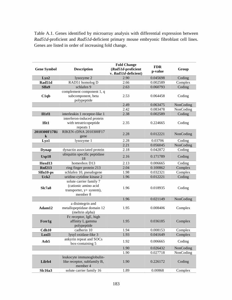
183
Table A.1. Genes identified by microarray analysis with differential expression between
Rad51d-proficient and Rad51d-deficient primary mouse embryonic fibroblast cell lines.
Genes are listed in order of increasing fold change.
Gene Symbol Description
Fold Change
(Rad51d-proficient
v. Rad51d-deficient)
FDR
p-value Group
Lyz2 lysozyme 2 2.90 0.045698 Coding
Rad51d RAD51 homolog D 2.66 0.002589 Complex
Slfn9 schlafen 9 2.63 0.060793 Coding
C1qb
complement component 1, q
subcomponent, beta
polypeptide
2.53 0.064458 Coding
2.49 0.063475 NonCoding
2.42 0.083478 NonCoding
Il1rl1 interleukin 1 receptor-like 1 2.38 0.002589 Coding
Ifit1
interferon-induced protein
with tetratricopeptide
repeats 1
2.35 0.224665 Coding
2010300F17Ri
k
RIKEN cDNA 2010300F17
gene 2.28 0.012221 NonCoding
Lyz1 lysozyme 1 2.28 0.03706 Coding
2.21 0.056045 NonCoding
Dynap dynactin associated protein 2.18 0.042872 Coding
Usp18 ubiquitin specific peptidase
18 2.16 0.171789 Coding
Hoxd13 homeobox D13 2.13 0.006665 Coding
Rnf213 ring finger protein 213 2.04 0.100862 Complex
Slfn10-ps schlafen 10, pseudogene 1.98 0.032321 Complex
Uck2 uridine-cytidine kinase 2 1.96 0.012221 Coding
Slc7a8
solute carrier family 7
(cationic amino acid
transporter, y+ system),
member 8
1.96 0.018935 Coding
1.96 0.021149 NonCoding
Adam12
a disintegrin and
metallopeptidase domain 12
(meltrin alpha)
1.95 0.008406 Complex
Fcer1g
Fc receptor, IgE, high
affinity I, gamma
polypeptide
1.95 0.036185 Complex
Cdh10 cadherin 10 1.94 0.008153 Complex
Loxl3 lysyl oxidase-like 3 1.93 0.041649 Complex
Asb5 ankyrin repeat and SOCs
box-containing 5 1.92 0.006665 Coding
1.90 0.026432 NonCoding
1.90 0.027718 NonCoding
Lilrb4
leukocyte immunoglobulin-
like receptor, subfamily B,
member 4
1.90 0.226172 Coding
Slc16a3 solute carrier family 16 1.89 0.00868 Complex
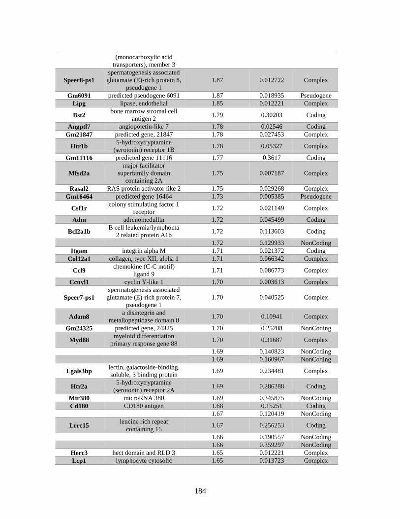
184
(monocarboxylic acid
transporters), member 3
Speer8-ps1
spermatogenesis associated
glutamate (E)-rich protein 8,
pseudogene 1
1.87 0.012722 Complex
Gm6091 predicted pseudogene 6091 1.87 0.018935 Pseudogene
Lipg lipase, endothelial 1.85 0.012221 Complex
Bst2 bone marrow stromal cell
antigen 2 1.79 0.30203 Coding
Angptl7 angiopoietin-like 7 1.78 0.02546 Coding
Gm21847 predicted gene, 21847 1.78 0.027453 Complex
Htr1b 5-hydroxytryptamine
(serotonin) receptor 1B 1.78 0.05327 Complex
Gm11116 predicted gene 11116 1.77 0.3617 Coding
Mfsd2a
major facilitator
superfamily domain
containing 2A
1.75 0.007187 Complex
Rasal2 RAS protein activator like 2 1.75 0.029268 Complex
Gm16464 predicted gene 16464 1.73 0.005385 Pseudogene
Csf1r colony stimulating factor 1
receptor 1.72 0.021149 Complex
Adm adrenomedullin 1.72 0.045499 Coding
Bcl2a1b B cell leukemia/lymphoma
2 related protein A1b 1.72 0.113603 Coding
1.72 0.129933 NonCoding
Itgam integrin alpha M 1.71 0.021372 Coding
Col12a1 collagen, type XII, alpha 1 1.71 0.066342 Complex
Ccl9 chemokine (C-C motif)
ligand 9 1.71 0.086773 Complex
Ccnyl1 cyclin Y-like 1 1.70 0.003613 Complex
Speer7-ps1
spermatogenesis associated
glutamate (E)-rich protein 7,
pseudogene 1
1.70 0.040525 Complex
Adam8 a disintegrin and
metallopeptidase domain 8 1.70 0.10941 Complex
Gm24325 predicted gene, 24325 1.70 0.25208 NonCoding
Myd88 myeloid differentiation
primary response gene 88 1.70 0.31687 Complex
1.69 0.140823 NonCoding
1.69 0.160967 NonCoding
Lgals3bp lectin, galactoside-binding,
soluble, 3 binding protein 1.69 0.234481 Complex
Htr2a 5-hydroxytryptamine
(serotonin) receptor 2A 1.69 0.286288 Coding
Mir380 microRNA 380 1.69 0.345875 NonCoding
Cd180 CD180 antigen 1.68 0.15251 Coding
1.67 0.120419 NonCoding
Lrrc15 leucine rich repeat
containing 15 1.67 0.256253 Coding
1.66 0.190557 NonCoding
1.66 0.359297 NonCoding
Herc3 hect domain and RLD 3 1.65 0.012221 Complex
Lcp1 lymphocyte cytosolic 1.65 0.013723 Complex
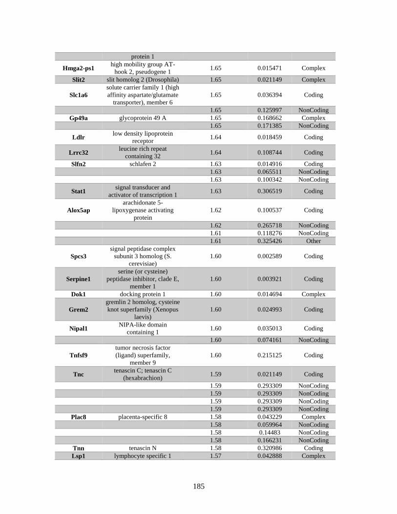
185
protein 1
Hmga2-ps1 high mobility group AT-
hook 2, pseudogene 1 1.65 0.015471 Complex
Slit2 slit homolog 2 (Drosophila) 1.65 0.021149 Complex
Slc1a6
solute carrier family 1 (high
affinity aspartate/glutamate
transporter), member 6
1.65 0.036394 Coding
1.65 0.125997 NonCoding
Gp49a glycoprotein 49 A 1.65 0.168662 Complex
1.65 0.171385 NonCoding
Ldlr low density lipoprotein
receptor 1.64 0.018459 Coding
Lrrc32 leucine rich repeat
containing 32 1.64 0.108744 Coding
Slfn2 schlafen 2 1.63 0.014916 Coding
1.63 0.065511 NonCoding
1.63 0.100342 NonCoding
Stat1 signal transducer and
activator of transcription 1 1.63 0.306519 Coding
Alox5ap
arachidonate 5-
lipoxygenase activating
protein
1.62 0.100537 Coding
1.62 0.265718 NonCoding
1.61 0.118276 NonCoding
1.61 0.325426 Other
Spcs3
signal peptidase complex
subunit 3 homolog (S.
cerevisiae)
1.60 0.002589 Coding
Serpine1
serine (or cysteine)
peptidase inhibitor, clade E,
member 1
1.60 0.003921 Coding
Dok1 docking protein 1 1.60 0.014694 Complex
Grem2
gremlin 2 homolog, cysteine
knot superfamily (Xenopus
laevis)
1.60 0.024993 Coding
Nipal1 NIPA-like domain
containing 1 1.60 0.035013 Coding
1.60 0.074161 NonCoding
Tnfsf9
tumor necrosis factor
(ligand) superfamily,
member 9
1.60 0.215125 Coding
Tnc tenascin C; tenascin C
(hexabrachion) 1.59 0.021149 Coding
1.59 0.293309 NonCoding
1.59 0.293309 NonCoding
1.59 0.293309 NonCoding
1.59 0.293309 NonCoding
Plac8 placenta-specific 8 1.58 0.043229 Complex
1.58 0.059964 NonCoding
1.58 0.14483 NonCoding
1.58 0.166231 NonCoding
Tnn tenascin N 1.58 0.320986 Coding
Lsp1 lymphocyte specific 1 1.57 0.042888 Complex
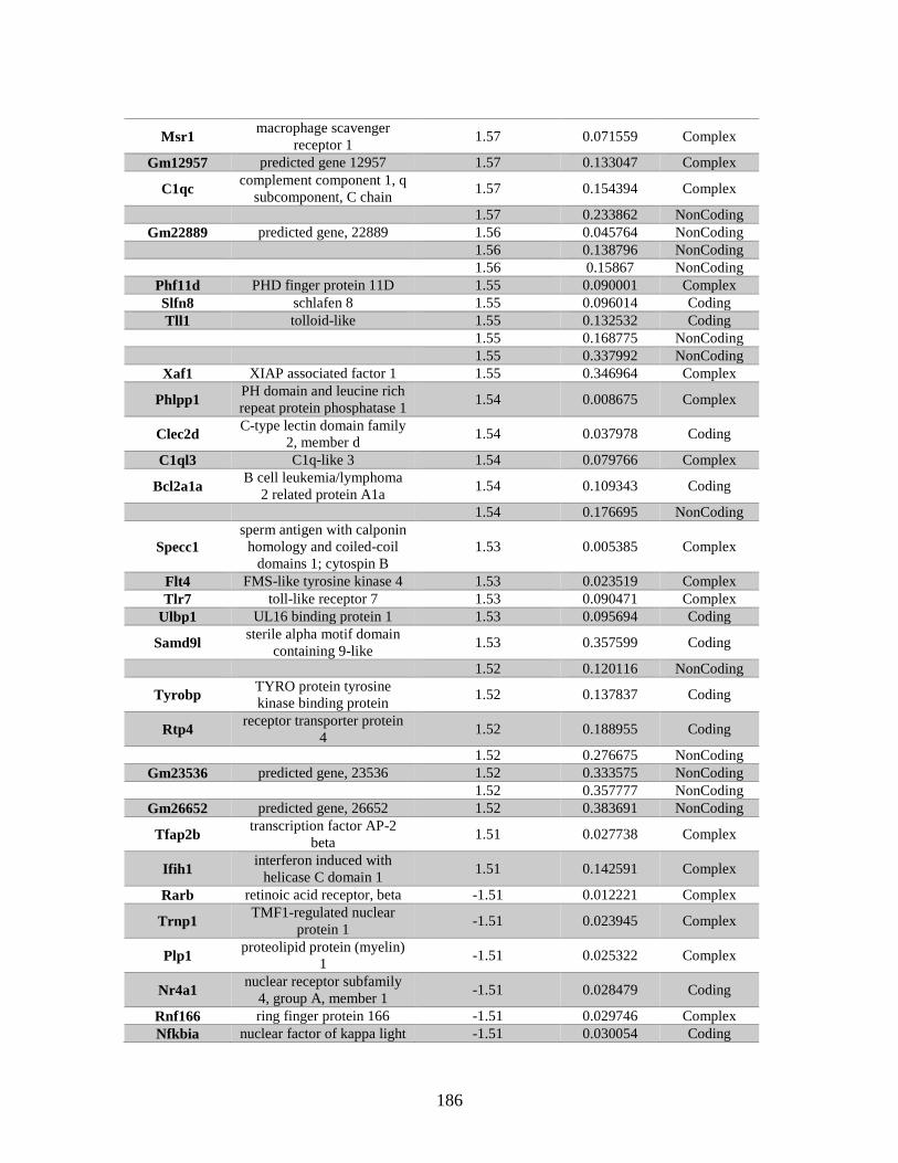
186
Msr1 macrophage scavenger
receptor 1 1.57 0.071559 Complex
Gm12957 predicted gene 12957 1.57 0.133047 Complex
C1qc complement component 1, q
subcomponent, C chain 1.57 0.154394 Complex
1.57 0.233862 NonCoding
Gm22889 predicted gene, 22889 1.56 0.045764 NonCoding
1.56 0.138796 NonCoding
1.56 0.15867 NonCoding
Phf11d PHD finger protein 11D 1.55 0.090001 Complex
Slfn8 schlafen 8 1.55 0.096014 Coding
Tll1 tolloid-like 1.55 0.132532 Coding
1.55 0.168775 NonCoding
1.55 0.337992 NonCoding
Xaf1 XIAP associated factor 1 1.55 0.346964 Complex
Phlpp1 PH domain and leucine rich
repeat protein phosphatase 1 1.54 0.008675 Complex
Clec2d C-type lectin domain family
2, member d 1.54 0.037978 Coding
C1ql3 C1q-like 3 1.54 0.079766 Complex
Bcl2a1a B cell leukemia/lymphoma
2 related protein A1a 1.54 0.109343 Coding
1.54 0.176695 NonCoding
Specc1
sperm antigen with calponin
homology and coiled-coil
domains 1; cytospin B
1.53 0.005385 Complex
Flt4 FMS-like tyrosine kinase 4 1.53 0.023519 Complex
Tlr7 toll-like receptor 7 1.53 0.090471 Complex
Ulbp1 UL16 binding protein 1 1.53 0.095694 Coding
Samd9l sterile alpha motif domain
containing 9-like 1.53 0.357599 Coding
1.52 0.120116 NonCoding
Tyrobp TYRO protein tyrosine
kinase binding protein 1.52 0.137837 Coding
Rtp4 receptor transporter protein
4 1.52 0.188955 Coding
1.52 0.276675 NonCoding
Gm23536 predicted gene, 23536 1.52 0.333575 NonCoding
1.52 0.357777 NonCoding
Gm26652 predicted gene, 26652 1.52 0.383691 NonCoding
Tfap2b transcription factor AP-2
beta 1.51 0.027738 Complex
Ifih1 interferon induced with
helicase C domain 1 1.51 0.142591 Complex
Rarb retinoic acid receptor, beta -1.51 0.012221 Complex
Trnp1 TMF1-regulated nuclear
protein 1 -1.51 0.023945 Complex
Plp1 proteolipid protein (myelin)
1 -1.51 0.025322 Complex
Nr4a1 nuclear receptor subfamily
4, group A, member 1 -1.51 0.028479 Coding
Rnf166 ring finger protein 166 -1.51 0.029746 Complex
Nfkbia nuclear factor of kappa light -1.51 0.030054 Coding
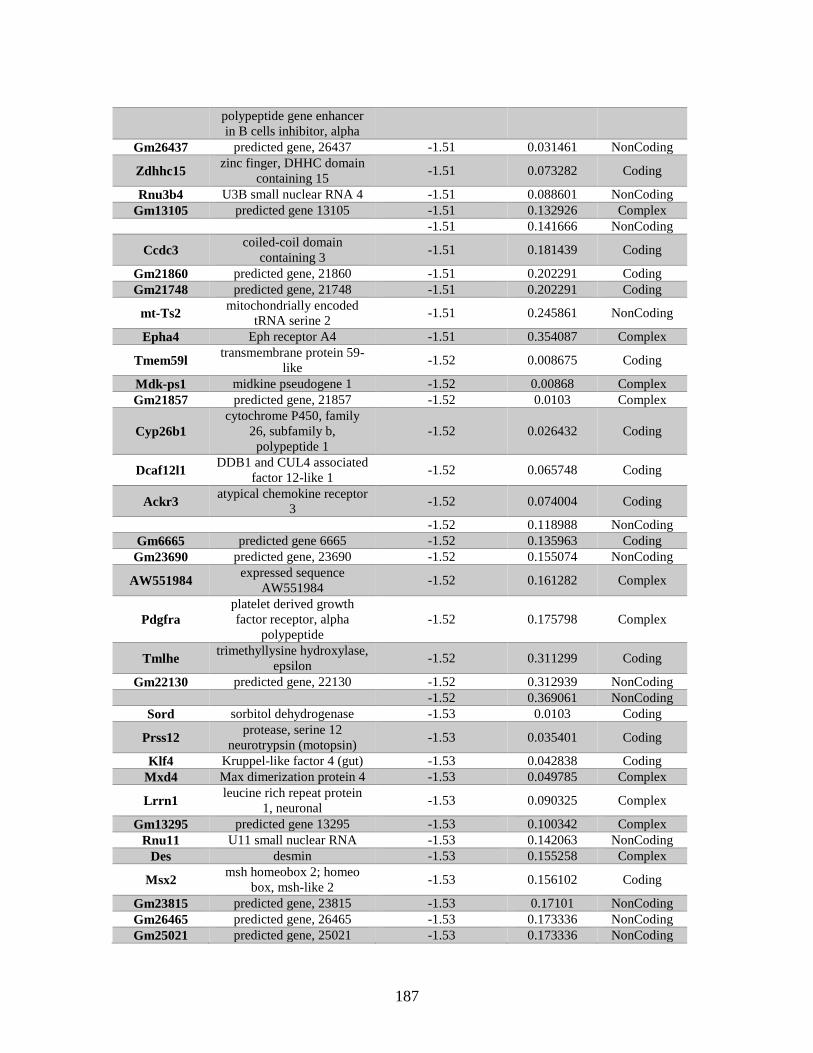
187
polypeptide gene enhancer
in B cells inhibitor, alpha
Gm26437 predicted gene, 26437 -1.51 0.031461 NonCoding
Zdhhc15 zinc finger, DHHC domain
containing 15 -1.51 0.073282 Coding
Rnu3b4 U3B small nuclear RNA 4 -1.51 0.088601 NonCoding
Gm13105 predicted gene 13105 -1.51 0.132926 Complex
-1.51 0.141666 NonCoding
Ccdc3 coiled-coil domain
containing 3 -1.51 0.181439 Coding
Gm21860 predicted gene, 21860 -1.51 0.202291 Coding
Gm21748 predicted gene, 21748 -1.51 0.202291 Coding
mt-Ts2 mitochondrially encoded
tRNA serine 2 -1.51 0.245861 NonCoding
Epha4 Eph receptor A4 -1.51 0.354087 Complex
Tmem59l transmembrane protein 59-
like -1.52 0.008675 Coding
Mdk-ps1 midkine pseudogene 1 -1.52 0.00868 Complex
Gm21857 predicted gene, 21857 -1.52 0.0103 Complex
Cyp26b1
cytochrome P450, family
26, subfamily b,
polypeptide 1
-1.52 0.026432 Coding
Dcaf12l1 DDB1 and CUL4 associated
factor 12-like 1 -1.52 0.065748 Coding
Ackr3 atypical chemokine receptor
3 -1.52 0.074004 Coding
-1.52 0.118988 NonCoding
Gm6665 predicted gene 6665 -1.52 0.135963 Coding
Gm23690 predicted gene, 23690 -1.52 0.155074 NonCoding
AW551984 expressed sequence
AW551984 -1.52 0.161282 Complex
Pdgfra
platelet derived growth
factor receptor, alpha
polypeptide
-1.52 0.175798 Complex
Tmlhe trimethyllysine hydroxylase,
epsilon -1.52 0.311299 Coding
Gm22130 predicted gene, 22130 -1.52 0.312939 NonCoding
-1.52 0.369061 NonCoding
Sord sorbitol dehydrogenase -1.53 0.0103 Coding
Prss12 protease, serine 12
neurotrypsin (motopsin) -1.53 0.035401 Coding
Klf4 Kruppel-like factor 4 (gut) -1.53 0.042838 Coding
Mxd4 Max dimerization protein 4 -1.53 0.049785 Complex
Lrrn1 leucine rich repeat protein
1, neuronal -1.53 0.090325 Complex
Gm13295 predicted gene 13295 -1.53 0.100342 Complex
Rnu11 U11 small nuclear RNA -1.53 0.142063 NonCoding
Des desmin -1.53 0.155258 Complex
Msx2 msh homeobox 2; homeo
box, msh-like 2 -1.53 0.156102 Coding
Gm23815 predicted gene, 23815 -1.53 0.17101 NonCoding
Gm26465 predicted gene, 26465 -1.53 0.173336 NonCoding
Gm25021 predicted gene, 25021 -1.53 0.173336 NonCoding
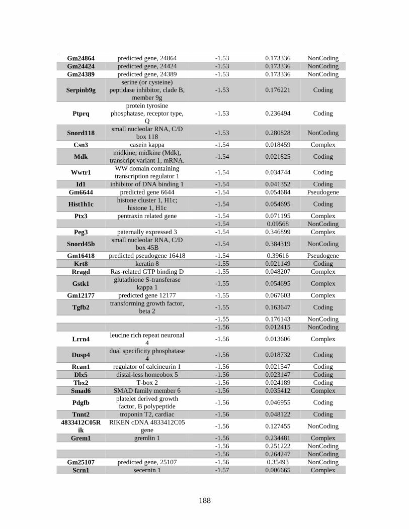
188
Gm24864 predicted gene, 24864 -1.53 0.173336 NonCoding
Gm24424 predicted gene, 24424 -1.53 0.173336 NonCoding
Gm24389 predicted gene, 24389 -1.53 0.173336 NonCoding
Serpinb9g
serine (or cysteine)
peptidase inhibitor, clade B,
member 9g
-1.53 0.176221 Coding
Ptprq
protein tyrosine
phosphatase, receptor type,
Q
-1.53 0.236494 Coding
Snord118 small nucleolar RNA, C/D
box 118 -1.53 0.280828 NonCoding
Csn3 casein kappa -1.54 0.018459 Complex
Mdk midkine; midkine (Mdk),
transcript variant 1, mRNA. -1.54 0.021825 Coding
Wwtr1 WW domain containing
transcription regulator 1 -1.54 0.034744 Coding
Id1 inhibitor of DNA binding 1 -1.54 0.041352 Coding
Gm6644 predicted gene 6644 -1.54 0.054684 Pseudogene
Hist1h1c histone cluster 1, H1c;
histone 1, H1c -1.54 0.054695 Coding
Ptx3 pentraxin related gene -1.54 0.071195 Complex
-1.54 0.09568 NonCoding
Peg3 paternally expressed 3 -1.54 0.346899 Complex
Snord45b small nucleolar RNA, C/D
box 45B -1.54 0.384319 NonCoding
Gm16418 predicted pseudogene 16418 -1.54 0.39616 Pseudogene
Krt8 keratin 8 -1.55 0.021149 Coding
Rragd Ras-related GTP binding D -1.55 0.048207 Complex
Gstk1 glutathione S-transferase
kappa 1 -1.55 0.054695 Complex
Gm12177 predicted gene 12177 -1.55 0.067603 Complex
Tgfb2 transforming growth factor,
beta 2 -1.55 0.163647 Coding
-1.55 0.176143 NonCoding
-1.56 0.012415 NonCoding
Lrrn4 leucine rich repeat neuronal
4 -1.56 0.013606 Complex
Dusp4 dual specificity phosphatase
4 -1.56 0.018732 Coding
Rcan1 regulator of calcineurin 1 -1.56 0.021547 Coding
Dlx5 distal-less homeobox 5 -1.56 0.023147 Coding
Tbx2 T-box 2 -1.56 0.024189 Coding
Smad6 SMAD family member 6 -1.56 0.035412 Complex
Pdgfb platelet derived growth
factor, B polypeptide -1.56 0.046955 Coding
Tnnt2 troponin T2, cardiac -1.56 0.048122 Coding
4833412C05R
ik
RIKEN cDNA 4833412C05
gene -1.56 0.127455 NonCoding
Grem1 gremlin 1 -1.56 0.234481 Complex
-1.56 0.251222 NonCoding
-1.56 0.264247 NonCoding
Gm25107 predicted gene, 25107 -1.56 0.35493 NonCoding
Scrn1 secernin 1 -1.57 0.006665 Complex
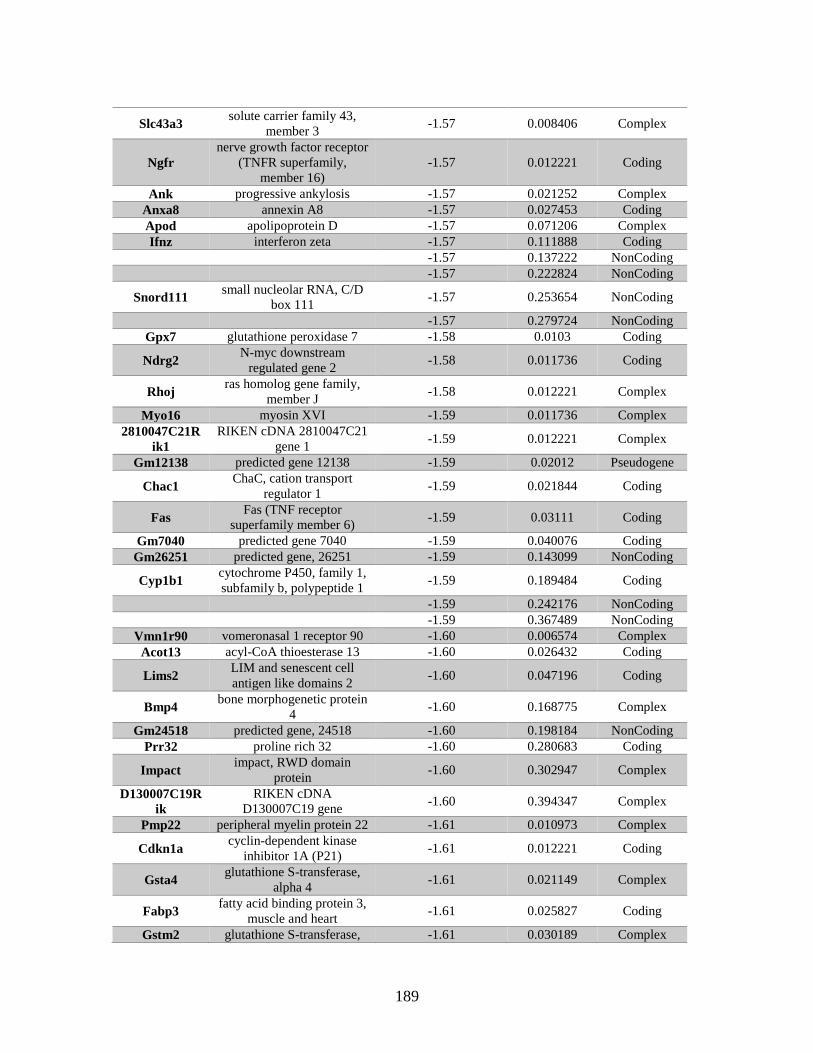
189
Slc43a3 solute carrier family 43,
member 3 -1.57 0.008406 Complex
Ngfr
nerve growth factor receptor
(TNFR superfamily,
member 16)
-1.57 0.012221 Coding
Ank progressive ankylosis -1.57 0.021252 Complex
Anxa8 annexin A8 -1.57 0.027453 Coding
Apod apolipoprotein D -1.57 0.071206 Complex
Ifnz interferon zeta -1.57 0.111888 Coding
-1.57 0.137222 NonCoding
-1.57 0.222824 NonCoding
Snord111 small nucleolar RNA, C/D
box 111 -1.57 0.253654 NonCoding
-1.57 0.279724 NonCoding
Gpx7 glutathione peroxidase 7 -1.58 0.0103 Coding
Ndrg2 N-myc downstream
regulated gene 2 -1.58 0.011736 Coding
Rhoj ras homolog gene family,
member J -1.58 0.012221 Complex
Myo16 myosin XVI -1.59 0.011736 Complex
2810047C21R
ik1
RIKEN cDNA 2810047C21
gene 1 -1.59 0.012221 Complex
Gm12138 predicted gene 12138 -1.59 0.02012 Pseudogene
Chac1 ChaC, cation transport
regulator 1 -1.59 0.021844 Coding
Fas Fas (TNF receptor
superfamily member 6) -1.59 0.03111 Coding
Gm7040 predicted gene 7040 -1.59 0.040076 Coding
Gm26251 predicted gene, 26251 -1.59 0.143099 NonCoding
Cyp1b1 cytochrome P450, family 1,
subfamily b, polypeptide 1 -1.59 0.189484 Coding
-1.59 0.242176 NonCoding
-1.59 0.367489 NonCoding
Vmn1r90 vomeronasal 1 receptor 90 -1.60 0.006574 Complex
Acot13 acyl-CoA thioesterase 13 -1.60 0.026432 Coding
Lims2 LIM and senescent cell
antigen like domains 2 -1.60 0.047196 Coding
Bmp4 bone morphogenetic protein
4 -1.60 0.168775 Complex
Gm24518 predicted gene, 24518 -1.60 0.198184 NonCoding
Prr32 proline rich 32 -1.60 0.280683 Coding
Impact impact, RWD domain
protein -1.60 0.302947 Complex
D130007C19R
ik
RIKEN cDNA
D130007C19 gene -1.60 0.394347 Complex
Pmp22 peripheral myelin protein 22 -1.61 0.010973 Complex
Cdkn1a cyclin-dependent kinase
inhibitor 1A (P21) -1.61 0.012221 Coding
Gsta4 glutathione S-transferase,
alpha 4 -1.61 0.021149 Complex
Fabp3 fatty acid binding protein 3,
muscle and heart -1.61 0.025827 Coding
Gstm2 glutathione S-transferase, -1.61 0.030189 Complex
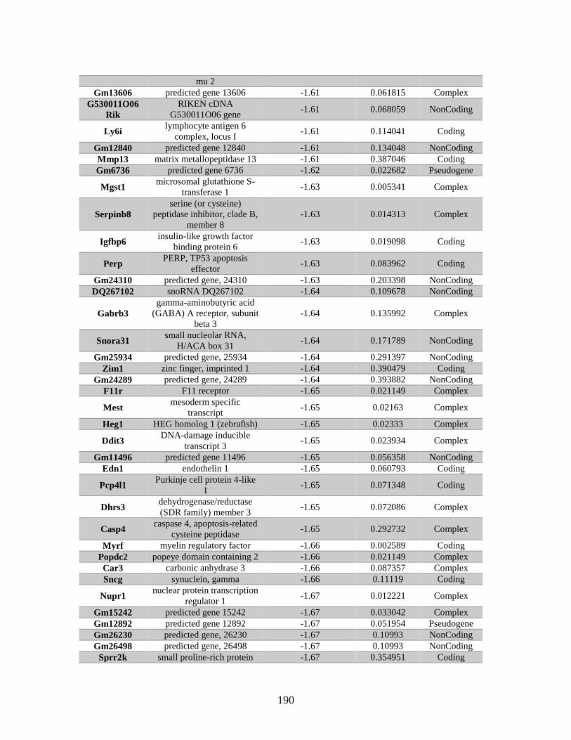
190
mu 2
Gm13606 predicted gene 13606 -1.61 0.061815 Complex
G530011O06
Rik
RIKEN cDNA
G530011O06 gene -1.61 0.068059 NonCoding
Ly6i lymphocyte antigen 6
complex, locus I -1.61 0.114041 Coding
Gm12840 predicted gene 12840 -1.61 0.134048 NonCoding
Mmp13 matrix metallopeptidase 13 -1.61 0.387046 Coding
Gm6736 predicted gene 6736 -1.62 0.022682 Pseudogene
Mgst1 microsomal glutathione S-
transferase 1 -1.63 0.005341 Complex
Serpinb8
serine (or cysteine)
peptidase inhibitor, clade B,
member 8
-1.63 0.014313 Complex
Igfbp6 insulin-like growth factor
binding protein 6 -1.63 0.019098 Coding
Perp PERP, TP53 apoptosis
effector -1.63 0.083962 Coding
Gm24310 predicted gene, 24310 -1.63 0.203398 NonCoding
DQ267102 snoRNA DQ267102 -1.64 0.109678 NonCoding
Gabrb3
gamma-aminobutyric acid
(GABA) A receptor, subunit
beta 3
-1.64 0.135992 Complex
Snora31 small nucleolar RNA,
H/ACA box 31 -1.64 0.171789 NonCoding
Gm25934 predicted gene, 25934 -1.64 0.291397 NonCoding
Zim1 zinc finger, imprinted 1 -1.64 0.390479 Coding
Gm24289 predicted gene, 24289 -1.64 0.393882 NonCoding
F11r F11 receptor -1.65 0.021149 Complex
Mest mesoderm specific
transcript -1.65 0.02163 Complex
Heg1 HEG homolog 1 (zebrafish) -1.65 0.02333 Complex
Ddit3 DNA-damage inducible
transcript 3 -1.65 0.023934 Complex
Gm11496 predicted gene 11496 -1.65 0.056358 NonCoding
Edn1 endothelin 1 -1.65 0.060793 Coding
Pcp4l1 Purkinje cell protein 4-like
1 -1.65 0.071348 Coding
Dhrs3 dehydrogenase/reductase
(SDR family) member 3 -1.65 0.072086 Complex
Casp4 caspase 4, apoptosis-related
cysteine peptidase -1.65 0.292732 Complex
Myrf myelin regulatory factor -1.66 0.002589 Coding
Popdc2 popeye domain containing 2 -1.66 0.021149 Complex
Car3 carbonic anhydrase 3 -1.66 0.087357 Complex
Sncg synuclein, gamma -1.66 0.11119 Coding
Nupr1 nuclear protein transcription
regulator 1 -1.67 0.012221 Complex
Gm15242 predicted gene 15242 -1.67 0.033042 Complex
Gm12892 predicted gene 12892 -1.67 0.051954 Pseudogene
Gm26230 predicted gene, 26230 -1.67 0.10993 NonCoding
Gm26498 predicted gene, 26498 -1.67 0.10993 NonCoding
Sprr2k small proline-rich protein -1.67 0.354951 Coding
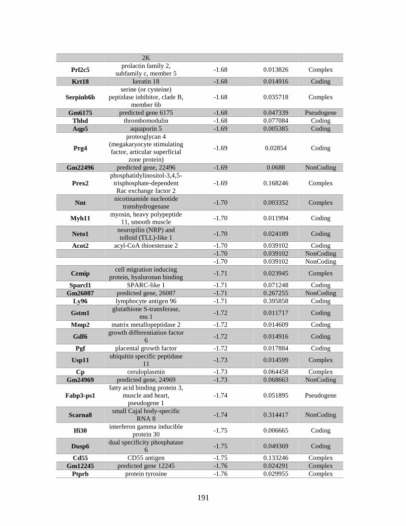
191
2K
Prl2c5 prolactin family 2,
subfamily c, member 5 -1.68 0.013826 Complex
Krt18 keratin 18 -1.68 0.014916 Coding
Serpinb6b
serine (or cysteine)
peptidase inhibitor, clade B,
member 6b
-1.68 0.035718 Complex
Gm6175 predicted gene 6175 -1.68 0.047339 Pseudogene
Thbd thrombomodulin -1.68 0.077084 Coding
Aqp5 aquaporin 5 -1.69 0.005385 Coding
Prg4
proteoglycan 4
(megakaryocyte stimulating
factor, articular superficial
zone protein)
-1.69 0.02854 Coding
Gm22496 predicted gene, 22496 -1.69 0.0688 NonCoding
Prex2
phosphatidylinositol-3,4,5-
trisphosphate-dependent
Rac exchange factor 2
-1.69 0.168246 Complex
Nnt nicotinamide nucleotide
transhydrogenase -1.70 0.003352 Complex
Myh11 myosin, heavy polypeptide
11, smooth muscle -1.70 0.011994 Coding
Neto1 neuropilin (NRP) and
tolloid (TLL)-like 1 -1.70 0.024189 Coding
Acot2 acyl-CoA thioesterase 2 -1.70 0.039102 Coding
-1.70 0.039102 NonCoding
-1.70 0.039102 NonCoding
Cemip cell migration inducing
protein, hyaluronan binding -1.71 0.023945 Complex
Sparcl1 SPARC-like 1 -1.71 0.071248 Coding
Gm26087 predicted gene, 26087 -1.71 0.267255 NonCoding
Ly96 lymphocyte antigen 96 -1.71 0.395858 Coding
Gstm1 glutathione S-transferase,
mu 1 -1.72 0.011717 Coding
Mmp2 matrix metallopeptidase 2 -1.72 0.014609 Coding
Gdf6 growth differentiation factor
6 -1.72 0.014916 Coding
Pgf placental growth factor -1.72 0.017884 Coding
Usp11 ubiquitin specific peptidase
11 -1.73 0.014599 Complex
Cp ceruloplasmin -1.73 0.064458 Complex
Gm24969 predicted gene, 24969 -1.73 0.068663 NonCoding
Fabp3-ps1
fatty acid binding protein 3,
muscle and heart,
pseudogene 1
-1.74 0.051895 Pseudogene
Scarna8 small Cajal body-specific
RNA 8 -1.74 0.314417 NonCoding
Ifi30 interferon gamma inducible
protein 30 -1.75 0.006665 Coding
Dusp6 dual specificity phosphatase
6 -1.75 0.049369 Coding
Cd55 CD55 antigen -1.75 0.133246 Complex
Gm12245 predicted gene 12245 -1.76 0.024291 Complex
Ptprb protein tyrosine -1.76 0.029955 Complex
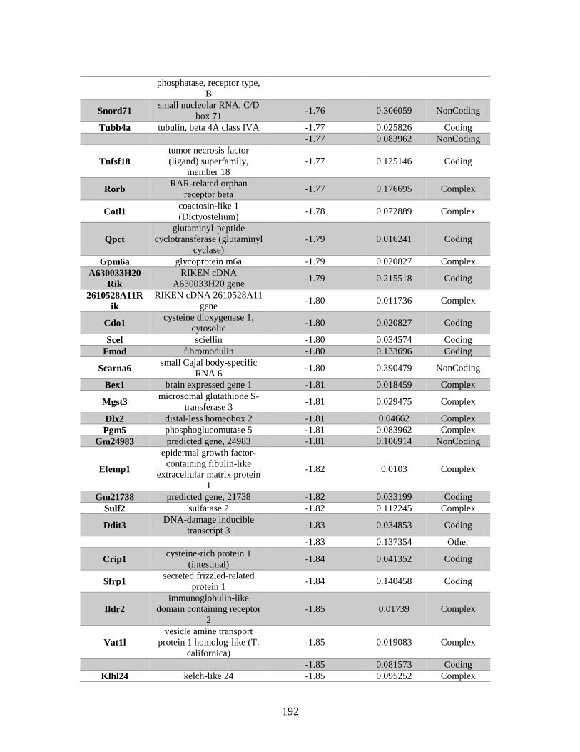
192
phosphatase, receptor type,
B
Snord71 small nucleolar RNA, C/D
box 71 -1.76 0.306059 NonCoding
Tubb4a tubulin, beta 4A class IVA -1.77 0.025826 Coding
-1.77 0.083962 NonCoding
Tnfsf18
tumor necrosis factor
(ligand) superfamily,
member 18
-1.77 0.125146 Coding
Rorb RAR-related orphan
receptor beta -1.77 0.176695 Complex
Cotl1 coactosin-like 1
(Dictyostelium) -1.78 0.072889 Complex
Qpct
glutaminyl-peptide
cyclotransferase (glutaminyl
cyclase)
-1.79 0.016241 Coding
Gpm6a glycoprotein m6a -1.79 0.020827 Complex
A630033H20
Rik
RIKEN cDNA
A630033H20 gene -1.79 0.215518 Coding
2610528A11R
ik
RIKEN cDNA 2610528A11
gene -1.80 0.011736 Complex
Cdo1 cysteine dioxygenase 1,
cytosolic -1.80 0.020827 Coding
Scel sciellin -1.80 0.034574 Coding
Fmod fibromodulin -1.80 0.133696 Coding
Scarna6 small Cajal body-specific
RNA 6 -1.80 0.390479 NonCoding
Bex1 brain expressed gene 1 -1.81 0.018459 Complex
Mgst3 microsomal glutathione S-
transferase 3 -1.81 0.029475 Complex
Dlx2 distal-less homeobox 2 -1.81 0.04662 Complex
Pgm5 phosphoglucomutase 5 -1.81 0.083962 Complex
Gm24983 predicted gene, 24983 -1.81 0.106914 NonCoding
Efemp1
epidermal growth factor-
containing fibulin-like
extracellular matrix protein
1
-1.82 0.0103 Complex
Gm21738 predicted gene, 21738 -1.82 0.033199 Coding
Sulf2 sulfatase 2 -1.82 0.112245 Complex
Ddit3 DNA-damage inducible
transcript 3 -1.83 0.034853 Coding
-1.83 0.137354 Other
Crip1 cysteine-rich protein 1
(intestinal) -1.84 0.041352 Coding
Sfrp1 secreted frizzled-related
protein 1 -1.84 0.140458 Coding
Ildr2
immunoglobulin-like
domain containing receptor
2
-1.85 0.01739 Complex
Vat1l
vesicle amine transport
protein 1 homolog-like (T.
californica)
-1.85 0.019083 Complex
-1.85 0.081573 Coding
Klhl24 kelch-like 24 -1.85 0.095252 Complex
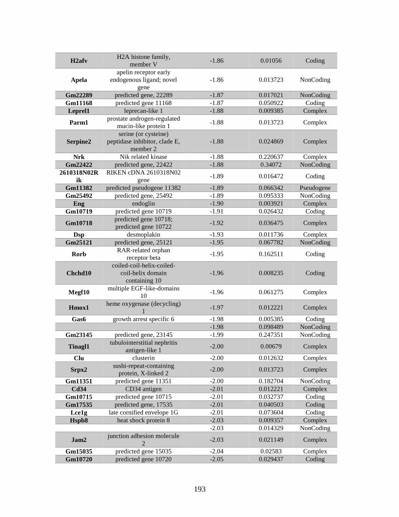
193
H2afv H2A histone family,
member V -1.86 0.01056 Coding
Apela
apelin receptor early
endogenous ligand; novel
gene
-1.86 0.013723 NonCoding
Gm22289 predicted gene, 22289 -1.87 0.017021 NonCoding
Gm11168 predicted gene 11168 -1.87 0.050922 Coding
Leprel1 leprecan-like 1 -1.88 0.009385 Complex
Parm1 prostate androgen-regulated
mucin-like protein 1 -1.88 0.013723 Complex
Serpine2
serine (or cysteine)
peptidase inhibitor, clade E,
member 2
-1.88 0.024869 Complex
Nrk Nik related kinase -1.88 0.220637 Complex
Gm22422 predicted gene, 22422 -1.88 0.34072 NonCoding
2610318N02R
ik
RIKEN cDNA 2610318N02
gene -1.89 0.016472 Coding
Gm11382 predicted pseudogene 11382 -1.89 0.066342 Pseudogene
Gm25492 predicted gene, 25492 -1.89 0.095333 NonCoding
Eng endoglin -1.90 0.003921 Complex
Gm10719 predicted gene 10719 -1.91 0.026432 Coding
Gm10718 predicted gene 10718;
predicted gene 10722 -1.92 0.036475 Complex
Dsp desmoplakin -1.93 0.011736 Complex
Gm25121 predicted gene, 25121 -1.95 0.067782 NonCoding
Rorb RAR-related orphan
receptor beta -1.95 0.162511 Coding
Chchd10
coiled-coil-helix-coiled-
coil-helix domain
containing 10
-1.96 0.008235 Coding
Megf10 multiple EGF-like-domains
10 -1.96 0.061275 Complex
Hmox1 heme oxygenase (decycling)
1 -1.97 0.012221 Complex
Gas6 growth arrest specific 6 -1.98 0.005385 Coding
-1.98 0.098489 NonCoding
Gm23145 predicted gene, 23145 -1.99 0.247351 NonCoding
Tinagl1 tubulointerstitial nephritis
antigen-like 1 -2.00 0.00679 Complex
Clu clusterin -2.00 0.012632 Complex
Srpx2 sushi-repeat-containing
protein, X-linked 2 -2.00 0.013723 Complex
Gm11351 predicted gene 11351 -2.00 0.182704 NonCoding
Cd34 CD34 antigen -2.01 0.012221 Complex
Gm10715 predicted gene 10715 -2.01 0.032737 Coding
Gm17535 predicted gene, 17535 -2.01 0.040503 Coding
Lce1g late cornified envelope 1G -2.01 0.073604 Coding
Hspb8 heat shock protein 8 -2.03 0.009357 Complex
-2.03 0.014329 NonCoding
Jam2 junction adhesion molecule
2 -2.03 0.021149 Complex
Gm15035 predicted gene 15035 -2.04 0.02583 Complex
Gm10720 predicted gene 10720 -2.05 0.029437 Coding
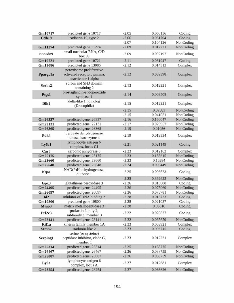
194
Gm10717 predicted gene 10717 -2.05 0.060156 Coding
Cdh19 cadherin 19, type 2 -2.06 0.061704 Coding
-2.07 0.104126 NonCoding
Gm11274 predicted gene 11274 -2.09 0.012221 NonCoding
Snord89 small nucleolar RNA, C/D
box 89 -2.09 0.092197 NonCoding
Gm10721 predicted gene 10721 -2.11 0.031947 Coding
Gm13086 predicted gene 13086 -2.12 0.014313 Complex
Ppargc1a
peroxisome proliferative
activated receptor, gamma,
coactivator 1 alpha
-2.12 0.039398 Complex
Sorbs2 sorbin and SH3 domain
containing 2 -2.13 0.012221 Complex
Ptgs1 prostaglandin-endoperoxide
synthase 1 -2.14 0.003508 Complex
Dlk1 delta-like 1 homolog
(Drosophila) -2.15 0.012221 Complex
-2.15 0.02583 NonCoding
-2.15 0.041051 NonCoding
Gm26337 predicted gene, 26337 -2.16 0.160047 NonCoding
Gm22131 predicted gene, 22131 -2.17 0.029957 NonCoding
Gm26365 predicted gene, 26365 -2.19 0.01056 NonCoding
Pdk4 pyruvate dehydrogenase
kinase, isoenzyme 4 -2.19 0.019534 Complex
Ly6c1 lymphocyte antigen 6
complex, locus C1 -2.21 0.021149 Coding
Car8 carbonic anhydrase 8 -2.23 0.012163 Complex
Gm25175 predicted gene, 25175 -2.23 0.155615 NonCoding
Gm23660 predicted gene, 23660 -2.23 0.16284 NonCoding
Gm25648 predicted gene, 25648 -2.24 0.090449 NonCoding
Nqo1 NAD(P)H dehydrogenase,
quinone 1 -2.25 0.006623 Coding
-2.25 0.362625 NonCoding
Gpx3 glutathione peroxidase 3 -2.26 0.013808 Coding
Gm24495 predicted gene, 24495 -2.26 0.075069 NonCoding
Gm26097 predicted gene, 26097 -2.26 0.075781 NonCoding
Id2 inhibitor of DNA binding 2 -2.28 0.013723 Coding
Gm10800 predicted gene 10800 -2.28 0.021037 Coding
Mmp3 matrix metallopeptidase 3 -2.28 0.03816 Coding
Prl2c3 prolactin family 2,
subfamily c, member 3 -2.32 0.020827 Coding
Gm23141 predicted gene, 23141 -2.32 0.035659 NonCoding
Kif1a kinesin family member 1A -2.33 0.003921 Complex
Stmn2 stathmin-like 2 -2.33 0.006715 Coding
Serping1
serine (or cysteine)
peptidase inhibitor, clade G,
member 1
-2.33 0.012221 Complex
Gm25314 predicted gene, 25314 -2.35 0.168775 NonCoding
Gm26467 predicted gene, 26467 -2.36 0.038759 NonCoding
Gm25087 predicted gene, 25087 -2.36 0.038759 NonCoding
Ly6a lymphocyte antigen 6
complex, locus A -2.37 0.012681 Complex
Gm23254 predicted gene, 23254 -2.37 0.066626 NonCoding
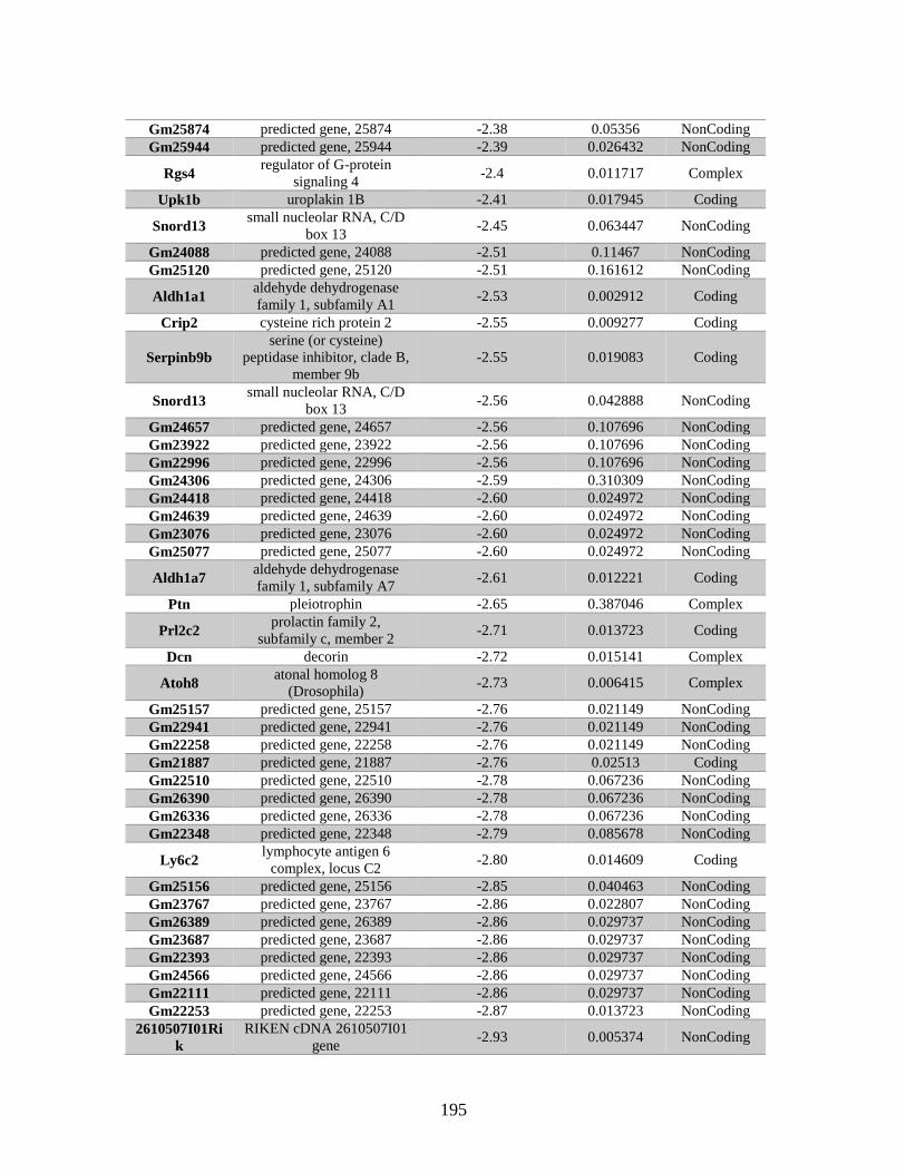
195
Gm25874 predicted gene, 25874 -2.38 0.05356 NonCoding
Gm25944 predicted gene, 25944 -2.39 0.026432 NonCoding
Rgs4 regulator of G-protein
signaling 4 -2.4 0.011717 Complex
Upk1b uroplakin 1B -2.41 0.017945 Coding
Snord13 small nucleolar RNA, C/D
box 13 -2.45 0.063447 NonCoding
Gm24088 predicted gene, 24088 -2.51 0.11467 NonCoding
Gm25120 predicted gene, 25120 -2.51 0.161612 NonCoding
Aldh1a1 aldehyde dehydrogenase
family 1, subfamily A1 -2.53 0.002912 Coding
Crip2 cysteine rich protein 2 -2.55 0.009277 Coding
Serpinb9b
serine (or cysteine)
peptidase inhibitor, clade B,
member 9b
-2.55 0.019083 Coding
Snord13 small nucleolar RNA, C/D
box 13 -2.56 0.042888 NonCoding
Gm24657 predicted gene, 24657 -2.56 0.107696 NonCoding
Gm23922 predicted gene, 23922 -2.56 0.107696 NonCoding
Gm22996 predicted gene, 22996 -2.56 0.107696 NonCoding
Gm24306 predicted gene, 24306 -2.59 0.310309 NonCoding
Gm24418 predicted gene, 24418 -2.60 0.024972 NonCoding
Gm24639 predicted gene, 24639 -2.60 0.024972 NonCoding
Gm23076 predicted gene, 23076 -2.60 0.024972 NonCoding
Gm25077 predicted gene, 25077 -2.60 0.024972 NonCoding
Aldh1a7 aldehyde dehydrogenase
family 1, subfamily A7 -2.61 0.012221 Coding
Ptn pleiotrophin -2.65 0.387046 Complex
Prl2c2 prolactin family 2,
subfamily c, member 2 -2.71 0.013723 Coding
Dcn decorin -2.72 0.015141 Complex
Atoh8 atonal homolog 8
(Drosophila) -2.73 0.006415 Complex
Gm25157 predicted gene, 25157 -2.76 0.021149 NonCoding
Gm22941 predicted gene, 22941 -2.76 0.021149 NonCoding
Gm22258 predicted gene, 22258 -2.76 0.021149 NonCoding
Gm21887 predicted gene, 21887 -2.76 0.02513 Coding
Gm22510 predicted gene, 22510 -2.78 0.067236 NonCoding
Gm26390 predicted gene, 26390 -2.78 0.067236 NonCoding
Gm26336 predicted gene, 26336 -2.78 0.067236 NonCoding
Gm22348 predicted gene, 22348 -2.79 0.085678 NonCoding
Ly6c2 lymphocyte antigen 6
complex, locus C2 -2.80 0.014609 Coding
Gm25156 predicted gene, 25156 -2.85 0.040463 NonCoding
Gm23767 predicted gene, 23767 -2.86 0.022807 NonCoding
Gm26389 predicted gene, 26389 -2.86 0.029737 NonCoding
Gm23687 predicted gene, 23687 -2.86 0.029737 NonCoding
Gm22393 predicted gene, 22393 -2.86 0.029737 NonCoding
Gm24566 predicted gene, 24566 -2.86 0.029737 NonCoding
Gm22111 predicted gene, 22111 -2.86 0.029737 NonCoding
Gm22253 predicted gene, 22253 -2.87 0.013723 NonCoding
2610507I01Ri
k
RIKEN cDNA 2610507I01
gene -2.93 0.005374 NonCoding
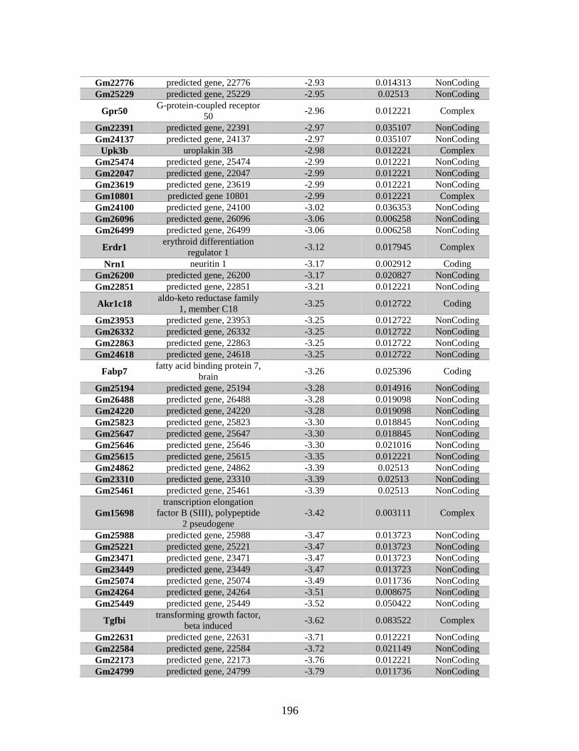
196
Gm22776 predicted gene, 22776 -2.93 0.014313 NonCoding
Gm25229 predicted gene, 25229 -2.95 0.02513 NonCoding
Gpr50 G-protein-coupled receptor
50 -2.96 0.012221 Complex
Gm22391 predicted gene, 22391 -2.97 0.035107 NonCoding
Gm24137 predicted gene, 24137 -2.97 0.035107 NonCoding
Upk3b uroplakin 3B -2.98 0.012221 Complex
Gm25474 predicted gene, 25474 -2.99 0.012221 NonCoding
Gm22047 predicted gene, 22047 -2.99 0.012221 NonCoding
Gm23619 predicted gene, 23619 -2.99 0.012221 NonCoding
Gm10801 predicted gene 10801 -2.99 0.012221 Complex
Gm24100 predicted gene, 24100 -3.02 0.036353 NonCoding
Gm26096 predicted gene, 26096 -3.06 0.006258 NonCoding
Gm26499 predicted gene, 26499 -3.06 0.006258 NonCoding
Erdr1 erythroid differentiation
regulator 1 -3.12 0.017945 Complex
Nrn1 neuritin 1 -3.17 0.002912 Coding
Gm26200 predicted gene, 26200 -3.17 0.020827 NonCoding
Gm22851 predicted gene, 22851 -3.21 0.012221 NonCoding
Akr1c18 aldo-keto reductase family
1, member C18 -3.25 0.012722 Coding
Gm23953 predicted gene, 23953 -3.25 0.012722 NonCoding
Gm26332 predicted gene, 26332 -3.25 0.012722 NonCoding
Gm22863 predicted gene, 22863 -3.25 0.012722 NonCoding
Gm24618 predicted gene, 24618 -3.25 0.012722 NonCoding
Fabp7 fatty acid binding protein 7,
brain -3.26 0.025396 Coding
Gm25194 predicted gene, 25194 -3.28 0.014916 NonCoding
Gm26488 predicted gene, 26488 -3.28 0.019098 NonCoding
Gm24220 predicted gene, 24220 -3.28 0.019098 NonCoding
Gm25823 predicted gene, 25823 -3.30 0.018845 NonCoding
Gm25647 predicted gene, 25647 -3.30 0.018845 NonCoding
Gm25646 predicted gene, 25646 -3.30 0.021016 NonCoding
Gm25615 predicted gene, 25615 -3.35 0.012221 NonCoding
Gm24862 predicted gene, 24862 -3.39 0.02513 NonCoding
Gm23310 predicted gene, 23310 -3.39 0.02513 NonCoding
Gm25461 predicted gene, 25461 -3.39 0.02513 NonCoding
Gm15698
transcription elongation
factor B (SIII), polypeptide
2 pseudogene
-3.42 0.003111 Complex
Gm25988 predicted gene, 25988 -3.47 0.013723 NonCoding
Gm25221 predicted gene, 25221 -3.47 0.013723 NonCoding
Gm23471 predicted gene, 23471 -3.47 0.013723 NonCoding
Gm23449 predicted gene, 23449 -3.47 0.013723 NonCoding
Gm25074 predicted gene, 25074 -3.49 0.011736 NonCoding
Gm24264 predicted gene, 24264 -3.51 0.008675 NonCoding
Gm25449 predicted gene, 25449 -3.52 0.050422 NonCoding
Tgfbi transforming growth factor,
beta induced -3.62 0.083522 Complex
Gm22631 predicted gene, 22631 -3.71 0.012221 NonCoding
Gm22584 predicted gene, 22584 -3.72 0.021149 NonCoding
Gm22173 predicted gene, 22173 -3.76 0.012221 NonCoding
Gm24799 predicted gene, 24799 -3.79 0.011736 NonCoding
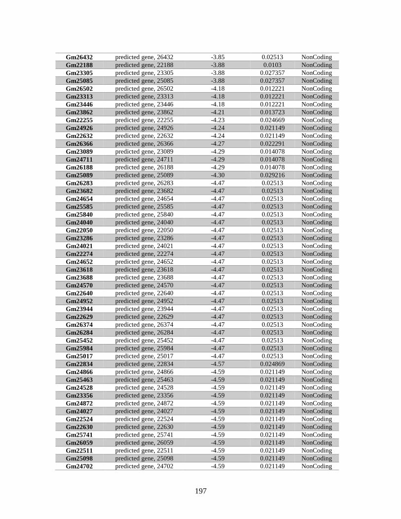
197
Gm26432 predicted gene, 26432 -3.85 0.02513 NonCoding
Gm22188 predicted gene, 22188 -3.88 0.0103 NonCoding
Gm23305 predicted gene, 23305 -3.88 0.027357 NonCoding
Gm25085 predicted gene, 25085 -3.88 0.027357 NonCoding
Gm26502 predicted gene, 26502 -4.18 0.012221 NonCoding
Gm23313 predicted gene, 23313 -4.18 0.012221 NonCoding
Gm23446 predicted gene, 23446 -4.18 0.012221 NonCoding
Gm23862 predicted gene, 23862 -4.21 0.013723 NonCoding
Gm22255 predicted gene, 22255 -4.23 0.024669 NonCoding
Gm24926 predicted gene, 24926 -4.24 0.021149 NonCoding
Gm22632 predicted gene, 22632 -4.24 0.021149 NonCoding
Gm26366 predicted gene, 26366 -4.27 0.022291 NonCoding
Gm23089 predicted gene, 23089 -4.29 0.014078 NonCoding
Gm24711 predicted gene, 24711 -4.29 0.014078 NonCoding
Gm26188 predicted gene, 26188 -4.29 0.014078 NonCoding
Gm25089 predicted gene, 25089 -4.30 0.029216 NonCoding
Gm26283 predicted gene, 26283 -4.47 0.02513 NonCoding
Gm23682 predicted gene, 23682 -4.47 0.02513 NonCoding
Gm24654 predicted gene, 24654 -4.47 0.02513 NonCoding
Gm25585 predicted gene, 25585 -4.47 0.02513 NonCoding
Gm25840 predicted gene, 25840 -4.47 0.02513 NonCoding
Gm24040 predicted gene, 24040 -4.47 0.02513 NonCoding
Gm22050 predicted gene, 22050 -4.47 0.02513 NonCoding
Gm23286 predicted gene, 23286 -4.47 0.02513 NonCoding
Gm24021 predicted gene, 24021 -4.47 0.02513 NonCoding
Gm22274 predicted gene, 22274 -4.47 0.02513 NonCoding
Gm24652 predicted gene, 24652 -4.47 0.02513 NonCoding
Gm23618 predicted gene, 23618 -4.47 0.02513 NonCoding
Gm23688 predicted gene, 23688 -4.47 0.02513 NonCoding
Gm24570 predicted gene, 24570 -4.47 0.02513 NonCoding
Gm22640 predicted gene, 22640 -4.47 0.02513 NonCoding
Gm24952 predicted gene, 24952 -4.47 0.02513 NonCoding
Gm23944 predicted gene, 23944 -4.47 0.02513 NonCoding
Gm22629 predicted gene, 22629 -4.47 0.02513 NonCoding
Gm26374 predicted gene, 26374 -4.47 0.02513 NonCoding
Gm26284 predicted gene, 26284 -4.47 0.02513 NonCoding
Gm25452 predicted gene, 25452 -4.47 0.02513 NonCoding
Gm25984 predicted gene, 25984 -4.47 0.02513 NonCoding
Gm25017 predicted gene, 25017 -4.47 0.02513 NonCoding
Gm22834 predicted gene, 22834 -4.57 0.024869 NonCoding
Gm24866 predicted gene, 24866 -4.59 0.021149 NonCoding
Gm25463 predicted gene, 25463 -4.59 0.021149 NonCoding
Gm24528 predicted gene, 24528 -4.59 0.021149 NonCoding
Gm23356 predicted gene, 23356 -4.59 0.021149 NonCoding
Gm24872 predicted gene, 24872 -4.59 0.021149 NonCoding
Gm24027 predicted gene, 24027 -4.59 0.021149 NonCoding
Gm22524 predicted gene, 22524 -4.59 0.021149 NonCoding
Gm22630 predicted gene, 22630 -4.59 0.021149 NonCoding
Gm25741 predicted gene, 25741 -4.59 0.021149 NonCoding
Gm26059 predicted gene, 26059 -4.59 0.021149 NonCoding
Gm22511 predicted gene, 22511 -4.59 0.021149 NonCoding
Gm25098 predicted gene, 25098 -4.59 0.021149 NonCoding
Gm24702 predicted gene, 24702 -4.59 0.021149 NonCoding

198
Gm25462 predicted gene, 25462 -4.59 0.021149 NonCoding
Gm25523 predicted gene, 25523 -4.59 0.021149 NonCoding
Gm22909 predicted gene, 22909 -4.59 0.021149 NonCoding
Gm26392 predicted gene, 26392 -4.59 0.021149 NonCoding
Gm24219 predicted gene, 24219 -4.59 0.021149 NonCoding
Gm25230 predicted gene, 25230 -4.59 0.021149 NonCoding
Gm22627 predicted gene, 22627 -4.59 0.021149 NonCoding
Gm24585 predicted gene, 24585 -4.59 0.021149 NonCoding
Gm24206 predicted gene, 24206 -4.59 0.021149 NonCoding
Gm25209 predicted gene, 25209 -4.59 0.021149 NonCoding
Gm26334 predicted gene, 26334 -4.59 0.021149 NonCoding
Gm25742 predicted gene, 25742 -4.59 0.021149 NonCoding
Gm25146 predicted gene, 25146 -4.59 0.021149 NonCoding
Gm24759 predicted gene, 24759 -4.59 0.021149 NonCoding
Gm23575 predicted gene, 23575 -4.59 0.021149 NonCoding
Gm22449 predicted gene, 22449 -4.59 0.021149 NonCoding
Gm22912 predicted gene, 22912 -4.59 0.021149 NonCoding
Gm25710 predicted gene, 25710 -4.59 0.021149 NonCoding
Gm26434 predicted gene, 26434 -4.59 0.021149 NonCoding
Gm23560 predicted gene, 23560 -4.59 0.021149 NonCoding
Gm22252 predicted gene, 22252 -4.72 0.017861 NonCoding
Snord116 small nucleolar RNA, C/D
box 116 -4.79 0.012221 NonCoding
Snord116 small nucleolar RNA, C/D
box 116 -4.79 0.012221 NonCoding
Snord116 small nucleolar RNA, C/D
box 116 -4.79 0.012221 NonCoding
Snord116 small nucleolar RNA, C/D
box 116 -4.79 0.012221 NonCoding
Snord116 small nucleolar RNA, C/D
box 116 -4.79 0.012221 NonCoding
Snord116 small nucleolar RNA, C/D
box 116 -4.79 0.012221 NonCoding
Snord116 small nucleolar RNA, C/D
box 116 -4.79 0.012221 NonCoding
Snord116 small nucleolar RNA, C/D
box 116 -4.79 0.012221 NonCoding
Snord116 small nucleolar RNA, C/D
box 116 -4.79 0.012221 NonCoding
Snord116 small nucleolar RNA, C/D
box 116 -4.79 0.012221 NonCoding
Snord116 small nucleolar RNA, C/D
box 116 -4.79 0.012221 NonCoding
Snord116 small nucleolar RNA, C/D
box 116 -4.79 0.012221 NonCoding
Snord116 small nucleolar RNA, C/D
box 116 -4.79 0.012221 NonCoding
Snord116 small nucleolar RNA, C/D
box 116 -4.79 0.012221 NonCoding
Snord116 small nucleolar RNA, C/D
box 116 -4.79 0.012221 NonCoding
Snord116 small nucleolar RNA, C/D
box 116 -4.79 0.012221 NonCoding
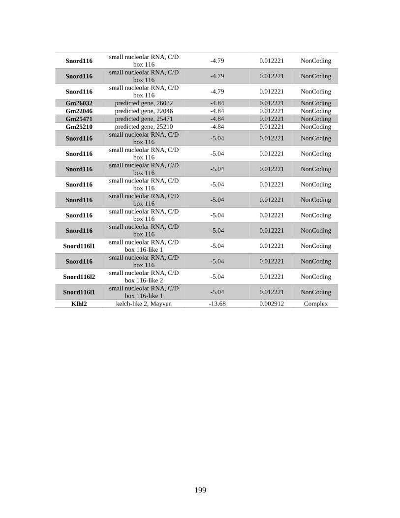
199
Snord116 small nucleolar RNA, C/D
box 116 -4.79 0.012221 NonCoding
Snord116 small nucleolar RNA, C/D
box 116 -4.79 0.012221 NonCoding
Snord116 small nucleolar RNA, C/D
box 116 -4.79 0.012221 NonCoding
Gm26032 predicted gene, 26032 -4.84 0.012221 NonCoding
Gm22046 predicted gene, 22046 -4.84 0.012221 NonCoding
Gm25471 predicted gene, 25471 -4.84 0.012221 NonCoding
Gm25210 predicted gene, 25210 -4.84 0.012221 NonCoding
Snord116 small nucleolar RNA, C/D
box 116 -5.04 0.012221 NonCoding
Snord116 small nucleolar RNA, C/D
box 116 -5.04 0.012221 NonCoding
Snord116 small nucleolar RNA, C/D
box 116 -5.04 0.012221 NonCoding
Snord116 small nucleolar RNA, C/D
box 116 -5.04 0.012221 NonCoding
Snord116 small nucleolar RNA, C/D
box 116 -5.04 0.012221 NonCoding
Snord116 small nucleolar RNA, C/D
box 116 -5.04 0.012221 NonCoding
Snord116 small nucleolar RNA, C/D
box 116 -5.04 0.012221 NonCoding
Snord116l1 small nucleolar RNA, C/D
box 116-like 1 -5.04 0.012221 NonCoding
Snord116 small nucleolar RNA, C/D
box 116 -5.04 0.012221 NonCoding
Snord116l2 small nucleolar RNA, C/D
box 116-like 2 -5.04 0.012221 NonCoding
Snord116l1 small nucleolar RNA, C/D
box 116-like 1 -5.04 0.012221 NonCoding
Klhl2 kelch-like 2, Mayven -13.68 0.002912 Complex

200
APPENDIX B
GENES WITH DIFFERENTIAL EXPRESSION IN THE ABSENCE OF RAD51D
IDENTIFIED BY RNA SEQ
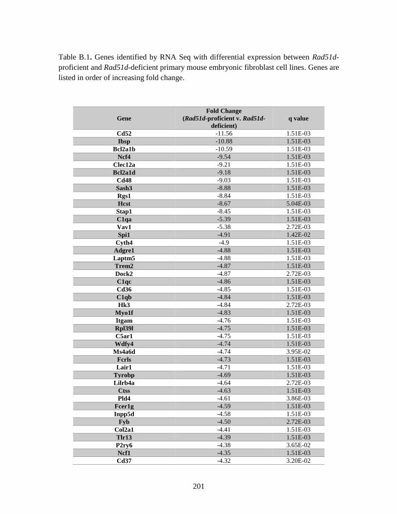
201
Table B.1. Genes identified by RNA Seq with differential expression between Rad51d-
proficient and Rad51d-deficient primary mouse embryonic fibroblast cell lines. Genes are
listed in order of increasing fold change.
Gene
Fold Change
(Rad51d-proficient v. Rad51d-
deficient)
q value
Cd52 -11.56 1.51E-03
Ibsp -10.88 1.51E-03
Bcl2a1b -10.59 1.51E-03
Ncf4 -9.54 1.51E-03
Clec12a -9.21 1.51E-03
Bcl2a1d -9.18 1.51E-03
Cd48 -9.03 1.51E-03
Sash3 -8.88 1.51E-03
Rgs1 -8.84 1.51E-03
Hcst -8.67 5.04E-03
Stap1 -8.45 1.51E-03
C1qa -5.39 1.51E-03
Vav1 -5.38 2.72E-03
Spi1 -4.91 1.42E-02
Cyth4 -4.9 1.51E-03
Adgre1 -4.88 1.51E-03
Laptm5 -4.88 1.51E-03
Trem2 -4.87 1.51E-03
Dock2 -4.87 2.72E-03
C1qc -4.86 1.51E-03
Cd36 -4.85 1.51E-03
C1qb -4.84 1.51E-03
Hk3 -4.84 2.72E-03
Myo1f -4.83 1.51E-03
Itgam -4.76 1.51E-03
Rpl39l -4.75 1.51E-03
C5ar1 -4.75 1.51E-03
Wdfy4 -4.74 1.51E-03
Ms4a6d -4.74 3.95E-02
Fcrls -4.73 1.51E-03
Lair1 -4.71 1.51E-03
Tyrobp -4.69 1.51E-03
Lilrb4a -4.64 2.72E-03
Ctss -4.63 1.51E-03
Pld4 -4.61 3.86E-03
Fcer1g -4.59 1.51E-03
Inpp5d -4.58 1.51E-03
Fyb -4.50 2.72E-03
Col2a1 -4.41 1.51E-03
Tlr13 -4.39 1.51E-03
P2ry6 -4.38 3.65E-02
Ncf1 -4.35 1.51E-03
Cd37 -4.32 3.20E-02
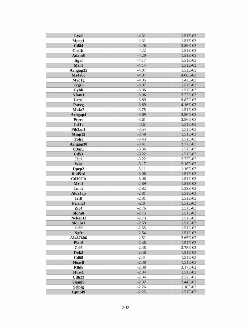
202
Lyz2 -4.31 1.51E-03
Mpeg1 -4.31 1.51E-03
Cd84 -4.26 3.86E-03
Clec4d -4.22 1.51E-03
Adam8 -4.20 1.51E-03
Itgal -4.17 1.51E-03
Msr1 -4.14 1.51E-03
Arhgap25 -4.07 1.51E-03
Ms4a6c -4.07 4.08E-02
Myo1g -4.05 1.42E-02
Fcgr3 -3.97 1.51E-03
Cybb -3.96 1.51E-03
Nfam1 -3.96 2.72E-03
Lcp1 -3.89 9.82E-03
Parvg -3.89 4.16E-02
Ms4a7 -3.75 1.51E-03
Arhgap4 -3.69 3.86E-03
Ptprc -3.61 3.86E-03
Csf1r -3.6 1.51E-03
Pik3ap1 -3.54 1.51E-03
Mmp12 -3.49 1.51E-03
Tph1 -3.43 1.51E-03
Arhgap30 -3.41 2.72E-03
C3ar1 -3.36 1.51E-03
Cd53 -3.22 1.51E-03
Tlr7 -3.22 2.72E-03
Was -3.17 2.16E-02
Dpep2 -3.11 1.16E-02
Rad51d -3.08 1.51E-03
Cd300lb -3.08 1.51E-03
Mrc1 -2.89 1.51E-03
Lmo2 -2.82 1.33E-02
Alox5ap -2.81 1.51E-03
Irf8 -2.81 1.51E-03
Fermt3 -2.8 1.51E-03
Zic4 -2.76 1.51E-03
Slc7a8 -2.75 1.51E-03
Nckap1l -2.73 1.51E-03
Slc11a1 -2.59 1.51E-03
Ccl9 -2.55 1.51E-03
Ngfr -2.54 1.51E-03
AI467606 -2.51 2.65E-02
Plac8 -2.48 1.51E-03
Ccl6 -2.48 2.78E-02
Dok2 -2.46 1.51E-03
Cd68 -2.41 1.51E-03
Hoxc8 -2.38 1.51E-03
Klhl6 -2.38 3.17E-02
Hmx1 -2.34 1.51E-03
Cdh23 -2.34 2.52E-02
Slamf9 -2.32 2.44E-02
Selplg -2.26 1.16E-02
Gpr149 -2.25 1.51E-03
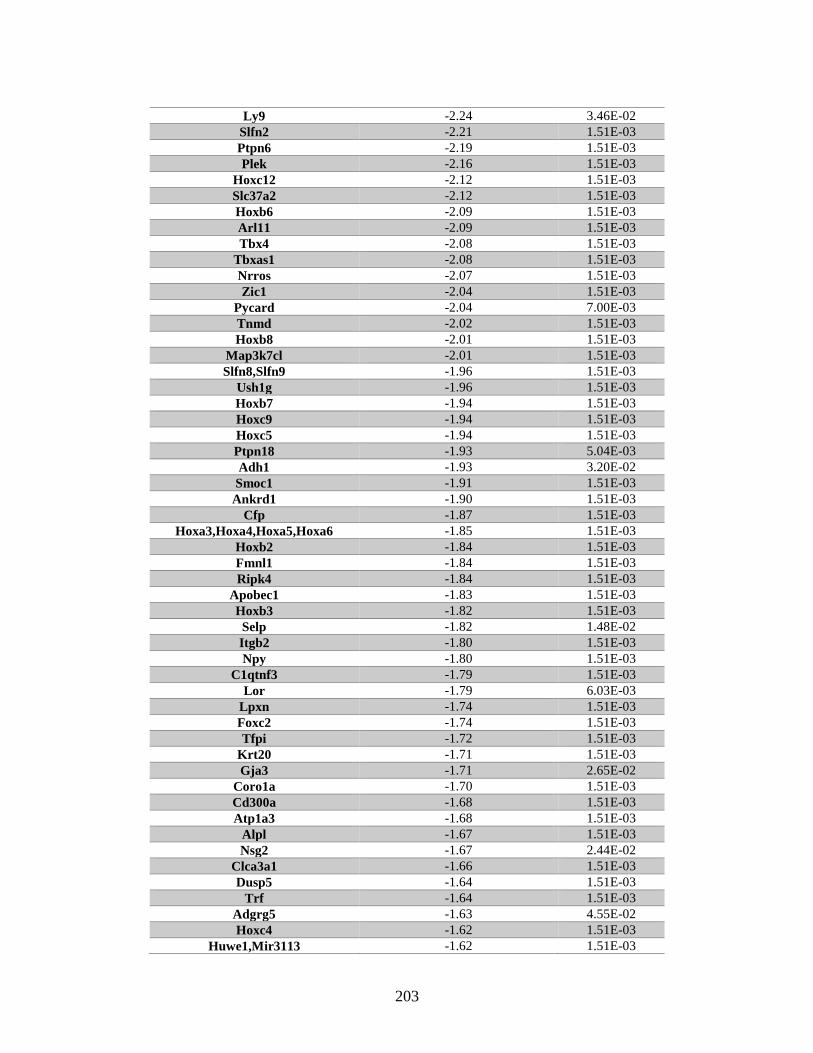
203
Ly9 -2.24 3.46E-02
Slfn2 -2.21 1.51E-03
Ptpn6 -2.19 1.51E-03
Plek -2.16 1.51E-03
Hoxc12 -2.12 1.51E-03
Slc37a2 -2.12 1.51E-03
Hoxb6 -2.09 1.51E-03
Arl11 -2.09 1.51E-03
Tbx4 -2.08 1.51E-03
Tbxas1 -2.08 1.51E-03
Nrros -2.07 1.51E-03
Zic1 -2.04 1.51E-03
Pycard -2.04 7.00E-03
Tnmd -2.02 1.51E-03
Hoxb8 -2.01 1.51E-03
Map3k7cl -2.01 1.51E-03
Slfn8,Slfn9 -1.96 1.51E-03
Ush1g -1.96 1.51E-03
Hoxb7 -1.94 1.51E-03
Hoxc9 -1.94 1.51E-03
Hoxc5 -1.94 1.51E-03
Ptpn18 -1.93 5.04E-03
Adh1 -1.93 3.20E-02
Smoc1 -1.91 1.51E-03
Ankrd1 -1.90 1.51E-03
Cfp -1.87 1.51E-03
Hoxa3,Hoxa4,Hoxa5,Hoxa6 -1.85 1.51E-03
Hoxb2 -1.84 1.51E-03
Fmnl1 -1.84 1.51E-03
Ripk4 -1.84 1.51E-03
Apobec1 -1.83 1.51E-03
Hoxb3 -1.82 1.51E-03
Selp -1.82 1.48E-02
Itgb2 -1.80 1.51E-03
Npy -1.80 1.51E-03
C1qtnf3 -1.79 1.51E-03
Lor -1.79 6.03E-03
Lpxn -1.74 1.51E-03
Foxc2 -1.74 1.51E-03
Tfpi -1.72 1.51E-03
Krt20 -1.71 1.51E-03
Gja3 -1.71 2.65E-02
Coro1a -1.70 1.51E-03
Cd300a -1.68 1.51E-03
Atp1a3 -1.68 1.51E-03
Alpl -1.67 1.51E-03
Nsg2 -1.67 2.44E-02
Clca3a1 -1.66 1.51E-03
Dusp5 -1.64 1.51E-03
Trf -1.64 1.51E-03
Adgrg5 -1.63 4.55E-02
Hoxc4 -1.62 1.51E-03
Huwe1,Mir3113 -1.62 1.51E-03

204
Neurl3 -1.62 2.72E-03
Slc1a6 -1.57 1.51E-03
Shtn1 -1.54 1.51E-03
Cd93 -1.54 1.51E-03
Ncf2 -1.53 1.51E-03
Sema4d -1.53 1.51E-03
Lad1 -1.53 2.31E-02
Cfh -1.52 2.72E-03
Ptpre -1.51 1.51E-03
Krt7 -1.50 1.51E-03
Hoxb4 -1.49 1.51E-03
Hoxc6 -1.48 1.51E-03
Rbm20 -1.47 1.51E-03
Psd4 -1.46 1.51E-03
Tmem132e -1.45 1.51E-03
Tll1 -1.43 1.51E-03
Grem2 -1.42 1.51E-03
Grap -1.42 3.77E-02
Foxd1 -1.40 1.51E-03
Foxl1 -1.39 1.51E-03
Des -1.38 1.51E-03
Egr2 -1.37 1.51E-03
Parvb -1.37 1.51E-03
Ret -1.36 1.51E-03
Clca3a2 -1.36 1.16E-02
Hoxb5 -1.35 3.90E-02
Meg3,Mir1906-1,Mir770 -1.33 1.51E-03
Cspg4 -1.33 1.51E-03
Ucp2 -1.31 1.51E-03
Ulbp1 -1.30 1.51E-03
Ankk1 -1.30 2.72E-03
Sp100 -1.30 4.79E-02
Irx1 -1.28 1.51E-03
Ifi27l2b -1.28 7.00E-03
Fat3 -1.27 1.51E-03
Slfn3 -1.27 1.80E-02
Dmrta2 -1.26 1.51E-03
Hoxd4 -1.26 1.87E-02
Unc5b -1.25 1.51E-03
Skap1 -1.25 1.51E-03
Irf5 -1.25 1.51E-03
Hecw1 -1.24 1.51E-03
Col11a2 -1.24 2.72E-03
Chst2 -1.23 1.51E-03
Amer2 -1.23 2.92E-02
Lrtm2 -1.23 3.60E-02
Sox13 -1.22 1.51E-03
Smim10l2a -1.21 2.52E-02
Ctsh -1.19 1.51E-03
Sorbs1 -1.18 1.51E-03
Glce,Mir5133 -1.18 1.51E-03
Gpsm3 -1.18 2.72E-03
Slc43a2 -1.17 1.51E-03
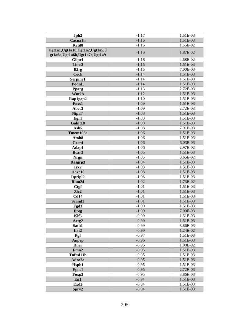
205
Jph2 -1.17 1.51E-03
Cacna1h -1.16 1.51E-03
Kctd8 -1.16 1.55E-02
Ugt1a1,Ugt1a10,Ugt1a2,Ugt1a5,U
gt1a6a,Ugt1a6b,Ugt1a7c,Ugt1a9 -1.16 1.87E-02
Glipr1 -1.16 4.68E-02
Lims2 -1.15 1.51E-03
Il2rg -1.15 7.00E-03
Coch -1.14 1.51E-03
Serpine1 -1.14 1.51E-03
Podnl1 -1.14 1.51E-03
Pparg -1.13 2.72E-03
Wnt2b -1.12 1.51E-03
Rap1gap2 -1.10 1.51E-03
Foxs1 -1.09 1.51E-03
Abcc3 -1.09 2.72E-03
Nipal4 -1.08 1.51E-03
Egr1 -1.08 1.51E-03
Galnt18 -1.08 1.51E-03
Asb5 -1.08 7.91E-03
Tmem106a -1.06 1.51E-03
Atoh8 -1.06 1.51E-03
Cxcr4 -1.06 6.03E-03
Adap1 -1.06 2.97E-02
Bcar3 -1.05 1.51E-03
Nrgn -1.05 3.65E-02
Rasgrp3 -1.04 1.51E-03
Irx2 -1.03 1.51E-03
Hoxc10 -1.03 1.51E-03
Itpripl2 -1.03 1.51E-03
Rbm24 -1.02 1.73E-02
Ctgf -1.01 1.51E-03
Zic2 -1.01 1.51E-03
Cd14 -1.01 1.51E-03
Scand1 -1.01 1.51E-03
Fgd3 -1.00 1.51E-03
Ereg -1.00 7.00E-03
Klf5 -0.99 1.51E-03
Actg2 -0.99 1.51E-03
Satb1 -0.99 3.86E-03
Lat2 -0.99 1.24E-02
Pgf -0.97 1.51E-03
Anpep -0.96 1.51E-03
Dner -0.96 1.08E-02
Fmn2 -0.95 1.51E-03
Tnfrsf11b -0.95 1.51E-03
Adra2a -0.95 1.51E-03
Hspb1 -0.95 1.51E-03
Epas1 -0.95 2.72E-03
Foxp2 -0.95 3.86E-03
En1 -0.94 1.51E-03
Exd2 -0.94 1.51E-03
Spry2 -0.94 1.51E-03
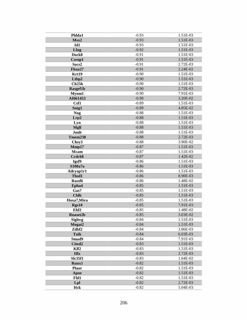
206
Phlda1 -0.93 1.51E-03
Msx2 -0.93 1.51E-03
Id1 -0.93 1.51E-03
Lfng -0.92 1.51E-03
Dock8 -0.91 1.51E-03
Csrnp1 -0.91 1.51E-03
Socs2 -0.91 2.72E-03
Fbxo27 -0.91 2.24E-02
Krt19 -0.90 1.51E-03
Ltbp2 -0.90 1.51E-03
Ch25h -0.90 1.51E-03
Rasgef1b -0.90 2.72E-03
Myom1 -0.90 7.91E-03
AI661453 -0.90 3.20E-02
Csf1 -0.89 1.51E-03
Sntg1 -0.89 4.85E-02
Nog -0.88 1.51E-03
Lrp2 -0.88 1.51E-03
Lyn -0.88 1.51E-03
Mgll -0.88 1.51E-03
Junb -0.88 1.51E-03
Tmem238 -0.88 2.72E-03
Chsy3 -0.88 3.90E-02
Mmp17 -0.87 1.51E-03
Mcam -0.87 1.51E-03
Ccdc68 -0.87 1.42E-02
Igsf9 -0.86 1.51E-03
S100a7a -0.86 1.51E-03
Adcyap1r1 -0.86 1.51E-03
Thsd1 -0.86 8.90E-03
Rassf6 -0.86 1.48E-02
Epha4 -0.85 1.51E-03
Gas7 -0.85 1.51E-03
Cblb -0.85 1.51E-03
Hoxa7,Mira -0.85 1.51E-03
Rgs10 -0.85 7.91E-03
Ebf2 -0.85 1.48E-02
Rnaset2b -0.85 3.03E-02
Siglecg -0.84 1.51E-03
Mogat2 -0.84 1.51E-03
Zdbf2 -0.84 3.86E-03
Tnik -0.84 6.03E-03
Smad9 -0.84 7.91E-03
Cited2 -0.83 1.51E-03
Klf2 -0.83 1.51E-03
Hlx -0.83 2.72E-03
Slc35f1 -0.83 1.64E-02
Runx1 -0.82 1.51E-03
Plaur -0.82 1.51E-03
Apoe -0.82 1.51E-03
Fhl1 -0.82 1.51E-03
Lpl -0.82 2.72E-03
Hck -0.82 5.04E-03
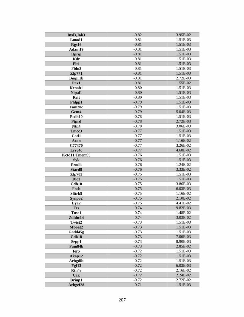
207
Insl3,Jak3 -0.82 3.95E-02
Lmod1 -0.81 1.51E-03
Rgs16 -0.81 1.51E-03
Adam19 -0.81 1.51E-03
Itprip -0.81 1.51E-03
Kdr -0.81 1.51E-03
Flt1 -0.81 1.51E-03
Fbln2 -0.81 1.51E-03
Zfp771 -0.81 1.51E-03
Bmpr1b -0.81 2.72E-03
Pax1 -0.81 1.55E-02
Kcnab1 -0.80 1.51E-03
Nipal1 -0.80 1.51E-03
Relt -0.80 1.51E-03
Phlpp1 -0.79 1.51E-03
Fam20c -0.79 1.51E-03
Gcnt4 -0.79 5.04E-03
Pcdh10 -0.78 1.51E-03
Ptprd -0.78 2.72E-03
Ntn4 -0.78 3.86E-03
Tmcc3 -0.77 1.51E-03
Cotl1 -0.77 1.51E-03
Acan -0.77 1.16E-02
C77370 -0.77 3.26E-02
Lrrc4c -0.77 4.68E-02
Kctd11,Tmem95 -0.76 1.51E-03
Syk -0.76 1.51E-03
Prodh -0.76 1.24E-02
Stard8 -0.76 3.33E-02
Zfp703 -0.75 1.51E-03
Dlc1 -0.75 1.51E-03
Cdh10 -0.75 3.86E-03
Fosb -0.75 6.03E-03
Slitrk5 -0.75 1.16E-02
Synpo2 -0.75 2.10E-02
Eya2 -0.75 4.41E-02
Fes -0.74 9.82E-03
Tusc1 -0.74 1.48E-02
Zdhhc14 -0.74 3.03E-02
Twist2 -0.73 1.51E-03
Mboat2 -0.73 1.51E-03
Gadd45g -0.73 1.51E-03
Cdk18 -0.73 7.00E-03
Sepp1 -0.73 8.90E-03
Fam84b -0.73 2.85E-02
Ier5 -0.72 1.51E-03
Akap12 -0.72 1.51E-03
Arhgdib -0.72 1.51E-03
Fgf13 -0.72 6.03E-03
Rtn4r -0.72 2.16E-02
Cck -0.72 2.24E-02
Brinp1 -0.72 2.72E-02
Arhgef28 -0.71 1.51E-03
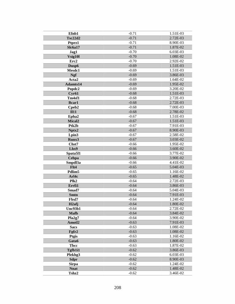
208
Efnb1 -0.71 1.51E-03
Tsc22d2 -0.71 2.72E-03
Ptprz1 -0.71 8.90E-03
Slc6a17 -0.71 1.87E-02
Jag1 -0.70 6.03E-03
Vsig10l -0.70 1.08E-02
Erc2 -0.70 2.92E-02
Dusp6 -0.69 1.51E-03
Mesdc1 -0.69 1.51E-03
Ngf -0.69 3.86E-03
Acta2 -0.69 1.64E-02
Adamts14 -0.69 1.95E-02
Popdc2 -0.69 3.20E-02
Cyr61 -0.68 1.51E-03
Tm4sf1 -0.68 2.72E-03
Bcar1 -0.68 2.72E-03
Cpeb2 -0.68 7.00E-03
Il11 -0.68 2.78E-02
Epha2 -0.67 1.51E-03
Mical2 -0.67 1.51E-03
Ptk2b -0.67 7.91E-03
Nptx2 -0.67 8.90E-03
Lpin3 -0.67 2.58E-02
Runx3 -0.67 3.03E-02
Chst7 -0.66 1.95E-02
Lhx9 -0.66 3.60E-02
Spata5l1 -0.66 3.77E-02
Cebpa -0.66 3.90E-02
Smpdl3a -0.66 4.41E-02
Flt4 -0.65 5.04E-03
Pdlim5 -0.65 1.16E-02
Arl4c -0.65 1.48E-02
Plk2 -0.64 2.72E-03
Errfi1 -0.64 3.86E-03
Smad7 -0.64 5.04E-03
Smtn -0.64 7.91E-03
Fbxl7 -0.64 1.24E-02
H2afj -0.64 1.80E-02
Unc93b1 -0.64 2.72E-02
Mafb -0.64 3.84E-02
Pla2g7 -0.64 3.90E-02
Amotl2 -0.63 7.91E-03
Sacs -0.63 1.08E-02
Fgfr2 -0.63 1.08E-02
Ptgis -0.63 1.16E-02
Gata6 -0.63 1.80E-02
Tbcc -0.63 1.87E-02
Tgfb1i1 -0.62 3.86E-03
Plekhg3 -0.62 6.03E-03
Sdpr -0.62 8.90E-03
Sirpa -0.62 1.24E-02
Nnat -0.62 1.48E-02
Tshz2 -0.62 3.46E-02
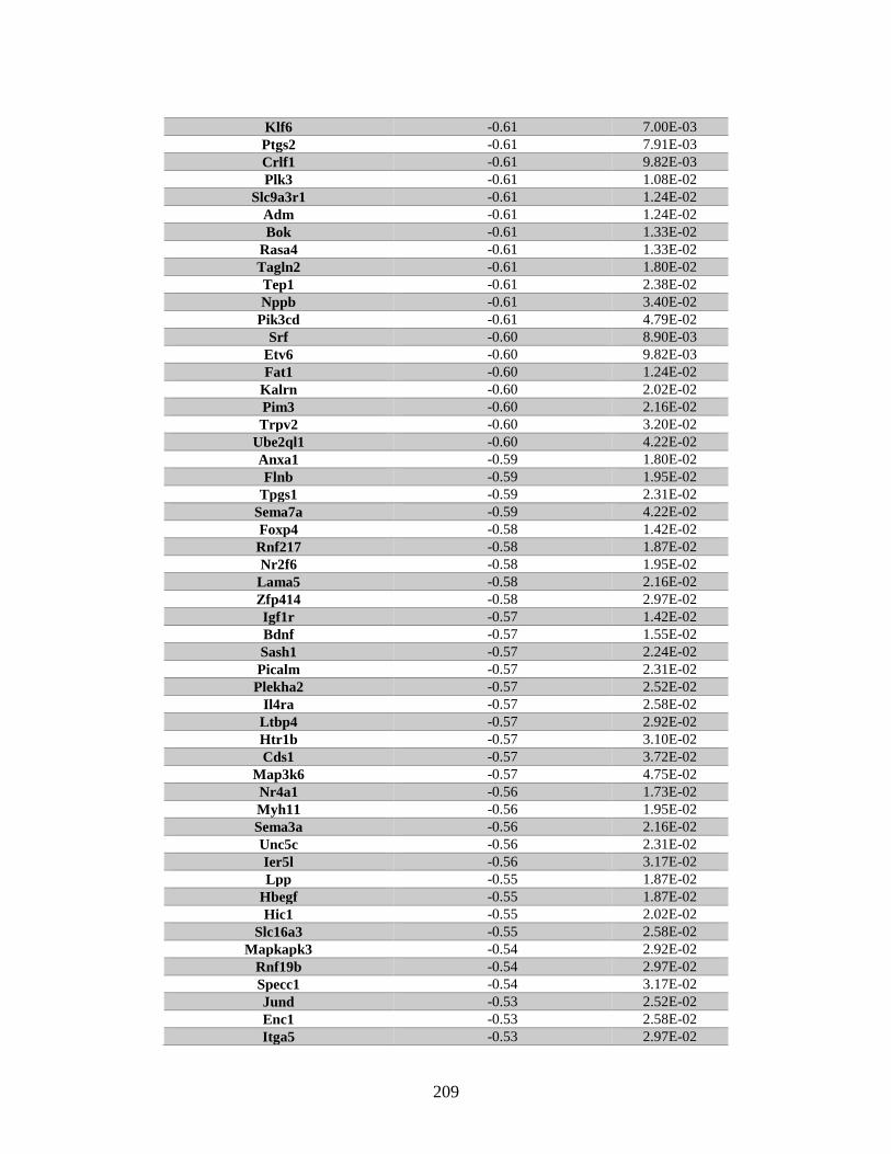
209
Klf6 -0.61 7.00E-03
Ptgs2 -0.61 7.91E-03
Crlf1 -0.61 9.82E-03
Plk3 -0.61 1.08E-02
Slc9a3r1 -0.61 1.24E-02
Adm -0.61 1.24E-02
Bok -0.61 1.33E-02
Rasa4 -0.61 1.33E-02
Tagln2 -0.61 1.80E-02
Tep1 -0.61 2.38E-02
Nppb -0.61 3.40E-02
Pik3cd -0.61 4.79E-02
Srf -0.60 8.90E-03
Etv6 -0.60 9.82E-03
Fat1 -0.60 1.24E-02
Kalrn -0.60 2.02E-02
Pim3 -0.60 2.16E-02
Trpv2 -0.60 3.20E-02
Ube2ql1 -0.60 4.22E-02
Anxa1 -0.59 1.80E-02
Flnb -0.59 1.95E-02
Tpgs1 -0.59 2.31E-02
Sema7a -0.59 4.22E-02
Foxp4 -0.58 1.42E-02
Rnf217 -0.58 1.87E-02
Nr2f6 -0.58 1.95E-02
Lama5 -0.58 2.16E-02
Zfp414 -0.58 2.97E-02
Igf1r -0.57 1.42E-02
Bdnf -0.57 1.55E-02
Sash1 -0.57 2.24E-02
Picalm -0.57 2.31E-02
Plekha2 -0.57 2.52E-02
Il4ra -0.57 2.58E-02
Ltbp4 -0.57 2.92E-02
Htr1b -0.57 3.10E-02
Cds1 -0.57 3.72E-02
Map3k6 -0.57 4.75E-02
Nr4a1 -0.56 1.73E-02
Myh11 -0.56 1.95E-02
Sema3a -0.56 2.16E-02
Unc5c -0.56 2.31E-02
Ier5l -0.56 3.17E-02
Lpp -0.55 1.87E-02
Hbegf -0.55 1.87E-02
Hic1 -0.55 2.02E-02
Slc16a3 -0.55 2.58E-02
Mapkapk3 -0.54 2.92E-02
Rnf19b -0.54 2.97E-02
Specc1 -0.54 3.17E-02
Jund -0.53 2.52E-02
Enc1 -0.53 2.58E-02
Itga5 -0.53 2.97E-02
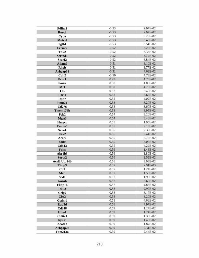
210
Pdlim1 -0.53 2.97E-02
Rusc2 -0.53 2.97E-02
Cyba -0.53 3.20E-02
Metrnl -0.53 3.40E-02
Tgfb1 -0.53 3.54E-02
Fermt2 -0.52 3.26E-02
Tnk2 -0.52 3.33E-02
Sertad1 -0.52 3.77E-02
Scarf2 -0.52 3.84E-02
Adam9 -0.51 3.10E-02
Rhob -0.51 3.77E-02
Arhgap31 -0.51 4.62E-02
Cdh2 -0.50 4.79E-02
Prrx1 0.49 4.79E-02
Postn 0.50 4.08E-02
Mt1 0.50 4.79E-02
Lss 0.52 3.40E-02
Il1rl1 0.52 3.65E-02
Dpp7 0.52 4.02E-02
Pmp22 0.53 3.20E-02
Cd276 0.53 3.60E-02
Tmem176b 0.53 3.95E-02
Pck2 0.54 3.20E-02
Mgst3 0.54 3.46E-02
Hmgcr 0.55 1.95E-02
Emilin1 0.55 2.10E-02
Srxn1 0.55 2.38E-02
Cav2 0.55 2.44E-02
Acat2 0.55 2.72E-02
Mdk 0.55 3.03E-02
Cdh13 0.55 4.22E-02
Fdps 0.56 1.48E-02
Akr1b3 0.56 1.80E-02
Sorcs2 0.56 2.52E-02
Acsl3,Utp14b 0.56 3.03E-02
Timp3 0.57 7.91E-03
Cd9 0.57 1.24E-02
Mvd 0.57 1.55E-02
Scd1 0.57 1.95E-02
Gorab 0.57 3.60E-02
Fkbp14 0.57 4.85E-02
Dkk2 0.58 2.97E-02
Crip2 0.58 3.17E-02
Cbr3 0.58 3.20E-02
Gsdmd 0.58 4.68E-02
Rab3d 0.58 4.97E-02
Cd248 0.59 1.24E-02
Htra1 0.59 1.24E-02
Col6a1 0.59 1.33E-02
Isyna1 0.59 1.48E-02
Acot13 0.59 1.87E-02
Arhgap28 0.59 2.31E-02
Fam213a 0.59 2.44E-02
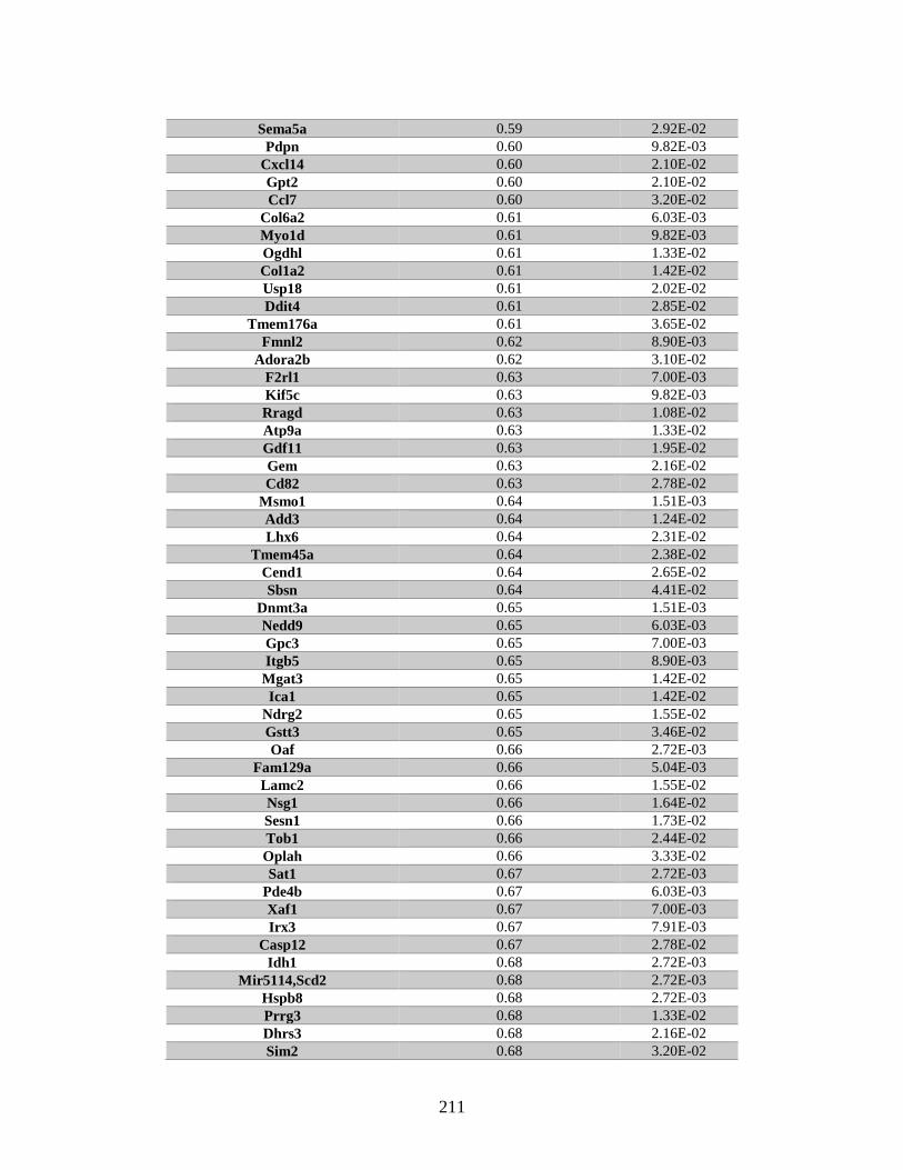
211
Sema5a 0.59 2.92E-02
Pdpn 0.60 9.82E-03
Cxcl14 0.60 2.10E-02
Gpt2 0.60 2.10E-02
Ccl7 0.60 3.20E-02
Col6a2 0.61 6.03E-03
Myo1d 0.61 9.82E-03
Ogdhl 0.61 1.33E-02
Col1a2 0.61 1.42E-02
Usp18 0.61 2.02E-02
Ddit4 0.61 2.85E-02
Tmem176a 0.61 3.65E-02
Fmnl2 0.62 8.90E-03
Adora2b 0.62 3.10E-02
F2rl1 0.63 7.00E-03
Kif5c 0.63 9.82E-03
Rragd 0.63 1.08E-02
Atp9a 0.63 1.33E-02
Gdf11 0.63 1.95E-02
Gem 0.63 2.16E-02
Cd82 0.63 2.78E-02
Msmo1 0.64 1.51E-03
Add3 0.64 1.24E-02
Lhx6 0.64 2.31E-02
Tmem45a 0.64 2.38E-02
Cend1 0.64 2.65E-02
Sbsn 0.64 4.41E-02
Dnmt3a 0.65 1.51E-03
Nedd9 0.65 6.03E-03
Gpc3 0.65 7.00E-03
Itgb5 0.65 8.90E-03
Mgat3 0.65 1.42E-02
Ica1 0.65 1.42E-02
Ndrg2 0.65 1.55E-02
Gstt3 0.65 3.46E-02
Oaf 0.66 2.72E-03
Fam129a 0.66 5.04E-03
Lamc2 0.66 1.55E-02
Nsg1 0.66 1.64E-02
Sesn1 0.66 1.73E-02
Tob1 0.66 2.44E-02
Oplah 0.66 3.33E-02
Sat1 0.67 2.72E-03
Pde4b 0.67 6.03E-03
Xaf1 0.67 7.00E-03
Irx3 0.67 7.91E-03
Casp12 0.67 2.78E-02
Idh1 0.68 2.72E-03
Mir5114,Scd2 0.68 2.72E-03
Hspb8 0.68 2.72E-03
Prrg3 0.68 1.33E-02
Dhrs3 0.68 2.16E-02
Sim2 0.68 3.20E-02
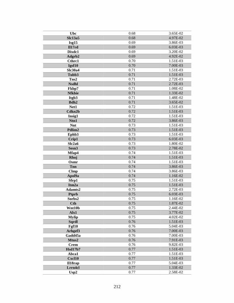
212
Ubc 0.68 3.65E-02
Slc13a5 0.68 4.97E-02
Isg15 0.69 3.86E-03
Il17rd 0.69 6.03E-03
Dixdc1 0.69 3.20E-02
Adgrb2 0.69 4.92E-02
Cthrc1 0.70 1.51E-03
Igsf10 0.70 7.00E-03
Slc30a4 0.71 1.51E-03
Tubb3 0.71 1.51E-03
Tns2 0.71 2.72E-03
Nsdhl 0.71 2.72E-03
Fkbp7 0.71 1.08E-02
Nfkbie 0.71 1.33E-02
Itgb3 0.71 1.48E-02
Bdh2 0.71 3.65E-02
Net1 0.72 1.51E-03
Cdkn2b 0.72 1.51E-03
Insig1 0.72 1.51E-03
Ntn1 0.72 3.86E-03
Nnt 0.73 1.51E-03
Pdlim2 0.73 1.51E-03
Ephb3 0.73 1.51E-03
Crip1 0.73 6.03E-03
Slc2a6 0.73 1.80E-02
Sesn3 0.73 2.78E-02
Mfap4 0.74 1.51E-03
Rhoj 0.74 1.51E-03
Osmr 0.74 1.51E-03
Tnn 0.74 3.86E-03
Clmp 0.74 3.86E-03
Apol9a 0.74 1.16E-02
Sfrp1 0.75 1.51E-03
Itm2a 0.75 1.51E-03
Adamts2 0.75 2.72E-03
Ptprb 0.75 6.03E-03
Sorbs2 0.75 1.16E-02
Cth 0.75 1.87E-02
Wnt10b 0.75 2.44E-02
Alx1 0.75 3.77E-02
Mylip 0.75 4.02E-02
Sqrdl 0.76 1.51E-03
Fgf10 0.76 5.04E-03
Arhgef3 0.76 7.00E-03
Gadd45a 0.76 7.00E-03
Mtus2 0.76 7.91E-03
Crem 0.76 9.82E-03
Hsd17b7 0.77 1.51E-03
Abca1 0.77 1.51E-03
Cxcl10 0.77 1.51E-03
Il18rap 0.77 5.04E-03
Lrrn4cl 0.77 1.33E-02
Usp2 0.77 2.58E-02
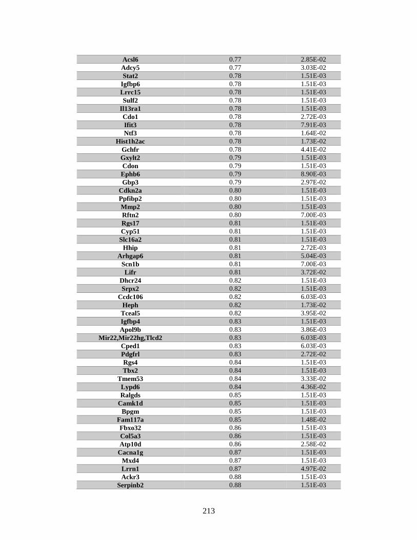
213
Acsl6 0.77 2.85E-02
Adcy5 0.77 3.03E-02
Stat2 0.78 1.51E-03
Igfbp6 0.78 1.51E-03
Lrrc15 0.78 1.51E-03
Sulf2 0.78 1.51E-03
Il13ra1 0.78 1.51E-03
Cdo1 0.78 2.72E-03
Ifit3 0.78 7.91E-03
Ntf3 0.78 1.64E-02
Hist1h2ac 0.78 1.73E-02
Gchfr 0.78 4.41E-02
Gxylt2 0.79 1.51E-03
Cdon 0.79 1.51E-03
Ephb6 0.79 8.90E-03
Gbp3 0.79 2.97E-02
Cdkn2a 0.80 1.51E-03
Ppfibp2 0.80 1.51E-03
Mmp2 0.80 1.51E-03
Rftn2 0.80 7.00E-03
Rgs17 0.81 1.51E-03
Cyp51 0.81 1.51E-03
Slc16a2 0.81 1.51E-03
Hhip 0.81 2.72E-03
Arhgap6 0.81 5.04E-03
Scn1b 0.81 7.00E-03
Lifr 0.81 3.72E-02
Dhcr24 0.82 1.51E-03
Srpx2 0.82 1.51E-03
Ccdc106 0.82 6.03E-03
Heph 0.82 1.73E-02
Tceal5 0.82 3.95E-02
Igfbp4 0.83 1.51E-03
Apol9b 0.83 3.86E-03
Mir22,Mir22hg,Tlcd2 0.83 6.03E-03
Cped1 0.83 6.03E-03
Pdgfrl 0.83 2.72E-02
Rgs4 0.84 1.51E-03
Tbx2 0.84 1.51E-03
Tmem53 0.84 3.33E-02
Lypd6 0.84 4.36E-02
Ralgds 0.85 1.51E-03
Camk1d 0.85 1.51E-03
Bpgm 0.85 1.51E-03
Fam117a 0.85 1.48E-02
Fbxo32 0.86 1.51E-03
Col5a3 0.86 1.51E-03
Atp10d 0.86 2.58E-02
Cacna1g 0.87 1.51E-03
Mxd4 0.87 1.51E-03
Lrrn1 0.87 4.97E-02
Ackr3 0.88 1.51E-03
Serpinb2 0.88 1.51E-03
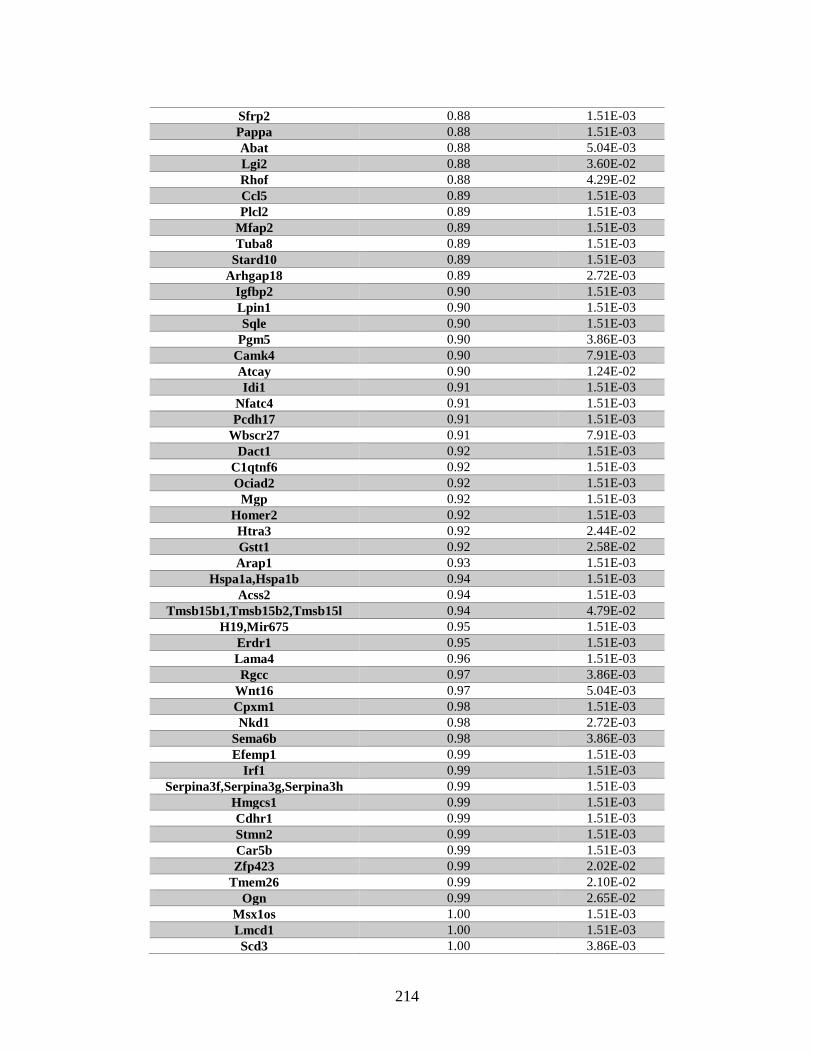
214
Sfrp2 0.88 1.51E-03
Pappa 0.88 1.51E-03
Abat 0.88 5.04E-03
Lgi2 0.88 3.60E-02
Rhof 0.88 4.29E-02
Ccl5 0.89 1.51E-03
Plcl2 0.89 1.51E-03
Mfap2 0.89 1.51E-03
Tuba8 0.89 1.51E-03
Stard10 0.89 1.51E-03
Arhgap18 0.89 2.72E-03
Igfbp2 0.90 1.51E-03
Lpin1 0.90 1.51E-03
Sqle 0.90 1.51E-03
Pgm5 0.90 3.86E-03
Camk4 0.90 7.91E-03
Atcay 0.90 1.24E-02
Idi1 0.91 1.51E-03
Nfatc4 0.91 1.51E-03
Pcdh17 0.91 1.51E-03
Wbscr27 0.91 7.91E-03
Dact1 0.92 1.51E-03
C1qtnf6 0.92 1.51E-03
Ociad2 0.92 1.51E-03
Mgp 0.92 1.51E-03
Homer2 0.92 1.51E-03
Htra3 0.92 2.44E-02
Gstt1 0.92 2.58E-02
Arap1 0.93 1.51E-03
Hspa1a,Hspa1b 0.94 1.51E-03
Acss2 0.94 1.51E-03
Tmsb15b1,Tmsb15b2,Tmsb15l 0.94 4.79E-02
H19,Mir675 0.95 1.51E-03
Erdr1 0.95 1.51E-03
Lama4 0.96 1.51E-03
Rgcc 0.97 3.86E-03
Wnt16 0.97 5.04E-03
Cpxm1 0.98 1.51E-03
Nkd1 0.98 2.72E-03
Sema6b 0.98 3.86E-03
Efemp1 0.99 1.51E-03
Irf1 0.99 1.51E-03
Serpina3f,Serpina3g,Serpina3h 0.99 1.51E-03
Hmgcs1 0.99 1.51E-03
Cdhr1 0.99 1.51E-03
Stmn2 0.99 1.51E-03
Car5b 0.99 1.51E-03
Zfp423 0.99 2.02E-02
Tmem26 0.99 2.10E-02
Ogn 0.99 2.65E-02
Msx1os 1.00 1.51E-03
Lmcd1 1.00 1.51E-03
Scd3 1.00 3.86E-03
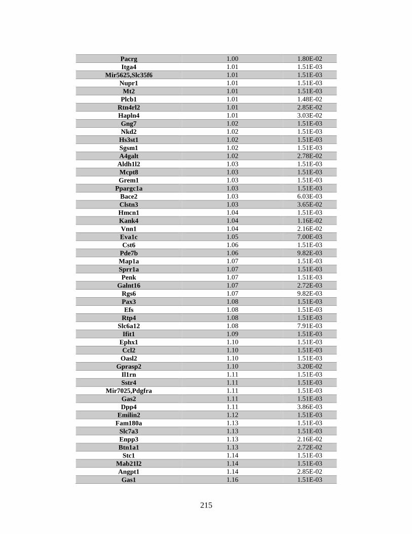
215
Pacrg 1.00 1.80E-02
Itga4 1.01 1.51E-03
Mir5625,Slc35f6 1.01 1.51E-03
Nupr1 1.01 1.51E-03
Mt2 1.01 1.51E-03
Plcb1 1.01 1.48E-02
Rtn4rl2 1.01 2.85E-02
Hapln4 1.01 3.03E-02
Gng7 1.02 1.51E-03
Nkd2 1.02 1.51E-03
Hs3st1 1.02 1.51E-03
Sgsm1 1.02 1.51E-03
A4galt 1.02 2.78E-02
Aldh1l2 1.03 1.51E-03
Mcpt8 1.03 1.51E-03
Grem1 1.03 1.51E-03
Ppargc1a 1.03 1.51E-03
Bace2 1.03 6.03E-03
Clstn3 1.03 3.65E-02
Hmcn1 1.04 1.51E-03
Kank4 1.04 1.16E-02
Vnn1 1.04 2.16E-02
Eva1c 1.05 7.00E-03
Cst6 1.06 1.51E-03
Pde7b 1.06 9.82E-03
Map1a 1.07 1.51E-03
Sprr1a 1.07 1.51E-03
Penk 1.07 1.51E-03
Galnt16 1.07 2.72E-03
Rgs6 1.07 9.82E-03
Pax3 1.08 1.51E-03
Efs 1.08 1.51E-03
Rtp4 1.08 1.51E-03
Slc6a12 1.08 7.91E-03
Ifit1 1.09 1.51E-03
Ephx1 1.10 1.51E-03
Ccl2 1.10 1.51E-03
Oasl2 1.10 1.51E-03
Gprasp2 1.10 3.20E-02
Il1rn 1.11 1.51E-03
Sstr4 1.11 1.51E-03
Mir7025,Pdgfra 1.11 1.51E-03
Gas2 1.11 1.51E-03
Dpp4 1.11 3.86E-03
Emilin2 1.12 1.51E-03
Fam180a 1.13 1.51E-03
Slc7a3 1.13 1.51E-03
Enpp3 1.13 2.16E-02
Btn1a1 1.13 2.72E-02
Stc1 1.14 1.51E-03
Mab21l2 1.14 1.51E-03
Angpt1 1.14 2.85E-02
Gas1 1.16 1.51E-03
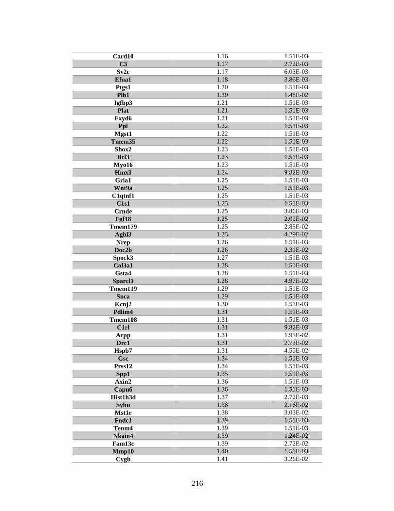
216
Card10 1.16 1.51E-03
C3 1.17 2.72E-03
Sv2c 1.17 6.03E-03
Efna1 1.18 3.86E-03
Ptgs1 1.20 1.51E-03
Plb1 1.20 1.48E-02
Igfbp3 1.21 1.51E-03
Plat 1.21 1.51E-03
Fxyd6 1.21 1.51E-03
Ppl 1.22 1.51E-03
Mgst1 1.22 1.51E-03
Tmem35 1.22 1.51E-03
Shox2 1.23 1.51E-03
Bcl3 1.23 1.51E-03
Myo16 1.23 1.51E-03
Hmx3 1.24 9.82E-03
Gria1 1.25 1.51E-03
Wnt9a 1.25 1.51E-03
C1qtnf1 1.25 1.51E-03
C1s1 1.25 1.51E-03
Crnde 1.25 3.86E-03
Fgf18 1.25 2.02E-02
Tmem179 1.25 2.85E-02
Agbl3 1.25 4.29E-02
Nrep 1.26 1.51E-03
Doc2b 1.26 2.31E-02
Spock3 1.27 1.51E-03
Col3a1 1.28 1.51E-03
Gsta4 1.28 1.51E-03
Sparcl1 1.28 4.97E-02
Tmem119 1.29 1.51E-03
Snca 1.29 1.51E-03
Kcnj2 1.30 1.51E-03
Pdlim4 1.31 1.51E-03
Tmem108 1.31 1.51E-03
C1rl 1.31 9.82E-03
Acpp 1.31 1.95E-02
Drc1 1.31 2.72E-02
Hspb7 1.31 4.55E-02
Gsc 1.34 1.51E-03
Prss12 1.34 1.51E-03
Spp1 1.35 1.51E-03
Axin2 1.36 1.51E-03
Capn6 1.36 1.51E-03
Hist1h3d 1.37 2.72E-03
Sybu 1.38 2.16E-02
Mst1r 1.38 3.03E-02
Fndc1 1.39 1.51E-03
Tenm4 1.39 1.51E-03
Nkain4 1.39 1.24E-02
Fam13c 1.39 2.72E-02
Mmp10 1.40 1.51E-03
Cygb 1.41 3.26E-02
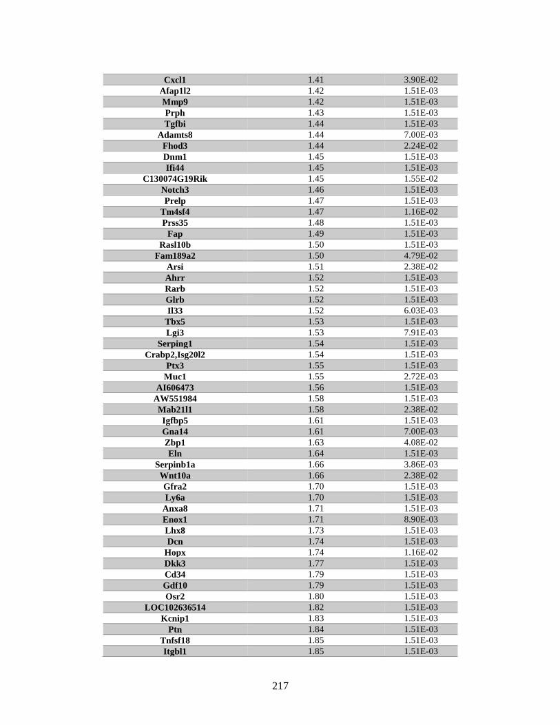
217
Cxcl1 1.41 3.90E-02
Afap1l2 1.42 1.51E-03
Mmp9 1.42 1.51E-03
Prph 1.43 1.51E-03
Tgfbi 1.44 1.51E-03
Adamts8 1.44 7.00E-03
Fhod3 1.44 2.24E-02
Dnm1 1.45 1.51E-03
Ifi44 1.45 1.51E-03
C130074G19Rik 1.45 1.55E-02
Notch3 1.46 1.51E-03
Prelp 1.47 1.51E-03
Tm4sf4 1.47 1.16E-02
Prss35 1.48 1.51E-03
Fap 1.49 1.51E-03
Rasl10b 1.50 1.51E-03
Fam189a2 1.50 4.79E-02
Arsi 1.51 2.38E-02
Ahrr 1.52 1.51E-03
Rarb 1.52 1.51E-03
Glrb 1.52 1.51E-03
Il33 1.52 6.03E-03
Tbx5 1.53 1.51E-03
Lgi3 1.53 7.91E-03
Serping1 1.54 1.51E-03
Crabp2,Isg20l2 1.54 1.51E-03
Ptx3 1.55 1.51E-03
Muc1 1.55 2.72E-03
AI606473 1.56 1.51E-03
AW551984 1.58 1.51E-03
Mab21l1 1.58 2.38E-02
Igfbp5 1.61 1.51E-03
Gna14 1.61 7.00E-03
Zbp1 1.63 4.08E-02
Eln 1.64 1.51E-03
Serpinb1a 1.66 3.86E-03
Wnt10a 1.66 2.38E-02
Gfra2 1.70 1.51E-03
Ly6a 1.70 1.51E-03
Anxa8 1.71 1.51E-03
Enox1 1.71 8.90E-03
Lhx8 1.73 1.51E-03
Dcn 1.74 1.51E-03
Hopx 1.74 1.16E-02
Dkk3 1.77 1.51E-03
Cd34 1.79 1.51E-03
Gdf10 1.79 1.51E-03
Osr2 1.80 1.51E-03
LOC102636514 1.82 1.51E-03
Kcnip1 1.83 1.51E-03
Ptn 1.84 1.51E-03
Tnfsf18 1.85 1.51E-03
Itgbl1 1.85 1.51E-03
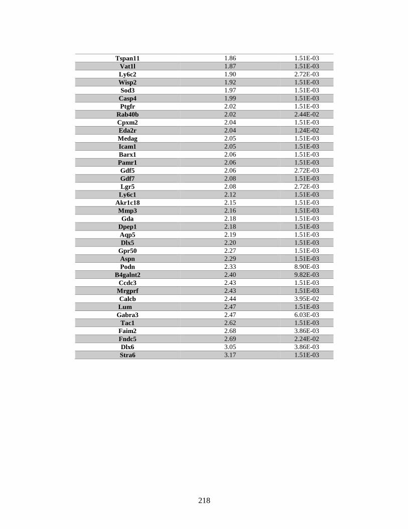
218
Tspan11 1.86 1.51E-03
Vat1l 1.87 1.51E-03
Ly6c2 1.90 2.72E-03
Wisp2 1.92 1.51E-03
Sod3 1.97 1.51E-03
Casp4 1.99 1.51E-03
Ptgfr 2.02 1.51E-03
Rab40b 2.02 2.44E-02
Cpxm2 2.04 1.51E-03
Eda2r 2.04 1.24E-02
Medag 2.05 1.51E-03
Icam1 2.05 1.51E-03
Barx1 2.06 1.51E-03
Pamr1 2.06 1.51E-03
Gdf5 2.06 2.72E-03
Gdf7 2.08 1.51E-03
Lgr5 2.08 2.72E-03
Ly6c1 2.12 1.51E-03
Akr1c18 2.15 1.51E-03
Mmp3 2.16 1.51E-03
Gda 2.18 1.51E-03
Dpep1 2.18 1.51E-03
Aqp5 2.19 1.51E-03
Dlx5 2.20 1.51E-03
Gpr50 2.27 1.51E-03
Aspn 2.29 1.51E-03
Podn 2.33 8.90E-03
B4galnt2 2.40 9.82E-03
Ccdc3 2.43 1.51E-03
Mrgprf 2.43 1.51E-03
Calcb 2.44 3.95E-02
Lum 2.47 1.51E-03
Gabra3 2.47 6.03E-03
Tac1 2.62 1.51E-03
Faim2 2.68 3.86E-03
Fndc5 2.69 2.24E-02
Dlx6 3.05 3.86E-03
Stra6 3.17 1.51E-03

219
APPENDIX C
RAD51D AND RNF138 EXPRESSION CONSTRUCTS
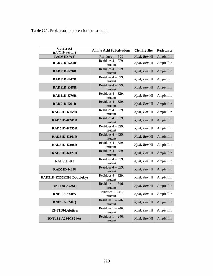
220
Table C.1. Prokaryotic expression constructs.
Construct
(pUC19 vector) Amino Acid Substitutions Cloning Site Resistance
RAD51D-WT Residues 4 – 329 KpnI, BamHI Ampicillin
RAD51D-K24R Residues 4 – 329,
mutant KpnI, BamHI Ampicillin
RAD51D-K26R Residues 4 – 329,
mutant KpnI, BamHI Ampicillin
RAD51D-K42R Residues 4 – 329,
mutant KpnI, BamHI Ampicillin
RAD51D-K48R Residues 4 – 329,
mutant KpnI, BamHI Ampicillin
RAD51D-K76R Residues 4 – 329,
mutant KpnI, BamHI Ampicillin
RAD51D-K91R Residues 4 – 329,
mutant KpnI, BamHI Ampicillin
RAD51D-K159R Residues 4 – 329,
mutant KpnI, BamHI Ampicillin
RAD51D-K201R Residues 4 – 329,
mutant KpnI, BamHI Ampicillin
RAD51D-K235R Residues 4 – 329,
mutant KpnI, BamHI Ampicillin
RAD51D-K261R Residues 4 – 329,
mutant KpnI, BamHI Ampicillin
RAD51D-K298R Residues 4 – 329,
mutant KpnI, BamHI Ampicillin
RAD51D-K327R Residues 4 – 329,
mutant KpnI, BamHI Ampicillin
RAD51D-K0 Residues 4 – 329,
mutant KpnI, BamHI Ampicillin
RAD51D-K298 Residues 4 – 329,
mutant KpnI, BamHI Ampicillin
RAD51D-K235K298 DoubleLys Residues 4 – 329,
mutant KpnI, BamHI Ampicillin
RNF138-A236G Residues 1 – 246,
mutant KpnI, BamHI Ampicillin
RNF138-S240A Residues 1 -246,
mutant KpnI, BamHI Ampicillin
RNF138-S240Q Residues 1 – 246,
mutant KpnI, BamHI Ampicillin
RNF138-Deletion Residues 1 – 246,
mutant KpnI, BamHI Ampicillin
RNF138-A236GS240A Residues 1 – 246,
mutant KpnI, BamHI Ampicillin
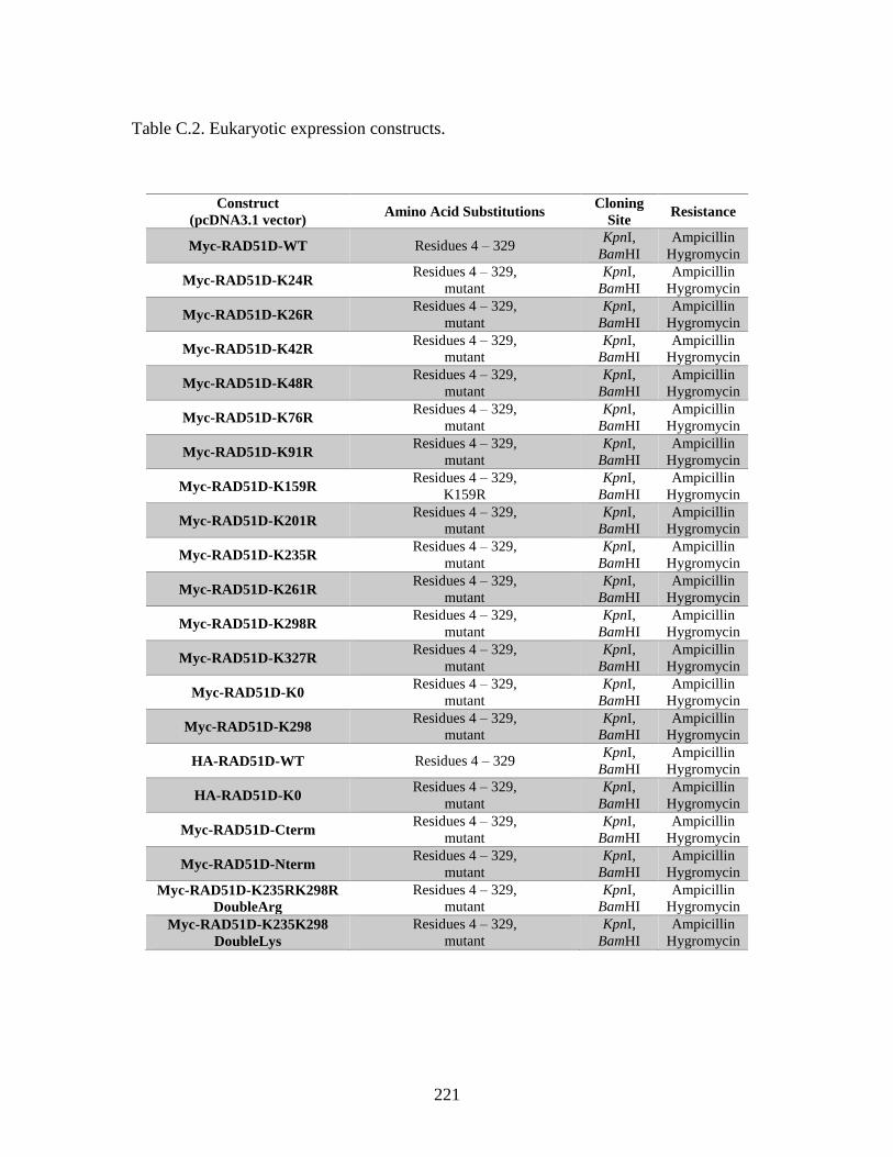
221
Table C.2. Eukaryotic expression constructs.
Construct
(pcDNA3.1 vector) Amino Acid Substitutions
Cloning
Site Resistance
Myc-RAD51D-WT Residues 4 – 329 KpnI,
BamHI
Ampicillin
Hygromycin
Myc-RAD51D-K24R Residues 4 – 329,
mutant
KpnI,
BamHI
Ampicillin
Hygromycin
Myc-RAD51D-K26R Residues 4 – 329,
mutant
KpnI,
BamHI
Ampicillin
Hygromycin
Myc-RAD51D-K42R Residues 4 – 329,
mutant
KpnI,
BamHI
Ampicillin
Hygromycin
Myc-RAD51D-K48R Residues 4 – 329,
mutant
KpnI,
BamHI
Ampicillin
Hygromycin
Myc-RAD51D-K76R Residues 4 – 329,
mutant
KpnI,
BamHI
Ampicillin
Hygromycin
Myc-RAD51D-K91R Residues 4 – 329,
mutant
KpnI,
BamHI
Ampicillin
Hygromycin
Myc-RAD51D-K159R Residues 4 – 329,
K159R
KpnI,
BamHI
Ampicillin
Hygromycin
Myc-RAD51D-K201R Residues 4 – 329,
mutant
KpnI,
BamHI
Ampicillin
Hygromycin
Myc-RAD51D-K235R Residues 4 – 329,
mutant
KpnI,
BamHI
Ampicillin
Hygromycin
Myc-RAD51D-K261R Residues 4 – 329,
mutant
KpnI,
BamHI
Ampicillin
Hygromycin
Myc-RAD51D-K298R Residues 4 – 329,
mutant
KpnI,
BamHI
Ampicillin
Hygromycin
Myc-RAD51D-K327R Residues 4 – 329,
mutant
KpnI,
BamHI
Ampicillin
Hygromycin
Myc-RAD51D-K0 Residues 4 – 329,
mutant
KpnI,
BamHI
Ampicillin
Hygromycin
Myc-RAD51D-K298 Residues 4 – 329,
mutant
KpnI,
BamHI
Ampicillin
Hygromycin
HA-RAD51D-WT Residues 4 – 329 KpnI,
BamHI
Ampicillin
Hygromycin
HA-RAD51D-K0 Residues 4 – 329,
mutant
KpnI,
BamHI
Ampicillin
Hygromycin
Myc-RAD51D-Cterm Residues 4 – 329,
mutant
KpnI,
BamHI
Ampicillin
Hygromycin
Myc-RAD51D-Nterm Residues 4 – 329,
mutant
KpnI,
BamHI
Ampicillin
Hygromycin
Myc-RAD51D-K235RK298R
DoubleArg
Residues 4 – 329,
mutant
KpnI,
BamHI
Ampicillin
Hygromycin
Myc-RAD51D-K235K298
DoubleLys
Residues 4 – 329,
mutant
KpnI,
BamHI
Ampicillin
Hygromycin
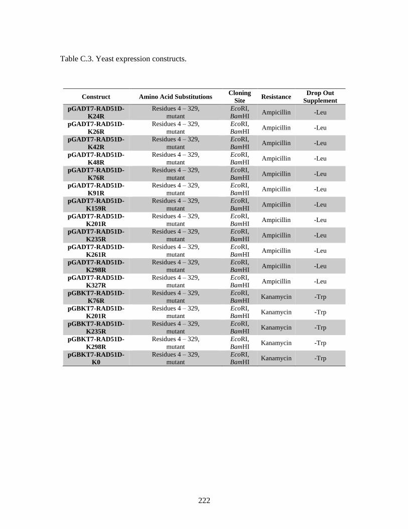
222
Table C.3. Yeast expression constructs.
Construct Amino Acid Substitutions Cloning
Site Resistance
Drop Out
Supplement
pGADT7-RAD51D-
K24R
Residues 4 – 329,
mutant
EcoRI,
BamHI Ampicillin -Leu
pGADT7-RAD51D-
K26R
Residues 4 – 329,
mutant
EcoRI,
BamHI Ampicillin -Leu
pGADT7-RAD51D-
K42R
Residues 4 – 329,
mutant
EcoRI,
BamHI Ampicillin -Leu
pGADT7-RAD51D-
K48R
Residues 4 – 329,
mutant
EcoRI,
BamHI Ampicillin -Leu
pGADT7-RAD51D-
K76R
Residues 4 – 329,
mutant
EcoRI,
BamHI Ampicillin -Leu
pGADT7-RAD51D-
K91R
Residues 4 – 329,
mutant
EcoRI,
BamHI Ampicillin -Leu
pGADT7-RAD51D-
K159R
Residues 4 – 329,
mutant
EcoRI,
BamHI Ampicillin -Leu
pGADT7-RAD51D-
K201R
Residues 4 – 329,
mutant
EcoRI,
BamHI Ampicillin -Leu
pGADT7-RAD51D-
K235R
Residues 4 – 329,
mutant
EcoRI,
BamHI Ampicillin -Leu
pGADT7-RAD51D-
K261R
Residues 4 – 329,
mutant
EcoRI,
BamHI Ampicillin -Leu
pGADT7-RAD51D-
K298R
Residues 4 – 329,
mutant
EcoRI,
BamHI Ampicillin -Leu
pGADT7-RAD51D-
K327R
Residues 4 – 329,
mutant
EcoRI,
BamHI Ampicillin -Leu
pGBKT7-RAD51D-
K76R
Residues 4 – 329,
mutant
EcoRI,
BamHI Kanamycin -Trp
pGBKT7-RAD51D-
K201R
Residues 4 – 329,
mutant
EcoRI,
BamHI Kanamycin -Trp
pGBKT7-RAD51D-
K235R
Residues 4 – 329,
mutant
EcoRI,
BamHI Kanamycin -Trp
pGBKT7-RAD51D-
K298R
Residues 4 – 329,
mutant
EcoRI,
BamHI Kanamycin -Trp
pGBKT7-RAD51D-
K0
Residues 4 – 329,
mutant
EcoRI,
BamHI Kanamycin -Trp
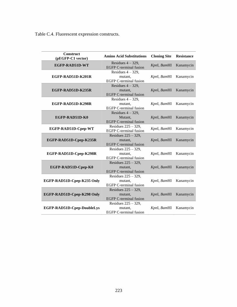
223
Table C.4. Fluorescent expression constructs.
Construct
(pEGFP-C1 vector) Amino Acid Substitutions Cloning Site Resistance
EGFP-RAD51D-WT Residues 4 – 329,
EGFP C-terminal fusion KpnI, BamHI Kanamycin
EGFP-RAD51D-K201R
Residues 4 – 329,
mutant,
EGFP C-terminal fusion
KpnI, BamHI Kanamycin
EGFP-RAD51D-K235R
Residues 4 – 329,
mutant,
EGFP C-terminal fusion
KpnI, BamHI Kanamycin
EGFP-RAD51D-K298R
Residues 4 – 329,
mutant,
EGFP C-terminal fusion
KpnI, BamHI Kanamycin
EGFP-RAD51D-K0
Residues 4 – 329,
Mutant,
EGFP C-terminal fusion
KpnI, BamHI Kanamycin
EGFP-RAD51D-Cpep-WT Residues 225 – 329,
EGFP C-terminal fusion KpnI, BamHI Kanamycin
EGFP-RAD51D-Cpep-K235R
Residues 225 – 329,
mutant,
EGFP C-terminal fusion
KpnI, BamHI Kanamycin
EGFP-RAD51D-Cpep-K298R
Residues 225 – 329,
mutant,
EGFP C-terminal fusion
KpnI, BamHI Kanamycin
EGFP-RAD51D-Cpep-K0
Residues 225 – 329,
mutant,
EGFP C-terminal fusion
KpnI, BamHI Kanamycin
EGFP-RAD51D-Cpep-K235 Only
Residues 225 – 329,
mutant,
EGFP C-terminal fusion
KpnI, BamHI Kanamycin
EGFP-RAD51D-Cpep-K298 Only
Residues 225 – 329,
mutant,
EGFP C-terminal fusion
KpnI, BamHI Kanamycin
EGFP-RAD51D-Cpep-DoubleLys
Residues 225 – 329,
mutant,
EGFP C-terminal fusion
KpnI, BamHI Kanamycin

224
APPENDIX D
PERMISSION TO REPRINT DATA IN CHAPTER 3
225

226
APPENDIX E
PERMISSION TO REPRINT DATA IN CHAPTER 5
227
228
229
230
231