Mechanisms of bacterial resistance to chromium compounds
12
Mechanisms of bacterial resistance to chromium compounds Martha I. Ramı ´rez-Dı ´az Ce ´sar Dı ´az-Pe ´rez Ere ´ndira Vargas He ´ctor Riveros-Rosas Jesu ´s Campos-Garcı ´a Carlos Cervantes Received: 18 April 2007 / Accepted: 25 September 2007 / Published online: 13 October 2007 Ó Springer Science+Business Media, LLC. 2007 Abstract Chromium is a non-essential and well- known toxic metal for microorganisms and plants. The widespread industrial use of this heavy metal has caused it to be considered as a serious environmental pollutant. Chromium exists in nature as two main species, the trivalent form, Cr(III), which is relatively innocuous, and the hexavalent form, Cr(VI), consid- ered a more toxic species. At the intracellular level, however, Cr(III) seems to be responsible for most toxic effects of chromium. Cr(VI) is usually present as the oxyanion chromate. Inhibition of sulfate membrane transport and oxidative damage to bio- molecules are associated with the toxic effects of chromate in bacteria. Several bacterial mechanisms of resistance to chromate have been reported. The best characterized mechanisms comprise efflux of chromate ions from the cell cytoplasm and reduction of Cr(VI) to Cr(III). Chromate efflux by the ChrA transporter has been established in Pseudomonas aeruginosa and Cupriavidus metallidurans (formerly Alcaligenes eutrophus) and consists of an energy- dependent process driven by the membrane potential. The CHR protein family, which includes putative ChrA orthologs, currently contains about 135 sequences from all three domains of life. Chromate reduction is carried out by chromate reductases from diverse bacterial species generating Cr(III) that may be detoxified by other mechanisms. Most character- ized enzymes belong to the widespread NAD(P)H- dependent flavoprotein family of reductases. Several examples of bacterial systems protecting from the oxidative stress caused by chromate have been described. Other mechanisms of bacterial resistance to chromate involve the expression of components of the machinery for repair of DNA damage, and systems related to the homeostasis of iron and sulfur. Keywords Chromate resistance Á Chromate efflux Á Chromate reduction Occurrence of chromium Chromium is the seventh most abundant element on earth and although occurs in oxidation states ranging from Cr(II) to Cr(VI), the trivalent Cr(III) and hexavalent Cr(VI) are the most stable and abundant forms (Cervantes et al. 2001). Cr(VI) is a strong oxidizing agent, commonly present in solution as the M. I. Ramı ´rez-Dı ´az (&) Á C. Dı ´az-Pe ´rez Á E. Vargas Á J. Campos-Garcı ´a Á C. Cervantes Instituto de Investigaciones Quı ´mico-Biolo ´gicas, Universidad Michoacana de San Nicola ´s de Hidalgo, Edificio B-3, Ciudad Universitaria, Morelia, Michoacan 58030, Mexico e-mail: [email protected] H. Riveros-Rosas Departamento de Bioquı ´mica, Facultad de Medicina, Universidad Nacional Auto ´noma de Me ´xico, Mexico, D.F., Mexico 123 Biometals (2008) 21:321–332 DOI 10.1007/s10534-007-9121-8
Transcript of Mechanisms of bacterial resistance to chromium compounds

Mechanisms of bacterial resistance to chromium compounds
Martha I. Ramırez-Dıaz Æ Cesar Dıaz-Perez Æ Erendira Vargas ÆHector Riveros-Rosas Æ Jesus Campos-Garcıa Æ Carlos Cervantes
Received: 18 April 2007 / Accepted: 25 September 2007 / Published online: 13 October 2007
� Springer Science+Business Media, LLC. 2007
Abstract Chromium is a non-essential and well-
known toxic metal for microorganisms and plants.
The widespread industrial use of this heavy metal has
caused it to be considered as a serious environmental
pollutant. Chromium exists in nature as two main
species, the trivalent form, Cr(III), which is relatively
innocuous, and the hexavalent form, Cr(VI), consid-
ered a more toxic species. At the intracellular level,
however, Cr(III) seems to be responsible for most
toxic effects of chromium. Cr(VI) is usually present
as the oxyanion chromate. Inhibition of sulfate
membrane transport and oxidative damage to bio-
molecules are associated with the toxic effects of
chromate in bacteria. Several bacterial mechanisms
of resistance to chromate have been reported. The
best characterized mechanisms comprise efflux of
chromate ions from the cell cytoplasm and reduction
of Cr(VI) to Cr(III). Chromate efflux by the ChrA
transporter has been established in Pseudomonas
aeruginosa and Cupriavidus metallidurans (formerly
Alcaligenes eutrophus) and consists of an energy-
dependent process driven by the membrane potential.
The CHR protein family, which includes putative
ChrA orthologs, currently contains about 135
sequences from all three domains of life. Chromate
reduction is carried out by chromate reductases from
diverse bacterial species generating Cr(III) that may
be detoxified by other mechanisms. Most character-
ized enzymes belong to the widespread NAD(P)H-
dependent flavoprotein family of reductases. Several
examples of bacterial systems protecting from the
oxidative stress caused by chromate have been
described. Other mechanisms of bacterial resistance
to chromate involve the expression of components of
the machinery for repair of DNA damage, and
systems related to the homeostasis of iron and sulfur.
Keywords Chromate resistance �Chromate efflux � Chromate reduction
Occurrence of chromium
Chromium is the seventh most abundant element on
earth and although occurs in oxidation states ranging
from Cr(II) to Cr(VI), the trivalent Cr(III) and
hexavalent Cr(VI) are the most stable and abundant
forms (Cervantes et al. 2001). Cr(VI) is a strong
oxidizing agent, commonly present in solution as the
M. I. Ramırez-Dıaz (&) � C. Dıaz-Perez �E. Vargas � J. Campos-Garcıa � C. Cervantes
Instituto de Investigaciones Quımico-Biologicas,
Universidad Michoacana de San Nicolas de Hidalgo,
Edificio B-3, Ciudad Universitaria, Morelia, Michoacan
58030, Mexico
e-mail: [email protected]
H. Riveros-Rosas
Departamento de Bioquımica, Facultad de Medicina,
Universidad Nacional Autonoma de Mexico, Mexico,
D.F., Mexico
123
Biometals (2008) 21:321–332
DOI 10.1007/s10534-007-9121-8
hydrochromate (HCrO4–), chromate (CrO4
2–) or di-
chromate (Cr2O72–) oxyanions, depending on the pH
(US EPA 1998). Cr(VI) exists as water-soluble
anions that may persist in water for long periods
and is considered as a key contaminant at the US
Department of Energy waste sites (Riley et al. 1992).
Cr(III) derivatives are much less mobile and exist in
the environment mostly forming stable complexes
with both organic and inorganic ligands (Zayed and
Terry 2003). In addition, Cr(III) is less toxic because
is less soluble at physiological pH. At neutral pH,
Cr(III) tends to precipitate as hydroxide [Cr(OH)3] or
hydrated oxide (Cr2O.H2O) (Ehrlich 2002). The low
solubility of Cr(III) [mostly as Cr2O3 and Cr(OH)3] is
likely the major reason why Cr(III) makes up a small
percentage of the total chromium concentration in
polluted groundwater (US EPA 1998). Mobilisation
of the Cr(OH)3 precipitate is slow, unless enhanced
by dissolution in strongly acidic environments or by
being complexed with organic compounds (Rai et al.
1987). The widespread use of Cr in diverse industrial
processes has converted it into a serious contaminant
of air, soil and water (Khasim et al. 1989).
Biological properties of chromium
Transmembrane transport of chromium
It has been demonstrated in a variety of bacterial
species that chromate actively crosses biological
membranes by means of the sulfate uptake pathway,
which reflects the chemical analogy between these
two oxyanions (Fig. 1) (Cervantes and Campos-
Garcıa 2007). Cr(III) crosses cell membranes with a
low efficiency because it forms insoluble compounds
(Cary 1982). Inside the cell, Cr(VI) is readily reduced
to Cr(III) by the action of various enzymatic or
nonenzymatic activities; the Cr(III) generated may
then exert diverse toxic effects in the cytoplasm
(Fig. 1) (Cervantes et al. 2001).
Toxic effects of Cr(VI)
The biological effects of Cr strongly depend on its
oxidation state and cellular localization. Cr(VI) is
considered the most toxic form of Cr (for a review on
CYTOPLASM
PERIPLASM
nietorPegamad
DOSesalataCevitadixO
sserts
enoihtatulG)IV(rC
OS 4-2
X
ANDegamad
noitcudeR
OrC 4-2
riapeRsmetsys
)DOS(CrhC
MEMBRANE
dimsalP
OrC 4-2
OrC 4-2
OrC 4-2
OrC 4-2
OrC 4-2
OrC 4-2
OrC 4-2
)IV(rC
noitcudeR
)III(rC
C
B
A
E
D
OrC 4-2
)III(rC
F
=
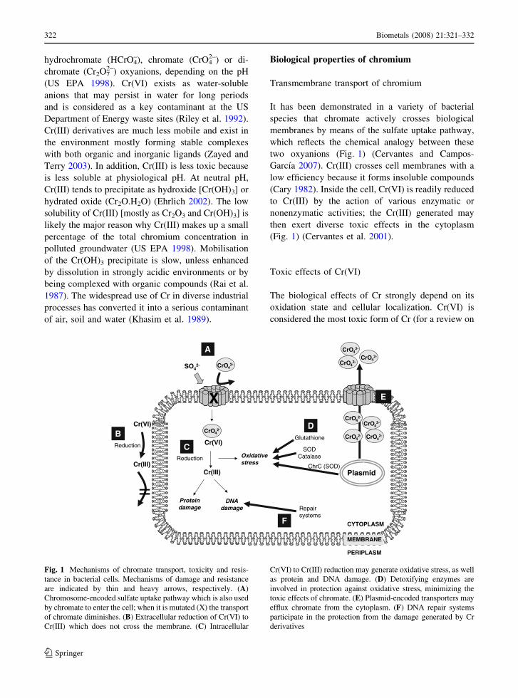
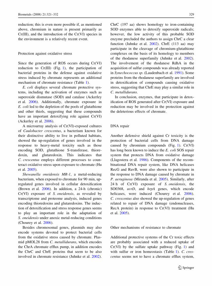
Fig. 1 Mechanisms of chromate transport, toxicity and resis-
tance in bacterial cells. Mechanisms of damage and resistance
are indicated by thin and heavy arrows, respectively. (A)
Chromosome-encoded sulfate uptake pathway which is also used
by chromate to enter the cell; when it is mutated (X) the transport
of chromate diminishes. (B) Extracellular reduction of Cr(VI) to
Cr(III) which does not cross the membrane. (C) Intracellular
Cr(VI) to Cr(III) reduction may generate oxidative stress, as well
as protein and DNA damage. (D) Detoxifying enzymes are
involved in protection against oxidative stress, minimizing the
toxic effects of chromate. (E) Plasmid-encoded transporters may
efflux chromate from the cytoplasm. (F) DNA repair systems
participate in the protection from the damage generated by Cr
derivatives
322 Biometals (2008) 21:321–332
123

the toxic effects of Cr see US EPA 1998) and is
known to cause irritation of the skin and the
respiratory tract, and lung carcinoma in humans.
Occupational exposure to chromate is considered a
serious toxicological problem, as it has been demon-
strated that Cr(VI) is a human carcinogen (Riveros-
Rosas et al. 1997; De Flora 2000). In bacteria, at the
extracellular level, Cr(VI) is highly toxic because it
rapidly enters to the cytoplasm where it may exert its
toxic effects (Wong and Trevors 1988; Katz and
Salem 1993). In the cytoplasm, Cr toxicity is mainly
related to the process of reduction of Cr(VI) to lower
oxidation states [i.e., Cr(III) and Cr(V)] in which free
radicals may be formed (Fig. 1) (Shi and Dalal 1990;
Kadiiska et al. 1994). Oxidative damage to DNA is
probably responsible for the genotoxic effects caused
by chromate (Kawanishi et al. 1986; Aiyar et al.
1991; Itoh et al. 1995; Luo et al. 1996).
Toxic effects of Cr(III)
Cr(III) is classified as an essential trace element for
humans, since it seems to participate in the metab-
olism of glucose and lipids (Anderson 1997; Vincent
2004). However, Cr seems not to be required by
microorganisms (Wong and Trevors 1988) or plants
(Shanker et al. 2005). At the extracellular level,
Cr(III) is relatively innocuous as a consequence of its
insolubility and subsequent inability to cross cell
membranes (Wong and Trevors 1988; Katz and
Salem 1993).
Inside the cell, Cr(III) may generate toxic effects
by its ability to bind to phosphates in DNA
(Kortenkamp et al. 1991, Bridgewater et al. 1994,
Plaper et al. 2002) (Fig. 1). The main forms of
chromium-DNA adducts in mammalian cells are
ternary complexes generated by cross-linking of
cysteine and histidine to DNA via a phosphate-bound
Cr(III) atom (Zhitkovich et al. 1996). Tyrosine and
cysteine exhibited the highest activity in being
complexed to DNA by Cr(III) in vitro (Salnikow
et al. 1992). Cr(III) may exert additional toxic effects
by its ability to bind to carboxyl and sulfhydryl
groups in proteins (Levis and Bianchi 1982), and in
human cells by competing with the transport of iron
by transferrin (Moshtaghie et al. 1992). In Saccha-
romyces cerevisiae, oxidative damage to proteins has
been established as a central mechanism of Cr
toxicity (Sumner et al. 2005).
Bacterial mechanisms of chromate resistance
A variety of chromate-resistant bacterial isolates has
been reported, and the mechanisms of resistance to
this ion may be encoded either by plasmids or by
chromosomal genes (Nies et al. 1998; Cervantes and
Campos-Garcıa 2007). Usually, the genes located in
plasmids encode membrane transporters, which
directly mediate efflux of chromate ions from the
cell’s cytoplasm (Fig. 1). On the other hand, resis-
tance systems encoded within bacterial chromosomes
are generally related to strategies such as specific or
unspecific Cr(VI) reduction, free-radical detoxifying
activities, repairing of DNA damage, and processes
associated with sulfur or iron homeostasis (Fig. 1).
Table 1 summarizes the bacterial strategies that have
been related to chromate tolerance.
Transmembrane efflux of chromate
The efflux of chromate is a resistance mechanism
conferred by the ChrA protein (Table 1). ChrA is
encoded by plasmids pUM505 of Pseudomonas
aeruginosa and pMOL28 from Cupriavidus metalli-
durans (previously Alcaligenes eutrophus and
Ralstonia metallidurans) (Cervantes et al. 1990, Nies
et al. 1990). ChrA from P. aeruginosa, of 416 amino
acids (aa), displays a topology of 13 transmembrane
segments (TMS) (Jimenez-Mejia et al. 2006). ChrA
functions as a chemiosmotic pump that effluxes
chromate from the cytoplasm using the proton motive
force (Alvarez et al. 1999; Pimentel et al. 2002). In
vitro and in vivo efflux of chromate showed satura-
tion kinetics with similar Km values of 0.12 and
0.08 mM chromate, respectively (Alvarez et al.
1999; Pimentel et al. 2002). Efflux of chromate is
inhibited by sulfate, suggesting that this analog
oxyanion may also bind to the ChrA protein (Pimen-
tel et al. 2002). In fact, it has been proposed that
ChrA may function as a chromate/sulfate antiporter
(Nies et al. 1998) nevertheless, sulfate transport by
the ChrA proteins has not yet been determined.
Random mutagenesis of the P. aeruginosa chrA
gene showed that most essential amino acid residues
Biometals (2008) 21:321–332 323
123

are located in the amino terminal end of ChrA
(Aguilera et al. 2004). In agreement with this finding,
phylogenetic analysis of ChrA homologs revealed
that the amino terminal halves are more conserved
than the carboxyl terminal halves (Dıaz-Perez et al.
submitted). A similar situation was reported for
transporters of the closely related major facilitator
superfamily (MFS) (Pao et al. 1998). These data
suggest that the two halves of ChrA carry out
different roles in their transporting functions.
The ChrA protein (401 aa) from Cupriavidus
displays a different topology of 10 TMS and shows
29% of identical aa with respect to ChrA from
Pseudomonas (Nies et al. 1998). The resistance mech-
anism, however, seems to be the efflux of chromate ions
like in Pseudomonas (Nies et al. 1990). In addition, the
C. metallidurans chromosome contains the chrA2 gene,
encoding a protein 84% identical to the product of its
plasmid-encoded ChrA homolog (chrA1). Expression of
the ChrA2 protein also confers chromate resistance
(Juhnke et al. 2002). Each chrA determinant increased
chromate resistance just two-fold, whereas in the
presence of both chrA determinants resistance increased
four-fold in low-sulfate medium and five-fold in high-
sulfate medium (Juhnke et al. 2002). This is an indica-
tion of the importance of both determinants in the
chromate resistance by C. metallidurans.
The complete sequencing of plasmid pB4 (79
kilobases) from a Pseudomonas sp. strain revealed
the presence of a chrA homologous gene that shares
high sequence similarity with the chromate resistance
determinant from plasmid pUM505 (93% aa identity)
(Tauch et al. 2003). Sequence analysis showed that
the chrA gene of pB4 forms part of the Tn5719
transposon. Additionally, the analysis revealed that
the chr homologous region in pUM505 still contains
remnants of transposon sequences, which suggests
that Tn5719 is an ancestor of the chromate resistance
determinant of pUM505 (Tauch et al. 2003); chro-
mate resistance by pB4 has not been determined.
In summary, the efflux of chromate seems to be an
efficient and widespread mechanism of resistance,
which prevents the accumulation of this toxic ion
inside the cell.
Table 1 Bacterial systems related to chromate tolerance
Enzyme/system Species Function Reference
Transport
ChrA transporter Pseudomonas aeruginosa Efflux of cytoplasmic chromate Alvarez et al. 1999
Cys operon products Shewanella oneidensis Sulfate transport Brown et al. 2006
TonB receptor, hemin transporter Shewanella oneidensis Iron transport Brown et al. 2006
Reduction
Chromate reductases Diverse species Reduction of Cr(VI) to Cr(III) Cervantes et al. 2001b
General and oxidative stress
SOD, catalase Escherichia coli Combat of oxidative stress Ackerley et al. 2004
SOD, glutathione transferase Caulobacter crescentus Combat of oxidative stress Hu et al. 2005
Outer membrane proteins Caulobacter crescentus General stress response Hu et al. 2005
Fe-SOD ChrCa Cupriavidus metallidurans Detoxification of free radicals Juhnke et al. 2002
DNA repair
SOS response Escherichia coli Repair of DNA damage Llagostera et al. 1986
RecG and RuvB DNA helicases Pseudomonas aeruginosa Repair of DNA damage Miranda et al. 2005
SO0368, UvrD, and HrpA helicases Shewanella oneidensis Repair of DNA damage Chourey et al. 2006
Other mechanisms
Cys operon products Shewanella oneidensis Sulfur metabolism Brown et al. 2006
Adenylyl sulfate kinase Shewanella oneidensis Sulfur metabolism Brown et al. 2006
Sulfite reductase Shewanella oneidensis Sulfur metabolism Brown et al. 2006
Ferritin Shewanella oneidensis Iron binding Brown et al. 2006
a Plasmid-encoded genesb See this review for species’s names
324 Biometals (2008) 21:321–332
123

The CHR superfamily of transporters
The CHR superfamily of transporters, classified as
TC # 2.A.51 (Saier 2003), is a group of proteins
probably involved in chromate or sulfate transport
(Nies et al. 1998). The databases of the CHR protein
family currently contain 135 sequences of homologs,
including proteins from eukaryotes (Cervantes and
Campos-Garcıa 2007). With the exception of the
P. aeruginosa and C. metallidurans ChrA proteins,
the function of other CHR homologs has not yet been
analyzed in detail. CHR homologs exist in two sizes
(Nies et al. 1998, Dıaz-Perez et al. submitted):
(1) Small proteins, or SCHR (about 200 aa),
possess only one domain. Sequence analysis
suggests that these proteins may form a paralog
group inside the CHR superfamily (see below).
(2) Large proteins, or LCHR (about 400 aa, except
eukaryotic proteins of 500–600 aa), with two
homologous domains.
Further sequence analysis suggested that LCHR
proteins may have derived from a gene duplication
event, as occurred with members of other families
of transporters (Pao et al. 1998). The fact that
several genomes contain two separated tandem
copies of the genes for amino- and carboxyl-
terminal parts of a large CHR also supports the
hypothesis of a different function for each protein
half (Nies et al. 1998).
The LCHR proteins are arranged in six subfamilies
from bacteria (LCHR1 to LCHR6), and one subfam-
ily from fungi (Dıaz-Perez et al. submitted). The
LCHR1 subfamily contains all the Gram positive
homologs. The ChrA proteins from C. metallidurans
and P. aeruginosa, with a demonstrated function in
chromate efflux, are located into the LCHR2 and
LCHR5 subfamilies, respectively, which also include
mainly proteins from proteobacteria. The LCHR3
subfamily, closer to LCHR2 and LCHR5, may also
contain functional chromate transporters. The
LCHR4 subfamily includes a protein from Desulf-
ovibrio vulgaris and the only protein from an Archaea
(Methanococcus jannaschii). The fungal CHR sub-
family contains six proteins from fungal species that
are significantly larger than their bacterial counter-
parts, due to a large interdomain sequence, and are
most closely related to the LCHR1 subfamily. No
proteins from these subfamilies have yet been
studied.
When the large proteins were divided into their
moieties amino- and carboxyl-terminal domains and
aligned with the small proteins, a separate distribu-
tion of the SCHR and the LCHR groups was found
(Dıaz-Perez et al. submitted). This suggested that the
SCHR proteins form a paralog group inside the CHR
family and that they probably carry out a function
different to chromate transport.
Thus, the CHR superfamily is a widespread group
of proteins, which includes chromate transporters that
probably evolved recently as a result of chromate
exposure by bacteria.
Chromate reduction
Bacterial reduction of metallic ions has been shown
to occur for U(VI), Se(VI), Cr(VI), Mo(VI), Se(IV),
Hg(II), Ag(I) and others (Lovley 1993; Bradley and
Obraztsova 1998). A wide range of bacteria has been
identified that are capable of carrying out a complete
reduction of Cr(VI) to Cr(III) by oxidation–reduction
reactions of biotic and abiotic nature. Microbial
reduction of Cr(VI) to Cr(III) can be considered as
an additional chromate resistance mechanism
which is not usually a plasmid-associated trait
(Cervantes et al. 2001). Cr(VI) reduction outside
the cell generates Cr(III) which cannot cross cellular
membranes.
Three Cr(VI) reduction mechanisms have been
described (Cervantes and Campos-Garcıa 2007):
(i) In aerobic conditions, chromate reduction has
been commonly associated with soluble chro-
mate reductases that use NADH or NADPH as
cofactors.
(ii) Under anaerobiosis, some bacteria, like Pseu-
domonas fluorescens LB300 (Bopp and Ehrlich
1988), can use Cr(VI) as an electron acceptor in
the electron transport chain.
(iii) Reduction of Cr(VI) may also be carried out by
chemical reactions associated with compounds
such as amino acids, nucleotides, sugars,
vitamins, organic acids or glutathione. For
instance, ascorbate is capable of reducing
Cr(VI), and riboflavin derivatives FAD and
Biometals (2008) 21:321–332 325
123

FMN are essential coenzymes for chromate-
reducing flavoenzymes (Masayasu 1991).
Enzymatic reduction of chromate
Chromate reduction is carried out by diverse bacterial
species (Ohtake and Silver 1994; Cervantes et al.
2001) (Table 1). This reduction may be associated
with the cell membrane or with the soluble fraction,
and may occur either under aerobic or anaerobic
conditions. The first enzyme described with the
ability to transform Cr(VI) to Cr(III) was a Cr(VI)
reductase from chromate-resistant Enterobacter clo-
acae HO1 (Ohtake et al. 1990). This is a membrane-
associated enzyme that transfers electrons to Cr(VI)
by NADH-dependent cytochromes (Wang et al.
1990).
Several bacterial Cr(VI) reductases, some confer-
ring resistance to chromate, have been subsequently
characterized. These enzymes commonly show a
NADH:flavin oxidoreductase activity and can use
Cr(VI) as electron acceptor (Gonzalez et al. 2005).
Ishibashi et al. (1990) suggested that the ability to
reduce chromate may be a secondary function for
Cr(VI) reductases, which have a different primary
role other than Cr(VI) reduction. The nitroreductases
NfsA/NfsB from Vibrio harveyi possess a nitrofura-
zone nitroreductase as primary activity and a Cr(VI)
reductase activity as a secondary function (Kwak
et al. 2003). Similarly, ferric reductase FerB from
Paracoccus denitrificans uses both Fe(III)-nitrilotri-
acetate and Cr(VI) as substrates (Mazoch et al. 2004).
These secondary functions may be related to the
bacterial enzymatic adaptation as a result of the
relatively recent increase of Cr(VI) content in the
environment due to anthropogenic activities (Silver
and Phung 1996).
ChrR from Pseudomonas putida is the currently
best studied Cr(VI) reductase. ChrR is a soluble flavin
mononucleotide-binding protein (Park et al. 2000).
This enzyme functions as a 50-kDa dimer and shows a
NADH-dependent reductase activity. This multifunc-
tional protein, besides its role as Cr(VI) reductase,
also reduces ferricyanide (Ackerley et al. 2004).
Studies with enzyme mutants showed that ChrR
protects against chromate toxicity; this is possibly
because it preempts chromate reduction by the
cellular one-electron reducers, thereby minimizing
reactive oxygen species (ROS) generation (Ackerley
et al. 2004). During Cr(VI) reduction, ChrR shows a
quinone reductase activity that generates a flavin
semiquinone. By this reaction, the enzyme transfers
[25% of the NADH electrons to superoxide anion
and probably produces the Cr(V) species transiently
(Fig. 2). Indeed, ChrR in one pathway reduces Cr(VI)
to Cr(III), generating intermediary Cr(V) and super-
oxide anion, and by an additional mechanism reduces
quinones, which provide shielding against ROS
(Fig. 2). ChrR contains the sequence signature
LFVTPEYNXXXXXXLKNAIDXXS as a member
of the COG0431 (prokaryotic Cluster of Orthologous
Groups of proteins), or KOG4530 (eukaryotic orthol-
ogous groups) (Tatusov et al. 2003), named also as
NAD(P)H-dependent FMN reductase family (Pfam
accession number: PF0358) (Finn et al. 2006). This
protein family is a member of the flavoprotein clan
that includes FMN- or FAD-binding redox proteins.
The Escherichia coli YieF Cr(VI) reductase
shares sequence homology with the P. putida
ChrR enzyme (Ackerley et al. 2004). Both soluble
enzymes are members of a widespread family of
proteins and show similar kinetic and physicochem-
ical properties. YieF has a broad substrate range and
can reduce, in addition to Cr(VI), substrates like
ferricyanide, vanadium (V), molybdenum (VI), sev-
eral quinones, 2,6-dichloroindophenol (Ackerley
et al. 2004), and even the prodrugs mitomycin C
and 5-aziridinyl-2,4-dinitrobenzamide (Barak et al.
2006). The action of YieF involves an obligatory
four-electron reduction of Cr(VI) by the protein
dimer (50 kDa), in which the enzyme simultaneously
transfers three electrons to Cr(VI) to produce Cr(III)
and one electron to molecular oxygen generating
ROS; no flavin semiquinone is generated during this
process (Ackerley et al. 2004). YieF may thus
provide to E. coli an effective protection mechanism
against chromate toxicity by forming a lower amount
of ROS.
Another E. coli enzyme, the Fre flavin reductase,
reduces Cr(VI) by a different strategy that involves
complexation of Cr(III) with the NAD+ cofactor
(Puzon et al. 2002). This interaction may be related
to the notorious ability of Cr(III) to form adducts with
DNA.
In conclusion, Cr(VI) reduction seems to be an
efficient system of resistance to chromate in bacteria;
however, the use of alternative substrates in addition
326 Biometals (2008) 21:321–332
123

to Cr(VI) by chromate reductases suggests that this
reduction activity has been an adaptive mechanism
promoted by recent chromate exposure.
The NAD(P)H-dependent FMN reductase family
The NAD(P)H-dependent FMN reductase
(FMN_red) protein family, which includes putative
ChrR orthologs, currently comprises 243 homologous
dimeric or tetrameric proteins that bind the FMN
cofactor. Members of the group are widespread,
suggesting an early evolutionary origin of this protein
family. The utilization of NAD(P)H and the absence
of a flavin semiquinone radical distinguish this
protein family from flavodoxins, which adopt the
same structural fold, i.e. a five-stranded b sheet
sandwiched by five a helices (Deller et al. 2006). The
FMN_red protein family may be divided into ten
main clusters, where each one probably corresponds
to different protein subfamilies. Only three of the
above mentioned protein clusters include character-
ized proteins and can be defined as subfamilies, using
criteria outlined previously for other protein groups
(Riveros-Rosas et al. 2003).
Subfamily I is the most numerous group (73
homologous sequences), and is present mainly in
proteobacteria (Fig. 3). The ChrR enzyme of P. put-
ida (accession number AAK56852) is the only well-
characterized protein included inside this subfamily
(Fig. 3).
Subfamily II, with 32 homologous proteins, is
present in archaea, bacteria, mainly proteobacteria,
and plants (Fig. 3). The YieF protein from E. coli
(Ackerley et al. 2004), the FMN reductase from
P. aeruginosa PAO1 (Agarwal et al. 2006) and the
NAD(P)H:quinone reductase (NQR) of Arabidopsis
thaliana (Sparla et al. 1999) are included in this
subfamily (Fig. 3).
Subfamily III comprises nine homologous proteins
reported in firmicutes, fungi, and mycetozoa (Dictyo-
stelium discoideum) (Fig. 3). Two characterized
proteins belong to this subfamily: the azoreductase
from Bacillus sp. OY1-2 (Suzuki et al. 2001) and
the dimeric S. cerevisiae YLR011wp protein (Liger
et al. 2004) (Fig. 3). The Bacillus protein transforms
azo dyes into colourless compounds, a reaction
mediated by a reductase activity for the azo group in
the presence of NADPH (Suzuki et al. 2001).
YLR011wp from S. cerevisiae also shows a weak
rC III
rC V
rC IV
SOR
selucelomoiBRrhC xO
RrhC deR
RrhC xO
HDAN
DAN +
senoniuQ xO
)QoC,K.tiV(O2 HOL, 2 H, 2O2
O(SOR 2.
OL, 2.
H, 2O2)senoniuQ deR
Fig. 2 Chromate reduction and protection mechanism by Pseu-domonas putida ChrR chromate reductase. Cr(VI) is reduced to
Cr(V) by ChrR, previously reduced by NADH; Cr(V) is next
converted to Cr(III) by diverse biomolecules generating reactive
oxygen species (ROS). ROS may be eliminated by alternative
mechanisms (i.e. catalases or peroxidases) or by the additional
function of ChrR. ChrR in a reduced status may reduce quinones
(such as vitamin K or coenzyme Q) which may then detoxify
previously formed ROS. ChrROx, ChrRRed, oxidized or reduced
forms of the ChrR chromate reductase, respectively; quinonesOx,
quinonesRed, oxidized or reduced forms of quinones, respectively;
O2�, superoxide radical; LO2�, lipoperoxide radical; H2O2,
hydrogen peroxide; Vit. K, vitamin K; CoQ, coenzyme Q. Model
based on data from Ackerley et al. 2004 and Gonzalez et al. 2005
Biometals (2008) 21:321–332 327
123
but specific reductive activity over azo dyes and
nitrocompounds, in addition to a strong ferricyanide
reductase activity (Liger et al. 2004).
In summary, the multifunctional abilities of the
FMN_red family members make it unlikely that the
primary role of this protein family is chromate
57 1-. psR-3 8251110
PZ
ZP
0092
2950
-Sdy
sent
eria
e-18
8Y
P40
9969
-Sbo
ydii-
188
AA
K62
985-
Eco
li-18
8Z
P01
5895
71-E
sp.-
188
NP
4627
49-S
typh
imur
ium
-194
AA
R87
716-
Sen
teric
a-19
4
ZP00
8320
85-Y
inte
rmed
ia-1
51
ZP01
5355
06-S
prot
eam
acul
ans-
188
ZP00
8293
26-Y
frede
rikse
nii-1
9
ZP00
8222
08-Y
berc
ovie
ri-19
0
ZP00
8256
28-Y
mol
lare
tii-1
90
ZP01
1995
51-X
auto
troph
icus
-18
ZP01
4011
38-V
eise
niae
-220
ZP01
5797
34-D
acido
vora
ns-1
93
B-31005LAA
c
781-aicape
AAL500
15-B
cepa
cia-1
88
YP677362-C
hutchinso
nii-189
YP 255894-Sacid
ocaldariu
s-191
YP 023262-Ptorrid
us-183
ZP 00609088-Facidarm
anus-184
CAJ38023-u methanogeni -c 186
YP 285287-D aromatica-192
NP 900200-C violaceum-163
YP 285997-D aromatica-182
1RTT A-P aeruginosa-193
ZP 01427093-H aurantiacus-187
NP 001045430-O sativa-203
NP 001045429-O sati av -197
ABE79156 M- truncatula-191
Q8H9D2-S tuberosum-194AAD37373-A thaliana-196NP 793482-P s ry ingae-194ZP 01528230-P mendocina-184
YP 260394-P fluorescens-189
AAK56852-P putida-186
YP 609081-P entomophila-185
YP 296226-R eutropha-187
YP 584169-R metallidurans-186
NP 771427-B japoni uc m-183
ZP 00859672-B sp.-185
NP 948210-R palustris-186
YP 318433-Nwinogradskyi-185
YP577022-N
hamburgensis-185
ZP01445991-R
sp.-187
7447
15PY
-D
esnei
nfah
-78
1
YP818357-L
mesenteroides-180
NP784063-L
plantarum-182
ZP01354102-C
phytofermentans-184
YP803580-P
pentosaceus-182
YP810458-O
oeni-185
NP
815404-Efae
ac lis-160
YP
395928-Lsakei-185
ZP00133903-A
pleuropneumoniae-183
YP
087203-Msucciniciproducens-183
YP
207281-Ngonorrhoeae-161
NP
283473-Nm
eningitidis-188
ZP
00380533-Blinens-190
NP
940565-Cdiphtheriae-178C
AD
4791
2A-
nico
tinov
oran
s-20
4091-snanegolahedA-204664
PYY
P76
5289
-Rle
gum
inos
arum
-203
YP
4728
45-R
etli
1-90
ZP
0104
5532
-Nsp
.-19
0
YP
5791
10-N
ham
burg
ensi
s-19
0
NP
1022
15-M
loti-
192
NP
1055
17-M
loti-
189
YP
4281
65-R
rubr
um-1
84
ZP01
2285
51-A
sp.-1
92
ZP00
6286
59-P
deni
trifi
acn
-s18
2
YP09
6447
-Lpn
eum
ophi
la-1
83
NP
5230
54-R
sola
nace
arum
-184
ZP00
9781
94-B
ceno
cepa
cia-1
84
YP37
0770
-Bsp
.-184
ZP01
5564
52-B
ambif
aria
-184
cB-348677
PY
e
aicap
-481
ZP00
4223
22-B
v ei tnam
ei nsis-
184
ZP01568266-B
multivorans-1
83
ZP 00983603-Bdolosa
-185
YP 440554-Bthailandensis
-185
YP 106617-Bmallei-185
YP 112348-B p es udomallei-185
ZP 01500943-B phymatum-183
ZP 01508101-B phytofirmans-184
YP 555270-B xenovorans-184
CA 5J 0200-B avium-183
NP 884513-B parapertussis-184NP 880298-B pertussis-184NP 641810-X axonopodis 1- 84YP 363265 X- campestris-184
YP 200163-X oryzae-184
NP 636806 X- campestris-184
ZP 01578107-D acido ov rans-182
ZP 01518879-C testosteroni-185
YP 549190-P sp.-183
ZP 01020227-P naphthalenivoran-206
ZP 01407067-A avenae-184
ZP 01381431-A sp.-183
YP 412267-N multiformis-184
YP 523616-R ferrireducens-183
ZP 00590046-P phaeoclathratiforme-185
ZP 00529808-C phaeobacteroides-183
ZP 01386182-Cferrooxidans-190
NP662348-C
tepidum-187
ZP00864144-B
sp.-182
aesir
gM-
3969
53PX
-01
2
XP748851-A
fumigatus-195
NP013111-S
cerevisiae-191
XP638939-D
discoideum-199
Q9FAW
5-Bsp.-178
ZP00236672-B
cereus-178
ZP01184139-B
weihenstephanens-178
NP
844662-Banthracis-178
YP
036388-Bthuringiensis-178
0.1
γP
rote
obac
teria
α
Prot
eoba
tcer
ia
β Prote
obac
teria
Bacteroidetes
EAHCRA
A
β Proteo-
bacteria
γ Proteo-
bacteria
Chlorof el xi
PLANTS
γ Proteo-bacteria
β Proteoba tc e ir a
α Proteo-bacteria
Firmicutes
γP
roteo-
bacteria
βP
roet o-
bacet ria
Actinobacteria
δa iret caboe tor
P
αP
rote
obac
etir a
Firmicutes
MYC
ETOZO
A
FUNGI
α Proteo-bacteria
γ Pro
teob
acetria
β Proteobacteria
γ Proteo-bacteria
β Proteobacteria
Chlorobium
I
II
III
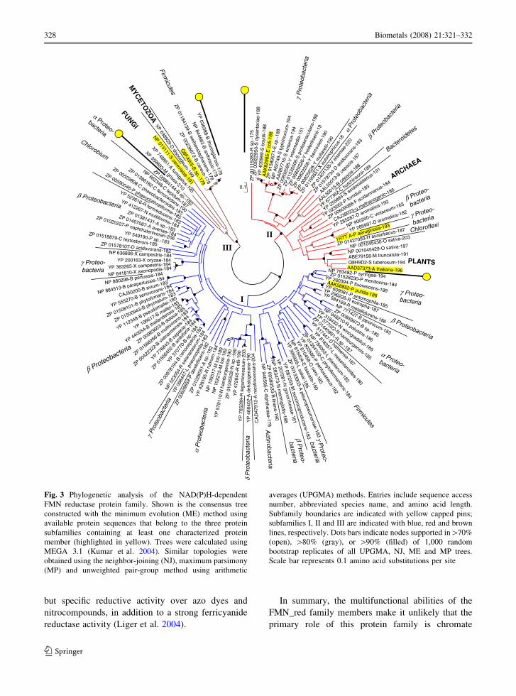
Fig. 3 Phylogenetic analysis of the NAD(P)H-dependent
FMN reductase protein family. Shown is the consensus tree
constructed with the minimum evolution (ME) method using
available protein sequences that belong to the three protein
subfamilies containing at least one characterized protein
member (highlighted in yellow). Trees were calculated using
MEGA 3.1 (Kumar et al. 2004). Similar topologies were
obtained using the neighbor-joining (NJ), maximum parsimony
(MP) and unweighted pair-group method using arithmetic
averages (UPGMA) methods. Entries include sequence access
number, abbreviated species name, and amino acid length.
Subfamily boundaries are indicated with yellow capped pins;
subfamilies I, II and III are indicated with blue, red and brown
lines, respectively. Dots bars indicate nodes supported in[70%
(open), [80% (gray), or [90% (filled) of 1,000 random
bootstrap replicates of all UPGMA, NJ, ME and MP trees.
Scale bar represents 0.1 amino acid substitutions per site
328 Biometals (2008) 21:321–332
123
reduction; this is even more possible if, as mentioned
above, chromium in nature is present primarily as
Cr(III), and the introduction of the Cr(VI) species in
the environment is a relatively recent event.
Protection against oxidative stress
Since the generation of ROS occurs during Cr(VI)
reduction to Cr(III) (Fig. 1), the participation of
bacterial proteins in the defense against oxidative
stress induced by chromate represents an additional
mechanism of chromate resistance (Table 1).
E. coli displays several chromate protective sys-
tems, including the activation of enzymes such as
superoxide dismutase (SOD) and catalase (Ackerley
et al. 2006). Additionally, chromate exposure in
E. coli led to the depletion of the pools of glutathione
and other thiols, suggesting that these compounds
have an important detoxifying role against Cr(VI)
(Ackerley et al. 2006).
A microarray analysis of Cr(VI)-exposed cultures
of Caulobacter crescentus, a bacterium known for
their distinctive ability to live in polluted habitats,
showed the up-regulation of genes involved in the
response to heavy-metal toxicity such as those
encoding SOD, glutathione S-transferase, thiore-
doxin, and glutaredoxin. This indicates that
C. crescentus employs different processes to coun-
teract oxidative stress upon exposure to chromate (Hu
et al. 2005).
Shewanella oneidensis MR-1, a metal-reducing
bacterium, when exposed to chromate for 90 min, up-
regulated genes involved in cellular detoxification
(Brown et al. 2006). In addition, a 24-h (chronic)
Cr(VI) exposure of S. oneidensis, as revealed by
transcriptome and proteome analysis, induced genes
encoding thioredoxins and glutaredoxins. The induc-
tion of detoxification and stress response genes seems
to play an important role in the adaptation of
S. oneidensis under anoxic metal-reducing conditions
(Chourey et al. 2006).
Besides chromosomal genes, plasmids may also
encode systems devoted to protect bacterial cells
from the oxidative stress caused by chromate. Plas-
mid pMOL28 from C. metallidurans, which encodes
the ChrA chromate efflux pump, in addition encodes
the ChrC and ChrE proteins that seem to be also
involved in chromate resistance (Juhnke et al. 2002).
ChrC (197 aa) shows homology to iron-containing
SOD enzymes able to detoxify superoxide radicals;
however, the low activity of this probable SOD
enzyme precluded the authors to assign ChrC a clear
function (Juhnke et al. 2002). ChrE (113 aa) may
participate in the cleavage of chromium-glutathione
complexes on the basis of its homology to members
of the rhodanese superfamily (Juhnke et al. 2002).
The involvement of the rhodanese RdhA in the
acquisition of sulfur compounds was already reported
in Synechococcus sp. (Laudenbach et al. 1991). Some
proteins from the rhodanese superfamily are involved
in detoxification of compounds causing oxidative
stress, suggesting that ChrE may play a similar role in
C. metallidurans.
In conclusion, enzymes, that participate in detox-
ification of ROS generated after Cr(VI) exposure and
reduction may be involved in the protection against
the deleterious effects of chromate.
DNA repair
Another defensive shield against Cr toxicity is the
protection of bacterial cells from DNA damage
caused by chromium compounds (Fig. 1). Cr(VI)
has long been known to induce the E. coli SOS repair
system that protects DNA from oxidative damage
(Llagostera et al. 1986). Components of the recom-
binational DNA repair system, like DNA helicases
RecG and RuvB, were also shown to participate in
the response to DNA damage caused by chromate in
P. aeruginosa (Miranda et al. 2005). Similarly, after
24 h of Cr(VI) exposure of S. oneidensis, the
SO0368, uvrD, and hrpA genes, which encode
helicases, were induced (Chourey et al. 2006).
C. crescentus also showed the up-regulation of genes
related to repair of DNA damage (endonucleases,
RecA protein) in response to Cr(VI) treatment (Hu
et al. 2005).
Other mechanisms of resistance to chromate
Additional protective systems of the Cr toxic effects
are probably associated with a reduced uptake of
Cr(VI) by the sulfate uptake pathway (Fig. 1) and
with sulfur or iron homeostasis (Table 1). C. cres-
centus seems not to have a chromate efflux system,
Biometals (2008) 21:321–332 329
123

but Cr stress down-regulates a sulfate transport
system probably reducing chromate uptake (Hu et al.
2005).
SrpC, encoded by plasmid pANL from Synecho-
coccus sp., is a sulfur-regulated protein of 393 aa that
shows 62% of amino acid identity with the P. aeru-
ginosa ChrA protein (Nicholson and Laudenbach
1995). SrpC is located into the LCHR2 subgroup of
the CHR superfamily but may be involved in sulfate
uptake instead of extruding chromate ions. Interest-
ingly, plasmid pANL also encodes the SrpA protein,
with sequence similarity to catalases. SrpA was
proposed to participate in the detoxification of
hydrogen peroxide that may help diminishing Cr
oxidative damages (Nicholson and Laudenbach
1995).
Exposure to Cr(VI) in S. oneidensis caused the up-
regulation of genes involved in sulfate transport; this
suggested the possibility of chromate-induced sulfur
limitation, perhaps through the competitive inhibition
of sulfate uptake by chromate (Brown et al. 2006).
S. oneidensis also showed the enhanced expression
of genes encoding proteins involved in sulfur meta-
bolism (adenylyl sulfate kinase, sulfite reductase) and
in iron binding (ferritin) and transport (siderophore
biosynthesis, heme transport). It has been suggested
that uptake of iron prevents the generation of highly
reactive hydroxyl radicals via Fenton reactions thus
lowering the toxic effects of chromate (Brown et al.
2006).
Conclusions
Microorganisms have evolved diverse resistance
mechanisms to cope with chromate toxicity. These
systems include direct strategies that involve the
efflux of toxic chromate ions from the cytoplasm or
the transformation of Cr(VI) to innocuous Cr(III)
outside the cell. Several probable Cr(VI) membrane
transporters have been identified and they have been
grouped into a large superfamily, although only two
bacterial homologous able to extrude chromate are
well characterized. Many bacterial species are
reported to reduce Cr(VI) to Cr(III), but the biochem-
ical properties of only a few Cr(VI) reductases have
been elucidated. The diverse characteristics of these
ancient enzymes and their wide distribution support
the hypothesis that reduction of chromate is a
secondary role for Cr reductases.
Diverse bacterial species seem to display indirect
systems of tolerance to Cr. After chromate exposure,
these bacteria show a varied regulatory network that
involves the expression of genes for several different
metabolic processes as a Cr stress defensive strategy.
These include genes for sulfur or iron homeostasis
and ROS detoxification. These indirect systems of
tolerance to Cr include mechanisms focused to
maintain the integrity of the cells by protecting them
from oxidative stress or to repair the damages caused
by Cr derivatives.
Acknowledgments Research in our laboratories was
supported by grants from CIC (Universidad Michoacana) and
CONACYT (No. 41712-Q).
References
Ackerley DF, Gonzalez CF, Park CH, Blake R, Keyhan M,
Matin A (2004) Chromate-reducing properties of soluble
flavoproteins from Pseudomonas putida and Escherichiacoli. Appl Environ Microbiol 70:873–882
Ackerley DF, Barak Y, Lynch SV, Curtin J, Matin A (2006)
Effect of chromate stress on Escherichia coli K-12.
J Bacteriol 188:3371–3381
Agarwal R, Bonnano JB, Burley SK, Swaminathan S (2006)
Structure determination of an FMN-reductase from
Pseudomonas aeruginosa PAO1 using sulfur anomalous
signal. Acta Crystallogr D 62:383–391
Aguilera S, Aguilar ME, Chavez MP, Lopez-Meza JE, Pedr-
aza-Reyes M, Campos-Garcıa J, Cervantes C (2004)
Essential residues in the chromate transporter ChrA of
Pseudomonas aeruginosa. FEMS Microbiol Lett 232:
107–112
Aiyar J, Berkovits HJ, Floyd RA, Wetterhahn KE (1991)
Reaction of chromium(VI) with glutathione or with
hydrogen peroxide: identification of reactive intermedi-
ates and their role in chromium(VI)-induced DNA
damage. Environ Health Perspect 92:53–62
Alvarez AH, Moreno-Sanchez R, Cervantes C (1999) Chro-
mate efflux by means of the ChrA chromate resistance
protein from Pseudomonas aeruginosa. J Bacteriol
181:7398–7400
Anderson RA (1997) Chromium as an essential nutrient for
humans. Regul Toxicol Pharmacol 26:535–541
Barak Y, Thorne SH, Ackerley DF, Lynch SV, Contag CH,
Matin A (2006) New enzyme for reductive cancer che-
motherapy, YieF, and its improvement by directed
evolution. Mol Cancer Ther 5:97–103
Bopp LH, Erlich HL (1988) Chromate resistance and reduction
in Pseudomonas fluorescens strain LB300. Arch Micro-
biol 150:426–431
330 Biometals (2008) 21:321–332
123

Bradley MT, Obraztsova AY (1998) Sulfate-reducing bacte-
rium grows with Cr(VI), U(VI), Mn(IV) and Fe(III) as
electron acceptor. FEMS Microbiol Lett 162:193–198
Bridgewater LC, Manning FC, Woo ES, Patierno SR (1994)
DNA polymerase arrest by adducted trivalent chromium.
Mol Carcinog 9:122–133
Brown SD, Thompson MR, VerBerkmoes NC, Chourey K,
Shah M, Zhou J, Hettich RL, Thompson DK (2006)
Molecular dynamics of the Shewanella oneidensisresponse to chromate stress. Mol Cell Proteomics 5:
1054–1071
Cary EE (1982) Chromium in air, soil and natural waters. In:
Langard S (ed) Biological and environmental aspects of
chromium. Elsevier, Amsterdam, pp 48–64
Cervantes C, Ohtake H, Chu L, Misra T, Silver S (1990)
Cloning, nucleotide sequence, and expression of the
chromate resistance determinant of Pseudomonas aeru-ginosa plasmid pUM505. J Bacteriol 172:287–291
Cervantes C, Campos-Garcıa J, Devars S, Gutierrez-Corona F,
Loza-Tavera H, Torres-Guzman JC, Moreno-Sanchez R
(2001) Interactions of chromium with microorganisms
and plants. FEMS Microbiol Rev 25:335–347
Cervantes C, Campos-Garcia J. (2007) Reduction and efflux of
chromate by bacteria. In: Nies DH, Silver S (eds) Molec-
ular Microbiology of Heavy Metals. Springer-Verlag,
Berlin, pp 407–420
Chourey K, Thompson MR, Morrell-Falvey J, VerBerkmoes
NC, Brown SD, Shah M, Zhou J, Doktycz M, Hettich RL,
Thompson DK (2006) Global molecular and morpholog-
ical effects of 24-hour chromium(VI) exposure on
Shewanella oneidensis MR-1. Appl Environ Microbiol
72:6331–6344
De Flora S (2000) Treshold mechanisms and site specificity in
chromium (VI) carcinogenesis. Carcinogenesis 21:533–541
Deller S, Sollener S, Trenker-El-Toukhy R, Jelesarov I, Gubitz
GM, Macheroux P (2006) Characterization of a thermo-
stable NADPH:FMN oxidoreductase from the mesophilic
bacterium Bacillus subtilis. Biochemistry 45:7083–7091
Ehrlich HL (2002) How microbes mobilize metals in ores: A
view of current understandings and proposals for further
research. Miner Metall Process 19:220–224
Finn RD, Mistry J, Schuster-Bockler B, Griffiths-Jones S,
Hollich V, Assmann T, Moxon S, Marshall M, Khanna A,
Durbin R, Eddy SR, Sonnhammer EL, Bateman A (2006)
Pfam: clans, web tools and services. Nucleic Acids Res
34:D247–D251
Gonzalez CF, Ackerley DF, Lynch SV, Matin A (2005) ChrR,
a soluble quinone reductase of Pseudomonas putida that
defends against H2O2. J Biol Chem 280:22590–22595
Hu P, Brodie EL, Suzuki Y, McAdams HH, Andersen GL
(2005) Whole-genome transcriptional analysis of heavy
metal stresses in Caulobacter crescentus. J Bacteriol
187:8437–8449
Ishibashi Y, Cervantes C, Silver S (1990) Chromium reduction
in Pseudomonas putida. Appl Environ Microbiol
56:2268–2270
Itoh M, Nakamura M, Suzuki T, Kawai K, Horitsu H, Takam-
izawa K (1995) Mechanism of chromium(VI) toxicity in
Escherichia coli: is hydrogen peroxide essential in Cr(VI)
toxicity? J Biochem 117:780–786
Jimenez-Mejıa R, Campos-Garcıa J, Cervantes C (2006)
Membrane topology of the chromate transporter ChrA of
Pseudomonas aeruginosa. FEMS Microbiol Lett
262:178–184
Juhnke S, Peitzsch N, Hubener N, Grobe C, Nies DH (2002)
New genes involved in chromate resistance in Ralstoniametallidurans strain CH34. Arch Microbiol 179:15–25
Kadiiska MB, Xiang QH, Mason RP (1994) In vivo free radical
generation by chromium (VI): An electron resonance
spin-trapping investigation. Chem Res Toxicol 7:800–805
Katz SA, Salem H (1993) The toxicology of chromium with
respect to its chemical speciation: a review. J Appl Tox-
icol 13:217–224
Kawanishi S, Inoue S, Sano S (1986) Mechanism of DNA
cleavage induced by sodium chromate (VI) in the pres-
ence of hydrogen peroxide. J Biol Chem 261:5952–5958
Khasim DI, Kumar NV, Hussain RC (1989) Environmental
contamination of chromium in agricultural and animal
products near a chromate industry. Bull Environ Contam
Toxicol 43:742–746
Kortenkamp A, O’Brien P, Beyersmann D (1991) The reduc-
tion of chromate is a prerequisite of chromium binding to
cell nuclei. Carcinogenesis 12:1143–1144
Kumar S, Tamura K, Nei M (2004) MEGA3: integrated soft-
ware for molecular evolutionary genetics analysis and
sequence alignment. Brief Bioinform 5:150–163
Kwak YH, Lee DS, Kim HB (2003) Vibrio harveyi nitrore-
ductase is also a chromate reductase. Appl Environ
Microbiol 69:4390–4395
Laudenbach DE, Ehrhardt D, Green L, Grossman A (1991)
Isolation and characterization of a sulfur-regulated gene
encoding a periplasmically localized protein with
sequence similarity to rhodanese. J Bacteriol 173:
2751–2760
Levis AG, Bianchi V (1982) Mutagenic and cytogenetic effects
of chromium compounds. In: Langard S (ed) Biological
and environmental aspects of chromium. Elsevier,
Amsterdam, pp 171–208
Liger D, Graille M, Zhou CZ, Leulliot N, Quevillon-Cheruel S,
Blondeau K, Janin J, Van Tilbeurgh H (2004) Crystal
structure and functional characterization of yeast
YLR011wp, an enzyme with NAD(P)H-FMN and ferric
iron reductase activities. J Biol Chem 279:34890–34897
Llagostera M, Garrido S, Guerrero R, Barbe J (1986) Induction
of SOS genes of Escherichia coli by chromium com-
pounds. Environ Mutagen 8:571–577
Lovley DR (1993) Dissimilatory metal reduction. Annu Rev
Microbiol 47:263–290
Luo H, Lu Y, Shi X, Mao Y, Dalal NS (1996) Chromium (IV)-
mediated Fenton-like reaction causes DNA damage:
implication to genotoxicity of chromate. Ann Clin Lab Sci
26:185–191
Masayasu S (1991) Effects of vitamins on chromium(VI)-
induced damage. Environ Health Perspect 92:63–70
Mazoch J, Tesarik R, Sedlacek V, Kucera I, Turanek J (2004)
Isolation and biochemical characterization of two soluble
iron (III) reductases from Paracoccus denitrificans. Eur J
Biochem 271:553–562
Miranda AT, Gonzalez MV, Gonzalez G, Vargas E, Campos-
Garcıa J, Cervantes C (2005) Involvement of DNA
Biometals (2008) 21:321–332 331
123

helicases in chromate resistance by Pseudomonas aeru-ginosa PAO1. Mutat Res 578:202–209
Moshtaghie AA, Ani M, Bazrafshan MR (1992) Comparative
binding study of aluminum and chromium to human
transferrin. Effect of iron. Biol Trace Elem Res 32:39–46
Nicholson ML, Laudenbach DE (1995) Genes encoded on a
cyanobacterial plasmid are transcriptionally regulated by
sulfur availability and CysR. J Bacteriol 177:2143–2150
Nies A, Nies DH, Silver S (1990) Nucleotide sequence and
expression of a plasmid-encoded chromate resistance
determinant from Alcaligenes eutrophus. J Biol Chem
265:5648–5653
Nies DH, Koch S, Wachi S, Peitzsch N, Saier MH (1998) CHR,
a novel family of prokaryotic proton motive force-driven
transporters probably containing chromate/sulfate anti-
porters. J Bacteriol 180:5799–5802
Ohtake H, Fuji E, Toda K (1990) Bacterial reduction of
hexavalent chromium: Kinetic aspects of chromate
reduction by Enterobacter cloacae HO1. Biocatalysis
4:227–235
Ohtake H, Silver S (1994) Bacterial detoxification of toxic
chromate. In: Chaudhry GR (ed) Biological degradation
and bioremediation of toxic chemicals. Dioscorides,
Portland, OR, pp 403–415
Pao SS, Paulsen IT, Saier MH (1998) Major facilitator super-
family. Microbiol Mol Biol Rev 62:1–34
Park CH, Keyhan M, Wielinga B, Fendorf S, Matin A (2000)
Purification to homogeneity and characterization of a
novel Pseudomonas putida chromate reductase. Appl
Environ Microbiol 66:1788–1795
Pimentel BE, Moreno-Sanchez R, Cervantes C (2002) Efflux of
chromate by cells of Pseudomonas aeruginosa expressing
the ChrA protein. FEMS Microbiol Lett 212:249–254
Plaper A, Jenko-Brinovec S, Premzl A, Kos J, Raspor P (2002)
Genotoxicity of trivalent chromium in bacterial cells.
Possible effects on DNA topology. Chem Res Toxicol
15:943–949
Puzon GJ, Petersen JN, Roberts AG, Kramer DM, Xun L
(2002) A bacterial flavin reductase system reduces chro-
mate to a soluble chromium(III)-NAD(+) complex.
Biochem Biophys Res Commun 294:76–81
Rai D, Sass BM, Moore DA (1987) Chromium(III) hydrolysis
constants and solubility of chromium(III) hydroxide.
Inorg Chem 26:345–349
Riley RG, Zachara JM, Wobber FJ (1992) Chemical contam-
inants on DOE lands and selection of contaminant
mixtures for subsurface science research. Report DOE/
ER-0547T. US Department of Energy, Washington, DC
Riveros-Rosas H, Pfeifer GD, Lynam DR, Pedroza JL, Julian-
Sanchez A, Canales O, Garfias J (1997) Personal exposure
to elements in Mexico City air. Sci Total Environ 198:
79–96
Riveros-Rosas H, Julian-Sanchez A, Villalobos-Molina R,
Pardo JP, Pina E (2003) Diversity, taxonomy and evolution
of medium-chain dehydrogenase/reductase superfamily.
Eur J Biochem 270:3309–3334
Saier MH Jr (2003) Tracing pathways of transport protein
evolution. Mol Microbiol 48:1145–1156
Salnikow K, Zhitkovich A, Costa M (1992) Analysis of the
binding sites of chromium to DNA and protein in vitroand in intact cells. Carcinogenesis 13:2341–2346
Shanker AK, Cervantes C, Loza-Tavera H, Avudainayagam S
(2005) Chromium toxicity in plants. Environ Int 31:
739–753
Shi X, Dalal NS (1990) On the hydroxyl radical formation in
the reaction between hydrogen peroxide and biologically
generated chromium (V) species. Arch Biochem Biophys
277:342–350
Silver S, Phung LT (1996) Bacterial heavy metal resistance:
new surprises. Annu Rev Microbiol 50:53–89
Sparla F, Tedeschi G, Pupillo P, Trost P (1999) Cloning and
heterologous expression of NAD(P)H:quinone reductase
of Arabidopsis thaliana, a functional homologue of ani-
mal DT_diaphorase. FEBS Lett 463:382–386
Sumner ER, Shanmuganathan S, Sideri TC, Willets SA,
Houghton JE, Avery SV (2005) Oxidative protein damage
causes chromium toxicity in yeast. Microbiology
151:1939–1948
Suzuki Y, Yoda T, Ruhul A, Sugiura W (2001) Molecular
cloning and characterization of the gene coding for azo-
reductase from Bacillus sp. OY1–2 isolated from soil.
J Biol Chem 276:9059–9065
Tatusov RL, Fedorova ND, Jackson JD, Jacobs AR, Kiryutin
B, Koonin EV, Krylov DM, Mazumder R, Mekhedov SL,
Nikolskaya AN, Rao BS, Smirnov S, Sverdlov AV,
Vasudevan S, Wolf YI, Yin JJ, Natale DA (2003) The
COG database: an updated version includes eukaryotes.
BMC Bioinformatics 4:41
Tauch A, Schluter A, Bischoff N, Goesmann A, Meyer F,
Puhler A (2003) The 79,370-bp conjugative plasmid pB4
consists of an IncP-1b backbone loaded with a chromate
resistance transposon, the strA-strB streptomycin resis-
tance gene pair, the oxacillinase gene blaNPS-1, and a
tripartite antibiotic efflux system of the resistance-nodu-
lation-division family. Mol Gen Genomics 268:570–584
US Environmental Protection Agency (1998) Toxicological
review of hexavalent chromium. CAS No. 18540–29–9.
Washington, DC, 77 pp
Vincent JB (2004) Recent developments in the biochemistry of
chromium (III). Biol Trace Elem Res 99:1–16
Wang P, Mori T, Toda K, Ohtake H (1990) Membrane-asso-
ciated chromate reductase activity from Enterobactercloacae. J Bacteriol 172:1670–1672
Wong PT, Trevors JT (1988) Chromium toxicity to algae and
bacteria. In: Nriagu JO, Nieboer E (eds), Chromium in the
natural and human environments. Wiley, New York, pp
305–315
Zayed A, Terry N (2003) Chromium in the environment:
factors affecting biological remediation. Plant Soil 249:
139–156
Zhitkovich A, Voitkun V, Costa M (1996) Formation of the
amino acid-DNA complexes by hexavalent and trivalent
chromium in vitro: importance of trivalent chromium and
the phosphate group. Biochemistry 35:7275–7282
332 Biometals (2008) 21:321–332
123