Mechanism of Intracellular Block of the Kcs A K+ Channel by Tetrabutylammonium Insights from X-ray...
14
Mechanism of Intracellular Block of the KcsA K + Channel by Tetrabutylammonium: Insights from X-ray Crystallography, Electrophysiology and Replica-exchange Molecular Dynamics Simulations José D. Faraldo-Gómez 1 †, Esin Kutluay 2 †, Vishwanath Jogini 3 † Yanxiang Zhao 3 , Lise Heginbotham 2 and Benoît Roux 1 ⁎ 1 Institute for Molecular Pediatric Sciences and Department of Biochemistry and Molecular Biology, University of Chicago, Gordon Center for Integrative Sciences, 929 East 57th Street, Chicago, IL 60637, USA 2 Department of Molecular Biophysics and Biochemistry , Yale University, 266 Whitney Avenue, New Haven, CT 06520, USA 3 Department of Physiology and Biophysics, Training Program in Chemical Biology, Weill Medical College of Cornell University , 1300 York Avenue, New York, NY 10021, USA The mechanism of intracellular blockade of the KcsA potassium channel by tetrabutylammonium (TBA) is investigated through functional, structural and computational studies. Using planar-membrane electrophysiological recordings, we characterize the binding kinetics as well as the dependence on the transmembrane voltage and the concentration of the blocker. It is found that the apparent affinity of the complex is significantly greater than that of any of the eukaryotic K + channels studied previously, and that the off-rate increases with the applied transmembrane voltage. In addition, we report a crystal structure of the KcsA–TBA complex at 2.9 Å resolution, with TBA bound inside the large hydrophobic cavity located at the center of the channel, consistent with the results of previous functional and structural studies. Of particular interest is the observation that the presence of TBA has a negligible effect on the channel structure and on the position of the potassium ions occupying the selectivity filter. Inspection of the electron density corresponding to TBA suggests that the ligand may adopt more than one conformation in the complex, though the moderate resolution of the data precludes a definitive interpretation on the basis of the crystal- lographic refinement methods alone. To provide a rationale for these observations, we carry out an extensive conformational sampling of an atomic model of TBA bound in the central cavity of KcsA, using the Hamiltonian replica-exchange molecular dynamics simulation method. Comparison of the simulated and experimental density maps indicates that the latter does reflect at least two distinct binding orientations of TBA. The simulations show also that the relative population of these binding modes is dependent on the ion configuration occupying the selectivity filter, thus providing a clue to the nature of the voltage-dependence of the binding kinetics. © 2006 Published by Elsevier Ltd. *Corresponding author Keywords: Cation; solvation; binding; hydrophobic; molecular dynamics Introduction Small molecules that block the passage of ions across biological channels are one of the most useful tools in electrophysiology for probing the pore- forming regions of these proteins, as well as their gating mechanisms. 1 For example, calcium-acti- vated IKCa1 channels are blocked by TRAM-34, 2 HERG1 is blocked by MK-499, 3 and voltage-gated channels are blocked by bupivacaine. 4 Many such blockers have been identified, and substantial pro- gress has been made towards the characterization of Present address: Y. Zhao, Hong Kong Polytechnic University, Hong Kong, China. † J.D.F.-G., E.K. and V.J. contributed equally to this work. Abbreviations used: QA, quaternary ammonium; TBA, tetrabutylammonium; HREMD, Hamiltonian replica-exchange molecular dynamics. E-mail address of the corresponding author: [email protected] doi:10.1016/j.jmb.2006.09.069 J. Mol. Biol. (2007) 365, 649–662 0022-2836/$ - see front matter © 2006 Published by Elsevier Ltd.
Transcript of Mechanism of Intracellular Block of the Kcs A K+ Channel by Tetrabutylammonium Insights from X-ray...
doi:10.1016/j.jmb.2006.09.069 J. Mol. Biol. (2007) 365, 649–662
Mechanism of Intracellular Block of the KcsAK+ Channel by Tetrabutylammonium: Insights fromX-ray Crystallography, Electrophysiology andReplica-exchange Molecular Dynamics Simulations
José D. Faraldo-Gómez1†, Esin Kutluay2†, Vishwanath Jogini3†Yanxiang Zhao3, Lise Heginbotham2 and Benoît Roux1⁎
1Institute for MolecularPediatric Sciences andDepartment of Biochemistry andMolecular Biology, University ofChicago, Gordon Center forIntegrative Sciences,929 East 57th Street,Chicago, IL 60637, USA2Department of MolecularBiophysics and Biochemistry,Yale University, 266 WhitneyAvenue, New Haven, CT 06520,USA3Department of Physiology andBiophysics, Training Program inChemical Biology, Weill MedicalCollege of Cornell University,1300 York Avenue, New York,NY 10021, USAPresent address: Y. Zhao, Hong KUniversity, Hong Kong, China.† J.D.F.-G., E.K. and V.J. contribut
work.Abbreviations used: QA, quaterna
tetrabutylammonium; HREMD, Hamreplica-exchange molecular dynamicE-mail address of the correspondi
0022-2836/$ - see front matter © 2006 P
The mechanism of intracellular blockade of the KcsA potassium channel bytetrabutylammonium (TBA) is investigated through functional, structuraland computational studies. Using planar-membrane electrophysiologicalrecordings, we characterize the binding kinetics as well as the dependenceon the transmembrane voltage and the concentration of the blocker. It isfound that the apparent affinity of the complex is significantly greater thanthat of any of the eukaryotic K+ channels studied previously, and that theoff-rate increases with the applied transmembrane voltage. In addition, wereport a crystal structure of the KcsA–TBA complex at 2.9 Å resolution, withTBA bound inside the large hydrophobic cavity located at the center of thechannel, consistent with the results of previous functional and structuralstudies. Of particular interest is the observation that the presence of TBA hasa negligible effect on the channel structure and on the position of thepotassium ions occupying the selectivity filter. Inspection of the electrondensity corresponding to TBA suggests that the ligand may adopt morethan one conformation in the complex, though the moderate resolution ofthe data precludes a definitive interpretation on the basis of the crystal-lographic refinement methods alone. To provide a rationale for theseobservations, we carry out an extensive conformational sampling of anatomic model of TBA bound in the central cavity of KcsA, using theHamiltonian replica-exchange molecular dynamics simulation method.Comparison of the simulated and experimental density maps indicates thatthe latter does reflect at least two distinct binding orientations of TBA. Thesimulations show also that the relative population of these binding modes isdependent on the ion configuration occupying the selectivity filter, thusproviding a clue to the nature of the voltage-dependence of the bindingkinetics.
© 2006 Published by Elsevier Ltd.
*Corresponding author
Keywords: Cation; solvation; binding; hydrophobic; molecular dynamicsong Polytechnic
ed equally to this
ry ammonium; TBA,iltonians.ng author:
ublished by Elsevier Ltd.
Introduction
Small molecules that block the passage of ionsacross biological channels are one of the most usefultools in electrophysiology for probing the pore-forming regions of these proteins, as well as theirgating mechanisms.1 For example, calcium-acti-vated IKCa1 channels are blocked by TRAM-34,2
HERG1 is blocked by MK-499,3 and voltage-gatedchannels are blocked by bupivacaine.4 Many suchblockers have been identified, and substantial pro-gress has been made towards the characterization of
650 Tetrabutylammonium Block of KcsA K+ Channel
their binding mechanisms; for instance, throughpoint-mutation,4–6 X-ray crystallography,7,8 andmodeling studies.9–11 Nonetheless, a deeper under-standing of the structural and energetic determi-nants of these binding processes is of great interest,both to help interpret and design electrophysiologi-cal measurements, and to guide the rational designof more selective and potent blockers that may havetherapeutic use against diseases such as epilepsy,arrhythmia and long QT syndrome.12,13
Arguably, the most extensively used of the ion-channel blockers are the quaternary ammonium (QA)compounds,14–18 suchas tetrabutylammonium (TBA).These cationic compounds access the pore of K+
channels, primarily from the intracellular side of themembrane, where they are believed to bind by virtueof energetically favorable interactions with the pro-tein, thus blocking the passage of ions directly. Thebacterial K+ channel protein KcsA from Streptomyceslividians currently serves as the principal model of thepore domain of potassium channels, since this is theonly channel that has been well characterized bothelectrophysiologically and structurally,19–23 and hasbeen the subject of in-depth theoretical analyses.24–28
In addition, KcsA has a relatively high sequencesimilarity to otherK+ channels, especially in the pore-lining region.29,30 Hence, KcsA is also a suitablemodel system to investigate in molecular detail theprocess of binding of QA blockers to K+ channels, asillustrated by recent analyses of tetraethylammo-nium blockade.8,31
Here, we aim to further our understanding of themechanism of blockage of K+ channels by smallcompounds such as QA ions, through structural andfunctional studies of KcsA in association with TBA.Specifically, through electrophysiological measure-ments using planar lipid membranes, we character-ize the kinetics of binding, as well its dependence ontransmembrane voltage and the concentration of theblocker. In addition, we report a crystal structure ofthe KcsA–TBA complex, which helps to resolvesome of the open questions posed by previousstructural studies,7,8 and reveals novel insights intothe association of channel and blocker. Finally, wecarry out an extensive conformational exploration ofTBA bound inside the cavity of a reduced model ofthe KcsA channel using the Hamiltonian replica-exchange molecular dynamics method, in order toclarify the interpretation of the experimental obser-vations. More generally, this study demonstrates theusefulness of a multidisciplinary approach in thestudy of complex processes in molecular biology.
Results
Electrophysiology measurements
The use of single-channel recordings generallypermits dissection of an inhibitor action in mechan-istic detail. However, under typical experimentalconditions, the KcsA channel is biased stronglytowards the closed state, and the resulting narrow
dynamic range places severe restrictions on single-channel studies of blockers. The low probability ofthe channel being open results partially frominactivation; after being triggered to open from aresting, closed state, the channel enters an inacti-vated state from which it reopens only infre-quently.23,32 The probability of the channel beingopen is further decreased by the brief duration of theopenings.33,34 Previous work has shown that thedynamic range can be extended using two experi-mental manipulations; namely, threonine substitu-tion of A108, and the use of Rb+ as the permeant ion.The A108T mutation increases the open probabilityselectively by destabilizing a prominent closed state,without affecting the rate of ion conduction.35 Bycontrast, Rb+ dramatically stabilizes the conductingstate of KcsA.36 Although the underlying micro-scopic mechanism for the enhanced stability isunknown, Rb+ is believed to traverse the selectivityfilter using the same fundamental translocationsteps as those used by K+,37 and it seems likelythat the effect of Rb+ on open time is a consequenceof slight energetic differences in the way these twoions interact with the protein. Permeant ions areknown to affect gating in a broad variety ofchannels;38,39 for example, in eukaryotic channels,a “foot-in-the-door” mechanism has been proposedto explain the observation that Rb+ prevents thechannel from closing.38
As illustrated in Figure 1(a) and (b), bursts ofA108T KcsA activity are interrupted by long non-conducting states, but within each burst the channeldisplays a high probability of being open. Wefocused our work on the effect of TBA on channelactivity during the bursting behavior. Analysis ofthe dwell-time distributions within bursts revealsone open state and two closed states (Figure 1(c)).The single open state has an average lifetime of55.9(±13) ms. The two closed states have averagedwell times of 24.3(±3.5) ms and 0.5(±0.3) ms, andthe longer closed state occurs at a slightly higherfrequency (65.7(±19.1) %).The recordings shown in Figure 2(a) illustrate the
typical single-channel behavior in the absence and inthe presence of TBA. Upon addition of TBA, a newnon-conducting state dominates the closed dwell-time distributions. TBA acts as a slow blocker, with aτblocked of 9.0(±0.2) ms. In addition, the dwell time ofthe open state changes in a concentration-dependentmanner (Figure 2(b)). Figure 3(a) shows the resultsfrom a closer examination of the concentration-dependence of on and off-rates at 200 mV. Accord-ing to these data, the on-rate is concentration-dependent, while the off-rate is independent ofconcentration; namely, 8.8(±0.1)×109 M−1 s−1 and93(±7) s−1, respectively. The resulting disassociationconstant, Kd= koff/kon, is therefore 10 nM; theapparent affinity of the KcsA–TBA complex is thussubstantially greater than those measured in any ofthe eukaryotic K+ channels.Additional recordings made at 175 mV and
250 mV reveal another interesting feature of theTBA intracellular block.While the on-rate appears to

Figure 1. Properties of single-channel recordings from A108T-KcsA with 100 mM RbCl on both sides of themembrane. The pH of the intra- and extracellular solutions is 4.0 and 7.0, respectively. (a) The bursts of activity areinterrupted by long non-conducting periods (top trace). The lower trace shows the activity during a burst in greater time-resolution. (b) All-point histogram of the open probability of the A108T channel, excluding the long-lasting, non-conducting state. The continuous line represents a Gaussian fit with two components. (c) Dwell-time histograms for theclosed (left) and open (right) states. The closed dwell-time distribution was fitted with two exponentials (continuouslines), and the open dwell-time distribution was fitted with one; the overall fit is shown with broken lines.
651Tetrabutylammonium Block of KcsA K+ Channel
be independent of voltage, the off-rate is voltage-dependent, increasing as the cytoplasmic facebecomes more positive (Figure 3(b)). This resultmay seem somewhat counter-intuitive for a posi-tively charged blocker accessing the channel fromthe intracellular side, since traditionally voltage-dependence is considered to arise from the drivingforce exerted by the transmembrane electric field onthe blocker itself. As discussed below, previoustheoretical analyses indicate that this assumptionmay not be justified in the current case,40 and thatthe voltage-dependence may reflect indirect effectsmediated by the permeant ions and/or the gating ofthe channel.41
X-ray crystallography
Crystals of the KcsA–Fab complex were grown in150 mM KCl under saturating concentrations ofTBA as described.7 After data collection, processingand refinement, our 534 amino acid residue modelof KcsA is very similar to the ligand-free structurereported by MacKinnon and co-workers (Figure4(a)).22 The RMS displacement of the Cα trace of theTBA-bound structure with respect to KcsA withoutTBA is 0.3 Å, which demonstrates that binding ofTBA has little influence on the overall structure ofthe channel, as noted previously.7 In addition, it waspossible to locate four binding sites for K+ in the
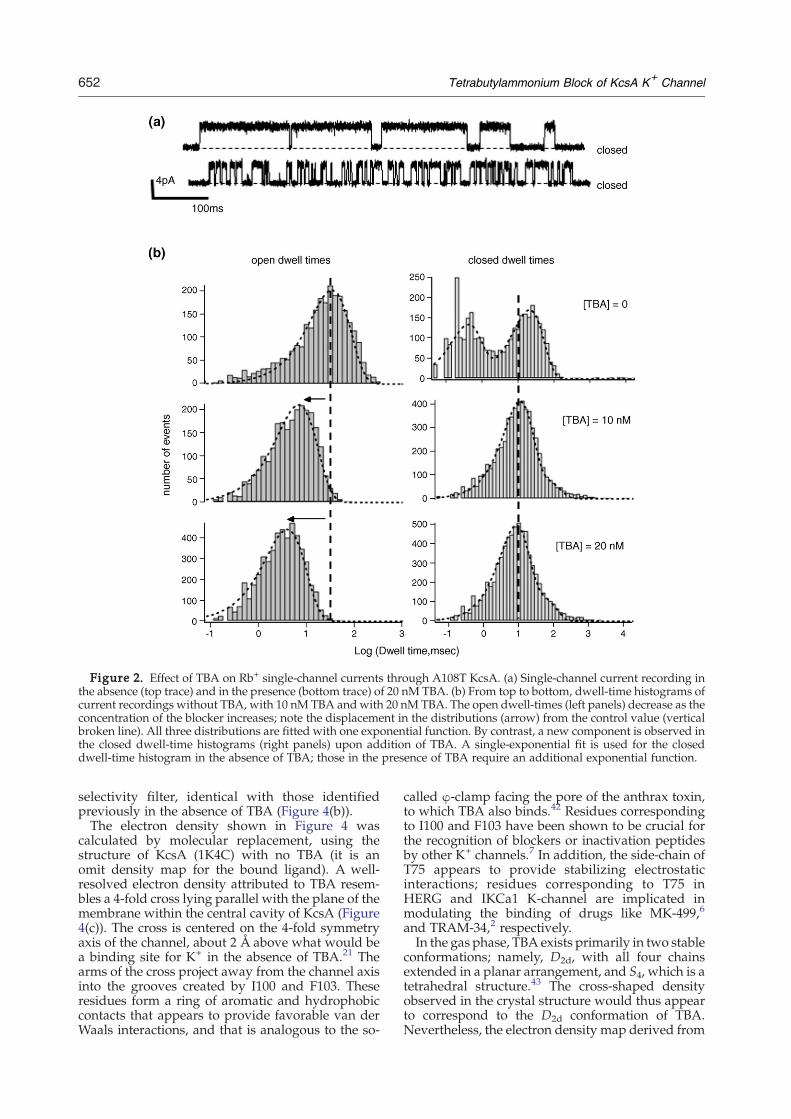
Figure 2. Effect of TBA on Rb+ single-channel currents through A108T KcsA. (a) Single-channel current recording inthe absence (top trace) and in the presence (bottom trace) of 20 nM TBA. (b) From top to bottom, dwell-time histograms ofcurrent recordings without TBA, with 10 nM TBA andwith 20 nM TBA. The open dwell-times (left panels) decrease as theconcentration of the blocker increases; note the displacement in the distributions (arrow) from the control value (verticalbroken line). All three distributions are fitted with one exponential function. By contrast, a new component is observed inthe closed dwell-time histograms (right panels) upon addition of TBA. A single-exponential fit is used for the closeddwell-time histogram in the absence of TBA; those in the presence of TBA require an additional exponential function.
652 Tetrabutylammonium Block of KcsA K+ Channel
selectivity filter, identical with those identifiedpreviously in the absence of TBA (Figure 4(b)).The electron density shown in Figure 4 was
calculated by molecular replacement, using thestructure of KcsA (1K4C) with no TBA (it is anomit density map for the bound ligand). A well-resolved electron density attributed to TBA resem-bles a 4-fold cross lying parallel with the plane of themembrane within the central cavity of KcsA (Figure4(c)). The cross is centered on the 4-fold symmetryaxis of the channel, about 2 Å above what would bea binding site for K+ in the absence of TBA.21 Thearms of the cross project away from the channel axisinto the grooves created by I100 and F103. Theseresidues form a ring of aromatic and hydrophobiccontacts that appears to provide favorable van derWaals interactions, and that is analogous to the so-
called φ-clamp facing the pore of the anthrax toxin,to which TBA also binds.42 Residues correspondingto I100 and F103 have been shown to be crucial forthe recognition of blockers or inactivation peptidesby other K+ channels.7 In addition, the side-chain ofT75 appears to provide stabilizing electrostaticinteractions; residues corresponding to T75 inHERG and IKCa1 K-channel are implicated inmodulating the binding of drugs like MK-499,6
and TRAM-34,2 respectively.In the gas phase, TBA exists primarily in two stable
conformations; namely, D2d, with all four chainsextended in a planar arrangement, and S4, which is atetrahedral structure.43 The cross-shaped densityobserved in the crystal structure would thus appearto correspond to the D2d conformation of TBA.Nevertheless, the electron density map derived from
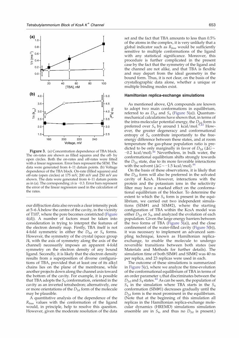
Figure 3. (a) Concentration dependence of TBA block.The on-rates are shown as filled squares and the off- byopen circles. Both the on-rates and off-rates were fittedwith a linear regression. Error bars represent the SEM. Thedata were generated from 4–11 datum points. (b) Voltagedependence of the TBA block. On-rate (filled squares) andoff-rate (open circles) at 175 mV, 200 mV and 250 mV areshown. The data were generated from 4–11 datum pointsas in (a). The corresponding zδ is −0.3. Error bars representthe error of the linear regression used in the calculation ofthe rates.
653Tetrabutylammonium Block of KcsA K+ Channel
our diffraction data also reveals a clear intensity peakat 5–6 Å below the centre of the cavity, in the vicinityof T107, where the pore becomes constricted (Figure4(d)). A number of factors must be taken intoconsideration in trying to interpret the features ofthe electron density map. Firstly, TBA itself is not4-fold symmetric in either the D2d or S4 forms.However, the symmetry of the crystal (space groupI4, with the axis of symmetry along the axis of thechannel) necessarily imposes an apparent 4-foldsymmetry on the electron density of any boundligand. Secondly, it is likely that the electron densityresults from a superposition of diverse configura-tions of TBA, provided that at least one of its alkylchains lies on the plane of the membrane, whileanother projects down along the channel axis towardthe bottom of the cavity. For example, it is possiblethat TBA adopts the S4 conformation, oriented in thecavity as an inverted tetrahedron; alternatively, oneor more orientations of the D2d form of the moleculemay be plausible.A quantitative analysis of the dependence of the
Rfree values with the conformation of the ligandwould, in principle, help to resolve this question.However, given the moderate resolution of the data
set and the fact that TBA amounts to less than 0.5%of the atoms in the complex, it is very unlikely that aglobal indicator such as Rfree would be sufficientlysensitive to multiple conformations of the ligandwith any statistical significance. Moreover, thisprocedure is further complicated in the presentcase by the fact that the symmetry of the ligand andthe channel are not alike, and that TBA is flexibleand may depart from the ideal geometry in thebound form. Thus, it is not clear, on the basis of thecrystallographic data alone, whether a unique ormultiple binding modes exist.
Hamiltonian replica-exchange simulations
As mentioned above, QA compounds are knownto adopt two main conformations in equilibrium,referred to as D2d and S4 (Figure 5(a)). Quantum-mechanical calculations have shown that, in terms ofthe intra-molecular potential energy, the D2d form ispreferred over S4 by around 1 kcal/mol.9,43 How-ever, the greater degeneracy and conformationalentropy of S4 contribute importantly to the free-energy difference between these states, and at roomtemperature the gas-phase population ratio is pre-dicted to be only marginally in favor of D2d (ΔG∼−0.2 kcal/mol).44 Nevertheless, in bulk water, theconformational equilibrium shifts strongly towardsthe D2d state, due to its more favorable interactionswith the solvent (ΔG∼−1.5 kcal/mol).44
On the basis of these observations, it is likely thatthe D2d form will also be preferred in the solvatedcavity of KcsA. However, interactions with theprotein and the potassium ions in the selectivityfilter may have a marked effect on the conforma-tional equilibrium of the blocker. To determine theextent to which the S4 form is present in the equi-librium, we carried out two independent simula-tions (SIM#1 and SIM#2), where the startingconfiguration of TBA within the KcsA model waseither D2d or S4, and analyzed the evolution of eachpopulation. Given the large energy barriers betweenthe two forms of TBA (Figure 5(a)), and the closeconfinement of the water-filled cavity (Figure 5(b)),it was necessary to implement an advanced sam-pling technique, known as Hamiltonian replica-exchange, to enable the molecule to undergoreversible transitions between both states (seeMaterials and Methods for further details). Thesimulation time of both SIM#1 and SIM#2 was 40 nsper replica, and 23 replicas were used in each.The outcome of these simulations is summarized
in Figure 5(c), where we analyze the time-evolutionof the conformational equilibrium of TBA in terms ofan order parameter η that discriminates between theD2d and S4 states.
44 As can be seen, the population ofS4 in the simulation where TBA starts in the S4conformation (SIM#1) decreases gradually until theD2d form is the most prominent in the equilibrium.(Note that at the beginning of this simulation allreplicas in the Hamiltonian replica-exchange mole-cular dynamics (HREMD) simulations simulationensemble are in S4, and thus no D2d is present.)
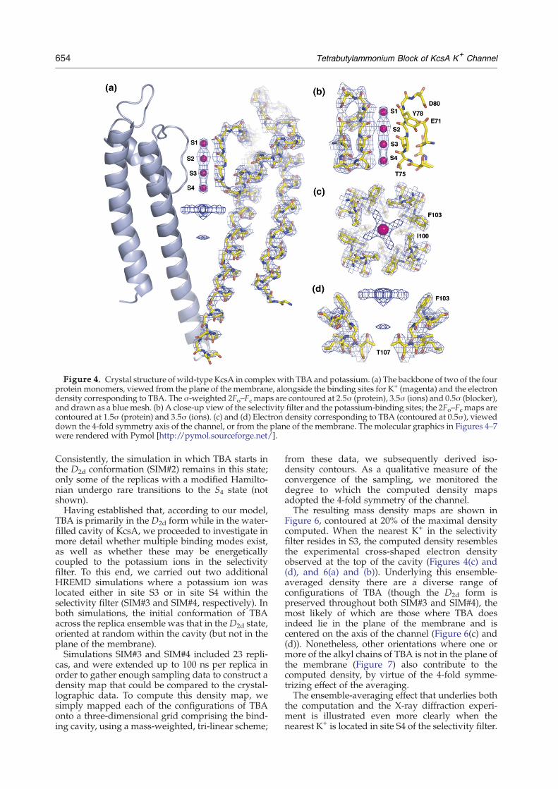
Figure 4. Crystal structure of wild-type KcsA in complexwith TBA and potassium. (a) The backbone of two of the fourprotein monomers, viewed from the plane of the membrane, alongside the binding sites for K+ (magenta) and the electrondensity corresponding to TBA. The σ-weighted 2Fo–Fc maps are contoured at 2.5σ (protein), 3.5σ (ions) and 0.5σ (blocker),and drawn as a blue mesh. (b) A close-up view of the selectivity filter and the potassium-binding sites; the 2Fo–Fc maps arecontoured at 1.5σ (protein) and 3.5σ (ions). (c) and (d) Electron density corresponding to TBA (contoured at 0.5σ), vieweddown the 4-fold symmetry axis of the channel, or from the plane of the membrane. The molecular graphics in Figures 4–7were rendered with Pymol [http://pymol.sourceforge.net/].
654 Tetrabutylammonium Block of KcsA K+ Channel
Consistently, the simulation in which TBA starts inthe D2d conformation (SIM#2) remains in this state;only some of the replicas with a modified Hamilto-nian undergo rare transitions to the S4 state (notshown).Having established that, according to our model,
TBA is primarily in the D2d form while in the water-filled cavity of KcsA, we proceeded to investigate inmore detail whether multiple binding modes exist,as well as whether these may be energeticallycoupled to the potassium ions in the selectivityfilter. To this end, we carried out two additionalHREMD simulations where a potassium ion waslocated either in site S3 or in site S4 within theselectivity filter (SIM#3 and SIM#4, respectively). Inboth simulations, the initial conformation of TBAacross the replica ensemble was that in the D2d state,oriented at random within the cavity (but not in theplane of the membrane).Simulations SIM#3 and SIM#4 included 23 repli-
cas, and were extended up to 100 ns per replica inorder to gather enough sampling data to construct adensity map that could be compared to the crystal-lographic data. To compute this density map, wesimply mapped each of the configurations of TBAonto a three-dimensional grid comprising the bind-ing cavity, using a mass-weighted, tri-linear scheme;
from these data, we subsequently derived iso-density contours. As a qualitative measure of theconvergence of the sampling, we monitored thedegree to which the computed density mapsadopted the 4-fold symmetry of the channel.The resulting mass density maps are shown in
Figure 6, contoured at 20% of the maximal densitycomputed. When the nearest K+ in the selectivityfilter resides in S3, the computed density resemblesthe experimental cross-shaped electron densityobserved at the top of the cavity (Figures 4(c) and(d), and 6(a) and (b)). Underlying this ensemble-averaged density there are a diverse range ofconfigurations of TBA (though the D2d form ispreserved throughout both SIM#3 and SIM#4), themost likely of which are those where TBA doesindeed lie in the plane of the membrane and iscentered on the axis of the channel (Figure 6(c) and(d)). Nonetheless, other orientations where one ormore of the alkyl chains of TBA is not in the plane ofthe membrane (Figure 7) also contribute to thecomputed density, by virtue of the 4-fold symme-trizing effect of the averaging.The ensemble-averaging effect that underlies both
the computation and the X-ray diffraction experi-ment is illustrated even more clearly when thenearest K+ is located in site S4 of the selectivity filter.
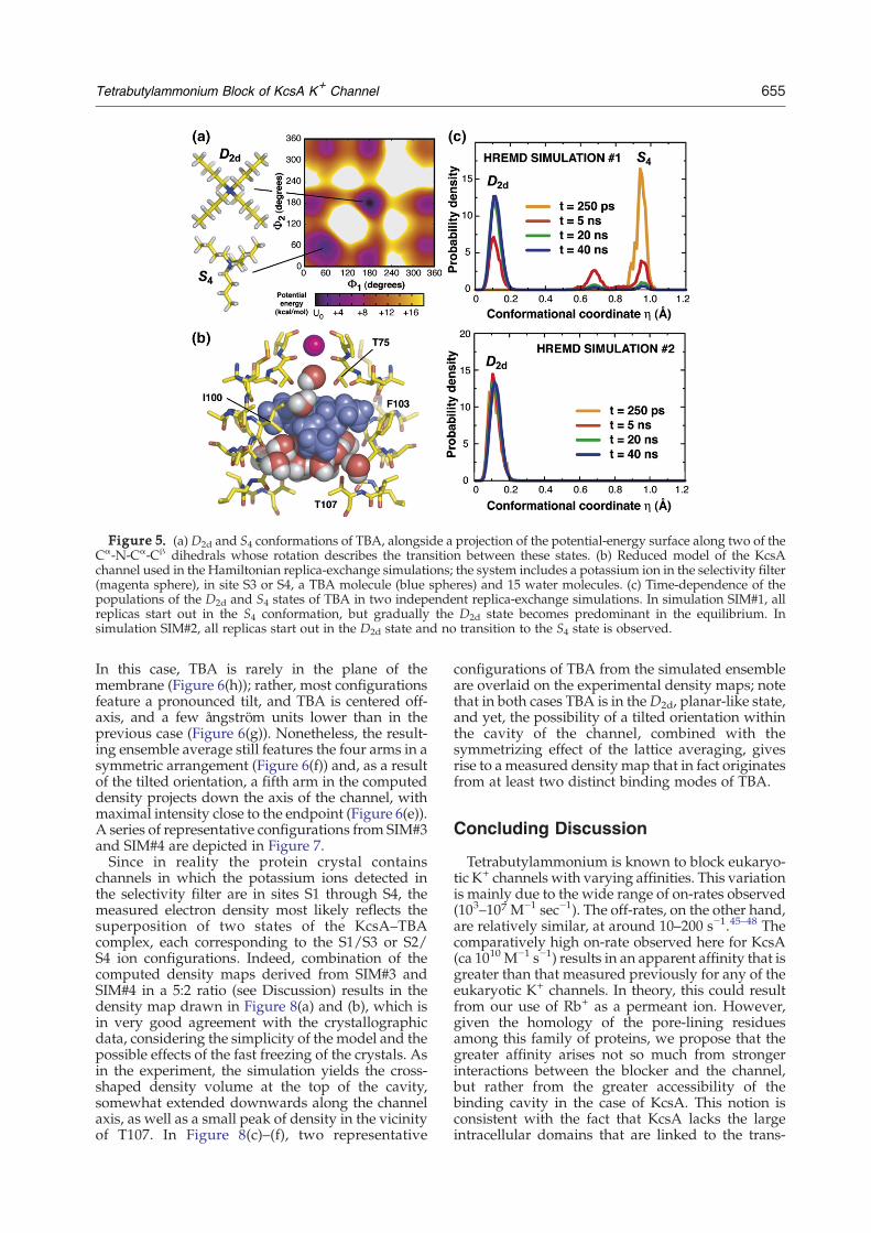
Figure 5. (a) D2d and S4 conformations of TBA, alongside a projection of the potential-energy surface along two of theCα-N-Cα-Cβ dihedrals whose rotation describes the transition between these states. (b) Reduced model of the KcsAchannel used in the Hamiltonian replica-exchange simulations; the system includes a potassium ion in the selectivity filter(magenta sphere), in site S3 or S4, a TBA molecule (blue spheres) and 15 water molecules. (c) Time-dependence of thepopulations of the D2d and S4 states of TBA in two independent replica-exchange simulations. In simulation SIM#1, allreplicas start out in the S4 conformation, but gradually the D2d state becomes predominant in the equilibrium. Insimulation SIM#2, all replicas start out in the D2d state and no transition to the S4 state is observed.
655Tetrabutylammonium Block of KcsA K+ Channel
In this case, TBA is rarely in the plane of themembrane (Figure 6(h)); rather, most configurationsfeature a pronounced tilt, and TBA is centered off-axis, and a few ångström units lower than in theprevious case (Figure 6(g)). Nonetheless, the result-ing ensemble average still features the four arms in asymmetric arrangement (Figure 6(f)) and, as a resultof the tilted orientation, a fifth arm in the computeddensity projects down the axis of the channel, withmaximal intensity close to the endpoint (Figure 6(e)).A series of representative configurations from SIM#3and SIM#4 are depicted in Figure 7.Since in reality the protein crystal contains
channels in which the potassium ions detected inthe selectivity filter are in sites S1 through S4, themeasured electron density most likely reflects thesuperposition of two states of the KcsA–TBAcomplex, each corresponding to the S1/S3 or S2/S4 ion configurations. Indeed, combination of thecomputed density maps derived from SIM#3 andSIM#4 in a 5:2 ratio (see Discussion) results in thedensity map drawn in Figure 8(a) and (b), which isin very good agreement with the crystallographicdata, considering the simplicity of the model and thepossible effects of the fast freezing of the crystals. Asin the experiment, the simulation yields the cross-shaped density volume at the top of the cavity,somewhat extended downwards along the channelaxis, as well as a small peak of density in the vicinityof T107. In Figure 8(c)–(f), two representative
configurations of TBA from the simulated ensembleare overlaid on the experimental density maps; notethat in both cases TBA is in theD2d, planar-like state,and yet, the possibility of a tilted orientation withinthe cavity of the channel, combined with thesymmetrizing effect of the lattice averaging, givesrise to ameasured density map that in fact originatesfrom at least two distinct binding modes of TBA.
Concluding Discussion
Tetrabutylammonium is known to block eukaryo-tic K+ channels with varying affinities. This variationis mainly due to the wide range of on-rates observed(103–107 M−1 sec−1). The off-rates, on the other hand,are relatively similar, at around 10–200 s−1.45–48 Thecomparatively high on-rate observed here for KcsA(ca 1010 M−1 s−1) results in an apparent affinity that isgreater than that measured previously for any of theeukaryotic K+ channels. In theory, this could resultfrom our use of Rb+ as a permeant ion. However,given the homology of the pore-lining residuesamong this family of proteins, we propose that thegreater affinity arises not so much from strongerinteractions between the blocker and the channel,but rather from the greater accessibility of thebinding cavity in the case of KcsA. This notion isconsistent with the fact that KcsA lacks the largeintracellular domains that are linked to the trans-

Figure 6. (a) and (b) Density maps derived from the ensemble of TBA configurations obtained in simulation SIM#3,where a potassium ion is located at site S3 in the selectivity filter. (c) Projection on the membrane plane of the coordinatesof the nitrogen atom of TBA in all configurations in the simulated ensemble. (d) Correlation of the Z-coordinate of thenitrogen atom of TBA and the tilt of the molecule relative to the plane of the membrane, for all configurations in thesimulation ensemble. The plane of TBA is defined by a least-squares fit relative to coordinates of the N and Cβ atoms.(e)–(h) Same as above, but for simulation SIM#4, where a potassium ion is located in site S4 in the selectivity filter.
656 Tetrabutylammonium Block of KcsA K+ Channel
membrane segment in other K+ channels, whichpresumably constitute a kinetic barrier for thetranslocation of the blocker from the bulk to theentrance of the pore. The subsequent binding event,which results in the actual blockage of the ioniccurrent, is probably energetically comparable acrossthe family of K+ channels, as reflected by thesimilarity of the off-rates.An intriguing aspect of the blocking properties of
TBA is the voltage dependence of the binding
the ideal, all-trans D2d geometry (Figure 5(a)) is provided, decorresponds to the C8N+ core (which defines the conformaticorresponds to all non-hydrogen atoms in the molecule.
kinetics, which at first sight appears to be counter-intuitive, in that more positive voltages (in theintracellular side) lead to faster off-rates. However,this result can be rationalized, bearing in mind that,in the context of the lipid membrane, transfer of TBAto and from the external solution can take place onlywhile the channel is in the open state, even though,as shown by the crystal structure, the channel canclose in the presence of the blocker. In this openstate, the drop in the transmembrane voltage is
Figure 7. Representative con-figurations of TBAwithin the trans-membrane cavity of KcsA, extractedfrom simulations SIM#3 and SIM#4.The configurations are orderedaccording to the tilt of the moleculerelative to the plane of the mem-brane and to the position of thenitrogen atom of TBA along the axisof the channel (Z=0 correspondsapproximately to the location of thebinding site for K+ in the absence ofTBA). For each tilt, the characteristicRMS deviation (in Å)with respect to
rived from an ensemble average. The number at the toponal state, i.e. D2d versus S4); the number at the bottom

Figure 8. (a) and (b) Density maps derived from the ensemble of TBA configurations obtained in simulations SIM#3and SIM#4, combined in a 5:2 ratio. (c) and (d) Experimental electron density map for TBA (contoured at 0.5σ), alongside aconfiguration of TBA chosen randomly from those of tilt smaller than 10°, relative to the plane of the membrane. (e) and (f)Experimental electron density map for TBA (contoured at 0.5σ), alongside a configuration of TBA chosen randomly fromthose of tilt equal to around 60°. Note that in both cases TBA is in theD2d state, which is preserved throughout simulationsSIM#3 and SIM#4.
657Tetrabutylammonium Block of KcsA K+ Channel
concentrated in the selectivity filter region,40 andthus the magnitude of the electric field in thebinding cavity is minimal. This is consistent withthe observations that the on-rates are voltage-independent; i.e. the transmembrane voltage doesnot drive the blocker into the cavity. Thus, thevoltage-dependence of the off-rates must ariseindirectly from the effect that voltage may have onthe kinetics of channel gating.It has been shown that the voltage-dependence of
the block can be altered radically by interactionbetween the blocker and the permeant ions.31,41 Inthis work, we show through computer simulationsand X-ray crystallography that TBA binds in twomodes, and that the relative populations of thesemodes appear to be coupled to the configuration ofthe potassium ions in the selectivity filter. Inparticular, a tilted arrangement of TBA relative tothe plane of the membrane is more favoredwhen thenearest potassium ion resides in site S4, whereas anorientation along the plane of the membrane is morefavored if the nearest potassium ion is located in siteS3. This trend is thus consistent with the electrostaticrepulsion between the blocker and the permeantions. In the latter arrangement, TBA interacts solelywith residues at the top of the cavity (T75, I100, andF103); by contrast, in the tilted orientation, it alsocontacts the hydrophobic group of T107, whichconstricts the pore when the channel is closed.On the basis of these observations, we hypothesize
that the observed dependence of the off-rate on the
transmembrane potential arises from the influenceof the electric field on the ion configuration in theselectivity filter, which in turn affects the extent towhich TBA may interfere with the gating of thechannel through its interaction with T107. That is tosay, the smaller the transmembrane potential, thegreater the probability of having a K+ in site S4,which, in turn, would increase the probability thatTBA adopts a tilted orientation. In this orientation,the hydrophobic interaction of TBA and T107 mayhinder the stochastic opening of the channel, thusslowing the off-rates. More positive voltages wouldhave the opposite effect on the off-rates, byindirectly promoting a binding mode where TBAlies on the plane of the membrane, and thereforedoes not interfere with the gating kinetics.Another important conclusion of this work per-
tains to the extent to which TBA binding affects thestructure of the channel, which has been an openquestion until now. The structure of KcsA withbound TBA at 2.9 Å resolution was solved byMacKinnon and co-workers.7 In this crystal struc-ture, KcsA is unchanged with respect to the ligand-free, closed-state form, and TBA can be identifiedclearly within the transmembrane cavity. However,the electron density corresponding to TBA isfeatureless, which precludes the elucidation of theconformation of the molecule, its orientation withinthe cavity, or its interactions with pore-liningresidues in the channel. Subsequent crystallographicstudies of a KcsA–Fab antibody complex in the
658 Tetrabutylammonium Block of KcsA K+ Channel
presence of Tl+ by Gross and co-workers succeededin resolving TBA in molecular detail; namely, in aconfiguration where the molecule lies on the planeof the membrane.8 Nevertheless, their structurereveals substantial changes in the conformationand ion occupancy of the selectivity filter, as wellas at the top of the binding cavity.In this work, the use of K+ as the permeant species
as well of the Fab antibody fragment to aid thecrystallization has enabled us to resolve TBA inmolecular detail, as well as to assess the structuralimpact that the blocker may have, under morephysiological conditions. On the basis of the result-ing crystal structure, we concur with Zhou et al., inthat the TBA blocker does not alter the structure ofthe channel per se when the selectivity filter isoccupied by potassium ions. Therefore, the struc-tural changes observed by Lenaeus et al.8 must thenbe attributed to the presence of Tl+, or to thecombined effect of TBA and Tl+. In addition, thestructure reported by Gross and co-workers did notreveal the additional peak of density in the vicinityof T107 that is observed clearly in our data. It isplausible that the single occupancy of the selectivityfilter in their case enhances the likelihood of a singleorientation near the top of the cavity.In a broader sense, the present study illustrates
the usefulness of computer simulations as acomplementary tool in biological chemistry. Totackle the intrinsic difficulties associated with theconformational free-energy barriers of TBA and theclose-confinement of the water-filled cavity ofKcsA, we have used an advanced but computa-tionally costly simulation method, which in turnrequires a simplification of the model system.Simulations with more sophisticated models incor-porating long-range electrostatic effects, or alter-native force field parameters for TBA such as thosedescribed by Luzhkov et al.43 could alter the relativestability of the S4 and D2d forms and the couplingbetween the TBA and the ions occupying theselectivity filter, though the main results from thepresent work are expected to remain valid. Inpractice, different treatments would simply affectthe 5:2 ratio assigned to the computed density mapsin Figure 6(a) and (b) (leading to that in Figure 8(a)),but the shape of the simulated maps of bound TBAwould remain qualitatively unchanged. Seekingquantitative agreement with the relative popula-tions of the configurations of the protein-ion-blocker system in the complex environment ofcrystals that are fast-frozen in liquid nitrogen is notthe purpose of the present analysis. The fact thatgood agreement is found between the crystallo-graphic and computed density maps with a singleadjustable parameter is strong support for thepresent interpretation of the data; namely, (a) TBAcan bind into the cavity of KcsA by adoptingmultiple conformations, dominated by the D2dconformer in an in-plane and a tilted orientations,and (b) the relative population of these bindingmodes is sensitive to the configuration of the ionsoccupying the selectivity filter.
Materials and Methods
Electrophysiology measurements
Materials
All salts used were reagent grade or higher. Unlessstated otherwise, high-purity chemicals were purchasedfrom Sigma-Aldrich (St. Louis, MO). Mops wasobtained from American Bioanalytical (Natick, MA),TBA was from Fluka (Milwaukee, WI), Rb2Succinatewas from Great Western Inorganics (Arvada, CO). n-Dodecyl-β-D-maltoside (sol-grade) was used for proteinextraction and purification; lipid for reconstitution wassolubilized with Chaps (anagrade) from Anatrace(Maumee, OH). Lipids for the reconstitution intovesicles and for planar lipid bilayer experiments were1-palmitoyl-2-oleoyl-sn-glycero-3-phosphoethanolamine(POPE) and 1-palmitoyl-2-oleoyl-sn-glycero-3 [phospho-rac-(1-glycerol)] (sodium salt) (POPG) from Avanti PolarLipids (Alabaster, AL). Escherichia coli strain JM-83 waspurchased from ATCC (Manassas, VA). The cells weregrown in Terrific broth (TB: per liter; 12 g of Tryptone,24 g of yeast extract, and 4 ml of glycerol, with 17 mMKH2PO4 and 72 mM K2HPO4). Solutions for bilayerexperiments were prepared daily in the following twoways: (1) 96 mM RbCl and 10 mM succinic acid (transsolution) or 10 mM Mops (cis solution); the pH wasadjusted to 4.0 (trans solution) or 7.0 (cis solution); (2)For the trans solutions, we used 10 mM Rb2Succinateand 80 mM RbCl; the pH was adjusted to 4.0 usingHCl.
Expression, purification and reconstitution of KcsA
For the plasmid construct for expression of KcsA, weused a synthetic gene encoding the native proteinsequence with an additional N-terminal His6 tag. Thisgene was inserted into the pASK90 expression plasmid(generously supplied by Dr Arne Skerra); expression wasinduced by the addition of anhydrotetracycline (aTC;Acros Organics, Morris Plains, NJ). Wild-type and mutantKcsA proteins were expressed and purified as des-cribed.31 After induction, cells were incubated for 2 h toallow for the expression of the protein. Cells were thenwashed and resuspended in buffer A (95 mM NaCl,5 mM KCl, and 50 mM Mops, adjusted to pH 7.0 withNaOH), protease inhibitors were added (final concentra-tions were 1 μM leupeptin, 1 μM pepstatin A, 0.5 mMPMSF), and cells were disrupted by a French press.Unbroken cells were cleared by centrifugation at 12,000gfor 25 min, and membranes were isolated by ultracen-trifugation at 75,000g for 45 min. Membranes were re-suspended in buffer B (95 mM NaOH and 5 mM KCl,adjusted to pH 7.0 with H3PO4) and extracted using15 mM dodecyl maltoside for 30 min. The unsolubilizedmembranes were removed by ultracentrifugation at75,000g for 45 min. The protein was purified using Niaffinity chromatography (Ni-NTA Agarose, Qiagen); theextracted membranes were incubated with the agarosebeads for 2 h in the presence of 40 mM imidazole, washedwith 40 mM imidazole and eluted with 400 mMimidazole. For reconstitution, lipid (7.5 mg/ml of POPEand 2.5 mg/ml of POPG) was solubilized after incubationwith 34 mM Chaps for 2 h at room temperature. From 1-4 μg of protein was mixed immediately after purificationwith 400 μl of solubilized lipid and incubated at roomtemperature for 20 min. Detergent was removed using a

659Tetrabutylammonium Block of KcsA K+ Channel
20 ml Sephadex G-50 column; fractions of 500 μl werecollected and those containing the vesicles were deter-mined by visual inspection and were stored at −80 °C forup to a month.
Single-channel recordings
Single-channel recordings were performed in a hor-izontal planar lipid bilayer setup. Partitions made fromoverhead transparency film having holes roughly 50 μmin diameter were treated by application of ∼0.2 μl oflipid solution (15 mg/ml of POPE and 5.0 mg/ml ofPOPG in decane), followed by drying in air for 20 min.After the chambers were filled with solution, bilayerswere formed by painting with a glass rod dipped in thelipid solution. The reconstituted protein was added tothe cis chamber, and the bilayer was formed with thebubble expelled by the pipet after the addition of sample.The orientation of the recording system is such that thecis chamber, containing solutions at pH 7, is equivalent tothe periplasmic surface of the protein, while theintracellular surface of the channel faces the transchamber. Voltages and currents follow the standardelectrophysiological convention, with extracellular solu-tion at ground voltage.Currents were sampled at 20 kHz or 50 kHz, low-pass
filtered at 2 kHz and further filtered digitally at 1 kHzbefore data analysis (the effective filtering frequencycorresponds to 0.9 kHz).49 Data were acquired andanalyzed with Clampex and P-stat software (AxonInstruments). Dwell time distributions were fitted withone or more exponential components using a least-squaresfitting procedure.Single channel records of the KcsA channel display
bursts of activity that are interrupted by long closed/inactive states (ca 10–15 s). During a given burst, thechannel alternates between open and closed states thathave much shorter lifetimes than the typical duration ofthe bursts and of the long closed/inactive states. Thefollowing analysis of the block experiments by TBAcorresponds to the time-period within bursts. Let usdenote the closing and opening rates of the channel inthe absence of TBA as kopen=1/τclose and kclose=1/τ,where τopen and τclose are the open and closed dwelltimes, respectively. In the presence of a slow blocker, theexit rate from the open state will be the sum of the ratesof closing of the channel and the on-rate of blocker.Therefore, the on-rate of the blocker can be calculatedas:
kon ¼ 1H Bopen
� 1H open
!1
½TBA�
where τopenB is the open dwell time in the presence of the
blocker. The off-rate is expected to be concentration-independent and equal to 1/τblocked, where τblocked isthe dwell time of the blocked state (which may differsignificantly from τclose in the absence of the blocker).Finally, the dependence of the block with the appliedtransmembrane voltage is given by:
ln k Vð Þ ¼ ln k 0ð Þ � zyFVRT
where k denotes either the on-rate or the off-rate, z is theunitary charge moving in the electrical field, δ is thefraction of voltage across which the charge moves, R isthe gas constant and T is absolute temperature.50
Crystallography and X-ray diffraction
Purification of wild-type KcsA and Fab antibody fragment
KcsA was expressed and purified as described,22 andeluted with 500 mM imidazole. To remove excessimidazole, overnight dialysis was carried out in 50 mMTris–HCl (pH 7.5), 150 mM KCl, 10 mM dodecylmaltoside. The C-terminal flexible end with His6 tag (35amino acid residues) was excised from KcsA usingchymotrypsin, by incubating KcsA and chymotrypsin ata ratio of 15:1 (w/w), respectively, for 2 h at roomtemperature. All chemicals were bought from Sigma-Aldrich, except detergent, which was purchased fromAnatrace.Hybridoma cells containing monoclonal antibodies
raised against the extracellular epitope of KcsA weregenerously provided by Roderick Mackinnon. The Sloan-Kettering Monoclonal Antibody facility provided IgG inserum after harvesting the hybridoma cells that had beenprovided to them. An affinity column was used toseparate IgG from serum protein A, using a phosphatebuffer at pH 7.0 and eluted with a pH gradient from 7.0 to3.0 (citrate buffer). The Fab fragment of the antibody wasobtained through proteolysis, through incubation of theprotein with papain at a 250:1 (w/w) ratio at 37°C for 3 h.The reaction was stopped by adding 50 mM iodoaceta-mide. Excess iodoacetamide was subsequently removedthrough overnight dialysis in 20 mM Tris–HCl pH (7.5).Fab and Fc fragments were separated on an ion-exchangechromatography column (Mono-Q), where Fab comes outin the flow through. Fractions were concentrated to 2–4 mg/ml and stored at −80 °C.
Formation of KcsA–Fab complex
After digestion by chymotrypsin, wild-type KcsA wasincubated with purified Fab for 30 min. The complex wasthen injected into a size-exclusion chromatography col-umn (AKTA FPLC with 200 HR Sepharose), which isequilibrated with 50 mM Tris–HCl (pH 7.5), 150 mM KCl,5 mM dodecyl maltoside. The complex comes out as asingle peak at ∼12.2 ml. Fractions were pooled in andconcentrated to 10–12 mg/ml using 30 kDa cutoff filters.Before setting up the trays for crystallization, purifiedKcsA-Fab complex (10–12 mg/ml) in 50 mM Tris–HCl(pH 7.5), 150 mM KCl, 5 mM dodecyl maltoside wasincubated with 5 mM TBA (purchased from Sigma) atroom temperature for 30–60 min.
X-ray crystallography
Crystals were obtained after two to three days in asitting drop containing 20–25% (w/v) PEG 400, 50 mMmagnesium acetate and 50 mM sodium acetate (pH 5.4–5.6) at 20 °C. In the same drop, cryoprotection wasachieved by increasing the concentration of PEG 400 from20–25% to 40%. After cryoprotection, crystals were frozenin liquid nitrogen and shot at the X4A beam line of theNational Synchrotron Light Source, diffracting to a Braggspacing of 2.9 Å. Data were processed with Denzo andScalepack.51 Phases were obtained by molecular replace-ment using the structure of the KcsA-Fab complex solvedby Zhou et al.22 CNS52 was used to refine the model of thestructure of TBA-bound KcsA (a TBA molecule was notexplicitly included in the model during refinement), at2.9 Å resolution and 81% completeness. (An alternativerefinement at 3.3 Å and 90% completeness was carried out,which yielded an analogous omit density map for TBA.)
660 Tetrabutylammonium Block of KcsA K+ Channel
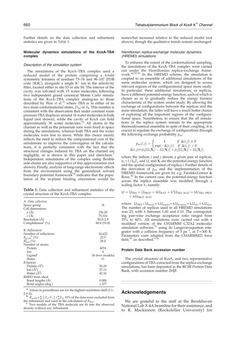
Further details on the data collection and refinementstatistics are given in Table 1.
Molecular dynamics simulations of the KcsA-TBAcomplex
Description of the simulation system
The simulations of the KcsA–TBA complex used areduced model of the protein comprising a 4-foldsymmetric tetramer of residues 73–76 and 96–107 (PDBcode 1K4C), alongside a single K+ ion in the selectivityfilter, located either in site S3 or site S4. The interior of thecavity was solvated with 15 water molecules, followingtwo independent grand canonical Monte Carlo simula-tions of the KcsA–TBA complex analogous to thosedescribed by Woo et al.53 where TBA is in either of itstwo main conformational states, D2d or S4. This number isconsistent with the observation that under constant roompressure TBA displaces around 16 water molecules in bulkliquid (not shown), while the cavity of KcsA can holdapproximately 30 water molecules.53 All atoms in thechannel as well as the potassium ions were fixed in spaceduring the simulations, whereas both TBA and the watermolecules were free to move. While this choice merelyreflects the need to reduce the computational cost of thesimulations to improve the convergence of the calcula-tions, it is perfectly consistent with the fact that thestructural changes induced by TBA on the channel arenegligible, as is shown in this paper and elsewhere.7
Independent simulations of the complex using flexibleside-chains are also supportive of this approximation (notshown). Finally, analysis of long-range electrostatic effectsfrom the environment using the generalized solventboundary potential framework54 indicates that the popu-lation of the in-plane binding orientation would be
Table 1. Data collection and refinement statistics of thecrystal structure of the KcsA-TBA complex
A. Data collectionSpace group I4Cell dimensions
a (Å) 156.20c (Å) 75.316
Resolution (Å) 50.0–2.9Completenessa (%) 80.8 (53.8)
B. RefinementNumber of reflections 16,622Rwork
b (%) 22.5Rfreeb (%) 28.4
Number of atomsProtein 4074Ion 4Ligandc 34 (two models)Water 11
B-factorsProtein (Å2) 50.29Ion (Å2) 27.13Water (Å2) 42.16
RMSD from idealBond lengths (Å) 0.008Bond angles (deg.) 1.377a Values in parentheses are for the highest-resolution shell (3.1–
2.9 Å).b Rwork=∑|Fo–Fc|/∑Fo, 10% of the data were excluded from
the refinement and used in the calculation of Rfree.c Two models of the TBA molecule are fit into the observed
density without any refinement.
somewhat increased relative to the reduced model (notshown), though the qualitative trends remain unchanged.
Hamiltonian replica-exchange molecular dynamics(HREMD) simulations
To enhance the extent of the conformational sampling,the simulations of the KcsA–TBA complex were carriedout under the Hamiltonian replica-exchange frame-work.44,55,56 In the HREMD scheme, the simulation iscoupled to an ensemble of additional simulations of thesame molecular system, which are designed to accessrelevant regions of the configurational space more easily.In particular, these additional simulations, or replicas,have a different potential-energy function, each of which ischosen so as to gradually reduce the energy barrierscharacteristic of the system under study. By allowing theexchange of configurations between the replicas and themain simulation, the latter will have a much better chanceof exploring all the important regions of the configura-tional space. Nonetheless, to ensure that the all simula-tions in the replica system remain in the appropriatethermodynamical ensemble in spite of their coupling, it iscrucial to regulate the exchange of configurations throughthe following exchange probability pex:
pexði; jÞ ¼ 1 if Dði; jÞ V 0expð�Dði; jÞÞ if Dði; jÞ > 0
�Dði; jÞuhi½UiðXjÞ �UiðXiÞ� þ hj½UjðXiÞ �UjðXjÞ�
where the indices i and j denote a given pair of replicas,βι=1/kBTi, and Ui and Xi are the potential energy functionand the spatial configuration of replica i. Further details ofthe derivation of pex and the implementation of theHREMD framework are given by e.g. Faraldo-Gómez &Roux.44 In the current case, the potential-energy functionacross the replica ensemble was modified through ascaling factor λ; namely:
U ¼ UTBA þUPROT þ EUWAT þ E3UTBA−WAT þ EUTBA−PROTþ EUPROT−WAT
where UTBA=Ubonds+λUangles+λUtorsions+λUelec+λUvdw.The number of replicas used in all HREMD simulationswas 23, with λ between 1.00 and 0.10. The correspond-ing pair-wise exchange acceptance ratio ranged from35% to 60%. All simulations were carried out with amodified version of the CHARMM C32A2 molecularsimulation software,57 using its Langevin-equation inte-grator with a collision frequency of 5 ps−1, at T=300 K.Parameters were adapted from the CHARMM22 forcefield,58 as described.9,44
Protein Data Bank accession number
The crystal structure of KcsA, and two representativeconfigurations of TBA extracted from the replica-exchangesimulations, has been deposited in the RCSB Protein DataBank, with accession number 2HJF.
Acknowledgements
We are grateful to the staff at the BrookhavenNational Lab X-4A beamline for their assistance, andto R. Mackinnon (Rockefeller University) for
661Tetrabutylammonium Block of KcsA K+ Channel
providing us with the KcsA monoclonal antibodyhybridoma cell-line. Computational resources forthis project were provided, in part, by the Institutefor Computational Biomedicine of the Weill MedicalCollege of Cornell University.
References
1. Armstrong, C. M. (1974). Ionic pores, gates, and gatingcurrents. Quart. Rev. Biophys. 7, 179–210.
2. Wulff, H., Miller, M. J., Hansel, W., Grissmer, S.,Cahalan, M. D. & Chandy, K. G. (2000). Design of apotent and selective inhibitor of the intermediate-conductance Ca2+-activated K+ channel, IKCa1: apotential immunosuppressant. Proc. Natl Acad. Sci.USA, 97, 8151–8156.
3. Spector, P. S., Curran, M. E., Keating, M. T. &Sanguinetti, M. C. (1996). Class III antiarrhythmicdrugs block HERG, a human cardiac delayed rectifierK+ channel - open-channel block by methanesulfona-nilides. Circulat. Res. 78, 499–503.
4. Franqueza, L., Longobardo, M., Vicente, J., Delpon, E.,Tamkun, M. M., Tamargo, J. et al. (1997). Moleculardeterminants of stereoselective bupivacaine block ofhKv1.5 channels. Circulat. Res. 81, 1053–1064.
5. Nilsson, J., Madeja, M. & Arhem, P. (2003). Localanesthetic block of Kv channels: role of the S6 helixand the S5-S6 linker for bupivacaine action. Mol.Pharmacol. 63, 1417–1429.
6. Mitcheson, J. S., Chen, J., Lin, M., Culberson, C. &Sanguinetti, M. C. (2000). A structural basis for drug-induced long QT syndrome. Proc. Natl Acad. Sci. USA,97, 12329–12333.
7. Zhou,M., Morais-Cabral, J. H., Mann, S. &MacKinnon,R. (2001). Potassium channel receptor site for the in-activation gate and quaternary amine inhibitors.Nature,411, 657–661.
8. Lenaeus, M. J., Vamvouka, M., Focia, P. J. & Gross, A.(2005). Structural basis of TEA blockade in a modelpotassium channel. Nature Struct. Biol. 12, 454–459.
9. Crouzy, S., Bernèche, S. & Roux, B. (2001). Extra-cellular blockade of K+ channels by TEA: results frommolecular dynamics simulations of the KcsA channel.J. Gen. Physiol. 118, 207–217.
10. Luzhkov, V. B., Nilsson, J., Arhem, P. & Aqvist, J.(2003). Computational modelling of the open-state K(v)1.5 ion channel block by bupivacaine. Biochim.Biophys. Acta, 1652, 35–51.
11. Luzhkov, V. B., Osterberg, F. & Aqvist, J. (2003).Structure-activity relationship for extracellular blockof K+ channels by tetraalkylammonium ions. FEBSLetters, 554, 159–164.
12. Ashcroft, F. M. (2002). Ion Channels and Disease.Academic Press, San Diego, CA.
13. Wickenden, A. D. (2002). K+ channels as therapeuticdrug targets. Pharmacol. Ther. 94, 157–182.
14. Armstrong, C. M. (1971). Interaction of tetraethylam-monium ion derivatives with potassium channels ofgiant axons. J. Gen. Physiol. 58, 413–437.
15. Choi, K. L., Aldrich, R. W. & Yellen, G. (1991).Tetraethylammonium blockade distinguishes twoinactivation mechanisms in voltage-activated K+
channels. Proc. Natl Acad. Sci. USA, 88, 5092–5095.16. Mackinnon, R. & Yellen, G. (1990). Mutations affecting
TEA blockade and ion permeation in voltage-acti-vated K+ channels. Science, 250, 276–279.
17. Yellen, G., Jurman, M. E., Abramson, T. & Mackinnon,
R. (1991). Mutations affecting internal TEA blockadeidentify the probable pore-forming region of a K+
channel. Science, 251, 939–942.18. Holmgren,M., Smith, P. L.&Yellen,G. (1997). Trapping
of organic blockers by closing of voltage-dependent K+
channels: evidence for a trap door mechanism ofactivation gating. J. Gen. Physiol. 109, 527–535.
19. Cuello, L. G., Romero, J. G., Cortes, D. M. & Perozo, E.(1998). pH-dependent gating in the Streptomyceslividans K+ channel. Biochemistry, 37, 3229–3236.
20. Heginbotham, L., Kolmakova-Partensky, L. & Miller,C. (1998). Functional reconstitution of a prokaryoticK+ channel. J. Gen. Physiol. 111, 741–749.
21. Doyle, D. A., Cabral, J. M., Pfuetzner, R. A., Kuo, A. L.,Gulbis, J. M., Cohen, S. L. et al. (1998). The structure ofthe potassium channel: molecular basis of K+ conduc-tion and selectivity. Science, 280, 69–77.
22. Zhou, Y. F., Morais-Cabral, J. H., Kaufman, A. &MacKinnon, R. (2001). Chemistry of ion coordinationand hydration revealed by a K+ channel-Fab complexat 2.0 angstrom resolution. Nature, 414, 43–48.
23. Cordero-Morales, J. F., Cuello, L. G., Zhao, Y. X.,Jogini, V., Cortes, D. M., Roux, B. & Perozo, E. (2006).Molecular determinants of gating at the potassium-channel selectivity filter. Nature Struct. Mol. Biol. 13,311–318.
24. Bernèche, S. & Roux, B. (2001). Energetics of ionconduction through theK+ channel.Nature, 414, 73–77.
25. Noskov, S. Y., Bernèche, S. & Roux, B. (2004). Controlof ion selectivity in potassium channels by electro-static and dynamic properties of carbonyl ligands.Nature, 431, 830–834.
26. Aqvist, J. & Luzhkov, V. (2000). Ion permeationmechanism of the potassium channel. Nature, 404,881–884.
27. Shrivastava, I. H. & Sansom, M. S. P. (2000).Simulations of ion permeation through a potassiumchannel: molecular dynamics of KcsA in a phospho-lipid bilayer. Biophys. J. 78, 557–570.
28. Shrivastava, I. H., Tieleman, D. P., Biggin, P. C. &Sansom, M. S. P. (2002). K+ versus Na+ ions in a Kchannel selectivity filter: a simulation study. Biophys. J.83, 633–645.
29. MacKinnon, R., Cohen, S. L., Kuo, A. L., Lee, A. &Chait, B. T. (1998). Structural conservation in prokar-yotic and eukaryotic potassium channels. Science, 280,106–109.
30. Lu, Z., Klem, A. M. & Ramu, Y. (2001). Ion conductionpore is conserved among potassium channels. Nature,413, 809–813.
31. Kutluay, E., Roux, B. & Heginbotham, L. (2004).Probing the mechanism of internal TEA block ofKcsA. Biophys. J. 86, 130A–131A.
32. Gao, L., Mi, X., Paajanen, V., Wang, K. & Fan, Z.(2005). Activation-coupled inactivation in the bacterialpotassium channel KcsA. Proc. Natl Acad. Sci. USA,102, 17630–17635.
33. Schrempf, H., Schmidt, O., Kummerlen, R., Hinnah,S., Muller, D., Betzler, M. et al. (1995). A prokaryoticpotassium ion channel with two predicted transmem-brane segments from Streptomyces lividans. EMBO J.14, 5170–5178.
34. Heginbotham, L., LeMasurier,M., Kolmakova-Partensky,L. & Miller, C. (1999). Single streptomyces lividans K(+)channels: functional asymmetries and sidednessof protonactivation. J. Gen. Physiol. 114, 551–560.
35. Irizarry, S. N., Kutluay, E., Drews, G., Hart, S. J. &Heginbotham, L. (2002). Opening the KcsA K+
channel: tryptophan scanning and complementation
662 Tetrabutylammonium Block of KcsA K+ Channel
analysis lead to mutants with altered gating. Biochem-istry, 41, 13653–13662.
36. LeMasurier, M., Heginbotham, L. & Miller, C. (2001).KcsA: it's a potassium channel. J. Gen. Physiol. 118,303–314.
37. Morais-Cabral, J. H., Zhou, Y. &MacKinnon, R. (2001).Energetic optimization of ion conduction rate by theK+ selectivity filter. Nature, 414, 37–42.
38. Swenson, R. P. J. & Armstrong, C. M. (1981). K+
channels close more slowly in the presence of externalK+ and Rb+. Nature, 291, 427–429.
39. Ray, E. C. & Deutsch, C. (2006). A trapped intracel-lular cation modulates K+ channel recovery from slowinactivation. J. Gen. Physiol., 128.
40. Jogini, V. & Roux, B. (2005). Electrostatics of theintracellular vestibule of K+ channels. J. Mol. Biol. 354,272–288.
41. Heginbotham, L. & Kutluay, E. (2004). Revisitingvoltage-dependent relief of block in ion channels: amechanism independent of punchthrough. Biophys. J.86, 3663–3670.
42. Krantz, B. A., Melnyk, R. A., Zhang, S., Juris, S. J.,Lacy, D. B., Wu, Z. Y. et al. (2005). A phenylalanineclamp catalyzes protein translocation through theanthrax toxin pore. Science, 309, 777–781.
43. Luzhkov, V. B., Osterberg, F., Acharya, P.,Chattopadhyaya, J. & Aqvist, J. (2002). Computationaland NMR study of quaternary ammonium ionconformations in solution. Phys. Chem. Chem. Phys.4, 4640–4647.
44. Faraldo-Gómez, J. D. & Roux, B. (2006). Characteriza-tion of conformational equilibria throughHamiltonianand temperature replica-exchange simulations: asses-sing entropic and environmental effects. J. Comput.Chem. In the press.
45. Villaroel, A., Alvarez, O., Oberhauser, A. & Latorre, R.(1988). Probing a Ca2+ activated K+ channel with qua-ternary ammonium ions. Plufgers Arch. 413, 118–126.
46. Choi, K. L., Mossman, C., Aube, J. & Yellen, G. (1993).The internal quaternary ammonium receptor-site ofShaker potassium channels. Neuron, 10, 533–541.
47. French, R. J. & Shoukimas, J. J. (1981). Blockage ofsquid axon potassium conductance by internal tetra-N-alkylammonium ions of various sizes. Biophys. J. 34,271–291.
48. Guo, D. L. & Lu, Z. (2001). Kinetics of inward-rectifierK+ channel block by quaternary alkylammonium ions:dimension and properties of the inner pore. J. Gen.Physiol. 117, 395–405.
49. Colquhoun, D. & Sigworth, F. J. (1995). Fitting andstatistical analysis of single-channel records. In SingleChannel Recordings (Sakmann, B. & Neher, E., eds),pp. 483–587, Plenum Press, New York.
50. Woodhull, A. M. (1973). Ionic blockage of sodiumchannels in nerve. J. Gen. Physiol. 61, 687–708.
51. Otwinowski, Z. & Minor, W. (1997). Processing of X-ray diffraction data collected in oscillation mode. InMethods in Enzymology Macromolecular Crystallography,Pt. A. (Carter, C. W., Jr. & Sweet, R. M., eds), vol. 276,pp. 307–326, Academic Press, New York, London.
52. Brunger, A. T., Adams, P. D., Clore, G. M., DeLano,W. L., Gros, P., Grosse-Kunstleve, R. W. et al. (1998).Crystallography and NMR system: a new softwaresuite for macromolecular structure determination.Acta Crystallog. sect. D, 54, 905–921.
53. Woo, H. J., Dinner, A. R. & Roux, B. (2004). Grandcanonical Monte Carlo simulations of water in proteinenvironments. J. Chem. Phys. 121, 6392–6400.
54. Im, W., Bernèche, S. & Roux, B. (2001). Generalizedsolvent boundary potential for computer simulations.J. Chem. Phys. 114, 2924–2937.
55. Sugita, Y. & Okamoto, Y. (2000). Replica-exchangemulticanonical algorithm and multicanonical replica-exchange method for simulating systems with roughenergy landscape. Chem. Phys. Letters, 329, 261–270.
56. Fukunishi, H., Watanabe, O. & Takada, S. (2002). Onthe Hamiltonian replica exchange method for efficientsampling of biomolecular systems: application toprotein structure prediction. J. Chem. Phys. 116,9058–9067.
57. Brooks, B. R., Bruccoleri, R. E., Olafson, B. D., States,D. J., Swaminathan, S. & Karplus, M. (1983).CHARMM—A program for macromolecular energy,minimization, and dynamics calculations. J. Comput.Chem. 4, 187–217.
58. MacKerell, A. D., Bashford, D., Bellott, M., Dunbrack,R. L., Evanseck, J. D., Field, M. J. et al. (1998). All-atomempirical potential for molecular modeling anddynamics studies of proteins. J. Phys. Chem. B, 102,3586–3616.
Edited by J. Bowie
(Received 7 July 2006; received in revised form 22 September 2006; accepted 24 September 2006)Available online 29 September 2006