Mechanism and efficacy of a GD2-specific immunotherapy ...
103
Mechanism and efficacy of a GD2-specific immunotherapy using NK cells D i s s e r t a t i o n zur Erlangung des akademischen Grades d o c t o r r e r u m n a t u r a l i u m (Dr. rer. nat.) im Fach Biologie eingereicht an der Lebenswissenschaftlichen Fakultät der Humboldt-Universität zu Berlin von Diplom-Biochemikerin Diana Seidel Präsident der Humboldt-Universität zu Berlin Prof. Dr. Jan-Hendrik Olbertz Dekan der Lebenswissenschaftlichen Fakultät Prof. Dr. Richard Lucius Gutachter/innen: 1. Prof. Dr. Andreas Radbruch 2. Prof. Dr. Holger N. Lode 3. Prof. Dr. Hans-Dieter Volk Tag der mündlichen Prüfung: 13.02.2015
-
Upload
khangminh22 -
Category
Documents
-
view
1 -
download
0
Transcript of Mechanism and efficacy of a GD2-specific immunotherapy ...

Mechanism and efficacy of a GD2-specific immunotherapy using NK cells
D i s s e r t a t i o n
zur Erlangung des akademischen Grades
d o c t o r r e r u m n a t u r a l i u m
(Dr. rer. nat.)
im Fach Biologie
eingereicht an der
Lebenswissenschaftlichen Fakultät
der Humboldt-Universität zu Berlin
von
Diplom-Biochemikerin Diana Seidel
Präsident der Humboldt-Universität zu Berlin
Prof. Dr. Jan-Hendrik Olbertz
Dekan der Lebenswissenschaftlichen Fakultät
Prof. Dr. Richard Lucius
Gutachter/innen: 1. Prof. Dr. Andreas Radbruch
2. Prof. Dr. Holger N. Lode
3. Prof. Dr. Hans-Dieter Volk
Tag der mündlichen Prüfung: 13.02.2015

Table of contents
I
Table of contents I
Abbreviations IV
1. Introduction 1
1.1. Neuroblastoma 1
1.2. Treatment of neuroblastoma 1
1.3. Immunotherapy of neuroblastoma 2
1.4. Natural killer cells 8
1.5. NK-92 11
1.6. Chimeric antigen receptors 12
1.7. NK-92-scFv(ch14.18)-zeta 14
1.8. Aim of this study 15
2. Material and Methods 16
2.1. Material 16 2.1.1. Chemicals and supplements 16 2.1.2. Cell culture media und supplements 17 2.1.3. Special laboratory reagents and buffers 18 2.1.4. Kits 18 2.1.5. Antibodies 19 2.1.6. Cell culture media and buffers preparations 20 2.1.7. Special laboratory tools 22 2.1.8. Special laboratory equipment 22 2.1.9. Softwares 23 2.1.10. Cell lines 23
2.2. Methods 24 2.2.1. Cell culture 24 2.2.2. PPPP-treatment of NB cell lines 24 2.2.3. Flow cytometry 25 2.2.4. Cytotoxicity assays 26 2.2.5. Granzyme B and Perforin ELISA 28 2.2.6. Western Blot for glucosylceramide synthase (GCS) 29 2.2.7. In vivo efficacy of NK-92-scFv(ch14.18)-zeta in a drug-resistant NB mouse model 30 2.2.8. Statistical analysis 31

Table of contents
II
3. Results 32
3.1. GD2-specificity of NK-92-scFv(ch14.18)-zeta 32 3.1.1. Chimeric antigen receptor expression on NK-92-scFv(ch14.18)-zeta 32 3.1.2. Cytotoxicity of NK-scFv(ch14.18)-zeta and control NK cell lines towards GD2+ and GD2-
NB cell lines 33
3.2. Cytotoxicity of NK-92-scFv(ch14.18)-zeta towards GD2-expressing NB cell lines 35
3.3. Role of GD2 recognition by chimeric antigen receptor for activation of NK-92-scFv(ch14.18)-zeta 36
3.4. Sensitivity of NB relapse cell lines towards NK-92-scFv(ch14.18)-zeta-mediated lysis 45
3.5. In vivo efficacy of NK-92-scFv(ch14.18)-zeta in a drug-resistant NB mouse model 48
4. Discussion 54
4.1. Immunotherapy in NB 54
4.2. Cellular therapy and chimeric antigen receptors 54
4.3. Effect and mechanism of a GD2-directed NK CAR therapy approach based on NK-92-scFv(ch14.18)-zeta 60
4.4. Toxicities and potential side effects of CAR-based approaches and strategies for circumvention 62
4.5. Conclusion 67
5. Summary 68
6. Zusammenfassung 70
7. References 72
8. Appendix 91
8.1. List of figures 91
8.2. List of tables 92
8.3. Publications 93 8.3.1. Papers 93 8.3.2. Oral presentations 94 8.3.3. Poster presentations 94

Table of contents
III
8.4. Scholarships and awards 95
8.5. Versicherung an Eides Statt 96

Abbreviations
IV
Abbreviations
13-cis-RA 13-cis-retinoic acid
4-HPR N-(4-hydroxyphenyl)retinamide
ADCC antibody-dependent cellular cytotoxicity
anti-IdAb anti-idiotype antibody
BAT3 HLA-B associated transcript 3
BSA bovine serum albumin
CAR chimeric antigen receptor
CD cluster of differentiation
CDC complement-dependent cytotoxicity
CRS cytokine release syndrome
DAPI diaminophenylindole
DC dendritic cell
DD death domain
DFF DNA-fragmentation factor
DISC death-inducing signaling complex
DMSO dimethyl sulfoxide
DNA deoxyribonucleic acid
DNAM-1 DNAX accessory molecule-1
DR death receptor
EBV Epstein-Barr virus
EDTA ethylenediaminetetraacetic acid
ELISA enzyme-linked immunosorbent assay
FACS fluorescence activated cell sorting
FADD Fas-associated death domain adaptor protein
FBS fetal bovine serum
GCS glucosylceramide synthase
GM-CSF granulocyte-macrophage colony-stimulating factor
GMP good manufacturing practice
GvHD graft-versus-host disease
HACA human anti-chimeric antibody
HAMA human anti-mouse antibody
HLA human leukocyte antigen

Abbreviations
V
HRP horseradish peroxidase
HSCT hematopoietic stem cell transplantation
i.p. intraperitoneal
IC immunocytokine
iCAR inhibitory chimeric antigen receptor
IDRF image-defined risk factors
IFN interferon
Ig immunoglobulin
IL interleukin
INRG International Neuroblastoma Research Group
INRGSS International Neuroblastoma Research Group Stratification System
INSS International Neuroblastoma Staging System
ITIM immunoreceptor tyrosine-based inhibitory motif
KIR killer cell immunoglobulin-like receptor
LAK lymphokine activated killer
mAb monoclonal antibody
MFI mean fluorescence intensity
MHC major histocompatibility complex
MICA/B MHC class I-related chain
MRD minimal residual disease
mRNA messenger ribonucleic acid
NB neuroblastoma
NCR natural cytotoxicity receptor
NK natural killer
NSG NOD scid gamma
PBS phosphate buffered saline
PE phycoerythrin
PI propidium iodide
PMA phorbol myristate acetate
PPPP 1-phenyl-2-hexadecanoylamino-3-pyrrolidino-1-propanol
PVR poliovirus receptor
RNA ribonucleic acid
ROS reactive oxygen species
RT room temperature
Abbreviations
VI
s.c. subcutaneous
scFv single chain fragment variable
SDS sodium dodecyl sulfate
TAA tumor-associated antigen
TCR T cell receptor
TEMED tetramethylethylendiamine
TGF transforming growth factor
TNF tumor necrosis factor
TNFR tumor necrosis factor receptor
TRAIL TNF-related apoptosis-inducing ligand
ULBP UL16-binding protein

Introduction
1
1. Introduction
1.1. Neuroblastoma
Neuroblastoma (NB) is an aggressive childhood malignancy that accounts for 8-10%
of all childhood cancers [1] and approximately 15% of all pediatric oncology deaths [2]. It is
a solid, extracranial tumor that originates from neural crest cells of the sympathetic nervous
system and consists of undifferentiated, neuroectodermal cells. In general, NB tumors can
be located at any side along the sympathetic chain, such as neck, chest or pelvis, but the
most common location of primary tumor growth is abdominal, generally within the adrenal
glands [3]. The clinical appearance of NB is heterogeneous, as patients may experience
spontaneous regression or differentiation while others endure aggressive growth and rapid
spread of the disease. To account for this wide variability in clinical behavior, staging
systems have been established to assign patients to different risk groups with the purpose
of avoiding overtreatment among low-risk patients while intensifying therapy for patients
identified as high-risk. The International Neuroblastoma Staging System (INSS) assigns
patients to different groups, based on their age at time of diagnosis, extent of the disease
and the resectability of the tumor [4, 5]. Since this staging system is based on a post-
surgical evaluation, the International Neuroblastoma Research Group (INRG) established a
new pre-treatment risk stratification system (INRGSS) to improve reproducibility of clinical
trial results. This INRG staging system is based on image-defined risk factors (IDRF), age
and biological prognostic factors, such as MYCN-amplification, chromosomal 11q
aberrations or DNA ploidy [6]. About 20% of NB patients exhibit an amplification of the
oncogene MYCN, which is associated with rapid progression of the disease and poor
prognosis [7, 8]. Deletion of chromosome 11q is associated with decreased event-free
survival [9]. DNA ploidy can be used as a prognostic factor for infants with NB, for which
diploidy of tumor cells was shown to be associated with advanced tumor stages and
reduced responsiveness to chemotherapy [10, 11]. In summary, about 50% of all NB cases
belong to the high-risk group [2], characterized by poor prognosis and a 5 year event-free
survival rate of less than 50% despite intensive multimodal therapy [12].
1.2. Treatment of neuroblastoma
Since clinical behavior of NB is very heterogeneous, treatment of NB varies widely
depending on the risk group to which patients are initially assigned. The treatment of low-
risk NB includes observation only (generally in the special case of INSS stage 4S) or
moderate chemotherapy. In case of intermediate-risk patients, treatment is based on

Introduction
2
surgical removal of the tumor, followed by moderate multi-agent chemotherapy [5]. The
treatment of high-risk patients is the most challenging and can be divided into induction
therapy, consolidation therapy and maintenance therapy. Induction therapy aims at
reducing the tumor burden and is based on high dose multi-agent chemotherapy as well as
irradiation of the primary tumor site. Consolidation therapy includes removal of the primary
tumor and metastases. Further high-dose myeloablative chemotherapy, followed by rescue
with autologous hematopoietic progenitor cells is currently used as consolidation therapy in
most of the high-risk clinical trials [5, 13-15]. Although the majority of high-risk patients
initially respond to induction and consolidation therapy, they often develop progressive
disease or relapse. These relapses are potentially caused by minimal residual disease
(MRD) consisting of cells that have acquired drug resistance or that have been otherwise
selected from a heterogeneous population of tumor cells during induction and consolidation
therapy [12, 16]. Therefore, maintenance therapy aims at eradication of minimal residual
disease that could potentially cause a relapse. The standard treatment of minimal residual
disease is based on the application of 13-cis-retinoic acid (Isotretinoin) [17, 18]. Retinoic
compounds are natural or synthetic derivates of vitamin A that are able to induce cell
growth arrest or differentiation in NB cells [19]. Furthermore, the synthetic vitamin A
derivate Fenretinide (N-(4-hydroxyphenyl)retinamide; 4-HPR) is under investigation for
application in NB treatment [20, 21]. Fenretinide was demonstrated to be effective against
NB cell lines, even if these cells were resistant to other retinoic compounds such as 13-cis-
RA [22]. Mechanisms suggested to be involved in 4-HPR-mediated cytotoxicity include the
intracellular accumulation of ceramides and production of reactive oxygen species (ROS)
[23, 24].
Despite existing intensive multi-modal standard therapy, the outcome of high-risk NB
patients remains poor, emphasizing the need for new and more effective approaches to
treat minimal residual disease. Therefore, immunotherapeutic approaches are under
current investigation and appear to be promising alternatives to address this critical
problem.
1.3. Immunotherapy of neuroblastoma
Cancer immunotherapy aims at activating the immune system to specifically target
and kill tumor cells. A variety of immunotherapeutic strategies is currently being
investigated that can be divided into two broad categories. Passive immunotherapeutic
approaches mediate an immediate but only transient effect. They are based on the direct
application of immunostimulatory cytokines, such as interleukin-2 (IL-2) or monoclonal
antibodies (mAbs), which specifically target a tumor-associated antigen (TAA). Additionally,

Introduction
3
immune effector cells such as T cells or NK cells can be adoptively transferred for passive
immunotherapy. In contrast, active immunotherapeutic strategies are utilized to induce a
long-lasting anti-tumor effect by stimulating the immune system with the help of cancer
vaccines. These cancer vaccines include peptide/protein vaccines, DNA vaccines or
dendritic cell vaccines.
The susceptibility of a certain tumor entity to immunotherapy in general, as well as
the selection of the most promising immunotherapeutic approach, depends on the
immunogenicity of the tumor. An important prerequisite for immunogenicity is the
expression and successful presentation of TAAs to induce a specific immune response. In
case of NB, several TAAs have already been identified (Table 1.3) [25] confirming that NB
can be recognized by the immune system.
Table 1.3.: Tumor-associated antigens expressed in human neuroblastoma tumors,
adapted from Gray et al. [25].
Antigen Nature of antigen Tumor expression Normal tissue expression MYC-N
Transcription factor and proto-oncogen
Over-expression in >40% of patients with metastatic disease
Little expression beyond foetal development
Disialoganglioside (GD2)
Glycolipid
100% of NB
CNS neurons and peripheral pain fibers
Tyrosine hydroxylase
Catecholamine biosynthesis
100% of NB samples
Adrenal medulla and CNS dopaminergic neurons
Hu antigens
Neuronal-specific RNA binding proteins
80% of NB express HuD
Central and peripheral nervous tissue
Survivin
Inhibitor of apoptosis
Expression 26/26 high-risk NB
Low or absent in normal tissue
Melanoma antigen A (MAGE) family
Cancer germline antigens
8/10 NB express at least 1 MAGE family antigen
Little expression in normal tissue other than testis
NY-ESO-1
Cancer germline antigen
18/22 (82%) NB samples
Normal tissue expression restricted to testis
PReferentially expressed Antigen in MElanoma (PRAME)
Cancer germline antigen
87/94 (93%) NB samples
Expression in normal tissue restricted to testis
Anaplastic lymphoma kinase (ALK)
Receptor tyrosine kinase
Expression in 14/16 NB samples
Limited expression on neural tissue

Introduction
4
Despite the expression of these TAAs, spontaneous immune responses in NB patients are
generally weak and fail to effectively control tumor growth on their own [25]. Various factors
have an impact on the induction of an effective anti-tumor response, such as the nature of
the antigen or the development of immune escape mechanisms by the tumor. Furthermore,
low expression of the antigen might not be sufficient to effectively induce a specific immune
response. In the case of NB, the disialoganglioside GD2 is highly expressed on NB tumors.
Gangliosides are sialic acid-containing glycosphingolipids. They consist of a hydrophobic
ceramide, which is anchored in the cell membrane, and an extracellular hydrophilic
oligosaccharide chain, which is connected to one ore more molecules of sialic acid [26].
GD2 is consistently expressed on neuroectodermal tumors, such as NB and most
melanomas. Further, GD2 has been reported to be expressed on a variety of other tumor
entities, such as bone and soft-tissue sarcomas, small cell lung cancer or brain tumors [27,
28]. Physiological expression of GD2 is restricted to the neurons, skin melanocytes and
peripheral pain fibers [29]. So far, the exact function of GD2 is not completely understood,
but it could be shown that GD2 together with GD3 is involved in the attachment of tumor
cells to extracellular matrix proteins [30].
Due to its high expression in NB but restricted physiological expression, GD2 is a
suitable TAA for immunotherapeutic approaches in NB treatment. Importantly, analysis of
tumor samples derived from patients before and after treatment with anti-GD2 antibodies
revealed that GD2 expression persisted in refractory or recurrent NB, indicating that GD2 is
not modulated off the cell membrane during therapy [31]. This stable expression of GD2 on
NB tumors is an important prerequisite for GD2-directed immunotherapies. As a glycolipid,
GD2 is a T cell-independent antigen and cannot induce a GD2-specific T cell response.
Therefore, GD2-directed therapeutic appoaches focus on passive immunotherapy with
GD2-specific antibodies.
In addition to the nature of the TAA and level of its expression, the presentation of
antigens in the context of MHC class I molecules is another factor that has an impact on
the immunogenicity of a tumor. Antigen presentation can be negatively affected in several
ways. This can be caused by defects in the antigen processing machinery as well as low or
absent MHC class I expression. In case of NB, it is known that tumors exhibit low or absent
MHC class I expression, thereby being unable to present TAAs to cytotoxic T cells [32].
The expression of antigen processing genes, another prerequisite for a successful
presentation of the TAAs, is only low in NB [33, 34]. Although low or absent MHC class I
expression negatively affects activation of cytotoxic T cells, this actually renders NB cells
sensitive to recognition by NK cells. In general, NK cell-mediated lysis of tumor cells is

Introduction
5
further promoted by expression of activating NK cell ligands on tumor cells, such as
MICA/B, ULBP-1 or ligands for NCRs, which will be discussed in more detail in 1.4. In the
case of NB, NCRs and the DNAX accessory molecule-1 (DNAM-1) have been shown to
play an important role in NK cell-mediated lysis [35, 36]. Unfortunately, tumor cells have
evolved mechanisms to escape recognition by NK cells. This includes either
downregulation or release/shedding of activating NK cell ligands from the tumor cell
surface. Further, the release of soluble activating ligands can result in downregulation of
activating receptors, such as NKG2D, on effector cells [37-39]. Immunosuppressive
cytokines, such as transforming growth factor-β (TGF-β), produced in the tumor
microenvironment also negatively affect the expression of activating receptors as well as
some of their ligands [40-42].
Based on the current knowledge about the immunogenicity of NB, various novel
passive and active immunotherapeutic approaches specifically targeting NB are being
evaluated. Active anti-NB strategies include dendritic cell (DC) vaccines, DNA vaccines
and peptide/protein vaccines. DC vaccines are generated by pulsing autologous dendritic
cells with tumor antigen, which can be either provided as peptides, tumor lysates or tumor
cell RNA.
Although NB-specific DC vaccines have been shown to mediate tumor immunity in a
NB mouse model, their application in two small phase I clinical trials failed to induce a
significant anti-tumor response [43, 44]. NB-specific DNA vaccination approaches focus on
survivin [45], tyrosin hydroxylase [46, 47] or MYCN [48] as TAA. Further, DNA vaccines
based on GD2 peptide mimotopes are under investigation [49]. These NB-targeting DNA
vaccines have been shown to be safe and effective in the induction of antigen-specific T
cells, thereby mediating an anti-tumor effect in pre-clinical experiments. Despite these
promising pre-clinical results, none of these DNA vaccines has entered a clinical trial so
far.
Additionally, protein vaccines based on the application of anti-idiotype antibodies
(anti-IdAbs) are under investigation. These anti-IdAbs consist of paratopes that mimic the
nominal antigen GD2 and thereby induce the induction of anti-anti-IdAbs, which in turn
recognize the nominal antigen on tumor cells. Recently, the generation and
characterization of the new anti-IdAb ganglidiomab has been reported [50]. Anti-idiotype
properties of ganglidiomab were confirmed in vitro and vaccination with ganglidiomab
resulted in a GD2-specific anti-NB immune response in a syngeneic NB mouse model. The
anti-IdAb 1A7 has been already applied as a GD2 surrogate in a clinical trial with high-risk
NB patients [51]. This study revealed the generation of anti-mAb1A7 antibodies in all 31
vaccinated patients and further reported CDC (complement-dependent cytotoxicity) and

Introduction
6
ADCC (antibody-dependent cellular cytotoxicity) activity in sera of patients. There were no
systemic toxicities and only local reactions to subcutaneous 1A7 application were recorded
as adverse events. Vaccination of melanoma patients with the GD2-mimicking anti-IdAb
TriGem resulted in a GD2-specific immune response in 40 out of 47 patients [52]. In
addition to anti-IdAbs, GD2 peptide mimotopes offer another approach to active
immunotherapy in the form of a peptide/protein vaccine. Translation of the weakly
immunogenic glycolipid structure of GD2 into the protein structure of GD2 peptide
mimotopes provides an important baseline for the induction of a GD2-specific immune
response. The application of GD2 peptide mimotopes in a syngeneic mouse model
resulted in significantly reduced tumor growth [53].
Although the induction of a long lasting anti-NB immune response by active
immunization would be desirable, the immunosuppressed condition of most NB patients
after intensive initial therapy remains a major obstacle. Therefore, the most advanced
immunotherapeutic anti-NB approaches at the moment are passive immunotherapeutic
strategies based on the application of GD2-specific antibodies. The disialoganglioside GD2
is highly expressed on NB cells while physiological expression is restricted, resulting in
moderate but manageable side effects of GD2-targeted immunotherapy. Over the last
decades, a variety of monoclonal GD2-specific antibodies have been generated and
entered clinical trial evaluation. The first antibodies employed in clinical trials were the
murine antibodies 3F8 [28, 54, 55] and 14G2a [56-58], which revealed limited tumor
responses in some patients, mainly in a minimal residual disease setting, accompanied by
generation of human-anti-mouse antibodies (HAMA). Due to their murine nature, 3F8 and
14G2a are immunogenic and thereby induce the generation of those HAMA, resulting in
limited anti-tumor activity of the therapeutic murine antibodies. Interestingly, it was shown
for some patients that low levels of HAMA antibodies correlated with improved survival
[59]. A possible explanation might be provided by the idiotype network theory of Jerne [60].
According to this network theory, some of the generated HAMA antibodies might have anti-
idiotype characteristics and specifically bind the paratopes of anti-GD2-antibodies. Hence,
these anti-IdAbs mimic the nominal TAA GD2 and can induce GD2-specific antibodies
(anti-anti-IdAbs).
Since ADCC in addition to CDC is known to be one of the main mechanisms by which
antibodies mediate their anti-tumor effect [61, 62], therapeutic protocols have been
adapted to include cytokines, such as IL-2 [63, 64] and granulocyte-macrophage colony-
stimulating factor GM-CSF [65] that increase the number of lymphocytes as well as their
capacity to mediate ADCC [66, 67].

Introduction
7
To reduce the immunogenicity of murine anti-GD2 antibodies, the human/mouse
chimeric antibody ch14.18 was generated. This antibody consists of the murine variable
regions of 14G2a and human IgG1 constant regions [68]. GD2-binding properties of
ch14.18 and 14G2a are comparable and ch14.18 was reported to be even more effective
in mediating ADCC towards NB cells [69, 70]. Ch14.18 has been used in several clinical
trials, either as a single agent or in combination with cytokines [71-73]. The most promising
results of therapeutic application of ch14.18 have been recently shown in a clinical phase
III trial conducted by the Childen`s Oncology Group in the US. There, a treatment protocol
based on a combination of ch14.18, GM-CSF, IL-2 and 13-cis-retinoic acid resulted in a
significant increase of the 2-year event-free survival rate from 46% to 66%. (Fig. 1.3) [74].
The most common side effects of this treatment were pain, hypotension, capillary leak
syndrome, fever and hypersensitivity reactions. Pain is thought to be caused by binding of
the administered antibody to GD2 on peripheral nerve fibers and subsequent activation of
the complement system [75, 76]. The capillary leak syndrome is attributed to systemic IL-2
application [77].
Figure 1.3: Event-free survival of high-risk NB patients [74, modified]. Kaplan-Meier plot for event-free
survival of patients either treated with immunotherapy, based on a combination of ch14.18, GM-CSF, IL-2 and
13-cis-retinoic acid or standard therapy. Two year event-free survival rate ± SE is shown for each treatment
group.
Since the chimeric antibody ch14.18 still contains murine regions, which can induce
the generation of human-anti-chimeric antibodies (HACA), ch14.18 was humanized to
further decrease immunogenicity of this anti-GD2 antibody. The humanized antibody
hu14.18 is 98% human and contains only the complementarity-determining regions of the
original murine antibody. To further increase the potential of GD2-specific antibodies and
reduce side effects of a systemic cytokine application, immunocytokines (IC) such as

Introduction
8
hu14.18-IL-2 are under current investigation. Immunocytokines were generated to combine
the specificity and effector functions of an antibody with the immune stimulating potential of
a cytokine, resulting in targeted delivery of the cytokine to the tumor microenvironment.
Hu14.18-IL-2 is a fusion protein of the humanized GD2-specific antibody hu14.18 and IL-2
and has already entered clinical trials [78, 79]. These GD2-directed passive
immunotherapeutic approaches demonstrated clinical activity and safety to exploit GD2 as
TAA in neuroblastoma. This provides an important baseline for further improvement of
GD2-directed therapeutic regimens. Along this line, this thesis evaluates a new GD2-
directed immunotherapy, based on effector NK cells specifically directed towards GD2 by
expression of a chimeric antigen receptor.
1.4. Natural killer cells
Natural killer cells (NK cells) are cells of the innate immune system that mediate lysis
of virus-infected or malignant transformed cells and produce immunoregulatory cytokines
(IFN-γ, TNF-α and GM-CSF). They are characterized as large granular lymphocytes that
lack expression of CD3 [80, 81]. In humans, NK cells can be divided into two subsets with
distinct effector functions, according to their CD56 and CD16 expression. The majority of
NK cells (90%) characteristically exhibit only low expression of CD56 but high expression
of CD16 (CD56dim CD16bright), therefore having a cytotoxic phenotype capable of mediating
ADCC. In contrast, 10% of all NK cells exhibit high expression of CD56 and low or absent
expression of CD16 (CD56bright CD16dim). These cells are less cytotoxic but produce
immunoregulatory cytokines [82]. In contrast to T cells, NK cells are able to mediate
cytotoxicity to target cells without prior sensitization [80, 83].
The complex process of NK cell activation is tightly regulated and based on the
interaction of activating or inhibitory receptors, expressed on NK cells, with their respective
ligands expressed on target cells. NK cell receptors are germ-line encoded and hence do
not have to undergo somatic recombination [84, 85]. Lysis of healthy autologous cells is
prevented by signaling through inhibitory receptors, such as KIR (killer cell
immunoglobulin-like receptors) or the CD94/NKG2A receptor. Inhibitory KIRs, in contrast to
activating KIRs, characteristically express a long cytoplasmic tail including a tyrosine-based
inhibitory motif (ITIM), which is also a feature of the inhibitory CD94/NKG2A heterodimer.
Inhibitory signaling by KIR and CD94/NKG2A is induced upon engagement of these
receptors with allelic variants of HLA class I molecules and HLA-E molecules, respectively
[86-88]. Lysis of healthy autologous cells in the absence of activating signaling is thereby
prevented. In contrast, decreased or even absent HLA class I expression, which can be

Introduction
9
caused by viral infection or malignant transformation, renders cells susceptible to NK cell-
mediated lysis (missing self hypothesis) [84, 89].
In addition to these inhibitory receptors, NK cells express a variety of activating
receptors. The activating receptor CD16 (FcγRIIIa) is a low affinity receptor for IgG that
enables NK cells to mediate ADCC by binding to IgG that specifically recognizes and binds
a certain antigen on target cells [90, 91]. Further, activating signaling can be mediated by
natural cytotoxicity receptors (NCR), such as NKp46 (NCR1), NKp44 (NCR2) and NKp30
(NCR3) that belong to the Ig-like superfamily. NKp46 and NKp30 are expressed on both
resting as well as activated NK cells, in contrast to NKp44, which is only present on
activated NK cells [92-94]. Although NCRs have been shown to be involved in NK cell-
mediated lysis of different tumor entities, such as NB or leukemia [35, 95], little is known
about cellular ligands for NCR. B7-H6 and the HLA-B-associated transcript 3 (BAT3) are
known NCR ligands expressed on tumor cells that can mediate NK cell activation through
engagement with NKp30 [96, 97]. Additionally, NCRs have been shown to recognize viral
components [98-100]. The C-type lectin-like receptor NKG2D is another activating NK cell
receptor, which recognizes ligands that are structurally related to MHC class I. This group
consists of the MHC class I-related chain (MIC) A and B (MICA and MICB) as well as the
unique long 16-binding proteins 1-4 (ULBP1, ULBP2, ULBP3, ULBP4) [101-104].
Expression of these ligands is induced or upregulated on cells upon stress, infection or
malignant transformation and has been shown to play a role in cytotoxic activity of NK cells
towards melanoma, NB and leukemia cell lines [105]. Activating ligands Nectin-2 (CD112)
and the poliovirus receptor (PVR, CD155), which are physiologically expressed on
epithelial and endothelial cells and overexpressed on certain tumor entities, can be
recognized by Ig-like superfamily member DNAM-1 (dynax-accessory-molecule-1, CD226)
[106, 107]. Interaction of DNAM-1 with its ligands has been shown to be involved in NK
cell-mediated killing of tumor cells [36, 108, 109].
Due to the wide variety of NK cell receptors expressed, NK cell activation is a very
complex process and so far its regulation is not completely understood. It is known though
that recognition of IgG by CD16 is sufficient to induce activation by itself, in contrast to
other activating receptors, which supposedly have to synergize with each other to induce
activation of NK cells [85].
In general, NK cells are capable of inducing apoptosis in target cells by one of two
pathways. In the case of predominantly activating signaling, via engagement of activating
NK cell receptors with their respective ligands or interaction of CD16 with antibodies that
have specifically bound to cellular target antigens, NK cell-mediated induction of apoptosis
is based on the release of the pre-stored effector molecules granzyme B and perforin from

Introduction
10
cytotoxic granules into the immunological synapse that is formed between NK cell and
target cell. Although the exact delivery mechanism of the effector molecules into the target
cell is not completely understood, perforin is thought to form a pore within the target cell
membrane by polymerization making the cytoplasm of the target cell accessible for
granzyme B [110].
The human serine protease family of granzymes consists of granzymes A, B, H, K
and M [111]. As one of the most abundant granzymes, granzyme B is one of the major
components of cytolytic granules. Inside the target cell granzyme B-mediated apoptosis
can be induced by direct activation of the executioner procaspases-3 and -7, resulting in
activation of the executioner procaspase-6 and subsequent cleavage of multiple
intracellular proteins. Further, granzyme B can directly cleave intracellular housekeeping
proteins, such as cytoskeleton components, and is able to translocate into the nucleus to
activate the DNA-fragmentation factor (DFF), which induces target cell death. On the other
hand, the intrinsic mitochondrial death pathway is an alternative mechanism, by which
granzyme B can induce apoptosis in target cells. This is initiated by cleavage of Bid and/or
the Mcl-1/Bim complex, resulting in oligomerization of BAX and/or BAK in the outer
mitochondrial membrane, which mediates the release of pro-apoptotic proteins such as
cytochrome c (cyt-c) into the cytoplasm. Interaction of cyt-c and the apoptotic protease
activating factor-1 (Apaf-1) induces formation of the apoptosome that in turn activates pro-
caspase-9 [112].
In addition to target cell lysis mediated by ADCC or activating signaling via the above
described activating receptors, NK cells can induce apoptosis in target cells via the death
receptor pathway. The group of death receptors belongs to the tumor necrosis factor
receptor (TNFR) superfamily and consists of six different death receptors [TNFR1 (DR1),
Fas (CD95, DR2), DR3, DR4 (TRAIL-R1), DR5 (TRAIL-R2), DR6], all of which express a
characteristic death domain (DD) in their cytoplasmic portion. Upon engagement of these
death receptors with their ligands (TNF, FasL and TRAIL) expressed on NK cells,
oligomerization of receptors and subsequent recruitment of Fas-associated death domain
adaptor protein (FADD) and procaspase-8 and -10 result in formation of the death-inducing
signaling complex (DISC). Within the context of DISC, procaspase-8 and -10 are cleaved
into their active form and released into the cytoplasm. Caspase-8 and -10 promote
apoptosis either via direct activation of executioner caspases-3 and -7 or induction of the
mitochondrial death pathway [113, 114]. Based on the variety of pathways by which NK
cells can be activated and subsequently lyse malignant cells, NK cells are an attractive
effector cell population for immunotherapy. Since functionality or NK cell counts can be
negatively affected in cancer patients, NK cell lines are an alternative NK cell source. The

Introduction
11
GD2-directed immunotherapeutic approach evaluated in the present study is based on
CAR-mediated redirection and adoptive transfer of the human NK cell line NK-92.
1.5. NK-92
The human NK cell line NK-92 was generated from a 50-year-old patient with Non-
Hodgkin’s lymphoma and exhibits phenotypical and functional characteristics of activated
NK cells [115]. NK-92 cells have been shown to mediate high cytotoxicity towards a variety
of tumor cell lines as well as primary tumor cells [116-118]. Characterization of surface
marker expression revealed expression of CD56bright, CD2, CD7, CD11a, CD28, CD45 and
CD54, whereas expression of CD1, CD3, CD4, CD5, CD8, CD10, CD14, CD16, CD19,
CD20, CD23, CD34 and HLA-DR could not be detected. Survival and growth of NK-92 are
IL-2 dependent [116]. Due to lack of expression of the low affinity IgG Fc receptors CD16
and CD32 as well as the high IgG affinity receptor CD64, NK-92 are not capable of
mediating ADCC by themselves [119]. Regarding the expression of activating NK cell
receptors, it has been shown that NK-92 express NKp30, NKp46, 2B4 and NKG2D.
Further, NK-92 cells express TRAIL, TNF-α and low levels of FasL, indicating that they are
able to mediate cytotoxicity using the death receptor/death receptor ligand pathway [119].
Expression of the majority of inhibitory NK cell receptors in particular of KIRs is absent on
NK-92. The only exceptions are KIR2DL4, which is an unusual KIR and has been shown to
mediate inhibitory as well as activating signaling [120] and the CD94/NKG2A heterodimer
[119]. Importantly, despite the lack of inhibitory receptors, NK-92 do not mediate
cytotoxicity towards non-malignant allogeneic cells or negatively affect the normal
proliferative potential of hematopoietic cells [116].
All the above mentioned characteristics, and the fact that NK-92 can be easily
expanded under good manufacturing practice conditions (GMP) [121, 122], support NK-92
as an interesting effector cell line for an immunotherapeutic approach. Regarding the
safety of an NK-92 application, irradiation of NK-92 cells with 500 cGy was found to
prevent cell proliferation while retaining cytotoxic activity [123]. So far, safety and
tolerability of an application of irradiated NK-92 in patients has been shown in a clinical
phase I trial [124]. Recently, the application of NK-92 in a phase I trial demonstrated
persistence of NK-92 for up to 48 hours after infusion and showed some responses in
patients with advanced lung cancer [125]. To address the problem of NK cell resistance
due to reduced expression or release of activating NK cell ligands on tumor cells, and to
specifically direct this highly cytotoxic NK cell line towards distinct tumor entities, NK-92
can be genetically engineered to express TAA-specific chimeric antigen receptors.

Introduction
12
1.6. Chimeric antigen receptors
The concept of chimeric antigen receptors (CARs) was developed to combine two
existing immunotherapeutic approaches, the targeting of a specific antigen with an
antibody and the adoptive transfer of immune effector cells. This is based on the idea that
a combination of the specificity of an antibody and the ability of effector cells to penetrate
tissues and to directly mediate cytotoxicity towards target cells would further improve
therapeutic potential. The first chimeric antigen receptors were designed as a combination
of variable regions of the heavy and the light chain (VH and VL) of an antibody and the
extracellular constant regions as well as the transmembrane and cytoplasmic domains of a
T cell receptor (TCR) [126]. Although these chimeric T cell receptors had been shown to be
functional, further improvements were made to create CARs, consisting of single chain
fragments (scFv) for antigen recognition, which are connected via a hinge region to signal-
transducing receptor subunits, such as the CD3 ζ-chain or the FcγRIII γ-chain. Single chain
fragments contain the variable regions of the heavy (VH) and the light chain (VL) of an
antibody, connected by a flexible linker [127, 128]. Specificity and affinity of such single
chain fragments have been shown to be comparable to the Fab fragment of the respective
antibody [129].
Further improvements had been made to these first generation CARs, resulting in
second generation CARs which include an additional costimulatory signaling domain such
as CD28, CD134 (OX40) or CD137 (4-1BB), and third generation CARs that contain a
combination of several additional costimulatory signaling domains (Fig. 1.6) [130-132].
Inclusion of these additional costimulatory domains could increase cytotoxic activity and
cytokine production, as well as trafficking and persistence of CAR-expressing effector cells
[132-134].
Different methods have been employed for genetic engineering of effector cells.
Permanent expression of the CAR-transgene in effector cells can be achieved by the use
of retroviral or lentiviral vectors [135, 136]. Additionally, approaches based on
electroporation to transfect effector cells with mRNA were successful in inducing transient
expression of the CAR-transgene [137].
The concept of chimeric antigen receptors has great potential for cancer
immunotherapy, since expression of CARs especially on T cells enables these effector
cells to specifically target a broader range of surface TAA in addition to proteins, including
sugars or lipids, which they could otherwise not recognize by their conventional MHC-
restricted mechanism of antigen recognition. This is particularly important with regard to the
down-regulated/decreased MHC class I expression by which tumor cells can escape
immune recognition.

Introduction
13
Figure 1.6: Chimeric antigen receptor (CAR) generations (adapted from [138]). First generation CARs
consist of an antigen-specific single chain fragment variable (blue), a hinge region (red) to separate the scFv
from the transmembrane domain, a transmembrane domain (gray), and an intracellular signal-transducing
domain, such as the CD3 ζ-chain or the FcγRIII γ-chain (black). Second generation and third generation CARs
additionally include one or multiple costimulatory signaling domains (yellow, green), such as CD28, CD134
(OX40) or CD137 (4-1BB), respectively.
In addition to T cells, NK cells are promising effector cells for CAR-based
immunotherapeutic approaches. MHC class I downregulation, such as in the case of NB,
already renders tumors susceptible to NK cell-mediated cytotoxicity due to lack of inhibitory
signals mediated in healthy tissues by the interaction of MHC class I molecules and
inhibitory KIR receptors on NK cells. Since activating NK cell receptors are germline
encoded, recognition of target cells is restricted to a limited number of conserved activating
ligands on tumor cells. Hence, genetic engineering of NK cells to express a CAR will highly
increase specificity of NK cells to a broad range of surface TAA on tumor cells. This has
the potential to address the problem of immune evasion by down-regulation or shedding of
activating ligands on tumor cells.
So far, CAR-expressing effector cells have been generated to specifically target
different TAAs, such as ErbB2, CD20, CD19 and GD2 [139-144]. Importantly, CAR-

Introduction
14
expressing effector cells have already entered clinical trials and have shown some
promising results [145-147]. In the present study, CAR technology was employed to
expand passive immunotherapy directed against GD2 as NB-associated target antigen to a
cellular approach based on NK cells as effector cells. In particular, NK-92 were genetically
modified to express a GD2-specific CAR (NK-92-scFv(ch14.18)-zeta.
1.7. NK-92-scFv(ch14.18)-zeta
The concept of genetic engineering of immune effector cells to express a CAR was
employed to combine passive immunotherapeutic approaches based on TAA-specific
antibodies with the application of the human NK cell line NK-92, which has been
demonstrated to be safe and well tolerated, with some responses in patients [122, 124,
125]. So far, CAR-expressing NK-92 cell lines have been generated towards a variety of
TAAs, such as CD20 or CD19 in lymphoma and leukemia cells and ErbB2 or EpCAM in
tumors of epithelial origin. The efficacy of these cell lines has been demonstrated in pre-
clinical in vitro and in vivo studies [140, 142, 148, 149].
Since GD2-specific passive immunotherapeutic approaches based on the application
of the GD2-specific antibody ch14.18 have been shown to significantly improve survival of
high-risk NB patients [74], the GD2-specific NK cell line NK-92-scFv(ch14.18)-zeta was
generated to extend the approach of CAR-expressing NK-92 to an application in NB.
NK-92-scFv(ch14.18)-zeta was previously generated by genetic engineering of
parental NK-92 cells to express a GD2-specific chimeric antigen receptor [150]. GD2-
specificity of this CAR is mediated by a single chain fragment (scFv(ch14.18)), containing
the variable regions of the heavy (VH) and the light chain (VL) of the GD2-specific antibody
ch14.18, connected by a synthetic flexible linker ((G4S)4). A CD8α hinge region connects
the scFv to an extracellular Myc-tag, which is included to enable detection of the expressed
CAR on the surface of transduced cells.
Signal-transduction is mediated by the CD3 ζ-chain, which is linked to the Myc-tag.
Further, the neomycin-resistance gene is included in the vector to enable selection of
successfully transduced cells by addition of G418 in the culture media [150]. Positions of all
components within the retroviral vector pLXSN are shown in Figure 1.7.
Since it was unknown, whether orientation of VH and VL might affect functionality of
the resulting CAR, vectors with both orientations were used for the generation of NK-92-
scFv(ch14.18)-zeta as described previously [150]. No differences in expression of CARs
with both orientations were observed. Hence, we received the clone with the VHVL
orientation from the collaborating lab to perform the experiments presented in this study.

Introduction
15
Figure 1.7: Retroviral vector encoding for GD2-specific CAR (scFv(ch14.18)-zeta) [150, modified].
Expression of scFv(ch14.18)-zeta is controlled by a 5’LTR (long terminal repeat; from moloney murine leukemia
virus). N-terminal immunoglobulin heavy chain leader peptide SP (signal peptide) is followed by the single chain
fragment containing the variable regions of the heavy and the light chain of ch14.18 (scFv(ch14.18), a Myc-tag,
CD8α hinge region and the CD3 ζ-chain. Additionally, the SV40 promotor drives the expression of a neomycin
resistance gene, which was included for selection of transduced cells with G418.
In theory, these newly generated NK-92-scFv(ch14.18)-zeta should be able to
mediate cytotoxicity towards tumor cells by either one or a combination of three different
ways in the absence of inhibitory signaling due to lack of inhibitory receptors: (1) activation
by interaction of activating NK receptors with respective ligands on tumor cells, (2)
activation by engagement of the CAR with the TAA GD2, both of which result in the release
of the effector molecules granzyme B and perforin, and (3) induction of apoptosis in tumor
cells by interaction of death receptor ligands on NK cells with death receptors expressed
on tumor cells.
1.8. Aim of this study
The treatment of high-risk NB is particularly challenging due to the development of
drug-resistance during induction and consolidation therapy. Drug resistance associated
with progressive disease and in particular MRD, which can cause a relapse, is a major
challenge in the treatment of high-risk NB with conventional therapies. Although passive
immunotherapeutic approaches targeting the TAA GD2 have recently shown promising
results, there is still need for the development of new and more effective treatment
strategies to further improve survival of high-risk patients.
Therefore, this study aims at analyzing the mechanism and the efficacy of a GD2-
specific cellular therapy with NK cells. For this purpose, we employed NK-92-
scFv(ch14.18)-zeta cells. Expression of the GD2-specific CAR should enable NK-92-
scFv(ch14.18)-zeta to overcome evasion mechanisms developed by the tumor. Within this
study we wanted to evaluate in detail, GD2-specificity, and the role of the interaction
between CAR and GD2 in both activation of NK-92-scFv(ch14.18)-zeta and lysis mediated
by NK-92-scFv(ch14.18)-zeta. Additionally, in vivo therapeutic efficacy of NK-92-
scFv(ch14.18)-zeta was determined in a drug-resistant xenograft mouse model.
SP scFv(ch14.18) Myc CD3 ζ neo
extracellular intracellularTM
LTR LTRSV40
CD8hingeSP scFv(ch14.18) M
yc CD3 ζ neo
extracellular intracellularTM
LTR LTRSV40
CD8hinge

Material and Methods
16
2. Material and Methods
2.1. Material
2.1.1. Chemicals and supplements
Product Provider
Acrylamide Bisphosphate BioRad, Munich, Germany Ammoniumpersulfate BioRad, Munich, Germany Bovine serum albumin (BSA) Sigma-Aldrich, Steinheim, Germany Calcein-AM Sigma-Aldrich, Steinheim, Germany 51Chromium PerkinElmer, Billerica, MA, USA Diamino-phenylindole (DAPI) Sigma-Aldrich, Steinheim, Germany Dimethylsulfoxide (DMSO) WAK Chemie Medical GmbH,
Steinbach, Germany Ethanol ≥ 99.8% Carl Roth GmbH, Karlsruhe, Germany Ethylenediaminetetraacetic acid
(EDTA) Carl Roth GmbH, Karlsruhe, Germany
GM2 Sigma-Aldrich, Steinheim, Germany GD2 (disialoganglioside) Sigma-Aldrich, Steinheim, Germany Human albumin CSL Behring GmbH, Marburg,
Germany Ionomycin Sigma-Aldrich, Steinheim, Germany Methanol ≥ 99.9% Carl Roth GmbH, Karlsruhe, Germany 2-mercaptoethanol Sigma-Aldrich, Steinheim, Germany Phosphate buffered saline (PBS) PAA, Pasching, Austria w/o Ca2+ and Mg2+ Phorbol myristate acetate PMA Calbiochem, EMD Biosciences Inc., La
Jolla, CA, USA

Material and Methods
17
1-Phenyl-2-hexadecanoylamino-3-pyrrolidino-1-propanol
(PPPP) Dr. Barry Maurer, Texas Tech University, Health Sciences Center Cancer Center, Lubbock, TX, USA
2-Propanol Carl Roth GmbH, Karlsruhe, Germany Propidiumiodide (PI) Molecular Probes, Life Technologies,
Darmstadt, Germany Sodium azide NaN3 Carl Roth GmbH, Karlsruhe, Germany Sodium chloride Merck KGaA, Darmstadt, Germany Sodium dodecyl sulphate (SDS) BioRad, Munich, Germany Sulphuric acid (H2SO4) Carl Roth GmbH, Karlsruhe, Germany N,N,N`,N`tetra-methyl-ethylendiamine
(TEMED) BioRad, Munich, Germany
Tris-HCl BioRad, Munich, Germany Triton-X-100 Carl Roth GmbH, Karlsruhe, Germany Tween-20 BioRad, Munich, Germany
2.1.2. Cell culture media und supplements
Cell culture medium Provider IMDM PAA, Pasching, Austria RPMI 1640 PAA, Pasching, Austria X-VIVO-10
Biozym Scientific GmbH, Hessisch Oldendorf, Germany
Supplement Provider
Fetal bovine serum (FBS) PAN-Biotech, Aidenbach, Germany Geneticin (G418) Sigma-Aldrich, Steinheim, Germany Human serum AB converted PAA, Pasching, Austria Interleukin-2 (Aldesleukin,
18x106 IU/ml) Novartis, Nuremberg, Germany

Material and Methods
18
ITS (1000x) (ITS, 5 mg/ml insulin, 5 mg/ml transferrin, 5 μg/ml selenious acid)
BD Biosciences, Heidelberg, Germany
Penicillin/Streptomycin 10,000 IU/ml /
10 mg/ml PAA, Pasching, Austria
Stable glutamine 200 mM PAA, Pasching, Austria
2.1.3. Special laboratory reagents and buffers
Product Provider
Trypan blue 0.4% Sigma-Aldrich, Steinheim, Germany 0.05% Trypsin/EDTA PAA, Pasching, Austria Protease inhibitor cocktail Sigma-Aldrich, Steinheim, Germany
BioRad protein assay BIO-RAD, Munich, Germany Laemmli sample buffer BioRad, Munich, Germany SpectraTM Multicolor Broad Range Protein Ladder
Thermo Scientific, Erlangen, Germany
MatrigelTM Basement Membrane Matrix
BD Biosciences, Heidelberg, Germany
Immun-Star- HRP peroxide buffer
BioRad, Munich, Germany
Immun-Star-HRP luminol/enhancer
BioRad, Munich, Germany
2.1.4. Kits
Human granzyme B ELISA Kit
Cat# 3485-1H-20 MabTech, Hamburg, Germany
Human perforin ELISA Kit
Cat# 3465-1H-20 MabTech, Hamburg, Germany

Material and Methods
19
2.1.5. Antibodies
Primary antibodies
antigen clone/name isotype conjugate Cat # provider
GD2 ch14.18/CHO human IgG1
purified / Polymun, Vienna, Austria
14G2a Id14G2a-17-9
(anti-IdAb, ganglidiomab)
mouse IgG1, κ
purified / BioGenes, Berlin, Germany
Myc-tag 9E10 mouse
IgG1, κ purified SAB4700447 Sigma-Aldrich,
Steinheim, Germany GCS 1E5 mouse
IgG1, κ purified H00007357-
M03 Abnova, Heidelberg, Germany
β-actin AC-15 mouse
IgG1, κ HRP A3854 Sigma-Aldrich,
Steinheim, Germany Isotype controls
isotype clone/name conjugate Cat # provider
human IgG1
Rituximab/MabThera purified / Roche, Mannheim, Germany
mouse IgG1
11711 purified MAB002 R&D systems, Wiesbaden-Nordenstadt, Germany
Secondary antibodies
specificity clone/name isotype conjugate Cat # provider
human-IgG
G18-145 Mouse IgG1
PE 555787 BD Biosciences
mouse IgG1
A85-1 Rat IgG1
PE 550083 BD Biosciences
mouse IgG1
/ goat HRP 170-6516 BioRad, Munich, Germany

Material and Methods
20
2.1.6. Cell culture media and buffers preparations
Cell culture media
RPMI 1640
10% FBS 100 IU/ml Penicillin 0.1 mg/ml Streptomycin IMDM
20% FBS 1 x ITS (5 μg/ml Insulin, 5 μg/ml transferrin, 5
ng/ml selenious acid) 4 mM stable glutamine X-VIVO-10
5% human serum, type AB, converted 100 IU/ml IL-2 (Aldesleukin) 0.6 mg/ml G418 (Geneticin)
Western blot buffers
Resolving gel buffer pH 8,8
1.5 M Tris-HCl 0.4% SDS Stacking gel buffer pH 6.8
0.5 M Tris-HCl 0.4% SDS

Material and Methods
21
Running buffer
25 mM Tris 192 mM Glycin 0.1% SDS Washing buffer (TTBS) pH 7.5
20 mM Tris 150 mM NaCl 0.05% Tween 20 Blocking buffer
PBS 1x Roti-Block Transfer buffer (Towbin buffer) pH 8.3
25 mM Tris 192 mM Glycin 5% Methanol Lysis buffer
10 mM Tris 10 mM NaCl 0.1 mM EDTA 0.5% Triton-X 100 0.02% NaN3

Material and Methods
22
FACS buffer
PBS 1% BSA 0.1% EDTA 0.1% NaN3
ELISA buffers
Washing buffer
PBS 0.05% TWEEN 20
Assay diluent
PBS 0.1% BSA
2.1.7. Special laboratory tools
Protran nitrocellulose transfer membrane
Whatman GmbH, Dassel, Germany
Mini trans-blot filter paper BioRad, Munich, Germany 70 μm cell strainer BD Biosciences, Heidelberg, Germany
2.1.8. Special laboratory equipment
Synergy HT multi-mode microplate reader
BioTek, Bad Friedrichshall, Germany
FACS Canto II BD Biosciences, Heidelberg, Germany Wizard 2 Gamma counter PerkinElmer, Billerica, MA, USA

Material and Methods
23
Mini Trans-Blot® Electrophoretic Transfer Cell
BioRad, Munich, Germany
Mini-PROTEAN® cell BioRad, Munich, Germany ChemiDocTM XRS+ Molecular Imager BioRad, Munich, Germany
2.1.9. Softwares
FlowJo 7.6 Treestar, Ashland,OR FACSDiva 6.1.3 BD Biosciences, Heidelberg, Germany GraphPad Prism 5.01 GraphPad software, San Diego, CA Sigma Plot 10.0 Systat Software GmbH,
Erkrath, Germany
2.1.10. Cell lines
All NB cell lines (LA-N-1, LA-N-5, SK-N-BE(1), SK-N-BE(2), CHLA-15, CHLA-20,
CHLA-79, CHLA-136, SMS-KAN, SMS-KANR, SMS-KCN, SMS-KCNR, SK-N-SH) and the
erythroleukemia cell line K562 were kindly provided by Dr. Reynolds’ laboratory (Texas
Tech University, Health Sciences Center Cancer Center, Lubbock, TX, USA), which
houses the Children’s Oncology Group (COG) cell line repository. Some of the NB cell
lines used were generated from the same patient at different points in therapy and are
referred to as cell line pairs. The cell lines SK-N-BE(1), SMS-KAN, SMS-KCN and CHLA-
15 were established at time point of diagnosis. SK-N-BE(2), SMS-KANR, SMS-KCNR and
CHLA-20 were generated from relapse tumor material of the same patient, respectively.
Drug resistance patterns of the above mentioned NB cell lines had been analyzed in
previous studies. These reported drug resistance of CHLA-20 and multidrug resistance of
SK-N-BE(2), CHLA-79 and CHLA-136 [16, 151, 152].
The parental human NK cell line NK-92, as well as the empty vector control cell line
NK-92-pLXSN, ErbB2-specific NK-92-scFv(FRP5)-zeta [140] and GD2-specific NK-92-
scFv(ch14.18)-zeta [150], were kindly provided by Prof. Wels (Georg-Speyer-Haus,
Institute for Tumor Biology and Experimental Therapy, Frankfurt, Germany).

Material and Methods
24
2.2. Methods
2.2.1. Cell culture
All cell lines were cultured under standard conditions (37°C, 100% relative humidity,
5% CO2, 95% air). The human NB cell lines SK-N-BE(1), SK-N-BE(2), SMS-KAN, SMS-
KANR, SMS-KCN, SMS-KCNR, LA-N-1, LA-N-5, SK-N-SH as well as the human
erythroleukemia cell line K562 were cultured in RPMI 1640 supplemented with 10% heat-
inactivated FBS, 100 IU/ml Penicillin and 0.1 mg/ml Streptomycin. CHLA-15, CHLA-20,
CHLA-79 and CHLA-136 were cultured in IMDM supplemented with 20% heat-inactivated
FBS, 4 mM glutamine, 1x ITS, 100 IU/ml Penicillin and 0.1 mg/ml Streptomycin.
The parental human NK cell line NK-92 was cultured in X-VIVO-10 supplemented
with 5% heat-inactivated human AB serum and 100 IU/ml human recombinant IL-2. X-
VIVO-10 containing 5% heat-inactivated human AB serum, 100 IU/ml human recombinant
IL-2 and 0.6 mg/ml G418 was used to culture NK-92-pLXSN, NK-92-scFv(FRP5)-zeta as
well as NK-92-scFv(ch14.18)-zeta.
Adherent cells were subcultured at 80-90% confluency using 0.05% Trypsin/EDTA.
For cryoconservation, NB cells and K562 were frozen in human albumin containing 10%
DMSO at a concentration of 3-6x10⁶ cells/ml. NK cells were frozen in X-VIVO-10 containing
5% human AB serum, 100 IU/ml IL-2 and 10% DMSO at a concentration of 5x10⁶ cells/ml.
For optimal viability, cells were frozen at -80°C for 72 h at a cooling rate of -1°C to -3°C/min
using an isopropanol chamber. After 72 h, cells were transferred to a liquid nitrogen tank
for long-term storage. Identities of all NB cell lines, K562 and NK-92 were verified by
analysis of short tandem repeats (STR), as described in the literature [153], and verified
against the COG cell line STR database. Further, all cell lines were routinely tested for
mycoplasma contamination using the MycoAlert-Assay from LONZA (Walkersville, MD,
USA).
2.2.2. PPPP-treatment of NB cell lines
To determine the impact of GD2 recognition on NK-92-scFv(ch14.18)-zeta-mediated
lysis of NB tumor cells, GD2-expression was inhibited by pre-treating cells with the
selective glucolysceramide synthase (GCS) inhibitor PPPP (1-phenyl-2-
hexadecanoylamino-3-pyrrolidino-1-propanol, kindly provided by Dr. Barry Maurer, Texas
Tech University, Health Sciences Center Cancer Center, Lubbock, TX, USA, [154]). GCS
catalyzes the conversion of ceramides into glucosylceramides, which is the first step in
GD2-synthesis. For PPPP-treatment NB cells (CHLA-20 and SK-N-BE(2)) were seeded
into a 6-well plate at a concentration of 1x10⁶ cells in 3 ml/well and allowed to attach

Material and Methods
25
overnight. PPPP was added in a final concentration of 1 μM. Control cells were treated with
the same volume of 100% EtOH (vehicle control). Cells were incubated with PPPP or
vehicle control for three days. GD2-expression was confirmed using flow cytometry and
sensitivity towards NK-92-scFv(ch14.18)-zeta-mediated lysis was analyzed in 51Cr release
cytotoxicity assays as described below.
2.2.3. Flow cytometry
Exclusion of dead cells in flow cytometric analyses was done by addition of PI or
DAPI prior to sample acquisition, at a concentration of 1 mg/ml and 0.1 mg/ml,
respectively. Flow cytometric analyses were performed at a BD FACS CANTOII using
FACSDiva software (BD Biosciences, Heidelberg, Germany), and data was analyzed using
FlowJo (Treestar, Ashland, OR, USA).
2.2.3.1. Flow cytometric analysis of CAR-expression
CAR surface expression can be analyzed with two different staining strategies. For
analysis based on Myc-tag-expression within the extracellular part of the CAR, cells were
incubated with anti-Myc-tag antibody (1 μg/1x106 cells, diluted in FACS buffer) in a total
volume of 100 μl for 30 min on ice in the dark. For analysis of GD2-specific CAR-
expression, NK cells were stained with the anti-idiotype antibody (anti-IdAb) ganglidiomab
[50] (Id14G2A-17-9, 1 μg/1x106 cells) in a total volume of 100 μl for 30 min on ice in the
dark. Due to its anti-idiotype characteristics, ganglidiomab specifically binds the paratopes
of the GD2-specific CAR. Purified mouse IgG1 (1 μg/1x106 cells) was utilized as isotype
control. Cells were washed with 1 ml FACS buffer and centrifuged (300x g, 5 min, RT).
Supernatant was discarded, and cells were incubated with PE-labeled rat anti-mouse IgG1
antibody (1:200) in a total volume of 100 μl for 20 min on ice in the dark. Cells were
washed with 1 ml of FACS buffer and centrifuged (300x g, 5 min, RT). Supernatant was
discarded; cells were resuspended in 500 μl FACS buffer and transferred into a FACS
tube. For each sample, 20,000 live cells were analyzed.
To analyze the effect of IL-2 starvation on CAR expression, cells were cultured for
four days in X-VIVO-10 with 5% human serum and 0.6 mg/ml G418 (IL-2 starvation) or in
X-VIVO-10 containing 5% human serum, 0.6 mg/ml G418 and 100 IU/ml IL-2 (complete).
Starvation was abrogated after four days by addition of IL-2 and culture of cells for two
additional days.
2.2.3.2. Flow cytometric analysis of GD2-expression
For analysis of GD2 surface expression, cells were stained with ch14.18/CHO
(1 μg/1x106 cells, diluted in FACS buffer) in a total volume of 100 μl for 20 min on ice in the

Material and Methods
26
dark. The chimeric anti-CD20 antibody rituximab (1 μg/1x106 cells) was used as isotype
control for ch14.18. Cells were washed with 1 ml FACS buffer and centrifuged (300x g,
5 min, RT). Supernatant was discarded and cells were incubated with PE-labeled mouse
anti-human IgG antibody (1:5, diluted in FACS buffer) in a volume of 100 μl for 20 min on
ice in the dark. Cells were washed with 1 ml FACS buffer, centrifuged (300x g, 5 min, RT)
and supernatant was discarded. Cells were then resuspended in 500 μl FACS buffer and
transferred to a FACS tube for flow cytometric analysis. Ch14.18/CHO was kindly provided
by the SIOPEN (International Society of Pediatric Oncology Europe Neuroblastoma) group.
For each sample, 20,000 live cells were analyzed.
2.2.4. Cytotoxicity assays
Two different kinds of cytotoxicity assays were employed to analyze the cytotoxic
activity of NK-92-scFv(ch14.18)-zeta as well as control NK cell lines. The 51Cr release
assay is a radioactive cytotoxicity assay based on the labeling of target cells with 51Cr prior
to co-incubation with effector cells. Killing of target cells then results in release of
radioactive 51Cr that can be detected in the supernatant with a gamma counter.
Alternatively, the calcein release cytotoxicity assay is a non-radioactive cytotoxicity assay
and based on the labeling of target cells with calcein-acetoxymethylester (calcein-AM).
Within the cell, calcein-AM is converted by intracellular esterases to calcein, which cannot
passively cross the intact cell membrane. Effector cell-mediated cell killing results in
release of calcein from dead cells into the supernatant. Calcein is a fluorescent dye with an
excitation maximum at a wavelength of 495 nm and an emission maximum at 515 nm.
Fluorescence of calcein in the supernatant can be measured with a microplate reader
(filters: 485/20 and 528/20).
2.2.4.1. 51Cr release cytotoxicity assay 51Cr release cytotoxicity assay was used to analyze the cytotoxicity of NK-92-
scFv(ch14.18)-zeta and parental NK-92 or empty vector control NK-92-pLXSN towards
GD2-positive cell lines (CHLA-136, CHLA-79, CHLA-20, SK-N-BE(2), LA-N-1, LA-N-5).
Further, sensitivity of NB cell line pairs of four patients (SK-N-BE(1)/SK-N-BE(2), SMS-
KAN/SMS-KANR, SMS-KCN/SMS-KCNR, CHLA-15/CHLA-20) towards NK-92-
scFv(ch14.18)-zeta and control NK cell line-mediated cytotoxicity was compared. 51Cr
release assays were also performed to determine effect of inhibition of GCS and thereby
GD2-expression on sensitivity of PPPP or vehicle pre-treated CHLA-20 and SK-N-BE(2)
target cells towards NK-92-scFv(ch14-18)-zeta and control NK cell line-mediated
cytotoxicity.

Material and Methods
27
Target cells were loaded with 51Cr (0.125 mCi/5x105 cells in 500 μl) and incubated for
two hours at 37°C. Cells were washed twice with X-VIVO-10 containing 5% heat-
inactivated human AB serum and 5x10³ target cells were then co-incubated with effector
cells at an E:T ratio of 6.3:1 in a final volume of 200 μl. Maximum release was induced by
addition of 5% SDS solution to target cell suspension. To assess for spontaneous 51Cr
release 5x10³ target cells were incubated without NK cells. After incubation for six hours,
50 μl of supernatant were transferred into a test tube, and radioactivity was measured
using a gamma counter Wizard 2 (PerkinElmer, Billerica, MA, USA). Specific cytotoxicity
was calculated according to the formula:
((experimental lysis – spontaneous lysis) / (maximal lysis – spontaneous lysis)) x 100.
To block CAR-mediated lysis, 10 μg/ml anti-IdAb ganglidiomab were added during
co-incubation of NK-92-scFv(ch14.18)-zeta with either LA-N-5 or LA-N-1. Purified mouse
IgG1 (10 μg/ml) was used as isotype control.
2.2.4.2 Calcein release cytotoxicity assay
The calcein release cytotoxicity assay was used to determine an appropriate E:T ratio
in assays with CHLA-20 as target cells. Further, the cytotoxic activity of NK-92-
scFv(ch14.18)-zeta and control cell lines towards GD2+ CHLA-20 and GD2- SK-N-SH was
analyzed using calcein release assays. The effect of CAR-blocking as well as the effect of
blocking the target antigen GD2 on NK-92-scFv(ch14.18)-zeta-mediated lysis of GD2+
CHLA-15 and CHLA-20 in comparison to GD2- K562 were further analyzed with this assay.
For calcein labeling 6x10⁵ target cells were resuspended in 1 ml PBS/12.5% FBS,
and calcein-AM was added at a final concentration of 10 μM. Cells were incubated shaking
(100 rpm) for 30 min at 37°C, without CO2 and then washed twice with PBS/12.5% FBS.
Next, cells were washed once in X-VIVO-10 containing 5% heat-inactivated human AB
serum, centrifuged (300x g, 5 min, RT). Target cells were then resuspended in 12 ml X-
VIVO-10/5% heat-inactivated human serum for a final concentration of 5x10³ cells/100 μl
per well. Effector cells were washed once in X-VIVO-10/5% heat-inactivated human serum
and resuspended in X-VIVO-10/5% heat-inactivated human serum at a final concentration
of 3.15x10⁴ cells/100 μl/well for an E:T ratio of 6.3:1. For maximal calcein release, target
cell suspension was diluted 1:2 in X-VIVO-10/5% human serum, treated with ultrasound for
30 s and then 200 μl/well were added to respective wells. After incubation for five hours at
37°C without CO2, 96-well plates were centrifuged (300x g, 5 min, RT) and 50 μl of
supernatant were transferred into a black 96-well plate. Calcein fluorescence in
supernatants was then analyzed at an excitation wavelength of 495 nm and emission
wavelength of 515 nm using a microplate reader. Specific cytotoxicity was calculated

Material and Methods
28
according to the formula: ((experimental lysis – spontaneous lysis) / (maximal lysis –
spontaneous lysis)) x 100.
Blocking of the CAR during co-incubation of target cells and NK-92-scFv(ch14.18)-
zeta was accomplished by addition of 10 μg/ml anti-IdAb ganglidiomab or mouse IgG1 as
the isotype control.
The target antigen GD2 was blocked by addition of the GD2-specific antibody
ch14.18/CHO during co-incubation with effector cells at a final concentration of 10 μg/ml.
Chimeric CD20-specific antibody rituximab (10 μg/ml) was used as the isotype control.
2.2.5. Granzyme B and Perforin ELISA
Granzyme B and perforin production of NK-92-scFv(ch14.18)-zeta and NK-92-pLXSN
in response to activation with immobilized GD2 were analyzed with ELISA. Therefore, NK
cells were incubated with 100 ng plate-bound GD2. The same amounts of GM2 were used
as an unspecific control. Immobilization of gangliosides was achieved by incubation of
100 ng GD2 (100 μl, 1 ng/μl) or GM2 (100 μl, 1 ng/μl) in 100% methanol/well in a 24-well
plate for one hour at 56°C to evaporate the methanol. 5x10⁵ NK-92-scFv(ch14.18)-zeta or
NK-92-pLXSN were added in a total volume of 500 μl X-VIVO-10 containing 5% heat-
inactivated human serum, and incubated for six hours at 37°C. As a positive control, NK
cells were stimulated with 10 ng/ml PMA and 1 μg/ml Ionomycin.
To block GD2-induced granzyme B and perforin production, the anti-IdAb
ganglidiomab was added at a final concentration of 10 μg/ml during incubation. Purified
mouse IgG1 was utilized as an isotype control for ganglidiomab. After six hours, 500 μl of
supernatant were collected, centrifuged (300x g, 5 min, RT) and stored at -80°C until
further analysis with ELISA.
Granzyme B and perforin concentrations were determined using granzyme B and
perforin ELISA kits. For this purpose, a 96-well ELISA plate was coated with either
monoclonal anti-granzyme B antibody (GB10, 150 ng/well in 100 μl) or monoclonal anti-
perforin antibody (Pf-80/164, 200 ng/well in 100 μl) and incubated overnight at 4°C. The
next day, plates were washed twice with washing buffer and incubated with 200 μl/well
assay diluent for one hour at room temperature to block unspecific binding. After washing
plates three times with washing buffer, 100 μl of standard or sample were added into
respective wells. The granzyme B standard was 1:2 serially diluted with concentrations
ranging from 10.3 pg/ml to 1,300 pg/ml, and the perforin standard 1:2 dilutions ranged from
31.25 pg/ml to 4,000 pg/ml. Plates were incubated at room temperature for two hours and
washed three times with washing buffer. For granzyme B and perforin detection, 100 μl of
0.5 μg/ml GB11-biotin and 1 μg/ml Pf-344-biotin, respectively, were added to each well and
incubated at room temperature for one hour. After plates were washed three times with

Material and Methods
29
washing buffer, 100 μl/well Streptavidin-HRP (1:1,000 in assay diluent) were added for one
hour at room temperature. Plates were washed three times with washing buffer and 75 μl
of TMB substrate solution were added to each well. The reaction was stopped using
75 μl/well 2N H2SO4, and samples were then analyzed with an ELISA reader at 450 nm.
2.2.6. Western Blot for glucosylceramide synthase (GCS)
For analysis of GCS-expression using Western blot, cell pellets of 5x10⁶-1x10⁷ cells
were collected from SK-N-BE(1)/SK-N-BE(2), SMS-KAN/SMS-KANR, SMS-KCN/SMS-
KCNR, and CHLA-15/CHLA-20. Cell pellets were resuspended in 100 μl lysis buffer
containing protease inhibitor cocktail (PIC, 1 μl/1x106 cells) and incubated on ice for
15 min, followed by centrifugation at 10,000x g for 30 min. Supernatant was transferred
into a new Eppendorf tube, and protein concentration was determined using BioRad
protein assay. Therefore, BioRad protein assay reagent was diluted 1:5 in aqua dest and
added into a 96-well plate at a volume of 250 μl/well. 1 μl/well standard dilutions, ranging
from 10 mg/ml to 0.15 mg/ml BSA, or 1 μl/well of sample was added to the respective
wells. Samples were incubated for 5 min and analyzed with an ELISA reader at a
wavelength of 595 nm.
After protein concentration was determined, 40 μg of protein were diluted 1:2 with
Leammli sample buffer containing β-mercaptoethanol, and samples were denatured at
95°C for 5 min. SDS-PAGE was performed at 80 V for 20 min followed by 120 V for one
hour, using a 10% resolving gel and a 4% stacking gel to resolve all proteins of the cell
lysate. Fiber pads, filter paper and nitrocellulose membrane were incubated in transfer
buffer for 20 min prior to blotting. The sandwich was assembled in the following order: fiber
pad, filter paper, gel, membrane, filter paper, fiber pad. Afterwards, the blotting tank was
filled with the sandwich, a cooling unit and transfer buffer, and blotting was then performed
at 100 V for one hour. To block non-specific binding, the membrane was incubated with
Rotiblock (1x solution in PBS) and shaken at room temperature for one hour. For detection
of GCS, the membrane was incubated overnight while rotating at 4°C with mouse anti-
human GCS antibody (1:200 diluted in Rotiblock). On the following day, the membrane was
washed three times (10 min each) with washing buffer and was incubated with a HRP-
labelled goat anti-mouse IgG antibody (1:2,000 in PBS) for one hour at room temperature.
After washing the membrane three times (10 min each) with washing buffer, a 1:2 mixture
of Immun-Star-HRP peroxide buffer and Immun-Star luminol/enhancer was added to the
membrane, and the reaction was visualized using the ChemiDoc XRS™ Molecular Imager.
The size of the detected GCS protein band (52 kDa) was determined according to a protein
standard (SpectraTM Multicolor Broad Range Protein Ladder) on the membrane.

Material and Methods
30
To compare protein expression between different samples, β-actin-expression was
analyzed as a control. Therefore, the membrane was incubated with a HRP-labelled anti-β-
actin antibody (1:30,000 in PBS) for one hour at room temperature. After washing the
membrane three times (5 min each) with washing buffer, protein visualization was
performed as described above. The ratio of GCS-expression in relapse cell lines to GCS
expression in cell lines from diagnosis was determined according to the following formula:
(intensity of GCS in relapse / intensity of GCS in diagnosis cell line) x (intensity of β-actin in
diagnosis cell line / intensity of β-actin in relapse cell line).
2.2.7. In vivo efficacy of NK-92-scFv(ch14.18)-zeta in a drug-resistant NB mouse model
The in vivo efficacy of NK-92-scFv(ch14.18)-zeta was analyzed in a drug-resistant NB
xenograft mouse model. Experiments were performed in compliance with the German Law
for Welfare of Laboratory Animals. Since CHLA-20, NK-92-scFv(ch14.18)-zeta and NK-92-
scFv(FRP5)-zeta are all human cell lines, an immunodeficient mouse model was chosen to
ensure engraftment of subcutaneous tumors and prevent human NK cells from being
attacked by mouse immune cells. Therefore female NSG (NOD.Cg-PrkdcscidIl2rgtm1WjI/SzJ)
mice were obtained at an age of 6-8 weeks from Charles River laboratories (Sulzfeld,
Germany) and maintained under specific-pathogen free conditions. Mutation of the Prkdc
gene affects recombination of VDJ segments in immunoglobulin genes and T cell receptor
genes. Additionally, these mice exhibit a mutation in the gene encoding for the IL-2
receptor gamma chain. This IL-2 receptor gamma chain is important for high affinity binding
and signaling of IL-2, IL-4, IL-7, IL-9, IL-15 and IL-21. Both mutations result in a mouse
phenotype lacking mature B cells and T cells. This phenotype also displays decreased NK
cell numbers and only very low cytotoxic activity of the remaining NK cells.
To determine the efficacy of NK-92-scFv(ch14.18)-zeta, mice were challenged with
subcutaneous tumors of the drug-resistant human NB cell line CHLA-20 and subjected to
eight peritumoral subcutaneous injections of a combination of NK cells and human
recombinant IL-2. CHLA-20 tumor cells were harvested, washed twice in IMDM without
supplements (300x g, 5 min, RT) and resuspended in a 1:2 mixture of PBS and Matrigel
(BD Biosciences, Bedford, MA, USA). Matrigel is a basement membrane extract,
generated from the Engelbreth-Holm-Swarm (EHS) mouse sarcoma. Extracellular matrix
proteins and growth factors in matrigel promote engraftment and growth of xenografts
[155]. Primary tumor growth of NB tumors was induced by subcutaneous injection of 1x10⁶
CHLA-20 into the left flank (day 0) of 28 mice. On day three after tumor cell inoculation,
mice received the first peritumoral subcutaneous injection of 2x10⁷ NK cells and 200 IU IL-
2 in 150 μl PBS. NK cells were washed twice in PBS prior to injection. One experimental

Material and Methods
31
group (n=7) received a combination of NK-92-scFv(ch14.18)-zeta and IL-2. Control groups
(n=7) were subjected to injection of PBS (“untreated”) or NK-92-scFv(FRP5)-zeta + IL-2.
Further, an additional control group (n=7) was injected with 200 IU IL-2 only. Since the
parental NK-92 cell line, and therefore also the genetically engineered NK cell lines NK-92-
scFv(ch14.18)-zeta and NK-92-scFv(FRP5)-zeta are IL-2 dependent, the application of IL-2
was included into the treatment schedule to ensure survival and stable expression of the
CAR in vivo.
Mice received eight injections of the combination of NK cells and IL-2 in total, starting
on day three after tumor cell inoculation (injections on day 3, 7, 11, 15, 19, 26, 33, 40).
Additional intraperitoneal injections of IL-2 (1000 IU/mouse) were applied the day after
each NK cell injection (days 4, 8, 12, 16, 20, 27, 34, 41) to both NK cell-treated groups as
well as the IL-2 control group. Untreated mice received 100 μl PBS intraperitoneally on the
respective days. To determine the effect of NK cell applications on subcutaneous primary
tumor growth, tumor size was measured every other day using a micro caliper and tumor
volume was calculated according to the following formula: length x (width)2 x 0.5. Mice
were sacrificed at a maximum tumor volume of 800 mm3 or when tumors showed signs of
necrosis.
2.2.8. Statistical analysis
Statistical analyses were done with GraphPad Prism (GraphPad Software Inc., La
Jolla, CA, USA). An unpaired t-test was used for comparison of two experimental groups,
while statistic analysis of more than two experimental groups was performed using a one
way ANOVA-test. Experimental differences were considered statistically significant if the
resulting p-value was lower than 0.05 (*p<0.05, **p<0.01, ***p<0.001). Statistical analysis
of in vivo survival data was completed using a Log-rank (Mantel-Cox) test. To account for
multiple comparison of survival curves, only p-values less than the Bonferroni-corrected
significance level of 0.008 were considered significant (*p<0.008, **p<0.002, ***p<0.0002).

Results
32
3. Results
3.1. GD2-specificity of NK-92-scFv(ch14.18)-zeta
3.1.1. Chimeric antigen receptor expression on NK-92-scFv(ch14.18)-zeta
To confirm GD2-specificity of NK-92-scFv(ch14.18)-zeta, CAR-expression was
analyzed using flow cytometry. In this regard, two different staining strategies can be
utilized for flow cytometric analysis of GD2-specific CAR-expression. The first is based on
the use of an anti-idiotype antibody (anti-IdAb) called ganglidiomab, which has recently
been generated and characterized as a surrogate of GD2. Due to its anti-idiotype
characteristics, ganglidiomab is able to specifically bind the antigen binding regions of the
GD2-specific CAR (Fig. 3.1.1 A). Therefore, both GD2-specificity and CAR-expression can
be evaluated. The second staining strategy is based on an antibody recognizing a Myc-tag
which is located in the extracellular part of the CAR (Fig. 3.1.1 B). While a positive staining
of the Myc-tag indicates CAR-expression on NK cells, it does not provide any information
about CAR-specificity. In addition to NK-92-scFv(ch14.18)-zeta, the control NK cell lines
NK-92, NK-92-pLXSN as well as the ErbB2-specific NK-92-scFv(FRP5)-zeta were
analyzed to confirm that they do not express a GD2-specific CAR (Fig. 3.1.1 C).
Staining of NK-92-scFv(ch14.18)-zeta using the anti-IdAb revealed that all NK-92-
scFv(ch14.18)-zeta cells homogeneously express a CAR that specifically recognizes GD2.
Myc-tag staining also showed CAR-expression on NK-92-scFv(ch14.18)-zeta. For parental
NK-92 cells as well as the empty vector-transduced NK-92-pLXSN cell line, neither staining
with anti-IdAb nor Myc-tag-specific antibody showed CAR-expression on the cell surface.
Analysis of NK-92-scFv(FRP5)-zeta revealed a high and homogeneous CAR-expression
after staining with the Myc-tag-specific antibody. Staining with anti-IdAb did not show any
positive signal. This shows that the control cell line NK-92-scFv(FRP5)-zeta expresses a
CAR that is not GD2-specific.

Results
33
Figure 3.1.1: Chimeric antigen receptor expression on NK cell lines. Chimeric antigen receptor expression
was analyzed with flow cytometry, based on two staining strategies. (A) CAR-detection by staining with
1 μg/1x106 cells anti-idiotype antibody ganglidiomab (red) and PE-labeled rat anti-mouse secondary antibody
(orange). (B) CAR-detection based on recognition of the extracellular Myc-tag. NK cells were stained with
1 μg/1x106 cells anti-Myc-tag antibody (green) and PE-labeled rat anti-mouse secondary antibody (orange). (C) Representative histograms of CAR-staining with both staining strategies (isotype controls: gray dashed
curves; CAR staining: filled black curve).
3.1.2. Cytotoxicity of NK-scFv(ch14.18)-zeta and control NK cell lines towards GD2+ and GD2- NB cell lines
Calcein release cytotoxicity assays were performed to analyze GD2-specificity and
functionality of the CAR expressed on NK-92-scFv(ch14.18)-zeta. To determine a suitable
ratio of effector cells to target cells (E:T ratio) for these experiments, NK-92-scFv(ch14.18)-
A BA B
102 103 104 1050
NK-92-scFv(ch14.18)-z
rela
tive
cell
num
ber
NK-92
CARisotype control CAR staining
NK-92-pLXSNNK-92-
scFv(FRP5)-z
anti-Myc-tag
anti-IdAb
102 103 104 1050 102 103 104 1050 102 103 104 1050
102 103 104 1050 102 103 104 1050 102 103 104 1050 102 103 104 1050
C

Results
34
spec
ific
cyto
toxi
city
in %
0
20
40
60
80
100
NK-92-scFv(ch14.18)-zetaNK-92-scFv(FRP5)-zeta
50:1
25:1
12.5:
16.3
:13.1
:11.6
:10.8
:1
E:T ratio
zeta and the control cell line NK-92-scFv(FRP5)-zeta were applied at different E:T ratios
(ranging from 0.8:1 to 50:1) in a calcein release cytotoxicity assay with the GD2+ NB cell
line CHLA-20 as target cells (Fig. 3.1.2.1). This experiment showed that NK-92-
scFv(ch14.18)-zeta effectively lysed CHLA-20, compared to the control cell line NK-92-
scFv(FRP5)-zeta and further that this effect was dependent on the E:T ratio. For the
following cytotoxicity assays an E:T ratio of 6.3:1 was chosen, since the GD2-specific effect
was clearly visible at this ratio and the amount of NK cells needed at this E:T ratio was
applicable for complex experimental settings.
After a suitable E:T ratio was determined, NK-92-scFv(ch14.18)-zeta as well as all
control NK cell lines were analyzed for their specific cytotoxicity towards the GD2+ NB cell
line CHLA-20 and the GD2- NB cell line SK-N-SH in calcein release assays. GD2-
expression of target cell lines was confirmed with flow cytometry (Fig. 3.1.2.2 A). For
comparison of independent experiments, calcein release data were normalized using NK-
92-scFv(ch14.18)-zeta. Calcein assays revealed that specific cytotoxicities of NK-92-
scFv(ch14.18)-zeta and control NK cell lines towards the GD2-negative NB cell line SK-N-
SH at an E:T ratio of 6.3:1 were not statistically different from each other, with specific
cytotoxicities ranging from 19% (NK-92-pLXSN) to 22% (NK-92) (Fig. 3.1.2.2 B). In
contrast, NK-92-scFv(ch14.18)-zeta effectively lysed the GD2-expressing cell line CHLA-20
(specific cytotoxicity 55%), compared to control NK cell lines with cytotoxicities ranging
from 22% for NK-92 to 25% for NK-92-scFv(FRP5)-zeta. These results clearly show that
expression of the GD2-specific CAR on NK-92-scFv(ch14.18)-zeta results in a significantly
higher cytotoxicity towards GD2-expressing cells compared to basal cytotoxicity of control
NK cell lines. Further, comparison of all control NK cell lines revealed that their cytotoxicity
is comparable. Therefore, only one NK control cell line was used as control in all following
cytotoxicity assays.
Figure 3.1.2.1: Determination of suitable E:T ratio for cytotoxicity assays. Specific
cytotoxicities of NK-92-scFv(ch14.18)-zeta
(black curve) and the control cell line NK-92-
scFv(FRP5)-zeta (gray curve) were analyzed
in a calcein release assay with the GD2+ NB
cell line CHLA-20 as target cells. To
determine a suitable E:T ratio for further
cytotoxicity assays, different E:T ratios in the
range of 0.8:1 to 50:1 were tested. Results
are presented as mean specific cytotoxicity ±
SD from three independent experiments.

Results
35
Figure 3.1.2.2: GD2-specificity of NK-92-scFv(ch14.18)-zeta. (A) GD2-expression on target cell lines CHLA-
20 and SK-N-SH was analyzed with flow cytometry. Results are presented as one representative histogram for
each cell line. Cells were stained with either 1 μg anti-GD2 antibody ch14.18 (filled black curve) or 1 μg isotype
control (dashed black curve), followed by incubation with PE-labeled secondary antibody. (B) Cytotoxicity of
NK-92-scFv(ch14.18)-zeta as well as control NK cell lines towards CHLA-20 and SK-N-SH was determined in
calcein release assays at an E:T ratio of 6.3:1. NK-92-scFv(ch14.18)-zeta-mediated lysis of CHLA-20 was
statistically different from lysis mediated by control NK cell lines (***p<0.001). Further, the difference in specific
cytotoxicity of NK-92-scFv(ch14.18)-zeta towards CHLA-20 and SK-N-SH was statistically significant
(***p<0.001).
3.2. Cytotoxicity of NK-92-scFv(ch14.18)-zeta towards GD2-expressing NB cell lines
Functionality and cytotoxic activity of NK-92-scFv(ch14.18)-zeta was further
confirmed in 51Cr release cytotoxicity assays with a panel of six different GD2-positive NB
cell lines (CHLA-136, CHLA-79, CHLA-20, SK-N-BE(2), LA-N-1, LA-N-5), some of which
exhibit partial or multidrug resistance. GD2-expression of all NB cell lines was confirmed by
flow cytometry and results are shown in figure 3.2 A. To demonstrate GD2-specific lysis
mediated by NK-92-scFv(ch14.18)-zeta, lysis mediated by control NK cells was set to zero
as baseline level, and NK-92-scFv(ch14.18)-zeta-mediated lysis was normalized against
lysis by control NK cell lines. Therefore, specific cytotoxicities presented in Figure 3.2 B
represent the difference in specific lysis between the control NK cell line and NK-92-
scFv(ch14.18)-zeta expressing the GD2-specific CAR.
***
0
20
40
60
80
100CHLA-20 (GD2+)SK-N-SH (GD2-)
NK-92-
scFv
(ch14
.18)-z
NK-92
NK-92
-scFv
(FRP5
)-z
NK-92
-pLX
SN
spec
ific
cyto
toxi
city
in %
GD2 stainingisotype controlGD2
rela
tive
cell
num
ber SK-N-SHCHLA-20
A B
0 102 103 104 105
******
***
0 102 103 104 105
******
0
20
40
60
80
100CHLA-20 (GD2+)SK-N-SH (GD2-)
NK-92-
scFv
(ch14
.18)-z
NK-92
NK-92
-scFv
(FRP5
)-z
NK-92
-pLX
SN
spec
ific
cyto
toxi
city
in %
GD2 stainingisotype controlGD2
rela
tive
cell
num
ber SK-N-SHCHLA-20
A B
0 102 103 104 105
************
***
0 102 103 104 105

Results
36
51Cr release assays revealed that NK-92-scFv(ch14.18)-zeta, at an E:T ratio of 6.3:1,
effectively lysed all tested GD2-expressing NB cell lines, with specific cytotoxicities ranging
from 34% (CHLA-79) to 66% (CHLA-136) (Fig. 3.2 B). For LA-N-5, CHLA-136, CHLA-79
and SK-N-BE(2), the empty vector control cell line NK-92-pLXSN was used as control,
while parental NK-92 cells were utilized as control for the 51Cr release assays with CHLA-
20 and LA-N-1. These results show that GD2-specific CAR expression enables NK-92-
scFv(ch14.18)-zeta to effectively lyse GD2-expressing NB cell lines.
Figure 3.2: NK-92-scFv(ch14.18)-zeta-mediated lysis of GD2-positive NB cell lines. (A) GD2-expression
on six NB cell lines was analyzed using flow cytometry. Cells were stained with either 1 μg anti-GD2 antibody
ch14.18 (filled curve) or 1 μg isotype control (dashed curve), followed by incubation with PE-labeled secondary
antibody. Results are shown as one representative histogram for each cell line. (B) Cytotoxicity of NK-92-
scFv(ch14.18)-zeta towards six GD2-positive NB cell lines at an E:T ratio of 6.3:1 was analyzed using 51Cr-
release assays. Specific cytotoxicity of NK-92-scFv(ch14.18)-zeta-mediated lysis was normalized against lysis
by control NK cell lines. Results are presented as mean specific cytotoxicity ± SD from at least three
independent experiments.
3.3. Role of GD2 recognition by chimeric antigen receptor for activation of NK-92-scFv(ch14.18)-zeta
To determine whether NK-92-scFv(ch14.18)-zeta-mediated cytotoxicity is dependent
on recognition of the target antigen GD2, the CAR was blocked using the anti-IdAb
ganglidiomab that specifically binds and blocks the antigen binding regions of the CAR. 51Cr release assays were performed using the GD2+ NB cell lines LA-N-1 (Fig. 3.3.1 A) and
LA-N-5 (Fig. 3.3.1 B) as target cells. Data from independent experiments were normalized
against specific cytotoxicity of control samples without addition of any antibody to compare
the effect of CAR-blocking on NK-92-scFv(ch14.18)-zeta-mediated lysis. In the case of LA-
spec
ific
cyto
toxi
city
in %
0
20
40
60
80
100
CHLA-1
36CHLA
-79
CHLA-2
0SK
-N-B
E(2)
LA-N
-1LA
-N-5
GD2 stainingisotype controlGD2
0 102 103 104 1050 102 103 104 105
CHLA-20CHLA-136 CHLA-79
SK-N-BE(2) LA-N-1
rela
tive
cell
num
ber
A B
0 102 103 104 1050 102 103 104 105 0 102 103 104 1050 102 103 104 105
0 102 103 104 1050 102 103 104 105 0 102 103 104 1050 102 103 104 1050 102 103 104101
LA-N-5

Results
37
N-1, addition of anti-IdAb significantly decreased specific cytotoxicity from 36% without
addition of antibody to 8% with 10 μg/ml anti-IdAb. In contrast, addition of 10 μg/ml mouse
IgG1 as isotype control did not result in decreased specific cytotoxicity. These observations
were confirmed with LA-N-5 as target cells, resulting in a significant decrease in specific
cytotoxicity from 49% in controls to 13% after addition of 10 μg/ml anti-IdAb. Addition of the
same amounts of mouse IgG1 did not have a significant effect on specific cytotoxicity.
Figure 3.3.1: Blocking of the CAR expressed on NK-92-scFv(ch14.18)-zeta. GD2-specific CAR and thereby
NK-92-scFv(ch14.18)-zeta-mediated lysis of GD2-positive NB cell lines LA-N-1 (A) and LA-N-5 (B) was blocked
in a 51Cr-release assay by addition of 10 μg/ml anti-IdAb. For controls, respective amounts of mouse IgG1 were
applied. Results are presented as mean specific cytotoxicity in % ± SD from at least three independent
experiments. Data of independent experiments were normalized using specific cytotoxicity of control samples.
Abrogation of NK-92-scFv(ch14.18)-zeta-mediated lysis of LA-N-1 and LA-N-5 by addition of anti-IdAb was
statistically significant (***p< 0.001).
These results were further confirmed by CAR-blocking in calcein release cytotoxicity
assays using the GD2+ NB cell lines CHLA-15 and CHLA-20 as well as the GD2-
erythroleukemia cell line K562 as target cells (Fig. 3.3.2). For comparison of independent
experiments, calcein release data were normalized using control samples without addition
of antibodies. These assays revealed that NK-92-scFv(ch14.18)-zeta-mediated lysis of
GD2-expressing CHLA-15 was almost completely abrogated by addition of 10 μg/ml anti-
IdAb (38% specific cytotoxicity in controls compared to 6% with anti-IdAb), while addition of
the same amounts of isotype control did not have any effect (Fig. 3.3.3 A). CAR-blocking
by addition of anti-IdAb also induced almost complete abrogation of NK-92-scFv(ch14.18)-
zeta-mediated lysis of GD2+ CHLA-20 (11% specific cytotoxicity with anti-IdAb compared to
40% specific cytotoxicity in controls). Addition of the same amounts of isotype control did
LA-N-1
spec
ific
cyto
toxi
city
in %
0
20
40
60
80
100
contr
ol+
anti-I
dAb
+ IgG
1LA-N-5
A B
****** ******
spec
ific
cyto
toxi
city
in %
0
20
40
60
80
100
contr
ol+
anti-I
dAb
+ IgG
1

Results
38
not have any effect (Fig. 3.3.3 B). In contrast, lysis of GD2-negative K562 was unaffected
by addition of CAR-blocking anti-IdAb (37% specific cytotoxicity in controls compared to
36% with anti-IdAb and 37% with isotype control) (Fig. 3.3.3 C), indicating that this lysis is
mediated by GD2-independent mechanisms.
Figure 3.3.2: GD2 expression on NB cell lines CHLA-15 and CHLA-20 and erythroleukemia cell line K562. For flow cytometric analysis of GD2-expression, cells were stained with either 1 μg anti-GD2 antibody
ch14.18 (filled curve) or 1 μg isotype control (dashed curve), followed by incubation with PE-labeled secondary
antibody. Results are shown as one representative histogram for each cell line.
These experiments demonstrate that NK-92-scFv(ch14.18)-zeta-mediated lysis of
GD2-expressing cell lines is based on recognition of GD2 by the GD2-specific CAR,
expressed on the NK cell line. The almost complete abrogation of specific cytotoxicity of
NK-92-scFv(ch14.18)-zeta towards GD2+ cell lines after blocking the CAR further indicates
that GD2-induced activation is the primary mechanism of target cell recognition and
thereby NK-92-scFv(ch14.18)-zeta-mediated lysis.
rela
tive
cell
num
ber
isotype controlGD2
0 102 103 104 105
CHLA-20
0 102 103 104 105
K562
GD2 staining
0 102 103 104 105
CHLA-15
rela
tive
cell
num
ber
isotype controlGD2
0 102 103 104 105
CHLA-20
0 102 103 104 105
K562
GD2 staining
0 102 103 104 105
CHLA-15

Results
39
Figure 3.3.3: Effect of CAR-blocking on NK-92-scFv(ch14.18)-zeta-mediated lysis of GD2+ and GD2- target cell lines. CAR was blocked using 10 μg/ml anti-IdAb in a calcein release assay. Results are presented
as mean specific cytotoxicity in % ± SD of at least three independent experiments. Data of independent
experiments were normalized using specific cytotoxicity of control samples. (A) NK-92-scFv(ch14.18)-zeta-
mediated lysis of GD2+ NB cell line CHLA-15 was abrogated by addition of 10 μg/ml anti-IdAb. This effect was
statistically significant (***p<0.001). (B) Abrogation of NK-92-scFv(ch14.18)-zeta-mediated lysis of GD2+ NB
cell line CHLA-20 by addition of 10 μg/ml anti-IdAb was statistically significant (***p<0.001). (C) CAR-blocking
did not have any effect on NK-92-scFv(ch14.18)-zeta-mediated lysis of GD2- cell line K562.
An alternative approach of disrupting the interaction of the target antigen GD2 on
tumor cells and the CAR expressed on NK-92-scFv(ch14.18)-zeta is blocking the target
antigen GD2 by addition of the GD2-specific antibody ch14.18. Importantly, NK-92 cells
lack expression of FcγRs, and therefore addition of ch14.18 does not induce ADCC
towards target cells. Ch14.18 was added during co-incubation of NK-92-scFv(ch14.18)-
zeta and target cells in a calcein release cytotoxicity assay to further confirm that NK-92-
scFv(ch14.18)-zeta-mediated lysis is dependent on recognition of GD2 (Fig. 3.3.4). CHLA-
15 and CHLA-20 were used as GD2+ NB cell lines target cells in these assays. Again, the
erythroleukemia cell line K562 was utilized as negative control. Addition of 10 μg/ml
ch14.18 during co-incubation resulted in abrogation of NK-92-scFv(ch14.18)-zeta-mediated
lysis of CHLA-15 (13% specific cytotoxicity compared to 38% specific lysis in control
without antibody) (Fig. 3.3.4 A). Addition of same amounts of rituximab as control did not
have any effect. Comparable results were obtained with CHLA-20 as target cells (Fig. 3.3.4
B). Specific cytotoxicity of NK-92-scFv(ch14.18)-zeta was abrogated by addition of
10 μg/ml ch14.18 (14% specific cytotoxicity) compared to controls without addition of
antibody (54% specific cytotoxicity) or addition of 10 μg/ml rituximab (57% specific
cytotoxicity). In contrast, lysis of the negative control K562 was unaffected by addition of
ch14.18 and thereby comparable in all samples (51% specific cytotoxicity in controls, 52%
K562CHLA-20
A B
******
0
20
40
60
80
100sp
ecifi
ccy
toto
xici
ty in
% CHLA-15
C
cont
rol
+ an
ti-Id
Ab
+ Ig
G1
***
0
20
40
60
80
100
spec
ific
cyto
toxi
city
in %
0
20
40
60
80
100
spec
ific
cyto
toxi
city
in %
cont
rol
+ an
ti-Id
Ab
+ Ig
G1
cont
rol
+ an
ti-Id
Ab
+ Ig
G1

Results
40
and 54% specific lysis after addition of 10 μg/ml ch14.18 and same amounts of rituximab,
respectively) (Fig. 3.3.4 C).
Figure 3.3.4: Effect of GD2-blocking on NK-92-scFv(ch14.18)-zeta-mediated lysis of GD2+ and GD2- target cell lines. GD2 on target cells was blocked using 10 μg/ml ch14.18 in a calcein release assay. Results
are presented as mean specific cytotoxicity in % ± SD of three independent experiments. Data of independent
experiments were normalized using specific cytotoxicity of control samples. (A) NK-92-scFv(ch14.18)-zeta-
mediated lysis of GD2+ NB cell line CHLA-15 was abrogated by addition of 10 μg/ml ch14.18. This effect was
statistically significant (***p<0.001). (B) Addition of 10 μg/ml ch14.18 resulted in abrogation of NK-92-
scFv(ch14.18)-zeta-mediated lysis of GD2+ NB cell line CHLA-20. This effect was statistically significant
(***p<0.001). (C) GD2-blocking did not have any effect on NK-92-scFv(ch14.18)-zeta-mediated lysis of GD2-
cell line K562.
Additionally, the impact of GD2 recognition by the CAR on activation of NK-92-
scFv(ch14.18)-zeta was assessed using immobilized GD2. NK-92-scFv(ch14.18)-zeta were
incubated with either immobilized target antigen GD2, or the ganglioside GM2 as a
negative control. Activation of NK-92-scFv(ch14.18)-zeta was confirmed by measuring the
concentration of effector molecules granzyme B (Fig. 3.3.5 A) and perforin (Fig. 3.3.5 B) in
the supernatant, using ELISA. This experimental design excludes all possible stimuli that
might be present on a target cell and potentially induce activation of NK-92-scFv(ch14.18)-
zeta, such as activating NK cell ligands. Therefore, this assay provides a possibility to
analyze the effect of GD2 on the activation of NK-92-scFv(ch14.18)-zeta. Incubation with
immobilized GD2 resulted in a significant 6-fold increase in granzyme B and a 2.5-fold
increase in perforin release, compared to NK-92-scFv(ch14.18)-zeta controls. In contrast,
incubation with GM2 did not have a significant effect on granzyme B or perforin release of
NK-92-scFv(ch14.18)-zeta. This confirms GD2-specificity of NK-92-scFv(ch14.18)-zeta and
shows that GD2 as a stimulus is sufficient to induce activation of CAR-expressing NK-92-
scFv(ch14.18)-zeta.
CB
CHLA-20
spec
ific
cyto
toxi
city
in % K562
******
CHLA-15
cont
rol
+ ch
14.1
8+
ritux
imab
0
20
40
60
80
100
A
******
spec
ific
cyto
toxi
city
in %
0
20
40
60
80
100
spec
ific
cyto
toxi
city
in %
0
20
40
60
80
100
cont
rol
+ ch
14.1
8+
ritux
imab
cont
rol
+ ch
14.1
8+
ritux
imab

Results
41
Figure 3.3.5: Release of effector molecules granzyme B and perforin in response to immobilized gangliosides. NK-92-scFv(ch14.18)-zeta were incubated with 100 ng GD2 or GM2 for five hours and effector
molecules granzyme B (A) and perforin (B) were quantified in supernatants using ELISA. Results are presented
as mean effector molecule concentration in ng/ml ± SD from three independent experiments. Data of
independent experiments were normalized according to NK-92-scFv(ch14.18)-zeta that were incubated without
immobilized gangliosides (control). Differences in granzyme B and perforin release in response to incubation
with immobilized GD2 were statistically significant (***p<0.001).
To show that the GD2-induced increase in effector molecule release of NK-92-
scFv(ch14.18)-zeta is dependent on GD2 recognition by the CAR, the receptor was
blocked by addition of 10 μg/ml anti-IdAb in the presence of immobilized GD2.
Concentrations of effector molecules in supernatants were then determined using ELISA.
The 2.4-fold increase in granzyme B release in the presence of immobilized GD2,
compared to NK-92-scFv(ch14.18)-zeta incubated in the absence of gangliosides (control),
could be completely abrogated by addition of anti-IdAb (3.3.6 A and B). Addition of
10 μg/ml IgG1 isotype control in the presence of immobilized GD2 still resulted in a 2.4-fold
increase. Furthermore, incubation of control NK cell line NK-92-pLXSN in the presence of
GD2 had no effect on granzyme B release, while both NK cell lines reacted to non-specific
activation with PMA/Ionomycin (5.3-fold increase in NK-92-scFv(ch14.18)-zeta and 4.6-fold
increase in NK-92-pLXSN) (Fig. 3.3.6 B). Analysis of perforin release of NK-92-
scFv(ch14.18)-zeta revealed similar results (Fig. 3.3.6 C and D). Incubation of NK-92-
scFv(ch14.18)-zeta in the presence of immobilized GD2 resulted in a 1.6-fold increase in
perforin release compared to NK-92-scFv(ch14.18)-zeta incubated in the absence of GD2.
This GD2-induced increase could be abrogated by addition of anti-IdAb, in contrast to
addition of isotype control (1.7-fold increase in perforin production). Again, NK-92-pLXSN
did not respond to activation with GD2, while both NK cell lines reacted to non-specific
granzyme Bgr
anzy
me
B in
ng/
ml
0
20
40
60
80
100
control GD2 GM2
A
perfo
rin in
ng/
ml
0
5
10
15perforinB
****** ******
control GD2 GM2

Results
42
activation with PMA/Ionomycin (2.2-fold increase in NK-92-scFv(ch14.18)-zeta and 3.8-fold
increase in NK-92-pLXSN) (Fig. 3.3.6 D).
These results show that the expression of the GD2-specific CAR enables NK-92-
scFv(ch14.18)-zeta to specifically respond to activation with GD2 in contrast to the control
NK cell line NK-92-pLXSN. Furthermore, results demonstrate that stimulation with the
target antigen GD2 is sufficient to induce activation of NK-92-scFv(ch14.18)-zeta, thereby
suggesting an antigen-specific stimulation as the primary mechanism of activation of NK-
92-scFv(ch14.18)-zeta, in the absence of other possible activating stimuli that might be
present on a target cell.

Results
43
Figure 3.3.6: Effect of CAR-blocking on release of effector molecules granzyme B and perforin. NK-92-
scFv(ch14.18)-zeta and the NK control cell line NK-92-pLXSN were incubated in the presence or absence of
100 ng GD2 for five hours and the concentration of effector molecules granzyme B and perforin was
determined in supernatant using ELISA. To determine the effect of blocking the GD2-specific CAR, 10 μg/ml
anti-IdAb or respective amounts of mouse IgG1 as isotype control were added during incubation. As a control,
PMA/Ionomycin was used for non-specific activation of NK cells. To compare independent experiments, data
were normalized according to basal release of effector molecules in the absence of GD2. (A) Mean granzyme B
release of NK-92-scFv(ch14.18)-zeta in ng/ml ± SD of three independent experiments. Changes in granzyme B
release in response to stimulation with GD2 as well as abrogation of granzyme B release by addition of anti-
IdAb were statistically significant (***p<0.001). (B) Fold increase in granzyme B production of NK-92-
scFv(ch14.18)-zeta and NK-92-pLXSN compared to respective controls incubated in the absence of GD2.
Results are presented as mean fold increase ± SD of three independent experiments. (C) Mean perforin
release of NK-92-scFv(ch14.18)-zeta in ng/ml ± SD of three independent experiments. Increase in perforin
production in response to stimulation with GD2 as well as abrogation of perforin release by addition of anti-IdAb
were statistically significant (***p<0.001). (D) Fold increase in perforin production of NK-92-scFv(ch14.18)-zeta
and NK-92-pLXSN compared to respective controls incubated in the absence of GD2. Results are presented as
mean fold increase ± SD of three independent experiments.
A Bgr
anzy
me
B in
ng/
ml
0
20
40
60
80
GD2anti-IdAb
IgG1
DC
05
1015
2025
30
perfo
rin in
ng/
ml
*** ***
*** ***
NK-92tr NK-92-pLXSN
PMA/I
GD2 only
+ anti-IdAb 10 μg/ml
+ IgG1 10 μg/ml
5.3 ± 0.8
2.4 ± 0.2
1.0 ± 0.1
2.4 ± 0.3
4.6 ± 0.4
0.9 ± 0.1
0.7 ± 0.1
0.8 ± 0.1
Fold increase in granzyme B compared to control
2.2 ± 0.8
1.6 ± 0.4
0.8 ± 0.2
1.7 ± 0.4
3.8 ± 0.5
1.0 ± 0.2
0.8 ± 0.1
0.9 ± 0.1
Fold increase in perforin compared to control
GD2anti-IdAb
IgG1
+ + +- + -- - +
--
-
NK-92tr NK-92-pLXSN
PMA/I
GD2 only
+ IgG1 10 μg/ml
+ anti-IdAb 10 μg/ml+ + +- + -- - +
--
-
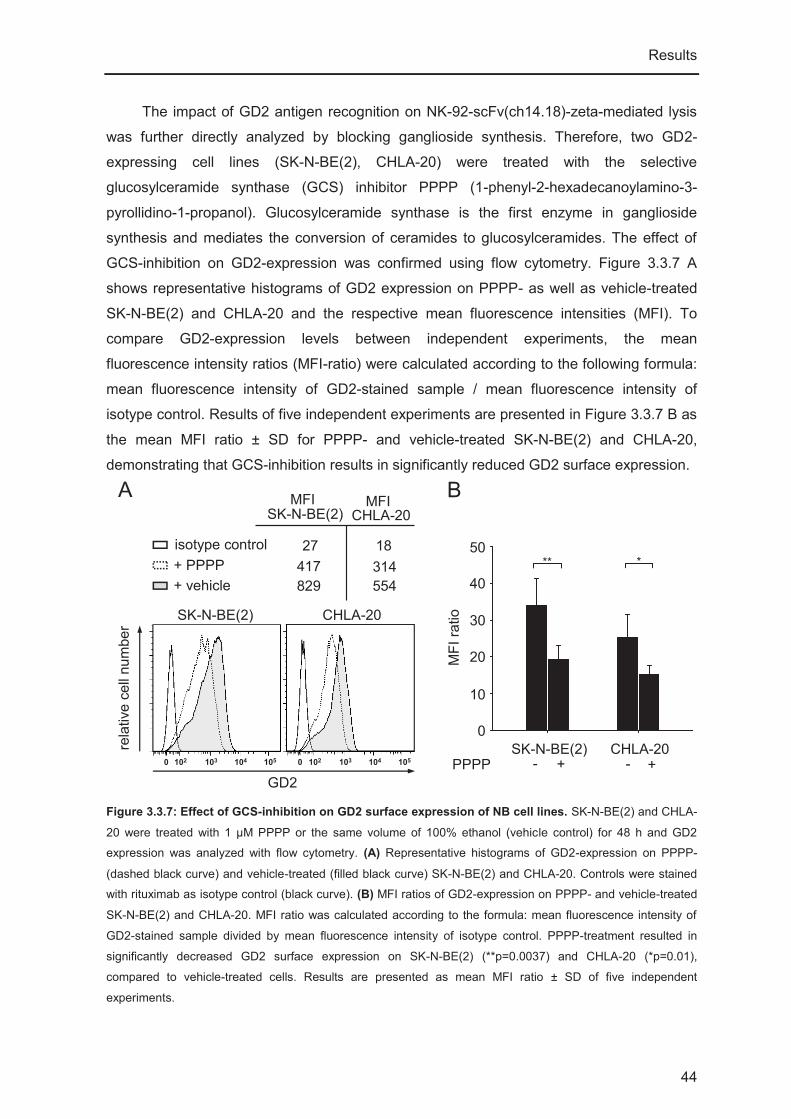
Results
44
The impact of GD2 antigen recognition on NK-92-scFv(ch14.18)-zeta-mediated lysis
was further directly analyzed by blocking ganglioside synthesis. Therefore, two GD2-
expressing cell lines (SK-N-BE(2), CHLA-20) were treated with the selective
glucosylceramide synthase (GCS) inhibitor PPPP (1-phenyl-2-hexadecanoylamino-3-
pyrollidino-1-propanol). Glucosylceramide synthase is the first enzyme in ganglioside
synthesis and mediates the conversion of ceramides to glucosylceramides. The effect of
GCS-inhibition on GD2-expression was confirmed using flow cytometry. Figure 3.3.7 A
shows representative histograms of GD2 expression on PPPP- as well as vehicle-treated
SK-N-BE(2) and CHLA-20 and the respective mean fluorescence intensities (MFI). To
compare GD2-expression levels between independent experiments, the mean
fluorescence intensity ratios (MFI-ratio) were calculated according to the following formula:
mean fluorescence intensity of GD2-stained sample / mean fluorescence intensity of
isotype control. Results of five independent experiments are presented in Figure 3.3.7 B as
the mean MFI ratio ± SD for PPPP- and vehicle-treated SK-N-BE(2) and CHLA-20,
demonstrating that GCS-inhibition results in significantly reduced GD2 surface expression.
Figure 3.3.7: Effect of GCS-inhibition on GD2 surface expression of NB cell lines. SK-N-BE(2) and CHLA-
20 were treated with 1 μM PPPP or the same volume of 100% ethanol (vehicle control) for 48 h and GD2
expression was analyzed with flow cytometry. (A) Representative histograms of GD2-expression on PPPP-
(dashed black curve) and vehicle-treated (filled black curve) SK-N-BE(2) and CHLA-20. Controls were stained
with rituximab as isotype control (black curve). (B) MFI ratios of GD2-expression on PPPP- and vehicle-treated
SK-N-BE(2) and CHLA-20. MFI ratio was calculated according to the formula: mean fluorescence intensity of
GD2-stained sample divided by mean fluorescence intensity of isotype control. PPPP-treatment resulted in
significantly decreased GD2 surface expression on SK-N-BE(2) (**p=0.0037) and CHLA-20 (*p=0.01),
compared to vehicle-treated cells. Results are presented as mean MFI ratio ± SD of five independent
experiments.
B
SK-N-BE(2)
rela
tive
cell
num
ber
GD20 102 103 104 105
isotype control+ PPPP+ vehicle
SK-N-BE(2)MFI
CHLA-20MFI
27417829
18314554
A
0 102 103 104 105
CHLA-20
0
10
20
30
40
50
SK-N-BE(2) CHLA-20PPPP +- +-
MFI
ratio
** *

Results
45
Furthermore, application of PPPP- and vehicle-treated SK-N-BE(2) and CHLA-20
target cells in a 51Cr release assay revealed that decreased GD2 surface expression
correlated with decreased sensitivity of PPPP-treated SK-N-BE(2) and CHLA-20 towards
NK-92-scFv(ch14.18)-zeta-mediated lysis (14% and 38% specific cytotoxicity,
respectively), compared to vehicle-treated cells (39% and 63% specific cytotoxicity,
respectively) (Fig. 3.3.8). Figure 3.3.8: Effect of GCS-inhibition on NK-92-
scFv(ch14.18)-zeta-mediated lysis of GD2+ NB cell lines. SK-N-BE(2) and CHLA-20 were treated
with 1 μM PPPP or the same volume of 100%
ethanol (vehicle control) for 48 h. Sensitivity
towards NK-92-scFv(ch14.18)-zeta-mediated lysis
was analyzed in a 51Cr release assay. PPPP-
treatment of SK-N-BE(2) and CHLA-20 resulted in
significantly reduced sensitivity towards NK-92-
scFv(ch14.18)-mediated cytotoxicity (***p<0.001).
Results are presented as mean specific cytotoxicity
in % ± SD of three independent experiments. To
compare independent experiments, data were
normalized to respective vehicle-treated control.
3.4. Sensitivity of NB relapse cell lines towards NK-92-scFv(ch14.18)-zeta-mediated lysis
Despite the use of intensive multi-modal therapy protocols, prognosis for high-risk NB
patients remains poor. The treatment of high-risk NB patients is particularly challenging,
because many patients initially respond to therapy but develop progressive disease or
relapse, which is potentially caused by minimal residual disease. Unfortunately, relapsed
tumors often exhibit drug resistance and are refractory towards chemotherapy. Therefore,
alternative approaches for treatment of minimal residual disease are needed.
To address this problem, the sensitivity of NB cell line pairs from four patients
towards NK-92-scFv(ch14.18)-zeta-mediated lysis was analyzed in 51Cr release assays.
Each cell line pair consists of one cell line established at time of diagnosis (SK-N-BE(1),
SMS-KAN, SMS-KCN, CHLA-15) and a corresponding cell line generated from relapse
material (SK-N-BE(2), SMS-KANR, SMS-KCNR, CHLA-20) of the same patient. For
comparison of independent experiments, specific cytotoxicity of control NK cell lines was
set to zero and specific cytotoxicity of NK-scFv(ch14.18)-zeta was normalized against
control NK cell lines.
0
20
40
60
80
100
SK-N-BE(2) CHLA-20PPPP +-
spec
ific
cyto
toxi
city
in %
+-
*** ***

Results
46
51Cr release assays revealed that relapse cell lines of all patients tested are still
sensitive towards NK-92-scFv(ch14.18)-zeta-mediated lysis, with specific cytotoxicities
ranging from 32% (SMS-KANR) to 47% (SK-N-BE(2) (Fig. 3.4.1). Interestingly, in three out
of four tested cell line pairs (SK-N-BE(1)/(2), SMS-KAN/SMS-KANR, SMS-KCN/SMS-
KCNR), the relapse cell line showed significantly higher sensitivity towards NK-92-
scFv(ch14.18)-zeta compared to the respective cell line from time point of diagnosis.
Relapse cell line SK-N-BE(2) exhibited a 2-fold higher sensitivity compared to SK-N-BE(1).
Sensitivity of SMS-KANR towards NK-92-scFv(ch14.18)-zeta was about 1.9-fold higher
than sensitivity of SMS-KAN. In case of SMS-KCN/SMS-KCNR, the relapse cell line SMS-
KCNR showed an almost 1.5 fold higher sensitivity. The fourth patient cell line pair (CHLA-
15/CHLA-20) exhibited comparable sensitivities towards NK-92-scFv(ch14.18)-zeta.
Figure 3.4.1: Sensitivity of NB cell line pairs towards NK-92-scFv(ch14.18)-zeta-mediated lysis. NB cell
line pairs of four patients were analyzed in 51Cr release assays. NK-92-scFv(ch14.18)-zeta effectively lysed all
relapse cell lines at an E:T ratio of 6.3:1. In three out of four patients tested (SK-N-BE(1)/(2), SMS-KAN/SMS-
KANR, SMS-KCN/SMS-KCNR), the relapse cell line exhibited a significantly higher sensitivity towards NK-92-
scFv(ch14.18)-mediated lysis (***p≤0.001), compared to the respective cell line from time point of diagnosis.
Specific cytotoxicity of control cell lines was set to zero and NK-92-scFv(ch14.18)-zeta-mediated lysis was
normalized against control NK cells. Results are presented as mean specific cytotoxicity in % ± SD of at least
three independent experiments.
Since recognition of GD2 by the CAR was shown to be the main mechanism of NK-
92-scFv(ch14.18)-zeta-mediated lysis of GD2-positive NB cell lines, cell line pairs from all
four patients were analyzed for GD2 surface expression. Figure 3.4.2 A displays
representative histograms of GD2-expression on NB cell line pairs. To compare GD2
expression levels of independent experiments, mean fluorescence intensity ratios (MFI-
ratio) were calculated according to the following formula: MFI of GD2-stained sample
divided by MFI of isotype-stained sample. Results of four independent experiments are
spec
ific
cyto
toxi
city
in %
0
20
40
60
80
100
SK-N-BE(1)
SK-N-BE(2)
SMS-KAN
SMS-KANR
SMS-KCN
SMS-KCNR
CHLA-15
CHLA-20
*** *** ***

Results
47
presented in Figure 3.4.2 B as the mean MFI ratio ± SD. Flow cytometric analysis of GD2
surface expression revealed that, in three out of four cases tested, the relapse cell line
exhibited significantly higher GD2-expression compared to the respective cell line from
time point of diagnosis. In the case of CHLA-15/CHLA-20, GD2-expression on the relapse
cell line was not significantly higher. Thus, higher GD2-expression in relapse cell lines SK-
N-BE(2), SMS-KANR and SMS-KCNR correlated with increased sensitivity of these cell
lines towards NK-92-scFv(ch14.18)-zeta-mediated lysis in 51Cr release assays.
These results show that relapse cell lines are sensitive towards NK-92-scFv(ch14.18)-
zeta-mediated lysis, because they still express the antigen GD2. In our tests, the majority
of relapse cell lines even displayed significantly increased GD2-expression, correlating with
an increased specific cytotoxicity mediated by NK-92-scFv(ch14.18)-zeta.
Figure 3.4.2: GD2-expression on NB cell line pairs. GD2-expression on NB cell line pairs of four patients
was determined with flow cytometry. (A) Representative histograms for each cell line pair. (gray dashed curve:
isotype control of diagnosis cell line; gray solid curve: GD2-staining of diagnosis cell line; black dashed curve:
isotype control of relapse cell line; filled black curve: GD2-staining of relapse cell line). (B) MFI ratios of GD2-
expression NB cell line pairs. MFI ratio was calculated according to the formula: mean fluorescence intensity of
GD2-stained sample divided by mean fluorescence intensity of isotype control. Relapse cell lines SK-N-BE(2),
SMS-KANR and SMS-KCNR exhibited a significantly higher GD2-expression than respective diagnosis cell
lines (*p<0.05). Results are presented as mean MFI ratio ± SD of four independent experiments.
SMS-KAN/SMS-KANR
SK-N-BE(1)/SK-N-BE(2)
SMS-KCN/SMS-KCNR
CHLA-15/CHLA-20
rela
tive
cell
num
ber
isotype controldiagnosis cell line
isotype controlrelapse cell line
GD2-staining diagnosis cell line
GD2-stainingrelapse cell line
A
0
20
40
60
80
100
SK-N-BE(2)
SK-N-BE(1)
SMS-KAN
SMS-KANR
SMS-KCN
SMS-KCNR
CHLA-15
CHLA-20
MFI
ratio
B
* * *
102 103 104 1050 102 103 104 1050102 103 104 1050 102 103 104 1050
102 103 104 1050 102 103 104 1050102 103 104 1050 102 103 104 1050
GD2

Results
48
Increased expression of GCS is associated with relapse cell lines and a common
mechanism of drug resistance that is activated in tumor cells to avoid accumulation of
ceramides and evade apoptosis [156-158]. Since GCS as the first enzyme in GD2
synthesis was shown to have an effect on GD2 surface expression and thereby on NK-92-
scFv(ch14.18)-zeta-mediated lysis (Fig. 3.3.6 and 3.3.7), GCS-expression in NB cell line
pairs was analyzed using Western blot. All relapse cell lines showed a higher GCS-
expression compared to their respective diagnosis cell lines (Fig. 3.4.3 A). Based on the β-
actin control, GCS-expression was normalized for all cell lines and the ratio of GCS
expression relapse/diagnosis cell line was calculated. To compare results from
independent experiments for each cell line pair, mean ratios were calculated (Fig. 3.4.3 B).
Results showed that the relapse cell lines SMS-KANR, SMS-KCNR and SK-N-BE(2)
exhibited higher GCS-expression, while expression in the relapse cell line CHLA-20 was
only slightly increased compared to diagnosis cell line CHLA-15. These results correlate
with GD2-expression on NB cell line pairs, suggesting that increased GCS-expression
might be one possible explanation for higher sensitivity of relapse cell lines SMS-KANR,
SMS-KCNR and SK-N-BE(2) towards NK-92-scFv(ch14.18)-zeta.
Figure 3.4.3: GCS-expression in NB cell line pairs. GCS-expression in NB cell line pairs of four patients was
analyzed using Western blot. For relative quantification of GCS-expression, β-actin-expression was analyzed
as control. (A) Representative Western blots for GCS-expression (top row) as well as β-actin control (bottom
row) in NB cell line pairs. (B) Ratio of GCS-expression of relapse to diagnosis cell lines. To compare GCS-
expression in relapse and diagnosis cell lines, GCS-expression was normalized using the respective β-actin
control, and intensity of relapse cell line was divided by intensity of diagnosis cell line. Results are presented as
mean ratio of GCS-expression relapse to diagnosis cell lines ± SD from at least three independent experiments.
3.5. In vivo efficacy of NK-92-scFv(ch14.18)-zeta in a drug-resistant NB mouse model
Since NK-92-scFv(ch14.18)-zeta was effective towards a panel of GD2+ NB cell lines
in vitro, the in vivo efficacy of a therapeutic application of NK-92-scFv(ch14.18)-zeta was
analyzed in a drug-resistant NB xenograft mouse model.
CH
LA-1
5
CH
LA-2
0
SM
S-K
AN
SM
S-K
AN
R
SM
S-K
CN
SM
S-K
CN
R
SK
-N-B
E(1
)
SK
-N-B
E(2
)
GCS
β-actin
CHLA-20/CHLA-15SMS-KANR/SMS-KANSMS-KCNR/SMS-KCNSK-N-BE(2)/SK-N-BE(1)
1.27 ± 0.49 1.54 ± 0.15 1.88 ± 0.67 1.61 ± 0.47
Ratio of GCS expression relapse/diagnosis cell line
A B

Results
49
Prior to the actual in vivo study, the effect of IL-2 starvation on CAR-expression was
examined in an in vitro experiment to determine whether an application of IL-2 should be
included in the in vivo experiment. Therefore, NK-92-scFv(ch14.18)-zeta were cultured
under different conditions, containing culture in complete medium, starvation from IL-2 for
four days and culture in the absence of IL-2 for four days and subsequent transfer to
complete medium for two additional days. CAR-expression was then analyzed in all
samples with flow cytometry (Fig. 3.5.1). This analysis revealed that starvation from IL-2
resulted in decreased CAR-expression compared to controls cultured in complete medium.
Abrogated CAR-expression was reversed by culture of cells in complete medium for two
additional days. This suggests that an application of IL-2 should be included in the
experimental setting of the following in vivo study.
Therapeutic efficacy of NK-92-scFv(ch14.18)-zeta was then determined according to
their effect on the subcutaneous primary tumor growth of GD2+ CHLA-20 tumors (Fig.
3.5.2). The primary tumor was induced by subcutaneous injection of 1x10⁶ CHLA-20 NB
cells into the left flank of female NSG mice (day 0). Starting on day three after tumor
inoculation, mice were subjected to a total of eight peritumoral injections of NK-92-
scFv(ch14.18)-zeta and 200 IU of recombinant human IL-2 (injections on day 3, 7, 11, 15,
19, 26, 33, 40). Additionally, mice received 1000 IU of IL-2 intraperitoneally 24 h after each
NK cell injection (injections on day 4, 8, 12, 16, 20, 27, 34, 41). To specifically show that
the mediated effect of a repeated application of NK-92-scFv(ch14.18)-zeta is based on the
expression of the GD2-specific CAR on these cells, the experimental setting also included
control groups treated with PBS only (untreated) or either IL-2 only or a combination of the
control NK cell line NK-92-scFv(FRP5)-zeta and IL-2. Since NK-92-scFv(ch14.18)-zeta and
Figure 3.5.1: Impact of IL-2-starvation on CAR-expression on NK-92-scFv(ch14.18)-
zeta. To assess the impact of IL-2 on CAR-
expression, NK-92-scFv(ch14.18)-zeta were
cultured under different culture conditions
and CAR-expression was analyzed by
staining with the anti-IdAb ganglidiomab.
Mouse IgG1 was used as isotype control
(gray curve) for staining. Cells were cultured
in either complete medium (dashed gray
curve), IL-2-free medium for four days (black
filled curve), or IL-2-free medium for four
days followed by two additional days in
complete medium (black curve). Results are
presented as a representative histogram.
rela
tive
cell
num
ber
CAR
isotype controlcomplete
no IL-2 + completeno IL-2
100 101 102 103 104

Results
50
NK-92-scFv(FRP5)-zeta are IL-2-dependent cell lines and IL-2-dependency of CAR-
expression had been demonstrated in a previous in vitro experiment, the application of IL-2
was included in the treatment schedule to ensure survival and stable CAR-expression of
NK cell lines.
Figure 3.5.2: Treatment schedule for therapeutic treatment with NK-92-scFv(ch14.18)-zeta. 1x10⁶ CHLA-
20 were subcutaneously injected into the left flank of female NSG (NOD.Cg-PrkdcscidIl2rgtm1WjI/SzJ) mice. Mice
received eight peritumoral subcutaneous (s.c.) injections of 2x10⁷ NK cells and 200 IU human recombinant IL-2
in total, starting on day three after tumor cell inoculation (days 3, 7, 11, 15, 19, 26, 33, 40). Additionally, mice
were subjected to intraperitoneal (i.p.) injections of 1000 IU IL-2 24 h after each NK cell injection (days 4, 8, 12,
16, 20, 27, 34, 41). Experimental groups (n=7) included mice treated with PBS only (untreated), IL-2 only, NK-
92-scFv(ch14.18)-zeta + IL-2 as well as a control group treated with NK-92-scFv(FRP5)-zeta + IL-2.
Peritumoral injections of NK-92-scFv(ch14.18)-zeta resulted in delayed appearance of
palpable tumors in treated mice (mean time to appearance 30 days) compared to control
groups (mean time to appearance 13-20 days). Although growth of primary CHLA-20
tumors was quite aggressive, treatment with GD2-specific NK-92-scFv(ch14.18)-zeta
effectively delayed tumor growth. To demonstrate this effect, experimental groups were
compared based on time until the tumors reached a volume of ≥150 mm3 (Fig. 3.5.3). This
comparison revealed that tumor growth was significantly delayed in NK-92-scFv(ch14.18)-
zeta-treated mice (49 days) compared to control groups (27-29 days).
1x10⁶
CHLA-20 s.c.
3d 7d 11d 15d 19d 40d33d26d
4d 8d 12d 16d 20d 41d34d27d
peritumoral injection of 2x10⁷ NK cells and 200 IU IL-2 s.c.
injection of 1000 IU IL-2 i.p.
1x10⁶
CHLA-20 s.c.
tumorvolume
800 mm3

Results
51
Since tumor growth in control groups was comparable, Figure 3.5.4 shows tumor growth of
individual mice in the NK-92-scFv(FRP5)-zeta control and NK-92-scFv(ch14.18)-zeta-
treated groups as well as the treatment schedule. Interestingly, the overlay of tumor growth
of individual mice of both groups showed that mice in the NK-92-scFv(ch14.18)-zeta group
were sacrificed at a smaller tumor size than NK-92-scFv(FRP5)-zeta-treated mice. To
confirm this finding, the mean tumor volumes on the day mice were sacrificed of all groups
were compared. This comparison revealed that NK-92-scFv(ch14.18)-zeta-treated mice
were sacrificed at a lower tumor volume (348 ± 124 mm3) than control mice (untreated
group 605 ± 101 mm3, IL-2 group 635 ± 128 mm3, NK-92-scFv(FRP5)-zeta group 666 ± 86
mm3). Mice of the NK-92-scFv(ch14.18)-zeta-treated group had to be sacrificed before they
reached the maximum tumor size of 800 mm3 due to development of necrosis in these
tumors at a lower tumor volume compared to tumors in control groups. This might be a sign
for increased tumor tissue destruction potentially caused by an increased invasion and
killing of tumor cells by NK-92-scFv(ch14.18)-zeta.
Figure 3.5.3: Comparison of tumor growth based on time to tumor volume ≥ 150 mm3.
Tumor growth was followed up by micro-caliper
measurements and tumor volume was
calculated according to the formula: length x
(width)2 x 0.5. Results are presented as mean
time in days until an established tumor with a
volume of ≥ 150 mm3 was developed ± SD
(n=7). Time until the tumors reached a volume ≥
150 mm3 was statistically longer in NK-92-
scFv(ch14.18)-zeta-treated mice (***p<0.001).
untre
ated
IL-2 c
ontro
l
NK-92-sc
Fv(FRP5)-
z
NK-92-sc
Fv(ch1
4.18)-
z0
20
40
60
80tim
e to
tum
orvo
lum
e15
0 m
m3
[d]
*********
***

Results
52
Figure 3.5.4: Tumor growth of individual mice of NK-92-scFv(ch14.18)-zeta-treated group and NK-92-scFv(FRP5)-zeta control group. Mice were challenged with subcutaneous injection of 1x10⁶ CHLA-20 in the
left flank (day 0). Starting on day three, mice received peritumoral subcutaneous injections of either a
combination of 2x10⁷ NK-92-scFv(ch14.18)-zeta and 200 IU IL-2 or 2x10⁷ NK-92-scFv(FRP5)-zeta and 200 IU
IL-2 (days 3, 7, 11, 15, 19, 26, 33, 40). Additional intraperitoneal injections of 1000 IU IL-2 were performed 24 h
after each NK cell injection (days 4, 8, 12, 16, 20, 27, 34, 41). Tumor volume was calculated according to the
formula: length x (width)2 x 0.5. Results are presented as the tumor volume of individual mice (n=7) of the NK-
92-scFv(ch14.18)-zeta-treated group (black solid curves) and the NK-92-scFv(FRP5)-zeta control group (black
dashed curves).
Most importantly, survival analysis of all groups showed, that the repeated therapeutic
application of NK-92-scFv(ch14.18)-zeta resulted in significantly higher median survival (52
days) compared to controls (untreated mice 30 days, IL-2 control 30 days, NK-92-
scFv(FRP5)-zeta group 32 days) (Fig. 3.5.5).
tum
orvo
lum
e in
mm
3
NK-92-scFv(ch14.18)-zNK-92-scFv(FRP5)-z
days after tumor cell inoculationtumor cells s.c.
3d 7d 11d 15d 19d 26d 33d 40d
4d 8d 12d 16d 20d 27d 34d 41d
peritumoral injection of NK cells/IL-2 s.c.
injection of IL-2 i.p.0d
0
200
400
600
800
0 4 8 12 16 20 24 28 32 36 40 44 48 52 56 60 64 68

Results
53
Figure 3.5.5: Therapeutic effect of repeated peritumoral NK cell applications on survival. Primary tumors
were induced by subcutaneous injection of 1x10⁶ CHLA-20 into the left flank of mice (day 0). Starting on day
three, mice received a total of eight injections of 2x10⁷ NK-92-scFv(ch14.18)-zeta + 200 IU IL-2 (days 3, 7, 11,
15, 19, 26, 33, 40). Additionally, mice were subjected to intraperitoneal injections of 1000 IU IL-2 24 h after
each NK cell injection (days 4, 8, 12, 16, 20, 27, 34, 41). Controls were injected with PBS only (untreated), IL-2
only or NK-92-scFv(FRP5)-z and IL-2. Results are presented as percent survival (n=7) of untreated mice (gray
dashed curve), IL-2-treated mice (gray solid curve), mice of the NK-92-scFv(ch14.18)-zeta group (black solid
curve) and mice of the NK-92-scFv(FRP5)-zeta group (black dashed curve). Survival of NK-92-scFv(ch14.18)-
zeta-treated mice was significantly higher compared to untreated mice (**p=0.0009), IL-2-treated mice
(**p=0.0008) and NK-92-scFv(FRP5)-zeta-treated mice (*p=0.002). There was no statistically significant
difference in survival of control groups.
untreatedIL-2 control
NK-92-scFv(ch14.18)-zNK-92-scFv(FRP5)-z
0 20 40 60 800
20
40
60
80
100
days after tumor inoculation
perc
ent s
urvi
val

Discussion
54
4. Discussion
4.1. Immunotherapy in NB
Neuroblastoma (NB) is an aggressive childhood malignancy, with about 50% of
patients exhibiting a high-risk phenotype associated with poor prognosis [2]. Despite
intensive therapy protocols based on radiation, chemotherapy, stem cell support and
differentiation therapy, standard treatment fails to successfully control high-risk NB in the
majority of patients, resulting in the development of progressive disease or relapse. The
induction of drug resistance in response to intensive therapy is a major obstacle to
treatment with conventional therapies. Hence, the development and refinement of effective
alternative therapies are of great importance. In this respect, immunotherapy is
increasingly attractive. Clinical evidence of the potential of immunotherapy in NB was
demonstrated in a phase III trial conducted by the Children’s Oncology Group in the US,
based on a protocol including a combination of the GD2-specific antibody ch14.18, IL-2,
GM-CSF and 13-cis-retinoic acid [74]. This new treatment approach resulted in a
significant increase in the two year event-free survival rate from 46 to 66%. Although this
shows the potential of immunotherapy in NB, it also emphasizes the need for further
improvement of therapeutic regimens, since about 30% of high-risk patients still do not
survive the disease.
The most advanced immunotherapeutic approaches in NB are based on targeting the
disialoganglioside GD2. Due to its abundant expression on NB cells but restricted
physiological expression in the brain and peripheral nerve fibers [29], GD2 is a suitable
antigen for passive immunotherapy with specific antibodies. With regard to the
development of evasion mechanisms by the tumor, it is important that GD2 was shown not
to be modulated off the cell membrane in response to GD2-targeted immunotherapy [31]. A
combination of antibody-based immunotherapy and adoptive transfer of immune effector
cells might further increase the therapeutic efficacy of immunotherapeutic approaches
targeting GD2.
4.2. Cellular therapy and chimeric antigen receptors
According to the missing self hypothesis, low or absent MHC class I expression
renders NB cells sensitive to NK cell lysis. Therefore, the use of NK cells as immune
effector cells for adoptive transfer is attractive. In general, cellular therapy with NK cells can
be based on reinfusion of ex vivo stimulated and expanded autologous NK or lymphokine

Discussion
55
activated killer cells (LAK cells), or the application of allogeneic cells in particular in a
haploidentical setting.
Clinical responses to a therapeutic application of autologous NK or LAK cells have
been only moderate [159-161]. The match of MHC class I molecules on tumor cells and
inhibitory KIR expressed on NK cells is a factor that can negatively affect the efficacy of
therapy with autologous NK cells. Importantly, due to independent inheritance of KIR and
HLA class I genes, HLA/KIR mismatch can also happen in the autologous setting. This
means that a person expresses a certain KIR but simultaneously lacks expression of the
respective HLA class I molecule, recognized by that KIR. This autologous KIR/HLA
mismatch has been shown to be beneficial in terms of improved outcome of patients [162].
A retrospective study in high-risk NB patients who received an autologous HSCT
demonstrated that KIR/HLA mismatch is a prognostic factor for an improved outcome of
patients [163]. Another factor that has to be considered for the application of autologous
cells is the observation that the functionality of NK cells can be impaired in cancer patients.
Different mechanisms have been suggested to be involved in this phenomenon
[summarized in 164]. One is a decreased expression of activating NK cell receptors, such
as NCRs or NKG2D [95, 165]. Furthermore, decreased expression or impaired function of
the signal-transducing zeta chain in patient T cells and NK cells has been reported for
various malignancies, such as cervical cancer, colorectal carcinoma, ovarian carcinoma,
prostate cancer and myeloid malignancies [166-170]. Additionally, redistribution of NK cell
subsets in favor of the less cytotoxic CD16dim CD56bright has been reported for cancer
patients [171, 172].
With regard to these problems, the use of allogeneic cells (particularly haploidentical
NK cells) is appealing. The mismatch between MHC class I molecules on the patients’
tumor cells and inhibitory KIRs on donor NK cells in an allogeneic setting is beneficial to
activation of NK cells due to the lack of inhibitory signaling [173, 174]. Additionally, NK cells
expanded from a healthy donor might exhibit improved functionality compared to the
patient’s NK cells. Clinical applications of allogeneic haploidentical NK cells in particular in
the setting of hematopoietic stem cell transplantation, have shown more promising results
[175-180].
Independent of the autologous or allogeneic nature of NK cells applied within an
immunotherapeutic approach, tumor cells can develop escape strategies to evade
recognition by NK cells. This includes shedding as well as down regulation of activating
ligands or release of soluble factors (such as TGF-β), eventually resulting in down
regulation of activating receptors on NK cells or their respective ligands on tumor cells [37-
42, 181]. The application of a chimeric antigen receptor (CAR) in order to specifically direct

Discussion
56
NK cells to tumor target cells and, at the same time, provide for an activation signal
following specific targeting of a TAA is an approach to overcome these evasion strategies.
In the case of NB, the disialoganglioside GD2 is an established target antigen for passive
immunotherapy based on the application of GD2-specific antibodies. Chimeric antigen
receptors have been generated to combine antibody-based immunotherapeutic
approaches and the adoptive transfer of immune effector cells to increase the therapeutic
potential. In general, CARs are not dependent on presentation of antigens in the context of
MHC class I molecules and therefore are able to recognize carbohydrate or glycolipid
antigens in addition to protein antigens. Importantly, the expression of chimeric antigen
receptors provides effector cells with a certain specificity and thereby overcomes the above
mentioned immune evasion strategies, since lysis is not only dependent on interaction of
activating NK cell receptors with their respective ligands on tumor cells anymore.
In comparison to the application of anti-GD2 antibodies, the use of CAR-expressing
effector cells might be especially beneficial for patients who only express Fc receptors for
IgG (FcγR) with low affinity for the therapeutic antibody. This correlation between the
affinity of the FcγR and clinical efficacy of antibody-based immunotherapy has been
reported across a variety of therapeutic antibody applications [182-185]. Interestingly,
studies indicated that the use of CAR-expressing effector cells might compare favorably to
the use of a combination of monoclonal antibodies and NK cells [148]. Patients who did not
respond to therapy with the chimeric bispecific antibody blinatumomab (anti-CD19 and anti-
CD3) were shown to respond to CAR-expressing effector cells [186]. Furthermore,
immunotherapeutic studies based on the application of the GD2-specific immunocytokine
hu14.18-IL2 have shown correlations between HLA/KIR mismatch and improved outcome
[187, 188]. In addition to the potential benefit of a CAR-based approach compared to the
application of an TAA-specific antibody, this also suggests that the application of a CAR-
expressing allogeneic cell line might be especially beneficial for patients who do not exhibit
an autologous HLA/KIR mismatch status.
Another challenge to passive immunotherapy with monoclonal antibodies is that
patients are very often heavily pre-treated with high-dose multi-agent chemotherapy. This
application of cytostatic agents negatively affects numbers and cytotoxic activity of
endogenous immune cells, resulting in immune suppression of patients. In this respect, the
adoptive transfer of CAR-engineered allogeneic effector cells is becoming increasingly
attractive as a substitute for functionally impaired endogenous immune cells, thereby
suggesting a greater therapeutic potential for immunotherapeutic applications.
Cellular therapy based on the adoptive transfer of CAR-expressing effector cells,
mainly T cells, has already entered clinical trials. So far, approaches using 1st generation

Discussion
57
CARs have shown only modest success [189-191]. Interestingly, the exception was a
phase I clinical trial in NB patients treated with autologous T cells that were genetically
engineered to express a GD2-specific CAR [192]. In detail, 11 patients received a
combination of CAR-expressing activated T cells (ATC) and CAR-expressing CTLs that are
specific for Epstein Barr-virus (EBV), intended to provide additional co-stimulation. CAR-
expressing CTLs were detectable at six weeks after injection, while CAR expressing ATC
were only detectable until three weeks after injection. Four out of eight patients that
entered the study with detectable disease responded with tumor necrosis or tumor
regression after treatment with GD2-specific autologous T cells [192]. Long-term
monitoring of these patients and eight additional patients demonstrated comparable
persistence of both effector cell populations at a low level for up to four years [146].
Persistence of either cell type correlated with prolonged time to disease progression in 11
patients with active disease. Three out of these 11 patients experienced complete
response. Importantly, in addition to these responses, this phase one trial demonstrated
that the therapeutic application of CAR-expressing effector cells was safe and well
tolerated. According to clinical trials based on the therapeutic application of GD2-specific
antibodies, neuropathic pain is a major side effect due to physiological expression of GD2
on peripheral nerve fibers. This side effect was not reported upon treatment with GD2-
CAR-expressing effector cells. So far, studies showed that neuropathic pain is potentially
caused by recognition of GD2 on peripheral nerve fibers, and subsequent activation of the
complement system, based on the interaction between complement proteins and the Fc
part of the anti-GD2 antibody [193]. This suggests that in the case of GD2-targeting
therapeutic strategies, the use of CAR-expressing effector cells might be beneficial
compared to the application of GD2-specific antibodies, because the CAR does not contain
a Fc part and therefore does not activate the complement system. Furthermore, it was
reported that treatment of patients with GD2-CAR-expressing effector cells did not induce
the generation of antibodies against the CAR [146].
To address the problem of low persistence and expansion of 1st generation CAR-
expressing effector cells which resulted in only modest therapeutic effects, effector cells
expressing a 2nd generation CAR including a co-stimulatory domain have been developed
and entered clinical trials [reviewed in 194-196]. So far, the most promising results
regarding persistence and expansion of CAR-expressing effector cells and clinical
response have been demonstrated towards CD19+ hematological malignancies [145, 186,
197, 198]. Kochenderfer et al. [197] reported results of a clinical application of CD28-CD3ζ
CAR-T cells specifically targeting CD19. Out of eight patients, five experienced partial
remission and one patient showed complete remission. In the majority of patients CAR-T

Discussion
58
cells were detectable up to 50 days, while in some patients CAR-T cells persisted for 130
days or longer. Persistence of CD19 specific 4-1BB-CD3ζ CAR-T cells for up to six months
was reported by Kalos et al. [145] in three patients with chronic lymphocytic leukemia
(CLL). Importantly, CAR-T cells expanded more than 1000-fold after infusion into patients.
Two of these patients experienced complete remission, while a third patient showed a
partial response. Porter et al. [198] reported results of the treatment of one additional CLL
patient with 4-1BB-CD3ζ CAR-T cells. This patient also experienced complete remission of
disease and showed greater than 1000-fold expansion of CAR-T cells which persisted for
six months. These promising results were confirmed by the use of 4-1BB-CD3ζ CAR-T
cells in two patients with acute lymphoid leukemia (ALL) [186]. Expansion of CAR-T cells
greater than 1000-fold as well as persistence of cells for six months and longer was
confirmed in both patients. Treatment with 4-1BB-CD3ζ CAR-T cells resulted in complete
remission in both patients, with one patient maintaining complete remission after nine
months. The other patient relapsed two months after treatment.
Not many 3rd generation CARs, which include several co-stimulatory domains have
entered clinical trials so far. A CD20-specific 3rd generation CAR containing CD28 and 4-
1BB co-stimulatory domains was utilized in a pilot clinical trial [147]. Out of four patients
with follicular lymphoma or mantle cell lymphoma that enrolled to the study, three patients
received CAR-expressing T cells. Two of these patients entered the study without
evaluable disease and remained progression-free for 12 and 24 months after treatment.
The third patient entered partial remission but relapsed 12 months after treatment.
Importantly, CAR-expression on infused modified T cells was only detectable with PCR and
persistence of cells in patients was confirmed up 12 months after treatment. In contrast, the
application of T cells expressing a 3rd generation ErbB2-specific CAR, containing CD28
and 4-1BB co-stimulatory domains, in a patient with colorectal cancer caused a serious
adverse event resulting in death [199].
All of the above mentioned CAR-based immunotherapeutic approaches have utilized
T cells as effector cells. Since the understanding of NK cell biology and regulation of NK
cell activation is evolving, the use of NK cells as alternative effector cells is becoming
increasingly attractive. In contrast to T cells, NK cells mediate a non-MHC-restricted natural
cytotoxicity without any prior sensitization [80, 83]. Importantly, this natural cytotoxicity of
NK cells has already been shown to play a role in the lysis of NB cells [35, 36, 200].
Although T cells are advantageous with regard to initial number of effector cells which can
be isolated from peripheral blood, progress has been made in the establishment of
protocols for NK isolation and ex vivo expansion and activation, compensating for the initial
lower cell count. These improved protocols are based on the expansion of NK cells using

Discussion
59
an artificial antigen presenting cell line generated by genetic engineering of K562 to
express one or several activating ligands, such as CD137L or MICA and IL-15 or IL-21 as a
membrane-bound activating cytokine [201-204]. Within several expansion cycles, NK cell
numbers can be expanded up to 47,967-fold depending on the artificial antigen presenting
cell line that is used. These new and improved NK cell expansion and activation protocols
potentially compensate the initial difference in cell numbers of T cells and NK cells.
Importantly, NK cells might be advantageous as effector cells compared to T cells in an
allogeneic adoptive transfer setting. It has been shown that NK cells mediate cytotoxicity in
the graft versus tumor direction but do not cause graft-versus-host disease (GvHD), a
serious complication that is potentially mediated by T cells [177, 205, 206]. The shorter life
span of NK cells compared to T cells might be beneficial with regard to long-term side
effects which can be potentially mediated by memory-like T cells [207]. Such a long-term
side effect has been reported in clinical trials based on the application of CD19-specific
CAR-expressing T cells, which resulted in long-term depletion of normal B cells [197, 198].
Based on these advantages of NK cells, the approach of CAR-based immunotherapy
has already been expanded to NK cells as effector cells [133, 143, 208]. Although these
studies demonstrated the feasibility of a CAR-based approach employing NK cells as
effector cells, they also demonstrated that there are still limitations regarding donor-
dependent extensive variations in transfection efficacy and CAR functionality [133, 143].
Furthermore, ex vivo expansion, activation and genetic engineering of autologous or
allogeneic primary NK cells for each individual patient is time intensive and requires
substantial resources.
The human NK cell line NK-92 is a promising alternative to address the problems
associated with the use of primary NK cells. In contrast to primary patient-derived NK cells
that might be functionally impaired, NK-92 provides unrestricted alloreactivity due to lack of
expression of KIRs [119]. NK-92 has been shown to exhibit a high cytotoxicity against a
variety of cancer cell lines and primary cancer cells [116-118]. As a prerequisite for a
clinical application large scale expansion of NK-92 can be easily performed under good
manufacturing practices (GMP) [121, 122]. NK-92 expressing CARs of various specificities
have already been generated [140, 142, 148, 209, 210], demonstrating the feasibility of
genetic engineering of NK-92. Once CAR-expressing NK-92 of certain specificity are
generated, they can be provided on demand in a standardized quality and can be used not
only for one specific patient but many different patients. This non-individualized approach is
advantageous compared to the use of patient-derived effector cells, because it
compensates for functional impairment of primary effector cells as well as donor-
dependent variations in ex vivo expansion rates, transfection efficacy and CAR-

Discussion
60
functionality. Most importantly, the characteristic of being ready on demand in a
standardized quality circumvents the obstacle of time and labor consuming isolation,
transfection, expansion and testing of patient-derived effector cells. This reduction in lead
time is an important prerequisite for the initiation of multicenter clinical trials in the
maintenance therapy immediately after conventional therapy to prevent a relapse.
To expand this approach to NB, this thesis aimed to analyze the mechanism and
efficacy of a cellular therapy based on a human NK cell line expressing a GD2-specific
CAR (NK-92-scFv(ch14.18)-zeta).
4.3. Effect and mechanism of a GD2-directed NK CAR therapy approach based on NK-92-scFv(ch14.18)-zeta
The initial part of this study involved the analysis of CAR-expression on NK-92-
scFv(ch14.18)-zeta as well as the control cell lines NK-92, NK-92-pLXSN, and NK-92-
scFv(FRP5)-zeta with flow cytometry, because CAR-expression is the prerequisite for
GD2-specific lysis of target cells. High homogenous CAR-expression was confirmed for
NK-92-scFv(FRP5)-zeta and NK-92-scFv(ch14.18)-zeta, with only NK-92-scFv(ch14.18)-
zeta expressing a GD2-specific CAR (Fig. 3.1.1). In vitro cytotoxicity assays demonstrated
significantly higher lysis of GD2-expressing NB cell lines mediated by NK-92-
scFv(ch14.18)-zeta compared to control NK cell lines (Fig. 3.1.2-3.2). As the development
of drug-resistance remains a major challenge in NB treatment with conventional therapies,
the panel of GD2-positive target cell lines tested in these assays also included NB cell
lines, which are known to be partially or even multidrug-resistant. This was intended to
assess the sensitivity of NB cell lines towards a GD2-targeted approach with NK-92-
scFv(ch14.18)-zeta. NK-92-scFv(ch14.18)-zeta effectively lysed all tested NB cell lines,
even those with a drug-resistant phenotype, indicating that targeting GD2 with a cellular
approach based on NK-92-scFv(ch14.18)-zeta might offer an alternative therapeutic
approach to address the problem of acquired drug-resistance with conventional therapies.
The role of the interaction between the target antigen GD2 and the CAR for cytotoxic
activity of NK-92-scFv(ch14.18)-zeta was assessed in additional in vitro cytotoxicity assays
by disrupting the interaction, either by blocking the CAR with an anti-IdAb (Fig. 3.3.1 and
3.3.2) or by blocking the target antigen GD2 by addition of the GD2-specific antibody
ch14.18 (Fig. 3.3.3) during co-incubation of NK-scFv(ch14.18)-zeta and target cells. Both
strategies resulted in significant abrogation of NK-92-scFv(ch14.18)-zeta-mediated lysis of
GD2-positive target cells. Importantly, lysis was almost completely abrogated, indicating
that recognition of GD2 by the CAR is the primary mechanism of activation of NK-92-
scFv(ch14.18)-zeta-mediated lysis. To directly analyze the impact of GD2-expression on

Discussion
61
NK-92-scFv(ch14.18)-zeta-mediated lysis of NB cell lines, GD2-expression on NB target
cells was decreased by inhibition of glucosylceramide synthase (GCS) with the selective
inhibitor PPPP. Inhibition of GCS, and thereby inhibited conversion of ceramides to
glucosylceramides, abrogates the first step in ganglioside synthesis. Significantly
decreased GD2-expression on PPPP-treated target cells (Fig. 3.3.6) resulted in significant
abrogation of target cell lysis mediated by NK-92-scFv(ch14.18)-zeta (Fig. 3.3.7). This
demonstrates a direct correlation between GD2-expression and NK-92-scFv(ch14.18)-
zeta-mediated lysis. Additionally, the potential for GD2 alone to induce activation of NK-92-
scFv(ch14.18)-zeta in absence of other activating stimuli was analyzed in a target cell-free
experimental setting, based on immobilized GD2 (Fig. 3.3.4 and 3.3.5). Immobilized GD2
was sufficient to stimulate a significantly increased release of granzyme B and perforin,
indicating activation of NK-92-scFv(ch14.18)-zeta. This effect was abrogated by blocking
the CAR, confirming that activation is mediated by interaction of the CAR with immobilized
GD2, which demonstrated that immobilized GD2 is sufficient to induce activation of NK-92-
scFv(ch14.18)-zeta. This finding is particularly important, considering the general
complexity and tight regulation of NK cell activation and that sometimes several activating
receptors must be synergistically engaged to induce activation and cytotoxicity of NK cells.
Importantly, these experiments demonstrate that recognition of GD2 by the CAR results in
activating signaling, leading to the release of effector molecules. This suggests that GD2-
specific lysis of NK-92-scFv(ch14.18)-zeta is actually due to activation of CAR signaling
and not due to binding of the target cell via the CAR and activation via another
ligand/receptor interaction. Additionally, these results indicate that targeting GD2 with the
GD2-specific CAR expressed on NK-92-scFv(ch14.18)-zeta would allow the therapy to
overcome escape mechanisms developed by cancer cells, such as shedding of activating
ligands.
Based on the establishment of GD2-specificity and functionality of CAR-expressing
NK-92-scFv(ch14.18)-zeta we could clearly show the primary role of the interaction of GD2
with the CAR for activation of NK-92scFv(ch14.18)-zeta and thereby NK-92-scFv(ch14.18)-
zeta-mediated lysis. NK-92-scFv(ch14.18)-zeta were then used to address the problem of
developing effective therapy against refractory relapse cell lines which remains a major
challenge in high-risk NB treatment with conventional therapies. NB cell line pairs of four
patients, each consisting of a cell line generated at time of diagnosis and a corresponding
relapse cell line, were analyzed for their sensitivity towards NK-92-scFv(ch14.18)-zeta (Fig.
3.4.1). Importantly, all relapse cell lines were sensitive towards NK-92-scfv(ch14.18)-zeta-
mediated lysis with three out of four relapse cell lines displaying even higher sensitivity
than the corresponding cell line from time of diagnosis. This finding correlated with an

Discussion
62
increased GD2-expression in the respective relapse cell lines (Fig. 3.4.2). Since it was
shown that GCS, as the first enzyme in ganglioside synthesis has an impact on GD2
expression, GCS-expression in all cell line pairs was analyzed with Western blot (Fig.
3.4.3). This analysis revealed an increase in GCS-expression in three relapse cell lines
compared to their corresponding cell line from diagnosis. The fourth relapse cell line
displayed only slightly increased GCS-expression. While this last observation remains of
interest, it would be necessary to analyze a greater number of cell line pairs to confirm this
finding.
These encouraging in vitro results provided a baseline to test the therapeutic efficacy
of NK-92-scFv(ch14.18)-zeta in vivo in a drug-resistant xenograft mouse model. This
mouse model was based on induction of a subcutaneous human NB tumor followed by
repeated peritumoral applications of NK cells, in combination with IL-2 to promote the
survival of NK cells as well as to maintain high CAR-expression. Injection of NK cells was
started on day three after tumor cell inoculation (Fig. 3.5.1). Since the idea of a cellular
immunotherapy following conventional standard therapy is to treat minimal residual disease
(MRD), which could otherwise potentially lead to relapse, NK cell injections were started at
a time point with no palpable or visible signs of a tumor. Monitoring of tumor growth in all
experimental groups revealed that palpable tumors appeared later in NK-92-
scFv(ch14.18)-zeta-treated mice, and tumor growth rates were significantly delayed.
Control groups displayed aggressive tumor growth, in contrast to NK-92-scFv(ch14.18)-
zeta-treated mice, in which NK cell injections suppressed tumor growth in the majority of
treated mice (Fig. 3.5.3 and 3.5.4). Mice in this NK-92-scFv(ch14.18)-zeta-treated group
developed tumors after the end of NK cell treatment. Interestingly, tumors of mice receiving
NK-92-scFv(ch14.18)-zeta already showed signs of necrosis at smaller tumor volumes
than control groups and therefore had to be sacrificed before the maximal tumor volume
was reached. These data provide a proof of concept that therapeutic application of NK-92-
scFv(ch14.18)-zeta can mediate an in vivo anti-tumor effect towards an aggressively
growing tumor and leads to significantly prolonged survival in mice receiving GD2-specific
therapy (Fig. 3.5.5).
4.4. Toxicities and potential side effects of CAR-based approaches and strategies for circumvention
Despite its potential for cancer therapy, the use of genetically modified effector cells
expressing a CAR can be accompanied by potential toxicities and side effects. On-target
toxicities on healthy tissue are one major complication that should be considered. These
toxicities most likely occur if healthy tissue also displays expression of the target antigen,

Discussion
63
resulting in the destruction of healthy cells. Hence, target antigens for CAR-based
immunotherapeutic approaches should be carefully chosen with high levels of expression
on the tumor but restricted physiological expression. However, the desired on-target
toxicity towards the tumor can also be accompanied by severe side effects. Strong
activation of CAR-expressing effector cells, or activation of a high number of effector cells
at the same time and subsequent destruction of tumor cells, may result in the release of
high amounts of cytokines, such as TNF-α, IFN-γ, IL-2 or IL-6, leading to cytokine release
syndrome (CRS). This can cause severe fever or hypotension and might result in life-
threatening conditions, including multiple organ dysfunction [211-213]. Clinical applications
of CAR-expressing T cells have reported the development of CRS as a side effect of
adoptive transfer of CAR-expressing effector cells [197, 199].
Avoiding these toxicities and potential side effects is a major challenge for the
therapeutic application of CAR-expressing effector cells. To address this problem of on-
target toxicities towards healthy tissue, a new CAR approach is currently under
investigation, based on the co-expression of an antigen-specific CAR and a chimeric co-
stimulatory receptor (CCR) [214]. In this approach, an antigen-specific CAR that mediates
only suboptimal activation is combined with a second receptor recognizing another antigen
on the tumor and providing co-stimulation. Hence, effector cells only receive enough
activation when both the CAR and the CCR interact with their respective antigen on the
tumor. Engagement of only one of two receptors with their respective ligand provides only
insufficient stimulation of effector cells. This potentially avoids toxicity towards healthy
tissue when antigens are used that are not absolutely restricted to the tumor but are also
expressed to some extent on healthy tissue [214]. Alternatively, the feasibility of a
combination of an activating CAR recognizing a TAA and a second inhibitory CAR,
specifically recognizing an antigen that is expressed on normal tissue, is under
investigation to address the problem of on target, off organ toxicities [215]. In this
approach, inhibitory CARs (iCARs) are based on the expression of inhibitory intracellular
signaling domains of the immunoinhibitory receptors CTLA-4 and PD-1. Co-expression of
activating CAR and iCAR was reported to result in inhibition of on-target cytotoxicity
against other organs than the tumor when the iCAR encounters its respective antigen
expressed on normal tissue [215].
Regarding the development of CRS, several aspects have to be considered. Since
the extend of CRS has been shown to correlate with the disease burden [213, 216], the risk
of CRS might be reduced by the restriction of CAR-based immunotherapy to a minimal
residual disease setting. Further, the CAR-design might have an impact on potential side
effects. Although improvement of CAR design based on the addition of co-stimulatory

Discussion
64
domains might be beneficial in terms of stronger activation, prolonged persistence or
increased proliferation of engineered effector cells, this might also require investigation into
possible side effects related to strong activation or proliferation of TAA-specific effector
cells. Even if the use of autologous effector cells might be beneficial in terms of low
immunogenicity of CAR-expressing effector cells and therefore persistence of these cells,
there is also the potential disadvantage of extensive proliferation of effector cells in
response to TAA recognition on the tumor. This might result in increased numbers of
effector cells reaching a threshold at which activation of these effector cells could
potentially cause a CRS. To address this potential safety concern, suicide switches have
been incorporated into the concept of adoptive transfer of CAR-expressing effector cells
[217]. Safety switches based on the Herpes simplex virus thymidine kinase [218], inducible
caspase 9 [219] and CD20 [220] have already been generated. These suicide switches
enable elimination of CAR-expressing effector cells in the case of critical proliferation. The
disadvantage of this approach would be that the effect is not some kind of preventive
therapy and will be used when indications for too high effector cell numbers or
pathologically elevated high cytokine levels are already detectable. Additionally, suicide
gene-based induction of apoptosis would also completely abrogate the therapeutic effect of
CAR-expressing cells.
Alternatively, the use of non-proliferating effector cells or effector cells which exhibit
only transient CAR-expression, facilitated by transfection with mRNA encoding for the
CAR, might be disadvantageous in terms of persistence of the anti-tumor effect but might
be beneficial with regard to the safety of the therapeutic approach [137, 221, 222].
Irradiation can prevent proliferation of effector cells while maintaining cytotoxic activity. The
advantage of non-proliferating effector cells is that the procedure of their therapeutic
application would be comparable to the application of conventional therapeutic agents.
Since the effector cells do not proliferate, the exact dosage can be calculated and dose
escalation can be performed to determine a maximal tolerated dose that is not associated
with severe side effects. Further, in case of unexpected adverse events, treatment can
immediately be stopped by discontinuing the injection of cells or by halting additional
effector cell injections. Therefore, the application of non-proliferating cells appears to be
safer and more predictable. On the other hand, this approach may potentially require
injection of higher cell numbers and repeated application of effector cells to maintain an
anti-tumor response. Generation of sufficient numbers of CAR-expressing effector cells is
therefore a crucial prerequisite. Due to recent improvements of protocols for NK cell
isolation and ex vivo expansion [201-204], NK cells are an increasingly attractive effector
cell population for CAR-based immunotherapeutic approaches. NK cells appear to be

Discussion
65
advantageous with regard to their natural cytotoxicity especially towards tumor entities that
lack MHC class I but still express activating NK cell ligands. NK cells also have an
improved safety profile in an allogeneic setting, since they exhibit an increased cytotoxicity
compared to the autologous setting but do not cause GvHD, a potential severe
complication that can be caused by allogeneic T cells [177, 205, 206].
Unless endogenous T cell receptor genes are removed during genetic engineering, T
cells still express their T cell receptor in addition to the TAA-specific CAR. When those T
cells encounter their respective TAA recognized by the CAR, they get activated and
proliferate. Expansion of this T cell population does not only mean expansion of TAA-
specific CAR-expressing T cells, but also proliferation of T cells with a T cell receptor of
unknown specificity, which might be another safety concern. Additionally, memory-like
features of T cells compared to NK cells might be beneficial with regard to retaining a long-
term anti-tumor response, but also bear the potential risk of long-term side effects if the
TAA is not exclusively expressed on tumor tissue only [197, 198, 207].
Another aspect to be considered is the potential immunogenicity of the expressed
CAR as a “foreign” protein. In the case of CAR-generation based on scFvs consisting of
murine variable regions, there is the possibility that this might induce an immune response
directed against the CAR, resulting in clearance of CAR-expressing effector cells from the
system and thereby abrogation of the therapeutic activity. The development of anti-idiotype
antibodies specifically binding the paratopes of the CAR has been reported in a number of
cases [190, 223, 224], in contrast to others not showing the induction of anti-idiotype
antibodies [189]. The use of scFvs that were derived from humanized antibodies for CAR
generation might be beneficial in terms of lower potential to induce anti-idiotype antibodies.
Interestingly, in the clinical application of murine GD2-specific antibodies, the induction of
human anti-mouse antibodies (HAMA) correlated with improved outcome of patients [59].
This might be due to the induction of anti-anti-idiotype antibodies according to the anti-
idiotype network theory [60], which recognize the nominal antigen GD2. In addition to these
humoral immune responses, genetically engineered effector cells can also induce cell-
mediated immune responses, which potentially limit therapeutic efficacy [223, 225].
The therapeutic approach presented in this study is based on the repeated
application of a human NK cell line expressing a GD2-specific CAR (NK-92-scFv(ch14.18)-
zeta). With regard to a clinical application, NK-92-scFv(ch14.18)-zeta can be irradiated to
prevent proliferation. Parental NK-92 cells tested in clinical trials have been subjected to
irradiation with 10 Gy prior to injection [124, 125], and the cytotoxicity of irradiated GD2-
specific NK-92-scFv(ch14.18)-zeta has been shown to be comparable to non-irradiated
NK-92-scFv(ch14.18)-zeta [150]. This is a prerequisite for the safety of the injection of a

Discussion
66
human NK cell line into patients to avoid induction of a lymphoproliferative disease.
Importantly, these cells can be expanded under GMP conditions to provide sufficient cell
numbers for repeated injections during therapy, another prerequisite for a clinical
application. So far, two clinical trials investigated the application of parental NK-92 cells in
patients with advanced malignancies. The first phase I study analyzed the safety of NK-92
administration in 12 patients with renal cancer or melanoma based on a dose escalation
including four different dose levels 1x10⁸/m2, 3x10⁸/m2, 1x10⁹/m2 and 3x10⁹/m2 [124].
Patients received three infusions of the respective NK-92 cell number within a 48 h infusion
period on days 1, 3 and 5. NK-92 application was well tolerated, mainly accompanied only
by grade 1 fever as well as grade 3 and grade 4 hypoglycemia observed in two patients
only. The second phase I study was based on treatment of 15 patients with solid
tumors/sarcomas or leukemia/lymphoma [125]. Patients were subjected to two NK-92
injections, 48 h apart from each other with a dose escalation including three different NK-
92 dose levels (1x10⁹/m2, 3x10⁹/m2, 1x10¹⁰/m2). One patient received additional NK-92
infusions (5 infusions over a 6 month period). Importantly, NK-92 infusions were well
tolerated, and analysis of persistence of NK-92 cells in two patients demonstrated that NK-
92 was detectable in the circulation up to 48 hours after injection.
Addressing the problem of allogenicity of this cell line, and thereby potential immune
reactions of the patient’s immune system towards these cells, patients were selected for
the clinical trial based on the absence of cross-match with NK-92. The first study revealed
anti-HLA antibodies recognizing NK-92 in one out of two patients tested [124]. However, in
the second study only one out of seven patients was tested positive for anti-HLA antibodies
1 and 4 weeks after NK-92 application. Although both studies aimed at investigating the
safety of repeated NK-92 infusions, some responses have been seen in both studies [125].
The first study reported a minor response in a patient with metastatic melanoma and a
mixed response in a patient with renal cell carcinoma [124]. In the second study, two
patients with advanced lung cancer showed clinically significant mixed responses, and
treatment of another patient with lung cancer resulted in stable disease for about two years
[125]. These data demonstrated that repeated application of NK-92 is safe and well
tolerated with evidence for clinical activity in patients with advanced cancer. Importantly,
immune reactions towards NK-92 were rare, suggesting that a repeated application of NK-
92 is feasible for treatment of advanced cancer. These data regarding the parental NK-92
cell line suggest that an application of the GD2-specific cell line NK-92-scFv(ch14.18)-zeta
will have a similar safety and activity profile. Based on these considerations, the findings
reported here provide an important baseline for the design of phase I/II clinical trials to

Discussion
67
address tolerability of and response to NK-92-scFv(ch14.18)-zeta therapy in a highly
challenging population of stage 4 NB patients.
4.5. Conclusion
In vitro as well as in vivo data generated within the present study provide definite
evidence for specificity of NK-92-scFv(ch14.18)-zeta and efficacy of a GD2-targeted
cellular approach against drug-resistant NB. Importantly, the interaction of the target
antigen GD2 on tumor cells with the CAR was shown to be the primary mechanism of
activation and subsequent tumor cell lysis mediated by NK-92-scFv(ch14.18)-zeta. The use
of allogeneic GD2-specific NK-92-scFv(ch14.18)-zeta might further increase the
therapeutic potential of an anti-GD2 immunotherapy. As a non-individualized cellular
therapy, the use of NK-92-scFv(ch14.18)-zeta is independent from primary patient cells.
Therefore, the present therapy could remain a therapeutic option for heavily pre-treated
patients with poorly functioning primary immune cells or unfavorable conditions regarding
FcγR affinity or HLA/KIR mismatch status. Importantly, this CAR-based cellular approach is
likely to have an improved pain toxicity profile compared to GD2-specific antibodies,
because CARs do not activate the complement system which is responsible for the pain
side effects. One major benefit of the NK-92-scFv(ch14.18)-zeta cellular approach is that
effector cells can be made available on demand, which is an important prerequisite to start
this cell-based maintenance therapy subsequent to conventional therapy in a multicenter
setting.
In conclusion, these considerations and the pre-clinical data generated in the present
study provide an important rationale for the clinical investigation of tolerability and efficacy
of NK-92-scFv(ch14.18)-zeta in phase I/II trials to further improve the outcome for highly
challenging stage 4 NB patients.

Summary
68
5. Summary Neuroblastoma (NB) is a solid extracranial childhood malignancy of neuroectodermal
origin. About 50% of NB patients belong to the high-risk group characterized by poor
prognosis. Standard multi-modal therapy is based on high dose chemotherapy and stem
cell support followed by differentiation therapy with 13-cis-retinoic acid. Although this has
improved the outcome of NB patients, treatment of high-risk NB remains challenging due to
development of drug-resistant progressive disease or relapse that no longer responds to
conventional therapy. This emphasizes the urgent need of alternative adjuvant therapy
regimen.
Along this line, the potential of passive immunotherapy for NB treatment has recently
been shown in a phase III clinical trial. Treatment of high-risk NB patients with a
combination of a GD2-specific chimeric antibody (ch14.18), IL-2, GM-CSF and 13-cis-
retinoic acid resulted in an increase in the two year event-free survival rate by 20%.
Despite these promising results, about 30% of the patients still do not survive the disease,
emphasizing the need for further optimization.
The Disialoganglioside GD2 is an established antigen for immunotherapy of NB, due
to its high expression on almost all NB but restricted physiological expression in the brain
and peripheral nerve fibers. Cellular therapy of NB with natural killer (NK) cells is especially
appealing because MHC class I expression is absent or low in most NB, rendering this
tumor sensitive to NK cell recognition according to the missing self hypothesis. Additionally,
natural cytotoxicity of NK cells, mediated by interaction of activating receptors expressed
on NK cells and their respective ligands on tumor cells, has been shown to play a role in
lysis of NB cells. It is therefore tempting to assume that a combination of passive
immunotherapy with GD2-specific antibodies and adoptive transfer of NK effector cells
would result in an improved NB therapy. To achieve this goal an NK cell line expressing a
GD2-specific chimeric antigen receptor was engineered: NK-92-scFv(ch14.18)-zeta. This
chimeric antigen receptor (CAR) consists of a GD2-specific scFv-fragment, which was
generated from ch14.18, and the CD3ζ-chain as intracellular signal-transducing domain.
Within this thesis, GD2-specificity of NK-92-scFv(ch14.18)-zeta as well as efficacy
towards GD2-expressing NB cell lines, including relapse cell lines that exhibit partial or
multidrug resistance were demonstrated. In vitro experiments utilizing an anti-idiotype
antibody or a GD2-specific antibody blocking the CAR or the target antigen GD2,
respectively, resulted in almost complete abrogation of NK-92-scFv(ch14.18)-zeta-
mediated lysis of GD2+ NB cell lines. This indicates that the interaction between the CAR
and its target GD2 is the main mechanism of activation of NK-92-scFv(ch14.18)-zeta. This

Summary
69
finding was confirmed by additional experiments demonstrating that immobilized GD2 is
sufficient to induce activation of NK-92-scFv(ch14.18)-zeta in the absence of other
activating stimuli, and that experimental down-regulation of GD2 surface expression
significantly decreased NK-92-scFv(ch14.18)-zeta-mediated lysis of GD2+ NB cell lines.
Importantly, repeated application of NK-92-scFv(ch14.18)-zeta in combination with IL-
2 significantly decreased tumor growth and prolonged survival of mice in an aggressively
growing drug-resistant xenograft NB mouse model. This provides an important baseline for
further optimization of this approach with respect to a clinical application. Supporting this
strategy is the fact that the parental NK-92 cell line can be expanded under GMP
conditions and has been demonstrated to be safe and well tolerated by patients in clinical
trials. These findings and the results presented in the current study suggest that GD2-
specific NK-92 has potential for a future clinical application as NB-specific effector cells that
would be ready on demand in a standardized quality.

Zusammenfassung
70
6. Zusammenfassung Das Neuroblastom (NB) ist ein solider, extrakranieller Tumor neuroektodermalen
Ursprungs, der sich im Kleinkindalter manifestiert. Etwa 50% aller Patienten gehören der
Hochrisikogruppe an, welche mit einer schlechten Prognose assoziiert ist. Die multimodale
Standardtherapie beinhaltet eine Hochdosischemotherapie mit anschließender
Stammzelltransplantation und einer darauffolgenden Erhaltungstherapie mit 13-cis-
Retinsäure. Die Behandlung der Hochrisikopatienten stellt hierbei eine besondere
Herausforderung dar, da die Mehrheit zwar auf die initiale Standardtherapie anspricht, im
weiteren Verlauf jedoch häufig eine fortschreitende Erkrankung oder ein Rezidiv entwickelt,
welches oftmals Resistenzen gegenüber Chemotherapeutika aufweist.
Dies erfordert die Entwicklung neuer Alternativen zur Standardtherapie. Durch eine
passive Immuntherapie, die sich gegen das NB-assoziierte Disialogangliosid GD2 richtet,
konnte kürzlich die 2-Jahres-Ereignis-freie Überlebensrate von 46% auf 66% angehoben
werden. Das Therapieprotokoll basierte dabei auf der kombinierten Gabe des GD2-
spezifischen chimären Antikörpers ch14.18 mit IL-2, GM-CSF und 13-cis Retinsäure. Das
Disialogangliosid GD2 ist bei dieser Erkrankung ein für die Immuntherapie bereits
etabliertes NB-assoziiertes Antigen, das eine starke Expression auf nahezu 100% der NB-
Tumoren aufweist, während sich die physiologische Expression auf das Gehirn und
periphere Nervenbahnen beschränkt.
Verschiedene andere Ansätze, basierend auf der Induktion einer zellulären
Immunität, werden verfolgt, jedoch zeichnen sich NB-Tumore häufig durch eine geringe
oder fehlende Expression von MHC Klasse I Molekülen aus. Dies wiederum macht eine auf
NK-Zellen basierende Therapie zu einem ebenfalls vielversprechenden Ansatz zur
Behandlung dieser aggressiven Erkrankung. Dafür spricht die Tatsache, dass die Lyse von
NB-Zellen durch verschiedene Mechanismen der natürlichen Zytotoxizität von NK-Zellen
vermittelt werden kann.
Auf Grundlage dieser Erkenntnisse wurde eine NK-Zelllinie generiert, die einen GD2-
spezifischen chimären Antigenrezeptor (chimeric antigen receptor CAR) exprimiert (NK-92-
scFv(ch14.18)-zeta). Die Hauptbestandteile dieses CAR sind ein Einzelkettenantikörper,
welcher die variablen Regionen des GD2-spezifischen Antikörpers ch14.18 enthält und die
CD3ζ-Kette als signaltransduzierende Komponente. Die zelluläre Therapie mit NK-92-
scFv(ch14.18)-zeta stellt damit eine Kombination der passiven GD2-gerichteten
Immuntherapie und des adoptiven Transfers von NK-Zellen dar.
Im Rahmen der vorliegenden Arbeit konnte gezeigt werden, dass NK-92-
scFv(ch14.18)-zeta in der Lage sind, auch Chemotherapie-resistente GD2-positive NB-

Zusammenfassung
71
Zelllinien effektiv abzutöten. In vitro wurde nachgewiesen, dass eine Unterbrechung der
Interaktion zwischen dem CAR und GD2, die sowohl durch die Blockierung des CAR als
auch durch Blockierung von GD2 hervorgerufen werden kann, zur fast vollständigen
Aufhebung der NK-92-scFv(ch14.18)-zeta-vermittelten Lyse GD2-exprimierender NB-
Zelllinien führt. Dies zeigt, dass die Erkennung und Bindung von GD2 durch den CAR der
Hauptmechanismus der NK-92-scFv(ch14.18)-zeta-vermittelten Lyse ist. Weiterhin konnte
demonstriert werden, dass immobilisiertes GD2 als Stimulus für die Aktivierung von NK-92-
scFv(ch14.18)-zeta ausreichend ist und dass eine experimentell induzierte Verringerung
der GD2-Expression zu einer verminderten Lyse der Zielzellen führt. Die Analyse von NB-
Zelllinienpaaren hat außerdem ergeben, dass auch Zelllinien, die aus Rezidivtumoren
generiert wurden, sensitiv gegenüber einer NK-92-scFv(ch14.18)-zeta-vermittelten Lyse
sind und zum Teil sogar eine höhere Sensitivität aufweisen als entsprechende Zelllinien,
welche zum Diagnosezeitpunkt aus dem gleichen Patienten entwickelt wurden.
Abschließend wurde die Effektivität von NK-92-scFv(ch14.18)-zeta darüber hinaus in
vivo in einem Chemotherapie-resistenten GD2+ Xenograft-Mausmodell gezeigt. Die
wiederholte therapeutische Applikation von NK-92-scFv(ch14.18)-zeta in Kombination mit
IL-2 resultierte dabei in einem signifikant verlangsamten Tumorwachstum und einem
verbesserten Überleben im Vergleich zu den Kontrollgruppen.
Die Ergebnisse dieser Arbeit belegen, dass GD2-spezifische NK-92 das Potential für
eine zukünftige klinische Anwendung besitzen. Bezüglich der Umsetzbarkeit einer solchen
zellulären Therapie konnten klinische Studien zum adoptiven Transfer der parentalen
Zelllinie NK-92 bereits zeigen, dass NK-92 in ausreichender Menge unter GMP-
Bedingungen expandiert werden können und dass die Infusion der Zellen sicher und gut
verträglich ist. Demnach stellt der Einsatz einer solchen GD2-spezifischen NK-Zelllinie, die
zu jeder Zeit in einer standardisierten Qualität verfügbar wäre, eine vielversprechende
Alternative zur Behandlung von Hochrisikopatienten dar, deren Erkrankung nicht mehr auf
die Standardtherapie anspricht.

References
72
7. References 1. Westermann,F. and Schwab,M., Genetic parameters of neuroblastomas. Cancer Lett. 2002.
184: 127-147.
2. Maris,J.M., Hogarty,M.D., Bagatell,R., and Cohn,S.L., Neuroblastoma. Lancet 2007. 369:
2106-2120.
3. Esiashvili,N., Anderson,C., and Katzenstein,H.M., Neuroblastoma. Curr.Probl.Cancer.
2009. 33: 333-360.
4. Brodeur,G.M., Pritchard,J., Berthold,F., Carlsen,N.L., Castel,V., Castelberry,R.P., De,B.B., Evans,A.E., Favrot,M., Hedborg,F., Revisions of the international criteria for
neuroblastoma diagnosis, staging, and response to treatment. J.Clin.Oncol. 1993. 11: 1466-
1477.
5. Ora,I. and Eggert,A., Progress in treatment and risk stratification of neuroblastoma: Impact
on future clinical and basic research. Semin.Cancer Biol. 2011. 21: 217-228.
6. Cohn,S.L., Pearson,A.D., London,W.B., Monclair,T., Ambros,P.F., Brodeur,G.M.,
Faldum,A., Hero,B., Iehara,T., Machin,D., Mosseri,V., Simon,T., Garaventa,A., Castel,V., and Matthay,K.K., The International Neuroblastoma Risk Group (INRG) classification system:
an INRG Task Force report. J.Clin.Oncol. 2009. 27: 289-297.
7. Seeger,R.C., Brodeur,G.M., Sather,H., Dalton,A., Siegel,S.E., Wong,K.Y., and
Hammond,D., Association of multiple copies of the N-myc oncogene with rapid progression of
neuroblastomas. N.Engl.J.Med. 1985. 313: 1111-1116.
8. Westermann,F., Muth,D., Benner,A., Bauer,T., Henrich,K.O., Oberthuer,A., Brors,B.,
Beissbarth,T., Vandesompele,J., Pattyn,F., Hero,B., Konig,R., Fischer,M., and
Schwab,M., Distinct transcriptional MYCN/c-MYC activities are associated with spontaneous
regression or malignant progression in neuroblastomas. Genome Biol. 2008. 9: R150.
9. Attiyeh,E.F., London,W.B., Mosse,Y.P., Wang,Q., Winter,C., Khazi,D., McGrady,P.W.,
Seeger,R.C., Look,A.T., Shimada,H., Brodeur,G.M., Cohn,S.L., Matthay,K.K., and
Maris,J.M., Chromosome 1p and 11q deletions and outcome in neuroblastoma.
N.Engl.J.Med. 2005. 353: 2243-2253.
10. Look,A.T., Hayes,F.A., Nitschke,R., McWilliams,N.B., and Green,A.A., Cellular DNA
content as a predictor of response to chemotherapy in infants with unresectable
neuroblastoma. N.Engl.J.Med. 1984. 311: 231-235.
11. Look,A.T., Hayes,F.A., Shuster,J.J., Douglass,E.C., Castleberry,R.P., Bowman,L.C.,
Smith,E.I., and Brodeur,G.M., Clinical relevance of tumor cell ploidy and N-myc gene
amplification in childhood neuroblastoma: a Pediatric Oncology Group study. J.Clin.Oncol.
1991. 9: 581-591.
12. Maris,J.M., Recent advances in neuroblastoma. N.Engl.J.Med. 2010. 362: 2202-2211.
13. Berthold,F., Boos,J., Burdach,S., Erttmann,R., Henze,G., Hermann,J., Klingebiel,T.,
Kremens,B., Schilling,F.H., Schrappe,M., Simon,T., and Hero,B., Myeloablative
megatherapy with autologous stem-cell rescue versus oral maintenance chemotherapy as

References
73
consolidation treatment in patients with high-risk neuroblastoma: a randomised controlled trial.
Lancet Oncol. 2005. 6: 649-658.
14. Yalcin,B., Kremer,L.C., Caron,H.N., and van Dalen,E.C., High-dose chemotherapy and
autologous haematopoietic stem cell rescue for children with high-risk neuroblastoma.
Cochrane.Database.Syst.Rev. 2010. CD006301.
15. Simon,T., Berthold,F., Borkhardt,A., Kremens,B., De,C.B., and Hero,B., Treatment and
outcomes of patients with relapsed, high-risk neuroblastoma: results of German trials.
Pediatr.Blood Cancer. 2011. 56: 578-583.
16. Keshelava,N., Seeger,R.C., Groshen,S., and Reynolds,C.P., Drug resistance patterns of
human neuroblastoma cell lines derived from patients at different phases of therapy. Cancer
Res. 1998. 58: 5396-5405.
17. Matthay,K.K., Reynolds,C.P., Seeger,R.C., Shimada,H., Adkins,E.S., Haas-Kogan,D.,
Gerbing,R.B., London,W.B., and Villablanca,J.G., Long-term results for children with high-
risk neuroblastoma treated on a randomized trial of myeloablative therapy followed by 13-cis-
retinoic acid: a children's oncology group study. J.Clin.Oncol. 2009. 27: 1007-1013.
18. Matthay,K.K., Villablanca,J.G., Seeger,R.C., Stram,D.O., Harris,R.E., Ramsay,N.K.,
Swift,P., Shimada,H., Black,C.T., Brodeur,G.M., Gerbing,R.B., and Reynolds,C.P.,
Treatment of high-risk neuroblastoma with intensive chemotherapy, radiotherapy, autologous
bone marrow transplantation, and 13-cis-retinoic acid. Children's Cancer Group.
N.Engl.J.Med. 1999. 341: 1165-1173.
19. Sidell,N., Retinoic acid-induced growth inhibition and morphologic differentiation of human
neuroblastoma cells in vitro. J.Natl.Cancer Inst. 1982. 68: 589-596.
20. Garaventa,A., Luksch,R., Lo Piccolo,M.S., Cavadini,E., Montaldo,P.G., Pizzitola,M.R.,
Boni,L., Ponzoni,M., Decensi,A., De,B.B., Bellani,F.F., and Formelli,F., Phase I trial and
pharmacokinetics of fenretinide in children with neuroblastoma. Clin.Cancer Res. 2003. 9:
2032-2039.
21. Maurer,B.J., Kang,M.H., Villablanca,J.G., Janeba,J., Groshen,S., Matthay,K.K.,
Sondel,P.M., Maris,J.M., Jackson,H.A., Goodarzian,F., Shimada,H., Czarnecki,S.,
Hasenauer,B., Reynolds,C.P., and Marachelian,A., Phase I trial of fenretinide delivered
orally in a novel organized lipid complex in patients with relapsed/refractory neuroblastoma: a
report from the New Approaches to Neuroblastoma Therapy (NANT) consortium.
Pediatr.Blood Cancer 2013. 60: 1801-1808.
22. Reynolds,C.P., Wang,Y., Melton,L.J., Einhorn,P.A., Slamon,D.J., and Maurer,B.J., Retinoic-acid-resistant neuroblastoma cell lines show altered MYC regulation and high
sensitivity to fenretinide. Med.Pediatr.Oncol. 2000. 35: 597-602.
23. Maurer,B.J., Metelitsa,L.S., Seeger,R.C., Cabot,M.C., and Reynolds,C.P., Increase of
ceramide and induction of mixed apoptosis/necrosis by N-(4-hydroxyphenyl)- retinamide in
neuroblastoma cell lines. J.Natl.Cancer Inst. 1999. 91: 1138-1146.

References
74
24. Oridate,N., Suzuki,S., Higuchi,M., Mitchell,M.F., Hong,W.K., and Lotan,R., Involvement of
reactive oxygen species in N-(4-hydroxyphenyl)retinamide-induced apoptosis in cervical
carcinoma cells. J.Natl.Cancer Inst. 1997. 89: 1191-1198.
25. Gray,J.C. and Kohler,J.A., Immunotherapy for neuroblastoma: turning promise into reality.
Pediatr.Blood Cancer 2009. 53: 931-940.
26. Ledeen,R.W. and Yu,R.K., Gangliosides: structure, isolation, and analysis. Methods
Enzymol. 1982. 83: 139-191.
27. Schulz,G., Cheresh,D.A., Varki,N.M., Yu,A., Staffileno,L.K., and Reisfeld,R.A., Detection
of ganglioside GD2 in tumor tissues and sera of neuroblastoma patients. Cancer Res. 1984.
44: 5914-5920.
28. Cheung,N.K., Lazarus,H., Miraldi,F.D., Abramowsky,C.R., Kallick,S., Saarinen,U.M., Spitzer,T., Strandjord,S.E., Coccia,P.F., and Berger,N.A., Ganglioside GD2 specific
monoclonal antibody 3F8: a phase I study in patients with neuroblastoma and malignant
melanoma. J.Clin.Oncol. 1987. 5: 1430-1440.
29. Svennerholm,L., Bostrom,K., Fredman,P., Jungbjer,B., Lekman,A., Mansson,J.E., and
Rynmark,B.M., Gangliosides and allied glycosphingolipids in human peripheral nerve and
spinal cord. Biochim.Biophys.Acta 1994. 1214: 115-123.
30. Cheresh,D.A., Pierschbacher,M.D., Herzig,M.A., and Mujoo,K., Disialogangliosides GD2
and GD3 are involved in the attachment of human melanoma and neuroblastoma cells to
extracellular matrix proteins. J.Cell Biol. 1986. 102: 688-696.
31. Kramer,K., Gerald,W.L., Kushner,B.H., Larson,S.M., Hameed,M., and Cheung,N.K.,
Disialoganglioside G(D2) loss following monoclonal antibody therapy is rare in neuroblastoma.
Clin.Cancer Res. 1998. 4: 2135-2139.
32. Prigione,I., Corrias,M.V., Airoldi,I., Raffaghello,L., Morandi,F., Bocca,P., Cocco,C.,
Ferrone,S., and Pistoia,V., Immunogenicity of human neuroblastoma. Ann.N.Y.Acad.Sci.
2004. 1028: 69-80.
33. Raffaghello,L., Prigione,I., Bocca,P., Morandi,F., Camoriano,M., Gambini,C., Wang,X.,
Ferrone,S., and Pistoia,V., Multiple defects of the antigen-processing machinery components
in human neuroblastoma: immunotherapeutic implications. Oncogene 2005. 24: 4634-4644.
34. Raffaghello,L., Prigione,I., Airoldi,I., Camoriano,M., Morandi,F., Bocca,P., Gambini,C., Ferrone,S., and Pistoia,V., Mechanisms of immune evasion of human neuroblastoma.
Cancer Lett. 2005. 228: 155-161.
35. Sivori,S., Parolini,S., Marcenaro,E., Castriconi,R., Pende,D., Millo,R., and Moretta,A., Involvement of natural cytotoxicity receptors in human natural killer cell-mediated lysis of
neuroblastoma and glioblastoma cell lines. J.Neuroimmunol. 2000. 107: 220-225.
36. Castriconi,R., Dondero,A., Corrias,M.V., Lanino,E., Pende,D., Moretta,L., Bottino,C., and
Moretta,A., Natural killer cell-mediated killing of freshly isolated neuroblastoma cells: critical
role of DNAX accessory molecule-1-poliovirus receptor interaction. Cancer Res. 2004. 64:
9180-9184.

References
75
37. Raffaghello,L., Prigione,I., Airoldi,I., Camoriano,M., Levreri,I., Gambini,C., Pende,D.,
Steinle,A., Ferrone,S., and Pistoia,V., Downregulation and/or release of NKG2D ligands as
immune evasion strategy of human neuroblastoma. Neoplasia. 2004. 6: 558-568.
38. Groh,V., Wu,J., Yee,C., and Spies,T., Tumour-derived soluble MIC ligands impair expression
of NKG2D and T-cell activation. Nature 2002. 419: 734-738.
39. Salih,H.R., Rammensee,H.G., and Steinle,A., Cutting edge: down-regulation of MICA on
human tumors by proteolytic shedding. J.Immunol. 2002. 169: 4098-4102.
40. Castriconi,R., Cantoni,C., Della,C.M., Vitale,M., Marcenaro,E., Conte,R., Biassoni,R.,
Bottino,C., Moretta,L., and Moretta,A., Transforming growth factor beta 1 inhibits expression
of NKp30 and NKG2D receptors: consequences for the NK-mediated killing of dendritic cells.
Proc.Natl.Acad.Sci.U.S.A 2003. 100: 4120-4125.
41. Eisele,G., Wischhusen,J., Mittelbronn,M., Meyermann,R., Waldhauer,I., Steinle,A.,
Weller,M., and Friese,M.A., TGF-beta and metalloproteinases differentially suppress NKG2D
ligand surface expression on malignant glioma cells. Brain 2006. 129: 2416-2425.
42. Lee,J.C., Lee,K.M., Kim,D.W., and Heo,D.S., Elevated TGF-beta1 secretion and down-
modulation of NKG2D underlies impaired NK cytotoxicity in cancer patients. J Immunol. 2004.
172: 7335-7340.
43. Caruso,D.A., Orme,L.M., Amor,G.M., Neale,A.M., Radcliff,F.J., Downie,P., Tang,M.L., and Ashley,D.M., Results of a Phase I study utilizing monocyte-derived dendritic cells pulsed with
tumor RNA in children with Stage 4 neuroblastoma. Cancer 2005. 103: 1280-1291.
44. Geiger,J.D., Hutchinson,R.J., Hohenkirk,L.F., McKenna,E.A., Yanik,G.A., Levine,J.E.,
Chang,A.E., Braun,T.M., and Mule,J.J., Vaccination of pediatric solid tumor patients with
tumor lysate-pulsed dendritic cells can expand specific T cells and mediate tumor regression.
Cancer Res. 2001. 61: 8513-8519.
45. Fest,S., Huebener,N., Bleeke,M., Durmus,T., Stermann,A., Woehler,A., Baykan,B.,
Zenclussen,A.C., Michalsky,E., Jaeger,I.S., Preissner,R., Hohn,O., Weixler,S., Gaedicke,G., and Lode,H.N., Survivin minigene DNA vaccination is effective against
neuroblastoma. Int.J.Cancer 2009. 125: 104-114.
46. Huebener,N., Fest,S., Strandsby,A., Michalsky,E., Preissner,R., Zeng,Y., Gaedicke,G.,
and Lode,H.N., A rationally designed tyrosine hydroxylase DNA vaccine induces specific
antineuroblastoma immunity. Mol.Cancer Ther. 2008. 7: 2241-2251.
47. Huebener,N., Fest,S., Hilt,K., Schramm,A., Eggert,A., Durmus,T., Woehler,A.,
Stermann,A., Bleeke,M., Baykan,B., Weixler,S., Gaedicke,G., and Lode,H.N., Xenogeneic
immunization with human tyrosine hydroxylase DNA vaccines suppresses growth of
established neuroblastoma. Mol.Cancer Ther. 2009. 8: 2392-2401.
48. Stermann, A., Huebener, N., Lode, H.N. A new syngeneic MYCN-overexpressing
neuroblastoma mouse model and MYCN-DNA vaccine. Proceedings of the 102nd Annual
Meeting of the American Association for Cancer Research (abstract LB-155). Cancer
Research. 2011.

References
76
49. Fest,S., Huebener,N., Weixler,S., Bleeke,M., Zeng,Y., Strandsby,A., Volkmer-Engert,R.,
Landgraf,C., Gaedicke,G., Riemer,A.B., Michalsky,E., Jaeger,I.S., Preissner,R., Forster-
Wald,E., Jensen-Jarolim,E., and Lode,H.N., Characterization of GD2 peptide mimotope
DNA vaccines effective against spontaneous neuroblastoma metastases. Cancer Res. 2006.
66: 10567-10575.
50. Lode,H.N., Schmidt,M., Seidel,D., Huebener,N., Brackrock,D., Bleeke,M., Reker,D.,
Brandt,S., Mueller,H.P., Helm,C., and Siebert,N., Vaccination with anti-idiotype antibody
ganglidiomab mediates a GD(2)-specific anti-neuroblastoma immune response. Cancer
Immunol.Immunother. 2013. 62: 999-1010.
51. Yu AL, Eskenazi A, Strother D, Castleberry R., A pilot study of anti-idiotype
monoclonal antibody as tumor vaccine in patients with high risk neuroblastoma. Proc Am Soc
Clin Oncol 20. 2001 (abstract 1470).
52. Foon,K.A., Lutzky,J., Baral,R.N., Yannelli,J.R., Hutchins,L., Teitelbaum,A., Kashala,O.L.,
Das,R., Garrison,J., Reisfeld,R.A., and Bhattacharya-Chatterjee,M., Clinical and immune
responses in advanced melanoma patients immunized with an anti-idiotype antibody
mimicking disialoganglioside GD2. J.Clin.Oncol. 2000. 18: 376-384.
53. Bleeke,M., Fest,S., Huebener,N., Landgraf,C., Schraven,B., Gaedicke,G., Volkmer,R.,
and Lode,H.N., Systematic amino acid substitutions improved efficiency of GD2-peptide
mimotope vaccination against neuroblastoma. Eur.J.Cancer 2009. 45: 2915-2921.
54. Cheung,N.K., Kushner,B.H., Yeh,S.D., and Larson,S.M., 3F8 monoclonal antibody
treatment of patients with stage 4 neuroblastoma: a phase II study. Int.J.Oncol. 1998. 12:
1299-1306.
55. Cheung,N.K., Kushner,B.H., Cheung,I.Y., Kramer,K., Canete,A., Gerald,W., Bonilla,M.A.,
Finn,R., Yeh,S.J., and Larson,S.M., Anti-G(D2) antibody treatment of minimal residual stage
4 neuroblastoma diagnosed at more than 1 year of age. J.Clin.Oncol. 1998. 16: 3053-3060.
56. Handgretinger,R., Baader,P., Dopfer,R., Klingebiel,T., Reuland,P., Treuner,J., Reisfeld,R.A., and Niethammer,D., A phase I study of neuroblastoma with the anti-
ganglioside GD2 antibody 14.G2a. Cancer Immunol.Immunother. 1992. 35: 199-204.
57. Murray,J.L., Cunningham,J.E., Brewer,H., Mujoo,K., Zukiwski,A.A., Podoloff,D.A.,
Kasi,L.P., Bhadkamkar,V., Fritsche,H.A., Benjamin,R.S., Phase I trial of murine monoclonal
antibody 14G2a administered by prolonged intravenous infusion in patients with
neuroectodermal tumors. J.Clin.Oncol. 1994. 12: 184-193.
58. Uttenreuther-Fischer,M.M., Huang,C.S., Reisfeld,R.A., and Yu,A.L., Pharmacokinetics of
anti-ganglioside GD2 mAb 14G2a in a phase I trial in pediatric cancer patients. Cancer
Immunol.Immunother. 1995. 41: 29-36.
59. Cheung,N.K., Cheung,I.Y., Canete,A., Yeh,S.J., Kushner,B., Bonilla,M.A., Heller,G., and
Larson,S.M., Antibody response to murine anti-GD2 monoclonal antibodies: correlation with
patient survival. Cancer Res. 1994. 54: 2228-2233.
60. Jerne, N.K., 1974. Towards a network theory of the immune system. Ann. Immunol. (Paris)
125C, 373.

References
77
61 Mujoo,K., Cheresh,D.A., Yang,H.M., and Reisfeld,R.A., Disialoganglioside GD2 on human
neuroblastoma cells: target antigen for monoclonal antibody-mediated cytolysis and
suppression of tumor growth. Cancer Res. 1987. 47: 1098-1104.
62. Chen,S., Caragine,T., Cheung,N.K., and Tomlinson,S., Surface antigen expression and
complement susceptibility of differentiated neuroblastoma clones. Am.J.Pathol. 2000. 156:
1085-1091.
63. Munn,D.H. and Cheung,N.K., Interleukin-2 enhancement of monoclonal antibody-mediated
cellular cytotoxicity against human melanoma. Cancer Res. 1987. 47: 6600-6605.
64. Shiloni,E., Eisenthal,A., Sachs,D., and Rosenberg,S.A., Antibody-dependent cellular
cytotoxicity mediated by murine lymphocytes activated in recombinant interleukin 2.
J.Immunol. 1987. 138: 1992-1998.
65. Kushner,B.H. and Cheung,N.K., GM-CSF enhances 3F8 monoclonal antibody-dependent
cellular cytotoxicity against human melanoma and neuroblastoma. Blood 1989. 73: 1936-
1941.
66. Kushner,B.H., Kramer,K., and Cheung,N.K., Phase II trial of the anti-G(D2) monoclonal
antibody 3F8 and granulocyte-macrophage colony-stimulating factor for neuroblastoma.
J.Clin.Oncol. 2001. 19: 4189-4194.
67. Frost,J.D., Hank,J.A., Reaman,G.H., Frierdich,S., Seeger,R.C., Gan,J., Anderson,P.M., Ettinger,L.J., Cairo,M.S., Blazar,B.R., Krailo,M.D., Matthay,K.K., Reisfeld,R.A., and
Sondel,P.M., A phase I/IB trial of murine monoclonal anti-GD2 antibody 14.G2a plus
interleukin-2 in children with refractory neuroblastoma: a report of the Children's Cancer
Group. Cancer 1997. 80: 317-333.
68. Gillies,S.D., Lo,K.M., and Wesolowski,J., High-level expression of chimeric antibodies using
adapted cDNA variable region cassettes. J.Immunol.Methods 1989. 125: 191-202.
69. Mueller,B.M., Romerdahl,C.A., Gillies,S.D., and Reisfeld,R.A., Enhancement of antibody-
dependent cytotoxicity with a chimeric anti-GD2 antibody. J.Immunol. 1990. 144: 1382-1386.
70. Barker,E., Mueller,B.M., Handgretinger,R., Herter,M., Yu,A.L., and Reisfeld,R.A., Effect of
a chimeric anti-ganglioside GD2 antibody on cell-mediated lysis of human neuroblastoma
cells. Cancer Res. 1991. 51: 144-149.
71. Handgretinger,R., Anderson,K., Lang,P., Dopfer,R., Klingebiel,T., Schrappe,M., Reuland,P., Gillies,S.D., Reisfeld,R.A., and Neithammer,D., A phase I study of
human/mouse chimeric antiganglioside GD2 antibody ch14.18 in patients with neuroblastoma.
Eur.J.Cancer 1995. 31A: 261-267.
72. Gilman,A.L., Ozkaynak,M.F., Matthay,K.K., Krailo,M., Yu,A.L., Gan,J., Sternberg,A.,
Hank,J.A., Seeger,R., Reaman,G.H., and Sondel,P.M., Phase I study of ch14.18 with
granulocyte-macrophage colony-stimulating factor and interleukin-2 in children with
neuroblastoma after autologous bone marrow transplantation or stem-cell rescue: a report
from the Children's Oncology Group. J.Clin.Oncol. 2009. 27: 85-91.
73. Ozkaynak,M.F., Sondel,P.M., Krailo,M.D., Gan,J., Javorsky,B., Reisfeld,R.A.,
Matthay,K.K., Reaman,G.H., and Seeger,R.C., Phase I study of chimeric human/murine anti-

References
78
ganglioside G(D2) monoclonal antibody (ch14.18) with granulocyte-macrophage colony-
stimulating factor in children with neuroblastoma immediately after hematopoietic stem-cell
transplantation: a Children's Cancer Group Study. J.Clin.Oncol. 2000. 18: 4077-4085.
74. Yu,A.L., Gilman,A.L., Ozkaynak,M.F., London,W.B., Kreissman,S.G., Chen,H.X.,
Smith,M., Anderson,B., Villablanca,J.G., Matthay,K.K., Shimada,H., Grupp,S.A.,
Seeger,R., Reynolds,C.P., Buxton,A., Reisfeld,R.A., Gillies,S.D., Cohn,S.L., Maris,J.M.,
and Sondel,P.M., Anti-GD2 antibody with GM-CSF, interleukin-2, and isotretinoin for
neuroblastoma. N.Engl.J.Med. 2010. 363: 1324-1334.
75. Slart,R., Yu,A.L., Yaksh,T.L., and Sorkin,L.S., An animal model of pain produced by
systemic administration of an immunotherapeutic anti-ganglioside antibody. Pain.1997. 69:
119-125.
76. Xiao,W.H., Yu,A.L., and Sorkin,L.S., Electrophysiological characteristics of primary afferent
fibers after systemic administration of anti-GD2 ganglioside antibody. Pain.1997. 69: 145-151.
77. Dutcher,J., Atkins,M.B., Margolin,K., Weiss,G., Clark,J., Sosman,J., Logan,T., Aronson,F., and Mier,J., Kidney cancer: the Cytokine Working Group experience (1986-
2001): part II. Management of IL-2 toxicity and studies with other cytokines. Med.Oncol. 2001.
18: 209-219.
78. Osenga,K.L., Hank,J.A., Albertini,M.R., Gan,J., Sternberg,A.G., Eickhoff,J., Seeger,R.C., Matthay,K.K., Reynolds,C.P., Twist,C., Krailo,M., Adamson,P.C., Reisfeld,R.A.,
Gillies,S.D., and Sondel,P.M., A phase I clinical trial of the hu14.18-IL2 (EMD 273063) as a
treatment for children with refractory or recurrent neuroblastoma and melanoma: a study of
the Children's Oncology Group. Clin.Cancer Res. 2006. 12: 1750-1759.
79. Shusterman,S., London,W.B., Gillies,S.D., Hank,J.A., Voss,S.D., Seeger,R.C.,
Reynolds,C.P., Kimball,J., Albertini,M.R., Wagner,B., Gan,J., Eickhoff,J., DeSantes,K.B.,
Cohn,S.L., Hecht,T., Gadbaw,B., Reisfeld,R.A., Maris,J.M., and Sondel,P.M., Antitumor
activity of hu14.18-IL2 in patients with relapsed/refractory neuroblastoma: a Children's
Oncology Group (COG) phase II study. J.Clin.Oncol. 2010. 28: 4969-4975.
80. Trinchieri,G., Biology of natural killer cells. Adv.Immunol. 1989. 47: 187-376.
81. Robertson,M.J. and Ritz,J., Biology and clinical relevance of human natural killer cells. Blood
1990. 76: 2421-2438.
82. Cooper,M.A., Fehniger,T.A., and Caligiuri,M.A., The biology of human natural killer-cell
subsets. Trends Immunol. 2001. 22: 633-640.
83. Middleton,D., Curran,M., and Maxwell,L., Natural killer cells and their receptors.
Transpl.Immunol. 2002. 10: 147-164.
84. Lanier,L.L., NK cell recognition. Annu.Rev.Immunol. 2005. 23: 225-274.
85. Bryceson,Y.T., March,M.E., Ljunggren,H.G., and Long,E.O., Synergy among receptors on
resting NK cells for the activation of natural cytotoxicity and cytokine secretion. Blood 2006.
107: 159-166.

References
79
86. Moretta,A., Bottino,C., Vitale,M., Pende,D., Biassoni,R., Mingari,M.C., and Moretta,L.,
Receptors for HLA class-I molecules in human natural killer cells. Annu.Rev.Immunol. 1996.
14: 619-648.
87. Long,E.O., Regulation of immune responses through inhibitory receptors. Annu.Rev.Immunol.
1999. 17: 875-904.
88. Bolland,S. and Ravetch,J.V., Inhibitory pathways triggered by ITIM-containing receptors.
Adv.Immunol. 1999. 72: 149-177.
89. Ljunggren,H.G. and Karre,K., In search of the 'missing self': MHC molecules and NK cell
recognition. Immunol.Today 1990. 11: 237-244.
90. Trinchieri,G. and Valiante,N., Receptors for the Fc fragment of IgG on natural killer cells.
Nat.Immun. 1993. 12: 218-234.
91. Bryceson,Y.T., Chiang,S.C., Darmanin,S., Fauriat,C., Schlums,H., Theorell,J., and
Wood,S.M., Molecular mechanisms of natural killer cell activation. J.Innate.Immun. 2011. 3:
216-226.
92. Sivori,S., Vitale,M., Morelli,L., Sanseverino,L., Augugliaro,R., Bottino,C., Moretta,L., and
Moretta,A., p46, a novel natural killer cell-specific surface molecule that mediates cell
activation. J.Exp.Med. 1997. 186: 1129-1136.
93. Pende,D., Parolini,S., Pessino,A., Sivori,S., Augugliaro,R., Morelli,L., Marcenaro,E., Accame,L., Malaspina,A., Biassoni,R., Bottino,C., Moretta,L., and Moretta,A.,
Identification and molecular characterization of NKp30, a novel triggering receptor involved in
natural cytotoxicity mediated by human natural killer cells. J.Exp.Med. 1999. 190: 1505-1516.
94. Cantoni,C., Bottino,C., Vitale,M., Pessino,A., Augugliaro,R., Malaspina,A., Parolini,S., Moretta,L., Moretta,A., and Biassoni,R., NKp44, a triggering receptor involved in tumor cell
lysis by activated human natural killer cells, is a novel member of the immunoglobulin
superfamily. J.Exp.Med. 1999. 189: 787-796.
95. Costello,R.T., Sivori,S., Marcenaro,E., Lafage-Pochitaloff,M., Mozziconacci,M.J., Reviron,D., Gastaut,J.A., Pende,D., Olive,D., and Moretta,A., Defective expression and
function of natural killer cell-triggering receptors in patients with acute myeloid leukemia. Blood
2002. 99: 3661-3667.
96. Brandt,C.S., Baratin,M., Yi,E.C., Kennedy,J., Gao,Z., Fox,B., Haldeman,B., Ostrander,C.D., Kaifu,T., Chabannon,C., Moretta,A., West,R., Xu,W., Vivier,E., and
Levin,S.D., The B7 family member B7-H6 is a tumor cell ligand for the activating natural killer
cell receptor NKp30 in humans. J.Exp.Med. 2009. 206: 1495-1503.
97. Pogge von,S.E., Simhadri,V.R., von,T.B., Sasse,S., Reiners,K.S., Hansen,H.P., Rothe,A.,
Boll,B., Simhadri,V.L., Borchmann,P., McKinnon,P.J., Hallek,M., and Engert,A., Human
leukocyte antigen-B-associated transcript 3 is released from tumor cells and engages the
NKp30 receptor on natural killer cells. Immunity. 2007. 27: 965-974.
98. Arnon,T.I., Lev,M., Katz,G., Chernobrov,Y., Porgador,A., and Mandelboim,O., Recognition
of viral hemagglutinins by NKp44 but not by NKp30. Eur.J.Immunol. 2001. 31: 2680-2689.

References
80
99. Jarahian,M., Watzl,C., Fournier,P., Arnold,A., Djandji,D., Zahedi,S., Cerwenka,A.,
Paschen,A., Schirrmacher,V., and Momburg,F., Activation of natural killer cells by
newcastle disease virus hemagglutinin-neuraminidase. J.Virol. 2009. 83: 8108-8121.
100. Mandelboim,O., Lieberman,N., Lev,M., Paul,L., Arnon,T.I., Bushkin,Y., Davis,D.M.,
Strominger,J.L., Yewdell,J.W., and Porgador,A., Recognition of haemagglutinins on virus-
infected cells by NKp46 activates lysis by human NK cells. Nature 2001. 409: 1055-1060.
101. Bauer,S., Groh,V., Wu,J., Steinle,A., Phillips,J.H., Lanier,L.L., and Spies,T., Activation of
NK cells and T cells by NKG2D, a receptor for stress-inducible MICA. Science 1999. 285: 727-
729.
102. Groh,V., Rhinehart,R., Secrist,H., Bauer,S., Grabstein,K.H., and Spies,T., Broad tumor-
associated expression and recognition by tumor-derived gamma delta T cells of MICA and
MICB. Proc.Natl.Acad.Sci.U.S.A 1999. 96: 6879-6884.
103. Cosman,D., Mullberg,J., Sutherland,C.L., Chin,W., Armitage,R., Fanslow,W., Kubin,M.,
and Chalupny,N.J., ULBPs, novel MHC class I-related molecules, bind to CMV glycoprotein
UL16 and stimulate NK cytotoxicity through the NKG2D receptor. Immunity. 2001. 14: 123-
133.
104. Chalupny,N.J., Sutherland,C.L., Lawrence,W.A., Rein-Weston,A., and Cosman,D.,
ULBP4 is a novel ligand for human NKG2D. Biochem.Biophys.Res.Commun. 2003. 305: 129-
135.
105. Pende,D., Rivera,P., Marcenaro,S., Chang,C.C., Biassoni,R., Conte,R., Kubin,M.,
Cosman,D., Ferrone,S., Moretta,L., and Moretta,A., Major histocompatibility complex class
I-related chain A and UL16-binding protein expression on tumor cell lines of different
histotypes: analysis of tumor susceptibility to NKG2D-dependent natural killer cell cytotoxicity.
Cancer Res. 2002. 62: 6178-6186.
106. Bottino,C., Castriconi,R., Pende,D., Rivera,P., Nanni,M., Carnemolla,B., Cantoni,C.,
Grassi,J., Marcenaro,S., Reymond,N., Vitale,M., Moretta,L., Lopez,M., and Moretta,A., Identification of PVR (CD155) and Nectin-2 (CD112) as cell surface ligands for the human
DNAM-1 (CD226) activating molecule. J.Exp.Med. 2003. 198: 557-567.
107. Tahara-Hanaoka,S., Shibuya,K., Onoda,Y., Zhang,H., Yamazaki,S., Miyamoto,A.,
Honda,S., Lanier,L.L., and Shibuya,A., Functional characterization of DNAM-1 (CD226)
interaction with its ligands PVR (CD155) and nectin-2 (PRR-2/CD112). Int.Immunol. 2004. 16:
533-538.
108. Tahara-Hanaoka,S., Shibuya,K., Kai,H., Miyamoto,A., Morikawa,Y., Ohkochi,N., Honda,S., and Shibuya,A., Tumor rejection by the poliovirus receptor family ligands of the
DNAM-1 (CD226) receptor. Blood 2006. 107: 1491-1496.
109. El-Sherbiny,Y.M., Meade,J.L., Holmes,T.D., McGonagle,D., Mackie,S.L., Morgan,A.W.,
Cook,G., Feyler,S., Richards,S.J., Davies,F.E., Morgan,G.J., and Cook,G.P., The
requirement for DNAM-1, NKG2D, and NKp46 in the natural killer cell-mediated killing of
myeloma cells. Cancer Res. 2007. 67: 8444-8449.

References
81
110. Bolitho,P., Voskoboinik,I., Trapani,J.A., and Smyth,M.J., Apoptosis induced by the
lymphocyte effector molecule perforin. Curr.Opin.Immunol. 2007. 19: 339-347.
111. Grossman,W.J., Revell,P.A., Lu,Z.H., Johnson,H., Bredemeyer,A.J., and Ley,T.J., The
orphan granzymes of humans and mice. Curr.Opin.Immunol. 2003. 15: 544-552.
112. Rousalova,I. and Krepela,E., Granzyme B-induced apoptosis in cancer cells and its
regulation (review). Int.J.Oncol. 2010. 37: 1361-1378.
113. Lavrik,I., Golks,A., and Krammer,P.H., Death receptor signaling. J.Cell Sci. 2005. 118: 265-
267.
114. Schultz,D.R. and Harrington,W.J., Jr., Apoptosis: programmed cell death at a molecular
level. Semin.Arthritis Rheum. 2003. 32: 345-369.
115. Gong,J.H., Characterization of a human cell line (NK-92) with phenotypical and functional
characteristics of activated natural killer cells. Leukemia.1994. 8(4): 652-658.
116. Klingemann,H.G., Wong,E., and Maki,G., A cytotoxic NK-cell line (NK-92) for ex vivo purging
of leukemia from blood. Biol.Blood Marrow Transplant. 1996. 2: 68-75.
117. Yan,Y., Steinherz,P., Klingemann,H.G., Dennig,D., Childs,B.H., McGuirk,J., and
O'Reilly,R.J., Antileukemia activity of a natural killer cell line against human leukemias.
Clin.Cancer Res. 1998. 4: 2859-2868.
118. Tam,Y.K., Miyagawa,B., Ho,V.C., and Klingemann,H.G., Immunotherapy of malignant
melanoma in a SCID mouse model using the highly cytotoxic natural killer cell line NK-92.
J.Hematother. 1999. 8: 281-290.
119. Maki,G., Klingemann,H.G., Martinson,J.A., and Tam,Y.K., Factors regulating the cytotoxic
activity of the human natural killer cell line, NK-92. J.Hematother.Stem Cell Res. 2001. 10:
369-383.
120. Faure,M. and Long,E.O., KIR2DL4 (CD158d), an NK cell-activating receptor with inhibitory
potential. J.Immunol. 2002. 168: 6208-6214.
121. Tam,Y.K., Martinson,J.A., Doligosa,K., and Klingemann,H.G., Ex vivo expansion of the
highly cytotoxic human natural killer-92 cell-line under current good manufacturing practice
conditions for clinical adoptive cellular immunotherapy. Cytotherapy. 2003. 5: 259-272.
122. Tonn,T., Becker,S., Esser,R., Schwabe,D., and Seifried,E., Cellular immunotherapy of
malignancies using the clonal natural killer cell line NK-92. J Hematother.Stem Cell Res. 2001.
10: 535-544.
123. Tam,Y.K., Maki,G., Miyagawa,B., Hennemann,B., Tonn,T., and Klingemann,H.G.,
Characterization of genetically altered, interleukin 2-independent natural killer cell lines
suitable for adoptive cellular immunotherapy. Hum.Gene Ther. 1999. 10: 1359-1373.
124. Arai,S., Meagher,R., Swearingen,M., Myint,H., Rich,E., Martinson,J., and Klingemann,H.,
Infusion of the allogeneic cell line NK-92 in patients with advanced renal cell cancer or
melanoma: a phase I trial. Cytotherapy. 2008. 10: 625-632.
125. Tonn,T., Schwabe,D., Klingemann,H.G., Becker,S., Esser,R., Koehl,U., Suttorp,M.,
Seifried,E., Ottmann,O.G., and Bug,G., Treatment of patients with advanced cancer with the
natural killer cell line NK-92. Cytotherapy. 2013. 15: 1563-1570.
References
82
126. Gross,G., Waks,T., and Eshhar,Z., Expression of immunoglobulin-T-cell receptor chimeric
molecules as functional receptors with antibody-type specificity. Proc.Natl.Acad.Sci.U.S.A
1989. 86: 10024-10028.
127. Eshhar,Z., Waks,T., Gross,G., and Schindler,D.G., Specific activation and targeting of
cytotoxic lymphocytes through chimeric single chains consisting of antibody-binding domains
and the gamma or zeta subunits of the immunoglobulin and T-cell receptors.
Proc.Natl.Acad.Sci.U.S.A 1993. 90: 720-724.
128. Eshhar,Z., Waks,T., Bendavid,A., and Schindler,D.G., Functional expression of chimeric
receptor genes in human T cells. J.Immunol.Methods 2001. 248: 67-76.
129. Huston,J.S., Levinson,D., Mudgett-Hunter,M., Tai,M.S., Novotny,J., Margolies,M.N.,
Ridge,R.J., Bruccoleri,R.E., Haber,E., Crea,R., Protein engineering of antibody binding
sites: recovery of specific activity in an anti-digoxin single-chain Fv analogue produced in
Escherichia coli. Proc.Natl.Acad.Sci.U.S.A 1988. 85: 5879-5883.
130. Krause,A., Guo,H.F., Latouche,J.B., Tan,C., Cheung,N.K., and Sadelain,M., Antigen-
dependent CD28 signaling selectively enhances survival and proliferation in genetically
modified activated human primary T lymphocytes. J.Exp.Med. 1998. 188: 619-626.
131. Milone,M.C., Fish,J.D., Carpenito,C., Carroll,R.G., Binder,G.K., Teachey,D., Samanta,M.,
Lakhal,M., Gloss,B., Danet-Desnoyers,G., Campana,D., Riley,J.L., Grupp,S.A., and June,C.H., Chimeric receptors containing CD137 signal transduction domains mediate
enhanced survival of T cells and increased antileukemic efficacy in vivo. Mol.Ther. 2009. 17:
1453-1464.
132. Finney,H.M., Akbar,A.N., and Lawson,A.D., Activation of resting human primary T cells with
chimeric receptors: costimulation from CD28, inducible costimulator, CD134, and CD137 in
series with signals from the TCR zeta chain. J.Immunol. 2004. 172: 104-113.
133. Imai,C., Iwamoto,S., and Campana,D., Genetic modification of primary natural killer cells
overcomes inhibitory signals and induces specific killing of leukemic cells. Blood 2005. 106:
376-383.
134. Savoldo,B., Ramos,C.A., Liu,E., Mims,M.P., Keating,M.J., Carrum,G., Kamble,R.T.,
Bollard,C.M., Gee,A.P., Mei,Z., Liu,H., Grilley,B., Rooney,C.M., Heslop,H.E.,
Brenner,M.K., and Dotti,G., CD28 costimulation improves expansion and persistence of
chimeric antigen receptor-modified T cells in lymphoma patients. J.Clin.Invest 2011. 121:
1822-1826.
135. Miller,A.D. and Rosman,G.J., Improved retroviral vectors for gene transfer and expression.
Biotechniques 1989. 7: 980-6, 989.
136. Naldini,L., Blomer,U., Gallay,P., Ory,D., Mulligan,R., Gage,F.H., Verma,I.M., and
Trono,D., In vivo gene delivery and stable transduction of nondividing cells by a lentiviral
vector. Science 1996. 272: 263-267.
137. Barrett,D.M., Zhao,Y., Liu,X., Jiang,S., Carpenito,C., Kalos,M., Carroll,R.G., June,C.H.,
and Grupp,S.A., Treatment of advanced leukemia in mice with mRNA engineered T cells.
Hum.Gene Ther. 2011. 22: 1575-1586.

References
83
138. Urba,W.J. and Longo,D.L., Redirecting T cells. N.Engl.J.Med. 2011. 365: 754-757.
139. Moritz,D., Wels,W., Mattern,J., and Groner,B., Cytotoxic T lymphocytes with a grafted
recognition specificity for ERBB2-expressing tumor cells. Proc.Natl.Acad.Sci.U.S.A 1994. 91:
4318-4322.
140. Uherek,C., Tonn,T., Uherek,B., Becker,S., Schnierle,B., Klingemann,H.G., and Wels,W.,
Retargeting of natural killer-cell cytolytic activity to ErbB2-expressing cancer cells results in
efficient and selective tumor cell destruction. Blood 2002. 100: 1265-1273.
141. Pinthus,J.H., Waks,T., Kaufman-Francis,K., Schindler,D.G., Harmelin,A., Kanety,H.,
Ramon,J., and Eshhar,Z., Immuno-gene therapy of established prostate tumors using
chimeric receptor-redirected human lymphocytes. Cancer Res. 2003. 63: 2470-2476.
142. Muller,T., Uherek,C., Maki,G., Chow,K.U., Schimpf,A., Klingemann,H.G., Tonn,T., and Wels,W.S., Expression of a CD20-specific chimeric antigen receptor enhances cytotoxic
activity of NK cells and overcomes NK-resistance of lymphoma and leukemia cells. Cancer
Immunol.Immunother. 2008. 57: 411-423.
143. Altvater,B., Landmeier,S., Pscherer,S., Temme,J., Schweer,K., Kailayangiri,S.,
Campana,D., Juergens,H., Pule,M., and Rossig,C., 2B4 (CD244) signaling by recombinant
antigen-specific chimeric receptors costimulates natural killer cell activation to leukemia and
neuroblastoma cells. Clin.Cancer Res. 2009. 15: 4857-4866.
144. Kochenderfer,J.N., Feldman,S.A., Zhao,Y., Xu,H., Black,M.A., Morgan,R.A., Wilson,W.H.,
and Rosenberg,S.A., Construction and preclinical evaluation of an anti-CD19 chimeric
antigen receptor. J.Immunother. 2009. 32: 689-702.
145. Kalos,M., Levine,B.L., Porter,D.L., Katz,S., Grupp,S.A., Bagg,A., and June,C.H., T cells
with chimeric antigen receptors have potent antitumor effects and can establish memory in
patients with advanced leukemia. Sci.Transl.Med. 2011. 3: 95ra73.
146. Louis,C.U., Savoldo,B., Dotti,G., Pule,M., Yvon,E., Myers,G.D., Rossig,C., Russell,H.V.,
Diouf,O., Liu,E., Liu,H., Wu,M.F., Gee,A.P., Mei,Z., Rooney,C.M., Heslop,H.E., and Brenner,M.K., Antitumor activity and long-term fate of chimeric antigen receptor-positive T
cells in patients with neuroblastoma. Blood 2011. 118: 6050-6056.
147. Till,B.G., Jensen,M.C., Wang,J., Qian,X., Gopal,A.K., Maloney,D.G., Lindgren,C.G.,
Lin,Y., Pagel,J.M., Budde,L.E., Raubitschek,A., Forman,S.J., Greenberg,P.D., Riddell,S.R., and Press,O.W., CD20-specific adoptive immunotherapy for lymphoma using a
chimeric antigen receptor with both CD28 and 4-1BB domains: pilot clinical trial results. Blood
2012. 119: 3940-3950.
148. Boissel,L., Betancur-Boissel,M., Lu,W., Krause,D.S., Van Etten,R.A., Wels,W.S., and
Klingemann,H., Retargeting NK-92 cells by means of CD19 and CD20 specific CAR
compares favorable with ADCC. Oncoimmunology. 2013. 2: e26527.
149. Tavri,S., Jha,P., Meier,R., Henning,T.D., Muller,T., Hostetter,D., Knopp,C., Johansson,M., Reinhart,V., Boddington,S., Sista,A., Wels,W.S., and Daldrup-Link,H.E., Optical imaging
of cellular immunotherapy against prostate cancer. Mol.Imaging 2009. 8: 15-26.

References
84
150. Esser,R., Muller,T., Stefes,D., Kloess,S., Seidel,D., Gillies,S.D., Aperlo-Iffland,C.,
Huston,J.S., Uherek,C., Schonfeld,K., Tonn,T., Huebener,N., Lode,H.N., Koehl,U., and
Wels,W.S., NK cells engineered to express a GD2 -specific antigen receptor display built-in
ADCC-like activity against tumour cells of neuroectodermal origin. J.Cell Mol.Med. 2012. 16:
569-581.
151. Keshelava,N., Zuo,J.J., Chen,P., Waidyaratne,S.N., Luna,M.C., Gomer,C.J., Triche,T.J.,
and Reynolds,C.P., Loss of p53 function confers high-level multidrug resistance in
neuroblastoma cell lines. Cancer Res. 2001. 61: 6185-6193.
152. Keshelava,N., Davicioni,E., Wan,Z., Ji,L., Sposto,R., Triche,T.J., and Reynolds,C.P.,
Histone deacetylase 1 gene expression and sensitization of multidrug-resistant neuroblastoma
cell lines to cytotoxic agents by depsipeptide. J.Natl.Cancer Inst. 2007. 99: 1107-1119.
153. Masters,J.R., Thomson,J.A., Daly-Burns,B., Reid,Y.A., Dirks,W.G., Packer,P., Toji,L.H.,
Ohno,T., Tanabe,H., Arlett,C.F., Kelland,L.R., Harrison,M., Virmani,A., Ward,T.H.,
Ayres,K.L., and Debenham,P.G., Short tandem repeat profiling provides an international
reference standard for human cell lines. Proc.Natl.Acad.Sci.U.S.A 2001. 98: 8012-8017.
154. Deng,W., Li,R., and Ladisch,S., Influence of cellular ganglioside depletion on tumor
formation. J.Natl.Cancer Inst. 2000. 92: 912-917.
155. Benton,G., Kleinman,H.K., George,J., and Arnaoutova,I., Multiple uses of basement
membrane-like matrix (BME/Matrigel) in vitro and in vivo with cancer cells. Int.J.Cancer 2011.
128: 1751-1757.
156. Liu,Y.Y., Han,T.Y., Giuliano,A.E., and Cabot,M.C., Ceramide glycosylation potentiates
cellular multidrug resistance. FASEB J. 2001. 15: 719-730.
157. Gouaze,V., Yu,J.Y., Bleicher,R.J., Han,T.Y., Liu,Y.Y., Wang,H., Gottesman,M.M.,
Bitterman,A., Giuliano,A.E., and Cabot,M.C., Overexpression of glucosylceramide synthase
and P-glycoprotein in cancer cells selected for resistance to natural product chemotherapy.
Mol.Cancer Ther. 2004. 3: 633-639.
158. Lavie,Y., Cao,H., Bursten,S.L., Giuliano,A.E., and Cabot,M.C., Accumulation of
glucosylceramides in multidrug-resistant cancer cells. J.Biol.Chem. 1996. 271: 19530-19536.
159. Parkhurst,M.R., Riley,J.P., Dudley,M.E., and Rosenberg,S.A., Adoptive transfer of
autologous natural killer cells leads to high levels of circulating natural killer cells but does not
mediate tumor regression. Clin.Cancer Res. 2011. 17: 6287-6297.
160. Law,T.M., Motzer,R.J., Mazumdar,M., Sell,K.W., Walther,P.J., O'Connell,M., Khan,A.,
Vlamis,V., Vogelzang,N.J., and Bajorin,D.F., Phase III randomized trial of interleukin-2 with
or without lymphokine-activated killer cells in the treatment of patients with advanced renal cell
carcinoma. Cancer 1995. 76: 824-832.
161. Rosenberg,S.A., Lotze,M.T., Muul,L.M., Leitman,S., Chang,A.E., Ettinghausen,S.E.,
Matory,Y.L., Skibber,J.M., Shiloni,E., Vetto,J.T., Observations on the systemic
administration of autologous lymphokine-activated killer cells and recombinant interleukin-2 to
patients with metastatic cancer. N.Engl.J.Med. 1985. 313: 1485-1492.

References
85
162. Leung,W., Handgretinger,R., Iyengar,R., Turner,V., Holladay,M.S., and Hale,G.A.,
Inhibitory KIR-HLA receptor-ligand mismatch in autologous haematopoietic stem cell
transplantation for solid tumour and lymphoma. Br.J.Cancer 2007. 97: 539-542.
163. Venstrom,J.M., Zheng,J., Noor,N., Danis,K.E., Yeh,A.W., Cheung,I.Y., Dupont,B.,
O'Reilly,R.J., Cheung,N.K., and Hsu,K.C., KIR and HLA genotypes are associated with
disease progression and survival following autologous hematopoietic stem cell transplantation
for high-risk neuroblastoma. Clin.Cancer Res. 2009. 15: 7330-7334.
164. Sutlu,T. and Alici,E., Natural killer cell-based immunotherapy in cancer: current insights and
future prospects. J.Intern.Med. 2009. 266: 154-181.
165. Konjevic,G., Mirjacic,M.K., Vuletic,A., Jovic,V., Jurisic,V., Babovic,N., and Spuzic,I., Low
expression of CD161 and NKG2D activating NK receptor is associated with impaired NK cell
cytotoxicity in metastatic melanoma patients. Clin.Exp.Metastasis 2007. 24: 1-11.
166. Kono,K., Ressing,M.E., Brandt,R.M., Melief,C.J., Potkul,R.K., Andersson,B.,
Petersson,M., Kast,W.M., and Kiessling,R., Decreased expression of signal-transducing
zeta chain in peripheral T cells and natural killer cells in patients with cervical cancer.
Clin.Cancer Res. 1996. 2: 1825-1828.
167. Nakagomi,H., Petersson,M., Magnusson,I., Juhlin,C., Matsuda,M., Mellstedt,H.,
Taupin,J.L., Vivier,E., Anderson,P., and Kiessling,R., Decreased expression of the signal-
transducing zeta chains in tumor-infiltrating T-cells and NK cells of patients with colorectal
carcinoma. Cancer Res. 1993. 53: 5610-5612.
168. Lai,P., Rabinowich,H., Crowley-Nowick,P.A., Bell,M.C., Mantovani,G., and
Whiteside,T.L., Alterations in expression and function of signal-transducing proteins in tumor-
associated T and natural killer cells in patients with ovarian carcinoma. Clin.Cancer Res.
1996. 2: 161-173.
169. Healy,C.G., Simons,J.W., Carducci,M.A., DeWeese,T.L., Bartkowski,M., Tong,K.P., and
Bolton,W.E., Impaired expression and function of signal-transducing zeta chains in peripheral
T cells and natural killer cells in patients with prostate cancer. Cytometry 1998. 32: 109-119.
170. Kuss,I., Saito,T., Johnson,J.T., and Whiteside,T.L., Clinical significance of decreased zeta
chain expression in peripheral blood lymphocytes of patients with head and neck cancer.
Clin.Cancer Res. 1999. 5: 329-334.
171. Carrega,P., Morandi,B., Costa,R., Frumento,G., Forte,G., Altavilla,G., Ratto,G.B.,
Mingari,M.C., Moretta,L., and Ferlazzo,G., Natural killer cells infiltrating human nonsmall-cell
lung cancer are enriched in CD56 bright CD16(-) cells and display an impaired capability to kill
tumor cells. Cancer 2008. 112: 863-875.
172. Bauernhofer,T., Kuss,I., Henderson,B., Baum,A.S., and Whiteside,T.L., Preferential
apoptosis of CD56dim natural killer cell subset in patients with cancer. Eur.J.Immunol. 2003.
33: 119-124.
173. Igarashi,T., Wynberg,J., Srinivasan,R., Becknell,B., McCoy,J.P., Jr., Takahashi,Y.,
Suffredini,D.A., Linehan,W.M., Caligiuri,M.A., and Childs,R.W., Enhanced cytotoxicity of

References
86
allogeneic NK cells with killer immunoglobulin-like receptor ligand incompatibility against
melanoma and renal cell carcinoma cells. Blood 2004. 104: 170-177.
174. Re,F., Staudacher,C., Zamai,L., Vecchio,V., and Bregni,M., Killer cell Ig-like receptors
ligand-mismatched, alloreactive natural killer cells lyse primary solid tumors. Cancer 2006.
107: 640-648.
175. Miller,J.S., Soignier,Y., Panoskaltsis-Mortari,A., McNearney,S.A., Yun,G.H.,
Fautsch,S.K., McKenna,D., Le,C., Defor,T.E., Burns,L.J., Orchard,P.J., Blazar,B.R., Wagner,J.E., Slungaard,A., Weisdorf,D.J., Okazaki,I.J., and McGlave,P.B., Successful
adoptive transfer and in vivo expansion of human haploidentical NK cells in patients with
cancer. Blood 2005. 105: 3051-3057.
176. Ruggeri,L., Role of natural killer cell alloreactivity in HLA-mismatched hematopoietic stem cell
transplantation. Blood 1999.
177. Ruggeri,L., Effectiveness of donor natural killer cell alloreactivity in mismatched
hematopoietic transplants. Science 2002.
178. Leung,W., Iyengar,R., Turner,V., Lang,P., Bader,P., Conn,P., Niethammer,D., and
Handgretinger,R., Determinants of antileukemia effects of allogeneic NK cells. J.Immunol.
2004. 172: 644-650.
179. Bachanova,V., Burns,L.J., McKenna,D.H., Curtsinger,J., Panoskaltsis-Mortari,A., Lindgren,B.R., Cooley,S., Weisdorf,D., and Miller,J.S., Allogeneic natural killer cells for
refractory lymphoma. Cancer Immunol.Immunother. 2010. 59: 1739-1744.
180. Geller,M.A., Cooley,S., Judson,P.L., Ghebre,R., Carson,L.F., Argenta,P.A., Jonson,A.L.,
Panoskaltsis-Mortari,A., Curtsinger,J., McKenna,D., Dusenbery,K., Bliss,R., Downs,L.S., and Miller,J.S., A phase II study of allogeneic natural killer cell therapy to treat patients with
recurrent ovarian and breast cancer. Cytotherapy. 2011. 13: 98-107.
181. Elboim,M., Gazit,R., Gur,C., Ghadially,H., Betser-Cohen,G., and Mandelboim,O., Tumor
immunoediting by NKp46. J.Immunol. 2010. 184: 5637-5644.
182. Cartron,G., Dacheux,L., Salles,G., Solal-Celigny,P., Bardos,P., Colombat,P., and
Watier,H., Therapeutic activity of humanized anti-CD20 monoclonal antibody and
polymorphism in IgG Fc receptor FcgammaRIIIa gene. Blood 2002. 99: 754-758.
183. Weng,W.K. and Levy,R., Two immunoglobulin G fragment C receptor polymorphisms
independently predict response to rituximab in patients with follicular lymphoma. J.Clin.Oncol.
2003. 21: 3940-3947.
184. Musolino,A., Naldi,N., Bortesi,B., Pezzuolo,D., Capelletti,M., Missale,G., Laccabue,D., Zerbini,A., Camisa,R., Bisagni,G., Neri,T.M., and Ardizzoni,A., Immunoglobulin G fragment
C receptor polymorphisms and clinical efficacy of trastuzumab-based therapy in patients with
HER-2/neu-positive metastatic breast cancer. J.Clin.Oncol. 2008. 26: 1789-1796.
185. Taylor,R.J., Chan,S.L., Wood,A., Voskens,C.J., Wolf,J.S., Lin,W., Chapoval,A., Schulze,D.H., Tian,G., and Strome,S.E., FcgammaRIIIa polymorphisms and cetuximab
induced cytotoxicity in squamous cell carcinoma of the head and neck. Cancer
Immunol.Immunother. 2009. 58: 997-1006.

References
87
186. Grupp,S.A., Kalos,M., Barrett,D., Aplenc,R., Porter,D.L., Rheingold,S.R., Teachey,D.T.,
Chew,A., Hauck,B., Wright,J.F., Milone,M.C., Levine,B.L., and June,C.H., Chimeric
antigen receptor-modified T cells for acute lymphoid leukemia. N.Engl.J.Med. 2013. 368:
1509-1518.
187. Koehn,T.A., Trimble,L.L., Alderson,K.L., Erbe,A.K., McDowell,K.A., Grzywacz,B.,
Hank,J.A., and Sondel,P.M., Increasing the clinical efficacy of NK and antibody-mediated
cancer immunotherapy: potential predictors of successful clinical outcome based on
observations in high-risk neuroblastoma. Front Pharmacol. 2012. 3: 91.
188. Delgado,D.C., Hank,J.A., Kolesar,J., Lorentzen,D., Gan,J., Seo,S., Kim,K.,
Shusterman,S., Gillies,S.D., Reisfeld,R.A., Yang,R., Gadbaw,B., DeSantes,K.B.,
London,W.B., Seeger,R.C., Maris,J.M., and Sondel,P.M., Genotypes of NK cell KIR
receptors, their ligands, and Fcgamma receptors in the response of neuroblastoma patients to
Hu14.18-IL2 immunotherapy. Cancer Res. 2010. 70: 9554-9561.
189. Till,B.G., Jensen,M.C., Wang,J., Chen,E.Y., Wood,B.L., Greisman,H.A., Qian,X., James,S.E., Raubitschek,A., Forman,S.J., Gopal,A.K., Pagel,J.M., Lindgren,C.G.,
Greenberg,P.D., Riddell,S.R., and Press,O.W., Adoptive immunotherapy for indolent non-
Hodgkin lymphoma and mantle cell lymphoma using genetically modified autologous CD20-
specific T cells. Blood 2008. 112: 2261-2271.
190. Kershaw,M.H., Westwood,J.A., Parker,L.L., Wang,G., Eshhar,Z., Mavroukakis,S.A.,
White,D.E., Wunderlich,J.R., Canevari,S., Rogers-Freezer,L., Chen,C.C., Yang,J.C.,
Rosenberg,S.A., and Hwu,P., A phase I study on adoptive immunotherapy using gene-
modified T cells for ovarian cancer. Clin.Cancer Res. 2006. 12: 6106-6115.
191. Park,J.R., Digiusto,D.L., Slovak,M., Wright,C., Naranjo,A., Wagner,J., Meechoovet,H.B.,
Bautista,C., Chang,W.C., Ostberg,J.R., and Jensen,M.C., Adoptive transfer of chimeric
antigen receptor re-directed cytolytic T lymphocyte clones in patients with neuroblastoma.
Mol.Ther. 2007. 15: 825-833.
192. Pule,M.A., Savoldo,B., Myers,G.D., Rossig,C., Russell,H.V., Dotti,G., Huls,M.H., Liu,E.,
Gee,A.P., Mei,Z., Yvon,E., Weiss,H.L., Liu,H., Rooney,C.M., Heslop,H.E., and
Brenner,M.K., Virus-specific T cells engineered to coexpress tumor-specific receptors:
persistence and antitumor activity in individuals with neuroblastoma. Nat.Med. 2008. 14: 1264-
1270.
193. Sorkin,L.S., Otto,M., Baldwin,W.M., III, Vail,E., Gillies,S.D., Handgretinger,R.,
Barfield,R.C., Ming,Y.H., and Yu,A.L., Anti-GD(2) with an FC point mutation reduces
complement fixation and decreases antibody-induced allodynia. Pain 2010. 149: 135-142.
194. Jena,B., Dotti,G., and Cooper,L.J., Redirecting T-cell specificity by introducing a tumor-
specific chimeric antigen receptor. Blood 2010. 116: 1035-1044.
195. Maher,J., Immunotherapy of malignant disease using chimeric antigen receptor engrafted T
cells. ISRN.Oncol. 2012. 2012: 278093.

References
88
196. Zhang,Q., Li,H., Yang,J., Li,L., Zhang,B., Li,J., and Zheng,J., Strategies to improve the
clinical performance of chimeric antigen receptor-modified T cells for cancer. Curr.Gene Ther.
2013. 13: 65-70.
197. Kochenderfer,J.N., Dudley,M.E., Feldman,S.A., Wilson,W.H., Spaner,D.E., Maric,I.,
Stetler-Stevenson,M., Phan,G.Q., Hughes,M.S., Sherry,R.M., Yang,J.C., Kammula,U.S.,
Devillier,L., Carpenter,R., Nathan,D.A., Morgan,R.A., Laurencot,C., and Rosenberg,S.A.,
B-cell depletion and remissions of malignancy along with cytokine-associated toxicity in a
clinical trial of anti-CD19 chimeric-antigen-receptor-transduced T cells. Blood 2012. 119:
2709-2720.
198. Porter,D.L., Levine,B.L., Kalos,M., Bagg,A., and June,C.H., Chimeric antigen receptor-
modified T cells in chronic lymphoid leukemia. N.Engl.J.Med. 2011. 365: 725-733.
199. Morgan,R.A., Yang,J.C., Kitano,M., Dudley,M.E., Laurencot,C.M., and Rosenberg,S.A.,
Case report of a serious adverse event following the administration of T cells transduced with
a chimeric antigen receptor recognizing ERBB2. Mol.Ther. 2010. 18: 843-851.
200. Foreman,N.K., Rill,D.R., Coustan-Smith,E., Douglass,E.C., and Brenner,M.K.,
Mechanisms of selective killing of neuroblastoma cells by natural killer cells and lymphokine
activated killer cells. Potential for residual disease eradication. Br.J.Cancer 1993. 67: 933-938.
201. Fujisaki,H., Kakuda,H., Shimasaki,N., Imai,C., Ma,J., Lockey,T., Eldridge,P., Leung,W.H., and Campana,D., Expansion of highly cytotoxic human natural killer cells for cancer cell
therapy. Cancer Res. 2009. 69: 4010-4017.
202. Gong,W., Xiao,W., Hu,M., Weng,X., Qian,L., Pan,X., and Ji,M., Ex vivo expansion of natural
killer cells with high cytotoxicity by K562 cells modified to co-express major histocompatibility
complex class I chain-related protein A, 4-1BB ligand, and interleukin-15. Tissue Antigens
2010. 76: 467-475.
203. Somanchi,S.S., Senyukov,V.V., Denman,C.J., and Lee,D.A., Expansion, purification, and
functional assessment of human peripheral blood NK cells. J.Vis.Exp. 2011.
204. Denman,C.J., Senyukov,V.V., Somanchi,S.S., Phatarpekar,P.V., Kopp,L.M.,
Johnson,J.L., Singh,H., Hurton,L., Maiti,S.N., Huls,M.H., Champlin,R.E., Cooper,L.J., and
Lee,D.A., Membrane-bound IL-21 promotes sustained ex vivo proliferation of human natural
killer cells. PLoS.One. 2012. 7: e30264.
205. Benjamin,J.E., Gill,S., and Negrin,R.S., Biology and clinical effects of natural killer cells in
allogeneic transplantation. Curr.Opin.Oncol. 2010. 22: 130-137.
206. Farag,S.S., Fehniger,T.A., Ruggeri,L., Velardi,A., and Caligiuri,M.A., Natural killer cell
receptors: new biology and insights into the graft-versus-leukemia effect. Blood 2002. 100:
1935-1947.
207. Campbell,K.S. and Hasegawa,J., Natural killer cell biology: an update and future directions.
J.Allergy Clin.Immunol. 2013. 132: 536-544.
208. Li,L., Liu,L.N., Feller,S., Allen,C., Shivakumar,R., Fratantoni,J., Wolfraim,L.A.,
Fujisaki,H., Campana,D., Chopas,N., Dzekunov,S., and Peshwa,M., Expression of chimeric

References
89
antigen receptors in natural killer cells with a regulatory-compliant non-viral method. Cancer
Gene Ther. 2010. 17: 147-154.
209. Badoual,C., Bastier,P.L., Roussel,H., Mandavit,M., and Tartour,E., An allogeneic NK cell
line engineered to express chimeric antigen receptors: A novel strategy of cellular
immunotherapy against cancer. Oncoimmunology. 2013. 2: e27156.
210. Tassev,D.V., Cheng,M., and Cheung,N.K., Retargeting NK92 cells using an HLA-A2-
restricted, EBNA3C-specific chimeric antigen receptor. Cancer Gene Ther. 2012. 19: 84-100.
211. Wang,H. and Ma,S., The cytokine storm and factors determining the sequence and severity of
organ dysfunction in multiple organ dysfunction syndrome. Am.J.Emerg.Med. 2008. 26: 711-
715.
212. Sadelain,M., Brentjens,R., and Riviere,I., The basic principles of chimeric antigen receptor
design. Cancer Discov. 2013. 3: 388-398.
213. Xu,X.J. and Tang,Y.M., Cytokine release syndrome in cancer immunotherapy with chimeric
antigen receptor engineered T cells. Cancer Lett. 2014. 343: 172-178.
214. Kloss,C.C., Condomines,M., Cartellieri,M., Bachmann,M., and Sadelain,M., Combinatorial
antigen recognition with balanced signaling promotes selective tumor eradication by
engineered T cells. Nat.Biotechnol. 2013. 31: 71-75.
215. Fedorov,V.D., Themeli,M., and Sadelain,M., PD-1- and CTLA-4-based inhibitory chimeric
antigen receptors (iCARs) divert off-target immunotherapy responses. Sci.Transl.Med. 2013.
5: 215ra172.
216. Brentjens,R.J., Davila,M.L., Riviere,I., Park,J., Wang,X., Cowell,L.G., Bartido,S.,
Stefanski,J., Taylor,C., Olszewska,M., Borquez-Ojeda,O., Qu,J., Wasielewska,T., He,Q., Bernal,Y., Rijo,I.V., Hedvat,C., Kobos,R., Curran,K., Steinherz,P., Jurcic,J., Rosenblat,T.,
Maslak,P., Frattini,M., and Sadelain,M., CD19-targeted T cells rapidly induce molecular
remissions in adults with chemotherapy-refractory acute lymphoblastic leukemia.
Sci.Transl.Med. 2013. 5: 177ra38.
217. Casucci,M. and Bondanza,A., Suicide gene therapy to increase the safety of chimeric
antigen receptor-redirected T lymphocytes. J.Cancer 2011. 2: 378-382.
218. Bonini,C., Ferrari,G., Verzeletti,S., Servida,P., Zappone,E., Ruggieri,L., Ponzoni,M.,
Rossini,S., Mavilio,F., Traversari,C., and Bordignon,C., HSV-TK gene transfer into donor
lymphocytes for control of allogeneic graft-versus-leukemia. Science 1997. 276: 1719-1724.
219. Straathof,K.C., Pule,M.A., Yotnda,P., Dotti,G., Vanin,E.F., Brenner,M.K., Heslop,H.E.,
Spencer,D.M., and Rooney,C.M., An inducible caspase 9 safety switch for T-cell therapy.
Blood 2005. 105: 4247-4254.
220. Griffioen,M., van Egmond,E.H., Kester,M.G., Willemze,R., Falkenburg,J.H., and
Heemskerk,M.H., Retroviral transfer of human CD20 as a suicide gene for adoptive T-cell
therapy. Haematologica 2009. 94: 1316-1320.
221. Birkholz,K., Hombach,A., Krug,C., Reuter,S., Kershaw,M., Kampgen,E., Schuler,G.,
Abken,H., Schaft,N., and Dorrie,J., Transfer of mRNA encoding recombinant

References
90
immunoreceptors reprograms CD4+ and CD8+ T cells for use in the adoptive immunotherapy
of cancer. Gene Ther. 2009. 16: 596-604.
222. Yoon,S.H., Lee,J.M., Cho,H.I., Kim,E.K., Kim,H.S., Park,M.Y., and Kim,T.G., Adoptive
immunotherapy using human peripheral blood lymphocytes transferred with RNA encoding
Her-2/neu-specific chimeric immune receptor in ovarian cancer xenograft model. Cancer Gene
Ther. 2009. 16: 489-497.
223. Lamers,C.H., Willemsen,R., van,E.P., van Steenbergen-Langeveld,S., Broertjes,M., Oosterwijk-Wakka,J., Oosterwijk,E., Sleijfer,S., Debets,R., and Gratama,J.W., Immune
responses to transgene and retroviral vector in patients treated with ex vivo-engineered T
cells. Blood 2011. 117: 72-82.
224. Lamers,C.H., Sleijfer,S., Vulto,A.G., Kruit,W.H., Kliffen,M., Debets,R., Gratama,J.W., Stoter,G., and Oosterwijk,E., Treatment of metastatic renal cell carcinoma with autologous
T-lymphocytes genetically retargeted against carbonic anhydrase IX: first clinical experience.
J.Clin.Oncol. 2006. 24: e20-e22
225. Berger,C., Flowers,M.E., Warren,E.H., and Riddell,S.R., Analysis of transgene-specific
immune responses that limit the in vivo persistence of adoptively transferred HSV-TK-modified
donor T cells after allogeneic hematopoietic cell transplantation. Blood 2006. 107: 2294-2302.

Appendix
91
8. Appendix
8.1. List of figures
1.3. Event-free survival of high-risk NB patients 7 1.6. Chimeric antigen receptor (CAR) generations 13 1.7. Retroviral vector encoding for GD2-specific CAR (scFv(ch14.18)-zeta) 15 3.1.1. Chimeric antigen receptor expression on NK cell lines 33 3.1.2.1 Determination of suitable E:T ratio for cytotoxicity assays 34 3.1.2.2 GD2-specificity of NK-92-scFv(ch14.18)-zeta 35 3.2. NK-92-scFv(ch14.18)-zeta-mediated lysis of GD2 positive NB cell lines 36 3.3.1 Blocking of the CAR expressed on NK-92-scFv(ch14.18)-zeta 37 3.3.2 GD2-expression on NB cell lines CHLA-15 and CHLA-20 and
erythroleukemia cell line K562 38
3.3.3 Effect of CAR-blocking on NK-92-scFv(ch14.18)-zeta-mediated lysis
of GD2+ and GD2- target cell lines 39
3.3.4. Effect of GD2-blocking on NK-92-scFv(ch14.18)-zeta-mediated lysis
of GD2+ and GD2- target cell lines 40
3.3.5. Release of effector molecules granzyme B and perforin in response
to immobilized gangliosides 41
3.3.6. Effect of CAR-blocking on release of effector molecules granzyme B and
perforin. 43
3.3.7. Effect of GCS-inhibition on GD2 surface expression of NB cell lines 44 3.3.8. Effect of GCS-inhibition on NK-92-scFv(ch14.18)-zeta-mediated lysis
of GD2+ NB cell lines 45
3.4.1. Sensitivity of NB cell line pairs towards NK-92-scFv(ch14.18)-zeta-mediated
lysis 46
3.4.2. GD2-expression on NB cell line pairs 47 3.4.3. GCS-expression in NB cell line pairs 48 3.5.1. Impact of IL-2-starvation on CAR-expression on NK-92-scFv(ch14.18)-zeta 49

Appendix
92
3.5.2. Treatment schedule for therapeutic treatment with NK-92-scFv(ch14.18)-zeta
50
3.5.3. Comparison of tumor growth based on time to tumor volume ≥ 150 mm3 51 3.5.4. Tumor growth of individual mice of NK-92-scFv(ch14.18)-zeta-treated group
and NK-92-scFv(FRP5)-zeta control group 52
3.5.5. Therapeutic effect of repeated peritumoral NK cell applications on survival 53
8.2. List of tables
1.3. Tumor-associated antigens expressed in human neuroblastoma tumors,
adapted from Gray et al. [25]
3

Appendix
93
8.3. Publications
8.3.1. Papers
Seidel,D., Shibina,A., Siebert,N., Wels,W.S., Reynolds,C.P., Huebener,N., Lode,H.N.
Disialoganglioside-specific human natural killer cells are effective against drug-
resistant neuroblastoma. Cancer Immunol Immunother. 2015. in press.
Stermann,A., Huebener,N., Seidel,D., Fest,S., Eschenburg,G., Lode,H.N. Targeting of
MYCN by means of DNA vaccination is effective against neuroblastoma. Cancer
Immunol Immunother. 2015. in revision.
Siebert,N., Seidel,D., Eger,C., Jüttner,M., Lode,H.N. Functional bioassays for immune
monitoring of high-risk neuroblastoma patients treated with ch14.18/CHO anti-GD2
antibody. PLoS. One. 9 .2014. e107692.
Siebert,N., Eger,C., Seidel,D., Jüttner,M., Lode,H.N. Validated detection of human
anti-chimeric immune responses in serum of neuroblastoma patients treated with
ch14.18/CHO. J Immunol Methods. 2014. 407:108-115.
Siebert,N., Seidel,D., Eger,C., Brackrock,D., Reker,D., Schmidt,M., and Lode,H.N.,
Validated detection of anti-GD2 antibody ch14.18/CHO in serum of neuroblastoma
patients using anti-idiotype antibody ganglidiomab. J.Immunol Methods. 2013. 398-399: 51-59.
Lode,H.N., Schmidt,M., Seidel,D., Huebener,N., Brackrock,D., Bleeke,M., Reker,D.,
Brandt,S., Mueller,H.P., Helm,C., and Siebert,N., Vaccination with anti-idiotype
antibody ganglidiomab mediates a GD(2)-specific anti-neuroblastoma immune
response. Cancer Immunol.Immunother. 2013. 62: 999-1010.
Shibina,A., Seidel,D., Somanchi,S.S., Lee,D.A., Stermann,A., Maurer,B.J., Lode,H.N.,
Reynolds,C.P., and Huebener,N., Fenretinide sensitizes multidrug-resistant human
neuroblastoma cells to antibody-independent and ch14.18-mediated NK cell
cytotoxicity. J.Mol.Med.(Berl) 2013. 91: 459-472.
Esser,R., Muller,T., Stefes,D., Kloess,S., Seidel,D., Gillies,S.D., Aperlo-Iffland,C.,
Huston,J.S., Uherek,C., Schonfeld,K., Tonn,T., Huebener,N., Lode,H.N., Koehl,U.,
and Wels,W.S., NK cells engineered to express a GD2 -specific antigen receptor
display built-in ADCC-like activity against tumour cells of neuroectodermal origin.
J.Cell Mol.Med. 2012. 16: 569-581.

Appendix
94
8.3.2. Oral presentations
2014
8th Rostocker Symposium for Tumor Immunology in Childhood, Rostock, Germany
2012
4th International Tübingen-Symposium on Pediatric Solid Tumors, From Bench to
Bedside in Neuroblastoma, Tübingen, Germany
6th Rostocker Symposium for Tumor Immunology in Childhood, Rostock, Germany
2011
Central European Society for Anticancer Drug Research (CESAR) Annual Meeting
2011, Greifswald, Germany
XXIV. Annual Meeting, Kind-Philipp-Stiftung für Leukämieforschung, Wilsede,
Germany
5th Rostocker Symposium for Tumor Immunology in Childhood, Rostock, Germany
2009
XXII. Annual Meeting, Kind-Philipp-Stiftung für Leukämieforschung, Wilsede,
Germany
3th Rostocker Symposium for Tumor Immunology in Childhood, Rostock, Germany
8.3.3. Poster presentations
2014
Advances in Neuroblastoma Research (ANR) Conference, Cologne, Germany
American Association for Cancer Research (AACR) Annual Meeting 2014, San
Diego, CA, USA
2012
3rd International Conference on Immunotherapy in Pediatric Oncology, Frankfurt,
Germany
Advances in Neuroblastoma Research (ANR) Conference, Toronto, Canada
2011
American Association for Cancer Research (AACR) Annual Meeting 2011, Orlando,
Florida, USA
2010
Innovations in Cancer Research Prevention and Research Conference, Austin,
Texas, USA
American Association for Cancer Research (AACR) Annual Meeting 2010,
Washington DC, USA
Student Research Week 2010, TTUHSC, Lubbock, Texas, USA

Appendix
95
2009
4th European Pediatric Congress EUROPEDIATRICS 2009, Moscow, Russia
4th ENII-Mugen Imunology Summer School, Capo Caccia, Sardinia, Italy
AEK 15th International Cancer Congress, Berlin, Germany
____________________ ____________________
Ort, Datum Diana Seidel
8.4. Scholarships and awards
05/2014
Travel scholarship, GlaxoSmithKline Foundation
04/2014
AACR Scholar in Training Award
06/2012
Travel scholarship, Kind-Philipp-Stiftung für Leukämieforschung
05/2011-05/2012
Training grant, Kind-Philipp-Stiftung für Leukämieforschung
04/2011
Travel scholarship, Kind-Philipp-Stiftung für Leukämieforschung
____________________ ____________________
Ort, Datum Diana Seidel

Appendix
96
8.5. Versicherung an Eides Statt
Hiermit erkläre ich, die vorliegende Arbeit selbstständig und ohne unerlaubte Hilfe verfasst
zu haben und alle Hilfsmittel und Inhalte aus anderen Quellen als solche gekennzeichnet
zu haben. Des Weiteren versichere ich, dass die vorliegende Arbeit nie Gegenstand eines
früheren Promotionsverfahrens war.
____________________ ____________________
Ort, Datum Diana Seidel