Combination strategies for enhancing the efficacy of immunotherapy in cancer patients

Proc. Natl. Acad. Sci. USAVol. 93, pp. 4102-4107, April 1996Immunology
Selective expansion of high- or low-avidity cytotoxic Tlymphocytes and efficacy for adoptive immunotherapy
(viral immunity/virus clearance/T-cell therapy/determinant density/antigen dose)
MARTHA A. ALEXANDER-MILLER, GRAHAM R. LEGGATT, AND JAY A. BERZOFSKY*Metabolism Branch, National Cancer Institute, National Institutes of Health, Bethesda, MD 20892-1578
Communicated by Bernard Moss, National Institutes of Health, Bethesda, MD, December 28, 1995
ABSTRACT The conventional approach to cytotoxic T-lymphocyte (CTL) induction uses maximal antigen concen-tration with the intent of eliciting more CTL. However, theefficacy of this approach has not been systematically exploredwith regard to the quality of the CTLs elicited or their in vivofunctionality. Here, we show that a diametrically oppositeapproach elicits CTLs that are much more effective at clearingvirus. CTLs specific for a defined peptide epitope were selec-tively expanded with various concentrations of peptide anti-gen. CTLs generated with exceedingly low-dose peptide lysedtargets sensitized with >100-fold less peptide than CTLsgenerated with high-dose peptide. Differences in expression ofT-cell antigen receptors or a number of other accessorymolecules did not account for the functional differences.Further, high-avidity CTLs adoptively transferred into severecombined immunodeficient mice were 100- to 1000-fold moreeffective at viral clearance than the low-avidity CTLs, despitethe fact that all CTL lines lysed virus-infected targets in vitro.Thus, the quality of CTLs is as important as the quantity ofCTLs for adoptive immunotherapy, and the ability to killvirally infected targets in vitro is not predictive of in vivoefficacy, whereas the determinant density requirement de-scribed here is predictive. Application of these principles maybe critical in developing effective adoptive cellular immuno-therapy for viral infections and cancer.
The approach to elicit and expand cytotoxic T lymphocytes(CTLs) by most investigators to date has been based on theidea that increased peptide would yield stronger CTL re-sponses. In such cases, a stronger response is likely attributableto activation of a larger number of peptide-specific precursors.Clever methods, such as the use of transporter-mutant cellsthat can be coated homogeneously with a desired peptide (1),have been developed to achieve high determinant density toelicit more CTLs. The question remains, however, of whetherelicitation and expansion of every potential precursor are themost effective mechanisms for generating an optimal CTLresponse. In other words, is the quality of the responding CTLsas, or perhaps even more, important for in vivo function thanthe quantity of CTLs present? Several laboratories are cur-rently investigating adoptive transfer of in vitro expandedautologous CTLs for treatment of a number of disease states,including cytomegalovirus (2, 3), human immunodeficiencyvirus type 1 (HIV-1) (4), and cancer (5, 6). Thus, the questionposed above becomes of utmost importance, but it has notbeen investigated previously. Consequently, we were inter-ested in determining the functional attributes that wouldconfer in vivo efficacy to CTLs; in particular, we wanted todiscover the relationship among stimulating antigen dose, invitro phenotype, and in vivo function. Here, we show that CTLavidity is critical for in vivo efficacy and that the best approachfor eliciting such CTLs is diametrically opposed to the cur-
The publication costs of this article were defrayed in part by page chargepayment. This article must therefore be hereby marked "advertisement" inaccordance with 18 U.S.C. §1734 solely to indicate this fact.
4102
rently accepted paradigm and requires exceedingly low con-
centrations of peptide. A panel of CTL lines, generated bystimulation with various concentrations of I10 peptide, theimmunodominant epitope from HIV-1 IIIB gpl60 (7), exhib-ited significant differences in their determinant density re-
quirements, with low-dose stimulation resulting in >100-foldgreater sensitivity to peptide. Adoptive transfer of these linesinto severe combined immunodeficient (SCID) mice innocu-lated with a recombinant vaccinia virus expressing the gpl60protein demonstrated that viral clearance correlated with thedeterminant density requirement of the lines, despite the factthat both types of CTLs effectively killed virus-infected cells invitro. Thus, the ability of CTLs to clear virus in vivo cannot bepredicted simply by the ability of CTLs to kill infected targets.The results demonstrate that the concentration of peptide usedto expand CTLs for adoptive immunotherapy will significantlyaffect CTLs in vivo efficacy and that by regulating the con-
centration of antigen in vitro, one can selectively generateCTLs with discrete functional avidities and distinct in vivoactivity in viral clearance.
MATERIALS AND METHODS
Mice, Antibodies, and Cell Lines. BALB/c mice were
purchased from The Jackson Laboratory and SCID mice(BALB/c background) were purchased from the FrederickCancer Research and Development Center (Frederick, MD).Fluorescein isothiocyanate-conjugated anti-CD8a (clone 53-6.7), anti-LFA-1 (clone 2D7), anti-Thy-1.2 (clone 53-2.1),anti-CD3 (clone 145-2C11), anti-TCRa/3 (clone H57-597),and anti-CD49 (clone R1-2) monoclonal and goat anti-mouseimmunoglobulin polyclonal antibodies were purchased fromPharMingen. F23.1, specific for Vp8, was a kind gift of RichardHodes (National Institute on Aging, Bethesda). 15-12, derivedby transfection of BALB/c 3T3 cells, expresses the gpl60protein from the IIIB strain of HIV-1 (7). P815 is a DBA/2-derived mastocytoma.
Peptides. Peptides were synthesized on an automated pep-tide synthesizer (model no. 430A, Applied Biosystems) usingt-boc chemistry (8) and were purified as described (9).Recombinant Vaccinia Virus. vPE-16 is a recombinant
vaccinia virus that expresses the gpl60 protein from the IIIBstrain of HIV-1 (10) and was a kind gift of Patricia Earl andBernard Moss (National Institute of Allergy and InfectiousDiseases, Bethesda). WR is a control vaccinia construct thatdoes not express gpl60. For infection of target cells, vacciniawas added to 1 x 10⁶ P815 cells at a multiplicity of infectionof 50 in 1 ml of cell culture medium 16 hr before assay andincubated at 37°C.
Abbreviations: HIV-1, human immunodeficiency virus type 1; CTL,cytotoxic T lymphocyte; SCID, severe combined immunodeficient;MHC, major histocompatibility complex; TCR, T-cell antigen receptor.*To whom reprint requests should be addressed at: MetabolismBranch, National Cancer Institute, Building 10, Room 6B-12, Na-tional Institutes of Health, Bethesda, MD 20892-1578.
Proc. Natl. Acad. Sci. USA 93 (1996) 4103
In Vivo Reconstitution and Vaccinia Clearance in SCIDMice. H-2d SCID mice were inoculated i.v. via the tail vein with1 X 107 CTLs in 100 ,l of phosphate-buffered saline (PBS).CTLs were used for reconstitution on day 4 after stimulation.Immediately after CTL administration, 5 x 10⁷ plaque-forming units of recombinant vaccinia was delivered in 200 dA1of PBS i.p. Three days later, mice were killed and tissues wereharvested. Samples were frozen at -70°C until analysis asdescribed by Buller and Wallace (11).
Generation of CTL Lines. Responding BALB/c spleen cells(7.5 x 10⁶) from mice previously immunized with a recombi-nant vaccinia expressing the gpl60 protein from the HIV-1IIIB strain were co-cultured either with 3.5 x 10⁶ stimulatingBALB/c splenocytes [3000 rads (1 rad = 0.01 Gy)] pulsed withvarious concentrations (100, 0.1, or 0.0001 AM) of I10 peptideor in the presence of 1 ,LM free peptide in a 24-well platecontaining 2 ml of a 1:1 mixture of RPMI 1640 medium andEagle-Hanks' amino acid (EHAA) medium supplementedwith L-glutamine, sodium pyruvate, nonessential amino acids,penicillin, streptomycin, 5 x 10⁻⁵ M 2-mercaptoethanol, 10%fetal calf serum, and 10% T-stim (Collaborative BiomedicalProducts, Bedford, MA). CTL lines were established fromprimary cultures and were maintained by weekly restimulationof 3-5 x 10⁵ cells/well in the presence of 5 x 10⁶ irradiated(3000 rad) BALB/c spleen cells pulsed with the appropriateconcentration of I10 peptide.
5SCr Release Assay. The 51Cr release assay was carried outas described (12). Target cells (1 x 10⁶) were labeled with 300tlCi (1 Ci = 37 GBq) of Na2 51CrO4 in 200-250 tul for 2 hr at37°C. Where appropriate, targets were pulsed with peptideduring labeling. Cells were then washed and added to wellsalong with the appropriate number of effector cells in 96-wellround-bottom plates. After 4 hr, supernatants were harvestedand counted in an ISOMEDIC gamma counter (ICN). The meanof triplicate samples and percent of 51Cr release was calculated.Flow Cytometry. For flow cytometric analysis, 2 x 10⁵ cells
were washed and resuspended in PBS containing 0.2% bovineserum albumin and 0.1% sodium azide. Cells were incubatedon ice with the appropriate antibody for 30 min and washed.Where necessary, a secondary reagent was then added for an
additional 30 min, and the cells were again washed. Sampleswere analyzed on a FACScan (Becton Dickinson). Backgroundstaining was ascertained with an irrelevant antibody.
RESULTSCTL Lines Generated via Stimulation with I110-Pulsed
Splenocytes Have Determinant Density Requirements ThatCorrelate with the Stimulating Concentration of Peptide. Tostringently address the effect of stimulating antigen dose on thefunctional properties of responding CTLs, a panel of lines wasgenerated by in vitro restimulation of splenocytes fromBALB/c mice immunized with 1 X 10⁷ plaque-forming unitsof vPE-16, which is a recombinant vaccinia construct thatexpresses the gpl60 envelope protein from HIV-1 IIIB (10).The immunodominant epitope recognized by CTLs generatedin this manner is I10 (RGPGRAFVTI) (7, 9). Splenocytespooled from two immunized mice were divided and restimu-lated with autologous splenocytes pulsed with various concen-trations of I10 peptide (100, 0.1, or 0.0001 ,jM) or with solubleI10 peptide (1 ,M) added directly to the stimulation culture.We hypothesized that the inclusion of 1 ,uM free peptideduring the stimulation may allow for maximal loading ofH-2Dd molecules compared with cells receiving a 2- to 3-hrpulse. However, the possibility that the phenotype of linesstimulated with soluble peptide is at least in part due to thepresentation of I10 by the CTL to either itself or other CTLscannot be ruled out. No CTL lines could be generated in theabsence of peptide. The density of peptide-major histocom-patibility complex (MHC) complexes on the target cells was
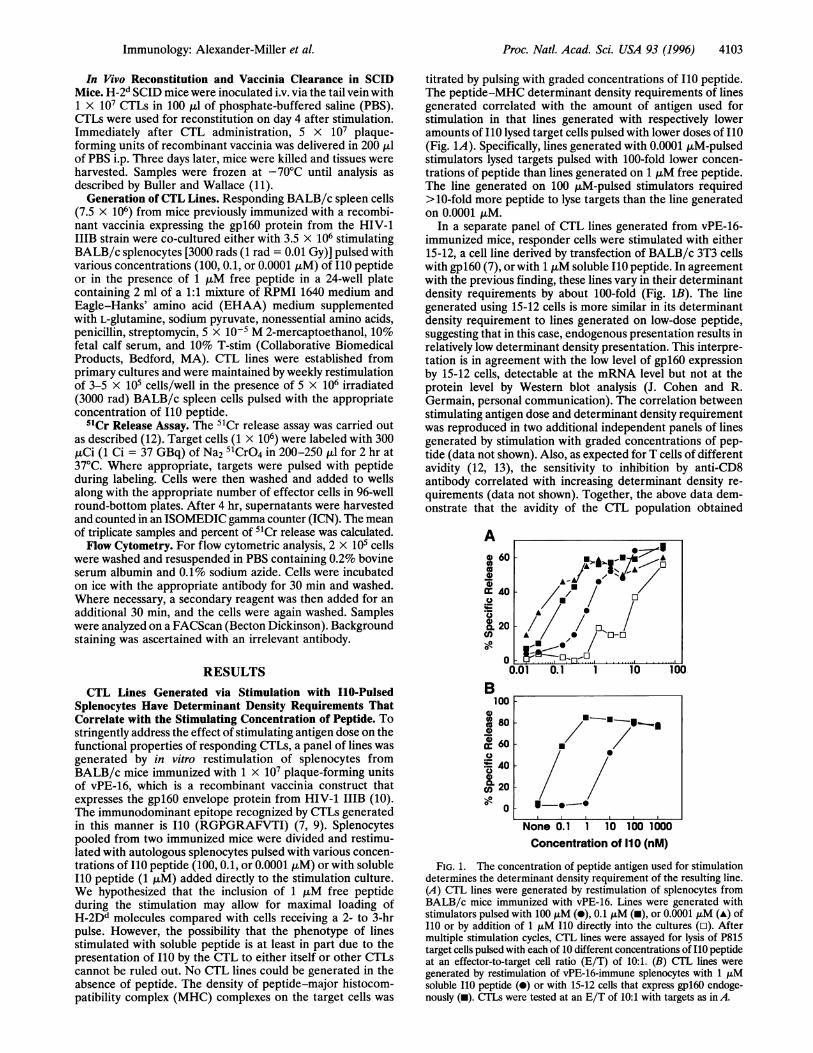
titrated by pulsing with graded concentrations of I10 peptide.The peptide-MHC determinant density requirements of linesgenerated correlated with the amount of antigen used forstimulation in that lines generated with respectively loweramounts of I10 lysed target cells pulsed with lower doses of I10(Fig. 1A). Specifically, lines generated with 0.0001 pM-pulsedstimulators lysed targets pulsed with 100-fold lower concen-trations of peptide than lines generated on 1 ,uM free peptide.The line generated on 100 tjM-pulsed stimulators required>10-fold more peptide to lyse targets than the line generatedon 0.0001 ItM.
In a separate panel of CTL lines generated from vPE-16-immunized mice, responder cells were stimulated with either15-12, a cell line derived by transfection of BALB/c 3T3 cellswith gpl60 (7), or with 1 ,M soluble I10 peptide. In agreementwith the previous finding, these lines vary in their determinantdensity requirements by about 100-fold (Fig. 1B). The linegenerated using 15-12 cells is more similar in its determinantdensity requirement to lines generated on low-dose peptide,suggesting that in this case, endogenous presentation results inrelatively low determinant density presentation. This interpre-tation is in agreement with the low level of gpl60 expressionby 15-12 cells, detectable at the mRNA level but not at theprotein level by Western blot analysis (J. Cohen and R.Germain, personal communication). The correlation betweenstimulating antigen dose and determinant density requirementwas reproduced in two additional independent panels of linesgenerated by stimulation with graded concentrations of pep-tide (data not shown). Also, as expected for T cells of differentavidity (12, 13), the sensitivity to inhibition by anti-CD8antibody correlated with increasing determinant density re-
quirements (data not shown). Together, the above data dem-onstrate that the avidity of the CTL population obtained
Ao 60
ootC 40
200
20
n
B100
s)0(a0
m
0,.o
ael
.01 0.1 1 10 10
80
60
40
20p
0
j -0
I-·----.
None 0.1 1 10 100 1000Concentration of 110 (nM)
FIG. 1. The concentration of peptide antigen used for stimulationdetermines the determinant density requirement of the resulting line.(A) CTL lines were generated by restimulation of splenocytes fromBALB/c mice immunized with vPE-16. Lines were generated withstimulators pulsed with 100 pM (-), 0.1 ,tM (m), or 0.0001 AM (A) ofI10 or by addition of 1 ,uM I10 directly into the cultures (o). Aftermultiple stimulation cycles, CTL lines were assayed for lysis of P815target cells pulsed with each of 10 different concentrations of I10 peptideat an effector-to-target cell ratio (E/T) of 10:1. (B) CTL lines were
generated by restimulation of vPE-16-immune splenocytes with 1 tjMsoluble I10 peptide (0) or with 15-12 cells that express gpl60 endoge-nously (m). CTLs were tested at an E/T of 10:1 with targets as in A.
U !
// /. /-* */
'
/ -/1 /'A°-n'°.,_ _ . . ...... ........ ........I
Immunology: Alexander-Miller et aL.
I

4104 Immunology: Alexander-Miller et al.
during an immune response is a direct result of the antigendensity presented by the stimulating antigen-presenting cell(APC). In this context, we have used avidity to mean sensitivityto low peptide-MHC complex determinant density on thetarget cells, with the understanding that other mechanismsmay apply as well. Most of the following analyses were performedwith additional lines, with one line shown as representative.Assessment of Cell Surface Molecules Expressed by CTLs
with Differing Determinant Density Requirements. As differ-ences in the expression of T-cell antigen receptors (TCRs) orother accessory molecules could contribute to the observeddifferences in avidity, the CTL lines were assayed for expres-sion of a number of cell surface molecules that have beenimplicated in CTL function, including CD8, LFA-1, Thy-1.2,and VLA-4 (Table 1). Although there are differences in theexpression of some molecules (e.g., TCRs), they do not appearto correlate with functional phenotype, although we cannotexclude from the fluorescence-activated cell sorter analysis theformal possibility that a small subset of cells with skewed TCRexpression contributes disproportionately to the response.These results suggest that other mechanisms, including varia-tion in TCR affinities or differences in the number of TCRsthat must be engaged for activation to occur, may account for thein vitro functional differences observed among the CTL lines.A previous study (14) from our lab identified Vp8 as a
predominant Vp chain employed by I10-specific CTLs, al-though other Vp chains can be used. To determine whether thestimulating antigen dose influenced the TCR repertoire of theCTLs, the lines were analyzed for expression of VB8. Inter-estingly, the two higher avidity lines contained only a smallpercentage of Vp8+ CTLs (0.0001 ,M line < 15% and 0.1 tLMline < 4%), whereas the lines generated on 100 t,M-pulsedstimulators or 1 juM soluble peptide contained 55% and >99%Vp8+ cells, respectively. This correlation of Vp8 usage withlower-avidity cells was also observed in an independent panelof lines (data not shown). The difference in Vp usage impliesthat different TCRs are used by the lines. Because the lines hadundergone 20 rounds of stimulation, it is likely that all of thecells are antigen-specific despite different V, usage, consistentwith the finding that TCRs with different Vps can recognizethe I10-Dd complex (14).
Divergence Among the Lines in the Number of EngagedTCR Required for Triggering May Contribute to the Deter-minant Density Requirement of the CTL Lines. To determinewhether differences in the number of TCRs that must beengaged for activation contributed to the observed variation indeterminant density requirements, anti-CD3 antibody wasused to direct lysis by the CTL lines in the absence of specificpeptide antigen. Anti-CD3 has been shown to promote lysisthrough Fc receptor binding on the P815 target cell andconcurrent binding to CD3 on the CTL (15). Because activa-tion through CD3 bypasses the interaction of TCRs withantigen, differences in TCR affinity cannot influence thedose-response curve to anti-CD3 antibody. If indeed thedifferences in determinant density requirements are solely theresult of variation in the TCR affinity, then the lysis obtainedusing anti-CD3 should be equivalent. However, Fig. 2 shows
Table 1. Expression of cell surface molecules on the CTL lineswith various avidities
Concentration of10A mean fluorescenceConcentration of I10used to generate line TCR CD8 Thy-1.2 LFA-1 VLA-4
0.0001 pIM 102 160 846 1154 200.1 ApM 174 238 731 1354 14100 tpM 101 204 941 1355 131 puM (soluble) 71 172 794 1335 18
-100l
O /i A
r 60E=
40
20
0
103 10-2 10-1
jig/ml 2C1 1
FIG. 2. CTL lines of different avidities respond with differingsensitivities to anti-CD3 antibody in a redirected lysis assay. CTL linesstimulated with 0.0001 juM-pulsed (-), 0.1 ,tM-pulsed (-), 100 tLM-pulsed (A), or 1 ,uM soluble I10 (*) peptide were tested for recognitionof P815 target cells incubated with anti-CD3 (2C11) antibody at titeredconcentrations at an effector-to-target cell ratio of 5:1. The presenceof anti-CD3 antibody did not affect the spontaneous release of thetarget cells.
that the dose-response curves among the lines are unexpect-edly distinct. Although there is variation in TCR expressionamong the lines (Table 1), this level of TCR expression doesnot explain the differences in the dose-response curves toanti-CD3-e.g., the levels of TCR expression for the linesgenerated with 100 ,LM peptide and 0.0001 tiM peptide havesignificantly different dose-response curves yet have equiva-lent TCR expression, 101 vs. 102 mean fluorescence intensity,respectively. This finding suggests that the higher avidity linesrequire fewer TCRs to be engaged for activation to occur,possibly because of differences in coupling of TCRs to down-stream signaling mechanisms. This factor likely contributes tothe apparent differences in the concentration of antigenneeded for target cell sensitization. However, whether the8-fold difference (average of three independent assays) in theconcentration of anti-CD3 required to achieve lysis detectedbetween the lines of highest and lowest avidity can fullyaccount for the 100-fold difference in determinant densityrequired for lysis must be determined by cloning of the TCRgenes and direct measurements of TCR affinity of represen-tative CTL from each line.
Recognition of Endogenously Expressing Targets by CTLLines with Different Determinant Density Requirements. Inview of the reports that CTL lines generated by in vitro primarystimulation with high concentrations of peptide often fail tolyse targets endogenously expressing antigen (16, 17), the CTLlines were assessed for recognition of targets infected withrecombinant vaccinia virus expressing gpl60. All four linesrecognized vPE-16-infected P815 target cells, although themaximal lysis achieved was greater with the higher affinitylines (Fig. 3). Recognition of vaccinia-infected targets dem-onstrated that all of the lines, regardless of avidity, couldrecognize endogenously processed and presented antigen. Thisobservation was intriguing in light of the appreciable differ-ences in the concentration of peptide required for target cellsensitization. To determine the in vivo implications of these invitro findings, the CTL lines were tested for the ability to clearvirus in mice.
In Vivo Clearance of Recombinant Vaccinia Expressinggpl60 Correlates with the Determinant Density Requirementsof the CTL Lines. A primary function of CTLs in vivo isclearance of viral infections (18, 19). We hypothesized thateven though both low- and high-avidity CTLs can kill targetsinfected with recombinant vaccinia, those CTLs that can
recognize cells early in infection, before much virus replicationA mean fluorescence is calculated as the mean fluorescence ob-
tained with the antibody minus the mean background fluorescence.
Proc. Natl. Acad. Sci. USA 93 (1996)

Proc. Natl. Acad. Sci. USA 93 (1996) 4105
100
_e
a)
oa)
a)
0
0Co
20 10 5 2 20 10 5 2
E/T E/TFIG. 3. High- and low-avidity CTL lines efficiently recognize
targets expressing I10 endogenously. CTLs generated on 0.0001 jLMI10-pulsed stimulators (A), 0.1 tLM I10-pulsed stimulators (B), 100 uiMI10-pulsed stimulators (C), and 1 ,LM soluble I10 (D) were tested onvPE-16-infected P815 cells (e) and control WR-infected cells in theabsence (m) or presence (A) of 10 ,uM I10 peptide. All four CTL linesrecognized endogenously expressing targets.
has occurred, and therefore before high levels ofviral peptide-MHC complexes are on the cell surface, might be moreefficient at clearing virus. To test this hypothesis and to assessthe in vivo relevance of these CTLs with differing functionalavidities, the lines were compared for their ability to clear virusfrom SCID mice. Mice received either 1 x 10⁷ CTLs or PBSalone as a mock control via tail vein injection. Concurrently,mice received 5 x 10⁷ plaque-forming units of vPE-16 by i.p.injection. CTLs generated on 0.0001 ,tM-pulsed stimulatorsreduced the amount of virus in the ovary by 1000-fold morethan CTLs generated on 1 utM soluble I10 or 100 AtM-pulsedstimulators (P = 0.004 and 0.00005, respectively) (Fig. 4A).Similar results were obtained in two additional experimentsusing this panel of lines. In addition, an independent panel oflines generated by stimulation with either 1 tM soluble I10 or15-12 cells and displaying disparate determinant density re-
quirements (Fig. 1B) showed a similar difference for in vivoviral clearance (Fig. 4B) (forP values, see Fig. 4 legend). Thus,the correlation between determinant density requirement andin vivo efficacy has been observed in two independent panelsof lines. Therefore, the dose of peptide used to stimulate CTLswill directly affect the ability of those CTLs to function in vivoto clear virus.
DISCUSSIONIn this study, CTLs were generated by repeated in vitrostimulation of immune cells with distinct antigen doses. CTLsgenerated in this manner display significant and reproducibledifferences in their antigen dose requirements for optimaltarget cell lysis. Although CTLs with a broad range of deter-minant density requirements have been observed followingcloning of polyclonal responses, this study is the first todemonstrate a mechanism for the selective bulk expansion ofCTL populations possessing a distinct in vivo viral clearancecapacity and in vitro peptide-MHC requirement. A previousobservation of increased lytic activity after stimulation withlow-dose antigen is likely explained by the data presented here(20). The functional phenotypes of the CTL lines as measuredby their determinant density requirements appear to be quite
A109
| 1080.- 7o 10E
" 105
104
B109 -10
>_m 1080
O 107E106
.- 05104104
Lines generatedwith
' No CTLI soluble 110/ 1 00 -LM 110
IX 0.0001 P.M 110
__ ,.t..X''
Lines generated
CTL line for reconstitution
withNo CTLsoluble 11015-12 cells
FIG. 4. High-avidity lines efficiently clear virus in vivo, whereaslow-avidity lines are ineffective. CTLs (1 x 10⁷) were injected i.v. and5 x 107 plaque-forming units of recombinant vaccinia expressinggpl60 were injected i.p. into H-2d SCID mice. On day 3, mice werekilled and ovaries were harvested, as the virus has a tropism for theovaries. Vaccinia present in the tissues was determined by titering onBSC-1 cells. (A) Lines tested were generated by stimulation with0.0001 JLM- or 100 jiM-pulsed splenocytes or in the presence of 1 tLMsoluble peptide. Results shown are the titers obtained in a represen-tative experiment with three to five mice per group. Similar resultswere obtained in two additional independent experiments. P valuescalculated using Student's t test were as follows: mice receiving the linegenerated on 0.0001 AjM I10-pulsed stimulators vs. mice receiving noCTLs, P = 0.00002 or mice receiving 100 ,AM-stimulated CTLs, P =
0.00005, or mice receiving CTLs generated on 1 juM soluble I10, P =
0.004. Results obtained from mice receiving low-avidity lines vs. noCTLs had P values of >0.05. (B) Lines used for reconstitution were
generated by stimulation with 1 jiM soluble peptide or with the 15-12transfectant that expresses the peptide determinant endogenously. Pvalues were as follows: mice receiving CTLs generated on 15-12 cellsvs. mice receiving no CTLs, P = 0.049, or vs. mice receiving CTLsgenerated with 1 tLM soluble I10, P = 0.0019. For mice receiving no
CTLs versus CTLs generated with 1 jiM soluble I10, P = 0.35.
stable and do not change. The in vitro differences in determi-nant density requirements of the lines in our study correlatedirectly with the lines' abilities to clear virus in vivo. CTLsrequiring high determinant density for target cell recognitionwere ineffective in reducing viral burden in SCID mice inoc-ulated with recombinant vaccinia virus expressing HIV-1gpl60, even though both lines lysed virus-infected targets invitro. In contrast, CTLs capable of lysing targets bearing lowantigen densities could reduce viral titers by 100- to 1000-fold,compared with lower-avidity CTLs.The data obtained with the CTL lines of differing avidity
demonstrate that in vitro recognition of targets infected withvirus is not necessarily reflective of the ability of those sameCTLs to clear viral infection in vivo. This is an importantfinding, as recognition of endogenously expressed antigen hasbeen regarded as an indicator of in vivo relevance (21). Basedon the data in our study, we propose that measurement ofdeterminant density requirement may be a better predictor ofin vivo efficacy for adoptive immunotherapy. We conclude thatthe quality of CTLs is as important as the quantity for clearing
Li
r~
Immunology: Alexander-Miller et al.
r
E
E
4106 Immunology: Alexander-Miller et al.
viral infection. This result is consistent with the hypothesis thatCTLs that can recognize target cells early in infection, whenthe density of viral peptide-MHC complexes is still low, may bemore effective in clearing virus infection than CTLs that cankill only targets already loaded with much viral progeny (22, 23).The CTL lines in this study produced interferon-y in re-
sponse to antigenic stimulation. Thus, it is possible thatinterferon-y production could contribute to viral clearance inaddition to lysis of virally infected cells. However, the thresh-olds of peptide antigen concentration required for interfer-on-y production and CTL lysis are equivalent (data notshown). Therefore, even if cytokine production contributes tothe observed clearance in vivo, the threshold of antigenconcentration required for its production is still determined byCTL avidity and is selected for by in vitro stimulation withdifferent antigen concentrations.The most ready explanation for the observed differences in
the determinant density requirements of the CTL lines isinherent differences in the TCR affinities, but direct measure-ment of these would require recombinant soluble TCR mol-ecules. Observed differences in Vp usage of the lines supportthis hypothesis in that they allow for the possibility that cellswith TCRs of very different affinity exist in the high- versuslow-avidity CTL lines. A correlation between a high determi-nant density requirement and V,8 usage was found in twoindependent panels of CTL lines, although not all TCRs usingVp8 will necessarily be of low affinity. However, alternativemechanisms must be considered. Two additional possibilitieswere differences in the level of expression of the TCR itself orin various accessory molecules. No differences in the expres-sion of CD8, LFA-1, VLA-4, or Thy-1.2 were found. Althoughthere are differences in the level of TCRs expressed, thepattern of expression does not correlate with the functionalphenotype of the lines (Table 1 and Fig. 1). In a previousreport, differences in the level of CD44 expression were shownto affect in vivo clearance of adoptively transferred CTL clones(24). Relative CD44 expression among the lines in the currentstudy (tested in two independent panels of lines) varied overtime in culture (data not shown), but in vivo clearance nevercorrelated with such differences. Thus, differences in homingto targets are unlikely to account for the results. Third, thetrivial explanation of a difference inMHC restriction was ruledout because both high- and low-avidity lines are restricted byH-2Dd (data not shown). A fourth alternative mechanism issuggested by Kwan-Lim et al. (25), who reported differencesamong a panel of hybridomas in their response to plate-boundanti-CD3 antibody, suggesting a difference in the number ofengaged TCRs required for activation. Redirected lysis exper-iments showed that differences in the number of TCRs thatmust be engaged for activation do appear to contribute, at leastin part, to the differences in determinant density requirementsof these lines, as the dose-response curves to anti-CD3 anti-body were modestly shifted among the lines (Fig. 2). Thissurprising result suggests that differences may be present in thesignaling threshold required to achieve activation of the var-ious lines or in the efficiency of coupling of the TCRs todownstream signaling events. Therefore, the sensitivity todeterminant density of a CTL is likely a combination of severalfactors, including TCR affinity and the threshold level of TCRengagement required for signaling.Responding cells used for generation of the lines in this
study were initially activated in vivo by immunization with arecombinant vaccinia expressing gpl60. Thus, even loweravidity cells can be activated during an endogenous infection.As previous attempts to generate primary responses by in vitroactivation of naive splenocytes have been unsuccessful, it isunlikely that these CTLs were activated in vitro from naiveprecursors (data not shown). Elicitation of these CTLs in vivodoes not necessitate that they are effective for viral clearance,as seems to be the case for the CTLs studied here.
In addition to obvious practical research applications (e.g.,generation of lines for in vitro study), these findings haveimportant implications for clinical therapy. A number ofgroups have undertaken clinical trials in which peripheralblood mononuclear cells, presumably containing CTLs specificfor the antigen of interest, are removed from the patient,expanded in vitro, and reinfused. This procedure is being usedfor HIV positive patients (4), immunosuppressed patientscombating cytomegalovirus (2), and patients with certaincancers (5, 6, 26). The present study suggests that the mannerin which these CTLs are stimulated in vitro will have profoundeffects on their efficacy in vivo. Selective expansion of bulkCTLs using stimulators expressing relatively low determinantdensity as demonstrated in the current study, as opposed tocloning, may be optimal because large numbers of CTLs canbe obtained quickly and lines are polyclonal. The latter at-tribute should be beneficial as adoptive immunotherapy usinga monoclonal CTL population can readily allow for viralescape (27). Conversely, elicitation of CTLs that require highdeterminant density may be beneficial for selective killing oftumor cells in situations in which the antigen of interest is aself-peptide that is overexpressed in the target cell, as is thecase in melanoma (28). CTLs generated on high-dose antigenthat may differentially recognize a tumor cell with supranor-mal expression while sparing wild-type cells would be ideal inthis situation. It is important to note that high and lowdeterminant density are relative and will vary for differentpeptides, depending on their affinity for MHC and the rep-ertoire of TCRs available from which to select. Thus, optimalpeptide concentrations must be determined empirically. Inconclusion, the data presented here provide evidence that thequality is as important as the quantity of CTLs and that CTLswith discrete avidities can be preferentially selected andexpanded in accordance with their desired therapeutic usagein vivo. This ability should be a powerful tool in the use of CTLsfor adoptive therapy of disease.
We thank Drs. Bernard Moss and Patricia Earl for the gift ofvPE-16virus and BSC-1 cells, Dr. Miles Carroll for instruction in the methodof titering vaccinia virus in tissues, Dr. Richard Hodes for F23.1antibody, Dr. Ronald Germain for the gift of the 15-12 transfectedfibroblast line, and Drs. Richard Hodes and David Margulies forcritical reading of the manuscript and helpful suggestions.
1. Houbiers, J. G. A., Nijman, H. W., van der Burg, S. H., Drijfhout,J. W., Kenemans, P., van de Velde, C. J. H., Brand, A., Momburg,F., Kast, W. M. & Melief, C. J. M. (1993) Eur. J. Immunol. 23,2072-2077.
2. Riddell, S. R., Watanabe, K. S., Goodrich, J. M., Li, C. R., Agha,M. E. & Greenberg, P. D. (1992) Science 257, 238-241.
3. Riddell, S. R., Greenberg, P. D., Overell, R. W., Loughran, T. P.,Gilbert, M. J., Lupton, S. D., Agosti, J., Scheeler, S., Coombs,R. W. & Corey, L. (1992) Hum. Gene Ther. 3, 319-338.
4. Lieberman, J., Skolnik, P. R., Parkerson, G. R., Fabry, J. A.,Fong, D. M., Landry, B. & Kagan, J. (1994) AIDS Res. Hum.Retroviruses 10, S110.
5. Topalian, S. L., Muul, L., Solomon, D. & Rosenberg, S. A. (1987)J. Immunol. Methods 102, 127-141.
6. Rosenberg, S. A., Packard, B. S., Aebersold, P. M., Solomon, D.,Topalian, S. L., Toy, S. T., Simon, P., Lotze, M. T., Yang, J. C.,Seipp, C. A., Simpson, C., Carter, C., Bock, S., Schwartzentruber,D., Wei, J. P. & White, D. E. (1988) N. Engl. J. Med. 319,1676-1680.
7. Takahashi, H., Cohen, J., Hosmalin, A., Cease, K. B., Houghten,R., Cornette, J., DeLisi, C., Moss, B., Germain, R. N. & Berzof-sky, J. A. (1988) Proc. Natl. Acad. Sci. USA 85, 3105-3109.
8. Stewart, J. M. & Young, J. D. (1984) Solid Phase Peptide Synthesis(Pierce, Rockford, IL).
9. Takeshita, T., Takahashi, H., Kozlowski, S., Ahlers, J. D., Pendle-ton, C. D., Moore, R. L., Nakagawa, Y., Yokomuro, K., Fox,B. S., Margulies, D. H. & Berzofsky, J. A. (1995)J. Immunol. 154,1973-1986.
10. Earl, P. L., Koenig, S. & Moss, B. (1991) J. Virol. 65, 31-41.
Proc. Natl. Acad. Sci. USA 93 (1996)

Immunology: Alexander-Miller et al.
11. Buller, R. M. L. & Wallace, G. D. (1985) Lab. Anim. Sci. 35,473-476.
12. Alexander, M. A., Damico, C. A., Wieties, K. M., Hansen, T. H.& Connolly, J. M. (1991) J. Exp. Med. 173, 849-858.
13. Maryanski, J. L., Pala, P., Cerottini, J.-C. & MacDonald, H. R.(1988) Eur. J. Immunol. 18, 1863-1866.
14. Shirai, M., Vacchio, M. S., Hodes, R. J. & Berzofsky, J. A. (1993)J. Immunol. 151, 2283-2295.
15. Leo, O., Foo, M., Sachs, D. H., Samelson, L. E. & Bluestone,J. A. (1987) Proc. Natl. Acad. Sci. USA 84, 1374-1378.
16. Staerz, U. D., Zepp, F., Schmid, R., Hill, M. & Rothbard, J.(1989) Eur. J. Immunol. 19, 2191-2196.
17. Carbone, F. R., Moore, M. W., Sheil, J. M. & Bevan, M. J. (1988)J. Exp. Med. 167, 1767-1779.
18. Byrne, J. A. & Oldstone, M. B. A. (1984) J. Virol. 51, 682-686.19. Taylor, P. M. & Askonas, B. A. (1986) Immunology 58, 417-420.20. Zhou, X., Berg, L., Motal, U. M. A. & Jondal, M. (1992) J.
Immunol. Methods 153, 193-200.
Proc. Natl. Acad. Sci. USA 93 (1996) 4107
21. Speiser, D. E., Kyburz, D., Stiibi, U., Hengartner, H. & Zinker-nagel, R. M. (1992) J. Immunol. 149, 972-980.
22. Jackson, D. C., Ada, G. L. & Tha hla, R. (1976)Aust. J. Exp. Biol.Med. Sci. 54, 349-363.
23. Zinkernagel, R. M. & Althage, A. (1977) J. Exp. Med. 145,644-651.
24. Rodrigues, M., Nussenzweig, R. S., Romero, P. & Zavala, F.(1992) J. Exp. Med. 175, 895-905.
25. Kwan-Lim, G.-E., Ong, T., Aosai, F., Stauss, H. & Zamoyska, R.(1993) Int. Immunol. 5, 1219-1228.
26. Yannelli, J. R. (1991) J. Immunol. Methods 139, 1-16.27. Koenig, S., Conley, A. J., Brewah, Y. A., Jones, G. M., Leath, S.,
Boots, L. J., Davey, V., Pantaleo, G., Demarest, J. F., Carter, C.,Wannebo, C., Yannelli, J. R., Rosenberg, S. A. & Lane, H. C.(1995) Nat. Med. 1, 330-336.
28. Kawakami, Y., Eliyahu, S., Delgado, C. H., Robbins, P.F.,Rivoltini, L., Topalian, S. L., Miki, T. & Rosenberg, S. A. (1994)Proc. Natl. Acad. Sci. USA 91, 3515-3519.
Copyright © 2022 FDOKUMEN