New and modified anodic alumina membranes Part I. Thermotreatment of anodic alumina membranes
Available online at www.sciencedirect.com
www.elsevier.com/locate/jmbbm
j o u r n a l o f t h e m e c h a n i c a l b e h a v i o r o f b i o m e d i c a l m a t e r i a l s 3 0 ( 2 0 1 4 ) 1 6 8 – 1 7 5
1751-6161/$ - see frohttp://dx.doi.org/10.
nCorresponding autE-mail addresses:
Research Paper
Mechanical and physical properties of calciumsilicate/alumina composite for biomedicalengineering applications
F.S. Shirazia, M. Mehralia,b,n, A.A. Oshkoura, H.S.C. Metselaara,N.A. Kadrib, N.A. Abu Osmanb
aDepartment of Mechanical Engineering and Advanced Material Research Center, University of Malaya, 50603,Kuala Lumpur, MalaysiabDepartment of Biomedical Engineering, Faculty of Engineering, University of Malaya, 50603 Kuala Lumpur, Malaysia
a r t i c l e i n f o
Article history:
Received 2 June 2013
Received in revised form
12 October 2013
Accepted 21 October 2013
Available online 7 November 2013
Keywords:
Calcium Silicate
Alumina
Mechanochemical synthesis
Hardness
Fracture toughness
Young's modulus
nt matter & 2013 Elsevie1016/j.jmbbm.2013.10.024
hor. Tel.: þ60 17 2946970;faridseyedshirazy@yaho
a b s t r a c t
The focus of this study is to investigate the effect of Al2O3 on α-calcium silicate (α-CaSiO3)
ceramic. α-CaSiO3 was synthesized from CaO and SiO2 using mechanochemical method
followed by calcinations at 1000 1C. α-CaSiO3 and alumina were grinded using ball mill to
create mixtures, containing 0–50 w% of Al2O3 loadings. The powders were uniaxially
pressed and followed by cold isostatic pressing (CIP) in order to achieve greater uniformity
of compaction and to increase the shape capability. Afterward, the compaction was
sintered in a resistive element furnace at both 1150 1C and 1250 1C with a 5 h holding
time. It was found that alumina reacted with α-CaSiO3 and formed alumina-rich calcium
aluminates after sintering. An addition of 15 wt% of Al2O3 powder at 1250 1C were found to
improve the hardness and fracture toughness of the calcium silicate. It was also observed
that the average grain sizes of α-CaSiO3 /Al2O3 composite were maintained 500–700 nm
after sintering process.
& 2013 Elsevier Ltd. All rights reserved.
1. Introduction
Calcium Silicate (CaSiO3 or CaO–SiO2) ceramics are widelyused for load-bearing bone substitutes in clinical applica-tions due to their significant biocompatibility, high osteo-conductivity and their biological apatite similarity to humanhard tissues(Beheri et al., 2013; Lin et al., 2005; Yu et al., 2012).Also, pure calcium silicate ceramics are greatly inducedformation of a hydroxyapatite (HA) layer on calcium silicatesurface after soaking in simulated body fluid (SBF) and insaliva medium(De Aza et al., 1994; Siriphannon et al., 1999,
r Ltd. All rights reserved.
fax: þ60 3 79675317.o.co.uk (F.S. Shirazi), m.m
2002a). These layers help to form the chemical bonds betweenthe bioceramics and bone tissues.
In fact, calcium silicate (CaSiO3) was introduced as a bioma-terial for hard tissue repair substituent since 1970s, whenHench and coworkers invented Bioglasss. As yet several formsof silicate glasses and glassceramics like bioactive glasses(Kokubo, 1991; Rahaman et al., 2011) and calcium silicate basedglass ceramic (Cho et al., 1997; Liu et al., 2004) have beenexplored. Previous studies revealed that the biomaterials con-taining CaO–SiO2 act excellent bioactivity and were found tobond to living bone and soft tissue, through the development ofa biologic HA layer on their surface (Siriphannon et al., 2002b).
[email protected] (M. Mehrali).
j o u r n a l o f t h e m e c h a n i c a l b e h a v i o r o f b i o m e d i c a l m a t e r i a l s 3 0 ( 2 0 1 4 ) 1 6 8 – 1 7 5 169
However, the foremost limitation of ceramic powders is theirlow fracture toughness that restricts some restriction of biome-dical application such as load bearing implant application(De Aza et al., 1997; Encinas-Romero et al., 2008; Mehrali et al.,in press). Therefore, one approach to improve the mechanicalproperties of bioactive ceramic materials is by incorporate thealumina as a ceramic second phase dispersed in a ceramicmatrix. Some researchers in regards to the approach haveinvestigated the alumina reinforcement role in HA and alsohave demonstrated that alumina particles could improve tough-ening and hardness (Huang and Nicholson, 1993; Ji and Marquis,1992). Alumina, which is mostly categorized as a bioinertmaterial, has been broadly investigated as a reinforcement agentfor ceramics (Mobasherpour et al., 2009). Good flexural strength,excellent resistance to dynamic and impact fatigue, resistance tosubcritical crack growth and excellent compressive strength areobtained with average grain sizeso4 μm and purity499.7%(Askari et al., 2012). However, just several researches have beendone on the effective role of bioinert materials such as aluminaand zirconia on the fracture toughness and hardness of calciumsilicate, similar to what has been done in this study.
In this study, the effects on the mechanical properties ofcalcium silicate, namely micro hardness, fracture toughnessand Young's modulus, by changing the composition (basedon different weight ratios of alumina) and sintering tempera-tures, were investigated.
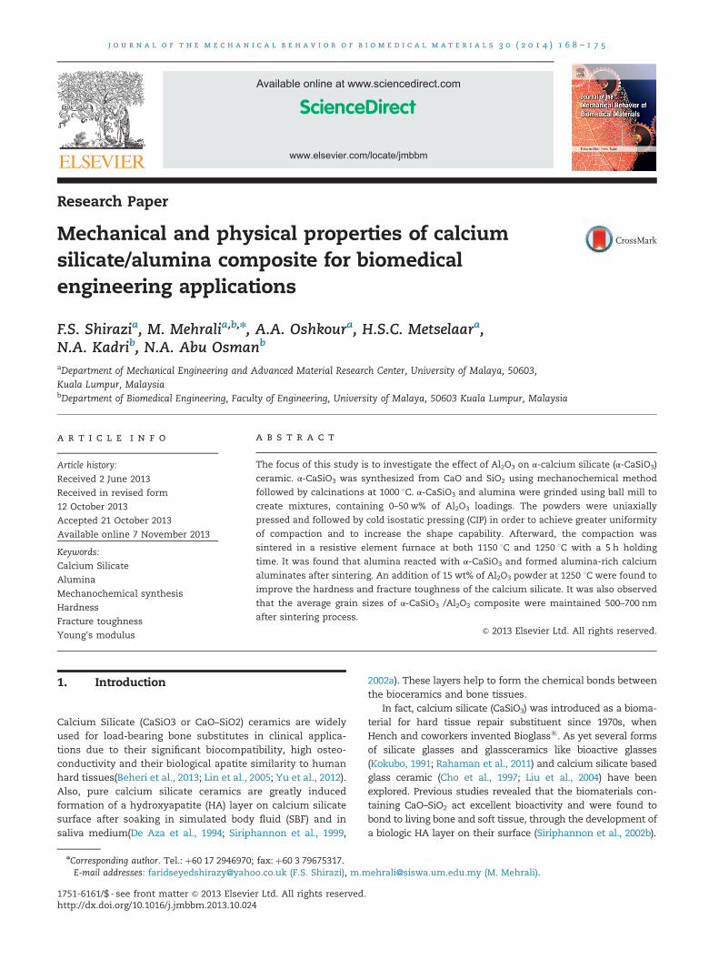
Fig. 1 – FESEM micrograph of the indentation-induced crackpropagation.
2. Material and method
2.1. Synthesis of calcium silicate powder
Calcium silicate powders have been synthesized using mechan-ochemical process. The main advantage using this method is agood crystallinity and small crystallites without using hightemperatures for synthesizing (Nasiri-Tabrizi et al., 2009).
Commercial calcium oxide (CaO) (Ajax chemicals indus-tries) and silicon oxide (SiO2) (Sigma-Aldrich Inc.) were used inCaSiO3 preparation. For reaction to occure, aforementionedmaterials were grounded on a planetary-mill (Retsch PM 100)with the stoichiometric proportionality between the oxides asgiven in reaction (1). Milling was accomplished using therotational speed of 500 rpm. The ratio of ball to powder wasassumed to be 20/1. 60 of min milling with 10min intervalpauses were performed through the process to avoid over-heating. Total duration of the process to perform the reaction 1was 50 h. Finally, the white powders were calcined at 1000 1Cfor 2 h to obtain α-CaSiO3. Previous researches demonstratedthat α-CaSiO3 is less degradable than β phase which is moreapplicable in implantation (Liu et al., 2008; Shirazi et al., 2013;Wu et al., 2007). Hence the mechanochemical technique tomake submicron (o1 mm) calcium silicate powder is done as
CaOþSiO2-CaSiO3 (1)
2.2. Fabrication of composites and sintering
The prepared calcium silicate was mixed with α-Al2O3
(99.95%, Alfa Aesar, Ward Hill, MA, USA) powders with anaverage crystal and particle size of 0.3 μm using ball milling in
ethyl alcohol for 24 h with zirconia balls as media to break theCaSiO3 agglomerates. Mixing of CaSiO3 and alumina was donein different ratios (5, 10, 15, 25 and 50 wt%) in order to perusethe influence of Al2O3 content in calcium silicate mechanicaland physical properties. The powder mixtures (CaSiO3/Al2O3)were uniaxial compressed at 10 t and then cold isostatic press(CIP) was applied into rectangular-prism shape specimens(30�8�2.5 mm3) under a pressure of 250 MPa for 20 min.Subsequently all bulk samples were sintered at two differenttemperatures; 1150 1C and 1250 1C for 5 h with 5 1C/minheating rate. Three samples per each combination werefabricated with aforementioned process.
2.3. Characterization of α-CaSiO3/Al2O3 composites
The sintered bulks were grounded, polished, and thermallyetched for 2 h in a furnace at 1100 1C in air atmosphere formicrostructural analysis. The porosity sintered materials wasmeasured by the Archimedes method. The microstructure andphase composition of the calcined powders and sintered sam-ples were determined by field emission scanning electron micro-scopy (FESEM; CARL ZEISS-AURIGA 60 microscope) and X-raydiffraction (XRD; EMPYREAN, PANALYTICA) using Cu Kα radia-tion. XRD was performed over a range of 51o2θo801 with a stepsize of 0.0151 and a time per step of 20.02 s. The relative densitywas calculated by comparing the measured data to the theore-tical densities of CaSiO3 (2.905 g/cm3) and Al2O3 (3.969 g/cm3).
2.4. Mechanical properties evaluation
Vickers hardness was measured by the indentation-strengthmethod at room temperature using an applied load of2 kgf with a dwell of 15 s. (MITUTOYO-AVK C200-AKASHICORPORATION). Five indentations run per sample werecarried out to determine the average value of mechanicalproperties. Fracture toughness was then determined throughusing the following equation (Anstis et al., 1981; Lawn et al.,1980; Shetty et al., 1985):
KIC ¼ 0:0937 � Hv� P4L
� �ð1=2Þð2Þ

j o u r n a l o f t h e m e c h a n i c a l b e h a v i o r o f b i o m e d i c a l m a t e r i a l s 3 0 ( 2 0 1 4 ) 1 6 8 – 1 7 5170
Where P is the applied indentation load and L is the cracklength as it has been shown in Fig. 1. As well, Young'smodulus was measured using Knoop hardness method(Shimadzu; HMV-2000). Load of 500 g with a duration time of15 s was applied in accordance to ASTM E384. Below equationwas used to determine the Young's modulus (Slosarczyk andBiałoskórski, 1998):
b′a′
¼ ba
� αEHk
ð3Þ
Where a' and b' are longer and shorter diagonals of indenterafter elastic recovery respectively, b/a is the ratio of the Knoopindenter dimensions or geometry which is 1/7.11, α is
Fig. 2 – X-ray diffraction peaks of α-CaSiO3 synthesized bymechanochemichal method.
Fig. 3 – X-ray diffraction peaks of α-CaSiO3/aluminacomposites with different weight ratio: 0/100 (a), 100/0 (b),95/5 (c), 90/10(d), 85/15 (e), 75/25 (f) and 50/50 (g) sintered at1150 1C.
a constant value which is 0.45, E is the Young's modulus ofthe sample and Hk is Knoop hardness which is obtained byhardness machine.
Field emission scanning electron microscopy (FESEM;CARL ZEISS-AURIGA 60 microscope) was used to determinethe particle size and to observe the bonding between thegrains.
3. Results and discussion
3.1. Structural evaluation of fabricated composites
The results of XRD significantly showed that the product ofreaction 1 is α-calcium silicate which coincides with α-CaSiO3
in the JCPDS card of 01-0720 as could be seen in Fig. 2. Figs. 3and 4 illustrate the XRD patterns of α-CaSiO3/Al2O3 compo-sites for different initial concentration of alumina powderafter sintering at 1150 1C and 1250 1C respectively. The α-CaSiO3 peaks were clearly found in the composites sintered at1150 1C. Moreover, the alumina peaks were detected in thecomposites sintered at 1150 1C which increased in intensityas the alumina content increased. At the same sinteringtemperature several additional peaks were spotted in thecomposite which represents the interfacial reaction betweenα-CaSiO3 and alumina. At the peak of 30.081 2θ the intensityincreased with increasing the alumina content up to 15 wt%however the intensity was decreased when the amount of
Fig. 4 – X-ray diffraction peaks of α-CaSiO3/aluminacomposites with different weight ratio: 0/100 (a), 100/0 (b),95/5 (c), 90/10(d), 85/15 (e), 75/25 (f) and 50/50 (g) sintered at1250 1C.
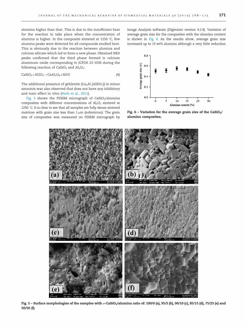
Fig. 6 – Variation for the average grain size of the CaSiO3/alumina composites.
j o u r n a l o f t h e m e c h a n i c a l b e h a v i o r o f b i o m e d i c a l m a t e r i a l s 3 0 ( 2 0 1 4 ) 1 6 8 – 1 7 5 171
alumina higher than that. This is due to the insufficient heatfor the reaction to take place when the concentration ofalumina is higher. In the composite sintered at 1250 1C, fewalumina peaks were detected for all compounds studied here.This is obviously due to the reaction between alumina andcalcium silicate which led to form a new phase. Obtained XRDpeaks confirmed that the third phase formed is calciumaluminum oxide corresponding to JCPDS 23-1036 during thefollowing reaction of CaSiO3 and Al2O3:
CaSiO3þAl2O3-CaAl2O4þSiO2 (4)
The additional presence of gehlenite (Ca2Al [AlSiO7]) in minoramounts was also observed that does not have any inhibitoryand toxic effect in vitro (Nath et al., 2011).
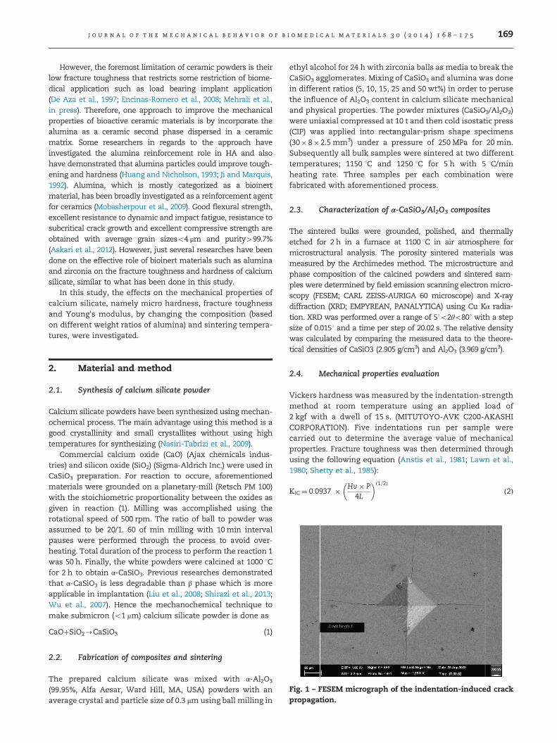
Fig. 5 shows the FESEM micrograph of CaSiO3/aluminacomposites with different concentrations of Al2O3 sintered at1250 1C. It is clear to see that all samples are fully dense sinteredmatrices with grain size less than 1 mm (submicron). The grainsize of composites was measured on FESEM micrograph by
Fig. 5 – Surface morphologies of the samples with α-CaSiO3/alum50/50 (f).
Image Analysis software (Digimizer version 4.2.4). Variation ofaverage grain size for the composites with the alumina contentis shown in Fig. 6. As the results show, average grain sizeincreased up to 15 wt% alumina although a very little reduction
ina ratio of: 100/0 (a), 95/5 (b), 90/10 (c), 85/15 (d), 75/25 (e) and

j o u r n a l o f t h e m e c h a n i c a l b e h a v i o r o f b i o m e d i c a l m a t e r i a l s 3 0 ( 2 0 1 4 ) 1 6 8 – 1 7 5172
was observed for the samples with higher content of alumina.Imaging analysis also confirmed that the average grain size didnot change significantly at different sintering temperatures.
Furthermore, it could be seen obviously that withincreasing of alumina content, the amount of fiber-likegrains increase remarkably. As it is shown in Fig. 5 (c andd) when 10 and 15 wt% alumina was added to α-CaSiO3, thefracture morphology changed remarkably.
3.2. Physical properties
Table 1 indicates the effect of the composite ratio on linearshrinkage and porosity of the samples after sintering at
Table 1 – Physical properties of CaSiO3/alumina composites si
CaSiO3/Alumina (W%:W%) 100/0 95/5
Porosity (%) (1150 1C) 7.9370.03 11.6070.05Porosity (%) (1250 1C) 2.2570.07 4.1270.1
Linear shrinkage (%) (1150 1C) 5.0770.14 5.4370.1Linear shrinkage (%) (1250 1C) 12.370.35 12.370.2
Fig. 7 – Open porosities of α-CaSiO3/alumina composites with:
1150 1C and 1250 1C. The porosity of the specimens signifi-cantly changed from 2.25% to 17.3% at 1250 1C and 7.93% to23.79% at 1150 1C. Therefore, the porosity increased by risingthe concentration of Al2O3 for both sintering temperaturesdue to the gap between the optimum sintering temperaturesof Al2O3 and CaSiO3. In general, the porosity and grain sizeare key factors affecting the mechanical properties ceramicssuch as hardness and fracture toughness (Alizadeh et al.,2011; Sprio et al., 2009). Fig. 7 shows that the increasing ofporosity happens with increasing of alumina content incomposites. increasing in the porosity is required in orderto inhabit bone absorption after implantation (Niinomi, 2008).However the mechanical properties such as hardness and
ntered at different temperatures.
90/10 85/15 75/25 50/50
12.4370.1 16.170.04 16.5270.1 23.7970.176.70.12 8.3170.12 13.370.08 17.370.23
4.5570.4 3.7470.25 1.4970.36 0.5570.0311.1970.3 11.6170.15 7.4970.26 0.7570.05
100/0 (a), 95/5 (b), 90/10 (c), 85/15 (d), 75/25 (e) and 50/50 (f).
j o u r n a l o f t h e m e c h a n i c a l b e h a v i o r o f b i o m e d i c a l m a t e r i a l s 3 0 ( 2 0 1 4 ) 1 6 8 – 1 7 5 173
fracture toughness decrease inevitably. As it is seen fromTable 1, the linear shrinkage of the samples decreased at1150–1250 1C when the Al2O3 component increased and linearshrinkage will rise with increasing of sintering temperature.As it will be explained in the next section, the obtainedoptimum mechanical properties of the studied compositeincludes 10 and 15 w% of alumina which porosity is 6.1 and8.31% respectively at 1250 1C sintering temperature.
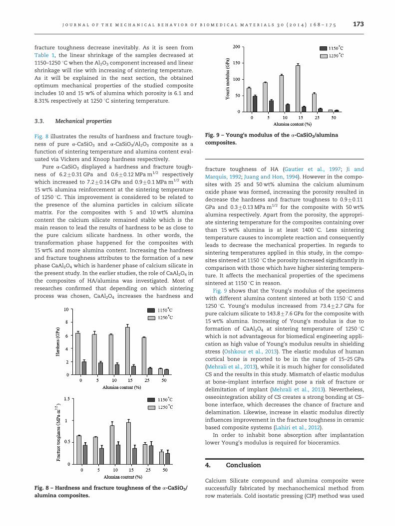
Fig. 9 – Young's modulus of the α-CaSiO3/aluminacomposites.
3.3. Mechanical properties
Fig. 8 illustrates the results of hardness and fracture tough-ness of pure α-CaSiO3 and α-CaSiO3/Al2O3 composite as afunction of sintering temperature and alumina content eval-uated via Vickers and Knoop hardness respectively.
Pure α-CaSiO3 displayed a hardness and fracture tough-ness of 6.270.31 GPa and 0.670.12 MPa m1/2 respectivelywhich increased to 7.270.14 GPa and 0.970.1 MPa m1/2 with15 wt% alumina reinforcement at the sintering temperatureof 1250 1C. This improvement is considered to be related tothe presence of the alumina particles in calcium silicatematrix. For the composites with 5 and 10 wt% aluminacontent the calcium silicate remained stable which is themain reason to lead the results of hardness to be as close tothe pure calcium silicate hardness. In other words, thetransformation phase happened for the composites with15 wt% and more alumina content. Increasing the hardnessand fracture toughness attributes to the formation of a newphase CaAl2O4 which is hardener phase of calcium silicate inthe present study. In the earlier studies, the role of CaAl2O4 inthe composites of HA/alumina was investigated. Most ofresearches confirmed that depending on which sinteringprocess was chosen, CaAl2O4 increases the hardness and
Fig. 8 – Hardness and fracture toughness of the α-CaSiO3/alumina composites.
fracture toughness of HA (Gautier et al., 1997; Ji andMarquis, 1992; Juang and Hon, 1994). However in the compo-sites with 25 and 50 wt% alumina the calcium aluminumoxide phase was formed, increasing the porosity resulted indecrease the hardness and fracture toughness to 0.970.11GPa and 0.370.13 MPa m1/2 for the composite with 50 wt%alumina respectively. Apart from the porosity, the appropri-ate sintering temperature for the composites containing overthan 15 wt% alumina is at least 1400 1C. Less sinteringtemperature causes to incomplete reaction and consequentlyleads to decrease the mechanical properties. In regards tosintering temperatures applied in this study, in the compo-sites sintered at 1150 1C the porosity increased significantly incomparison with those which have higher sintering tempera-ture. It affects the mechanical properties of the specimenssintered at 1150 1C in reason.
Fig. 9 shows that the Young's modulus of the specimenswith different alumina content sintered at both 1150 1C and1250 1C. Young's modulus increased from 73.472.7 GPa forpure calcium silicate to 143.877.6 GPa for the composite with15 wt% alumina. Increasing of Young's modulus is due toformation of CaAl2O4 at sintering temperature of 1250 1Cwhich is not advantageous for biomedical engineering appli-cation as high value of Young's modulus results in shieldingstress (Oshkour et al., 2013). The elastic modulus of humancortical bone is reported to be in the range of 15–25 GPa(Mehrali et al., 2013), while it is much higher for consolidatedCS and the results in this study. Mismatch of elastic modulusat bone–implant interface might pose a risk of fracture ordelimitation of implant (Mehrali et al., 2013). Nevertheless,osseointegration ability of CS creates a strong bonding at CS–bone interface, which decreases the chance of fracture anddelamination. Likewise, increase in elastic modulus directlyinfluences improvement in the fracture toughness in ceramicbased composite systems (Lahiri et al., 2012).
In order to inhabit bone absorption after implantationlower Young's modulus is required for bioceramics.
4. Conclusion
Calcium Silicate compound and alumina composite weresuccessfully fabricated by mechanochemical method fromrow materials. Cold isostatic pressing (CIP) method was used
j o u r n a l o f t h e m e c h a n i c a l b e h a v i o r o f b i o m e d i c a l m a t e r i a l s 3 0 ( 2 0 1 4 ) 1 6 8 – 1 7 5174
to obtain more uniform shape of samples. Two differentsintering temperatures were applied to sinter the composites.A sintering temperature of 1150 1C is insufficient to producedandified bodies. On the other hand for the specimenssintered at 1250 1C were revealed to produce higher hardnessand fracture toughness with levels of open porosity A newformed phase CaAl2O4 from reaction of calcium silicate andalumina is the significant reason of improving the mechan-ical properties. The main drawback outcome of this study isgetting the high Young's modulus in the composites sinteredat 1250 1C which leads to shielding stress. In consequence toovercome this issue and also to improve the mechanicalproperties of the calcium silicate/alumina composites theother fabrication methods such as spark plasma sintering(SPS) or hot isostatic press (HIP) should be considered forfuture researches. Biological evaluation of this compositemight be also considered for further studies.
Acknowledgment
This work was supported by Ministry of Higher Education ofMalaysia (MOHE), grant number UM.C/HIR/MOHE/ENG/10D000010-16001 (Biomechanical System for Hard Tissues ofNormal and Disable Subjects) and Institute of ResearchManagement and Consultancy (IPPP), University of Malayagrant number PG012-2012B (Rapid Prototyping and Manufac-turing of Dental Prostheses). The authors are grateful for thegrants.
r e f e r e n c e s
Alizadeh, R., Beaudoin, J., Raki, L., 2011. Mechanical properties ofcalcium silicate hydrates. Mater. Struct. 44, 13–28.
Anstis, G., Chantikul, P., Lawn, B.R., Marshall, D., 1981. A criticalevaluation of indentation techniques for measuring fracturetoughness: I direct crack measurements. J. Am. Ceram. Soc. 64,533–538.
Askari, E., Mehrali, M., Metselaar, I.H.S.C., Kadri, N.A., Rahman, M.M.,2012. Fabrication and mechanical properties of functionallygraded material by electrophoretic deposition.J. Mech. Behav. Biomed. Mater. 12, 144–150.
Beheri, H.H., Mohamed, K.R., El-Bassyouni, G.T., 2013. Mechanicaland microstructure of reinforced hydroxyapatite/calciumsilicate nano-composites materials. Mater. Des. 44, 461–468.
Cho, S.-B., Miyaji, F., Kokubo, T., Nakamura, T., 1997. Induction ofbioactivity of a non-bioactive glass-ceramic by a chemicaltreatment. Biomaterials 18, 1479–1485.
De Aza, P.N., Guitian, F., de Aza, S., 1994. Bioactivity ofwollastonite ceramics: in vitro evaluation. Scr. Metall. Mater.31, 1001–1005.
De Aza, P.N., Guitián, F., De Aza, S., 1997. Bioeutectic: a newceramic material for human bone replacement. Biomaterials18, 1285–1291.
Encinas-Romero, M.A., Aguayo-Salinas, S., Castillo, S.J., Castillón-Barraza, F.F., Castaño, V.M., 2008. Synthesis andcharacterization of hydroxyapatite–wollastonite compositepowders by sol–gel processing. Int. J. Appl. Ceram. Technol. 5,401–411.
Gautier, S., Champion, E., Bernache-Assollant, D., 1997.Processing, microstructure and toughness of Al2O3 platelet-reinforced hydroxyapatite. J. Eur. Ceram. Soc. 17, 1361–1369.
Huang, X.N., Nicholson, P.S., 1993. Mechanical properties andfracture toughness of α-Al2O3-platelet-reinforced γ-PSZcomposites at room and high temperatures. J. Am. Ceram. Soc.76, 1294–1301.
Ji, H., Marquis, P., 1992. Preparation and characterization of Al2O3
reinforced hydroxyapatite. Biomaterials 13, 744–748.Juang, H.Y., Hon, M.H., 1994. Fabrication and mechanical
properties of hydroxyapatite-alumina composites. Mater. Sci.Eng. C 2, 77–81.
Kokubo, T., 1991. Bioactive glass ceramics: properties andapplications. Biomaterials 12, 155–163.
Lahiri, D., Ghosh, S., Agarwal, A., 2012. Carbon nanotubereinforced hydroxyapatite composite for orthopedicapplication: a review. Mater. Sci. Eng. C 32, 1727–1758.
Lawn, B.R., Evans, A., Marshall, D., 1980. Elastic/plasticindentation damage in ceramics: the median/radial cracksystem. J. Am. Ceram. Soc. 63, 574–581.
Lin, K., Zhai, W., Ni, S., Chang, J., Zeng, Y., Qian, W., 2005. Study ofthe mechanical property and in vitro biocompatibility ofCaSiO3 ceramics. Ceram. Int. 31, 323–326.
Liu, X., Ding, C., Chu, P.K., 2004. Mechanism of apatite formationon wollastonite coatings in simulated body fluids.Biomaterials 25, 1755–1761.
Liu, X., Morra, M., Carpi, A., Li, B., 2008. Bioactive calcium silicateceramics and coatings. Biomed. Pharmacother. 62, 526–529.
Mehrali, M., Shirazi, F.S., Mehrali, M., Metselaar, H.S.C., Kadri, N.A.B., Osman, N.A.A., 2013. Dental implants from functionallygraded materials. J Biomed Mater Res Part A 101a, 3046–3057.
Mehrali, M., Shirazi, S.F.S., Baradaran, S., Mehrali, M., Metselaar,H.S.C., Kadri, N.A.B., Osman, N.A.A., Facile synthesis ofcalcium silicate hydrate using sodium dodecyl sulfate as asurfactant assisted by ultrasonic irradiation. Ultrason.Sonochem., http://dx.doi.org/10.1016/j.ultsonch.2013.08.012,in press.
Mobasherpour, I., Solati Hashjin, M., Razavi Toosi, S.S., DarvishiKamachali, R., 2009. Effect of the addition ZrO2–Al2O3 onnanocrystalline hydroxyapatite bending strength and fracturetoughness. Ceram. Int. 35, 1569–1574.
Nasiri-Tabrizi, B., Honarmandi, P., Ebrahimi-Kahrizsangi, R.,Honarmandi, P., 2009. Synthesis of nanosize single-crystalhydroxyapatite via mechanochemical method. Mater. Lett. 63,543–546.
Nath, S., Kalmodia, S., Basu, B., 2011. In vitro biocompatibility ofnovel biphasic calcium phosphate-mullite composites.J. Biomater. Appl.
Niinomi, M., 2008. Mechanical biocompatibilities of titaniumalloys for biomedical applications. J. Mech. Behav. Biomed.Mater. 1, 30–42.
Oshkour, A.A., Abu Osman, A, Davoodi, N., Yau, M., Tarlochan, Y.,Wan, F.B., Abas, W., Bayat, M., 2013. Finite element analysis onlongitudinal and radial functionally graded femoralprosthesis. Int. J. Numer. Methods Biomed. Eng.
Rahaman, M.N., Day, D.E., Sonny Bal, B., Fu, Q., Jung, S.B.,Bonewald, L.F., Tomsia, A.P., 2011. Bioactive glass in tissueengineering. Acta Biomater. 7, 2355–2373.
Shetty, D.K., Wright, I.G., Mincer, P.N., Clauer, A.H., 1985.Indentation fracture of WC–Co cermets. J. Mater. Sci. 20,1873–1882.
Shirazi, F.S., Ataollahi Oshkour, A., Metselaar, C., Simon, H.,Mehrali, M., Kadri, N.A., Osman, A., Azuan, N., 2013.Characterization and mechanical properties of calciumsilicate/citric acid–based polymer composite materials. Int. J.Appl. Ceram. Technol.
Siriphannon, P., Hayashi, S., Yasumori, A., Okada, K., 1999.Preparation and sintering of CaSiO3 from coprecipitatedpowder using NaOH as precipitant and its apatite formation insimulated body fluid solution. J. Mater. Res. 14, 529–536.

j o u r n a l o f t h e m e c h a n i c a l b e h a v i o r o f b i o m e d i c a l m a t e r i a l s 3 0 ( 2 0 1 4 ) 1 6 8 – 1 7 5 175
Siriphannon, P., Kameshima, Y., Yasumori, A., Okada, K.,Hayashi, S., 2002a. Formation of hydroxyapatite on CaSiO3
powders in simulated body fluid. J. Eur. Ceram. Soc. 22,511–520.
Siriphannon, P., Kameshima, Y., Yasumori, A., Okada, K.,Hayashi, S., 2002b. Formation of hydroxyapatite on CaSiO3
powders in simulated body fluid. J. Eur. Ceram. Soc. 22,511–520.
Slosarczyk, A., Białoskórski, J., 1998. Hardness and fracturetoughness of dense calcium-phosphate-based materials.J. Mater. Sci. Mater. Med. 9, 103–108.
Sprio, S., Tampieri, A., Celotti, G., Landi, E., 2009. Development ofhydroxyapatite/calcium silicate composites addressed to thedesign of load-bearing bone scaffolds. J. Mech. Behav. Biomed.Mater. 2, 147–155.
Wu, C., Ramaswamy, Y., Kwik, D., Zreiqat, H., 2007. The effect ofstrontium incorporation into CaSiO3 ceramics on theirphysical and biological properties. Biomaterials 28, 3171–3181.
Yu, H., Ning, C., Lin, K., Chen, L., 2012. Preparation andcharacterization of PLLA/CaSiO3/apatite composite films. Int. J.Appl. Ceram. Technol. 9, 133–142.
Copyright © 2022 FDOKUMEN