Measurement of the quadriceps (Q) angle with respect ... - PLOS
13
RESEARCH ARTICLE Measurement of the quadriceps (Q) angle with respect to various body parameters in young Arab population Ramada R. Khasawneh ID 1 *, Mohammed Z. Allouh 2☯ , Ejlal Abu-El-Rub 3☯ 1 Department of Basic Medical Sciences, Faculty of Medicine, Yarmouk university, Irbid, Jordan, 2 Department of Anatomy, Faculty of Medicine, Jordan University of Science and Technology, Irbid, Jordan, 3 Department of Physiology and Pathophysiology, University of Manitoba, Winnipeg, Canada ☯ These authors contributed equally to this work. * [email protected] Abstract The quadriceps angle (Q angle), formed between the quadriceps muscles and the patella tendon, is considered clinically as a very important parameter which displays the bio- mechanical effect of the quadriceps muscle on the knee, and it is also regarded a crucial fac- tor for the proper posture and movement of the knee patella. The Q angle is routinely and regularly used as an assessment parameter during the diagnosis of many knee-related problems, including the anterior knee pain, osteoarthritis, and degenerative knee disorders. This study had been conducted so as to measure the normal Q angle values range in the Arab nationalities and determine the correlation between Q angle values and several body parameters, including gender, height, weight, dominant side, and the condylar distance of the femur. The study includes 500 healthy young Arab students from the Yarmouk Univer- sity and Jordan University of Science and Technology. The Q angle of those volunteers was measured using a universal manual Goniometer with the subjects in the upright weight- bearing position. It was found that Q angle was greater in young women than young men. Also, the analysis of the data revealed an insignificant increase in the dominant side of the Q angle. In addition, the Q angle was significantly higher in the taller people of both sexes. However, the Q angle did not present any considerable correlation with weight in the study population; conversely, it was clearly observed that there was a link with the condylar dis- tance of the femur in both sexes. It was also noticed that the Q angle increased remarkably when there was an increase in the condylar distance. Consequently, it turned out that the gender, height, and the condylar distance were momentous factors that had impact on the Q angle in our study samples. However, weight and dominancy factors did not show to have any influence on the values in our study. Introduction The Q angle, which is also known as quadriceps angle, is defined as the angle formed between the quadriceps muscles and the patella tendon. It was described for the first time by Brattstrom PLOS ONE | https://doi.org/10.1371/journal.pone.0218387 June 13, 2019 1 / 13 a1111111111 a1111111111 a1111111111 a1111111111 a1111111111 OPEN ACCESS Citation: Khasawneh RR, Allouh MZ, Abu-El-Rub E (2019) Measurement of the quadriceps (Q) angle with respect to various body parameters in young Arab population. PLoS ONE 14(6): e0218387. https://doi.org/10.1371/journal.pone.0218387 Editor: Yan Li, Cleveland Clinic, UNITED STATES Received: January 14, 2019 Accepted: May 23, 2019 Published: June 13, 2019 Copyright: © 2019 Khasawneh et al. This is an open access article distributed under the terms of the Creative Commons Attribution License, which permits unrestricted use, distribution, and reproduction in any medium, provided the original author and source are credited. Data Availability Statement: All relevant data are within the manuscript and its Supporting Information files. Funding: This work was supported by Yarmouk University, grant number 25/2017. The funder had no role in study design, data collection and analysis, decision to publish, or preparation of the manuscript. Competing interests: The authors have declared that no competing interests exist.
-
Upload
khangminh22 -
Category
Documents
-
view
3 -
download
0
Transcript of Measurement of the quadriceps (Q) angle with respect ... - PLOS

RESEARCH ARTICLE
Measurement of the quadriceps (Q) angle
with respect to various body parameters in
young Arab population
Ramada R. KhasawnehID1*, Mohammed Z. Allouh2☯, Ejlal Abu-El-Rub3☯
1 Department of Basic Medical Sciences, Faculty of Medicine, Yarmouk university, Irbid, Jordan,
2 Department of Anatomy, Faculty of Medicine, Jordan University of Science and Technology, Irbid, Jordan,
3 Department of Physiology and Pathophysiology, University of Manitoba, Winnipeg, Canada
☯ These authors contributed equally to this work.
Abstract
The quadriceps angle (Q angle), formed between the quadriceps muscles and the patella
tendon, is considered clinically as a very important parameter which displays the bio-
mechanical effect of the quadriceps muscle on the knee, and it is also regarded a crucial fac-
tor for the proper posture and movement of the knee patella. The Q angle is routinely and
regularly used as an assessment parameter during the diagnosis of many knee-related
problems, including the anterior knee pain, osteoarthritis, and degenerative knee disorders.
This study had been conducted so as to measure the normal Q angle values range in the
Arab nationalities and determine the correlation between Q angle values and several body
parameters, including gender, height, weight, dominant side, and the condylar distance of
the femur. The study includes 500 healthy young Arab students from the Yarmouk Univer-
sity and Jordan University of Science and Technology. The Q angle of those volunteers was
measured using a universal manual Goniometer with the subjects in the upright weight-
bearing position. It was found that Q angle was greater in young women than young men.
Also, the analysis of the data revealed an insignificant increase in the dominant side of the Q
angle. In addition, the Q angle was significantly higher in the taller people of both sexes.
However, the Q angle did not present any considerable correlation with weight in the study
population; conversely, it was clearly observed that there was a link with the condylar dis-
tance of the femur in both sexes. It was also noticed that the Q angle increased remarkably
when there was an increase in the condylar distance. Consequently, it turned out that the
gender, height, and the condylar distance were momentous factors that had impact on the
Q angle in our study samples. However, weight and dominancy factors did not show to have
any influence on the values in our study.
Introduction
The Q angle, which is also known as quadriceps angle, is defined as the angle formed between
the quadriceps muscles and the patella tendon. It was described for the first time by Brattstrom
PLOS ONE | https://doi.org/10.1371/journal.pone.0218387 June 13, 2019 1 / 13
a1111111111
a1111111111
a1111111111
a1111111111
a1111111111
OPEN ACCESS
Citation: Khasawneh RR, Allouh MZ, Abu-El-Rub E
(2019) Measurement of the quadriceps (Q) angle
with respect to various body parameters in young
Arab population. PLoS ONE 14(6): e0218387.
https://doi.org/10.1371/journal.pone.0218387
Editor: Yan Li, Cleveland Clinic, UNITED STATES
Received: January 14, 2019
Accepted: May 23, 2019
Published: June 13, 2019
Copyright: © 2019 Khasawneh et al. This is an
open access article distributed under the terms of
the Creative Commons Attribution License, which
permits unrestricted use, distribution, and
reproduction in any medium, provided the original
author and source are credited.
Data Availability Statement: All relevant data are
within the manuscript and its Supporting
Information files.
Funding: This work was supported by Yarmouk
University, grant number 25/2017. The funder had
no role in study design, data collection and
analysis, decision to publish, or preparation of the
manuscript.
Competing interests: The authors have declared
that no competing interests exist.

in 1964 [1]. It is an evident medical fact that the measurement of the Q angle is a very decisive
indicator of the biomechanical function in the lower extremity since this measurement reflects
the effect of the quadriceps mechanism on the knee, it also gives an idea how the thigh muscles
function to make the knee moves, as well as how the knee patella tracks in the groove of the
knee joint [2,3]. Moreover, Q angle has become accepted as an important factor in assessing
knee joint function and determining knee health in individuals suffering from an anterior
knee pain [2–4]. When it is assessed correctly, it will supply very useful information concern-
ing the alignment of the pelvis, leg, and foot [5–7]. It is beyond doubt that misalignment will
cause problems to the knee function. Therefore, the determination of the Q angle is particu-
larly momentous for patients who are athletically and physically active [8]. Furthermore, it is
essential to measure the angle of female patients who walk for health purposes, climb stairs fre-
quently, or participate in a regular form of sports [5,9].
The literature of the documented values of Q angle by various researchers vary. It is well-
known that the normal Q angle should fall between 12 and 20 degrees; the males are usually at
the low end of this range; while females tend to have higher measurements [6,10–13]. Other
researchers’ suggestions that the values should be as low as 10 degrees reflect problems.
Recently, some studies have illustrated that values between 8˚ and 10˚ for men and up to 15˚
for women are deemed normal, but values which are higher than those can indicate an abnor-
mality. Davies and Larson have not stated a range for normal values, but they regarded Q
angles >20˚ as excessive [14]. The measurement of Q angle is usually deemed excessive when
it increases the lateral pull of the quadriceps femoris muscle on the patella and potentiates
patellofemora disorders [2,15].
An excessive Q angle indicates a tendency for added biomechanical stress during repetitive
activities using the knee [2] because it interferes with the smooth movement of the patella in
the femoral groove [2,3]. Over the passage of time, especially with sports activities, it will cause
muscle imbalance [16] and eventually wearing away of the cartilage on the underside of the
patella which can be translated into the loosing of the articular surface of the knee [17]. There-
fore, the resultant damage is permanent which makes the complete recovery after treatment
impossible.
Moreover, excessive Q angle leads to excessive pronation of the foot, and the increase of the
pronation time will cause excessive internal rotation of the tibia which will change the quadri-
ceps mechanism and lateral tracking of the patella [18]. Eventually, the more rapid progression
from knee dysfunction to patellofemoral arthralgia can be developed into degenerative joint
disease. Controlling the foot pronation can often reduce the detrimental effects of an abnormal
Q angle [19].
In a nutshell, this study was undertaken to investigate the influence of gender, weight and
height and leg dominance on Q-angle utilizing a goniometer with the subject standing on a
weight bearing position. In addition to identifying any interrelation between the Q angle and
the femur condylar, the study is designed to further investigate the mean Q angle in the Arab
countries including, some Gulf Countries population with the goal of making the data be used
and compared to the values of other parts of the world as well as to help improving the clinical
diagnosis and assessment of the misalignments of the knee joint.
Materials and methods
Study sample
The subjects for the study were normal healthy adult students from Yarmouk University and
the Jordan University of Science and Technology. The students with a history of trauma, frac-
tures, or dislocation in the lower limbs were excluded from the study. Also, participants with
Measurement of the Q angle
PLOS ONE | https://doi.org/10.1371/journal.pone.0218387 June 13, 2019 2 / 13
musculoskeletal pathology, that could influence the Q-angle were excluded from the study.
The Q angle measurements had been performed bilaterally for each volunteer. The total study
sample consisted of 500 volunteers (100 Jordanians, 100 Palestinians, 100 Syrians, 100 Saudis,
50 Kuwaitis and 50 Omanis (with ages ranging from 19 to 25 years. Among the study subjects,
267 were females and 233 were males.
Measurement procedure
Measurement procedures were performed after securing the approval of the Institutional
Research Board at JUST (IRB-# 34-120-2019) (S1 Fig). An appropriate written consent report
was distributed before embarking on the measurements (S2 Fig). In addition, a brief descrip-
tion of the procedure was demonstrated to make it familiar to the subjects after recording their
nationalities, age, gender, weight, height, and dominant side on a specific investigation paper
sheet. Also, the determination of the leg dominance was based on their individual preference
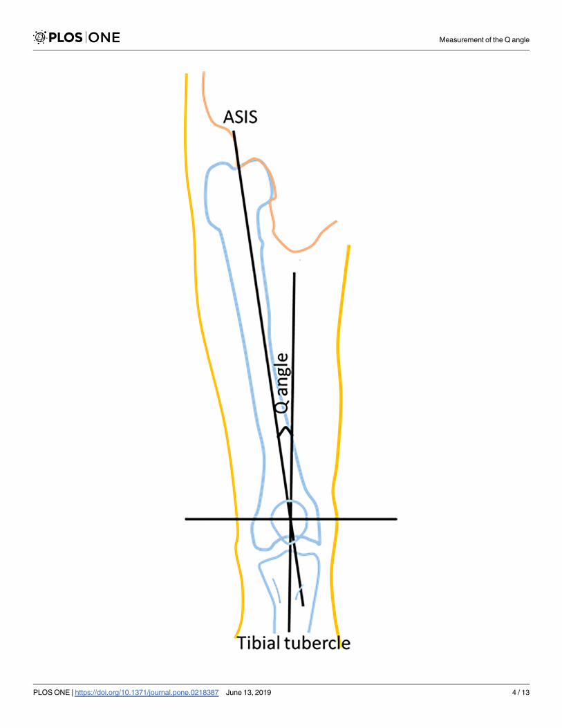
when being asked to kick a ball. The Q angle was measured with a full circle universal manual
goniometer which is made of clear plastic with the subject standing in the erect weight-bearing
position. The anterior superior iliac spine (ASIS), the midpoint of the patella, and the tibial
tuberosity were replaced and determined. The hinge of the goniometer was located at the mid-
point of the patella, the goniometer arms were adjusted to become positioned to the line join-
ing the ASIS and the line joining the tibia tuberosity, then the small angle on the goniometer
was read as the Q angle (Fig 1). Both sides were measured for each individual. Each side was
measured 3 times, and the mean value of the angle was calculated.
A manual caliper, scaled from 0 cm to 20 cm and with a marginal error of ± 1 mm, was
used to measure the condylar distance of the femur for both sides of each volunteer. The sub-
ject first stood in the anatomical position with the feet facing forward, and the leg was flexed to
90˚ with the result that the femoral condyles became prominent and easily palpable at that
position. After the fixed arm of the caliper was placed on the lateral condyle, and the movable
arm was then adjusted to the medial condyle; the condylar distance measurement for each side
had been determined and recorded on the participant’s investigation sheet.
After collecting the requested information and measurements, the data were transferred
into a computer to perform the required statistical analysis.
Statistical analysis
After applying the Levene test to determine the homogeneity of variance, the data were evalu-
ated by one-way analysis of variance (ANOVA) or independent samples t-test at 0.05 and 0.01
levels of significance. The Scheffe post hoc analysis test was performed when it was needed to
examine statistical differences between the groups when necessary. The data were presented as
mean ± standard error of the mean (SEM).
Results
Variation in Q angle with sex
The volunteers were divided according to sex as follows: male (n = 233) and female groups
(n = 267). The Q angle in both sides was significantly (P<0.01) greater in the female subjects
than in the male subjects, such a finding indicated that the Q angle was more prominent in the
female subjects than their counterparts in the male subjects. The mean Q angle ± SEM in the
female subjects was 17.35 ± 0.225 o, whereas that in the male subjects was 14.1 ± 0.21o (Fig 2).
Measurement of the Q angle
PLOS ONE | https://doi.org/10.1371/journal.pone.0218387 June 13, 2019 3 / 13
Measurement of the Q angle
PLOS ONE | https://doi.org/10.1371/journal.pone.0218387 June 13, 2019 4 / 13

Variation in Q angle with height and weight
A sample of 500 adults aged between 18 and 25 years was divided in line with sex as follows:
males 233 and females 267. Each category was studied separately and independently so as to
determine the variation in Q angle with respect to height and weight. The male subjects were
divided into 4 groups according to their heights with each the height interval of each group
was 10cm. It turned out that a significant (P<0.05) variation in Q angle with height was
observed in both sides of the male subjects (Fig 3A). The female subjects were divided into 3
groups in accordance with their height, with each group consisted of a 10-cm interval. It was
also found that a considerable (P<0.05) variation in Q angle with height was observed in both
sides of the female subjects (Fig 3B).
The male subjects were divided into 5 groups on the basis of their weights. Each group
included a weight interval of 10 kg. It was interesting to find that no significant (P>0.05) varia-
tion in Q angle with weight was observed in both sides of the male subjects (Table 1).
The female subjects were also divided into 5 groups in agreement with their weights with
each group had a weight interval of 10 kg. It was astonishing to find that there was also no
important (P>0.05) variation in Q angle with weight was observed in both sides of the female
subjects (Table 2).
Fig 1. Q angle and marker locations: Anterior Superior Iliac Spine (ASIS) and tibial tuberosity.
https://doi.org/10.1371/journal.pone.0218387.g001
Fig 2. Variation of the Q angle with sex in adult population. The data revealed a remarkable difference in the Q
angle between males and females with higher values in females. Each column represents the mean Q angle ± standard
error of the mean (SE). ��P<0.01 (t-test).
https://doi.org/10.1371/journal.pone.0218387.g002
Measurement of the Q angle
PLOS ONE | https://doi.org/10.1371/journal.pone.0218387 June 13, 2019 5 / 13

Fig 3. Variation of Q angle with respect to height in males (A) and females (B). A columnar representation for the relationship between the mean Q and the height
of in males (A) and in females (B). Values are mean Q angle ± standard error (SE). There is a significant increase in Q angle as the condylar distance increases in both
sides. �P<0.05, ��P<0.01.
https://doi.org/10.1371/journal.pone.0218387.g003
Measurement of the Q angle
PLOS ONE | https://doi.org/10.1371/journal.pone.0218387 June 13, 2019 6 / 13

Variation in Q angle with the dominant side
The Q angle was measured in a sample of 437 right-side dominant volunteers with the remark-
able result that was no significant (P>0.05) between the Q angle measurement of the right and
left side in both sexes in the right side-dominant volunteers. Normally speaking, the Q angle
value on the right side is more often greater than the left. The mean Q angle ± SE was
16.7 ± 0.43˚ in the right side and 16.4 ± 0.12˚ in the left side (Fig 4A).
The Q angle was measured in a sample of 63 left-side dominant volunteers. There was also,
no significant (P>0.05) between the Q angle measurement of the right and left side in both
sexes in the left side dominant volunteers. The Q angle value on the left side is more often
greater than the left. The mean Q angle ± SE was 16.0 ± 0.51˚ in the right side and 16.3 ± 0.28˚
in the left side (Fig 4B).
Variation in Q angle with the condylar distance
A sample of 489 adult volunteers, whose age range was between 18-25 years, were divided into
four groups which was based on the length of their right condylar distance. The right Q angle
was measured and compared among those four groups. We found that the Q angle and the
condylar distance was directly proportional, the Q angle (P<0.05) increased significantly as
the condylar distance increased (Fig 5A).
Additionally, the left Q angle was measured and compared among the same four groups.
We, also came to the same conclusion, we found that the Q angle (P<0.05) insignificantly
increased as the condylar distance increased (Fig 5B). Our results indicated that the Q angle is
directly correlated with the condylar distance of the femur in the study group regardless of
which side is measured.
Table 1. Measurements of the Q angle with respect to weight in the adult men.
Weight (kg) Right Q angle Left Q angle P value (χ2)
60–69
N = 32
13.6 ± 0.23˚ 13 ± 0.51˚ 0.62
70–79
N = 57
14.2 ± 0.42˚ 13.9 ± 0.33˚
80–89
N = 83
14.2 ± 0.16˚ 14 ± 0.29˚
90–99
N = 32
14.5± 0.71˚ 14.2 ± 0.11˚
100–109
N = 29
15 ± 0.11˚ 14.6 ± 0.20˚
https://doi.org/10.1371/journal.pone.0218387.t001
Table 2. Measurements of the Q angle with respect to weight in the adult women.
Weight (kg) Right Q angle Left Q angle P value (χ2)
40–49
N = 36
16.9 ± 0.66˚ 16.6 ± 0.02˚ 0.58
50–59
N = 86
17.4 ± 0.12˚ 16.8 ± 0.56˚
60–69
N = 90
17.5 ± 0.25˚ 17 ± 0.38˚
70–79
N = 40
17.7± 0.45˚ 17.2 ± 1.1˚
80–89
N = 15
18.5 ± 0.80˚ 18 ± 0.19˚
https://doi.org/10.1371/journal.pone.0218387.t002
Measurement of the Q angle
PLOS ONE | https://doi.org/10.1371/journal.pone.0218387 June 13, 2019 7 / 13

Measurement of the Q angle
PLOS ONE | https://doi.org/10.1371/journal.pone.0218387 June 13, 2019 8 / 13

Discussion
The Q angle (The quadriceps femoris angle) is one of the most clinically used parameter in
evaluating the quadriceps forces and factors acting on the patellofemoral joint which is consid-
ered to be as an indicator for sports performance as well as in the diagnosis of several patellofe-
moral painful disorders and diseases. Knee alignment indicators such as Q angle are highly
correlated with the quadriceps femoris muscularity. Any alteration in alignment that increases
the Q angle is thought to increase the lateral force on the patella. This can be harmful because
the increase in this lateral force may lead to increase the compression of the lateral patella on
the lateral lip of the femoral sulcus. In the presence of a great enough lateral force, the patella
may actually sublux or dislocate over the femoral sulcus when the quadriceps muscle is acti-
vated on an extended knee. It has also been found that an abnormal Q angle may also influence
neuromuscular response and quadriceps reflex response time [20], consequently, it may be a
risk factor for anterior cruciate ligament injury [21].
The aim of this study was to pinpoint the relationship between the Q angle and various
body parameters. Numerous studies on Q angle have been conducted worldwide aimed to cor-
relate the variations in the Q angle values to the variations in race [11,12,22]. The present
study provides new findings about the Q angle and its relation to several body parameters in
Arab countries population.
The outcomes of this study, which revealed that Q angle was greater in women compared
to men, were similar to earlier reported results regarding the variations in Q angle with gender
that was higher in females as well [13,22]. In our study, we made use of the goniometer to
assess the absolute difference in Q angle between young men and young women which turned
out to be 3.25˚ higher in females than males. Interestingly, the values of the Q angle in both
sexes in Arab population were relatively higher than what had been reported in other countries
and ethnicities [12]. On the other hand, the mean value in this study appears to be close to the
values reported by Clifford [23]. The possible explanation of females having high Q angle val-
ues can be attributed to the fact that their pelvis anatomy is wider than males’ pelvis which is
extrapolated by having a long distance between the pelvis and the patella in comparison to the
distance from the patella to the tibial tuberosity, thereby inducing an alternation in the posi-
tion of the anterior superioriliac spine that has a huge impact on the Q angle values [24]. These
explanations are contrary to what was previously reported by Jaiyesimi, A.O. and Jegede, O.
O’s studies (2009) which suggested that the difference in the Q angle between the males and
females maybe ascribed to the fact that men tend to be taller than women, and that the Q angle
is usually slightly smaller in the taller persons [10]. The higher Q angle values in females
increase the articulating surfaces compression which is clinically important in elucidating the
fact regarding why females are at higher risk of patellofemoral pain. Recent studies have found
that high Q-angle values in females are also linked to the increase in cartilage thickness mea-
surements of the medial femoral condyle and cartilage grading in female patients of osteoar-
thritis. The Q angle values in Arab females, measured in the current study, are greater than the
normal values range reported in other countries and ethnicities, therefore, the Arab females
tend to be at greater risk of developing knee abnormalities. The outcomes of our study further
confirmed what was previously discovered regarding the fact that Q angle is significantly
smaller in taller person on both sexes. Moreover, previous studies had shown that the quadri-
ceps contraction had a considerable corollary on the Q angle values by affecting the patella
Fig 4. Variation of the Q angle with dominant side in adult population. (A) right side dominant volunteers showed
no significant differences. (B) left side dominant volunteer with the results displayed no significant difference. Each
column represents the mean Q angle ± standard error of the mean (SE).
https://doi.org/10.1371/journal.pone.0218387.g004
Measurement of the Q angle
PLOS ONE | https://doi.org/10.1371/journal.pone.0218387 June 13, 2019 9 / 13
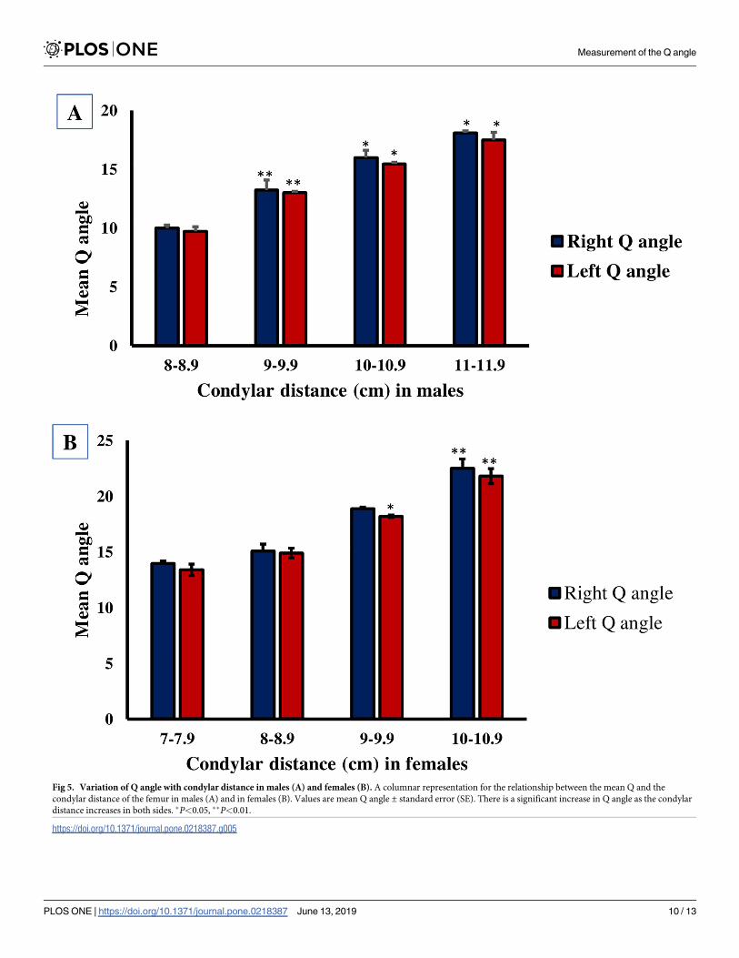
Fig 5. Variation of Q angle with condylar distance in males (A) and females (B). A columnar representation for the relationship between the mean Q and the
condylar distance of the femur in males (A) and in females (B). Values are mean Q angle ± standard error (SE). There is a significant increase in Q angle as the condylar
distance increases in both sides. �P<0.05, ��P<0.01.
https://doi.org/10.1371/journal.pone.0218387.g005
Measurement of the Q angle
PLOS ONE | https://doi.org/10.1371/journal.pone.0218387 June 13, 2019 10 / 13

position [25,26]. Generally speaking, the fact that males are more physically active than females
lead to lower Q angle values as a result of their stronger quadriceps muscle.
Based on the findings of the present study, the Q angle values do not vary significantly with
the weight of the study population. Sra A. et al (2008) also reported no noticeable variation in
the Q angle with weight [27].
Relatively speaking, few worldwide studies have focused on Q angle bilateral variability. In
the present study, the Q angle was greater on the dominant side compared with the non-domi-
nant side, but this difference was not statistically significant. Hahn and Foldspang were among
the first researchers to make a detailed study of the bilateral variability in the Q angle [8]. Fol-
lowing this study, other studies have documented similar bilateral variations [12,13,27,28]
with only two studies found that this bilateral differences significantly affected the Q angle
[27,28].
To further investigate the Q angle, the condylar distance was measured in both legs using a
digital caliper. This has been the first study that investigates the relationship between the Q
angle and the condylar distance of the femur. The results show a significant increase in the Q
angle as the condylar distance increases in both sexes. The correlation between Q angle and
condylar distance is clinically important in the diagnosis of degenerative arthritis and other
knee degenerative abnormalities.
Supporting information
S1 Fig. A student’s approved form for conducting the Q angle research.
(PDF)
S2 Fig. The ethical approval of the Institutional Research Board (IRB) at Jordan University
of Science and Technology to conduct the Q angle research.
(DOCX)
Acknowledgments
We would like to thank Mr. Muhammad Abu El-Rub for the time and effort spent in reviewing
the Manuscript
Author Contributions
Conceptualization: Mohammed Z. Allouh.
Formal analysis: Ramada R. Khasawneh, Ejlal Abu-El-Rub.
Funding acquisition: Ramada R. Khasawneh.
Investigation: Ramada R. Khasawneh.
Methodology: Ramada R. Khasawneh.
Project administration: Ramada R. Khasawneh.
Software: Ramada R. Khasawneh.
Supervision: Ramada R. Khasawneh.
Writing – original draft: Ramada R. Khasawneh.
Writing – review & editing: Ramada R. Khasawneh.
Measurement of the Q angle
PLOS ONE | https://doi.org/10.1371/journal.pone.0218387 June 13, 2019 11 / 13

References1. Brattstrom H. Shape of the Intercondylar Groove Normally and in Recurrent Dislocation of Patella: A
Clinical and X-Ray Anatomical Investigation. Acta Orthop Scand. 1964; 35: 1–148. https://doi.org/10.
3109/ort.1964.35.suppl-68.01
2. Loudon JK. Biomechanics and Pathomechanics of the Patellofemoral Joint. Int J Sports Phys Ther.
2016; 11: 820–830. PMID: 27904787
3. Chhabra P, Setiya M, Godwin R. “Quadriceps angle”: An Important Indicator of Biomechanical Function
of Lower Extremity and Its Relation with Anterior Knee Pain. Int J Sci Study. 2016; 4: 173–176.
4. Emami M, Ghahramani M, Abdinejad F, Namazi H. Q-angle: an invaluable parameter for evaluation of
anterior knee pain. Arch Iran Med. 2007; 10: 24–26. doi: 07101/AIM.007 PMID: 17198449
5. Daneshmandi H, Saki F, Shahheidari S, Khoori A. Lower extremity Malalignment and its linear relation
with Q angle in female athletes. 3rd World Conf Educ Sci—2011. 2011; 15: 3349–3354. https://doi.org/
10.1016/j.sbspro.2011.04.298
6. Nguyen A-D, Boling MC, Levine B, Shultz SJ. Relationships between lower extremity alignment and the
quadriceps angle. Clin J Sport Med Off J Can Acad Sport Med. 2009; 19: 201–206. https://doi.org/10.
1097/JSM.0b013e3181a38fb1 PMID: 19423972
7. Almeida GPL, Silva AP de MCCE, Franca FJR, Magalhães MO, Burke TN, Marques AP. Q-angle in
patellofemoral pain: relationship with dynamic knee valgus, hip abductor torque, pain and function. Rev
Bras Ortop. 2016; 51: 181–186. https://doi.org/10.1016/j.rboe.2016.01.010 PMID: 27069887
8. Hahn T, Foldspang A. The Q angle and sport. Scand J Med Sci Sports. 1997; 7: 43–48. https://doi.org/
10.1111/j.1600-0838.1997.tb00116.x PMID: 9089904
9. Yilmaz A, Kabadayi M, Mayda M, Cavusoglu G, Tasmektepligi M. Analysis of Q Angle Values of Female
Athletes from Different Branches. Sci Mov Heal. 2017; 17: 141–146.
10. Jaiyesimi A, Jegede O. Influence of Gender and Leg Dominance on Q-angle Among Young Adult Niger-
ians. Afr J Physiother Rehabil Sci. 2009; 1: 18–23.
11. Omololu BB, Ogunlade OS, Gopaldasani VK. Normal Q-angle in an adult Nigerian population. Clin
Orthop. 2009; 467: 2073–2076. https://doi.org/10.1007/s11999-008-0637-1 PMID: 19034592
12. Raveendranath R, Nachiket S, Sujatha N, Priya R, Rema D. Bilateral Variability of the Quadriceps
Angle (Q angle) in an Adult Indian Population. Iran J Basic Med Sci. 2011; 14: 465–471. PMID:
23493777
13. Tella B, Ulogo U U, Odebiyi D, Omololu A. Gender variation of bilateral Q-angle in young adult Niger-
ians. Nig Q J Hosp Med. 2010; 20: 114–116. PMID: 21033317
14. Davies G, Larson R. Examining the knee. J Am Phys Ther Assoc Sports Med. 1978; 6: 49–67.
15. Tanifuji O, Blaha JD, Kai S. The vector of quadriceps pull is directed from the patella to the femoral
neck. Clin Orthop. 2013; 471: 1014–1020. https://doi.org/10.1007/s11999-012-2741-5 PMID:
23263931
16. Galea A, Albers J. Patellofemoral pain: targeting the cause. Phys Sports Med. 1994;22.
17. Tsakoniti AE, Mandalidis DG, Athanasopoulos SI, Stoupis CA. Effect of Q-angle on patellar positioning
and thickness of knee articular cartilages. Surg Radiol Anat. 2011; 33: 97–104. https://doi.org/10.1007/
s00276-010-0715-4 PMID: 20798938
18. Tiberio D. The effect of excessive subtalar joint pronation on patellofemoral mechanics: a theoretical
model. J Ortho Sports Phys Ther. 1987; 9: 160–165.
19. Piva SR, Fitzgerald GK, Irrgang JJ, Fritz JM, Wisniewski S, McGinty GT, et al. Associates of physical
function and pain in patients with patellofemoral pain syndrome. Arch Phys Med Rehabil. 2009; 90:
285–295. https://doi.org/10.1016/j.apmr.2008.08.214 PMID: 19236982
20. Shultz SJ, Carcia CR, Gansneder BM, Perrin DH. The independent and interactive effects of navicular
drop and quadriceps angle on neuromuscular responses to a weight-bearing perturbation. J Athl Train.
2006; 41: 251–259. PMID: 17043692
21. Griffin L, Agel J, Albohm M, Arendt E, Dick R, Garrett W, et al. Noncontact Anterior Cruciate Ligament
Injuries: Risk Factors and Prevention Strategies. J Am Acad Orthop Surg. 2000; 8: 141–150. PMID:
10874221
22. Ebeye A, Abade P, Okwoka B. Influence of gender on quadriceps (Q) angle among adult Urhobos in
Nigeria population. J Exp Clin Anat. 2014; 13: 50–53.
23. Wheeless C. Wheeless’ Textbook of Orthopaedics [Internet]. 2018. Available: http://www.
wheelessonline.com/ortho/q_angle_of_the_knee.
24. Grelsamer RP, Dubey A, Weinstein CH. Men and women have similar Q angles. J Bone Joint Surg Br.
2005; 87-B: 1498–1501. https://doi.org/10.1302/0301-620X.87B11.16485 PMID: 16260666
Measurement of the Q angle
PLOS ONE | https://doi.org/10.1371/journal.pone.0218387 June 13, 2019 12 / 13

25. Guerra JP, Arnold MJ, Gajdosik RL. Q Angle: Effects of Isometric Quadriceps Contraction and Body
Position. J Orthop Sports Phys Ther. 1994; 19: 200–204. https://doi.org/10.2519/jospt.1994.19.4.200
PMID: 8173567
26. Bayraktar B, Yucesir I, Ozturk A, Cakmak A, Taskara N, Kale A, et al. Change of quadriceps angle val-
ues with age and activity. Saudi Med J. 2004; 25: 756–760. PMID: 15195206
27. Sra A, Ba T, Oo J. Comparison of bilateral Quadriceps angle in asymptomatic and symptomatic males
with anterior knee pain. Internet J Pain Symptom Contr Palliat Care. 2008;6.
28. Livingston LA, Spaulding SJ. OPTOTRAK Measurement of the Quadriceps Angle Using Standardized
Foot Positions. J Athl Train. 2002; 37: 252–255. PMID: 12937581
Measurement of the Q angle
PLOS ONE | https://doi.org/10.1371/journal.pone.0218387 June 13, 2019 13 / 13