Update on 13 Syndromes Affecting Craniofacial and Dental ...
Upload
independentCategory
view
1download
0
Leukemia Research 30 (2006) 9–16
Maturation-associated immunophenotypic abnormalities in bonemarrow B-lymphocytes in myelodysplastic syndromes
Elisangela Ribeiroa,1, Sergio Matarraz Sudonb,1, Marıa de Santiagob,Carmen S.P. Limaa, Konradin Metzec, Manuel Giraltd, Sara T.O. Saade,
Alberto Orfao de Matosb, Irene Lorand-Metzee,∗a Hematology and Hemotherapy Center, State University of Campinas, Brazil
b Centro de Investigacion del Cancer Servicio General de Citometrıa and Departamento de Medicina,Universidad de Salamanca, Salamanca, Spain
c Department of Pathology, Faculty of Medicine, State University of Campinas, Brazild Department of Hematology, Hospital Miguel Servet, Zaragoza, Spain
e Department of Internal Medicine, Faculty of Medicine, State University of Campinas,Rua Carlos Chagas 480, P.O. Box 6198, BR 13081 970 Campinas, Sao Paulo, Brazil
Received 7 March 2005; received in revised form 17 May 2005; accepted 18 May 2005Available online 7 July 2005
A
of complexi turation inB D34+ andC nt donorsa /13w etweenC f blastsa 1c one antigenw lities. Ourr e abnormalc tment andm malities.©
K
1
d[
s
tureto
las-ntedpic-DRome
the
0d
bstract
Recent studies concerning the pathophysiology of myelodysplastic syndromes (MDS) have shown evidences for the existencenteractions between hematopoietic stem cells and the bone marrow (BM) microenvironment. We analyzed the B-lymphocyte maM of patients with MDS. For this purpose, 41 newly-diagnosed patients were analyzed. Enumeration and characterization of CD34− B-cell precursors and mature B-lymphocytes was performed using multiparameter flow cytometry. BM from eight transpland six orthopedic surgery patients were used as controls. CD34+/CD45lo B-cells were found in 17/22 patients with RA/RARS and in 5ith RAEB. In patients with RAEB-t and CMML no CD34+ B-cell precursors could be detected. A positive correlation was found bD34+ and CD34− B-cell precursors (r = 0.52). CD34+ B-cell precursors presented an inverse correlation with BM percentage ond peripheral leukocytes and a positive one with hemoglobin. Asynchronous antigen expression (CD19+/CD79a− cells) was found in 7/1ases of RA/RARS and 6/18 cases of RAEB in which this phenotype was examined. Abnormal patterns of expression for at leastas found in 91% of RA/RARS cases and in 74% of RAEB. Underexpression of TdT and CD79a were the most frequent abnorma
esults present evidences of an abnormal B-cell maturation in MDS. This may be an evidence that B-lymphocytes are derived of thlone. But it may also be the consequence of influences of abnormalities of BM microenvironment leading to an impaired commiaturation of the B-cell line in MDS. Studies performed with purified well-characterized B-cells may further elucidate these abnor2005 Elsevier Ltd. All rights reserved.
eywords: Myelodysplastic syndromes; Bone marrow; B-cell maturation; Flow cytometry
. Introduction
Myelodysplastic syndromes (MDS) are a group of clonalisorders originating from myeloid hematopoietic stem cells
1]. Despite this, accumulating evidence indicates that the
∗ Corresponding author. Tel.: +55 19 37888740; fax: +55 19 37888600.E-mail address: [email protected] (I. Lorand-Metze).
1 Both authors have equally contributed to this work and should be con-idered as first authors.
neoplastic event in MDS could target a more immastem cell, which still retains the ability to differentiate inboth the myeloid and lymphoid lineages[2–7]. On theother hand, progression of MDS into acute lymphobtic leukemia (ALL) although rare, has been docume[3,8–11]. ALL has been diagnosed mainly by phenotyfeatures, more frequently by the presence of TdT, HLAbut also by the expression of CD19 and CD10. In scases, IgH rearrangement has also been documented[3,9].In others, the same cytogentetic abnormalities found in
145-2126/$ – see front matter © 2005 Elsevier Ltd. All rights reserved.oi:10.1016/j.leukres.2005.05.019
10 E. Ribeiro et al. / Leukemia Research 30 (2006) 9–16
original MDS clone, were also observed in blasts of ALL[10,11].
More recently, several groups have attempted to demon-strate the clonal nature of lymphoid cells in MDS through theidentification of common genetic abnormalities in myeloidprecursors, multipotent stem cells and lymphoid cells. Pres-ence of circulating clonal B-lymphocytes was observed in5/68 cases of MDS analyzing the hypervariable region CDR3of the immunoglobulin heavy chains[2]. White et al.[4]demonstrated the presence of 20q deletions in a multipotentprecursor of both myeloid cells and B-cells. In keeping withthese results, Nilsson et al.[5] studied 10 patients with MDSwith del(5q) and showed that this cytogenetic abnormalitywas also present in mature B-lymphocytes (one of 10 casesstudied) as well as CD34+/CD19+ B-cell precursors (threecases). In contrast, Saitoh et al.[6] could not demonstratethe presence of trisomy 8 neither in lymphoid cells nor inCD34+/CD90+ hematopoietic stem cells in a group of sevenMDS patients carrying this cytogenetic abnormality in theneutrophil, monocytic and erythroid compartments. Morerecently, Nilsson et al.[7], suggested that these apparentlydiscrepant results could be related to the fact that trisomy 8 isa secondary cytogenetic change which is usually restricted tothe more mature myeloid committed stem cell progenitors,while other primary cytogenetic abnormalities such as 5q−would involve more immature CD34+/CD38−/CD90+ stemc loida thei byA otT scept llsf ses[ portsh typica ellsi
bero .H beenr no-t hasb . Oura ypicc com-p toi mal-i
2
2
eana ssi-
fied according to the FAB classification[21] were included.From these patients, 12 were diagnosed at the HematologyCenter of the State University of Campinas (Brazil) and 29at the Cytometry Service (University of Salamanca, Spain).According to the FAB classification, 20 cases had refrac-tory anemia (RA), 2 RA with sideroblasts (RARS) (22 lowrisk cases), 13 RA with excess of blasts (RAEB), 2 RAEBin transformation (RAEB-t) and 4 chronic myelomonocyticleukemia (CMML) (19 high risk cases). IPSS was not per-formed as karyotype was not available is all cases and wasfrequently normal. Instead, in order not to split the 41 casesin too many categories and lower too much the statistic powerof the tests performed, the correlations between the bonemarrow (BM) B-cell populations and the peripheral blood(PB) counts and BM blasts were analyzed as continuousvariables.
In each patient, a BM aspirate was obtained for diag-nostic purposes and used for flow cytometry immunophe-notypic studies described below, according to the guide-lines of the Local Ethics Committees. In addition, 14 BMsamples from normal individuals (donors of allogeneicBM transplantation—eight cases or patients of orthopedicsurgery—six cases) were analyzed as normal BM controls.All subjects gave their informed consent to participate in thisstudy.
2
medu nala per-f encesw tra-c entB n-D withC -e sents ellsi andH neV 9a( omD fters flowc ram( ed:C andC
nd-i red.A t-upa par-i cter-i ar-
ells with the capacity to differentiate into both the myend lymphoid lineages. Additional indirect evidence of
nvolvement of lymphoid cells in MDS has been foundmin et al. [12] who have shown that BM B-cells, but n-lymphocytes, have increased apoptosis. Increased suibility to undergo apoptosis of BM myeloid precursor cerom MDS patients, particularly among the low risk ca13–16]has been described by several authors. Recent reave shown the existence of a high number of phenoberrations on different myeloid compartments of BM c
n MDS [16–19].In a preliminary work, we have found a decreased num
f B-lymphocytes in MDS patients[20] especially in RAEBowever, to the best of our knowledge, no study has
eported so far in which the distribution and immunopheype of the different compartments of maturing B-cellseen analyzed in detail in a large series of MDS patientsim was to analyze the distribution and immunophenotharacteristics of the different maturation-associatedartments of BM B-cells in patients with MDS, in order
dentify the presence of numerical and phenotypic abnorties that would support the neoplastic nature of B-cells.
. Materials and methods
.1. Patients and samples
A total of 41 patients: 23 males, 18 females with mge of 60 years (20–93) with newly diagnosed MDS cla
-
.2. Flow cytometry immunophenotypic studies
In all cases immunophenotypic studies were perforsing three- and four-color combinations of monoclontibodies (MAb). Immunofluorescence staining was
ormed using a standardized direct immunofluoresctain-and-then-lyse-and-wash technique[22,23] combinedith fixation and permeabilization for the detection of inellular antigens. For the identification of the differM B-cell compartments, CD45 (clone HLE-1); Bectoickinson Biosciences (BDB) San Jose, CA togetherD19 (clone HIB19, BDB) were used (Fig. 1). Other markrs combined with CD19 and CD45 used in the pretudy for the phenotypic characterization of BM B-cncluded: CD34 (clone 8G12), CD16 (clone NKP15)LA-DR (clone L243) purchased from BDB; CD3 (cloCHT1), CD4 (clone MT310), CD8 (clone DK25), CD7
clone HM57) and TdT (clone HT-6) purchased frakoCytomation (Glostrup, Denmark). Immediately ataining, samples were acquired in a FACSCaliburytometer (BDB) using the CellQuest software progBDB). The following combinations of antibodies were usD19/cCD79a/CD45/CD34, TdT/cCD79a/CD45/CD34D/cCD79a/CD45/CD34.Information on a minimum of 300,000 cells (correspo
ng to all nucleated BM cells) was acquired and stot the two participating centers, identical instrument send calibration procedures were used. In addition, com
son of the light scatter and immunophenotypic charastics of the different B-cell subsets in normal bone m
E. Ribeiro et al. / Leukemia Research 30 (2006) 9–16 11
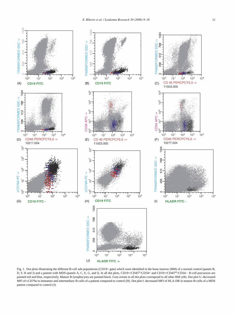
Fig. 1. Dot plots illustrating the different B-cell sub-populations (CD19+ gate) which were identified in the bone marrow (BM) of a normal control (panels B,D, F, H and J) and a patient with MDS (panels A, C, E, G, and I). In all dot plots, CD19+/CD45lo/CD34+ and CD19+/CD45int/CD34− B-cell precursors arepainted red and blue, respectively. Mature B-lymphocytes are painted black. Grey events in all dot plots correspond to all other BM cells. Dot plot G: decreasedMFI of cCD79a in immature and intermediary B-cells of a patient compared to control (H). Dot plot I: decreased MFI of HLA-DR in mature-B-cells of a MDSpatient compared to control (J).
12 E. Ribeiro et al. / Leukemia Research 30 (2006) 9–16
row samples was performed showing comparable results forthe cases analyzed at each of the two centers (p > 0.05).The PAINT-A-GATE software (BDB) was used for dataanalysis.
In all samples, B-cells were identified as being eitherCD19+ or cCD79a+ and having low sideward light scatter(SSC) as shown inFig. 1. Among B-cells, three differentsubsets were identified in a CD45/SSC and CD45/CD34bivariate dot plots which included, according to their degreeof maturation: (1) early CD34+/CD45lo B-cell precursors;(2) CD34−/CD45 intermediate (int) B-cell precursors; and(3) CD34−/CD45hi mature B-lymphocytes. For the pheno-typic characterization of each of these B-cell populationstheir mean light scatter intensity as well as the mean fluores-cence intensity (MFI) obtained for each individual antigen,both expressed in relative linear arbitrary channel units scaledfrom 0 to 1024 were used. Based on the staining observed forisotype-matched negative controls, cells were considered tobe positive for a given antigen once MFI values higher than5 were found.
2.3. Statistical analysis
For all variables under study, their median and rangevalues were calculated. The differences observed betweengroups was assessed usingX2 or the Kruskal–Wallis testsw oci-a am-i cor-r ari-a ofo ari-as wereu
3. Results
3.1. Distribution of BM B-cell maturation compartments
The distribution of the different B-cell maturation com-partments in BM of low risk (LR) and high risk (HR)MDS patients as compared to normal BM are shown onTable 1. For each individual, the sum of the percentagesof all three compartments was 100%. Concerning normalBM, early CD34+/CD45lo cells were found in 11/14 cases.However, in RA/RARS they were found in 17/22 cases. InRAEB, only 5/13 cases presented these cells. None of theRAEB-t and CMML cases presented CD34+ B-precursorcells. This frequency was significantly different (inX2
p < 0.0001).All normal BMs presented cells in the intermediate and
mature compartments. CD34− intermediate cells were foundin all cases of RA/RARS but only in 10/13 patients withRAEB and 3/6 patients with RAEB-t/CMML cases.
Considering only cases who presented CD34+ B-cellprecursors (population 1), their percentage was similar inRA/RARS but were increased in the five cases of RAEBin which these cells were detected (Table 1). The CD34− B-cell precursors (population 2) were also similar in normal BMand RA/RARS but was decreased in RAEB. A positive cor-relation was found between populations 1 and 2 (r = 0.52). Ap MFIo nC
tionw -c ithh ngM nifi-c
TD w risk s mediana
B Low ri
C4.5 (10.15 (
C19.5 (4
0.74 (
M77.4 (21.5 (0
C9.0 (40.33 (
N syndrs of RAIn high
hen suitable.p-values < 0.05 were considered to be assted with statistical significance. Correlations were ex
ned by the Spearman’s test. We calculated partialelation coefficients to examine interdependency of vbles. This method allows to eliminate the influencene variable on the correlation between two other vbles, keeping the first variable constant[24]. The Win-tat and SPSS softwares (SPSS 10.0, Chicago, IL)sed.
able 1istribution of subsets of B-cell maturation compartments in BM of lomong cases that presented cells in each compartment
-cell subset NBM (n = 14)
D34+ B-cell precursorsa
Percent of B-cells 4.5 (1.8–15.9)Percent of all BM cells 0.11 (<0.01–0.23)
D34− B-cell precursorsb
Percent of B-cells 29 (7.35–80.1)Percent of all BM cells 0.62 (0.15–1.07)
ature B-lymphocytesc
Percent of B-cells 72.1 (50.4–98.2)Percent of all BM cells 1.47 (0.3–3.3)
D19+/cCD79a− aberrant cellsPercent of B-cells 0Percent of all BM cells 0
BM: normal bone marrow; BM: bone marrow; MDS: myelodysplastica 11/14 NBM, 17/22 in RA/RARS, 5/13 RAEB and none of the caseb All NBM and low risk patients showed cells in this compartments.c All patients in this group showed cells in this compartment.
ositive correlation was found between percentage andf FSC in CD34+ B-cell precursors (r = 0.79) as well as iD34− B-cells (r = 0.43).CD34+ B-cell precursors showed an inverse correla
ith BM percentage of blasts (r =−0.28) and with PB leukoytes (r =−0.43). A positive correlation was found wemoglobin (r = 0.38). In the partial correlation, maintainiFI of FSC constant, all these correlations lost their sig
ance.
(LR) and high risk (HR) MDS patients as compared to normal BM a
sk MDS (n = 22) High risk MDS (n = 19) p-value
.2–19.2) 11.9 (5.3–46.1) 0.01<0.01–0.73) 0.25 (0.13–0.6) 0.01
.5–83.5) 10.4 (5.3–66.4) <0.001<0.01–3.7) <0.01 (<0.01- 2.5) <0.001
5.7–100) 92.0 (4.8–100) 0.01.1–6.3) 0.8 (<0.01–6.1) 0.10
–36) 8.8 (1.0–20) 0.56<0.01–1.9) 0.16 (<0.01–0.6) 0.25
ome.p-values obtained by the Kruskal–Wallis test.EB-t and CMML.risk, only 13/19 patients showed these cells.

E. Ribeiro et al. / Leukemia Research 30 (2006) 9–16 13
Concerning CD34− B-cells these correlations were:r =−0.38,−0.36, and +0.26, respectively. In the partial corre-lation however, these correlations were accentuated for blastpercentage (r =−0.75) but remained unaltered for leukocytes(r =−0.33) and hemoglobin (r = +0.22).
3.2. Immunophenotypic characteristics of BM B-cells
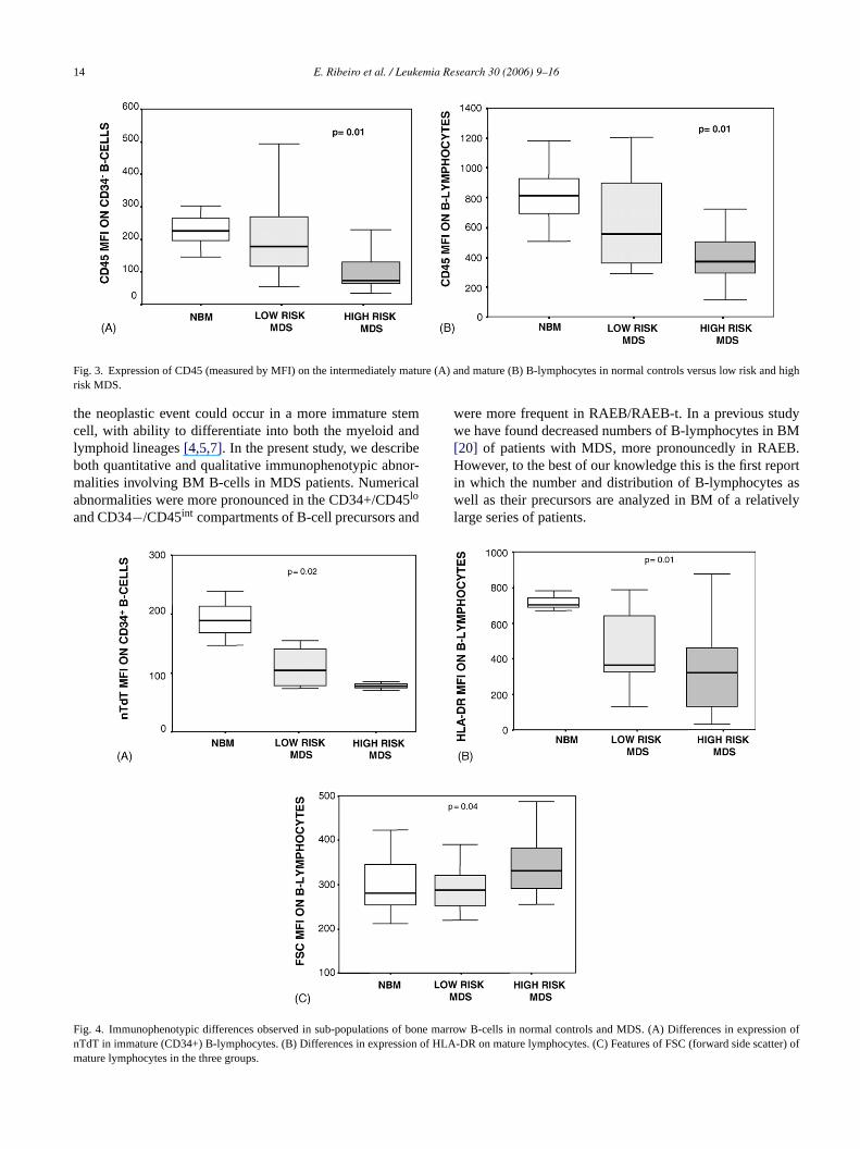
Concerning the two pan-B-cell markers used (CD19 andcCD79a) only CD19 was constantly expressed by all B-cellsfrom all individuals. However, aberrant CD19+/cCD79a− B-cell precursors were observed in both LR-MDS and HR-MDSpatients but not in normal BM (Table 1). CD19+/CD79a−cells were found in 7/11 cases of LR- and 6/18 cases ofHR-MDS (Fig. 1G). They were associated with a statis-tically significant low expression of cCD79a in early andintermediate B-cells but not in mature ones (Fig. 2). In addi-tion to cCD79a, other B-cell associated markers were alsoabnormally expressed in different BM B-cell compartments.Thus, reactivity for CD45 was abnormally low on immatureas well as on mature lymphocytes (Fig. 3). Expression ofTdT was also decreased (Fig. 4A) on CD34+ B-cell precur-sors. A lower amount of HLA-DR was found on CD34−
B-cells (Fig. 4B). For all markers analyzed except TdT,abnormal antigen expression on CD34+ B-cell precursorswas more frequently observed in LR-MDS patients whereasabnormal antigen expression on the two CD34− B-cell com-partments predominated among HR-MDS cases. Overall,91% of all LR-MDS and 74% of HR-MDS cases showedaberrant expression of at least one marker in one or moresubpopulations of B-cells. From all antigens studied, aber-rant expression of CD3, CD4, CD8 and CD16 on BM B-cellswas never observed. Mean forward light scatter (FSC) values(Fig. 4C) on B-lymphocytes were variable but overall notdecreased. However, a significant positive correlation wasfound between percentage of CD34+ B-cells and their FSCMFI (r = 0.61) and those CD34− ones (r = 0.51).
4. Discussion
Myelodysplastic syndromes (MDS) are clonal disordersoriginated in the myeloid hematopoietic stem cell1. How-ever, recently, several studies using FISH and immunologicalmarkers as well as several functional essays have broughtevidences indicating that, in at least a part of the cases,
FC
ig. 2. Intensity of CD79a expression (measured by MFI) observed in the BD34+ (immature) B-cells (B) intermediately mature cells (CD34−) (C) mature l
-cell sub-populations in normal controls versus low risk and high risk MDS. (A)ymphocytes.
14 E. Ribeiro et al. / Leukemia Research 30 (2006) 9–16
Fig. 3. Expression of CD45 (measured by MFI) on the intermediately mature (A) and mature (B) B-lymphocytes in normal controls versus low risk and highrisk MDS.
the neoplastic event could occur in a more immature stemcell, with ability to differentiate into both the myeloid andlymphoid lineages[4,5,7]. In the present study, we describeboth quantitative and qualitative immunophenotypic abnor-malities involving BM B-cells in MDS patients. Numericalabnormalities were more pronounced in the CD34+/CD45lo
and CD34−/CD45int compartments of B-cell precursors and
were more frequent in RAEB/RAEB-t. In a previous studywe have found decreased numbers of B-lymphocytes in BM[20] of patients with MDS, more pronouncedly in RAEB.However, to the best of our knowledge this is the first reportin which the number and distribution of B-lymphocytes aswell as their precursors are analyzed in BM of a relativelylarge series of patients.
Fnm
ig. 4. Immunophenotypic differences observed in sub-populations of boneTdT in immature (CD34+) B-lymphocytes. (B) Differences in expression ofature lymphocytes in the three groups.
marrow B-cells in normal controls and MDS. (A) Differences in expression ofHLA-DR on mature lymphocytes. (C) Features of FSC (forward side scatter) of

E. Ribeiro et al. / Leukemia Research 30 (2006) 9–16 15
The numerical changes found are in keeping with thehypothesis that the abnormal stem cell has a decreased abil-ity to produce B-lymphocytes, as has also been postulatedby others[25]. The apparent high proportion of mature B-lymphocytes presenting less phenotypic alterations couldsuggest that in this sub-population there is a mixture ofabnormal cells derived from the MDS clone and normal“pre-clonal” B-lymphocytes as has already been pointed out[25].
This decrease of CD34+ and less pronounced of CD34−B-cells, was more evident with increasing numbers of BMblasts. We also observed a relation between these cell popu-lations and PB leukocyte count and hemoglobin values. Thiscould suggest that, impaired B-cell production is associatedwith progression of the MDS clone. Proportion of BM blastsas well as PB counts are well recognized prognostic factorsin MDS [1,13,14,26]. Therefore, one may hypothesize that amore abnormal stem cell looses its capacity to differentiateto B-lymphocytes.
However, it has been recently shown that the early phaseof commitment of the multipotent hematopoietic stem cells tothe B-cell line in BM is driven by their attachment to stromalcells expressing CXCL12[27]. As several stromal abnormal-ities have been described in MDS, these may be associatedwith the impairment of B-cell commitment and maturation[28]. This would also be in keeping with the increaseda inM
e ofa nteda ark-e e fre-qs ng thet ali-t theya d andt f B-c
theC rs.D M,eNp phe-n sorA he-n pe-c res-s pto-s ive)o in-e ofC e ina elys to-
sis of B-cell precursors influenced by abnormalities of BMmicroenvironment. These are multiple and remain poorlycharacterized[28]. Studies analyzing apoptosis specificallyon these cells with abnormal phenotype could clarify thispoint.
Clonal subpopulations of several lineages have beendescribed in MDS, more frequently in more advanced cases[2,16,25,34]. Clonality is well recognized in granulocytes,erythroblasts and megakaryocytes and can be observed inmost patients, while detection of clonal B-lymphocytes hasbeen controversial, but has been demonstrated in at least fewcases[4–7,34]. In order to elucidate this point, further studiesexamining clonality of BM B-cell precursors using FISH andlineage-specific markers should be performed. But also theinfluence of the stromal abnormalities on B-cell productionin MDS should be further elucidated.
In summary, the present study shows phenotypic evidenceof an abnormal B-cell maturation in MDS patients, morepronounced in advanced disease. This could be due to aparticipation of the B-lymphocytes in the MDS clone, butcould also be a result of an influence of the altered microen-vironment on these cells. Further research, using purified wellcharacterized cells would better describe the role of each celltype in the complex pathophysiology of MDS.
A
da-t( eivea il).
R
i C,ema-
lym-matol
zalezaryclin-
l B,occurood
ll-n oflastic
cell
situcell
rmal-ood
poptosis recently described in BM B-lymphocytesDS [12].Further supporting the hypothesis on the occurrenc
n abnormal B-cell maturation, most of our cases presebnormal patterns of expression for at least one of the mrs analyzed. Such phenotypic abnormalities were moruently observed in the early CD34+/CD45lo B-cell precur-ors and were more evident in advanced cases. Regardiype and frequency of the different phenotypic abnormies found in the present study, it should be noted thatffected most of the B-cell-associated antigens analyze
hey highly varied both between different populations oells and between different individuals.
Asynchronous antigen expression was restricted toD19+/cCD79a− phenotype on CD34+ B-cell precursouring B-cell maturation in normal and regenerating Bxpression of cCD79a always precedes that of CD19[29,30].o normal B-cell precursors showing a cCD79a−/CD19+henotype has been reported in the literature. Thisotypic abnormality is not frequent in B-cell precurLL where most blast cells display a CD19+/CD79a+ potype [29–31]. Therefore, this abnormality may be sific for MDS. Recently, it has been shown that expion of lineage antigens are frequently lost during apois of T lymphocytes and B-lymphocytes (CD19 positf chronic lymphocytic leukemia as well as in cell lages in culture[32,33]. One may speculate if absenceD79a expression is due to a downregulation of its genneoplastic cell, specifically occurring in MDS but rar
een in ALL or if this expression is lost during apop
cknowledgements
Elisangela Ribeiro has a doctor fellowship from Founion for Development of Science from the State of Sao PauloFAPESP), Konradin Metze and Irene Lorand-Metze recresearch grant from CNPq (National Research Counc
eferences
[1] Alessandrino EP, Amadori S, Cazzola M, Locatelli F, MecuccMorra E, et al. Myelodysplastic syndromes: recent advances. Hatologica 2001;86:1124–57.
[2] Culligan DJ, Cachia P, Whittaker J, Jacobs A, Padua RA. Clonalphocytes are detectable in only some cases of MDS. Br J Hae1992;81:346–52.
[3] San Miguel JF, Hernandez JM, Gonzalez-Sarmiento R, GonM, Sanchez I, Orfao A, et al. Acute leukemia after a primmyelodysplastic syndrome: immunophenotypic, genotypic, andical characteristics. Blood 1991;78:768–74.
[4] White NJ, Nacheva E, Asimakopoulos FA, Bloxham D, PauGreen AR. Deletion of cromossome 20q in myelodysplasia canin a multipotent precursor of both myeloid cells and B-cells. Bl1994;83:2809–16.
[5] Nilsson L, Astrand-Grundstom I, Arvidsson I, Jacobsson B, Hestrom-Lindberg E, Hast R, et al. Isolation and characterizatiohematopoietic progenitor/stem cells in 5q-deleted myelodyspsyndromes: evidence for involvement at the hematopoietic stemlevel. Blood 2000;96:2012–21.
[6] Saitoh K, Miura I, Takahashi N, Miura AB. Fluorescence inhybridization progenitor cells obtained by fluorescence-activatedsorting for the detection of cells affected by chromosome abnoity trisomy 8 in patients with myelodysplastic syndromes. Bl1998;92:2886–92.
16 E. Ribeiro et al. / Leukemia Research 30 (2006) 9–16
[7] Nilsson L, Astrand-Grundstom I, Anderson K, Arvidsson I, HoklandP, Bryder D, et al. Involvement and functional impairment of theCD34+ CD38−Thy-1+ hematopoietic stem cell pool in myelodys-plastic syndromes with trisomy 8. Blood 2002;100:259–67.
[8] Kouides PA, Bennett JM. Transformation of chronic myelomonocyticleukemia to acute lymphoblastic leukemia—case report and reviewof the literature of lymphoblastic transformation of myelodysplasticsyndrome. Am J Hematol 1995;49:157–62.
[9] Pajor L, Matolcsy A, Vass JA, Mehes G, Marton E, Szabo F, et al.Phenotypic and genotypic analyses of blastic cell population suggestthat pure B-lymphoblastic leukemia may arise from myelodysplasticsyndrome. Leuk Res 1998;22:13–7.
[10] Ikeda T, Sato K, Yamashita T, Kanai Y, Kuwada N, Matsumura T,et al. Burkitt’s acute lymphoblastic leukaemia transformation aftermyelodysplastic syndrome. Br J Haematol 2001;115:69–71.
[11] Abruzzese E, Buss D, Rainer R, Pettenati MJ, Rao PN. Progres-sion of a myelodysplastic syndrome to a pre-B acute lymphoblas-tic leukemia: a case report and cell lineage study. Ann Haematol1996;73:35–8.
[12] Amin HM, Jilani I, Estey EH, Keating MJ, Dey AL, Man-shoury T, et al. Increased apoptosis in bone marrow B lympho-cytes but not T lymphocytes in myelodysplastic syndromes. Blood2003;102:1868–86.
[13] Ribeiro E, Lima CSP, Metze K, Lorand-Metze I. Flow cytometricanalysis of the expression of fas/fasl in bone marrow CD34+ cellsin myelodysplastic syndromes: relation to disease progression. LeukLymph 2004;45:309–13.
[14] Greenberg PL. Apoptosis and its role in the myelodysplastic syn-dromes: implications for disease natural history and treatment. LeukRes 1998;22:1123–36.
[15] Suarez L, Vidriales MB, Sanz G, Lopez A, Lopez-Berges MC,tiveiatednts
[ x G,mes.
[ JA,no-
[ ingflow
theation.
[ J.syn-
[ oneodys-
[21] Bennett JM, Catovsky D, Daniel MT, Flandrin G, Galton DA, Gral-nick HR, et al. Proposals for the classification of the myelodysplasticsyndromes. Br J Haematol 1982;51:189–99.
[22] Almeida J, Bueno C, Alguero MC, Sanchez ML, Canizo MC, Fer-nanadez ME, et al. Extensive characterization of the immunopheno-type and pattern of cytokine production by distinct subpopulations ofnormal human peripheral blood MHC II++/lineage cells. Clin ExpImmunol 1999;118:392–401.
[23] Kappelmayer J, Gratama JW, Karaszi E, Menendez P, Ciudad J,Rivas R, et al. Flow cytometric detection of intracellular myeloper-oxidase, CD3 and CD79a. Interaction between monoclonal antibodyclones, fluorochromes and sample preparation protocols. J ImmunolMethods 2000;242:53–65.
[24] Oliveira GB, Pereira FG, Metze K, Lorand-Metze I. Spontaneousapoptosis in chronic lymphocytic leukemia and its relationship toclinical and cell kinetic parameters. Cytometry 2001;46:329–35.
[25] Boultwood J, Wainscoat JS. Clonality in the myelodysplastic syn-dromes. Int J Hematol 2001;73:411–5.
[26] Lorand-Metze I, Pinheiro MP, Ribeiro E, De Paula EV, MetzeK. Factors influencing survival in myelodysplastic syndromes in abrazilian population: comparison of FAB and WHO classifications.Leuke Res 2004;28:587–94.
[27] Tokoyoda K, Egawa T, Sugiyama T, Choi BI, Nagasawa T. Cellu-lar niches controlling B-lymphocyte behaviour within bone marrowduring development. Immunity 2004;20:707–18.
[28] Liesveld JL, Jordan CT, Phillips GL. The hematopoietic stem cellin myelodysplasia. Stem Cells 2004;22:590–9.
[29] Dworzak MN, Fritisch G, Froschl G, Printz D, Gadner H. Four-color flow cytometric investigation of terminal deoxynucleotidyltransferase-positive lymphoid precursors in pediatric bone marrow:cCD79a expression precedes CD19 in early B-cell ontogeny. Blood
[ typeazil-ence
[ J.syn-
[ ,totic
[ edasso-lasma
[riph-eniane-Leuk
de Santiago M, et al., Orfao A for the PETHEMA CooperaGroup. Expression of APO 2.7,Bcl2 and bax apoptosis assocproteins in CD34− bone marrow cell compartments from patiewith myelodysplastic syndrome. Leukemia 2004;18:1311–3.
16] Maynadie M, Picard F, Husson B, Chatelain B, Cornet Y, Le Rouet al. Immunophenotypic clustering of myelodysplastic syndroBlood 2002;100:2349–56.
17] Stetler-Stevenson M, Arthur DC, Jabbour N, Xie XY, MolldremBarrett J, et al. Diagnostic utility of flow cytometric immunophetyping in myelodysplastic syndrome. Blood 2001;98:979–87.
18] Wells DA, Benesch M, Loken MR, Vallejo C, Myerson D, LeisenrWM, et al. Myeloid and monocytic dyspoiesis as determined bycytometric scoring in myelodysplastic syndrome correlates withIPSS and with outcome after hematopoietic stem cell transplantBlood 2003;102:394–403.
19] Orfao A, Ortuno F, De Santiago M, Lopez A, San MiguelImmunophenotyping of acute leukemias and myelodysplasticdromes. Cytometry 2004;58:62–71.
20] Lorand-Metze I, Ribeiro E, Lima CSP, Saad STO, Metze K. Bmarrow B and T lymphocytes present selective changes in myelplastic syndromes. Haematol J 2004;5(Suppl. 2):S245.
1998;92:3203–9.30] Rego EM, Garcia AB, Carneiro JJ, Falcao RP. Immunopheno
of normal and leukemic bone marrow B-precursors in a Brian population. A comparative analysis by quantitative fluoresccytometry. Braz J Med Biol Res 2001;34:183–94.
31] Orfao A, Ortuno FJ, Santiago M, Lopez A, San MiguelImmunophenotyping of acute leukemias and myelodysplasticdromes. Cytometry 2004;58A:62–71.
32] Diaz D, Prieto A, Barcenilla H, Montserrat J, Prieto P, Sanchez MAet al. Loss of lineage antigens is a common feature of apoplymphocytes. J Leuk Biol 2004;76:609–715.
33] Jurisic V, Kraguljac N, Konjevic G, Spuzic I. TNF-alpha inducchanges in cell membrane antigen expression on K-562 cellsciated with increased lactate dehydrogenase release. Neop2005;52:25–31.
34] Cermak J, Belickova M, Krejkova H, Michalova K, Zilovcova S,Zemanova Z, et al. The presence of clonal cell populations in peeral blood and bone marrow of patients with refractory cytopwith multilineage dysplasia but not in patients with refactory amia may reflect a multistep pathogenesis of myelodysplasia.Res 2005;29:371–9.
Copyright © 2022 FDOKUMEN


![Syndromes drépanocytaires atypiques : à propos de deux cas [Atypical sickle cell syndromes: A report on two cases]](https://static.fdokumen.com/doc/165x107/6319e3d265e4a6af371005c0/syndromes-drepanocytaires-atypiques-a-propos-de-deux-cas-atypical-sickle-cell.jpg)