Rework for Winners Hall A3, Stand 181 Der Blick in die Zukunft
Upload
vanderbiltCategory
view
3download
0
Igor Feoktistov, Sergey Ryzhov, Anna E. Goldstein and Italo Biaggioniand A3 Adenosine Receptors
Mediated Stimulation of Angiogenesis: Cooperative Interaction Between A2B−Mast Cell
Print ISSN: 0009-7330. Online ISSN: 1524-4571 Copyright © 2003 American Heart Association, Inc. All rights reserved.is published by the American Heart Association, 7272 Greenville Avenue, Dallas, TX 75231Circulation Research
doi: 10.1161/01.RES.0000061572.10929.2D2003;92:485-492; originally published online February 13, 2003;Circ Res.
http://circres.ahajournals.org/content/92/5/485World Wide Web at:
The online version of this article, along with updated information and services, is located on the
http://circres.ahajournals.org/content/suppl/2003/03/17/92.5.485.DC1.htmlData Supplement (unedited) at:
http://circres.ahajournals.org//subscriptions/
is online at: Circulation Research Information about subscribing to Subscriptions:
http://www.lww.com/reprints Information about reprints can be found online at: Reprints:
document. Permissions and Rights Question and Answer about this process is available in the
located, click Request Permissions in the middle column of the Web page under Services. Further informationEditorial Office. Once the online version of the published article for which permission is being requested is
can be obtained via RightsLink, a service of the Copyright Clearance Center, not theCirculation Researchin Requests for permissions to reproduce figures, tables, or portions of articles originally publishedPermissions:
by guest on October 28, 2014http://circres.ahajournals.org/Downloaded from by guest on October 28, 2014http://circres.ahajournals.org/Downloaded from by guest on October 28, 2014http://circres.ahajournals.org/Downloaded from by guest on October 28, 2014http://circres.ahajournals.org/Downloaded from by guest on October 28, 2014http://circres.ahajournals.org/Downloaded from by guest on October 28, 2014http://circres.ahajournals.org/Downloaded from by guest on October 28, 2014http://circres.ahajournals.org/Downloaded from by guest on October 28, 2014http://circres.ahajournals.org/Downloaded from by guest on October 28, 2014http://circres.ahajournals.org/Downloaded from by guest on October 28, 2014http://circres.ahajournals.org/Downloaded from by guest on October 28, 2014http://circres.ahajournals.org/Downloaded from by guest on October 28, 2014http://circres.ahajournals.org/Downloaded from by guest on October 28, 2014http://circres.ahajournals.org/Downloaded from by guest on October 28, 2014http://circres.ahajournals.org/Downloaded from by guest on October 28, 2014http://circres.ahajournals.org/Downloaded from by guest on October 28, 2014http://circres.ahajournals.org/Downloaded from by guest on October 28, 2014http://circres.ahajournals.org/Downloaded from by guest on October 28, 2014http://circres.ahajournals.org/Downloaded from by guest on October 28, 2014http://circres.ahajournals.org/Downloaded from by guest on October 28, 2014http://circres.ahajournals.org/Downloaded from by guest on October 28, 2014http://circres.ahajournals.org/Downloaded from by guest on October 28, 2014http://circres.ahajournals.org/Downloaded from by guest on October 28, 2014http://circres.ahajournals.org/Downloaded from
Mast Cell–Mediated Stimulation of AngiogenesisCooperative Interaction Between A2B and A3 Adenosine Receptors
Igor Feoktistov, Sergey Ryzhov, Anna E. Goldstein, Italo Biaggioni
Abstract—Adenosine is released during tissue injury, ischemia and tumor growth, and promotes angiogenesis. Becausemast cells accumulate in the proximity of new blood vessel development, we examined if they may contribute toadenosine-induced angiogenesis. We found that HMC-1 human mast cells express A2A, A2B, and A3 adenosine receptors.The adenosine agonist NECA (100 �mol/L) increased interleukin-8 (IL-8), vascular endothelial growth factor (VEGF),and angiopoietin-2 mRNA expression. NECA-induced secretion of IL-8 and VEGF was verified by ELISA. A2B
receptors mediate VEGF and IL-8 secretion because neither CGS21680 (selective A2A agonist) nor IB-MECA (selectiveA3 agonist) produced this effect, and it was inhibited by the selective A2B antagonist IPDX but not by the selective A2A
antagonist SCH58261 or the selective A3 antagonist MRS1191. In contrast, the selective A3 agonist IB-MECA (EC50 1nmol/L) stimulated angiopoietin-2 expression. Conditioned media from NECA-activated HMC-1 stimulated humanumbilical vein endothelial cell proliferation and migration, and induced capillary tube formation. Capillary formationinduced by mast cell–conditioned media was maximal if both HMC-1 A2B and A3 receptors were activated, whereasactivation of A2B receptor alone was less effective. Thus, adenosine A2B and A3 receptors act in a functional cooperativefashion to promote angiogenesis by a paracrine mechanism involving the differential expression and secretion ofangiogenic factors from human mast cells. (Circ Res. 2003;92:485-492.)
Key Words: adenosine receptors � mast cells � angiogenesis � angiopoietin-2 � vascular endothelial growth factor
The purine nucleoside adenosine is an intermediate prod-uct of adenine nucleotide metabolism. In many organs
adenosine serves as a “retaliatory metabolite” in situationswhen oxygen supply is decreased or energy consumption isincreased. Under these conditions, adenosine is released intothe extracellular space and signals to restore the balancebetween energy supply and local energy requirements. En-ergy supply to the affected tissue can be modulated acutely byregulation of vascular tone or chronically by formation ofnew capillaries from preexisting blood vessels, by the processknown as angiogenesis. Adenosine, produced in high concen-trations during tissue injury, ischemia, or tumor growth, hasbeen implicated in promoting angiogenesis.1–4
There is also growing evidence that mast cells, because oftheir ubiquitous distribution and location in proximity toblood vessels, can be involved in regulation of angiogenesis.Their number is increased in situations associated withangiogenesis and remodeling, such as arteriosclerosis,5 asth-ma,6 psoriasis,7 wound repair,8 and tumor growth (see re-view9). Accumulation of mast cells at the periphery ofcarcinomatous nodules occurs before the onset of angiogen-esis.10 Mast cells can serve as a rich source of cytokines andgrowth factors that induce or modulate angiogenesis. Awidely used human mast cell line HMC-1 has been reportedto produce the potent proangiogenic factors interleukin-8
(IL-8)11 and vascular endothelial growth factor (VEGF)12 inresponse to nonspecific stimulation with phorbol 12-myris-tate 13-acetate (PMA) and the calcium ionophore A23187.
We have previously shown that HMC-1 express functionalA2A and A2B receptors, but only A2B receptors induced theproduction of IL-8.13 In the present study, we determined that, inaddition to A2A and A2B receptors, human mast cells express alsoA3 adenosine receptors. Further experimentation revealed thatactivation of A2B adenosine receptors stimulates the synthesis ofthe proangiogenic factors VEGF and IL-8, whereas activation ofA3 receptor induces the expression of angiopoietin-2. Stimula-tion of both A2B and A3 receptors was essential for inducingangiogenesis in a human umbilical vein endothelial cell(HUVEC) model in vitro. We propose that adenosine-inducedrelease of proangiogenic factors from mast cells may represent anovel paracrine mechanism of regulation of angiogenesis invarious physiological and pathological situations associated withelevated extracellular concentrations of adenosine. This phe-nomenon takes advantage of a novel cooperative interactionbetween A2B and A3 adenosine receptors, which are coexpressedin human mast cells.
Materials and MethodsCell CulturesHuman mast cells (HMC-1), a generous gift from Dr J.H. Butterfield(Mayo Clinic, Rochester, Minn), were maintained in suspension
Original received October 17, 2002; revision received January 16, 2003; accepted January 30, 2003.From the Divisions of Cardiovascular Medicine (I.F., A.E.G.) and Clinical Pharmacology (S.R., I.B.), Departments of Medicine (I.F., S.R., A.E.G., I.B.)
and Pharmacology (I.F., I.B.), Vanderbilt University, Nashville, Tenn.Correspondence to Igor Feoktistov, PhD, 360 PRB, Vanderbilt University, Nashville, TN 37232-6300. E-mail [email protected]© 2003 American Heart Association, Inc.
Circulation Research is available at http://www.circresaha.org DOI: 10.1161/01.RES.0000061572.10929.2D
485
Molecular Medicine
by guest on October 28, 2014http://circres.ahajournals.org/Downloaded from
culture at a density between 3 and 9�105 cells/mL by dilution withIscove’s medium supplemented with 10% (vol/vol) FBS, 2 mmol/Lglutamine, antibiotics, and 1.2 mmol/L �-thioglycerol.
HUVECs propagated from pooled primary cultures of humanumbilical veins were kindly provided by Dr D.E. Vaughan, Vander-bilt University (Nashville, Tenn). HUVECs were maintained inM-199 medium supplemented with 15% (vol/vol) FBS, 1�antibiotic-antimycotic mixture (Gibco BRL catalog No. 15240-062),and 0.3 �g/mL bovine hypothalamus endothelial mitogen (Biomed-ical Technologies, Stoughton, Mass). All cells were kept underhumidified atmosphere of air/CO2 (19:1) at 37°C.
ChemicalsCGS21680 (4-((N-ethyl-5�-carbamoyladenos-2-yl)-aminoethyl)-phenylpropionic acid), NECA (5�-N-ethylcarboxamidoadenosine),and MRS1191 (3-ethyl-5-benzyl-2-methyl-4-phenylethynyl-6-phenyl-1,4-(�)-dihydropyridine-3,5-dicarboxylate) were purchasedfrom Research Biochemicals (Natick, Mass). IB-MECA (N6-(3-iodobenzyl)-N-methyl-5�-carbamoyladenosine) was obtained fromTocris Cookson (Ellisville, Mo). [125I]AB-MECA (N6-(4-amino-3-iodobenzyl)-adenosine-5�-(N-methyluronamide), 2200 Ci/mmol,was purchased from NEN Lifescience Products (Boston, Mass).SCH58261 (5-amino-7-(phenylethyl)-2-(2-furyl)-pyrazolo-[4,3-e]-1,2,4-triazolo-[1,5-c]-pyrimidine) was a generous gift from Drs C.Zocchi and E. Ongini (Schering Plough Research Institute, Milan,Italy). IPDX (3-isobutyl-8-pyrrolidinoxanthine) was synthesized aspreviously described.14
Gene Expression AssayTotal RNA was isolated using RNeasy mini kit (Qiagen). Expressionof angiogenic factors was evaluated using gene expression arrays(Super Array). Human adenosine receptors gene expression arraywas custom designed by Super Array. The assay was performedaccording to manufacturer’s instructions.
Reverse Transcription–Polymerase Chain Reaction(RT-PCR)VEGF and angiopoietin-2 mRNA expression was determined withdual-quantitative RT-PCR kits (Maxim Biotech) in accordance withmanufacturer’s instructions.
Determination of IL-8 and VEGF Levels inConditioned MediaIL-8 and VEGF concentrations were measured using ELISA kits(R&D Systems) as previously described.13
Western Blot Analysis of Angiopoietin-2 ExpressionHMC-1 lysates were separated on 4% to 12% gradient SDS-PAGEgel and processed for Western blotting. Rabbit polyclonal anti-human angiopoietin-2 antibody (Zymed Laboratories, South San Fran-cisco, Calif), 1 �g/mL, was used as a primary antibody, and horseradishperoxidase–conjugated anti-rabbit IgG (Jackson ImmunoResearch Lab-
oratories, West Grove, Pa) was used as a second antibody at a dilution1:25 000.
Capillary Tube Formation AssayTo determine morphogenic effects of HMC-1 conditioned media, weused the In Vitro Angiogenesis assay kit (Chemicon). Plates (96-well) were coated with ECMatrix according to the manufacturer’srecommendations. HUVECs were harvested and resuspended inHMC-1 conditioned media. The same media, incubated in theabsence of HMC-1, was used for control purposes. HUVECs wereseeded at a density of 104 cells/well and incubated for 4 hours underhumidified atmosphere of air/CO2 (19:1) at 37°C. Tube formationwas inspected under inverted phase-contrast light Olympus IX-70microscope equipped with a digital camera.
An expanded Materials and Methods section can be found in theonline data supplement available at http://www.circresaha.org.
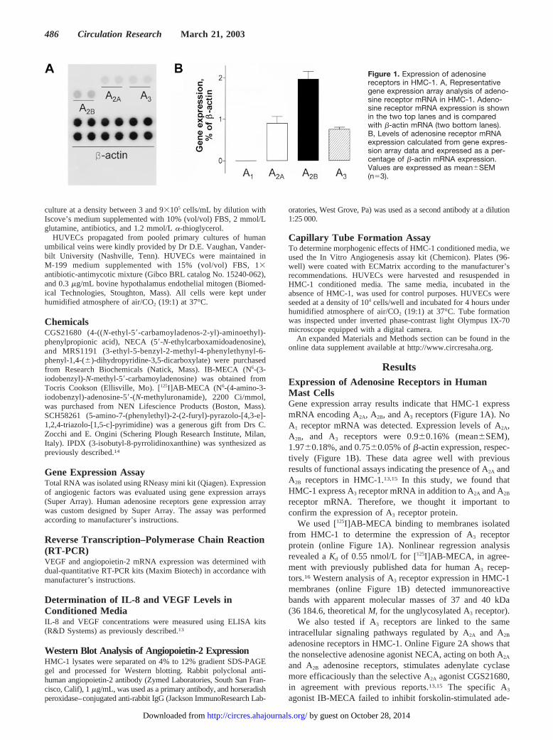
ResultsExpression of Adenosine Receptors in HumanMast CellsGene expression array results indicate that HMC-1 expressmRNA encoding A2A, A2B, and A3 receptors (Figure 1A). NoA1 receptor mRNA was detected. Expression levels of A2A,A2B, and A3 receptors were 0.9�0.16% (mean�SEM),1.97�0.18%, and 0.75�0.05% of �-actin expression, respec-tively (Figure 1B). These data agree well with previousresults of functional assays indicating the presence of A2A andA2B receptors in HMC-1.13,15 In this study, we found thatHMC-1 express A3 receptor mRNA in addition to A2A and A2B
receptor mRNA. Therefore, we thought it important toconfirm the expression of A3 receptor protein.
We used [125I]AB-MECA binding to membranes isolatedfrom HMC-1 to determine the expression of A3 receptorprotein (online Figure 1A). Nonlinear regression analysisrevealed a Kd of 0.55 nmol/L for [125I]AB-MECA, in agree-ment with previously published data for human A3 recep-tors.16 Western analysis of A3 receptor expression in HMC-1membranes (online Figure 1B) detected immunoreactivebands with apparent molecular masses of 37 and 40 kDa(36 184.6, theoretical Mr for the unglycosylated A3 receptor).
We also tested if A3 receptors are linked to the sameintracellular signaling pathways regulated by A2A and A2B
adenosine receptors in HMC-1. Online Figure 2A shows thatthe nonselective adenosine agonist NECA, acting on both A2A
and A2B adenosine receptors, stimulates adenylate cyclasemore efficaciously than the selective A2A agonist CGS21680,in agreement with previous reports.13,15 The specific A3
agonist IB-MECA failed to inhibit forskolin-stimulated ade-
Figure 1. Expression of adenosinereceptors in HMC-1. A, Representativegene expression array analysis of adeno-sine receptor mRNA in HMC-1. Adeno-sine receptor mRNA expression is shownin the two top lanes and is comparedwith �-actin mRNA (two bottom lanes).B, Levels of adenosine receptor mRNAexpression calculated from gene expres-sion array data and expressed as a per-centage of �-actin mRNA expression.Values are expressed as mean�SEM(n�3).
486 Circulation Research March 21, 2003
by guest on October 28, 2014http://circres.ahajournals.org/Downloaded from
nylate cyclase at concentrations (10�9 to 10�7 mol/L) thatselectively activate A3 receptors (online Figure 2B). There-fore, we found no evidence for functional coupling of A3
receptors to adenylate cyclase in HMC-1. Similarly, only thenonselective adenosine agonist NECA stimulated phospho-inositol hydrolysis, whereas the selective A2A agonistCGS21680 and the selective A3 agonist IB-MECA had noeffect (online Figure 3). These data confirm that only A2B
receptors are functionally coupled to stimulation of phospho-lipase C� in HMC-1.
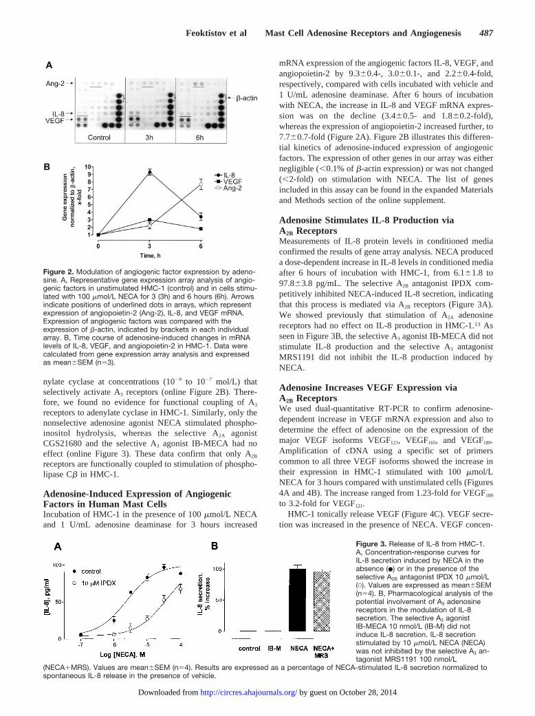
Adenosine-Induced Expression of AngiogenicFactors in Human Mast CellsIncubation of HMC-1 in the presence of 100 �mol/L NECAand 1 U/mL adenosine deaminase for 3 hours increased
mRNA expression of the angiogenic factors IL-8, VEGF, andangiopoietin-2 by 9.3�0.4-, 3.0�0.1-, and 2.2�0.4-fold,respectively, compared with cells incubated with vehicle and1 U/mL adenosine deaminase. After 6 hours of incubationwith NECA, the increase in IL-8 and VEGF mRNA expres-sion was on the decline (3.4�0.5- and 1.8�0.2-fold),whereas the expression of angiopoietin-2 increased further, to7.7�0.7-fold (Figure 2A). Figure 2B illustrates this differen-tial kinetics of adenosine-induced expression of angiogenicfactors. The expression of other genes in our array was eithernegligible (�0.1% of �-actin expression) or was not changed(�2-fold) on stimulation with NECA. The list of genesincluded in this assay can be found in the expanded Materialsand Methods section of the online supplement.
Adenosine Stimulates IL-8 Production viaA2B ReceptorsMeasurements of IL-8 protein levels in conditioned mediaconfirmed the results of gene array analysis. NECA produceda dose-dependent increase in IL-8 levels in conditioned mediaafter 6 hours of incubation with HMC-1, from 6.1�1.8 to97.8�3.8 pg/mL. The selective A2B antagonist IPDX com-petitively inhibited NECA-induced IL-8 secretion, indicatingthat this process is mediated via A2B receptors (Figure 3A).We showed previously that stimulation of A2A adenosinereceptors had no effect on IL-8 production in HMC-1.13 Asseen in Figure 3B, the selective A3 agonist IB-MECA did notstimulate IL-8 production and the selective A3 antagonistMRS1191 did not inhibit the IL-8 production induced byNECA.
Adenosine Increases VEGF Expression viaA2B ReceptorsWe used dual-quantitative RT-PCR to confirm adenosine-dependent increase in VEGF mRNA expression and also todetermine the effect of adenosine on the expression of themajor VEGF isoforms VEGF121, VEGF165, and VEGF189.Amplification of cDNA using a specific set of primerscommon to all three VEGF isoforms showed the increase intheir expression in HMC-1 stimulated with 100 �mol/LNECA for 3 hours compared with unstimulated cells (Figures4A and 4B). The increase ranged from 1.23-fold for VEGF189
to 3.2-fold for VEGF121.HMC-1 tonically release VEGF (Figure 4C). VEGF secre-
tion was increased in the presence of NECA. VEGF concen-
Figure 2. Modulation of angiogenic factor expression by adeno-sine. A, Representative gene expression array analysis of angio-genic factors in unstimulated HMC-1 (control) and in cells stimu-lated with 100 �mol/L NECA for 3 (3h) and 6 hours (6h). Arrowsindicate positions of underlined dots in arrays, which representexpression of angiopoietin-2 (Ang-2), IL-8, and VEGF mRNA.Expression of angiogenic factors was compared with theexpression of �-actin, indicated by brackets in each individualarray. B, Time course of adenosine-induced changes in mRNAlevels of IL-8, VEGF, and angiopoietin-2 in HMC-1. Data werecalculated from gene expression array analysis and expressedas mean�SEM (n�3).
Figure 3. Release of IL-8 from HMC-1.A, Concentration-response curves forIL-8 secretion induced by NECA in theabsence (�) or in the presence of theselective A2B antagonist IPDX 10 �mol/L(�). Values are expressed as mean�SEM(n�4). B, Pharmacological analysis of thepotential involvement of A3 adenosinereceptors in the modulation of IL-8secretion. The selective A3 agonistIB-MECA 10 nmol/L (IB-M) did notinduce IL-8 secretion. IL-8 secretionstimulated by 10 �mol/L NECA (NECA)was not inhibited by the selective A3 an-tagonist MRS1191 100 nmol/L
(NECA�MRS). Values are mean�SEM (n�4). Results are expressed as a percentage of NECA-stimulated IL-8 secretion normalized tospontaneous IL-8 release in the presence of vehicle.
Feoktistov et al Mast Cell Adenosine Receptors and Angiogenesis 487
by guest on October 28, 2014http://circres.ahajournals.org/Downloaded from
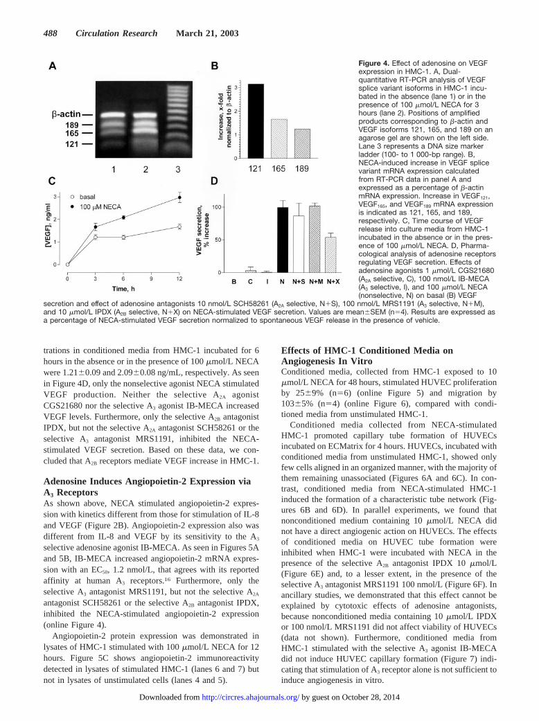
trations in conditioned media from HMC-1 incubated for 6hours in the absence or in the presence of 100 �mol/L NECAwere 1.21�0.09 and 2.09�0.08 ng/mL, respectively. As seenin Figure 4D, only the nonselective agonist NECA stimulatedVEGF production. Neither the selective A2A agonistCGS21680 nor the selective A3 agonist IB-MECA increasedVEGF levels. Furthermore, only the selective A2B antagonistIPDX, but not the selective A2A antagonist SCH58261 or theselective A3 antagonist MRS1191, inhibited the NECA-stimulated VEGF secretion. Based on these data, we con-cluded that A2B receptors mediate VEGF increase in HMC-1.
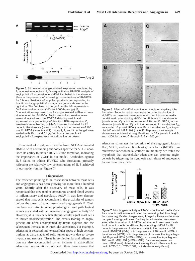
Adenosine Induces Angiopoietin-2 Expression viaA3 ReceptorsAs shown above, NECA stimulated angiopoietin-2 expres-sion with kinetics different from those for stimulation of IL-8and VEGF (Figure 2B). Angiopoietin-2 expression also wasdifferent from IL-8 and VEGF by its sensitivity to the A3
selective adenosine agonist IB-MECA. As seen in Figures 5Aand 5B, IB-MECA increased angiopoietin-2 mRNA expres-sion with an EC50, 1.2 nmol/L, that agrees with its reportedaffinity at human A3 receptors.16 Furthermore, only theselective A3 antagonist MRS1191, but not the selective A2A
antagonist SCH58261 or the selective A2B antagonist IPDX,inhibited the NECA-stimulated angiopoietin-2 expression(online Figure 4).
Angiopoietin-2 protein expression was demonstrated inlysates of HMC-1 stimulated with 100 �mol/L NECA for 12hours. Figure 5C shows angiopoietin-2 immunoreactivitydetected in lysates of stimulated HMC-1 (lanes 6 and 7) butnot in lysates of unstimulated cells (lanes 4 and 5).
Effects of HMC-1 Conditioned Media onAngiogenesis In VitroConditioned media, collected from HMC-1 exposed to 10�mol/L NECA for 48 hours, stimulated HUVEC proliferationby 25�9% (n�6) (online Figure 5) and migration by103�5% (n�4) (online Figure 6), compared with condi-tioned media from unstimulated HMC-1.
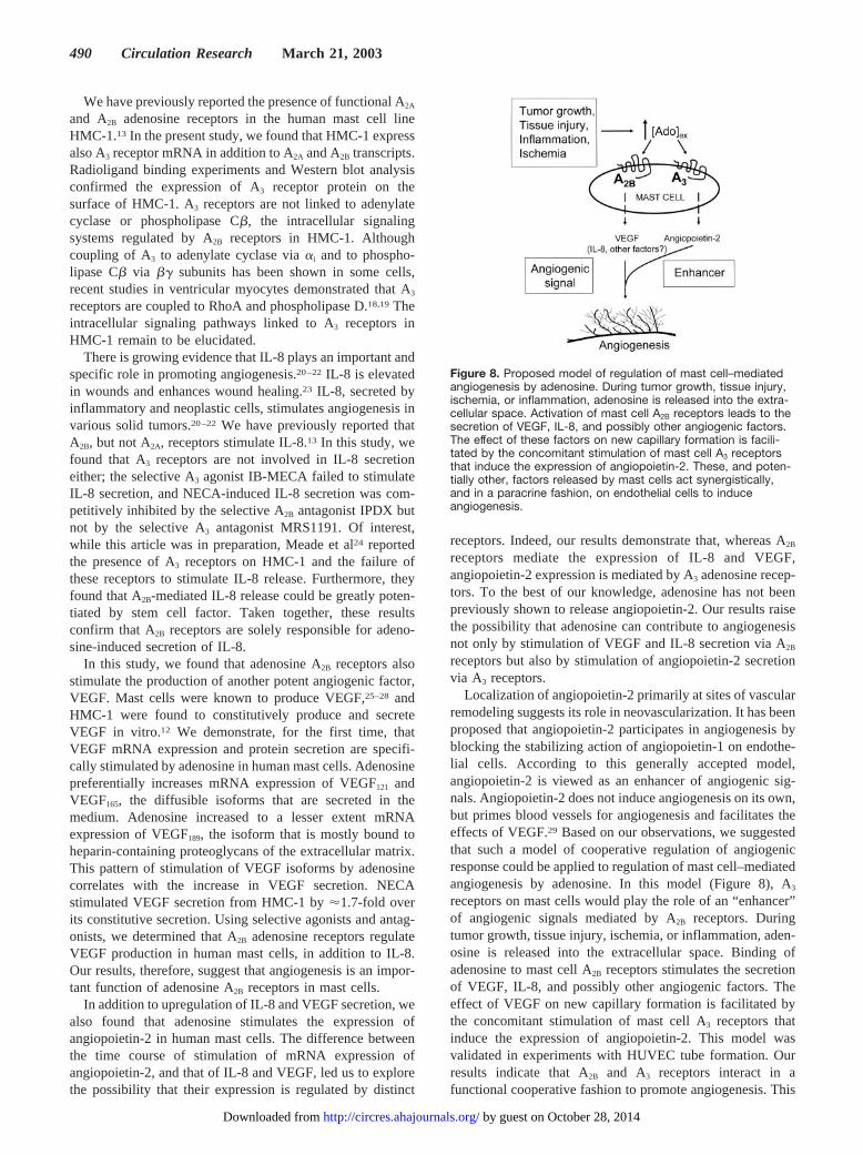
Conditioned media collected from NECA-stimulatedHMC-1 promoted capillary tube formation of HUVECsincubated on ECMatrix for 4 hours. HUVECs, incubated withconditioned media from unstimulated HMC-1, showed onlyfew cells aligned in an organized manner, with the majority ofthem remaining unassociated (Figures 6A and 6C). In con-trast, conditioned media from NECA-stimulated HMC-1induced the formation of a characteristic tube network (Fig-ures 6B and 6D). In parallel experiments, we found thatnonconditioned medium containing 10 �mol/L NECA didnot have a direct angiogenic action on HUVECs. The effectsof conditioned media on HUVEC tube formation wereinhibited when HMC-1 were incubated with NECA in thepresence of the selective A2B antagonist IPDX 10 �mol/L(Figure 6E) and, to a lesser extent, in the presence of theselective A3 antagonist MRS1191 100 nmol/L (Figure 6F). Inancillary studies, we demonstrated that this effect cannot beexplained by cytotoxic effects of adenosine antagonists,because nonconditioned media containing 10 �mol/L IPDXor 100 nmol/L MRS1191 did not affect viability of HUVECs(data not shown). Furthermore, conditioned media fromHMC-1 stimulated with the selective A3 agonist IB-MECAdid not induce HUVEC capillary formation (Figure 7) indi-cating that stimulation of A3 receptor alone is not sufficient toinduce angiogenesis in vitro.
Figure 4. Effect of adenosine on VEGFexpression in HMC-1. A, Dual-quantitative RT-PCR analysis of VEGFsplice variant isoforms in HMC-1 incu-bated in the absence (lane 1) or in thepresence of 100 �mol/L NECA for 3hours (lane 2). Positions of amplifiedproducts corresponding to �-actin andVEGF isoforms 121, 165, and 189 on anagarose gel are shown on the left side.Lane 3 represents a DNA size markerladder (100- to 1 000-bp range). B,NECA-induced increase in VEGF splicevariant mRNA expression calculatedfrom RT-PCR data in panel A andexpressed as a percentage of �-actinmRNA expression. Increase in VEGF121,VEGF165, and VEGF189 mRNA expressionis indicated as 121, 165, and 189,respectively. C, Time course of VEGFrelease into culture media from HMC-1incubated in the absence or in the pres-ence of 100 �mol/L NECA. D, Pharma-cological analysis of adenosine receptorsregulating VEGF secretion. Effects ofadenosine agonists 1 �mol/L CGS21680(A2A selective, C), 100 nmol/L IB-MECA(A3 selective, I), and 100 �mol/L NECA(nonselective, N) on basal (B) VEGF
secretion and effect of adenosine antagonists 10 nmol/L SCH58261 (A2A selective, N�S), 100 nmol/L MRS1191 (A3 selective, N�M),and 10 �mol/L IPDX (A2B selective, N�X) on NECA-stimulated VEGF secretion. Values are mean�SEM (n�4). Results are expressed asa percentage of NECA-stimulated VEGF secretion normalized to spontaneous VEGF release in the presence of vehicle.
488 Circulation Research March 21, 2003
by guest on October 28, 2014http://circres.ahajournals.org/Downloaded from
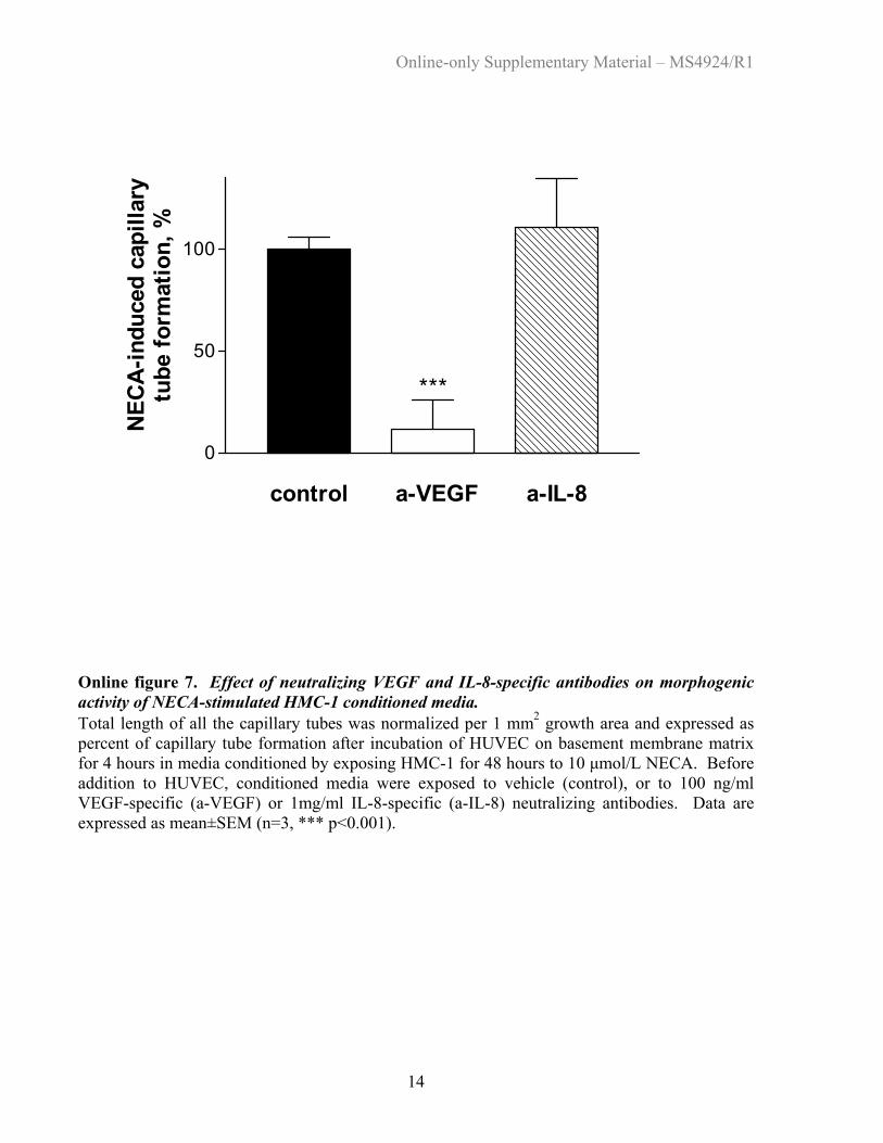
Treatment of conditioned media from NECA-stimulatedHMC-1 with neutralizing antibodies specific for VEGF abol-ished its ability to induce HUVEC tube formation, indicatingthe importance of VGEF in our model. Antibodies againstIL-8 failed to inhibit HUVEC tube formation, probablyreflecting the relatively low concentrations of IL-8 achievedin our model (online Figure 7).
DiscussionThe evidence pointing to an association between mast cellsand angiogenesis has been growing for more than a hundredyears. Shortly after the discovery of mast cells, it wasrecognized that they tend to concentrate around blood vesselsin inflammatory and neoplastic foci.17 It was later demon-strated that mast cells accumulate in the proximity of tumorsbefore the onset of tumor-associated angiogenesis.10 Theirnumbers also rise in other physiological and pathologicalevents associated with an increase in angiogenic activity.5,6,8
However, it is unclear which stimuli would signal mast cellsto induce neovascularization. The events leading to angio-genesis are often accompanied by tissue hypoxia and thesubsequent increase in extracellular adenosine. For example,adenosine is released into extracellular space at high concen-trations at early stages of solid tumor growth due to severehypoxia and necrosis. Tissue injury, ischemia, and inflamma-tion are also accompanied by an increase in extracellularadenosine concentrations. We and others have shown that
adenosine stimulates the secretion of the angiogenic factorsIL-8, VEGF, and basic fibroblast growth factor (bFGF) frommicrovascular endothelial cells.1–4 In this study, we tested thehypothesis that extracellular adenosine can promote angio-genesis by triggering the synthesis and release of angiogenicfactors from mast cells.
Figure 5. Stimulation of angiopoietin-2 expression mediated byA3 adenosine receptors. A, Dual-quantitative RT-PCR analysis ofangiopoietin-2 expression in HMC-1 incubated in the absence(0) or in the presence of increasing concentrations of IB-MECAfor 6 hours. Positions of amplified products corresponding to�-actin and angiopoietin-2 on agarose gel are shown on theright side. The first lane on the gel from the left represents aDNA size marker ladder (100- to 1 000-bp range). B,Concentration-response curve for angiopoietin-2 mRNA expres-sion induced by IB-MECA. Angiopoietin-2 expression levelswere calculated from the RT-PCR data in panel A andexpressed as a percentage of �-actin mRNA expression. C,Western immunoblotting of HMC-1 lysates incubated for 12hours in the absence (lanes 4 and 5) or in the presence of 100�mol/L NECA (lanes 6 and 7). Lanes 1, 2, and 3 on the gel wereloaded with 10, 1, and 0.1 �g/mL human recombinantangiopoietin-2, respectively, for calibration purposes.
Figure 6. Effect of HMC-1 conditioned media on capillary tubeformation. Tube formation was inspected after incubation ofHUVECs on basement membrane matrix for 4 hours in mediaconditioned by incubating HMC-1 for 48 hours in the absence(panels A and C) or in the presence of 10 �mol/L NECA, in theabsence (panels B and D) or in the presence of the selective A2B
antagonist 10 �mol/L IPDX (panel E) or the selective A3 antago-nist 100 nmol/L MRS1191 (panel F). Representative imagesshown were obtained at magnifications �40 for panels A and B,and �200 for panels C through F. Bar�200 �m.
Figure 7. Morphogenic activity of HMC-1 conditioned media. Cap-illary tube formation was estimated by measuring their total lengthfrom low-magnification images using ImageJ software and normal-ized per 1 mm2 growth area. Capillary tube formation was mea-sured after incubation of HUVECs on basement membrane matrixfor 4 hours in media conditioned by incubating HMC-1 for 48hours in the presence of vehicle (control), in the presence of 10nmol/L IB-MECA (IB-M) or in the presence of 10 �mol/L NECA, inthe absence (NECA) or in the presence of the selective A2B antago-nist 10 �mol/L IPDX (NECA�IPDX), or the selective A3 antagonist100 nmol/L MRS1191 (NECA�MRS). Data are expressed asmean�SEM (n�4). Asterisks indicate significant differences fromcontrol (**P�0.01; ***P�0.001; ns indicates nonsignificant).
Feoktistov et al Mast Cell Adenosine Receptors and Angiogenesis 489
by guest on October 28, 2014http://circres.ahajournals.org/Downloaded from
We have previously reported the presence of functional A2A
and A2B adenosine receptors in the human mast cell lineHMC-1.13 In the present study, we found that HMC-1 expressalso A3 receptor mRNA in addition to A2A and A2B transcripts.Radioligand binding experiments and Western blot analysisconfirmed the expression of A3 receptor protein on thesurface of HMC-1. A3 receptors are not linked to adenylatecyclase or phospholipase C�, the intracellular signalingsystems regulated by A2B receptors in HMC-1. Althoughcoupling of A3 to adenylate cyclase via �i and to phospho-lipase C� via �� subunits has been shown in some cells,recent studies in ventricular myocytes demonstrated that A3
receptors are coupled to RhoA and phospholipase D.18,19 Theintracellular signaling pathways linked to A3 receptors inHMC-1 remain to be elucidated.
There is growing evidence that IL-8 plays an important andspecific role in promoting angiogenesis.20–22 IL-8 is elevatedin wounds and enhances wound healing.23 IL-8, secreted byinflammatory and neoplastic cells, stimulates angiogenesis invarious solid tumors.20–22 We have previously reported thatA2B, but not A2A, receptors stimulate IL-8.13 In this study, wefound that A3 receptors are not involved in IL-8 secretioneither; the selective A3 agonist IB-MECA failed to stimulateIL-8 secretion, and NECA-induced IL-8 secretion was com-petitively inhibited by the selective A2B antagonist IPDX butnot by the selective A3 antagonist MRS1191. Of interest,while this article was in preparation, Meade et al24 reportedthe presence of A3 receptors on HMC-1 and the failure ofthese receptors to stimulate IL-8 release. Furthermore, theyfound that A2B-mediated IL-8 release could be greatly poten-tiated by stem cell factor. Taken together, these resultsconfirm that A2B receptors are solely responsible for adeno-sine-induced secretion of IL-8.
In this study, we found that adenosine A2B receptors alsostimulate the production of another potent angiogenic factor,VEGF. Mast cells were known to produce VEGF,25–28 andHMC-1 were found to constitutively produce and secreteVEGF in vitro.12 We demonstrate, for the first time, thatVEGF mRNA expression and protein secretion are specifi-cally stimulated by adenosine in human mast cells. Adenosinepreferentially increases mRNA expression of VEGF121 andVEGF165, the diffusible isoforms that are secreted in themedium. Adenosine increased to a lesser extent mRNAexpression of VEGF189, the isoform that is mostly bound toheparin-containing proteoglycans of the extracellular matrix.This pattern of stimulation of VEGF isoforms by adenosinecorrelates with the increase in VEGF secretion. NECAstimulated VEGF secretion from HMC-1 by �1.7-fold overits constitutive secretion. Using selective agonists and antag-onists, we determined that A2B adenosine receptors regulateVEGF production in human mast cells, in addition to IL-8.Our results, therefore, suggest that angiogenesis is an impor-tant function of adenosine A2B receptors in mast cells.
In addition to upregulation of IL-8 and VEGF secretion, wealso found that adenosine stimulates the expression ofangiopoietin-2 in human mast cells. The difference betweenthe time course of stimulation of mRNA expression ofangiopoietin-2, and that of IL-8 and VEGF, led us to explorethe possibility that their expression is regulated by distinct
receptors. Indeed, our results demonstrate that, whereas A2B
receptors mediate the expression of IL-8 and VEGF,angiopoietin-2 expression is mediated by A3 adenosine recep-tors. To the best of our knowledge, adenosine has not beenpreviously shown to release angiopoietin-2. Our results raisethe possibility that adenosine can contribute to angiogenesisnot only by stimulation of VEGF and IL-8 secretion via A2B
receptors but also by stimulation of angiopoietin-2 secretionvia A3 receptors.
Localization of angiopoietin-2 primarily at sites of vascularremodeling suggests its role in neovascularization. It has beenproposed that angiopoietin-2 participates in angiogenesis byblocking the stabilizing action of angiopoietin-1 on endothe-lial cells. According to this generally accepted model,angiopoietin-2 is viewed as an enhancer of angiogenic sig-nals. Angiopoietin-2 does not induce angiogenesis on its own,but primes blood vessels for angiogenesis and facilitates theeffects of VEGF.29 Based on our observations, we suggestedthat such a model of cooperative regulation of angiogenicresponse could be applied to regulation of mast cell–mediatedangiogenesis by adenosine. In this model (Figure 8), A3
receptors on mast cells would play the role of an “enhancer”of angiogenic signals mediated by A2B receptors. Duringtumor growth, tissue injury, ischemia, or inflammation, aden-osine is released into the extracellular space. Binding ofadenosine to mast cell A2B receptors stimulates the secretionof VEGF, IL-8, and possibly other angiogenic factors. Theeffect of VEGF on new capillary formation is facilitated bythe concomitant stimulation of mast cell A3 receptors thatinduce the expression of angiopoietin-2. This model wasvalidated in experiments with HUVEC tube formation. Ourresults indicate that A2B and A3 receptors interact in afunctional cooperative fashion to promote angiogenesis. This
Figure 8. Proposed model of regulation of mast cell–mediatedangiogenesis by adenosine. During tumor growth, tissue injury,ischemia, or inflammation, adenosine is released into the extra-cellular space. Activation of mast cell A2B receptors leads to thesecretion of VEGF, IL-8, and possibly other angiogenic factors.The effect of these factors on new capillary formation is facili-tated by the concomitant stimulation of mast cell A3 receptorsthat induce the expression of angiopoietin-2. These, and poten-tially other, factors released by mast cells act synergistically,and in a paracrine fashion, on endothelial cells to induceangiogenesis.
490 Circulation Research March 21, 2003
by guest on October 28, 2014http://circres.ahajournals.org/Downloaded from
conclusion is based on the observation that tube formationwas substantially greater when human endothelial cells wereexposed to conditioned media from human mast cells if bothA2B and A3 receptors were simultaneously activated comparedwith the selective activation of A2B receptor alone. Suchcooperation between adenosine receptors is a novel finding.Adenosine A2B receptors appear to be ubiquitously expressed,but in virtually every cell they are coexpressed with otheradenosine receptor subtypes. The functional relevance of thiscoexpression had not previously been investigated. There iscurrently no evidence that a similar cooperation exists be-tween A2B and A3 receptors in the modulation of mastcell–mediated inflammatory processes, but such a possibilityshould be explored.
It should be noted that our experiments were done in anestablished mast cell line HMC-1 with phenotypic character-istics resembling MCT-type human mast cells.30 Mast cellsare heterogeneous, and not all mast cell lines may express thesame adenosine receptor subtypes as HMC-1. Similarly, wechose HUVECs as a model to study paracrine regulation ofangiogenesis because these cells do not secrete VEGF, IL-8,or angiopoietin-2 in response to adenosine.4 However, endo-thelial cells are also heterogeneous. We have previouslyshown that A2B receptors can directly induce release of VEGFand IL-8 from microvascular endothelial cells.2,4 It is possiblethat in some vascular beds adenosine provides autocrinestimulation of angiogenesis by acting directly on endothelialA2B receptors, in addition to paracrine modulation mediatedby mast cell A2B and A3 receptors. Finally, we cannot excludethat, in addition to VEGF, IL-8, and angiopoietin-2, there arealso other factors involved in the angiogenic response in-duced by adenosine. For example, mast cells were shown tostimulate angiogenesis by releasing tryptase, chymase, andmatrix metalloproteinase-9 (MMP-9).31–33 In this study, wescreened only a limited number of angiogenesis-related genesin human mast cells. Nonetheless, our findings that adenosinestimulates secretion of such angiogenic factors as VEGF andIL-8 via A2B receptors, and angiopoietin-2 via A3 receptors,demonstrate for the first time an example of functionalcooperation between different adenosine receptor subtypes inpromoting an angiogenic response.
In summary, we have shown the presence of A2B and A3
receptors in human mast cells. Activation of A2B receptorsleads to the secretion of VEGF and IL-8, whereas activationof A3 receptors upregulates the expression of angiopoietin-2.These, and potentially other factors released by mast cells,can act synergistically, and in a paracrine fashion, on endo-thelial cells to induce angiogenesis.
AcknowledgmentsThis work was supported by NIH grants R29 HL55596 and R01HL70073 (to I.F.) and R01 HL67232 (to I.B.).
References1. Fischer S, Sharma HS, Karliczek GF, Schaper W. Expression of vascular
permeability factor/vascular endothelial growth factor in pig cerebralmicrovascular endothelial cells and its upregulation by adenosine. BrainRes. 1995;28:141–148.
2. Grant MB, Tarnuzzer RW, Caballero S, Ozeck MJ, Davis MI, Spoerri PE,Feoktistov I, Biaggioni I, Shryock JC, Belardinelli L. Adenosine receptor
activation induces vascular endothelial growth factor in human retinalendothelial cells. Circ Res. 1999;85:699–706.
3. Grant MB, Davis MI, Caballero S, Feoktistov I, Biaggioni I, BelardinelliL. Proliferation, migration, and ERK activation in human retinal endo-thelial cells through A2B adenosine receptor stimulation. Invest Oph-thalmol Vis Sci. 2001;42:2068–2073.
4. Feoktistov I, Goldstein AE, Ryzhov S, Zeng D, Belardinelli L, Voyno-Yasenetskaya T, Biaggioni I. Differential expression of adenosinereceptors in human endothelial cells: role of A2B receptors in angiogenicfactor regulation. Circ Res. 2002;90:531–538.
5. Atkinson JB, Harlan CW, Harlan GC, Virmani R. The association of mastcells and atherosclerosis: a morphologic study of early atheroscleroticlesions in young people. Hum Pathol. 1994;25:154–159.
6. Brightling CE, Bradding P, Symon FA, Holgate ST, Wardlaw AJ, PavordID. Mast-cell infiltration of airway smooth muscle in asthma. N EnglJ Med. 2002;346:1699–1705.
7. Schubert C, Christophers E. Mast cells and macrophages in earlyrelapsing psoriasis. Arch Dermatol Res. 1985;277:352–358.
8. Trautmann A, Toksoy A, Engelhardt E, Brocker EB, Gillitzer R. Mast cellinvolvement in normal human skin wound healing: expression ofmonocyte chemoattractant protein-1 is correlated with recruitment ofmast cells which synthesize interleukin-4 in vivo. J Pathol. 2000;190:100–106.
9. Ribatti D, Vacca A, Nico B, Crivellato E, Roncali L, Dammacco F. Therole of mast cells in tumour angiogenesis. Br J Haematol. 2001;115:514–521.
10. Dabbous MK, Walker R, Haney L, Carter LM, Nicolson GL, WoolleyDE. Mast cells and matrix degradation at sites of tumour invasion in ratmammary adenocarcinoma. Br J Cancer. 1986;54:459–465.
11. Moller A, Lippert U, Lessmann D, Kolde G, Hamann K, Welker P,Schadendorf D, Rosenbach T, Luger T, Czarnetzki BM. Human mastcells produce IL-8. J Immunol. 1993;151:3261–3266.
12. Grutzkau A, Kruger-Krasagakes S, Baumeister H, Schwarz C, Kogel H,Welker P, Lippert U, Henz BM, Moller A. Synthesis, storage, and releaseof vascular endothelial growth factor/vascular permeability factor(VEGF/VPF) by human mast cells: implications for the biological sig-nificance of VEGF. Mol Biol Cell. 1998;206:9:875–884.
13. Feoktistov I, Biaggioni I. Adenosine A2b receptors evoke interleukin-8secretion in human mast cells: an enprofylline-sensitive mechanism withimplications for asthma. J Clin Invest. 1995;96:1979–1986.
14. Feoktistov I, Garland E, Goldstein AE, Zeng D, Belardinelli L, Wells JN,Biaggioni I. Inhibition of human mast cell activation with the novelselective adenosine A2B receptor antagonist 3-isobutyl-8-pyrrolidin-oxanthine (IPDX). Biochem Pharmacol. 2001;62:1163–1173.
15. Feoktistov I, Biaggioni I. Pharmacological characterization of adenosineA2B receptors: studies in human mast cells co-expressing A2A and A2B
adenosine receptor subtypes. Biochem Pharmacol. 1998;55:627–633.16. Klotz KN, Hessling J, Hegler J, Owman C, Kull B, Fredholm BB, Lohse
MJ. Comparative pharmacology of human adenosine receptor subtypes:characterization of stably transfected receptors in CHO cells. NaunynSchmiedebergs Arch Pharmacol. 1998;357:1–9.
17. Selye H. The Mast Cells. Washington, DC: Butterworth Publications;1965.
18. Parsons M, Young L, Lee JE, Jacobson KA, Liang BT. Distinct cardio-protective effects of adenosine mediated by differential coupling ofreceptor subtypes to phospholipases C and D. FASEB J. 2000;14:1423–1431.
19. Lee JE, Bokoch G, Liang BT. A novel cardioprotective role of RhoA:new signaling mechanism for adenosine. FASEB J. 2001;15:1886–1894.
20. Rofstad EK, Halsor EF. Vascular endothelial growth factor, interleukin-8,platelet-derived endothelial cell growth factor, and basic fibroblastgrowth factor promote angiogenesis and metastasis in human melanomaxenografts. Cancer Res. 2000;60:4932–4938.
21. Westphal JR, Van’t Hullenaar R, Peek R, Willems RW, Crickard K,Crickard U, Askaa J, Clemmensen I, Ruiter DJ, De Waal RM. Angiogenicbalance in human melanoma: expression of VEGF, bFGF, IL-8, PDGFand angiostatin in relation to vascular density of xenografts in vivo. Int JCancer. 2000;86:768–776.
22. Inoue K, Slaton JW, Eve BY, Kim SJ, Perrotte P, Balbay MD, Yano S,Bar-Eli M, Radinsky R, Pettaway CA, Dinney CP. Interleukin-8expression regulates tumorigenicity and metastases in androgen-independent prostate cancer. Clin Cancer Res. 2000;6:2104–2119.
23. Rennekampff HO, Hansbrough JF, Kiessig V, Dore C, Sticherling M,Schroder JM. Bioactive interleukin-8 is expressed in wounds andenhances wound healing. J Surg Res. 2000;93:41–54.
Feoktistov et al Mast Cell Adenosine Receptors and Angiogenesis 491
by guest on October 28, 2014http://circres.ahajournals.org/Downloaded from
24. Meade CJ, Worrall L, Hayes D, Protin U. Induction of interleukin-8release from the HMC-1 mast cell line: synergy between stem cell factorand activators of the adenosine A2b receptor. Biochem Pharmacol. 2002;64:317–325.
25. Boesiger J, Tsai M, Maurer M, Yamaguchi M, Brown LF, Claffey KP,Dvorak HF, Galli SJ. Mast cells can secrete vascular permeability factor/vascular endothelial cell growth factor and exhibit enhanced release afterimmunoglobulin E-dependent upregulation of Fc�R1 expression. J ExpMed. 1998;188:1135–1145.
26. Sawatsubashi M, Yamada T, Fukushima N, Mizokami H, Tokunaga O,Shin T. Association of vascular endothelial growth factor and mast cellswith angiogenesis in laryngeal squamous cell carcinoma. Virchows Arch.2000;436:243–248.
27. Toth-Jakatics R, Jimi S, Takebayashi S, Kawamoto N. Cutaneousmalignant melanoma: correlation between neovascularization andperitumor accumulation of mast cells overexpressing vascular endothelialgrowth factor. Hum Pathol. 2000;31:955–960.
28. Fan L, Iseki S. Immunohistochemical localization of vascular endothelialgrowth factor in the globule leukocyte/mucosal mast cell of the ratrespiratory and digestive tracts. Histochem Cell Biol. 1999;111:13–21.
29. Holash J, Maisonpierre PC, Compton D, Boland P, Alexander CR,Zagzag D, Yancopoulos GD, Wiegand SJ. Vessel cooption, regression,and growth in tumors mediated by angiopoietins and VEGF. Science.1999;284:1994–1998.
30. Nilsson G, Blom T, Kusche-Gullberg M, Kjellen I, Butterfield JH,Sundstrom C, Nilsson K, Hellman L. Phenotypic characterization ofthe human mast-cell line HMC-1. Scand J Immunol. 1994;39:489 – 498.
31. Blair RJ, Meng H, Marchese MJ, Ren S, Schwartz LB, Tonnesen MG,Gruber BL. Human mast cells stimulate vascular tube formation:tryptase is a novel, potent angiogenic factor. J Clin Invest. 1997;99:2691–2700.
32. Coussens LM, Raymond WW, Bergers G, Laig-Webster M, BehrendtsenO, Werb Z, Caughey GH, Hanahan D. Inflammatory mast cellsup-regulate angiogenesis during squamous epithelial carcinogenesis.Genes Dev. 1999;13:1382–1397.
33. Coussens LM, Tinkle CL, Hanahan D, Werb Z. MMP-9 supplied by bonemarrow-derived cells contributes to skin carcinogenesis. Cell. 2000;103:481–490.
492 Circulation Research March 21, 2003
by guest on October 28, 2014http://circres.ahajournals.org/Downloaded from

Online-only Supplementary Material – MS4924/R1
8
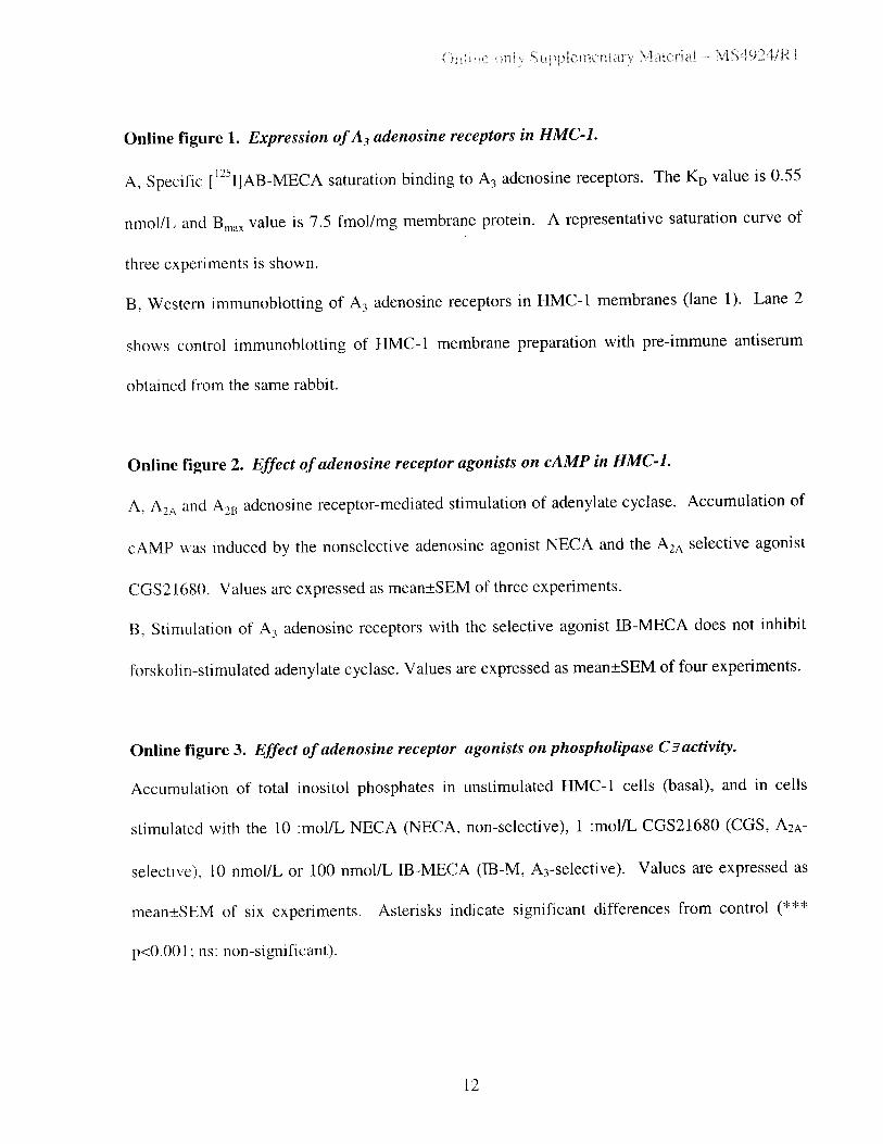
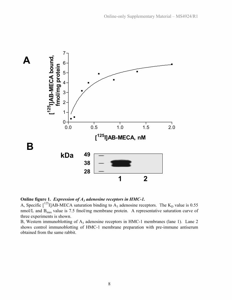
Online figure 1. Expression of A3 adenosine receptors in HMC-1. A, Specific [125I]AB-MECA saturation binding to A3 adenosine receptors. The KD value is 0.55 nmol/L and Bmax value is 7.5 fmol/mg membrane protein. A representative saturation curve of three experiments is shown. B, Western immunoblotting of A3 adenosine receptors in HMC-1 membranes (lane 1). Lane 2 shows control immunoblotting of HMC-1 membrane preparation with pre-immune antiserum obtained from the same rabbit. Back to List of Figures
0.0 0.5 1.0 1.5 2.00
1
2
3
4
5
6
7
[125I]AB-MECA, nM
[125 I]A
B-M
ECA
boun
d,fm
ol/m
g pr
otei
nA
1 2
B493828
kDa
Online-only Supplementary Material – MS4924/R1
9
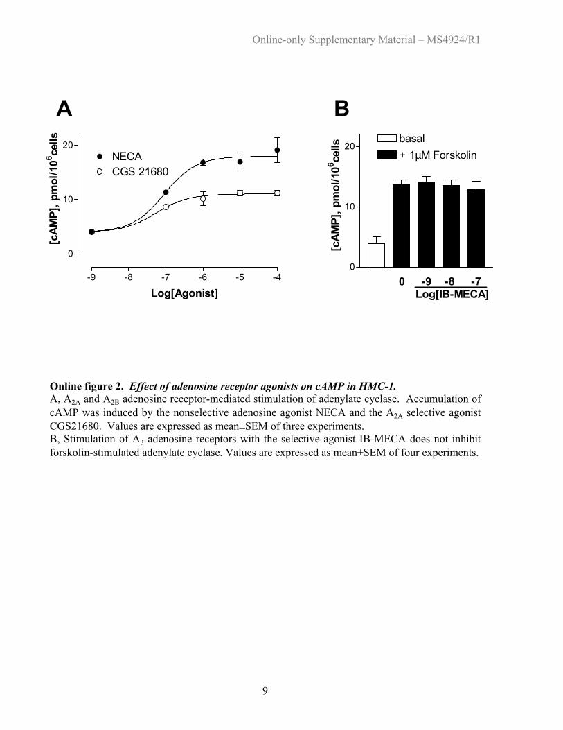
Online figure 2. Effect of adenosine receptor agonists on cAMP in HMC-1. A, A2A and A2B adenosine receptor-mediated stimulation of adenylate cyclase. Accumulation of cAMP was induced by the nonselective adenosine agonist NECA and the A2A selective agonist CGS21680. Values are expressed as mean±SEM of three experiments. B, Stimulation of A3 adenosine receptors with the selective agonist IB-MECA does not inhibit forskolin-stimulated adenylate cyclase. Values are expressed as mean±SEM of four experiments. Back to List of Figures
-9 -8 -7 -6 -5 -4
0
10
20NECACGS 21680
Log[Agonist]
[cAM
P], p
mol
/106 ce
lls
A
0
10
20basal+ 1µM Forskolin
[cAM
P], p
mol
/106 ce
lls
0 -9 -8 -7 Log[IB-MECA]
B

Online-only Supplementary Material – MS4924/R1
10
Online figure 3. Effect of adenosine receptor agonists on phospholipase C$ activity. Accumulation of total inositol phosphates in unstimulated HMC-1 cells (basal), and in cells stimulated with the 10 :mol/L NECA (NECA, non-selective), 1 :mol/L CGS21680 (CGS, A2A-selective), 10 nmol/L or 100 nmol/L IB-MECA (IB-M, A3-selective). Values are expressed as mean±SEM of six experiments. Asterisks indicate significant differences from control (*** p<0.001; ns: non-significant). Back to List of Figures
0
2000
4000
6000
8000
basal NECA CGS IB-M IB-M 10nM 100nM
IP a
ccum
ulat
ion,
cpm
***
ns ns
Online-only Supplementary Material – MS4924/R1
11
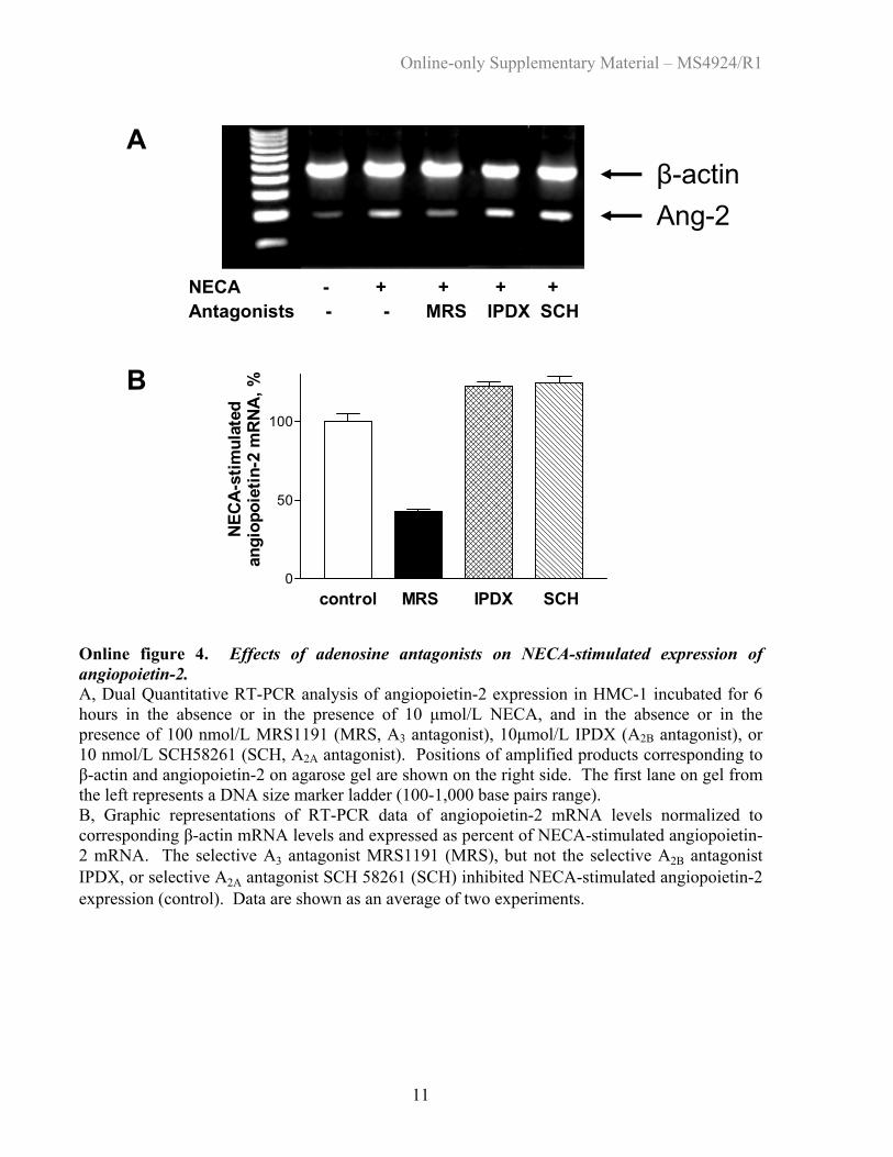
Online figure 4. Effects of adenosine antagonists on NECA-stimulated expression of angiopoietin-2. A, Dual Quantitative RT-PCR analysis of angiopoietin-2 expression in HMC-1 incubated for 6 hours in the absence or in the presence of 10 µmol/L NECA, and in the absence or in the presence of 100 nmol/L MRS1191 (MRS, A3 antagonist), 10µmol/L IPDX (A2B antagonist), or 10 nmol/L SCH58261 (SCH, A2A antagonist). Positions of amplified products corresponding to β-actin and angiopoietin-2 on agarose gel are shown on the right side. The first lane on gel from the left represents a DNA size marker ladder (100-1,000 base pairs range). B, Graphic representations of RT-PCR data of angiopoietin-2 mRNA levels normalized to corresponding β-actin mRNA levels and expressed as percent of NECA-stimulated angiopoietin-2 mRNA. The selective A3 antagonist MRS1191 (MRS), but not the selective A2B antagonist IPDX, or selective A2A antagonist SCH 58261 (SCH) inhibited NECA-stimulated angiopoietin-2 expression (control). Data are shown as an average of two experiments. Back to List of Figures
0
50
100
control MRS IPDX SCH
NECA
-stim
ulat
edan
giop
oiet
in-2
mRN
A, %
Ang-2β-actin
A
B
NECA - + + + +Antagonists - - MRS IPDX SCH
Online-only Supplementary Material – MS4924/R1
12
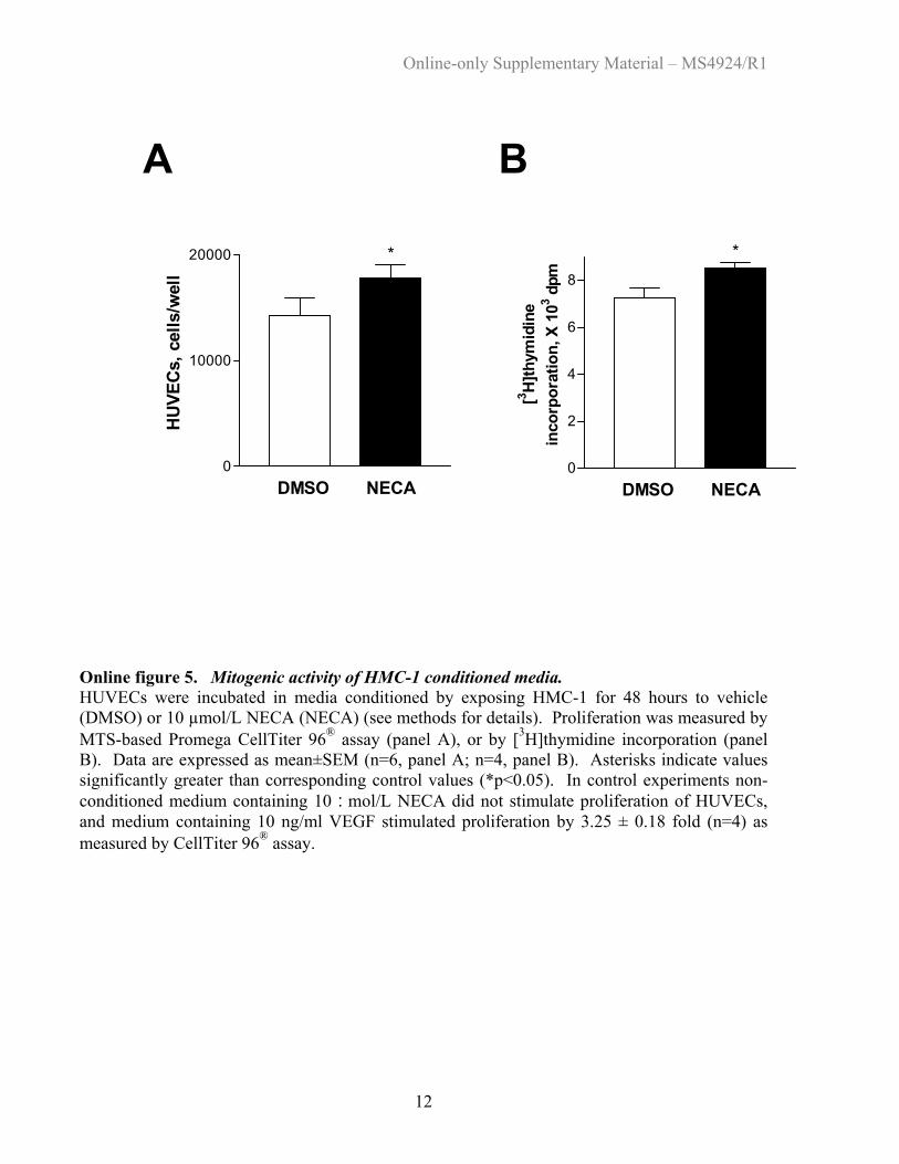
Online figure 5. Mitogenic activity of HMC-1 conditioned media. HUVECs were incubated in media conditioned by exposing HMC-1 for 48 hours to vehicle (DMSO) or 10 µmol/L NECA (NECA) (see methods for details). Proliferation was measured by MTS-based Promega CellTiter 96® assay (panel A), or by [3H]thymidine incorporation (panel B). Data are expressed as mean±SEM (n=6, panel A; n=4, panel B). Asterisks indicate values significantly greater than corresponding control values (*p<0.05). In control experiments non-conditioned medium containing 10 :mol/L NECA did not stimulate proliferation of HUVECs, and medium containing 10 ng/ml VEGF stimulated proliferation by 3.25 ± 0.18 fold (n=4) as measured by CellTiter 96® assay. Back to List of Figures
0
10000
20000
DMSO NECA
HUVE
Cs, c
ells
/wel
l
*
A B
0
2
4
6
8
DMSO NECA
[3 H]th
ymid
ine
inco
rpor
atio
n, X
103 d
pm
*

Online-only Supplementary Material – MS4924/R1
13
Online figure 6. Effect of HMC-1 conditioned media on HUVEC migration. HUVEC (105 cells) were placed in the upper chamber, and media conditioned by incubating HMC-1 for 48 hours in the presence of vehicle (DMSO) or in the presence of 10 :mol/L NECA (NECA) were placed in the lower chamber in transmembrane (8 µm pore size) cell migration assay. HUVEC that had migrated through pores were stained and counted after six hours of incubation. A, Representative images of HUVEC (dark blue) migrating through membrane pores (black dots). Bar denotes 100 :m. B, Number of HUVEC migrated through 1 mm2 of transwell membranes. Data are expressed as mean±SEM (n=4, ***p<0.001). In control experiments non-conditioned medium containing 10 :mol/L NECA did not stimulate migration of HUVEC, and medium containing 10 ng/ml VEGF stimulated migration by 3 ± 0.2 fold (n=4). Back to List of Figures
DMSO
NECA
0
100
200
DMSO NECAM
igra
tion,
cel
ls/m
m2 ***
A B
Online-only Supplementary Material – MS4924/R1
14
Online figure 7. Effect of neutralizing VEGF and IL-8-specific antibodies on morphogenic activity of NECA-stimulated HMC-1 conditioned media. Total length of all the capillary tubes was normalized per 1 mm2 growth area and expressed as percent of capillary tube formation after incubation of HUVEC on basement membrane matrix for 4 hours in media conditioned by exposing HMC-1 for 48 hours to 10 µmol/L NECA. Before addition to HUVEC, conditioned media were exposed to vehicle (control), or to 100 ng/ml VEGF-specific (a-VEGF) or 1mg/ml IL-8-specific (a-IL-8) neutralizing antibodies. Data are expressed as mean±SEM (n=3, *** p<0.001). Back to List of Figures
0
50
100
control a-VEGF a-IL-8
NECA
-indu
ced
capi
llary
tube
form
atio
n, %
***
Copyright © 2022 FDOKUMEN