Marks MK, Marden K, Mileusnic D. 2009.Forensic Osteology of Child Abuse. Chapter 16 in: Dawnie W....
16
Forensic Osteology of Child Abuse Murray K. Marks, Kerriann Marden, and Darinka Mileusnic-Polchan It was in the middle of the last century that physi- cians first formally recognized the severity and scope of child abuse as a major cause of injury and death in children. Detailed descriptions of infant abuse and associated non-accidental, repetitive trauma were first documented in those early investigations (see Altman and Smith 1960; Caffey 1946; Silverman 1953), and a suite of traumatized tissues related to this abuse was recognized as pathognomonic (dis- tinctly characteristic) of an abuse syndrome shortly thereafter (Kempe et al. 1962). Early studies of the problem estimated that as many as 20percent of chil- dren receiving medical treatment for accidents had experienced child abuse (Morse et al, 1970)and that "trauma is the most important killer and crippler of infants and children and it warrants aggressive study to insure effective, preventive and optimal diagnosis and management" (Caffey 1972:169). Despite these early warnings, however. the incidence of child abuse and neglect has increased annually in the United States (Cramer and Green 2003;Johnson 1990), with a recent estimate of more than a million cases occurring each year and approximately 2,000 children dying annually (McClain et a11993). Two- thirds of the victims who reach adulthood are emo- tionally and physically scarred and are prone to become parents who repeat the same abusive treat- ment they survived (Oliver 1993). There are six broad patterns of child abuse recog- nized in the medicolegal context: physical abuse, nutritional deprivation, sexual abuse, intentional drugging or poisoning, neglect of medical care or safety, and emotional abuse. According to Spitz (1993),physical abuse constitutes approximately 70 percent of all abuse cases. Although this skew may result from the fact that neglect and sexual abuse can be more difficult to detect than broken bones and bruises, the number of children who are physically abused is nonetheless staggering. Recent surveys estimate that the national incidence ofphysical abuse of children is nearly five children in every 1,000. Of these abused children, one in every 1,000 dies (Campbell and Schrader 2006). Moreover, the prob- lem is steadily increasing. The Third National Incidence Study of Child Abuse and Neglect reported that the total number of physically abused children nearly doubled between 1986and 1993and that the number of children who were seriously injured quadrupled during this seven year span (Department of Health and Human Services 2001). Child abuse is among the most devastating health problems facing American children today and has become a "social and medical problem of epidemic proportions" (Cramer and Green 2003:587). The past quarter century has seen a burgeoning interest in many aspects of the physical trauma of child abuse, including the biomechanics of bone breakage, wound patterning, trauma and healing of soft tissues, skeletal and dental markers of abuse, and the pathology of fatal trauma (see Reece and 205
Transcript of Marks MK, Marden K, Mileusnic D. 2009.Forensic Osteology of Child Abuse. Chapter 16 in: Dawnie W....

Forensic Osteologyof Child Abuse
Murray K. Marks, Kerriann Marden, and Darinka Mileusnic-Polchan
It was in the middle of the last century that physi-cians first formally recognized the severity and scopeof child abuse as a major cause of injury and death inchildren. Detailed descriptions of infant abuse andassociated non-accidental, repetitive trauma werefirst documented in those early investigations (seeAltman and Smith 1960; Caffey 1946; Silverman1953), and a suite of traumatized tissues related tothis abuse was recognized as pathognomonic (dis-tinctly characteristic) of an abuse syndrome shortlythereafter (Kempe et al. 1962). Early studies of theproblem estimated that as many as 20 percent of chil-dren receiving medical treatment for accidents hadexperienced child abuse (Morse et al, 1970) and that"trauma is the most important killer and crippler ofinfants and children and it warrants aggressivestudy to insure effective, preventive and optimaldiagnosis and management" (Caffey 1972:169).Despite these early warnings, however. the incidenceof child abuse and neglect has increased annually inthe United States (Cramer and Green 2003; Johnson1990), with a recent estimate of more than a millioncases occurring each year and approximately 2,000children dying annually (McClain et a11993). Two-thirds of the victims who reach adulthood are emo-tionally and physically scarred and are prone tobecome parents who repeat the same abusive treat-ment they survived (Oliver 1993).There are six broad patterns of child abuse recog-
nized in the medicolegal context: physical abuse,
nutritional deprivation, sexual abuse, intentionaldrugging or poisoning, neglect of medical care orsafety, and emotional abuse. According to Spitz(1993), physical abuse constitutes approximately 70percent of all abuse cases. Although this skew mayresult from the fact that neglect and sexual abuse canbe more difficult to detect than broken bones andbruises, the number of children who are physicallyabused is nonetheless staggering. Recent surveysestimate that the national incidence of physical abuseof children is nearly five children in every 1,000. Ofthese abused children, one in every 1,000 dies(Campbell and Schrader 2006). Moreover, the prob-lem is steadily increasing. The Third NationalIncidence Study of Child Abuse and Neglectreported that the total number of physically abusedchildren nearly doubled between 1986 and 1993 andthat the number of children who were seriouslyinjured quadrupled during this seven year span(Department of Health and Human Services 2001).Child abuse is among the most devastating healthproblems facing American children today and hasbecome a "social and medical problem of epidemicproportions" (Cramer and Green 2003:587).The past quarter century has seen a burgeoning
interest in many aspects of the physical trauma ofchild abuse, including the biomechanics of bonebreakage, wound patterning, trauma and healing ofsoft tissues, skeletal and dental markers of abuse,and the pathology of fatal trauma (see Reece and
205

206
Ludwig 2001). Although soft tissue injuriescomprise the most common and visible clinical pre-sentation in child abuse cases, up to 70 percent ofphysically abused children exhibit some degree ofosseous lesion (Green and Swiontkowski 2003; seealso Brogdon 1998 for radiological perspective). Dueto the relative plasticity of the subadult skeleton,accidental fractures in children are extremely rare(Barsness et al. 2003), so when bone fractures are dis-covered, they indicate that the child was subjectedto tremendous force. Furthermore, certain types andpatterns of fracture. are highly specific for abuse.
One in every eight fractures in children undereighteen months of age is likely to result fromabuse (Walker et al. 1997; Worlock et al. 1986), soan understanding of bone fracture mechanics andwound healing is especially important whenphysical abuse is suspected in neonates andinfants. Whereas soft tissue lacerations andbruises fade in the living through healing and inthe deceased through decomposition, the skeletalrecord serves as a permanent testimony to theinjuries suffered.
INTERPRETATION OF TAPHONOMY AND TRAUMA
Case History
In August 2002, an unresponsive three month-oldmale in cardiorespiratory arrest was brought to theemergency room and pronounced dead within fif-teen minutes of arrival. At autopsy, soft tissueobservations included multiple abrasions andbruising distributed over the entire body, as well ashealed thermal bums to the upper abdomen andback of the right hand. Widespread, patternedbruising is a hallmark of child abuse (Saukko andKnight 2004), and the added presence of cutaneous(skin) bums in this case only heightens SuspICIon ofabuse. A single anteroposterior (A-P), full-bodyradiograph revealed radiopaque irregularities inthe left femur, the right humerus, and at numeroussites on the proximal and middle rib shafts. In thepathologist's report, the "Postmortem RadiologicalEvidence of Injury" section makes numerous note-worthy references to bone fracture and healing:
Multiple old healing rib fractures are noted on thex-rays of the chest [See Figure 16.1].Readily VISIblecalluses are noted on the right lateral ribs 2
FIGURE 16.1 Two RADIOGRAPHS OF THE VICfIM PIuOR TO AUTOPSY
Note the deformity of the distal right humerus (thick arrow) on the left image, and telltale (though faint)enlarged rib portions from healing callus formation lateral to the vertebral column (several marked withthin arrows). Note in the right radiograph the displaced fracture of the left femoral midshaft withmassive callus formation (see Figure 16.3,below). Compare the size of this traumatized bone to thenormal right femur to appreciate the degree of mineralization in callus formation and the resultantenlarged outline of the left upper leg compared to the right.

FORENSIC OSTEOLOGY OF CHiLD ABUSE
through 6, right posterior ribs 7 through 10, leftposterior ribs 8 through 10 and lateral left ribs 2through 5 and 7. Due to the limitations of the x-raytechnique, more recent fractures and anterior frac-tures cannot be demonstrated. X-raysof the upperextremities reveal thickening and periosteal reac-tion with osseous callus formation of the right dis-tal humerus. X-rays of the lower extremities revealsevere dislocated fracture and large irregular cal-lus formation of the left femur.
The "Internal Evidence of Injury" section of thepathologist's report describes the bones involvedand the adjacent soft tissue hemorrhage that mayimpact bone(s):
Multiple left-sided rib fractures .... there arenoticeable recent non-healing mobile fractures ofthe lateral left ribs 2 through 5 and healing fractureswith calluses of the left posterior ribs 7 through 10.The surrounding intercostal muscles display focalhemorrhage [bleeding]. On the left superior andlateral aspect of the chest, there is a large focus ofhemorrhage with central area of early abscess for-mation, overlying the recent rib fractures. Prior toremoval of the pleura and intercostalmuscles in theright chest cavity, multiple rib fractures are noted.
207
Callus formations consistent with healing rib frac-tures are noted on the right lateral ribs 2 through 6and posterior right ribs 7 through 11.On the leftupper back, close to the midline, there are deepintramuscular hemorrhages.
Based on these findings, the ribs from both sidesof the chest, the right humerus and the left femurwere removed during autopsy for anthropologicalexamination (Figures 16.2-16.4). Adherent soft tis-sue was carefully teased away from the ribs using ascalpel, tweezers, and warm (not boiling) watersubmersion. It is important to keep in mind thatmany parts of fetal, neonatal, and infant bone arecomprised of woven bone in an active stage ofgrowth. When damaged by perimortem violence, itcan be difficult to distinguish the fragile, un-unitedpieces of an early-stage fracture callus in the imma-ture skeleton, and unless handled with the utmostcare, these diagnostic fragments may go umecog-nized and be lost or discarded during the cleaningprocedure.
The questions posed by the pathologist guide theanthropologist's investigation. In this investigation,
FIGURE 16.2 AUTOPSY IMAGE OF THE INTERNAL Asrscr OF DORSAL THORACIC CAVITYAfTER REMOVAL OF CARDIa-RESPIRATORY VISCERA
N tus antemortem chronic fracture sites, which appear as distinct nodules bilaterally along
o e numero h . deoi h . . f h . .th t b I column (marked with arrows). Rig t Image eptcts erntsecnon 0 t oracic cavity
ever e ra . ( hi k ) d . dshowing both antemortem chroniC fracture sites ~ IC arrows an a penmortem. or acute woun
d.i I· f . t the two antemortem wounds (thin arrows). Also note the traumaltzed sterno-costal
rect y in error 0
junctions.

208
FIGURE 16.3 [MAGE Of A LONGrruDINAL
SECTION THROUGH AN ANTEMORTEM
CHRONIC RIB FRACTURE
The normal appearance of the sterno-costaljunctionisat the left(thin arrow)whilethehealedrib fracture is to the right (thick arrow), showingnon-union of the fractured ends but partialmineralized callus surrounding the fracture site.
victim identification and time since death were notfocal questions for the anthropologist, as these factswere already established. Rather, the pathologistenlisted the forensic anthropologist to assess thetime since insult for the various fractures. Since thewounds were not all in the same stage of healing, itwas necessary to assess relative healing of theinjuries over time to establish a history of abuse for
INTERPRETATION OF TAPHONOMY AND TRAUMA
the legal investigation-details which may exoner-ate or implicate one of the adult caregivers or asso-ciates. Here, the forensic anthropologist wascharged with identifying and sequencing the skele-tal trauma and determining the biomechanics of thefractures: essentially, which bones were broken,when, and how. Distinguishing repetitive ante-mortem breakage, traditionally termed "chronic"by the forensic pathologist, from perimortem break-age, (termed "acute") poses larger questions in theinvestigation that may help create a timeline of thehistory of abuse.
The Role of Forensic Anthropology
Among the most important contributions offorensic anthropology to medicolegal investiga-tions is the accurate identification of the signa-tures of violence and the discernmen t of tra umafrom naturally occurring postmortem changes inskeletal tissue (Ubelaker 1995).Such identificationhelps establish the pattern and timing of injuriescritical in diagnosing fatal child abuse, which may
FIGURE 16.4 Two ANTERIOR I'~AGES (LEFf AND CENTER) AND ONE POSTERIOR IMAGE (RIGHT)OF THE HEAliNG FRACTURE CALLUS OF THE VICTIM'S LEFT FEMUR
Compare these "three-dimensional" views to the radiographic perspective of the bone in situ in Figure 16.1. Theleft image was taken soon after resection from the victim and prior to final cleaning of soft tissue. Note that thecartilaginous epiphyses have been removed in the center and right images.

FORENSIC OSTEOLOGY OF CHILD ABUSE
aid in effective prosecution and sentencing of theabuser(s) for the murder of the child and also cansubstantiate the authorities' decision to removeother children from the home. It is a regrettablefact that in cases of fatal child abuse, the offenderwill often dispose of the body and simply reportthe child missing, effectively covering the crimeand diverting suspicion to an unknown assailant(Walker et a1. 1997). By the time the body of thechild is recovered, soft tissue evidence of thefatal trauma-such as bruising and internalhemorrhage-may be obscured, reduced, orabsent, and often only skeletal indicators remain.The correct identification of the skeletal signatureof abuse is therefore critical in estimating mannerof death in the recovered remains of abused chil-dren. A qualified anthropologist offers "uniqueexperience in skeletal anatomy and familiaritywith the diverse factors that can affect skeletalindicators" (Chapter 17, this volume).
Forensic pathologists rarely possess the special-ized osteological experience to recognize evidenceof bone trauma on the basis of often fragmentaryelements recovered. In cases involving fleshedremains, diagnostic radiological instruments are sel-dom sensitive enough to accurately identify the fullextent of recent and healed traumatic lesions in thechild's skeleton (Barsness et a1.2003; Brogdon 1998;Catteneo et a1.2006; Islam et at. 2000; Klotzbach et al,2003; Mandelstam et a1. 2003; Ng and Hall 1998;Walker et a1.1997). Even the classic "battered childsyndrome" differs greatly when observed directlyon skeletal remains as opposed to radiographicimaging of the fleshed body (White 2000). However,physical anthropologists' specialized understandingof bone anatomy, of the gross appearance andhistological properties of healing bone, and of thebiomechanics of bone fracture can greatly aid thepathologist in interpreting skeletal evidence oftrauma and healing. Subtle periosteal and sub-perisoteallesions that are radiographically invisiblecan be readily detected, macroscopically and/ormicroscopically, by an osteologist experienced in thenormal appearance of subadult bone surfaces, ascan the slight structural variations that mdIcate h~al-ing or healed lesions (Figure 16.5). LIkeWise,growth-related surface expansion of lon~ bones dur-ing the first six to eight months of life, termed"periostitis of growth," can easily be confused WIthperiostitis caused by infection or tr~uma by anobserver lacking sufficient osteologIcal expenence
209
(Walker et a1. 1997:204; see also Ortner 2003).Potential for differential diagnosis is presented byfractures related to birth complications or to dis-eases affecting bone composition, which requireexamination by an experienced osteologist familiarwith both normal and pathological conditions ofsubadult bone.
Ultimately, the anthropologist is uniquely qual-ified to help discern evidence of perimortem(acute) and antemortem (chronic) trauma fromcommon anatomical variants and diseaseprocesses, which the pathologist can then incorpo-rate into the estimation of cause and manner ofdeath (Sauer 1998). The "dynamic interfacebetween forensic anthropology and forensicpathology ... can lead to final interpretations that
FIGURE 16.5 RADIOGRAPH OF RIBS AFrERCLEANING SHOWING BOTH ANTEMORTEM CHRONIC
(EXAMPLE MARKED WITH THICK ARROW) ANDI'ERlMOKTEM AcUTE (THIN ARRow)FRAC11JRES
In actuality, the perimortem breaks are ante-mortem fractures as well, but are so recent thattheyareradiographicallyinvisible.Thishighlightsthe inabilityof traditionalradiography to depictevidenceofearlyperiostealhealingoffractures.

210
would not likely be attained independently byeither discipline" (see Chapter 17, this volume).Recent collaboration in the forensic sciencesreflects the multidisciplinary approach that bestserves the investigation of fatal child abuse. In2002, an American Academy of Forensic Sciencessymposium on child abuse amply demonstratedthe combined perspectives of anthropology,pathology, and odontology. Anthropologists bringa specialized knowledge of skeletal biology,osseous trauma, and bone healing to this interdis-ciplinary collaboration.
Skeletal Signature of Child Abuse
All bone fractures result from abnormal stress orloading by tension, compression, torsion, flexion,or shearing forces. However, non-accidentalskeletal breakage in infants and children exhibitsdistinctive patterns of fracture distribution andhealing that can be used forensically to distin-guish between abusive and non-abusive forms ofinjury. Typically, an asymmetrical pattern of skele-tal trauma comprised of multiple lesions in differ-ent stages of healing indicates chronic abuse(Dolinak and Matshes 2005; Walker et al. 1997;Worlock et al. 1986), as does the localized,repeated fracture of the same bone (Saukko andKnight 2004; Spitz 1993).Abused children are farmore likely to exhibit multiple fractures atautopsy and to present healing or healed fracturesthat are either unrelated to or older than thewound under investigation. Explanations of aninjury provided by the victim's caretaker(s) thatdo not correspond with the distribution or sever-ity of the trauma serve as one of the most criticalindicators of abuse (see Campbell and Schrader2006; DiMiao and DiMiao 2001; Hobbs 1984;Walker et al. 1997).Fractures of the growth plates of the limbs are
telltale signs of child abuse and are the mostcommon skeletal injuries sustained by abusedchildren (Haller and Kassner 1977; Leonidas1983). Nonetheless, abuse trauma may target anybody region, involving either appendicular oraxial elements. Abusive injuries often present asmultiple fractures, with an average of at leastthree fractures per child in over half of physicallyabused children (Cooperman and Merten 2001).Diaphyseal fractures of long bones resulting from
INTERPRETATION OF TAPHONOMY AND TRAUMA
abuse are most often spiral or oblique (diagonal)fractures of the mid shaft. Spiral fracture of thehumeral diaphysis, a result of twisting forces, issignificantly more common among child abusevictims than in their non-abused counterparts andare especially suspect in a non-ambulatory (notyet walking) child under three years old (DiMiaoand DiMiao 2001; Herdon 1983; Spitz 1993;Walker et al. 1997; Warlock et al. 1986). Whiletransverse fractures of the diaphyses can occuraccidentally in older children, the same fracturetype is not as common in non-ambulatory chil-dren and should be examined as a possible sign ofabuse or neglect in infants (Galleno andOppenheim 1982).A more subtle form of long bone injury that is
strongly diagnostic of child abuse involves the for-mation of new subperiosteal bone (Figure 16.6).This occurs when the periosteum, which is richlyvascularized in growing subadults, is shearedfrom the bone cortex under forces of traction(pulling) and torsion (twisting), as when thechild's limb is used as a "handle" in violent shak-ing (Brogdon 1998;Spitz 1993;Walker et al. 1997).Alternatively, new subperisoteal "woven" bonecan form when compressive force causes tissuedisruption and bleeding within the inner layer ofthe periosteum and the bone surface, such as bluntforce trauma caused by hitting the child with oragainst an object. These latter subperiosteal lesionsare usually manifested in areas where the bone isclose to the surface, with little soft tissue cover,such as the tibia or cranial vault, and often roughlyconform to the shape of the object used (Walkeret al. 1997).The incidence of non-accidental fractures is
inversely correlated with the age of the injuredchild, with more than half of all abuse-relatedskeletal injuries occurring in children youngerthan one year (Leonidas 1983). Among childrenunder the age of fifteen, the frequency of injuryfrom any cause peaks at fifteen to seventeenmonths, but abuse injury is especially highamong infants of zero to five months of age, andrates decline significantly by nine to elevenmonths (Agran et al. 2003). When abused chil-dren were compared to children with accidentalinjuries, 80 percent of abused children wereunder eighteen months old, whereas only 2 per-cent of non-abused children were in this age set(Worlock et al. 1986).

FORENSIC OSTEOLOGY OF CHILD ABUSE 211
FIGURE 16.6 ANTEMORTEM FRACTURE OF A RIGHT RIB
(A) at x25 magnification showing periosteal "reactive" bone and polish/smoothing of the originalfractured surfaces. (B) Transverse section at x125 magnification of a left tibial mid shaft showingperiosteal "reactive" bone arranged in orderly columns over the original cortical surface (arrows).
Fatal child abuse is most common amonginfants; the highest injury-related death rate forchildren reported in 2000 was among children inthe first year of life (Agran et at. 2003). More than80 percent of infant homicides are considered to befatal child abuse, and half of all infant homicidesoccur by the age of four months (Overpeck et al.1998). Because infants are so much more likely tobe fatally abused, it is all the more crucial to under-stand the specifics of their injuries and healingpatterns.
While child abuse takes many forms, the mostcommon cause of abuse-related death amonginfants is trauma to the central nervous system(McClelland et al. 1980; Merten and Osborne1983; Merten et al. 1984; Rubin et al. 2003). Suchinjuries are associated with violent shaking orswinging of the child, which causes the lethalcombination of compressive impact, rotational,and acceleration / deceleration forces, and canoccur without associated cranial fracture (Caffey1972; 1974; Ironside 1993; Kempe et al. 1962; Klein1981). Whether shaking alone is the main factorin trauma is a matter of debate in forensic pathol-ogy. Since 1987, Duhaime and coworkers havechallenged the widespread use of the term"shaken" in diagnosis of severe head injuries inthe absence of impact trauma (see discussion ofcranial trauma below). Today, while the term isstill applied, caution is necessary when themechanics of "shaking" are considered in isola-tion. However, the co-occurrence of rib frac-tures and epiphyseal or metaphyseal damage is
strongly characteristic of abusive shaking traumain infants, even in the absence of damage to thecranial bones.
Rib Fractures and Infant Abuse
Rib fractures are uncommon in infant and child-hood accidents, but are considered specific indica-tors of abuse (Kleinman et aI. 1992, 1996; Kleinmanand Schlesinger 1997; Worlock et al. 1986). In astudy of children with rib fractures, 82 percent ofchildren under three years and 90 percent of chil-dren under tv.o years old who presented with ribfractures Were victims of non-accidental trauma.Whet:: subjects who had an explanatory medicalhistory were excluded frorn analysis, 100 percent ofchildren with rib fractures in this age group werefound to be abused (Barsness et al. 2003).Breakage of subadult ribs has been shown to
require excessive force (Catteneo et al. 2006), andcan result either from blows to the chest or fromsqueezing the thorax while gripping an infant(Campbell and Schrader 2006; Gunther et al. 2000;Walker et al. 1997). Usually, manual compressionof the infant chest is in the anteroposterior (frontand back) plane, causing fractures of the proximalrib as it is levered against the transverse process ofthe vertebra. Fractures also occur in more lateralsites along the rib midshaft as bending from com-pression snaps the shaft outward (Chapman 1990;Gunther et al. 2000; Kleinman et al. 1992). Becausethe torso is such a common site of compressivetrauma, an exact recognition and understanding of

212
rib fracture and healing is crucial to diagnose theacute/fresh "perirnortem" breaks from the morecomplex chronic/healed "antemortem" breakage(Figure 16.7). This allows us to discern between asingle traumatic incident, which is usually theexplanation given by abusers, and a chronic his-tory of abusive episodes, which may have far dif-ferent legal consequences.
Due to the direct thoracic compression of thevictim while gripping the chest, rib fractures areseen in almost 90 percent of abused infantsyounger than two years. Cadzow and Armstrong(2000)found that the mean age of infants with non-accidental injury was 16.2 weeks (range nineteendays to twelve months) as compared to the meanage of fifty-two weeks for those with accidentalinjuries (range nine to fifteen months), and notedthat this age range corresponds to the etiology ofshaking trauma. Similarly, long bone lesions suchas epiphyseal separation and shearing of theperiosteum at the metaphysis can also result fromsevere shaking, especially in infants whose perios-teum is only loosely adhered to the cortex andwhose small size and weight may facilitate violentor prolonged shaking (Caffey 1972;Walker et al.1997). Although rib fractures often comprise thegreatest proportion of total observed fractures,these tend to be clustered as multiple features in asmaller number of total cases, and are thereforeobserved at higher total numbers but in feweroverall children. While less numerous, metaphy-seal fractures are generally distributed across moreinfants, making this the most likely fracture toresult from abuse (Kleinman et al. 1995). Both
INTERPRETATION OF TJl.PHONOMY AND TRAUMA
forms of skeletal trauma are strongly associatedwith infant abuse.
Cranial Signature of Child Abuse
Skull fracture is second onl y to long bone frac-tures in frequency of occurrence in child abuseand result from abuse far more often than by acci-dent (Leonidas 1983).In fatal cases of child abuse,skull fractures are commonly associated withsubdural intracranial hemorrhage (bleeding),especially when fractures occur in the occipitaland/or parietal area (Saukko and Knight 2004).Nonetheless, the significance of skull fractures incases of blunt force head trauma has beenoveremphasized in the investigation of childabuse (Ironside 1993), as major soft tissue dam-age and death frequently occur in the absence ofskull or scalp injury. Clinical cases in which thepatient presents with severe intracranialtrauma-including subdural hematoma (area ofbleeding between two layers of the brain cover-ings), diffuse brain swelling, petechial hemor-rhages (small red marks on the skin due to bro-ken capillaries), spinal cord injuries, and cerebralor cerebellar contusions (bruises)-often exhibitno evidence of skull fracture (Harwood-Nash etal. 1971; Ironside 1993; McClelland et al. 1980;Merten et al. 1984). In a study of 712 physicallyabused children with a mean age of twelvemonths, less than half of the cases with acuteintracranial lesions involved any skull fracture(Merten et al. 1984). In particular, cases in which achild is swung into an object can produce severe
FIGURE 16.7 ANTEMORTEM FRACTURE OF A LEFT RIB
(A) at x20 magnificationshowing remodeledbony callus with obliteration of the broken surfaces.(B)Perimortemfractureofa leftribat x28magnificationshowingsharp,splintered,unremodeledbreakageon the leftsideoftheimage(shortarrow).Notethehealingantemortemcallusto theright (longarrow).
FORENSIC OSTEOLOGY OF CHILD ABUSE
diffuse brain damage in the absence of skull frac-ture (Ironside 1993).
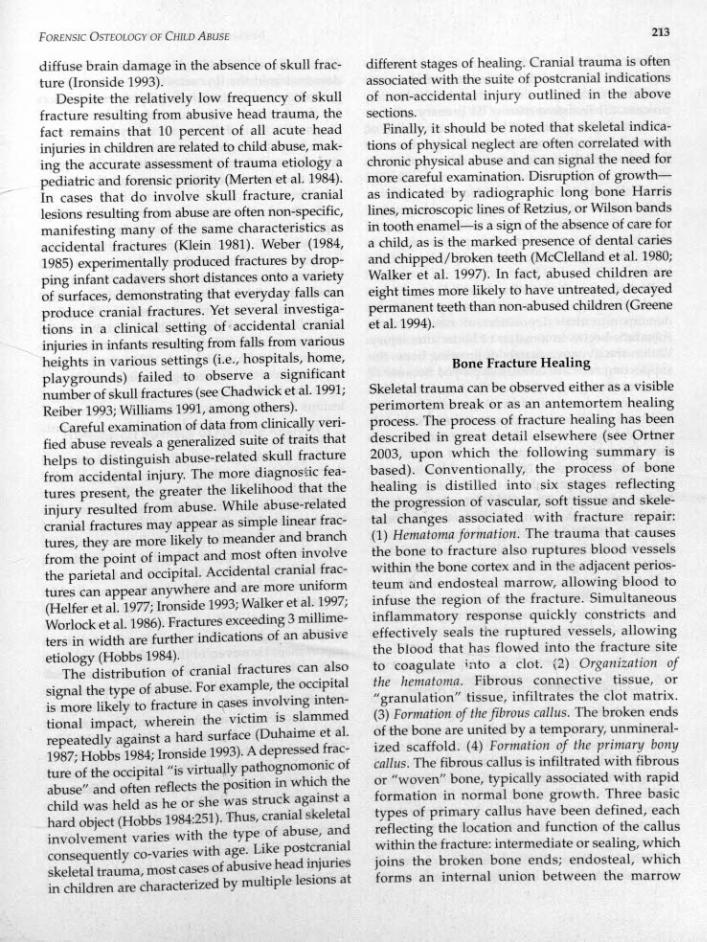
Despite the relatively low frequency of skullfracture resulting from abusive head trauma, thefact remains that 10 percent of all acute headinjuries in children are related to child abuse, mak-ing the accurate assessment of trauma etiology apediatric and forensic priority (Merten et al. 1984).In cases that do involve skull fracture, craniallesions resulting from abuse are often non-specific,manifesting many of the same characteristics asaccidental fractures (Klein 1981). Weber (1984,1985) experimentally produced fractures by drop-ping infant cadavers short distances onto a varietyof surfaces, demonstrating that everyday falls canproduce cranial fractures. Yet several investiga-tions in a clinical setting of··accidental cranialinjuries in infants resulting from falls from variousheights in various settings (i.e., hospitals, home,playgrounds) failed to observe a significantnumber of skull fractures (seeChadwick et al. 1991;Reiber 1993;Williams 1991,among others).
Careful examination of data from clinically veri-fied abuse reveals a generalized suite of traits thathelps to distinguish abuse-related skull fracturefrom accidental injury. The more diagnostic fea-tures present, the greater the likelihood that theinjury resulted from abuse. While abuse-relatedcranial fractures may appear as simple linear frac-tures, they are more likely to meander and branchfrom the point of impact and most often involvethe parietal and occipital. Accidental cranial frac-tures can appear anywhere and are more uniform(Helfer et al. 1977;Ironside 1993;Walker et al. 1997;Worlock et al. 1986).Fractures exceeding 3 millime-ters in width are further indications of an abusiveetiology (Hobbs 1984).
The distribution of cranial fractures can alsosignal the type of abuse. For example, the occipitalis more likely to fracture in cases involving inten-tional impact, wherein the victim is slammedrepeatedly against a hard surface (Duhaime et al.1987;Hobbs 1984;Ironside 1993).Adepressed frac-ture of the occipital "is virtually pathognomonic ofabuse" and often reflects the position in which thechild was held as he or she was struck against ahard object (Hobbs 1984:251).Thus, cranial skeletalinvolvement varies with the type of abuse, andconsequently co-varies with age. Like postcranialskeletal trauma, most cases of abusive head fiJurlesin children are characterized by multiple lesions at
213
different stages of healing. Cranial trauma is oftenassociated with the suite of postcranial indicationsof non-accidental injury outlined in the abovesections.
Finally, it should be noted that skeletal indica-tions of physical neglect are often correlated withchronic physical abuse and can signal the need formore careful examination. Disruption of growth-as indicated by radiographic long bone Harrislines, microscopic lines of Retzius, or Wilson bandsin tooth enamel-is a sign of the absence of care fora child, as is the marked presence of dental cariesand chipped/broken teeth (McClelland et al. 1980;Walker et al. 1997). In fact, abused children areeight times more likely to have untreated, decayedpermanent teeth than non-abused children (Greeneet al. 1994).
Bone Fracture Healing
Skeletal trauma can be observed either as a visibleperimortem break or as an antemortem healingprocess. The process of fracture healing has beendescribed in great detail elsewhere (see Ortner2003, upon which the following summary isbased). Conventionally, the process of bonehealing is distilled into six stages reflectingthe progression of vascular, soft tissue and skele-tal changes associated with fracture repair:(1) Hematoma formation. The trauma that causesthe bone to fracture also ruptures blood vesselswithin the bone cortex and in the adjacent perios-teum and endosteal marrow, allowing blood toinfuse the region of the fracture. Simultaneousinflammatory response quickly constricts andeffectively seals the ruptured vessels, allowingthe blood that has flowed into the fracture siteto coagulate into a clot. (2) Organization ofthe hematoma. Fibrous connective tissue, or"granulation" tissue, infiltrates the clot matrix.(3) Formatioll of the fibrous callus. The broken endsof the bone are united by a temporary, unmineral-ized scaffold. (4) Formntion. of the primary bonycallus. The fibrous callus is infiltrated with fibrousor "woven" bone, typically associated with rapidformation in normal bone growth. Three basictypes of primary callus have been defined, eachreflecting the location and function of the calluswithin the fracture: intermediate or sealing, whichjoins the broken bone ends; endosteal, whichforms an internal union between the marrow

214
spaces of the broken ends; and periosteal or bridg-ing, which forms on the outer cortex and consti-tutes the externally visible evidence of the healingprocess. (5) Transformation of the primary callus tothe secondary bony callus. The temporary callus ofwoven bone is then replaced by stronger lamellarbone through the processes of surface apposition,removal of dead areas of bone, and internalremodeling of the osteons and Haversian systemsof the cortex. (6) Remodeling of the secondary callus.Pinally, the callus is reduced, and the bone willslowly return to a size and shape that approxi-mates the original functional morphology of thebone.
Recent research in oral and dental surgery hasused scanning electron microscopy (SEM) todemonstrate that deposition of new bone insubadults begins in a matter of hours after injury.Within the dynamic matrix of growing bone, thesupporting cells are already deployed because ofthe ever-changing nature of the tissue, and thispromotes an accelerated healing rate. Given thishighly charged osteogenic potential of a subadultperiosteal layer, which is four to five times thickerthan the wafer-thin periosteum of adult bone(johnstone and Foster 2001), wound healing pro-ceeds at a much quicker rate than in comparatively"static" adult bone. Therefore, instead of applyingthe adult timetable of ten to fourteen days for bonerepair, it is likely that healing is taking place muchfaster in subadult wounds (O'Connor and Cohen1998). Ortner (2003) estimates that children'sfractures heal at as much as twice the rate of adulthealing, and histological examination of infantabuse cases clearly demonstrates a degree of heal-ing corresponding to that seen in adults after amuch greater time interval.
Radiological Evidence of Child Abuse
Since broken bones are usually diagnosed clini-cally with radiographs, why are X-rays insuffi-cient to diagnose child abuse? Skeletal injuries area key indicator of abuse and can often be verifiedradiographically during the initial investigationof the victim (see Brogdon 1998). However, it isimportant to note that "bone fractures are perhapsthe most important and problematic issue as far asdetectability is concerned" in cases of infant abuseand it is "consequently fundamental to be awareof the sensitivity of investigative tools used"
INTERPRETATION OF TAPHONOMY AND TRAUMA
(Catteneo et al. 2006:132).Numerous studies havedemonstrated the low sensitivity of the currentpathology standards of radiographic examinationfor the detection of fractures in children. Thesediagnostic tools fail for a variety of reasons,including the angle at which standard skeletalsurveys are taken, the sensitivity of the equip-ment, and the nature of the fracture and healing ininfant bone.
The various stages of injury and healing,especially in the tiny bones of infants, varygreatly in their visibility with standard diagnosticimaging tools. Periosteal remodeling can be asthin as half a millimeter and therefore can beoccult (invisible) or barely visible in even themost sensitive radiographic images (Brogdon1998).Dolinak and Matshes (2005)add that whilethe most sensitive CT diagnostics can image suchnew bone (see Donchin et a1. 1994), the practicalapplication of this technology can be problematic.After the initial stages of healing, subperiosteallesions usually fall well below the threshold ofdetection of standard radiographic equipment.Conversely, if a child dies during the initialhealing phase, prior to the consolidation ofthe underlying cortex, no radiographicallyobservable evidence of trauma will remain onthe bone (Walker et a1. 1997). Antemortem ribbreakage with healing and callus formation isfar easier to diagnose than acute wounds that,even though complete, may be non-displacedand radiographically occult.
As discussed above, rib fractures are highlydiagnostic of abuse. In a study of thirty-one fatallyabused infants, half of all fractures were ribfractures, and thus their detection is of criticalimportance. However, of these rib fractures, justover a third were visible on standard skeletalsurvey (Kleinman et al. 1996). In a comparison ofthe effectiveness of diagnostic tools using ananimal model, only about half of recent, unhealedrib fractures were detected by radiology, a third byCT scan, and two-thirds by normal autopsymethods (Catteneo et al. 2006). In contrast, whenthe skeleton was defleshed, 100percent of fractureswere discerned. In children under three years ofage, healing of costochondral fractures oftenpresent with minimal periosteal reaction, callus,or other radiographically evident deformity,leaving them invisible on skeletal survey (Ng andHall 1998).
FORENSIC OSTEOLOGY OF CHILD ABUSE
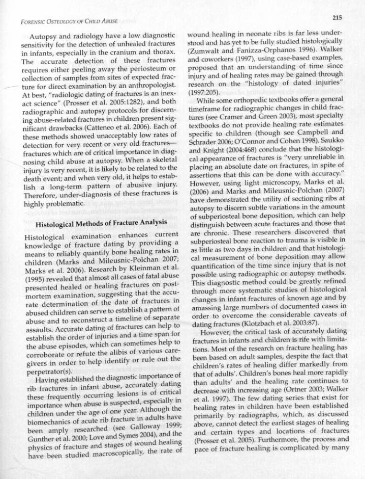
Autopsy and radiology have a low diagnosticsensitivity for the detection of unhealed fracturesin infants, especially in the cranium and thorax.The accurate detection of these fracturesrequires either peeling away the periosteum orcollection of samples from sites of expected frac-
_ ture for direct examination by an anthropologist.At best, "radiologic dating of fractures is an inex-act science" (Prosser et al. 2005:1282), and bothradiographic and autopsy protocols for discern-ing abuse-related fractures in children present sig-nificant drawbacks (Catteneo et al, 2006). Each ofthese methods showed unacceptably low rates ofdetection for very recent or very old fractures-fractures which are of critical importance in diag-nosing child abuse at autopsy. When a skeletalinjury is very recent, it is likely to be related to thedeath event; and when very old, it helps to estab-lish a long-term pattern of abusive injury.Therefore, under-diagnosis of these fractures ishighly problematic.
Histological Methods of Fracture Analysis
Histological examination enhances currentknowledge of fracture dating by providing ameans to reliably quantify bone healing rates inchildren (Marks and Mileusnic-Polchan 2007;Marks et al. 2006). Research by Kleinman et a!.(1995) revealed that almost all cases of fatal abusepresented healed or healing fractures on post-mortem examination, suggesting that the accu-rate determination of the date of fractures inabused children can serve to establish a pattern ofabuse and to reconstruct a timeline of separateassaults. Accurate dating of fractures can help toestablish the order of injuries and a time span forthe abuse episodes, which can sometimes help tocorroborate or refute the alibis of various care-givers in order to help identify or rule out theperpetrator(s).Having established the diagnostic importance of
rib fractures in infant abuse, accurately datingthese frequently occurring lesions is of ~ritic~1importance when abuse is suspected, espeoally in
children under the age of one year. Although thebiomechanics of acute rib fracture in adults havebeen amply researched (see Galloway 1999;Gunther et a!. 2000; Love and Symes 2004), and thephysics of fracture and stages of wound healinghave been studied macroscopically, the rate of
215
wound healing in neonate ribs is far less under-stood and has yet to be fully studied histologically(Zumwalt and Fanizza-Orphanos 1996). Walkerand coworkers (1997), using case-based examples,proposed that an understanding of time sinceinjury and of healing rates may be gained throughresearch on the "histology of dated injuries"(1997:205).While some orthopedic textbooks offer a general
timeframe for radiographic changes in child frac-tures (see Cramer and Green 2003), most specialtytextbooks do not provide healing rate estimatesspecific to children (though see Campbell andSchrader 2006; O'Connor and Cohen 1998). Saukkoand Knight (2004:468) conclude that the histologi-cal appearance of fractures is "very unreliable inplacing an absolute date on fractures, in spite ofassertions that this can be done with accuracy."However, using light microscopy, Marks et al.(2006) and Marks and Mileusnic-Polchan (2007)have demonstrated the utility of sectioning ribs atautopsy to discern subtle variations in the amountof subperiosteal bone deposition, which can helpdistinguish between acute fractures and those thatare chronic. These researchers discovered thatsubperiosteal bone reaction to trauma is visible inas little as two days in children and that h.istologi-cal measurement of bone deposition may allowquantification of the time since injury that is notpossible using radiographic or autopsy methods.This diagnostic method could be greatly refinedthrough more systematic studies of histologicalchanges in infant fractures of known age and byamassing large numbers of documented cases inorder to overcome the considerable caveats ofdating fractures (Klotzbach et a!. 2003:87).However, the critical task of accurately dating
fractures in infants and children is rife with limita-tions. Most of the research on fracture healing hasbeen based on adult samples, despite the fact thatchildren's rates of healing differ markedly fromthat of adults'. Children's bones heal more rapidlythan adults' and the healing rate continues todecrease with increasing age (Ortner 2003; Walkeret a!. 1997). The few dating series that exist forhealing rates in children have been establishedprimarily by radiographs, which, as discussedabove, cannot detect the earliest stages of healingand certain types and locations of fractures(Prosser et al. 2005). Furthermore, the process andpace of fracture healing is complicated by many

216
factors. Healing rates can differ greatly within thesame individual depending upon age, healthstatus, anatomical location of the injury, and sever-ity of the insult. Cranial fractures, for example,tend to heal at a slower rate than long bone frac-tures (Ortner 2003).
Characteristics specific to an abusive contextfurther impede accurate dating of osseous lesions.Lack of treatment and failure to immobilize theinjury-both hallmarks of abusive injuries-inhibithealing and confound accurate dating of the frac-ture. Skeletal injuries in non-abuse cases exhibit adifferent progression of healing than abusiveinjuries that have been left untreated (Islam et al.2000; Prosser et al. 2005; Spitz 1993). Abusersoften show idiosyncratic patterns of violence,establishing personal proclivities for abuse sites(e.g., thighs, top of head, chin) that are repeatedlysubjected to trauma (Walker et al. 1997).Repeatedassault at the same site can break through existingfracture calluses (Gunther et al. 2000;Spitz 1993).However, it has also been demonstrated thatconcomitant skeletal injuries appear to alter heal-ing rates so that the presence of coexisting headinjury can speed the healing rate of injuries at otherskeletal sites (Prosser et al. 2005).
Differential Diagnosis
Whether a specimen is clinical, and therefore ofmedicolegal importance, or paleopathological froman archaeological context, it is essential to considerdifferential diagnoses of the wound(s) in suspectedcases of child abuse. While direct physical exami-nation, clinical history, and interview of the care-giver(s) should help to discern skeletal lesions ofdifferent etiologies in a forensic case, an under-standing of differing causation is required for aclear diagnosis. It is important to remember thatthe misdiagnosis of abuse can have as damaging asocial and psychological effect on the family asunder-diagnosis would have (see Bernet 1993;Kaplan 1986).
There are numerous systemic diseases thatmanifest in bone and may mimic child abuse. Evenduring periods of normal growth, the appearanceof subadult bone can mimic trauma, particularlythe expansive periosteal growth associated withthe diaphyses during long bone growth, variouslytermed "periostitis of growth" (Walker et al. 1997)or "subperiosteal new bone formation" (Brill et al.
INTERPRETATION OF TAPHONOMY AND TRAUMA
1998; Dolinak and Matshes 2005; Glaser 1949;Kleinman and Kwon 1998; Silverman 1980). Theradiographic appearance of the metaphyses duringlengthwise growth of long bones can be similarlyconfused with injury (Kleinman et al. 1991).
Outside of the normal growth variants in bone,the disease most likely to be considered in a caseof multiple fractures is osteogenesis imperfecta(see Lachman et al. 1998; Patterson et al. 1993;Sillence 1981). In fact, this disease is almost auto-matically suggested by legal experts seeking a rea-sonable clinical argument for an alternative causeof multiple fractures in children. Osteogenesisimperfecta is a rare genetic disorder which mani-fests in four broad types (with several subgroups)variously affecting the connective tissues of bone,skin, ligaments, fascia, the sclera, and the auditorysystem. Although uncommon in occurrence, it hasbeen studied extensively. In osteogenesis irnper-fecta, a fracture results from structural failure ofporotic bone in any area of the body and causesexaggerated callus formation. A variant of osteoge-nesis imperfecta called "temporary brittle bonedisease" (see Chapman and Hall 1997; Pattersonet al. 1993),which is seen in the first year of life andmay be related to copper deficiency, offers anotherpotential diagnosis for lesions similar to thoseresulting from child abuse, though this remainsdebated.
Non-disease conditions that may result infractures, such as Sudden Infant Death Syndrome(see Campbell and Schrader 2006; DiMiao andDiMiao 2001;Platt 1993),obstetric and birth trauma(see Kleinman 1998), and accidental trauma (seeabove), can also potentially be confused with physi-cal mistreatment. Rickets, scurvy, osteomyelitis,congenital syphilis, leukemia, and vitamin A intoxi-cation may all present with confusing radiographicsignatures of abnormal bone formation suggestive ofabuse. In many cases, an experienced skeletal biolo-gist can discern bone trauma resulting from abusefrom systemic pathological conditions of disease,and serological findings can readily confirm or denycertain maladies associated with metabolic diseasestates. Brill and coworkers (1998)explored the radi-ographic details of each disease and highlightedexactly when and how the disease process can mimicbone trauma mistaken for child abuse. However, itmust be noted that malnutrition and neglect often gohand in hand with abusive trauma and that disor-ders which systemically reduce the quality of the

FORENSIC OSTEOLOGY OF CHlLD ABUSE
bone also place children at risk for fractures fromphysical abuse.
Case Findings and Resolution
The pathologist's final diagnosis of the victim canbe summarized as multiple blunt force traumainvolving the head, neck, torso, abdomen, andlimbs, accompanied by lacerations to the anusand genita Is and evidence of thermal burns tothe baby's abdomen and extremities. The completedetails of the medical examiner's findings,outlined below, demonstrate the full extent ofthe injuries and complications that the childsuffered in his short life. The preponderance ofevidence led to the final determination that thisthree-and-a-half-month-old infant "died of multi-ple blunt force trauma due to child abuse. Mannerof death is homicide." The general findings of thepathologist are:• Multiple blunt force trauma, including abra-sions and bruises of head and neck; multiplelacerations of soft tissue of internal lips; andsevere brain swelling with hemorrhage alongthe spinalchord.
• Multiple abrasions and bruises of the trunk,including subcutaneous and intramusc~larhemorrhages of chest wall and back; multiple.bilateral rib fractures at different stages of heal-ing with hemorrhaging of the muscles betweenthe ribs; and associated chronic damage to thetissues of the lung, heart, and spleen.Lacerations and contusions to the anus andgenitals.
• Deformities and bruises of extremities, includ-ing a healing fracture of the distal righthumerus and a healing dislocated fracture ofleftfemur.
• Scarring of the abdomen and the right hand,consistent with healed thermal bums.
One of the main directives of the prosecution'Scase was to establish that the victim suffered apattern of chronic abuse. Lacking any record ofhospital visits or other emergency medical atten-tion (deficient in most instances of child abuse), ahistory of abuse can be difficult to validate.Therefore, it was important to estabhsh that thewounds were received over a period of time todemonstrate a pattern of chronic abuse during thechild's short life. The duty of the anthropologist inthis case was twofold: (1) to identify the tissuesnecessary to help confirm or refute suspectedabuse by the parents to the law enforcement
217
agents, the pathologist, and eventually the prose-cution, and (2) to perform the macroscopic andhistological analysis on the specimens to render anopinion.While working with the pathologist at the
autopsy table with the soft tissue removed, it is notdifficult to discern defects that are antemortem andchronic, given the bone nodule development, fromthose that are acute, fresh breaks. When examiningskeletal lesions in association with soft tissue,however, one must keep in mind that the presenceof hemorrhaging at a wound site is not specific toacute bone trauma. Chronic wound sites can behighly vascular, and any damage to an early form-ing fracture callus can tear these delicate bloodvessels, resulting in bleeding. In cases of variouswound ages, the best way to positively establishacute from chronic hemorrhage is to resect (removeall or part of) the bone at autopsy and examine thefracture for a periosteal response under a dissect-ing microscope. While bruises, both healing andrecent, are helpful in the diagnosis of abusechronology in soft tissues, the dissecting scopeimages of acute and/or chronic bone fracture siteson the bone were key to the diagnosis of a historyof abuse in this case.The results of the anthropological and histologi-
cal examination in this case revealed multiplefractures at various distinct stages of healing,indicative of a pattern of chronic abuse culminat-ing in the acute, fatal incident. As a result of themedicolegal investigation, the mother of the victimwas charged with first degree murder and aggra-vated child abuse. She avoided trial by pleadingguilty .o a lesser charge and is currently servingtwenty years. The father chose to go to trial, inwhich the pathologist and anthropologist wereexpert witnesses fer the prosecution and providedmore than three hours of combined testimony.The jury found the father guilty of first degreemurder and aggravated child abuse. He wassentenced to twenty-five years with no chance forparole, but has filed for an appeal, the outcome ofwhich is still pending at this writing.
Conclusion
Since few pathologists possess an expertise withbone that parallels that of the anthropologist,collaboration between these specialties is crucial to

218
the successful resolution of fatal abuse casesinvolving skeletal lesions. Similarly, the perspec-tives of the orthopedist, pediatrician, and radiolo-gist can be invaluable to the forensic pathologist inchild abuse cases. While radiological evidence canwell establish some forms of chronic skeletalwounds, the resolution of traditional radiographicimages is not adequate to discern certain forms offracture, certain locations of fracture, or the subtledegree of bone healing in wounds received only afew days before death. Because freshly depositedsubperiosteal bone is radiographically invisible,the pathologist must resect (remove) any boners)presenting indications of acute injury for anthropo-logical and/or histological assessment. It is essen-tial to resect the bones, or parts thereof, to gain aclear understanding of the mechanism of traumaand the chronology of the injury.
References
Altman, D. H., and R. L. Smith. 1960. Unrecognizedtrauma in infants and children. Journal of Bone andJoint Surgery 42:407-413.
Agran, P. E, C. Anderson, D. Winn, R. Trent, L. Walton-Haynes, and S. Thayer. 2003. Rates of pediatricinjuries by 3D-month intervals for children a to 3 yearsof age. Pediatrics 111:e683--£692.
Barsness, K. A., E. S. Cha, D. D. Bansard. C. M. Calkins,D. A. Patrick, E M. Karrer, and J. D. Strain. 2003.The positive predictive value of rib fractures as anindicator of nonaccidental trauma in children. TheJournal of Trauma, Injury, Infection and Critical Care54(6):1107-1110.
Bernet, W 1993. False statements and the differentialdiagnosis of abuse allegations. Journal of the AmericanAcademy of Child Adolescence Psychiatry 32:903-910.
Brill,Paula W, Patricia Winchester, and Paul K. Kleinman.1998. Differential diagnosis I: Diseases simulatingabuse. Pp. 178-196 in Diagnostic Imaging of Child Abuse,2nd ed., ed. Paul K. Kleinman. St. Louis, MO: Mosby.
Brogdon, Barry G. 1998. Forensic Radiology. Boca Raton,FL: CRC Press.
Cadzow, S.P., and K. L. Armstrong. 2000.Rib fractures ininfants: Red alert! The clinical features, investigationsand child protection outcomes. Journal of PaediatricChild Health 36:322-326.
Caffey, John. 1946.Multiple fractures in the long bones ofinfants suffering from chronic subdural haematoma.American Journal of Roentgenology 56:163-173.
Caffey, John. 1972. On the theory and practice of shakinginfants. American Journal of Diseases in Childhood124(2):161-169.
Caffey, John. 1974. The whiplash shaken infantsyndrome: Manual shaking by the extremities withwhiplash-induced intracranial and intraocular
INTERPRETATION OF TAPHONOMY AND TRAUMA
bleedings, linked with residual permanent braindamage and mental retardation. Pediatrics 54:396-403.
Campbell, Jr. R. M., and T. Schrader. 2006. Child abuse.Pp. 223-253 in Rockwood and Wilkins's Fractures inChildren, 6th ed., ed. J. H. Beaty and J. R. Kasser.Philadelphia: Lippincott Williams & Wilkins.
Catteneo, C. E., A. Marinelli, M. DiGiancamillo, O. DiCiancamtllo, L. Vigano Travetti. P. Pappa, D. Porta,A. Gentilomo, and M. Grandi. 2006. Sensitivity ofautopsy and radiological examination in detectingbone fractures in an animal model: Implications forthe assessment of fatal child physical abuse. ForensicScience International 164:131-137.
Chadwick, D. L., S. Chin, and C. Salerno. 1991. Deathsfrom falls in children: how far is fatal? Journal ofTrauma 31:1353-1355.
Chapman, S. 1990. Radiological aspects of non-acciden-tal injury. Journal of Royal Society of Medicine 83:67-71.
Chapman, S., and C. M. Hall. 1997. Non-accidentalinjury or brittle bones. Pediatric Radiology 27:106-110.
Cooperman, D. R., and D. E Merten. 2001. Skeletalmanifestations of child abuse. Pp. 123-156 in ChildAbuse: Medical Diagnosis and Management, 2nd ed., ed.R. M. Reece and S. Ludwig. Philadelphia: LippincottWilliams & Wilkins.
Cramer, K. E., and N. E. Green. 2003. Child abuse.Pp. 587-605, in Skeletal Trauma in Children, Vol. 3, 3rded., ed. N. E. Green and M. F. Swionkowski.Philadelphia: WB. Saunders.
Department of Health and Human Services. 2001. Reporton the Third Nationallncidence Study of Child Abuseand Neglect (NlS-3).
DiMiao, Vincent J. M., and Dominick DiMiao. 2001.Forensic Pathology, 2nd ed. New York:Elsevier.
Dolinak, D., and E. Matshes. 2005. Child abuse.Pp. 369-412 in Forensic Pathology: Principles andPractice, ed. D. Dolinak, E. Matshes, and E. Lew.Amsterdam: Elsevier-Academic Press.
Donchin, Y, A. 1. Rivkind, J. Bar-Ziv, J. Hiss, J. Almog,and M. Drescher. 1994. Utility of postmortemcomputed tomography in trauma victims. Journal ofTrauma 37(4):552-536.
Duhaime, A. c, T. A. Gennarelli, L. E. Thibault, D. A.Bruce, S. S. Margulies, and R. Wiser. 1987.The shakenbaby syndrome: A clinical, pathological, and biome-chanical study. Journal of Neurosurgery 66:409-415.
Galleno, H., and W L. Oppenheim. 1982. The batteredchild syndrome revisited. Clinical Orthopedics162:11-19.
Galloway, A. 1999. Broken Bones. Springfield, IL: CharlesC. Thomas.
Glaser, K. 1949.Double contour, cupping and spurring inroentgenograms of long bones in infants. AmericanJournal of Roentgenology 61:482-492.
Green, N. E., and M. E Swiontkowski. 2003.Skeletal Traumain Children, Vol.3, 3rd ed. Philadelphia: WE. Saunders.
Greene, P. E., M. C. Chisick, and G. R. Aaron. 1994.Comparison of oral health status and need for dentalcare between abused neglected children andnonabused/non-neglected children. Pediatric Dentistry16(1):41-45.

FORENSIC OSTEOLOGY OF CHILD ABUSE
Gunther, Wendy M., Steven A. Symes, and HughE. Berryman. 2000. Characteristics of child abuse byanteroposterior manual compression versuscardiopulmonary resuscitation: Case reports.American Journal of Medical Pathology 21(1):5-10.
Haller, J. 0., and E. G. Kassner. 1977. The "battered child"syndrome and its imitators: a critical evaluation of spe-cific radiological signs. Applied Radiology 6:85-111.
Harwood-Nash, D. C; E. B.Hendrick, and A. R. Hudson.1971. The significance of skull fractures in children.Rndiology 101:151-155.
Helfer, R. E., T. L. Slovis, and M. Black. 1977. Injuriesresulting when small children fall out of bed.Pediatrics 60(4):533-535.
Herdon, W A. 1983. Child abuse in a military popula-tion. Journal of Pediatric Orthopedics 3:341-343.
Hobbs, C. J. 1984. Skull fracture and the diagnosis ofabuse. Archives of Diseases in Childhood 59:246-252.
Ironside, J. W 1993. Blunt injury to the head. Pp. 163-177in The Pathologtj of Trauma, ed. J. K Mason. London:Arnold.
Islam, 0., D. Soboleski, S. Symons, L. K. Davidson,M. A. Ashworth, and P. Babyn. 2000. Developmentand duration of radiographic signs of bone healing inchildren. American Journal of Radiology 175:75-78.
Johnson, C. E 1990. Inflicted injury versus accidentalinjury. Pediatric Clinics afNorth America 37:791-814.
Johnstone, E. W, and B. K Foster. 2001. The biologicalaspects of children's fractures. Pp. 1-15 in Rockwoodand Wilkins' Fractures in Children, 5th ed., ed. J. H. Beatyand J. R. Kasser. Philadelphia: Lippincott, Williams& Wilkins.
Kaplan, J. M. 1986. Pseudoabuse: the misdiagnosis ofchild abuse. Journal of Forensic Sciences 31:1420--1428.
Kempe, c., H. Frederic, N. Silverman, B. E Steele,W. Droegemueller, and H. K Silver. 1962. The bat-tered-child syndrome. Journal of the American Medica!Association 181(1):17-24.
Klein, D. M. 1981. Central nervous system injuries.Pp. 73-93 in Child Abuse and Neglect: A Medical Reference,ed. N. S. Ellerstein. New York: John Wiley & Sons.
Kleinman, Paul K 1998. Differential diagnosis III:Accidental and obstetric trauma. Pp. 214-224 inDiagnostic Imaging of Child Abuse, 2nd ed., ed. Paul K.Kleinman SI. Louis: Mosby.
Kleinman, Paul K, and A. Schlesinger. 1997. Mechanicalfactors associated with posterior rib fracture: Laboratory
. and case studies. Pediatric Rndiology 27:87-91.Kleinman, Paul K, and David S. Kwon. 1998. Differentialdiagnosis IV: Normal variants. Pp. 225-236 in
Diagnostic Imaging of Chiid Abuse, 2nd ed., ed. Paul K.Kleinman SI. Louis: Mosby.
Kleinman Paul K., P. L. Belanger, A. Karellas, andM. R. Spevak. 1991. Normal metaphyseal radiologyvariants not to be confused with findings of infantabuse. American Journal of Radiology 156:781-783.
Kleinman, Paul K, S. C. Marks, M. R. Spevak, andJ. M. Richmond. 1992. Fractures of the fib head mabused infants. Radiology 185(1):119-123.
Kleinman, Paul K, S. C. Marks, K Nimkin. S. M. Ryder,and S. C. Kessler. 1996. Rib fractures m 31 abused
219
infants: postmortem radiologic-histopathic study.Rndiology 200:807-810.
Kleinman, Paul K., S. C. Marks, J. M. Richmond, andB. D. Blackbourne. 1995. Inflicted skeletal injury:A postmortem radiologic-histopathic study in31 infants. American Journal of Radiology 165:647--{i50.
Klotzbach, H., G. Delling, E. Richter, J. P. Sperhake, andK. Puschel. 2003. Post-mortem diagnosis and ageestimation of infants' fractures. International Journal ofLegal Medicine 117:82-89.
Lachman, Ralph S., Deborah Krakow, and Paul K.Kleinman. 1998. Differential diagnosis II:Osteogenesis imperfecta. Pp. 197-213 in DiagnosticImaging of Child Abuse, 2nd ed., ed. Paul K KleinmanSt. Louis: Mosby.
Leonidas, J. C. 1983. Skeletal trauma in the child abusesyndrome. Pediatric Annals 12:875-881.
Love, Jennifer c., and Steven A. Symes. 2004.Understanding rib fracture patterns: Incomplete andbuckle fractures. Journal of Forensic Sciences49(6):1153-1158.
Mandelstam. S. A., D. Cook, M. Fitzgerald, and M. R.Ditchfield. 2003. Complementary use of radiologicalskeletal survey and bone scintigraphy in detection ofbony injuries in suspected child abuse. Archives ofDisease in Childhood 88:387-390.
Marks, Murray K., and Darinka Mileusnic-Polchan.2007. Histopathology of antemortem infant bonefractures: Estimation of time since insult.Proceedings of the American Academy of ForensicSciences XIlI:315.
Marks, Murray K., M. A. Tersigru. and DarinkaMileusnic-Polchan. 2006. Antemortem infant ribfracture:The histological evidence. Proceedings of theAmerican Academy of Forensic Sciences XII:307.
McClain, P. W, J. J. Sacks, R. G. Froehlke, and B. G.Ewigman. 1993. Estimates of fatal child abuse andneglecr, United States, 1979 through 1988. Pediatrics91:335-343.
McClelland, C. Q., H. Rekate, B. Kaufman, and L.Persse.1980. Cerebral injury in child abuse: A changingprofile. Child's Brain 7:225-235.
Merten, D. E, and n.R. S. Osborne. 1983. Craniocerebraltrauma in the child abuse syndrome. Pediatric Annals12:882-887.
Merten, D. E, D. R. S. Osborne, M. A. Radkcwski, andJ. C. Leonidas. 1984. Craniocerebral trauma in thechild abuse syndrome: Radiological observations.Pediatric Rndiology 14:272-277.
Morse, C. W., O. J. Z. Sahler, and S. B. Friedman. 1970.A three-year follow-up study of abused and neglectedchildren. American [ournal of Diseases of Children120:439-446.
Ng, C. S., and C. M. Hall. 1998. Costochondral junctionfractures and intra-abdominal trauma in non-accidental injury (child abuse). Pediatric Radiology28:671-676.
O'Connor John E, and Jonathan Cohen. 1998. Datingfractures. Pp. 168-177 in Diagnostic Imaging of ChildAbuse, 2nd ed., ed. Paul K. Kleinman. Baltimore:Williams and Wilkins.

220
Oliver, J. E. 1993. Intergenerational transmission of childabuse: Rates, research and clinical implications.American Journal of Psychiatry 150:1315-1324.
Ortner, Donald. 2003. Identification of PathologicalConditions in Human Skeletal Remains, 2nd ed.Washington DC: Academic Press.
Overpeck, M. D., R. A. Brenner, A. C. Trumble, L. B.Trifiletti, and H. W. Berendes. 1998. Risk factors forinfant homicide in the United States. New EnglandJournal of Medicine 339(17):1211-1216.
Patterson, C. R., J. Burns, and S. J. McAllion. 1993.Osteogenesis imperfecta: The distinction from childabuse and the recognition of a variant form. AmericanJournal ojMedical Genetics 45:187-192.
Platt, M. S. 1993. The differential diagnosis of childabuse. Pp. 724-732 in Medicolegal Investigation ofDeath: Guidelines for the Application of Pathology toCrime Investigation, 3rd ed., ed. Werner U. Spitz.Springfield: Charles C. Thomas.
Prosser, C S. McGuire, S. K. Harrison, M. Mann,J. R. Sibert, and A. M. Kemp. 2005. How old is thisfracture? Radiologic dating of fractures in children: Asystematic review. American Journal of Radiology184:1282-1286.
Reece, R. M., and S. Ludwig. 2001. Child Abuse: MedicalDiagnosis and Management, 2nd ed. Philadelphia:Lippincot Williams & Wilkins.
Reiber, G. D. 1993. Fatal falls in childhood. How farmust children fall to sustain fatal head injury?Report of cases and review of the literature.American Journal of Forensic Medicine and Pathology14:201-207.
Rubin, D. M., C. W. Christian, L. T. Bilaniuk, K. A.Zazyczny, and D. R. Durbin. 2003. Occult head injuryin high-risk abused children. Pediatrics 111:1382-1386.
Sauer, Norman J. 1998. The timing of injuries andmanner of death: Distinguishing among antemortem,perimortem and postmortem trauma. Pp. 321-332 inForensic Osteology: Advances in the Identification ofHuman Remains, ed. Kathleen Reichs. Charles CThomas: Springfield.
Saukko, P., and B. Knight. 2004. Knight's ForensicPathology, 3rd ed. London: Arnold.
Sillence, D. O. 1981. Osteogenesis imperfecta: An expand-ing panorama of variants. Clinical Orthopedics159:11-25.
Silverman, F. N. 1953. The roentgen manifestations ofunrecognized skeletal trauma in infants. AmericanJournal of Roentgenology 69:413-427.
Silverman, F. N. 1980. Radiologic and special diagnosticprocedures. Pp. 215-40 in The Battered Child, ed.Kempe, C. H. and Helfer, R. E. Chicago: University ofChicago Press
INTERPRETATION OF TAPHONOMY AND TRAUMA
Spitz, Werner U. 1993. Spitz and Fisher's MedicolegalInvestigation of Death: Guidelines for the Application ofPathology to Crime Investigation, 3rd ed. Springfield, IL:Charles C. Thomas.
Ubelaker, Douglas H. 1995. Latest developments inskeletal biology and forensic anthropology.Pp. 91-106 in Biological Anthropology, The State of theScience, ed. N. T. Boaz and L. D. Wolfe. Corvalis,Oregon: Oregon State University Press.
Walker, Phillip L., Della C. Cook, and Patricia Lambert.1997. Skeletal evidence for child abuse: A physicalanthropological perspective. Journal of ForensicSciences 42(2):196-207. ,
Weber, W. 1984. Experimental studies of skull fracturesin infants. Zeitschrift fur Rechtsmedizin. Journal ofLegal Medicine 92:87-94.
Weber, W. 1985. Biomechanical fragility of the infantskull. Zeitschrift fur Rechtsmedizin. Journal of LegalMedicine 94:93-101. .
White, T. D. 2000. Pp. 459-465 in Human Osteology, 2nded. Academic Press: San Diego, CA.
Williams, R. A. 1991. Injuries in infants and small chil-dren resulting from witnessed and corroborated freefall. Journal of Trauma 31:1350-1352.
Warlock, P., M. Stower, and P. Barbour. 1986. Patterns offractures in accidental and non-accidental injury inchildren: A comparative study. British Medical Journal293:100-102.
Zumwalt, R., and A. Fanizza-Orphanos. 1996. Dating ofhealing rib fractures in fatal child abuse. Pp. 193-205in Advances in Pathology, ed. C. Fenoglio-Preiser,R. Weinstein, R. Anderson, E. Benson, R. Cotran.F.Vogel. St. Louis: Mosby Yearbook.
Further Readings
Berryman, H. E., and S. J. Haun. 1996. Applying forensictechniques to interpret cranial fracture patterns in anarchaeological specimen. International Journal ofOsteoarchaeology 6:2-9.
Bulloch, B., C. J. Schubert, P. D. Brophy, N. Johnson, M.H. Reed, and R. A. Shapiro. 2000. Cause and clinicalcharacteristics of rib fractures in infants. Pediatrics105(4)e48.
Ellis, P. S. J. 1997. The pathology of fatal child abuse.Pathology 29:113-121.
Reece, R. M. 1993. Fatal child abuse and sudden infantdeath syndrome: A critical diagnosis decision.Pediatrics 91:423-429.
Thomsen, T. K., B. Elle, and J. L. Thomsen. 1997. Post-mortem radiological examination in infants: Evidenceof child abuse? Forensic Science International 90:223-230.