Marine Invertebrate Survey of Guana Island, BYI - Guana Science
178
Marine Invertebrate Survey of Guana Island, BYI Report for the 2000 Field and Research Season Guana Island Marine Science Month Todd L. Zimmennan and Joel W. Martin Project Directors Natural History Museum of Los Angeles County 900 Exposition Boulevard Los Angeles, California 90007 U.S.A.
-
Upload
khangminh22 -
Category
Documents
-
view
3 -
download
0
Transcript of Marine Invertebrate Survey of Guana Island, BYI - Guana Science

Marine Invertebrate Survey of Guana Island, BYI
Report for the 2000 Field and Research Season
Guana Island Marine Science Month
Todd L. Zimmennan and Joel W. Martin
Project Directors
Natural History Museum of Los Angeles County 900 Exposition Boulevard
Los Angeles, California 90007 U.S.A.

General Introduction to the Project..." .......... " ......... " .................... " ........................... " ......... 3 Summary of Education Activities in 2000"""" ...... """ .. " ............. "" ........ " .... " ........ ",, ........ 3 Summary of Research Activities in 2000 .. "" ...... "" .......... " ..... " ......... ....... "" .... "" .... " .. ,, ..... 5 Individual Research Reports .......... " ......................................... " ....................... " ....... " ...... ".6
Report on the Crustaceans .......................... " ................................................... " ................ 6
Report on the Non-Decapod Arthropods .... " ............................................. " ................ 10 Report on Leptostracan Crustacea ............................................................................... 15
Report on the Opisthobranch Mollusca (Sea Slugs) ........................................................ 19
Report on the Polychaete Worms ........................................ .. .......................................... 27
Report on the Echinoderms .................................................................. .. .......................... 28
Guana Island Seagrass Investigations .............................................................................. 30
Concluding Remarks ............................................................................................................ 31 Plans for Summer of 200 I .................................. ................................................................. 32 Appendix I. Guana Island Products ..................................................................................... 33
A. Electronic I Web I CD ROM Products ....................................................................... 33
B. Presentations I talks (in chronological order) ............................................................ 33
C. Publications ................................................................................................................. 34
Peer-reviewed Scientific Articles ................................................................................ 34 Books and Popular Articles: ........................................................................................ 36 \Vhite Papers ................................................................................................................ 36
Appendix II. Stomatopod and Decapod Crustacean Species Lis!... .................................... 37 Appendix III. Isopod Crustaceans ........................................................................................ 43 Appendix VI. Cumulative list of Polychaete Taxa .............................................................. 44 Attachments ......................................................................................................................... 48
Report on the Stenopodidean "Shrimps" of Guana Island, British Virgin Islands .......... 48
Marine Invertebrates of Guana Island, BV!. CD-ROM ................................................... 48
The stomatopod AlachosqllillafloridclIsis ... reported from Guana Island .................... 48
Morphology of the first zoeal stage of Platypodiella spectabilis .................................... 48
An emendation of the fan worm genus Fabricilluda ....................................................... 48
Species of NovoJabricia ... in the Caribbean Sea ................................. .. .......................... 48
2

General Introduction to the Project
The Marine Invertebrate Survey of Guana Island began as a three-year project funded jointly by the U.S. National Science Foundation and the Fa1conwood Corporation. The primary goals of the project are to document and photograph as many as possible of the small, cryptic invertebrates -- especially crustaceans, polychaete worms, echinoderms, and selected mollusks -- found in and around the various marine habitats on Guana Island. To achieve these goals, a small team of scientists from the Natural History Museum of Los Angeles County (LACM), led by Todd L. Zimmerman and Joel W. Martin, as well as two marine scientists not affiliated with LACM (Rick Ware, Don Cadien), visit the island each summer during Marine Science Month. The team consists of 8 members, although visits to the island are staggered such that only 6 team members are on the island at any given time. Specimens are collected, photographed, assigned preliminary identifications where possible, and preserved in 70% ethyl alcohol for later genetic analysis. In order to survey these small invertebrates, we use a variety of collecting methods including hand collecting, SCUBA diving and snorkeling, yabby pumps and other suction devices, sediment vacuum air lifts, and, most notably, the construction of Artificial Reef Matrices (ARt\1s) made of concrete and PVC pipe. These ARMs are placed at different depths around the reef and in other underwater habitats, usually at a depth of 30 feet (10m). The purpose of the ARMs is to attract reefinvertebrates that we can study without touching or damaging the actual reefs, where most of these species live. After returning to Los Angeles, we spend most of the remainder of the year identifying and describing the specimens and preparing our findings for publication. Products of the Survey will include scientific publications (some of which are included in this report) and, at the project's conclusion, a CD ROM disk containing color photographs of a large number of marine invertebrates found in the waters of Guana Island, many of which are proving to be new to science.
Summary of Education Activities in 2000
Our project has a strong Education component. We are training or involving graduate students, undergraduate students, high school students, and elementary school students in various aspects of our work. During 2000, the following students benefited from our project.
Todd L. Zimmerman (Ph. D. candidate at UCLA I LACM, using data and specimens from Guana Island to augment his dissertation research on island biogeography of crabs).
Todd A. Haney (a doctoral student at UCLA I LACM, collecting leptostracan ("sea flea") specimens as part of his dissertation research on the Crustacea Leptostraca).
Sandra Trautwein (a doctoral student at UCLA I LACM, benefiting from freshly collected xanthoid crabs that will form the basis of her dissertation research on crab phylogeny).
Barbara Hajduczek (MS student at UC Berkley, beginning fall 200 I, trained on photography and electronic publications techniques).
3

Peter Marko (postdoctoral scholar at LACM, benefiting from ethanol-preserved bivalve shells, which will be part of his study of the molecular phylogeny of the molluscan family Arciidae)
Sarah Boyce (MS student at Florida State University, using some of our calappid crab material as part of her thesis r~search).
Lianna Jarecki (a doctoral student [as well as an instructor at H. Lavity Stout! Conununity College, Tortola, BVIl working on the ecology of salt ponds in the British Virgin Islands; benefiting indirectly from an ongoing exchange of information about copepods and other salt pond crustaceans).
Simone Lettsome (an undergraduate student of Lianna Jarecki, above, who solicited our help for her talk for the Worldwide-Young-Researchers-of-the-Environment meetings in Germany, where she has been invited to present her student research as the sole student representing the BVI).
Nadia Primus (former undergraduate student of Lianna Jarecki, now preparing for entering the University of South Florida, Tampa; worked with us during the harvesting of the ARLV! off of White's Beach).
David Adolfis and the Seventh Day Adventist Church "Pathfinders" group (approximately 30 students ranging in age from 5 to IS, plus 12 adults, took part in a hands-on demonstration of coral-associated invertebrates on, July 26, 2000, near the airport site on Beef Island).
4

Summary of Research Activities in 2000
The most significant activity during the 2000 season was the "harvesting" of the concrete and PVC ARt\'!s that we had deployed in the summer of 1999. Collecting and processing these arrays was a time-consuming task involving usually four divers and approximately half a day per ARt\'!. Each ARt\'! was placed in a nylon bag while still undeIWater (at 10m depth); the nylon bag was sealed, and the entire ARM was airlifted to the surface, where it was hoisted aboard the boat. The same procedure was applied to the PVC and mesh "basket" underneath each ARt\'!; these were also sealed undeIWater and then airlifted to the boat. After we had returned to Whites Beach, the plates were carefully separated, with each plate photographed and the associated animals stored and labeled as to where on the ARM they had been found. Rubble from the basket was also carefully sorted, then rinsed, and sorted again, to search for any invertebrates. Selected specimens from both the ARM and from the underlying rubble baskets were taken to the "lab" (the Anegada house) where they were further studied, photographed, and labeled.
After returning to Los Angeles, we spent much of the remainder of the year identifying and describing the specimens and preparing our findings for pUblication. What follows are some of the completed research reports. A more complete account of the research activities can be seen in Appendices I and II, which list the talks and scientific publications, respectively, that have stemmed from this project to date.
5
Individual Research Reports
Report on the Crustaceans Collected at or near Guana Island during 2000
Todd L. Zimmerman
The number of crustacean species collected on and near Guana Island in 2000 increased substantially from that of 1998 and 1999. In addition, the species composition from certain areas has seemingly changed from year to year. These two observations can be attributed to several factors. The first is the use of novel collecting techniques such as artificial reef matrix structures (or A.R.M.S) for nondestructive collecting in reef areas, hand net dredging by divers on sand bottoms in the deeper water at the base of the reef wall, and the use of light traps in various areas. The second factor may be the presence of slightly colder waters around the island in the first half of2000, which could have influenced recruitment of larvae from the plankton, or distribution of adults. The third factor may simply be the natural variation in recruitment from year to year combined with the relatively low number of sites that can be sampled (e.g. because natural processes around the island tend to cement rocks together in the intertidal zone, the number of rocks that can be turned to uncover specimens is relatively small. Each year the species found under these few rocks changes). A fourth factor may be that our familiarity with the island gained during previous years allows us concentrate our sampling on areas and microhabitats that have proven to be productive in the past.
The most substantial increase in numbers of species found has been for the various shrimp groups, which were nearly lacking in collections from previous years. This increase can be attributed directly to the ARl\1S. The ARl\1S replicate many of the microhabitats found in association with a medium-small coral head. By allowing this concrete and plastic structure to sit on the ocean bottom for a full year, a somewhat natural invertebrate community had time to colonize and develop. Careful removal of these structures from the water allowed us to capture not only those larger fast moving species nomlally hidden in the rock, but also the smallest, most transparent and delicate of species. This would never have been possible using traditional diver or shipboard methods (hand collecting, pulled nets/dredges, etc).
Representatives of the infraorder Stenopodidea serve to illustrate the amount of taxonomic work needed for Caribbean invertebrates. Slel/opus hispidus, the barber pole cleaner shrimp, is one of the most common and brightly colorcd rccf inhabitants. The group to which it belongs has few species, and most of the representatives are, like S. hispidus, brightly colored inhabitants of shallow tropical waters. The ARl\1S yielded several species from this group, each striking enough to be immediately set aside for photography. Anticipating easy identification of these specimens, Joel Martin decided to work on this group during a visit to the US National Museum, Smithsonian Institution, where most of the world's Caribbean material is held. Stellopus hispidus was the only one of the species collected that did not yield some type of new scientific information (please see included report on Stenopodidean shrimps, and copies of two submitted manuscripts).
6

The number of porcelain crabs and related anomurans found in 2000 increased substantially over that of previous years due directly to the use of ARMS. These animals are not only fast moving, but also very delicate. Surprisingly, although the number of brachyurans (true crabs) found in association with the ARl'vfS did increase, it was nowhere near what was expected based upon our experience collecting at islands in the tropical eastern Pacific. This could be due to the high number of stomatopods (mantis shrimp) found in those same samples. Mantis shrimp, especially species of Neogollodaclyius, prey aggressively on hard-shelled invertebrates such as crabs. One interesting observation pertains to the crab Uhlias limbalus, a small (-5 mm across) species that resembles a large grain of sand. Because of its small size and appearance it is quite rare in collections. After examining all specimens held in the major museums of the world, Rathbun, in her 1937 monograph, could report on only four specimens (one each from four scattered Caribbean locations). The rubble baskets from each of the 8 ARMS placed around Guana yielded from 4 to 8 specimens of Uhlias. Each rubble basket represents an area of seafloor Y. m2
,
making this one of the most common crabs inhabiting the reef.
The crustaceans found on the deep (55-75 ft) sand bottom at the base of the reef wall consisted mainly of brachyuran crabs and hermit crabs. By running a flat edged dip net through the top layer of this sediment, sieving out fine particles and bagging up the remainder to be floated to the surface, we were able to find many small species not normally collected. These include several bizarre-looking species of elbow crabs (family Parthenopidae) and purse crabs (family Leucosiidae), as well as miniature (adults -3 mm across) species of pebble crabs (families Xanthidae and Pilunmidae). Of the many small species of hermit crabs found in these samples, several may tum out to be new species (Rafael Lemaitre, U.S. National Museum, personal communication). One of these, a species of Iridopagurus, was observed to throw off the snail shell it inhabited and actively swim away from the perceived threat caused by our actions. This behavior has never before been reported, and will be investigated further in 2001.
Light traps were used in several locations to collect emergent invertebrates. Many species hide in the sand or crevices in the reef during the day, but swim into the water column at night to feed, search out mates, or release eggs. Just as moths are drawn to porch lights, these marine creatures are drawn into the funnel-mouthed light traps. Of the crustaceans collected this way, most belong to the groups Ostracodaand Peracarida. Of the peracarids, cumaceans made up the bulk of the samples both in terms of numbers and species. Cumaceans are not well known so it was easy to recognize undescribed species, and in fact many of the light trap samples contained more undescribed species than described species. These were generally males that enter the water column at night to seek females, who tend to stay on the bottom. The rubble baskets of the ARMS are also yielding a number of cumaceans, and it is hoped that many of these are females so that they can be paired to the males and proper species descriptions can be produced. Two easily recognized cumaceans in Guana Island samples were Cuballocuma gulzui and Slephmlomma goesii. Cubanocuma gulzii has only been positively identified in the literature from its type locality in Cuba, its presence on Guana Island has sparked communication with other scientists and we now know that it occurs throughout the
7

Caribbean. The existence of Stephanomma goesii was last (and first) reported in 1871 from Saint Martin.
One group of small cmstaceans, the Leptostracans, has shown up in a wide diversity of samples (from algae collected off the shallow finger reefs in White Bay, from deeper water on the opposite side of the island (Grand Ghut AR..t\1), and from a light trap sample from Muskmelon Bay. Lcptostracans have a body segmentation pattern different from any other cmstacean group and so are often referred to as "living fossils." One handful of algae casually collected while snorkeling in White Bay the day before the team left the island in 2000 by itself made Guana Island the most diverse location for leptostracans in the world (please see full report by Todd Haney).
North Bay, revisited in 2000, yielded many new finds. This was due to sampling a slightly different microhabitat in tbe same exact area that had been heavily visited the two previous years (North Bay). The shallow waters of North Bay just northwest of the wooden deck are easily accessible and contain a large amount of large rocks and rubble that can be turned over to reveal specimens. In 2000 instead of simply looking under the rocks we began to strip off the algal mat that covered them, and to break up the large pieces of coraline rubble as well. In addition to many snapping shrimp species (family Alpheidae) several pebble crab species (family Xanthidae) turned up that either had not been seen before, or had been found only one or two times. One of these species, which turned up in abundance, was the clown crab, Platypodiella spectabilis. The numerous specimens showed a wide range of colors and color patterns; photographs of these will eventually enable us to publish on this information. One of these individuals, an egg-bearing female, released larvae while being held in the laboratory. These larvae, which had not been described, lead to a collaborative paper with a group of Brazillian scientists (see included reprint).
A group of species first found in the North Bay shallows, spider crabs in the genus Epiaitlls. have proven to be taxonomically frustrating. At least six types (four species and two forms or subspecies) could conceivably occur at Guana. Over the past three years, specimens definitely fitting the description of Epialtus bitllberculatus have been found, along with a miniature form of uncertain identity (possibly a new species). In 2000, specimens identifiable as Epialtus kingsleyi were collected both by scuba divers and from AR..t\1S. Epialtlls Itingsleyi is only known in the literature from a single specimen collected prior to 1923 from an unrecorded location in Florida. Subsequent study of variation in the specimens at the US National Museum has done little more than raise questions as to the validity of species designations and relationships. The only descriptions and key to the group currently available (Rathbun, 1925) was based upon very few specimens. In order to help clarify this problematic group, digital microphotography of the USNM and Guana Island specimens will continue in order to define intraspecific variation and to complete an interactive electronic identification guide . .
The beach fauna at White Bay changed markedly between 1999 and 2000, possibly due to the presence of cold water in 2000. In 1999 a total of five species of swimming crabs (family Portunidae) were collected from water that was waist decp (or less) in the
8

area extending from the boat dock past the beach house. At this same time, White Bay was filled with swarms of the purple moon jelly Allrelia sp. Many dead and dying jellyfish washed up on the beach and provided an abundant food supply for the crabs. possibly facilitating the abundance and diversity of these crabs at the time. In 2000 the jellyfish were found in much lower densities. and only three portunid species were found. Numbers of these were very low. However. two species of sand crab. Hippa sp. and Albllnea sp. (infraorder Anomura). normally considered to be prey items of the aggressive swimming crabs. were found in abundance near the boat dock.
One portunid species of note. found each of the past three years. is Charybdis he//eri. This species is indigenous to the Indo-western Pacific. It was first found in both Cuba and Brazil in 1987, and was probably introduced to the Caribbean via ships from the Mediterranean. It invaded the Mediterranean from the Red Sea and Indian Ocean via the Suez Canal through natural immigration (Lemaitre, 1995). This is the first report of it in the northern Lesser Antilles. It is not known if eastern Caribbean populations were established by island hopping north from Brazil. or if they came east from the Greater Antilles. This aggressive predator is found mainly in mangrove areas. Its effect on the community structure in that habitat. so important to local fisheries, is unknown but should be monitored.
Our work on Guana often includes looking for specific target taxa needed for either ongoing or planned projects. In 2000 one of these target species was the queen crab, Carpilills coralilllls, needed for a planned study of genetic relationships within the genus. Night dives were conducted to find this animal. and two were collected. R. Wetzer et al. have completed the study and a manuscript has been drafted. One of the interesting findings of this study was that the two individuals collected at Guana Island showed greater genetic diversity for one of the genes studied than did all individuals of Hawaiian CarpilillS COllveXlIS tested (the ten individuals tested were randomly taken from a sample of 100 collected from throughout the Hawaiian islands). The conclusion of the study is that even though the Caribbean species, C. coralinus, looks similar to the Pacific C. convex us. the Caribbean species is from a lineage ancestral to the more genetically similar Pacific species, C. convexlls and C. lIIaculall/s.
Literature Cited
Lemaitre. R. 1995. Charybdis he//eri (Milne Edwards. 1867) a nonindigenous portunid crab (Crustacea: Decapoda: Brachyura) discovered in the Indian River lagoon system of Florida. Proceedings of the Biological Society of Washington 108(4): 643-648.
Rathbun. M. 1. 1925. Spider crabs of America.United States National Museum Bulletin 129: 613 pp.
Rathbun, M. J, 1937. The oxystomatous and allied crabs of America. United States National Museum Bulletin 166: 278 pp.
9
Report on the Non-Decapod Arthropods Collected at or near Guana Island during 2000
Donald B. Cadi en
In all of the habitats so far investigated at Guana small arthropods of various groups have proven very numerous and quite diverse. This is not a surprise. In other areas, both tropical and temperate, these small animals form a conspicuous and species-rich portion of the biota. While a number of different types have been taken in collections to date, including barnacles, copepods, ostracods, tanaids, isopods, amphipods, cumaceans, mysids, stomatopods, and leptostracans, my efforts have been concentrated on the cumaceans. Stomatopods have been partially addressed by Todd Zimmerman and Jody Martin, and leptostracans by Todd Haney. Richard Heard has begun to examine one of the isopod groups that have been particularly conspicuous, the stenetriids. The rich amphipod and ostracod collections are still in the process of sorting and initial evaluation.
Cumaceans are typically inhabitants of soft bottom substrates, living among the grains of sand and on or between the small fragments of coral rubble. They all share a basic similarity in form, and pose variations on the theme of a ball with a tail. The "ball" is the carapace of the animal within which all of the organs are located, and on which most of the sensory apparatus is concentrated. The mouthparts are located near the front of the animal and slightly ventrally. Thc top of the "ball" is a shield protecting the vulnerable organs, and serving as a base for the location of the eyes and antennae. To the rear of this shield a muscular tail is attached which propels the animal forward in the sediments, or is used -spadelike- to reposition the animal within the sediments. All along the underside of the shield, and for the first part of the tail, appendages are located. There are mouthparts (several maxillipeds, and mandibles), and usually five pairs of legs. In some forms this number of pairs is reduced. The end of the tail is a fork formed by two uropods that consist of a basal joint attached to the sixth abdominal segment, and two terminal branches (rami) which consist of several segments. In some families of cumaceans a telson is present between the uropodal bases, while in others this is completely lacking.
In Guana the cumaceans have proven very abundant. They have been found not only in bottom sediments taken with cores, in the air-lift sediment samples, and in washings from the Artificial Reef Matrices (AR.,\1S), but also in samples of both broadleaved and filamentous algae taken from the reef surface in several areas, and from reef rubble inshore at North Beach. They have also been found associated with seagrasses, both rooted and drift Thc animals undoubtedly form a significant portion of the diet of smaller reef fishes with "picker" mouth structure. Given their generally small size they may be of particular importance to juveniles. Avoiding this trophic link is a preoccupation of the cumaceans, who have no defense against predation. Their behavior is designed to minimize their exposure to fish predation to the extent possible.
Reproductive biology of cumaceans strongly affects their exposure to predation. Sexual dimorphism is usually (but not invariantly) strong in these small animals. Females, who carry eggs (and later young) in a marsupium or brood pouch on the underside of their
10

body, are larger than the males of their species in nearly all cases, In a few cases where broods are small and dimorphism is reduced, the two sexes may be virtually the same size. The young are brooded in the marsupium until they are tinier versions of the adult, at which point they exit the pouch and take up residence nearby in the adult habitat. Unlike the decapod crustaceans and the copepods that pass through numerous juvenile stages in the plankton, the entire life cycle of the cumaceans is passed on or very near the bottom.
The animals can, and do, swim from one part of the bottom to another. Males engage in much more active and persistent swimming than females, performing mating forays into the water column to move for considerable distances (given the small size of the animal) of tens or hundreds of meters in search of available females ready to mate. These forays out of the bottom and into the water column are all under cover of darkness to reduce the likelihood of predation by \'isual predators. Different species ofcumaceans swim at different times in response to diurnal rhythms of light and darkness, and to monthly differences in the intensity of moonlight.
One of the major challenges of working on the taxonomy of this group is that many species live together in the sediments sharing the trophic resources of organic detritus and attached benthic diatoms (which they scrape from sand grains and rubble fragments and consume). This cohabitation of similar species, combined with strong sexual dimorphism, makes it difficult to determine which females belong with which males. Descriptions of new taxa need to unravel this knot so that males and females of each species can be fully described. In benthic collections females (and juvenile males, which usually resemble them as much or more than they do adult males) are relatively common, while adult males are uncommon to rare.
To help increase the availability of adult males of the various species, samples of plankton taken on Beef Island by ICLARlVl were examined in 1999. This yielded males of several species for which females were already known from benthic collections, but also a variety of other males for which the females were unknown, a result both gratifying and frustrating. Subsequently in 1999 additional benthic sediment collections were made around the plankton collectors ICLARlVl has positioned over the shallow sands near their facility (and also near a mangrove stand), but these did not yield females of most of the species of males taken in the plankton. Just before departure from Guana two deployments oflight-traps to catch planktonic organisms were made under the pier at White Beach. These were left out all night, and provided an interesting variety of organisms in the morning - but few cumaceans. Instead we found the remnants of many cumaceans, which had apparently been eaten like popsicles (held by the tail and the "ball" carapace totally consumed). Since several varieties of relatively large predacious isopods were present in these samples they were presumed to have been the culprits. Subsequent observations have suggested that the more likely villains are the exceedingly active and equally predaceous (if somewhat smaller) ostracods (most of the genus Vargula) which swarm near these light traps
Last year we deployed additional light-traps for shorter periods in several di fferent locations. Deployments were held to between one and two hours duration, and used a
1 I
different light source than that tried in 1999. Rather than the battery operated flashlight (producing a mostly white beam) used for the initial attempts, dimmer light sources were used. Cyalume light sticks were activated and placed within the traps as "bait" to attract swimming organisms that swann towards light in the hours of darkness. As these light sticks come in several different colors we had the opportunity to examine experimentally the sensory biology of the animals, comparing the attractant power of different wavelengths of light, timing of exposure, deployment location [over sediment, on coral head, under dock, etc.) and exposure duration on catch composition. We found significant variation in catch betwcen samples, and we will be pursuing a more rigorous experimental design in 200 I with the aim of publishing these results. Our initial experiments, while promising and providing good collections of organisms for taxonomic purposes, were not sufficiently controlled to serve as good data for publication.
These shorter deployments did provide the cumacean males sought initially, although once again some predation within the traps was evident. The various deployments also sampled species that were not otherwise taken, including large numbers of one species of Oxyurostyiis (probably described - 5 species are known from the region), a relatively large diastylid cumaccan not taken previously. Additional species of Cyciaspis, a bodotriid cumacean important in the local fauna, were also taken, complicating the interpretation of benthlc data.
Bodotriids are conspicuous in the local fauna. There are at least eight different types of males represented in the collections. These include specimens of Cyciaspis unicorn is, a related species that is possibly the onc for which the genus Stephanomma was erected by Sars at the end of the 19th century, Cyc/aspis striata [a single male specimen was taken in one of the 2000 light traps), and five species that appear to be undescribed. Females and males are known for at least three of these species. Several of these species have very indurate tests (heavily calcified carapaces), while several others are poorly calcified. Those in the former group tend to have relatively short, tall carapaces, while those in the later are more fusiform. These differences should signal differences in lifestyle, or microhabitat, but this has yet to be determined. Scanning electron micrographs of details of carapace morphology have been taken of several of these animals, and description is in progress.
NarUlastacids are even more abundant and diverse than are the bodotriids at Guana. They are the preeminent group among the tropical West Atlantic cumaceans, and have been extensively examined in the last few decades by workers from Romania. Prominent among thcsc was Dr. Mihai Bacescu, sadly recently deceased, whose work is being continued by Dr. Iorgu Petrescu, his student. Although a large number of taxa have already been described (see table), a significant number of the forms taken at Guana and in the ICLAR.t\1 samples from Beeflsland [most of which are also present in Guana samples) are not. The majority fit into the traditional concept of the genus Cume/la, but recent work on the group has tend cd to increasingly restrict the broad concept of the genus, establishing new genera split from it. Other nannastacid genera represented in the collections from Guana include a single species [seemingly undescribed) of Campyiaspis, and the broadly distributed ClIballoclIma gulzui.
12

Little has been published on the latter species to date aside from the brief original description. and a mention (and illustration) of specimens from Bermuda. The genus is. so far. monotypic; Dr. Petrescu has examined material from Bermuda and finds it not to differ from the type material in any significant way. The species is very small. looking like a tiny pink stepped-on table-tennis ball [the top is flattened and a bit rugose. while the sides and base of the carapace are very smooth and evenly rounded]. It is often found in Guana samples. but never in large numbers. A paper is in MS (lody Martin. Don Cadien. Dr. Richard Heard. and Tom Hansknecht) providing additional information on the species from various points in the tropical West Atlantic. where it appears to be a widespread and characteristic species in reef-associated sands despite the paucity of published records.
The wide distribution of this little animal. with so little evident dispersal ability. is of considerable interest in the evaluation of the rest of the group. Many of the Caribbean and Antillean species described in the genus Cumella are known from single collections or limited areas. This is probably artifactual. Many of these species are clearly siblings. and recently derived in what appears to be an active evolutionary center for these small animals. Differentiation of species within this group needs to be particularly well established. given a lack of information on species and popUlation variability. and little or no evident ecological separation between co-distributed congeners at Guana. The larger question of how the distant populations of Cuballocuma are interconnected is intriguing. These animals are far too small and weak to swim between isolated island groups. yet they are found on both sides of such imposing zoogeographic divides as the Strait of Florida. They have no planktonic larval stages for dispersal. and brood young to the point at which they begin benthic existence alongside the adult. There is as yet no convincing evidence that the observed distribution of this and similar animals result from vicari ant events.
Richard Heard. a man with extensive experience with peracarid crustaceans in the Gulf. Caribbean. and Antillean regions. suggested one possible scenario. He posited a catastrophist distributional method derived from the wind driven surface water movement associated with hurricanes. Small benthic animals such as Cubal10cuma and other cumaceans are easily resuspended in turbulent near-shore waters. and could potentially be driven in surface waters "before the storm." moving from land-fall to land-fall throughout the region.
One puzzling feature of our several years of collections at Guana is potentially explained by this hypothesis; each year we find a number of different things (and do not find others we have found previously) when the same sites are visited. This degree of species turnover was not really anticipated at the start of the project. when characterization of a perennial reef-associated fauna was expected. The year-in year-out recurrence of energetic storm events in the region. which follow a number of different vectors. and which have redistributive potential for these small animals. might explain this inconstancy. Perhaps a good test case can be constructed from available data to address this possibility. Much less energetic storms have been shown to be effective at altering local distributions in other areas. but these cannot be generalized easily to the present case of suspected longrange dispersal.
13

It is hoped that the above discussion, without delving into many particulars, will provide an indication of the progress of the work with these animals to date .. In addition to the numerous new species descriptions currently underway, general questions are being posed by the collections made so far. As analysis of those materials continues, perhaps these can be further addressed. Additional and more rigorously experimental data collection with light traps should also be most informative.
14

Report on Leptostracan Crustacea Collected at or near Guana Island during 2000
Todd A. Haney
The order Leptostraca is a small group of marine crustaceans, the members of which can be most easily distinguished by the presence of a hinged rostrum, an unhinged, bivalved carapace, and a thorax of eight segments (Figure I). Leptostracans are predominantly benthic organisms and occur in marine environments worldwide, associated with habitats ranging from coral reefs to hydrothermal vents. In these marine environments, leptostracans can be surprisingly abundant and, in such cases, serve an important ecological role as secondary producers. Despite their ubiquity and a reasonably long history of descriptive work on the group, the order Leptostraca is badly in need of taxonomic revision. The leptostracans are among the most obvious gaps in our knowledge of marine invertebrate biodiversity. Our understanding of the group has not changed significantly since Hansen's (1920) comment that "our present knowledge is quite insufficient, . .. a monograph on Nebalia based on rich material from most seas must be worked out." To date, the taxonomy of these crustaceans causes confusion, and the need for monographic research on the Lcptostraca has been well recognized (Hansen, 1920; Caiman, 1927; Pillai, 1959; Bratlegard, 1970; Johnson, 1970; Kensley, 1976; Mauchline and Gage, 1983; Dahl, 1985, 1990; Martin el al., 1996).
Antennule
'.
Rostrum J
Antenna
Carapace
Pleopod
Abdomen
(p\eon) " j
Caudal furC3B
O.50mm
Figure I. Leptostracan morphology : Nebalia gerke/we, female, modified from Haney and Martin, 2000.
Currently, only 33 leptostracan species are known worldwide; this does not reflect low diversity but rather a paucity of collecting and taxonomic efforts. There are various
15

indications that the undiscovered diversity of the group is substantial. For instance, six of the ten genera have been described in only the last 17 years. Additionally. the literature includes numerous accounts of specimens that cannot bc assigned to any known species (see Johnson, 1970; Dahl, 1990; Rainer and Unsworth, 1991; Vetter, 1996a). Even populations recently found just off the well-examined coast of Cali fomi a represented several undescribed species (Martin el al., 1996; Vetter, 1996a; Haney and Martin, 2000).
The Marine Invertebrate Survey has resulted in the discovery of at least three leptostracan genera from Guana Island. An unidentified species of the genus Nebalia Leach, 1814, was collected from small patches of fine rubble near Long Point at Muskmelon Bay. Paranebalia longipes (Willemoes-Suhm, 1875) was collected from widespread localities around the Island. Paranebalia longipes was found to be sympatric with Nebalia in Muskmelon Bay; this species also occurred in large numbers among open sands and colonies of the alga Halimeda sp. from White Bay as well as from the ARM rubble basket from Grand Ghut. The most interesting find was the discovery of a new genus and species ofleptostracan. Specimens of the new genus were collected from light traps and colonies of the green alga Halimeda from shallow waters «3 meters) of White Bay. The new genus differs from other leptostracans most notably in the morphology of the antennule and the eighth thoracopod. While its eyes, like those of Nebalia, are nontuberculate, it shares the rostral spine, dentate margin of antennular article four, and serrate pleopodal protopods of Levinebalia Walker-Smith, 2000, and Paranebalia Claus, 1880. Howevcr, the new genus lacks the setal row of the exopod ofpleopod one, a feature characteristic of Levinebalia, Nebalia and Paranebalia.
Although it is undoubtedly a result of the intensive collection effort, a greater diversity of Leptostraca is now known from Guana Island than from any other location in the world. The manuscript in which the new genus and species is currently being described (Haney and Martin, in preparation) will represent only the third published account of Leptostraca from the Caribbean Sea. Previous records are those of Paranebalia belizensis Modlin, 1991, from the bamer reef of Belize and Nebalia lagarlensis Escobar-Briones and Villalobos-Hiriart, 1995, from the Yucatan Peninsula. Further, more detailed studies of the leptostracan fauna ofGuana Island will serve to improve our understanding of the distribution and ecology of these animals.
Literature cited
Brattegard, T. 1970. Marine Biological Investigations in the Bahamas. 13. Leptostraca from shallow water in the Bahamas and southem Florida. Sarsia. 44:1-7.
Caiman, W. T. 1927. Report on the Phyllocarida, Cumacea and Stomatopoda. Cambridge Expcd. Suez Canal, Transactions of the Zoological Society of London, part 3, 1927. 22: 399-40 l.
Dahl, E. 1985. Crustacea Leptostraca, principles of taxonomy and a revision of European shelf species. Sarsia. 70: 135-165.
16

Dahl, E. 1990. Records of Nebalia (Crustacea Leptostraca) from the Southern Hemisphere - a critical review. Bulletin of the British Museum of Natural History (Zoology). 56(1): 73-91.
Escobar-Briones, E., and J. L. Villalobos-Hiriart. 1995. Nebalia lagartensis (Leptostraca) a new species from the Yucatan peninsula, Mexico. Crustaceana. 68( I): I-II.
Haney, T. A., and J. W. Martin. 2000. Nebalia gerke/we, a new species of leptostracan (Crustacea, Phyllocarida) from the Bennett Slough region of Monterey Bay, California. Proceedings of the Biological Society of Washington. 113(4): 996-1014.
Haney, T. A., and J. W. Martin . In preparation. Xnebalia yensis, a new genus and species of leptostracan (Crustacea: Malacostraca: Phyllocarida) from Guana Island, British Virgin Islands.
Hansen, H. J. 1920. Crustacea Malacostraca IV. Danish Ingolf-Expedition. 3(6): 1-86, plates 1-4.
Johnson, D. S. 1970. Occurrence of the mud-shrimp Nebalia (Crustacea Leptostraca) at Singapore. Journal of the Singapore National Academy of Science. 2: 50-52.
Kensley, B. 1976. The genus Nebalia in South West Africa. Cimbebasia. 4(8): 155-162.
Martin, J. W., Vetter, E. W., and C. E. Cash-Clark. 1996. Description, external morphology, and natural history observations of Nebalia hessleri, new species (Phyllocarida: Leptostraca), from southern California, with a key to the extant families and genera of the Leptostraca. Journal of Crustacean Biology. 16(2): 347-372.
Mauchline, J., and J. D. Gage. 1983. The Nebaliacea (Crustacea: Leptostraca) of the Rockall Trough. Journal of the Marine Biological Association of the United Kingdom. 63: 627-631.
Modlin , R. F. 1991. Paranebalia belizellsis, a new species from shallow waters off Belize, Central America (Crustacea: Malacostraca: Leptostraca). Proceedings of the Biological Society of Washington. 104: 603-612.
Pillai, N. K. 1959 . On the occurrence of Nebalia longicornis in Indian waters. Journal of the Bombay Natural History Society. 56: 351-353, plate 1.
Rainer, S. F., and P. Unsworth . 1991. Ecology and production of Nebalia sp. (Crustacea: Lcptostraca) in a shallow-water seagrass community. Australian Journal of Marine and Freshwater Research. 42: 53-68.
Vetter, E. W. 1996a. Nebalia day toni n. sp. a leptostracan from southern California (Phyllocarida) Crustaceana. 69(3): 379-386.
17

Walker-Smith, G. K. 2000. Levinehalia maria, a new genus and new species of Leptostraca (Crustacea) from Australia. Memoirs of the Museum of Victoria. 58(1): 137-148.
Willemoes-Suhm, R. 1875. On some Atlantic Crustacea from the Challenger Expedition. Ill. On a Nebalia from the Bermudas. Transactions of the Linnean Society of London, series 2. 26.
18
Report on the Opisthobranch Mollusca (Sea Slugs) of Guana Island
Todd L. Zimmennan
Specimen identifications and infonnation have been provided by Angel Valdes of the California Academy of Sciences. Opisthobranch mollusks are one of the few groups that can be readily identified using color photographs. Dr. Valdes has been able to identify these animals using only our color images, which we have posted for him on the World Wide Web. This is the first instance of an entire museum collection set being identified using this technology that we are aware of. A great deal of time and money has been saved using this technique, and the threat of important museum specimens being lost in the mail has been overcome. Dr. Valdes believes at least two species (not listed) from Guana Island are new to science. He will be joining the museum staff in the summer of2001 and will use some of these specimens for his research on molluscan genetics.
The following infonnation has been provided in a fonnat designed for the individual species identification pages of the Guana Survey Website . We have simply included it here in its original fonnat as e-mailed to us . We are hoping that all taxonomists will provide similar infonnation for our website as they incorporate Guana Island specimens into their own research.
Partial list of Guana Island Opistbobranchs
Angel Valdes
Elysia timida (Risso, 1818) .. . Size: Up to 20 mm long . .. . Defining characters: Body elongate and slender. Two lateral prolongations on the body (parapodia). Color white with conspicuous red dots . ... Range: Caribbean, Mediterranean and subtropical eastern Atlantic, from France to Cape Verde . ... Depth: Intertidal and subtidal. . .. Habitat: On green algae of the genus Acetabularia . . . . General infonnation: The Caribbean populations of this species were described under the name Elysia cornigera Nuttall, 1989, recently synonymized with Elysia timida (Risso, 1818) by Ortea et al. (1997). '" Literature Ortea, J., Moro, L. & Espinosa, J . (1997) Nuevos datos sobre el genero Elysia Risso, 1818 (Opisthobranchia: Sacoglossa) en el Atlantico. Revista de fa Academia Callaria de Ciellcias. 9: 141-155 . ... Links http://www.seaslugforum.netJelyscftimi.htm
19

http://v.;ww.medslugs.deiE/MediterraneaniElysiatimida.htm
OX),lIoe all/iIIamm Morch, 1863
... Size: Up to 10 mm long .
... Defining characters: Fragile external shell partially covered by lateral prolongations of the body (parapodia). Posterior end of the foot very elongate . ... Range: Caribbean . ... Depth: Intertidal and subtidal. ... Habitat: On green algae of the genus Caulerpa . . .. General information: When disturbed this species produces dense, white clouds of toxic chemicals . ... Literature Marcus, Ev. & Marcus Er. (1967) American opisthobranch mollusks. SllIdies in Tropical Oceanography, 6: 1-256, 1 pI. ... Links http://www.seaslugfomm.net/oxvnanti .htm
Chromodoris billza Ev. Marcus & Er. Marcus, 1963)
.. . Size: Up to 30 mm long .
... Defining characters: Body oval, with the dorsum covered with an irregular network of red pigment and pale blue areas . . .. Range: From Florida to the southern Caribbean . ... Depth: Intertidal and subtidal. ... Habitat: On hard bottoms . ... Literature Ortea, J., Valdes, A. & Espinosa, J. (1994) North Atlantic Nudibranchs of the Chromodoris clenchi colour group (Opisthobranchia, Chromodorididae). Journal of Molluscan Studies, 60: 237-248 . . . . Links http ://w\vw.seaslugfortlm.net/<:tlro b i IlZ. II t 1lI
Hypselodoris ru/hae Ev. Marcus & Hughes, 1974
... Size: Up to 25 mm long .
. .. Defining characters: Body elongate, with longitudinal yellow or white bands and a wide white area surrounding ... Range: Caribbean . ... Depth: Intertidal and subtidal. '" Habitat : On hard bottoms, on sponges of the genus Dysidea . ... Literature Ortea, J., Valdes, A. & Garcia-G6mez, J. C. (1996) Revisi6n de las especies atlanticas de la
20

familia Chromodorididae (Mollusca: Nudibranchia) del grupo cromatico azul. Avicennia, supl. I: 1-165 .... Links: http://www.scaslugforum.nctlhypsruth.htm
Styiocileillls striatlls (Quoy & Gaimard, 1832)
... Size: Up to 50 mm long .
... Defining characters: Body elongate, with numerous longitudinal dark lines and thin papillae. Lateral prolongations of the body (parapodia) reduced. There is no shell. ... Range: Circumtropical. ... Depth: Intertidal. ... Habitat: On hard and soft bottoms . ... General infonnation: This species is able to swim away from potential predators by violent movements of the posterior end of the foot. ... Li terature Bebbin!,'lon, A. (I 977) Aplysiid species from Eastern Australia with notes on the Pacific Ocean Aplysiomorpha (Gastropoda: Opisthobranchia). Transactions of the Zoological Society of London, 34: 87-147 . ... Links http://www.seaslugforum.net/stylstri .htm
Cyerce antil/ellsis Engel, 1927
... Size: Up to 35 mm long .
... Defining characters: Body covered with leaf-like appendages (cerata). Dorsal tentacles (rhinophores) bifurcated . ... Range: Caribbean . ... Depth: Intertidal and subtidal. ... Habitat: On algae of the genus Halimeda . ... General infonnation: This is a solar-powered sea slug. The animal is able to take the photosynthetic organelles (chloroplasts) from the algae it eats, and keep them alive in the large leaf-shaped dorsal prolongations (cerata). When the animal is disturbed it is able to cast-off the dorsal appendages (cerata) . ... Literature Marcus, Ev. & Marcus Er. (1967) American opisthobranch mollusks. Studies in Tropical Oceanography, 6: 1-256, I pI.
Aphe/odoris alltillellsis (Bergh, 1879)
... Size: Up to 20 mm long .
... Defining characters: Body oval with an irregular pattern of brown and white pigment.
... Range: Circumtropical.
... Depth: Intertidal and subtidal.
... Habitat: On hard bottoms.
21

... General information: This species is able to swim by means of violent contractions of the body . ... Literature Hamann, J. C. (1992) A warm water Atlantic synonymy, Aphelodoris alllillellsis equals Chromodoris bisteliata (Opisthobranchia: Gastropoda). The Veliger, 35: 215-221
Delldrodoris krebsii 0'loreh, 1863)
... Size: Up to 55 mm long .
... Defining characters: Body oval, very variable in color. There is no radula .
.. . Range: Western Atlantic (from Georgia to southern Brazil) and Caribbean .
... Depth: Intertidal and subtidal.
... Habitat: On hard bottoms. '" Literature Valdes, A., Ortea, J., Avila, C. y Ballesteros, M. 1996. Review of the genus Dendrodoris (Gastropoda, Nudibranchia) in the Atlantic Ocean. Journal of Moliuscan Studies, 62: 1-31 . ... Links http://www.seaslugforum.netldcndkreb.htm
Ap(I'sia dactylonrela Rang, 1828
... Size: Up to 400 mm long .
... Defining characters: Body covered with dark brown or black rings. Shell internal, covered by the lateral prolongations of the body (parapodia) . .. , Range: Circum tropical. ... Depth: Intertidal to about 20 m depth . ... Habitat: On soft or hard bottoms . .. . General information: When disturbed, this species produces a defensive purple ink. ... Literature Eales, N. B. (1960). Revision of the world species of Ap/ysia (Gastropoda, Opisthobranchia). Bulietin of the British Museum (Natllral History), 5: 269-404 ... Links http://www.seaslugforum.netiaplVdact.htm
Elysia crispata (Moreh, 1863)
... Size: Up to 40 mm long .
... Defining characters: Dorsal tentacles (rhinophores) enrolled. Lateral prolongations of the body (parapodia) with undulating edges . ... Range: Florida, Bermuda and Caribbean Sea . ... Depth: Intertidal. ... Habitat: On hard or soft bottoms with green algae.
22

. .. General inforn1ation: The common name of this species, lettuce slug, refers to the curled borders of the lateral prolongations of the body . .. . Literature Marcus, Ev. & Marcus Er. (1967) American opisthobranch mollusks. Studies in Tropical Oceanography, 6: 1-256, 1 pI. ... Links http://www.seaslugforum.netJelyscris.htm
Phidiana fYllceus Bergh, 1867
. . . Size: Up to 30 mm long .
. .. Defining characters: Mid-dorsal whitish line that bifurcates in front of the annulate dorsal tentacles (rhinophores) into each oral tentacle. Dorsal papillae (cerata) with the base dark brown and white apexes . ... Range: Western Atlantic (from Brazil to Florida, Caribbean) and western Africa (Ghana) . ... Depth: Intertidal and subtidal. ... Habitat: On hard bottoms . ... Literature Edmunds, M. (1964). Eolid Mollusca from Jamaica, with descriptions of two new genera and three new species. Bulletin of Marine Science of the Gulfand Caribbean, 14: 1-32 . . .. Links http://www.seaslugforum.netJphidlync.htm
Aegires subfaevis Odhner, 1932
... Size: Up to 15 mm long .
... Defining characters: Body with two dorsal ridges that merge into a single one near the anterior and posterior ends of the dorsum. There are also several pitches irregularly di stributed . .. . Range: Tropical Atlantic, from the Mediterranean to the Canary Islands in the east side, and in the Caribbean in the west side . ... Depth: Intertidal and subtidal. ... Habitat: On hard bottoms . ... General information:. This species feeds on sponges of the genus Clathrina . ... Literature Templado, J., Luque, A. A. & Ortea, J. (1987). A new species of Aegires Loven, 1844 (Opisthobranchia: Doridacea: Aegiretidae) from the Caribbean Sea: Aegires ortizi spec. nov., with comparative descriptions of the North Atlantic species of this genus. The Veliger, 29: 303-307.
23

Aegires ortizi Templado et al., 1987
'" Size: Up to 8 mm long . ... Defining characters: Dorsum covered by conical tubercles arranged in several rows . ... Range: Caribbean . ... Depth: Intertidal. ... Habitat: On hard bottoms . ... General information: This is the second record of this rare species . ... Literature Templado, J., Luque, A. A. & Ortea, 1. (1987). A new species of Aegires Loven, 1844 (Opisthobranchia: Doridacea: Aegiretidae) from the Caribbean Sea: Aegires ortizi spec. nov., with comparative descriptions of the North Atlantic species of this genus. The Veliger, 29: 303-307.
Tritonia bayed Ev. Marcus & Er. Marcus, 1967
... Size: Up to 15 mm long .
... Defining characters: Color translucent white with an irregular network of opaque white on the dorsum. '" Range: Caribbean . ... Depth: Intertidal to 20 m depth . ... Habitat: On gorgonians . ... General information: This species feeds on gorgonias of the genus Pseudopterogorgia . . .. Literature Marcus, Ev. & Marcus Er. (1967) American opisthobranch mollusks. Studies in Tropical Oceanography, 6: 1-256, I pI. ... Links http://www.seaslugforum.neUtritbaye.htm
Chelidollllra ilirlllrdillilla (Quoy & Gaimard, 1833)
... Size: Up to 20 mm long. ' .. Defining characters: Color dark with a regular pattern of orange, blue and opaque white pigment. Posterior end of the body with two tapering "tails", one of them larger than the other. ... Range: Circumtropical. ... Depth: Intertidal and subtidal. ... General information: This is an active predator ofllat worms. It secretes a brown chemical when it is molested . ... Literature Marcus, Er. & Marcus, Ev. (1970). Opisthobranchs from Cura<;ao and faunistically related regions. Studies 011 the Faulla ofCura,ao and other Caribbean Islands, 33: 1-129 .... Links htlp: ilwww.seaslugforum.ncUchelhiru.htm
24

Doriopsil/a pharpa Er. Marcus, 1961
. . . Size: Up to 15 mm long .
... Defining characters: Yellowish color with numerous reddish brown spots .
... Range: Western Atlantic, from Massachusetts to the Caribbean .
... Depth: Intertidal and subtidal.
... Habitat: On hard bottoms .
. .. Literature Valdes, A. & Ortea, J. (1997). Review of the genus Doriopsilla Bergh, 1880 (Gastropoda: Nudibranchia) in the Atlantic Ocean. The Veliger, 40: 240-254.
Chelidonura hummelincki (Er. Marcus & Ev. Marcus, 1970)
.. . Size: Up to 10 mm long. '" Defining characters: Color dark brown with yellowish pigment on the anterior and posterior ends of the body as well as in the center of the dorsum. There are also irregularly arranged yellowish spots on the entire body . . . . Range: Caribbean . ... Depth: Intertidal and subtidal. ... Habitat: Unknown . ... General information: This is probably an active predator of flat worms . ... Literature Marcus, Er. & Marcus, Ev. (1970). Opisthobranchs from Curayao and faunistically related regions. Studies 011 the Faulla oJCurar,:ao alld other Caribbeall Islallds, 33: 1-129.
Stiliger cricetus Er. Marcus & Ev. Marcus, 1970
. .. Size: Up to 5 mm long .
... Defining characters: Dorsal papillae (cerata), with green ramified branches of the digestive gland inside and white glands on the apex . ... Range: Caribbean . .. . Depth: Intertidal. .. . Habitat: On green algae . . .. Literature Marcus, Er. & Marcus, Ev. (1970). Opisthobranchs from Curayao and faunistically related regions. Studies on the Fauna oJCurw;ao alld other Caribbean Islands, 33: 1-129.
25

Bosellia mimetica Trinchese, 1890
'" Size: Up to 10 nun long . .. . Defining characters: Body flat, unifonnly green, with whitish dorsal tentacles (rhinophores) . ... Range: Tropical and subtropical Atlantic . ... Depth: Intertidal and subtidal. ... Habitat: On algae of the genus Halimeda . ... General infonnation: This species has the same color an appearance of the "leaves" of the algae of the genus Halimeda . ... Literature Marcus, Er. & Marcus, Ev. (1970). Opisthobranchs from Curayao and faunistically related regions. Studies on the Fauna ofCllrar;ao and a/her Caribbean Islands, 33: 1-129 . ... Links http://www.seaslugforum.netibosemime.htm
26

Report on the Polychaete Wonns Collected at or near Guana Island during 2000
Leslie H. Harris
This year's sampling produced 791 lots of polychaetes, each lot containing from I to 100 individuals. 35 families were represented with a minimum of 134 taxa identified mostly to the family or genus-level while live sorting on Guana (Appendix Ill). \vl1en the material is completely identified the number of species is expected to be between 250 and 300 species since a single genus such as Typosyflis may contain 15 to 20 as yet unsorted species.
Of particular interest are the species which appear to be both undescribed and new generic records for the entire Caribbean region. Some of these have appeared only once, such as Rerererbella sp. I (Terebellidae) and Neoleprea sp. I (Terebellidae), found among algae at the foot of mangroves on Beef Island opposite the old Guana Island boat dock. A single specimen of Virchowia sp. I (Syllidae) was taken from filamentous red algae growing on light-bulb tunicates on the wall at Long Point, 3 m deep. Other species, such as Laubierpholoe sp. I (Pholoididae) and Oplziodromus sp. I (Hesionidae) were never abundant in anyone sample but did occur in several different sites around Guana Island in habitats varying from shallow « I m) algal turf to sand patches at 15 m. The overall scarcity of these species and the absence of any previous record for the Caribbean can be ascrihed to both the lack of collecting in suitable habitats and the small size of the animals, most of which were under 10 mm in length.
The task of identifying the Guana polychaetes is being shared with the personnel of the ECOSUR-Polychaete Lab, headed by Dr. Sergio Salazar-Vallejo. ECOSUR is the ecological research division ofEI Colegio de la Frontera Sur, Chetumal, Quintana Roo, Mexico.
27
Report on the Echinoderms Studied at or near Guana Island during 2000
Gordon Hendler
During the last Summer Science Month reconnaissance, additional species of echinoderms (sea stars, sea urchins and related animals) were added to the faunal list for Guana Island. Records of previously unreported species have continued to accumulate as new sites, habitats, and depths were explored. Last swnmer's noteworthy additions to the list of brittle stars included Ophiobyrsa !>elpens, of which only a few individuals ever have been collected, and an unusual burrowing species that I now believe to be Ophiophragmlls flle/keni. In my book on Caribbean echinoderms, I merged 0. flle/keni with a very similar species, but having examined specimens from Guana Island the distinctive features of the Virgin Islands animals became clear to me. I will be able to veritY and publish on that topic once I have examined the type-specimen of 0. fue/keni housed in Sweden's national museum of natural history. Apropos of that, it should be mentioned that the first significant collections of Caribbean brittle stars were described in the mid-19th century by Ljungman and Luetken, of Sweden and Denmark, respectively, based on specimens collected from the Virgin Islands. Many of Ljungman's specimens came from Tortola and vicinity, while Luetken's came from St. Thomas and vicinity. Collections of echinoderms from Guana Island will enable me to veritY that their rather limited list of species are still present, and to show that the fauna of the British Virgin Islands is considerably richer than previously thought. The Guana list may also provide evidence of regional differences of the shallowwater echinoderm fauna of the Caribbean.
During 2000, a dive was made near the locality where one individual of an undescribed species of brittle star had been collected in 1999. The 1999 discovery was significant in that it represents: a previously unknown species, the only shallow-water ophioleucid ever found in the Caribbean, and one of the few brittle stars ever observed with the capacity to launch itself and swim through the water! Unfortunately, a second specimen was not found. On that dive, however, several individuals of a different species, Ophios/igma isocan/hllln, were collected that spawned in the laboratory on my last day at Guana Island. I was able to carry them back to California, "incubating" them in a vial of seawater carried in my pants pocket. Before I could locate a source of food for the larvae, they unexpectedly metamorphosed into baby brittle stars. Similarly rapid, abbreviated development is known for only two other species. I have photographed the preserved specimens of the larvae that I reared and have a publication in preparation describing the phenomenon. In it, I will suggest that that the reason larval forms have been described for so few brittle star species is that many others may, like Ophios/igma isocafllhum, have short-lived, benthonic ("bottom-hugging") larvae that have never been collected in standard plankton samples.
Progress was also made in a multi-pronged investigation of Ophioderma brevispil/um, a brittle star associated with seagrass habitats. It has been suggested in the literature that the distribution of the species is constrained by its sensitivity to shortwavelength solar radiation. I carried out a preliminary experiment on Guana Island that
28

suggested the species is indeed sensitive to ultra-violet (UV) radiation, and which indicated that natural levels ofUV are lethal to exposed individuals. [made a quantitative field survey of the brittle star and found out that it usually occurs in clear water much less than one meter deep, where UV radiation is particularly severe, Furthermore, I found that individuals do not live "in the open," as suggested in the literature. Rather, they nestle under small pieces of algae, shell, coral, and so on - and are presumably shielded from UV radiation. Why is the species abundant in a physically stressful habitat instead of in immediately adjacent algal/coral reef habitat or at greater depth, protected from harmful UV radiation? I plan to study those questions this year through a series of experiments that will test the importance of predation pressure on the distribution of Ophioderma brevispinum.
29

Guana Island Seagrass Investigations
Rick Ware
Since 1998, the LACMNH scientific team has conducted baseline investigations of the intertidal and shallow subtidal seagrass meadows of Guana Island. In 1998 these investigations were limited to determining the areal extent and density of the turtle grass (Thalassia) meadow located in the intertidal and shallow subtidal depths at North Beach. Line transects were established at five meter intervals along the entire length of the turtle grass meadow. Along each transect, the beginning and ending distance of turtle grass was measured and the number of live, green shoots of turtle grass within replicated Jl4 square meter sampling quadrats was counted at the beginning, middle, and deep portions of the turtle grass meadow. These surveys were also conducted as a teaching tool to introduce BYI high school students to ecological field studies using line-transect and quadrat sampling methods. Underwater photographs of the seagrass bed habitat and associated plants and animals were also collected.
In 1999 and 2000, the investigations were expanded to include percent cover measurements of seagrass, algae, unvegetated sand flat, and sediment types (sand, rubble, reef) in order to better characterize seagrass community structure and function. Dominant algae and invertebrates within 114 square meter quadrats and randomly along the transects were also identified. Underwater photographs of the North Beach seagrass community were taken during both 1999 and 2000. Researchers also have collected invertebrate specimens within this seagrass habitat between 1999 and 2000. The meadow is approximately 1,404 square meters in size, extends 171 meters along the shoreline, and ranges in width between 2 to 22.8 meters. Examples of the types of data being collected and analyzed are shown in Figure 1.
In July 2000, an intensive, initial underwater mapping effort was undertaken employing a combination of SCUBA and GPS (Global Position System) to map the distribution and species composition of the seagrass meadows between Tom Point and Monkey Point in Guana Channel that separates Guana Island and Tortola, BYI. Initial mapping results suggest that seagrass forms an extensive meadow along approximately 975 meters of shoreline and extends at least 430 meters into Guana Channel at a depth of 11 to 13 meters. However, most of the meadow appears to be less than 150 meters wide. Three species of sea grass were identified from this area; turtle grass, (Thalassia lestlldinllm), manatee grass (Syringodillmjiliforme), and Halophila ?decipiens.
Investigations during 200 I will concentrate on (1) continuing a fourth-year of seagrass bed monitoring at North Bay, (2) completing the mapping of the seagrass bed resources between Tom Point and Monkey Point, (3) identirying depth ranges and distributional ranges of the three species of seagrass observed between Tom Point and Monkey Point, (4) conducting ecological studies of invertebrates within the Guana Channel seagrass ecosystem, and (5) photo-documenting the biological resources within the Guana Cham1el seagrass ecosystem ..
30

Concluding Remarks
When we first undertook this study in the summer of 1999 (some of the team members had visited Guana before that time) , we anticipated making some new discoveries . Nearly any modem survey of any area of high biodiversity results in the discovery of something not seen previously. Nature, after all, is always more diverse than we think she is, and she is masterful at keeping some of her secrets hidden. This is especially true as concerns tropical marine invertebrates, so many of which arc small or cryptic. But we were not prepared for the number and significance of what we have found to date. We are uncovering species never seen in the Caribbean before; species seen in the Caribbean but never reported from anywhere in the BVI; species whose closest relatives are in far-flung comers of the globe; species who have no known relatives; species completely new to science.
Science, especially the science of discovery and description, is sometimes very slow. The "discovery" phase of a biotic surveyor inventory, despite the amount of physical labor involved in our field operations, is actually the easy and quick part of a survey. Often the true results of a serious biological survey take years of laborious, tedious sorting and hours of microscope work to decipher. As an example, among our large collections at the Natural History Museum of Los Angeles County, there are still jars of marine invertebrates that remain unidentified despite the fact that they were collected back in the 1930s and 1940s by the Velero expeditions of Captain Allan Hancock. The Smithsonian's U.S. National Museum of Natural History, the largest natural history museum in the world, has literally miles of shelves with unsorted and unidentified invertebrate samples from previous marine surveys. One reason for such backlogs of work is that the description of a new species is no trivial undertaking. Time, money, and dedication are all necessary. The specimen must be compared with all of its closest relatives, compared to museum "type" specimens, photographed or illustrated, sometimes dissected, with all of its legs, mouthparts, antennae, and other appendages drawn in great detail. The process can (and usually does) take over a year from start to finish; consequently, for a mere 10 undescribed species, one could spend a decade describing them correctly. And we have many more new species than that.
Seen in this light, our progress has becn amazingly fast. We are describing results from our study on Guana Island as quickly as is possible. Perhaps more importantly, we are also sending specimens to other specialists all over the world so that they Can join us in this process. The result is that. both above and below water, Guana is emerging as one of the biologically richest islands in the world, and we are proud of our role in elucidating the marine diversity of the island. The fact that we have done this with virtually no harm to the surrounding reefs (because of our use of Artifical Reef Matrices) adds considerably to our sense of accomplishment.
We are especially pleased that our efforts to date have included a strong education component. Our work with students in the BVI and in Los Angeles has provided strong incentives for pursuing careers in marine biology, and our electronic (CD-ROM) identification guide is being used at colleges and marine labs. We anticipate the continuation of this effort and look forward to future interactions with students and other interested parties in the BV!.
31

Finally, we thank Henry and Gloria Jarecki, for having the foresight to make Guana Island into a focal point for conservation work and scientific study, and Lianna Jarecki , James Lazell, and Graham Forrester for allowing us to participate in Marine Science Month.
Plans for Summer of 200 1
We have been lucky enough to secure additional firnding from both the Falconwood Corporation and the National Science Foundation to return for a third field season on Guana Island. In addition we have secured firnding (NSF REU supplement) that will enable two undergraduate students to participate in a number of experiments and field studies.
Possible field experiments and studies to be pursued this summer include: --Characterization of nutrient uptake by shallow water reef sponges (Lianna Jarecki and Students). --Determination of population structure and reproductive ecology of salt pond fiddler crabs (Todd Zimmerman and Students). Determining the effect of salinity on the development of mosquito larvae (Lianna Jarecki, Todd Zimmerman, and Students). --Effect of different light colors for collecting marine invertebrates in light traps (Todd Zimmerman, Todd Haney, and Students). --Ultraviolet light tolerance of selected brittle star species (Gordon Hendler). --PopUlation structure and movement patterns of selected brittle star species (Gordon Hendler). --Characterization and mapping of sea grass beds around Guana Island (Rick Ware).
We also plan to continue evaluating several collecting methods. The artificial reef structures (ARMS), which proved so valuable in 2000, were redeployed at both deeper and shallower depths. An array of large nylon mesh pads was deployed off of the Guana boat dock in late July 2000. This array was made to simulate the algal habitats colonized by many invertebrate types including post-larval lobsters, and it is hoped that it will yield many species that were not attracted to the ARMS. We also plan to test the effectiveness of diver pulled beam and/or sled nets to sample fast moving invertebrates living in the seagrass beds.
32

Appendix 1. Guana Island Products Resulting either directly or indin.'Ctly from specimens or mater;al gained through the granl NSF DEB 99-72100 "Survey of the Marine Invertebrate Cryptofauna of Guana Island. Brilish Virgin Island" and associated grants provided by the Fa1conwood Corporation.
A. Electronic I Web I CD ROM Products
Zimmennan, T. L., and J. W. Martin. 2000-2001. CD-ROM: Marine Invertebrates of Guana Island, BV!. (Photography by T. L. Zimmennan, L. Harris, and Richard R. Ware). Also available on the web as URL: http://nhm.orgl-tzimmennlbvi_80010bvi-idx.htm
To eventually include "Searchable Database of Marine Invertebrates ofGuana Island" (Currently existing omine as the relational database Caribbean Invertebrates - version 14)
B. Presentations I talks (in chronological order)
Zimmennan, T. L., and J. W. Martin. 1999. Guana Island marine invertebrate biodiversity project. Abstract, p. 62, Program of The Crustacean Society 1999 Summer Meeting, 26-30 May, 1999, Lafayette, Louisiana.
Martin, J. W. 1999. A survey of the marine invertebrates ofGuana Island and what it means to the British Virgin Islands. Rotary Club of Tortola, August 5,1999, meeting, Road Town, Tortola.
Zimmennan, T. L., and J. W. Martin. 2000a. Survey of the shallow water marine cryptofauna of Guana Island, BV!. In: All-hands Biotic Surveys and Inventories workshop, Outlook Inn, Orcas Island, Washington, May 18-21,2000: 26 (abstract).
Zimmennan, T. L., and J. W. Martin. 2000b. Electronic Publication and Web-based Infonnation -- Guana Island Marine Invertebrate Biodiversity Project. The Crustacean Society Summer Meeting, Puerto Vallarta, Mexico, June 26-30, 2000 (abstract).
Martin, J. W., and D. Cadien. 2000. Notes on peracarids from Guana Island, BV!. The Crustacean Society Summer Meeting, Puerto Vallarta, Mexico, June 26-30, 2000 (abstract).
Martin, J. W. 2000. The Guana Island Marine Biodiversity Project and What it Means to the New Museum. Address given to the Museum Fellows, November 12,2000, home of John and Mimi Harris, Los Angeles .
Zimmennan, T. L., and J. W. Martin. 2001. Interesting crustaceans from Guana Island, British Virgin Islands. Invited research seminar, USNM I Smithsonian, February, 2001.
Martin, J. W. 2001a. Marine Biodiversity, Guana Island, and the Age of Discovery. Natural History Museum of Los Angeles County, Annual Open House Plenary Lecture, April 14,2001.
33

Todd L. Zimmerman 2001. Marine invertebrates: systematics, ecology, and education. Job Seminar. C.W. Post -Long Island University.
Martin, J. W., and T. L. Zimmerman. 2001. Collecting coral reef crustaceans without harming the coral: preliminary results of thc Guana Island (BVI) Survey. International Crustacean Congress, Melbourne, Australia, July 9-13, 2001.
Martin, 200 I b. An update on the "Updated Classification of Crustacea" project. International Crustacean Congress, Melbourne, Australia, July 9-13, 200 I.
Wetzer, R., J. W. Martin, & S. E. Trautwein. 200 I. Phylogenetic relationships within the coral crab genus Carpilills (Brachyura, Xanthoidea, Carpiliidae) and a preliminary analysis of the relationships of the Carpiliidae to other xanthoid families based on molecular sequence data. International Crustacean Congress, Melbourne, Australia, July 9-13, 200 I.
Haney, T. A., & J. W. Martin. 2001. A new leptostracan genus from the British Virgin Islands, Caribbean Sea, with a preliminary analysis of relationships among the families and genera of the Leptostraca. International Crustacean Congress, Melbourne, Australia, July 9-13,2001.
C. Publications
Pecr-re\'iewed Scientific Articles (Published, in press, submitted, or in manuscript)
Martin, J. W., and T. L. Zimmerman. 2001. The stomatopod Alachosqllilla jloridensis (Manning, 1962) (Crustacea, Stomatopoda, Nannosquillidae) reported from Guana Island, British Virgin Islands, with observations on color. Gulfand Caribbean Research 13: 87-89.
Fransozo, A., M. L. Negreiros-Fransozo, 1. W. Martin, and S. E. Trautwein. 200 I. Morphology of the first zoeal stage of Plalypodiella spectabilis (Herbst, 1794) (Decapoda, Brachyura, Xanthidae) obtained in the laboratory. Gulfand Caribbean Research 13: 79-85.
Martin, J. W., 1. C. Christiansen, and S. E. Trautwein. 2001. Homolodromia monstrosa, new species, from the western Atlantic, with a redescription of the holotype of l-I. paradoxa A. Milne Edwards, 1880. Bulletin of Marine Science 68(2): 313-326.
Martin, 1. W., and T. L. Zimmerman. 2001. Notes on three western Atlantic deep-sea erabs (Decapoda, Brachyura). Crustaceana 00:000-000.
Harvey, A. H., J. W. Martin, and R. Wetzer. In press. Crustacea. In: C. Young (editor), Atlas of Marine Invertebrate Larvae. John Wiley & Sons.
Martin, J. W., and G. E. Davis. In press. An updated classification of the Recent Crustacea. Science Series, Natural History Museum of Los Angeles County.
34
Fitzhugh, K. Submitted. An emendation of the fan worm genus FabriciJ/l/{/a Fitzhugh, 1990 (Polychaeta: Sabellidae: Fabriciinae), and description of a new species from the Caribbean. Journal of Natural History.
Fitzhugh, K. Submitted. Species of Novofabricia Fitzhugh, 1990, and Augeneriella Banse, 1957 (Polychaeta: Sabellidae: Fabriciinae), in the Caribbean Sea. Bulletin of Marine Science.
Martin, J. W. Submitted. Microproslhemajareckii, a new species ofstenopodidean shrimp (Crustacea, Decapoda, Stenopodidea, Spongicolidae) from Guana Island, British Virgin Islands. Proceedings of the Biological Society of Washington.
Heard, R. W., J. W. Martin, and T. Hansknecht. Submitted. A new species Sallipedis Gutu, 1995 (Crustacea: Tanaidacea: Parapseudidae) from the Caribbean, with the first description of a male for the genus. Proceedings of the Biological Society of Washington.
Martin, J. W., and J. W. Goy. Submitted. The first larval stage of Microproslhema semilaeve (Von Martens, 1872) (Crustacea: Decapoda: Stenopodidea) from Guana Island, British Virgin Islands. Gulf and Caribbean Research.
Wetzer, R., S. E. Trautwein, and J. W. Martin. In manuscript. Genetics of the xanthoid crab genus Carpilius across its range. For Biological Bulletin.
Leslie H. Harris and Jennifer Ruiz-Ramirez. In manuscript. A new species of Branchiosyllis (Syllidae) from the Grand Caribbean.
Martin, J. W., and T. L Zimmerman. In manuscript. New records ofstenopodidean shrimps from Guana Island, BVI, with a review of the Stenopodidea of the Caribbean and western Atlantic.
Martin, J. W., and J. W. Goy. In manuscript. Redescription of the stenopodidean shrimp Microprosrhema semilaeve from the Caribbean
Cadien, D. B., R. W. Heard, T. Hansknecht, and J. W. Martin. In manuscript. New records of the cumacean genus Cubanocllma Bacescu and Muradian, 1977 (Crustacea: Peracarida: Cumacea: Nallllastacidae) from the Caribbean and Gulf of Mexico.
Heard, R. W., and J. W. Martin. In manuscript. A new genus of Apselldomorpha-like tanaidacean (family Metapseudidae) from the Caribbean.
Heard, R. W., J. W. Martin, and R. Wetzer. In manuscript. A new species of the isopod genus Stenelrium from the Caribbean. Journal of Crustacean Biology.
Felder, D. L, and J. W. Martin. In manuscript. Establishment of Peripanopeus, new genus, for several species of small xanthoid crabs from the Atlantic and eastern Pacific Oceans (Crustacea, Decapoda, Panopcidae)
35

Cadien, D. B., and J. W. Martin. [n manuscript. New records of the cumacean genus Cyclaspis from Guana Island, British Virgin Islands.
Zimmennan, T. L. In manuscript. Invasion of the swimming crab Charybdis helleri (A. Milne Edwards, 1867) (Decapod a, Portunidae) into the central Caribbean.
Books and Popu[ar Articles:
Zimmennan, T. L. 2000. Understanding Caribbean Biodiversity: Guana Island Project. Terra (Natural History Museum of Los Angeles County) 37: 21.
Hickman, C. P., Jr. and T. L. Zimmennan 2000. A Field Guide to Crustaceans of Galapagos. Galapagos Marine Life Series. Sugar Spring Press, Lexington, Virginia: pp.156.
Martin, J. W. In manuscript. The Ages of Discovery. Terra (Natural History Museum of Los Angeles County) 38 : 00 (scheduled for fall of2001).
White Papers Results of National Science Foundation Biological Surveys and Inventoried Program- AllHands Workshop
36

Appendix II. Stomatopod and Decapod Crustacean Species List collected or sighted 1998-2000
Todd L. Zimmerman
Key sp.= single unidentified species in a genus spp. = more than one unidentified species in a genus sp. 1. 2. 3 ... = recognized distinct species. but not yel identified sp. A. B. C .. .. = recognized as probable new species ? = possible new species identity uncertain new species = verified unnamed species new 10 science--Species is being described and named
1998 1999 2000 Stomatopoda (Mantis shrimps)
stomatopod sp. 1 X stomatopod sp. 2 X
Gonodactylidae Neogonodactylus curocaoensis X X
Neogonodactylus oerstedii X X many Neogonodactylus spinulosus X
Pseudosquillidae Pseudosquilla oculala X
Nannosquillidae Alachosquilla florid ens is X
Penaeoidea (Penaeid shrimps) Penaeidae
Penaeid sp. 1 X X Penaeid sp. 2 X
Sicyoniidae Sicyoniid sp 1 X Sicyoniid sp 2 X
Sergestidae or Luciferidae Acetes or Lucifer sp. X
Slenopodldea (Stenopidid shrimps) Slenopodidae
Stenopus hispidus X X X Stenopus scutellalus X
37

SpongicoJidae Microprosthema semilaeve X X
Microprosthema new species? (near semilaeve) X Microprosthema new species (white) X
Odontozona new species X
Caridea (Caridean shrimps) Apheidae
Alpheus arm at us X Alpheus spp. few few many
Synalpheus spp. few few many Automate sp. X
other genera spp. X
Palaemonldae Macrobrachium sp X Palaemonetes spp X
Periclimenes yucatanensis X Periclimenes pedersoni X X X
Brachycarpus biunguiculatus X Other genera spp. few many
Gnathophyllidae Gnathophyllum americanum X X
Gnathophylloides mineri X X X
Hippo/ytidae Lyesmata spp. few many
Thor ambionensis X X Eualus spp . X
Hippolyte spp. Latreutes sp. X X
Latreutes sp. A X Latreutes sp. B X Tozeuma sp. X
Thallasinoidea (Lobster shrimps and Ghost shrimps)
Axiidae Axiopsis sp. X
Laomidildae Axianassa new species X X
Calilanideidae Callianidia (Iaevicauda?) X
38

Callianassldae Neocalichirus sp. 1 X X X Neocalichirus sp . 2 X X Neocalichirus sp. 3 X
Upogebiidae Pomatogebia operculata X
Palinura (Spiny and Slipper lobsters) Panuliridae
Panulirus argus X X X Panulirus sp. (spiny lobster) juvenile X X
Scyllarldae Scyllarides aequinoctalis X X
Scyllarus sp. X
Anomura (Porcelain, Hermit and Sand crabs)
Porcelianldae Parcel ana sayana X X X
Petrolisthes quadratus X X X Petrolisthes sp 1 X X Petrolisthes sp 2 X Petrolisthes sp 3 X Pachycheles sp 1 Pachycheles sp 2
Galtheidae Galatheid sp 1 X X
Munid sp 1 Munid sp 2 X X
X Coenobllidae
Coenobita clypeatus X X X
Other hermit crabs Calcinus tibicen X X X
Clibanarius antilliensis? X X ? Clibanarius tricolor X X X Dardanus venosus X X X
Iridopaguris sp. X X Pagaristes grayi X ?
Paguristes cadenati X X X Paguristes oxyophthalmus ? X ?
39

Paguristes puncticeps X X
Paguristes sericeus X ?
Petrochirus diogenes X X X Pagaristes sp 1 X ?
Hippldae
Hippa sp. X
Albuneidae Albunea sp. X x
Brachyura (True crabs) Symethldae
Symethis sp. X
Dromiidae Cryptodromiopsis antillensis X X X
Hypoconcha sp. X X
leucosildae
Speleophorus sp. X X lIiacantha sp. X X
Callidactytus as per? X X Uhlias limbatus X
Calappldae Calappa sp. (shed carapace only) X
Calappa gallus X
Parthenopidae Heterocrypta or Cryptopodia sp. X
Mesorhoea sp. X X Parthenope sp. X
Solenolambrus sp. 1 X X Solenolambrus sp. 2 X
MaJidae Acanthonyx (petiverii) X X Epialtus bituberculatus X X X
Epiaitus kingsleyi X Epialtus sp A. X X X Epiaitus sp.? X
Hemus cristulipes X many Inachid sp. X X
40

Inachoides sp. A X Macrocoeloma sp. X
Macrocoeloma trispinosum subspecies 1 X X X Macrocoeloma trispinosum subspecies 2 X X X
Microphrys bicornutus X X X Microphrys sp.? X X
Mithrax commensalis X X Mithrax corophe X X X Mithrax forceps? X X X Mithrax holderi X X Mithrax pilosus X
Mithrax pleuracanlhus X X Milhrax sculptus X X X
Mithrax spinosissima X Mithrax verrucosus X X
Pelia mutica X X X Pitho Iherminieri X X
Pitho mirabilis (shed carapace) X Podochela sp. X'
Stenorhynchus seticornis X X X Thoe puella X
Portunldae Areneaus cribrarius X X
Callinectes ornatus var humphreyi X X X Charybdis helleri X X
Cronius ruber X Portinus depressifrons X X X
Portunus anceps X X X Portunus sebae X X
Portunus ventralis X
Xanthidae Carpilius coralinus X
Chlorod iella longimana X Domecia acanthophora X
Eriphia gonagra X Eurypanopeus abbreviatus X X
Melybia thalamita X X Ozius reticulatus X X
Panopeus bermudensis X Panopeus lacustris X X
Panopeus occidental is X X Paractaea rufopunctata nodosa X
Platypodiella spectabil is X X Xanthid sp. X
Xanthodius depressifrons X X
41

Pilumnidae Lobopilumnus sp 1 shallow rocks X Lobopilumnus sp 2 shallow rocks X
Pilumnus sp 1 shallow rocks X X Pilumnus sp 2 shallow rocks X Pilumnus sp 3 shallow rocks X
Pilumnus sp 4 deep sand Pilumnus sp 5 deep sand
Pinnotherldae Dissodactylus sp. X X
Gecarcinidae Cardisoma guanhumi X X X Gecarcinus lateralis X Gecarcinus ruricola X X X
Grapsidae Aratus pisonii X
Armases ricordi X Armasss miersii X
Cyclograpsus integer X X Geograpsus livid us X X X Goniopsis cruentata X
Grapsus grapsus X X X Pachygrapsus gracilis X
Pachygrapsus transversus X X X Percnon gibbesi X X X
Sesarma curacaoense X
Ocypodidae Ocypode quadrata X X X
Uca burgersi X X X
42

Appendix III. Isopod Crustaceans of Guana Island
Todd L. Zimmerman
The following is a list of the isopod crustaceans that have been photographed over the course of this study. Specimen numbers having the prefix "Vc" were collected in 2000. Marilyn Schotte of the US National Museum, Smithsonian Institution, kindly identified the specimens. She is currently conducting research to determine if any are new species, and has told us that Guana Island is only the fourth location that Calyptolana hancocki has ever been found. Many of these species cannot be identified further unless representatives of both sexes can be collected.
Yc 1533 Calyptolana hancocki If LH 0762 Calyptolana hancocki I ovig. f, I m Yc 0041 Paraimene sp I f Yc 0325 Paracerceis sculpta 1m Yc 0773 Paracereeis sp. 1 f Yc 0305 Erichsonella filiformis 1 m, 2 f Yc 0359 Santi a milleri 11 Yc 0890 Gnathia sp. 3 praniza larvae Yc 0030 Joeropsis bifasciatus 1 ovig. f Yc 0038 Apanthura cf. cracenta 1 f Yc 0452 Erichsonella filiformis 1m Yc 0191 Santia milleri I ovig. f Yc 0891 Epicaridium larvae 3 LH 0449 Joeropsis bifasciatus I Yc 1520 Paracerceis I f Yc 0369 Santia milleri 7 Yc 0034 Apanthura cf. cracenta 1m Yc 0356 Cirolana parva 1 Yc 0630 Paracerceis sp. I ovig. f Yc 0041 Paraimene sp. I Yc 0031 Joeropsis sp. cf. Antillcnsis 1 Yc 0214 Mesanthura sp. I f Yc 0852 Carpias sp. I f LH 0153 Gnathia sp. 2 f Yc 1662 Eurydice personata 2 f Yc 1821 Paranthura infundibulata I f LH 0654 Paraimene sp. I juv. LH 0705 Santi a milleri I ovig. f LH 0104 Carpi as algicola2 m, If Yc 0379 Uromunna sp. I Yc 0484 Carpias sp. I ovig. f Yc 0888 Gnathia sp. I f Yc 0889 Gnathia sp . If Yc 0103 Carpias algicola I m LH 0704 Paracerceis sp. I f, I juv. Yc 0212 Dynamcnella sp. (tube tail) I f
43

Appendix VI. Cumulative list of Polychaete Taxa from Guana [sland (field identifications)
Leslie H . Harris
Family Taxon 1997 1998 1999 2000 # of species reported for genus in Caribbean
ABE Aberranta x ACR Acrocirridae x AMP amphinomid x x x AMP Chtoeia x x 1 AMP Eurythoe x x 2 AMP Hermodice x x x x 1 AMP linopherus x x x x 3 AMP Notopygos x 1 AMR ampharetid x AMR tsolda x 1 APH aphroditid x ARE Branchiomaldane x x 1 CAP capitellid x x x x CAP Dasybranchus (green species) x 3 CAP Dasybranchus (pink species) x CAP Decamastus x 2 CAP Notomastus x x x 8 CAP Scyphoproctus x x x x 2 CHA Chaetopterus x 1 CHA Phyllochaetopterus x x none CHR Bhawania x 2 CHR chrysopetalid x x x x CHR Chrysopetalum x x x x 6 CIR Chaetozone x 1 CIR cirratulid x x x x CIR cirratutid 1 x CIR Cirr iform ia x x x 3 CIR Cirriformia 1 x x CIR Cirriformia 2 x CIR Cirriformia 3 x CIR Dodecaceria color morph 1 x x 4 CIR Dodecaceria color morph 2 x CIR Tharyx x 1 CIR Timarete x 4 CTE Ctenodrilus x x CTE Ctenodrilus 1 x 1 CTE Ctendorilus 2 x DOR dorvilleid x x x DOR Ophryotrocha-group x EUN ?Nematonereis x 2 EUN Eunice sp. 1 x x EUN Eunice sp. 2 x x EUN Eunice sp. 3 x x x
44

EUN Eunice sp. 4 x x x EUN Eunice sp. 5 x x EUN Eunice spp. x x x x 54 EUN Lysidice x 2 EUN Marphysa? Paramarphysa? x 17/0 EUN Nematonereis x FLA nabelligerid x FLA Pherusa x 6 FLA Piromis x 3 GON goniadid x HES Gyptis x 4 HES Hesione x 4 HES hesionid x x x HES Heteropodarke x 3 HES Microphthalmus x 5 HES Ophiodromus x none LUM lumbrinerid x x MAG Magelona x 8 MAL Axiothella x 1 MAL Boguea x 1 MAL euclymeninae x x x MAL maldanid x MAL Maldanidae genus A x MAL Praxillella? x 1 NEP Aglaophamus x 3 NER Ceratonereis x 9 NER Micronereis n. sp. x x x 1 NER nereid n. sp. 1 x NER nereid n. sp. 2 x x NER nereid n. sp. 3 x x NER nereid n. sp. 4 x NER nereid n. sp. 5 x NER nereidid x x x x NER Nereis egregicirrata x x NER Perinereis x x 7 NRL nerillid x OEN arabell in x OEN Oenone x 1 ONU Diopatra x 4 ONU Kinbergonuphis sp. 1 x 4 ONU onuphid x x OPH Armandia x x x x 5 OPH Armandia sp. 1 x OPH Armandia sp. 2 (?maculata Webster) x OPH Armandia sp. 3 x OPH Polyophthalmus x x x x 1 OPH Polyophthalmus sp. 1 x OPH Polyophthalmus sp. 2 x ORB Naineris x 6 ORB orbiniid x x OWE Myriochele x x 2 PAR Levinsenia x 2 PAR paraonid x x
45

PHO Laubierpholoe x x x none PHO Pholoe x x none PHO Pholoides x 1 PHY Nereiphylla x x x x 5 PHY Paranaitis x 4 PHY Phyllodoce x 8 PHY Pterocirrus x 2 PIL Synelmis? x 3 POL Halosydna x x 2 POL Halosynda sp. 1 x POL Harmothoe x 9 POL Harmothoe sp. 1 x POL Harmothoe cf. lunulata x POL Hermenia verruculosa x x POL Paradyte? x none POL polynoid x x x x POL Subadyte x none SAB Anamobaea x x 1 SAB Bispira x x x x SAB Branchiomma x x x x 2 SAB Chone x x x 2 SAB Fabricinae x x x x SAB Fabricinuda x 2 SAB Genus A x none SAB Megalomma x 6 SAB Nolaulax x x 5 SAB Pseudaugeneriella x none SAB Pseudobranchiomma x 1 SAB sabellid x x x x SAB sabellid 1 x SCA scalibregmatid x SER Filograna x SER Hydroides x SER serpulid x x x x SER Spirobranchus x x x x SIG Psammolyce x 5 SIG sigalionid x SPH sphaerodorid x SPI Poly dora x 10 SPI Prionospio x x 4 SPI Prionospio (Minuspio) x x 7 SPI Rhynchospiol Malacoceros x x 02102 SPI Scolelepis x 2 SPI Spio x 1 SPI spionid x x x x SYL Amblyosyll is sp. 1 x x 5 SYL Amblyosyllis sp. 2 x SYL autolytinae x x x x SYL autolyt inae. Polybostrichus x x SYL Branch iosyllis x x x x 7 SYL Brania x x x x 9 SYL Ehlersia x 8 SYL eusyllinae x x x x
46

SYL eusyllinae, n. gen ., n. sp. ? x none SYL Eusyllis x 3 SYL eusyllnae sp. G x x x x SYL Exogone x x x x 15 SYL Exogone ct. lourei x SYL exogoninae x x x x SYL Haplosyll is x 4 SYL Odontosyllis x 7 SYL Opistodonta x 2 SYL Opistosyllis x 2 SYL . Parapionosyllis x 3 SYL Parasphaerosyllis x x x 1 SYL Plakosyllis x 1 SYL Sphaerosytlis x x x x 18 SYL syllidae x x x x SYL Syllides x x x 6 SYL syllinae x x x x SYL syllinae 1 x SYL syllinae 2 x SYL syllinae 3 x SYL syllinae 4 x SYL syll inae 5 x SYL syllinae 6 x SYL syllinae 7 x SYL syllinae 8 x SYL Syllis x 5 SYL Trypanosyllis x x 10 SYL Typosyllis x x x x 23 SYL Virchowia x none SYL Xenosyll is x 2 TER Amphitritinae x x TER Amphitr itinae sp. 1 x TER Amphitritinae sp. 2 x TER Amphitritinae sp. 3 x TER Amphitritinae sp. 4 x TER Amphitr itinae sp. 5 x TER Eupolymnia x 3 TER Lanice? x 1 TER Lanicides? x 1 TER Neoleprea 1 x none TER Pista x x x 6 TER Polycirrus sp 1 x x x 9 TER Poly cirrus sp. 2 x TER Reteterebella sp. x none TER Streblosoma x 3 TER Terebel la? x 2 TER terebellidae x x x x TER Thelepinae x x TER Thelepinae sp. 1 x TRI Terebellides x 5
47

Attachments
Report on the Stenopodidean "Shrimps" of Guana Island, British Virgin Islands.
Marine Invertebrates of Guana Island, BV!. CD-ROM.
The stomatopod Alachosquilla jloridellsis ... reported from Guana Island
Morphology of the first zoeal stage of Platypodiella spectabilis
An emendation of the fan worm genus Fabricilluda
Species of Novofabricia " .in the Caribbean Sea.
48

Guana Island Marine Invertebrate Survey, Year 2000
Report Number _ ___ _
Stenopodidean "shrimps" (Crustacea, Oecapoda, Stenopodidea)
from Guana Island, British Virgin Islands
Joel W. Martin
Natural Histol)' Museum ofLos Angeles County
Abstract. - This report concerns a small cotlection ofstenopodidean shrimp collected as part ofan ongoing survey of the shallow marine invertebrates ofGuana Island, BV!, eastern Caribbean. Included are two species of the genus SlenoplIs (S. hipsidlls and S. sClllellallls), three species of }vlicroproslhema (tV/. semilae"e and two unnamed species), and an undescribed species of the genus Odonlozolla, usually considered a deep water genus. Color notes are given for two species of Microproslhema and for the undescribed species ofOdonlozona, the latter representing the first report of coloration for any species in that genus.
Introduction
The decapod crustacean infraorder Stenopodidea contains a small number of very colorful lobster-like "shrimps." These shrimps are conspicuous members of the reeffauna throughout the Caribbean, and some species are documented to be "cleaner shrimp," removing parasites from passing reeffish. These shrimp are not thought to be c1ose[y related to true (caridean and penaeoid) shrimps, but their relationships to true shrimps, and to other groups of decapod crustaceans (lobsters, thalassinoids, etc.), are poorly understood.
At the time of Ho[thuis's (1955) publication summarizing the state of our knowledge on caridean and stenopodidean shrimp genera, there were only 7 genera known worldwide Slenoplls, OdOlllozolla, Uichardina, Fngyslelloplls, Microproslhema, Spongicola, and Spongicoloides. [n subsequent years, Bruce and Baba ([ 973) described the genus Spongiocaris from New Zealand and South African waters, and the genus Pwmpo"gicola was described by Saint Laurent and Cleva ([ 981). Schram (1986) erected the family Spongico[idae to include the genera Spongicola, Spongic%ides, SiJ(J11giocari.<, and Microproslhema, He apparently was not aware of Saint Laurent and Cleva's ([981) new genus.
Thus, there are currently two families and nine genera of the Stenopodidea recognized, as follows (and following Ho[thuis, 1993): Family Stenopodidae Huxley, 1879 (containing the genera SlenoplIs, Hngysle/JoplIs, Odolliozona, and Richardina), and Family Spongico[idae Schram, [986 (containing the genera Spollgicola, ,'l'pongic%ides, Spongiocaris, Microproslhema, and Paralpongicola). A tenth genus is being described (Goy, in press) from the Pacific and is not included in this report . Only four Atlantic and one Pacific species are common enough to have been

treated and given "common names" by Wtlliams et aI. (1989). A key to the currently accepted families and genera is presented as an appendix to this report.
Previous work in the Caribbean and Wrstem AtlMtic
In the Caribbean and western Atlantic, relatively thv species ofstenopodideans have been reported (as compared to the Indo-Pacific region). Published reports include three species of the genus Slenoplls (S. hispidlls, S. sculellallts, S. ~piIlO,\1IS), one species of Richardina (R . spinicillcla, known in the western Atlantic from a single specimen; Goy, 1982, but see below), one species of Spollgicola (S, hexaclillel/icola) (now known from the Bahamas, Dry Tortugas, and Puerto Rico; Berggren, 1993, and J. Goy, unpublished data), two species ofUdolllozona (0. slrialaalld U. Uherlae) (Goy, 1981, and Gore, 1981, respectively), and four species ofMicroproslhema. The species ofMicroproslhema areM, semilaeve (von Martens, 1872), M. manningi Goy and Felder, 1988, M. looense Goy and Felder, 1988, and M. granalellSe Criales, 1997 (see Criales, 1997, for a review and key). Two other species, Udolllozona anaphorae and Microproslhema illomalum, both described by Manning and Chace (1 990) from Ascension Island, South Atlantic, could potentially be here, as species of stenopodideans tend to have a relatively long larval duration. Additionally, we are aware of at least two other undescribed species ofMicroproslhema and several unpublished records of RJchw'dilul spinicillclll (J. Goy, unpublished data). Finally, the first record of the genus Paraspongicola in the Atlantic has recently been reported from specimens off Caribbean Venezuela (Rodriguez Q and Goy, in press).
Materials and Methods
The specimens reported below were collected during the course of a survey of the cryptic marine invertebrates ofGuana Island, British Virgin Island, led by T. L. Zimmerman and J. W. Martin and funded by grants from the U. S. National Science Foundation and the Falconwood Corporation. Various collecting methods were employed during that survey, including light traps, hand collecting, and arrays of artificial reef matrices (~\1s) consisting offour slabs of concrete containing holes of different sizes, set atop open baskets of coral rubble. These arrays were deployed at a depth of30 feet at 8 different locations around Guana Island in the summer of 1999 and were collected one year later. With the exception of the ovigerous specimen of Micro pros/hem a semiluel'e, one specimen of OdOIJlOZOIUI sp ., and the pair of adult Slelloplls hi~pidIlS, all of the stenopodideans in tlus report were collected from the ARMs. Other Caribbean material was examined during a visit to the U.S. National Museum of Natural History in February of2001, including the following specimens USNM 233997, Micropros/hema manningi Goy & Felder (holotype); USNM 275993, Micropro,\'lhema gralla/ellse Criales, 1997 (holotype), Odo/llozolla slriala Goy, 1981 (holotypc), Udo/llozolla liherlae Gore, 1981 (holotype), USNM 244439, Bahamas,Micropro.l/hemasemilaeve ovigerous female (non-type specimen).

Results Family Stenopodidae Huxley, 1879
Stenopus hispidus (Olivier, 1811)
Material eXl!,ffiined: One male, one female, BV! Station 84, 19 July 2000, Long Point, near end of point, 5-35 feet depth, general coral reef collecting, SCUBA, morning dive. Coll. by T Zimmerman, R. Ware, T Haney, and 1. Martin. Photographic voucher numbeL Vcl285 Habitat on Guana Istand: Coral reefs and patch corals, dock pilings. Pre.Yiously ~no\Vn range· Throughout the Caribbean and western Atlantic; one record for the eastern Pacific (Goy, 1987). Florida, Bahamas, Caribbean, western Atlantic (Williams, 1984; Humann, 1992; Camp, (998). Bermuda (Chace et aI., (986). Venezuela (Rodriguez, 1980). Brazil (Coelho and Ramos-Porto, in Young, 1998). Chace (1972) gives a very wide distribution for this species: "Bermudas and southern Florida to French Guiana; Red Sea and southeastern Africa to Japan, Hawaii, and Tuamotu Archipelago; to a depth of21 0 meters." Reported trom the eastern Pacific by Goy (1987). Type locality "Australasiatic Seas" (see Chace, 1972). Remarks: Slenoplls hispidlls is the commonly encountered ''banded coral shrimp" (Williams et aI., 1989) seen often in pairs on coral heads or rocky areas adjacent to coral. It has been reported trom throughout the Caribbean and western Atlantic (e.g. see Williams, 1984) and from one locality in the eastern Pacific (Goy, 1987). The red and white banding on the chelipeds and body are useful field characters.
Slenopus scutellalus Rankin, 1898
Material examined: I juvenile, BVl Station 46C, 22 July 2000, Monkey Point ARM, SCUBA, morning dive, 30 feet. CoIL hy T Zimmemlan, R. Ware, T Haney, and 1. Martin. Photographic voucher number: Ve1310 Habitat on Guana Island: Known omy trom the Monkey Point ARM collection, but several divers reported seeing a yellow stenopodidean, probably this species, in areas where there was coral.

Previously known range: Florida, Bahamas, Caribbean (Humann, 1992; Camp, (998). Bermudas and the GulfofMexico to Fernando de Noronha, to a depth of In meters (Chace, (972) . Type locality: Silver Cay, New Providence, Bahamas (Chace, 1972). B.~marks: This bright yellow species (the "golden coral shrimp" ofWtlliams et al., (989) is difficult to distinguish fTom the Mediterranean species S. :pinusus, which also is known from the western Atlantic (1. Goy, personal communication), although to our knowledge this has not been published. Coloration in both species is predominantly yellow; the juvenile fTom the ARt'v1 at Monkey Point exhibits red banding on the chelipeds, which is not previously reported for this species and may be confined to juvenile stages.
Family Spongicolidae Schram, 1986
Miaoprosthema semilaeve (\'on Martens, 1872)
Mat~Ij_'!l examined: One ovigerous female, BVl Station 82, 18 July 2000, North Beach, central to northeast end, shallow water, general collecting, by hand I snorkel. Coli. T Zimmerman, J. Martin, T Haney, R. Ware. Pbotograllhic voucher number: Vc1105 Habitat on Guana Island: Rock and coral rubble; North Beach (above) and olfWhite Bay under rock rubble (in 1999). Previou~jy known rang~: Reported fTom scattered localities throughout the western Atlantic and Caribbean (Holthuis, 1946; Manning, 1961). Bahamas, southern Florida, and Yucatan to Fernando de Noronha, to a depth of4 meters (Chace, 1972). Venezuela (Rodriguez, 1980). Florida (Camp, J 998). Brazil (Coelho and Ramos-Porto, in Young, 1998). Type locality listed ortly as "probably fTom the West Indies" (Rankin, 1898, citing Von Martens, 1872), but given as "Cuba" by Chace (1972). Although most authors indicated some affinity of the species to coral reef areas, Coelho and Ramos-Porto (1998, in Young, 1998) list the habitat in Brazil as "on rocks covered "1th algae." .B.emarks There is some question as to the identity of our North Beach specimen. Although the color pattern matched closely with the verbal color description given by Manning (1961), the chelipeds of the Guana Island specimen are more delicate and lack the large dactylar tooth. Comparison with specimens of "true" M. laevis proved to be difficult, as that species has not been illustrated other than by Rankin (1898, side view of who Ie animal), Holthuis (1946, scaphoceriteortly), and Rodriguez (1980, partial views of carapace and abdomen). Thus, although commonly reported in the literature (the species is referred to as the "crimson coral shrimp" in Williams et al , 1989), this species (M. semikwve) lacks a thorough modem description. Forthe purposes of this report we are assuming that the crimson and white coloration is specific to this species, and so we are referring our Guana Island

specimen above to lv/icroproslhema semi/ael'e. The ovigerous female was kept alive until the eggs hatched. The first stage larvae are being
described in a separate report Larval development in this species was reported by Raje and Ranade (1978) for larvae reared from a female collected in the Indian Ocean. However, M. semilaeve does not occur in that part ofthe world; the species with which they were working is another undescribed species ofMic:roprosfhema (1. Goy, unpublished data; see also Goy, 1987). Thus, our upcoming report on the first zoeal stage ",111 be the first such report for M. semi/arvl!, with the possible exception of some plank 'tonic larvae oft'Bermuda that Lebour (in Gurney and Lebour, 1941) attributed to lv/ic:ruplVsfhema.
MicroprostJlema 5pecles A
Materi~ E.xamined: One male, one female, BVI Station 46C, 22 July 2000, ARt\!! at Monkey Point, 30 feet, SCUBA, airlift, morning dive. CoIL T. Zimmerman, R. Ware, H. Haney, 1. Martin. Photographic voucher number Vcl 314 (Dlale), VclJI6 (female) Habitat on GlJana Island: Known only from Monkey Point. Previously known range: Previously unknown. Remark~: This distinctive species is clearly a new species of /vfic:roproslhema, and is being described in a separate report . In addition to morphological characters that distinguish it from other Caribbean species of Jv/icroproslhema, it is pure white ill color. Two other Caribbean species, M. manningi and M. fouel/sis, were described as being white with some light tan coloration, and M. fooensis held in captivity later appeared completely white (1. Goy, personal observation). The new species is most similar to M. mall/lingi.
Micropmsfhema species B
Material Ex¥!]in~ : One specimen, possibly immature, BVI Station 84, 19 July 2000, Long Point, generalcoralreefcollecting, SCUBA, 51035 feet deep. Coli. T. Zimmerman,J. Martin, R. Ware, T. Haney. Photographic voucher nun1ber: This specimen was not photographed. Habitat on Quana Island Known only from Station 84 (coral ofr of Long Point). Previously knownrang~: Previously unknown. Remarks: In some ways, especially in the lack of spin at ion on the carapace, our single specinlen appears similar to M iMmalllm described from Ascension Island, South Atlantic, by Manning and Chace (1990). Possibly, our specimen is ajuvenile of AI inornallim. [fso, it would be the second

time that species has been reported, and the first record of it in the Western Atlantic or the northern hemisphere.
Odonw%ona species A
Material Examined One male (Vc0835), BYl Station07Bc, 15 July 2000, White Bay ARN', 30 feet. One ovigerous female (V c 113 7), BVI Station 84, 19 July 2000, SCUBA morning dive, Long Point, near end of point, 5 to 35 feet, general coral reef collecting. CoiL T Zimmerman, R. Ware, T Haney, J Martin. Two specimens (Vc054 7, Vc0548), sex not determined, BYl Station 45C, II July 2000, Bigelow Beach AlUvl, 30 feet. Coli. T Zimmerman, J Martin, G. Hendler, R. Ware. Photographic voucher numbers: VC0835, VC0547, Vc0548, Vcll37 Habitat on Guana Island: White Bay, Bigelow Beach, and Long Point Previously known range: See Remarks. Remarks: The genus OJolllozona has only rarely been reported from the Caribbean. According to Criales ( 1997), the genus was known "only from West Africa and the Indo-Pacific (Holthuis, 1946) until 1981 , when Goy discovered O. striata Goy, 1981 , in Cuban waters, and Gore ( 198 I) found a libertae in the Florida Keys. " However, both Goy's (1981) and Gore's (I 981) specimens (Goy had only a single specimen ofo. striata, whereas Gore had three specinlens of 0. liberwe) came from deep waters. OJontozona striata was reported from deep waters off the west coast of Cuba, and O. liber/ae was reported from a deep-water reef(57 m) in the Florida Keys.
Criales (1997) repOIted the first relatively shallow water occurrence ofOdolltozolla in the Caribbean when she reported three ovigerous females of 0 liber/ae from the talus pile around the burrow of a tile fish (Malacaflthlls pillmieri) at a depth of23 m. Her record was also the first lor the genus in the southern Caribbean (Granate Bay, Colombia), and the only other report ofG.libertae subsequent to the discovery of the type material (although Criales noted some slight differences between her specimens and the original description ofO. liber/ae by Gore, 1981).
The species from Guana Island appears to be undescribed. It differs from O. stria/a mostly in characters of the carapace, which lacks a well defined cervical groove in O . . \·friala. The new species is more similar to Gore's (1981)description ofo. liberlae, a species that, compared to the Guana

Island specimens, is smaller, less spinose overall, less calcified (the carapace appears soft and pliable), and has less pigmented eyes. Additionally, the spines on the eyestalks ofo. liberfae, although present, are not as well developed and do not appear to extend out over the cornea to the extent that they do in the Guana Island specimens. Because these differences are slight, it is possible that our specimens are simply morphological variants of Gore's (1981) Odonfozofla liberfae; ifso, our report would still be of significance in that it is the flISt record of this genus in the eastern Caribbean and the first description of color pattern in the genus.
Coloration ofthe Guana Island specimens was light orange-brown alternating with white or clear (transparent) areas ofthe cuticle on the abdomen. The carapace is more or less uniformly the same color. The tips of the chelae are white, as is the base of both fingers of the claws; the white coloration is broken by a wide band of orange-brown color in the middle of each finger. The distal end ofthe carpus and merus are slightly more darkly colored than the surrounding areas.
Acknowledgements
This work was supported by a grant (DEB-9972 1 00 ) from the US. National Science Foundation's Biotic Surveys and Inventories Program to T. L. Zimmerman and J. W Martin. I thank both the National Science Foundation and the Falconwood Corporation for financial support, the Jarecki family and especially Lianna Jarecki for hosting our working visit to Guana Island, the staff ofGuana Island, British Virgin Islands, and the following persons for help while in the field: Todd Zimmerman, Rick Ware, Don Cadien, Leslie Harris, Gordon Hendler, Kirk Fitzhugh, Todd Haney, and Lianna Jarecki. Much of this report was prepared during a visit to the U.S. National Museum, during which time I was assisted greatly by the hospitality and kind assistance of Dr. Rafael Lemaitre, and also by late night discussions with Todd Zimmerman, Richard Heard, and Darryl Felder. I also thank Dr. Regina Wetzcr for help \vith the literature.
Literature Cited
Berggren, M. 1993. Spongiocaris hexacfiflellida, a new species ofstenopodidean shrimp (Decapoda: Stenopodidae) associated with hexactinellid sponges from Tartar Bank, Bahamas. Journal of Crustacean Biology 13(4) 784-792.
Bruce, A J., and K. Baba. 1973 . Spollgiocaris, a new genus ofstenopodidean shrimp from New Zealand and South African waters, with a description of two new species. Crustaceana 25: 153- I 70.
Camp, D. K. 1998. Checklist of shallow-water marine malacostracan Crustacea of Florida. Pp. 123-189 In: D. K. Camp, W G. Lyons, and T. 1-1. Perkins, Checklists of selected shallow-water marine invertebrates of Florida. Florida Marine Research Institute Technical Reports TR-3,238 pp.
Chace, FA, Jr. 1972. The shrimps ofthe Smithsonian-Bredin Caribbean Expeditions with a summary of the West Indian shallow-water species (Crustacea: Decapoda: Natantia). Smithsonian Contributions to Zoology 98: 1-179.
Chace, F A, Jr, J. J McDermott, P A McLaughlin, and R. B. Manning. 1986. Decapoda Pp. 314-358 In: W Sterrer (ed.), Marine Fauna and Flora of Bermuda. John Wiley & Sons, Inc.

Coelho, P A , and M. Ramos-Porto. Stenopodidea Pp.323-324 In: P Young, editor, Catalo~,'ue of Crustacea of Brazil. Museo Nacional, Serie Livros 6. Rio de Janeiro.
Criales, M. M. 1997. Micropros/hema Krana/ense, new species, from the southern Caribbean, with a key to shrimps ofthe genus MicroplVs/hema from the western Atlantic and a new record of Ocloll/owlla fiher/oe (Decapoda: Stenopodidea). Journal of Crustacean Biology 17: 538-545.
Gore, R. H . 1981. Three new shrimps, and some interesting new records of decapod Crustacea from a deep-water coral reefin the Florida Keys. Proceedings of the Biological Society of Washington 94: 135-162.
Goy, 1. W 1981. Studies on West Indian Stenopodidae: 1. Oc/on/ozona striata new species form otfthe eastern coast of Cuba (Crustacea Decapoda: Stenopodidea) Bulletin of Marine Science 31: 843-852.
Goy,1. W 1982. Studies on West Indian Stenopodidae, 2. Occurrence of Richarclina spinicinL'la A 1'.1ilne Edwards, 1881 (Crustacea Decapoda: Stenopodidea) otfthe Dry Tortugas Bulletin of Marine Science 32: 344-347.
Goy, J. W 1987. Micropros/hema emmit/um, new species, and other records of stenopodidean shrimps from the ea!>'tern Pacific (Crustacea Decapoda). Proceedings of the Biological Society of Washington 100(4): 717- 725.
Goy, 1. W , and D. L. Felder. 1988. Two new species oflvlicropros/hema from the western Atlantic (Crustacea: Decapoda: Stenopodidae). Journal of Natural History 22: 1277-1292.
Holthuis, L. B. 1946. The Stenopodidae, Nephropsidae, Scyllaridae and Palinuridae. Part I. In The Decapoda Macrura of the Snellius Expedition, Part XIV in Biological Records of the Snellius Expedition. Temminckia 7 1-178.
Holthuis, L. B. 1955. The Recent genera of the caridean and stenopodidean shrimps (Crustacea, Decapoda) Zoologisches Verhandelingen 24.
Holthuis, L. B. 1993. The Recent genera ofthe caridean and stenopodidean shrimps (Crustacea, Decapoda) with an appendix on the order Amphionidacea. Nationaal Natuurhistorisch Museum, Leiden, The Netherlands. 328 pp.
Humann, P. 1992. Reef creature identification: Florida, Caribbean, Bahamas. New World Publications, Inc, Vaughan Press, Orlando, Florida. 344 pp.
Manning, R. B. 1 % I. Observations on Micropros/hema semilaeve (von Martens) (Decapoda: Stenopodidea) from Florida. Crustaceana 2( 1): 81 - 82.
Manning, R. B, and F A Chace, Jr. 1990. Decapod and stomatopod Crustacea from Ascension Island, South Atlantic Ocean. Smithsonian Contributions to ZoolO!,'Y 503: 1-91 .

Raje,P. c., and M. R. Ranade. 1978. Early life history of a stenopodid shrimp, Microprosthema semilaeve (Decapoda Macrura). Journal of the Marine Biological Association ofindia 17( I): 1975 (1978): 213- 222.
Rodriguez, G. 1980. Crustaceos Oecapodosde Venezuela. Instituto Venezolanao de Investigaciones Cientificas, Caracas. 494 pp.
Saint Laurent, M. de, and R. Cleva. 1981. Crustaces Decapodes Stenopodidea. ?p o 151-- 188 in Resultats des Campagnes MUSORSTOM. 1. Philippines (18- 28 Mars 1976), vol . 1. 7. Memoirs ORSTOM, vol. 91, 1981. [establishing genus Paraspollgico/a]
Schram, F. R. 1986. Crustacea. Oxford University Press. 606 pp.
Williams, A. B. 1984. Shrimps, lobsters, and crabs of the Atlantic coast of the eastern United States, Maine to Florida. Smithsonian Institution Press, Washington, DC. 550 pp.
Williams, A. B., L. G. Abele, D. L Felder, H. H. Hobbs, Jr. , R. B. Manning, P. A. McLaughun, and 1. Perez Farfante. 1989. Common and scientific names of aquatic invertebrates from the United States and Canada: decapod crustaceans. American Fisheries Society Special Publication 17: 1-77.
Young, P. S. (editor) 1998. Catalogue u/Crus/acea a/Brazil. Rio de Janeiro: Museo Nacional, Serie Livros 6, i - xvii + 717 pp.

Appendix
Key to the families and genera of the Stenopodidea (modified after Holthuis, 1993) (An additional genus is currently being erected by Chan and Goy, in press)
1. Body laterally compressed. Telson elongate lance-shaped, ending in two strong spines, sometimes with a much smaller spine in between. Endopod of uropod with two longitudinal dorsa! ridges. Exopod of third maxilliped... . ... ....... ........ .... .. .. ...... .... .2 (Family S tenopodidae Claus, 1872)
- Body dorsoventrally depressed. Telson broadly lance-shaped or subquadrangular, ending in three to five subequal spines. Endopod ofuropod usually with a single longitudinal dorsal ridge. Exopod of third maxilliped sometimes absent or rudimentary.... ........... .. .5 (Family Spongicolidae Schram, 1986)
2. (Stenopodidae) Dactylus offourth and fifth pereiopods biunguiculate, short .. 3
- Dactylus offourth and fifth pereiopods simple, relatively long and slender.. . . . ..4
3. Carapace and abdomen densely covered with uniformly distributed strong spines, which sometimes are arranged in longitudinal rows. Spines erect, curved forwards. Ischium of third maxilliped with external spinules. . .... .. .. .... .. ............. ......... .. .. ...... .. . .. ... . Slellopus Latreille, 1819
- Abdomen without spines dorsally, sometimes ",ith some spinules near the lateral margins of the pleura. Carapace with a cincture of spines along the posterior margin of the cervical groove; often more parallel cinctures present. These spines are straight, directed forwards and are pressed against the surface of the carapace. Ischium of third maxilliped without external spines .... ....... ........................ .. ...... ...... ..... ... ............. . .............. Odolliozona Holthuis, 1946
4. Carapace with a distinct dorsal cincture of spines along the posterior margin ofthe cervical groove. Propodus ofthird pereiopod not more than twice as broad as the carpus. Fingers ofthird pereiopod ",ithout teeth..... .. ........ ... ...... ......... .. ...... Richardina A Milne Edwards, 1881
- Carapace glabrous or with evenly spaced spines, no distinct cincture of spines along posterior margin of cervical groove. Propodus of third pereiopod more than twice as broad as carpus. Fingers of third pereiopod with distinct teeth on the cutting edges.. . . .................. .
....... ..... .. 1~"'g),.~lenoplI.~ Alcock & Anderson, 1894
5. (Spongico[idae) Third maxilliped ",ith the exopod long and slender. First pereiopod with setiferous organ ("carpal cleaning brush") at ventral side of anterior part of carpus and posterior part of propodus .. .. .. ...... . . ....... ..6
- Third maxilliped with the exopod rudimentary or absent. First pereiopods without setiferous organs on anterior part of carpus and posterior part of propodus. Carapace glabrous or ",ith some spines near the anterior margin ........... .. . ...... ....... . ....... ....... .. .... .... .......... .. ..... 7

6. Carapace covered with many spinules. Lateral margin oftelson with a single tooth .... .............. ....... .. ......... .. """"" . .. ...... ... ... ... J""ficropmslhema Stimpson, 1860
- Carapace smooth, but for a pair of submedian spines at the base of the rostrum and three spinules on the pterygostomian angle. Lateral margins of tel son serrate with numerous teeth ... ... . .. .............. ...."''''''' ''' '' '''''' '''' . P{ua~pvngicola Saint Laurent and Cleva, 198 I
7. Chela of third pereiopod with upper and lower margins serrate. Exopod of second maxilliped present, that of third maxilliped rudimentary... . .. }jpollgicola de Haan, 1844
- Chela of third pereiopod with upper and lower margin entire. Exopod of second maxilliped present or absent, that of third maxilliped absent ...... ..... .. .. .. ...... .. .............. ... ......... .. .. .... 8
8. Second maxilliped without exopod. Exopod ofuropod with a single median dorsal ridge .... .... ..... ....... .. .......... ...... .... .. ....... . .......... . .... . S'pongicoloides Hansen, 1908
- Second maxiIliped with a well-developed exopod Exopod ofuropod with two weak longitudinal dorsal ridges.... ..... ..... ..... ..... ... ........ . ..... .." .. ............ ...... ... SpongiuC{uis Bruce & Baba, 1973
'" * '" '" '" End of Report '" '" '" '" '"

SPECIES OF NOVAFABRlClA FITZHUGH, 1990, Al .... DAuGENERlELLA BANSE,1957
(POLYCHAETA: SABELLIDAE: FABRICDNAE), IN THE CARIBBEAN SEA
Running title: Caribbean Fabriciinae Fan Worms
Kirk Fitzhugh
Research and Collections Branch, Los Angeles County Museum of Natural History,
900 Exposition Boulevard, Los Angeles, California 90007, USA; e-mail : [email protected]
Keywords: Annelida, fan worm, British Virgin Islands, Mexico, taxonomy, systematics
ABSTRACT
Recent collections of Fabriciinae fan wonns from Mexico and the British Virgin Islands are
reported. Two species from Mexico are new records for genera in the region': Novafabricia
infralOrquata (Fitzhugh), originally described from Belize, and A ugeneriella sp. cf A. hummelincki
hummelincki Banse. Augeneriella sp. cf A. hummelincki hummelincki is also reported from Guana
Island, British Virgin Islands. Specimens from both localities differ from A. hummelincki
hummelincki in the infrequent occurrence of branching in the ventral filamentous appendages and
length of the manubrium in abdominal uncini . Possible taxonomic implications of these findings
for the placement of species in either Augeneriella or Pseudoaugeneriella Fitzhugh are discussed.
A new species of Novafabricia Fitzhugh. N. guana, is also described from Guana Island. The
species is similar to N. infratorquata in having abdominal uncini with a manubrium two times longer
than the dentate region, but differs in that the distal margin oftbe medial lobe of anterior peristomial
ring is broad and rounded as opposed to triangular.
2

Fan wonn polychaete species in the subfamily Fabriciinae have been infrequently described from
the Caribbean Sea and Gulf of Mexico. Six species have been formally described from intertidal
or shallow subtidal depths in the Caribbean: Augeneriella hummelincki hummelincki Banse, 1957,
from Curar;ao and Bonnaire, Novafabricia infratorquata (Fitzhugh, 1983), Fabncinuda trilobata
(Fitzhugh, 1983; see also Fitzhugh, 1990a), Fabriciolafiammula Rouse, 1993, Fabriciola parvus
Rouse, 1993, and Pseudofabriciola quasiincisura Fitzhugh, 1996, from Belize, and F. guana
Fitzhugh, 2002, from the British Virgin Islands. One species,P. sofia Fitzhugh, 1996, occurs on the
Gulf of Mexico continental sbelf along the west coast of Florida. and two possible undescribed
species in Fabricinuda andPseudofabriciola have also been found in the vicinity (originally placed
in Fabriciola and Fabricia, respectively, by Uebelacker, 1984; see Fitzhugh, 1996, 2001).
Augeneriella hummelincki hummelincki was also reported from San Salvador Island, Bahamas, by
Fitzhugh (1990b). Two undescribed species of Fabriciola Friedrich, 1939, are also known, one
from an inland cave system along the west coast of Florida, and the other from tbe British Virgin
Islands (Fitzhugh, in prep.).
The present paper is based on recent collections offabriciin fan wonns from Guana Island,
British Virgin Islands, and from along the east coast of Mexico, along the state of Quintana Roo.
Among the specimens collected, the original occurrence of Novafabricia infratorquata in Belize is
now extended into Mexico and a new species in Novafabricia is described from Guana Island,
British Virgin Islands. Specimens tentatively identified as Augeneriella hummelincki hummelincki
are also reported from Guana Island and Mexico. There are, however, distinct morphological
differences between these specimens and what has previously been described in the species, which
not only raises issues regarding variability in the species, but also calls into question the validity of
3

the genus Pseudoaugenenelfa Fitzhugh, 1998.
MA TERlAL AND METHODS
Samples were fixed in 10% seawater-formalin solution, then transferred to 70% ethanol.
All specimens examined for the present study have been deposited in the Los Angeles County
Museum of Natural History's Allan Hancock Foundation Polychaete Collection (LACM-AHF).
Catalog numbers have only been given to types.
SYSTEMATICS
Novafabricia Fitzhugh, 1990c
Novafabricia infralorquala (Fitzhugh, 1983)
(Figs. 1-3; Table I)
Fahricia infralorqua/a Fitzhugh, 1983: 284-289, figs. 3d-j, 4.
Novafahricia infra/orquala. Fitzhugh, 1990c 13, fig. 8.
Ma/erial examined.- Atlantic Ocean, Caribbean Sea, Mexico, Quintana Roo. Sta. 515 (I
specimen), Majahual, north of main resort area, algal scrapings of Ec/ocarpales sp. and Padina sp.
off intertidal rock, depth 15 cm, coli. L.H. Harris, 25 February 200 I; sta.657 (I specimen), Isla
Contoy, southwestern point of island, mid-intertidal coral reef, algal scrapings of Codium repens,
4

depth 0.3 m, coil . L.H. Harris, 2 March 200 I.
Description.- Specimen from Majahual a complete male (Fig. lA), with branchial cro"lI1
length 0.7 mm and total thorax/abdomen length 1.5 mm; specimen from Isla Contoy a complete
female, with branchial cro"lI1 length 0.6 mm and total thorax/abdomen length 1.4 mm. Both
specimens with 8 thoracic and 3 abdominal setigers. Trunk slender, slightly tapering posteriorly.
Branchial crown with 3 pairs ofradioles, distal ends filamentous, same width as pinnules. Radioles
with 4-5 pairs ofpinnules; all pinnules terminate at about same height, slightly shorter than tips of
radioles. Dorsal lips low, narrow, shelf-like ridges (Fig. IB). Ventral lips or lip-like processes
absent. Dorsal margins of branchial lobes not fused to one another. Branchial hearts present.
Anterior margin of anterior peristomial ring a low ridge dorsally and laterally, separated mid
dorsally by a low, triangular, median lobe (Fig. 2A-B); ventral margin of ring developed as low,
triangular collar lobe (Fig. 2C). Anterior peristomial ring wider than long. Distinct vascular loops
present in anterior peristomial ring (Fig. 2A-B). Annulation bel\veen anterior and posterior
peristomial rings distinct all around except mid-dorsally . Posterior peristomial ring wider than long,
about same length as anterior ring (not including ventral collar lobe) or slightly longer. One pair
of black eyes near anterior margin of posterior ring, crescentic in dorsal view, round in lateral view.
Setiger I same length as posterior peristomial ring, wider than long; setigers 2-3 slightly longer than
setiger I, with setigers 4-5 each about two times longer than setigers 2-3; setigers 6-8 each about
one-third longer than setiger 5, nearly as long as wide. Setiger 9 about one-halflength ofsetiger 8,
setigers 10-\1 each successively shorter. Pygidium about two times longer than setiger II, posterior
margin broadly rounded. One pair of round, black pygidial eyes. Superior thoracic notosetae
elongate, narrowly hooded. 3-4 per fascicle. Inferior thoracic notosetae in setigers 2 and 7-8
5

elongate narrowly hooded, 1-2 per fascicle; setigers 3-5 with pseudospatulate setae, 1-2 per fascicle.
Abdominal neuropodia of setigers 9-11 with very elongate, narrowly hooded setae, 1-2 per fascicle.
Thoracic uncini acicular, main fang slender; large tooth offset from mid-line of fang, remaining
teeth behind main fang slender and slightly decrease in size away from fang; hood present; 6-7
uncini per fascicle, in irregular single or double rows. Abdominal uncini with 7-8 rows of teeth in
profile, 3 teeth per row (Fig. I C); manubrium two times longer than dentate region, slightly
expanded proximally; uncini in setigers 9-11 number 19, 17, and 15, respectively. Female with
oocytes in setiger 4; male with developing sperm in setigers 4-8. Specimen from Majahual with
inner margins of branchial lobes dark brown, with pigment extending along some proximal
pinnules; outer surfaces of proximal half of crown light brown; peristomial rings dark brown;
setigers 1-5 each with successively lighter brown; remainder of trunk white. Specimen from Isla
Contoy without pigmentation in branchial crown; peristomial rings with very light brown; setigers
1-3 each with successively lighter brown; remainder of trunk white. Tubes and brooding not
observed.
Distribution.- Twin Cays, Belize (type locality), Majahual and Isla Contoy, Mexico (Fig.
3), intertidal.
Remarks. - The two specimens described here fit well with the descriptions of Novafabricw
inJratorquata given by Fitzhugh (1983, 1990c). Similarities with the specimens from the type
locality include the shape of the dorsal lips (cf. Fitzhugh, 1990c: fig. 8), the triangular shape of the
ventral collar lobe, distribution of inferior thoracic pseudospatulate setae in setigers 3-5, number
and arrangement of thoracic uncini, and the length of the manubrium of abdominal uncini is two
times longer than the dentate region.
6
There are as well some notable differences between the two Mexico specimens and what was
described among the type series. The Mexico specimens have total body lengths greater than those
from Belize, with total lengths ranging from 2.0 to 2.2 mm, whereas the holotype is 1.6 nun, which
is the typicallengtb within the type series. Fitzhugh (1983) reported that all of the type specimens
lack body wall pigmentation. The variation in the presence and extent of pigmentation between the
type specimens and the Mexico specimens is, however, consistent with what has been reported in
N. chi/em'is (Hartmann-Schroder, 1962; see Fitzhugh, 1990c) and N. lenuiseta Fitzhugh, 1990c
(Table I). Overall, the variation exhibited among the specimens from Belize and Mexico does not
appear to be sufficiently substantial to warrant considering the Mexico specimens as a new species.
Among species in the genus, Novafabricia infralorquata is most similar to N. brunnea
(Hartman, 1969: see also Fitzhugh, 1990d, 1993) and the new species described below, N. guana,
in that all three have inferior thoracic pseudospatulate setae limited to setigers 3-5 and the
manubrium of abdominal uncini is at least two times longer than the dentate region (Table I). Both
N. infratorqllolo and N. guano have ventral collar lobes that are triangular, whereas the lobe in N.
brunnea is distinctly broad and tongue shaped (Fitzhugh, 1990d: fig. 3A, 1993: fig. I). Novafabricia
guano differs from N. injralorqllalo in that the dorso-median lobe along the anterior margin of the
anterior peristomial ring is broadly rounded (Figs. 6A, 7 A) as opposed to narrow and triangular (Fig.
2A).
Novafabricia guana new species
(Figs. 3-7; Table I)
Material examined.- Atlantic Ocean, Caribbean Sea, British Virgins Islands. Holotype:
7
LACM-AHF 0000, sta. BVl-062, Guana Island, Atlantic side of Long Point, intertidal to shallow
subtidal, algal scrapings off rock wall with large barnacles, coil. T. Zimmennan, G. Hendler, J.
Martin, R. Ware. Paratypes: LACM-AHF 0000 (I specimen), sta. BVl-002, Guana Island, North
Bay, dredged area in front of beach house and pier, algal scrapings consisting of Amphiroa sp. and
blue-green algae off coral rubble, N 18° 28.909', W 64° 34.490', depth 0.5 m, coil. K. Fitzhugh, 3
July2000; LACM-AHF 0000 (3 specimens), sta. BVl-005, Guana Island, White Bay, fingerreefnear
beach house, east of pier, 40 m offshore, scrapings of zooanthid anemones, sand, and filamentous
algae (Ceramium sp., Polysiphonia sp.) off coral boulder, depth 1.0 m, coll. K. Fitzhugh, 4 July
2000; LACM-AHF 0000 (2 specimens), sta. BVl-O 17, Guana Island, White Bay, shoreward side of
middle finger reefs, east of pier, scrapings of zooanthid anemones and coralline silt off coral
boulder, depth 1.0 m, coll. K. Fitzhugh, 6 July 2000; LACM-AHF 0000(2 specimens), same locality
as holotype.
Descriplion.- Holotype a complete male with 8 thoracic and 3 abdominal setigers (Fig. 4A).
Branchial crown length 0.60 mm, total thorax/abdomen length 1.50 mm, maximum width 0.18 mm
in anterior thorax. Trunk slender, slightly tapering posteriorly. Branchial crown with 3 pairs of
radioles, distal ends filamentous, same width as pinnules. Radioles with 4 pairs of pinnules; all
pinnules tenninate at about same height, slightly shorter than tips of radioles. Dorsal lips low,
broadly rounded, shelf-like ridges (Fig. 5B). Ventral lips or lip-like processes absent. Dorsal
margins of branchial lobes not fused to one another. Branchial hearts present. Anterior margin of
anterior peristomial ring a low ridge dorsally and laterally, separated mid-dorsally by wide, broadly
rounded median lobe (Figs. 6A-B, 7 A-B); ventral margin of ring developed as low, triangular collar
lobe (Figs. 5A, 6C); some paratypes with lobe broadly rounded (Fig. 7C). Anterior peristomial ring
8

wider than long. Distinct vascular loops present in anterior peristomial ring (Figs. 6A-B, 7 A-B).
Annulation between anterior and posterior peristomial rings distinct all around except mid-dorsally.
Posterior peristomial ring wider than long, about same length as anterior ring (not including ventral
collar lobe) or slightly longer. One pair of black eyes near anterior margin of posterior ring,
crescentic in dorsal view, round or ovoid in lateral view. Setiger I same length as posterior
peristomial ring, wider than long; setigers 2-4 each successively longer than setiger I, with setiger
5 about two times longer than setiger 2; setigers 6-7 each about same length as setiger 5, nearly as
long as wide; setiger 8 slightly longer than wide. Setiger 9 about one-half length of setiger 8,
setigers J 0-11 each successively shoner. Pygidium about same length as setiger II, posterior
margin broadly rounded. One pair of round, black pygidial eyes. Superior thoracic notosetae
elongate, narrowly hooded, 2-3 per fascicle. Inferior thoracic notosetae in setigers 2 and 7-8
elongate narrowly hooded, I per fascicle; setigers 3-5 with pseudospatulate setae, 1 per fascicle (Fig.
48). Abdominal neuropodia of setigers 9-11 with very elongate, narrowly hooded setae, I per
fascicle. Thoracic uncini acicular (Fig. 4C), main fang slender; large tooth offset from mid-line of
fang, remaining teeth behind main fang slender and slightly decrease in size away from fang; hood
present; 6-8 uncini per fascicle, in irregular single or double rows. Abdominal uncini with 7-8 rows
of teeth in profile. 3-4 teeth per row (Fig. 4D); manubrium two times longer than dentate region,
slightly expanded proximally; uncini in setigers 9-11 number 17, 15, and 9, respectively. Females
v.ith oocytes in setiger 4; males with developing sperm in setigers 4-8. Body wall pigmentation
absent in all specimens. Tubes composed of thin. inner mucoid layer loosely covered by sand
grains. Brooding not observed.
t:tymology.- The specific epithet refers to the island around which most specimens of the
9

species were collected.
DiSlriblJlion.- The species is only known from intertidal and shallow subtidal depths
around Guana Island, British Virf,-in Islands. The species co-occurs with the other fabriciin
sabellids, Fabricimuia guana and Augeneriella sp. cf. A. hummelincki hummelincki.
Remarks.- Novafabricia guana is very similar to N. infralorquala in that both have the
combined presence of a triangular ventral lobe collar and abdominal uncini manubrium that is two
times longer than the dentate region (Table I). There is, as well, some interesting variation in the
shape of the collar which has not been reported in another other species in the genus. Among the
available specimens, the most common condition for the collar lobe is that seen in the holotype,
where it is distinctly triangular (e.g., Fig. 6C). The more broadly rounded lobes have only been seen
in several specimens lacking a branchial crown (Fig. 7). It is possible that in these laner instances,
collar shape reflects some degree of deformation subsequent to loss of the crown while the
specimens were alive. For instance, the crown of the paratype illustrated in Fig. SA was removed
subsequent to preservation, and shows the anterior marf,-in of the anterior peristomial ring fully
expanded. The broadly rounded collar lobe condition illustrated in Fig. 7 is associated with a very
contracted anterior peristomial ring margin which does not appear capable of accommodating the
crown.
Novafabricia guana is readily distinguished from N. infralorquata by the wide, broadly
rounded dorso-median lobe on the anterior peristomial ring (Figs. 6A, 7 A). As was described above
(cf. Fig. 2A; see also Fitzhugh, 1983: fig . 4a), the lobe in N. infralorquala is relatively narrow and
distinctly triangular.
The occurrence of Novafabricia guana in the intertidal zone among algae growing on hard
10

substrates is consistent with the general habitats from which all other species in the genus have been
collected, except in the case of N. bi/oba/a Martin and Giangrande, 1991, which is only known from
muddy sediments (Martin and Giangrande, 1991).
Augeneriella Banse. 1957
Augenerie/la sp. cf. A. hummelincki hummelincki Banse, 1957
(Figs. 8-12; Table 2)
Fabricia sabella (not Ehrenberg). Augener. 1936: 351.
Augenerie/la hummelincki Banse. 1957: 96-97. fig. 9. Hartman, 1959: 536.
Augenerie/la hummelincki hummelincki Banse, 1959: 174, 176. Hartman, 1965: 75. Gitay, 1970:
105,108. Fitzhugh, 1990b: 197-201, figs. 1-3.
JUa/erial examined.-Atlantic Ocean, Caribbean Sea, British Virgins Islands, Guana Island.
Sta. LH 418 (4 specimens), North Bay, dredged area in front of beach house and pier. algal scraping
of Amphiroa sp. off coral rubble, depth 0.3 m, colI. L.H. Harris, 15 October 1997; Sta. BVl-DO I (10
specimens), North Bay, dredged area in front of beach house and pier, algal scrapings consisting of
Amphiroa sp. and blue-green algae off coral rubble, N 180 28.909', W 64° 34.485', depth 0.7 m, coil.
K. Fitzhugh, 3 July 2000; sta. BVI-002 (IO specimens), North Bay, dredged area in front of beach
house and pier, algal scrapings consisting of Amphiroa sp. and blue-green algae off coral rubble, N
180 28.909', W 64 0 34.490', depth 0.5 m, call . K. Fitzhugh, 3 July 2000; sta. BVI-003 (33
specimens), North Bay, 30 m west of pier. algal scrapings of Cladophora sp. and G. pusi/lum, with
II

coralline silt, crustacean tubes, and detritus offcoralline rocks, N 180 28.9 \3', W 64 0 34.503', depth
20 cm, coil . K. Fitzhugh, 3 July 2000; sta. BVI-005 (3 specimens), White Bay, finger reef near beach
house, east of pier, 40 m offshore, scrapings of zoo ant hid anemones, sand, and filamentous algae
(Ceramium sp., Polysiphonia sp.) offcoral boulder, depth 1.0 m, coil. K. Fitzhugh, 4 July 2000; sta.
BY\cO 17 (I specimen), White Bay, shoreward side of middle finger reefs, east of pier, scrapings of
zooanthid anemones and coraJIine silt off coral boulder, depth 1.0 m, coil. K. Fitzhugh, 6 J uJy 2000;
sta. BVI-048 (2 specimens), North Bay, slightly north of pier, 10 m offshore, scrapings ofloose sand
and detritus off, depth 0.5 m, coil. K. Fitzhugh, II July 2000; sta. BVI-055 (I specimen),
Muskmelon Bay, Long Point, reef slope, algal scraping, depth 15 m, coil. R. Ware, II July 2000;
sta. BVI-066 (2 specimens), North Bay, 30 m offshore from pier, algal scrapings consisting of
Amphiroa sp. and Dictyola sp. off coral rubble, depth 0.5 m, coil . K. Fitzhugh, 14 July 2000.
Atlantic Ocean, Caribbean Sea, Mexico, Quintana Roo. Sta. 503 (I specimen), Majahual,
north of main resort area, coral rubble wash, depth I m, coil. S. Salazar-Vallejo and L.H. Harris, 25
February 2001; sta.514 (9 specimens), Majahual , north of main resort area, algal scrapings of
EClocarpales sp. and Padina sp. off intertidal rock, depth 15 cm, coli . L.H. Harris, 25 February
2001.
Descrip,ion (Guana Island speclmens).- Complete specimens with 8 thoracic and 3
abdominal setigers (Fig. 8A-B). Adult body dimensions usually as follows: branchial crown length
0.65 mm, total thorax/abdomen length 1.45 nun (total range among all individuals = 1.8to 0.4 mm),
maximum width 0.20 mm at median thorax. Body slender, slightly tapering anteriorly and
posteriorly. Branchial crown with 3 pairs of radioles, distal ends filamentous, same width as
pinnules. Radioles each with 4-5 pairs of pinnules, all terminate slightly below level of radioles.
12

Dorsal lips erect, broadly rounded distally, distinct from radioles (Figs. 8A, 9); ventral lip-like
processes present at bases of proximalmost pinnules of ventral radioles. Vascularized ventral
filamentous appendages present (Fig. 8A-B); most specimens (n = 50) with unbranched appendages
in each half of crown, three specimens with one unbranched and one branched appendage (e.g., Fig.
9), and three specimens with both appendages branched; all branched appendages dichotomous,
with secondary branch distinctly shorter than primary; most specimens (n = 51) with appendages
less than one-half length of crown, with four specimens with appendages extending to one-half
crown length, and only one specimen with appendages over one-half length of crown; branched
appendages present in larger individuals (total thoraxlabdomen length 1.8 to 1.3 mm); appendage
surface with minute wrinkles, about two times wider than pinnules; interior of each filament
occupied by large blood vessel. Dorsal margins of branchial lobes not fused to one another.
Branchial hearts present. Anterior margin of anterior peristomial ring a low ridge dorsally and
laterally. Collar developed ventrally as triangular lobe; wide basally, tapering distally to distinctly
truncate margin (Fig. 88). Annulation between collar and anterior peristomial ring indistinct.
Middorsal medial lobe just dorsal to mouth low, triangular. One pair of round black eyes in anterior
peristomial ring. Peristomial rings equal in length. Setiger I about one-third longer than posterior
peristomial ring, distinctly wider than long; setigers 3-6 successively longer but all are wider than
long; setigers 7-8 with len61h and width comparable. Setiger 9 slightly shorter than 8, with setigers
10-11 each about one-halflength of9. Pygidium about same length as seliger II, posterior margin
broadly rounded. One pair of round, black pygidial eyes. Superior thoracic notosetae elongate,
narrowly hooded, 3-4 per fascicle. Inferior thoracic notosetae in setigers 2 and 7-8 also narrowly
hooded but shorter, 1-2 per fascicle; setigers 3-6 each with 2 pseudospatulate setae. Abdominal
13

neuropodia ofsetigers 9-11 with very elongate, narrowly hooded setae, 1-2 per fascicle. Thoracic
uncini acicular, main fang slender; single large tooth slightly offset over main fang followed by
series of smaller teeth; hood present; 4-6 uncini per fascicle in irregular single rows. Abdominal
uncini with 8- I 0 rows of teeth in profile, 3-4 teeth per row (Fig. 8C); manubrium abOut same length
as dentate region or 1.5 times longer than dentate region, slightly expanded proximally; uncini in
setigers 9-11 number 16, 16, and 13, respectively. Females with oocytes in setiger 4; males with
developing sperm in setigers 4-8. Most specimens without body wall pigmentation; crown and trunk
white. Some specimens with dark brown pigmentation along inner branchial lobe margins and
proximal pinnules; peristomial rings dark brown, with lighter brown in setiger I and successively
lighter in setigers 2-3 or 4. Several specimens with very light brown pigmentation limited to
peristomial rings and setigers 1-4. Tubes composed of thin, inner mucoid layer loosely covered by
sand grains. Brooding not observed.
Description (Mexico specimen"j .- Only features differing from Guana Island specimens
are presented here. Total thorax/abdomen length among all individuals varies from 1.4 to 0.7 mm
(Fig. lOA-B). Radioles each with 4-5 pairs of pinnules. Vascularized ventral filamentous
appendages unbranched in all specimens (n = 10; Figs. IOA-B, II); eight specimens with
appendages less than one-half length of crown, two specimens with appendages extending to one
half crown length. Abdominal neuropodia of setigers 9-11 with very elongate, narrowly hooded
setae, 2-3 per fascicle . Thoracic uncini acicular, 6-9 per fascicle in irregular single rows.
Ontogenticallyyoungest abdominal uncini with 7-8 rows of teeth in profile (Fig. I DC), oldest uncini
with 6 rows of teeth (Fig. 100); manubrium about 1.5 times longer than dentate region; uncini in
setigers 9-11 number20, 17, and 15 , respectively. Most specimens without body wall pigmentation;
14

cro\\'n and trunk white. Some specimens with very faint brown pigmentation limited to penstomial
rings and setigers 1-4.
D,slribulion.- The species is known from intertidal and shallow subtidal depths around
Guana Island, British Virgin Islands, and Majahual, Mexico (Fig. 12). The Guana Island specimens
co-occur with the other fabriciin sabellids, Fabricinuda guana, Novafabricia guana n. sp., and an
undescribed species of Fabriciola.
Remarks.- The most recent revision of A ugeneriel/a was conducted by Fitzhugh (I 990b),
in which A. hummelincki hummelincki was redescribed from the type series from Bonaire and
Cura9ao, and supplemented with material from San Salvador Island, Bahamas (Fig. 12). Among
the types, vascularized filamentous appendages could only be observed in five individuals, all of
which had branched filaments, ranging from 2-4 branches. Of the over 900 specimens from the
Bahamas, Fitzhugh (1990b) noted that immature and some adult specimens have unbranched
filaments, but that most sexually mature individuals have dichotomous branching, and some were
observed with 3-4 branches. Among the San Salvador specimens, appendages are between one-half
and three-quarters the length of the crown.
The specimens of Augeneriel/a sp. cf. A. hummelillcki hummelincki very closely resemble
A. hllmmelineki hllmmelincki in regard to the distal margin of the ventral collar lobe, which is
distinctly truncate (Figs. 8B, lOB; Fitzhugh, 1990b: fig. 1 C; Table 2). The only other species in the
genus with a similar condition is A. bansei Hartmann-Schroder, 1986 (Fitzhugh, 1990b: fig. 7C)
from Australia. All of these species, as well as A. hummelincki indica Banse, 1959, from India and
A. peelillala Fitzhugh, 1990b, from the western Indian Ocean have inferior thoracic pseudospatulate
notosetae in setigers 3-6. The principle differences between Augeneriella sp. cf. A. hummelineki
15

hummelincki and A. hummelincki hllmmelincki is in the degree of ventral filamentous appendage
branching and manubrium length of abdominal uncini. Of the specimens collected from Guana
Island, 90% have unbranched appendages and nearly the same percentage have appendages that are
less than one-half the length of the crown. All of the specimens from Majahual have unbranched
appendages. Both Banse (1957: fig. ge) and Fitzhugh (l990b: fig. 3D) described the abdominal
uncini as having a manubrium at least two times longer than the dentate region. The uncini
examined from specimens from Majahual and Guana Island have a manubrium that is 1.5 times
longer, or the same length as the dentate region.
The overall similanties of the Majahual and Guana Island specimens to Augeneriella
hummelincki hummelincki make it compelling to regard them as this species. The differences,
however, between abdominal uncini, coupled with the tendency for having unbranched ventral
filamentous appendages, makes that assignment questionable. It is conceivable that at a minimum
the Majahual and Guana Island specimens are a new species. But, it is not yet known if the
Majahual population exhibits the level of appendage polymorphism of the Guana Island population,
or ifbranching simply never occurs. This is one of the fundamental problems with denoting species:
applying the notion that a species should be defined on the basis of a "central tendency"cannot
accurately convey the properties of those individuals that do not fall within the prescribed limit.
Alternatively, ifone considersA . hummelincki hummelincki a very widespread and highly variable
Caribbean species, this calls into question the monophyly of Augeneriella and the relation of this
genus to Pseudoaugeneriella. Interestingly, some of the recent cladistic analyses of Fabriciinae
genera and species (Fitzhugh, 1998, 1999) have called into question the monophyly of Augeneriella.
Deciding how to realistically represent the variation in filamentous appendages will have significant
16

implications on the recOh'llition of Augeneriella and Pseudoaugeneriella, which is discussed below.
COMMENTS ON THE STATUS OF A UGENER/ELlA AND PSEUDOA [lGENER/ElLA
The genusPseudoaugeneriella was originally described by Fitzhugh (1998) to accommodate
one species, P. unirama, from Okinawa Island, Japan. Based on the four specimens known, the
species shows general similarities with species in Augeneriella, except that the ventral filamentous
appendages are unbranched (Table 2). The appendages extend to near the ends of the radioles
(Fitzhugh, 1998: fig. lOB). Fitzhugh (1998) acknowledged that the recognition of
Pseudoaugeneriella was not based on the presence of any synapomorphies, but rather on the lack
of branched appendages. Fitzhugh (1999) then described J' . brevirama from Thailand, based on 18
specimens, all of which have unbranched filaments that extend about one-half the length of the
crown.
The revision of Augeneriella by Fitzhugh (1990b) was the first to examine the ontogeny of
filamentous appendage branching. At the earliest stage of development, filaments are unbranched,
with successive branches originating later off the original, primary filament (e. g., Fitzhugh, 1990b:
fig. 100). As a result, the general interpretation within Augeneriella has been that unbranched
appendages in small specimens are not yet fully developed, and any adult specimens with
unbranched appendages might reflect infrequent anomalous growth patterns. For example, in
redescribing A. bansei from Australia, which is only known from a single specimen, Fitzhugh
(1 990b: fig. 7 A, C) noted that the filamentous appendages are very short and unbranched. He
suggested that in this instance the appendages had not yet fully formed into the branched form
17

typical of species in the genus.
An explicit assumption in the recognition of Pseudoaugeneriella unirama and P. hrevirama
has been that individuals in these species do not exhibit filamentous appendage branching. With
the finding of infrequent branching in the Guana Island specimens of Augeneriella sp. cf. A.
hUlrimelincki hummelincki and the lack of evidence of branching in the Majahual specimens, the
boundary between Pseudoaugeneriella and Augeneriella becomes obscure. One could just as
readily argue that the Majahual specimens belong in Pseudoaugeneriella as they do in
Pseudoaugeneriella. Cladistically, the solution cannot be to place P. unirama andP. brevirama in
A ugeneriel/a. The presence of unbranched, vascularized, ventral filamentous appendages is
plesiomorphic for the Fabriciinae (Fitzhugh, 1998, \999), thus cannot be a synapomorphy for a more
inclusive Augeneriella. At least three issues need to be addressed in the future regarding this
problem: (I) further sampling to establish the extent to which branched and unbranched appendages
are distributed within populations of the Caribbean species described in the present work, as well
as additional sampling of A. hummelincki hummelincki from the type localities, (2 ) determine
whether there are synapomorphies which might group Pseudoallgeneriella and Augeneriella species
into an exclusive clade, and (3) obtain additional specimens of Pseudoaugeneriella species to
determine if only unbranched appendages occur in these taxa. Until these additional data are
obtained, it appears that the status of both genera will be unsettled and the assignment of
Augenerzella sp. cf. A. hummelincki hummelincki should remain tentative.
18
ACKNOWLEDGMENTS
Sincere thanks are extended to Drs. Todd Zimmerman and Jody Martin, LACM, for inviting
me to participate in the Guana Island Marine Invertebrate Survey, which was supported by National
Science Foundation Biotic Surveys and Inventories Program Grant DEB-99-72I 00 to T. Zimmerman
and 1. Martin, a grant from the Falconwood Corporation through The Conservation Agency, and the
LACM. Ms. Lianna Jarecki, Guana Island, is gratefully acknowledged for providing material and
logistical support, and special thanks go to the staff on Guana Island for providing superb
accommodations. Leslie H. Harris, LACM, is thanked for her continued efforts to collect fabriciin
fan worms every time she enters the water.
LITERAl1JRE CITED
Augcner, H. 1936. Zoologische Ergebnisse einer Reise nach Bonaire, Curayao und Aruba im Jahre
1930. No. 14. Polychaeten aus den marinen Salinen von Bonaire und Curayao. Zool.Jahrb.
(SySI.). 67: 337-352.
Banse, K. 1957. Die Gattungen Oriopsis, Desdemona und Augeneriella (Sabellidae, Polychaeta).
Vidensk. Medd. Dansk Naturhisl. Foren. 119: 67-105.
Banse, K. 1959. On marine Polychaeta from Mandapam (South India). J . Mar. BioI. Ass. India I:
165-177.
Fitzhugh, K., 1983, New species of Fabriciola and Fabricia (Polychaeta: Sabellidae) from Belize.
Proc. BioI. Soc. Wash. 96: 276-290.
19

__ . 1990a. Fabricinuda, a new genus of Fabriciinae (Polychaeta: Sabellidae). Proc. BioI. Soc.
Wash. 103: 161-178.
__ . 1990b. A revision of the fabriciin genus Augeneriella Banse, 1957 (Polychaeta: Sabellidae).
J. Nat. Hist. 24: 195-218.
__ . _. 1990c. Two new genera of the subfamily Fabriciinae (Polychaeta: Sabellidae). Am. Mus.
Novitates 2967: 1-19.
1990d. A revision of the genus Fabricia B1ainville, 1828 (polychaeta: Sabellidae:
Fabriciinae). Sarsia 75: 1-16.
_ _ . 1993, Novafabricia bronnea (Hartman, 1969), new combination, with an update on
relationships among Fabriciinae taxa (po1ychaeta: Sabellidae). Contrib. Sci., Nat. His!. Mus.
Los Angeles County 438: 1-12.
1996. New fan worm species (Po1ychaeta: Sabe1lidae: Fabriciinae) in the genus
Pseudofabriciola Fitzhugh. J. Nat. His!. 30: 1267-1286.
_ _ . 1998. New fan worm genera and species (Po1ychaeta, Sabellidae, Fabriciinae) from the
western Pacific, and cladistic relationships among genera. Zoo!. Scr. 27: 209-245.
_ _ . 1999. New fan worm species (Polychaeta: Sabellidae: Fabriciinae) from Phuket, Thailand,
with comments on FabriclOlafiammula Rouse and Fabriciola cri Rouse. Contrib. Sci., Nat.
Hist. Mus. Los Angeles County 477: 1-17.
__ . 200 I . New species of Fabricinuda Fitzhugh and Pseudufabriciula Fitzhugh (Polychaeta:
Sabellidae: Fabriciinae), with an emendation of Pseudufabriciola australiensis (Hartmenn
SchrOder). J. Nat. Hist. (in press).
2002. An emendation of the fan worm genus Fabricinuda Fitzhugh, 1990 (Polychaeta:
20

Sabellidae: Fabriciinae), and description of a new species from the Caribbean. 1. Nat. Hist.
(in press).
Friedrich, H. 1939. Polychaeten - Studien V-x. Zur Kenntnis eineger wenig bekannter oder neuer
Polychaeten aus der westlichen Ostsee. Kiel. Meeresforsch. 3: 362-373.
Gitay, A. 1970. A review of A ugeneriella (Polychaeta: Sabellidae) and a new species from
Northern Sinai. Israel J. Zoo1. 19: 105-109.
Hartman, O. 1959. Catalogue of the polychaetous annelids of the world. Part 2. Allan Hancock
Fdtn. Oce. Pap. 23: 355-628.
Hartman, O. 1965. Catalogue of the polychaetous annelids of the world. Supplement 1960-1965
and index. Allan Hancock Fdtn. Pub., Occ. Pap. 23 : 1-197.
Hartman, O. 1969. Atlas of sedentariate polychaetous annelids from California. Allan Hancock
Fdtn., Univ. So. California. 812 pp.
Hartmann-SchrOder, G. 1962. Zur Kenntnis des Eulitorals der chilenischen Pazifikkiiste und
argentinischen KOste Siidpatagoniens unter besonder Beruckischtigung der Polychaeten und
Ostracoden. Teil 1] : Die Polychaeten des Eulitorals. Mitt. HamburbZool. Mus.lnst. 60: 57-
168.
_ _ . 1974. Zur Kenntnis des Eulitorals der afrikanischen Westkiiste Zwischen Angola und Kap
der Guten Hoffnung und der afrikanischen Ostkiiste von Siidafrika und Mocambique unter
besonder Berucksichtigung der Polychaeten und Ostracoden. Teil 1]:. Die Polychaeten des
Untersuchungsgebietes. Mitt. Hamburb Zoo 1. Mus. Inst. 69: 95-228.
1986. Zur Kenntnis des Eulitorals der australischen KUsten unter besonderer
Berucksichtigung der Polychaeten und Ostracoden. Teil 12. Die POlychaeten der
21

antiborealen Siidkiiste Australiens (zwischen Wallaroo im Westen und Port MacDonnell im
Osten). Mitt. Hamburb Zool. Mus. Inst. 83: 31-70.
Martin, D. and A. Giangrande. 1991. Nova/abricia bilobata sp. nov. (Polychaeta, Sabellidae,
Fabriciinae) from the Mediterranean. Ophelia 33: 113-120.
Rouse, G. W. 1993. New Fabriciola species (Polychaeta, Sabellidae, Fabriciinae) from the eastern
Atlantic, with a description of sperm and spermathecal ultrastructure. Zool. Scr. 22: 249-
261.
Uebelacker, J . M. 1984. Chapter 54. Family Sabellidae Malmgren, 1867. Pages 54-1 - 54-43 in
J. M. Uebelacker and P. G. Johnson, eds. Taxonomic guide to the polychaetes of the
northern Gulf of Mexico, Volume Vll. Final Report to the Minerals Management Service,
contract 14-12-001-29091. Barry A. Vittor and Associates, lnc., Mobile, Alabama.
ADDRESS: Research and Collections Branch, Los Angeles County MIISeum o/Natural History,
900 EXposllion Boulevard, Los Angeles, Cali/ornia 90007, USA. E-mail: <k/[email protected]>.
22

Table l. Comparisons of selected features among Novafabricia species.
Anterior penstomial Pseudospatulate Body wdll Type locality (and non-type Species ring ventral lobe setae distribution Abdominal unci"i manubrium pigmentation localities)
N. bilobara Martin & Giangrande, 1991 triangular 3-7 same length as dentate region present Mediterranean Sea
N. bnmllea (Hartman, 1969) tongue shaped 3-5 two times Ilmger than dentate
present California, USA regIOn
N. c1l1lensis (Hartmann-Schroder, 1962) tongue shaped 3-6 same length as dentate region absent or present Chile
N. exigllus Fitzhugh, 1998 triangular 3-5 1.5 times longer than dentate
absent Papua New Guinea region
N. gerdi (Hartmann-Schroder, 1974) triangular 3-5 same length as dentate region present Southern Africa
N. guana, new species trians:ular 3-5 two times longer than
absent British Vi'1:in Islands dentate region
N. in/raJorquaJa (Fitzhugh, 1983) triangular 3-5 two times longer .han
absenf or present Belize (Mexico) dentate re~ioD
N. labms Fitzhugh, 1998 triangular 3-6 same lengtb as dentate region present Australia
N. renl.liseta Fitzhugh, 1990a triangular absent Iwo times longer than dentate
absenl or prescnl \Vcstcm Indian Ocean region
N. rriangularis Fitzhugh, 1990a triangular 3-5 1.5 limes longer than dentale
present California, USA region
23

Table 2. Comparisons of selected features among Augeneriella and Pseudoaugeneriella species.
Ventral filamenlous Anterior peristomial Pseudospalulale Abdominal uncini Type local ily Species appendages ring venlTal lobe se1Be distribution manubrium length (and non-Iype localilies)
Augeneriella hanJei Hartmann-unbranched truncate 3-6
same length as Australia
Schroder. 1986 dentate region
A. basiforcala Fitzhugh. 1990b branched triangular 3-7 same Icnglh as
Westem Indian Ocean dentate region
A, hllmmelincki hummdincki Banse, branched truncate 3-6
two times longer BonaIre, Cura~o 1957 than dentate region (Bahamas)
unbranched/ same length as
l\-leJ:ico, BritiJh Virgin A • • p. cr. A. hummelillcki hummelinckl
branched truncate 3-6 dentate region or
Islands l.S 1ime~ lon~tr
A. hummelineki mdica Banse. 1959 branched triangular 3-6 unknown India
A. lagl/nari Gitay. 1970 branched triangular 3-7 same leng1h as
MedIterranean Sea dentate region
A. pee/inala Fitzhugh. 1990b branched triangular 3-6 same length as
Wes tern Ind ian Ocean dentate region
. - - - - - - - - - - - - -- - - - - - - - - - - - - - - - - - - - - - - - - - - - - - - - - - -Pseudoaugeneriella un;rama Fitzhugh,
unbranched triangular 3-6 1.5 limes longer
Okinawa. Japan 1998 than dentate region
1'. brevirama Fitzhugh. 1999 unbranched triangular 3-6 1,5 times longer
Thailand than dentate region
24

FIGURE LEGENDS
Figure L Novajabricia infratorquata from Majahual, Mexico. A. Entire specimen, lateral view
(right side). B. Right half of branchial crown, inner margin. C. Abdominal uncinus from setiger
9. Abbreviations: apr, anterior peristomial ring; bh, branchial heart; dl, dorsal lip; ppr, posterior
peristomial ring.
Figure 2. Novafabricia infratorquata from Isla Contoy, Mexico. A-C. Dorsal, lateral (left side),
and ventral views, respectively, of anterior end Abbreviations: apr, anterior peristomial ring; bh,
branchial hean; cI, anterior peristomial ring collar lobe; ml, median peristomiallobe; ppr, posterior
peristomial ring; vI, vascular loop.
Figure 3. Occurrence of Novafabricia injratorquata and N. guana n. sp. in the Caribbean Sea.
Figure 4. Novafabricia guana n. sp. (holotype, LACM-AHF 0000). A. Entire specimen, lateral view
(right side). 8. Inferior thoracic pseudospatulate seta from setiger 5. C. Thoracic neuropodial
uncini from setiger 6. D. Abdominal notopodial uncini from setiger9. Abbreviations: apr, anterior
pcristomial ring; bh., branchial heart; ppr, posterior peristomial ring; vI, vascular loop.
Figure 5. Novafabricia guana n. sp. (paratypes, LACM-AHF 0000). A. Entire specimen, lateral
view (right side, missing branchial crown). B. Right half of branchial crown, inner margin.
Abbreviations: apr, anterior peristomial ring; bh, branchial heart; cI, anterior peristomial ring collar
25

lobe; dl, dorsal lip; ml, median peristomial lobe; ppr, posterior peristomial ring.
Figure 6. Novafabriciaguana n. sp. (paratype, LACM-AHF 0000). A-C. Dorsal, lateral (right side),
and ventral views, respectively, of anterior end. Abbreviations: apr, anterior peristomial ring; bh,
branchial heart; cl, anterior peristomial ring collar lobe; ml, median peristomiallobe; ppr, posterior
peristomial ring; vI, vascular loop.
Figure 7. Novafabricia guana n. sp. (paratype, LACM-AHF 0000). A-C. Dorsal,lateral (right side),
and ventral views, respectively, of anterior end. Note the broadly rounded shape of the ventral collar
lobe compared to the condition shown in Fig. 7C. Abbreviations: apr, anterior peristomial ring; cl,
anterior peristomial ring collar lobe; ml, median peristomiallobe; ppr, posterior peristomial ring;
vi, vascular loop.
Figure 8. Augenerie//a sp. cf A. hummelincki hummelincki from Guana Island, British Virgin
Islands. A-B. Dorsal and ventral views, respectively, of entire specimen. C. Abdominal notopodial
uncini from setiger 9. Abbreviations: apr, anterior peristomial ring; cl, anterior peristomial ring
collar lobe; dl, dorsal lip; ppr, posterior peristomial ring; vfa, vascularized, ventral filamentous
appendages.
Figure 9. Augenerie/la sp. cf A. hummelincki hummelinckl from Guana Island, British Virgin
Islands. A-B. Inner margins left and right halves of branchial crown, respectively, from the same
specimen. Note that the ventral filamentous appendage on the left half of the crown is
26

dichotomously branched, whereas the appe'ndage from the right is unbranched. Total crown length
is 0.80 mm; filamentous appendages are 0.30 mm long. Abbreviations: bh, branchial heart; dl,
dorsal lip; vfa, vascularized., ventral filamentous appendage; vi, ventral lip-like appendage.
Figure 10. Augeneriella sp. cf A. hummelincki hummelincki from Mexico. A-B. Dorsal and ventral
views, respectively, of entire specimen. C. Abdominal notopodial unci nus from the dorsal most pan
of the fascicle from setiger 9, thus the youngest unci nus. D. Abdominal notopodial unci nus from
the ventral most part of the fascicle from setiger 9, thus the oldest unci nus. Abbreviations: apr,
anterior peristomial ring; cl, anterior peristomial ring collar lobe; dl, dorsal lip; ppr, posterior
peristomial ring; vfa, vascularized, ventral filamentous appendages.
Figure II. Augenenella sp. cf A. lwmmelincki hummelincki from Mexico. Left half of branchial
crown, inner margin. Note that the ventral filamentous appendage is unbranched. Total crown
length is 0.63 mm; filamentous appendage lenb'lh is 0.14 mm. Abbreviations: bh, branchial heart;
dl, dorsal lip; vfa, vascularized, ventral filamentous appendage; vi, ventral lip-like appendage.
Figure 12. Occurrence of A ugeneriella species in the Caribbean Sea.
27

.....
C ----'"'
A 0.20 mm
B 0.05 mm
C 0.007 mm
Fitzhugh ms: Figure 1
ppr
\ apr
bh
dl

v ~ I
,:.. ) ~.r-= . . ~. -ml .................. ,".' J
A B c 0.1 mm
Fitzhugh ms: Figure 2
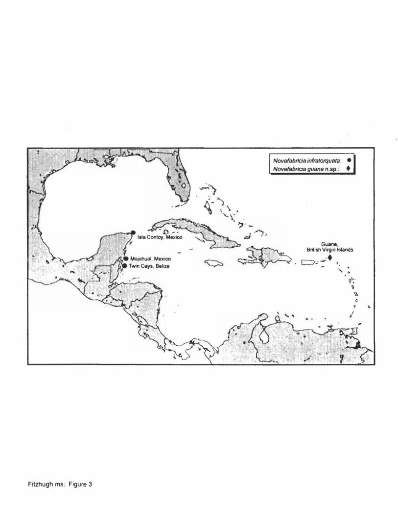
-,
Fitzhugh ms: Figure 3
Novafabrlcia inftatorquala : •
Novafsbrlcia guana n.'p.: •
.. ..., d ~ ,
~ .. ..
'" .' , . .
Guana, British Virgin IsJands
CJ~~;· -.
"
• cf;' ~
<a
I • 0
•
, . -:". ~. r ......... I • . • ~, t. 't 1.

........
A-
. Figure 4 Fitzhugh ms.
\ :. '",
c B
A 0.20 mm
B-O 0.01 mm D

S 0.050'0'
B

vi :. :::. apr , ~ ~----.-
ppr
-"'~ .. :-
A B C 0.1 mm
Fitzhugh ms: Figure 6

· -,:' ~
: ~, :--t-- vl---+-C:;;> apr '.- <'. ppr
A B c O.2mm
Fitzhugh ms: Figure 7

dl-~r)/
..
c ___.._
, A-B 0.200 mm .. .. ,
C 0.015 mm
A~
,
Fitzhugh ms: Figure 8

A O.05mm B
Fitzhugh ms Figure 9

Fitzhugh ms: Figure 10
D
A-B 0.20 mm
C-D 0.02 mm
apr ppr

bh >
0.05 mm
Fitzhugh ms: Figure 11

, .> .. :- . '1 :
Fitzhugh ms: Figure 12
MajalllJ8l. Mexico
~.
"':.')
Augenerie/la hummelincki hummelincki: • A nerie/la s . ct. A. hummelink; hummelinck;: •
~ .') ~-. \'\. '\ . '. San Satvador, Bahamas
'-~ -') - ~
.. ,-'"1 } d .
-5.~ ~.
Guana, British Virgin Islands
•• f 7.::: '. . "\ .
Bonaire
... . "";;;; ..... ~
'-0-. -.-\. .?:" • · . · .
" . . . · .~ .
. .' .
i d
•

J 1\ i ! V '-- .
An emendation of tbe fan worm genus Fabricinuda Fitzhugh, 1990 (Polychaeta: Sabellidae:
Fabriciinae), and description of a new species from the Caribbean
K. FITZHUGH
Research and Collections Branch. Los Angeles County Museum of Natural History.
900 Exposition Boulevard, Los Angeles. California 90007. USA; e-mail: k/[email protected]
Running title: New Fabriciinae Fan Worm

A new species of Fabricinllda Fitzhugh, F. guana, is described from the British Virgin Islands,
Caribbean Sea, from intertidal to shallow subtidal depths. The species is distinguished from the
other six described species in having relatively short, cirrifonn dorsal lips and thoracic inferior
pseudospatulate setae in setigers 3-8. Fabricinuda gual1a most closely resembles F. bikinii
(Hartman) and F. trifobala (Fitzhugh) in the more dorsal placement of the branchial crown, leaving
the ventral half of the anterior peristomial ring exposed. An emended diagnosis for the genus is
provided, which removes the presence of pseudospatulate setae as a synapomorphy for the genus.
In addition, previous descriptions of F. bikinii, F. Iri/obala, F. limnicora (Hartman), and F.
pseudoco({aris Fitzhugh have stated that dorsal lips are absent, but vascularized, ventral filamentous
appendages are present, whereas remaining species (F. pseudoparpa Fitzhugh, F. rongifabrum
Fitzhugh) have lobate lips that are slightly longer than wide. These "appendages" are reinterpreted
here as highly modified dorsal lips . Cladistic relationships among the seven described species of
Fabricinuda are presented.
KEYWORDS: Polychaeta, Sabellidae, Fabriciinae, Fabricinuda
2

Introduction
The fan wonn genus Fabricinllda Fitzhugh, 1990a is knovm for at least six species from
temperate to tropical, intertidal and continental shelf habitats in the Atlantic, Pacific, and Indian
Oceans (Fitzhugh, 1983, 1990a, 2001a; Uebel acker, 1984; Nishi, 1996). The genus is one of the
more readily recognized from among the small-bodied Fabriciinae, especially because of the highly
reduced collar on the anterior peristomial ring, which gives the anterior margin of the ring a more
truncate appearance than is seen in species in other genera. In addition to reduction of the collar,
the monophyly of Fabricinuda has remained very consistent with the occurrence of thoracic
pseudospatulate setae in setigers 3-8; these setae are common in the subfamily, but when present
in other genera, they have a more limited distribution, usually between setigers 3-6 or 3-7. Previous
systematic and cladistic studies involving Fabncinuda (Fitzhugh 1990a, 1998, 2001a) have noted
the absence of branchial crown dorsal lips in F. limnicola (Hartman, 1951), F. bikinii (Hartman,
1954), F. Irilobala (Fitzhugh, 1983), and F. pseudocollaris Fitzhugh, I 990a, but these species have
been described as having vascularized, ventral filamentous appendages (sensu Fitzhugh 1989).
Interestingly, appendages are absent in those species with dorsal lips, i.e., F. pseudopalpa Fitzhugh,
1990a and F. longilabrum Fitzhugh, 2001a.
During a marine invertebrate biodiversity survey in the vicinity of Guana Island, British
Virgin Islands, in the Caribbean Sea, a new species of FabriclnlJda was collected and is described
here. The species exhibits a dorsal lip morphology unlike that seen in other members of the genus,
and has prompted a reinterpretation of the presence of dorsal lips throughout the genus. The new
species also has a thoracic pseudospatulate setal distribution different from that in other species in
the genus. As a result, the diagnosis of FabricinlJda is emended for the first time since it was named
3

by Fitzhugh (1990a). Subsequent to the cladistic analysis conducted by Fitzhugh (200 I a) for species
in the genus, a revised analysis is performed with the inclusion of the new species and new character
data.
Specimens examined for the present study have been deposited in the Los Angeles County
Museum of Natural History'S Allan Hancock Foundation Polychaete Collection (LACM-AHF).
Fabricinuda Fitzhugh, 1990a, emended
The following diagnosis mainly follows that given by Fitzhugh (1990a). Those sections
which have been emended are indicated in bold, and the justifications for these changes are
discussed in the Remarks. Synapomorphies for the genus are shown in italics.
Diagnosis. Small species with eight thoracic and three abdominal setigers. Branchial crown
with three pairs of radioles; distal ends filamentous, about same width as pinnules. All pinnules
terminating at about same hcight, extending to same height as radioles or slightly shorter. Dorsal
lips present and eitber (I) short, lobate, and sligbtly longer tban wide, (2) sbort and cirriform,
or (3) very elongate and filiform; lips of types (1) and (2) witb very narrow,longitudinal blood
vessel, type (3) contains a very wide vessel. Branchial hearts present. Branchial lobes not fused
together mid-dorsally; lobes attached to peristomium in typical sabellid form or with very narrow
attachment along dorsal half of peristomial margin. Mouth located centrally between branchiallobcs
or shifted dorsal to attachment point of lobes. Anterior margin of anterior peristomial ring as low.
even or slightly oblique ridge afl around except for narrow mid-dorsal separation which is
continuous wllh low. median lobe; lateral margins of mid-dorsal separation smooth or with pair of
4

low, rounded lobes. An/erior pens/omial rmg as wide as long or longer. Peristomial eyes black,
round to crescentic; pygidial eyes black, round. Superior thoracic notosetae elongate, narrowly
hooded. Inferior thoracic notosetae short, elongate narrowly hooded in setiger 2 and sometimes
setiger 8; setigers 3-7 or 3-8 with pseudospatulate setae. Thoracic uncini acicular, with large
tooth above main fang; hood present. Abdominal uncini rasp-shaped plates; manubrium about same
length as dentate region; dentate region "lith several teeth per row. Abdominal neurosetae elongate,
\Vith very narrow hoods. Body wall pigmentation present or absent. Females with ovaries in setiger
4. Males with sperm development in setigers 4-8; spermatids arranged in groups of more than
100 attached to central cytopbore. Young brooded in maternal tubes to juvenile stage.
Remarks. The present diagnosis is a modified version of that provided by Fitzhugh (1990a).
The original diagnosis did not include the occurrence of gamete formation and brooding among
species in the genus. These data have been included based on the work of Rouse & Fitzhugh (1994),
Nishi (1996), Fitzhugh (200 I a, pers. obs.), Rouse (pers. comm.), and the present study.
Based on the present study, the diagnosis has changed in two respects. First, Fitzhugh (1990a,
see also Fitzhugh 1998,200 la) included as a synapomorphy for the genus the distribution of inferior
thoracic pseudospatulate setae in setigers 3-8. Among other Fabriciinae species with
pseudospatulates, occurrence is more limited, usually between setigers 3-6 or 3-7. The new species
of Fahncmuda described below has pseudospatulates in setigers 3-7, thereby precluding the 3-8
distribution as a synapomorphy for the genus (see Cladistic relationships among species of
Fabric;'lUda below).
A second significant change in the diagnosis pertains to the interpretation of dorsal lips.
Originally, Fitzhugh (l990a) described Fabncmuda species as either having dorsal lips or
5

vascularized, ventral filamentous appendages. In contrast to what was seen in other genera with
appendages (Fitzhugh 200 I b: figure I) , dorsal lips and appendages were never seen occurring
simultaneously in a species of Fabricinuda. Dorsal lips have been described in F. pseudopalpa and
F. longilabrum as being slightly longer than wide and distally rounded (Fitzhugh 1990a: figure 6E,
2001a: figure IB, respectively), but otherwise similar to the more triangular shape seen in most
other Fabriciinae species (e.g., Fitzhugh 1989: figure 14D, E, 1990b,I993, 1996: figure IB, 1998:
figures 2B, IOB,1999: figures IB, 3B, 200 la: figure 3). Previous distinctions between the shapes of
dorsal lips have only taken into consideration whether lips are well developed (i .e., triangular or
slightly elongate) or reduced to narrow ridges (Fitzhugh 1998). Discriminating between lips that are
triangular and the more elongate form seen in F. pseudopalpa, F. longilabrum, and the new species
dcscribed below, is warranted since these respective shapes are easily characterized. In addition to
the elongate, lobate lips in F. pseudopalpa and F. longilabrum, a slightly different elongate form
\~ill be described below in the new species, where the lips are distinctly narrower than long and
cirri form . Fitzhugh (1998: figure 9B) described the dorsal lips in Bri/acia melaslellaris as being
slightly longer than wide, and are similar in appearance to the lips in F. pseudopalpa and F.
IOllgilahrum.
Four Fabricinuda species have been described in the past as having vascularized, ventral
filamentous appendages (Fitzhugh 1990a, see also Fitzhugh 1998, 200 la): F. bikmii, F. limnicola,
F psewioco/larls, and F Irilohala. As noted earlier, dorsal lips have been described as being absent
in these instances, which differs from what has been observed in other fabriciins with appendages
in that these structures are always associated with triangular dorsal lips. The presence of cirri form
dorsal lips in the new species described below, F. guana, raises the question of the homologous
6

relations between the "appendages" in these four Fabricinuda species and appendages and dorsal
lips in other fabriciins . The dorsal lips in F guana are essentially intermediate in form between the
wider, elongate lips in F pseudopalpa and F. longilabrum and what were originally interpreted by
Fitzhugh (1990a) as vascularized, ventral filamentous appendages in the other four species. As will
be described below, the dorsal lips in F guana have a narrow blood vessel extending through their
length. A similar vessel can also be seen in Flongilabrum (pers. obs.) and probably also exists in
F. pseudopalpa, although I have not examined specimens of the latter for this structure. These
observations preclude using the presence of the larger blood vessel in the "appendages" in F bikinii,
F limnicola, F pseudocollaris, and F Irilobala as a basis for judging these structures as different
from what have been interpreted as dorsal lips in the other three species. Based on these
observations, it seems reasonable to reinterpret the "appendages" in F. bikinii, F limnicola, F.
pseudocollaris, and F. Irilobala as dorsal lips as opposed to filamentous appendages. This
reinterpretation establishes a greater level of uniformity in the genus in that all species have dorsal
lips as opposed to the view that some species have dorsal lips and other species have filamentous
appendages, with concurrent loss of lips. This uniformity is also consistent with the patterns seen
among other fabriciins, where dorsal lips are always associated with ventral filamentous
appendages. Figure I provides a summary of the homologous relations between dorsal lips and
filamentous appendages among Fabriciinae genera, as well as the general shapes of lips. Based on
the prt!sent interpretation of dorsal branchial lobe structures in Fabricinuda, species in this genus
exhibit the greatest variation in dorsal lip structure. A different type of variability occurs among
species in Pseudufabriciola Fitzhugh, 1990b, where dorsal lips are either triangular or as narrow
ridges, and vascularized, ventral filamentous appendages can be present or absent (see review by
7

Fitzhugh 1998, 200 I a; figure I).
There is the alternate interpretation that might be applied to Fabricillllda, namely that all of
these dorsal branchial lobe structures represent filamentous appendages, \\~th the loss of dorsal lips
as a synapomorphy for the genus. Relative to what is seen in the Fabriciinae overall, this conclusion
seems less tenable. Exclusive of Fabricilluda, all Fabriciinae species have dorsal lips (Fitzhugh
1998, 200 I b; figure I). It is the presence or absence of filamentous appendages that is variable.
Given the consistency of occurrence of dorsal lips in the subfamily and the presence of what might
appear to be dorsal lips in F. pseudopalpa, F. IOllgilabrum, and the new species described here, it
appears more parsimonious to interpret all of the structures along the inner dorsal margins of the
branchial lobes as dorsal lips.
Fitzhugh (1990a) identified three characters as establishing the monophyly of Fabricilluda:
(I) reduction of the anterior peristomial ring margin to a low ridge, in contrast to having a well
developed collar; (2) the anterior peristomial ring being at least as wide as long, as opposed to
distinctly wider than long; and (3) the occurrence of thoracic pseudospatulate setae in setigers 3-8,
rather than in a fewer number of contiguous setigers. As is indicated in the diagnosis, and will be
shown in the cladistic analysis below, the pseudospatulate distribution in setigers 3-8 is no longer
a synapomorphy for the genus, but characters (\) and (2) continue to define the genus. With the
reinterpretation of ventral filamentous appendages in Fabricilluda species as dorsal lips, it is
conceivable that one could consider the more general condition of lips being longer than wide as
an additional synapomorphy. In the emended diagnosis and the cladistic analysis for the present
study, I have opted for the alternative of characterizing dorsal lip shapes among species more
explicitly with n:gard to specific shapes. As part of the present study, the possibility that the
8

elongate dorsal lips among species in Brifacia and Fabncinuda might be considered a
synapomorphy for an exclusive sister-group relationship between these two genera will be discussed
as part of the cladistic analysis of relationships among Fabricinuda species presented below. In the
event these two genera are sister taxa, this would preclude elongate lip shape as being a
synapomorphy for Fabricinuda.
Fab,icinuda guano, n.sp.
(figures 2-4; table I)
•
Material examined. Atlantic Ocean, Caribbean Sea, British Virgins Islands. Holotype:
LACM-AHF 0000, sta. BVI-059, Beef Island, Trellis Bay, adjacent to airport runway, scrapings of
algae off coral rubble, depth 1.0 m, coIl. K. Fitzhugh, 12 July 2000. Paratypes: LACM-AHF 0000
(five specimens), sta. BVI-OO I, Guana Island, North Bay, dredged area in front of beach house and
pier, algal scrapings consisting of Amphiroa sp., Gelidium pusillum, Laurencia sp., blue-green algae,
with coralline silt and crustacean tubes off coral rubble, N 18° 28.909', W 64° 34.485', depth 0.7 m,
colI. K. Fitzhugh, 3 July 2000; LACM-AHF 0000 (10 specimens), sta. BVI-002, Guana Island, North
Bay, dredged area in front of beach house and pier, algal scrapings consisting of Amphiroa sp, and
blue-green algae off coral rubble, N 18° 28.909', W 64° 34.490', depth 0.5 m, coil. K. Fitzhugh, 3
July 2000; LACM-AHF 0000 (eight specimens), sta, BVI-003, Guana Island, North Bay, 30 m west
of pier, algal scrapings of Cladophora sp. and G. pus ilium, with coralline silt, crustacean tubes, and
detritus off coralline rocks, N 18° 28.913', W 64° 34 .503', depth 20 cm, coil. K. Fitzhugh, 3 July
2000; LACM-AHF 0000 (five specimens), sta. BVI-005, Guana Island, White Bay, finger reef near
beach house, east of pier, 40 m offshore, scrapings of zooanthid anemones, sand, and filamentous
9

algae (Ceramium sp., Polysiphoma sp.) ofT coral boulder, depth 1.0 m, coli. K. Fitzhugh, 4 July
2000; LACM-AHF 0000 (27 specimens), sta. BVI-017, Guana Island, White Bay, shoreward side
of middle finger reefs, east of pier, scrapings of zooanthid anemones and coralline silt ofT coral
boulder, depth 1.0 m, coil. K. Fitzhugh, 6 July 2000; LACM-AHF 0000 (one specimen), sta. BVl-
020, Guana Island, Grand Ghut, scraping ofT sponges, depth 9.0 m, coil. L. Jarecki, 6 July 2000;
LACM-AHF 0000 (II specimens), sta. BVl-OS5, Guana Island, Long Point, Muskmelon Bay, algal
scrapings ofT reef slope, depth IS.O m, coil. R. Ware, II July 2000; LACM-AHF 0000 (one
specimen). same station as holotype; LACM-AHF 0000 (I specimen), sta. BVl-066, Guana Island,
North Bay, 30 m ofTshore from pier, algal scrapings of Amphiroa sp. and Dictyota sp. off coral
rubble, N 180 28.909', W 64° 34.490', depth O.S m, colI. K. Fitzhugh, 14 July 2000.
Descriprion. Holotype a complete male ",ith 8 thoracic and 3 abdominal setigers. Branchial
crown length 2.0 mm, remainder of body 2.40 mm long, maximum \\~dth 0.2 mm in anterior thorax.
Trunk slender, slightly tapering posteriorly (figure 2A). Branchial crown ",ith 3 pairs of radioles,
distal ends filamentous, same \~dth as pinnules. All radioles with five pairs ofpinnules; all pinnules
terminate at about same height, slightly shorter than tips of radioles. Dorsal lips erect, about five
times longer than \~de, distally rounded (figure 2B, C); narrow coelomic space containing blood
vessel extends through entire length of lips (figure 2C). Ventral lips and ventral filamentous
appendages absent. Dorsal margins of branchial lobes not fused to one another. Branchial hearts
present (figure 2A, B). Anterior peristomial ring collar as a low ridge, separated mid-dorsally by
distinct gap; dorsolateral collar margins as small lobes partially overlapping mid-dorsal medial lobe
(figures 3A, 4A). Mid-dorsal medial lobe broadly rounded distally, extending slightly beyond collar.
10

Several paratypes with distinct pair of spherical "auxilia1')'" lobes lateral to median lobe and
partially beneath lateral collar lobes (figure 4A); "auxiliary" lobes originate along dorso-Iateral
margins of anterior peristomial ring; composed of single layer of thick cells surrounding hollow
(coelomic?) space. Annulation between collar and anterior peristomial ring not visible. Antero
dorsal margin of anterior peristomial ring extends beyond antero-ventral margin, creating a shelf
like appearance along ventral half of anterior peristomial margin; some paratypes with similar
arrangement as in holotype, other paratypes with anterior margin relatively even (figures 3, 4A).
Anterior peristomial ring slightly longer than wide, about five times longer than posterior
peristomial ring in holotype (figure 2A). Annulation between rings visible ventrally and laterally
(figures 2A, 3, 4A). Mid-dorsum of anterior peristomial ring with shallow groove extending from
mid-dorsal collar separation to posterior peristomial ring (figure 4A), groove same width as mid
dorsal collar separation. Pair of crescentic or round. black eyes near bases of dorso-Iateral collar
lobes. Setiger I about one-halflength of anterior peristomial ring (slightly shorter in some paratypes:
figures 3, 4A), wider than long: setigers 2-3 each about same length as setiger I; setigers 4-8
successively longer than setiger 3, with setiger 8 about 2.5 times longer than Seliger I. Setiger 9
slightly shorter than setiger 8, with seligers 10-11 each successively shorter than 9. Pygidium about
same length as setiger II, posterior margin slightly tapered, rounded (figure 2A). Pair of round,
black pygidial eyes (figure 2A). Superior thoracic notosetae elongate, narrowly hooded, 3-4 per
fascicle. Inferior thoracic notosetae in setigers 2 and 8 elongate narrowly hooded, 2 per fascicle;
setigers 3-7 with pseudospatulate setae (figure 48), 2 per fascicle . Abdominal neuropodia ofsetigers
9-11 with very elongate, narrowly hooded setae, 1-2 per fascicle. Thoracic uncini acicular, main
fang slender (figure 4C); large tooth offset from mid-line of main fang present, remaining teeth
I I

behind main fang slender and slightly decrease in size away from fang; hood present; 5-6 uncini per
fascicle in irregular single rows. Abdominal uncini with 7-8 rows of teeth in profile, 3 teeth per row
(figure 40); manubrium about same length as dentate region, slightly expanded proximally; uncini
in setigers 9-11 number 19,20, and 14, respectively. Females with oocytes in setiger 4, males with
developing sperm in setigers 4-8. Pigmentation absent on branchial crown and body wall of all
specimens. Specimens occasionally found in tubes composed of coralline sand and fine detritus. No
brooding of young observed.
Etymology. The specific epithet refers to the island around which most specimens of the
species were collected.
Remarks. Fabricinuda guana is readily distinguished from other species of Fabricinuda by
the presence of thoracic pseudospatulate setae in setigers 3-7, whereas all other described species
have pseudospatulates in setigers 3-8. The cirri form shape of the dorsal lips is also a unique feature .
As was indicated in the Remarks for the generic diagnosis, all other species of Fabricinuda have
dorsal lips that are either wider and more lobate or have the appearance of "vascularized, ventral
filamentous appendages."
Fabricinuda guana shows greatest similarities to F bikinii and F trtlobata in that the
branchial cro\m in each species is shifted dorsally, leaving the ventral half of the anterior
peristomial ring well exposed (table 1). Among these three species there is also the tendency for the
anterior peristomial margin to be either transverse relative to the long axis of the body (figure 3) or
distinctly oblique (figure 2A; Fitzhugh 1990a: figures 28, 3D; Nishi 1996: figures 2A, D, 4A). In
all other species, the margin is always transverse and completely covered by the attachment of the
crown.
12

There is some superficial resemblance between Fabrtcmuda guana and F. pseudopalpa in
that both species have paired extensions from the dorso-Iateral margins of the anterior peristomial
ring. The extensions are not, however, homologous. Fitzhugh (1990) showed that the palp-Iike
structures in F. pseudopalpa are elongations of the dorso-Iateral collar lobes, within which are
located the anterior vascular loops. The "auxiliary" lobes in F. guana are completely separate from
the collar lobes and internally appear to contain coelomic spaces, but with no blood vessels. As well,
the extensions in F. pseudopalpa are very variable in length, ranging from about as long as wide to
much longer than wide (Fitzhugh 1990a: figure 7). It is unknown at this time whether the "auxiliary"
lobes in F. guana are preservation artefacts or a feature present in living specimens.
The description of Fabricinuda guana raises the number of western Atlantic/Caribbean
species to three, the other species from the region being F. trilnhata from the intertidal zone along
Belize and F. pseudocollaris from shallow subtidal depth along the west coast of Florida. A possible
additional undescribed species (Fitzhugh 200 la) was identified as F. trilobata by Uebelacker (1984,
as Fabriciola rrilobata) from the Gulf of Mexico continental shelf off Texas and Florida at depths
ranging from 10 to 189 m. The knOV,'ll depth distribution for F. guana is from the intertidal zone to
15 m.
Key to species of Fabricinuda
The following key has been modified from that presented by Fitzhugh ( 1990a). The principle
change is in the interpretation of what have previously been regarded as vascularized, ventral
filamentous appendages, which are now interpreted as dorsal lips. The original key by Fitzhugh
(1990a) contained five species , whereas the present key also includes F.longilabrum and F. guana.
13

la. Dorsal lips very elongate, filamentous, "ith large internal blood vessel (Fitzhugh I 990a:
figures IE, 2E, 3E, 5A, 9A), closely resembling the vascularized, ventral filamentous
appendages of species in Pseudoaugeneriella and some Pseudofabriciola .... ..... .. 4
lb. Dorsal lips short, lobe like (Fitzhugh 1990a: figure 6E, 200 la: figure I B) orcirriform (figure
I B, C), without large internal blood vessel (but might have a narrow vessel) ......... 2
2a. Thoracic inferior pseudospatulate notosetae in setigers 3-8; dorsal lips lobe like . ..... 3
2b. Thoracic inferior pseudospatulate notosetae in setigers 3-7; dorsal lips short, cirri form ..
. . . . . . . . . . . . . . . . . . . . . . . . . . . . . . . . . . . . . . . . . . . . . . . . . . . . . . . . . . . F. guana n.sp.
3a. Anterior peristomial ring distinctly longer than wide (e.g., Fitzhugh 1990a: figure I B, 200 I:
figure 2B) . . . . . . . . . . . . . . . . . . . . . . . . . . . . . . . . . . . . . . . . . . . . . . . . .. F. longilabrum
3b. Anterior peristomial ring as long as wide (Fitzhugh 1990a: figure 6B) .. F. pselldopalpa
4a. Branchial crown attachment to peristomium shifted dorsally to some extent, exposing
antero-ventral peristomium margin (e.g., figure lA) ..... .. ... .... ............... 5
4b. Branchial crown attached over entire anterior peristomium margin (e.g., Fitzhugh 1990a:
figure IS) .. .. ... . . ..... .. ...... .. ..... . ........... . . ........... . ... 6
5a. Mouth located in typical sabellid position, i.e., centrally on the anterior peristomium margin
(Fitzhugh 1990a: figure 5B) ......... .. ................ . . .. F. Irilobalo
5b. Mouth located along antero-dorsal margin of peristomium, dorsal to branchial lobe
attachment (Fitzhugh I 990a: figure 2F) .............. .. .......... . ... . . F. bikinll
6a. Ventrum of anterior peristomial ring with paired, membranous lappet-like processes
(Fitzhugh 1990a: figure 8C) . . . . . . . . . . . . . . . . . . . . . . . . . . . . . . . . .. F. pselldocollaris
6b. Ventrum of anterior peristomial ring smooth, without processes .. .. ...... F. limnicola
14

Cladistic relationships among species of Fabricinuda
Previous cladistic analyses examining relationships among species of Fabrrcinuda were
reviewed by Fitzhugh (200Ia), who also presented the first cladistic analysis limited to species in
the genus. As was noted by Fitzhugh (200 la), all previous studies had only considered relationships
within Fabricinuda in the context of relationships with all other Fabriciinae genera and species (i.e.,
Fitzhugh, 1991, 1992, 1993, 1995, 1998, 1999). The purpose of the present analysis is to examine
the relationship of F. guana to other species, as well as to modifY the characters used by Fitzhugh
(200 la) based on the reinterpretation of dorsal lip morphologies and to correct a previous error in
character coding regarding length/width relations in the anterior peristomial ring.
The analysis performed by Fitzhugh (200 la) consisted of six ingroup taxa and the properties
associated with four morphological features (table 2). These features included (I) the venlTal
margins of branchial lobes, which pertained to the presence or absence of ventral filamentous
appendages; (2) the presence or absence of dorsal lips; (3) position of the branchial crown; and (4)
dimensions of the anterior peristomial ring. The present analysis includes the seven species in the
genus, but only considers properties among three features (table 3): dorsal lip shape, branchial
crown position, and anterior peristomial ring dimension. Consistent with the above modifications
in the interpretation of dorsal lips in the genus, feature (I) of Fitzhugh (200Ia) has been removed,
and the properties of the dorsal lips have been expanded to accommodate the very elongate,
digitiform condition seen in most species (table 3: feature I) and to differentiate the lobate and
cirri form conditions from the more typical triangular shape seen in most fabriciins. The lobate lips
in Fabricirludapseudopalpa and Flongilabrum, and the cirriform lips in F. guana were considered
equivalent in this analysis since the overall shapes can be characterized as moderately longer than
15

wide , in contrast to the very elongate, digitifonn conditions seen in F. bikinii, F. limillcola, F.
trilobala, and F. [lseudocollaris.
The analysis by Fitzhugh (200Ia) incorrectly treated the anterior peristomial ring in
Fabricinuda pseudopalpa, which is about as long as wide (Fitzhugh 1990a: figure 6B), as
plesiomorphic (cf. Table 2), thus equivalent to the condition seen in other Fabriciinae. Exclusive
of Fabricinuda, the anterior peristomial ring in other Fabriciinae species is distinctly wider than
long, not including the anterior collar ex1ensions seen in genera such as Fabricia Blainville, 1828,
Fabriciola Friedrich, 1939, Novafabricia Fitzhugh, 1990b, or Pseudofabriciola Fitzhugh, 1990b.
Within Fabricinuda (see Diagnosis for the genus above), the anterior peristomial ring is at least as
wide as long, as in F. pseudopalpa, or longer than wide as in all other described species in the
genus. As a result, the greater length of the ring is a synapomorphy for the genus and not relevant
to discerning cladistic relationships among species. Properties related to anterior ring dimensions
are therefore not included in the present analysis . With the occurrence of inferior thoracic
pseudospatulate setae in setigers 3-7 in F. guana, which is also seen in some other Fabriciinae
species, as opposed to the 3-8 distribution in all other species in the genus, the occurrence of this
setal type has been included in the present analysis (table 3).
A feature not considered by Fitzhugh (200Ia), or in any of the previous cladistic analyses
involving Fabrrcinuda, is the occurrence of body wall pigmentation, either on the crown or the
anterior region of the trunk (Table I). At least three species of Fabrrcinuda are known to
consistently lack pigmentation: F. blkinii, F. longilabrwn, and F. guana. All of the remaining
species have been described as having brown pigmentation, usually on the branchial crown,
especially the inner margins of the branchial lobes, the median lobe on the anterior peristomial ring
16

and adjacent faecal groove, peristomial rings, and occasionally some anterior thoracic setigers. In
some instances, pigment patterns can be very discrete. For instance, Uebelacker (1984: fib'Ure 54-16)
and Fitzhugh (200Ia) described for F. trilobata and Fabricmuda sp., respectively, the presence of
dark brown pigmentation limited tothe median lobe of the anterior peristomial ring. With the degree
of variability in the patterns of pigment distribution between species, as well as within (Fitzhugh
1983, I 990a), the more general presence or absence of body pigmentation has not been included in
the present analysis .
The determination ofplesiomorphic propenies was based on outgroup comparisons with the
remainder of the Fabriciinae. For dorsal lip shape (table 3: feature I ), species in Fabricinuda all
have lips that are longer than wide. Among other fabriciins , only Brifacia metastel/aris has dorsal
lips that are slightly longer than wide and somewhat similar to the forms seen in F. pseudopalpa and
F. longilabrum. Remaining fabriciin species either have lips that are distinctly triangular or reduced
to narrow, shelf-like processes. A preliminary, unpublished cladistic analysis involving the majority
ofFabriciinae species and genera reveals that in some topologies, B. metas/el/aris is the sister taxon
to the Fabricinuda clade, thus rendering the elongate dorsal lip condition plesiomorphic for the
latter genus. There are as well topologies in which trianb'Ular or shelf like dorsal lips are
plesiomorphic relative to Fabncinwla. As a result, two outgroups were considered for dorsal lips
in the present analysis, one consisting of B. metas/el/aris and the other representing the more generic
fabriciin condition oflips being either triangular or narrow and shelf-like. With respect to features
(2) and (3), both of these outgroups are functionally equivalent.
Me/hods and results
17

Cladograms were constructed from the character matrix presented in table 4 using the
. AllTrees ' command in PAUP* 4.0b7 (Swofford, 1998), with all characters given equal weight. A
single cladogram was obtained using either outgroup, both with the same topology, and each with
a length of4 steps, and ci and ri of 0.75 and 0.83, respectively (figure 5). Relationships are relatively
consistent with the results obtained by Fitzhugh (200 I a: figure 10; figure 6), although, considering
the marked differences in the characters used in the two studies, there is no utility in making specific
comparisons between results. In the present study, Fabricinuda bikinii and F. tri/obata form the
most apomorphic sister group, defined by the shift of the branchial crown to a more dorsal position
(character 2( I )]- this condition is homoplasious, also occurring in F. guana (figure 5). The sister
group to this most apomorphic clade comprises a pOI}1omy involving F. limnico/a and F.
pseudoco//aris. This more inclusive group is based on the presence of the very elongate, digiti form
dorsal lips with prominent blood vessels [character I (2)], i.e., closely resembling the vascularized,
ventral filamentous appendages in species in some other genera. Fabricinuda pseudopa/pa and F.
/ongi/abrum then form a polytomy with these four more apomorphic species, all of which have
thoracic pseudo spatulate setae in setigers 3-8 [character 3(1)]. Fabricinuda guana is the most .
plesiomorphic species in the genus. The transformation series for the dorsal lips among Fabricinuda
species are identical regardless of whether the outgroup condition is the presence of triangular or
narrow lips [character 1(0): figure 5A] or moderately long lips (character 1(1): figure 58]. lt is,
however, only in the case of the outgroup condition being the presence of triangular or narrow dorsal
lips (figure 5A) that elongate lips is a syllapomorphy for Fabricinuda.
Acknowledgments
18

Sincere thanks are extended to Drs. ToddZimmennan and Jody Martin, LACM, for inviting
me to participate in the Guana Island Marine Invertebrate Survey. Ms. Lianna Jarecki, Guana Island,
is gratefully acknowledged for providing material and logistical support. Special thanks go to the
staff on Guana Island for providing superb accommodations. As always, Mr. Thomas H. Perkins,
Florida Marine Research Institute, afforded me his extensive insight on the subject of sabellids. The
field work on which this study was based was funded by National Science Foundation Biotic
Surveys and Inventories Program Grant DEB-99-72 100 to T. Zimmennan and J. Martin, a grant
from the Falconwood Corporation through The Conservation Agency, and the LACM.
References
BLAINVll..LE, H. de, 1828, DlcllOnaire des sciences nalurelles dans lequel on Iraile melhodiquemenl
des differenl elres de la nalure, conslderes soit en euxmemes, d'apres I 'etat actual de nos
connaisscance; soit relalivement a I 'utilite qu 'en peuvent retirer la medicine, I 'agriculture,
Ie commerce etles arts. SuiVl! d 'une biographie des plus celebres naluralistes, Volume 57.
FITZHUGH, K. , 1983, New species of Fabriciola and Fabncia (Poly chaeta: Sabellidae) from Belize.
Proceedings of Ihe BIOlogical Society of Washington, 96, 276-290.
FITZHUGH, K., 1989, A systematic revision of the Sabellidae-Caobangiidae-Sabellongidae complex
(Annelida: Polychaeta). Bulletin of Ihe American Museum of Natural History, 192. 1-104.
FITZHUGH, K., I990a, Fabricinuda, a new genus of Fabriciinae (Polychaeta: Sabellidae).
J'roceedings oflhe Biological Society of Washington, 103, 161-178.
FITZHUGH, K., 1990b, Two new genera of the subfamily Fabriciinae (Polychaeta: Sabellidae).
Amencan Museum Novitates, 2967,1-19.
19

FITZHUGH, K., 1991, Further revisions of the Sabellidae subfamilies and cladistic relationships
among the Fabriciinae (Annelida: Polychaeta). Zoological Journal of the Linnean Society,
102,305-332.
FITZHUGH, K., 1992, On the systematic position of Monroika africana (Monro) (Polychaeta:
Sabellidae: Fabriciinae) and a description of a new fabriciin genus and species from
Australia . Proceedings of the Biological Society of Washington, 105, 116- 131.
FITzHUGH, K., 1993, Novafabricla bnmnea (Hartman, 1969), new combination, with an update on
relationships among Fabriciinae taxa (Polychaeta: Sabellidae). Contributions in Science,
Natural History Museum o/Los Angeles Counly, 438,1-12.
FITZHUGH, K., 1995, Additions to the description of the fanworm genus Pseudo/abrieia Cantone,
1972 (Polychaeta: Sabellidae: Fabriciinae). Contributions in SCience, Natural History
Museum o/Los Angeles Counly, 456, 1-6.
FITZHUGH, K., 1996, New fanworm species (Polychaeta: Sabellidae: Fabriciinae) in the genus
Pseudofabrieiola Fitzhugh. Journal of Natural History, 30,1267-1286.
FITZHUGH, K., 1998, New fan worm genera and species (Polychaeta, Sabellidae, Fabriciinae) from
the western Pacific, and cladistic relationships among genera. Zoologiea Scripta, 27, 209-
245.
FITZHUGH, K., 1999, New fanworm species (Polychaeta: Sabellidae: Fabriciinae) from Phuket,
Thailand, with comments on Fabriciola jlammula Rouse and Fabriciola cn Rouse.
Contributions in Science, Natural History Museum o/l.os Angeles County, 477,1-17.
FITZHUGH, K., 2001a, New species of Fabricinuda Fitzhugh and Pseudofahrieiola Fitzhugh
(Polychaeta: Sabellidae: Fabriciinae), with an emendation of Pseudufabriciola australiensis
20
(Hartmann-SchrOder). Journal o(Natural History. (in press).
FITZHUGH, K., 200 1 b, A new deep-water genus and species ofFabriciinae fan worm from Antarctica
(Polychaeta: Sabellidae). Contributions in SCience, Natural History Museum of Los Angeles
County. (in press).
FRIEDRICH, H., 1939, Polychaeten - Studien V-x. Zur Kenntnis eineger wenig bekannter oder neuer
Polychaeten aus der westlichen Ostsee. Kieler Meeresforschungen, 3, 362-373 .
HARTMAN, 0., 1951, Fabricinae (feather-duster polychaetous annelids) in the Pacific. Pacific
SCience, 5, 379-391.
HARThfAN,O., 1954, Marine annelids from the eastern Marshall Islands. Professional Papers of the
U. S. Geological Survey, 260-Q, 619-644.
NISHI, E., 1996, A new record of Fabridnuda bikinii (Hartman) (Polychaeta: Sabellidae:
Fabriciinae) from Okinawa, Japan. Benthos Research, 51, 21-26.
ROUSE, G. W. and FmHUGH, K., 1994, Broadcasting fables: is external fertilization really primitive?
Sex, size, and larvae in sabellid polychaetes. Zoologica Scripta, 23, 271-312.
SWOFFORD. D. L.. 1998, PA UP·. Phylogenetic Analysis U.~ing Parsimony (·and Other Methods) .
Version 4.0b6 (Sunderland, Massachusetts: Sinauer Associates).
UEBELACKER, J. M., 1984, Chapter 54. Family Sabellidae Malmgren, 1867, inJ. M. Uebelacker and
P. G. Johnson (eds) Taxonomic Guide to the Polychaetes of the Northern Gulf of Mexico,
Volume VII, Final Report to the Minerals Management Service, contract 14-12-001-29091
(Mobile, Alabama: Barry A. Vittor & Ass.), pp. 54-1 - 54-43.
21

Table I. Comparisons of selected features among Fabricinuda species.
Branchial Anterior PseudOipatulate Body wall Type locality (and other SpecIes Dorsal lips crown position perislomial ring setae distribution pigmentation reponed localit.es)
J< bikinii (Hartman, 1954) very elongate, filiform,
shifted dorsally longer than wide setigcrs 3-8 absent Bikini Atoll (Okinav.~,
with large blood vessel Aldabra Atoll)
F. guullu ... ,p. cirriform shifled tonger Ihan
.eligen 3-7 absent British Vi~in Islands dorsally wide
f: Iimrlimla (Hartman, 1951) very elongate, filiform, over entire
longer than wide setigers 3-8 present California with large blood vessel anterior end
F. lon!(i1abmm Fitzhugh, 200la lobe like over entire
longer than wide setigers 3-8 absent Thailand anterior end
J': p.w:udocoJ/aris Fitzhugh, 1990 very elongate, filifomn, over entire
longer than wide setigers 3-8 prescnt Florida with large blood vessel anterior end
J< pselldopalpa Fitzhugh, 1990 lobe like over entire anterior end
as wide as long setigers 3-8 present California
f: Irilobata (Fitzhugh, 1983) very elongate, filiform,
shifted dorsally longer than wide setigers 3-8 l.Ibsent or
Belize with large blood vessel present
Fabricinuda sp. ,renSII Fitzhugh, 2001 a unknown over entire anterior e.nd
longer than wide seligers 3-8 present TI,ailand
22

Table 2. Properties for the four morphological features used by Fitzhugh (200 I a) to determine
cladistic relationships among Fabricinuda species (cf. table 3).
1. Ventral margins of branchial lobes: (0) as narrow shelf-like processes; (I) as ventral
filamentous appendages (i.e. vascularized & unbranched).
2. Branchial lobes adjacent to dorsalmost radioles: (0) as dorsal lips; (1) as even shelf
continuous with ventral branchial lobe margins.
3. Branchial crown: (0) located over entire anterior end; (I) shifted dorsally.
4. Anterior peristomial ring: (0) at least as wide as long; (I) distinctly longer than wide.
23

Table 3. Revised properties of features used to detennine cladistic relationships among
Fabriclnuda species in the present study.
I . Dorsal lip shape: (0) triangular [e .g., Fabricia stellaris (Muller)] or narrow, shelflike (e.g.,
Novafabricia spp.); (I) moderately longer than wide, i.e ., lobe like or cirrifonn; (2) very
elongate, digitifonn.
2. Branchial crown position: (0) extends over entire anterior peristomial margin; (1) shifted
dorsally, exposing ventral half of anterior peristomial margin.
3. Distribution of inferior thoracic pseudospatulate setae: (0) setigers 3-7; (l) setigers 3-8.
24

Table 4. Character matrix for Fabricinuda species based on characters presented in table 3.
Characters
Outgroup, : Brifacia melaslellaris
OUtgrOUP2: Fabricia, Novafabricia
F bikinii
F. guano n.sp.
F lirnnicola
F longilabrum
F pseudocollaris
F pseudopalpa
F rrilobala
25
o 2
1
2
2
2
2
o o
1
o o o o
3
o o
o

Figure legends
Fig. I. Relationships between the occurrence of branchial crown dorsal lips (dark gray) and ventral
filamentous appendages (light gray) among Fabriciinae genera.
Fig. 2. Fabricinuda guana: (A) holotype, LACM-AHF 0000; (8-C) paratypes LACM-AHF 0000:
(A) entire specimen,lateral view (left side); (8) right halfofbranchial crown, inner margin;
(C) detail of dorsal lip. Abbreviations: apr, anterior peristomial ring; bh, branchial heart; by,
blood vessel; dl, dorsal lip; ppr, posterior peristomial ring.
Fig. 3. Fabridnuda guana (paratype LACM-AHF 0000): (A-C) dorsal, lateral (right side), and
ventral views, respectively, of anterior end (branchial crown missing). Abbreviations: apr,
anterior peristomial ring; cl, dorsolateral collar lobes; ml, median peristomial lobe; ppr,
posterior peristomial ring.
Fig. 4. r"abricinuda guana (paratype LACM-AHF 0000): (A) dorsal view of anterior end (branchial
crown missing), showing the presence of distinct, "auxiliary" lobes (cf. figure 2A); (8)
inferior thoracic notopodial pseudospatulate setae from setiger 5; (C) thoracic uncinus from
setiger 5; (D) abdominal uncini from setiger 9. Abbreviations: aI, "auxiliary" lobes; apr,
anterior peristomial ring; cl, dorsolateral collar lobes; ml, median peristomial lobe; ppr,
posterior peristomial ring.
26

Fig. 5. Relationships among species of Fabricinuda based on the data matrix presented in Table 3.
Transfonnation series for the apomorphic properties referring to dorsal lip shape (I) ,
branchial crown position (2), and thoracic pseudospatulate setae distribution (3) are shown:
(A) transfonnation series in which either triangular or narrow, shelf-like dorsal lips is
plesiomorphic [1(0)); (8) transfonnation series in which dorsal lips that are moderately
longer than wide is plesiomorphic [1(1)] .
Fig. 6. Comparison of (A) the minimum-length tree obtained by Fitzhugh (2001a: figure 10) and
(8) the topology from the present study (cf figure 5).
27

• dorsal lips present
ventral filamentous appendages absent
dorsal lips present
• ventral filamentous appendages present
Fitzhugh ms: Fig, 1.
'nonvascularized' ventral filamentous appendages
• 'vascularized' ventral filamentous appendages
Brifacia Fabricia Parafabricia Pseudofabricio/a, Raficiba
Novafabricia Pseudofabricio/a,
Brifacia Fabric/nuda,
Fabric/nuda,
Fabricio/a
Pseudofabricia
Manayunkia Monroika Pseudoaugenerie/la Pseudofabricio/a, 'Genus A'
Augenerie/la

bh
..... ~
..... ...,
c
B A O.Smm
,1" ;I ,l ' I ' • B 0.1 mm •
C 0.06 mm
Fitzhugh ms: Fig. 2. Fabricinuda guana n.sp.

cl
...... <II",g.fj .to
A B c 0.1 mm
Fitzhugh ms: Fig. 3. Fabricinuda guana n.sp.

A
apr
] ppr ----1Jrrf
A
c
D 0.1 mm
8-0 0.015 mm
Fitzhugh ms: Fig. 4. Fabricinuda guana n.sp.
B
f

A
B
Fitzhugh ms: Fig . 5.
1 (0)
3( 1): pse ........... 1asa in setlQenl »
guana n.sp.
~~----- longilabrum
r'l 1(1)
1 (2)
13(1): pS;l~tWales in Mtjge" 3-8
pseudopalpa
.~--- limnicola
pseudoco/laris
'-- bikinii
trilobata
~2(1)~=~ guana n.sp.
~~----- longilabrum
pseudopalpa
.~--- limnicola 1(2)
pseudoco/laris
-- bikinii
trilobata

guana n.sp.
pseudopalpa
longilabrum
Iimnicola
pseudocol/aris
bikinii
A trilobata B
Fitzhugh ms: Fig. 6.
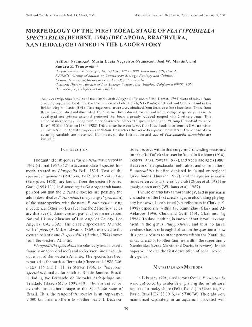
GulLtnd CarihJ:x:<ll1 Research Vol. 13.19--85.2001 M.~l1LlS.(; r i pl received O..:lohcr fI, "l(J(J(i. cll.:ce ptcd LlIIuar:-- 5. 2001
MORPHOLOGY OF THE FIRST ZOEAL STAGE OF PLATYPODIELLA SPECTABI LIS (HERBST, 1794) (DECAPODA, BRACHYURA, XANTHIDAE) OBTAINED INTHE LABORATORY
Adilson Fransozo '• Maria Lucia Ncgrciros-Fransozo l, .Jocl \\'. ]\'1artin 1, and
Sandra E. Trautwein 2. '
'/)eparramenlo de l o%g/{{, tn. [;,\L."il', / 86J 8·(}{)(}. BOIllcarll (SJJ.), Bra:.:i/,
X/~·nf.'CC (Group O[Sllldlo:!s on Cruslau:olI Biology, Fco l o.,,~' and Cullllrl...'). E-mail.!runso=o.il)hhunesp.hrandmln.fi.j /hb.lIT1esp.br ' .\"OlUrai lfislory .\ll/scum n[ /.lJs ..t"gt:/es ('olln ty. I.os .·lfI,f!.e/cs. Calijorllla 9(j(}07. USA ' l..'nivus ily (~r Ca;lfOrf1la I.os .-/Jlg eies
A bSlr(/Cf Ovigl,.·wus lcmak " of til\: xanth id crab /1/alyp otlit.'lIa speCfahilt.l" (llcrh sl, 1794) \n rl,.' ohtai !1l.:d from 2 widl'ly ,S\:pa rat l,.'d 10lo:alities: th\: lJhatuha ,oasl (Felix [h:ach. Sao Pau lo) of Ilrazi l and (juana "land in the llrit b h Virgin hlands (B VI) . Firs t ~ tagi.! 10l:.'a lar\ ac were obtninl!d from fcmaks at hOlh l o~atiolls. Those frolll Brevi I are d~~crihed and il lmtralcd . Th~ first loea hl!Jrs dorsal. rostral. and l al~ra l carap<1Io:C spi n~". plus a wl.:lldeveloped and spinose antenna I prolopod that hl.!ars a greatly reduced cxopod with 2 min ute setae. This antenn al morpho log) . along with other chara cll.! r", places the species amot1~ thl.! "Group [" xan thid lOcas or Ricl'" ( 19XO) and \tart in ( 1984. 19X8) . Dj ffcn:nc;.!":-; bel\vecn larvae from Drazi I and Ihose from the B V I arc minor and arlo: :.tllributed 10 within-::-plo:eics \ari:lllon. Charac tcrs that ~cr\'c to s..:parat..: lhc'5c larva..: froillthose of co· occuring \imthids arl' prl.: ) Io:Jllcd . Comments on the dislnbul io ll and :-.J,le of" ffalypod/e/la spec/abi"s arc included.
hTROIJLcrlc)~
The xanthid crab genus P/mypodu.dla was erected in 1967 (Guinot 1967:562) to accommodate 4 species formerly treat~d as Platypodia Bell. 1835. Two of the species, P. gemmata (Rathbun. 1902) and r. rO/llndara
(Stimpson, 1860), are kn own from Ih..: I;!<lstern Pacific. Garlh (1991: 131 ), In discussing the Galapagos crab fauna, poimed out that (he 2 Pacific species an~ posslb.ly the adu It (d0scribcd <IS I). rofllndu{a) and ~'oung (/'. gemmata)
or the sam~ species. with the name r. rOlllndata having precedence . Other workers feel that the 2 PJcific spcci~s are distinct (r. Zimml!rman, personal communi cation, Natural History Museum of Los Angeles County, Los !\ngcles 1 C!\, USA). The other 2 species are Atlantic, with P. picto (A. Milne Edwards, 1869) r~strictcd 10 the CJstern i\ t Ian (ic and P .. \p":4..:(uhi/ IS (II erb s. t. 1794) known from the weSlern ,\tbnti c.
Plafypodiellu SI)(:clubi/i.r; i~ a rdalivdy small xanthid found ill or near coral r~c fs and rocky shore I ine~ throughout most of the western Atlantic. The species has been report\.'d as far north as l1crmuda (Chace d al. 1986:346. plate ..:; I l:'i and I 1 : 1 I. in Starer 1986, (Ii P/aIYI}()tiia
spec-ta}"ills) and as far south as Rio de Janeiro, Brazil. Including the Fernando de Noronha Archipelago and Tnlldadc Is land (Melo 1998:490 ). The currenl repon
L:x{cnds the southern range to the Sao Paulo state of Brazil. Thus, the range of the species is an impresslvc 7,000 km from northern to ~ou thLrn extent. Di str ihu-
79
tional records within this range, and (!xtcnding weslward illlo Ihe GulfofMexieo, can be found in Ralhbun ( 1930),
FoIder( 1973). Powers (1977), and Abele and Kim (1986).
Because of its spectacular coloration and color pattern. P . .'ipcctabi/Is is often depicted in faunal or reg.iona l guide books (Humann 1992), and the species is some · times referred to as the calico crab (CIHlce cl al. 1986) or gaudy clown crab (Williams el .1. 1989).
The use of crab larval morphology, and in particular characters of the first zoeal slag!;!, in elucidating phylogeny is now well established (see references in Clark et al. 1998) especially wilhin Ihe Xanlhidae (Clark "nd AI
Aidaroos 1996. Clork alld Galil 1998, Clark .lId Ng
1998). To dale, nothing is known about larval developn1c::nt in the genus Platypodiclla. and thus no larval evidL'nce has beell brought to bear on the question ofl1ow thi s genus relates to olher gC::llcra within the Xanthidae sensu Hric/a or to othe r families within the superfamily Xanthoid..:a (sensu Martin and Davis , in review). In thi :-. paper we provide th l;! first desc ription of zoea l IJrvac in 1his gc;l\us .
MATERIAl.S A:\I) 1\'IETIIOl)S
In I-=ebruary 1998,4 ovigero us female P SP!!CWhtlfS
\\o'ere co!lected by scuba diving along the infralinoral region of a rocky shore (F~lix lJeach) in Ubatuba, Sao Paolo, Brazil (23 ' 23 '00"5, 4~ 5 T06"W). The crabs were
m<.lintaincd separately in an aquarium provided with

F RJ' :-Jsozo ET "L.
seawater from the collecting site (salinity 35%o,tempera
ture 24 ± I "C) until hatching. The eggs of 2 of the se
f~males, c<lrapace widths 28.1 and 29 mm l hatched. N~wly hatched zoeae were fixed in 10% form al in and
were then transferred to a mixture ( 1:1) of 70% ethyl
alcohol and glycerin~ 15 specimens were dissected for
detailed examination and description. Additional oviger
ous females \"'ere collech::d from the t:astern Ca ribbean
as pan of an ongoing su rvey oflhe marine invertebrates
of Guana Island, British Virgin Islands (B VI) (led by T.L. Zimmerman and l\V. Martin). These spl:cimens
came from North Beach, Guana Island. from interstices of dead coral (mostly clumps of dead Porites) in shallow
water (I rn and less) , on 14 July 2000 (Station 65 of the
Zimmemlan/Martin survey of Guana Island). A s ingle
ovigerous femal~ (later photographed alive, photographic voucher number V(0796), carapace width 10.5 mm,
carapace length (as measured from front to back and
excluding the rostrum) 7.1 mm, was kept alive in a small plastic container of fresh se<lwater until the eggs hatl:hed
later that evening. Exam ination o fthl.!se larvae occurn.::d in the laboratory of the 3rd and 4th auth ors in Lo s
Angel!:!s.
Illust rations were made- from fixed material and \\(1:
based on at least 15 spec imen s (Brazil) or 5 specimens
(BVI). First stage zocae were dis sectl.!d und~r a ste rcomi
croscope and mou nted on sem i-perm anen t slides. Morphological charact!:!rs were examinc-d with a Zeiss
/\ xios kop 2 compound microscope attached to a personal computer using an Axiovision Image Analysis
system (B razil) or a \Vild M5APO dissecting microscope
and Nikon Labophot compound microscope with drawing tube (Los Angeles, for the BVI specimens). For the
description of the larvae we followed the format sug
gested b)' Clark et 31. (1998). Larvae and one parelltal
female from Brazil have been deposited in th e collection of the Dcpartamcntode Zoologia,lnstituto de- Diocicncias t
UN ES P, Botucatu. SI', £lenzil, NEBECCILC 00076.1
(larvae) and NED ECC/LC 00076.0 (female). Larvae and the parental femak from the BVI have been deposited in
the collections o f rhe Natural History Museum of Los
Angeles Count)' (LACM CR 2000006 1), as has a secon d
large ovigerous female from Brazil (Lt\CM C R
1998142 1).
REStJUS
Size. Larvae from the Drazilian female were slightly
longer than those from the I3VI. averaging 1.43 ± 0.18
111m from tip of rostru m (0 lip of dors;:!.l spi ne (compared
to 1.20 Illm for BVI larvae) and 0.53 ± 0.03 mrn total
80
carapace length (compared to 0.42 mm in BVI larvae).
However, width offhe larvae from the 2 localities was the
same; both Brazilian and BVI larvae averaged O. 79 :1; 0.04
mm from tip to tip of the lateral carapace spines. Carapace (Figure 1 a, 3a). Globose, bearing dorsa l,
rostral, and lateral spines. Lateral spi nes shorter than
dorsal and rostral spines. Rostral spine with 2 to 6
sp inulcs (a lways 6 in Brazilian specimens) a long rnedio
distal two thirds. Eyes ses~dle.
Abdomen (Figure 1 b), \Vith 5 abdominal somltes,
each bearing pair ofposterodorsal sdae. Somite 2 bear
ing lat eral knobs projecting anteriorly and dorsally. Somite 3 bear ing small lateral knobs projecting lall.!rally
or posterolatcrally. Somites 3-5 wilh acute posterolat
eral processes. Pleopods absent. Telson (Figure J b, 3b). Bearing on each branch one
large lateral spine , one small seta-like spine directly
posterior to large lateral spin!:!, and one short dorsal spine
located po~terior to these. Three pairs of minutely se rrate spines on posterior ernargination: innermost pair with 3
Figurc I. :'olorphotogy of thc first lOC!) of Plalypodiella speclabilis (b<1sed on sllccim ens from Brazil). a, en lire 'loca, larcnll .. dew. b, abdomen and tel so n. donal vic,,.'. c, :lntcnnule . d. :1ntl'nn:l.

fiRST IDEA or PLITYPODIELLI S'I'FCLf[JflJ'1
or 4 sctules at approximate midlcngth in addition to
serrations.
Antennule (Figure Ie, Jc). Uniramous. EnJopod
Clbscnt. Exopod unsegmented, with 3 aesthetascs (BraziL
only 2 in BYI) and 2 simple setae.
Antenna (Figure Id, 3c). Pro'opod well developed,
as long as rostral Spine, and distally spinulatc. Endopod
reduced, rcpre:;;enled by small spine at 1!3 length of
protopod, Exopod greatly reduced, less than 1/10 length
at' protopod. and bearing 2 minute simple setae.
i\o1andible (Figure 2a), Incisor process stout. ~lolar
process well developed. [ndopod palp absent. l\'lax ill ule (Figure 2b). Basial cHdite with 2 plurno~
denticulate to cuspidate setae, 2 plumodt.:nticulatc setae
and I plumose st.:tae. Coxal endite with 5 plumodcllticulatt.:
sc:tac and I plumose sera. Endopod 2~scgmcntcd, with 1 plumose seta on proximal arlicle and 6 plum os!.:! setae (2
sublcrminal -+ 4 terminal) on distal article.
Maxilla (Figure 2c). Coxal endi'e bilobed wi,h 4 . 4 plumodenticulate setae. 8asial endite bilobed with
4 ;.. 4 plumodt.:nticulatt.: setae. Endopod bilobed with
a
b
Fi g ure- 2. ,\lorpholo~y of lhe finl zol'a of PI(l(Y{Jor/idlrl
,\pccfIll)ili\ (b:·tsl'd on spe(ilnl·rtS" from Brazil).:t, mandibll'.
h. m:n:illule. c, maxilla. d. fint m:nillipl'fL l'. second ma\ilfiped.
3 . 5 (2 subterminal + J terminal) plumodenticulate se
tae, Scaphognathite with 4 marginal setose setae and
stout posterior process.
First l'-laxilliped (Figure 2d). Coxa with one plu~
mose seta. Das ial s!.:!gment with 10 medial plumose setae
arranged 2, 2, 3, J. Endopod 5~segIT1ented with 3, 2, I. 2, and 5 plumose setae. Exopod 2~segmented with 4 termi
nal plumose setae.
Seco nu Maxilliped (Figure 2e), Coxa \-vithout se~
tae. f1asiat segment with 4 medial plumose setae ar~
ranged I. I, I, I. Endopod 3~segmentcd. with I, I, and 1 subtcrmin~l 3 terminal plumose setae. Exopod 2-seg~
mented. \-vlth 4 terminal plumose setae.
RE'JARKS A~'1iD DISClSSIO'\
Comparison oflanac from Brazil with those from the
eastern Caribbean
l.arvae from Guana Island (8YI) differ from those
from Brazil only slightly. Brazilian larvae were longer,
based on measurements of the rostral-tn-dorsal spine
F i gu re J .. \dd it io n ::11 figu res 0 ft hl' fi rsl LOl':\ (j r Plf1IY{Jor/it'{/(l
\{Jl'c(ahili\' rrom a fematl' collected in the Bntish Virgin hland ... a, frontal \'i l'W show in g r l'lali " l" lt ugtiH ofl:ltcral and dorsal CH:lp ;.l ce-spine5. b.right for k ., rh· !-.un and right "idc ~dal', dorsal ,,·jew. C, ;'Int~nn;t (I eff ) and :lnlt'rtnuh .. (rr~lIt).

FRANSOZO ET " L.
kngth s, but were equally wide (lateral spine tip-Io-tip
length). Rostral $pinules were fewer in the BY[ speci
mens, with sometimes as few as only 2 spinules, whe reas
Brazilian larvae always possessed 6 rostral sp inules.
Aesthetascs of the antennule usua Hy numbered 2 in the
BYI specimens but 3 in the Brazilian specimens. Place
ment of the 2nd (smaller) lateral spine on the tel son
appeared to be slightly more posterior in the BVI speci
mens (compare Figures 1 band 3b), although this may be
only a mailer of interpretation of the illustrator. Thus,
despill:: the geographic distance separating the parental
female s, larvae from the 2 areas are quite similar.
Obserl'iJtions on hatching behavior of the parental female
The parental fem"le collected in the BVI was ob
served from the time of collection (late afternoon) until
about 0 130 the following morning. Hatching of the e ggs
commenced at about 1930 on July 14. As hatching
began. thl! female began rhythmically pumping her ab
domen at a rate of about I pump per second, with a pattern of 4 or 5 pumps followed by a short pause,
followed by 4 or 5 pumps, followed by another pause.
This rh ythmical pumping continued for several hours,
gradually s lowing at 0130 the next morning (15 July), at
which time- the female and her remaining eggs were
preserved. At the tim!! of preservation , there w\:rl.! still a large number (we estimate about 1110) o f the original
number of eggs remaining on thl! pleopods. It is not
known if these eggs would have continued to hatch
throughout the night. Thus, hatching in this species can
take at kast 5 hours based on these limited laboratory
observations. The effects of captivity and confinement
in a small container a re unknown: these factors also
could playa role in the crab ' s behavior.
Systematic position of Platypodiella based on lanai
morphology
Tht: reduced antenna I expopod , well-developed lat
era l carapace spin~s. and setati r:lfl ()fthe cndopods of the
maxilla. maxil\ulc, and both maxillipeds all combine- to
suggest that Plalypoc/;el/a belongs :lOlOng the Group I xanthid zoeas of Rice (1980) and Martin (1984). Within
this group. Plal),podiella belongs to a subgroup that
bears spinuJes on the protopod of the antenna (see be
low).
Guinot (i967), when ereLtin g the genus
Platypodiella. rcmllrkcd on the similarities between spe
cie s of this genus and those of PlatyaclueQ Guinot, 1967,
and to a lesser I..:xtcnt to species of Atergati~' Dt: Haan,
1833 and Atergalopsis A. Milne Fdwards , 1862, as well
82
as to members of Platypodia. The latter 3 genera arc usually considcrt:d m~mbers of Ihe xanthid subfamily
Zosim inac (Sere ne 1984, Clark and Ng 1998). Thus, one
might expect to find morphological and zoeal !>im ilari
ties among spec ies of these genera. To our knowledg\!,
within these genera de scription s of the first zoea exist
only for AlerRalis (Terada 1980) and for /'Iaryp oc!ia (Hashmi 1970). Larvae of another Zosiminae genus.
LophozozymollJ, were recently describt:d by Clark and
Ng (1998). Manin (1984) placed Atergotis (based on
Terada 's desniption of the larvae of A. reliclilalu.'i DeHaan, 1&35) in Group I. but was indeci s ive about
Hashm i's (1970) description of the ZOea of Platypodia
crislala (A. Milne Edwards. 1865). and left it in an
" incertac" grouping of xanthid Iarllae having no dear
affinities. Hashmi (1970) did not provide illustrations o f
any larv al features of P. cristata, instead indicating the
antennal and tel son "types" using a schematic diagram.
The antenna of P. criSlala was categorized by Hashmi
(1970) as " type b," where the antenna I exopod was at
least 1/2 the length ofthc'protopod. Thus, in this feature
there is a s ignifican t difference between larvae of Platypodia and tarvae of f'Ialypodiella .
Concenting relationships of P. spec fahilis to the
numerous and diverse families of"xanthoids" (treated as
the superf'lJrnily Xanthoidl..!a, containing 12 families, by
Martin and Davis in review): the rostral and ante nnal
morphologies of the zoea of P. speclabilis may indicate
all affinityw ith the subfamily Panopeinae(the Panopeidae
of Guinot 1978, and Martin and Davis in review). The
zoea of P. spectabilis is also s imilar to those oflhe lndoPacific A. reti('ldalll.'· (as described by Terada 1980).
sometimes considered a member of the xanthoid sub
family Zosiminae (Sor':ne 1984). C lark and Ng (! 998) correclly note that antennal morphology alone is insuffi
cient for dekrmining subfamiliar affinities with ill th~ xa nth oids.
Comparison of the larvae uf P. speduhili.'\ wich thuse of other ''''estern Atlantic xanthoids
J\l1artin (1984), in his review of the larvae of xa nthid
crabs, listed some 25 spl.! c ies of xanthids (semi/l largo) from the wes tern Atlantic and Gulf of Mexico for which
at least the first 7.oeal stage was described. Most of these
(15) belonged to the Group I larvae as de~cribed above.
Since that time several other descriptions of wl.: s tcrn
Atlantic .-.:anthiJ larvae have appeared (sel! ("lartin ct;)1.
1985 for Panopells bermlldensis Benedict and Rathbun,
1891; Iorio and Boschi 1986 for Jllaryxanlliusparagoniclls A. Milne Edwards , 1879; Nt:greiros-Fran so7.o 1986a, b
for Pannp ells americanlls De Sauss ure, 1857 and

t:urypunvpells ahhrt:'\'wtu.'i Stimpson, 1860_ re!\pectiv(.'ly:
Frall sozo 1987 for En phia K'mugru (Fabricius, 11M I ;
Ingle t 985 for Pal1op e::. lls 1)ccidenrolis De Sau~sur~ , J 857: Ing le 1987 fo r CawlcpluJillsjlondanlls (Gibbcs. 1850);
Montu et aL 19S8 for Panopeus U/{slrohcslls Williams,
J 983 ; Martin 1988 for a r.;.'v iew of xi1l1thid larvae and
information 011 phyletic utility of the me ga lopa stage:
nakkcr et al. 1989 for I/cJ,-apanopclIs schmilli Rathbun.
1930 : rransozo ct al. 1990 for Ile:r:apunopeu.'1 palile nsis
Rathbun, 19]0: M\.! s ~l!rkllecht ct al. 1991 for EurYlium
limoslJlf/ [previuu"ly dl!scribed by Kurata el aL 19R I] ;
al\d Vil::ira 1999 for lfe:w/wlwPClIS carihhaeus (Stimpson.
1871 ).
The first I.oeal stag~ of P. speclOhilis is very similar
to the first zoea of a larg.\:! numb<"'r of w-:stcrn Atlantit:
.,anthids. However. it can b l! readily distinguished fr om
la rvae orany of Tile pr~vious !y described western t\tlan
tic specie s by 2 characters. FirsT, the antt:"llllal exopod i ~
greatly reduced (true of all "Group ' " x;Jnthid ioeas), while the antennal proto pod bt;;a rs rows of ~pines on the
distal 1/4 to 1/2 o f ils length . In this character, the first
zoea of P. speclahilis is s imili1r to e:-;isting de scriplions
of the first 70eal ~tage o f P. occidenlalis. P. herbs/ii, r. americ('/"II.~ , P. olfsiroheslis. E. IifllO.\"1I111. Eurypanopeus
ahhrel"iaru.l'. F. dcpres.w.\, C. flf)ridt1nu ~, and H paule"s;s.
Second. the spinules on the rost ral spine are known only
for this spec ie~ and for the first zocas of GarthlOpe
hr'lrhadenxa (Rathbun, 192 1). formerl y MicrojJanof' e
Stimpson , 1871 (Gore et aL 1981) i..Ifld C. /loridarms
(I ngle 1987). The rostral spine of P. spectabil ls can bear
up to 6 sharp spinules, whereas first zoeal larvae of G.
harhariensis and (' /lorida1"1us wen:: each described as
h<1ving only 2 ~llch ~ pinulcs
Geographical Distribution
Th e.:: distrihut iofl of P. spec{uhhs 3" pres-entl y under
stood (i.e " a s ... uminL~ that there is a single species in tht:
western Atlantic rather than a .... pecics comp lex) is in
c:xccss 01'7 .00 0 km. When we add to this lhl: fact that P.
.~fJed({hills i:::. found in {hI:: far w\:~tern Gulf of Mexico
(h:! ldcr 1973) and far eastern Caribbean (e,g .. British
Virgin Isla nds, this report), it becomes d~a r that the
range of the speci\.!$ is extraordinaril y I<Jrgc. However,
such a range is not un ique: (here arc other spec ies or
xanthoid nahs (e.g.. , f:nphill gOl1agra and Carpiliu ...
c()rallinu.>, (f lerbst. 1783 ) that have similar ranges (T.
7,immcrman, personal communication) . It is poss ibk
that the spectacular cok)r pat1crn . from which the species
de rives it s name, has caused workc.:rs to quickly and
eas ily "identify" all of the various color forms (SCI!
description of color variation in Chace d a l. \986) a.s P.
83
.\fJeclubili .... nccau~c c.ryptic species are being di scov
ered freqUi.:l1lly on the basis of s ubtle color di fr!!re !lc~.s
(see Zimmerman and Felder 1991 for species of Sesarma Say. 1817; William s and relder 1989 for specl~s o f
Menippe Dc 113all. 1833). i[ is certainly possible [hal the
wide ran gc of co lors and color patkms in I' spcctuhflis
is hinting at a species comp lex rarhe.::r than one \",'id ~
ran g ing species . Ifsuch is (he case, it is also poss ibk that
we hav~ misintCfpreted th(.~ differences in larvae from the
eastern Caribbean and Brazil (where the adult f~males
are nc::arly 3 tiTTles tht: size of those in the BVI) as be-ing
ct}U~ed by intraspecific , ralher than interspecific , vHria
tion. On the other hand, this species can exhibit a widl'
range of co lo rs and patterns within a very small geo
graphic region (T.L. Zimmerman and J. W. Martin, un
published data). Clearly more work o n morphological and color variation in this species across it s entire range
is needed.
The first 2 atllhors arc grateful to "Funda~ao de
Amparo i. Pesqui sa no ESlado de Silo Paulo " (FAPESP
(grants 1194/4878-8; 9518520-3; 98/9426-90) for finan
cial SUrpOTt, and to f .P.L. Marques for his helpful sug
gestions on the manu script. This work was funded in part
by th~ Biotic Surv~ys and Inventories program of the US
National Science Founda tion (NSF) via grant DEn
9972100 [0 l'.L. Zimmerman and J. W. Martin. and in
pari by a PEEl' grant (DEB 997819 3) from NSF to J.W.
Martin and D. Jacobs. We 'hank NSf and especially D.
Ca usey for suprort and t:!ncouragement We also thank
other members ofth~ Guana Island field team : L. I farris,
D. Cadicn . R. Ware, l'. Haney , K. fi[zhugll , and G. Hendler. r:inatly, we thank the Falconwood Corporation
and espl::cially r .. Jarecki for allowing us to condu ct thi ...
work on Guana Island, and R. I k ard and O. Hard for
helpin g us prepare the manuscript. J , Martin ex tends
speclLlI thanks to the fir"t 2 authors for graciously all ow
ing us to combine data and observalions in order to
jOintly pri.:pare this note.
LITER\ n Ri: C[ n:u
Ahch.:, L(j. and \Y . Kim. 1986. An illus trated gtlld~ to the: marin!.: dl'capou crustanans (If I'"lorida. State or I lorid.1
J)cpartmCr\1 or Environmcntal Rl.:guiatioH T cchnical \ie
nl: ~, Vol. 8, 110. I (in 2 paris) . lJakkcr. C. dc, M. ,\o1onttL. K. !\n£.cr, <lnd L. L. h.:rnamh:s
19~<) l.arval dcvt.:lorml.:n l of IIf!X(lP(UlOPI!US SCIWllfll
Rathbun. 1930 (lkcapoJa. l3rachyura. Xanthidal:) rcar ... :d LTl th e laboratory. i't.:ritil::.L 4: 137-1(d .

FRANSOZO ET AL.
Chacc. LA .• Jr., J.1. McDermott, P.i\. ~fcLaugblin. and R.B. ~·!anning. 1986. Ol!capoda. In : W. Stcrr~r . t:d. ~1arinc
Fauna and Flora of Bcrmuda. Wih.:y · Iutcrscir.:nee. Joh n Wiley and Sons. New York. NY. p. 312- 358.
Clark. P.F. and A.~·t. Al·Aidaroos, 1996. rhc fir~t zol.!as of .. kt(ll!odes hirsutissimlJs (Ruppell. 1830) and A. lomen/ oSll.) (II. Milne Edwards, 1834 ) (Crustacea: Decapoda: Drachyura : Xanthidac: Actar.:inac) . Journal of King Abdulaziz University Marine Scicncl.!s 7. Special Issue: Symposium on the Red Sea Marine Environment :207-214.
Clark, P.F .. O.K. Calazans. and G.W. Pohk 1995 . Accuracy and standardization of brachyuran larval dt:scriptions. Invertr.:brate Reproduction and Development 33: 127- 144.
Clark . P.F. ami D.S. Gali\' 1998. The first slage zoea of PH'udoliom(!ra "'pcdosa (Dana, 1852) (Crustacea. Decapoda. Drachy ura. Xanthidae). Zuosyslema 20: 193 -200.
Clark, P.P. and P.K.L. Ng. 1998. Thr.: larval dcvdopmen! of thl.: poisonous mosaic crab. Lophozozymous pic(or (Fabrici us, 1798) (Crustacea, Decapoda, Brachyurn, Xanthidae, Zosimiuae), wilh comments on familial characters for first stag.1.: zoeas. i".O()systcma 20:201 - 220.
Fcldl.:r, D.L 1973. An annotated key to crabs and loh:)h.:rs (Decapoda, Replimtia) from coastal waters of thl.: norlh· wcstl.:rn Gulf of Mexico. Ccn ll.:r for Wt.:lland Resoun.:l.:s. Louisiana Statl.: University. Baton Ro uge, LA . 103 p.
Fran sozo, A. 1987. Descnvo1vimenlo larval dl! Eriphia gonagra (Fabricius, 1781 )(Dccapoda. Xanthidae). em lahorat6rio. Re'o'ista Brasilil.!ra Zoologic 4: 165 - 179.
Fmnsozo, A .• F.L.M. Mantclatto, and M.L. Ne gre ims-Fransozo . 1990. Ltlrval dc .... dopment of IIcxapaf/opeus palllensis Rathbun, 1930 (Crustacea, Oraehyura. Xantilidae) under labo rCltory conditions. Rc ... ·ista Brasilicra Zoologie 7:3 \ -45.
Garth. J.S. 1991. Taxonomy, Distribution. and Ecology of Galapagos IJrachyura. In: ~1 . J. James. cd. Gahipagos Marine In vertebrates. Topics in Gcobiology, Volume 8, Plr.:num Prl.:ss. Nl.:w York, NY, p. 123 - 145 .
Gore, R.II .• c.L. Van Dover. and K.A Wil son . 1981. Studies on Dr.:capod Cru:.;la<.:<.:a from the Indian River rl.:[', ion of f-lorida. XX . .\hcropa flope barbadellsis (Rtlthbun, 1921): thr.: complete larval dC'o'l.:lopment under laboratory condi· lions (B rachyura, Xanthidac). Journal of Crustacean Diology 1:28-50.
Guinot, O. 1967. Rechercbes pr~liminaires sur les groupemeilis naturcls chcz les Crusta(:l;s Dccapodes Brachyoures III. 1\ propos des aO"inilcs des genres Doiroidl!s Stebbing <.:t Daira de lIaan . Dullelin du Museum Na tional d'liisloire Naturc lk, 2r.: S~rk, 39: 540-563 .
Guinot. D. 1975 . Principcs d'unc classification cvo lutivc des Cru stae~s I)ceapodcs Brachyoures. Bulletin Biologiqu r.: de Ie France ct de Ie Dclgique (n. s.) 112:211 - 292.
Hashmi. S.S. 1970. The brachyuran larvae of west Pakistan hatched in the labora tory (Occapoda, Crustacea) . Paki· s tan Journal of Zool og,' 2:S1-93.
Ilumann. P. 1992 . Reef Creature Identification: florida, Car· ibbean, Daharnas. New World Publications. Inc .• Vaughan Press. Or lando, I:L. 320 p.
84
Ing ir.:. R.W . 1985. Larval dcvclopmr.:nt o f Ihe mud crab l'a',ol)t'U ~i occidentalis de Saus-sure. from Ot:rmuda (Crustacea: Xanlhoidea: Panopddae). Bulletin of the Br itish r ... tusl.:urn ot"Natural I1istory (Zoology) 48:2 33 - 248
Ingle, R.W. 1987. The first zoca of three PadlygrapSllss.pecil!s and or Caralt'ptodillsj7oridarllls (Gibbl.!s) from Ikrmuda and Mediterranean (Crustacea: Decapoda: Brachyura). Dullc!in of the Briti sh Mus.eum of Nat ural History (Zool· ogy) 52: 31--41.
lo rio , M.l. and E.E. Boschi. 1986. Studies on the larval slagcs of tht: crab Platyxllnrhus patagonicils. from lahoratoryrl.! aring and plankton samples. Journal or Aquatics Tropicair.: 1:7- 24.
Kurata, 1-1 .• R. \V . Ileard, and J. W. Martin . 1981. Larval de vr.:!opment in Ihe laboratory of the xanthid mud crab E/Jr)'tillm limosilm (Say, 181S) (Brachyura: Xanthidal.:) rrom Gl.:o rgia . Gulf Rl.! search Repurts 7: 19-25.
Martin, lW. 1984. Notes and bibliography on thl.: larval.: of xanthid crabs, wilh a key w the known xanlhid zoeas of the western Atlanti c and Gulr of Ml.!xico. Dulletin or Marine Science 34:220- 239.
Martin. J. W. 1988. rhylogenetic signitican..:c of the hrachyuran megalopa: evidenl.:c rrom the Xanthidac. Symposia of thl.! Zoological Soeir.:ly of London 59:69- 102.
Martin, J.W. and G.E. Dayis. In rc vil.:w. An Updated Classifica lion of the Recent Crustacea. Science Series. Natura l Histo ry Museum of Los Angeles County. Los Angeles. CA. 390 ms pages.
Marrin, J.W .. F.M. Truesdale, and D.L. relder. 1985 . Larval development of Panopeus bermudeTisis Benedict and Rathhun, 1891 (£l rachyura, Xanthidae) with notes on weal characters in xanthid t.:rabs. Jou rn <.11 of Crustacean Biology 5:84-105.
Melo, G.A .S. 1998. Malacostraca-Eucarida. Brnchyura. Oxyrhyncha and Drac hyrhyncha. In: P.S. Young, I.!d. Catalogc of Crustacr.:a of Orazil. Museu Nacional (Serie Linos no. b). Rio dr.: Janeiro, Brazil. p. 455 - 515.
Messerknecht, I., M . Montu. and K. Anger . 1991 . Larval dcvelopment and salini ty tolerance of Brazilian £lJr)'lium limoswn (Say. 1818) (fJrachyura: Xanthidac) reared in thl.: laboratory. Mecresforsehuugen 33:275- 296 .
Montu. M., K. Angl.!r. C. dl.: IlaUer. V. Anger, and L.L. Fernande.". 1988. Larval development of the Bra zil ian mud cra b Panopel(s allslroheslIs Williams. )9S3 (Decapoda: Xanlhidae) rca red in the \<1boralory. JOLlrnal ofCrustaC I.:,Hl Biology 8:594-613.
:"·h:g.rciros·Fransozo. M.L 19X6a . Dcsenvolvimel1to pl'lscrnbrionario de Panopcus amprinlflIJs Saussure. 1857 (Decapoda. Xanthidae), I.:nl laboralorio. Revi sla I1rasiliera Biologia 46: 173- 188.
Ncgrciros·Fransozo. M.L. 1986b. Descnvolv iment tl p6sI.!mbrionario de Fl(r),pallopclI s abbn'via(fls (Stimpson, 1860) (Crustacea, Decapoda, Xanthidae). cm \;..Iborat6 ri o. Boklim Zoologia 10:19-l9.
Powers, L. 1977. I\. cutalogue l:l.nd bibliogri..lphy to the crabs ( l3rachyura) of (hI.: Gulf of Mexico. Contribut ions in Marine Scicnce 20: 1-190.
Rathbun . ~U. 1930. The cancroid crabs of America of Ih(: families Euryalidae, Portunidae, Atdccycli dae. Cancr idar.:, and Xilnlhidac. Bulletin of thr.: US National Mu~ellm 152:1- 609.

FIRST ZOEA OF PI.ATrpODIELLA SPEC7>lBIUS
Ricc. A.L. 1980. Crab lO\!a ] morphology and it s bC'aring on [he class ification oflhl! Brachyura. Transact ions of thl! Zoological Society of London 35 :271--424.
Seren \: . R. 1984. CrustaccsD~capodcs Orachyoures dt! ]'Oc\!an Indien Occidental ct dt! la M\!r Rouge. Xanth oidca: Xanthidal! et Trapcziidac. A vee un addl!ndum par Cro~alicr .
A .: Cn rpilii dat.: ct Mcnippida\! . faun\! Tropi eale 24 :1-400.
Sterrer. W .. ed. 1986. Marine Fauna and Flora of Berrnuda. John Wiley and Sons, Ne\ .... York . NY. 7-12 p.
Terada. M. 1 9~O. ZOl!a larvae of four crabs in the subfamily Xanthinae. Zoo logical Magazine 89 : 13~- 148. (in Japanese).
Vieira, R.R. 19 99. Dcse Jl volvirncnto larval c juvenil de ffexapano!' (' 1Is caribbaells (Stimpson, 1871 ) (Crustacea, Dccapodil. Xanlhidac) \!n1 lahorat6rio. MSc. dissCrlalinn , Un ivcrsidadc Ootucatu, SP. Brazil, 120 p.
85
Will iams. A.D. <wd D.L. Felder. 1989 . Analysis of slone crahs: .\Ie"ippe merce"aria (Say). n.:s tri ctcd . aod a previously unret.:ognilcd species desc ribed (Dccapoda: Xanthid al.!). Proceedings of the Riological Socie ty of Washington 99:5 17- 54.1 .
Williams. A. B .. I. .G. Abele. D.L. Felder. ILl 1. Ilobbs. Jr. . R.Il . Manning. P.A. Mcl.aughlin. and I. P~rcz FarfantL: 1989_ Common and sc il.:ntitic names of aquatic invcrtcbratt.:s from Ih~ United STatl!S and Canad a: Decapod Crustaceans. Arncric,m Fisheries Society Specia l Publi~atiof1 17: 1-77.
Zimmerman. T.L. and D.L. Felder. 1991. Rcproductivl! I.!colog)' of an intertidal bmchyuran t.:rah, Sesarma sp. (nr. r" (ieu/arum), from Ih e Gul f o f Mexico. Uiological Oulh::tin 181 :) 87-401.

Gulfand Caribbean Rl:scarch Vol. 13,87- 89, 2001 ~anuscr ip! rcc..:i\'..:d Sl'pt..:mbl:f 25,2000: accl:pt..:d January 25. 2001
THE STOMATOPOD ALACHOSQUILLA FLORIDENSIS (MANNING, 1962) (CRUST ACEA, STOMATOPODA, NANNOSQUILLIDAE) REPORTED FROM GUANA ISLAND, BRITISH VIRGIN ISLANDS, WITH OBSERVATIONS ON COLOR
Joel W. Martin and Todd L. Zimmerman NllIllralllislOry ,\fllsellm of Los Angeles County and University of California. LO::i Angel":J. CA 90007. USA
hTROD\JCTIO~
Although color pattern can vary among and within species of the Crustacea. it can also b~ a conservative feature that can be very helpful in field identifications . This is often the case in the Crustacea Decapoda, when:: sibl ing species can be detected by subtle dilTerencl:!s in color(c.g .• Williams and Felder 1989. Zimmerman and
Felder 1991. Knowlton and Mills 1992). SlOmatopods are among the most colorful crustaceans when living. Many species show a great deal of variability (Man ning 1969. Camp 1973), and knowledge of this coloration is often key to their identification in the field and in the lab (Schotte and Manning 1993).
As part of an ongcing survey of the marine invertebrate fauna of Guana Island, British Virgin Islands, we collected 2 large (45 mm and 48 mm TL) females. and one male specimen (30 mm TL) of the rarely reponed ."lomatopod species Alachm:quillajlnridensis (Manning 1962). To our knowledge. the species has been reported previously in the literature only 4 times (see Schotte and Manning 1993). These reports are based on a total of 8 specimens. Previous distribution records for the spccies include Lake Worth Inlet. Florida; Virgin Gorda. US Virgin Islands; Isla Margu~rit a. Venezuela; Balli;]., Brazil; and Saint Giles Island and London Bridge Rock. Tobago. West Indies (Manning 1969. Schotte and Mannin g 1993).
Illac/tosquilla jloridensis was first described as I.ysiusqllillajluridensis by Mann ing (1962). The species was transferred (0 the genus Acanthnsquilla by Manning (1963). along with L digllcli (Couti"re 1905) from the eastcrn Pacific, Later. Manning (1974) synonymi zed I'L jlnridensis with A. digueli. Subsequelltly Schotte and Manning (1993) recognized the differl!l1ccs b(:lween (he
2 species. and also placed the 2 into a newly created
genus. Alachosq uilla.
MATERIAtS A1"otD Mr.Tllons
Two femalt.' and one male specimens oflhe stomatopod tl lachosqllilla jlnrid('nsis ( Lt\CM CR 1999020.1.
87
48 mm TL fem a le; LACM CR 1999020.2. 45 mm TL female. and 30 mm TL male), cataloged in the Crustacea collections of the Natural History Museum of Los Angeles County, were collected from shallow subtidal waters (1 - 1.501) along White Beach, a protected calcareous
sand beach on Guana Island, British Virgin Islands. on 6 August 1999. They were collected using a stainless stee l "yabby pump" suction device (see Manning 1975)
wh i Ie collecting callianassid shrimp (Neocalichirus spp.). It is lIot known if the stomatopods were sharing the callianassid burrows or had formed tht!ir own. Although 3 species of Neocalichirus were collected at this time. no specim ens were brought up with the stomatopods. and no balanoglossids were seen.
Specimens were taken directly to the lab and chilled on ice until dead. at which time the larger female (LACM CR I999020.2) was photographed. Digital images were clcaned (backgrounds were replaced with solid black, and the images were adjusted for brightness and filtered using "unsharp mask") using Adobe Photos hop 5.5 so ftware.
RESllLTS
TIle dor:;<!1 <! nd lateral int~gum~nt is bright white. More-or-Iess transparent regions across the middle of each abdominal somi te and bctwecn somiles allow the orange-tan colored ovary to show through (Figure I a, b) in thl: females, In our preserved specimens, dark chromatophorcs are pre-.sent. forming very thin lines along the posterior margin of each abdominal somite, Two widely spaced spots of chromatophores occur at the posterior
m<!rgin of the 5th abdominal somite . On the dorsal surface of the telson , a short longitudinal band of chromatophores lie s on either side of th e median dorsal toolh (Figure la. 2a). Manning (1969. Figure 16b) showed these bands as surro und ing the bases of the 2 flanking teeth. and Schotte and Man ning (1993, Figure 3b) omit
red thcm from their figure altogether, but mcntioned them in the tl..::Xt. This is the pigmentation pattern used to separate A. jloridensis from its only eastern Pacific congener. A. dlglleli, which has but one central coalesced

Figure 1. Color pntlcrn of a fresh spccimen of AludlOsqllU/tljlorh/ensi.f (;\1anning, 1962) LACM CR1999020.2: a = close up of leI~on and poslerior somilcs in dor.!tal view; b = whole anima' in dorsal view.
spOI. On Ihe venlral surface oflhe lelson, a small fi e ld of
chromatophorcs can be seen posterior to [he anus on ont:
specimen. A dark cre SL:ent of chromatophores occurs at
each posterolateral corner of the carapace. Scattered
chromatoph ores al so occuron the carapace , rostral plate,
and eyeslalk s.
In all of our specimens (one preserved using only
75% EIOI-! ~ LA CM CR1999020.1, and 2 fixed IIrsl in
10% formalin ~LACM CRI999020 .2) Ihe leelh of Ihe
rostral plate extend only to the bases of th e ocular
peduncle , and the bases of th e eycstalks are plainly vis ible. The expanded, fu sed ocular scales are clearly
88
visibl e, protruding laterally from under the proximal half of the median rostral spine (Figure 2b).
DISCVSSIO:-;
Descriplion of Ihe animal's habilal is , caHered. The
type specimen came from "Shoreline" along Cape Florida,
Key Biscayne, florida (Manning 1962). Brazilian speci
mens (from Praia do Ara9a) came from a vcry sheltered bl.:ach between normal low and extremely low tide level
(Rodri g ues 1971) with sediml!nts cons isting of layers of
fine sand (pn:dominantly 0. 1-0.07 film) overlying strata

b
Fig'! rl" 2. Oi!)p; no~tic (,7 h:1 roll tl'l'".\i CI f · tlflcllfJ~q u fila {1JJridellsi ,\ ( '1 ~ n Iii n ~ . 1962) r rom 1......\C1\1 C IU 9fJ9 02lJ,.1 ; II = tdson :1 0 tJ 6th lI LlI.J om in.lI 110 l'111 t1 . denal \-'iew (St'lr\1: orniUed); b :. hC'ad in donal v it.' ''''',
" t' \!f'wl ([\"rdon-J"" " 1%9, p. 256 . Appendi' I ), 1~~lvi ta l nOle ... l or Toba,yll :"> pt: L i m~ r' s include : "Sub lil
wra l. in 6-11 :Jnd 13 2 ~ TTl . fmm roc -, rubble, w it ll live and clend cor:!!. (J II m: and V(.'rt iCil l rock WJI! [0 ledge. ) 3-28 m. Thi'i specll' ': . wh ich hurrows in level bottom
.su b!)tnll c ~, cL'rl;Jin ly wa s t ~l ~e n from sJnd un the ledge
T:J lher than on tile wall il5c1 f ' (Scholtc ano ~' l a.n ll in~
1993 : 57.1) Rodri gue s (1971 ) m ~lI lioll cd tn kill ~ . 1
p (jrtdf!m' ;<; with h'i1 lanoglossids in UrJJiI in h is. orjg~nal accou nt of ('o/li(/n(l.~ s(j ( Sergio) ga ll 5s il mg (l .
Morphologically, an d especially in n:g<.l rd to thi:
.... L'ry distinctivt: rO~lrUill. our specimen s agree wilh Ihe figures Clnd descriptions provided by MalHI ing. ( 1962.
1969) and Scholl< and Mooning (1993). 011C sublle
dirrcrcnce is in Ihl! amount ofhtad covered by the rostral
plate. but thi s n1ay he due to the relaxed death of the
animal~ (D . Camp. per So onai comtnurllcJl ion , P.O . Box
4430, Seminole. FL .:'3 77 5 ~44 3 0) .
WI.:. thallk R. W. I !c(1rd and I). K. Camp fo r akflil\~ li S
to til .... ullusua l nature and the sigll i fi cancL' of thi s rind . Th is war" Wit" din.:ctly supported by an ;-':SI' gran! from
the Biot ic SurVL';'s aT1d i!lYenlOri cs program, DCB
9972 100, 10 1' .1. Zimmerrnan and J.W. \1anin, Jnd
i ndirect ly via \'SF PI ·TT gran l DEB <)97S193 to lW.
\rlarr in and!) K. Jac obs.
LITE" \'f I: I< " Cnw
nuruon~Jon~.s. C . 1902 . The reedi ng 1ll~l' h :"l IiI Sm of fJ(/I(ll/oglo~ .\·U5 gIgas. Faeil1uad Filo~l,i fi tl dl: C/\:llclas y 1,,"ln:: ... l ll1ivcrs;d"d de sao I'au\(,. Bollil ,rnll no. 26 1. I.oologia no 24 :255- 280.
89
Co mpo [) K J97 3. Mcmoirts o f lll r llourgl:lss ('flll S.~~ Shl
In.awpod C rll ~ to((' J. . J· J<.nidu [Jr.:partmcn l 01 "' l ilr'lI~ Re· sourcco; :vlurint' Rt.: 5enn:h l ,ahomtNY VnL III. PI :: . f flll 11
(,·n uticrc. 11. Il)O ~, Note ~Ll r I .YfifQrq /(dln (/'g Il L'r l n .. p (ommt: llsalc: (j 'un l'ulym'xl icn c::1 d'un p~ ] n nj)~JL)~'i 1.: tI -=: 1\;)\ ').( CJ il fonr jl.:, I lu 1lc' 1 In dl! Ia Sud~,(~ l' hi lomnl hltlure de Pnri ' (9)7: 174 - 179 ,
Kllo w lloll. K <llId D.E. K, ~·ljlls . 1992 . r hcs } .~ t C'm[l! ic imr"rt aJ'\ C~ or t:olor and col or p:.t tlcrn : Ev idtot'l: for (,"011 'rdc\.cs of-<; ib l i n g~ reLi~~ flf s )):l.PI,illg shrimp «('arid C: iJ : Al p h cld~c:: Alp li t'll.n f nHn thl.! Cdri "hcat) ~nd Pa~i !rc (.'oa .... rs of P;H1alll :t .
Proccc<.l in s~ , San Diego Soc icl Y of \hHlIr:J1 J 'I S I (lr~ 1992( tM ):t - 5
~\'I:mning . R.n . 1962. Scvcn nc:w spccies of stOnlHlOrod f..: 11I:;; ta~c:Jn s fmm thc >-iofthwt."stl:rn Alhmtlc . Procf..:cdi np of thc Diological Soddy ofWashinglClIl 75:215 -.222 .
\1 (1(l l1iflP, R.B . 19&9. StolOalopod CruslJCea of the W CSICTlI
Atl ;.ln tic , SlUdic:;; in Tropical Oceanography, In sliHlle or \1anfl~ ~ciL'ncc s . Univc rsit ), or Miami 8:!· 3XO.
\1anning. R.II . 1974 . St()mal(lpod~ collO:l.:tcd by '} h. \-lorlL' n"cn in th~ Eastern Pa c.: ific Region {Cru$taf..:>:J.. SlOrnalOpUU <l) Slec.:nstruria 3; I 0' - I 09.
\1anllin g. R.1l . 1975 . Twu IIldlwds for col kdlllg dL'Glpml" in shallow Wal~r. Cru:::;wc.t.:rma. 29 :31 319,
Rodrigues, S, deA 1971 . ~'fud shrimps orille gCllu S' ( 'lIlilllll05Stl
!,cOldl from the Ilra l.ilian ~tlilSI (Crustae.;". DC(';Cl I,oJ II ) ArquivQs de ZnnJogill . S~(lI':Lllk, 2Q' I f))-22,
Sc.:hf)t1~. ~t an d R.Il, ~·lunning. 1993. SIOmJlOpod L'rusli\c.:~a fmm Toh;lgn. Wc.<;\ Indics. Proceedings oflhc niLllogil.;;lI Socict~ t1f Wash ington 106 :566- 58 I .
Williams. AB. fi nd l) ,L, FddcL 1989 , An;lly .. is of stonl: (:Tub ... .\Icm'ppL' mr:rt: l'lI(wia (Say). rc<:itrictcd . and ~I rrl:"'l l] usl~
unr.:c.:og lli 7.-c::d ~rc..:ics dc>aibcd (lkcapoda : X [] [Ul lidilC) . Proceedings or th..: Biological Soci..::t)' of \\ '3S)1 il1g1o n 99'517 · ),13
Zi lLlJIh:rmall, T.I. and D.I .. FdJ~r. 1991 , Rcpr~Hluc.:tivc c l:(ll~
ogy of <Ill illlt.:rlidal hrJ.ch~ uran cr:Jb, SeH1I'I/UI $rL (nr reife l/la/lim). from the (iulfof Mc,\ILO BioJog it.:ai Bu !k lill 1 ~ 13R7-40 1

The flfs! larval stage of Microprosthema semilaeve (Von Martens, 1872)
(Crustacea: Decapoda: Stenopodidea) from Guana Island,
British Virgin Islands
Joel W. Martin' and Joseph W. Go/
I Natural History Museum of Los Angeles County
900 Exposition Boulevard
Los Angeles, California 90007
2 Department of Biology
Harding University, Box 12251
Searcy, Arkansas 72149-0001

Abstract. - The first larval stage ofMicroprosthcllla sellli/ael'e (von Martens, 1872) is
described from an ovigerous female collected off Guana Island, British Virgin Islands,
Caribbean. The larvae are characterized by a broad, triangular tel son bearing
posterolateral spines and an "anomuran seta," a first maxilliped that differs markedly
from the very similar (to each other) second and third maxillipeds, and the presence of
the first pereiopod as a swimming appendage upon hatching, as is apparently true of aU
stenopodidean first stage larvae. Characters of the larvae are compared to those
described from the Indian Ocean by Raje and Ranade (1978), and mistakenly attributed to
this same species, and to those of stenopodidean larvae described by Gurney and Lebour
(1941) from Bermuda plankton. Problems in identifying adult specimens of
Microprosthema from the Caribbean are discussed .
Introduction
The decapod crustacean infraorder Stenopodidea comprises two families and 9 genera
(see Holthuis, 1993) (a tenth genus is currently being described; see Chan and Goy, in
press) of small, lobster-like shrimp, many of which are highly colorful. The affinities of
stenopodideans to other groups of decapod crustaceans has been an ongoing source of
controversy and interest (e .g. see Abele, 1991; Martin and Davis, in press). Despite the
uncertainty that has always surrounded the relationships of stenopodideans to other
decapods, and despite the recognized value of larval stages in taxonomy and phylogeny
of decapods (e.g. see Rice, 1980, 1983), there are surprisingly few reports of larval stages
2

of any stenopodideans. Gurney and Lebour (1941) (and following Gurney, 1936, on
Sielloplls hispid/ls) described some stenopodidean larvae from Bermuda plankton, and
were able, with some uncertainty, to anribute most of them to genus level. To our
knowledge, the only other publications in which stenopodidean larvae have been
described in any detail are the works of Cano (1892, Slenop/ls spinoslIs) and a more
recent paper by Raje and Raoade (1978), who described the larval stages of a species of
Microproslhema from the Indian Ocean. Raje and Ranade (1978) anributed those larvae
to the speciesM. semilaeve (Von Martens, 1872); however, the species could not have
been M. semilaeve, because that species is restricted to the Caribbean and western
Atlantic. Thus, Raje and Ranade described larvae of an undescribed species of
Microproslhema that one of us (JWG) is describing (see also Goy, 1987).
Microproslhema semilaeve is a commonly encountered associate of reefs and
rocky areas. The species has been reported throughout the Caribbean and western
Atlantic, and was thought common enough by Williams et al. (1989) to be assigned the
common name "crimson coral shrimp." Below, we provide the first description of the
larvae of Microproslhema semilaeve (von Martens).
Materials and Methods
This study was prompted by the discovery of several small steoopodidean shrimps
collected during a survey of the cryptic marine invertebrates ofGuana Island, British
Virgin Island, led by T. L. Zimmerman and J. W. Martin and funded by grants from the
U. S. National Science Foundation and the Falconwood Corporation. Although various
3

collecting methods have been employed during that survey, the single ovigerous female
Microprosthema semilaeve from which larvae were reared was collected by hand on 18
July 2000 from BV1 Station 82, Guana Island, B\l1, just off North Beach, central to
northeast end, in shallow water (less than I m), from rock and coral rubble. Collectors
were T. Zimmerman, 1. Martin, T. Haney, and R. Ware. The ovigerous female was
photographed and assigned the photographic voucher number Vc1105; she and all of the
first stage larvae have been deposited in the Natural History Museum of Los Angeles
County and assigned catalog number LACM CR 00000. The live ovigerous female was
maintained in seawater approximately 2 days, with larvae hatching on July 19, one day
after capture of the female . Larvae and the spent parental female were preserved in 70%
ethanol. Illustrations of the larvae were made using a Wild M5 stereoscope, a Wild M5
APO stereoscope, and a Nikon Labophot compound microscope, all equipped with
drawing tubes.
Other Caribbean material was examined during a visit (JWM) to the U.S. National
Museum of Natural History in February 0[2001, including the following specimens:
USNM 233997, Microprosthema manningi Goy & Felder (holotype); USNM 275993,
Microproslhema granatense Criales (holotype), USNM 244439, Bahamas,
Microproslhema semilaeve ovigerous female (non-type specimen).
Results
Size. -- Total length (tip of rostrum to posterior indentation oftelson) 2 .20 mm (N
= 10).
4

Carapace (Fig. la, b) -- Extending posteriorly in a more or less straight line from
the rostrum. Cervical groove slight but visible just posterior to large, well developed
eyes. Dorsally with medial rounded invagination Minutely punctate and minutely
granulate, especially on posterolateral half Rostrum straight, unornamented, extending
to. level just short of distal extremity of second peduncular article of antenna 1.
Antennae (Fig, 1 d). -- Antennule (first antenna) biramous, but with inner ramus
(endopod) so reduced as to appear as a single thick plumose seta. Outer (lateral) ramus
(exopod) short, approximately 1/3 length of article preceding it, and with 4 plumose
distal setae, More proximal articles (I and 2) unarmed, second longer than first Antenna
(second antenna) inner ramus (endopod) short, approximately half length of exopod, and
with two long, stout plumose setae; outer ramus (exopod) with curving inner border and
nearly straight lateral border, bearing setae from midpoint on medial border around tip
and on to distolateral edge of lateral border as shown,
Abdomen and Telson. -- Abdomen (Fig. Ib, c) with minute teeth on posterolateral
borders of somites 3 and 4, and with sharp spine extending ventrally from sternal region
of somite 5 (Fig 1 c) . Sixth abdominal somite not distinguishable from (fused to.) telson,
Telson (Fig , 1 e, f) broadly triangular in dorsal view, Lateral corners ending in acute
tooth, followed immediately by a stout, setose articulating spine that curves dorsally and
laterally. Area between tooth and spine harboring single "anomuran seta," which in some
cases is actually 2 or 3 thin setae (Fig. I f) Posterior border of telson with shallow sharp
indentation medially, and with 4 long plumodenticulate setae and I considerable shorter
plumodenticulate seta on each side of medial indentation.
5

Mandible (Fig. 2a) -- Broad, simple, spade-shaped, with slight tooth at
dorsodistal corner. Palp lacking.
Maxilla I (maxillule) (Fig. 2b). -- Protopod consisting of two lobes; anterior lobe
with two heavy cuspidate and serrate spines and three plumodenticulate setae; posterior
lobe with two stout spines, two plumodenticulate setae, and one heavier seta extending
posteriorly from lower margin. Palp lacking.
Maxilla 2 (maxilla) (Fig. 2c). -- Palp bearing I subterminal and 2 terminal
plumose setae. Proto pod subdivided into 3 large enditic lobes, with setation 4, 3, and 4
(proximal to distal). Scaphognathite poorly developed, with 2 to 4 plumose setae and
usually a stronger setose "posterior process."
Maxilliped I (Fig. 2d). -- Endopod weakly 3-segmented, with setation 2, 2, 4.
Exopod (palp) unsegmented, with 4 distal plumose setae. Protopod weakly 2-segmented;
basal article with two plumodenticulate setae; distal article subdivided into 3 lobes, with
setation 3, 2, 2; some setae distinctly stronger and more spinulose than others, especially
noticeable on posteriormost lobe of second article.
Maxilliped 2 (Fig. 2e) -- Basis with 4 setae arranged I, I, 2. Endopod 5-
segmented, with setation2, 1,0,2, 1+5. Exopod 2-segmented, with setation I, 5.
Maxilliped 3 (Fig. 21). -- Very similar to maxilliped 2. Basis with 3 setae
occurring singly. Endopod 5-segmented, with setation2, 1,0, 1,4. Exopod (palp)
weakly two segmented, with setation I, 4.
Pereiopod I (Fig. 2e). -- Endopod small, bearing 3 terminal, I subterminal, and I
basal plumose setae. Exopod with numerous crenulations and bumps, 2-segmented, with
2 setae on either articles
6

Remarks
There are currently four described species of Microproslhcma reported from the
Caribbean and/or western Atlantic: M. semifaeve (von Martens, 1872), M mallning Goy
and Felder, 1988; M. fooensis Goy and Felder, 1988; and M. grallafellse Criales, 1997
(see review by Criales, 1997). The description of a fifth species is currently in press
(Martin, in press) . The species Microprosfhema inomafllm, described by Manning and
Chace (1990) from Ascension Island, South Atlantic, could potentially be in the
Caribbean as well, because species of stenopodidcans tend to have a relatively long larval
duration (1. Goy, unpublished data). Additionally, we are aware of another undescribed
species of Microprosfhema from the Dry Tortugas (1. Goy, unpublished) .
Confirming the identification of the adult parental female from Guana Island from
which larvae were obtained proved more difficult than we anticipated. The coloration of
the adult was a striking red and white, matching closely with the color description of M.
semifaeve provided by Manning (1961) and befitting the common name "crimson coral
shrimp" bestowed on it by Williams et al. (1989). However, the chelipeds of the parental
female from Guana Island are more delicate and lack the large dactylar tooth as compared
to "typical" M. semifaeve in the holdings of the USNM. Additionally, the chelae
possessed a layer of fine, short, plumose setae on the inner face of the propodus.
Comparison with specimens or illustrations of "true" M. faevis proved to be difficult, as
that species has not been illustrated other than by Rankin (1898, side view of whole
animal), Holtbuis (1946, scaphocerite only), and Rodriguez (1980, partial views of
carapace and abdomen). Thus, although commonly reported in the literature, this species
7
lacks a thorough modern description. For the purposes of this report we are assuming
that the crimson and white coloration is specific to this species, and thus we are referring
our Guana Island specimen, and its larvae, to Microprosfhellla semilaeve.
First stage larvae of A1icroprosfhema semilaeve described herein are
m.orphologicaUy very similar to the larvae described by Raje and Ranade (1978) for an
Indian Ocean species of Microprosfhema (which was erroneously attributed to}vI.
semilaeve). Differences include the mandible, which in M. semilaeve appears broadly
rounded and not as toothed as in the figure provided by Raje and Ranade (1978; their fig.
I d), and overall less setose appendages in the Indian Ocean larva. The first and second
abdominal so mites of the Indian Ocean species bear ventral projections not evident in our
specimens. Finally, there are slight differences in the setal counts of some of the articles
of the mouthparts and of the endopod of the first pereiopod, although these slight
discrepancies might be simply differences in perceiving the appendages or variation
within a batch of larvae.
Of the various stenopodidean larvae described from Bermuda plankton by Gurney
and Lebour (1941), their larvae A, B, and C are most similarto ours. Raje and Ranade
(1978) also felt that larvae A and B of Gurney and Lebour were most similar to their
description of Microprosfhema larvae from the Indian Ocean, and noted that B was
"closer to Microprosfhema than any other species described." However, Raje and
Ranade also noted differences between their Indian Ocean larvae and both larvae A and B
of Gurney and Lebour (1941). Gurney and Lebour (1941) thought that larva B, the most
common larval type encountered by them ofT Bermuda, was possibly an undescribed
species of the genus SfenoplIs (recall that the genus lvlicroprosfhema was not established
8

at that time), and later in the paper they state note that "it seems probable that species A,
B, C, E, and F do not belong to the genus Stenoplls." Our description of larvae of
Microprosthema semi/aeve (Von Martens) differs from their larvae A and B in having a
shorter rostrum, a straight (rather than recurved) spine on the sternum of abdominal
somite 5, and no postorbital spines. Thus, we cannot with certainty assign any of their
descriptions to larval stages of Microprosthema as currently understood.
Acknowledgements
This work was supported by a grant (DEB 0074423) from the U.S. National Science
Foundation's Biotic Surveys and Inventories Program to T. L. Zimmerman and J. W.
Martin. We thank Doug Causey (formerly at NSF) and the Falconwood Corporation for
support; Todd Zimmerman for overseeing the field work that has resulted in several
interesting stenopodideans, and especially Lianna Jarecki for allowing us to participate
in Marine Science Month on Guana Island. We also thank Dr. Regina Wetzer for help
with the literature and fl!,TUres.
Literature Cited
Abele, L. G. 1991. Comparisons of morphological and molecular phylogeny of the
Decapoda. In Proceedings oj the 1990 International CnlSlacean Conference, ed. P. 1. F.
Davie and R. H. Quinn. MemoirsojtiJe QueenslandMllsellm 31:101-108.
Cano, 1892. (paper on larval development of StenoplIs)
9

Chan and Goy, in press (new genus of Stenopodidea)
Criales, M. M. 1997. Micropros/hema grana/elise, new species, from the southern
Caribbean, with a key to shrimps of the genus Micropros/hema from the western Atlantic
and a new record of Odontozona libertae (Decapoda: Stenopodidea). Journal of
Crustacean Biology 17: 538-545.
Goy, 1. W. 1987. Micropros/hema emmillum, new species, and other records of
stenopodidean shrimps from the eastern Pacific (Crustacea: Decapoda). Proceedings of
the Biological Society of Washington 100(4): 717-725.
Goy, 1. W., and D. L. Felder. 1988. Two new species of Micropros/hema from the
western Atlantic (Crustacea: Decapoda: Stenopodidae). Journal of Natural History 22:
1277-1292.
Gurney, R. 1936. (paper on larval development of S/enoplis hi.lpidus)
Gurney, R., and M. V. Lebour. 1941. On the larvae of certain Crustacea Macrura,
mainly from Bermuda. The Linnean Society Journal of Zoology 41: 000-000.
10

Holthuis, L. B. 1993. The Recent genera of the caridean and stenopodidean shrimps
(Crustacea, Decapoda) with an appendix on the order Amphionidacea. Nationaal
Natuurhistorisch Museum, Leiden, The Netherlands. 328 pp.
Manning, R. B. 1961. Observations on Microproslhema semilaeve (von Martens)
(Decapoda: Stenopodidea) from Florida. Crustacean a 2( I) : 81-82.
Manning, R. B., and F. A. Chace, Jr. 1990. Decapod and stomatopod Crustacea from
Ascension Island, South Atlantic Ocean. Smithsonian Contributions to Zoology 503 : 1-
91.
Martin, 1. W. In press . (New species ofMicroprosihema from Guana Island, M.
Jareckii) . Proceedings of the Biological Society of Washington.
Martin, 1. W. , and G. E. Davis. In press. An Updated Classification of the Recent
Crustacea. Natural History Museum of Los Angeles County, Science Series No. 39.
Accepted March .7 , 200 I. 403 ms pages.
Raje, P c., and M. R. Ranade. 1978. Early life history of a stenopodid shrimp,
Microproslhema semilaeve (Decapod a: Macrura) . Journal of the Marine Biological
Association of India 17(1) 1975 (1978): 213-222.
II

Rice, A. L. 1980. Crab zoeal morphology and its bearing on the classification of the
Brachyura. Transactions of the Zoological Society of London 35 :271-424.
Rice, A. L. 1983. Zoeal evidence for brachyuran phylogeny. In Cmstaceanls.11/cs I.
Cms/acean Phylogeny, ed. F. R. Schram, 313-329. Rotterdam: A. A. Balkema Press, 372
pp.
Williams, A. B., L. G. Abele, D. L. Felder, H H. Hobbs, Jr., R. B. Manning, P. A.
McLaughlin, and 1. Perez Farfante. 1989. Common and scientific names of aquatic
invertebrates from the United States and Canada: decapod crustaceans. American
Fisheries Society Special Publication 17: 1-77.
Figure Legends
Figure I. First zoeal stage of the stenopodidean shrimp Microprosthema s<!milaeve (von
Martens, 1872) 1T0m an ovigerous female collected off Guana Island, BV1 , Caribbean
Sea. a, entire larva, dorsal view. b, same, lateral view. c, higher magnification of
abdominal somites 3-5 and anterior of somite 6 (still fused to tel son at this stage),
showing acute spine extending from sternum of somite 5. d, rostrum (r), antennule (first
antenna) (a I), and antenna (second antenna)(a2) in dorsal view, drawn in situ. e, telson,
dorsal view. f, higher magnification of posterolateral spines oftelson, dorsal view. Scale
bars = xxxxxxx.
12

Figure 2. First zoeal stage of the stenopodidean shrimp Microproslhema semilaeve (von
Martens, 1872), mouthparts and first pereiopod . a, mandible. b, maxillule (first maxilla).
c, maxilla (second maxilla) . d, first maxilliped. e, second maxilliped. f, third .
maxilliped. g, first pereiopod . Scale bars = xxxxxxx.
13

a
b
d
c
e
1~ ' , '
" . "

.~ .
a (
o .. -, "'
b ' c
g

S~~,,,,; -ffr<ci FOR CORRESPONDENCE
Dr. Joel W. Martin, Research & Collections Branch
Natural History Museum of Los Angeles County
900 Exposition Boulevard, Los Angeles , CA 90007
Phone: 213-763-3440, FAX: 213-746-2999; E-mail [email protected]
Microprosthema jareckii, a new species of stenopodidean shrimp (Crustacea:
Decapoda: Stenopodidea: Spongicolidae) from Guana Island, British Virgin
Islands
Joel W. Martin
(JWM) Research and Collections Branch, Natural History Museum of Los Angeles
County, 900 Exposition Boulevard, Los Angeles, CA 90007
Abstract. -- A new species of the stenopodidean shrimp genus Microorosthema,
M. jareckii, is described from two specimens collected off the coast of Guana Island,
British Virgin Islands. The species differs from known congeners by the shape and
spination of the rostrum, spination of the carapace, shape of the third pereiopod, dentition
of the mandibular palp, and coloration (the new species is completely white) The new
species is compared to all other known species of Microprosthema in the Caribbean.

Relatively few species of stenopodidean shrimps have been reported from the
Caribbean and western Atlantic. Published reports include three species of the genus
Stenopus Latreille, 1819 (s. hispidus (Olivier, 1811), s. scutellatus, Rankin, 1898; and ~.
spinosus), one species of Richardina A. Milne Edwards, 1881 (.R. spinicincta A. Milne
Edwards 1881, known in the western Atlantic from a single specimen [Goy 19821
although numerous unpublished records exist; 1. Goy, pers. oomm.), one species of
Spongiocaris Bruce & Baba, 1973 (S. hexactinellicola Berggren, 1993) (now known from
the Bahamas, Dry Tortugas, and Puerto Rico; Berggren 1993, and 1. Goy, unpublished
data), two species ofOdontozona Holthuis, 1946 (0. striata Goy, 1981 and O. libertae
Gore,J981) (described by Goy 1981 and Gore 1981, respectively), and four species of
Microprosthema Stimpson, 1860. The known species of Microprosthema are M.
semilaeve (von Martens, 1872); M. manningi Goy & Felder, 1988; M. looense Goy &
Felder, 1988; and M. granatense Criales, 1997 (see Criales 1997 for a review and key).
Two other stenopodidean species, Odontozona anaphorae and Microprosthema
inornatum, both described by Manning & Chace (1990) from Ascension Island, South
Atlantic, could potentially be here, as species of stenopodideans tend to have a relatively
loog larval duration. Criales (1997) lists M. inornatum among the known western
Atlantic species of Micro prost hem a, although to our knowledge it is known only from the
type locality (Ascension Island, eastern South Atlantic). Additionally, we are aware of at
least two other undescribed species of Microprosthema and several unpublished records
of Richardina spinicincta (J . Goy, unpublished data), and the first occurrence of the genus
2

Para~pongicola de Saint Laurent and Cleva, 1981, in the Atlantic has recently been
reported based on specimens from Caribbean Venezuela (Rodriguez Q. & Goy, in press).
Below I describe a new species of Microprosthema from Guana Island, British Virgin
Islands.
Materials and Methods
The specimens reported below were collected during the course of a biodiversity survey
of the cryptic marine invertebrates ofGuana Island, British Virgin Islands, led by T. L.
Zimmerman and J W. Martin and funded by grants from the U.S. National Science
Foundation and the Falconwood Corporation. Various collecting methods were
employed during that survey, including light traps, hand collecting, and arrays of
artificial reef matrices (ARMs). The AR..t'v1s consisted of four slabs of concrete, each
approximately 30 x 50 x 6 cm, containing holes of different sizes. The concrete slabs
were set upon a basket filled with coral rubble, and the basket was set into the substrate
so that the bonom concrete slab was roughly level with the sea floor. These arrays were
deployed at a depth ono feet (10 m) at 8 different locations around Guana Island, and at
two locations in shallower water, in the summer of 1999 and were collected one year
later. The two specimens of the new species of Micropro~;thema were collected from the
ARM deployed off of Monkey Point, Guana Island, when that AR..t'v1 was harvested on 20
July 2000. Other Caribbean material was examined during a visit to the U.S . National
Museum of Natural History in February of200 I, including the following specimens:
USNM 233997, Microprosthema manningi Goy & Felder, 1988 (ho10type); USNM
3

275993, Microprosthema granatense Criales, 1997 (holotype), USNM 244439, Bahamas,
Microprosthema semilaeve von Martens, ovigerous female (non-type specimen). Both
specimens of the new species have been deposited in the Crustacea collections of the
Natural History Museum of Los Angeles County.
Results
Family Spongicolidae Schram, 1986
Genus Microprosthema Stimpson, 1860
Microprosthema jareckii, new species
Figs. 1-5
Material Examined. -- One male (holotype, LACM CR 00000), one female
(paratype, LACM CR 00000), BVI Station 46C, 22 July 2000, ARM at Monkey Point, 30
feet, SCUBA, morning dive. Coli. T. Zimmerman, R. Ware, H. Haney, J. Martin.
Photographic voucher numbers: Vcl314 (male), Vcl316 (female).
Habitat. -- Known only from Monkey Point, Guana Island, British Virgin islands,
from an artifical reef matrix, 30 feet (10m) depth. The surrounding seafloor was
predominantly hard bottom with scattered coral heads, coral rubble, and sea fans, and
occasional pockets and channels of sand.
Description. n Carapace (Figs. I, 3a, b) with relatively few short dorsal and
dorsolateral spines (as compared to other members of the genus), all of which are
directed anteriorly, and with numerous distally plumose setae giving the animal an
overall "fuzzy" appearance. Cervical groove present but weak, with sharp spines just
4

posterior to groove on dorsal and dorsolateral regions, and terminating at level of strong
hepatic spine. Carapace spines more numerous and better developed on anterolateral
(branchiostegal) regions. Antennal and orbital spines strong, acute, well developed.
Rostrum large, extending to level of distal end of antennular peduncle, well developed,
strongly curved downward, ventrally with one small subterminal tooth, dorsally with
series of 5 teeth (excluding acute tip of rostrum), continuing posteriorly (with additional
teeth) along well defined carina that extends back to cervical groove.
Eyes (Fig. 3a, b) with cornea slightly smaller in diameter than eyestalk; eyestalk
with small spines just proximal to, and extending slightly laterally over, cornea.
Abdomen (Figs. I, 3c) smooth, lacking transverse ridges. Abdominal somites 1-3
pleura terminating in rounded point bearing 3 setae (close up illustration, Fig. 3c),
somites 4-6 more rounded. Pleura of somites 1-3 each bearing 2 or 3 teeth on anterior
and posterior borders; those on anterior border more acute than those on posterior border.
Antenna I (Fig. 4a, b) with large slightly curved stylocerite extending only to
distal end of its own (basal) article, and with one smaller tooth on distal end of second
article and pair of teeth on distalmost article of peduncle. Flagellar articles (Fig. 4b)
heavily setose, with setae arising more or less circularly around each article, giving the
flagella a bushy overall appearance (Fig. 2b).
Antenna 2 (Fig. 4c) with sharp teeth on all peduncular articles. Scaphocerite
reaching well beyond tip of rostrum (tip of rostrum in dorsal view extending about 1/3
length of scaphocerite), strongly curved on medial border, nearly straight on lateral
border. Lateral border with 4 sharp teeth in addition to sharp distolateral tooth at corner.
5

Mandible (Fig. 4d) on left side (right side not examined) with smooth, sharp,
blade-like cutting edge, posterior side strongly concave (left side of Fig. 4d).
Anterodistal coroer of cutting edge marked by extremely long sharp tooth; posterodistal
corner with shorter, subtriangular tooth. Mandibular palp composed of three articles,
second of which bears two strong spines, one at approximate midlength and one near
distal articulation with terminal article. Terminal article lanceolate, broadest at
midleogth, tapering to acute tip and bearing scattered setae as illustrated.
Maxilla I (Fig. 4e) with heavy, serrate spines on upper endite and scattered
simple and plumose setae on lower endite. Palp with two articles, distalmost of which
bears 2 short terminal setae. Maxilla 2 (Fig. 4f) eodites strongly bilobed, with setation
(proximal to distal) 10 + 5,8 + 13. Blade ofscaphognathite not examined.
Maxilliped I (Fig. 4g) with unsegmented endopod bearing 13 long plumose setae
on curved anterolateral border. Basipodite long and wide, bearing numerous short setae,
relatively straight along medial border and broadly curved on anterolateral border.
Coxopodite small, approximatley 115 leni,>1h ofbasipodite and unsegmented. Expopod
long and slender, with numerous plumose setae increasing in number distally and with
minute crenulations beginning just distal to somewhat abrupt bend and continuing to tip.
Epipod divided into equally sized distal and proximal lobes, neither with any setation.
Maxilliped 2 (Fig. 4h) with 4-segmented endopod. Dactylus lanceolate and
densely setose on medial border. Propodus slightly shorter than dactylus and with dense
medial setation similar to dactylus. Carpus approximately same length as propodus,
triani,'Ular, with distal end broader, lacking dense setation but with 7-10 long, simple
dorsodistal setae. Merus subrectan6'Ular, with 13 or 14 long simple setae spaced regularly
6

on minute cuticular projections along medial border and with short simple setae on
anterolateral border.
Maxilliped 3 (Fig. 4i, j) endopodite strongly developed, S-segmented . Dactylus
elongate-triangular. Propodus longer than dactylus, with dense setae along medial border
and with distal dense setal brush ("setiferous organ" of some workers); four long simple
setae proximal to this brush on medial border. Carpus approximately equal in length to
propodus, with strong distolateral spine extending beyond distal border of segment, and
with 7 long, simple setae arising from slight cuticular protrusions along medial border.
Merus slightly longer than carpus, with 4 strong spines along lateral border; distal-most
such spine extending to midlength of carpus. Ischium longer than merus, with 8 spines
along lateral border, increasing in size distally, and with single anterodistal spine on
medial border. Exopod long, slender, reaching (excluding setation) just past midlenb>th of
endopodal merus, with numerous plumose setae beginning at approximate midlength of
exopod, increasing in number toward tip.
Pereiopod I (Fig. Sa) short, stout, spinose. Dactylus unremarkable,
approximately half length of propodus (including fixed finger). Propodus slightly
swollen basally, with obvious cleaning brush on inner surface; setae of cleaning brush
recessed centrally. Carpus wiwth 6 heavy spines along outer (lateral) border and with
cleaning brush on distomedial border, probably opposing brush of propodus when
pereiopod is flexed. Merus with two stout spines on medial border and 4 spines,
increasing in size distally, along lateral border, plus single sharp distolateral tooth . Entire
appendage with scattered distally plumose setae on all articles except dactylus.
7

Pereiopod 2 (Fig. 5b) longer and more slender than pereiopod 1, lacking
propodal-carpal cleaning brush. Chelipedal fingers with small regularly spaced teeth
along cutting edges; tips of flllgers with clusters of simple setae. Carpus longer than any
other article, with series of 5 heavy spines along outer (lateral) border. Merus with two
stout spines on medial border and 4 stout spines along lateral border. Scattered distally
plumose setae on all articles except dactylus.
Pereiopod 3 (Fig. 5c) extremely large, heavy. Merus wiwth two stout spines on
inner (medial) border, one at approximate mid length and one at approximate 3/4 length of
merus. Carpus triangular in dorsal view and in cross section; dorsal surface slightly
excavate, widening distally into shallow trough; lateral border of carpus with 4 stout
spines and distolateral tooth; medial border with three small spines and cluster of short
spines on distomedial border. Propodus deep, centrally thick but narrowing to bladelike
carina dorsally (dorsal crista), with minute serrulations on dorsal border and ventral
border, fading to smooth along ventral border of fixed finger; cutting edge of finger with
large triangular tooth at base. Dactylus with minute serrulations on proximal third of
upper (dorsal) surface; cutting edge with large triangular tooth opposite and just distal to
similar tooth on propodal finger. Dactylar and propodal fingers slightly overlapping
when chela closed. Entire chela high (dorsal to ventral) but thin (medial to lateral); inner
surface of chela slightly concave, with chela curved inward toward front of animal.
Pereiopods 4 and 5 (Fig. 5d, e) long, slender, similar to one another, with short,
bifurcated dactylus; ventral branch of dactylus shorter, approximately half length of
dorsal branch. Propodus undivided, with series of 15 (P4) to 17 (P5) short, sharp
movable spines spaced regularly along ventral border. Carpus longer than merus, which
s

is longer than propodus. P4 with three setae arising from slight ventral protrusion and
with scattered simple setae dorsally; only 2 such setae (plus one seta not arising from a
protuberance) see on P5.
Pleopods (not illustrated) as for the genus (Holthuis, 1946), with first pleopod
uniramous and pleopods 2-5 biramous; all pleopods lacking appendices.
Telson and uropods (Fig: 3d) broad, strongly deflexed, not visible or only partly
visible in dorsal view in life (Figs. I, 2a, 3c); tel son approximately equal in length to
uropods. Exopod with 5-7 small teeth on lateral margin and terminating in acute tooth on
distolateral comer; distal border smoothly rounded, with rounded border not exceeding
length of distolateral tooth, continuing dorsally to form interior (medial) border.
Endopod similar, with fewer teeth on lateral border and with rounded posterior border
clearly extending beyond length of distolateral tooth. Both endopod and exopod heavily
setose on posterior and medial borders. Telson subtriangular, with strong lateral teeth at
approximate midlength. Dorsal surface with 4 teeth at anterior third and two
longitudinal rows of three spines each. Lateral edges terminating distally in small acute
tooth . Posterior border slightly curved, with small tooth at midpoint, and heavily setose.
Color (see. Fig. 2) of both specimens was completely white and slightly
translucent (Fig. 2a, b). The only color discernable other than white was a yellowish
central area under the carapace, caused by the hepatopancreas showing through the
carapace. There were no other colors on any of the body parts.
Etymology. -- [ am pleased to name this new species after Henry and Gloria
Jarecki, in appreciation for their concern for the preservation and conservation of our
9

natural world, and especially for their vision in establishing a protected nature preserve
on Guana Island, BVl.
Remarks. -- Of the previously described species of Microprclsthema known from
the Caribbean and western Atlantic, the new species is most similar in coloration to M.
manningi and M. looensis, both of which were described by Goy & Felder (1988). Goy
& Felder (1988) described coloration in M. manningi as being "whitish to pale tan;
antennae, abdomen and appendages white, abdomen and pereiopods sometimes edged in
tan or pale magenta." Coloration in M. looensis was described by them as "carapace and
abdomen whitish tan; antennae, telson, uropods, and all appendages white." Specimens
ofM. looensis held in captivity later appeared completely white (J. Goy, personal
observation). Thus, all three of these species are predominantly white or whitish.
However, an abundance of morphological characters serve to distinguish M. jareckii from
M. manningi and M. looensis. Spination of the carapace in M. manningi is much more
uniform than in M. jareckii, and the cervical groove is indistinct. The carapace spines of
M. looensis are numerous and mostly blunt, rather than acute as in M. jareckii, the chela
of the third pereiopod lacks the dorsal crista, the rostrum is shoner and ventrally
unarmed, and all of the pereiopods are unique in being covered with shon setae (see Goy
& Felder 1988: fig. 7) .
The new species shares with M. manningi the unusual and strikingly similar
character of stout spines on the middle anicle of the mandibular palp (not known for any
other species in the genus), and the more commonly encountered dorsal crista on the third
pereiopod. However, the cutting edge of the mandible ofM. jareckii is more similar to
that ofM. looensis in possessing a long acute process on the dorsodistal angle.
10

Coloration of M. granatense, currently known only from the southern Caribbean,
was not Doted by Criales (1997) . However, M. jareckii is easily distinguished from M.
grana tense by the complete absence of spines on pereiopods 1 and 2, but a more spinose
pereiopod 3, in M. granatense, as well as by differences in the spination of the carapace,
relative width of the scaphocerite, subdivision of the propodus ofpereiopods 4 and 5,
relative height of the propodus ofpereiopod 3, and spination of the third maxilliped.
Comparison of the new species to the widespread and commonly reported species
Microprosthema semilaeve proved to be more difficult than expected, as that species has
not been illustrated other than by Rankin (1898, side view of whole animal), Holthuis
(1946, scaphocerite only), and Rodriguez (1980, partial views of carapace and abdomen).
Although the color notes provided by Manning (1961) are quite detailed, I have not been
able to locate the specimen on which that note was based (Manning did not mention a
repository, and I did not see any specimens ofM semilaeve from his collection site
among the specimens at the USNM). Thus, although commonly reported in the literature
(the species is referred to as the "crimson coral shrimp" in the list of decapod common
names assembled by Williams et aL 1989), this species (M. semilaeve) lacks a thorough
modern description. For the purposes of this report I am assuming that the crimson and
white coloration described by Manning (1961) is specific to this species, and thus color
pattern is one obvious difference between M. jareckii (completely white) and
Microprosthema semilaeve (mostly brilliant red) Additionally, Goy & Felder (1988)
examined 78 specimens ofM. semilaeve and noted (among other characters) that in all
specimens examined the "carpi and propodi of the third maxillipeds lack spines" (not true
in M. jareckii, which bears 4 very heavy spines on this article; see Fig. 4i, j), the "merus
I I

of the first pereiopod lacks spines" (in contrast there are 7 heavy spines present on the
merus of P 1 in M. jareckii; see Fig. 5a), and the "second pereiopod bears only 1-2 meral
spines" (6 present in M. jareckii, Fig. 5b). Therefore, even without the difference in
coloration, these obvious morphological differences confIrm that Micronrosthema
jareckii is distinct from M. semilaeve, at least as defined and understood by Goy &
Felder (1988). A thorough redescription ofM. semilaeve would seem an obvious next
step in our understanding of Caribbean stenopodidean shrimps.
Acknowledgements
This work was directly supponed by a grant (DEB 9972100) from the U.S. National
Science Foundation's Biotic Surveys and Inventories Program to T. L. Zimmerman and J.
W. Manin, by a grant from the Falconwood Corporation through the Marine Science
Program on Guana Island, and indirectly by NSF grant DEB 9978193 from the PEET
program in Systematic Biology. 1 thank Doug Causey for his help and encouragement
while at NSF, and the Jarecki family, and especially Lianna Jarecki, for hosting our
working visit to Guana Island. I also thank the staff of Guana Island, British Virgin
Islands, and the following persons for help while in the field: Todd Zimmerman, Rick
Ware, Don Cadien, Leslie Harris, Gordon Hendler, Kirk Fitzhugh, Todd Haney, and
Lianna Jarecki. I thank Dr. Regina Wetzer for help with locating needed literature and
compiling and arranging the figures . Much of the manuscript was written during a visit
to the U.S. National Museum in February of2001, during which time I benefited greatly
12

from the hospitality and kind assistance of Dr. Rafael Lemaitre, and also by late night
discussions with Todd Zimmerman, Richard Heard, and Darryl Felder.
Literature Cited
Berggren, M. 1993. Spongiocaris hexactinellida, a new species of stenopodidean shrimp .
(Decapoda: Stenopodidae) associated with hexactinellid sponges from Tartar Bank,
Bahamas. Journal of Crustacean Biology 13(4): 784-792.
Criales, M. M. 1997. Microprosthema granatense, new species, from the southern
Caribbean, with a key to shrimps of the genus Microprosthema from the western Atlantic
and a new record of Odontozona lihertae (Decapoda : Stenopodidea). Journal of
Crustacean Biology 17: 538-545 .
Gore, R. H. 1981 . Three new shrimps, and some interesting new records of decapod
Crustacea from a deep-water coral reef in the Florida Keys . Proceedings of the
Biological Society of Washington 94: 135-162.
Goy, J. W. 1981. Studies on West Indian Stenopodidae: I. Odontozona striata new
species from off the eastern coast of Cuba (Crustacea: Decapoda: Stenopodidea).
Bulletin of Marine Science 31 : 843-852.
13

Goy, J W. 1982. Studies on West Indian Stenopodidae, 2. Occurrence of Richardina
spinicincta A. Milne Edwards, 1881 (Crustacea: Decapoda: Stenopodidea) otTthe Dry
Tortugas. Bulletin of Marine Science 32: 344-347.
Goy, J. W., & D. L. Felder. 1988. Two new species of Microprosthema from the
western Atlantic (Crustacea: Decapoda: Stenopodidae). Journal of Natural History 22 :
1277-1292.
Holthuis, L. B. 1946. The Stenopodidae, Nephropsidae, Scyllaridae and Palinuridae.
Part 1. In: The Decapoda Macrura of the Snell ius Expedition, Part XlV in Biological
Records of the Snell ius Expedition. Temminckia 7: 1-178.
Manning, R. B. 1961. Observations on Microprosthema semilaeve (von Martens)
(Decapoda: Stenopodidea) from Florida. Crustaceana 2( I): 81 - 82.
Manning, R. 8. , and F. A. Chace, Jr. 1990. Decapod and stomatopod Crustacea ITom
Ascension Island, South Atlantic Ocean. Smithsonian Contributions to Zoology 503 : 1-
91.
Rankin, W M. 1898. The Northrop collection of Crustacea from the Bahamas. Annals
of the New York Academy of Sciences 11: 225-258 .
14

Rodriguez, G. 1980. Crustaceos Decapodos de Venezuela. Instituto Venezolanao de
Investigaciones Cientificas, Caracas. 494 pp.
Williams, A. B., L. G. Abele, D. L. Felder, H. H Hobbs, Jr, R. B. Manning, P A.
McLaughlin, & I. Perez Farfante. 1989. Common and scientific names of aquatic
invertebrates from the United States and Canada: decapod crustaceans. American
Fisheries Society Special Publication 17: 1-77.
15

Fi!,,'ure Legends
Fig. 1. Microprosthema jareckii new species, holotype male, LACM CR 00000 This is
an artifical "composite view, composed of separate illustrations drawn at the same scale
and later pieced together to show relative sizes of the carapace, abdomen, and
appendages (especially the third pereiopod).
Fig. 2. Microprosthema jareckii new species, black and white photographs (made from
color 35 mm photographic slides, our photographic voucher number Vc1314) oflive
holotype male (LACM CR 00000), taken in small aquarium on Guana Island, BVl. a,
dorsal view. b, lateral view. Photographs by T. L. Zimmerman.
Fig. 3. Microprosthema jareckii new species, holotype male, LACM CR 00000,
carapace, abdomen, tailfan (telson plus uropods) . a, carapace, lateral view. b, carapace,
dorsal view. c, abdomen, lateral view, with tip of pleuron of abdominal somite 3
magnified to lower left (arrow). d, telson and uropods. Scale bar = 2.0 mm for a-c, 1.0
mm for d.
Fig. 4. Microprosthema jareckii new species, holotype male, LACM CR 00000, antennae
and mouthparts, left side. a, antenna I, ventral view. b, section of distal articles of
antenna I flagellum showing setation. c, base of antenna 2 and scaphocerite, dorsal (left)
and ventral (right) views. d, mandible, inner (left figure) and outer view of same
mandible. e, maxilla 1. . f, endites of maxilla 2. g, first maxilliped. h, second maxilliped .
16

i, third maxilliped (illustrated at ditTerent magnification from g and h). j , higher
magnification of distal 4 articles of third maxilliped (same appendage as in i, but reversed
to show other side) with only selected setae illustrated. Scale bar on left = 0.5 mm for
figures .. . Scale bar on right = 1.0 mm for X and 2.0 mm for XX
Fig. 5. Microprosthema jareckii new species, holotype male, LACM CR 00000, left side
pereiopods. a, pereiopod I, with higher magnification of chela and distal part of carpus at
lower right (arrow). b, pereiopod 2, with higber magnification of chela at lower left
(arrow). c, pereiopod 3, dorsolateral view. d, pereiopod 4, with dactylus and propodus
magnified below (arrow). e, pereiopod 5, with dactylus and part of pro pod us magnified
(arrow). Scale bar = 1.0 mm for all figures except for close up views in a, b, d, and e,
where it = 2.0 mm.
17


1.0 mm 20 , . rnm

J
O.S mrn 1.0 mm, 2.0 mm

e