Marios 12 de Juiio de 18S! Alio X. - Biblioteca Virtual de ...
Upload
independentCategory
view
0download
0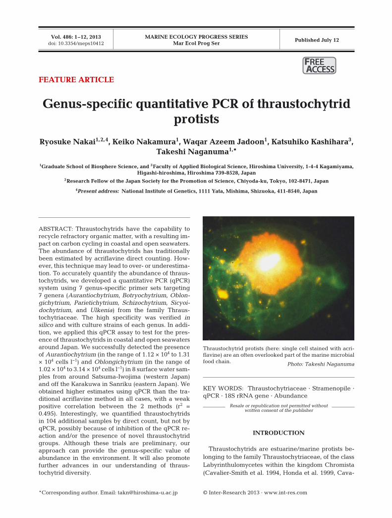
MARINE ECOLOGY PROGRESS SERIESMar Ecol Prog Ser
Vol. 486: 1–12, 2013doi: 10.3354/meps10412
Published July 12
KEY WORDS: Thraustochytriaceae · Stramenopile ·qPCR · 18S rRNA gene · Abundance
Resale or republication not permitted without written consent of the publisher
INTRODUCTION
Thraustochytrids are estuarine/marine protists be -longing to the family Thraustochytriaceae, of the classLabyrinthulomycetes within the kingdom Chromista(Cavalier-Smith et al. 1994, Honda et al. 1999, Cava-
© Inter-Research 2013 · www.int-res.com*Corresponding author. Email: [email protected]
FEATURE ARTICLE
Genus-specific quantitative PCR of thraustochytridprotists
Ryosuke Nakai1,2,4, Keiko Nakamura1, Waqar Azeem Jadoon1, Katsuhiko Kashihara3, Takeshi Naganuma1,*
1Graduate School of Biosphere Science, and 3Faculty of Applied Biological Science, Hiroshima University, 1-4-4 Kagamiyama, Higashi-hiroshima, Hiroshima 739-8528, Japan
2Research Fellow of the Japan Society for the Promotion of Science, Chiyoda-ku, Tokyo, 102-8471, Japan
4Present address: National Institute of Genetics, 1111 Yata, Mishima, Shizuoka, 411-8540, Japan
ABSTRACT: Thraustochytrids have the capability torecycle refractory organic matter, with a resulting im-pact on carbon cycling in coastal and open seawaters.The abundance of thraustochytrids has traditionallybeen estimated by acriflavine direct counting. How-ever, this technique may lead to over- or underestima-tion. To accurately quantify the abundance of thraus-tochytrids, we developed a quantitative PCR (qPCR)system using 7 genus-specific primer sets targeting7 genera (Aurantiochytrium, Botryochytrium, Oblon -gichytrium, Parietichytrium, Schizochytrium, Sicyoi -dochytrium, and Ulkenia) from the family Thraus-tochytriaceae. The high specificity was verified insilico and with culture strains of each genus. In addi-tion, we applied this qPCR assay to test for the pres-ence of thraustochytrids in coastal and open seawatersaround Japan. We successfully detected the presenceof Aurantiochytrium (in the range of 1.12 × 10⁴ to 1.31× 10⁴ cells l−1) and Oblongichytrium (in the range of1.02 × 104 to 3.14 × 10⁴ cells l−1) in 8 surface water sam-ples from around Satsuma-Iwojima (western Japan)and off the Karakuwa in Sanriku (eastern Japan). Weobtained higher estimates using qPCR than the tra -ditional acriflavine method in all cases, with a weakpositive correlation between the 2 methods (r2 =0.495). Interestingly, we quantified thraustochytridsin 104 additional samples by direct count, but not byqPCR, possibly because of inhibition of the qPCR re-action and/or the presence of novel thraustochytridgroups. Although these trials are preliminary, our approach can provide the genus-specific value ofabundance in the environment. It will also promotefurther advances in our understanding of thraus-tochytrid diversity.
Thraustochytrid protists (here: single cell stained with acri-flavine) are an often overlooked part of the marine microbialfood chain. Photo: Takeshi Naganuma
FREEREE ACCESSCCESS
Mar Ecol Prog Ser 486: 1–12, 2013
lier-Smith & Chao 2006). They have attracted attentionby virtue of their biotechnological role in the produc-tion of omega-3 long-chain polyunsaturated fatty acids(PUFAs) such as docosapentaenoic acid (DPA) anddoco sahexaenoic acid (DHA) (Raghukumar 2008). Theyhave the ability to decompose plant material, such asalgal tissue and mangrove leaf litter, by means of ex-tracellular cellulase (Sathe-Pathak et al. 1993, Bremer1995, Nagano et al. 2011), with a resulting impact oncarbon cycling in coastal and open seawaters, andmay grow on terrestrial refractory organic substratescontained in river water (Kimura & Naganuma 2001).In addition, the bio-volume of thraustochytrids is ~103
times greater than that of bacterioplankton (Naga -numa et al. 1998). Thus, they serve as potentially im-portant food sources for picoplankton-feeders, therebyenhancing pelagic secondary production (Naga numaet al. 1998, Raghu kumar & Damare 2011).
The abundance of thraustochytrids has been meas-ured in many previous studies focusing on the directenumeration of non-planktonic or planktonic thraus-tochytrids (Raghukumar & Schaumann 1993, Naga -numa et al. 1998, Raghukumar et al. 2001, Naganumaet al. 2006), estimation of their biomass based on cel-lular carbon and nitrogen content and the C:N ratio(Kimura et al. 1999), estimation of the correlation be -tween their abundance and environmental para meters(Kimura et al. 2001), and determination of the effect ofriver discharge on their distribution and abundance(Kimura & Naganuma 2001). These studies used a flu-orogenic acriflavine dye to enumerate the thraus-tochytrids by direct detection. This technique relieson the fact that the wall and nucleus of thraustochytridcells fluoresce differently (red and blue-green, re-spectively) under blue-light excitation. This dual fluo -res cence distinguishes thraustochytrids from otherprotists and detritus. However, the acriflavine countincludes protozoan cysts, thereby leading to overesti-mation, and excludes thraustochytrid zoospores, lead-ing to underestimation. In addition, there may be vari-ation in results due to observer error. Kimura et al.(2001) pointed out that such over- or underestimationshould be evaluated in future studies by using a morespecific technique. To address this issue, Takao et al.(2007) developed a fluorescence in situ hybridization(FISH) method using an 18S rRNA-targeted fluores-cent oligo-nucleotide probe for specific detection ofthraustochytrids. Damare & Raghu kumar (2010) usedan internal transcribed spacer (ITS)-based in situ hy-bridization (ISH) technique to detect aplanochytrids(Labyrin thulomycetes). Although FISH and ISH arepowerful tools, they require many hybridization stepsand intensive microscopic work.
To address these limitations and provide a simplemethod that can be used to process multiple samples,we developed a quantitative polymerase chain reac-tion (qPCR) assay. Quantification by qPCR relies ondetection of the increase in fluorescence from expo-nentially amplified DNA by a PCR involving a primerset and/or a fluorochrome-labeled probe designed tobind to the desired DNA locus. qPCR-based quantifi-cation provides a highly sensitive and specific systemfor the identification of target organisms. Thismethod is increasingly being used in marine micro -biological studies, such as in the detection of dino -flagellates (Bowers et al. 2000, Moorthi et al. 2006,Yamashita et al. 2011) and the thraustochytrid patho-gen quahog parasite unknown (QPX) (Lyons et al.2006, Liu et al. 2009). In a recent report, Bergmann etal. (2011) developed a qPCR assay for detection ofthe labyrinthulid Labyrinthula zosterae (Labyrinthu-lomycetes), known as the causative agent of eelgrasswasting disease. However, except for labyrinthu-lomycete pathogens, there are no published reportsdetailing qPCR detection of thraustochytrids. We de -veloped and evaluated a new qPCR system with genus-specific primer sets targeting thraustochytridsand then used this assay to test for the presence ofthraustochytrids in field samples.
MATERIALS AND METHODS
Design of genus-specific PCR primers
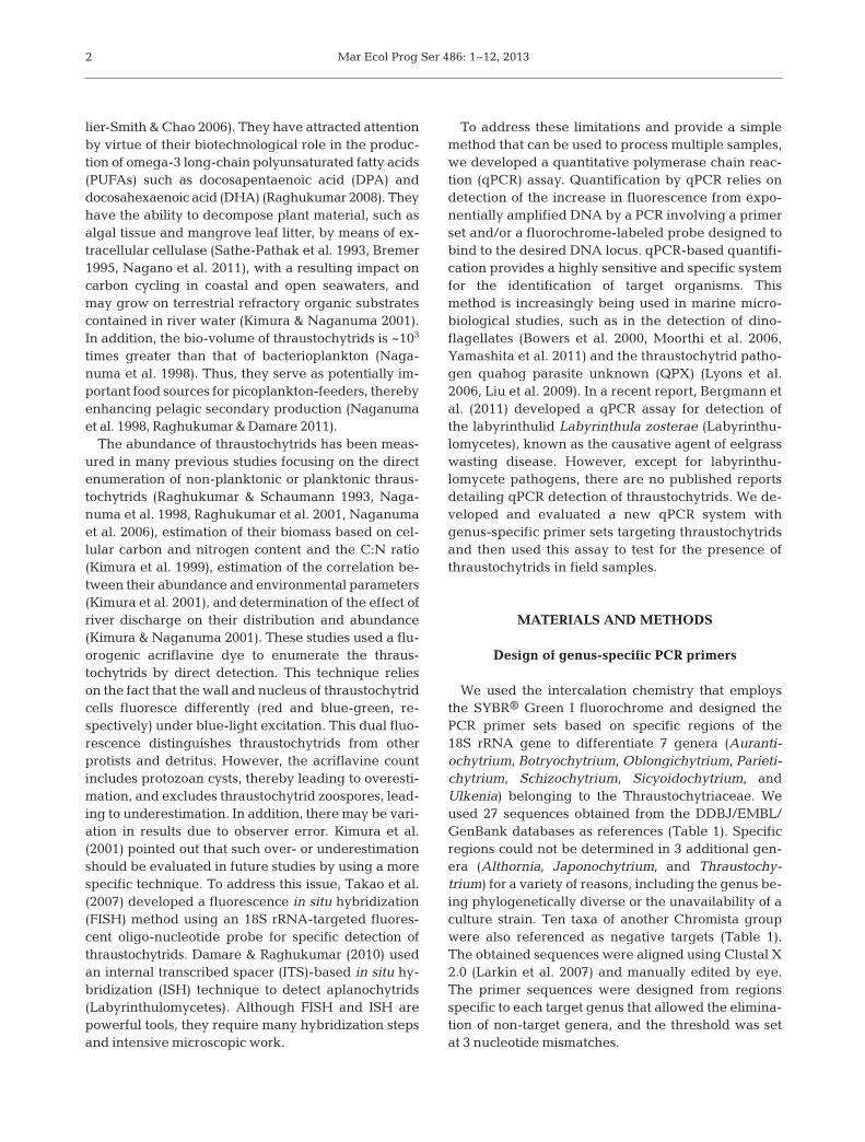
We used the intercalation chemistry that employsthe SYBR® Green I fluorochrome and designed thePCR primer sets based on specific regions of the18S rRNA gene to differentiate 7 genera (Auranti-ochytrium, Botryochytrium, Oblongichytrium, Parieti -chytrium, Schizochytrium, Sicyoidochytrium, andUlkenia) belonging to the Thraustochytriaceae. Weused 27 sequences obtained from the DDBJ/EMBL/GenBank databases as references (Table 1). Specificregions could not be determined in 3 additional gen-era (Althornia, Japonochytrium, and Thraustochy -trium) for a variety of reasons, including the genus be-ing phylogenetically diverse or the unavailability of aculture strain. Ten taxa of another Chromista groupwere also referenced as negative targets (Table 1).The obtained sequences were aligned using Clustal X2.0 (Larkin et al. 2007) and manually edited by eye.The primer sequences were designed from regionsspecific to each target genus that allowed the elimina-tion of non-target genera, and the threshold was setat 3 nucleotide mismatches.
2
Nakai et al.: Genus-specific qPCR of thraustochytrids
Testing for primer specificity to culturestrains using PCR and qPCR
To confirm that the designed primersmatched 18S rRNA genes from the targetgenus rather than from non-target genera,we conducted a Primer-BLAST search(Altschul et al. 1997, Ye et al. 2012) againstthe NCBI non-redundant (nr) data basewith an input setting of 50 to 250 bp for thePCR product size. In addition, the searchwas also performed with an input setting of50 to 5000 bp to examine the risks of unpre-dictable matching with other estuarine andmarine organisms.
In addition to these database searches,we performed experimental confirmationby PCR using the above-mentioned primersagainst cultured strains of each genus:Aurantiochytrium sp. SEK 209 (NBRC -102614), Botryochytrium radiatum SEK 353(NBRC104107), Oblongichy trium sp. SEK347 (NBRC102618), Parietichy trium sar ka -ri anum SEK 351 (NBRC104108), Sic yoido -chytrium sp. MBIC11077 (NBRC 102979),Schizochytrium sp. SEK 345 (NBRC -102616), and Ulkenia amoeboidea SEK 214(NBRC104106). Samples of the culture(2−3 ml) were harvested during the expo-nential growth stage by centrifugation(6000 g ×, 5 min). The resultant cell pelletswere suspended in 200 µl of phosphate-buffered saline (PBS, pH 7.2), then digestedwith 26 µl of 10% sodium dodecyl sulfate(SDS), 20 µl of 5 mg ml−1 lysozyme, and40 µl of 25 mg ml−1 Proteinase K. DNAs inthe solutions were extracted with phenol-chloroform-isoamyl alcohol (PCI; 25:24:1,v/v/v) and chloroform-isoamyl alcohol (CIA;24:1, v/v), then precipitated by isopropanolin 0.3M sodium acetate. The DNA pelletswere washed in 70% ethanol and thenfinally dissolved with 100 µl of sterileMilliQ water. A mixture (total volume:20 µl) containing 0.5 µl of template DNA(dissolved as above), 1 µl of 10 pmol µl−1
genus-specific forward and reverse primers,and the recommended volume of 5 unitsµl−1 Ex Taq DNA polymerase, 10× Ex Taqbuffer, 25 mM MgCl2, and deoxynucleo-side triphosphate (dNTP) mixture includedin the Takara Ex Taq kit (Takara Bio) wassubjected to conventional PCR to verify the
3
Taxon Accession Sequence number length (bp)
Genus AurantiochytriumAurantiochytrium limacinum NIBH SR21* AB022107 1678Aurantiochytrium mangrovei DQ100293 1721Aurantiochytrium sp. KH105* AB052555 1755Aurantiochytrium sp. mh0186 AB362211 1790Aurantiochytrium sp. SEK 209 AB290574 1720Aurantiochytrium sp. SEK 218 AB290573 1711Aurantiochytrium sp. SEK 217 AB290572 1764
Genus BotryochytriumBotryochytrium radiatum SEK 353 AB355410 1699
Genus OblongichytriumOblongichytrium sp. SEK 347 AB290575 1774Oblongichytrium sp. TN6 FJ821480 1798Oblongichytrium sp. 8-7* AF257317 1639Oblongichytrium sp. 7-5* AF257316 1635
Genus ParietichytriumParietichytrium sarkarianum SEK 351 AB355411 1756
Genus SicyoidochytriumSicyoidochytrium minutum NBRC 102975* AB290585 1733Sicyoidochytrium sp. NBRC 102979* AB183659 1711Sicyoidochytrium minutum SEK 354 AB355412 1733
Genus SchizochytriumSchizochytrium sp. SEK 346 AB290578 1766Schizochytrium sp. SEK 345 AB290577 1755Schizochytrium sp. SEK 210 AB290576 1766Schizochytrium aggregatum ATCC 28209 AB022106 1677Schizochytrium sp. KK17-3* AB052556 1793
Genus UlkeniaUlkenia amoeboidea SEK 214* AB290355 1790Ulkenia profunda L34054 1815Ulkenia profunda BUTRBG 111 DQ023615 1762Ulkenia sp. ATCC 28207* AB022104 1760Ulkenia visurgensis BURAAA 141 DQ100296 1812Ulkenia visurgensis ATCC 28208 AB022116 1812
Other Chromista groupCafeteria roenbergensis L27633 1718Achlya bisexualis M32705 1809Phytophthora megasperma X54265 1827Hyphochytrium catenoides BR217 AF163294 1814Chaetoceros debilis ch.4 AY229896 1739Eucampia antarcia CCMP1452 AY485503 1632Skeletonema costatum CCAP 1077/3 X85395 1798Thalassiosira weissflogii CCAP1085/1 FJ600728 1764Chattonera ovata C. Tomas Japan AY788924 1781Heterosigma akashiwo 893 AB217869 1806
Table 1. DDBJ/EMBL/GenBank accession numbers of 18S rRNA gene se-quences used to design the genus-specific PCR primer sets. Asterisks (*)indicate scientific names according to Yokoyama & Honda (2007) and
Yokoyama et al. (2007)

Mar Ecol Prog Ser 486: 1–12, 2013
genus-specific amplicon from a designated thraus-tochytrid genus. The thermal-cycling protocol was: 1cycle at 95°C for 5 min, 35 cycles at 95°C for 45 s,60°C for 45 s, and 72°C for 30 s, followed by 1 cycle at72°C for 5 min. PCR was conducted in a Ta kara PCRThermal Cycler PERSONAL (Takara Bio). The an -nealing temperature was 58°C for Aurantiochytriumand 62°C for Parietichytrium and Ulkenia. The PCRproducts were electrophoresed on a 2% agarose geland stained with ethidium bromide.
We performed qPCR of the above-mentioned DNAextracts from 7 genera. A mixture (total volume: 20 µl)containing 0.5 µl of template DNA (dissolved asdescribed above), 0.4 µl of 10 pmol µl−1 genus-specific forward and reverse primers, and 10 µl ofPlatinum® SYBR® Green qPCR SuperMix-UDG withROX™ (Invitrogen) was analyzed in an ABI PRISM7000 (Applied Biosystems) using the following ther-mal protocol: 1 cycle at 50°C for 2 min, 1 cycle at 95°Cfor 2 min, and 40 cycles at 95°C for 15 s and 60°C for30 s. Following qPCR, the melting profiles of the PCRproduct versus temperature (dissociation curves)were obtained for each sample to check for the occur-rence of positive amplification of target DNA andabsence of primer−dimers.
Quantification of thraustochytrid cells using qPCR
The cell numbers for each culture were countedunder a light microscope, and culture aliquots equi -valent to 5000, 10 000, 100 000, and 1 000 000 cellswere each filter-trapped onto a 0.22 µm pore sizeSterivex filter (Millipore). The filter units were thenstored at –20°C for a period >1 d. The units werethawed before DNA extraction and the cells lysedas described by Somerville et al. (1989). The crudelysates were used for DNA preparation by PCI extrac -tion, CIA extraction, and isopropanol precipitation asdescribed above. The DNA pellets were finally dis-solved with 100 µl of sterile MilliQ water. Triplicatealiquots of 0.5 µl (each equivalent to 25, 50, 500, or5000 cells reaction−1) were retrieved and subjected toqPCR using each genus-specific primer set, with ster-ile MilliQ water as a non-template control or a nega-tive control. In addition, to determine whether eachprimer set produces a signal from non-target genera,a 5000 cell-equivalent DNA derived from each of theother genera was also subjected to qPCR. TheseqPCR measurements were characterized by 2 inter-related parameters: (1) the cycle threshold (Ct) value,i.e. the number of cycles at which the reactioncrossed the specified fluorescence threshold; and (2)
the normalized reporter signal (Rn), which is calcu-lated as the ratio of the fluorescence of the reporterdye (SYBR® Green I) divided by the fluorescence ofthe passive reference dye (ROX™). The larger theamount of starting target DNA, the earlier a signifi-cant increase in Rn is observed, leading to a decreasein the Ct value. The change in Rn, or delta Rn (ΔRn),was plotted against the cycle number of the reaction.
Application of qPCR in testing seawater samples
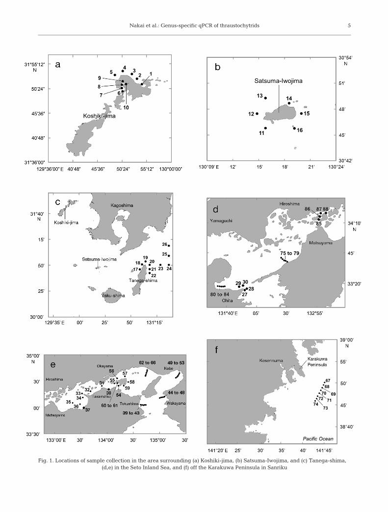
Seawater samples were collected at 88 sites aroundthe islands of Koshiki-jima, Satsuma-Iwojima, andTanega-shima (southwestern Japan), in the SetoInland Sea (western Japan) during cruises of the RV‘Toyoshio-maru’, Hiroshima University, in April 2008,May 2010, April 2011, and March and April 2012,and off the Karakuwa Peninsula in Sanriku (easternJapan) during a cruise of the RV ‘Tansei-maru’,Japan Agency for Marine-Earth Science and Tech-nology (JAMSTEC), in August 2011. In this study,qPCR only successfully quantified the thraus-tochytrid cell numbers in 8 of the 212 seawater sam-ples, viz. those collected at sites around Satsuma-Iwojima and off the Karakuwa Peninsula (Fig. 1).(A detailed list of all 212 samples is given in Table S1in the Supplement at www.int-res. com/ articles/ suppl/m486p001_supp.pdf). Samples were collected using aconductivity, temperature, and depth (CTD) profilingrosette equipped with Niskin bottle samplers atbetween 1 and 4 depths (surface to 210 m), depend-ing on the water depth at the site.
From the 93 seawater samples collected in 2008, a2 l water sample was filtered through a 0.22 µm poresize Sterivex filter (Millipore) using a peristalticpump. The DNA was then extracted from the Steri -vex housing as described above. From the 119 sam-ples collected in 2010 to 2012, a 20 l water samplewas prefiltered with a 500 µm mesh, then filteredthrough a 0.22 µm pore size Steripak-GP20 filter(Millipore) using a peristaltic pump. The DNA wasextracted from this Steripak filter using PCI and CIA,as described by Frias-Lopez et al. (2008). Each DNApellet was finally dissolved with 100 to 500 µl of ster-ile MilliQ water. Prior to qPCR, the initial screeningfor confirmation of the existence of thraustochytridswas performed by conventional PCR. Amplicons fromthe DNA extract with a minimum of 3 cells reaction−1
could be reliably detected on an agarose gel by ethi -dium bromide staining. Therefore, this initial screen-ing was used to select the samples applicable for ourqPCR system to quantitate thraustochytrid cells rang-
4
Nakai et al.: Genus-specific qPCR of thraustochytrids 5
Fig. 1. Locations of sample collection in the area surrounding (a) Koshiki-jima, (b) Satsuma-Iwojima, and (c) Tanega-shima, (d,e) in the Seto Inland Sea, and (f) off the Karakuwa Peninsula in Sanriku

Mar Ecol Prog Ser 486: 1–12, 2013
ing from 25 to 5000 cells reaction−1. This step wasincluded to reduce the effort and cost associated withqPCR. A mixture (total volume: 20 µl) containing0.5 µl of template DNA, pooled primers comprising0.7 µl of 10 pmol µl−1 of each forward and reverseprimer, and the recommended volume of 5 units µl−1
Ex Taq DNA polymerase, 10× Ex Taq buffer, 25 mMMgCl2, and dNTP mixture included in the TakaraEx Taq kit (Takara Bio) was used in the conventionalPCR. The thermal-cycling protocol consisted of1 cycle at 95°C for 2 min, 35 cycles at 94°C for 30 s,60°C for 45 s, and 72°C for 40 s, followed by 1 cycleat 72°C for 10 min. This cycle number (35 cycles)was chosen based on the Ct values and ΔRn valuesobtained during the qPCR amplification of standardDNA. In the preliminary experiments with pooledprimers, some losses in PCR performance wereobserved. We therefore reduced the primer concen-tration and optimized the PCR reaction conditionsusing control DNAs. The PCR products were electro-phoresed on a 2% agarose gel and stained withethidium bromide. For samples that tested positiveusing the mix of primers, we performed PCR usingeach forward and reverse primer to determine thepresence or absence of each genus.
For samples that tested positive during conven-tional PCR-based screening, we conducted qPCRusing the primer sets described above. A mixture(total volume 20 µl) containing 0.5 µl of templateDNA, 0.4 µl of 10 pmol µl−1 genus-specific forwardand reverse primers, and 10 µl of Platinum® SYBR®
Green qPCR SuperMix-UDG with ROX™ (Invitro-gen) was analyzed in an ABI PRISM 7000 (AppliedBiosystems), as described above, but with a decrease
in the number of PCR cyclesfrom 40 to 35 to minimize poten-tial interference by non-specificamplification. DNA extracts fromthe culture strain of the targetgenus, each corresponding to 25,50, 500, or 5000 cells reaction−1,were loaded for every run to serveas quantification standards (all per -formed in triplicate). The abun-dance of each genus was deter-mined based on these culture-based standardizations.
For the 8 samples for whichthraustochytrids were quantifiedby qPCR, we tested for interfer-ence due to other co-extractedcompounds (e.g., organic acidsor polysaccharides) or from filter-
trapped particles in the qPCR by adding 50 or 500cell equivalents of DNA from the culture strains toevery sample as internal standards. The additionalincrease in the qPCR signal corresponding to theadditional DNAs was recorded. When interferencewas suspected, the samples were diluted sufficientlyto diminish interference. In addition, the 6 samples inwhich thraustochytrid cells were found and strongamplification was observed by conventional PCR butnot qPCR were checked for amplification inhibitionin the same manner. To compare the results of qPCRwith traditional acriflavine counts, 125 of the 212samples were counted with acriflavine using epifluo-rescence microscopy following the method describedby Raghukumar & Schaumann (1993). Briefly, parti-cles in a water sample of 10- to 100 ml were collectedon an isopore membrane filter (Millipore; pore size0.2 µm, diameter 25 mm). The particles on the filterwere stained with 4 ml of 0.2 µm filtered 0.05% acri-flavine in 0.1 M citrate buffer (pH 3.0) for 4 min andthen rinsed with 75% isopropanol for 1 min. Thraus-tochytrid cells were counted in 100 microscopicfields, and each count was duplicated.
RESULTS AND DISCUSSION
PCR specificity
The sequence and amplicon size of the qPCRprimer sets designed to amplify each 18S rRNA geneof 7 thraustochytrid genera are given in Table 2.Some primer sets had mismatches with targetthrausto chytrid sequences ranging from 1 to 5 bases.
6
Genus Name Sequence Amplicon size (bp)
Aurantiochytrium Aur-F 5’-CTACGGTGACTATAACGGGTG-3’ 120Aur-R 5’-GTGGAGTCCACAGTGGGTAA-3’
Botryochytrium Bot-F 5’-ATGTGAGTGCGATAGCTTTCG-3’ 92Bot-R 5’-CGATTGCCTTCACACAAAAATG-3’
Oblongichytrium Obl-F 5’-GAGCCTTCGGGTTCGTGT-3’ 93Obl-R 5’-AACGATATGGATCCCATGCC-3’
Parietichytrium Par-F 5’-TTCGTAAGAGAACCAAATGTGG-3’ 164Par-R 5’-GCCATGCAAACCAACAAAAT-3’
Sicyoidochytrium Sic-F 5’-ACGAGGAAAAAGTCCTTATCCG-3’ 235Sic-R 5’-TACGCTACATCAAACTTTCATCC-3’
Schizochytrium Sch-F 5’-AATTCCCATGATTGTGCGTTGTGT-3’ 172Sch-R 5’-CCCGAGGGCTATGCGATTCGCTC-3’
Ulkenia Ulk-F 5’-GGGCTAAGCCTACTCTTTCTG-3’ 168Ulk-R 5’-CTGGTCCGTCCTACCAATACTT-3’
Table 2. Sequences and amplicon sizes of genus-specific PCR primer sets
Nakai et al.: Genus-specific qPCR of thraustochytrids
Auran tiochytrium sp. SEK 209 (AB290574) had a 1-base difference with our Aurantiochytrium-specificreverse primer; Oblongichytrium sp. SEK 347(AB290575) had a 1-base difference with our forwardprimer; and Ulkenia profunda (L34054) had a 5-basedifference (2 in the forward primer and 3 in thereverse) with our primers. The desired amplificationsagainst each genus were simulated in the Primer-BLAST database search. Mismatched regions werealso found in the search results. For example, Auran-tiochytrium sp. B013 (JF266573) had a 4-base differ-ence (2 in the forward primer and 2in the reverse) with our primers; andParietichytrium sp. BAFCcult 3109(HQ228977) had a 2-base differencewith our forward primer.
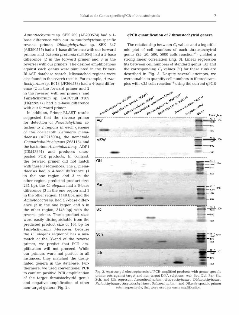
In addition, Primer-BLAST resultssuggested that the reverse primerfor detection of Parietichytrium at -taches to 2 regions in each genomeof the co elacanth Latimeria mena-doensis (AC215904), the nematodeCaenorhabditis elegans (Z68116), andthe bacterium Acinetobacter sp. ADP1(CR543861) and produces unex-pected PCR products. In contrast,the forward primer did not matchwith these 3 sequences. The L. mena-doensis had a 4-base difference (1in the one region and 3 in theother region; predicted product size:231 bp), the C. elegans had a 6-basedifference (3 in the one region and 3in the other region; 1148 bp), and theAcinetobacter sp. had a 7-base differ-ence (2 in the one region and 5 inthe other region; 3148 bp) with thereverse primer. These product sizeswere easily distinguishable from thepredicted product size of 164 bp forParietichytrium. Moreover, becausethe C. elegans sequence has a mis-match at the 3’-end of the reverseprimer, we predict that PCR am -plification will not proceed. Whileour primers were not perfect in allinstances, they matched the desig-nated genera in the database. Fur-thermore, we used conventional PCRto confirm positive PCR amplificationof the target thraustochytrid genusand negative amplification of othernon-target genera (Fig. 2).
qPCR quantification of 7 thraustochytrid genera
The relationship between Ct values and a logarith-mic plot of cell numbers of each thraustochytridgenus (25, 50, 500, 5000 cells reaction−1) yielded astrong linear correlation (Fig. 3). Linear regressionfits between cell numbers of standard genus (X ) andthe corresponding Ct values (Y ) for these runs aredescribed in Fig. 3. Despite several attempts, wewere unable to quantify cell numbers in filtered sam-ples with <25 cells reaction−1 using the current qPCR
7
Fig. 2. Agarose gel electrophoresis of PCR-amplified products with genus-specificprimer sets against target and non-target DNA solutions. Aur, Bot, Obl, Par, Sic,Sch, and Ulk represent Aurantiochytrium-, Botryochytrium-, Oblongichytrium-,Parietichytrium-, Sicyoidochytrium-, Schizochytrium-, and Ulkenia-specific primer
sets, respectively, that were used for each amplification

Mar Ecol Prog Ser 486: 1–12, 2013
assay (data not shown). In addition, in the 4 assays forBotryochytrium, Schizochytrium, Sicyoidochy trium,and Ulkenia, the amplification efficiencies werehigh, ranging from 90.8 to 107.1%. The efficienciesof the other 3 primer sets ranged from approximately70.4 to 81.5%; this suggests that there is room forimprovement. However, all assays designed in thisstudy had comparable levels of detection limits asdescribed below.
The ΔRn curves for the 7 genera and primer setsare summarized in Fig. 4. The graphs for the Auran -tiochytrium, Botryochytrium, Oblongichytrium, Schi -zochy trium, and Sicyoidochytrium-specific primers
clearly illustrate a significant increase in ΔRn of eachtarget genus in comparison with the 5000 cell-equiva-lent DNAs derived from 1 of the other 6 non-targetgenera. Our data also suggest that for these 5 genera,the increase in the non-specific ΔRn may haveoccurred in later PCR cycles (typically beyond 33 to35 cycles; Fig. 4). Based on the thermal dissociationcurve analysis of the qPCR amplicons, such anincrease in ΔRn would likely result from the amplifi-cation of non-target DNA. In addition, for the Sicy-oidochytrium-specific primer set, a weak peak fromthe formation of primer-dimers occurred, and the sig-nal was recognized after 35 cycles as described in the
8
Fig. 3. Linear regression cur ves of cycle threshold (Ct) values versus logarithmiccell numbers for each genus. Standard DNA solutions equivalent to 25, 50,500, and 5000 cells reaction−1 were analyzed by qPCR using genus-specificprimer sets. Data represent the mean ± SD of triplicate measurements. Each
primer set exhibited a strong linear correlation (r2 = 0.991−0.998)

Nakai et al.: Genus-specific qPCR of thraustochytrids
non-template control (NTC) reaction result (Fig. 4).However, in our qPCR system, the ΔRn for the 25 cellsreaction−1 as the detection limit was observed in Ct
values ranging from 26 to 32 cycles (Fig. 3). To mini-mize the effect of such signals, the number of PCRcycles was reduced from 40 to 35 during analysis ofthe field samples. Thus, the signals derived from non-specific amplification or primer-dimers have no realinfluence on our results. In the case of Aurantiochy -trium, the Ct value (35.1 ± 0.4 SD; n = 3) for the 25 cellequivalent could not be discriminated from the sig-nal of the non-target DNA amplified. Conversely, forthe Parietichytrium and Ulkenia-specific primers, theincrease in ΔRn derived from the amplification of
non-target genera DNA appeared to occur relativelyquickly, after 27 cycles. This cycle number is similarto the Ct values for the Parietichy trium 25 cell equiv-alent (27.1 ± 0.3, mean ± SD; n = 3) and the Ulkenia25 cell equivalent (25.6 ± 0.1; n = 3) samples (Fig. 3).However, in the case of non-target genera not beingdetected in the sample by conventional PCR-basedscreening, the non-specific ΔRn is considered to haveno real effect on the qPCR results. We emphasize thatprior to the qPCR assay, we performed conventionalPCR to determine the presence or absence of eachthraustochytrid genus. This was necessary to confirmbackground genera that could affect the quantifica-tion in qPCR.
9
Fig. 4. Change in normalized reporter signal (ΔRn) curves versus PCR cyclein qPCR using genus-specific primer sets against the target DNA solutionequivalent to 5000 cells reaction−1. At the same time, 5000 cell-equivalentDNAs derived from 1 of the other 6 non-target genera were also subjectedto qPCR. NTC: non-template control in which sterile MilliQ water was used.Each panel represents results using 1 of the genus-specific primer pairs
Mar Ecol Prog Ser 486: 1–12, 2013
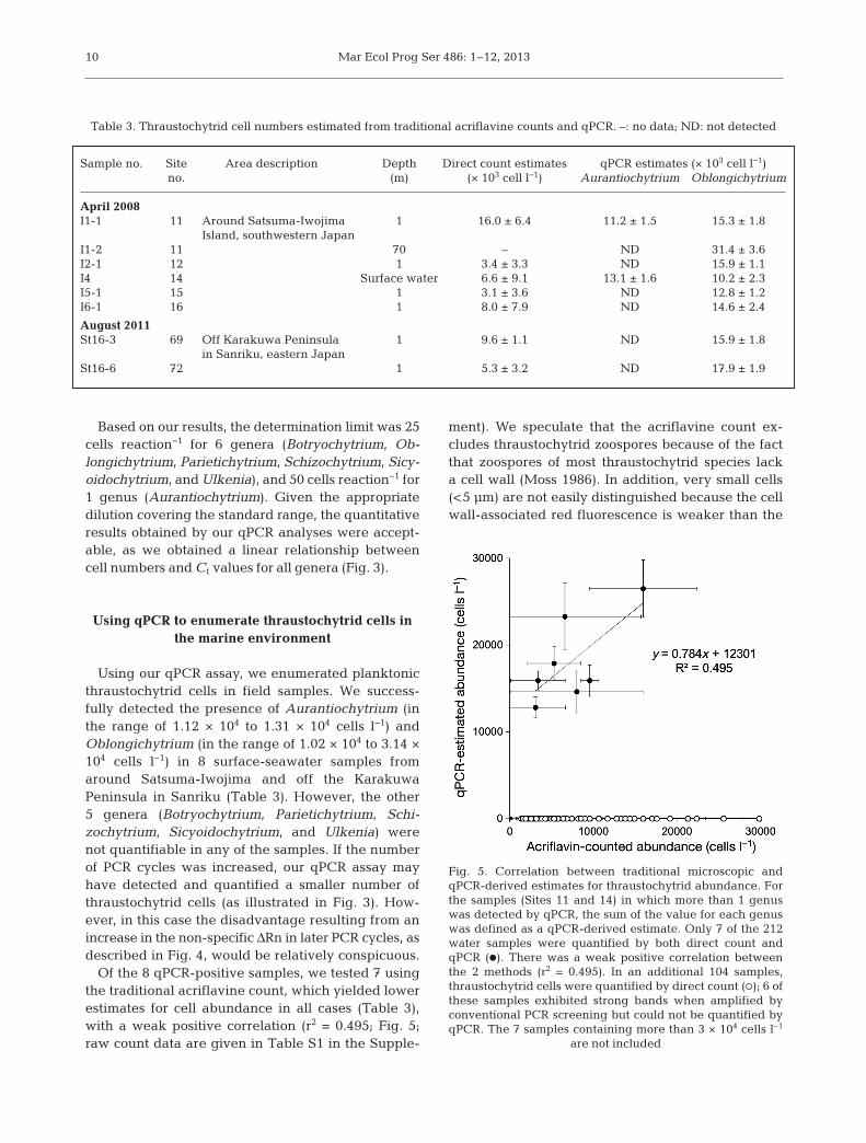
Based on our results, the determination limit was 25cells reaction−1 for 6 genera (Botryochytrium, Ob-longichytrium, Parietichytrium, Schizochytrium, Sicy-oidochytrium, and Ulkenia), and 50 cells reaction−1 for1 genus (Aurantiochytrium). Given the ap propriatedilution covering the standard range, the quantitativeresults obtained by our qPCR analyses were accept-able, as we obtained a linear relationship betweencell numbers and Ct values for all genera (Fig. 3).
Using qPCR to enumerate thraustochytrid cells inthe marine environment
Using our qPCR assay, we enumerated planktonicthraustochytrid cells in field samples. We success-fully detected the presence of Aurantiochytrium (inthe range of 1.12 × 10⁴ to 1.31 × 10⁴ cells l−1) andOblongichytrium (in the range of 1.02 × 10⁴ to 3.14 ×10⁴ cells l−1) in 8 surface-seawater samples fromaround Satsuma-Iwojima and off the KarakuwaPeninsula in Sanriku (Table 3). However, the other5 genera (Botryochytrium, Parietichytrium, Schi -zochytrium, Sicyoidochytrium, and Ulkenia) werenot quantifiable in any of the samples. If the numberof PCR cycles was increased, our qPCR assay mayhave detected and quantified a smaller number ofthraustochytrid cells (as illustrated in Fig. 3). How-ever, in this case the disadvantage resulting from anincrease in the non-specific ΔRn in later PCR cycles, asdescribed in Fig. 4, would be relatively conspi cuous.
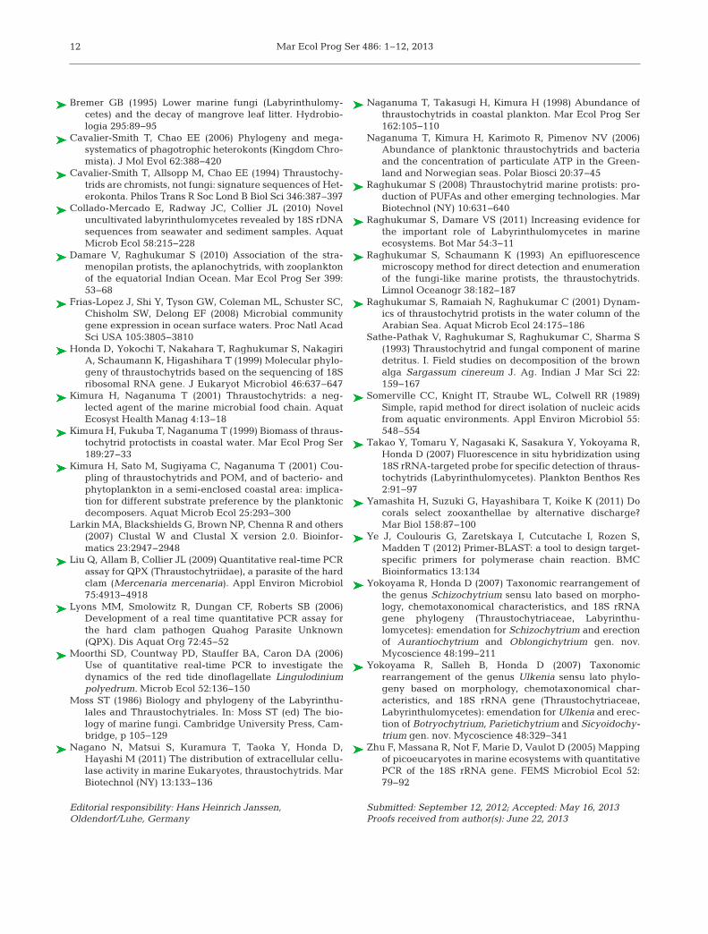
Of the 8 qPCR-positive samples, we tested 7 usingthe traditional acriflavine count, which yielded lowerestimates for cell abundance in all cases (Table 3),with a weak positive correlation (r2 = 0.495; Fig. 5;raw count data are given in Table S1 in the Supple-
ment). We speculate that the acriflavine count ex -cludes thraustochytrid zoospores because of the factthat zoospores of most thraustochytrid species lacka cell wall (Moss 1986). In addition, very small cells(<5 µm) are not easily distinguished because the cellwall-associated red fluorescence is weaker than the
10
Sample no. Site Area description Depth Direct count estimates qPCR estimates (× 103 cell l−1)no. (m) (× 103 cell l−1) Aurantiochytrium Oblongichytrium
April 2008I1-1 11 Around Satsuma-Iwojima 1 16.0 ± 6.4 11.2 ± 1.5 15.3 ± 1.8
Island, southwestern JapanI1-2 11 70 – ND 31.4 ± 3.6I2-1 12 1 3.4 ± 3.3 ND 15.9 ± 1.1I4 14 Surface water 6.6 ± 9.1 13.1 ± 1.6 10.2 ± 2.3I5-1 15 1 3.1 ± 3.6 ND 12.8 ± 1.2I6-1 16 1 8.0 ± 7.9 ND 14.6 ± 2.4
August 2011St16-3 69 Off Karakuwa Peninsula 1 9.6 ± 1.1 ND 15.9 ± 1.8
in Sanriku, eastern JapanSt16-6 72 1 5.3 ± 3.2 ND 17.9 ± 1.9
Table 3. Thraustochytrid cell numbers estimated from traditional acriflavine counts and qPCR. –: no data; ND: not detected
Fig. 5. Correlation between traditional microscopic andqPCR-derived estimates for thraustochytrid abundance. Forthe samples (Sites 11 and 14) in which more than 1 genuswas detected by qPCR, the sum of the value for each genuswas defined as a qPCR-derived estimate. Only 7 of the 212water samples were quantified by both direct count andqPCR (d). There was a weak positive correlation betweenthe 2 methods (r2 = 0.495). In an additional 104 samples,thraustochytrid cells were quantified by direct count (s); 6 ofthese samples exhibited strong bands when amplified byconventional PCR screening but could not be quantified byqPCR. The 7 samples containing more than 3 × 10⁴ cells l−1
are not included

Nakai et al.: Genus-specific qPCR of thraustochytrids
nucleus-associated green fluorescence. Thus, thesecells are often not counted, leading to underestima-tion. Taken together, our observations suggest thatqPCR provides a more accurate estimate of the abun-dance of these zoospores and very small cells.
Thraustochytrid cells were found in 104 additionalsamples by direct count. The abundance of thrausto -chytrids ranged from 1.37 ± 1.22 × 10³ cells l−1
(Site 20, around Tanega-shima, southwestern Japan)to 7.68 ± 0.85 × 10⁴ cells l−1 (Site 53, Osaka Bay, SetoInland Sea, western Japan; Table S1). Prior studiesreported an average thraustochytrid abundance of103 to 104 cells (Naganuma et al. 1998, Kimura etal. 1999, 2001) in the coastal Seto Inland Sea andadjacent open waters. Our direct count estimates ofabundance were consistent with prior studies. How-ever, for these samples, thraustochytrids could not bequantified by qPCR, although 6 did produce strongbands when amplified by conventional PCR (Table S1).We suspect that contamination with non-target DNAsderived from smaller microorganisms and/or otherco-extracted compounds, such as organic acids orpolysaccharides, inhibits the amplification reaction.This is consistent with the decrease in signals weobserved for the 500 cell internal standard used inthe qPCR runs with the 6 samples above (data notshown). Given this, we recommend further evalua-tion of methods for filtration to remove picoeukaryotesand bacteria. Moreover, recent culture-independentstudies revealed that thraustochytrid 18S rRNA genesequences in the environment were more phyloge-netically diverse than expected (Collado-Mercado etal. 2010). There is the possibility that the presenceof these novel thraustochytrid groups could not bedetected, which may have affected our results.
Properties of the qPCR developed in the present study
We have developed and applied a SYBR® GreenqPCR method for the identification and quantificationof 7 thraustochytrid genera. Our initial objective wasto estimate genus-specific abundances of the familyThraustochytriaceae, all of which were based on theqPCR results. Therefore, our qPCR system has tar-geted the specific regions of 18S rRNA gene fromrepresentative strains for each genus. Expressingthis parameter as the copy number of an rRNA genewould be more accurate, but less insightful for eco-logical purposes. Thus, we calculated cell numbersusing the formula obtained from Ct values versus cellnumbers of cultured strains. The qPCR-based cellnumber was determined based on each independ-
ent genus standard. Therefore, our culture-based standardizations should give independent levels ofdetection and should not be affected by differences inthe rRNA gene copy numbers among the genera. Incontrast, since the environmental samples may con-tain multiple and/or unknown species of each genus,there was the potential that the copy number varia-tions within each genus affect the qPCR estimates.Zhu et al. (2005) studied the copy number variationsof 18 algal strains belonging to different phylogeneticgroups and suggested that qPCR could be used tomonitor specific narrow groups since the range ofrRNA gene copy numbers was quite restricted. Thus,the difference in copy number within genus does notseem to present a significant obstacle to the determi-nation of cell number based on the culture-basedstandardizations. Nevertheless, accurate quantifica-tion that takes the copy number variations into con-sideration is desirable, as this has not been conclu-sively determined so far. Although the trials fornatural samples are still preliminary, our molecularapproach can provide the genus-specific value ofabundance in the environment. It could also help ad-vance our understanding of thraustochytrid diversity.
Acknowledgements. We thank the crews of the RV‘Toyoshio-maru’, Hiroshima University, and the RV ‘Tansei-maru’, Japan Agency for Marine Earth Science and Tech-nology (JAMSTEC), and the cruise participants for their on-board assistance. We thank K. Koike, Hiroshima University,for allowing sample collection during the RV ‘Toyoshio-maru’ Cruise No. 2010-02 and K. Hamasaki, University ofTokyo, for allowing sample collection during the RV ‘Tansei-maru’ Cruise KT-11-17. We especially thank T. Nagata, University of Tokyo, N. Nagano, Kyushu University, and H.Yamashita, Seikai National Fisheries Research Institute, andK. Inoue, University of Tokyo, for their on-board assistanceand helpful discussion. R.N. was supported by JSPS Re -search Fellowships for Young Scientists (10J07702 and11J30005). We also thank 3 anonymous reviewers for theircareful reading and constructive remarks.
LITERATURE CITED
Altschul SF, Madden TL, Schaffer AA, Zhang J, Zhang Z,Miller W, Lipman DJ (1997) Gapped BLAST and PSI-BLAST: a new generation of protein database searchprograms. Nucleic Acids Res 25:3389−3402
Bergmann N, Fricke B, Schmidt MC, Tams V and others(2011) A quantitative real-time polymerase chain reactionassay for the seagrass pathogen Labyrinthula zosterae.Mol Ecol Resour 11:1076−1081
Bowers HA, Tengs T, Glasgow HB Jr, Burkholder JM,Rublee PA, Oldach DW (2000) Development of real-timePCR assays for rapid detection of Pfiesteria piscicidaand related dinoflagellates. Appl Environ Microbiol 66:4641−4648
11
Mar Ecol Prog Ser 486: 1–12, 2013
Bremer GB (1995) Lower marine fungi (Labyrinthulomy -cetes) and the decay of mangrove leaf litter. Hydrobio -logia 295:89−95
Cavalier-Smith T, Chao EE (2006) Phylogeny and mega -systematics of phagotrophic heterokonts (Kingdom Chro -mista). J Mol Evol 62:388−420
Cavalier-Smith T, Allsopp M, Chao EE (1994) Thraustochy -trids are chromists, not fungi: signature sequences of Het-erokonta. Philos Trans R Soc Lond B Biol Sci 346: 387−397
Collado-Mercado E, Radway JC, Collier JL (2010) Noveluncultivated labyrinthulomycetes revealed by 18S rDNAsequences from seawater and sediment samples. AquatMicrob Ecol 58:215−228
Damare V, Raghukumar S (2010) Association of the stra-menopilan protists, the aplanochytrids, with zooplanktonof the equatorial Indian Ocean. Mar Ecol Prog Ser 399:53−68
Frias-Lopez J, Shi Y, Tyson GW, Coleman ML, Schuster SC,Chisholm SW, Delong EF (2008) Microbial communitygene expression in ocean surface waters. Proc Natl AcadSci USA 105:3805−3810
Honda D, Yokochi T, Nakahara T, Raghukumar S, NakagiriA, Schaumann K, Higashihara T (1999) Molecular phylo -geny of thraustochytrids based on the sequencing of 18Sribosomal RNA gene. J Eukaryot Microbiol 46: 637−647
Kimura H, Naganuma T (2001) Thraustochytrids: a neg-lected agent of the marine microbial food chain. AquatEcosyst Health Manag 4:13−18
Kimura H, Fukuba T, Naganuma T (1999) Biomass of thraus-tochytrid protoctists in coastal water. Mar Ecol Prog Ser189:27−33
Kimura H, Sato M, Sugiyama C, Naganuma T (2001) Cou-pling of thraustochytrids and POM, and of bacterio- andphytoplankton in a semi-enclosed coastal area: implica-tion for different substrate preference by the planktonicdecomposers. Aquat Microb Ecol 25:293−300
Larkin MA, Blackshields G, Brown NP, Chenna R and others(2007) Clustal W and Clustal X version 2.0. Bioinfor -matics 23:2947−2948
Liu Q, Allam B, Collier JL (2009) Quantitative real-time PCRassay for QPX (Thraustochytriidae), a parasite of the hardclam (Mercenaria mercenaria). Appl Environ Microbiol75:4913−4918
Lyons MM, Smolowitz R, Dungan CF, Roberts SB (2006)Development of a real time quantitative PCR assay forthe hard clam pathogen Quahog Parasite Unknown(QPX). Dis Aquat Org 72:45−52
Moorthi SD, Countway PD, Stauffer BA, Caron DA (2006)Use of quantitative real-time PCR to investigate thedynamics of the red tide dinoflagellate Lingulodiniumpolyedrum. Microb Ecol 52:136−150
Moss ST (1986) Biology and phylogeny of the Labyrinthu-lales and Thraustochytriales. In: Moss ST (ed) The bio -logy of marine fungi. Cambridge University Press, Cam-bridge, p 105−129
Nagano N, Matsui S, Kuramura T, Taoka Y, Honda D,Hayashi M (2011) The distribution of extracellular cellu-lase activity in marine Eukaryotes, thraustochytrids. MarBiotechnol (NY) 13:133−136
Naganuma T, Takasugi H, Kimura H (1998) Abundance ofthraustochytrids in coastal plankton. Mar Ecol Prog Ser162:105−110
Naganuma T, Kimura H, Karimoto R, Pimenov NV (2006)Abundance of planktonic thraustochytrids and bacteriaand the concentration of particulate ATP in the Green-land and Norwegian seas. Polar Biosci 20:37−45
Raghukumar S (2008) Thraustochytrid marine protists: pro-duction of PUFAs and other emerging technologies. MarBiotechnol (NY) 10:631−640
Raghukumar S, Damare VS (2011) Increasing evidence forthe important role of Labyrinthulomycetes in marine ecosystems. Bot Mar 54:3−11
Raghukumar S, Schaumann K (1993) An epifluorescencemicroscopy method for direct detection and enumerationof the fungi-like marine protists, the thraustochytrids.Limnol Oceanogr 38:182−187
Raghukumar S, Ramaiah N, Raghukumar C (2001) Dynam-ics of thraustochytrid protists in the water column of theArabian Sea. Aquat Microb Ecol 24:175−186
Sathe-Pathak V, Raghukumar S, Raghukumar C, Sharma S(1993) Thraustochytrid and fungal component of marinedetritus. I. Field studies on decomposition of the brownalga Sargassum cinereum J. Ag. Indian J Mar Sci 22:159−167
Somerville CC, Knight IT, Straube WL, Colwell RR (1989)Simple, rapid method for direct isolation of nucleic acidsfrom aquatic environments. Appl Environ Microbiol 55:548−554
Takao Y, Tomaru Y, Nagasaki K, Sasakura Y, Yokoyama R,Honda D (2007) Fluorescence in situ hybridization using18S rRNA-targeted probe for specific detection of thraus-tochytrids (Labyrinthulomycetes). Plankton Benthos Res2:91−97
Yamashita H, Suzuki G, Hayashibara T, Koike K (2011) Docorals select zooxanthellae by alternative discharge?Mar Biol 158:87−100
Ye J, Coulouris G, Zaretskaya I, Cutcutache I, Rozen S,Madden T (2012) Primer-BLAST: a tool to design target-specific primers for polymerase chain reaction. BMCBioinformatics 13:134
Yokoyama R, Honda D (2007) Taxonomic rearrangement ofthe genus Schizochytrium sensu lato based on morpho -logy, chemotaxonomical characteristics, and 18S rRNAgene phylogeny (Thraustochytriaceae, Labyrinthu-lomycetes): emendation for Schizochytrium and erectionof Aurantiochytrium and Oblongichytrium gen. nov.Mycoscience 48:199−211
Yokoyama R, Salleh B, Honda D (2007) Taxonomicrearrangement of the genus Ulkenia sensu lato phylo -geny based on morphology, chemotaxonomical char -acteristics, and 18S rRNA gene (Thraustochytriaceae,Laby rinthulomycetes): emendation for Ulkenia and erec-tion of Botryochytrium, Parietichytrium and Sicyoidochy -trium gen. nov. Mycoscience 48:329−341
Zhu F, Massana R, Not F, Marie D, Vaulot D (2005) Mappingof picoeucaryotes in marine ecosystems with quantitativePCR of the 18S rRNA gene. FEMS Microbiol Ecol 52:79−92
12
Editorial responsibility: Hans Heinrich Janssen, Oldendorf/Luhe, Germany
Submitted: September 12, 2012; Accepted: May 16, 2013Proofs received from author(s): June 22, 2013
Copyright © 2022 FDOKUMEN