Surface Failure and Durability of Induction-Hardened Sintered ...
Journal of Oral Rehabilitation 1997 24:646-651
Marginal adaptation of a sintered ceramic inlay systembefore and after cementationD . G E M A L M A Z , M . O Z C A N , A . B . Y O R U g * & H . N . A L K U M R U Faculty of Dentistry.Urtiversity of Marmara. Istanbul and *Faculty of Chemistry and Metallurgy. Yildiz Technical University. Istanbul. Turkey
SUMMARY The long term clinical performance ofporcelain inlays depends on a number of factors andthe marginal adaptation is one of significant interest.The purpose of this in vitro study was to evaluatethe marginal integrity of a sintered inlay technique(Ducera®), hefore and after cementation. MODcavities without bevels were prepared on 10 humanmandibular molar teeth and porcelain inlays werefabricated according to the manufacturer'sinstructions. Inlays were evaluated microscopicallyfor their adaptation to the occlusal and approximalmargins of the tooth by means of a replica technique.Inlays were cemented with a dual-cured hybridcomposite luting material (Enforce®). Afterpolishing, each tooth was sectioned in buccal/lingualand mesial/distal directions following the same
procedure in the sectioning of replicas. The marginalgap and the thickness of exposed cement weremeasured at each section. The mean marginal gapof 71-83 ± 8-93 (lm recorded for the occlusal marginbefore cementation was significantly smaller thanthat of 105-6 ± 39-33 |im calculated attheapproximalmargin. Following the cementation, the adaptationof the inlays at the occlusal margin was also foundto he superior to that of the approximal margin.Comparison of mean gap values before and aftercementation revealed that the marginal gapincreased by 6 94 tim and 23-25 ^m at the occlusaland approximal margins, respectively. Althoughpolishing was performed after cementation, excessluting material was still observed, tbat caused anincrease in the width of the exposed luting cement.
IntroductionThe use oi aesthetic materials is becoming more populardue to the increased interest in tooth appearance.Patients are demanding improved aesthetics and evenin the posterior regions of the mouth, insistence ontooth-coloured restorations has generated great interestin the developmeiii: o{ beuer alternatives to amalgam.Despite the improved qualities of the posteriorcomposites, they still show a relatively high poly-merization shrinkage, low resistance to clinical wearand a high coefficient of thermal expansion (Feilzer,DeGee & Davidson, 1989; Wohlwerd, 1990; Burgoyne.NichoUs &• Brudvik. 1991). Since ceramic inlays offermore wear-resistance and biocompatihle posterior
restorations, they are viable alternatives to compositeinlays.
The long term clinical performance ot ceramic inlaysdepends on a number of factors with marginaladaptation being one of significance. Several in vitrostudies (Hickel & Kunzelmann, 1990; Rose, Platzer &•Roth, 1990; Sturdevant et ai, 1991; Federlin, Schmaiz&• Reich, 1992; Inokoshi etal. 1992; Thordrup, Isidor& Horsted-Bindslev, 1992; Moiin & Karlsson, 1993;Thordrup, Isidor & Horsted-Bindslev, 1994; Sertgozet al.. 1995), exist in the literature in relation to themarginal integrity of ceramic inlays. However, themarginal integrity values reported were related only toeither before cementation or after cementation.Therefore, a need for a comparison of the initial fit with
© 1997 Blackwell Science Ltd
SINTERED INLAY TECHNIQUES 647
the final adaptation of the ceramic inlays seems to beessential to achieve a basis for the evaluation of marginalintegrity studies.
The aim of the present in vitro study was to evaluatethe marginal integrity of a sintered ceramic inlaytechnique, before and after cementation.
Materials and methods
Ten newly extraaed noncarious human mandibularmolar teeth were used*. After cleaning and scaling,MOD inlay preparations with 90° cavosurface marginswere made on all of the teeth. The preparation wasperformed with a standard bur sefl". The base oi thecavity was placed at a depth of 2-3 mm. and theproximal gingival margins were placed in the enamel.
After preparation of the cavities, the teeth werecleaned with an air-water spray and dried thoroughly.An impression of each inlay cavity was taken using anadditional silicone material*. Stone dies were pouredwithin one hour. A ceramic inlay was manufacturedfor each inlay cavity in accordance with the workinginstructions of the Ducera®^ Low Fusing Ceramic inlaytechnique. An inlay preparation is more irregular witha great number of angles that necessitated internaladjustment of the restoration for accurate seating. Forthis purpose, the inlays were adjusted first on the stonedies and subsequently on the abutment teeth by usinga control paste*".
When the inlays were completed, replicas of theintermediate space between the inner surface of theinlay and the tooth cavity surfaces were made. This wasachieved by coating the tooth cavity walls with a thinlayer of light-body additional silicone material, afterwhich the inlay was placed onto the looth and fingerpressure was applied. After the impression material wasset the inlay was removed leaving a thin film of light-body material adhering to the die, representing thediscrepancy between the inlay and the tooth. For thepurpose of stabilization, a medium-body material wasapplied, adhering with the light-body film dressing thecavity. This procedure made it possible to remove andhandle the intermediate replica of the light-bodymaterial. The replica specimens were sectioned with
d foi rthodontic+Imensiv, Lugano. Switzerland.^Extrude®, Kerr, Romulus, MI, USA.^Diicera®, Rosbach, Germany.llPS Empress, ivoclar, Scbaan. Liechtenstei
a scalpel in two axial directions, buccolingually andmesiodistally. Five sections were made buccolinguallyand two sections were made mesiodistaiJy. The fivesections in the buccolingual direction enabled a total of10 measurements from occlusa! margins whereas thetwo mesiodistal sections supplied measurements ofapproximal margins in 4 different locations. Themarginal fit was assessed microscopically under atransmitting tight microscope at a 1 50 X magnification.
After initial assessment of marginal adaptation, theinlays were cemented with a dual-cured composite resinluting material**. First, the inlays were etched for2 min with hydrofluoric acid'*̂ and coated with a silanesolution**, necessary to restore the bond strength.During the conditioning of the inlay, the enamel marginsof the cavities were etched for 20 s with 37% phosphoricacid. After thoroughly spraying with water for 20 s anddrying with compressed air, the cavities were coveredwith a dentine bonding agent, Probond®^^ primer.While holding for 30 s, Probond® adhesive was appliedover the entire surface of the preparation. The bondingagent was thinned using compressed air for 1 5 s andthe adhesive was light-cured for 10 s using a visiblelight-curing unit'̂ ''. Equal amounts of resin cement baseand catalyst paste were mixed for 15 s and applied tothe internal surfaces of the restorations. The inlays wereimmediately inserted with gradual pressure using ablunt instrument to assure final seating. Gross resinexcess was removed from all the marginal areas and aconstant vibratory motion was applied by using anultrasonic sealer to insure optimal seating. After lightaaivation was completed, final finishing and polishingwas accomplished by using a porceJain polishing kit***.
The teeth were embedded in separate acrylic blocksin order to facilitate the sectioning procedure with alow speed sawing machine. Each tooth was sectionedin buccal/lingual and mesial/distal directions with thesame procedure being followed for the sectioning ofreplicas. The following parameters were assessedmicroscopically under a transmitting light microscopeat 150 X magnification: (a) the marginal gap, definedas the smallest distance between the inlay and the tooth
'^Enforce, Dentsply, Milford, DE. USA.
^"•tCeramco, Burlingame, NJ, USA.**Mc)nobond S. Ivoclar. Schaan. Liechtenstein.S^Enforce, Dentspiy, Milford, DE, U.S.A.
•"Vivadent, Scbaan, Liechtensiein.
***Edema Porcelain Veneer Kit, Edenia AG dentalprodukte,Switzerland.
) 1997 Blackwell Science Ltd, Journal of Oral Rehabilitation 24; 646-^5!
648 D . G E M A L M A Z
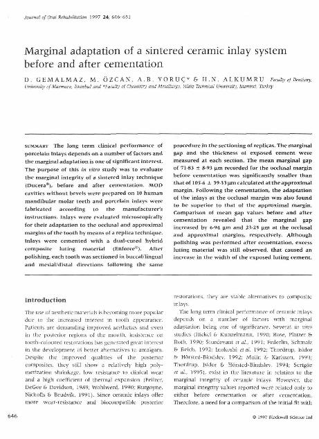
Fig. 1. Photomicrograph of the occlusal margin oi an inlay, (a)Marginal gap. (b) Thickness of the exposed luting cement. I, inlay;T, tooth; C, Luring composite.
Fig. 2. Photomicrograph of the approximal margin of an inlay,(al Marginal (inish line of the inlay, (b) Marginal finish iinc ofthe tooth cavily. h, horizonial discropancy: 1. inlay; T. looth; C,Luting composite.
luting composites, SEM observation* was performed onsectioned specimens at 100 X and 150 x magnifications.
Results
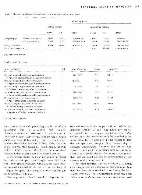
The mean marginal gap values {before and aftercementation), luting composite thicknesses andhorizontal discrepancy recordings at both the occlusaland the approximal margins are shown in Table 1.Table 2 summarizes the analysis of variance results forvarious comparisons of the recorded differences. Whenthe marginal gap values helore cementation werecompared, ihe mean marginal gap of 71 83 jim recordedfor the occiusal margin was significantly smaller thanthe 105-6 nm recorded for the approximal margin(P < 0-05). Following the cementation, the adaptationof the inlays at the occlusal margin (78'77 |am), wasalso found to be superior to the adaptation of theapproximai margins (128-85 |im) (? < 0 05).
The observed differences between before and aftercementation marginal gap values of occlusa! andapproximal margins were 6'94 (im and 23-25 |am,respectively, which were not statistically significant(Table 2).
Comparison of the marginal gap values aftercementation and exposed luting composite thicknessesrevealed that the exposed luting composite thicknesseswere significantly higher than those of the marginal gapvalues observed at both the occiusai and the approximalmargins (Tables 1 &• 2).
A wide range of horizontal discrepancynieasurcmenis, expressed in levms oi" standarddeviation, was observed. The mean discrepancy was79-74 [i.m with a range ot-142-6 [im (undercontoured)to 343-8 M-m (overcontoured), indicated that the inlayswere generally overcontoured at the approximalmargins.
structure (Fig. 1); (b) the thickness of the exposed lutingcement (Fig. 1); (c) the horizontal discrepancy, bothover and undercontours observed for the approximalmargins (Fig. 2).
The tnarginal fit values recorded for the ceramic inlaysal occlusal and proximal margins before and aftercementation were recorded and analysed statisticallyusing two way analysis of variance at a confidence levelof 0-95 (a = 0-05). In order to visualize the marginaladaptation of the inlays after cementation and exposed
Discussion
Several in vitro studies have reported that the marginalfit of ceramic inlays is inferior to that of gold inlays(Fett et aL, 1989; Shortall, Baylis 8- Grundy, 1989; Hunget at., 1990; Qualtrough et al., 1991; Molin &- Karlsson,1993). The marginal interface is said to be compensatedior by the use of a bonded dual-cured composite resinluting agent. However, in the long term, this could
*Jeol, Tokyo, Japan.
© 1997 Blat-kw tal of Oral Rehabilitation 24; 646-651
SINTERED INLAY TECHNIQUES 649
Table 1. Marginal gap, luting composite and horizontal discrepancy values (Mm)
Measuring poini
Ocdusai margin Approximdl margin
Range Range
Marginal gapAfter cementation
Luting compositeHorizontal discrepancy
78'77 14.85
197-54 64-64
63-44-104-56
140-4-313-6
128-85
165-6979-74
34 34
17-20139-18
59-196-474-4-196-73
148 2-204-13-1426-343-8
s.d., standard deviation.
Table 2. ANOVA results
Source of variance d.f. Sum of squai
Occlusal marginal gap before cementationX Approximal marginal gap before cementation
Occlusal marginal gap after cementation
X Approximal marginal gap after cemeniaiionOcclusal marginal gap before cementation
X Occlusal marginal gap after cementationApproximal marginal gap before cementation
X Approximal marginal gap after cementationOcclusal luting composite thickness
X Approxima! luting composite thicknessOcclusa! marginal gap after cementation
X Occlusal Luting composite thicknessApproximal marginal gap after cementation
X Approxima] luting composite thickness
1
1
!
1
I
1
I
5701-389
12540-03
241-0957
2704'208
5072-495
70532-99
6785-928
7-01
17-91
t-6
1-98
2-27
32-06
9 1 9
0-016
0-001
0-221
0'176
0-149
0-000
0-007
d.i., degrees of Ireedom.
be a serious drawback promoting the failure of therestoration due to dissolution and leakage,discolouration and excessive wear of the luting agent.Although an ideal range for the marginal gap of lutingcomposite cements has not yet been reported, someauthors (Leinfelder, Isenberg &• Essig, 1989; Federlinetai. 1992; Van Meerbeeker<j/.. 1992; Schmalz, Federlin& Reich. 1995). suggested that the maximum width ofexposed luting cement should not exceed 100 \xm.
In the present study, the mean gap values calculatedfor occlusal and approximal margins were 78-77 ̂ .mand 128-85 uni, respectively. Although, the mean valueof occlusa] marginal fit did noi exceed the limit of100 fim, the approximal marginal tit was over 100 jimby approximately 30 |im.
Since a great variation in marginal adaptation was
observed within all the samples and even within thedifferent locations of the same inlay, the clinicalacceptability of the marginal adaptation of an inlaysystem cannot be determined only by considering themean value. Thus, it would not be appropriate to statethat the approximal marginal fit of Ducera inlays isclinically unacceptable. However, the use of highviscosity resins coutd be recommended for luting, asSchmalz elal. (1995) claimed that luting spaces greaterthan 100 .̂m could partially be compensated by theviscosity ol the luting cement.
It has been reported that the viscosity of a lutingagent might have an influence on the minimum filmthickness that can he obtained in sectioning the inlay(Lambrechts et al., 1991). Comparison of the mean gapvalues before and after cementation revealed that dual-
) 1997 Biackwell Science Ltd. Journal of Oral Rehabililai
650 D.GEM ALMAZ et al.
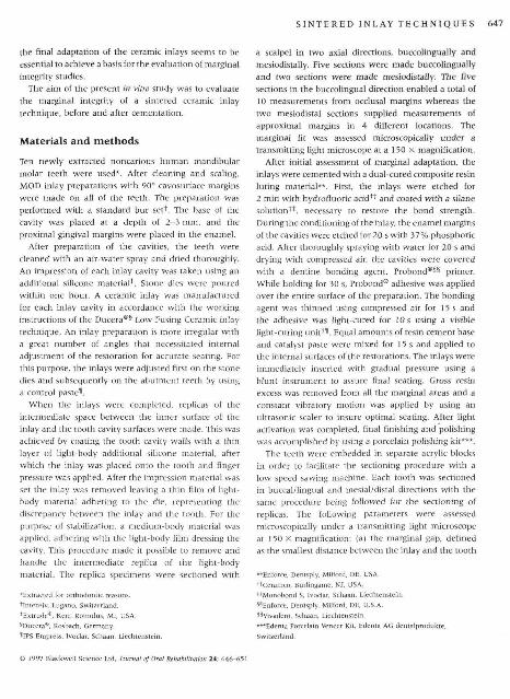
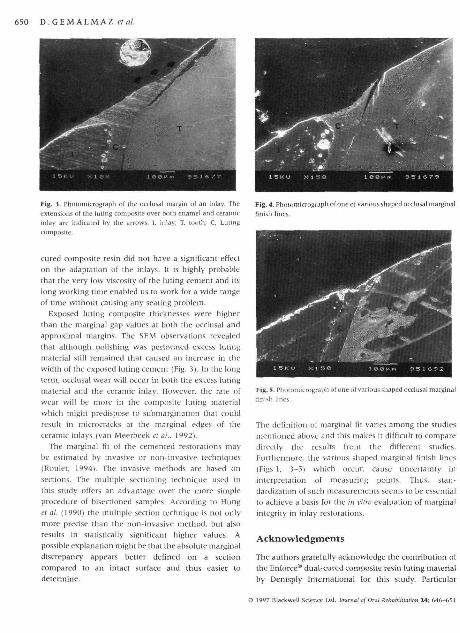
Fig. 3. Photomicrograph of the occlusal margin of an inlay. The
extensions of the luting composite over bolh enamel and ceramic
inlay are indicated by ihc arrows. 1, inlay; T. tooth; C, Luting
composite-
cured composite resin did not have a significani effecton the adaptation of the inlays. It is highly probablethat the very low viscosity of the luting cement and itslong working time enabled us to work for a wide rangeof time without causing any seating problem.
Exposed luting composite thicknesses were higherthan the marginal gap values at both the occlusal andapproximal margins. The SEM observations revealedthat although polishing was performed excess lutingmaterial still remained that caused an increase in thewidth of the exposed luting cement (Fig. 3). In the longterm, occlusa! wear will occur in both the excess lutingmaterial and the ceramic inlay. However, the rate ofwear will be more in the composite luting materialwhich might predispose to submargination that couldresult in microcracks at the marginal edges of theceramic inlays (van Meerbeek et at,, 1992).
The marginal fit of the cemented restorations maybe estimated by invasive or non-invasive techniques(Rouiet, 1994). The invasive methods are based onsections. The multiple sectioning technique used inthis study offers an advantage over the more simpleprocedure of bisectioned satnples. According to Hunget at. (1990) the multiple section technique is not onlymore precise than the non-invasive method, but alsoresults in statistically significant higher values. Apossible explanation might be that the absolute marginaldiscrepancy appears better defined on a sectioncompared to an intact surface and thus easier todetertnine.
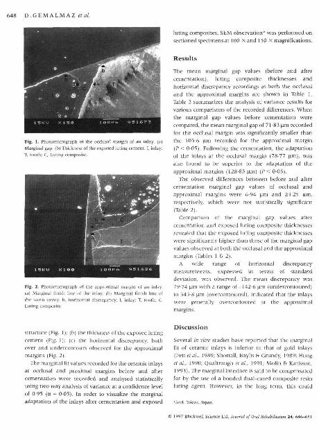
Fig. 4. Photomicrograph of one of various shaped occlusal marginalfinish lines.
"ograpli of one ol various shaped occlusaJ marginal
The definition of marginal fit varies among the studiesmentioned above and this makes it difficult to comparedirectly the results from the different studies.Furthermore, the various shaped marginal finish lines(Figs 1, 3-5) which occur, cause uncertainty ininterpretation of measuring points. Thus, stan-dardization of such measurements seems to be essentialto achieve a basis for the in vitro evaluation of marginalintegrity in inlay restorations.
Acknowledgments
The authors gratefully acknowledge the contribution ofthe Enforce® dual-cured composite resin luting materialby Dentsply International for this study. Particular
© 19«7 Blackwtl! Science Ltd. Journal of Oral Rehabilih
SINTERED INLAY TECHNIQUES 651
appreciation is expressed to Professor Adnan Tekin forhis help with the SEM observations. Appreciation isalso expressed to Mr Eren Tor and Mr Sunay Rodoplu fortheir assistance in preparing the experimental samples.
ReferencesBUFGOYNH, A,R., NiCHOLLS, I,J. & BRUDVIK, J . S , (1991) In vilro two-
body wear of inlay-onlay composite resin restoratives, Joumaf
of Prosthetic Dentistry. 65, 206.
FEDERLIN, M , , SCHMALZ, G, fr REICH, E, (1992) Adaptation of Cerec
infays: Influence of gap dimension/luting composiie, Jaurniil of
Dental Research. 71 , 691 (Abstraa No.),
FEILZER, A J . . DE GEE, A J . & DAVIDSON, C.L. (1989) Increased wall-
iD-wall curing contraclinn in ihin bonded resin layers. Journai
of Dcntai Research. 68 ,48 ,
FETT, H,P,, MORMANN, W , H , , LUTZ, F. & KREJCI, I. (1989) Margin
adaptation of computer machined Cerec inlays in vitro. Journai
ofDentai Research. 68 (Special Issue), 324 (Abstract No.).
HTCKEL. R. 6- KUNZELMANN, K-H, (1990) Der EiniluP der
kavitatenpraparation auf die randspaltbreite bei Cerec-inlays,
Deutsche Zahnarztiiche Zeitschrift. 45, 675,
HUNG. S,H,, HUNG, K.S., EICK, J , D . &• CHAPELL. R,P, (1990) Marginal
fit of porcelain fused to metal and two types of ceramic crowns.
Journal of Prosthetic Dentisrry. 63 , 26,
INOKOSHI. S,, VAN MnERBHEK, B., WiLLEMS, G., LAMBRF.CHTS, P.,
BRAEM, M , frVAN HERLE. G. (1992) Marginal accuracy of CAD/
CAM inlays made wilii ibe onginal and the updaied solTware.
Journal of Dentistry, 20, 171,
LAMBRECHTS. P,, INOKOSHI, S., VAN MEERBEEK. B,, WILLEMS, G ,
BBAEM, M , & VAN HERLE, G, (1991) Classification and potential
of composite luting materials. In: Proceedings of the International
Symposium on Computer Restorations. The Stale of Art of the Cerec
Method (ed, W. H. Mbrniann ), pp, 61-90, Quintessence, Berlin,
LEINFELDER, K.F,, ISENBERC;, B . P frEssiG, M,H, (1989) A new method
for generating ceramic restorations: CAD-CAM system. Journal
of American Dental Association. 118, 703.
Moi.iN, M. & KARLSSON. S. (1993) The fit of gold inlays and
three ceramic inlay systems. A clinical and in vitro study. Ada
Odontoio0ica Scandinavica, 51 , 201,
QUALTROUGH, A.S .E . , CRAMER, A., WlLSON, N.H.F . , ROULET, J .F. &
NuACK. M. {\99\) An in vitro evaluation of the marginal inlegrity
of a porcelain inlay system. International Journal of Prosthodontics.
6, 517,
ROSE, D,, PLATZER. U. & ROTH K K - F , (1990) Untersuchungen zur
kompositfuge bei computererslellten keramikinlays. Deutsche
Zahnarztiiche Zeitschrift, 45, 677.
RouLET, J,F, (1994) Marginal integrity: clinical significance. Journal
of Dentistry 22, (Suppl.), 9,
SCHMALZ, G., FEOERLm, M. & REICH, E. (1995) Effea of dimension
of luting space and luting composite on marginal adaptation of
a class n ceramic inlay. Journal of Prosthetic Dentistry. 73, 392,
SERrGOz, A,, GEMALMAZ, D,, ALKUMRU, H, & YORUI;, B, (1995) Luting
composite thickness of two ceramic iniay systems. European
Jourfjal of Prostko^ontics afiii Hesioralive Dentistry, 3 , ) 5 ] .
SHORTALL, A . C , BAYLIS, R.L, h- GRUNDV, J ,R. (1989) Marginal seal
comparisons between resin bonded class two porcelain inlays,
posterior composite restorations and direct composite resin
inlays. International Journal of Prosthodontics. 2, 217,
STURDEVANT, J ,R, , HEYMANN, H.O. , WILDER, A.D, , ROBERSON, T . M . &
BAYNE, S.C. (1991) Composite cemeni thickness of CERBC
GAD/CAM ceramic inlays, Journai of Dental Research. 70, 296
(Abstract No.),
THORDRUP, M , ISIDOR, 1 & HORSTED-BINDSIEV, P. (1992) Marginal fit
of tooth-colored inlays. Journal of Dental Research. 71, 658
(Abstract No,),
THORDRUP, M, , ISSDOR, F. & HORSTED-BINDSLEV, P, (1994) Comparison
of marginal fil and mjcroleakage of ceramic and composite
inlays: An in vitro study. Journal of Dentistry, 22, 147.
VAN MEERBEEK, B., INOKOSHI, S., WIIIEMS, G., NOACK, M , J , ,
BRAEM, M., LAMBRECHTS, P,, ROULET, .I.P. a VAN HERLE, G. (1992)
Marginal adaptation of four tooth-coloured inlay systems/M vivo.
Journal of Dentistry. 20, 18.
WoHLWERD, A. (1990) The empress technique, Quintessenz der
Zahntechnik. 16, 966,
Correspondence; Dr Deniz Gemalmaz, Marmara tjnivei
Hekimfigi FakiilEcsi, 8Q200 Nisaaiasi. Istanbul Turkey,, Dis

Copyright © 2022 FDOKUMEN