Mapping Motor Inhibition: Conjunctive Brain Activations across Different Versions of Go/No-Go and...
12
Mapping Motor Inhibition: Conjunctive Brain Activations across Different Versions of Go/No-Go and Stop Tasks Katya Rubia, 1 Tamara Russell, Stephan Overmeyer, Michael J. Brammer, Edward T. Bullmore,* Tonmoy Sharma, Andrew Simmons, Steve C. R. Williams, Vincent Giampietro, Chris M. Andrew, and Eric Taylor Institute of Psychiatry, King’s College, London; and *Department of Psychiatry, Addenbrooke’s Hospital, Cambridge, United Kingdom Received March 17, 2000; published online December 21, 2000 Conjunction analysis methods were used in func- tional magnetic resonance imaging to investigate brain regions commonly activated in subjects per- forming different versions of go/no-go and stop tasks, differing in probability of inhibitory signals and/or contrast conditions. Generic brain activation maps highlighted brain regions commonly activated in (a) two different go/no-go task versions, (b) three differ- ent stop task versions, and (c) all 5 inhibition task versions. Comparison between the generic activation maps of stop and go/no-go task versions revealed in- hibitory mechanisms specific to go/no-go or stop task performance in 15 healthy, right-handed, male adults. In the go/no-go task a motor response had to be selec- tively executed or inhibited in either 50% or 30% of trials. In the stop task, the motor response to a go- stimulus had to be retracted on either 50 or 30% of trials, indicated by a stop signal, shortly (250 ms) fol- lowing the go-stimulus. The shared “inhibitory” neu- rocognitive network by all inhibition tasks comprised mesial, medial, and inferior frontal and parietal corti- ces. Generic activation of the go/no-go task versions identified bilateral, but more predominantly left hemi- spheric mesial, medial, and inferior frontal and pari- etal cortices. Common activation to all stop task ver- sions was in predominantly right hemispheric anterior cingulate, supplementary motor area, infe- rior prefrontal, and parietal cortices. On direct com- parison between generic stop and go/no-go activation maps increased BOLD signal was observed in left hemispheric dorsolateral prefrontal, medial, and pari- etal cortices during the go/no-go task, presumably reflecting a left frontoparietal specialization for re- sponse selection. © 2001 Academic Press Key Words: fMRI; neuroimaging; motor response in- hibition; response selection; motor preparation; motor attention; conjunction analysis. INTRODUCTION Control of behavior and impulse is a higher-order function that evolves late, phylogenetically as well as ontogenetically, and has been suggested to be sub- served by the frontal lobes (Fuster, 1989). Every be- havioral, cognitive, or motor act requires a finely tuned balance between initiatory and inhibitory processes to provide appropriate preparation, initiation, on-line control, and timely inhibition of this act. Inhibitory control is therefore an essential regulatory function. It develops progressively from childhood to adulthood (Williams et al., 1999) and is therefore susceptible to impairment in neurodevelopmental disorders such as attention deficit hyperactivity disorder (Rubia et al., 1999, 2000b), conduct disorder, antisocial personality disorder, obsessive compulsive disorder, and Tourette’s syndrome (Bradshaw, 2000). Different types of motor acts are likely to be regu- lated by different inhibitory processes, which may be mediated by different cortical areas. The parts of the frontal lobes specifically involved in inhibitory control may therefore depend on the type of inhibitory process and the kind of action which needs to be inhibited. Concordant with this multiple domain model, different parts of the frontal lobes have been found to be respon- sible for different aspects of inhibitory control. Lesions in orbitofrontal cortex can lead to behavioural and socioemotional dyscontrol (Fuster, 1989), mesial and dorsolateral prefrontal brain areas have been related to reflex inhibition in the antisaccade task (Gaymard et al., 1998; O’Driscoll et al., 1995; Pierrot-Deseilligny et al., 1991), the supplementary motor cortex has shown to be involved in both initiation and suppression of voluntary movements (Dinner and Lueders, 1995; Ka- washima et al., 1996; Peterson et al., 1999), dorsolat- eral, inferior prefrontal, and anterior cingulate cortices are activated during the more cognitive/attentional forms of “inhibiting interference” during the Stroop task (Pardo et al., 1990; Bench et al., 1993; Taylor et al., 1 To whom correspondence and reprint requests should be ad- dressed. Fax: 144-0207-7085800; E-mail: [email protected]. NeuroImage 13, 250 –261 (2001) doi:10.1006/nimg.2000.0685, available online at http://www.idealibrary.com on 250 1053-8119/01 $35.00 Copyright © 2001 by Academic Press All rights of reproduction in any form reserved.
-
Upload
independent -
Category
Documents
-
view
3 -
download
0
Transcript of Mapping Motor Inhibition: Conjunctive Brain Activations across Different Versions of Go/No-Go and...

NeuroImage 13, 250–261 (2001)doi:10.1006/nimg.2000.0685, available online at http://www.idealibrary.com on
Mapping Motor Inhibition: Conjunctive Brain Activations across DifferentVersions of Go/No-Go and Stop Tasks
Katya Rubia,1 Tamara Russell, Stephan Overmeyer, Michael J. Brammer, Edward T. Bullmore,*Tonmoy Sharma, Andrew Simmons, Steve C. R. Williams, Vincent Giampietro,
Chris M. Andrew, and Eric TaylorInstitute of Psychiatry, King’s College, London; and *Department of Psychiatry, Addenbrooke’s Hospital, Cambridge, United Kingdom
Received March 17, 2000; published online December 21, 2000
Conjunction analysis methods were used in func-tional magnetic resonance imaging to investigatebrain regions commonly activated in subjects per-forming different versions of go/no-go and stop tasks,differing in probability of inhibitory signals and/orcontrast conditions. Generic brain activation mapshighlighted brain regions commonly activated in (a)two different go/no-go task versions, (b) three differ-ent stop task versions, and (c) all 5 inhibition taskversions. Comparison between the generic activationmaps of stop and go/no-go task versions revealed in-hibitory mechanisms specific to go/no-go or stop taskperformance in 15 healthy, right-handed, male adults.In the go/no-go task a motor response had to be selec-tively executed or inhibited in either 50% or 30% oftrials. In the stop task, the motor response to a go-stimulus had to be retracted on either 50 or 30% oftrials, indicated by a stop signal, shortly (250 ms) fol-lowing the go-stimulus. The shared “inhibitory” neu-rocognitive network by all inhibition tasks comprisedmesial, medial, and inferior frontal and parietal corti-ces. Generic activation of the go/no-go task versionsidentified bilateral, but more predominantly left hemi-spheric mesial, medial, and inferior frontal and pari-etal cortices. Common activation to all stop task ver-sions was in predominantly right hemisphericanterior cingulate, supplementary motor area, infe-rior prefrontal, and parietal cortices. On direct com-parison between generic stop and go/no-go activationmaps increased BOLD signal was observed in lefthemispheric dorsolateral prefrontal, medial, and pari-etal cortices during the go/no-go task, presumablyreflecting a left frontoparietal specialization for re-sponse selection. © 2001 Academic Press
Key Words: fMRI; neuroimaging; motor response in-hibition; response selection; motor preparation; motorattention; conjunction analysis.
1 To whom correspondence and reprint requests should be ad-
dressed. Fax: 144-0207-7085800; E-mail: [email protected].2501053-8119/01 $35.00Copyright © 2001 by Academic PressAll rights of reproduction in any form reserved.
INTRODUCTION
Control of behavior and impulse is a higher-orderfunction that evolves late, phylogenetically as well asontogenetically, and has been suggested to be sub-served by the frontal lobes (Fuster, 1989). Every be-havioral, cognitive, or motor act requires a finely tunedbalance between initiatory and inhibitory processes toprovide appropriate preparation, initiation, on-linecontrol, and timely inhibition of this act. Inhibitorycontrol is therefore an essential regulatory function. Itdevelops progressively from childhood to adulthood(Williams et al., 1999) and is therefore susceptible toimpairment in neurodevelopmental disorders such asattention deficit hyperactivity disorder (Rubia et al.,1999, 2000b), conduct disorder, antisocial personalitydisorder, obsessive compulsive disorder, and Tourette’ssyndrome (Bradshaw, 2000).
Different types of motor acts are likely to be regu-lated by different inhibitory processes, which may bemediated by different cortical areas. The parts of thefrontal lobes specifically involved in inhibitory controlmay therefore depend on the type of inhibitory processand the kind of action which needs to be inhibited.Concordant with this multiple domain model, differentparts of the frontal lobes have been found to be respon-sible for different aspects of inhibitory control. Lesionsin orbitofrontal cortex can lead to behavioural andsocioemotional dyscontrol (Fuster, 1989), mesial anddorsolateral prefrontal brain areas have been relatedto reflex inhibition in the antisaccade task (Gaymard etal., 1998; O’Driscoll et al., 1995; Pierrot-Deseilligny etal., 1991), the supplementary motor cortex has shownto be involved in both initiation and suppression ofvoluntary movements (Dinner and Lueders, 1995; Ka-washima et al., 1996; Peterson et al., 1999), dorsolat-eral, inferior prefrontal, and anterior cingulate corticesare activated during the more cognitive/attentionalforms of “inhibiting interference” during the Stroop
task (Pardo et al., 1990; Bench et al., 1993; Taylor et al.,
1iihnbripg2
G
cdtbttcfaaggeicvtiis
prtstsfnlgrnso
ropl
r1eaffsefnnp
251MAPPING MOTOR INHIBITION WITH fMRI
1997) and during the suppression of previously learnedstimulus–response associations in switching tasks(Nagahama et al., 1998, 1999; Konishi et al., 1998b,999; Dove et al., 2000). Inhibition of a motor responses the most direct expression of inhibitory control, as itnvolves (compared to the more cognitive forms of in-ibitory control such as interference control) all-or-one decisions about action or non-action. Severalrain areas have been related to inhibition of a motoresponse in stop and go/no-go tasks, including orbital,nferior, dorsolateral and mesial frontal, temporal andarietal cortices, as well as cerebellum and basal gan-lia (Garavan et al., 1999; Rubia et al., 1997, 1999,000a,b,c; Konishi et al., 1998a, 1999; Humberstone et
al., 1997; Casey et al., 1997; Kawashima et al., 1996;odefroy et al., 1996).The goal of this study was to further investigate and
ompare the neurocognitive networks related to twoifferent forms of motor response inhibition, namelyhose required by performing go/no-go and stop tasks,y use of generic brain image analysis methods similaro cognitive conjunction (Price and Friston, 1997; Fris-on et al., 1999). Such analyses allow exploration ofommonalities in activations of subject groups per-orming different tasks in relation to functions whichre common to the tasks. In this study, these genericnalysis methods enable the identification of brain re-ions generically activated during different versions ofo/no-go and stop tasks independent of the specificffects of the particular task versions (versions differ-ng in probability of inhibitory signals or in contrastonditions). Furthermore, the analysis of generic acti-ation across all stop and go/no-go task versions allowshe identification of brain regions related to sharednhibitory control mechanisms involved in both tasks,ndependent of the task-specific effects (go/no-go ortop task performance).The go/no-go paradigm requires a response selection
rocess between either executing or inhibiting a motoresponse, triggered by a go- or a no-go-stimulus. Theask demands high-level cognitive functions of deci-ion-making, response selection, and response inhibi-ion. The stop task requires withholding a motor re-ponse, which is triggered by a stop signal shortlyollowing the go signal, thereby converting the go-sig-al aposteriori to a no-go signal. It contains a higher
oad on response inhibition processes compared to theo/no-go task in that it involves the retraction of aesponse that has already been triggered by a go sig-al. Go/no-go tasks have a higher load on responseelection, due to the apriori knowledge about whetherr not to respond, provided by the categorical stimuli.The common and distinct neural substrates of motor
esponse inhibition in these two tasks has not previ-usly been identified. Evidence from lesion studiesoints towards the involvement of the mesial frontal
obes in go/no-go tasks, especially the SMA and ante-ior cingulate (Drewe, 1975; Leimkuhler and Mesulam,985; Verfaellie and Heilman, 1987), but also dorsolat-ral, medial prefrontal cortex, and caudate (Godefroy etl., 1996). Recent modern brain imaging studies usingMRI have revealed mesial, dorsolateral, and inferiorrontal and parietal involvement in this selective re-ponse inhibition process (Rubia et al., 2000c; Garavant al., 1999; Humberstone et al., 1997). Event relatedMRI has shown that focused activation of predomi-antly right inferior frontal cortex correlated witho-go activity (Konishi et al., 1998a, 1999), as well asre-SMA (Humberstone et al., 1997), but also inferior,
mesial, and middle frontal, insular, parietal and tem-poral lobes (Garavan et al., 1999). The motor responseinhibition process involved in stop tasks has beenshown to elicit predominantly right mesial and infero-medial prefrontal cortex activation in adults (Rubia etal., 1997, 2000a,c) and additional caudate activation inadolescents (Rubia et al., 1999, 2000a).
The goal of this paper was to investigate and com-pare the neurocognitive networks mediating the twotypes of motor inhibitory control required by go/no-goand stop tasks, independent of the specific contexts ofthe task variants as well as to explore shared neuro-cognitive processes underlying performance on bothinhibition tasks.
METHODS
Subjects
Fifteen healthy right-handed male adults, aged 26 to58 (mean age 5 36 years; standard deviation (SD) 5 7years), and of average intelligence as measured by anonverbal intelligence measure (Raven, 1960) partici-pated in the study (mean intelligence quotient 5 104 6(SD) 5 16). Subjects were divided in subgroups per-forming different task versions. Groups of performersdid not differ in IQ or task performance (Mean perfor-mance data. Go/no-go task1: mean reaction time (MRTin ms) 5 352 6 82, mean probability of inhibition (P(I)in percentage 5 95 6 8): go/no-go task2: MRT 5 304 687, P(I) 5 89 6 9; stop task1: MRT 5 664 6 66, P(I) 593 6 7; stop task2: MRT 5 624 6 149, P(I) 5 93 6 7;stop task3: MRT 5 576 6 111, P(I) 5 93 6 5). Allsubjects provided written informed consent. The studywas approved by the Bethlem Royal and MaudsleyNHS Trust Ethics (Research) Committee.
Tasks
Each paradigm consisted of two main conditions(control and activation condition), lasting 27 s each,preceded by a short visual warning cue (lasting 3 s).Control and activation condition epochs were periodi-cally alternated five times in the course of a single
experiment lasting 5 min. The control condition was
mHtheT
252 RUBIA ET AL.
presented first for each task. Throughout acquisition ofthe MR images, subjects responded to the stimuli bypressing a button with their right thumb, which wasrecorded by means of an MR compatible interface to aPC.
Go/no-go task 1. The go/no-go task requires selec-tion of either a response, indicated by a go signal, or a“no-response,” indicated by a no-go signal. Interstimu-lus-interval (ISI) was 1000 ms, including a stimulusduration of 200 ms followed by a blank screen for 800ms (27 stimuli per epoch). Go signals (airplanes) andno-go signals (bombs) alternated with 50% probabilityeach. Subjects had to respond to airplanes, but not tobombs. In the control condition airplanes and zeppelinsalternated with a probability of 50% and subjects wereinstructed to press a button in response to either stim-uli. This task controlled for the amount of sensorystimulation but only partially for the number of motorresponses, which was higher in the control condition(by 12 responses). Five of the subjects performed go/no-go task 1.
Go/no-go task 2. This task differed from the firstgo/no-go task in that the no-go signals and its controlsignals in the control condition appeared in 30% oftrials only, while the go signals appeared in 70% oftrials, in both conditions. A second difference was thatthe ISI of the control task was 1300 ms compared to1000 ms for the activation task, in order to control forthe number of motor responses, but only partially forthe number of visual stimuli, which was higher in theactivation condition (by six stimuli). This task wasperformed by seven subjects.
Stop task 1. The stop task requires a motor re-sponse in response to the go signal, only when the gosignal is not followed by a stop signal. In the activationcondition (stop condition), airplanes appeared on thescreen for 1000 ms, followed by a blank screen for 650ms (18 stimuli per epoch). On 50% of trials the airplanewas followed by a bomb, which appeared 250 ms afteronset of the airplane, replacing it for 300 ms, and wasthen then followed by a blank screen for 1100 ms. Thesubject was required to press a button if the airplanealone appeared, but not if the airplane was followed bya bomb. The control condition was identical, exceptthat a zeppelin instead of the bomb appeared on 50% oftrials, and subjects were instructed to press the button
TAB
Overview of Inhibition Task Ver
Go/no-go 1 Go/no-go 2
Percentage of inhibition 50% 30%Contrast condition Stimulus controlled Motor controlleNumber of subjects 5 7
always, whether or not the airplane was followed by a
zeppelin. This task controls for the number of visualstimuli, but only partially the number of motor re-sponses which was higher in the control condition (byeight responses). This task was performed by eightsubjects.
Stop task 2. A second version of the stop task wasidentical to stop task 1, except that the stop stimuliand its control stimuli in the control conditions ap-peared with a frequency of 30% of trials instead of 50%of trials. The conditions were matched for visual stim-ulation, but only partially for number of motor re-sponses, which was higher in the activation condition(by 4 responses). Go-signals appeared in 70% of trials.This task was performed by seven subjects.
Stop task 3. This stop task was identical to stoptask 2, where stop stimuli and its control stimuli ap-peared with a frequency of 30%, except that the ISI inthe control condition was 2200 ms compared to the ISIfor 1650 ms of the activation condition. This task con-trols for the number of motor responses, but only par-tially for the number of visual stimuli, which washigher (by 4 stimuli) in the activation condition. Thistask was performed by the same seven subjects whoperformed stop task 2 (see Table 1).
Image Acquisition
Gradient-echo echoplanar MR images were acquiredusing a 1.5 Tesla GE Signa System (General Electric,Milwaukee, WI) fitted with Advanced NMR hardwareand software (ANMR, Wolburn MA) at the MaudsleyHospital, London. A quadrature birdcage head coil wasused for RF transmission and reception. Data werecollected from 15 axial planes parallel to the anteriorcommissure-posterior commissure plane, ensuringthat the lowermost slice lay below the inferior border ofthe frontal lobe. 100 T*2-weighted MR images depictingBOLD contrast (Ogawa et al., 1990) were acquired withTE 5 40 ms, TR 5 3300 ms, in-plane resolution 5 3.1
m, slice thickness 5 5 mm, and slice gap 5 0.5 mm.ead movement was limited by foam padding within
he head coil and a restraining band across the fore-ead. At the same session, a 43 slice, high resolutionchoplanar image of the whole brain was acquired withE 5 40 ms, TI 5 180 ms, TR 5 16 s, in-plane resolu-
tion 5 1.5 mm, slice-thickness 5 3 mm, slice gap 5 0.3
1
s Performed by the 15 Subjects
Stop 1 Stop 2 Stop 3
50% 30% 30%Stimulus controlled Stimulus controlled Motor controlled
8 7 7
LE
sion
d
mm for subsequent registration of the fMRI data in
tTFsarb
253MAPPING MOTOR INHIBITION WITH fMRI
standard stereotactic space (Talairach and Tournoux,1988).
Image Analysis
Analysis of individual subject data. Followingthree-dimensional correction for movement during im-age acquisition using standard algorithms (Bullmore etal., 1999a), analysis of the individual subject data bysinusoidal regression yielded estimates for the ampli-tudes of the sine and cosine components of the responseat the frequency of alternation between the activationand control conditions of each task. These estimates(gamma and delta) were used to compute the standard-ised power (fundamental power quotient or FPQ) andphase of the response at each voxel (Bullmore et al.,1996). Gamma and delta were then reestimated 10times at each voxel following random permutation ofthe time-series data. This facilitated construction of adistribution of FPQs under the null hypothesis of noexperimentally determined response at the experimen-tal design frequency. Tests for activation of any voxelcould then be performed by obtaining the appropriatecritical value from the distribution of “randomized”FPQs and accepting as activated any voxel whose FPQexceeded this threshold (normally set at P , 0.003) inthe current series of experiments.
Generic analyses. The data were first normalisedfor each subject on an inversion recovery echoplanarimage obtained at twice the resolution of the fMRI datain x, y, and z. In a second stage, these were thentransformed into a Talairach template (Talairach andTournoux, 1988), constructed by manual transforma-tion of the structural inversion recovery image from 10subjects (5 male, 5 female) using the AFNI software(Cox, 1995) and spatially smoothed by application of a2-D Gaussian filter (SD 5 3 mm). The transformationat both stages was done by minimizing the sum ofabsolute image differences between the image to betransformed and the template image using an affinetransformation and at the second stage by a quadraticwarp algorithm. The procedure is described in detail inBrammer et al. (1997).
The data from the different experiments were com-bined and several generic analyses with non-paramet-ric inference at a voxel-wise probability of type I errorP , 0.0007 were performed on the motion-correctedfMRI time series at each voxel to explore commonlyactivated brain regions (a) in the two different versionsof the go/no-go task (b) in the three different versions ofthe stop task, and (c) across all five task versions.Approximate Brodmann areas were assigned to thevoxel with maximum FPQ in each generically activated2-D cluster by an automated cortical parcellationscheme described in detail by Wright et al. (1999).Generic activations (of a, b, and c) were analyzed using
a linear model to detect effects that were dependent onand independent of the nature of the particular task/contrast condition.
The following regression model can be expressed asFPQijk 5 a0i 1 a1i Task version 1 eijk, where FPQijk
denotes the standard power of response to the jth task(i.e., across the different experiments of (a, b, and c)) inthe kth individual at the ith voxel; a0i 1 a1i are theparameters estimated from the model; and eij is theresidual error at each voxel. The effect of task versionmembership (Task version is the classification param-eter) is parameterized by a1i at the ith voxel and theask version-independent (overall mean) effect by a0i.his model was fitted to the Talairach transformedPQ data obtained by random permutation of the timeeries (see above) as well as the FPQ data obtained bynalysis of the observed time series. Fitting to theandomized FPQ data permitted construction of distri-utions of a1i and a0i under the null hypothesis that
there was no experimentally determined response toperiodic alternation of the different activation and con-trol conditions of each task. The null distributions of a1i
and a0i were then used to determine critical values ofthe two parameters for statistical significance at thelevel of probability P , 0.0007, allowing for 10 errorvoxels. As the main goal of the analysis was to identifyvoxels showing significant responses related to sharedinhibitory processes involved in task performanceacross all task conditions, regardless of the particulartask version, we were primarily interested in estimat-ing and testing experiment-independent effects (a0i).But since the overall mean can be inflated by a partic-ularly salient response to one of several experiments,we included the a1i term in the model to allow suchresponses to be identified and removed from genericactivation maps. Following this conservative correctionof the data, significant generic effects across task ver-sions in (a, b, and c) were then displayed on a morpho-logical template. The median value of gamma, indicat-ing the phase of periodic signal change with respect tothe input function, was computed for each genericallyactivated voxel. Voxels with gamma . 0 had maximumsignal during the first (control) condition, voxels withgamma , 0 had maximum signal value during thesecond (activation) condition. Generic brain activationmaps were constructed to represent FPQ and gammaat each voxel of generic activation in (a) (go/no-go), (b)(stop), or (c) (stop and go/no-go tasks). Only voxelsactivated in phase with the activation condition weresuperimposed on a grey-scale template image to form ageneric activation map depicting significant effects in-dependent from the different task versions.
Task comparison. To estimate the difference be-tween generic brain activation across all go/no-go taskversions and generic activation across all stop taskversions, data from all experiments were combined and
fitted to the following analysis of variance (ANOVA)
jdo
m
254 RUBIA ET AL.
model at the ith voxel generically activated by theactivation condition in one or both of the tasks (refer-ring to generic gonogo and generic stop task activa-tions).
FPQi,j 5 mi 1 b Taskj 1 ei, j
Here, FPQi,j denotes the standardized power of re-sponse by the jth individual at the ith voxel; mi is theoverall mean power at the ith voxel; and ei,j is theresidual term for the same individual. Task denotes afactor coding the main effect of task (generic go/no-gotask activation or generic stop task activations), andmi 1 bTaskj denotes the mean power of response in theth Task. The null hypothesis of zero between-taskifference in mean FPQ was tested by comparing thebserved coefficient b1 to critical values of its nonpara-
metrically ascertained null distribution. To do this, theelements of Task are randomly permuted 10 times ateach voxel; b1 is estimated at each voxel after eachpermutation; and these estimates are pooled over allintracerebral voxels in standard space to sample thepermutation distribution of b1. Critical values for atwo-tailed test of size a 5 0.01 are the 100*(a/2)th and100*(1 2 a/2)th percentiles of this distribution (Bull-
ore et al., 1999b). Note that this uncorrected proba-bility threshold was used to identify differentially ac-tivated voxels only within the restricted search volumeof voxels generically activated in the activation condi-tions of the different stop task and go/no-go task ver-sions.
Voxel clusters containing less than 4 voxels are notconsidered in any of the analyses and are not shown inthe figures.
RESULTS
Generic Activation of Go/No-Go Task Versions
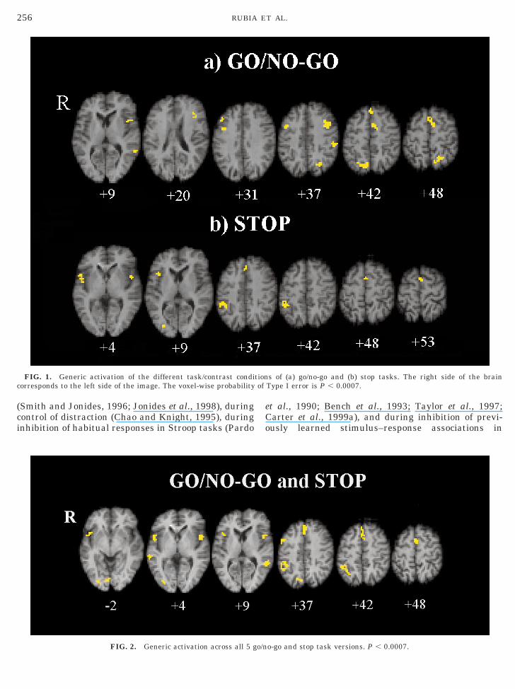
Common activation foci (P , 0.0007) in the differentversions of the go/no-go task were in bilateral but pre-dominantly left hemispheric middle (BA 9) and inferiorfrontal gyri at the border to the frontal operculum (BA44/45), left and right mesial frontal cortex, includinganterior cingulate and pre-SMA (BA 8/32/6), left infe-rior parietal lobe (BA 40), left precuneus (BA 7), andbilateral extrastriate cortices (BA 18/19) (see Fig. 1,Table 2a).
Generic Activation of Stop Task Versions
Common activation foci across the different stop taskversions (P , 0.0007) were noted in bilateral, but pre-dominantly right hemispheric inferior prefrontal/oper-cular cortex (BA 45), right inferior parietal lobe (BA40), pre-SMA (BA 6), and anterior cingulate (BA 32)
(see Fig. 1, Table 2b).Shared Activation in All Inhibition Task Versions
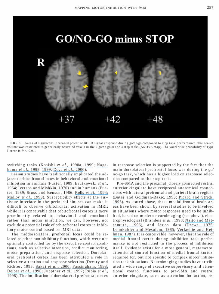
Common foci of activation across all five go/no-go andstop task versions (P , 0.0007) were observed in leftand right inferior (BA 47/44), right middle frontal gy-rus (BA 9/6), anterior cingulate (BA 8/32), pre-SMA(BA 6), right inferior parietal lobe (BA 40), and pre-dominantly left middle temporal cortex (BA 21) (seeFig. 2, Table 2c).
Differences between Generic Go/No-Go TaskActivation and Generic Stop Task Activation
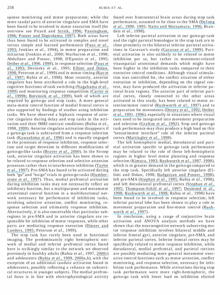
The search volume for the differences between thegeneric go/no-go and generic stop task activations waslimited to the generic activations found in each of thetwo conjunctive analyses. The search volume in theseregions of conjunctive activations in either go/no-go orstop tasks or both of them was 514 voxels and thevoxel-wise probability of false positive test was P ,0.01. At this size of test we expect five false-positivetests. In fact we observed significant differences at 77voxels. The differences were in left middle prefrontalgyrus (BA 9), in left inferior parietal lobe (BA 40), andin left medial frontal cortex (BA 32/6). In each regionthere was increased BOLD response during the go/no-go task compared to the stop task (see Fig. 3,Table 2d).
DISCUSSION
Concerted activation of mesial, middle, and inferiorfrontal and inferior parietal lobes appear to mediateperformance on tasks requiring the inhibition of a mo-tor response. While selective inhibition in a go/no-gotask activates a bilateral, but more left hemisphericmiddle-infero-mesio-frontal and parietal network,withholding a planned motor response in a stop taskelicits a predominantly right hemispheric homologuenetwork.
Our observation of a neural network of anterior cin-gulate, pre-SMA, dorsolateral, and inferior frontal andinferior parietal cortices during go/no-go task perfor-mance confirms previous findings. The middle and in-ferior frontal foci of activation of our study are in closeproximity to the middle and inferior activation foci inthe event related fMRI study of Garavan et al. (1999)and Konishi et al. (1999) (in inferior frontal lobe), withthe difference that the foci in middle and inferior fron-tal gyri were more prominent in the right hemispherein those studies as opposed to the left hemispherepredominance in our study (inferior lobe Talairach co-ordinates (mm); Garavan et al., 42, 40, 22; Konishi etal., 41, 16, 19; this study, 249, 11, 9; middle frontalgyrus, Garavan et al., 36, 23, 33; this study, 235, 19,37). Bilateral foci in anterior cingulate as well as infe-
rior parietal lobes were also found by Garavan et al.
tccetdthe
cfitaviTsac
SA
(
M
I
MAPP
E
P(
I
255MAPPING MOTOR INHIBITION WITH fMRI
(1999), again more prominently right hemisphericcompared to our left hemisphere predominance (Gara-van et al., 1999). Studies using block designed go/no-goasks have observed activation in bilateral anterioringulate, middle prefrontal and inferior frontal corti-es (Casey et al., 1997; Kawashima et al., 1996; Kramst al., 1999). Differences between studies in go/no-goask designs and contrast conditions may explain theifferences in laterality or precise localization. Despitehese differences, however, common areas of activationave been identified, especially in medial, middorsolat-
TAB
Main Brain Regions Generically Activated (Omnibus P ,top Task, and (d) Differences between Generic Activations ipproach at P , 0.01)
Cerebral region BA Side
a) Generic Go/no-gotask activationiddle frontal 9 L
Rnferior frontal 44/45 L
Resial frontal 8 Rnterior cingulate 24 Lre-SMA 6 L 1 Rarietal 40 L
Rxtrastriate cortex 18 L
19 Rrecuneus 7 L
b) Generic stop taskactivation
nferiorfrontal/insula 44/45 R
LInferior parietal 40 RPre-SMA 6 MAnterior cingulate 32 M(c) Activation
common to all go/no-go and stoptask versions
Inferior frontal 44/45 LR
Middle frontal 9/6 RAnterior cingulate 8/32 RPre-SMA 6 RInferior parietal 40 RMiddle temporal 21 L
R(d) Differences
between genericgo/no-go and stoptask activations
Middle frontal 9 LInferior parietal 40 LMedial frontal 32/6 L
Note. BA, approximate Brodmann area; P, probability of maximumof voxels; x, y, z refer to Talairach coordinates (mm).
ral, and inferior frontal lobes. From these, the most m
onsistent activation found across studies is in inferiorrontal lobes. Right and left inferior frontal cortices arenvolved in a wide range of high level cognitive func-ions, including language processing, working memorynd attention. Many of these executive functions in-olve aspects of inhibitory control, such as inhibition ofnterference in attention or working memory tasks.he inhibitory role of inferior frontal cortex thereforeeems not to be limited to the motor domain. Inferior,nd also occasionally middorsolateral prefrontal corti-es, have been found to be activated during working
2
003) in (a) Go/No-Go Task, (b) Stop Task, (c) Go/No-Go ando/No-Go and Stop Tasks (Using Region of Interest Analysis
x, y, z P N
235, 19, 37 0.000012 4346, 22, 31 0.000012 14249, 11, 9 0.000012 4440, 3, 31 0.000012 123, 31, 42 0.000012 2723, 0, 42 0.000012 333, 14, 48 0.000012 22252, 28, 37 0.000012 1549, 250, 37 0.000089 12238, 275, 22 0.000012 1717, 269, 42 0.000012 27212, 264, 48 0.000024 17
49, 3, 4/40, 17, 9 0.000006 28240, 11, 4 0.000006 1849, 242, 37 0.000006 393, 3, 53 0.000006 166, 25, 37 0.000006 8
249, 11, 4 0.000007 3949, 3, 4 0.000007 3443, 3, 37 0.000007 193, 31, 37/3, 14, 42 0.000007 403, 11, 48 0.000007 2646, 242, 37 0.000007 71249, 244, 9 0.000007 2961, 228, 4 0.000007 8
232, 6, 37 8252, 228, 37 826, 0, 42 8
gional difference in fundamental power quotient (FPQ); N, number
LE
0.0n G
re
emory conditions with high inhibitory demand
tionc of
256 RUBIA ET AL.
(Smith and Jonides, 1996; Jonides et al., 1998), duringcontrol of distraction (Chao and Knight, 1995), duringinhibition of habitual responses in Stroop tasks (Pardo
FIG. 1. Generic activation of the different task/contrast condiorresponds to the left side of the image. The voxel-wise probability
FIG. 2. Generic activation across all 5 go/n
et al., 1990; Bench et al., 1993; Taylor et al., 1997;Carter et al., 1999a), and during inhibition of previ-ously learned stimulus–response associations in
s of (a) go/no-go and (b) stop tasks. The right side of the brainType I error is P , 0.0007.
o-go and stop task versions. P , 0.0007.
h
ji1t
1
imnt
at(1eiitsLlmmiartut
vI
257MAPPING MOTOR INHIBITION WITH fMRI
switching tasks (Konishi et al., 1998a, 1999; Naga-ama et al., 1998, 1999; Dove et al., 2000).Lesion studies have traditionally implicated the ad-
acent orbito-frontal lobes in behavioral and emotionalnhibition in animals (Fuster, 1989; Brutkowski et al.,964; Iverson and Mishkin, 1970) and in humans (Fus-er, 1989; Stuss and Benson, 1986; Rolls et al., 1994;
Malloy et al., 1993). Susceptibility effects at the air–tissue interface in the perinasal sinuses can make itdifficult to observe orbitofrontal activation in fMRI;while it is conceivable that orbitofrontal cortex is moreprominently related to behavioral and emotionalrather than motor inhibition, we can, however, notexclude a potential role of orbitofrontal cortex in inhib-itory motor control based on fMRI data.
The middorsolateral prefrontal focus could be re-lated to other noninhibitory functions, which were notoptimally controlled for by the executive control condi-tions, such as selective attention, conflict monitoring,motor preparation, and response selection. Dorsolat-eral prefrontal cortex has been attributed a role inselective attention and response selection (Decary andRichter, 1995; Sakai et al., 2000; Passingham, 1993;Deiber et al., 1996; Jueptner et al., 1997; Rubia et al.,
FIG. 3. Areas of significant increased power of BOLD signal resolume was restricted to generically activated voxels in the 2 go/no-gerror is P , 0.01.
998). The implication of dorsolateral prefrontal cortex a
n response selection is supported by the fact that theain dorsolateral prefrontal focus was during the go/
o-go task, which has a higher load on response selec-ion compared to the stop task.
Pre-SMA and the proximal, closely connected rostralnterior cingulate have reciprocal anatomical connec-ions with lateral prefrontal and parietal brain regionsBates and Goldman-Rakic, 1993; Picard and Strick,996). As stated above, these medial frontal brain ar-as have been shown by several studies to be involvedn situations where motor responses need to be inhib-ted, based on modern neuroimaging (see above), elec-rophysiological (Brandeis et al., 1998; Naito and Mat-amura, 1996), and lesion data (Drewe, 1975;eimkuhler and Mesulam, 1985; Verfaellie and Hei-
man, 1987). It is conceivable, however, that the role ofedial frontal cortex during inhibition task perfor-ance is not restricted to the process of inhibition
tself. Evidence exists for a more general, metamotor,ttentional control function of medial frontal cortex,equired for, but not specific to complex motor inhibi-ion task situations. Neuroimaging studies have attrib-ted a wide range of executive supervisory and atten-ional control functions to pre-SMA and rostral
se during go/no-go compared to stop task performance. The searchr the 3 stop tasks (ANOVA map). The voxel-wise probability of Type
pono o
nterior cingulate, such as attention for action, re-
a1acc1armatrv1atitgtbpebsdiawisArspL
iwhpaeact
fped
acteivbetetreatpecat“c
emrswtlla11bimw
astiisdaumbt
258 RUBIA ET AL.
sponse monitoring and motor preparation; while themore caudal parts of anterior cingulate and SMA havebeen found to be involved in motor execution itself (foroverview see Picard and Strick, 1996; Passingham,1996; Posner and Digirolamo, 1997). Both areas havethus been found to be activated in complex and novelversus simple and learned performance (Paus et al.,1993; Jenkins et al., 1994), in motor preparation andinitiation (Jenkins et al., 2000; Warburton et al., 1998;Abdullaev and Posner, 1998; D’Eposito et al., 1995;Deiber et al., 1996, 1999), in response selection (Paus et
l., 1993; Devinsky et al., 1995; Elliott and Dolan,998; Peterson et al., 1999) and in motor timing (Rao etl., 1997; Rubia et al., 1998). Most recently, anterioringulate has been attributed a role in the high-levelognitive functions of task switching (Nagahama et al.,999) and monitoring response competition (Carter etl., 1999b; Botvinick et al., 1999), both of which areequired by go/no-go and stop tasks. A more generaleta-motor control function of medial frontal cortex is
lso supported by findings of studies using inhibitionasks. We have observed a biphasic response of ante-ior cingulate during delay and stop tasks in the acti-ation and their fMRI contrast conditions (Rubia et al.,998, 1999). Anterior cingulate activation disappears ifgo/no-go task is subtracted from a response selection
ask (Kawashima et al., 1996) and is equally engagedn the processes of response inhibition, response selec-ion and target detection in different modifications ofo/no-go-like tasks (Braver et al., 2000). In the Stroopask, anterior cingulate activation has been shown toe related to response selection and selective attentionrocesses rather than to interference inhibition (Taylort al., 1997). Pre-SMA has found to be activated duringoth “go” and “no-go” trials in go/no-go tasks (Humber-tone et al., 1997). Thus, medial prefrontal activationuring inhibition tasks may not necessarily reflect annhibitory function, but a multipurpose and metamotorttentional control function in a multifunctional net-ork necessary for performance of inhibition tasks,
nvolving selective attention, conflict monitoring, re-ponse selection and ultimately response inhibition.lternatively, it is also conceivable that particular sub-
egions in pre-SMA and in anterior cingulate are re-ponsible for inhibition of motor responses, while otherarts are mediating response execution (Dinner andueders, 1995; Peterson et al., 1999).The stop task has rarely been used in functional
maging. The predominantly right hemispheric net-ork of medial and inferior prefrontal cortex foundere is strikingly similar to the network we observedreviously in healthy adults (Rubia et al., 1997, 2000c)nd adolescents (Rubia et al., 1999, 2000a,b), with thexception of an additional caudate activation found indolescents, possibly reflecting a reliance on subcorti-al structures in younger subjects. The medial prefron-
al focus is in line with electrophysiological activity gound over frontocentral brain areas during stop taskerformance, assumed to lie close to the SMA (DeJongt al., 1990, 1995; Naito and Matsamura, 1996; Bran-eis et al., 1998).Left inferior parietal activation in our go/no-go task
nd the right parietal homologue in the stop task are inlose proximity to the bilateral inferior parietal activa-ions in Garavan’s study (Garavan et al., 1999). Pari-tal activation is also unlikely to be related to motornhibition per se, but rather to movement-relatedisuospatial attentional demands which might haveeen higher in the inhibition tasks compared to theirxecutive control conditions. Although visual stimula-ion was controlled for, the conflict situation of eitherxecution or inhibition, depending on the signal con-ext, may have produced the activation in inferior pa-ietal brain regions. The anterior part of inferior pari-tal cortex, closely connected to the other areasctivated in this study, has been related to motor at-ention/motor control (Rushworth et al., 1997) and toreparation for movements (Decety et al., 1992; Deibert al., 1991, 1996), especially in situations where visualues need to be integrated into movement preparationnd selection (Grafton et al., 1992). Stop and go/no-goask performance may thus produce a high load on thissensorimotor interface” role of the inferior parietalortex (Mattingley et al., 1998).The left hemispheric medial, dorsolateral and pari-
tal activation specific to go/no-go task performanceay be related to the role of these left-hemispheric
egions in higher level motor planning and responseelection (Kimura, 1993; Rushworth et al., 1997, 1998),hich is in greater demand in the go/no-go compared to
he stop task. Specifically left anterior cingulate (El-iott and Dolan, 1998; Badgaiyan and Posner, 1998),eft pre-SMA (Stephan et al., 1995; Rubia et al., 1998),nd left dorsolateral prefrontal cortex (Stephan et al.,995; Thompson-Schill et al., 1997; Desmond et al.,998; Rushworth et al., 1998; Rubia et al., 1998) haveeen found to be involved in response selection; leftnferior parietal lobe has been shown to play a role in
ovement preparation and fine-motor control (Rush-orth et al., 1997).In conclusion, using a range of conjunctive brain
ctivation and ANOVA analysis methods we havehown that the neurocognitive network subserving mo-or response inhibition involves bilateral middle andnferior frontal gyri, anterior cingulate, pre-SMA, andnferior parietal cortex. Inferior frontal cortex may bepecifically related to motor response inhibition, whileorsolateral, medial prefrontal, and parietal corticesre possibly mediating more general metamotor exec-tive control functions such as motor attention, conflictonitoring, and response selection, necessary for inhi-
ition task performance. While activations during stopask performance were more right-hemispheric, the
o/no-go task with lower load on inhibition elicited
B
259MAPPING MOTOR INHIBITION WITH fMRI
specific left hemispheric dorsolateral, medial prefron-tal, and parietal activations, presumably responsiblefor response selection.
ACKNOWLEDGMENTS
S.O. and K.R. were supported by European Fellowships from theEuropean Union Programme for the Training and Mobility of Re-searchers. E.B. was supported by the Wellcome Trust.
REFERENCES
Abdullaev, Y. G., and Posner, M. I. 1998. Event-related brain poten-tial imaging of semantic encoding during processing of singlewords. NeuroImage 7: 1–13.
adgaiyan, R. D., and Posner, M. 1998. Mapping the cingulate cortexin response selection and monitoring. NeuroImage 7: 255–260.
Bates, J. F., and Goldman-Rakic, P. S. 1993. Prefrontal connectionsof medial motor areas in the rhesus monkey. J. Comp. Neurol. 336:211–228.
Bench, C. J., Frith, C. D., Friston, K. J., Frackowiak, R. S., andDolan, R. J. 1993. Investigations of the functional anatomy ofattention using the Stroop test. Neuropsychologia 31(9): 907–922.
Botvinick, M., Nystrom, L. E., Fissell, K., Carter, C. S., and Cohen,J. D. 1999. Conflict monitoring versus selection-for-action in ante-rior cingulate cortex. Nature 402: 179–181.
Bradshaw, J. L. 2000. Neurodevelopmental Fronto-Striatal Disor-ders. Psychology Press, London.
Brammer, M. J., Bullmore, E. T., Simmons, A., Williams, S. C. R.,Grasby, P. M., Howard, R. J., Woodruff, P. W. R., and Rabe-Hesketh, S. R. 1997. Generic brain activation mapping in func-tional magnetic resonance imaging: A nonparametric approach.Magn. Reson. Imag. 15(7): 763–770.
Brandeis, D., Leeuwen, T. H., Rubia, K., Vitacco, D., Steger, J.,Borntraeger, E., and Steinhausen, H. C. 1998. Neuroelectric pre-cursors of stop failures in children with attention problems. Behav.Brain Res. 94(1): 111–125.
Braver, T., Barch, D., Molfese, D., and Ollinger, J. 2000. Anteriorcingulate activation is sensitive to response probability but notresponse inhibition. NeuroImage 11(5), S55.
Brutkowski, S. 1964. Prefrontal cortex and drive inhibition. In TheFrontal Granular Cortex and Behaviour (J. Warren and K. Akert,Eds.), pp. 242–270. McGraw-Hill, New York.
Bullmore, E. T., Brammer, M. J., Williams, S. C. R., Rabe-Herketh,S., Janot, N., David, A. S., Mellers, J. D. C., Howard, R., and Sham,P. 1996. Statistical methods of estimation and inference for func-tional MR image analysis. Magn. Reson. Med. 35: 261–277.
Bullmore, E. T., Brammer, M. J., Rabe-Herketh, S., Curtis, V., Mor-ris, R. G., Williams, S. C. R., Sharma, T., and McGuire, P. K.1999a. Methods for diagnosis and treatment of stimulus-correlatedmotion in generic brain activation studies using fMRI. Hum. BrainMap. 7: 38–48.
Bullmore, E. T., Suckling, J., Overmeyer, S., Rabe-Hersketh, S.,Taylor, E., and Brammer, M. 1999b. Global, voxel, and clustertests, by theory and permutation, for a difference between twogroups of structural MR images of the brain. IEEE Trans. Med.Imag. 18: 32–42.
Carter, C. S., Botvinick, M. M., and Cohen, J. D. 1999b. The contri-bution of the anterior cingulate cortex to executive processes incognition. Rev. Neurosci. 10(1): 49–57.
Carter, C. S., Mintun, M., and Cohen, J. D. 1999a. Interference andfacilitation effects during selective attention: An H215O PET
study of Stroop task performance. Neuroimage 2(4): 264–272.Casey, B. J., Trainor, R., Orendi, J. L., Schubert, A. B., Nystrom,L. E., Giedd, J. N., Castellanos, F. X., Haxby, J. V., Noll, D. C.,Cohen, J. D., Forman, S. D., Dahl, R. E., and Rapoport, J. L. 1997.A developmental functional MRI study of prefrontal activationduring performance of a go/no-go task. J. Cogn. Neurosci. 9: 835–847.
Chao, L. L., and Knight, R. T. 1995. Human prefrontal lesionsincrease distractibility to irrelevant sensory inputs. NeuroReport6: 1605–1610.
Cox, R. W. 1995. Analysis and visualisation of 3D fMRI data. Proc.Third Scientific Meeting. Soc. Magn. Res. 2: 834.
Decary, A., and Richer, F. 1995. Response selection deficits in frontalexcisions. Neuropsychologia 33: 1243–1253.
Decety, J., Kawashima, R., Gulyoas, B., and Roland, P. E. 1992.Preparation for reaching: A PET study of the participating struc-tures in the human brain. Neuroreport 3: 761–764.
deJong, R., Coles, M. G. H., Logan, G. D., and Gratton, G. 1990. Insearch of the point of no return: The control of response processes.J. Exp. Psychol. Hum. Percept. Perform. 16: 164–182.
deJong, R., Coles, M. G. H., and Logan, G. D. 1995. Strategies andmechanisms in nonselective and selective inhibitory motor control.J. Exp. Psychol. Hum. Percept. Perform. 21(3): 498–511.
Deiber, M. P., Passingham, R. E., Colebatch, J. G., Friston, K. J.,Nixon, P. D., and Frackowiak, R. S. 1991. Cortical areas and theselection of movement, A study with positron emission tomogra-phy. Exp. Brain Res. 84: 393–402.
Deiber, M. P., Ibanez, V., Sadato, N., and Hallett, M. 1996. Cerebralstructures participating in motor preparation in humans: Apositron emission tomography study. J. Neurophysiol. 75: 233–247.
Deiber, M. P., Honda, M., Ibanez, V., Sadato, N., and Hallett, M.1999. Mesial motor areas in self-initiated versus externally trig-gered movements examined with fMRI: Effect of movement typeand rate. J. Neurophysiol. 81: 3065–3077.
D’Eposito, M., Detre, J. A., Alsorp, D. C., Shin, R. K., Atlas, S., andGrossman, M. 1995. The neural basis of the central executivesystem of working memory. Nature 378: 279–281.
Desmond, J. E., Gabrieli, J. D., and Glover, G. H. 1998. Dissociationof frontal and cerebellar activity in a cognitive task: Evidence for adistinction between selection and search. Neuroimage 7(4): 368–376.
Devinsky, O., Morrell, M. J., and Vogt, B. A. 1995. Contributions ofthe anterior cingulate cortex to behaviour. Brain 118: 279–306.
Dinner, D. S., and Lueders, H. O. 1995. Human SupplementaryMotor Area- Electrical stimulation and movement-related poten-tial studies. In Epilepsy and the Functional Anatomy of the FrontalLobe (H. H. Jasper, S. Riggio, and Goldman-Rakic, Eds.) RavenPress, New York.
Dove, A., Schubert, T., Pollman, S., Norris, D., and v.Cramon, D. Y.2000. Prefrontal cortex activation in task switching. Cogn. BrainRes. 9: 103–109.
Drewe, E. A. 1975. Go/no-go learning after frontal lobe lesions inhumans. Cortex 11: 8–16.
O’Driscoll, G. A., Alpert, N. M., Matthysse, S. W., Levy, D. L., Rauch,S. L., and Holzman, P. S. 1995. Functional neuroanatomy of anti-saccade eye movements investigated with positron emission to-mography. Proc. Natl. Acad. Sci. USA 92: 925–929.
Elliott, R., and Dolan, R. J. 1998. Activation of different anteriorcingulate foci in association with hypothesis testing and responseselection. NeuroImage 8(1): 17–29.
Friston, K. J., Holmes, A. P., Price, C. J., Buechel, C., and Worsley,K. J. 1999. Multisubject fMRI studies and conjunction analyses.
NeuroImage 10: 385–396.
260 RUBIA ET AL.
Fuster, J. M. 1989. The Prefrontal Cortex: Anatomy, Physiology andNeuropsychology of the Frontal Lobe. Raven, New York.
Garavan, H., Ross, T. J., and Stein, E. A. 1999. Right hemisphericdominance of inhibitory control: An event-related functional MRIstudy. Proc. Nat. Acad. Sci. USA 96(14): 8301–8306.
Gaymard, B., Ploner, C. J., Rivaud, S., Vermersch, A. I., and Pierrot-Deseilligny, C. 1998. Cortical control of saccades. Exp. Brain Res.123(1–2): 159–163.
Godefroy, O., Lhullier, C., and Rousseaux, M. 1996. Non-spatialattention disorder in patients with frontal or posterior brain dam-age. Brain 119: 191–202.
Grafton, S. T., Mazziotta, J. C., Presty, S., Friston, K. J., Frackowiak,R. S., and Phelps, M. E. 1992. Functional anatomy of humanprocedural learning determined with regional cerebral blood flowand PET. J. Neurosci. 12(7): 2542–2548.
Humberstone, M., Sawle, G. V., Clare, S., Hykin, J., Coxon, R.,Bowtell, R., Macdonald, I. A., and Morris, P. G. 1997. Functionalmagnetic resonance imaging of single motor events reveals humanpresupplementary motor area. Ann. Neurol. 42(4): 632–637.
Iverson, S. D., and Mishkin, M. 1970. Perseverative interference inmonkeys following selective lesions of the inferior prefrontal con-vexity. Exp. Brain Res. 11: 376–386.
Jenkins, I. H., Brooks, D. J., Nixon, P. D., Frackowiak, R. S., andPassingham, R. E. 1994. Motor sequence learning: A study withpositron emission tomography. J. Neurosci. 14(6): 3775–3790.
Jenkins, H., Jahanshahi, M., Jueptner, M., Passingham, R. E., andBrooks, D. J. 2000. Self-initiated versus externally triggered move-ments. Brain 123(6): 1216–1228.
Jonides, J., Smith, E. E., Marshuetz, C., Koeppe, R. A., and Reuter-Lorenz, P. A. 1998. Inhibition in verbal working memory revealedby brain activation. Proc. Nat. Acad. Sci. USA 95(14): 8410–8413.
Jueptner, M., Stephan, K. M., Frith, C. D., Brooks, D. J., Frackow-iak, R. S., and Passingham, R. E. 1997. Anatomy of motor learning.I. Frontal cortex and attention to action. J. Neurophysiol. 77(3):1313–1324.
Kawashima, R., Satoh, K., Itoh, H., Ono, S., Furumoto, S., Gotoh, R.,Koyama, M., Yoshioka, S., Takahashi, T., Takahashi, K., Yanagi-sawa, T., and Fukuda, H. 1996. Functional anatomy of GO/NO-GOdiscrimination and response selection-a PET study in man. BrainRes. 728(1): 79–89.
Kimura, D. 1993. Left hemisphere control of oral and brachial move-ments and their relation to communication. Phil. Transact. RoyalSoc. London: Biol. Sci. 298: 135–149.
Konishi, S., Nakajima, K., Uchida, I., Kikyo, H., Kameyama, M., andMiyashita, Y. 1999. Common inhibitory mechanism in humaninferior prefrontal cortex revealed by event-related functionalMRI. Brain 122: 981–991.
Konishi, S., Nakajima, K., Uchida, I., Sekihara, K., and Miyashita, Y.1998a. No-go dominant brain activity in human inferior prefrontalcortex revealed by functional magnetic resonance imaging. Eur.J. Neurosci. 10(3): 1209–1213.
Konishi, S., Nakajima, K., Uchida, I., Kameyama, M., Nakahara, K.,Sekihara, K., and Miyashita, Y. 1998b. Transient activation ofinferior prefrontal cortex during cognitive set shifting. NatureNeurosci. 1(1): 80–84.
Krams, M., Rushworth, M. F., Deiber, M. P., Frackowiak, R. S., andPassingham, R. E. 1998. The preparation, execution and suppres-sion of copied movements in the human brain. Exp. Brain Res.120(3): 386–398.
Leimkuhler, M. E., and Mesulam, M. M. 1985. Reversible go/no-godeficits in a case of frontal lobe tumor. Ann. Neurol. 18: 617–619.
Malloy, P. F., Bihrle, A., Duffy, J., Cimino, C. 1993. The orbitomedial
frontal syndrome. Arch. Clin. Neuropsychol. 7: 88–95.Mattingley, J. B., Husain, M., Rorden, C., Kennard, C., and Driver,J. 1998. Motor role of human inferior parietal lobe revealed inunilateral neglect patients. Nature 392(6672): 179–182.
Nagahama, Y., Okada, T., Katsumi, Y., Hayashi, T., Yamauchi, H.,Sawamoto, N., Toma, K., Nakamura, K., Hanakawa, T., Konishi,J., Fukuyama, H., and Shibasaki, H. 1999. Transient neural ac-tivity in the medial superior frontal gyrus and precuneus timelocked with attention shift between object features. Neuroimage10(2): 193–199.
Nagahama, Y., Sadato, N., Yamauchi, H., Katsumi, Y., Hayashi, T.,Fukuyama, H., Kimura, J., Shibasaki, H., and Yonekura, Y. 1998.Neural activity during attention shifts between object features.Neuroreport 9(11): 2633–2638.
Naito, E., and Matsamura, M. 1996. Movement-related potentialsassociated with motor inhibition under different preparatorystates during performance of two visual stop signal paradigms inhumans. Neuropsychologica 34: 565–573.
Ogawa, S., Lee, T. M., Kay, A. R., and Tank, D. W. 1990. Brainmagnetic resonance imaging with contrast dependent on bloodoxygenation. Proc. Nat. Acad. Sci. USA 3: 9868–9872.
Pardo, J. V., Pardo, P. J., Janer, K. W., and Raichle, M. E. 1990. Theanterior cingulate cortex mediates processing selection in theStroop attentional conflict paradigm. Proc. Natl. Acad. Sci. USA315: 148–152.
Passingham, R. E. 1993. The Frontal Lobes and Voluntary Action.Oxford Univ. Press, Oxford.
Passingham, R. E. 1996. Functional specialisation of the supplemen-tary motor area in monkeys and humans. In Advances in Neurol-ogy, Vol. 70: Supplementary Motor Area (H. O. Luders, Eds.), pp.105–116. Lippingcott-Raven, Philadelphia.
Paus, T., Petrides, M., Evans, A. C., and Meyer, E. 1993. Role of thehuman cingulate cortex in the control of oculomotor, manual, andspeech responses: A positron emission tomography study. J. Neu-rophysiol. 70(2): 453–469.
Peterson, B. S., Skudlarski, P. J., Gatenby, J. G., Zhang, H., Ander-son, A. W., and Gore, J. C. 1999. An fMRI study of Stroop word-color interference: Evidence for cingulate subregions subservingmultiple distributed attentional systems. Biol. Psych. 45: 1237–1258.
Picard, N., and Strick, P. L. 1996. Motor areas of the medial wall: Areview of their location and functional activation. Cerebral Cortex6: 342–353.
Pierrot-Deseilligny, C., Rivaud, S., Gaymard, B., and Agid, Y. 1991.Cortical control of reflexive visually-guided saccades. Brain 114:1473–1485.
Posner, M. I., and DiGirolamo, G. J. 1997. Conflict, target detectionand cognitive control. In The Attentive Brain (R. Parasuraman,Ed.) MIT Press, Cambridge, MA.
Price, C. J., and Friston, K. J. 1997. Cognitive conjunction: A newapproach to brain activation experiments. Neuroimage 5: 261–270.
Rao, S. M., Harrington, D. L., Haaland, K. Y., Bobholz, J. A., Cox,R. W., and Binder, J. R. 1997. Distributed neural systems under-lying the timing of movements. J. Neurosci. 17(14): 5528–5535.
Raven, J. C. 1960. Guide to the Standard Progressive Matrices. HKLewis, London.
Rolls, E. T., Hornak, J., Wade, D., and McGrath, J. 1994. Emotion-related learning in patients with social and emotional changesassociated with frontal lobe damage. J. Neurol. Neurosurg. Psy-chiatr. 57: 1518–1524.
Rubia, K., Overmeyer, S., Taylor, E., Bullmore, E. T., Brammer, M.,Williams, S. C. R., Simmons, A., and Andrew, C. 1997. Neuronalsubstrate of inhibitory control: A fMRI study. NeuroImage 5: S113.
Rubia, K., Overmeyer, S. O., Taylor, E., Brammer, M., Williams, S.,
Simmons, A., Andrew, C., and Bullmore, E. T. 1998. Prefrontal
261MAPPING MOTOR INHIBITION WITH fMRI
involvement in temporal bridging and timing movement: An fMRIstudy. Neuropsychologia 36: 1283–1293.
Rubia, K., Overmeyer, S. O., Taylor, E., Brammer, M., Williams, S.,Simmons, A., Andrew, C., and Bullmore, E. T. 1999. Hypofrontal-ity in Attention Deficit Hyperactivity Disorder during higher ordermotor control: A study with fMRI. Am. J. Psych. 156(6): 891–896.
Rubia, K., Overmeyer, S. O., Taylor, E., Brammer, M., Williams, S.,Simmons, A., Andrew, C., and Bullmore, E. T. 2000a. Frontalisa-tion with age: Mapping neurodevelopmental trajectories withfMRI. Neurosci. Biobehav. Rev. 24: 13–19.
Rubia, K., Taylor, E., Smith, A., Oksanen, H., Overmeyer, S., Bull-more, E. T., and Newman, S. 2000b. Neuropsychological analysesof impulsiveness in childhood hyperactivity. Br. J. Psychiatry, inpress.
Rubia, K., Russell, T. A., Taylor, E., Bullmore, E. T., Brammer, M.,Williams, S. C. R., Simmons, A., Andrew, C., and Sharma, T.2000c. An fMRI study of reduced left prefrontal activation inschizophrenia during normal inhibitory function. SchizophreniaRes., in press.
Rushworth, M. F., Nixon, P. D., Wade, D. T., Renowden, S., andPassingham, R. E. 1998. The left hemisphere and the selection oflearned actions. Neuropsychologia 36(1): 11–24.
Rushworth, M. F., Nixon, P. D., Renowden, S., Wade, D. T., andPassingham, R. E. 1997. The left parietal cortex and motor atten-tion. Neuropsychologia 35(9): 1261–1273.
Sakai, K., Hikosaka, O., Takino, R., Miyauchi, S., Nielsen, M., andTamada, T. 2000. What and when: Parallel and convergent pro-cessing in motor control. J. Neurosci. 20(7): 2691–2700.
Smith, E. E., and Jonides, J. 1998. Neuroimaging analyses of human
working memory. Proc. Natl. Acad. Sci. USA 95: 12061–12068.Stephan, K. M., Fink, G. R., Passingham, R. E., Sibersweig, D.,Ceballos-Baumann, A. O., Frith, C. D., and Frackowiak, R. S. J.1995. Functional anatomy of the mental representation of upperextremity movements in healthy subjects. J. Neurophysiol. 73:373–386.
Stuss, D. T., and Benson, D. F. 1986. The Frontal Lobes. Raven, NewYork.
Talairach, J., and Tournoux, P. 1988. A Co-Planar Stereotactic Atlasof the Human Brain. Thieme Medical, New York.
Taylor, S. F., Kornblum, S., Lauber, E. J., Minoshima, S., and Ko-eppe, R. A. 1997. Isolation of specific interference processing in theStroop task: PET activation studies. Neuroimage 6(2): 81–92.
Thompson-Schill, S. L., D’Esposito, M., Aguirre, G. K., and Farah,M. J. 1997. Role of left inferior prefrontal cortex in retrieval ofsemantic knowledge: A reevaluation. Proc. Natl. Acad. Sci. USA94: 1492–14797.
Verfaellie, M., and Heilman, K. M. 1987. Response preparation andresponse inhibition after lesions of the medial frontal lobe. Arch.Neurol. 44(12): 1265–1271.
Warburton, E., Wise, R. J. S., Price, C., Weiller, C., Hadar, U.,Ramsay, S., and Frackowiak, R. S. J. 1996. Noun and verb re-trieval by normal subjects. Studies with PET. Brain 119: 159–179.
Williams, B. R., Ponesse, J. S., Schachar, R. J., Logan, G. D., andTannock, R. 1999. Development of inhibitory control across the lifespan. Dev. Psychol. 35(1): 205–213.
Wright, I. C., Sharma, T., Ellison, Z. R., McGuire, P. K., Friston,K. J., Brammer, M. J., Murray, R. M., and Bullmore, E. T. 1999.Supra-regional brain systems and the neuropathology of schizo-
phrenia. Cereb. Cortex. 9(4): 366–378.