Manual 2 Cohort Component Procedures
111
ATHEROSCLEROSIS RBSK IN COMMUNlTlES STUDY Manual 2 Cohort Component Procedures The National Heart, Lung, and Blood lnsfifufe of the National Institutes of Health
-
Upload
khangminh22 -
Category
Documents
-
view
0 -
download
0
Transcript of Manual 2 Cohort Component Procedures

ATHEROSCLEROSIS RBSK IN COMMUNlTlES STUDY
Manual 2
Cohort Component Procedures
The National Heart, Lung, and Blood lnsfifufe of the National Institutes of Health

ATHEROSCLEROSIS RISK IN COMMUNITIES STUDY PROTOCOL
Manual 2
COHORT COMPONENT PROCEDURES FOR THE SECOND EXAMINATION
VERSION 3.0, August 1990
For Copies, Please Contact ARIC Coordinating Center
Suite 203, NCNB Plaza 137 E. Franklin Street Chapel Hill, NC 27514

FOREWORD
This manual, entitled Manual 2: Cohort Comnonent Procedures for the Second Examination, is one of a series of protocols and manuals of operation for the Atherosclerosis Risk in Communities (ARIC) Study. The complexity of the ARIC Study requires that a sizeable number of procedures be described, thus this rather extensive set of materials has been organized into the set of manuals listed below.
The version status of each manual is printed on the title sheet and in the footer of each page in the text of the document. The first edition of each manual was labelled Version 1.0. Subsequent minor revisions are indicated in the decimal portion of the version number. Major revisions are reflected in the integer.
ARIC Study Protocols and Manuals of Operation
MANUAL TITLE
1 General Description and Study Management
2 Cohort Component Procedures for the Second Examination
3 Surveillance Component Procedures
4 Pulmonary Function Assessment
5 Electrocardiography
6 Ultrasound Assessment
ba: Ultrasound Scanning Protocol Ultrasound B-Mode Image Reading Protocol
i: Distensibility Scanning Protocol Distensibility Reading Protocol
7
'8
9
10
11
12
Blood Collection and Processing
Lipid and Lipoprotein Determinations
Hemostasis Determinations
Clinical Chemistry Determinations
Sitting Blood Pressure
Quality Assurance and Quality Control

ii
MANUAL 2 Table of Contents
Chapter 1. Recruitment and Follow-Up of the ARIC Cohort After Visit 1
1.1 1.2
1.3 1.4 1.5
1.6
Introduction .................................................... Annual Follow-Up 1.2.1 Annual Contacts Between Exams ......................... 1.2.2 Follow-Up Procedures .................................. 1.2.3 Annual Cohort Interview ............................... Eligibility Requirements for Post-Baseline Examinations ......... Window for Visit 2 .............................................. Recruitment 1.5.1 Outline of Recruitment for Visit 2 .................... 1.5;2 Contacting Participants ............................... 1.5.3 Making the Clinic Appointment ......................... 1.5.4 Instructions for the Follow-Up Clinic Examinations .... 1.5.5 Scheduling Appointments ............................... Completeness of Re-Examination 1.6.1 Introduction .......................................... 1.6.2. Qualified Interviewers ................................ 1.6.3 Pre-appointment Contacts .............................. 1.6.4 Contacts for No Shows ................................. 1.6.5 Reimbursement ......................................... 1.6.6 Publicity ............................................. 1.6.7 Supervision ...........................................
Chapter 2. The Second Cohort Examination 2.1 Introduction .................................................... 2.2 Participant Flow
2.2.1 Rationale ............................................. 2.2.2 Fixed Sequences .......................................
'2.2.3 Flexible Sequences .................................... 2.2.4 Data Collection .......................................
2.3 Reception 2.3.1 Informed Consent ...................................... 2.3.2 Update Form ........................................... 2.3.3 Fasting/Tracking ...................................... 2.3.4 .....................................
2.4 Medication Survey
Anthropometry 2.4.1 Rationale ............................................. 2.4.2 Procedures ............................................ 2.4.3 Training .............................................. 2.4.4 Certification ......................................... 2.4.5 Quality Assurance ..................................... 2.4.6 Data Collection .......................................
2.5 Sitting Blood Pressure 2.5.1 Rationale ............................................. 2.5.2 Procedures ............................................ 2.5.3 Training .............................................. 2.5.4 Certification ......................................... 2.5.5 Quality Assurance ..................................... 2.5.6 Data Collection .......................................
1
8
10 10 10 14
14 15 16 17
21 21 29 29 29-- 30
30 30 30 30 30 31
ARIC PROTOCOL 2. Cohort Component Procedures - Visit 2. VERSION 3.0 8/90

iii
Chapter 2. The Second Cohort Examination
2.6
2.7 2.8
2.9
2.10
2.11
2.12
2.13
Venipuncture 2.6.1 Rationale ............................................. 2.6.2 Procedures ............................................ 2.6.3 Training .............................................. 2.6.4 Certification ......................................... 2.6.5 Quality Assurance ..................................... 2.6.6 Data Collection ....................................... Snack ........................................................... Cognitive Function 2.8.1 Rationale ............................................. 2.8.2 Administration ........................................ 2.8.3 Training .............................................. 2.8.4 Certification ......................................... 2.8.5 Quality Assurance ..................................... 2.8.6 Data Collection ....................................... Family History 2.9.1 Rationale ............................................. 2.9.2 Administration ........................................ 2.9.3 Training .............................................. 2.9.4 Certification ......................................... 2.9.5 Quality Assurance ..................................... 2.9.6 Data Collection ....................................... Health History 2.10.1 Rationale ............................................. 2.10.2 Administration ........................................ 2.10.3 Training .............................................. 2.10.4 Certification ......................................... 2.10.5 Quality Assurance ..................................... 2..10.6 Data Collection ....................................... Health and Life Profile 2.11.1 Rationale ............................................. 2.11.2 Administration ........................................ 2.11.3 Training .............................................. 2.11.4 Certification ......................................... 2.11.5 Quality Assurance ..................................... 2.11.6 Data Collection ....................................... Respiratory Symptoms 2.12 .I Rationale ............................................. 2.12.2 Adrbistration ........................................ 2.12.3 Training .............................................. 2.12.4 Certification ......................................... 2.12.5 Quality Assurance ..................................... 2.12.6 Data Collection ....................................... T&/Stroke 2.13.1 Rationale ............................................. 2.13.2 Administration ........................................ 2.13.3 Training .............................................. 2.13.4 Certification ......................................... 2.13.5 Quality Assurance ..................................... 2.13.6 Data Collection .......................................
31 31 31 31 31 31 32
32 32 33 33 33 33
33 33 34 34 34 34
34 34 35 35 35 35
35 36 37 37 37 37
38 38 38 38 38 38
39 39 39 39 39 39
ARIC PROTOCOL 2. Cohort Component Procedures - Visit 2. VERSION 3.0 8/90

iv
Chapter 2. The Second Cohort Examination
2.14
2.15
2.16
2.17
2.18
2.19
2.20 2.21 2.22
2.23
Physical Exam 2.14.1 Rationale ............................................. 40 2.14.2 Procedures ............................................ 40 2.14.3 Training .............................................. 42 2.14.4 Certification ......................................... 42 2.14.5 Quality Assurance
I ..................................... 42 2.14.6 Data Collection ....................................... 42 Electrocardiograms 2.15.1 Rationale ............................................. 42 2.15.2 Procedures ............................................ 43 2.15.3 Training .............................................. 43 2.15.4 Certification ......................................... 43 2.15.5 Quality Assurance ..................................... 43 2.15.6 Data Collection ....................................... 43 Pulmonary Function 2.16.1 Rationale ............................................. 44 2.16.2 Procedures ............................................ 44 2.16.3 Training .............................................. 44 2.16.4 Certification ......................................... 45 2.16.5 Quality Assurance ..................................... 45 2.16.6 Data Collection ....................................... 45 Ultrasound 2.17.1 Rationale ............................................. 46 . 2.17.2 Procedures ............................................ 46 2.17.3 Training .............................................. 47 2.17.4 Certification ......................................... 47 2.17.5 Quality Assurance ..................................... 47 2.17.6 Data Collection ....................................... 47 Data Inventory 2.18.1 Rationale ............................................. 48 2.18.2 Procedures ............................................ 48 2.18.3 Training .............................................. 49 2.18.4 Certification ......................................... 49 2.18.5 Quality Assurance ..................................... 49 2.18.6 Data Collection ....................................... 49 Medical Data Review 2.19.1 Rationale ............................................. 49 2.19.2 Procedures ............................................ 50 2.19.3 Training .............................................. 54 2.19.4 Certification ......................................... 54 2.19.5 Quality Assurance ...... ..e.............: .............. 54 2.19.6 Data Collection ....................................... 54 Exit Interview .................................................. 54 Referral Guidelines ............................................. 54 Medical Reviews 2.22.1 General Policies ...................................... 56 2.22.2 Procedures ............................................ 56 Results Reporting 2.23.1 Rationale ............................................. 61 2.23.2 Overview of Results Reporting ......................... 61 2.23.3 Report of Ultrasound B-Mode Scann Measurements ........ 64 2.23.4 Routine Notification of Study Results ................. 65
ARIC PROTOCOL 2. Cohort Component Procedures - Visit 2. VERSION 3.0 8/90

V
2.23.5 Results Reported Only by Request ...................... 66 2.23.6 Study Results Requiring Special Notification .......... 66
Chapter 2. The Second Cohort Examination
2.24 Participant Safety 2.24.1 Measures to Protect the Participant ................... 69 2.24.2 Methods for Handling Emergencies ...................... 70 2.24.3 Emergency Equipment ................................... 72 2.24.4 Notification of Study Results ......................... 72
Chapter 3. Event Classification for Cohort Component 3.1 Identification of Events
3.1.1 Identification of Hospitalized Events . . . . . . . . . . . . . . . . . 75 3.1.2 Identification of Deaths . . . . . . . . . . . . . . . . . . . . . . . . . . . . . . 78
3.2 Event Investigation 3.2.1 Procedures for Fatal Events . . . . . . . . . . . . . . . . . . . . . . . . . . . 79 3.2.2 Procedures for Hospitalized Events . . . . . . . . . . . . . . . . . . . . 82 3.2.3 Summary of Cohort Investigations . . . . . . . . . . . . . . . . . . . . . . 83
3.3 Diagnostic Criteria 3.3.1 Coronary Heart Disease . . . . . . . . . . . . . . . . . . . . . . . . . . . . . . . . 84 3.3.2 Stroke . . . . . . . . . . . . . . . . . . . . . . . . . . . . . . . . . . . . . . . . . . . . . . . . 94
3.4 Event Determination . . . . . . . . . . . . . . . . . . . . . . . . . . . . . . . . . . . . . . . . . . . . . 101 3.4.1 Diagnosis of Coronary Heart Disease . . . . . . . . . . . . . . . . . . . 102
3.5 Diagnosis of Prevalent MI at Baseline and Interim MI between Clinic Visits 3.5.1 Procedures . . . . . . . . . . . . . . . . . . . . . . . . . . . . . . . . . . . . . . . . . . . . 103 3.5.2 Definitions . . . . . . . . . . . . . . . . . . . . . . . . . . . . . . . . . . . . . . . . . . . 104
Appendix
‘Table of Contents . . . . . . . . . . . . . . . . . . . . . . . . . . . . . . . . . . . . . . . . . . . . . . . . . . . . . . . . . A-l
ARIC PROTOCOL 2. Cohort Component Procedures - Visit 2. VERSION 3.0 8/90

page 1
1. RRCRUI~EIENT AND FOLLOW-UP OF THR ARIC COHORT AFI'RR VISIT 1
1.1 Introduction
The ARIC cohort consists of approximately 4,000 men and women ages 45-64 at Visit 1, from each community. Annually, participants are recontacted by telephone in order to maintain correct addresses and to ascertain interim medical events. Every third year, participants are contacted for reexamination at the field centers.
1.2 Amlual Follow-Up
1.2.1 Annual Contacts Between Exams
Each study participant is recontacted annually after his or her initial examination at approximately the same time each year. The target date for the annual follow-up interview is the date of the baseline visit. However, a 1 year window, up to 6 months before and 6 months after the target date, is allowed for each annual contact. The initial call for annual contact generally should be no more than three weeks before the target date, except in contact year 4 (CY04) when initial calls can occur up to 4 months in advance. of the target date. These follow-up contacts review the health-related developments which have occurred since the last contact. Follow-up interviews are preferably conducted by telephone, but can be done in person if necessary. Beginning February 1990, letters (Appendix 1.1) will not be sent to participants prior to the telephone interview for Contact Years 03, 05 and 06 unless the person cannot be reached by phone.
1.2.2 Follow-up Procedures
Annual follow-up of the ARIC Study cohort is used to (1) maintain contact and correct address information on cohort participants and (2) ascertain vital status and interim medical events between the three-year comprehensive examina- tions.
The basic procedure for interim contacts is described below and summarized in Figure 1.1,
m-> (a,b,c) prl
Additional diagnostic or abstract- ing procedure if indicated
(a) Send Annual Contact Letter at Contact Year 2. (b) Send Pre-Visit 2 Contact Letter at Contact Year 4. (c) Send Annual Contact Letter/Pre-Visit 2 Letter for cohort members who
cannot be contacted by telephone.
Figure 1.1 Interim Contact Procedures Between Clinical Examinations in the ARIC Cohort Study
ARIC PROTOCOL 2. Cohort Component Procedures - Visit 2. VERSION 3.0 8/90

page 2
At the baseline examination the following information was collected and stored in the data base to facilitate future contacts:
1. I.D. Number 2. Name, address, telephone number 3. Age 4. Physician/hospital name, address, telephone number 5. All tracing information, such as the names of close friends,
social security number, employer, etc.
This file is used for preparing results letters, annual contact letters, and rescheduling follow-up exams.
The preferred time-window for making interim contacts is within a month of the anniversary date of the original examination. The telephone interviews are generally scheduled no more than three weeks before to three weeks after the target date.
For persons being contacted for their first annual follow-up (Contact Year 2) or those in subsequent years (Contact Years 03, 05, 06) not reached by phone after three attempts at ideal times, letters on ARIC Study stationery and "forwarding and address correction requested" on the envelope are mailed and further attempts are made (Appendix 1.1). These letters contain:
1. A reminder that the addressee is in the study and that annual contact is involved.
2. A description of the purpose of the contact. 3. Information that the participant should obtain to assist with the
interview (e.g., hospitalizations, physicians visits). 4. A request to call the ARIC Study office to set up a time to complete
the Annual Follow-up Interview.
Extensive efforts are made to maintain contact with every cohort participant.
1.2.3 Annual Cohort Interview
The annual follow-up interview of the ARIC Study cohort updates address and tracing information of cohort participants, ascertains vital status, interim hospitalizations, and new cardiovascular symptoms. (See Appendix 2.2.a.) Its main purpose is to identify possible cardiovascular events or treatment requir- ing hospitalization. Every hospitalization is verified and the discharge diagnoses recorded. Potential cardiovascular events are reviewed further for ARIC Study endpoint criteria by abstraction of hospital records.
Every attempt is made to identify cohort deaths before the annual contact, through regular review of death certificates. When deaths are ascertained, a mortality interview is conducted at an appropriate time.
1.3 Eligibility Requirements for Post-Baseline Examinations
Participants who completed at least part of the baseline examination are followed and, if alive, invited to subsequent ARIC examinations. This excludes enumerated residents who completed the home interview, but did not sign the informed consent form at-the field center.
ARIC PROTOCOL 2. Cohort Component Procedures - Visit 2. VERSION 3.0 8/90

page 3
Participants do not have to still live in the community to participate in subsequent annual follow-up interviews or examinations. Those who have moved are invited to return for examinations, but study reimbursement for long distance travel is unavailable.
1.4 Window for Visit 2
The scheduling of Visit 2 is made in conjunction with the annual contact in the fourth contact year. The ontimal timeframe for scheduling Visit 2 is within 30 days of the participant's annual contact target date. It is anticipated that most of the field center visits will be completed within at least 90 days. However, if the participant cannot complete Visit 2 within this window, it is still possible for Visit 2 to be completed at any time during Contact Years 4
The Visit 2 data is entered into the database as Contact Year 4 through 6. data, regardless when Visit 2 occurs.
1.5 Recruitment
1.5.1 Outline of Recruitment for Visit 2
The
1.
2.
3.
4.
5.
6.
steps in the recruitment process for Visit 2 are as follows:
A list of participants to be contacted, their tracing information, and the target contact date is provided to field centers by the Coordinating Center at least 3 months in advance of the contact date.
Field Centers mail a letter to the participant indicating that the usual Annual Follow-up telephone call will be coming, and at that time an appointment for Visit 2 will be set. A brief description of Visit 2 is provided in the letter.
The participant is telephoned, the Annual Follow-up Form is completed in the usual manner, and the participant is recruited for Visit 2. Some home interviews may be necessary for individuals unreachable by telephone or for special circumstances. After the appointment is set, basic instructions for Visit 2 are provided.
Soon afterwards, field centers send a reminder letter indicating the appointment time and providing full instructions for the visit.
A reminder.telephone call precedes the visit.
If the participant is not available during the usual time window for his/her Visit 2 appointment, centers keep trying to recruit for Visit 2 at a later date. Even if a participant refuses Visit 2 during contact year 4, he/she is to be invited in future contact years unless the supervisor considers it inappropriate.
1.5.2 Contacting Participants
The Coordinating Center generates from the ARIC database a list of participants to be contacted for Visit 2 and the target contact date. The list is similar to the lists provided for Annual Follow-up, and is generated well in advance of the contact window to allow field centers to schedule the lengthier interviews,
ARIC PROTOCOL 2. Cohort Component Procedures - Visit 2. VERSION 3.0 B/90

page 4
and if necessary, to trace hard to find participants.
Participant information sheets are generated that contain pertinent information from Visit 1 to be used in the Visit 2 (e.g., baseline vital status of parents, baseline menopausal status, etc.) clinic examination.
Field centers mail a letter to each participant indicating that the usual Annual Follow-up call is due and that Visit 2 will be scheduled. A prototype letter is provided in Appendix 1.4. The Coordinating Center generates data files from which the field centers produce address labels for the mailing. Letters are sent "forwarding and address correction requested", so that participants who have moved can be identified. Approximately one week after the letter is mailed, a telephone call is placed to'the home. All Annual Follow-up interviews are completed, and tracing information is updated. The interviewer then asks to schedule a clinic appointment as described in Section 1.5.3. The interviewer must be aware of available appointment times and be able to convey basic clinic instructions. Participants who do not have phones, have trouble communicating by telephone, or have special needs are not contacted by.telephone but are visited in-person. If these participants can be identified in advance, the letter indicates that an interviewer will visit the home, and annual follow-up and recruitment takes place there.
Participants found to have moved or who are otherwise lost to follow-up are traced using the tracing information obtained at Visit 1 and subsequent annual follow-up contacts. Periodic searches of the National Death Index are made. Every attempt is made to schedule and complete a visit for each cohort participant.
1.5.3 Making the Clinic Appointment
At the end of the annual follow-up for all participants in a household, the clinic visit is described and a request made for an appointment. The interviewer inquires about several items to assist in scheduling the appointment:
1. Preferred time and date of examination; 2. Any medical conditions (e.g., diabetes, dietary restrictions) which might
affect the physical examination and/or type of snack provided; 3. Need for assistance getting to or moving about the clinic.
If possible, the interviewer schedules appointments for the examination during the 30 days following the telephone call. The interviewer notifies the clinic scheduler to set an appointment day and time. The appointment is recorded on a reminder sheet which is mailed to (or left for) the participant. Participants are scheduled for appointments at their convenience, dependent upon clinic schedule. For convenience of the study participants, eligible members of a single household are scheduled for examination on the same day whenever possible.
1.5.4 Instructions for the Follow-up Clinic Examinations
The instructions for clinic visits are specified on an information sheet (Appendix 1.5) prepared by each Field Center, and mailed (or delivered) to the participant soon after the appointment is made.
ARIC PROTOCOL 2. Cohort Component Procedures - Visit 2. VERSION 3.0 B/90

page 5
The instructions include:
1. Appointment date and time.
2. Preparations: a> Instructions how to complete the 12-hour fast; b) Instructions concerning the tobacco and vigorous activity
restrictions the morning prior to the visit; c) Appropriate clothing to wear for the visit.
3. Things to bring: a) Eyeglasses for reading; b) Name of primary care physician and/or clinic; c) Name, address, and phone number of contact persons; d) Driver's license; e> Social Security Card (or number); f> Medication Instruction Sheet:
Instructions for bringing medications taken within the last two weeks and a bag for bringing the medications to the field center.
4. Overview of Clinic Operations: a> A snack is provided after the initial part of the exam. b) Clinic hours and phone number for questions or rescheduling
appointment.
5. Directions to the clinic (a map) and to parking facilities, a> All Field Centers provide free parking or reimbursements.
6. Transportation: a> Some centers provide transportation and arrange for participant
pick-up. b) In Jackson, those who drive are asked to record mileage for
reimbursement.
1.5.5 Scheduling Appointments
Interviewers scheduling examinations report appointment information to the field center. Sufficient appointments are scheduled each day for Monday through Friday to meet the requirement of approximately 30 appointments per week. Each clinic maintains:
1. Assignment record of ID labels for the clinics.
2. A listing of telephone numbers and dates and times to conduct the telephone reminder calls.
3. Daily appointment schedule with participant name, ID number, appointment time, and special considerations such as health restrictions or child care requests. This schedule is used to structure that day's appointments and to check in participants as they arrive for their examination.
4. Clinic schedules are maintained.
ARIC PROTOCOL 2. Cohort Component Procedures - Visit 2. VERSION 3.0 8/90

page 6
1.6 Completeness of Re-Examination
1.6.1 Introduction
The projected clinic re-examination rates (ranging from 80 to 90 percent) are dependent upon each clinic's ability to recruit eligible participants and to maintain their clinic attendance. Every effort is made to make the clinic visit as pleasant and burden free as possible. Additionally, the following features are part of the effort to maximize participation: (1) qualified interviewers, (2) preappointment contacts, (3) no show procedures, (4) reimbursement, and (5) publicity.
1.6.2 Qualified Interviewers
Interviewers make initial contact with households at optimal times (i.e., late afternoons, evenings, or weekends), and schedule appointments for interviews as needed. Additionally, interviewers make return calls as necessary, at varying times of the day and week. No unlocatable code may be entered without supervisor approval.
1.6.3 Pre-appointment Contacts
To increase respondent participation following the Annual Follow-up/Visit 2 Scheduling telephone call by an ARIC interviewer, a pre-Visit 2 appointment packet is mailed at some centers prior to the scheduled appointment. This packet confirms the examination date and time and reviews the preparation procedures as listed in section 1.5.4.
Reminder calls are made to each participant one or two days prior to the examination. At this time, the information concerning the fasting requirements, medications bags, and other details is reviewed with the participant. Participants are asked if they have any special needs and every effort is made to answer participant's questions.
When appropriate, a letter is sent to the participant's employer explaining the ARIC Study (see Appendix 1.6).
1.6.4 Contacts for No Shows
Eligible participants who fail to arrive for a scheduled appointment or who cancel their appointments are contacted by telephone to reschedule the appointment. At that time, the scheduler attempts to address any concerns or fears that the participant may still have.
Each no-show case is individually reviewed by the interviewer and when necessary by the supervisory staff. Conversion efforts include a combination of telephone contacts, in-person visits, and/or conversion letters. A participant is considered a refusal following three conversion contacts or three broken appointments.
1.6.5 Reimbursement
Each center provides for, or reimburses, local transportation and/or parking. Long distance transportation for participants who have moved is not provided.
ARIC PROTOCOL 2. Cohort Component Procedures - Visit 2. VERSION 3.0 B/90

page 7
For those who are reimbursed, records are maintained for accounting purposes according to Office of Management and Budget (OMB) regulations and each university's guidelines.
1.6.6 Publicity
To enhance participation, the Field Centers maintain active contact with the media in their communities. Periodic attempts are made to provide them with updates of the study and to enhance community support.
1.6.7 Supervision
Throughout the entire process from initial interview to final examination or refusal, close supervision helps maximize the rate of response. Supervisors record reasons for nonresponse, and examine performance trends by interviewer and by area. When deemed appropriate, supervisors initiate recontact with refusing participants to attempt their conversion. Detailed records of all contacts are maintained.
ARIC PROTOCOL 2. Cohort Component Procedures - Visit 2. VERSION 3.0 B/90

page 8
2. THE SECOND COHORT EXAMINATION
2.1 Introduction
The second cohort examination (Visit 2) is the first re-examination scheduled for the participants of the ARIC cohort. As envisaged during the initial design of the ARIC study, a core component of the cohort examination remains constant in Visit 2 to provide comparability. Other procedures have been deleted or postponed until subsequent exams to keep the length of the exam to under 4 hours, and others have been added to pursue interests for which there was not time in Visit 1.
Only cohort members recruited during the first three years of the study (i.e., Visit 1 participants) are invited to take part in the first cohort re-examination. Consequently, no household enumerations nor home interviews as used in Visit 1 are needed for Visit 2.
The following items which were collected during recruitment and at the first exam (Visit 1) are not repeated at Visit 2:
1.
2.
3.
4.
5.
6.
7.
The
1.
2.
Household enumeration which includes race, Hispanic status, sex, and marital status of all household members age 18 or more.
Participant's sex, race, state of birth, education, income, maiden name, nickname; total number of persons in household, number in household between 45-64 years, moving plans, length of residence in community, and participation in other research projects.
Anthropometry: standing height, sitting height, calf girth and wrist breadth.
Reproductive History: parity, gravidity, birth control pills more than 3 years ago, and use of more than two types of female hormones more than 3 years ago.
Respiratory History: those questions which probe for symptoms in the morning or during the rest of the day, or which probe for duration of symptoms in years.
The entire physical activity questionnaire.
The entire food frequency (Dietary Intake) questionnaire, except to measure reliability of the dietary assessment. For this purpose the questionnaire will be administered to 400 persons at Visit 2.
following items (including their rationale) have been added to Visit 2:
Indicators of Medical Care: a question has been added asking about source of help for health problems. Access to medical care has been hypothesized to be related to cardiovascular outcomes.
Left handedness: a question has been added to determine whether the person is right or left handed. Unconfirmed studies have shown handedness to be related to mortality.
ARIC PROTOCOL 2. Cohort Component Procedures - Visit 2. VERSION 3.0 B/90

3.
4.
5.
6.
7.
page 9
Medical procedures: a question has been added to the physical examination to determine whether the participant has had specified procedures since his/her last visit (echocardiograms, ECG, stress test, ultrasound or catheterization). Cohort membership and access to medical have been hypothesized to be related to mortality.
Pulmonary function: at the end of the usual pulmonary function test, a test of maximum inspiratory pressure will be made. Pressure may be a measure of respiratory strength which has been hypothesized to be related to mortality.
Family history: questions have been added to determine the cardiovascular history of the participant's siblings. This information will be used to evaluate genetic hypotheses.
Psychosocial: questionnaires have been added on social support, social networks, fatigue/depression, and hostility/anger. These have also been hypothesized to be related to mortality and cardiovascular disease.
Cognitive function: questions have been added to assess mental capacity (delayed word recall, digit symbol substitution, and word fluency tests.) These have been added to establish a baseline for future comparisons to assess the possible correlations between risk factors and the progression of atherosclerosis with deterioration of mental capacity.
Chapter 2 of this manual includes an overview of the second cohort examination, procedures for administering participant interviews and conducting exams, references to the pertinent manuals of the protocol for those examination procedures not covered in detail in Manual 2, and appendices. Table 2.1 lists the main components of Visit 2, identifying the activities at each workstation and cross-referencing each procedure with its respective manual of operation. The operational descriptions of each component in this chapter are arranged in the order listed in this table. Their corresponding data collection instruments are arranged in alphabetical order in the appendix.
The description of each interview/exam component in the text includes the (.l) rationale for its use, (.2) operational procedures, (.3) training requirements, (.4) overview of certification criteria, (.5) routine quality assurance measures and (.6) data collection procedures. The rationale for each interview/procedure that was performed in Visit 1 briefly states the major premise(s) for its inclusion in the ARIC study and its continued use in Visit 2. A more detailed rationale is provided for the new Visit 2 studies. The operational procedures summarize administrative procedures for interviews and operational procedures for examinations or a reference to the appropriate manual of operations for the procedures with their own protocols. Training requirements and certification criteria are listed separately from their traditional rubric of quality assurance to provide easier reference for study personnel. To reduce the use of repetitive statements for each procedure in these two sections, it is understood that the minimum training and certification requirements/criteria for all Visit 2 interviewers, technicians and clinicians are a command of the pertinent protocol sections and forms, and demonstrated proficiency on the ARIC direct Data Entry System or back-up procedures for data collection on paper forms. Table 2.2 lists the personnel responsible for the central and local training of each interview/procedure at
ARIC PROTOCOL 2. Cohort Component Procedures - Visit 2. VERSION 3.0 8/90

page 10
the outset of Visit 2. The Quality Assurance section further summarizes and/or references the additional quality control activities that are carried out locally by field center personnel and globally by the Coordinating Center and other Central Agencies. The final section on Data Collection briefly summarizes the standard and backup operating procedures for data collection using both the direct and delayed entry systems. A separate protocol, The Data Management Manual, serves as the official reference document for all data collection and systems management procedures. The appendices provide support material for both Chapters 1, 2 and 3 of this manual, including interviewing scripts, the data entry screen and paper versions of all forms, the detailed question by question instructions for administering each form, prototypes of all participant results reports and quality control checklists. Also included are instructions for coding cognitive function, health/life profile form, medications and occupation.) (See the question by question instructions.)
2.2 Participant Flow
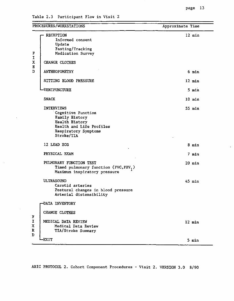
The participant flow, as outlined in Table 2.3, has successfully evolved since the implementation of Visit 1 to reflect study requirements and the operational needs of the individual field centers.
2.2.1 Rationale
Participant flow at each field center is structured to contain both fixed and flexible components. The fixed components reflect the requirement to initiate the examination'with the informed consent, the grouping of the procedures which require fasting, and the logistical necessity of conducting medical data reviews and exit interviews after all other procedures have been completed. The flexible components reflect the historical advantages of having the separate field centers conduct the majority of the interviews and examinations in accordance with the physical layout and the scheduling patterns of the individual field centers. This approach has been shown to minimize participant burden (maximum allowable exam time is 4 hours) and to reduce variability in study measurements.
2.2.2 Fixed Sequences
The fixed portion of participant flow must meet the following criteria: informed consent must be signed before any examination; twelve hours of fasting and. one hour of abstinence from smoking and overt physical exercise are required for venipuncture and sitting blood pressure (procedures for noncompliance are described below); sitting blood pressure and anthropometry must be measured before venipuncture, and all other interviews and exams are completed before the medical data review.
2.2.3 Flexible Sequences
The sequence of the remainder of the examination is flexible and is designed and monitored by the study coordinator at each field center. These procedures include the interviews, ultrasound and physical examinations, electrocardiogram and pulmonary function.
ARIC PROTOCOL 2. Cohort Component Procedures - Visit 2. VERSION 3.0 8/90

page 11
Table 2.1 Components of the ARIC Cohort Visit 2 examination and location of the descriptions in the Manuals of Operation
Procedure/ Workstation
Description Location
Informed Consent Obtain informed consent.
Reception Greet the participant; determine fasting status; verify identifying information; obtain tracing data; collect medications.
Sitting Blood Pressure
Anthropometry
Venipuncture
Snack
ECG
Interview
Physical Exam
Pulmonary Function
Ultrasound
Medical Data Review
Exit Interview
Obtain sitting blood pressure before the participant has blood drawn.
Measure weight, frame size, skin folds.
Obtain blood samples for all laboratory tests.
Provide snack which contains no caffeine or stimulants.
Obtain a 12 lead ECG
Collect sociodemographic, cognitive function, psychosocial, and selected medical, personal and family history data.
Obtain a brief systems review on each participant including neck, neurological, chest and lungs, heart, and extremities. Verify reported history of possible CVD.
Obtain spirometric measurements of timed pulmonary function (FVC, FEVl), and inspiratory pressure (MIP).
Obtain B-mode scan and arterial wall distensibility measurements in carotid arteries. Measure heart rate and blood pressure changes as participant arises from supine position.
Ascertain the completeness of the exam and verify abnormal results. Review results of the medical history and exam with the participant. Refer participant for diagnosis or treatment elsewhere if appropriate.
Return medication; thank participant.
Manual 2
Manual 2
Manual 11
Manual 2
Manual 7
Manual 2
Manual 5
Manual 2
Manual 2
Manual 4
Manual 6
Manual 11
Manual 2
Manual 2
ARIC PROTOCOL 2. Cohort Component Procedures - Visit 2. VERSION 3.0 8/90

page 12 Table 2.2 Training for the ARIC Visit 2 Cohort Exam
CENTRAL LOCAL Trainee Trainer Trainee Trainer
Hemostasis Other Techs Hemostasis Other Techs
VENIPUNCTURE Chief Technician Chief Technician
Forsyth Co. Interviewers Forsyth Co. Interviewers/
Clinic staff cscc Interviewers
Study Coordinator cscc Interviewers Study Coordinator Minneapolis Interviewers
Study Coordinator Washington Co Interviewers Study Coordinator Washington Co Interviewers
Study Coordinator Neurologist Interviewers
Study Coordinator For/Wash Co. Interviewers
Interviewers
Interviewers
Study Coordinator For/Wash Co. Interviewers
Study Coordinator For/Wash Co. Interviewers
Study Coordinator For/Wash Co. Interviewers
Study Coordinator
Med Code Spclst Med Code Spclst
Study Coordinator Study Coordinator
Study Coordinator Study Coordinator
Study Coordinator
Study Coordinator
Study Coordinator
Study Coordinator
Study Coordinator
Stdy Coord/Sprvsr
Study
Staff
PA/Nurse P. PA/Nurse P.
Staff
Study Coordinator
Romm Romm
Staff Back-up
Study Coordinator
Data Coordinator Data Coordinator
n.a. n.a.
n.a. n.a.
Anthr. Techs Reed
PA/Nurse P. Romm
Other Sonog. Other Sonog.
Chief Sonographer Chief Sonographer
ECG Techs Reed
Blood drawing Blood processing
MEDICATION SURVEY Interview Transcription
Coding AFU (Contact Yr 4)
Annual Follow-up Visit 2 Scheduling
HEALTH/LIFE PROFILE Self-administered Staff-supported
COGNITIVE FUNCTION Interview
FAMILY HISTORY Interview
'UPDATE Interview
FASTING/TRACKING Interview
HEALTH HISTORY Interview
TIA/STROKE Interview
RESPIRATORY Sx Interview
Coord/Sprvsr RECEPTION
Informed Consent MED DATA REVIEW
Med. Revue TIA/Stroke Review
LETTERS/REPORTS Ppt. Results Rpt.
ARDES Version 3.0 Workstations Date Management
PULMONARY FUNCTION Spirometry Inspir. Pressure
ANTHROPOMETRY Interview
PHYSICAL EXAM Exam
ULTRASOUND Scans Postural Change
ECG 12 lead ECG
Chief Technician Chief Technician
Study Coordinator Study Coordinator
Study Coordinator
Study Coordinator cscc
Study Coordinator cscc
Study Coordinator Local
Forsyth Co. Forsyth Co.
Study Coordinator Study Coordinator
Study Coordinator cscc
Data Coordinator Data Coordinator
cscc cscc
PFT Technicians PFI Technicians
To&man To&man
Study Coordinator Minneapolis
n.a. n.a.
Chief Sonographer Barnes Chief Sonographer Barnes
n.a. n.a.
ARIC PROTOCOL 2. Cohort Component Procedures - Visit 2. VERSION 3.0 July, 1990
page 13
Table 2.3 Participant Flow in Visit 2
PROCEDURES/WORKSTATIONS Approximate Time
RECEPTION Informed consent Update Fasting/Tracking Medication Survey
12 min
CHANGE CLOTHES
ANTHROPOMETRY
1 SITTING BLOOD PRESSURE
VENIPUNCTURE
SNACK
INTERVIEWS Cognitive Function Family History Health History Health and Life Profiles Respiratory Symptoms Stroke/T&!
12 LEAD ECG
PHYSICAL EXAM
PULMONARY FUNCTION TEST Timed pulmonary function (FVC,FEVl) Maximum inspiratory pressure
ULTRASOUND Carotid arteries Postural changes in blood pressure Arterial distensibility
6 min
12 min
5 min
10 min
55 min
8 min
7 min
20 min
45 min
DATA INVENTORY
CHANGE CLOTHES
r MEDICAL DATA REVIEW Medical Data Review TIA/Stroke Summary
12 min
ARIC PROTOCOL 2. Cohort Component Procedures - Visit 2. VERSION 3.0 8/90

page 14
2.2.4 Data Collection
The participant's flow is documented on the ARIC Cohort Inventory (CXI) form within the Data Entry System. The CXI is added to the local database when the participant's diskette is initialized. Its operating instructions are provided in the Data Coordinator's Manual.
2.3 Reception
At the reception work station, the participant is greeted and welcomed, informed consent is obtained, participant questions are answered, demographic and tracking information are updated, fasting status is determined and the medication survey begun (in some instances, completed).
Staff are trained for the reception work station by the Study Coordinator at each field center. Certification requirements include the successful completion of training on general interviewing techniques, Informed Consent, the Fasting/Tracking form, Direct Data Entry System, and Medications Transcription/Interview (optional). Although no formal certification schedule has been established, interviewers working at the reception workstation are routinely observed by the local study coordinator.
2.3.1 Informed Consent
2.3.1.1 Rationale
The primary objective of readministering Informed Consent is to protect the rights of the ARIC Study participants, meet local Institutional Review Board requirements, and to update the participant's permission to abstract medical records in the event of hospitalization or death.
2.3.1.2 Administration
The goals of the ERIC study and the Visit 2 procedures are reviewed with the participant. It is explained that the goals of the study have not changed and the primary purpose for obtaining a second signature is to keep current his/her permission to review medical records in the event of hospitalization or death. Time is allowed for the person to read and sign the informed consent document. If he/she is visually handicapped or illiterate (incapable of reading the study description and informed consent page), the narrative portion is read to him/her and then the participant is asked to sign the document. It is noted on the Participant Inventory Sheet that assistance with the self-administered portion of the interview (Health and Life Profile Form) should be offered. A copy of the informed consent is given to the participant if required by the local Institutional Review Board.
2.3.1.3 Training
Study coordinators are responsible for providing local staff training.
2.3.1.4 Certification
Certification by the Study Coordinator is required, as listed above.
ARIC PROTOCOL 2. Cohort Component Procedures - Visit 2. VERSION 3.0 8/90

page 15
2.3.1.5 Quality Assurance
Routine quality assurance is provided at each field center by means of observation by the local study coordinator.
2.3.1.6 Data Collection
The Informed Consent is a paper form. The first two pages of descriptive text are given to the participant and the signature page (page 3) is stored in the participant's folder.
2.3.2 Update Form
2.3.2.1 Rationale
Demographic and tracking information, initially recorded in Visit 1 and updated on the Annual Follow-Up Tracking Form, is summarized on a new form, the Update form. This form is generated by the Coordinating Center from information stored in the study's central database, and sent to the field centers for inclusion on the participant's diskette prior to Visit 2.
2.3.2.2 Administration
After greeting the participant and obtaining his/her informed consent, the information on the Update screen is verified by reviewing with the participant the information which was filled out on the form sent to his/her home in the Visit 2 information packet (see Appendix 1.5). The Update form used during Visit 2 also includes mailing information for the person or agency designated to receive the participant's study results.
After verifying all data elements, a hard copy of the Update form is printed. If no changes were made, "NO CHANGES" is written in the upper right hand corner of the form and the form is filed in the participant's folder. If changes were made to the UPDATE screen, "MODIFIED" is written in the upper right hand corner of the paper copy before filing the form in the participant's folder. (This paper copy is used as a tracking log to document the date of any future changes and the initials of the person recording the changes.)
In recognition of the confidential nature of the information collected on the Update form, the information sheet that was brought in by the participant is either returned to him/her or torn up and disposed of in front of the participant.
A schedule for reporting the participant's study results is reviewed (Appendix 7.1.a) with the participant after the Update form is completed. It indicates that the results of some of the procedures done during the visit will be reviewed later with the ARIC clinician while the participant is still at the field center, and a written summary report of those and some additional tests will be mailed to the participant and his/her physician (or alternate) 10 - 12 weeks after the clinic visit date, as described in Section 2.23. Samples of the report and prototypes of accompanying letters are included in Appendix 7.
ARIC PROTOCOL 2. Cohort Component Procedures - Visit 2. VERSION 3.0 8/90

page 16
2.3.2.3 Training
Study coordinators are centrally trained before Visit 2 and are responsible for providing local staff training.
2.3.2.4 Certification
Certification is required, provided by the study coordinator.
2.3.2.5 Quality Assurance
Routine quality assurance is provided locally by the study coordinator, by observing staff performance. Protocol adherence and interviewing technique are reviewed biannually by Coordinating Center field center monitors. Deviations from protocols and possible remedial actions are discussed with study coordinators and staff. Major deviations are brought to the attention of the EXM Committee.
2.3.2.6 Data Collection
The Coordinating Center provides an Update Form for each participant with demographic and tracking information from the most current information on the consolidated database. During the interview data in this form are modified using Change Mode of the DES.
2.3.3 Fasting/Tracking Form
2.3.3.1 Rationale
The Fasting/Tracking form is a revised version of the questionnaire used in visit 1. Whereas the original form collected both fasting and tracking information, this version is limited to documenting the participant's fasting status. As this form established the official visit date in Visit 1, for continuity it was decided to have it continue this function in Visit 2, and preserve its title, even though the collection of tracking information has been transferred to the Update form.
2.3.3.2 Administration
The participant's fasting status is verified. Strict fasting is defined as nothing by mouth, except water, for the past 12 hours. Participants are considered fasting if they have met the strict definition or if they have ingested no more than one cup of coffee/tea within the past 12 hours. The participant's fasting status is recorded as FASTING on the Fasting/Tracking form, but the consumption of coffee/tea is recorded in a note log. Ingestion of more substantive liquids or solids constitutes breaking the fast. If the participant has not fasted for 12 hours, the participant is offered the opportunity to repeat blood drawing in the fasting state at a later date. If in agreement, blood is not drawn and the participant is rescheduled for fasting venipuncture within the shortest feasible time period. The Fasting/Tracking Form is completed; the non-fasting state and rescheduled date of venipuncture are noted on the Participant Inventory Form. When the participant returns in the fasting state for venipuncture, the questions concerning fasting status and recent blood donation on the Fasting/Tracking form are updated. If a
ARIC PROTOCOL 2. Cohort Component Procedures - Visit 2. VERSION 3.0 8/90

page 17
non-fasting participant does not wish to return, the participant's blood is drawn and the Fasting/Tracking form completed appropriately.
The Fasting/Tracking Form also documents whether the participant has given blood within the last 7 days. It is assumed that very few cohort members will have donated blood within the last week as they are reminded during both the scheduling calls not to do so, or to reschedule their clinic visit if they have had to give blood. Recent donors are not rescheduled once they come for Visit 2; the response to question 5 on the Fasting/Tracking form is recorded to reflect the recent blood donation and the individual is sent to the venipuncture workstation.
2.3.3.3 Training
Study coordinators are centrally trained before Visit 2 and are responsible for providing local staff training before Visit 2 start-up.
2.3.3.4 Certification
Certification is required, provided by the study coordinator.
2.3.3.5 Quality Assurance
Routine quality assurance is locally provided by observation of the local study coordinator. Protocol adherence and interviewing techniques are reviewed at least biannually by Coordinating Center field center monitors. Deviations from protocols and possible remedial actions are discussed with study coordinators and staff. Major deviations are brought to the attention of the Cohort Committee.
2.3.3.6. Data Collection
The Fasting/Tracking form is collected by direct data entry on a data entry screen unless the computer is not operational. Computed fasting time is calculated by the Data Entry System (DES). A paper version is available for back-up and subsequent data entry. Computed fields may be hand calculated and written in the margin to assist in determining the need to reschedule the participant for venipuncture. The data field will be automatically calculated at data entry.
2.3.4 Medication Survey
2.3.4.1 Rationale
As in Visit 1, the goal of the Medication Survey is to ascertain medication usage by coding both prescription and nonprescription drugs used by the respondent within the two weeks preceding the examination date. Information on use of medications assists in measuring patterns of medication use in the study communities, temporal changes in medical care practice, diagnostic classification of cardiovascular diseases, interpretation of laboratory results, frequency and type of vitamin/mineral supplement use, and predictors of study end points.
ARIC PROTOCOL 2. Cohort Component Procedures - Visit 2. VERSION 3.0 8/90

page 18
2.3.4.2 Administration
The Medication Survey questionnaire is divided into three major sections and is completed in several stages, at one or more workstations. At the Reception station, it is determined whether the participant has brought in all medications taken within the last two weeks. Identification labels are placed on the participant's medication bag and Medication Survey form. If the participant has not brought in any (all) medications, inquiries are made whether s/he has taken any medications during the past two weeks, or for possible reasons for noncompliance. In case of inadvertent omissions, arrangements are scheduled for obtaining the information by telephone. The deliberate omission to bring medications to the Field Center is recorded on the Participant Itinerary Sheet (Appendix 2.13) and conversion is attempted later during the medical review with the participant.
Subsequent parts of the Medication Survey can be administered at the Reception workstation or later, by trained interviewers or the physician assistant/nurse clinician.
The Medication Survey completes the interviews at the Reception workstation and the participant is asked to change into a loose-fitting scrub suit and place his/her personal belongings in a locker.
Before starting Part B of the Medication Survey, the name on the medication bag is checked against the name on the Medication Survey form. The medication containers are removed from the participant's medication bag and the medication name and concentration are transcribed into column (a) of Section B of the Medication Survey form. Medications that are not in a container are opened only in front of the participant, with his/her permission. When there are more than 17 medications, recording the name and concentration is continued on the back of the page if a paper form is used. If the Medication Survey DES is used and more than 17 medications need to be entered, the name and concentration of the additional medications are written on a piece of paper labelled with the participant's ID, and filed in the participant's folder for future coding. See below for coding instructions. If the name of the medication exceeds the number of fields in the DES, the name is abbreviated on the screen and its complete name is recorded on a piece of paper (labelled with the participant's ID number) and filed in the participant's folder for future coding.
If the interview portion of the Medication Survey is not to be administered at the Reception workstation, after the medication names and concentrations are transcribed, the medications are placed in the carrying bag and taken to the work station designated for the completion of the medication survey. Otherwise, a trained interviewer or the physician assistant/nurse practitioner conduct a brief medication use interview by asking two questions for every medication listed in Section B: (1) classification of the drug - shared, prescribed, or over-the-counter and (2) use of the medication within the last 24 hours.
If the participant has not brought in all (any) medications, compliance is attempted at this time.
ARIC PROTOCOL 2. Cohort Component Procedures - Visit 2. VERSION 3.0 8/90

page 19
When more than 17 medications have been recorded, the priority algorithm for data entry and coding of the medications is as follows: prescription medications first; aspirin and aspirin containing medications (aspirin, Alka Seltzer, headache powders, cold medications, medication for arthritis, etc.); anti-inflammatory drugs (ibuprofen, motrin, nuprin, etc.); then over-the-counter-medications, followed by vitamins and food supplements.
When preparing to ask the participant about each medication, the interviewer removes all containers from the bag and sets them in front of the participant. As each medication is reviewed, it is shown to the participant while keeping the other medications in view. After the questions are answered for each medication, each container is placed back in the carrying bag to minimize confusion and to assure that all medications are returned.
In the process of asking these questions about each medication, the interviewer verifies the transcription of medication names and makes corrections on the paper form as required. Unknown and incomplete names are checked against the American Drug Index and Physician's Desk Reference.
Part C of the Medication Survey re-asks categorical information on medications: use in the past 24 hours; use of any of the medications within the past 2 weeks for cardiovascular diseases; and use of medications containing aspirin.
2.3.4.3 Training
Study coordinators and medication coding specialists are centrally trained and are responsible for r providing local staff training in the transcribing and coding of medications.
2.3.4.4 Certification
Certification by the study coordinator is required for medication transcription and interview. No recertification is required.
Separate certification is required for medication coding, based on a certification test provided by the Coordinating Center and administered by the local medication coding specialist. Recertification for medication coding is also required annually. For the medication coding specialist, this includes coding a set of selected medicines circulated for this purpose and adequate performance on blinded recoding of medications recorded during the previous year. Recertification criteria for field center medication coders require meeting minimum standards of coding repeatability (by interviewer/ transcriptionist) and a review at the Coordinating Center of the accumulated performance on quality control repeat medication coding.
2.3.4.5 Quality Assurance
For each person certified to code medications a ten percent sample of medication coding records is identified by the Coordinating Center for blinded repeat coding at the field center.
2.3.4.6 Data Collection
The Medication Survey can either be collected on screens by direct data entry or on paper for delayed data entry. ARIC PROTOCOL 2. Cohort Component Procedures - Visit 2. VERSION 3.0 8/90

Page 20
Anterior and posterior views of the human skeleton. Source: G. Wolf-Heidegger, Atlas of Systematic Human Anatomy, Vol. I.
Acromiun Process
Tip of Scapula
Olecranon Process
Styloid Process
Styloid Process
of Ulna
Figure 2.1 Bony Landmarks for Anthropometric Measurements
ARIC PROTOCOL 2. Cohort Component Procedures - Visit 2. VERSION 3.0 8/90

page 21
2.4 Anthropometry
2.4.1 Rationale
As in Visit 1 various anthropometric measurements are obtained on the ARIC participants to assess ponderosity, frame, thickness of subcutaneous fat, and pattern of distribution of body fat. Standing and sitting height were measured as part of Visit 1 and are not repeated at Visit 2 because little change is expected in these indices over this time period. Elbow breadth is measured in Visit 2 instead of wrist breadth, which was measured in Visit 1, because the former is considered a more widely accepted measurement of frame size.
2.4.2 Procedures
Anthropometry is performed before the clinic snack and after offering the participant an opportunity to empty his/her bladder. All measurements are made with the participant wearing light-weight, nonconstricting underwear. Each field center determines at the beginning of the study whether hip measurements are to be taken over or under the scrub suit and then follows that procedure consistently for the duration of Visit 2. Weight is measured without shoes.
All anthropometric measurements are taken by either a team of two persons (one serving as observer; the other as recorder) or by one technician using a full length mirror to aid in the appropriate placement of the tape measure. Using the team approach, the observer calls out the name of the next measurement, takes the measurement, and keeps the measuring instrument in place until the recorder repeats the number. The recorder checks the position of the examinee and verifies the accurate placement of the measuring instrument during each procedure, and records the result. When a single technician performs the measurements, he/she verifies the accurate placement of the measuring instrument (using the mirror when appropriate) for each measurement and records each measurement immediately after it is taken. Values are rounded a to the unit indicated for each measurement. Anatomical landmarks for the anthropometric measurements are identified in Figure 2.1.
2.4.2.1 Body Weight
Before a participant is weighed, the scale is balanced so that the indicator is at zero when no weight is on the scale. The scale must be level and on a firm surface (not a carpet), The participant is instructed to stand in the middle of the platform of the balance scale (Detector, model #437) with head erect and eyes looking straight ahead. Weight is adjusted on the indicator until it is balanced. Results are recorded to the pound, rounding down. To maintain accuracy, the scale is zero balanced daily and calibrated with a known weight (50 lbs) every week or whenever the scale is moved. The daily zero balance and the weekly scale calibration are documented on the Anthropometry Equipment Calibration Log (Appendix 6.2).
2.4.2.2 Skinfolds
The Lange caliper is used for all skinfold measurements. The caliper is calibrated using the calibration block prior to taking measurements on each participant. A chart of percent body fat computed from the sum of triceps and
ARIC PROTOCOL 2. Cohort Component Procedures - Visit 2. VERSION 3.0 8/90

page 22 subscapular skinfolds is available if the participant asks for the interpretation. (See Appendix 5.)
All measurements are taken on the participant's right side. Positions are marked with an erasable marking pen. A fold of skin is firmly grasped one (1) cm above the pen mark between the left thumb and first two fingers and then the fold is gently lifted away from the body & to the extent to determine that no muscle is grasped. A firm grip is necessary, but it must not exceed the pain threshold. The skinfold is not stretched away from the body; the fold is pentlv lifted two or three times to make certain that no musculature is praSDed. The skinfold is gently grasped again, the skinfold continues to be held firmly, and the calipers are placed at the pen mark midway between the base and the crest. The skin fold is not let go of. The grip on the caliper is released completely, allowing the spring to compress the fold. Counting silently l-2-3 (approximately 2 seconds), a reading on the caliper dial is taken to the millimeter, rounding down. (Keeping the left hand above the skinfold allows the dial to be read easily. See Figure 3.) The caliper is released, and then the fold. When using a team, the observer performs the measurements and calls out the value to the recorder for data entry. When performed by a single technician, he/she measures the skinfold, removes the caliper, releases the skinfold, and then immediately records the measurement. The entire Drocedure is reneated a second time.
The width of the skinfold that is enclosed between the fingers varies from one site on the body to another. With a thick subcutaneous layer, a wider segment of the skin must be "pinched" than when there is little adipose tissue. For a given site, the width of the skin is the minimum needed to yield a well-defined fold.
The depth of the skinfold at which the calipers is placed on the fold also requires comment. The two sides of the fold are not likely to.be parallel, being narrower near the crest and broader toward the base. The measurement is too large when the calipers are placed at the base. The correct location is approximately midway between the crest and the base, where surfaces are approximately parallel to each other. The contact surfaces of the calipers should be parallel and applied perpendicular to the grasped skinfold. Pen marks are erased.
2.4.2.2.1 Triceps Skinfold
The posterior tip of the acromion process is marked. The tape measure is used to measure from the tip of the acromion process on the right shoulder to the tip of the olecranon process on the back of the elbow. flexing the right arm 90 degrees,
With the participant the tip of the olecranon is marked. The
participant is then requested to straighten the arm, allowing it to hang loosely at the side. A mark (+) is made at the midpoint between the acromion process and olecranon in the midline of the back of the arm (Figure 2.2). Using thumb and first two fingers, the straightened,
a skinfold is grasped parallel to the long axis of relaxed arm one centimeter above the mark.
applied at the mark perpendicular to the grasped skinfold. The caliper is
l-2-3 (approximately 2 seconds), Counting silently
a reading on the caliper dial is taken to the millimeter, rounding down. Measurements must be read two seconds after the full pressure of the caliper jaws is applied to the skinfold. interval is allowed, the jaws may "creep"
If a longer
will be inaccurate. or fat may compress and the reading
The caliper is removed, then the skinfold is released. Pen marks are erased.
ARIC PROTOCOL 2. Cohort Component Procedures - Visit 2. VERSION 3.0 8/90

page 23
.
Triceps: a vehal fold on me posterior midline of the upper arm (over triceps muscle), halfway between lhe acmmion and 0l- processes: the elbow should be extended and
I relaxed.
Figure 2.2 Location of Skinfold Measurements: Triceps
ARIC PROTOCOL 2. Cohort Component Procedures - Visit 2. VERSION 3.0 8/90

page 24
71 Subscapular: a fold taken-on a diagonal line coming from the vertical border to 1 to 2cm from the inferior angle of the scapula.
Figure 2.3 Location of Skinfold Measurements: Subscapula
ARIC PROTOCOL 2. Cohort Component Procedures - Visit 2. VERSION 3.0 8/90

page 25
level
Figure 2.4 Location of Waist Girth Measurement
ARIC PROTOCOL 2. Cohort Component Procedures - Visit 2. VERSION 3.0 8/90

page 26
Upper Arm Circumference
Hip Girth (at maximum protrusion of gluteal muscles)
Calf Circumference
Figure 2.5 Location of Upper Arm and Hip Circumferences; Subscapular Skinfold
ARIC PROTOCOL 2. Cohort Component Procedures - Visit 2. VERSION 3.0 8/90

Figure 2.6 Elbow Breadth Measurement at the Epicondyles of the Humerus
page 27
Figure 2.7 Location of Epicondyles of the Humerus
ARIC PROTOCOL 2. Cohort Component Procedures - Visit 2. VERSION 3.0 8/90

page 28
2.4.2.2.2 Subscapular Skinfold
This measurement is made one (1) cm below the inferior angle (tip) of the right scapula (Figure 2.3). To find the right medial scapular border, have the participant place the back of his right hand on the middle of his back. The right medial s,capular border is located by moving the fingers down the full length until finding the inferior angle. With the participant's arm relaxed, a pen mark is made one (1) cm below the inferior angle on a diagonal line coming down from the medial border. With two fingers on top and thumb below, a skinfold is grasped 1 cm above the mark, on and in the direction of the diagonal line coming down from the medial border of the scapula. The skinfold should be angled about 45 degrees from the horizontal, going medially upward and laterally downward. The calipers are placed on the pen mark perpendicular . to the grasped skinfold. The measurement is made to the millimeter, rounding down. The caliper is removed, the skinfold released, and the measurement is recorded. The procedure is repeated. Pen marks are erased.
2.4.2.3 Waist (Abdominal) Girth
The participant is instructed to stand erect and relaxed with weight equally distributed on both feet. Have the participant lift the scrub suit top just high enough to make the area visible. An anthropometric tape is applied at the level of the umbilicus (navel) and the participant is instructed to breathe quietly. The tape should be snug, but not so tight as to compress tissue. (See Figure 2.4). The full length mirror or recorder verify that the participant is standing erect and that the tape is kept horizontal. The measurement is recorded to the nearest centimeter, rounding down.
2.4.2.4 Hip Girth
Have the participant stand erect, yet relaxed, with weight distributed equally over both feet. The hip girth is measured at the level of the maximal protrusion of the gluteal muscles (hips). (See Figure 2.5). An anthropometric tape is kept horizontal at this level. The measurement is recorded to the centimeter, rounding down. The greatest source of error for this measurement is due to not having the tape horizontal and not verifying that the maximum width is being measured. The position of the tape is checked from both the front and the back.
2.4.2.5 Elbow Breadth
The participant is asked to raise the right arm to the horizontal, with the elbow flexed at 90 degrees. The dorsum (back) of the hand faces the measurer
* (Figure 2.6). The measurer stands in front of the participant and palpates the lateral and medial epicondyles of the humerus (see Figure 2.7). The sliding caliper is applied, pointing the blades upwards, to bisect the right angle formed at the elbow. The caliper is held at a slight angle to the epicondyles rather than parallel to them, because the medial epicondyle is distal to the lateral epicondyle. The measurement is recorded to the nearest millimeter, rounding down. Measurement is easiest if, unlike Figure 2.6, the technician holds the caliper near its tips, and simultaneously places his/her fingers bilaterally on the elbow. This allows the technician to continue to feel the bony landmarks while sliding the caliper jaws into place.
ARIC PROTOCOL 2. Cohort Component Procedures - Visit 2. VERSION 3.0 8/90

page 29
2.4.3 Training
An anthropometry supervisor from each field center is trained centrally (or locally by the study's central anthropometry expert) and is responsible for the local training of her/his local anthropometry technicians (observers) and recorders. Training includes an (1) introduction to the rationale for body size measurements, the expected limits of reproducibility, and usual errors; (2) a demonstration of proper and improper procedures; (3) practice on volunteers and (4) testing on volunteers with four different body types - lean, obese, athletic and aged.
2.4.4 Certification
Initial certification and recertification criteria are identical for anthropometry. Field center anthropometry supervisors are (re)certified annually by the study's central anthropometry expert. Local technicians must meet the same criteria. Each technician measures at least two certification volunteers, meeting the following criteria:
1. Each skinfold measurement must agree within + 2 mm of the expert (locally the anthropometry supervisor's measurements constitute the gold standard) on two certification volunteers (an average difference with + 1 mm on both volunteers).
2. The waist and hip circumference measurements must agree within + 1.5 cm on each certification volunteer; average difference within + 0.75 cm for both volunteers.
3. Weight must agree within + 0.5 lb.
4. Elbow breadth must agree within 1 mm.
Recertification is required annually, meeting the following criteria:
1. Absence of end digit preference for more than 6 months during one year;
2. Absence of systematic differences in mean values;
3. Adequate performance on replicate measurements.
2.4.5 Quality Assurance
In addition to annual recertification, protocol adherence in the performance of each procedure is reviewed at least biannually by Coordinating Center field center monitors. Deviations from protocol and possible remedial actions are discussed with study coordinators and staff. the attention of the EXM Committee.
Major deviations are brought to
Anthropometry equipment is calibrated frequently and results are recorded on an Anthropometry Equipment Calibration Log (Appendix 6.2) and sent to the Coordinating Center weekly. Scales are zero balanced daily and calibrated weekly. The Lange (10 mm) and sliding (50 mm) calipers are calibrated before
ARIC PROTOCOL 2. Cohort Component Procedures - Visit 2. VERSION 3.0 8/90

page 30
measuring each participant. Measuring tapes are checked monthly and replaced as needed.
Digit preference, systematic differences in location statistics, completion of checklists/logs according to schedule are analyzed by the Coordinating Center and reviewed by the Quality Control Committee. Refer to Manual 12 for a detailed description of quality assessment procedures.
2.4.6 Data Collection
The Anthropometry Form is collected by direct data entry on a data entry screen or on a paper form if a computer is not available by either the technician (observer) or recorder.
2.5 Sitting Blood Pressure
2.5.1 Rationale
As one of the most powerful risk factors of cardiovascular disease, a measurement of sitting blood pressure is included in every clinic examination of the ARIC cohort. The procedures are identical to those used in Visit 1, as detailed in Manual 11 of the ARIC Protocol.
2.5.2 Procedures
Sitting blood pressure is a fixed component of the participant flow obtained before venipuncture. Procedures for obtaining sitting blood pressure are found in Chapter 1 of Manual 11. Guidelines have been established for referring participants with abnormal blood pressures for clinical care or follow-up in sections 2.19, Medical Data Review and 2.21, Referrals and Review Guidelines.
2.5.3 Training
Blood pressure technicians were trained centrally recruitment before Visit 1. New technicians were the study coordinator or designated local expert. further details.
2.5.4 Certification
Certification is required; criteria are listed in
prior to participant and are trained locally by
Refer to Manual 11 for
Manual 11. Recertification is performed annually. Recertification criteria include:
1. Successful completion of double-stethoscope observation, semi-annually;
2. Semi-annual test with recertification tapes; 3. Absence of end digit preference for more than 6 months during one year; 4. Annual review by the central ARIC blood pressure trainer.
2.5.5 Quality Assurance
Detailed quality control procedures are provided in Manuals 11 and 12, and include periodic review by the Quality Control Committee of end digit preference, systematic differences between technicians in mean values, and
ARIC PROTOCOL 2. Cohort Component Procedures - Visit 2. VERSION 3.0 8/90

page 31
completion of performance on checklists/logs. Monitoring of certification status is conducted by the Coordinating Center.
2.5.6 Data Collection
The Sitting Blood Pressure Form is collected by direct data entry on screen unless the work station computer is disabled. A paper version of the form is available as backup.
2.6 Venipuncture
2.6.1 Rationale
Venipuncture is a fixed component of the participant flow and performed after on all cohort members who have met fasting requirements (or who are medically unable or indicate an unwillingness to adhere to fasting).
2.6.2 Procedures
The venipuncture protocol is a separate document, Manual 7: Blood Collection and Processing,
2.6.3 Training
Phlebotomists were trained centrally prior to participant recruitment before visit 1. New technicians are trained locally by the chief laboratory technician. Refer to Manual 7 for further details.
2.6.4 Certification
Certification is required and takes place semiannually at the field center. Criteria are listed in Manual 7.
Recertification is required annually and is performed by the chief ARIC technician at the Central Hemostasis Laboratory or by trainer/certifiers from two of the ARIC field centers. Criteria are described in Manuals 7 and 12.
2.6.5 Quality Assurance
Data quality monitoring includes periodic review by the Quality Control Committee.of tube filling time; number of venipuncture attempts; condition of specimens on arrival at the central laboratories; and selected markers of lack of adherence to protocol during phlebotomy and/or processing of specimens at the field center laboratory.
2.6.6 Data Collection
Venipuncture data are collected on a hard copy of the Venipuncture Form (see Appendix 2.3.g. Notes reflecting blood drawing or processing problems are recorded on the accompanying Venipuncture Incident Log which is forwarded as hard copy to the central laboratories and Coordinating Center.
ARIC PROTOCOL 2. Cohort Component Procedures - Visit 2. VERSION 3.0 8/90

page 32
2.7 Snack
A light snack is scheduled as soon as possible after venipuncture. Caffeine- free refreshments are provided; however, decaffeinated coffee and tea may be offered. Menus are locally determined.
2.8 Cognitive Function
2.8.1 Rationale
Measurements of cognitive function are introduced in the second cohort examination. Reportedly a decline in cognitive function may be related to cardiovascular risk factors, e.g., hypertension, elevated cholesterol, or cardiac arrhythmias. It has also been reported that cerebrovascular disease or multi-infarct dementia is the second leading cause of dementing illness among Caucasians in the United States, preceded only by dementia of the Alzheimers Type. Although the ARIC study population is too young at this time to evaluate frank dementia, it provides the opportunity to investigate changes in cognitive function over time. This in turn can be correlated with specific risk factors and progression of atherosclerosis. The main objective of cognitive function testing in Visit 2 is to establish a baseline for future comparison.
Three measures of cognitive function have been selected to establish a baseline and assess changes over time: the Delayed Word Recall, Digit-Symbol Substitution and Word Fluency tests. These tests cover a broad range of cognitive function, have been standardized, are challenging enough to elicit variability, are easy to administer and are easy to score. None of these tests have an upper limitation on performance, and can be expected to allow small changes in mental performance to be detected longitudinally.
The Delayed Word Recall is a test of short term memory. This test has the added feature of allowing participants to encode the words to be recalled (use each word in a sentence) to enhance retrieval. Ten words are given which in effect removes the ceiling or upper limit of performance.
The Digit Symbol Substitution Test requires response speed, sustained attention, and visual-spatial skills. It is part of the widely used Wechsler Adult Intelligence Scale. This test requires that the participant fill in a series of symbols within 90 seconds.
The Word Fluency Test measures verbal function. This too requires speed and sustained attention, but measures mental agility in retrieving words. This test has been used widely, is standardized, and is easy to administer.
2.8.2 Administration
A trained ARIC interviewer administers all three cognitive function tests in a quiet room which is sheltered from distracting noises and has sufficient work space for the participant to place the Digit Symbol Substitution form on a table before him/her and fill in the blanks on the form. The purpose of the tests is briefly explained to each participant. The tests are administered following the step by step instructions printed on the Cognitive Function paper forms (see Appendix 2.4.b). Responses to Parts A and C are recorded on a paper
ARIC PROTOCOL 2. Cohort Component Procedures - Visit 2. VERSION 3.0 8/90

page 33
form by the interviewer. Part B is completed by the participant. Test results are tabulated by the interviewer after the participant has completed the tests and left the room. Test results are entered on the Cognitive Function data entry screen by the interviewer. Final scores for the Word Fluency Test, which are based on the total number of words given by the participant and other demographic data collected during Visit 1, are calculated through analysis requests once the data have been entered into the ARIC collaborative database.
2.8.3 Training
Interviewer supervisors are trained centrally prior to Visit 2, and are responsible for training and certification of the the field center technicians. New technicians are trained locally by the supervisor or study coordinator.
2.8.4 Certification
Certification by the supervisor or study coordinator is required, and monitored by the Coordinating Center.
2.8.5 Quality Assurance
A non-systematic sample of Cognitive Function tests are reviewed by the supervisor. Technique and adherence to protocol are also monitored at least semi-annually by Coordinating Center Monitors; data quality is monitored by the Quality Control Committee on a semi-annual basis.
2.8.6 Data Collection
Cognitive function data are collected on a three part paper form for delayed data entry. Scores are tallied by the interviewer or a certified staff member and recorded at the end of each test after the participant has left the interview room.
2.9 Family History
2.9.1 Rationale
The first half of the Family History Form repeats questions from the Visit 1 Home Interview on the participant's marital status and the vital status of natural parents. The second half introduces new questions on the vital status and cardio- and cerebrovascular medical history of the participant's full siblings. The availability of a more detailed family history makes the study's database more comparable to the information currently being collected in other national and international cardiovascular disease epidemiologic studies.
2.9.2 Administration
The Family History Form is administered by certified interviewers within the flexible component of the participant flow. To assist the participant in remembering the names and ages of his/her natural siblings, a paper list is made of all siblings before specific questions on vital status and medical history are asked of each sibling. The interviewer then orders the eligible siblings by age (eldest first) so that the interviewer and the respondent can
ARIC PROTOCOL 2. Cohort Component Procedures - Visit 2. VERSION 3.0 8/90

page 34
use the list as a reference sheet from which to answer the questions about the five eldest natural siblings on the Family History Form.
2.9.3 Training
A supervisor or study coordinator from each field center is centrally trained before Visit 2 and is responsible for providing local staff training in interviewing techniques and the question by question instructions for the Family History form.
2.9.4 Certification
Certification by the supervisor or study coordinator is required, and monitored by the Coordinating Center. With participant approval, all interviews are taped. Satisfactory performance on ten taped interviews reviewed by the supervisor during the first month leads to certification. Recertification is not required.
2.9.5 Quality Assurance
With participant approval, all interviews are taped for quality control. A non-systematic sample of interviews is reviewed by the supervisor. Technique and adherence to protocol are also monitored at least semi-annually by Coordinating Center Monitors; data quality is monitored by the Quality Control Committee on a semi-annual basis.
2.9.6 Data Collection
Data from the Family History form are collected by direct data entry on a data entry screen unless the work station computer is inoperable. A paper version of the form is available for back-up and delayed data entry.
2.10 Health History
2.10.1 Rationale
The Health History Form updates information on occupation, history of serious illnesses, smoking habits, reproductive history, and alcohol consumption initially collected at Visit 1. New questions on handedness and the use of medical care have been added.
2.10.2 Administration
The Health History Form is administered by certified interviewers within the flexible component of the participant flow. Visit 1 participant responses to the lead-in questions for occupation, and reproductive history are provided on the Visit 1 Participant Information Sheet (PIN) to assist the interviewer in verifying/adjudicating the name and address of previous employment with current employment and determining whether the smoking and reproductive history questions should be administered or skipped (see Appendix 2.14). A drink conversion table for recording alcohol consumption has been added to the question by question instructions for administering the alcohol consumption component of the form.
ARIC PROTOCOL 2. Cohort Component Procedures - Visit 2. VERSION 3.0 8/90

page 35
2.10.3 Training
Study coordinators and interviewer supervisors are centrally trained before Visit 2 and are responsible for providing training to local staff in interviewing techniques and the question by question instructions for the Health History form.
2.10.4 Certification
Certification by the supervisor or study coordinator is required, and monitored by the Coordinating Center. Satisfactory performance on ten taped interviews reviewed by the supervisor during the first month leads to certification. Recertification is not required.
2.10.5 Quality Assurance
With participant approval, all interviews are taped for quality control. A non-systematic sample of interviews is reviewed by the supervisor. Technique and adherence to protocol are also monitored at least semi-annually by Coordinating Center Monitors: data quality is monitored by the Quality Control Committee on a semi-annual basis.
2.10.6 Data Collection
Data from the Health History form are collected by direct data entry unless the work station computer is inoperable. A paper version of the form is available for back-up and delayed data entry.
2.11 Health and Life Profile
2.11.1 Rationale
Several psychosocial instruments on social support/networks, hostility/anger, and depression are introduced in Visit 2. To date, no psychosocial studies have been used in conjunction with ultrasound evaluation of atherosclerosis. The use of these noninvasive procedures in a prospective study of atherosclerosis and the heterogenous nature of the ARIC cohort could help to determine whether psychosocial measures are associated with asymptomatic, early disease versus the precipitation of clinical events. The Health and Life Profile forms (HPA, HPB and HPC) incorporate four well established, widely used, standardized psychosocial instruments into three self-administered forms.
The first form (HPA) of the Health and Life Profile forms combines the Interpersonal Support Evaluation List and the Lubben Social Network Scale (Broadhead WE et al., The Epidemiologic Evidence for a Relationship Between Social Support and Health. Am J Epidemiol 1983: 117:521-37 and Heitzmann CA et al., Assessment of Methods for Measuring Social Support. Health Psycho1 1988; 7:75-109). There is considerable evidence that social support is related to all-cause mortality. However, the specific relationship of social support to cardiovascular disease is less well established. Furthermore, no study has examined the association of social support and carotid atherosclerosis. The social support/network sections ascertain how well subjects perceive their support. The perceived availability of social support may buffer the
ARIC PROTOCOL 2. Cohort Component Procedures - Visit 2. VERSION 3.0 8/90

page 36
individual against the negative effects of significant life stressors. The second Health and Life Profile form (HPB), the Maastricht Questionnaire, ascertains a participant's outlook on life and symptoms of fatigue and depression. It has been chosen because for many years excess fatigue and depression have been anecdotally associated with the occurrence of cardiovascular disease. Epidemiologic evidence to support such an association have been lacking until a recent study by Appels and Mulder found a strong association between excess fatigue and coronary heart disease incidence (Appels A et al., Excess Fatigue as a Precursor of Myocardial Infarction. Eur Heart J 1988; 9:758-64). These investigators developed a new scale, the Maastricht Questionnaire, which has good psychometric properties and offers a new avenue of research for the ARIC study.
The third form, (HPC), the Spielberger Trait-Anger Scale, is a brief, but widely used instrument with known psychometric properties that measures the most important components of Type A behavior, hostility and anger. It has been hypothesized that increased anger alters adrenergic blood hormones, thereby increasing risk of cardiovascular disease. Although several scales to measure anger have been developed and used, the administration of the Spielberger Trait-Anger Scale in the ARIC cohort offers a new opportunity to explore this hypothesis ,in relation to cardiovascular disease prospectively.
2.11.2 Administration
The Health and Life Profile forms are designed to be self administered, but can be interviewer-administered if necessary. (The HLP forms are routinely interviewer administered in the Jackson field center,) There are three parts to the form (HPA, HPB, and HPC) to facilitate completion during the clinic visit. Each part begins with a title, is followed by a body of questions which is completed by the participant and ends with an administrative section which is completed by the interviewer.
The scheduling of the administration of this from is flexible and is tailored to the needs of each field center. Possible times are at a specifically scheduled time on the participant's itinerary, or during waiting times between exams and interviews. Some participants may be able to complete all the questions by themselves in one sitting, some may require more time, and others may require interviewer assistance with part or all of the questions.
Prototypes of interviewer scripts to support self-administration are provided in the first section of the question by question instructions. Administration instructions are read to participants; the participants' ability/willingness to complete the forms is assessed, and the availability of the interviewer is made known in case of participant questions. When it is known/determined in advance that a participant is incapable of completing a self-administered version of the Health and Life Profile, all three forms are interviewr-administered. Interviewer assistance can be offered or initiated at any point in the administration of the Profile. The type of administration (self, interviewer- assisted, or both) is coded in the administrative section at the end of each form. Question by question instructions for interviewer administration are provided in the appendix.
ARIC PROTOCOL 2. Cohort Component Procedures - Visit 2. VERSIONl3.0 8/90

page 37
The completion status of all three forms of the Profile is assessed during the Data Inventory. Participants who have not completed all questions are encouraged to do so; interviewer assistance is offered if appropriate.
It is anticipated that occasionally participants will express manifestations compatible with depressive symptoms or extreme exhaustion in their responses to selected items. Guidelines are provided in the question by question instructions for this form (Appendix 2.8.~) to call these to the attention of the physicians assistant/nurse practitioner in order to address possible participant safety concerns during the Medical Data Review.
Upon completion by the participant, the forms are reviewed by the interviewer responsible for the form's administration and all three parts of the form are coded.
2.11.3 Training
Study coordinators and intenriewer supervisors are centrally trained before Visit 2 and are responsible for providing local staff training in interviewing techniques and the question by question instructions for the Health and Life Profile form.
2.11.4 Certification
Certification by the supervisor or study coordinator is required, and monitored by the Coordinating Center.
2.11.5 Quality Assurance
Technique and adherence to protocol are also monitored at least semi-annually by Coordinating Center Monitors; data quality is monitored by the Quality Control Committee on a semi-annual basis.
2.11.6 Data Collection
The Health and Life Profile forms are intended to be self-administered, and are therefore, designed as paper forms. Data are keyed by the interviewer into the HLP data entry screens on the participant's diskette as soon as is feasible, The HLP form is one of the few forms for which the data entry screens are different form the paper version. The primary difference consists of a "don't know" response category in Parts A and C of the screen version to document that the participant either did not complete the question or the questionnaire. The exclusion of this category on the self-administered version of Parts A and C of the questionnaire is done in compliance with standardized administration instructions. The data entry screen includes this category to document non-response.
Scores for each part of the HLP form are calculated once the data are in the ARIC collaborative database.
ARIC PROTOCOL 2. Cohort Component Procedures - Visit 2. VERSION 3.0 8/90

page 38
2.12 Respiratory Symptoms
2.12.1 Rationale
The Respiratory Symptoms form is a condensed version of the questions on respiratory symptoms asked in the Respiratory Symptoms/Physical Activity form during Visit 1. Relevant questions for the analysis of the pulmonary function tests are updated by restricting the response period to the interim between Visit 1 and Visit 2.
2.12.2 Administration
The Respiratory Symptoms Form is administered by certified interviewers within the flexible component of the participant flow. Although the number of questions has been condensed and the reference period restricted to the past 3 years, the wording and structure of the questionnaire, as well as the detailed instructions to the interviewers, are taken directly from the Epidemiology Standardization Project. Interviewers are instructed to read each question as printed and accept unequivocal answers as provided by the respondent. The wording of the questions, and the instructions by the interviewer before starting the interview, lead to simple "yes" or "no" answers. Probing is limited to a repetition of the question when possible, and equivocal answers are recorded as "no".
2.12.3 Training
Study coordinators and interviewer supervisors are centrally trained before Visit 2 and are responsible for providing local staff training based on a common training manual, practice scripts, and role playing.
2.12.4 Certification
Certification by the supervisor or study coordinator is required, and monitored by the Coordinating Center. Satisfactory performance on ten taped interviews reviewed by the supervisor during the first month leads to certification. Recertification is not required.
2.12.5 Quality Assurance
With participant approval, all interviews are taped for quality control. A non-systematic,sample of interviews is reviewed by the supervisor. Technique and adherence to protocol are also monitored at least semi-annually by Coordinating Center Monitors; data quality is monitored by the Quality Control Committee on a semi-annual basis.
2.12.6 Data Collection
Data from the Respiratory Symptoms form are collected by direct data entry on a data entry screen unless the computer is down or a workstation is inoperable. A paper version of the form is available for back-up and delayed data entry.
ARIC PROTOCOL 2. Cohort Component Procedures - Visit 2. VERSION 3.0 8/90

page 39
2.13 TIA/Stroke
2.13.1 Rationale
Stroke and transient ischemic attack (TIA) have been identified as important end points in the ARIC study. A baseline history of TIA/stroke was collected during Visit 1. New occurrence(s) of cerebrovascular disease is updated by repeating all questions in the TIA/Stroke form (TIAC), but restricting the response period to the interim between Visit 1 and Visit 2.
2.13.2 Administration
The TIA/Stroke Form is administered by certified interviewers within the flexible component of the participant flow. Any positive symptom is recorded . on the TIA Summary Review (TSR) form for assessment during the Medical Data Review at the end of the clinic visit, at which time the physicians assistant/nurse practitioner probes for non-cerebrovascular explanations for the event(s).
2.13.3 Training
Interviewer supervisors and study coordinators are centrally trained before Visit 2 and are responsible for providing local staff training based on a common interview training manual, question by question instructions for the TIA/Stroke and TIA Summary Forms, practice scripts, and role playing.
2.13.4 Certification
Local as well as central certification criteria have to be met for this form. Satisfactory performance on ten taped interviews reviewed by the supervisor during the first month leads to certification by the study coordinator/local supervisor. Interviewers also code three TIA/Stroke forms based on three sets of scripts distributed by the Coordinating Center. Certification is conferred after review by the study's neurologist and the Coordinating Center. Yearly recertification scripts are distributed, reviewed and scored by the Coordinating Center.
2.13.5 Quality Assurance
With participant approval, all interviews are taped for quality control. A non-systematic sample of interviews is reviewed by the supervisor. Technique and adherence to protocol are also monitored at least semi-annually by Coordinating Center Monitors; data quality is monitored by the Quality Control Committee on a semi-annual basis.
2.13.6 Data Collection
Data from the TIA/Stroke form are collected by direct data entry on a data entry screen unless the work station computer is inoperable. A paper version of the form is available for back-up and delayed data entry.
ARIC PROTOCOL 2. Cohort Component Procedures - Visit 2. VERSION 3.0 B/90

page 40
2.14 Physical Exam
2.14.1 Rationale
A brief physical examination takes place during the clinic visit to ascertain major cardio-pulmonary conditions and sequelae of cerebrovascular accidents. The examination is administered by a physician assistant, a nurse practitioner, or a physician trained in the protocol of the ARIC Study. At this time the examiner also administers a brief questionnaire on history of exposure to diagnostic and therapeutic procedures since Visit 1, and documents the characteristics of any chest pain on effort reported by the participant during the previous year (ascertained during the last annual follow-up call).
2.14.2 Procedures
2.14.2.1 Walking/Standing
Use of cane/wheelchair. The use of ambulatory aids is ascertained at the time the participant enters the examination room.
Gait. If the person uses a cane for normal walking, the cane is part of the examination. The inability to walk is documented on a note log. The participant walks ten steps along a line in the center of a hallway at a rapid rate. A dystaxic gait is present if the individual passes one ankle more than six inches away from the other in walking. A hemiplegic or paretic gait is noted when the normal leg is on the ground and the abnormal leg swings in a circular motion to place the opposite foot on the floor. A limp is usually apparent. If an arm is affected, it usually does not swing and may be held flexed at the elbow.
Arm strength/Romberg. The participant stands with feet together, ankles and big toes of each foot touching. He or she is asked to fix gaze on a distant location with arms outstretched horizontally, palms up, and hands and fingers extended. If the individual cannot balance with the feet together, have the person stand so that balance is achieved. If the person cannot balance, this is recorded on a note log. When balance is achieved, the participant is asked to close eyes and balance for ten seconds. Weakness in one arm is noted by a downward drift in that arm of one foot or more, or pronation of the hand toward the vertical position. A positive Romberg sign is one in which the individual has to move a foot from the starting position to maintain balance. During this procedure the examiner stands close to the participant, to assist in case of loss of balance.
2.14.2.2 Confirmation of Reported Chest Pain
A positive participant response to the Rose Questionnaire which was administered during the Annual Follow-up contact which immediately preceded the Visit 2 examination is documented on the Visit 1 Participant Information Sheet (PIN). The occurrence of the participant reported pain is confirmed, its location documented, and its frequency ascertained by the physician assistant/nurse practitioner.
ARIC PROTOCOL 2. Cohort Component Procedures - Visit 2. VERSION 3.0 B/90

page 41
2.14.2.3 History of Diagnostic or Invasive Procedures on the Cardiovascular System
The participant is asked about diagnostic or invasive procedures on the cardiovascular system since the first cohort exam. Diagnostic procedures include echocardiogram, electrocardiogram, treadmill or cardiac stress test, carotid ultrasound studies or heart catheterization. Invasive procedures include surgery to the heart, or the arteries of the neck or legs (excluding varicose veins) and various arterial revascularization procedures.
2.14.2.4 Procedures Performed while the Participant is Sitting
Lungs, Rhonchi, and Rales. Men are asked to remove the scrub top entirely, women to lift it. The stethoscope diaphragm previously warmed in the palm of the hand is used. The participant is instructed to take deep breaths through the mouth. After the first five or six breaths and as needed thereafter, the participant is asked about symptoms of lightheadedness. Auscultation takes place over the posterior lung fields, beginning at the apices with at least one full breath in each location. The locations on each side are examined: apex, mid-lung field (approximately at the 6th intercostal space) and the base, which may need to be determined by percussion. Rhonchi are described as coarse breathing noises. Rales are fine moist noises.
Heart. The diaphragm of the stethoscope is placed consecutively at the apex, the left sternal border at the 5th intercostal space, the left sternal border at the 2nd intercostal space, and the right sternal border at the 2nd intercostal space. The examiner listens for at least five beats in each location. This is repeated at each of the four spaces with the bell of the stethoscope lightly applied to each area. The location of a systolic or diastolic murmur is reported in the area in which it appears loudest. More than one location of equal intensity is acceptable. A grade one murmur is barely audible. Grade two is just easily audible. Grades three and four are intermediate and increasing in intensity; grade four is palpable as a thrill. Grade five is louder, palpable, but still requires the stethoscope on the chest, lightly applied. Grade six can be heard with the stethoscope off the surface of the chest. Other findings include the radiation and the character of the murmur. Other cardiac findings include changes in breath sounds and evidence of surgery.
2.14.2.5 Procedures Performed while the Participant is Supine
Neck. While supine, the participant is asked to stop breathing momentarily. With the stethoscope bell, the examiner listens first above the clavicle for the common carotid artery and second, at the angle of the jaw for the carotid bifurcation. In each position, the stethoscope is placed for three cardiac cycles, alternating sides of the neck.
Cardiopulmonary, Auscultation to document systolic and diastolic murmur(s) is performed in the supine position as described above for the sitting position (Section 2.14.2.4).
Lower Extremities. To document ankle edema, the socks or other foot coverings are removed. Gentle but firm pressure is applied along the mid-tibia, anteriorly down to the ankle in each leg. Pitting or indentation remaining
ARIC PROTOCOL 2. Cohort Component Procedures - Visit 2. VERSION 3.0 B/90

page 42
after pressure is removed constitutes definite edema. The examiner identifies the mid-point between the prominence of the medial malleolus and the inferior border of the patella. Pitting at or above that mid-point is recorded as "marked" edema. Pitting only below that point is recorded as "mild" edema.
Posterior Tibia1 Pulse. The examiner palpates inferior to the medial malleolus ofceach foot. The presence or absence of arterial pulsation is recorded. If in doubt, the examiner compares it with the radial pulsation.
Babinski. The lateral surface of the sole of the foot (plantar surface) is stroked with pressure beginning at the heel and going forward along the lateral surface, crossing the forefoot (ball of the foot) toward the big toe.
The absence of Babinski reflex is a plantar flexion of the great toe. If the leg is withdrawn (a tickle response), the lateral surface of the foot (not the sole) is stroked similarly beginning at the heel and going forward toward the little toe. The Babinski sign is present when the great toe extends on these maneuvers (dorsiflexion).
Other significant findings. Other significant findings are documented by recording in a note log.
2.14.3 Training
Training is required, consisting of (1) training as nurse-practitioner, nurse-clinician, physician assistant, or physician; and (2) command of the pertinent protocol sections and forms.
2.14.4 Certification
Certification by the (central) certifying physician is required. This takes place after a review (in person or over the phone) of the procedures detailed in:the protocol with the central certifying physician.
2.14.5 Quality Assurance
Periodic observation by the field center medical director or physician takes place at field centers. Data quality is monitored by the Quality Control Committee by means of an annual examination of patterns in the data and of incorrect use of the Physical Exam form.
2.14.6 Data Collection
Data from the Physical Exam form are collected by direct data entry on a data entry screen unless the work station computer is inoperable. A paper version of the form is available for back-up and delayed data entry.
2.15 Electrocardiogram
2.15.1 Rationale
A resting 12-lead ECG is performed on each participant in Visit 2 using procedures and equipment identical to those employed in Visit 1. A 2-minute
ARIC PROTOCOL 2. Cohort Component Procedures - Visit 2. VERSION 3.0 B/90

page 43
rhythm strip was obtained on all participants of the baseline cohort examination, but is not included in Visit 2. Processing and coding at the Minnesota and Halifax central electrocardiographic reading centers follows the same procedures used in the baseline visit. Full details are provided in Manual 5 of the ARIC Protocol. The main purpose of the electrocardiographic measurements is to provide information on (1) interim myocardial infarction; (2) changes in conduction pattern, ventricular hypertrophy and ischemia; (3) and other indicators of cardiac function. Hospital ECGs are also read and abstracted for all cohort participants hospitalized after their baseline visit, to determine if a cardiac end point event has occurred.
2.15.2 Procedures
Standard (12-lead) ECG operational procedures are provided in Manual 5, Electrocardiography.
2.15.3 Training
Central training of senior field center technicians was initially performed in visit 1. Training for new ECG technicians is provided by the senior certified ECG technician at each field center, consisting of (1) electrode placement, (2) skin preparation, (3) MAC PC menus and data entry, and (4) self-evaluation techniques for technical performance.
2.15.4 Certification
Certification is required for ECG technicians performing 12-lead ECGs. Requirements and procedures are listed in Manual 5. The Minnesota ECG Reading Center serves as the certifier. Recertification is performed annually.
2.15.5 Quality Assurance
To maintain certification each technician is required to perform a minimum of 3 ECGs per week over a two-month period; quality grades for each 12-Lead ECG are reported by the Halifax ECG Computer Center to each technician on an ongoing basis; a monitoring/re-training visit by the local ECG trainer takes place annually; an ECG quality control checklists is administered quarterly (see Appendix Q of Manual 5).
Quality assurance of the ECG coding at each of the two central ECG reading facilities includes internal, and external quality control programs. These are detailed in manuals 5 (Electrocardiography) and 12 (Quality Control) of the ARIC Protocol.
2.15.6 Data Collection
The standard electrocardiograph for the recording of 12-lead ECGs is the MAC PC Personal Cardiograph by Marquette Electronics, Inc. Data collection procedures are fully documented in Manual 5. Tracings are transmitted daily to the ECG Computer Center at Halifax, Nova Scotia via modem. Paper tracings are stored in the participant's folder.
ARIC PROTOCOL 2. Cohort Component Procedures - Visit 2. VERSION 3.0 B/90

page 44
2.16 Pulmonary Function
2.16.1 Rationale
Pulmonary function studies were implemented in Visit 1 and continued in Visit 2 to study the associations between impaired ventilation (spirometry) and mortality. The previously observed excess mortality associated with impaired respiratory function is due to.a variety of causes, especially cardiovascular disease and cancer. Although the reasons for the association of impaired ventilation with cardiovascular mortality are not known at present, the repeatability of this association and the demonstration of a dose-response suggest that the relationship is real and important.
A simple, rapid measurement of respiratory muscle strength (maximal inspiratory pressure) has been added to Visit 2. The predictive value of FEVl impairment for subsequent cardiovascular mortality is becoming well recognized. In addition to intrinsic pulmonary limitations to airflow, recent publications have suggested that respiratory muscle strength is an important determinant in impaired ventilation. The importance of respiratory muscle strength as an independent component of (oxygen delivery and) cardiovascular mortality are as yet unknown. The complementary studies of fitness, respiratory muscle strength and cardiovascular events in the ARIC study's middle-aged cohort provide an opportunity to study these interactions.
2.16.2 Procedures
Standard spirometry (FEVl) and maximal inspiratory pressure (MIP) procedures are documented in Manual 4, Pulmonary Function Assessment.
Participants with an average sitting blood pressure greater than or equal to 200/120 mg/Hg cannot perform any of the pulmonary function tests (PFT) until their blood pressure falls below that level. Following the PFT procedures in Manual 4, the PFT technician reviews each participant's itinerary form to ascertain the sitting blood pressure before beginning testing. Participants with sitting blood pressure outside the acceptable range are told that they currently do not meet testing criteria, but that they can be rescheduled for the PFT at a later date. Likewise, participants with a history of myocardial infarction or chest/abdominal surgery within the last 6 weeks are excluded from performing pulmonary function testing until a later date. In either situation, after the participants have been escorted to the next workstation, the PFT technician consults with the physicians assistant or nurse practitioner to confirm the appropriate date for rescheduling.
2.16.3 Training
Central training and certification of all pulmonary function assessment technicians are required. Each certified pulmonary function technician completes an intensive two-day training course in spirometric testing which meets the criteria for National Institute of Occupational Safety and Health (NIOSH). This includes:
ARIC PROTOCOL 2. Cohort Component Procedures - Visit 2. VERSION 3.0 B/90

page 45
1. Basic physiology of the forced inspiratory maneuver and determinants of airflow limitation with emphasis on the relation to reproducibility of results.
2. Instrumentation requirements including calibration procedures, sources of error, and their correction.
3. Participant, preparation and indications for postponing testing.
4. Performance of testing including participant coaching (during insoiration), recognition of improperly performed maneuvers, and corrective actions.
5. Data quality (i.e., what constitutes a valid spirogram) with emphasis on reproducibility.
6. Measurements of tracings and calculation of results.
In addition,. each technician receives training in the ARIC Pulmonary Function Testing Protocol (Manual 4), using ARIC pulmonary function calibration and test equipment, computer hardware and software.
2.16.4 Certification
Technicians are certified after successfully completing the training course and a practical examination at the Pulmonary Function Reading Center. To maintain certification technicians must be responsible for one full day of testing per week or equivalent (one complete calibration plus tests on five participants.) Central recertification procedures are carried out at annual site visits to provide assurance that knowledge and competence in ARIC spirometry procedures remain at an acceptable level.
2.16.5 Quality Assurance
Quality of data acquisition is supported by daily calibration of the instrument, annual volume standardization of the four centers by the Pulmonary Function Reading Center (PFRC), review at the of quality measurements, and measurement by hand of a 10 percent sample of spirometry tracings for comparison of field center computer-generated results. A weekly report of the quality control check is returned to each field center. Quality control measures for the PFRC include the periodic use of a test library of previously coded records submitted for computer coding, and hand measuring of original tracings from the standard library.
2.16.6 Data Collection
Measurements are made following procedural guidelines of the American Thoracic Society, with a volume-displacement spirometer supported by a computer through an analog-to-digital interface. The calibration and analytic programs have been installed on the computer to assist the operator in calibration, testing, and assessment of data quality. Data are transmitted to the Pulmonary Function Center each week on diskette.
ARIC PROTOCOL 2. Cohort Component Procedures - Visit 2. VERSION 3.0 B/90

page 46
2.17 Ultrasound
12.17.1 Rationale
Three types of measurements are obtained in the ultrasound work station of the ARIC field centers: B-Mode ultrasound images of the extracranial carotid arteries, distensibility of the common carotid artery, and heart rate and brachial blood pressure in the supine position and on standing up. The main emphasis of the ARIC B-Mode scanning and image reading methods is placed on the measurement of arterial wall thickness, as detailed in manual 6-A (Ultrasound Scanning) and 6-B (Ultrasound Reading) of the ARIC Protocol. Thickening of the arterial wall attributable to atherosclerotic arterial disease precedes significant stenosis and clinical manifestations. Its prevalence in the ARIC study population and change over time represent the dependent variables for major study questions in ARIC, as described elsewhere (The ARIC Investigators. The Atherosclerosis Risk in Communities (ARIC) Study: design and objectives. Am J Epidemiol 1989;129:687-702). Ultrasonographic indices of atherosclerosis will also be examined to test their ability to predict incident cardiovascular events in the ARIC cohort.
Arterial distensibility is measured as the ratio of the change in lumen diameter to the change in blood pressure during a cardiac cycle. Procedural details are provided in Manual 6-D (Arterial Distensibility ) of the ARIC Protocol. Measurement of arterial stiffness is included because of reports of reduced arterial distensibility in the offspring of survivors of myocardial infarction, and of an inverse association between distensibility of the abdominal aorta and the degree of coronary artery stenosis.
The measurement of postural changes in heart rate and blood pressure was included in Visit 1 to examine the potential significance of blood pressure and heart rate in standardized body positions and postural changes, as measures of vascular reactivity and autonomic cardiovascular control as risk factors of cardiovascular disease. The operational details of these measurements are provided in Manual 6-A (Ultrasound Scanning) and Manual 11 (Sitting Blood Pressure and Postural Changes in Blood Pressure and Heart Rate) of the ARIC Protocol.
During Visit 1 the examination protocol included a determination of the ankle blood pressure and a B-Mode scan of one popliteal artery. These measurements are considered part of the baseline information on cohort participants and are not repeated in Visit 2. All other measurements performed during Visit 1 at the ultrasound work station are repeated in Visit 2, using the same procedures and equipment.
12.17.2 Procedures
As mentioned above, procedural and operational detail is provided in manuals 6-A (Ultrasound Scanning), 6-B (Ultrasound Reading), 6-D (Arterial Distensibility) and 11 (Sitting Blood Pressure and Postural Changes in Blood Pressure and Heart Rate).
ARIC PROTOCOL 2. Cohort Component Procedures - Visit 2. VERSION 3.0 B/90

page 47
12.17.3 Training
Central training for ARIC sonographers is provided by the Ultrasound Reading Center, and detailed in Manual 6-A.
12.17.4 Certification
Pre-certification of ARIC sonographers by the Ultrasound Reading Center (URC) requires the completion of the central training course; and review by URC experts of videotaped studies on ten volunteers. Sonographers are certified after review of videotapes at the URC confirms a satisfactory performance on at least 10 complete studies on ARIC participants, done by the pre-certified sonographer under the supervision of the field center chief sonographer. To maintain certification a sonographer is required to scan a minimum of five ARIC participants per week (computed as a two-month average). Recertification is annual, based on satisfactory performance on quality control monitoring and annual review of videotaped studies at the URC.
12.17.5 Quality Assurance
Quality assurance of the ultrasound scan is supported by annual retraining of chief sonographers, visits by URC experts to field centers, a preventive maintenance program of the ultrasound equipment, monitoring by the URC of equipment performance, repeat scanning of a randomly selected arterial segment for each participant, and monitoring of data at the URC and the Coordinating Center. The ultrasound system is monitored by scanning of tissue-equivalent phantoms.on a schedule determined by the performance characteristics of the systems. The arterial distensibility equipment is monitored by the URC readers.
At the URC a monthly review takes place to monitor the quality of arterial wall boundary images contributed by each sonographer. At the Coordinating Center periodic reports are prepared for the Quality Control Committee, to monitor the rate of success in the acquisition of data, comparability between repeated scans, by sonographer, by field center, and over time. Equivalent reports are prepared by the Coordinating Center to monitor ultrasound reader performance.
Sonographer performance on acquisition of arterial distensibility data is monitored by the URC and the Quality Control Committee. Adherence to protocol in obtaining data on postural changes in heart rate and blood pressure is monitored by means of reports prepared at the Coordinating Center for the Quality Control Committee. Quality assurance procedures to support the reading process at the URC are detailed in Manual 12.
12.17.6 Data Collection
A microcomputer and a specialized flow panel assist the sonographer during the standardized examination sequence and data collection. The B-Mode examination is recorded on 3/4-inch videotape and read at the URC; a back-up l/2-inch tape remains at the field center. Data on arterial diameters, blood pressures, beat-to-beat heart rate, and their timing are sent to the URC on diskette.
ARIC PROTOCOL 2. Cohort Component Procedures - Visit 2. VERSION 3.0 B/90

page 48
2.18 Data Inventory
2.18.1 Rationale
The data inventory step initiates the second fixed component of the field center examination, and is done after all interviews and examination procedures have been completed in preparation for the Medical Data Review. Participant data are collected by various means during the course of Visit 2 and require summarization and placement in the participant's folder for physicians assistant/nurse practitioner review.
2.18.2 Procedures
An interviewer assigned to each participant reviews the participant's itinerary sheet, self-administered forms, and folder for completeness. The participant is encouraged to complete any missed portions of the examination; attention is also given at this time to any repeat examinations indicated by the quality control protocol.
After completeness of examination and quality control procedures has been confirmed the participant is invited to change back into street clothes while the data are being prepared for the medical data review with the physician assistant/nurse clinician. Medical data review and pulmonary function (and the interviews if it facilitates participant flow) may be conducted in street clothes.
A program within the ARIC data entry system is run on the .participant's diskette which generates a printout of selected items pertinent for the Medical Data Review. A participant's review may be properly conducted with the following components. Their sources and locations include:
1.
2.
3.
4.
5.
6.
7.
Blood Pressure
ba: front of chart on Itinerary Form clinic visit report
C. inside folder on draft summary Pulmonary Function Test Results a. original computer printout from pulmonary
function inside chart b. clinic visit report C. draft summary Electrocardiogram a. original copy inside chart b. ECG interpretation on both clinic visit report
and draft summary Physical Exam Findings a. Itinerary Form TIA/Stroke Summary Form a. inside chart Interview Note Logs a. inside chart Positive notification of Health and Life Profile Part B trigger questions a. direct notification received from interviewer
ARIC PROTOCOL 2. Cohort Component Procedures - Visit 2. VERSION 3.0 B/90

page 49
8. Major Medical Problem a. Itinerary Form
9. Weight a. clinic visit report
10. Demographics a. Itinerary Form
2.18.3 Training
At each field center the Data Coordinator and/or the Study Coordinator is responsible for training the personnel charged with data inventory, and the assembly of study materials for the Medical Data Review.
2.18.4 Certification
Certification for data inventory is the responsibility of the trainer.
2.18.5 Quality Assurance
Quality assurance consists of observation by the supervisor and retraining or corrective action, as required.
2.18.6 Data Collection
Please refer to the Manual of Operations for Data Coordinators.
2.19 Medical Data Review
2.19.1 Rationale
Although the ARIC study explains to all cohort participants that the interviews and clinical exams which they undergo are not to be construed as a substitute for regular medical care, one of the benefits to participants is the summary of results distributed by the field center at the conclusion of, and also several weeks following the clinical exam. At the end of the field center visit, participant interview and examination data are reviewed by the physician assistant/nurse clinician to provide the participant with a preliminary summary of study results: weight, blood pressure and preliminary ECG and lung function test reports. (Please refer to the results reporting sheet reviewed with the participant during the administration of the Update form, Appendix 8.1.a).
The paramount objective of the medical data review from the perspective of the investigators is participant safety. Clinical interview data are reviewed with the participants to confirm selected positive symptoms reported during the interviews/exams, to determine if these appear to warrant immediate or additional medical follow-up. When all laboratory data reported by the central laboratories have been received, all data are again reviewed in order to produce summary reports for the participant and their physician. As part of this review ARIC clinical personnel again may recommend follow-up if symptoms/conditions appear to warrant further medical attention.
ARIC PROTOCOL 2. Cohort Component Procedures - Visit 2. VERSION 3.0 B/90

page 50
The participant's Visit 2 data undergo three levels of review at the field center. The first is designated the Medical Data Review (see below, section 2.19), which is conducted by the physician assistant/nurse clinician after all interviews and physical exams have been completed and all data have been assembled as part of the Data Inventory step (section 2.18). The second and third levels of medical data review are described in sections 2.22 (Medical Review) and 2.23 (Results Reporting), respectively.
2.19.2 Procedures
The physician assistant/nurse clinician(practitioner) conducts the medical data review to (1) summarize the results of selected measurements obtained during the exams/interviews and answer participant questions, (2) determine whether a reported stroke/TIA symptom(s) constitutes a possible cerebrovascular event(s), and (3) identify potential medical problems. Prior to meeting with the participant, the Annual Follow-up Form (to document reported positive Rose Angina symptoms), the interview note logs, ECG, pulmonary function tests, blood pressure, physical exam findings, TIA/Stroke form, weight, demographics, major medical problems, positive notification of Health and Life Profile trigger questions and when available, the Medical Data Review printout (Appendix 2.11) are examined.
Access to Visit 1 data by field center staff during Visit 2 is limited to two purposes: (1) to prepare the Visit 2 folder, and (2) to conduct the medical data review. Visit 1 data should not be accessed for other purposes during the course of the Visit 2 exam because of the possibility that it may bias Visit 2 measurements. (For example, knowing a participant's Visit 1 blood pressure might influence a blood pressure technician's measurements during Visit 2.)
The data coordinator, or staff member designated by the study coordinator to prepare participant folders, should be the only person accessing Visit 1 information prior to the follow-up visit. During folder preparation, the chart is to be reviewed for any untoward incidents and special participant needs that may have occurred during the initial visit. In addition, this same staff member should identify factors that could affect participant and staff safety (infectious disease, syncopal episodes, etc.) This is the only Visit 1 information to be brought to the attention of the entire staff. It is to be noted on the Visit 2 Participant Itinerary Sheet or the PIN Sheet. The person performing the medical data review (NP, NC, PA) will access all Visit 1 findings relevant to the med review immediately prior to discussing the first participant's report (Appendix 7.1.b)
If during the course of the Visit 2 examination the participant asks about changes in his values since Visit 1, staff members should defer the questions to the med review. Specifically, the staff could say, "I do not have access to the results from your previous exam, but if you hold your questions until the completion of your visit, Ms/Mr. will answer them." In the med review, the NP, NC or PA should try to address all questions that may arise. Care must be taken not to over-emphasize changes between visits, because some differences may be random variability or measurement error. Real changes may be pointed out, but recommendations about health, as in Visit 1, are to be avoided.
ARIC PROTOCOL 2. Cohort Component Procedures - Visit 2. VERSION 3.0 B/90

page 51
Below are guidelines for Visit 2 recommendations.
1. Changes in anthropometrics should be focused on weight gained or lost. Changes in skinfolds are less reliable because of possible measurement errors. Changes in the order of 3 to 5 millimeters in skinfolds may simply be the result of measurement differences and not real differences. Stress that these are epidemiological findings. Because of the general clinical irrelevances of anthropometrics and the inaccessibility of the data during med review, discussion of these results with participants is discouraged. If necessary, the participant's physician may obtain the results from the ARIC field center.
2. Changes in blood pressure are less important than the actual reading obtained during Visit ,2. The reading should be discussed at the Medical Data Review according to the categories listed in the Clinic Visit Report which is given to the participant. Criteria for referrals are based both on the Visit 2 blood pressure readings and the results from Visit 1, and are summarized in Table 2.4.
3. Changes in lung function less than 15% should be considered clinically insignificant and minimized when presented to the participant. Consideration must also be given to associated symptoms. The following are guidelines:
a. At Visit 2, if there is a > 15% decrease in pulmonary function, some referral is necessary, even though the results are still within clinically normal ranges, e.g., FEVl 2 65%, FVC 2 65%, and FEVl/FVC 2 60%. It should be left to the participant's physician to evaluate the significance of the decrease.
b. If a participant's Visit 1 results bordered above the reference range and on Visit 2 they drop only minimally, falling just below the reference range, the results are referable as long as FEVl < 65%, FVC < 65%, or FEVl/FVC < 60%.
C. A participant referred on Visit 1 because of PFT results below the reference range (FEVl < 65%, FVC < 65%, FEVl/FVC < 60%) should not be referred again on Visit 2 if results remain reduced unless the decrease is greater than 15%.
ARIC PROTOCOL 2. Cohort Component Procedures - Visit 2. VERSION 3.0 July, 1990

page 52
Table 2.4 Medical Care Referral Guidelines for Blood Pressure. Findings by Level of Blood Pressure at Visit 2 and Results from Visit 1.
Referral Examination Recommendation Explanation Classification Findings to Participant to Participant
Emergency *SBP 2 260 mm Hg See M.D. BP very high Referral *DBP 2 130 mm Hg Immediately BP very high
Immediate *SBP 240-259 mm Hg See M.D. BP very high Referral *DBP 115-129 mm Hg Today BP very high
Urgent *SBP 200-239 mm Hg See M.D. within BP high Referral *DBP 105-114 mm Hg a week BP high
Routine Referral
No Elevated BP at Visit 1
SBP 150-199 mm Hg See M.D. within BP elevated DBP 95-104 mm Hg month or at (see footnote at first convenient end of table) appointment
Elevated BP at Visit 1
SBP 160-199 mm Hg See M.D. within BP elevated DBP 95-105 mm Hg month or at
first convenient appointment
No Referral No Elevated BP at Visit 1
SBP 140-149 or DBP 90-94
(verbal recommendation) The conventional Have BP rechecked "normal" BP is within 2 months SBP less than 140
and DBP less than 90
Above normal BP but no referral letter to MD
ARIC PROTOCOL 2. Cohort Component Procedures - Visit 2. VERSION 3.0 July, 1990

page 53
Table 2.4 Medical Care Referral Guidelines for Blood Pressure, continued
Referral Examination Recommendation Explanation Classification Findings to Participant to Participant
No Referral Elevated BP at Visit 1
SBP 140-159 or DBP 90-94
{verbal recommendation) The conventional Have BP rechecked "normal" BP is within 2 months SBP less than 140
and DBP less than 90
Above normal BP but no referral letter to MD
Note: Conventional DBP ranges (cut points) have been maintained regardless of blood pressure findings at Visit 1. According to the above referral criteria, no referrals are made for SBP less than 150 mm Hg.
4. Action on ECG findings, of course, depends on the severity of the findings and whether there are accompanying symptoms. The previously unrecognized appearances of a major abnormality, especially when accompanied by symptoms, warrants discussion and possible referral. In contrast, a
,previously referred ECG that demonstrates no change in Visit 2 in an asymptomatic participant does not warrant repeat referral.
5. It is unlikely that participants will ask about changes in other factors. However, these should also be considered in the context of measurement variability before labelling them real changes.
During the Medical Data Review, selected affirmative answers to the standardized questions in the interviews and exams are confirmed through additional, non-standardized, clinically-oriented questions. The TIA/Stroke Summary (TSR) form is completed when participants have reported positive symptoms on the TIA/Stroke form to document (1) the presence of noncerebrovascular causes for an event(s), (2) the impression of a TIA or stroke, and (3) the most recent date of a putative event. Should this event(s) be attributable to cerebrovascular symptoms within the last six months, the field center medical director is consulted for recommendations on referral for medical care. Referral guidelines and alert values are listed in Section 2.21.
The person conducting the Medical Data Review may be notified by one of the interviewers that a participant has shown signs of severe emotional distress as a reaction to the Health and Life questionnaire, or his/her responses to the Health and Life Profile questionnaire are indicative of emotional distress. Under these circumstances, the reviewer addresses the topics covered by the items in the questionnaire with the participant. If it is felt that the participant's emotional status warrants urgent/immediate attention, the clinical director is consulted and appropriate action taken before the participant leaves the field center. If the medical data reviewer perceives the need for counseling, but not on an urgent basis, a counseling appointment
ARIC PROTOCOL 2. Cohort Component Procedures - Visit 2. VERSION 3.0 July, 1990

page 54
is recommended using the established ARIC referral procedures.
Factual information (the First Participant Report, Appendix 8.1.b) is given to the participant about his/her results during the Medical Data Review, identifying any abnormalities and recommending referral as needed, but avoiding medical advice about prognosis, prevention or therapy. Physician back-up is available at all times.
2.19.3 Training
Physician assistants/nurse practitioners are trained by the medical director and/or field center principal investigator. The medical director of one of the field centers serves as the central trainer.
2.19.4 Certification
The central trainer is responsible for certification of the physician assistants, and nurse practitioners/clinicians responsible for medical data review, This certification is obtained after review of procedures with the central trainer; it is acceptable to do this over the telephone.
2.i9.5 Quality Assurance
It is the responsibility of the medical director of each field center to ensure that the medical data review, referrals and reporting of results are done according to the procedures in the ARIC protocol.
2.19.6 Data Collection
The study data generated during the medical data review includes confirmation of positive symptoms identified on the TIA/Stroke Form, and occasionally critically important notes. The cerebrovascular symptoms are recorded on the TIA/Stroke Summary form for delayed data entry; notes can be stored in the data base as note logs. All other information is stored as hard copy in the participant's folder, inclusive of a log to record any referrals to (or communication with) the participant's provider of medical care, and not stored in the study data base.
2.20 Exit Procedures
After completing all exams and interviews and changing back into street clothing, participants are reminded that an ARIC interviewer will contact them for annual follow-up within a year, and informed that there is a possibility of another clinical examination in three years. Participants are asked whether medications have been returned and all personal belongings have been retrieved from the locker, whether the appointment date and time for any make-up procedures is accurate and if transportation arrangements (if applicable) have been made. Participants are thanked for their time and effort and escorted to the door.
2.21 Referrals and Review Guidelines
. . .
Participants are referred based on medical consensus, using the guidelines for referral listed below. For participant safety, the nurse clinician/physicians
ARIC PROTOCOL 2. Cohort Component Procedures - Visit 2. VERSION 3.0 July, 1990

page 55
assistant is alerted prior to the Medical Data Review that the participant has provided affirmative responses to key items indicative of exhaustion on Part B of the Health and Life Profile, the Maastricht Questionnaire. Guidelines for the staff conducting the medical data review are provided in the Med-Data Review instructions. Referrals for initial care, as well as follow-up care, can be made at the Medical Data Review or in subsequent communications. Uniform criteria for emergency, immediate, urgent and routine referrals have been established for use at all ARIC field centers. Sources of medical care for participants who do not have a physician are identified by each field center in consultation with the representatives of the medical community. All referrals are documented on a separate Referral Log (Appendix 7.5).
Referrals made during the Medical Data Review follow the criteria listed below. .
1. Emeraencv Referral. Transportation to the nearest emergency care facility is provided or an emergency squad is called.
2. Immediate Referral. The participant is urged to see his/her physician within one day.
The physician assistant/nurse clinician consults with the ARIC physician, and the participant's physician is called. The participant is provided with an "immediate referral" letter (Referral Letter 1, Appendix 7.2) to take to the physician.
3. Uraent Referral. The participant is asked to see his/her physician within one week.
The physician assistant/nurse clinician confirms the decision with the ARIC physician, and gives the participant an "urgent referral" letter (Referral Letter 2, Appendix 7.2) to take to his/her physician's office. The ARIC physician calls the participant's provider of care, and sends a follow-up copy of Referral Letter 2.
4. Routine Referral. The participant is asked to see his/her physician within one month, or at the first convenient appointment.
The physician assistant/nurse clinician advises a visit to the participant's physician. A "routine referral" letter (Referral Letter 3, Appendix 7.2) is sent to the participant's physician.
5. No Referral. The study results are summarized for the participant and held for a routine results letter.
Procedure/symptom specific guidelines are summarized in Table 2.4 (blood pressure) and Table 2.5 (all others). Certain interview items or measurements (identified with an asterisk) require confirmation. The reviewer determines the acuteness of the findings, and whether or not the condition is being monitored by the participant's physician. If the participant is aware of and being followed medically for a condition, judgement is exercised about whether to refer.
ARIC PROTOCOL 2. Cohort Component Procedures - Visit 2. VERSION 3.0 July, 1990

page 56
2.22 Medical Reviews
2.22.1 General Policies
The second level of medical data review is a review of the participant's data within one week of the visit, by the field center medical staff, and when appropriate, the field center Ultrasound Director. This procedure includes the information initially reviewed by the physicians assistant/nurse clinician at the Medical Data Review; hematology laboratory results received from local laboratories; clinical chemistry, hemostasis or lipid alert values reported by telephone/electronic mail from one or more of the central laboratories; and ultrasound scans if the field center sonographer has reported finding a lumen diameter meeting the criteria of an alert value.
This general medical review provides (1) a medical staff interpretation of the study results, (2) records the impression of the ARIC physician on the presence of a noncerebrovascular cause(s) for participants reporting positive TIA or stroke symptoms, and (3) provides an overview of referrals and reports from the field center..
2.22.2 Procedures
The medical reviews are an ongoing activity at the field center. Once a week the medical staff reviews the data of participants seen in the preceding week. After reviewing the participant's medical data review printout and ECG, the physician records the interpretation on the Medical Data Review printout and reviews the preliminary interpretation by the physician assistant/nurse clinician. The medical staff also reviews the local hematology results for alert values, and assumes responsibility for any referrals. Any referrals made during Medical Data Review are reviewed. Local field center ultrasound directors provide clinical back up to the field center sonographers.
Procedures for reporting possible alert values to the Ultrasound Director at the field centers are initiated when the minimum residual lumen in the carotid artery is < 2mm. Identification of these possible alert values is carried out by the sonographers performing the scans at the field centers or by the readers at the Ultrasound Reading Center. When a field center sonographer suspects one or more sites on the carotid artery meet the possible alert value criterion, the back-up copy of the participant's tape is sent to the field center's Ultrasound Director for reading, in addition to the regular data transfer procedures to the Ultrasound Reading Center.
ARIC PROTOCOL 2. Cohort Component Procedures - Visit 2. VERSION 3.0 July, 1990

page 57
Table 2.5 Medical Care Referral Guidelines Excluding Blood Pressure (BP). (Use Table 2.4 for BP Referral Guidelines.)
Referral Examination Classification Findings
Recommendation to Participant
Explanation to Participant
Emergency Referral
See M.D. Immediately
Immediate Referral
*Unstable angina See M.D. today
Your chest pains may be important
*Neurologic symptoms in past week
Your symptoms may be important
*Other severe symptoms or findings
Your symptoms may be important
Urgent *Angina, stable but See M.D. within Your chest pains untreated/not being may be important followed
*Neurologic symptoms, untreated, one week to six months ago
Your symptoms may be important
*Acute congestive heart failure
PFTs: FEVl < 45% or FVC < 45% or FEVl/FVC < 45%
Your lung function is diminished to
% of predicted and warrants attention; M.D. will get a copy
*Other acute, but less severe symptoms
Your symptoms may be important
ARIC PROTOCOL 2. Cohort Component Procedures - Visit 2. VERSION 3.0 July, 1990

page 58
Table 2.5 Medical Care Referral Guidelines, continued
Referral Examination Classification Findings
Recommendation to Participate
Explanation to Participant
Routine ' *Old MI (Rose Questionnaire), previously unrecognized
See M.D. within Your chest pain month or at may be important first convenient appointment
*Neurologic problem (stroke, TIA exam findings) >6 months ago, unrecognized
Your symptoms may be important
*Claudication, previously unrecognized
Your leg pain may be important
PFTs: FEVl < 65% or FVC < 65% or FEVl/FVC C 60% and not aware
Your lung function is diminished to
% of predicted and warrants attention; M.D. will get a copy
NB: if participant's Visit 1 PFT results bordered above reference range and @ Visit 2 drop minimally, falling in the above reference range, refer as above, even if the percent decline is less than 15%.
*Other symptoms or findings needing evaluation/not being followed
Your symptoms may be important
ARIC PROTOCOL 2. Cohort Component Procedures - Visit 2. VERSION 3.0 July, 1990

page 59
Table 2.5 Medical Care Referral Guidelines, continued
Referral Examination Classification Findings
Recommendation to Participant
Explanation to Participant
No Referral PFTs: FEVl 65-79% or FVC 65-79% and FEVl/FVC > 60%
Your lung function is diminished to
% of predicted. This does not warrant referral, but M.D. will get a copy.
PFTs FEVl > 80% and Fvc > 80% and FEVl/FVC > 60% of
predicted.
Normal, M.D. will get a copy.
*Angina, stable on treatment/being followed.
Confirm only
*MI, previously documented
Confirm only
Height, weight Report only
ARIC PROTOCOL 2. Cohort Component Procedures - Visit 2. VERSION 3.0 July, 1990

page 60
Table 2.5 Medical Care Referral Guidelines, continued
Referral Examination Classification Findings
Recommendation to Participant
Explanation to Participant
EGG Findings *Acute pattern Would like to Requiring Review abnormalities (MI, review with M.D. by M.D. Before ischemia...) Participant leaves the Field Center *2nd or 3rd degree block,
ventricular tachycardia, R on T, atria1 fib/flutter with ventri- cular rate < 60 or > 110, sinus bradycardia C 50, sinus tachycardia > 110, PR interval > 0.26 sec.
*Any other ECG finding, alone or in conjunction with symptoms, causing concern.
Other ECG Findings or Normal ECG .
I am reviewing this ECG only for major abnormalities and see none. Dr.
and I will review this ECG in detail within days. A copy wx be sent to your physician with the other results.
Emotional Distress Acute emotional Would like to distress, severe review with M.D. depressive mani- festations
* Interview items or measurements require confirmation.
ARIC PROTOCOL 2. Cohort Component Procedures - Visit 2. VERSION 3.0 July, 1990

page 61
2.23 Results Reporting
2.23.1 Rationale
This activity concludes a more extended process over the course of 4 to 12 weeks. After all data results are received and processed by the Coordinating Center, they are summarized and returned to the field centers for final disposition by field center medical staff. Final summaries of study results are compiled, according to the criteria in section 2.23.4, and mailed to participants and physicians.
As alert values (see Section 2.21) are returned from the central laboratories and reading centers, the medical staff reviews them and assumes responsibility for referrals. Routine results may bypass'physician review until the final report is generated. The ARIC physician or clinic director reviews all letters and reports sent to participants and their physicians.
Reporting of Visit 2 values are made in the context of Visit 1 results. Specifically, all alert values, such as those in Table 2.6, are reported. However, if an abnormal electrocardiogram or pulmonary function study is noted to be similar or identical to one that was referred from Visit 1, then, at the discretion of the medical director, no referral need be made. However, a copy of the electrocardiogram and the summary of the pulmonary function test are included in the summary of results sent to the participant and his/her provider of care.
With particip,ant approval, all results of routine medical'tests (normal and abnormal) are reported to the participant's physician. Routine medical tests are differentiated from those with strictly research value as being of empirical value for diagnosis and/or treatment. Whenever the therapeutic implications of results are not known, a statement to that effect is included in the report to the physician. Copies of all reports and letters concerning examination results sent to participants and physicians are kept at each field center.
All reports to participants or physicians are factual. If verification or follow-up is needed, the participant is advised to discuss the results with the physician. ARIC study personnel provide no specific medical advice or interpretation. This type of medical practice is the prerogative and responsibility of the participant's physician. Consistent with this policy, clear instructions are given to all ARIC staff to avoid interpreting study results. If additional tests and procedures are performed by participant's physicians as a result of ARIC reporting, this is considered an acceptable and necessary consequence.
2.23.2 Overview of Results Reporting
Figure 2.8 (Summary of Review of Results, Reporting, and Referral) provides an overview of this process and illustrates the interface between the review of medical data, the referral process, and the notification of study results. The figure also indicates that certain results are reported on a routine basis, whereas potentially abnormal study results are quickly reported to participants and their physicians.
ARIC PROTOCOL 2. Cohort Component Procedures - Visit 2. VERSION 3.0 July, 1990

page 62
Medical Data Review During Clinic Visit
I
Inform LMD12 Yes Meet No Summarize Advise ppt. < Referral > results to ppt.
Criteria? Hold for results letter.
Medical (M.D.) Review of Exam Findings
(and interpret ECG)
I
Inform LMD Advise ppt. <
Yes Meet Referral Criteria?
No > No Action
Yes
Laboratory/URC Value Notification * Hemostasis * Hematology * Lipid * Chemistry * Ultrasound
No Hold for Advise ppt. <
I ?
I > results letter
M.D. Review 1 Local Medical Doctor 2 Participant
Figure 2.8 Summary of Review of Results, Reporting, and Referral
ARIC PROTOCOL 2. Cohort Component Procedures - Visit 2. VERSION 3.0 8/90
page 63
The reports to the participant and/or the physician provide a minimum, standard set of study results. Reports to participants include a statement indicating either that all study results are within ranges considered normal, or that a study results requires confirmation or further investigation. Normal ranges and brief explanatory statements are provided. Physicians receive a letter of explanation (see Appendix 7) and a copy of the participant's results report, and are thus aware of any results flagged as being outside of the ARIC reference range, and of the wording and explanations provided to their patients.
1.
2.
3.
4.
5.
6.
7.
At reception, the participant is given the document Schedule of ARIC Results Reporting (Appendix 7.l.a), describing the tests to be reported to the participant and the physician.
At Medical Data Review, a Participant Medical Data Review Printout is generated summarizing findings for the Medical Data Review. Items flagged for review are automatically retrieved from the data base and printed on this form. The physician assistant, nurse, or physician conducts. the Medical Data Review with the participant, as described in section 2.19. A preprinted First Participant Report (Appendix 7.1.b) is given to the participant to summarize exam results.
At the Medical Data Review, a referral may be necessary. Three levels of referral are designated: Immediate (Letter l), Urgent (Letter 2), Routine (Letter 3), and the corresponding referral letters are sent to the participant's physician (Appendix 7.2). In some cases, a phone call may be indicated.
Once a week, a medical review occurs during which the ARIC physician reviews the participant's data and interprets ECG tracings, as described in section 2.22.1. If an abnormality is detected at this time, a report or referral letter, such as the ones described above, is sent.
Subsequent to the exam, results will return from various labs and reading centers as described below. If there are "alert values", the participant is notified using a Alert Value Referral Letter (Appendix 7.2 Letters 4 & 5) and his/her physician is notified using either the Urgent, or Routine Letter, or a phone call if indicated. If there are no "alert values", the results are entered in the database for final Results Letters.
A record is kept of all alert values and referrals on the Alert/Referral Log (Appendix 7.5) and a copy of all referral letters is filed in each participant's folder.
When all results are available, the Summary Report to the Participant and Physician and accompanying cover letters are generated. The types of cover letters are summarized in Table 2.6.
ARIC PROTOCOL 2. Cohort Component Procedures - Visit 2. VERSION 3.0 July, 1990

page 64
Table 2.6 Cover Letters for the Summary Reports to Participants and Physicians
Recipient Type of Results Type of Cover Letter
Physician
l
Normal results M.D. Letter 1
Abnormal results, no earlier referral made M.D. Letter 2a
Abnormal results, previous referral made M.D. Letter 2b
Participant Normal results Participant Letter 1
Abnormal results, no earlier referral made Participant Letter 2a
Abnormal results, previous referral made Participant Letter 2b
Normal results, no M.D. designated Participant Letter 3a
Abnormal results, no M.D. designated Participant Letter 3b
8. The field center director or a field center physician reviews all results and takes responsibility for letters before they are mailed. If the participant is currently participating in another medical research project, possible unblinding by reporting ARIC results is considered.
2.23.3 Report of Ultrasound B-Mode Scan Measurements
The ARIC ultrasound examination is oriented toward the detection of early changes in the arterial wall and does not provide clinical documentation of the extent of isolated lesions which might be of medical importance. Portions of the internal carotid artery, which may have disease, are not visualized at all. Some of the early arterial changes documented for ARIC (changes in arterial distensibility, for example, or non-lumen encroaching wall thickness) are not, at present, of known medical value and are of research interest only. Such results are not routinely reported to the participant and his/her physician. In the process of obtaining consent, the participant is informed of this fact. No consensus exists as to the most effective treatment of atherosclerotic lesions in the carotid arteries, and surgery has no proven benefit at present. Neither the ARIC ultrasound examination protocol, nor the training of the ARIC sonographers, provide an adequate capability to detect clinically significant arterial lesions in the study participants. If in the course of the highly standardized ultrasound scanning procedures lesions are found that occupy the carotid artery lumen, the ARIC study is not able to adequately characterize such lesions.
ARIC PROTOCOL 2. Cohort Component Procedures - Visit 2. VERSION 3.0 July, 1990

page 65
False positives cannot be ruled out, and a significant risk would be incurred if "abnormal findings" were reported to participants and their physicians under such circumstances.
For the above reasons, participants and their physicians are notified only if:
1. Participants report recent (six months) symptoms indicative of TIA or stroke, verified during the medical data review, or .
2. The Ultrasound Director at a field center confirms a residual lumen of 2 mm or less in a carotid artery segment measured according to the ultrasound reading protocol.
The medical and ultrasound experts of the ARIC Study agree that these criteria are consistent with local medical practice for each of the ARIC study communities. It is an explicit requirement of the participant safety criteria of the ARIC Study that this section of the protocol be reviewed periodically, and modified as needed according to advances in the state of the science and evolving medical practice.
2.23.4 Routine Notification of Study Results
Results of routine medical examinations, normal or abnormal, are reported to the participant and his/her physician, unless the participant has not identified a personal physician or has specifically asked to receive all study results. (Refer to Appendix 8.3 for prototype letters.) This is explained to the participant during the visit to the ARIC field center, and the participant is provided a schedule for results reporting (see Appendix 8.1.a).
2.23.4.1 Results ,Routinely Reported to the Participant
Results reported to the participant during the clinic visit include weight, blood pressure, lung function test (preliminary report), and ECG (preliminary report).
Within two months after Visit 2, the following are reported to the participant by mail: weight, blood pressure, electrocardiogram (summary report only), lung function tests (summary report only), Ultrasound Examination (summary report only or indication that a report will be made if the findings are abnormal), and blood tests: total cholesterol, LDL cholesterol, total HDL cholesterol, triglycerides, -hematocrit, hemoglobin, white blood cell count, platelet count, magnesium, sodium, potassium, creatinine, uric acid, and glucose.
2.23.4.2 Results Routinely Reported to the Physician
Participants' physicians receive a copy of the reports sent to their patients, as indicated in Section 2.23.4.1. In addition, physicians are notified of any important symptoms reported by the participant and they are provided with the participant's electrocardiogram, and lung function test (copy and/or interpretation).
ARIC PROTOCOL 2. Cohort Component Procedures - Visit 2. VERSION 3.0 July, 1990

page 66
2.23.5 Results Reported Only by Request
All other study measurements, i.e., those not routinely reported to the participants and/or their physicians, are considered to be of research value only. If a participant requests them, these values are provided on an ad hoc basis.
On the rare occasion that a field center receives a request for a participant's study results from a third party medical care payor, a results report can be released according to the following steps.
1. A signed statement of release must accompany the request from the participant and is kept in the participant's folder.
2. The report contains only the information that was released to the participant's physician (or the participant), i.e., an exact copay of the cover letter, the results report and the ECG tracing.
3. This information is sent with a cover letter from the field center's medical director stating that the ARIC study does not provide diagnostic services or treatment.
4. The information is sent directly to the third party payor with an exact copy to the study participant, indicating the date on which the information was sent.
2.23.6 Study Results Requiring Special Notification
The ARIC protocol identifies certain potentially abnormal findings that require expedited notification to the participant or his/her physician. These include flagged responses to the medidal history questionnaire and findings during the physical examination. These items, and the corresponding referral and notification criteria, are described in section 2.21. Similarly, "alert value" levels have been defined for the functional tests and laboratory measurements.
Laboratory and ultrasound results are not available at the time of the clinic visit. Local hematology results are reviewed at the Field Center for alert values within several days of the clinic examination. Notification in response to an alert value in hematology results occurs after review of the participant's record. Central laboratories and the Ultrasound Reading Center notify field centers directly of any "alert values". Notification of alert values to field centers is by telephone or electronic mail; confirmation and acknowledgment is required. The laboratory alert values are listed in Table 2.8.
2.23.6.1 Ultrasound Scan Alert Values
A minimal residual carotid artery lumen of 2 mm or less is reported to the ultrasound director by the field center sonographer, and by the Ultrasound Reading Center if detected during the routine reading of the study. Records of this notification are kept at the Reading Center and the field center. The field center's Ultrasound Director reviews all studies identified in this manner, suspected to contain an alert value.
ARIC PROTOCOL 2. Cohort Component Procedures - Visit 2. VERSION 3.0 July, 1990

page 67
2.23.6.2 Criteria for Reporting Alert Values to Participants and their Physicians
At the field centers, alert values require special mention to participants and their physicians. The degree of urgency of notification or referral depends upon the type of finding and level.
1. Immediate/Urgent Referrals - These are based on neurologic symptoms, major ECG abnormalities, or physical examination findings. Alert values received from an ARIC central agency are reviewed by the ARIC physician and/or Ultrasound Director in the context of other data in the participant's record. In this process, extreme laboratory results and readings performed at the ARIC central agencies can lead to urgent notifications to participants and their physicians.
2. Routine Referrals - All confirmed alert values require at least a routine referral. Such alert values include those reported by the ARIC laboratories as well as the URC reports of minimal residual lumen of 2 mm or less in any segment of the carotid system, once confirmed by the field center Ultrasound Director. All communication between the central laboratories, the field centers, the participants, and their referring physicians is documented in writing, and a copy is kept in the participant's file.
Participant Safety
The safety and welfare of the ARIC examinee is assured by (1) specific measures taken in the design or conduct of the examination for his/her protection, (2) the mechanisms established for handling potential emergencies, (3) routine notification of examinees and their physicians regarding the results of the examination and (4) 'the procedures ARIC staff use to review all potentially medically important results and make the appropriate referrals.
An important factor in the participant's welfare involves his/her expectations regarding the examination. If he/she believes the ARIC examination is a substitute for a clinical examination, he/she could delay seeking medical care that is needed. Provision of adequate information is a requisite to the ARIC informed consent procedures (described in section 2.3.1).
ARIC PROTOCOL 2. Cohort Component Procedures - Visit 2. VERSION 3.0 July, 1990

page 68
Table 2.8 Laboratory Alert, and Normal Reference Values
Test Alert Valuea Reference Range ARIC Laboratoryb
Total cholesterol (mg/dL).......... __ < 200 Desirable 200-240 Mildly
elevated > 240 Markedly
elevated
LDL cholesterol bg/dL) -- . . . . . . . . . . < 165 Total
HDL cholesterol (mg/dL).......... -- Male > 35 Female > 40
Triglycerides (mg/dL).......... >l,OOO Male < 250 Female < 220
----------_-_-_-_-_------------------------------------------------------------ Hematocrit (%) . . . . . . . . . . . . . . . . . . . ~30, >60 Male 41 - 513
Female 37 - 473
Hemoglobin (g/dL).................. <8, >20 Male 13 - 173 Female 12 - 163
White blood cell count (xlO3/mmC).. .<2, >20 4.8 ,- 10.83
Platelet count (x103/mmc)......... ~40, >800 140 - 4403 ------------_------_----------------------------------------------------------- Magnesium (mEq/L).................. >3 1.3 - 2.1
Sodium (mmol/L)................. <130, >155 136 - 147
Potassium (mmol/L)................. <3.0, ~6.0 3.5 - 5.2 -----------_-_--_-------------------------------------------------------------- Creatinine (mg/dL)................. >2 Male 0.5 - 1.3
Female 0.5 - 1.1
Uric acid (mg/dL).................. -- Male 3.5 - 7.6 Female 2.6 - 6.0
--_----_--___------------------------------------------------------------------ Glucose (mg/dL).................... ~60, >200 70 - 130
a Laboratory notifies field center; field center MD takes referral or notification action.
b Reference ranges are provided on ARIC reports to participant and their physician.
c Center-specific reference ranges
ARIC PROTOCOL 2. Cohort Component Procedures - Visit 2. VERSION 3.0 July, 1990

page 69
2.24.1 Measures to Protect the Participant
Examination procedures which convey potential risk to participants include the fasting requirement, venipuncture, pulmonary function test, ultrasound scan and measurement of postural changes in blood pressure. Methods by which participant risk is minimized (more fully described elsewhere in ARIC Manuals) include the following.
The possibility of hypoglycemia with a 12-hour fast is diminished by routine inquiry about diabetes during the scheduling of Visit 2. Other medical conditions or dietary restrictions which may be incompatible with the snack provided in the clinic are also ascertained.
Hematomas or prolonged bleeding may result from venipuncture. These are usually avoided if well-trained technicians follow the procedures for blood drawing and take the precautions described in ARIC Manual 7. Prior to venipuncture, the participant is asked the question "Do you have any bleeding disorders?" If the participant answers affirmatively or is uncertain, he/she is asked about whether he/she has had blood drawn previously and if so, whether there were any problems such as swelling or continuing to bleed at the venipuncture site. If the answer to this question is "yes", or if the participant has never had a previous blood test, the clinic supervisor is summoned and will approve the venipuncture only if so advised by a physician. Occasionally, with any participant, bleeding persists after venipuncture.
I Procedures described in Manual 7 are followed. If bleeding persists, the clinic supervisor is alerted, and if the measures taken have not stopped all bleeding within 30 minutes, and.there is no obvious explanation for the prolonged bleeding, a medical referral is made. Also, the'participant is instructed to seek medical care promptly if bleeding recurs after leaving the ARIC clinic. Participants may experience syncope during the venipuncture. Methods for handling minor and major emergencies are described in section 2.24.2.
The exertion and hyperventilation sometimes associated with the pulmonary function test can also produce a syncopal attack. Routine precautions are described in ARIC Manual 4. Procedures followed in the event of syncopal attack are described in this Manual, section 2.24.2.
The ARIC ultrasound exam involves no more ultrasound exposure than is usually the case when examining superficial arteries clinically. See ARIC Manual 6 for details.
The American Institute for Ultrasound in Medicine has issued the following statement concerning the safety of ultrasound.
Safety Statement for Training and Research
Diagnostic ultrasound has been in use for over 25 years. No confirmed adverse biological effects on patients resulting from this usage have ever been reported. Although no hazard has been identified that would preclude the prudent and conservative use of diagnostic ultrasound in education and research, experience from normal diagnostic practice may or may not be relevant to extended exposure times and altered exposure conditions. It is therefore considered appropriate to make the following recommendations:
ARIC PROTOCOL 2. Cohort Component Procedures - Visit 2. VERSION 3.0 July, 1990

page 70
In those special situations in which examinations are to be carried out for purposes other than direct medical benefit to the individual being examined, the subject should be informed of the anticipated exposure conditions, and of how these compare with conditions for normal diagnostic practice.
Following the 45 minute ultrasound examination, the participant is asked to stand so that postural changes in blood pressure and pulse rate can be measured. These procedures are described in ARIC Manual 11. The precautions against adverse effects of orthostatism are summarized here.
Before beginning, the procedures for measuring postural changes are explained to the participant. The participant is asked whether or not he or she ever feels faint on standing. If the question is answered in the affirmative, permission to make the measurement (postural change) is still sought. Should the patient decline, the procedure is not performed. In the absence of a reason not to continue, however, the participant is asked if he or she is taking medications that produce postural effects. When the postural changes are measured, the sonographer is positioned closely behind the patient as a protective measure should he or she become faint. A sturdy chair is close at hand so that the participant may sit down promptly should s/he feel the need. Furthermore, examinees are advised to notify staff immediately if not feeling well and to ask for the chair. Clinic staff are instructed to watch the participant constantly for signs of distress. In the event that the participant faints, the procedures described in section Manual 11 are followed.
2.24.2 Methods for Handling Emergencies
While all life threatening emergencies (eg. acute MI) require immediate evaluation of the participant at an acute care facility, some emergency measures may be required in the clinic before departure (eg. cardiac arrest). In addition, there are minor emergencies (hypotension, fainting, etc.) which may require treatment in the clinic only. Although most emergencies are of the less severe nature, ARIC Field Center clinics are prepared for both types.
2.24.2.1 Major Emergencies
In a serious event the primary concern of the clinic staff is'to implement pre-established procedures to get the participant to the nearest medical facility. All ARIC clinics are located within a few city blocks of a large general acute-care hospital. At every clinic session a physician, physician assistant or registered nurse with certification in basic life support is on duty and physically present. Needed life support procedures are continued until emergency care arrives or the participant is transported to a hospital. Each ARIC clinic, depending on its location and staffing patterns, has specific emergency procedures, which define:
1. Who is in charge during the emergency.
2. Who is to administer treatments.
ARIC PROTOCOL 2. Cohort Component Procedures - Visit 2. VERSION 3.0 July, 1990

3. Who is to be notified.
page 71
4. What action clinic staff is to take.
5. Which reports are to be filed.
Each clinic has, in addition to trained personnel and emergency equipment, posted in a conspicuous place (eg. the reception area): phone number of police and fire stations; ambulance services; and specific phone numbers or codes to alert medical teams, if applicable.
In each participant's folder, the name and phone number of his/her physician or usual source of health care is available on a standard ARIC form. The home and work telephone numbers of the next of kin are also listed. Each field center clinic is required to have on site at all times during which participants are interviewed and examined either a physician, a physician assistant or a registered nurse.
All emergency situations are coordinated by a physician if present in the clinic. In the physical absence of the latter, this role is assumed by the charge nurse or senior physician assistant (to be designated by the clinic Principal Investigator). Each center has a designated physician on duty for each clinic session. If not physically present in clinic, he or she is within immediate reach by phone or paging system and within a short distance to the clinic. The physician duty roster is posted with the clinic secretaries and in the office of the head nurse and/or senior physician assistant so that the name of the responsible physician is readily accessible. However, in no case is emergency referral and/or care deferred while staff is attempting to locate a clinic doctor.
All personnel are trained to carry out their specific responsibility during an emergency. Retraining is conducted at least yearly.
All emergencies, whether serious or minor, are documented. This requires filling out a form identifying the type of emergency. This is done by the person in charge at the time, and all reports are co-signed by a clinic physician. These reports are filed at each clinic.
2.24.2.2 Minor Emergencies
The most common minor emergency is simple syncope (fainting) and near syncope. These events may occur during the postural blood pressure measurements, venipuncture, or the pulmonary function test. Management of simple syncope or near syncope is the same whether associated with measuring postural blood pressure changes, drawing blood or performing the pulmonary function test.
Many syncopal episodes can be prevented if clinic staff are alert to early signs. In any situation in which syncope is likely, e.g., before the venipuncture, staff verify that the participant does not look or feel faint. If the participant looks faint or feels faint in the venipuncture area:
ARIC PROTOCOL 2. Cohort Component Procedures - Visit 2. VERSION 3.0 July, 1990

page 72
1. Have the person remain in the chair and sit with head between the knees.
2. Crush an ampule of smelling salts and wave it under the participant‘s nose for a few seconds;
3. Provide the participant with a basin and a towel if he/she feels nauseous;
4. Have the participant stay in the chair until he/she feels better and the color returns.
If the participant continues to feel sick, recline the chair, place a cold wet :towel on the back of the person's neck, and notify the supervisor. If a participant faints, he/she is cautiously lowered to the supine position on the floor and one attendant immediately calls for an in-house physician assistant or nurse to assist the patient. The remaining attendant raises the patient's legs above the plane of the body to increase venous return. Prior to this, the staff member.momentarily palpates for a carotid pulse and checks to be sure the subject is breathing. If life support measures are needed, the procedures outlined in section 2.24.2.1 are followed.
2.24.3 Emergency Equipment
A basic first aid kit is,maintained at each Field Center. The kit contains a reference guide of its contents, and is checked every six months and immediately after each use. At each Field Center the Study Coordinator identifies a person responsible for this task.
2.24.4 Notification of Study Results
Before informed consent to be examined is obtained, the ARIC participant is told about each component of the examination. It is emphasized that the ARIC examination is not a substitute for clinical examination. The participant is told, however, that one of the benefits of participation is possible early detection of warning signs of certain diseases.
As described in section 2.23, the ARIC notification mechanism is designed to provide a clear statement to the participant to seek medical care, when confirmation.or further investigation of study results indicates this course of action. An additional criterion built into the notification mechanism is to avoid anxiety in the study participants when presented with medical information, and any unnecessary consultation to practitioners.
All letters of notification conform to common procedures stipulated in the ARIC protocol. Appendix 7 of this Manual includes prototype letters of notification. The wording of these letters can be modified by the principal investigators of the ARIC Field Centers, to conform to the referral practices of each ARIC study community.
Section 2.23 of this Manual identifies the minimum set of significant findings and the-alert values of laboratory results to be reported to participants and/or their physicians. It also specifies the schedule followed by the ARIC
ARIC PROTOCOL 2. Cohort Component Procedures - Visit 2. VERSION 3.0 July, 1990

page 73
central agencies and field centers in notifying study participants, according to an expedited and a routine notification procedure. Described in section 2.22 in this Manual is the medical data review mechanism that generates a referral, and the report to the participant and his/her the physician.
ARIC PROTOCOL 2. Cohort Component Procedures - Visit 2. VERSION 3.0 July, 1990

page 74
BLANK PAGE

Page 75
3. EVENT CLASSIFICATION FOR COHORT COMPONENT
3.1 Identification of Events
In addition to information from the three-year clinic visits, three sources of identification of medical events are used for cohort members: (1) death certificates, (2) hospital discharge indexes and (3) annual follow-up interviews. The ARIC Study records the occurrence of several kinds of medical events: (1) hospitalized MI and stroke, and (2) death from CHD, stroke and all-causes. This section describes the identification, investigation and diagnosis of these hospitalized and fatal events. ARIC also records the occurrence of a number of non-hospitalized, non-fatal events, events identified through the routine operations of the ARIC clinics, such as angina pectoris and peripheral vascular disease, including intermittent claudication. These are generally defined using standard instruments, such as the Rose Questionnaire, and their identification and diagnosis are described in other sections of this manual.
The at risk period for an incident event begins at the baseline visit. Computer listings of death certificates and hospital discharges used for community surveillance are matched to the cohort membership list to identify cohort events. Additionally, when the annual follow-up interview indicates that the participant has either died or been admitted to a hospital, the death certificate or hospital record is obtained, and information abstracted onto appropriate forms.
3.1.1 Identification of Hospitalized Events
All hospitalized events occurring in cohort members are identified. Hospital admissions may be identified initially through review of hospital discharge indexes or information elicited during the annual follow-up interview. Hospital chart abstraction is carried out whenever needed to identify MI or stroke. All events discharged with specified diagnostic codes are abstracted onto the Hospital Record Abstraction Form (HRA) and/or the Hospital Stroke (STR) Form (See Appendix 8). In order to assure completeness of ascertainment, the discharge summary information is reviewed for events discharged with certain screening codes more remotely related to MI or stroke, If an MI or stroke is suggested, the chart is abstracted. In addition, all discharge diagnoses for all hospitalizations are recorded.
3.1.1.1 Obtaining Access to Hospital Medical Records
A critical feature of the process of hospitalized event identification among cohort members is obtaining information from medical records. Without complete cooperation of hospitals, the usefulness of event rates in the cohort at any time is limited. Hospital cooperation is sought for the cohort and community surveillance components of the ARIC Study simultaneously. However, the protocol sent to hospital administrators emphasizes the fact that, for cohort members, ARIC obtains signed hospital record release forms. A detailed
ARIC PROTOCOL 2. Cohort Component Procedures - Visit 2. Version 3.0 8/90

Page 76
description of an approach for obtaining hospital cooperation for community surveillance is found in Manual 3, Section 2.2.1. On occasion, there may be a need.to carry out special negotiations with out-of-area hospitals where an ARIC Study cohort member was hospitalized.
For both the cohort and the community surveillance components of the ARIC Study, it is important to keep the medical records directors, hospital administrators and cardiologists informed concerning the progress of the project. A periodic newsletter and reprints of publications from the project may help demonstrate the significance of the research and the lack of threat to the hospitals. This is also important because of turnover in staff both for the researchers and the hospitals. Thus the newsletters serve as a reminder to the continuing staff and an introduction to the newly hired staff.
3.1.1.2 Hospital Discharge Index
Eligible hospitalized events are identified from the discharge index of each hospital surveyed. Discharge indices are obtained directly from the hospital or from an indexing service such as CPHA.
When a person is discharged from a hospital, the physician must indicate the major illness from which the patient suffers. Usually one such diagnosis accounted for the hospitalization. This is the primary discharge diagnosis. Other old or new diagnoses may be listed as secondary discharge diagnoses. Discharge diagnoses are coded by the hospital medical records personnel according to the International Classification of Diseases (ICD). Most hospitals subscribe to a service which takes these diagnostic codes and produces an index of discharges classified by code.
The ICD was originally constructed to provide comparable international data on causes of death. It is now extended in many countries for use in coding hospital discharge diagnoses. The extension of the ICD currently being used by hospitals is called ICD9-CM (Clinical Modification). The hospital or "CM" modifications do not alter the basic codes, but provide additional codes so that diagnoses may be classified with more detail. For instance, ICD9 uses' the code 410 for acute MI; ICDB-CM adds a decimal point so that location of the MI can be coded (e.g., an anterior wall MI is coded 410.1).
Using the discharge index for each hospital, all hospitalized events occurring in ARIC cohort members are identified. However, only special diagnoses require hospital chart abstraction. Hospital chart abstraction onto the Hospital Record Abstraction Form and/or Hospital Stroke Form is carried out for all of the hospitalizations with the following ICD9-CM primary or secondary discharge diagnosis codes:
1. MI: 402, 410-414, 427, 428 and 518.4
2. Stroke: 430-438
A list of diseases included in these ICDB-CM rubrics is presented in Appendix 9.
ARIC PROTOCOL 2. Cohort Component Procedures - Visit 2. Version 3.0 8/90

Page 77
Hospital chart discharge summaries are reviewed for the following screening codes:
1. Diabetes: 250 2. Diseases of the circulatory system (including pulmonary
embolism and hypertensive heart disease): 390-459 3. Cardiac surgery- 35-39 4. Cardiac angiography: 88 5. Congenital abnormalities 6. Cardiovascular symptoms,
794.3 (Abnormal function cause unknown); and 799
.5 of the heart: 745-747 signs and ill-defined conditions: study); 798 (Sudden death,
(other).
Should any mention of MI or stroke on the present admission (or synonyms for these conditions) be uncovered by the review of discharge summaries for the above conditions, hospital chart abstraction onto the Hospital Record Abstraction Form and/or Hospital Stroke Form is undertaken. For all other ICD9-CM codes, the discharge diagnoses are obtained from hospital discharge lists and recorded, but hospital records are not obtained or abstracted. A Cohort Eligibility Form (CEL, Appendix 8.1) is used to help determine eligibility.
A number of hospitalized events for cohort members are fatal. Hospital abstracting for these events is the same as for non-fatal events, regardless of whether the ICD9 code for cause of death from the death certificate satisfies the eligibility criteria for fatal events.
3.1.1.3 Hospitalized Events Occurring Outside the Study Community
Review of death certificates or annual follow-up interviews may reveal that the cohort member was hospitalized outside the study area. Hospitalization may occur outside the study area for the following reasons:
1. A major hospital catchment area for the region exists outside -of the area (e.g., tertiary care hospital referral centers).
2. Residents who work outside of the geographic area may be admitted to an out-of-area hospital if they have an event requiring admission on an emergency basis.
3. A resident may have an event while in transit outside of the geographic area for recreation or social activities.
4. A cohort member may have moved from the study community.
Every effort is made to identify discharge diagnoses for such events and, if applicable, review the hospital chart. In soliciting access to discharge indexes and, occasionally, medical charts, a letter briefly describing the ARIC cohort study is sent to the hospital administrator as well as the director of medical records, along with a copy of the ARIC hospital record
ARIC PROTOCOL 2. Cohort Component Procedures - Visit 2. Version 3.0 8/90

Page 78
release form, signed by the participant at the time of the first exam. In some situations, it is also useful to send an abbreviated protocol. Additional contacts, including telephone conversations, with the hospital administrator or the head of the proper department (cardiology, neurology, etc.) may be necessary. No major obstacles are expected in obtaining access to medical charts, in view of the consent for such access provided by ARIC cohort members.
3.1.1.4 Range of Facilities Covered for Hospitalized Events
Events occurring to cohort members in acute care hospitals are investigated, regardless of where the hospital is located. Events in other institutions providing medical care (such as nursing homes, rehabilitation hospitals, long-term chronic disease hospitals and psychiatric hospitals) are not investigated. Cohort events in hospitals in the study community are identified by review of the discharge indexes from these hospitals and by the annual follow-up interview. The annual follow-up interview also allows identification of events occurring in, or leading to admission to acute care hospitals out of the study community. Events in out-of-area hospitals will generally have to be investigated by requesting a complete copy of the medical record to be mailed to the Field Center.
3.1.2 Identification of Deaths
3.1.2.1 Death Certificates
All deaths in the United States must be recorded on a death certificate which is filled out by a physician, medical examiner or coroner. The death certificate is a legally-mandated, public document which is filed in the county of the decedent's residence. A copy is also filed with the state. If a person dies away from his usual residence, a copy of the death certificate is ,(eventually) returned to the decedent's county of residence for filing and is also filed at the state health department. In each state health department, trained nosologists code the cause of death given on the death certificate according to the International Classification of Diseases (ICD). The 9th revision of the ICD (ICD9) is currently used.
Each of the four states containing the ARIC communities assigns the specific "underlying cause of death" from the nosologist's coding of the death certificate using the Automated Classification of Medical Entities (ACME) system. Each ARIC center obtains a monthly printout of deaths in the community, from which cohort deaths are identified. Deaths occurring in cohort members are also identified if the member has moved out of the study community. Methods include systematic review of death certificates, annual follow-up interview, hospital chart review, use of obituary notices and other means. The corresponding death certificate is located and abstracted onto the ARIC Death Certificate Form (DTH), included in Appendix 8.3. ICD9 codes for both the underlying and contributory causes of death are recorded for all deaths, thus allowing computation of death rates for the underlying cause, as well as the contributory causes. This increases comparability between ARIC Study communities, as coding of death certificates and the decision to assign a cause to the underlying category may vary from community to community.
ARIC PROTOCOL 2, Cohort Component Procedures - Visit 2. Version 3.0 8/90

Page 79
3.1.2.2 Deaths Occurring Outside the Study Community
Deaths outside of the study area but within the state are included on State Health Department monthly printouts, but some delay between the death and death registration is expected. The delay for out-of-state deaths is even greater, and they may appear only on final death files at the State Health Department. If the death certificate file is reviewed for the ARIC Study prior to receipt of the out-of-area certificates, a subsequent review is undertaken to identify these deaths. If the location of an out-of-area death is learned through the annual interview with a participant's proxy, a copy of the death certificate can be obtained directly.
Deaths occurring outside the study community are also identified through the National Death Index and, in some centers, by monitoring of obituaries.
3.1.2.3 Identification of Deaths Requiring Special Investigation
Deaths in cohort members which occur out-of-hospital (as defined in Section 3.2.1.2) require a special investigation to determine whether or not they died of CHD if their death certificates have any of the following ICD9 codes for the underlying cause:
250, 401, 402, 410-414; 427-429, 440, 518.4, 798 and 799
For a listing of disease categories see Appendix 9. '
Deaths in hospitalized cohort members which occur before an EGG or a complete set of enzymes is obtained also require special investigation, if the death certificate has one of the death certificate codes as shown.
The special investigation required for these deaths is described in Section 3.2.1.2.
3.2 Event Investigation
For the hospitalized events of MI and/or stroke, investigation entails review of the hospital record. Investigation of the fatal events occurring in cohort members (Section 3.1.2) includes review of the death certificate and hospital record where available. For out-of-hospital deaths and some inadequately diagnosed in-hospital events (defined in Section 3.2.1.2), investigations include physician questionnaires, interviews with next-of-kin and collection of other information.
3.2.1 Procedures for Fatal Events
The Cohort Eligibility Form and the Death Certificate Form are completed for all fatal events occurring in cohort members. One or more of the following forms may also have to be completed: (1) Hospital Record Abstraction Form NW, (2) Hospital Stroke Form (STR), (3) Informant Interview Form (IFI),
ARIC PROTOCOL 2. Cohort Component Procedures - Visit 2. Version 3.0 8/90

Page 80
(4) Physician Questionnaire (PHQ), (5) Coroner/Medical Examiner Report Form (COR), and (6) a photocopy of the Autopsy Report (AUT). A Cohort Event Investigation Summary Form (CEI) is used to keep track of forms that are needed. Copies of these forms are included in Appendix 8.
The Death Certificate Form is completed and submitted to the Coordinating Center prior to or concurrent with submission of other forms. Occasionally it is necessary to obtain certificates for deaths occurring out-of-state to study area residents by writing to the state in which the death occurred.
Some proportion of fatal events -- either in-hospital or out-of-hospital -- are coroner or medical examiner's cases. This means that the county coroner or state medical examiner has performed an investigation of the circumstances of death in order to ascertain whether the causes were natural. In this case, the coroner/medical examiner signs the death certificate. In general, the coroner/medical examiner takes cases of unexpected death where no physician was in attendance during the 24 hours prior to death. During this investigation, the coroner/medical examiner may or may not perform an autopsy. Any death where a legal question is likely to arise (e.g., after surgery, during an automobile accident, etc.) will probably be a coroner/medical examiner case. If the death is certified by a coroner/medical examiner, the Coroner/Medical Examiner Form is completed and submitted to the Coordinating Center. When an autopsy is performed, the Autopsy Form is completed.
Specific procedures for investigating in-hospital and out-of-hospital deaths and requirements for completion of the other forms listed above are given in the next two sections.
3.2.1.1 In-Hospital Deaths
In-hospital deaths may be identified initially from death certificates or hospital discharge indexes. Hospital records for these events are abstracted if eligible as hospitalized events according to the rules described in Section 3.1.1.2. The Death Certificate Form is also completed and sent to the Coordinating Center for all deaths.
If the in-hospital death is initially identified from the hospital discharge index, the death certificate printout must be cross-checked to avoid duplication. If the.in-hospital death is initially identified from the death index, the hospital discharge index must be cross-checked. Occasionally the hospital lies outside the catchment area for the ARIC Study community. In this case, this fact is noted on the Death Certificate Form and an attempt is made to find and, if eligible, abstract the hospital record.
Cohort members who die in the emergency room, are dead on arrival at the hospital, or are admitted without vital signs are reclassified as out-of-hospital deaths (as defined in Section 3.2.1.2). Only the administrative data of the Hospitalized Event Form are recorded for patients without vital signs. If the death is first identified from the death index and if the death certificate indicates "dead on arrival," an attempt is made to find the hospital record in order to verify this information.
ARIC PROTOCOL 2. Cohort Component Procedures - Visit 2. Version 3.0 8/90

Page 81
If the hospital record indicates that the cohort member has been transferred directly from another acute care hospital or is transferring directly to another such hospital, the record for the other hospitalization is found and reviewed according to the rules given in Section 3.1.1.2.
3.2.1.2 Out-of-Hospital Deaths
Out-of-hospital deaths with one of the eligibility codes given in Section 3.1.2.3 require a special investigation into the cause of death. For this purpose out-of-hospital death is defined to include:
1. Deaths occurring outside of regular acute care hospitals.. 2. Deaths occurring in hospital emergency rooms or outpatient departments. 3. Persons who were either dead on arrival or were admitted without vital
signs. For purposes of defining out-of-hospital death "no vital signs" means no pulse rate and systolic blood pressure (or admitted on a respirator with no pulse rate or systolic blood pressure at any time off the respirator).
When the special investigation for out-of-hospital deaths is required, the information from the decedent's family and physician must be obtained within 6 months after death. The former is contacted for an interview, the latter by questionnaire. Often the informant is the spouse or other family member of the decedent. On other occasions the informant is someone else who witnessed the death or someone whose name is mentioned on the death certificate.
First an attempt is-made to contact and interview the spouse or a first-degree relative (i.e., son, daughter, or sibling) of the decedent, or someone else who lived with the decedent. If another person witnessed the death,'this person is interviewed as well. Using the information provided by the participant at the time of the clinic interview, the informant's telephone number can be identified, and a "Format 1" letter sent (Appendix 10.1). If a number cannot be found when reviewing information in the clinic interview, a reverse ("criss-cross") directory is used. If the informant's telephone number is still unavailable, a "Format 2" letter (Appendix 10.2) is sent asking the informant to provide a telephone number on the enclosed, self-addressed stamped post card (Appendix 10.3). A copy of the participant's consent form is attached to the letter to the informant. These letters are sent with both the interviewer and the Field Center Principal Investigator's signatures. -After enough time elapses for the "Format 1" letter to arrive, or after receiving the reply post card to the "Format 2" letter, the interview is conducted using the Informant Interview Form. This interview may be conducted over the telephone, or if necessary, in person. If no reply is received, a "Format 4" letter (Appendix 10.4) is sent to next-door neighbors (identified by the reverse telephone directory) to request information on the whereabouts of the potential informant. The post card, to be returned by the neighbor(s), is shown in 'Appendix 10.5. A "Format 4" letter is also sent to the neighbor(s) when an informant's telephone number is initially available, but attempts at telephone contacts are unsuccessful. If no reply is received from the neighbor(s), no further effort is needed.
ARIC PROTOCOL 2. Cohort Component Procedures - Visit 2. Version 3.0 8/90

Page 82
When the death is witnessed by someone other than a member of the decedent's family, both the family member whose name was given by the participant, and the witness recorded on the death certificate are interviewed. In such a case, the information from both interviews is recorded on separate Informant Interview Forms. Up to three (the three best) Informant Interview Forms may be completed for a given event.
Information is sought from physicians by sending the Physician Questionnaire. From both the clinic and informant interviews an attempt is made to identify the physician(s) who attended the decedent during the four week period prior to death. One questionnaire is sent to the physician who signed the death certificate. Another questionnaire is sent to the physician (if any, and if different from the first) who saw the patient for heart disease during the 28 days prior to death. Sample cover letters (Formats 7 and 8) for each of these physician contacts are provided in Appendix 10.7-8. Release-of-Information Forms, signed by the deceased cohort participant, are attached to these letters. If there is no response after four weeks of the initial mailing to the physician, a follow-up letter and another copy of the Physician Questionnaire are sent. If there is no response after eight weeks of the initial mailing, the physician is contacted by telephone. Up to two (the two best) Physician Questionnaires may be completed for a given event.
If the fatal event was a coroner's or medical examiner's case, his/her report is abstracted onto the Coroner Form. If the decedent died in a nursing home, personnel are asked'to complete a Physician Questionnaire based on the nursing home record. Centers may offer to assist with abstraction if this would be . helpful. A Release of Information Form may be needed.
If information provided by the informants or physicians indicates that a person who died out-of-hospital was hospitalized within 28 days prior to death for MI or heart surgery, an attempt is made to ascertain the discharge diagnoses and, if applicable, review and abstract the hospital record. Requests to hospitals include copies of the ARIC release forms.
3.2.2 Procedures for Hospitalized Events
For hospitalized events with one of the discharge diagnosis codes for MI or stroke, the Cohort Eligibility Form is completed. The selection codes are listed in Section 3.1.1.2. If a possible MI, the Hospital Record Abstraction Form is used for hospital record abstraction. If a possible stroke, the Hospital Stroke Form is completed. Both forms are completed if both a stroke and MI occurred. For the special case of MI, for events with discharge codes other than ICD9 410 or 411, if the patient was discharged alive with no ECGs taken and no cardiac enzymes measured, only the administrative information on the Hospital Record Abstraction Form is completed.
. .
For certain ICD9 codes, specified in Section 3.1.1.2, which refer to conditions which are more remotely related to MI or stroke, the medical record is obtained and its discharge summary reviewed. Any evidence in the discharge summary of the occurrence of MI requires the use of the Hospital Record Abstraction Form. Any evidence of stroke requires the use of the Hospital Stroke Form.
ARIC PROTOCOL 2. Cohort Component Procedures - Visit 2. Version 3.0 8/90

Page 83
For all remaining ICD9 codes, the discharge lists are perused and only the discharge diagnoses recorded. These latter codes do not lead to hospital abstraction.
There are a few cases in which the ICD9 code is recorded incorrectly, so that a code on the diagnostic index meets the ARIC Study criteria but none of the codes recorded on the discharge summary of the medical record meets the study criteria. The appropriate hospital forms are still completed in such a case.
Prior to abstracting any records from a hospital for the ARIC Study, information is collected on the normal ranges used for each of the cardiac enzymes abstracted. Many hospitals report use of more than one upper limit of normal for a particular enzyme, for example, when a different laboratory is used for determinations at night or on weekends.
ECGs are copied and sent to the University of Minnesota (as described in section 3.3.1.7) for full Minnesota coding.
If the hospital record indicates that the cohort member was transferred directly from another acute care hospital, or that the participant upon discharge was transferred directly &Q another acute care hospital, the discharge diagnoses for the other hospitalization are found and the rules described in Section 3.1.1.2 are followed.
3.2.3 Summary of Cohort Investigations
The following schema summarizes the forms completed for cohort events.
3.2.3.1 Out-of-hospital CHD death, as defined in Section 3.2.1.2
1. Cohort Event Investigation Summary Form, Cohort Eligibility Form, Death Certificate Form
2. One or more Physician Questionnaires and Informant Interviews
3. Coroner Form on all coroner/medical examiner's cases, Autopsy Report if autopsy was done, and Hospital Record Abstraction Form on cases hospitalized in the past 28 days with heart conditions meeting screening codes.
3.2.3.2 Hospital CHD deaths, no vital signs in-hospital*
1. Cohort Event Investigation Summary Form, Cohort Event Eligibility Form
2. First part of Hospital Record Abstraction Form, then investigate as 3.2.3.1 above.
ARIC PROTOCOL 2. Cohort Component Procedures - Visit 2. Version 3.0 8/90

Page 84
3.2.3.3 Hospitalized CHD death, vital signs sometime in hospital*
1. Cohort Event Investigation Summary Form, Cohort Event Eligibility Form, Death Certificate Form, Hospital Record Abstraction Form
2. Autopsy Report (if applicable)
3.2.3.4 Hospitalized CHD case, discharged alive*
1. Cohort Event Investigation Summary Form, Cohort Event Eligibility Form, Hospital Record Abstraction Form
3.2.3.5 Hospitalized Stroke*
1. Cohort Event Investigation Summary Form, Cohort Event Eligibility Form, Hospital Stroke Form
2. Death Certificate Form (if death), Autopsy Report if applicable .
3.2.3.6 Deaths from other causes
1. Cohort Event Investigation Summary Form, Cohort Eligibility Form, Death Certificate Form
*If also transferred to or from another hospital, the additional hospital forms are completed.
3.3 Diagnostic Criteiia
This section describes the diagnostic criteria to define the major events studied as outcomes among ARIC cohort members: fatal coronary heart disease, hospitalized acute MI, or stroke. Note: A distinction is made between "chest pain", used in fatal diagnoses and "cardiac pain" used for MI.
3.3.1 Coronary Heart Disease
This section describes criteria for CHD events in cohort members.
3.3.1.1 Definite Fatal Myocardial Infarction (MI)
Must meet criteria 1.' AND 2. below:
1. No known non-atherosclerotic or non-cardiac atherosclerotic process or event that was probably lethal.
2. Definite hospitalized MI within four weeks of death; use criteria in Section 3.3.1.7 (a) for Definite Hospitalized MI.
ARIC PROTOCOL 2. Cohort Component Procedures - Visit 2. Version 3.0 8/90

Page 85
3.3.1.2 Definite Fatal CHD
Must meet ALL of the following criteria:
1. Lack of sufficient evidence to diagnose Definite Fatal MI according to criteria given in Section 3.3.1.1.
2. No known non-atherosclerotic or non-cardiac atherosclerotic process or event that was probably lethal.
3. Presence of one or both of the following findings:
a) A history of chest pain within 72 hours of death;
b) A history of ever having had chronic ischemic heart disease such as definite or possible MI, coronary insufficiency, or angina pectoris in the absence of valvular disease or non-ischemic cardiomyopathy.
3.3.1.3 Possible Fatal CHD
Must meet ALL of the following three criteria:
1. Lack of sufficient evidence to diagnose Definite Fatal MI or Definite Fatal CHD according to criteria in Sections 3.3.1.1 and 3.3.1.2.
2. No known non-atherosclerotic or non-cardiac atherosclerotic process or event that was probably lethal.
3. Death Certificate with consistent underlying cause, i.e., ICD9 codes 410-414, 42.7.5, 429.2, and 799.
3.3.1.4 Non-CHD Death
All deaths that do not meet the above criteria for Definite Fatal MI, Definite Fatal CHD, or Possible Fatal CHD.
3.3.1.5 Chronology of Death.
All CHD deaths are classified, where possible, according to time interval from onset of acute symptoms to time of death.
3.3.1.6 Limitation of Activity
All out-of-hospital CHD deaths are classified according to whether in the month before death the decedent's activity was limited by sickness or illness.
3.3.1.7 Hospitalized Myocardial Infarction (MI)
The aim of the ARIC Study is a well-standardized process for event identification of hospitalized acute MI, allowing for valid inter-community and longitudinal comparisons, as well as the examination of associations with
ARIC PROTOCOL 2. Cohort Component Procedures - Visit 2. Version 3.0 8/90

Page 86
risk factors. Although, as described in Section 3.1.1, all hospitalized events occurring in cohort members are identified, detailed chart abstraction is carried out only when acute MI or stroke is suspected. In addition, hospitalization for mild and chronic manifestations of ischemic heart disease, such as angina pectoris and congestive heart failure, are included in the screening process, only to aid in the identification of acute MI. (So-called silent infarctions are not identified from the hospital records, but from ECG changes occurring to cohort members between their baseline and follow-up examinations.) Both Q-wave (transmural) and non-Q-wave (non-transmural) infarctions are sought in all hospital records abstracted.
It is recognized that aggressive treatment of signs and symptoms of impending myocardial infarction, such as angioplasty, Coronary Artery Bypass Graft or streptokinase infusion, may prevent the development of the full diagnostic syndrome. In such cases, it may be difficult to diagnose the event accurately. The use of such modalities is recorded and subject to data analysis, but these modalities are not employed in the criteria for diagnosis.
3.3.1.7.1 Definite Hospitalized MI
Must meet one or more of the following criteria:
1. Evolving diagnostic ECG pattern (EDl-ED7, defined below in e). OR
2. Diagnostic ECG pattern (Dl or D2, defined below in e) and abnormal enzymes (defined below in f).
OR 3. Cardiac pain (defined below in d) and abnormal enzymes &
a) Evolving ST-T pattern (EVl through EV8) OR
b) Equivocal ECG pattern (El through E4)
3.3.1.7.2 Probable Hospitalized MI
Must meet one or more of the following criteria in the absence of sufficient evidence for Definite Hospitalized MI:
1. Cardiac pain and abnormal enzymes. OR
2. Cardiac pain and equivocal enzymes and a) Evolving ST-T pattern
OR b) Diagnostic ECG pattern.
OR 3. Abnormal enzymes &
a) Evolving ST-T pattern
3.3.1.7.3 Suspect Hospitalized MI
Must meet one or more of the following criteria in the absence of sufficient evidence for Definite or Probable Hospitalized MI:
ARIC PROTOCOL 2. Cohort Component Procedures - Visit 2. Version 3.0 8/90

Page 87
1. Abnormal enzymes OR
2. Cardiac pain & incomplete enzymes and a) Diagnostic ECG pattern
OR b) Evolving ST-T pattern
OR 3. Cardiac pain & equivocal enzymes
OR 4. Equivocal enzymes and
4 Diagnostic ECG pattern OR
b) Evolving ST-T pattern OR
c) Equivocal ECG pattern
The.criteria for Definite, Probable and Suspect Hospitalized MI are summarized in Table 3..1.
3.3.1.8 Definition of Cardiac Pain
Cardiac pain is defined as both 1. and 2. below.
1. Pain occurring anywhere in the anterior chest, left arm or jaw
2. Absence of a definite non-cardiac cause of chest pain.
3.3.1.9 Definitions of Electrocardiographic Criteria:
The ECG series is assigned the highest category for which criteria are met, i.e., evolving diagnostic ECG patterns are higher than diagnostic ECG patterns, which are higher than evolving ST-T patterns, which are higher than equivocal ECG patterns, which are higher than other, which are higher than uncodable.
To fit an evolving ECG Pattern (Evolving Diagnostic and Evolving ST-T) two or more recordings are needed. Changes must occur within lead arouos, i.e., lateral (I, aVL, V6), inferior (II, III, aVF), or anterior (Vl-V5) and be confirmed for a codes by Serial ECG comparison.
Example
Reference ECG codes: l-3-4 4-o 5-o 9-o Follow-up ECG codes: l-2-4 4-O 5-2 9-O
To be considered Evolving Diagnostic (pattern ED3) both the l-2-4 and the 5-2 must be determined to be Significant Increase by Serial Change rules. If the l-2-4 change is not Significant Increase and the 5-2 change is Significant Increase, then the change would fit Evolving ST-T (pattern EV3). If the 5-2 change is not Significant Increase, then pattern would be Diagnostic ECG (pattern Dl) because of the l-2-4, regardless of whether or not the l-2-4 change is Significant Increase.
ARIC PROTOCOL 2. Cohort Component Procedures - Visit 2. Version 3.0 8/90

Page 88
Table 3.1. Summary of ARIC Cohort Diagnostic Criteria for Hospitalized MI
Cardiac Pain ECG Findings Enzymes Diagnosis
Present Evolving Diagnostic ECG Pattern -
Diagnostic ECG Pattern
Evolving ST-T Pattern
Equivocal ECG Pattern
Absent, Uncodable, or Other
Not Present Evolving Diagnostic ECG Pattern
Diagnostic ECG Pattern
Evolving ST-T Pattern
Equivocal ECG Pattern
Absent, Uncodable, or other
Abnormal Definite MI Equivocal Definite MI Incomplete Definite MI' Normal Definite MI
Abnormal Equivocal Incomplete Normal
Definite MI Probable MI Suspect MI No MI
Abnormal Equivocal Incomplete Normal
Definite MI Probable MI Suspect MI No MI
Abnormal Equivocal Incomplete Normal
Definite MI Suspect MI No MI No MI
Abnormal Equivocal Incomplete Normal
Probable MI Suspect MI No MI No MI
Abnormal Equivocal Incomplete Normal
Definite MI Definite MI Definite MI Definite MI
Abnormal Equivocal Incomplete Normal
Definite MI Suspect MI No MI No MI
Abnormal Equivocal Incomplete Normal
Probable MI Suspect MI' No MI No MI
Abnormal Equivocal Incomplete Normal
Suspect MI Suspect MI No MI No MI
Abnormal Equivocal Incomplete Normal
Suspect MI No MI No MI No MI
ARIC PROTOCOL 2. Cohort Component Procedures - Visit 2. VERSION 3.0 8/90

Page 89
3.3.1.9.1 Evolving Diagnostic ECG (Judged within lead group)
ED1 through ED7 cannot be assigned if a 7-l-l code is present. ED2 through ED7 cannot be assigned if a 7-2-l or 7-4 code is present.
.
EDl.
ED2.
ED3.
ED4.
ED5.
ED6.
ED7.
No Q-code (no 1 code) in reference ECG followed by a record with a Diagnostic Q-code (Minn. code l-l-l through l-2-5 plus l-2-7). OR any code 1-3-x in reference ECG followed by a record with any code 1-l-X.
An Equivocal Q-code [(Minn. code l-2-8 in the absence of 7-2-l or 7-4)or (any l-3 code)] and no major ST-segment depression in reference ECG followed by a record with a Diagnostic Q-code PLUS a major ST-segment depression (Minn. code 4-l-X or 4-2).
An Equivocal Q-code and no major T-wave inversion in reference ECG followed by a record with a Diagnostic Q-code PLUS a major T-wave inversion (Minn. code 5-l or 5-2).
An Equivocal Q-code and no ST-segment elevation in reference ECG followed by a record with a Diagnostic Q-code PLUS an ST segment elevation (Minn. code 9-2).
No Q-code and neither 4-l-X nor 4-2 in reference ECG followed by a record with an Equivocal Q-code PLUS 4-l-X or 4-2.
No Q-code and neither 5-l nor 5-2 in reference ECG followed by a record with an Equivocal Q-code PLUS a 5-l or 5-2.
No Q-code and no 9-2 in reference ECG followed by a record with an Equivocal Q-code PLUS a 9-2.
3.3.1.9.2 Evolving ST-T Pattern (Judged within lead group)
This diagnosis cannot be assigned if a 7-l-l or 7-2-l or 7-4 code is present.
EVl. Either 4-O (no 4-code), 4-4 or 4-3 in reference ECG followed'by a record with 4-2 or 4-l-2 or 4-l-l (confirmed by Significant Increase) OR, for hospital ECGs only, 4-2, 4-l-2 or 4-l-l in reference ECG followed by a record with 4-0, 4-4 or 4-3 (confirmed by Significant Decrease),
PLUS either no Q-code in both the reference ECG and the follow-up ECG or Q-code(s) present in reference ECG or follow-up ECG but no Significant Increase in Q-code found.
N2. Either 4-2 or 4-l-2 in reference ECG followed by a record with 4-l-l (confirmed by Significant Increase) OR, for hospital ECGs only, 4-l-l in reference ECG followed by a record with 4-2 or 4-l-2 (confirmed by Significant Decrease),
PLUS either no Q-code in both the reference ECG and the follow-up ECG or Q-code(s) present in reference 'ECG or follow-up ECG but no Significant Increase in Q-code found.
ARIC PROTOCOL 2. Cohort Component Procedures - Visit 2. VERSION 3.0 8/90

Page 90
EV3.
N4.
N5.
EV6.
EV7.
EV8.
Either 5-0, 5-4 or 5-3 in reference ECG followed by a record with 5-2 or 5-l (confirmed by Significant Increase) OR, for hospital ECGs only, 5-2 or 5-l in reference ECG followed by a record with 5-0, 5-4 or 5-3 (confirmed by Significant Decrease),
PLUS either no Q-code in both the reference ECG and the follow-up ECG or Q-code(s) present in reference ECG or follow-up ECG but no Significant Increase in Q-code found.
Code 5-2 in reference ECG followed by a record with 5-l (confirmed by Significant Increase) OR, for hospital ECGs only, 5-l in reference ECG followed by a record with 5-2 (confirmed by Significant Decrease),
PLUS either no Q-code in both the reference ECG and the follow-up ECG or Q-code(s) present in reference ECG or follow-up ECG but no Significant Increase in Q-code found.
Code 9-O in reference ECG followed by a record with 9-2 (confirmed by Significant Increase) OR 9-2 in reference ECG followed by a record with 9-O (confirmed by Significant Decrease),
PLUS either no Q-code in both the reference ECG and the follow-up ECG or Q-code(s) present in reference ECG or follow-up ECG but no Significant Increase in Q-code found.
Code 4-1 in reference ECG followed by a record with 4-l (confirmed by Significant Increase) OR, for hospital ECGs only, 4-l in reference ECG followed by a record with 4-1 (confirmed by Significant Decrease),
PLUS either no Q-code in both the reference ECG and the follow-up ECG or Q-code(s) present in reference ECG of follow-up ECG but no Significant Increase in Q-code found.
Code 5-l-l in reference ECG followed by a record with 5-l-l (confirmed by Significant Increase) OR, for hospital ECGs only, 5-l-l in reference ECG followed by a record with 5-l-l (confirmed by Significant Decrease),
PLUS either'no Q-code in both the reference ECG and the follow-up ECG or Q-code(s) present in reference ECG or follow-up ECG but no Significant Increase in Q-code found.
Code 5-l-2 in reference ECG followed by a record with 5-l-2 (confirmed by Significant Increase) OR, for hospital ECGs only, 5-l-2 in reference ECG followed by a record with 5-l-2 (confirmed by Significant Decrease),
PLUS either no Q-code in both the reference ECG and the follow-up ECG or Q-code(s) present in reference ECG or follow-up ECG but no Significant Increase in Q-code found.
ARIC PROTOCOL 2. Cohort Component Procedures - Visit 2. VERSION 3.0 8/90

Page 91
3.3.1.9.3 Diagnostic ECG
Dl. An ECG record with any Diagnostic Q-code (Minn. code l-l-l through l-2-5 plus l-2-7).
D2. An ECG record with ST-segment elevation code 9-2 PLUS (T-wave inversion code 5-1 or 5-2 in the absence of 7-2-l or 7-4).
3.3.1.9.4 Equivocal ECG
El. An ECG record with an Equivocal Q-code [(Minn. code l-2-8 in the absence of 7-2-l or 7-4) or (any l-3 code)].
E2. An ECG record with ST-segment depression (code 4-l-X or 4-2 or 4-3 in the absence of 7-2-l or 7-4).
E3. An ECG record with T-wave inversion (code 5-l or 5-2 or 5-3 in the absence of 7-2-l or 7-4).
E4. An ECG record with ST-segment elevation code 9-2.
3.3.1.9.5 Other ECG
01. Reference ECG coded 7-l-l. 02. Any ECG coded 7-l-l. 03. Normal ECG(s), defined as 1.0 in "clear" field of all ECGs. 04. Other findings including l-2-6.
3.3.1.9.6 Uncodable ECG
Ul. Technical errors coded 9-8-l by Minnesota Code.
3.3.1.9.7 Absent ECG
Al. No ECG available for coding.
3.3.1.9.8 Minnesota Coding Procedures
The following ECG tracings are identified:
1. The first codable ECG after admission;
2. The last codable ECG recorded before discharge; and
3. The last codable ECG recorded on day 3 (or the first ECG thereafter) following admission or an in-hospital event.
Photocopies of the cohort hospital ECGs are sent to the Minnesota Coding Center in Minneapolis for Minnesota Coding, using the Minnesota Coding and Serial Change Form for hospitalized ECGs shown in Appendix 0 of Manual 5. Each ECG is read three times, blinded; the final codes are adjudicated by a senior coder. Minnesota Code criteria are in Appendix E of Manual 5.
After the data from the individual ECGs are entered, a determination is made at the Minnesota Coding Center by computer algorithm as to whether or not
ARIC PROTOCOL 2. Cohort Component Procedures - Visit 2. VERSION 3.0 8/90

Page 92
Minnesota Code change criteria are met. A list of those IDS that fit the change criteria (i.e., any pattern ED1 through ED7 or EVl through EV5, defined above) is generated. ECGs for these IDS are examined side by side for Serial ECG change.
Simultaneous ECG comparison is performed on the final Minnesota codes using the first codable ECG of the hospitalization as the reference. Serial ECG changes are determined two times, blinded. Serial change categories are (1) significant increase, (2) decrease (4-, 5-, and 9-2 codes, but not for Q-codes), (3) no change (this implies no increase for Q-codes) or (4) technical problem. The final categories are adjudicated by a senior coder and entered into the database. Serial Change criteria are in App. L of Manual 5.
As an example, the ARIC protocol defines a new Minnesota code l-2-7 as a potential ischemic event. Persons with this severity of ECG change will have simultaneous ECG comparison. The ECG comparison procedure (for this case) requires a.2 1 mm R-wave amplitude decrease between corresponding leads of the reference and comparison ECGs. The criteria for l-2-7 are QS patterns in Vl, V2 and V3. If the reference ECG has R-waves that are > 1 mm tall in Vl or V2 or V3, then when comparing these ECGs side by side, the R-waves in the reference ECG appear to decrease the appropriate amount (at least 1 mm) and a "significant increase" is recorded. If the reference ECG has R-waves < 1 mm tall, it cannot fulfill the change criteria and no change (or no increase) is noted. See Appendix L of Manual 5.
3.3.1.10 Definitions of Cardiac Enzyme Criteria
All pertinent enzyme results (as defined below) recorded in the hospital chart for days 1 through 4 after hospital admission, or days 1 through 4 after an in-hospital CHD event are abstracted. Information on non-ischemic cause for elevated enzymes is abstracted exclusively from the discharge summary on the medical chart.
3.3.1.10.1 Abnormal Cardiac Enzymes
Enzymes are classed as "abnormal". if any enzyme values recorded meet any of the three following criteria:
1. 4 CK-MB is "present" (if laboratory uses the criterion of "present" or "absent" or similar technology without reporting a more specific value) s CK-MB is twice the upper limits of normal (if the laboratory gives a normal range) or, if no normal range is given, the CK-MB (heart fraction) is greater than or equal to 10% of the total CK value.
AND b) There is no known non-ischemic cause (cardiac surgery,
severe muscle trauma, rhabdomyolysis) for the elevated enzyme value.
OR
ARIC PROTOCOL 2. Cohort Component Procedures - Visit 2. VERSION 3.0 8/90

Page 93
2. a> The ratio ml : m2 is 1 1. AND
b) There is no evidence of hemolytic disease. OR
3. a> Total CK and LDH are both at least twice the upper limit of normal. (These increases do not have to occur on the same day.)
AND b) There is no known non-ischemic cause (cardiac surgery,
severe muscle trauma, rhabdomyolysis) for the elevated CK and no evidence of hemolytic disease.
If l.b), 2.b), or 3.b) is present, but the criterion for abnormal is otherwise met, an MMCC physician reviews the enzymes to determine whether equivocal or normal applies.
3.3.1.10.2. Equivocal Cardiac Enzymes
Enzymes are classed as "equivocal" if the criteria for abnormal enzymes are not met and if:
1. Either total CK of total LDH are at least twice the upper limit of normal.
OR 2. Both total CK and total LDH are between the upper limit of normal and
twice the upper limit of normal. (These increases do not have to occur the same day.)
3. CK-MB twice
A summary of the enzyme diagnostic criteria, as related to total CK and LDH is given in the following algorithm (Figure 3.1).
OR is "weakly present" or between the upper limits of normal and the upper limits of normal or 5-9% of total CK.
T Twice 0 Limit T A L
.I 1 Equivocal i Equivocal I Abnormal
I
the Upper 1 I of Normal 1 t I
I I I Normal 1 Equivocal I Equivocal I I I I
Upper Limit I L of Normal I I I I D 1 Normal I Normal 1 Equivocal I H
I I I Upper Limit Twice Upper
of Normal Limit of Normal
TOTAL CK
Figure 3.1. Algorithm for Total CK and LDH Enzyme Diagnostic Criteria
ARIC PROTOCOL 2. Cohort Component Procedures - Visit 2. VERSION 3.0 8/90

Page 94
3.3.2 Stroke
This section describes the ARIC diagnostic criteria used to define strokes. Stroke is broadly defined as a clinical syndrome consisting of a constellation of neurological findings, sudden or rapid in onset, which persist for more than 24 hours or lead to death. This definition excludes events whose neurologic findings are due to traumatic, metabolic, toxic, vasculitic, neoplastic, or infectious processes of the central nervous system. Based upon objective diagnostic or pathologic findings, strokes are subcategorized into five major categories: (1) Subarachnoid hemorrhage, (2) Brain hemorrhage, (3) Brain infarction, thrombotic, (4) Brain infarction, embolic, and (5) Stroke of undetermined type.
3.3.2.1 Definite Subarachnoid Hemorrhage (SAH)
Evidence in the patient's clinical record of sudden or rapid onset of neurologic.symptoms lasting for more than 24 hours or leading to death, plus must meet the criteria specified under at least one of the four paragraphs below:
1. Meets both criteria (a) and either (b) or (c) below:
a) Angiographic identification of a saccular aneurysm or as the source of the bleeding (e.g., demonstration of a clot adjacent to aneurysm or reduced caliber of otherwise normal vessels),
-AND- b) Bloody (not traumatic) tap or xanthochromic spinal fluid,
-OR- c) Demonstration by computerized tomography of subarachnoid hematoma,
-OR- 2. Demonstration by computerized tomography of a blood clot in Fissure
of Sylvius, between the frontal lobes, in basal cisterns, or within a ventricle, with no associated intraparenchymal hematoma,
-OR- 3. Demonstration at surgery of a bleeding saccular aneurysm,
-OR- 4. Demonstration at autopsy of recent bleeding of a saccular aneurysm.
3.3.2.2 Probable Subarachnoid Hemorrhage
Evidence in the patient's clinical record of sudden or rapid'onset of neurologic symptoms lasting for more than 24 hours or leading to death, plus must meet both criteria (1) and (2) below:
1. One or more of the following symptoms or signs occurred within minutes or a few hours after onset:
a) Severe headache at onset, or severe headache when first conscious after hospital admission;
b) Depression of state of consciousness; c) Evidence of meningeal irritation; d) Retinal (subhyaloid) hemorrhages;
-AND- ARIC PROTOCOL 2. Cohort Component Procedures - Visit 2. VERSION 3.0 8/90

Page 95
2. Bloody (not traumatic) tap or xanthochromic spinal fluid.
3.3.2.3 Definite Brain Hemorrhage (IPH)
Evidence in the patient's clinical record of sudden or rapid onset of neurologic symptoms lasting for more than 24 hours or leading to death, plus must meet the criteria specified under at least one of the three paragraphs below:
1. Demonstration of definite intracerebral hematoma by computerized tomography, e.g., an area of increased density, such as seen with blood,
-OR- 2. Demonstration at autopsy or surgery of intracerebral hemorrhage,
-OR- 3. Evidence in the patient's clinical record that meet criteria (a),
(b), Cc>, and (d) below:
a) One major or two minor neurological signs orsymptoms from the following list that lasted at least 24 hours or until the patient died:
Major o Hemiparesis involving two or more body parts o Unilateral numbness involving two or more body parts o Homonymous hemianopia o Aphasia
Minor o Diplopia o Vertigo or gait disturbance o Dysarthia or dysphagia or dysphonia
-AND- b) Bloody (not traumatic tap) or xanthochromic spinal fluid,
-AND- c) Cerebral angiography demonstrates an avascular mass effect and
no evidence of aneurysm or arteriovenous malformation, -AND-
d) No computerized tomography was performed or the CT was technically inadequate.
3.3.2.4 Probable Brain Hemorrhage
Evidence in the patient's clinical record of sudden or rapid onset of neurologic symptoms lasting for more than 24 hours or leading to death, plus must meet all criteria (l), (2), (3) and (4) below:
1. One major or two minor neurological signs or symptoms listed in Section 3.3.2.3, No. 3 above that lasted at least 24 hours or until the patient died,
-AND-
ARIC PROTOCOL 2. Cohort Component Procedures - Visit 2. VERSION 3.0 8/90

Page 96
2. Decreased level of consciousness or coma that lasted at least 24 hours or until the patient died,
-AND- 3. Bloody (not traumatic tap) or xanthochromic spinal fluid,
-AND- 4. No computerized tomography was performed or the CT was technically
inadequate.
3.3.2.5 Definite Brain Infarction, Thrombotic (TIB)
Evidence in the patient's clinical record of sudden or rapid onset of neurologic symptoms lasting for more than 24 hours or leading to death, plus must meet the criteria specified under at least one of the two paragraphs below:
1. Demonstration at autopsy of nonhemorrhagic infarct in brain, -OR-
2. Evidence in the patient's clinical record that meet criteria (a), and (b) below:
a) One major or two minor neurological signs and symptoms that lasted at least 24 hours or until the patient died:
Major o Hemiparesis involving two or more body parts o Unilateral numbness involving two or more body parts o Homonymous hemianopia o Aphasia
Minor o Diplopia o Vertigo or gait disturbance o Dysarthia or dysphagia or dysphonia
-AND- b) Computerized tomography shows an area of decreased density which
may indicate edema or ischemia, with no evidence of hemorrhage, or "infarct" on CT report.
3.3.2.6 Probable Brain Infarction, Thrombotic
Evidence in the patient's clinical record of sudden or rapid onset of neurologic symptoms lasting for more than 24 hours or leading to death, plus must meet all criteria (l), (2), and (3) below:
1.
2.
One major or two minor neurological signs or symptoms listed in Section 3.3.2.5 (a) above that lasted at least 24 hours or until the patient died,
-AND- Demonstration of negative or nonspecific findings and no evidence of hemorrhage by computerized tomography performed in the first 48 hours after the onset of symptoms or signs,
-AND-
ARIC PROTOCOL 2. Cohort Component Procedures - Visit 2. VERSION 3.0 8/90

Page 97
3. A spinal tap was either not done, or was a traumatic tap, or yielded clear, colorless spinal fluid.
3.3.2.7 Definite Brain Infarction, Embolic (EIB)
Evidence in the patient's clinical record of sudden or rapid onset of neurologic symptoms lasting for more than 24 hours or leading to death, plus must meet the criteria specified under at least one of the two paragraphs below:
1. Demonstration at autopsy of:
a) An infarcted area (bland or hemorrhagic) in the brain, -AND-
b) A source of emboli in a vessel of any organ, or an embolus in the brain,
-OR- 2. Evidence in the patient's clinical record that meet criteria (a),
(b) , and (c) below:
a) One major or two minor neurological signs and symptoms that lasted at least 24 hours or until the patient died:
Major o Hemiparesis involving two or more body parts o Unilateral numbness involving two or more body parts o Homonymous hemianopia o Aphasia
Minor o Diplopia o Vertigo or gait disturbance o Dysarthia or dysphagia or dysphonia
-AND- b) Establishment of a likely source for cerebral embolus, e.g.:
o Valvular heart disease (including prosthetic heart valve) o Atria1 fibrillation or flutter o Myocardial infarction with mural thrombus o Cardiac or arterial operation or procedure o Cardiac myxoma o Bacterial endocarditis o Arteriographic evidence showing an arterial branch occlusion
-AND- c) Computerized tomography shows an area of decreased density which
may indicate edema or ischemia, with no evidence of hemorrhage.
3.3.2.8 Probable Brain Infarction, Embolic
Evidence in the patient's clinical record of sudden or rapid onset of neurologic symptoms lasting for more than 24 hours or leading to death, plus must meet all criteria (l), (2), and (3) below:
ARIC PROTOCOL 2. Cohort Component Procedures - Visit 2. VERSION 3.0 8/90

Page 98
1. One major or two minor neurological signs or symptoms listed in Section 3.3.2.7 (a) above that lasted at least 24 hours or until the patient died.
-AND- 2. An identifiable source for the cerebral embolus as specified in
Section 3.3.2.7 (b), -AND-
3. Demonstration of negative or nonspecific findings and no evidence of hemorrhage by computerized tomography performed in the first 48 hours after the onset of symptoms or signs,
3.3.2.9 Possible Stroke of Undetermined Type
Evidence in the patient's clinical record of sudden or rapid onset of at least one major or two minor signs and symptoms that lasted more than 24 hours or until the patient died:
Maior o Hemiparesis involving two or more body parts o Unilateral numbness involving two or more body parts o Homonymous hemianopia o Aphasia
Minor o Diplopia o Vertigo or gait disturbance o Dysarthia or dysphagia or dysphonia o Severe headache at onset, or severe headache when first
conscious after hospital admission; o Depression of state of consciousness; o Evidence of meningeal irritation; o Retinal (subhyaloid) hemorrhages; o Palsy of the iii cranial nerve;
-AND- .
Clinical history, signs, symptoms and findings from diagnostic tests and/or autopsy are not sufficient to meet the criteria for classifying the case as a "Definite" or "Probable" case of one of the four specific diagnostic categories of stroke.
3.3.2.10 Undocumented Fatal Stroke
Must meet the following criteria:
1. Does not meet criteria for definite, probable, or possible stroke noted above
-AND- 2. Underlying cause of death consistent with stroke (i.e. ICDA9:
430-438), but death occurred without hospitalization or hospital chart cannot be located.
ARIC PROTOCOL 2. Cohort Component Procedures - Visit 2. VERSION 3.0 8/90

Page 99
3.3.2.11 Exclusionary Conditions for Diagnostic Criteria for Stroke
Cases are not considered a stroke if there is evidence in the patient's clinical record that the neurologic symptoms were the result of any of the following:
1.
2.
3.
4.
5.
6.
7.
Major head (brain) trauma; e.g., epidural hematoma, subdural hematoma, skull fracture
Neoplasm; e.g., primary or metastatic brain/CNS neoplasia (malignant or benign)
Coma due to metabolic disorders or disorders of fluid or electrolyte balance; e.g., due to diabetes, hypoglycemia, epilepsy, hypovolemia, poisoning, drug overdose, uremia, or liver disease
Vasculitis involving the brain; e.g., SLE, radiation, etc.
Peripheral neuropathy
Hematologic abnormalities (considered exclusionary if present prior to event under consideration); e.g., DIC, thrombocytopenia, Heparin or Coumadin therapy
CNS infection: brain abcess, granulomas, meningitis, encephalitis, or any specific infection involving the brain or meninges.
The diagnostic algorithm for stroke is summarized in Table 3.2.
ARIC PROTOCOL 2. Cohort Component Procedures - Visit 2. VERSION 3.0 8/90

Page 100
Table 3.2 Stroke Diagnosis Summary for ARIC Cohort Study1
Category Specific Embolic CT Lumbar Symptoms Source Scan Angiogram Puncture Pathology
Subarachnoid Hemorrhape
Definite a. (HI + (+I or b. S or c. +
Probable + +
Brain Hemorrhage
Definite a. or b. or c. +
Probable ii-
Brain Infarction. Thrombotic
H +
0 + + 0 +
Definite a. - + or b. + I
Probable + N
Brain Infarction. Embolic
Definite a. or b.
Probable
+ + + + I + + + N
Stroke of Undetermined TvDe
Possible + +
Undocumented Fatal Stroke
Unconfirmed out-of-hospital stroke death (ICD9 430-38)
+ - present/positive
i) - absent - either one must be present
H - CT shows hemorrhage S - CT shows SAH I - CT shows infarction 0 - CT not helpful N - CT within 48 hours is negative
1All strokes must have neurologic finding(s) lasting at least 24 hours or until death, and no nonvascular cause.
ARIC PROTOCOL 2. Cohort Component Procedures - Visit 2. VERSION 3.0 8/90

Page 101
3.4 Event Determination
Final assignment of diagnostic categories for all cohort events of interest in the ARIC Study is made by the Morbidity and Mortality Classification Committee (MMCC), after initial assignment to diagnostic categories is carried out by computer algorithm. The diagnostic criteria used are given in Section 3.3 of this Manual. This section describes the procedures by which these determinations are made.
Computer-generated summaries of all relevant coded information from the data collection forms are provided to the MMCC in a convenient form for review. In addition, the MMCC considers remarks by family interviewers, hospital record abstracters, or clinic examiners or other uncoded information recorded on the data collection forms. These are photocopied for use by the committee.
All positive diagnoses made by computer for cohort members are reviewed by MMCC members to assure the specificity of the diagnoses. A sample of non-events diagnosed by computer is also reviewed. All differences between computer and MMCC diagnoses are reviewed by the full committee. If the MMCC determines that any change in the ARIC Study diagnostic criteria or refinement in the computer algorithm is needed to classify more accurately a given event, a recommendation is brought to the ARIC Steering Committee.
For types of events which often are not classifiable by computer algorithm,- e.g., out-of-hospital deaths, the diagnostic criteria given. in Section 3.3 may not be specific enough to permit unequivocal classification of each event by the MMCC. If the MMCC discovers a rule which helps standardize this process, it either (1) makes a recommendation to the ARIC Steering Committee for further specification of the ARIC Study diagnostic criteria or (2) records the rule as a part of the "case law" for its own use in classifying similar events.
In addition to diagnosing all cohort clinical events, the MMCC provides other information about these events. Examples include clinical judgments required prior to making diagnoses (e.g., concerning non-cardiac causes of chest pain, of elevated enzyme concentrations or death) and resolution of conflicting evidence regarding the time interval between onset of symptoms and death. These are discussed in the appropriate sections below.
All.cohort events given ARIC Study diagnoses which differ substantially from the diagnosis coded at hospital discharge or on the death certificate receive special MMCC review for confirmation or correction, Events in which the difference cannot be confirmed or corrected are referred to the Field Centers for reabstraction of hospital records by a physician or the abstractor supervisor. This process serves as an additional quality control mechanism for the ARIC Study event investigation process.
The differences between the ARIC Study and the death certificate, or hospital record diagnoses which require MMCC review are listed in Table 3.3.
ARIC PROTOCOL 2. Cohort Component Procedures - Visit 2. VERSION 3.0 8/90

Page 102
Table 3.3. Differences between ARIC diagnoses and diagnoses from other sources, which require review by the Mortality and Morbidity Classification Committee (MMCC)
Diagnosis by ARIC ICD Codes Recorded as Final Diagnoses Diagnostic Algorithm on the Death Certificate or Hospital Record
1. Definite MI
2. No MI
3. Fatal CHD, in hospital
4. Non-CHD Death, in hospital
5. Definite Stroke
6. No Stroke
No 410 - 414 codes on hospital record or, for fatal MI, no 410 - 414, 427.5, 429.2, 799 codes on death certificate
Codes 410 - 411 on hospital record
No 410 - 414, 427.5, 429.2, or 799 codes on death certificate
Codes 410 - 414, 427.5, 429.2, or 799 on death certificate
No 430 - 438 codes
Codes 430 - 434
For each event requiring an MMCC judgement, the process begins with two MMCC members reviewing the information independently. If they agree, adjudication by the full committee is not required. If they disagree, the Coordinating Center informs them that they have disagreed (without specifying the exact nature of the disagreement). If, after review the two judges still disagree, adjudication by the full MMCC is undertaken. Selection of the two judges for each event is made by the Coordinating Center by a randomized process. The Coordinating Center also assigns specific tasks to the judges for each case (diagnosis, chronology of death, cause of elevated enzymes, etc.).
3.4.1 Diagnosis of Coronary Heart Disease
3.4.1.1 Hospitalized MI
Classification of hospitalized cohort events as Definite, Probable, Suspect or No MI is made by computer algorithm. MMCC members review all events assigned Definite, Probable and Suspect diagnoses and a sample of "No MI" diagnoses. In addition, the MMCC judges each event in which the chest pain or elevated enzymes is coded to be the result of non-cardiac causes. Records of all computer-MMCC differences are maintained, and any recommendations for changes in the diagnostic criteria or the computer algorithm are sent to the Steering Committee.
ARIC PROTOCOL 2. Cohort Component Procedures - Visit 2. VERSION 3.0 8/90

Page 103
3.4.1.2 CHD Death
Narratives recorded by family interviewers and other uncoded information are important in diagnosing deaths which occurred out-of-hospital. For many out-of-hospital events, the MMCC must resolve conflicting information collected from several informants. In-hospital deaths meeting the criteria for "Definite MI" require MMCC review for a possible Non-CHD cause of death before being classified as "Definite Fatal MI".
A computer diagnosis of "Definite Fatal MI", "Definite Fatal CHD" or "Non-CHD Death" is provided for those events for which all the necessary coded information is available and unequivocal. Except for a sample of unequivocal computer diagnosed Non-CHD Deaths, all cohort deaths require MMCC review and classification.
All out-of-hospital deaths classified as "Definite Fatal CHD" or "Possible Fatal CHDYrequire an MMCC determination of the interval between the onset of symptoms and death.
3.4.2. Diagnosis of Stroke
Verbatim reports of lumbar puncture, cerebral angiography, CT scan, MRI scan, ultrasound, craniotomy, or autopsy abstracted from hospital records are interpreted by a study neurologist.
Classification of cohort events into "Definite", or "Probable", or "Possible" stroke for hospitalized events is made by means of a computer algorithm.
3.5 Diagnosis of Prevalent MI at Baseline and Interim MI Between Clinic Visits
3.5.1 Procedures
3.5.1.1 Minnesota Coding
Cohort la-lead ECGs are taken during Field Center visits. One ECG is taken at the baseline exam and a second ECG is taken at the follow-up exam three years later.
Abnormal ECGs and a 10% selection of normal ECGs are transmitted from the Halifax Computer Center to the Minnesota Coding Center in Minneapolis. These ECGs are coded visually by the Minnesota Code as illustrated on the coding form in Appendix K of Manual 5. ECGs are read three times, blinded; the final codes are adjudicated by a senior coder.
3.5.1.2 Adjudication
The visual Minnesota Codes are entered and the Coordinating Center compares them with the computer generated codes. Adjudication between the visual code and the computer code is performed by two electrocardiographers only on ECGs that have a discrepancy involving any Q-code (l-code), or any 4-1, 4-2, 5-1, 5-2, 9-2, 6-4, 7-l-l or 7-2-l code. The Coordinating Center determines the IDS that have any of these discrepancies and sends a report form to the
ARIC PROTOCOL 2. Cohort Component Procedures - Visit 2. VERSION 3.0 8/90

Page 104
Minnesota Coding Center listing the ID, acrostic, date and time of ECG, the visual codes and the computer codes. These ECGs are examined and the adjudicated codes are recorded in the ECG database which is returned to the Coordinating Center.
3.5.1.3 Serial ECG Coding
When two ECGs from different Field Center visits are available, a determination is made at the Halifax ECG Coding Center as to whether or not Minnesota Code change criteria are met. A list of those IDS that fit the change criteria (i.e. any pattern ED1 through ED7) is sent to the Minnesota ECG Coding Center. ECGs for these IDS are examined side by side for Serial ECG change.
Simultaneous ECG comparison is based on the final Minnesota Codes. Serial ECG changes (significant increase, no increase or technical problem) are determined. two times; the final categories are adjudicated by a senior coder and added to the database. The simultaneous ECG evaluation procedure uses the ECG of the first clinic visit as the reference ECG for comparison.
ARIC requires Minnesota Code change plus agreement by simultaneous ECG comparison before declaring the ECG pattern change meets ARIC criteria for an interim MI.
3.5.2 Definitions .
A determination that an ARIC participant has had an MI, either prior to the initial clinic visit or between visits, can be made on ECG evidence alone, using the following criteria:
3.5.2.1 Prevalent MI at Baseline
Baseline ECG (initial cohort visit) coded:
4 any 1-l-X code. -OR-
b) any l-2-X PLUS 4-l-l or 4-l-2 or 4-2 or 5-l or 5-2.
3.5.2.2 Interim MI Between Cohort Visits
An Evolving Diagnostic ECG Pattern (ED1 through ED7) between the baseline ECG (initial cohort visit) and an ECG from a later cohort visit.
ARIC PROTOCOL 2. Cohort Component Procedures - Visit 2. VERSION 3.0 8/90