Teacher Training Manual for the Christian Education Program of Bethesda Church
Upload
independentCategory
view
6download
0
Lynch Syndrome among Gynecologic Oncology Patients MeetingBethesda Guidelines for Screening
Christine S. Walsh, MD, MS1, Audra Blum, MD1, Ann Walts, MD2, Randa Alsabeh, MD2, HangTran1, H. Phillip Koeffler, MD3, and Beth Y. Karlan, MD11Department of Obstetrics & Gynecology, Division of Gynecologic Oncology, Cedars-Sinai MedicalCenter, Los Angeles, California2Department of Pathology & Laboratory Medicine, Cedars-Sinai Medical Center, Los Angeles,California3Department of Hematologic Oncology, Cedars-Sinai Medical Center, Los Angeles, California
AbstractObjective—Lynch Syndrome (LS) is characterized by a high lifetime incidence of colorectal cancerand gynecologic malignancies such as endometrial and ovarian cancer. Identification of LS familiesis important as it allows for heightened cancer screening which decreases colorectal cancer mortality.The original 1996 Bethesda guidelines included two gynecologic populations that should be furtherevaluated for LS: those with endometrial cancer before the age of 45 and those with two LS-relatedcancers (i.e. synchronous endometrial and ovarian cancer). Our study aims to estimate the prevalenceof LS in these two populations.
Methods—We utilized a diagnostic algorithm that included immunohistochemistry for mismatchrepair protein expression followed by selective evaluation for microsatellite instability and MLH1gene promoter methylation.
Results—Among 72 eligible patients, 9 (12%) had molecular findings consistent with LS: 6/50(12%) in the early-onset endometrial cancer group and 3/22 (14%) in the synchronous primary cancergroup. In an additional 3 cases, MLH1 silencing was due to promoter methylation: 1/50 (2%) in theearly-onset endometrial cancer group and 2/22 (9%) in the synchronous primary cancer group. Ofthe 9 women with molecular criteria suggesting LS, only three had pedigrees meeting the Amsterdamcriteria.
Conclusions—A diagnostic algorithm can identify patients with LS and those who warrant furthergenetic testing. Our findings reinforce the recommendation that women diagnosed with endometrialcancer before age 45 and women with synchronous endometrial and ovarian cancer be screened forLS, irrespective of family history.
© 2009 Elsevier Inc. All rights reserved.Corresponding author: Christine Walsh, MD, MS, Cedars-Sinai Medical Center, 8700 Beverly Blvd, Suite 160W, Los Angeles, CA90048, Telephone: 310-423-3599, FAX: 310-423-0155, [email protected]'s Disclaimer: This is a PDF file of an unedited manuscript that has been accepted for publication. As a service to our customerswe are providing this early version of the manuscript. The manuscript will undergo copyediting, typesetting, and review of the resultingproof before it is published in its final citable form. Please note that during the production process errors may be discovered which couldaffect the content, and all legal disclaimers that apply to the journal pertain.Conflict of Interest Statement: There are no financial disclosures from any authors.This is an original manuscript. Data were presented in abstract form at the Society of Gynecologic Oncologists 40th Annual Meeting onWomen’s Cancers, San Antonio, TX, on February 9th, 2009.
NIH Public AccessAuthor ManuscriptGynecol Oncol. Author manuscript; available in PMC 2011 March 1.
Published in final edited form as:Gynecol Oncol. 2010 March ; 116(3): 516. doi:10.1016/j.ygyno.2009.11.021.
NIH
-PA Author Manuscript
NIH
-PA Author Manuscript
NIH
-PA Author Manuscript

IntroductionLynch Syndrome (LS), also known as Hereditary Nonpolyposis Colorectal Cancer (HNPCC),results from the autosomal dominant inheritance of a mutated DNA mismatch repair (MMR)gene. Clinically, LS families have up to an 80% risk of developing colorectal cancer, a 60%risk of developing endometrial cancer and a 12% risk of developing ovarian cancer [1,2].Cancers of the stomach, pancreas, upper urinary tract, biliary tract and small intestine are alsoreported in LS families [3]. Identification of LS in affected individuals has importantimplications for screening in individuals as well as family members, as close screening andsurveillance has been shown to reduce the mortality of colorectal cancer by over 60% [4].
The initial (1991) and revised (1998) Amsterdam criteria were developed to identify familiesat high risk for LS [5,6]. These criteria required colorectal or other LS-associated cancers inthree first-degree relatives, occurring in at least two successive generations, and in oneindividual under the age of 50. These criteria were recognized to have poor sensitivity inidentifying individuals carrying a LS gene mutation. Therefore, the Bethesda Guidelines wereintroduced to broaden testing recommendations and to identify a greater proportion of affectedindividuals. The original 1996 Bethesda Guidelines recommended molecular testing for LS insix groups of patients, including two gynecologic cancer populations: those with endometrialcancer diagnosed before 45 years of age and those with two LS-related cancers (i.e.synchronous endometrial and ovarian cancers) [7]. The Bethesda guidelines were revised in2002 to enhance the sensitivity and specificity of the original recommendations, but they failedto specify which gynecologic cancers should undergo further testing [8].
The majority of LS results from an inherited germline mutation in one of three mismatch repair(MMR) genes, MLH1, MSH2, or MSH6 [9,10]. Deficient MMR protein activity leads to DNAmicrosatellite instability (MSI) and absent immunohistochemical protein expression in tumortissue [11]. The pattern of abnormal staining provides guidance as to which of the MMR genesis likely to harbor a germline mutation [12]. However, epigenetic silencing of the MLH1 geneby promoter methylation can also result in defective MMR protein activity [13]. This is asomatic and non-heritable event, and does not warrant further evaluation for LS.
This study was designed to utilize a diagnostic algorithm to estimate the prevalence of LS intwo gynecologic populations for whom screening is recommended by the 1996 Bethesdaguidelines; women less than 45 years of age at diagnosis with endometrial cancer and womenwith synchronous endometrial and ovarian cancers.
Materials and MethodsPatient Population
After Institutional Review Board approval, 72 patients were identified from a pathologydatabase at Cedars-Sinai Medical Center in Los Angeles, CA. Group 1 included 50 patientswith endometrial cancer diagnosed before 45 years of age and group 2 included 22 patientswith synchronous endometrial and ovarian cancers (no age restriction). Cases were selectedbetween March 1994 and August 2008 based on the availability of pathologic materials foranalysis. During that equivalent time period, there were 1197 total patients diagnosed withendometrial carcinoma (all ages, all histologies); 100 (8.3%) cases occurred in women youngerthan age of 45. Among these 100 cases, 75 (75%) contained a diagnosis of endometrialcarcinoma alone and 25 (25%) were associated with co-existing adnexal disease (synchronousor metastatic). H&E stained slides of formalin-fixed paraffin embedded tissue were retrievedfrom the surgical pathology files, reviewed, diagnoses confirmed, and appropriate tumor andcontrol tissue blocks selected for study by a gynecologic pathologist. Cases were not includedin this series if too little tumor tissue existed for analysis, if the original blocks were from an
Walsh et al. Page 2
Gynecol Oncol. Author manuscript; available in PMC 2011 March 1.
NIH
-PA Author Manuscript
NIH
-PA Author Manuscript
NIH
-PA Author Manuscript

outside institution, or if tumor blocks could not be retrieved. Cases that contained bothendometrial and ovarian carcinomas were included if a diagnosis of synchronous rather thanmetastatic disease was favored by the gynecologic pathologist. Retrospective chart reviewswere performed to collect demographic and clinical information.
Molecular Analysis for Lynch SyndromeSerial sections of the selected paraffin embedded tumor and control tissue blocks from all 72patients were immunostained for MMR proteins: MLH1, MSH2, and MSH6. Theimmunostained slides were reviewed by a gynecologic pathologist and characterized as absent,weak, or present based on the intensity of nuclear staining for MLH1, MSH2, and MSH6.Figure 1A illustrates present (normal) and absent (abnormal) staining patterns.
Selected cases with absent or weak immunostaining for one or more of the MMR proteins weretested for MSI (representative example in figure 1B). DNA from tumor and normal tissue wereextracted from paraffin-embedded tissues using the EX-WAX DNA extraction kit (ChemiconInternational; Temecula, CA) DNA from matched tumor and normal tissue was amplified forthe five National Cancer Institute (NCI) recommended microsatellite markers, BAT25,BAT26, D17S250, D2S123, and D5S346 using fluromer-labeled primers [18]. MSI wasdetermined by comparing each endometrial and ovarian cancer to paired normal DNA fromthe same individual. A tumor was designated MSI-High (MSI-H) if ≥2 of the 5 MSI markersdemonstrated evidence of instability. Tumors with zero or one marker unstable were designatedas microsatellite stable (MSS) or MSI-low (MSI-L), respectively [18, 19].
Tumors with absent MLH1 immunohistochemistry were further evaluated for MLH1 promotermethylation (representative examples in figure 1C). Tumor DNA was bisulfite treated usingthe Qiagen Epitect Kit (Qiagen; Valencia, CA) allowing for the conversion of unmethylatedcytosines to uracil. For each tumor DNA sample, two separate PCR reactions were set up toamplify for methylated and unmethylated MLH1 gene promoters. PCR products were run on20% Tris-HCl polyacrylamide gels in 1× TAE at 80V for 2 hours and visualized underultraviolet light after staining and destaining with ethidium bromide.
Detailed descriptions of the laboratory protocols for immunohistochemistry, DNA extraction,MSI testing and methylation specific PCR, including primer sequences and protocolconditions, are provided as supplementary material (S1).
Diagnostic AlgorithmFigure 2 illustrates the diagnostic algorithm that we used to determine whether a tumor wouldbe classified as genetic (arising from LS), sporadic, or requiring further work-up. Two patternsof molecular findings were considered to be diagnostic of Lynch syndrome: (1) absent MLH1staining with an unmethylated MLH1 gene promoter and (2) absent MSH2 and/or MSH6staining. Tumors with absent MLH1 staining and evidence of methylation of the MLH1 genepromoter were classified as sporadic. Tumors with weak immunohistochemical staining weretriaged according to information from the literature. MLH1 staining by immunohistochemistry,in particular, can be problematic in predicting the presence of a germline MLH1 mutation[20,21]. Tumors from MLH1 mutation carriers demonstrate absent staining in only 2/3 of casesand have been shown to exhibit weak positive MLH1 staining in 1/3 of cases [22]. Therefore,we triaged tumors with weak MLH1 staining to further evaluation by MSI testing and thosewith a pattern of MSI-H were considered appropriate for referral for genetic testing. In contrast,tumors from individuals with MSH2 germline mutations demonstrate absent MSH2 staining[22]. Therefore, we considered the finding of weak MSH2 staining to be clinically insignificantand not warranting further work-up. Tumors from patients with germline MSH6 mutationshave been shown to demonstrate lower or absent levels of MSI [23]. Therefore, any abnormal
Walsh et al. Page 3
Gynecol Oncol. Author manuscript; available in PMC 2011 March 1.
NIH
-PA Author Manuscript
NIH
-PA Author Manuscript
NIH
-PA Author Manuscript

MSH6 staining, irrespective of MSI status was considered appropriate for referral for genetictesting.
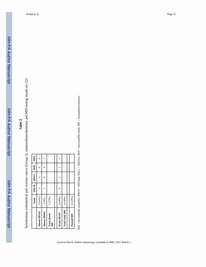
ResultsPatient characteristics are summarized in Table 1. Results of the molecular analysis for group1 (50 early onset endometrial cancers) and group 2 (22 synchronous endometrial and ovariancancers) are detailed in table 2 and table 3, respectively. Absence of staining for MLH1 orMSH2 was found in 7 (14%) tumors in group 1 and in 5 (23%) tumors in group 2. A strongcorrelation existed between negative IHC staining and MSI of tumor in both groups.
Those tumors with absent MLH1 immunohistochemistry were further evaluated formethylation of the MLH1 gene promoter. In group 1, among six cases with absent MLH1immunostaining, five (83%) were unmethylated, providing strong support for an underlyingetiology of LS in these early onset endometrial cancer cases. In contrast, in group 2, amongthree synchronous endometrial/ovarian cancer cases, two (67%) were methylated anddetermined to be of sporadic origin (Supplemental table S2).
Supplemental table S3 details the molecular findings in four cases with synchronousendometrial and ovarian cancers with absent MMR protein staining (group 2). The two tumorsites tend to show similar molecular characteristics. In each of the four patients, the same MMRprotein was affected in both of their tumors; and in two of the patients, methylation of theMLH1 promoter was demonstrated in both tumors. Cases 1 and 2 are consistent with possibleLS, while cases 3 and 4 demonstrate evidence of epigenetic MLH1 silencing through promotermethylation. A fifth case (not included in the table) underwent staining only for the endometrialcancer, which demonstrated absent MSH2 and MSH6 staining. Based on this finding, thepatient was referred to genetic testing and was found to have a deleterious mutation inMSH2.
Table 4 summarizes our findings. When considering the entire diagnostic algorithm thatincludes immunohistochemistry, MSI testing, and evaluation for MLH1 promoter methylation,molecular criteria supporting a genetic etiology of LS were found in 12% (6/50) of early onsetendometrial cancer patients (group 1) and in 14% (3/22) of synchronous endometrial/ovariancancer patients (group 2). Epigenetic silencing of MLH1 by promoter methylation was a moreprominent feature of synchronous cases (9%) than early endometrial cases (2%). Suspicious,but non-diagnostic molecular abnormalities were found in an additional 10% of earlyendometrial (group 1) and 4% of synchronous cases (group 2), warranting further evaluationand genetic work-up.
Among the nine patients with molecular criteria for LS, only three met the Amsterdam II criteriabased on family history and only one additional patient had a first degree relative with a historyof a LS-associated tumor (Supplemental table S4). Three of the nine patients have undergonecommercial genetic testing, and all were found to carry deleterious germline LS mutations:patient 1 carries the MSH2 IVS5 +3A>T mutation, patient 5 carries the MLH1 K416X mutation,and patient 8 has a deletion of exons 1 to 6 in the MSH2 gene.
DiscussionAmong two populations of gynecologic oncology patients that are recommended to undergogenetic testing by the 1996 Bethesda guidelines, we found 12% (6/50) of the early-onsetendometrial cancer group and 14% (3/22) of the synchronous endometrial and ovarian cancergroup to have tumors with molecular characteristics suggestive of LS. Only three of these ninepatients had a pedigree pattern that met the revised Amsterdam criteria for LS.
Walsh et al. Page 4
Gynecol Oncol. Author manuscript; available in PMC 2011 March 1.
NIH
-PA Author Manuscript
NIH
-PA Author Manuscript
NIH
-PA Author Manuscript

Screening by IHC for MMR proteins followed by selective MSI testing and evaluation forMLH1 promoter methylation may provide a useful algorithm for triage of patient samplestoward genetic testing to identify a deleterious mutation in a MMR gene. We demonstrate ahigh concordance between absent MMR protein IHC and the MSI-H phenotype and concludethat further MSI testing is not necessary in these cases.
The prediction of MLH1 mutations by IHC has been problematic in the past, due to theoccurrence of MLH1 missense mutations that result in a deficient, but antigenically-activeprotein [20]. We approached this problem by using MSI testing as a triage tool for further work-up of tumors with weak MLH1 protein staining [22]. An alternative approach would be to addthe PMS2 antibody to the IHC panel. Addition of PMS2 increases the sensitivity of IHC inpredicting MLH1 mutation to 92%; up from 85% with the three-antibody panel composed ofMLH1, MSH2 and MSH6 [21]. MLH1 dimerizes with PMS2 and mutations of MLH1 willoften cause a concurrent loss of the two proteins [24]. Our study potentially underestimatesthe prevalence of LS by the omission of this fourth antibody in our screening panel.
We chose to use IHC as the primary screening tool based on studies that suggest similareffectiveness of this method when compared to screening by MSI [25,26]. Addition of an IHCpanel of MMR proteins to the pathological evaluation of a tumor is relatively easy for theclinical pathologist and the pattern of MMR protein staining abnormalities can direct genetictesting towards the gene most likely to be affected [21]. Furthermore, IHC is more likely thanMSI testing to detect a MSH6 deficient tumor that may be characterized by low or absent MSI[23]. However, IHC can miss cases resulting from a deleterious missense mutation that encodesa functionally-deficient but antigenically-intact protein [27]. Furthermore, while most cases ofLS are due to mutations in MLH1, MSH2, MSH6, or PMS2; cases arising from an as of yetundefined mutated gene would not be detected by the IHC antibody panel [28]. Notably, theconcordance rate between IHC and MSI testing is only 92%, with both tests missing somecases that would be detected by the other [29,30].
The relatively high rate of MLH1 promoter methylation found in the synchronous primarycancer group suggests the benefit of adding MLH1 methylation analysis to the diagnosticalgorithm. Those cases found to have MLH1 promoter methylation would not require additionalgenetic testing for LS. Studies in colorectal cancer have demonstrated the utility of addingBRAF V600E mutation analysis to determine the sporadic nature of tumors with decreasedMLH1 expression [31–33]. Sparse data are available to this approach to the work-up ofendometrial cancer. However, one recent report suggests the BRAF V600E mutation is notfound in sporadic endometrial carcinomas [34].
In our diagnostic algorithm, we classified two patterns of abnormalities to be virtuallydiagnostic of LS: (1) absent MLH1 staining and non-methylated MLH1 gene promoter and (2)absent MSH2 and/or MSH6 staining [35,36]. Three of the nine patients classified as LS basedon these patterns of molecular abnormalities underwent commercial genetic testing and allthree (100%) were confirmed to carry a deleterious mutation. The highly predictive nature ofthese molecular findings raises the issue that IHC for MMR proteins could be interpreted as agenetic test. As such, the clinician should consider whether appropriate informed consentprotocols should be in place before immunohistochemical testing is performed.
We did not perform germline testing on all patients in this study, nor did we study a population-based sample. Both of these limitations could result in either overestimation or underestimationof LS among our two study populations. Nevertheless, using our diagnostic algorithm, wefound 12% of patients with endometrial cancers before the age of 45 to have molecular findingsconsistent with LS, which aligns with findings from prior studies. In patients diagnosed withendometrial cancer before 50 years of age, three studies utilizing germline gene sequencing
Walsh et al. Page 5
Gynecol Oncol. Author manuscript; available in PMC 2011 March 1.
NIH
-PA Author Manuscript
NIH
-PA Author Manuscript
NIH
-PA Author Manuscript

reported LS in 4.9%[37], 8.6% [38], and 9% [39]. We found 14% of patients with synchronousendometrial and ovarian cancers to have molecular findings suggestive of LS. Our results areslightly higher than the those of two retrospective studies (also based on tumor molecularprofiling) that suggested LS incidence rates of 3 – 7% [40,41]. However, in a study utilizinggermline mutation analysis in early-onset endometrial cancer patients less than 50 years of age,one of nine (11%) patients with a synchronous primary ovarian cancer had a LS mutation[39]. Our study also demonstrates a substantial proportion of MMR and MSI abnormalities insynchronous cases to result from MLH1 promoter methylation.
Among patients with synchronous endometrial and ovarian cancers, the tumors at both sitesoften showed similar IHC staining patterns and/or similar patterns of MLH1 promotermethylation, possibly reflecting either a still undefined genetic or environmental field effectthat impacts tumor development at both sites. We included only tumors where the clinicalimpression of synchronous malignancies was favored, but the possibility that the two tumorsites represent a metastasis from one site to the other must also be considered. Nevertheless,the concordance of molecular findings in tumor pairs raises the feasibility of restrictingmolecular testing to the endometrial cancer in these patients.
The optimal population of endometrial cancer patients for referral to genetic testing has yet tobe defined. In this study, we evaluated patients diagnosed with endometrial cancer before theage of 45 as recommended by the 1996 Bethesda guidelines. However, evidence suggests thata cutoff age of 45 will miss a large proportion of LS patients. In a population-based study, themedian age of diagnosis among ten LS mutation carriers was 54.6 years (range 39–69), withsix of the ten probands more than 50 years of age at the time of endometrial cancer diagnosis[37]. The use of family history as a triage tool may also miss LS cases. Berends et al [38]reported that among early-onset endometrial cancer patients (before the age of 50), 23% werefound to have a germline LS mutation if they had a first-degree relative with a LS-associatedcancer. In a population-based study of unselected endometrial cancer patients, seven of ten LSmutation carriers did not fulfill either the Amsterdam criteria or the Bethesda guidelines forscreening [37].
Identification of LS individuals and families is important because it has been shown to decreasecolorectal cancer mortality with the institution of heightened cancer screening protocols [4].The use of immunohistochemistry followed by selective MSI and MLH1 promoter methylationstudies may represent a useful algorithm for the identification of patients who should undergoanalysis for a germline MMR gene mutation. We did not find family history to be a usefultriage tool. Based on our findings, we would recommend screening for both gynecologic cancerpopulations identified by the original Bethesda guidelines, irrespective of family history.However, the optimal age cut-off for LS screening has not yet been defined and remains to bedetermined with future study.
Supplementary MaterialRefer to Web version on PubMed Central for supplementary material.
AcknowledgmentsResearch Support provided by the Gynecologic Cancer Foundation/Lee Kaplan Ovarian Cancer Research Grant Award(C.S.W.), the General Clinical Research Center Grant No. M01-RR00425 (C.S.W.), the American Cancer SocietyCalifornia Division-Early Detection Professorship Grant No. SIOP-06-258-01-CCE (B.Y.K.), the Women’s CancerResearch Institute (C.S.W. and B.Y.K.), and NIH grant 2 R01 CA026038-30 (H.P.K.).
Walsh et al. Page 6
Gynecol Oncol. Author manuscript; available in PMC 2011 March 1.
NIH
-PA Author Manuscript
NIH
-PA Author Manuscript
NIH
-PA Author Manuscript

References1. Dunlop MG, Farrington SM, Carothers AD, et al. Cancer risk associated with germline DNA mismatch
repair gene mutations. Hum Mol Genet 1997;6(1):105–110. [PubMed: 9002677]2. Aarnio M, Sankila R, Pukkala E, et al. Cancer risk in mutation carriers of DNA-mismatch-repair genes.
Int J Cancer 1999;81(2):214–218. [PubMed: 10188721]3. Watson P, Lynch HT. Extracolonic cancer in hereditary nonpolyposis colorectal cancer. Cancer
1993;71(3):677–685. [PubMed: 8431847]4. Jarvinen HJ, Aarnio M, Mustonen H, et al. Controlled 15-year trial on screening for colorectal cancer
in families with hereditary nonpolyposis colorectal cancer. Gastroenterology 2000;118(5):829–834.[PubMed: 10784581]
5. Vasen HF, Mecklin JP, Khan PM, et al. The International Collaborative Group on Hereditary Non-Polyposis Colorectal Cancer (ICG-HNPCC). Dis Colon Rectum 1991;34(5):424–425. [PubMed:2022152]
6. Vasen HF, Watson P, Mecklin JP, et al. New clinical criteria for hereditary nonpolyposis colorectalcancer (HNPCC, Lynch syndrome) proposed by the International Collaborative group on HNPCC.Gastroenterology 1999;116(6):1453–1456. [PubMed: 10348829]
7. Rodriguez-Bigas MA, Boland CR, Hamilton SR, et al. A National Cancer Institute Workshop onHereditary Nonpolyposis Colorectal Cancer Syndrome: meeting highlights and Bethesda guidelines.J Natl Cancer Inst 1997;89(23):1758–1762. [PubMed: 9392616]
8. Umar A, Boland CR, Terdiman JP, et al. Revised Bethesda Guidelines for hereditary nonpolyposiscolorectal cancer (Lynch syndrome) and microsatellite instability. J Natl Cancer Inst 2004;96(4):261–268. [PubMed: 14970275]
9. Peltomaki P, de la Chapelle A. Mutations predisposing to hereditary nonpolyposis colorectal cancer.Adv Cancer Res 1997;71:93–119. [PubMed: 9111864]
10. Kolodner RD, Tytell JD, Schmeits JL, et al. Germ-line msh6 mutations in colorectal cancer families.Cancer Res 1999;59(20):5068–5074. [PubMed: 10537275]
11. Marra G, Boland CR. Hereditary nonpolyposis colorectal cancer: the syndrome, the genes, andhistorical perspectives. J Natl Cancer Inst 1995;87(15):1114–1125. [PubMed: 7674315]
12. Vasen HF, Hendriks Y, de Jong AE, et al. Identification of HNPCC by molecular analysis of colorectaland endometrial tumors. Dis Markers 2004;20(4–5):207–213. [PubMed: 15528786]
13. Kane MF, Loda M, Gaida GM, et al. Methylation of the hMLH1 promoter correlates with lack ofexpression of hMLH1 in sporadic colon tumors and mismatch repair-defective human tumor celllines. Cancer Res 1997;57(5):808–811. [PubMed: 9041175]
14. Eifel P, Hendrickson M, Ross J, et al. Simultaneous presentation of carcinoma involving the ovaryand the uterine corpus. Cancer 1982;50(1):163–170. [PubMed: 7083121]
15. Pearl ML, Johnston CM, Frank TS, et al. Synchronous dual primary ovarian and endometrialcarcinomas. Int J Gynaecol Obstet 1993;43(3):305–312. [PubMed: 7907042]
16. Gitsch G, Hanzal E, Jensen D, et al. Endometrial cancer in premenopausal women 45 years andyounger. Obstet Gynecol 1995;85(4):504–508. [PubMed: 7898824]
17. Zaino R, Whitney C, Brady MF, et al. Simultaneously detected endometrial and ovarian carcinomas--a prospective clinicopathologic study of 74 cases: a gynecologic oncology group study. GynecolOncol 2001;83(2):355–362. [PubMed: 11606097]
18. Boland CR, Thibodeau SN, Hamilton SR, et al. A National Cancer Institute Workshop onMicrosatellite Instability for cancer detection and familial predisposition: development ofinternational criteria for the determination of microsatellite instability in colorectal cancer. CancerRes 1998;58(22):5248–5257. [PubMed: 9823339]
19. Dietmaier W, Wallinger S, Bocker T, et al. Diagnostic microsatellite instability: definition andcorrelation with mismatch repair protein expression. Cancer Res 1997;57(21):4749–4756. [PubMed:9354436]
20. Salahshor S, Koelble K, Rubio C, et al. Microsatellite Instability and hMLH1 and hMSH2 expressionanalysis in familial and sporadic colorectal cancer. Lab Invest 2001;81(4):535–541. [PubMed:11304573]
Walsh et al. Page 7
Gynecol Oncol. Author manuscript; available in PMC 2011 March 1.
NIH
-PA Author Manuscript
NIH
-PA Author Manuscript
NIH
-PA Author Manuscript

21. Shia J. Immunohistochemistry versus microsatellite instability testing for screening colorectal cancerpatients at risk for hereditary nonpolyposis colorectal cancer syndrome. Part I. The utility ofimmunohistochemistry. J Mol Diagn 2008;10(4):293–300. [PubMed: 18556767]
22. Mangold E, Pagenstecher C, Friedl W, et al. Tumours from MSH2 mutation carriers show loss ofMSH2 expression but many tumours from MLH1 mutation carriers exhibit weak positive MLH1staining. J Pathol 2005;207(4):385–395. [PubMed: 16216036]
23. Berends MJ, Wu Y, Sijmons RH, et al. Molecular and clinical characteristics of MSH6 variants: ananalysis of 25 index carriers of a germline variant. Am J Hum Genet 2002;70(1):26–37. [PubMed:11709755]
24. Kadyrov FA, Dzantiev L, Constantin N, et al. Endonucleolytic function of MutLalpha in humanmismatch repair. Cell 2006;126(2):297–308. [PubMed: 16873062]
25. Hampel H, Frankel WL, Martin E, et al. Screening for the Lynch syndrome (hereditary nonpolyposiscolorectal cancer). N Engl J Med 2005;352(18):1851–1860. [PubMed: 15872200]
26. Hampel H, Frankel WL, Martin E, et al. Feasibility of screening for Lynch syndrome among patientswith colorectal cancer. J Clin Oncol 2008;26(35):5783–5788. [PubMed: 18809606]
27. Peltomaki P, Vasen H. Mutations associated with HNPCC predisposition -- Update of ICG-HNPCC/INSiGHT mutation database. Dis Markers 2004;20(4–5):269–276. [PubMed: 15528792]
28. Zhang L. Immunohistochemistry versus microsatellite instability testing for screening colorectalcancer patients at risk for hereditary nonpolyposis colorectal cancer syndrome. Part II. The utility ofmicrosatellite instability testing. J Mol Diagn 2008;10(4):301–307. [PubMed: 18556776]
29. Shia J, Ellis NA, Klimstra DS. The utility of immunohistochemical detection of DNA mismatch repairgene proteins. Virchows Arch 2004;445(5):431–441. [PubMed: 15455227]
30. Mueller J, Gazzoli I, Bandipalliam P, et al. Comprehensive Molecular Analysis of Mismatch RepairGene Defects in Suspected Lynch Syndrome (Hereditary Nonpolyposis Colorectal Cancer) Cases.Cancer Res. 2009
31. Jensen LH, Lindebjerg J, Byriel L, et al. Strategy in clinical practice for classification of unselectedcolorectal tumours based on mismatch repair deficiency. Colorectal Dis 2008;10(5):490–497.[PubMed: 17868408]
32. Loughrey MB, Waring PM, Tan A, et al. Incorporation of somatic BRAF mutation testing into analgorithm for the investigation of hereditary non-polyposis colorectal cancer. Fam Cancer 2007;6(3):301–310. [PubMed: 17453358]
33. Rajagopalan H, Bardelli A, Lengauer C, et al. Tumorigenesis: RAF/RAS oncogenes and mismatch-repair status. Nature 2002;418(6901):934. [PubMed: 12198537]
34. Kawaguchi M, Yanokura M, Banno K, et al. Analysis of a correlation between the BRAF V600Emutation and abnormal DNA mismatch repair in patients with sporadic endometrial cancer. Int JOncol 2009;34(6):1541–1547. [PubMed: 19424571]
35. Ewald J, Rodrigue CM, Mourra N, et al. Immunohistochemical staining for mismatch repair proteins,and its relevance in the diagnosis of hereditary non-polyposis colorectal cancer. Br J Surg 2007;94(8):1020–1027. [PubMed: 17440950]
36. Buttin BM, Powell MA, Mutch DG, et al. Increased risk for hereditary nonpolyposis colorectal cancer-associated synchronous and metachronous malignancies in patients with microsatellite instability-positive endometrial carcinoma lacking MLH1 promoter methylation. Clin Cancer Res 2004;10(2):481–490. [PubMed: 14760069]
37. Hampel H, Frankel W, Panescu J, et al. Screening for Lynch syndrome (hereditary nonpolyposiscolorectal cancer) among endometrial cancer patients. Cancer Res 2006;66(15):7810–7817.[PubMed: 16885385]
38. Berends MJ, Wu Y, Sijmons RH, et al. Toward new strategies to select young endometrial cancerpatients for mismatch repair gene mutation analysis. J Clin Oncol 2003;21(23):4364–4370. [PubMed:14645426]
39. Lu KH, Schorge JO, Rodabaugh KJ, et al. Prospective determination of prevalence of lynch syndromein young women with endometrial cancer. J Clin Oncol 2007;25(33):5158–5164. [PubMed:17925543]
40. Shannon C, Kirk J, Barnetson R, et al. Incidence of microsatellite instability in synchronous tumorsof the ovary and endometrium. Clin Cancer Res 2003;9(4):1387–1392. [PubMed: 12684409]
Walsh et al. Page 8
Gynecol Oncol. Author manuscript; available in PMC 2011 March 1.
NIH
-PA Author Manuscript
NIH
-PA Author Manuscript
NIH
-PA Author Manuscript

41. Soliman PT, Broaddus RR, Schmeler KM, et al. Women with synchronous primary cancers of theendometrium and ovary: do they have Lynch syndrome? J Clin Oncol 2005;23(36):9344–9350.[PubMed: 16361634]
Walsh et al. Page 9
Gynecol Oncol. Author manuscript; available in PMC 2011 March 1.
NIH
-PA Author Manuscript
NIH
-PA Author Manuscript
NIH
-PA Author Manuscript

Figure 1.A. Immunohistochemistry results for MLH1, MSH2, and MSH6 The top row demonstratesnormal nuclear staining for each mismatch repair protein. The bottom row demonstrates absentstaining which is consistent with abnormal mismatch repair protein function. B. Representativeexample of microsatellite instability in endometrial cancer DNA (bottom) compared tomatched normal DNA (top) in one of the NCI-recommended microsatellite markers. In thiscase, there is a shift in the peaks, representing an error in the DNA replication process andcontraction of this microsatellite region. C. Methylation specific PCR results. Lane 1 containsthe methylated control. For each case, two PCR reactions were performed with primers specificfor the methylated MLH1 promoter (loaded on the left) and with primers specific for the
Walsh et al. Page 10
Gynecol Oncol. Author manuscript; available in PMC 2011 March 1.
NIH
-PA Author Manuscript
NIH
-PA Author Manuscript
NIH
-PA Author Manuscript

unmethylated MLH1 promoter (loaded on the right). Cases 1 and 2 demonstrate tumors withMLH1 silencing due to MLH1 gene promoter methylation, while case 3 represents and casethat may be due to an inherited germline MLH1 gene mutation.
Walsh et al. Page 11
Gynecol Oncol. Author manuscript; available in PMC 2011 March 1.
NIH
-PA Author Manuscript
NIH
-PA Author Manuscript
NIH
-PA Author Manuscript

Figure 2.Diagnostic algorithm used in our study. Dark blue boxes represent molecular findingsconsistent with Lynch Syndrome. Light blue boxes represent abnormal molecular findings thatwarrant further genetic testing for Lynch Syndrome.
Walsh et al. Page 12
Gynecol Oncol. Author manuscript; available in PMC 2011 March 1.
NIH
-PA Author Manuscript
NIH
-PA Author Manuscript
NIH
-PA Author Manuscript

NIH
-PA Author Manuscript
NIH
-PA Author Manuscript
NIH
-PA Author Manuscript
Walsh et al. Page 13
Table 1
Patient Characteristics
Group 1:Early-onset Endometrial
CA(age< 45 yrs)
N=50
Group 2:Synchronous
Endometrial/Ovarian CAN=22
Median age (range) 39 (29–44) 42 (31–52)
Median BMI (range) 26.9 (18–62) 26 (18–44)
Race/Ethnicity
Caucasian 34 (68%) 17 (77%)
Asian 11 (22%) 3 (14%)
Other 5 (10%) 2 (9%)
Endometrial CA Histology
Endometrioid 45 (90%) 19 (86%)
Adenosquamous 1 (2%) 1 (5%)
Mixed 4 (8%) 2 (9%)
Endometrial CA Stage
I 37 (74%) 18 (81%)
II 1 (2%) 1 (5%)
III 5 (10%) 2 (9%)
IV 1 (2%) 1 (5%)
Unknown 6 (12%)
Gynecol Oncol. Author manuscript; available in PMC 2011 March 1.

NIH
-PA Author Manuscript
NIH
-PA Author Manuscript
NIH
-PA Author Manuscript
Walsh et al. Page 14
Tabl
e 2
Early
ons
et e
ndom
etria
l can
cer (
Gro
up 1
): im
mun
ohis
toch
emis
try a
nd M
SI te
stin
g re
sults
(n=5
0)
Tot
alM
SI-H
MSI
-LM
SSU
nkno
wn
Abs
ent M
LH
16
(12%
)5
10
0
Abs
ent M
SH2
1 (2
%)
10
00
Tot
al a
bsen
tIH
C7
(14%
)
Wea
k M
LH
19
(18%
)1
25
1
Wea
k M
SH6
3 (6
%)
20
10
Tot
al w
eak
IHC
12 (2
4%)
Nor
mal
IHC
31 (6
2%)
MSI
= m
icro
sate
llite
inst
abili
ty; M
SI-H
= M
SI-h
igh;
MSI
-L =
MSI
-low
; MSS
= m
icro
sate
llite
stab
le; I
HC
= im
mun
ohis
toch
emis
try
Gynecol Oncol. Author manuscript; available in PMC 2011 March 1.
NIH
-PA Author Manuscript
NIH
-PA Author Manuscript
NIH
-PA Author Manuscript
Walsh et al. Page 15
Tabl
e 3
Sync
hron
ous e
ndom
etria
l and
ova
rian
canc
er (G
roup
2):
imm
unoh
isto
chem
istry
and
MSI
test
ing
resu
lts (n
=22)
Tot
alM
SI-H
MSI
-LM
SSM
SS
Abs
ent M
LH
13
(14%
)3
00
0
Abs
ent M
SH2
2 (9
%)
10
01
Tot
al a
bsen
tIH
C5
(23%
)
Wea
k M
LH
14
(18%
)0
21
1
Tot
al w
eak
IHC
4 (1
8%)
Nor
mal
IHC
13 (5
9%)
MSI
= m
icro
sate
llite
inst
abili
ty; M
SI-H
= M
SI-h
igh;
MSI
-L =
MSI
-low
; MSS
= m
icro
sate
llite
stab
le; I
HC
= im
mun
ohis
toch
emis
try
Gynecol Oncol. Author manuscript; available in PMC 2011 March 1.

NIH
-PA Author Manuscript
NIH
-PA Author Manuscript
NIH
-PA Author Manuscript
Walsh et al. Page 16
Table 4
Summary results based on our diagnostic algorithm
Group 1:Early onsetendometrial
cancer
Group 2:Synchronous
endometrial andovarian cancer
Genetic, suggests LS 6/50 (12%) 3/22 (14%)
Absent MLH1, unmethylated
Absent MSH2/MSH6
Sporadic 1/50 (2%) 2/22 (9%)
Absent MLH1, methylated
Needs further evaluation 5/50 (10%) 1/22 (4%)
Weak MLH1, MSI-H orunknown
Weak MSH6, any MSI status
LS = Lynch Syndrome; MSI = microsatellite instability; MSI-H = MSI-high
Gynecol Oncol. Author manuscript; available in PMC 2011 March 1.
Copyright © 2022 FDOKUMEN


![[Colorectal Carcinoma with Suspected Lynch Syndrome: A Multidisciplinary Algorithm.]](https://static.fdokumen.com/doc/165x107/6335f98064d291d2a302b343/colorectal-carcinoma-with-suspected-lynch-syndrome-a-multidisciplinary-algorithm.jpg)