luisa helena cazarolli - Universidade Federal de Santa Catarina
128
LUISA HELENA CAZAROLLI CARACTERIZAÇÃO DE COMPOSTOS NATURAIS E AVALIAÇÃO DA ATIVIDADE INSULINO-MIMÉTICA EM TECIDOS ALVOS DA INSULINA EM ESTUDOS IN VIVO E IN VITRO FLORIANÓPOLIS 2009
-
Upload
khangminh22 -
Category
Documents
-
view
1 -
download
0
Transcript of luisa helena cazarolli - Universidade Federal de Santa Catarina

LUISA HELENA CAZAROLLI
CARACTERIZAÇÃO DE COMPOSTOS NATURAIS E AVALIAÇÃO DA ATIVIDADEINSULINO-MIMÉTICA EM TECIDOS ALVOS DA INSULINA EM ESTUDOS IN VIVO E
IN VITRO
FLORIANÓPOLIS
2009
UNIVERSIDADE FEDERAL DE SANTA CATARINACENTRO DE CIÊNCIAS DA SAÚDE
PROGRAMA DE PÓS-GRADUAÇÃO EM FARMÁCIA
LUISA HELENA CAZAROLLI
CARACTERIZAÇÃO DE COMPOSTOS NATURAIS E AVALIAÇÃO DA ATIVIDADEINSULINO-MIMÉTICA EM TECIDOS ALVOS DA INSULINA EM ESTUDOS IN VIVO E
IN VITRO
Tese apresentada ao Programa de Pós-graduação em Farmácia doCentro de Ciências da Saúde da Universidade Federal de SantaCatarina, como requisito parcial para obtenção do título de Doutor emFarmácia.
Orientadora: Profª. Drª. FÁTIMA REGINA MENA BARRETO SILVA
Florianópolis2009

CAZAROLLI, Luisa Helena
Caracterização de compostos naturais e avaliação da atividadeinsulino-mimética em tecidos alvos da insulina em estudos in vivo ein vitro / Luisa Helena Cazarolli. Florianópolis, 2009. 151 p.
Tese (Doutorado) – Universidade Federal de Santa Catarina.
Programa de Pós-Graduação em Farmácia.
1. Flavonóides. 2. Síntese de glicogênio. 3. Captação de glicose. 4.Hiperglicemia. 5. Averrhoa carambola. 6. Diabetes.


Dedico este trabalho aos meus pais e minha irmã José Luis, Eliria Maria e Juciana
Clarice Cazarolli, pela oportunidade recebida, pelo apoio e amor incondicional e, por
vezes, tão longe fisicamente, estiveram sempre presentes em todos os momentos da
minha vida.

AGRADECIMETOS
A Deus, por me proporcionar mais esta oportunidade única.A minha família, José Luis, Eliria Maria e Juciana Clarice Cazarolli pelo amor e carinho e por
toda a ajuda e compreensão em todos os momentos desta caminhada.Agradecimento especial à minha orientadora, Profa. Dra. Fátima Regina Mena Barreto Silva,
pela confiança em mim depositada, pela dedicação, paciência, amizade e pelos inúmerosensinamentos e incentivo à pesquisa.
Ao Rafael Nicolay Pereira, pelo carinho, paciência e apoio em todas as situações.Às colegas de laboratório pelos bons momentos, amizade, ajuda e conhecimentos
compartilhados, especialmente à Elga Heloisa Alberton, Poliane Folador e Rosangela GuolloDamazio pelo apoio na realização dos experimentos.
Aos meus queridos amigos, pela amizade sincera, paciência, e companheirismo.Aos professores Dr. Moacir Geraldo Pizzolatti e Dra. Inês Maria Costa Brighente e aluno
Henrique Hunger Moresco, pela colaboração no desenvolvimento deste trabalho.Aos professores Dr. Danilo Wilhelm Filho, Dr. João Batista Calixto e Dra. Rozangela Curi
Pedrosa por compartilhar equipamentos.Ao laboratório da Dra. Tânia Silvia Fröde, em especial às alunas Ziliane e Jucélia pela
disponibilidade concedida.A todos os amigos aqui não mencionados e que de uma maneira ou de outra contribuíram para
a realização deste trabalho.
RESUMO
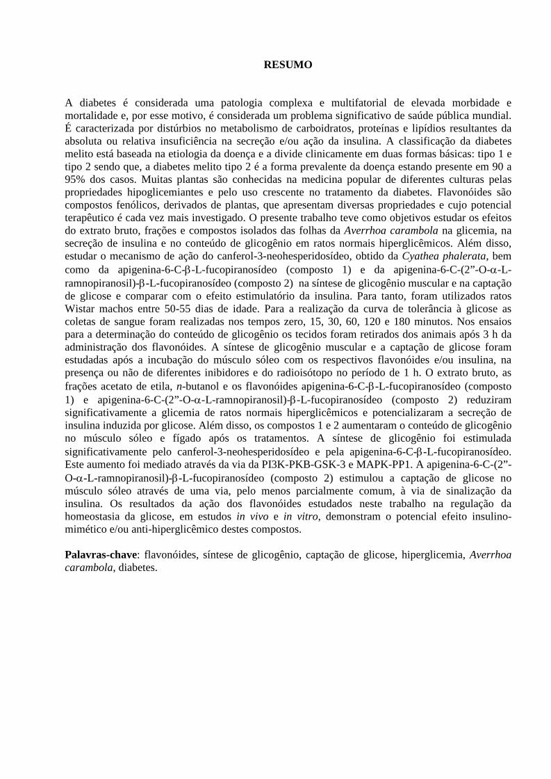
A diabetes é considerada uma patologia complexa e multifatorial de elevada morbidade emortalidade e, por esse motivo, é considerada um problema significativo de saúde pública mundial.É caracterizada por distúrbios no metabolismo de carboidratos, proteínas e lipídios resultantes daabsoluta ou relativa insuficiência na secreção e/ou ação da insulina. A classificação da diabetesmelito está baseada na etiologia da doença e a divide clinicamente em duas formas básicas: tipo 1 etipo 2 sendo que, a diabetes melito tipo 2 é a forma prevalente da doença estando presente em 90 a95% dos casos. Muitas plantas são conhecidas na medicina popular de diferentes culturas pelaspropriedades hipoglicemiantes e pelo uso crescente no tratamento da diabetes. Flavonóides sãocompostos fenólicos, derivados de plantas, que apresentam diversas propriedades e cujo potencialterapêutico é cada vez mais investigado. O presente trabalho teve como objetivos estudar os efeitosdo extrato bruto, frações e compostos isolados das folhas da Averrhoa carambola na glicemia, nasecreção de insulina e no conteúdo de glicogênio em ratos normais hiperglicêmicos. Além disso,estudar o mecanismo de ação do canferol-3-neohesperidosídeo, obtido da Cyathea phalerata, bemcomo da apigenina-6-C--L-fucopiranosídeo (composto 1) e da apigenina-6-C-(2”-O--L-ramnopiranosil)--L-fucopiranosídeo (composto 2) na síntese de glicogênio muscular e na captaçãode glicose e comparar com o efeito estimulatório da insulina. Para tanto, foram utilizados ratosWistar machos entre 50-55 dias de idade. Para a realização da curva de tolerância à glicose ascoletas de sangue foram realizadas nos tempos zero, 15, 30, 60, 120 e 180 minutos. Nos ensaiospara a determinação do conteúdo de glicogênio os tecidos foram retirados dos animais após 3 h daadministração dos flavonóides. A síntese de glicogênio muscular e a captação de glicose foramestudadas após a incubação do músculo sóleo com os respectivos flavonóides e/ou insulina, napresença ou não de diferentes inibidores e do radioisótopo no período de 1 h. O extrato bruto, asfrações acetato de etila, n-butanol e os flavonóides apigenina-6-C--L-fucopiranosídeo (composto1) e apigenina-6-C-(2”-O--L-ramnopiranosil)--L-fucopiranosídeo (composto 2) reduziramsignificativamente a glicemia de ratos normais hiperglicêmicos e potencializaram a secreção deinsulina induzida por glicose. Além disso, os compostos 1 e 2 aumentaram o conteúdo de glicogêniono músculo sóleo e fígado após os tratamentos. A síntese de glicogênio foi estimuladasignificativamente pelo canferol-3-neohesperidosídeo e pela apigenina-6-C--L-fucopiranosídeo.Este aumento foi mediado através da via da PI3K-PKB-GSK-3 e MAPK-PP1. A apigenina-6-C-(2”-O--L-ramnopiranosil)--L-fucopiranosídeo (composto 2) estimulou a captação de glicose nomúsculo sóleo através de uma via, pelo menos parcialmente comum, à via de sinalização dainsulina. Os resultados da ação dos flavonóides estudados neste trabalho na regulação dahomeostasia da glicose, em estudos in vivo e in vitro, demonstram o potencial efeito insulino-mimético e/ou anti-hiperglicêmico destes compostos.
Palavras-chave: flavonóides, síntese de glicogênio, captação de glicose, hiperglicemia, Averrhoacarambola, diabetes.

ABSTRACT
Diabetes is a complex and multifactorial disease which presents high death rates and morbidity.Because of that, diabetes is considered a World public health problem. It is characterized byhyperglycemia resulting from defects in insulin secretion and/or in insulin action. Basically, thisdisorder is divided into type 1 and type 2 diabetes and the last one represents the great majority ofthe cases. Many plant species are known in popular medicine for their hypoglycemic properties andtheir increasing use for treating diabetes. Flavonoids are phenolic compounds derived from plants,which present diverse properties and whose therapeutic potential is being increasingly investigated.The objectives of the present investigation were to study the effects of crude extract, fractions andisolated compounds, apigenin-6-C--L-fucopyranoside (compound 1) and apigenin-6-C-(2”-O--L-rhamnopyranosyl)--L-fucopyranoside (compound 2) from leaves of Averrhoa carambola onglycemia, on insulin secretion as well as on glycogen content in muscle and liver fromhyperglycemic rats. In addition, we intended to study the mechanism of action of kaempferol-3-neohesperidoside, obtained from Cyathea phalerata, and of apigenin-6-C--L-fucopyranoside(compound 1) and apigenin-6-C-(2”-O--L-rhamnopyranosyl)--L-fucopyranoside (compound 2)on glycogen synthesis and on glucose uptake and compare them with the stimulatory effect ofinsulin. Male Wistar rats aged 50-55 days were used. To the glucose tolerance curve, blood sampleswere collected just prior to and at 15, 30, 60, 120 and 180 min after the glucose loading and serumglucose levels were measured. In the assays for the determination of the glycogen content tissueswere removed from animals after 3 h of the administration of the compounds 1 and 2. Theglycogen synthesis and the glucose uptake were studied after incubation of the soleus muscle for 1 hwith the respective flavonoids and/or insulin, in the presence or not of different inhibitors and of theradioisotope. The oral administration of crude extract, ethyl acetate and n-butanol fractions andcompounds 1 and 2 of Averrhoa carambola leaves exhibited a potential hypoglycemic activity inthe hyperglycemic normal rats and potentiated the glucose-induced insulin secretion. Additionaly,compounds 1 and 2 also increased glycogen content in muscle and liver from the hyperglycemicrats after 3 h of the treatments. The glycogen synthesis was significantly stimulated by kaempferol-3-neohesperidoside as well as by apigenin-6-C--L-fucopyranoside (compound 1). This effect ismediated through the PI3K-PKB-GSK-3 and MAPK-PP1 pathways. Furthermore, apigenin-6-C-(2”-O--L-rhamnopyranosyl)--L-fucopyranoside (compound 2) stimulated glucose uptake insoleus muscle through the same signaling pathway as insulin. The results observed in this worksupport the flavonoid’s effect in glucose homeostasis through in vivo and in vitro studies. Thisdemonstrates the insulinmimetic and/or antihyperglycemic potential of these compounds.
Key-words: flavonoids, glycogen synthesis, glucose uptake, hyperglycemia, Averrhoa carambola,diabetes.

LISTA DE FIGURAS
Figura 1. Secreção bifásica de insulina estimulada por glicose (A) e processo desecreção de insulina pelo pâncreas (B)................................................................................ 3
Figura 2. Vias de sinalização da insulina............................................................................. 5
Figura 3. Síntese de glicogênio............................................................................................ 8
Figura 4. Cascata de fosforilação e ativação da glicogênio sintase..................................... 9
Figura 5. Regulação do metabolismo da glicose.................................................................. 12
Figura 6. Espécie vegetal Averrhoa carambola................................................................... 15
Figura 7. Espécie vegetal Cyathea phalerata...................................................................... 17
Figura 8. Estruturas do núcleo fundamental dos flavonóides, canferitrina, canferol ecanferol 3-neohesperidosídeo.............................................................................................. 19
Figura 9. Modelo proposto para o mecanismo de ação geral dos flavonóides natransdução de sinais da insulina........................................................................................... 95

LISTA DE FLUXOGRAMAS
Fluxograma 1. Extração e isolamento dos flavonóides da Averrhoa carambola................ 21
Fluxograma 2. Indução do modelo de diabetes experimental.............................................. 22
Fluxograma 3. Tratamentos dos animais normais hiperglicêmicos e diabéticos.................23
Fluxograma 4. Extração do glicogênio muscular................................................................. 24
Fluxograma 5. Representação esquemática do ensaio de incorporação de D-[U-14C]Gem glicogênio no músculo sóleo incubado in vitro para a curva de dose-resposta com osflavonóides ou insulina........................................................................................................ 25
Fluxograma 6. Representação esquemática do ensaio de incorporação de D-[U-14C]Gem glicogênio no músculo sóleo incubado com os flavonóides e com inibidores daPI3K, GSK-3, MEK e PP1................................................................................................... 26
Fluxograma 7. Representação esquemática do ensaio de captação de [14C]-DG nomúsculo sóleo para a curva de dose-resposta com os flavonóides ou insulina.................... 27
Fluxograma 8. Representação esquemática do ensaio de captação de [14C]-DG nomúsculo sóleo incubado com os flavonóides e com diferentes inibidores........................... 28

LISTA DE ABREVIATURAS
AcOEt Acetato de etila
ADP Adenosina difosfato
AMPK Proteína cinase ativada por 5’-AMP
APS Substrato da proteína associada
ATP Adenosina trifosfato
AS160 Proteína substrato da Akt de 160 kDa
CAP Proteína associada à cbl
[14C]DG [U-14C]-2-deoxi-D-glicose
[14C]G ouD[U-14C]G
D - [14C (U)] – glicose
CEUA Comitê de Ética do Uso de Animais
C3G Proteína trocadora de nucleotídeos
cpm Contas por minuto
DPP-IV Dipeptidilpeptidase 4
ERK Cinase reguladora de sinal extracelular
EtOH Etanol
GAP Proteína ativadora de GTPases
GDP Guanosina difosfato
GIP Polipeptídeo insulinotrópico
GLP-1 Peptídeo semelhante ao glucagon 1
GLUT Transportador de glicose
G-6-P Glicose 6 fosfato
G-1-P Glicose 1 fosfato
Grb2 Proteína 2 ligada ao receptor de fator de crescimento
GS Glicogênio sintase
GSK-3 Glicogênio sintase cinase-3
GTP Guanosina trifosfato

HMIT Transportador H+ ligado ao mio-inositol
IRS Substrato do receptor de insulina
JAK2 Janus cinase 2
JNK Cinase c-jun NH2 terminal
KIF proteína quinesina
Km Constante Cinética de Michaelis Menten
MAPK Proteína cinase ativada por mitógeno
MEK Proteína cinase cinase ativada por mitógeno
mTOR Alvo da rapamicina em mamíferos
n-BuOH n-butanol
NPH Insulina com protamina neutra de Hagedorn
PDK-1 e 2 Proteína cinase 1 e 2 dependentes de PI-3K
PH Domínios de homologia de plekstrina
PI-3K Fosfatidilinositol 3-cinase
PIP3 Fosfatidilinositol-3,4,5-trifosfato
PKA Proteína Cinase A
PKB/AKt Proteína Cinase B
PKC Proteína Cinase C
p38 MAPK p38 Proteína cinase ativada por mitógeno
PP1 Proteína fosfatase 1
PP1G Isoforma da proteína fosfatase 1 ligada ao glicogênio
PRG Proteína regulatória da glicocinase
PTB Domínios de ligação a fosfotirosina
PTG Proteína regulatória da PP1
PTPase Proteína fosfatase
PTP1B Proteína fosfatase 1B
p70rsk p70 ribossomal S6 cinase
p90rsk p90 ribossomal S6 cinase
RTK Receptor do tipo tirosina cinase

SH2 Homólogo 2 Src
SHP2 Proteína tirosina fosfatase homóloga de Src 2
SNAP23 Proteína de 23 kD associada à sinaptossoma
SNARE Proteína do receptor ligada a NSF solúvel
SOCS 1 e 3 Proteínas supressoras da sinalização de citocinas
UDP-G Uridina difosfato glicose
VAMP2 Proteína 2 da vesícula associada à membrana

SUMÁRIO
1 INTRODUÇÃO............................................................................................................ 1
1.1 DIABETES MELITO................................................................................................. 1
1.2 INSULINA E MECANISMOS DE TRANSDUÇÃO DE SINAL............................. 2
1.3 REGULAÇÃO DO METABOLISMO DA GLICOSE.............................................. 6
1.3.1 Transportadores de glicose e captação de glicose.................................................... 6
1.3.2 Síntese de glicogênio................................................................................................ 7
1.3.3 Regulação do metabolismo da glicose..................................................................... 11
1.4 HIPOGLICEMIANTES ORAIS................................................................................. 12
1.5 TERAPIA INSULÍNICA............................................................................................ 14
1.6 PLANTAS MEDICINAIS.......................................................................................... 14
1.6.1 Averrhoa carambola......................................................................................... 15
1.6.2 Cyathea phalerata............................................................................................. 16
1.6.3 Flavonóides....................................................................................................... 17
2 OBJETIVOS................................................................................................................. 20
2.1 OBJETIVO GERAL................................................................................................... 20
2.2 OBJETIVOS ESPECÍFICOS...................................................................................... 20
3 METODOLOGIA........................................................................................................ 21
4 ARTIGOS..................................................................................................................... 294.1 CAZAROLLI, L.H.; MACHADO, L.M.; FOLADOR, P.; MORESCO, H.H.;
BRIGHENTE, I.M.C.; PIZZOLATTI, M.G.; SILVA, F.R.M.B. Potentialantihyperglycemic role of crude extract, fractions and flavonoids from Averrhoacarambola leaves in rats……………........................................................................ 29
4.2 CAZAROLLI, L.H.; FOLADOR, P.; MORESCO, H.H.; BRIGHENTE, I.M.C.;PIZZOLATTI, M.G.; SILVA, F.R.M.B. Stimulatory Effect of Apigenina-6-C--L-fucopyranoside on Insulin Secretion and GlycogenSynthesis…………………………………………...………………………………. 44

4.3 CAZAROLLI, L.H.; FOLADOR, P.; MORESCO, H.H.; BRIGHENTE, I.M.C.;PIZZOLATTI, M.G.; SILVA, F.R.M.B. Mechanism of action of the stimulatoryeffect of apigenin-6-C-(2”-O--L-rhamnopyranosyl)--L-fucopyranoside on 14C-glucose uptake……………..……………………………………………………….. 56
4.4 CAZAROLLI, L.H.; FOLADOR, P.; PIZZOLATTI, M.G.; SILVA, F.R.M.B.Signaling Pathways of Kaempferol-3-neohesperidoside on Glycogen Synthesis inRat Soleus Muscle………………………………………………..………………… 63
4.5 CAZAROLLI, L.H.; ZANATTA, L.; ALBERTON, E.H.; FIGUEIREDO,M.S.R.B.; FOLADOR, P.; DAMAZIO, R.G.; PIZZOLATTI, M.G.; SILVA,F.R.M.B. Flavonoids: Prospective drug candidates…….......................................... 70
4.6 CAZAROLLI, L.H.; ZANATTA, L.; ALBERTON, E.H.; FIGUEIREDO,M.S.R.B.; FOLADOR, P.; DAMAZIO, R.G.; PIZZOLATTI, M.G.; SILVA,F.R.M.B. Flavonoids: Cellular and molecular mechanism of action in glucosehomeostasis…………………………………............................................................ 82
5 DISCUSSÃO................................................................................................................. 90
6 CONCLUSÕES FINAIS............................................................................................. 94
7 REFERÊNCIAS BIBLIOGRÁFICAS...................................................................... 96
8 ANEXOS...................................................................................................................... 107

1
1 INTRODUÇÃO
1.1 DIABETES MELITO
Diabetes melito é um grupo heterogêneo de distúrbios metabólicos que apresentam emcomum a hiperglicemia. É caracterizada por distúrbios no metabolismo de carboidratos, proteínas elipídios resultantes da absoluta ou relativa insuficiência na secreção e/ou ação da insulina. (ISLAS-ANDRADE et al., 2000; SOCIEDADE BRASILEIRA DE DIABETES, 2003; SOCIEDADEBRASILEIRA DE DIABETES, 2007; AMERICAN DIABETES ASSOCIATION, 2008; WHO,2009a). Segundo estudos, no ano de 2000 aproximadamente 171 milhões de adultos com mais de 20anos tinham diabetes em todo o mundo. Além disso, estima-se que neste mesmo ano, 2,9 milhões depessoas morreram em conseqüência da doença (WILD et al., 2004; ROGLIC et al., 2005; WHO,2009b). Em 2007, já eram 246 milhões de pessoas com idade entre 20 e 79 anos com diabetes(7,3% da população adulta) (INTERNATIONAL DIABETES FEDERATION, 2009). Projeçõesindicam que pode haver 366 milhões de casos até o ano de 2030 (GADSBY, 2002; WILD et al.,2004; WHO, 2009b). No Brasil, a prevalência de pacientes diabéticos em 2000 era de 4,6 milhõesde casos, em 2007 6,9 milhões e estima-se que até o ano de 2030 sejam 11,3 milhões de diabéticos(WILD et al., 2004; INTERNATIONAL DIABETES FEDERATION, 2009; WHO, 2009c).
A classificação da diabetes melito atualmente é baseada na etiologia da doença sendo divididaclinicamente em duas formas básicas: tipo 1 e tipo 2 (SOCIEDADE BRASILEIRA DEDIABETES, 2003; SOCIEDADE BRASILEIRA DE DIABETES, 2007; AMERICAN DIABETESASSOCIATION, 2008). A diabetes melito tipo 1, presente em 5 a 10% dos casos, se caracterizapela destruição auto-imune das células pancreáticas mediada por células T CD4+ e CD8+ bemcomo macrófagos infiltrados nas ilhotas (ROSMALEN et al., 2002; KAWASAKI et al., 2004;GILLESPIE, 2006). Inúmeros fatores genéticos e ambientais contribuem para a ativaçãoimunológica que desencadeia esse processo destrutivo e que se caracteriza pela ausência dasecreção endógena de insulina (KAWASAKI et al., 2004; GILLESPIE, 2006; AMERICANDIABETES ASSOCIATION, 2008). A diabetes melito tipo 2 é a forma prevalente da doençaestando presente em 90 a 95% dos casos sendo que a maioria dos pacientes apresenta sobrepeso ouobesidade (SOCIEDADE BRASILEIRA DE DIABETES, 2003; AMERICAN DIABETESASSOCIATION, 2008). Esta desordem metabólica se caracteriza por dois defeitos fisiopatológicosprincipais: a redução da secreção de insulina pelas células pancreáticas e a resistência periférica àação da insulina em diversos tecidos (músculo, fígado e tecido adiposo) (LEROITH, 2002;CHENG, FANTUS, 2005; KASUGA, 2006). A resposta inicial da célula à resistência a insulina éaumentar a secreção do hormônio promovendo uma hiperinsulinemia. À medida que a doençaprogride, a produção bem como a secreção de insulina diminuem gradativamente levando a estágiosprogressivos de hiperglicemia (LEROITH, 2002; KASUGA, 2006).
A resistência a insulina geralmente precede o início da diabetes e pode ser influenciada porinúmeros fatores genéticos e ambientais como, por exemplo, idade, obesidade e sedentarismo.Indivíduos insulino-resistentes apresentam diversas outras características como dislipidemia,hipertensão, excesso de adiposidade visceral, fibrinólise alterada, disfunção endotelial, inflamaçãovascular e aterosclerose prematura (CHENG; FANTUS, 2005). Além disso, defeitos na função doreceptor e/ou na sinalização da insulina pós-receptor, aumento da lipólise no tecido adiposo comconseqüente elevação das concentrações plasmáticas de ácidos graxos livres e inabilidade desuprimir a produção de glicose hepática são condições que contribuem para a patogênese dadiabetes (LEROITH, 2002; KASUGA, 2006).
A diabetes é considerada uma patologia complexa e multifatorial de elevadas morbidade emortalidade e, por esse motivo, é considerada um problema significativo de saúde pública mundial.O número de casos de diabetes vem aumentando gradativamente entre a população, devidobasicamente ao crescimento populacional e aumento da expectativa de vida, urbanização, obesidade

2
e sedentarismo (NARAYAN et al., 2000; BOYLE et al., 2001; GADSBY, 2002; WILD et al.,2004).
1.2 INSULINA E MECANISMOS DE TRANSDUÇÃO DE SINAL
A insulina é um hormônio polipeptídico produzido pelas células das ilhotas de Langerhansno pâncreas. É composta por 51 aminoácidos dispostos em duas cadeias polipeptídicas, A e B,unidas através de pontes dissulfeto. A síntese ocorre a partir de um precursor de 110 aminoácidos, apré-pró-insulina no retículo endoplasmático rugoso onde é clivada a pró-insulina. Esta é convertidaà insulina e armazenada no complexo de golgi (KRAUSS, 2008). A glicose é o estímulo primáriopara a secreção de insulina, embora outros mecanismos de controle possam estar envolvidos como aação coordenada de vários nutrientes, hormônios gastrintestinais, pancreáticos e neurotransmissores(BEARDSALL et al., 2003).
Quando estimulada por glicose, a secreção de insulina é bifásica. Uma fase inicial rápida (4 –8 minutos) e transitória resultante da liberação da insulina estocada de grânulos localizadospróximos à membrana plasmática (estoque de liberação rápida). Uma segunda fase de maiorduração ocorre através do deslocamento e ativação de grânulos citoplasmáticos (estoque de reserva)em direção à membrana bem como através da síntese do hormônio (Figura 1A) (RORSMAN, 1997;MACDONALD et al., 2005; OHARA-IMAIZUMI; NAGAMATSU, 2006; HENQUIN et al.,2006). A meia-vida da insulina circulante é de 3 a 5 minutos e o metabolismo ocorre principalmenteno fígado e rins por ação de insulinases (KRAUSS, 2008).
A secreção de insulina ocorre, principalmente, em função da hiperglicemia. Quando a célula é exposta à glicose, esta é internalizada através de difusão facilitada pelo GLUT2. No interior dacélula a glicose é fosforilada a glicose 6-fosfato pela glicocinase e então, metabolizada. A elevaçãodo metabolismo da glicose aumenta a relação ATP/ADP citoplasmática gerando um fechamento decanais de potássio (K+) dependentes de ATP. A redução da saída de potássio causa umadespolarização da membrana das células e abertura de canais de Ca2+ dependentes de voltagemcom conseqüente aumento do influxo de cálcio que, por sua vez, desencadeia o processo deexocitose dos grânulos de insulina (Figura 1B) (RORSMAN, 1997; MACDONALD et al., 2005;OHARA-IMAIZUMI; NAGAMATSU, 2006).

3
Figura 1. Secreção bifásica de insulina estimulada por glicose (A) (adaptado dehttp://www.servier.com/imgs/Pro/diabeto/diabetographia/ud/1/1.gif). Processo de secreção deinsulina pelo pâncreas (B) (adaptado dehttp://www.ncbi.nlm.nih.gov/books/bv.fcgi?rid=diabetes.section.621).
A insulina é o principal hormônio anabólico responsável pelo controle da captação, utilizaçãoe armazenamento dos nutrientes celulares como carboidratos, proteínas e lipídios. Além disso, éessencial para a manutenção da homeostasia da glicose, do crescimento e diferenciação celular(HEI, 1998; TAHA; KLIP, 1999). A insulina exerce os efeitos biológicos através dafosforilação/desfosforilação de proteínas, resultando em aumento do transporte e metabolismo deglicose, mitogênese, bem como, através do controle da atividade/síntese de enzimas regulatórias devias metabólicas centrais (TAHA; KLIP, 1999).
Para exercer os efeitos metabólicos, a insulina se liga a um receptor específico da membranadas células. O receptor de insulina é uma proteína heterotetramérica pertencente à família dereceptores tirosina cinases (RTKs). Consiste de duas subunidades extracelulares que contém osítio de ligação à insulina e duas subunidades trans-membrana com atividade de tirosina cinase(TAHA; KLIP, 1999; SALTIEL; KAHN, 2001; CHANG et al., 2004). O receptor de insulinafunciona como uma enzima alostérica, onde a subunidade inibe a atividade tirosina cinase dasubunidade . A ligação da insulina ao receptor promove autofosforilação da subunidade emresíduos de tirosina específicos. Além disso, o receptor também passa por mudançasconformacionais que resultam no aumento ainda maior da atividade tirosina cinase do receptor(Figura 2) (SALTIEL; KAHN, 2001; CHANG et al., 2004).
Uma vez ativado, o receptor de insulina promove a fosforilação de diversos substratosprotéicos em resíduos de tirosina como, por exemplo, a família de substratos do receptor de insulina(IRS 1-4). Outros substratos incluem Shc, Gab-1, p60dok, Cbl, JAK2 e APS (TAHA; KLIP, 1999;CHANG et al., 2004). As proteínas IRS-1 e IRS-2 estão amplamente distribuídas nos tecidos demamíferos e são as mais importantes para as ações da insulina. IRS-1 parece ser a isoforma

4
predominante envolvida na transdução de sinal da insulina no músculo. Já a IRS-2 parece estarenvolvida no desenvolvimento das células e ambas as isoformas são importantes para ometabolismo da glicose no fígado (KROOK et al., 2004). As funções das IRS-3 e IRS-4 são menosentendidas, mas parecem envolver a regulação das proteínas IRS-1 e IRS-2 (HEI, 1998; SALTIEL;KAHN, 2001).
Em geral, as proteínas IRS apresentam regiões em comum: domínios de homologia complekstrina (PH) que ligam fosfolipídios, domínios de ligação a fosfotirosina (PTB), sítios de ligaçãoa proteínas com domínios com homologia Src 2 (SH2), regiões ricas em prolina e regiões ricas emserina/treonina. A ligação das proteínas IRS com o receptor de insulina se dá através dos domíniosde ligação a fosfotirosina (PTB) que permitem a fosforilação de diversos resíduos tirosina nas IRS(TAHA; KLIP, 1999; KAHN; PESSIN, 2002; TANIGUCHI et al., 2006; KRAUSS, 2008).
Além da fosforilação em resíduos de tirosina, tanto o receptor de insulina quanto as proteínasIRS podem ser fosforiladas em resíduos de serina em resposta à insulina e outros estímulos. Esteefeito parece ser mediado por diversas cinases como fosfatidilinositol 3-cinase (PI3K), proteínacinase B (PKB/Akt), glicogênio sintase cinase 3 (GSK-3), cinase reguladora de sinal extracelular(ERK), cinase c-jun NH2 terminal (JNK), proteínas supressoras da sinalização de citocinas (SOCS 1e 3) e proteína alvo da rapamicina em mamíferos (mTOR) bem como pela interação entre sistemasde sinalização. Estas fosforilações parecem regular negativamente a sinalização insulínica e podemprovocar resistência a insulina (SALTIEL; KAHN, 2001; TANIGUCHI et al., 2006). Além disso, aação da insulina também é atenuada por proteínas tirosina fosfatases (PTPases) as quais catalisam adesfosforilação rápida do receptor de insulina e de substratos destacando-se a PTP1B e a SHP2(TAHA; KLIP, 1999; SALTIEL; KAHN, 2001; TANIGUCHI et al., 2006; KRAUSS, 2008).
Os resíduos tirosina fosforilados das IRS formam sítios de ligação para proteínas comdomínios SH2 (TAHA; KLIP, 1999; KAHN; PESSIN, 2002; TANIGUCHI et al., 2006). Muitasdessas proteínas são moléculas adaptadoras como a subunidade regulatória p85 da PI3K, a Grb2 e aCrkII ou enzimas como a fosfotirosina fosfatase SHP2 e a tirosina cinase citoplasmática Fyn(SALTIEL; KAHN, 2001; KRAUSS, 2008).
A PI3K é uma enzima importante na regulação da mitogênese e das ações metabólicas dainsulina como, por exemplo, o estímulo da translocação dos transportadores de glicose GLUT4 paraa membrana. Consiste de uma subunidade catalítica (p110) e uma subunidade regulatória (p85).Essa enzima catalisa a fosforilação de fosfoinositídeos na posição 3 do anel inositol produzindofosfatidilinositol-3-fosfato, fosfatidilinositol-3,4-fosfato e fosfatidilinositol-3,4,5-trifosfato (PIP3)(TAHA; KLIP, 1999; SALTIEL; KAHN, 2001; TANIGUCHI et al., 2006). O PIP3 funciona comosegundo mensageiro intracelular e regula a localização e atividade de diversas proteínasintracelulares como a proteína cinase 1 dependente de 3-fosfoinositídeos (PDK-1 e PDK-2) e aproteína cinase B (PKB/Akt) através da interação com domínios de homologia de plekstrina (PH).A seguir, a PDK-1 fosforila e ativa a PKB e isoformas de proteína cinase C (PKC) atípica (, )(HEI, 1998; SALTIEL; KAHN, 2001; KAHN; PESSIN, 2002; KRAUSS, 2008).
O mecanismo de ativação da PKB por insulina ocorre em duas etapas. A primeira envolve afosforilação em dois sítios, treonina 308 e serina 473 pelas PDK-1 e PDK-2, respectivamente e umamudança conformacional da enzima. A segunda se relaciona com a translocação da PKB citosólicapara as proximidades da membrana plasmática onde ocorre a fosforilação (TAHA; KLIP, 1999;HAJDUCH et al., 2001).
Um dos principais alvos fisiológicos da PKB é a glicogênio sintase cinase 3 (GSK-3). Afosforilação e inativação da GSK-3 resulta em aumento da síntese de glicogênio. A PKB tambémregula a captação de glicose através do aumento da translocação dos transportadores de glicoseGLUT4 para a membrana. Esse efeito parece ser mediado pela fosforilação da proteína AS 160, queestá envolvida na reorganização do citoesqueleto (HAJDUCH et al., 2001; KROOK et al., 2004;TANIGUCHI et al., 2006). Além disso, a PKB atua na síntese e bloqueio da degradação deproteínas através da fosforilação do alvo da rapamicina em mamíferos (mTOR) que por sua vez

5
ativa a p70S6 cinase (p70rsk) assim como 4E-BP1 e eIF-4E (TAHA; KLIP, 1999; SALTIEL;KAHN, 2001; TANIGUCHI et al., 2006; KRAUSS, 2008).
Assim como a PKB, as isoformas atípicas de PKC (, ) estão envolvidas na translocação doGLUT4, induzida pela insulina, para a membrana plasmática. Além disso, as isoformas atípicas dePKC exercem efeitos na síntese de glicogênio, secreção de insulina e no remodelamento docitoesqueleto (TANIGUCHI et al., 2006; LIU et al., 2006).
Além da ativação da PI3K, diferentes cascatas de sinalização intracelulares são necessáriaspara que a insulina estimule o transporte de glicose. Uma das vias propostas é a da CAP/Cbl. Estacascata envolve a ligação da proteína adaptadora APS ao receptor de insulina com conseqüenterecrutamento e fosforilação de Cbl que, em geral, se encontra associada à proteína adaptadora CAP.Após a fosforilação, o complexo CAP/Cbl migra para a membrana plasmática e interage com aproteína CrkII que também está constitutivamente associada à proteína C3G. A C3G é uma proteínatrocadora de íons que catalisa a troca de GDP por GTP da proteína TC10 ativando-a. Uma vezativada, a TC10 causa um sinal subseqüente para a translocação do GLUT4 em paralelo à ativaçãoda via da PI3K (KAHN; PESSIN, 2002; CHANG et al., 2004).
Além da via da PI3K e semelhante a outros fatores de crescimento, a insulina estimula a viada proteína cinase ativada por mitógeno (MAPK) relacionada ao crescimento, sobrevivência ediferenciação celular. Essa via se inicia com a fosforilação das proteínas IRS e/ou Shc, queinteragem com a proteína Grb2. A Grb2 está constitutivamente ligada a SOS, proteína que trocaGDP por GTP da Ras ativando-a. Uma vez ativada, a Ras promove o recrutamento para amembrana e a ativação da Raf que resulta na ativação da MEK1 e MEK2 as quais fosforilam eativam as MAPK/ERK1 e ERK2 em resíduos tirosina e treonina. As MAPK ativadas fosforilamdiversos alvos intracelulares entre eles a p90S6 cinase (p90rsk), fosfolipase A2 e fatores detranscrição como o ELK1 e p62TCF (TAHA; KLIP, 1999; SALTIEL; KAHN, 2001; TANIGUCHI etal., 2006; KRAUSS, 2008).
Figura 2. Vias de sinalização da insulina (adaptado de SALTIEL; KAHN, 2001).

6
1.3 REGULAÇÃO DO METABOLISMO DA GLICOSE
1.3.1 Transportadores de glicose e Captação de Glicose
A captação de glicose nas células ocorre através de difusão facilitada por transportadoresespecíficos (KAHN; PESSIN, 2002). O transporte facilitado de glicose nos tecido periféricos émediado através de carreadores solúveis pertencentes à família dos transportadores de glicose,GLUTs. Atualmente, existem 14 membros desta família (GLUTs 1-14) cuja distribuição tecidual,propriedades cinéticas e especificidade de carboidratos é variável. Essas proteínas são divididas em3 classes principais, sendo a classe I a melhor caracterizada e compreende os GLUTs 1-4 e o GLUT14 (KAHN; PESSIN, 2002; MANOLESCU et al., 2007).
O GLUT1 é amplamente expresso, principalmente nos eritrócitos e células endoteliais e éresponsável pela captação basal de glicose. A isoforma GLUT2 é expressa principalmente nascélulas e no fígado além da membrana basal tubular dos rins e no intestino. O GLUT2 possuibaixa afinidade (Km elevado) para a glicose e por esse motivo, juntamente com a hexocinase e/ouglicocinase funciona como parte do sensor de glicose nestas células. O GLUT3 tem a maiorafinidade (menor Km) para glicose e está presente em tecidos como o cérebro, tecido muscularesquelético, coração, placenta e durante o desenvolvimento fetal. O GLUT4 é a isoformapredominante nos tecidos sensíveis à insulina, como músculo esquelético e cardíaco e tecidoadiposo e é responsável pelo transporte de glicose estimulado por insulina (WATSON; PESSIN,2001; KAHN; PESSIN, 2002; MANOLESCU et al., 2007). A classe II é composta pelos GLUTs 5,7, 9 e 11 e a classe III pelos transportadores GLUT 6, 8, 10, 12 e 13 (HMIT) (MANOLESCU et al.,2007).
A insulina estimula a captação de glicose no músculo e tecido adiposo através de vias desinalização complexas que iniciam com a ligação da insulina ao receptor de membrana e ativação daatividade tirosina cinase do receptor (TAHA; KLIP, 1999; KAHN; PESSIN, 2002; KRAUSS,2008). Uma vez ativado, o receptor de insulina promove a fosforilação das proteínas IRS-1 e IRS-2que ativadas promovem a subseqüente fosforilação de proteínas com domínios SH2 como asubunidade regulatória p85 da PI3K. Essa enzima catalisa a formação de PIP3 que funciona comosegundo mensageiro intracelular e regula a localização e atividade da PDK-1 e PDK-2. A seguir, asPDK-1 e PDK2 fosforilam e ativam a PKB e isoformas de proteína cinase C (PKC) atípica (, )(TAHA; KLIP, 1999; KAHN; PESSIN, 2002; CHANG et al., 2004; KRAUSS, 2008).
A PKB regula a captação de glicose através do aumento da translocação dos transportadoresde glicose GLUT4 para a membrana. Esse efeito parece ser mediado pela fosforilação da proteínaAS 160, que possui domínios de proteína ativadora de GTPases (GAP). A AS160 tem açõesespecíficas para proteínas da família Rab nos diversos aspectos do tráfego de vesículasintracelulares além de estar envolvida na reorganização do citoesqueleto (HAJDUCH et al., 2001;WATSON; PESSIN, 2007; HOU; PESSIN, 2007). Assim como a PKB, as isoformas atípicas dePKC (, ) regulam a translocação do GLUT4 induzida por insulina para a membrana(TANIGUCHI et al., 2006; LIU et al., 2006).
Além da ativação da PI3K, outras cascatas de sinalização intracelulares são necessárias paraque a insulina estimule o transporte de glicose. Uma das vias propostas é a da CAP/Cbl que ocorrenos microdomínios lipídicos da membrana. Esta via resulta na ativação da proteína TC10, membroda família Rho de GTPases que regulam a actina do citoesqueleto. Além disso, após a ativação, aTC10 pode interagir com diversas moléculas efetoras como a proteína CIP4 e com componentes docomplexo exocyst como o Exo70, ambos envolvidos no processo de ancoragem, ligação e fusão dasvesículas contendo GLUT4 com a membrana plasmática (KAHN; PESSIN, 2002; CHANG et al.,2004; HOU; PESSIN, 2007).
A translocação dos transportadores dos estoques intracelulares para a membrana é consideradao passo limitante do processo de captação de glicose (KAHN; PESSIN, 2002). Sob condiçõesbasais, o GLUT4 está localizado em compartimentos intracelulares responsivos a insulina

7
conhecidos como estruturas tubulovesiculares. A translocação do GLUT4 dos compartimentos dearmazenamento para a membrana após estímulo da insulina e o retorno para as vesículas dearmazenamento envolve um processo complexo de exocitose e endocitose com tráfego, ligação e/oufusão de vesículas com a membrana plasmática (HEI, 1998; WATSON; PESSIN, 2007; HOU;PESSIN, 2007).
Os microtúbulos e os filamentos de actina apresentam funções importantes no tráfego doGLUT4 principalmente pelo direcionamento do movimento das vesículas da região perinuclear paraa membrana em resposta à insulina. A regulação da actina cortical pela TC10 bem como a ação deproteínas quinesinas motoras dos microtúbulos KIF5b e KIF3 facilitam o trânsito dos GLUT4através do remodelamento dinâmico do citoesqueleto e dos microtúbulos (CHANG et al., 2004;HOU; PESSIN, 2007).
No processo de ligação e fusão das vesículas contendo GLUT4 com a membrana plasmáticadois grupos de proteínas sinalizadoras estão presentes, v-SNAREs e t-SNAREs. Algumas dessasproteínas (VAMP2, sintaxina 4, SNAP23, munc18 e Synip) regulam a fusão das vesículas com amembrana plasmática. A insulina recruta as vesículas de GLUT4 em direção à membrana através dafosforilação e ativação de VAMP2 na superfície das vesículas. Essa proteína interage com SNAP23e sintaxina 4 na membrana da célula formando um complexo ternário. Concomitantemente àformação desse complexo, ocorre a dissociação da proteína synip da sintaxina 4 e a mudançaconformacional de munc18c expondo o domínio de ligação do complexo ternário à VAMP2,promovendo a fusão das vesículas com a membrana (HEI, 1998; CHANG et al., 2004; WATSON;PESSIN, 2007; HOU; PESSIN, 2007).
Também vale a pena mencionar que no músculo esquelético, o transporte de glicose pode serestimulado, independentemente da insulina, através do exercício ou hipóxia. Nesta via de regulaçãodo transporte de glicose não-insulino dependente um dos reguladores é a proteína cinase ativada por‘5-AMP (AMPK), para revisão ver Musi; Goodyear, (2003) e Krook et al., (2004).
1.3.2 Síntese de Glicogênio
Nos tecidos de mamíferos, os carboidratos são estocados principalmente na forma deglicogênio sendo que os principais locais de depósito de glicogênio são o fígado e o músculoesquelético. Além destes, tecidos como músculo liso e cardíaco, rins, cérebro e tecido adiposotambém são capazes de sintetizar e armazenar glicogênio (SRIVASTAVA; PANDEY, 1998;TAHA; KLIP, 1999; ROACH, 2002).
A insulina regula a síntese de glicogênio em duas etapas: a primeira através do controle dacaptação e transporte de glicose e a segunda pela regulação dos estados de fosforilação e ativaçãodas enzimas envolvidas na síntese e degradação do glicogênio (VILLAR-PALASÍ; GUINOVART,1997; SRIVASTAVA; PANDEY, 1998; TAHA; KLIP, 1999; ROACH, 2002). Após entrar nacélula, a glicose é fosforilada a glicose 6-fosfato pela hexocinase muscular e/ou pelaglicocinase/hexocinase hepáticas. A glicose 6-fosfato (G-6-P) é convertida à glicose 1-fosfato (G-1-P) pela enzima fosfoglicomutase e a seguir, convertida em uridina-difosfato glicose (UDP-G) pelaenzima uridina-difosfato glicose pirofosforilase. A UDP-G formada serve como doador de unidadesglicosil para a cadeia de glicogênio nascente. Essa reação é catalisada pela enzima glicogêniosintase (GS), ponto chave na síntese de glicogênio. Além da GS, uma proteína iniciadora chamadaglicogenina e uma enzima ramificadora também contribuem para o processo de síntese earmazenamento de glicogênio (Figura 3) (SRIVASTAVA; PANDEY, 1998; ROACH, 2002;FERRER et al., 2003).

8
Figura 3. Síntese de glicogênio (Adaptado de SRIVASTAVA; PANDEY, 1998).A GS é uma proteína multimérica, cuja atividade é regulada por mecanismos alostéricos e de
fosforilação/desfosforilação. Ela existe basicamente sob duas isoformas em mamíferos, umaexpressa no fígado e outra no músculo e em diversos outros tecidos (SRIVASTAVA; PANDEY,1998; ROACH, 2002). A insulina modula a atividade da GS através de modificação covalente,translocação e regulação alostérica (VILLAR-PALASÍ; GUINOVART, 1997; BRADY; SALTIEL,2001; FERRER et al., 2003).
Os mecanismos moleculares pelos quais a insulina regula o metabolismo do glicogênio sãocomplexos e podem variar entre diferentes tipos celulares. Uma das cascatas envolvidas na ativaçãoda GS é a da PI3K. A ativação da PI3K pela insulina resulta na ativação das PDK1 e PDK2 que porsua vez fosforilam e ativam a PKB. A PKB ativada promove a fosforilação e inativação da GSK-3em resíduos de serina (S21 e S9). Além da PKB, a p90rsk e a p70S6 cinase também parecemfosforilar e inibir a GSK-3. A GSK-3 existe sob duas isoformas, GSK-3 e , constitutivamenteativas no estado basal que promovem a fosforilação nos sítios 3a, 3b, 3c e 4 da glicogênio sintaseinativando-a. A inibição da GSK-3 reduz a taxa de fosforilação da GS resultando em ativação daenzima e conseqüente aumento da síntese de glicogênio (SRIVASTAVA; PANDEY, 1998; TAHA;KLIP, 1999; HAJDUCH et al., 2001; MORA et al., 2005; FORDE; DALE, 2007). Por outro lado,além da GSK-3, a inibição da PKA por insulina, cinases dependentes de calmodulina e AMPK eainda a ativação da via da mTOR também parecem estar envolvidas na ativação da glicogêniosintase (Figura 4) (BRADY; SALTIEL, 2001; ROACH, 2002).
Glicose
Glicose-6-fosfato
Hexo/glicocinase
Glicose-1-fosfato
Fosfoglicomutase
UDP-Glicose
UDP-glicose pirofosforilaseUTP
PPi
Glicogênio (n resíduos)
Glicogêniosintase/glicogenina/enzima ramificadora
Glicose
Glicose-6-fosfato
Hexo/glicocinase
Glicose-1-fosfato
Fosfoglicomutase
UDP-Glicose
UDP-glicose pirofosforilaseUTP
PPi
Glicogênio (n resíduos)
Glicogêniosintase/glicogenina/enzima ramificadora

9
Figura 4. Cascata de fosforilação e ativação da glicogênio sintase (Adaptado de SRIVASTAVA;PANDEY, 1998).
A inativação da GSK-3 não é suficiente para causar a ativação completa da glicogênio sintaseuma vez que ela não fosforila vários dos resíduos na GS que são desfosforilados pela insulina (1a,1b, 2, 2a, 5). Mecanismos adicionais podem envolver a desfosforilação da enzima por fosfatasescomo a proteína fosfatase do tipo 1 (PP1) (SRIVASTAVA; PANDEY, 1998; TAHA; KLIP, 1999;BRADY; SALTIEL, 2001; KRAUSS, 2008).
A PP1 é constituída de uma subunidade catalítica (PP1-C) e diferentes subunidadesregulatórias. Essas subunidades regulatórias são constituídas de quatro proteínas diferentes,chamadas fosfatases associadas ao glicogênio (PP-1Gs) que estão relacionadas com a ligação daPP1 com as partículas de glicogênio. GM/R3 e GL/R4 (PP-1G) são expressas no músculo esqueléticoe no fígado, respectivamente enquanto as PTG/R5 e R6 são distribuídas no músculo, fígado e tecidoadiposo. Essas subunidades regulatórias formam complexos com a subunidade catalítica e ligam aenzima a partículas de glicogênio e a outras estruturas celulares. As PTGs ainda podem interagir ecomplexar diretamente com a glicogênio sintase (SRIVASTAVA; PANDEY, 1998; RAGOLIA;BEGUM, 1998; BRADY; SALTIEL, 2001; ROACH, 2002; KRAUSS, 2008). A insulina promove afosforilação e ativação da PP1. O mecanismo proposto inicialmente é através da cascata da MAPK,levando a ativação da p90rsk e esta, fosforilando e aumentando a atividade da PP1. No entanto, oenvolvimento desta via na ativação da PP1 deve ser melhor estudado (SRIVASTAVA; PANDEY,1998; TAHA; KLIP, 1999; BRADY; SALTIEL, 2001).
Além da regulação por modificação covalente, a GS também é regulada por efetoresalostéricos. Um dos efetores alostéricos envolvidos na ativação da GS é a glicose 6-fosfato (G-6-P).O mecanismo de ativação da enzima parece envolver a ligação da G-6-P à GS levando a umamudança conformacional que converte a enzima em um substrato melhor para a ação das fosfatases(VILLAR-PALASÍ; GUINOVART, 1997; SRIVASTAVA; PANDEY, 1998; FERRER et al.,2003).
Um dos mecanismos propostos para a regulação alostérica da síntese de glicogênio envolve aligação entre a ativação da GS e a inativação da glicogênio fosforilase. Isto ocorre devido à inibição

10
da desfosforilação da GS pela forma fosforilada da glicogênio fosforilase (fosforilase a). Destaforma sinais como a G-6-P que ativam a síntese de glicogênio causam inicialmente adesfosforilação e inibição da glicogênio fosforilase seguido da liberação da GS da inibiçãopromovida pela forma ativa da glicogênio fosforilase resultando no aumento da síntese deglicogênio (ROACH, 2002; AISTON et al., 2003a,b).
A ligação da G-6-P à glicogênio fosforilase reduz o efeito inibitório dessa enzima sobre aglicogênio sintase o que facilita a ativação da GS (VILLAR-PALASÍ; GUINOVART, 1997;SRIVASTAVA; PANDEY, 1998; FERRER et al., 2003). Em hepatócitos, quando as concentraçõesintracelulares de glicose estão reduzidas a glicogênio fosforilase a (forma fosforilada, mais ativa)inibe a desfosforilação da glicogênio sintase mantendo-a na forma inativa (fosforilada). Emconcentrações aumentadas de glicose, a G-6-P se ligaria à glicogênio fosforilase inibindo-adiretamente e também facilitando a atividade da PP1 na desfosforilação da glicogênio sintase. Aconversão da glicogênio fosforilase para a forma menos ativa (desfosforilada) também diminui ainibição que esta enzima exerce na própria PP1, permitindo sua ativação (ROACH, 2002; FERRERet al., 2003; AISTON et al., 2003a).
A potência da G-6-P para a ativação da GS em hepatócitos depende da origem, uma vez que aG-6-P oriunda da ação da glicocinase é mais efetiva em mediar a ativação da enzima do que omesmo metabólito oriundo da hexocinase. Como resultado, no hepatócito a deposição de glicogênioestá sujeita a um sistema de controle onde a GS é regulada pela glicocinase e pelo transporte deglicose através do GLUT2 (capacidade de fosforilar a glicose). Já no músculo, o controle da sínteseé mantido entre o transporte de glicose estimulado pela insulina através do GLUT4 e a GS já que ahexocinase I possui alta afinidade para glicose (independente da capacidade de fosforilar a glicose)(VILLAR-PALASÍ; GUINOVART, 1997; FERRER et al., 2003).
Além disso, as enzimas chaves do metabolismo do glicogênio mudam as localizaçõesintracelulares em resposta à glicose o que constitui um mecanismo adicional de controle. Naausência de carboidratos, a glicocinase está localizada no núcleo dos hepatócitos ligada à proteínaregulatória da glicocinase (PRG) e se move para o citosol quando as concentrações de glicoseintracelulares aumentam. Já a glicogênio sintase se encontra distribuída no citosol na ausência deglicose e se acumula na periferia da membrana quando as concentrações dessa hexose aumentam(FERRER et al., 2003). No músculo, a hexocinase se encontra associada à membrana mitocondrialquando as concentrações de G-6-P estão baixas, o que estimula a atividade da enzima. Já a GS seencontra no núcleo em concentrações baixas de glicose e transloca para o citosol quando asconcentrações aumentam (FERRER et al., 2003).
As mudanças nas distribuições intracelulares das enzimas ativadas pela glicose secorrelacionam com o estímulo da síntese de glicogênio. As partículas que dão origem às novasmoléculas de glicogênio se encontram próximas à membrana da célula e inicialmente, a síntese seconcentra nessa região. À medida que a síntese progride, esta se torna ativa no interior celular emadição à periferia da membrana (FERRER et al., 2003).
A degradação do glicogênio é catalisada pela glicogênio fosforilase resultando em glicose 1fosfato, substrato para a glicose 6-fosfatase. A queda dos estoques de glicogênio é a primeiraresposta dos tecidos, especialmente do fígado, para a manutenção das concentrações normais deglicose sangüínea frente a uma redução das concentrações plasmáticas de insulina e aumento dasconcentrações de glucagon. A degradação do glicogênio armazenado é regulada pela ação decinases e fosfatases. Assim como a GS, a glicogênio fosforilase também sofre regulação alostéricapor G-6-P, modificação colavente por fosforilação pela fosforilase cinase e desfosforilação pelaPP1, além de translocação celular. A ativação da fosforilase cinase é mediada por concentraçõesintracelulares aumentadas de Ca2+ ou por fosforilação pela AMPK. Ainda, a atividade pode sermodulada por Mg2+, ADP e pH (JOHNSON, 1992; BOLLEN et al., 1998; ROACH, 2002; FERRERet al., 2003).

11
1.3.3 Regulação do metabolismo da glicose
No estado fisiológico, a manutenção da homeostasia da glicose é mantida, principalmente,através da regulação hormonal da captação periférica e produção endógena de glicose,primariamente pelo músculo, tecido adiposo e fígado, além da secreção de insulina pelo pâncreas eda secreção de hormônios contra-regulatórios (TAHA; KLIP, 1999; SALTIEL; KAHN, 2001;BEARDSALL et al., 2003; MOORE et al., 2003).
A insulina é um dos hormônios essenciais que regulam o metabolismo, o crescimento e adiferenciação celular, atuando em diversos tecidos. De maneira geral, as ações anabólicas dainsulina incluem o estímulo da captação, da utilização e do armazenamento intracelular de glicose,aminoácidos e ácidos graxos e a inibição de processos catabólicos como a glicogenólise, lipólise eproteólise. Além disso, a insulina também inibe a gliconeogênese hepática (SALTIEL; KAHN,2001; BEARDSALL et al., 2003; MOORE et al., 2003).
No estado pós-prandial, quando as concentrações de glicose sangüínea estão elevadas, ahiperglicemia sinaliza às células do pâncreas para produzir e liberar insulina e suprimir aprodução de glucagon pelas células das ilhotas pancreáticas (RORSMAN, 1997; TAHA; KLIP,1999; BEARDSALL et al., 2003). Uma vez liberada, a insulina estimula a captação de glicose nomúsculo através do aumento da translocação dos GLUT4 para a membrana. Além disso, asconcentrações aumentadas de glicose no interior das células musculares e a presença da insulinaestimulam a síntese de glicogênio através da ativação da GS bem como a glicólise para produção deenergia. A glicose que não é imediatamente utilizada pelo músculo e/ou tecido adiposo é captadapelo fígado onde a insulina estimula a produção de glicogênio através da estimulação da GS,inibição da glicogênio fosforilase e inibição da gliconeogênese e da glicogenólise. No tecidoadiposo, a insulina estimula a captação de glicose semelhante ao músculo e promove a lipogênese,aumentando a atividade da lipoproteína lipase, que libera ácidos graxos para a síntese detriglicerídeos e inibe a lipase hormônio-sensível, enzima responsável pela quebra dos estoques degordura. Com relação ao metabolismo protéico, a insulina também possui um efeito anabólico,promovendo a entrada de aminoácidos nas células e estimulando a síntese protéica (Figura 5) (HEI,1998; TAHA; KLIP, 1999; BEARDSALL et al., 2003; MOORE et al., 2003).
Durante o jejum ou entre as refeições, as concentrações de insulina diminuem e as deglucagon e outros hormônios contra-regulatórios da insulina aumentam. O glucagon atuaprimariamente no fígado com o objetivo de ativar vias que levem ao aumento das concentraçõesplasmáticas de glicose como a gliconeogênese e glicogenólise. Embora as concentrações de glicosesangüínea sejam mantidas inicialmente pela glicogenólise hepática, os estoques de glicogênio sãolimitados e após um jejum prolongado, a contribuição da gliconeogênese hepática bem como renal apartir de glicerol, lactato e aminoácidos aumenta progressivamente (SALTIEL; KAHN, 2001;BEARDSALL et al., 2003; ZIERATH; KAWANO, 2003).
Durante o jejum, a captação de glicose no músculo é reduzida e este se torna altamentedependente da oxidação de ácidos graxos para obtenção de energia. Além disso, ocorre aumento daglicogenólise e proteólise muscular. No tecido adiposo ocorre ativação da lipólise com elevação daliberação de ácidos graxos e glicerol que servem como precursores gliconeogênicos e cetogênicosno fígado (Figura 5) (BEARDSALL et al., 2003; MOORE et al., 2003).

12
Figura 5. Regulação do metabolismo da glicose (adaptado dehttp://health.howstuffworks.com/diabetes1.htm).
A insulina é o hormônio anabólico mais importante que regula o metabolismo energético.Uma deficiência relativa ou absoluta, como no caso da diabetes, leva a severas disfunções nosprincipais órgãos alvos da insulina, isto é, fígado, tecido adiposo e músculo (HEI, 1998). A falta deinsulina pode levar ao aumento das concentrações glicêmicas, redução da captação de glicose pelostecidos periféricos e redução da lipogênese e da síntese protéica com os aminoácidos sendoutilizados como substrato para a gliconeogênese. Além disso, ocorre ativação da produção hepáticade glicose e aumento da lipólise no tecido adiposo com conseqüente elevação de ácidos graxos nacirculação (MOORE et al., 2003). Se não controlada, a hiperglicemia crônica resulta nodesenvolvimento de diversas complicações que levam a disfunção, dano ou falência de váriosórgãos (SOCIEDADE BRASILEIRA DE DIABETES, 2003; AMERICAN DIABETESASSOCIATION, 2008; WHO, 2009a).
1.4 HIPOGLICEMIANTES ORAIS
Pacientes diabéticos freqüentemente apresentam um modelo complexo de anormalidadesmetabólicas e fisiológicas incluindo hiperglicemia, hipertensão, obesidade e hiperinsulinemia. Emgeral, o tratamento inicial da diabetes envolve mudanças no estilo de vida, especialmenterelacionadas à dieta, exercício físico e controle de peso (SOCIEDADE BRASILEIRA DEDIABETES, 2003; CHENG; FANTUS, 2005; KOSKI, 2006; FOWLER, 2007).

13
Quando o controle glicêmico adequado não é atingido com essas medidas, os pacientes fazemuso dos medicamentos conhecidos como hipoglicemiantes orais. Esses medicamentos são divididosem classes de acordo com o efeito geral. Os fármacos chamados de “sensibilizadores da insulina”atuam sobre a resistência à insulina, exercendo efeitos terapêuticos através do estímulo à captaçãode glicose pelos tecidos periféricos e da redução da liberação de glicose pelo fígado. Este grupoinclui duas classes terapêuticas, as biguanidas e as glitazonas ou tiazolidinodionas. A metformina,único representante da classe das biguanidas, atua através da redução da produção hepática deglicose e, em menor extensão da diminuição da resistência à insulina nos tecidos periféricos. Asglitazonas ou tiazolidinodionas, representadas pela rosiglitazona e pioglitazona, exercem sua açãoatravés da redução da resistência à insulina (SOCIEDADE BRASILEIRA DE DIABETES, 2003;CHENG; FANTUS, 2005; KOSKI, 2006; FOWLER, 2007).
Outro grupo de fármacos hipoglicemiantes inclui os chamados “secretagogos de insulina” queatuam estimulando a produção de insulina pelas células β pancreáticas, sendo representado pelasclasses das glinidas e das sulfoniluréias. As sulfoniluréias atuam estimulando a secreção de insulinae reduzindo as concentrações plasmáticas de glicose. Essa classe de substâncias ocupa um sítioespecífico (subunidade SUR-1) do receptor de sulfoniluréia nos canais de potássio dependentes deATP. As sulfoniluréias de primeira geração são representadas pela clorpropamida, tolbutamida,acetohexamida e tolazamida. Os agentes de segunda geração representados pela gliburida, glipizida,glibenclamida e glimepirida são mais potentes, seguros e eficazes que os da primeira geração. Asglinidas (repaglinida e nateglinida) atuam pelo mesmo mecanismo de ação, porém ocupam outrasubunidade do receptor e possuem duração de ação mais curta comparado às sulfoniluréias. Háainda fármacos que retardam a absorção intestinal de glicose através da inibição da atividade das -glicosidases, como a acarbose e o miglitol (SOCIEDADE BRASILEIRA DE DIABETES, 2003;CHENG; FANTUS, 2005; KOSKI, 2006; FOWLER, 2007).
Além da gama de medicamentos atualmente disponíveis para o tratamento da diabetes, oconhecimento emergente dos mecanismos que envolvem a patogenia dessa desordem fornece novosalvos moleculares para o desenvolvimento de fármacos. Dentre os novos fármacos emdesenvolvimento estão os que estimulam a secreção de insulina como as incretinas peptídeosemelhante ao glucagon 1 (GLP-1) e polipeptídio insulinotrópico (GIP), os inibidores da dipeptidilpeptidase IV (DPP IV) que cliva o GLP-1 e agonistas do receptor de GLP-1 como o exenadina.Esses agentes aumentam a secreção de insulina, promovem a proliferação das ilhotas e suprimem aliberação de glucagon (MORRAL, 2003; VATS et al., 2005; FOWLER, 2007, 2008). Além destes,também são alvos de estudos os antagonistas do adrenoreceptor α2 como por exemplo, RS79948-197, fentolamina e efaroxano, que estimulam a secreção de insulina nas células β pancreáticasprincipalmente através do bloqueio dos adrenoreceptor α2 impedindo a ação das catecolaminas(MORGAN, 1994; ABDEL-ZAHER et al., 2001; FAGERHOLM et al., 2008). Ainda, os quemelhoram a ação da insulina, como moléculas ativadoras do receptor de insulina, inibidores da açãoda resistina, das fosfatases PTP-1B e SHP2, e da GSK-3. Existem ainda, pesquisas envolvendoestratégias para redução da produção de glicose hepática como antagonistas do receptor deglucagon, inibidores da glicogênio fosforilase, glicose 6-fosfatase e frutose 1,6-bisfosfatase eativadores da glicocinase objetivando a redução da gliconeogênese e glicogenólise e aumento daglicólise e síntese de glicogênio hepáticas. Além disso, alternativas que alterem o metabolismolipídico reduzindo os ácidos graxos livres circulantes também estão em estudo como a elevação dasconcentrações de adiponectina, a utilização de agonistas de AMPK e o aumento da expressão daenzima 11-hidroxi-esteróide desidrogenase tipo 1 (11HSD-1) no tecido adiposo (MORRAL,2003; VATS et al., 2005; FOWLER, 2007, 2008). Ainda, agonistas do adrenoreceptor β3
apresentam atividade termogênica e anti-obesidade e efeitos sensibilizadores da insulina no tecidoadiposo branco e marrom e no músculo esquelético. Estes agentes atuam através do aumento daoxidação dos lipídeos resultando em perda de peso e aumento da sensibilidade à insulina nestestecidos e também estão entre as alternativas em estudo para o desenvolvimento de novas moléculaspara o tratamento da diabetes e obesidade (VATS et al., 2005; ARCH, 2002, 2008).
14
1.5 TERAPIA INSULÍNICA
A terapia com insulina é a base do tratamento da diabetes tipo 1 e quando um controleglicêmico adequado não é atingido com mudanças no estilo de vida e/ou terapia farmacológica, ainsulina se torna a alternativa para os diabéticos do tipo 2 (SOCIEDADE BRASILEIRA DEDIABETES, 2003; BETHEL; FEINGLOS, 2005; YADAV; PARAKH, 2006; FOWLER, 2008).
Desde a descoberta da insulina a indústria farmacêutica vem propondo modificações namolécula nativa do hormônio com o objetivo de melhorar as características farmacocinéticas. Essasalterações podem acelerar os efeitos da insulina na circulação sangüínea ou ainda, prolongar o perfilfarmacocinético (SOCIEDADE BRASILEIRA DE DIABETES, 2003; BETHEL; FEINGLOS,2005; YADAV; PARAKH, 2006; FOWLER, 2008). Atualmente existem diversas preparações deinsulina disponíveis para o tratamento da diabetes. Estas incluem as insulinas de ação curta e rápida(regular, lispro, aspart e glusilina). A insulina regular foi a primeira preparação de insulinadisponível. Tem início de ação entre 30 a 60 minutos após a injeção, com pico de ação entre 2 a 3horas e duração total entre 8 a 10 horas. As insulinas lispro, aspart e glusilina contêm alteraçõesestruturais que permitem rápida dissociação e absorção após a injeção. Elas têm início de ação entre5 a 15 minutos, com pico entre 30 a 90 minutos e duração de ação entre 4 a 6 horas. Essas insulinasde ação rápida exibem melhor controle sobre as concentrações de glicose pós-prandiais e causammenos hipoglicemia que a insulina regular (SOCIEDADE BRASILEIRA DE DIABETES, 2003;BETHEL; FEINGLOS, 2005; YADAV; PARAKH, 2006; FOWLER, 2008).
As insulinas de ação intermediária e longa resultam de modificações na estrutura da insulinaregular bem como na solução de administração. A adição da protamina neutra de Hagedorn (NPH)forma a insulina NPH de ação intermediária cujo início de ação é entre 2 a 4 horas, com pico deatividade entre 4 a 10 horas e duração total de 10 a 16 horas. Por causa do perfil de ação, a insulinaNPH está bastante associada a episódios de hipoglicemia. Além da protamina, zinco também podeser associado às preparações de insulina aumentando os perfis de ação (insulinas de ação lenta).Ainda, as insulinas de ação longa representadas pela insulina glargina e detemir apresentam perfisde ação bastante prolongados quando comparadas às preparações de ação rápida e intermediária. Ainsulina glargina possui modificações na molécula que atrasam a absorção, resultando em ausênciade pico de atividade característico e duração de ação de aproximadamente 24 horas após aadministração. A insulina detemir consiste de uma molécula de insulina ligada a um ácido graxo de14 carbonos que facilita a ligação e o transporte dessa insulina na albumina da circulação. Issoresulta em um tempo de ação de 14 a 21 horas. Tanto a insulina glargina quanto a detemir teminício de ação muito lento e os níveis séricos se mantêm durante todo o dia (BETHEL; FEINGLOS,2005; YADAV; PARAKH, 2006; FOWLER, 2008).
1.6 PLANTAS MEDICINAIS
Os produtos naturais representam uma das principais fontes de estruturas químicas únicas paraavaliação e pesquisa de novos fármacos com potencial utilidade na indústria farmacêutica. Muitosprodutos naturais e derivados sintéticos e semi-sintéticos foram desenvolvidos para uso clínico emquase todas as áreas terapêuticas com sucesso como, por exemplo, a atropina isolada da Atropabelladona L., morfina e codeína isoladas da espécie Papaver somniferum L., quinina isolada daCinchona spp., taxol® isolado da espécie Taxus brevifolia entre outros (SIMÕES et al., 2001;BUTLER, 2004; BALUNAS; KINGORN, 2005; BAKER et al., 2007).
Existem cerca de 1200 espécies de plantas, pertencentes a 190 famílias que são citadas de usona diabetes. Embora muitas dessas plantas não possuam uma avaliação científica completa, a grandemaioria das espécies estudadas demonstra, experimentalmente, atividade antidiabética e/ou anti-hiperglicêmica (MARLES; FARNSWOTH, 1995; GROVER et al., 2002; SILVA et al., 2002;

15
ZAREBA et al., 2005; MUKHERJEE et al., 2006; LEDUC et al., 2006). Alguns dos fármacosatualmente utilizados no tratamento da diabetes têm origem em plantas, como a metformina que foidesenvolvida a partir de um protótipo (galegina) que foi identificado nas flores da espécie Galenaofficinalis e acarbose isolada da Actinoplances spp., (OUBRÉ et al., 1997; LEDUC et al., 2006;NEWMAN; CRAGG, 2007).
1.6.1 Averrhoa carambola
Averrhoa carambola L. pertencente à família das Oxalidaceae é uma árvore de porte médio (6a 9 metros) conhecida popularmente como caramboleira ou carambola. Possui folhas decíduas ealternas de 15 a 20 cm de comprimento e flores pequenas e variegadas, brancas e púrpuras. Osfrutos do tipo baga com formato oblongo anguloso (5 ângulos ou arestas longitudinais) são decoloração amarelo âmbar e quando cortados no sentido transversal lembram o formato de umaestrela (CORRÊA, 1984; MORTON, 1987). Além disso, os frutos se caracterizam pelo elevadoconteúdo de ácido oxálico entre outros como ácidos tartárico, cítrico, málico, ascórbico, succínico efumárico. Esta planta é nativa das regiões tropicais e subtropicais da Ásia e foi introduzida no Brasilem 1817, em Pernambuco de onde se espalhou para todas as regiões do país (Figura 6) (CORRÊA,1984; MORTON, 1987).
Figura 6. Espécie vegetal Averrhoa carambola.
De acordo com a medicina popular, na Índia o suco dos frutos maduros da carambola sãoutilizados para estancar hemorragias, febres e afecções oculares. No Brasil, as folhas da planta sãoutilizadas como diurético, estimulante do apetite, antiemético, antidiarréico, e antifebril. Também éutilizada no tratamento de eczemas, diabetes, escorbuto, hipertensão, enxaqueca, angustia, náusea etosse. A Averrhoa carambola também é utilizada topicamente em picadas de insetos (BURKHILL,1935; CORRÊA, 1984; MOREIRA, 1985; MORTON, 1987; IAMONI 1997). O suco dos frutos dacarambola também é utilizado para limpar e polir metais, uma vez que remove ferrugem e manchase para alvejar roupas brancas, devido ao elevado conteúdo ácido (CORRÊA, 1984; MORTON,1987).
Os frutos da carambola são ricos em fibras insolúveis além da presença de ácido oxálico,ácido L-ascórbico, epicatequina e ácido gálico na forma de galotaninos e proantocianidinas(dímeros, trímeros, tetrâmeros e pentâmeros da catequina e epicatequina) (SHUI; LEONG, 2004,2006). Essas fibras apresentam efeitos hipolipemiantes e hipocolesterolemiantes (redutoras decolesterol), ações hipoglicemiantes, atividade antioxidante além da melhora das funções intestinais(CHAU et al., 2004a; CHAU et al., 2004b; CHAU; CHEN, 2006; SHUI; LEONG, 2006).

16
Estudos in vitro com coração ou átrio isolado de cobaias realizados por Vasconcelos ecolaboradores (2005, 2006) demonstraram que o extrato aquoso das folhas da Averrhoa carambolaL., apresentou ação sobre o ritmo cardíaco e a força contráctil do miocárdio. Os resultados sugeremque o efeito redutor sobre o ritmo e a força contráctil cardíaca seria mediado através do bloqueio decanais de Ca2+ do tipo L.
Foram identificados taninos, triterpenos e flavonóides a partir do extrato bruto hidroalcoólicodas folhas da Averrhoa carambola (PROVASI et al., 2001; ARAHO et al., 2005). Recentemente, oextrato bruto liofilizado, extrato aquoso e frações semi-purificadas das folhas da Averrhoacarambola apresentaram efeito antihiperglicêmico em ratos normais e normais hiperglicêmicos.(MARTHA et al., 2000; PROVASI et al., 2001; PROVASI et al., 2005).
Outras espécies do gênero Averrhoa, como a Averrhoa bilimbi Linn., também são conhecidaspopularmente pelas propriedades antiinflamatórias, antiescorbúticas, adstringentes, antibacterianas eantidiabéticas (GOH et al., 1995). Extratos e frações semi-purificadas das folhas de Averrhoabilimbi apresentaram efeitos hipoglicemiantes e hipolipemiantes quando administradas pelas viasoral e intraperitoneal em ratos diabéticos induzidos por estreptozotocina, após tratamentos agudos ecrônicos (TAN et al., 1996; PUSHPARAJ et al., 2000; 2001; TAN et al., 2005). Além disso, otratamento crônico de ratos diabéticos com a fração aquosa do extrato bruto das folhas da Averrhoabilimbi aumentou as concentrações plasmáticas de insulina e reduziu a atividade da enzima glicose6-fosfatase hepática (PUSHPARAJ et al., 2001).
1.6.2 Cyathea phalerata
Com mais de 12000 espécies, as samambaias arborescentes encontradas em algumas famíliastropicais como a Cyatheaceae constituem uma importante divisão de representantes do reinovegetal (BRINGMANN et al., 1999). O gênero Cyathea consiste de samambaias arborescentes quevariam de 4 a 8 m de altura e se encontram amplamente distribuídas pelas áreas tropicais esubtropicais do planeta. Muitas espécies são utilizadas em algumas regiões para o tratamento dedoenças sexualmente transmissíveis e como anti-helmíntico (ARAI et al., 1994; ARAI et al., 1995;BRINGMANN et al., 1999). Ameríndios da Bolívia e Equador fazem uso da espécie Cyatheapungens como antiinflamatório (MACÍA, 2004) e no México a infusão de raízes e folhas da espécieCyathea fulva é utilizada como hipoglicemiante, porém sem nenhuma comprovação científica destaatividade (ANDRADE-CETTO; HEINRICH, 2005).
Em geral, os flavonóides das samambaias são quatro grupos principais: flavonóis, C-glicosilflavonas, flavonas e flavanonas. Outros tipos conhecidos de forma mais esporádica são asantocianidinas, xantonas, chalconas, flavanonóis e biflavonas (WALLACE, 1989). A primeirainvestigação sobre os constituintes flavonóides no gênero Cyathea (Cyatheaceae) foi realizada porHarada et al. (1955, 1958) que analisou as folhas de Cyathea fauriei e Cyathea hancockii duranteum levantamento sobre a distribuição de flavonas, flavonóis e flavanonas nas samambaiasjaponesas. De acordo com eles, as espécies continham canferol, cirtominetina, farrerol e vitexina(in: HIRAOKA; HASEGAWA, 1975). Concordando com a distribuição dos flavonóidesheterosídeos neste gênero, Soeder e Bass (1972) demonstraram a ocorrência de flavonas C-heterosídeos (vitexina e isovitexina) nas folhas de três espécies do gênero Cyathea da AméricaLatina (in: HIRAOKA; HASEGAWA, 1975).
Espécies representantes da família Cyatheaceae acumulam flavonóis O-heterosídeos,predominantemente 3-O-glicosídeos do canferol e quercetina e C-glicosilflavonas e flavanonas(HIRAOKA; HASEGAWA, 1975; HIRAOKA; MAEDA, 1979; WALLACE, 1989). Além dessescompostos são características a presença de constituintes triterpenóides e esteróides emdeterminadas espécies do gênero Cyathea (ARAI et al., 1994; ARAI et al., 1995; BRINGMANN etal., 1999). Porém a fitoquímica do gênero Cyathea é pouco conhecida, apresentando até o momentoescassa literatura a respeito. Os poucos estudos realizados mostram que o gênero é caracterizadofitoquimicamente pelo acúmulo de triterpenos hopanóides e fernanos e compostos flavonoídicosglicosilados (CUNHA et al., 2003).

17
No estudo fitoquímico da Cyathea phalerata (C. phalerata) realizado por Cunha ecolaboradores (2003) (Figura 7), foram isolados e caracterizados um flavonóide glicosídeocontendo o canferol como aglicona e um derivado do ácido caféico: canferol 3-O--L-ramnopiranosil (12) -D-glicopiranosídeo (canferol-3-neohesperidosídeo) e ácido-4-O--D-glicopiranosil caféico. Além disso, mais recentemente, foram isolados e identificados um derivadospiropiranosil do ácido protocatechuico, ciatenosídeo A e um glicosídeo, o ácido 4-O--D-glicopiranosil cumárico (PIZZOLATTI et al., 2007).
Figura 7. Espécie vegetal Cyathea phalerata.
Popularmente conhecida como xaxim, Cyathea phalerata Mart. é utilizada na medicinapopular regional para o tratamento de diversas doenças inflamatórias. Na região sul do Brasil, oextrato alcoólico do caule é utilizado para o tratamento de varicosas e hemorróidas. Além disso, éutilizada para o tratamento de afecções renais, amarelão, reumatismo e gripes, porém semcomprovação científica (CUNHA et al., 2003, PIZZOLATTI et al., 2007).
1.6.3 Flavonóides
Os flavonóides constituem um grupo importante de polifenóis de baixo peso molecularamplamente distribuídos em frutas, plantas e vegetais. A síntese dos flavonóides ocorre a partir dacombinação de derivados da fenilalanina e ácido acético. A estrutura é baseada no núcleoflavonoídico que consiste de três anéis fenólicos denominados A, B e C. Os flavonóides podemocorrer como agliconas, heterosídeos ou derivados metilados e as diferentes classes de flavonóidesdiferem entre si pelo nível de oxidação e modelo de substituição do anel C. Já os compostosindividuais de uma mesma classe diferem no modelo de substituição nos anéis A e B. As principaisclasses de flavonóides incluem as antocianidinas, flavanóis (catequinas), flavonas, flavonóis,flavanonas e isoflavonas entre outras (HAVSTEEN, 1983; PIETTA, 2000; AHERNE; O’BRIEN,2002; CAZAROLLI et al., 2008a, item 4.5).
Os flavonóides são encontrados na natureza principalmente como heterosídeos e o tipo deligação do carboidrato à molécula de flavonóide dá origem aos O-heterosídeos ou aos C-heterosídeos. O carboidrato mais comumente encontrado é a D-glicose mas outros carboidratos

18
como L-ramnose, glicoramnose, galactose, xilose e arabinose ou combinações destes podem estarpresentes. O grande número de flavonóides encontrados na natureza é devido às inúmeraspossibilidades de combinações entre as agliconas e os diferentes carboidratos (AHERNE;O’BRIEN, 2002; CAZAROLLI et al., 2008a, item 4.5). São amplamente distribuídos em folhas,sementes, caule e flores das plantas, onde conferem proteção contra a radiação ultravioleta,patógenos e herbívoros. Estão presentes em vegetais e frutas, no entanto, a ingestão diária variaconforme a região geográfica e a cultura (Para revisão, ver CAZAROLLI et al., 2008a, item 4.5).
Uma vez ingeridos, os flavonóides são absorvidos no intestino delgado e metabolizadosprincipalmente pela mucosa intestinal e pelo fígado, porém túbulos renais, pele, pulmão e placentaexerçam essa função secundariamente. Os flavonóides intactos e derivados também podem sofrermetabolismo por bactérias do cólon e serem reabsorvidos entrando no ciclo entero-hepático ouserem excretados através das fezes ou da urina (Para revisão, ver CAZAROLLI et al., 2008a, item4.5). Os flavonóides e derivados são amplamente explorados em função das inúmeras aplicaçõesterapêuticas que apresentam como: ações antialérgica, anti-oxidante, antiinflamatória, anti-carcinogênica, anti-viral, anti-ulcerogênica, antiparasitária e protetora do sistema cardiovascular edo sistema nervoso central (Para revisão, ver CAZAROLLI et al., 2008a, item 4.5).
Diversos trabalhos demonstram o efeito antidiabético dos flavonóides e as ações nometabolismo de carboidratos através da redução da absorção de glicose ou da melhora da tolerânciaà glicose. Os flavonóides estimulam a captação e metabolismo da glicose, regulam a atividade e/oua expressão de enzimas chave do metabolismo de carboidratos, atuam como secretagogos deinsulina ou protetores da função das células pancreáticas. Ainda, podem atuar como insulino-miméticos provavelmente influenciando mecanismos pleiotrópicos da sinalização insulínicaintracelular contribuindo para melhorar o estado diabético (CAZAROLLI et al., 2008b, item 4.6).
Estudos anteriores deste laboratório, com plantas utilizadas popularmente comohipoglicemiantes e ricas em flavonóides, demonstraram resultados que estimulam a continuidadedestas investigações. Dentre as plantas estudadas em relação ao potencial hipoglicemiante, a fraçãon-butanol oriunda das folhas da Bauhinia forficata, rica em flavonóides glicosilados, apresentouefeito hipoglicemiante após tratamento agudo em ratos diabéticos sem alterar a curva de tolerância àglicose (SILVA et al., 2002). Desta fração foram isolados diferentes flavonóides, sendo o compostomajoritário, a canferitrina (Figura 8) (PIZZOLATTI et al., 2003). A curva de dose-resposta destecomposto mostrou um efeito hipoglicemiante significativamente melhor do que o observado para afração n-butanol e reduziu a glicemia em ratos diabéticos em todos os tempos e doses estudados.Também, com doses semelhantes àquelas estudadas in vivo, exibiu um efeito anti-oxidante in vitropor mostrar alta reatividade com o 1,1-difenil-2-picril hidrazil, inibiu a atividade damieloperoxidase, diminuiu a peroxidação lipídica induzida pelo radical ascorbil tanto emmicrossomas de fígado de ratos como em lipossomas de fosfatidilcolina e asolecitina (DE SOUSAet al., 2004).
O estudo deste composto em ratos normoglicêmicos e diabéticos revelou uma açãosignificativa na captação de glicose no músculo sóleo, não influenciou a síntese protéica in vitro enão interferiu na glicosúria após tratamento por via oral. Ainda, a canferitrina promoveu umaumento significativo do conteúdo de glicogênio muscular após tratamento de animaisnormoglicêmicos e diabéticos. Estes resultados mostraram um efeito agudo da canferitrinapercentualmente tão eficaz quanto à insulina embora utilizado em doses mais altas do que ohormônio purificado (JORGE et al., 2004).
Além disso, quando complexado com vanádio, um metal já descrito como um regulador doreceptor de insulina, formou uma espécie ativa em pH fisiológico que reduziu significativamente aglicemia em ratos diabéticos após um tratamento agudo (CAZAROLLI et al., 2006). Tambémdemonstramos que a ação hipoglicemiante dos flavonóides depende do tipo, número e da posiçãodos carboidratos ligados ao núcleo flavonoídico, uma vez que o flavonóide canferol, tambémidentificado das folhas da B. forficata não apresentou efeito em ratos diabéticos (Figura 8)(CAZAROLLI, 2004; CAZAROLLI et al., 2006).

19
Levando em consideração a similaridade estrutural entre os compostos (Figura 8) e os efeitosda canferitrina na glicemia e no metabolismo de carboidratos, foi pesquisado também a atividadehipoglicemiante do canferol-3-neohesperidosídeo, constituinte isolado do caule fresco da Cyatheaphalerata. Este flavonóide se mostrou eficaz em diminuir a glicemia de animais diabéticos pelasvias oral e intraperitoneal. Além disso, quando complexado com o íon vanádio (IV) demonstrou umpronunciado efeito hipoglicêmico. Este efeito foi maior do que o observado com a canferitrina ecom o canferol-3-neohesperidosídeo livre (CAZAROLLI et al., 2006). Com o objetivo dedeterminar o mecanismo de ação do canferol 3-neohesperidosídeo na glicemia, foi estudado o efeitona captação de glicose e no conteúdo de glicogênio muscular. O canferol 3-neohesperidosídeoestimulou a captação de glicose em músculo sóleo através de ações mediadas via transdução desinal da insulina e promoveu o aumento do conteúdo de glicogênio muscular após tratamento emratos diabéticos sugerindo um potencial efeito insulino-mimético (ZANATTA et al., 2008). Apósobservado o efeito estimulatório do canferol-3-neohesperidosídeo na captação de glicose e noconteúdo de glicogênio, neste trabalho, foi estudado o efeito bem como o mecanismo de ação destecomposto na síntese de glicogênio muscular com a finalidade de elucidar a hipótese insulino-mimética de ação deste composto na regulação da glicemia. A ação e o mecanismo de ação dediversos flavonóides, além dos acima citados, foram revisados recentemente, bem como, foiproposto um mecanismo geral de ação do canferol-3-neohesperidosídeo, o flavonóide com melhorefeito hipoglicemiante determinado por este grupo até o momento (CAZAROLLI et al., 2008a, b,itens 4.5 e 4.6).
Figura 8. Estruturas do núcleo fundamental dos flavonóides, canferitrina (1), canferol (2) e canferol3-neohesperidosídeo (3).
20
2 OBJETIVOS
2.1 OBJETIVO GERALCaracterização de compostos naturais como hipoglicemiantes em modelo de diabetes
experimental. Estudo do mecanismo de ação destes compostos sobre o metabolismo decarboidratos.
2.2 OBJETIVOS ESPECÍFICOSDeterminar o efeito do extrato bruto, de frações e de compostos isolados das folhas de
Averrhoa carambola na glicemia de animais hiperglicêmicos e diabéticos após período agudo detratamento.
Determinar o efeito dos compostos isolados das folhas de Averrhoa carambola, apigenina-6-C--L-fucopiranosídeo e apigenina-6-C-(2”-O--L-ramnopiranosil)--L-fucopiranosídeo noconteúdo de glicogênio hepático e muscular.
Estudar o efeito dos compostos isolados das folhas da Averrhoa carambola, apigenina-6-C--L-fucopiranosídeo e apigenina-6-C-(2”-O--L-ramnopiranosil)--L-fucopiranosídeo na secreção deinsulina após tratamento agudo em ratos normais hiperglicêmicos.
Estudar o efeito, bem como, o mecanismo de ação do canferol-3-neohesperidosídeo, isoladodo caule da Cyathea phalerata, na síntese de glicogênio muscular in vitro via incorporação de D -[14C (U)] – glicose e comparar com o efeito da insulina em ratos normoglicêmicos.
Estudar o efeito, bem como, o mecanismo de ação da apigenina-6-C--L-fucopiranosídeo,isolada das folhas da Averrhoa carambola, na síntese de glicogênio muscular in vitro viaincorporação de D - [14C (U)] – glicose e comparar com o efeito da insulina em ratosnormoglicêmicos.
Estudar o efeito da apigenina-6-C-(2”-O--L-ramnopiranosil)--L-fucopiranosídeo, isoladodas folhas da Averrhoa carambola, na captação de [U-14C]-2-deoxi-D-glicose em músculo sóleo deratos normoglicêmicos, bem como o mecanismo de ação deste composto e ainda comparar com aação da insulina.
Comparar o efeito e o mecanismo de ação de produtos naturais (extrato bruto, frações ecompostos isolados), principalmente flavonóides, com os efeitos da insulina (insulino-miméticos) ecom os secretagogos da insulina (anti-hiperglicêmicos) na regulação da captação e metabolizaçãoda glicose estudados nesta tese com os relatados dos últimos dez anos da literatura mundial.

21
3 METODOLOGIA
Fluxograma 1. Extração e isolamento dos flavonóides da Averrhoa carambola.
Averrhoa carambola
Particonamento sucessivo com n-hexano, acetato de etila e n-butanol
Fração solúvelem n-hexano
Fração solúvel emacetato de etila
Fração solúvel emn-butanol
Cromatografia em coluna (CC)
Composto 1 Composto 2
RecristalizaçãoTécnicas de identificação
Folhas secas e trituradas
Maceração EtOH-H2O (4:1)
Extrato bruto hidroalcoólico
Frações 6-8 e 10-12

22
Fluxograma 2. Indução do modelo de diabetes experimental.
Animais normoglicêmicos em jejum
Injeção endovenosa de aloxana 50 mg/kg
Coleta de sangue e determinação da glicemia
Diabéticos – glicemia acima de 300 mg/dL
Após 3 dias
Animais normoglicêmicos em jejum
Injeção endovenosa de aloxana 50 mg/kg
Coleta de sangue e determinação da glicemia
Diabéticos – glicemia acima de 300 mg/dL
Após 3 dias

23
Fluxograma 3. Tratamentos dos animais normais hiperglicêmicos e diabéticos.
Animais diabéticos em jejum
Controle
Os níveis de glicose foram verificados nos tempos 0, 15, 30, 60, 120 e 180 minutos após os tratamentospor via oral e insulina lispro por via ip.
Tratados com o extrato bruto oufração acetato de etila (400 mg/kg)
ou flavonóide 2 (50 mg/kg)
Animais normoglicêmicos em jejum
Sobrecarga de glicose (4 g/kg) via oral
Controlehiperglicêmico
Tratados
Extrato bruto (200,400 e 800 mg/kg)
Frações acetato de etila e n-butanol (200, 400 e 800 mg/kg)
Flavonóides 1 e 2 (10,20 e 50 mg/kg)
Tolbutamida (100 mg/kg)ou glipizida (10 mg/kg)
Insulina lispro (0,05 UI)

24
Fluxograma 4. Extração do glicogênio muscular.
Ratos após tratamentos
Eutanásia por decapitação
Retirada do músculo e fígado
KOH 33%
Etanol 96%
Banho de gelo
Centrifugação 664 g 15min
Precipitado + NH4Cl
Lavagem e ressolubilizaçãoem água destilada
Amostra para dosagemdo glicogênio
Resultados expressosem mg glicogênio/g
tecido
Ratos após tratamentos
Eutanásia por decapitação
Retirada do músculo e fígado
KOH 33%
Etanol 96%
Banho de gelo
Centrifugação 664 g 15min
Precipitado + NH4Cl
Lavagem e ressolubilizaçãoem água destilada
Amostra para dosagemdo glicogênio
Resultados expressosem mg glicogênio/g
tecido
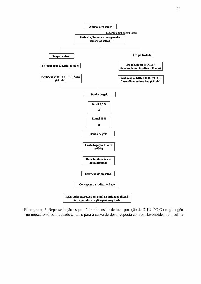
25
Fluxograma 5. Representação esquemática do ensaio de incorporação de D-[U-14C]G em glicogêniono músculo sóleo incubado in vitro para a curva de dose-resposta com os flavonóides ou insulina.
Animais em jejum
Retirada, limpeza e pesagem dosmúsculos sóleos
Grupo controle Grupo tratado
Pré-incubação c/ KRb (30 min) Pré-incubação c/ KRb +flavonóides ou insulina (30 min)
Incubação c/ KRb +D-[U-14C]G(60 min)
Incubação c/ KRb + D-[U-14C]G +flavonóides ou insulina (60 min)
Eutanásia por decapitação
Banho de gelo
KOH 0,5 N
Etanol 95%
Banho de gelo
Centrifugação 15 mina 664 g
Ressolubilização emágua destilada
Extração de amostra
Contagem da radioatividade
Resultados expressos em pmol de unidades glicosilincorporadas em glicogênio/mg tec/h

26
Fluxograma 6. Representação esquemática do ensaio de incorporação de D-[U-14C]G em glicogêniono músculo sóleo incubado com os flavonóides e com inibidores da PI-3K, GSK-3, MEK e PP1.
Banho de gelo
KOH 0,5 N
Etanol 95%
Banho de gelo
Centrifugação 15 min a 664 g
Ressolubilização em água destilada
Extração de amostra
Contagem da radioatividade
Animais em jejum
Retirada, limpeza e pesagem dos músculos sóleos
Eutanásia por decapitação
Grupo Controle Grupo Tratado Grupo Inibidor Grupo Inibidor + Tratado
Pré-incubação emKRb (30 min)
Pré-incubação emKRb + flavonóides
Pré-incubação emKRb + inibidor
Pré-incubação em KRb +flavonóides + inibidor
Incubação c/ D-[U-14C]G (60 min)
Incubação c/ D-[U-14C]G + flavonóides
Incubação c/ D-[U-14C]G + inibidor
Incubação c/ D-[U-14C]G+ flavonóides + inibidor
Resultados expressos em pmol de unidades glicosilincorporadas em glicogênio/mg tec/h
Banho de gelo
KOH 0,5 N
Etanol 95%
Banho de gelo
Centrifugação 15 min a 664 g
Ressolubilização em água destilada
Extração de amostra
Contagem da radioatividade
Animais em jejum
Retirada, limpeza e pesagem dos músculos sóleos
Eutanásia por decapitação
Grupo Controle Grupo Tratado Grupo Inibidor Grupo Inibidor + Tratado
Pré-incubação emKRb (30 min)
Pré-incubação emKRb + flavonóides
Pré-incubação emKRb + inibidor
Pré-incubação em KRb +flavonóides + inibidor
Incubação c/ D-[U-14C]G (60 min)
Incubação c/ D-[U-14C]G + flavonóides
Incubação c/ D-[U-14C]G + inibidor
Incubação c/ D-[U-14C]G+ flavonóides + inibidor
Resultados expressos em pmol de unidades glicosilincorporadas em glicogênio/mg tec/h

27
Fluxograma 7. Representação esquemática do ensaio de captação de [14C]-DG no músculo para acurva de dose-resposta com os flavonóides ou insulina.
Animais em jejum
Retirada, limpeza e pesagem dosmúsculos sóleos
Grupo controle Grupo tratado
Pré-incubação c/ KRb (30 min) Pré-incubação c/ KRb + flavonóidese/ou insulina (30 min)
Incubação c/ KRb +[14C]DG(60 min)
Incubação c/ KRb + [14C]DG +flavonóides e/ou insulina (60 min)
Eutanásia por decapitação
Processamento das amostras
Extração de amostra
Contagem da radioatividade
Resultados expressos pela relação tecido/meio

28
Fluxograma 8. Representação esquemática do ensaio de captação de [14C]-DG no músculo sóleoincubado com os flavonóides e com diferentes inibidores.
Animais em jejum
Retirada, limpeza e pesagem dos músculos sóleos
Eutanásia por decapitação
Grupo Controle Grupo Tratado Grupo Inibidor Grupo Inibidor + Tratado
Pré-incubação emKRb (30 min)
Pré-incubação emKRb + flavonóides
ou insulina
Pré-incubação emKRb + inibidor
Pré-incubação em KRb +flavonóides ou insulina +
inibidor
Incubação c/[14C]DG (60 min)
Incubação c/ [14C]DG +flavonóides ou insulina
Incubação c/[14C]DG + inibidor
Incubação c/ [14C]DG +flavonóides ou insulina +
inibidor
Processamento das amostras
Extração de amostra
Contagem da radioatividade
Resultados expressos pela relação tecido/meio
29
4 ARTIGOS
4.1 ARTIGO SUBMETIDO PARA PUBLICAÇÃOPeriódico – Journal of Agricultural and Food Chemistry
CAZAROLLI, L.H.; MACHADO, L.M.; FOLADOR, P.; MORESCO, H.H.; BRIGHENTE,I.M.C.; PIZZOLATTI, M.G.; SILVA, F.R.M.B. Potential antihyperglycemic role of crudeextract, fractions and flavonoids from Averrhoa carambola leaves in rats.
Nota: A formatação deste artigo não corresponde extamente à forma submetida ao periódicouma vez que as figuras foram inseridas no meio do texto a fim de facilitar a leitura e compreensão.
Potential antihyperglycemic role of crude extract, fractions and flavonoids from Averrhoacarambola leaves in rats
Luisa H. Cazarollia, Lisiane M. Machadoa, Poliane Foladora, Henrique H. Morescob, Inês M. C.Brighenteb, Moacir G. Pizzolattib, Fátima R. M. B. Silvaa,*
aDepartamento de Bioquímica - Centro de Ciências Biológicas and bDepartamento de Química -Centro de Ciências Físicas e Matemáticas, Universidade Federal de Santa Catarina, Campus
Universitário, TrindadeCx. Postal 5069, CEP: 88040-970 - Florianópolis, SC, Brazil.
*Correspondence: Silva, FRMB. Departamento de Bioquímica, Centro de Ciências Biológicas,Universidade Federal de Santa Catarina, Florianópolis, SC – Brasil, Tel/Fax: +55-
48.3721.69.12/+55-48.3721.96.72, E-mail: [email protected]; [email protected]

AbstractThe effect of the crude extract, ethyl acetate (EtOAc) and n-butanol (n-BuOH) fractions and
two flavonoids, apigenin-6-C--L-fucopyranoside (compound 1) and apigenin-6-C-(2”-O--L-rhamnopyranosyl)--L-fucopyranoside (compound 2) isolated from Averrhoa carambola L.(Oxalidaceae), were investigated on glycemia and on glycogen content in muscle and liver fromhyperglycemic rats. These effects were compared with insulin and/ or tolbutamide in hyperglycemicand diabetic rats. The oral administration of crude extract, ethyl acetate and n-butanol fractions ofAverrhoa carambola leaves exhibited a potential hypoglycemic activity in hyperglycemic normalrats. The maximum hypoglycemic effect detected was with the 400 mg/kg dose of the EtOAcfraction in a very rapid response, 15 min after treatment in hyperglycemic rats. Nevertheless, noalteration in the serum glucose level was detected in diabetic rats with these substances. Also, usingthis approach, compound 2 (20 mg/kg), a glycosylated flavonoid isolated from the EtOAc fraction,appeared to be a potential natural hypoglycemic agent. Although both compounds exhibited ahypoglycemic effect they appear to act on glucose management through different mechanisms sincethe compound 1 was less potent as a hypoglycemic substance but more potent in increasingglycogen content of soleus muscle as well as that of the liver.
Keywords: Averrhoa carambola; Flavonoids; Diabetes; Hypoglycemia; Antihyperglycemic

Introduction
Diabetes mellitus is a group of metabolic diseases characterized by hyperglycemia resultingfrom defects in insulin secretion and/or in insulin action (1). Although there has been broaddiscussion about the etiopathogenesis of diabetes, the vast majority of cases of diabetes fall intotype 1 (the cause of which is an absolute deficiency of insulin secretion) or, in a much moreprevalent category, type 2 (caused by combination of resistance to insulin action and an inadequatecompensatory insulin secretory response) (1). The number of people with diabetes is increasing andestimates of diabetes prevalence warn of a worldwide epidemic in the coming years. Along with theincreasing prevalence, diabetes care means high costs to the public health service as well as to theindividual (2).
Medicinal plants used in folk medicine and cited in the scientific literature for the treatment ofdiabetes may be an alternative to the classical drugs used such as insulin and oral hypoglycemicagents (3). Several studies have demonstrated that phytotherapies appear to be effective ashypoglycemic agents being used as adjuvants in the glucose management with no or very few sideeffects, as well as resulting in a low cost for the maintenance of the entire treatment (3, 4). Theflavonoids are widely distributed in plants and are the largest group of natural products known.They are important components in the human diet and are found in fruits, vegetables, seeds, nuts,grains, spices and beverages (wine, tea and beer). They have a broad range of biological activitiesand numerous studies have been carried out on their potential role in the treatment of diabetes (5, 6).Therefore, the investigation of such agents from medicinal plants has become more important.
One such plant is Averrhoa carambola L. (Oxalidaceae), also known as star fruit. This plant isnative from tropical and subtropical regions of Asia and it was successfully introduced to Brazil in1817. Averrhoa carambola (A. carambola) has been used as an appetite stimulant, a diuretic, anantiemetic, an antidiarrheal, an antifebrile and for the treatment of eczemas (7). Ripe carambolas areconsumed as fresh fruits, cooked or served in salads. Also, carambola juice is served as a coolingbeverage. The fruits are very popular consumed food items (8). Recently, the decoction of A.carambola leaves has been used in the treatment of diabetes (9). Additionally, the fruits of this plantare reported to be rich in insoluble fiber that presents hypoglycemic, antioxidant andhypocholesterolemic effects (10, 11, 12).
Other species of the Averrhoa genus, such as Averrhoa bilimbi Linn. are known for their anti-inflammatory, anti-scorbutic, astringent, anti-bacterial and antidiabetic properties (13). In addition,a leaf extract and semi-purified fractions of A. bilimbi exhibited hypoglycemic and hypolipidemiceffects when administered intraperitoneally (14) as well as orally in diabetic rats (15, 16, 17). Basedon this, the present study investigated the acute effect of crude extract, EtOAc and n-BuOHfractions as well as isolated compounds from A. carambola leaves on serum glucose levels. Also,the glycogen content in normal hyperglycemic rats was measured in soleus muscle and liver aftertreatment with glycosylated flavonoids from A. carambola.
Materials and methods
DrugsAlloxan monohydrate, tolbutamide and oyster glycogen type IIwere purchased from Sigma
Chemical Company® (St. Louis, MO, USA). Lispro insulin (100 IU/mL; batch FF6D46D;Humalog®—Lilly, São Paulo, SP, Brazil) was purchased from a commercial source. Glucose andall other solvents were purchased from Merck® AG (Darmstadt, Germany). The iodine reagents(CaCl2 + I2 + KI) were purchased from VETEC, Rio de Janeiro, Brazil. All reagents were ofanalytical grade.
Plant materialThe leaves of A. carambola were collected (March, 2003; co-ordinates -27.687799 latitude; -
48.777296 longitude) at Santo Amaro da Imperatriz, Santa Catarina, Brazil and identified by Prof.

Daniel de Barcellos Falkenberg. A voucher specimen was deposited at the herbarium of the BotanyDepartment at the Universidade Federal de Santa Catarina, Florianópolis, under number FLOR-24.144.
Extraction and isolationThe powdered, dried leaves (281 g) were extracted with ethanol-water (EtOH-H2O) (4:1). The
extract was concentrated to dryness by rotatory vaporization at 60oC under reduced pressure (41.3g; crude extract-dry leaves ratio = 14.7%). The concentrated extract was then suspended in EtOH-H2O (4:1) and successively extracted with n-hexane, ethyl acetate (EtOAc) and n-butanol (n-BuOH). The ethyl acetate soluble fraction (EtOAc) of 6.9 g was subjected to silica gel (100-200mesh) CC and eluted with an ethyl acetate/ethanol mixture gradient to afford 36 fractions. Fractions6-8 (150 mg) and 10-12 (200 mg) were purified by recrystallization from methanol (MeOH) to givepure (HPTLC in ethyl acetate/methanol/acetic acid 80:14:6 and NMR) compounds 1 and 2,respectively. These compounds were identified by nuclear magnetic resonance spectroscopy (NMR)analysis (1H, 13C, DEPT, COSY, HMQC and HMBC) and comparison with literature data (18).
Apigenin-6-C--L-fucopyranoside (compound 1): Yellow amorphous powder, (drug-extractratio = 0.36%). 1H NMR (400 MHz, CD3OD): aglycone moiety : 6.61 (s, H-3), 6.53 (s, H-8), 7.84(d, J=8,0 Hz, H-2’ & H-6’), 6.92 (d, J=8,0 Hz, H-3’ & H-6’); sugar moiety: 4.64 (d, J=8.4 Hz, H-1”), 4.03 (t, J=8.7 Hz, H-2’’), 3.97 (dd, J=3.0 and 6.5 Hz, H-3”), 3.50 (d, J=3.0 Hz, H-4”), 3.17 (m,H-5”), 1.44 (d, J=6.2 Hz, H-6’’). 13C NMR (400 MHz, CD3OD) aglycone moiety : 165.1 (C-2),99.6 (C-3), 183.1 (C-4), 161.7 (C-5), 108.0 (C-6), 163.9 (C-7), 94.0 (C-8), 161.8 (C-9), 102.8 (C-10), 121.9 (C-1’), 128.3 (C-2’ and 6’), 115.9 (C-3’ and 5’), 157.8 (C-4´); sugar moiety: 71.9 (C-1’’), 69.1 (C-2’’), 78.5 (C-3’’), 70.6 (C-4’’), 70.8 (C-5’’), 18.1 (C-6’’).
Apigenin-6-C-(2”-O--L-rhamnopyranosyl)--L-fucopyranoside (compound 2): Yellowamorphous powder (drug-extract ratio = 0.48%). 1H NMR (400 MHz, CD3OD): aglycone moiety :6.61 (s, H-3), 6.54 (s, H-8), 7.85 (d, J=7.6 Hz, H-2’ & H-6’), 6.94 (d, J=7,6 Hz, H-3’ & H-6’); sugarmoieties: 4.91 (d, J=9.6 Hz, H-1’’), 4.27 (t, J=8.8 Hz, H-2’’), 3.75 (m, H-3’’), 3.69 (sl, H-4’’), 3.84(m, H-5’’), 1.28, d, J=6.0 Hz, H-6’’), 5.18 (sl, H1’’’), 3.78 (sl, H-2”’), 3.28 (overlapped with signalsof CD3OD, H-3’’’), 3.09 (t, J=9.5 Hz, H-4’’’), 2.54 (t, J=8.4 Hz, H-5’’’), 0.71 (d, J=6.0 Hz, H-6’’’).13C NMR (400 Mhz, CD3OD) aglycone moiety : 165.1 (C-2), 104.3 (C-3), 183.0 (C-4), 161.6 (C-5), 108.8 (C-6), 163.0 (C-7), 95.0 (C-8), 159.6 (C-9), 102.8 (C-10), 122.0 (C-1’), 128.3 (C-2’ and6’), 115.9 (C-3’ and 5’), 157.7 (C-4´); sugar moieties: 72.2 (C-1’’), 75.1 (C-2’’), 76.5 (C-3’’), 72.8(C-4’’), 74.6 (C-5’’), 16.0 (C-6’’), 101.2 (C-1’’’) 71.1 (C-2’’’), 70.9 (C-3’’’), 72.4 (C-4’’’), 68.7(C-5’’’), 16.8 (C-6’’’) (Figure 1).
O
O
O
OH
HO
OH
Me
OH
HO
OH
O
O
O
OH
HO
OHOMe
HOHO
OMe
HOHO OH
12
Figure 1. Chemical structure of compounds apigenin-6-C--L-fucopyranoside (compound 1)and apigenin-6-C-(2”-O--L-rhamnopyranosyl)--L-fucopyranoside (compound 2) isolated fromthe EtOAc fraction of A. carambola leaves.
AnimalsMale Wistar rats (190–220 g) were used. They were bred in our animal facility and housed in
an air-conditioned room (approximately 22 ± 2 ºC and during the entire experiments) withcontrolled lighting on a 12:12 h light/dark cycle (lights on from 06:00 to 18:00 h). The animals were

maintained with pelleted food (Nuvital, Nuvilab CR1, Curitiba, PR, Brazil), while tap water wasavailable ad libitum. Fasted rats were deprived of food for at least 16 h but allowed free access towater. The animals received 50 mg/kg body weight of alloxan by a single intravenous injection. Thediabetic state was assessed by measuring body weight and serum glucose levels 3 days later and aserum glucose range of 400-500 mg/dL was used for the experiment. For all oral treatments, 0.5 mLof each respective substance was given by gavage (19). All the animals were carefully monitoredand maintained in accordance with ethical recommendations of the Brazilian Veterinary MedicineCouncil and the Brazilian College of Animal Experimentation. This study was approved by theCommittee for Ethics in Animal Research of UFSC (Protocol CEUA PP0007).
Study of insulin and tolbutamide effects on the serum glucose level in the oral glucosetolerance curve
Fasted rats were divided into four groups of six animals for each treatment: Group I, normalrats that received vehicle 1% EtOH-H2O; Group II, hyperglycemic rats that received glucose (4 g/kgb.w.) plus vehicle; Group III, hyperglycemic rats that received glucose (4 g/kg) plus lispro insulin(0.05 IU) by i.p. route; Group IV, hyperglycemic rats that received glucose (4 g/kg) plustolbutamide (100 mg/kg b.w.) by oral gavage (Table 1). Blood samples were collected just prior toand at 15, 30, 60, 120 and 180 min after the glucose loading and the serum glucose levels weremeasured.
Study of the effects of crude extract, fractions or isolated compounds from A. carambola onthe serum glucose level in the oral glucose tolerance curve
Fasted rats were divided into three groups of six animals for each treatment: Group I, normalrats that received vehicle 1% EtOH-H2O; Group II, hyperglycemic rats that received glucose (4g/kg) plus vehicle; Group III, rats that received glucose (4 g/kg) plus crude extract (200, 400 and800 mg/kg b.w.) (Table 1 – Group V). As shown in Figure 2, the hyperglycemic rats receivedEtOAc or n-BuOH (400 and 800 mg/kg b.w.) fractions. Also, hyperglycemic rats received isolatedcompounds 1 or 2 (20 and 50 mg/kg b.w.) by oral gavage (Figure 3). Blood samples were collectedjust prior to and at 15, 30, 60, 120 and 180 min after the glucose loading and the serum glucoselevels were measured.
Study of the effects of the crude extract and fraction of A. carambola and tolbutamide onserum glucose levels in alloxan-induced diabetic rats
Animals in which the development of hyperglycemia was confirmed (around 90%), 72 h afterthe alloxan injection, were randomly allocated to different groups of six rats for each treatment:Group I, diabetic rats that received vehicle;Group II, diabetic rats that received the crude extract(400 mg/kg b.w.); Group III, diabetic rats that received EtOAc fraction (400 mg/kg b.w.); GroupIV, diabetic rats that received tolbutamide (100 mg/kg b.w.). Serum glucose levels were measuredat zero time (before receiving the extract, EtOAc fraction or tolbutamide) and at 1, 2 and 3 hfollowing the treatment (Table 2).
Determination of the serum glucose concentrationBlood samples (100 µL) were collected from the tail vein of the anesthetized rat, centrifuged
and the serum was used to determine the glycemia (GBC 916 UV – visible spectrophotometer) bythe glucose oxidase method (20). The commercial kit used to determine the glycemia was fromGold Analisa (Belo Horizonte, MG, Brazil).
Glycogen content measurementsSoleus muscles and livers were harvested from normal fed or fasted rats and hyperglycemic
rats treated with compounds 1 or 2 (50 mg/kg) or regular insulin (0.5 IU) and used for the assay ofglycogen content immediately after 3 h of treatment (Figures 4A and 4B). Glycogen was isolated

from these tissues as described by Krisman (21) with minor modifications. The tissues wereweighed, homogenized in 33% KOH and boiled at 100 ºC for 20 min, with occasional stirring. Aftercooling, 96% ethanol was added to the samples and heated to boiling followed by cooling in an icebath to aid the precipitation of glycogen. The homogenates were centrifuged at 664 g for 15 min,the supernatant was discarded and the pellets were neutralized with saturated NH4Cl before beingheated to 100 ºC for 5 min, washed and resolubilized in water. Glycogen content was determined bytreatment with iodine reagent and the absorbance was measured at 460 nm. The results wereexpressed as mg of glycogen/ g of tissue.
Data and statistical analysisData were expressed as mean ± S.E.M. One-way analysis of variance (ANOVA) followed by
Bonferroni post-test or unpaired Student’s t-test was used to identify significantly different groups.Differences were considered to be significant at p ≤ 0.05. The software InStat version 3.05; Graph-Pad Software Inc., San Diego, CA was used for statistical analysis.
Results
Effect of insulin and tolbutamide on the oral glucose tolerance curveAs expected, after starting the glucose tolerance test in normal rats overloaded with glucose,
the serum glucose concentration was significantly increased when compared with the zero time ofthis group. Also, the classical hypoglycemic effect of insulin was observed over the period studiedwhen compared to the hyperglycemic group. The oral hypoglycemic agent, tolbutamide (100mg/kg), produced a typical serum glucose lowering at 30 and 60 min compared to thehyperglycemic group. The normal vehicle control group (1% EtOH-H2O) showed an unchangedprofile of glycemia over the time studied (Table 1). On the other hand, tolbutamide (100 mg/kg)failed to modify the glycemia in diabetic rats, reinforcing the diabetic status of the rats (Table 2).
Effect of crude extract, EtOAc, n-BuOH fractions or apigenin-6-C--L-fucopyranoside andapigenin-6-C-(2”-O--L-rhamnopyranosyl)--L-fucopyranoside compounds from A. carambola onthe oral glucose tolerance curve
Crude extract at 200 and 400 mg/kg was effective in reducing glycemia at 30 min and/ or 60min after oral treatment when compared with the respective hyperglycemic control group. However,oral treatment with 400 mg/kg of the EtOAc fraction in hyperglycemic rats produced a significantantihyperglycemic effect from 15 to 180 min (Figure 2A). In addition, the glycemia was maintainedat basal levels from 60 to 180 min (Figure 2A). On the other hand, the 800 mg/kg dose showed afast antihyperglycemic effect only in the first two periods studied (15 and 30 minutes) (Figure 2B).Figures 2C and 2D show the effect of the n-BuOH fraction of A. carambola at 400 and 800 mg/kgin hyperglycemic animals. Although both doses showed a slightly antihyperglycemic effect (at 60or 30 min and 60 min, respectively) neither 400 nor 800 mg/kg doses were as powerful as theEtOAc fraction (400 mg/kg), previously demonstrated in Figure 2A. Nevertheless, treatment withthe crude extract and EtOAc fraction of A. carambola (400 mg/kg) did not reduce the glycemiclevels in diabetic animals over the period studied (Table 2).

Figure 2. Effect of EtOAc and n-BuOH fractions from A. carambola on the oral glucosetolerance curve. (A and B) EtOAc 400 and 800 mg/kg, respectively; (C and D) n-BuOH 400 and800 mg/kg, respectively, fractions by gavage. Values are expressed as mean S.E.M; n= 6 induplicate for each treatment. Statistically significant difference to the corresponding hyperglycemicgroup; *P ≤ 0.05; **P ≤ 0.001.
The oral administration of compound 1 reduced serum glucose levels in hyperglycemic ratsand the maximum reduction observed was 19% at 15 min with the higher dose (Figure 3A and 3B).The effect of compound 2 on the glucose tolerance curve was similar and is shown in Figures 3Cand 3D. Oral administration with 20 and 50 mg/kg of this compound significantly reduced theglycemia by around 32%, 24% and 16% at 15, 30 and 60 min after treatment with the higher doseused. At 120 and 180 min, glycemic levels were similar to respective results for the hyperglycemiccontrol groups.
0 15 30 60 120 1800
50
100
150
200
250 Hyperglycemic
Hyper + EtOAc 400 mg/kg
**
**
**
**
**
Normal
Time (min)
Seru
m g
luco
se le
vels
(m
g/dL
)
0 15 30 60 120 1800
50
100
150
200
250Hyperglycemic
Hyper + EtOAc 800 mg/kg
**
Normal
Time (min)
Seru
m g
luco
se le
vels
(m
g/dL
)
A B
0 15 30 60 120 1800
50
100
150
200
250 Hyperglycemic
Hyper + EtOAc 400 mg/kg
**
**
**
**
**
Normal
Time (min)
Seru
m g
luco
se le
vels
(m
g/dL
)
0 15 30 60 120 1800
50
100
150
200
250Hyperglycemic
Hyper + EtOAc 800 mg/kg
**
Normal
Time (min)
Seru
m g
luco
se le
vels
(m
g/dL
)
A B
0 15 30 60 120 1800
50
100
150
200
250Hyperglycemic
Hyper + n-BuOH 400 mg/kg
*
Normal
Time (min)
Seru
m g
luco
se le
vels
(m
g/dL
)
0 15 30 60 120 1800
50
100
150
200
250Hyperglycemic
Hyper + n-BuOH 800 mg/kg
**
Normal
Time (min)
Seru
m g
luco
se le
vels
(mg/
dL)
C D
0 15 30 60 120 1800
50
100
150
200
250Hyperglycemic
Hyper + n-BuOH 400 mg/kg
*
Normal
Time (min)
Seru
m g
luco
se le
vels
(m
g/dL
)
0 15 30 60 120 1800
50
100
150
200
250Hyperglycemic
Hyper + n-BuOH 800 mg/kg
**
Normal
Time (min)
Seru
m g
luco
se le
vels
(mg/
dL)
C D

Figure 3 Effect of compound 1 and compound 2 isolated compounds from A. carambola onthe oral glucose tolerance curve. Values are expressed as mean S.E.M; n= 6 in duplicate for eachtreatment. Statistically significant difference to the corresponding hyperglycemic group; *P ≤ 0.05;**P ≤ 0.001.
Effect of apigenin-6-C--L-fucopyranoside and apigenin-6-C-(2”-O--L-rhamnopyranosyl)--L-fucopyranoside from A. carambola on the glycogen content of soleus muscle and liver
Figures 4A and 4B show that the glycogen content was significantly increased, 3 h after theadministration of glucose (4 g/kg) by oral gavage, in soleus muscle compared with fed and fastednormal rats and in liver when compared with fasted normal rats. In addition, the well known insulinstimulatory effect on glycogen storage in both tissues was observed 3 h after insulin treatment (9,3times for muscle and 19,6 times for liver) in hyperglycemic normal rats compared with fastednormal rats.
Compound 1 was able to significantly increase the glycogen content in soleus muscle whencompared with fed normal (4,6 times), fasted normal (10,7 times), hyperglycemic (1,2 times) andhyperglycemic plus insulin (1,14 times) animals 3 h after treatment. Moreover, compound 1stimulated glycogen content in the liver as well. This change was 25 times when compared with thefasted normal group at 3 h after treatment and it represented 54% of the insulin stimulatory effect.
The stimulatory effect of compound 2 on muscle glycogen content (around 1,7 and 3,9 times)was observed 3 h after treatment when compared with fed and fasted groups, respectively.However, compound 2 did not alter the glycogen content as compared with hyperglycemic group.In liver, the effect of compound 2 was around 10-fold when compared with fasted normal rats andwhen compared with insulin, representing 21% of the total stimulatory effect of the hormone.
0 15 30 60 120 1800
50
100
150
200
250Hyperglycemic
Hyper + compound2 20 mg/kg
****
**
Normal
Time (min)
Seru
m g
luco
se le
vels
(mg/
dL)
0 15 30 60 120 1800
50
100
150
200
250Hyperglycemic
Hyper + compound2 50 mg/kg
***
*
Normal
Time (min)
Seru
m g
luco
se le
vels
(mg/
dL)
C D
0 15 30 60 120 1800
50
100
150
200
250Hyperglycemic
Hyper + compound2 20 mg/kg
****
**
Normal
Time (min)
Seru
m g
luco
se le
vels
(mg/
dL)
0 15 30 60 120 1800
50
100
150
200
250Hyperglycemic
Hyper + compound2 50 mg/kg
***
*
Normal
Time (min)
Seru
m g
luco
se le
vels
(mg/
dL)
C D
0 15 30 60 120 1800
50
100
150
200
250Hyperglycemic
Hyper + compound 1 20 mg/kg
** *
Normal
Time (min)
Seru
m g
luco
se le
vels
(mg/
dL)
0 15 30 60 120 1800
50
100
150
200
250Hyperglycemic
Hyper + compound 1 50 mg/kg
**
*
Normal
Time (min)
Seru
m g
luco
se le
vels
(mg/
dL)
A B

Figure 4 Effect of compound 1 and compound 2 on the glycogen content in normalhyperglycemic rats. (A) soleus muscle and (B) liver 3h after treatment by oral gavage. Values areexpressed as mean S.E.M; n=6 in duplicate for each group. Significantly different to thecorresponding fasted normal group; *P ≤ 0.05; **P ≤ 0.001; ***P ≤ 0.0001.
Discussion
Many plant species are known in the folk medicine of different cultures to be used for thetreatment of diabetes mellitus (3, 4). Moreover, phytochemical and pharmacological studies thatwere performed in order to characterize such compounds have shown the presence of flavonoids asthe active constituents in several plant extracts (4, 20, 22, 23).
0 .00
0 .25
0 .50
0 .75
1 .00
Fed normal
Fasted normal
H yperglycemic
H yper + insulin 0.5 IU
H yper + compound 1 50 mg/kg
H yper + compound 2 50 mg/kg
***
***
**
*
Sole
um m
uscl
e gl
ycog
en c
onte
nt(m
g/ g
tiss
ue)
A
0
10
20
30
40
Fed normalFasted normal
H yperglycemic
H yper + insulin 0.5 IU
H yper + compound 1 50 mg/kg
H yper + compound 2 50 mg/kg
***
***
***
***
Liv
er g
lyco
gen
cont
ent
(mg/
g ti
ssue
)
B

Flavonoids are a large group of phenolic plant constituents and their bioactive potential in thetreatment and prevention of diabetes and other diseases has been demonstrated (5, 6). They canaffect glucose transport and metabolism in peripheral tissues as well as inducing insulin releasefrom -cells in the pancreas (20, 24, 25, 26, 27).
This study showed the antihyperglycemic effect of the crude extract, fractions and isolatedcompounds of A. carambola leaves in normal hyperglycemic rats following an acute treatment.Similar results showing the glucose lowering effects of the crude extract of A. carambola leaves inhyperglycemic normal rats were reported by Provasi et al (9). Also, it has been reported in theliterature that the extracts of another Averrhoa species, Averrhoa bilimbi, showedantihyperglycemic effects in normal hyperglycemic and diabetic rats when compared with therespective control groups. Furthermore, chronic daily treatment with A. bilimbi extracts decreasedglycemia in diabetic rats compared to both the control group and a metformin group (15). Takingthis into account, the effect of the crude extract points to a mechanism involving the regulation ofinsulin release and/or the inhibition of intestinal glucose absorption (5, 6, 28).
The EtOAc and n-BuOH fractions were isolated from the crude extract and their potentialantihyperglycemic effects were studied in normal glucose-fed rats. The antihyperglycemic effect ofthe EtOAc fraction was more marked than that of n-BuOH at 400 mg/kg and was around 26.4% at60 min after treatment (Figures 2A and 2C). This effect was as pronounced as that caused by bothtolbutamide (18.26%) (Table 1), a sulfonylurea agent that increases insulin secretion from thepancreas (29) and the rapid effect of lispro insulin (29.2%) during the same time-course studied(Table 1). Additionally, the absence of effect of the crude extract and EtOAc fraction in alloxan-induced diabetic rats is in accordance with the lack of tolbutamide response (used to the therapy fortype 2 diabetes), observed in the same experimental model. Consequently, the effect of crudeextract as well as the EtOAc fraction appears to be mediated through mechanisms involving insulinsecretion from -cells (28, 30). In line with these results, previous studies of Pushparaj et al (16)and Tan et al (17) showed the effect of aqueous and n-BuOH fractions from A. bilimbi leaves onglucose serum levels in diabetic and hyperglycemic normal rats. Both fractions improved theglucose tolerance curve in diabetic rats and hyperglycemic rats. Furthermore, after two weeks oftreatment the fractions increased plasma insulin levels when compared with the zero time and withthe diabetic control group.
Considering the EtOAc fraction’s antihyperglycemic effect, two glycosylated flavonoids,compound 1 and compound 2 (C-flavones) were isolated from this fraction from A. carambolaleaves (31). The oral administration of compounds 1 and 2 at 20 and 50 mg/kg resulted insignificant antihyperglycemic effects in glucose-fed normal rats. Recently, Hsu et al (24)demonstrated the antihyperglycemic effect of puerarin, an isoflavone, in normal rats, hyperglycemicnormal rats and diabetic rats. Puerarin reduced glycemia in normal and diabetic rats in a dosedependent manner and it was also able to attenuate the increase of plasma glucose induced by anintravenous glucose challenge in normal rats.
We have demonstrated the antihyperglycemic effect of the EtOH fraction, rich in glycosylatedflavonoids, from Bauhinia forficata leaves as well as the major flavonoid of that fraction,kaempferitrin (19, 23). The antihyperglycemic activity of plant extracts rich in flavones inhyperglycemic and diabetic rats has been described (22, 32). The EtOAc fraction from Gentianaolivieri and isoorientin, a C-glycosylflavone, and the active compound isolated from this fraction,significantly reduced serum glucose levels in hyperglycemic and diabetic rats. However, the highantioxidant activity of isoorientin is suggested as a possible mechanism of action, protecting -cellsfrom oxidative damage and restoring plasma insulin levels (32).
The results observed for compounds 1 and 2 suggest that some pancreatic functions such asinsulin secretion or the presence of insulin are required for the antihyperglycemic activity of suchcompounds since the EtOAc fraction from which compounds 1 and 2 originated had an effect inhyperglycemic normal rats but not in diabetic rats.

In mammals, carbohydrate is stored mainly in the form of glycogen, with skeletal muscle andliver as the major storage sites. Glycogen metabolism is regulated by insulin/glucagon throughactivation and/or inhibition of several enzymes and proteins (33, 34). The determination ofglycogen levels in muscle and liver of hyperglycemic normal rats after acute treatments withcompounds 1 and 2 revealed a significant increase in glycogen content when compared with fastednormal rats (Figures 4A and 4B). Additionally, the known effect of insulin on muscle and hepaticglycogen storage was observed. The study demonstrated that the stimulatory effect of compound 1on glycogen storage in soleus muscle as well as in liver was greater than that of compound 2 at thesame dosage (50 mg/kg). Such an effect of compound 1 was quite similar to that of insulin inmuscle. The effect of this compound represented 54% of the total stimulatory effect of insulin inliver 3 h after treatment.
It is well known that glycogen deposition from glucose is regulated by insulin in two steps:firstly by controlling the uptake and transport of glucose, and secondly by regulating thephosphorylation/ dephosphorylation status of enzymes involved in the metabolism of glycogen.Flavonoids and plant extracts with proven antihyperglycemic activity have been shown to influenceglycogen deposition in different tissues as well as to interact with key enzymes of the glycolyticroute in rats (25, 26, 35).
The results found for compound 2 and specially for compound 1 (Figures 3 and 4) are closelycorrelated with the known insulin activity on the stimulatory effect of glucose disposal and loweringserum glucose levels in normal hyperglycemic rats (Figure 4 and Table 1). The increase inglycogen content in both liver and muscle may be due to stimulation of insulin release from -cells(27, 28, 30, 36). The insulinomimetic activity of compounds 1 and/or 2 could be a consequence ofdirect peripheral glucose uptake, glycogen synthesis or a combination of both. However, studies areunderway to elucidate the mechanism involved in the antihyperglycemic effect of these flavonoidsin muscle and liver.
We conclude that the crude extract, ethyl acetate and n-butanol fractions of A. carambolaleaves were effective in decreasing the serum glucose levels in hyperglycemic normal rats. Themaximum antihyperglycemic effect detected was at a dose of 400 mg/kg with a very rapid response,15 min after treatment. Also, using this approach, compound 2 (20 mg/kg), a glycosylated flavonoidisolated from the A. carambola EtOAc fraction, appears to be a potential natural antihyperglycemicagent. Although both compounds exhibited an antihyperglycemic effect they appear to act on theblood glucose management by different mechanisms, since compound 1 was less potent as anantihyperglycemic substance but more potent in increasing glycogen content in the soleus muscle aswell as in the liver.
Considering these findings, A. carambola, popularly known as an antihyperglycemic plant,contains compound(s) able to manage glucose utilization, probably through different pathways.Among these compounds, 1 and 2 may be attractive adjuvants for the treatment of diabetic patientsin the future.
AcknowledgementsThis work was supported by grants from CNPq, FAPESC and CAPES. Luisa Helena Cazarolli
and Poliane Folador are registered on the PGFAR-UFSC. The authors express their appreciation toDr. Gareth Paul Cuttle for assistance with the English correction of the manuscript.
Literature cited
(1) American Diabetes Association. Diagnosis and Classification of Diabetes Mellitus.Diabetes Care 2006, 29 (Suppl. 1), 543-548.
(2) Wild, S.; Roglic, G.; Green, A.; Sicree, R.; King, H. Global Prevalence of Diabetes -Estimates for the year 2000 and projections for 2030. Diabetes Care 2004, 27, 1047-1053.

(3) Zareba, G.; Serradell, N.; Castañer, R.; Davies, S.L.; Prous, J.; Mealy, N. Phytotherapiesfor diabetes. Drugs Future 2005, 30, 1253-1282.
(4) Mukherjee, P.K.; Maiti, K.; Mukherjee, K.; Houghton, P.J. Leads from Indian medicinalplants with hypoglycemic potentials. J. Ethnopharmacol. 2006, 106, 1-28.
(5) Cazarolli, L.H.; Zanatta, L.; Alberton, E.H.; Figueiredo, M.S.R.B.; Folador, P.; Damazio,R.G.; Pizzolatti, M.G.; Silva, F.R.M.B. Flavonoids: Prospective drug candidates. Mini Rev. Med.Chem. 2008a, 8, 1429-1440.
(6) Cazarolli, L.H.; Zanatta, L.; Alberton, E.H.; Figueiredo, M.S.R.B.; Folador, P.; Damazio,R.G.; Pizzolatti, M.G.; Silva, F.R.M.B. Flavonoids: Cellular and molecular mechanism of action inglucose homeostasis, Mini Rev. Med. Chem. 2008b, 8, 1032-1038.
(7) Corrêa, M.P. Dicionário das plantas úteis do Brasil. Instituto Brasileiro deDesenvolvimento Florestal: Rio de Janeiro, 1984; pp. 03.
(8) Morton, J. Carambola. In: Fruits of warm climates. Julia F. Morton: Miami, FL, 1987; p.125–128.
(9) Provasi M.; Oliveira, C.E.; Martino, M.C.; Pessini, L.G.; Bazotte, R.B.; Cortez, D.A.G.Avaliação da toxicidade e do potencial antihiperglicemiante da Averrhoa carambola L.(Oxalidaceae). Acta Sci – Health Sci. 2001, 23, 665-669.
(10) Chau, C.F.; Chen, C.H.; Lin, C.Y. Insoluble fiber-rich fractions derived from Averrhoacarambola: hypoglycemic effect determined by in vitro methods. Lebensm.-Wiss. u.-Technol.2004a, 37, 331–335.
(11) Chau, C.F.; Chen, C.H.; Wang, Y.T. Effects of a novel pomace fiber on lipid andcholesterol metabolism in the hamster. Nutr. Res. 2004b, 24, 337–345.
(12) Shui, G.; Leong, L.P. Residue from star fruit as valuable source for functional foodingredients and antioxidant nutraceuticals. Food Chem. 2006, 97, 277–284.
(13) Goh, S.H.; Chuah, C.H.; Mok, J.S.L.; Soepadmo, E. Malaysian medicinal plants for thetreatment of cardiovascular diseases. Pelanduk Publications: Malaysia, 1995; pp.62–63.
(14) Tan, B.K.H.; Fu, P.; Chow, P.W.; Hsu, A. Effects of A. bilimbi on blood sugar and foodintake in streptozotocin induced diabetic rats. Phytomedicine 1996, 03, 271-272.
(15) Pushparaj, P.N.; Tan, C.H.; Tan, B.K.H. Effects of Averrhoa bilimbi leaf extract onblood glucose and lipids in streptozotocin-diabetic rats. J. Ethnopharmacol. 2000, 72, 69-76.
(16) Pushparaj, P.N.; Tan, B.K.H.; Tan, C.H. The mechanism of hypoglycemic action of thesemi-purified fractions of Averrhoa bilimbi in streptozotocin-diabetic rats. Life Sci. 2001, 70, 535-547.
(17) Tan, B.K.H.; Tan, C.H.; Pushparaj, P.N. Anti–diabetic activity of the semi–purifiedfractions of Averrhoa bilimbi in high fat diet fed–streptozotocin–induced diabetic rats. Life Sci.2005, 76, 2827–2839.
(18) Suzuki, R.; Okada, Y.; Okuyama, T. A new flavone C-glycoside from the style of Zeamays L. with glycation inhibitory activity. Chem. Pharm. Bull. 2003, 51, 1186-1188.
(19) Silva, F.R.M.B.; Szpoganicz, B.; Pizzolatti, M.G.; Willrich, M.A.V.; De Sousa, E. Acuteeffect of Bauhinia forficata on serum glucose levels in normal and alloxan-induced diabetic rats. J.Ethnopharmacol. 2002, 83, 33-37.
(20) De Sousa, E.; Zanatta, L.; Seifriz, I.; Creczynski-Pasa, T.B.; Pizzolatti, M.G.;Szpoganicz, B.; Silva, F.R.M.B. Hypoglycemic effect and antioxidant potential of kaempferol-3,7-O-()-dirhamnoside from Bauhinia forficata leaves. J. Nat. Prod. 2004, 67, 829-832.
(21) Krisman, C.R. A method for the colorimetric estimation of glycogen with iodine. Anal.Biochem. 1962, 4, 17-23.
(22) Narváez-Mastache, J.M.; Garduño-Ramírez, M.L.; Alvarez, L.; Delgado, G.Antihyperglycemic activity and chemical constituents of Eysenhardtia platycarpa. J. Nat. Prod.2006, 69, 1687-1691.
(23) Cazarolli, L.H.; Zanatta, L.; Jorge, A.P.; De Sousa, E.; Horst, H.; Woehl, V.M.;Pizzolatti, M.G.; Szpoganicz, B.; Silva, F.R.M.B. Follow-up studies on glycosylated flavonoids and

their complexes with vanadium: Their anti-hyperglycemic potential role in diabetes. Chem.-Biol.Interact. 2006, 163, 177–191.
(24) Hsu, F.L.; Liu, I.M.; Kuo, D.H.; Chen, W.C.; Su, H.C.; Cheng, J.T. Antihyperglycemiceffect of puerarin in streptozotocin-induced diabetic rats. J. Nat. Prod. 2003, 66, 788-792.
(25) Jorge, A.P.; Horst, H.; De Sousa, E.; Pizzolatti, M.G.; Silva, F.R.M.B. Insulinomimeticeffects of kaempferitrin on glycaemia and on 14Cglucose uptake in rat soleus muscle. Chem.-Biol.Interact. 2004, 149, 89–96.
(26) Park, S.A.; Choi, M.S.; Cho, S.Y.; Seo, J.S.; Jung, U.J.; Kim, M.J.; Sung, M.K.; Park,Y.B.; Lee, M.K. Genistein and daidzein modulate hepatic glucose and lipid regulating enzymeactivities in C57BL/KsJ-db/db mice. Life Sci. 2006, 79, 1207–1213.
(27) Liu, D.; Zhen, W.; Yang, Z.; Carter, J.D.; Si, H.; Reynolds, K.A. Genistein acutelystimulates insulin secretion in pancreatic -cells through a cAMP-dependent protein kinasepathway. Diabetes 2006, 55, 1043–1050.
(28) Rorsman, P. The pancreatic beta-cell as a fuel sensor: an electrophysiologist’s viewpoint.Diabetologia 1997, 40, 487–495.
(29) Inzucchi, S.E. Oral Antihyperglycemic Therapy for Type 2 Diabetes. JAMA – J. Am.Med. Assoc. 2002, 287, 360-372.
(30) Detimary, P.; Jonas, J.C.; Henquin, J.C. Possible links between glucose-induced changesin the energy state of pancreatic cells and insulin release - Unmasking by decreasing a stable poolof adenine nucleotides in mouse islets. J. Clin. Investig. 1995, 96, 1738-1745.
(31) Araho, D.; Masaazumi, M.; Wen-hua, C.; Toshimitsu, K.; Kenji, M.; Takao, I. A newflavone c-glycoside from the leaves of Averrhoa carambola. Nat. Med. (Tokyo, Japan) 2005, 59,113-116.
(32) Sezik, E.; Aslan, M.; Yesilada, E.; Ito, S. Hypoglycemic activity of Gentiana olivieri andisolation of the active constituent through bioassay-directed fractionation techniques. Life Sci. 2005,76, 1223-1238.
(33) Srivastava, A.K.; Pandey, S.K. Potential mechanism(s) involved in the regulation ofglycogen synthesis by insulin. Mol. Cell. Biochem. 1998, 182, 135-141.
(34) Ferrer, J.C.; Favre, C.; Gomis, R.R.; Fernández-Novell, J.M.; Garcia-Rocha, M.; De laIglesia, N.; Cid, E.; Guinovart, J.J. Control of glycogen deposition. FEBS Lett. 2003, 546, 127-132.
(35) Harmon, A.W.; Patel, Y.M. Naringenin inhibits phosphoinositide 3-kiinase activity andglucose uptake in 3T3-L1 adipocytes. Biochem. Biophys. Res. Communm. 2003, 305, 229–234.
(36) Jayaprakasam, B.; Vareed, S.K.; Olson, L.K.; Nair, M.G. Insulin secretion by bioactiveanthocyanins and anthocyanidins present in fruits. J. Agric. Food Chem. 2005, 53, 28-31.

Table 1 Acute effect of lispro insulin, tolbutamide and crude extract of A. carambola on the serumglucose level in the oral glucose tolerance curvea.
Serum Glucose Level (mg/dL)Group V glucose fedhyperglycemic plus
A. carambola crude extractTime(min)
Group Ivehicle
(1% EtOH-H2O)
Group IIHyperglyce
micGlucose (4g/kg) plus
vehicle
Group IIIhyperglycemic plus lispro
insulin(0.05 IU)
Group IVhyperglycem
ic plustolbutamide(100 mg/kg)
200mg/kg
400mg/kg
800mg/kg
0 111.8 ± 2.8 122.6 ± 5.8 113.1 ± 2.6 108.4 ± 5.1110.1 ±
3.3111.8 ±
5.7125.4± 7.8
15 114.7 ± 6.8196.2 ±3.6***
147.2 ±2.1***#
168.6 ±5.6***
178.4 ±8.7***
174.7 ±7.3***
172.0±2.8***
30 125.5 ± 3.8201.7 ±1.9***
151.7 ±4.3***#
172.0 ±10.4***#
184.4 ±7.6***
167.8 ±7.3***#
182.2±
5.8***
60 128.3 ± 3.8178.4 ±9.1***
126.2 ± 4.2# 145.8 ± 6.8#147.5 ±5.6**#
153.2 ±5.9***#
165.9±
8.9**
120 120.1 ± 6.9 137.8 ± 7.7 126.0 ± 2.7# 131.8 ± 9.5138.4 ±
6.0139.8 ±5.2
151.8± 6.7
180 120.7 ± 2.0 134.3 ± 4.4 123.3 ± 3.6 131.8 ± 6.9144.1 ±
4.2*149.5 ±4.1**
140.4± 6.0
a Values expressed as mean ± S.E.M.; n = 6 in duplicate for each treatment. Statistically significantdifference to the corresponding zero time value; *P ≤ 0.05; **P ≤ 0.001; ***P ≤ 0.0001.Significantly different to the corresponding hyperglycemic group; # P ≤ 0.05.
Table 2 Acute effect of crude extract and EtOAc fraction of A. carambola and tolbutamide on theserum glucose level in alloxan-induced diabetic ratsa.
Serum Glucose Level (mg/dL)Time(h)
Group IDiabetic control
(vehicle 1%EtOH-H2O)
Group IIDiabetic plus
Crude extract (400mg/kg)
Group IIIDiabetic plus
EtOAc (400 mg/kg)
Group IVDiabetic plus
Tolbutamide (100mg/kg)
0 401,6 17,3 443.0 ± 13.6 446.9 ± 9.6 455.0 ± 3.91 438,4 11,8 486.2 ± 13.2 485.5 ± 13.8 455.8 ± 22.72 441,4 11,9 464.5 ± 14.7 486.2 ± 13.1 432.5 ± 14.83 429,6 14,2 482.5 ± 11.2 477.7 ± 15.8 518.2 ± 1.3
a Values expressed as mean ± S.E.M.; n = 6 in duplicate for each treatment.

44
4.2 ARTIGO ACEITO PARA PUBLICAÇÃOPeriódico – European Journal of Medicinal Chemistry
CAZAROLLI, L.H.; FOLADOR, P.; MORESCO, H.H.; BRIGHENTE, I.M.C.;PIZZOLATTI, M.G.; SILVA, F.R.M.B. Stimulatory Effect of Apigenin-6-C--L-fucopyranosideon Insulin Secretion and Glycogen Synthesis.
Nota: A formatação deste artigo não corresponde extamente à forma submetida ao periódicouma vez que as figuras foram inseridas no meio do texto a fim de facilitar a leitura e compreensão.

Stimulatory Effect of Apigenin-6-C-β-L-fucopyranoside on Insulin Secretion and GlycogenSynthesis
Luisa Helena Cazarollia, Poliane Foladora, Henrique Hunger Morescob, Inês Maria CostaBrighenteb, Moacir Geraldo Pizzolattib, Fátima Regina Mena Barreto Silvaa,*
aDepartamento de Bioquímica, Centro de Ciências Biológicas and bDepartamento de Química,Centro de Ciências Físicas e Matemáticas, Universidade Federal de Santa Catarina, Campus
Universitário, Bairro TrindadeCx. Postal 5069, CEP: 88040-970 - Florianópolis, SC, Brazil
* Correspondence to: Prof. Dr. Fátima Regina Mena Barreto Silva, Departamento de Bioquímica,Centro de Ciências Biológicas, UFSC. Campus Universitário, Bairro Trindade, Cx Postal 5069,
CEP: 88040-970 Florianópolis, SC, Brazil. e-mail: [email protected], Tel.: +55-48.3721.69.12;Fax.: +55-48.3721.96.72.

Abstract
In vivo and in vitro treatments were carried out to investigate the effects of apigenin-6-C-β-L-fucopyranoside (1), isolated from Averrhoa carambola L. (Oxalidaceae), on serum glucose andinsulin levels in hyperglycemic rats as well as its effect on glycogen synthesis in normal rat soleusmuscle. Apigenin-6-C-β-L-fucopyranoside showed an acute effect on blood glucose lowering inhyperglycemic rats and stimulated glucose-induced insulin secretion. A stimulatory effect of 1 onglycogen synthesis was observed when muscles were incubated with this flavonoid and also itseffect was completely nullified by pre-treatment with insulin signal transduction inhibitors. Takingthis into account, the MAPK - PP1 and PI3K – GSK3 pathways are involved in the apigenin-6-C-β-L-fucopyranoside-induced increase in glycogen synthesis in muscle. This study provides evidencefor dual effects of apigenin-6-C-β-L-fucopyranoside as an antihyperglycemic (insulin secretion) aswell as an insulinomimetic (glycogen synthesis) agent.
Keywords: Flavonoid; Insulin; Diabetes; Glycogen synthesis.

1. Introduction
Diabetes mellitus is a complex metabolic disorder in the endocrine system characterized byabnormalities in insulin secretion and/or insulin action that lead to progressive failure of glucosetolerance and cause chronic hyperglycemia [1]. In recent years there has been a growing interest inhypoglycemic agents from natural products, especially those derived from plants. Medicinal plantsused in folk medicine for diabetes treatment may provide an alternative to the classical drugs used(insulin and oral hypoglycemic agents), with few side effects and low cost for the maintenance oftreatment [2,3].
Flavonoids are naturally occurring phenolic compounds that are widely distributed in plants.They have a broad range of biological activities and numerous studies have been carried out on theirpotential role in the treatment of diabetes and other diseases [4,5,6]. They can exert effects onglucose transport and metabolism in peripheral tissues and ameliorate diabetic status [7,8,9,10,11].We have previously reported the acute hypoglycemic effect of flavonoid and flavonoid-enrichedfractions in diabetic rats, as well as the chalcone analogues in hyperglycemic rats [8,9,12,13,14].Recently, it was demonstrated that crude extracts, flavonoid-enriched fractions from leaves ofdifferent species of Averrhoa are able to diminish serum glucose levels in hyperglycemic rats asefficiently as insulin [15,16,17].
Insulin is the most important hormone in the regulation of blood glucose concentrations. Itmediates a wide spectrum of biological responses including synthesis and storage of carbohydrates,lipids and proteins, activation of specific gene transcription, and modulation of cellular growth anddifferentiation [18,19]. This hormone is synthesized and stored in pancreatic cells. Glucose is theprimary stimulus for insulin secretion and when blood sugar concentrations rise, insulin is secretedinto the blood stream. Initially, glucose enters β-cells through the high capacity glucose transportertype 2 (GLUT 2) and is phosphorylated by glucokinase. The generation of ATP from glycolysisincreases the intracellular ATP/ADP ratio. ATP binds to ATP-dependent K+-channels on the β-cellmembranes closing these channels and depolarizing the cells. The depolarization activates voltage-sensitive calcium channels causing a calcium influx triggering insulin secretion [20,21]. Among theoral antihyperglycemic agents currently used in diabetes therapy, sulphonylureas, for exampleglipizide, are known to stimulate insulin secretion from -cells by inducing ATP-dependent K+
channels to close, which activates downstream events that lead to the release of insulin-containing-vesicles [22]. Insulin released into the blood promotes glucose uptake by tissues such as fat andmuscle. The binding of insulin to its receptor activates the intrinsic receptor tyrosine kinase, whichresults in autophosphorylation and recruitment of substrates, such as insulin receptor substrate (IRS)proteins. Specific tyrosine residues on the IRS proteins serve as docking sites for proteins thatcontain SH2 domains, such as the p85 regulatory subunit of phosphatidylinositol-3-kinase (PI3K)[18,19]. PI3K catalyzes the formation of phosphatidylinositol- 3,4,5-trisphosphate (PIP3), anallosteric activator of phosphoinositide-dependent kinase (PDK). Targets of PDK include proteinkinase B (PKB) and the atypical protein kinase C (aPKC) isoforms, which, when activated viaphosphorylation, stimulate the translocation of GLUT4-containing vesicles to the plasma membrane[18,19]. Most of the glucose that enters cells, especially in muscle fibers through GLUT4 inresponse to insulin, is converted into glycogen. This hormonal effect involves activation ofglycogen synthase (GS), the enzyme that catalyzes the rate-limiting step in the conversion ofintracellular glucose into glycogen [23,24]. Insulin activates GS by promoting dephosphorylation ofseveral sites of the enzyme through the inhibition or stimulation of protein kinases and phosphatases[23,25]. Moreover, insulin also regulates GS activation by controlling the uptake and transport byGLUT4 of glucose and by regulating the phosphorylation and activation states of enzymes involvedin the synthesis and degradation of glycogen [23,24].
The regulation of these cellular processes involves the activation of the PI3K-PDK-PKBpathway, similarly to the stimulation of glucose uptake. Additionally, activated PKB phosphorylatesand inhibits glycogen synthase kinase 3 (GSK3). GSK3 is one of the several kinases that

phosphorylates glycogen synthase providing the enzyme is in an inactive state. The inhibition ofGSK3 activity results in the dephosphorylation and activation of glycogen synthase [23,26,27].Additional mechanisms that contribute to glycogen synthesis involve dephosphorylation ofglycogen synthase by phosphatases such as protein phosphatase 1 (PP1) that is activated byphosphorylation at specific sites by insulin [23,25,28]. Moreover, it has been suggested that theMAPK/p90 ribosomal S6 kinase (p90rsk) insulin-stimulated pathway could also be involved ininsulin-induced PP1G and GSK3 phosphorylation as well as glycogen synthase activation, besidesthe classical metabolic PI3K pathway [29,30].
Many studies have demonstrated the hypoglycemic effects of flavonoids, as well as theiraction on insulin secretion [8,12,31,32,33], glucose uptake [7,9,34] and glycogen metabolism[11,35,36]. Based on the efficient antihyperglycemic effect of apigenin-6-C-β-L-fucopyranoside (1),compared with exogenous insulin action in hyperglycemic rats, the aim of this study was toinvestigate the acute effect of 1 on insulin secretion and on glycogen synthesis as well as themechanism of action of this flavonoid in a target tissue of insulin, soleus muscle.
2. Results and Discussion
2.1. Effect of Apigenin-6-C-β-L-fucopyranoside (1) on Serum Glucose and Insulin Levels inHyperglycemic Rats
Table 1 shows the in vivo effect of apigenin-6-C-β-L-fucopyranoside (1) (50 mg/kg) andglipizide (10 mg/kg) on serum glucose levels in hyperglycemic normal rats following the treatment.As expected, after starting the glucose tolerance test serum glucose concentration was significantlyincreased when compared with zero time of the hyperglycemic group. Glipizide was able to reducethe glycemia significantly from 15 to 60 min after oral treatment and reached basal levels after that.The oral administration of 1 reduced significantly the serum glucose levels in hyperglycemic rats.This efficient and acute effect of 1 (50 mg/kg) reproduced up to 50% of the maximum glipizideaction on the glucose serum lowering. Flavonoids are a large group of phenolic plant constituentsand their bioactive potential as antihyperglycemic and/or hypoglycemic agents has been described[6]. We have previously demonstrated the effect of flavonoids and chalcones on serum glucoselowering in hyperglycemic and diabetic rats [8,12,14]. Furthermore, Hsu et al. [7] havedemonstrated the antihyperglycemic effect of puerarin, an isoflavone, in normal rats, hyperglycemicnormal rats and diabetic rats. Puerarin reduced glycemia in normal and diabetic rats in a dose-dependent manner and it was also able to attenuate the increase of plasma glucose induced by anintravenous glucose challenge in normal rats.

Table 1. Effect of apigenin-6-C-β-L-fucopyranoside (1) (50 mg/kg) and glipizide (10 mg/kg) onserum glucose levels in oral glucose tolerance curve a
Serum Glucose Levels (mg/dL)Time(min)
Group IEuglycemic
Vehicle(1% EtOH-
H2O)
Group IIHyperglycemic
Glucose(4 g/kg)
Group IIIHyperglycemic plus
1(50 mg/kg)
Group IVHyperglycemic
plusGlipizide
(10 mg/kg)0 115.76 2,8 120.4 ± 6.5 121.97 ± 1.77 120.0 ± 2.10
15 114.73 6,8 191.21 ± 11.91 158.62 ± 4.58* 160.0 ± 6.60*
30 125.53 3,8 205.12 ± 9.75 166.39 ± 7.98* 143.0 ± 7.00*
60 128.25 3,8 169.45 ± 10.21 140.98 ± 12.75* 123.0 ± 7.85*
a Values expressed as mean ± S.E.M.; n = 6 in triplicate for each treatment. Significant at *p ≤ 0.05in relation to the corresponding hyperglycemic group.
Serum insulin levels were determined in fasted rats after an oral glucose loading (4 g/kg) asshown in Fig. 1. The glucose induced-insulin secretion was increased 71% at 15 min compared withzero time and returned to the basal levels after 60 min. Biphasic insulin secretion is the normalresponse of cells to a rapid and sustained increase in glucose concentration. The first phasecorresponds to a prompt and marked, but transient (4-7 min), increase in the secretory rate. It isfollowed by a decrease to a minimum and sustained second phase that lasts as long as the glucosestimulation is maintained [21,37]. The rapid increase in insulin levels observed at 15 min after theoral glucose loading confirms, in this model, the classical profile of insulin secretion. It wasobserved that 1 stimulated insulin secretion at 15 min compared with the basal group and alsopotentiated the glucose effect after 30 and 60 min when compared to the hyperglycemic group.These results are in agreement with those reported for isoorientin, a C-glycosylflavone thatsignificantly reduced serum glucose levels in hyperglycemic and diabetic rats due to the protectionof -cells from oxidative damage and also by restoring plasma insulin levels [31]. Furthermore,genistein and daidzein have been observed to potentiate glucose-induced insulin secretion asdemonstrated by an in vitro direct action on pancreatic cells [33,38]. Anthocyanins,anthocyanidins, quercetin and (-) epicatechin have also been described as insulin secretagogues[32,39]. The results here presented suggest that 1 is a novel flavonoid with strongantihyperglycemic characteristics.
A
O
O
O
OH
HO
OH
Me
OH
HO
OH
O
O
O
OH
HO
OH
Me
OH
HO
OH

B
0 15 30 60
0.5
1.0
1.5
2.0Basal
Hyperglycemic control
**
# ##
Hyper + 1 (50 mg/kg)
Time (min)
[In
suli
n]
(ng/
mL
)
Fig. 1. Apigenin-6-C-β-L-fucopyranoside (1) structure (A) and effect of 50 mg/kg apigenin-6-C-β-L-fucopyranoside (1) on serum insulin levels in hyperglycemic rats (B). Values are expressedas mean ± S.E.M.; n = 8 in duplicate for each group. Significant at **p ≤ 0.01 in relation to zerotime; #p ≤ 0.05 and ##p ≤ 0.01 in relation to hyperglycemic group.
2.2. Effect of Apigenin-6-C-β-L-fucopyranoside (1) on Glycogen SynthesisInsulin is the most important hormone that regulates glycogen synthesis in skeletal muscle
[18,23]. This hormone induces its effects by phosphorylating/dephosphorylating severalintracellular enzymes activating or deactivating them and subsequently stimulating GS activity[23,29,40].
It was studied the effect of apigenin-6-C-β-L-fucopyranoside (1) on glycogen synthesis in ratsoleus muscle. As shown in Fig. 2, apigenin-6-C-β-L-fucopyranoside (1) at 1 and 100 µM caused asignificant increase in glycogen synthesis (around 50%) in soleus muscle compared with the basalgroup. The stimulatory effect of apigenin-6-C-β-L-fucopyranoside (1) is comparable with thosereported to insulin (100 nM; 113% of stimulatory effect compared with the control group) [41]although the dose of 1 was higher than that used to insulin.
0
5
10
15
**
1 M100 M
--
+-
-+
11
Gly
coge
n s
ynth
esis
(p
mol
glu
cosy
l un
its
inco
rpor
ated
into
gly
coge
n. m
g ti
ssu
e-1. h
-1)

Fig. 2. Effect of apigenin-6-C-β-L-fucopyranoside (1) on glycogen synthesis in rat soleusmuscle. Preincubation time = 30 min; incubation time = 60 min. Values are expressed as mean ±S.E.M.; n = 6 in duplicate for each group. Significant at *p ≤ 0.05 in relation to basal group.
Flavonoids have been demonstrated to act on insulin signaling pathways that regulate glucoseuptake and glycogen synthesis [6]. We have demonstrated the hypoglycemic effect of kaempferitrinin diabetic rats and the stimulatory effect of this flavonoid on glucose uptake in rat soleus muscle[8,9]. Also, it has been demonstrated that kaempferol 3-neohesperidoside, structurally very similarto kaempferitrin, was able to decrease blood glucose levels in diabetic rats. Additionally, it wasshown to increase glucose uptake and glycogen content in soleus muscle more efficiently thankaempferitrin [9,12,42]. Catechin, myricetin and procyanidin extracts derived from grape seedshave also been shown to increase glycogen synthesis and glucose uptake in insulin responsivetissues and cells [34,35,36]. These findings of glycogen synthesis in soleus muscle support theinsulinomimetic effect of apigenin-6-C-β-L-fucopyranoside.
2.3. Effect of Various Inhibitors on the Stimulatory Action of Apigenin-6-C-β-L-fucopyranoside (1) on Glycogen Synthesis in Rat Soleus Muscle
To determine the mechanism by which 1 induced glycogen synthesis in the soleus muscle, weperformed the glycogen synthesis assay with 5 nM calyculin A, a specific inhibitor of PP1 activity,50 µM PD98059, a specific inhibitor of MEK, 100 nM wortmannin, a specific inhibitor of PI3K, or50 mM LiCl, a known inhibitor of GSK3. The inhibitor concentrations used were those previouslyreported in the literature [29,40,42]. Fig. 3 shows that the glycogen synthesis stimulated by 1 wasinhibited by calyculin A and PD98059 pre-treatment. When only calyculin A or PD98059 wereadded to the muscle samples, no significant change resulted compared with the basal glycogensynthesis. Thus, the stimulatory effect of 1 on glycogen synthesis seems to be mediated, at least inpart, through insulin signaling involving the MAPK and PP1 pathways. These results are inagreement with the literature concerning the role of MAPK and PP1 activity on glycogen synthesisregulated by insulin. Insulin has been demonstrated to increase PP1 activity and glycogen synthaseactivity in HepG2 cells. The same result has been reported with regard to L6 rat skeletal musclecells and adipocytes [40,43]. The activation of PP1 by insulin is mediated by increasing itsphosphorylation state at site 1 [23,43]. This site is readily phosphorylated in vitro by p90rsk, a kinasethat is phosphorylated and activated by MAPK when cells or tissues are incubated with insulin[30,44]. The MAPK pathway has been proposed to counteract increased glycogen synthesisstimulated by insulin in hepatocytes since PD98059 totally inhibited insulin-stimulated glucoseincorporation into glycogen [29]. Furthermore, Dent et al. have suggested that the phosphorylationand activation of p90rsk by MAPK increase its activity toward site 1 on PP1 in rabbit skeletal muscle[30].
0
10
20**
## ##
1Calyculin APD98059
---
+--
-+-
++-
--+
+-+
Gly
coge
n s
ynth
esis
(p
mol
glu
cosy
l un
its
inco
rpor
ated
into
gly
coge
n. m
g ti
ssu
e-1.
h-1
)

Fig. 3. Effect of 5 nM calyculin A and 50 µM PD98059 on the stimulatory action of 1 µMapigenin-6-C-β-L-fucopyranoside (1) on glycogen synthesis in rat soleus muscle. Basal group = notreatment. Signals (+) and (-) indicate presence and absence, respectively, of each substance in theincubation medium. Preincubation time = 30 min; incubation time = 60 min. Values are expressedas mean ± S.E.M.; n = 6 in duplicate for each group. Significant at **p ≤ 0.01 in relation to basalgroup; ##p ≤ 0.01 in relation to apigenin 6-C-fucopyranoside (1) group.
Additionally, the stimulatory effect of 1 on glycogen synthesis was totally inhibited in thepresence of wortmannin, as shown in Fig. 4. The involvement of the PI3K pathway in glycogensynthesis regulated by insulin has previously been demonstrated in 3T3-L1 adipocytes, hepatocytesand L6 myotubes [29,45,46]. It has been demonstrated that PI3K phosphorylates and activates PDK1 which acts on PKB increasing its activity [26,27]. One important target of PKB is GSK3. Underbasal conditions, GSK3 is constitutively active and phosphorylates and inactivates GS. Theinactivation of GSK3 isoforms GSK3 and GSK3 by insulin is achieved through phosphorylationof Ser21 and Ser9, respectively, resulting in the activation of GS and an increase in glycogensynthesis [27,40,47].
0
5
10
15
20
** *
**
###
1WortmanninLiCl
+-+
++-
--+
-+-
+--
---
Gly
coge
n s
ynth
esis
(p
mol
glu
cosy
l un
its
inco
rpor
ated
into
gly
coge
n. m
g ti
ssu
e-1. h
-1)
Fig. 4. Effect of 100 nM wortmannin and 50 mM LiCl on the stimulatory action of 1 µMapigenin-6-C-β-L-fucopyranoside (1) in glycogen synthesis in rat soleus muscle. Basal group = notreatment. Signals (+) and (-) indicate presence and absence, respectively, of each substance in theincubation medium. Preincubation time = 30 min; incubation time = 60 min. Values are expressedas mean ± S.E.M.; n = 6 in duplicate for each group. Significant at **p ≤ 0.01 and *p ≤ 0.05 inrelation to basal group. Significant at ###p ≤ 0.001 in relation to apigenin 6-C-fucopyranoside (1)group.
To study the involvement of GSK3 in the mechanism of action of 1 in relation to glycogensynthesis, lithium chloride (LiCl) was used. It was observed that 1 and LiCl caused similar increasesin glycogen in soleus muscle (around 69% and 78%, respectively). These results suggests that,although the stimulatory effect of 1 and LiCl are very close, they may act on the same site of GSK3,since no additive effect on glycogen synthesis was observed (Fig. 4).
In hepatocytes from normal and diabetic rats lithium and insulin induced a substantial increasein glycogen accumulation as well as in GS activity. Also, lithium potentiated the effect of insulin inthese events [48,49]. Additionally, in L6 myotubes and 3T3-L1 adipocytes, LiCl and insulininduced an increased inhibition of GSK3 activity, confirming the key role of GSK3 on GS
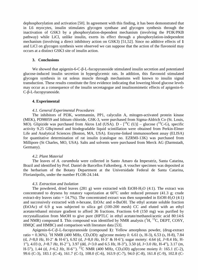
dephosphorylation and activation [50]. In agreement with this finding, it has been demonstrated thatin L6 myocytes, insulin stimulates glycogen synthase and glycogen synthesis through theinactivation of GSK3 by a phosphorylation-dependent mechanism (involving the PI3K/PKBpathway) while LiCl, unlike insulin, exerts its effect through a phosphorylation-independentmechanism (involving a direct inhibitory action on GSK3) [51,52]. Since no additive effects of 1and LiCl on glycogen synthesis were observed we can suppose that the action of the flavonoid mayoccurs at a distinct GSK3 site of insulin action.
3. Conclusions
We showed that apigenin-6-C-β-L-fucopyranoside stimulated insulin secretion and potentiatedglucose-induced insulin secretion in hyperglycemic rats. In addition, this flavonoid stimulatedglycogen synthesis in rat soleus muscle through mechanisms well known to insulin signaltransduction. These results constitute the first evidence indicating that lowering blood glucose levelsmay occur as a consequence of the insulin secretagogue and insulinomimetic effects of apigenin-6-C-β-L-fucopyranoside.
4. Experimental
4.1. General Experimental ProceduresThe inhibitors of PI3K, wortmannin, PP1, calyculin A, mitogen-activated protein kinase
(MEK), PD98059 and lithium chloride, GSK-3, were purchased from Sigma-Aldrich Co (St. Louis,MO). Glipizide was purchased from Akros Ltd (USA). D - [14C (U)] – glucose (14C-G), specificactivity 9.25 GBq/mmol and biodegradable liquid scintillation were obtained from Perkin-ElmerLife and Analytical Sciences (Boston, MA, USA). Enzyme-linked immunosorbent assay (ELISA)for quantitative determination of rat insulin (catalogue no. EZRMI-13K) was purchased fromMillipore (St Charles, MO, USA). Salts and solvents were purchased from Merck AG (Darmstadt,Germany).
4.2. Plant MaterialThe leaves of A. carambola were collected in Santo Amaro da Imperatriz, Santa Catarina,
Brazil and identified by Prof. Daniel de Barcellos Falkenberg. A voucher specimen was deposited atthe herbarium of the Botany Department at the Universidade Federal de Santa Catarina,Florianópolis, under the number FLOR-24.144.
4.3. Extraction and IsolationThe powdered, dried leaves (281 g) were extracted with EtOH-H2O (4:1). The extract was
concentrated to dryness by rotatory vaporization at 60oC under reduced pressure (41.3 g; crudeextract-dry leaves ratio = 14.7%). The concentrated extract was then suspended in EtOH-H2O (4:1)and successively extracted with n-hexane, EtOAc and n-BuOH. The ethyl acetate soluble fraction(EtOAc) of 6.9 g was subjected to silica gel (100-200 mesh) CC and eluted with an ethylacetate/ethanol mixture gradient to afford 36 fractions. Fractions 6-8 (150 mg) was purified byrecrystallization from MeOH to give pure (HPTLC in ethyl acetate/methanol/acetic acid 80:14:6and NMR) compound 1. This compound was identified by NMR analysis (1H, 13C, DEPT, COSY,HMQC and HMBC) and comparison with literature data [53].
Apigenin-6-C--L-fucopyranoside (compound 1): Yellow amorphous powder, (drug-extractratio = 0.36%). 1H NMR (400 MHz, CD3OD): aglycone moiety : 6.61 (s, H-3), 6.53 (s, H-8), 7.84(d, J=8,0 Hz, H-2’ & H-6’), 6.92 (d, J=8,0 Hz, H-3’ & H-6’); sugar moiety: 4.64 (d, J=8.4 Hz, H-1”), 4.03 (t, J=8.7 Hz, H-2’’), 3.97 (dd, J=3.0 and 6.5 Hz, H-3”), 3.50 (d, J=3.0 Hz, H-4”), 3.17 (m,H-5”), 1.44 (d, J=6.2 Hz, H-6’’). 13C NMR (400 MHz, CD3OD) aglycone moiety : 165.1 (C-2),99.6 (C-3), 183.1 (C-4), 161.7 (C-5), 108.0 (C-6), 163.9 (C-7), 94.0 (C-8), 161.8 (C-9), 102.8 (C-

10), 121.9 (C-1’), 128.3 (C-2’ and 6’), 115.9 (C-3’ and 5’), 157.8 (C-4´); sugar moiety: 71.9 (C-1’’), 69.1 (C-2’’), 78.5 (C-3’’), 70.6 (C-4’’), 70.8 (C-5’’), 18.1 (C-6’’).
4.4. Experimental AnimalsMale Wistar rats weighing 180-200 g from the Central Animal House-UFSC were used. The
rats were housed in plastic cages in an air-conditioned animal room and fed on pellets with freeaccess to tap water. Room temperature was controlled at 21 ºC with a 12 h light: 12 h dark cycle.Animals described as fasted had been deprived of food for 16 h but allowed free access to water.For all oral treatments, 0.5 mL of each respective substance was given by gavage. All the animalswere monitored carefully and maintained in accordance with the ethical recommendations of theBrazilian Veterinary Medicine Council and the Brazilian College of Animal Experimentation(Protocol CEUA/PP007).
4.5. Apigenin-6-C-β-L-fucopyranoside (1) TreatmentFasted rats were divided into groups of six animals for each treatment: Group I, euglycemic,
normal fasted rats that received the vehicle, 1% EtOH-H2O, p.o.; Group II, hyperglycemic rats thatreceived glucose (4 g/kg, p.o.); Group III, rats that received glucose (4 g/kg, p.o.) plus apigenin-6-C-β-L-fucopyranoside (1) (50 mg/kg, p.o.); Group IV, rats that received glucose (4 g/kg, p.o.) plusglipizide (10 mg/kg, p.o.); by oral gavage. Blood samples were collected just prior to and at 15, 30and 60 min after the glucose loading. After centrifugation, serum samples were used eitherimmediately to determine the blood glucose levels.
4.6. Determination of the Serum InsulinFasted rats were divided into groups of six animals for each treatment: Group I,
hyperglycemic rats that received glucose (4 g/kg, p.o.); Group II, rats that received glucose (4 g/kg,p.o.) plus apigenin-6-C-β-L-fucopyranoside (1) (50 mg/kg, p.o.). Blood samples were collected justprior to and at 15, 30 and 60 min after the glucose loading. Blood samples from the tail vein werecollected and prepared for the analysis of insulin. The insulin levels were measured by enzyme-linked immunosorbent assay (ELISA) according to the manufacturer’s instructions. The range ofvalues detected by this assay was: 0.2 ng/mL to 10 ng/mL. The intra- and inter-assay coefficients ofvariation for insulin were 3.22 and 6.95, respectively. Sensitivity value of 0.2 ng/mL. All insulinlevels were estimated by means of colorimetric measurement at 450 nm with an ELISA plate reader(Organon Teknika, Roseland, NJ, USA) by interpolation from a standard curve. Samples wereanalyzed in duplicate and results were expressed as ng of insulin serum mL-1.
4.7. Glycogen synthesis on rat soleus muscleThe assays of 14C-glucose incorporation into glycogen were conducted as described by
Cazarolli et al., [9,41]. Slices of soleus muscle from normal rats were distributed (alternately leftand right) between basal and treated groups. The muscles were dissected, weighed, andpreincubated and incubated at 37 °C in Krebs Ringer-bicarbonate (KRb) buffer of the composition122 mM NaCl, 3 mM KCl, 1.2 mM MgSO4, 1.3 mM CaCl2, 0.4 mM KH2PO4, 25 mM NaHCO3,1% BSA and 5 mM D-glucose and bubbled with O2/CO2 (95%:5%, v/v) until pH 7.4. Apigenin-6-C-β-L-fucopyranoside (1) (1 and 100 μM) was added to the preincubation (30 min) and incubationmedium (60 min) in the presence or absence of 100 nM wortmannin, 5 nM calyculin A, 50 μMPD98059 or 50 mM lithium chloride. 14C-glucose (0.15 μCi/mL) was added to each sample duringthe incubation period. After incubation, the muscles were removed, washed in cold KRb and driedon filter paper. The muscles were homogenized in 0.5 N KOH and boiled at 100 °C for 20 min, withoccasional stirring. After cooling, 95% ethanol was added to the samples, which were heated toboiling followed by cooling in an ice bath for 20 min to aid the precipitation of glycogen. Thehomogenates were centrifuged at 664 g for 15 min, the supernatant was discarded, and pellets

resolubilized in water. 30 μL aliquots of the samples were placed in liquid scintillation vials on anLKB rack beta liquid scintillation spectrometer (model 1215; EG and G-Wallac, Turku, Finland),for the radioactivity measurements. The results were expressed as pmol glucosyl units incorporatedinto glycogen. mg tissue-1. h-1.
4.8. Statistical AnalysisData were expressed as mean ± S.E.M. One or two-way analysis of variance (ANOVA),
followed by the Bonferroni post-test or non paired Student’s t-test, was used to identify significantlydifferent groups. Differences were considered to be significant at the p ≤ 0.05 level.
AcknowledgementThis work was supported by grants from Conselho Nacional de Desenvolvimento e
Tecnológico (CNPq), Coordenação de Pessoal de Nível Superior (CAPES-PGFAR), and Fundaçãode Amparo à Pesquisa do Estado de Santa Catarina (FAPESC). The authors thank to Dr. Roselis S.M. da Silva and Dr. Luis Carlos Kucharski for their helpful advice. Special acknowledgement to Dr.Marcos L. S. Perry for his useful discussion and critical analysis of the data. L. H. C and P. F. wereregistered on the PGFAR-UFSC. The authors express their appreciation to Dr. Siobhan Wiese forassistance with the English correction of the manuscript.
References
[1] R. Gadsby, Adv. Drug Del. Rev. 54 (2002) 1165-1172.[2] J.K. Grover, S. Yadav, V. Vats, J. Ethnopharmacol. 81 (2002) 81-100.[3] G. Zareba, N. Serradell, R. Castañer, S.L. Davies, J. Prous, N. Mealy, Drugs Fut. 30
(2005) 1253-1282.[4] J.B. Harborne, C.A. Williams, Phytochemistry 55 (2000) 481-504.[5] L.H. Cazarolli, L. Zanatta, E.H. Alberton, M.S.R.B. Figueiredo, P. Folador, R.G. Damazio,
M.G. Pizzolatti, F.R.M.B. Silva, Mini Rev. Med. Chem. 8 (2008a) 1429-1440.[6] L.H. Cazarolli, L. Zanatta, E.H. Alberton, M.S.R.B. Figueiredo, P. Folador, R.G. Damazio,
M.G. Pizzolatti, F.R.M.B. Silva, Mini Rev. Med. Chem. 8 (2008b) 1032-1038.[7] F.L. Hsu, I.M. Liu, D.H. Kuo, W.C. Chen, H.C. Su, J.T. Cheng, J. Nat. Prod. 66 (2003)
788-792.[8] E. De Sousa, L. Zanatta, I. Seifriz, T.B. Creczynski-Pasa, M.G. Pizzolatti, B. Szpoganicz,
F.R.M.B. Silva, J. Nat. Prod. 67 (2004) 829-832.[9] A.P. Jorge, H. Horst, E. De Sousa, M.G. Pizzolatti, F.R.M.B. Silva, Chem.-Biol. Interact.
149 (2004) 89-96.[10] M. Pinent, M. Blay, M.C. Bladé, M.J. Salvadó, L. Arola, A. Ardévol, Endocrinology 14
(2004) 54985-54990.[11] I.M. Liu, S.S. Liou, T.W. Lan, F.L. Hsu, J.T. Cheng, Planta Med. 71 (2005) 617-621.[12] L.H. Cazarolli, L. Zanatta, A.P. Jorge, E. De Sousa, H. Horst, V.M. Woehl, M.G.
Pizzolatti, B. Szpoganicz, F.R.M.B. Silva, Chem.-Biol. Interact. 163 (2006) 177-191.[13] L. Zanatta, E. De Sousa, L.H. Cazarolli, A.J. Cunha, M.G. Pizzolatti, B. Szpoganicz,
F.R.M.B. Silva, J. Ethnopharmacol. 109 (2007) 151-155.[14] E.H. Alberton, R.G. Damazio, L.H. Cazarolli, L.D. Chiaradia, P.C. Leal, R.J. Nunes,
R.A. Yunes, F.R.M.B. Silva, Chem.-Biol. Interact. 171 (2008) 355-362.[15] M. Provasi, C.E. Oliveira, M.C. Martino, L.G. Pessini, R.B. Bazotte, D.A.G. Cortez, Acta
Sci – Health Sci. 23 (2001) 665-669.[16] M. Provasi, C.E. Oliveira, L.C. Fernandes, O. Tchaikovski, R.B. Bazotte, L.E.R. Cortez,
D.A.G. Cortez, Acta Sci. – Health Sci. 27 (2005) 45-48.[17] P.N. Pushparaj, C.H. Tan, B.K.H. Tan, J. Ethnopharmacol. 72 (2000) 69-76.[18] A. Taha, A. Klip, J. Membr. Biol. 169 (1999) 1-12.

[19] A.R. Saltiel, C.R. Kahn, Nature 414 (2001) 799-806.[20] P. Detimary, J.C. Jonas, J.C. Henquin, J. Clin. Invest. 96 (1995) 1738-1745.[21] P. Rorsman, Diabetologia 40 (1997) 487–495.[22] A.Y.Y. Cheng, I.G. Fantus, CMAJ 172 (2005) 213-226.[23] A.K. Srivastava, S.K. Pandey, Mol. Cell. Biochem. 182 (1998) 135-141.[24] P.J. Roach, Curr. Mol. Med. 02 (2002) 101-120.[25] M.J. Brady, A.R. Saltiel, Recent Prog. Horm. Res. 56 (2001) 157-173.[26] E. Hajduch, G.J. Litherland, H.S. Hundal, FEBS Lett. 492 (2001) 199-203.[27] A. Mora, K. Sakamoto, E.J. McManus, D.R. Alessi, FEBS Lett. 579 (2005) 3632-3638.[28] C.B. Newgard, M.J. Brady, R.M. O’Doherty, A.R. Saltiel, Diabetes 49 (2000) 1967-
1977.[29] J. Carlsen, K. Christiansen, J. Vinten, Cell. Signal. 09 (1997) 447-450.[30] P. Dent, S.N. Lavoinne, F.B. Caudwell, P. Watt, P. Cohen, Nature 348 (1990) 302-308.[31] E. Sezik, M. Aslan, E. Yesilada, S. Ito, Life Sci. 76 (2005) 1223-1238.[32] B. Jayaprakasam, S.K. Vareed, L.K. Olson, M.G. Nair, J. Agric. Food Chem. 53 (2005)
28-31.[33] D. Liu, W. Zhen, Z. Yang, J.D. Carter, H. Si, K.A. Reynolds, Diabetes 55 (2006) 1043-
1050.[34] K.C. Ong, H.E. Khoo, Life Sci. 67 (2000) 1695-1705.[35] A.K. Valsa, S. Sudheesh, N.R. Vijayalakshmi, Indian J. Biochem. Biophys. 34 (1997)
406-408.[36] M. Pinent, M.C. Bladé, M.J. Salvadó, L. Arola, A. Ardévol, J. Agric. Food Chem. 53
(2005) 5932-5935.[37] J.C. Henquin, M. Nenquin, P. Stiernet, B. Ahren, Diabetes 55 (2006) 441-451.[38] J.C. Jonas, T.D. Plant, P. Gilon, P. Detimary, M. Nenquin, J.C. Henquin. Br. J.
Pharmacol. 114 (1995) 872-880.[39] C.S. Hii, S.L. Howell, J. Endocrinol. 107 (1985) 1-8.[40] N.A. Syed, R.L. Khandelwal, Mol. Cell. Biochem. 211 (2000) 123-136.[41] L.H. Cazarolli, P. Folador, M.G. Pizzolatti, F.R.M.B. Silva, Biochimie (2009) 1-7. Doi:
10.1016/j.biochi.2009.04.004[42] L. Zanatta, A. Rosso, P. Folador, M.S.R.B. Figueiredo, M.G. Pizzolatti, L.D. Leite,
F.R.M.B. Silva, J. Nat. Prod. 71 (2008) 532-535.[43] L. Ragolia, N. Begum, Mol. Cell. Biochem. 182 (1998) 49-58.[44] I. Azpiazu, A.R. Saltiel, A.A. DePaoli-Roach, J.C. Lawrence, J. Biol. Chem. 271 (1996)
5033-5039.[45] D.A.E. Cross, D.R. Alessi, P. Cohen, M. Andjelkovich, B.A. Hemmings, Nature 378
(1995) 785-789.[46] P.R. Shepherd, B.T. Navé, K. Siddle, Biochem. J. 305 (1995) 25-28.[47] S.J. Oreña, A.J. Torchia, R.S. Garofalo, J. Biol. Chem. 275 (2000) 15765-15772.[48] J.E. Rodríguez-Gil, J.J. Guinovart, F. Bosch, Arch. Biochem. Biophys. 301 (1993) 411-
415.[49] J.M. Fernández-Novell, J.E. Rodriguez-Gil, A. Barberà, J.J. Guinovart, Arch. Biochem.
Biophys. 457 (2007) 29-34.[50] K. MacAulay, E. Hajduch, A.S. Blair, M.P. Coghlan, S.A. Smith, H.S. Hundal, Eur. J.
Biochem. 270 (2003) 3829-3838.[51] W.S. Choi, C.K. Sung, Biochim. Biophys. Acta 1475 (2000) 225-230.[52] W.J. Ryves, A.J. Harwood, Biochem. Biophys. Res. Commun. 280 (2001) 720–725.[53] D. Araho, M. Masaazumi, C. Wen-hua, K. Toshimitsu, M. Kenji, I. Takao, Nat. Med.
(Tokyo, Japan) 59 (2005) 113-116.

56
4.3 ARTIGO PUBLICADOPeriódico – Chemico-Biological Interactions
CAZAROLLI, L.H.; FOLADOR, P.; MORESCO, H.H.; BRIGHENTE, I.M.C.;PIZZOLATTI, M.G.; SILVA, F.R.M.B. Mechanism of action of the stimulatory effect ofapigenin-6-C-(2”-O--L-rhamnopyranosyl)--L-fucopyranoside on 14C-glucose uptake.Chemico-Biological Interactions, v. 179, p. 407-412, 2009.

This article appeared in a journal published by Elsevier. The attachedcopy is furnished to the author for internal non-commercial researchand education use, including for instruction at the authors institution
and sharing with colleagues.
Other uses, including reproduction and distribution, or selling orlicensing copies, or posting to personal, institutional or third party
websites are prohibited.
In most cases authors are permitted to post their version of thearticle (e.g. in Word or Tex form) to their personal website orinstitutional repository. Authors requiring further information
regarding Elsevier’s archiving and manuscript policies areencouraged to visit:
http://www.elsevier.com/copyright

Author's personal copy
Chemico-Biological Interactions 179 (2009) 407–412
Contents lists available at ScienceDirect
Chemico-Biological Interactions
journa l homepage: www.e lsev ier .com/ locate /chembio int
Mechanism of action of the stimulatory effect ofapigenin-6-C-(2′′-O-�-l-rhamnopyranosyl)-�-l-fucopyranosideon 14C-glucose uptake
Luisa Helena Cazarolli a, Poliane Foladora, Henrique Hunger Morescob,Inês Maria Costa Brighente b, Moacir Geraldo Pizzolattib, Fátima Regina Mena Barreto Silvaa,∗
a Departamento de Bioquímica, Centro de Ciências Biológicas, UFSC, Campus Universitário, Bairro Trindade, Cx. Postal 5069, CEP: 88040-970 Florianópolis, SC, Brazilb Departamento de Química, Centro de Ciências Físicas e Matemáticas, UFSC, Campus Universitário, Bairro Trindade, Cx. Postal 5069, CEP: 88040-970 Florianópolis, SC, Brazil
a r t i c l e i n f o
Article history:Received 29 September 2008Received in revised form13 November 2008Accepted 14 November 2008Available online 25 November 2008
Keywords:FlavonoidGlucose uptakeInsulinDiabetes
a b s t r a c t
There has been a growing interest in hypoglycemic agents from natural products, particularly thosederived from plants. Flavonoids are naturally occurring phenolic compounds with a broad range of bio-logical activities and the beneficial effects of flavonoids have been studied in relation to diabetes mellitus,either through their capacity to avoid glucose absorption or to improve glucose tolerance. The purpose ofthis study was to investigate the mechanism of action of the stimulatory effect of apigenin-6-C-(2′′-O-�-l-rhamnopyranosyl)-�-l-fucopyranoside (1), isolated from Averrhoa carambola L. (Oxalidaceae) leaves,on 14C-glucose uptake. This compound (1) was found to have an acute effect on blood glucose lowering indiabetic rats and stimulated glucose-induced insulin secretion after oral treatment in hyperglycemic rats.A significant stimulatory effect of compound 1 on 14C-glucose uptake was observed at 50 and 100 �M.The effect of compound 1 on glucose uptake was completely nullified by wortmannin, an inhibitor ofphosphatidylinositol 3-kinase (PI3K), RO318220, an inhibitor of protein kinase C (PKC), PD98059, a spe-cific inhibitor of mitogen-activated protein kinase (MEK), cycloheximide, an inhibitor of protein synthesis,and colchicine, a microtubule-depolymerizing agent. Compound 1 (100 �M) and insulin (10 nM) did notshow any synergistic effect on glucose uptake. These results suggest that the flavonoid may have a dualtarget of action, as an insulin-secretagogue and also as an insulin-mimetic agent.
© 2008 Published by Elsevier Ireland Ltd.
1. Introduction
Insulin is the most important hormone that regulates energymetabolism. It mediates a wide spectrum of biological responsesincluding synthesis and storage of carbohydrates, lipids and pro-teins as well as the inhibition of catabolism [1]. Glucose is theprimary stimulus for insulin secretion and when blood sugar con-centrations rise, insulin is secreted into the bloodstream [2,3].Insulin stimulates glucose uptake via multiple signaling path-ways. The binding of insulin to its receptor activates the intrinsicreceptor tyrosine kinase, which results in autophosphorylationand recruitment of substrates, such as the insulin-receptor sub-strate (IRS) proteins and Cbl protein. Specific tyrosine residueson the IRS proteins serve as docking sites for proteins thatcontain SH2 domains, such as the p85 regulatory subunit of phos-phatidylinositol 3-kinase (PI3K). PI3K catalyzes the formation of
∗ Corresponding author. Tel.: +55 48 331 69 12; fax: +55 48 3721 9672.E-mail addresses: [email protected],
[email protected] (F.R.M.B. Silva).
phosphatidylinositol (3,4,5)-trisphosphate (PIP3), an allosteric acti-vator of phosphoinositide-dependent kinase (PDK). Targets of PDKinclude protein kinase B (PKB) and the atypical protein kinase C(aPKC) isoforms, which, when activated via phosphorylation, stim-ulate the translocation of GLUT4-containing vesicles to the plasmamembrane. Alternatively, insulin also stimulates glucose uptakethrough the CAP–Cbl–TC10 pathway [1,4].
In both the basal and insulin-stimulated states, GLUT4 cyclesbetween the plasma membrane and intracellular compartments.Insulin increases the amount of GLUT4 on the plasma membraneby stimulating exocytose of the intracellular GLUT4 pool and bydecreasing the endocytose of the plasma membrane-associatedGLUT4 protein. The overall insulin-stimulated changes in the cel-lular dynamics of GLUT4 protein distribution are controlled bycomplex and multiple vesicle trafficking events. This vesicular traf-ficking network includes mechanisms for the sorting, budding,trafficking, tethering, docking and fusion of the GLUT4 vesiclesand they are directly regulated by intracellular signals generatedfrom the insulin receptor [4,5]. The cytoskeleton has been impli-cated in various membrane trafficking events and in the retentionof organelles at specific locations within cells and the actin and
0009-2797/$ – see front matter © 2008 Published by Elsevier Ireland Ltd.doi:10.1016/j.cbi.2008.11.012

Author's personal copy
408 L.H. Cazarolli et al. / Chemico-Biological Interactions 179 (2009) 407–412
microtubule cytoskeleton networks have been implicated in thesubcellular movements of the GLUT4-containing vesicles [6–8]. Itis suggested that insulin stimulation could induce a release of thevesicles allowing the movement to the plasma membrane by sim-ple diffusion, or insulin may trigger the movement of vesicles alongcytoskeletal tracks, as has been observed for regulated exocytose ofsecretory vesicles in other cell systems [9,10]. However, the pre-cise mechanism whereby insulin signals to the GLUT4 membranetrafficking machinery remains obscure.
An absolute or relative lack of insulin, as in the case of dia-betes, leads to severe dysfunction and deregulation of insulinsignaling in target tissues, such as the liver, adipose tissue andmuscle. Diabetes mellitus is a metabolic disorder characterizedby chronic hyperglycemia resulting from defects in insulin secre-tion, insulin action, or both [11]. The molecular defects accountingfor impaired glucose utilization are not fully understood but mayinvolve defective GLUT4 translocation, glucose uptake and aberrantinsulin signal transduction. In this context, several naturally occur-ring polyphenols have been shown to affect glucose transport andinsulin-receptor function, both of which play essential roles in dia-betes [12]. It has been demonstrated that kaempferitrin is able todiminish serum glucose level and increase glucose uptake in the ratsoleus muscle as efficiently as insulin [13,14]. Also, kaempferol 3-neohesperidoside has a significant hypoglycemic effect in diabeticrats and stimulate glucose uptake in rat soleus muscle mimick-ing insulin signaling [15,16]. It has been demonstrated by popularmedicine usage in Brazil that Averrhoa carambola helps to bal-ance glucose levels [17]. Recently, it was demonstrated that crudeextracts, flavonoid-enriched fractions from leaves of A. carambolaare able to diminish serum glucose levels in hyperglycemic rats asefficiently as insulin [18,19]. Considering the effect of apigenin-6-C-(2′′-O-�-l-rhamnopyranosyl)-�-l-fucopyranoside, isolated fromA. carambola leaves, on blood glucose levels and on glycogen con-tent in the liver and soleus muscle from hyperglycemic rats, theaim of this study was to investigate the mechanism of action ofthis compound in relation to its stimulatory effect on 14C-d-glucoseuptake.
2. Materials and methods
2.1. Materials
Regular human insulin (Biohulin) was obtained from Biobrás,Bioquímica do Brasil S/A (Águas Claras, MG, Brazil). [U-14C]-2-Deoxy-d-glucose (14C-DG), specific activity 10.6 GBq/mmol, andbiodegradable scintillation liquid were obtained from Perkin-ElmerLife and Analytical Sciences (Boston, MA, USA). Alloxan, wortman-nin (inhibitor of phosphatidylinositol 3-kinase (PI3K)), RO318220(inhibitor of protein kinase C (PKC)), PD98059 (inhibitor ofmitogen-activated protein kinase (MEK)), cycloheximide (inhibitorof protein synthesis) and colchicine (microtubule-depolymerizingagent) were purchased from Sigma Chemical Company® (St.Louis, MO, USA). Enzyme-linked immunosorbent assay (ELISA) forquantitative determination of rat insulin (catalogue no. EZRMI-13K) was purchased from Millipore (St. Charles, MO, USA). Saltsand solvents were purchased from Merck AG (Darmstadt, Ger-many).
2.2. Plant material
The leaves of A. carambola were collected in Santo Amaro daImperatriz, Santa Catarina, Brazil and identified by Prof. Daniel deBarcellos Falkenberg. A voucher specimen was deposited at theherbarium of the Botany Department at the Universidade Federalde Santa Catarina, Florianópolis, under the number FLOR-24.144.
Fig. 1. (A) Apigenin-6-C-(2′′-O-�-l-rhamnopyranosyl)-�-l-fucopyranoside (1)structure and (B) effect of apigenin-6-C-(2′′-O-�-l-rhamnopyranosyl)-�-l-fucopyranoside (1) 50 mg/kg on serum insulin levels in hyperglycemic rats. Valuesare expressed as mean ± S.E.M.; n = 6 in duplicate for each group. Significant at**p ≤ 0.01 in relation to zero time. Significant at #p ≤ 0.05 and ##p ≤ 0.01 in relationto hyperglycemic group.
2.3. Extraction and isolation
The powdered, dried leaves (281 g) were extracted withEtOH–H2O (4:1). The extract was concentrated to dryness byrotatory vaporization at 60 ◦C under reduced pressure. The concen-trated extract was suspended in EtOH–H2O (4:1) and successivelyextracted with n-hexane, EtOAc and n-BuOH. The ethyl acetate sol-uble fraction was subjected to silica gel (100–200 mesh) CC andeluted with an ethyl acetate/ethanol mixture gradient to afford frac-tions that were purified by recrystallization from MeOH to giveisolated compounds. One of these was apigenin-6-C-(2′′-O-�-l-rhamnopyranosyl)-�-l-fucopyranoside (1) (Fig. 1A) [20].
2.4. Experimental animals
Male Wistar rats weighing 180–200 g from the Central AnimalHouse-UFSC were used. The rats were housed in plastic cages inan air-conditioned animal room and fed on pellets with free accessto tap water. Room temperature was controlled at 21 ◦C with a 12 hlight:12 h dark cycle. Animals described as fasted had been deprivedof food for 16 h but allowed free access to water. For all oral treat-ments, 0.5 mL of each respective substance was given by gavage.All the animals were monitored carefully and maintained in accor-dance with the ethical recommendations of the Brazilian VeterinaryMedicine Council and the Brazilian College of Animal Experimenta-tion (Protocol CEUA/PP007). Rats fasted for 16 h received 50 mg/kgbody weight of alloxan (Sigma, St. Louis, MO, USA) by a single intra-venous injection. We assessed the diabetic state by measuring bodyweight and blood glucose levels 3 days after injection.

Author's personal copy
L.H. Cazarolli et al. / Chemico-Biological Interactions 179 (2009) 407–412 409
2.5. Studies on the effect ofapigenin-6-C-(2′′-O-˛-l-rhamnopyranosyl)-ˇ-l-fucopyranoside(1) on serum glucose levels
Fasted rats were divided into groups of six animals for each treat-ment: Group I, hyperglycemic rats that received glucose (4 g/kg);Group II, rats that received glucose (4 g/kg) plus apigenin-6-C-(2′′-O-�-l-rhamnopyranosyl)-�-l-fucopyranoside (1) (50 mg/kg);Group III, alloxan-induced diabetic rats that received vehicle (1%EtOH–H2O); and Group IV, diabetic rats that received apigenin-6-C-(2′′-O-�-l-rhamnopyranosyl)-�-l-fucopyranoside (1) (50 mg/kg).Blood samples were collected just prior to and 60 min after treat-ments. Blood samples from the tail vein were collected andcentrifuged, and the serum was used to determine glycemiaby a glucose oxidase method. For diabetic rats, a serum glu-cose range of 300–400 mg/dL was used for the experiment[21].
2.6. Determination of insulin levels
Blood samples from the tail vein were collected and centrifuged,and the serum was used to determine insulin levels. Blood sampleswere collected just prior to, and at 15, 30 and 60 min after treat-ment. The insulin levels were measured by ELISA according to themanufacturer’s instructions. The range of values detected by thisassay was 0.2–10 ng/mL. The intra- and inter-assay coefficients ofvariation for insulin were 3.22 and 6.95, respectively, with a sensi-tivity value of 0.2 ng/mL. All insulin levels were estimated by meansof colorimetric measurement at 450 nm with an ELISA plate reader(Organon Teknika, Roseland, NJ, USA) through interpolation froma standard curve. Samples were analyzed in duplicate and resultswere expressed as ng of insulin serum per mL.
2.7. Studies on 14C-glucose uptake in rat soleus muscle
For the [U-14C]-2-deoxy-d-glucose uptake experiments, soleusmuscles from normal rats were used. Slices of soleus muscle weredistributed (alternately left and right) between basal and treatedgroups. The muscles were dissected, weighed, and preincubatedand incubated at 37 ◦C in Krebs Ringer-bicarbonate (KRb) bufferwith a composition of 122 mM NaCl, 3 mM KCl, 1.2 mM MgSO4,1.3 mM CaCl2, 0.4 mM KH2PO4, and 25 mM NaHCO3 and bubbledwith O2/CO2 (95%:5%, v/v) until pH 7.4. Compound 1 (1, 10, 50,100 �M and 1 mM) and insulin (10 nM) were added to the preincu-bation (30 min) and incubation medium (60 min) in the presence orabsence of 100 nM wortmannin, 40 �M RO318220, 50 �M PD98059,0.35 mM cycloheximide or 1 �M colchicine. 14C-DG (0.1 �Ci/mL)was added to each sample during the incubation period. After incu-bation, the muscles were placed in screw cap tubes containing 1 mLof distilled water. These were frozen at −20 ◦C in a freezer and thenboiled for 10 min; 25 �L aliquots of tissue and external mediumwere placed in scintillation liquid on an LKB rack beta liquid scintil-lation spectrometer (model 1215; EG and G-Wallac, Turku, Finland),for the radioactivity measurements. The results were expressed as
the tissue/medium (T/M) ratio: cpm/mL tissue fluid per cpm/mLincubation medium [13].
2.8. Data and statistical analysis
Data were expressed as mean ± S.E.M. The statistical analysiswas carried out using one-way analysis of variance (ANOVA), fol-lowed by the Bonferroni post-test or non-paired Student’s t-test toidentify significantly different groups. Differences were consideredto be significant at p ≤ 0.05.
3. Results and discussion
3.1. Effect ofapigenin-6-C-(2′′-O-˛-l-rhamnopyranosyl)-ˇ-l-fucopyranoside(1) on serum glucose and insulin levels in rats
The administration of alloxan (50 mg/kg) to the fasted rats sig-nificantly increased blood glucose levels when compared withnormal rats. Also, as expected, after the glucose loading serum glu-cose concentration was increased when compared with zero time(Table 1). The oral administration of compound 1 reduced signifi-cantly the serum glucose levels in hyperglycemic rats as well as inalloxan-induced diabetic rats after the treatments. We have previ-ously demonstrated the effect of flavonoid-rich fractions, flavonoidsand chalcones in diabetic and hyperglycemic rats [14,15,21–23].Moreover, puerarin has been described as a hypoglycemic andantihyperglycemic agent due to its effects in hyperglycemic anddiabetic rats [24].
Based on the effects of compound 1 on blood glucose lev-els, especially in hyperglycemic rats, serum insulin levels weredetermined in fasted rats after an oral glucose loading (4 g/kg)as shown in Fig. 1B. The glucose-induced insulin secretion wasincreased 71% at 15 min compared with zero time and returnedto the basal levels after 60 min. Biphasic insulin secretion is thenormal response of � cells to a rapid and sustained increasein blood glucose concentration and the rapid increase in insulinlevels observed in Fig. 1B confirms the classical profile of glucose-induced insulin secretion [3,25]. The treatment with compound1-stimulated insulin secretion as well as potentiated the glucoseeffect on insulin levels in hyperglycemic rats after 30 and 60 mincompared to the hyperglycemic group. Also, genistein and daidzeinflavonoids have been reported as insulin-secretagogues since theyincreased serum insulin secretion stimulated by glucose in vivoand in vitro [26,27]. Thus, the hypoglycemic effect of apigenin-6-C-(2′′-O-�-l-rhamnopyranosyl)-�-l-fucopyranoside seems to be aconsequence of insulin secretion to manage glucose metabolism.
3.2. Effect ofapigenin-6-C-(2′′-O-˛-l-rhamnopyranosyl)-ˇ-l-fucopyranoside(1) on 14C-glucose uptake in rat soleus muscle
Fig. 2 shows the in vitro effect of apigenin-6-C-(2′′-O-�-l-rhamnopyranosyl)-�-l-fucopyranoside (1) on glucose uptake in
Table 1Acute effect of apigenin-6-C-(2′′-O-�-l-rhamnopyranosyl)-�-l-fucopyranoside (1) (50 mg/kg) on serum glucose level in hyperglycemic and diabetic rats. Values expressedas mean ± S.E.M.; n = 6 in duplicate for each treatment.
Time (min) Serum glucose levels (mg/dL)
Group I hyperglycemic glucose (4 g/kg) Group II hyperglycemicplus 1 (50 mg/kg)
Group III diabetic plus(1% EtOH–H2O)
Group IV diabeticplus 1 (50 mg/kg)
0 122.6 ± 5.8 111.7 ± 5.7 317.5 ± 11.9 336.0 ± 8.960 178.4 ± 9.1 149.0 ± 3.3# 372.0 ± 8.5 284.4 ± 4.5***
# Significant at p ≤ 0.05 in relation to hyperglycemic group.*** Significant at p ≤ 0.001 in relation to zero time of diabetic plus 1 group.

Author's personal copy
410 L.H. Cazarolli et al. / Chemico-Biological Interactions 179 (2009) 407–412
Fig. 2. Concentration–response curve of apigenin-6-C-(2′′-O-�-l-rhamnopyranosyl)-�-l-fucopyranoside (1) on 14C-glucose uptake in rat soleusmuscle. Preincubation time = 30 min; incubation time = 60 min. Values are expressedas mean ± S.E.M.; n = 6 in duplicate for each group. Significant at ***p ≤ 0.001 and*p ≤ 0.05 in relation to basal group.
soleus muscle after 60 min of incubation. The stimulatory effectof compound 1 was significant at 50 and 100 �M compared tothe basal group. This stimulatory effect represents 36.5% and42% of glucose uptake compared to the basal level at 60 min,respectively. These results are in line with those recently pub-lished concerning the hypoglycemic effect of kaempferitrin andkaempferol 3-neohesperidoside (similar flavonoids to compound1) in diabetic rats and the stimulatory effect of these flavonoids onglucose uptake in rat soleus muscle [13–16]. The flavonoid puer-arin also increased glucose utilization by stimulation of glucoseuptake in soleus muscle from streptozotocin-diabetic rats [24].These results demonstrate a potential insulinomimetic effect ofcompound 1 on glucose uptake in one of the insulin target tissues,soleus muscle.
3.3. Effect of various inhibitors on the stimulatory action ofapigenin-6-C-(2′′-O-˛-l-rhamnopyranosyl)-ˇ-l-fucopyranoside(1) on 14C-glucose uptake in rat soleus muscle
In order to study the mechanism by which compound 1 stim-ulates glucose uptake in soleus muscle, we performed the glucoseuptake assay with 100 nM wortmannin, a specific inhibitor of PI3K,40 �M RO318220, a specific inhibitor of PKC, 50 �M PD98059,an inhibitor of MEK, 0.35 mM cycloheximide, an inhibitor of pro-tein synthesis or 1 �M colchicine, a microtubule-depolymerizingagent. The wortmannin, RO318220, PD98059, cycloheximide andcolchicine concentrations used were those that have previouslyresulted in inhibition in cell and tissue assays [16,28–32].
Fig. 3 shows that the stimulation of glucose uptake by com-pound 1 was completely inhibited by wortmannin, RO318220,PD98059 and cycloheximide pretreatment. However, when onlywortmannin, RO318220, PD98059 or cycloheximide were added tothe muscle samples, no significant change resulted compared withthe glucose uptake in the basal group. These results suggest that,at least in part, the stimulatory effect of compound 1 on glucoseuptake is mediated through the PI3K and PKC pathways togetherwith the involvement of the MAPK pathway as well as protein syn-thesis (glucose transporters?). Thus, the results here reported arein agreement with those reported in the literature concerning thekey role of PI3K and GLUT4 translocation regulated by insulin [1,4].Similarly, the stimulatory effect of kaempferol 3-neohesperidoside
Fig. 3. Effect of enzyme inhibitors on the stimulatory action of apigenin-6-C-(2′′-O-�-l-rhamnopyranosyl)-�-l-fucopyranoside (1) on 14C-glucose uptake in ratsoleus muscle with 100 nM Wortmannin, 40 �M RO318220, 50 �M PD98059,0.35 mM cycloheximide and 100 �M 1 used. Basal group = no treatment. Signal (+)and (−) indicates presence and absence, respectively, of each substance in theincubation medium. Preincubation time = 30 min; incubation time = 60 min. Val-ues are expressed as mean ± S.E.M.; n = 6 in duplicate for each group. Significantat ***p ≤ 0.001 in relation to basal group. Significant at ###p ≤ 0.001 in relation toapigenin-6-C-(2′′-O-�-l-rhamnopyranosyl)-�-l-fucopyranoside (1) group.
on glucose uptake seems to be mediated through insulin signaltransduction involving the PI3K and PKC pathways. The stimula-tory effect on glucose uptake has also been demonstrated for grapeseed-derived procyanidins, myricetin and apigenin and its glyco-sides [16,30,33,34].
In contrast to the observed kaempferol 3-neohesperidosidemechanism [16], cycloheximide totally blocked the stimulation ofglucose uptake induced by compound 1. Additionally, extracts fromAegles marmelos, Syzygium cumini and Canna indica, plants rich inflavonoids, stimulated glucose uptake and these effects were totallyinhibited in the presence of cycloheximide, suggesting that activeprotein synthesis is important in terms of glucose transport [32,35].Thus, these data indicate that active protein synthesis is necessaryfor compound 1-stimulated glucose transport and, coupled withthe involvement of PI3K and PKC pathways, reinforces the insuli-nomimetic effect of this flavonoid in an insulin-sensitive tissue,soleus muscle.
Fig. 4. Effect of colchicine (1 �M) on the stimulatory action of apigenin-6-C-(2′′-O-�-l-rhamnopyranosyl)-�-l-fucopyranoside (1) (100 �M) and insulin (10 nM) on14C-glucose uptake in rat soleus muscle. Basal group = no treatment. Signal (+)and (−) indicates presence and absence, respectively, of each substance in theincubation medium. Preincubation time = 30 min; incubation time = 60 min. Val-ues are expressed as mean ± S.E.M.; n = 6 in duplicate for each group. Significantat ***p ≤ 0.001 in relation to basal group. Significant at ##p ≤ 0.01 in relationto apigenin-6-C-(2′′-O-�-l-rhamnopyranosyl)-�-l-fucopyranoside (1) group and###p ≤ 0.001 in relation to insulin group.

Author's personal copy
L.H. Cazarolli et al. / Chemico-Biological Interactions 179 (2009) 407–412 411
Fig. 5. Comparative effect of apigenin-6-C-(2′′-O-�-l-rhamnopyranosyl)-�-l-fucopyranoside (1) (100 �M) and insulin (10 nM) on 14C-glucose uptake in ratsoleus muscle. Basal group = no treatment. Signal (+) and (−) indicates presence andabsence, respectively, of each substance in the incubation medium. Preincubationtime = 30 min; incubation time = 60 min. Values are expressed as mean ± S.E.M.; n = 6in duplicate for each group. Significant at ***p ≤ 0.001 in relation to basal group.
It is well known that insulin stimulates glucose uptake in skeletalmuscle and adipose tissue primarily by eliciting the translocationof GLUT4 from an intracellular pool to the plasma membrane [4].The cellular cytoskeleton structure is engaged in the efficient trans-port of vesicles through intracellular membrane-sorting pathwaysand the microtubules and actin filaments are particularly impor-tant for insulin-stimulated GLUT4 translocation [6–8]. To determinewhether an intact microtubule cytoskeleton is necessary for thecompound 1-stimulated glucose uptake, the glucose uptake in thepresence of colchicine was studied (Fig. 4). The stimulatory effectof compound 1 on glucose transport was completely blocked by1 �M colchicine and when only colchicine was added to the sam-ples no significant change was observed compared with the basalgroup. It has been demonstrated that the disruption of microtubulescaused by colchicine and other microtubule-depolymerizing agentsinhibits insulin-induced GLUT4 translocation and glucose uptake inadipocytes [10,36–39]. Taken together, these results suggest that toobserve the complete and efficient effect of compound 1 on glu-cose uptake, the cytoskeleton integrity and cascade of PI3K–PKCand MAPK are required.
3.4. Comparative effect ofapigenin-6-C-(2′′-O-˛-l-rhamnopyranosyl)-ˇ-l-fucopyranoside(1) and insulin on 14C-glucose uptake in rat soleus muscle
Fig. 5 shows the stimulation of glucose uptake by apigenin-6-C-(2′′-O-�-l-rhamnopyranosyl)-�-l-fucopyranoside (1) (100 �M)compared to insulin (10 nM) in soleus muscle. In percentage terms,the compound was as effective as insulin in glucose uptake in thisapproach. On the other hand, the absence of an additive effectof compound 1 and insulin on glucose uptake clearly demon-strates that the compound 1 mechanism of action is mediatedpartially through the insulin transduction pathway. This result isin agreement with those reported for the effect of kaempferol3-neohesperidoside and for grape seed-derived procyanidins onglucose uptake in insulin-sensitive cell lines and tissues, sincesimultaneous treatment with saturating doses of insulin and thecompounds did not cause an additive effect on glucose uptake[16,30].
4. Conclusions
We have shown that apigenin-6-C-(2′′-O-�-l-rhamno-pyranosyl)-�-l-fucopyranoside reduced blood glucose levelsin diabetic rats, stimulated insulin secretion and potentiated
glucose-induced insulin secretion in hyperglycemic rats. In addi-tion, this flavonoid stimulated glucose uptake in rat soleus musclethrough the involvement of the well-known mechanism of insulinsignal transduction. These results constitute strong evidence fora dual effect of apigenin-6-C-(2′′-O-�-l-rhamnopyranosyl)-�-l-fucopyranoside characterizing it as both an insulin secretagogeand an insulin-mimetic agent.
Acknowledgements
This work was supported by grants from Conselho Nacional deDesenvolvimento e Tecnológico (CNPq), Coordenacão de Pessoal deNível Superior (CAPES-PGFAR), and Fundacão de Amparo à Pesquisado Estado de Santa Catarina (FAPESC). The authors express theirappreciation to Dr. Siobhan Wiese for assistance with the Englishcorrection of the manuscript. L.H.C. and P.F. were registered on thePGFAR-UFSC.
References
[1] C. Taha, A. Klip, The insulin signaling pathway, J. Membr. Biol. 169 (1999) 1–12.[2] P. Rosman, The pancreatic beta-cell as a fuel sensor: an electrophysiologist’s
viewpoint, Diabetologia 40 (1997) 487–495.[3] P.E. MacDonald, J.W. Joseph, P. Rorsman, Glucose-sensing mechanisms in pan-
creatic �-cells, Phil. Trans. Roy. Soc. B 360 (2005) 2211–2225.[4] L. Chang, S.H. Chiang, A.R. Saltiel, Insulin signaling and the regulation of glucose
transport, Mol. Med. 10 (2004) 65–71.[5] M. Kanzaki, Insulin receptor signals regulating GLUT4 translocation and actin
dynamics, Endocr. J. 53 (2006) 267–293.[6] A. He, X. Liu, L. Liu, Y. Chang, F. Fang, How many signals impinge on GLUT4
activation by insulin? Cell. Signal. 19 (2007) 1–7.[7] Y. Chen, Y. Wang, W. Ji, P. Xu, T. Xu, A pre-docking role for microtubules
in insulin-stimulated glucose transporter 4 translocation, FEBS J. 275 (2008)705–712.
[8] J. Lane, V. Allan, Microtubule-based membrane movement, Biochim. Biophys.Acta 1376 (1998) 27–55.
[9] S.F. Hamm-Alvarez, P.Y. Kim, M.P. Sheetz, Regulation of vesicle transport in CV-1cells and extracts, J. Cell Sci. 106 (1993) 955–966.
[10] A.L. Olson, A.R. Trumbly, G.V. Gibson, Insulin-mediated GLUT4 translocation independent on the microtubule network, J. Biol. Chem. 276 (2001) 10706–10714.
[11] R. Gadsby, Epidemiology of diabetes, Adv. Drug Deliv. Rev. 54 (2002) 1165–1172.[12] L.H. Cazarolli, L. Zanatta, E.H. Alberton, M.S.R.B. Figueiredo, P. Folador, R.G.
Damazio, M.G. Pizzolatti, F.R.M.B. Silva, Flavonoids: cellular and molecularmechanism of action in glucose homeostasis, Mini Rev. Med. Chem. 8 (2008)1032–1038.
[13] A.P. Jorge, H. Horst, E. De Sousa, M.G. Pizzolatti, F.R.M.B. Silva, Insulinomimeticeffects of kaempferitrin on glycaemia and on 14C-glucose uptake in rat soleusmuscle, Chem. Biol. Interact. 149 (2004) 89–96.
[14] E. De Sousa, L. Zanatta, I. Seifriz, T.B. Creczynski-Pasa, M.G. Pizzolatti, B.Szpoganicz, F.R.M.B. Silva, Hypoglycemic effect and antioxidant potential ofkaempferol-3,7-O-(alpha)-dirhamnoside from Bauhinia forficata leaves, J. Nat.Prod. 67 (2004) 829–832.
[15] L.H. Cazarolli, L. Zanatta, A.P. Jorge, E. De Sousa, H. Horst, V.M. Woehl, M.G. Pizzo-latti, B. Szpoganicz, F.R.M.B. Silva, Follow-up studies on glycosylated flavonoidsand their complexes with vanadium: their anti-hyperglycemic potential role indiabetes, Chem. Biol. Interact. 163 (2006) 177–191.
[16] L. Zanatta, A. Rosso, P. Folador, M.S.R.B. Figueiredo, M.G. Pizzolatti, L.D. Leite,F.R.M.B. Silva, Insulinomimetic effect of kaempferol 3-neohesperidoside on therat soleus muscle, J. Nat. Prod. 71 (2008) 532–535.
[17] M.P. Corrêa, Dicionário das plantas úteis do Brasil, Instituto Brasileiro de Desen-volvimento Florestal, Rio de Janeiro, 1984, p. 03.
[18] M. Provasi, C.E. Oliveira, M.C. Martino, L.G. Pessini, R.B. Bazotte, D.A.G. Cortez,Avaliacão da toxicidade e do potencial antihiperglicemiante da Averrhoa caram-bola L. (Oxalidaceae), Acta Sci. Health Sci. 23 (2001) 665–669.
[19] M. Provasi, C.E. Oliveira, L.C. Fernandes, O. Tchaikovski, R.B. Bazotte, L.E.R.Cortez, D.A.G. Cortez, Efeito do extrato bruto hidroalcoólico e de fracões defolhas da Averrhoa carambola L. (Oxalidaceae) no metabolismo glicêmico deratos Wistar, Acta Sci. Health Sci. 27 (2005) 45–48.
[20] D. Araho, M. Masaazumi, C. W-hua, K. Toshimitsu, M. Kenji, I. Takao, A newflavone c-glycoside from the leaves of Averrhoa carambola, Nat. Med. (Tokyo,Japão) 59 (2005) 113–116.
[21] F.R.M.B. Silva, B. Szpoganicz, M.G. Pizzolatti, M.A.V. Willrich, E. De Sousa, Acuteeffect of Bauhinia forficata on serum glucose levels in normal and alloxan-induced diabetic rats, J. Ethnopharmacol. 83 (2002) 33–37.
[22] L. Zanatta, E. De Sousa, L.H. Cazarolli, A.J. Cunha, M.G. Pizzolatti, B. Szpoganicz,F.R.M.B. Silva, Effect of crude extract and fractions from Vitex megapotamicaleaves on hyperglycemia in alloxan-diabetic rats, J. Ethnopharmacol. 109 (2007)151–155.
[23] E.H. Alberton, R.G. Damazio, L.H. Cazarolli, L.D. Chiaradia, P.C. Leal, R.J. Nunes,R.A. Yunes, F.R.M.B. Silva, Influence of chalcone analogues on serum glucoselevels in hyperglycemic rats, Chem. Biol. Interact. 171 (2008) 355–362.
Author's personal copy
412 L.H. Cazarolli et al. / Chemico-Biological Interactions 179 (2009) 407–412
[24] F.L. Hsu, I.M. Liu, D.H. Kuo, W.C. Chen, H.C. Su, J.T. Cheng, Antihyperglycemiceffect of puerarin in streptozotocin-induced diabetic rats, J. Nat. Prod. 66 (2003)788–792.
[25] J.C. Henquin, M. Nenquin, P. Stiernet, B. Ahren, In vivo and in vitroglucose-induced biphasic insulin secretion in mouse. Pattern and role ofcytoplasmatic Ca2+ and amplification signals in � cells, Diabetes 55 (2006)441–451.
[26] J.C. Jonas, T.D. Plant, P. Gilon, P. Detimary, M. Nenquin, J.C. Henquin, Multipleeffects and stimulation of insulin secretion by the tyrosine kinase inhibitorgenistein in normal mouse islets, Br. J. Pharmacol. 114 (1995) 872–880.
[27] D. Liu, W. Zhen, Z. Yang, J.D. Carter, H. Si, K.A. Reynolds, Genistein acutely stimu-lates insulin secretion in pancreatic �-cells through a cAMP-dependent proteinkinase pathway, Diabetes 55 (2006) 1043–1050.
[28] G.F. Wasserman, F.R.M.B. Silva, M.L. Grillo, Role of microtubules and proteinsynthesis on the stimulatory action of FSH on amino acid uptake in rat testes,Med. Sci. Res. 17 (1989) 737–738.
[29] S.P. Davies, H. Reddy, M. Caivano, P. Cohen, Specificity and mechanism of actionof some commonly used protein kinase inhibitors, Biochem. J. 351 (2000)95–105.
[30] M. Pinent, M. Blay, M.C. Bladé, M.J. Salvadó, L. Arola, A. Ardévol, Grapeseed-derived procyanidins have an antihyperglycemic effect in streptozotocin-induced diabetic rats and insulinomimetic activity in insulin-sensitive cell lines,Endocrinology 14 (2004) 54985–54990.
[31] D. Menegaz, A. Zamoner, C. Royer, L.D. Leite, Z.A. Bortolotto, F.R.M.B. Silva, Rapidresponses to thyroxine in the testis: active protein synthesis-independent path-way, Mol. Cell. Endocrinol. 246 (2006) 128–134.
[32] J. Purintrapiban, M. Suttajit, N.E. Forsberg, Differential activation of glucosetransport in cultured muscle cells by polyphenolic compounds from Cannaindica L. root, Biol. Pharm. Bull. 29 (2006) 1995–1998.
[33] K.C. Ong, H.E. Khoo, Insulinomimetic effects of myricetin on lipogenesis andglucose transport in rat adipocytes but not glucose transporter translocation,Biochem. Pharmacol. 51 (1996) 423–429.
[34] W. Li, R.J. Dai, Y.H. Yu, L. Li, C.M. Wu, W.W. Luan, W.W. Meng, X.S. Zhang,Y.L. Deng, Antihyperglycemic effect of Cephalotaxus sinensis leaves and GLUT-4translocation facilitating activity of its flavonoid constituents, Biol. Pharm. Bull.30 (2007) 1123–1129.
[35] R. Anandharajan, S. Jaiganesh, N.P. Shankernarayanan, R.A. Viswakarma, A. Bal-akrishnan, In vitro glucose uptake activity of Aegles marmelos and Syzygiumcumini by activation of GLUT-4, PI3 kinase and PPAR� in L6 myotubes, Phy-tomedicine 13 (2006) 434–441.
[36] L.M. Fletcher, G.I. Welsh, P.B. Oatey, J.M. Tavaré, Role for the microtubulecytoskeleton in GLUT4 vesicle trafficking and in the regulation of insulin-stimulated glucose uptake, Biochem. J. 352 (2000) 267–276.
[37] M. Emoto, S.E. Langille, M.P. Czech, A role for kinesin in insulin-stimulatedGLUT4 glucose transporter translocation in 3T3-L1 adipocytes, J. Biol. Chem.276 (2001) 10677–10682.
[38] L.B. Liu, W. Omata, I. Kojima, H. Shibata, Insulin recruits GLUT4 from dis-tinct compartments via distinct traffic pathways with differential microtubuledependence in rat adipocytes, J. Biol. Chem. 278 (2003) 30157–30169.
[39] J. Huang, T. Imamura, J.L. Babendure, J.C. Lu, J.M. Olefsky, Disruption of micro-tubules ablates the specificity of insulin signaling to GLUT4 translocation in3T3-L1 adipocytes, J. Biol. Chem. 280 (2005) 42300–42306.

63
4.4 ARTIGO PUBLICADOPeriódico – Biochimie
CAZAROLLI, L.H.; FOLADOR, P.; PIZZOLATTI, M.G.; SILVA, F.R.M.B. Signaling Pathwaysof Kaempferol-3-neohesperidoside on Glycogen Synthesis in Rat Soleus Muscle. Biochimie p.1-7, 2009. Doi: 10.1016/j.biochi.2009.04.004

Research paper
Signaling pathways of kaempferol-3-neohesperidoside in glycogensynthesis in rat soleus muscle
Luisa Helena Cazarolli a, Poliane Folador a, Moacir Geraldo Pizzolatti b,Fatima Regina Mena Barreto Silva a,*
a Departamento de Bioquımica, Centro de Ciencias Biologicas, UFSC, Campus Universitario, Bairro Trindade. Cx. Postal 5069, CEP: 88040-970 - Florianopolis, SC, Brazilb Departamento de Quımica, Centro de Ciencias Fısicas e Matematicas, UFSC, Campus Universitario, Bairro Trindade. Cx. Postal 5069, CEP: 88040-970, Florianopolis, SC, Brazil
a r t i c l e i n f o
Article history:Received 18 December 2008Accepted 7 April 2009Available online 17 April 2009
Keywords:GlycogenFlavonoidInsulinKaempferol 3-neohesperidoside
a b s t r a c t
Kaempferol 3-neohesperidoside is one of the several compounds that have been reported to haveinsulin-like properties in terms of glucose lowering. We studied the effect of kaempferol 3-neo-hesperidoside in glycogen synthesis in rat soleus muscle through the incorporation of 14C-D-glucose inglycogen. Kaempferol 3-neohesperidoside stimulates glycogen synthesis in rat soleus muscle byapproximately 2.38-fold. Insulin at 100 nM showed a stimulatory effect on glycogen synthesis whencompared with the control group. The stimulatory effect of kaempferol 3-neophesperidoside on glycogensynthesis was inhibited by wortmannin, the phosphatidylinositol 3-kinase (PI3K) inhibitor, andenhanced by lithium chloride, a glycogen synthase kinase 3 (GSK-3) inhibitor. Moreover, the stimulatoryeffect of kaempferol 3-neohesperidoside was also nullified by PD98059, a specific inhibitor of mitogen-activated protein kinase (MEK) and by calyculin A, an inhibitor of protein phosphatase 1 (PP1) activity. Itwas concluded that the PI3K – GSK-3 pathway and MAPK – PP1 pathway are involved in the stimulatorykaempferol 3-neohesperidoside effect on glycogen synthesis in rat soleus muscle.
� 2009 Elsevier Masson SAS. All rights reserved.
1. Introduction
Insulin is the most important hormone that regulates energymetabolism. It mediates a wide spectrum of biological responsesincluding synthesis and storage of carbohydrates, lipids andproteins as well as the inhibition of catabolism [1,2]. In mammaliantissues, carbohydrate is stored mainly in the form of glycogen andthe major sites of glycogen deposition are skeletal muscle and theliver [3]. An absolute or relative lack of insulin, as in the case ofdiabetes, leads to severe dysfunction and deregulation of insulinsignaling in target tissues such as the liver, adipose tissue andmuscles. Diabetes mellitus is a metabolic disorder characterized bychronic hyperglycemia resulting from defects in insulin secretion,insulin action, or both [2]. In recent years, there has been a growinginterest in hypoglycemic agents from natural products, especially
those derived from plants [4–10]. Substances that mimic insulinaction are of interest since they can act efficiently in the alleviationof insulin resistance and diabetes [10,11].
The flavonoids are a group of low-molecular-weight poly-phenolic substances, qualitatively and quantitatively one of thelargest groups of natural products known. Besides their roles inplants, flavonoids are important components in the human diet andare found in fruits, vegetables, seeds, nuts, grains, spices andbeverages [12,13]. They have a broad range of biological activitiesand numerous studies have been carried out on their potential rolein the treatment and prevention of diseases and especially of dia-betes [13–15]. Many studies have demonstrated the hypoglycemiceffects of flavonoids [16–18] as well as their action in glucoseuptake [16–19], glycogen metabolism [20–22] and gluconeogenicenzyme activities [23–25].
We have previously demonstrated the acute hypoglycemic effectof isolated flavonoids as well as flavonoid-enriched fractions [5,6,26].Recently, it was reported that kampferitrin, the major flavonoidfound in Bauhinia forficata leaves, was able to reduce glycemia andstimulate glucose uptake in rat soleus muscle as efficiently as insulin[19]. In addition, studies with kaempferol 3-neohesperidoside, aloneor complexed with vanadium IV, isolated from Cyathea phalerataMart. (Cyatheaceae) stems and structurally similar to kaempferitrin,showed a significant hypoglycemic effect in diabetic rats [6]. This
Abbreviations: PI3K, phosphatidylinositol 3-kinase; GSK-3, glycogen synthasekinase 3; MEK/MAPKK, mitogen-activated protein kinase kinase; PP1, proteinphosphatase 1; PKC, protein kinase C; GLUT4, glucose transporter 4; GS, glycogensynthase; LiCl, lithium chloride; MAPK, mitogen-activated protein kinase; p90rsk2,p90 ribossomal S6 kinase; PKB, protein kinase B; PDK, protein kinase dependent on3-phosphoinositides.
* Corresponding author. Tel.: þ55 48 3721 69 12; fax: þ55 48 3721 96 72. .E-mail address: [email protected] (F.R. Mena Barreto Silva).
Contents lists available at ScienceDirect
Biochimie
journal homepage: www.elsevier .com/locate/biochi
0300-9084/$ – see front matter � 2009 Elsevier Masson SAS. All rights reserved.doi:10.1016/j.biochi.2009.04.004
Biochimie 91 (2009) 843–849
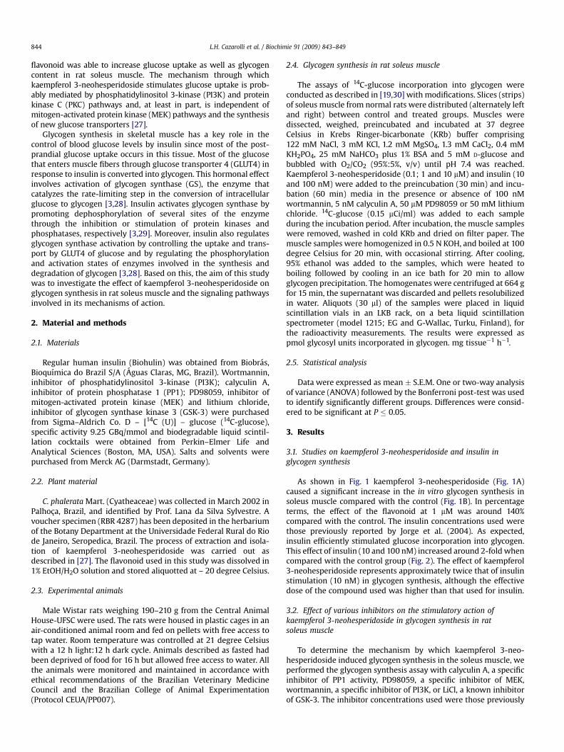
flavonoid was able to increase glucose uptake as well as glycogencontent in rat soleus muscle. The mechanism through whichkaempferol 3-neohesperidoside stimulates glucose uptake is prob-ably mediated by phosphatidylinositol 3-kinase (PI3K) and proteinkinase C (PKC) pathways and, at least in part, is independent ofmitogen-activated protein kinase (MEK) pathways and the synthesisof new glucose transporters [27].
Glycogen synthesis in skeletal muscle has a key role in thecontrol of blood glucose levels by insulin since most of the post-prandial glucose uptake occurs in this tissue. Most of the glucosethat enters muscle fibers through glucose transporter 4 (GLUT4) inresponse to insulin is converted into glycogen. This hormonal effectinvolves activation of glycogen synthase (GS), the enzyme thatcatalyzes the rate-limiting step in the conversion of intracellularglucose to glycogen [3,28]. Insulin activates glycogen synthase bypromoting dephosphorylation of several sites of the enzymethrough the inhibition or stimulation of protein kinases andphosphatases, respectively [3,29]. Moreover, insulin also regulatesglycogen synthase activation by controlling the uptake and trans-port by GLUT4 of glucose and by regulating the phosphorylationand activation states of enzymes involved in the synthesis anddegradation of glycogen [3,28]. Based on this, the aim of this studywas to investigate the effect of kaempferol 3-neohesperidoside onglycogen synthesis in rat soleus muscle and the signaling pathwaysinvolved in its mechanisms of action.
2. Material and methods
2.1. Materials
Regular human insulin (Biohulin) was obtained from Biobras,Bioquımica do Brazil S/A (Aguas Claras, MG, Brazil). Wortmannin,inhibitor of phosphatidylinositol 3-kinase (PI3K); calyculin A,inhibitor of protein phosphatase 1 (PP1); PD98059, inhibitor ofmitogen-activated protein kinase (MEK) and lithium chloride,inhibitor of glycogen synthase kinase 3 (GSK-3) were purchasedfrom Sigma–Aldrich Co. D – [14C (U)] – glucose (14C-glucose),specific activity 9.25 GBq/mmol and biodegradable liquid scintil-lation cocktails were obtained from Perkin–Elmer Life andAnalytical Sciences (Boston, MA, USA). Salts and solvents werepurchased from Merck AG (Darmstadt, Germany).
2.2. Plant material
C. phalerata Mart. (Cyatheaceae) was collected in March 2002 inPalhoça, Brazil, and identified by Prof. Lana da Silva Sylvestre. Avoucher specimen (RBR 4287) has been deposited in the herbariumof the Botany Department at the Universidade Federal Rural do Riode Janeiro, Seropedica, Brazil. The process of extraction and isola-tion of kaempferol 3-neohesperidoside was carried out asdescribed in [27]. The flavonoid used in this study was dissolved in1% EtOH/H2O solution and stored aliquotted at – 20 degree Celsius.
2.3. Experimental animals
Male Wistar rats weighing 190–210 g from the Central AnimalHouse-UFSC were used. The rats were housed in plastic cages in anair-conditioned animal room and fed on pellets with free access totap water. Room temperature was controlled at 21 degree Celsiuswith a 12 h light:12 h dark cycle. Animals described as fasted hadbeen deprived of food for 16 h but allowed free access to water. Allthe animals were monitored and maintained in accordance withethical recommendations of the Brazilian Veterinary MedicineCouncil and the Brazilian College of Animal Experimentation(Protocol CEUA/PP007).
2.4. Glycogen synthesis in rat soleus muscle
The assays of 14C-glucose incorporation into glycogen wereconducted as described in [19,30] with modifications. Slices (strips)of soleus muscle from normal rats were distributed (alternately leftand right) between control and treated groups. Muscles weredissected, weighed, preincubated and incubated at 37 degreeCelsius in Krebs Ringer-bicarbonate (KRb) buffer comprising122 mM NaCl, 3 mM KCl, 1.2 mM MgSO4, 1.3 mM CaCl2, 0.4 mMKH2PO4, 25 mM NaHCO3 plus 1% BSA and 5 mM D-glucose andbubbled with O2/CO2 (95%:5%, v/v) until pH 7.4 was reached.Kaempferol 3-neohesperidoside (0.1; 1 and 10 mM) and insulin (10and 100 nM) were added to the preincubation (30 min) and incu-bation (60 min) media in the presence or absence of 100 nMwortmannin, 5 nM calyculin A, 50 mM PD98059 or 50 mM lithiumchloride. 14C-glucose (0.15 mCi/ml) was added to each sampleduring the incubation period. After incubation, the muscle sampleswere removed, washed in cold KRb and dried on filter paper. Themuscle samples were homogenized in 0.5 N KOH, and boiled at 100degree Celsius for 20 min, with occasional stirring. After cooling,95% ethanol was added to the samples, which were heated toboiling followed by cooling in an ice bath for 20 min to allowglycogen precipitation. The homogenates were centrifuged at 664 gfor 15 min, the supernatant was discarded and pellets resolubilizedin water. Aliquots (30 ml) of the samples were placed in liquidscintillation vials in an LKB rack, on a beta liquid scintillationspectrometer (model 1215; EG and G-Wallac, Turku, Finland), forthe radioactivity measurements. The results were expressed aspmol glycosyl units incorporated in glycogen. mg tissue�1 h�1.
2.5. Statistical analysis
Data were expressed as mean � S.E.M. One or two-way analysisof variance (ANOVA) followed by the Bonferroni post-test was usedto identify significantly different groups. Differences were consid-ered to be significant at P � 0.05.
3. Results
3.1. Studies on kaempferol 3-neohesperidoside and insulin inglycogen synthesis
As shown in Fig. 1 kaempferol 3-neohesperidoside (Fig. 1A)caused a significant increase in the in vitro glycogen synthesis insoleus muscle compared with the control (Fig. 1B). In percentageterms, the effect of the flavonoid at 1 mM was around 140%compared with the control. The insulin concentrations used werethose previously reported by Jorge et al. (2004). As expected,insulin efficiently stimulated glucose incorporation into glycogen.This effect of insulin (10 and 100 nM) increased around 2-fold whencompared with the control group (Fig. 2). The effect of kaempferol3-neohesperidoside represents approximately twice that of insulinstimulation (10 nM) in glycogen synthesis, although the effectivedose of the compound used was higher than that used for insulin.
3.2. Effect of various inhibitors on the stimulatory action ofkaempferol 3-neohesperidoside in glycogen synthesis in ratsoleus muscle
To determine the mechanism by which kaempferol 3-neo-hesperidoside induced glycogen synthesis in the soleus muscle, weperformed the glycogen synthesis assay with calyculin A, a specificinhibitor of PP1 activity, PD98059, a specific inhibitor of MEK,wortmannin, a specific inhibitor of PI3K, or LiCl, a known inhibitorof GSK-3. The inhibitor concentrations used were those previously
L.H. Cazarolli et al. / Biochimie 91 (2009) 843–849844

reported in the literature [27,31–33]. Fig. 3 shows the effect ofcalyculin A on the glycogen synthesis in the soleus muscle. Thisinhibitor was not able to reduce significantly the glycogen synthesisin soleus muscle at 1, 2.5 or 5 nM compared with the control group.On the other hand, it was observed that the stimulatory effect of
kaempferol 3-neohesperidoside on 14C-glucose incorporation intoglycogen in the isolated soleus muscle was completely blocked inthe presence of 5 nM of calyculin (Fig. 3B).
Since the MAPK pathway may be involved in GS activation andsubsequently glycogen synthesis stimulated by insulin, we studiedthe effect of PD98059 on the stimulatory effect of kaempferol3-neohesperidoside in glycogen synthesis. In the presence of 50 mMPD98059 the effect of the flavonoid disappeared, suggesting aninvolvement of MAPK-p90rsk activation in the glycogen synthesisstimulated by kaempferol 3-neohesperidoside (Fig. 4).
To elucidate whether the effects of kaempferol 3-neo-hesperidoside on glycogen synthesis required intact PI3K activity,wortmannin, a potent inhibitor of PI3K was used. The stimulation ofglycogen synthesis by kaempferol 3-neohesperidoside wascompletely blocked by wortmannin with no effect on the basalsynthesis (Fig. 5). Since the stimulatory effect of kaempferol3-neohesperidoside on glycogen accumulation was mediated byPI3K we studied the influence of GSK-3, a downstream effector ofPI3K in the kaempferol 3-neohesperidoside action.
Fig. 6A shows the effect of LiCl, a known inhibitor of GSK-3, onglycogen synthesis in muscle. The maximum stimulatory effectobserved for lithium in glycogen synthesis was at 50 mM. In Fig. 6B,1mM of kaempferol 3-neohesperidoside and 50 mM of LiCl caused
A
B
0
10
20
30
40**
0.1 µM1.0 µM10.0 µM
---
+--
-+-
--+
K3-N
Gly
coge
n sy
nthe
sis
(pm
olgl
ucos
yl u
nits
inco
rpor
ated
into
gly
coge
n. m
g ti
ssue
-1. h
-1)
O
O
OH
HO
OH
O
OH
O
OH
OH
O
OH
OH
OH
O
Fig. 1. Kaempferol 3-neohesperidoside (K3-N) structure (A) and dose–response curveof kaempferol 3-neohesperidoside in glycogen synthesis in the rat soleus muscle.Preincubation time ¼ 30 min; incubation time ¼ 60 min. Values are expressed as mean� S.E.M.; n ¼ 6 in duplicate for each group. Significant at **p � 0.01 in relation tocontrol group.
0
10
20
30
10 nM100 nM
--
+-
-+
***
Insulin
glyc
ogen
syn
thes
is (
pm
olgl
uco
syl u
nit
s in
corp
orat
edin
to g
lyco
gen.
mg
tis
sue-
1 . h
-1)
**
Fig. 2. - Insulin dose–response curve in glycogen synthesis in the rat soleus muscle.Preincubation time ¼ 30 min; incubation time ¼ 60 min. Values are expressed as mean� S.E.M.; n ¼ 10 in duplicate for each group. Significant at **p � 0.01 and ***p � 0.001in relation to control group.
A
zero 1.0 2.5 5.00
5
10
15
Calyculin A (nM)
glyc
ogen
syn
thes
is (
pm
olgl
uco
syl u
nit
s in
corp
orat
edin
to g
lyco
gen.
mg
tis
sue-
1 . h
-1)
B
0
10
20 ***
K3-NCalyculin A
++
-+
+-
--
###gl
ycog
en s
ynth
esis
(p
mol
glu
cosy
l un
its
inco
rpor
ated
into
gly
coge
n. m
g t
issu
e-1 .
h-1
)
Fig. 3. - Dose–response curve of calyculin A in basal glycogen synthesis (A) and effectof 5 nM calyculin A (B) on the stimulatory action of 1 mM kaempferol 3-neo-hesperidoside (K3-N) in glycogen synthesis in the rat soleus muscle. Controlgroup ¼ no treatment. Signals (þ) and (�) indicate presence and absence of eachsubstance in the incubation medium, respectively. Preincubation time ¼ 30 min;incubation time ¼ 60 min. Values are expressed as mean � S.E.M.; n ¼ 6 in duplicatefor each group. Significant at ***p � 0.001 in relation to control group and significant at###p � 0.001 in relation to kaempferol 3-neohesperidoside group.
L.H. Cazarolli et al. / Biochimie 91 (2009) 843–849 845

a similar increase in glycogen accumulation. Under these conditions,when incubation with LiCl plus kaempferol 3-neohesperidoside wascarried out, glycogen synthesis was higher than that observed forLiCl or kaempferol 3-neohesperidoside alone.
4. Discussion
Insulin is the most important hormone that regulates glycogensynthesis in skeletal muscle [1,2]. In our study, we show thatkaempferol 3-neohesperidoside stimulates the in vitro glycogensynthesis in soleus muscle. This effect was observed at 1 mM andrepresented twice that of insulin stimulation in glycogen synthesis.Previous studies have shown that flavonoids such as catechin andmyricetin act in the insulin signaling pathways that regulatesglucose uptake and glycogen synthesis. These flavonoids stimu-lated glycogen synthesis and glucose uptake in insulin targettissues, such as soleus muscle and liver tissues [16,17,21]. Also,procyanidin extract derived from grape seeds was shown toincrease glycogen synthesis in 3T3-L1 adipocytes [20].
Furthermore, it has been demonstrated that kaempferol 3-neo-hesperidoside was able to decrease blood glucose levels in diabeticrats. Additionally, it was shown to increase glucose uptake andglycogen content in soleus muscle as effectively as insulin [6,27].These results point to the insulinomimetic potential effect ofkaempferol 3-neohesperidoside in tissue responsive to insulin.
It is well known that insulin induces phosphorylation/dephos-phorylation of several intracellular enzymes activating or deacti-vating specific pathways that subsequently stimulate glycogensynthase activity [3,31,32]. These processes are regulated throughinsulin binding to its receptor and activating the intrinsic tyrosinekinase activity. Consequently, it promotes the phosphorylation andactivation of PI3K and subsequently the phosphorylation andactivation of protein kinase B (PKB) [34–37]. One important targetof PKB involved in glycogen synthesis is GSK-3 and its phosphor-ylation induced by insulin promotes the activation of glycogensynthase [35,38–40]. Alternatively, insulin also promotes theactivation of the MAPK pathway and protein phosphatase 1 (PP1)activity, the phosphatase responsible for the regulation of glycogenmetabolism [31,32,41,42]. Protein phosphatase 1 has an importantrole in the regulation of glycogen metabolism, where it is inhibitedunder conditions of glycogen degradation and activated underconditions of glycogen synthesis. The basis of the regulation of thePP1 in the skeletal muscle is the ability of the catalytic subunit of
0
10
20
***
###
K3-NPD98059
--
+-
-+
++
Gly
coge
n sy
nth
esis
(p
mol
glu
cosy
l un
its
inco
rpor
ated
into
gly
coge
n. m
g t
issu
e-1 .
h-1
)
Fig. 4. Effect of 50 mM PD98059 on the stimulatory action of 1 mM kaempferol3-neohesperidoside (K3-N) in glycogen synthesis in the rat soleus muscle. Controlgroup ¼ no treatment. Signals (þ) and (�) indicate presence and absence, respectively,of each substance in the incubation medium. Preincubation time ¼ 30 min; incubationtime ¼ 60 min. Values are expressed as mean � S.E.M.; n ¼ 6 in duplicate for eachgroup. Significant at ***p � 0.001 in relation to control group. Significant at###p � 0.001 in relation to kaempferol 3-neohesperidoside group.
0
10
20
***
###
K3-NWortmannin
++
-+
+-
--
Gly
coge
n sy
nth
esis
(p
mol
glu
cosy
l un
its
inco
rpor
ated
into
gly
coge
n. m
g t
issu
e-1 .
h-1
)
Fig. 5. Effect of 100 nM wortmannin on the stimulatory action of 1 mM kaempferol3-neohesperidoside (K3-N) in glycogen synthesis in the rat soleus muscle. Controlgroup ¼ no treatment. Signals (þ) and (�) indicate presence and absence, respectively,of each substance in the incubation medium. Preincubation time ¼ 30 min; incubationtime ¼ 60 min. Values are expressed as mean � S.E.M.; n ¼ 6 in duplicate for eachgroup. Significant at ***p � 0.001 in relation to control group. Significant at###p � 0.001 in relation to kaempferol 3-neohesperidoside group.
zero 2 10 500
10
20
30
40**
glyc
ogen
syn
thes
is (
pm
olgl
uco
syl u
nit
s in
corp
orat
edin
to g
lyco
gen.
mg
tis
sue-
1 . h
-1)
LiCl (mM)
A
B
0
10
20
30
***
*** ***
K3-NLiCl
++
-+
+-
--
#
Gly
coge
n sy
nthe
sis
(pm
olgl
ucos
yl u
nits
inco
rpor
ated
into
gly
coge
n. m
g ti
ssue
-1. h
-1)
Fig. 6. Dose–response curve of LiCl in basal glycogen synthesis (A) and effect of 50 mMLiCl (B) on the stimulatory action of 1 mM kaempferol 3-neohesperidoside (K3-N) inglycogen synthesis in the rat soleus muscle. Control group ¼ no treatment. Signals (þ)and (�) indicate presence and absence, respectively, of each substance in the incu-bation medium. Preincubation time ¼ 30 min; incubation time ¼ 60 min. Values areexpressed as mean � S.E.M.; n ¼ 6 in duplicate for each group. Significant at***p � 0.001 and at **p � 0.01 in relation to control group. Significant at #p � 0.05 inrelation to kaempferol 3-neohesperidoside and LiCl groups.
L.H. Cazarolli et al. / Biochimie 91 (2009) 843–849846

the enzyme to reversibly associate with a glycogen-binding protein,also known as the GM subunit of PP1. As a result, the activity andspecificity of the PP1 are modulated [43]. Calyculin A, a specificinhibitor of PP1 activity, was not able to change the glycogensynthesis in soleus muscle. However, it was observed that thestimulatory effect of kaempferol 3-neohesperidoside on14C-glucose incorporation into glycogen in the isolated soleusmuscle was completely blocked in the presence of calyculin(Fig. 3B).
In liver derived HepG2 cells, insulin increased PP1 activity 4-foldand a quite similar effect of insulin on glycogen synthase activitywas observed. These effects, either on PP1 activity or GS activity,disappeared in the presence of calyculin A. It has been suggestedthat insulin activates glycogen synthase through the activation ofPP1. The results with HepG2 cells provide evidence that insulin actsby changing the phosphorylation state of glycogen synthasethrough the involvement of PP1 [32]. The same finding has beenreported for L6 rat skeletal muscle cells, adipocytes and isolatedskeletal muscle [41,42]. Thus, as with insulin, the stimulatory effectof kaempferol 3-neohesperidoside on glycogen synthesis in soleusmuscle involves the PP1 pathway.
Additionally, insulin has been shown to activate PP1 activity inL6 rat skeletal muscle cells and fresh isolated adipocytes byincreasing the phosphorylation state at site 1 of PP1 [3,41]. This siteis readily phosphorylated in vitro by p90 ribosomal S6 kinase(p90rsk2), a kinase that is phosphorylated and activated by MAPKwhen cells or tissues are incubated with insulin [44,45]. In thepresence of PD98059 the effect of the kaempferol 3-neo-hesperidoside disappeared, suggesting an involvement of MAPK-p90rsk activation in the glycogen synthesis stimulated by
kaempferol 3-neohesperidoside (Fig. 4). It has been proposed thatthe MAPK pathway increases glycogen synthesis stimulated byinsulin in hepatocytes since PD98059 totally inhibits the insulin-stimulated glucose incorporation into glycogen [31]. Furthermore,Dent et al. [45] have suggested that the phosphorylation andactivation of p90rsk by MAPK increases the PP1 activity in rabbitskeletal muscle resulting in the dephosphorylation of GS increasingthe rate of glycogen accumulation.
Despite the wealth of details regarding the precise mechanismby which insulin mediates glycogen synthesis, several questionsremain regarding the role of MAPK and p90rsk in this signaltransduction [32,38,44,46–48]. The data here presented concerningthe role of this flavonoid in glycogen synthesis show that the MAPKsystem must be active for the full kaempferol 3-neohesperidosideresponse in glycogen synthesis.
Besides insulin promoting glycogen synthesis through dephos-phorylation of GS by PP1 activation, it also stimulates glycogensynthesis by inactivating GSK-3. In unstimulated cells, GSK-3 isactive and contributes to the phosphorylation and inactivation ofGS. The inactivation of GSK-3 isoforms GSK-3alpha and GSK-3betais achieved through phosphorylation of Ser21 and Ser9, respectively[3]. This is mediated by PKB in a PI3K-dependent model [36,37].Taking into account the PI3K-PKB-GSK-3 downstream signalinginvolved in the activation of glycogen synthase and the stimulatoryeffect of kaempferol 3-neohesperidoside on glucose uptake blockedby LY294002 [27], the involvement of PI3K in the effect ofkaempferol 3-neohesperidoside on glycogen synthesis was inves-tigated. The stimulation of glycogen synthesis by kaempferol3-neohesperidoside was completely blocked by wortmannin. In3T3-L1 adipocytes, wortmannin inhibited PI3K activity as well as
Fig. 7. Proposed mechanism of action of kaempferol 3-neohesperidoside (K3-N) in glycogen synthesis in soleus muscle.
L.H. Cazarolli et al. / Biochimie 91 (2009) 843–849 847

glycogen synthase activity stimulated by insulin [49]. Moreover, theinhibitory effect of wortmannin on PI3K was also observed inhepatocytes where the basal and insulin-induced glucose incor-poration into glycogen disappeared [31] as also reported forhepatocytes, myoblasts and L6 myotubes [32,38,47]. The inhibitoryeffect of wortmannin on kaempferol 3-neohesperidoside action isin agreement with the results reported above for insulin. This resultindicates that kaempferol 3-neohesperidoside action in glycogensynthesis is mediated through PI3K in soleus muscle and probablyinvolves the PDK-PKB-GSK-3 signaling pathway downstream ofPI3K.
It has been demonstrated that PI3K activates protein kinasedependent on 3-phosphoinositides 1 (PDK 1) which acts on PKBincreasing its phosphorylation and activation states [2,36,37]. Sincethe stimulatory effect of kaempferol 3-neohesperidoside onglycogen accumulation was mediated by PI3K we studied theinfluence of GSK-3, a downstream effector of PI3K in the kaemp-ferol 3-neohesperidoside action.
Kaempferol 3-neohesperidoside and LiCl caused a similarincrease in glycogen accumulation and also presented an additiveaction on glycogen synthesis. In hepatocytes from normal anddiabetic rats, lithium induced a substantial increase in glycogenaccumulation as well as in GS activity. Thus, lithium behaves asinsulin, since both were able to activate GS and stimulate glycogensynthesis in hepatocytes. Thus, the additive effect of kaempferol3-neohesperidoside and lithium on glycogen synthesis seems to besimilar to that reported for lithium and insulin in hepatocytes[50,51].
In summary, we have shown that kaempferol 3-neohesperido-side promptly stimulates glycogen synthesis in soleus muscle. Ashypothesized and shown in Fig. 7, the inhibitor data support therole of PI3K, GSK-3, MAPK and PP1 in the stimulatory effect ofkaempferol 3-neohesperidoside on glycogen synthesis. Thesesimilarities in the involvement of protein kinases and phosphatasesin the stimulation of glycogen deposition by kaempferol 3-neo-hesperidoside and by insulin constitute strong evidence for theinsulinomimetic role of kaempferol 3-neohesperidoside in glucosehomeostasis.
Acknowledgements
This work was supported by grants from Conselho Nacional deDesenvolvimento e Tecnologico (CNPq), Coordenaçao de Pessoal deNıvel Superior (CAPES-PGFAR), and Fundaçao de Amparo a Pes-quisa do Estado de Santa Catarina (FAPESC). The authors thankDr. Roselis S.M. da Silva and Dr. Luis Carlos Kucharski (UFRGS), fortheir helpful advice. We particularly acknowledge Dr. Marcos L.S.Perry (UFRGS), for his useful discussions and critical analysis of thedata. L.H.C. and P.F. were registered on the PGFAR-UFSC. Theauthors express their appreciation to Dr. Siobhan Wiese for assis-tance with the English correction of the manuscript.
References
[1] C. Taha, A. Klip, The insulin signaling pathway, J. Membr. Biol. 169 (1999)1–12.
[2] Y.J. Hei, Recent progress in insulin signal transduction, J. Pharmacol. Toxicol.Methods 40 (1998) 123–135.
[3] A.K. Srivastava, S.K. Pandey, Potential mechanism(s) involved in the regula-tion of glycogen synthesis by insulin, Mol. Cell. Biochem. 182 (1998) 135–141.
[4] F.R.M.B. Silva, B. Szpoganicz, M.G. Pizzolatti, M.A.V. Willrich, E. De Sousa,Acute effect of Bauhinia forficata on serum glucose levels in normal andalloxan-induced diabetic rats, J. Ethnopharmacol. 83 (2002) 33–37.
[5] E. De Sousa, L. Zanatta, I. Seifriz, T.B. Creczynski-Pasa, M.G. Pizzolatti,B. Szpoganicz, F.R.M.B. Silva, Hypoglycemic effect and antioxidant potentialof kaempferol-3,7-O-(alpha)-dirhamnoside from Bauhinia forficata leaves,J. Nat. Prod. 67 (2004) 829–832.
[6] L.H. Cazarolli, L. Zanatta, A.P. Jorge, E. De Sousa, H. Horst, V.M. Voehl,M.G. Pizzolatti, B. Szpoganicz, F.R.M.B. Silva, Follow-up studies on glycosy-lated flavonoids and their complexes with vanadium: their anti-hypergly-cemic potential role in diabetes, Chem. Biol. Interact. 163 (2006) 177–191.
[7] J.T. Jaouhari, H.B. Lazrek, M. Jana, The hypoglycemic activity of Zygophyllumgaetulum extracts in alloxan-induced hyperglycemic rats, J. Ethnopharmacol.69 (2000) 17–20.
[8] G. Zareba, N. Serradell, R. Castaner, S.L. Davies, J. Prous, N. Mealy, Phyto-therapies for diabetes, Drugs Future 30 (2005) 1253–1282.
[9] J.M. Narvaez-Mastache, M.L. Garduno-Ramirez, L. Alvarez, G. Delgado, Anti-hyperglycemic activity and chemical constituents of Eysenhardtia platycarpa,J. Nat. Prod. 69 (2006) 1687–1691.
[10] P.K. Mukherjee, K. Maiti, K. Mukherjee, P.J. Houghton, Leads from Indianmedicinal plants with hypoglycemic potentials, J. Ethnopharmacol. 106(2006) 1–28.
[11] S. Anton, L. Melville, G. Rena, Epigallocatechin gallate (EGCG) mimics insulinaction on the transcription factor FOXO1a and elicits cellular responses in thepresence and absence of insulin, Cell. Signal. 19 (2007) 378–383.
[12] C. Manach, J.L. Donovan, Pharmacokinetics and metabolism of dietaryflavonoids in humans, Free Radic. Res. 38 (2004) 771–785.
[13] L.H. Cazarolli, L. Zanatta, E.H. Alberton, M.S.R.B. Figueiredo, P. Folador,R.G. Damazio, M.G. Pizzolatti, F.R.M.B. Silva, Flavonoids: prospective drugcandidates, Mini Rev. Med. Chem. 8 (2008a) 1429–1440.
[14] L.H. Cazarolli, L. Zanatta, E.H. Alberton, M.S.R.B. Figueiredo, P. Folador,R.G. Damazio, M.G. Pizzolatti, F.R.M.B. Silva, Flavonoids: cellular and molec-ular mechanism of action in glucose homeostasis, Mini Rev. Med. Chem. 8(2008b) 1032–1038.
[15] B.H. Havsteen, The biochemistry and medical significance of the flavonoids,Pharmacol. Ther. 96 (2002) 67–202.
[16] K.C. Ong, H.E. Khoo, Insulinomimetic effects of myricetin on lipogenesis andglucose transport in rat adipocytes but not glucose transport translocation,Biochem. Pharmacol. 51 (1996) 423–429.
[17] K.C. Ong, H.E. Khoo, Effects of myricetin on glycemia and glycogen metabo-lism in diabetic rats, Life Sci. 67 (2000) 1695–1705.
[18] F.L. Hsu, I.M. Liu, D.H. Kuo, W.C. Chen, H.C. Su, J.T. Cheng, Antihyperglycemiceffect of puerarin in streptozotocin-induced diabetic rats, J. Nat. Prod. 66(2003) 788–792.
[19] A.P. Jorge, H. Horst, E. De Sousa, M.G. Pizzolatti, F.R.M.B. Silva, Insulinomi-metic effects of kaempferitrin on glycaemia and on 14C-glucose uptake in ratsoleus muscle, Chem. Biol. Interact. 149 (2004) 89–96.
[20] M. Pinent, M.C. Blade, M.J. Salvado, L. Arola, A. Ardevol, Metabolic fate ofglucose on 3T3-L1 adipocytes treated with grape seed-derived procyanidinextract (GSPE). Comparison with the effects of insulin, J. Agric. Food Chem.53 (2005) 5932–5935.
[21] A.K. Valsa, S. Sudheesh, N.R. Vijayalakshmi, Effect of catechin on carbohy-drate metabolism, Indian J. Biochem. Biophys. 34 (1997) 406–408.
[22] I.M. Liu, S.S. Liou, T.W. Lan, F.L. Hsu, J.T. Cheng, Myricetin as the activeprinciple of Abelmoschus moschatus to lower plasma glucose in streptozo-tocin-induced diabetic rats, Planta Med. 71 (2005) 617–621.
[23] U.J. Jung, M.K. Lee, K.S. Jeong, M.S. Choi, The hypoglycemic effects ofHesperidin and Naringin are partly mediated by hepatic glucose-regulatingenzymes in C57BL/KsJ-db/db mice, J. Nutr. 134 (2004) 2499–2503.
[24] S.A. Park, M.S. Choi, S.Y. Cho, J.S. Seo, U.J. Jung, M.J. Kim, M.K. Sung, Y.B. Park,M.K. Lee, Genistein and daidzein modulate hepatic glucose and lipidregulating enzyme activities in C57BL/KsJ-db/db mice, Life Sci. 79 (2006)1207–1213.
[25] S.N. Singh, P. Vats, S. Suri, R. Shyam, M.M.L. Kumria, S. Ranganathan,K. Sridharan, Effect of an antidiabetic extract of Catharanthus roseus onenzymic activities in streptozotocin induced diabetic rats, J. Ethnopharmacol.76 (2001) 269–277.
[26] L. Zanatta, E. De Sousa, L.H. Cazarolli, A.J. Cunha, M.G. Pizzolatti,B. Szpoganicz, F.R.M.B. Silva, Effect of crude extract and fractions from Vitexmegapotamica leaves on hyperglycemia in alloxan-diabetic rats, J. Ethno-pharmacol. 109 (2007) 151–155.
[27] L. Zanatta, A. Rosso, P. Folador, M.S.R.B. Figueiredo, M.G. Pizzolatti, L.D. Leite,F.R.M.B. Silva, Insulinomimetic effect of kaempferol 3-neohesperidoside onthe rat soleus muscle, J. Nat. Prod. 71 (2008) 532–535.
[28] P.J. Roach, Glycogen and its metabolism, Curr. Mol. Med. 02 (2002) 101–120.[29] M.J. Brady, A.R. Saltiel, The role of protein phosphatase-1 in insulin action,
recent prog, Horm. Res. 56 (2001) 157–173.[30. C. Kucharski, V. Schein, E. Capp, R.S.M. Silva, In vitro insulin stimulatory effect
on glucose uptake and glycogen synthesis in the gills of the estuarine crabChasmagnathus granulate, Gen. Comp. Endocrinol. 125 (2002) 256–263.
[31] J. Carlsen, K. Christiansen, J. Vinten, Insulin stimulated glycogen synthesis inisolated rat hepatocytes: effect of protein kinase inhibitors, Cell. Signal. 09(1997) 447–450.
[32] N.A. Syed, R.L. Khandelwal, Reciprocal regulation of glycogenphosphorylase andglycogen synthase by insulin involving phosphatidylinositol-3 kinase andprotein phosphatase-1 in HepG2 cells, Mol. Cell. Biochem. 211 (2000) 123–136.
[33] A.W. Harmon, D.S. Paul, Y.M. Patel, MEK inhibitors impair insulin-stimulatedglucose uptake in 3T3-L1 adipocytes, Am. J. Physiol. Endocrinol. Metab. 287(2004) 758–766.
[34] J.C. Ferrer, C. Favre, R.R. Gomis, J.M. Fernandez-Novell, M. Garcia-Rocha, N. Dela Iglesia, E. Cid, J.J. Guinovart, Control of glycogen deposition, FEBS Lett. 546(2003) 127–132.
L.H. Cazarolli et al. / Biochimie 91 (2009) 843–849848

[35] A. Mora, K. Sakamoto, E.J. McManus, D.R. Alessi, Role of PDK1 – PKB – GSK-3pathway in regulating glycogen synthase and glucose uptake in the heart,FEBS Lett. 579 (2005) 3632–3638.
[36] E. Hajduch, G.J. Litherland, H.S. Hundal, Protein kinase B (PKB/Akt) – a keyregulator of glucose transport? FEBS Lett. 492 (2001) 199–203.
[37] K. Ueki, R. Yamamoto-Honda, Y. Kaburagi, T. Yamauchi, K. Tobe,B.M.T. Burgering, P.J. Coffer, I. Komuro, Y. Akanuma, Y. Yazaki, T. Kadowaki,Potential role of protein kinase B in insulin-induced glucose transport,glycogen synthesis, and protein synthesis, J. Biol. Chem. 273 (1998) 5315–5322.
[38] S.J. Hurel, J.J. Rochford, A.C. Borthwick, A.M. Wells, J.R. Vandenheede,D.M. Turnbull, S.J. Yeaman, Insulin action in cultured human myoblasts:contribution of different signalling pathways to regulation of glycogensynthesis, Biochem. J. 320 (1996) 871–877.
[39] S.J. Orena, A.J. Torchia, R.S. Garofalo, Inhibition of glycogen-synthase kinase 3stimulates glycogen synthase and glucose transport by distinct mechanismsin 3T3-L1 adipocytes, J. Biol. Chem. 275 (2000) 15765–15772.
[40] K. MacAulay, E. Hajduch, A.S. Blair, M.P. Coghlan, S.A. Smith, H.S. Hundal, Useof lithium and SB-415286 to explore the role of glycogen synthase kinase-3in the regulation of glucose transport and glycogen synthase, Eur. J. Biochem.270 (2003) 3829–3838.
[41] L. Ragolia, N. Begum, Protein phosphatase-1 and insulin action, Mol. Cell.Biochem. 182 (1998) 49–58.
[42] A. Acitores, N. Gonzalez, V. Sancho, I. Valverde, M.L. Villanueva-Penacarrillo,Cell signalling of glucagon-like peptide-1 action in rat skeletal muscle,J. Endocrinol. 180 (2004) 389–398.
[43] J.B. Aggen, A.C. Nairn, R. Chamberlin, Regulation of protein phosphatase-1,Chem. Biol. 7 (2000) R13–R23.
[44] I. Azpiazu, A.R. Saltiel, A.A. DePaoli-Roach, J.C. Lawrence, Regulation of bothglycogen synthase and PHAS-I by insulin in rat skeletal muscle involvesmitogen-activated protein kinase-independent and rapamycin-sensitivepathways, J. Biol. Chem. 271 (1996) 5033–5039.
[45] P. Dent, S.N. Lavoinne, F.B. Caudwell, P. Watt, P. Cohen, The molecularmechanism by which insulin stimulates glycogen synthesis in mammalianskeletal muscle, Nature 348 (1990) 302–308.
[46] D.F. Lazar, R.J. Wiese, M.J. Brady, C.C. Mastick, S.B. Waters, K. Yamauchi,J.E. Pessin, P. Cuatrecasas, A.R. Saltiel, Mitogen-activated protein kinasekinase inhibition does not block the stimulation of glucose utilization byinsulin, J. Biol. Chem. 270 (1995) 20801–20807.
[47] D.A.E. Cross, D.R. Alessi, P. Cohen, M. Andjelkovich, B.A. Hemmings, Inhibitionof glycogen synthase kinase-3 by insulin mediated by protein kinase B,Nature 378 (1995) 785–789.
[48] M. Peak, J.J. Rochford, A.C. Borthwick, S.J. Yeaman, L. Agius, Signalling path-ways involved in the stimulation of glycogen synthesis by insulin in rathepatocytes, Diabetologia 41 (1998) 16–25.
[49] P.R. Shepherd, B.T. Nave, K. Siddle, Insulin stimulation of glycogen synthesisand glycogen synthase activity is blocked by wortmannin and rapamycin in3T3-L1 adipocytes: evidence for the involvement of phosphoinositide3-kinase and p70 ribosomal protein-S6 kinase, Biochem. J. 305 (1995) 25–28.
[50] J.E. Rodrıguez-Gil, J.J. Guinovart, F. Bosch, Lithium restores glycogen synthesisfrom glucose in hepatocytes from diabetic rats, Arch. Biochem. Biophys. 301(1993) 411–415.
[51] J.M. Fernandez-Novell, J.E. Rodriguez-Gil, A. Barbera, J.J. Guinovart, Lithiumions increase hepatic glycogen synthase stability through a proteasome-related mechanism, Arch. Biochem. Biophys. 457 (2007) 29–34.
L.H. Cazarolli et al. / Biochimie 91 (2009) 843–849 849

70
4.5 ARTIGO PUBLICADOPeriódico – Mini-Reviews in Medicinal Chemistry
CAZAROLLI, L.H.; ZANATTA, L.; ALBERTON, E.H.; FIGUEIREDO, M.S.R.B.;FOLADOR, P.; DAMAZIO, R.G.; PIZZOLATTI, M.G.; SILVA, F.R.M.B. Flavonoids:Prospective drug candidates. Mini Reviews in Medicinal Chemistry, v.8, n.13, p. 1429-1440,2008a.

Mini-Reviews in Medicinal Chemistry, 2008, 8, 1429-1440 1429
1389-5575/08 $55.00+.00 © 2008 Bentham Science Publishers Ltd.
Flavonoids: Prospective Drug Candidates
Luisa Helena Cazarolli1, Leila Zanatta
1, Elga Heloisa Alberton
1, Maria Santos Reis Bonorino
Figueiredo1, Poliane Folador
1, Rosangela Guollo Damazio
1, Moacir Geraldo Pizzolatti
2 and
Fátima Regina Mena Barreto Silva1,*
1Departamento de Bioquímica, Centro de Ciências Biológicas and 2Departamento de Química, Centro de Ciências Físi-cas e Matemáticas Campus Universitário, Bairro Trindade. Cx. Postal 5069, CEP: 88040-970, Florianópolis, SC, Brazil
Abstract: The purpose of this review is to discuss the recent developments related to the chemistry and medicinal proper-
ties of flavonoids. Major flavonoids that show well categorized structures and well defined structure function-
relationships are: flavans, flavanones, flavones, flavanonols, flavonols, catechins, anthocyanidins and isoflavone. The bio-
logical properties of flavonoids include antioxidant, anti-inflamatory, antitumoral, antiviral and antibacterial, as well as a
direct cytoprotective effect on coronary and vascular systems, the pancreas and the liver. These characteristics place them
among the most attractive natural substances available to enrich the current therapy options.
Key Words: Flavonoids, medicinal properties, absorption, disease.
CHEMISTRY, CLASSIFICATION AND OCCUR-
RENCE
The flavonoids belong to a large group of low-molecular-weight polyphenolic compounds biosynthesized from both the shikimic acid and acetic acid pathways. Flavonoids are the product of the condensation of the three malonyl-CoA units (C-2) and a P-coumaric acid unit (C-9) to give a basic nucleus (C6-C3-C6) composed of two benzene rings con-nected by a three-carbon unit such as an oxygen-containing pyrene ring. Their structural features comprise a fundamental skeleton of the 2-phenyl chromone with a variety of substitu-tion patterns in the A-ring (characteristically a phlorogluci-nol or resorcinol hydroxylation pattern) and B-ring (usually catechol, pyrogallol or 4´-hydroxylated) [1, 2]. The oxidation level of the three-carbon unit (C-ring) gives rise to different classes of flavonoids including flavans, flavanones, flavones, flavanonols, flavonols, catechins, anthocyanidins, and isofla-vone, among others (Fig. (1)).
Flavonoids are mainly present in nature as glycosides although free aglycones can be found as major constituents in several plants. The glycosilation of a phenolic alcoholic hydroxyl group of a flavonoid aglycone can occur through the hemiacetal bound (O-glycosides) or straight attachment to the C-1 of the sugar unit via a carbon-carbon bond (C-glycosides). A few monosaccharides, such as D-glucose and L-rhamnose, which are the most common, and other less frequent glucorhamnose, galactose, xylose and arabinose, or combinations of these (di- or trisaccharides), can bind to hydroxyl groups or directly to a carbon atom at different positions on the flavonoid aglycone [1, 3-5]. The O-glyco-
*Address correspondence to this author at the Departamento de Bioquímica,
Centro de Ciências Biológicas, Campus Universitário, UFSC. Bairro Trin-dade. Cx. Postal 5069, CEP: 88040-970, Florianópolis, SC, Brazil;
Tel./Fax: +55-48.3721.6912/+55-48.3721.9672; E-mail: [email protected]
silation of the flavonoid molecule occurs most frequently at the C-3 and/or C-7 positions and for C-glycoside flavonoids the preferential glycosilation sites are at positions C-6 and C-8. The large number of flavonoids found in nature is due to the innumerable combinations between flavonoid aglycones and sugars units.
The flavonoids are very widespread in nature and are the largest group of natural products known. They are present in plants in many different glycosidic forms, for example, quer-cetin-3-rutinoside (rutin), quercetin-4´-glucoside and quer-cetin-3,4´-glucoside, which are the most common. Besides their roles in plants, flavonoids are important components in the human diet and are found in fruits, vegetables, seeds, nuts, grains, spices and beverages (wine, tea and beer). Quercetin is considered one of the most common flavonol aglycones in the human diet due its high concentration in several foods including onions, kale, french beans, broccoli, lettuce, toma-toes, apples and beverages [1, 6]. Other flavonols in the diet include kaempferol (broccoli), myricetin (berries), and iso-rhamnetin (onion) [1, 5, 6]. The glycoside flavanones mainly occur in citrus fruits and are represented by hesperetin-7-rutinoside and naringenin-7-rutinoside, which are the major flavonoids of oranges and mandarins. The glycosides narin-genin-7-neohesperoside and narirutin are typically found in grapefruit. Pears, grapes, peaches, vegetables, tea and red wine are sources of catechins that occur as aglycones or es-terified with gallic acid [7-9]. The main flavones in the diet are apigenin and luteolin and most important sources are red pepper, celery, cereal grains and aromatic herbs [9, 10]. Edi-ble fruits, such as plums, apples, eggplant, and many berries have important contents of anthocyanidins and their glyco-sides, which are the compounds responsible for the red, blue or violet color. The most common anthocyanidins are pelar-gonidin, cyanidin, delphinidin, and malvidin [1, 11]. The predominant isoflavonoids are the genistein and daidzein

1430 Mini-Reviews in Medicinal Chemistry, 2008, Vol. 8, No. 13 Cazarolli et al.
found in high concentrations in soybean and soy products [12, 13].
ABSORPTION AND METABOLISM
An important factor in the absorption efficiency of fla-vonoid glycosides in the intestine is the sugar moiety, as demonstrated for quercetin glucosides, aglycone and rutin supplements [14]. Flavonoid aglycones are hydrophobic in nature and can be transported across membranes by passive
diffusion. The glycoside moiety increases the hydrophilicity of the flavonoid molecule which reduces the possibility of passive transport. This leads to the theory that flavonoids are absorbed by active transport [1]. Evidence is accumulating that epithelial brush border membrane transporters play a role in the absorption of dietary (iso)flavonoids. Indirect evi-dence indicates that some flavonoid glycosides can be ab-sorbed intact in the small intestine through the sodium-dependent glucose transporter 1 (SGLT-1), as reported for
Fig. (1). Representative structure of main classes of flavonoids.
2
3
45
6
7
8 1'
2'
3'
4'
5'
6'A
B
C
FLAVAN NUCLEUS
OHO
OH O
OH
(FLAVANONE)
2,3-dihydro-5,7-dihydroxy-2-(4-dihydroxyphenyl)-
4H-1-Benzopyran-4-one (naringenin)
OHO
OH O
OH
OH
(FLAVONE)
5,7-dihydroxy-2-(3,4-hydroxyphenyl)-
4H-1-benzopyran-4-one (luteolin)
OHO
OH O
OH
OH
(FLAVANONOL)
2,3-dihydro-3,5,7-trihydroxy-2-(3,4-dihydroxyphenyl)-
4H-1-benzopyran-4-one (taxifolin)
OH
OHO
OH O
OH
(FLAVONOL)
3,5,7-trihydroxy-2-(4-hydroxyphenyl)-
4H-1-benzopyran-4-one (kaempferol)
OH
OHO
OH
OH
(FLAVANOL)
(2R,3R)-3,4-dihydro-3,5,7-trihydroxy-2-(3,4-dihydroxyphenyl)-
4H-1-benzopyran((-)-epicatechin)
OH
OH
OHO
OH
OH
ANTHOCYANIDIN NUCLEUS
3,5,7-trihydroxy-2-(3,4-dihydroxyphenyl)-
1-benzopirilium cyanidin
OH
OH
OHO
OH OOH
ISOFLAVONE NUCLEUS
5,7-dihydroxy-3-(4-hydroxyphenyl)-
4H-1-benzopyran-4-one (genistein)

Flavonoids and Therapy Mini-Reviews in Medicinal Chemistry, 2008, Vol. 8, No. 13 1431
quercetin-3-glucoside and quercetin-4´-glucoside [14-16] (Fig. (2A)).
On the other hand, the flavonoids can return to the intes-tinal lumen through efficient efflux transport, most likely due to the apical multi-drug resistance protein- 2 (MRP-2), a characteristic intestinal efflux pump, as in the efflux of quer-cetin-4´- -glucoside, (-)-epicatechin and (-)-epigallocatechin-3-gallate [17, 18]. It has been described that before absorp-tion flavonoids are cleaved by specific enzymes either in the lumen or inside the cells of the gut. Lactase-phlorizin hydro-lase (LPH) is anchored in the brush-border membrane in the small intestine and catalyzes extracellular hydrolysis of some glucosides [19, 20]. Another enzyme, located intracellularly and with broad specificity, is the cytosolic -glucosidase (CBG). It is found in abundance in the small intestine, liver and kidney of mammals and requires active transport of hy-drophilic glucosides into the cells [20]. Concerning LPH activity, it has been shown that the enzyme cleaves some flavonols and isoflavone glycosides such as: quercetin- 4´-glucoside, quercetin-3-glucoside, quercetin-3, 4´-glucoside, 3´-methylquercetin-3-glucoside, genistein-7-glucoside, and daidzein-7-glucoside. However, quercetin-3-rhamnoglucoside (rutin) and naringenin-7-rhamnoglucoside (naringin) are not substrates for this enzyme [19, 21]. In addition, -gluco-sidase (BCG) activity is reported to act on flavonoid and isoflavone glycosides according to the position and the struc-ture of the sugar moiety attached to the flavonoid aglycone [22] (Fig. (2A)).
Once absorbed, flavonoids are subject to 3 main types of conjugation: methylation, sulfation and glucuronidation [23]. The metabolic steps in polyphenol metabolism are catalyzed by enzymes. The levels and sites of enzyme expression in human tissues determine the metabolic fate and the pharma-cokinetics of ingested polyphenols and their glycosides [24]. The most important enzymes involved in flavonoids metabo-lism are: catechol-O-methyltransferase (COMT; EC 2.1.1.6), which methylates polyphenols and has the highest activity in the liver and kidneys [25]. Phenol sulfotransferases (P-PST, SULT; EC 2.8.2.1) are cytosolic enzymes that transfer sul-fate moieties to hydroxyl groups from substrates such as iodothyronines, phenols and hydroxyarylamines mainly in the liver [23, 24, 26]. UDP glucuronosyl transferase (UDPGT, UGT; EC 2.4.1.17) catalyzes the conjugation of polyphenols to glucuronic acid in endoplasmic reticulum in the intestine, liver and kidney. In humans, the liver has the greatest capac-ity for glucuronidation while in rats, the highest level of glu-curonyl transferase activity was observed in the intestine [26-28].
Conjugation reactions with glucuronic acid and/or sulfate seem to be the most common type of metabolic pathways for the flavonoids [1]. This conjugation first occurs in the gut barrier and these conjugates then reach the liver, where they are further metabolized [26]. For example, catechin is exten-sively methylated in the liver [29] and increased plasma total (+)-catechin levels were observed after hydrolysis of its glu-curonide and sulphate derivatives from volunteers that in-gested red wine [30]. Otake et al. [31] using human liver microsomes demonstrated that hepatic UDP-glucuronosyl transferase isoforms were the main factor responsible for galangin metabolism into two major glucuronides conjugated
at the 7- and 3- positions. Also, Vaidyanathan and Walle [32] demonstrated no glucuronidation of (-)-epicatechin by human liver and small intestinal microsomes. However, in rats, (-)-epicatechin was efficiently metabolized by liver mi-crosomes with formation of two glucuronides. In the same study, the authors concluded that sulfation also occurred in both the liver and intestine in human and rats.
On the other hand, glycoside flavonoids that are not ab-sorbed in the small intestine along with the conjugated me-tabolites that are excreted in bile can be metabolized by mi-croflora when they reach the colon (Fig. (2B)). Glycoside flavonoid-hydrolyzing enzymes have been identified in fecal flora cultures. Bokkenheuser et al. [33], recovered three en-zyme-producing strains that, using -glucosidases, -rhamno-sidases, and/or -galactosidases, were capable of converting rutin to quercetin. Also, it was shown that at least some of the bacterial glycosidases are able to cleave glycosidic bonds and flavonoid-saccharide bonds in the gut [19]. The profile of metabolites has been demonstrated in studies with quer-cetin, rutin and naringin. The flavonoid metabolism produces aromatic acids such as phenylvaleric, phenylpropionic, phe-nylacetic and benzoic acids with easy absorption through the colonic barrier [34-36].
Metabolites of flavonoids in general (and also microflora metabolites), aglycones, glycosides and conjugated metabo-lites which are not absorbed, may follow two pathways of excretion: via the biliary or the urinary route. Large conju-gated metabolites are more likely to be eliminated in the bile whereas small conjugates such as monosulfates are preferen-tially excreted in urine [27]. When excreted in bile, the fla-vonoids are passed to the duodenum and metabolized by intestinal bacteria, which results in the production of frag-mentation products and/or the hydrolysis of glucurono- or sulfoconjugates [37]. The resulting metabolites which are released may be reabsorbed and enter an enterohepatic cycle or being excreted in feces [38, 39] (Fig. (2B)). For each fla-vonoid, the beneficial effect will be dependent upon their absorption and availability in the body. Thus, these factors should be considered in any interpretation of the potential health effects of flavonoids.
MEDICINAL PROPERTIES
1. Antioxidant Activity
The major mechanisms of action of antioxidant agents are suppression of reactive oxygen species formation, either by inhibition of enzymes or chelating trace elements involved in free radical production; scavenging of reactive oxygen species (ROS); and upregulation or protection of antioxidant defenses [40].
The antioxidant activity of flavonoids and the mecha-nisms involved in their action are extensively revised by Pietta [41] and Amic et al. [42]. The configuration and total number of hydroxyl groups are determinant factors in the mechanisms of antioxidant activity of flavonoids. Particu-larly, the B-ring hydroxyl configuration is significant as a scavenger of ROS and reactive nitrogen species (RNS) [43, 44]. Furthermore, a 3´,4´-catechol structure in the B-ring and the presence of 6-hydroxyl group potentiates lipid peroxida-tion inhibition as well as the free radical scavenger effect

1432 Mini-Reviews in Medicinal Chemistry, 2008, Vol. 8, No. 13 Cazarolli et al.
[45, 46]. Different classes of compounds are able to scav-enge reactive species including: catechin, luteolin, kaemp-ferol, 3-O-methyl-quercetin, naringenin, kaempferol 3-O-D-glycoside, kaempferol-3-O-(2´´,6´´-di-O-p-trans-coumaroyl) glucoside, 6-hydroxyluteolin glycosides, hesperidin and quercetin [46-49].
The flavonoid heterocycle contributes to antioxidant ac-tivity by permitting conjugation between the aromatic rings and particularly through the presence of a free 3-OH which
can be potentiated by the presence of a 3´,4´-catechol ex-plaining the potent antioxidant activity of flavan-3-ols and flavon-3-ols which possess the latter feature [50, 51]. Also, the carbonyl group at C-4 and a double bond between C-2 and C-3 are important features in relation to the high anti-oxidant activity of flavonoids [51, 52]. Furthermore, the number of polyhydroxy or polymethoxy substituents deter-mines the difference in the antioxidant activities of flavon-oids probably due to the hydrophobicity and molecular pla-
A
B
Fig. (2). Schematic representation of intestinal absorption (A) and metabolism of flavonoids (B). SGLT-1 = sodium-dependent glucose
transporter-1; MRP-2 = multidrug-resistance associated protein-2; LPH = lactase phloridzin hydrolase; CBG = cytosolic -glucosidase. Fig.
(A) was adapted from Nemeth et al., (2003) [20].

Flavonoids and Therapy Mini-Reviews in Medicinal Chemistry, 2008, Vol. 8, No. 13 1433
narity induced by the substitution models [45, 50]. Another important feature of antioxidant agents is the presence, num-ber and position of glycosides in the flavonoid structure. In general, aglycones are more potent antioxidants than the corresponding glycosides, as demonstrated by daidzein, ge-nistein and their 7-glycosides, kaempferitrin and the flavonol glycosides in green tea [52-56].
One of the most well known antioxidant activities of fla-vonoids is the inhibition of the enzymes responsible for su-peroxide anion production, such as xanthine oxidase [57]. Also, flavonoids inhibit the enzymes of the cyclooxygenase and lipoxygenase pathway [58] and microsomal monooxy-genase, glutathione S-transferase, mitochondrial succinoxi-dase, and NADH oxidase, all involved in ROS generation [59]. Another possible contributory mechanism to the anti-oxidant activity of flavonoids is the ability to efficiently chelate trace metals through interaction with the catechol moiety in the B-ring, 3-hydroxyl, 4-oxo groups in the hetero-cyclic ring, and 4-oxo, 5-hydroxyl groups between the het-erocyclic and the A-rings, decreasing the availability of these metals to participate in the generation of free radicals [60, 61].
2. Anti-Inflammatory Activity
Several cellular action mechanisms have been proposed to explain the in vivo and in vitro anti-inflammatory activity of flavonoids. They can modulate the functions and activities of inflammatory and immune cells through, for instance, inhibition of histamine release from mast cells, immunosup-pressive effects on T-cell proliferation and IL-2 synthesis, and regulation of secretion of IgG, IgM and IgA isotypes [58, 62]. There are also reports concerning flavonoid actions over a range of cellular types such as platelets, eosinophils, neutrophils, mast cells, basophils, macrophages and mono-cytes [to review see 62, 63].
In addition, one of the direct actions of flavonoids is modulating the activity of arachidonic acid (AA) metaboliz-ing enzymes [58]. The formation of AA is the rate limiting step in the synthesis of prostaglandis, leukotrienes and the platelet activating factor. This pathway is triggered by phos-pholipase A2 (PLA2). AA undergoes metabolism to several active products (eicosanoids) by two major routes: the cy-clooxygenase and lipoxygenase pathways. The immediate products of the cyclooxygenase pathway are endoperoxides (PGG2 and PGH2), which are converted to prostaglandins (PGE2 , PGF2Q and PGD2) by prostaglandins synthetase enzymes, as well as thromboxanes (TXA2 and TXB2) and prostacyclins (PGI2) by the corresponding synthetases. The lipoxygenase pathway converts AA to hydroxyl fatty acids, including hydroperoxyeicosatetraenoic acids (HPETEs), which are then metabolized to hydroxyeicosatetraenoic acids (HETEs). The HETEs and their leukotriene metabolites are important mediators of inflammatory responses [64].
The first flavonoid inhibitor of PLA2 activity discovered was quercetin, which inhibits the activity of PLA2 from hu-man neutrophils and several other sources. Also, other fla-vonoids have been reported to inhibit PLA2 activity, for ex-ample, hesperetin, naringenin, kaempferol, myricetin and biflavonoids [64, 65].
The cyclooxygenase (COX) enzyme exists basically in two different isoforms: COX-1 and COX-2 [66, 67]. Some flavonoids such as luteolin, 3´,4´-dihydroxyflavone, galangin, and morin inhibit COX activity [68]. Inhibition was also observed for baicalin, (+)-catechin, rutin, chrysin and its derivatives, kaempferol and quercetin in different animal tissues and cells [58, 69]. Studies on flavonoid inhibition of COX-2 have been rarely reported. An inhibitory activity against COX-2 or COX-1/COX-2 are reported for some flavan-3-ols such as catechin and 4´-Me-gallocatechin and for two dihydrochalcones [70-72]. Lipooxygenase (LOXs) enzyme isoforms also act in inflammatory processes and are responsible for generating hydroxy acids and leukotrienes (LTs) from AA. Flavonols, including kaempferol, quercetin, morin and myricetin, have been found to be stronger LOX inhibitors than flavones [69, 73].
Nitric oxide (NO) is one of the cellular mediators of physiological and pathological processes involved in in-flammatory events and it is biochemically synthesized from L-arginine by nitric oxide synthase (NOS) isoforms [74, 75]. Quercetin, apigenin, luteolin, genistein, kaempferol, chrysin and its derivatives were found to inhibit NO production to act against NOS isoforms both by inhibition of induced NOS (iNOS) activity or down-regulation of iNOS expression from different cell types [76, 77]. Furthermore, evidence supports the idea that certain flavonoids act as modulators of proin-flammatory gene expression.
The tumor necrosis factor- (TNF- ) and IL-1 are prominent contributors to chronic inflammatory disorders [78]. Genistein and other flavonoids are reported to inhibit IL-1 , IL-6, and TNF- production in several kinds of cells [79, 80]. Activation of NF- B is inhibited by genistein, api-genin, kaempferol, oroxylin A [77, 81], epigallocatechin 3-gallate and amentoflavone [82, 83] while quercetin, rutin and luteolin inhibits the expression and production of TNF- and intercellular adhesion molecule 1 (ICAM-1) in mice [84, 85]. Another flavonoid, myricitrin, has also been described as an inhibitor of the nociceptive responses in models of acute pain by inhibiting of phosphatidylinositol 3-kinase (PI3K) and protein kinase C (PKC) activities, NO production, iNOS over expression and NF-kB activation [86, 87].
3. Protective Effects on Coronary Disease and Vascular
Activity
Atherosclerosis is a chronic inflammatory response in the walls of arteries, in large part due to the deposition of lipo-proteins (plasma proteins that carry cholesterol and triglyc-erides). There is much evidence that oxidized low density lipoprotein (LDL) is responsible for cholesterol loading of macrophages, foam cell formation and atherogenesis [88, 89]. LDL is oxidized by free radicals generated by endothe-lial cells, macrophages and smooth muscle cells. Therefore, it has been hypothesized that oxidized LDL is the responsi-ble for the initiation and promotion of atherogenesis [90, 91].
In this regard, several aglycone and polyhydroxylated flavonoids, such as quercetin, morin, hypoleatin, setin, gossy-petin and galangin, are potent inhibitors of LDL oxidation invitro by macrophages or copper ions. They may reduce the formation of free radicals (for instance, chelating divalent

1434 Mini-Reviews in Medicinal Chemistry, 2008, Vol. 8, No. 13 Cazarolli et al.
metal involved in the Fenton reactions), and protect the -tocopherol present in the LDL structure from oxidative dam-age or regenerate it [92, 93]. Along with all of the activity of flavonoids against atherogenesis, they may inhibit adhesion and platelet aggregation as well as promote vascular smooth muscle relaxation. The antiaggregatory effects of flavonoids seems to influence the platelet activation pathway, such as the inhibition of the enzymes involved in AA metabolism as well as the inhibition of platelet aggregation by antagonizing thromboxane formation and thromboxane receptor function [94, 95].
The antioxidant actions of flavonoids appear to partici-pate in their antithrombotic action. The antithrombotic and vasoprotective actions of quercetin, rutin, and other flavon-oids have been attributed to their ability to bind to platelet membranes and scavenge free radicals, restoring the biosyn-thesis and action of endothelial prostacyclin and endothelial-derived relaxing factor [96-98]. Flavonoids with anti-platelet activity include isobavachalcone and neobavaisoflavone, luteolin, genistein, quercetin, apigenin and kaempferol de-rivatives [99-101]. One of the flavonoid mechanisms of platelet aggregation inhibition is to increase cyclic AMP (cAMP) levels through adenylate cyclase activation and phosphodiesterase inhibition [102, 103]. Quercetin-4´-O- -D-glycoside inhibited collagen-stimulated tyrosine phos-phorylation of platelet and quercetin inhibits the intracellular Ca
2+ mobilization suggesting that these flavonoids act as
inhibitors of the trigger signal for thrombus formation [104, 105]. These results have been recently confirmed in humans by Hubbard et al. [106].
In addition to their antiaggregatory effects, flavonoids appear to increase vasodilation by inducing vascular smooth muscle relaxation which may be mediated by the inhibition of PKC, phosphodiesterase, or by decreased cellular Ca
2+
uptake [102]. Luteolin, naringenin and eriodictyol have been shown to promote the relaxation of rat aorta contractions induced by Ca
2+, noradrenaline and K
+ [107]. Quercetin,
chrysin and (-)-epicatechin also have vasorelaxant effects [108-111].
4. Antitumoral Activity
Cancer is a disease caused by a combination of exoge-nous and endogenous factors which results in a cellular cyclo imbalance (mitosis/apoptosis) turning normal cells into can-cer cells [2].
The flavonoids are one of the most promising anticancer natural products that have been studied [112-114]. They in-terfere with a large number of regulatory pathways such as: cellular growth, energy metabolism, apoptosis, cell division, transcription, gene repair, neuronal transmission, inflamma-tion, and stress response which may be involved in tumori-genesis [2, 62]. Quercetin has been reported to inhibit many biochemical events associated with tumor promotion, such as alteration in PKC activity and interactions with calmodulin [115, 116]. Also, quercetin strongly inhibits the expression of the mutated p53 (tumor suppressor gene) protein prevent-ing the accumulation of newly synthesized p53 protein with-out affecting the steady-state mRNA levels of p53 in cancer cell lines [117, 118]. This flavonoid also exhibited antipro-
liferative effects in: drug-resistant leukemia cells in vitro and in vivo, colon and hepatocellular cancers in rats and mice and exerted growth-inhibitory effects on several malignant tumor cell lines in vitro [119-123].
In in vivo studies quercetin, kaempferol, and myricetin have been found to be able to inhibit carcinogen-induced tumors in rats and mice [124]. Other flavonoids such as cate-chin, epicatechin, quercetin, and resveratrol, polyphenolic compounds in red wine have been shown to inhibit growth of human breast and prostate cancer cells [125, 126]. The expo-sure of human epidermoid carcinoma cells to silymarin re-sulted in a significant decrease in ligand-induced activation of the epidermal growth factor receptor (EGFR) with an as-sociated decrease in EGFR intrinsic kinase activity. This was accompanied by inhibition of DNA synthesis and cell growth [127].
The anticancer effects of genistein and its derivatives, such as biochanin A, daidzein, genistin and daidzin, have also been reported. They potently inhibit the growth of hu-man breast carcinoma cell lines [128, 129]. In vitro studies have shown that such chemopreventive and antineoplastic effects are associated with the antioxidant activity of genis-tein and inhibitor activities in cell proliferation and angio-genesis [130, 131] as demonstrated recently for genistin and daidzin in M14 cells [132]. Effects of green tea on the inhibi-tion of carcinogenesis in experimental animal models, along with its constituents, for example, (-)- epigallocatechin gallate, in all levels of cancer progression, namely initiation, promo-tion and transformation, have been described [133, 134].
Recently, Cárdenas et al. [135] studied the effects of various natural flavonoids, cinnamic acid derivatives, and a series of synthetic flavones on cell proliferation in vitro in established human and murine tumor cell lines. The most potent antiproliferative agents were caffeic acid n-butyl ester > 2
´-nitroflavone > caffeic acid ethyl ester ~ 2´, 6-dini-
troflavone > apigenin > 3´-bromoflavone ~ 2´-fluoro-6-bro-moflavone. Some compounds showed a moderate effect, the order of cytotoxic activities being chrysin > 2´-fluoro-6-chloroflavone ~ 2´-chlorochrysin > -naphthoflavone > -naphthoflavone ~ 6-chloroflavone ~ 6-bromoflavone ~ 4´-nitroflavone. None of the natural or synthetic compounds tested affected the proliferation of epithelial cells derived from normal mammary glands of mice or fibroblastic cells of the mouse embryo, suggesting a selective action against tu-mor cells. Besides the flavonoid actions described above, they can also influence adhesion molecules, metastasis and angiogenesis as well as apoptosis, gene expression and mutagenicity, which may contribute to its anticarcinogenic activity [see review 2, 62].
5. Antiviral Effects
Viruses are obligate intracellular parasites, which contain little more than bundles of gene strands of either RNA or DNA, and may be surrounded by a lipid-containing envelope [136]. Unlike bacterial cells, which are free-living entities, viruses utilize the host cell environment to propagate new viruses. They use the reproductive machinery and metabolic pathways of cells to provide energy and building blocks for new viral particles. In an alternative stage, the viral genes

Flavonoids and Therapy Mini-Reviews in Medicinal Chemistry, 2008, Vol. 8, No. 13 1435
may remain silent for a prolonged period inside the host cells [136].
A recent area of research that is of particular interest is the apparent inhibitory activity of some flavonoids against human immunodeficiency virus (HIV). The mechanisms of action of compounds such as baicalin, robustoflavone and hinokiflavone, robinetin, myricetin, quercetagetin, quercetin 3-O-(2´´-galloyl)- -l-arabinopyranoside and chrysin seem to involve the inhibition of entry, infection, transcription and replication in cells as well as the inhibition of enzymes in-volved with these processes, for instance, reverse tran-scriptase, HIV-1 proteinase and integrase [137-141].
The effects of green tea constituents on the HIV-1 viral life cycle have being studied. It was found that (-)- epigallo-catechin gallate caused the destruction of the viral particles and inhibited viral attachment to cells, post-adsorption entry into cells, reverse transcription and viral production from chronically-infected cells [142]. Epigallocatechin-3-gallate showed a dose-dependent effect on the inhibition of the rep-lication HIV strains [143]. An inhibitory effect on HIV-1 replication was shown for tea flavin and catechin derivatives. These tea polyphenols are able to inhibit HIV-1 entry into target cells by blocking HIV-1 envelope glycoprotein-mediated membrane fusion [144].
The structural basis for the antiviral activity of naturally occurring flavonoids appears to be associated with non gly-cosidic compounds and hydroxylation at the 3´, 4´, 3, 5, and 7-position is apparently a prerequisite for antiviral activity [145]. A number of possible mechanisms whereby polyphe-nols may exert their antiviral action have been proposed. The action of flavonoids probably derives from their direct inac-tivation of the virus and/or from inhibition of the virus bind-ing to the cells. They are also known to inhibit viral replica-tion [146].
The antiherpetic activity of several flavonoids against the herpes simplex virus type 1 and type 2 has being evaluated and morin, epicatechin, epicatechin gallate (flavanols), ge-nistein (isoflavone), naringenin (flavanone), quercetin (fla-vonol), galangin, kaempferol, catechin, epigallocatechin, epigallocatechin gallate, chrysin, baicalin, fisetin, myricetin, and genistein showed significant effects [147, 148]. The an-tiherpetic activity and genotoxicity of catechin and some of its derivatives has also been confirmed by Savi et al. [149].
Homoisoflavonoids were identified as having low cyto-toxicity and a good antiviral activity against Coxsackie vi-ruses (CVB3, CVB4, CAV9) and Echovirus 30 (Echo30) which may be useful as an additional antiviral drug against these infections [150]. Furthermore, antiviral activity against the human cytomegalovirus (HCMV) has been shown for baicalein. The basic mechanism of action seems to be the blockage of HCMV infection through inhibiting it entry into the cells and its replication [151].
7. Antibacterial Activity
The use of flavonoids against bacterial, protozoan, and fungal infections has two purposes: (1) to kill the bacterial or fungal cells and (2) to counteract the spread and the effects of the bacterial toxins [152]. However, the mechanism by
which this is accomplished is not known yet. Antibacterial mechanisms of action of flavonoids based on their structure-activity relationships [to review see 153] have been pro-posed, for example, inhibition of nucleic acid synthesis [154, 155], cytoplasmatic membrane function [156, 157] and en-ergy metabolism [158].
Recently, the antibacterial activity of some flavonoids has been increasingly documented. Examples of such fla-vonoids are apigenin, galangin, chrysin, sophoraflavanone G and its derivatives, naringin and naringenin, epigallocatechin gallate and its derivatives, luteolin and luteolin 7- glucoside, quercetin, 3-O-methylquercetin and various quercetin gly-cosides, along with kaempferol and its derivatives [159-166]. Other flavones, isoflavones, flavanones, flavonols, flavonol glycosides and chalcones with antibacterial activity have also been identified [163, 167-170].
The activity of the flavonoids apigenin, baicalin and galangin against sensitive and antibiotic resistant strains of Staphylococcus aureus, Enterococcus faecalis, Enterococcus faecium, Escherichia coli and Pseudomonas aeruginosa has been investigated and galangin and apigenin were shown to have an inhibitory activity [171]. Genistein presented inhibi-tory effects on the growth of staphylococcal strains, Strepto-coccus pasteurianus, Bacillus cereus, and Helicobacter py-lori, whereas Escherichia coli growth was not suppressed. Daidzein, which is structurally similar to genistein, also in-hibited the growth of Staphylococcus aureus, albeit with lower potency than genistein [172]. Although advances in understanding the role played by flavonoids in each particu-lar pathology are still required, it is now clear that structure function-relationship of glycosylated flavonoids indicates a molecular mechanism which is crucial to the drug discovery process.
ACKNOWLEDGEMENTS
Studies in the authors´ laboratory were supported by Conselho Nacional de Desenvolvimento e Tecnológico CNPq); Coordenação de Pessoal de Nível Superior (CAPES-PGFAR) and Fundação de Amparo à Pesquisa do Estado de Santa Catarina (FAPESC). The authors express their appre-ciation to Dr. Siobhan Wiese for assistance with the English correction of the manuscript and to the student Alexandre Balduino Westphal for his extensive computer modeling of the schematic drawings.
ABBREVIATIONS
AA = Arachidonic acid
AMP = 3'-5'- Adenosine monophosphate
cAMP = 3'-5'-Cyclic adenosine monophosphate
CBG = Cytosolic -glucosidase
COMT = Catechol-O-methyltransferase
COX = Cyclooxygenase
CVB = Coxsackie viruses
DNA = Deoxyribonucleic acid
EGFR = Epidermal growth factor receptor
1436 Mini-Reviews in Medicinal Chemistry, 2008, Vol. 8, No. 13 Cazarolli et al.
HCMV = Human cytomegalovirus
HETE = Hydroxyeicosatetraenoic acid
HIV = Human immunodeficiency virus
HPETE = Hydroperoxyeicosatetraenoic acid
ICAM-1 = Intercellular adhesion molecule 1
IgM, IgA, IgG = Immunoglobulines
IL-1 = Interleukin 1
Il-6 = Interleukin 6
LDL = Low density lipoprotein
LOXs = Lipooxygenase isoforms
LPH = Lactase-phlorizin hydrolase
LT = Leucotrienes
mRNA = Messenger ribonucleic acid
MRP-2 = Apical multi-drug resistance protein- 2
NADH = Nicotinamide adenine dinucleotide oxidase oxidase
NF- B = Nuclear factor-kappa B
NO = Nitric oxide
NOS = Nitric oxide synthase
PGE2 , PGF2Q = Prostaglandins and PGD2
PGI2 = Prostacyclin
PI3K = Phosphatidylinositol 3-kinase
PKC = Protein kinase C
PLA2 = Phospholipase A2
P-PST, SULT = Phenol sulfotransferases
RNA = Ribonucleic acid
RNS = Reactive nitrogen species
ROS = Reactive oxygen species
SGLT-1 = Sodium-dependent glucose transporter 1
TNF- = Tumor necrosis factor-
TXA2 and = Thromboxanes TXB2
UDPGT, UGT = UDP Glucuronosyl transferase
REFERENCES
[1] Aherne, S.A.; O´Brien, N.M. Dietary Flavonols: chemistry, food
content, and metabolism. Nutrition, 2002, 18, 75-81. [2] Havsteen, B.H. The biochemistry and medical significance of the
flavonoids. Pharmacol. Ther., 2002, 96, 67-202. [3] Havsteen, B.H. Flavonoids, a class of natural products of high
pharmacological potency. Biochem. Pharmacol., 1983, 32, 1141-8. [4] Williams, C.A.; Harborne, J.B. in The Flavonoids. Advances in
research since 1986, Harborne, J.B. Ed; Chapman & Hall: London, 1994, 337-85.
[5] Erlund, I. Review of the flavonoids quercetin, hesperetin, and nar-
ingenin. Dietary sources, bioactivities, bioavailability and epidemi-ology. Nutr. Res., 2004, 24, 851-74.
[6] Harborne, J.B.; Williams, C.A. Advances in flavonoid research since 1992. Phytochemistry, 2000, 55, 481-504.
[7] Tomás-Barberán, F.A.; Clifford, M.N. Flavanones, chalcones and dihydrochalcones - nature, occurrence and dietary burden. J. Sci. Food Agric., 2000, 80, 1073-80.
[8] Kawaii, S.; Tomono, Y.; Katase, E.; Ogawa, K.; Yano, M. Quanti-
tation of flavonoid constituents in citrus fruits. J. Agric. Food Chem., 1999, 47, 3565-71.
[9] Arts, I.C.W.; Van de Putte, B.; Hollman, P.C.H. Catechin contents of foods commonly consumed in The Netherlands. 2. Tea, wine,
fruit juices, and chocolate milk. J. Agric. Food Chem., 2000, 48,1752-7.
[10] Hertog, M.G.L.; Hollman, P.C.H.; Venema, D.P. Optimization of a quantitative HPLC determination of potentially anticarcinogenic
flavonoids in vegetables and fruits. J. Agric. Food Chem., 1992, 40,1591-8.
[11] Wu, X.; Beecher, G.R.; Holden, J.M.; Haytowitz, D.B.; Gebhardt, S.E.; Prior, R.L. Concentrations of anthocyanins in common foods
in the United States and estimation of normal consumption. J. Ag-ric. Food Chem. 2006, 54, 4069-75.
[12] Liggins, J.; Bluck, L.J.; Runswick, S.; Atkinson, C.; Coward, W.A.; Bingham, S.A. Daidzein and genistein contents of vegeta-
bles. Br. J. Nutr., 2000, 84, 717-25. [13] Mazur, W.M.; Duke, J.A.; Wähälä, K.; Rasku, S.; Adlercreutz, H.
Isoflavonoids and lignans in legumes: nutritional and health aspects in humans. J. Nutr. Biochem., 1998, 9, 193-200.
[14] Hollman, P.C.H.; Vries, J.H.M. de; Leeuwen, S.D.; Mengelers, M.J.B.; Katan, M.B. Absorption of dietary quercetin glycosides and
quercetin in healthy ileostomy volunteers. Am. J. Clin. Nutr., 1995,62, 1276-82.
[15] Chang, Q.; Zuo, Z.; Chow, M.S.S.; Ho, W.K.K. Difference in ab-sorption of the two structurally similar flavonoid glycosides, hy-
peroside and isoquercitrin, in rats. Eur. J. Pharm. Biopharm., 2005,59, 549-55.
[16] Gee, J.M.; DuPont, M.S.; Day, A.J.; Plumb, G.W.; Williamson, G.; Johnson, I.T. Intestinal transport of quercetin glycosides in rats in-
volves both deglycosylation and interaction with the hexose trans-port pathway. J. Nutr., 2000, 130, 2765-71.
[17] Walgren, R.A.; Karnaky, K.J.; Lindenmeyer, G.E.; Walle, T. Ef-flux of dietary flavonoid quercetin-4’- -glucoside across human in-
testinal Caco-2 cell monolayers by apical multidrug resistance-associated protein-2. J. Pharmacol. Exp. Ther., 2000, 294, 830-6.
[18] Vaidyanathan, J.B.; Walle, T. Transport and metabolism of the tea flavonoid (-)-epicatechin by the human intestinal cell line Caco-2.
Pharm. Res., 2001, 18, 1420-5. 19] Day, A.J.; Cañada, F.J.; Diaz, J.C.; Kroon, P.A.; Mclauchlan, R.;
Faulds, C.B.; Plumb, G.W.; Morgan, M.R.; Williamson, G. Dietary flavonoid and isoflavone glycosides are hydrolysed by the lactase
site of lactase phlorizin hydrolase. FEBS Lett., 2000, 468, 166-70. [20] Németh, K.; Plumb, G.W.; Berrin, J.G.; Juge, N.; Jacob, R.; Naim,
H.Y.; Williamson, G.; Swallow, D.M.; Kroon, P.A. Deglycosyla-tion by small intestinal epithelial cell beta-glucosidases is a critical
step in the absorption and metabolism of dietary flavonoid gly-cosides in humans. Eur. J. Nutr., 2003, 42, 29-42.
[21] Day, A.J.; Gee, J.M.; DuPont, M.S.; Johnson, I.T.; Williamson, G. Absorption of quercetin-3-glucoside and quercetin-4’-glucoside in
the rat small intestine: the role of lactase phlorizin hydrolase and the sodium-dependent glucose transporter. Biochem. Pharmacol.,2003, 65, 1199-206.
[22] Lambert, N.; Kroon, P.A.; Faulds, C.B.; Plumb, G.W.; McLauch-
lan, W.R.; Day, A.J.; Williamson, G. Purification of cytosolic beta-glucosidase from pig liver and its reactivity towards flavonoid gly-
cosides. Biochim. Biophys. Acta, 1999, 1435, 110-6. [23] Manach, C.; Scalbert, A.; Morand, C.; Rémésy, C.; Jiménez, L.
Polyphenols: food sources and bioavailability. Am. J. Clin. Nutr.,2004, 79, 727- 47.
[24] Scalbert, A.; Williamson, G. Dietary intake and bioavailability of polyphenols. J. Nutr., 2000, 130 (8S suppl), 2073S-85S.
[25] Nielsen, S. E.; Breinholt, V.; Justesen, U.; Cornett, C.; Dragsted, L. O. In vitro biotransformation of flavonoids by rat liver microsomes.
Xenobiotica, 1998, 28, 389-401.

Flavonoids and Therapy Mini-Reviews in Medicinal Chemistry, 2008, Vol. 8, No. 13 1437
[26] Piskula, M.K.; Terao. J. Accumulation of (-)-epicatechin metabo-
lites in rat plasma after oral administration and distribution of con-jugation enzymes in rat tissues. J. Nutr., 1998, 128, 1172-8.
[27] Mojarrabi, B.; Mackenzie, P. I. Characterization of two UDP glu-curonosyltransferases that are predominantly expressed in human
colon. Biochem. Biophys. Res. Commun., 1998, 247, 704-9. [28] Strassburg, C. P.; Nguyen, N.; Manns, M. P.; Tukey, R. H. UDP-
glucuronosyltransferase activity in human liver and colon. Gastro-enterology, 1999, 116, 149-60.
[29] Donovan, J.L.; Crespy, V.; Manach, C.; Morand, C.; Besson, C.; Scalbert, A.; Rémésy, C. Catechin is metabolized by both the small
intestine and liver of rats. J. Nutr., 2001, 131, 1753-7. [30] Bell, J.R.C.; Donovan, J. L.; Wong, R.; Waterhouse, A. L.; Ger-
man, J. B.; Walzem, R. L.; Kasim-Karakas, S. E. (+)-Catechin in human plasma after ingestion of a single serving of reconstituted
red wine. Am. J. Clin. Nutr., 2000, 71, 103-8. [31] Otake, Y.; Hsieh, F.; Walle, T. Glucuronidation versus oxidation of
the flavonoid galangin by human liver microsomes and hepato-cytes. Drug Metab. Dispos., 2002, 30, 576-81.
[32] Vaidyanathan, J.B.; Walle, T. Glucuronidation and sulfation of the tea flavonoid (-)-epicatechin by the human and rat enzymes. Drug Metab. Dispos., 2002, 30, 897-903.
[33] Bokkenheuser, V.D.; Shackleton, C.H.L.; Winter, J. Hydrolysis of
dietary flavonoid glycosides by strains of intestinal Bacteroides from humans. Biochem. J., 1987, 248, 953-6.
[34] Rechner, A.R.; Smith, M.A.; Kuhnle, G.; Gibson, G.R.; Debnam, E.S.; Srai, S.K.S.; Moore, K.P.; Rice-Evans, C.A. Colonic metabo-
lism of dietary polyphenols: influence of structure on microbial fermentation products. Free Radic. Biol. Med., 2004, 36, 212-25.
[35] Jenner, A.M.; Rafter, J.; Halliwell, B. Human fecal water content of phenolics: the extent of colonic exposure to aromatic com-
pounds. Free Radic. Biol. Med., 2005, 38, 763-72. [36] Aura, A.M.; O´Leary, K.A.; Williamson,G.; Ojala, M.; Bailey, M.;
Puupponen-Pimiä, R.; Nuutila, A.M.; Oksman-Caldentey, K.M.; Poutanen, K. Quercetin derivatives are deconjugated and converted
to hydroxyphenylacetic acids but not methylated by human fecal flora in vitro. J. Agric. Food Chem., 2002, 50, 1725-30.
[37] Formica, J.V.; Regelson, W. Review of the biology of quercetin and related bioflavonoids. Food Chem. Toxicol., 1995, 33, 1061-
80. [38] Crespy, V.; Morand, C.; Besson, C.; Cotelle, N.; Vézin, H.; Demi-
gné, C.; Rémésy, C. The splanchnic metabolism of flavonoids highly differed according to the nature of the compound. Am. J. Physiol. Gastrointest. Liver Physiol., 2003, 284, G980-8.
[39] Silberberg, M.; Morand, C.; Mathevon, T.; Besson, C.; Manach, C.;
Scalbert, A.; Rémésy, C. The bioavailability of polyphenols is highly governed by the capacity of the intestine and of the liver to
secrete conjugated metabolites. Eur. J. Nutr., 2006, 45, 88-96. [40] Halliwell, B.; Gutteridge, J. M. C. Free Radicals in Biology and
Medicine; Oxford University Press: Oxford, 1998.[41] Pietta, P.G. Flavonoids as Antioxidants. J. Nat. Prod., 2000, 63,
1035-42. [42] Amic, D.; Davidovic-Amic, D.; Beslo, D.; Rastija, V.; Lucic, B.;
Trinajstic, N. SAR and QSAR of the antioxidant activity of flavon-oids. Curr. Med. Chem., 2007, 14, 827-45.
[43] Cao, G.; Sofic, E.; Prior, R.L. Antioxidant and prooxidant behavior of flavonoids: structure-activity relationships. Free Radic. Biol. Med., 1997, 22, 749-60.
[44] Sekher Pannala, A.; Chan, T.S.; O´Brien, P.J.; Rice-Evans, C.A.
Flavonoid B-ring chemistry and antioxidant activity: fast reaction kinetics. Biochem. Biophys. Res. Commun., 2001, 282, 1161-8.
[45] Dugas Jr., A.J.; Castaneda-Acosta, J.; Bonin, G.C.; Price, K.L.; Fischer, N.H.; Winston, G.W. Evaluation of the total peroxyl radi-
cal-scavenging capacity of flavonoids: structure-activity relation-ships. J. Nat. Prod., 2000, 63, 327-31.
[46] Es-Safi, N.E.; Kollmann, A.; Khlifi, S.; Ducrot, P.H. Antioxidative effect of compounds isolated from Globularia alypum L. Structure-
activity relationship. LWT- Food Sci. Technol., 2007, 40, 1246-52. [47] Sadhu, S.K.; Okuyama, E.; Fujimoto, H.; Ishibashi, M.; Yesilada,
E. Prostaglandin inhibitory and antioxidant components of Cistus laurifolius, a Turkish medicinal plant. J. Ethnopharmacol., 2006,
108, 371-8. [48] Susanti, D.; Sirat, H.M.; Ahmad, F.; Ali, R.M.; Aimi, N.; Kitajima,
M. Antioxidant and cytotoxic flavonoids from the flowers of Melastoma malabathricum L. Food Chem., 2007, 103, 710-6.
[49] Cirico, T.L.; Omaye, S.T. Additive or synergetic effects of pheno-
lic compounds on human low density lipoprotein oxidation. Food Chem. Toxicol., 2006, 44, 510-6.
[50] Wang, L.; Tu, Y.C.; Lian, T.W.; Hung, J.T.; Yen, J.H.;Wu, M.J. Distinctive Antioxidant and Antiinflammatory Effects of Fla-
vonols. J. Agric. Food Chem., 2006, 54, 9798-804. [51] Rice-Evans, C.A.; Miller, N.J.; Paganga, G. Structure-antioxidant
activity relationships of flavonoids and phenolic acids. Free Radic. Biol. Med., 1996, 20, 933-56.
[52] Burda, S.; Oleszek, W. Antioxidant and antiradical activities of flavonoids. J. Agric. Food Chem., 2001, 49, 2774-9.
[53] Gao, Z.; Huang, K.; Yang, X.; Xu, H. Free radical scavenging and antioxidant activities of flavonoids extracted from the radix of
Scutellaria baicalensis Georgi. Biochim. Biophys. Acta, 1999,1472, 643-50.
[54] Lai, H.H.; Yen, G.C. Inhibitory effect of isoflavones on peroxyni-trite-mediated low-density lipoprotein oxidation. Biosci. Biotech-nol. Biochem., 2002, 66, 22-8.
[55] Plumb, G.W.; Price, K.R.; Williamson, G. Antioxidant properties
of flavonol glycosides from tea. Redox Rep., 1999, 04, 13-6. [56] De Sousa, E.; Zanatta, L.; Seifriz, I.; Creczynski-Pasa, T.B.; Pizzo-
latti, M.G.; Szpoganicz, B.; Silva, F.R.M.B. Hypoglycemic effect and antioxidant potential of kaempferol-3,7-O-( )-dirhamnoside
from Bauhinia forficata leaves. J. Nat. Prod., 2004, 67, 1-4. [57] Van Hoorn, D.E.C.; Nijveldt, R.J.; Van Leeuwen, P.A.M.; Hofman,
Z.; M´Rabet, L.; De Bont, D.B.; Van Norren, K. Accurate predic-tion of xanthine oxidase inhibition based on the structure of flavon-
oids. Eur. J. Pharmacol., 2002, 451, 111-8. [58] Kim, H.P.; Son, K.H.; Chang, H.W.; Kang, S.S. Anti-inflammatory
plant flavonoids and cellular action mechanisms. J. Pharm. Sci.,2004, 96, 229-45.
[59] Korkina, L.G.; Afanasév, I.B. Antioxidants in Disease Mechanisms and Therapy; Sies, H., Ed.; Academic Press: San Diego, 1997, 151-
63. [60] Soczynska-Kordala, M.; Bakowska, A.; Oszmianski, J.; Gabrielska,
J. Metal ion-flavonoid associations in bilayer phospholipid mem-branes. Cell. Mol. Biol. Lett., 2001, 6, 277-81.
[61] Cheng, I.F.; Breen. K. On the ability of four flavonoids, baicilein, luteolin, naringenin, and quercetin, to suppress the Fenton reaction
of the iron-ATP complex. Biometals, 2000, 13, 77-83. [62] Middleton Jr., E.; Kandaswami, C.; Theoharides, T.C. The effects
of plant flavonoids on mammalian cells: implications for inflamma-tion, heart disease, and cancer. Pharmacol. Rev., 2000, 52, 673-
751. [63] Theoharides, T.C.; Alexandrakis, M.; Kempuraj, D.; Lytinas, M.
Anti-inflammatory actions of flavonoids and structural require-ments for new design. Int. J. Immunopathol. Pharmacol., 2001, 14,
119-27. [64] Yoon, J.H.; Baek, S.J. Molecular targets of dietary polyphenols
with anti-inflammatory properties. Yonsei Med. J., 2005, 46, 585-96.
[65] Lättig, J.; Böhl, M.; Fischer, P.;Tischer, S.; Tietböhl, C.; Men-schikowski, M.; Gutzeit, H.O.; Metz, P.; Pisabarro, M.T. Mecha-
nism of inhibition of human secretory phospholipase A2 by flavon-oids: rationale for lead design. J. Comput. Aided. Mol. Des., 2007,
21, 473-83. [66] Simmons, D.L. Variants of cyclooxygenase-1 and their roles in
medicine. Thromb. Res., 2003, 110, 265-8. [67] Needleman, P.; Isakson, P.C. The discovery and function of COX-
2. J. Rheumatol. Suppl., 1997, 49, 6-8. [68] Baumann, J.; Bruchhausen, F.V.; Wurm, G. Flavonoids and related
compounds as inhibitors of arachidonic acid peroxidation. Prosta-glandins, 1980, 20, 627-39.
[69] Laughton, M.J.; Evans, P.J.; Moroney, M.A.; Hoult, J.R.; Halli-well, B. Inhibition of mammalian 5-lipoxygenase and cyclooxy-
genase by flavonoids and phenolic dietary additives. Relationship to antioxidant activity and to iron ion-reducing ability. Biochem. Pharmacol., 1991, 42, 1673-81.
[70] Noreen, Y.; Serrano, G.; Perera, P.; Bohlin, L. Flavan-3-ols iso-
lated from some medicinal plants inhibiting COX-1 and COX-2 catalysed prostaglandin biosynthesis. Planta Med., 1998, 64, 520-4.
[71] Likhiwitayawuid, K.; Sawasdee, K.; Kirtikara, K. Flavonoids and stilbenoids with COX-1 and COX-2 inhibitory activity from Dra-caena loureiri. Planta Med., 2002, 68, 841-3.

1438 Mini-Reviews in Medicinal Chemistry, 2008, Vol. 8, No. 13 Cazarolli et al.
[72] Takano-Ishikawa, Y.; Goto, M.; Yamaki, K. Structure-activity
relations of inhibitory effects of various flavonoids on lipopolysac-charide-induced prostaglandin E2 production in rat peritoneal
macrophages: Comparison between subclasses of flavonoids. Phy-tomedicine, 2006, 13, 310-7.
[73] Burnett, B.P.; Jia, Q.; Zhao, Y.; Levy, R.M. A medicinal extract of Scutellaria baicalensis and Acacia catechu acts as a dual inhibitor
of cyclooxygenase and 5-lipoxygenase to reduce inflammation. J. Med. Food, 2007, 10, 442-51.
[74] Moncada, S.; Palmer, R.M.; Higgs, E.A. Nitric oxide: physiology, pathophysiology, and pharmacology. Pharmacol. Rev., 1991, 43,
109-42. [75] Nathan C. Nitric oxide as a secretory product of mammalian cells.
FASEB J., 1992, 6, 3051-64. [76] Chiesi, M.; Schwaller, R. Inhibition of constitutive endothelial NO-
synthase activity by tannin and quercetin. Biochem. Pharmacol.,1995, 49, 495-501.
[77] Liang, Y.C.; Huang, Y.T.; Tsai, S.H.; Lin-Shiau, S.Y.; Chen, C.F.; Lin, J.K. Suppression of inducible cyclooxygenase and inducible
nitric oxide synthase by apigenin and related flavonoids in mouse macrophages. Carcinogenesis, 1999, 20, 1945-52.
[78] Bingham, C.O. 3rd. The pathogenesis of rheumatoid arthritis: piv-otal cytokines involved in bone degradation and inflammation. J. Rheumatol. Suppl., 2002, 65, 3-9.
[79] Cho, J.Y.; Kim, P.S.; Park, J.; Yoo, E.S.; Baik, K.U.; Kim, Y.K.;
Park, M.H. Inhibitor of tumor necrosis factor-alpha production in lipopolysaccharide-stimulated RAW 264.7 cells from Amorpha fru-ticosa. J. Ethnopharmacol., 2000, 70, 127-33.
[80] Krakauer, T.; Li, B.Q.; Young, H.A. The flavonoid baicalin inhibits
superantigen-induced inflammatory cytokines and chemokines. FEBS Lett., 2001, 500, 52-5.
[81] Chen, Y.C.; Yang, L.; Lee, T.J. Oroxylin A inhibition of lipopoly-saccharide-induced iNOS and COX-2 gene expression via suppres-
sion of nuclear factor-kappaB activation. Biochem. Pharmacol.,2000, 59, 1445-57.
[82] Singh, R.; Ahmed, S.; Islam, N.; Goldberg, V.M.; Haqqi, T.M. Epigallocatechin-3-gallate inhibits interleukin-1beta-induced ex-
pression of nitric oxide synthase and production of nitric oxide in human chondrocytes: Suppression of nuclear factor kappaB by deg-
radation of the inhibitor of nuclear factor kappaB. Arthritis Rheum.,2002, 46, 2079-86.
[83] Banerjee, T.; Valacchi, G.; Ziboh, V.A.; Van der Vliet, A. Inhibi-tion of TNFalpha-induced cyclooxygenase-2 expression by amen-
toflavone through suppression of NF-kappaB activation in A549 cells. Mol. Cell. Biochem., 2002, 238, 105-10.
[84] Kotanidou, A.; Xagorari, A.; Bagli, E.; Kitsanta, P.; Fotsis, T.; Papapetropoulos, A.; Roussos, C. Luteolin reduces lipopolysaccha-
ride-induced lethal toxicity and expression of proinflammatory molecules in mice. Am. J. Respir. Crit. Care Med., 2002, 165, 818-
23. [85] Ueda, H.; Yamazaki, C.; Yamazaki, M. A hydroxyl group of fla-
vonoids affects oral anti-inflammatory activity and inhibition of systemic tumor necrosis factor-alpha production. Biosci. Biotech-nol. Biochem., 2004, 68, 119-25.
[86] Meotti, F.C.; Luiz, A.P.; Pizzolatti, M.G.; Kassuya, C.A.; Calixto,
J.B.; Santos, A.R. Analysis of the antinociceptive effect of the fla-vonoid myricitrin: evidence for a role of the L-arginine-nitric oxide
and protein kinase C pathways. J. Pharmacol. Exp. Ther., 2006,316, 789-96.
[87] Gamet-Payrastre, L.; Manenti, S.; Gratacap, M.P.; Tulliez, J.; Chap, H.; Payrastre, B. Flavonoids and the inhibition of PKC and
PI 3-kinase. Gen. Pharmacol., 1999, 32, 279-86. [88] Goldstein, J.L.; Ho, Y.K.; Basu, S.K.; Brown, M.S. Binding site on
macrophages that mediates uptake and degradation of acetylated low density lipoprotein, producing massive cholesterol deposition.
Proc. Natl. Acad. Sci. USA, 1979, 76, 333-7. [89] Sparrow, C.P.; Parthasarathy, S.; Steinberg, D. A macrophage
receptor that recognizes oxidized low density lipoprotein but not acetylated low density lipoprotein. J. Biol. Chem., 1989, 264, 2599-
604. [90] Steinberg, D.; Parthasarathy, S.; Carew, T.E.; Khoo, J.C.; Witztum,
J.L. Beyond cholesterol: Modification of low-density lipoprotein that increases its atherogenicity. New Engl. J. Med., 1989, 320,
915-24.
[91] Lapointe, A.; Couillard, C.; Lemieux, S. Effects of dietary factors
on oxidation of low-density lipoprotein particles. J. Nutr. Biochem.,2006, 17, 645-58.
[92] Zhu, Q.Y.; Huang, Y.; Chen, Z.Y. Interaction between flavonoids and alpha-tocopherol in human low density lipoprotein. J. Nutr. Biochem., 2000, 11, 14 -21.
[93] Safari, M.R.; Sheikh, N. Effects of flavonoids on the susceptibility
of low-density lipoprotein to oxidative modification. Prostagland-ins Leukot. Essent. Fatty Acids, 2003, 69, 73-7.
[94] Tzeng, S.H.; Ko, W.C.; Ko, F.N.; Teng, C.M. Inhibition of platelet aggregation by some flavonoids. Thromb. Res., 1991, 64, 91-100.
[95] Elliott, A.J.; Scheiber, S.A.; Thomas, C.; Pardini, R.S. Inhibition of glutathione reductase by flavonoids. A structure-activity study.Biochem. Pharmacol., 1992, 44, 1603-8.
[96] Gryglewski, R.J.; Korbut, R.; Robak, J.; Swies, J. On the mecha-
nism of antithrombotic action of flavonoids. Biochem. Pharmacol.,1987, 36, 317-22.
[97] Robak, J.; Korbut, R.; Shridi, F.; Swies, J.; Rzadkowska-Bodalska, H. On the mechanism of antiaggregatory effect of myricetin. Pol. J. Pharm. Pharmacol., 1988, 40, 337-40.
[98] Robak, J.; Gryglewski, R.J. Flavonoids are scavengers of superox-
ide anions. Biochem. Pharmacol., 1988, 37, 837-41. [99] Tsai, W.J.; Hsin, W.C.; Chen, C.C. Antiplatelet flavonoids from
seeds of Psoralea corylifolia. J. Nat. Prod., 1996, 59, 671-2. [100] Lin, C.N.; Kuo, S.H.; Chung, M.I.; Ko, F.N.; Teng, C.M. A new
flavone C-glycoside and antiplatelet and vasorelaxing flavones from Gentiana arisanensis. J. Nat. Prod., 1997, 60, 851-3.
[101] Dhar, A.; Paul, A.K.; Shukla, S.D. Platelet-activating factor stimu-lation of tyrosine kinase and its relationship to phospholipase C in
rabbit platelets: Studies with genistein and monoclonal antibody to phosphotyrosine. Mol. Pharmacol., 1990, 37, 519-25.
[102] Duarte, J.; Pérez Vizcaino, F.; Utrilla, P.; Jiménez, J.; Tamargo, J.; Zarzuelo, A. Vasodilatory effects of flavonoids in rat aortic smooth
muscle. Structure activity relationships. Gen. Pharmacol., 1993,24, 857-62.
[103] Kuppusamy, U.R.; Das, N.P. Effects of flavonoids on cyclic AMP phosphodiesterase and lipid mobilization in rat adipocytes. Bio-chem. Pharmacol., 1992, 44, 1307-15.
[104] Hubbard, G.P.; Wolffram, S.; Lovegrove, J.A.; Gibbins, J.M. The
role of polyphenolic compounds in the diet as inhibitors of platelet function. Proc. Nutr. Soc., 2003, 62, 469-78.
[105] Hubbard, G.P.; Wolffram, S.; Lovegrove, J.A.; Gibbins, J.M. In-gestion of quercetin inhibits platelet aggregation and essential
components of the collagen-stimulated platelet activation pathway in humans. J. Tromb. Haemost., 2004, 2, 2138-45.
[106] Hubbard, G.P.; Wolffram, S.; De Vos, R.; Bovy, A.; Gibbins, J.M.; Lovegrove, J.A. Ingestion of onion soup high in quercetin inhibits
platelet aggregation and essential components of the collagen-stimulated platelet activation pathway in man: a pilot study. Br. J. Nutr., 2006, 96, 482-8.
[107] Sánchez de Rojas, V.R.; Somoza, B.; Ortega, T.; Villar, A.M. Iso-
lation of vasodilatory active flavonoids from the traditional remedy Satureja obovata. Planta Med., 1996, 62, 272-4.
[108] Roghani, M.; Baluchnejadmojarad, T.; Vaez-Mahdavi, M.R.; Roghani-Dehkordi, F. Mechanisms underlying quercetin-induced
vasorelaxation in aorta of subchronic diabetic rats: an in vitrostudy. Vasc. Pharmacol., 2004, 42, 31-5.
[109] Ajay, M.; Achike, F.I.; Mustafa, A.M.; Mustafa, M.R. Effect of quercetin on altered vascular reactivity in aortas isolated from
streptozotocin-induced diabetic rats. Diabetes Res. Clin. Pract.,2006, 73, 1-7.
[110] Chen, Z.Y.; Yao, X.Q.; Chan, F.L.; Lau, C.W.; Huang, Y. (-) Epicatechin induces and modulates endothelium-dependent relaxa-
tion in isolated rat mesenteric artery rings. Acta Pharmacol. Sin.,2002, 23, 1188-92.
[111] Villar, I.C.; Galisteo, M.; Vera, R.; O´Valle, F.; García-Saura, M.F.; Zarzuelo, A.; Duarte, J. Effects of the Dietary Flavonoid
Chrysin in Isolated Rat Mesenteric Vascular Bed. J. Vasc. Res.,2004, 41, 509-16.
[112] Lopez-Lazaro, M. Flavonoids as anticancer agents: structure-activity relationship study. Curr. Med. Chem. - Anti-Cancer Agents,
2002, 2, 691-714. [113] Ren,W.; Qiao, Z.; Wang, H.; Zhu, L.; Zhang, L. Flavonoids: prom-
ising anticancer agents. Med. Res. Rev., 2003, 23, 519-34.

Flavonoids and Therapy Mini-Reviews in Medicinal Chemistry, 2008, Vol. 8, No. 13 1439
[114] Li, Y.; Fang, H.; Xu, W. Recent advance in the research of flavon-
oids as anticancer agents. Mini Rev. Med. Chem., 2007, 7, 663-78. [115] Gschwendt, M.; Horn, F.; Kittstein, W.; Marks, F. Inhibition of the
calcium and phospholipid-dependent protein kinase activity from mouse brain cytosol by quercetin. Biochem. Biophys. Res. Com-mun., 1983, 117, 444-7.
[116] Nishino, H.; Naito, E.; Iwashima, A.; Tanaka, K.; Matsuura, T.;
Fujiki, H.; Sugimura. T. Interaction between quercetin and Ca21 calmodulin complex: Possible mechanism for anti-tumor-
promoting action of the flavonoids. Gann, 1984a, 74, 311-6. [117] Kuo, P.C.; Liu, H.F.; Chao, J.I. Survivin and p53 modulate quer-
cetin-induced cell growth inhibition and apoptosis in human lung carcinoma cells. J. Biol. Chem., 2004, 279, 55875-85.
[118] Avila, M.A.; Velasco, J.A.; Cansado, J.; Notario, V. Quercetin mediates the down-regulation of mutant p53 in the human breast
cancer cell line MDA-MB468. Cancer Res., 1994, 54, 2424-8. [119] Hoffman, R.; Graham, L.; Newlands, E.S. Enhanced antiprolifera-
tive action of busulphan by quercetin on the human leukaemia cell line K562. Br. J. Cancer, 1989, 59, 347-8.
[120] Hofmann, J.; Fiebig, H.H.; Winterhalter, B.R.; Berger, D.P.; Grunicke, H. Enhancement of the antiproliferative activity of cis-
diamminedichloroplatinum (II) by quercetin. Int. J. Cancer, 1990,45, 536-9.
[121] Yoshida, M.; Sakai, T.; Hosokawa, N.; Marui, N.; Matsumoto, K.; Fujioka, A.; Nishino H.; Aoike, A. The effect of quercetin on cell
cycle progression and growth of human gastric cancer cells. FEBS Lett., 1990, 260, 10-3.
[122] Hosokawa, N.; Hosokawa, Y.; Sakai, T.; Yoshida, M.; Marui, N.; Nishino, H.; Kawai, K.; Aoike, A. Inhibitory effect of quercetin on
the synthesis of a possibly cell-cycle-related 17-kDa protein, in human colon cancer cells. Int. J. Cancer, 1990b, 45, 1119-24.
[123] Kandaswami, C.; Perkins, E.; Soloniuk, D.S.; Drzewiecki, G.; Middleton, E. Jr. Antiproliferative effects of citrus flavonoids on a
human squamous cell carcinoma in vitro. Cancer Lett., 1991, 56,147-52.
[124] Nakayama, T.; Yamada, M.; Osawa, T.; Kawakishi, S. Suppression of active oxygen-induced cytotoxicity by flavonoids. Biochem. Pharmacol., 1993, 45, 265-7.
[125] Damianaki, A.; Bakogeorgou, E.; Kampa, M.; Notas, G.; Hatzo-
glou, A.; Panagiotou, S.; Gemetzi, C.; Kouroumalis, E.; Martin, P.M.; Castanas, E. Potent inhibitory action of red wine polyphenols
on human breast cancer cells. J. Cell. Biochem., 2000, 78, 429-41. [126] Kampa, M.; Hatzoglou, A.; Notas, G.; Damianaki, A.; Bakogeor-
gou, E.; Gemetzi, C.; Kouroumalis, E.; Martin, P.M.; Castanas, E. Wine antioxidant polyphenols inhibit the proliferation of human
prostate cancer cell lines. Nutr. Cancer, 2000, 37, 105-15. [127] Ahmad, N.; Gali, H.; Javed, S.; Agarwal, R. Skin cancer chemo-
preventive effects of a flavonoid antioxidant silymarin are mediated via impairment of receptor tyrosine kinase signaling and perturba-
tion in cell cycle progression. Biochem. Biophys. Res. Commun.,1998, 247, 294-301.
[128] Barnes, S. Effect of genistein on in vitro and in vivo models of cancer. J. Nutr., 1995, 125, 777S-83S.
[129] Peterson, G.; Barnes, S. Genistein inhibition of the growth of hu-man breast cancer cells: Independence from estrogen receptors and
the multi-drug resistance gene. Biochem. Biophys. Res. Commun.,1991, 179, 661-7.
[130] Peterson, G. Evaluation of the biochemical targets of genistein in tumor cells. J. Nutr., 1995, 125, 784S-9S.
[131] Booth, C.; Hargreaves, D.F.; Hadfield, J.A.; McGown, A.T.; Pot-ten, C.S. Isoflavones inhibit intestinal epithelial cell proliferation
and induce apoptosis in vitro. Br. J. Cancer, 1999, 80, 1550-7. [132] Russo, A.; Cardile, V.; Lombardo, L.; Vanella, L.; Acquaviva, R.
Genistin inhibits UV light-induced plasmid DNA damage and cell growth in human melanoma cells. J. Nutr. Biochem., 2006, 17,
103-8. [133] Dreosti, I.E.; Wargovich, M.J.; Yang, C.S. Inhibition of carcino-
genesis by tea: The evidence from experimental studies. Crit. Rev. Food Sci. Nutr., 1997, 37, 761-70.
[134] Mitscher, L.; Jung, M.; Shankel, D.; Dou, J.H.; Steele, L.; Pillai, S.P. Chemoprotection: a review of the potential therapeutic anti-
oxidant properties of green tea and certain of its constituents. Med. Res. Rev., 1997, 17, 327-65.
[135] Cárdenas, M.; Marder, M.; Blank, V.C.; Roguin, L.P. Antitumor activity of some natural flavonoids and synthetic derivatives on
various human and murine cancer cell lines. Bioorg. Med. Chem.,2006, 14, 2966-71.
[136] Wagner, E.K.; Hewlett, M.J. Basic Virology, Blackwell Science:
Malden, USA, 1999.[137] Li, B.Q.; Fu, T.; Dongyan, Y.; Mikovits, J.A.; Ruscetti, F.W.;
Wang, J.M. Flavonoid baicalin inhibits HIV-1 infection at the level of viral entry. Biochem. Biophys. Res. Commun., 2000, 276, 534-8.
[138] Lin, Y.M.; Anderson, H.; Flavin, M.T.; Pai, Y.H.; Mata-Greenwood, E.; Pengsuparp, T.; Pezzuto, J.M.; Schinazi, R.F.;
Hughes, S.H.; Chen, F.C. In vitro anti-HIV activity of biflavonoids isolated from Rhus succedanea and Garcinia multiora. J. Nat. Prod., 1997, 60, 884-8.
[139] Fesen, M.R.; Pommier, Y.; Leteurtre, F.; Hiroguchi, S.; Yung, J.;
Kohn, K.W. Inhibition of HIV-1 integrase by flavones, caffeic acid phenethyl ester (CAPE) and related compounds. Biochem. Phar-macol., 1994, 48, 595-608.
[140] Kim, H.J.; Woo, E.R.; Shin, C.G.; Park, H. A new flavonol glyco-
side gallate ester from Acer okamotoanum and its inhibitory activ-ity against human immunodeficiency virus-1 (HIV-1) integrase. J. Nat. Prod., 1998, 61, 145-8.
[141] Critchfield, J.W.; Butera, S.T.; Folks, T.M. Inhibition of HIV acti-
vation in latently infected cells by flavonoid compounds. AIDS Res. Hum. Retroviruses, 1996, 12, 39-46.
[142] Yamaguchi, K.; Honda, M.; Ikigai, H.; Hara, Y.; Shimamura, T. Inhibitory effects of (-)-epigallocatechin gallate on the life cycle of
human immunodeficiency virus type 1 (HIV-1). Antiviral Res.,2002, 53, 19-34.
[143] Fassina, G.; Buffa, A.; Benelli, R.; Varnier, O.E.; Noonan, D.M.; Albini, A. Polyphenolic antioxidant (-)-epigallocatechin-3-gallate
from green tea as a candidate anti-HIV agent. AIDS, 2002, 16, 939-41.
[144] Liu, S.; Lu, H.; Zhao, Q.; He, Y.; Niu, J.; Debnath, A.K.; Wu, S.; Jiang, S. Theaflavin derivatives in black tea and catechin deriva-
tives in green tea inhibit HIV-1 entry by targeting gp41. Biochim. Biophys. Acta, 2005, 1723, 270-81.
[145] Wleklik, M.; Luczak, M.; Panasiak, W.; Kobus, M.; Lammer-Zarawska, E. Structural basis for antiviral activity of flavonoids-
naturally occurring compounds. Acta Virol., 1988, 32, 522-5. [146] Sakagami, H.; Sakagami, T.; Takeda, M. Antiviral properties of
polyphenols. Polyphenol Actualites, 1995, 12, 30-2. [147] Bunyapraphatsara, N.; Dechsree, S.; Yoosook, C.; Herunsalee, A.;
Panpisutchai, Y. Anti-herpes simplex virus component isolated from Maclura cochinchinensis. Phytomedicine, 2000, 6, 421-4.
[148] Lyu, S.Y.; Rhim, J.Y.; Park, W.B. Antiherpetic activities of fla-vonoids against herpes simplex virus type 1 (HSV-1) and type 2
(HSV-2) in vitro. Arch. Pharm. Res., 2005, 28, 1293-301. [149] Savi, L.A.; Barardi, C.R.; Simões, C.M. Evaluation of Antiherpetic
Activity and Genotoxic Effects of Tea Catechin Derivatives. J. Ag-ric. Food Chem., 2006, 54, 2552-7.
[150] Tait, S.; Salvati, A.L.; Desideri, N.; Fiore, L. Antiviral activity of substituted homoisoflavonoids on enteroviruses. Antiviral Res.,2006, 72, 252-5.
[151] Evers, D.L.; Chao, C.F.; Wang, X.; Zhang, Z.; Huong, S.M.;
Huang, E.S. Human cytomegalovirus-inhibitory flavonoids: Studies on antiviral activity and mechanism of action. Antiviral Res., 2005,
68, 124-34. [152] Lopes, N. P.; Chicaro, P.; Kato, M. J.; Albuquerque, S.; Yoshida,
M. Flavonoids and lignans from Virola surinamensis twigs and their in vitro activity against Trypanosoma cruzi. Planta Med.,1998, 64, 667-8.
[153] Cushnie, T.P.; Lamb, A.J. Antimicrobial activity of flavonoids. Int. J. Antimicrob. Agents, 2005, 26, 343-56.
[154] Plaper, A.; Golob, M.; Hafner, I.; Oblak, M.; Solmajer, T.; Jerala,
R. Characterization of quercetin binding site on DNA gyrase. Bio-chem. Biophys. Res. Commun., 2003, 306, 530-6.
[155] Bernard, F.X.; Sablé, S.; Cameron, B.; Provost, J.; Desnottes, J.F.; Crouzet, J.; Blanche, F. Glycosylated flavones as selective inhibi-
tors of topoisomerase IV. Antimicrob. Agents Chemother., 1997,41, 992-8.
[156] Tsuchiya, H.; Iinuma, M. Reduction of membrane fluidity by anti-bacterial sophoraflavanone G isolated from Sophora exigua. Phy-tomedicine, 2000, 7, 161-5.
[157] Cushnie, T.P.; Lamb, A.J. Detection of galangin-induced cyto-
plasmic membrane damage in Staphylococcus aureus by measuring potassium loss. J. Ethnopharmacol., 2005, 101, 243-8.

1440 Mini-Reviews in Medicinal Chemistry, 2008, Vol. 8, No. 13 Cazarolli et al.
[158] Haraguchi, H.; Tanimoto, K.; Tamura, Y.; Mizutani, K.; Kinoshita,
T. Mode of antibacterial action of retrochalcones from Glycyrrhiza inata. Phytochemistry, 1998, 48, 125-9.
[159] Sato, Y.; Suzaki, S.; Nishikawa, T.; Kihara, M.; Shibata, H.; Higu-ti, T. Phytochemical flavones isolated from Scutellaria barbata and
antibacterial activity against methicillin-resistant Staphylococcus aureus. J. Ethnopharmacol., 2000, 72, 3-8.
[160] Pepeljnjak, S.; Kosalec, I. Galangin expresses bactericidal activity against multiple-resistant bacteria: MRSA, Enterococcus spp. and
Pseudomonas aeruginosa. FEMS Microbiol. Lett., 2004, 240, 111-6.
[161] Babu, K.S.; Babu, T.H.; Srinivas, P.V.; Kishore, K.H.; Murthy, U.S.N.; Rao, J.M. Synthesis and biological evaluation of novel C
(7) modified chrysin analogues as antibacterial agents. Bioorg. Med. Chem. Lett., 2006, 16, 221-4.
[162] Sakagami, Y.; Mimura, M.; Kajimura, K.; Yokoyama, H.; Linuma, M.; Tanaka, T.; Ohyama, M. Anti-MRSA activity of sophora-
flavanone G and synergism with other antibacterial agents. Lett. Appl. Microbiol., 1998, 27, 98-100.
[163] Rauha, J.P.; Remes, S.; Heinonen, M.; Hopia, A.; Kähkönen, M.; Kujala, T.; Pihlaja, K.; Vuorela, H.; Vuorela, P. Antimicrobial ef-
fects of Finnish plant extracts containing flavonoids and other phe-nolic compounds. Int. J. Food Microbiol., 2000, 56, 3-12.
[164] Zhao, W.H.; Hu, Z.Q.; Okubo, S.; Hara, Y.; Shimamura, T. Mechanism of synergy between epigallocatechin gallate and beta-
lactams against methicillin-resistant Staphylococcus aureus. Anti-microb. Agents Chemother. 2001, 45, 1737-42.
[165] Stapleton, P.D.; Shah, S.; Hamilton-Miller, J.M.; Hara, Y.;
Nagaoka, Y.; Kumagai, A.; Uesato, S.; Taylor, P.W. Anti-Staphylococcus aureus activity and oxacillin resistance modulating
capacity of 3-O-acyl-catechins. Int. J. Antimicrob. Agents, 2004,24, 374-80.
[166] Arima, H.; Danno, G. Isolation of antimicrobial compounds from guava (Psidium guajava L.) and their structural elucidation. Biosci. Biotechnol. Biochem., 2002, 66, 1727-30.
[167] Alcaráz, L.E.; Blanco, S.E.; Puig, O.N.; Tomás, F.; Ferretti, F.H.
Antibacterial activity of flavonoids against methicillin-resistant Staphylococcus aureus strains. J. Theor. Biol., 2000, 205, 231-40.
[168] Dastidar, S.G.; Manna, A.; Kumar, K.A.; Mazumdar, K.; Dutta, N.K.; Chakrabarty, A.N.; Motohashi, N.; Shirataki, Y. Studies on
the antibacterial potentiality of isoflavones. Int. J. Antimicrob. Agents, 2004, 23, 99-102.
[169] Simin, K.; Ali, Z.; Khaliq-Uz-Zaman, S.M.; Ahmad, V.U. Structure and biological activity of a new rotenoid from Pongamia pinnata.
Nat. Prod. Res., 2002, 16, 351-7. [170] Liu, H.; Orjala, J.; Sticher, O.; Rali, T. Acylated flavonol gly-
cosides from leaves of Stenochlaena palustris. J. Nat. Prod., 1999,62, 70-5.
[171] Cushnie, T.P.; Hamilton, V.E.; Lamb, A.J. Assessment of the anti-bacterial activity of selected flavonoids and consideration of dis-
crepancies between previous reports. Microbiol. Res., 2003, 158,281-9.
[172] Verdrengh, M.; Collins, L.V.; Bergin, P.; Tarkowski, A. Phytoes-trogen genistein as an anti-staphylococcal agent. Microbes Infect.,2004, 6, 86-92.
Received: 07 January, 2008 Revised: 17 June, 2008 Accepted: 11 August, 2008

82
4.6 ARTIGO PUBLICADOPeriódico – Mini-Reviews in Medicinal Chemistry
CAZAROLLI, L.H.; ZANATTA, L.; ALBERTON, E.H.; FIGUEIREDO, M.S.R.B.;FOLADOR, P.; DAMAZIO, R.G.; PIZZOLATTI, M.G.; SILVA, F.R.M.B. Flavonoids: Cellularand molecular mechanism of action in glucose homeostasis. Mini Reviews in MedicinalChemistry, v. 8, n. 10, p. 1032-1038, 2008b.
1032 Mini-Reviews in Medicinal Chemistry, 2008, 8, 1032-1038
1389-5575/08 $55.00+.00 © 2008 Bentham Science Publishers Ltd.
Flavonoids: Cellular and Molecular Mechanism of Action in Glucose Homeostasis
Luisa Helena Cazarolli1, Leila Zanatta
1, Elga Heloisa Alberton
1, Maria Santos Reis Bonorino Fi-
gueiredo1, Poliane Folador
1, Rosangela Guollo Damazio
1, Moacir Geraldo Pizzolatti
2 and
Fátima Regina Mena Barreto Silva1,*
1Departamento de Bioquímica, Centro de Ciências Biológicas and
2Departamento de Química, Centro de Ciências
Físicas e Matemáticas Campus Universitário, Bairro Trindade, Cx, Postal 5069, CEP: 88040-970, Florianópolis, SC,
Brazil
Abstract: The purpose of this review is to discuss the cellular and molecular mechanisms of action of flavonoids focus-
ing on carbohydrate metabolism. The beneficial effects of flavonoids have been studied in relation to diabetes mellitus, ei-
ther through their capacity to avoid glucose absorption or to improve glucose tolerance. Furthermore, flavonoids stimulate
glucose uptake in peripheral tissues, regulate the activity and/or expression of the rate-limiting enzymes in the carbohy-
drate metabolism pathway and act per se as insulin secretagogues or insulin mimetics, probably, by influencing the plei-
otropic mechanisms of insulin signaling, to ameliorate the diabetes status.
Key Words: Flavonoids, mechanism of action, glycemia, insulin, diabetes.
BIOLOGICAL EFFECTS OF FLAVONOIDS
In the physiological state, the maintenance of glucose homeostasis is achieved by a hormonal regulation of glucose uptake and endogenous glucose production by the muscle and liver, respectively. Diabetes mellitus is a complex meta-bolic disorder in the endocrine system characterized by ab-normalities in insulin secretion and/or insulin action that lead to progressive deterioration of glucose tolerance and cause hyperglycemia. This disease is a major public health problem found in all parts of the world and is rapidly increasing [1]. There are basically two types of diabetes: a) type 1, insulin-dependent diabetes mellitus (IDDM), in which the body does not produce insulin, most often occurs in children and young adults; b) type 2, noninsulin-dependent diabetes mellitus (NIDDM), characterized by insulin-resistance due to an im-proper use of insulin. It is the most common form of the dis-ease, occurring mainly in elderly people [1].
Recently, there has been a growing interest in hypogly-cemic agents from natural products, especially those derived from plants. Flavonoids are naturally occurring phenolic compounds that are widely distributed in plants. They have a broad range of biological activities and numerous studies have been carried out on their potential role in the treatment of diabetes. Many studies have demonstrated the hypogly-cemic effects of flavonoids using different experimental models and treatments. Intraperitoneal injection of epigallo-catechin gallate into rats has been found to lowered blood glucose and insulin levels [2]. In the same way, green tea has been observed to improve glucose metabolism in healthy humans in oral glucose tolerance tests. Green tea also low-ered blood glucose levels in diabetic db/db mice and strepto
*Address correspondence to this author at the Departamento de Bioquímica,
Centro de Ciências Biológicas, Campus Universitário, UFSC, Bairro Trin-dade, Cx, Postal 5069, CEP: 88040-970, Florianópolis, SC, Brazil; Tel./Fax:
+55-48.3721.6912/+55-48.3721.9672; E-mail: [email protected]
zotocin-diabetic (STZ-diabetic) mice 2–6 h after administra-tion without affecting the serum insulin level [3]. Admini-stration of an aqueous solution of green tea in normal and alloxan-diabetic rats improved glucose tolerance and reduced blood glucose levels, respectively [4]. Furthermore, studies with procyanidins administered alone have demonstrated a significant reduction in glycemic levels and together with insulin they showed an addictive hypoglycemic effect in rats [5].
Bolus intravenous injection of puerarin, an isoflavonoid, has been reported to decrease the plasma glucose concentra-tions in STZ-diabetic, normal and hyperglycemic rats [6]. In addition, glucose tolerance tests with puerarin showed a sig-nificant blunting of the rise in blood glucose compared with control C57BL/6J-ob/ob mice [7]. Another isoflavonoid, genistein, has been shown to significantly decrease the blood glucose level in diabetic rats compared with the control, in glucose tolerance tests. These results were confirmed with genistein and daidzein chronic treatments in db/db mice and STZ-diabetic rats [8, 9].
Studies have been carried out on the effects of kaempferi-trin, one of the compounds found in the n-butanol fraction of Bauhinia forficata, on glycemia in diabetic rats. The hypo-glycemic effect of kaempferitrin in diabetic rats was evident at all doses tested and this profile was maintained throughout the study period. Additionally, in glucose-fed hyperglycemic normal rats, the kaempferitrin failed to decrease blood glu-cose levels [10]. Taking advantage of the hypoglycemic ef-fect of vanadium, a vanadium-based flavonoid complex has been designed and studied. Kaempferitrin–VO(IV) (vana-dium complex) as well as VO(IV) in alloxan-diabetic rats demonstrated hypoglycemic effect for 1 to 24 h when com-pared to the respective zero time. The kaempferol-3-neo-hesperidoside, a glycosylated flavonoid structurally very similar to kaempferitrin has shown an interesting hypogly-cemic effect in both oral and intraperitoneal treatments in

Flavonoids and Glucose Homeostasis Mini-Reviews in Medicinal Chemistry, 2008, Vol. 8, No. 10 1033
diabetic rats. When complexed with vanadium, the kaemp-ferol-3-neohesperidoside–VO (IV) exhibited a powerful hy-poglycemic effect throughout the post-treatment period stud-ied when compared to zero time [11]. When complexed with vanadium, quercetin, exhibited highly potent insulin-enhan-cing activity in STZ-diabetic mice with no effect on the blood glucose level of normal mice, in agreement with the results for kaempferitrin and kaempferol-3-neohesperido-side-VO complexes [11, 12].
The myricetin flavonol, when injected intravenously into rats, attenuated the increase of plasma glucose levels after an intravenous glucose challenge [13]. Also, the treatment of diabetic rats with myricetin resulted in the lowering of gly-cemia by 50% after 2 days. Myricetin, however, did not have an effect on the serum glucose levels in normal rats [14]. Another flavonol extensively studied in relation to its poten-tial role in diabetes is quercetin. Vessal et al. [15] demon-strated that quercetin reduced the blood glucose level of dia-betic rats in 8–10 days of treatment. In the same study, quer-cetin exerted no effect on the glucose tolerance curve either in normoglycemic or in STZ-diabetic rats. These results are in agreement with those described by Shetty et al. [16] for hypoglycemic effects of quercetin in diabetic rats.
Anti-hyperglycemic and hypoglycemic effects have been demonstrated for various flavonoids including chrysin and its derivatives, silymarin, isoquercetrin and rutin [17-19]. Long-term studies carried out with rutin orally administered to diabetic rats showed that it decreased the plasma glucose levels by up to 60% when compared to the control group. However, oral administration of rutin to normal rats did not show any significant effect on fasting plasma glucose levels [20]. Chronic treatment with hesperidin and naringin was found to lower the blood glucose level of db/db mice com-pared with the control group [21]. Intraperitoneal administra-tion of prunin (naringenin 7-O- -D-glucoside), a glycoside from naringenin, produced a significant hypoglycemic effect in diabetic rats [22].
Several studies have demonstrated that flavonoids are absorbed in the intestine and in some cases they compete with glucose in certain absorption mechanisms. A reduction in the intestinal absorption of glucose constitutes a possible means of controlling diabetic hyperglycemia. In vitro studies have shown that a soybean extract containing the isoflavones genistein and daidzein inhibits glucose absorption into the intestinal brush border membrane vesicles of rabbits [23]. Naringenin, a flavonoid present in citrus fruits and juices, inhibited glucose absorption in the intestine. Naringenin showed strong inhibitory action in rat everted intestinal sleeves in a competitive manner. In the same study, the authors observed that naringenin reduced glucose uptake in the intestinal brush border membrane vesicles of diabetic rats to a level similar to that of normal rats [24]. (-)Epicatechin gallate [25], myricetin, quercetin, apigenin, (-) epigallocate-chin gallate, and (-)-epigallocatechin demonstrated a marked reduction in glucose absorption, when compared with the control, by competitive inhibition of sodium-dependent glu-cose transporter-1. The non-glycosylated polyphenols have been shown to reduce glucose absorption under sodium-dependent conditions in vivo and in vitro in animal tissues [26, 27].
Besides reducing glucose absorption, another possible mechanism to control blood glucose levels is the inhibition of -glucosidase activity in the intestine. Cyanidin-3- -O-rhamnoside and pelargonidin-3- -O-rhamnoside, two antho-cyanins, have been observed to exhibit inhibitory effects on glucose absorption and on -glucosidase activity in vitro by CaCo2 cells [28]. Inhibitory effects on -glucosidase activity were demonstrated when luteolin, kaempferol, chrysin and galangin were used both in vitro and in vivo to study the po-tential role in the absorption and metabolism of carbohy-drates [29]. The -glucosidase inhibitory activity of flavon-oids was confirmed in a study by Kim et al. [30], where it was shown that luteolin, amentoflavone, luteolin 7-O-glucoside and daidzein were the strongest inhibitors of the compounds tested.
Under physiological conditions, the reabsorption of glu-cose from renal filtrate to plasma is determined primarily by sodium-coupled glucose transporters located on the luminal membrane of the proximal tubule of the kidney. This repre-sents important contribution to glucose homeostasis. Glucose present in urine is one of the symptoms of diabetic patients and can cause serious complications such as nephropathy. To revert this renal complication, some flavonoids have been studied due to their effect on renal glucose reabsorption and excretion. Human treatment with sylimarin can decrease glucosuria and glycemia after four months of daily ingestion of the flavonoid [18]. Naringenin has been shown to inhibit the glucose reabsorption in renal brush border membrane vesicles when compared to normal rats, while naringin was not found to have an effect [24]. In a study on quercetin, diabetic and normal rats were submitted to a long-term treatment with quercetin to evaluate urinary parameters. The quercetin-fed diabetic group showed an improvement in the polyurea state and the excretion of urine was lower through-out the study period when compared with the control groups. In addition, a quercetin-fed diabetic group demonstrated an improvement in the amount of sugar excreted in urine [16]. For kaempferitrin, no alteration in urinary glucose levels was detected in the urine of normal and diabetic rats collected for 3 h after the oral treatment [31]. Recently, flavonoids have been reported to affect renal advanced glycation end-products and protein expression, which are involved in diabetic neph-ropathy, for example, puerarin [32]. (-)-Epigallocatechin 3-O-gallate administration over a 50 day-period to diabetic rats has shown suppressed hyperglycemia, proteinuria and re-duced renal advanced glycation end-product accumulation and its related protein expression in the kidney and patho-logical conditions associated with nephropathy [33]. Green tea flavonoids can attenuate urinary protein excretion and the morphological changes particular to diabetic nephropathy after long-term treatment. Also hyperglycemia, as assessed in terms of blood glucose and glycosylated protein levels, can be improved by administration of green tea flavonoids in diabetic rats [34].
MOLECULAR MECHANISM OF FLAVONOIDS
COMPARED WITH INSULIN SIGNAL TRANSDUC-
TION
Insulin is the most important hormone in the regulation of blood glucose concentrations and is essential in the post-prandial state. When blood sugar concentrations rise, insulin

1034 Mini-Reviews in Medicinal Chemistry, 2008, Vol. 8, No. 10 Cazarolli et al.
is secreted into the blood stream by -cells of the endocrine pancreas and glucose is the primary stimulus for insulin se-cretion [35]. Initially, glucose enters -cells through the high capacity glucose transporter type 2 (GLUT 2) and is phos-phorylated by glucokinase. The generation of ATP from gly-colysis increases the intracellular ATP/ADP ratio [36]. ATP binds to ATP-dependent K
+-channels on the -cell mem-
branes closing these channels and depolarizing the cells. The depolarization activates voltage-sensitive calcium channels causing a calcium influx triggering insulin secretion [37].
Researchers have proposed flavonoids as potential anti-diabetic agents since they exert multiple actions on the syn-thesis and release of insulin from -cells. The supplementa-tion of genistein increases the plasma insulin of the STZ-diabetic rats [8]. In vitro studies have shown that genistein can increase insulin secretion from mouse pancreatic islets in the presence of glucose. Consistent with this effect, genistein increases intracellular cAMP, probably by enhancing adeny-late clyclase activity and activating protein kinase A (PKA) by a mechanism that does not involve protein tyrosine kinase(PTK). These findings demonstrate that genistein directly acts on pancreatic -cells, leading to activation of the cAMP/PKA signaling cascade to exert an insulinotropic ef-fect [38].
Oral administration of rutin in the long-term treatment of diabetic rats has been found to significantly increase plasma insulin and C-peptide levels. A histopathological study of the pancreas revealed the protective role of rutin resulting in -
cell proliferation [39]. An increase in the number of pancre-atic islets has also been observed in both normoglycemic and diabetic rats treated with quercetin. This effect may be due to increased DNA replication of -cells [15, 40]. Jayaprakasam et al. [41] characterized the effect of cyanidin-3-glucoside and delphinidin-3-glucoside as one of the most effective in-sulin secretagogues of the anthocyanins and anthocyanidins tested. Also, pelargonidin-3-galactoside and its aglycone, pelargonidin, can cause a significant increase in insulin se-cretion in the presence of glucose.
Once insulin is released by -cells the molecular signal-ing is mediated by a complex mechanism of action. In the presence of hormones, the activated insulin receptor phos-phorylates the insulin receptor substrate proteins (IRS pro-teins), which are linked to the activation of signaling path-ways: the phosphatidylinositol 3-kinase (PI3K)–AKT/protein kinase B (PKB) pathway, which is responsible for most of the metabolic actions of insulin; and the Ras–mitogen-activated protein kinase (MAPK) pathway, which regulates expression of some genes and cooperates with the PI3K pathway to control cell growth and differentiation [42]. Al-ternatively, through specific phospholipase C activity, a sec-ond messenger (inositol phosphate glycan/IPG) can be pro-duced which activates the protein phosphatases which in turn regulate glucose and lipid metabolism [43] (Fig. (1)).
Cellular glucose transport is mediated through solute carriers referred to as the family of facilitative glucose trans-porters (GLUTs), each one with different tissue distributions,
Fig. (1). Insulin binds to the insulin receptor (IR), thereby activating the intrinsic kinase activity in the -subunit, which results in autophos-
phorylation and recruitment of substrates, such as insulin receptor substrate (IRS1-4) proteins, Cbl and SHC. Phosphorylated IRS proteins
provide docking sites for proteins with Src Homology 2 (SH2) domains. Many of these proteins are adaptor molecules such as the regulatory
subunit of PI3K, or the adaptor molecule Grb2, which associates with SOS to activate the Ras–MAPK pathway. The PI3K enzyme consists
of regulatory (p85) and catalytic (p110) subunits which catalyze the formation of the lipid second messenger PIP3 in the cell, an allosteric
activator of phosphoinositide-dependent kinase (PDKs). Targets of PDK include PKB/Akt and the atypical protein kinase C (PKC) isoforms.
Together with PI3K, activated PKB/Akt and atypical PKCs are involved in the insulin-stimulated GLUT4 translocation, glucose uptake and
glycogen synthesis.

Flavonoids and Glucose Homeostasis Mini-Reviews in Medicinal Chemistry, 2008, Vol. 8, No. 10 1035
kinetic properties and sugar specificity. The GLUT-1 trans-porter is ubiquitously expressed and responsible for basal glucose uptake. The GLUT-2 isoform is primarily expressed in -cells and in the liver, and has a relatively low affinity (high Km) for glucose that in combination with hexokinase serves as part of the glucose sensor. GLUT-3 has the highest affinity (lowest Km) and is expressed during fetal develop-ment and in adult neurons. Similar to the tightly controlled distribution and functionality of the other GLUT family members, GLUT-4 is predominantly restricted to fat and muscle and is responsible for insulin-stimulated glucose up-take. This process occurs through a complex and, as yet, incompletely defined signaling pathway involving the insulin receptor tyrosine kinase. The primary effect is to promote the transport of GLUT-4 from intracellular storage sites to the plasma membrane. In the basal state, GLUT-4 is localized in intracellular vesicles, while in the presence of insulin GLUT-4 is immunolocalized in the plasma membrane of fat, skele-tal and cardiac muscle. The rate-limiting step at which insu-lin stimulates uptake of glucose in muscle and fat is the translocation of GLUT-4 transporters to the plasma mem-brane [43, 44].
This complex system of insulin-stimulated whole-body glucose utilization is impaired in people with diabetes. The molecular defects accounting for impaired glucose utilization are not fully understood but may involve defective GLUT4 translocation, glucose uptake and aberrant insulin signal transduction. In this context, several naturally occurring polyphenols have been shown to affect glucose transport and insulin-receptor function, both of which play essential roles in diabetes.
In vitro experiments with the rat diaphragm have shown that luteolin 5-rutinoside can increase glucose uptake and insulin secretion suggesting that this flavonoid can act as an anti-hyperglycemic and hypoglycemic agent [45]. In rat so-leus muscle, kaempferitrin stimulated significantly the glu-cose uptake compared to the control. Kaempferitrin report-edly has no effect on protein synthesis in muscle from nor-mal and diabetic rats treated with this compound. Consider-ing these results, it has been proposed that kaempferitrin acts as an insulin mimetic flavonoid [31].
Rat adipocytes incubated in the presence of myricetin have shown an increased rate of both D-glucose and D-3-O-methyl-glucose uptake without affecting insulin receptor autophosphorylation, tyrosine kinase activity of the receptor or glucose transporter translocation to the plasma membrane. It has been demonstrated that the stimulatory effect of myricetin on glucose uptake involves an increase in glucose transporter Vmax values [14, 46]. This stimulatory effect of myricetin on glucose uptake has also been observed in the soleus muscle of diabetic rats [13]. Liu et al. [47] demon-strated that GLUT4 mRNA and protein levels in the soleus muscle of diabetic rats were lower in comparison to the nor-mal control rats. Additionally, chronic treatment of diabetic rats with myricetin can result in an elevation of GLUT4 gene expression and protein levels.
Hesperidin or naringin treatments have resulted in a sig-nificant reduction in the hepatic GLUT2 expression in db/db mice, while the expression of adipocyte GLUT4 level in-
creased [48]. Puerarin, an isoflavone, in the soleus muscle of diabetic rats also enhanced the uptake of glucose. Moreover, the mRNA and GLUT4 transporter protein in soleus muscle were observed to increase after repeated administration of puerarin in diabetic rats. Probably, one of the mechanisms of puerarin in glucose uptake in muscle is mediated by a GLUT4 gene expression increase [6, 7].
The hypoglycemic effects of green tea catechins have been confirmed both in vivo and in vitro. Green tea supple-mentation in chronic treatment stimulated the basal and insu-lin-stimulated glucose uptake in adipocytes as well as GLUT4 content [49]. The effects of epigallocatechin gallate and (-) epicatechin have also been demonstrated in vitro [50, 51].
The antihyperglycemic effect of procyanidins has been studied in two insulin-sensitive cell lines. Procyanidin treat-ment caused an increase in glucose uptake in cell lines, L6E9 myotubes and 3T3-L1 adipocytes. Similarly to the action of insulin, the effect of procyanidins on glucose uptake was sensitive to wortmannin, an inhibitor of PI3K, and to SB203580, an inhibitor of p38MAPK. Procyanidins also stimulated the GLUT4 translocation to the plasma membrane suggesting that they mimic and/or influence the insulin effect by directly acting on specific components of the insulin-signaling transduction pathway [5]. Similarly, it has been demonstrated for kaempferol 3-neohesperidoside that the stimulatory effect on glucose uptake in muscle is via the PI3K and PKC pathways and is, at least in part, independent of the MEK pathway and the synthesis of new glucose trans-porters [52] (Fig. (1)).
In contrast with several reports regarding the stimulatory effect of flavonoids on insulin signal transduction, GLUT4 translocation and glucose transport, some natural compounds act negatively on these pathways. For example, naringenin can inhibit insulin-stimulated glucose uptake in a dose-dependent manner in adipocytes. Naringenin has been re-ported not to alter the phosphotyrosine status of the insulin receptor, IRS proteins, or PI3K, however, it was found to inhibit the phosphorylation of the downstream signaling molecule Akt. In an in vitro PI3K assay, naringenin, like wortmannin, blocked the production of PIP3 by immunopre-cipitated PI3K [53]. Also, genistein inhibited insulin-stimu-lated glucose uptake in 3T3-L1 adipocytes without affecting insulin-induced tyrosine kinase activity of the insulin recep-tor or activation of PKB. These results suggest that genistein can interfere with the insulin-induced glucose uptake directly and not by inhibiting GLUT4 translocation. This is in agreement with previous reports in the literature [54, 55].
In the same way, the flavonoids catechin-gallate, quer-cetin and myricetin can inhibit insulin-stimulated methylglu-cose uptake in rat adipocytes [56]. Moreover, evidence points to the fact that quercetin, myricetin and catechin-gallate inhibit glucose uptake due to a direct interaction with GLUT4, acting as competitive blockers of glucose transport [56, 57]. Interestingly, genistein, quercetin, apigenin and kaempferol have also been described as potent tyrosine kinase inhibitors [58].
The effect of insulin either on intermediate metabolism regulation or as growth factor is a consequence of the hor-

1036 Mini-Reviews in Medicinal Chemistry, 2008, Vol. 8, No. 10 Cazarolli et al.
mone binding to the specific receptor at the plasma mem-brane in the target tissues of insulin. Post-receptor signal transduction can culminate with activated anabolic pathways as well as cell proliferation. After entering the hepatocytes or muscle cells, glucose is immediately phosphorylated by glu-cokinase (hexokinase IV) or hexokinase I and II in glucose-6-P. From glucose-6-phosphate, the glucose flux is directed to glycogen synthesis, glycolysis or to triglyceride synthesis [59]. Insulin inhibits gluconeogenesis and glycogenolysis through a phosphorylation mechanism and regulates the ex-pression of genes encoding hepatic and muscular enzymes. This hormone inhibits the transcription of the gene encoding phosphoenolpyruvate carboxykinase, the rate-limiting step in gluconeogenesis [60]. Insulin also decreases transcription of the genes encoding fructose-1,6-biphosphatase and glucose-6-phosphatase (G-6-Pase), and increases transcription of glycolytic enzymes such as glucokinase and pyruvate kinase, and lipogenic enzymes such as fatty acid synthase and ace-tyl-CoA carboxylase [61, 62].
Glucose-6-phosphatase catalyses the dephosphorylation of glucose-6-phosphate in the liver and represents the ulti-mate step prior to the release of free glucose into the hepatic veins, be it derived from glycogenolysis or gluconeogenesis. As only the liver and the kidneys express glucose-6-phos-phatase, only in these tissues can gluconeogenesis result in the release of free glucose to the blood stream [59].
Insulin acts directly in the accumulation and breakdown of glycogen in the liver and in the skeletal muscles. The hormone activates glycogen synthase by promoting its dephosphorylation, through the inhibition of kinases such as PKB or glycogen synthase kinase-3 (GSK-3) [63] and acti-vation of protein phosphatase 1 (PP1) leading to increased glycogen synthesis kinase 3. On the other hand, insulin in-hibits glycogenolysis, by dephosphorylating glycogen phos-phorylase, through the activation of protein phosphatases [64, 65] (Fig. (1)).
In this regard, the supplementation of genistein to dia-betic and normal rats was found to increase the glucokinase level in the diabetic rats. A significant reduction in G-6-Pase was observed in the groups treated with genistein [8]. The genistein and daidzein supplementation of diabetic (db/db) and non-diabetic mice was found to elevate hepatic glu-cokinase activity, while it suppressed hepatic G-6-Pase and phosphoenolpyruvate carboxykinase activity in db/db mice. Genistein and daidzein supplements enhanced hepatic glyco-gen in diabetic treated db/db mice when compared with dia-betic db/db group and normal db/+ group [9].
It has recently been reported that epigallocatechin 3-gallate mimics the effects of insulin on the gene expression reduction of phosphoenolpyruvate carboxykinase and G-6-Pase in the mouse liver [66]. Like insulin, epigallocatechin 3-gallate increases tyrosine phosphorylation of the insulin receptor and insulin receptor substrate-1 (IRS-1), mitogen-activated protein kinase, p70s6k, and PI3K activity, and re-duces phosphoenolpyruvate carboxykinase gene expression mediated by PI3K [67]. Furthermore, epigallocatechin 3-gallate upregulates glucokinase mRNA expression in the liver of db/db mice [68].
Hesperidin and naringin supplemented groups can in-crease hepatic glucokinase activity and glycogen concentra-tion. Naringin has also been observed to markedly lower the activity of hepatic G-6-Pase and phosphoenolpyruvate car-boxykinase. The results suggested that hesperidin and nar-ingin improves hyperglycemia by regulating the activity of the hepatic enzymes involved in glycolysis and gluconeo-genesis [21]. Hesperidin and naringin both significantly in-crease the glucokinase mRNA, while naringin also lowers the mRNA expression of phosphoenolpyruvate carboxy-kinase and G-6-Pase in the liver [48].
In another study, oral administration of rutin to diabetic rats resulted in a decrease in plasma glucose and increase in insulin levels, and restored the glycogen content and hexokinase activity. The activity of enzymes such as G-6-Pase and fructose-1,6-bisphosphatase significantly decreased in the liver and muscles of rutin-treated diabetic rats [39].
Intraperitoneal quercetin treatment has been found to increase hexokinase and glucokinase activity in diabetic rats without effecting normal rats [15]. Also, quercetin has dem-onstrated a potent inhibitory effect on both glycogen phos-phorylase a (phosphorylated, active) and b (unphosphory-lated, inactive) in isolated muscle [69]. Also, quercetin and other flavonols inhibited the rat liver G-6-Pase activity. The highest inhibitory activity was shown by quercetin 3-O- -(2"-galloyl)rhamnoside and kaempferol 3-O- -(2"-galloyl) rhamnoside. Quercetin 3-O- -(2"-galloyl)rhamnoside and kaempferol 3-O- -(2"-galloyl)rhamnoside exhibited the lowest IC50 of all the flavonoids assayed. Quercetin 3-O- -(2"-galloyl)rhamnoside increased the Km for glucose-6-phosphate without changes in the Vmax and strongly inhibited the neoglucogenic capacity of rat liver. The G-6-Pase inhibi-tion by quercetin 3-O- -(2"-galloyl)rhamnoside might ex-plain the decrease in the liver neoglucogenesis and, in turn, the reduction in glucose levels observed in diabetic patients [70, 71].
Diabetic and normal rats treated with myricetin have shown reduced hyperglycemia after 2 days of treatment. The treatment increased the hepatic glycogen and glucose-6-phosphate levels in diabetic rats. It also increased hepatic glycogen synthase I activity without having any effect on either total glycogen synthase or hepatic phosphorylase aactivity, however, it lowered phosphorylase a activity in the muscle [14]. As with myricetin, catechin can cause an in-crease in hepatic glycogen and in glucose uptake in rats after in vivo treatment. In the same way, glycogen synthase activ-ity increases significantly whereas glycogen phosphorylase decreases, which is consistent with glycogen storage in the liver [72].
There has been considerable scientific progress over the past few years in unraveling of the effect and mechanism of action of flavonoids. The major potential benefits of flavon-oids reported over the past 15-20 years discussed in this re-view clearly demonstrate that these exogenous substances represent an unparalleled source of molecular diversity in relation to the drug discovery process. This is of great impor-tance given that the molecular mechanism of action of insu-lin is well known. However, many gaps remain in our under-standing of such processes, ranging from the absorption of

Flavonoids and Glucose Homeostasis Mini-Reviews in Medicinal Chemistry, 2008, Vol. 8, No. 10 1037
flavonoids in the enterocytes to cellular behavior changes. Furthermore, we still need to define the missing steps in the flavonoid-signaling network and elucidate the mechanism of cross-talk based on the complex mechanism of insulin ac-tion, in order to provide new insights into the potential role of flavonoids in diabetes treatment.
ACKNOWLEDGEMENTS
Studies in the authors’ laboratory were supported by Conselho Nacional de Desenvolvimento e Tecnológico (CNPq); Coordenação de Pessoal de Nível Superior (CAPES) and Fundação de Amparo à Pesquisa do Estado de Santa Catarina (FAPESC) for the past 5 years. The authors express their appreciation to Dr. Siobhan Wiese for assis-tance with the English correction of the manuscript and to Leila Zanatta for the schematic drawings of the molecular mechanisms.
ABBREVIATIONS
IDDM = Insulin-dependent diabetes mellitus
NIDDM = Noninsulin-dependent diabetes mellitus
STZ = Streptozotocin
VO(IV) = Vanadium IV
GLUT = Glucose transporter
ATP = Adenosine 5'-triphosphate
ADP = Adenosine diphosphate
cAMP = 3'-5'-cyclic adenosine monophosphate
PKA = Protein kinase A
PTK = Protein tyrosine kinase
DNA = Deoxyribonucleic acid
IRS = Insulin receptor substrate protein
PI3K = Phosphatidylinositol 3-kinase
PKB/AKT = Protein kinase B
MAPK = Ras–mitogen-activated protein kinase
mRNA = Messenger ribonucleic acid
p38MAPK = p38–mitogen-activated protein kinase
PKC = Protein kinase C
MEK = Mitogen-activated protein kinase kinase
PIP3 = Phosphatidylinositol (3,4,5)-trisphosphate
G-6-Pase = Glucose-6-phosphatase
GSK-3 = Glycogen synthase kinase 3
PP1 = Protein phosphatase 1
p70s6k = p70-ribosomal S6 kinase
Cbl = Adaptor protein
SHC = Adaptor protein
PDK = Phosphoinositide-dependent kinase
REFERENCES
[1] Gadsby, R. Adv. Drug Deliv. Rev., 2002, 54, 1165.
[2] Kao, Y.H.; Hiipakka, R.A.; Liao, S. Endocrinology, 2000, 141,
980. [3] Tsuneki, H.; Ishizuka, M.; Terasawa, M.; Wu, J.B.; Sasaoka, T.;
Kimura, I. BMC Pharmacol., 2004, 4, 18. [4] Sabu, M.C.; Smitha, K.; Kuttan, R. J. Ethnopharmacol., 2002, 83,
109. [5] Pinent, M.; Blay, M.; Bladé, M.C.; Salvadó, M.J.; Arola, L.; Ardé-
vol, A. Endocrinology, 2004, 145, 4985. [6] Hsu, F.L.; Liu, I.M.; Kuo, D.H.; Chen, W.C.; Su, H.C.; Cheng, J.T.
J. Nat. Prod., 2003, 66, 788.[7] Meezan, E.; Meezan, E.M.; Jones, K.; Moore, R.; Barnes, S.;
Prasain, J.K. J. Agric. Food Chem., 2005, 53, 8760. [8] Lee, J.S. Life Sci., 2006, 79, 1578.
[9] Park, S.Ae.; Choi, M.S.; Cho, S.Y.; Seo, J.S.; Jung, U.J.; Kim, M.J.; Sung, M.K.; Park, Y.B.; Lee, M.K. Life Sci., 2006, 79, 1207.
[10] De Sousa, E.; Zanatta, L.; Seifriz, I.; Creczynski-Pasa, T.B.; Pizzo-latti, M.G.; Szpoganicz, B.; Silva, F.R.M.B. J. Nat. Prod., 2004,
67, 829. [11] Cazarolli, L.H.; Zanatta, L.; Jorge, A.P.; Horst, H.; De Sousa, E.;
Woehl, V.M.; Pizzolatti, M.G.; Szpoganicz, B.; Silva, F.R.M.B. Chem. Biol. Interact., 2006, 163, 177.
[12] Shukla, R.; Barve, V.; Padhye, S.; Bhonde, R. Bioorg. Med. Chem. Lett., 2004, 14, 4961.
[13] Liu, I.M.; Liou, S.S.; Lan, T.W.; Hsu, F.L.; Cheng, J.T. Planta Med., 2005, 71, 617.
[14] Ong, K.C.; Khoo, H.E. Life Sci., 2000, 67, 1695. [15] Vessal, M.; Hemmati, M.; Vasei, M. Comp. Biochem. Physiol. Part
C, 2003, 135, 357. [16] Shetty, A.K.; Rashmi, R.; Rajan, M.G.R.; Sambaiah, K.; Salimath,
P.V. Nutr. Res., 2004, 24, 373. [17] Shin, J.S.; Kim, K.S.; Kim, M.B. Bioorg. Med. Chem. Lett., 1999,
9, 869. [18] Velussi, M.; Cernigoi, A.M.; De Monte, A.; Dapas, F.; Caffau, C.;
Zilli, M. J. Hepatol., 1997, 26, 871. [19] Hnatyszyn, O.; Miño, J.; Ferraro, G.; Acevedo, C. Phytomedicine,
2002, 9, 556. [20] Kamalakkannan, N.; Prince, P.S. Basic Clin. Pharmacol. Toxicol.,
2006, 98, 97. [21] Jung, U.J.; Lee, M.K.; Jeong, K.S.; Choi, M.S. J. Nutr., 2004, 134,
2499. [22] Choi, J.S.; Yokozawa, T.; Oura, H. Planta Med., 1991, 57, 208.
[23] Bhathena, S.J.; Velásquez, M.T. Am. J. Clin. Nutr., 2002, 76, 1191. [24] Li, J.M.; Che, C.T.; Lau, C.B.S.; Leung, P.S.; Cheng, C.H.K. Int. J.
Biochem. Cell Biol., 2006, 38, 985. [25] Shimizu, M.; Kobayashi, Y.; Suzuki, M.; Satsu, H.; Miyamoto, Y.
Bio Factors, 2000, 13, 61. [26] Johnston, K.; Sharp, P.; Clifford, M.; Morgan, L. FEBS Lett., 2005,
579, 1653. [27] Zhao, H.; Yakar, S.; Gavrilova, O.; Sun, H.; Zhang, Y.; Kim, H.;
Setser, J.; Jou, W.; Leroith, D. Diabetes, 2004, 53, 2901. [28] Hanamura, T.; Mayama, C.; Aoky, H.; Hirayama, Y.; Shimizu, M.
Biosci. Biotechnol. Biochem., 2006, 70, 1813. [29] Matsui, T.; Kobayashi, M.; Hayashida, S.; Matsumoto, K. Biosci.
Biotechnol. Biochem., 2002, 66, 689. [30] Kim, J.S.; Kwon, C.S.; Son, K.H. Biosci. Biotechnol. Biochem.,
2000, 64, 2458. [31] Jorge, A.P.; Horst, H.; De Sousa, E.; Pizzolatti, M.G.; Silva,
F.R.M.B. Chem. Biol. Interact., 2004, 149, 89. [32] Mao, C.P.; Gu, Z.L. Acta Pharmacol. Sin., 2005, 26, 982.
[33] Yamabe, N.; Yokozawa, T.; Oya, T.; Kim, M.T. J. Pharmacol. Exp. Ther., 2006, 319, 228.
[34] Yokozawa, T.; Nakagawa, T.; Oya, T.; Okubo, T.; Juneja, L.R. J. Pharm. Pharmacol., 2005, 57, 773.
[35] Kahn, A.H.; Pessin, J.E.I. Diabetologia, 2002, 45, 1475. [36] Detimary, P.; Jonas, J.C.; Henquin, J.C. J. Clin. Invest., 1995, 96,
1738.[37] Tarasov, A.; Dusonchet, J.; Ashcroft, F. Diabetes, 2004, 53 (3S
Suppl.), S113. [38] Liu, D.; Zhen, W.; Yang, Z.; Carter, J.D.; Si, H.; Reynolds, K.A.
Diabetes, 2006, 55, 1043. [39] Prince, P.S.M.; Kamalakkannan, N. J. Biochem. Mol. Toxicol.,
2006, 20, 96. [40] Hii, C.S.; Howell, S.L. J. Endocrinol., 1985, 107, 1.
[41] Jayaprakasam, B.; Vareed, S.K.; Olson, L.K.; Nair, M.G. J. Agric. Food Chem., 2005, 53, 28.
1038 Mini-Reviews in Medicinal Chemistry, 2008, Vol. 8, No. 10 Cazarolli et al.
[42] Avruch, J. Mol. Cell. Biochem., 1998, 182, 31.
[43] Saltiel, A.R.; Kahn, C.R. Nature, 2001, 414, 799. [44] Slot, J.W.; Geuze H.J.; Gigengack, S.; James D.E.; Lienhard, G.E.
Proc. Natl. Acad. Sci. U.S.A., 1991, 88, 7815. [45] Zarzuelo, A.; Jiménez, I.; Gámez, M.J.; Utrilla, P.; Fernadez, I.;
Torres, M.I.; Osuna, I. Life Sci., 1996, 58, 2311. [46] Ong, K.C.; Khoo, H.E. Biochem. Pharmacol., 1996, 51, 423.
[47] Liu, I.M.; Liou, S.S.; Cheng, J.T. J. Ethnopharmacol., 2006, 104,199.
[48] Jung, U.J.; Lee, M.K.; Park, Y.B.; Kang, M.A.; Choi, M.S. Int. J. Biochem. Cell Biol., 2006, 38, 1134.
[49] Wu, L.Y.; Juan, C.C.; Hwang, L.S.; Hsu, Y.P.; Ho, P.H.; Ho, L.T.Eur. J. Nutr., 2004, 43, 116.
[50] Zaveri, N.T. Life Sci., 2006, 78, 2073. [51] Ahmad, F.; Khalid, P.; Khan, M.M.; Rastogi, A.K.; Kidwai, J.R.
Acta Diabetol. Lat., 1989, 26, 291. [52] Zanatta, L.; Rosso, A.; Folador, P.; Figueiredo, M.B.S.R.; Pizzo-
latti, M.G.; Leite, L.D.; Silva, F.R.M.B. J. Nat. Prod., 2008, J. Nat. Prod., 2008, 71, 532.
[53] Harmon, A.W.; Patel, Y.M. Biochem. Biophys. Res. Commun.,2003, 305, 229.
[54] Bazuine, M.; Van den Broek, P.J.A.; Maassen, J.A. Biochem. Bio-phys. Res. Commun., 2005, 326, 511.
[55] Smith, R.M.; Tiesinga, J.J.; Shah, N.; Smith, J.A.; Jarett, L. Arch. Biochem. Biophys., 1993, 300, 238.
[56] Strobel, P.; Allard, C.; Perez-Acle, T.; Calderon, R.; Aldunate, R.; Leighton, F. Biochem. J., 2005, 386, 471.
[57] Park, J.B. Biochem. Biophys. Res. Commun., 1999, 260, 568. [58] Shisheva, A.; Shechter, Y. Biochemistry, 1992, 31, 8059.
[59] Roden, M.; Bernroider, E. Best Pract. Res. Clin. Endocrinol. Me-
tab., 2003, 17, 365.[60] Sutherland, C.; O’brien, R.M.; Granner, D.K. Biol. Sci., 1996, 351,
191. [61] Yoon, J.C.; Puigserver, P.; Chen, G.; Donovan, J.; Wu, Z.; Rhee, J.;
Adelmant, G.; Stafford, J.; Kahn, C.R.; Granner, D.K.; Newgard, C.B.; Spiegelman, B.M. Nature, 2001, 413, 131.
[62] Zhang, X.; Gan, L.; Pan, H.; Guo, S.; He, X.; Olson, S.T.; Mesecar, A.; Adam, S.; Unterman, T.G. J. Biol. Chem., 2002, 277, 45276.
[63] Cross, D.A.; Alessi, D.R.; Cohen, P.; Andjelkovich, M.; Hem-mings, B.A. Nature, 1995, 378, 785.
[64] Brady, M.J.; Nairn, A.C.; Saltiel, A.R. J. Biol. Chem., 1997, 272,29698.
[65] Yang, R.; Newgard, C.B. J. Biol. Chem., 2003, 278, 23418. [66] Koyama, Y.; Abe, K.; Sano, Y.; Ishizaki, Y.; Njelekela, M.; Shoji,
Y.; Hara, Y.; Isemura, M. Planta Med., 2004, 70, 1100. [67] Anton, S.; Melville, L.; Rena, G. Cell. Signal., 2007, 19, 378.
[68] Wolfram, S.; Raederstorff, D.; Preller, M.; Wang, Y.; Teixeira, S.R.; Riegger, C.; Weber, P. J. Nutr., 2006, 136, 2512.
[69] Jakobs, S.; Fridrich, D.; Hofem, S.; Pahlke, G.; Eisenbrand, G. Mol. Nutr. Food Res., 2005, 50, 52.
[70] Estrada, O.; Hasegawa, M.; Gonzalez-Mujica, F.; Motta, N.; Per-domo, E.; Solorzano, A.; Méndez, J.; Méndez, B.; Gabriela, Z.E.
Phytother. Res., 2005, 19, 859.[71] Gonzalez-Mujica, F.; Motta, N.; Estrada, O.; Perdomo, E.; Méndez,
J.; Hasegawa, M. Phytother. Res., 2005, 19, 624. [72] Valsa, A.K.; Sudheesh, S.; Vijayalakshmi, N.R. Indian J. Biochem.
Biophys., 1997, 34, 406.
Received: 15 January, 2008 Revised: 14 April, 2008 Accepted: 16 April, 2008

90
5 DISCUSSÃO
Em função da utilização popular da Averrhoa carambola no tratamento da diabetes e apresença de flavonóides, um dos objetivos deste trabalho foi estudar o efeito do extrato bruto,frações acetato de etila e n-butanol obtidos das folhas da Averrhoa carambola na glicemia de ratosnormais hiperglicêmicos e diabéticos. Os resultados demonstraram que ambos, tanto o extrato brutoquanto as frações acetato de etila e n-butanol reduziram significativamente a glicemia de ratosnormais hiperglicêmicos sem apresentar efeito em ratos diabéticos. Além disso, o efeito da fraçãoacetato de etila foi comparável ao promovido pela tolbutamida, um agente sulfoniluréia, e pelainsulina lispro. Esses resultados, presentes no item 4.1 comprovam os efeitos anti-hiperglicêmicosdescritos para esta planta na literatura (MARTHA et al., 2000; PROVASI et al., 2001, 2005;DAMASCENO et al., 2002; GONÇALVEZ et al., 2005). Ainda, a Averrhoa carambola podeexercer os efeitos através de mecanismos que envolvam a liberação de insulina das células pancreáticas por um ou mais componentes do extrato e/ou frações ou através da modulação daatividade de enzimas regulatórias do metabolismo de carboidratos e/ou ações insulino-miméticas ouainda interferindo na absorção de glicose no intestino (DETIMARY et al., 1995; RORSMAN, 1997;NÉMETH et al., 2003).
Da fração acetato de etila foram isoladas duas flavonas C-heterosídeos, apigenina-6-C--L-fucopiranosídeo (composto 1) e apigenina-6-C-(2”-O--L-ramnopiranosil)--L-fucopiranosídeo(composto 2) (ARAHO et al., 2005). Conforme os resultados apresentados (item 4.1), ambos osflavonóides possuem importantes ações anti-hiperglicêmicas sugerindo que efeitos insulino-miméticos em tecidos alvos da insulina como músculo e tecido adiposo sejam um dos mecanismosenvolvidos na regulação da glicemia pelos flavonóides 1 e 2. Como a fração acetato de etila nãoapresentou efeito em ratos diabéticos severos, ambos os flavonóides foram efetivos em ratoshiperglicêmicos e um deles, flavonóide 2 em animais diabéticos moderados, podemos sugerir ainda,que além da ação insulino-mimética, estes compostos exerçam os efeitos através do estímulo dasecreção de insulina.
Conforme exposto nos itens 4.2 e 4.3, ambos os flavonóides da Averrhoa carambolapotencializaram a secreção de insulina induzida por glicose após tratamento por via oral. Relatos daliteratura descrevem a atividade de plantas ricas em flavonóides e de compostos isolados nacaptação e metabolização da glicose, seja atuando como insulino-miméticos, secretagogos deinsulina ou ambos como, por exemplo, a Gentiana olivieri e o composto isolado, isoorientina, alémde relatos para a luteolina, apigenina e os heterosídeos (ZARZUELO et al., 1996; SEZIK et al.,2005; PANDA; KAR, 2007; LI et al., 2007). Juntamente com os dados da literatura, os resultadosdescritos nos itens 4.2 e 4.3 reforçam a hipótese de que os flavonóides das folhas da A. carambolaatuem de maneira dualística, como secretagogos de insulina e também como agentes insulino-miméticos em tecidos responsivos ao hormônio.
Estudos com os flavonóides canferitrina e canferol 3-neohesperidosídeo demonstraram queestes compostos atuam como agentes insulino-miméticos aumentando a captação de glicose e oconteúdo de glicogênio em um tecido alvo da ação da insulina, o músculo (JORGE et al., 2004;ZANATTA et al., 2008). Destes compostos, o canferol 3-neohesperidosídeo, isolado da Cyatheaphalerata, foi o que apresentou melhores resultados. Em função disto, este composto foi estudadoquanto ao mecanismo de ação na regulação da glicemia e foi demonstrado que o efeito estimulatórioda captação de glicose em músculo sóleo ocorreu através de ações mediadas via transdução de sinalda insulina (ZANATTA et al., 2008).
Uma vez verificado o efeito insulino-mimético de compostos como a canferitrina e o canferol3-neohesperidosídeo na captação de glicose e, além destes, os dois flavonóides isolados daAverrhoa carambola (compostos 1 e 2) no conteúdo de glicogênio, foi considerado de grandeimportância o estudo da ação do canferol 3-neohesperidosídeo e da apigenina-6-C--L-

91
fucopiranosídeo (composto 1) na síntese de glicogênio em um tecido alvo da insulina, bem como oesclarecimento do mecanismo de ação envolvido.
No presente trabalho demonstramos que o canferol 3-neohesperidosídeo aumentousignificativamente a síntese de glicogênio em músculo sóleo de ratos. O canferol-3-neohesperidosídeo, mesmo com uma concentração mais elevada, promoveu um estímulo maior dasíntese de glicogênio comparado à insulina, o hormônio classicamente responsável pela regulaçãodo metabolismo do glicogênio. Este efeito corrobora o papel insulino-mimético propostoanteriormente para este flavonóide e se relaciona diretamente com a redução da glicemia e com oaumento da captação da glicose no músculo (CAZAROLLI et al., 2006; ZANATTA et al., 2008).
Além do canferol 3-neohesperidosídeo, os estudos com a apigenina-6-C--L-fucopiranosídeo(composto 1) revelaram um aumento significativo da síntese de glicogênio em músculo sóleo deratos. Este flavonóide apresentou um efeito estimulatório percentualmente tão eficaz quanto àinsulina reforçando a hipótese de ação insulino-mimética proposta anteriormente para estecomposto. Assim como o demonstrado neste trabalho para o canferol 3-neohesperidosídeo e para aapigenina-6-C--L-fucopiranosídeo, relatos da literatura também descrevem ações estimulatórias nasíntese de glicogênio e na regulação da glicogênio sintase para os flavonóides miricetina, catequinae procianidinas (VALSA et al., 1997; ONG; KHOO, 2000; PINENT et al., 2005; LIU et al., 2005).
Uma vez constatado o efeito estimulatório do canferol 3-neohesperidosídeo e da apigenina-6-C--L-fucopiranosídeo (composto 1) na síntese de glicogênio no músculo, foi estudado omecanismo de ação pelo qual estes compostos estimulam a incorporação de 14C-D-glicose emglicogênio. Foram utilizados inibidores de algumas proteínas cinases que estão envolvidas noprocesso de transdução de sinal da insulina, uma vez que os efeitos dos flavonóides seassemelharam aos da insulina. As concentrações de cada inibidor foram escolhidas em função detrabalhos anteriores que resultaram na inibição da captação de glicose, síntese de glicogênio e/ou daatividade da glicogênio sintase em diferentes tecidos (RODRÍGUEZ-GIL et al., 1993; CARLSEN etal., 1997; SYED; KHANDELWAL, 2000; ZANATTA et al., 2008).
Uma vez que o efeito clássico da insulina está relacionado com a fosforilação e ativação daPI3K, PKB e fosforilação e inativação da GSK-3 e que os efeitos estimulatórios do canferol 3-neohesperidosídeo e da apigenina-6-C--L-fucopiranosídeo foram completamente inibidos pelawortmanina, podemos supor que o efeito destes flavonóides na síntese de glicogênio estejarelacionado com o aumento da fosforilação e/ou atividade dessas proteínas cinases. No entanto, paraque se confirme o modo pelo qual estes flavonóides atuam sobre a via da PI3K são necessáriosestudos adicionais de fosforilação e expressão de proteínas que confirmem esta hipótese.
Até o momento, os resultados do presente trabalho sugerem que ambos os compostos exercemos efeitos através da via da PI3K e que atuam nas proteínas de sinalização pertencentes a esta via eenvolvidas na síntese de glicogênio. Em função disto o cloreto de lítio, inibidor da GSK-3, foiutilizado para estudar os efeitos do canferol 3-neohesperidosídeo e da apigenina-6-C--L-fucopiranosídeo na ação da GSK-3.
Estudos com o cloreto de lítio demonstraram que o lítio estimulou a atividade da glicogêniosintase e a síntese de glicogênio em miotubos L6 e em hepatócitos similarmente à resposta dainsulina. Além disso, os efeitos do lítio foram aditivos aos da insulina em diversas concentraçõestestadas. Ao contrário do efeito da insulina que inibe a GSK-3 através da via dependente da PI3K-PKB, o lítio inibiu diretamente a GSK-3 sem alterar o estado de fosforilação da proteína cinase.Ainda, o lítio inibiu a atividade da GSK-3 em células que haviam sido estimuladas anteriormentepor insulina, ou seja, cuja atividade da GSK-3 já estava inibida. Estes resultados sugerem que osefeitos inibitórios diretos sejam independentes da fosforilação da GSK-3 pelo lítio e aditivos aosefeitos inibitórios dependentes de fosforilação promovidos pela insulina nas células L6. Istoexplicaria de certa forma, o efeito aditivo de ambos no estímulo da síntese de glicogênio.(RODRÍGUEZ-GIL et al, 1993; CHOI; SUNG, 2000).
Diferentemente do observado na presença da wortmanina, o canferol 3-neohesperidosídeo e aapigenina-6-C--L-fucopiranosídeo apresentaram efeitos diversos na presença do cloreto de lítio. O

92
sinergismo de ação apresentado pelo lítio e canferol 3-neohesperidosídeo é similar ao demonstradopara a insulina e o lítio, sugerindo que este flavonóide atue de forma semelhante à insulina, ou seja,influenciando o estado de fosforilação da GSK-3. Por outro lado, a ausência de efeito aditivo entre olítio e a apigenina-6-C--L-fucopiranosídeo sugere que este composto atue de forma direta na GSK-3, independente de ações que alterem o estado de fosforilação da proteína cinase e maisprovavelmente através de interações com sítios de ligação na GSK-3, como o sítio de ligação domagnésio (RYVES; HARWOOD, 2001).
Além de promover a supressão da fosforilação da glicogênio sintase, a insulina tambémpromove a desfosforilação da enzima. Diversos estudos demonstram que a inativação da GSK-3 nãoé suficiente para causar a ativação completa da glicogênio sintase e que mecanismos adicionaiscomo a desfosforilação da enzima por fosfatases como a proteína fosfatase do tipo 1 (PP1) estãoenvolvidos. Um dos mecanismos propostos para a ativação da PP1 pela insulina envolve afosforilação da enzima através da cascata da MAPK, levando à ativação da p90rsk e esta ativada,fosforila e aumenta a atividade da PP1G (SRIVASTAVA; PANDEY, 1998; RAGOLIA; BEGUM,1998; TAHA; KLIP, 1999; BRADY; SALTIEL, 2001).
Na presença de ambos os inibidores, caliculina A, inibidor de PP1 e PD98059, inibidor deMEK, o efeito estimulatório dos flavonóides canferol 3-neohesperidosídeo e apigenina-6-C--L-fucopiranosídeo foi completamente abolido. Estes dados permitem inferir o envolvimento da via daMAPK e da PP1 nos efeitos estimulatórios dos flavonóides na síntese de glicogênio em músculosóleo.
Similarmente, as ações da insulina tanto na atividade da PP1 quanto na glicogênio sintaseforam completamente abolidas na presença de caliculina A em diferentes tecidos (DENT et al.,1990; BEGUM, 1995; RAGOLIA; BEGUM, 1998; SYED; KHANDELWAL, 2000). A ativação daPP1 através da fosforilação do sítio 1 da enzima é catalisado pela proteína p90rsk que é fosforilada eativada através da via da MAPK após o estímulo da insulina (DENT et al., 1990; LAVOINNE et al.,1991; RAGOLIA; BEGUM, 1998). Esses resultados foram corroborados quando análogos de GTP,que estimulam a ativação da via Ras-MAPK mimetizaram os efeitos da insulina na ativação da PP1em adipócitos isolados. (BEGUM, 1995). Adicionalmente, o envolvimento da via Ras-MAPK nasíntese de glicogênio estimulada por insulina também foi comprovado em hepatócitos, uma vez queo tratamento destas células com PD98059 aboliu os efeitos da insulina (CARLSEN et al., 1997).
Entretanto, relatos na literatura questionam o papel da via da Ras-MAPK-p90rsk na síntese deglicogênio estimulada por insulina. Estes relatos contraditórios podem ser devidos às diferenças nascondições experimentais dos estudos sugerindo que esta via não seja essencial para a ativaçãoinicial da atividade da glicogênio sintase nos tecidos periféricos, mas que talvez ela possa serimportante na manutenção do estímulo da insulina (LAZAR et al., 1995; CROSS et al., 1995;AZPIAZU et al., 1996; HUREL et al., 1996; PEAK et al., 1998; SYED; KHANDELWAL, 2000).Estudos complementares devem ser realizados a fim de esclarecer a importância da via da MAPKna regulação do metabolismo do glicogênio.
Todos os resultados discutidos até o momento comprovam a hipótese de que os flavonóidescanferol 3-neohesperidosídeo e apigenina-6-C--L-fucopiranosídeo apresentam ações insulino-miméticas. O efeito hipoglicemiante destes flavonóides seria decorrente do aumento da captação deglicose pelos tecidos periféricos, principalmente músculos e a conseqüente utilização da glicosecomo substrato para síntese de glicogênio. Esta ação seria regulada através do envolvimento deproteínas cinase das vias de sinalização clássicas da insulina, PI3K e MAPK, como discutido eproposto por Cazarolli et al., 2009 (itens 4.2 e 4.4).
Os trabalhos desenvolvidos com o canferol 3-neohesperidosídeo na síntese de glicogênio ecom a apigenina-6-C--L-fucopiranosídeo na glicemia, na liberação de insulina e na síntese deglicogênio demonstraram resultados relevantes quanto ao potencial hipoglicemiante e anti-hiperglicêmico destes flavonóides e os prováveis mecanismos envolvidos nessa ação. Diante disto,da similaridade estrutural entre os flavonóides, dos efeitos da apigenina-6-C-(2”-O--L-ramnopiranosil)--L-fucopiranosídeo (composto 2) na glicemia, no conteúdo de glicogênio e na

93
liberação de insulina, estudamos a ação deste composto na captação de glicose em músculo sóleo eo mecanismo de ação envolvido (item 4.3).
A apigenina-6-C-(2”-O--L-ramnopiranosil)--L-fucopiranosídeo (composto 2) estimulou acaptação de glicose em músculo sóleo sendo tão efetivo quanto a insulina. Considerando que nãohouve efeito aditivo quando ambos, insulina e flavonóide foram estudados simultaneamente,podemos inferir que o composto 2 melhora a utilização de glicose nos tecidos periféricos através doaumento da captação de glicose, atuando provavelmente na mesma via de sinalização da insulina.Como descrito para o canferol 3-neohesperidosídeo (ZANATTA et al., 2008), o efeito estimulatóriodo composto 2 foi abolido na presença de inibidores da via da PI3K e PKC (wortmanina eRO318220) sugerindo que o composto 2 estimule a captação de glicose no músculo ativandoproteínas cinases que fazem parte do processo de sinalização da insulina.
A insulina estimula a atividade da proteína cinase ativada por mitógeno (MAPK). A ativaçãodesta via leva à proliferação e diferenciação celular, mas não afeta as ações metabólicas da insulina(TAHA; KLIP, 1999; SALTIEL; KAHN, 2001). No entanto, alguns relatos mostram que a p38MAPK está relacionada com a regulação do transporte de glicose no músculo esquelético (HO etal., 2004). O efeito estimulatório do composto 2 foi completamente abolido na presença do inibidorde MEK, PD98059. Isto sugere que a via da MAPK está implicada no processo de captação deglicose estimulado pelo composto 2 e que provavelmente este flavonóide regule a expressão dostransportadores de glicose no músculo.
Ao contrário do relatado anteriormente para o canferol 3-neohesperidosídeo (ZANATTA etal., 2008) a presença da cicloheximida, inibidor de síntese protéica, bloqueou completamente oefeito do composto 2 na captação de glicose o que sugere que a ação aguda na captação de glicoseseja dependente da tradução do RNAm além da expressão dos transportadores de glicose nomúsculo.
Ainda, a inibição induzida pela colchicina na captação de glicose estimulada pelo composto 2e por insulina mostra que o citoesqueleto celular é importante para as ações de ambos, insulina ecomposto 2 na captação de glicose. A insulina estimula a captação de glicose por aumentar atranslocação das vesículas de armazenamento de GLUT4 dos compartimentos intracelulares para amembrana. Os microtúbulos do citoesqueleto possuem papel fundamental no trânsito de vesículasbem como na manutenção da estrutura e posicionamento de diversas organelas intracelulares.Estudos demonstraram a importância dos microtúbulos para a translocação das vesículas de GLUT4estimulada por insulina, uma vez que a presença de agentes desestabilizadores de microtúbuloscomo a colchicina e o nocodazol dispersam as vesículas no compartimento citoplasmático e inibema captação de glicose (FLETCHER et al., 2000; CHANG et al., 2004; HUANG et al., 2005;KANZAKI, 2006; CHEN et al., 2008). Diante dos resultados com a colchicina, o efeitoestimulatório na captação de glicose com o composto 2 provavelmente envolva a organização eestabilidade do citoesqueleto, permitindo assim a migração e fusão das vesículas contendo GLUT4para a membrana plasmática das células.
Baseado nos resultados obtidos neste trabalho podemos propor que os flavonóides aquiestudados modifiquem a captação e a metabolização da glicose atuando como agentes insulino-miméticos, aumentando a utilização da glicose pelos tecidos periféricos basicamente através dacaptação de glicose e síntese de glicogênio. Além disso, também podem exercer ações estimulandoa secreção de insulina e aumentando a sensibilidade dos tecidos periféricos ao hormônio. Aespecificidade de ação destes compostos apresenta-se como ferramenta de estudos importantelevando-se em consideração o desenvolvimento de novas moléculas que possam ser utilizadas comofármacos bem como o aumento da segurança na utilização de plantas medicinais no tratamento dadiabetes.

94
6 CONCLUSÕES FINAIS
A partir dos resultados obtidos, podemos concluir que:
O extrato bruto, frações acetato de etila e n-butanol e os flavonóides apigenina-6-C--L-fucopiranosídeo e apigenina-6-C-(2”-O--L-ramnopiranosil)--L-fucopiranosídeo reduziram aglicemia de ratos normais hiperglicêmicos após período agudo de tratamento.
O extrato bruto e a fração acetato de etila não alteraram a glicemia de ratos diabéticos apósperíodo agudo de tratamento, no entanto o composto apigenina-6-C-(2”-O--L-ramnopiranosil)--L-fucopiranosídeo reduziu a glicemia de ratos diabéticos após tratamento por via oral.
Os flavonóides apigenina-6-C--L-fucopiranosídeo e apigenina-6-C-(2”-O--L-ramnopiranosil)--L-fucopiranosídeo aumentaram significativamente o conteúdo de glicogênio emmúsculo sóleo e fígado de ratos normais hiperglicêmicos no tempo de tratamento estudado.
Ambos os flavonóides, apigenina-6-C--L-fucopiranosídeo e apigenina-6-C-(2”-O--L-ramnopiranosil)--L-fucopiranosídeo potencializaram a secreção de insulina estimulada por glicoseapós tratamento agudo por via oral.
A comprovação do uso popular de plantas medicinais associado à pesquisa de substânciasobtidas destas plantas com ações que mimetizem ou potencializem as ações da insulina é de grandeimportância para o desenvolvimento de novos fármacos. Além disso, o esclarecimento das açõesdos compostos destas plantas aumenta a segurança e a eficácia da utilização, especialmente nadiabetes. Os estudos com a Averrhoa carambola comprovaram a utilização popular desta plantauma vez que os resultados dos tratamentos se relacionam com funções fisiologicamente importantesna regulação do metabolismo de carboidratos no organismo. Este estudo evidenciou a Averrhoacarambola como potencial adjuvante na terapia da diabetes e também como uma nova fonte e/ouferramenta de estudos para a elucidação de mecanismos de ação, bem como, para protótipos parasíntese de novas moléculas.
O canferol-3-neohesperidosídeo estimulou significativamente a síntese de glicogênio emmúsculo sóleo de ratos normais com baixa concentração e este estímulo foi maior que o apresentadopela insulina.
O efeito estimulatório do canferol-3-neohesperidosídeo na síntese de glicogênio no músculosóleo depende da atividade da PI3K, da inibição da GSK-3 (via da ativação da síntese deglicogênio) e ainda do envolvimento da via da MAPK e da ativação da PP1.
A apigenina-6-C--L-fucopiranosídeo estimulou significativamente a síntese de glicogênio emmúsculo sóleo de ratos normais e o mecanismo de ação deste flavonóide envolve a via da PI3K-PBK-GSK3 bem como a via da MAPK-PP1.
A apigenina-6-C-(2”-O--L-ramnopiranosil)--L-fucopiranosídeo estimulou a captação deglicose em músculo sóleo de ratos normais similar ao efeito da insulina apesar de uma dose maiordo flavonóide ter sido utilizada.
O efeito estimulatório da apigenina-6-C-(2”-O--L-ramnopiranosil)--L-fucopiranosídeo nacaptação de glicose depende da atividade da PI3K, PKC e MAPK. Ainda, a transcrição gênica,síntese, expressão e translocação dos transportadores de glicose estão relacionados com o efeitoestimulatório deste flavonóide na captação de glicose em músculo sóleo.
A apigenina-6-C-(2”-O--L-ramnopiranosil)--L-fucopiranosídeo não potencia o efeitoestimulatório da insulina na captação de glicose no músculo sóleo, sugerindo uma ação na mesmavia de transdução de sinais da insulina.
Os resultados da ação de flavonóides na regulação da homeostasia da glicose, em estudos invivo e in vitro, demonstram o potencial efeito insulino-mimético e/ou anti-hiperglicêmico destescompostos. Os mecanismos propostos para os flavonóides na estimulação da captação de glicose eda síntese de glicogênio parecem envolver a atividade de cinases e/ou fosfatases através demecanismos pleiotrópicos da sinalização insulínica intracelular. Como proposto na figura 9 os

95
resultados deste estudo comprovam cientificamente uma via importante do mecanismo de ação dosflavonóides na glicemia. No entanto, o esclarecimento das vias reguladas e/ou envolvidas nas açõesdos flavonóides na regulação do metabolismo de carboidratos requer maiores investigações. Comoperspectivas deste trabalho, estudos de cinética do transporte de glicose bem como de expressão eregulação de proteínas cinases e fosfatases terão papel fundamental na complementação dos efeitosdestes compostos como agentes insulino-miméticos e/ou anti-hiperglicêmicos.
Figura 9. Modelo proposto para o mecanismo de ação geral dos flavonóides na transdução de sinaisda insulina.

96
7 REFERÊNCIAS BIBLIOGRÁFICAS
ABDEL-ZAHER, A.O.; AHMED, I.T.; EL-KOUSSI, A.E.A. The potential antidiabetic activity ofsome alpha-2 adrenoceptor antagonists. Pharmacological Research, v. 44, p. 397-409, 2001.
AHERNE, S.A.; O’BRIEN, N. M. Dietary flavonols: chemistry, food content, and metabolism.Nutrition, v. 18, p. 75-81, 2002.
AISTON, S.; ANDERSEN, B.; AGIUS, L. Glucose 6-Phosphate regulates hepatic glycogenolysisthrough inactivation of phosphorylase. Diabetes, v. 52, p. 1333–1339,2003a.
AISTON, S.; COGHLAN, M.P.; AGIUS, L. Inactivation of phosphorylase is a major component ofthe mechanism by which insulin stimulates hepatic glycogen synthesis. European Journal ofBiochemistry, v. 270, p. 2773–2781, 2003b.
AMERICAN DIABETES ASSOCIATION. Diagnosis and classification of diabetes mellitus.Diabetes Care, v. 31, p. S55-S60, 2008.
ANDRADE-CETTO, A.; HEINRICH, M. Mexican plants with hypoglycaemic effect used in thetreatment of diabetes. Journal of Ethnopharmacology, v. 99, p. 325-348, 2005.
ARAHO, D.; MIYAKOSHI, M.; CHOU, W.H.; KAMBARA, T.; MIZUTANI, K.; IKEDA, T. Anew flavone C-glycoside from the leaves of Averrhoa carambola. Natural Medicines, v. 59, p.113-116, 2005.
ARAI, Y.; KOIDE, N.; OHKI, F.; AGETA, H.; YANG, L.L.; YEN, K.Y. Fern constituents:Triterpenoids isolated from leaflets of Cyathea spinulosa. Chemical Pharmacheutical Bulletin, v.42, p. 228-232, 1994.
ARAI, Y.; HIROHARA, M.; MATSUHIRA, M.; TOYOSAKI, K.; AGETA, H. Fern constituents:Triterpenoids isolated from leaflets of Cyathea lepifera. Chemical Pharmacheutical Bulletin, v.43, p. 1849-1852, 1995.
ARCH, J.R.S. β3-Adrenoceptor agonists: potential, pitfalls and progress. European Journal ofPharmacology, v. 440, p. 99– 107, 2002.
ARCH, J.R.S. The discovery of drugs for obesity, the metabolic effects of leptin and variablereceptor pharmacology: perspectives from β3-adrenoceptor agonists. Naunyn-Schmiedeberg'sArchives of Pharmacology, v. 378, p. 225–240, 2008.
AZPIAZU, I.; SALTIEL, A.R.; DEPAOLI-ROACH, A.A.; LAWRENCE, J.C. Regulation of bothglycogen synthase and PHAS-I by insulin in rat skeletal muscle involves mitogen-activated proteinkinase-independent and rapamycin-sensitive pathways. Journal of Biological Chemistry, v. 271, p.5033–5039, 1996.
BAKER, D.D.; CHU, M.; OZA, U.; RAJGARHIA, V. The value of natural products to futurepharmaceutical discovery. Natural Product Reports, v. 24, p. 1225-1244, 2007.

97
BALUNAS, M.J.; KINGORN, A.D. Drug discovery from medicinal plants. Life Sciences, v. 78, p.431-441, 2005.
BEARDSALL, K. YUEN, K. WILLIAMS R., DUNGER, D. Applied physiology of glucosecontrol. Current Paediatrics, v. 13, p. 543-548, 2003.
BEGUM, N. Stimulation of protein phosphatase-1 activity by insulin in rat adypocites – Evaluationof the role of mitogen-activated protein kinase pathway. Journal of Biological Chemistry, v. 270,p. 709-714, 1995.
BETHEL, M.A.; FEINGLOS, M.N. Basal insulin therapy in type 2 diabetes. The Journal of theAmerican Board of Family Practice, v. 18, p. 199-204, 2005.
BOLLEN, M.; KEPPENS, S.; STALMANS, W. Specific features of glycogen metabolism in theliver. Biochemical Journal, v. 336, p. 19-31, 1998.
BOYLE, J.P.; HONEYCUTT, A.A.; NARAYAN, K.M.V.; HOERGER, T.J.; GEISS, L.S.; CHEN,H.; THOMPSON, T.J. Projection of diabetes burden through 2050 – Impact of changingdemography and disease prevalence in the U.S. Diabetes Care, v. 24, p. 1936-1940, 2001.
BRADY, M.J.; SALTIEL, A.R. The role of protein phosphatase-1 in insulin action. Recent Progessin Hormone Research, v. 56, p. 157-173, 2001.
BRINGMANN, G.; GÜNTHER, C.; JUMBAM, D.N. Isolation of 4-O-β glucopyranosylcaffeic acidand gallic acid from Cyathea dregei Kunze (Cyatheaceae). Pharmaceutical PharmacologicalLetters, v. 9, p. 41-43, 1999.
BURKHILL, I.H. A dictionary of the economic products of the Malay peninsula (Ministry ofAgriculture, Kuala Lumpur, 1935). In: Muir, C.K., Lam, C.K. (Eds.), Med JMalaysia 34, 279–281, 1935.
BUTLER, M.S. The role of natural product chemistry in drug discovery. Journal of NaturalProducts, v. 67, p. 2141-2153, 2004.
CARLSEN, J.; CHRISTIANSEN, K.; VINTEN, J. Insulin stimulated glycogen synthesis inisolated rat hepatocytes: effect of protein kinase inhibitors. Cellular Signaling, v. 09, p. 447–450,1997.
CAZAROLLI, L. H. Estudo da Atividade de Flavonóides e de Complexos de Vanádio naGlicemia de Ratos Diabéticos Induzidos com Aloxana, 2004. Dissertação de mestrado emFarmácia. Universidade Federal de Santa Catarina, Florianópolis, 2004.
CAZAROLLI, L.H.; ZANATTA, L.; JORGE , A.P.; DE SOUSA, E.; HORST, H.; WOEHL, V. M.;PIZZOLATTI, M. G.; SZPOGANICZ, B. ; SILVA, F.R.M B. Follow-up studies on glycosylatedflavonoids and their complexes with vanadium: Their anti hyperglycemic potential role in diabetes.Chemico-Biological Interactions, v. 163, p. 177-191, 2006.
CHANG, L.; CHIANG, S.H.; SALTIEL, A.R. Insulin signaling and the regulation of glucosetransport. Molecular Medicine, v. 10, p. 65-71, 2004.

98
CHAU, C.F.; CHEN, C.H.; LIN, C.Y. Insoluble fiber-rich fractions derived from Averrhoacarambola: hypoglycemic effects determined by in vitro methods. Lebensmittel-Wissenschaftund-Technologie, v. 37, p. 331-335, 2004a.
CHAU, C.F.; CHEN, C.H.; WANG, Y.T. Effects of a novel pomace fiber on lipid and cholesterolmetabolism in the hamster. Nutrition Research, v. 24, p. 337-345, 2004b.
CHAU, C.F.; CHEN, C.H. Effects of two pomace insoluble fibres on the activities of faecalbacterial enzymes and intestinal health. European Food Research and Technology, v. 222, p.681-685, 2006.
CHEN, Y.; WANG, Y.; JI, W.; XU, P.; XU, T. A pre-docking role for microtubules in insulin-stimulated glucose transporter 4 translocation. FEBS Journal, v. 275, p. 705–712, 2008.
CHENG, A.Y.Y.; FANTUS, I.G. Oral antihyperglycemic therapy for type 2 diabetes mellitus.Canadian Medical Association Journal, v. 172, p. 213-226, 2005.
CHOI, W.S.; SUNG, C.K. Effects of lithium and insulin on glycogen synthesis in L6 myocytes:additive effects on inactivation of glycogen synthase kinase-3. Biochimica et Biophysica Acta, v.1475, p. 225-230, 2000.
CORRÊA, M.P. Dicionário das plantas úteis do Brasil. Instituto Brasileiro de DesenvolvimentoFlorestal: Rio de Janeiro, pp. 03, 1984.
CROSS, D.A.E.; ALESSI, D.R.; COHEN, P.; ANDJELKOVICH, M.; HEMMINGS, B.A.Inhibition of glycogen synthase kinase-3 by insulin mediated by protein kinase B. Nature, v. 378,p. 785–789, 1995.
CUNHA, P.; VERDI, L.G.; PIZZOLATTI, M.G.; MONACHE, F.D. Estudo fitoquímicoda Cyathea phalerata. In: XXVI Reunião Anual da Sociedade Brasileira de Química, 2003, Poços deCaldas, Brasil. Anais ..., v. 1, p. 122-122, 2003.
DAMASCENO, D.C.; VOLPATO, G.T.; CALDERON, I.M.P.; RUDGE, M.V.C. Estudo dosextratos de Averrhoa carambola e Eugenia jambolana, obtidas em farmácia de manipulação, sobreo diabete experimental, Revista Brasileira de Toxicologia, v. 15, p. 01-02, 2002.
DENT, P.; LAVOINNE, S.N.; CAUDWELL, F.B.; WATT, P.; COHEN, P. The molecularmechanism by which insulin stimulates glycogen synthesis in mammalian skeletal muscle. Nature,v. 348, p. 302–308, 1990.
DE SOUSA, E.; ZANATTA, L.; SEIFRIZ, I.; CRECZYNSKI-PASA, T.B.; PIZZOLATTI, M.G.;SZPOGANICZ, B.; SILVA, F.R.M.B. Hypoglycemic effect and antioxidant potential ofkaempferol-3,7-O-(α)-dirhamnoside from Bauhinia forficata leaves. Journal of Natural Products,v. 67, p. 829-832, 2004.
DETIMARY, P.; JONAS, J.C.; HENQUIN, J.C. Possible links between glucose-induced changes inthe energy state of pancreatic cells and insulin release - Unmasking by decreasing a stable pool ofadenine nucleotides in mouse islets. Journal of Clinical Investigation, v. 96, p. 1738-1745, 1995.
FAGERHOLM, V.; SCHEININ, M.; HAAPARANTA, M. α2A-Adrenoceptor antagonism increasesinsulin secretion and synergistically augments the insulinotropic effect of glibenclamide in mice.British Journal of Pharmacology, v. 154, p. 1287–1296, 2008.

99
FERRER, J.C.; FAVRE, C.; GOMIS, R.R.; FERNÁNDEZ-NOVELL, J.M.; GARCÍA-ROCHA,M.; DE LA IGLESIA, N.; CID, E.; GUINOVART, J.J. Control of glycogen deposition. FEBSLetters, v. 546, p. 127-132, 2003.
FLETCHER, L.M.; WELSH, G.I.; OATEY, P.B.; TAVARÉ, J.M. Role for the microtubulecytoskeleton in GLUT4 vesicle trafficking and in the regulation of insulin stimulated glucoseuptake. Biochemical Journal, v. 352, p. 267–276, 2000.
FOWLER, M.J. Diabetes treatment, part 2: Oral agents for glycemic management. ClinicalDiabetes, v. 25, p.131-134, 2007.
FOWLER, M.J. Diabetes treatment, part 3: Insulin and incretins. Clinical Diabetes, v. 26, p. 35-39,2008.
GADSBY, R. Epidemiology of diabetes. Advanced Drug Delivery Reviews, v. 54, p. 1165 – 1172,2002.
GILLESPIE K.M. Type 1 diabetes: pathogenesis and prevention. Canadian Medical AssociationJournal, v.175, p. 165-170, 2006.
GOH, S.H.; CHUAH, C.H.; MOK, J.S.L.; SOEPADMO, E. Malaysian medicinal plants for thetreatment of cardiovascular diseases. Pelanduk Publications: Malaysia, pp.62–63, 1995.
GONÇALVES, S.T.; BARONI, S.; BERSANI-AMADO, F.A.; SARTORETTO, J.L.; CORTEZ,D.A.G.; FERNANDES, C.A.M.; BERSANI-AMADO, C.A.; CUMAN, R.K.N. Avaliação dasatividades hipoglicemiante e anti-hiperglicemiante do extrato hidroalcoólico das folhas da Averrhoacarambola L. (Oxalidaceae) em modelos experimentais de hiperglicemia. Acta Scientiarium–Health Sciences, v. 27, p. 49-55, 2005.
GROVER, J.K.; YADAV, S.; VATS, V. Medicinal plants of India with anti-diabetic potential.Journal of Ethnopharmacology, v. 81, p. 81-100, 2002.
HAJDUCH, E.; LITHERLAND, G.J.; HUNDAL, H.S. Protein kinase B (PKB/Akt) – a keyregulator of glucose transport? FEBS Letters, v. 492, p.199-203, 2001.
HAVSTEEN, B. Flavonoids, a class of natural products of high pharmacological potency.Biochemical Pharmacology, v. 32, p. 1141-1148, 1983.
HEI, Y.J. Recent progress in insulin signal transduction. Journal of Pharmacological andToxicological Methods, v. 40, p. 123-135, 1998.
HENQUIN, J.C.; NENQUIN, M.; STIERNET, P.; AHREN B. In Vivo and In Vitro glucose-inducedbiphasic insulin secretion in the mouse - Pattern and role of cytoplasmic Ca2+ and amplificationsignals in -cells. Diabetes, v. 55, p. 441-451, 2006.
HIRAOKA, A.; HASEGAWA, M. Flavonoid glycosides from five Cyathea species. BotanicalMagazine [Tokio], v. 88, p. 127-130, 1975.
HIRAOKA, A.; MAEDA, M.; A new acylated flavonol glycoside from Cyathea contaminansCopel. and its distribution in the pterophyta. Chemical Pharmacheutical Bulletin, v. 27, p. 3130-3136, 1979.

100
HO, R.C.; ALCAZAR, O.; FUJII, N.; HIRSHMAN, M.F.; GOODYEAR, L.J. p38 MAPKregulation of glucose transporter expression and glucose uptake in L6 myotubes and mouse skeletalmuscle. American Journal of Physiology – Regulatory, Integrative and ComparativePhysiology, v. 286, p. R342-R349, 2004.
HOU, J.C.; PESSIN, J.E. Ins (endocytosis) and outs (exocytosis) of GLUTs trafficking. CurrentOpinion in Cell Biology, v. 19, p. 466-473, 2007.
HUANG, J.; IMAMURA, T.; BABENDURE, J.L.; LU, J.C.; OLEFSKY, J.M. Disruption ofmicrotubules ablates the specificity of insulin signaling to GLUT4 translocation in 3T3-L1adipocytes. Journal of Biological Chemistry, v. 280, p. 42300–42306, 2005.
HUREL, S.J.; ROCHFORD, J.J.; BORTHWICK, A.C.; WELLS, A.M.; VANDENHEEDE, J.R.;TURNBULL, D.M.; YEAMAN, S.J. Insulin action in cultured human myoblasts: contribution ofdifferent signalling pathways to regulation of glycogen synthesis. Biochemical Journal, v. 320, p.871–877, 1996.
IAMONI, R. A cura pelos remédios caseiros, Rio de Janeiro: Ediouro, 1997.
INTERNATIONAL DIABETES FEDERATION. Disponível em:<http://www.idf.org/home/index.cfm?node=264> Acesso em: 17 de fevereiro de 2009.
ISLAS-ANDRADE, S.; MONSALVE, M.C.R.; DE LA PEÑA, J.E.; POLANCO, A.C.;PAMOLINO, M.A.; VELASCO, A.F. Streptozotocin and alloxan in experimental diabetes:comparison of the two models in rats. Acta Histochemistry Citochemistry, v. 33, p. 201-208,2000.
JOHNSON, L.N. Glycogen phosphorylase: control by phosphorylation and allosteric effectors.FASEB Journal, v. 6, p. 2274-2282, 1992.
JORGE, A.P.; HORST, H.; DE SOUSA, E.; PIZZOLATTI, M.G.; SILVA, F.R.M.B.Insulinomimetic effects of kaempferitrin on glycaemia and on 14Cglucose uptake in rat soleusmuscle. Chemico-Biological Interactions, v. 149, p. 89–96, 2004.
KANZAKI, M. Insulin receptor signals regulating GLUT4 translocation and actin dynamics.Endocrine Journal, v. 53, p. 267–293, 2006.
KASUGA, M. Insulin resistance and pancreatic cell failure. The Journal of ClinicalInvestigation, v. 116, p.1756-1760, 2006.
KAWASAKI, E.; ABIRU, N.; EGUCHI, K. Prevention of type 1 diabetes: from the view point of cell damage. Diabetes Research and Clinical Practice, v. 66, p. S27-S32, 2004.
KHAN, A.H.; PESSIN, J.E. Insulin regulation of glucose uptake: a complex interplay ofintracellular signaling pathways. Diabetologia, v. 45, p. 1475-1483, 2002.
KOSKI, R.R. Practical review of oral antihyperglycemic agents for type 2 diabetes mellitus. TheDiabetes Educator, v. 32, p.869-876, 2006.

101
KRAUSS, G. Biochemistry of Signal Transduction and Regulation, 4ª edição, Alemanha: Wiley-VCH, 2008.
KROOK, A.; WALLBERG-HENRIKSSON, H.; ZIERATH, J.R. Sending the signal: molecularmechanisms regulating glucose uptake. Medicine & Science in Sports & Exercise, v. 36, p. 1212-1217, 2004.
LAVOINNE, A.; ERIKSON, E.; MALLER, J.L.; PRICE, D.J.; AVRUCH, J.; COHEN, P.Purification and characterisation of the insulin-stimulated protein kinasefrom rabbit skeletal muscle; close similarity to S6 kinase II. European Journal of Biochemistry, v.199, p. 723-728, 1991.
LAZAR, D.F.; WIESE, R.J.; BRADY, M.J.; MASTICK, C.C.; WATERS, S.B.; YAMAUCHI, K.;PESSIN, J.E.; CUATRECASAS, P.; SALTIEL, A.R. Mitogen-activated protein kinase kinaseinhibition does not block the stimulation of glucose utilization byInsulin. Journal of Biological Chemistry, v. 270, p. 20801–20807, 1995.
LEDUC, C.; COONISHISH, J.; HADDAD, P.; CUERRIER, A. Plants used by the Cree Nation ofEeyou Istchee (Quebec, Canada) for the treatment of diabetes: A novel approach in quantitativeethnobotany. Journal of Ethnopharmacology, v. 105, p. 55-63, 2006.
LEROITH, D. -Cell disfunction and insulin resistance in type 2 diabetes: role of metabolic andgenetic abnormalities. American Journal of Medicine, v. 113, (6A), p. 3S-11S, 2002.
LI, W.; DAI, R.J.; YU, Y.H.; LI, L.; WU, C.M.; LUAN, W.W.; MENG, W.W.; ZHANG, X.S.;DENG, Y.L. Antihyperglycemic effect of Cephalotaxus sinensis leaves and GLUT4 translocationfacilitating activity of its flavonoid constituents. Biological & Pharmaceutical Bulletin, v. 30, p.1123-1129, 2007.
LIU, I.M.; LIOU, S.S.; LAN, T.W.; HSU, F.L.; CHENG, J.T. Myricetin as the activeprinciple of Abelmoschus moschatus to lower plasma glucose in streptozotocin-induced diabetic rats. Planta Medica, v. 71, p. 617–621, 2005.
LIU, X.J.; HE, A.B.; CHANG, Y.S.; FANG, F.D. Atypical protein kinase C in glucose metabolism.Cellular Signalling, v. 18, p. 2071-2076, 2006.
MACDONALD, P.E.; JOSEPH, J.W.; RORSMAN, P. Glucose-sensing mechanisms in pancreatic-cells. Philosophical transactions of the Royal Society of London. Series B: Biologicalsciences, v. 360, p.2211-2225, 2005.
MACÍA, M.J.A Comparison of useful pteridophytes between two amerindian groups fromamazonian Bolivia and Ecuador. American Fern Journal, v. 94, p.39-46, 2004.
MANOLESCU, A.R.; WITKOWSKA, K.; KINNAIRD, A.; CESSFORD, T.; CHEESEMAN, C.Facilitated hexose transporters: new perspectives on form and function. Physiology, v. 22, p. 234-240, 2007.
MARLES, R.J.; FARNSWORTH, N.R. Antidiabetic plants and their active constituents.Phytomedicine, v. 02, p. 133-189, 1995.

102
MARTHA, R.C.D.; POUBEL, J.; FERREIRA, L.C.L.; LIMA, R.S.; BORRÁS, M.R.L.; COSTA,P.R.C.; ROLAND, I.A. Atividade hipoglicêmica de Averrhoa carambola L. usada em Manauscomo antidiabético. Newslab, v. 38, 2000.
MOORE, M.C.; CHERRINGTON, A.D.; WASSERMAN, D. H. Regulation of hepatic andperipheral glucose disposal. Best Practice & Research Clinical Endocrinology & Metabolism,v. 17, p. 343-364, 2003.
MORA, A.; SAKAMOTO, K.; MCMANUS, E.J.; ALESSI, D.R. Role of PDK1 – PKB – GSK3pathway in regulating glycogen synthase and glucose uptake in the heart. FEBS Letters, v. 579, p.3632-3638, 2005.
MORGAN, N.G. Oncologic, endocrine and metabolic adrenoceptors and imidazoline binding sitesin the endocrine pancreas as targets for anti-hyperglycaemic drugs. Expert Opinion onInvestigational Drugs, v. 03, p. 561-569, 1994.
MOREIRA, F. As Plantas que curam, Brasil: Hemus, 1985.
MORRAL, N. Novel targets and therapeutic strategies for type 2 diabetes. Trends inEndocrinology and Metabolism, v. 14, p. 169-175, 2003.
MORTON, J. Carambola. In: Fruits of warm climates. Julia F. Morton, Miami, FL, p. 125–128,1987.
MUKHERJEE, P.K.; MAITI, K.; MUKHERJEE, K.; HOUGHTON, P.J. Leads from indianmedicinal plants with hypoglycemic potentials. Journal of Ethnopharmacology, v. 106, p. 01-28,2006.
MUSI, N.; GOODYEAR, L.J. AMP-activated protein kinase and muscle glucose uptake. ActaPhysiologica Scandinavica, v. 178, p. 337-345, 2003.
NARAYAN, K. M.V.; GREGG, E. W.; FAGOT-CAMPAGNA, A.; ENGELGAU, M. M.;VICINOR, F. Diabetes – a common, growing, serious, costly, and potentially preventable publichealth problem. Diabetes Research and Clinical Practice, v. 50, Suppl. 2, p. S77 – S84, 2000.
NÉMETH, K.; PLUMB, G.W.; BERRIN, J.G.; JUGE, N.; JACOB, R.; NAIM, H.Y.;WILLIAMSON, G.; SWALLOW, D.M.; KROON, P.A. Deglycosylation by small intestinalepithelial cell beta-glucosidases is a critical step in the absorption and metabolism of dietaryflavonoid glycosides in humans. European Journal of Nutrition, v. 42, p. 29-42, 2003.
NEWMAN, D.J.; CRAGG, G.M. Natural products as sources of new drugs over the last 25 years.Journal of Natural Products, v. 70, p. 461-477, 2007.
OHARA-IMAIZUMI, M.; NAGAMATSU, S. Insulin exocytotic mechanism by imaging technique.Journal of Biochemistry, v. 140, p.1-5, 2006.
ONG, K.C.; KHOO, H.E. Effects of myricetin on glycemia and glycogen metabolismin diabetic rats. Life Sciences, v. 67, p. 1695–1705, 2000.
OUBRÉ, A.Y.; CARLSON, T.J.; KING, S.R.; REAVEN, G.M. From plant to patient: anethnomedical approach to the identification of new drugs for the treatment of NIDDM.Diabetologia, v. 40, p. 614-617, 1997.

103
PANDA, S.; KAR, A. Apigenin (4’,5,7-trihydroxyflavone) regulates hyperglycemia, thyroiddysfunction and lipid peroxidation in alloxan-induced diabetic mice. Journal of Pharmacy andPharmacology, v. 59, p. 1543-1548, 2007.
PEAK, M.; ROCHFORD, J.J.; BORTHWICK, A.C.; YEAMAN, S.J.; AGIUS, L. Signallingpathways involved in the stimulation of glycogen synthesis by insulin in rat hepatocytes.Diabetologia, v. 41, p. 16–25, 1998.
PIETTA, P.G. Flavonoids as antioxidants. Journal of Natural Products, v. 63, p. 1035-1042,2000.
PINENT, M.; BLADÉ, M.C.; SALVADÓ, M.J.; AROLA, L.; ARDÉVOL, A. Metabolic fate ofglucose on 3T3-L1 adipocytes treated with grape seed-derived procyanidin extract (GSPE).Comparison with the effects of insulin. Journal of Agricultural and Food Chemistry, v. 53, p.5932–5935, 2005.
PIZZOLATTI, M.G.; CUNHA JR, A.; SZPOGANICZ, B.; SOUSA, E. Flavonóides glicosiladosdas folhas e flores de Bauhinia forficata (Leguminosae). Química Nova, v. 26, p. 466-469, 2003.
PIZZOLATTI, M.G.; BRIGHENTE, I.M.C.; BORTOLUZZI, A.J.; SCHRIPSEMA, J.; VERDI,L.G. Cyathenosin A, a spiropyranosyl derivative of protocatechuic acid from Cyathea phalerata.Phytochemistry, v. 68, p. 1327-1330, 2007.
PROVASI, M.; OLIVEIRA, C.E.; MARTINO, M.C.; PESSINI, L.G.; BAZOTTE, R.B.; CORTEZ,D.A.G. Avaliação da toxicidade e do potencial antihiperglicemiante da Averrhoa carambola L.(Oxalidaceae). Acta Scientiarium– Health Sciences, v.23, p. 665-669, 2001.
PROVASI, M.; OLIVEIRA, C.E.; FERNANDES, L.C.; TCHAIKOVSKI, O.; BAZOTTE, R.B.;CORTEZ, L.E.R.; CORTEZ, D.A.G. Efeito do extrato bruto hidroalcoólico e de frações de folhasda Averrhoa carambola L. (Oxalidaceae) no metabolismo glicêmico de ratos. Acta Scientiarium –Health Sciences, v. 27, p. 45-48, 2005.
PUSHPARAJ, P.N.; TAN, C.H.; TAN, B.K.H. Effects of Averrhoa bilimbi leaf extract on bloodglucose and lipids in streptozotocin-diabetic rats. Journal of Ethnopharmacology, v.72, p. 69-76,2000.
PUSHPARAJ, P.N.; TAN, B.K.H.; TAN, C.H. The mechanism of hypoglycemic action of the semi-purified fractions of Averrhoa bilimbi in streptozotocin-diabetic rats. Life Sciences, v.70, p. 535-547, 2001.
RAGOLIA, L.; BEGUM, N. Protein phosphatase-1 and insulin action. Mollecular and CellularBiochemistry, v. 182, p. 49–58, 1998.
ROACH, P.J. Glycogen and its metabolism. Current Molecular Medicine, v. 02, p. 101-120,2002.
RODRÍGUEZ-GIL, J.E.; GUINOVART, J.J.; BOSCH, F. Lithium restores glycogen synthesis fromglucose in hepatocytes from diabetic rats. Archives of Biochemistry and Biophysics, v. 301, p.411–415, 1993.

104
ROGLIC, G.; UNWIN, N.; BENNETT, P.H.; MATHERS, C.; TUOMILEHTO, J.; NAG, S.;CONNOLLY, M.; KING, H. The burden of mortality attributable to diabetes – realistic estimatesfor the year 2000. Diabetes Care, v. 28, p. 2130-2135, 2005.
ROSMALEN, J.G.M.; LEENEN, P.J.M.; PELEGRI, C.; DREXHAGE, H.A.; HOMO-DELARCHE, F. Islet abnormalities in the pathogenesis of autoimmune diabetes. Trends inEndocrinology & Metabolism, v. 13, p.209-213, 2002.
RORSMAN, P. The pancreatic beta-cell as a fuel sensor: an electrophysiologist’s viewpoint.Diabetologia, v. 40, p. 487-495, 1997.
RYVES, W.J.; HARWOOD, A.J. Lithium inhibits glycogen synthase kinase-3 by competition formagnesium. Biochemical and Biophysical Research Communications, v. 280, p. 720–725, 2001.
SALTIEL, A.R.; KAHN, C.R. Insulin signaling and the regulation of glucose and lipid metabolism.Nature, v. 414, p. 799-806, 2001.
SEZIK, E.; ASLAN, M.; YESILADA, E.; ITO, S. Hypoglycemic activity of Gentiana olivieri andisolation of the active constituent through bioassay-directed fractionation techniques. Life Sciences,v. 76, p. 1223-1238, 2005.
SHUI, G.; LEONG, L.P. Analysis of polyphenolic antioxidants in star fruit using liquidchromatography and mass spectrometry. Journal of Chromatography A, v. 1022, p. 67-75, 2004.
SHUI, G.; LEONG, L.P. Residue from star fruit as valuable source for functional food ingredientsand antioxidant nutraceuticals. Food Chemistry, v. 97, p. 277-284, 2006.
SILVA, F.R.M.B.; SZPOGANICZ, B.; PIZZOLATTI, M.G.; WILLRICH, M.A.V.; DE SOUSA, E.Acute effect of Bauhinia forficata on serum glucose levels in normal and alloxan-induced diabeticrats. Journal of Ethnopharmacology, v. 83, p. 33-37, 2002.
SIMÕES, C.M.O.; SCHENKEL, E.P.; GOSMANN, G.; MELLO, J.C.P.; MENTZ, L.A.;PETROVICK, P.R. Farmacognosia: da planta ao medicamento, 3ª edição, Brasil:Universidade/UFRGS/Ed UFSC, 2001.
SOCIEDADE BRASILEIRA DE DIABETES. Consenso brasileiro sobre diabetes 2002.Diagnóstico e classificação do diabetes melito e tratamento do diabetes melito do tipo 2. Rio deJaneiro: Diagraphic, 2003. 72 p.
SOCIEDADE BRASILEIRA DE DIABETES. Tratamento e acompanhamento do diabetes mellitus– diretrizes da sociedade brasileira de diabetes. Rio de Janeiro: Diagraphic, 2007. 168p.
SRIVASTAVA, A.K.; PANDEY, S.K. Potential mechanism(s) involved in the regulation ofglycogen synthesis by insulin. Molecular and Cellular Biochemistry, v. 182, p. 135-141, 1998.
SYED, N.A.; KHANDELWAL, R.L. Reciprocal regulation of glycogen phosphorylase andglycogen synthase by insulin involving phosphatidylinositol-3 kinase and protein phosphatase-1 inHepG2 cells. Molecular and Cellular Biochemistry, v. 211, p. 123–136, 2000.
TAHA, C.; KLIP, A. The insulin signaling pathway. Journal of Membrane Biology, v. 169, p.1-12, 1999.
105
TAN, B.K.H.; FU, P.; CHOW, P.W.; HSU, A. Effects of A. bilimbi on blood sugar and food intakein streptozotocin induced diabetic rats. Phytomedicine, v.03, p. 271, 1996.
TAN, B.K.H.; TAN, C.H.; PUSHPARAJ, P.N. Anti–diabetic activity of the semi–purified fractionsof Averrhoa bilimbi in high fat diet fed–streptozotocin–induced diabetic rats. Life Sciences, v.76, p.2827–2839, 2005.
TANIGUCHI, C.M.; EMANUELLI, B.; KAHN, C.R. Critical nodes in signaling pathways: insightsinto insulin action. Nature reviews. Molecular cell biology, v. 7, p.85-96, 2006.
VALSA, A.K.; SUDHEESH, S.; VIJAYALAKSHMI, N.R. Effect of catechin on carbohydratemetabolism. Indian Journal of Biochemistry & Biophysics, v. 34, p. 406–408, 1997.VASCONCELOS, C.M.L.; ARAÚJO, M.S.; SILVA, B.A.; CONDE-GARCIA, E.A. Negativeinotropic and chronotropic effects on the guinea pig atrium of extracts obtained from Averrhoacarambola L. leaves. Brazilian Journal of Medical and Biological Research, v.38, p. 1113-1122,2005.
VASCONCELOS, C.M.L.; ARAÚJO, M.S.; CONDE-GARCIA, E.A. Electrophysiological effectsof the aqueous extract of Averrhoa carambola L. leaves on the guinea pig heart. Phytomedicine, v.13, p. 501-508, 2006.
VATS, R.K.; KUMAR, V.; KOTHARI, A.; MITAL, A.; RAMACHANDRAN, U. Emerging targetsfor diabetes. Current Science, v. 88, p. 241-249, 2005.
VILLAR-PALASÍ, C.; GUINOVART, J.J. The role of glucose 6-phosphate in the control ofglycogen synthase. FASEB Journal, v.11, p. 544-558, 1997.
WALLACE, J. Chemosystematic implications of flavonoids and C- glycosylxanthones in ferns.Biochemical Systematics and Ecology, v.17, p. 145-153, 1989.
WATSON, R.T.; PESSIN, J.E. Subcellular compartimentalization and trafficking of the insulin-responsive glucose transporter, GLUT4. Experimental Cell Research, v. 271, p. 75-83, 2001.
WATSON, R.T.; PESSIN, J.E. GLUT4 translocation: The last 200 nanometers. Cellular Signaling,v. 19, p. 2209-2217, 2007.
WHO, World Health Organization. Diabetes. Disponível em:<http://www.who.int/mediacentre/factsheets/fs312/en/print.html>. Acesso em: 17de fevereiro de2009a.
WHO, World Health Organization. Diabetes. Disponível em:<http://www.who.int/diabetes/facts/world_figures/en/ > Acesso em: 17 de fevereiro de 2009b.
WHO, World Health Organization. Prevalence of diabetes in the WHO region of the americas.Disponível em:<http://www.who.int/diabetes/facts/world_figures/en/index3.html>. Acesso em 17de fevereiro de 2009c.
WILD, S.; ROGLIC, G.; GREEN, A.; SICREE, R.; KING, H. Global prevalence of diabetes -estimates for the year 2000 and projections for 2030. Diabetes Care, v.27, p. 1047-1053, 2004.
YADAV, S.; PARAKH, A. Insulin therapy. Indian Pediatrics, v. 43, p. 863-872, 2006.

106
ZANATTA, L.; ROSSO, A.; FOLADOR, P.; FIGUEIREDO, M.S.R.B.; PIZZOLATTI, M.G.;LEITE, L.D.; SILVA, F.R.M.B. Insulinomimetic effect of kaempferol 3-neohesperidoside on the ratsoleus muscle. Journal of Natural Products, v. 71, p. 532-535, 2008.
ZAREBA, G.; SERRADELL, N.; CASTAÑER, R.; DAVIES, S.L.; PROUS, J.; MEALY, N.Phytotherapies for diabetes. Drugs of the Future, v. 30, p. 1253-1282, 2005.
ZARZUELO, A.; JIMÉNEZ, I.; GÁMEZ, M.J.; UTRILLA, P.; FERNANDEZ, I.; TORRES, M.I.;OSUNA, I. Effects of luteolin 5-O--rutinoside in streptozotocin-induced diabetic rats. LifeSciences, v. 58, p. 2311-2316, 1996.
ZIERATH, J.R.; KAWANO, Y. The effect of hyperglycaemia on glucose disposal and insulinsignal transduction in skeletal muscle. Best Practice & Reseach Clinical Endocrinology &Metabolism, v. 17, (3), p. 385-398, 2003.

107
8 ANEXOS
Resultado de Solicitação de Protocolo
Protocolo PP00007
TítuloCARACTERIZAÇÃO DE COMPOSTOS NATURAIS E AVALIAÇÃO DA ATIVIDADEINSULINO-MIMÉTICA EM TECIDOS ALVOS DA INSULINA NO MODELO DE DIABETESEXPERIMENTAL QUIMICAMENTE INDUZIDO
Data de Entrada20/10/2005
Resultado:Aprovado
Data/Prazoaprovado em 09/12/2005. Vigência de 09/12/2005 até 09/12/2007
ConsideraçõesOfício nº111/05/CEUA/PRPe
Da: Presidente da Comissão de Ética no Uso de Animais-CEUA
Ao(à): Prof(a) Dr(a)Fátima Regina Mena Barreto Silva.Departamento de Bioquímica - CCB
Prezado(a) Professor(a),
Em relação ao Protocolo de Pesquisa sob sua responsabilidade, cadastrado sob o númeroPP00007/CEUA e intitulado: "Caracterização de compostos naturais e avaliação da atividadeinsulino-mimética em tecidos alvos da insulina no modelo de diabetes experimental quimicamenteinduzido", a Presidente da CEUA deliberou o seguinte:
- APROVADO por 2 (dois) anos, a contar desta data, por unanimidade, envolvendo o uso de 460ratos Wistar machos, durante o período.
Por ocasião do término desse protocolo, DEVERÁ SER APRESENTADO RELATÓRIO detalhadorelacionando o uso de animais no Projeto desenvolvido aos resultados obtidos, conforme formulárioON LINE CEUA.

108
Atenciosamente,
Profª Drª Fátima Regina Mena Barreto SilvaPresidente/CEUA/PRPe/UFSC
OBS.: Deverá ser assinado e retirado na secretaria da CEUA o "Certificado de Credenciamento" doreferido protocolo para o mesmo ser CONSIDERADO APROVADO e vigente pela CEUA. Após, aretirada do Certificado de Credenciamento será enviado para o BC a aprovação do referido númerode animais.
Relatório Final previsto para (90 dias após término da vigência do protocolo ou no momentoda apresentação de um novo protocolo)Data 09/03/2008
Data 09/12/2005

109
Resultado de Solicitação de Protocolo
Protocolo PP00146TítuloCARACTERIZAÇÃO DE COMPOSTOS NATURAIS E AVALIAÇÃO DA ATIVIDADEINSULINO-MIMÉTICA EM TECIDOS ALVOS DA INSULINA NO MODELO DE DIABETESEXPERIMENTAL QUIMICAMENTE INDUZIDO - Solicitação de prorrogação do protocoloCEUA PP007.Data de Entrada12/11/2007
Resultado:AprovadoData/Prazo19/03/2008
ConsideraçõesOficio nº 028/CEUA/PRPe/2008
Do: Presidente da Comissão de Ética no Uso de Animais-CEUAAo(à): Prof(a) Dr(a) Fátima Regina Mena Barreto Silva, Departamento de Bioquímica - CCB
Prezado(a) Professor(a),Em relação ao protocolo de pesquisa sob sua responsabilidade a CEUA deliberou:
- APROVADO, por 2 (dois) anos, para a utilização de 480 ratos (Rattus novergicus). Apesar de umparecer desfavorável e outro pedindo pequenos esclarecimentos, os membros presentes entenderamque o protocolo estava adequado e com mérito.
Por ocasião do término desse protocolo, DEVERÁ SER APRESENTADO RELATÓRIO detalhadorelacionando o uso de animais no Projeto desenvolvido aos resultados obtidos, conforme formulárioON LINE CEUA.
Atenciosamente,
Relatório Final previsto para (90 dias após término da vigência do protocolo ou no momentoda apresentação de um novo protocolo)Data 19/06/2010
Data 25/03/2008