Lowering Body Weight in Obese Mice With Diastolic Heart Failure Improves Cardiac Insulin Sensitivity...
15
Sowndramalingam Sankaralingam, 1,2 Osama Abo Alrob, 1,2,3 Liyan Zhang, 1,2 Jagdip S. Jaswal, 1,2 Cory S. Wagg, 1 Arata Fukushima, 1,2 Raj S. Padwal, 2 David E. Johnstone, 2 Arya M. Sharma, 2 and Gary D. Lopaschuk 1,2 Lowering Body Weight in Obese Mice With Diastolic Heart Failure Improves Cardiac Insulin Sensitivity and Function: Implications for the Obesity Paradox DOI: 10.2337/db14-1050 Recent studies suggest improved outcomes and sur- vival in obese heart failure patients (i.e., the obesity paradox), although obesity and heart failure unfavor- ably alter cardiac function and metabolism. We in- vestigated the effects of weight loss on cardiac function and metabolism in obese heart failure mice. Obesity and heart failure were induced by feeding mice a high-fat (HF) diet (60% kcal from fat) for 4 weeks, following which an abdominal aortic constric- tion (AAC) was produced. Four weeks post-AAC, mice were switched to a low-fat (LF) diet (12% kcal from fat; HF AAC LF) or maintained on an HF (HF AAC HF) for a further 10 weeks. After 18 weeks, HF AAC LF mice weighed less than HF AAC HF mice. Diastolic function was improved in HF AAC LF mice, while cardiac hypertrophy was decreased and accompanied by de- creased SIRT1 expression, increased FOXO1 acetyla- tion, and increased atrogin-1 expression compared with HF AAC HF mice. Insulin-stimulated glucose oxidation was increased in hearts from HF AAC LF mice, compared with HF AAC HF mice. Thus lowering body weight by switching to LF diet in obese mice with heart failure is associated with decreased cardiac hypertrophy and improvements in both cardiac insulin sensitivity and diastolic function, suggesting that weight loss does not negatively impact heart function in the setting of obesity. Obesity is a recognized risk factor for heart failure (1,2). Obesity is associated with left ventricular (LV) hypertrophy and dilatation, features that are known to precede the de- velopment of overt heart failure (3,4). For every increase in BMI by 1, the risk of heart failure increases by 5% in men and 7% in women (5). In a prospective study of 21,094 men, every 1 kg/m 2 increase in BMI was associated with an 11% increase in heart failure risk (6). Compared with lean sub- jects, overweight and obese individuals have a 49 and 180% increased risk of developing heart failure, respectively. Despite the fact that obesity increases the incidence of heart failure, several studies suggest that there is a protective effect of being obese in patients with heart failure, known as the obesity paradox (7–13). A low BMI in heart failure patients is associated with decreased survival. This paradox- ical association is found in patients with preserved and re- duced ejection fraction, with a nadir of mortality in one individual patient meta-analysis (n = 23,967) of 34.0–34.9 kg/m 2 (8). A number of experimental studies have also shown favorable effects of high-fat (HF) diet on cardiac function and survival in different disease states such as myocardial infarc- tion, heart failure, and hypertension (see 14 for review). The obesity paradox would suggest that intentional weight loss in obese heart failure patients could have a detrimental effect on cardiac function. However, weight loss can decrease cardiac hypertrophy and improve LV 1 Department of Pediatrics, University of Alberta, Edmonton, Alberta, Canada 2 Mazankowski Alberta Heart Institute, University of Alberta, Edmonton, Alberta, Canada 3 Faculty of Pharmacy, Yarmouk University, Irbid, Jordan Corresponding author: Gary D. Lopaschuk, [email protected]. Received 15 July 2014 and accepted 27 November 2014. This article contains Supplementary Data online at http://diabetes .diabetesjournals.org/lookup/suppl/doi:10.2337/db14-1050/-/DC1. © 2015 by the American Diabetes Association. Readers may use this article as long as the work is properly cited, the use is educational and not for profit, and the work is not altered. Diabetes 1 OBESITY STUDIES Diabetes Publish Ahead of Print, published online January 20, 2015
-
Upload
independent -
Category
Documents
-
view
2 -
download
0
Transcript of Lowering Body Weight in Obese Mice With Diastolic Heart Failure Improves Cardiac Insulin Sensitivity...

Sowndramalingam Sankaralingam12 Osama Abo Alrob123 Liyan Zhang12 Jagdip S Jaswal12
Cory S Wagg1 Arata Fukushima12 Raj S Padwal2 David E Johnstone2 Arya M Sharma2 andGary D Lopaschuk12
Lowering Body Weight in ObeseMice With Diastolic HeartFailure Improves Cardiac InsulinSensitivity and FunctionImplications for the ObesityParadoxDOI 102337db14-1050
Recent studies suggest improved outcomes and sur-vival in obese heart failure patients (ie the obesityparadox) although obesity and heart failure unfavor-ably alter cardiac function and metabolism We in-vestigated the effects of weight loss on cardiacfunction and metabolism in obese heart failure miceObesity and heart failure were induced by feedingmice a high-fat (HF) diet (60 kcal from fat) for 4weeks following which an abdominal aortic constric-tion (AAC) was produced Four weeks post-AAC micewere switched to a low-fat (LF) diet (12 kcal from fatHF AAC LF) or maintained on an HF (HF AAC HF) fora further 10 weeks After 18 weeks HF AAC LF miceweighed less than HF AAC HF mice Diastolic functionwas improved in HF AAC LF mice while cardiachypertrophy was decreased and accompanied by de-creased SIRT1 expression increased FOXO1 acetyla-tion and increased atrogin-1 expression comparedwith HF AAC HF mice Insulin-stimulated glucoseoxidation was increased in hearts from HF AAC LFmice compared with HF AAC HF mice Thus loweringbody weight by switching to LF diet in obese mice withheart failure is associated with decreased cardiachypertrophy and improvements in both cardiac insulinsensitivity and diastolic function suggesting thatweight loss does not negatively impact heart functionin the setting of obesity
Obesity is a recognized risk factor for heart failure (12)Obesity is associated with left ventricular (LV) hypertrophyand dilatation features that are known to precede the de-velopment of overt heart failure (34) For every increase inBMI by 1 the risk of heart failure increases by 5 in menand 7 in women (5) In a prospective study of 21094 menevery 1 kgm2 increase in BMI was associated with an 11increase in heart failure risk (6) Compared with lean sub-jects overweight and obese individuals have a 49 and 180increased risk of developing heart failure respectively
Despite the fact that obesity increases the incidence ofheart failure several studies suggest that there is a protectiveeffect of being obese in patients with heart failure known asthe obesity paradox (7ndash13) A low BMI in heart failurepatients is associated with decreased survival This paradox-ical association is found in patients with preserved and re-duced ejection fraction with a nadir of mortality in oneindividual patient meta-analysis (n = 23967) of 340ndash349kgm2 (8) A number of experimental studies have also shownfavorable effects of high-fat (HF) diet on cardiac function andsurvival in different disease states such as myocardial infarc-tion heart failure and hypertension (see 14 for review)
The obesity paradox would suggest that intentionalweight loss in obese heart failure patients could havea detrimental effect on cardiac function However weightloss can decrease cardiac hypertrophy and improve LV
1Department of Pediatrics University of Alberta Edmonton Alberta Canada2Mazankowski Alberta Heart Institute University of Alberta Edmonton AlbertaCanada3Faculty of Pharmacy Yarmouk University Irbid Jordan
Corresponding author Gary D Lopaschuk garylopaschukualbertaca
Received 15 July 2014 and accepted 27 November 2014
This article contains Supplementary Data online at httpdiabetesdiabetesjournalsorglookupsuppldoi102337db14-1050-DC1
copy 2015 by the American Diabetes Association Readers may use this article aslong as the work is properly cited the use is educational and not for profit andthe work is not altered
Diabetes 1
OBESITYSTUDIES
Diabetes Publish Ahead of Print published online January 20 2015
systolic and diastolic filling in obese heart failure patients(1516)
The scientific literature offers inconsistent results withregards to the association between obesity and heartfailure It is therefore important to better understandhow obesity impacts outcomes of heart failure patientsOne major pathway that is altered in both obesity andheart failure is cardiac energy metabolism (1718) Insulinresistance states such as obesity and diabetes are associ-ated with dramatic changes in cardiac energy metabolismwhich include an increase in fatty acid oxidation and a de-crease in glucose oxidation (171920) Diet-induced obesemice (20) as well as obob and dbdb mice (19) that exhibitinsulin resistance have increased cardiac fatty acid oxida-tion rates and decreased efficiency Obese women withincreased myocardial fatty acid uptake and oxidationshow insulin resistance and lower cardiac efficiency (21)Thus obesity and the associated insulin resistance ad-versely affect cardiac metabolism and function
Heart failure even in the absence of risk factors such asobesity can also lead to dramatic alterations in cardiacenergy metabolism (1718) A decrease in energy produc-tion and or a decrease in energy efficiency can result ina state of energetic deficit in the heart (22ndash24) Usingvarious experimental models of heart failure we haveshown that a decrease in insulin-stimulated glucose oxi-dation precedes heart failure and that stimulating glucoseoxidation can improve both cardiac efficiency and func-tion (24ndash27)
Since both obesity and heart failure can profoundlyalter cardiac energy metabolism we investigated whateffect lowering body weight by switching from HF tolow-fat (LF) diet in obese mice with heart failure has oncardiac function and energy metabolism This was achievedby developing a model of obesity and heart failure in micewhich involved producing diastolic dysfunction in obesemice via an abdominal aortic constriction (AAC) We thenexamined what effect weight loss due to switch from HFto LF diet in the obese mice with heart failure had ondiastolic function cardiac hypertrophy and cardiac en-ergy metabolism
RESEARCH DESIGN AND METHODS
AnimalsAll procedures were approved by the University of AlbertaHealth Sciences Animal Policy and Welfare CommitteeThe care of mice conformed to the guidelines of theCanadian Council on Animal Care
Obesity Heart Failure ModelMale C57Bl6J mice (8 weeks of age) were randomlyassigned to be fed either a standard chowLF diet (12kcal from fat) or a HF diet (60 kcal from fat) Fourweeks later mice in both groups were anesthetized with075 isoflurane and underwent sham or AAC surgicalprocedure (24) Briefly a 2-cm mediolateral incisionextending from the level of the 13th rib was made on
the ventral side of the left abdominal wall 15 cm lateralto the spine The abdominal aorta was located at the levelof the adrenal gland A titanium vascular clip was appliedto constrict the aorta It was set for a 011-mm closureSham-operated animals were subjected to an identical sur-gical procedure except that a clip was not applied to theaorta The surgical incision was then closed and the ani-mals were allowed to recover under constant supervision
Treatment ProtocolMice in the LF and HF groups that were subjected toeither the sham or AAC surgery were continued on theirrespective diet for a further 4-week period At 4 weekspostsurgery (8 weeks after starting the LF or HF diet)mice were assessed for body weight whole-body glucosetolerance and in vivo cardiac function (see below)Furthermore the pressure gradient across the AAC inthe HF AAC HF mice was similar to that of the subgroupof mice that was subsequently randomized to be fed theLF diet (HF AAC LF 365 6 37 vs 381 6 46 mmHg)Mice in the HF AAC group were then randomly divided toeither continuing on an HF diet or switching to an LF dietfor a further 10-week period The HF sham and LF shamgroups were also continued on their respective diets fora further 10-week period Body weight in all mice wasmonitored weekly
Oral Glucose Tolerance TestOral glucose tolerance tests were performed at 4 8 14 and18 weeks of the protocol After an overnight fast for 16 ha fasting blood glucose sample was obtained Subse-quently mice were challenged with glucose (2 gkg bodyweight) orally Blood glucose was measured at 15 30 6090 and 120 min after glucose administration using anACCU-CHEK Aviva (Roche Diagnostics) glucometer
MRIFat and lean mass composition was analyzed after 18weeks of the feeding protocol using EchoMRI QMNR4-in-1 Whole Body Composition Analyzer (Echo MedicalSystems Houston TX)
Echocardiography and Tissue Doppler ImagingEchocardiography was performed using a VisualSonicsVevo 770 high-resolution echocardiography imaging sys-tem equipped with a 30-MHz transducer (RMV-707BVisualSonics Toronto Ontario Canada) (26) Echocardio-graphic analyses of in vivo cardiac function were carriedout at baseline (4 weeks) and 6 8 14 and 18 weeks of thetreatment protocol Mice were anesthetized with 075isoflurane for the duration of the procedure M-modeimages were obtained for measurements of LV wall thick-ness LV end-diastolic diameter and LV end-systolic diam-eter LV ejection fraction (EF) and fractional shorteningwere calculated to assess systolic function Tissue Dopplerimaging was used to assess diastolic function where a re-duction in E9A9 and an elevation in EE9 were consideredmarkers of elevated LV filling pressure and diastolic dys-function Tissue Doppler imaging was used to characterize
2 Cardiac Insulin Resistance in Obesity Diabetes
the inferolateral region in the radial short axis at the base ofthe LV with the assessment of early (E) and late diastolic(A) myocardial velocities
Isolated Working Heart PerfusionsAfter 18 weeks of the treatment protocol mice were killedwith sodium pentobarbital Hearts were quickly excisedafter a thoracic incision cannulated and perfused asisolated working preparations as described previously(24) Hearts were perfused with Krebs-Henseleit solutioncontaining 25 mmolL Ca2+ 5 mmolL [U-14C]glucoseand 08 mmolL [910-3H]palmitate prebound to 3 al-bumin Hearts underwent an initial 30-min perfusion atwhich time 100 mUmL insulin was added to the perfus-ate and hearts were perfused for an additional 30-minperiod Glucose and palmitate oxidation rates were mea-sured by simultaneously collecting 14CO2 and 3H2O pro-duced from the oxidation of [U-14C] glucose and [910-3H]palmitate respectively as described previously (24) At theend of the perfusion hearts were quickly frozen with tongscooled to the temperature of liquid N2
Determination of Cardiac TriacylglycerolTriacylglycerol (TG) from 10 mg of frozen heart tissue wasextracted with a 21 chloroform-methanol solution andquantified with an enzymatic assay kit (Wako Pure ChemicalIndustries) as previously described (20)
Short-Chain CoA DeterminationApproximately 10 mg of frozen heart tissue was homog-enized for 30 s using a Polytron homogenizer in 200 mLof 6 (vv) perchloric acid and 2 mmolL dithiothreitolAfter homogenization the samples were left on ice for 10min and then centrifuged at 12000 g for 5 min Thesupernatant was collected and subjected to ultra perfor-mance liquid chromatography analysis as described pre-viously (28) For short-chain CoA ester analysis 10 mL ofthe sample was run through an Ascentis Express C18column (10 cm 3 21 mm and 27 mm particle sizeSupelco Oakville Ontario Canada) The flow rate wasset at 04 mLmin and analyte detection occurred at anabsorbance of 260 nm The mobile phase consisted ofa mixture of buffer A (250 mmolL NaH2PO4) and bufferB (250 mmolL NaH2PO4 and acetonitrile pH 50) Thegradient elution profile consisted of the following 0ndash25min 97 A 3 B 25ndash75 min 97 A 3 B to 82 A18 B 75ndash15 min 82 A 18 B 15ndash18 min 82 A18 B to 63 A 37 B 18ndash35 min 63 A 37 B to10 A 90 B and 35ndash42 min 10 A 90 B Peaks wereintegrated using the Beckman System Gold softwarepackage
Heart Tissue Preparation for Immunoblot AnalysisFrozen ventricular tissue was homogenized for 30 s witha Polytron homogenizer in a homogenization buffercontaining 005 molL Tris-HCl 10 glycerol 1 mmolLEDTA 002 Brij-35 and 1 mmolL dithiothreitol in thepresence of protease and phosphatase inhibitors (Sigma-Aldrich) Homogenized tissues were then centrifuged at
800 g for 10 min to obtain a supernatant lysate Proteinassay was performed using the Bradford method Sampleswere boiled for 5 min in a sample preparation buffercontaining 0062 molL Tris-HCl 10 glycerol 0003bromphenol blue 5 2-b-mercapto-ethanol 2 SDS and6 molL urea
Immunoblot AnalysisProteins (20 mglane) were subjected to 10 SDS-PAGEand transferred to nitrocellulose membranes (24) Mem-branes were blocked with 5 skim milk for 1 h andprobed with specific antibodies against AMPK P-AMPKP-mTOR mTOR P-P70S6K P70S6K Akt P-Akt pyruvatedehydrogenase (PDH) P-PDH PDH kinase 4 (PDK4)GSK3b P-GSK3b SIRT1 FOXO1 Atrogin-1 P-38 MAPKP-P38 MAPK (Cell Signaling Technology Inc Danvers MA)acetyl-lysine (Millipore Inc Billerica MA) SIRT3 and Glut4alpha skeletal actin ANP and long chain acyl CoA dehy-drogenase (LCAD Abcam Inc Toronto Ontario Canada)GCN5L1 was generously provided by Dr MN Sack (Na-tional Institutes of Health Bethesda MD) Membraneswere incubated with the appropriate secondary antibodies(Santa Cruz Biotechnologies Santa Cruz CA) for 1 hEnhanced chemiluminescence (Bio-Rad Inc Hercules CA)system was used for band detection The intensity of bandsignals was analyzed by Quantity One software (440)
ImmunoprecipitationFor immunoprecipitation 100 mg of the total heart lysatewas used Lysates were incubated with acetyl-lysine anti-bodies (3 mg100 mg lysate) overnight at 4degC and 50 mLof protein A-agarose beads were added to each sample andincubated on a rotator for 4 h at 4degC After 4 h sampleswere washed 33 with 100 mL of homogenization buffercontaining 3 molL NaCl and centrifuged at 16000 g for5 min Immunoprecipitates were boiled in a sample prep-aration buffer for 5 min (25) and the resulting sampleswere subject to immunoblot analysis as described above
Statistical AnalysisData are represented as mean6 SEM Data were analyzedby one- or two-way ANOVA When ANOVA revealed dif-ferences data sets were compared by Bonferroni multiple-comparisons posttest P 005 was deemed significant
RESULTS
Obesity Heart Failure ModelFollowing 4 weeks of either LF or HF feeding mice weresubjected to a sham or AAC surgical procedure andcontinued to receive either the LF or HF diet At the8-week time point mice fed HF weighed significantlymore than mice fed LF that were subjected to the shamsurgical procedure (348 6 20 [n = 6] vs 261 6 12 g[n = 6] P 005) (Fig 1A) HF mice subjected to the AACprocedure had a similar body weight to HF sham HF AACmice were subsequently divided into a group that wasswitched to LF (HF AAC LF) or a group maintained onHF (HF AAC HF) for an additional 10 weeks After 18 weeks
diabetesdiabetesjournalsorg Sankaralingam and Associates 3
the HF sham and HF AAC HF groups had significantlyhigher body weight compared with the LF sham groupwhile the HF AAC LF group did not gain weight andultimately had weight similar to the LF sham group Fatmass (g) assessed by MRI quantification was increased inboth HF sham and HF AAC HF groups compared with LFsham while the HF AAC LF group had decreased fat masscompared with HF sham and HF AAC HF groups (Fig 1B)Lean mass (g) did not differ between the groups (Fig 1C)Thus differences in body weight in these mice are primar-ily due to difference in fat mass Impaired glucose toler-ance had developed in HF mice at 8 weeks as area underthe curve (mmolL min) was significantly increased inboth HF sham and HF AAC HF compared with the LF
sham group (569 6 26 [n = 6] and 586 6 21 [n = 6]respectively vs 461 6 12 [n = 6] P 005) At 18weeks glucose tolerance (mmolL min) was improvedin HF AAC LF mice compared with both HF sham andHF AAC HF groups (445 6 14 [n = 5] vs 687 6 26 [n =6] and 691 6 32 [n = 6] respectively P 005)
Effects of Weight Loss due to Diet Switch From HF toLF on In Vivo Cardiac Function in Obese Mice WithHeart FailureLF sham mice had a normal systolic and diastolic functionwhile the HF sham mice developed diastolic dysfunctionwhile maintaining a normal systolic function throughoutthe 18-week study protocol (Supplementary Table 1) In
Figure 1mdashEffect of a LF diet on weight gain and glucose tolerance in obese mice with heart failure A Body weight (g) over the 18-weekstudy protocol in LF sham HF sham HF AAC HF and HF AAC LF groups Fat mass (g) (B) and lean mass (g) (C) at the end of the 18-weekstudy protocol in LF sham HF sham HF AAC HF and HF AAC LF groups D Glucose tolerance at 18 weeks in LF sham HF sham HF AACHF and HF AAC LF groups P lt 001 P lt 0001 vs LF sham daggerP lt 005 daggerdagger P lt 001 vs HF AAC HF Values represent mean 6 SEM(n = 5ndash6)
4 Cardiac Insulin Resistance in Obesity Diabetes
HF mice subjected to an AAC diastolic dysfunction oc-curred as early as 2 weeks after the procedure (ie 6weeks into the protocol) and remained for the durationof the protocol as indicated by an increased EA ratio(Fig 2A) However in mice switched to an LF diet at 8weeks (HF AAC LF) a significant improvement in diastolicfunction was seen (Fig 2A and Supplementary Table 1)Mitral tissue Doppler E9A9 ratio and EE9 ratio (markerof left atrial filling pressure) were also measured as indicesof diastolic function A significant decrease in the E9A9ratio and an increase in the EE9 ratio were observed as
early as 2 weeks post-AAC in the HF AAC HF mice dem-onstrating diastolic dysfunction Diastolic dysfunctionwas attenuated in HF AAC LF mice (Fig 2B and C) Sys-tolic function (EF) was normal in LF sham and HF shammice (Supplementary Table 1) AAC induced a significantreduction in EF compared with baseline in HF mice (Fig2D) In the HF AAC LF mice a significant improvement inEF was observed at the end of the 18-week study period(Fig 2D and Supplementary Table 1) Furthermore asalterations in calcium handling can affect cardiac contrac-tile function we measured SERCA2a expression in these
Figure 2mdashEffects of weight loss on in vivo cardiac function in obese mice with heart failure Indices of diastolic function assessed by EAratio (A) E9A9 ratio (B) and EE9 ratio (C ) D Systolic function as assessed by echocardiographic EF E SERCA2aa-tubulin expressionas a factor regulating cardiac contractile function Plt 005 Plt 001 vs baseline or LF sham (as appropriate) daggerP lt 005 vs HF AAC HFat the same time point DaggerP lt 005 vs HF AAC HF Values represent mean 6 SEM (n = 5ndash6)
diabetesdiabetesjournalsorg Sankaralingam and Associates 5
hearts SERCA2a expression was significantly reduced inthe HF AAC HF hearts and normalized in HF AAC LFhearts (Fig 2E)
Mechanisms Contributing to Cardiac Hypertrophy inObese Mice With Heart FailureThe changes in cardiac function were accompanied bycardiac hypertrophy (Fig 3A and B) in the HF AAC HFgroups Cardiac hypertrophy was attenuated in the HFAAC LF groups The heart weighttibia length (HWTLmgmm) ratio measured after 18 weeks also confirmedcardiac hypertrophy in the HF AAC HF mice (Fig 3B)The HWTL in HF AAC HF (116 6 022 P 005)was significantly increased in comparison with LF sham(91 6 033) and HF sham (101 6 042) but was reducedin HF AAC LF (97 6 019) As such weight loss due todiet switch from HF to LF resulted in a significant de-crease in the severity of cardiac hypertrophy in theseobese heart failure mice To further characterize cardiachypertrophy and failure we also measured a-skeletal ac-tin that is known to accumulate in hypertrophied hearts(29) and atrial natriuretic peptide expression in thesehearts Both markers were significantly increased in theHF AAC HF hearts compared with LF sham and reducedsignificantly in the HF AAC LF hearts (Fig 3C and D)
Recent studies indicate that SIRT1 can regulateFOXO1 an important mediator of cardiac hypertrophy(30ndash34) SIRT1 expression was increased in HF AAC HFhearts (Fig 3E) and accompanied by decreased FOXO1acetylation (Fig 3F) decreased atrogin-1 expression (Fig3G) and decreased p38 MAPK phosphorylation (Fig 3H)These changes were prevented in hearts from HF AAC LFmice Interestingly the activation of AMPK (phosphory-lation) resulting in a phosphorylation and activation ofp38 MAPK has also been suggested to increase atrogin-1expression and decrease cardiac hypertrophy (35) Indeedincreased AMPK activation was observed in HF AAC LFhearts (Fig 4A) In addition an increase in protein kinaseC (PKC)-a (Fig 4B) can also contribute to cardiac hyper-trophy via phosphorylation and activation of mTOR (Fig4C) and subsequently P70S6K (Fig 4D) Furthermorea decrease in PKC-a and increased AMPK activation alsoresult in decreased phosphorylation of mTOR andP70S6K resulting in decreased hypertrophy in the HFAAC LF mice Accumulation of lipids such as TG canalso increase heart mass HF AAC HF hearts had signifi-cantly higher TG content (63 6 04 mmolg wet wt P 005) in comparison with LF sham (38 6 03 mmolg wetwt) and HF sham (41 6 06 mmolg wet wt) mousehearts In contrast in the HF AAC LF mouse hearts noincrease in TG levels (39 6 06 mmolg wet wt) wasobserved compared with LF sham mouse hearts
Cardiac Energy Metabolism in Obese Mice With HeartFailureCardiac energy metabolism was assessed in isolatedworking hearts from each experimental group at theend of the 18-week study In hearts from the LF sham
group insulin increased glucose oxidation rates (Fig 5A)while inhibiting fatty acid oxidation rates (Fig 5B) Thisresponse was blunted in both HF sham and HF AAC HFgroups indicating the presence of cardiac insulin resis-tance Interestingly in the HF AAC LF group a significantincrease in insulin-stimulated glucose oxidation was ob-served (Fig 5A) which was accompanied by a significantinsulin inhibition of fatty acid oxidation (Fig 5B) Inhearts from the HF AAC HF group fatty acid oxidationdominated as a source of ATP production (Fig 5C) andtricarboxylic acid cycle acetyl CoA supply (Fig 5D) even inthe presence of insulin In contrast in the HF AAC LFgroup the contribution of glucose oxidation to ATP pro-duction and acetyl CoA supply increased in the presenceof insulin (Fig 5C and D) While cardiac fatty acid oxida-tion rates can be regulated by malonyl CoA glucose oxi-dation rates can be regulated by PDH the rate-limitingenzyme in glucose oxidation The content of malonyl CoAan endogenous potent inhibitor of mitochondrial fattyacid uptake was assessed in each group of hearts at theend of the 18-week study protocol Malonyl CoA levelswere similar in LF sham HF sham and HF AAC HFmouse hearts (Supplementary Fig 1A) HF AAC LF mousehearts had significantly increased cardiac malonyl CoAcontent (Supplementary Fig 1A) This was associatedwith a significant decrease in phosphorylation (inhibition)of acetyl CoA carboxylase the enzyme responsible formalonyl CoA synthesis (Supplementary Fig 1B) In con-trast the expression of malonyl CoA decarboxylase (whichis responsible for malonyl CoA degradation) remained un-changed (Supplementary Fig 1C) The increase in malonylCoA content in the HF AAC LF group may contribute tothe decrease in fatty acid oxidation and increase in glu-cose oxidation observed in isolated working hearts in re-sponse to insulin treatment
Interestingly increased glucose oxidation rates in HFAAC LF mouse hearts was not accompanied by changes inPDH phosphorylation (which inhibits PDH activity) byPDK4 expression (Supplementary Fig 1D and E) On theother hand a significant increase in the acetyl CoACoAratio (which inhibits PDH activity) was found in HF sham(013 6 002) and HF AAC HF (018 6 002) mousehearts compared with LF sham hearts (010 6 001P 005) Interestingly cardiac acetyl CoACoA levelswere normalized in HF AAC LF mouse hearts (010 6001) These data demonstrate that hearts from obesemice with heart failure were insulin resistant but thatweight loss could improve insulin sensitivity
Effects of Weight Loss due to Diet Switch From HF toLF on the Cardiac Insulin Signaling in Obese Mice WithHeart FailureTo examine the potential mechanisms responsible for thecardiac insulin resistance in obese mice with heart failurewe measured P-Akt levels an important mediator ofinsulin sensitivity The P-AktAkt ratio was lowest in theHF AAC HF mouse hearts and also significantly increased
6 Cardiac Insulin Resistance in Obesity Diabetes
Figure 3mdashEffects of weight loss and mechanisms contributing to cardiac hypertrophy in obese mice with heart failure Indices of cardiachypertrophy at the end of the 18-week protocol assessed by in vivo echocardiography (corrected LV mass [mg]) (A) and in vitro method(HWTL [mgmm]) (B) Molecular markers of cardiac hypertrophy measured in heart tissue after 18 weeks in LF sham HF sham HF AAC HFand HF AAC LF groups C a-Skeletal actina-tubulin D Atrial natriuretic peptidea-tubulin Cellular signaling pathways contributing tocardiac hypertrophy assessed at the end of the 18-week protocol in LF sham HF sham HF AAC HF and HF AAC LF groups E Expressionof SIRT1b-actin F Content of acetylated FOXO1 G Expression of atrogin-1b-actin H Content of P-p38 MAPKp38 MAPK Plt 005 vsbaseline or LF Sham daggerPlt 005 vs HF AAC HF daggerdaggerPlt 001 vs HF sham or HF AAC HF (as appropriate) DaggerPlt 005 vs HF AAC HF Valuesrepresent mean 6 SEM (n = 5ndash6) Ac acetylated ANP atrial natriuretic peptide
diabetesdiabetesjournalsorg Sankaralingam and Associates 7
in HF AAC LF mouse hearts (Fig 6A) This correlated witha decrease in P-GSK3b in the HF AAC HF mouse heartsand an increase in P-GSK3b in the HF AAC LF mousehearts (Fig 6B)
Furthermore obesity and heart failure in HF sham andHF AAC HF were associated with decreased GLUT4expression in the membrane while it was normalized inHF AAC LF hearts (Fig 6C) However no significantchanges in membrane GLUT1 expression were observedamong groups (Fig 6D) To further understand mecha-nisms of insulin resistance we also measured the expres-sion of SOCS3 (which is associated with insulin resistance)and its regulation by STAT3 Increased expression of SOCS3was observed in HF sham and HF AAC HF hearts and wasdecreased in the HF AAC LF hearts (Fig 6E) This wasaccompanied by parallel changes in phosphorylation (acti-vation) of STAT3 (Fig 6F)
Effects of Weight Loss due to Diet Switch From HF toLF on Acetylation Control of Fatty Acid Oxidation inObese Mice With Heart FailureRecent studies have implicated posttranslational changesin acetylation of fatty acid oxidation enzymes as animportant pathway by which fatty acid oxidation iscontrolled (36ndash39) Important mediators include the mi-tochondrial acetyl-transferase GCN5L1 and the mitochon-drial deacetylase SIRT3 Acetylation of lysine residues can
occur on the fatty acid oxidative enzymes LCAD whichwe have shown is associated with activation of LCADactivity and fatty acid oxidation rates (39) In HF shamand HF AAC HF mouse hearts an increase in GCN5L1expression was observed compared with LF sham hearts(Fig 7A) The HF AAC LF hearts had significantly lowerGCN5L1 expression compared with HF AAC HF hearts(Fig 7A) However changes in SIRT3 expression werenot observed in any of the experimental groups (Fig7B) LCAD expression was significantly decreased in theHF AAC LF hearts compared with HF AAC HF mousehearts (Fig 7C) In parallel with the increase in GCN5L1expression in the HF AAC HF mice an increase in acety-lation of LCAD was observed (Fig 7D) This was accom-panied by an increase in LCAD activity (Fig 7E) Incontrast the decrease in GCN5L1 expression in HF AACLF hearts (Fig 7A) was associated with a decrease inLCAD acetylation (Fig 7D) and a decrease in LCAD activ-ity (Fig 7E) We also found a positive correlation betweenabundance of acetylated LCAD and palmitate oxidationrates suggesting that acetylation of LCAD could activateLCAD and therefore stimulate fatty acid oxidation inthese hearts (Fig 7F)
DISCUSSION
Our study provides a number of novel observationsregarding the issue of diet-induced weight loss in the
Figure 4mdashMechanisms contributing to prevention of cardiac hypertrophy in obese mice with heart failure Cellular signaling pathwayscontributing to cardiac hypertrophy assessed at the end of the 18-week protocol in LF sham HF sham HF AAC HF and HF AAC LFgroups A Content of P-AMPKAMPK B PKC-a expressionb-actin C Content of P-mTORmTOR D Content of P-p70S6Kp70S6KP lt 005 P lt 0001 vs LF sham daggerP lt 005 daggerdaggerP lt 001 vs HF AAC HF Values represent mean 6 SEM (n = 5ndash6)
8 Cardiac Insulin Resistance in Obesity Diabetes
setting of obesity and heart failure (Fig 8) First weightloss significantly reduces the severity of existing heartfailure in obese heart failure mice This suggests that de-creasing body weight does not aggravate heart failure (iein contrast to the obesity paradox) We also demonstratethat weight loss or dietary changes from HF to LF diet inobese mice with heart failure has a number of desirableeffects on heart metabolism and cardiac hypertrophy Inobese heart failure mice the heart becomes insulin resis-tant and is almost completely reliant on fatty acid oxida-tion as a source of energy Weight loss due to diet switchfrom HF to LF restored cardiac insulin sensitivity andswitched energy substrate metabolism back toward glu-cose metabolism Another desirable effect of weight loss isthat cardiac hypertrophy was reduced in obese heart failuremice which was due to an activation of antihypertrophicsignaling pathways Combined our data clearly demon-strate that diet-induced purposeful weight loss can dramat-ically lessen the severity of heart failure in obese mice
As expected obese heart failure mice exhibited a whole-body insulin resistance Of interest is that the heart wasalso profoundly insulin resistant in these obese heartfailure mice The ability of insulin to stimulate glucoseoxidation and inhibit fatty acid oxidation was markedlyimpaired in the obese mice with heart failure On the otherhand weight loss due to switching these mice to an LF dietresulted in a marked improvement in cardiac insulinsensitivity and a dramatic increase in glucose oxidation
This increase in cardiac insulin sensitivity correlates withimprovements in the control of insulin signaling STAT3activation is associated with increased SOCS3 expressionthat is known to correlate with insulin resistance (40)SOCS3 in turn inhibits insulin signaling by binding tothe insulin receptor and inhibiting the tyrosine phosphor-ylation of IRS1 and IRS2 and the subsequent activation ofAkt (41) The activation of STAT3 and increase in SOCS3we observed in the hearts of obese heart failure mice isconsistent with previous reports of increased SOCS3 ex-pression in adipose tissue (42) liver (4344) and skeletalmuscle (45) that were associated with insulin resistanceThis activation of STAT3 and increase in SOCS3 we ob-served in hearts of obese heart failure mice was associatedwith impaired insulin signaling (Akt and GSK3b phos-phorylation) and therefore decreased glucose oxidationrates Interestingly a decrease in STAT3 activation (phos-phorylation) and a decrease in SOCS3 expression wasobserved with weight loss in obese heart failure miceresulting in improved insulin signaling increased GLUT4expression and increased glucose oxidation rates Howeverit was interesting to note that although the HF sham micehad a higher P-AktAkt ratio the P-GSK3b was blunted incomparison with LF sham hearts GSK3b can be phosphor-ylated by PKC PKA as well as p90RSK (46ndash48) The com-bined effect of lack of change in PKC and other kinasescould have decreased GSK3b phosphorylation Another fac-tor that could potentially modify insulin signaling is leptin
Figure 5mdashEffects of weight loss on cardiac energy metabolism in obese mice with heart failure Cardiac energy metabolism assessed atthe end of the 18-week study protocol in isolated hearts from LF sham HF sham HF AAC HF and HF AAC LF groups A Glucose oxidation(nmol g dry wt21 min21) B Palmitate oxidation (nmol g dry wt21 min21) C ATP production (mmol g dry wt21 min21) D Tricarboxylicacid cycle activity (mmol g dry wt21 min21) P lt 005 P lt 001 vs respective group without insulin daggerdaggerP lt 001 vs HF AAC HF withinsulin DaggerDaggerPlt 001 vs glucose oxidation or palmitate oxidation HF AAC HF with insulin respectively Values represent mean6 SEM (n = 5ndash6) TCA tricarboxylic acid W with WO without
diabetesdiabetesjournalsorg Sankaralingam and Associates 9
While leptin is known to be increased in mice fed an HFdiet in comparison with mice fed regular chow (49)whether alterations in insulin signaling and cardiac func-tion were accompanied by changes in leptin levels is un-clear in our study
Despite the decrease in cardiac glucose oxidation ratesin obese heart failure mice no changes in PDK4 or P-PDHwere observed We therefore propose that the dramaticdecrease in glucose oxidation in these hearts was there-fore primarily occurring as a result of the observed
increase in fatty acid oxidation which competes withPDH for acetyl CoA production (ie the Randle cycle)(50) As a result the primary effect of impaired cardiacinsulin signaling may have been on the fatty acid oxida-tive pathway Insulin inhibition of cardiac fatty acid oxi-dation in obese heart failure mice was markedly impairedin obese heart failure mice Weight loss due to switch toLF diet in these mice resulted in a dramatic improvementin the ability of insulin to inhibit fatty acid oxidation andtherefore increase glucose oxidation
Figure 6mdashEffects of weight loss on the cardiac insulin signaling in obese mice with heart failure Insulin signaling assessed at the end of the18-week study protocol in isolated hearts from LF sham HF sham HF AAC HF and HF AAC LF groups A P-AktAkt B P-GSK3bGSK3bC m-GLUT4Caveolin-3 Dm-GLUT1Caveolin-3 E SOCS3b-actin F P-STAT3STAT3 Plt 005 Plt 001 vs LF sham daggerPlt 005 vsHF sham DaggerP lt 005 vs HF AAC HF Values represent mean 6 SEM (n = 5ndash6)
10 Cardiac Insulin Resistance in Obesity Diabetes
We recently showed that the decrease in cardiacfunction in mice subjected to pressure overload due toa transverse aortic constriction (27) or angiotensin II in-fusion (2526) was associated with a decrease in insulin-stimulated cardiac glucose oxidation rates We alsoshowed that in mice lacking malonyl CoA decarboxylasethe enzyme that inhibits fatty acid oxidation throughmodulating malonyl CoA levels a robust increase in car-diac insulin stimulated glucose oxidation was seen inobese mice (51) Therefore we propose that the decreasein cardiac glucose oxidation and increase in fatty acidoxidation contributed to cardiac dysfunction in obese
heart failure mice However while excessively high fattyacid oxidation rates may contribute to cardiac dysfunc-tion it should also be recognized that excessively lowrates of fatty acid oxidation may also contribute to con-tractile dysfunction For instance carnitine palmitoyl-transferase (CPT)-1b+2 mice that partially lack CPT-1have decreased cardiac fatty acid oxidation and developcardiac dysfunction under mild pressure overload condi-tions (52) In addition preserved cardiac function andattenuated cardiac hypertrophy are observed followingpressure overload of mice in which fatty acid oxidationwas increased by deletion of acetyl CoA carboxylase (53)
Figure 7mdashEffects of weight loss on acetylation control of fatty acid oxidation in obese mice with heart failure Acetylation control of fattyacid oxidation assessed at the end of the 18-week study protocol in isolated hearts from LF sham HF sham HF AAC HF and HF AAC LFgroups A GCN5L1 expressionb-actin B SIRT3b-actin C LCAD expressionb-actin D Content of acetylated LCADLCAD E LCADactivity (nmol mg protein21 min21) F Correlation between palmitate oxidation and the content of acetylated LCADLCAD P lt 005P lt 001 vs LF sham daggerP lt 005 vs HF AAC HF DaggerP lt 005 vs HF sham Values represent mean 6 SEM (n = 5ndash6) Ac acetylated
diabetesdiabetesjournalsorg Sankaralingam and Associates 11
While maintaining adequate fatty acid oxidation may beimportant in heart failure our data suggest that the ex-cessively high rates of fatty acid oxidation in obese heartfailure mice may also contribute to the severity of heartfailure In support of this concept hearts from obob anddbdb mice have high fatty acid oxidation rates low glu-cose oxidation rates and impaired cardiac function (19)
Recent studies have shown that mitochondrial acety-lation of lysine residues is an important regulatorypathway involved in regulating fatty acid and glucoseoxidation (36ndash39) Mitochondrial acetylation is a revers-ible process catalyzed by the acetyltransferase GCN5L1(5455) while deacetylation is mediated by sirtuins
particularly SIRT3 (56) We examined whether alterationsin acetylation status contributed to the alterations in en-ergy metabolism in the obese heart failure mice Acetyla-tion of the fatty acid oxidative enzyme LCAD wasincreased in obese mice with diastolic dysfunction whichwas associated with an increase in GCN5L1 expressionand an increase cardiac fatty acid oxidation rates Weightloss due to dietary switch to LF diet was associated witha decrease in acetylation of LCAD a decrease in GCN5L1expression and a decrease in fatty acid oxidation ratesThis suggests that weight loss favorably decreased LCADacetylation in hearts from obese heart failure mice Itshould be recognized however that there is a controversy
Figure 8mdashSchematic diagram showing alterations in various pathways in obese mice with heart failure Schematic diagram summarizingthe various pathways that regulate cardiac metabolism insulin sensitivity and cardiac hypertrophy in obese mice with heart failure and theeffect of weight loss due to diet change Ac acetylated
12 Cardiac Insulin Resistance in Obesity Diabetes
in the literature whether acetylation of LCAD or otherfatty acid oxidation enzymes is activating or inhibitoryHirschey et al (36) demonstrated that in hepatocytesdeacetylation of the fatty acid oxidative enzyme LCADincreases fatty acid oxidation rates In contrast Zhaoet al (38) showed that acetylation of b-hydroxyacyl CoAdehydrogenase another enzyme involved in fatty acidoxidation results in its activation Nasrin et al (57) ob-served increased fatty acid oxidation in culture hepato-cytes that had increased acetylation due to a decrease inSIRT4 expression Moreover fetal offspring of Japanesemacaques fed an HF diet during pregnancy had increasedH3 acetylation in the liver compared with that of maca-ques fed an LF diet (58) A recent study reported in-creased fatty acid oxidation in skeletal muscle ofSIRT3KO mice (37) We also recently observed a positivecorrelation between acetylation status of LCAD and fattyacid oxidation rates in the obese mice and in SIRT3 KOmice (39)
An interesting finding from this study is that weightloss in obese heart failure mice was associated witha decrease in cardiac hypertrophy We therefore examinedwhat effect weight loss had on the hypertrophic signal-ing pathway in the hearts of obese heart failure miceInhibition of FOXO and the decrease in downstreamatrogin-1 expression have been implicated in HF-diet-induced cardiac hypertrophy (59) On the other handactivation of FOXO has been shown to promote cardiac(60) and skeletal muscle atrophy (61) In several tissuesFOXO1 and 3 can be regulated by SIRT1 deacetylation(30ndash34) with deacetylation of FOXO repressing its ac-tivity (3233) In obese mice with heart failure we ob-served a significant increase in SIRT1 expression thatwas associated with decreased acetylation and inhibitionof FOXO1 and a decreased atrogin-1 expression Atrogin-1is associated with skeletal muscle atrophy and thereforedecreases in atrogin-1 also contribute to cardiac hypertro-phy We also observed that switching to LF diet in obeseheart failure mice resulted in weight loss that was associ-ated with a decrease in SIRT1 an increase in acetylationand activation of FOXO1 and a subsequent increase inatrogin-1 expression While an HF diet could decreaseSIRT1 expression how SIRT1 expression is reduced byLF diet in HF AAC LF mice is unclear We speculate thateither a switch to an LF diet (with subsequent body weightchanges) or the presence of pressure overload due to ab-dominal aortic could have altered SIRT1 expression in theHF AAC LF mice
Another pathway involved in cardiac hypertrophy isPKC PKC activity is increased during the development ofheart failure (62) In a pressure-overload-induced modelof heart failure in guinea pigs constriction of thedescending thoracic aorta was accompanied by increasein PKC-a expression during decompensated cardiac hy-pertrophy (63) Moreover PKC is also involved in cardiachypertrophy PKC is hypothesized to modulate cardiachypertrophy by phosphorylation of transcription factors
controlling expression of hypertrophic genes Amongthese transcription factors found to be modulated byPKC in agonist-stimulated cardiomyocytes are c-jun andfos (64) PKC also promotes phosphorylation of mTORand P70S6K resulting in activation of the cardiac hyper-trophic pathways (65) We found that cardiac hypertro-phy was associated with activation of mTOR and P70S6Kin the obese heart failure mice Weight loss in these miceresulted in activation of AMPK resulting in inhibition ofthe mTOR and P70S6K pathway that led to a decrease incardiac hypertrophy These results are consist with a pre-vious study showing that AMPK activation with AICARcould reduce cardiac hypertrophy induced by a transverseaortic constriction in rats (66) In addition resveratrol anAMPK activator also reduced cardiac hypertrophy in thespontaneously hypertensive rats (67) In addition to itseffect on mTOR activation of AMPK can also activateP38MAPK (35) that can increase atrogin-1 independentof FOXO (6869) Thus it is possible that increased SIRT1mediated the increased hypertrophy in obese heart failuremice while activation of AMPK and FOXO1 blunted thehypertrophic response following LF-diet-induced weightloss
It was interesting to note that the results of our studyare in contrast to that of the clinical studies suggestingobesity paradox Purposeful (intentional) weight lossresulted in improvement in cardiac function (1516) Wecan therefore speculate that our mice were morbidly obeseto the point that obesity was a risk rather than beingprotective Moreover one study has shown a higher mor-tality in obese patients with systolic heart failure in com-parison with obese patients with diastolic heart failure(12) Since the mice in our study had systolic heart failurein addition to diastolic dysfunction it is possible thatweight loss may have improved cardiac function
It should also be pointed out that clinical studiesexamining obesity paradox have not excluded patientswith unintentional weight loss due to cachexia of heartfailure In such scenarios being obese may offer themetabolic reserve improving survival (70) Thereforecarefully controlled studies excluding patients with ca-chexia (or unintentional weight loss) upon entry into clin-ical trials may help resolve the obesity paradoxFurthermore identifying the best index of obesity thatcould predict survival in obese patients with heart failurewould add value
In summary we show that weight loss in obese heartfailure mice can improve diastolic function which isassociated with a decrease in cardiac hypertrophy possiblydue to inhibition of SIRT1 and activation of AMPKFurthermore improved insulin sensitivity due to sup-pression of SOCS3 could also have contributed to theobserved increase in insulin-stimulated glucose oxidationWhile alterations in cardiac hypertrophy metabolism andinsulin sensitivity could have improved cardiac functionin obese heart failure mice the primary pathway remainsto be determined and is a limitation of our study
diabetesdiabetesjournalsorg Sankaralingam and Associates 13
Therefore lowering body weight in obese mice withheart failure has a number of beneficial effects includingimproving cardiac hypertrophy and cardiac function andrestoring cardiac insulin sensitivity Overall our findingsdemonstrate that increased weight was not associatedwith improved outcomes in mice with heart failureThese findings based upon surrogate but clinically impor-tant outcomes do not support the existence of an obesityparadox
Funding This study was funded by a grant to GDL from the Heart andStroke Foundation of Canada and by a grant from the University Hospital Foun-dation GDL is supported by an Alberta Heritage Foundation for MedicalResearch Scientist Award SS received a fellowship from Alberta InnovatesHealth SolutionsDuality of Interest No potential conflicts of interest relevant to this articlewere reportedAuthor Contributions SS was involved in study design conduct ofexperiments analysis of results and writing of the manuscript for submissionOAA was involved in the conduct of experiments and analysis of results LZwas involved in performing additional experiments during the revision of thismanuscript JSJ was involved in the study design and critical revisions to thedraft of the manuscript CSW and AF were involved in the conduct of experi-ments RSP DEJ AMS and GDL were involved in study design andprovided critical revisions to the manuscript and funding for this study GDLis the guarantor of this work and as such had full access to all the data in thestudy and takes responsibility for the integrity of the data and the accuracy of thedata analysis
References1 Alpert MA Obesity cardiomyopathy pathophysiology and evolution of theclinical syndrome Am J Med Sci 2001321225ndash2362 Murphy NF MacIntyre K Stewart S Hart CL Hole D McMurray JJ Long-term cardiovascular consequences of obesity 20-year follow-up of more than15 000 middle-aged men and women (the Renfrew-Paisley study) Eur Heart J20062796ndash1063 Gardin JM McClelland R Kitzman D et al M-mode echocardiographicpredictors of six- to seven-year incidence of coronary heart disease strokecongestive heart failure and mortality in an elderly cohort (the CardiovascularHealth Study) Am J Cardiol 2001871051ndash10574 Vasan RS Larson MG Benjamin EJ Evans JC Levy D Left ventriculardilatation and the risk of congestive heart failure in people without myocardialinfarction N Engl J Med 19973361350ndash13555 Kenchaiah S Evans JC Levy D et al Obesity and the risk of heart failureN Engl J Med 2002347305ndash3136 Kenchaiah S Sesso HD Gaziano JM Body mass index and vigorousphysical activity and the risk of heart failure among men Circulation 200911944ndash527 Oreopoulos A Padwal R Kalantar-Zadeh K Fonarow GC Norris CMMcAlister FA Body mass index and mortality in heart failure a meta-analysis AmHeart J 200815613ndash228 Padwal R McAlister FA McMurray JJ et al Meta-analysis Global Group inChronic Heart Failure (MAGGIC) The obesity paradox in heart failure patients withpreserved versus reduced ejection fraction a meta-analysis of individual patientdata Int J Obes (Lond) 2014381110ndash11149 Davos CH Doehner W Rauchhaus M et al Body mass and survival inpatients with chronic heart failure without cachexia the importance of obesityJ Card Fail 2003929ndash3510 Lavie CJ Osman AF Milani RV Mehra MR Body composition and prognosis inchronic systolic heart failure the obesity paradox Am J Cardiol 200391891ndash894
11 Curtis JP Selter JG Wang Y et al The obesity paradox body mass indexand outcomes in patients with heart failure Arch Intern Med 200516555ndash6112 Gustafsson F Kragelund CB Torp-Pedersen C et al DIAMOND studygroup Effect of obesity and being overweight on long-term mortality in con-gestive heart failure influence of left ventricular systolic function Eur Heart J20052658ndash6413 Shah R Gayat E Januzzi JL Jr et al GREAT (Global Research on AcuteConditions Team) Network Body mass index and mortality in acutely decom-pensated heart failure across the world a global obesity paradox J Am CollCardiol 201463778ndash78514 Stanley WC Dabkowski ER Ribeiro RF Jr OrsquoConnell KA Dietary fat andheart failure moving from lipotoxicity to lipoprotection Circ Res 2012110764ndash77615 Alpert MA Terry BE Lambert CR et al Factors influencing left ventricularsystolic function in nonhypertensive morbidly obese patients and effect of weightloss induced by gastroplasty Am J Cardiol 199371733ndash73716 Rider OJ Francis JM Tyler D Byrne J Clarke K Neubauer S Effects ofweight loss on myocardial energetics and diastolic function in obesity Int JCardiovasc Imaging 2013291043ndash105017 Lopaschuk GD Folmes CD Stanley WC Cardiac energy metabolism inobesity Circ Res 2007101335ndash34718 Lopaschuk GD Ussher JR Folmes CD Jaswal JS Stanley WC Myocardialfatty acid metabolism in health and disease Physiol Rev 201090207ndash25819 Buchanan J Mazumder PK Hu P et al Reduced cardiac efficiency andaltered substrate metabolism precedes the onset of hyperglycemia and con-tractile dysfunction in two mouse models of insulin resistance and obesity En-docrinology 20051465341ndash534920 Zhang L Ussher JR Oka T Cadete VJ Wagg C Lopaschuk GD Cardiacdiacylglycerol accumulation in high fat-fed mice is associated with impairedinsulin-stimulated glucose oxidation Cardiovasc Res 201189148ndash15621 Peterson LR Herrero P Schechtman KB et al Effect of obesity and insulinresistance on myocardial substrate metabolism and efficiency in young womenCirculation 20041092191ndash219622 Doenst T Pytel G Schrepper A et al Decreased rates of substrate oxidationex vivo predict the onset of heart failure and contractile dysfunction in rats withpressure overload Cardiovasc Res 201086461ndash47023 Neubauer S The failing heartmdashan engine out of fuel N Engl J Med 20073561140ndash115124 Zhang L Jaswal JS Ussher JR et al Cardiac insulin-resistance and de-creased mitochondrial energy production precede the development of systolicheart failure after pressure-overload hypertrophy Circ Heart Fail 201361039ndash104825 Mori J Alrob OA Wagg CS Harris RA Lopaschuk GD Oudit GY ANG IIcauses insulin resistance and induces cardiac metabolic switch and in-efficiency a critical role of PDK4 Am J Physiol Heart Circ Physiol 2013304H1103ndashH111326 Mori J Basu R McLean BA et al Agonist-induced hypertrophy and diastolicdysfunction are associated with selective reduction in glucose oxidation a met-abolic contribution to heart failure with normal ejection fraction Circ Heart Fail20125493ndash50327 Zhabyeyev P Gandhi M Mori J et al Pressure-overload-induced heartfailure induces a selective reduction in glucose oxidation at physiological after-load Cardiovasc Res 201397676ndash68528 Ussher JR Koves TR Cadete VJ et al Inhibition of de novo ceramidesynthesis reverses diet-induced insulin resistance and enhances whole-bodyoxygen consumption Diabetes 2010592453ndash246429 Schwartz K de la Bastie D Bouveret P Olivieacutero P Alonso S Buckingham MAlpha-skeletal muscle actin mRNArsquos accumulate in hypertrophied adult rathearts Circ Res 198659551ndash55530 Brunet A Sweeney LB Sturgill JF et al Stress-dependent regulation ofFOXO transcription factors by the SIRT1 deacetylase Science 20043032011ndash2015
14 Cardiac Insulin Resistance in Obesity Diabetes
31 Daitoku H Hatta M Matsuzaki H et al Silent information regulator 2 po-tentiates Foxo1-mediated transcription through its deacetylase activity Proc NatlAcad Sci U S A 200410110042ndash1004732 Motta MC Divecha N Lemieux M et al Mammalian SIRT1 repressesforkhead transcription factors Cell 2004116551ndash56333 Yang Y Hou H Haller EM Nicosia SV Bai W Suppression of FOXO1 activityby FHL2 through SIRT1-mediated deacetylation EMBO J 2005241021ndash103234 Sundaresan NR Pillai VB Wolfgeher D et al The deacetylase SIRT1 pro-motes membrane localization and activation of Akt and PDK1 during tumori-genesis and cardiac hypertrophy Sci Signal 20114ra4635 Xi X Han J Zhang JZ Stimulation of glucose transport by AMP-activatedprotein kinase via activation of p38 mitogen-activated protein kinase J Biol Chem200127641029ndash4103436 Hirschey MD Shimazu T Goetzman E et al SIRT3 regulates mitochondrialfatty-acid oxidation by reversible enzyme deacetylation Nature 2010464121ndash12537 Jing E OrsquoNeill BT Rardin MJ et al Sirt3 regulates metabolic flexibility ofskeletal muscle through reversible enzymatic deacetylation Diabetes 2013623404ndash341738 Zhao S Xu W Jiang W et al Regulation of cellular metabolism by proteinlysine acetylation Science 20103271000ndash100439 Alrob OA Sankaralingam S Ma C et al Obesity-induced lysine acetylationincreases cardiac fatty acid oxidation and impairs insulin signalling CardiovascRes 2014103485ndash49740 Mashili F Chibalin AV Krook A Zierath JR Constitutive STAT3 phos-phorylation contributes to skeletal muscle insulin resistance in type 2 diabetesDiabetes 201362457ndash46541 Ueki K Kondo T Kahn CR Suppressor of cytokine signaling 1 (SOCS-1) andSOCS-3 cause insulin resistance through inhibition of tyrosine phosphorylation ofinsulin receptor substrate proteins by discrete mechanisms Mol Cell Biol 2004245434ndash544642 Emanuelli B Peraldi P Filloux C et al SOCS-3 inhibits insulin signaling andis up-regulated in response to tumor necrosis factor-alpha in the adipose tissueof obese mice J Biol Chem 200127647944ndash4794943 Senn JJ Klover PJ Nowak IA et al Suppressor of cytokine signaling-3(SOCS-3) a potential mediator of interleukin-6-dependent insulin resistance inhepatocytes J Biol Chem 200327813740ndash1374644 Ueki K Kondo T Tseng YH Kahn CR Central role of suppressors of cytokinesignaling proteins in hepatic steatosis insulin resistance and the metabolicsyndrome in the mouse Proc Natl Acad Sci U S A 200410110422ndash1042745 Yang Z Hulver M McMillan RP et al Regulation of insulin and leptinsignaling by muscle suppressor of cytokine signaling 3 (SOCS3) PLoS ONE 20127e4749346 Goode N Hughes K Woodgett JR Parker PJ Differential regulation ofglycogen synthase kinase-3 beta by protein kinase C isotypes J Biol Chem 199226716878ndash1688247 Fang X Yu SX Lu Y Bast RC Jr Woodgett JR Mills GB Phosphorylationand inactivation of glycogen synthase kinase 3 by protein kinase A Proc NatlAcad Sci U S A 20009711960ndash1196548 De Mesquita DD Zhan Q Crossley L Badwey JA p90-RSK and Akt maypromote rapid phosphorylationinactivation of glycogen synthase kinase 3 inchemoattractant-stimulated neutrophils FEBS Lett 200150284ndash8849 Belin de Chantemegravele EJ Mintz JD Rainey WE Stepp DW Impact of leptin-mediated sympatho-activation on cardiovascular function in obese mice Hy-pertension 201158271ndash27950 Randle PJ Garland PB Hales CN Newsholme EA The glucose fatty-acidcycle Its role in insulin sensitivity and the metabolic disturbances of diabetesmellitus Lancet 19631785ndash789
51 Ussher JR Koves TR Jaswal JS et al Insulin-stimulated cardiac glucoseoxidation is increased in high-fat diet-induced obese mice lacking malonyl CoAdecarboxylase Diabetes 2009581766ndash177552 He L Kim T Long Q et al Carnitine palmitoyltransferase-1b deficiencyaggravates pressure overload-induced cardiac hypertrophy caused by lipotoxicityCirculation 20121261705ndash171653 Kolwicz SC Jr Olson DP Marney LC Garcia-Menendez L Synovec RE TianR Cardiac-specific deletion of acetyl CoA carboxylase 2 prevents metabolic re-modeling during pressure-overload hypertrophy Circ Res 2012111728ndash73854 Lerin C Rodgers JT Kalume DE Kim SH Pandey A Puigserver P GCN5acetyltransferase complex controls glucose metabolism through transcriptionalrepression of PGC-1alpha Cell Metab 20063429ndash43855 Xiong S Salazar G San Martin A et al PGC-1 alpha serine 570 phos-phorylation and GCN5-mediated acetylation by angiotensin II drive catalasedown-regulation and vascular hypertrophy J Biol Chem 20102852474ndash248756 Sinclair D Verdin E The longevity of sirtuins Cell Reports 201221473ndash147457 Nasrin N Wu X Fortier E et al SIRT4 regulates fatty acid oxidation andmitochondrial gene expression in liver and muscle cells J Biol Chem 201028531995ndash3200258 Suter MA Chen A Burdine MS et al A maternal high-fat diet modulatesfetal SIRT1 histone and protein deacetylase activity in nonhuman primatesFASEB J 2012265106ndash511459 Fang CX Dong F Thomas DP Ma H He L Ren J Hypertrophic cardio-myopathy in high-fat diet-induced obesity role of suppression of forkheadtranscription factor and atrophy gene transcription Am J Physiol Heart CircPhysiol 2008295H1206ndashH121560 Ni YG Berenji K Wang N et al Foxo transcription factors blunt cardiachypertrophy by inhibiting calcineurin signaling Circulation 20061141159ndash116861 Sandri M Sandri C Gilbert A et al Foxo transcription factors induce theatrophy-related ubiquitin ligase atrogin-1 and cause skeletal muscle atrophy Cell2004117399ndash41262 Wang J Liu X Arneja AS Dhalla NS Alterations in protein kinase A andprotein kinase C levels in heart failure due to genetic cardiomyopathy Can JCardiol 199915683ndash69063 Takeishi Y Bhagwat A Ball NA Kirkpatrick DL Periasamy M Walsh RAEffect of angiotensin-converting enzyme inhibition on protein kinase C and SRproteins in heart failure Am J Physiol 1999276H53ndashH6264 Shubeita HE Martinson EA Van Bilsen M Chien KR Brown JH Tran-scriptional activation of the cardiac myosin light chain 2 and atrial natriureticfactor genes by protein kinase C in neonatal rat ventricular myocytes Proc NatlAcad Sci U S A 1992891305ndash130965 Puceacuteat M Vassort G Signalling by protein kinase C isoforms in the heartMol Cell Biochem 199615765ndash7266 Li HL Yin R Chen D et al Long-term activation of adenosine mono-phosphate-activated protein kinase attenuates pressure-overload-induced car-diac hypertrophy J Cell Biochem 20071001086ndash109967 Dolinsky VW Chan AY Robillard Frayne I Light PE Des Rosiers C Dyck JRResveratrol prevents the prohypertrophic effects of oxidative stress on LKB1Circulation 20091191643ndash165268 Yamamoto Y Hoshino Y Ito T et al Atrogin-1 ubiquitin ligase is upregu-lated by doxorubicin via p38-MAP kinase in cardiac myocytes Cardiovasc Res20087989ndash9669 Zhang G Li YP p38b MAPK upregulates atrogin1MAFbx by specificphosphorylation of CEBPb Skelet Muscle 201222070 Anker SD Negassa A Coats AJ et al Prognostic importance of weight lossin chronic heart failure and the effect of treatment with angiotensin-converting-enzyme inhibitors an observational study Lancet 20033611077ndash1083
diabetesdiabetesjournalsorg Sankaralingam and Associates 15
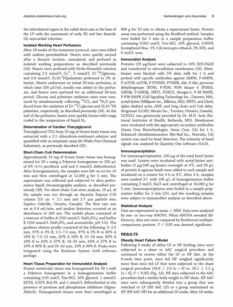