Loss of the SxxSS Motif in a Human T-Cell Factor-4 Isoform Confers Hypoxia Resistance to Liver...
14
Loss of the SxxSS Motif in a Human T-Cell Factor-4 Isoform Confers Hypoxia Resistance to Liver Cancer: An Oncogenic Switch in Wnt Signaling Hironori Koga 1 , Orkhontuya Tsedensodnom 1 , Yoshito Tomimaru 1 , Evan J. Walker 1 , Han Chu Lee 2 , Kang Mo Kim 2 , Hirohisa Yano 3 , Jack R. Wands 1 , Miran Kim 1 * 1 Liver Research Center, Rhode Island Hospital and the Warren Alpert Medical School of Brown University, Providence, Rhode Island, United States of America, 2 Department of Internal Medicine, Asan Medical Center, University of Ulsan College of Medicine, Seoul, Korea, 3 Department of Pathology, Kurume University, Kurume, Japan Abstract Purpose: Aberrantly activated Wnt/b-catenin signaling is important in hepatocellular carcinoma (HCC) development. Downstream gene expressions involving the Wnt/b-catenin cascade occur through T-cell factor (TCF) proteins. Here, we show the oncogenic potential of human TCF-4 isoforms based on the expression of a single conserved SxxSS motif. Methods: We investigated the TCF-4J and K isoform pair characterized by the presence (K) or absence (J) of the SxxSS motif. The mRNA expression profiles were examined in 47 pairs of human HCCs and adjacent non-cancerous liver tissues by RT- PCR. Proliferation, sphere assays and immunoblot analysis were performed under normoxia and hypoxia conditions. The ability of HCC cells overexpressing TCF-4J (J cells) and K (K cells) to grow as solid tumors in nude mice was explored. Results: TCF-4J expression was significantly upregulated in HCC tumors compared to corresponding peritumor and normal liver and was preferentially expressed in poorly differentiated HCCs. In contrast, TCF-4K was downregulated in those same HCC tumors. TCF-4J-overexpressing HCC cells (J cells) revealed a survival advantage under hypoxic conditions, high proliferation rate and formation of aggregates/spheres compared to overexpression of TCF-4K (K cells). The hypoxic J cells had high expression levels of HIF-2a and EGFR as possible mechanisms to promote tumorigenesis. Increased stability of HIF- 2a under hypoxia in J cells was associated with a decreased level of von Hippel-Lindau (VHL) protein, a known E3 ligase for HIF-as. In a xenograft model, the J cells rapidly developed tumors compared to K cells. Tumor tissues derived from J cells exhibited high expression levels of HIF-2a and EGFR compared to the slow developing and small K cell derived tumors. Conclusions: Our results suggest that the specific TCF-4J isoform, which lacks a regulatory SxxSS motif, has robust tumor- initiating potential under hypoxic conditions. Citation: Koga H, Tsedensodnom O, Tomimaru Y, Walker EJ, Lee HC, et al. (2012) Loss of the SxxSS Motif in a Human T-Cell Factor-4 Isoform Confers Hypoxia Resistance to Liver Cancer: An Oncogenic Switch in Wnt Signaling. PLoS ONE 7(6): e39981. doi:10.1371/journal.pone.0039981 Editor: Vincenzo Coppola, Ohio State University Comprehensive Cancer Cente, United States of America Received January 30, 2012; Accepted May 30, 2012; Published June 29, 2012 Copyright: ß 2012 Koga et al. This is an open-access article distributed under the terms of the Creative Commons Attribution License, which permits unrestricted use, distribution, and reproduction in any medium, provided the original author and source are credited. Funding: This work was supported by the National Institutes of Health (CA-123544 to JRW). The funders had no role in study design, data collection and analysis, decision to publish, or preparation of the manuscript. Competing Interests: The authors have declared that no competing interests exist. * E-mail: [email protected] Introduction The Wnt/b-catenin signaling pathway plays a crucial role in cell fate determination and stem cell renewal in adult tissue [1,2]. Genetic and/or epigenetic deregulation of this pathway leads to aberrant nuclear accumulation of b-catenin where it binds with T- cell factor-4 (TCF-4) to form a transcriptional complex [3]; this event drives gene expression such as c-Myc, cyclin D1, and EPCAM [4,5,6] that contributes to a malignant phenotype. Hepatocellular carcinoma (HCC) is the third common cause of cancer mortality worldwide [7]. Aberrantly activated Wnt/b- catenin signaling due to overexpression of upstream components of this pathway such as Frizzled (FZD) receptors and Wnt ligands is a common early event in the molecular pathogenesis of this disease [8,9,10]. However, the involvement of the canonical Wnt TCF-4 effector proteins in this process has yet to be examined. The human TCF-4 gene (TCF7L2) consists of 17 exons and has several alternative splicing sites in exons 13–17 and exon 4 [11]. The alternative splicing sites in the central domain of TCF-4 can also generate isoforms with or without the conserved LVPQ and SxxSS motifs located at the end of exon 7 and the beginning of exon 9, respectively [11,12]. Recently, we identified and characterized 14 (12 of which are unique) TCF-4 isoforms derived from human HCC cell lines [13]. Among such isoforms, three structurally identical pairs (TCF-4J and K; TCF-4A and B; TCF- 4G and H) were observed that differed only by the presence (K, A, and H) or absence (J, B, and G) of a SxxSS motif. In this context, two pairs of isoforms (TCF-4A and B, and TCF-4H and G) are short forms, whereas TCF-4K and J are long forms due to the inclusion of a C-terminal tail (exon 13–17) [13]. Previous studies suggest that the SxxSS motif may modulate transcriptional activity PLoS ONE | www.plosone.org 1 June 2012 | Volume 7 | Issue 6 | e39981
-
Upload
independent -
Category
Documents
-
view
0 -
download
0
Transcript of Loss of the SxxSS Motif in a Human T-Cell Factor-4 Isoform Confers Hypoxia Resistance to Liver...

Loss of the SxxSS Motif in a Human T-Cell Factor-4Isoform Confers Hypoxia Resistance to Liver Cancer: AnOncogenic Switch in Wnt SignalingHironori Koga1, Orkhontuya Tsedensodnom1, Yoshito Tomimaru1, Evan J. Walker1, Han Chu Lee2, Kang
Mo Kim2, Hirohisa Yano3, Jack R. Wands1, Miran Kim1*
1 Liver Research Center, Rhode Island Hospital and the Warren Alpert Medical School of Brown University, Providence, Rhode Island, United States of America,
2 Department of Internal Medicine, Asan Medical Center, University of Ulsan College of Medicine, Seoul, Korea, 3 Department of Pathology, Kurume University, Kurume,
Japan
Abstract
Purpose: Aberrantly activated Wnt/b-catenin signaling is important in hepatocellular carcinoma (HCC) development.Downstream gene expressions involving the Wnt/b-catenin cascade occur through T-cell factor (TCF) proteins. Here, weshow the oncogenic potential of human TCF-4 isoforms based on the expression of a single conserved SxxSS motif.
Methods: We investigated the TCF-4J and K isoform pair characterized by the presence (K) or absence (J) of the SxxSS motif.The mRNA expression profiles were examined in 47 pairs of human HCCs and adjacent non-cancerous liver tissues by RT-PCR. Proliferation, sphere assays and immunoblot analysis were performed under normoxia and hypoxia conditions. Theability of HCC cells overexpressing TCF-4J (J cells) and K (K cells) to grow as solid tumors in nude mice was explored.
Results: TCF-4J expression was significantly upregulated in HCC tumors compared to corresponding peritumor and normalliver and was preferentially expressed in poorly differentiated HCCs. In contrast, TCF-4K was downregulated in those sameHCC tumors. TCF-4J-overexpressing HCC cells (J cells) revealed a survival advantage under hypoxic conditions, highproliferation rate and formation of aggregates/spheres compared to overexpression of TCF-4K (K cells). The hypoxic J cellshad high expression levels of HIF-2a and EGFR as possible mechanisms to promote tumorigenesis. Increased stability of HIF-2a under hypoxia in J cells was associated with a decreased level of von Hippel-Lindau (VHL) protein, a known E3 ligase forHIF-as. In a xenograft model, the J cells rapidly developed tumors compared to K cells. Tumor tissues derived from J cellsexhibited high expression levels of HIF-2a and EGFR compared to the slow developing and small K cell derived tumors.
Conclusions: Our results suggest that the specific TCF-4J isoform, which lacks a regulatory SxxSS motif, has robust tumor-initiating potential under hypoxic conditions.
Citation: Koga H, Tsedensodnom O, Tomimaru Y, Walker EJ, Lee HC, et al. (2012) Loss of the SxxSS Motif in a Human T-Cell Factor-4 Isoform Confers HypoxiaResistance to Liver Cancer: An Oncogenic Switch in Wnt Signaling. PLoS ONE 7(6): e39981. doi:10.1371/journal.pone.0039981
Editor: Vincenzo Coppola, Ohio State University Comprehensive Cancer Cente, United States of America
Received January 30, 2012; Accepted May 30, 2012; Published June 29, 2012
Copyright: � 2012 Koga et al. This is an open-access article distributed under the terms of the Creative Commons Attribution License, which permitsunrestricted use, distribution, and reproduction in any medium, provided the original author and source are credited.
Funding: This work was supported by the National Institutes of Health (CA-123544 to JRW). The funders had no role in study design, data collection and analysis,decision to publish, or preparation of the manuscript.
Competing Interests: The authors have declared that no competing interests exist.
* E-mail: [email protected]
Introduction
The Wnt/b-catenin signaling pathway plays a crucial role in
cell fate determination and stem cell renewal in adult tissue [1,2].
Genetic and/or epigenetic deregulation of this pathway leads to
aberrant nuclear accumulation of b-catenin where it binds with T-
cell factor-4 (TCF-4) to form a transcriptional complex [3]; this
event drives gene expression such as c-Myc, cyclin D1, and EPCAM
[4,5,6] that contributes to a malignant phenotype.
Hepatocellular carcinoma (HCC) is the third common cause of
cancer mortality worldwide [7]. Aberrantly activated Wnt/b-
catenin signaling due to overexpression of upstream components
of this pathway such as Frizzled (FZD) receptors and Wnt ligands
is a common early event in the molecular pathogenesis of this
disease [8,9,10]. However, the involvement of the canonical Wnt
TCF-4 effector proteins in this process has yet to be examined.
The human TCF-4 gene (TCF7L2) consists of 17 exons and has
several alternative splicing sites in exons 13–17 and exon 4 [11].
The alternative splicing sites in the central domain of TCF-4 can
also generate isoforms with or without the conserved LVPQ and
SxxSS motifs located at the end of exon 7 and the beginning of
exon 9, respectively [11,12]. Recently, we identified and
characterized 14 (12 of which are unique) TCF-4 isoforms derived
from human HCC cell lines [13]. Among such isoforms, three
structurally identical pairs (TCF-4J and K; TCF-4A and B; TCF-
4G and H) were observed that differed only by the presence (K, A,
and H) or absence (J, B, and G) of a SxxSS motif. In this context,
two pairs of isoforms (TCF-4A and B, and TCF-4H and G) are
short forms, whereas TCF-4K and J are long forms due to the
inclusion of a C-terminal tail (exon 13–17) [13]. Previous studies
suggest that the SxxSS motif may modulate transcriptional activity
PLoS ONE | www.plosone.org 1 June 2012 | Volume 7 | Issue 6 | e39981

of TCF-4 [12]; however, both the cell-based and in vivo functional
consequences of the SxxSS motif expression as well as subsequent
gene regulatory activity have not been determined.
Emerging evidence indicates that both embryonic (ES), adult
and inducible pluripotent stem (iPS) cells [14] prefer hypoxic
conditions for growth and survival [15]. Hypoxia generates diverse
cellular signals through the stabilization of hypoxia-inducible
factors (HIF-as) such as HIF-1a and HIF-2a. Recent studies reveal
that HIF-as interact with b-catenin and, therefore, may regulate
TCF-4-driven gene expression not only in stem/progenitors but
also tumor cells experiencing hypoxic conditions during rapid
growth [16,17]. These findings imply that Wnt/b-catenin/TCF-4
signaling may be directly regulated by the professional oxygen-
sensing system in the nucleus. For instance, stabilized HIF-2a, a
partner of b-catenin and often found in the hypoxic core of the
tumor, upregulates the expression of epidermal growth factor
receptor (EGFR) and may contribute to tumor growth [18]. In
human HCC, the HIF-as are involved in the multi-step process of
tumor dedifferentiation via promotion of angiogenesis [19].
Accordingly, we determined if different functional properties of
TCF-4 isoforms associated with the HCC malignant phenotype
were regulated in the context of a SxxSS motif-dependent
mechanisms under conditions of oxygen deprivation.
Materials and Methods
Ethics StatementFull ethical approval was obtained for all human sample
collections from either the Asan Medical Center Ethics Committee
or the Kurume University Ethics Committee. All samples were
obtained with written consent. All animal experiments were
conducted in accordance with the NIH Guidelines for the Care
and Use of Laboratory Animals and were approved by the
Lifespan Animal Welfare Committee of Rhode Island Hospital,
Providence, RI (permit number A3922-01).
Detection of TCF-4 Isoforms in HCC Tumors by RT-PCRPreparations of human TCF-4A, B, J and K-myc plasmids have
been previously described [13]. Two independent RT-PCR
analyses were performed using 47 pairs of human HCCs
(Tables 1 and 2) as previously described [13].
Cell Lines and CulturesHuman cell lines Hep3B, Huh7, HepG2, and HEK293 were
obtained from the American Type Culture Collection (ATCC,
Manassas, VA). FOCUS cell line was obtained from Dr. Wands
(Liver Research Center, Rhode Island Hospital, RI). HAK-1A
and HAK-1B cells were provided by Dr. Yano (Kurume
University School of Medicine, Japan). The immortalized
hepatocyte derived cell line OUMS-29 was a kind gift from
Drs. Namba and Kobayashi (Okayama University, Japan).
Hep3B, Huh7, HepG2, and FOCUS cell lines are hepatitis B
virus (HBV)-related HCC cells. HAK-1A and HAK-1B cells are
hepatitis C virus (HCV)-related HCC cell lines. All cells have
been described and employed in previous studies (Hep3B [20],
Huh7 [21], HepG2 [20], HEK293 [22], FOCUS [23], and
HAK-1A and HAK-1B [24]). To generate stable transfectants,
HAK-1A cells were transfected with the TCF-4 or empty vector
plasmid using TransIT-LT1 Reagent (Mirus Bio Co., Madison,
WI) and selected by G418.
Hypoxia InductionHypoxic conditions (1% oxygen) were produced by an
incubator equipped with an oxygen concentration regulator
system (ASTEC, Fukuoka, Japan). Alternatively, cells were
exposed to hypoxia-mimic chemical cobalt chloride (CoCl2,
Sigma-Aldrich, St. Louis, MO) [25].
Confocal Laser Scanning MicroscopyCells, grown on 35 mm diameter Glass Bottom Dishes
(MatTek, Ashland, MA), were fixed with cold acetone/methanol
(1:1) for 10 min, and then washed in PBS containing 0.05%
Tween 20 (PBS-T). Nonspecific reactions were blocked with
Protein Block Serum-Free (DAKO North America, Carpinteria,
CA) and then incubated with a mouse anti-Myc-tag antibody
(Santa Cruz Biotechnology, Santa Cruz, CA) or a rabbit anti-HIF-
2a antibody (Abcam, Cambridge, UK) at 4uC overnight. After
washing in PBS-T, the specimens were treated with Alexa Fluor
goat anti-mouse or goat anti-rabbit IgG (H+L) antibody (Molec-
ular Probes, Eugene, OR) for 40 min at RT, and then
counterstained by VECTASHIELD Mounting Medium with
49,6-diamino-2-phenylindole (DAPI) (Burlingame, CA). The Zeiss
LSM510 Confocal Laser Scanning Microscope (Carl Zeiss
MicroImaging, Inc., Thornwood, NY) equipped with the user
interface software Zen 2008 was used to visualize the immuno-
fluorescence staining for Myc-tag or HIF-2a and nuclear
localization was provided by DAPI. The negative controls were
obtained by incubating cells with non-specific mouse IgG or rabbit
IgG as described above.
Immunoblot Analysis and ImmunoprecipitationThe primary antibodies used were against for Myc-tag, b-
catenin, actin, c-tubulin, lamin A/C, HIF-1a, c-myc, and
ubiquitin (Santa Cruz Bio, Santa Cruz, CA), TCF-4, EpCAM,
and von Hipple-Lindau (VHL) (Cell Signaling Technology,
Beverly, MA), HIF-2a (Abcam, Cambridge, UK), and keratin-19
(K-19) (DAKO, Carpinteria, CA). A TCF-4 rabbit derived mAb
(Cell Signaling Technology, Cat# 2569) recognizes surrounding
the Leu330 of human TCF-4 and detects endogenous TCF-4
proteins including both the short and long isoforms. For
immunoprecipitation, cell extracts were prepared as previously
described [26]. Total cell lysate or nuclear extract were incubated
with antibodies against HIF-1a, HIF-2a, and VHL followed by
Recombinant Protein G Agarose (Invitrogen, Carlsbad, CA).
Immunoblots were probed with antibodies to ubiquitin, HIF-1a,
HIF-2a, and VHL and detected with HRP-labeled anti-mouse or
anti-rabbit IgG (Amersham Pharmacia Biotech, Buckinghamshire,
UK) using ECL Advanced kit (Amersham). Positive signals were
captured by the Image analyzer LAS-1000plus (Fujifilm, Tokyo,
Japan), and the band intensity of protein was determined using
Image Gauge version 3.45 (Fujifilm).
Cell Proliferation AssayCells were seeded into 24-well plates at a density of 2.56103/
well. A colorimetric assay (CellTiter 96H Aqueous One Solution
Cell Proliferation Assay; Promega, Madison, WI) was performed
at days 0, 1, 3, 5, and 7, and the signals were measured using
Spectra Max M5 (Molecular Devices, Sunnyvale, CA).
Anchorage-independent Growth (Sphere Assay)Single-cell suspensions were put into Ultra Low Attachment
6-well plates (Corning, Lowell, MA). After 27 days, the number
of cell aggregates/spheres per well was quantified and photo-
graphed under the Zeiss Axiovert 200 M Fluorescence Micro-
scope (Carl Zeiss MicroImaging). The area of the cell
aggregates was measured using the MetaMorph 6.0 software
(Molecular Devices).
TCF-4 Isoform Confers Hypoxia Resistance to HCC
PLoS ONE | www.plosone.org 2 June 2012 | Volume 7 | Issue 6 | e39981
Evaluation of Protein StabilityCells were exposed to 5 mg/mL cycloheximide (CHX; Sigma-
Aldrich) for 0 or 60 min at the last phase of the 48 h CoCl2treatment. Total cell lystes were used for immunoblot analysis to
evaluate HIF-1a and HIF-2a expression levels after protein
synthesis was inhibited by CHX.
Xenograft Tumor Model in Nude MiceHAK-1A-derived stable clones were used; parental HAK-1A
cells do not form tumors in nude mice, and are therefore suitable
for assessment of malignant transformation following stable
transfection with TCF-4 isoforms [24]. Cells (16106) were sub-
cutaneously injected into the back of 5-week old male BALB/c
nude mice (n = 12) (Taconic Farms, Cranbury, NJ). The tumor
size was measured twice per week, and the tumor volume (mm3)
was estimated using the equation length6(width)260.5. When the
longer diameter reached 10 mm, the mice were sacrificed and the
tumors were prepared for subsequent analysis.
immunohistochemistry of Xenograft TumorsParaffin-embedded tissue sections were deparaffinized and
heated in 10 mM citrate buffer at 120uC for 5 min for antigen
retrieval. The sections were pre-blocked by Protein Block Serum-
Free (DAKO) and incubated with primary antibodies for HIF-2a(Abcam) and EGFR (D38B1 XPTM) (Cell Signaling Technology).
After washing, the sections were incubated with EnVision
secondary antibodies labeled with HRP-polymer complexes
(DAKO) and visualized by 0.1% 3–399-diamino-benzidine-tetra-
hydrochloride. The cell nuclei were counterstained with hema-
toxylin. Specimen incubated with mouse or rabbit IgG were set as
the negative controls.
Statistical AnalysisStatistical significance was performed by the Mann-Whitney U
test using GraphPad Prism 4.0 software (San Diego, CA). P,0.05
was considered statistically significant. The Pearson’s correlation
coefficient was used to calculate correlation between expression of
TCF-4J and K.
Results
Differential Expression Profile of TCF-4J and TCF-4K inHuman HCC Tissues
Expression of TCF-4 isoforms with or without the SxxSS motif
may influence their transcriptional activity [12,13,27]. Therefore,
we determined if three pairs of isoforms (TCF-4A and B; TCF-4G
and H; and TCF-4J and K) exhibited different expression profiles
that may suggest SxxSS-dependent regulation in human HCC
tumors. The relative mRNA levels of these TCF-4 isoforms in
27 pairs of HCC tumors and corresponding adjacent liver tissue
obtained from chronic HBV-related (26/27) disease were mea-
sured. Comparisons were made to three normal liver specimens by
semi-quantitative RT-PCR (Fig. 1A, Fig. S1A and C, and Table 1)
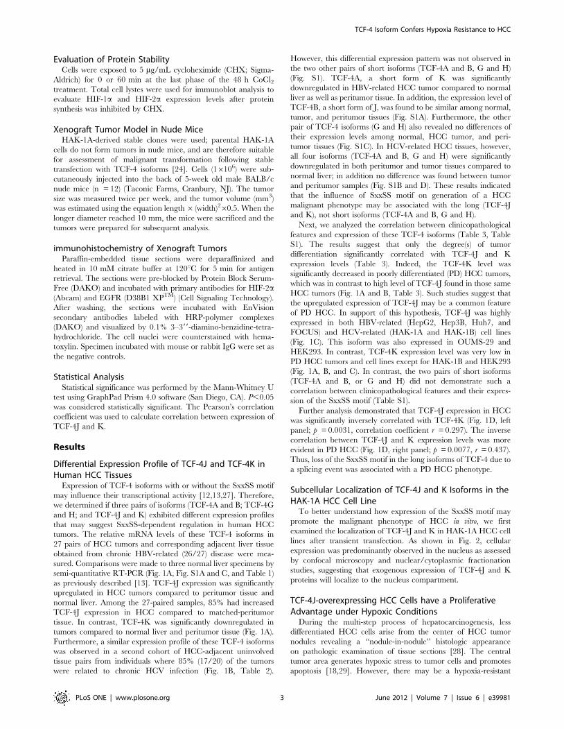
as previously described [13]. TCF-4J expression was significantly
upregulated in HCC tumors compared to peritumor tissue and
normal liver. Among the 27-paired samples, 85% had increased
TCF-4J expression in HCC compared to matched-peritumor
tissue. In contrast, TCF-4K was significantly downregulated in
tumors compared to normal liver and peritumor tissue (Fig. 1A).
Furthermore, a similar expression profile of these TCF-4 isoforms
was observed in a second cohort of HCC-adjacent uninvolved
tissue pairs from individuals where 85% (17/20) of the tumors
were related to chronic HCV infection (Fig. 1B, Table 2).
However, this differential expression pattern was not observed in
the two other pairs of short isoforms (TCF-4A and B, G and H)
(Fig. S1). TCF-4A, a short form of K was significantly
downregulated in HBV-related HCC tumor compared to normal
liver as well as peritumor tissue. In addition, the expression level of
TCF-4B, a short form of J, was found to be similar among normal,
tumor, and peritumor tissues (Fig. S1A). Furthermore, the other
pair of TCF-4 isoforms (G and H) also revealed no differences of
their expression levels among normal, HCC tumor, and peri-
tumor tissues (Fig. S1C). In HCV-related HCC tissues, however,
all four isoforms (TCF-4A and B, G and H) were significantly
downregulated in both peritumor and tumor tissues compared to
normal liver; in addition no difference was found between tumor
and peritumor samples (Fig. S1B and D). These results indicated
that the influence of SxxSS motif on generation of a HCC
malignant phenotype may be associated with the long (TCF-4J
and K), not short isoforms (TCF-4A and B, G and H).
Next, we analyzed the correlation between clinicopathological
features and expression of these TCF-4 isoforms (Table 3, Table
S1). The results suggest that only the degree(s) of tumor
differentiation significantly correlated with TCF-4J and K
expression levels (Table 3). Indeed, the TCF-4K level was
significantly decreased in poorly differentiated (PD) HCC tumors,
which was in contrast to high level of TCF-4J found in those same
HCC tumors (Fig. 1A and B, Table 3). Such studies suggest that
the upregulated expression of TCF-4J may be a common feature
of PD HCC. In support of this hypothesis, TCF-4J was highly
expressed in both HBV-related (HepG2, Hep3B, Huh7, and
FOCUS) and HCV-related (HAK-1A and HAK-1B) cell lines
(Fig. 1C). This isoform was also expressed in OUMS-29 and
HEK293. In contrast, TCF-4K expression level was very low in
PD HCC tumors and cell lines except for HAK-1B and HEK293
(Fig. 1A, B, and C). In contrast, the two pairs of short isoforms
(TCF-4A and B, or G and H) did not demonstrate such a
correlation between clinicopathological features and their expres-
sion of the SxxSS motif (Table S1).
Further analysis demonstrated that TCF-4J expression in HCC
was significantly inversely correlated with TCF-4K (Fig. 1D, left
panel; p = 0.0031, correlation coefficient r = 0.297). The inverse
correlation between TCF-4J and K expression levels was more
evident in PD HCC (Fig. 1D, right panel; p = 0.0077, r = 0.437).
Thus, loss of the SxxSS motif in the long isoforms of TCF-4 due to
a splicing event was associated with a PD HCC phenotype.
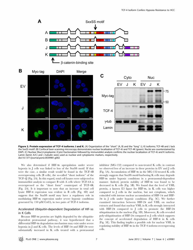
Subcellular Localization of TCF-4J and K Isoforms in theHAK-1A HCC Cell Line
To better understand how expression of the SxxSS motif may
promote the malignant phenotype of HCC in vitro, we first
examined the localization of TCF-4J and K in HAK-1A HCC cell
lines after transient transfection. As shown in Fig. 2, cellular
expression was predominantly observed in the nucleus as assessed
by confocal microscopy and nuclear/cytoplasmic fractionation
studies, suggesting that exogenous expression of TCF-4J and K
proteins will localize to the nucleus compartment.
TCF-4J-overexpressing HCC Cells have a ProliferativeAdvantage under Hypoxic Conditions
During the multi-step process of hepatocarcinogenesis, less
differentiated HCC cells arise from the center of HCC tumor
nodules revealing a ‘‘nodule-in-nodule’’ histologic appearance
on pathologic examination of tissue sections [28]. The central
tumor area generates hypoxic stress to tumor cells and promotes
apoptosis [18,29]. However, there may be a hypoxia-resistant
TCF-4 Isoform Confers Hypoxia Resistance to HCC
PLoS ONE | www.plosone.org 3 June 2012 | Volume 7 | Issue 6 | e39981

TCF-4 Isoform Confers Hypoxia Resistance to HCC
PLoS ONE | www.plosone.org 4 June 2012 | Volume 7 | Issue 6 | e39981
population of HCC cells that survive under these severe
conditions to promote tumor growth through accelerated
angiogenesis and vascular invasion. These two phenomena are
regulated by HIF-1a and HIF-2a [30,31]. Therefore, we
hypothesized that the high expression of TCF-4J in PD HCC
was a possible reflection of an induced hypoxia-resistant HCC
phenotype. To test this idea, stable cell clones overexpressing
TCF-4J (J cells) and TCF-4K (K cells) were established, and
comparisons were made to empty vector-transfected cell clones
(EV cells), as well as to the parental HAK-1A cells. Under
phase-contrast microscopy, the morphologic appearance of these
clones was similar and comparable to the parental cells (Fig. 3A),
which was in contrast to the HAK-1A-derived dedifferentiated
cell clone HAK-1B [24].
Wnt/b-catenin signaling often correlates with stem cell-like
molecular alterations [32,33]. In this regard, the expression of
EpCAM, a putative liver cancer stem cell (CSC) marker and a b-
catenin/TCF-4 downstream target gene product [6], was not
upregulated in these cells. However, the expression of K-19, a
biliary lineage marker for an aggressive HCC phenotype [34], was
increased in J cells but not in K, EV, or parental HAK-1A cells
(Fig. 3B).
Consistent with our previous report in Huh7 cells [13], J cells
had an increased basal growth rate compared to K and EV control
cells. The J cells were resistant under chemically induced, severe
hypoxic conditions (150 mM CoCl2), proliferated significantly
faster than K or EV cells (Fig. 3C). This finding was further
supported when cells were exposed to 1% oxygen for 5 days
Figure 1. Expression of TCF-4J and TCF-4K mRNAs was different in human HCCs. (A) Comparative analysis of TCF-4J and K mRNAexpression levels in 27 HBV-related HCC tumors (T) adjacent peritumor tissue (pT) and histologic normal liver (N) by RT-PCR. Values are normalized toGAPDH. Red rectangles denote poorly differentiated (PD) HCC. WD, well differentiated; MD, moderately differentiated. Statistical results from alltissues or PD HCC are expressed as mean + SD (right panel). (B) Another 20 HCC tumors including five WD HCCs from a different clinical site.Seventeen individuals had HCV-related chronic liver disease, and the remainder had chronic HBV infection. See also Tables 1 and 2. (C) Expressionlevels of TCF-4J and K mRNA in human cell lines HepG2 (G2), Hep3B (3B), Huh-7 (H7), FOCUS (FO), HAK-1A (1A), HAK-1B (1B), OUMS-29 (OU), andHEK293 (293). (D) Inverse correlation between TCF-4J and K expression in all HCC (left panel) and PD HCC (right panel). Note the weak inversecorrelation in all cases (r = 0.297), while increased inverse correlation in PD HCC (r = 0.437). *p,0.01; **p,0.05.doi:10.1371/journal.pone.0039981.g001
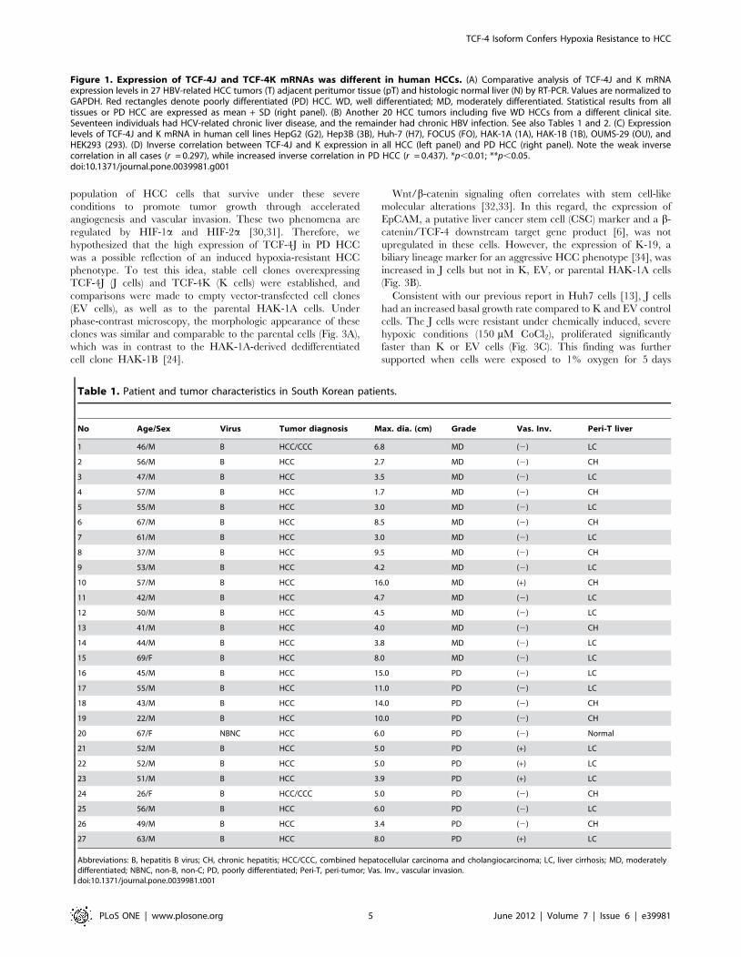
Table 1. Patient and tumor characteristics in South Korean patients.
No Age/Sex Virus Tumor diagnosis Max. dia. (cm) Grade Vas. Inv. Peri-T liver
1 46/M B HCC/CCC 6.8 MD (2) LC
2 56/M B HCC 2.7 MD (2) CH
3 47/M B HCC 3.5 MD (2) LC
4 57/M B HCC 1.7 MD (2) CH
5 55/M B HCC 3.0 MD (2) LC
6 67/M B HCC 8.5 MD (2) CH
7 61/M B HCC 3.0 MD (2) LC
8 37/M B HCC 9.5 MD (2) CH
9 53/M B HCC 4.2 MD (2) LC
10 57/M B HCC 16.0 MD (+) CH
11 42/M B HCC 4.7 MD (2) LC
12 50/M B HCC 4.5 MD (2) LC
13 41/M B HCC 4.0 MD (2) CH
14 44/M B HCC 3.8 MD (2) LC
15 69/F B HCC 8.0 MD (2) LC
16 45/M B HCC 15.0 PD (2) LC
17 55/M B HCC 11.0 PD (2) LC
18 43/M B HCC 14.0 PD (2) CH
19 22/M B HCC 10.0 PD (2) CH
20 67/F NBNC HCC 6.0 PD (2) Normal
21 52/M B HCC 5.0 PD (+) LC
22 52/M B HCC 5.0 PD (+) LC
23 51/M B HCC 3.9 PD (+) LC
24 26/F B HCC/CCC 5.0 PD (2) CH
25 56/M B HCC 6.0 PD (2) LC
26 49/M B HCC 3.4 PD (2) CH
27 63/M B HCC 8.0 PD (+) LC
Abbreviations: B, hepatitis B virus; CH, chronic hepatitis; HCC/CCC, combined hepatocellular carcinoma and cholangiocarcinoma; LC, liver cirrhosis; MD, moderatelydifferentiated; NBNC, non-B, non-C; PD, poorly differentiated; Peri-T, peri-tumor; Vas. Inv., vascular invasion.doi:10.1371/journal.pone.0039981.t001
TCF-4 Isoform Confers Hypoxia Resistance to HCC
PLoS ONE | www.plosone.org 5 June 2012 | Volume 7 | Issue 6 | e39981

(Fig. 3D). Cell growth in hypoxia (1% oxygen) was reduced in EV
and K cells compared to cell proliferation in normoxia (20%
oxygen) by 22 and 27%, respectively, while J cells demostrated
only a 13% reduction in cell growth. Moreover, robust resistance
to severe hypoxia in the J cells was also exhibited in a sphere assay.
In this context, the J cells formed the largest cell aggregates at all
the CoCl2 concentrations employed. The J cells’ potential for
anchorage-independent growth was most evident with severe
hypoxia induced by 150 mM CoCl2 (Fig. 3E).
Loss of the SxxSS Motif Contributes to the ExpressionLevels of HIF-as
Based on the hypoxia-resistant features exhibited by J cells,
we determined if these cells expressed HIF-as under hypoxia.
Both HIF-1a and HIF-2a expression in J and K cells were
explored since HIF-2a has recently been shown to be a key
molecule that promotes survival of CSCs under hypoxic
conditions [35,36,37]. As expected, HIF-1a and HIF-2aexpression were induced with moderate hypoxia (75 mM CoCl2)
in EV, J and K cells; however, under severe hypoxic conditions
(150 mM CoCl2), only J cells maintained these high protein
levels (Fig. 4A). Stabilized HIF-2a, often found in the hypoxic
core of the tumor, upregulated the expression of EGFR which
may contribute to tumor growth [18], and the activation of
EGFR signaling pathway was associated with the development
of K-19-positive HCC [38]. In J cells, expression of EGFR and
K-19 was associated with an increased level of HIF-2a (Fig. 4A).
The consistent HIF response was also observed in 1% oxygen-
exposed J cells (Fig. 4B). Furthermore, the 150 mM CoCl2-
induced HIF-2a expression was localized in the nuclei of J cells
as assessed by immunofluorescence staining (Fig. 4C).
Table 2. Patient and tumor characteristics in Japanese patients.
No Age/Sex Virus Tumor diagnosis Max. dia. (cm) Grade Vas. Inv. Peri-T liver
1 73/F C HCC 2.7 WD (2) LC
2 59/F C HCC 2.7 WD (2) LC
3 59/M C HCC 2.5 WD (2) LC
4 82/M C HCC 3.0 WD (2) CH
5 72/M C HCC 3.3 WD (2) LC
6 62/M C HCC 2.0 MD (2) CH
7 75/M C HCC 2.9 MD (2) CH
8 64/M C HCC 1.8 MD (2) CH
9 77/M C HCC 2.0 MD (2) CH
10 63/M C HCC 1.7 MD (2) CH
11 72/M C HCC 2.0 MD (2) CH
12 78/F C HCC 3.7 MD (2) CH
13 33/M B HCC 2.8 MD (2) CH
14 56/M B HCC 2.0 MD (+) LC
15 78/M C HCC 3.3 PD (+) CH
16 51/M C HCC 3.3 PD (+) CH
17 70/M C HCC 4.2 PD (+) LC
18 63/F C HCC 4.5 PD (+) LC
19 71/M C HCC 6.0 PD (2) CH
20 60/F B HCC 2.0 PD (+) CH
Abbreviations: B, hepatitis B virus; C, hepatitis C virus; CH, chronic hepatitis; LC, liver cirrhosis; Max. dia., maximum diameter; MD, moderately differentiated; PD, poorlydifferentiated; Peri-T, peri-tumor; Vas. Inv., vascular invasion; WD, well differentiated.doi:10.1371/journal.pone.0039981.t002
Table 3. Relation between clinicopathological factors andTCF-4J and K expression in patients.
Group No TCF-4J TCF-4K
Level P value Level P value
Age (y) #56 23 1.0060.38 0.2764 0.2260.23 0.1798
57# 24 0.8860.38 0.3160.21
Gender M 35 0.9360.41 0.8270 0.2760.22 0.9107
F 12 0.9660.27 0.2660.24
Virus HBV 29 0.9860.40 0.1991 0.2360.22 0.1331
HCV 17 0.8360.34 0.3360.21
Tumorsize (mm)
#30 17 0.8360.28 0.1446 0.3560.19 0.0481
30, 30 1.0060.42 0.2260.23
Vascularinvasion
2 36 0.8860.35 0.0521 0.2860.21 0.3975
+ 11 1.1360.41 0.2160.26
Histology WD, MD 29 0.8260.35 0.0083 0.3560.18 0.0001
PD 18 1.1260.36 0.1260.21
Liverbackground
Normal, CH 24 0.9860.40 0.3997 0.2660.23 0.9985
LC 23 0.8960.36 0.2660.22
Abbreviations: CH, chronic hepatitis; HBV, hepatitis B virus; HCV, hepatitis Cvirus; LC, liver cirrhosis; MD, moderately differentiated; PD, poorly differentiated;WD, well differentiated.doi:10.1371/journal.pone.0039981.t003
TCF-4 Isoform Confers Hypoxia Resistance to HCC
PLoS ONE | www.plosone.org 6 June 2012 | Volume 7 | Issue 6 | e39981
We also determined if HIF-as upregulation under severe
hypoxia in J cells was linked to loss of the SxxSS motif. If that
were the case, a similar result would be found in the TCF-4B
overexpressing cells (B cells), the so-called ‘‘short isoform’’ of the
TCF-4J (Fig. 2A). In this regard, total cell lysates were subjected to
immunoblot analysis to compare B with A cells where TCF-4A is
overexpressed as the ‘‘short form’’ counterpart of TCF-4K
(Fig. 2A). It is important to note that an increase in total cell
lysate HIF-a expression was evident in B cells (Fig. 4D) and
suggests that the SxxSS motif may have a regulatory role in
modulating HIF-as expression under severe hypoxic conditions
generated by 150 mM CoCl2 in two pairs of TCF-4 isoforms.
Accelerated Ubiquitin-dependent Degradation of HIF-asin K Cells
Because HIF-as proteins are highly degraded by the ubiquitin-
dependent proteasomal pathway, it was hypothesized that a
differential HIF-as degradation mechanism may exist under severe
hypoxia in J and K cells. The levels of HIF-1a and HIF-2a were
substantially increased in K cells treated with a proteasomal
inhibitor (MG-132) compared to non-treated K cells; in contrast
we observed less of an increase in these proteins in EV and J cells
(Fig. 5A). Accumulation of HIF-as in the MG-132-treated K cells
strongly suggests that SxxSS motif-harboring K cells may degrade
HIF-as under hypoxic conditions in a proteasomal-dependent
manner. Indeed, protein stability of HIF-as was found to be
decreased in K cells (Fig. 5B). We found that the level of VHL
protein, a known E3 ligase for HIF-as, in K cells was higher
compared to J cells in the nucleus, but not cytoplasm, which
coincided with intense nuclear accumulation of HIF-1a and HIF-
2a in J cells under hypoxic conditions (Fig. 5C). We further
examined interaction between HIF-2a and VHL on nuclear
extracts and found that nuclear VHL in K cells strongly interacted
with HIF-2a compared to J cells to promote the HIF-2aubiquitination in the nucleus (Fig. 5D). Moreover, K cells revealed
poly-ubiquitination of HIF-2a compared to J cells which supports
the concept of accelerated degradation of HIF-a in K cells
(Fig. 5E). This finding implies a possible role for nuclear VHL in
regulating stability of HIF-as in the TCF-4 isoform-overexpressing
cells.
Figure 2. Protein expression of TCF-4 isoforms J and K. (A) Organization of the ‘‘short’’ (A, B) and the ‘‘long’’ (J, K) isoforms; TCF-4B and J lackthe SxxSS motif. (B) Confocal laser-scanning microscopy demonstrates nuclear localization of TCF-4J and TCF-4K (green). Nuclei are counterstained byDAPI. (C) Nuclear (Nuc)/cytoplasmic (Cyto) fractionation followed by immunoblot analysis confirms the nuclear localization of TCF-4J and K isoforms.Lamin (lamin A/C) and c-tubulin were used as nuclear and cytoplasmic markers, respectively.doi:10.1371/journal.pone.0039981.g002
TCF-4 Isoform Confers Hypoxia Resistance to HCC
PLoS ONE | www.plosone.org 7 June 2012 | Volume 7 | Issue 6 | e39981

Figure 3. TCF-4J-overexpressing cells proliferate under hypoxic conditions. (A) Phase-contrast microscopy of parental HAK-1A (1A) and 1A-derived stable clones, including the empty vector-transfected clone (EV2), the TCF-4J-transfected clones (J1 and J15), and the TCF-4K-transfectedclones (K2 and K5). The morphologic appearance of the highly malignant HAK-1B (1B) cell line, a clonally dedifferentiated cell type from 1A, is alsopresented for comparison. Bar = 50 mm. (B) Immunoblot analysis of stable cell clones. Positive signals for Myc-tag are shown in J1, J15, K2, and K5.HepG2 cells (G2) known to express both full-length (92 kDa) and truncated b-catenin (75 kDa), exhibited a lower band for b-catenin (b-Cat) HepG2was also used as a positive control for EpCAM and K-19 expression. HEK293 was used as a negative control for K-19. (C) Cell growth rates of stableclones under the hypoxic conditions generated by CoCl2 (150 mM) for 7 days. Growth rate is represented as the fold-increase compared to day 0. (D)Cell growth of stable clones in normoxia (20% O2) or hypoxic conditions (1% O2) Cells were exposed to either 20% or 1% oxygen for 5 days. Note thatreduction of cell growth in hypoxic contidions was less in J cells (13%) compared to EV (22%) and K (27%) cells. (E) Anchorage-independent growthassay (sphere assay). Phase-contrast microscopic views of representative cell aggregates are displayed at 0, 75, and 150 mM of CoCl2. Bar = 300 mm.Note the striking difference in colony growth rate and appearance at 150 mM of CoCl2. *p,0.05; **p,0.01.doi:10.1371/journal.pone.0039981.g003
TCF-4 Isoform Confers Hypoxia Resistance to HCC
PLoS ONE | www.plosone.org 8 June 2012 | Volume 7 | Issue 6 | e39981

Figure 4. Loss of the SxxSS motif in TCF-4 isoforms increases expression of HIF-as under hypoxia. (A and B) Immunoblot analysis ofempty vector-transfected (EV), TCF-4J-overexpressing (J), and TCF-4K-overexpressing cells (K). The cells were treated with 0 and 150 mM CoCl2 (A) orcultured under 1% oxygen tension for 48 hr (B). Cell lysates were subjected to detect HIF-as and Myc-tag TCF-4 isoform protein expression.Expression levels were plotted as a ratio to actin (right panel). (C) Confocal microscopy for nuclear localization of HIF-2a protein. Cells were treatedwith 0 mM (Normoxia) or 150 mM (Hypoxia) CoCl2 and stained with an anti-HIF-2a antibody (green). DAPI was employed for nuclear staining. (D)Immunoblot analysis of total cell lysates from cells treated with 0 or 150 mM CoCl2 for 48 hr. A and B represent HAK-1A cells overexpressing TCF-4A(‘‘short form’’ of TCF-4K) and TCF-4B (‘‘short form’’ of TCF-4J), respectively (see Figure 2A). Note the robust increase in expressions of HIF-1a and HIF-2a in B cells.doi:10.1371/journal.pone.0039981.g004
TCF-4 Isoform Confers Hypoxia Resistance to HCC
PLoS ONE | www.plosone.org 9 June 2012 | Volume 7 | Issue 6 | e39981

TCF-4J Isoform Expression Confers a TumorigenicPhenotype to HCC Cells
The tumorigenic potential of EV, J, and K cells was assessed in
nude mice. As might be predicted from the in vitro findings, J cells
were highly tumorigenic. Although K cells generated small
tumors, they appeared later (about 40 days) after tumor cell
injection and grew very slowly (Fig. 6A). Control (EV) cells did not
produce tumors as reported previously [24].
Immunoblot analysis was applied to confirm whether the
xenograft tumors had the same protein phenotype as exhibited in
the cell clones. The Myc-tag detected exogenous TCF-4J and K
isoforms derived from tumor tissues; in addition, RT-PCR was
Figure 5. Lack of SxxSS motif contributes to protein stability of HIF-as in J cells. (A) Cells were treated with 150 mM CoCl2 for 36 hrfollowed by incubation with or without MG-132 (10 mM) for 2 hr. Total cell lysates were employed for immunoblot analysis. Relative expression ofHIF-as was normalized to actin. (B) Protein stability of HIF-as was evaluated by immunoblot analysis, where cells were exposed to 5 mg/mLcycloheximide (CHX) to inhibit protein synthesis for 0 or 60 min at the last phase of the 48 hr CoCl2 treatment period. (C) Expression of VHL in TCF-4Jand K cells. Cells were treated 0 or 150 mM CoCl2 and nuclear and cytoplamic fractions was prepared for immunoblot analysis. Expression level of HIF-2a and VHL was normalized by lamin (bottom panel); VHL-C, cytoplasmic VHL. (D) Interaction between VHL and HIF-2a in nucleus of TCF-4J and Kcells. (E) Polyubiquitination of HIF-2a in cytoplasmic and nuclear fractions of TCF-4J and K cells. Immunoprecipitation (IP)/immunoblot (IB) analysiswas performed by using antibodies for HIF-2a and ubiquitin (Ub); *, IgG heavy chain. Note the decreased ubiquitination of HIF-2a and weak VHL-HIF-2a interaction in J cells, which was in contrast to the observations in K cells.doi:10.1371/journal.pone.0039981.g005
TCF-4 Isoform Confers Hypoxia Resistance to HCC
PLoS ONE | www.plosone.org 10 June 2012 | Volume 7 | Issue 6 | e39981
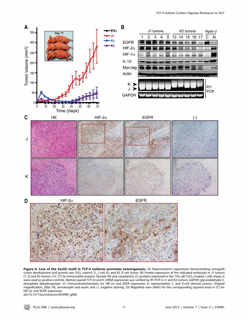
Figure 6. Loss of the SxxSS motif in TCF-4 isoforms promotes tumorigenesis. (A) Representative experiment demonstrating xenografttumor development and growth rate. EV2, control; J1, J cell; K2 and K5, K cell clones. (B) Protein expression of the indicated molecules in J1 tumors(1–5) and K2 tumors (13–17) by immunoblot analysis. Nuclear (N) and cytoplasmic (C) proteins expressed in the 150 mM CoCl2-treated J cells (Hypo-J)were used as positive controls. (Bottom panel) TCF-4J and K mRNA expression was verified by RT-PCR in J1 and K2 tumors; GAPDH, glyceraldehyde-3-phosphate dehydrogenase. (C) Immunohistochemistry for HIF-2a and EGFR expression in representative J- and K-cell derived tumors. Originalmagnification, 200x. HE, hematoxylin and eosin; and (-), negative staining. (D) Magnified view (400x) for the corresponding squared areas in (C) forHIF-2a and EGFR expression.doi:10.1371/journal.pone.0039981.g006
TCF-4 Isoform Confers Hypoxia Resistance to HCC
PLoS ONE | www.plosone.org 11 June 2012 | Volume 7 | Issue 6 | e39981
performed to verify overexpression of the TCF-4J or K without
cross-tissue contamination during these experiments (Fig. 6B;
lower panel). Consistent with previous report that HIF-2aupregulated EGFR expression in the hypoxic core of solid tumors
[18], higher expression levels of EGFR in concert with HIF-2awere found in J compared to K cell-derived tumors (Fig. 6B).
We also assessed HIF-2a and EGFR protein expression in
xenograft tumors by immunohistochemistry. The histological
features of the tumor tissue as well as cellular morphology did
not differ among tumors induced by either J or K cells using
hematoxylin and eosin staining. However, there was increased
expression of both HIF-2a and EGFR predominantly in the J cell-
derived tumor tissue (Fig. 6C). Since tumors frequently contain
central necrotic areas that form a boundary between the areas of
living and hypoxic dead tumor cells [29], we focused on the
immunoreactive signals at this interface. Striking nuclear and
cytoplasmic expression of HIF-2a and membranous EGFR were
predominantly found at the hypoxic boundary in J cell derived
tumor tissues (Fig. 6C and D).
Discussion
In the present study, we demonstrated the following: (1) in
hepatocyte derived cells, overexpression of the TCF-4J isoform
lacking the SxxSS motif, exhibited robust tumorigenic potential;
(2) the TCF-4J isoform was highly expressed in rapidly growing
and presumably in the hypoxic milieu of human PD HCC, and (3)
TCF-4J expression contributed to nuclear accumulation of HIF-
2a and was due to, in part, less ubiquitin-dependent degradation
of HIF-as in J cells compared to K cells under severe hypoxia.
This observation was linked to reduced expression of VHL.
There is evidence that genetic and/or epigenetic deregulation of
the Wnt/b-catenin signaling pathway leads to tumor formation
[2]. Indeed, genetic mutations in components of this pathway such
as APC, AXIN1/2 and CTNNB1 are well-established molecular
events in colorectal, as well as gastric carcinomas and HCC
[39,40]. Epigenetic disruptions in the Wnt/b-catenin signaling are
also involved in oncogenesis through inappropriate stabilization of
b-catenin and aberrant activation of upstream components of this
signal transduction cascade [9,10,41]. There is little information,
however, on the potential role of TCF-4 transcription factor
isoforms in the oncogenic process. We demonstrated that
overexpression of human TCF-4 isoforms may promote hepatic
oncogenesis through actions of their intrinsic motifs which suggest
that the Wnt/b-catenin signaling cascade may be augmented and
further able to contribute to the malignant phenotype due to
mechanisms that are operative in the nucleus. Indeed, expression
analysis in human HCC tumors revealed that both TCF-4J and
TCF-4K were present in non-cancerous dysplastic and normal
liver, although both isoforms were initially identified and cloned
from HCC cell lines. This finding is consistent with the previous
report that emphasized the physiological distribution of TCF-4
isoforms in mice [42]. In human HCC tissues, however, PD HCCs
demonstrated reduced expression of TCF-4K and exhibited very
high levels of TCF-4J. The differential expression pattern of this
TCF-4 isoform pair was independent of viral etiology and more
directly related to the hypoxic microenvironment characteristic of
PD HCC.
Although the precise interactions between HIF-as and the b-
catenin/TCF-4 complex are under evaluation, it has been
demonstrated that HIF-1a [16] and HIF-2a [17] directly interact
with b-catenin and thereby modulate TCF-4-mediated transcrip-
tional activity. These observations suggest an important role for
tissue hypoxia in regulating the Wnt/b-catenin/TCF-4 signaling
cascade as a mechanism, which contributes to hepatic oncogenesis.
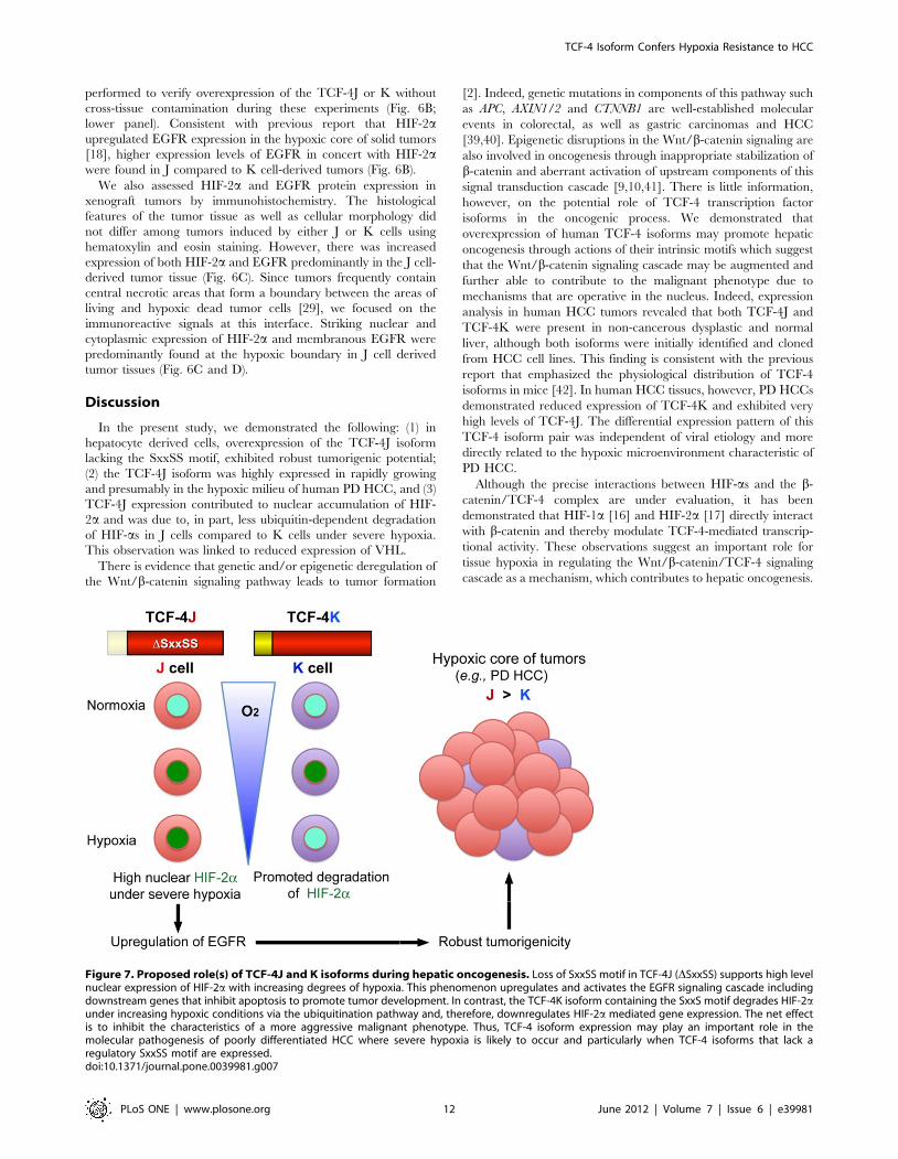
Figure 7. Proposed role(s) of TCF-4J and K isoforms during hepatic oncogenesis. Loss of SxxSS motif in TCF-4J (DSxxSS) supports high levelnuclear expression of HIF-2a with increasing degrees of hypoxia. This phenomenon upregulates and activates the EGFR signaling cascade includingdownstream genes that inhibit apoptosis to promote tumor development. In contrast, the TCF-4K isoform containing the SxxS motif degrades HIF-2aunder increasing hypoxic conditions via the ubiquitination pathway and, therefore, downregulates HIF-2a mediated gene expression. The net effectis to inhibit the characteristics of a more aggressive malignant phenotype. Thus, TCF-4 isoform expression may play an important role in themolecular pathogenesis of poorly differentiated HCC where severe hypoxia is likely to occur and particularly when TCF-4 isoforms that lack aregulatory SxxSS motif are expressed.doi:10.1371/journal.pone.0039981.g007
TCF-4 Isoform Confers Hypoxia Resistance to HCC
PLoS ONE | www.plosone.org 12 June 2012 | Volume 7 | Issue 6 | e39981

Furthermore, the role of the SxxSS motif in mediating TCF-4
effects on HIF-as stability under hypoxic conditions has been
emphasized here. We found that TCF-4J and TCF-4B, which lack
this motif, are associated with enhanced expression of HIF-1a and
HIF-2a under severe hypoxia, whereas TCF-4K and TCF-4A,
which express the SxxSS sequence, do not exhibit this property. As
a potential mechanism(s), we found less ubiquitin-dependent
degradation of HIF-as in J than K cells under severe hypoxia;
less degradation in J cells was linked to reduced expression of
VHL. In contrast, the HIF-as were highly degraded in K cells,
raising the possibility that HIF-as were subjected to VHL-
mediated degradation due to the presence of the SxxSS motif.
These findings emphasize the possible involvement of TCF-4
isoforms in mediating HIF-a stability via this SxxSS motif;
however, it is unclear whether each S of the SxxSS plays a distinct
role in the regulation of HIF-a stability.
The biological roles of HIF-1a and HIF-2a under hypoxia is
controversial; however, HIF-2a, but not HIF-1a, is considered to
be important in tumor formation [43], by promoting Myc activity
[44]. In addition, HIF-2a is known to upregulate EGFR
expression in the hypoxic core of solid tumors [18]. Emerging
evidence has been presented that HIF-2a should be recognized as
a CSC marker [45]. It is of interest that HIF-2a expression was
significantly associated with poor prognosis in aggressive gliomas
[37]. Since TCF-4J expression in our study was a potential
biomarker for an aggressive HCC phenotype [34], we focused on
the possible relevance of SxxSS as a mechanism of HIF-2astabilization under hypoxic conditions. Since K cells showed weak
and little tumorigenic ability in xenograft model, it was suggested
that mechanism of drastic HIF-2a degradation in those cells even
under hypoxia worked also may occur in vivo, possibly through
affecting the EGFR expression in a SxxSS-dependent manner. It
was observed in xenograft tissues that the driving force (HIF-2a)
for EGFR expression was active in J cells as compared to K cells,
especially in tumor cells under hypoxic conditions as observed in
necrotic area. This HIF-2a-induced upregulation of EGFR in J
cells may be associated with activation of EGFR signaling
pathways and further studies will be needed.
The robust tumorigenic potential of J cells may be predicted by
their significant anchorage-independent growth ability in vitro
under hypoxic conditions. The oncogenic potential of J cells grown
as xenografts is likely to be triggered by chronic hypoxic stress
derived from subcutaneous inoculation which has previously been
shown to have a poor neovascularization and thus, would generate
a hypoxic environment during rapid tumor growth. The
expression levels of both HIF-2a and EGFR were much higher
in J tumors than K derived tumors and suggests that the J tumors
may have grown faster through activating the HIF-2a/EGFR
system under a hypoxic environment as summarized by the
illustration presented in Fig. 7. This hypoxia-resistant property of J
cells may be the biologic basis for the finding that TCF-4J was the
predominant isoform expressed in human PD HCCs, which are
known to have a hypoxic core in the central region of the tumor
[18,29].
Supporting Information
Figure S1 Expression levels of TCF-4A, B, G and Hisoforms in human HCCs. (A and C) Comparative analysis of
TCF-4A/B (A) and G/H (C) mRNA expression levels in 27 HBV-
related HCC tumors (T) adjacent peritumor tissue (pT) and
histologic normal liver (N) by RT-PCR. (B and D) Comparative
analysis of TCF-4A/B (B) and G/H (D) mRNA expression levels
in another 20 HCC tumors including five WD HCCs from a
different clinical site. Seventeen individuals had HCV-related
chronic liver disease, and the remainder had chronic HBV
infection. Values are normalized to GAPDH. Statistical results
from all tissues are expressed as mean + SD (right panel). MD,
moderately differentiated; PD, poorly differentiated; WD, well
differentiated; *p,0.01; **p,0.05
(TIF)
Table S1 Relation between clinicopathological factorsand TCF-4A and B, and G and H expression in patients.
(DOC)
Acknowledgments
The authors would like to thank Drs. Osamu Nakashima and Sachiko
Ogasawara for providing human tissue samples and the corresponding
pathological information for the 2nd TCF-4 isoform analysis.
Author Contributions
Conceived and designed the experiments: HK MK. Performed the
experiments: HK OT YT EJW MK. Analyzed the data: HK OT YT MK.
Contributed reagents/materials/analysis tools: HCL JWR KMK HY.
Wrote the paper: HK JRW MK.
References
1. Reya T, Clevers H (2005) Wnt signalling in stem cells and cancer. Nature 434:
843–850.
2. Clevers H (2006) Wnt/beta-catenin signaling in development and disease. Cell
127: 469–480.
3. Ying Y, Tao Q (2009) Epigenetic disruption of the WNT/beta-catenin signaling
pathway in human cancers. Epigenetics 4: 307–312.
4. He TC, Sparks AB, Rago C, Hermeking H, Zawel L, et al. (1998) Identification
of c-MYC as a target of the APC pathway. Science 281: 1509–1512.
5. Tetsu O, McCormick F (1999) Beta-catenin regulates expression of cyclin D1 in
colon carcinoma cells. Nature 398: 422–426.
6. Yamashita T, Ji J, Budhu A, Forgues M, Yang W, et al. (2009) EpCAM-positive
hepatocellular carcinoma cells are tumor-initiating cells with stem/progenitor
cell features. Gastroenterology 136: 1012–1024.
7. El-Serag HB, Rudolph KL (2007) Hepatocellular carcinoma: epidemiology and
molecular carcinogenesis. Gastroenterology 132: 2557–2576.
8. Merle P, de la Monte S, Kim M, Herrmann M, Tanaka S, et al. (2004)
Functional consequences of frizzled-7 receptor overexpression in human
hepatocellular carcinoma. Gastroenterology 127: 1110–1122.
9. Kim M, Lee HC, Tsedensodnom O, Hartley R, Lim YS, et al. (2008) Functional
interaction between Wnt3 and Frizzled-7 leads to activation of the Wnt/beta-
catenin signaling pathway in hepatocellular carcinoma cells. J Hepatol 48: 780–
791.
10. Bengochea A, de Souza MM, Lefrancois L, Le Roux E, Galy O, et al. (2008)
Common dysregulation of Wnt/Frizzled receptor elements in human hepato-
cellular carcinoma. Br J Cancer 99: 143–150.
11. Duval A, Rolland S, Tubacher E, Bui H, Thomas G, et al. (2000) The human T-
cell transcription factor-4 gene: structure, extensive characterization of
alternative splicings, and mutational analysis in colorectal cancer cell lines.
Cancer Res 60: 3872–3879.
12. Pukrop T, Gradl D, Henningfeld KA, Knochel W, Wedlich D, et al. (2001)
Identification of two regulatory elements within the high mobility group box
transcription factor XTCF-4. J Biol Chem 276: 8968–8978.
13. Tsedensodnom O, Koga H, Rosenberg SA, Nambotin SB, Carroll JJ, et al.
(2011) Identification of T-cell factor-4 isoforms that contribute to the malignant
phenotype of hepatocellular carcinoma cells. Exp Cell Res 317: 920–931.
14. Takahashi K, Yamanaka S (2006) Induction of pluripotent stem cells from
mouse embryonic and adult fibroblast cultures by defined factors. Cell 126: 663–
676.
15. Mohyeldin A, Garzon-Muvdi T, Quinones-Hinojosa A (2010) Oxygen in stem
cell biology: a critical component of the stem cell niche. Cell Stem Cell 7: 150–
161.
16. Kaidi A, Williams AC, Paraskeva C (2007) Interaction between beta-catenin and
HIF-1 promotes cellular adaptation to hypoxia. Nat Cell Biol 9: 210–217.
17. Choi H, Chun YS, Kim TY, Park JW (2010) HIF-2alpha enhances beta-
catenin/TCF-driven transcription by interacting with beta-catenin. Cancer Res
70: 10101–10111.
TCF-4 Isoform Confers Hypoxia Resistance to HCC
PLoS ONE | www.plosone.org 13 June 2012 | Volume 7 | Issue 6 | e39981

18. Franovic A, Gunaratnam L, Smith K, Robert I, Patten D, et al. (2007)
Translational up-regulation of the EGFR by tumor hypoxia provides anonmutational explanation for its overexpression in human cancer. Proc Natl
Acad Sci U S A 104: 13092–13097.
19. Nakamura K, Zen Y, Sato Y, Kozaka K, Matsui O, et al. (2007) Vascularendothelial growth factor, its receptor Flk-1, and hypoxia inducible factor-
1alpha are involved in malignant transformation in dysplastic nodules of theliver. Hum Pathol 38: 1532–1546.
20. Knowles BB, Howe CC, Aden DP (1980) Human hepatocellular carcinoma cell
lines secrete the major plasma proteins and hepatitis B surface antigen. Science209: 497–499.
21. Nakabayashi H, Taketa K, Miyano K, Yamane T, Sato J (1982) Growth ofhuman hepatoma cells lines with differentiated functions in chemically defined
medium. Cancer Res 42: 3858–3863.22. Graham FL, Smiley J, Russell WC, Nairn R (1977) Characteristics of a human
cell line transformed by DNA from human adenovirus type 5. J Gen Virol 36:
59–74.23. He L, Isselbacher KJ, Wands JR, Goodman HM, Shih C, et al. (1984)
Establishment and characterization of a new human hepatocellular carcinomacell line. In Vitro 20: 493–504.
24. Yano H, Iemura A, Fukuda K, Mizoguchi A, Haramaki M, et al. (1993)
Establishment of two distinct human hepatocellular carcinoma cell lines from asingle nodule showing clonal dedifferentiation of cancer cells. Hepatology 18:
320–327.25. Wang GL, Semenza GL (1993) General involvement of hypoxia-inducible factor
1 in transcriptional response to hypoxia. Proc Natl Acad Sci U S A 90: 4304–4308.
26. Maeyama M, Koga H, Selvendiran K, Yanagimoto C, Hanada S, et al. (2008)
Switching in discoid domain receptor expressions in SLUG-induced epithelial-mesenchymal transition. Cancer 113: 2823–2831.
27. Hecht A, Stemmler MP (2003) Identification of a promoter-specific transcrip-tional activation domain at the C terminus of the Wnt effector protein T-cell
factor 4. J Biol Chem 278: 3776–3785.
28. Kojiro M (2004) ‘Nodule-in-nodule’ appearance in hepatocellular carcinoma: itssignificance as a morphologic marker of dedifferentiation. Intervirology 47: 179–
183.29. Brown JM, Wilson WR (2004) Exploiting tumour hypoxia in cancer treatment.
Nat Rev Cancer 4: 437–447.30. Franovic A, Holterman CE, Payette J, Lee S (2009) Human cancers converge at
the HIF-2alpha oncogenic axis. Proc Natl Acad Sci U S A 106: 21306–21311.
31. Semenza GL (2010) Defining the role of hypoxia-inducible factor 1 in cancer
biology and therapeutics. Oncogene 29: 625–634.
32. Liebner S, Cattelino A, Gallini R, Rudini N, Iurlaro M, et al. (2004) Beta-
catenin is required for endothelial-mesenchymal transformation during heart
cushion development in the mouse. J Cell Biol 166: 359–367.
33. Mani SA, Guo W, Liao MJ, Eaton EN, Ayyanan A, et al. (2008) The epithelial-
mesenchymal transition generates cells with properties of stem cells. Cell 133:
704–715.
34. Yang XR, Xu Y, Shi GM, Fan J, Zhou J, et al. (2008) Cytokeratin 10 and
cytokeratin 19: predictive markers for poor prognosis in hepatocellular
carcinoma patients after curative resection. Clin Cancer Res 14: 3850–3859.
35. Borovski T, De Sousa EMF, Vermeulen L, Medema JP (2011) Cancer stem cell
niche: the place to be. Cancer Res 71: 634–639.
36. Heddleston JM, Li Z, Lathia JD, Bao S, Hjelmeland AB, et al. (2010) Hypoxia
inducible factors in cancer stem cells. Br J Cancer 102: 789–795.
37. Li Z, Bao S, Wu Q, Wang H, Eyler C, et al. (2009) Hypoxia-inducible factors
regulate tumorigenic capacity of glioma stem cells. Cancer Cell 15: 501–513.
38. Yoneda N, Sato Y, Kitao A, Ikeda H, Sawada-Kitamura S, et al. (2011)
Epidermal growth factor induces cytokeratin 19 expression accompanied by
increased growth abilities in human hepatocellular carcinoma. Lab Invest 91:
262–272.
39. Satoh S, Daigo Y, Furukawa Y, Kato T, Miwa N, et al. (2000) AXIN1 mutations
in hepatocellular carcinomas, and growth suppression in cancer cells by virus-
mediated transfer of AXIN1. Nat Genet 24: 245–250.
40. Segditsas S, Tomlinson I (2006) Colorectal cancer and genetic alterations in the
Wnt pathway. Oncogene 25: 7531–7537.
41. Deng Y, Yu B, Cheng Q, Jin J, You H, et al. (2011) Epigenetic silencing of
WTF-1 in hepatocellular carcinomas. J Cancer Res Clin Oncol 136: 1161–1167.
42. Weise A, Bruser K, Elfert S, Wallmen B, Wittel Y, et al. (2010) Alternative
splicing of Tcf7l2 transcripts generates protein variants with differential
promoter-binding and transcriptional activation properties at Wnt/beta-catenin
targets. Nucleic Acids Res 38: 1964–1981.
43. Kaelin WG, Jr., Ratcliffe PJ (2008) Oxygen sensing by metazoans: the central
role of the HIF hydroxylase pathway. Mol Cell 30: 393–402.
44. Gordan JD, Bertout JA, Hu CJ, Diehl JA, Simon MC (2007) HIF-2alpha
promotes hypoxic cell proliferation by enhancing c-myc transcriptional activity.
Cancer Cell 11: 335–347.
45. Patel SA, Simon MC (2008) Biology of hypoxia-inducible factor-2alpha in
development and disease. Cell Death Differ 15: 628–634.
TCF-4 Isoform Confers Hypoxia Resistance to HCC
PLoS ONE | www.plosone.org 14 June 2012 | Volume 7 | Issue 6 | e39981