Long-lasting anxiolytic effect of neural precursor cells freshly prepared but not...
8
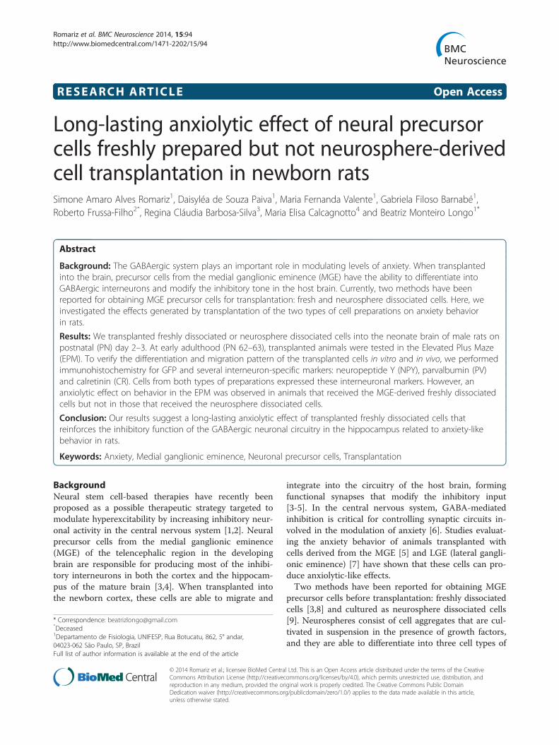
RESEARCH ARTICLE Open Access Long-lasting anxiolytic effect of neural precursor cells freshly prepared but not neurosphere-derived cell transplantation in newborn rats Simone Amaro Alves Romariz 1 , Daisyléa de Souza Paiva 1 , Maria Fernanda Valente 1 , Gabriela Filoso Barnabé 1 , Roberto Frussa-Filho 2 ˆ , Regina Cláudia Barbosa-Silva 3 , Maria Elisa Calcagnotto 4 and Beatriz Monteiro Longo 1* Abstract Background: The GABAergic system plays an important role in modulating levels of anxiety. When transplanted into the brain, precursor cells from the medial ganglionic eminence (MGE) have the ability to differentiate into GABAergic interneurons and modify the inhibitory tone in the host brain. Currently, two methods have been reported for obtaining MGE precursor cells for transplantation: fresh and neurosphere dissociated cells. Here, we investigated the effects generated by transplantation of the two types of cell preparations on anxiety behavior in rats. Results: We transplanted freshly dissociated or neurosphere dissociated cells into the neonate brain of male rats on postnatal (PN) day 2–3. At early adulthood (PN 62–63), transplanted animals were tested in the Elevated Plus Maze (EPM). To verify the differentiation and migration pattern of the transplanted cells in vitro and in vivo, we performed immunohistochemistry for GFP and several interneuron-specific markers: neuropeptide Y (NPY), parvalbumin (PV) and calretinin (CR). Cells from both types of preparations expressed these interneuronal markers. However, an anxiolytic effect on behavior in the EPM was observed in animals that received the MGE-derived freshly dissociated cells but not in those that received the neurosphere dissociated cells. Conclusion: Our results suggest a long-lasting anxiolytic effect of transplanted freshly dissociated cells that reinforces the inhibitory function of the GABAergic neuronal circuitry in the hippocampus related to anxiety-like behavior in rats. Keywords: Anxiety, Medial ganglionic eminence, Neuronal precursor cells, Transplantation Background Neural stem cell-based therapies have recently been proposed as a possible therapeutic strategy targeted to modulate hyperexcitability by increasing inhibitory neur- onal activity in the central nervous system [1,2]. Neural precursor cells from the medial ganglionic eminence (MGE) of the telencephalic region in the developing brain are responsible for producing most of the inhibi- tory interneurons in both the cortex and the hippocam- pus of the mature brain [3,4]. When transplanted into the newborn cortex, these cells are able to migrate and integrate into the circuitry of the host brain, forming functional synapses that modify the inhibitory input [3-5]. In the central nervous system, GABA-mediated inhibition is critical for controlling synaptic circuits in- volved in the modulation of anxiety [6]. Studies evaluat- ing the anxiety behavior of animals transplanted with cells derived from the MGE [5] and LGE (lateral gangli- onic eminence) [7] have shown that these cells can pro- duce anxiolytic-like effects. Two methods have been reported for obtaining MGE precursor cells before transplantation: freshly dissociated cells [3,8] and cultured as neurosphere dissociated cells [9]. Neurospheres consist of cell aggregates that are cul- tivated in suspension in the presence of growth factors, and they are able to differentiate into three cell types of * Correspondence: [email protected] ˆ Deceased 1 Departamento de Fisiologia, UNIFESP, Rua Botucatu, 862, 5° andar, 04023-062 São Paulo, SP, Brazil Full list of author information is available at the end of the article © 2014 Romariz et al.; licensee BioMed Central Ltd. This is an Open Access article distributed under the terms of the Creative Commons Attribution License (http://creativecommons.org/licenses/by/4.0), which permits unrestricted use, distribution, and reproduction in any medium, provided the original work is properly credited. The Creative Commons Public Domain Dedication waiver (http://creativecommons.org/publicdomain/zero/1.0/) applies to the data made available in this article, unless otherwise stated. Romariz et al. BMC Neuroscience 2014, 15:94 http://www.biomedcentral.com/1471-2202/15/94
Transcript of Long-lasting anxiolytic effect of neural precursor cells freshly prepared but not...
Romariz et al. BMC Neuroscience 2014, 15:94http://www.biomedcentral.com/1471-2202/15/94
RESEARCH ARTICLE Open Access
Long-lasting anxiolytic effect of neural precursorcells freshly prepared but not neurosphere-derivedcell transplantation in newborn ratsSimone Amaro Alves Romariz1, Daisyléa de Souza Paiva1, Maria Fernanda Valente1, Gabriela Filoso Barnabé1,Roberto Frussa-Filho2ˆ, Regina Cláudia Barbosa-Silva3, Maria Elisa Calcagnotto4 and Beatriz Monteiro Longo1*
Abstract
Background: The GABAergic system plays an important role in modulating levels of anxiety. When transplantedinto the brain, precursor cells from the medial ganglionic eminence (MGE) have the ability to differentiate intoGABAergic interneurons and modify the inhibitory tone in the host brain. Currently, two methods have beenreported for obtaining MGE precursor cells for transplantation: fresh and neurosphere dissociated cells. Here, weinvestigated the effects generated by transplantation of the two types of cell preparations on anxiety behaviorin rats.
Results: We transplanted freshly dissociated or neurosphere dissociated cells into the neonate brain of male rats onpostnatal (PN) day 2–3. At early adulthood (PN 62–63), transplanted animals were tested in the Elevated Plus Maze(EPM). To verify the differentiation and migration pattern of the transplanted cells in vitro and in vivo, we performedimmunohistochemistry for GFP and several interneuron-specific markers: neuropeptide Y (NPY), parvalbumin (PV)and calretinin (CR). Cells from both types of preparations expressed these interneuronal markers. However, ananxiolytic effect on behavior in the EPM was observed in animals that received the MGE-derived freshly dissociatedcells but not in those that received the neurosphere dissociated cells.
Conclusion: Our results suggest a long-lasting anxiolytic effect of transplanted freshly dissociated cells thatreinforces the inhibitory function of the GABAergic neuronal circuitry in the hippocampus related to anxiety-likebehavior in rats.
Keywords: Anxiety, Medial ganglionic eminence, Neuronal precursor cells, Transplantation
BackgroundNeural stem cell-based therapies have recently beenproposed as a possible therapeutic strategy targeted tomodulate hyperexcitability by increasing inhibitory neur-onal activity in the central nervous system [1,2]. Neuralprecursor cells from the medial ganglionic eminence(MGE) of the telencephalic region in the developingbrain are responsible for producing most of the inhibi-tory interneurons in both the cortex and the hippocam-pus of the mature brain [3,4]. When transplanted intothe newborn cortex, these cells are able to migrate and
* Correspondence: [email protected]ˆDeceased1Departamento de Fisiologia, UNIFESP, Rua Botucatu, 862, 5° andar,04023-062 São Paulo, SP, BrazilFull list of author information is available at the end of the article
© 2014 Romariz et al.; licensee BioMed CentraCommons Attribution License (http://creativecreproduction in any medium, provided the orDedication waiver (http://creativecommons.orunless otherwise stated.
integrate into the circuitry of the host brain, formingfunctional synapses that modify the inhibitory input[3-5]. In the central nervous system, GABA-mediatedinhibition is critical for controlling synaptic circuits in-volved in the modulation of anxiety [6]. Studies evaluat-ing the anxiety behavior of animals transplanted withcells derived from the MGE [5] and LGE (lateral gangli-onic eminence) [7] have shown that these cells can pro-duce anxiolytic-like effects.Two methods have been reported for obtaining MGE
precursor cells before transplantation: freshly dissociatedcells [3,8] and cultured as neurosphere dissociated cells[9]. Neurospheres consist of cell aggregates that are cul-tivated in suspension in the presence of growth factors,and they are able to differentiate into three cell types of
l Ltd. This is an Open Access article distributed under the terms of the Creativeommons.org/licenses/by/4.0), which permits unrestricted use, distribution, andiginal work is properly credited. The Creative Commons Public Domaing/publicdomain/zero/1.0/) applies to the data made available in this article,

Romariz et al. BMC Neuroscience 2014, 15:94 Page 2 of 8http://www.biomedcentral.com/1471-2202/15/94
the central nervous system: neurons, astrocytes and oli-godendrocytes [10]. In vitro studies using MGE cells cul-tured as neurospheres have shown that these cells areable to maintain their regional identity, giving rise to in-hibitory interneurons besides presenting other neurallineages such as astrocytes and oligodentrocytes [11-14].The fresh dissociation method consists of transplantingthe fetal brain cells immediately after dissecting themfrom the tissue. In this method, the cells migrate widely,maintaining their inhibitory phenotype and functionality[3,5,8,15]. When transplanted into the cerebral cortex,these cells are able to migrate and restore hippocampalfunction [16,17].Because of the increasing number of emerging studies
using MGE cells prepared by different methods andshowing discrepant results, the aim of the present studywas to compare the two preparations of MGE cells andinvestigate the long-lasting effects of the transplantationof either freshly dissociated or neurosphere dissociatedMGE-derived cells into the neonate brain of male ratson postnatal (PN) day 2–3 on the anxiety behaviorof these animals when tested at early adulthood (PN62–63) using the Elevated Plus Maze (EPM). The EPMis one of the most widely used tests in contemporarypreclinical research on anxiety, and is based on an innatefear that rodents have for open and elevated spaces [18].When in the EPM, rats tend to avoid the open arms andclearly prefer the enclosed arms. The avoidance of theopen arms occurs primarily because the open arms pre-vent the rat from engaging in thigmotaxic behavior [19].The ratio of open-arm to total arm entries has been usedas an index of anxiety [20]. Often, the percentage of timespent in the open arms is also reported. Anxiolytic drugsincrease the number of entries into and the total timespent in the open arms, whereas anxiogenic agentsdo the opposite [21,22]. Our data suggest a long-termanxiolytic effect following transplantation of freshly dis-sociated MGE cells, but not of cells expanded as neuro-spheres. We propose that the fresh cells were able toreinforce the inhibitory function of the GABAergic neur-onal circuitry related to anxiety-like behavior in rats.
MethodsAll animals were maintained in accordance with the Guidefor the Care and Use of Laboratory Animals (NationalResearch Council). All experimental protocols were ap-proved by the Animal Care and Ethics Committee ofUNIFESP/SP (CEP 0081/09).Sprague Dawley transgenic rat embryos (E14.5) express-
ing enhanced green fluorescent protein (SD-Tg (GFP)2BalRrrc), obtained from Charles River Labs and providedby CEDEME (Center for the Development of AnimalModels in Biology and Medicine at Universidade Federalde São Paulo) were used as the MGE cell donors.
Fresh dissociation preparationFor tissue extraction and cell dissociation, ventricularand subventricular layers of the MGE were dissectedfrom E14.5 rat embryos expressing enhanced greenfluorescent protein (GFP). Briefly, the tissue was re-moved and mechanically dissociated by pipetting andcentrifugation; the cells were washed with DMEM/ F-12(Dubelco’s Modified Eagle Medium, Gibco) containingDNase I (10 mg/mL), centrifuged, and ressuspended inthe same medium. We determined the cell number andviability of live cells using Trypan Blue exclusion method.The cell density was adjusted to ~100,000 cells/μL of viablecells (90% of cell viability) in culture medium per animal.To verify the in vitro cell differentiation into GABAergicneurons, part of the cells was plated in laminin/poly-l-lysine to confirm the GABA phenotype (Additional file 1:Figure S1).
Neurosphere culture preparationVentricular and subventricular layers of the MGE weredissected from E14.5 rat embryos expressing enhancedgreen fluorescent protein (GFP). The tissue was removedand incubated in trypsin for 5 minutes at 37°C followedby inactivation with fetal bovine serum (Gibco). The tis-sue was dissociated until a cell suspension was obtained.After centrifugation, the pellet was ressuspended in0.5 mL of DMEM/F-12. To check the number and cellviability, cells were counted in a Neubauer chamber inthe presence of trypan blue. The cells were cultured asneurospheres in a density of 100,000 cells/ml. The cul-ture medium consisted of DMEM/ F-12 supplementedwith 1% N2 (100× Invitrogen), 1% L-glutamine (200 mM,Invitrogen), EGF (20 ng/ml, Sigma), FGF-2 (10 ng/mL,R & D Systems) and penicillin (100 IU/ml), streptomycin(100 μg/mL) and amphotericin B (0.25 μg/mL) (all inantibiotic-antimycotic solution 100×, Invitrogen). Cellswere maintained in culture between 5 and 7 days. To ver-ify the in vitro cell differentiation into GABAergic neu-rons, part of the cells was plated to confirm the GABAphenotype (Additional file 1: Figure S1).
Neonatal TransplantationA suspension of freshly dissociated or neurosphere disso-ciated cells was placed in a Narishige microinjector guidedby a stereotaxic apparatus. Male Sprague Dawley rat pups(PN2-3) were anesthetized by hypothermia (−4°C), and5–10 × 104 GFP+ cells in 0.6 μL were bilaterally injectedinto the cortex of each rat for both methods of cell prepar-ation. The approximate bregma co-ordinates of the cortextargeted for injections were (2.4 mm A, 3.2 mm L,1.2 mm D) from bregma. Animals from the group namedMGE-F received freshly dissociated MGE cells (n = 7), andan age-matched control (CTRL-MF) group received cul-ture medium (DMEM-F12) only (n = 8) in the same site
Romariz et al. BMC Neuroscience 2014, 15:94 Page 3 of 8http://www.biomedcentral.com/1471-2202/15/94
and volume. Animals in the MGE-N group received neu-rosphere dissociated MGE-derived cells (n = 16), and anadditional control group received culture medium (CTRL-MN, n = 14). An age-matched control group (CTRL-MFand CTRL-MN) was established with each experimentalgroup and was transplanted on the same day as the re-spective experimental groups (MGE-N or MGE-F). Toavoid differences in basal levels of stress and anxiety, ani-mals from the control and experimental groups were fromthe same litter, and all pups remained away from theirmothers for the same amount of time.
Elevated Plus Maze (EPM)The EPM has been used extensively to evaluate anxiety-related behavior in rodents [23,24]. It consisted of twoopen (50 × 10 cm) and two enclosed (50 × 10 × 40 cm)arms made of wood, arranged in such a way that the twopairs of identical arms were opposite to each other. Theopen arms were bounded by 1 cm-high ledge on theborder of the arms. The maze was raised to a height of50 cm above the floor. Each animal was placed in thecenter of the apparatus facing a closed arm, and thenumber of entries (arm entry defined as all four pawsinto an arm) and the time spent in the open and closedarms were recorded for 5 minutes. The percentage oftime spent in the open arms and the number of openarm entries were recorded as indices of anxiety-like be-havior [21]. The number of entries into either arm of theapparatus was used as an index of general activity [25].The maze was thoroughly cleaned after each test with asolution of 20% ethanol and then dried. Each rat wastested only once.The behavioral tests were performed at 60 days after
the transplantation with either MGE-derived cells (freshor neurosphere dissociated cells) or medium. Tests wereperformed during the light period (14:00–17:00 h). Thebehavior of each animal was videotaped by a video cam-era located 150 cm above the apparatus. The videoswere later watched and analyzed by a trained observerwho was blind to the experimental condition.
ImmunohistochemistryThe animals were deeply anesthetized and perfusedthrough the heart with 100 mL of phosphate-buffered sa-line (PBS) followed by 250 mL of 4% paraformaldehyde.Coronal cryostat sections (30 μm thick) were made be-tween bregma 0.98 and bregma −3.28 mm, according tothe stereotaxic coordinates of the rat brain atlas [26]. Theprecise bregma co-ordinates of the analyzed coronal sec-tions for the cortex and hippocampus were betweenbregma −2.28 and −5.40 [26]. From this interval, we se-lected 5 sections for each structure, that would beequivalent to the dorsal, medial and ventral hippocam-pus (−2.36, −3.32, −4.00, −4.74 -5.38) of this interval of
bregma coordinate. To identify MGE-derived inhibitoryneurons in the transplanted brains, the sections were incu-bated with interneuronal markers for neuropeptide Y(NPY), calretinin (CR) and parvalbumin (PV). The sectionswere incubated for 2 h with anti-GFP AlexaFluor 488-conjugated antibody (1:600; Molecular Probes/Invitrogen),followed by an overnight incubation with rabbit antibodiesagainst NPY (1:2000, Molecular Probes), CR (1:2000,Molecular Probes) and PV (1:2000, Molecular Probes).The sections were then incubated with anti-rabbit IgGAlexaFluor 546-conjugated antibodies (1:600, MolecularProbes/Invitrogen) for 1 hour. The sections were mountedusing a nuclear-counterstaining, fluorescence-preservingmounting medium containing DAPI (Vector Labs). Theslides were examined using fluorescence (Nikon 80i) andconfocal (Olympus FluoView 1000) microscopes. For eachsection, double-labeled cells were qualitatively evaluatedfor their localization in the brain areas of interest (thecortex and hippocampus) and for their morphology.In order to quantify the MGE-derived transplanted cells,
the sections were incubated with anti-GFP AlexaFluor488-conjugated antibody (1:600; Molecular Probes/Invi-trogen) and were mounted on slides and sealed with cov-erslips. The sections were examined using a fluorescencemicroscope (Nikon 80i), and images were captured anddigitized using the Nikon ACT-1 v.2 system. The estima-tion of GFP+ cells was sampled in 5 coronal sections peranimal for cortex and hippocampus. Images of 10 random,non-overlapping fields were captured and quantified foreach region under 20× magnification.
Statistical analysisStatistical analyses of anxiety-related behavior and cellcounts were performed using the Mann–Whitney test(SPSS XI.) for comparison between the MGE-N xCTRL-MN and MGE-F x CTRL-MF groups. A signifi-cance level of 5% was assumed. Data are shown as themean ± SE.
ResultsBehavioral resultsIn order to assess the inhibitory effects of transplantedcells in vivo, the anxiety-like behavior of the animals wasevaluated using the EPM 60 days after transplantationand compared to an age-matched control group. Ratsfrom the MGE-F group showed a significant increase inthe percentage of time spent in the open arms (p = 0.04),indicating an anxiolytic effect as compared to theCTRL-MF group (Figure 1A). The percentage of openarm entries was similar for all groups but with a trendtowards an increase in the number of open arm entriesin the MGE-F group (p = 0.08) (Figure 1B).The percentage of open arm entries (p = 0.9) and the
total time spent in the open arms (p = 0.4) for animals

Figure 1 Elevated plus maze (EPM) using animals transplanted with freshly dissociated cells. EPM data comparing MGE-F (n = 7) andCTRL-MF (n = 8) groups; A) Percentage of time spent in the open arms; B) Percentage of entries into the open arms; C) Total number of entriesinto the open and closed arms. Data are shown as the mean and standard error. (*) Significant difference between groups (p < 0.05),Mann–Whitney test.
Romariz et al. BMC Neuroscience 2014, 15:94 Page 4 of 8http://www.biomedcentral.com/1471-2202/15/94
that received neurosphere cells (MGE-N) were not dif-ferent from the control group (CTRL-MN), indicatingthat MGE cells grown in culture were not able to gener-ate an anxiolytic effect (Figure 2A and 2B).Concerning the total number of entries into both the
open and closed arms, no significant differences werefound between the groups MGE-F versus CTRL-MF(p = 0.2) (Figure 1C) and MGE-N versus CTRL-MN (p = 0.7)
Figure 2 Elevated plus maze (EPM) using animals transplanted with n(n = 16) groups; A) Percentage of time spent in the open arms; B) Percentaopen and closed arms. Data are shown as the mean and standard error, M
(Figure 2C). These data suggest that there was no alter-ation in general locomotor activity and that the preferencefor the open arms was related to the anxiolytic effect ofthe freshly prepared MGE cells.
Analysis of transplanted GFP+ cellsA qualitative evaluation of the MGE-transplanted cells inthe host brain was performed by immunohistochemistry
eurosphere cells. EPM data comparing MGE-N (n = 14) and CTRL-MNge of entries into the open arms; C) Total number of entries into theann–Whitney test.

Romariz et al. BMC Neuroscience 2014, 15:94 Page 5 of 8http://www.biomedcentral.com/1471-2202/15/94
to confirm the presence of GFP+ transplanted cells, as wellas to verify their inhibitory phenotype by co-localizationwith the interneuronal markers NPY, PV and CR. Theanalysis of GFP+ cells in the adult brain indicated that theMGE-derived cells from both the freshly dissociated andneurosphere preparations were present and differentiatedinto inhibitory neurons (Figure 3). Additionally, these cellsmigrated throughout the parenchyma from the site of the
Figure 3 Double-labeled cells with GFP and inhibitory interneuronal minhibitory interneuronal markers in animals that received freshly dissociatedused were: calretinin (CR) (A-C) and neuropeptide Y (NPY) in the hilus (D-Fcortex (J-L); calretinin (CR) (M-O) and parvalbumin (PV) (P-R) in the CA1. S
injection (cortical layers 3 and 4) and were mainly foundin the cortex and hippocampus, with an insignificantnumber of GFP+ cells detected in other brain regions.The double-labeled NPY+/GFP+, PV+/GFP+ and CR+/GFP+
cells were detected in both the cortex and hippocampus(Figure 3), confirming that the transplanted cells from bothpreparation types, fresh and expanded as neurosphere, wereable to differentiate into inhibitory interneurons in vivo.
arkers. Confocal images revealed double labeling of GFP+ cells andcells (A-I) and neurosphere cells (J-R). The interneuronal markers); parvalbumin (PV) in the hilus (G-I); neuropeptide Y (NPY) in thecale bar = 50 μm.

Romariz et al. BMC Neuroscience 2014, 15:94 Page 6 of 8http://www.biomedcentral.com/1471-2202/15/94
A quantitative analysis of the amount of GFP+ cellswithin the two groups (MGE-F and MGE-N) showed nosignificant difference in cortical regions (p = 0.9). In thehippocampus, however, the MGE-F group had a highernumber of GFP+ cells when compared to the MGE-Ngroup (p = 0.009). This finding provides a possible explan-ation for the behavioral results, associating the presence ofMGE-F-derived inhibitory neurons in the hippocampuswith a reduced anxiety-like behavior (Figure 4).
DiscussionOur results indicate that MGE-derived cells obtained bytwo different methods, freshly dissociated or neurospheredissociated, and then transplanted into the neonate brainproduce distinct anxiety-related behaviors in adult rats.Animals from the MGE-F group presented an anxiolytic-like behavior, whereas transplanted neurosphere-derivedMGE cells was not able to decrease the basal levels of anx-iety. The freshly dissociated cells transplanted into new-born rats showed a long-lasting effect on anxiety behaviorwhen evaluated two months later in adulthood. This effectcould be attributed to the differentiation of these cells intoinhibitory GABAergic neurons that were able to migratemainly to the hippocampus, while the cells from the neu-rospheres remained in the cortex with no sign of longdistance migration.The GABAergic system plays an important role in
modulating the levels of anxiety. Investigations using ani-mal models are currently being conducted to betterunderstand the GABAergic circuits involved in the regula-tion of anxiety levels and mood disorders. Moreover, cellsobtained from MGE could present discrepant resultswhen transplanted as fresh or neurosphere cells. Recentstudies have shown that cells derived from the sub regions
Figure 4 Quantification of GFP+ cells. GFP+ cells were quantifiedfor the MGE-F and MGE-N groups in two brain regions: the cortex;and the hippocampus. A significant increase in the number of GFP+
cells was detected in the hippocampus of group MGE-F comparedto MGE-N. (*) Significant difference between groups (p < 0.05), dataare presented as the mean and standard error, Mann–Whitney test.
of eminences were able to modify the anxiety behavior intransplanted animals increasing the GABAergic transmis-sion and producing anxiolytic effects [5,7]. In the presentstudy, although animals transplanted with neurospherecells did not present effects in anxiety-like behavior, ani-mals grafted with MGE fresh cells presented a clear-cutanxiolytic-like behavior in the EPM test. The anxiolyticeffect was observed 60 days after transplantation, suffi-cient time for cells migrate, acquire an inhibitory pheno-type and to integrate into host tissue [3]. These long-termeffects of fresh cell transplantation in anxiety-like behaviorare consistent with our previous study with fresh MGEcells [5] that described the same decrease in anxiety levelsin two different ages, and showed a high density of trans-planted cells mainly in the striatum, corpus callosum andhippocampus.The presence of a large number of inhibitory neurons
in the hippocampus obtained from the fresh dissociationmethod two months after transplantation likely contrib-utes to decreased levels of anxiety. The hippocampus isa structure of extreme importance, not only for learningand memory but also for anxiety and fear-related re-sponses, being involved either directly or indirectly inthe modulation of anxiety [27-29]. The hippocampus islinked to a number of subcortical structures includingthe amygdala, and it has been associated with certain as-pects of emotional behavior, anxiety in particular [30].Moreover, it has been reported that animals with im-paired hippocampal neurogenesis have increased levelsof anxiety, showing a direct correlation between hippo-campal plasticity and the modulation of anxiety [31].The GFP+ cells obtained from the neurosphere prepar-
ation also differentiated into GABAergic inhibitory inter-neurons, as evidenced by the double-labeling with markersfor NPY, PV and CR along with GFP. Although specificstudies have succeeded in obtaining a considerable numberof neurons after the transplantation of neurospheres inclassic neurogenic areas of the brain [32,33], few neuronshave been found when these cells were transplanted intoareas not usually described as neurogenic, such as the stri-atum [13,34,35]. Thus, the environment may greatly influ-ence the differentiation of cells from neurospheres [36].Furthermore, although grafts of neurosphere cells derivedfrom the MGE preserved their ability to generate inhibitoryinterneurons in vitro and after transplantation, they pri-marily differentiate into astrocytes [13,14]. In our hands,even though the neurosphere-derived cells were alive andpresented an inhibitory phenotype, they were not able toreduce the basal levels of anxiety. Indeed, these cells failedto migrate to the hippocampus, a region involved in anx-iety behavior, and instead remained in the cortex, whichwas the site of transplantation.One could suggest that the low number of MGE cells
from neurosphere found in the two areas can be explained

Romariz et al. BMC Neuroscience 2014, 15:94 Page 7 of 8http://www.biomedcentral.com/1471-2202/15/94
by the cell death that must occur between the transplant-ation in postnatal period and the end of the experiment60 days after. Although the number of injected cells wasthe same for both preparations, e differences in anxietybehavior could be caused by differences in cell death ratebetween the two cells after transplantation, and that theanxiolytic effect can lie in the reduced apoptosis rate ofthe fresh cells, However, we have to consider that theapoptosis would have occurred soon after the transplant-ation (for the vast majority of cells), and could not be de-tected long after the transplantation of cells, when theanxiety test was applied (60 days after).The changes in the anxiety levels of animals that were
transplanted with freshly dissociated MGE cells indicatethat these cells were able to not only differentiate into in-hibitory neurons but also integrate into the local circuitryand change the inhibitory tone 60 days after the trans-plantation. By contrast, the transplanted cells dissociatedfrom neurospheres were able to differentiate into inhibi-tory interneurons but were unable to induce changes inthe levels of anxiety, probably because the amount ofcells that differentiated into inhibitory interneurons spe-cifically in the hippocampus was insufficient to generatean anxiolytic effect. According to our data, even thoughthe neurosphere-derived cells were alive and presentedinhibitory neuron phenotype, they have failed to migrateto the hippocampus, what can explain their incapabilityto modulate anxiety.
ConclusionOur results demonstrate a long-term anxiolytic effectinduced by the transplantation of freshly dissociatedMGE-derived cells but not by neurosphere dissociatedcells. The freshly dissociated MGE cells were able tomodulate the inhibitory tone in the hippocampus, asevidenced by a long-lasting decrease in the levels ofanxiety of transplanted animals. This work provides anew neural stem cell approach for the study of mooddisorders.
Additional file
Additional file 1: Figure S1. In vitro cells double-stained for GABA andDAPI.Images from fluorescence microscopy reveled GABAergic interneurons. In(A-C) cells dissociated from neurospheres; and in (D-F) fresh-dissociated cells.Scale bar = 100μm.
Competing interestsThe authors declare that they have no competing interests.
Authors’ contributionsSAA conceived the study, carried out the laboratory experiments, analyzedthe data, performed the statistical analysis and wrote the paper; DSP helpedto perform the statistical analysis, analyzed the data and wrote the paper;MFV carried out the laboratory experiments; GFB participated in theexperimental design and contributed with materials and analysis tools; RFFhelped to draft the manuscript, to interpret the results and critically revised
the paper; RCBS and MEC helped with the general idea of the paper andrevised the paper; BML defined the research theme, designed theexperiments, interpreted the results and wrote the paper. The workpresented here was carried out in collaboration between all authors. Allauthors read and approved the final manuscript.
AcknowledgmentsWe are grateful to Enéas Ferrazoli and Emanuel Barreto Cabral for thetechnical support.Dr. Roberto Frussa-Filho is no longer with us but he actively participated inthe study concept, interpretation as well as in the manuscript draft.This work was supported by FAPESP and CNPq.
Author details1Departamento de Fisiologia, UNIFESP, Rua Botucatu, 862, 5° andar,04023-062 São Paulo, SP, Brazil. 2Departamento de Farmacologia, UNIFESP,Rua Botucatu, 862, 04023-062 São Paulo, SP, Brazil. 3Departamento deBiociências, UNIFESP, Rua Silva Jardim, 136, 11015-020 Santos, SP, Brazil.4Departamento de Bioquímica, Instituto de Ciências Básicas da Saúde,UFRGS, Rua Ramiro Barcelos 2600, 90035-003 Porto Alegre, RS, Brazil.
Received: 19 May 2014 Accepted: 30 July 2014Published: 2 August 2014
References1. Raedt R, Van Dycke A, Vonck K, Boon P: Cell therapy in models for
temporal lobe epilepsy. Seizure 2007, 16(7):565–578.2. Thompson K: Transplantation of GABA-producing cells for seizure control
in models of temporal lobe epilepsy. Neurotherapeutics 2009, 6(2):284–294.3. Alvarez-Dolado M, Calcagnotto ME, Karkar KM, Southwell DG, Jones-Davis
DM, Estrada RC, Rubenstein JL, Alvarez-Buylla A, Baraban SC: Corticalinhibition modified by embryonic neural precursors grafted into thepostnatal brain. J Neurosci 2006, 26(28):7380–7389.
4. Wichterle H, Turnbull DH, Nery S, Fishell G, Alvarez-Buylla A: In utero fatemapping reveals distinct migratory pathways and fates of neurons bornin the mammalian basal forebrain. Development 2001, 128(19):3759–3771.
5. Valente MF, Romariz S, Calcagnotto ME, Ruiz L, Mello LE, Frussa-Filho R,Longo BM: Postnatal transplantation of interneuronal precursor cellsdecreases anxiety-like behavior in adult mice. Cell Transplant 2013,22(7):1237–1247.
6. Nemeroff CB: The role of GABA in the pathophysiology and treatment ofanxiety disorders. Psychopharmacol Bull 2003, 37(4):133–146.
7. Cunningham MG, Connor CM, Carlezon WA Jr, Meloni E: AmygdalarGABAergic-rich neural grafts attenuate anxiety-like behavior in rats.Behav Brain Res 2009, 205(1):146–153.
8. Baraban SC, Southwell DG, Estrada RC, Jones DL, Sebe JY, Alfaro-Cervello C,Garcia-Verdugo JM, Rubenstein JL, Alvarez-Buylla A: Reduction of seizuresby transplantation of cortical GABAergic interneuron precursors intoKv1.1 mutant mice. Proc Natl Acad Sci U S A 2009, 106(36):15472–15477.
9. Reynolds BA, Weiss S: Clonal and population analyses demonstrate thatan EGF-responsive mammalian embryonic CNS precursor is a stem cell.Dev Biol 1996, 175(1):1–13.
10. Reynolds BA, Tetzlaff W, Weiss S: A multipotent EGF-responsive striatalembryonic progenitor cell produces neurons and astrocytes. J Neurosci1992, 12(11):4565–4574.
11. Anderson SA, Marin O, Horn C, Jennings K, Rubenstein JL: Distinct corticalmigrations from the medial and lateral ganglionic eminences.Development 2001, 128(3):353–363.
12. Ostenfeld T, Joly E, Tai YT, Peters A, Caldwell M, Jauniaux E, Svendsen CN:Regional specification of rodent and human neurospheres. Brain Res DevBrain Res 2002, 134(1–2):43–55.
13. Eriksson C, Bjorklund A, Wictorin K: Neuronal differentiation followingtransplantation of expanded mouse neurosphere cultures derived fromdifferent embryonic forebrain regions. Exp Neurol 2003, 184(2):615–635.
14. Waldau B, Hattiangady B, Kuruba R, Shetty AK: Medial ganglioniceminence-derived neural stem cell grafts ease spontaneous seizuresand restore GDNF expression in a rat model of chronic temporal lobeepilepsy. Stem Cells 2010, 28(7):1153–1164.
15. Zipancic I, Calcagnotto ME, Piquer-Gil M, Mello LE, Alvarez-Dolado M:Transplant of GABAergic precursors restores hippocampal inhibitory

Romariz et al. BMC Neuroscience 2014, 15:94 Page 8 of 8http://www.biomedcentral.com/1471-2202/15/94
function in a mouse model of seizure susceptibility. Cell Transplant 2010,19(5):549–564.
16. Sebe JY, Looke-Stewart E, Dinday MT, Alvarez-Buylla A, Baraban SC: Neocorticalintegration of transplanted GABA progenitor cells from wild type andGABAB receptor knockout mouse donors. Neurosci Lett 2014, 561:52–57.
17. Howard MA, Rubenstein JL, Baraban SC: Bidirectional homeostaticplasticity induced by interneuron cell death and transplantation in vivo.Proc Natl Acad Sci U S A 2014, 111(1):492–497.
18. Montgomery KC: The relation between fear induced by novel stimulationand exploratory behavior. J Comp Physiol Psychol 1955, 48(4):254–260.
19. Treit D, Menard J, Royan C: Anxiogenic stimuli in the elevated plus-maze.Pharmacol Biochem Behav 1993, 44(2):463–469.
20. Silva RC, Brandao ML: Acute and chronic effects of gepirone and fluoxetinein rats tested in the elevated plus-maze: an ethological analysis. PharmacolBiochem Behav 2000, 65(2):209–216.
21. Pellow S, Chopin P, File SE, Briley M: Validation of open:closed arm entriesin an elevated plus-maze as a measure of anxiety in the rat. J NeurosciMethods 1985, 14(3):149–167.
22. Pellow S, File SE: Anxiolytic and anxiogenic drug effects on exploratoryactivity in an elevated plus-maze: a novel test of anxiety in the rat.Pharmacol Biochem Behav 1986, 24(3):525–529.
23. Handley SL, Mithani S: Effects of alpha-adrenoceptor agonists and antagonistsin a maze-exploration model of ‘fear’-motivated behaviour. NaunynSchmiedeberg’s Arch Pharmacol 1984, 327(1):1–5.
24. Calzavara MB, Lopez GB, Abilio VC, Silva RH, Frussa-Filho R: Role of anxietylevels in memory performance of spontaneously hypertensive rats.Behav Pharmacol 2004, 15(8):545–553.
25. Moser PC: An evaluation of the elevated plus-maze test using the novelanxiolytic buspirone. Psychopharmacology (Berl) 1989, 99(1):48–53.
26. Paxinos G, Watson C: The Rat Brain in Stereotaxic Coordinates. 5th edition. 2004.27. Anagnostaras SG, Gale GD, Fanselow MS: Hippocampus and contextual
fear conditioning: recent controversies and advances. Hippocampus 2001,11(1):8–17.
28. Bertoglio LJ, Joca SR, Guimaraes FS: Further evidence that anxiety andmemory are regionally dissociated within the hippocampus. Behav BrainRes 2006, 175(1):183–188.
29. Biedenkapp JC, Rudy JW: Hippocampal and extrahippocampal systemscompete for control of contextual fear: role of ventral subiculum andamygdala. Learn Memory 2009, 16(1):38–45.
30. Bannerman DM, Sprengel R, Sanderson DJ, McHugh SB, Rawlins JN, MonyerH, Seeburg PH: Hippocampal synaptic plasticity, spatial memory andanxiety. Nat Rev Neurosci 2014, 15(3):181–192.
31. Revest JM, Dupret D, Koehl M, Funk-Reiter C, Grosjean N, Piazza PV, Abrous DN:Adult hippocampal neurogenesis is involved in anxiety-related behaviors.Mol Psychiatry 2009, 14(10):959–967.
32. Fricker RA, Carpenter MK, Winkler C, Greco C, Gates MA, Bjorklund A:Site-specific migration and neuronal differentiation of human neuralprogenitor cells after transplantation in the adult rat brain. J Neurosci1999, 19(14):5990–6005.
33. Englund U, Fricker-Gates RA, Lundberg C, Bjorklund A, Wictorin K:Transplantation of human neural progenitor cells into the neonatalrat brain: extensive migration and differentiation with long-distanceaxonal projections. Exp Neurol 2002, 173(1):1–21.
34. Svendsen CN, ter Borg MG, Armstrong RJ, Rosser AE, Chandran S, OstenfeldT, Caldwell MA: A new method for the rapid and long term growth ofhuman neural precursor cells. J Neurosci Methods 1998, 85(2):141–152.
35. Vroemen M, Aigner L, Winkler J, Weidner N: Adult neural progenitor cellgrafts survive after acute spinal cord injury and integrate along axonalpathways. Eur J Neurosci 2003, 18(4):743–751.
36. Klein C, Butt SJ, Machold RP, Johnson JE, Fishell G: Cerebellum- andforebrain-derived stem cells possess intrinsic regional character.Development 2005, 132(20):4497–4508.
doi:10.1186/1471-2202-15-94Cite this article as: Romariz et al.: Long-lasting anxiolytic effect of neuralprecursor cells freshly prepared but not neurosphere-derived celltransplantation in newborn rats. BMC Neuroscience 2014 15:94.
Submit your next manuscript to BioMed Centraland take full advantage of:
• Convenient online submission
• Thorough peer review
• No space constraints or color figure charges
• Immediate publication on acceptance
• Inclusion in PubMed, CAS, Scopus and Google Scholar
• Research which is freely available for redistribution
Submit your manuscript at www.biomedcentral.com/submit