INDUCTION OF STRONGER AND LONG LASTING VACCINE ...
175
The Texas Medical Center Library The Texas Medical Center Library DigitalCommons@TMC DigitalCommons@TMC The University of Texas MD Anderson Cancer Center UTHealth Graduate School of Biomedical Sciences Dissertations and Theses (Open Access) The University of Texas MD Anderson Cancer Center UTHealth Graduate School of Biomedical Sciences 12-2012 INDUCTION OF STRONGER AND LONG LASTING VACCINE INDUCTION OF STRONGER AND LONG LASTING VACCINE IMMUNITY TO TUBERCULOSIS IMMUNITY TO TUBERCULOSIS Pearl Bakhru Follow this and additional works at: https://digitalcommons.library.tmc.edu/utgsbs_dissertations Part of the Medicine and Health Sciences Commons Recommended Citation Recommended Citation Bakhru, Pearl, "INDUCTION OF STRONGER AND LONG LASTING VACCINE IMMUNITY TO TUBERCULOSIS" (2012). The University of Texas MD Anderson Cancer Center UTHealth Graduate School of Biomedical Sciences Dissertations and Theses (Open Access). 315. https://digitalcommons.library.tmc.edu/utgsbs_dissertations/315 This Dissertation (PhD) is brought to you for free and open access by the The University of Texas MD Anderson Cancer Center UTHealth Graduate School of Biomedical Sciences at DigitalCommons@TMC. It has been accepted for inclusion in The University of Texas MD Anderson Cancer Center UTHealth Graduate School of Biomedical Sciences Dissertations and Theses (Open Access) by an authorized administrator of DigitalCommons@TMC. For more information, please contact [email protected].
-
Upload
khangminh22 -
Category
Documents
-
view
0 -
download
0
Transcript of INDUCTION OF STRONGER AND LONG LASTING VACCINE ...
The Texas Medical Center Library The Texas Medical Center Library
DigitalCommons@TMC DigitalCommons@TMC
The University of Texas MD Anderson Cancer Center UTHealth Graduate School of Biomedical Sciences Dissertations and Theses (Open Access)
The University of Texas MD Anderson Cancer Center UTHealth Graduate School of
Biomedical Sciences
12-2012
INDUCTION OF STRONGER AND LONG LASTING VACCINE INDUCTION OF STRONGER AND LONG LASTING VACCINE
IMMUNITY TO TUBERCULOSIS IMMUNITY TO TUBERCULOSIS
Pearl Bakhru
Follow this and additional works at: https://digitalcommons.library.tmc.edu/utgsbs_dissertations
Part of the Medicine and Health Sciences Commons
Recommended Citation Recommended Citation Bakhru, Pearl, "INDUCTION OF STRONGER AND LONG LASTING VACCINE IMMUNITY TO TUBERCULOSIS" (2012). The University of Texas MD Anderson Cancer Center UTHealth Graduate School of Biomedical Sciences Dissertations and Theses (Open Access). 315. https://digitalcommons.library.tmc.edu/utgsbs_dissertations/315
This Dissertation (PhD) is brought to you for free and open access by the The University of Texas MD Anderson Cancer Center UTHealth Graduate School of Biomedical Sciences at DigitalCommons@TMC. It has been accepted for inclusion in The University of Texas MD Anderson Cancer Center UTHealth Graduate School of Biomedical Sciences Dissertations and Theses (Open Access) by an authorized administrator of DigitalCommons@TMC. For more information, please contact [email protected].
INDUCTION OF STRONGER AND LONG LASTING VACCINE IMMUNITY TO
TUBERCULOSIS [DISSERTATION]
By
Pearl Bakhru, B.S., M.S.
APPROVED:
Dr Chinnaswamy Jagannath
Dr Bradley McIntyre
Dr Keri Smith
Dr Audrey Wanger
Dr Rick Wetsel
APPROVED:
Dean, The University of Texas
Graduate School of Biomedical Sciences
INDUCTION OF STRONGER AND LONG LASTING VACCINE IMMUNITY TO
TUBERCULOSIS
A
DISSERTATION
Presented to the Faculty of
The University of Texas
Health Science Center at Houston
And
The University of Texas
M.D Anderson Cancer Center
Graduate School of Biomedical Sciences
in Partial Fulfillment
of the Requirements
for the Degree of
DOCTOR OF PHILOSOPHY
by
Pearl Bakhru, B.S., M.S.
Houston, Texas
December, 2012

iii
Dedication
To my parents whose unconditional love and support has encouraged me to stay
strong during thick and thin. Also, to my uncle and aunt, who were very instrumental
in helping me during the toughest times and made me feel at ease throughout this
journey. I love you all immensely.

iv
ACKNOWLEDGEMENTS
With a deep sense of gratitude and respect, I would like to acknowledge my mentor
Dr. Chinnaswamy Jagannath for providing me the opportunity to work under his
esteemed guidance and being a source of immense inspiration and support during
the course of my project. He has been like a father figure to me and his patience
and trust in my scientific ability has helped me during the tough times of my PhD.
I would also extend my utmost appreciation to my advisory, exam as well as my
supervisory committee members: Dr Diane Bick, Dr Bradley McIntyre, Dr Rick
Wetsel, Dr John Klein, Dr Keri Smith, Dr Audrey Wanger, Dr Jagannadha Sastry, Dr
Jeffrey Actor and Dr Joseph Alcorn. Their critical reviews and analysis has changed
my approach to a scientific problem and their guidance has helped me be a better
researcher.
I would like to acknowledge Dr Robert Hunter for his financial support, Dr Dorothy
Lewis and her lab members for their help in flow cytometry analysis and NIH
vaccine center at Emory University for providing us with the tetramers. I would also
want to thank Dr Steve Norris and all the other faculty members involved with
Molecular Basis of Infectious Diseases (MBID) program for the financial support as
well guidance involved with scientific writing.
My utmost gratitude to all my lab members who are also my dear friends- Dr Arshad
Khan, Dr Emily Soudani, Dr Eulogia Roman, Dr Chris Singh, Dr Jaymie Estrella, Dr

v
Shen-An Huang, Karie Herdtner, Daniel Yuan, Smriti Malasi, and Shruti Malasi for
helping me with many techniques and providing me with tremendous assistance
during heavy work load. Also, I would like to thank Natalie Sirisaengtaksin and
Seema Mukherjee for their help during certain projects.
A huge hug and thanks goes to all my friends for being there for me during the most
insane times in my project where I was suffering with data and results and
whenever I was feeling low. Their humor, love and support made this journey easy
for me and instilled confidence and kept me going. You all are like my family.
Most importantly my sincere appreciation goes to the Graduate School of
Biomedical Sciences (GSBS) for allowing me to be a part of this institute and
providing me with an amazing opportunity to work with the best people in research.
vi
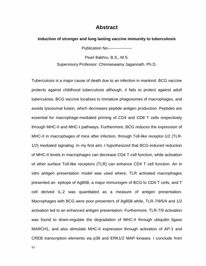
Abstract
Induction of stronger and long lasting vaccine immunity to tuberculosis
Publication No------------------
Pearl Bakhru. B.S., M.S.
Supervisory Professor: Chinnaswamy Jagannath. Ph.D.
Tuberculosis is a major cause of death due to an infection in mankind. BCG vaccine
protects against childhood tuberculosis although, it fails to protect against adult
tuberculosis. BCG vaccine localizes to immature phagosomes of macrophages, and
avoids lysosomal fusion, which decreases peptide antigen production. Peptides are
essential for macrophage-mediated priming of CD4 and CD8 T cells respectively
through MHC-II and MHC-I pathways. Furthermore, BCG reduces the expression of
MHC-II in macrophages of mice after infection, through Toll-like receptor-1/2 (TLR-
1/2) mediated signaling. In my first aim, I hypothesized that BCG-induced reduction
of MHC-II levels in macrophages can decrease CD4 T cell function, while activation
of other surface Toll-like receptors (TLR) can enhance CD4 T cell function. An in
vitro antigen presentation model was used where, TLR activated macrophages
presented an epitope of Ag85B, a major immunogen of BCG to CD4 T cells, and T
cell derived IL-2 was quantitated as a measure of antigen presentation.
Macrophages with BCG were poor presenters of Ag85B while, TLR-7/9/5/4 and 1/2
activation led to an enhanced antigen presentation. Furthermore, TLR-7/9 activation
was found to down-regulate the degradation of MHC-II through ubiquitin ligase
MARCH1, and also stimulate MHC-II expression through activation of AP-1 and
CREB transcription elements via p38 and ERK1/2 MAP kinases. I conclude from
vii
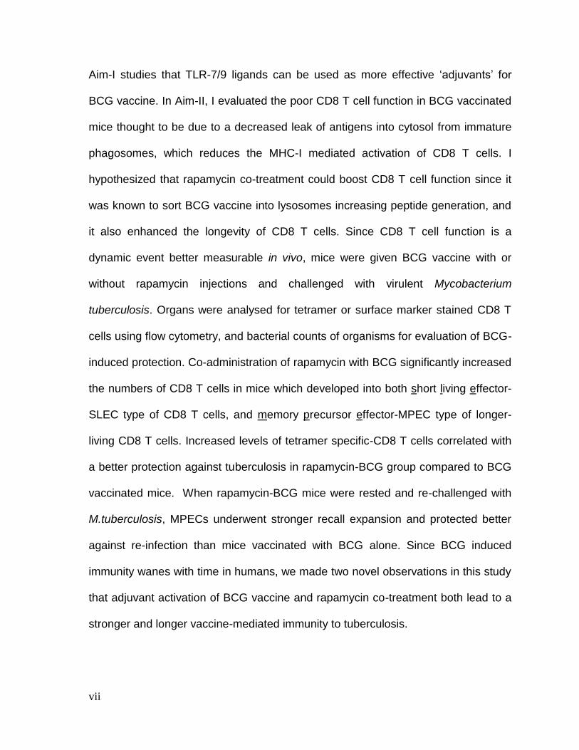
Aim-I studies that TLR-7/9 ligands can be used as more effective ‘adjuvants’ for
BCG vaccine. In Aim-II, I evaluated the poor CD8 T cell function in BCG vaccinated
mice thought to be due to a decreased leak of antigens into cytosol from immature
phagosomes, which reduces the MHC-I mediated activation of CD8 T cells. I
hypothesized that rapamycin co-treatment could boost CD8 T cell function since it
was known to sort BCG vaccine into lysosomes increasing peptide generation, and
it also enhanced the longevity of CD8 T cells. Since CD8 T cell function is a
dynamic event better measurable in vivo, mice were given BCG vaccine with or
without rapamycin injections and challenged with virulent Mycobacterium
tuberculosis. Organs were analysed for tetramer or surface marker stained CD8 T
cells using flow cytometry, and bacterial counts of organisms for evaluation of BCG-
induced protection. Co-administration of rapamycin with BCG significantly increased
the numbers of CD8 T cells in mice which developed into both short living effector-
SLEC type of CD8 T cells, and memory precursor effector-MPEC type of longer-
living CD8 T cells. Increased levels of tetramer specific-CD8 T cells correlated with
a better protection against tuberculosis in rapamycin-BCG group compared to BCG
vaccinated mice. When rapamycin-BCG mice were rested and re-challenged with
M.tuberculosis, MPECs underwent stronger recall expansion and protected better
against re-infection than mice vaccinated with BCG alone. Since BCG induced
immunity wanes with time in humans, we made two novel observations in this study
that adjuvant activation of BCG vaccine and rapamycin co-treatment both lead to a
stronger and longer vaccine-mediated immunity to tuberculosis.
viii
Index
Dedication ................................................................................................................ iii
Acknowledgements ................................................................................................. iv
Abstract .................................................................................................................... vi
Index ....................................................................................................................... viii
List of Illustrations ................................................................................................... x
CHAPTER 1: INTRODUCTION ................................................................................. 1
CHAPTER 2.1: Activation of Toll Like Receptors (TLRs) enhance expression of
MHC-II in BCG infected macrophages ..................................................................... 16
CHAPTER 2.2: Toll Like Receptor (TLR) ligands enhance MHC-II in macrophages
through the activation of MAP-Kinases and AP-1 or CREB transcription factors ..... 37
CHAPTER 3: Effects of rapamycin on T cell function during experimental
tuberculosis .............................................................................................................. 48
Chapter 3.1: Rapamycin increases CD8 T cell function during experimental
tuberculosis .............................................................................................................. 53
Chapter 3.2: Rapamycin increases BCG vaccine induced protection and CD4 and
CD8 T cell function against experimental tubercuolsis in mice ................................ 63
Chapter 3.3: Rapamycin shifts the phenotype of immune memory cell populations
through inhibition of mTOR facilitating expansion of central memory T cells after
BCG vaccination ...................................................................................................... 76
Chapter 3.4: Evaluation of the protective function of memory precursor effector
CD8 T cells (MPECs) induced by rapamycin after rechallenge of BCG vaccinated
mice ...................................................................................................................... 100
ix
Chapter 3.5: In vitro studies on the effect of rapamycin on dendritic cell-T cell co-
cultures .................................................................................................................. 113
CHAPTER 4: Conclusions ..................................................................................... 122
CHAPTER 5: Materials and Methods .................................................................... 131
References ........................................................................................................... 140
Curriculum Vitae .................................................................................................. 158
x
List of Illustrations
Figure 1: Major immune mechanisms involved in immunity to tuberculosis and
relevance for vaccine development ........................................................................... 8
Figure 2: Antigen presenting cells (APCs) process peptides from BCG vaccine to
activate T cells via MHC-I and MHC-II pathways. .................................................... 17
Figure 3: Processing and presentation of peptides from BCG vaccine is defective
in antigen presenting cells (APCs) which reduces activation of T cells .................... 18
Figure 4: An in vitro antigen presentation assay using macrophages and DCs ... 20
Figure 5: Macrophages (MΦs) infected with Mycobacterium bovis BCG vaccine
present Ag85B epitope to CD4 T cells (BB7) and induce IL-2 secretion. Lipids of
BCG suppress IL-2 responses ................................................................................. 22
Figure 6: Lipids of BCG suppress presentation of soluble Antigen-85B of BCG
vaccine from macrophages (MΦs) to CD4 T cells ................................................... 23
Figure 7: BCG infected MΦs show suppression of MHC-II surface expression
which correlates with enhanced expression of intracellular MARCH-1 ubiquitin
ligase ....................................................................................................................... 24
Figure 8: Alternate activation of Toll-like receptors in macrophages infected with
Mycobacterium bovis (BCG vaccine) enhances antigen presentation in vitro .......... 26
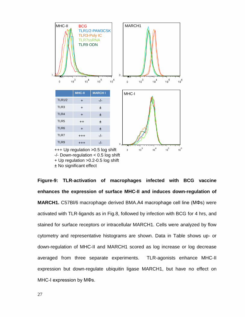
Figure 9: TLR-activation of MΦs infected with BCG vaccine enhances the
expression of surface MHC-II and induces down-regulation of MARCH1 ................ 27
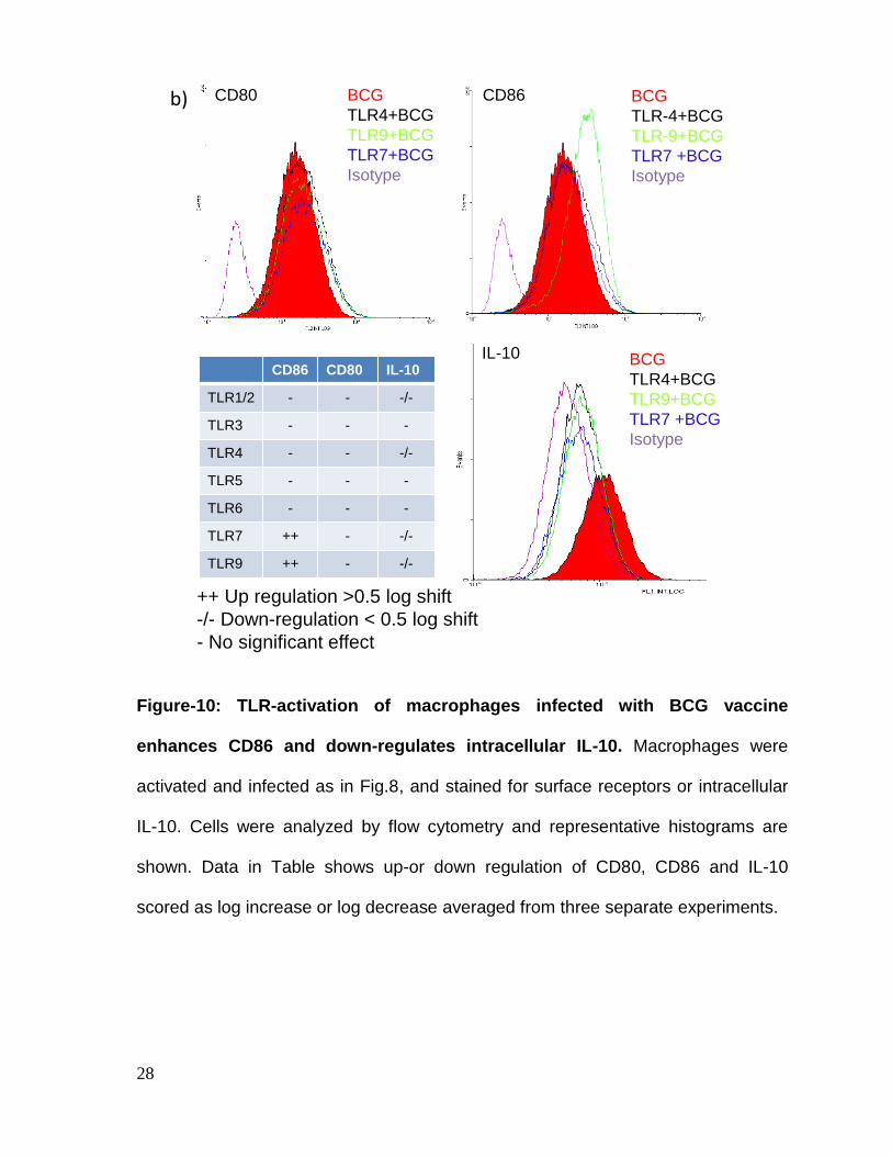
Figure 10: TLR-activation of MΦs infected with BCG vaccine enhances the CD86
in and down-regulates intracellular IL-10 ................................................................ 28
xi
Figure 11: Combined TLR-activation of MΦs infected with BCG vaccine enhances
surface expression of MHC-II and downregulates MARCH1 .................................. 30
Figure 12: siRNA knockdown of MARCH1 increases surface expression of MHC-II
in MΦs ..................................................................................................................... 31
Figure 13: TLR-activation decreases the ubiquitination of MHC-II in BCG infected
MΦs ......................................................................................................................... 33
Figure 14: TLR-4 induced MAPK pathway activation ............................................. 34
Figure 15: TLR-activation can converge at TRAF6 activation and MAPKs (p38
MAPK, ERK1/2, JNK) which lead to activation of transcription factors AP-1 or CREB
and synthesis of MHC-II........................................................................................... 40
Figure 16: Use of MAPK or AP-1/CREB inhibitors in macrophages does not affect
MHC-II expression in MΦs ....................................................................................... 42
Figure 17: TLR-7 and TLR-9 activation of MΦs enhances MHC-II expression in
BCG infected MΦs and inhibitors of MAPK cascade and AP1/CREB transcription
factor decrease surface MHC-II ............................................................................... 43
Figure 18: Activation of TLR-2 and TLR-4 has moderate effects on the levels of
MHC-II in BCG infected MΦs ................................................................................... 44
Figure 19: TLR-activation increases MHC-II expression in dendritic cells, which is
dependent upon activation of p38 MAPK, ERK1/2 and AP-1 or CREB transcription
factors (TFs) ............................................................................................................ 45
Figure 20: Dual effects of Rapamycin on T cells .................................................. 51
Figure 21: Rapamycin effects on experimental tuberculosis of C57Bl/6 mice ....... 57
xii
Figure 22: Rapamycin treatment during tuberculosis enhances T-bet expression
within CD4 T cells of lungs ...................................................................................... 58
Figure 23: Rapamycin enhances levels of antigen (tetramer) specific CD8 T cells
during tuberculosis ................................................................................................... 59
Figure 24: The NIH- mouse tuberculosis vaccine evaluation model used to
determine the immunological basis of protection ..................................................... 66
Figure 25: Rapamycin enhances efficacy of BCG vaccine against tuberculosis ... 68
Figure 26: Rapamycin enhances levels of Antigen specific tetramer positive CD8
T cells. ..................................................................................................................... 69
Figure 27: Rapamycin enhances the levels of T-bet+ short living effector CD8
T cells (SLEC) and Eomes+ MPECs memory precursor effector CD8 T cells
(MPEC) after vaccination ......................................................................................... 72
Figure 28: Rapamycin affects the levels of T-bet+ short living effector CD4 T
cells (SLEC) and Eomes+ MPECs memory precursor effector CD4 T cells
(MPEC) after vaccination ....................................................................................... 73
Figure 29: IFN-αβ plays a role in regulation of effector memory .......................... 82
Figure 30: Memory CD8+ T-cell differentiation ..................................................... 83
Figure 31: Rapamycin improves the quantity and quality of memory CD8+ T cells......................................................................................................................... 84
Figure 32: Effect of low and high dose rapamycin on BCG induced protection and memory populations ......................................................................................... 86
Figure 33: Evaluation of memory population on CD4 T cells ............................... 87
Figure 34: Assessing memory population on CD8 T cells .................................... 88
xiii
Figure 35: Both low dose long term and high dose short term rapamycin
dosing boost the efficacy of BCG vaccine in mice against tuberculosis .................. 91
Figure 36a: Rapamycin induces a better expansion of memory precursor effector
(MPEC) T cells ......................................................................................................... 92
Figure 36b: Rapamycin induces a better expansion of memory precursor effector
(MPEC) T cells in lungs and spleens ....................................................................... 93
Figure 37a: Rapamycin induces a better expansion of short living effector (SLEC)
T cells lungs and spleen between 4-6 weeks and lymph nodes at 8 weeks post
challenge with Mtb ................................................................................................... 94
Figure 37b: Rapamycin has only a moderate effect on the expansion of short living
effector (SLEC) CD8 T cells in the spleens .............................................................. 95
Figure 38: Rapamycin induced antigen specific tetramer+ CD8 T cells are CD44+
and CD62L+ memory cells ....................................................................................... 97
Figure 39: The mouse tuberculosis rechallenge model to determine the protective
effect of memory precursor effector T cells (MPECs) ............................................ 105
Figure 40: Prior rapamycin treatment of BCG vaccinated mice enables a better
protection against tuberculosis in the mouse rechallenge model indicating a
protective function for memory precursor effector T cells (MPECs) ....................... 106
Figure 41: Rapamycin treatment following BCG vaccination enables mice to mount
a stronger recall expansion of antigen specific tetramer+ CD8 T cells in the lungs
upon rechallenge with virulent M.tuberculosis ....................................................... 107
xiv
Figure 42: Rapamycin treatment following BCG vaccination enables mice to mount
a stronger recall expansion of CD8 T cells in the lungs upon rechallenge with
virulent M.tuberculosis ........................................................................................... 108
Figure 43: Rapamycin treatment following BCG vaccination has no major effect on
the expansion of antigen specific tetramer+ CD8 T cells in the spleens upon
rechallenge with virulent M.tuberculosis ................................................................ 109
Figure 44: Rapamycin treatment following BCG vaccination has no significant
effect on the expansion of CD8 T cells in the spleens upon rechallenge with virulent
M.tuberculosis ........................................................................................................ 110
Figure 45: Rapamycin treatment following BCG vaccination has no significant
effect on the expansion of CD8 T cells in the lymph nodes upon rechallenge with
virulent M.tuberculosis ........................................................................................... 111
Figure 46: Rapamycin treated dendritic cells enhance the expression of T-bet and
Eomes transcription factors (TFs) early after antigen presentation to CD8 B3Z-T
cells........................................................................................................................ 117
Figure 47: Rapamycin treated dendritic cells enhance the expression of granzyme
and perforin early after antigen presentation to CD8-B3Z T cells .......................... 118
Figure 48: Rapamycin treatment of BCG infected DCs does not affect the
expression of T-bet and Eomes in CD4 T cells ...................................................... 119

1
CHAPTER 1
INTRODUCTION
2
Tuberculosis (TB) is a disease caused by the rod shaped tubercle bacillus
Mycobacterium tuberculosis (Mtb) which mainly infects the lungs and brain. In
2007, there were an estimated 13.7 million chronic active cases globally, while in
2010 there were an estimated 8.8 million new cases and 1.5 million associated
deaths, mostly occurring in developing countries. There is an increased incidence of
TB in Asia and Africa whereas, only about 5-10% of population in USA and Europe
are affected (61). The main reason behind this disparity is the prevalence of AIDS in
the developing countries where TB co-infection occurs as a consequence of HIV-
infection induced reduction in CD4 T cell mediated immunity (105).
Symptoms of TB are chronic cough with blood tinged sputum, weight loss, fever and
night sweats. If TB spreads to other organs, the symptoms are widely distributed
(57). To diagnose active TB, the most important method is either the detection of
mycobacteria in sputum or the X-ray of lungs which detects granuloma, which is a
typical histological presentation of TB. For diagnosis of latent TB, physicians rely on
tuberculin/PPD skin test or an Interferon- γ release (IGRA) test performed on blood
samples. Treatment of TB is intensive and requires a long time to completely clear
the pathogen. Health care workers recommend directly observed therapy (DOTS)
for comprehensive treatment of TB (40). It is emphasized in case of patients
suffering from multi-drug resistant (MDR) TB, patients with HIV-1 and co-infection
with TB (82). Prevention in developing countries relies on administering the vaccine
BCG soon after birth.
3
Pathogenesis: In advanced stages TB can also spread to other parts of the body.
Transmission of TB usually occurs through aerosol route; when an infected person
sneezes or coughs, the TB droplets are released into the air and the latter can infect
a healthy individual (33). Most of the infections are latent or asymptomatic and only
about 10 % of infected humans develop active disease, which if not treated can
have serious consequences (112).
Bacillus Calmette-Guérin (or Bacille Calmette-Guérin, BCG) is a vaccine which is
an attenuated form of Mtb derived from Mycobacterium bovis, which usually infects
the cattle and other animals. BCG is less virulent because it has been subcultured
for 230 passages in an artificial medium for over 13 years (65). BCG still holds its
strong immunogenicity and thought to be an effective and safe vaccine in children. It
is the most widely used and distributed vaccine by WHO and is the first vaccine
given to children immediately after birth (87). However, BCG has lost some gene
segments (e.g., RD1- immunodominant region present in both M.bovis and Mtb)
during its long term-passage in vitro, and it has been thought that this is the reason
why it protects children only up to 5-10 years.
Variable Efficacy of BCG vaccine. The first large scale trial evaluating the efficacy
of BCG was conducted from 1956 to 1963, and involved 54,239 school children who
received BCG at the age of 14 or 15; this study showed an efficacy of 84% up to 5
years after immunization However, a US Public Health Service trial of BCG
in Georgia and Alabama published in 1966, showed an efficacy of only 14% and did
4
much to convince the US, that it did not want to implement mass immunization with
BCG. A further trial conducted in South-India and published in 1979 (the
"Chingleput trial"), also showed no protective effect due to BCG. The duration of
efficacy is not exactly known for BCG vaccine. However, meta-analysis of data from
different trials indicate that protection level of 80 % decreases after 15 years of age
and by the time the individual is 20, the protection is almost close to zero (4). The
most important effect of BCG is preventing miliary TB and TB meningitis in different
parts of the world. This is one of the most important reasons why BCG is still
administered in developing countries and is an essential part of childhood
immunization (24).
Reasons for variable efficacy: Many reasons have been cited for the variable
efficacy of BCG; although most remain to be explained.
a) Genetic variations in BCG strains: Because BCG has been passaged for a
number of times, there have been genetic modifications (deletions) in the
strains. A major example is the deletion of RD1 operon that encodes for the
secreted immune-dominant antigens ESAT6 and CFP10 conserved in both
M.bovis (parent of BCG) and in wild type Mtb (94).
1) Genetic variations in human population: The genetic makeup of human
populations in different countries and their diet and lifestyle may have a
strong influence on the vaccine efficacy of BCG strain (46). However,
interesting data emerged from the Birmingham BCG trial published in 1988.
The trial was based in Birmingham, United Kingdom, and examined children
born to families who originated from the Indian subcontinent (where vaccine
5
efficacy had previously been shown to be zero). The BCG trial showed a
64% protective effect, which is very similar to the figure derived from other
UK trials, arguing against the human genetic variation hypothesis.
2) Interference by non-tuberculous mycobacteria : Humans are also exposed to
environmental (M. vaccae) or non-tuberculous mycobacteria (M. Kansasii)
(76). This can generate a non-specific immune response to the mycobacteria
and administering BCG may become less effective. There is clinical evidence
from parallel studies performed on adolescent school children in the UK and
Malawi. In this study, the UK school children had a low baseline cellular
immunity to mycobacteria which was increased by BCG; in contrast, the
Malawi school children had a high baseline cellular immunity to mycobacteria
which was not significantly increased by BCG. This was thought to be due to
the induction of suppressive immunity by environmental mycobacteria. An
alternative explanation was suggested by mouse studies: immunity against
mycobacteria prevents BCG from replicating and preventing from its efficient
induction of immune responses. This is called as the blocking hypothesis.
3) Interference by concurrent parasitic infection in humans: Simultaneous
infection with parasites may change the immune response to BCG, making it
less effective. A T-helper 1 (Th1) response is required for an effective
immunity to tuberculosis; parasites induce a counter-productive Th2-
response which blunts the Th1-inducing effect of BCG (89).
4) Exposure to ultraviolet light: Concentration of ultraviolet light (particularly UV-
blue) from the Sun may have some effect on efficacy of the BCG vaccine.
6
UVB light has been demonstrated to reduce efficacy of BCG vaccine in
guinea pigs. The concentration gradient of UVB light increases
geographically closer to the Earth's equator. It is possible, though currently
unproven that this effect may occur as a result of sunlight-
dependent Vitamin-D production in humans (50).
5) Immunological sequestration of BCG in antigen presenting cells. BCG
vaccine has a mechanism to evade the host immune responses by
sequestering within phagosomes that do not fuse with lysosomes (97).
Lysosome mediated degradation of BCG antigens is a pre-requisite for
activation of CD4 T cells through MHC-II and this has been thought to affect
the efficacy of Th1 immunity induced by BCG. It is also known that BCG
phagosome membrane is not permeable enough for antigens to leak across
into cytosol preventing cross-presentation (41). These aspects are further
discussed below under the context of peptide processing mechanisms.
Immune responses against Tuberculosis. For the optimal protection against TB,
T lymphocytes are the sentinel cells used for defense mechanism. Macrophages
and dendritic cells (DCs) (antigen presenting cells; APCs) present antigens from
Mtb or BCG vaccine to T cells through the surface MHC-I and MHC-II molecules. T
lymphocytes defend against pathogens through 3 distinct mechanisms; a) CD4+ T
cells recognize peptide antigens via Class II Major Histocompatibility complex
(MHC-II) (10). b) CD8+T cells recognize peptide antigens via Class I Major
Histocompatibility complex (MHC-I) (17) and c) in addition, gamma delta (γδ) T cells
7
and CD1 restricted T cells may play a significant role at mucosal surfaces (17). B-
cell mediated responses and antibody responses are evident in tuberculosis but
have not been shown to be associated with protection (114). Thus, current vaccine
improvement strategies are focused on improving T lymphocyte activation against
Mtb.
8
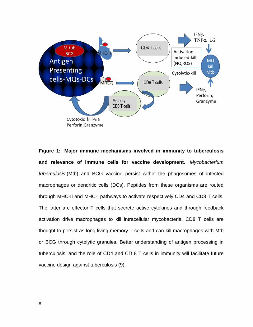
Figure 1: Major immune mechanisms involved in immunity to tuberculosis
and relevance of immune cells for vaccine development. Mycobacterium
tuberculosis (Mtb) and BCG vaccine persist within the phagosomes of infected
macrophages or dendritic cells (DCs). Peptides from these organisms are routed
through MHC-II and MHC-I pathways to activate respectively CD4 and CD8 T cells.
The latter are effector T cells that secrete active cytokines and through feedback
activation drive macrophages to kill intracellular mycobacteria. CD8 T cells are
thought to persist as long living memory T cells and can kill macrophages with Mtb
or BCG through cytolytic granules. Better understanding of antigen processing in
tuberculosis, and the role of CD4 and CD 8 T cells in immunity will facilitate future
vaccine design against tuberculosis (9).
Antigen Presenting cells-MQs-DCs
CD4 T cells
CD8 T cells
MHC-II
MHC-I
IFNγ, TNFα, IL-2
IFNγ, Perforin, Granzyme
MQkill
Mtb
Memory CD8 T cells
M.tubBCG
Cytotoxic kill-via Perforin,Granzyme
Cytolytic-kill
Activation induced-kill (NO,ROS)
9
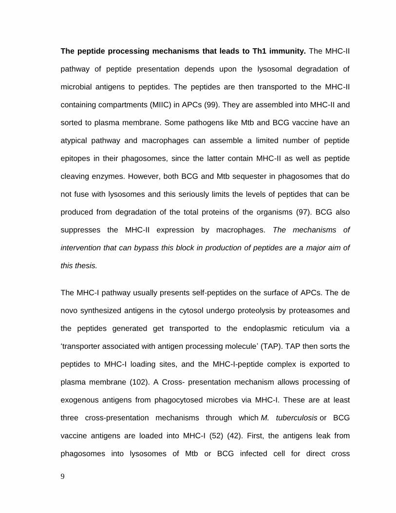
The peptide processing mechanisms that leads to Th1 immunity. The MHC-II
pathway of peptide presentation depends upon the lysosomal degradation of
microbial antigens to peptides. The peptides are then transported to the MHC-II
containing compartments (MIIC) in APCs (99). They are assembled into MHC-II and
sorted to plasma membrane. Some pathogens like Mtb and BCG vaccine have an
atypical pathway and macrophages can assemble a limited number of peptide
epitopes in their phagosomes, since the latter contain MHC-II as well as peptide
cleaving enzymes. However, both BCG and Mtb sequester in phagosomes that do
not fuse with lysosomes and this seriously limits the levels of peptides that can be
produced from degradation of the total proteins of the organisms (97). BCG also
suppresses the MHC-II expression by macrophages. The mechanisms of
intervention that can bypass this block in production of peptides are a major aim of
this thesis.
The MHC-I pathway usually presents self-peptides on the surface of APCs. The de
novo synthesized antigens in the cytosol undergo proteolysis by proteasomes and
the peptides generated get transported to the endoplasmic reticulum via a
‘transporter associated with antigen processing molecule’ (TAP). TAP then sorts the
peptides to MHC-I loading sites, and the MHC-I-peptide complex is exported to
plasma membrane (102). A Cross- presentation mechanism allows processing of
exogenous antigens from phagocytosed microbes via MHC-I. These are at least
three cross-presentation mechanisms through which M. tuberculosis or BCG
vaccine antigens are loaded into MHC-I (52) (42). First, the antigens leak from
phagosomes into lysosomes of Mtb or BCG infected cell for direct cross
10
presentation. Secondly, Mtb or BCG infected cells produce exosomes, that can be
taken up by bystander dendritic cells or macrophages, which then present antigens
via MHC-I. In the third mechanism, the Mtb or BCG infected cells can undergo
apoptosis and release the apoptotic bodies containing antigens, which can be taken
up by bystander APCs (15). The relative efficacy of these three mechanisms is
unclear. However, in the context of this proposal, BCG phagosomes are not leaky
enough that peptides do not get access to cytosol. A mechanism to enhance
peptide production and activate CD8 T cells for a better function is the second major
aim of this proposal.
Role of Toll-like receptors (TLRs) in immunity and pathogenesis. TLRs are
similar to the toll-gene products of Drosophila, and the ten mammalian TLRs can
recognize specific molecular patterns of a microbe, and generate protective immune
responses (59). The TLRs utilize the adaptor protein MyD88 and downstream
signals affect specific gene targets and transcription factors. Thus, MyD88 can
activate NF-kB and trigger production of cytokines (7). MyD88 plays an important
role in combating tuberculosis infection and MyD88 deficient mice are markedly
susceptible to tuberculosis, dying within 4 weeks after infection (22). TLRs are
known to link both innate and adaptive immunity. Thus, MyD88 can induce the
‘inducible nitric oxide synthase’ (iNOS) mediating nitric oxide secretion, and MyD88
deficiency affects the production of Th1 cytokines like TNF-α, IL-6 and IL-12p40;
major mediators of Th1 immunity. When the lungs of MyD88 deficient mice were
examined, a massive infiltration of mononuclear cells and neutrophils occurred
11
during tuberculosis, but interestingly, there was no difference in the recruitment and
numbers of CD4 and CD8 T cells (38). Thus, MyD88 may selectively affect immune
responses.
While TLRs are emerging as the major mediators of protective immunity, it has also
been recognized that certain lipids of mycobacteria can in fact induce immune-
suppression in macrophages and DCs. The lipid components of the cell wall of both
Mtb and BCG are known to reduce macrophage function and down-regulate MHC-II
expression (80) (37). Thus, it appears that pathogens such as Mtb have evolved to
modulate phagocyte function to survive within. Likewise, due to its cell wall lipids,
BCG vaccine also has the same suppressive influence (20). In our proposal we
propose to examine whether we can activate selective TLR-pathways to positively
regulate the function of macrophages and DCs.
Relative role of CD4 and CD8 T cells during Th1 immunity. It is known that CD8
T cell depleted mice or mice which have a defective β2-microglobulin or TAP1 gene
(making it MHC-I deficient) do not effectively control tuberculosis compared to
control wild type mice (39). In striking contrast, depletion of CD4 T cells in mice
rapidly leads to death due to tuberculosis. In humans as well, depletion of CD4 T
cells during HIV-1 infection rapidly leads to active tuberculosis and accelerated
death due to infection (78). Thus, CD4 T cells seem to control acute infection. In
humans perhaps it is the CD8 T cell that controls infection over long term. This
function is consistent with the ability of CD8 T cells to develop into long living
memory T cells (54). Whether long living CD4 cells occur during tuberculosis of
12
mice or humans remains unclear. CD8 T cells also generate cytotoxic T
lymphocytes which induce perforin and granzyme-B mediated killing of Mtb or BCG
infected macrophages and DCs (67). Whether long living CD8 T cells play a role in
the control of tuberculosis remains unclear and we propose to investigate this
aspect in one of our aims.
The mouse is a good tuberculosis model to dissect the relative importance of CD4
and CD8 T cells during short term protection against tuberculosis. However, the
phenotype of T cells mediating effector and memory functions still remains unclear
in tuberculosis. One of the reasons is that antigen specific, tetramer positive CD8 T
cells were found infrequently in BCG vaccinated mice. Research has indicated a
limited set of antigen specific CD8 T cells that could be associated with protection in
mice. When the CD8 T cell epitopes were analyzed to check for the most immune-
dominant regions of TB, the homologous proteins of the early secretory antigenic
target-6 kDa (ESAT-6) family: TB10.3 and TB10.4 were discovered (31). TB10.43-11-
specific CD8 T cells seem to be important during the infection because they are
recruited to the lungs, and express the Th1 cytokines TNF-α and IFN-γ, and they
up-regulate the expression of FasL and LAMP-1/2 (CD107A/B) upon activation
(110). Such cells are also present in secondary lymphoid organs like spleen.
However, the expansion and contraction of CD8 memory T cells and their specific
recall expansion after re-infection are relatively unknown. In this proposal, we will
investigate these events associated with BCG vaccination to better understand
vaccine mediated immunity.
13
Rapamycin as an immune-regulator. The role of memory T cells in protecting
against infection is better understood in viral infections, several of which induce
specific CD8 T cell responses in mice. Rafi Ahmad et al. have used the
Lymphocytic-choriomeningitis virus infection (LCMV) of mice to demonstrate the
memory CD8 T cell responses (2). These studies revealed an interesting
observation that low doses of Rapamycin can have an immunomodulatory effect.
Rapamycin is a well-known immunosuppressive drug used in anti-cancer therapy
and in transplant patients. Rapamycin effects are dose-dependent and it induces
FoxP3 regulatory T cells, inhibits type I interferon production by plasmacytoid
dendritic cells and modulates T cell trafficking (91). It binds to the FKBP12 protein
and forms an inhibitory complex which suppresses the mTORC1 pathway (23).
During LCMV infection of mice, Rapamycin treatment increased CD8 T cells
compared to untreated but virus infected mice. Memory CD8 T cells expressing
markers CD127, CD62L and Bcl2 were increased while KLRG-1 marker was
reduced (2). These data suggested that inhibition of mTOR pathway using
Rapamycin not only increased the magnitude of the virus specific CD8 T cell
response, but also improved the functional quality of the memory CD8 T cells since
memory cells with the phenotype (CD127HighCD62LHigh Bcl-2High and KLRG-1Low) are
associated with long-lived protective immunity. These studies now provide a model
for investigating the emergence and maintenance of memory T cells during BCG
vaccination in this proposal.
14
Potential approaches for improving BCG vaccine:
Aim-I: Investigate whether BCG induced suppression of MHC-II leads to
reduced antigen presentation in macrophages and this process can be
enhanced through Toll-like receptor activation.
Hypothesis: Since BCG suppresses MHC-II, we generated a hypothesis that TLR-
activation in macrophages increases MHC-II expression and enhances antigen
presentation to CD4 T cells. This would enhance the efficacy of the vaccine.
Results: We show that BCG suppresses antigen presentation to CD4 T cells and
this is due to the lipids of its cell wall. Using an in vitro antigen presentation where
BCG derived-Ag85B is presented by macrophages and DCs to CD4 T cells, we
then show that activation of TLRs increases antigen presentation. We report that
TLR-ligands (7/9) were the most effective to enhance MHC-II levels in BCG. They
reduce the degradation of MHC-II, through down-regulation of ubiquitination by
MARCH1 enzyme. They increase surface expression of MHC-II through a process
dependent upon p38/ERK-1/2, AP-1 and CREB activation. TLR-7/9 ligands can be
therefore used adjuvants with BCG vaccine.
Aim-II: Investigate whether MHC-I induced activation of CD8 T cells in BCG
infected mice can be enhanced using Rapamycin co-treatment.
Hypothesis: Rapamycin enhances the function of APCs and also affects the
longevity of CD8 T cells. We therefore developed a hypothesis that rapamycin
15
enhances autophagic delivery of BCG vaccine into lysosomes increasing peptide
generation and routing of peptides into MHC-I for activation of CD8 T cells. We
further proposed that Rapamycin would enhance the function of CD8 T cells and
prolong the vaccine-induced protection against tuberculosis.
Results: We found that Rapamycin has a dual effect in modulating APCs as well as
CD8 T cells. Rapamycin was found to increase the numbers of antigen specific CD8
T cells both during natural tuberculosis infection of mice and after vaccination with
BCG. Rapamycin enhanced the efficacy of BCG vaccine through increasing the
memory precursors as well as effectors, affecting both CD4 and CD8 T cells.
Finally, Rapamycin enhanced the longevity of CD8 T cells by expanding central
memory T cells which persisted and showed a robust recall protection of mice
against re-challenge with tuberculosis. Thus rapamycin enhanced the short term
and long term efficacy of BCG vaccine and helped to identify Eomes as biomarker
for ling living memory T cells.
16
CHAPTER-2.1
ACTIVATION OF TOLL-LIKE RECEPTORS (TLRs)
ENHANCE EXPRESSION OF MHC-II IN BCG INFECTED
MACROPHAGES
17
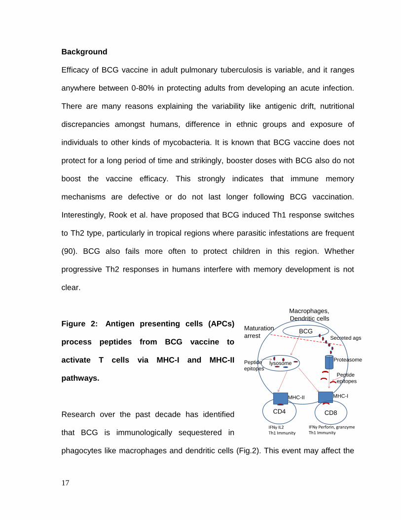
Background
Efficacy of BCG vaccine in adult pulmonary tuberculosis is variable, and it ranges
anywhere between 0-80% in protecting adults from developing an acute infection.
There are many reasons explaining the variability like antigenic drift, nutritional
discrepancies amongst humans, difference in ethnic groups and exposure of
individuals to other kinds of mycobacteria. It is known that BCG vaccine does not
protect for a long period of time and strikingly, booster doses with BCG also do not
boost the vaccine efficacy. This strongly indicates that immune memory
mechanisms are defective or do not last longer following BCG vaccination.
Interestingly, Rook et al. have proposed that BCG induced Th1 response switches
to Th2 type, particularly in tropical regions where parasitic infestations are frequent
(90). BCG also fails more often to protect children in this region. Whether
progressive Th2 responses in humans interfere with memory development is not
clear.
Figure 2: Antigen presenting cells (APCs)
process peptides from BCG vaccine to
activate T cells via MHC-I and MHC-II
pathways.
Research over the past decade has identified
that BCG is immunologically sequestered in
phagocytes like macrophages and dendritic cells (Fig.2). This event may affect the
CD4
lysosome
CD8
BCGMaturation
arrest
Proteasome
IFNγ IL2Th1 Immunity
IFNγ Perforin, granzymeTh1 Immunity
Secreted ags
Macrophages,
Dendritic cells
MHC-II MHC-I
Peptide
epitopes
Peptide
epitopes
18
ability of phagocytes to cross-talk with T cells, which mediate Th1 immunity to
tuberculosis. It is known that, antigen presenting cells like macrophages and
dendritic cells (APCs) present antigenic peptides of BCG to CD4 or CD8 T cells via
MHC-II and MHC-I, respectively. The bacterial peptides are usually generated by
the proteolytic breakdown of BCG derived antigens in the lysosomes, which needs
BCG phagosomes to fuse with lysosomes. However, there are two major flaws in
this antigen processing mechanism. First, BCG sequesters within immature
phagosomes of macrophages and does not fuse with lysosomes. This affects
peptide generation before loading into MHC-II (97). Secondly, the phagosome
membrane of BCG is not permeable to peptides as much, and this prevents leakage
of peptides into cytosol, where they are normally processed by proteasomes to
generate peptides for further loading into MHC-I (12, 64). This reduces efficacy of
MHC-I pathway in BCG infected APCs.
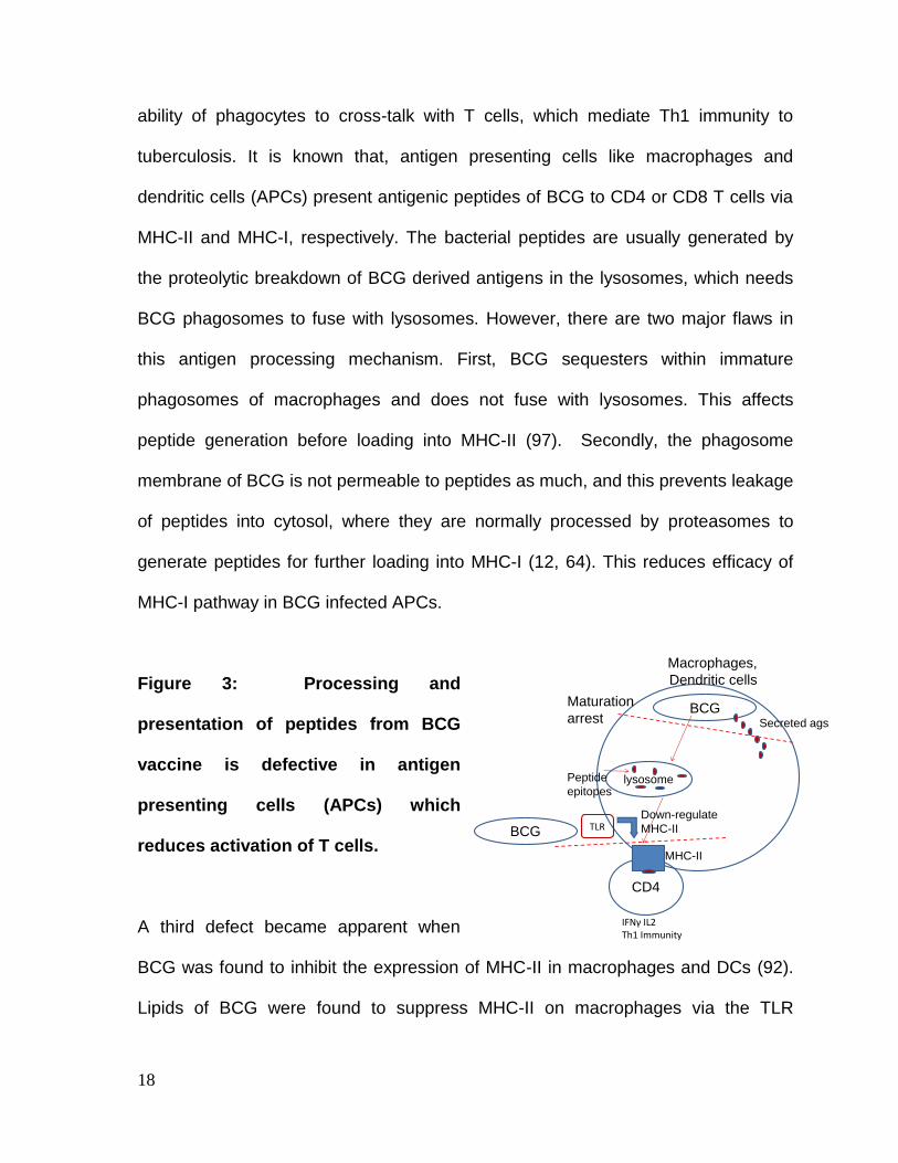
Figure 3: Processing and
presentation of peptides from BCG
vaccine is defective in antigen
presenting cells (APCs) which
reduces activation of T cells.
A third defect became apparent when
BCG was found to inhibit the expression of MHC-II in macrophages and DCs (92).
Lipids of BCG were found to suppress MHC-II on macrophages via the TLR
CD4
lysosome
BCGMaturation
arrest
IFNγ IL2Th1 Immunity
Secreted ags
Macrophages,
Dendritic cells
MHC-II
Peptide
epitopes
TLRBCG
Down-regulate
MHC-II
19
pathway (20) (Fig.3). Since MHC-II molecules load peptides and present to CD4 T
cells to trigger the major component of Th1 immunity, we hypothesized that lack of
lysosomal fusion of BCG in APCs coupled with reduced MHC-II expression may
seriously affect the ability of APC to activate T cells. In phagocytes, MHC-II is
constitutively synthesized but its expression can be up-regulated in response to
infection or treatment with IFN-γ. Unloaded MHC-II, misfolded MHC-II or membrane
internalized MHC-II can all be labeled with ubiquitin and targeted to proteasomes
and lysosomes for breakdown (32). This process is mediated by a group of ubiquitin
labeling enzymes. Membrane-associated RING-CH (MARCH) proteins like
MARCH1 represent a family of E3 ubiquitin ligases, which contain a variant catalytic
RING-finger domain (RING-CH domain, C4HC3) located at the N-terminus (30).
Earlier studies in mouse DCs proposed that ubiquitination of lysine-225 in the
cytoplasmic tail of MHC-II β-chain through MARCH1 is a determinant for the
targeting and accumulation of the mature form of MHC-II in the lysosomes of DCs
(111). Loss of MHC-II ubiquitination was accompanied by the down-regulation of
MARCH1 expression; therefore, MARCH1 is thought to be an important regulatory
molecule of MHC-II expression (30).
Specific Hypothesis: Mycobacteria like BCG contain lipoproteins (LP) like the 19
kDa LP and lipo-arabinomannan (LAM), which are ligands for Toll-like receptor,
TLR-1/2 (37). We hypothesized that these lipids suppress MHC-II expression in
macrophages, and thereby suppress antigen presentation to CD4 T cells. Since the
lipids are contained in BCG, we anticipate that BCG by itself will cause down-
20
regulation of MHC-II through up-regulation of MARCH1 enzyme in macrophages.
Finally, we propose that alternate activation of TLRs other than TLR-1/2 can bypass
BCG mediated suppression of MHC-II.
Methods: Even though live BCG does not undergo lysosomal fusion, limited
peptide antigen processing occurs in BCG infected macrophages. This has been
thought to be due to sorting of soluble Ag85B to lysosomes, sorting of dead BCG to
lysosomes or limited antigen presentation within phagosome itself (13). The BB7-
CD4 T cell hybridoma cell line is specific for p25 epitope of Ag85B, a major secreted
immune-dominant antigen of BCG. When these T cells are overlaid on
macrophages containing BCG, IL-2 is secreted, which is dependent upon their
specific recognition of p25 epitope from Ag85B bound to MHC-II on macrophages.
This process does not occur when Ag85B negative strain of BCG or M.tuberculosis
is used to infect macrophages (97). We therefore
used this well established, in vitro system to
investigate whether, BCG infected macrophages
present Ag85B epitope to CD4 T cells (BB7 T cells),
and study the effect of lipids and other activators on
macrophages (Fig.4)..
Figure 4: An in vitro antigen presentation to CD4
T cell assay using macrophages and DCs
infected with BCG vaccine.
BB-7
CD4
lysosome
BCGMaturation
arrest
Secretes IL2
Secreted
Ag85B
Macrophages,
Dendritic cells
MHC-II
Ag85B
epitope
Sandwich ELISA
21
Results
Lipids of BCG decrease in vitro antigen presentation. Macrophages were
infected with BCG alone or were treated with increasing doses of lipids (19 kDa LP
and LAM) as indicated. (Fig.5). Macrophages were overlaid with BB7 T cells and
supernatants analyzed for IL-2 using ELISA. Fig.5 shows that both 19 kDa
lipoprotein and LAM suppressed antigen presentation in a dose-dependent manner.
Since LP and LAM are integral components of cell wall in BCG and are shed from
growing bacteria, it follows that BCG by itself suppresses antigen presentation.
Since BCG can decrease MHC-II levels, but may also exert additional non-specific
effects, we sought to determine whether lipids could interfere with the processing of
soluble Ag85B. Macrophages were therefore treated with soluble Ag85B, followed
two hour later by T cell overlay. Macrophages processed Ag85B and activated T
cells to secrete IL-2 (Fig.6). Prior treatment of macrophages with lipids however,
suppressed antigen presentation.
Macrophage surface MHC-II levels determine peptide presentation while,
intracellular MARCH1 enzyme determines degradation of MHC-II. Thus, BCG
infected macrophages were analyzed using flow-cytometry. BCG induced a down-
regulation of surface MHC-II while, concurrently, intracellular MARCH1 was up-
regulated (Fig.7).
22
Figure-5: Macrophages (MΦs) infected with Mycobacterium bovis BCG
vaccine present Ag85B epitope to CD4 T cells (BB7) and induce IL-2
secretion. Lipids of BCG suppress IL-2 responses. C57Bl/6 derived primary
macrophages (MΦs) were infected with BCG, and tested alone or were added with
lipids as indicated for 4 hrs. Washed macrophages were overlaid with BB7 T cells
and supernatants analyzed for IL-2 using ELISA. Lipids (19 kDa lipoprotein and lipo-
arabinomannan, LAM) suppress IL-2 elicitation. Data from one of three separate
similar experiments are shown. Triplicate wells of macrophages per dose per
reagent in each experiment (mean values of IL-2 expressed ± SD; p values by t
test).
a)
b)
0.0090.009
0.010.01
0.008
a)
b)
0 5
00
100
0
250
0 0
500
100
0
250
0
0
200
400
600
800
1000
0.008
0.006
19 kDa (ng/mL) LAM (ng/mL)
IL-2
pg
/ml (S
D)
23
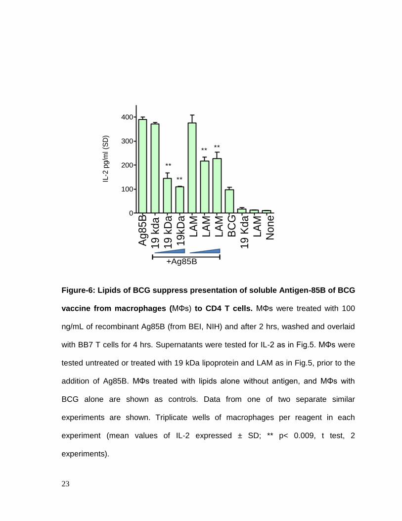
Figure-6: Lipids of BCG suppress presentation of soluble Antigen-85B of BCG
vaccine from macrophages (MΦs) to CD4 T cells. MΦs were treated with 100
ng/mL of recombinant Ag85B (from BEI, NIH) and after 2 hrs, washed and overlaid
with BB7 T cells for 4 hrs. Supernatants were tested for IL-2 as in Fig.5. MΦs were
tested untreated or treated with 19 kDa lipoprotein and LAM as in Fig.5, prior to the
addition of Ag85B. MΦs treated with lipids alone without antigen, and MΦs with
BCG alone are shown as controls. Data from one of two separate similar
experiments are shown. Triplicate wells of macrophages per reagent in each
experiment (mean values of IL-2 expressed ± SD; ** p< 0.009, t test, 2
experiments).
0
100
200
300
400
**
Ag
85
B
1
9 k
da
19
kD
a
1
9kD
a
L
AM
LA
M
L
AM
BC
G
1
9 K
da
LA
M
No
ne
**
**
+Ag85B
**
IL-2
pg/m
l (S
D)
24
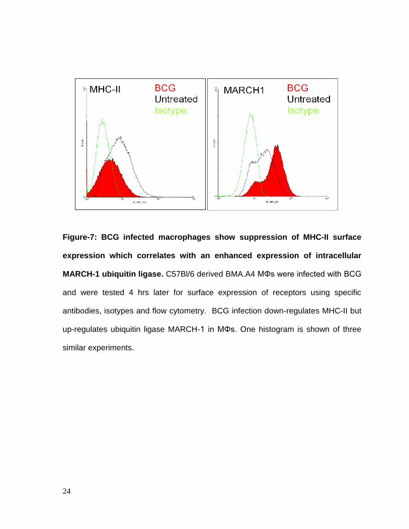
Figure-7: BCG infected macrophages show suppression of MHC-II surface
expression which correlates with an enhanced expression of intracellular
MARCH-1 ubiquitin ligase. C57Bl/6 derived BMA.A4 MΦs were infected with BCG
and were tested 4 hrs later for surface expression of receptors using specific
antibodies, isotypes and flow cytometry. BCG infection down-regulates MHC-II but
up-regulates ubiquitin ligase MARCH-1 in MΦs. One histogram is shown of three
similar experiments.
a)
c)
b)
25
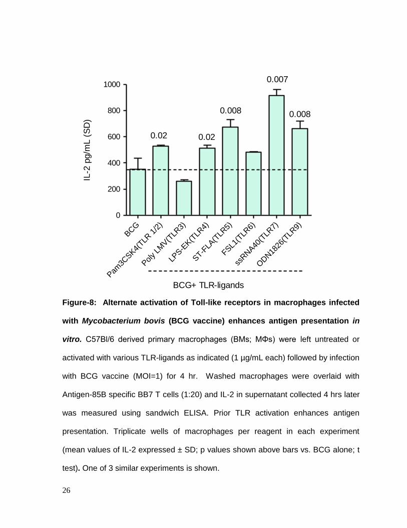
TLR-ligands increase in vitro antigen presentation. Macrophages were infected
with BCG with or without prior activation using various TLR ligands. Fig.8 shows
that prior activation with multiple TLR-ligands increased antigen presentation by
macrophages. Ligands for TLR-9, TLR-7 and TLR-5 were better activators than
ligands for TLR-4 or TLR-1/2. Ligands for TLR-3 and TLR-6 had no beneficial effect.
TLR-ligands down-regulate MARCH-1 in BCG infected macrophages and
increase MHC-II expression. Since the levels of MHC-II and MARCH1 in
macrophages were inversely correlated during antigen presentation following BCG
infection, the effect of various TLR-ligands on MHC-II and MARCH1 in
macrophages was evaluated using flow-cytometry. Fig.9 illustrates that
macrophages activated with various TLR-ligands show two profiles. Some like TLR-
9/7 ligands induced a strong up-regulation of MHC-II while decreasing MARCH1.
Others like TLR-1/2 ligand induced a down-regulation of MARCH1 but had no affect
the overall levels of MHC-II. Yet others had no effect on either MHC-II or MARCH1.
None of the ligands affected the levels of MHC-I, indicating that TLR-activation
affected only MHC-II expression.
TLR-ligands affect CD86 and intracellular IL-10 in BCG infected macrophages.
The co-stimulatory molecules CD80 and CD86 participate during MHC-II mediated
peptide presentation to CD4 T cells. Fig.10 demonstrates that TLR-9/7 ligands up-
regulate the expression of CD86 but not CD80 in macrophages. Furthermore, IL-10,
a suppressive cytokine has been found to affect the levels of intracellular MARCH1
26
Figure-8: Alternate activation of Toll-like receptors in macrophages infected
with Mycobacterium bovis (BCG vaccine) enhances antigen presentation in
vitro. C57Bl/6 derived primary macrophages (BMs; MΦs) were left untreated or
activated with various TLR-ligands as indicated (1 µg/mL each) followed by infection
with BCG vaccine (MOI=1) for 4 hr. Washed macrophages were overlaid with
Antigen-85B specific BB7 T cells (1:20) and IL-2 in supernatant collected 4 hrs later
was measured using sandwich ELISA. Prior TLR activation enhances antigen
presentation. Triplicate wells of macrophages per reagent in each experiment
(mean values of IL-2 expressed ± SD; p values shown above bars vs. BCG alone; t
test). One of 3 similar experiments is shown.
BCG
Pam
3CSK4(
TLR 1
/2)
Poly LM
V(T
LR3)
LPS-E
K(T
LR4)
ST-F
LA(T
LR5)
FSL1
(TLR
6)
ssRNA40
(TLR
7)
ODN18
26(T
LR9)
0
200
400
600
800
10000.007
0.02
0.008 0.008
0.02
BCG+ TLR-ligands
IL-2
pg/m
L (
SD
)
27
Figure-9: TLR-activation of macrophages infected with BCG vaccine
enhances the expression of surface MHC-II and induces down-regulation of
MARCH1. C57Bl/6 macrophage derived BMA.A4 macrophage cell line (MΦs) were
activated with TLR-ligands as in Fig.8, followed by infection with BCG for 4 hrs, and
stained for surface receptors or intracellular MARCH1. Cells were analyzed by flow
cytometry and representative histograms are shown. Data in Table shows up- or
down-regulation of MHC-II and MARCH1 scored as log increase or log decrease
averaged from three separate experiments. TLR-agonists enhance MHC-II
expression but down-regulate ubiquitin ligase MARCH1, but have no effect on
MHC-I expression by MΦs.
MARCH1BCG
TLR1/2-PAM3CSK
TLR3-Poly IC
TLR7ssRNA
TLR9 ODN
MHC-II
MHC-IMHC-II MARCH I
TLR1/2 + -/-
TLR3 +
TLR4 +
TLR5 ++
TLR6 +
TLR7 +++ -/-
TLR9 +++ -/-
+++ Up regulation >0.5 log shift
-/- Down-regulation < 0.5 log shift
+ Up regulation >0.2-0.5 log shift
No significant effect
a)
28
Figure-10: TLR-activation of macrophages infected with BCG vaccine
enhances CD86 and down-regulates intracellular IL-10. Macrophages were
activated and infected as in Fig.8, and stained for surface receptors or intracellular
IL-10. Cells were analyzed by flow cytometry and representative histograms are
shown. Data in Table shows up-or down regulation of CD80, CD86 and IL-10
scored as log increase or log decrease averaged from three separate experiments.
BCG
TLR4+BCG
TLR9+BCG
TLR7 +BCG
Isotype
IL-10CD86 CD80 IL-10
TLR1/2 - - -/-
TLR3 - - -
TLR4 - - -/-
TLR5 - - -
TLR6 - - -
TLR7 ++ - -/-
TLR9 ++ - -/-
CD80 CD86 BCG
TLR-4+BCG
TLR-9+BCG
TLR7 +BCG
Isotype
BCG
TLR4+BCG
TLR9+BCG
TLR7+BCG
Isotype
b)
++ Up regulation >0.5 log shift
-/- Down-regulation < 0.5 log shift
- No significant effect
29
Ligands for TLR-1/2, TLR-4, TLR-7 and TLR-9 induced a down-regulation of IL-10.
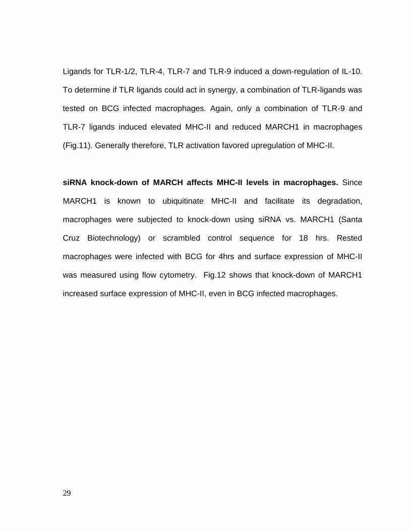
To determine if TLR ligands could act in synergy, a combination of TLR-ligands was
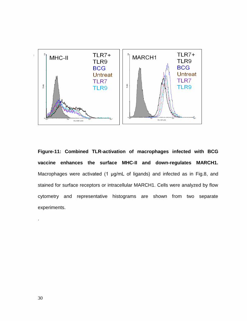
tested on BCG infected macrophages. Again, only a combination of TLR-9 and
TLR-7 ligands induced elevated MHC-II and reduced MARCH1 in macrophages
(Fig.11). Generally therefore, TLR activation favored upregulation of MHC-II.
siRNA knock-down of MARCH affects MHC-II levels in macrophages. Since
MARCH1 is known to ubiquitinate MHC-II and facilitate its degradation,
macrophages were subjected to knock-down using siRNA vs. MARCH1 (Santa
Cruz Biotechnology) or scrambled control sequence for 18 hrs. Rested
macrophages were infected with BCG for 4hrs and surface expression of MHC-II
was measured using flow cytometry. Fig.12 shows that knock-down of MARCH1
increased surface expression of MHC-II, even in BCG infected macrophages.
30
Figure-11: Combined TLR-activation of macrophages infected with BCG
vaccine enhances the surface MHC-II and down-regulates MARCH1.
Macrophages were activated (1 μg/mL of ligands) and infected as in Fig.8, and
stained for surface receptors or intracellular MARCH1. Cells were analyzed by flow
cytometry and representative histograms are shown from two separate
experiments.
.
a)
c)
b)
31
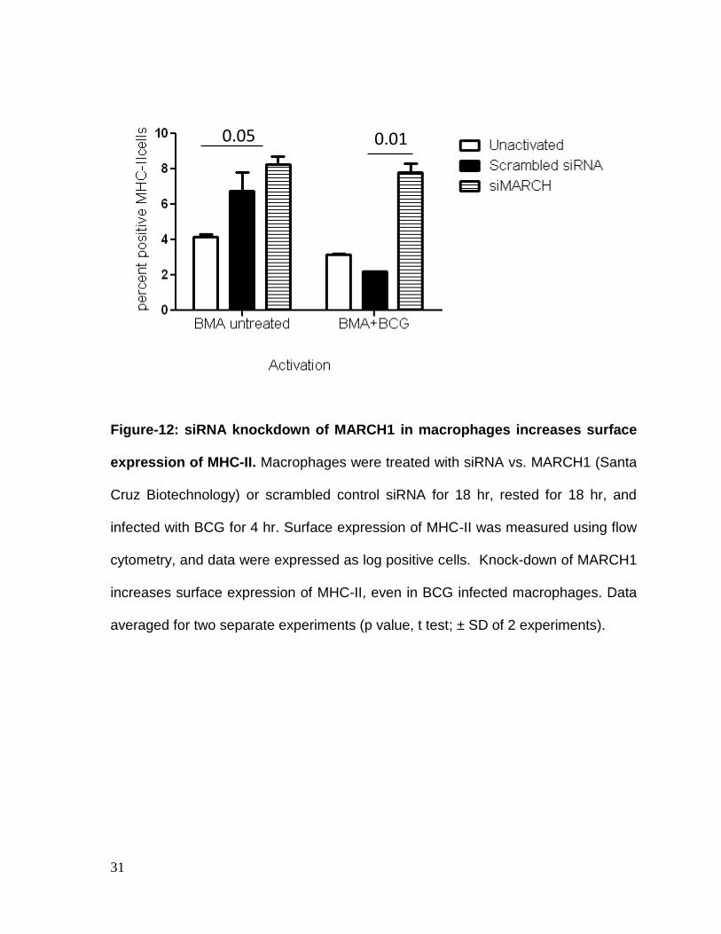
Figure-12: siRNA knockdown of MARCH1 in macrophages increases surface
expression of MHC-II. Macrophages were treated with siRNA vs. MARCH1 (Santa
Cruz Biotechnology) or scrambled control siRNA for 18 hr, rested for 18 hr, and
infected with BCG for 4 hr. Surface expression of MHC-II was measured using flow
cytometry, and data were expressed as log positive cells. Knock-down of MARCH1
increases surface expression of MHC-II, even in BCG infected macrophages. Data
averaged for two separate experiments (p value, t test; ± SD of 2 experiments).
0.05 0.01
32
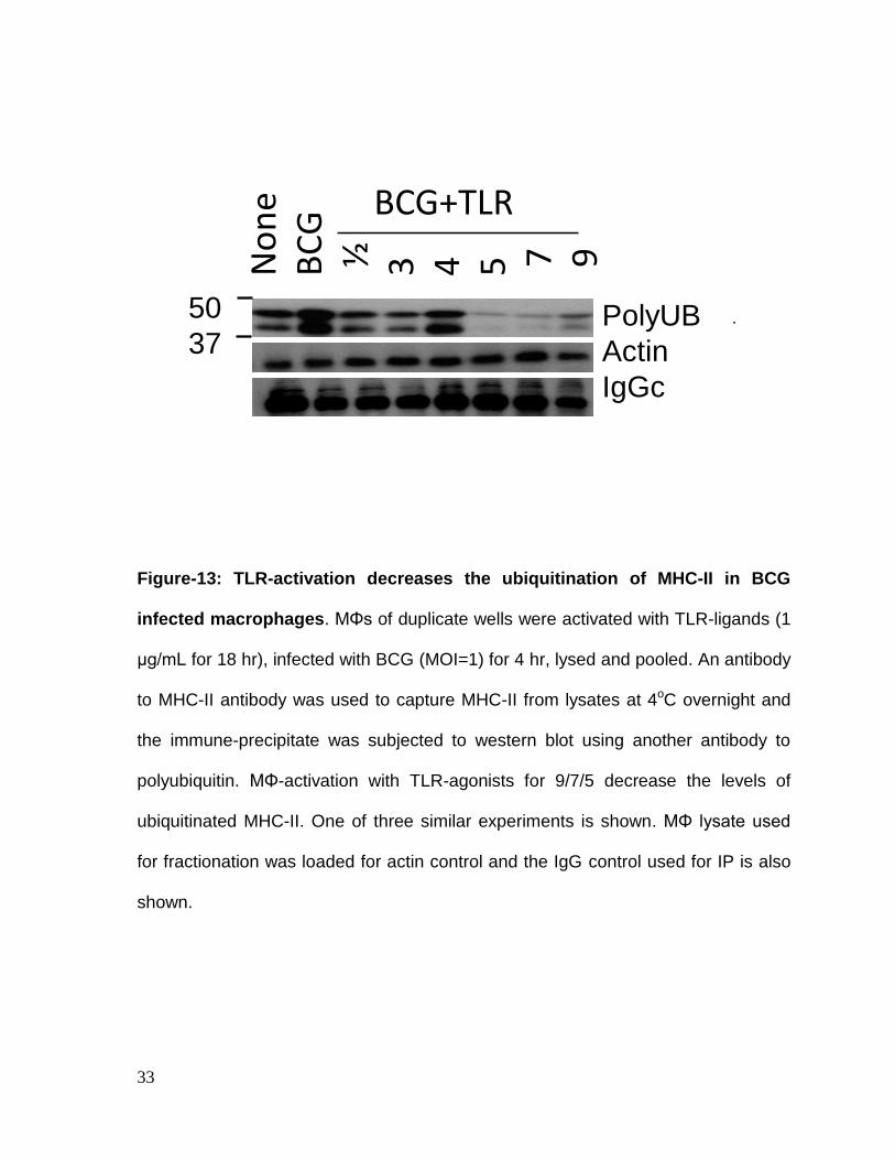
TLR-ligands decrease ubiquitination of MHC-II in BCG infected macrophages:
MARCH1 is known to ubiquitinate MHC-II and ubiquitinated MHC-II is routed
through proteasome for degradation. To validate that TLR-activation led to a
decrease in ubiquitinated MHC-II, macrophages were activated with TLR-ligands,
infected with BCG and their lysates were immune-precipitated (IP) with an antibody
to MHC-II. The IP was then labeled with an antibody to polyubiquitin. Fig.13
indicates that TLR-ligands (7>5>9>3>1/2) reduced the levels of ubiquitinated MHC-
II relative to MHC-II from BCG infected macrophages.
33
.
Figure-13: TLR-activation decreases the ubiquitination of MHC-II in BCG
infected macrophages. MΦs of duplicate wells were activated with TLR-ligands (1
μg/mL for 18 hr), infected with BCG (MOI=1) for 4 hr, lysed and pooled. An antibody
to MHC-II antibody was used to capture MHC-II from lysates at 4oC overnight and
the immune-precipitate was subjected to western blot using another antibody to
polyubiquitin. MΦ-activation with TLR-agonists for 9/7/5 decrease the levels of
ubiquitinated MHC-II. One of three similar experiments is shown. MΦ lysate used
for fractionation was loaded for actin control and the IgG control used for IP is also
shown.
PolyUB
Actin
IgGc
No
ne
BC
G½
3
4
5 7 9
BCG+TLR
50
37
34
Discussion
Mouse infection and vaccination experiments have shown that BCG induces largely
a CD4 T cell type of response with less strong CD8 T cell response [90]. Yet in the
mice, BCG is barely able to control infection with Mtb. Several studies propose that
the sequestration of BCG in immature phagosomes of APCs and the ability of live
BCG to inhibit MHC-II is one main reason why APCs do not efficiently cross-talk to
T cells. Such T cells are therefore poorly effective against Mtb in mice. We therefore
investigated the hypothesis that moieties contained in BCG inhibit MHC-II. We used
an in vitro antigen presentation that confirmed that lipids of BCG inhibited antigen
presentation and since these lipids are integral in the cell wall of BCG, it seemed to
have an inherent ability to suppress antigen presentation.
Macrophages sense pathogens and bacteria through specific Toll like receptors
(TLRs) and LP and LAM lipids of BCG are known to mediate their suppressive
effects through TLR-1/2. However, we hypothesized that alternate activation of
TLRs may help bypass the suppressive effect of BCG on APCs. Toll like receptors
(TLRs) are single membrane spanning non-catalytic receptor proteins and are
similar to the toll gene products of Drosophila. They are present in the membranes
of both vertebrates and non-vertebrates. They recognize and distinguish between
special microbial patterns and hence allow the cell to distinguish between self and
non-self (16). TLRs link innate immunity to adaptive response and most human
tissues contain at least one type of TLR while, APCs express multiple TLRs (75).
While TLR-1/2 induced a negative effect through BCG, we sought to activate other
35
TLRs. In this experiment, interestingly, TLR-9/7/5/4 &1/2 ligands enhanced antigen
presentation indicating that, BCG induced suppressive effect on antigen
presentation can be bypassed. This was not surprising since TLRs mediate their
effects on signaling and cytokine secretion in APCs using multiple pathways, and it
was anticipated that TLR-1/2 induced suppressive effects on antigen presentation
could be neutralized. During MHC-II mediated priming of CD4 T cells, additional
stimulation is required by co-stimulatory molecules like CD80 and CD86 for
optimum activation (107) (27). For example, peptide presentation without co-
stimulation can induce tolerance. It was therefore interesting to note that TLR-9
ligand boosted the CD86 response in addition to MHC-II in macrophages.
We also found that, intracellular MARCH1 levels were inversely correlated with
surface expression of MHC-II confirming the regulatory role of MARCH1 on
recycling of MHC-II. Many TLR ligands enhanced MHC-II through a down-regulation
of MARCH1. They also induced a down-regulation of IL-10, a cytokine known to
induce MARCH1 and thus negatively affect MHC-II levels (36). We noted however
that there was some variability in the ability of TLR ligands to down-regulate
MARCH1, enhance MHC-II, and increase antigen presentation. Only TLR-9/7
ligands performed all three functions. TLR-1/2 ligand down-regulated MARCH1 but
did not significantly enhance MHC-II. Since intracellular loading of peptides into
MHC-II, MHC-II recycling, and sorting of peptide loaded MHC-II to surface
membrane together determine the eventual expression of MHC-II on plasma
membrane, we propose that TLR-ligands perhaps influence different stages of the
36
MHC-II mediated peptide presentation. It is however clear that, at least TLR-9/7
ligands can be used to enhance the immunogenicity of BCG vaccine in vitro and in
animal models. Our data are novel in that; previous studies have only used TLR-4
or TLR-1/2 ligands to enhance the efficacy of subunit vaccines to tuberculosis. This
study has identified more potent TLR-7/9 ligands as adjuvants to improve vaccines
against tuberculosis.
37
CHAPTER-2.2
TOLL-LIKE RECEPTOR (TLR) LIGANDS ENHANCE MHC-II
IN MACROPHAGES THROUGH THE ACTIVATION OF MAP
-KINASES AND AP-1 OR CREB TRANSCRITION FACTORS
38
Background MHC-II is required for activation of the CD4 T cells, which are obviously important to
control multiple infections. In humans, depletion of CD4 T cells through AIDS results
in extensive cross infection with tuberculosis and accelerated death. MHC-II is a
crucial immune determinant and its abnormal expression or decreased expression
can have strong pathological effects (47). Mitogen activated protein kinases
(MAPKs) are highly conserved in eukaryotes and are important signaling
components that regulate cytokine responses and transcription of cellular factors in
multiple cell types. There are three major groups of MAPK in mammalian cells: the
extracellular signal-regulated kinase (ERK), the p38 MAPK and the c-jun NH2-
terminal kinase (JNK) (25, 113). When the cell receives an extracellular stimulus,
MAPKs are phosphorylated and they mediate signaling for cellular transcription and
induce cytokine secretion. MAPKs also activate transcription factors (TFs) like the
cyclic AMP response element binding protein (CREB) and Activator protein (AP-1),
which bind the Class II transactivator (CIITA), a master regulator of MHC-II
synthesis (5) (66). The signaling pathways triggered by TLR ligands are illustrated
in Fig. 14 and Fig.15.
Specific Hypothesis
Our recent studies (Chapter 2.1) showed that multiple TLR ligands induced an up-
regulation of MHC-II in macrophages. We hypothesized that TLR-ligands regulated
MHC-II through MAPK cascade and AP-1 or CREB transcription factors. We
proposed that differences in the levels of MHC-II induced by TLR ligands could be
attributed to specific signaling pathways.
39
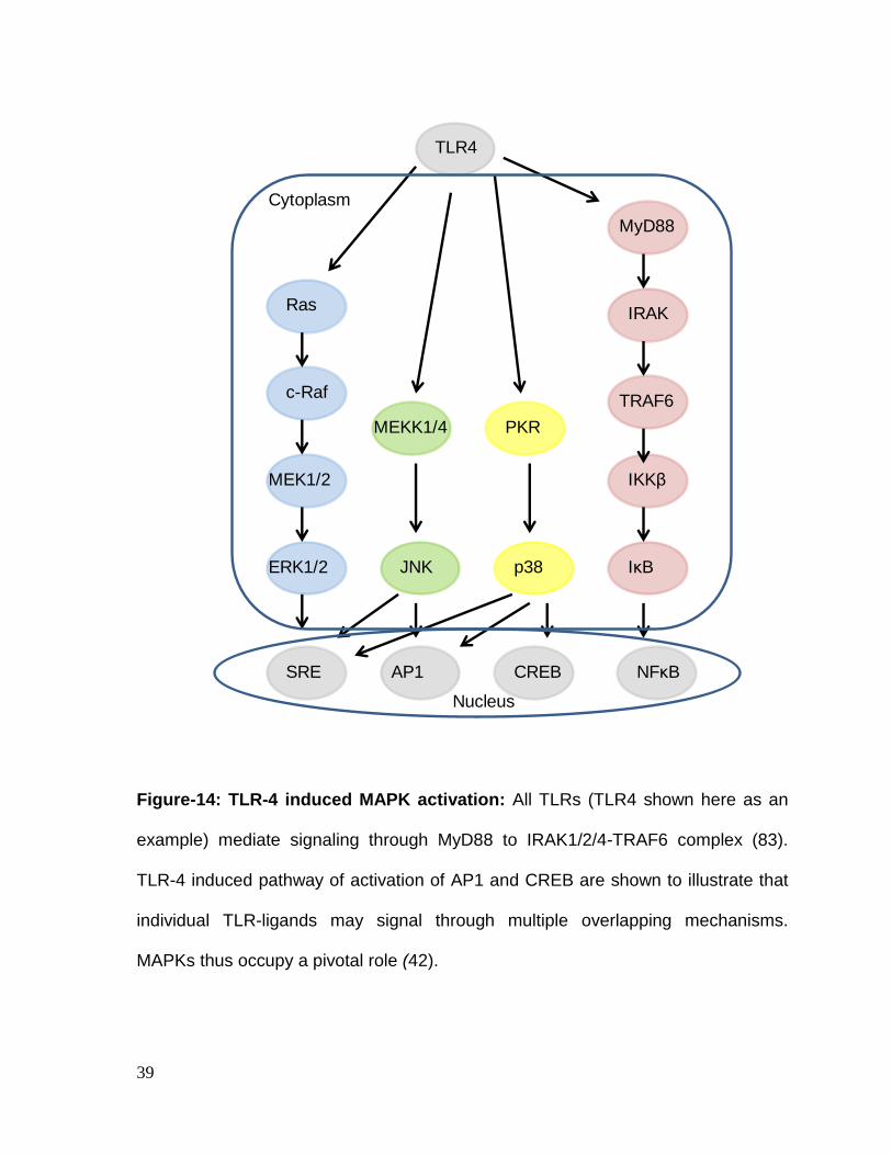
Figure-14: TLR-4 induced MAPK activation: All TLRs (TLR4 shown here as an
example) mediate signaling through MyD88 to IRAK1/2/4-TRAF6 complex (83).
TLR-4 induced pathway of activation of AP1 and CREB are shown to illustrate that
individual TLR-ligands may signal through multiple overlapping mechanisms.
MAPKs thus occupy a pivotal role (42).
TLR4
MEKK1/4
p38
PKR
JNK
Ras
c-Raf
MEK1/2
ERK1/2
SRE AP1 CREB NFκB
IKKβ
TRAF6
IRAK
IκB
MyD88
Cytoplasm
Nucleus
40
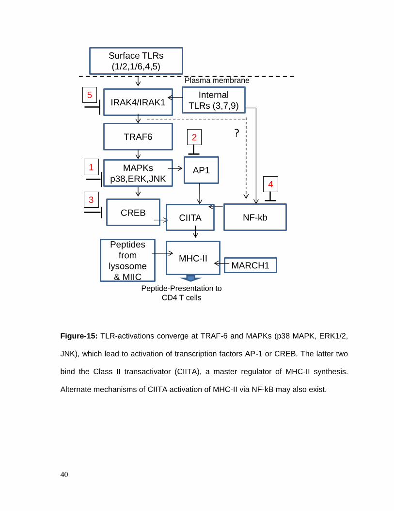
Figure-15: TLR-activations converge at TRAF-6 and MAPKs (p38 MAPK, ERK1/2,
JNK), which lead to activation of transcription factors AP-1 or CREB. The latter two
bind the Class II transactivator (CIITA), a master regulator of MHC-II synthesis.
Alternate mechanisms of CIITA activation of MHC-II via NF-kB may also exist.
Surface TLRs
(1/2,1/6,4,5)
MAPKs
p38,ERK,JNK
TRAF6
IRAK4/IRAK1
CREB
AP1
3
1
2
Peptide-Presentation to
CD4 T cells
MHC-II
Plasma membrane
Internal
TLRs (3,7,9)
Peptides
from
lysosome
& MIIC
MARCH1
?
NF-kbCIITA
4
5
41
Results:
TLR-activation induced MHC-II expression in macrophages is dependent upon
activation of p38 MAPK, ERK1/2 and AP-1 or CREB mediated activation of
MHC-II. MΦs were incubated with inhibitors of MAPKs (p38, ERK1/2 and JNK), AP-
1 or CREB (all tested at 1 μM) for 2 hrs and were tested for surface MHC-II
expression or were further activated with TLR-ligands for 2 hrs and BCG infection
for 2 hrs before MHC-II analysis. Trypan blue vital dye was used to ensure that
macrophages were > 95% viable. Fig.16 illustrates that MAPKs inhibitors alone did
not affect MHC-II expression in macrophages. TLR-7 and TLR-9 ligands both
induced an up-regulation of MHC-II in macrophages (Fig.17). Interestingly, MHC-II
induced by TLR-7 activation was inhibited by blockade of p38-ERK1/2 and AP-1
while, TLR-9 induced MHC-II was affected by blockade of p38-ERK1/2 and CREB.
Signaling induced by TLR-9/7 ligands was therefore subtly different. In striking
contrast, TLR-2 and TLR-4 ligands induced only a modest up-regulation of MHC-II
which was not affected significantly by inhibitors of MAPKs or AP-1/CREB (Fig.18).
TLR-activation increases MHC-II expression in dendritic cells dependent upon
MAPKs and AP-1/CREB. DCs are another type of APCs and their role of MHC-II in
priming CD4 T cells is of prime importance. When vaccines are initially given, DCs
are the cells that process vaccine to prime ‘naïve’ T cells. In BCG infected DCs as
well, TLR-9/7 ligands significantly shifted the MHC-II expression and this process
was dependent upon MAPKs and both AP-1 and CREB (Fig.19). In addition,
multiple TLR-ligands enhanced antigen presentation by DCs to CD4 T cells
(Fig.19).
42
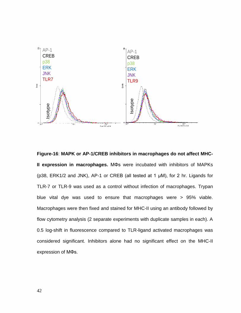
Figure-16: MAPK or AP-1/CREB inhibitors in macrophages do not affect MHC-
II expression in macrophages. MΦs were incubated with inhibitors of MAPKs
(p38, ERK1/2 and JNK), AP-1 or CREB (all tested at 1 μM), for 2 hr. Ligands for
TLR-7 or TLR-9 was used as a control without infection of macrophages. Trypan
blue vital dye was used to ensure that macrophages were > 95% viable.
Macrophages were then fixed and stained for MHC-II using an antibody followed by
flow cytometry analysis (2 separate experiments with duplicate samples in each). A
0.5 log-shift in fluorescence compared to TLR-ligand activated macrophages was
considered significant. Inhibitors alone had no significant effect on the MHC-II
expression of MΦs.
AP-1+BCG <0.2log
CREB+BCG <0.5log *
p38+BCG <0.5log *
ERK+BCG <0.6log *
JNK+BCG <0.3log
BCG>0.6log vs. isotype
TLR7+BCG >1.0log vs. isotype
AP-1+BCG <0.6log *
CREB+BCG <0.4log
p38+BCG <0.4log
ERK+BCG <0.4log
JNK+BCG <0.4log
BCG>0.6log vs. isotype
TLR9+BCG >1.0log vs. isotype
Iso
typ
e
Iso
typ
eAP-1
CREB
p38
ERK
JNK
TLR7
AP-1
CREB
p38
ERK
JNK
TLR9
Iso
typ
e
Iso
typ
e
b)
c)
AP-1+BCG
CREB+BCG
p38+BCG
ERK+BCG
JNK+BCG
BCG
TLR1/2+BCG
AP-1+BCG
CREB+BCG
P38+BCG
ERK+BCG
JNK+BCG
BCG
TLR4+BCG
d)
Iso
typ
e
Iso
typ
e
43
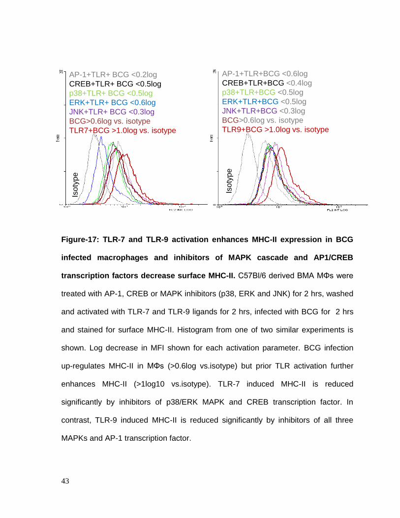
Figure-17: TLR-7 and TLR-9 activation enhances MHC-II expression in BCG
infected macrophages and inhibitors of MAPK cascade and AP1/CREB
transcription factors decrease surface MHC-II. C57Bl/6 derived BMA MΦs were
treated with AP-1, CREB or MAPK inhibitors (p38, ERK and JNK) for 2 hrs, washed
and activated with TLR-7 and TLR-9 ligands for 2 hrs, infected with BCG for 2 hrs
and stained for surface MHC-II. Histogram from one of two similar experiments is
shown. Log decrease in MFI shown for each activation parameter. BCG infection
up-regulates MHC-II in MΦs (>0.6log vs.isotype) but prior TLR activation further
enhances MHC-II (>1log10 vs.isotype). TLR-7 induced MHC-II is reduced
significantly by inhibitors of p38/ERK MAPK and CREB transcription factor. In
contrast, TLR-9 induced MHC-II is reduced significantly by inhibitors of all three
MAPKs and AP-1 transcription factor.
AP-1+TLR+ BCG <0.2log
CREB+TLR+ BCG <0.5log
p38+TLR+ BCG <0.5log
ERK+TLR+ BCG <0.6log
JNK+TLR+ BCG <0.3log
BCG>0.6log vs. isotype
TLR7+BCG >1.0log vs. isotype
AP-1+TLR+BCG <0.6log
CREB+TLR+BCG <0.4log
p38+TLR+BCG <0.5log
ERK+TLR+BCG <0.5log
JNK+TLR+BCG <0.3log
BCG>0.6log vs. isotype
TLR9+BCG >1.0log vs. isotype
Iso
typ
e
Iso
typ
e
44
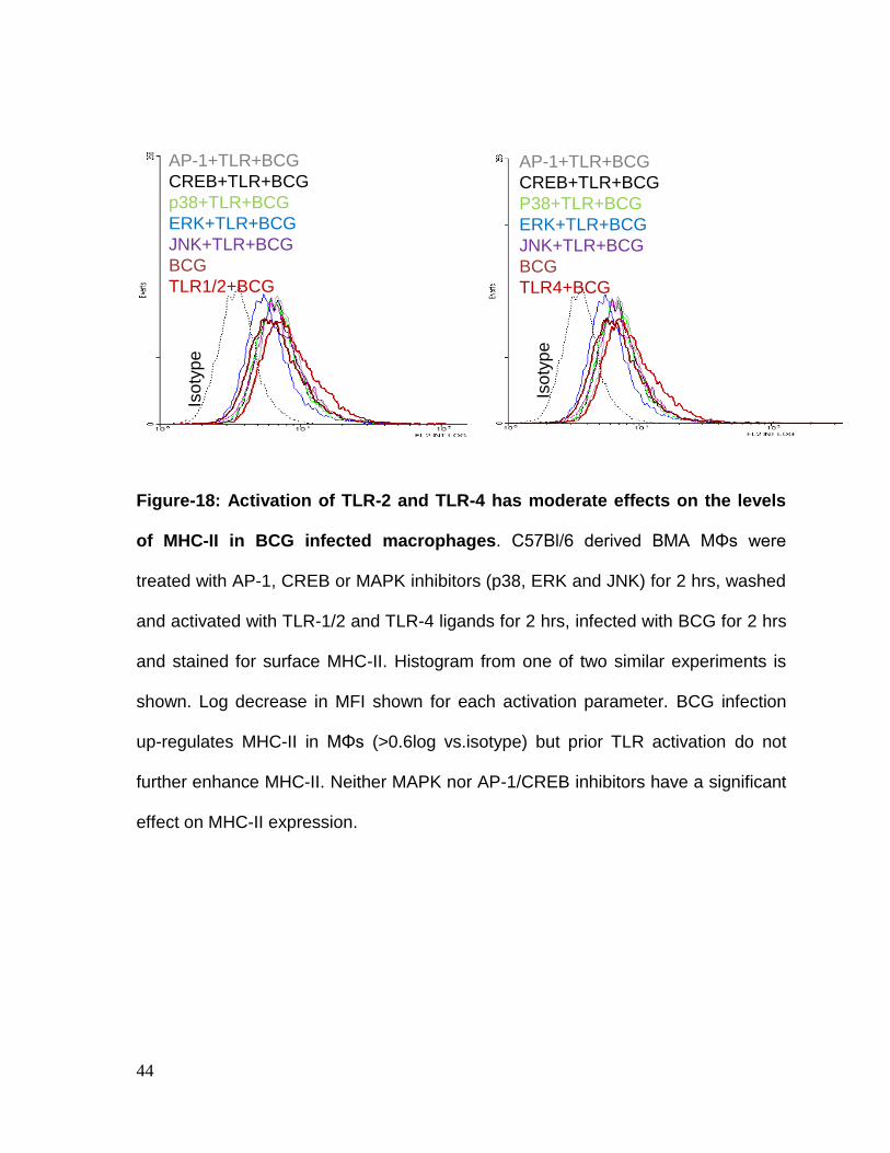
Figure-18: Activation of TLR-2 and TLR-4 has moderate effects on the levels
of MHC-II in BCG infected macrophages. C57Bl/6 derived BMA MΦs were
treated with AP-1, CREB or MAPK inhibitors (p38, ERK and JNK) for 2 hrs, washed
and activated with TLR-1/2 and TLR-4 ligands for 2 hrs, infected with BCG for 2 hrs
and stained for surface MHC-II. Histogram from one of two similar experiments is
shown. Log decrease in MFI shown for each activation parameter. BCG infection
up-regulates MHC-II in MΦs (>0.6log vs.isotype) but prior TLR activation do not
further enhance MHC-II. Neither MAPK nor AP-1/CREB inhibitors have a significant
effect on MHC-II expression.
AP-1+TLR+BCG
CREB+TLR+BCG
p38+TLR+BCG
ERK+TLR+BCG
JNK+TLR+BCG
BCG
TLR1/2+BCG
AP-1+TLR+BCG
CREB+TLR+BCG
P38+TLR+BCG
ERK+TLR+BCG
JNK+TLR+BCG
BCG
TLR4+BCG
Iso
typ
e
Iso
typ
e
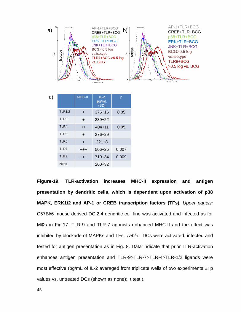
45
Figure-19: TLR-activation increases MHC-II expression and antigen
presentation by dendritic cells, which is dependent upon activation of p38
MAPK, ERK1/2 and AP-1 or CREB transcription factors (TFs). Upper panels:
C57Bl/6 mouse derived DC.2.4 dendritic cell line was activated and infected as for
MΦs in Fig.17. TLR-9 and TLR-7 agonists enhanced MHC-II and the effect was
inhibited by blockade of MAPKs and TFs. Table: DCs were activated, infected and
tested for antigen presentation as in Fig. 8. Data indicate that prior TLR-activation
enhances antigen presentation and TLR-9>TLR-7>TLR-4>TLR-1/2 ligands were
most effective (pg/mL of IL-2 averaged from triplicate wells of two experiments ±; p
values vs. untreated DCs (shown as none); t test ).
AP-1+TLR+BCG
CREB+TLR+BCG
p38+TLR+BCG
ERK+TLR+BCG
JNK+TLR+BCG
BCG> 0.5 log
vs.isotype
TLR7+BCG >0.5 log
vs. BCG
AP-1+TLR+BCG
CREB+TLR+BCG
p38+TLR+BCG
ERK+TLR+BCG
JNK+TLR+BCG
BCG>0.5 log
vs.isotype
TLR9+BCG
>0.5 log vs. BCG
Iso
typ
e
Iso
typ
e
MHC-II IL-2
pg/mL
(SD)
p
TLR1/2 + 376+16 0.05
TLR3 + 239+22
TLR4 ++ 404+11 0.05
TLR5 + 276+29
TLR6 + 221+8
TLR7 +++ 506+25 0.007
TLR9 +++ 710+34 0.009
None 200+32
+++ Up regulation >0.5 log shift
++ Up regulation >0.2-0.5 log shift
a) b)
c)
46
Overall Discussion- Up-regulation of MHC-II using TLR activation
BCG is the most frequently used vaccine in the world today with over a billion doses
administered to date. Although, BCG is the safest vaccine used, it is less effective in
generating stronger and longer lasting T cell responses. When vaccines are
internalized through receptor mediated phagocytosis by APCs, they are enclosed
within a plasma membrane bound phagosome, which is subsequently sorted to
lysosomes through a series of fusion events. Lysosome breakdown of BCG vaccine
generates peptides which are then routed into MHC-II loading compartments. MHC-
II then primes CD4 T cells to generate a specific Th1 immunity. It has become clear
now that BCG activates TLR-1/2 and TLR-4 when it is phagocytosed into APCs,
leading to immunosuppression. Specifically, BCG down-regulates MHC-II in APCs
when given to mice and has the same effect on APCs in vitro. Secondly, BCG stays
put in immature phagosomes preventing lysosome fusion. We made some novel
observations in this study that help to bypass these immune-suppressive events.
First, we confirmed that lipids of BCG suppressed in vitro antigen presentation,
presumably through TLR-1/2 and TLR-4 (Fig.5-8). Alternate TLR-activation however
boosted the ability of APCs to present antigen in vitro (Fig.8). TLR-ligands seemed
to activate MHC-II through down-regulation of MARCH1 (Fig.5-9). In a previous
study, TLR-1/2, 3 and 4 ligands were found to similarly enhance MHC-II levels
through down-regulation of MARCH1 in naïve DCs. This report was different from
our study in using ‘naïve’ DCs (108). Furthermore, our model was more relevant to
vaccine development since, only live attenuated BCG vaccine suppressed MHC-II

47
in APCs. Interestingly, in our model, we showed that only TLR-9/7 ligands up-
regulated MHC-II and down-regulated expression of MARCH1. In contrast, TLR-1/2,
TLR-3 and TLR-4 ligands had a moderate effect in enhancing MHC-II mediated
responses from BCG infected macrophages. It should be noted that BCG vaccine
already has TLR-1/2 and TLR-4 binding lipids like LP and LAM. We therefore
propose that adding similar ligands may not significantly help in boosting the
vaccine. We further noted that the ligands for TLR-7 and TLR-9, single strand RNA
and DNA are present in BCG vaccine, but due to its sequestration within
phagosomes, these ligands may not be able to activate the TLRs inside
macrophages. In addition to macrophages, we also found that TLR-ligands boosted
the antigen presentation by DCs. Again, only TLR-9/7 ligands seemed to boost
surface MHC-II levels as evaluated using flow-cytometry.
A review of the signaling mechanisms indicated that pathways induced by multiple
TLR-ligands converge at the level of MYD88 and TRAF6 (Fig.14). These molecules
in turn, trigger phosphorylation of the MAPKs prior to the activation of AP-1 and
CREB transcription factors. Subtle differences in AP-1 and CREB signaling may
account for differences in the effects of TLRs. Overall, our study identified a new
rationale for the use of TLR-7/9 ligands as novel adjuvants to boost the efficacy of
BCG vaccine. These studies are clinically relevant since a TLR-4 ligand MPL has
been proposed as an adjuvant for subunit protein tuberculosis vaccine in humans.
We show that TLR-9/7 ligands are stronger adjuvants and propose that adjuvants
for TLR-7/9 can be used with BCG vaccine in humans to boost CD4 T cell
responses.

48
CHAPTER-3
EFFECTS OF RAPAMYCIN ON T CELL FUNCTION DURING
EXPERIMENTAL TUBERCULOSIS
49
Background
Mycobacterium bovis BCG is the approved vaccine for humans to prevent
tuberculosis caused by M.tuberculosis (Mtb). BCG vaccine protects against
childhood tuberculosis but not adult disease. Thus, it lacks the ability to induce long
term memory. BCG infected antigen presenting cells (APCs) like macrophages and
dendritic cells prime effector CD4 and CD8 T cells to control tuberculosis, but only
CD8 T cells are thought to persist as long term memory cells (68). (108)Since BCG
induced Th1 response was sub-optimal in the mouse model (97), we proposed that,
inherent defects of BCG in APCs may lead to less efficient expansion of CD8 T
cells, and the latter do not transition into long living memory CD8 T cells.
The MHC-I dependent presentation of peptides to CD8 T cells (Fig.20) depends on
the two pathways. In the first mechanism, BCG phagosome fuses with lysosomes
and peptides generated are sorted to MHC-I. This process is defective since BCG
does not fuse with lysosome (97). In the second mechanism, secreted antigens
from BCG phagosomes escape into cytosol, where they are cleaved by proteasome
into peptides, which are in turn, loaded into MHC-I. However, BCG has limited
phagosome membrane pore size, because of which, leakage of antigens into
cytosol is reduced (41). For example, cloning of pore forming toxin listeriolysin
(LLO) into BCG enables it to induce better CD8 T cells in mice (22). It is noted here
that the wild type Mtb also sequesters like BCG vaccine in macrophages, although it
secretes pore forming antigens, unlike BCG.
50
Rapamycin is an anti-neoplastic drug which is shown to repress the mammalian
target mTOR. Rapmycin has an effect on a wide variety of biological pathways; one
of which is the autophagy. Autophagy is a process in which a double membrane
auto-phagosome vesicle engulfs the cytosol and organelle. They are shuttled to
lysosome to form an auto-phagolysosome, where degradation occurs. Various
cellular components are recycled using autophagy, and the latter can be triggered
by starvation. We recently found that autophagy can be induced by rapamycin in
BCG infected macrophages which led to the delivery of BCG to lysosome,
increased peptide production and enhanced efficacy for BCG vaccine (94). Since
BCG and Mtb avoid lysosome fusion, in this study, we sought to determine if
rapamycin treatment of mice would enhance delivery of BCG or Mtb to lysosome
compartments, increased peptide antigen production and thereby enhance immune
responses. More recent studies show that rapamycin also has multiple effects on T
cells and these effects are discussed below in other chapters. Fig.20 outlines the
current state of knowledge on the mechanisms of rapamycin as it pertains to Mtb or
BCG infection of APCs.
Overall Hypothesis: Rapamycin has dual effects by enabling BCG vaccine to fuse
with lysosomes and it prolongs the longevity of CD8 T cells. Thus, I developed a
hypothesis that rapamycin activation of APCs may enhance their ability to better
process BCG vaccine, and concurrently induce more effective and long- lasting
CD8 T cell responses by modulating mTOR of T cells.
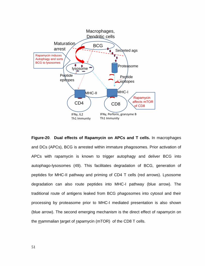
51
Figure-20. Dual effects of Rapamycin on APCs and T cells. In macrophages
and DCs (APCs), BCG is arrested within immature phagosomes. Prior activation of
APCs with rapamycin is known to trigger autophagy and deliver BCG into
autophago-lysosomes (49). This facilitates degradation of BCG, generation of
peptides for MHC-II pathway and priming of CD4 T cells (red arrows). Lysosome
degradation can also route peptides into MHC-I pathway (blue arrow). The
traditional route of antigens leaked from BCG phagosomes into cytosol and their
processing by proteasome prior to MHC-I mediated presentation is also shown
(blue arrow). The second emerging mechanism is the direct effect of rapamycin on
the mammalian target of rapamycin (mTOR) of the CD8 T cells.
CD4
lysosome
CD8
BCGMaturation
arrest
Proteasome
IFNγ, IL2Th1 Immunity
IFNγ, Perforin, granzyme BTh1 Immunity
Secreted ags
Macrophages,
Dendritic cells
MHC-II MHC-I
Peptide
epitopes
Peptide
epitopes
Rapamycin induces
Autophagy and sorts
BCG to lysosomes
Rapamycin
affects mTOR
of CD8
52
Methods
Since the expansion of CD8 T cells, elaboration of their effectors like perforin and
granzyme-B and correlation of their function to protection against tuberculosis, can
only be observed using in vivo models, I used mouse models to investigate specific
questions. The C57Bl/6 mice are Th1 biased mice suitable to study immunity
against tuberculosis. The BALB/c mice have a Th2 bias and are thought to be
alternate models to study pathogenesis.
The tuberculosis mouse infection model (Fig. 21). The standard (NIH approved)
mouse model of aerosol infection with tuberculosis consists of infecting C57Bl/6
mice with a low aerosol dose (approx. 100 CFU per mouse) of M.tuberculosis (Mtb)
and observing infection for 4 weeks. Mice are sacrificed at 4 week, and the lungs
and spleens are homogenized and plated on 7H11 agar plates for CFU counts. Mtb
grows within lungs of mice rapidly to yield 6 log10 (1X10⁶ CFU) organisms per lungs,
over the 4 weeks. Concurrent with growth in lungs and spleens, CD4 and CD8 T
cells are induced and are measured through flow cytometric analysis of lung, spleen
and lymph node derived suspension cells. IFN-γ secreting CD4 and CD8 T cells are
important in defense against tuberculosis, and when they reach to a significant
number, the growth of Mtb is arrested and bacterial numbers start decreasing
around 4 weeks. Recent reports indicate that even multi-functional, multiple
cytokine secreting T cells can be induced during tuberculosis infection of mice
although; their numbers are small during natural aerosol infection.
53
CHAPTER-3.1
RAPAMYCIN INCREASES CD8 T CELL FUNCTION DURING
EXPERIMENTAL TUBERCULOSIS
54
Specific Hypothesis
It is well known that aerosol induced infection of tuberculosis in mice is exponential
up to 4 weeks after which it levels off. It has been shown that mice produce
moderate CD8 T cell responses during experimental tuberculosis (35). Thus, the
levels of antigen-specific CD8 T cells which can be demonstrated during infection of
mice are lower. In this section, we hypothesized that rapamycin can enhance
macrophage mediated-processing of wild type Mtb, induce better CD8 T cell
responses and increase their protective function.
Methods: Mice were aerosol infected with Mtb and from day one, treated with i.p.
doses of rapamycin given at 75 μg/kg dose, once a day for 30 days. Two days after
the last dose, mice were sacrificed and lungs and spleens were processed for
bacterial counts. The T cells of lungs, lymph nodes and spleens from sacrificed
mice were also analyzed using surface markers (CD4, CD8 and tetramers) and flow
cytometry. CD8 T cells were analysed using 2 different tetramers which were
specific for Mtb antigens, TB10.4 and ESAT6 (PE-labeled tetramers were
synthesized by the NIH Tetramer facility, Emory University). During Th1 immunity
the transcription factors (TFs) T-box expressed in T cells (T-bet) and
Eomesodermin (Eomes) seem to determine the effector and memory T cell lineage,
respectively (86). T-bet is a major determinant of effector T cell immunity against
many infections. To determine if rapamycin affected the expression of these TFs,
CD8 T cells were also typed for intracellular levels of TFs.
55
Results:
Rapamycin treatment enhances protection against tuberculosis in mice
(Fig.21). Rapamycin treated macrophages can kill Mtb or BCG through autophagy
[94]. However, in this experiment, the primary goal was not to determine the killing
effect of rapamycin, but to evaluate the effects of low doses of rapamycin on T cell
function. Fig 21 shows that rapamycin did not affect the lung counts of Mtb but had
an inhibitory effect on Mtb counts of spleens. Since the low dose rapamycin was not
sufficient in enhancing APC mediated killing of Mtb in vitro, we speculated that the
anti-tuberculosis effect of rapamycin was likely due to an increased activation of T
cells in the spleens. Activated T cells are known to enhance ability of macrophages
to kill intracellular Mtb.
Rapamycin increases expression of T-bet in CD4 T cells (Fig.22). Rapamycin is
known to affect CD8 T cells positively but not CD4 T cells (41). Furthermore,
rapamycin is also known to differentially affect the expression of T-bet and Eomes
which are major regulators of the effector Th1 immunity and longevity, respectively
(86). Interestingly, during tuberculosis of mice, rapamycin was only able to increase
T-bet in CD4 but not in CD8 T cells. It did not affect T cells of spleen or lymph
nodes, where T cell expansion and contraction usually occur following infection.
Rapamycin increases the numbers of antigen specific-CD8 T cells (Fig.23). To
determine whether cytotoxic CD8 T cells mediated protection in rapamycin treated
mouse spleens; antigen specific, tetramer positive CD8 T cells were enumerated
56
using flow cytometry. A typical histogram using TB10.4-specific tetramer is shown in
Fig.23. Rapamycin significantly enhanced the expansion of CD8 T cells specific for
TB10.4 and ESAT-6. We speculate that a dramatically decreased level of TB10.4
specific CD8 T cells in spleens at 8 weeks could be due to cytolytic degranulation,
and enhanced killing of splenic macrophages, which in turn led to decreased Mtb
counts.
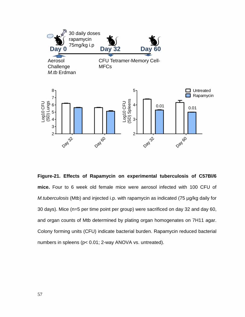
57
Figure-21. Effects of Rapamycin on experimental tuberculosis of C57Bl/6
mice. Four to 6 week old female mice were aerosol infected with 100 CFU of
M.tuberculosis (Mtb) and injected i.p. with rapamycin as indicated (75 µg/kg daily for
30 days). Mice (n=5 per time point per group) were sacrificed on day 32 and day 60,
and organ counts of Mtb determined by plating organ homogenates on 7H11 agar.
Colony forming units (CFU) indicate bacterial burden. Rapamycin reduced bacterial
numbers in spleens (p< 0.01; 2-way ANOVA vs. untreated).
30 daily doses
rapamycin
75mg/kg i.pDay 0 Day 32
Aerosol
Challenge
M.tb Erdman
Day 60
CFU Tetramer-Memory Cell-
MFCs
Day
32
Day
60
2
3
4
5
6
7
8
Log10
CF
U
(S
D) Lungs
Day
32
Day
60
2
3
4
5 Untreated
Rapamycin
0.01 0.01
Log
10
CF
U
(S
D)
Sp
leen
s
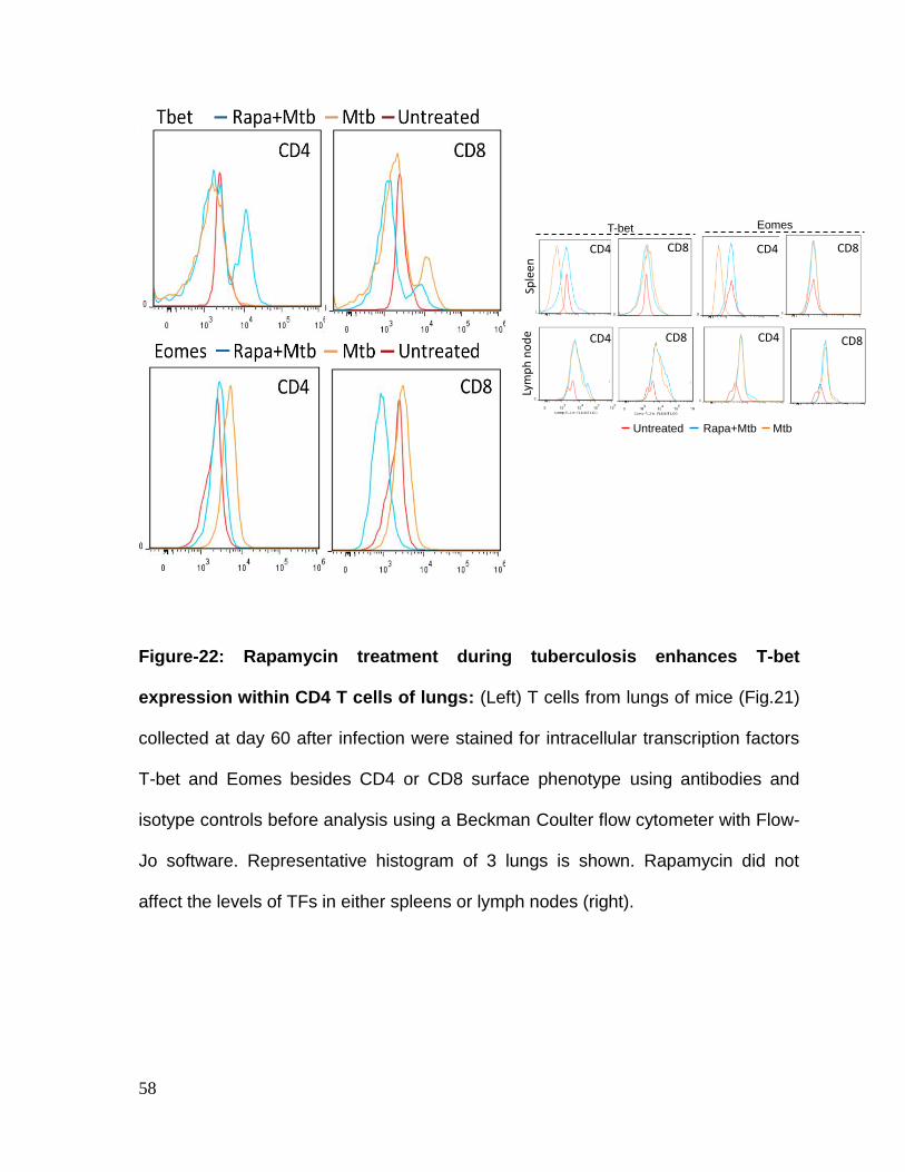
58
Figure-22: Rapamycin treatment during tuberculosis enhances T-bet
expression within CD4 T cells of lungs: (Left) T cells from lungs of mice (Fig.21)
collected at day 60 after infection were stained for intracellular transcription factors
T-bet and Eomes besides CD4 or CD8 surface phenotype using antibodies and
isotype controls before analysis using a Beckman Coulter flow cytometer with Flow-
Jo software. Representative histogram of 3 lungs is shown. Rapamycin did not
affect the levels of TFs in either spleens or lymph nodes (right).
T-bet
Untreated Rapa+Mtb Mtb
Sple
enLy
mp
h n
od
e
CD4 CD8 CD4 CD8
CD4 CD8 CD4 CD8
Eomes
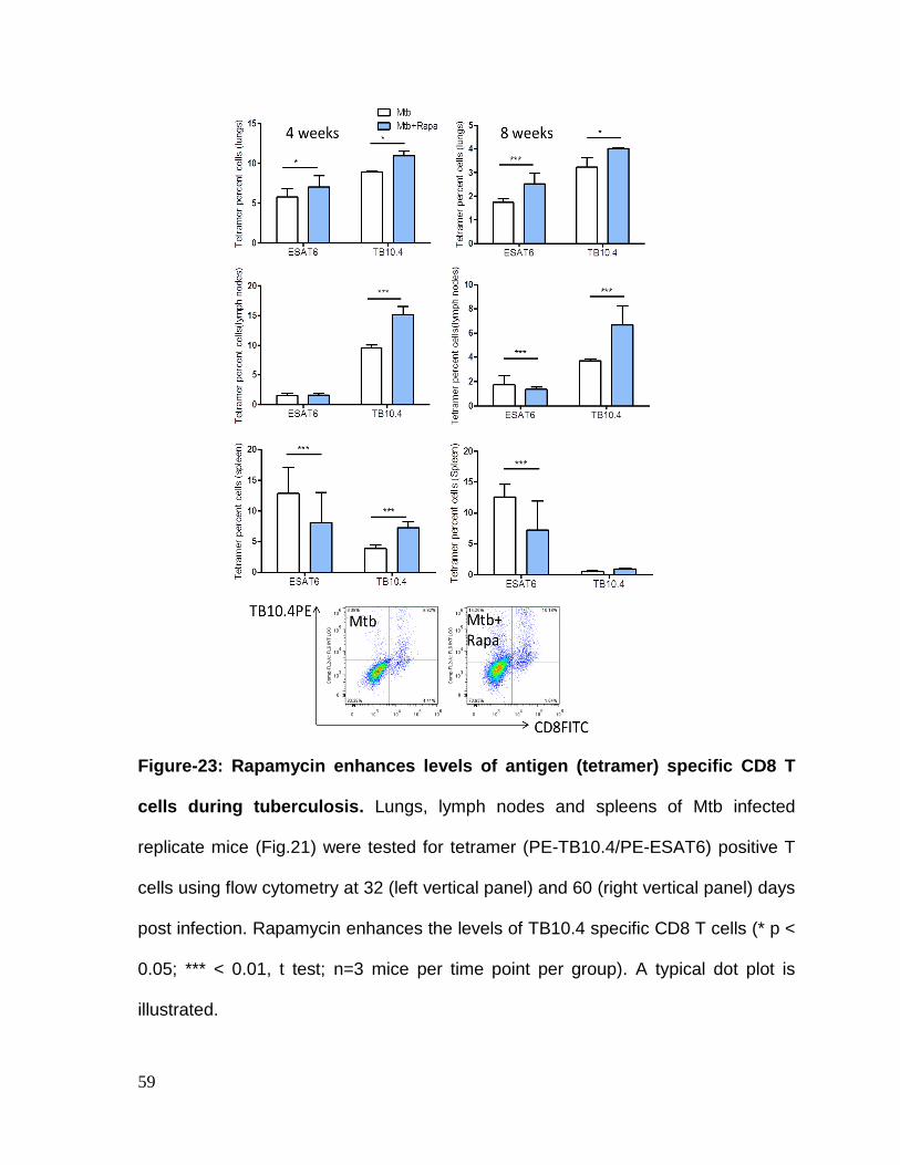
59
Figure-23: Rapamycin enhances levels of antigen (tetramer) specific CD8 T
cells during tuberculosis. Lungs, lymph nodes and spleens of Mtb infected
replicate mice (Fig.21) were tested for tetramer (PE-TB10.4/PE-ESAT6) positive T
cells using flow cytometry at 32 (left vertical panel) and 60 (right vertical panel) days
post infection. Rapamycin enhances the levels of TB10.4 specific CD8 T cells (* p <
0.05; *** < 0.01, t test; n=3 mice per time point per group). A typical dot plot is
illustrated.
60
Discussion
A striking feature of human tuberculosis is that people recovering from tuberculosis
do not become immune to infection. Furthermore, a significant proportion of TB
patients treated with drugs also undergo relapse indicating that the process of
immune memory is not well developed (101, 106). The purpose of this study was to
determine if T cell function during tuberculosis of mice could be altered using
rapamycin, paving the way for vaccination mechanisms to increase T cell responses
and memory. Rapamycin treatment given for 30 days enhanced bacterial clearance
only from spleens of mice but not lungs. Our in vitro studies showed that 5 μg/ mL
of rapamycin is required to activate 106 macrophages to kill Mtb in vitro
(unpublished observations). Since the mice were treated with a low dose of
rapamycin at 75 μg/ kg dose, < 200 ng of rapamycin per mL of blood was available
per mouse weighing 25 gm. We therefore propose that the rapamycin could only
activate T cells of spleens. These in turn, may have activated macrophages locally
to kill Mtb within macrophages. In support of these observations, rapamycin
treatment increased the function of T cells.
The transcription factors, T-bet and Eomes are major determinants Th1 immunity
respectively regulating, short term effector immunity and long term memory in T
cells (6, 8). Surprisingly, rapamycin activated T-bet in CD4 T cells of lungs, but not
T-bet or Eomes in T cells of spleens or lymph nodes. Earlier studies using Mtb
infected mice show that, aerosol implanted Mtb drain from lungs into hilar lymph
nodes, and then to spleen over the course of infection. Local priming of T cells
61
occurs and such primed T cells home back to lungs, either during primary infection
or after re-infection of lungs. Migration to and from lung target organ to lymphoid
organs is regulated by pro-inflammatory chemokine receptors on T cells and T-bet
plays a major role in inducing their expression (6, 8). Enhanced expression of T-bet
in CD4 T cells of lungs but not spleen or lymph nodes by rapamycin therefore
indicates that the drug was able to enhance effector CD4 T cell function in the
lungs. We also propose that these T cells were in the process of migration into
lymphoid organs since, generally in the aerosol infected mice, bacterial numbers
decline only after 4-8 weeks and homing of T cells into lymphoid organs occurs to
facilitate contraction and memory development.
In order to further address this question, we analyzed antigen specific CD8 T cells.
In our initial validation studies, we considered using four types of tetramer specific
CD8 T cells to determine T cell activation. The antigens Mtb32a, PstS3 and TB10.4
are found in both wild type Mtb strain as well as BCG and could be used to detect
CD8 T cell responses both during BCG vaccination and after Mtb challenge (11,
88). The antigen ESAT6 is an immune-dominant region of Mtb but absent in BCG
(58). In our model, the numbers of CD8 T cells specific for Mtb32a, PstS3 were too
low. Both ESAT6 and TB10.4 specific CD8 T cells were however found in significant
numbers in the organs of mice with tuberculosis. Interestingly, rapamycin treatment
enhanced the numbers of TB10.4 and ESAT6 specific CD8 T cells in all three
organs (55). However, there were certain differences in their association with
protection against tuberculosis in rapamycin treated mice. ESAT6 specific CD8 T
62
cells were not associated with protection since lung CFUs remained unchanged
even in the presence of their abundant numbers. In contrast, TB10.4 numbers were
significantly low in spleens when Mtb numbers declined (day 60). We propose that
these CD8 T cells lysed, accounting for their reduced numbers coinciding with
decreased CFU of Mtb. This observation is consistent with a previous report
wherein, TB10.420–28-loaded H-2Kd specific CD8+ T cells were found at high
frequencies in draining and non-draining lymph nodes, spleen, bone marrow, and
blood following M. tuberculosis infection (55). TB10.420–28-specific CD8+ T cells are
known to be enriched in the lung and comprise up to 40% of the CD8+ T cells in the
lung tissue and in the broncho-alveolar compartment. Finally, TB10.4 specific CD8
T cells express granzyme-B, an important cytotoxin mediating target cell lysis [66].
TB10.4 specific CD8 T cells therefore appear to be a biomarker indicating protection
against tuberculosis of mice and could be used to monitor the expansion and
contraction of protective CD8 T cells. We therefore conclude that rapamycin seems
to exert a moderate protective function against tuberculosis of mice at low doses
and we propose that perhaps, higher doses would be more effective.
63
CHAPTER-3.2
RAPAMYCIN INCREASES BCG VACCINE INDUCED
PROTECTION AND CD4 AND CD8 T CELL FUNCTION
AGAINST EXPERIMENTAL TUBERCULOSIS IN MICE
64
Specific Hypothesis: During natural infection with Mtb, low dose rapamycin was
able to marginally enhance CD8 T cell function, which resulted in significantly
increased clearance of Mtb from spleens. Since Mtb has been found to exert
suppressive effects on the expression of MHC-II in mice (60, 70), decrease
sensitivity of macrophages to IFN-γ and affect various cellular processes, we
speculated that virulent Mtb could be invasive and not that susceptible to the action
of low dose rapamycin. As is the practice in many infectious diseases, a vaccine
has been used to prevent tuberculosis in humans. The M.bovis- derived BCG
vaccine is the only vaccine approved for use against human tuberculosis. It protects
children variably against tuberculosis but not adults. BCG has been thought to
induce immunity for 5-10 years since around 10 years of age, children again
become susceptible to tuberculosis despite childhood vaccination with BCG. We
found earlier that, when dendritic cells containing BCG vaccine were pre-activated
with rapamycin and injected to mice, an enhanced protection occurred against
subsequent challenge with virulent Mtb (48). We therefore hypothesized that
rapamycin enhances the efficacy of BCG vaccine through its combined action on
DCs and CD8 T cells (Fig.24).
Tuberculosis mouse vaccine evaluation model (Fig. 24) The standard (NIH
approved) mouse model of vaccine evaluation consists of mice which are first
vaccinated with BCG vaccine (one s.c. dose per mouse given at 1X10⁶ CFU per
mouse) and rested for 4 weeks to develop immunity. They are then aerosol infected
with M.tuberculosis (Mtb; 100 CFU per mouse) and observing infection for the next
4 weeks. Mice are sacrificed at 8 weeks after vaccination and 4 weeks after
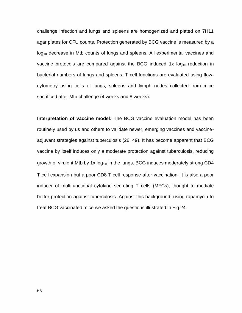
65
challenge infection and lungs and spleens are homogenized and plated on 7H11
agar plates for CFU counts. Protection generated by BCG vaccine is measured by a
log10 decrease in Mtb counts of lungs and spleens. All experimental vaccines and
vaccine protocols are compared against the BCG induced 1x log10 reduction in
bacterial numbers of lungs and spleens. T cell functions are evaluated using flow-
cytometry using cells of lungs, spleens and lymph nodes collected from mice
sacrificed after Mtb challenge (4 weeks and 8 weeks).
Interpretation of vaccine model: The BCG vaccine evaluation model has been
routinely used by us and others to validate newer, emerging vaccines and vaccine-
adjuvant strategies against tuberculosis (26, 49). It has become apparent that BCG
vaccine by itself induces only a moderate protection against tuberculosis, reducing
growth of virulent Mtb by 1x log10 in the lungs. BCG induces moderately strong CD4
T cell expansion but a poor CD8 T cell response after vaccination. It is also a poor
inducer of multifunctional cytokine secreting T cells (MFCs), thought to mediate
better protection against tuberculosis. Against this background, using rapamycin to
treat BCG vaccinated mice we asked the questions illustrated in Fig.24.
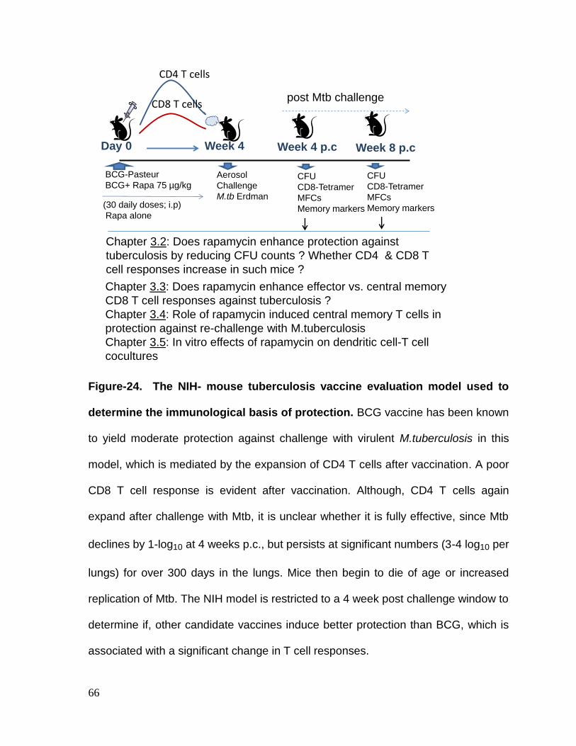
66
Figure-24. The NIH- mouse tuberculosis vaccine evaluation model used to
determine the immunological basis of protection. BCG vaccine has been known
to yield moderate protection against challenge with virulent M.tuberculosis in this
model, which is mediated by the expansion of CD4 T cells after vaccination. A poor
CD8 T cell response is evident after vaccination. Although, CD4 T cells again
expand after challenge with Mtb, it is unclear whether it is fully effective, since Mtb
declines by 1-log10 at 4 weeks p.c., but persists at significant numbers (3-4 log10 per
lungs) for over 300 days in the lungs. Mice then begin to die of age or increased
replication of Mtb. The NIH model is restricted to a 4 week post challenge window to
determine if, other candidate vaccines induce better protection than BCG, which is
associated with a significant change in T cell responses.
BCG-Pasteur
BCG+ Rapa 75 µg/kg
Day 0 Week 4
Aerosol
Challenge
M.tb Erdman
Week 4 p.c
CFU
CD8-Tetramer
MFCs
Memory markers (30 daily doses; i.p)
Rapa alone
Week 8 p.c
CFU
CD8-Tetramer
MFCs
Memory markers
post Mtb challenge
Chapter 3.2: Does rapamycin enhance protection against
tuberculosis by reducing CFU counts ? Whether CD4 & CD8 T
cell responses increase in such mice ?
Chapter 3.3: Does rapamycin enhance effector vs. central memory
CD8 T cell responses against tuberculosis ?
Chapter 3.4: Role of rapamycin induced central memory T cells in
protection against re-challenge with M.tuberculosis
Chapter 3.5: In vitro effects of rapamycin on dendritic cell-T cell
cocultures
CD4 T cells
CD8 T cells
67
Results:
Rapamycin enhances efficacy of BCG vaccine in mice. Fig.25 shows that
rapamycin treatment of mice after BCG vaccination led to a significant decrease in
Mtb counts of both lungs and spleens compared to mice vaccinated with BCG alone
(p< 0.02, 2-way ANOVA, 5 mice per group). Rapamycin alone had no significant
effect on the growth of challenged Mtb organisms.
Rapamycin enhances levels of antigen specific tetramer positive CD8 T cells.
The organs of vaccinated mice were tested for antigen-specific CD8 T cells specific
for TB10.4 & ESAT6-tetramers at 4 (left vertical panel) and 8 (right vertical panel)
weeks post challenge (Fig.26). Data show that rapamycin treatment with BCG
generally expands antigen-specific CD8 T cells. However, ESAT6 specific T cells
tend to remain elevated in lungs while, TB10.4 specific CD8 T cells showed a
decline over time, which may indicate degranulation and cytolytic effects associated
with this population of CD8 T cells.
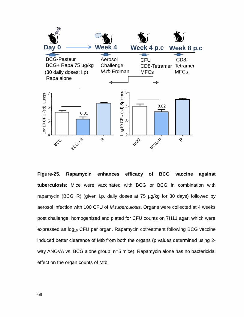
68
Figure-25. Rapamycin enhances efficacy of BCG vaccine against
tuberculosis: Mice were vaccinated with BCG or BCG in combination with
rapamycin (BCG+R) (given i.p. daily doses at 75 µg/kg for 30 days) followed by
aerosol infection with 100 CFU of M.tuberculosis. Organs were collected at 4 weeks
post challenge, homogenized and plated for CFU counts on 7H11 agar, which were
expressed as log10 CFU per organ. Rapamycin cotreatment following BCG vaccine
induced better clearance of Mtb from both the organs (p values determined using 2-
way ANOVA vs. BCG alone group; n=5 mice). Rapamycin alone has no bactericidal
effect on the organ counts of Mtb.
Lungs
BCG
BCG +
R R
4
5
6
7
0.01
Lo
g1
0 C
FU
(sd
) L
ung
s
Spleens
BCG
BCG+R R
2
3
4
5
0.02
Lo
g1
0 C
FU
(sd
) S
ple
ens
BCG-Pasteur
BCG+ Rapa 75 µg/kg
Day 0 Week 4
Aerosol
Challenge
M.tb Erdman
Week 4 p.c
CFU
CD8-Tetramer
MFCs (30 daily doses; i.p)
Rapa alone
Week 8 p.c
CD8-
Tetramer
MFCs
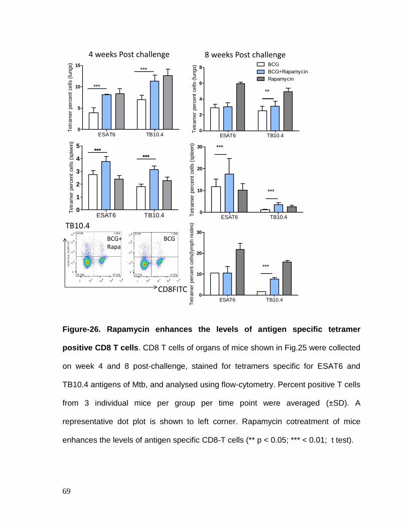
69
Figure-26. Rapamycin enhances the levels of antigen specific tetramer
positive CD8 T cells. CD8 T cells of organs of mice shown in Fig.25 were collected
on week 4 and 8 post-challenge, stained for tetramers specific for ESAT6 and
TB10.4 antigens of Mtb, and analysed using flow-cytometry. Percent positive T cells
from 3 individual mice per group per time point were averaged (±SD). A
representative dot plot is shown to left corner. Rapamycin cotreatment of mice
enhances the levels of antigen specific CD8-T cells (** p < 0.05; *** < 0.01; t test).
ESAT6 TB10.40
1
2
3
4
5
******
Tetr
am
er
perc
ent
cells
(sp
leen)
ESAT6 TB10.40
5
10
15
***
***T
etr
am
er
perc
ent
cells
(lu
ngs)
ESAT6 TB10.40
10
20
30 ***
***T
etr
am
er
perc
ent
cells
(sp
leen)
ESAT6 TB10.40
2
4
6
8
**
BCG
BCG+Rapamycin
Rapamycin
Tetr
am
er
perc
ent
cells
(lu
ngs)
ESAT6 TB10.40
10
20
30
***
Tetr
am
er
perc
ent
cells
(lym
ph n
odes)TB10.4
CD8FITC
4 weeks Post challenge 8 weeks Post challenge
BCG+Rapa
BCG
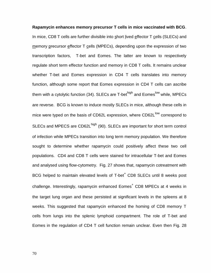
70
Rapamycin enhances memory precursor T cells in mice vaccinated with BCG.
In mice, CD8 T cells are further divisible into short lived effector T cells (SLECs) and
memory precursor effector T cells (MPECs), depending upon the expression of two
transcription factors, T-bet and Eomes. The latter are known to respectively
regulate short term effector function and memory in CD8 T cells. It remains unclear
whether T-bet and Eomes expression in CD4 T cells translates into memory
function, although some report that Eomes expression in CD4 T cells can ascribe
them with a cytolytic function (34). SLECs are T-bethigh
and Eomeslow
while, MPECs
are reverse. BCG is known to induce mostly SLECs in mice, although these cells in
mice were typed on the basis of CD62L expression, where CD62Llow
correspond to
SLECs and MPECS are CD62Lhigh
(90). SLECs are important for short term control
of infection while MPECs transition into long term memory population. We therefore
sought to determine whether rapamycin could positively affect these two cell
populations. CD4 and CD8 T cells were stained for intracellular T-bet and Eomes
and analysed using flow-cytometry. Fig. 27 shows that, rapamycin cotreatment with
BCG helped to maintain elevated levels of T-bet+ CD8 SLECs until 8 weeks post
challenge. Interestingly, rapamycin enhanced Eomes+ CD8 MPECs at 4 weeks in
the target lung organ and these persisted at significant levels in the spleens at 8
weeks. This suggested that rapamycin enhanced the homing of CD8 memory T
cells from lungs into the splenic lymphoid compartment. The role of T-bet and
Eomes in the regulation of CD4 T cell function remain unclear. Even then Fig. 28

71
shows that rapamycin cotreatment increased the levels of T-bet+ and Eomes
+ CD4
SLECs in the organs of mice.
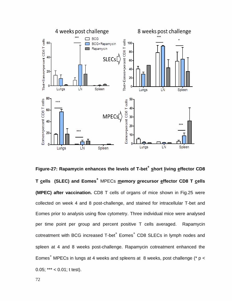
72
Figure-27: Rapamycin enhances the levels of T-bet+ short living effector CD8
T cells (SLEC) and Eomes+ MPECs memory precursor effector CD8 T cells
(MPEC) after vaccination. CD8 T cells of organs of mice shown in Fig.25 were
collected on week 4 and 8 post-challenge, and stained for intracellular T-bet and
Eomes prior to analysis using flow cytometry. Three individual mice were analysed
per time point per group and percent positive T cells averaged. Rapamycin
cotreatment with BCG increased T-bet+ Eomes
+ CD8 SLECs in lymph nodes and
spleen at 4 and 8 weeks post-challenge. Rapamycin cotreatment enhanced the
Eomes+ MPECs in lungs at 4 weeks and spleens at 8 weeks, post challenge (* p <
0.05; *** < 0.01; t test).
73
Figure-28: Rapamycin affects the levels of T-bet+ short living effector CD4 T
cells (SLEC) and Eomes+ MPECs memory precursor effector CD4 T cells
(MPEC) after vaccination. CD4 T cells of organs of mice shown in Fig.25 were
collected on week 4 and 8 post-challenge, and stained for intracellular T-bet and
Eomes prior to analysis using flow cytometry (legend as in Fig.27). Three individual
mice were analysed per time point per group and per cent positive T cells averaged.
Rapamycin cotreatment with BCG increased T-bet+ Eomes
+ CD4 SLECs in lymph
nodes and spleens at 4 and 8 weeks post-challenge (p *** < 0.01, t test).
74
Discussion
In our previous study, during experimental tuberculosis, rapamycin was partially
able to enhance clearance of virulent Mtb in the spleens of mice, although it did
enhance the levels of antigen specific CD8 T cells in both lungs and spleens
(Chapter 3.1). Since Mtb is a virulent organism capable of modulating immune
responses, we sought to determine the immune responses elicited by the BCG
vaccine in mice against tuberculosis. BCG is an attenuated mutant of M.bovis
similar to M.tuberculosis and thought to be less immune-suppressive than virulent
Mtb. Data presented here show that rapamycin was able to significantly boost the
efficacy of BCG vaccine to reduce the numbers of Mtb in both lungs and spleens of
mice. This correlated well with the expansion of antigen specific, tetramer positive
CD8 T cells in lungs and spleens. Significantly, when lung counts of Mtb CFUs
declined, the numbers of TB10.4 specific CD8 T cells declined from 10% to 4% but
those of ESAT6 specific CD8 T cells remained unchanged. Furthermore, TB10.4
specific CD8 T cells increased dramatically in the lymphoid organs (spleens and
lymph nodes). This is consistent with a pattern of homing of CD8 effectors from the
target lung organs to the latter for transition into memory cells.
Recent studies indicate that transition of effector T cells into memory precursor T
cells occurs under defined conditions which are probably dictated both by pathogen
elimination and by intracellular transcription factors T-bet and Eomesodermin
(Eomes) (86). First, infection is largely controlled by effectors, which removes or
reduces the continuous priming signaling from APCs towards CD8 T cells.
Secondly, perhaps due to cytokine signaling, a switch occurs from effector into
75
memory phenotype regulated by T-bet and Eomes. In support of this observation,
CD8+ T cells lacking T-bet show enhanced differentiation as long lived memory
cells. In addition, mice deficient in Eomes have a limited capability to compete for
antigen specific memory compartment in CD8 T cells (6). Furthermore, CD8 T cells
from Eomes deficient mice lack long term persistence and could not fight the
secondary rechallenge infection since they did not produce a strong response of
effector T cells (34). These observations led us to examine the numbers of T-bet+
and Eomes+ T cells operationally defined as short living effector cells (SLECs) and
memory precursor effector T cells (MPECs) (1, 53). Interestingly, we found that
rapamycin enhanced the accumulation of Eomes+CD8
+ MPECs early after infection
in the lungs (4 weeks) which persisted later in the spleens (Fig.23). At the same
time, it maintained a strong expansion of SLECs. A similar profile was also
observed for Eomes+CD4
+ T cells (Fig.24). Although the function of Eomes in CD4
T cells remains unclear, some studies indicate that Eomes+CD4
+ are cytotoxic. The
major observation of this study was that rapamycin cotreatment not only enhanced
the levels of T-bet+CD8
+ SLECs but also Eomes
+CD8
+ MPECs. We therefore
suggest that the novel, dual action of rapamycin in enhancing the CD8 and CD4 T
cells enabled the BCG and rapamycin co-treated mice to control tuberculosis better.
76
CHAPTER-3.3
RAPAMYCIN AFTER BCG VACCINATION SHIFTS THE
PHENOTYPE OF IMMUNE MEMORY CELL POPULATIONS
THROUGH INHIBITION OF MTOR FACILITATING
EXPANSION OF CENTRAL MEMORY T CELLS
77
Background:
In the previous chapter, we used rapamycin to boost the efficacy of BCG vaccine
and found that it increased bacterial clearance Mtb from organs, induced enhanced
levels of antigen specific CD8 T cells and boosted the levels of T-bet+CD8
+ SLECs
and Eomes+CD8
+ MPECs (Figs.25-28). These observations were exciting but also
raised additional questions. Emerging studies using virus infection models have
revealed that the phenotypes of T cells in vaccinated mice are quite diverse and
require the use of multiple surface markers to define effector and memory T cells.
Some of these studies have used rapamycin based on the exciting observation that
rapamycin represses mTOR to alter T cell function (3). Studies show that long
living memory precursor T cells express distinct surface markers in addition to
Eomes and undergo considerable flux (18, 104). Furthermore, there are divergent
opinions on how effector and memory T cells arise after presentation of antigens
from APCs, and the rate at which effector T cells undergo transition into memory
cells (72). Finally, an effective transition from effector to memory phenotype seems
to require a significant elimination of the pathogen in mice. Paradoxically, mice
really do not eliminate Mtb organisms which persist for months. These
considerations led us to re-examine the phenotype of memory CD8 T cells using
rapamycin treated mice and re-evaluate their significance and correlation to
protection.
Memory T cell phenotypes: A sustained T cell immune response is divided into two
distinct phases. The first phase after infection is the activation event, where antigen
specific T cells or effector T cells undergo an exponential increase (14, 62).
78
Expanded effector cells ensure enough T cells to attack and combat and contain the
infection (Fig.29). As soon as the infection is controlled, the T cells undergo a
controlled contraction phase where only 5-10 % of cells remain after resolution of
the infection (54, 103). These cells present during the contraction phase are
referred to as the memory pool. The second phase of the immune response occurs
when there is a ‘reinfection’ and a second exposure of the antigen to the resting T
cell memory pool. When the memory T cells encounter the antigen again, they re-
expand and divide much more vigorously than the initial effector T cell population.
They produce cytokines at a much higher rate and have increased specificity
against the antigen (81). Memory T cells are distinct compared to naïve T cells and
possesses properties which are different and unique. They express high levels of
IL-7 (CD127) receptor and also the anti-apoptotic protein Bcl2. These markers
along with other co-receptors allow for the constant division and self-renewal
properties of the memory T cells (85, 109).
Role of rapamycin in the regulation of memory phenotype in T cells: The
mammalian target of rapamycin (mTOR), is emerging as a major regulator of T cell
function. First, activated T cells down-regulate the receptors CD62L and CCR7,
which are homing receptors usually present on naïve T cells (45). Since mTOR
inhibits both CCR7 and CD62L, it has been proposed that repression of mTOR by
rapamycin may facilitate the expression of chemokine receptors and enable homing
of memory T cell populations (95) (73). Fig.30 illustrates another mechanism in
which, rapamycin can affect T cells through repression of mTOR which reduces
TRAF-6 mediated signaling. TRAF-6 activation has multiple consequences, one of
79
them being the reduction of IFN-αβ. It has been reported that TRAF-6 (a
downstream signaling molecule inhibited by mTOR) deficient T cells could develop
into effector T cells, but failed to differentiate into memory precursors (79).
Furthermore, TRAF-6 deficient T cells exhibited lower levels of AMP-kinase, which
is an inhibitor for mTOR. Interestingly, metformin, an AMPK agonist could reverse
the effect, and restore the formation of memory T cells in TRAF-6 deficient mice
(84). Since IFN-αβ enhances effector function of CD8 T cells in combination with IL-
12 (see below), we propose that rapamycin inhibits mTOR and TRAF-6 thereby
reducing IFN-αβ and this enables effectors to change into memory cells.
We initially used rapamycin to boost the efficacy of BCG vaccine due to its ability
increase macrophage mediated processing of BCG vaccine (49). Recent studies
indicate that inhibition of mTOR in T cells through rapamycin could be another
mechanism to enhance vaccine efficacy and memory cell formation in mice (86).
Data show that mTOR plays a significant role in the formation of a functional
memory pool. Araki et al. reported that low dose-rapamycin has an immune-
modulatory role on the phenotype of the CD8 T cells during virus infections and can
enhance different stages of the memory formation and development of the T cells
(3).
While rapamycin affects expression of homing receptors by acting on mTOR, either
prolonged or timed exposure can also suppress them (Fig.31). Furthermore,
rapamycin has varied effects on the production of memory T cells depending on
when it is given. Rapamycin given during the expansion of T cells improves the
80
number of memory T cells, but generates a similar number of effector CD8 T cells
during the infection compared to mice untreated with rapamycin. However, the rate
at which rapamycin treated CD8 T cells undergo apoptotic death is reduced when
they enter the contraction phase. Rapamycin given during the contraction phase
changes the phenotype of the effector T cells to a memory phenotype. Finally,
rapamycin yields the best results when it is given during both expansion and
contraction phase (2).
Role of IL-12 and T-bet during memory development: IL-12, a key cytokine that is
secreted from BCG or Mtb infected APCs also plays a significant role in skewing
effector vs. memory T cells. IL-12 activates T-bet in OT-I CD8 T cells leading to a
burst in effector T cell population [75]. Paradoxically, rapamycin reduced T-bet and
IL-12 expression but enhanced Eomes, thereby facilitating memory cells. The role
of IL-12 in enhancing effectors is understandable since it is the first innate cytokine
to be secreted after macrophages or DCs engulf mycobacteria, and the initial burst
of effectors is required to control infection (83, 86). However, IL-12 must decline in
order for memory cell expansion to continue. Since both BCG and Mtb persist in
APCs for prolonged periods, an intriguing possibility is that, the continuous
secretion of IL-12 by infected APCs may in fact interfere with the overall process of
memory cell expansion.
Synergy of IL-12 with IFN-αβ: The other key cytokines which seem to play a role in
the regulation of effector vs. memory are the IFN-α and IFN-β. Mescher et al.
reported that both IL-12 and IFN-αβ can act as a ‘third signal’ to promote both IFN-γ
81
secretion and expression of perforin and granzyme in murine CD8+ cells (29). The
latter are the effector cells that can potentially kill APCs containing mycobacteria.
Paradoxically, they are also the cells which transition into long living MPECs in mice
and when re-infection occurs, undergo recall expansion into effectors which contain
re-infection. It is thus obvious that IL-12 and IFN-αβ are required to sustain both
initial strong effector response, and recall expansion of memory cells into cytotoxic
effectors. Interestingly, Fig.30 illustrates that IFN-αβ can be induced both through
TLR-activation as well as through rapamycin mediated repression of mTOR.
Specific Hypothesis
Cumulatively, these observations lead to our hypothesis that CD8 effector vs.
memory switch following BCG vaccination may be defective due to both host and
vaccine (BCG) dependent factors. BCG may hide in APCs not inducing enough IL-
12 or IFN-αβ. On the other hand peptide antigens from BCG are also probably not
enough to induce CD8 T cells. We propose that rapamycin treatment of mice during
BCG vaccination may induce ‘functionally better CD8 T cells’ due to its dual effect
on APCs and T cells.
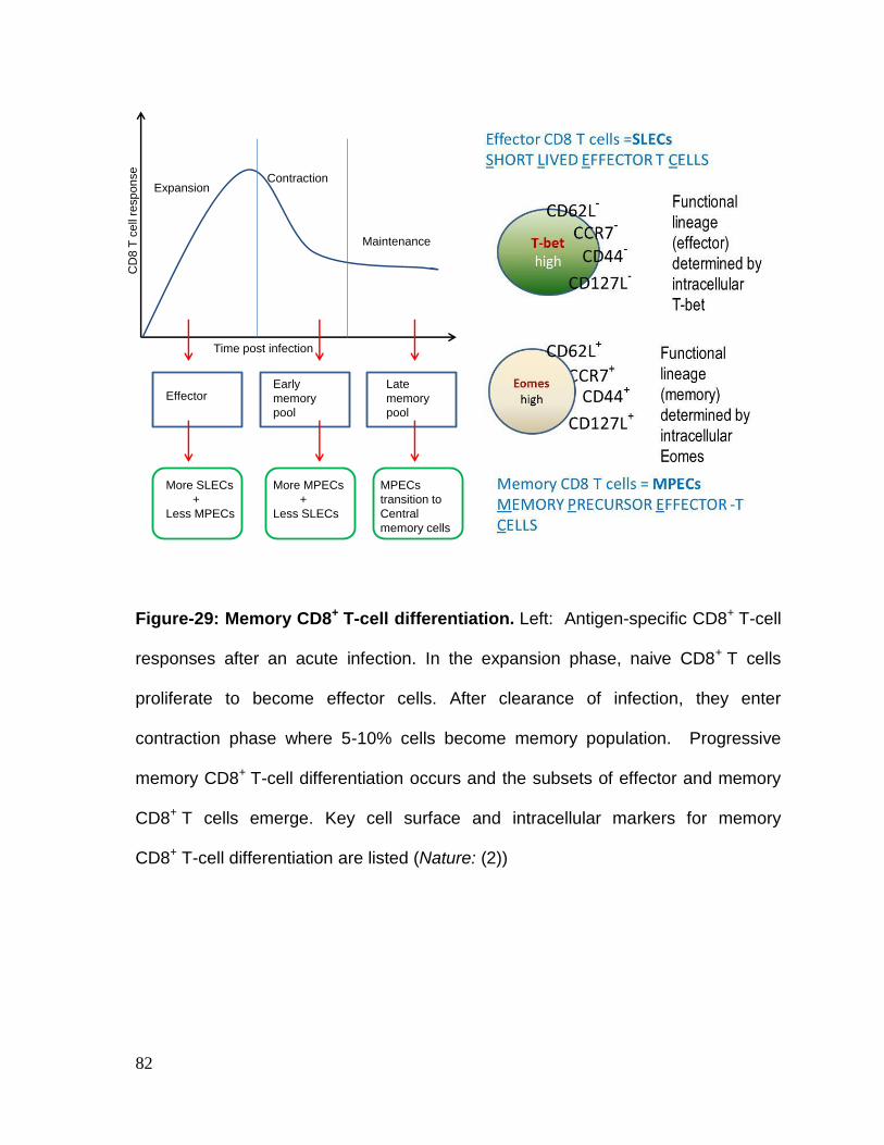
82
Figure-29: Memory CD8+ T-cell differentiation. Left: Antigen-specific CD8+ T-cell
responses after an acute infection. In the expansion phase, naive CD8+ T cells
proliferate to become effector cells. After clearance of infection, they enter
contraction phase where 5-10% cells become memory population. Progressive
memory CD8+ T-cell differentiation occurs and the subsets of effector and memory
CD8+ T cells emerge. Key cell surface and intracellular markers for memory
CD8+ T-cell differentiation are listed (Nature: (2))
Expansion Contraction
Maintenance
Time post infection
CD
8 T
ce
ll re
sp
on
se
EffectorEarly
memory
pool
Late
memory
pool
More SLECs
+
Less MPECs
MPECs
transition to
Central
memory cells
More MPECs
+
Less SLECs
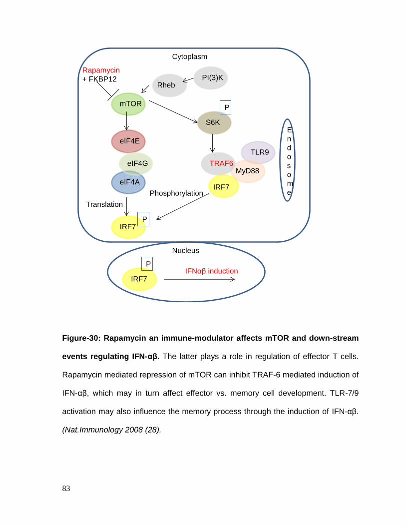
83
Figure-30: Rapamycin an immune-modulator affects mTOR and down-stream
events regulating IFN-αβ. The latter plays a role in regulation of effector T cells.
Rapamycin mediated repression of mTOR can inhibit TRAF-6 mediated induction of
IFN-αβ, which may in turn affect effector vs. memory cell development. TLR-7/9
activation may also influence the memory process through the induction of IFN-αβ.
(Nat.Immunology 2008 (28).
PmTOR
TRAF6
S6K
eIF4A
IRF7
IRF7
eIF4G
P
P
eIF4E
MyD88
TLR9
IRF7
Rapamycin
+ FKBP12
Phosphorylation
IFNαβ induction
Nucleus
Translation
E
n
d
o
s
o
m
e
Cytoplasm
RhebPI(3)K
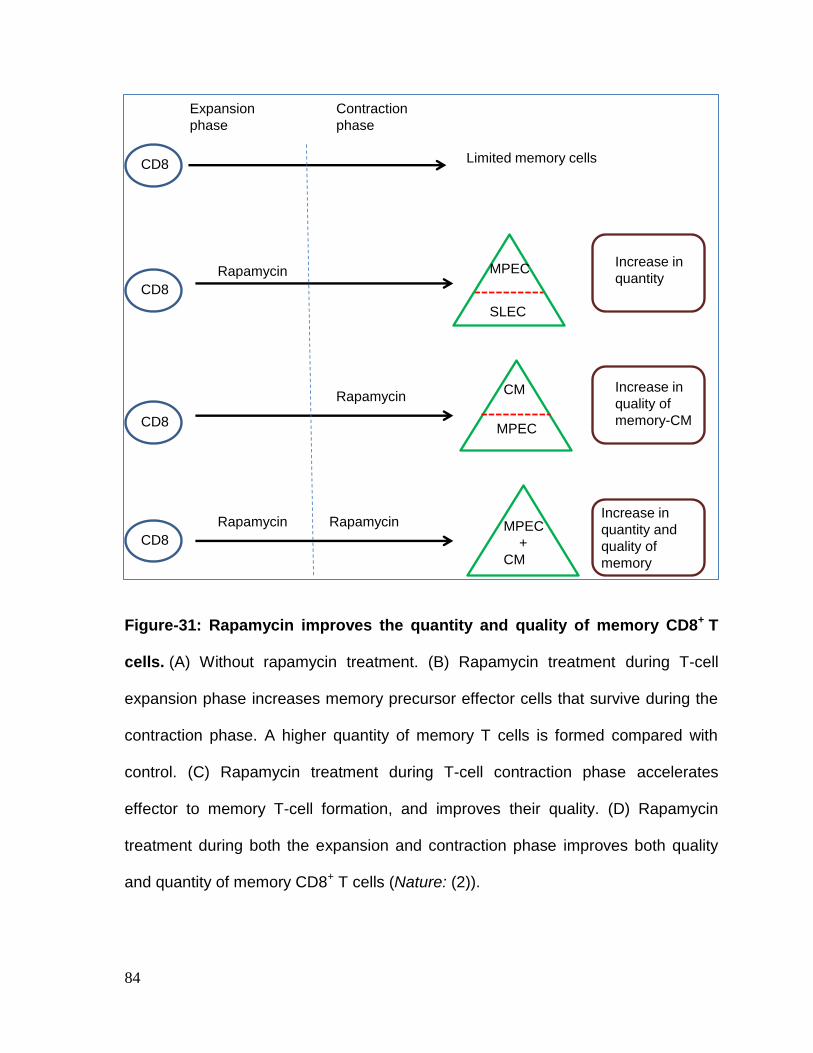
84
Figure-31: Rapamycin improves the quantity and quality of memory CD8+ T
cells. (A) Without rapamycin treatment. (B) Rapamycin treatment during T-cell
expansion phase increases memory precursor effector cells that survive during the
contraction phase. A higher quantity of memory T cells is formed compared with
control. (C) Rapamycin treatment during T-cell contraction phase accelerates
effector to memory T-cell formation, and improves their quality. (D) Rapamycin
treatment during both the expansion and contraction phase improves both quality
and quantity of memory CD8+ T cells (Nature: (2)).
MPEC
Expansion
phase
Contraction
phase
MPEC
SLEC
CM
MPEC
+
CM
Increase in
quantity
Increase in
quality of
memory-CM
Increase in
quantity and
quality of
memory
CD8
CD8
CD8
CD8
Rapamycin
Rapamycin
RapamycinRapamycin
Limited memory cells
85
Methods
Evaluation of effector vs. central memory in mice: In our previous chapter, we used
a single low dose of rapamycin to determine if it could increase effector and
memory T cells and enhance efficacy of BCG vaccine. However, investigations in
other laboratories have indicated that the dosing and timing of rapamycin can
impact the development of effector and memory cells (Fig.31). Therefore, we sought
to determine in this study, the effects of a low dose rapamycin (long term) vs. high
dose (short term) on the course of tuberculosis and the expansion of effector and
memory T cells. The mouse experiment is illustrated in Fig.32. Earlier, we identified
SLECs and MPECs on the basis of T-bet and Eomes, which are transcription
factors. More recent studies show that they also show a differential expression of
CD62L, CD44, CD127 and CCR7, depending on whether they are SLECs and
MPECs. For example MPECs are CD62L+, CD44
+, CD127
+ and CCR7
+. In contrast
the SLECs are CD62L-, CD44
-, CD127
- and CCR7
-. Organs of three mice per time
point per group were individually analyzed and averaged for graphs and p value
determined using graphpad prism. Typical dot blots are illustrated in Figs.33 and 34.
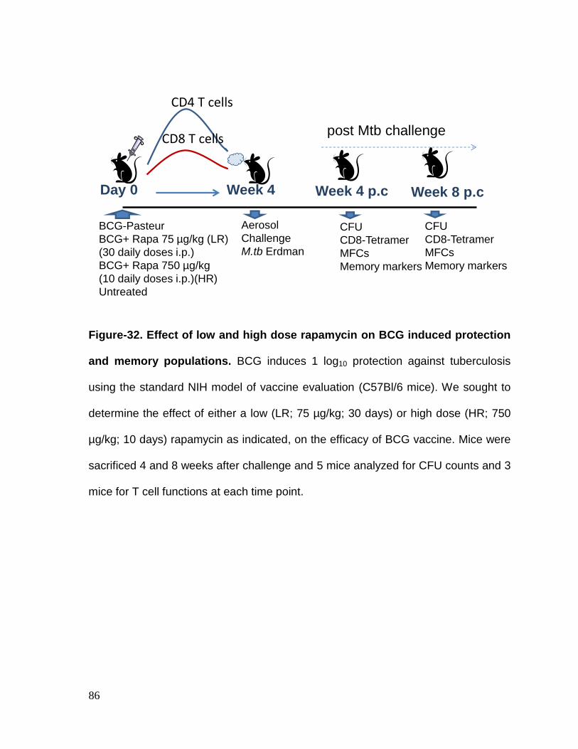
86
Figure-32. Effect of low and high dose rapamycin on BCG induced protection
and memory populations. BCG induces 1 log10 protection against tuberculosis
using the standard NIH model of vaccine evaluation (C57Bl/6 mice). We sought to
determine the effect of either a low (LR; 75 µg/kg; 30 days) or high dose (HR; 750
µg/kg; 10 days) rapamycin as indicated, on the efficacy of BCG vaccine. Mice were
sacrificed 4 and 8 weeks after challenge and 5 mice analyzed for CFU counts and 3
mice for T cell functions at each time point.
BCG-Pasteur
BCG+ Rapa 75 µg/kg (LR)
(30 daily doses i.p.)
BCG+ Rapa 750 µg/kg
(10 daily doses i.p.)(HR)
Untreated
Day 0 Week 4
Aerosol
Challenge
M.tb Erdman
Week 4 p.c
CFU
CD8-Tetramer
MFCs
Memory markers
Week 8 p.c
CFU
CD8-Tetramer
MFCs
Memory markers
post Mtb challenge
CD4 T cells
CD8 T cells
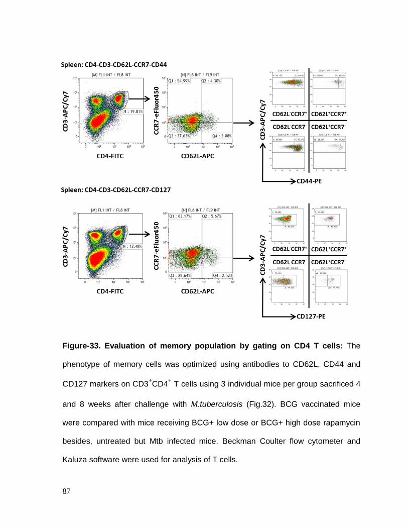
87
Figure-33. Evaluation of memory population by gating on CD4 T cells: The
phenotype of memory cells was optimized using antibodies to CD62L, CD44 and
CD127 markers on CD3+CD4
+ T cells using 3 individual mice per group sacrificed 4
and 8 weeks after challenge with M.tuberculosis (Fig.32). BCG vaccinated mice
were compared with mice receiving BCG+ low dose or BCG+ high dose rapamycin
besides, untreated but Mtb infected mice. Beckman Coulter flow cytometer and
Kaluza software were used for analysis of T cells.
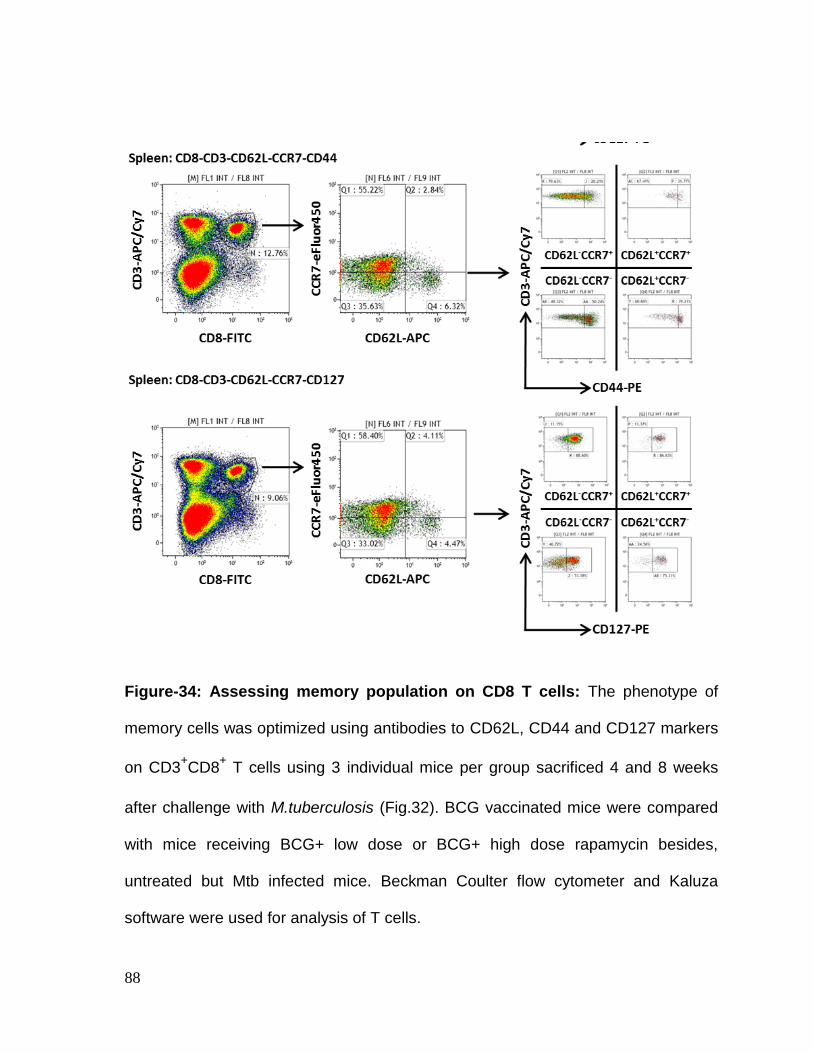
88
Figure-34: Assessing memory population on CD8 T cells: The phenotype of
memory cells was optimized using antibodies to CD62L, CD44 and CD127 markers
on CD3+CD8
+ T cells using 3 individual mice per group sacrificed 4 and 8 weeks
after challenge with M.tuberculosis (Fig.32). BCG vaccinated mice were compared
with mice receiving BCG+ low dose or BCG+ high dose rapamycin besides,
untreated but Mtb infected mice. Beckman Coulter flow cytometer and Kaluza
software were used for analysis of T cells.
89
Results:
Cotreatment of rapamycin with BCG vaccine enhances the ability of vaccine
to clear Mtb from organs. Fig.35 shows that in both lungs and spleen of mice,
rapamycin enhanced bacterial clearance well above the levels induced by BCG
vaccine alone. Significantly, both low-dose and high-dose rapamycin boosted the
efficacy of BCG vaccine in mice against tuberculosis. The high dose rapamycin was
marginally better than low dose rapamycin although, the differences were not
statistically significant. As anticipated, BCG alone protected against tuberculosis by
decreasing organ CFUs of untreated mice by 1-log10.
Effect of Rapamycin on effector memory (MPEC) and effector T cell (SLEC)
phenotype: Spleen, lymph nodes and lungs were analysed 4 week and 8 week
post challenge to determine changes in MPEC vs. SLEC population (Fig.32). CD4+
MPECs and CD8+ MPECs (CD62L
+CCR7
+ ) were again typed on the basis of CD44
and CD127. Thus, there were two groups of MPECs, CD62L+CCR7
+CD127
+ and
CD62L+ CCR7
+. CD44
+ distributed in the target organ lung and the lymphoid
organs; spleen and lymph nodes. Rapamycin plus BCG treatment generally
enhanced CD8+ MPECs compared to BCG alone group in the lungs at 4 weeks
post challenge, when Mtb counts had declined (Fig.36a-b). At 8 weeks again, only
rapamycin plus BCG had accumulated higher numbers of CD8+ MPECs in the lungs
(Fig.36b). In contrast, Figs 37a-b shows the profiles of CD4+ and CD8
+ SLECs. In
general, rapamycin treatment did not markedly affect the distribution of SLECs

90
which were present already in significant numbers. However, rapamycin tended to
increase the numbers of CD8+ SLECs in lungs and spleens while increasing those
of CD8+ SLECs in the lymph nodes.
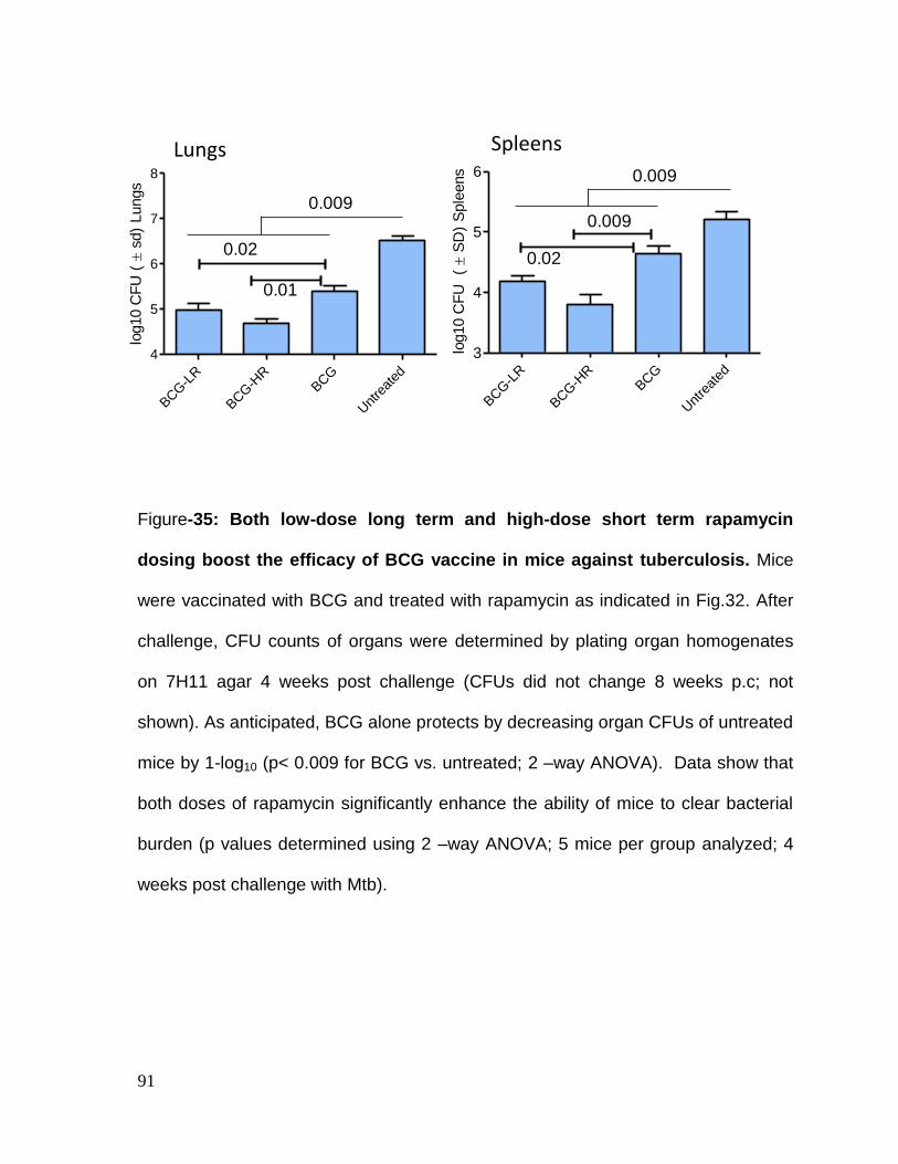
91
Figure-35: Both low-dose long term and high-dose short term rapamycin
dosing boost the efficacy of BCG vaccine in mice against tuberculosis. Mice
were vaccinated with BCG and treated with rapamycin as indicated in Fig.32. After
challenge, CFU counts of organs were determined by plating organ homogenates
on 7H11 agar 4 weeks post challenge (CFUs did not change 8 weeks p.c; not
shown). As anticipated, BCG alone protects by decreasing organ CFUs of untreated
mice by 1-log10 (p< 0.009 for BCG vs. untreated; 2 –way ANOVA). Data show that
both doses of rapamycin significantly enhance the ability of mice to clear bacterial
burden (p values determined using 2 –way ANOVA; 5 mice per group analyzed; 4
weeks post challenge with Mtb).
Lungs Spleens
BCG-L
R
BCG-H
RBCG
Unt
reat
ed
4
5
6
7
8
0.02
0.01
0.009
log10 C
FU
( s
d)
Lungs
BCG-L
R
BCG-H
RBCG
Unt
reat
ed
3
4
5
6
0.009
0.02
0.009
log10 C
FU
( S
D)
Sple
ens
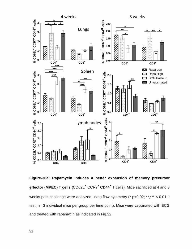
92
Figure-36a: Rapamycin induces a better expansion of memory precursor
effector (MPEC) T cells (CD62L+ CCR7
+ CD44
+ T cells). Mice sacrificed at 4 and 8
weeks post challenge were analyzed using flow cytometry (* p<0.02; **,*** < 0.01; t
test; n= 3 individual mice per group per time point). Mice were vaccinated with BCG
and treated with rapamycin as indicated in Fig.32.
CD4+ CD8+0.0
0.5
1.0
1.5
2.0
2.5Rapa Low
Rapa High
BCG Pasteur
Unvaccinated
****
***
*
% C
D62L
+ C
CR
7+ C
D44
Hi c
ell
s
CD4+ CD8+0.0
0.5
1.0
1.5
2.0Rapa Low
Rapa High
BCG Pasteur
Unvaccinated
**
% C
D62L
+ C
CR
7+ C
D44
Hi c
ell
s
CD4+ CD8+0
1
2
3
4Rapa Low
Rapa High
BCG Pasteur
Unvaccinated*
*
**
% C
D62L
+ C
CR
7+ C
D44
Hi c
ell
sCD4
+CD8
+0
10
20
30
40Rapa Low
Rapa High
BCG Pasteur
Unvaccinated
*%
CD
62L
- CC
R7
- CD
127
+ c
ell
s
CD4+ CD8+0
1
2
3
4Rapa Low
Rapa High
BCG Pasteur
Unvaccinated
*** *
% C
D62L
+ C
CR
7+ C
D44
Hi c
ell
s
CD4+ CD8+0
1
2
3
4Rapa Low
Rapa High
BCG Pasteur
Unvaccinated
***
*
***
***
***
***
**
% C
D62L
+ C
CR
7+ C
D44
Hi c
ells
CD4+ CD8+0.0
0.5
1.0
1.5
2.0
2.5Rapa Low
Rapa High
BCG Pasteur
Unvaccinated
*
% C
D62L
+ C
CR
7+ C
D44
Hi c
ell
s
Lungs
Spleen
lymph nodes
4 weeks 8 weeks
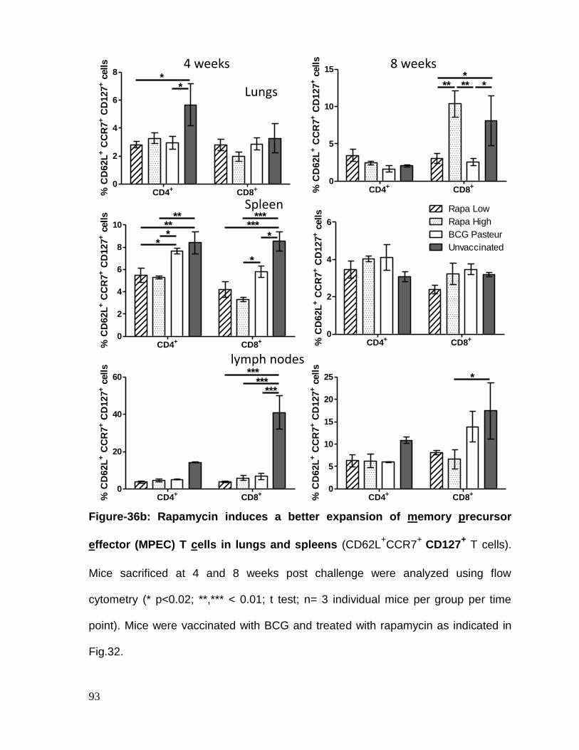
93
Figure-36b: Rapamycin induces a better expansion of memory precursor
effector (MPEC) T cells in lungs and spleens (CD62L+CCR7
+ CD127
+ T cells).
Mice sacrificed at 4 and 8 weeks post challenge were analyzed using flow
cytometry (* p<0.02; **,*** < 0.01; t test; n= 3 individual mice per group per time
point). Mice were vaccinated with BCG and treated with rapamycin as indicated in
Fig.32.
CD4+
CD8+
0
5
10
15Rapa Low
Rapa High
BCG Pasteur
Unvaccinated
***** *
% C
D62L
+ C
CR
7+ C
D127
+ c
ell
s
CD4+
CD8+
0
2
4
6Rapa Low
Rapa High
BCG Pasteur
Unvaccinated
% C
D62L
+ C
CR
7+ C
D127
+ c
ell
s
CD4+
CD8+
0
5
10
15
20
25Rapa Low
Rapa High
BCG Pasteur
Unvaccinated
*
% C
D62L
+ C
CR
7+ C
D127
+ c
ell
sCD4
+CD8
+0
10
20
30
40Rapa Low
Rapa High
BCG Pasteur
Unvaccinated
*%
CD
62L
- CC
R7
- CD
127
+ c
ell
sCD4+
CD8+
0
2
4
6
8Rapa Low
Rapa High
BCG Pasteur
Unvaccinated
**
% C
D62L
+ C
CR
7+ C
D127
+ c
ell
s
CD4+ CD8+0
2
4
6
8
10Rapa Low
Rapa High
BCG Pasteur
Unvaccinated
*
** ****
*
** ***
*
% C
D62L
+ C
CR
7+ C
D127
+ c
ell
s
CD4+
CD8+
0
20
40
60Rapa Low
Rapa High
BCG Pasteur
Unvaccinated
******
***
% C
D62L
+ C
CR
7+ C
D127
+ c
ell
s
Lungs
Spleen
lymph nodes
4 weeks 8 weeks
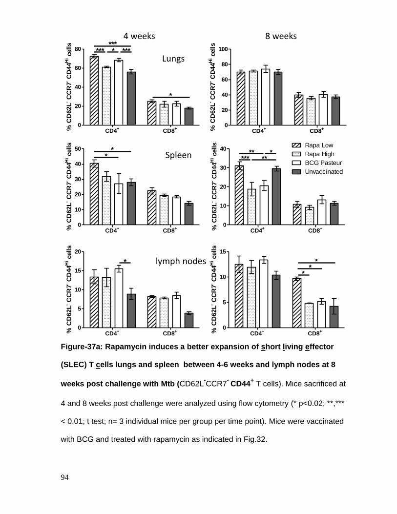
94
Figure-37a: Rapamycin induces a better expansion of short living effector
(SLEC) T cells lungs and spleen between 4-6 weeks and lymph nodes at 8
weeks post challenge with Mtb (CD62L-CCR7
- CD44
+ T cells). Mice sacrificed at
4 and 8 weeks post challenge were analyzed using flow cytometry (* p<0.02; **,***
< 0.01; t test; n= 3 individual mice per group per time point). Mice were vaccinated
with BCG and treated with rapamycin as indicated in Fig.32.
CD4+ CD8+0
20
40
60
80
100Rapa Low
Rapa High
BCG Pasteur
Unvaccinated
% C
D62L
- CC
R7
- CD
44
Hi c
ell
s
CD4+ CD8+0
10
20
30
40Rapa Low
Rapa High
BCG Pasteur
Unvaccinated
*****
***
% C
D62L
- CC
R7
- CD
44
Hi c
ell
s
CD4+ CD8+0
5
10
15
Rapa High
BCG Pasteur
Rapa Low
Unvaccinated
**
*
% C
D62L
- CC
R7
- CD
44
Hi c
ell
s
CD4+
CD8+
0
10
20
30
40Rapa Low
Rapa High
BCG Pasteur
Unvaccinated
*%
CD
62L
- CC
R7
- CD
127
+ c
ell
s
CD4+ CD8+0
20
40
60
80Rapa Low
Rapa High
BCG Pasteur
Unvaccinated
******
*
* ***
% C
D62L
- CC
R7
- CD
44
Hi c
ells
CD4+ CD8+0
10
20
30
40
50Rapa Low
Rapa High
BCG Pasteur
Unvaccinated
**
% C
D62L
- CC
R7
- CD
44
Hi c
ell
s
CD4+ CD8+0
5
10
15
20
Rapa High
BCG Pasteur
Unvaccinated
Rapa Low*
% C
D62L
- CC
R7
- CD
44
Hi c
ell
s
Lungs
Spleen
lymph nodes
4 weeks 8 weeks
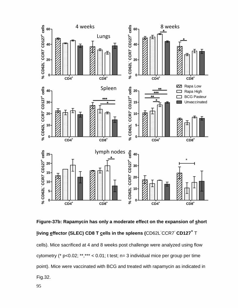
95
Figure-37b: Rapamycin has only a moderate effect on the expansion of short
living effector (SLEC) CD8 T cells in the spleens (CD62L-CCR7
- CD127
+ T
cells). Mice sacrificed at 4 and 8 weeks post challenge were analyzed using flow
cytometry (* p<0.02; **,*** < 0.01; t test; n= 3 individual mice per group per time
point). Mice were vaccinated with BCG and treated with rapamycin as indicated in
Fig.32.
CD4+
CD8+
0
20
40
60Rapa Low
Rapa High
BCG Pasteur
Unvaccinated
*
*
% C
D62L
- CC
R7
- CD
127
+ c
ell
s
CD4+
CD8+
0
5
10
15
20Rapa Low
Rapa High
BCG Pasteur
Unvaccinated
*****
*
**
% C
D62L
- CC
R7
- CD
127
+ c
ell
s
CD4+
CD8+
0
10
20
30
40Rapa Low
Rapa High
BCG Pasteur
Unvaccinated
*
% C
D62L
- CC
R7
- CD
127
+ c
ell
sCD4
+CD8
+0
10
20
30
40Rapa Low
Rapa High
BCG Pasteur
Unvaccinated
*%
CD
62L
- CC
R7
- CD
127
+ c
ell
sCD4+
CD8+
0
20
40
60Rapa Low
Rapa High
BCG Pasteur
Unvaccinated
% C
D62L
- CC
R7
- CD
127
+ c
ell
s
CD4+
CD8+
0
10
20
30
40Rapa Low
Rapa High
BCG Pasteur
Unvaccinated
****
% C
D62L
- CC
R7
- CD
127
+ c
ell
s
CD4+
CD8+
0
5
10
15
20
25Rapa Low
Rapa High
BCG Pasteur
Unvaccinated
*
% C
D62L
- CC
R7
- CD
127
+ c
ell
s
Lungs
Spleen
lymph nodes
4 weeks 8 weeks
96
Effect of Rapamycin on antigen specific memory T cells. CD8+ SLECs and
CD8+ MPECs possibly contained T cells specific for different antigens. In order to
determine if a distinct antigen specificity is associated with a memory phenotype,
CD8 T cells of the organs were analysed in mice after infection was cleared using
INH drug treatment (Fig.38, inset). T cells of mice harvested at the 12th week (1
week post drug treatment) were stained using TB10.4 tetramers and either CD62L
or CD44. It has been known from previous studies that CD62L+ or CD44
+ T cells
represent central memory phenotype. Fig. 38 shows that rapamycin treatment
significantly enhanced the numbers of antigen specific, central memory T cells
specific for TB10.4, particularly in the spleens and lymph nodes.
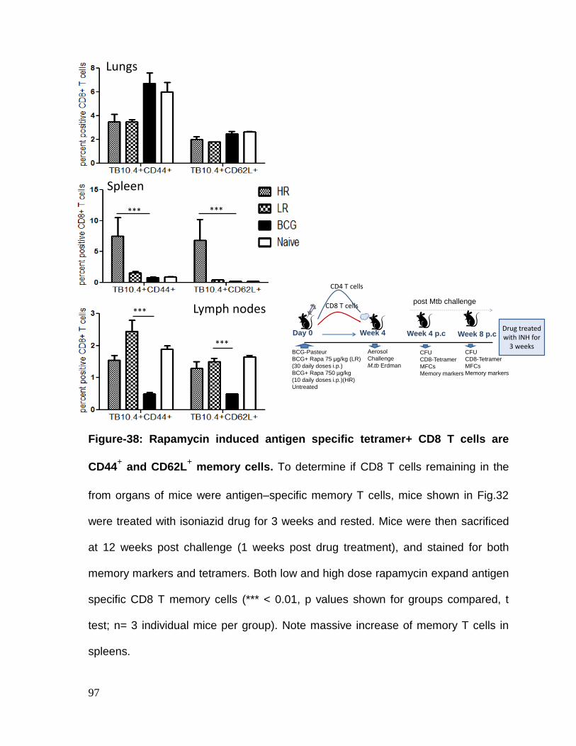
97
Figure-38: Rapamycin induced antigen specific tetramer+ CD8 T cells are
CD44+ and CD62L
+ memory cells. To determine if CD8 T cells remaining in the
from organs of mice were antigen–specific memory T cells, mice shown in Fig.32
were treated with isoniazid drug for 3 weeks and rested. Mice were then sacrificed
at 12 weeks post challenge (1 weeks post drug treatment), and stained for both
memory markers and tetramers. Both low and high dose rapamycin expand antigen
specific CD8 T memory cells (*** < 0.01, p values shown for groups compared, t
test; n= 3 individual mice per group). Note massive increase of memory T cells in
spleens.
***
***
***
***
Lungs
Spleen
Lymph nodes
BCG-Pasteur
BCG+ Rapa 75 µg/kg (LR)
(30 daily doses i.p.)
BCG+ Rapa 750 µg/kg
(10 daily doses i.p.)(HR)
Untreated
Day 0 Week 4
Aerosol
Challenge
M.tb Erdman
Week 4 p.c
CFU
CD8-Tetramer
MFCs
Memory markers
Week 8 p.c
CFU
CD8-Tetramer
MFCs
Memory markers
post Mtb challenge
CD4 T cells
CD8 T cells
Drug treated with INH for
3 weeks
98
Discussion
Since BCG does not induce protection against adult tuberculosis, it has been
hypothesized that BCG lacks the ability to induce central memory T cells. The
central memory T cells are by definition CD8 or CD4 positive T cells that survive for
prolonged periods and undergo recall expansion into effectors when rechallenge
with infection occurs. Mechanisms of effector to memory switch are however
debated.
Obar and Lefrancois proposed two concepts on the fate of effector and memory
cells. In the ‘Transition hypothesis’ effector T cells or TEM are induced by a vaccine
or infection and need to transition into memory T cells or TCM, under the regulatory
influence of distinct cytokines, including but not limited to IFN-γ, IL-2, TNF-α, IL-15
and IL-7 (63). In the ‘Induction of central memory’ hypothesis, memory T cells- TCM
(=MPECs) are sufficiently primed at the initial administration of vaccine, and are
present in low numbers. They are present at higher numbers only due to prolonged
priming from APCs to CD8 T cells (72).
Since LLO expressing BCG leaks more antigens into cytosol and enhances CD8 T
cell response in mice, it now appears that the short term protection provided by
BCG vaccine is likely due to insufficient stimulation of CD8 T cells (41). BCG thus
induces low levels of central memory T cells in mice (45).
99
To determine if BCG-induced levels of MPECs can be enhanced and such MPECs
are functional, in this study, we used the mTOR suppressive action of rapamycin.
The goal was to determine if, the phenotype of BCG induced CD8 T cells can be
skewed from effector to memory phenotype using well defined surface markers
including tetramers. We found that rapamycin co-treatment after BCG vaccination in
mice indeed enhanced the levels of MPECs (Fig.36-37). This confirmed that BCG
by itself induced low levels of MPECs but their numbers could be increased using
mTOR inhibition. Most significantly, after clearance of infection, only rapamycin
treated BCG vaccinated mice had elevated levels of central memory MPECS in
lymphoid organs (Fig.38). Interestingly, rapamycin also affected CD4 T cells both in
the MPECs and SLEC compartment, although the changes were distinct in organ
compartments (Fig.36-37). Since it is well known that rapamycin does not affect
CD4 T cells positively, we offer a novel explanation that rapamycin enhances the
function of BCG and Mtb infected APCs in mice, which in turn enhance CD4 T cell
numbers and quality.
We therefore conclude that BCG induces low levels of central memory T cells
perhaps because of a defect in host-vaccine interaction or absence of key cytokines
but immune-modulation can enhance vaccine induced central memory responses
(98). We found that rapamycin induced both MPECS and SLECs in response to
vaccination by BCG and this had a markedly beneficial action on the clearance of
pathogenic Mtb in mice. We propose that rapamycin can enhance long term
immunity engendered by BCG vaccine.
100
CHAPTER-3.4
EVALUATION OF THE PROTECTIVE FUNCTION OF
MEMORY PRECURSOR EFFECTOR CD8 T CELLS
(MPECs) INDUCED BY RAPAMYCIN AFTER
RECHALLENGE OF BCG VACCINATED MICE
101
Background
In most developing and some developed countries, primary BCG vaccination is
practiced for children soon after birth. Protection from BCG vaccination varies in
different geographic regions. Nearly in all regions though, protection wanes by the
age of 10-15 years and adults are susceptible to tuberculosis. Interestingly,
tuberculosis infection does not lead to significant cross protection or ‘infection-
induced’ immunity. This has been a problem for the natural control of the spread of
tuberculosis. The take home lesson is that BCG does not protect against adult
tuberculosis most probably due to the loss of long living memory T cells (central
memory) that can persist in the human body.
To determine the importance of persisting memory T cells, however, there is a need
for appropriate animal models (51). In previous studies along this direction, mice
have been vaccinated with BCG or infected with wild type Mtb and memory cells
have been examined over prolonged periods (45). In other studies, mice were
infected with wild type Mb and re-infected to study persistence of memory T cells
(55). Neither of these studies recapitulate the human situation, where BCG
vaccination is followed by infection with wild type Mtb and then, T cells shift in their
phenotype and function or lost. Since Vaccination induced central memory T cells
undergo contraction and are present in small numbers for prolonged periods, their
functional significance can only be ascertained by the recall expansion into effector
T cells. In the previous chapter, we demonstrated that rapamycin cotreatment with
BCG enhanced the numbers of central memory T cells. In this study, we examined
102
the hypothesis that rapamycin-induced central memory T cells are fully functional in
mice and protect against tuberculosis after rechallenge.
Methods:
Evaluation of protection against rechallenge tuberculosis in mice: In our previous
chapter, we demonstrated marked expansion of both SLECs and MPECs (Fig.32-
38). To determine whether the TB10.4 specific MPECs are the cells that persist and
undergo recall expansion, mouse vaccine experiments were performed as before.
However, at 8 weeks (Fig.39), mice were treated with the isoniazid (INH) drug for 3
weeks. INH is lethal for both Mtb and BCG and clears the vaccine and Mtb from the
organs reducing the load of bacteria and removing priming stimuli from APCs. Mice
were rested and on week 12, challenged again with wild type Mtb. Re-infection or
protection against re-infection was allowed to take place for the next four weeks and
mice were sacrificed. Mouse organs were then plated for CFU counts of Mtb and T
cells were phenotyped for effector T cell function (perforin and granzyme positive)
using TB10.4 tetramer as a marker. The recall expanding T cells were therefore
TB10.4 antigen specific T cells.
103
Results:
Prior rapamycin treatment of BCG vaccinated mice enables a better
protection against tuberculosis in the mouse rechallenge model. Mice were
vaccinated with BCG and treated with rapamycin as in Fig.39. Rechallenged mice
were sacrificed on week 16 and CFU counts of organs determined. Fig.40
demonstrates that both low and high dose rapamycin given after BCG vaccination
were able to enhance the ability of mice to reduce the bacterial burden of lungs and
spleens better than those given with BCG vaccine alone. BCG alone was not
protective against rechallenge in spleen and induced only a modest protection in
lungs.
Effect of Rapamycin on the recall expansion of functional CD8 T cells. In the
previous chapter we found that after drug induced bacterial clearance in BCG-
rapamycin treated mice, significant numbers of TB10.4+ MPECs persisted in the
spleens and lymph nodes (Fig.38). To determine if the protection generated during
rechallenge among such mice was due to MPECs (Fig.40), the T cells of organs
were stained for TB10.4 tetramer/IFN-γ and TB10.4 tetramer/perforin/granzyme and
analysed using flow cytometry. Fig.41 shows that rapamycin treatment enhanced
the numbers of TB10.4/IFN-γ+ T cells in the lungs. Furthermore, the proportion of
TB10.4+ CD8 T cells expressing perforin and granzyme-B were also high in lungs
(Fig.42). Finally, the numbers of functional CD8 T cells correlated well with those
expressing both T-bet and Eomes (Fig.42). Thus, rapamycin strongly enhanced the
recall expansion of functional CD8 T cells in the target lung organ which was
104
exposed to reinfection. Rapamycin had less significant effects on the recall
expansion of CD8 T cells in the spleens and lymph nodes (Fig.43-44, 45), despite
reducing the numbers of Mtb in the spleens. Since lymphoid organs harbour resting
MPECs, it is possible that after Mtb rechallenge, these cells underwent recall
expansion and homed more rapidly to the target lung organ, accounting for their
lesser numbers in spleens.
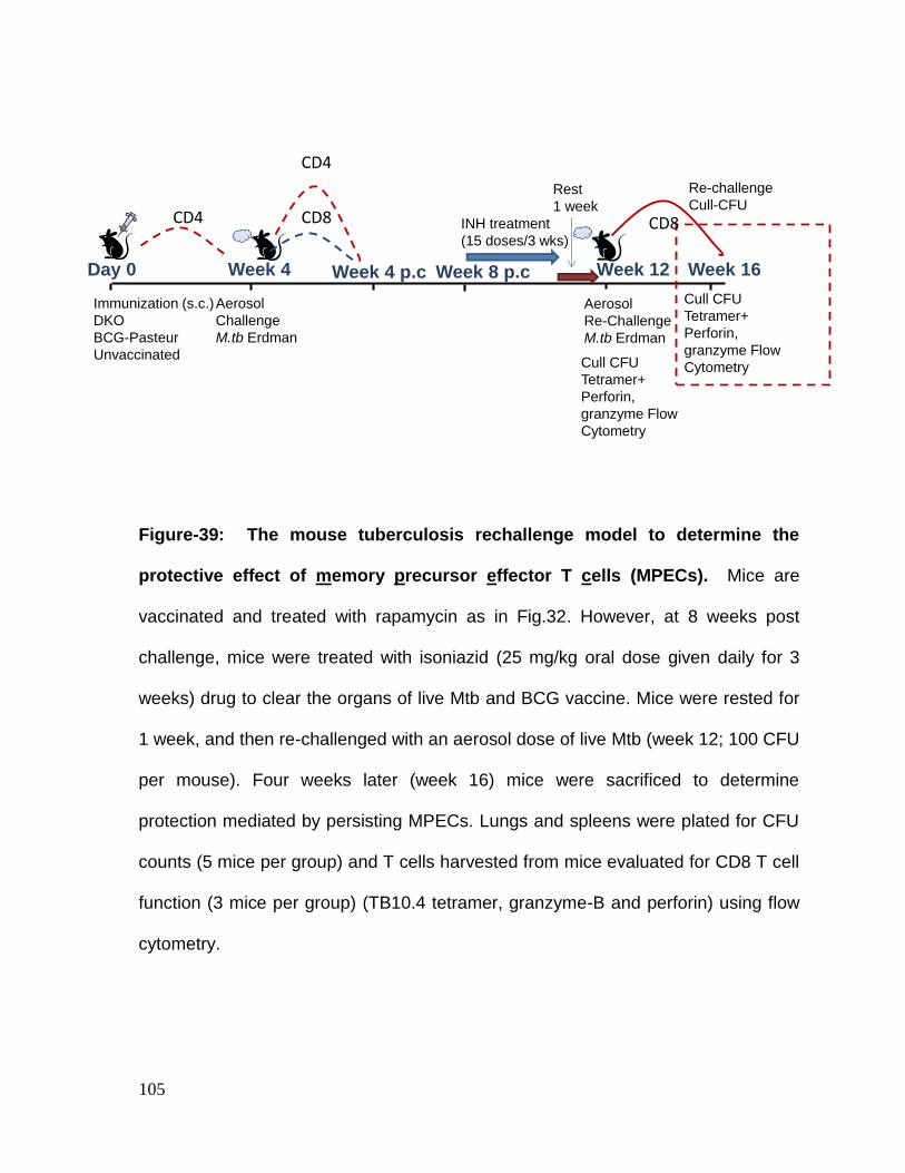
105
Figure-39: The mouse tuberculosis rechallenge model to determine the
protective effect of memory precursor effector T cells (MPECs). Mice are
vaccinated and treated with rapamycin as in Fig.32. However, at 8 weeks post
challenge, mice were treated with isoniazid (25 mg/kg oral dose given daily for 3
weeks) drug to clear the organs of live Mtb and BCG vaccine. Mice were rested for
1 week, and then re-challenged with an aerosol dose of live Mtb (week 12; 100 CFU
per mouse). Four weeks later (week 16) mice were sacrificed to determine
protection mediated by persisting MPECs. Lungs and spleens were plated for CFU
counts (5 mice per group) and T cells harvested from mice evaluated for CD8 T cell
function (3 mice per group) (TB10.4 tetramer, granzyme-B and perforin) using flow
cytometry.
Immunization (s.c.)
DKO
BCG-Pasteur
Unvaccinated
Day 0 Week 4
Aerosol
Challenge
M.tb Erdman
Week 4 p.c
Aerosol
Re-Challenge
M.tb Erdman
Week 12
INH treatment
(15 doses/3 wks)
Rest
1 week
Week 16
Re-challenge
Cull-CFUCD4 CD8
CD4
CD8
Week 8 p.c
Cull CFU
Tetramer+
Perforin,
granzyme Flow
CytometryCull CFU
Tetramer+
Perforin,
granzyme Flow
Cytometry
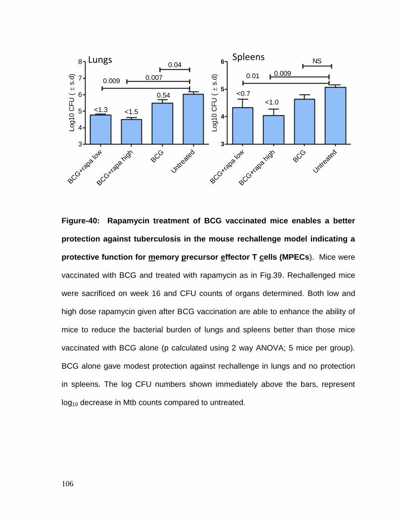
106
Figure-40: Rapamycin treatment of BCG vaccinated mice enables a better
protection against tuberculosis in the mouse rechallenge model indicating a
protective function for memory precursor effector T cells (MPECs). Mice were
vaccinated with BCG and treated with rapamycin as in Fig.39. Rechallenged mice
were sacrificed on week 16 and CFU counts of organs determined. Both low and
high dose rapamycin given after BCG vaccination are able to enhance the ability of
mice to reduce the bacterial burden of lungs and spleens better than those mice
vaccinated with BCG alone (p calculated using 2 way ANOVA; 5 mice per group).
BCG alone gave modest protection against rechallenge in lungs and no protection
in spleens. The log CFU numbers shown immediately above the bars, represent
log10 decrease in Mtb counts compared to untreated.
BCG+r
apa
low
BCG+r
apa
high
BCG
Unt
reat
ed
3
4
5
6
0.0090.01
NS
<0.7
<1.0
Log10 C
FU
( s
.d)
BCG+r
apa
low
BCG+r
apa
high
BCG
Unt
reat
ed
3
4
5
6
7
8
0.0070.009
<1.5<1.3
0.54
0.04Log10 C
FU
( s
.d)
Lungs Spleens
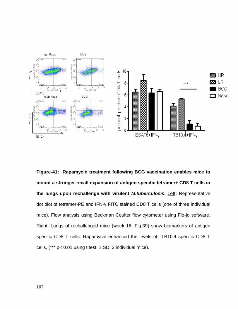
107
***
Figure-41: Rapamycin treatment following BCG vaccination enables mice to
mount a stronger recall expansion of antigen specific tetramer+ CD8 T cells in
the lungs upon rechallenge with virulent M.tuberculosis. Left: Representative
dot plot of tetramer-PE and IFN-γ FITC stained CD8 T cells (one of three individual
mice). Flow analysis using Beckman Coulter flow cytometer using Flo-jo software.
Right: Lungs of rechallenged mice (week 16, Fig.39) show biomarkers of antigen
specific CD8 T cells. Rapamycin enhanced the levels of TB10.4 specific CD8 T
cells, (*** p< 0.01 using t test; ± SD, 3 individual mice).
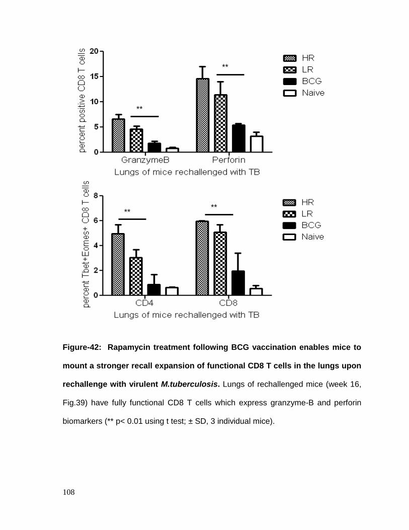
108
Figure-42: Rapamycin treatment following BCG vaccination enables mice to
mount a stronger recall expansion of functional CD8 T cells in the lungs upon
rechallenge with virulent M.tuberculosis. Lungs of rechallenged mice (week 16,
Fig.39) have fully functional CD8 T cells which express granzyme-B and perforin
biomarkers (** p< 0.01 using t test; ± SD, 3 individual mice).
***
***
***
***
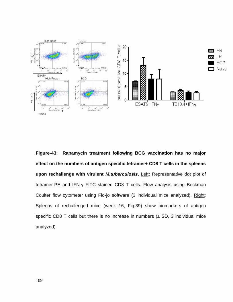
109
Figure-43: Rapamycin treatment following BCG vaccination has no major
effect on the numbers of antigen specific tetramer+ CD8 T cells in the spleens
upon rechallenge with virulent M.tuberculosis. Left: Representative dot plot of
tetramer-PE and IFN-γ FITC stained CD8 T cells. Flow analysis using Beckman
Coulter flow cytometer using Flo-jo software (3 individual mice analyzed). Right:
Spleens of rechallenged mice (week 16, Fig.39) show biomarkers of antigen
specific CD8 T cells but there is no increase in numbers (± SD, 3 individual mice
analyzed).
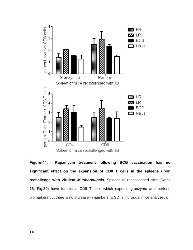
110
Figure-44: Rapamycin treatment following BCG vaccination has no
significant effect on the expansion of CD8 T cells in the spleens upon
rechallenge with virulent M.tuberculosis. Spleens of rechallenged mice (week
16, Fig.39) have functional CD8 T cells which express granzyme and perforin
biomarkers but there is no increase in numbers (± SD, 3 individual mice analyzed).
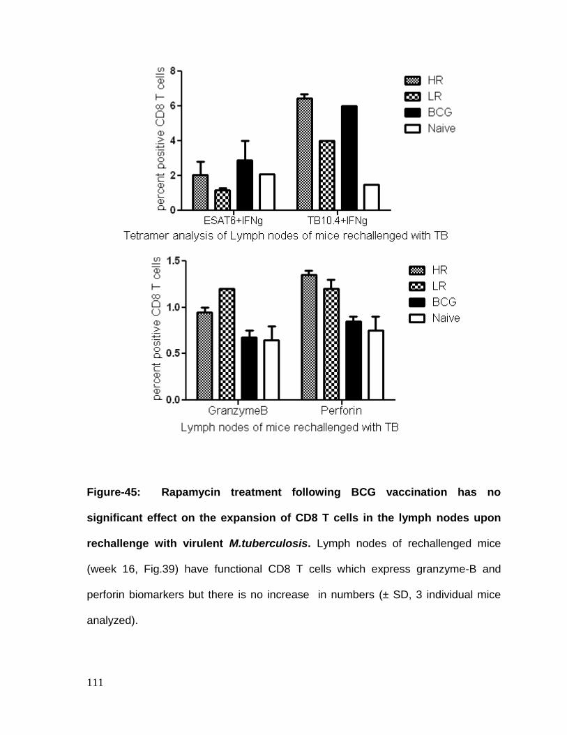
111
Figure-45: Rapamycin treatment following BCG vaccination has no
significant effect on the expansion of CD8 T cells in the lymph nodes upon
rechallenge with virulent M.tuberculosis. Lymph nodes of rechallenged mice
(week 16, Fig.39) have functional CD8 T cells which express granzyme-B and
perforin biomarkers but there is no increase in numbers (± SD, 3 individual mice
analyzed).
112
Discussion
Two problems are evident in the control of human tuberculosis. First, primary
childhood vaccination with BCG vaccine does not protect against adult tuberculosis
occurring after 10 years of age. Second, tuberculosis does not generate a robust
infection-induced immunity in adults (74). Thus, BCG induced long living memory T
cells are not robust. Not surprisingly, booster vaccination with BCG is also not
effective. This means that the central memory pool (MPECs) in adults is so small
that it cannot be rejuvenated with booster BCG vaccine or through natural infection
with tuberculosis. Thus, the first goal of our study was to determine if rapamycin in
combination with BCG vaccine could be used to create a pool of MPECs which was
different in quality. In other words, we proposed that the memory pool induced by
BCG vaccine alone would be different than the one created through rapamycin-
induced mTOR modulation during BCG vaccination.
Results of this study confirm that rapamycin induced MPECs were much better than
BCG induced MPECs in protecting against rechallenge of tuberculosis in mice
(Fig.40-44). As expected, BCG vaccine alone generated no protection against
tuberculosis of the spleens and borderline protection against tuberculosis of the
lungs. The protection data were interesting also because the recall population
consisted of TB10.4 specific CD8 T cells expressing markers of CTL activity like
perforin and granzyme. Our observations are novel because there are no previous
reports which show CD8 T cell mediated protection of lungs in mice against
rechallenge tuberculosis. It is thus clear that rapamycin treatment during BCG
vaccination affects the quality and quantity of MPECs that can protect long term.
113
CHAPTER-3.5
IN VITRO STUDIES ON THE EFFECT OF RAPAMYCIN ON
DENDRITIC CELL- T CELL COCULTURES
114
Background
Previous chapters show the dramatic result that rapamycin treatment induces a
remarkable shift in the quality and quantity of antigen specific CD8 T cells which
mediate protection against tuberculosis. In these studies, a complex mouse model
was used to analyze the emergence of various subsets of T cells. Since rapamycin
can affect both the APCs and T cells, it is possible that the mouse permits multiple
interactions to take place between rapamycin, APCs and T cells. We therefore
sought to determine if DCs treated with rapamycin in vitro could directly affect the
signaling within T cells and allow them to change their phenotype. This would help
to differentiate between the dual effects of rapamycin.
Various types of in vitro antigen presentation models are available to investigate the
mechanism of T cell activation (21). We illustrated the use of APCs overlaid with
BB7 T cells as models to investigate the MHC-II dependent presentation of peptides
to CD4 T cells (Chapter 2). Typically, macrophages or DC’s are pulsed with soluble
or bacterial antigens, which are processed by the DCs and epitopes presented in
the context of either MHC-II or MHC-I on the plasma membrane (56). The DCs are
then overlaid with either CD4 or CD8 T cell lines, which specifically recognize the
peptide epitope (96). Overlaid T cells then respond by either secreting cytokines or
another biomarker that indicates antigen recognition or binding [103]. In this study,
we tested the ability of DCs to present a peptide to CD8 T cells and analyzed the
expression of transcription factors that could affect the lineage and CTL function of
the T cells.
115
Methods
A DC cell line DC2.4, obtained from bone marrow derived DCs of C57Bl/6 mice was
used as the antigen processing cell in the in vitro assay. DCs were first activated
with rapamycin overnight or left untreated. They were infected with a recombinant
BCG strain which expressed an ovalbumin (Ova) derived peptide (H2Kb-257-264
peptide, SIINFEKL) and washed. DCs were overlaid with B3Z, a CD8 T cell
hybridoma that is specific for Ova. B3Z CD8 specific T cells were gently scraped off
after overnight incubation and flow cytometry analysis was performed to determine
levels of T-bet, Eomes, perforin and granzyme-B using antibodies and using Flow-
Jo software. It is noted here that, DCs were used as APCs although macrophages
can also present antigen to T cells.
116
Results
DCs activated with rapamycin and infected with BCG were found to enhance the
expression of T-bet and Eomes in CD8 T cells incubated overnight (Fig.46).
Likewise, the coculture enhanced the expression of both perforin and granzyme in
CD8 T cells (Fig.47). When a CD4 T cell line BB7 was overlaid on the same DCs,
there was no significant change in the expression of T-bet and Eomes (Fig.48).
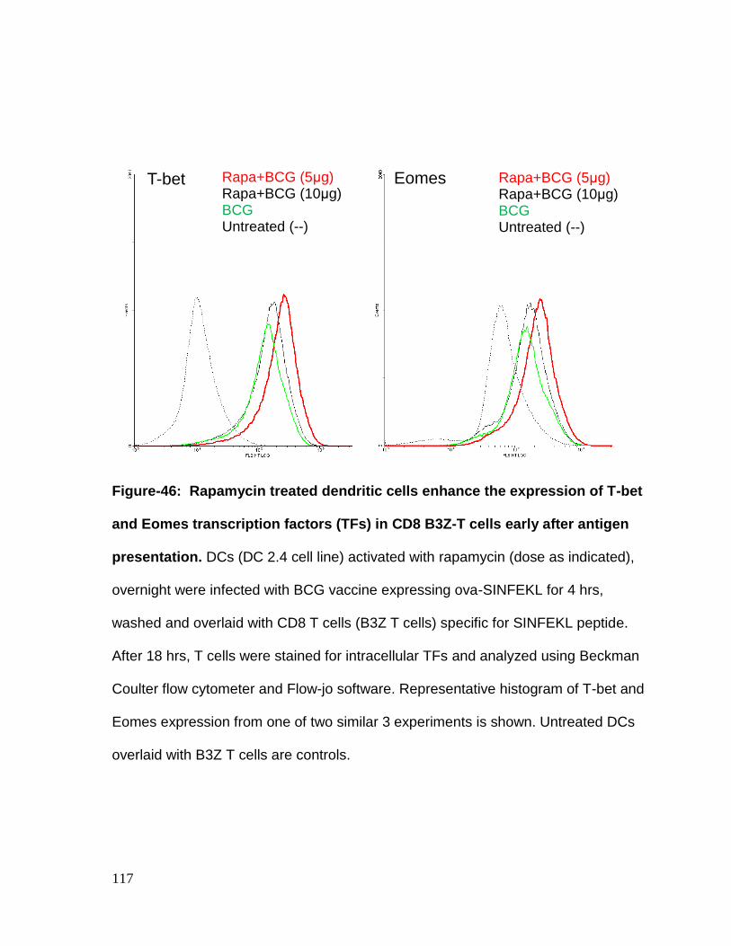
117
Rapa+BCG (5μg) Rapa+BCG (10μg) BCG Untreated (--)
Rapa+BCG (5μg) Rapa+BCG (10μg) BCG Untreated (--)
T-bet Eomes
Figure-46: Rapamycin treated dendritic cells enhance the expression of T-bet
and Eomes transcription factors (TFs) in CD8 B3Z-T cells early after antigen
presentation. DCs (DC 2.4 cell line) activated with rapamycin (dose as indicated),
overnight were infected with BCG vaccine expressing ova-SINFEKL for 4 hrs,
washed and overlaid with CD8 T cells (B3Z T cells) specific for SINFEKL peptide.
After 18 hrs, T cells were stained for intracellular TFs and analyzed using Beckman
Coulter flow cytometer and Flow-jo software. Representative histogram of T-bet and
Eomes expression from one of two similar 3 experiments is shown. Untreated DCs
overlaid with B3Z T cells are controls.
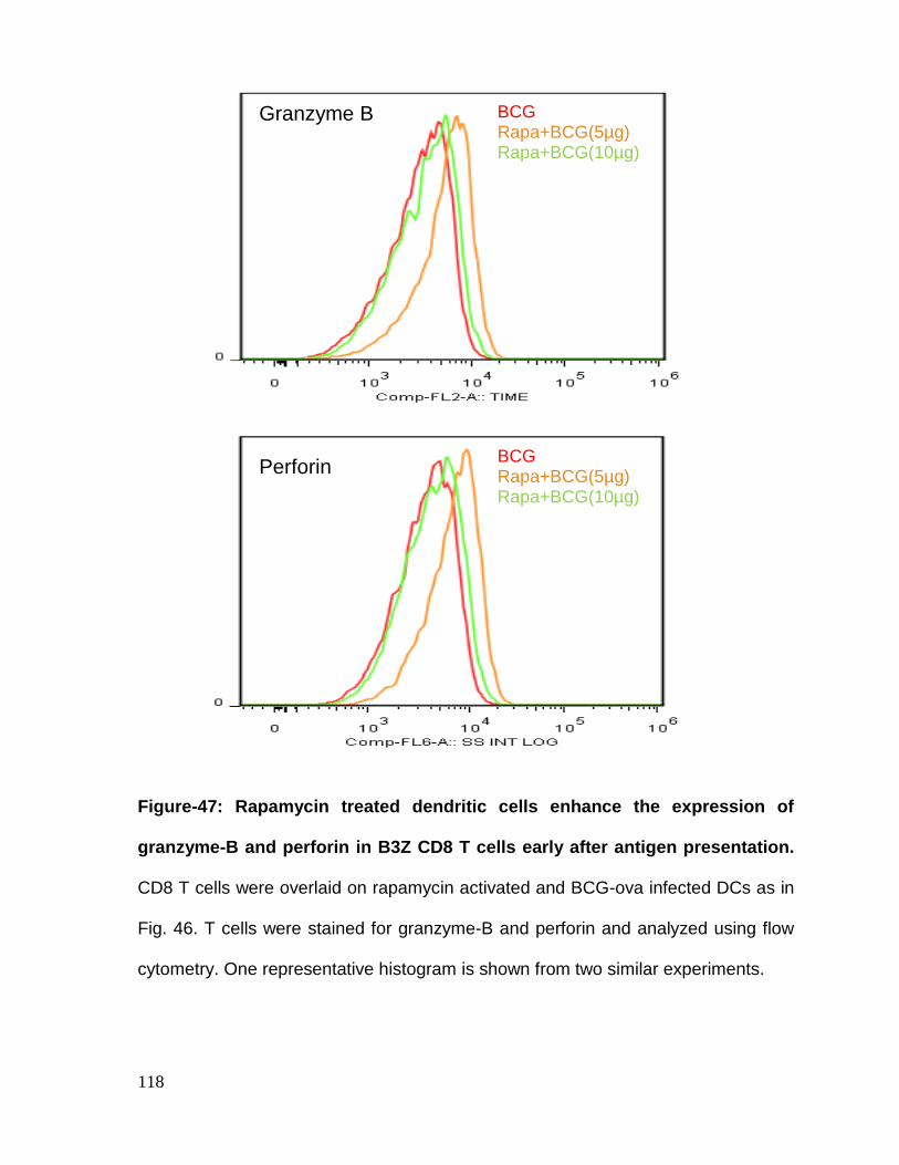
118
BCG
Rapa+BCG(5µg) Rapa+BCG(10µg)
BCG
Rapa+BCG(5µg) Rapa+BCG(10µg)
Perforin
Granzyme B
Figure-47: Rapamycin treated dendritic cells enhance the expression of
granzyme-B and perforin in B3Z CD8 T cells early after antigen presentation.
CD8 T cells were overlaid on rapamycin activated and BCG-ova infected DCs as in
Fig. 46. T cells were stained for granzyme-B and perforin and analyzed using flow
cytometry. One representative histogram is shown from two similar experiments.
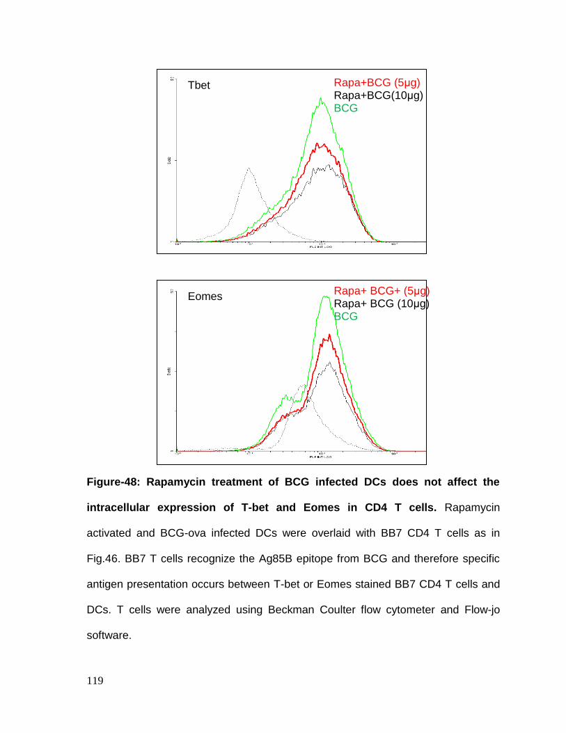
119
Eomes
Tbet
Rapa+ BCG+ (5μg) Rapa+ BCG (10μg) BCG
Rapa+BCG (5μg) Rapa+BCG(10μg) BCG
Figure-48: Rapamycin treatment of BCG infected DCs does not affect the
intracellular expression of T-bet and Eomes in CD4 T cells. Rapamycin
activated and BCG-ova infected DCs were overlaid with BB7 CD4 T cells as in
Fig.46. BB7 T cells recognize the Ag85B epitope from BCG and therefore specific
antigen presentation occurs between T-bet or Eomes stained BB7 CD4 T cells and
DCs. T cells were analyzed using Beckman Coulter flow cytometer and Flow-jo
software.
120
Discussion
In a previous study, we found that rapamycin activated DCs of mice enhance the
intracellular processing of BCG vaccine and allow enhanced presentation of
peptides to CD4 T cells in vitro (49).This has been found to be due to the autophagy
inducing effect of rapamycin, where BCG is sorted to into autophago-lysosomes
resulting in enhanced peptide production. Although, these CD4 T cells recognize
peptide in the context of MHC-II, it remained unclear if the process activated other
intracellular events in T cells.
Recent research has shown that when DCs cross talk to T cells via TCR, a number
of events are triggered within T cells that lead to their differentiation into Th1
immunity mediating effectors (43, 69). This process is of course dependent upon
both MHC-TCR ligation and costimulation. Interestingly, CD4 and CD8 T cells seem
to have several intracellular transcription factors that determine the eventual fate of
T cells as effectors and memory cells. Thus, T-bet regulates the differentiation of
Th1 T cells into effectors, while Eomes regulates the memory lineage of T cells. It
should also be noted that T-bet and Eomes can have subtly different effects on
subsets of T cells. Furthermore, T-bet also affects DCs (93). Coincident with the
activation of these TFs, T cells may independently activate programs to secrete
IFN-γ , IL-2 and TNF-α by CD4 T cells or enable perforin and granzyme synthesis
among CD8 T cells to mediate cytolytic function (19, 110). The major finding of this
study was that DCs activated with rapamycin induced a better expression of T-bet
and Eomes in CD8 T cells along with enhanced perforin/granzyme-B expression
121
upon antigen presentation. This did not occur during a similar treatment window in
vitro with CD4 T cells. Thus, rapamycin preferentially and rapidly activated TFs in
CD8 T cells which are consistent with its known positive effect on CD8 T cells in
vivo. It should be noted that neither the CD8 T cells nor CD4 T cells were exposed
to rapamycin directly in vitro in this study. However, when mice are given rapamycin
in vivo, it seems possible that rapamycin could affect T cells separately. This has
been shown in our in vivo studies where, rapamycin, in addition to affecting CD8 T
cells also enhanced the function of CD4 T cells. Furthermore, rapamycin was given
for longer periods in mice when it can have different effects and in vitro models do
not recapitulate this event. We therefore tentatively conclude that rapamycin treated
DCs have a rapid enhancing effect on the function of CD8 T cells through the
induction of T-bet or Eomes. Additional studies seem necessary to determine if
rapamycin treatment affects CD4 T cells when they are interacting with DCs.

122
CHAPTER-4
CONCLUSIONS
123
Tuberculosis is the leading cause of death due to infections in mankind with 2
million people dying of the disease. BCG is the only approved vaccine for humans
and is being used in many parts of the world. Despite being the most frequently
used vaccine, BCG generates partial immunity against childhood tuberculosis and
does not protect against adult tuberculosis. Humans above the age of 10-15 years
are not protected either by childhood vaccination or by infection with tuberculosis,
which is expected to generate infection-induced immunity. Therefore, it has been
proposed that the T cell responses induced by BCG are not strong and do not last
longer. Experimental infections with Mycobacterium tuberculosis (Mtb) and
vaccination with BCG are feasible using mice, which offer a model to evaluate
vaccine induced T cell responses. These studies indicate that BCG induces
moderately strong CD4 T cell responses in mice that are able to control tuberculosis
to a certain extent. However, certain inherent properties of BCG vaccine seem to
limit its potential as a vaccine. The overall goal of this proposal was therefore to
investigate why BCG fails to induce robust CD4 and CD8 T cell responses, and
identify newer strategies to strengthen the BCG vaccine.
Initial observations from our laboratory showed that antigen presenting cells (APCs)
infected with BCG vaccine do not adequately present soluble mycobacterial
antigen(s) to CD4 T cells via the MHC-II pathway (97). We therefore developed the
first hypothesis under Aim-I that, lipids of BCG vaccine may bind the surface Toll-
like receptors (TLRs) and inhibit expression of MHC-II and other cellular processes.
Using in vitro assays for antigen presentation with BCG infected macrophages; we
124
confirmed that the 19 kDa lipoprotein and lipo-arabinomannan lipids, which are
integral structures of cell wall in BCG, inhibited antigen presentation (Chapter 2).
We then demonstrated that, BCG infection of APCs reduced the expression of
MHC-II in macrophages. This observation explained why BCG vaccine was less
than optimally effective as a vaccine. In mice this defect is apparent as a modest
protection (decline in bacterial counts by 1-log10) against aerosol induced
tuberculosis. In humans, the protection induced by BCG vaccine is lost over the first
10 years of age. Investigation into the strategies to boost the efficacy of BCG
vaccine then led us to the mechanisms of activation in APCs.
APCs like macrophages and dendritic cells use multiple TLRs to recognize
microbial ligands. The latter are known to regulate cytokine expression by APCs
and alter the micro-environment in which APCs cross-talk to T cells. However,
TLRs also stimulate a wide variety of intracellular mechanisms in APCs and we
sought to clarify them in the context of MHC-II mediated activation of T cells. First,
we examined whether BCG induced down-regulation of MHC-II could be enhanced
by alternate activation of TLRs. We discovered that BCG down regulated MHC-II
through the up-regulation of the ubiquitinating enzyme MARCH1 and alternate
activation of many TLRs, particularly TLR-7/9 strongly enhanced MHC-II in
macrophages and DCs. Elsewhere, we reported that TLR-7/9 ligands are indeed
the best adjuvants to boost the efficacy of BCG vaccine against experimental
tuberculosis of mice (unpublished observations). Our studies were novel and
thought provoking. First, the efficacy of BCG vaccine has been increased using
125
genetic manipulations but to date it has not been possible to counteract the
suppressive effect of BCG vaccine for APCs. TLR-4 activating LPS derived MPL
has been used as adjuvant with subunit protein vaccines against tuberculosis. This
study shows that TLR-7/9 ligands are much more powerful than TLR-4 activating
LPS, and they show an unique ability to enhance MHC-II unlike the less effective
LPS. Since cpgDNA, a TLR-9 ligand has been approved for use as adjuvant against
certain cancers in humans; we anticipate that similar ligands of TLR-7/9 will be used
as adjuvants with BCG vaccine for humans.
Our investigations into the TLR mediated activation of APCs also identified several
avenues for future research. Since the major lipids of BCG vaccine suppressed
MHC-II, it seems worthwhile to evaluate lipid-deficient BCG vaccine mutants as
possible vaccine candidates. In support of this observation, the 19 kDa lipoprotein
knockout strain of BCG vaccine was reported more immunogenic but not tested as
a vaccine (44, 100). Second, TLR-ligands were found to use subtly different
activation pathways to trigger MHC-II via AP-1 and CREB before they bind the
CIITA. It is possible that genetically engineering a protein inducer of AP-1 or CREB
into BCG may yield a more immunogenic BCG vaccine. Finally, a combination of
TLR-adjuvants may boost the efficacy of BCG vaccine than a single adjuvant.
Overall, Aim-I studies of this proposal have unraveled newer novel TLR- dependent
mechanisms to improve BCG vaccine.
126
Since the Aim-I studies established a mechanism to render the CD4 T cell response
more robust, we next sought to prolong the ‘longevity of protection’ generated by
BCG vaccine.
While CD4 T cells protect against acute infection, CD8 T cells have been proposed
to be able to persist in the human body for years and undergo recall expansion to
protect against infection and re-infection. However, BCG induces a poor CD8 T cell
response in mice. Part of the problem is that the membrane structure of BCG
phagosome within APCs prevents leakage of antigens into cytosol for proteasome
processing prior to loading of peptides into MHC-I. One way to bypass this problem
is to enhance the sorting of BCG to lysosomes, where peptides are produced and
get routed to MHC-I. The second way is to enhance the secretion of antigens from
BCG into the cytosol and this requires additional genetic engineering of the vaccine.
Grode et al., cloned LLO into BCG improving the leakage of antigens from the
phagosome into cytosol and better induction of CD8 T cell responses in mice (41).
Our previous studies indicated that rapamycin, an immune-modulator enhanced the
sorting of BCG vaccine to lysosomes (49). Others reported that rapamycin
increased the longevity of CD8 T cells by repressing mTOR. Thus, for Aim-II
studies, we developed a new hypothesis that the dual effects of rapamycin may
enhance CD8 T cell response to BCG vaccine if the drug were to be given to mice.
We used three models of infection and vaccination followed by infection to
determine the effect of rapamycin on CD4 and CD8 T cells during tuberculosis.
127
Since APCs prime and expand antigen specific T cells, one measure of the efficacy
of APC mediated priming is the number of tetramer specific T cells. In an aerosol
infection model, rapamycin was able to stimulate the expansion of tetramer specific-
CD8 T cells but this correlated only with a limited control of tuberculosis. We next
examined the ability of rapamycin to enhance the efficacy of BCG vaccine by
administering low and high doses of rapamycin with BCG vaccine, and found
several significant effects. First, rapamycin dramatically enhanced the efficacy of
BCG vaccine to clear virulent Mtb from the organs of mice. Second, rapamycin
enhanced the magnitude of antigen specific CD8 T cells specific for TB10.4 antigen.
Third, rapamycin enhanced the magnitude of memory precursor effector T cells
(MPECs), thought to persist in mice for prolonged periods. Finally, we found that the
rapamycin-BCG induced functional MPECs persist in mice, and undergo robust
recall expansion into effector CD8 T cells expressing perforin and granzyme, and
protect against rechallenge of tuberculosis. Overall, these studies demonstrated
that rapamycin combination with BCG could enhance short term effector responses
as well as prolong long term memory.
The ability of rapamycin to enhance short term protection was obviously due to a
strong effector T cell response (CD62Llow population) although surprisingly,
rapamycin enhanced CD4 cell responses in addition to CD8 T cells. We suggest
that under in vivo conditions, rapamycin boosted the function of APCs to sort BCG
to lysosomes which enabled a better activation of CD4 T cells. It is well established
that rapamycin does not affect CD4 T cells positively at the doses we used in mice.
128
However, other than our own previous study (49), the long term effects of low dose
rapamycin has not been explored in the context of the ability to APCs to prime CD4
T cells. The dual enhancing effect of rapamycin is therefore a novel observation that
suggests that mTOR repression is likely to markedly boost the efficacy of BCG
vaccine. Since BCG is immunologically sequestered (49, 77), the use of mTOR
repressors like rapamycin in humans to enhance processing of BCG antigens and
ensure long lasting T cell responses remains an intriguing approach. Low doses
rapamycin are also not likely to deleteriously affect the immune system in humans.
The beneficial effects of rapamycin on CD8 T cells in this study was anticipated but
still brought out surprising observations. Soon after we described that rapamycin
enhances lysosome mediated processing of BCG enhancing its efficacy against
tuberculosis in mice, others reported that rapamycin enhanced the longevity of CD8
T cells in mice against viral infections.
There are two schools of thought on how long term memory develops against
pathogens (63, 71, 73). The ‘transition hypothesis’ suggests that effector T cells
switch to memory T cells through selective activation or deactivation of intracellular
signaling mechanisms including mTOR, Eomes and T-bet and perhaps others.
Under the ‘induction hypothesis’, memory T cells are always present at low
numbers following APC mediated priming of CD8 T cells, but their numbers become
robust after prolonged priming from APCs. Whether one or both mechanisms
operate in the development of long term memory has been thought to depend upon
129
the pathogen. Data from our studies (Chapter 3) suggest that during BCG
vaccination the beneficial effect of rapamycin likely involved both mechanisms.
First, BCG was not processed efficiently by naïve APCs, but presumably rapamycin
enhanced its processing. We propose that this led to enhanced activation and
prolonged contact with T cells during the 30 days treatment of mice with rapamycin
following BCG vaccination. In evidence of this observation, both CD4 and CD8 T
cells increased in numbers in this mouse model. Second, rapamycin induced a
robust tetramer specific CD8 T cell response which suggested that these antigen
specific CD8 T cells had a better chance to transition into memory T cells. In Fig.38
for example, after mice have been treated with drugs and infection was cleared,
there was a marked increase in the numbers of TB10.4 specific CD62L+ memory
CD8 T cells in the spleens.
The final intriguing discovery of this study was that TB10.4 CD8 T cells that
persisted in drug cured mice, underwent a strong recall expansion after rechallenge
of tuberculosis and protected against infection. Since BCG vaccine generally fails to
show strong protection against re-infection of tuberculosis, these studies strongly
argue that rapamycin treatment of BCG vaccine qualitatively alters memory
response enabling long term immunity. These data now suggest future studies
which are of translational value. To boost the long term efficacy of BCG vaccine we
speculate that infants could be given low dose rapamycin following BCG vaccine,
although there are likely to be impediments in administering rapamycin to infants.
The discovery of Eomes as a biomarker of long living CD8 T cells however
130
suggests that, cloning of an Eomes inducing cytokine like IL-15 into BCG can
prolong its ability to confer long term memory. We conclude that Aim-I and II
together have generated novel observations on boosting the ability of BCG vaccine
and the data are also likely to be helpful for developing vaccines for other
intracellular pathogens.
131
CHAPTER-5
MATERIALS AND METHODS
132
CHAPTER 2:
Macrophages: Primary bone marrow derived macrophages (MΦs) from C57Bl/6
mice ( 4-8 week sold M/F, Harlan or Jackson animal providers, USA) were grown in
Iscove’s medium with 10% FBS (IDM) and 10 ng/mL GM-CSF and CD11c beads
(Miltenyi INc, USA;130-052-001) were used to deplete DCsfrom bone marrow cells
cultured for 7 days. The CD11b+ CD11c- MΦs were plated in GM-CSF free medium
and used for activation and infection. MΦ-derived BMA.A1 cell line (kind gift of Dr.
Kenneth L. Rock, U. Massachusetts, USA), were similarly cultured in IDM and used
for many experiments. MΦs were activated with TLR-ligands (1-5 μg/mL; dissolved
in sterile endotoxin-free water, Invivogen, USA) and incubated overnight at 370C.
They were then infected with Mycobacterium bovis BCG (Pasteur strain,
ATCC35734; MOI =1), prepared as a single cell suspension, for 4 hr on a shaker at
370C, washed and collected for further analysis. Viability of MΦs was > 90% using
trypan blue.
In vitro antigen presentation assay: This has been described in detail earlier (97).
Briefly, MΦs were treated or not with either mycobacterial lipids (19 kDa lipoprotein
and lipoarabinomannan), inhibitors of TLR-signaling or activated with TLR-ligands,
followed by infection with BCG (MOI=1). Washed monolayers were overlaid with
BB7 T cell hybridoma (kind gift of Dr. Cliff Harding, CWRU) (1:20 ratio) and
supernatants collected either 4 hr or 18 hr after overlay and tested for IL-2 using
sandwich ELISA.
133
CD4 T cell hybridoma: The Ag85 epitope-specific (241-256) T cell hybridoma (BB7) a
gift from Dr. Clifford Harding (Case Western Reserve University, Cleveland, OH),
was maintained in DMEM medium (Sigma, USA) supplemented with 10% heat-
inactivated FBS (HyClone Laboratories), 50 µM 2-ME, 1 mM sodium pyruvate, 2
mM L-glutamine, 10 mM HEPES buffer, nonessential amino acids, 100 µg/ml
penicillin, and 100 µg/ml gentamicin.
Inhibition of TLR-signaling: Various inhibitors of MAPK and API/CREB were used
to block signaling due to TLRs. The inhibitors were from Calbiochem and used at
doses recommended by the manufacturer. MΦs were treated with inhibitors of
MAPKs and API/CREB for 2 hr, activated with TLR-ligands for 2 hr and infected with
BCG for 2 hr prior to fixation and assay of surface MHC-II. This rapid procedure was
adapted to ensure >90% viability of MΦs.
siRNA down-regulation of MARCH1: MΦs were treated with siRNA and
scrambled siRNA directed against MARCH1 (sc-106199; sense/antisense and
scrambled, SantaCruz Biotechnology, USA;) as recommended by the manufacturer
and rested for 14 hr. They were tested naïve, or infected with BCG for 4 hr followed
by surface expression of MHC-II evaluated using flow cytometry.
Immunofluorescent staining of cells: MΦs, BMA.A1 or DC.2.4 cell lines were
stained using antibodies against MHC-II, MHC-I, CD80, CD86. After incubation for
30 mins on ice with antibodies, cells were washed with PBS and fixed with
134
Cytofix/Cytoperm buffer (BD biosciences, #555028). Intracellular staining was using
antibodies for MARCH-1 and IL-10. Cells were incubated for 30 mins and washed
with PBS. MARCH1 assay did not require brefeldin-A as it is not secreted (MHC-
II,#12-5322; CD80,#12-0801; CD86, #12-0862;IL-10, #11-7101; MHC-I, #17-5985,
all from Ebiosciences USA) (MARCH1, #D16-sc104369; Santacruz Biotechnology,
USA).
Flow cytometry: The fixed cells were acquired and analyzed for surface or
intracellular markers using Gallios flow cytometer (Beckman Coulter) and the data
analysis was carried out using Flow-Jo software (Treestar, Ashland). The graphical
analysis was performed using the Prism Software.
Immunoprecipitation and Western Blot analysis of ubiquinated MHC-II: MΦs in
6 well plates were treated with TLR-ligands at a concentration of 1 μg/mL or none
overnight at 37°C. They were then infected with BCG for 4 hr (MOI=1:1) on a
shaker at 37°C. Cells were centrifuged for 10 min (3,000 x g, 4°C) and the
supernatant was removed. Cells were resuspended in 30 μL of the lysis buffer
(mammalian Protein Extraction Reagent (M-PER, Pierce) with 5 μM MG132, a
proteasome inhibitor, and a protease inhibitor cocktail (112 μM PMSF, 3 μM
aprotinin, 112 μM leupeptin, 17 μM pepstatin). Samples were incubated with
shaking at 4°C for 1 hr, centrifuged for 20 min (10,000 rpm 4°C) and nuclei free
supernatants were collected and estimated for protein and normalized. One μg of
anti-mouse MHC-II antibody (Ebioscience, #14-5321) was added per sample, which
135
were gently mixed overnight at 4°C.
Fifteen μL of Protein A/G PLUS-Agarose beads (Santacruz Biotechnology, USA)
were added to each sample and gently mixed for 4 hr at 4°C. Supernatants were
removed though centrifugation (2000 rpm, 15 min) and beads were washed three
times with 1x PBS. Sample buffer was added to each sample, and each tube was
boiled for 5 minutes to elute proteins from beads. Samples were loaded onto a 12%
SDS-PAGE gel and electroblotted onto a PVDF membrane. Western blotting was
performed using an antibody directed towards polyubiquitin (Ebioscience, #14-
6078).
Statistical analysis: Statistical analyses were done using GraphPad software. A p
value ≤0.05 was considered statistically significant for these analyses. Student’s t-
test and standard one-way ANOVA followed by Dunnett’s Multiple Comparison Test
were used to determine statistical significance. Experiments were repeated a
minimum of three times and in each, at least triplicate wells were used. Data are
presented using the mean ± SD of results for 1 or 3 similar experiments or are
averaged from 2-3 experiments as indicated.
CHAPTER 3:
BCG & Rapamycin: M. bovis BCG (ATCC-35734) was grown in Dubos broth and
log phase organisms were washed thrice with PBS, sonicated CFU counts
determined by plating before infections. Rapamycin was purchased from Santacruz

136
Biotechnologies (USA). It was dissolved at a final concentration of 1 mg/ml in sterile
PBS (phosphate buffer saline).
Mouse immunization, challenge and lung CFU determination: Female C57BL/6
mice of age 8–12 wk were used for most experiments. C57BL/6 were purchased
from Jackson ImmunoResearch Laboratories. They were housed within a BSL-3
vivarium and fed food and water and chow. For most experiments, mice were
immunized with 75 µg/kg of rapamycin (low dose) and 750 µg/kg (high dose) of
mice intraperitoneally, with or without BCG administered subcutaneously. For most
experiments, mice were immunized once. After 4 weeks of vaccination, mice were
infected with a log phase, highly viable single-cell suspension of M. tuberculosis
Erdman strain via aerosol using a Glas-Col aerosol apparatus, calibrated to deliver
100–200 CFU per mouse lungs over 30 min of nebulization. For Rechallenge
experiments; after 8 weeks post challenge, mice were treated with the isoniazid
(INH) drug for 3 weeks and were rested for a week followed by a rechallenge on
week 12 with Mtb via aerosol using a Glas-Col aerosol apparatus, calibrated to
deliver 100–200 CFU per mouse lungs over 30 min of nebulization. Protocols for the
animal experiments described in this study were approved by Institutional Animal
Care and Use Committee. The amount of bacteria delivered into the lung was
confirmed 24 h after exposure by plating neat lung homogenates on 7H11 agar from
three mice.

137
Colony forming units (CFU) determination: Protection was determined post-
challenge and post rechallenge by harvesting the lungs and spleens from the
infected mice, homogenizing the tissues in 0.05% PBS–Tween 80, and plating ten-
fold serial dilutions on 7H11 agar plates for bacterial growth. Bacterial colonies were
counted 4 weeks after incubation at 370C. Values ranging between 10 and 100
colonies per plate per dilution were used to calculate the bacterial burden per organ
and were expressed as: mean log10 CFU saline/untreated - Mean log10 CFU
vaccine.
P value for CFU counts vaccine vs. untreated control, 5 mice per time
point:Mice were left untreated (5 per group) or vaccinated with BCG followed 4 wks
later with aerosol challenge with
M.tuberculosis. Mice were sacrificed and
colony counts of M. tuberculosis measured
in the lungs. Data are plotted as log10 CFU
per organ per mouse. The example to the
right shows that such early CFU data is
highly discriminative and predictive of
survival, and importantly the statistical power of these data is superior to survival
data. Allowing for a statistical power of 0.8–0.9, and a usual variance of 0.2–0.3
log10 CFU, a reduction in the mean CFU values between saline controls and test
groups of about 0.7 log10CFU is usually significant, when n= 5 animals are used (2-
way ANOVA used for p values).
Aeverag e distribution of colony counts in saline control and BCG vaccine
4
5
6
7
Animakl groupos
Lo
g C
FU
per
org
an
s
138
In vitro Antigen presentation assay using CD8 T cells (Antigen processing).
DC2.4-C57BL/6 specific cell line was grown in Dulbecco's modified Eagle medium
(Invitrogen) supplemented with 10% heat-inactivated FBS (HyClone Laboratories),
50 µM 2-ME, 1 mM sodium pyruvate, 2 mM L-glutamine, 10 mM HEPES buffer,
nonessential amino acids, 100 µg/ml penicillin, and 100 µg/ml gentamicin. DC2.4
was plated in 6 well plates and used as naive cells or activated for 18 hours with
different doses of rapamycin-5 µg or 10 µg. They were infected with BCG at
MOI=1. After a 4-hr infection, monolayers were washed and were used directly to
overlay B3Z T cells at a ratio of 1:20. The cells were incubated at 370C overnight
and was scraped the next day and analyzed for flow cytometry.
CD8 T cell hybridoma: B3Z cells, a CD8+ T-cell hybridoma has been engineered to
secrete β-galactosidase when its T-cell receptor engages an Ova257-264:Kb complex
(24, 25). It was kind gift of Dr. Nilabh Shastri (University of California, Berkeley) and
were maintained in a medium (RP-10) consisting of RPMI 1640 (Invitrogen, Grand
Island, NY) supplemented with 10% fetal calf serum, L-glutamine, HEPES, 50 μM 2-
mercaptoethanol, 50 U/mL penicillin, and 50 μg/mL streptomycin.
Flow cytometry: Lungs were obtained from C57bl/6 mice at 4 weeks post aerosol
challenge and 4 weeks post rechallenge with Mtb and processed with 1mg/ml
collagenase and 1mg/ml elastase (Sigma Biologicals, USA) to break down the
fibrous tissue material. It was then passed through cell strainers to achieve a cell
139
suspension devoid of tissue pieces. Spleen and lymph nodes were mashed using
frosted slides till a suspension of cells was obtained. The tissues were further
treated with ACK lysis buffer (BioWhittaker,USA) which removes RBCs followed by
incubation. In some experiments were restimulated T cells with Phorbol 12-
Myristate 13-Acetate, Ionomycin and Brefeldin-A (Sigma Biologicals, USA) to
increase further stimulation and expansion of lymphocytes. Abs (CD4-FITC, CD8-
FITC, IFNγ-APC, Perforin-APC, Granzyme-B-PE, Tbet-PE, Eomes-APC, PD1-APC,
KLRG1-FITC, CD44-FITC, CD127-PE, CD62L-APC, CD3-APCCy7) were
purchased from Ebioscience, USA and tetramers were obtained from NIH facility,
Emory University. Flow staining was performed as per BD Biosciences protocol.
Cell events were collected using Beckman-Coulter-Gallios cytometer and the
cytokine profile was analyzed using FlowJo software (Tree Star, Ashland, OR). The
graphs were plotted and analyzed using graph pad prism software.
Flow cytometry for in vitro studies: CD8 T cells, B3Z T cells and BB7 T cells
were incubated with Phorbol 12-Myristate 13-Acetate, Ionomycin and Brefeldin-A
(Sigma Biologicals, USA) for 4 hr to increase further stimulation and intracellular
staining of markers. The T cells were collected, washed with PBS and stained with
CD8-FITC, CD4-FITC, Tbet-PE, Eomes-APC, Perforin-APC and Granzyme-B-PE
(Ebioscience, USA) according to BD biosciences protocol. Cell events were
collected using Beckman-Coulter-Gallios cytometer and the cytokine profile was
analyzed using FlowJo software (Tree Star, Ashland, OR). The graphs were plotted
and analyzed using graph pad prism software.
140
REFERENCES
1. Amoah, S., R. D. Yammani, J. M. Grayson, and M. A. Alexander-Miller.
2012. Changes in functional but not structural avidity during differentiation of
CD8+ effector cells in vivo after virus infection. J Immunol 189:638-45.
2. Araki, K., A. P. Turner, V. O. Shaffer, S. Gangappa, S. A. Keller, M. F.
Bachmann, C. P. Larsen, and R. Ahmed. 2009. mTOR regulates memory
CD8 T-cell differentiation. Nature 460:108-12.
3. Araki, K., B. Youngblood, and R. Ahmed. 2010. The role of mTOR in
memory CD8 T-cell differentiation. Immunol Rev 235:234-43.
4. Aronson, N. E., M. Santosham, G. W. Comstock, R. S. Howard, L. H.
Moulton, E. R. Rhoades, and L. H. Harrison. 2004. Long-term efficacy of
BCG vaccine in American Indians and Alaska Natives: A 60-year follow-up
study. JAMA 291:2086-91.
5. Baan, B., E. Pardali, P. ten Dijke, and H. van Dam. 2010. In situ proximity
ligation detection of c-Jun/AP-1 dimers reveals increased levels of c-
Jun/Fra1 complexes in aggressive breast cancer cell lines in vitro and in vivo.
Mol Cell Proteomics 9:1982-90.
6. Banerjee, A., S. M. Gordon, A. M. Intlekofer, M. A. Paley, E. C. Mooney,
T. Lindsten, E. J. Wherry, and S. L. Reiner. 2010. Cutting edge: The
transcription factor eomesodermin enables CD8+ T cells to compete for the
memory cell niche. J Immunol 185:4988-92.
141
7. Bardag-Gorce, F., J. Oliva, A. Lin, J. Li, B. A. French, and S. W. French.
2010. SAMe prevents the up regulation of toll-like receptor signaling in
Mallory-Denk body forming hepatocytes. Exp Mol Pathol 88:376-9.
8. Barnard, G., and F. Kohen. 1990. Idiometric assay: noncompetitive
immunoassay for small molecules typified by the measurement of estradiol in
serum. Clin Chem 36:1945-50.
9. Baumann, S., A. Nasser Eddine, and S. H. Kaufmann. 2006. Progress in
tuberculosis vaccine development. Curr Opin Immunol 18:438-48.
10. Baumgartner, C. K., A. Ferrante, M. Nagaoka, J. Gorski, and L. P.
Malherbe. 2010. Peptide-MHC class II complex stability governs CD4 T cell
clonal selection. J Immunol 184:573-81.
11. Begum, D., M. Umemura, A. Yahagi, Y. Okamoto, S. Hamada, K. Oshiro,
and G. Matsuzaki. 2009. Accelerated induction of mycobacterial antigen-
specific CD8+ T cells in the Mycobacterium tuberculosis-infected lung by
subcutaneous vaccination with Mycobacterium bovis bacille Calmette-
Guerin. Immunology 128:556-63.
12. Bennett, K., T. Levine, J. S. Ellis, R. J. Peanasky, I. M. Samloff, J. Kay,
and B. M. Chain. 1992. Antigen processing for presentation by class II major
histocompatibility complex requires cleavage by cathepsin E. Eur J Immunol
22:1519-24.
13. Bobadilla, K., E. Sada, M. Esther Jaime, Y. Gonzalez, L. Ramachandra,
R. E. Rojas, S. Pedraza-Sanchez, C. Michalak, A. Gonzalez-Noriega, and
142
M. Torres. 2012. Human phagosome processing of Mycobacterium
tuberculosis antigens is modulated by IFN-gamma and IL-10. Immunology.
14. Bold, T. D., N. Banaei, A. J. Wolf, and J. D. Ernst. 2011. Suboptimal
activation of antigen-specific CD4+ effector cells enables persistence of M.
tuberculosis in vivo. PLoS Pathog 7:e1002063.
15. Boom, W. H. 2007. New TB vaccines: is there a requirement for CD8 T
cells? J Clin Invest 117:2092-4.
16. Broering, R., M. Lu, and J. F. Schlaak. 2011. Role of Toll-like receptors in
liver health and disease. Clin Sci (Lond) 121:415-26.
17. Budde, M. L., J. J. Lhost, B. J. Burwitz, E. A. Becker, C. M. Burns, S. L.
O'Connor, J. A. Karl, R. W. Wiseman, B. N. Bimber, G. L. Zhang, W.
Hildebrand, V. Brusic, and D. H. O'Connor. 2011. Transcriptionally
abundant major histocompatibility complex class I alleles are fundamental to
nonhuman primate simian immunodeficiency virus-specific CD8+ T cell
responses. J Virol 85:3250-61.
18. Caccamo, N., S. Meraviglia, C. La Mendola, G. Guggino, F. Dieli, and A.
Salerno. 2006. Phenotypical and functional analysis of memory and effector
human CD8 T cells specific for mycobacterial antigens. J Immunol 177:1780-
5.
19. Canaday, D. H., R. J. Wilkinson, Q. Li, C. V. Harding, R. F. Silver, and W.
H. Boom. 2001. CD4(+) and CD8(+) T cells kill intracellular Mycobacterium
tuberculosis by a perforin and Fas/Fas ligand-independent mechanism. J
Immunol 167:2734-42.
143
20. Carroll, M. V., R. B. Sim, F. Bigi, A. Jakel, R. Antrobus, and D. A.
Mitchell. 2010. Identification of four novel DC-SIGN ligands on
Mycobacterium bovis BCG. Protein Cell 1:859-70.
21. Chang, S. T., J. J. Linderman, and D. E. Kirschner. 2005. Multiple
mechanisms allow Mycobacterium tuberculosis to continuously inhibit MHC
class II-mediated antigen presentation by macrophages. Proc Natl Acad Sci
U S A 102:4530-5.
22. Chatterjee, S., V. P. Dwivedi, Y. Singh, I. Siddiqui, P. Sharma, L. Van
Kaer, D. Chattopadhyay, and G. Das. 2011. Early secreted antigen ESAT-6
of Mycobacterium tuberculosis promotes protective T helper 17 cell
responses in a toll-like receptor-2-dependent manner. PLoS Pathog
7:e1002378.
23. Chen, J., and Y. Fang. 2002. A novel pathway regulating the mammalian
target of rapamycin (mTOR) signaling. Biochem Pharmacol 64:1071-7.
24. Chou, P. S., C. K. Liu, R. T. Lin, C. L. Lai, and A. C. Chao. 2012. Central
nervous system tuberculosis: a forgotten diagnosis. Neurologist 18:219-22.
25. Chung, K. F. 2011. p38 mitogen-activated protein kinase pathways in
asthma and COPD. Chest 139:1470-9.
26. Copenhaver, R. H., E. Sepulveda, L. Y. Armitige, J. K. Actor, A. Wanger,
S. J. Norris, R. L. Hunter, and C. Jagannath. 2004. A mutant of
Mycobacterium tuberculosis H37Rv that lacks expression of antigen 85A is
attenuated in mice but retains vaccinogenic potential. Infect Immun 72:7084-
95.
144
27. Corcoran, K., M. Jabbour, C. Bhagwandin, M. J. Deymier, D. L. Theisen,
and L. Lybarger. 2011. Ubiquitin-mediated regulation of CD86 protein
expression by the ubiquitin ligase membrane-associated RING-CH-1
(MARCH1). J Biol Chem 286:37168-80.
28. Costa-Mattioli, M., and N. Sonenberg. 2008. RAPping production of type I
interferon in pDCs through mTOR. Nat Immunol 9:1097-9.
29. Curtsinger, J. M., and M. F. Mescher. 2010. Inflammatory cytokines as a
third signal for T cell activation. Curr Opin Immunol 22:333-40.
30. De Gassart, A., V. Camosseto, J. Thibodeau, M. Ceppi, N. Catalan, P.
Pierre, and E. Gatti. 2008. MHC class II stabilization at the surface of
human dendritic cells is the result of maturation-dependent MARCH I down-
regulation. Proc Natl Acad Sci U S A 105:3491-6.
31. Dietrich, J., C. Aagaard, R. Leah, A. W. Olsen, A. Stryhn, T. M. Doherty,
and P. Andersen. 2005. Exchanging ESAT6 with TB10.4 in an Ag85B fusion
molecule-based tuberculosis subunit vaccine: efficient protection and
ESAT6-based sensitive monitoring of vaccine efficacy. J Immunol 174:6332-
9.
32. Dusseljee, S., R. Wubbolts, D. Verwoerd, A. Tulp, H. Janssen, J. Calafat,
and J. Neefjes. 1998. Removal and degradation of the free MHC class II
beta chain in the endoplasmic reticulum requires proteasomes and is
accelerated by BFA. J Cell Sci 111 ( Pt 15):2217-26.
33. Ernst, J. D. 2012. The immunological life cycle of tuberculosis. Nat Rev
Immunol 12:581-91.

145
34. Eshima, K., S. Chiba, H. Suzuki, K. Kokubo, H. Kobayashi, M. Iizuka, K.
Iwabuchi, and N. Shinohara. 2012. Ectopic expression of a T-box
transcription factor, eomesodermin, renders CD4(+) Th cells cytotoxic by
activating both perforin- and FasL-pathways. Immunol Lett 144:7-15.
35. Feng, C. G., A. G. Bean, H. Hooi, H. Briscoe, and W. J. Britton. 1999.
Increase in gamma interferon-secreting CD8(+), as well as CD4(+), T cells in
lungs following aerosol infection with Mycobacterium tuberculosis. Infect
Immun 67:3242-7.
36. Galbas, T., V. Steimle, R. Lapointe, S. Ishido, and J. Thibodeau. 2012.
MARCH1 down-regulation in IL-10-activated B cells increases MHC class II
expression. Cytokine 59:27-30.
37. Gehring, A. J., K. M. Dobos, J. T. Belisle, C. V. Harding, and W. H.
Boom. 2004. Mycobacterium tuberculosis LprG (Rv1411c): a novel TLR-2
ligand that inhibits human macrophage class II MHC antigen processing. J
Immunol 173:2660-8.
38. Geisel, R. E., K. Sakamoto, D. G. Russell, and E. R. Rhoades. 2005. In
vivo activity of released cell wall lipids of Mycobacterium bovis bacillus
Calmette-Guerin is due principally to trehalose mycolates. J Immunol
174:5007-15.
39. Goldszmid, R. S., A. Bafica, D. Jankovic, C. G. Feng, P. Caspar, R.
Winkler-Pickett, G. Trinchieri, and A. Sher. 2007. TAP-1 indirectly
regulates CD4+ T cell priming in Toxoplasma gondii infection by controlling
NK cell IFN-gamma production. J Exp Med 204:2591-602.
146
40. Goyal, J. P., V. B. Shah, and S. R. Kukkar. 2010. Consensus statement on
childhood tuberculosis. Indian Pediatr 47:539-40; author reply 540.
41. Grode, L., P. Seiler, S. Baumann, J. Hess, V. Brinkmann, A. Nasser
Eddine, P. Mann, C. Goosmann, S. Bandermann, D. Smith, G. J.
Bancroft, J. M. Reyrat, D. van Soolingen, B. Raupach, and S. H.
Kaufmann. 2005. Increased vaccine efficacy against tuberculosis of
recombinant Mycobacterium bovis bacille Calmette-Guerin mutants that
secrete listeriolysin. J Clin Invest 115:2472-9.
42. Harding, C. V. 1996. Class I MHC presentation of exogenous antigens. J
Clin Immunol 16:90-6.
43. Hedlund, S., A. Persson, A. Vujic, K. F. Che, O. Stendahl, and M.
Larsson. 2010. Dendritic cell activation by sensing Mycobacterium
tuberculosis-induced apoptotic neutrophils via DC-SIGN. Hum Immunol
71:535-40.
44. Henao-Tamayo, M., A. P. Junqueira-Kipnis, D. Ordway, M. Gonzales-
Juarrero, G. R. Stewart, D. B. Young, R. J. Wilkinson, R. J. Basaraba,
and I. M. Orme. 2007. A mutant of Mycobacterium tuberculosis lacking the
19-kDa lipoprotein Rv3763 is highly attenuated in vivo but retains potent
vaccinogenic properties. Vaccine 25:7153-9.
45. Henao-Tamayo, M. I., D. J. Ordway, S. M. Irwin, S. Shang, C. Shanley,
and I. M. Orme. 2010. Phenotypic definition of effector and memory T-
lymphocyte subsets in mice chronically infected with Mycobacterium
tuberculosis. Clin Vaccine Immunol 17:618-25.
147
46. Infuso, A., and D. Falzon. 2006. European survey of BCG vaccination
policies and surveillance in children, 2005. Euro Surveill 11:6-11.
47. Irla, M., N. Kupfer, T. Suter, R. Lissilaa, M. Benkhoucha, J. Skupsky, P.
H. Lalive, A. Fontana, W. Reith, and S. Hugues. 2010. MHC class II-
restricted antigen presentation by plasmacytoid dendritic cells inhibits T cell-
mediated autoimmunity. J Exp Med 207:1891-905.
48. Jagannath, C., and P. Bakhru. 2012. Rapamycin-induced enhancement of
vaccine efficacy in mice. Methods Mol Biol 821:295-303.
49. Jagannath, C., D. R. Lindsey, S. Dhandayuthapani, Y. Xu, R. L. Hunter,
Jr., and N. T. Eissa. 2009. Autophagy enhances the efficacy of BCG vaccine
by increasing peptide presentation in mouse dendritic cells. Nat Med 15:267-
76.
50. Jeevan, A., A. K. Sharma, and D. N. McMurray. 2009. Ultraviolet radiation
reduces resistance to Mycobacterium tuberculosis infection in BCG-
vaccinated guinea pigs. Tuberculosis (Edinb) 89:431-8.
51. Jeyanathan, M., J. Mu, S. McCormick, D. Damjanovic, C. L. Small, C. R.
Shaler, K. Kugathasan, and Z. Xing. 2010. Murine airway luminal
antituberculosis memory CD8 T cells by mucosal immunization are
maintained via antigen-driven in situ proliferation, independent of peripheral
T cell recruitment. Am J Respir Crit Care Med 181:862-72.
52. Joffre, O. P., E. Segura, A. Savina, and S. Amigorena. 2012. Cross-
presentation by dendritic cells. Nat Rev Immunol 12:557-69.
148
53. Joshi, N. S., W. Cui, A. Chandele, H. K. Lee, D. R. Urso, J. Hagman, L.
Gapin, and S. M. Kaech. 2007. Inflammation directs memory precursor and
short-lived effector CD8(+) T cell fates via the graded expression of T-bet
transcription factor. Immunity 27:281-95.
54. Kaech, S. M., and W. Cui. 2012. Transcriptional control of effector and
memory CD8(+) T cell differentiation. Nat Rev Immunol 12:749-61.
55. Kamath, A., J. S. Woodworth, and S. M. Behar. 2006. Antigen-specific
CD8+ T cells and the development of central memory during Mycobacterium
tuberculosis infection. J Immunol 177:6361-9.
56. Kamath, A. T., B. Mastelic, D. Christensen, A. F. Rochat, E. M. Agger, D.
D. Pinschewer, P. Andersen, P. H. Lambert, and C. A. Siegrist. 2012.
Synchronization of dendritic cell activation and antigen exposure is required
for the induction of Th1/Th17 responses. J Immunol 188:4828-37.
57. Kim, L., C. M. Heilig, K. D. McCarthy, N. Phanuphak, P. Chheng, N.
Kanara, H. T. Quy, B. Sar, K. P. Cain, and J. K. Varma. 2012. Symptom
screen for identification of highly infectious tuberculosis in people living with
HIV in Southeast Asia. J Acquir Immune Defic Syndr 60:519-24.
58. Kinhikar, A. G., I. Verma, D. Chandra, K. K. Singh, K. Weldingh, P.
Andersen, T. Hsu, W. R. Jacobs, Jr., and S. Laal. 2010. Potential role for
ESAT6 in dissemination of M. tuberculosis via human lung epithelial cells.
Mol Microbiol 75:92-106.
59. Kovach, M. A., and T. J. Standiford. 2011. Toll like receptors in diseases of
the lung. Int Immunopharmacol 11:1399-406.
149
60. Kumar, P., R. Agarwal, I. Siddiqui, H. Vora, G. Das, and P. Sharma. 2012.
ESAT6 differentially inhibits IFN-gamma-inducible class II transactivator
isoforms in both a TLR2-dependent and -independent manner. Immunol Cell
Biol 90:411-20.
61. Lawn, S. D., and A. I. Zumla. 2011. Tuberculosis. Lancet 378:57-72.
62. Lazarevic, V., D. Nolt, and J. L. Flynn. 2005. Long-term control of
Mycobacterium tuberculosis infection is mediated by dynamic immune
responses. J Immunol 175:1107-17.
63. Lefrancois, L., and J. J. Obar. 2010. Once a killer, always a killer: from
cytotoxic T cell to memory cell. Immunol Rev 235:206-18.
64. Levine, T. P., and B. M. Chain. 1991. The cell biology of antigen
processing. Crit Rev Biochem Mol Biol 26:439-73.
65. Lewis, K. N., R. Liao, K. M. Guinn, M. J. Hickey, S. Smith, M. A. Behr,
and D. R. Sherman. 2003. Deletion of RD1 from Mycobacterium
tuberculosis mimics bacille Calmette-Guerin attenuation. J Infect Dis
187:117-23.
66. Lochamy, J., E. M. Rogers, and J. M. Boss. 2007. CREB and phospho-
CREB interact with RFX5 and CIITA to regulate MHC class II genes. Mol
Immunol 44:837-47.
67. Madhan Kumar, M., and A. Raja. 2010. Cytotoxicity responses to selected
ESAT-6 and CFP-10 peptides in tuberculosis. Cell Immunol 265:146-55.
150
68. Marin, N. D., S. C. Paris, M. Rojas, and L. F. Garcia. 2012. Reduced
frequency of memory T cells and increased th17 responses in patients with
active tuberculosis. Clin Vaccine Immunol 19:1667-76.
69. Morel, C., E. Badell, V. Abadie, M. Robledo, N. Setterblad, J. C.
Gluckman, B. Gicquel, S. Boudaly, and N. Winter. 2008. Mycobacterium
bovis BCG-infected neutrophils and dendritic cells cooperate to induce
specific T cell responses in humans and mice. Eur J Immunol 38:437-47.
70. Nagabhushanam, V., A. Solache, L. M. Ting, C. J. Escaron, J. Y. Zhang,
and J. D. Ernst. 2003. Innate inhibition of adaptive immunity: Mycobacterium
tuberculosis-induced IL-6 inhibits macrophage responses to IFN-gamma. J
Immunol 171:4750-7.
71. Obar, J. J., K. M. Khanna, and L. Lefrancois. 2008. Endogenous naive
CD8+ T cell precursor frequency regulates primary and memory responses
to infection. Immunity 28:859-69.
72. Obar, J. J., and L. Lefrancois. 2010. Early signals during CD8 T cell
priming regulate the generation of central memory cells. J Immunol 185:263-
72.
73. Obar, J. J., and L. Lefrancois. 2010. Memory CD8+ T cell differentiation.
Ann N Y Acad Sci 1183:251-66.
74. Orme, I. M. 2000. Tuberculosis: recent progress in basic immunity and
vaccine development. Kekkaku 75:97-101.
75. Otani, K., T. Tanigawa, T. Watanabe, Y. Nadatani, M. Sogawa, H.
Yamagami, M. Shiba, K. Watanabe, K. Tominaga, Y. Fujiwara, and T.
151
Arakawa. 2012. Toll-like receptor 9 signaling has anti-inflammatory effects
on the early phase of Helicobacter pylori-induced gastritis. Biochem Biophys
Res Commun 426:342-9.
76. Palmer, C. E., and M. W. Long. 1966. Effects of infection with atypical
mycobacteria on BCG vaccination and tuberculosis. Am Rev Respir Dis
94:553-68.
77. Pancholi, P., A. Mirza, N. Bhardwaj, and R. M. Steinman. 1993.
Sequestration from immune CD4+ T cells of mycobacteria growing in human
macrophages. Science 260:984-6.
78. Pawlowski, A., M. Jansson, M. Skold, M. E. Rottenberg, and G.
Kallenius. 2012. Tuberculosis and HIV co-infection. PLoS Pathog
8:e1002464.
79. Pearce, E. L., M. C. Walsh, P. J. Cejas, G. M. Harms, H. Shen, L. S.
Wang, R. G. Jones, and Y. Choi. 2009. Enhancing CD8 T-cell memory by
modulating fatty acid metabolism. Nature 460:103-7.
80. Pecora, N. D., A. J. Gehring, D. H. Canaday, W. H. Boom, and C. V.
Harding. 2006. Mycobacterium tuberculosis LprA is a lipoprotein agonist of
TLR2 that regulates innate immunity and APC function. J Immunol 177:422-
9.
81. Perona-Wright, G., J. E. Kohlmeier, E. Bassity, T. C. Freitas, K. Mohrs, T.
Cookenham, H. Situ, E. J. Pearce, D. L. Woodland, and M. Mohrs. 2012.
Persistent loss of IL-27 responsiveness in CD8+ memory T cells abrogates
IL-10 expression in a recall response. Proc Natl Acad Sci U S A.
152
82. Pineiro, R., M. J. Mellado, M. J. Cilleruelo, M. Garcia-Ascaso, A. Medina-
Claros, and M. Garcia-Hortelano. 2012. Tuberculin skin test in bacille
Calmette-Guerin-vaccinated children: how should we interpret the results?
Eur J Pediatr.
83. Pipkin, M. E., J. A. Sacks, F. Cruz-Guilloty, M. G. Lichtenheld, M. J.
Bevan, and A. Rao. 2010. Interleukin-2 and inflammation induce distinct
transcriptional programs that promote the differentiation of effector cytolytic T
cells. Immunity 32:79-90.
84. Prlic, M., and M. J. Bevan. 2009. Immunology: A metabolic switch to
memory. Nature 460:41-2.
85. Prlic, M., M. A. Williams, and M. J. Bevan. 2007. Requirements for CD8 T-
cell priming, memory generation and maintenance. Curr Opin Immunol
19:315-9.
86. Rao, R. R., Q. Li, K. Odunsi, and P. A. Shrikant. 2010. The mTOR kinase
determines effector versus memory CD8+ T cell fate by regulating the
expression of transcription factors T-bet and Eomesodermin. Immunity
32:67-78.
87. Ritz, N., T. G. Connell, and N. Curtis. 2008. To BCG or not to BCG?
Preventing travel-associated tuberculosis in children. Vaccine 26:5905-10.
88. Romano, M., V. Roupie, X. M. Wang, O. Denis, F. Jurion, P. Y. Adnet, R.
Laali, and K. Huygen. 2006. Immunogenicity and protective efficacy of
tuberculosis DNA vaccines combining mycolyl-transferase Ag85A and
phosphate transport receptor PstS-3. Immunology 118:321-32.
153
89. Rook, G. A., K. Dheda, and A. Zumla. 2005. Do successful tuberculosis
vaccines need to be immunoregulatory rather than merely Th1-boosting?
Vaccine 23:2115-20.
90. Rook, G. A., K. Dheda, and A. Zumla. 2005. Immune responses to
tuberculosis in developing countries: implications for new vaccines. Nat Rev
Immunol 5:661-7.
91. Schmitz, F., A. Heit, S. Dreher, K. Eisenacher, J. Mages, T. Haas, A.
Krug, K. P. Janssen, C. J. Kirschning, and H. Wagner. 2008. Mammalian
target of rapamycin (mTOR) orchestrates the defense program of innate
immune cells. Eur J Immunol 38:2981-92.
92. Sendide, K., A. E. Deghmane, D. Pechkovsky, Y. Av-Gay, A. Talal, and Z.
Hmama. 2005. Mycobacterium bovis BCG attenuates surface expression of
mature class II molecules through IL-10-dependent inhibition of cathepsin S.
J Immunol 175:5324-32.
93. Serre, K., A. F. Cunningham, R. E. Coughlan, A. C. Lino, A. Rot, E. Hub,
K. Moser, R. Manz, A. Ferraro, R. Bird, K. M. Toellner, J. Demengeot, I.
C. Maclennan, and E. Mohr. 2012. CD8 T cells induce T-bet-dependent
migration toward CXCR3 ligands by differentiated B cells produced during
responses to alum-protein vaccines. Blood 120:4552-9.
94. Sherman, D. R., K. M. Guinn, M. J. Hickey, S. K. Mathur, K. L. Zakel, and
S. Smith. 2004. Mycobacterium tuberculosis H37Rv: Delta RD1 is more
virulent than M. bovis bacille Calmette-Guerin in long-term murine infection. J
Infect Dis 190:123-6.
154
95. Sinclair, L. V., D. Finlay, C. Feijoo, G. H. Cornish, A. Gray, A. Ager, K.
Okkenhaug, T. J. Hagenbeek, H. Spits, and D. A. Cantrell. 2008.
Phosphatidylinositol-3-OH kinase and nutrient-sensing mTOR pathways
control T lymphocyte trafficking. Nat Immunol 9:513-21.
96. Singh, C. R., P. Bakhru, A. Khan, Q. B. Li, and C. Jagannath. 2011.
Cutting edge: Nicastrin and related components of gamma-secretase
generate a peptide epitope facilitating immune recognition of intracellular
mycobacteria, through MHC class II-dependent priming of T cells. J Immunol
187:5495-9.
97. Singh, C. R., R. A. Moulton, L. Y. Armitige, A. Bidani, M. Snuggs, S.
Dhandayuthapani, R. L. Hunter, and C. Jagannath. 2006. Processing and
presentation of a mycobacterial antigen 85B epitope by murine macrophages
is dependent on the phagosomal acquisition of vacuolar proton ATPase and
in situ activation of cathepsin D. J Immunol 177:3250-9.
98. Soares, A. P., T. J. Scriba, S. Joseph, R. Harbacheuski, R. A. Murray, S.
J. Gelderbloem, A. Hawkridge, G. D. Hussey, H. Maecker, G. Kaplan,
and W. A. Hanekom. 2008. Bacillus Calmette-Guerin vaccination of human
newborns induces T cells with complex cytokine and phenotypic profiles. J
Immunol 180:3569-77.
99. Steers, N. J., S. Ratto-Kim, M. S. de Souza, J. R. Currier, J. H. Kim, N. L.
Michael, C. R. Alving, and M. Rao. 2012. HIV-1 envelope resistance to
proteasomal cleavage: implications for vaccine induced immune responses.
PLoS One 7:e42579.
155
100. Stewart, G. R., K. A. Wilkinson, S. M. Newton, S. M. Sullivan, O.
Neyrolles, J. R. Wain, J. Patel, K. L. Pool, D. B. Young, and R. J.
Wilkinson. 2005. Effect of deletion or overexpression of the 19-kilodalton
lipoprotein Rv3763 on the innate response to Mycobacterium tuberculosis.
Infect Immun 73:6831-7.
101. Stobie, L., S. Gurunathan, C. Prussin, D. L. Sacks, N. Glaichenhaus, C.
Y. Wu, and R. A. Seder. 2000. The role of antigen and IL-12 in sustaining
Th1 memory cells in vivo: IL-12 is required to maintain memory/effector Th1
cells sufficient to mediate protection to an infectious parasite challenge. Proc
Natl Acad Sci U S A 97:8427-32.
102. Svensson, M., J. Pfeifer, B. Stockinger, and M. J. Wick. 1997. Bacterial
antigen delivery systems: phagocytic processing of bacterial antigens for
MHC-I and MHC-II presentation to T cells. Behring Inst Mitt:197-211.
103. Takai, S., H. Sabzevari, B. Farsaci, J. Schlom, and J. W. Greiner. 2012.
Distinct effects of saracatinib on memory CD8+ T cell differentiation. J
Immunol 188:4323-33.
104. Takata, H., and M. Takiguchi. 2006. Three memory subsets of human
CD8+ T cells differently expressing three cytolytic effector molecules. J
Immunol 177:4330-40.
105. Thayil, S. M., Y. C. Ho, R. C. Bollinger, J. N. Blankson, R. F. Siliciano, P.
C. Karakousis, and K. R. Page. 2012. Mycobacterium tuberculosis complex
enhances susceptibility of CD4 T cells to HIV through a TLR2-mediated
pathway. PLoS One 7:e41093.
156
106. Thom, M. L., M. McAulay, H. M. Vordermeier, D. Clifford, R. G.
Hewinson, B. Villarreal-Ramos, and J. C. Hope. 2012. Duration of
immunity against Mycobacterium bovis following neonatal vaccination with
bacillus Calmette-Guerin Danish: significant protection against infection at
12, but not 24, months. Clin Vaccine Immunol 19:1254-60.
107. Tze, L. E., K. Horikawa, H. Domaschenz, D. R. Howard, C. M. Roots, R. J.
Rigby, D. A. Way, M. Ohmura-Hoshino, S. Ishido, C. E. Andoniou, M. A.
Degli-Esposti, and C. C. Goodnow. 2011. CD83 increases MHC II and
CD86 on dendritic cells by opposing IL-10-driven MARCH1-mediated
ubiquitination and degradation. J Exp Med 208:149-65.
108. Walseng, E., K. Furuta, B. Bosch, K. A. Weih, Y. Matsuki, O. Bakke, S.
Ishido, and P. A. Roche. 2010. Ubiquitination regulates MHC class II-
peptide complex retention and degradation in dendritic cells. Proc Natl Acad
Sci U S A 107:20465-70.
109. Williams, M. A., and M. J. Bevan. 2007. Effector and memory CTL
differentiation. Annu Rev Immunol 25:171-92.
110. Woodworth, J. S., Y. Wu, and S. M. Behar. 2008. Mycobacterium
tuberculosis-specific CD8+ T cells require perforin to kill target cells and
provide protection in vivo. J Immunol 181:8595-603.
111. Young, L. J., N. S. Wilson, P. Schnorrer, A. Proietto, T. ten Broeke, Y.
Matsuki, A. M. Mount, G. T. Belz, M. O'Keeffe, M. Ohmura-Hoshino, S.
Ishido, W. Stoorvogel, W. R. Heath, K. Shortman, and J. A. Villadangos.
2008. Differential MHC class II synthesis and ubiquitination confers distinct
157
antigen-presenting properties on conventional and plasmacytoid dendritic
cells. Nat Immunol 9:1244-52.
112. Yu, Y., Y. Zhang, S. Hu, D. Jin, X. Chen, Q. Jin, and H. Liu. 2012. Different
Patterns of Cytokines and Chemokines Combined with IFN-gamma
Production Reflect Mycobacterium tuberculosis Infection and Disease. PLoS
One 7:e44944.
113. Zassadowski, F., C. Rochette-Egly, C. Chomienne, and B. Cassinat.
2012. Regulation of the transcriptional activity of nuclear receptors by the
MEK/ERK1/2 pathway. Cell Signal 24:2369-77.
114. Zhang, M., X. Zheng, J. Zhang, Y. Zhu, X. Zhu, H. Liu, M. Zeng, M. W.
Graner, B. Zhou, and X. Chen. 2012. CD19(+)CD1d(+)CD5(+) B cell
frequencies are increased in patients with tuberculosis and suppress Th17
responses. Cell Immunol 274:89-97.
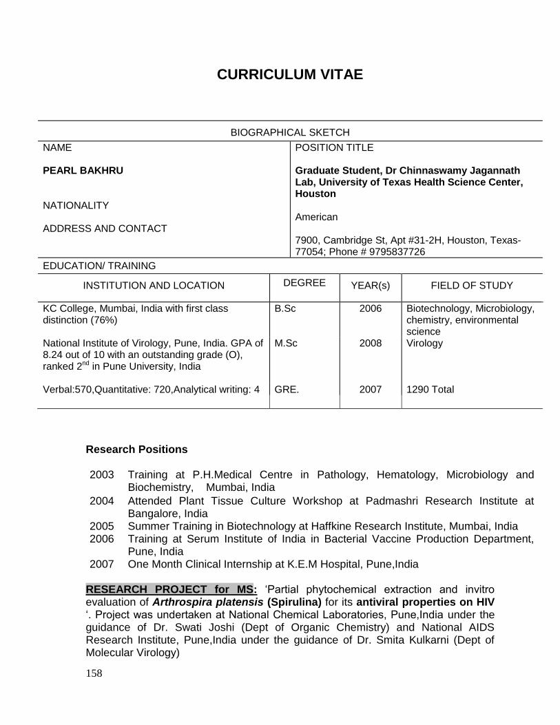
158
CURRICULUM VITAE
Research Positions
2003 Training at P.H.Medical Centre in Pathology, Hematology, Microbiology and Biochemistry, Mumbai, India
2004 Attended Plant Tissue Culture Workshop at Padmashri Research Institute at Bangalore, India
2005 Summer Training in Biotechnology at Haffkine Research Institute, Mumbai, India 2006 Training at Serum Institute of India in Bacterial Vaccine Production Department,
Pune, India 2007 One Month Clinical Internship at K.E.M Hospital, Pune,India
RESEARCH PROJECT for MS: ‘Partial phytochemical extraction and invitro evaluation of Arthrospira platensis (Spirulina) for its antiviral properties on HIV ‘. Project was undertaken at National Chemical Laboratories, Pune,India under the guidance of Dr. Swati Joshi (Dept of Organic Chemistry) and National AIDS Research Institute, Pune,India under the guidance of Dr. Smita Kulkarni (Dept of Molecular Virology)
BIOGRAPHICAL SKETCH
NAME PEARL BAKHRU NATIONALITY ADDRESS AND CONTACT
POSITION TITLE Graduate Student, Dr Chinnaswamy Jagannath Lab, University of Texas Health Science Center, Houston American 7900, Cambridge St, Apt #31-2H, Houston, Texas-77054; Phone # 9795837726
EDUCATION/ TRAINING
INSTITUTION AND LOCATION DEGREE
YEAR(s) FIELD OF STUDY
KC College, Mumbai, India with first class distinction (76%)
B.Sc 2006 Biotechnology, Microbiology, chemistry, environmental science
National Institute of Virology, Pune, India. GPA of 8.24 out of 10 with an outstanding grade (O), ranked 2nd in Pune University, India
M.Sc 2008 Virology
Verbal:570,Quantitative: 720,Analytical writing: 4 GRE. 2007 1290 Total
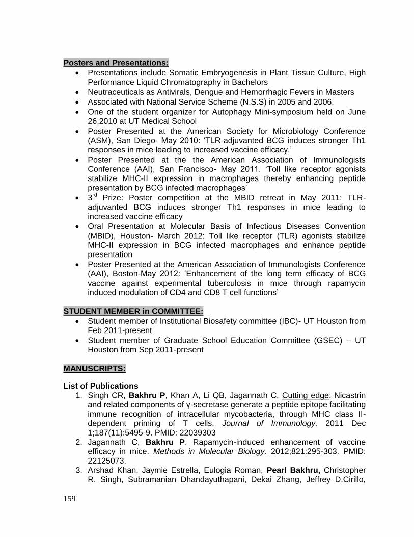
159
Posters and Presentations:
Presentations include Somatic Embryogenesis in Plant Tissue Culture, High Performance Liquid Chromatography in Bachelors
Neutraceuticals as Antivirals, Dengue and Hemorrhagic Fevers in Masters
Associated with National Service Scheme (N.S.S) in 2005 and 2006.
One of the student organizer for Autophagy Mini-symposium held on June 26,2010 at UT Medical School
Poster Presented at the American Society for Microbiology Conference (ASM), San Diego- May 2010: ‘TLR-adjuvanted BCG induces stronger Th1 responses in mice leading to increased vaccine efficacy.’
Poster Presented at the the American Association of Immunologists Conference (AAI), San Francisco- May 2011. ‘Toll like receptor agonists stabilize MHC-II expression in macrophages thereby enhancing peptide presentation by BCG infected macrophages’
3rd Prize: Poster competition at the MBID retreat in May 2011: TLR-adjuvanted BCG induces stronger Th1 responses in mice leading to increased vaccine efficacy
Oral Presentation at Molecular Basis of Infectious Diseases Convention (MBID), Houston- March 2012: Toll like receptor (TLR) agonists stabilize MHC-II expression in BCG infected macrophages and enhance peptide presentation
Poster Presented at the American Association of Immunologists Conference (AAI), Boston-May 2012: ‘Enhancement of the long term efficacy of BCG vaccine against experimental tuberculosis in mice through rapamycin induced modulation of CD4 and CD8 T cell functions’
STUDENT MEMBER in COMMITTEE:
Student member of Institutional Biosafety committee (IBC)- UT Houston from Feb 2011-present
Student member of Graduate School Education Committee (GSEC) – UT Houston from Sep 2011-present
MANUSCRIPTS:
List of Publications 1. Singh CR, Bakhru P, Khan A, Li QB, Jagannath C. Cutting edge: Nicastrin
and related components of γ-secretase generate a peptide epitope facilitating immune recognition of intracellular mycobacteria, through MHC class II-dependent priming of T cells. Journal of Immunology. 2011 Dec 1;187(11):5495-9. PMID: 22039303
2. Jagannath C, Bakhru P. Rapamycin-induced enhancement of vaccine efficacy in mice. Methods in Molecular Biology. 2012;821:295-303. PMID: 22125073.
3. Arshad Khan, Jaymie Estrella, Eulogia Roman, Pearl Bakhru, Christopher R. Singh, Subramanian Dhandayuthapani, Dekai Zhang, Jeffrey D.Cirillo,
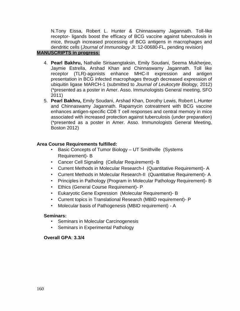
160
N.Tony Eissa, Robert L. Hunter & Chinnaswamy Jagannath. Toll-like receptor- ligands boost the efficacy of BCG vaccine against tuberculosis in mice, through increased processing of BCG antigens in macrophages and dendritic cells (Journal of Immunology JI: 12-00680-FL, pending revision)
MANUSCRIPTS in progress:
4. Pearl Bakhru, Nathalie Sirisaengtaksin, Emily Soudani, Seema Mukherjee, Jaymie Estrella, Arshad Khan and Chinnaswamy Jagannath. Toll like receptor (TLR)-agonists enhance MHC-II expression and antigen presentation in BCG infected macrophages through decreased expression of ubiquitin ligase MARCH-1 (submitted to Journal of Leukocyte Biology, 2012) (*presented as a poster in Amer. Asso. Immunologists General meeting, SFO 2011)
5. Pearl Bakhru, Emily Soudani, Arshad Khan, Dorothy Lewis, Robert L.Hunter and Chinnaswamy Jagannath. Rapamycin cotreatment with BCG vaccine enhances antigen-specific CD8 T cell responses and central memory in mice associated with increased protection against tuberculosis (under preparation) (*presented as a poster in Amer. Asso. Immunologists General Meeting, Boston 2012)
Area Course Requirements fulfilled:
• Basic Concepts of Tumor Biology – UT Smithville (Systems
Requirement)- B
• Cancer Cell Signaling (Cellular Requirement)- B
• Current Methods in Molecular Research-I (Quantitative Requirement)- A
• Current Methods in Molecular Research-II (Quantitative Requirement)- A
• Principles in Pathology (Program in Molecular Pathology Requirement)- B
• Ethics (General Course Requirement)- P
• Eukaryotic Gene Expression (Molecular Requirement)- B
• Current topics in Translational Research (MBID requirement)- P
• Molecular basis of Pathogenesis (MBID requirement) - A
Seminars: • Seminars in Molecular Carcinogenesis
• Seminars in Experimental Pathology
Overall GPA: 3.3/4