Ligand-Induced Structural Changes in TEM-1 Probed by Molecular Dynamics and Relative Binding Free...
11
Ligand-Induced Structural Changes in TEM‑1 Probed by Molecular Dynamics and Relative Binding Free Energy Calculations A. C. Pimenta, †,‡,§ J. M. Martins, † R. Fernandes, ‡,§ and I. S. Moreira* ,† † REQUIMTE/Departamento de Química, Faculdade de Ciê ncias da Universidade do Porto, Rua do Campo Alegre s/n, 4169-007 Porto, Portugal ‡ Centro de Investigaç ã o em Saú de e Ambiente, da Escola Superior de Tecnologia da Saú de do Porto, do Instituto Polite ́ cnico do Porto, Rua Valente Perfeito 322, 4400-330 Vila Nova de Gaia, Portugal § Centro de Farmacologia e Biopatologia Química (U38-FCT), Faculdade de Medicina da Universidade do Porto, Alameda Prof. Hernâ ni Monteiro, 4200-319 Porto, Portugal * S Supporting Information ABSTRACT: The TEM family of enzymes has had a crucial impact on the pharmaceutical industry due to their important role in antibiotic resistance. Even with the latest technologies in structural biology and genomics, no 3D structure of a TEM- 1/antibiotic complex is known previous to acylation. There- fore, the comprehension of their capability in acylate antibiotics is based on the protein macromolecular structure uncomplexed. In this work, molecular docking, molecular dynamic simulations, and relative free energy calculations were applied in order to get a comprehensive and thorough analysis of TEM-1/ampicillin and TEM-1/amoxicillin complexes. We described the complexes and analyzed the effect of ligand binding on the overall structure. We clearly demonstrate that the key residues involved in the stability of the ligand (hot-spots) vary with the nature of the ligand. Structural effects such as (i) the distances between interfacial residues (Ser70-Oγ and Lys73-Nζ, Lys73-Nζ and Ser130-Oγ, and Ser70-Oγ-Ser130-Oγ), (ii) side chain rotamer variation (Tyr105 and Glu240), and (iii) the presence of conserved waters can be also influenced by ligand binding. This study supports the hypothesis that TEM-1 suffers structural modifications upon ligand binding. ■ INTRODUCTION In the 21st century, infectious diseases are a major concern in public health, in great part, due to the increasing capability of pathogens to resist the antibiotics used in clinical practice. 1 Not only have resistance mechanisms to new antibiotics appeared but also the capability of the microorganisms to resist antibiotics has spread. 2 These facts are of such importance that the screening of resistant or multiresistant microorganisms is performed in several countries. In Gram-negative pathogens, the most common mechanism of resistance is the production of enzymes capable of hydrolyzing antibioticsβ-lactamases. 3 These enzymes are screened worldwide, and an online database is readily accessible. 4 Among these enzymes, a very important family is the TEM family of β-lactamases, which can present different acylating capabilities and therefore different phenotypes (i.e., ESBL, 1,5 IRT, 6,7 and CMT 8,9 ). A general 3D structure can be appointed and is formed by three main domains organized as sandwich-likeα-helix/β-sheets/α-helix. The ability of an enzyme to bind to a ligand is strongly determined by the tridimensional structure of both molecules. Therefore, the ability to determine their structure is crucial to a better prediction and understanding of complex formation. The advances in technologies in structural genomics 11 and biology, 12 such as protein expression and purification, microcrystalliza- tion, 13 the use of synchrotrons, 14 and the increasing automation of all the processes, made possible an exponential increase in the number of biomacromolecules with a known tridimensional (3D) structure. Even though macromolecular structures are often used to study biochemical processes, they still present some important limitations. These static structures only represent one possible conformation (X-ray) or a limited ensemble (NMR Nuclear Magnetic Resonance) that correspond to the structures that are more stable under the conditions of the experiments. 15 However, when analyzing macromolecules (i.e., proteins), their flexibility should be taken into account. 15-17 Enzymatic studies have an extra challenge, which is the difficulty associated with obtaining a 3D structure of the complex, either due to the kinetic properties of the enzyme (reaction occurs too rapidly to obtain a protein/ligand complex) or due to the instability of the complex itself. When it is possible to obtain the 3D structure of the complex, it remains important to take into account the inherent structural variations, which can be done by Molecular Dynamic (MD) simulations. 15,18,19 By performing this type of simulation, we can analyze the structural variation of the individual molecules and study the effect of ligand binding into the enzymatic Received: May 7, 2013 Article pubs.acs.org/jcim © XXXX American Chemical Society A dx.doi.org/10.1021/ci400269d | J. Chem. Inf. Model. XXXX, XXX, XXX-XXX
Transcript of Ligand-Induced Structural Changes in TEM-1 Probed by Molecular Dynamics and Relative Binding Free...
Ligand-Induced Structural Changes in TEM‑1 Probed by MolecularDynamics and Relative Binding Free Energy CalculationsA. C. Pimenta,†,‡,§ J. M. Martins,† R. Fernandes,‡,§ and I. S. Moreira*,†
†REQUIMTE/Departamento de Química, Faculdade de Ciencias da Universidade do Porto, Rua do Campo Alegre s/n, 4169-007Porto, Portugal‡Centro de Investigacao em Saude e Ambiente, da Escola Superior de Tecnologia da Saude do Porto, do Instituto Politecnico doPorto, Rua Valente Perfeito 322, 4400-330 Vila Nova de Gaia, Portugal§Centro de Farmacologia e Biopatologia Química (U38-FCT), Faculdade de Medicina da Universidade do Porto, Alameda Prof.Hernani Monteiro, 4200-319 Porto, Portugal
*S Supporting Information
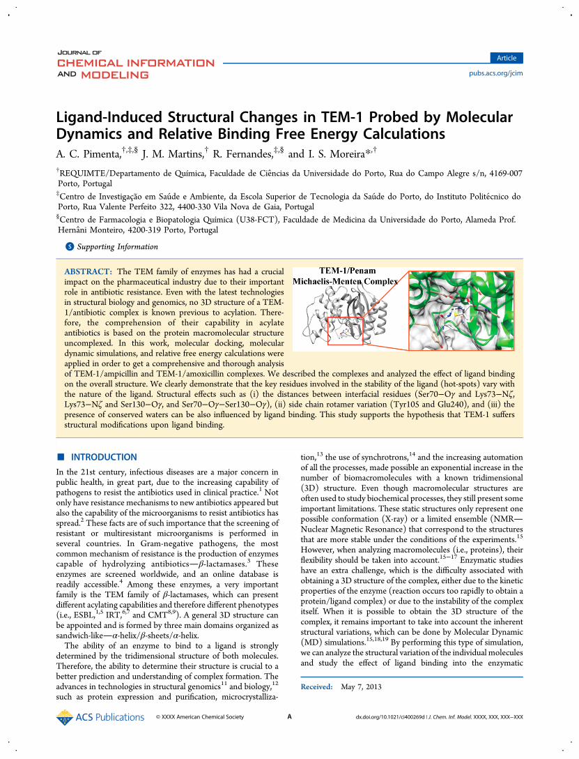
ABSTRACT: The TEM family of enzymes has had a crucialimpact on the pharmaceutical industry due to their importantrole in antibiotic resistance. Even with the latest technologiesin structural biology and genomics, no 3D structure of a TEM-1/antibiotic complex is known previous to acylation. There-fore, the comprehension of their capability in acylateantibiotics is based on the protein macromolecular structureuncomplexed. In this work, molecular docking, moleculardynamic simulations, and relative free energy calculations wereapplied in order to get a comprehensive and thorough analysisof TEM-1/ampicillin and TEM-1/amoxicillin complexes. We described the complexes and analyzed the effect of ligand bindingon the overall structure. We clearly demonstrate that the key residues involved in the stability of the ligand (hot-spots) vary withthe nature of the ligand. Structural effects such as (i) the distances between interfacial residues (Ser70−Oγ and Lys73−Nζ,Lys73−Nζ and Ser130−Oγ, and Ser70−Oγ−Ser130−Oγ), (ii) side chain rotamer variation (Tyr105 and Glu240), and (iii) thepresence of conserved waters can be also influenced by ligand binding. This study supports the hypothesis that TEM-1 suffersstructural modifications upon ligand binding.
■ INTRODUCTION
In the 21st century, infectious diseases are a major concern inpublic health, in great part, due to the increasing capability ofpathogens to resist the antibiotics used in clinical practice.1 Notonly have resistance mechanisms to new antibiotics appeared butalso the capability of the microorganisms to resist antibiotics hasspread.2 These facts are of such importance that the screening ofresistant or multiresistant microorganisms is performed inseveral countries. In Gram-negative pathogens, the mostcommon mechanism of resistance is the production of enzymescapable of hydrolyzing antibioticsβ-lactamases.3 Theseenzymes are screened worldwide, and an online database isreadily accessible.4 Among these enzymes, a very importantfamily is the TEM family of β-lactamases, which can presentdifferent acylating capabilities and therefore different phenotypes(i.e., ESBL,1,5 IRT,6,7 and CMT8,9). A general 3D structure canbe appointed and is formed by three main domains organized assandwich-likeα-helix/β-sheets/α-helix.The ability of an enzyme to bind to a ligand is strongly
determined by the tridimensional structure of both molecules.Therefore, the ability to determine their structure is crucial to abetter prediction and understanding of complex formation. Theadvances in technologies in structural genomics11 and biology,12
such as protein expression and purification, microcrystalliza-
tion,13 the use of synchrotrons,14 and the increasing automationof all the processes, made possible an exponential increase in thenumber of biomacromolecules with a known tridimensional(3D) structure. Even though macromolecular structures areoften used to study biochemical processes, they still present someimportant limitations. These static structures only represent onepossible conformation (X-ray) or a limited ensemble (NMRNuclear Magnetic Resonance) that correspond to the structuresthat are more stable under the conditions of the experiments.15
However, when analyzing macromolecules (i.e., proteins), theirflexibility should be taken into account.15−17 Enzymatic studieshave an extra challenge, which is the difficulty associated withobtaining a 3D structure of the complex, either due to the kineticproperties of the enzyme (reaction occurs too rapidly to obtain aprotein/ligand complex) or due to the instability of the complexitself. When it is possible to obtain the 3D structure of thecomplex, it remains important to take into account the inherentstructural variations, which can be done by Molecular Dynamic(MD) simulations.15,18,19 By performing this type of simulation,we can analyze the structural variation of the individual moleculesand study the effect of ligand binding into the enzymatic
Received: May 7, 2013
Article
pubs.acs.org/jcim
© XXXX American Chemical Society A dx.doi.org/10.1021/ci400269d | J. Chem. Inf. Model. XXXX, XXX, XXX−XXX

structure. MD simulations have been successfully performed tosimulate biological properties, and the number of studies usingthis methodology increases.19−26 MD simulations also take intoaccount the role of the solvent in the proteins structuralvariations and in the ligand geometry.18
The TEM family of enzymes has been heavily studied due totheir importance in antibiotic resistance. However, despite greatefforts, a 3D structure of a TEM/antibiotic complex preacylationhas not yet been obtained through structural biology methods.The ability of the enzymes to quickly acylate the antibiotics maybe the reason. This limitation led to the necessity of studying theantibiotic resistance promoted by these enzymes (i.e., kineticdata and results from disc diffusion methods) from the 3Dstructure of the protein uncomplexed. Although good resultshave been obtained by several authors,6,27−29 the molecularstructure of the protein upon ligand binding cannot be analyzed.This limitation is of special importance when (i) trying to justifydifferences observed in the kinetic properties and (ii) studyingthe catalytic reaction, as residues that intervene in ligand bindingor stabilization or in the proton transfer network cannot beanalyzed properly. Molecular docking methodologies allow us toovercome these limitations, especially when using algorithmsthat take into account the flexibility of both the protein andligand.18,30,31 These methodologies along with MD simulationsand relative free energy calculations grant the capability toanalyze complexes more thoroughly.32−34 In this work, thecomplexes between TEM-1 and ampicillin or amoxicillin arecharacterized. A detailed description of their protein/ligandinterface is made. Hot-spots (HS), residues that upon alaninemutation generate a binding free energy difference higher than2.0 kcal/mol,35 are identified. Interatomic contacts arecalculated, and the presence of water molecules near interfacialresidues is depicted. A comparison between the structure of theprotein uncomplexed and in complex with two small molecules ismade to study the conformational rearrangements of the TEM-1structure caused by ligand binding.
■ METHODOLOGY
Structure Preparation. The protonation states of TEM-1(PDB ID: 1ZG410) were assigned by EPIK36 in MAESTROSchrodinger Software, LLC (see Figure 1).37 All residues werefound at their physiological protonation state.TEM-1 Molecular Dynamics Simulation. TEM-1 was
stabilized and refined by MD simulation with the modified
Cornell force field, by Duan et al.ff03.38,39 TheMD simulationwas carried out in explicit solvent with the TIP3P (79) watermodel using the AMBER9 program. SevenNa+ counterions wereadded to keep the whole system neutral and a 10 Å separationbetween each edge of the box and the closest solute atom wasused to minimize electrostatic interactions between periodicimages of the solute. First, the water was equilibrated in thepresence of the fixed complex (25 ps); then only the side chainswere relaxed (200 ps)minimization, and finally a productionrun of 16 ns was done for the system. During the initial 2 ns of theproduction run, the temperature was increased from 0 to 300 Kfollowed by 48 ns of production at a constant 300 K (ensembleNPT) using a “weak-coupling” barostat (Berendsen barostat).40
In the restrained simulations, the atoms were subjected to aharmonic restraining force of 10 kJ mol−1 nm−2. In all MDsimulations, the bond lengths involving hydrogens wereconstrained using the SHAKE algorithm.41 The equations ofmotion were integrated with a 2 fs time step, and the Langevinalgorithm42,43 was used to regulate the temperature of thesystem. All of the crystallographic waters were removed from thestructure subjected to the MD simulation. Periodic boundaryconditions were applied using PME44,45 to treat long-rangeelectrostatic interactions. After establishing the stability of theMD simulation, we have calculated the RMSD (Root-Mean-Square-Deviation) to the average structure and selected sixstructures with the lowest value. These six structuralrepresentatives of the ensemble generated for the wild-typewere used in the following procedure: protein−ligand docking.
Molecular Docking. Protein−ligand docking was performedusing the six microstates ensemble from the protein MDsimulation as targets and the penicillins: amoxicillin andampicillin. The selection of these antibiotics was based on thefacts that they are known substrates of penicillinases like TEM-146 and are widely used in clinical practice.47 Their structureswere obtained from PubChem Compound.48 The AutoDock 4.2package49 was used for the entire docking procedure. An energygrid of 46 Å × 40 Å × 40 Å in dimensions, with a 0.375-Å gridspacing (the center of the Grid Box and dimension werepreviously tested), was generated with the program AutoGrid.Gasteiger charges were assigned to the ligand atoms. AutoDock 4was used to evaluate ligand-binding energies over the conforma-tional search space using the genetic algorithm-local searchmethod. Default docking parameters were used with thefollowing exceptions: ga-pop-size, 200; ga-num-evals, 10 000000; and ga-run, 50. The structural poses that resulted from thedocking were analyzed taking into account several parameters:(i) The binding energy should be negative. (ii) The complexesshould have low energy and form clusters with a higher numberof complexes. (iii) The β-lactam ring should be on the moreinternal part of the catalytic pocket and, if possible, facing thecatalytic Ser70. (iv) The complexes should have a high number ofinteractions (protein−ligand).
Molecular Dynamics Simulation of the Protein−LigandComplexes. The final complexes obtained from the moleculardocking were stabilized and refined byMD simulations followingthe procedure described in the TEM-1 Molecular DynamicsSimulation section. To obtain the parameters for amoxicillin andampicillin, partial atomic charges were derived with the standardHF/6-31G* RESP50 (Tables S1 and S3 in SupportingInformation) methodology using the program Antechamberimplemented in the Amber package.51 Atom types and missingforce-field parameters of the ligands (Tables S2 and S4 in theSupporting Information) were assigned with the GAFF force
Figure 1. Structural representation of TEM-1 (PDB ID: 1ZG410) withsome of the key secondary structures identified.
Journal of Chemical Information and Modeling Article
dx.doi.org/10.1021/ci400269d | J. Chem. Inf. Model. XXXX, XXX, XXX−XXXB
field.52 Although the ligand parametrization can be challenging,this was not the case as our organic compounds are composed bysome of the most common atoms present in the organic chemicalspace (C, N, O, S, H) in a typical spatial arrangement (chemicalstructures of amoxicillin and ampicillin shown in Figures S1−S3in the Supporting Information).Structural Analysis. RMSDs of the protein backbone,
amoxicillin, and ampicillin were calculated for all the MDsimulations in order to investigate their stability. For a detailedstudy of the binding interface, significant residues were selected:Met69, Ser70, Lys73, Tyr105, Ser130, Asn132, Glu166, Asn170,Val216, Lys234, Ser235, Glu240, Arg244, and Arg275. Thisselection took into account the literature5,53,54 to ensure thatcrucial residues were selected. For these residues, B-factors werecalculated to investigate the deviation of these compared to areference position. The environment around the same residueswas carefully characterized. For this step, all residues and watermolecules within 5 Å of each interfacial residue were selected,and their occupancy was estimated. We have also analyzed theradial distribution function, g(r) or RDF, as well as the averagenumber of waters within a given distance of all interfacialresidues. g(r) gives the probability of finding an atom at distance rfrom another atom, in relation to the probability expected for abulk solvent distribution at the same density. It was calculated bycompiling a histogram with a spacing of 0.02 and a range of 8 Å.These two procedures as well as the visual inspection of the MDsimulations were performed with the VMD package and tailor-made scripts.55 Crucial interatomic distances for the enzymecatalytic activity and enzymatic inhibition were measured for allthe MD simulations. All structural representations were made bythe PYMOL package.56
Energetic Profile. The MM-PBSA (Molecular MechanicsPoisson−Boltzmann Surface Area) script57 integrated into theAMBER 9 package58 was used to calculate the binding freeenergy difference upon alanine mutation. The alanine mutationswere performed on the interface residues that were previouslyselected in the structural analysis with the exception of alanine,glycine, or proline residues. These were not mutated since theyusually have a primary role in the protein stability and themutation could lead to protein degradation. Due to thelimitations of the method, residues that would only interact viabackbone could not be analyzed with computational AlanineScanning Mutagenesis (ASM). This method combines acontinuum approach to model solvent interactions with a MM-based approach to atomistically model protein−proteininteractions. It provides speed and accuracy and has been usedquite a bit in past years.20,22,57,59−70 The MM-PBSA approachfirst developed by Huo et al.57 was improved by Moreira et al.62
and can now be applied with an accuracy of 1 kcal/mol. Themutant complexes are generated by a single truncation of themutated side chain, replacing Cγ with a hydrogen atom andsetting the Cβ−Hdirection to that of the former Cβ−Cγ. For thebinding energy calculations, a total of 25 snapshots of thecomplexes were extracted in the last 1 ns of the run. TheΔΔG isdefined as the difference between the mutant and wild typecomplexes defined as
ΔΔ = Δ − Δ− −G G Gcpx mutant cpx wild type (1)
Typical contributions to the free energy include the internalenergy (bond, dihedral, and angle), the electrostatic and the vander Waals interactions, the free energy of polar solvation, the freeenergy of nonpolar solvation, and the entropic contribution:
= + + +
+ −
‐
− ‐
G E E E G
G TS
molecule internal electrostatic vdW polar solvation
non polar solvation (2)
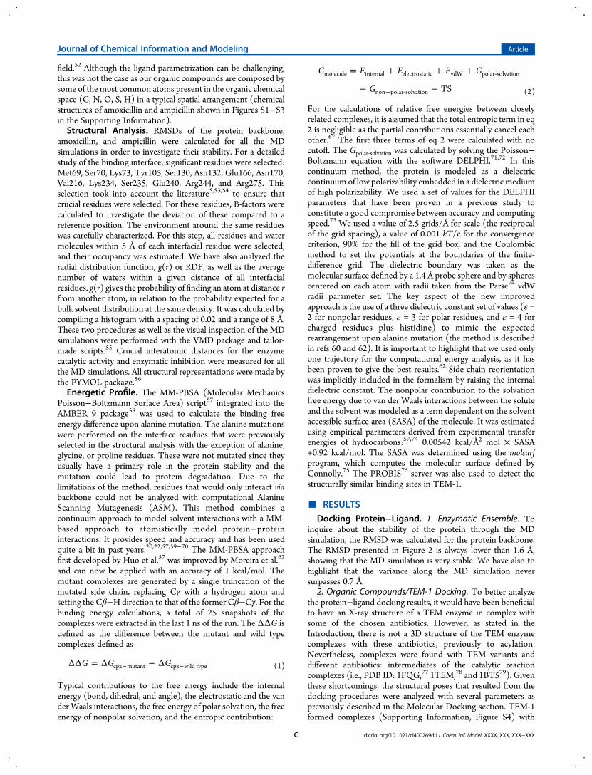
For the calculations of relative free energies between closelyrelated complexes, it is assumed that the total entropic term in eq2 is negligible as the partial contributions essentially cancel eachother.67 The first three terms of eq 2 were calculated with nocutoff. The Gpolar‑solvation was calculated by solving the Poisson−Boltzmann equation with the software DELPHI.71,72 In thiscontinuum method, the protein is modeled as a dielectriccontinuum of low polarizability embedded in a dielectric mediumof high polarizability. We used a set of values for the DELPHIparameters that have been proven in a previous study toconstitute a good compromise between accuracy and computingspeed.73 We used a value of 2.5 grids/Å for scale (the reciprocalof the grid spacing), a value of 0.001 kT/c for the convergencecriterion, 90% for the fill of the grid box, and the Coulombicmethod to set the potentials at the boundaries of the finite-difference grid. The dielectric boundary was taken as themolecular surface defined by a 1.4 Å probe sphere and by spherescentered on each atom with radii taken from the Parse74 vdWradii parameter set. The key aspect of the new improvedapproach is the use of a three dielectric constant set of values (ε =2 for nonpolar residues, ε = 3 for polar residues, and ε = 4 forcharged residues plus histidine) to mimic the expectedrearrangement upon alanine mutation (the method is describedin refs 60 and 62). It is important to highlight that we used onlyone trajectory for the computational energy analysis, as it hasbeen proven to give the best results.62 Side-chain reorientationwas implicitly included in the formalism by raising the internaldielectric constant. The nonpolar contribution to the solvationfree energy due to van der Waals interactions between the soluteand the solvent was modeled as a term dependent on the solventaccessible surface area (SASA) of the molecule. It was estimatedusing empirical parameters derived from experimental transferenergies of hydrocarbons:57,74 0.00542 kcal/Å2 mol × SASA+0.92 kcal/mol. The SASA was determined using the molsurfprogram, which computes the molecular surface defined byConnolly.75 The PROBIS76 server was also used to detect thestructurally similar binding sites in TEM-1.
■ RESULTSDocking Protein−Ligand. 1. Enzymatic Ensemble. To
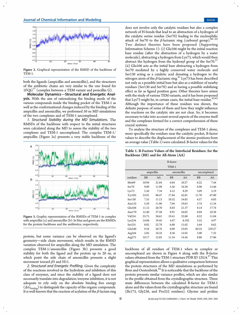
inquire about the stability of the protein through the MDsimulation, the RMSD was calculated for the protein backbone.The RMSD presented in Figure 2 is always lower than 1.6 Å,showing that the MD simulation is very stable. We have also tohighlight that the variance along the MD simulation neversurpasses 0.7 Å.
2. Organic Compounds/TEM-1 Docking. To better analyzethe protein−ligand docking results, it would have been beneficialto have an X-ray structure of a TEM enzyme in complex withsome of the chosen antibiotics. However, as stated in theIntroduction, there is not a 3D structure of the TEM enzymecomplexes with these antibiotics, previously to acylation.Nevertheless, complexes were found with TEM variants anddifferent antibiotics: intermediates of the catalytic reactioncomplexes (i.e., PDB ID: 1FQG,77 1TEM,78 and 1BT579). Giventhese shortcomings, the structural poses that resulted from thedocking procedures were analyzed with several parameters aspreviously described in the Molecular Docking section. TEM-1formed complexes (Supporting Information, Figure S4) with
Journal of Chemical Information and Modeling Article
dx.doi.org/10.1021/ci400269d | J. Chem. Inf. Model. XXXX, XXX, XXX−XXXC
both the ligands (ampicillin and amoxicillin), and the structuresof the antibiotic chains are very similar to the one found for1FQG77 (complex between a TEM variant and penicillin G).Molecular DynamicsStructural and Energetic Anal-
ysis. With the aim of rationalizing the binding mode of thevarious compounds inside the binding pocket of the TEM-1 aswell as the conformational changes induced by the binding of theampicillin and amoxicillin, we performed 50 ns MD simulationsof the two complexes and of TEM-1 uncomplexed.1. Structural Stability during the MD Simulations. The
RMSDs of the backbone with respect to the initial structureswere calculated along the MD to assess the stability of the twocomplexes and TEM-1 uncomplexed. The complex TEM-1/ampicillin (Figure 3a) presents a very stable backbone of the
protein, but some variance can be observed on the ligand’sgeometryside chain movement, which results in the RMSDvariation observed for ampicillin along the MD simulation. Thecomplex TEM-1/amoxicillin (Figure 3b) presents a goodstability for both the ligand and the protein up to 20 ns, atwhich point the side chain of amoxicillin presents a slightmovement toward β3 and H11.2. Structural and Energetic Profiling. Given the complexity
of the reactions involved in the hydrolysis and inhibition of thisclass of enzymes, and since the stability of a ligand does notnecessarily translate into degradation/enzyme inhibition, it is notadequate to rely only on the absolute binding free energy(ΔGbinding) to distinguish the capacity of the organic compounds.It is well-known that the reaction of acylation of the β-lactam ring
does not involve only the catalytic residues but also a complexnetwork of H-bonds that lead to an abstraction of a hydrogen ofthe catalytic serine residue (Ser70) leading to the nucleophilicattack of Ser70 to the β-lactamic ring (carbonyl group).80−82
Two distinct theories have been proposed (SupportingInformaiton Scheme 1): (i) Glu166 might be the initial reactionbase residue (after the abstraction of a hydrogen by a watermolecule), abstracting a hydrogen from Lys73, which would thenabstract the hydrogen from the hydroxyl group of the Ser70.27
(ii) Glu166 acts as the initial base abstracting a hydrogen fromSer70 mediated by a highly conserved water molecule andSer130 acting as a catalytic acid donating a hydrogen to thenitrogen atom of the β-lactamic ring.81 Lys73 has been describednot only as a possible initial base but also as a stabilizer of nearbyresidues (Ser130 and Ser70) and as having a possible stabilizingeffect as far as ligand position goes. Other theories have arisenwith the study of various TEM variants, and it has been proposedthat Lys73 might be, in certain cases, the initial base residue.82,83
Although the importance of these residues was shown, thedefinite purpose of some of them and how they might influencethe reactions on the catalytic site are not clear. So, it becomesnecessary to take into account several aspects of the enzyme itselfand the complexes formed for a correct comprehension of thesecrucial systems.To analyze the structure of the complexes and TEM-1 alone,
more specifically the residues near the catalytic pocket, B-factorvalues to describe the displacement of the atomic positions froman average value (Table 1) were calculated. B-factor values for the
backbone of all residues of TEM-1 when in complex oruncomplexed are shown in Figure 4 along with the B-factorvalues obtained from the TEM-1 structure PDB ID 1ZG4.27 Thisgraphical representation allows a qualitative comparison betweenthe protein structures of the MD simulations as performed byBren and Oostenbrink.84 It is noticeable that the backbone of theprotein presents similar variance profiles, which are also similarto the profile obtained from the crystallographic structure. Threemain differences between the calculated B-factor for TEM-1alone and the values from the crystallographic structure are found(Ile173, Gly236, and Pro252 residues). Glycine and proline
Figure 2. Graphical representation of the RMSD of the backbone ofTEM-1.
Figure 3. Graphic representation of the RMSDs of TEM-1 in complexwith ampicillin (a) and amoxicillin (b). In blue and green are the RMSDsfor the protein backbone and the antibiotics, respectively.
Table 1. B-Factors Values of the Interfacial Residues: for theBackbone (BB) and for All-Atom (AA)
B-factor
TEM-1
ampicillin amoxicillin uncomplexed
residues BB AA BB AA BB AA
Met69 10.94 21.64 6.84 10.27 6.22 7.44Ser70 9.09 11.98 5.56 16.58 6.96 13.46Lys73 5.56 7.34 4.12 8.29 3.69 5.19Tyr105 33.91 96.47 17.84 45.01 11.70 67.00Ser130 7.55 11.13 10.52 14.85 4.57 6.03Asn132 5.59 11.96 7.04 19.63 3.74 11.34Glu166 11.12 20.70 8.83 21.37 8.14 17.74Asn170 12.40 37.50 9.91 24.02 8.04 10.38Val216 25.75 38.61 19.41 32.80 8.22 11.04Lys234 14.08 19.45 4.47 8.192 3.52 4.97Ser235 8.82 22.78 6.88 11.62 5.64 8.62Glu240 9.54 26.76 8.09 23.05 60.32 239.27Arg244 5.05 18.16 8.36 15.92 3.90 7.32Arg275 10.17 12.05 11.34 14.56 8.80 17.06
Journal of Chemical Information and Modeling Article
dx.doi.org/10.1021/ci400269d | J. Chem. Inf. Model. XXXX, XXX, XXX−XXXD
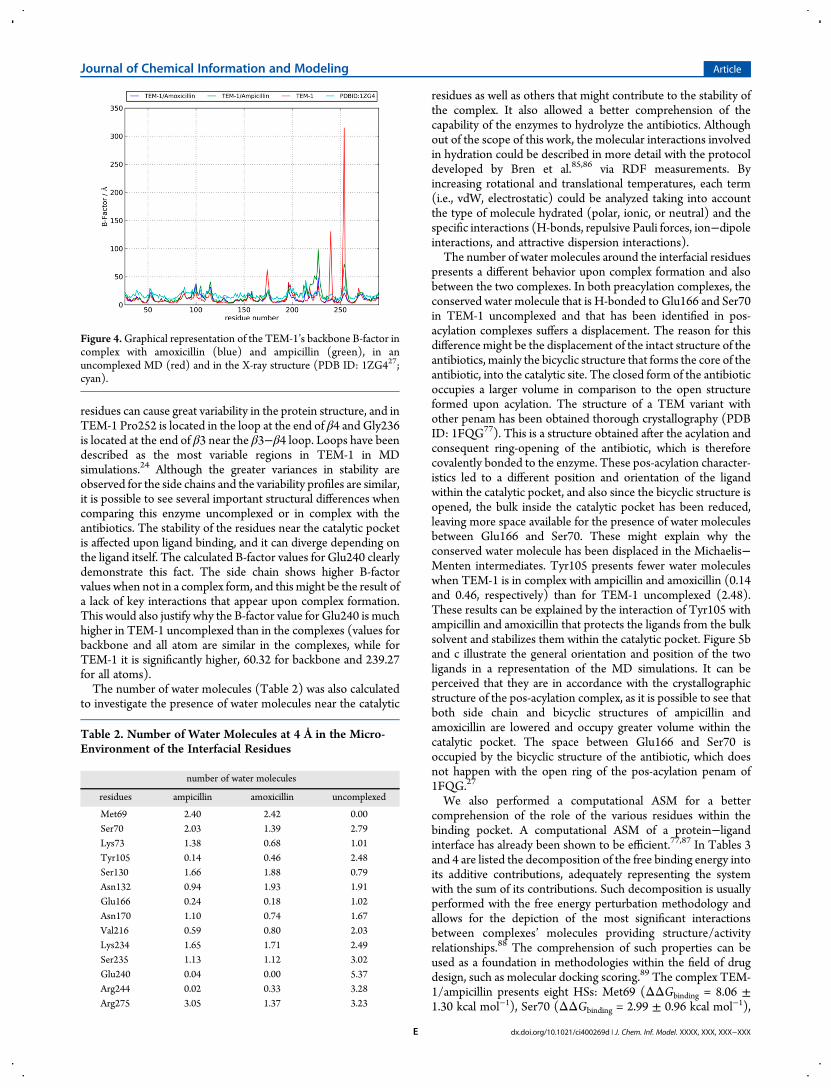
residues can cause great variability in the protein structure, and inTEM-1 Pro252 is located in the loop at the end of β4 and Gly236is located at the end of β3 near the β3−β4 loop. Loops have beendescribed as the most variable regions in TEM-1 in MDsimulations.24 Although the greater variances in stability areobserved for the side chains and the variability profiles are similar,it is possible to see several important structural differences whencomparing this enzyme uncomplexed or in complex with theantibiotics. The stability of the residues near the catalytic pocketis affected upon ligand binding, and it can diverge depending onthe ligand itself. The calculated B-factor values for Glu240 clearlydemonstrate this fact. The side chain shows higher B-factorvalues when not in a complex form, and this might be the result ofa lack of key interactions that appear upon complex formation.This would also justify why the B-factor value for Glu240 is muchhigher in TEM-1 uncomplexed than in the complexes (values forbackbone and all atom are similar in the complexes, while forTEM-1 it is significantly higher, 60.32 for backbone and 239.27for all atoms).The number of water molecules (Table 2) was also calculated
to investigate the presence of water molecules near the catalytic
residues as well as others that might contribute to the stability ofthe complex. It also allowed a better comprehension of thecapability of the enzymes to hydrolyze the antibiotics. Althoughout of the scope of this work, the molecular interactions involvedin hydration could be described in more detail with the protocoldeveloped by Bren et al.85,86 via RDF measurements. Byincreasing rotational and translational temperatures, each term(i.e., vdW, electrostatic) could be analyzed taking into accountthe type of molecule hydrated (polar, ionic, or neutral) and thespecific interactions (H-bonds, repulsive Pauli forces, ion−dipoleinteractions, and attractive dispersion interactions).The number of water molecules around the interfacial residues
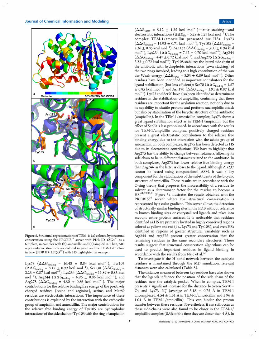
presents a different behavior upon complex formation and alsobetween the two complexes. In both preacylation complexes, theconserved water molecule that is H-bonded to Glu166 and Ser70in TEM-1 uncomplexed and that has been identified in pos-acylation complexes suffers a displacement. The reason for thisdifference might be the displacement of the intact structure of theantibiotics, mainly the bicyclic structure that forms the core of theantibiotic, into the catalytic site. The closed form of the antibioticoccupies a larger volume in comparison to the open structureformed upon acylation. The structure of a TEM variant withother penam has been obtained thorough crystallography (PDBID: 1FQG77). This is a structure obtained after the acylation andconsequent ring-opening of the antibiotic, which is thereforecovalently bonded to the enzyme. These pos-acylation character-istics led to a different position and orientation of the ligandwithin the catalytic pocket, and also since the bicyclic structure isopened, the bulk inside the catalytic pocket has been reduced,leaving more space available for the presence of water moleculesbetween Glu166 and Ser70. These might explain why theconserved water molecule has been displaced in the Michaelis−Menten intermediates. Tyr105 presents fewer water moleculeswhen TEM-1 is in complex with ampicillin and amoxicillin (0.14and 0.46, respectively) than for TEM-1 uncomplexed (2.48).These results can be explained by the interaction of Tyr105 withampicillin and amoxicillin that protects the ligands from the bulksolvent and stabilizes them within the catalytic pocket. Figure 5band c illustrate the general orientation and position of the twoligands in a representation of the MD simulations. It can beperceived that they are in accordance with the crystallographicstructure of the pos-acylation complex, as it is possible to see thatboth side chain and bicyclic structures of ampicillin andamoxicillin are lowered and occupy greater volume within thecatalytic pocket. The space between Glu166 and Ser70 isoccupied by the bicyclic structure of the antibiotic, which doesnot happen with the open ring of the pos-acylation penam of1FQG.27
We also performed a computational ASM for a bettercomprehension of the role of the various residues within thebinding pocket. A computational ASM of a protein−ligandinterface has already been shown to be efficient.77,87 In Tables 3and 4 are listed the decomposition of the free binding energy intoits additive contributions, adequately representing the systemwith the sum of its contributions. Such decomposition is usuallyperformed with the free energy perturbation methodology andallows for the depiction of the most significant interactionsbetween complexes’ molecules providing structure/activityrelationships.88 The comprehension of such properties can beused as a foundation in methodologies within the field of drugdesign, such as molecular docking scoring.89 The complex TEM-1/ampicillin presents eight HSs: Met69 (ΔΔGbinding = 8.06 ±1.30 kcal mol−1), Ser70 (ΔΔGbinding = 2.99 ± 0.96 kcal mol−1),
Figure 4.Graphical representation of the TEM-1’s backbone B-factor incomplex with amoxicillin (blue) and ampicillin (green), in anuncomplexed MD (red) and in the X-ray structure (PDB ID: 1ZG427;cyan).
Table 2. Number of Water Molecules at 4 Å in the Micro-Environment of the Interfacial Residues
number of water molecules
residues ampicillin amoxicillin uncomplexed
Met69 2.40 2.42 0.00Ser70 2.03 1.39 2.79Lys73 1.38 0.68 1.01Tyr105 0.14 0.46 2.48Ser130 1.66 1.88 0.79Asn132 0.94 1.93 1.91Glu166 0.24 0.18 1.02Asn170 1.10 0.74 1.67Val216 0.59 0.80 2.03Lys234 1.65 1.71 2.49Ser235 1.13 1.12 3.02Glu240 0.04 0.00 5.37Arg244 0.02 0.33 3.28Arg275 3.05 1.37 3.23
Journal of Chemical Information and Modeling Article
dx.doi.org/10.1021/ci400269d | J. Chem. Inf. Model. XXXX, XXX, XXX−XXXE
Lys73 (ΔΔGbinding = 16.48 ± 0.84 kcal mol−1), Tyr105(ΔΔGbinding = 8.17 ± 0.99 kcal mol−1), Ser130 (ΔΔGbinding =2.25 ± 0.87 kcal mol−1), Lys234 (ΔΔGbinding = 11.89 ± 0.85 kcalmol−1), Arg244 (ΔΔGbinding = 6.96 ± 0.86 kcal mol−1), andArg275 (ΔΔGbinding = 4.50 ± 0.86 kcal mol−1). The majorcontributions for the relative binding free energy of the positivelycharged residues (lysine and arginine), serine, and Met69residues are electrostatic interactions. The importance of thesecontributions is explained by the interaction with the carboxylicgroup of ampicillin and amoxicillin. The major contributions forthe relative free binding energy of Tyr105 are hydrophobicinteractions of the side chain of Tyr105 with the ring of ampicillin
(ΔΔEVDW = 5.12 ± 1.35 kcal mol−1)π−π stackingandelectrostatic interactions (ΔΔEEle = 5.29± 1.27 kcal mol−1). Thecomplex TEM-1/amoxicillin presented six HSs: Lys73(ΔΔGbinding = 14.93 ± 0.71 kcal mol−1), Tyr105 (ΔΔGbinding =2.36 ± 0.85 kcal mol−1), Asn132 (ΔΔGbinding = 3.00 ± 0.94 kcalmol−1), Lys234 (ΔΔGbinding = 7.42 ± 0.70 kcal mol−1), Arg244(ΔΔGbinding = 4.47± 0.72 kcal mol−1), and Arg275 (ΔΔGbinding =3.23± 0.72 kcal mol−1). Tyr105 stabilizes the lateral side chain ofthe antibiotic with hydrophobic interactions (π−π stacking) ofthe two rings involved, leading to a high contribution of the vander Waals energy (ΔΔEVDW = 3.03 ± 0.89 kcal mol−1). Otherresidues have been identified as important contributors for theligand stabilization (but less efficient): Ser70 (ΔΔGbinding = 1.57± 0.85 kcal mol−1) and Asn170 (ΔΔGbinding = 1.91 ± 0.87 kcalmol−1). Lys73 and Ser70 have also been identified as determinantresidues in the stabilization of ampicillin, confirming that theseresidues are important for the acylation reaction, not only due toits capability to shuttle protons and perform nucleophilic attackbut also by stabilization of the bicyclic structure of the antibiotic(ampicillin). In the TEM-1/amoxicillin complex, Lys73 shows agreat ligand stabilization effect as in TEM-1/ampicillin, but theeffect of Ser70 is less pronounced. In accordance with the resultsfor TEM-1/ampicillin complex, positively charged residuespresent a great electrostatic contribution to the relative freebinding energy due to the interaction with the acidic group ofamoxicillin. In both complexes, Arg275 has been detected as HSdue to its electrostatic contributions: We have to highlight thatArg275 has the ability to change between rotamers, allowing itsside chain to be in different distances related to the antibiotic. Inboth complexes, Arg275 has lower relative free binding energythan Arg244, as the latter is closer to the ligand. Although Ala237cannot be tested using computational ASM, it was a keycomponent for the stabilization of the substituents of the bicyclicstructure of ampicillin. These results are in accordance with theO-ring theory that proposes the inaccessibility of a residue tosolvent as a determinant factor for the residue to become aHS.61,69,90,91 Figure 5a illustrates the results obtained with thePROBIS76 server where the structural conservation isrepresented by a color gradient. This server allows the detectionof structurally similar binding sites in the PDB without referenceto known binding sites or cocrystallized ligands and takes intoaccount entire protein surfaces. It is noticeable that residuesidentified as HS are primarily located in highly conserved regionscolored as yellow and red (i.e., Lys73 and Tyr105), and even HSsidentified in regions of greater structural variability such asArg244 and Arg275 present greater conservation that theremaining residues in the same secondary structures. Theseresults suggest that structural conservation algorithms can beused to predict important residues in ligand binding inaccordance with the results from Nejc et al.92
To investigate if the H-bond network between the catalyticresidues is maintained through the MD simulation, relevantdistances were also calculated (Table 5).The distances measured between key residues have also shown
that the ligands influence the position of the side chain of theresidues near the catalytic pocket. When in complex, TEM-1presents a significant increase for the distance between Ser70−Oγ and Lys73−Nζ (average of 3.18 ± 0.75 Å in TEM-1uncomplexed, 4.54 ± 1.31 Å in TEM-1/amoxicillin, and 5.96 ±1.04 Å in TEM-1/ampicillin). This can hinder the protontransfer between these residues. Nevertheless, it can still occur asthese side-chains were also found to be closer in the TEM-1/ampicillin complex (8.5% of the time they are closer than 4 Å). In
Figure 5. Structural representations of TEM-1: (a) colored by structuralconservation using the PROBIS76 server with PDB ID 1ZG427 as atemplate; in complex with (b) amoxicillin and (c) ampicillin. Then, MDrepresentative structures are colored in green and the TEM-1 structurein blue (PDB ID: 1FQG77) with HS highlighted in orange.
Journal of Chemical Information and Modeling Article
dx.doi.org/10.1021/ci400269d | J. Chem. Inf. Model. XXXX, XXX, XXX−XXXF
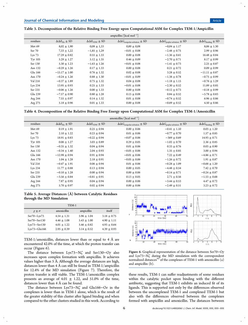
TEM-1/amoxicillin, distances lower than or equal to 4 Å areencountered 42.0% of the time, at which the proton transfer canoccur (Figure 6).The distance between Lys73−Nζ and Ser130−Oγ also
increases upon complex formation with ampicillin. It achievesvalues higher than 5 Å. Although the average distances are high,distances lower than 4 Å can still be found in TEM-1/ampicillinfor 12.4% of the MD simulation (Figure 7). Therefore, theproton transfer is still viable. The TEM-1/amoxicillin complexpresents an average of 4.01 ± 1.22, and 51.0% of the time,distances lower than 4 Å can be found.The distance between Lys73−Nζ and Glu166−Oε in the
complexes is lower than in TEM-1 alone, which is the result ofthe greater stability of this cluster after ligand binding and whencompared to the other clusters studied in this work. According to
these results, TEM-1 can suffer readjustments of some residueswithin the catalytic pocket upon binding with the differentantibiotic, suggesting that TEM-1 exhibits an induced fit of itsligands. This is supported not only by the differences observedbetween the uncomplexed TEM-1 and complexed TEM-1 butalso with the differences observed between the complexesformed with ampicillin and amoxicillin. The distances between
Table 3. Decomposition of the Relative Binding Free Energy upon Computational ASM for Complex TEM-1/Ampicillin
ampicillin/[kcal mol−1]
residues ΔΔEEle ± SD ΔΔEVDW ± SD ΔΔGnonpolar‑solvation ± SD ΔΔGpoplar‑solvation ± SD ΔΔGbinding ± SD
Met 69 8,02 ± 1,90 0,08 ± 1,33 0,00 ± 0,08 −0,04 ± 1.17 8,06 ± 1.30Ser 70 7,33 ± 1,22 −1,85 ± 1,29 −0.01 ± 0.08 −2.48 ± 0.75 2.99 ± 0.96Lys 73 17.29 ± 0.82 0.55 ± 1.33 0.00 ± 0.08 −1.36 ± 0.61 16.48 ± 0.84Tyr 105 5.29 ± 1.27 5.12 ± 1.35 0.46 ± 0.09 −2.70 ± 0.71 8.17 ± 0.99Ser 130 5.30 ± 1.23 −1.63 ± 1.26 −0.01 ± 0.08 −1.41 ± 0.73 2.25 ± 0.87Asn 132 −0.29 ± 1.26 0.17 ± 1.33 0.00 ± 0.08 0.21 ± 0.72 0.09 ± 0.99Glu 166 −15.17 ± 1.00 0.76 ± 1.32 0.02 ± 0.08 3.28 ± 0.52 −11.11 ± 0.87Asn 170 −0.24 ± 1.26 0.88 ± 1.30 −0.01 ± 0.09 −1.38 ± 0.78 −0.75 ± 0.99Val 216 −0.37 ± 1.89 0.75 ± 1.32 0.04 ± 0.08 −1.18 ± 1.15 −0.76 ± 1.29Lys 234 13.95 ± 0.93 0.25 ± 1.33 −0.01 ± 0.08 −2.30 ± 0.52 11.89 ± 0.85Ser 235 −0.06 ± 1.26 0.00 ± 1.33 0.00 ± 0.08 −0.12 ± 0.73 −0.18 ± 0.99Glu 239 −7.57 ± 0.90 0.80 ± 1.28 0.15 ± 0.08 0.84 ± 0.52 −5.78 ± 0.83Arg 244 7.55 ± 0.97 0.14 ± 1.32 0.01 ± 0.08 −0.74 ± 0.52 6.96 ± 0.86Arg 275 5.18 ± 0.96 0.01 ± 1.33 0.00 ± 0.08 −0.69 ± 0.52 4.50 ± 0.86
Table 4. Decomposition of the Relative Binding Free Energy upon Computational ASM for Complex TEM-1/Amoxicillin
amoxicillin/[kcal mol−1]
residues ΔΔEEle ± SD ΔΔEVDW ± SD ΔΔGnonpolar‑solvation ± SD ΔΔGpoplar‑solvation ± SD ΔΔGbinding ± SD
Met 69 0.19 ± 1.91 0.25 ± 0.94 0.00 ± 0.06 −0.41 ± 1.10 0.03 ± 1.20Ser 70 2.10 ± 1.22 0.23 ± 0.94 0.01 ± 0.06 −0.77 ± 0.70 1.57 ± 0.85Lys 73 18.91 ± 0.83 −0.22 ± 0.94 −0.07 ± 0.06 −369 ± 0.69 14.93 ± 0.71Tyr 105 0.06 ± 1.27 3.03 ± 0.89 0.29 ± 0.05 −1.02 ± 0.70 2.36 ± 0.85Ser 130 −0.35 ± 1.32 0.04 ± 0.94 0.01 ± 0.06 0.33 ± 0.76 0.03 ± 0.90Asn 132 −0.34 ± 1.40 2.04 ± 0.93 −0.05 ± 0.06 1.35 ± 0.85 3.00 ± 0.94Glu 166 −12.98 ± 0.94 0.91 ± 0.94 −0.01 ± 0.06 7.22 ± 0.49 −4.86 ± 0.71Asn 170 1.04 ± 1.28 2.18 ± 0.91 −0.05 ± 0.06 −1.26 ± 0.72 1.91 ± 0.87Val 216 −0.47 ± 1.91 0.06 ± 0.94 0.01 ± 0.06 −0.28 ± 1.09 −0.68 ± 1.20Lys 234 11.77 ± 0.88 0.13 ± 0.94 0.00 ± 0.05 −4.48 ± 0.54 7.42 ± 0.70Ser 235 −0.10 ± 1.28 0.00 ± 0.94 0.00 ± 0.06 −0.14 ± 0.71 −0.24 ± 0.87Glu 239 −3.56 ± 0.84 −0.81 ± 0.91 0.13 ± 0.05 2.71 ± 0.56 −1.53 ± 0.68Arg 244 7.87 ± 0.95 0.04 ± 0.94 0.00 ± 0.06 −3.44 ± 0.53 4.47 ± 0.72Arg 275 5.70 ± 0.97 0.02 ± 0.94 0.00 ± 0.06 −2.49 ± 0.51 3.23 ± 0.72
Table 5. Average Distances (Å) between Catalytic Residuesthrough the MD Simulation
TEM-1
χ ± σ amoxicillin ampicillin itself
Ser70−Lys73 4.54 ± 1.31 5.96 ± 1.04 3.18 ± 0.75Ser70−Ser130 4.46 ± 1.08 5.43 ± 1.00 4.90 ± 1.11Lys73−Ser130 4.01 ± 1.22 5.46 ± 0.82 4.93 ± 0.64Lys73−Glu166 2.95 ± 0.39 3.14 ± 0.52 4.39 ± 0.93
Figure 6. Graphical representation of the distance between Ser70−Oγand Lys73−Nζ during the MD simulation with the correspondentnormalized distances93 of the complexes of TEM-1 with amoxicillin (a)and ampicillin (b).
Journal of Chemical Information and Modeling Article
dx.doi.org/10.1021/ci400269d | J. Chem. Inf. Model. XXXX, XXX, XXX−XXXG
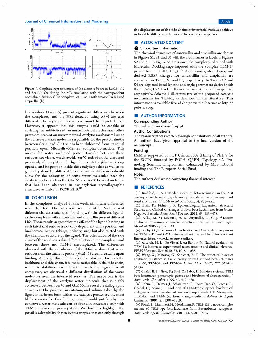
key residues (Table 5) present significant differences betweenthe complexes, and the HSs detected using ASM are alsodifferent. The acylation mechanism cannot be depicted here.However, it appears that this enzyme could be capable ofacylating the antibiotics via an unsymmetrical mechanism (otherproteases present an unsymmetrical catalytic mechanism) sincethe conserved water molecule responsible for the proton shuttlebetween Ser70 and Glu166 has been dislocated from its initialposition upon Michaelis−Menten complex formation. Thismakes the water mediated proton transfer between theseresidues not viable, which avoids Ser70 activation. As discussedpreviously after acylation, the ligand presents the β-lactamic ringopened, and its position inside the catalytic pocket as well as itsgeometry should be different. These structural differences shouldallow for the relocation of some water molecules near thecatalytic pocket such as the Glu166 and Ser70 bonded moleculethat has been observed in pos-acylation crystallographicstructures available in RCSB-PDB.10
■ CONCLUSIONIn the complexes analyzed in this work, significant differenceswere detected. The interfacial residues of TEM-1 presentdifferent characteristics upon binding with the different ligandsas the complexes with amoxicillin and ampicillin present differentHSs. These results suggest that the effect of the ligand binding ineach interfacial residue is not only dependent on its position andbiochemical nature (charge, polarity, size) but also related withthe chemical structure of the ligand. The orientation of the sidechain of the residues is also different between the complexes andbetween these and TEM-1 uncomplexed. The differencesobserved with the calculation of the B-factor show that someresidues near the catalytic pocket (Glu240) are more stable uponbinding. Although this difference can be observed for both thebackbone and side chain, it is more noticeable in the side chain,which is stabilized via interaction with the ligand. In allcomplexes, we observed a different distribution of the watermolecules near the interfacial residues. The major one is thedisplacement of the catalytic water molecule that is highlyconserved between Ser70 and Glu166 in several crystallographicstructures. The position, orientation, and volume taken by theligand in its intact form within the catalytic pocket are the mostlikely reasons for this finding, which would justify why thisconserved water molecule can be found in structures only withTEM enzymes or pos-acylation. We have to highlight thepossible adaptability shown by this enzyme that can only through
the displacement of the side chains of interfacial residues achievenoticeable differences between the various complexes.
■ ASSOCIATED CONTENT*S Supporting InformationThe chemical structures of amoxicillin and ampicillin are shownin Figures S1, S2, and S3 with the atom names as labels is FiguresS2 and S3. In Figure S4 are shown the complexes obtained withMolecular Docking superimposed with the complex TEM-1/penam from PDBID: 1FQG.77 Atom names, atom types, andderived RESP charges for amoxicillin and ampicillin areappointed in Tables S1 and S3, respectively. In Tables S2 andS4 are depicted bond lengths and angle parameters derived withthe HF/6-31G* level of theory for amoxicillin and ampicillin,respectively. Scheme 1 illustrates two of the proposed catalyticmechanisms for TEM-1, as described in the literature. Thisinformation is available free of charge via the Internet at http://pubs.acs.org.
■ AUTHOR INFORMATIONCorresponding Author*E-mail: [email protected] ContributionsThe manuscript was written through contributions of all authors.All authors have given approval to the final version of themanuscript.FundingI.S.M. is supported by FCT Ciencia 2008 (Hiring of Ph.D.’s forthe SCTN−financed by POPH−QREN−Typology 4.2−Pro-moting Scientific Employment, cofinanced by MES nationalfunding and The European Social Fund).NotesThe authors declare no competing financial interest.
■ REFERENCES(1) Bradford, P. A. Extended-spectrum beta-lactamases in the 21stcentury: characterization, epidemiology, and detection of this importantresistance threat. Clin. Microbiol. Rev. 2001, 14, 933−951.(2) Bush, K.; Fisher, J. F. Epidemiological Expansion, StructuralStudies, and Clinical Challenges of New beta-Lactamases from Gram-Negative Bacteria. Annu. Rev. Microbiol. 2011, 65, 455−478.(3) Wilke, M. S.; Lovering, A. L.; Strynadka, N. C. J. β-Lactamantibiotic resistance: a current structural perspective. Curr. Opin.Microbiol. 2005, 8, 525−533.(4) Jacoby, G. β-Lactamase Classification and Amino Acid Sequencesfor TEM, SHV and OXA Extended-Spectrum and Inhibitor ResistantEnzymes. http://www.lahey.org/Studies/.(5) Salverda, M. L.; De Visser, J. A.; Barlow, M. Natural evolution ofTEM-1 β-lactamase: experimental reconstruction and clinical relevance.FEMS Microbiol. Rev. 2010, 34, 1015−1036.(6) Wang, X.; Minasov, G.; Shoichet, B. K. The structural bases ofantibiotic resistance in the clinically derived mutant beta-lactamasesTEM-30, TEM-32, and TEM-34. J. Biol. Chem. 2002, 277, 32149−32156.(7) Chaïbi, E. B.; Sirot, D.; Paul, G.; Labia, R. Inhibitor-resistant TEMbeta-lactamases: phenotypic, genetic and biochemical characteristics. J.Antimicrob. Chemother. 1999, 43, 447−458.(8) Robin, F.; Delmas, J.; Schweitzer, C.; Tournilhac, O.; Lesens, O.;Chanal, C.; Bonnet, R. Evolution of TEM-type enzymes: biochemicaland genetic characterization of two new complex mutant TEM enzymes,TEM-151 and TEM-152, from a single patient. Antimicrob. AgentsChemother. 2007, 51, 1304−1309.(9) Poirel, L.; Mammeri, H.; Nordmann, P. TEM-121, a novel complexmutant of TEM-type beta-lactamase from Enterobacter aerogenes.Antimicrob. Agents Chemother. 2004, 48, 4528−4531.
Figure 7. Graphical representation of the distance between Lys73−Nζand Ser130−Oγ during the MD simulation with the correspondentnormalized distances93 in complexes of TEM-1 with amoxicillin (a) andampicillin (b).
Journal of Chemical Information and Modeling Article
dx.doi.org/10.1021/ci400269d | J. Chem. Inf. Model. XXXX, XXX, XXX−XXXH
(10) Bernstein, F. C.; Koetzle, T. F.; Williams, G. J.; Meyer, E. F.; Brice,M. D.; Rodgers, J. R.; Kennard, O.; Shimanouchi, T.; Tasumi, M. TheProtein Data Bank. A computer-based archival file for macromolecularstructures. Eur. J. Biochem. 1977, 80, 319−324.(11) Chandonia, J. M.; Brenner, S. E. The impact of structuralgenomics: expectations and outcomes. Science 2006, 311, 347−351.(12) Hunter, W. N. Present at the Flood. By Richard E. Dickerson.Sunderland, Mass.: Sinauer Associates, Inc., 2005. Pp. 307. ISBN 0-87893-168-6. Acta Crystallogr., Sect. D: Biol. Crystallogr. 2007, 63, 750.(13) Abola, E.; Kuhn, P.; Earnest, T.; Stevens, R. C. Automation of X-ray crystallography. Nat. Struct. Biol. 2000, 7, 973−977.(14) Sorensen, T. L.; McAuley, K. E.; Flaig, R.; Duke, E. M. New lightfor science: synchrotron radiation in structural medicine. TrendsBiotechnol. 2006, 24, 500−508.(15) Marco, E.; Gago, F. Overcoming the Inadequacies or Limitationsof Experimental Structures as Drug Targets by Using ComputationalModeling Tools and Molecular Dynamics Simulations. ChemMedChem2007, 2, 1388−1401.(16) Moreira, I. S.; Martins, J. M.; Ramos, R. M.; Fernandes, P. A.;Ramos, M. J. Understanding the importance of the aromatic amino-acidresidues as hot-spots. Biochim. Biophys. Acta, Proteins Proteomics 2013,1834, 404−414.(17) Gerstein, M.; Krebs, W. A database of macromolecular motions.Nucleic Acids Res. 1998, 26, 4280−4290.(18) Oliveira, B. L.; Moreira, I. S.; Fernandes, P. A.; Ramos, M. J.;Santos, I.; Correia, J. D. Insights into the structural determinats forselective inhibition of nitric oxide synathase isofroms. J. Mol. Model.2012, 19, 1537−1551.(19) Karplus, M.; McCammon, J. A. Molecular dynamics simulationsof biomolecules. Nat. Struct. Mol. Biol. 2002, 9, 646−652.(20) Martins, J. M.; Ramos, R. M.; Moreira, I. S. Structural determinatsof a typical leucine-rich repeat protein. Commun. Comput. Phys. 2013,13, 238−255.(21) Moreira, I. S.; Fernandes, P. A.; Ramos, M. J. Hot spot occlusionfrom bulk water: a comprehensive study of the complex between thelysozyme HEL and the antibody FVD1.3. J. Phys. Chem. B 2007, 111,2697−2706.(22)Moreira, I. S.; Fernandes, P. A.; Ramos,M. J. Detailed microscopicstudy of the full ZipA: FtsZ interface. Proteins: Struct., Funct., Bioinf.2006, 63, 811−821.(23) Durrant, J.; McCammon, J. A. Molecular dynamics simulationsand drug discovery. BMC Biol. 2011, 9, 71.(24) Fisette, O.; Morin, S.; Savard, P. Y.; Lague, P.; Gagne, S. M. TEM-1 backbone dynamics-insights from combined molecular dynamics andnuclear magnetic resonance. Biophys. J. 2010, 98, 637−645.(25) Bos, F.; Pleiss, J. Multiple molecular dynamics simulations ofTEM beta-lactamase: dynamics and water binding of the omega-loop.Biophys. J. 2009, 97, 2550−2558.(26) Díaz, N.; Suarez, D.; Merz, K. M.; Sordo, T. L. Moleculardynamics simulations of the TEM-1 beta-lactamase complexed withcephalothin. J. Med. Chem. 2005, 48, 780−791.(27) Stec, B.; Holtz, K. M.; Wojciechowski, C. L.; Kantrowitz, E. R.Structure of the wild-type TEM-1 beta-lactamase at 1.55 A and themutant enzyme Ser70Ala at 2.1 A suggest the mode of noncovalentcatalysis for the mutant enzyme. Acta Crystallogr., Sect. D: Biol.Crystallogr. 2005, 61, 1072−1079.(28) Thomas, V. L.; Golemi-Kotra, D.; Kim, C.; Vakulenko, S. B.;Mobashery, S.; Shoichet, B. K. Structural consequences of the inhibitor-resistant Ser130Gly substitution in TEM beta-lactamase. Biochemistry2005, 44, 9330−9338.(29) Saves, I.; Burlet-Schiltz, O.; Swaren, P.; Lefevre, F.; Masson, J. M.;Prome, J. C.; Samama, J. P. The asparagine to aspartic acid substitutionat position 276 of TEM-35 and TEM-36 is involved in the beta-lactamase resistance to clavulanic acid. J. Biol. Chem. 1995, 270, 18240−18245.(30) Thorsen, T. S.; Madsen, K. L.; Rebola, N.; Rathje, M.; Anggono,V.; Bach, A.; Moreira, I. S.; Stuhr-Hansen, N.; Dyhring, T.; Peters, D.;Beuming, T.; Huganir, R.; Weinstein, H.; Mulle, C.; Strømgaard, K.;Rønn, L. C.; Gether, U. Identification of a small-molecule inhibitor of
the PICK1 PDZ domain that inhibits hippocampal LTP and LTD. Proc.Natl. Acad. Sci. U. S. A. 2010, 107, 413−418.(31) Bach, A.; Stuhr-Hansen, N.; Thorsen, T. S.; Bork, N.; Moreira, I.S.; Frydenvang, K.; Padrah, S.; Christensen, S. B.; Madsen, K. L.;Weinstein, H.; Gether, U.; Strømgaard, K. Structure-activity relation-ships of a small-molecule inhibitor of the PDZ domain of PICK1. Org.Biomol. Chem. 2010, 8, 4281−4288.(32) Ramamoorthy, D.; Turos, E.; Guida,W. C. Identification of a NewBinding Site in E. coli FabH using Molecular Dynamics Simulations:Validation by Computational Alanine Mutagenesis and DockingStudies. J. Chem. Inf. Model. 2013, 53, 1138−1156.(33) Lee, H. S.; Jo, S.; Lim, H.-S.; Im, W. Application of Binding FreeEnergy Calculations to Prediction of Binding Modes and Affinities ofMDM2 and MDMX Inhibitors. J. Chem. Inf. Model. 2012, 52, 1821−1832.(34) De Luca, L.; Morreale, F.; Chimirri, A. Insight into theFundamental Interactions between LEDGF Binding Site Inhibitorsand Integrase Combining Docking and Molecular Dynamics Simu-lations. J. Chem. Inf. Model. 2012, 52, 3245−3254.(35) Moreira, I. S.; Fernandes, P. A.; Ramos, M. J. Hot spots-A reviewof the protein-protein interface determinant amino-acid residues.Proteins: Struct., Funct., Bioinf. 2007, 68, 803−812.(36) Shelley, J.; Cholleti, A.; Frye, L.; Greenwood, J.; Timlin, M.;Uchimaya, M. Epik: a software program for pK a prediction andprotonation state generation for drug-like molecules. J. Comput.-AidedMol. Des. 2007, 21, 681−691.(37) Maestro, version 9.2; Schrodinger, Inc.; New York, 2011.(38) Cornell, W. D.; Cieplak, P.; Bayly, C. I.; Gould, I. R.; Merz, K. M.;Ferguson, D.M.; Spellmeyer, D. C.; Fox, T.; Caldwell, J. W.; Kollman, P.A. A Second Generation Force Field for the Simulation of Proteins,Nucleic Acids, and Organic Molecules. J. Am. Chem. Soc. 1995, 117,5179−5197.(39) Duan, Y.; Wu, C.; Chowdhury, S.; Lee, M. C.; Xiong, G.; Zhang,W.; Yang, R.; Cieplak, P.; Luo, R.; Lee, T.; Caldwell, J.; Wang, J.;Kollman, P. A point-charge force field for molecular mechanicssimulations of proteins based on condensed-phase quantum mechanicalcalculations. J. Comput. Chem. 2003, 24, 1999−2012.(40) Berendsen, H. J. C.; Postma, J. P. M.; van Gunsteren, W. F.;DiNola, A.; Haak, J. R. Molecular dynamics with coupling to an externalbath. J. Chem. Phys. 1984, 81, 3684−3690.(41) Ryckaert, J.-P.; Ciccotti, G.; Berendsen, H. J. C. Numericalintegration of the cartesian equations of motion of a system withconstraints: molecular dynamics of n-alkanes. J. Comput. Phys. 1977, 23,327−341.(42) Loncharich, R. J.; Brooks, B. R.; Pastor, R. W. Langevin dynamicsof peptides: The frictional dependence of isomerization rates of N-acetylalanyl-N′-methylamide. Biopolymers 1992, 32, 523−535.(43) Izaguirre, J. A.; Catarello, D. P.; Wozniak, J. M.; Skeel, R. D.Langevin stabilization of molecular dynamics. J. Chem. Phys. 2001, 114,2090−2098.(44) Ewald, P. P. Die Berechnung optischer und elektrostatischerGitterpotentiale. Ann. Phys. (Berlin, Ger.) 1921, 369, 253−287.(45) Darden, T.; York, D.; Pedersen, L. Particle mesh Ewald: An N[center-dot] log(N) method for Ewald sums in large systems. J. Chem.Phys. 1993, 98, 10089−10092.(46) Bush, K.; Jacoby, G. A. Updated functional classification of beta-lactamases. Antimicrob. Agents Chemother. 2010, 54, 969−76.(47) Suarez, C. Antibioticos betalactamicos. Enferm. Infecc. Microbiol.Clin. 2009, 27, 116−129.(48) Bolton, E. E.; Wang, Y.; Thiessen, P. A.; Bryant, S. H., Chapter 12PubChem: Integrated Platform of Small Molecules and BiologicalActivities. In Annu. Rep. Comput. Chem.; Ralph, A. W. a. D. C. S., Ed.;Elsevier: New York, 2008; Vol. 4, pp 217−241.(49) Morris, G. M.; Huey, R.; Lindstrom, W.; Sanner, M. F.; Belew, R.K.; Goodsell, D. S.; Olson, A. J. AutoDock4 and AutoDockTools4:Automated docking with selective receptor flexibility. J. Comput. Chem.2009, 30, 2785−2791.(50) Bayly, C. I.; Cieplak, P.; Cornell, W.; Kollman, P. A. A well-behaved electrostatic potential based method using charge restraints for
Journal of Chemical Information and Modeling Article
dx.doi.org/10.1021/ci400269d | J. Chem. Inf. Model. XXXX, XXX, XXX−XXXI

deriving atomic charges: the RESP model. J. Chem. Phys. 1993, 97,10269−10280.(51) Case, D. A.; Cheatham, T. E.; Darden, T.; Gohlke, H.; Luo, R.;Merz, K. M.; Onufriev, A.; Simmerling, C.; Wang, B.; Woods, R. J. TheAmber biomolecular simulation programs. J. Comput. Chem. 2005, 26,1668−1688.(52) Wang, J.; Wolf, R. M.; Caldwell, J. W.; Kollman, P. A.; Case, D. A.Development and testing of a general amber force field. J. Comput. Chem.2004, 25, 1157−1174.(53) Stapleton, P. D.; Shannon, K. P.; French, G. L. Construction andcharacterization of mutants of the TEM-1 beta-lactamase containingamino acid substitutions associated with both extended-spectrumresistance and resistance to beta-lactamase inhibitors. Antimicrob. AgentsChemother. 1999, 43, 1881−1887.(54) Knox, J. R. Extended-spectrum and inhibitor-resistant TEM-typebeta-lactamases: mutations, specificity, and three-dimensional structure.Antimicrob. Agents Chemother. 1995, 39, 2593−2601.(55) Humphrey, W.; Dalke, A.; Schulten, K. VMD: Visual moleculardynamics. J. Mol. Graphics 1996, 14, 33−38.(56) DeLano, W. L. The PyMOL Molecular Graphics System; DeLanoScientific: San Carlos, CA, 2002.(57) Huo, S.; Massova, I.; Kollman, P. A. Computational alaninescanning of the 1: 1 human growth hormone-receptor complex. J.Comput. Chem. 2002, 23, 15−27.(58) Case, D. A.; Darden, T. A.; Cheatham, T. E., III; Simmerling, C.L.; Wang, J.; Duke, R. E.; Luo, R.; Crowley, M.; Walker, R. C.; Zhang,W.; Merz, K. M.; Wang, B.; Hayik, S.; Roitberg, A.; Seabra, G.;Kolossvary, I.; Wong, K. F.; Paesani, F.; Vanicek, J.; Wu, X.; Brozell, S.R.; Steinbrecher, T.; Gohlke, H.; Yang, L.; Tan, C.; Mongan, J.; Hornak,V.; Cui, G.; Mathews, D. H.; Seetin, M. G.; Sagui, C.; Babin, V.; Kollman,P. A. AMBER 10; University of California: San Francisco, CA, 2008.(59) Moreira, I. S.; Fernandes, P. A.; Ramos, M. J. Unraveling theimportance of protein-protein interaction: Application of a computa-tional alanine-scanning mutagenesis to the study of the IgG1streptococcal protein G (C2 fragment) complex. J. Phys. Chem. B.2006, 110, 10962−10969.(60) Moreira, I. S.; Fernandes, P. A.; Ramos, M. J. Unravelling HotSpots: a comprehensive computational mutagenesis study. Theor. Chem.Acc. 2007, 117, 99−113.(61) Moreira, I. S.; Fernandes, P. A.; Ramos, M. J. Hot spotcomputational identification: Application to the complex formedbetween the hen egg white lysozyme (HEL) and the antibodyHyHEL-10. Int. J. Quantum Chem. 2007, 107, 299−310.(62) Moreira, I. S.; Fernandes, P. A.; Ramos, M. J. Computationalalanine scanning mutagenesis - An improved methodological approach.J. Comput. Chem. 2007, 28, 644−654.(63) Moreira, I. S.; Fernandes, P. A.; Ramos, M. J. Backboneimportance for protein-protein binding. J. Chem. Theory Comput. 2007,3, 885−893.(64) Moreira, I. S.; Fernandes, P. A.; Ramos, M. J. Hot spot occlusionfrom bulk water: A comprehensive study of the complex between thelysozyme HEL and the antibody FVD1.3. J. Phys. Chem. B 2007, 111,2697−2706.(65) Moreira, I. S.; Fernandes, P. A.; Ramos, M. J. Protein-proteinrecognition: a computational mutagenesis study of the MDM2-P53complex. Theor. Chem. Acc. 2008, 120, 533−542.(66) Chong, L. T.; Duan, Y.; Wang, L.; Massova, I.; Kollman, P. A.Molecular dynamics and free-energy calculations applied to affinitymaturation in antibody 48G7. Proc. Natl. Acad. Sci. U. S. A. 1999, 96,14330−14335.(67) Kollman, P. A.; Massova, I.; Reyes, C.; Kuhn, B.; Huo, S. H.;Chong, L.; Lee,M.; Lee, T.; Duan, Y.;Wang,W.; Donini, O.; Cieplak, P.;Srinivasan, J.; Case, D. A.; Cheatham, T. E. Calculating structures andfree energies of complex molecules: Combining molecular mechanicsand continuum models. Acc. Chem. Res. 2000, 33, 889−897.(68) Bradshaw, R. T.; Patel, B. H.; Tate, E. W.; Leatherbarrow, R. J.;Gould, I. R. Comparing experimental and computational alaninescanning techniques for probing a prototypical protein-proteininteraction. Protein Eng., Des. Sel. 2011, 24, 197−207.
(69) Moreira, I. S.; Martins, J. M.; Ramos, M. J.; Fernandes, P. A.;Ramos, M. J. Understanding the importance of the aromatic amino-acidresidues as hot-spots. Biochem. Biophys. Acta. 2013, 1834, 401−414.(70) Ribeiro, J. V.; Cerqueira, N. M. F. S. A.; Moreira, I. S.; Fernandes,P. A.; Ramos, M. J. CompASM: an Amber-VMD Alanine ScanningMutagenesis plug-in. Theor. Chem. Acc. 2012, 131, 1271−1278.(71) Rocchia, W.; Alexov, E.; Honig, B. Extending the applicability ofthe nonlinear Poisson-Boltzmann equation: Multiple dielectricconstants and multivalent ions. J. Phys. Chem. B. 2001, 105, 6507−6514.(72) Rocchia, W.; Sridharan, S.; Nicholls, A.; Alexov, E.; Chiabrera, A.;Honig, B. Rapid grid-based construction of the molecular surface andthe use of induced surface charge to calculate reaction field energies:Applications to the molecular systems and geometric objects. J. Comput.Chem. 2002, 23, 128−137.(73) Moreira, I. S.; Fernandes, P. A.; Ramos, M. J. Accuracy of thenumerical solution of the Poisson-Boltzmann equation. J. Mol. Struct.:THEOCHEM 2005, 729, 11−18.(74) Sitkoff, D.; Sharp, K. A.; Honig, B. Accurate calculation ofhydration free-energies usingmacroscopic solvent models. J. Phys. Chem.1994, 98, 1978−1988.(75) Connolly, M. L. Analytical molecular surface calculation. J. Appl.Crystallogr. 1983, 16, 548−558.(76) Konc, J.; Janezic, D. ProBiS-2012: web server and web services fordetection of structurally similar binding sites in proteins. Nucleic AcidsRes. 2012, 40, W214−221.(77) Strynadka, N. C.; Adachi, H.; Jensen, S. E.; Johns, K.; Sielecki, A.;Betzel, C.; Sutoh, K.; James, M. N. Molecular structure of the acyl-enzyme intermediate in beta-lactam hydrolysis at 1.7 A resolution.Nature 1992, 359, 700−705.(78) Maveyraud, L.; Massova, I.; Birck, C.; Miyashita, K.; Samama, J.P.; Mobashery, S. Crystal Structure of 6Alpha-Hydroxymethylpenicilla-nate Complexed to the Tem-1 Beta-Lactamase from Escherichia Coli:Evidence on the Mechanism of Action of a Novel Inhibitor Designed bya Computer-Aided Process. J. Am. Chem. Soc. 1996, 118, 7435.(79) Maveyraud, L.; Mourey, L.; Kotra, L. P.; Pedelacq, J.-D.; Guillet,V.; Mobashery, S.; Samama, J.-P. Structural Basis for Clinical Longevityof Carbapenem Antibiotics in the Face of Challenge by the CommonClass A β-Lactamases from the Antibiotic-Resistant Bacteria. J. Am.Chem. Soc. 1998, 120, 9748−9752.(80) Roccatano, D.; Sbardella, G.; Aschi, M.; Amicosante, G.; Bossa,C.; Di Nola, A.; Mazza, F. Dynamical aspects of TEM-1 beta-lactamaseprobed by molecular dynamics. J. Comput.-Aided Mol. Des. 2005, 19,329−340.(81) Massova, I.; Kollman, P. A. pKa, MM, and QM studies ofmechanisms of β-lactamases and penicillin-binding proteins: Acylationstep. J. Comput. Chem. 2002, 23, 1559−1576.(82) Medeiros, A. A. Evolution and dissemination of beta-lactamasesaccelerated by generations of beta-lactam antibiotics. Clin. Infect. Dis.1997, 24 (Suppl 1), S19−45.(83) Matagne, A.; Lamotte-Brasseur, J.; Frere, J. M. Catalyticproperties of class A beta-lactamases: efficiency and diversity. Biochem.J. 1998, 330 (Pt 2), 581−598.(84) Bren, U.; Oostenbrink, C. Cytochrome P450 3A4 inhibition byketoconazole: tackling the problem of ligand cooperativity usingmolecular dynamics simulations and free-energy calculations. J. Chem.Inf. Model. 2012, 52, 1573−1582.(85) Bren, U.; Janezic, D. Individual degrees of freedom and thesolvation properties of water. J. Chem. Phys. 2012, 137, 024108.(86) Bren, U.; Krzan, A.; Mavri, J. Microwave catalysis throughrotationally hot reactive species. J. Phys. Chem. A. 2008, 112, 166−171.(87) Hamza, A.; Abdulhameed, M. D.; Zhan, C. G. Understandingmicroscopic binding of human microsomal prostaglandin E synthase-1with substrates and inhibitors by molecular modeling and dynamicssimulation. J. Phys. Chem. B. 2008, 112, 7320−7329.(88) Dubey, K. D.; Ojha, R. P. Binding free energy calculation withQM/MM hybrid methods for Abl-Kinase inhibitor. J. Biol. Phys. 2011,37, 69−78.
Journal of Chemical Information and Modeling Article
dx.doi.org/10.1021/ci400269d | J. Chem. Inf. Model. XXXX, XXX, XXX−XXXJ
(89) Bren, M.; Florian, J.; Mavri, J.; Bren, U. Do all pieces make awhole? Thiele cumulants and the free energy decomposition. Theor.Chem. Acc. 2007, 117, 535−540.(90) Bogan, A. A.; Thorn, K. S. Anatomy of hot spots in proteininterfaces. J. Mol. Biol. 1998, 280, 1−9.(91) Moreira, I. S.; Ramos, R. M.; Martins, J. M.; Fernandes, P. A.;Ramos, M. J., Are hot-spots occluded from water? J. Biomol. Struct. Dyn.2013, [Online] http://www.tandfonline.com/doi/abs/10.1080/07391102.2012.758598?url_ver=Z39.88-2003&rfr_id=ori:rid:crossref.org&rfr_dat=cr_pub%3dpubmed#.UfKusvkqeSo (accessed Jully 26,2013).(92) Nejc, C.; Hodoscek, M.; Vehar, B.; Konc, J.; Brooks, B. R.; Janezic,D. Correlating Protein Hot Spot Surface Analysis Using ProBiS withSimulated Free Energies of Protein−Protein Interfacial Residues. J.Chem. Inf. Model. 2012, 52, 2541−2549.(93) Graf, M. M.; Bren, U.; Haltrich, D.; Oostenbrink, C. Moleculardynamics simulations give insight into D-glucose dioxidation at C2 andC3 by Agaricus meleagris pyranose dehydrogenase. J. Comput.-AidedMol. Des. 2013, 27, 295−304.
Journal of Chemical Information and Modeling Article
dx.doi.org/10.1021/ci400269d | J. Chem. Inf. Model. XXXX, XXX, XXX−XXXK