LC-ESI-MS characterisation of phytoalexins induced in chickpea and pea tissues in response to a...
10
This article was downloaded by: [Muhammad Arman] On: 28 August 2011, At: 04:21 Publisher: Taylor & Francis Informa Ltd Registered in England and Wales Registered Number: 1072954 Registered office: Mortimer House, 37-41 Mortimer Street, London W1T 3JH, UK Natural Product Research Publication details, including instructions for authors and subscription information: http://www.tandfonline.com/loi/gnpl20 LC-ESI-MS characterisation of phytoalexins induced in chickpea and pea tissues in response to a biotic elicitor of Hypnea musciformis (red algae) Muhammad Arman a a PCSIR Laboratories Complex Karachi, Sharah-e-Dr Salimuzzaman Siddiqui, Off University Road, Karachi -75280, Pakistan Available online: 22 Aug 2011 To cite this article: Muhammad Arman (2011): LC-ESI-MS characterisation of phytoalexins induced in chickpea and pea tissues in response to a biotic elicitor of Hypnea musciformis (red algae), Natural Product Research, 25:14, 1352-1360 To link to this article: http://dx.doi.org/10.1080/14786419.2011.553952 PLEASE SCROLL DOWN FOR ARTICLE Full terms and conditions of use: http://www.tandfonline.com/page/terms-and- conditions This article may be used for research, teaching and private study purposes. Any substantial or systematic reproduction, re-distribution, re-selling, loan, sub-licensing, systematic supply or distribution in any form to anyone is expressly forbidden. The publisher does not give any warranty express or implied or make any representation that the contents will be complete or accurate or up to date. The accuracy of any instructions, formulae and drug doses should be independently verified with primary sources. The publisher shall not be liable for any loss, actions, claims, proceedings, demand or costs or damages whatsoever or howsoever caused arising directly or indirectly in connection with or arising out of the use of this material.
-
Upload
independent -
Category
Documents
-
view
6 -
download
0
Transcript of LC-ESI-MS characterisation of phytoalexins induced in chickpea and pea tissues in response to a...

This article was downloaded by: [Muhammad Arman]On: 28 August 2011, At: 04:21Publisher: Taylor & FrancisInforma Ltd Registered in England and Wales Registered Number: 1072954 Registeredoffice: Mortimer House, 37-41 Mortimer Street, London W1T 3JH, UK
Natural Product ResearchPublication details, including instructions for authors andsubscription information:http://www.tandfonline.com/loi/gnpl20
LC-ESI-MS characterisation ofphytoalexins induced in chickpea andpea tissues in response to a bioticelicitor of Hypnea musciformis (redalgae)Muhammad Arman aa PCSIR Laboratories Complex Karachi, Sharah-e-Dr SalimuzzamanSiddiqui, Off University Road, Karachi -75280, Pakistan
Available online: 22 Aug 2011
To cite this article: Muhammad Arman (2011): LC-ESI-MS characterisation of phytoalexins inducedin chickpea and pea tissues in response to a biotic elicitor of Hypnea musciformis (red algae),Natural Product Research, 25:14, 1352-1360
To link to this article: http://dx.doi.org/10.1080/14786419.2011.553952
PLEASE SCROLL DOWN FOR ARTICLE
Full terms and conditions of use: http://www.tandfonline.com/page/terms-and-conditions
This article may be used for research, teaching and private study purposes. Anysubstantial or systematic reproduction, re-distribution, re-selling, loan, sub-licensing,systematic supply or distribution in any form to anyone is expressly forbidden.
The publisher does not give any warranty express or implied or make any representationthat the contents will be complete or accurate or up to date. The accuracy of anyinstructions, formulae and drug doses should be independently verified with primarysources. The publisher shall not be liable for any loss, actions, claims, proceedings,demand or costs or damages whatsoever or howsoever caused arising directly orindirectly in connection with or arising out of the use of this material.

Natural Product ResearchVol. 25, No. 14, August 2011, 1352–1360
LC-ESI-MS characterisation of phytoalexins induced in chickpea
and pea tissues in response to a biotic elicitor of Hypnea musciformis(red algae)
Muhammad Arman*
PCSIR Laboratories Complex Karachi, Sharah-e-Dr Salimuzzaman Siddiqui,Off University Road, Karachi -75280, Pakistan
(Received 4 March 2010; final version received 10 January 2011)
A simple extraction procedure and HPLC method was developed to analysethe major and minor components of induced phytoalexins of elicited tissues(seeds) of chickpeas (Cicer arietinum L.) and peas (Pisum sativum L.)treated with a biotic elicitor (k-carrageenan) of Hypnea musciformis(red algae) from the Karachi coast. The level and timing of the inducedphytoalexin production were estimated on the basis of various elicitordilutions and as a function of time; the results are presented and discussed.A LC-ESI-MS/MS technique has been employed for the detection andcharacterisation of the induced phytochemical components (flavonoids andtheir glyco-conjugates). Nine flavonoids were identified from chickpeas:naringin, naringin malonate, liquiritigenin, naringenin, biochanin A,daidzein, formononetin, maackiain and medicarpin, while five flavonoidswere identified from peas: afrormosin, anhydropisatin, pisatin, pseudo-baptigenin and maackiain. These compounds play a vital role as phyto-alexins because of their antimicrobial activity.
Keywords: elicitor; induced secondary metabolites; phytoalexins; polysac-charides; Hypnea musciformis; flavonoids; LC-ESI-MS/MS
1. Introduction
As a result of infection or stress, plants exhibit some natural resistance responses.Amongst these, induced browning and phytoalexin production have especiallygained attention (Castoria, Maria, Anna, & Marisa, 1995). Phytoalexins are lowmolecular weight, biologically active compounds produced by plants as a defensivereaction to various exogenous stimuli, particularly fungal invasion, their role indisease resistance has been deduced from their antifungal activity (Arnold & Merlin,1990). Some of the reported phytoalexins in leguminosae species are alkaloids,coumarins and especially isoflavonoid derivatives (Grayer & Kokubun, 2001).The biosynthesis of isoflavonoid phytoalexins has been intensively studied and theirmetabolic regulation is known to be different according to the inductor agent, plantcell type and type of elicitors (Smith & Banks, 1986). It is reported thathypersensitive responses produced in plants after microbial attack may be triggered
*Email: [email protected]
ISSN 1478–6419 print/ISSN 1029–2349 online
� 2011 Taylor & Francis
DOI: 10.1080/14786419.2011.553952
http://www.informaworld.com
Dow
nloa
ded
by [
Muh
amm
ad A
rman
] at
04:
21 2
8 A
ugus
t 201
1

by elicitors that are the compounds isolated from cell wall, culture filtrate andcytoplasm of parasitic and non parasitic plant pathogens. Elicitors are diverse innature and are usually polysaccharides, proteins and fatty acids (Rao, Sarada, &Ravishankar, 1996).
Chickpea (Cicer arietinum L.) and peas (Pisum sativum L.) belonging to thefamily leguminosae are important crop plants and have both economical andnutritional importance. Due to high protein contents, they are consumed widely inmany countries as a meat substitute for poor people. The antimicrobial activities ofthese two plants are due to the presence of their phytoalexins i.e. pisatin andmaackiain (Ho, Lin, Labbe, & Shetty, 2003). A relationship between phytoalexinaccumulation and defense against pathogenic microorganisms in peas(Pisum sativum), alfalfa (Medicago sativa), barrel medic (Medicago truncatula) andchickpea (Cicer arietinum) is recently reported in literature (Liu et al., 2006).
It is documented that in most cases elicitor activity is associated withpolysaccharides fraction of various elicitor preparations when tested in chickpeaand other plant tissues (Melotto & Lebavitch, 1994). In the present study, theseaweed (Hypnea musciformis, red algae) polysaccharides were evaluated as anelicitor or inducer of plant defense responses in terms of phytoalexin production.A simple HPLC separation method was developed to analyse the major and minorcomponents of the mixture of induced secondary metabolites ‘phytoalexins’ and theindividual components were identified by LC-Electro Spray Mass Spectrometrictechnique with positive and negative ionisation modes.
2. Results and discussion
In the present study alcoholic extracts of elicitor treated and control tissues ofchickpea were analysed for the estimation of ISMs (phytoalexins) with respectto various elicitor doses, 5–100 mg glu eqmL�1 and different incubation periods,6–72 h. A reproducible HPLC method was developed to analyse the major andminor components of induced phytoalexins. Typical HPLC chromatograms(Supplementary Figures S1 and S2 – online only) showed that the reproducibility ofelution time of the separated components was 2–3%. The integrated area of the peakswas assumed to be proportional to the amount of solutes (phytoalexins) present.Initial results for elicitor activity of pea tissues were not found very promising,induction of browning and estimation of inducedmetabolites (phytoalexins) in treatedtissues of peas were insignificant as compared to controls (Bi, Iqbal, Ali, Arman, &Hassan, 2008). Therefore, attempts were made to develop HPLC methods and for thecharacterisation of the components present in the pea’s chromatogram whereas theindividual components quantification was not carried out.
The data presented in Supplementary Tables S1 and S2 (online only) are a ratioof peak area/g fresh weight of treated and control samples. These results showed thatchickpea tissues responded differentially to the applied elicitor preparations andproduced a positive and definite elicitor activity in terms of phytoalexin production.100 mg elicitor concentration and 24 h incubation time were the best conditions toinduce metabolites at maximum level in the samples. Some components (marked byFT) were induced in elicitor treated samples only. Recently reported dose dependantstudies showed that 100 mgmL�1 and 1000 mgmL�1 concentrations of algal polysac-charides, � carrageenan were the effective dilutions for high induction of florescent
Natural Product Research 1353
Dow
nloa
ded
by [
Muh
amm
ad A
rman
] at
04:
21 2
8 A
ugus
t 201
1

compounds and defense related genes at 168 h and 24 h after elicitation in tobacco
leaves (Mercier et al., 2001). Cooper, Doss, Price, Peterson, and Oliver, 2005
reported that during time course studies in peas, application of a new class of insect
elicitor ‘Bruchin B’ increased the expression of isoflavone synthase gene within 8 h of
elicitor treatment followed by increase in the level of isoflavone phytoalexin pisatin
that reached the highest level at 32–64 h after elicitation.Due to the low amount of induced metabolites and non-availability of standard
materials, LC-Electro Spray Ionisation Mass Spectrometry (LC-ESI/MS) appears to
be the ideal technique for the identification of secondary metabolites (Kite et al.,
2006). In the present study, samples were run under identical conditions and
monitored simultaneously by UV and total ion current (TIC) chromatograms
(Supplementary Figures S3 and S4 – online only). UV analogue is in complete agree-
ment with most of the peaks present in TIC, however some major peaks (at RT
22.57, 8.21 and 20.61) and minor peaks (at RT 10.06 and 2.19) of these figures
appeared in TIC are not found in their UV analogs, therefore these peaks were not
characterised during this work. The retention time’s data of the identified
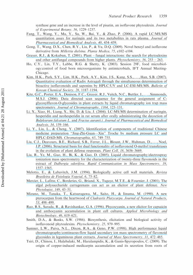
phytoalexins are summarised in Supplementary Table S3 (online only).Nine compounds from chickpea such as Naringin 1 (Fang et al., 2006; Zhang &
Brodbelt, 2004) Naringin malonate 2 (Liu, Ye, H. Guo, Bi, & D. Guo, 2005),
Liquiritigenin 3 (Li, Liu, & Cheng, 2007), Naringenin 4 (Zhang & Brodbelt, 2004),
Biochanin A 5 (Careri, Corradini, Elviri, & Mangia, 2007), Daidzein 6 (Careri et al.,
2007) Formononetin 7 (Kim et al., 2007) Maackiain 8 (Liu et al., 2005) and
Medicarpin 9 (Tsiri, Chinou, Halabalaki, Haralampidis, & Ganis-Spyropoulos,
2009) and five compounds from peas such as Afrormosin 10 (Carlsen et al., 2008;
Gong, Wang, Chen, Liu, & Yu, 2009), Anhydropisatin 11 (Mizuno, Tanaka,
Katsuragawa, Saito, & Iinuma, 1990;), Pisatin 12 (Mizuno et al., 1990;),
Pseudobaptigenin 13 (Wu, Wang, & Simon, 2003) have been identified by
LC-MS/MS/MS analysis. These compounds belong to the classes of flavanones,
Isoflavones, pterocarpans and their glyco conjugates (Figure 1). Maackiain was
found in both chickpea and peas tissues. Molecular ions in positive and negative
modes and the product ion spectra of these compounds were studied in order to
obtain the most specific transition (Li, Xiao, Liang, Shi, & Liu, 2004). The use of the
selected transition is necessary for the identification of these compounds.All identified phytoalexins (1–13) exhibited ionisation in both positive and
negative modes, compounds were predominantly detected as [MþH]þin positive
mode due to the high proton affinity. In negative ionisation mode all analytes
exhibited an [M�H]� ion due to loss of hydrogen and provided stable molecular ion
peaks (Liu et al., 2005). The main product ions observed for each analyte in positive
and negative ionisation modes are shown in Table S3.The MS/MS experiments were performed on each ion in positive and negative
modes, representative positive and negative ions ESI- MS and ESI-MS/MS spectra
of Naringenin are discussed and shown in Supplementary Figures S5–S8 (online
only). Literature search showed that the ions observed in positive ionisation mode
are mainly due to losses of sugar moieties (Bednarek et al., 2001) and from the
retro Diels-Alder cleavage of the ring C in the flavonoids (Liu et al., 2005). Similar
ions were observed in the current work in negative ionisation mode for compounds 1
and 2.
1354 M. Arman
Dow
nloa
ded
by [
Muh
amm
ad A
rman
] at
04:
21 2
8 A
ugus
t 201
1

For compounds 3, 4 and 6, the fragment ions observed in positive and negativemodes were mainly due to the losses of OH and CO ions as well as the loss indicatingfrom the retro Diels-Alder cleavage. In compounds 5, 7 and 10 the major fragmentions observed in positive and negative modes were due to the losses of CO & CH3
ions and from the loss occurred due to the retro Diels–Alder cleavage.In compounds 8, 9, 11 and 12, the major fragment ions observed in the positive
and negative modes were due to the losses of CH2O2 and CH3 ions along with somedifferent fragmentation pathways. Representative fragmentation pattern of medi-carpin (Supplementary Figure S9 – online only) in negative mode showed sixfragments A to F in the MS/MS spectrum among which D and E are thecharacteristic fragments for identification of pterocarpans (Woodward, 1981, 1982).For compound 13, the product ions observed in positive and negative modes weremainly due to the losses of CH2O2 and CO ions as well as the losses indicated by theretro Diels–Alder cleavage. Although these ions exhibited low abundance at 25% inNormalised Collision Energy (NCE), they become predominant when the collisionenergy was increased to 35% (Table S3).
The metabolic grid of an enzyme sequence showing the conjugation reaction andthe metabolic turnover of isoflavonoids in chickpeas was established earlier andexplained the conjugation reaction of isoflavanone such as formononetin-7-Oglucoside and formononetin-7-O-glucoside-6-O-malonate. Formononetin is referredas an intermediate component for the biosynthesis of pterocarpan phytoalexinsmaackiain and medicarpin in this system. Barz et al. (1988) reported formononetinwith one sugar component. The present studies showed an early glycoconjugationreaction soon as the naringenin was formed in the primary biosynthetic pathway.
O
OH
OH O
OOH
OH HH
OH
OH
CH2OR
H
OOH
H HH
OHOH
H
H
CH3
O
OH
O
HO
O
O
HO
R4
R1
O
O
R1
R2
R 4O
1 R=H2 R=COCH
2COOH
3 R=H4 R=OH
5 R1=OCH
3, R
2=R
3=H, R
4=OH
6 R1=OH, R
2=R
3=R
4=H
7 R1=OCH
3, R
2=R
3=R
4=H
10 R1=R
3=OCH
3, R2=R
4=H
13 R1R
2=O-CH
2-O, R
3=R
4=H
R3
R2
R
R3
8 R1R
2=O-CH
2-O, R
3=R
4=H
9 R1=OCH
3, R
2=R
3=R
4=H
11 R1R
2=O-CH
2-O,R
4=CH
3, R
3=Nofunctional group,
6a 11a=Double bond
12 R1R
2=O-CH
2-O,R
3=OH, R
4=CH
3
1
4
6
6a
11a
9
Figure 1. Identified phytoalexins 1–13 from elicited tissues of chickpea and peas.
Natural Product Research 1355
Dow
nloa
ded
by [
Muh
amm
ad A
rman
] at
04:
21 2
8 A
ugus
t 201
1

LC-(MS)n data showed that isoflavanone naringenin conjugation reaction provided
two compounds tentatively identified as naringin malonate (naringin-7-O-digluco-side-6-O-malonate) of molecular mass [683] and naringin (naringin- 7- O-diglyco-
side) of molecular mass [580]. Mass calculation indicated the presence of two sugarmoieties, glucose and rhamnose in these two compounds which were detected and
confirmed by paper chromatography. Maackiain and medicarpin were obtainedfrom formononetin instead of following the intermediate compounds like calycosin,
pseudobaptigenin, 2-hydroxy pseudobaptigenin, 2-hydroxy dihydropseudobapti-
genin, 2-hydroxy formononetin and vestitone, may be due to the transit inductiontime that was possibly difficult to locate or produced in very small quantities that
could not be detected. These compounds are known (Zhang & Brodbelt, 2004) andfirst time identified during this interaction. These results support the investigation of
Sumner, Paiva, Dixon, and Geno (1998) that described the optimisation parametersin the identification of flavonoid glucosides in alfalfa and chickpea extracts using
HPLC/CF-LSI MS technique.Referring to Figure S4, the compounds corresponding peaks 1–5 were
characterised as afrormosin, anhydropisatin, pisatin, pseudobaptigenin and maack-
iain and considered as pea phytoalexins. Earlier studies showed that liquiritigenin,isoliquiritigenin and formononetin were present in the biosynthesis of pisatin
(Carlson & Dolphin, 1981) whereas these compounds were not found during thisinvestigation.
3. Experimental
3.1. Plant materials
Plant collection, extraction and dry weight of High Molecular Weight Crude ElicitorPolysaccharides (HMWCEP) from hot water extract of H.musciformis, red algae of
Karachi Coast was described earlier (Bi & Iqbal, 1999). The voucher specimen wasdeposited at the Herbarium of the Botanical Garden of PCSIR Laboratories
Complex Karachi, Pakistan (M.A. R1130.E026). These crude polysaccharides were
purified by anion exchange chromatography on DEAE cellulose column and twopurified fractions PF-A and PF-B were collected. HMW PF-A was further examined
for its elicitor activity.
3.2. Elicitor activity and ethanolic extraction for induced secondarymetabolites (ISMs)
A general method of elicitor application was employed in all experiments
(Whitehead, Dey, & Dixon, 1982). Elicitor preparations of 5, 25, 50, 75 and100 mg glu. eqmL�1 of PF-A were used for dose response elicitor activity. Treated
and control samples were prepared by application of 20 mL of these elicitorpreparations and sterile water (control) on the cut surfaces of cotyledons and
incubated for 6, 12, 24, 48 and 72 h with control samples. Treated and control
samples were extracted with alcohol, defated with hexane and residual alcoholicextract were stored at �20�C in the dark.
1356 M. Arman
Dow
nloa
ded
by [
Muh
amm
ad A
rman
] at
04:
21 2
8 A
ugus
t 201
1

3.3. HPLC separation of phytoalexins
HPLC separation of phytoalexins of chickpea and peas were performed on FinniganSurveyor Liquid Chromatography equipped with PDA plus detector and auto-sampler plus (Thermo Electron Corporation, San Jose, CA USA). A (TeknokromaKromasil 100) C18 5 mm 25�0.46 cm analytical column was used for both chickpeaand peas analysis. A guard column of pellicular C18 hydrocarbon chemically bondedto glass beads was placed before the analytical column. Flow rate was 1mLmin�1.For the analysis of chickpea tissues, the gradient elution employed was acetonitrileand water; both solvents contained 1% acetic acid. Initially a solvent system consistof 80 : 20 Water:Acetonitrile was run for 5min, then a gradient of 40 : 60 reached into15min and further goes to 100% acetonitrile in 5min, stays there for another 5minand returned to initial solvent system, while for pea tissues the gradient elutionemployed was acetonitrile and 0.1% phosphoric acid in aqueous phase. Initially asolvent system consist of 40 : 60 Acetonitrile: 0.1% phosphoric acid in aqueous phasewas run for 3min, then a gradient of 95 : 05 reached into 17min and further goes to100 % acetonitrile in 5min, stays there for another 5min and returned to initialsolvent system. The whole chromatograms were monitored at 254 nm and 365 nm forchickpea and peas, respectively.
3.4. Sample preparations
Dry alcoholic extracts of elicited and control tissues of chickpea and peas weredissolved into 2mL of 40:60 following solvent system. (a) for chickpea, water:ace-tonitrile, both phases contain 1% acetic acid; (b) for peas, acetonitrile:0.1%phosphoric acid in aqueous phase. 100 mL of each solution was further diluted with1mL of their respective solvents, centrifuged and filtered through 0.2mm filters.20 mL of clear filtrates were applied to the column.
3.5. Identification of phytoalexins by LC-ESI MSn technique
Chromatograms run under the optimided HPLC conditions were reproduced onLC-MS system of Finnigan LCQ Advantage Max mass spectrometer (ThermoElectron Corporation, San Jose CA USA) and monitored by total ion current (TIC)in positive and negative modes in the mass range of 50–1500 u by using ElectrosprayIonisation source. Sheath and auxiliary gases were set at 80 and 20 units, needlevoltage of 4.5 kV and the capillary temperature was set at 250�C. (MS)n spectrawere recorded by selecting the ions obtained by applying 35% of normalised collisionenergy.
4. Conclusion
It is concluded from the present study that polysaccharides fromHypnea musciformis(red algae) are active elicitor of plant defense responses and induced biologicallyactive secondary metabolites ‘phytoalexins’. HPLC method employed gave a goodseparation and quantification of induced polyphenolic compounds in elicitedchickpea tissues while pea tissues were not quantified due to low profile of elicitoractivity. LC-ESI-(MS)n technique is helpful in determining the molecular ions and to
Natural Product Research 1357
Dow
nloa
ded
by [
Muh
amm
ad A
rman
] at
04:
21 2
8 A
ugus
t 201
1

identify the structures of aglycones (flavonoids) and different types of glycosidessuch as Naringin, Naringin malonate, Liquiritigenin, Naringenin, Biochanin A,Daidzein, Formononetin, Maackiain and Medicarpin from elicited chickpea tissueswhile Afrormosin, Anhydropisatin, Pisatin, Pseudobaptigenin and Maackiain fromelicited pea tissues. Methods and techniques involved have an advantage of reducedtime of analysis which make these techniques one of the promising tools available forlooking at initial changes in a particular plant pathogen or plant elicitor interactionsand also facilitates the routine processing of large number of elicited samples forquantitation and identification.
Supplementary material
Figures S1–S9 and Tables S1–S3 relating to this article are available online.
Acknowledgement
The author is thankful to Mr Muhammad Sadiq Ali, Technical Officer for his assistance in thelaboratory work.
References
Arnold, A., & Merlin, L. (1990). Lipophilicity-antifungal activity relationship for some
isoflavonoid phytoalexin. Journal of Agricultural and Food Chemistry, 38, 834–838.Barz, W., Daniel, S., Hinderer, W., Jaques, U., Kessmann, H., Koster, J., . . . , Tiemann, K.
(1988). Elicitation and metabolism of phytoalexins in plant cell cultures. Applications of
plant cell and tissue culture (pp. 178–198), Wiley: Chichester (Ciba FoundationSymposium 137).
Bednarek, P., Franski, R., Kerhoas, L., Einhorn, J., Wojtaszek, P., & Stobiecki, M. (2001).Profiling changes in metabolism of isoflavonoids and their conjugates in Lupinus albus
treated with biotic elicitor. Phytochemistry, 56, 77–85.Bi, F., & Iqbal, S. (1999). Chemical investigation and elicitor activity of polysaccharides of red
algaeHypnea musciformis and Botryocladia leptopoda. Pakistan Journal of Scientific and
Industrial Research, 42, 223–226.Bi, F., Iqbal, S., Ali, A., Arman, M., & Hassan, M.U. (2008). Induction of secondary
metabolites in Chickpea, Carrot and Potato tissues in response to elicitor ofH. musciformis. Indian Journal of Plant Physiology, 13, 101–106.
Careri, M., Corradini, C., Elviri, L., & Mangia, A. (2007). Optimization of a rapid microwaveassisted extraction method for the liquid chromatography-electrospray-tandem mass
spectrometry determination of isoflavonoid aglycones in soybeans. Journal of
Chromatography, A. 1152, 274–279.Carlsen, S.C.K., & Understrup, A. (2008). Flavonoids in roots of white clover: interaction of
arbuscular mycorrhizal fungi and a pathogenic fungus. Plant Soil, 302, 33–43.Carlson, R.E., & Dolphin, D.H. (1981). Chromatographic analysis of isoflavonoid accumu-
lation in stressed Pisum sativum. Phytochemistry, 20, 2281–2284.Castoria, A.R., Maria, F.M., Anna, T.A., & Marisa, C.F. (1995). Inter-relationship between
browning and phytoalexins accumulation elicited by arachidonic acid. Journal of PlantPhysiology, 145, 209–214.
Cooper, L.D., Doss, R.P., Price, R., Peterson, K., & Oliver, J.E. (2005). Application of
Bruchin B to Pea Pods results in the up regulation of CYP93C18, a putative isoflavone
1358 M. Arman
Dow
nloa
ded
by [
Muh
amm
ad A
rman
] at
04:
21 2
8 A
ugus
t 201
1
synthase gene and an increase in the level of pisatin, an isoflavone phytoalexin. Journal
of Experimental Botany, 56, 1229–1237.Fang, T., Wang, Y., Ma, Y., Su, W., Bai, Y., & Zhao, P. (2006). A rapid LC/MS/MS
quantitation assay for naringin and its two metabolites in rats plasma. Journal of
Pharmaceutical and Biomedical Analysis, 40, 454–459.Gong, T., Wang, D.X., Chen, R.Y., Liu, P., & Yu, D.Q. (2009). Novel benzyl and isoflavone
derivative from Millettia dielsina. Planta Medica, 75, e102–e104.Grayer, R.J., & Kokubun, T. (2001). Plant – fungal interactions: the search for phytoalexins
and other antifungal compounds from higher plants. Phytochemistry, 56, 253 – 263.
Ho, C.Y., Lin, T.Y., Labbe, R.G. & Shetty, K. (2003). Session 29F, food microbiol-
ogy:control of food born microorganisms by antimicrobials, IFT Annual Meeting-
Chicago.
Kim, H.K., Park, S.Y., Lim, H.K., Park, A.Y., Kim, J.S., Kang, S.S., . . . , Han, S.B. (2007).
Quantitative evaluation of Radix Astragali through the simultaneous determination of
bioactive isoflavonoids and saponins by HPLC/UV and LC-ESI-MS/MS. Bulletin of
Korean Chemical Society, 28, 1187–1194.Kite, G.C., Porter, E.A., Denison, F.C., Grayer, R.J., Veitch, N.C., Buttler, I., . . . , Simmonds,
M.S.J. (2006). Data directed scan sequence for the general assignment of C-
glycosylflavon-O-glycosides in plant extracts by liquid chromatography ion trap mass
spectrometry. Journal of Chromatography, 1104, 123–131.Li, X., Xiao, H., Liang, X., Shi, D, & Liu, J. (2004). LC-MS/MS determination of naringin,
hesperidin and neohesperidin in rat serum after orally administrating the decoction of
Bulpleurum falcatum L. and Fractus auranti.i. Journal of Pharmaceutical and Biomedical
Analysis, 34, 159–166.Li, Y., Liu, L., & Cheng, Y. (2007). Identification of components of traditional Chinese
medicine preparation ‘‘Jing-Zhi-Guan—Xin’’ Troche by medium pressure LC and
HPLC-DAD-MS. Chromatographia, 65, 749–755.Liu, C.J., Deavours, B.E., Richard, S.B., Ferrer, J.L., Blount, J.W., Huhman, D., . . . , Noel,
J.P. (2006). Structural basis for dual functionality of isoflavonoid O-methyl transferases
in the evolution of plant defense responses. Plant Cell, 18, 3656–3669.Liu, R., Ye, M., Guo, H., Bi, K., & Guo, D. (2005). Liquid chromatography/electrospray
ionization mass spectrometry for the characterization of twenty-three flavonoids in the
extract of Dalbergia odorifera. Rapid Communication in Mass Spectrometry, 19,
1557–1565.Melotto, E., & Labavitch, J.M. (1994). Biologically active cell wall materials. Revista
Brasileira de Fisiologia Vegetal, 6, 75–82.Mercier, L., Lafitte, C., Borderies, G., Briand, X., Tugaye, M.T.E., & Fournier, J. (2001). The
algal polysaccharide carrageenans can act as an elicitor of plant defence. New
Phytologist, 149, 43–51.Mizuno, M., Tanaka, T., Katsuragawa, M., Saito, H., & Iinuma, M. (1990). A new
pterocarpan from the heartwood of Cladrastis Platycarpa. Journal of Natural Products,
53, 498–499.Rao, R.S., Sarada, R., & Ravishankar, G.A. (1996). Phycocyanin, a new elicitor for capsaicin
and anthocyanin accumulation in plant cell cultures. Applied Microbiology and
Biotechnology, 46, 619–621.Smith, D.A., & Banks, S.W. (1986). Biosynthesis, elicitation and biological activity of
isoflavonoid phytoalexins. Phytochemistry, 25, 979–995.
Sumner, L.W., Paiva, N.L., Dixon, R.A., & Geno, P.W. (1998). High performance liquid
chromatography/continuous-flow liquid secondary ion mass spectrometry of flavonoid
glycosides in leguminous plant extracts. Journal of Mass Spectrometry, 31, 472–485.
Tsiri, D., Chinou, I., Halabalaki, M., Haralampidis, K., & Ganis-Spyropoulos, C. (2009). The
origin of copper-induced medicarpin accumulation and its secretion from roots of
Natural Product Research 1359
Dow
nloa
ded
by [
Muh
amm
ad A
rman
] at
04:
21 2
8 A
ugus
t 201
1
young fenugreek seedlings are regulated by copper concentration. Plant Science, 176,367–374.
Whitehead, T.M., Dey, P.M., & Dixon, R.A. (1982). Differential patterns of phytoalexinaccumulation and enzyme induction in wounded and elicitor treated tissues of Phaseolus
vulgaris. Planta, 154, 156–164.Woodward, MD. (1981). Differentiation of dihydroxy mono methoxy isoflavones by gas
chromatography mass spectroscopy. Phytochemistry, 20, 532–534.
Woodward, M.D. (1982). Gas chromatography/Mass spectroscopy of isoflavones and relatedcompounds. Phytochemistry, 21, 1403–1407.
Wu, Q., Wang, M., & Simon, J.E. (2003). Determination of isoflavones in red clover and
related species by high-performance liquid chromatography combined with ultravioletand mass spectrometric detection. Journal of Chromatography A, 1016, 195–209.
Zhang, J., & Brodbelt, J.S. (2004). Screening flavonoid metabolites of naringin and narirutin
in urine after human consumption of grapefruit juice by LC-MS and LC MS/MS.Analyst, 129, 1227–1233.
1360 M. Arman
Dow
nloa
ded
by [
Muh
amm
ad A
rman
] at
04:
21 2
8 A
ugus
t 201
1