Layered bionanocomposites as carrier for procainamide
7
International Journal of Pharmaceutics 388 (2010) 280–286 Contents lists available at ScienceDirect International Journal of Pharmaceutics journal homepage: www.elsevier.com/locate/ijpharm Pharmaceutical nanotechnology Layered bionanocomposites as carrier for procainamide Bhavesh D. Kevadiya, Ghanshyam V. Joshi, Hari C. Bajaj ∗ Discipline of Inorganic Materials and Catalysis, Central Salt and Marine Chemicals Research Institute, Council of Scientific and Industrial Research (CSIR), Gijubhai Badheka Marg, Bhavnagar 364 002, Gujarat, India article info Article history: Received 28 August 2009 Received in revised form 30 December 2009 Accepted 6 January 2010 Available online 13 January 2010 Keywords: Procainamide hydrochloride (PA) Montmorillonite (MMT) Control release Nanocomposites abstract The study deals with the intercalation of procainamide hydrochloride (PA), an antiarrythmia drug in montmorillonite (MMT), as a new drug delivery device. Optimum intercalation of PA molecules within the interlayer space of MMT was achieved by means of different reaction conditions. Intercalation of PA in the MMT galleries was conformed by X-ray diffraction (XRD), Fourier transform infrared spectra (FT-IR), and thermal analysis (DSC). In order to retard the quantity of drug release in the gastric environment, the prepared PA–MMT composite was compounded with alginate (AL), and further coated with chitosan (CS). The surface morphology of the PA–MMT–AL and PA–MMT–AL–CS nanocomposites beads was analyzed by scanning electron microscope (SEM). The in vitro release experiments revealed that AL and CS were able to retard the drug release in gastric environments, and release the drug in the intestinal environments with a controlled manner. The release profiles of PA from composites were best fitted in Higuchi kinetic model, and Korsmeyer–Peppas model suggested diffusion controlled release mechanism. © 2010 Elsevier B.V. All rights reserved. 1. Introduction For controlled drug delivery systems, the optimal concentra- tion of drug should be maintained without reaching a higher toxic level or dropping below the minimum effective level. Recently, the field of polymer layered silicate composites has attracted much attention for drug delivery applications (Pongjanyakul, 2009). The unique properties of the polymer layered silicate com- posites such as easy degradation, biocompatibility and tunable mechanical properties are essential for pharmaceutical appli- cations (Pongjanyakul, 2009). Montmorillonite (MMT), smectite family clay is a promising layered silicate as delivery carrier for various drug molecules. Smectite clays have a layered structure and layer is constructed from tetrahedrally coordinated silica atoms fused into an edge-shared octahedral plane of aluminum. (Mohanambe and Vasudevan, 2005; Patel et al., 2006; Depan et al., 2009). Moreover, the positively charged edges on the layers of MMT could interact with anionic polymer like alginate (AL) to form unique polymer layered silicate materials having a large inter-planar spacing; and superior capability to intercalate drug molecules into the interlayer space of the (0 0 1) plane. Involvement of MMT to AL composites decreases the drug release rate due to an increase in the adsorption capacity for the incorporated compound by the matrix (Gerstl et al., 1998; Lin et al., 2002; Pongjanyakul, 2009). ∗ Corresponding author. Tel.: +91 278 2471793; fax: +91 278 2567562. E-mail address: [email protected] (H.C. Bajaj). Alginate (AL) is widely used as a drug delivery vehicle for control release of therapeutic agents (Shilpa et al., 2003). The capa- bility of AL to form gel in the presence of multivalent ions has been exploited to prepare multi-particulate systems, incorporating numerous drugs, proteins, cells, or enzymes. AL is a linear, naturally occurring polysaccharide extracted from brown sea algae contain- ing d-mannuronic (M) and l-guluronic (G) acids which are arranged in homopolymeric MM or GG blocks separated by blocks with an alternating sequence (Tonnesen and Karlsen, 2002). The distinctive properties of AL, e.g. hydrophilicity, biocompatibility, mucoadhe- siveness, nontoxicity, and inexpensiveness makes it a potential drug delivery carrier. AL shrinks at low pH (gastric environment) and the encapsulated drugs cannot be released in the stomach, this phenomenon leads to site-specific delivery (Shilpa et al., 2003). The hydrogel of AL have attracted increasing attention due to their unique properties of pH-sensitivity as drug carrier (Shilpa et al., 2003). Due to increase in pH as the hydrogels pass down the intesti- nal tract, the degree of swelling increases which facilitate its rapid disintegration and drug releases at preferred sites (Tonnesen and Karlsen, 2002). The chitosan (CS), copolymer of d-glucosamine and N-acetyl glucosamine derived from chitin deacetylation process has a special feature of adhesion to the mucosal surface and tran- siently opening of tight junction between epithelial cells therefore quiet appropriate for drug carriers (Denkbas and Ottenbrite, 2006; Wang et al., 2008). Compared to other biopolymers, AL and CS offer additional advantage in terms of safety, biocompatibility and low cost. Although several drugs have been extensively investigated using alginate, chitosan and synthetic peptide as carriers, but 0378-5173/$ – see front matter © 2010 Elsevier B.V. All rights reserved. doi:10.1016/j.ijpharm.2010.01.002
-
Upload
independent -
Category
Documents
-
view
0 -
download
0
Transcript of Layered bionanocomposites as carrier for procainamide

P
L
BDG
a
ARR3AA
KPMCN
1
tltm2pmcfvaa(aotimoib2
0d
International Journal of Pharmaceutics 388 (2010) 280–286
Contents lists available at ScienceDirect
International Journal of Pharmaceutics
journa l homepage: www.e lsev ier .com/ locate / i jpharm
harmaceutical nanotechnology
ayered bionanocomposites as carrier for procainamide
havesh D. Kevadiya, Ghanshyam V. Joshi, Hari C. Bajaj ∗
iscipline of Inorganic Materials and Catalysis, Central Salt and Marine Chemicals Research Institute, Council of Scientific and Industrial Research (CSIR),ijubhai Badheka Marg, Bhavnagar 364 002, Gujarat, India
r t i c l e i n f o
rticle history:eceived 28 August 2009eceived in revised form0 December 2009ccepted 6 January 2010
a b s t r a c t
The study deals with the intercalation of procainamide hydrochloride (PA), an antiarrythmia drug inmontmorillonite (MMT), as a new drug delivery device. Optimum intercalation of PA molecules withinthe interlayer space of MMT was achieved by means of different reaction conditions. Intercalation of PA inthe MMT galleries was conformed by X-ray diffraction (XRD), Fourier transform infrared spectra (FT-IR),and thermal analysis (DSC). In order to retard the quantity of drug release in the gastric environment, the
vailable online 13 January 2010eywords:rocainamide hydrochloride (PA)ontmorillonite (MMT)
ontrol release
prepared PA–MMT composite was compounded with alginate (AL), and further coated with chitosan (CS).The surface morphology of the PA–MMT–AL and PA–MMT–AL–CS nanocomposites beads was analyzed byscanning electron microscope (SEM). The in vitro release experiments revealed that AL and CS were ableto retard the drug release in gastric environments, and release the drug in the intestinal environmentswith a controlled manner. The release profiles of PA from composites were best fitted in Higuchi kinetic
eppas
anocomposites model, and Korsmeyer–P. Introduction
For controlled drug delivery systems, the optimal concentra-ion of drug should be maintained without reaching a higher toxicevel or dropping below the minimum effective level. Recently,he field of polymer layered silicate composites has attracted
uch attention for drug delivery applications (Pongjanyakul,009). The unique properties of the polymer layered silicate com-osites such as easy degradation, biocompatibility and tunableechanical properties are essential for pharmaceutical appli-
ations (Pongjanyakul, 2009). Montmorillonite (MMT), smectiteamily clay is a promising layered silicate as delivery carrier forarious drug molecules. Smectite clays have a layered structurend layer is constructed from tetrahedrally coordinated silicatoms fused into an edge-shared octahedral plane of aluminum.Mohanambe and Vasudevan, 2005; Patel et al., 2006; Depan etl., 2009). Moreover, the positively charged edges on the layersf MMT could interact with anionic polymer like alginate (AL)o form unique polymer layered silicate materials having a largenter-planar spacing; and superior capability to intercalate drug
olecules into the interlayer space of the (0 0 1) plane. Involvement
f MMT to AL composites decreases the drug release rate due to anncrease in the adsorption capacity for the incorporated compoundy the matrix (Gerstl et al., 1998; Lin et al., 2002; Pongjanyakul,009).∗ Corresponding author. Tel.: +91 278 2471793; fax: +91 278 2567562.E-mail address: [email protected] (H.C. Bajaj).
378-5173/$ – see front matter © 2010 Elsevier B.V. All rights reserved.oi:10.1016/j.ijpharm.2010.01.002
model suggested diffusion controlled release mechanism.© 2010 Elsevier B.V. All rights reserved.
Alginate (AL) is widely used as a drug delivery vehicle forcontrol release of therapeutic agents (Shilpa et al., 2003). The capa-bility of AL to form gel in the presence of multivalent ions hasbeen exploited to prepare multi-particulate systems, incorporatingnumerous drugs, proteins, cells, or enzymes. AL is a linear, naturallyoccurring polysaccharide extracted from brown sea algae contain-ingd-mannuronic (M) and l-guluronic (G) acids which are arrangedin homopolymeric MM or GG blocks separated by blocks with analternating sequence (Tonnesen and Karlsen, 2002). The distinctiveproperties of AL, e.g. hydrophilicity, biocompatibility, mucoadhe-siveness, nontoxicity, and inexpensiveness makes it a potentialdrug delivery carrier. AL shrinks at low pH (gastric environment)and the encapsulated drugs cannot be released in the stomach, thisphenomenon leads to site-specific delivery (Shilpa et al., 2003).The hydrogel of AL have attracted increasing attention due to theirunique properties of pH-sensitivity as drug carrier (Shilpa et al.,2003). Due to increase in pH as the hydrogels pass down the intesti-nal tract, the degree of swelling increases which facilitate its rapiddisintegration and drug releases at preferred sites (Tonnesen andKarlsen, 2002). The chitosan (CS), copolymer of d-glucosamine andN-acetyl glucosamine derived from chitin deacetylation processhas a special feature of adhesion to the mucosal surface and tran-siently opening of tight junction between epithelial cells thereforequiet appropriate for drug carriers (Denkbas and Ottenbrite, 2006;
Wang et al., 2008). Compared to other biopolymers, AL and CS offeradditional advantage in terms of safety, biocompatibility and lowcost.Although several drugs have been extensively investigatedusing alginate, chitosan and synthetic peptide as carriers, but
rnal o
titrbaam(2chvae
mrIPAbki
2
2
MmwcdfmAo
2
2
wNwstedm
2
1wbtUit
2
t
B.D. Kevadiya et al. / International Jou
he use of layered silicate along with AL and CS as carrier isnadequate. Lin et al. (2006) have modified the MMT gallery byrimethylammonium cation (HDTMA) and used it as DNA car-ier. Poly(d,l-lactide-co-glycolide)/MMT nanoparticles for targetedreast cancer chemotherapy of paclitaxel have been reported (Dongnd Feng, 2005; Sun et al., 2008). Various drug molecules suchs BSA (Wang et al., 2008), timolol maleate, vitamin B1 and vita-in B6 (Joshi et al., 2009a,b,c; Kevadiya et al., 2009), carbofuran
Manuel Fernandez-Perez et al., 2000), ibuprofen (Zheng et al.,007), donepezil (Park et al., 2008) have been studied to exe-ute controlled drug delivery using smectite clays. Procainamideydrochloride (PA, an antiarrythmia drug), has a short half-life inivo and it must be dosed in every 3–4 h (Danielly et al., 1994; Sintovnd Levy, 1997; Ellenbogen et al., 1998; Markland et al., 1999; Leet al., 2005; An et al., 2009).
The present communication deals with the intercalation of PAolecules into the interlayer of MMT at different reaction envi-
onment such as time, temperature, pH, and initial concentration.n order to control the release of PA in the gastric environments,A–MMT composites were compounded with natural polymers,L and CS. The in vitro drug release studies were performed usinguffer solutions of pH 1.2 and 7.4. Higuchi and Korsmeyer–Peppasinetic models were applied to elucidate the drug release kineticsn a superior way.
. Materials and methods
.1. Materials
Alginic acid sodium salt (viscosity: 20.0–40.0 CP in 1% water,W: 7334), procainamide hydrochloride (PA), chitosan (mediumolecular weight) and cellulose acetate dialysis tube (MW: 07014)ere purchased from Sigma–Aldrich, USA. Sodium chloride, fused
alcium chloride, hydrochloric acid, potassium chloride, potassiumihydrogen orthophosphate and sodium hydroxide were procuredrom S.D. fine chemicals, India and were used as received. The
ontmorillonite (MMT) rich bentonite clay was collected fromkli mines, Barmer district, Rajasthan, India. Deionized water wasbtained from Milli-Q Gradient A10 water purification system.
.2. Preparation of PA–MMT composites
.2.1. Purification of MMT300 g of raw bentonite dispersed in 3 l of 0.1 M NaCl solution
as stirred for 12 h. To obtain Na-MMT, the slurry was treated withaCl for three times. Finally, the slurry was centrifuged and washedith Milli-Q water until free from chloride ion as tested by AgNO3
olution (Bergaya et al., 2006). Na-MMT was purified by sedimen-ation technique as described earlier (Patel et al., 2007a). The cationxchange capacity of MMT (91 mequiv./100 g of MMT on dry basis,ried at 110 ◦C) was measured by the standard ammonium acetateethod at pH 7 (Bergaya et al., 2006).
.2.2. Intercalation kinetics20 ml aqueous solution of PA (120 mg of PA) was mixed with
00 mg of MMT powder in 100 ml conical flask. The experimentsere performed with continuous shaking (Julabo shaking water
ath, SW23) at 40 ◦C, and different time intervals ranging from 1o 24 h. The reaction mixtures were filtered and analyzed for PA byV–visible spectrophotometer at �max = 278 nm. The amount of PA
ntercalated per gram of MMT was calculated by the difference of
he PA concentration before and after the intercalation process..2.3. Effect of temperature100 mg of MMT was dispersed in 20 ml of deionized water con-
aining 120 mg of PA. The suspensions were shaken for 5 h, and
f Pharmaceutics 388 (2010) 280–286 281
at 30, 40, 50, 60, 70, and 80 ◦C. The reaction mixtures were fil-tered, and the concentration of PA in the filtrate was determinedspectrophotometrically.
2.2.4. Influence of pH environmentThe relation between pH and the intercalation amount of PA
in MMT was studied at optimized time (5 h), temperature (40 ◦C),and fixed concentration of PA (120 mg). 20 ml aqueous solution ofPA and 100 mg of MMT powder were taken in a 100 ml flask, andwas shaken. The pH was adjusted from 2 to 12 by HCl and NaOHsolutions. The remaining concentrations of PA in the filtrates weremeasured by UV absorbance.
2.2.5. Equilibrium isothermsTo study the effect of PA concentration on the intercalation of
PA into MMT, reactions were carried out at different initial con-centration of PA at constant time, temperature, and pH. 20 mlaqueous solution of PA containing different amount of PA weretreated with 100 mg of MMT powder for 5 h, at pH 4 and 40 ◦Cin a 100 ml conical flask with continuous shaking. The reactionmixtures were filtered and absorption of PA in the filtrates wasdetermined by UV–visible spectrophotometer. The entire interca-lation studies were performed in triplicate and the average valueswere utilized in data analysis.
2.2.6. CharacterizationPowder X-ray diffraction (PXRD) analysis were carried out with
a Phillips powder diffractometer X’Pert MPD using PW3123/00curved Ni-filtered Cu K� (� = 1.54056 Å) radiation with scanningof 0.3◦/s in 2� range of 2–10◦. Fourier transform infrared spectra(FT-IR) were recorded on PerkinElmer, GX-FT-IR as KBr pellet overthe wavelength range 4000–400 cm−1. Differential scanning calori-metric (DSC) studies were carried out in the range of 30–400 ◦C atthe rate 10 ◦C/min under nitrogen flow (10 ml/min) using Mettler-Toledo, DSC-822e, Switzerland. The morphology of compositesbeads was observed by scanning electron microscope (SEM),LEO-1430VP, UK. The UV–visible absorbance of procainamidehydrochloride solutions were measured on UV–visible spectropho-tometer (Shimadzu, UV-2550, Japan) equipped with a quartz cellhaving a path length of 1 cm.
2.3. Preparation of PA–MMT–AL composites
The site-specific delivery of PA was attained by compoundingthe prepared PA–MMT composite with AL, and further coated withCS. The appropriate amount of AL (1.0 g) dissolved in deionizedwater (50 ml) and stirred for 6–8 h to obtain homogeneous solution.The required quantity of calcium chloride dihydrate was dissolvedin deionized water to prepare 100 mmol solutions. PA–MMT–ALnanocomposite beads were prepared by the means of gelation tech-nique (Shilpa et al., 2003; Pasparakis and Bouropoulos, 2006; Daiet al., 2008). Appropriate amount of PA loaded MMT (0.7 g) wasadded to the AL solution, and stirred for 5 h to obtain homogeneoussuspension. The resulting solution was then slowly added to the200 ml calcium chloride solution by dripping it from the tip of a20-guage hypodermic needle (falling distance 2 cm, pumping rate2.5 ml/min) attached to a peristaltic pump (Master flex L/S 7518-00,Cole-Parmer, USA). In this approach the spherical shape of the dropwas retained by the gelled suspension. The beads were allowed tocure in the calcium chloride solution for 20 min, and then separatedby filtration. The prepared beads were washed thrice with deion-
ized water, and dried at room temperature. The filtrate was usedto calculate the encapsulation efficiency of the beads. The meandiameter of the dry beads was determined by measuring 50 beadswith the help of micrometer screw (Mitutoyo, Japan), and meanvalue was used for data analysis. The PA–MMT composite: alginate
282 B.D. Kevadiya et al. / International Journal o
Table 1Different parameters of PA–MMT composites formulations.
Parameters PA–MMT–AL
rm
2
(oHaat
2
pgc
E
wwo
D
wo
2
UIe1(sptosraaufts
2
r
below pH ≤ 7.5 (pKa of PA is ∼9.45). Intercalation at pH 12 is onlyabout 6.3%, due to largely uncharged PA species and below pH 4,there was a decrease in adsorption of PA in the clay lattice due tothe competition between the cationic drug and H+ ions present asexchangeable ion in MMT (Joshi et al., 2009a).
Encapsulation efficiency (%) 91.04Drug loading (%) 8.53Bead diameter (mm) 0.61
atio (1:1.4%, w/w) was optimized to obtain stable beads, havinginimum amount of alginate and controlled release profiles.
.4. Preparation of CS coated PA–MMT–AL composites
For coating of PA–MMT–AL nanocomposite beads with CS, 0.5%w/v) CS solution was prepared by dissolving the desired quantityf CS into deionized water, and the pH was adjusted to 1.2 usingCl. The PA–MMT–AL nanocomposite beads, prepared above weredded to 50 ml of CS solution, kept for 20 min under gentle stirringnd finally the beads were collected by filtration, and dried at roomemperature.
.5. Encapsulation efficiency
The amount of PA captured into the PA–MMT–AL nanocom-osites beads were calculated by measuring the absorbance of theelling medium at 278 nm. The encapsulation efficiency (EE%) wasalculated as:
E(%) ={
(Wi − Wd)Wi
}× 100 (1)
here Wi initial weight of PA dissolved in the AL solution and Wdeight of PA measured in the gelling media after the preparation
f the drug-loaded beads.The drug content (DC%) was calculated as
C(%) ={
Mi
Md
}× 100 (2)
here Mi the weight of PA in coated composites and Md the weightf coated composites (Table 1).
.6. In vitro release study
In vitro release behavior of PA was carried out with the help ofSP eight stage dissolution rate test apparatus (VEEGO, Mumbai,
ndia) using dialysis bag technique (Joshi et al., 2009a,b; Kevadiyat al., 2009). The PA release experiments were carried out at pH.2 (1000 ml of 0.2 M HCl and 588 ml of 0.2 M KCl) and at pH 7.41000 ml of 0.1 M KH2PO4 and 782 ml of 0.1 M NaOH). The dialy-is bags were equilibrated with the release medium for few hoursrior to release studies. The weighed quantities of beads (con-aining 20 mg of PA) were placed in dialysis bag containing 5 mlf the release medium. The dialysis bags were placed in stainlessteel baskets and were immersed in container containing 500 ml ofelease medium. The temperature was maintained to 37 ± 0.5 ◦Cnd rotation frequency of basket was kept at 100 rpm. 1 ml ofliquots was withdrawn at regular time interval and the same vol-me was replaced with a fresh release medium. Samples wereurther diluted and analyzed for PA content by UV–visible spec-rophotometer. These studies were performed in triplicate for eachample and the average values were used in data analysis.
.7. Drug release kinetics
In order to understand the drug release mechanisms, theesults obtained were fitted in two kinetic models; Higuchi and
f Pharmaceutics 388 (2010) 280–286
Korsmeyer–Peppas. The Higuchi model describes the release ofdrugs as a square root of time based on fickian diffusion (Eq. (3)).
Q = kHt1/2 (3)
kH is the constant reflecting the design variables of the system.Korsmeyer et al. (1983) derived a relationship for drug release
kinetics (Eq. (4)). To find out the mechanism of drug release duringthe first 10 h, drug release data was fitted by the Korsmeyer–Peppasmodel.
Mt
M∞= Ktn (4)
where Mt/M∞ is the fraction of drug released at time t, K is therate constant and n is the diffusion exponent. According to thismodel, the value of n identifies the release mechanism of drug. Val-ues of n between 0.5 and 1.0 indicate anomalous transport kinetics,n approximately 0.5 indicates the pure diffusion controlled mech-anism (fickian diffusion). The smaller values n < 0.5 may be due todrug diffusion partially through a swollen matrix and water filledpores in the formulations (Pasparakis and Bouropoulos, 2006; Joshiet al., 2009b).
3. Results and discussion
3.1. Preparation of PA–MMT composites
3.1.1. Intercalation kineticsIon exchange reaction between the interlayer Na+ ions of MMT
and cationic PA molecules in the aqueous media is responsible forthe intercalation process. About 24.4% of PA was intercalated intoMMT within 3 h of interaction time, which remained constant upto 24 h. To avoid the partial intercalation of PA molecules in MMT,the interaction time was set to 5 h in the subsequent experiments.
3.1.2. Influence of pH environmentThe pH of the drug solution has always played a crucial role
for the intercalation between cationic molecules and MMT layers(Lin et al., 2002; Joshi et al., 2009a,b,c). The intercalation of PA inMMT remains constant in the pH range 4–9 (Fig. 1), and it decreasesabove pH 9 and below pH 4.The PA exists as mono charged cations
Fig. 1. Influence of pH on PA intercalation (MMT = 100 mg, PA = 120 mg/20 ml, tem-perature = 40 ◦C, and time = 5 h).
B.D. Kevadiya et al. / International Journal of Pharmaceutics 388 (2010) 280–286 283
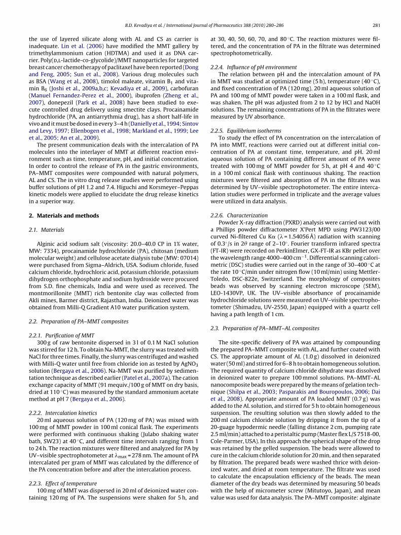
Fig. 2. Relation between adsorption of PA and initial concentration (MMT = 100 mg,pH = 4, temperature = 40 ◦C, and time = 5 h).
3
aa
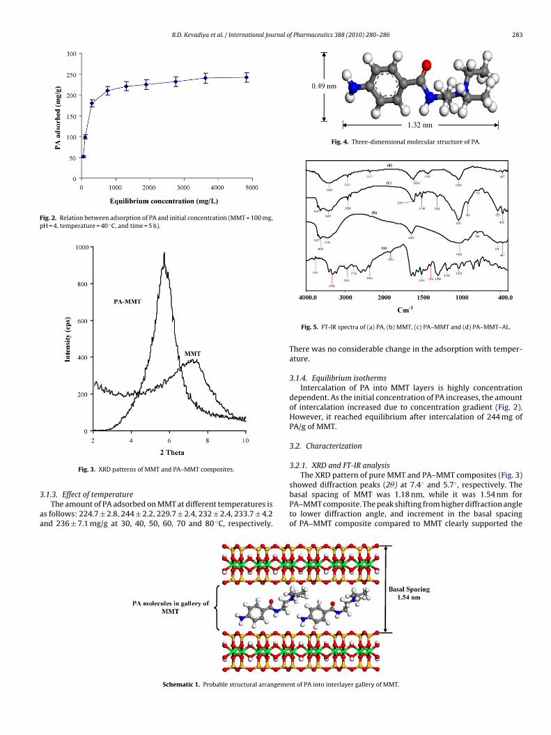
Fig. 4. Three-dimensional molecular structure of PA.
showed diffraction peaks (2�) at 7.4◦ and 5.7◦, respectively. The
Fig. 3. XRD patterns of MMT and PA–MMT composites.
.1.3. Effect of temperatureThe amount of PA adsorbed on MMT at different temperatures is
s follows: 224.7 ± 2.8, 244 ± 2.2, 229.7 ± 2.4, 232 ± 2.4, 233.7 ± 4.2nd 236 ± 7.1 mg/g at 30, 40, 50, 60, 70 and 80 ◦C, respectively.
Schematic 1. Probable structural arrangeme
Fig. 5. FT-IR spectra of (a) PA, (b) MMT, (c) PA–MMT and (d) PA–MMT–AL.
There was no considerable change in the adsorption with temper-ature.
3.1.4. Equilibrium isothermsIntercalation of PA into MMT layers is highly concentration
dependent. As the initial concentration of PA increases, the amountof intercalation increased due to concentration gradient (Fig. 2).However, it reached equilibrium after intercalation of 244 mg ofPA/g of MMT.
3.2. Characterization
3.2.1. XRD and FT-IR analysisThe XRD pattern of pure MMT and PA–MMT composites (Fig. 3)
basal spacing of MMT was 1.18 nm, while it was 1.54 nm forPA–MMT composite. The peak shifting from higher diffraction angleto lower diffraction angle, and increment in the basal spacingof PA–MMT composite compared to MMT clearly supported the
nt of PA into interlayer gallery of MMT.

284 B.D. Kevadiya et al. / International Journal o
icvsbi
static interaction with the positively charged sites at the edges
FP
Fig. 6. DSC analysis of PA, MMT, PA–MMT, AL and PA–MMT–AL.
ntercalation of PA into the interlayer of MMT. PA conformation isonceptually illustrated in Fig. 4 (Accelreys MS Modelling 3.2). The
ertical dimension of the PA molecule is ∼0.49 nm. Considering ailicate layer thickness of 0.96 nm, PA expanded the interlayer spacey 0.58 nm, corresponds to a vertical orientation of the PA cationsn a monolayer of MMT (Schematic 1).
ig. 7. SEM images of (ai) Pristine MMT (bi); PA–MMT–AL nanocomposite bead; (ci) PAA–MMT–AL nanocomposite bead; and (cii) PA–MMT–AL–CS nanocomposite bead.
f Pharmaceutics 388 (2010) 280–286
FT-IR spectrum of PA, MMT, PA–MMT and PA–MMT–AL areshown in Fig. 5. AL showed asymmetric and symmetric stretch-ing vibrations at 1616 and 1431 cm−1 due to carboxyl anions,and 1033 cm−1 for cyclic ether bridge for oxygen stretching. MMTshowed band at 3443 cm−1 due to –OH stretching for interlayerwater. The bands at 3624 and 3697 cm−1 are due to Al–OH andSi–OH. The shoulders and broadness of the structural –OH bandare mainly due to contributions of several structural –OH groupsoccurring in the clay (Joshi et al., 2009a,b). The overlaid absorptionpeak at 1641 cm−1 attributed to –OH bending mode of adsorbedwater, peak at 1032 cm−1 due to Si–O stretching (out-of-plane) andSi–O stretching (in-plane) vibration for layered silicates, respec-tively. Peaks at 914 and 775 cm−1 were attributed to Al–Al–OH andAl–Fe–OH, bending vibrations, respectively (Patel et al., 2007b). Thepeaks at 3216 and 3402 cm−1 are due to the primary amine and1509 cm−1 for secondary amide bending of PA. The peak at 3318and 3039 cm−1 were due to C–H stretching and peaks at 1394 cm−1
attributed to C–H bending vibrations of PA.Incorporation of MMT into AL caused a shift to a higher wave
number and the intensity of COO− stretching peaks of AL decreases(Thaned and Satit, 2007). Thaned and Satit (2007) explained thechange in the wave number of COO− while AL interacts with MMT.The negative charge of the carboxyl groups may have an electro-
of MMT. The –OH stretching peak of the silanol group (SiOH) at3697 cm−1 disappeared in the spectra of PA–MMT–AL composites,and the OH stretching peak of AL shifted to a higher wave number,which is an evidence of the intermolecular hydrogen bonding and
–MMT–AL–CS nanocomposite bead; (aii) PA–MMT hybrid; (bii) surface images of

B.D. Kevadiya et al. / International Journal o
Fig. 8. Release profiles of PA in gastric fluid (pH 1.2) at 37 ± 0.5 ◦C.
es
3
oottaepisw
for Higuchi and Korsmeyer–Peppas kinetic models are shown in
TE
Fig. 9. Release profiles of PA in intestinal fluid (pH 7.4) at 37 ± 0.5 ◦C.
lectrostatic forces between AL and MMT as confirmed by FT-IRtudies.
.2.2. Thermal stability of PA in MMT galleriesThermal stability of PA in MMT galleries were analyzed by DSC
f the PA, MMT, AL, PA–MMT and PA–MMT–AL (Fig. 6). DSC curvesf pristine MMT gave a peak at 120 ◦C due to loss of water, whereashe powder of AL gave broad endothermic peak at 121 ◦C due to itshermal decomposition. The PA showed a sharp endothermic peakt 171 ◦C, corresponds to its melting temperature and second sharpndothermic peaks at 310 ◦C due to its degradation. However, these
eaks were not observed in the DSC curves of PA–MMT compos-tes. The PA–MMT–AL nanocomposites showed broad peak havingimilar pattern as that of AL, indicating the PA–MMT compositesere uniformly dispersed in AL matrices at molecular level.
able 2xpected parameters and drug release kinetic models for various formulations.
Kinetic models Parameters FormulationsPA–MMT
pH 1.2 pH 7.4
Higuchi r2 0.9938 0.9920kH 26.598 8.6288
Korsmeyer–Peppas r2 0.9874 0.9960n 0.4631 0.3917KKP 0.7171 0.2985
f Pharmaceutics 388 (2010) 280–286 285
3.2.3. Morphology observation of the beadsMorphological study of PA–MMT/AL/CS composites beads is
shown in Fig. 7. Fig. 7ai indicates nearly spherical MMT clusters,approximately 1 �m in diameter. The PA loaded MMT showed adisperse form (Fig. 7aii). In contrast, PA–MMT dispersion entrappedwithin AL beads depicts the spherical and rough nature of the beadFig. 7(bi, bii). PA–MMT–AL–CS beads Fig. 7(ci, cii) showed extendedsmooth spherical shape due to the coating of CS on the surface ofthe PA–MMT–AL beads.
3.3. In vitro drug release study
The PA release profiles from the composites were observedboth in gastric (pH 1.2) and intestinal environments (pH 7.4).Approximately 38% of the intercalated PA was released within2 h from PA–MMT composite (Fig. 8). While in the case of for-mulations modified using AL and CS significantly reduced the PArelease in the gastric environment. ∼13 and 11% of PA was releasedfrom PA–MMT–AL and PA–MMT–AL–CS composites beads in 2 h.The negligible PA release from AL and CS coated composites ascompared to PA–MMT composite in the gastric fluids was dueto the fact that in acidic medium AL rapidly changed to water-insoluble alginic acid, consequently did not allowed PA to releasein the media (Shilpa et al., 2003; Dai et al., 2008; Pongjanyakul,2009).
In the phosphate buffer, release of PA from PA–MMT compos-ite was ∼17% in 2 h, ∼30% in the first 10 h and 43% within 30 h,and remained constant up to 70 h (Fig. 9). While, in the case ofPA–MMT–AL, and PA–MMTAL–CS composites beads, 4 and 3.5% in2 h and 13 and 11% in 10 h PA was released. The maximum amountof PA released was found to be 32 and 25%, respectively up to 70 h.These results indicated that the release rate is faster in PA–MMTcomposite as compared to PA–MMT–AL/CS composites beads. Con-trolled release of PA was observed in the case of PA–MMT–AL/CScomposites due to synergic effect of AL and MMT. The plateaufor the release pattern of PA reached more rapidly in the case ofPA–MMT composite as compared to the PA–MMT–AL/CS compos-ites. The degree of swelling and disintegration of AL increased in theintestinal fluids, resulted in slow and controlled release behavior ofPA from PA–MMT–AL composites. Moreover, CS was able to retardthe PA release from PA–MMT–AL–CS composite. Therefore, com-pounding of PA–MMT composite with AL and further coating withCS seems to have a desirable effect to achieve site-specific deliveryof PA.
3.4. Drug release kinetics
The values of correlation coefficient (r2) and rate constants (k)
Table 2. Taking into account the value of n, PA release from PA–MMTcomposite followed the diffusion controlled mechanism (fick-ian diffusion), while PA–MMT–AL/CS nanocomposites followedanomalous (non-fickian) diffusion.
PA–MMT–AL (1:1.4%, w/w) PA–MMT–AL–CS (1:1.4%, w/w)
pH 1.2 pH 7.4 pH 1.2 pH 7.4
0.9933 0.9882 0.9928 0.99549.8584 4.240 10.053 3.879
0.9947 0.9909 0.9856 0.99240.5568 0.6533 0.7211 0.72210.7059 0.0866 0.6582 0.0776

2 rnal o
4
eccrceciA
A
((Cat
R
A
B
D
D
D
D
D
E
G
J
J
J
86 B.D. Kevadiya et al. / International Jou
. Conclusion
The intercalations of PA into the interlayer of MMT via ionxchange mechanism have been achieved. The prepared PA–MMTomposite was successfully compounded within AL and furtheroated with CS. The release performance of PA was found to beetarded in PA–MMT–AL/CS composite in the gastric environmentompare to PA–MMT composite. Site-specific delivery of PA wasffectively achieved using AL and CS. Release of PA from PA–MMTomposite followed the diffusion controlled mechanism (fick-an diffusion), while from PA–MMT–AL/CS composites followednomalous (non-fickian) diffusion based on kinetic models.
cknowledgments
We are thankful to Council of Scientific and Industrial ResearchCSIR) for funding under Network Project: NWP 0010; to Dr. P. BhattXRD), Mr. V. Agarwal (FT-IR), Mrs. Sheetal patel (DSC) and Mr.handrakant (SEM) of the analytical section of the institute. Theuthors are thankful to Mr. G.P. Dangi for the design of computa-ional model.
eferences
n, J., Geib, S.J., Rosi, N.L., 2009. Cation-triggered drug release from a porous zinc-adeninate metal-organic framework. J. Am. Chem. Soc. 131, 8376–8377.
ergaya, F., Theng, B.K.G., Lagaly, G., 2006. Handbook of Clay Science, 1st edition.Elsevier Publication, Amsterdam.
ai, Y.N., Li, P., Zhang, J.P., Wang, A.Q., Wei, Q., 2008. A novel pH sensitive N-succinylchitosan/alginate hydrogel bead for nifedipine delivery. Biopharm. Drug Dispos.29, 173–184.
anielly, J., De Jong, R., Radke-Mitchell, L.C., Uprichard, A.C.G., 1994. Procainamide-associated blood dyscrasias. Am. J. Cardiol. 74, 1179–1180.
epan, D., Kumar, A.P., Singh, R.P., 2009. Cell proliferation and controlled drugrelease studies of monohybrids based on chitosan-g-lactic acid and montmo-rillonite. Acta Biomater. 5, 93–100.
enkbas, E.B., Ottenbrite, R.M., 2006. Perspectives on chitosan drug delivery systemsbased on their geometries. J. Bio. Comp. Polym. 21, 351–368.
ong, Y., Feng, S.S., 2005. Poly(d,l-lactide-co-lycolide)/montmorillonite nanoparti-cles for oral delivery of anticancer drugs. Biomaterials 26, 6068–6076.
llenbogen, K.A., Wood, M.A., Gilligan, D.M., 1998. Intravenous procainamide. Car-diol. Elect. Rev. 2, 179–181.
erstl, Z., Nasser, A., Mingelgrin, U., 1998. Controlled release of pesticides into waterfrom clay–polymer formulations. J. Agric. Food Chem. 46, 3803–3809.
oshi, G.V., Kevadiya, B.D., Patel, H.A., Bajaj, H.C., Jasra, R.V., 2009a. Montmorilloniteas a drug delivery system: intercalation and in vitro release of Timolol maleate.
Int. J. Pharm. 374, 53–57.oshi, G.V., Patel, H.A., Kevadiya, B.D., Bajaj, H.C., 2009b. Montmorillonite intercalatedwith vitamin B1 as drug carrier. Appl. Clay Sci. 45, 248–253.
oshi, G.V., Patel, H.A., Bajaj, H.C., Jasra, R.V., 2009c. Intercalation and controlledrelease of vitamin B6 from montmorillonite–vitamin B6 hybrid. Colloid Polym.Sci. 287, 1071–1076.
f Pharmaceutics 388 (2010) 280–286
Kevadiya, B.D., Joshi, G.V., Patel, H.A., Ingole, P.G., Mody, H.M., Bajaj, H.C., 2009.Montmorillonite–alginate nanocomposites as a drug delivery system: inter-calation and in vitro release of vitamin B1 and vitamin B6. J. Biomater. Appl.,doi:10.1177/0885328208344003.
Korsmeyer, R.W., Gurny, R., Doelker, E., Buri, P., Peppas, N.A., 1983. Mechanisms ofsolute release from porous hydrophilic polymers. Int. J. Pharm. 15,25–35.
Lee, B.H., Subramanian, S.Y., Lin, X., Nelson, W.G., 2005. Procainamide is aspecific inhibitor of DNA methyltransferase-I. J. Biol. Chem. 280, 40749–40756.
Lin, F.H., Lee, Y.H., Jian, C.H., Wong, J.M., Shieh, M.J., Wang, C.Y., 2002. A study ofpurified montmorillonite intercalated with 5-fluorouracil as drug carrier. Bio-materials 23, 1981–1987.
Lin, F.H., Chen, C.H., Winston, T.K., Kuo, C.T., 2006. Modified montmorillonite asvector for gene delivery. Biomaterials 27, 3333–3338.
Manuel Fernandez-Perez, M., Villafranca-Sanchez, E., González-Pradas, F., Martinez-Lopez, Flores-Cespedes, F., 2000. Controlled release of carbofuran from analginate–bentonite formulation: water release kinetics and soil mobility. J. Agric.Food Chem. 48, 938–943.
Markland, P., Amidon, G.L., Yang, V.C., 1999. Modified polypeptides containing gama-benzyl glutamic acid as drug delivery platforms. Int. J. Pharm. 178, 183–192.
Mohanambe, L., Vasudevan, S., 2005. Anionic clays containing anti-inflammatorydrug molecules: comparison of molecular dynamics simulation and measure-ments. J. Phys. Chem. B 109, 15651–15658.
Park, J.K., Choy, Y.B., Oh, J.M., Kim, J.Y., Hwang, S.J., Choy, 2008. Controlled release ofdonepezil intercalated in smectite clays. Int. J. Pharm. 359, 198–204.
Pasparakis, G., Bouropoulos, N., 2006. Swelling studies and in vitro release of vera-pamil from calcium alginate and calcium alginate–chitosan beads. Int. J. Pharm.323, 34–42.
Patel, H.A., Somani, R.S., Bajaj, H.C., Jasra, R.V., 2006. Nanoclays for polymernanocomposites, paints, inks, greases and cosmetics formulations, drug deliveryvehicle and waste water treatment. Bull. Mater. Sci. 29, 133–145.
Patel, H.A., Somani, R.S., Bajaj, H.C., Jasra, R.V., 2007a. Synthesis and characteriza-tion of organic bentonite using Gujarat and Rajasthan clays. Curr. Sci. 92, 1004–1009.
Patel, H.A., Somani, R.S., Bajaj, H.C., Jasra, R.V., 2007b. Preparation and characteri-zation of phosphonium montmorillonite with enhanced thermal stability. Appl.Clay Sci. 35, 194–200.
Pongjanyakul, T., 2009. Alginate–magnesium aluminum silicate films: importanceof alginate blocks structures. Int. J. Pharm. 365, 100–108.
Shilpa, A., Agrawal, S.S., Ray, A.R., 2003. Controlled delivery of drugs from alginatematrix. J. Macromol. Sci. C Polym. Rev. C 43, 187–221.
Sintov, A., Levy, R.J., 1997. Polymeric drug delivery of enzymatically degradable pen-dant agents: peptidyl-linked procainamide model system studies. Int. J. Pharm.146, 55–62.
Sun, B., Ranganathan, B., Feng, S.S., 2008. Multifunctional poly(d,l-lactide-co-glycolide)/montmorillonite (PLGA/MMT) nanoparticles decorated byTrastuzumab for targeted chemotherapy of breast cancer. Biomaterials 29,475–486.
Thaned, P., Satit, P., 2007. Sodium AL-magnesium aluminum silicate composite gels:characterization of flow behavior, micro-viscosity, and drug diffusivity. AAPSPharm. Sci. Technol. 8, E1–E7.
Tonnesen, H.H., Karlsen, J., 2002. Alginate in drug delivery systems. Drug Dev. Ind.Pharm. 28, 621–630.
Wang, X., Du, Y., Lueo, J., 2008. Biopolymer/montmorillonite nanocomposite: prepa-ration, drug-controlled release property and cytotoxicity. Nanotechnology,doi:10.1088/0957-4484/19/6/065707.
Zheng, J.P., Luan, L., Wang, H.Y., Xi, L.F., Yao, K.D., 2007. Study on ibupro-fen/montmorillonite intercalation composites as drug release system. Appl. ClaySci. 36, 297–301.