Layered Double Hydroxide Materials: Assembly and ...
68
Layered Double Hydroxide Materials: Assembly and Photofunctionality Rui Tian, Dongpeng Yan, and Min Wei Contents 1 Introduction ................................................................................... 3 2 Optical Properties of LDHs .................................................................. 4 3 Luminescent Properties of LDHs Nanoparticles ............................................ 6 3.1 Defect-Induced Luminescence ......................................................... 6 3.2 Rare-Earth-Doped Luminescence ..................................................... 7 4 LDH-Based UV-Shielding Materials and the Applications ................................ 8 4.1 Introduction of UV Light and UV-Shielding Materials .............................. 8 4.2 UV-Shielding Properties of LDHs ..................................................... 8 4.3 Host–Guest Interaction and UV-Shielding Performances ............................ 10 4.4 Application of LDH-Based UV-Shielding Materials in Polypropylene and Asphalt ......................................................... 13 5 LDH-Based IR Absorption Materials and the Application ................................. 17 5.1 Introduction of IR Absorption Materials .............................................. 17 5.2 IR Absorption Properties of LDHs .................................................... 17 5.3 Intercalation and Performances of LDH-Based IR Absorption Materials ........... 19 5.4 Application of LDH-Based IR Absorption Materials ................................. 22 6 Fabrication and Application of LDH-Based Host–Guest Luminescent Materials ......... 24 6.1 The Basic Guest Units of LDH-Based Photofunctional Materials ................... 25 6.2 Fabrication of LDH-Based Photofunctional Material ................................. 25 6.3 Adjusting Photofunctionalities of LDH-Based Materials ............................. 38 7 Conclusion and Outlook ..................................................................... 63 References ........................................................................................ 64 R. Tian • M. Wei State Key Laboratory of Chemical Resource Engineering, Beijing University of Chemical Technology, Beijing 100029, P. R. China D. Yan (*) State Key Laboratory of Chemical Resource Engineering, Beijing University of Chemical Technology, Beijing 100029, P. R. China College of Chemistry, Beijing Normal University, Beijing 100875, P. R. China e-mail: [email protected]; [email protected] © Springer International Publishing Switzerland 2015 D. Yan, M. Wei (eds.), Photofunctional Layered Materials, Structure and Bonding 166, DOI 10.1007/978-3-319-16991-0_1 1
-
Upload
khangminh22 -
Category
Documents
-
view
0 -
download
0
Transcript of Layered Double Hydroxide Materials: Assembly and ...

Layered Double Hydroxide Materials:
Assembly and Photofunctionality
Rui Tian, Dongpeng Yan, and Min Wei
Contents
1 Introduction . . . . . . . . . . . . . . . . . . . . . . . . . . . . . . . . . . . . . . . . . . . . . . . . . . . . . . . . . . . . . . . . . . . . . . . . . . . . . . . . . . . 3
2 Optical Properties of LDHs . . . . . . . . . . . . . . . . . . . . . . . . . . . . . . . . . . . . . . . . . . . . . . . . . . . . . . . . . . . . . . . . . . 4
3 Luminescent Properties of LDHs Nanoparticles . . . . . . . . . . . . . . . . . . . . . . . . . . . . . . . . . . . . . . . . . . . . 6
3.1 Defect-Induced Luminescence . . . . . . . . . . . . . . . . . . . . . . . . . . . . . . . . . . . . . . . . . . . . . . . . . . . . . . . . . 6
3.2 Rare-Earth-Doped Luminescence . . . . . . . . . . . . . . . . . . . . . . . . . . . . . . . . . . . . . . . . . . . . . . . . . . . . . 7
4 LDH-Based UV-Shielding Materials and the Applications . . . . . . . . . . . . . . . . . . . . . . . . . . . . . . . . 8
4.1 Introduction of UV Light and UV-Shielding Materials . . . . . . . . . . . . . . . . . . . . . . . . . . . . . . 8
4.2 UV-Shielding Properties of LDHs . . . . . . . . . . . . . . . . . . . . . . . . . . . . . . . . . . . . . . . . . . . . . . . . . . . . . 8
4.3 Host–Guest Interaction and UV-Shielding Performances . . . . . . . . . . . . . . . . . . . . . . . . . . . . 10
4.4 Application of LDH-Based UV-Shielding Materials
in Polypropylene and Asphalt . . . . . . . . . . . . . . . . . . . . . . . . . . . . . . . . . . . . . . . . . . . . . . . . . . . . . . . . . 13
5 LDH-Based IR Absorption Materials and the Application . . . . . . . . . . . . . . . . . . . . . . . . . . . . . . . . . 17
5.1 Introduction of IR Absorption Materials . . . . . . . . . . . . . . . . . . . . . . . . . . . . . . . . . . . . . . . . . . . . . . 17
5.2 IR Absorption Properties of LDHs . . . . . . . . . . . . . . . . . . . . . . . . . . . . . . . . . . . . . . . . . . . . . . . . . . . . 17
5.3 Intercalation and Performances of LDH-Based IR Absorption Materials . . . . . . . . . . . 19
5.4 Application of LDH-Based IR Absorption Materials . . . . . . . . . . . . . . . . . . . . . . . . . . . . . . . . . 22
6 Fabrication and Application of LDH-Based Host–Guest Luminescent Materials . . . . . . . . . 24
6.1 The Basic Guest Units of LDH-Based Photofunctional Materials . . . . . . . . . . . . . . . . . . . 25
6.2 Fabrication of LDH-Based Photofunctional Material . . . . . . . . . . . . . . . . . . . . . . . . . . . . . . . . . 25
6.3 Adjusting Photofunctionalities of LDH-Based Materials . . . . . . . . . . . . . . . . . . . . . . . . . . . . . 38
7 Conclusion and Outlook . . . . . . . . . . . . . . . . . . . . . . . . . . . . . . . . . . . . . . . . . . . . . . . . . . . . . . . . . . . . . . . . . . . . . 63
References . . . . . . . . . . . . . . . . . . . . . . . . . . . . . . . . . . . . . . . . . . . . . . . . . . . . . . . . . . . . . . . . . . . . . . . . . . . . . . . . . . . . . . . . 64
R. Tian • M. Wei
State Key Laboratory of Chemical Resource Engineering, Beijing University of Chemical
Technology, Beijing 100029, P. R. China
D. Yan (*)
State Key Laboratory of Chemical Resource Engineering, Beijing University of Chemical
Technology, Beijing 100029, P. R. China
College of Chemistry, Beijing Normal University, Beijing 100875, P. R. China
e-mail: [email protected]; [email protected]
© Springer International Publishing Switzerland 2015
D. Yan, M. Wei (eds.), Photofunctional Layered Materials, Structure and Bonding
166, DOI 10.1007/978-3-319-16991-0_1
1

Abstract As a large type of inorganic layered compounds with diversity of
composition in host layer and interlayer anions, layered double hydroxides
(LDHs) have been intensively studied toward construction of advanced
photofunctional materials. In this chapter, the optical properties of LDH materials
and related potential applications—mainly involving the functionalities of LDH
layers (such as tunable color, infrared radiation (IR) absorption, and ultraviolet
(UV) shielding)—will be described firstly. Then, we will review the development
of intercalated luminescent materials by adjusting both the host layer and guest
molecules, in which the static and dynamic photofunctional modulations have been
focused. Due to the host–guest and guest–guest interaction, fluorescence properties
of composites can be effectively altered, and intelligent materials with stimuli-
responsive performance can be further obtained. Finally, perspectives on the future
development of LDH-based solid-state luminescent materials are addressed.
Keywords Assembly • Layered double hydroxides • Luminescence • Sensor • UV
shielding
Abbreviations
AFM Atomic force microscopy
CL Chemiluminescence
ECL Electrochemiluminescence
EDTA Ethylenediaminetetraacetic acid
EL Electroluminescence
FRET F€orster resonance energy transfer
HMIs Heavy metal ions
IR Infrared radiation
LDHs Layered double hydroxides
LDPE Low-density polyethylene
LED Light-emitting diode
MMO Mixed metal oxide
PL Photoluminescence
RH Relative humidity
SEM Scanning electron microscopy
SNAS Separate nucleation and aging steps
TEM Transmission electron microscopy
UTF Ultrathin film
UV-Vis Ultraviolet-visible
VOCs Volatile organic compounds
XRD X-ray diffraction
2 R. Tian et al.

1 Introduction
Layered double hydroxides (LDHs) are one type of anionic clay materials, also
known as hydrotalcite compound [1, 2]. The basic structure of LDHs is derived
from the brucite with edge-sharing M(OH)6 octahedra, in which a fraction of the
divalent cations are substituted by trivalent cations in the layer and the positive
charges can be balanced by anions within the hydrated interlayer galleries
[3–5]. For the common binary LDHs, the general formula can be expressed as
[M2+1�xM
3+x(OH)2] (A
n�)x/n ·mH2O, with tunable composition and relative pro-
portions for both di- and trivalent cations as well as interlayer ions [6–8]. This
versatility gives rise to a large amount of LDH-based materials with variable
functionalities due to the exchange of interlayer ions and diverse properties from
different elemental compositions and charge densities of host layers. The structures
of LDHs have been comprehensively studied and introduced in several previous
literatures [9–11], and the models for typical packing fashions (3R and 2H) of
LDHs are illustrated in Scheme 1 [12].
During last few decades, LDHs have received much attention in the fields of
catalysis [13], separation processes [14], photochemical cell [15], and drug carrier
[16]. Moreover, based on the tunable host layer and interlayer guest molecules, the
optical/luminescent properties have also been largely investigated, and a number of
photofunctional materials have been developed [17]. Due to the rapid development
in the design and fabrication of LDH-based photo-related materials, it is timely and
Scheme 1 Typical models of LDHs structures with two packing fashions: (a) 2H; (b) 3R [12]
Layered Double Hydroxide Materials: Assembly and Photofunctionality 3

necessary to summarize recent progress in this area and thus hopefully stimulate the
construction of new types of layered photofunctional materials.
2 Optical Properties of LDHs
Due to different photo-absorption characteristics of the metal ions in the LDH
layers, the powdered LDH materials can present different colors under daylight.
Moreover, the nontransparent LDH powders can be further fabricated into contin-
uous self-supporting transparent films through solvent evaporation or spin-coating
method. The resulting films usually exhibit oriented (00l ) reflection due to the
edge–edge interaction between the adjacent LDH particles. For example, alkoxide-
intercalated LDHs have been synthesized in a nonaqueous media (methanol), and
the hydrolysis of these LDH derivatives resulted in a colloidal LDH suspension
which can be used as a precursor for the formation of continuous transparent films
via solvent evaporation [18, 19]. For another example, the fabrication of oriented
LDHs films without organic solvents has been attempted, and separate nucleation
and aging steps (SNAS) were adopted as a main method [20]. Due to the uniform
and small crystal size (about 40 nm) of the LDH platelets, both the face-to-face and
edge-to-edge interactions occur between individual LDH platelets, which facilitate
the formation of densely packed c-oriented LDH film [21]. The obtained films were
sufficiently thick and mechanically robust and maintained good optical transpar-
ency as well as uniform colors for ZnAl-NO3, NiAl-NO3, and ZnAl-Tb-EDTA
(EDTA, ethylenediaminetetraacetic acid) samples (Fig. 1). Moreover, Zhang [22]
has fabricated oriented LDH films by a spin-coating method, and the as-prepared
Fig. 1 Photographs of transparent self-supporting LDH films of (a) ZnAl-NO3, (b) NiAl-NO3, (c)
ZnAl-Tb(EDTA) (the ruler with centimetre scale lies behind the films), and SEM images of ZnAl-
NO3: (d) top view and (e) edge view with a high-resolution image of this structure shown in the
inset image [21]
4 R. Tian et al.

100
98
96
94
92
90
2
8
902 4
N6 8
95
100
1
7
T650 / %
l/nm
T/%
300 400 500 600 700 800
Fig. 2 UV–Vis transmittance spectra of the MMO10 film (1, 3, 5, 7) and the rehydrated LDH film
(2, 4, 6, 8). The inset shows the transmittance at 650 nm T650 as a function of cycle number n;cycling occurs between MMO and LDH films [23]
Fig. 3 Response of the (MMO-TiO2)61DPC toward different VOCs and RH: (A) reflectance
spectra and (B) photographs in air (a) and various VOCs: (b) ether, (c) acetone, (d) methanal, and
(e) toluene; (C) reflectance spectra and (D) photographs of color variation (I–V: 5 %, 30 %, 54 %,
75 %, and 85 %, respectively) [24]
Layered Double Hydroxide Materials: Assembly and Photofunctionality 5

films displayed good uniformity and compact structure composed of LDH platelets
stacked parallel to the substrate surface.
The calcinations of LDH materials can form mixed metal oxide (MMO) with
diverse optical properties compared with the pristine LDHs. Han et al. [23] havestudied the optical antireflection (AR) performance of MMO through calcinations
of LDHs. Figure 2 shows the changes of transmittance by the reconstruction effect
of LDH materials through recycling the calcination–rehydration procedure. These
AR properties were generated by the transformation between porous (MMO film)
and nonporous state (LDH film), and this phenomenon makes the MMO film an
effective candidate for erasable AR coatings. Furthermore, MMO-TiO2
one-dimensional photonic crystals (1DPC) can be fabricated through deposition
of LDHs and TiO2, followed by calcination [24]. The porous structure of MMO
enabled the adsorption of volatile gas or water molecule in the mesopores of the
MMO-TiO2 structure, which results in the change of optical refractive index
(Fig. 3). Thus, this system can serve as a colorimetric sensor for the detection of
volatile organic compounds (VOCs) or relative humidity (RH).
3 Luminescent Properties of LDHs Nanoparticles
3.1 Defect-Induced Luminescence
Several observations have proven that the pristine LDH particles and colloids can
exhibit well-defined luminescence. For example, the excitation and emission spec-
trum of Zn-Al-LDH has been recorded in Fig. 4 [25], and emission in the 350–
550 nm can be observed. The basic mechanisms for the luminescence of the LDH
colloids are attributed to the numerous surface defects of LDH nanocrystals. These
surface defects can act as traps which are beneficial to the recombination of excited
electrons and holes, and thus, obvious luminescence can be observed. In addition,
the effects of the surface charge density (e.g., the ratio of Zn to Al) on the
photoluminescence (PL) have been investigated. Figure 4 shows that higher
Zn/Al ratio resulted in a stronger PL intensity, that is, different surface charge
density may largely influence the PL intensity of LDH colloids. In addition,
fluorescence properties of MgAl-LDHs have also been studied, and influence of
MgAl-LDHs with different platelet size has been considered. The results showed
that LDHs with higher specific surface areas may result in more surface defects and
stronger fluorescence intensity [26].
6 R. Tian et al.

3.2 Rare-Earth-Doped Luminescence
One of the most striking features of LDHs is their versatility of composition, and
thus, numerous metal cations can be accommodated in the host layer. It was
reported that some rare-earth ions, such as lanthanide Tb3+, Eu3+, and Nd3+, can
be doped into the octahedral lattice of the brucite-like layer [27, 28]. Incorporation
of Tb3+ has been achieved by coprecipitation method under ambient conditions
[29]. Figure 5 shows the homogeneous distribution of Tb3+ in the lattice of LDH
layers regardless of the large ionic size of Tb3+, and up to 19 wt% of Tb3+ can be
incorporated. This Tb3+-doped LDH presents strong luminescence in the green
region due to the intra electron transition of Tb3+. Additionally, the photosensitizer
could also be incorporated into the interlayer of rare-earth-doped LDHs to obtain an
advanced performance. For example, F€orster resonance energy transfer (FRET)
process could occur between the interlayer guest and doped ions in the host layer of
LDHs [30–32], which leads to enhanced fluorescence.
Fig. 4 Effect of Zn/Al ratio on (a) excitation and (b) emission spectra for the colloids of the
pristine Zn–Al-LDHs [25]
Fig. 5 TEM images and the corresponding EDS images of Mg, Al, and Tb elements in Tb3+ doped
LDHs [29]
Layered Double Hydroxide Materials: Assembly and Photofunctionality 7

4 LDH-Based UV-Shielding Materials
and the Applications
4.1 Introduction of UV Light and UV-Shielding Materials
UV light possesses the shortest wavelength in the solar spectrum, and it takes up
about 8 % of the total solar energy. UV radiation includes UVC (220–290 nm),
UVB (290–320 nm), and UVA (320–400 nm). Among them, UVC region is always
absorbed by ozonosphere, and UVA and UVB in the range of 290–400 nm could
reach the earth’s surface. The UV light presents high energy which has severe
damages to the environment and human health [33]. Some diseases, sunburn,
acceleration of aging, and even cancer can be triggered due to the damage of skin
fibers, proteins, and nucleic acids. And the commonly used organic materials (such
as polypropylene, asphalt, rubber, and paints) are easy to degrade under the high
energy UV light as a result of the covalent bonds breakage (e.g., C–H, C–C, C–Cl)
[34]. Therefore, the shielding of UV irradiation is of great importance in our
daily life.
UV-shielding materials could be classified into organic, inorganic, and organic/
inorganic composites. The main organic UV absorbents include benzotriazole,
benzophenone, salicylic acid, and esters; the shielding mechanism is the
UV-induced molecular rearrangement, leading to the release of the absorbed energy
[35, 36]. The inorganic UV blocking materials contain TiO2, ZnO, CaCO3, French
chalk, and LDHs, and they are commonly dispersed into organic or inorganic
materials to form organic/inorganic composites with enhanced UV-shielding per-
formances [37, 38].
4.2 UV-Shielding Properties of LDHs
4.2.1 Scattering Effect of LDH Particles
According to the Lambert–Beer’s law, the particle size severely affects the scatter-
ing and reflection effects of UV light. Xing et al. [38, 39] have prepared Zn-Al-
LDHs through separate nucleation and aging steps (SNAS) at different tempera-
tures, and the influences of particle sizes on the UV blocking properties have been
investigated. The particle sizes calculated from XRD patterns via Scherrer expres-
sion are shown in Table 1. The results show that with increase of aging temperature,
the particle size of LDHs increases in both a and c directions.The suspension of ZnAl-CO3-LDHs with different aging temperature and parti-
cle size can be obtained by dispersing the LDHs into water at 0.02 wt%, and their
UV–Vis transmittance curves are shown in Fig. 6. With the increase of aging
temperature, the UV-shielding performances increased obviously. In order to
investigate the exact relation between particle size and UV-shielding performance,
8 R. Tian et al.

the absorbance and transmittance values are analyzed at different wavelengths, and
the threshold values for UV shielding at different wavelengths are shown in Table 2.
It can be known that long wavelengths correspond to high threshold value in a and
c directions. Therefore, different preparation conditions can be chosen to meet the
requirements of transmittance in different wavelengths.
4.2.2 Reflection and Absorption of LDHs
The reflection and absorption of UV light are largely dependent on the layered
structure of LDHs. The rigid LDH laminates could effectively reflect UV light, and
multiple decrements of UV intensity may occur through multilayer reflection of UV
light [34]. Moreover, the difference of metal elemental composition in LDHs layers
may result in different performance of UV shielding. It was known that the
UV-shielding properties of MgAl-LDH are highly related to the particle scattering
and reflection of LDH layers. However, the replacement of Mg by Zn atom in the
layer may further improve the UV-shielding properties [34, 40]. Figure 7a shows
Table 1 Particle sizes of LDHs at different aging temperatures [39]
Aging temperature (�C) 60 80 100
Particle size in a direction Da (nm) 37.227 39.178 41.806
Particle size in c direction Dc (nm) 25.318 34.097 42.662
Fig. 6 UV–Vis transmittance curves of suspension of ZnAl-CO3-LDHs with different aging
temperature: (a) 60 �C, (b) 80 �C, and (c) 100 �C [39]
Table 2 Threshold values of particle size in a and c directions at different wavelengths [39]
Wavelength (nm) Threshold value in a direction Threshold value in c direction
290 32.92 9.73
320 33.77 12.79
400 34.77 16.47
600 35.56 19.35
800 35.79 20.22
Layered Double Hydroxide Materials: Assembly and Photofunctionality 9
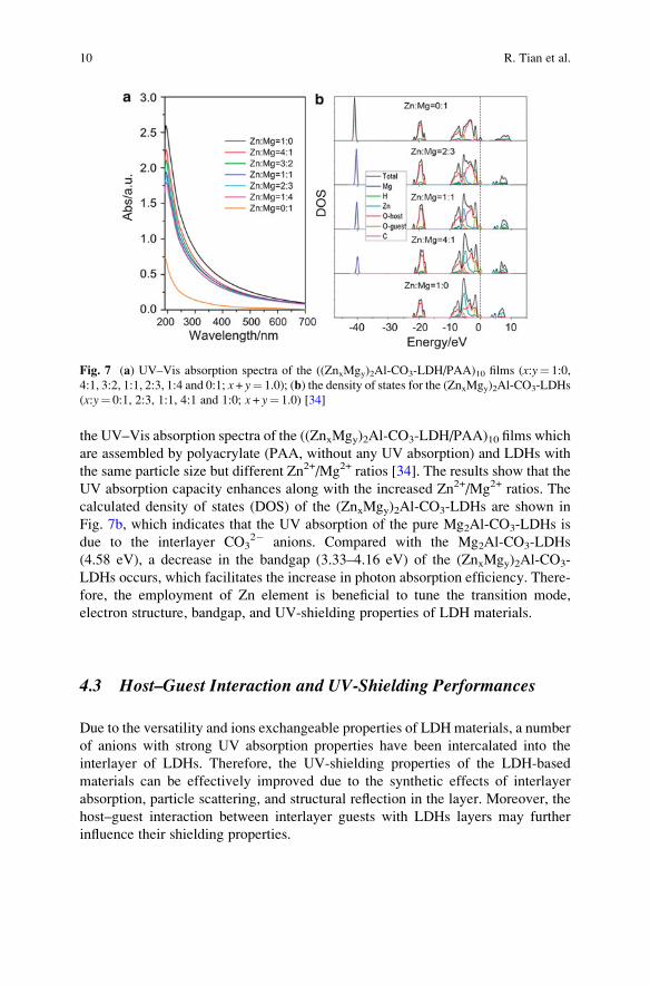
the UV–Vis absorption spectra of the ((ZnxMgy)2Al-CO3-LDH/PAA)10 films which
are assembled by polyacrylate (PAA, without any UV absorption) and LDHs with
the same particle size but different Zn2+/Mg2+ ratios [34]. The results show that the
UV absorption capacity enhances along with the increased Zn2+/Mg2+ ratios. The
calculated density of states (DOS) of the (ZnxMgy)2Al-CO3-LDHs are shown in
Fig. 7b, which indicates that the UV absorption of the pure Mg2Al-CO3-LDHs is
due to the interlayer CO32� anions. Compared with the Mg2Al-CO3-LDHs
(4.58 eV), a decrease in the bandgap (3.33–4.16 eV) of the (ZnxMgy)2Al-CO3-
LDHs occurs, which facilitates the increase in photon absorption efficiency. There-
fore, the employment of Zn element is beneficial to tune the transition mode,
electron structure, bandgap, and UV-shielding properties of LDH materials.
4.3 Host–Guest Interaction and UV-Shielding Performances
Due to the versatility and ions exchangeable properties of LDH materials, a number
of anions with strong UV absorption properties have been intercalated into the
interlayer of LDHs. Therefore, the UV-shielding properties of the LDH-based
materials can be effectively improved due to the synthetic effects of interlayer
absorption, particle scattering, and structural reflection in the layer. Moreover, the
host–guest interaction between interlayer guests with LDHs layers may further
influence their shielding properties.
Fig. 7 (a) UV–Vis absorption spectra of the ((ZnxMgy)2Al-CO3-LDH/PAA)10 films (x:y¼ 1:0,
4:1, 3:2, 1:1, 2:3, 1:4 and 0:1; x + y¼ 1.0); (b) the density of states for the (ZnxMgy)2Al-CO3-LDHs
(x:y¼ 0:1, 2:3, 1:1, 4:1 and 1:0; x + y¼ 1.0) [34]
10 R. Tian et al.
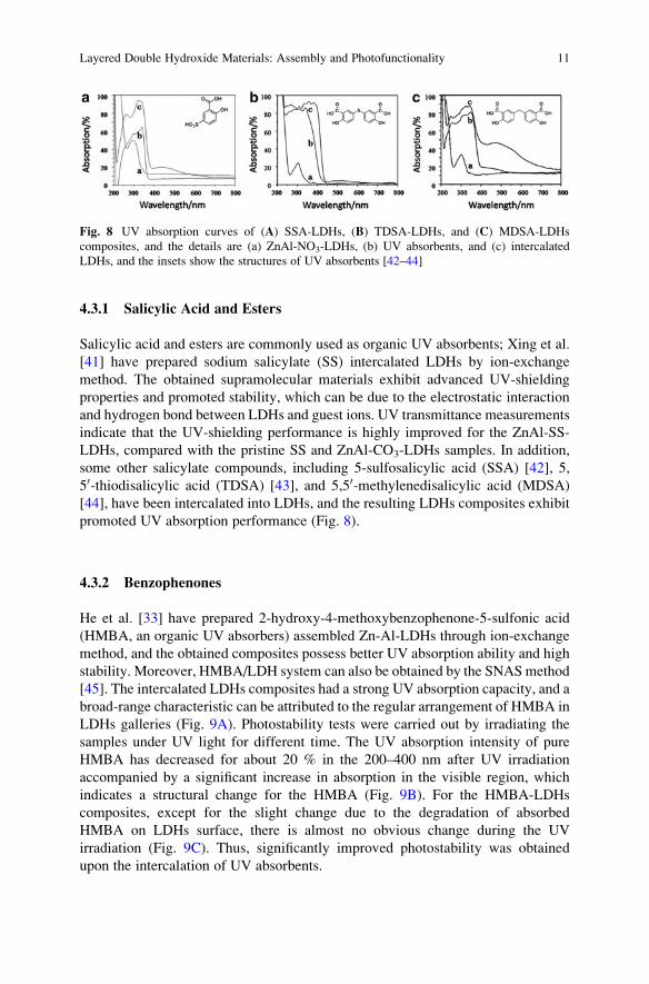
4.3.1 Salicylic Acid and Esters
Salicylic acid and esters are commonly used as organic UV absorbents; Xing et al.[41] have prepared sodium salicylate (SS) intercalated LDHs by ion-exchange
method. The obtained supramolecular materials exhibit advanced UV-shielding
properties and promoted stability, which can be due to the electrostatic interaction
and hydrogen bond between LDHs and guest ions. UV transmittance measurements
indicate that the UV-shielding performance is highly improved for the ZnAl-SS-
LDHs, compared with the pristine SS and ZnAl-CO3-LDHs samples. In addition,
some other salicylate compounds, including 5-sulfosalicylic acid (SSA) [42], 5,
50-thiodisalicylic acid (TDSA) [43], and 5,50-methylenedisalicylic acid (MDSA)
[44], have been intercalated into LDHs, and the resulting LDHs composites exhibit
promoted UV absorption performance (Fig. 8).
4.3.2 Benzophenones
He et al. [33] have prepared 2-hydroxy-4-methoxybenzophenone-5-sulfonic acid
(HMBA, an organic UV absorbers) assembled Zn-Al-LDHs through ion-exchange
method, and the obtained composites possess better UV absorption ability and high
stability. Moreover, HMBA/LDH system can also be obtained by the SNAS method
[45]. The intercalated LDHs composites had a strong UV absorption capacity, and a
broad-range characteristic can be attributed to the regular arrangement of HMBA in
LDHs galleries (Fig. 9A). Photostability tests were carried out by irradiating the
samples under UV light for different time. The UV absorption intensity of pure
HMBA has decreased for about 20 % in the 200–400 nm after UV irradiation
accompanied by a significant increase in absorption in the visible region, which
indicates a structural change for the HMBA (Fig. 9B). For the HMBA-LDHs
composites, except for the slight change due to the degradation of absorbed
HMBA on LDHs surface, there is almost no obvious change during the UV
irradiation (Fig. 9C). Thus, significantly improved photostability was obtained
upon the intercalation of UV absorbents.
Fig. 8 UV absorption curves of (A) SSA-LDHs, (B) TDSA-LDHs, and (C) MDSA-LDHs
composites, and the details are (a) ZnAl-NO3-LDHs, (b) UV absorbents, and (c) intercalated
LDHs, and the insets show the structures of UV absorbents [42–44]
Layered Double Hydroxide Materials: Assembly and Photofunctionality 11

4.3.3 Benzotriazole
Tuo et al. [35, 46] have intercalated 5-benzotriazolyl-4-hydroxy-3-sec-butyl-
benzenesulfonic acid (BZO) into interlayer of LDHs by ion-exchange method
using ZnAl-NO3-LDHs as a precursor. Figure 10 shows the UV transmittance
curves of intercalated BZO-LDHs composites as well as the pure LDH and BZO
precursors. UV absorption bands of ZnAl-NO3-LDHs are located around 300 and
230 nm (Fig. 10a), and BZO possesses strong absorption below 400 nm (Fig. 10b).
After intercalation, the BZO/LDH exhibits a broader range of UV shielding com-
pared with the pristine BZO (Fig. 10c) and also maintains high visible light
transmittance.
4.3.4 Others
Some other organic UV absorbents have also been intercalated into the interlayer
galleries of LDHs, and superior UV blocking properties, visible light transmittance,
Fig. 9 (A) UV–Vis absorption curves of (a) ZnAl-CO3-LDHs, (b) pure HMBA, (c) sodium
HMBA, and (d) ZnAl-HMBA-LDHs; UV-Visible absorption curves of (B) pure HMBA and (C)
ZnAl-HMBA-LDHs after different times of UV irradiation, (a)–(g): 0 min, 15 min, 30 min,
45 min, 60 min, 75 min, and 90 min [45]
Fig. 10 UV transmittance curves of (a) ZnAl-NO3-LDHs, (b) BZO, and (c) ZnAl-BZO-LDHs,
and the inset shows the structure of BZO [46]
12 R. Tian et al.

and photo- and thermal stability can also be obtained. The molecules and their
chemical structures are listed in Table 3.
4.4 Application of LDH-Based UV-Shielding Materialsin Polypropylene and Asphalt
4.4.1 Polypropylene
Polypropylene (PP) is one of the most common plastic, but its application has been
hindered due to the weak photostability. The mechanical properties of PP are
usually affected by the UV photo-oxidative degradation under sunlight
[43, 44]. To improve the anti-UV ability of PP, photostabilizers with
Table 3 Molecules and its structure of UV absorbents for intercalation
Interlayer guest molecules Structure Refs.
4-Hydroxy-3-methoxybenzoic acid [33]
4-Hydroxy-3-methoxycinnamic acid [33]
4,40-Diaminostilbene-2,20-disulfonic acid [33]
p-Aminobenzoic acid [33]
Urocanic acid [33]
Cinnamic acid [47]
p-Methoxycinnamic acid [47]
2-Phenylbenzimidazole-5-sulfonic acid [35, 48]
3,6-Dihydroxynaphthalene-2,7-disulfonate [36]
2-Naphthylamine-1,5-disulfonic acid [36, 49]
2,3-Dihydroxynaphthalene-6-sulfonic acid [36, 50]
Aurintricarboxylic acid [51]
Layered Double Hydroxide Materials: Assembly and Photofunctionality 13

UV-shielding properties have been added to the PP composite [35, 42, 49–51]. Fig-
ure 11 shows the compared IR reflectance spectra of PP films with and without the
addition of ZnAl-BZO-LDHs during photodegradation process [35]. Wavenumbers
at 1,456, 1,376, 1,165, 974, 841, and 808 cm�1 are characteristic IR absorption
bands of PP and ZnAl-BZO-LDHs/PP (Fig. 11a, b) samples. The absorption
intensity ratios I(carbonyl)/I(C–H) and I(hydroxyl)/I(C–H) can be calculated
(Fig. 11c, d), and the results show that the rate for formation of C¼O and
OH/OOH groups in ZnAl-BZO-LDHs/PP under UV irradiation is significantly
lower than that in the pristine PP. Therefore, the addition of ZnAl-BZO-LDH has
markedly blocked the UV light and prevented the degradation of PP and thus
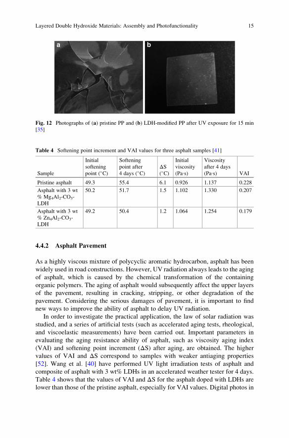
enhances the photostability. Figure 12 shows the digital photos of pristine PP and
LDH-modified PP after UV exposure for 15 min. It was found that the pristine PP
film becomes brittle after exposure to UV light, while the LDH-modified sample
maintains high mechanical properties under the same conditions. This result further
confirms that LDHs can effectively shield UV light to prevent degradation.
4000
0.42PPmodified PP
PPmodified PP
0.60
0.55
0.50
0.45
0.40
0.35
0.30
0.25
0 5 10 15 20 25 30 35
0.40
0.38
0.36
0.34
0.32
0.30
0.28
0.26
0 5 10 15
Aging time (mins) Aging time (mins)
hydr
oxyl
pea
k in
tent
sity
(/ 33
46// 84
1)
hydr
oxyl
pea
k in
tent
sity
(/ 17
41// 84
1)
20 25 30 35
3346nCH
3346nCH 1730
1744
nCO
841nCH
1730
1744
nCO
841nCH
3500 3000 2500
Wavenumber [cm–1] Wavenumber [cm–1]
Tran
smitt
ance
/a.u
.
Tran
smitt
ance
/a.u
.
2000 1500 1000 500 4000 3500 3000 2500 2000 1500 1000 500
ba
dc
Fig. 11 FT-IR spectra of (a) pristine PP and (b) PP modified with 1 % ZnAl-BZO-LDHs under
UV irradiation for 35 min, and the relative peak intensities of (c) hydroxyl and (d) carbonyl bands
for pristine PP and LDH-modified PP (1 %) [35]
14 R. Tian et al.
4.4.2 Asphalt Pavement
As a highly viscous mixture of polycyclic aromatic hydrocarbon, asphalt has been
widely used in road constructions. However, UV radiation always leads to the aging
of asphalt, which is caused by the chemical transformation of the containing
organic polymers. The aging of asphalt would subsequently affect the upper layers
of the pavement, resulting in cracking, stripping, or other degradation of the
pavement. Considering the serious damages of pavement, it is important to find
new ways to improve the ability of asphalt to delay UV radiation.
In order to investigate the practical application, the law of solar radiation was
studied, and a series of artificial tests (such as accelerated aging tests, rheological,
and viscoelastic measurements) have been carried out. Important parameters in
evaluating the aging resistance ability of asphalt, such as viscosity aging index
(VAI) and softening point increment (ΔS) after aging, are obtained. The higher
values of VAI and ΔS correspond to samples with weaker antiaging properties
[52]. Wang et al. [40] have performed UV light irradiation tests of asphalt and
composite of asphalt with 3 wt% LDHs in an accelerated weather tester for 4 days.
Table 4 shows that the values of VAI and ΔS for the asphalt doped with LDHs are
lower than those of the pristine asphalt, especially for VAI values. Digital photos in
Table 4 Softening point increment and VAI values for three asphalt samples [41]
Sample
Initial
softening
point (�C)
Softening
point after
4 days (�C)ΔS(�C)
Initial
viscosity
(Pa∙s)
Viscosity
after 4 days
(Pa∙s) VAI
Pristine asphalt 49.3 55.4 6.1 0.926 1.137 0.228
Asphalt with 3 wt
% Mg4Al2-CO3-
LDH
50.2 51.7 1.5 1.102 1.330 0.207
Asphalt with 3 wt
% Zn4Al2-CO3-
LDH
49.2 50.4 1.2 1.064 1.254 0.179
Fig. 12 Photographs of (a) pristine PP and (b) LDH-modified PP after UV exposure for 15 min
[35]
Layered Double Hydroxide Materials: Assembly and Photofunctionality 15
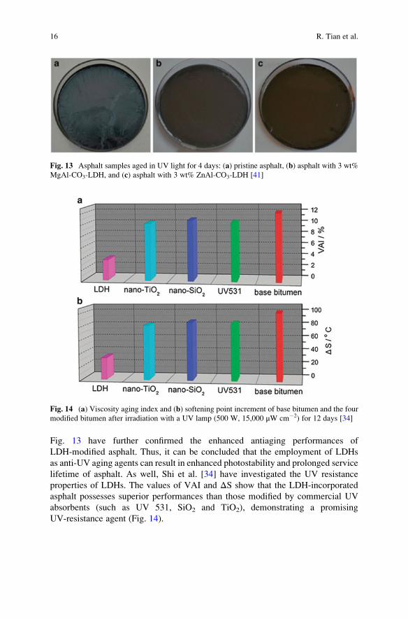
Fig. 13 have further confirmed the enhanced antiaging performances of
LDH-modified asphalt. Thus, it can be concluded that the employment of LDHs
as anti-UV aging agents can result in enhanced photostability and prolonged service
lifetime of asphalt. As well, Shi et al. [34] have investigated the UV resistance
properties of LDHs. The values of VAI and ΔS show that the LDH-incorporated
asphalt possesses superior performances than those modified by commercial UV
absorbents (such as UV 531, SiO2 and TiO2), demonstrating a promising
UV-resistance agent (Fig. 14).
Fig. 13 Asphalt samples aged in UV light for 4 days: (a) pristine asphalt, (b) asphalt with 3 wt%
MgAl-CO3-LDH, and (c) asphalt with 3 wt% ZnAl-CO3-LDH [41]
Fig. 14 (a) Viscosity aging index and (b) softening point increment of base bitumen and the four
modified bitumen after irradiation with a UV lamp (500 W, 15,000 μW cm�2) for 12 days [34]
16 R. Tian et al.

5 LDH-Based IR Absorption Materials
and the Application
5.1 Introduction of IR Absorption Materials
Solar light plays a crucial role in the photosynthesis and the growth of things on
earth. However, the heat supplied by solar during the daytime would rapidly lose as
heat radiation at night, which restricts the growth of plants to some extent. The heat
loss from the earth surface to the surrounding is usually diffused by means of
low-energy mid-infrared radiation, ranging from 7 to 25 μm (1,428–400 cm�1) with
the peak in the range of 9–11 μm (1,111–909 cm�1). Therefore, to keep the
temperature and prevent heat loss, agricultural plastic films (agri-film) [53, 54]
are widely used in “green house.”
Polymers, such as low-density polyethylene (LDPE), are commonly used agri-
films for crop harvest and energy saving [55, 56]. However, LDPE has poor heat
preservation, and the heat is rapidly lost through the film at night [57]. To effec-
tively enhance the ability of heat storage of agri-film, some additives are needed.
The heat-preservation additives should block the loss of long-wavelength IR at
night and maintain transmission of visible light during the daytime [55, 58]. Some
inorganic additives, such as French chalk, china clay, and metal oxides (ZnO, TiO2,
and Sb2O3), were incorporated as IR absorbents [54, 59, 60]. However, the
contained impurities and variable particle sizes have accelerated the degradation
of the film, and their transmittance of visible light and mechanical property are
unsatisfactory [61]. Therefore, there is a need to develop materials with high purity
and controllable particle size as alternative inorganic heat-preservation additives
used in agri-films.
5.2 IR Absorption Properties of LDHs
LDHs have been used as IR absorption materials due to the uniform particle size,
high crystallization degree, good photo-/thermal-stability, and tunable IR absorp-
tion abilities due to the changeable metal-oxide vibrations. By adding the LDHs
particles into agri-film, the IR absorption ability, transparency, heat-preservation
capacity, and the mechanical properties can be improved.
5.2.1 IR Absorption Ability
LDHs can strongly absorb IR due to the vibration of metal-oxide bonds in host layer
and the interlayer anions [62]. For the typical CO32� intercalated LDHs, the broad
peak around 3,441 cm�1 can be ascribed to the stretching vibration of OH groups
attached to the metal atoms in the LDH layers. The band at about 1,625 cm�1 can be
Layered Double Hydroxide Materials: Assembly and Photofunctionality 17

attributed to the deformation vibration of water molecules in the interlayer domain.
In addition to these absorption bands, characteristic peaks at 1,360, 840 and
671 cm�1 can be attributed to the ν3 vibration of CO32� [63]. The vibrations of
M-O units below 1,050 cm�1 (Fig. 15) are the key characteristics for the applica-
tions in IR absorption materials [64]. Moreover, in order to extend the range of IR
absorption, Eu-doped LDH was investigated [58, 65]. In addition to the common IR
absorption, the employment of Eu resulted in broad vibration peaks in the range of
600–900 cm�1 and 1,250–1,350 cm�1, which has further improved the heat-
preservation properties of agri-film.
5.2.2 Antireflection Property
The particle size of LDHs can be controlled due to different preparation methods
[66]. Different from the traditional additives (such as the French chalk and china
clay) with the particle size in the range of several μm, the LDH particles with the
size distribution in the range of 50–500 nm can be obtained [64, 67]. Thus, LDHs
with small size can be adopted as an alternative to promote dispersion degree in the
agri-film. Moreover, the similar refractive index of LDHs and LDPE (illustrated in
Table 5) has effectively reduced refraction phenomenon, leading to the low light
loss and the promoted transparency in the agri-film.
Fig. 15 FT-IR spectra of MgAl-CO3-LDH [64]
Table 5 Typical heat-retention additives and their refractive index [67]
Materials Refractive index Materials Refractive index
LDPE 1.51 MgAl-LDH 1.50
Silica 1.46 China clay 1.56
Light calcium carbonate 1.49 Diatomite 1.45
French chalk 1.55 Muscovite 1.58
18 R. Tian et al.
5.2.3 Heat-Preservation, Photostability, and Antifogging Properties
To achieve high heat-preservation ability, visible light transparency and infrared
irradiation blocking are significant. The LDH materials with high visible light
transmittance enabled visible light get through the agri-film during the daytime,
and the IR absorption properties of LDHs could prevent the infrared irradiation and
heat loss at night.
As an additive, LDHs are highly stable under the photo-irradiation, which
ensures their long service lifetime in practical use. Moreover, LDHs can absorb
or isolate acidic groups and heavy metal ions to protect agri-film from degradation.
Owing to the large specific surface area, porous structure, and oil absorption
property of LDHs, the addition of LDHs enables controlled release of antifogging
agent, which can further enhance the antifogging properties of agri-film.
However, the IR absorption of NO3� or CO3
2� intercalated LDHs usually ranges
from 7 to 8 μm, which cannot perfectly meet the strong IR loss at 9–11 μm (909–
1,111 cm�1). Therefore, the introduction of suitable anions with high IR absorption
at 909–1,111 cm�1 is necessary to reduce the IR loss and further promote the
capability of heat preservation of agri-film [68].
5.3 Intercalation and Performances of LDH-Based IRAbsorption Materials
Several molecule-based systems possess strong IR absorption required for blocking
heat irradiation. However, these molecules sometimes are acidic, and their direct
use as additives may result in fast degradation or aging of the film. Therefore, the
incorporation of these molecules (usually containing sulfate, carboxyl, and phos-
phate groups) into LDHs may be an effective way to achieve high photo- and
thermal stability of the agri-film.
5.3.1 Intercalation with Inorganic Anions
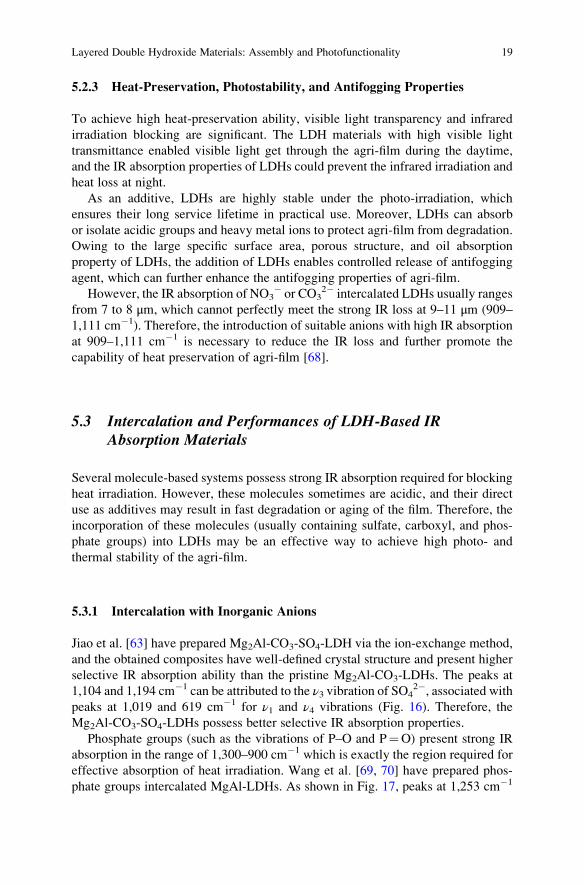
Jiao et al. [63] have prepared Mg2Al-CO3-SO4-LDH via the ion-exchange method,
and the obtained composites have well-defined crystal structure and present higher
selective IR absorption ability than the pristine Mg2Al-CO3-LDHs. The peaks at
1,104 and 1,194 cm�1 can be attributed to the ν3 vibration of SO42�, associated with
peaks at 1,019 and 619 cm�1 for ν1 and ν4 vibrations (Fig. 16). Therefore, the
Mg2Al-CO3-SO4-LDHs possess better selective IR absorption properties.
Phosphate groups (such as the vibrations of P–O and P¼O) present strong IR
absorption in the range of 1,300–900 cm�1 which is exactly the region required for
effective absorption of heat irradiation. Wang et al. [69, 70] have prepared phos-
phate groups intercalated MgAl-LDHs. As shown in Fig. 17, peaks at 1,253 cm�1
Layered Double Hydroxide Materials: Assembly and Photofunctionality 19
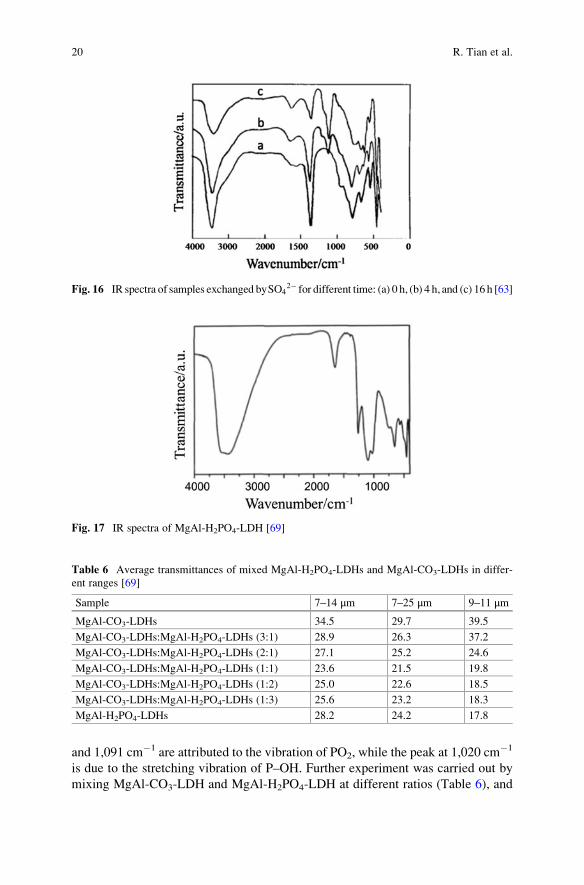
and 1,091 cm�1 are attributed to the vibration of PO2, while the peak at 1,020 cm�1
is due to the stretching vibration of P–OH. Further experiment was carried out by
mixing MgAl-CO3-LDH and MgAl-H2PO4-LDH at different ratios (Table 6), and
Fig. 16 IR spectra of samples exchanged bySO42� for different time: (a) 0 h, (b) 4 h, and (c) 16 h [63]
Fig. 17 IR spectra of MgAl-H2PO4-LDH [69]
Table 6 Average transmittances of mixed MgAl-H2PO4-LDHs and MgAl-CO3-LDHs in differ-
ent ranges [69]
Sample 7–14 μm 7–25 μm 9–11 μm
MgAl-CO3-LDHs 34.5 29.7 39.5
MgAl-CO3-LDHs:MgAl-H2PO4-LDHs (3:1) 28.9 26.3 37.2
MgAl-CO3-LDHs:MgAl-H2PO4-LDHs (2:1) 27.1 25.2 24.6
MgAl-CO3-LDHs:MgAl-H2PO4-LDHs (1:1) 23.6 21.5 19.8
MgAl-CO3-LDHs:MgAl-H2PO4-LDHs (1:2) 25.0 22.6 18.5
MgAl-CO3-LDHs:MgAl-H2PO4-LDHs (1:3) 25.6 23.2 18.3
MgAl-H2PO4-LDHs 28.2 24.2 17.8
20 R. Tian et al.

employment of H2PO4 has effectively improved the selective IR properties com-
pared with pristine MgAl-CO3-LDH, especially in the region of 9–11 μm. In order
to fabricate materials with comprehensive IR absorption properties, all of the IR
regions (such as 7–25, 7–14 and 9–11 μm) should be taken into consideration. The
sample with the ratio of MgAl-CO3-LDH and MgAl-H2PO4-LDH at 1:1 possess
optimized absorption ability in these three region. As another example, Badreddine
et al. [71] have prepared a series of phosphate anions (such as PO3�, H2PO4
�,HPO4
2�, PO43�, P2O7
4� and P3O105� ) intercalated LDH composites, and the
obtained composites displayed largely promoted IR absorption properties.
5.3.2 Intercalation with Organic Anions
Some organic anions containing phosphate groups have also been intercalated into
the interlayer of LDHs [70], and the LDH-based composites with different IR
absorption intensity and selectivity were obtained. For example, Wang et al. have
employed N,N-Bis(phosphonomethyl)glycine (GLYP) [53], aminotrimethylene-
phosphonic acid (ATMP) [55], and N-phosphonomethyliminodiacetic acid
(PMIDA) [59, 62] as interlayer guests (Fig. 18) to promote the IR absorption
capacity in the range of 909–1,111 cm�1 in the composites. Thus, the obtained
hybrid materials possess higher infrared absorption than the original MgAl-CO3-
LDH, and the heat loss via the radiation to the atmosphere environment can be
effectively inhibited.
Fig. 18 Molecular structure and IR spectra of (a) GLYP, (b) ATMP, (c) PMIDA, (d) MgAl-NO3-
LDH, (e) MgAl-CO3-LDH, (f) MgAl-GLYP-LDH, (g) MgAl-ATMP-LDH, and (h) MgAl-
PMIDA-LDH [53, 55, 59]
Layered Double Hydroxide Materials: Assembly and Photofunctionality 21
5.4 Application of LDH-Based IR Absorption Materials
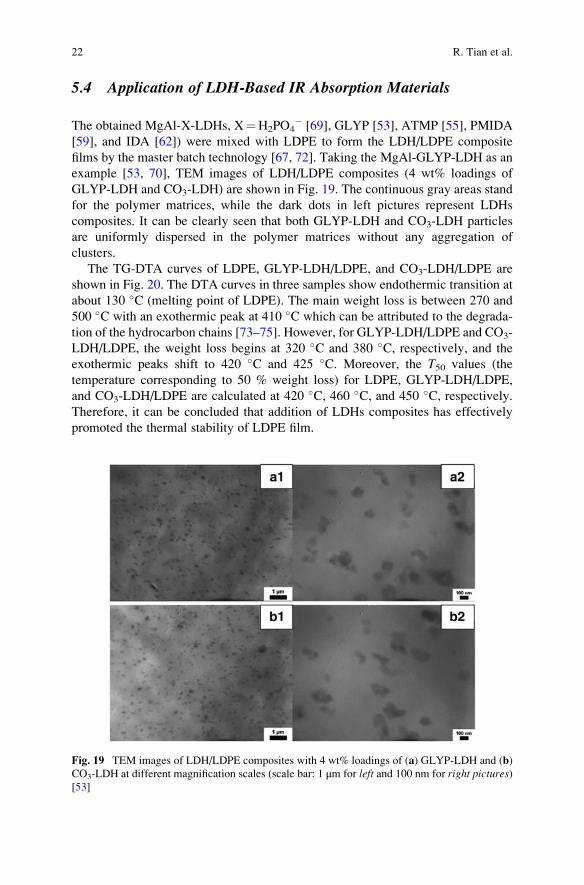
The obtained MgAl-X-LDHs, X¼H2PO4� [69], GLYP [53], ATMP [55], PMIDA
[59], and IDA [62]) were mixed with LDPE to form the LDH/LDPE composite
films by the master batch technology [67, 72]. Taking the MgAl-GLYP-LDH as an
example [53, 70], TEM images of LDH/LDPE composites (4 wt% loadings of
GLYP-LDH and CO3-LDH) are shown in Fig. 19. The continuous gray areas stand
for the polymer matrices, while the dark dots in left pictures represent LDHs
composites. It can be clearly seen that both GLYP-LDH and CO3-LDH particles
are uniformly dispersed in the polymer matrices without any aggregation of
clusters.
The TG-DTA curves of LDPE, GLYP-LDH/LDPE, and CO3-LDH/LDPE are
shown in Fig. 20. The DTA curves in three samples show endothermic transition at
about 130 �C (melting point of LDPE). The main weight loss is between 270 and
500 �C with an exothermic peak at 410 �C which can be attributed to the degrada-
tion of the hydrocarbon chains [73–75]. However, for GLYP-LDH/LDPE and CO3-
LDH/LDPE, the weight loss begins at 320 �C and 380 �C, respectively, and the
exothermic peaks shift to 420 �C and 425 �C. Moreover, the T50 values (the
temperature corresponding to 50 % weight loss) for LDPE, GLYP-LDH/LDPE,
and CO3-LDH/LDPE are calculated at 420 �C, 460 �C, and 450 �C, respectively.Therefore, it can be concluded that addition of LDHs composites has effectively
promoted the thermal stability of LDPE film.
Fig. 19 TEM images of LDH/LDPE composites with 4 wt% loadings of (a) GLYP-LDH and (b)
CO3-LDH at different magnification scales (scale bar: 1 μm for left and 100 nm for right pictures)[53]
22 R. Tian et al.
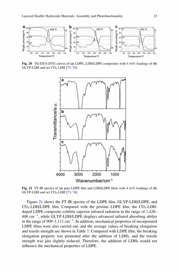
Figure 21 shows the FT-IR spectra of the LDPE film, GLYP-LDH/LDPE, and
CO3-LDH/LDPE film. Compared with the pristine LDPE film, the CO3-LDH-
doped LDPE composite exhibits superior infrared radiation in the range of 1,428–
400 cm�1, while GLYP-LDH/LDPE displays advanced infrared absorbing ability
in the range of 909–1,111 cm�1. In addition, mechanical properties of incorporated
LDPE films were also carried out, and the average values of breaking elongation
and tensile strength are shown in Table 7. Compared with LDPE film, the breaking
elongation property was promoted after the addition of LDHs, and the tensile
strength was just slightly reduced. Therefore, the addition of LDHs would not
influence the mechanical properties of LDPE.
Fig. 21 FT-IR spectra of (a) pure LDPE film and LDH/LDPE films with 4 wt% loadings of (b)
GLYP-LDH and (c) CO3-LDH [73, 74]
Fig. 20 TG-DTA-DTG curves of (a) LDPE, LDH/LDPE composites with 4 wt% loadings of (b)
GLYP-LDH and (c) CO3 LDH [73, 74]
Layered Double Hydroxide Materials: Assembly and Photofunctionality 23

6 Fabrication and Application of LDH-Based Host–Guest
Luminescent Materials
The photoactive guest molecules assembled into the LDHs host layers can achieve
new type of host–guest composite materials with advanced photo-related perfor-
mances (such as luminescence). There are several advantages for the accommoda-
tion of photofunctional species into LDHs inorganic matrices:
1. LDHs layers can provide a confined and isolated microenvironment for the
intercalated guest molecules. In the confined interlayer space, the vibration
and thermal motion of guests could be inhibited, and this may effectively
improve the luminescent efficiency of the photoactive molecules. Furthermore,
the photofunctional guests with different emission can be assembled into the
host layers in a step-by-step manner, in which the rigid LDH layers can avoid the
permeation between different guest molecules.
2. The regular and crystalline LDHs layers allow the ordered arrangement and high
orientation of interlayer guest molecules based on host–guest interactions. By
tuning the chemical composition and charge density of host layers as well as the
amount, arrangement and aggregation state of the guest molecules, the photo-
related performance can be finely adjusted.
3. Due to the introduction of LDHs layers, the thermal- and photostability of the
intercalated composites can be largely improved. Thus, the problem involving
the stability of pristine organic materials in the practical applications can be
resolved effectively.
4. The combination of transparent insulating LDHs layers with photoactive mole-
cules may form an inorganic/organic multi-quantum-well structure. The insu-
lating LDHs can act as energy barrier layers in the quantum well, which can
accelerate the formation of exciton and improve the fluorescence efficiency.
5. Based on the changes in the aggregation states and/or the conformations of the
interlayer photoactive molecules induced by the external environmental stimuli,
the corresponding photo-related properties can be further tuned due to the
alternations of host–guest and guest–guest interactions. Therefore, the
LDH-based intelligent-responsive materials can be further constructed.
According to the design strategies mentioned above, a number of host–guest
photofunctional materials have been continuously reported. In the following sec-
tion, we mainly focus on the host–guest photofunctional materials with tunable
static and dynamic luminescence.
Table 7 Mechanical properties of LDPE and LDH/LDPE [73, 74]
Samples Breaking elongation (%) Tensile strength (MPa)
LDPE 442.3 14.0
MgAl-GLYP-LDH/LDPE 447.2 13.3
MgAl-CO3-LDH/LDPE 447.1 13.6
24 R. Tian et al.

6.1 The Basic Guest Units of LDH-Based PhotofunctionalMaterials
To date, both the organic and inorganic photoactive species (including polymers,
complexes, small molecules, quantum dots, and polyoxometalates) can serve as
good candidates as guest molecules to assemble with LDHs. And the difficulties,
such as the aggregation-induced quenching, lifetime and stability are eager to be
solved. Table 8 gives typical examples of the interlayer photofunctional guests and
their related fabrication methods.
6.2 Fabrication of LDH-Based Photofunctional Material
6.2.1 Layer-by-Layer Assembly
Layer-by-layer (LbL) assembly is a widely used method to achieve multilayer
LDH-based ultrathin films (UTFs), which involves alternative dipping a substrate
in positively charged LDH nanosheets and negative-charged photofunctional mol-
ecules [129]. Based on their electrostatic interactions, the guest molecules can be
accommodated into the interlayer of LDH nanosheets. The employment of LDH
nanosheets has provided an opportunity to finely tune the arrangement and relative
distance of the interlayer molecules, and the homogeneous distribution of
photofunctional molecules can be obtained [130]. Due to the confined and ordered
environment provided by the rigid LDHs nanosheets, the luminescence quenching
commonly caused by the aggregation and/or accumulation of photoactive mole-
cules can be efficiently reduced. Moreover, the existence of inorganic layers may
improve the thermal and optical stability of the photofunctional molecules
[76]. Therefore, LbL assembly has supplied an effective way for the fabrication
of hybrid UTFs with advanced photo-related properties.
Assembly Based on π-Conjugated Polymers
Luminescent organic polymer materials have attracted extensive attention, partic-
ularly due to their applications in optoelectronic devices, such as liquid-crystal
displays [131, 132]. However, the development of these polymers is severely
hindered by their short lifetime and poor photo- or thermal stability [133]. Yan
et al. have presented a potential solution to these problems by assembling the
polymers with rigid LDH nanosheets which can serve as a new inorganic/organic
quantum well structure. Several anionic polymers, such as the derivates of poly
(p-phenylene) [76], poly(phenylenevinylene) [78], and polythiophene [79], have
been studied, and investigation on a sulfonated poly(p-phenylene) anionic derivate
(APPP) will be particularly introduced below.
Layered Double Hydroxide Materials: Assembly and Photofunctionality 25

Table 8 Photofunctional guests used in LDH-based composites and their fabrication methods
Materials Categories Molecules Method Refs.
Organic Polymers Poly(p-phenylene) anionic derivate
(APPP)
LbL [76,
77]
Poly(5-methoxy-2-
(3-sulfopropoxy)-1,4-phenylene-
vinylene anionic derivate (APPV)
LbL [77,
78]
Sulfonated polythiophene (SPT) LbL [79,
80]
Polydiacetylene (PDA) LbL [81]
Poly{1-4[4-(3-carboxy-4-
hydroxyphenylazo)benzenesul-
fonamido]-1,2-ethanediyl sodium
salt} (PAZO)
LbL [82]
Complexes Tris(1,10-phenanthroline-4,7-
diphenyl-sulfonate)ruthenium
(II) ([Ru(dpds)3]4�)
LbL [80,
83]
Bis(8-hydroxyquinolate)zinc, tris
(8-hydroxyquinolate-5-sulfonate)
aluminum
LbL/co-
intercalation
[84–
87]
[Tetrakis(4-carboxyphenyl)-
porphyrinato]zinc(II) (ZnTPPC)
Ion exchange [88]
Pd(II)-5,10,15,20-tetrakis
(4-carboxyphenyl) porphyrin
(PdTPPC)
Coprecipitation [89]
5,10,15,20-Tetrakis
(4-sulfonatophenyl) porphyrin
(TPPS) and its palladium complex
(PdTPPS)
Coprecipitation [89–
91]
Zinc phthalocyanines (ZnPc) Coprecipitation [92]
Tris[2-(4,6-difluorophenyl)
pyridinato-C2,N]iridium(III) (Ir
(F2ppy)3)
LbL [93]
Small molecules Sulforhodamine B (SRB) Co-
intercalation
[94]
Bis(N-methyl-acridinium) (BNMA) LbL [77,
95]
3,4,9,10-Perylene tetracarboxylate
(PTCB)
LbL/co-
intercalation
[96,
97]
Benzocarbazole (BCZC) Coprecipitation [98]
Bis(2-sulfonatostyryl)biphenyl
(BSB)
LbL [99]
Sulfonated derivate of cyanin (Scy) LbL [100]
2,20-(1,2-Ethenediyl)bis[5-[[4-(diethylamino)-6-
[(2,5-disulfophenyl) amino]-1,3,5-
triazin-2-yl]amino] benzene-
sulfonicacid] hexasodium salt
(BTBS)
LbL [101,
102]
(continued)
26 R. Tian et al.
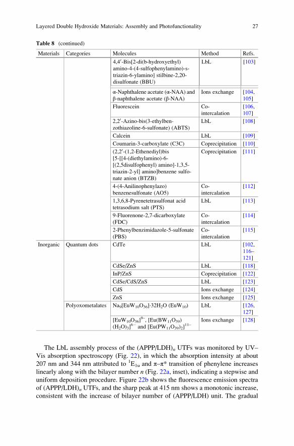
The LbL assembly process of the (APPP/LDH)n UTFs was monitored by UV–
Vis absorption spectroscopy (Fig. 22), in which the absorption intensity at about
207 nm and 344 nm attributed to 1E1u and π–π* transition of phenylene increases
linearly along with the bilayer number n (Fig. 22a, inset), indicating a stepwise anduniform deposition procedure. Figure 22b shows the fluorescence emission spectra
of (APPP/LDH)n UTFs, and the sharp peak at 415 nm shows a monotonic increase,
consistent with the increase of bilayer number of (APPP/LDH) unit. The gradual
Table 8 (continued)
Materials Categories Molecules Method Refs.
4,40-Bis[2-di(b-hydroxyethyl)amino-4-(4-sulfophenylamino)-s-
triazin-6-ylamino] stilbine-2,20-
disulfonate (BBU)
LbL [103]
α-Naphthalene acetate (α-NAA) andβ-naphthalene acetate (β-NAA)
Ions exchange [104,
105]
Fluorescein Co-
intercalation
[106,
107]
2,20-Azino-bis(3-ethylben-zothiazoline-6-sulfonate) (ABTS)
LbL [108]
Calcein LbL [109]
Coumarin-3-carboxylate (C3C) Coprecipitation [110]
(2,20-(1,2-Ethenediyl)bis[5-[[4-(diethylamino)-6-
[(2,5disulfophenyl) amino]-1,3,5-
triazin-2-yl] amino]benzene sulfo-
nate anion (BTZB)
Coprecipitation [111]
4-(4-Anilinophenylazo)
benzenesulfonate (AO5)
Co-
intercalation
[112]
1,3,6,8-Pyrenetetrasulfonat acid
tetrasodium salt (PTS)
LbL [113]
9-Fluorenone-2,7-dicarboxylate
(FDC)
Co-
intercalation
[114]
2-Phenylbenzimidazole-5-sulfonate
(PBS)
Co-
intercalation
[115]
Inorganic Quantum dots CdTe LbL [102,
116–
121]
CdSe/ZnS LbL [118]
InP/ZnS Coprecipitation [122]
CdSe/CdS/ZnS LbL [123]
CdS Ions exchange [124]
ZnS Ions exchange [125]
Polyoxometalates Na9[EuW10O36]·32H2O (EuW10) LbL [126,
127]
[EuW10O36]9–, [Eu(BW11O39)
(H2O)3]6� and [Eu(PW11O39)2]
11–Ions exchange [128]
Layered Double Hydroxide Materials: Assembly and Photofunctionality 27
change in luminescence can also be seen from photographs under UV light
(Fig. 22c). Moreover, the UTFs with different n show no obvious shift or broaden
in the absorption and fluorescence emission spectra, and this phenomenon suggests
that no aggregation of APPP was formed during the assembly process.
The top-view SEM image (Fig. 23a) displays the homogeneity and uniformity of
(APPP/LDH)9 UTFs. Thickness of 20–23 nm for (APPP/LDH)9 UTF was obtained
from the side-view SEM image (Fig. 23b), with the average value about 2.5–2.8 nm
for the basic unit of (APPP/LDH)1. The AFM topographical image (3 μm� 3 μm) is
illustrated in Fig. 23c with the root-mean-square roughness of 5.8 nm, indicating a
smooth surface. Figure 23d shows the homogeneous distribution of the APPP
chromophore throughout the film as dark blue color under the fluorescence
microscope.
The fluorescence lifetime of (APPP/LDH)n UTF (18.63–21.05 ns) is largely
prolonged compared with the pristine APPP solution (0.88 ns). This remarkable
increase is a result of isolation of the adjacent polymer provided by rigid LDH
Fig. 22 Characterizations of (APPP/LDH)n (n¼ 3–30) UTFs: (a) UV–Vis absorption spectra
(inset shows the linear correction of absorbance at 207 and 344 nm vs. n), (b) fluorescence spectra,and (c) photographs under 365 nm UV irradiation at different values of n [76]
28 R. Tian et al.

nanosheets. Moreover, APPP molecules are confined within the interlayer environ-
ment of the LDH, and the inhibited thermal vibration of the APPP backbones may
also contribute to the prolonged lifetime.
Photostability of (APPP/LDH)27 UTF was performed under UV irradiation, and
(APPP/PDDA)27 (PDDA, polydimethyldiallylammonium chloride) was tested as a
reference sample. As seen in Fig. 24, �70 % of initial fluorescence intensity of
(APPP/LDH)27 UTF remained after continuous irradiation for 2 min, in contrast
to� 50 % for (APPP/PDDA)27 with the same measurement. Broadened emission
band was observed for (APPP/PDDA)27 UTF after 15 min, while no obvious band
shift was found for (APPP/LDH)27 in the whole test. It can be concluded that
(APPP/LDH)27 UTF possesses better UV light resistance ability compared with
(APPP/PDDA)27.
Assembly Based on Anionic Metal Complexes and Small Molecules
Transition-metal-based photoactive complexes have been extensively investigated
in optoelectronics due to their high chemical stability and excellent luminescence
properties [134]. Ruthenium-based complexes, tris(1,10-phenanthroline-4,7-
diphenylsulfonate)ruthenium(II) (denoted as Ru(dpds)3), have been studied by
Fig. 23 The morphology of (APPP/LDH)9 UTF: (a) top view of SEM image, (b) side view of
SEM image, (c) tapping-mode AFM image, and (d) fluorescence microscope image [76]
Layered Double Hydroxide Materials: Assembly and Photofunctionality 29

assembly with LDH nanosheet to obtain orderly UTFs [83]. Figure 25 has illus-
trated the assembly process of (Ru(dpds)3/LDH)n UTFs, and UV–Vis absorption
and fluorescence emission spectra have been employed to monitor the process with
the increased intensity when varying the number of bilayers. Except for the periodic
long-range ordered structures, well-defined red photoluminescence for the UTFs,
the ordered Ru(dpds)3 has overcome symmetry due to interaction with LDHs, and
polarized photoluminescence behavior was observed. Other small molecules such
as sulfonated carbocyanine derivate (Scy) [100] can also be directly assembled with
LDH nanosheet, and the fluorescence performance and stability can be largely
improved for the assembled UTFs.
Assembly Based on Positive-Charged and Low-Charged Molecules
Due to the charge-balance rule, it has been known that only anionic species can be
directly assembled into the galleries of the positively charged LDH layers. This
largely restricts the development of LDH-based functional materials, and the
assembly of LDH nanosheets with abundant functional cations remains a challenge.
To meet this requirement, Yan et al. [95] have proposed a new assembly strategy for
cation–LDH UTFs by employing a suitable polyanion as the carrier. Bis(N-methylacridinium) (BNMA, an important dye in the field of chemiluminescence)
Fig. 24 Decay of normalized maximal PL intensity with time, normalized against the initial PL
value; λex¼ 365 nm to probe the UV irradiation resistance ability of (APPP/PDDA)27 (black datapoints) and (APPP/LDH)27 UTF (red) irradiated under 344 nmUV light. Insets: photographs under
UV light of (a) (APPP/PDDA)27 and (b) (APPP/LDH)27 UTFs after the UV resistance experiment
was finished [76]
30 R. Tian et al.

and an optically inert polyanion polyvinylsulfinate (PVS) was taken as examples for
fabrication of (BNMA@PVS/LDH)n UTFs, and the co-assembly process involves
two steps. Firstly, BNMA is mixed with the main chain of the PVS, based on
Coulombic interaction at the molecular level, and a cation–polyanion pair
(BNMA@PVS) with negative charge and functional behavior of BNMA can be
obtained. Subsequently, this negatively charged (BNMA@PVS) pair was assem-
bled with LDH nanosheet via LbL process, whereby the cations and polyanion were
stably coexisted in the gallery environment provided by LDH nanosheets.
Fig. 25 (A) Schematic representation of assembly process of (Ru(dpds)3/LDH)n: (a) structure of
[Ru(dpds)3]4�, (b) representation of a monolayer of MgAl-LDH (dark pink, Al(OH)6 octahedra;
green, Mg(OH)6 octahedra), and (c) assembly process; (B) UV–Visible absorption spectra (inset
shows plots of the absorbance at 196, 287 and 471 nm vs. n), and (C) fluorescence emission spectra
[83]
Layered Double Hydroxide Materials: Assembly and Photofunctionality 31

UV–visible absorption spectra were measured to monitor the assembly process
for (BNMA@PVS/LDH)n UTFs. The linearly increased intensities at 265 and
371 nm which correspond to the characteristic absorption bands of BNMA dem-
onstrated an ordered and regular film growth procedure (Fig. 26a). Fluorescence
emission intensity at 510 nm also presents consistent enhancement with the
increase of bilayer number n (Fig. 26b), which can be confirmed by increased
luminescence brightness under UV light irradiation (the inset of Fig. 26b). These
results demonstrated successful co-assembly of BNMA@PVS pairs into the
interlayer of LDHs. Moreover, the absorption and fluorescence emission spectra
show no obvious red or blue shift compared with the pristine BNMA solution,
suggesting uniform distribution throughout the whole assembly processing.
Top-view SEM (Fig. 27a), AFM (Fig. 27c) and fluorescence microscopy
(Fig. 27d) images show that the assembled UTFs exhibit a homogeneous and
continuous surface, indicating that the BNMA chromophores are distributed uni-
formly throughout the whole film. Side-view SEM image (Fig. 27b) reveals an
approximate 1.5 nm for per (BNMA@PVS/LDH)1 bilayer, in accordance with the
basal spacing obtained by XRD. This result further confirms a uniform and periodic
layered structure of the multilayer film.
The molecular dynamic (MD) simulation was carried out to further understand
the geometric structures of this new type UTF system. An idealized BNMA@PVS/
LDH structural model was constructed, and the computational basal spacing is
ca. 1.47 nm, in agreement with the experimental results. The simulation shows that
the organic ions are found to adopt an ordered arrangement with a preferred orien-
tation (15� between N-N axis to layers) in the as-prepared UTF, and the driving
force for the UTF assembly is mainly dominated by the Coulomb interaction.
Moreover, several small low-charged molecules or molecules with poor assem-
bled force can also be co-assembled into interlayer of LDHs by employing
Fig. 26 (a) UV–Vis absorption spectra, (b) fluorescence spectra of the (BNMA@PVS/LDH)n(n¼ 4–32) UTFs. The insets in (a) and (b) show the plots of absorbance at 265 and 371 nm
vs. n and optical photographs under 365 nm UV irradiation, respectively [95]
32 R. Tian et al.

polyanions. For example, Li et al. [84] have achieved the co-assembly of the tris
(8-hydroxyquinolate-5-sulfonate) aluminum (AQS3�) with three polyanions
[polyanions poly(acrylic acid) (PAA), poly(styrene 4-sulfonate) (PSS), and poly
[5-methoxy-2-(3-sulfopropoxy)-1,4-phenylene vinylene] (PPV)] to obtain the hybrid
UTFs, denoted as (AQS@PAA/LDH)n, (AQS@PSS/LDH)n, and (AQS@PPV/
LDH)n (Scheme 2). And similar work was also carried out by employing PSS and
PVS to co-assemble with PTCB (perylene 3,4,9,10-tetracarboxylate) [97]. Therefore,
the small complex anion–polyanion is anticipated to enhance the cohesion to assem-
ble with LDH for UTFs. The luminous properties of such ordered UTFs can also be
modulated by alternating the polyanion species and proportion.
Assembly Based on Electroneutral Molecules
A micellar assembly route for combination of neutral molecules and LDH
nanosheets has been put forward in the absence of interaction between them. For
example, neutral bis(8-hydroxyquinolate)zinc (Znq2) can be encapsulated into the
block copolymer poly(tert-butyl acrylate-co-ethyl acrylate-co-methacrylic acid)
(PTBEM) micelles, and then the anionic block copolymer micelles Znq2@PTBEM
were assembled with the exfoliated LDH nanosheets to obtain hybrid
(Znq2@PTBEM/LDH)n UTFs (Fig. 28a) [87]. The as-prepared UTFs present a
stepwise growth by varying number of bilayers, which can be observed in fluores-
cence emission spectra and corresponding photos under UV light (Fig. 28b).
Fig. 27 The morphology of (BNMA@PVS/LDH)8 UTF for (a) top-view SEM image, (b) side-
view SEM image, (c) tapping-mode AFM image, and (d) fluorescence microscope image multi-
plied by 50-fold and 1,000-fold (the inset plot) [95]
Layered Double Hydroxide Materials: Assembly and Photofunctionality 33

Similar methods also appear for the neutral small molecules perylene [135] and tris
[2-(4,6-difluorophenyl)pyridinato-C2,N]iridium(III) (Ir(F2ppy)3) [93]. With poly
(N-vinyl carbazole) (PVK) as the carrier, (PVK@perylene/LDH)n and
(Ir(F2ppy)3@PVK/LDH)n UTFs can be obtained based on hydrogen-bond LbL
assembly. The obtained UTFs exhibit decent brightness and reversible two-state
switching of the fluorescence emission with external organic compounds.
Fig. 28 (a) Schematic representation of the PTBEM, Znq2, and MgAl-LDH nanosheet and the
process for the fabrication of Znq2@PTBEM micelle and (Znq2@PTBEM/LDH)n film;
(b) photoemission spectra of (Znq2@PTBEM/LDH)n UTFs (n¼ 6�26) with the insets showing
the photograph of UTFs under the 365 nm UV illumination [87]
Scheme 2 Schematic representation of the process for the fabrication of (AQS@polyanion/
LDH)n [84]
34 R. Tian et al.

6.2.2 Intercalation Assembly
Intercalation is one of the most common methods to incorporate the photoactive
molecules into the interlayer of LDHs, and the powdered forms of layered organic–
inorganic composites can be obtained. Various anionic molecules can be interca-
lated due to the diversity of the composition in layers and ion exchangeable
properties of LDHs. Several intercalation methods have been applied and devel-
oped, such as direct intercalation (typical coprecipitation), ions exchange, and
co-intercalation. The intercalation of chromophore molecules into LDHs may
efficiently inhibit the quenching problems induced by molecular aggregation, and
the corresponding fluorescence properties can be largely improved.
Direct Intercalation
By the use of coprecipitation method, the single-component chromophore mole-
cules can be intercalated into the LDH galleries. As an example, 3,4,9,10-perylene
tetracarboxylate (PTCB) anion has been intercalated into LDH layers by
dropwisely adding the mixed solution of sodium hydroxide and PTCB to the
solution containing magnesium nitrate and aluminium nitrate [96]. Figure 29
shows the XRD pattern of as-prepared PTCB/LDH composites, the main charac-
teristic reflections appeared at 4.771� (003), 9.641� (006), and 14.331� (009)
indicated the successful intercalation of PTCB molecules. Good multiple relation-
ships can be observed for d003, d006, and d009 between the basal, second- and third-
order reflections. The stronger reflection of 006 compared with 003 implied that
PTCB molecules are located in the symmetric center of the half-way plane within
the interlayer region. The photophysical properties, thermolysis behavior, and
orientation arrangements of the PTCB/LDH have been studied to compare with
the pure PTCB. It was found that the intercalation into LDH has weakened the
Fig. 29 Powder XRD
pattern of PTCB/LDH [96]
Layered Double Hydroxide Materials: Assembly and Photofunctionality 35

strong π–π interaction between PTCB anions, which has paved a way for solid-state
optoelectronic devices with high performance.
Ion Exchange
The ion-exchange method is often employed as an alternative of direct intercala-
tion, particularly if the guest anions are unstable in aqueous solution with the
divalent or trivalent metal cations. According to the different stability and affinity
of the interlayer guests, nitrate-LDH is always applied as a precursor in the
ion-exchange process. For example, Shi et al. [104] have fabricated
α-naphthalene acetate (α-NAA) and β-naphthalene acetate (β-NAA) intercalatedLDH materials through ion-exchange method. The XRD in Fig. 30 show the
successful intercalation of these two compounds. The basal spacing of Zn2Al-
NO3-LDH (Fig. 30a) powder is 0.87 nm, while 2.80 and 1.94 nm of basal spacing
can be obtained when NO3� is replaced by α-NAA and β-NAA (Fig. 30b, c).
UV–Vis absorption spectra were used to investigate the status and arrangement
of NAA in the interlayer of LDH. The pristine NAA in solution, physical mixture of
NAA and LDH, and NAA intercalated LDH samples were measured. For the
mixture of α-NAA and LDH, a broadened and red-shifted absorption band can be
observed. The broaden of band is due to the existence of a continuous set of
vibrational sublevels in each electronic state; red shift of 14 nm is the result of
ordered and dense packing of molecules in the interlayer, and this may cause
intermolecular interactions between NAA (Fig. 31A, line b). In contrast, the
α-NAA intercalated LDH sample (curve c in Fig. 31A) exhibits a monomer form
in the LDH galleries without any shift caused by intermolecular interactions.
Similar results can be obtained for β-NAA samples (Fig. 31B). Moreover, other
inorganic species (such as quantum dots [124, 125] and polyoxometalates (POMs)
[128] can also be assembled into LDH through ion-exchange process, and the host–
guest interaction can effectively affect the fluorescence properties of the guest
molecules.
Fig. 30 XRD patterns for (a) Zn2Al-NO3-LDH powder, (b) α-NAA-LDH powder, and
(c) β-NAA-LDH powder [104]
36 R. Tian et al.

Co-intercalation
Co-intercalation is an effective way if guest molecules are difficult to accommodate
in the LDH layers. Frequently, surfactant, such as dodecyl sulfonate (DDS�),1-heptanesulfonic acid sodium (HES), and dodecylbenzene sulfonate (DBS) can
be employed as the co-intercalation agents. The introduction of surfactant has
provided an enlarged interlayer spacing, which facilitates the successful incorpo-
ration of guest. Moreover, a nonpolar and homogeneous microenvironment can be
obtained due to surfactant, and thus, the adjustment of the orientation and aggre-
gation status of photofunctional molecules can be achieved. The adjustment of ratio
of fluorophore and surfactant within the LDH layer can effectively influence the
fluorescence properties of guest molecules. Several fluorophores have been
co-intercalated in the LDH layers, such as sulforhodamine B (SRB) [94], zinc
phthalocyanines (ZnPc) [92], fluorescein (FLU) [106, 107, 136], ammonium
1-anilinonaphthalene-8-sulfonate (ANS) [137], and other similar systems
(as shown in Table 8). The quantity of intercalated guest can be modulated through
tuning the ratio of guest and surfactant, and the detailed information will be further
introduced in Sect. 6.3.1.
Fig. 31 UV–Vis absorption spectra of (A) α-NAA for (a) in solution, (b) mixed with LDH, and
(c) intercalated in LDH; (B) β-NAA for (a) in solution, (b) mixed with LDH, and (c) intercalated in
LDH [104]
Layered Double Hydroxide Materials: Assembly and Photofunctionality 37

6.3 Adjusting Photofunctionalities of LDH-Based Materials
6.3.1 Static Adjustment
Modulation Based on the Charge Density and Elemental Composition of Host
Layer
By controlling the synthesis condition and mole ratio of raw materials, the charge
density of the LDH layer can be varied in a wide range, which enabled the tunable
photo-related properties of intercalated guest molecules. Luminescent
benzocarbazole anions (BCZC), which possesses high quantum yield and good
color purity, has been incorporated into LDH via coprecipitation with different
layered charge densities of LDHs [98]. MgAl-LDHs with the Mg/Al ratio at 2 and
3.5 were employed to adjust the arrangement of the BCZC. Figure 32A shows the
structural model for LDHs with different charge density. LDHs with host layer
containing 12 Mg atoms and 6 Al atoms (Mg/Al ratio of 2, Fig. 32A-a) and 14 Mg
atoms and 4 Al atoms (Mg/Al ratio of 3.5, Fig. 32A-b) have been adopted. The main
characteristic peaks for BCZC/LDH samples present a good multiple relationship
for the basal, second- and third-order reflections, with the basal spacing of 2.31 nm
for Mg/Al ratio of 2 and 2.39 nm for Mg/Al ratio of 3.5 (Fig. 32B).
UV–Vis spectra of the pristine BCZC and intercalated BCZC have been studied.
The absorption bands at 265, 289, and 320 nm can be attributed to the characteristic
absorptions of carbazole-type dyes for BCZC in solution, while the solid sample of
pristine BCZC and BCZC intercalated LDH composites possess a broadband at
295 and 410 nm due to the molecule stacking (Fig. 33A). Figure 33B shows the
fluorescence emission spectra for these samples, and emissions in the range of 490–
520 nm are displayed. It can be observed that the spectra of pristine BCZC in
solution is similar with BCZC intercalated LDH with Mg/Al ratio at 2, while
photoemission of the interlayer BCZC (Mg/Al ratio at 3.5) is close to that of its
solid state. Therefore, it can be concluded that variation on the layered charge
Fig. 32 (A) Structure model for the superlattice layer model for MgAl-LDH (dark pink: Al(OH)6octahedra; green: Mg(OH)6 octahedra) for Mg/Al ratio of (a) 2 and (b) 3.5, and (c) structural
model of BCZC; (B) XRD patterns of BCZC/MgAl-LDH (d, e stand for samples with Mg/Al ratio
of 2 and 3.5, respectively; Asterisk: the Al substrate) [98]
38 R. Tian et al.

density could effectively modify the luminescence performance of the guest dye
molecules.
The chemical composition in the LDHs layer also serves as a parameter in
modulating the luminescent behavior. For a sulfonated derivate of stilbene interca-
lated LDHs system [138], compared with the pure sample with the maximum
emission wavelength (λemmax) located at 463 nm, the intercalation products exhibit
tunable λemmax in the range from 456 nm (Co2Al-LDH as the layer) to 484 nm
(Mg2Al-LDH as the layer), demonstrating that the fluorescent wavelength can be
continuously tuned by changing the chemical composition of LDHs, as a result of
different host–guest interactions between the host layer and the interlayer anions.
Modulation Based on the Quantity of Interlayer Molecules
The orientation and aggregation state of interlayer guest can be tuned by controlling
the contents of the interlayer chromophores, and thus, the fluorescence performance
can also be varied. For example, rhodamine is a widely used fluorescent dye due to
its high quantum yields, but its fluorescence properties are severely affected by its
concentration, and a high concentration usually leads to aggregation and subsequent
quenching [139]. Yan et al. have achieved the co-intercalation of sulforhodamine
B (SRB) and dodecylbenzene sulfonate (DBS) with different ratios into LDHs
[94]. Figure 34A shows the XRD pattern of SRB-DBS/LDH with different interlayer
concentration of SRB, and it was observed that when the ration of intercalated SRB
increased from 0 to 16.67 %, the spacing increased from 28.87 Å to the maximum
30.96Å (Fig. 34A, curve a–g). And the spacing value has a decreasing trend when the
quantity of SRB increased from 16.67 % to the maximum 100 % (Fig. 34A, curve h–
k). This variation of the interlayer spacing may be a result of changeable distribution
and arrangement of interlayered SRB with different ratios.
Fig. 33 (A) The UV–Vis absorption spectra and (B) normalized photoemission spectra for
(a) pristine BCZC aqueous solution (10 mM), (b) solid BCZC, and BCZC/MgAl-LDH with the
Mg/Al ratio of (c) 2 and (d) 3.5 [98]
Layered Double Hydroxide Materials: Assembly and Photofunctionality 39

The fluorescence emission behavior of SRB-DBS/LDHs with different SRB
content has been studied, and the emission spectra are displayed in Fig. 35A.
With the increase of SRB in SRB-DBS/LDHs, the intensity of fluorescence emis-
sion exhibits an increase to maximum (content of SRB is 4.76 %), followed by a
continuous decrease. The emission peak varies from 587 to 619 nm as the SRB
content increased to 75 %. This can be attributed to the variation of distribution
status and arrangement of interlayer SRB, and high concentration may facilitate the
formation of dye aggregation, accounting for the red shift and quenching in
fluorescence emission. UV–Vis spectra (Fig. 35B) show that absorption peaks at
Fig. 35 Study of SRB(x%)-DBS/LDH with different SRB content: (a) 0.99 %, (b) 1.96 %,
(c) 4.76 %, (d) 7.41 %, (e) 9.09 %, (f) 16.67 %, (g) 25.0 %, (h) 50.0 %, and (i) 75.0 % for (A)
photoemission spectra (with the excitation wavelength of 360 nm and the inset shows the
fluorescence intensity and the variation of maximum emission wavelength) and (B) solid UV–
Vis absorption spectra [94]
Fig. 34 (A) Powder XRD patterns of SRB(x%)-DBS/LDH (a, 0 %; b, 0.99 %; c, 1.96 %; d,
4.76 %; e, 7.41 %; f, 9.09 %; g, 16.67 %; h, 25.0 %; i, 50.0 %; j, 75.0 %; k, 100 %). (B) The plot of
interlayer spacing vs. SRB concentration [94]
40 R. Tian et al.

360 and 570 nm increase as more SRB molecules were intercalated. An absorption
shoulder at ca. 530 nm appears (SRB content of 16.67 %), indicating a formation of
H-type aggregates.
For the fluorescence lifetime, compared with pristine SRB solution (2.38 ns), the
fluorescence lifetime of SRB-DBS/LDH firstly increases with SRB content
changed from 0.99 to 16.7 %, and the lifetime decreases when the content increased
to 25 % (Table 9). The change in lifetime further indicates the aggregates of SRB,
which is in accordance with emission and absorption spectra. Therefore, the
absorption/emission intensity, wavelength, and fluorescence lifetime could be
changed due to the variation of interlayer content.
As another example, Li et al. have prepared co-intercalated systems based on bis
(8-hydroxyquinolate-5-sulfonate)zinc anion (ZQS2�) and DDS, and the fluores-
cence properties can also be changed obviously through adjustment of intercalation
content [86]. Table 9 shows the fluorescence emission information of ZQS-DDS/
LDHs. For different ratio of ZQS in the interlayer and different host layer compo-
sition, the maximum emission wavelength and quantum yields can be finely
controlled. Furthermore, the brightest luminous intensity, together with the longest
fluorescence lifetime, was observed for DDS-ZQS (5 %)/Mg2Al-LDH.
Liang et al. have investigated the dispersion and corresponding singlet oxygen
production efficiency of zinc phthalocyanines (ZnPc) after intercalation [92]. XRD
patterns showed the basal spacing varied in the range of 23.6–24.6 Å with the
content of ZnPc changed from 1 to 10 %, and the co-intercalation of ZnPc and SDS
can be further demonstrated by FT-IR spectra. Compared with the pristine form of
ZnPc in water and ethanol, the monomer dispersed state with maximum absorption
at 678 nm can be obtained via intercalation into the LDH galleries (Fig. 36A, curve
c), indicating a low polarity microenvironment provided by LDH gallery for the
formation of monomeric ZnPc. With the ZnPc content increased from 1 to 10 %, the
absorption intensity exhibit a gradual increase at 678 nm, while a broad absorption
Table 9 The maximum wavelength of absorption and emission of the ZQS(x%)-DDS/LDH [86]
Sample λabs (nm) λem (nm) PLQY (%)
DDS-ZQS(2 %)/Mg2Al-LDH 258, 379 464 5.0
DDS-ZQS(5 %)/Mg2Al-LDH 258, 381 479 24.4
DDS-ZQS(10 %)/Mg2Al-LDH 256, 390 486 18.1
DDS-ZQS(20 %)/Mg2Al-LDH 256, 395 487 17.3
DDS-ZQS(40 %)/Mg2Al-LDH 256, 399 488 16.4
DDS-ZQS(60 %)/Mg2Al-LDH 256, 396 491 9.6
DDS-ZQS(80 %)/Mg2Al-LDH 256, 389 495 5.8
ZQS(100 %)/Mg2Al-LDH 256, 385 497 2.3
DDS-ZQS(5 %)/Mg3Al-LDH 258, 382 490 9.7
DDS-ZQS(5 %)/Zn2Al-LDH 258, 377 491 27.4
DDS-ZQS(5 %)/Zn3Al-LDH 258, 373 491 25.9
H2ZQS crystal 258, 396 466 20.4
ZQS solution (5� 10�5 mol L�1) 254, 368 516 –
Layered Double Hydroxide Materials: Assembly and Photofunctionality 41
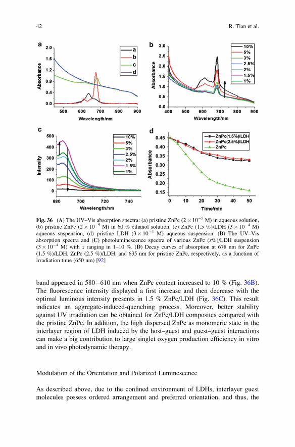
band appeared in 580�610 nm when ZnPc content increased to 10 % (Fig. 36B).
The fluorescence intensity displayed a first increase and then decrease with the
optimal luminous intensity presents in 1.5 % ZnPc/LDH (Fig. 36C). This result
indicates an aggregate-induced-quenching process. Moreover, better stability
against UV irradiation can be obtained for ZnPc/LDH composites compared with
the pristine ZnPc. In addition, the high dispersed ZnPc as monomeric state in the
interlayer region of LDH induced by the host–guest and guest–guest interactions
can make a big contribution to large singlet oxygen production efficiency in vitro
and in vivo photodynamic therapy.
Modulation of the Orientation and Polarized Luminescence
As described above, due to the confined environment of LDHs, interlayer guest
molecules possess ordered arrangement and preferred orientation, and thus, the
Fig. 36 (A) The UV–Vis absorption spectra: (a) pristine ZnPc (2� 10�5 M) in aqueous solution,
(b) pristine ZnPc (2� 10�5 M) in 60 % ethanol solution, (c) ZnPc (1.5 %)/LDH (3� 10�4 M)
aqueous suspension, (d) pristine LDH (3� 10�4 M) aqueous suspension. (B) The UV–Vis
absorption spectra and (C) photoluminescence spectra of various ZnPc (x%)/LDH suspension
(3� 10�4 M) with x ranging in 1–10 %. (D) Decay curves of absorption at 678 nm for ZnPc
(1.5 %)/LDH, ZnPc (2.5 %)/LDH, and 635 nm for pristine ZnPc, respectively, as a function of
irradiation time (650 nm) [92]
42 R. Tian et al.

obtained LDHs can present polarized luminescence properties. For example, cou-
marin-3-carboxylate (C3C) and DDS has been co-intercalated into LDH layer, and
a C3C-DDS/LDH thin film can be subsequently obtained through solvent evapora-
tion method [110]. Figure 37a shows the sole strong basal reflections (00l ) for thethin films of C3C-DDS/LDH, indicating a well c-oriented assembly of LDH
platelets. The uniform and smooth morphology of this film can also be confirmed
by top SEM and AFM (Fig. 37b, d). The obtained thin film possesses continuous
structure with the average thickness of 1.2 μm (Fig. 37c).
The polarized luminescence of the C3C–DDS/LDH thin films was investigated.
Generally, polarized luminescence can be evaluated by determining the anisotropy
value r, which can be expressed by the formula:
r ¼ Ik � I⊥
Ik þ 2I⊥or r ¼ IVV � GIVH
IVV þ 2GIVH;
where IVH stands for the PL intensity obtained with vertical polarized light excita-
tion and horizontal polarization detection and IVV, IHH, IHV are defined in a similar
way; G¼ IHV/IHH is determined from an aqueous solution of the C3C. Theoreti-
cally, the value of r is in the range from �0.2 (absorption and emission transition
dipoles are perpendicular to one another) to 0.4 (two transition dipoles are parallel
Fig. 37 (a) XRD pattern, (b) top-view SEM, (c) side-view SEM, and (d) tapping-mode AFM
images for C3C-DDS/LDH (1.96 %) thin film [110]
Layered Double Hydroxide Materials: Assembly and Photofunctionality 43
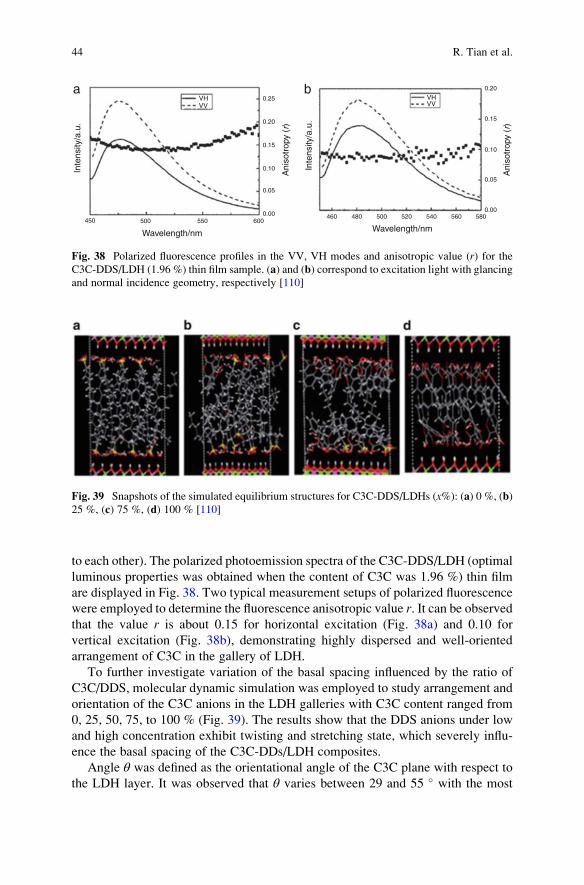
to each other). The polarized photoemission spectra of the C3C-DDS/LDH (optimal
luminous properties was obtained when the content of C3C was 1.96 %) thin film
are displayed in Fig. 38. Two typical measurement setups of polarized fluorescence
were employed to determine the fluorescence anisotropic value r. It can be observedthat the value r is about 0.15 for horizontal excitation (Fig. 38a) and 0.10 for
vertical excitation (Fig. 38b), demonstrating highly dispersed and well-oriented
arrangement of C3C in the gallery of LDH.
To further investigate variation of the basal spacing influenced by the ratio of
C3C/DDS, molecular dynamic simulation was employed to study arrangement and
orientation of the C3C anions in the LDH galleries with C3C content ranged from
0, 25, 50, 75, to 100 % (Fig. 39). The results show that the DDS anions under low
and high concentration exhibit twisting and stretching state, which severely influ-
ence the basal spacing of the C3C-DDs/LDH composites.
Angle θ was defined as the orientational angle of the C3C plane with respect to
the LDH layer. It was observed that θ varies between 29 and 55 � with the most
VHVV
VHVV
450 500
Wavelength/nm Wavelength/nm
Inte
nsity
/a.u
.
Inte
nsity
/a.u
.
Ani
sotr
opy
(r)
Ani
sotr
opy
(r)
550 6000.00 460 480 500 520 540 560 580
0.00
0.05
0.10
0.15
0.20
0.05
0.10
0.15
0.20
0.25a b
Fig. 38 Polarized fluorescence profiles in the VV, VH modes and anisotropic value (r) for theC3C-DDS/LDH (1.96 %) thin film sample. (a) and (b) correspond to excitation light with glancing
and normal incidence geometry, respectively [110]
Fig. 39 Snapshots of the simulated equilibrium structures for C3C-DDS/LDHs (x%): (a) 0 %, (b)
25 %, (c) 75 %, (d) 100 % [110]
44 R. Tian et al.

probable angle of ca. 32� for the intercalated C3C ratio at 100 %. Stimulation from
Fig. 40 shows that all the C3C molecules are inclining to the layers, attributed to the
strong electrostatic interaction between the positive-charged host layers and the
C3C anions. The optimal angles of 32�, 42�, and 54� can be obtained when the ratioof DDS and C3C varies from 0 to 1, with a tendency to change from a tilted to a
vertical arrangement with respect to the layers. Moreover, the distribution of θangle for the C3C-DDS/LDH (25 %) with high DDS content corresponds to the
relative rigid environment imposed by the DDS which restricts the flexible space
for the C3C guest anions. These results show that the changeable interlayer ratio of
two components can highly influence the preferred orientation of the photoactive
guests.
Several LDH-based LbL thin films also exhibit polarized luminescence proper-
ties. Due to the rigid-rod chain configuration or intrinsic one-dimensional structural
anisotropy for these luminescent molecules, well-oriented thin films with a macro-
scopic polarized optical effect can be prepared. For example, sulfonated
carbocyanine derivate (Scy) has been assembled with LDH nanosheets, which
presents polarized near-infrared (NIR) emission luminescence [100]. The average
anisotropy of pristine Scy solution is about 0.2 in the range from 770 to 820 nm,
which is related to the random distribution of the dye molecules in the nonviscous
liquid. NIR polarized fluorescence with the anisotropic value of ~0.8 can be
0.035
0.0300.025
0.0200.0150.0100.005
0.040
0.000
0.0450.0400.0350.030
Probab
lity
Probab
ility
q /degree q /degree
0.0250.0200.0150.0100.005
0.050
0.000
20 30 40 50 6010 70
0.035
0.030
Probab
lity
q /degree
0.0250.0200.0150.010
0.005
0.040
0.000
10 20 30 40 50 60 70 80
0.040
0.035
0.030
Probab
lity
q /degree
0.0250.0200.0150.0100.0050.000
10 20 30 40 50 60 70 80
20 30 40 50 60 7010 80
a
c d
b
Fig. 40 The distributions of orientational angle θ (the plane of C3C with respect to the LDH layer)
with different ratios of C3C to DDS for C3CDDS/LDH (x%): (a) 100 %, (b) 75 %, (c) 50 %, and
(d) 25 % [110]
Layered Double Hydroxide Materials: Assembly and Photofunctionality 45

observed for the Scy/LDH UTF (Fig. 41a), confirming the ordered arrangement of
Scy between the LDH nanosheets. There were no obvious changes for UTFs with
different number of bilayers (Fig. 41b), indicating the independent macroscopic
polarized luminescence on assembly process.
Moreover, due to the regular assembly within the LDH nanosheets, some
molecules with no luminescence polarization can also exhibit well-defined anisot-
ropy: the as-mentioned Ru(dpds)3/LDH UTF is such an example [83]. The pristine
Ru(dpds)3 in aqueous solution shows r at 0.02, while the r value for typical (Ru
(dpds)3/LDH)32 achieved about 0.2–0.3 when comparing the parallel and perpen-
dicular directions to the excitation polarization (IVV vs. IVH) for the in-plane
polarized excitation light. Upon increasing the number of bilayers, the r value
increases systematically, indicating enhancement of the orderly degree for Ru
(dpds)3 between LDH nanosheets.
Construction of Tunable Multicolor Emissive Film Materials
Materials with multicolor luminescence (especially white-light emission) have
been considered as excellent candidates in full-color displays [140], light-emitting
diodes (LEDs) [141], and optoelectronic devices [142]. Conventional strategies
usually rely on combination of three primary color luminescence species, such as
polymers [143], rare-earth compounds [144], metal complexes [145], and semicon-
ductor nanocrystals [146]. However, the different luminous efficiency and inten-
sity, complex fabrication process, as well as complicated interaction between these
chromophores have severely limited the development of multicolor materials. Yan
et al. [77] have introduced LDH nanosheet as building block to combine different
chromophores; in this case, materials with a rigid and ordered microenvironment,
finely tuning emission color, and polarized multicolor luminescence can be achieved.
Fig. 41 (a) Polarized fluorescence profiles in the VV, VH modes and anisotropic value r for the(Scy/LDH)8 UTF. (b) The correlation between fluorescence anisotropic values r of the Scy/LDHUTFs (averaged in the range 770–820 nm) and the bilayer number in the measurement mode with
glancing incidence geometry [100]
46 R. Tian et al.

Figure 42 shows the assembly process of multicolor emission materials employing
APPP (blue) [76], APPV (orange) [78], SPT (red) [79], and BNMA (green) [95] as
primary chromophores. The combination of these individual chromophores can
obtain organic/inorganic UTFs with emission color throughout the whole visible
region, even the white color emission.
Taking (APPP/LDH)12/(BNMA@PVS/LDH)n (n¼ 0–6) UTFs (B/G lumines-
cence) as the example, the assembly process was carried out by assembling
(BNMA@PVS/LDH)n (n¼ 1–6) bilayers onto the as-prepared (APPP/LDH)12UTF. Figure 43a1 shows the fluorescence monotonic increase at 481 and 504 nm
which can be attributed to the emission of BNMA@PVS, while the emission peak
at 410 nm is assigned to the pristine (APPP/LDH)12 UTF. The similar spectra
between assembled UTFs and solutions indicate that the aggregation of BNMA can
be avoided throughout the assembly process. The insets show the photographs
taken under UV illumination, and high luminescence can be observed for these
UTFs. The color coordinates in Fig. 43a2 demonstrated that the color of the UTFs
can be tuned from blue (CIE 1931: (0.172, 0.149); n¼ 0) to bluish green (CIE 1931:
(0.208, 0.343); n¼ 6) effectively. The multicolor emission of blue/orange, red/blue,
and red/green can also be tuned by this method throughout the whole visible region
(Fig. 43b–d). In addition, based on the periodic ordered structure of these UTFs,
they exhibit well-defined multicolor polarized luminescence with high anisotropy,
which may effectively extend its application in multicolor or white polarized
photoemission devices.
For the inorganic building blocks, quantum dots (QDs) [147] have attracted great
attention due to high quantum yields, good stability, and high color purity [148],
Fig. 42 (a) Representation of one monolayer of MgAl-LDH nanosheet; the chemical formulae of
(b) APPP (blue luminescence), (c) BNMA@PVS (green luminescence), (d) APPV (orangeluminescence), and (e) APT (red luminescence); (f) procedure for assembling two-color emitting
UTFs with blue/green (B/G), blue/orange (B/O), red/blue(R/B), and red/green (R/G) lumines-
cence [77]
Layered Double Hydroxide Materials: Assembly and Photofunctionality 47

and application of QDs in the multicolor emission materials has also been carefully
investigated. Liang et al. [117] have assembled QDs with LDH nanosheets to obtain
highly ordered structure and finely tunable fluorescence, and the tunable color in the
red–green region could be achieved by adjusting the assembly cycle number and
sequence of red- and green-emission QDs. In addition, Tian et al. [102] have
introduced fabrication of precisely tuned white color emission UTFs employing
red- and green-emission QDs and blue 2,20-(1,2-ethenediyl) bis
[5-[[4-(diethylamino)-6-[(2,5-disulfophenyl) amino]-1,3,5-triazin-2-yl] amino]
benzene-5 sulfonic acid] hexasodium salt (BTBS). By adjusting the deposition
sequence and relative ratio of the blue-, green- and red-emission units, luminescent
UTFs with finely tunable photoemission in the white spectral region were obtained
(Fig. 44B). Moreover, the color coordinates of the best white-light emission
(denoted as f in Fig. 44C) could achieve (0.322, 0.324) which is very close to the
standard coordinates of white light (0.333, 0.333).
The emission color of hybrid composites can be tuned in response to different
excitation light, which has been recognized as an intelligent photoluminescence
material. This composite UTF was illustrated as (BTBS/LDH)12(QD-530/
LDH)20(QD-620/LDH)7, composed of three chromophores: red-emission and
green-emission QDs and blue-emission organic compound BTBS. Due to the
different excitation and emission spectra of these three chromophores, it can be
concluded that BTBS can be excited at 360 nm while photon energy above 500 and
600 nm can efficiently excite QD-530 and QD-620. Therefore, excited at 360, 460,
and 560 nm, the UTFs display three different emission spectra as well as
corresponding white, orange, and red color, respectively (Fig. 45). These hybrid
materials responding to external excitation light via the alteration of emission color
can be widely used in anti-forgery, colorimetric sensors, and display devices.
Fig. 43 Fluorescence spectra (the inset shows the ratio of two chromophores), photographs under
UV light (365 nm), and the change in color coordinates with n for (a) (APPP/LDH)12/
(BNMA@PVS/LDH)n (n¼ 0–6) UTFs (for B/G luminescence), (b) (APPP/LDH)12/(APPV/
LDH)n (n¼ 0–18) UTFs (for B/O luminescence), (c) (APPP/LDH)12/(APT/LDH)n (n¼ 0–18)
UTFs (for R/B luminescence), and (d) (APT/LDH)12/(BNMA@PVS/LDH)n (n¼ 0–6) UTFs
(for R/G luminescence) [77]
48 R. Tian et al.
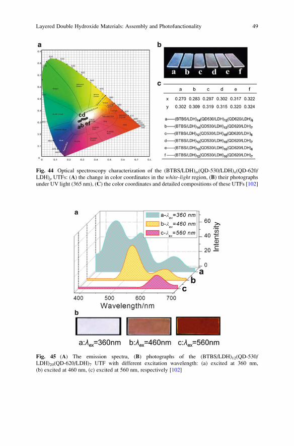
Fig. 45 (A) The emission spectra, (B) photographs of the (BTBS/LDH)12(QD-530/
LDH)20(QD-620/LDH)7 UTF with different excitation wavelength: (a) excited at 360 nm,
(b) excited at 460 nm, (c) excited at 560 nm, respectively [102]
Fig. 44 Optical spectroscopy characterization of the (BTBS/LDH)m(QD-530/LDH)n(QD-620/
LDH)p UTFs: (A) the change in color coordinates in the white-light region, (B) their photographsunder UV light (365 nm), (C) the color coordinates and detailed compositions of these UTFs [102]
Layered Double Hydroxide Materials: Assembly and Photofunctionality 49

Moreover, sulfonated Eu- and Tb-based lanthanide complexes can also be
employed in fabrication of multicolor emission materials [149]. After the synthesis
of red-emissive Eu(DBM)3bath and green-emissive Tb(acac)3Tiron (DBM,
dibenzoylmethane; bath, bathophenanthroline disulfonate disodium; acac,
2,4-pentanedione), these lanthanide complexes can be assembled with LDH to
obtain highly emissive luminescence with tunable color on tuning of their relative
ratio (Fig. 46). In addition, these UTFs exhibit well-defined one and two-color
polarized fluorescence and obvious red and green up-conversion emission upon
excitation by near-IR laser light.
6.3.2 Dynamic Tuning of the Emission
The photofunctional materials with smart-responsive properties can present
dynamic changes by tuning the external stimuli. Such dynamic-responsive
photofunctional materials should have rapid response time, high sensitivity, obvi-
ous signal change, and high stability for long-term use [150]. In order to achieve
these requirements, LDHs have been employed as an effective inorganic matrix and
building block for the ordered arrangement and improved stability of active chro-
mophores in the solid state [151]. Recently, the smart-responsive LDH
photofunctional materials responding to environmental stimuli (such as chemical
substances, pH value, temperature, pressure, light irradiation, and electricity) have
been studied, which present dynamic tuning of their photofunctionalities.
Detection of Chemical Substances
Chemo-sensor plays an important role in the detection of heavy metal ions (HMIs),
biomolecules, environmental pollutants, and chemical warfare agents [152–156].
The basic mechanisms of fluorescence detection for most of hazardous substances
Fig. 46 Digital photos of flexible (a) (Eu(DBM)3bath/LDH)8 and (b) (Tb(acac)3Tiron/LDH)8UTFs under UV irradiation; (c) fluorescence spectra of (Eu(DBM)3bath/LDH)10/(Tb(acac)3Tiron/
LDH)n (n¼ 0–14) UTFs, and insets show the photographs under UV light [149]
50 R. Tian et al.

rely on the quenching induced by the binding between analytes and fluorescence
probe molecules. However, the commonly studied fluorescence probe molecules
are in aqueous or powder states, and the complicated operation process and
difficulty in recyclability have severely limited the application of such materials.
Therefore, the development of suitable species and/or new approaches toward the
facile fabrication of chemo-sensor with high sensitivity, fast response, and decent
recyclability remains a stimulating challenge.
Fabrication of photofunctional UTFs based on the LbL assembly with LDH
nanosheets can serve as an effective method to solve the above difficulties. A
certain amount of fluorescence molecules have been applied (such as BTBS
[101], calcein [109], and 1-amino-8-hydroxy-3,6-disulfonaphthalene (H-acid)
[157]) as probes for the detection of HMIs (such as mercury ion). Taking BTBS/
LDH system as an example, fluorescence emission spectrum shows a monotonic
increase with n at ca. 445 nm, and the UTFs show well-defined blue luminescence
under UV light (Fig. 47a and inset). The systematical and significant photoemission
decrease of the (BTBS/LDH)32 UTF can be observed in responding to the addition
of Hg2+ (Fig. 47b), while different degrees of quenching for metal ions were
presented in Fig. 47d. The strongest quenching by Hg2+ is related to the high
thermodynamic affinity with typical O-chelate ligands and metal-to-ligand binding
kinetics. Reproducibility was carried out for alternative dipping the UTFs into Hg2+
and EDTA, and the UTFs can recover to the pristine fluorescence intensity at least
five cycles (Fig. 47c). The immobilization by LDH has guaranteed the repeatability
of HMI detection, while the drop-casted sample is easy to fall off in solution.
Moreover, Jin et al. have proposed the detection of biomolecules dextran-40 by
employing 8-amino-1,3,6-naphthalenetrisulfonates (ANTS) and LDH as building
block [158]. The obtained (ANTS/LDH)n UTF can be adapted in the detection of
Mg2+ which is important in stabilizing the structure of DNA and transferring of
biological signals [159]. The detection limit of 2.37� 10�7 M can be obtained.
Trinitrotoluene (TNT), which is an important explosives commonly applied in both
military and forensic science, can be specifically detected by (BBU/LDH)n UTFs,
which are assembled by 4,40-bis[2-di(b-hydroxyethyl)amino-4-(4-sulfophe-nylamino)-
s-triazin-6-ylamino] stilbine-2,20-disulfonate (BBU) with LDH nanosheets [103].
Li, Qin, and coworkers have efficiently detected the volatile organic compounds
(VOCs) based on modulating the FRET process. The basal spacing and the
interlayer distance between donor and acceptor can be varied with external stimuli
(VOC vapor), and thus, the color change from blue to violet can be observed for the
detection [93, 135].
pH Sensor
Optical pH sensor with stable lifetime and signal is of great importance in environ-
mental research, toxicological assay, blood measurement, and biotechnology [160–
163]. The red-emissive (SPT/LDH)32 UTF described above exhibited reversible
responses toward pH in alternative dipping in aqueous at pH¼ 4 and pH¼ 13 [79].
Layered Double Hydroxide Materials: Assembly and Photofunctionality 51
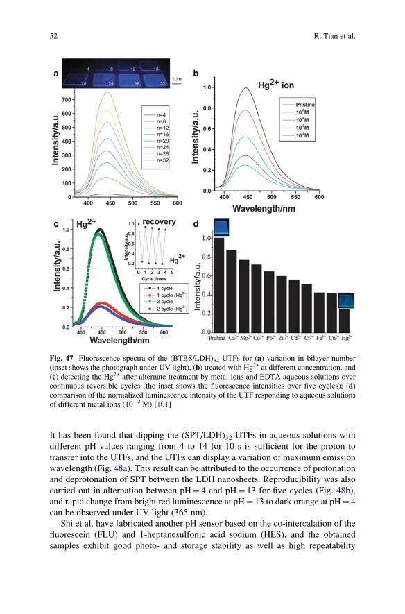
It has been found that dipping the (SPT/LDH)32 UTFs in aqueous solutions with
different pH values ranging from 4 to 14 for 10 s is sufficient for the proton to
transfer into the UTFs, and the UTFs can display a variation of maximum emission
wavelength (Fig. 48a). This result can be attributed to the occurrence of protonation
and deprotonation of SPT between the LDH nanosheets. Reproducibility was also
carried out in alternation between pH¼ 4 and pH¼ 13 for five cycles (Fig. 48b),
and rapid change from bright red luminescence at pH¼ 13 to dark orange at pH¼ 4
can be observed under UV light (365 nm).
Shi et al. have fabricated another pH sensor based on the co-intercalation of the
fluorescein (FLU) and 1-heptanesulfonic acid sodium (HES), and the obtained
samples exhibit good photo- and storage stability as well as high repeatability
Fig. 47 Fluorescence spectra of the (BTBS/LDH)32 UTFs for (a) variation in bilayer number
(inset shows the photograph under UV light), (b) treated with Hg2+ at different concentration, and
(c) detecting the Hg2+ after alternate treatment by metal ions and EDTA aqueous solutions over
continuous reversible cycles (the inset shows the fluorescence intensities over five cycles); (d)
comparison of the normalized luminescence intensity of the UTF responding to aqueous solutions
of different metal ions (10�2 M) [101]
52 R. Tian et al.
toward responses of pH [106]. Employing electrophoretic deposition method, a
sensor with broad linear dynamic range in pH (5.02–8.54) can be obtained
(Fig. 49a). Remarkable repeatability in 20 cycles can be achieved with the relative
standard deviation (RSD) less than 1.5 % (Fig. 49b). The sensitive and rapid
responses of SPT/LDH and FLU/LDH toward pH materials indicated their potential
application as pH luminescence sensor.
Fig. 49 (a) Fluorescence intensity of the pH sensor recorded after 1, 5, 10, 15, and 20 cycles at
pH 5.02, 6.51, and 8.54 and (b) the reversibility recorded by alternate measurement in two
solutions with pH 5.02 and 8.54 [106]
Fig. 48 (a) Variation of the maximum photoemission wavelength of the (SPT/LDH)32 UTF with
different pH values and (b) the reversible photoemission response upon alternation between
pH¼ 4 and pH¼ 13 (the inset show the luminescence photographs of the UTFs at the two pH
conditions) [79]
Layered Double Hydroxide Materials: Assembly and Photofunctionality 53
Temperature Sensor
Temperature is one of the most common and natural environmental factor, and
thermochromic materials can be potentially used in temperature sensor and devices
[164–166]. Therefore, much attention has been paid on fabrication of
thermochromic materials with good sensitivity and reversibility. Bis
(2-sulfonatostyryl)biphenyl (BSB) was employed as thermochromic luminescence
(TCL) molecule to assemble with LDH nanosheet to achieve an ordered supramo-
lecular UTFs [99]. Figure 50 shows the assembly process of (BSB/LDH)n with the
monotonic increase in absorption and fluorescence emission spectra. The maximum
emission peak is located at 444 nm, and no broadening or shift can be observed in
the whole assembly process, indicating no interaction between molecules occurred.
The XRD data show the ordered and monolayer arrangement of BSB in the
interlayer of LDHs.
The TCL behavior of the UTFs was studied on heating from 20 to 100 �C, and agradual decrease in fluorescence intensity with a red shift from 444 to 473 nm can
be observed (Fig. 51a). A visible change can be observed under UV light with the
emission color changes from blue to bluish green upon heating. The recover in color
and spectrum can be accomplished when the UTF cooled to 20 �C, and this heating–cooling process can be readily repeated (Fig. 51a, inset). Fluorescence lifetimes of
these (BSB/LDH)n UTFs were also studied to obtain the excited-state information.
The results show that lifetime of the UTF is longer at 100 �C (1.9 ns) than that at
20 �C (0.9 ns), and the recycle can also be achieved (Fig. 51b). Figure 51c shows the
polarized fluorescence measurements of the UTFs, and the degree of structural
order can be obtained by detecting the fluorescence anisotropy r. The r value
decreases from 0.15 at 20 �C to 0.02 for UTFs at 100 �C, which exhibit a less
ordered arrangement of molecules at high temperature.
Fig. 50 Characterization of (BSB/LDH)n (n¼ 4–32) UTFs. (a) UV–Vis absorption spectra (inset:
plots of the absorbance at 204 and 349 nm vs. the number of bilayers n) and (b) fluorescence
spectra. The insets in (a) and (b) are photographs of UTFs with different n under daylight and UVlight (365 nm), respectively [99]
54 R. Tian et al.

Similar observations can also be obtained for (CdTe QDs/LDH)30 UTF [116];
Fig. 52a shows the responses toward temperature upon heating, and an intensity
decrease along with red shift can be observed. These inorganic–inorganic hybrid
UTFs possess good sensitivity, and�1 % per �C decrease and 0.1 nm �C�1 red shift
can be obtained. The UTFs exhibit excellent repeatability for eight cycles in the
heating–cooling process (Fig. 52b).
Aromatic azo dyes, such as 4-(4-anilinophenylazo)benzenesulfonate (AO5),
have been widely used due to the easy transformation between the azo A-form
and hydrazone H-form (Scheme 3). It was well known that this tautomerism is very
sensitive to the surrounding environment; therefore, it has been widely used as
thermal sensors and in molecular memory storage. In order to further modulate the
environment in an inorganic matrix, AO5 was co-intercalated with sodium
dodecylbenzene sulfonate (SDS) into the interlayer of LDH [112]. The absorption
500
400
400 450 500
300
200
100
0
500
600
400
300
200
100
00550 600 650 700
Wavelength/nm
23°C
30°C
40°C
50°C
60°C
70°C
80°C
Inte
nsity
Intensity
Inte
nsity
Peak/nm
558560
564566568
562
R=0.994 R=0.998
20 30 40 50 60 70 80Temperature/°C
20 30 40 50 60 70 80Temperature/°C
400
320
240
160
80
1 2 3 4 5 6 7 8Number of cycle
Heating
Cooling
a b
Fig. 52 (a) The luminescence spectra of the (CdTe QDs/LDH)30 UTF in the temperature range
23–80 �C (inset: the emission position or intensity as a function of temperature, respectively); (b)
the reversible fluorescence response of eight consecutive cycles (the inset shows the UTF
photographs at 23 and 80 �C, respectively) [116]
Fig. 51 TCL of the (BSB/LDH)8 UTF at 20 and 100 �C. (a) Fluorescence emission spectra (inset:
the reversible fluorescence response over five consecutive cycles); (b) typical fluorescence decay
curve (inset: fluorescence lifetimes over five consecutive cycles, black: 20 �C, red: 100 �C; (c)fluorescence anisotropy over five consecutive cycles [99]
Layered Double Hydroxide Materials: Assembly and Photofunctionality 55
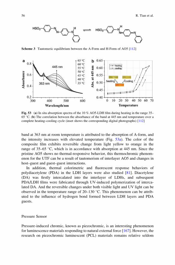
band at 363 nm at room temperature is attributed to the absorption of A-form, and
the intensity increases with elevated temperature (Fig. 53a). The color of the
composite film exhibits reversible change from light yellow to orange in the
range of 35–65 �C, which is in accordance with absorption at 445 nm. Since the
pristine AO5 shows no thermal-responsive behavior, this thermochromic phenom-
enon for the UTF can be a result of tautomerism of interlayer AO5 and changes in
host–guest and guest–guest interactions.
In addition, thermal colorimetric and fluorescent response behaviors of
polydiacetylene (PDA) in the LDH layers were also studied [81]. Diacetylene
(DA) was firstly intercalated into the interlayer of LDHs, and subsequent
PDA/LDH films were fabricated through UV-induced polymerization of interca-
lated DA. And the reversible changes under both visible light and UV light can be
observed in the temperature range of 20–130 �C. This phenomenon can be attrib-
uted to the influence of hydrogen bond formed between LDH layers and PDA
guests.
Pressure Sensor
Pressure-induced chromic, known as piezochromic, is an interesting phenomenon
for luminescence materials responding to natural external force [167]. However, the
research on piezochromic luminescent (PCL) materials remains relative seldom
Fig. 53 (a) In situ absorption spectra of the 10 % AO5-LDH film during heating in the range 35–
65 �C. (b) The correlation between the absorbance of the band at 445 nm and temperature over a
complete heating–cooling cycle (inset shows the corresponding digital photographs) [112]
Scheme 3 Tautomeric equilibrium between the A-Form and H-Form of AO5 [112]
56 R. Tian et al.

compared with other environment-responsive materials (such as temperature-, pH-
and HMI-sensitive materials). Therefore, the investigation on PCL materials with
excellent reversibility, repeatability, and rapid responsiveness is still a challenge
[168, 169].
Yan et al. have studied the fluorescence properties as well as molecular packing
mode of (2,20-(1,2-ethenediyl)bis[5-[[4-(diethylamino)-6-[(2,5disulfophenyl)
amino]-1,3,5-triazin-2-yl] amino]benzene sulfonate anion (BTZB) after intercala-
tion into LDHs matrix [111]. The fluorescence of the pristine BTZB and BTZB/
LDH composites is shown at approximately 452 nm, and significant luminescent
changes can be observed for the BTZB/LDHs treated with pressures while pristine
BTZB remains unchanged. Figure 54a shows the changes of fluorescence spectra
upon increasing the pressure from 0.1 MPa to 18.8 GPa; the emission wavelength
shifts from 452 to 515 nm, which can be assigned to the formation of J-type
Fig. 54 (a) Fluorescence emission and (b) UV–Vis spectra of the BTZB/Mg2Al-LDH at different
pressures (inset shows the photographs of pressurized samples under daylight and UV light,
respectively); (c) XRD pattern with the varying interlayer distance and (d) fluorescence lifetime
of the BTZB/Mg2Al-LDH at different pressures [111]
Layered Double Hydroxide Materials: Assembly and Photofunctionality 57
aggregates in the LDH galleries. The change of this phenomenon can be obviously
seen from photographs of the BTZB/LDH pellet under UV light (Fig. 54a, inset)
with the color varies from blue to green with increasing pressure. UV–Vis spectra
also exhibit an obvious change on varying the pressure (Fig. 54b). Except for the
absorption band at 290 nm for the pristine BTZB/LDH, a new band, ranging from
350 to 450 nm, appears when the pressure is sustainable increased. A broad
shoulder band at 450–550 nm can be observed when the pressure was increased
to 15.6 GPa. The appearance of this new band is due to the formation of J-typeaggregates of BTZB in the interlayer of LDH, which is in accordance with the
results from fluorescence tests above. In order to take an insight into the influences
of pressure on the supramolecular organization in this BTZB/LDH host–guest
system, XRD was carried out. Figure 54c shows that upon increasing the pressure
from 0.1 MPa to 18.8 GPa, the basal d003 spacing changes from 1.57 to 1.45 nm,
maintaining the structure of LDHs. This may due to the contracted gallery height
and subsequent rearrangement of intercalated guest molecules. And the fluores-
cence lifetime of BTZB/LDH is much shorter at high pressure (2.14 ns at 18.8 GPa
and 4.63 ns at 0.1 MPa), further confirming the aggregation of the BTZB (Fig. 54d).
Reversibility and repeatability tests are also carried out by grinding and subse-
quent heating for the pellet sample, and a reversible performance for practical
application of PCL materials is satisfactorily obtained. Moreover, the different
composition of host layer has been investigated, and the results show that Mg2Al-
LDH possesses better PCL performances compared to Zn-Al-LDH or Mg3Al-LDH.
Therefore, these BTZB/LDH materials exhibit obvious changes in UV–Vis absorp-
tion, fluorescence emission, and lifetime upon the increase of pressure, and the
color change from blue to green under UV light can be visually seen. Theoretical
results show that the variations of the arrangement and aggregation status of BTZB
in the interlayer of LDH cause the subsequent PCL phenomenon.
Mechanofluorochromism can be observed in the NFC-HPS (NFC, niflumic acid,
a typical aggregation-induced emission molecule; HPS, heptanesulfonate)
co-intercalated LDHs [170]. The results show that NFC-HPS/LDH (HFC:
HPS¼ 5 %) possesses obvious change in fluorescence on mechanical force, while
the pristine solid-state NFC sample remains nearly unchanged under pressure. This
phenomenon can be attributed to the slipping of LDH sheets and reorientation of the
interlayer surfactant; and the changed host–guest interactions can tune the molec-
ular conformation and intermolecular interaction of NFC anions.
Light-Induced Sensor
Photochromism is a phenomenon that substance exhibits color change upon light
irradiation, and it has been widely used in light switching, liquid-crystal alignment,
information storage, and nonlinear devices [171–173]. It is well known that the
photoisomerization process is very sensitive to the surrounding microenvironment,
and the low isomerization efficiency and stability has limited the development
of photochromic materials. Embedding the photo-responsive molecules into the
58 R. Tian et al.

inorganic matrix, such as LDH nanosheets, may effectively inhibit the aggregation of
the molecules and thus enhance the photoisomerization free space for photochromism.
Wang et al. have studied the photoisomerization process of a kind of Schiff base
(azomethine-H anions, AMH) assembled LDH system [174]. Generally, after the
excitation of the initial enol (anti-enol) tautomer, the excited-state intramolecular
proton transfer (ESIPT) occurs for AMH, leading to the excited keto tautomer
(cis-keto or its zwitterionic form), exhibiting a characteristic Stokes fluorescence
band. After the structural changes (rotation around C¼C and/or C–N bond)
involving the cleavage of the intramolecular hydrogen bond, the red long-lived
photochromic tautomer (trans-keto or its zwitterionic form) in the ground state is
generated either by thermal treatment or by irradiation of visible light.
In order to obtain advanced performance along with good stability, AMH was
co-intercalated with 1-pentanesulfonate (PS) into the interlayer of LDH, and the
corresponding thin films of AMH/PS-LDH were obtained by the solvent evapora-
tion. Firstly, the photoisomerization process of the pristine AMH solution has been
studied, and the transient absorption spectra were illustrated in Fig. 55a. The bands
at 470 nm are attributed to the absorption from the S0 state of the trans-keto
tautomer, and the band with a maximum at ~370 nm is possibly related to the
long-lived syn-enol form. Upon irradiation of laser, a dramatic decrease can be
observed. For AMH/PS-LDH films, an excited-state intramolecular proton transfer
to the trans-keto tautomer was occurred when the initial AMH (as an enol tautomer)
receives UV excitation, and the relaxed back isomerization to the ground state was
observed through transient UV–Vis spectroscopy (Fig. 55b). The band located at
465 nm is due to the S0 state of the trans-keto tautomer, and a 5 nm blue shift
due to the changed weak polar microenvironment can be observed. The absence of
syn-enol form indicates the inhibition of the syn-enol/anti-enol isomerization in the
Fig. 55 Transient absorption spectra obtained by laser photolysis for (a) nitrogen-saturated
solution of AMH (5.0� 10�5 M) in water (inset shows the magnified absorption from 350 to
400 nm), and (b) the 2 % AMH-LDH film [174]
Layered Double Hydroxide Materials: Assembly and Photofunctionality 59

2D confined environment. As a result, the trans-keto tautomer of AMH is less stable
in the 2D confined environment, and relaxed back isomerization is more easily to
take place.
Moreover, photoactive azobenzene polymer PAZO was also applied in the
assembly with LDH to obtain a photochromism material [82]. Upon UV irradiation,
the absorption at 376 nm decreases as a function of time (Fig. 56A) due to the
transformation of trans to cis of PAZO. A complete isomerization can be achieved
(Fig. 56B, line a–b) after sufficient irradiation time. The π–π* absorption band
increases again (Fig. 56B, line c) when irradiated by visible light (λ> 450 nm), and
the back isomerization for the cis–trans can be achieved. The extent of 51 % is
obtained for the cis to trans back isomerization, and repeatability for several cycles
can be accomplished by alternative irradiation of UV and visible light. The results
show that the intertwisting of polymer chains and π–π stacking of the azobenzene
chromophores can be efficiently reduced, and sufficient free space is available for
the isomerization of the azobenzene moiety due to the employment of LDH
nanosheets.
Light-triggered materials containing porphyrin and phthalocyanine molecules
for biological and medical application were also largely investigated. Through the
intercalation, the inherent properties of these porphyrin and phthalocyanine mole-
cules may be altered [175]. [Tetrakis(4-carboxyphenyl)-porphyrinato]zinc
(II) (ZnTPPC) [88], Pd(II)-5,10,15,20-tetrakis(4-carboxyphenyl) porphyrin
(PdTPPC) [89], 5,10,15,20-tetrakis(4-sulfonatophenyl) porphyrin (TPPS), and its
palladium complex (PdTPPS) [89–91] intercalated LDH-based hybrid materials
have been studied. Figure 57 shows the relaxation of PdTPPS phosphorescence
(triplet states) and luminescence signals of O2(1Δg) in the assembled LDH-DS/
PdTPPS films. The proximity of the porphyrin triplet states and their interaction
with O2(1Δg) contribute to the repopulation of the fluorescent excited singlet states
of TPPS. Moreover, Liang et al. have also studied the singlet oxygen production
2.0
1.5
1.0
0.5
0.0
a
c
b
2.0
1.5
1.0
0.5
0.0200 300 400 500 600 700 800
1.8
1.6
1.4
1.2
1.0
0.8
0.6
1.81.61.41.21.00.80.60.40.2
0 1 2 3 4 5 6Number of irradiation cycles
after UV irradiation
recovery by visible light0 min
215 min0 50 100 150 200 250 300
Irradiation time of UV light/min
Abs
orba
nce
Abs
orba
nce
Abs
orba
nce
Abs
orba
nce
Wavelength/nm200 300 400 500 600 700 800
Wavelength/nm
a b
Fig. 56 UV–Vis absorption spectra of photoisomerization for the (LDH/PAZO)50 film: (A) the
absorption intensity changes with time; (B) repeatability tests for (a) the trans isomer, (b) the same
sample after irradiation with UV light, and (c) the recovered trans isomer after irradiation with
visible light. The inset shows the change in the absorbance at the maximum of 376 nm [82]
60 R. Tian et al.

efficiency of zinc phthalocyanines intercalated LDHs, and the composite materials
exhibit excellent anticancer behavior when used for photodynamic therapy [92].
Electroluminescence, Chemiluminescence, and Electrochemiluminescence
Electroluminescence (EL) is the luminescent behavior induced by external electric
field, which requires saturated emission colors, high thermal stability, and long
operational lifetimes of chromophores [176]. However, traditional EL devices often
suffered from low efficiency, short lifetimes, and complicated fabrication technique
under specially controlled environment. A method for fabricating high efficiency
and thermally stable light-emitting device (LED) involving simple technique is
quite needed. Bendall et al. [119] have designed an all-inorganic EL device
constructed by assembly highly luminescent CdTe QDs with LDH nanosheet
(Fig. 58a). EL emission peak, which can be observed at 637 nm, is almost the
Fig. 58 (a) Schematic of the architecture of the device, which is based on a “sandwich” structure,
and (b) photoluminescence peak of aqueous CdTe QDs (gray curve) compared to the electrolu-
minescence peak of the device (black curve) [119]
Fig. 57 (a) Quenching of the PdTPPS triplet states in the Mg2Al-LDH-DS/PdTPPS film at
different oxygen pressures (excitation at 425 nm, phosphorescence recorded at 690 nm); the
arrow shows increasing O2 pressure (vacuum, 100, 210, 300, 400, 760 Torr). (b) Time dependen-
cies of the O2(1Δg) luminescence signals in air (black line) and oxygen (green line) after the
excitation of TPPS in the Mg2Al-LDH-DS/TPPS film. The red lines are least-squares
monoexponential fits (inset shows emission band of O2 (1Δg) in air) [91]
Layered Double Hydroxide Materials: Assembly and Photofunctionality 61

same as the photoluminescence (PL) emission peak of the bare QD without any
shift or broadening occurs. Such device maintains the requirements for the common
organic LEDs, and possesses better thermal stability (can be operated over 80 �C)due to the employment of LDHs. Thus, a bright, monochrome, and stable EL device
with excellent thermal stability, brightness, and simple fabrication process is
achieved.
Chemiluminescence (CL) stands for the luminescence derived from a chemical
reaction. Much attention has been paid on enhanced CL for detection in biology and
sensor fields [177]. Lu et al. have investigated the CL phenomenon based on
nanocomposite material constructed by the assembly of CdTe QDs upon the surface
of DBS-LDHs in an ordered manner. The obtained material exhibited a remarkable
CL amplification of the luminol–H2O2 system due to ordered arrangement of QDs
and host–guest interaction between LDH and CdTe QDs [121].
Electrochemiluminescence (ECL) is a combined process of EL and CL, which is
caused by energetic electron transfer (redox) reactions of electrogenerated species
[178]. Recently, ECL based on a solid-state sensor has also been investigated
employing ruthenium(II) tris(bipyridine) and poly(sodium 4-styrene sulfonate) to
assemble with LDH nanosheets [179]. Figure 59a shows the assembly process for
Fig. 59 Assembly and properties of (LDH/PSS/Ru(bpy)32+/PSS)n multilayer films: (a) schematic
representation, (b) UV–Vis absorption spectra along with different tetra-layer number n (the insetshows the absorbance at 291 and 457 nm vs. n), and (c) ECL spectra in the presence of NADH with
various concentrations (from a to h: 3.16� 10�6 to 3.23� 10�4 M, scan rate ¼ 100 mV s�1, and
the inset shows plot of ECL intensity vs. NADH concentration) [179]
62 R. Tian et al.

this (LDH/PSS/Ru(bpy)32+/PSS)10 multilayer films, and UV–Vis spectra has been
used to monitor the stepwise growth of the UTFs (Fig. 59b). A linear response to
dihydronicotinamide adenine dinucleotide (NADH) in the range 3.16� 10�6 to
3.23� 10�4 M can be obtained, and the detection limit can reach as low as
0.023 μM (Fig. 59c).
Therefore, the application of LDH-based materials can be extended to EL, CL,
and ECL devices due to the uniform dispersion of photoactive molecules in 2D
confined microenvironment supported by the superlattice structure of the multilayer
films.
7 Conclusion and Outlook
In conclusion, LDH-based compounds have become promising photofunctional
materials, which can be used in infrared radiation absorption, ultraviolet shielding,
and tunable color/luminescence with both static and dynamic manners. Due to their
inherent high stability, ion-exchangeable property, and tailorable photo-related
performance, LDHs can serve as an effective building block to incorporate
photoactive molecules for the fabrication of new hybrid materials with diverse
potential applications. Through the adjustment of the elemental composition,
charge density, and particle size of LDH precursors, the quantity and distribution
of confined guests can be tuned, which enable the fabrication of photofunctional
materials with preferred orientations. Host–guest interaction and interlayer con-
finement effect can further influence the arrangement and orientation of intercalated
guest molecules, and hybrid materials with superior optical properties compared
with the pristine form can be achieved. For example, the LDH-based luminescent
materials with precisely tuned emission in the whole visible range are obtained.
And the hybrid materials also present repeatable and reversible responses toward
external stimuli (such as light, pH, pressure, temperature, electricity, and
chemicals), which can be utilized in the fields of photochromic, piezochromic,
thermochromic, and polarized luminescence, chemical sensors, and switches.
Moreover, some of LDHs systems (IR absorption and UV-shielding materials)
have already been used as functional addictives due to the significant improvement
of IR/UV properties for polymers and asphalt.
From both academic and engineering perspectives, two key scientific problems
still need to be further understood for these LDH-based photofunctional materials:
(1) How the fine structures in both the inner layer and interlayer of LDHs (such as
interface, surface, and defect) as well as their size effect can influence the
macroscopical photo-related properties; (2) how the host–guest interactions
between the LDHs layers and photoactive guests can influence the arrangement,
orientation, and aggregation of the interlayer species. In addition, to meet the need
of practical application, further explorations and interests can be concentrated on:
(1) Intelligent materials: the development of new types of stimuli-responsive
luminescent materials with fast response, high on/off ratio, and good repeatability
Layered Double Hydroxide Materials: Assembly and Photofunctionality 63

are highly desirable. As well, the multi-stimuli-responsive materials remain in an
early stage and have been very seldom studied. (2) Large-scale preparation of
LDHs photofunctional materials: up to now, most of these materials are still at
the laboratory level, and how to scale up the LDHs materials for industrial appli-
cations needs further exploration from both process engineering and product engi-
neering perspectives. (3) Combination of the photo-related performance with other
functionalities, such as medical, pharmaceutical, environment, and energy fields,
would be an important strategy and direction to broaden LDH-based
multifunctional materials. Researches in these directions are continuing by us and
many others worldwide.
Acknowledgment We would like to thank all the coworkers cited in the references for their
invaluable contributions to the work described here. This work was supported by the 973 Program
(Grant No. 2014CB932103), the National Natural Science Foundation of China (NSFC), Beijing
Municipal Natural Science Foundation (Grant No. 2152016), the 111 Project (Grant B07004), and
Program for Changjiang Scholars and the Innovative Research Team in University (PCSIRT:
IRT1205).
References
1. Newman SP, Jones W (1998) New J Chem 22:105
2. Funnell NP, Wang Q, Connor L, Tucker MG, O’Hare D, Goodwin AL (2014) Nanoscale
6:8032
3. Jellicoe TC, Fogg AM (2012) J Phys Chem Solids 73:1496
4. Fogg AM, Dunn JS, Shyu SG, Cary DR, O’Hare D (1998) Chem Mater 10:351
5. Wang Q, O’Hare D (2012) Chem Rev 112:4124
6. Ma R, Liu Z, Li L, Iyi N, Sasaki T (2006) J Mater Chem 16:3809
7. He J, Wei M, Li B, Kang Y, Evans DG, Duan X (2006) Struct Bond 119:89
8. Kim JA, Hwang SJ, Choy JH (2008) J Nanosci Nanotechnol 8:5172
9. Guo X, Zhang F, Evans DG, Duan X (2010) Chem Commun 46:5197
10. Liu Z, Ma R, Osada M, Iyi N, Ebina Y, Takada K, Sasaki T (2006) J Am Chem Soc 128:4872
11. Evans DG, Slade RCT (2006) Struct Bond 119:1
12. Yan D, Lu J, Duan X (2013) Scientia Sinica Chimica 43:1
13. Li C, Dou Y, Liu J, Chen Y, He S, Wei M, Evans DG, Duan X (2013) Chem Commun
49:9992
14. Shao M, Ning F, Zhao J, Wei M, Evans DG, Duan X (2012) J Am Chem Soc 134:1071
15. Vialat P, Leroux F, Taviot-Gueho C, Villemure G, Mousty C (2013) Electrochim Acta
107:599
16. Choi SJ, Choy JH (2011) Nanomedicine 6:803
17. Yan D, Lu J, Wei M, Evans DG, Duan X (2011) J Mater Chem 21:13128
18. Gursky JA, Blough SD, Luna C, Gomez C, Luevano AN, Gardner EA (2006) J Am Chem Soc
128:8376
19. Gardner E, Huntoon KM, Pinnavaia TJ (2001) Adv Mater 13:1263
20. Li C, Wang L, Wei M, Evans DG, Duan X (2008) J Mater Chem 18:2666
21. Wang L, Li C, Liu M, Evans DG, Duan X (2007) Chem Commun 2:123
22. Zhang F, Sun M, Xu S, Zhao L, Zhang B (2008) Chem Eng J 141:362
23. Han J, Dou Y, Wei M, Evans DG, Duan X (2010) Angew Chem Int Ed 49:2171
24. Dou Y, Han J, Wang T, Wei M, Evans DG, Duan X (2012) J Mater Chem 22:14001
64 R. Tian et al.

25. Zhang Z, Chen G, Xu K (2013) Ind Eng Chem Res 52:11045
26. Xu K, Zhang Z, Chen G, Shen J (2014) RSC Adv 4:19218
27. Posati T, Costantino F, Latterini L, Nocchetti M, Paolantoni M, Tarpani L (2012) Inorg Chem
51:13229
28. Zhao Y, Li JG, Fang F, Chu N, Ma H, Yang X (2012) Dalton Trans 41:12175
29. Gunawan P, Xu R (2009) J Phys Chem C 113:17206
30. Gao X, Hu M, Lei L, O’Hare D, Markland C, Sun Y, Faulkner S (2011) Chem Commun
47:2104
31. Gao X, Lei L, Kang L, Wang Y, Lian Y, Jiang K (2014) J Alloys Compd 585:703
32. Zhang Z, Chen G, Liu J (2014) RSC Adv 4:7991
33. He Q, Yin S, Sato T (2004) J Phys Chem Solids 65:395
34. Shi W, Lin Y, Zhang S, Tian R, Liang R, Wei M, Evans DG, Duan X (2013) Phys Chem
Chem Phys 15:18217
35. Tuo Z (2006) Beijing University of Chemical Technology, China
36. Chai H (2008) Beijing University of Chemical Technology, China
37. Yu S, Zheng Y, Du J, Liu J, Shang H, Liu L (2005) China Surf Det Cosm 352:48
38. Xing Y (2003) Beijing University of Chemical Technology, China
39. Xing Y, Li D, Guo C, Evans DG, Duan X (2003) Fine Chem 20:1
40. Wang G, Rao D, Li K, Lin Y (2014) Ind Eng Chem Res 53:4165
41. Xing Y, Li D, Reng L, Evans DG, Duan X (2003) Acta Chim Sin 61:267
42. Zhang L, Lin Y, Tuo Z, Evans DG, Li D (2007) J Solid State Chem 180:1230
43. Cui G, Evans DG, Li D (2010) Polym Degrad Stab 95:2082
44. Cui GJ, Xu XY, Lin YJ, Evans DG, Li DQ (2010) Ind Eng Chem Res 49:448
45. Feng Y, Li D, Wang Y, Evans DG, Duan X (2006) Polym Degrad Stab 91:789
46. Li D, Tuo Z, Evans DG, Duan X (2006) J Solid State Chem 179:3114
47. Sun W, He Q, Luo Y (2007) Mater Lett 61:1881
48. Lin Y, Tuo Z, Chai H, Evans DG, Li D (2006) Chin J Inorg Chem 22:1431
49. Chai H, Lin Y, Evans DG, Li D (2008) Ind Eng Chem Res 47:2855
50. Chai H, Xu X, Lin Y, Evans DG, Li D (2009) Polym Degrad Stab 94:744
51. Zhu H, Feng Y, Tang P, Cui G, Evans DG, Li D, Duan X (2011) Ind Eng Chem Res 50:13299
52. Wang J (2012) Beijing University of Chemical Technology, China
53. Wang L, Xu X, Evans DG, Li D (2010) Ind Eng Chem Res 49:5339
54. Liu Y (2002) Plastics Sci Technol 2:22
55. Wang L, Wang L, Feng Y, Feng J, Li D (2011) Appl Clay Sci 53:592
56. Gao J (1999) China Plast 13:66
57. Ding S (1998) Chin Synth Res Plast 15:32
58. Tian Y (2004) China Plast 18:1
59. Zhu H, Tang P, Feng Y, Wang L, Li D (2012) Mater Res Bull 47:532
60. Wang S, Zhao X, Li SL (2003) Plast Sci Technol 3:29
61. Kunihiko K (2003) China Plast 17:1
62. Wang L, Xu X, Evans DG, Duan X, Li D (2010) J Solid State Chem 183:1114
63. Jiao Q, Zhao Y, Xie H, Evans DG, Duan X (2002) Chin J Appl Chem 19:1011
64. Xie H (2000) Beijing University of Chemical Technology, China
65. Liu J, Lian S, Zhu A, Li Q, Liu L, Zeng L (2007) Chin J Lumin 28:67
66. Duan X, Lu J (2013) Nano science and technology. The two-dimensional nano composite
hydroxide: structure, assembly and function. Science Press, China
67. Chen Z, Kang J (2006) China Plast 20:13
68. Wang L, Cui H, Jiao H (2007) Spectrosc Spect Anal 27:259
69. Wang L, Xu X, Evans DG, Li D (2010) Chin J Inorg Chem 26:970
70. Wang L (2011) Beijing University of Chemical Technology, China
71. Badreddinea M, Legrouri A, Barroug A, DeRoy A, Besse JP (1999) Mater Lett 38:391
72. Xu G, Guo C, Duan X (1999) Chin J Appl Chem 16:45
Layered Double Hydroxide Materials: Assembly and Photofunctionality 65

73. Haurie L, Fernandez AI, Velasco JI, Chimenos JM, Lopez Cuesta JM, Espiell F (2006) Polym
Degrad Stab 91:989
74. Roy PK, Surekha P, Rajagopal C, Chatterjee SN, Choudhary V (2007) Polym Degrad Stab
92:1151
75. Singh R, Samra KS, Kumar R, Singh L (2008) Radiat Phys Chem 77:53
76. Yan D, Lu J, Wei M, Han J, Ma J, Li F, Evans DG, Duan X (2009) Angew Chem Int Ed
48:3073
77. Yan D, Lu J, Wei M, Qin S, Chen L, Zhang S, Evans DG, Duan X (2011) Adv Funct Mater
21:2497
78. Yan D, Lu J, Ma J, Wei M, Wang X, Evans DG, Duan X (2010) Langmuir 26:7007
79. Yan D, Lu J, Ma J, Wei M, Evans DG, Duan X (2011) AIChE J 57:1926
80. Gao R, Lei X, Chen M, Yan D, Wei M (2013) New J Chem 37:4110
81. Shi W, Lin Y, He S, Zhao Y, Li C, Wei M, Evans DG, Duan X (2011) J Mater Chem
21:11116
82. Han J, Yan D, Shi W, Ma J, Yan H, Wei M, Evans DG, Duan X (2010) J Phys Chem B
114:5678
83. Yan D, Lu J, Wei M, Ma J, Evans DG, Duan X (2009) Chem Commun 42:6358
84. Li S, Lu J, Ma H, Xu J, Yan D, Wei M, Evans DG, Duan X (2011) Langmuir 27:11501
85. Li S, Lu J, Wei M, Evans DG, Duan X (2010) Adv Funct Mater 20:2848
86. Li S, Lu J, Xu J, Dang S, Evans DG, Duan X (2010) J Mater Chem 20:9718
87. Li S, Lu J, Ma H, Yan D, Li Z, Qin S, Evans DG, Duan X (2012) J Phys Chem C 116:12836
88. Daniel SR, Prabir KD (1996) Langmuir 12:402
89. Eva K, Kamil L, Pavel K, Mariana K, Jirı M, Miroslav S, Anne-Lise TT, Fabrice L,
Vincent V, Christine TG (2010) J Mater Chem 20:9423
90. Eva K, Christine TG, Petr B, Mariana K, Petr K, Pavel K, Jirı M, Miroslav P, Kamil L (2010)
Chem Mater 22:2481
91. Marie J, Jan D, Pavel K, Jirı H, Frantisek K, Kamil L (2011) J Phys Chem C 115:21700
92. Liang R, Tian R, Ma L, Zhang L, Hu Y, Wang J, Wei M, Yan D, Evans DG, Duan X (2014)
Adv Funct Mater 24:3144
93. Qin Y, Lu J, Li S, Li Z, Zheng S (2014) J Phys Chem C 118:20538
94. Yan D, Lu J, Wei M, Evans DG, Duan X (2009) J Phys Chem B 113:1381
95. Yan D, Lu J, Chen L, Qin S, Ma J, Wei M, Evans DG, Duan X (2010) Chem Commun
46:5912
96. Yan D, Lu J, Wei M, Ma J, Evans DG, Duan X (2009) Phys Chem Chem Phys 11:9200
97. Zhang Y, Song M, Yun R, Meng Q, Yan D (2014) Chin J Chem 32:859
98. Yan D, Lu J, Ma J, Wei M, Evans DG, Duan X (2010) Phys Chem Chem Phys 12:15085
99. Yan D, Lu J, Ma J, Wei M, Evans DG, Duan X (2011) Angew Chem Int Ed 50:720
100. Yan D, Lu J, Ma J, Wei M, Li S, Evans DG, Duan X (2011) J Phys Chem C 115:7939
101. Yan D, Lu J, Wei M, Li S, Evans DG, Duan X (2012) Phys Chem Chem Phys 14:8591
102. Tian R, Liang R, Yan D, Shi W, Yu X, Wei M, Li LS, Evans DG, Duan X (2013) J Mater
Chem C 1:5654
103. Ma H, Gao R, Yan D, Zhao J, Wei M (2013) J Mater Chem C 1:4128
104. Shi W, Wei M, Lu J, Li F, He J, Evans DG, Duan X (2008) J Phys Chem C 112:19886
105. Shi W, Wei M, Lu J, Evans DG, Duan X (2009) J Phys Chem C 113:12888
106. Shi W, He S, Wei M, Evans DG, Duan X (2010) Adv Funct Mater 20:3856
107. Shi W, Sun Z, Wei M, Evans DG, Duan X (2010) J Phys Chem C 114:21070
108. Shi W, Ji X, Zhang S, Wei M, Evans DG, Duan X (2011) J Phys Chem C 115:20433
109. Shi W, Ji X, Wei M, Evans DG, Duan X (2012) Langmuir 28:7119
110. Yan D, Lu J, Ma J, Wei M, Qin S, Chen L, Evans DG, Duan X (2010) J Mater Chem 20:5016
111. Yan D, Lu J, Ma J, Qin S, Wei M, Evans DG, Duan X (2011) Angew Chem Int Ed 50:7037
112. Wang X, Lu J, Shi W, Li F, Wei M, Evans DG, Duan X (2010) Langmuir 26:1247
113. Shi W, Lin Y, Kong X, Zhang S, Jia Y, Wei M, Evans DG, Duan X (2011) J Mater Chem
21:6088
66 R. Tian et al.

114. Yan D, Zhao Y, Wei M, Liang R, Lu J, Evans DG, Duan X (2013) RSC Adv 3:4303
115. Zheng S, Lu J, Li W, Qin Y, Yan D, Evans DG, Duan X (2014) J Mater Chem C 2:5161
116. Liang R, Tian R, Shi W, Liu Z, Yan D, Wei M, Evans DG, Duan X (2013) Chem Commun
49:969
117. Liang R, Xu S, Yan D, Shi W, Tian R, Yan H, Wei M, Evans DG, Duan X (2012) Adv Funct
Mater 22:4940
118. Liang R, Yan D, Tian R, Yu X, Shi W, Li C, Wei M, Evans DG, Duan X (2014) Chem Mater
26:2595
119. Bendall JS, Paderi M, Ghigliotti F, Li Pira N, Lambertini V, Lesnyak V, Gaponik N,
Visimberga G, Eychmuller A, Torres CMS, Welland ME, Gieck C, Marchese L (2010)
Adv Funct Mater 20:3298
120. Dong S, Guan W, Lu C (2013) Sens Actuators B Chem 188:597
121. Dong S, Liu F, Lu C (2013) Anal Chem 85:3363
122. Cho S, Jung S, Jeong S, Bang J, Park J, Park Y, Kim S (2013) Langmuir 29:441
123. Cho S, Kwag J, Jeong S, Baek Y, Kim S (2013) Chem Mater 25:1071
124. Xu X, Zhang F, Xu S, He J, Wang L, Evans DG, Duan X (2009) Chem Commun 48:7533
125. Wu G, Wang L, Evans DG, Duan X (2006) Eur J Inorg Chem 2006:3185
126. Han Z, Guo Y, Tsunashima R, Song YF (2013) Eur J Inorg Chem 2013:1475
127. Omwomaa S, Chena W, Tsunashimab R, Song Y (2014) Coord Chem Rev 258–259:58–71
128. Sousa FL, Pillinger M, Sa Ferreira RA, Granadeiro CM, Cavaleiro AMV, Rocha J, Carlos
LD, Trindade T, Nogueira HIS (2006) Eur J Inorg Chem 2006:726
129. Li L, Ma R, Ebina Y, Iyi N, Sasaki T (2005) Chem Mater 17:4386
130. Ma RZ, Sasaki T (2012) Recent Pat Nanotechnol 6:159
131. Takakazu Y, Kiyoshi S, Takashi K, Tetsuji I, Takaki K (1996) J Am Chem Soc 118:3930
132. Sirringhaus H (2005) Adv Mater 17:2411
133. Igor AL, Jinsang K, Timothy MS (1999) J Am Chem Soc 121:1466
134. Gao ZQ, Mi BX, Tam HL, Cheah KW, Chen CH, Wong MS, Lee ST, Lee CS (2008) Adv
Mater 20:774
135. Li Z, Lu J, Li S, Qin S, Qin Y (2012) Adv Mater 24:6053
136. Shi W, Wei M, Evans DG, Duan X (2010) J Mater Chem 20:3901
137. Sun Z, Jin L, Shi W, Wei M, Evans DG, Duan X (2011) Langmuir 27:7113
138. Yan D, Lu J, Wei M, Evans DG, Duan X (2013) Chem Eng J 225:216
139. Ray K, Nakahara H (2002) J Phys Chem B 106:92
140. Wood V, Panzer MJ, Chen J, Bradley MS, Halpert JE, Bawendi MG, Bulovic V (2009) Adv
Mater 21:1
141. Kim S, Kim T, Kang M, Kwak SK, Yoo TW, Park LS, Yang I, Hwang S, Lee JE, Kim SK,
Kim SW (2012) J Am Chem Soc 134:3804
142. Lee M, Yang R, Li C, Wang ZL (2010) J Phys Chem Lett 1:2929
143. Chiang CL, Tseng SM, Chen CT, Hsu CP, Shu CF (2008) Adv Funct Mater 18:248
144. Shang M, Geng D, Kang X, Yang D, Zhang Y, Lin J (2012) Inorg Chem 51:11106
145. Wang M, Guo S, Li Y, Cai L, Zou J, Xu G, Zhou W, Zheng F, Guo G (2009) J Am Chem Soc
131:13572
146. Ki W, Li J (2008) J Am Chem Soc 130:8114
147. Kwak J, Bae WK, Lee D, Park I, Lim J, Park M, Cho H, Woo H, Yoondo Y, Char K, Lee S,
Lee C (2012) Nano Lett 12:2362
148. Rosson TE, Claiborne SM, McBride JR, Stratton BS, Rosenthal SJ (2012) J Am Chem Soc
134:8006
149. Gao R, Zhao M, Guan Y, Fang X, Li X, Yan D (2014) J Mater Chem C 2:9579
150. Kim HN, Guo Z, Zhu W, Yoon J, Tian H (2011) Chem Soc Rev 40:79
151. Li W, Yan D, Gao R, Lu J, Wei M, Duan X (2013) J Nanomater 2013:437082
152. Tomchenko AA, Harmer GP, Marquis BT (2005) Sens Actuators B 108:41
153. Davis AP, Wareham RS (1999) Angew Chem Int Ed 38:2978
Layered Double Hydroxide Materials: Assembly and Photofunctionality 67

154. Zampolli S, Elmi I, Ahmed F, Passini M, Cardinali GC, Nicoletti S, Dori L (2005) Sens
Actuators B 105:400
155. Yoon S, Miller EW, He Q, Do PH, Chang CJ (2007) Angew Chem Int Ed 46:6658
156. Rose A, Hu Z, Madigan CF, Swager TM, Bulovic V (2005) Nature 434:876
157. Sun Z, Jin L, Zhang S, Shi W, Pu M, Wei M, Evans DG, Duan X (2011) Anal Chim Acta
702:95
158. Jin L, Guo Z, Wang T, Wei M (2013) Sens Actuators B 177:145
159. Jin L, Guo Z, Sun Z, Li A, Jin Q, Wei M (2012) Sens Actuators B 161:714
160. Michelle M, Ward M, Asher SA (2008) Adv Funct Mater 18:1186
161. Schroeder CR, Weidgans BM, Klimant I (2005) Analyst 130:907
162. Mattu J, Johansson T, Holdcroft S, Leach GW (2006) J Phys Chem B 110:15328
163. Roy I, Gupta MN (2003) Chem Biol 10:1161
164. Chandrasekharan N, Kelly LA (2001) J Am Chem Soc 123:9898
165. Allison SW, Gillies GT (1997) Rev Sci Instrum 68:2615
166. Tian L, He F, Zhang H (2007) Angew Chem Int Ed 46:3245
167. Kunzelman J, Kinami M, Crenshaw BR, Protasiewicz JD, Weder C (2008) Adv Mater 20:119
168. Zhang X, Chi Z, Li H (2011) Chemistry 6:808
169. Chi Z, Zhang X, Xu B (2012) Chem Soc Rev 41:3878
170. Zhao Y, Lin H, Chen M, Yan D (2014) Ind Eng Chem Res 53:3140
171. Durr H (1989) Angew Chem Int Ed 28:413
172. Zhang J, Zou Q, Tian H (2012) Adv Mater 25:378
173. Fukaminato T, Irie M (2006) Adv Mater 18:3225
174. Wang X, Lu J, Yan D, Wei M, Evans DG, Duan X (2010) Chem Phys Lett 493:333
175. Jan D, Kamil L (2012) Eur J Inorg Chem 2012:5154
176. Schreuder MA, Xiao K, Ivanov IN, Weiss SM, Rosenthal SJ (2010) Nano Lett 10:573
177. Gill SK, Brice LK (1984) J Chem Educ 61:713
178. Richter MM (2004) Chem Rev 104:3003
179. Zhang B, Shi S, Shi W, Sun Z, Kong X, Wei M, Duan X (2012) Electrochim Acta 67:133
68 R. Tian et al.