Laterality, heart rate and EEG as measurements of animal ...
264
HAL Id: tel-02360897 https://tel.archives-ouvertes.fr/tel-02360897 Submitted on 13 Nov 2019 HAL is a multi-disciplinary open access archive for the deposit and dissemination of sci- entific research documents, whether they are pub- lished or not. The documents may come from teaching and research institutions in France or abroad, or from public or private research centers. L’archive ouverte pluridisciplinaire HAL, est destinée au dépôt et à la diffusion de documents scientifiques de niveau recherche, publiés ou non, émanant des établissements d’enseignement et de recherche français ou étrangers, des laboratoires publics ou privés. Laterality, heart rate and EEG as measurements of animal welfare in dogs and horses Serenella d’Ingeo To cite this version: Serenella d’Ingeo. Laterality, heart rate and EEG as measurements of animal welfare in dogs and horses. Human health and pathology. Université Rennes 1; Università degli Studi di Bari Aldo Moro (Bari, Italie), 2019. English. NNT : 2019REN1B010. tel-02360897
-
Upload
khangminh22 -
Category
Documents
-
view
1 -
download
0
Transcript of Laterality, heart rate and EEG as measurements of animal ...

HAL Id: tel-02360897https://tel.archives-ouvertes.fr/tel-02360897
Submitted on 13 Nov 2019
HAL is a multi-disciplinary open accessarchive for the deposit and dissemination of sci-entific research documents, whether they are pub-lished or not. The documents may come fromteaching and research institutions in France orabroad, or from public or private research centers.
L’archive ouverte pluridisciplinaire HAL, estdestinée au dépôt et à la diffusion de documentsscientifiques de niveau recherche, publiés ou non,émanant des établissements d’enseignement et derecherche français ou étrangers, des laboratoirespublics ou privés.
Laterality, heart rate and EEG as measurements ofanimal welfare in dogs and horses
Serenella d’Ingeo
To cite this version:Serenella d’Ingeo. Laterality, heart rate and EEG as measurements of animal welfare in dogs andhorses. Human health and pathology. Université Rennes 1; Università degli Studi di Bari Aldo Moro(Bari, Italie), 2019. English. �NNT : 2019REN1B010�. �tel-02360897�

THESE DE DOCTORAT DE
L'UNIVERSITE DE RENNES 1COMUE UNIVERSITE BRETAGNE LOIRE
EN COTUTELLE AVEC
L'UNIVERSITE DE BARI ECOLE DOCTORALE N° 605 Biologie Santé Spécialité : Neurosciences, Ethologie
DOTTORATO IN SANITÀ ANIMALE E ZOONOSI
Laterality, heart rate and EEG as measurements of animal welfare in dogs and horses
Thèse présentée et soutenue à Valenzano (IT), le 22/03/2019 Unité de recherche : UMR6552 EthoS, Éthologie Animale et Humaine Section of Behavioural Sciences and Animal Bioethics (Department of Veterinary Medicine)
Par
Serenella d’INGEO
Rapporteurs avant soutenance :
Christelle JOZET-ALVES Maître de conférences, Université de Caen Normandie Adrien MEGUERDITCHIAN Chercheur, CNRS, Université Aix-Marseille Biagio D’ANIELLO Professeur, Université de Napoli Vincenzo CARCANGIU Professeur, Université de Sassari
Composition du Jury :
Président : Michele Quarto Examinateurs : Christelle JOZET-ALVES
Adrien MEGUERDITCHIAN Martine HAUSBERGER Caterina LUPINI Barbara DI MARTINO Nicola DI CHIARA
Professeur, Université de BariMaître de conférences, Université de Caen Normandie Chercheur, CNRS, Université Aix-Marseille Directeur de recherche, CNRS, Université de Rennes 1 Chercheur, Université de Bologna Professeur, Université de Teramo Vétérinaire, ASL Potenza
Dir. de thèse : Hugo COUSILLAS Professeur, Université de Rennes 1

ANNÉE 2019
THÈSE / UNIVERSITÉ DE RENNES 1 sous le sceau de l’Université Bretagne Loire
en cotutelle avec Università degli Studi di Bari « Aldo Moro »
pour le grade de
DOCTEUR DE L’UNIVERSITÉ DE RENNES 1
Mention : Neurosciences, Éthologie
DOTTORE DI RICERCA IN SANITÀ ANIMALE E ZOONOSI
Ecole doctorale Biologie Santé Dottorato di ricerca in Sanità Animale e Zoonosi
Serenella d’Ingeo
Préparée à l’unité de recherche UMR6552 EthoS) Éthologie Animale et Humaine
UFR Sciences de la Vie et de l’Environnement)
Dipartimento di Medicina Veterinaria Sezione di Scienze Comportamentali e Bioetica Animale
Laterality, heart rate and EEG
as measurements of animal
welfare in dogs and horses
Thèse rapportée par :
Christelle JOZET-ALVES M.C. Université de Caen Normandie/ rapporteur
Adrien MEGUERDITCHIAN C.R. Primatologue- CNRS/ rapporteur
Biagio D’ANIELLO Prof. Università di Napoli Federico II/ rapporteur
Vincenzo CARCANGIU Prof. Università di Sassari/ rapporteur
et soutenue à Valenzano (IT) le 22/03/2019
devant le jury composé de :
Hugo COUSILLAS Prof. Université de Rennes1/ directeur de thèse
Martine HAUSBERGER D.R.- CNRS/ examinateur
Christelle JOZET-ALVES M.C. Université de Caen Normandie/ rapporteur
Adrien MEGUERDITCHIAN C.R.- CNRS/ rapporteur
Michele QUARTO Prof. Università di Bari / examinateur
Caterina LUPINI C.R. Università di Bologna/ examinateur
Barbara DI MARTINO Prof. Università di Teramo/ examinateur
Nicola DI CHIARA Vétérinaire ASL Potenza/ examinateur


Acknowledgments
First of all, I would like to thank my supervisors, Prof. Angelo Quaranta and Prof. Hugo
Cousillas, for the patient guidance and the advice provided throughout my PhD, and for
giving me so many wonderful opportunities. It was a privilege for me to carry out my
research in two different laboratories and under their joint supervision.
In particular, I want to thank Prof. Angelo Quaranta and Prof. Marcello Siniscalchi for
their great support and motivation, for their constant feedback and faith in my work and to
have willingly shared their scientific knowledge with me.
I would like to express my deep gratitude to Prof. Hugo Cousillas and Prof. Martine
Hausberger for their constructive suggestions and their expertise, and for having “initiated”
me into the horse and electroencephalography scientific world.
I sincerely thank all the jury members, Prof. Christelle Jozet-Alves, Prof. Biagio D’Aniello,
Prof. Adrien Meguerditchian and Prof. Vincenzo Carcangiu, for having accepted to review
this work.
My grateful thanks are also extended to my PhD school director Prof. Martella, for his
patience, availability and valuable help. To Nicolantonio Scattarella, Antonio Guaricci,
Prof. Mirella Albrizio and Prof. Mario Cinone, I wish to thank them for their constant
support and kind advice.
Special mention goes to Alban Lemasson, to the entire staff of “EthoS” and “La station
biologique de Paimpont”, for having generously hosted me and for the great time spent
over there together.
I would like to thank the “Università Italo-Francese” for providing the funding that
allowed me to undertake this research.
Sincere thanks to all the human and dog volunteers taking part in this project and enjoying
their time working with me in the lab.

My special thanks go to my English friend and teacher, James Nye, for his significant help
in improving my English and for his valuable and kind support.
Profound gratitude goes to my Italian and French lab friends and colleagues: Serena,
Michela, Antonio C., Alfonsa, Juliette, Mathilde, Löic, Amélie, Margaux, Marine, Alice,
Claire, Ombeline, Khaoula, Hugues, and all the other people I worked with. Completing
this work would have been all the more difficult without their support and friendship.
Special thanks go to Juliette, for having been my French friend, little sister and family, and
for her constant and strong support.
To my friends, particularly to Gaia, Silvia, Giorgia, Serena, I express my gratitude for their
unconditional friendship, always prompt support and patience throughout these years.
Last, but not least, thanks to my two- and four-pawed family, and particularly to mum, dad,
my brother Dani, Zen, and to Flavio, for almost unbelievable support and everlasting
encouragement. They have always encouraged me to explore my potential and pursue my
dreams. I dedicate this thesis to them.

Contents
Chapter 1: Introduction…………………………………………1
1.1 Lateral i ty………………………………………………………..……4
1.1.1 Sensory perception in dogs and horses……………………………………7
1.1.2 Measuring brain lateralization………………………………………….8
1.1.3 Brain lateralization in dogs and horses……..…………………………...10
I. Lateralized Functions in the Dog Brain: article 3………………………..11
1.2 Heart rate…………………………………………………………...25
1.3 EEG…………………………………………………………………27
1.4 Research ques t ions and aims…………………………………….31
Chapter 2: Research methodology……………………………33
2.1 Partec ipants…………………………………………………………33
2.1.1 Dogs……………………………………………………………….33
2.1.2 Horses……………………………………………………………...34
2.2 Emotional s t imul i…………………………………………………..34
2.2.1 Olfactory stimuli……………………………………………………..34
2.2.2 Auditory stimuli……………………………………………………...35
2.2.3 Visual stimuli……………………………………………………….36
2.3 Assess ing animals ’ emot ional percept ion………………………….36
2.3.1 Eye, Ear, Nostril preferential use……………………………………...36
2.3.2 Cardiac activity……………….……………………………………..40
2.3.3 Electroencephalography (EEG)……………….……………………….42
2.3.4 Behaviour…………………….……………………………………..43
2.4 Ethica l s tatements…………………….………………………….46
2.5 Stat is t i ca l analys i s…………………….………………………....46

Chapter 3: Study 1
Dogs perception of human (and conspecific) emotional odours…..47
Summary………………………………………………………………...48
Riassunto………………………………………………………………..49
Résumé...………………………………………………………………..50
Schematic representation of the results……………………………………….51
Article 5...……………………………………………………………....52
Chapter 4: Study 2
Dogs perception of human emotional vocalizations………………..60
Summary………………………………………………………………....61
Riassunto……………………………………………………………........62
Résumé...……………………………………………………………........63
Schematic representation of the results………………………………………...64
Article 6...……………………………………………………………......65
Chapter 5: Study 3
Dogs perception of human emotional facial expressions…………..77
Summary………………………………………………………………....78
Riassunto……………………………………………………………........79
Résumé...……………………………………………………………........80
Schematic representation of the results………………………………………...81
Article 7...……………………………………………………………….82
Chapter 6: Study 4
Horses perception of human voices is modulated by the valence of
previous horse-human interactions..…………………………………....94
Summary………………………………………………………………...95

Riassunto……………………………………………………………........96
Résumé...……………………………………………………………........97
Schematic representation of the results…………………………………………98
Article 8...…………………………………………………………….......99
Chapter 7: Discussion
Discussion………………………………………….………………..128
7.1 Horses……………………………………………………………..129
7.2 Dogs………………………………………………………………129
Conclusions and future directions…………………………………143
References………………………...…………………………...147
Appendix A
Communicat ion in dogs : ar t i c l e 1………….……………………….167
Appendix B
Are dogs red-green co lour b l ind?: ar t i c l e 2……….………..………188
Appendix C
Relat ionship between v i suospat ia l at t ent ion and paw pre f erence in
dogs : ar t i c l e 4………………………………………………………200
Supplementary in formation art i c l e 4……………………………209
Appendix D
Supplementary in formation s tudy 1 (art i c l e 5)………………….211
Appendix E
Supplementary in formation s tudy 2 (art i c l e 6)…………………219

Publications and congresses…….…………………………....227
Summary………………………………………………………229
Riassunto……………………………………………………....236
Résumé…………………………………………………….......244
Abstracts…………………………………………………….....252

Chapter 1
Introduction
The Terrestrial Animal Health Code of the World Organisation for Animal Health (OIE)
defines animal welfare as "how an animal is coping with the conditions in which it lives. An
animal is in a good state of welfare if (as indicated by scientific evidence) it is healthy,
comfortable, well nourished, safe, able to express innate behaviour, and if it is not suffering
from unpleasant states such as pain, fear, and distress. Good animal welfare requires
disease prevention and veterinary treatment, appropriate shelter, management, nutrition,
humane handling and humane slaughter/killing. Animal welfare refers to the state of the
animal; the treatment that an animal receives is covered by other terms such as animal care,
animal husbandry, and humane treatment". Animal welfare is considered to be a
multidimensional phenomenon based upon life experiences and conditions, characterized
by how an individual feels and functions (Hall et al., 2018). In order for the animal welfare
to be safeguarded, it is of crucial importance to understand and to characterize an animal
state. Animals are indeed defined by European laws and by the scientific community as
sentient beings, capable of experiencing emotions, such as fear, frustration and pleasure
(European Union, 1997; Mendl & Paul, 2004). Therefore, animal welfare must be defined
also in terms of feelings, as recently stated by Duncan: "animal welfare is to do with the
feelings experienced by animals: the absence of strong negative feelings, usually called
suffering, and (probably) the presence of positive feelings, usually called pleasure. In any
assessment of welfare, it is these feelings that should be assessed." (Duncan 1996, 2005). In
other words, it is necessary to evaluate which is the emotional state of an individual and
specifically which emotions it experiences.
Emotions are defined as short-term affective states elicited by internal and/or external
events and are associated with synchronized physiological, behavioural, and cognitive
changes (Mendl et al., 2010). One of the main functions of emotions is to prepare an
1

individual to quickly select an appropriate response to cope efficiently with its environment
(Paul et al., 2015). They are distinguished from long-term affective states defined as mood
(as depression), but emotions and moods are inevitably closely connected and they
influence each other (Mendl et al., 2010). Emotions, indeed, have functional and
behavioural consequences on animals’ subsequent behavioural motivation, determining
whether an animal approaches or avoids a stimulus or situation (Hall et al., 2018).
Approach behaviours are generally linked to a positive appraisal of stimuli and are
indicative of expectations of positive outcomes, whilst avoidance behaviours orient an
animal away from aversive stimuli and from the threat of negative consequences (Elliot et
al., 2013). Previous experiences might lead an animal to “loose” the motivation, showing
long-term apathy and unresponsiveness (McBride et al., 2017). For instance, a depressive-
like affective state was described in horses, which reduced reactivity to environmental
stimuli (Fureix et al., 2015; Rochais et al., 2016). This apathetic state has been documented
in dogs as well. In particular, depression has been recognized as an abnormal and
pathologic behaviour that includes withdrawal from social and environmental stimuli,
alterations in sleep-wake cycles and in appetite (Overall, 2013). Hence, affective states can
alter individuals’ perception of the environment, increasing their caution (pessimistic-like)
after a fear-inducing event or making them more optimistic-like after a positive event
(Harding et al., 2004).
In social species like horses and dogs, individual emotions and their transfer to other
conspecifics contribute to the social stability of the group/pack. In particular, the transfer
of emotions between individuals of stable social groups, which occurs via visual, auditory
and olfactory stimuli, is fundamental for animals’ survivor, since it regulates social
interactions and it strengthens bonds between individuals (Baciadonna et al., 2018).
Positive emotions enhance group cohesion through affiliative behaviour (like mutual
grooming) and reduce unnecessary energy expenditure and risk of injury (Feh & Mazières,
1993; Feh, 2005). On the contrary, social instability may result in negative emotions,
producing, for example, an increase of inter-individual aggressions, as demonstrated in
young domestic horses repeatedly re-grouped (Christensen et al., 2011). Moreover,
previous negative experiences with humans can produce fear or anxiety states in animals
during social interactions, compromising human safety. A fearful horse may show escape
responses ranging from agitation to bolting, or an anxious dog may engage in aggression.
2

Both these behavioural reactions may result in severe injuries for humans. Therefore, there
is a need to identify the cause of negative experiences to reduce fear and anxiety and, at the
same time, to improve and safeguard human safety during the interactions with animals.
Humans have become an integral part of horses and dogs social groups since they have
included dogs in their families and managed life and working conditions of horses. As a
consequence, humans build social relationships with these species, which affect animals’
emotional state through daily and repeated interactions (Siniscalchi et al., 2013a). They
undoubtedly became one of the principal factors that influence and contribute to the
animals’ well-being. Thus, it is absolutely necessary to determine how animals perceive
humans and if their emotions have an influence on animals’ affective states, on short- and
long-terms.
In the light of this evidence, this thesis project aims at investigating dogs and horses
emotional perception of human (and conspecific) olfactory, visual and auditory emotional
signals in order to evaluate their potential impact on animals’ affective state. This
knowledge will certainly contribute to defining a more complete perspective on ways to
improve animals’ welfare.
Emotions regulate dog communication with both conspecifics and humans. An overview
of the recent literature about dog communication is provided in the paper entitled
“Communication in dogs” (Appendix A). Moreover, it has been shown that emotions
strongly influence dogs’ reactions to visual stimuli (Siniscalchi et al., 2010). Therefore,
emotional stimuli (i.e. a running cat), eliciting a high attentional state and targeting
behaviours related to dogs prey drive, have been chosen to evaluate dog colour vision.
Results are presented in the paper: “Are dogs red-green colour blind?” (Appendix B).
The study of emotions in animals is difficult but assumptions of emotional states are
usually derived from neurophysiological, behavioural and cognitive measurements (Désiré
et al., 2002; Mendl et al., 2010; Mendl & Paul, 2004). In human literature, indeed, emotions
are described as having physiological (autonomic), behavioural and cognitive components.
According to the recent cognitive approach described by Mendl and Paul (2004), the
evaluation of the above-mentioned parameters permits the characterization of emotional
states along the valence dimension (i.e. positive or negative, rewarding or punishing,
pleasant or unpleasant) and arousal/intensity dimension (i.e. contentment versus
excitement) (Paul et al., 2005). In particular, physiological measures that evaluate changes in
3

heart and brain activity together with the observation of stress-related/vigilance behaviour,
which indirectly reflects the sympathetic nervous system activation (Hydbring-Sandberg et
al., 2004; Siniscalchi et al., 2013b, 2015), allow the assessment of animals’ arousal. On the
other hand, assumptions of the emotional valence could be derived from the study of
behavioural lateralization, which reflects brain asymmetries in processing stimuli.
Considering that it has been described a right hemisphere specialization for processing
withdrawal and intense emotions (e.g. fear and aggression) and a left hemisphere
dominance for processing emotions that elicit approach (Davidson & Hugdahl, 1996;
Rogers, 2010), the analysis of the external manifestation of the prevalent activation of one
hemisphere (i.e. lateralised behaviours) could provide information about the valence that
animals attribute to environmental stimuli.
In this thesis project, physiological (brain and heart activity) and behavioural parameters
(lateralized behaviour and stress/alerting behaviour) have been analysed to evaluate dogs
and horses perception of human (and conspecific) emotional signals. The theoretical
framework for these parameters choice and their significance for the evaluation of animals’
emotional perception are presented below.
1.1 Laterality
Cerebral lateralization refers to the hemispheric asymmetries in structure and/or functions
(Bisazza et al, 1998). In human literature, the experience and processing of emotions are
recognized to be lateralized processes, even though the specific contribution of each
hemisphere is still debated (Demaree et al., 2005). For the behavioural expression of
emotions, it has been found that the anterior regions of the brain show functional
asymmetries, with the right hemisphere specialized for negative emotion and withdrawal
behaviour, and the left hemisphere specialized for processing positive emotions and
approaching behaviour (Davidson, 1995) (detailed description in EEG paragraph 1.3). For
the perception of emotional stimuli, two major hypotheses about the brain functional
asymmetries have been described. The “right hemisphere hypothesis” posits the right
hemisphere dominance in all the emotional processing, regardless of affective valence
(Borod et al, 1998), whereas the “valence-specific hypothesis” asserts that each hemisphere
is specialized for processing particular classes of emotion. Specifically, the right hemisphere
controls the reaction and processing of negative emotions while the left hemisphere
4

controls the reaction and processing of positive emotions (Adolphs et al., 2001; Ahern &
Schwartz, 1979). Although the “right hemisphere hypothesis” has received consistent
support, it has been difficult to reconcile this theory with a number of experimental
evidence suggesting the valence-specific organization of emotional perception (Rodway et
al., 2003). In a recent study Killgore and Yurgelun-Todd (2007) highlight a simultaneous
operation of the two main hypotheses, suggesting that they reflect different facets of a
complex distributed emotion processing system. They found a right hemisphere dominant
activity for emotional perception regardless of valence, and particularly for the perception
of negative emotional faces, which have a wider range of expressions than the positive
ones (four or five basic categories: anger, sadness, disgust, fear and contempt). On the
contrary, considering that results showed that the left hemisphere is poorer at processing
facial displays of emotions, the authors proposed that the left hemisphere could be
involved in processing positive emotions, which are less demanding and easier to identify
since they can be generally subsumed under a single general category of “happiness”.
Nevertheless, the validity of the method employed in this study (chimeric presentation of
emotional faces) as well as the employed interpretation method of the BOLD fMRI data,
are still a matter of ongoing discussion. Thus, although providing an interesting perspective,
future studies are needed to clarify this hypothesis.
To date, studies on several vertebrates have reported a general specialization of the right
hemisphere for processing novel and potentially threatening stimuli as well as clearly
arousing stimuli; it is also involved in the expression of intense emotions, including
aggression, escape behaviour and fear (Rogers & Andrew, 2002; Rogers et al., 2013). On
the other hand, the left hemisphere has been found to take charge of familiar stimuli
categorization and of the control of well-established patterns of behaviour; it also regulates
the expression of pro-social and approaching behaviour (Rogers et al., 2013) (Table 1).
The overall evidence from different taxonomic groups indicates a common pattern of brain
lateralization in all vertebrates, which could have evolved under similar evolutionary
pressures (Vallortigara, 2005). For instance, the right hemisphere specialization for
aggressive responses has been found in chicks (Vallortigara et al., 2001), horses (Austin &
Rogers, 2012), lizards (Hews & Worthington, 2001), gelada baboons (Casperd & Dunbar,
1996; Drews, 1996) and toads (Robins and Rogers, 2004), which showed more aggressive
responses to other conspecifics when they were positioned on the animal’s left side than on
5

their right. A recent study reported also that sheepdogs display more aggressive behaviour
toward a sheep when the herd is placed in their left visual hemifield (right hemisphere
activation) (Siniscalchi et al., 2019). In addition, domestic chicks (Rogers, 2000), toads
(Lippolis et al., 2002), Australian lungfish (Lippolis et al., 2009) and dunnarts (Lippolis et al.,
2005) appear to be more reactive to predator when the right hemisphere is attending to the
predator stimuli (i.e. when the predator is in their left visual hemifield). A dominant role of
the right hemisphere has also been described in response to social stimuli and in particular
it is involved in face recognition in humans (Bradshaw & Nettleton, 1982; Kanwisher et al.,
1996), monkeys (Hamilton & Vermeire, 1988; Pinsk et al., 2005), apes (Morris & Hopkins,
1993; Fernandez-Carriba et al., 2002), dogs (Guo et al., 2009) and sheep (Kendrick, 2006;
Peirce et al., 2000, 2001).
On the other hand, a consistent left hemisphere specialization for feeding response has
been found in toads, fish and several species of birds, including chicks (Rogers & Andrew,
2002; Robin & Rogers, 2004; Andrew et al., 2000), particularly for their prey catching and
foraging response.
Although the two hemispheres have different functional specializations, interactions
between left and right hemispheres are complex and crucial and reflect collaboration
between the two halves of the animals’ brain. One example of the two hemispheres
interaction is the processing of a novel stimulus. Considering the different hemisphere
specializations, when an animal faces a novel stimulus the right hemisphere estimates the
degree of novelty of it, noticing unique features and taking charge of behaviour in
emergency situations (e.g. fight or flight response). On the other hand, the left hemisphere
attends to similarities between stimuli, in order to allocate the novel stimulus into a specific
category (based on experiences and biological predispositions) and to decide the
appropriate response to be given (Rogers et al., 2013). Therefore, the initial detection of a
stimulus and the rapid emotional assessment are often performed by the right hemisphere,
which can initiate an intense emotional response in emergency situations. The stimulus is
further processed by the left hemisphere, which may then intervene to control and
modulate the emotional response, decreasing its intensity, and taking charge of further
assessment if needed. Moreover, if necessary, the left hemisphere will assume control,
dismissing further examination by the right hemisphere and suppressing the response to
stimuli that evoke emotional responses (Rogers et al., 2013). Therefore, the correct balance
6

Table 1. Summary of the brain hemispheres specialisations (from Rogers, 2010).
between the two hemispheres activity and their interaction allow the animal to respond
adequately to the stimulus perceived.
Considering the right-left hemisphere functional specializations in emotional processing,
studies on vertebrates reported the right hemisphere dominant role in processing and in
the expression of intense emotions mediated by the sympathetic nervous system, regardless
their valence (i.e. arousing pleasant and unpleasant stimuli) but more pronounced for
negative ones. On the other hand, the left hemisphere regulates and processes positive
emotions during non-stressful conditions, i.e. under the parasympathetic quietude, and
takes charge of pro-social and approaching behaviour (Rogers, 2011).
Overall, the study of the brain lateralization provides important information about
emotional processing in animals, particularly for the categorization of emotions along the
valence dimension (Leliveld et al., 2013). Nevertheless, when studying the animals’
emotional processing, it is necessary to integrate the results about the valence dimension
with the arousal dimension, which evaluates the intensity of the emotion perceived, in
order to correctly assess animals’ emotional state.
1.1.1 Sensory perception in dogs and horses
In this thesis projects dogs’ and horses’ perception of visual, auditory and olfactory
emotional stimuli have been analysed. Therefore, it is useful to briefly summarize the broad
anatomy organization of the sensory neural pathways in order to understand how dogs and
7

horses lateralized behaviour reflects brain lateralization in the perception and processing of
stimuli. Broadly speaking, the vertebrate nervous system shows a pervasively contralateral
organization in that afferent and efferent pathways cross the midline of the body so that
each side of the brain controls the opposite side of the body. Apart from olfaction, sensory
inputs from one side go to the opposite side of the brain (MacNeilage et al., 2009).
Therefore, stimuli perceived from the right eye or ear are mainly processed by the opposite
brain hemisphere, i.e. the left one, and vice versa. The anatomical reason for such
contralateral organization is that the acoustic and visual nervous fibres decussate in the
brain (particularly in dogs 70% and in horses 90% of the optic fibres cross the midline,
Fogle, 1992; Uemura, 2015; Harman et al., 1999). On the contrary, the olfactory pathways
ascend ipsilaterally in the brain, with most receptor information from each nostril
projecting, via the olfactory bulb, to the primary olfactory cortex in the same hemisphere
(Royet & Plailly 2004). Thus, the olfactory stimuli are processed by the hemisphere
ipsilateral to the nostril used to sniff them.
In the last years, a growing interest in dog neuroanatomy and neuroscience brought to
investigate dogs’ brain responses to sensory stimuli employing new techniques (like fMRI)
that directly measured the brain activity. Concerning olfaction, a recent fMRI study found
that familiar scents activate the caudate in dogs’ brain, suggesting that these olfactory
stimuli are considered as rewarding and they increase animals’ expectation for reward
(Berns et al., 2015). Moreover, it has been described that similarly to humans, dogs’ brain
shows sensitivity to faces and vocalization in its temporal lobe. In particular, recent studies
report the existence of the voice areas in dogs, which show a similar pattern to anterior
temporal voice areas in humans (Andics et al., 2014), as well as the existence of a specific
region in the canine inferior temporal cortex, homologous to human face area, which is
involved in face processing (Dilks et al., 2015). These results reveal the existence of
common functions in dog and human face and voice processing at both the structural and
functional level.
1.1.2 Measuring brain lateralization
Cerebral lateralization is measured employing easy and non-invasive methodologies based
on behavioural observations of the lateralized motor or sensory activities. Specifically,
previous studies have evaluated the preferential use of a nostril, an eye or an ear to explore
8
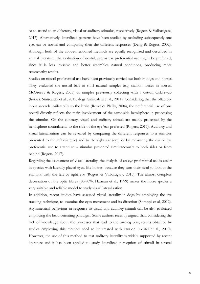
or to attend to an olfactory, visual or auditory stimulus, respectively (Rogers & Vallortigara,
2017). Alternatively, lateralized patterns have been studied by occluding subsequently one
eye, ear or nostril and comparing then the different responses (Deng & Rogers, 2002).
Although both of the above-mentioned methods are equally recognized and described in
animal literature, the evaluation of nostril, eye or ear preferential use might be preferred,
since it is less invasive and better resembles natural conditions, producing more
trustworthy results.
Studies on nostril preferential use have been previously carried out both in dogs and horses.
They evaluated the nostril bias to sniff natural samples (e.g. stallion faeces in horses,
McGreevy & Rogers, 2005) or samples previously collecting with a cotton disk/swab
(horses: Siniscalchi et al., 2015; dogs: Siniscalchi et al., 2011). Considering that the olfactory
input ascends ipsilaterally to the brain (Royet & Plailly, 2004), the preferential use of one
nostril directly reflects the main involvement of the same-side hemisphere in processing
the stimulus. On the contrary, visual and auditory stimuli are mainly processed by the
hemisphere contralateral to the side of the eye/ear preferred (Rogers, 2017). Auditory and
visual lateralization can be revealed by comparing the different responses to a stimulus
presented to the left ear (eye) and to the right ear (eye) or by measuring the ear or eye
preferential use to attend to a stimulus presented simultaneously to both sides or from
behind (Rogers, 2017).
Regarding the assessment of visual laterality, the analysis of an eye preferential use is easier
in species with laterally placed eyes, like horses, because they turn their head to look at the
stimulus with the left or right eye (Rogers & Vallortigara, 2015). The almost complete
decussation of the optic fibres (80-90%, Harman et al., 1999) makes the horse species a
very suitable and reliable model to study visual lateralization.
In addition, recent studies have assessed visual laterality in dogs by employing the eye
tracking technique, to examine the eyes movement and its direction (Somppi et al, 2012).
Asymmetrical behaviour in response to visual and auditory stimuli can be also evaluated
employing the head-orienting paradigm. Some authors recently argued that, considering the
lack of knowledge about the processes that lead to the turning bias, results obtained by
studies employing this method need to be treated with caution (Teufel et al., 2010).
However, the use of this method to test auditory laterality is widely supported by recent
literature and it has been applied to study lateralized perception of stimuli in several
9
vertebrate species, including dogs and horses (Siniscalchi et al., 2008, 2010; Basile et al.,
2009; Hauser & Andersson, 1994; Teufel et al., 2007; Fisher et al., 2009; Leliveld et al.,
2010; Lemasson et al., 2010). The head-orienting paradigm measures the animal
unconditioned and attentive response of turning its head toward the stimuli presented
simultaneously on its two sides or behind it. The direction of the head-turning indicates the
advantage of the contralateral hemisphere in processing the stimuli (Rogers, 2017). This
paradigm requires the animal to be centrally positioned with respect to the stimulus source.
Thus, to ensure the correct positioning of the animal, the experiment is usually run during
its feeding behaviour (Siniscalchi et al., 2008, 2010) or attaching it on a long rope (Basile et
al., 2009). In horses, auditory laterality can be also evaluated observing the ears movements
that occur independently of each other and of the head-turning (Waring, 2003; Burton,
2000).
1.1.3 Brain lateralization in dogs and horses
Concerning the two species involved in this project, namely dogs and horses, several
studies described behavioural asymmetries for both the sensory and motor activities.
Dogs lateralized behaviour and brain functions are summarized and reviewed in the
following article, entitled “Lateralized Functions in the Dog Brain”. In addition, I myself
carried out a study aiming at investigating the lateralized pattern of a cognitive ability (i.e.
visuospatial attention) and its relationship with a well-known lateralized motor activity,
namely paw preference. The paper that illustrates the results of this study is entitled
“Relationship between visuospatial attention and paw preference in dogs” (Appendix C).
10

Lateralized functions in the dog brain: article 3
11

symmetryS S
Review
Lateralized Functions in the Dog Brain
Marcello Siniscalchi *, Serenella d’Ingeo and Angelo Quaranta
Department of Veterinary Medicine, Section of Behavioral Sciences and Animal Bioethics,
University of Bari “Aldo Moro”, 70121 Bari, Italy; [email protected] (S.I.);
[email protected] (A.Q.)
* Correspondence: [email protected]; Tel.: +39-080-544-3948
Academic Editor: Lesley J. Rogers
Received: 21 March 2017; Accepted: 10 May 2017; Published: 13 May 2017
Abstract: Understanding the complementary specialisation of the canine brain has been the subject
of increasing scientific study over the last 10 years, chiefly due to the impact of cerebral lateralization
on dog behaviour. In particular, behavioural asymmetries, which directly reflect different activation
of the two sides of the dog brain, have been reported at different functional levels, including motor
and sensory. The goal of this review is not only to provide a clear scenario of the experiments carried
out over the last decade but also to highlight the relationships between dogs’ lateralization, cognitive
style and behavioural reactivity, which represent crucial aspect relevant for canine welfare.
Keywords: dog; lateralization; emotion; behaviour; physiology
1. Introduction
Brain hemispheres specialise to process and analyse information in an asymmetrical way is a
phenomenon widely reported in the animal kingdom [1,2] and, as shown by the increasing scientific
study over the last decade, it is now well manifested also in canine species. Based on findings derived
from experiments carried on different animal models, clear evidence exists that basic lateralized neural
mechanisms are very similar across vertebrate brains with a specialisation of the left hemisphere in the
control of routine behaviours, responding to features that are invariant and repeated, and with the
specialisation of the right hemisphere in detecting novelty (unexpected stimuli) and in the expression
of intense emotions, such as aggression and fear [3,4].
In this review, our first aim is to provide a comprehensive overview of the experiments carried out
in dogs providing extensive evidence of hemispheric asymmetries in function, structure and behaviour.
Our second aim in this paper is to analyse lateralized patterns specifically involved in emotional
processing by the dog brain and how the study of emotional lateralization could represent a valid and
interesting tool to contribute to the improvement of canine welfare and management.
In dogs, deepening the knowledge of cerebral lateralization with particular regard to emotional
processing is particularly interesting since behavioural asymmetries which indirectly reflect lateralized
cognitive processing of emotions can be easily detected (e.g., paw preference, nostril use, and tail
wagging) and can give insight into the different valences of an emotion felt by the animal. The latter is
crucial not only for a better understanding of canine cognition but also for the improvement of dogs’
training and handling during several activities within the human community (e.g., animal-assisted
therapy, police and rescue work, and guides for vision impaired people).
2. Sensory Lateralization
The complementary specialisation of dogs’ brain hemispheres is clearly apparent at different
sensory levels, including vision [5], hearing [6–10] and what is considered to be the most relevant
sensory domain for canine species, namely olfaction [11,12].
Symmetry 2017, 9, 71; doi:10.3390/sym9050071 www.mdpi.com/journal/symmetry
12

Symmetry 2017, 9, 71 2 of 12
Asymmetries of dogs’ visual sensory channels have been observed by studying their asymmetrical
head-turning response to bidimensional visual stimuli presented during feeding behaviour [5].
The experimental set-up consisted of the presentation of black silhouette drawings of different
animal models (a dog, a cat and a snake) to the dog’s right and left visual hemifields using two
retro-illuminated panels. When stimuli were presented at the same time in the two visual hemifields,
dogs preferentially turned the head with their left eye leading in response to alarming stimuli (the
snake silhouette that is considered to be an alarming stimulus for most mammals [13] or the cat
silhouette displaying a defensive threat posture). Given that, in dogs, neural structures located in
the right hemisphere are mainly fed by inputs from the left visual hemifield and vice versa (crossing
of fibres at the optic nerve level is 75% [14]), left head turns in response to threatening stimuli are
consistent with the specialisation of the right side of the brain for expressing intense emotion including
fear (snake) and aggression (cat with an arched lateral displayed body and erected tail). The latter
specialisation of the right hemisphere has been reported in several animal models (reviewed in [1,2]).
It is interesting to note that left head turns (right hemisphere activation) lead to shorter latencies
to react and longer latencies to resume feeding (i.e., higher emotional response). Moreover, during
monocular presentation, higher responsiveness to stimuli presented in the left visual hemifield was
observed, and this was irrespective of the type of stimulus. Overall, these results support the hypothesis
that in canine species, as well as in other mammals, the neural sympathetic mechanisms controlling
the “fight or flight” behavioural response are mainly under the activation of the right hemisphere [15].
In dogs, it is interesting to note that both in vivo [16] (Computed Tomography (CT) brain scanning)
and post mortem techniques [17] have revealed a right-biased hemispheric asymmetry with the
right hemisphere greater than the left; the latter could reflect the right hemisphere specialisation
for intense emotional activities like fight or flight reactions, which are related to aggressive and
defensive-escape behaviours.
As in dogs, a number of animals exhibit aggressive and defensive behaviours when the right
hemisphere is active. Chicks, for example, respond strongly to a potential predator (silhouette of a
predatory bird) seen in their left visual field (right hemisphere) [18,19]); very similar results were
reported in toads, which showed stronger avoidance responses when a model snake was presented on
their left side than when it was on their right side [20]. In domestic animals, horses approached by
a potential threatening stimulus (a human opening an umbrella) reacted more (i.e., moving further
away) when the approach was from their left side than when it was from their right side [21].
There is now evidence that the auditory sensory system in the dog brain also works in an
asymmetrical way depending on the type of acoustic stimulus [6,8,9]. Specifically, during feeding
behaviour, dogs’ head orienting responses to different sounds played at the same time from two
speakers placed symmetrically with respect to the subjects’ head were recorded [6] (see Figure 1A).
When thunderstorm playbacks were presented, dogs consistently turned the head with their left
ear leading and, given that the direction of the head turn is an unconditioned response indicating a
contralateral hemispheric advantage in attention to the auditory stimulus [22], this result supported
the right hemisphere specialization in processing alarming stimuli. In a similar way to what has been
previously reported about vision, in this experimental condition, left-head orienting turns also led
to longer latencies to resume feeding from the bowl. On the other hand, dogs consistently turned
the head with their right ear leading in response to playbacks of canine vocalizations (“disturbance”
and “isolation” calls) supporting the role of the left hemisphere in the analysis of familiar conspecific
calls, as reported in other species (non-human primates [23], horses [24], cats [25] and sea lions [26]).
Nevertheless, in dogs, conspecific vocalizations are not always processed by the left hemisphere, since
the right hemisphere is used for processing vocalizations when they elicit intense emotion [6,7].
13

Symmetry 2017, 9, 71 3 of 12
Figure 1. Behavioural techniques used to study functional lateralization in dogs: (A) head-orienting
response used to study auditory lateralization; and (B) left and right nostril use during sniffing different
olfactory stimuli.
In dogs, the left hemisphere advantage in processing vocalizations of familiar conspecifics seems
dependent on the calls’ temporal features, since the presentation of the reversed version of the same
canine call caused the loss of the right bias in the head turning response [27].
Head orienting response methods have been used in dogs to study possible lateralized neural
mechanisms in processing human speech [8]. Results revealed that dogs consistently turned their head
to the right during presentation of human spoken commands with artificially increased segmental
cues (i.e., higher salience of meaningful phonemic components); moreover, a significant left-turning
bias was observed in response to manipulated commands with increased supra-segmental vocal
cues (i.e., higher salience of intonation component). These results have been confirmed by recent
neuroimaging studies and overall suggest a convergent lateralized brain specialisation between canine
and human species for processing speech [9].
Regarding olfaction, asymmetries in nostril use have been observed during free sniffing behaviour
of odorants that differ in terms of emotional valence [11,28]. Briefly, cotton swabs installed on
a digital video camera were used to present odorants to dogs (see Figure 1B). The camera was
installed on a tripod in the centre of a large silent room. A frame-by-frame analysis of nostril use
video footages revealed a clear right nostril bias during sniffing of clearly arousing odours for dogs
(e.g., adrenaline and veterinary sweat). Given that, in dogs, the olfactory nervous fibres, which drive
14

Symmetry 2017, 9, 71 4 of 12
odour information from peripheral receptors to the olfactory cortex, are uncrossed, right nostril use
indicates a prevalent right hemisphere activation [29]. The latter was consistent with the previously
reported right hemisphere involvement in analysing alarming/threatening stimuli and had direct
implication for dogs’ welfare and training since, for example, the constant use of the right nostril during
olfactory inspection of a human being could reveal an increased arousal state of the animal, even in
the absence of clear behavioural signs (this could be useful in those activities like animal-assisted
therapy in which dogs must possess advanced behavioural control skills in order to help them handle
high arousal situations and consequently it is not always easy to detect stress increase directly from
behavioural signs).
When non-aversive stimuli were presented (e.g., food, lemon, and canine vaginal secretions),
right nostril use was observed only during the first presentations indicating the initial involvement of
the right hemisphere in the analysis of novelty (this bias was not evident for initial sniffing of food
probably because of its reduced valence as a novel stimulus). Furthermore, a shift from the right to the
left nostril use was observed with repeated stimulus presentations, indicating the prevalent control
of sniffing behaviour by the left hemisphere when routine responses to odour stimuli emerge as a
result of familiarization [1,2,30,31]. Left hemisphere specialisation in routine tasks has been observed
in pigeons [32], wild stilts [33], toads [34] and chickens [35]. In the latter case, during a routine task of
finding food, chicks using the right eye (left hemisphere) and not the left eye learn to find food grains
scattered on a back-ground of distracting pebbles (similar to the grains).
There is now evidence that dogs’ olfaction works in an asymmetrical way for processing both
conspecific and heterospecific odours collected during different emotional events [12]. In particular,
during sniffing of canine odours collected in a stressful situation (i.e., an “isolation” situation in
which dogs were isolated from their owners in an unfamiliar environment), a consistent use of
the right nostril was observed (right hemisphere activity). Moreover, when human odorants were
presented to dogs, a significant left-nostril bias (left hemisphere activation) was reported to sniff
olfactory stimuli collected from humans during a fearful situation (emotion-eliciting movies) and
physical stress. The observed opposite nostril use pattern in response to conspecific and heterospecific
odorants suggests that dog’s olfaction uses different sensory pathways to extract emotional cues from
canine and human chemosignals. Furthermore, an interesting hypothesis about the left nostril use
during sniffing at human sweat collected during a fear situation and physical stress is that these
heterospecific chemosignals (probably produced during the escape behavioural response to a predator)
could elicit dogs’ prey drive (i.e., approaching behavioural tendencies) to the stimuli through the
selective activation of the left hemisphere. The evidence that, in dogs [36], as in other animal models
(e.g., toads [34] and birds [33]), neural structures on the left side of the brain are involved in the control
of predatory behaviour supports this hypothesis.
3. Paw Preferences
Asymmetries of motor functions have been widely reported in various vertebrate and invertebrate
species, including the dog [1,2]. There is now a growing body of literature on motor lateralization in
dogs, focused mainly on behavioural lateralization in the form of forelimb preferential use. In recent
studies, paw preference has been assessed using several tasks: removal of a adhesive plaster from
the eye [17,37] or of a piece of tape from the nose [38–42], removal of a blanket from the head [43],
retrieval of food [44,45] from a toy object (namely the “Kong”, see Figure 2) [46–50] or a metal can [43],
paw-shaking [43], first foot placed forward to depart from a standing or sitting position [49,51] or
during a run [52] and stabilization of a ball [39] and hindlimb raising behaviour during urination [53].
15

Symmetry 2017, 9, 71 5 of 12
Figure 2. Motor lateralization: right paw use during stabilization of a food object (namely the “Kong”).
The existence of motor asymmetries at a population level is currently a subject of wide debate.
It has been reported in several species, including humans [54], non-human primates [55,56], rats [57],
humpback whales [58] and common European toads [59] but studies on other animals, as for example
marmosets [60], sheep [61,62], cats [63] and horses [64,65], has shown a motor bias only at the
individual-level. However, the same species may also display a limb preference at the level of
population or at the individual level depending on the task, as found in monkeys [66,67], cats [68] and
sheep [69].
Motor lateralization in dogs is stable between breeds and over time [41,46] but variable between
sexes. Although a few studies have reported an association between paw preference and sex at a
population level but in opposite directions, with males showing a left-paw and females a right-paw
preference [29,43,47], this seems to be inconsistent with other findings, which describe no population
bias [17,39,41,46,51]. These conflicting results suggest that sex hormone status could be influential on
the development of individual motor laterality but further investigations are necessary to accurately
determine if this is the case.
There have been several recent studies that revealed an interesting association between emotional
functioning and limb preference in animals, including dogs. It is well established that in primates
motor bias is associated with differences in the behaviour of individuals and their emotional states.
In particular, left-handed/pawed animals displayed more fear responses, higher stress level and
reactivity than right-handed/pawed animals [4,70,71]. The latter, instead, were more likely to
approach new objects and showed more social behaviours to capture a prey (chimpanzees: [72],
marmosets: [73,74]). These behavioural differences match the known specialization of the hemisphere
involved in the control of motor functions (contralateral to the preferred limb). Therefore, the limb
preferential use could be indicative of the subject’s personality type and its likelihood of expressing
a positive or negative emotional functioning. Recent studies have reported indeed that left-handed
marmosets have a negative cognitive bias compared to right-handed marmosets, which display a
positive cognitive bias [75]. Concerning dogs, Branson and Rogers [46] showed that dogs with weaker
motor lateralization were more reactive when exposed to potentially threatening stimuli (thunderstorm
16

Symmetry 2017, 9, 71 6 of 12
and fireworks sounds) since they displayed more stressed behaviours than lateralized subjects. Dogs
with stronger paw preference are otherwise more confident and relaxed in an unfamiliar environment
and when presented with novel stimuli [76]; on the contrary, they are less able in a problem-solving
task, to manipulate and explore a new object to obtain food than ambilateral subjects [76].
Given these findings, preferential limb use could be employed as a measure to assess vulnerability
to stress and welfare risk in animals [4] and also in dogs. Consequently, it is essential to correctly
categorize subjects as left- or right-pawed, choosing a motor test that provides reliable information
about dogs’ dominant paw, in order to make inference about dogs’ dominant hemisphere and their
ability to cope with stress. Wells et al. [48] recently investigated whether dogs use their dominant
paw in the most common motor test employed in this species, namely the Kong test. They found that
dogs use their non-dominant paw to stabilize the Kong to obtain food and their dominant paw for
postural support. These findings need to be considered for correct implications on animals’ welfare
and emotional vulnerability.
Therefore, the evaluation of paw preferential use could provide notable information regarding a
dog’s predisposition to solve future behavioural problems or about its suitability for work. It has been
demonstrated, indeed, that the direction of laterality is predictive of success in a Guide Dog Training
Programme; in particular, right-pawed dogs were more successful in completing the training than
left-pawed and ambilateral subjects [77].
Considering that behavioural differences in dogs’ response to different situations are linked with
motor lateralization and that dogs’ temperament plays an important role in the selection of dogs
(for working or adoptions), Schneider and colleagues [50] examined the relationship between paw
preference and temperament. They found no differences between lateralized and non-lateralized dogs
in the score obtained by a questionnaire completed by owners, aside from stranger-directed aggression
scale, where lateralized subjects registered higher scores than the ambilateral ones. This may suggest
the existence of a lateralized component in that particular type of aggressive response but further
investigations are required. Moreover, recent findings show that behavioural signs of fear and distress
displayed in a given situation and motor laterality are not associated with cortisol concentration in
saliva samples [42].
However, it would be interesting in the near future to deepen our understanding of the
relationship between motor laterality and emotional functioning since knowing the direction of
paw preference of a dog we could correctly assess the strategy to be employed to preserve and improve
its welfare.
Motor laterality is also associated with the analysis of visuospatial information, as we recently
found in our research. Specifically, agility trained dogs with weaker paw preference were less attentive
in performing agility exercises and displayed greater latency in the wave poles task (i.e., dogs’ ability
to work around pole obstacles that are secured in a straight line to a metal base) when the owner
was positioned in its left visual field [78]. These results clearly show that stimuli with high emotional
valence (the owner) could influence specific cognitive abilities, particularly when the right hemisphere
processes them. In a more recent study, we reported that visuospatial attention is strictly related to
motor lateralization since left-pawed dogs exhibited left visuospatial bias, right-pawed dogs a reversed
rightward bias, while ambilateral dogs displayed no bias [79]. The existence of such a relationship
has significant implications for animal welfare since it establishes a basis on which to develop new
therapies for the rehabilitation of visual attention during pathological conditions (namely, unilateral
spatial neglect); it could also help humans to improve canine training techniques, choosing the correct
side to handle dogs and how to capture their attention easily.
The importance of paw preference assessment as a useful tool to preserve animal welfare derives
also from the evidence of a direct relationship between dogs’ motor laterality and immune response
via an asymmetrical modulation exerted by the autonomic nervous system [38,80–82]. Right-pawed
and left-pawed dogs exhibit different patterns of immune response, in particular the former displayed
higher granulocytes percentage, number of γ-globulins [38], anti-rabies antibody titres and interferon
17

Symmetry 2017, 9, 71 7 of 12
gamma (IFN-γ) serum level [80] while the latter showed higher lymphocytes number [38] and higher
expression of specific interleukin genes (IL-2 and IL-6) after immune challenge [81]. Furthermore,
ambidextrous dogs exhibit a significantly higher increase of catecholamine levels after immunization
with rabies vaccine than lateralized subjects [82].
The direction of dogs paw preference is also related to anatomical asymmetries of the brain.
Aydınlıoglu et al. [45] found a variation in callosal size, particularly in its posterior segment (namely
the isthmus) that was larger in right-preferent dogs than left-pawed subjects. Post mortem analyses
showed also morphological asymmetries in canine hippocampi, which is associated with both sex
(males larger than females) and paw preference. Female left-pawed dogs showed indeed larger
hippocampi than the right ones [44]. In light of this evidence, motor lateralization may be considered
as a direct consequence of brain structural asymmetries that could be, more broadly the likely cause of
cerebral specialization of functions.
4. Tail-Wagging as a Tool to Study the Asymmetrical Representation of Emotional Processing inthe Dog Brain
Tail wagging represents an interesting model to study competition or cooperation between brain
hemispheres in the control of behavioural response to emotional stimuli mainly for two reasons:
(1) Dogs move their tails in an asymmetrical way in response to different emotional stimuli [83].
(2) Studies on behavioural asymmetries associated with lateralized brain functions have usually
focused on asymmetric use of paired organs (e.g., forelimbs) but not of a medial organ (i.e., the
tail). In order to test asymmetries in tail wagging behaviour, family pet dogs of mixed breeds
were placed in a large rectangular wooden box with an opening on the centre of one of its shorter
side to allow subjects to view the different stimuli (see Figure 3). Different emotional stimuli were
presented as follows: the dog’s owner; an unknown person; an unfamiliar dog with agonistic
approach behaviour; and a cat. Tail wagging was analysed frame by frame from video footages
recorded through a video camera placed on the ceiling of the box (see Figure 3).
ğ
Figure 3. Schematic representation of the testing apparatus used to study asymmetric
tail-wagging behaviour.
18

Symmetry 2017, 9, 71 8 of 12
Results revealed that both direction and amplitude of tail wagging movements were related to the
emotional valence of the stimulus. Specifically, when dogs looked at stimuli with a positive emotional
valence (e.g., their owner), there was a higher amplitude of tail wagging to the right. On the other
hand, during presentation of negative emotional stimuli (an unfamiliar dog with a clear agonistic
behaviour), a left bias in tail wagging appeared. Given that the movement of the tail depends on the
contralateral side of the brain [84], results are consistent with Davidson’s laterality-valence hypothesis
about the specialization of the left hemisphere for the control of approaching behavioural responses
(right-wag → positive stimulus) and the dominant role of the right hemisphere for the control of
withdrawal responses (left-wag → negative stimulus) [85]. In dogs, similar results were reported in
the work of Racca et al. [86] in which subjects presented with pictures of expressive dog faces exhibited
a left gaze bias (right hemisphere activation) while looking at negative conspecific facial expressions
and a right gaze bias (left hemisphere activation) when looking at positive ones. The amplitude of
tail-wagging movements is also a determinant cue for estimating “quantitatively” the level of arousal
elicited by different emotional stimuli: during presentations of an unfamiliar human being, dogs
significantly wagged their tails to the right side of their bodies but with less amplitude than towards
the owner, whereas the sight of a cat once again elicited right side tail-wagging movements with less
amplitude than towards the unfamiliar human being. The right side tail-wagging bias observed during
cat presentations would probably reflect the tendency of dogs to approach the stimulus under the left
hemisphere control of prey-drive behaviour.
In order to test whether or not dogs detect this asymmetry, in a more recent experiment, 43 dogs
of various breeds were shown movies of other dogs or black silhouettes manipulated in order to
display prevalent right or left sided tail-wagging or no wagging at all [87]. In addition, dogs’ emotional
response to movies was evaluated by measuring subjects’ behaviour and cardiac activity. Results
revealed that when dogs saw movies of a conspecific exhibiting prevalent left sided tail wagging, they
had an increased cardiac activity and higher stress behaviours. Moreover, when observing movies of
conspecific with right-sided tail wagging movements, dogs exhibited more relaxed behaviours with a
normal cardiac activity (i.e., heart rate values similar to those of the dogs during resting) suggesting
that the canine species is sensitive to the asymmetric tail movement of conspecifics, which has direct
implication for understanding dog social behaviour. Different results were reported in a previous
study in which the approach behaviour of free-ranging dogs to the asymmetric tail wagging of a
life-size robotic dog replica was recorded [88]. Results revealed a preference to approach the robotic
model (i.e., without stopping) when its tail was wagging to the left side. Authors reported that a
possible explanation for the stop response during the approach to the model moving its tail with a clear
bias to the right may originate when tested dogs are presented with a signal that would otherwise be
positive (right wag) yet is not accompanied by additional reciprocal visual or acoustical responses by
the robotic model. Another possible explanation for the different results between the two experiments
is that, in the first experiment, tail movements were taken by real dogs (i.e., biological movements)
while in the second they were artificially reproduced by a robotic model (even in the presence of a
good dog-replica robotic movements are not properly biological).
5. Conclusions
Overall, there is clear evidence that functional lateralization has profound connections with
cognition in dogs. A greater understanding of this association may certainly contribute to improve
dog welfare and the relationship between dogs and humans. Non-invasive techniques of measuring
lateralization (e.g., paw preference or tail wagging) could constitute a reliable, simple and direct tool of
evaluating dogs’ cognitive style and emotional affective states, providing elements that could enhance
every-day management practice and improve both dogs’ welfare and behavioural medicine.
Acknowledgments: All sources of funding of the study should be disclosed. Please clearly indicate grants thatyou have received in support of your research work. Clearly state if you received funds for covering the costs topublish in open access.
19
Symmetry 2017, 9, 71 9 of 12
Author Contributions: The authors contributed equally to this manuscript. There were no founding sponsors thathad any role in the writing of this manuscript or in any other capacity in preparing and publishing this manuscript.
Conflicts of Interest: The authors declare no conflict of interest.
References
1. Rogers, L.J.; Andrew, R.J. Comparative Vertebrate Lateralization; Cambridge University Press: New York, NY,
USA, 2002; p. 660. ISBN: 0521781612.
2. Rogers, L.J.; Vallortigara, G.; Andrew, R.J. Divided Brains. The Biology and Behaviour of Brain Asymmetries;
Cambridge University Press: New York, NY, USA, 2013; p. 229. ISBN: 0521604850.
3. MacNeilage, P.F.; Rogers, L.J.; Vallortigara, G. Origins of the left and right brain. Sci. Am. 2009, 301, 60–67.
[CrossRef] [PubMed]
4. Rogers, L.J. Relevance of brain and behavioural lateralization to animal welfare. Appl. Anim. Behav. Sci. 2010,
127, 1–11. [CrossRef]
5. Siniscalchi, M.; Sasso, R.; Pepe, A.M.; Vallortigara, G.; Quaranta, A. Dogs turn left to emotional stimuli.
Behav. Brain Res. 2010, 208, 516–521. [CrossRef] [PubMed]
6. Siniscalchi, M.; Quaranta, A.; Rogers, L.J. Hemispheric specialization in dogs for processing different acoustic
stimuli. PLoS ONE 2008, 3, e3349. [CrossRef] [PubMed]
7. Reinholz-Trojan, A.; Włodarczyk, E.; Trojan, M.; Kulczynski, A.; Stefanska, J. Hemispheric specialization in
domestic dogs (Canis familiaris) for processing different types of acoustic stimuli. Behav. Process. 2012, 91,
202–205. [CrossRef] [PubMed]
8. Ratcliffe, V.F.; Reby, D. Orienting asymmetries in dogs’ responses to different communicatory components of
human speech. Curr. Biol. 2014, 24, 2908–2912. [CrossRef] [PubMed]
9. Andics, A.; Gácsi, M.; Faragó, T.; Kis, A.; Miklósi, A. Voice-sensitive regions in the dog and human brain are
revealed by comparative fMRI. Curr. Biol. 2014, 24, 574–578. [CrossRef] [PubMed]
10. Andics, A.; Gábor, A.; Gácsi, M.; Faragó, T.; Szabó, D.; Miklósi, A. Neural mechanisms for lexical processing
in dogs. Science 2016, 353, 1030–1032. [CrossRef] [PubMed]
11. Siniscalchi, M.; Sasso, R.; Pepe, A.M.; Dimatteo, S.; Vallortigara, G.; Quaranta, A. Sniffing with right nostril:
Lateralization of response to odour stimuli by dogs. Anim. Behav. 2011, 82, 399–404. [CrossRef]
12. Siniscalchi, M.; d’Ingeo, S.; Quaranta, A. The dog nose “KNOWS” fear: Asymmetric nostril use during
sniffing at canine and human emotional stimuli. Behav. Brain Res. 2016, 304, 34–41. [CrossRef] [PubMed]
13. LoBue, V.; DeLoache, J.S. Detecting the snake in the grass: Attention to fear relevant stimuli by adults and
young children. Psychol. Sci. 2008, 19, 284–289. [CrossRef] [PubMed]
14. Fogle, B. The Dog’s Mind. Pelham Editions: London, UK, 1992; p. 203. ISBN: 072071964X.
15. Wittling, W. Brain asymmetry in the control of autonomic-physiologic activity. In Brain Asymmetry;
Davidson, R.J., Hugdahl, K, Eds.; MIT Press: Cambridge, UK, 1995; pp. 305–357. ISBN: 9780262041447.
16. Siniscalchi, M.; Franchini, D.; Pepe, A.M.; Sasso, R.; Dimatteo, S.; Vallortigara, G.; Quaranta, A. Volumetric
assessment of cerebral asymmetries in dogs. Laterality 2011, 16, 528–536. [CrossRef] [PubMed]
17. Tan, U.; Caliskan, S. Allometry and asymmetry in the dog brain: The right-hemisphere is heavier regardless
of paw preferences. Int. J. Neurosci. 1987, 35, 189–194. [CrossRef] [PubMed]
18. Rogers, L.J. Evolution of hemispheric specialisation: Advantages and disadvantages. Brain Lang. 2000, 73,
236–253. [CrossRef] [PubMed]
19. Dharmaretnam, M.; Rogers, L.J. Hemispheric specialization and dual processing in strongly versus weakly
lateralized chicks. Behav. Brain Res. 2005, 162, 62–70. [CrossRef] [PubMed]
20. Lippolis, G.; Bisazza, A.; Rogers, L.J.; Vallortigara, G. Lateralization of predator avoidance responses in three
species of toads. Laterality 2002, 7, 163–183. [CrossRef] [PubMed]
21. Austin, N.P.; Rogers, L.J. Asymmetry of flight and escape turning responses in horses. Laterality 2007, 12,
464–474. [CrossRef] [PubMed]
22. Scheumann, M.; Zimmermann, E. Sex-specific asymmetries in communication sound perception are not
related to hand preference in an early primate. BMC Biol. 2008, 6, 3. [CrossRef] [PubMed]
23. Poremba, A.; Malloy, M.; Saunders, R.C.; Carson, R.E.; Herscovitch, P.; Mishkin, M. Species-specific calls
evoke asymmetric activity in the monkey’s temporal lobes. Nature 2004, 427, 448–451. [CrossRef] [PubMed]
20

Symmetry 2017, 9, 71 10 of 12
24. Basile, M.; Boivin, S.; Boutin, A.; Blois-Heulin, C.; Hausberger, M.; Lemasson, A. Socially dependent auditory
laterality in domestic horses (Equus caballus). Anim. Cogn. 2009, 12, 611–619. [CrossRef] [PubMed]
25. Siniscalchi, M.; Laddago, S.; Quaranta, A. Auditory lateralization of conspecific and heterospecific
vocalizations in cats. Laterality 2016, 21, 215–227. [CrossRef] [PubMed]
26. Böye, M.; Güntürkün, O.; Vauclair, J. Right ear advantage for conspecific calls in adults and subadults, but
not infants, California sea lions (Zalophus californianus): Hemispheric specialization for communication?
Eur. J. Neurosci. 2005, 21, 1727–1732. [CrossRef] [PubMed]
27. Siniscalchi, M.; Lusito, R.; Sasso, R.; Quaranta, A. Are temporal features crucial acoustic cues in dog vocal
recognition? Anim. Cogn. 2012, 15, 815–821. [CrossRef] [PubMed]
28. Siniscalchi, M. Olfactory lateralization. In Lateralized Brain Functions; Rogers, L.J., Vallortigara, G., Eds.;
Humana press: New York, NY, USA, 2017; pp. 103–120. ISBN: 9781493967230.
29. Siniscalchi, M. Olfaction and the Canine Brain. In Canine Olfaction Science and Law; Jezierski, T., Ensminger, J.,
Papet, L.E., Eds.; CRC Press: Boca Raton, FL, USA, 2016; pp. 31–37. ISBN: 1482260239.
30. Vallortigara, G.; Chiandetti, C.; Sovrano, V.A. Brain asymmetry (animal). WIREs Cogn. Sci. 2011, 2, 146–157.
[CrossRef] [PubMed]
31. Vallortigara, G. Comparative neuropsychology of the dual brain: A stroll through left and right animals’
perceptual worlds. Brain Lang. 2000, 73, 189–219. [CrossRef] [PubMed]
32. Güntürkün, O.; Kesh, S. Visual lateralization during feeding in pigeons. Behav. Neurosci. 1987, 101, 433–435.
[CrossRef] [PubMed]
33. Ventolini, N.; Ferrero, E.A.; Sponza, S.; Chiesa, A.D.; Zucca, P.; Vallortigara, G. Laterality in the wild:
Preferential hemifield use during predatory and sexual behaviour in the black-winged stilt. Anim. Behav.
2005, 69, 1077–1084. [CrossRef]
34. Robins, A.; Rogers, L.J. Lateralised prey catching responses in the toad (Bufo marinus): Analysis of complex
visual stimuli. Anim. Behav. 2004, 68, 567–575. [CrossRef]
35. Rogers, L.J. Early experiential effects on laterality: Research on chicks has relevance to other species. Laterality
1997, 2, 199–219. [CrossRef] [PubMed]
36. Siniscalchi, M.; Pergola, G.; Quaranta, A. Detour behaviour in attack-trained dogs: Left-turners perform
better than right-turners. Laterality 2013, 18, 282–293. [CrossRef] [PubMed]
37. Tan, U. Paw preferences in dogs. Int. J. Neurosci. 1987, 32, 825–829. [CrossRef] [PubMed]
38. Quaranta, A.; Siniscalchi, M.; Frate, A.; Vallortigara, G. Paw preference in dogs: Relations between lateralised
behaviour and immunity. Behav. Brain Res. 2004, 153, 521–525. [CrossRef] [PubMed]
39. Poyser, F.; Caldwell, C.; Cobba, M. Dog paw preference shows liability and sex differences. Behav. Process.
2006, 73, 216–221. [CrossRef] [PubMed]
40. Batt, L.S.; Batt, M.S.; McGreevy, P.D. Two tests for motor laterality in dogs. J. Vet. Behav. 2007, 2, 47–51.
[CrossRef]
41. Batt, L.S.; Batt, M.S.; Baguley, J.A.; McGreevy, P.D. Stability of motor lateralisation in maturing dogs. Laterality
2008, 13, 468–479. [CrossRef] [PubMed]
42. Batt, L.S.; Batt, M.S.; Baguley, J.A.; McGreevy, P.D. The relationships between motor lateralization, salivary
cortisol concentrations and behavior in dogs. J. Vet. Behav. 2009, 4, 216–222. [CrossRef]
43. Wells, D.L. Lateralised behaviour in the domestic dog, Canis familiaris. Behav. Process. 2003, 61, 27–35.
[CrossRef]
44. Aydınlıoglu, A.; Arslan, K.; Cengiz, N.; Ragbetli, M.; Erdogan, E. The relationships of dog hippocampus to
sex and paw preference. Int. J. Neurosci. 2006, 116, 77–88. [CrossRef] [PubMed]
45. Aydınlıoglu, A.; Arslan, K.; Rıza Erdogan, A.; Cetin Ragbetli, M.; Keles, P.; Diyarbakırlı, S. The relationship
of callosal anatomy to paw preference in dogs. Eur. J. Morphol. 2000, 38, 128–133. [CrossRef]
46. Branson, N.J.; Rogers, L.J. Relationship between paw preference strength and noise phobia in Canis familiaris.
J. Comp. Psychol. 2006, 120, 176–183. [CrossRef] [PubMed]
47. McGreevy, P.D.; Brueckner, A.; Branson, N.J. Motor laterality in four breeds of dog. J. Vet. Behav. 2010, 5,
318–323. [CrossRef]
48. Wells, D.L.; Hepper, P.G.; Milligan, A.D.; Barnard, S. Comparing lateral bias in dogs and humans using the
Kong™ ball test. Appl. Anim. Behav. Sci. 2016, 176, 70–76. [CrossRef]
49. Tomkins, L.M.; Thomson, P.C.; McGreevy, P.D. First-stepping Test as a measure of motor laterality in dogs
(Canis familiaris). J. Vet. Behav. 2010, 5, 247–255. [CrossRef]
21

Symmetry 2017, 9, 71 11 of 12
50. Schneider, L.A.; Delfabbro, P.H.; Burns, N.R. Temperament and lateralization in the domestic dog
(Canis familiaris). J. Vet. Behav. 2013, 8, 124–134. [CrossRef]
51. Van Alphen, A.; Bosse, T.; Frank, I.; Jonker, C.M.; Koeman, F. Paw preference correlates to task performance
in dogs. In 27th Annual Conference of the Cognitive Science Society; Cognitive Science Society: Stresa, Italy, 2005;
pp. 2248–2253.
52. Hackert, R.; Maes, L.D.; Herbin, M.; Libourel, P.A.; Abourachid, A. Limb preference in the gallop of dogs
and the halfbound of pikas on flat ground. Laterality 2008, 13, 310–319. [CrossRef] [PubMed]
53. Gough, W.; McGuire, B. Urinary posture and motor laterality in dogs (Canis lupus familiaris) at two shelters.
Appl. Anim. Behav. Sci. 2015, 168, 61–70. [CrossRef]
54. McManus, I.C. Right Hand, Left Hand: The Origins of Asymmetry in Brains, Bodies, Atoms, and Cultures;
Weidenfeld & Nicolson: London, UK, 2002; ISBN: 9780674016132.
55. Diamond, A.C.; McGrew, W.C. True handedness in the cotton-top tamarin (Saguinus oedipus)? Primates 1994,
35, 69–77. [CrossRef]
56. Laska, M. Manual laterality in spider monkeys (Ateles geoffroyi) solving visually and tactually guided
food-reaching tasks. Cortex 1996, 32, 717–726. [CrossRef]
57. Güven, M.; Elalmis, D.D.; Binokay, S.; Tan, U. Population-level right-paw preference in rats assessed by a
new computerized food-reaching test. Int. J. Neurosci. 2003, 113, 1675–1689. [CrossRef] [PubMed]
58. Clapham, P.J.; Leimkuhler, E.; Gray, B.K.; Mattila, D.K. Do humpback whales exhibit lateralized behaviour?
Anim. Behav. 1995, 50, 73–82. [CrossRef]
59. Bisazza, A.; Cantalupo, C.; Robins, A.; Rogers, L.J.; Vallortigara, G. Right-pawedness in toads. Nature 1996,
379, 408. [CrossRef]
60. Hook, M.A.; Rogers, L.J. Development of hand preferences in marmosets (Callithrix jacchus) and effects of
ageing. J. Comp. Psychol. 2000, 114, 263–271. [CrossRef] [PubMed]
61. Anderson, D.M.; Murray, L.W. Sheep laterality. Laterality 2013, 18, 179–193. [CrossRef] [PubMed]
62. Morgante, M.; Gianesella, M.; Versace, E.; Contalbrigo, L.; Casella, S.; Cannizzo, C.; Piccione, G.; Stelletta, C.
Preliminary study on metabolic profile of pregnant and non pregnant ewes with high or low degree of
behavioral lateralization. Anim. Sci. J. 2010, 81, 722–730. [CrossRef] [PubMed]
63. Pike, A.V.L.; Maitland, D.P. Paw preferences in cats (Felis silvestris catus) living in a household environment.
Behav. Process. 1997, 39, 241–247. [CrossRef]
64. Austin, N.P.; Rogers, L.J. Limb preferences and lateralization of aggression, reactivity and vigilance in feral
horses, Equus caballus. Anim. Behav. 2012, 83, 239–247. [CrossRef]
65. Austin, N.P.; Rogers, L.J. Lateralization of agonistic and vigilance responses in Przewalski horses
(Equus przewalskii). Appl. Anim. Behav. Sci. 2014, 151, 43–50. [CrossRef]
66. Hook, M.A.; Rogers, L.J. Visuospatial reaching preferences of common marmosets (Callithrix jacchus):
An assessment of individual biases across a variety of tasks. J. Comp. Psychol. 2008, 122, 41–51. [CrossRef]
[PubMed]
67. Fagot, J.; Vauclair, J. Manual laterality in nonhuman primates: A distinction between handedness and
manual specialization. Psychol. Bull. 1991, 109, 76–89. [CrossRef] [PubMed]
68. Wells, D.L.; Millsopp, S. Lateralized behaviour in the domestic cat, Felis silvestris catus. Anim. Behav. 2009, 78,
537–541. [CrossRef]
69. Versace, E.; Morgante, M.; Pulina, G.; Vallortigara, G. Behavioural lateralization in sheep (Ovis aries).
Behav. Brain Res. 2007, 184, 72–80. [CrossRef] [PubMed]
70. Braccini, S.N.; Caine, N.G. Hand preference predicts reactions to novel foods and predators in marmosets
(Callithrix geoffroyi). J. Comp. Psychol. 2009, 123, 18. [CrossRef] [PubMed]
71. Rogers, L.J. Hand and paw preferences in relation to the lateralized brain. Philos. Trans. R. Soc. B 2009, 364,
943–954. [CrossRef] [PubMed]
72. Hopkins, W.D.; Bennett, A.J. Handedness and approach-avoidance behaviour in chimpanzees
(Pan troglodytes). J. Exp. Psychol. 1994, 20, 413–418. [CrossRef]
73. Cameron, R.; Rogers, L.J. Hand preference of the common marmoset, problem solving and responses in a
novel setting. J. Comp. Psychol. 1999, 113, 149–157. [CrossRef]
74. Gordon, D.J.; Rogers, L.J. Differences in social and vocal behavior between left- and right-handed common
marmosets. J. Comp. Psychol. 2010, 124, 402–411. [CrossRef] [PubMed]
22

Symmetry 2017, 9, 71 12 of 12
75. Gordon, D.J.; Rogers, L.J. Cognitive bias, hand preference and welfare of common marmosets. Behav. Brain
Res. 2015, 287, 100–108. [CrossRef] [PubMed]
76. Marshall-Pescini, S.; Barnard, S.; Branson, N.J.; Valsecchi, P. The effect of preferential paw usage on dogs’
(Canis familiaris) performance in a manipulative problem-solving task. Behav. Process. 2013, 100, 40–43.
[CrossRef] [PubMed]
77. Tomkins, L.M.; Thomson, P.C.; McGreevy, P.D. Associations between motor, sensory and structural
lateralisation and guide dog success. Vet. J. 2012, 192, 359–367. [CrossRef] [PubMed]
78. Siniscalchi, M.; Bertino, D.; Quaranta, A. Laterality and performance of agility-trained dogs. Laterality 2014,
19, 219–234. [CrossRef] [PubMed]
79. Siniscalchi, M.; d’Ingeo, S.; Fornelli, S.; Quaranta, A. Relationship between visuospatial attention and paw
preference in dogs. Sci. Rep. 2016, 6, 31682. [CrossRef] [PubMed]
80. Quaranta, A.; Siniscalchi, M.; Frate, A.; Iacoviello, R.; Buonavoglia, C.; Vallortigara, G. Lateralised behaviour
and immune response in dogs: relations between paw preference and interferon-gamma, interleukin-10 and
IgG antibodies production. Behav. Brain Res. 2006, 166, 236–240. [CrossRef] [PubMed]
81. Quaranta, A.; Siniscalchi, M.; Albrizio, M.; Volpe, S.; Buonavoglia, C.; Vallortigara, G. Influence of behavioural
lateralization on interleukin-2 and interleukin-6 gene expression in dogs before and after immunization with
rabies vaccine. Behav. Brain Res. 2008, 186, 256–260. [CrossRef] [PubMed]
82. Siniscalchi, M.; Sasso, R.; Pepe, A.M.; Dimatteo, S.; Vallortigara, G.; Quaranta, A. Catecholamine plasma levels
following immune stimulation with rabies vaccine in dogs selected for their paw preferences. Neurosci. Lett.
2010, 476, 142–145. [CrossRef] [PubMed]
83. Quaranta, A.; Siniscalchi, M.; Vallortigara, G. Asymmetric tail-wagging responses by dogs to different
emotive stimuli. Curr. Biol. 2007, 17, R199–R201. [CrossRef] [PubMed]
84. Buxton, D.F.; Goodman, D.C. Motor function and the corticospinal tracts in the dog and raccoon.
J. Comp. Neurol. 1967, 129, 341–360. [CrossRef] [PubMed]
85. Davidson, R.J. Well-being and affective style: Neural substrates and biobehavioural correlates. Phil. Trans. R.
Soc. B 2004, 359, 1395–1411. [CrossRef] [PubMed]
86. Racca, A.; Guo, K.; Meints, K.; Mills, D.S. Reading faces: differential lateral gaze bias in processing canine and
human facial expressions in dogs and 4-year-old children. PLoS ONE 2012, 7, e36076. [CrossRef] [PubMed]
87. Siniscalchi, M.; Lusito, R.; Vallortigara, G.; Quaranta, A. Seeing left- or right-asymmetric tail wagging
produces different emotional responses in dogs. Curr. Biol. 2013, 23, 2279–2282. [CrossRef] [PubMed]
88. Artelle, K.A.; Dumoulin, L.K.; Reimchen, T.E. Behavioral responses of dogs to asymmetrical tail wagging of
a robotic dog replica. Laterality 2011, 16, 129–135. [CrossRef] [PubMed]
© 2017 by the authors. Licensee MDPI, Basel, Switzerland. This article is an open access
article distributed under the terms and conditions of the Creative Commons Attribution
(CC BY) license (http://creativecommons.org/licenses/by/4.0/).
23

In horse literature, there is evidence supporting the existence of a lateralized emotional
processing in different sensory modalities (i.e. vision, hearing and olfaction). Nevertheless,
this field has not received the same interest as for dogs from the scientific community and
it still needs further and deep investigation.
In a recent study, Smith et al. (2018) examined horses perception of positive (laughs) and
negative (growls) emotions conveyed by human non-verbal vocalizations. They found a
right-ear/left hemisphere bias when horses attended to positive emotional voices,
suggesting that they perceive the positive emotional content of human laughs. Horses also
show auditory laterality in response to social stimuli. Specifically, they preferred to use their
right ear to attend to familiar conspecific calls, while no bias was found for the call of
strangers (Basile et al., 2009). The left hemisphere main involvement in processing positive
and familiar stimuli is therefore consistent with its specializations reported in previous
studies on vertebrates (Rogers, 2002).
As for olfaction, domestic horses showed a right nostril preference (right hemisphere
activation) to investigate stallion faeces (McGreevy & Rogers, 2005) and clear arousing
stimuli, such as adrenaline and oestrus mares’ urines, that elicited also an increased of
horses’ cardiac activity (Siniscalchi et al., 2015). These results suggest that the right
hemisphere has a dominant role in the analysis of intense emotions in hoses, as previously
described for several vertebrates (Rogers, 2002). A right hemisphere involvement has been
reported in response to visual potential threatening stimuli as well. Smith et al. (2016)
found a left-eye bias in investigating photographs of “angry” human faces, which was also
associated with an increased heart rate. In addition, Austin and Rogers (2007) reported
horses stronger reactions to a sudden and unexpected stimulus (an opening umbrella) when
it was presented in their left visual hemifield. However, when presenting a novel but
neutral object, horses preferentially use their right eye (left hemisphere) to investigate it (De
Boyer de Roche et al., 2008). These findings suggest that horses are sensitive to the
different emotional valence of the stimuli, which are specifically and differently processed
by the two hemispheres.
Overall, the recent literature about horses and dogs perceptual laterality provides evidence
suggesting that behavioural laterality is a suitable measure to examine animals emotional
processing along the valence dimension.
24

1.2 Heart rate
Affective states in animals are related to physiological changes in arousal, which are mainly
regulated by the autonomic nervous system (ANS) and by the hypothalamic-pituitary-
adrenal axis (HPA) (Weiten, 1992). Specifically, the activation of the ANS and HPA results
in changes in heart rate, respiratory rate, blood pressure, pupil size and corticosteroids
levels (Fraser, 2008). In humans, numerous studies have demonstrated that physiological
profiles and subjective human emotional experience are related. For instance, it has been
found that cardiovascular, electrodermal and respiratory measure could differentiate
between fear, sadness and neutral emotions elicited by a movie (Kreibig et al., 2007).
Moreover, Rainville et al. (2006) showed that different emotions (fear, anger, sadness and
happiness) produce distinct patterns of cardio-respiratory activity. Although these findings
suggest that peripheral physiological activity can provide information related to both the
valence and arousal dimensions, the hypothesis that emotions have distinct autonomic
signatures still need further support from literature (Mauss & Robinson, 2009). On the
contrary, the scientific community agrees about the effectiveness of the changes in the
ANS activity as measures of arousal, which therefore provide information about the
intensity of the animals’ affective states (Hall et al., 2018).
In this project, the heart rate (HR) has been chosen as a measurement of the ANS
activation, since it is an objective index of increased sympathetic tone that reflects changes
in the arousal dimension (Zupan et al., 2016). Moreover, the heart rate is measured with
non-invasive techniques, causing less interference with the animals’ spontaneous
behavioural expressions and allowing the collection of data over a long period of time
(Maros et al., 2008).
Several studies have investigated dogs’ HR changes in response to different emotional and
potentially stressful situations. For instance, the approach of a threatening human stranger
resulted in a higher cardiac activity (Gácsi et al., 2013), which has also been associated with
the increase of stress behaviours (appeasement gesture) displayed during a physical
interaction with humans (Kuhne et al., 2014). Moreover, it has been found that the HR
increase is related to higher emotional arousal displayed by dogs when seeing a reward for
the first time and obtaining it (Zupan et al., 2016). Concerning the relationship between
HR changes and emotional perception, dogs showed a heart rate increase and greater
emotional reactions (stress and anxious behaviour) when facing visual stimuli of a
25

conspecific wagging its tail to the left side (Siniscalchi et al., 2013b). Given that the left tail
wagging has been described as a clear withdrawal response (Quaranta et al., 2007), the
increase of subjects’ arousal (HR and stress behaviour) suggests that dogs clearly perceived
the negative valence of such stimulus.
As for horses, it has been found a positive correlation between the HR increase and
behavioural signs of anxiety displayed both in an isolation situation (Momozawa et al., 2007)
and during a stressful task (i.e. walking backwards) (Rietmann et al., 2004). HR changes
have been found to be related to anticipation as well. HR increases have been recorded in
young horses before entering a novel automated horse walker, which was considered as a
potentially anxiety-inducing anticipatory experience (Janczarek and Kędzieskì, 2011), but
also when anticipating food (Peters et al., 2012). Recent studies have also reported that the
HR increase is associated with horses’ emotional perception of stimuli. Specifically, a
higher cardiac activity has been recorded when horses visually investigated human angry
faces and when they sniffed high arousing stimuli (adrenaline and oestrus mares urines)
(Siniscalchi et al., 2015; Smith et al., 2016). Interestingly, the HR increase was observed
together with the right hemisphere main involvement (right nostril/left-gaze bias) in
processing both the olfactory and visual stimuli. Moreover, the higher cardiac activity was
related to the expression of behaviours indicative of arousal, particularly the ears fixed
backwards (Siniscalchi et al., 2015). Taken together, these findings are consistent with the
general hypothesis of the right hemisphere role in the regulation of the sympathetic activity
(via the HPA) during stressful situations, which is associated with the expression of intense
emotions (Rogers et al., 2013). The increase of the sympathetic activity reflects the increase
of individuals’ arousal and has a direct effect on the heart muscle, producing an increase of
the heart rate.
Overall, the heart rate appears to be a suitable parameter to evaluate horses’ and dogs’
emotional perception and processing, providing reliable and valuable information along the
arousal dimension.
26

1.3 EEG
In human, the electroencephalography (EEG) is employed to measure the neuronal activity
of the brain, providing direct evidence about functional asymmetries in response to
different stimuli (Hall et al., 2018). This technique is currently the only non-invasive
method that provides a window into the temporal dynamics of cortical activities, with
millisecond precision. Nevertheless, it does not provide data about the specific brain areas
where the effect of a specific event occurs. EEG provides a continuous measure of cortical
activity from the onset of an event of interest, allowing the evaluation of implicit
processing of a stimulus when the behavioural response is not overt (Mazza & Pagano,
2017). This feature makes the EEG a very suitable technique for studying animals
processing of different stimuli. It is particularly useful in the study of horse perception of
stimuli, for, as a prey species, it takes advantage in hiding emotions that may suggest
vulnerability (e.g. predation or social rejection) (Hall et al., 2018; McFarland, 1999).
The expression and processing of emotions are lateralized functions in the brain, which can
be effectively studied with the EEG. At least three main models relating emotions to brain
activity were described: the valence model, the intensity model and the Heller’s model
(Schmidt & Trainor, 2001).
Davidson and Fox found that emotions are differentially lateralised in the frontal region of
the brain, according to their valence (positive or negative). The valence model was
substantially supported by empirical results, which showed a greater relative left frontal
EEG activity in response to a film clip containing pleasant scenes (positive affects) and a
greater relative right EEG frontal activity in response to a film clip containing unpleasant
scenes (negative affects) (Jones & Fox, 1992). Moreover, Fox and Davidson (1986) found
that human newborns exhibited different frontal EEG activity according to the positive or
negative valence of the taste perceived. Specifically, a greater relative left frontal EEG
activity was recorded when newborns were presented with a sweet solution, whereas a
greater relative right frontal EEG activity was found in response to a sour solution. In
addition, the motivational tendencies of approach and avoidance are distinguishable on the
frontal brain asymmetries and are related to the different type of emotions. Previous
studies that focused on the relationship between emotional processing, brain asymmetries
and behaviour reported, indeed, an asymmetry on the frontal lobes, reflecting the dominant
activity of the left hemisphere for the approach-related emotions and motivation; and the
27

right-hemisphere advantage for negative withdrawal-related emotions and avoidance
motivation (Davidson, 1995; Mauss & Robinson, 2009). In particular, this frontal
asymmetry is reflected in the asymmetric decrease of alpha power according to the emotion
perceived, which is inversely related to regional cortical activation. In other words, a
decrease of alpha in one hemisphere indicates the increase of such hemisphere activity
(Allen et al., 2004). Positive emotions were found to elicit a decrease of left frontal alpha
power whereas negative emotions produce a decrease of right frontal alpha power
(Davidson, 1995). This evidences led to the development of the so-called “approach-
withdrawal hypothesis”.
According to the intensity model, the overall activation of the frontal region reflects the
intensity of the affective experience. Dawson et al. (1992) noted, indeed, that infants
exhibited an increase in absolute frontal EEG activity during the separation from their
mother, which was also associated with clear behavioural signs of stress. Moreover, in a
recent study, which evaluated the regional brain activation in response to joy/happy and
fear/sad music excerpts, an overall frontal EEG activity was found to decreased from the
most intense emotion (fear) to the less intense (sad music), together with an asymmetrical
frontal activity according to the valence of the music presented (Schmidt & Trainor, 2001).
The third model, the Heller’s model, on the other hand, considers both the valence and the
intensity of emotions (Heller, 1993). Heller posited that the asymmetries in the frontal
activation reflect the different perception of the positive and negative valence of a stimulus,
as postulated by Davidson and Fox. However, Heller argued that the intensity of emotions
is reflected by the right parieto-temporal activity, which is also involved in the modulation
of behavioural and autonomic arousal. Therefore, the right hemisphere activation suggests
a general increase in subjects’ arousal.
To date, these models are still debated in the human field but they certainly provide useful
elements for the EEG results interpretation of studies carried out on animals. EEG
measurements can also provide important information about the brain mechanism
underlying the emotion processing since each frequency band is related to different and
specific mental functions. According to human literature, the EEG power spectrum is
divided into five main frequency bands: delta (0-4 Hz), theta (4-8 Hz), alpha (8-12 Hz),
beta (12-30) and gamma (>30 Hz) (Niedermeyer, 1999). Delta oscillatory responses are
involved in attention, perception, signal detection, and decision-making. They are also
28

related to face perception, facial expression perception, and affective picture processes
(Güntekin & Başar, 2016). Theta activity is considered to reflect working memory
processes, maintenance and manipulation of information, mental concentration and focus
attentional processing (Ishii et al., 2014). Alpha rhythms are broadly linked with perceptual
processing, memory tasks and the vigilance of the subjects (Sammler et al., 2007).
Moreover, alpha activity is largely implicated in emotional processing, as described above.
Regarding beta activity, it increases in alertness and cognitive processes (Steriade, 2005) and
in visual attention in cats (Wróbel et al., 2000); whereas gamma oscillations are engaged in
several cognitive processes, including visuospatial focused attention, visual perception,
learning and memory (Ishii et al., 2014). In addition, synchronized gamma activity is
thought to be involved in object representation, including internally driven representation,
and in high-level mental activities (Bertrand & Tallon-Baudry, 2000; Fitzgibbon et al., 2004).
Concerning brain oscillatory responses during emotional perception, a different
involvement of brain waves activity was found in processing pictures of emotional faces.
Specifically, beta and gamma oscillatory responses are mostly related to valence, whereas
delta and theta responses are mostly related to arousal (Guntekin & Başar, 2014).
Therefore, EEG measurements can corroborate evidence coming from the observation of
behavioural lateralization about the valence that animals attribute to different stimuli.
In horse and dog studies, the use of EEG technique is markedly increasing and, to date, it
has been mainly employed to investigate neural events underlying cognition. Specifically,
EEG has been used to investigate sleep characteristics (Bunford et al., 2018) and their
relationship with learning processes in dogs (Iotchev et al., 2017; Kis et al., 2017). It has
been found that the density of EEG transients in the 9–16 Hz range during non-REM
sleep relates to memory (Iotchev et al., 2017) and that learning has an effect on dogs’ sleep
EEG spectrum (Kis et al., 2017). In particular, during non-REM sleep delta activity
increased, while alpha activity decreased after a learning task, whereas during REM sleep it
has been found a relative increase of theta activity after learning (Kis et al., 2017).
Moreover, the EEG technique has been employed to measure brain activity during a visual
task in which dogs had to observe pictures of human and conspecific faces. Spontaneous
oscillatory activity (mainly of parieto-occipital cortex) shown a significant suppression
during visual task compared with resting activity at the frequency of 15–30 Hz (Kujala et al.,
2013).
29

As for horses, an ambulatory and non-invasive EEG headset (Cousillas et al., 2017) was
developed and employed to study brain activity during a visual attention task (Rochais et al.,
2018) and a cognitive bias test (Stomp et al., in prep). The former study revealed that
attention towards the visual stimulus is associated with a significant increase of gamma
wave proportion in the right hemisphere while “inattention” is associated with more alpha
and beta waves in the left hemisphere (Rochais et al., 2018). In the cognitive judgment bias
test, the globally sustained attention of the horses to all the stimuli has been related to the
high proportion of theta waves in both hemispheres but the pattern of brain activity
differed according to the valence of the stimulus presented. In particular, a theta waves
predominance was found in both hemisphere when horses attended to the positive
stimulus but it was recorded only in the right hemisphere when they attended to the
negative stimulus, together with higher proportions of beta waves in both hemispheres but
especially the left one (Stomp et al., in prep).
Overall, although the EEG technique has been employed in studying neural events
underlying cognition, to date, evidence about the brain activity in emotional processing in
dogs and horses measured by the EEG are lacking.
30

1.4. Research questions and aims
Humans are undoubtedly one of the principal factors that influence and contribute to
animals’ well-being. It has been recently reported, indeed, that the valence of daily human-
animal interactions affects animals’ affective states (Sankey et al., 2010a,b).
Human communicates with animals using visual, olfactory and auditory signals. Both
horses and dogs adapted themselves to the human environment during their domestication
and became sensitive to human communicative signals, modifying their behaviour
according to the message conveyed. For instance, both species are able to interpret the
human pointing gesture and to follow its direction (Proops & McComb, 2010; Soproni et
al., 2002). Human communication with animals includes the transfer of emotional
information, which regulates social interactions and strengthens bonds between individuals
(Baciadonna et al., 2018). A growing body of literature demonstrates that dogs and horses
are able to discriminate between human emotions expressed by visual or auditory cues
(Smith et al., 2016, 2018; Müller et al., 2015; Albuquerque et al., 2016). Nevertheless,
animals’ perception of human emotions has not been fully investigated.
In the light of this evidence, the main aim of this research project was to investigate dog
and horse perception of the emotional content of human signals that potentially affects
animals’ affective state and welfare. To address this issue, sensory stimuli expressing
different emotions were presented to the studied population. An integrated approach
combining the analysis of behavioural lateralization, cardiac and brain activity, and subjects’
behaviour was applied in order to answer the following questions:
1) Do dogs and horses perceive the different emotional content of human signals?
2) Do dogs and horses attribute a different valence and intensity to the emotions perceived?
The study of behavioural lateralization and brain activity provides information about the
valence (positive or negative) that animals attribute to an emotional stimulus; on the other
hand, changes in the cardiac activity, as well as behavioural manifestations of vigilance and
stress, reflect animals’ arousal state, providing information about the intensity of the
perceived emotion (Paul et al., 2005; Rogers, 2010). Therefore, the integrated approach
proposed in this thesis project allowed an accurate description of animals’ perception and
31

processing of human emotions, along with valence and intensity dimensions.
The first study investigated dogs perception of human (and conspecific) odours collected
during different emotional events (Chapter 3), exploring for the first time the possible role
of odour chemosignals in the transfer of emotional cues. Dogs perception of human non-
verbal vocalizations and facial expressions of the six Ekman’s basic emotions (Ekman,
1993) was investigated in the second and third study respectively (Chapter 4 and 5).
As for horses, the fourth study addressed the questions of 1) whether the valence of
previous interactions could affect horse perception of human voices and 2) whether the
horse life conditions and welfare could impact the valence perceived. To explore this issue,
the EEG technique was employed in this study, together with behavioural and cardiac
measures, providing additional information about the mechanisms that regulate the
processing of emotional voices (Chapter 6).
Results of this research project will practically contribute to deepening the knowledge
about human-animal communication and, most importantly, they will provide a theoretical
framework for identifying potential stressful factors that could impact animal welfare,
significantly improving animals’ life conditions and their management.
32

Chapter 2
Research methodology
The research was carried out in two different locations according to the species studied.
During the first two years of the PhD, studies on dogs emotional perception of human
(and conspecific) visual, auditory and olfactory signals were conducted at the “Section of
Behavioural Sciences and Animal Bioethics”, at the Department of Veterinary Medicine of
the University of Bari (Italy). The research about horses emotional perception of human
voices, instead, was carried out at the “EthoS”- UMR 6552 CNRS- research unit, at the
University of Rennes 1 (France), during the last year of the PhD.
In this chapter, I describe the general methodology applied in the research project.
Specifically, a general description of participants’ characteristics (2.1), of the emotional
stimuli collection (2.2) and of the studied parameters and their analysis (2.3) are provided.
Moreover, I report ethical statements (2.4) and a summary of the statistical analysis
performed (2.5). Further details of each study specific methods will be described in the
corresponding chapter (Chapter 3-6).
2.1 Participants
2.1.1 Dogs
Veterinary students and their dogs were recruited for the studies. Volunteers recruitment
occurred by means of advertisements on the Department of Veterinary Medicine website
and by word of mouth. A total of 93 dogs of various breeds and sizes, ages (mean of 4,2
years) and sexes (entire or neutered) constituted the studied sample. All subjects were pet
living in households. Clinical and behavioural evaluations were carried out by a
Veterinarian Behaviourist of the Department of Veterinary Medicine (University of Bari),
who certified dogs’ health and the absence of any behavioural pathologies. The dogs’
33
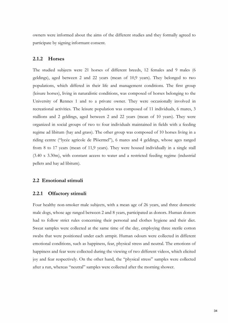
owners were informed about the aims of the different studies and they formally agreed to
participate by signing informant consent.
2.1.2 Horses
The studied subjects were 21 horses of different breeds, 12 females and 9 males (6
geldings), aged between 2 and 22 years (mean of 10,9 years). They belonged to two
populations, which differed in their life and management conditions. The first group
(leisure horses), living in naturalistic conditions, was composed of horses belonging to the
University of Rennes 1 and to a private owner. They were occasionally involved in
recreational activities. The leisure population was composed of 11 individuals, 6 mares, 3
stallions and 2 geldings, aged between 2 and 22 years (mean of 10 years). They were
organized in social groups of two to four individuals maintained in fields with a feeding
regime ad libitum (hay and grass). The other group was composed of 10 horses living in a
riding centre (“lycée agricole de Plöermel”), 6 mares and 4 geldings, whose ages ranged
from 8 to 17 years (mean of 11,9 years). They were housed individually in a single stall
(3.40 x 3.30m), with constant access to water and a restricted feeding regime (industrial
pellets and hay ad libitum).
2.2 Emotional stimuli
2.2.1 Olfactory stimuli
Four healthy non-smoker male subjects, with a mean age of 26 years, and three domestic
male dogs, whose age ranged between 2 and 8 years, participated as donors. Human donors
had to follow strict rules concerning their personal and clothes hygiene and their diet.
Sweat samples were collected at the same time of the day, employing three sterile cotton
swabs that were positioned under each armpit. Human odours were collected in different
emotional conditions, such as happiness, fear, physical stress and neutral. The emotions of
happiness and fear were collected during the viewing of two different videos, which elicited
joy and fear respectively. On the other hand, the “physical stress” samples were collected
after a run, whereas “neutral” samples were collected after the morning shower.
34

Conspecifics’ emotional odours were collected after the end of different emotional events:
a “play” interaction, in which the dog played together with its owner; an “isolation”
situation, in which the dog was left alone for 5 minutes in an unfamiliar environment; a
“disturbance” situation caused by a stranger approaching the car where the dog was resting,
and finally, the “neutral” sample was collected while the dog was sleeping. Sterile cotton
swabs were gently rubbed inside of dogs’ cheeks and on the skin surface of perianal and
interdigital areas, to collect salivary, perianal and interdigital secretions respectively.
2.2.2 Auditory stimuli
Dogs. Seven men and seven women, whose age ranged between 24 and 37 years, were
recruited to collect human emotional vocalizations expressing Ekmans’ six basic emotions
(Ekman, 1993). They were asked to pronounce non-verbal vocalizations of happiness
(laughs), surprise (strong expirations producing “oh” vocalizations), disgust (retches), fear
(screams), sadness (sobs) and anger (growls). The sounds were recorded in an anechoic
chamber.
The acoustic stimuli were then presented to 10 volunteers, five men and five women, aged
between 20 and 30 years, in order to select the most significant and clear vocalizations.
They were asked to fill a questionnaire indicating the valence (positive or negative) of each
vocalization heard, the emotion conveyed and its intensity, on a 3-point-scale. According to
questionnaire results, 18 vocalizations (three x each basic emotion) had been selected for
the final test. Each acoustic stimulus was equalized and filtered to remove background
noises.
Horses. Voices of twenty-eight women, whose age ranged between 21 and 62 years
(34,29±11,22; mean±S.D.), were recorded while reading the text previously employed by
Tallet et al. (2016), containing all the French phonemes and without any emotional
connotations: “Petit Louis, les yeux ouverts, rêvait dans son lit bleu. Le jour des vacances était arrivé. Il
sentait l’odeur du bon pain chaud et du chocolat que maman préparait. Papa et lui iraient à la gare
chercher son cousin. Ils feraient du camping à la campagne. Louis n’aurait plus peur des ruades de l’âne
brun.” Each reading lasted about 17 seconds and was repeated three times with a pause of
about 4s in between, in order to obtain a 1 min acoustic stimulus (60s±1,28s; mean±S.D.).
35
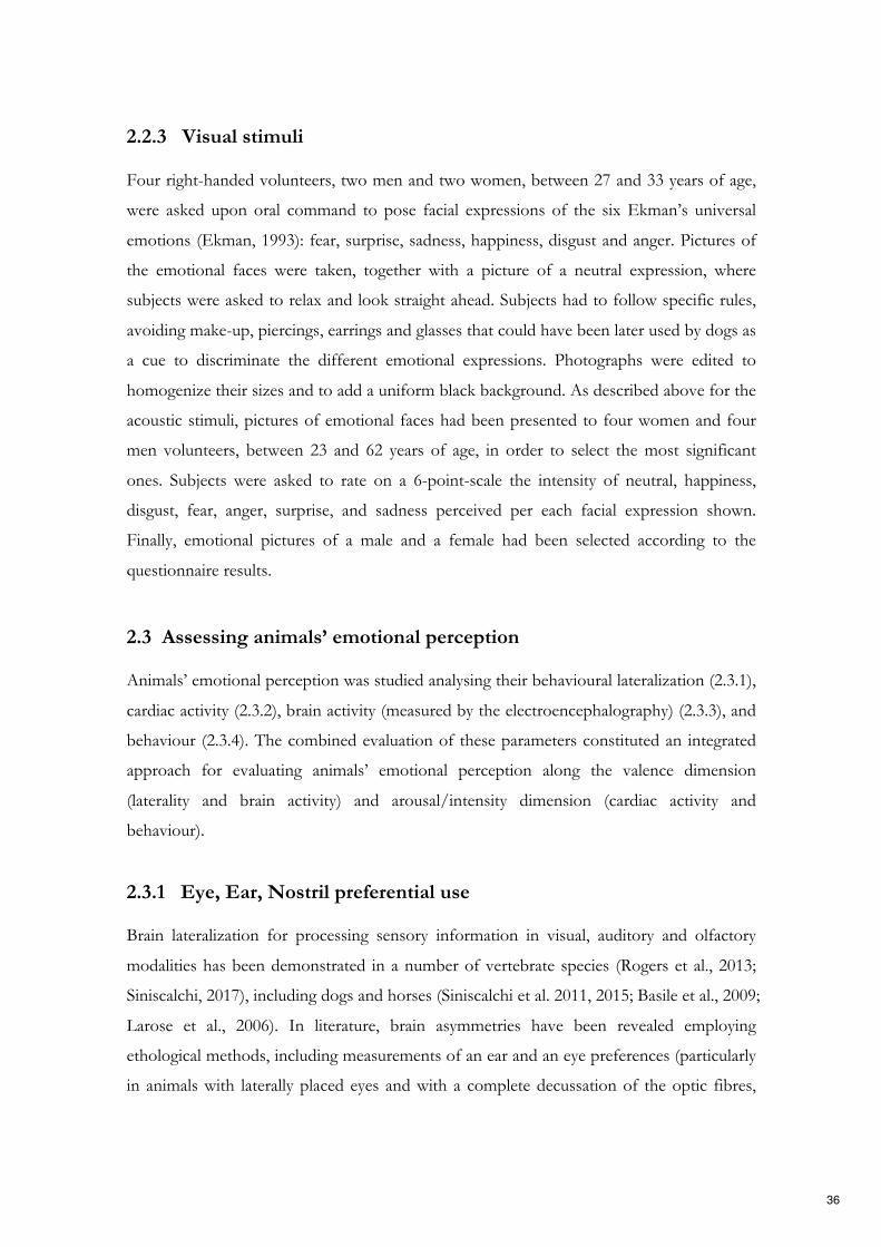
2.2.3 Visual stimuli
Four right-handed volunteers, two men and two women, between 27 and 33 years of age,
were asked upon oral command to pose facial expressions of the six Ekman’s universal
emotions (Ekman, 1993): fear, surprise, sadness, happiness, disgust and anger. Pictures of
the emotional faces were taken, together with a picture of a neutral expression, where
subjects were asked to relax and look straight ahead. Subjects had to follow specific rules,
avoiding make-up, piercings, earrings and glasses that could have been later used by dogs as
a cue to discriminate the different emotional expressions. Photographs were edited to
homogenize their sizes and to add a uniform black background. As described above for the
acoustic stimuli, pictures of emotional faces had been presented to four women and four
men volunteers, between 23 and 62 years of age, in order to select the most significant
ones. Subjects were asked to rate on a 6-point-scale the intensity of neutral, happiness,
disgust, fear, anger, surprise, and sadness perceived per each facial expression shown.
Finally, emotional pictures of a male and a female had been selected according to the
questionnaire results.
2.3 Assessing animals’ emotional perception
Animals’ emotional perception was studied analysing their behavioural lateralization (2.3.1),
cardiac activity (2.3.2), brain activity (measured by the electroencephalography) (2.3.3), and
behaviour (2.3.4). The combined evaluation of these parameters constituted an integrated
approach for evaluating animals’ emotional perception along the valence dimension
(laterality and brain activity) and arousal/intensity dimension (cardiac activity and
behaviour).
2.3.1 Eye, Ear, Nostril preferential use
Brain lateralization for processing sensory information in visual, auditory and olfactory
modalities has been demonstrated in a number of vertebrate species (Rogers et al., 2013;
Siniscalchi, 2017), including dogs and horses (Siniscalchi et al. 2011, 2015; Basile et al., 2009;
Larose et al., 2006). In literature, brain asymmetries have been revealed employing
ethological methods, including measurements of an ear and an eye preferences (particularly
in animals with laterally placed eyes and with a complete decussation of the optic fibres,
36

like horses (Harman et al., 1999)), as well as preferences in nostril use. Eye, ear and nostril
preferential use directly reflects the main involvement of the ipsilateral (olfaction) or
contralateral (vision and hearing) hemisphere in processing a specific stimulus. According
to the “valence-specific hypothesis”, each hemisphere is specialized for processing
particular classes of emotion. Specifically, the right hemisphere is specialized for the
reaction and processing of negative emotions, whereas the left hemisphere is specialized
for the reaction and processing of positive emotions (Adolphs et al., 2001; Ahern &
Schwartz, 1979). Therefore, given the different specialization of each hemisphere, the study
of spontaneous and preferential use of an eye, a nostril or an ear to attend to an emotional
stimulus provides information about the valence that an animal attributes to such
emotional stimulus. Previous studies employed these methodologies to evaluate horses and
dogs asymmetrical processing of sensory input (Siniscalchi et al., 2008, 2010, 2011, 2015;
Basile et al., 2009; Larose et al., 2006). In the present research project, lateralized
behavioural responses to olfactory, visual and auditory stimuli, in terms of a nostril, an eye
and an ear preferential use, were measured to assess the valence of the perceived emotion.
As for olfactory stimuli, the cotton swabs impregnated with different odours were installed
under a digital video camera hold by an experimenter. Dogs on leash were freely allowed to
approach the experimenter and to investigate the odours. The nostril preferential use was
recorded by the video camera held by the experimenter (Fig. 1). Each olfactory stimulus
was presented three times to each subject during three different sessions, with a 2-day
interval. Lateral biases were computed measuring the total time spent sniffing each
emotional odour with the left and the right nostril during the three sessions. A Laterality
Index was computed as follows: LI= (L-R/L+R), where L and R indicate, respectively, the
total time spent sniffing with the left and the right nostril during the three stimuli
presentations.
As for the auditory and visual stimuli, dogs’ and horses’ lateralized biases were evaluated
using the head-turning paradigm. This procedure has been previously employed by
Siniscalchi et al. (2008, 2010) to study dogs’ lateralized perception of visual and auditory
stimuli and by Basile et al. (2009) to evaluate horses’ lateralized perception of conspecific
vocalizations. Emotional sounds or pictures were presented simultaneously on each side of
the animals (or from behind it, for horses) and their lateralized behavioural response of
turning the head to attend the stimuli was measured. In order to test lateralized attention to
37

Figure 1. Picture of the experimental setup, study 1.
auditory and visual stimuli, the animals’ head had to be positioned centrally and midway
between the stimuli sources (speakers or screens). Therefore, to ensure the correct position,
horses were held on a long lead by an experimenter placed centrally and in front of it (Fig.
2); for dogs, a bowl containing their favourite food was placed midway between the stimuli
sources with two transparent barriers on each side, helping dogs to keep the correct
position. The stimuli were presented to the dogs while they had their head in position at
the bowl. The dog being tested was not leashed and feeding was a free choice. Owners
were positioned centrally and behind their dog, at a distance of about 3 m (Fig. 3, 4). Two
experimenters controlled the stimuli presentation (from an adjacent room for the
experiments on dogs, and from a designated position in front of the horses) and as soon as
the animal took up the correct position the stimulus was presented. Visual and auditory
emotional stimuli were presented only once since a high and quick level of habituation to
the stimuli was registered.
Ear/eye preferential use was assessed considering lateral asymmetries in the first head-
turning response. Three different responses were evaluated: turn right, turn left, and no
response, when a subject did not turn its head within the set threshold time from the
stimulus presentation. Concerning horses, the time spent with the loudspeaker in its
right/left visual hemifield during the acoustic stimuli broadcasting was also evaluated.
38

Figure 2. Picture of the experimental setup, study 4.
Figure 3. Picture of the experimental setup, study 2.
39

Figure 4. Picture of the experimental setup, study 3.
2.3.2 Cardiac activity
Physiological changes in the cardiac activity are thought to reflect animal’s stress and
arousal since they are mediated by the activation of the autonomic nervous system (ANS)
and by the hypothalamic-pituitary-adrenal axis (Weiten, 1992). In this research project, the
heart rate (HR) was chosen as a measurement of the ANS activation, representing an
objective index of the sympathetic activity increase, which reflects changes in the arousal
dimension (Zupan et al., 2016). Therefore, the heart rate increase provides information
about the intensity of the animals’ affective states (Hall et al., 2018), helping to characterize
animals’ emotional perception along the arousal dimension.
Dogs’ and horses’ cardiac activity was measured during stimuli presentation employing
non-invasive techniques. Specifically, horses heart rate variations were recorded by Polar
Equine RS800CX®, composed of a heart rate sensor belt and a watch registering animals
heart rate (Fig. 5A). On the other hand, dogs’ cardiac activity was recorded employing the
PC-Vetgard®+tm Multiparameter wireless system for telemetric measurements. It was
composed of three integrated electrodes connected to a wireless ECG data-transmitting
unit (Fig. 5B). A vest kept the electrodes in contact with dogs’ chest and the transmitter
unit on dogs’ back. Previous habituation to the vest was carried out before the
experimental test during weekly visits to the Department. Dogs had been considered as
40

A. B.
Figure 5. Devices to measure the cardiac activity: Polar Equine RS800CX® (A), PC-Vetgard®+tm
Multiparameter wireless system for telemetric measurements (B).
accustomed to the vest when they displayed no behavioural signs of stress.
Before the beginning of the experimental session, the animals’ heart rate was recorded. The
heart rate basal average (baseline) was then calculated for each subject, computing the
mean ECG- RR intervals during the recording period. Moreover, the subjects’ heart rate
was recorded continuously during the stimuli presentation. R-R intervals were then
calculated and the heart rate curve was obtained for each dog. The area delimited by the
heart rate curve and the basal average was computed for each subject and each stimulus,
employing Microsoft Excel®. The Area Under Curve (above baseline and under the curve,
AUC) was then graphically separated from the Area Above Curve (under baseline and
above the curve, AAC) (Fig. 6). Each area value was then calculated and expressed as the
number of pixels using Adobe Photoshop®.
Figure 6. An example of the cardiac activity analysis (from Siniscalchi et al., 2013).
41

2.3.3 Electroencephalography (EEG)
Human studies demonstrated that the electroencephalography (EEG) technique reveals
brain functional asymmetries and the mechanisms underlying emotional perception.
Specifically, an asymmetrical activity of the brain frontal regions according to the valence
of the perceived emotion has been found, with a greater relative left frontal EEG activity
in response to positive stimuli and a greater relative right frontal EEG activity in response
to negative stimuli (Fox & Davidson 1986; Jones & Fox, 1992). EEG measurements can be
also used to evaluate the intensity of the perceived emotion (Dawson et al. 1992; Heller,
1993), although the specific brain regions involved in subject arousal increase are still
debated in the scientific community. In addition, the different five waves oscillatory
responses could provide insight into the brain mechanisms involved in the emotional
processing.
In the light of this evidence, the electroencephalography (EEG) technique was employed
to measure the neuronal activity of the horses’ brain during the acoustic stimuli
broadcasting. The electrophysiological recordings were performed using ambulatory EEG
headset recently developed by Cousillas et al. (2017) (Fig. 7). It was composed of 4
electrodes positioned of on the horse’s forehead over the parietal and frontal bones, two
on each side of the horse’s forehead, allowing the recordings of the different activity
between the most occipital part of the brain and the most frontal one, as well as the
recordings of the left and right hemisphere activity. The ground electrode was placed on
the back of the left ear. The electrodes were connected to a homemade telemetric
recording setup (L 110 mm, l 90 mm, h30mm, weighing 110 g) and to an amplifier
connected to a Bluetooth transmitter. The whole telemetric recording setup was fixed on
the helmet and, thus, it allowed the animals to move freely, avoiding any bias due to head
immobilization. The device sampling-rate was 250Hz. An EEG software for horses based
on Labview allowed the experimenters to verify the EEG quality in real time.
Horses grew accustomed to the helmet within a period of a week. Daily training sessions in
which horses wore the EEG helmet were planned. Brain activity was recorded before the
onset of the stimulus playback (baseline) and soon after it, in order to compare the basal
activity with the neuronal activity following the stimuli presentation. The proportion of the
mean power of the five main types of brain waves, such as delta (δ: 0-4Hz), theta (θ: 4-
8Hz), alpha (α: 8-12Hz), beta (β: 12-30Hz) and gamma (γ: >30 Hz), was computed auto-
42

Figure 7. Electroencephalography device
matically. Then, the median of each wave proportions recorded during the pre-
experimental phase (baseline) and the stimuli broadcasting was calculated and compared.
2.3.4 Behaviour
Subjects’ behaviour was continuously recorded throughout the experiments by two video
cameras and was then analysed by a trained observer. The frequency of each behaviour was
measured. The following tables (Table 2, 3) summarize all the behaviours considered for
the analysis in the different experiments.
Furthermore, the latency time needed to turn the head toward the acoustic and visual
stimuli (i.e. reactivity) was computed for both horses and dogs. Additionally, the latency
time to resume feeding from the bowl after the stimuli presentation was evaluated in the
studies employing the head-turning paradigm on dogs.
43
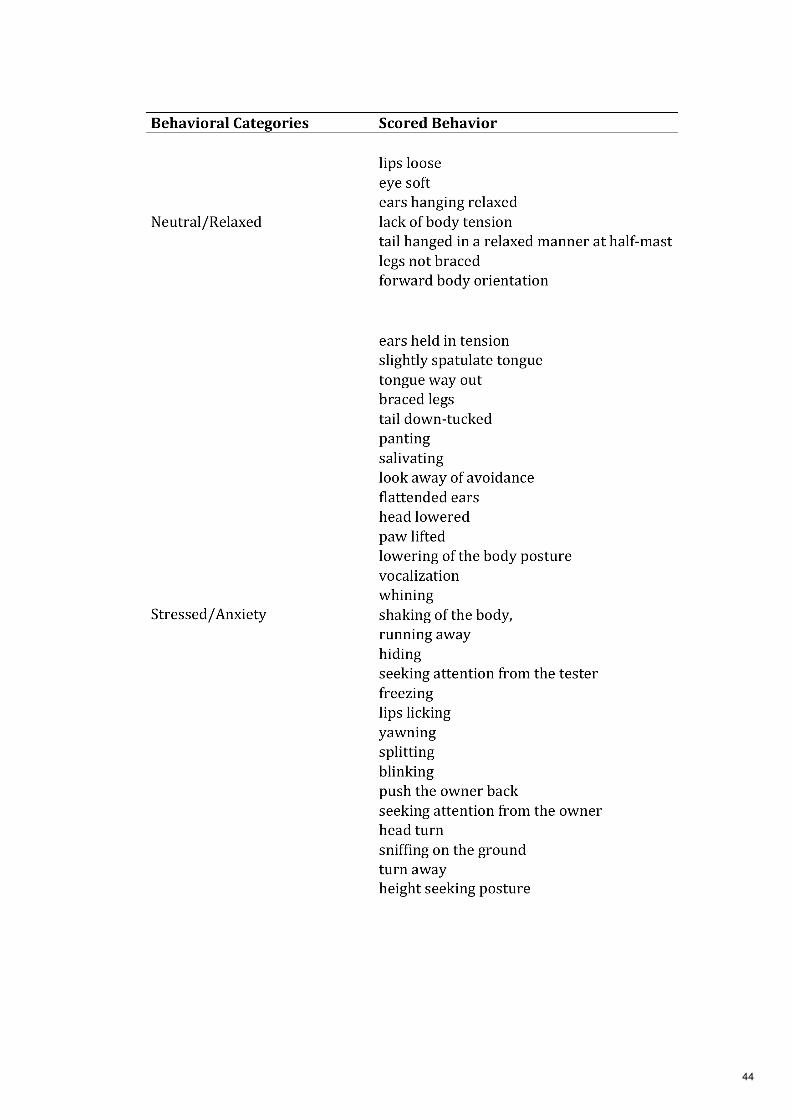
44

Table 2. List of scored behaviour in the studies on dogs
Table 3. List of scored behaviour in the study on horses
45

2.4 Ethical statements
The experiments on dogs were conducted according to the protocols approved by the
Italian Minister for Scientific Research in accordance with EC regulations and were
approved by the Department of Veterinary Medicine (University of Bari) Ethics
Committee EC.
The experiment on horses was carried out in accordance with the European Communities
Council Directive of 22 September 2010 (2010/63/UE) and the French law relative to the
protection of the animal used in scientific experiment (Décret n°2013-118 13 février 2013;
Article R. 21488). This experiment included only behavioural observations and non-
invasive interactions with the horses. Thus, it did not enter in the scope of application of
the European directive, and consequently did not require an authorization to experiment.
2.5 Statistical analysis
The statistical analysis was performed using SPSS software and the results were considered
statistically significant for p<0.05. Normality was assessed by Shapiro–Wilk and
Kolmogorov-Smirnov test. According to the data distribution, parametric or
nonparametric analysis was conducted.
The analysis of variance (GLM analysis for repeated measures) was used to determine the
effects of the odour type on various indexes. A binomial GLMM analysis was used to
assess the influence of various factors, for instance, the emotional category, on the tested
variable. Moreover, pairwise comparisons were performed to detect differences between
the emotional categories (e.g. Wilcoxon signed rank test, t-test paired samples, Fisher’s least
significant difference (LSD)). Asymmetries at a group-level (i.e. emotional category) in the
head-turning response were assessed via One-Sample Wilcoxon Signed Ranks Test, to
report a significant deviation from zero; whereas biases in the nostril use were assessed via
two-tailed one-sample t-test. Pearson and Spearman correlation were used to measure the
association between two variables and the direction of the relationship. Finally, differences
between the two populations of horses in their response to the acoustic stimuli were
assessed via Mann-Whitney test or via independent sample t-test.
46

Chapter 3
Study 1 Dogs perception of human (and conspecific)
emotional odours
47

Dogs perception of human (and conspecific) emotional odours: article 5
Study aim
In dogs, olfaction plays a pivotal role in social relational systems. Dogs, indeed, are able to
discriminate amongst pheromones contained in organic secretions and to detect a
conspecific reproductive status through olfaction. Moreover, there is evidence that canine
species prefer sniffing specific areas of human body, suggesting that odours produced at
different anatomical parts could provide different specific olfactory cues.
Previous studies have reported striking asymmetries in the nostril use of dogs during
sniffing at different emotive stimuli. In the light of these evidences, the aim of the present
study was to investigate whether dogs perceive the emotional content of human and
conspecific odours, showing nostril asymmetries in processing olfactory stimuli collected
during different emotional events. In addition, dogs’ behavioural and physiological
reactivity to different odours was evaluated to analyse animals’ arousal.
Methods
Human sweat samples were collected in different emotional conditions: joy, fear, physical
stress and neutral. Conspecifics’ emotional odours were collected from perianal,
interdigital and salivary secretions soon after the end of different emotional events: a “play”
situation, an “isolation” situation, a “disturbance” situation and a “neutral” situation.
Emotional odours were presented to 31 domestic dogs and their asymmetrical nostril use,
cardiac activity and behaviour were evaluated.
Results
Dogs showed lateral asymmetries in nostril use when sniffing human and conspecific
odours. Specifically, dogs consistently used their right nostril to sniff the dog-isolation
odour and the left nostril to sniff the human-fear and human-physical activity stimuli.
Moreover, dogs displayed higher behavioural and physiological reactivity to canine
(especially to dog-isolation) and humans odours collected during different emotional
conditions.
Conclusions
Dogs could detect the emotional states of a member of its social group through olfaction.
The opposite bias shown in nostril use during sniffing at canine versus human odours
suggests that chemosignals communicate conspecific and heterospecific emotional cues
using different sensory pathways.
48

La percezione degli odori emotivi dell’uomo (e dei conspecifici) nel
cane: articolo 5
Obiettivi dello studio
Nei cani, l'olfatto gioca un ruolo fondamentale nelle relazioni sociali. I cani, infatti, sono in
grado di distinguere i feromoni contenuti nelle secrezioni organiche e di rilevare lo stato
riproduttivo di un conspecifico attraverso l’olfatto. Inoltre, è stato dimostrato che i cani
preferiscono annusare aree specifiche del corpo umano. Ciò suggerisce che gli odori
prodotti in diverse parti anatomiche dell’uomo potrebbero fornire diversi segnali olfattivi
specifici.
Precedenti studi hanno descritto l’esistenza di asimmetrie nell'uso delle narici nel cane per
annusare diversi stimoli emotivi.
Alla luce di queste evidenze, lo scopo del presente studio è stato quello di indagare la
percezione del contenuto emotivo degli odori umani e conspecifici nel cane, valutando le
asimmetrie dell’uso delle narici per l’elaborazione di stimoli olfattivi raccolti durante diversi
eventi emotivi. Inoltre, è stata valutata la reattività comportamentale e fisiologica dei cani ai
diversi odori per analizzare l’arousal dei soggetti.
Metodi
Sono stati raccolti campioni di sudore umano in diverse condizioni emotive: gioia, paura,
stress fisico e neutro. Per gli odori emotivi dei conspecifici, sono stati raccolti dei campioni
delle secrezioni perianali, interdigitali e salivari al termine di diversi eventi emotivi: "gioco",
"isolamento", "disturbo" e una situazione "neutra". Gli odori emotivi sono stati presentati a
31 cani domestici, valutando l’uso asimmetrico delle narici per investigare lo stimolo,
l'attività cardiaca e il comportamento dei soggetti.
Risultati
I cani hanno mostrato delle asimmetrie nell'uso delle narici per annusare gli odori umani e
dei conspecifici. Nello specifico, i cani hanno usato la loro narice destra per annusare
l'odore “d’isolamento” del cane e la narice sinistra per annusare gli stimoli di paura e attività
fisica dell’uomo. Inoltre, i cani hanno mostrato una maggiore reattività comportamentale e
fisiologica agli odori dell’uomo e dei conspecifici (in particolare l'isolamento del cane)
raccolti in diverse condizioni emotive.
Conclusioni
I cani percepiscono gli stati emotivi di un membro del loro gruppo sociale attraverso l'olfatto. Le asimmetrie opposte nell'uso della narice registrate durante l’analisi degli odori dei conspecifici e dell’uomo suggeriscono che la comunicazione dei segnali emotivi dei conspecifici e degli eterospecifici attraverso i chemosignali utilizza diversi percorsi sensoriali.
49

Perception olfactive des émotions humaines (et conspécifiques) chez le chien: article 5
Objectif de l’étude
Chez le chien l’olfaction joue un rôle essentiel dans le système de relations sociales. Les chiens sont en effet capables de discriminer les phéromones contenues dans des sécrétions organiques et détecter le statut reproducteur d’un conspécifique. De plus, il a été montré que les chiens préfèrent renifler certaines zones du corps humain suggérant que les odeurs produites par différentes parties du corps possèdent différentes caractéristiques olfactives. Des études antérieures ont clairement montré des asymétries dans l’utilisation des narines du chien pour sentir différents stimuli émotionnels.
En se basant sur ces données, l’objectif de cette étude a été de déterminer si le chien est capable de percevoir le contenu émotionnel d’odeurs humaines ainsi que de conspécifiques et de mettre en évidence une possible latéralité dans l’utilisation des narines pour sentir et traiter des stimuli olfactifs prélevés dans différentes situations émotionnelles. Par ailleurs, les réponses comportementales et physiologiques du chien aux différentes odeurs étaient analysées pour définir le niveau de vigilance de l’animal.
Methodes
Des échantillons de sueur humaine ont été collectés dans différentes situations émotionnelles : joie, peur, stress physique et neutre. Les odeurs émotionnelles de conspécifiques ont été collectées dans les zones péri-anales, interdigitales et la salive juste après la fin des différents évènements émotionnels : une situation de jeu, un isolement, une situation perturbante et une situation neutre. Les odeurs ont été présentées à 31 chiens domestiques et l’utilisation asymétrique des narines, le rythme cardiaque et les comportements ont été analysés.
Résultats
Les chiens ont montré une utilisation latéralisée des narines pour sentir les odeurs humaines et conspécifiques. En particulier, les chiens ont constamment utilisé leur narine droite pour sentir l’odeur de chien isolé et la narine gauche pour les odeurs de peur et d’activité physique humaine. De plus, les chiens se sont montrés plus réactifs face aux odeurs de chiens (particulièrement pour les chiens isolés) et d’humain collectées dans des situations émotionnelles.
Conclusions
Les chiens ont été capables de détecter les états émotionnels de membres de leur groupe social via l’olfaction. L’utilisation des narines opposées pour sentir les odeurs de chiens et les odeurs humaines suggère que les émotions conspécifiques et hétérospécifiques sont véhiculées par différents circuits sensoriels.
50

Emotional stimuli
Hemispheric bias Cardiac activity Stress levels
Dog
Isolation Right High increase High
Disturb No bias High increase Moderate
Play No bias Moderate increase Moderate
Human
Fear Left High increase High
Running Left High increase Moderate
Joy No bias High increase Moderate
Schematic representation of study 1 results, taken from visual inspection of the graphs.
51

Behavioural Brain Research 304 (2016) 34–41
Contents lists available at ScienceDirect
Behavioural Brain Research
jou rn al hom epage: www.elsev ier .com/ locate /bbr
Research report
The dog nose “KNOWS” fear: Asymmetric nostril use during sniffing atcanine and human emotional stimuli
Marcello Siniscalchi ∗, Serenella d’Ingeo, Angelo Quaranta
Department of Veterinary Medicine, Section of Behavioral Sciences and Animal Bioethics, University of Bari “Aldo Moro”, Italy
h i g h l i g h t s
• Dogs sniffing at emotional odours showed asymmetric nostril use.• Sniffing canine and human arousal odours produce higher emotional response.• Chemosignals cues seem to be relevant for dogs social communication.
a r t i c l e i n f o
Article history:
Received 9 December 2015
Received in revised form 5 February 2016
Accepted 8 February 2016
Available online 10 February 2016
Keywords:
Dog
Olfaction
Lateralization
Emotion
Behaviour
Physiology
a b s t r a c t
Previous studies have reported striking asymmetries in the nostril use of dogs during sniffing at different
emotive stimuli. Here we report, for the first time, that this asymmetry is also manifested during sniffing
of both human and canine odours collected during different emotional events. Results showed that during
sniffing of conspecific odour collected during a stressful situation (e.g. an “isolation” situation in which
a dog was isolated from its owner in an unfamiliar environment) dogs consistently used their right
nostril (right hemisphere). On the other hand, dogs consistently used the left nostril to sniff human
odours collected during fearful situations (emotion-eliciting movies) and physical stress, suggesting the
prevalent activation of the left hemisphere. The opposite bias shown in nostril use during sniffing at
canine versus human odours suggests that chemosignals communicate conspecific and heterospecific
emotional cues using different sensory pathways.
© 2016 Elsevier B.V. All rights reserved.
1. Introduction
Functional asymmetries in dogs have been reported in various
sensory modalities [1]. For instance, an advantage of the left visual
field (right hemisphere activation) in attending to visual stimuli of
high emotional valence has been observed [2]. In the auditory sen-
sory domain, dogs tend to turn their head with the left ear leading
(right hemisphere) in response to threatening sounds (e.g. thunder-
storm playbacks) and with the right ear leading (left hemisphere)
in response to conspecific vocalizations [3]. Behavioural asymme-
tries, which directly reflect asymmetries of brain function, have
also been observed in what is considered to be the most signifi-
cant canine sensory domain, namely olfaction [4]. Previous research
suggests the presence of a lateralized process in the analysis of
olfactory stimuli in both vertebrate and invertebrate species [5]. In
domestic chicks, for example, stronger head shaking was observed
∗ Corresponding author.
E-mail address: [email protected] (M. Siniscalchi).
in response to a noxious odour presented under the right nostril [6].
In mammals, horses show a population bias to using the right nos-
tril first in response to both stallion faeces [7] and arousal odours
[8].
Studies on dogs’ free sniffing behaviour have reported asymmet-
ric nostril use in processing odours that differ in terms of emotional
valence [4]. Specifically, a right nostril bias has been observed dur-
ing sniffing of clear arousal odours (e.g. adrenaline and veterinary
sweat), confirming the main role of the right hemisphere in attend-
ing to threatening and alarming stimuli (contrarily to other organ
senses, olfactory pathways ascend ipsilaterally in mammals: right
nostril–> right hemisphere). On the other hand, a shift from the
right to the left nostril occurs following repetition of non-aversive
stimuli (e.g. food, lemon, vaginal secretion). This particular pattern
has been reported in a variety of animal models (e.g. birds: [9]; fish:
[10]), supporting the hypothesis of initial right hemisphere involve-
ment in the analysis of novel stimuli followed by prevalent control
of behaviour by the left hemisphere when a stimulus becomes
familiar (left hemisphere categorization reviewed in [11,5]).
http://dx.doi.org/10.1016/j.bbr.2016.02.011
0166-4328/© 2016 Elsevier B.V. All rights reserved.

M. Siniscalchi et al. / Behavioural Brain Research 304 (2016) 34–41 35
Fig. 1. Experimental set-up. Schematic representation of the testing apparatus.
In dogs, olfaction also plays a pivotal role in social relational sys-
tems [12,13]. For example, conspecific odours play a fundamental
role in signalling reproductive status in bitches, and both male and
female dogs are able to discriminate amongst pheromones con-
tained in organic secretions [14,15].
Regarding interspecific relational systems, although very little
is known about the informative role of human odour for dogs, there
is evidence that canine species prefer sniffing specific areas of chil-
dren’s bodies for olfactory exploration (e.g. the face, upper limbs)
indicating that odours produced at different anatomical parts could
provide different specific olfactory cues [16,17].
In the light of this evidence, the aim of the present study was
to investigate whether dogs show nostril asymmetries in pro-
cessing both human and canine odours collected during different
emotional events. In addition, dogs’ behavioural and physiological
reactivity to different odours was evaluated.
2. Materials and methods
2.1. Subjects
Subjects were 31 domestic dogs of various breeds. The dogs’
ages ranged from 1 to 12 years (4.8 ± 2.91; mean ± S.D.). All dogs
(11 males and 20 females) were pets living in households. Two of
the males and thirteen of the females had been neutered. The group
was made up of a mixture of different body sizes.
2.2. Odour stimuli
Four healthy voluntary non-smoker male subjects between the
ages of 25 and 27 years and three domestic male dogs between the
ages of 2 and 8 years participated as donors.
Human donors were informed about the aim of the study. Sweat
samples in different emotional conditions were collected: “joy” (H-
joy), “fear” (H-fear), “physical stress” (H-running) and “neutral” (H-
neutral). Subjects were advised to abstain from heavily flavoured
foods, spices, asparagus, onions, garlic, and not to use deodorants,
antiperspirants and scented products for two days before the sweat
donation session and until after the collection of samples was over.
Donors were instructed to use only scent-free products provided
by the experimenter for personal hygiene and for washing sheets
and clothing. Human donors were also asked to take a shower on
the morning of each session and to wear an odourless T-shirt to
prevent odour contamination of their regular clothes. The sweat
samples were collected at the same time of the day (9 a.m.) over
four consecutive days (one session per day) and in each session a
total of 24 odour samples per emotional state were collected.
Each donor individually watched a 15-min video in two differ-
ent sessions aiming to elicit emotions of fear and joy, respectively.
Three sterile cotton swabs were placed under each armpit during
the viewing and heart rate was simultaneously recorded using an
electrocardiograph. At the end of each session, donors indicated
on a five-point visual analogue scale how scared or happy they
felt (see Table S1 in the Supplementary materials). In addition,
heart rate was examined in order to see if the donors’ emotional
response to the movies was accompanied by increased cardiac
activity. The “physical stress” samples were collected after a 15-
min run, keeping three cotton swabs under each armpit for 2 min.
Finally, “neutral” samples were collected after the morning shower,
by keeping three cotton swabs under each armpit for 2 min.
Dogs’ emotional odours were collected from perianal, interdigi-
tal and salivary secretions soon after the end of different emotional
events: a “play” situation (D-play), in which the dog played together
with its owner; an “isolation” situation (D-isolation), in which the
dog was left alone for 5 min in an unfamiliar environment; a “distur-
bance” situation (D-disturb) caused by a stranger approaching the
car where the dog was resting and finally, the “neutral” (D-neutral)
olfactory stimulus was collected while the dog was sleeping. Sterile
cotton swabs were gently rubbed for about 10 s against the inside
of dogs’ cheeks (saliva samples) and the skin surface of perianal and
interdigital areas (perianal and interdigital secritions).
Swabs of the same emotion were gathered and stored at −20 ◦C
[18] and defrosted 30 min before the session. After the presentation,
cotton swabs were refrigerated to prevent the quality and the inten-
sity loss of odours; at the end of the session, swabs were re-stored
and used for a maximum of ten times.

36 M. Siniscalchi et al. / Behavioural Brain Research 304 (2016) 34–41
2.3. Experimental setup
The experiment was carried out at the Department of Veterinary
Medicine of Bari University, Italy, in an isolated room (4.5 × 1.5 m)
to avoid any noise interference. The cotton swab impregnated with
different odours was installed under a digital video camera (video
camera 1) being held by an experimenter placed in the centre of the
testing area, aligned with the entrance of the room. Another video
camera (video camera 2) was also located on a tripod at a distance
of about 3 m in order to avoid any possible interference with dogs’
behaviour during the experiment (see Fig. 1). Once in the testing
area, the owner and the dog followed a pathway mapped out on
the floor (see below).
2.4. Procedures
Each dog was tested three times, with a 2-day inter-session
interval. Each session consisted of presenting all eight different
olfactory stimuli in random order (first from one species and then
from the other, alternately) with a 1-min interval between each
stimulus and a 10-min interval between the two species. Each pre-
sentation lasted for a maximum of 2 min.
Before the beginning of the session, dogs were allowed to
explore the room and become familiar with the experimenter.
Owners were asked to follow the pathway mapped out on the floor
in the testing area with their dog on the leash: they were asked
to stand 3 s on the start line, go straight on and stop on the sec-
ond horizontal line where the experimenter with the video camera
was positioned (see Fig. 1). The owner stood either to the right
or to the left of the dog to prevent his position from affecting the
dog’s performance [19]. Owners were asked not to influence their
dogs’ behaviour (e.g. either to indicate the swab or to force sniffing
behaviour).
During odour presentation, the experimenter stood in the mid-
line of the pathway and used both hands together (i.e. without a
left or right bias) to hold the video camera at the dog’s head height.
The experimenter had received the same instructions as the human
donors with regard to personal hygiene and eating habits during the
experimental period. In addition, the experimenter’s hands and the
floor of the testing room were washed with baking soda soon after
the end of each session.
Fig. 2. Nostril use during sniffing at different odours. Laterality index in the total
time spent sniffing with the right and left nostril during inspection of different
odours (group means averaged across all three trials with SEM are shown). Asterisks
indicate significant biases (*P < 0.05, **P < 0.01, two-tailed one-sample t-tests).
2.5. Cardiac activity
The cardiac activity of the dogs was recorded using the
PC-Vetgard + tm Multiparameter wireless system for telemetric
measurements (see Fig. 1). The apparatus weighed about 150 g and
no signs of distress were observed during device application or use.
Four vests were used (according to different dog size, DogLeggs®) to
keep the three integrated electrodes in contact with the dog’s chest
and to fix the wireless ECG data-transmitting unit on the dog’s back.
Digital informations (e.g. real time ECG waveforms and trend-
ing) were displayed through a wireless link on a computer monitor.
Dogs grew accustomed to the vest during weekly visits to the
Department before the experimental tests. During the visits, the
vest was gently fixed to the dog’s chest by the experimenter and the
dog-owner dyad was conducted either to a large open area adjacent
to the department for a walk or to a room where some pet food toys
were placed.
Each visit lasted about 40 min, twice a week over a minimum
period of 1 week for dogs that were already accustomed to wear
a vest during their daily life and a maximum of 4 weeks (dogs not
accustomed to wearing a vest); subjects were considered adapted
to vest when no behavioural signs of stress (see Table S3, Supple-
mentary materials) were manifested.
2.6. Questionnaire
The dogs’ owners were asked to complete a questionnaire in
order to gather information about their dogs’ temperament.
The questionnaire was derived from the Hsu and Serpell [20]
owner-rated temperament assessment validation study (see Table
S2). Briefly, the questionnaire contains 11 items asking owners
to rate their dog’s typical behaviour in a given situation using a
four-point scale, where a score of zero represents no reaction to a
particular stimulus and a score of four represents a strong reaction
to it (see Table S2).
2.7. Data analyses
Lateral asymmetries in nostril use were computed using the
index: LI = (L − R/L + R), where L and R indicate, respectively, the
total time (in s) spent sniffing with the left and the right nostril
during the three stimulus presentations. Hence a score of 1.0 indi-
cated exclusive use of the left nostril and a score of −1.0 indicated
exclusive use of the right nostril. An LI score of 0 indicated equal
left and right nostril use.
Significant departures from chance level (0) were estimated by
two-tailed one-sample t tests.
HR basal average (baseline) was calculated from the heart rate
(HR) curve obtained during the pre-experimental phase (ECG-RR
intervals during the recording period). The highest (HV) and low-
est values (LV) of the HR response to different olfactory stimuli
were scored. Furthermore, the area delimited by the HR curve and
the baseline was computed for each dog and each odour separately
using Microsoft Excel®. The Area Under Curve (above baseline and
under curve, AUC) was then graphically separated from the Area
Above Curve (under baseline and above curve, AAC). Each area value
was then calculated and expressed as number of pixels (Adobe Pho-
toshop Elite®). HR changes for each dog during presentations of
different odours were then analysed by comparing different area
values with the corresponding baseline.
Subjects’ behaviours were video recorded continuously
throughout the experiment and then analysed frame by frame
by two trained observers who were blind to the odour stimulus
presented to the dogs.
Behavioural score was then calculated, allocating a score of 1
for each behaviour shown; a total of 46 behaviours were recorded
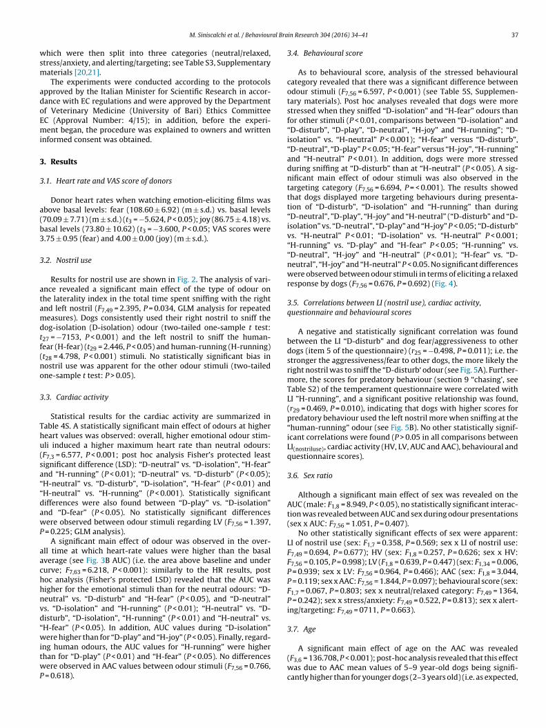
M. Siniscalchi et al. / Behavioural Brain Research 304 (2016) 34–41 37
which were then split into three categories (neutral/relaxed,
stress/anxiety, and alerting/targeting; see Table S3, Supplementary
materials [20,21].
The experiments were conducted according to the protocols
approved by the Italian Minister for Scientific Research in accor-
dance with EC regulations and were approved by the Department
of Veterinary Medicine (University of Bari) Ethics Committee
EC (Approval Number: 4/15); in addition, before the experi-
ment began, the procedure was explained to owners and written
informed consent was obtained.
3. Results
3.1. Heart rate and VAS score of donors
Donor heart rates when watching emotion-eliciting films was
above basal levels: fear (108.60 ± 6.92) (m ± s.d.) vs. basal levels
(70.09 ± 7.71) (m ± s.d.) (t3 = −5.624, P < 0.05); joy (86.75 ± 4.18) vs.
basal levels (73.80 ± 10.62) (t3 = −3.600, P < 0.05; VAS scores were
3.75 ± 0.95 (fear) and 4.00 ± 0.00 (joy) (m ± s.d.).
3.2. Nostril use
Results for nostril use are shown in Fig. 2. The analysis of vari-
ance revealed a significant main effect of the type of odour on
the laterality index in the total time spent sniffing with the right
and left nostril (F7,49 = 2.395, P = 0.034, GLM analysis for repeated
measures). Dogs consistently used their right nostril to sniff the
dog-isolation (D-isolation) odour (two-tailed one-sample t test:
t27 = −7153, P < 0.001) and the left nostril to sniff the human-
fear (H-fear) (t29 = 2.446, P < 0.05) and human-running (H-running)
(t28 = 4.798, P < 0.001) stimuli. No statistically significant bias in
nostril use was apparent for the other odour stimuli (two-tailed
one-sample t test: P > 0.05).
3.3. Cardiac activity
Statistical results for the cardiac activity are summarized in
Table 4S. A statistically significant main effect of odours at higher
heart values was observed: overall, higher emotional odour stim-
uli induced a higher maximum heart rate than neutral odours:
(F7,3 = 6.577, P < 0.001; post hoc analysis Fisher’s protected least
significant difference (LSD): “D-neutral” vs. “D-isolation”, “H-fear”
and “H-running” (P < 0.01); “D-neutral” vs. “D-disturb” (P < 0.05);
“H-neutral” vs. “D-disturb”, “D-isolation”, “H-fear” (P < 0.01) and
“H-neutral” vs. “H-running” (P < 0.001). Statistically significant
differences were also found between “D-play” vs. “D-isolation”
and “D-fear” (P < 0.05). No statistically significant differences
were observed between odour stimuli regarding LV (F7,56 = 1.397,
P = 0.225; GLM analysis).
A significant main effect of odour was observed in the over-
all time at which heart-rate values were higher than the basal
average (see Fig. 3B AUC) (i.e. the area above baseline and under
curve; F7,63 = 6.218, P < 0.001): similarly to the HR results, post
hoc analysis (Fisher’s protected LSD) revealed that the AUC was
higher for the emotional stimuli than for the neutral odours: “D-
neutral” vs. “D-disturb” and “H-fear” (P < 0.05), and “D-neutral”
vs. “D-isolation” and “H-running” (P < 0.01); “H-neutral” vs. “D-
disturb”, “D-isolation”, “H-running” (P < 0.01) and “H-neutral” vs.
“H-fear” (P < 0.05). In addition, AUC values during “D-isolation”
were higher than for “D-play” and “H-joy” (P < 0.05). Finally, regard-
ing human odours, the AUC values for “H-running” were higher
than for “D-play” (P < 0.01) and “H-fear” (P < 0.05). No differences
were observed in AAC values between odour stimuli (F7,56 = 0.766,
P = 0.618).
3.4. Behavioural score
As to behavioural score, analysis of the stressed behavioural
category revealed that there was a significant difference between
odour stimuli (F7,56 = 6.597, P < 0.001) (see Table 5S, Supplemen-
tary materials). Post hoc analyses revealed that dogs were more
stressed when they sniffed “D-isolation” and “H-fear” odours than
for other stimuli (P < 0.01, comparisons between “D-isolation” and
“D-disturb”, “D-play”, “D-neutral”, “H-joy” and “H-running”; “D-
isolation” vs. “H-neutral” P < 0.001); “H-fear” versus “D-disturb”,
“D-neutral”, “D-play” P < 0.05; “H-fear” versus “H-joy”, “H-running”
and “H-neutral” P < 0.01). In addition, dogs were more stressed
during sniffing at “D-disturb” than at “H-neutral” (P < 0.05). A sig-
nificant main effect of odour stimuli was also observed in the
targeting category (F7,56 = 6.694, P = < 0.001). The results showed
that dogs displayed more targeting behaviours during presenta-
tion of “D-disturb”, “D-isolation” and “H-running” than during
“D-neutral”, ”D-play”, “H-joy” and “H-neutral” (“D-disturb” and “D-
isolation” vs. “D-neutral”, “D-play” and “H-joy” P < 0.05; “D-disturb”
vs. “H-neutral” P < 0.01; “D-isolation” vs. “H-neutral” P < 0.001;
“H-running” vs. “D-play” and “H-fear” P < 0.05; “H-running” vs.
“D-neutral”, “H-joy” and “H-neutral” (P < 0.01); “H-fear” vs. “D-
neutral”, “H-joy” and “H-neutral” P < 0.05. No significant differences
were observed between odour stimuli in terms of eliciting a relaxed
response by dogs (F7,56 = 0.676, P = 0.692) (Fig. 4).
3.5. Correlations between LI (nostril use), cardiac activity,
questionnaire and behavioural scores
A negative and statistically significant correlation was found
between the LI “D-disturb” and dog fear/aggressiveness to other
dogs (item 5 of the questionnaire) (r25 = −0.498, P = 0.011); i.e. the
stronger the aggressiveness/fear to other dogs, the more likely the
right nostril was to sniff the “D-disturb‘ odour (see Fig. 5A). Further-
more, the scores for predatory behaviour (section 9 “chasing’, see
Table S2) of the temperament questionnaire were correlated with
LI ”H-running”, and a significant positive relationship was found,
(r29 = 0.469, P = 0.010), indicating that dogs with higher scores for
predatory behaviour used the left nostril more when sniffing at the
“human-running” odour (see Fig. 5B). No other statistically signif-
icant correlations were found (P > 0.05 in all comparisons between
LI(nostriluse), cardiac activity (HV, LV, AUC and AAC), behavioural and
questionnaire scores).
3.6. Sex ratio
Although a significant main effect of sex was revealed on the
AUC (male: F1,8 = 8.949, P < 0.05), no statistically significant interac-
tion was revealed between AUC and sex during odour presentations
(sex x AUC: F7,56 = 1.051, P = 0.407).
No other statistically significant effects of sex were apparent:
LI of nostril use (sex: F1,7 = 0.358, P = 0.569; sex x LI of nostril use:
F7,49 = 0.694, P = 0.677); HV (sex: F1,8 = 0.257, P = 0.626; sex x HV:
F7,56 = 0.105, P = 0.998); LV (F1,8 = 0.639, P = 0.447) (sex: F1,34 = 0.006,
P = 0.939; sex x LV: F7,56 = 0.964, P = 0.466); AAC (sex: F1,8 = 3.044,
P = 0.119; sex x AAC: F7,56 = 1.844, P = 0.097); behavioural score (sex:
F1,7 = 0.067, P = 0.803; sex x neutral/relaxed category: F7,49 = 1364,
P = 0.242); sex x stress/anxiety: F7,49 = 0.522, P = 0.813); sex x alert-
ing/targeting: F7,49 = 0711, P = 0.663).
3.7. Age
A significant main effect of age on the AAC was revealed
(F3,6 = 136.708, P < 0.001); post-hoc analysis revealed that this effect
was due to AAC mean values of 5–9 year-old dogs being signifi-
cantly higher than for younger dogs (2–3 years old) (i.e. as expected,

38 M. Siniscalchi et al. / Behavioural Brain Research 304 (2016) 34–41
Fig. 3. Cardiac activity. A: Highest value (HV) and lowest value (LV) of the dogs’ heart rate (HR) in response to presentation of different odours (means with S.E.M. are shown).
B: The Areas Under Curve (AUC; A) and Above Curve (AAC; B) in response to presentation of different odours (means with S.E.M. are shown).
during testing adult dogs spent longer with a lower heart rate than
the basal average).
Interactions between AAC and age during odour presentations
(age x AUC: F21,42 = 15.533, P < 0.001) revealed that this differ-
ence was more evident for “D-disturb” and “D-isolation” stimuli
(P < 0.001) and for “D-play”, “H-fear”, “H-joy” and “H-running”
odours (P < 0.05).
No other statistically significant effects of age were
apparent: LI of nostril use (age: F3,5 = 1.420, P = 0.341;
age × LI of nostril use: F21,35 = 1.090, P = 0.400); HV (age:
F3,6 = 0.232, P = 0.871; age × HV: F21,42 = 0.757, P = 0.750); LV
(age: F3,6 = 0.259, P = 0.852; age × LV: F21,42 = 0.760, P = 0.748);
AUC (age: F3,6 = 3.650, P = 0.083; age × AUC: F21,42 = 0.815,
P = 0.687); behavioural score (neutral/relaxed-age: F3,5 = 0.348,
P = 0.793; age × neutral/relaxed category: F21,35 = 0650, P = 0.850);
stress/anxiety-age: F3,5 = 1.294, P = 0.373: age × stress/anxiety:
F21,35 = 0.794, P = 0.708); alerting/targeting-age: F3,5 = 0100,
P = 0.957; age × alerting/targeting: F21,35 = 0633, P = 0.865).
4. Discussion
Previous studies have reported striking asymmetries in dogs’
nostril use during sniffing at different emotive stimuli [4]. Here
we report for the first time that this asymmetry is also manifested
during sniffing of both human and conspecific odours collected dur-
ing different emotional events, suggesting that dogs could detect
the emotional states of a member of its social group through
olfaction. The results specifically showed that during sniffing of “D-
isolation” odour, dogs consistently used their right nostril. Given
that the mammalian olfactory system ascends mainly ipsilaterally
to the brain [22], the pattern of right nostril use during sniffing
of “D-isolation” odour suggests involvement of the right hemi-
sphere. In dogs, right nostril use (right hemisphere activity) has
been observed during sniffing at clear arousal stimuli such as
adrenaline and the veterinarian’s sweat [4]. More in general, in the
animal kingdom, right hemisphere use has been associated with
the expression and control of intense emotions, such as aggression,
escape behaviour and fear (see for extensive review: [5,23]).
One possible explanation for the right nostril use observed dur-
ing the “D-isolation” stimulus is that this odour was collected
during a stressful situation (in which a dog was separated from
its owner in an unfamiliar environment) and, as a consequence,
the donor dog’s odour chemosignals could have enhanced the
arousal state in the receiver subject, eliciting its escape behavioural
response (right hemisphere activation). In other words, it could be
useful for a dog to receive information through its sense of smell
about a place where another dog experienced a stressful/possibly
dangerous situation. In addition, the higher cardiac activity shown
by dogs when presenting “D-isolation” odour is consistent with
the idea that the hypothalamic-pituitary-adrenal (HPA) axis, which
enhances cardiac activity in response to an emotional stressor, is
mainly under the control of the right hemisphere [8]. Behavioural
measures also supported the hypothesis that higher heart rate
during presentation of the “D-isolation” stimulus reflected a state
of increased arousal since subjects showed significantly higher
stress and alerting behaviours compared to other stimuli. Other
evidence comes from studies on marmosets, showing that visual
scanning (indicator of vigilance) during exposure to a predator
was correlated to serotoninergic neurotransmission in the right
hypothalamus, which activates an HPA-axis stress response [24].

M. Siniscalchi et al. / Behavioural Brain Research 304 (2016) 34–41 39
Fig. 4. Behavioural score. Data for the score of the three behavioural categories
determined from the behavioural score for each dog during inspection of different
odours (means with S.E.M. are shown).
In addition, different studies exploring the functional implica-
tions of emotional chemosignals in humans and rats reported that
smelling the sweat of stressed individuals enhances attention and
vigilance [25,26], improves anger and fear discrimination [27] and
augments the startle reflex [28,29].
More recently in humans, consistently with theories of possible
“emotional contagion” between dogs, de Groot et al. [30] tested
the ability of emotional chemosignals to recruit joint processes
between sender and receiver and found that inhaling chemosig-
nals emitted during emotional states induced the same state (fear,
disgust) in the receiver.
Although we failed to find any statistically significant bias in
nostril use during sniffing at “D-disturb”, it is interesting to note
that the stronger the aggressiveness/fear towards other dogs, the
more likely the right nostril was to sniff at this odour, suggesting
that temperament could influence behavioural response to emo-
tional odours.
On the other hand, dogs consistently used the left nostril to sniff
“H-fear” and “H-running” stimuli, suggesting prevalent activation
of the left hemisphere. One possible logical explanation for the
involvement of the left hemisphere in the analysis of these odours
is that a greater discrimination accuracy between stimuli that dis-
play varying levels of arousal occurred in the left amygdala [31,32].
Furthermore, neuroimaging studies in humans [33,34] have shown
that while the right amygdala rapidly and non-selectively detects
stimuli that represent a potential threat (i.e. general detector of
arousal), the left amygdala provides a fine-tuned and detailed
mechanism which helps provide a more accurate determination
of whether the potential threat is real. In other words, it is not
said that what represents a potential threat for a human (e.g. the
owner) represents a threat for the dog. Neural structures on the
left side of the brain are also involved in the control of predatory
behaviour in different animal models [23], including dogs [35]: as a
consequence, human chemosensory arousal signals (“H-fear” and
“H-running” odours) could elicit approaching behavioural tenden-
cies to the stimulus (dogs’ prey drive. i.e. the instinctive inclination
of a carnivore to pursue and capture prey) through the selective
use of the left nostril/hemisphere. This hypothesis is supported by
the positive correlation between targeting behaviour and item 9
in the questionnaire (predatory behaviour) during sniffing at “H-
running”, indicating that the stronger the dog’s targeting behaviour,
the more likely it would be to use the left nostril to sniff the “H-
running” stimulus. In addition, the prey-catching behaviour elicited
by left nostril use during sniffing at human chemosensory arousal
signals could be one explanation for canine aggression towards
people who are afraid of dogs. In a more evolutionary perspec-
tive, during prey-catching behaviour, it could be useful for a dog
to elicit an approaching behavioural response towards heterospe-
cific arousal chemosignals produced during prey escape behaviour:
the prey that is escaping produces arousal chemosignals in the
environment that could inform conspecifics about the presence
of the predator (eliciting a withdrawal behavioural response in
conspecific subjects) and at the same time the same odour could
reinforce the tracking behaviour of the predator. Furthermore,
behavioural/neurochemical correlations in marmosets suggest that
general motor activity (e.g. locomotory, exploratory behaviour)
is related to dopamine levels in the left hemisphere [24]. Taken
together, these hypotheses could support the long evolutionary role
of the left side of the brain in sustained pursuit of prey using odours
[5].
5. Conclusion
Overall, our data showed that dogs displayed higher behavioural
and physiological reactivity to canine (conspecific) and humans
(heterospecific) odours collected during different emotional con-
ditions.
In addition, a clear bias in nostril use was observed during sniff-
ing at clear arousal stimuli, but using opposite sides for canine
and human odours (showing right nostril-use bias for inspecting
conspecific anxiety odours and a prevalent use of the left nostril
during sniffing at heterospecific arousal odours), suggesting that

40 M. Siniscalchi et al. / Behavioural Brain Research 304 (2016) 34–41
Fig. 5. Correlations between LI(nostriluse) and questionnaire. Data for the significant correlation discussed in the text between A) LI “D-disturb” and “dog fear/aggressiveness
to other dogs” (item 5 of the questionnaire); B) LI “H-running” and “chasing” (item 9 of the questionnaire); Data presented are means calculated for each dog over the three
trials.
chemosignals communicate conspecific and heterospecific emo-
tions using different sensory pathways.
Acknowledgements
We are grateful to Anthony Green for revising the English text
and to Dr. Antonio Caliano for his precious help during data analysis.
Appendix A. Supplementary data
Supplementary data associated with this article can be found, in
the online version, at http://dx.doi.org/10.1016/j.bbr.2016.02.011.
References
[1] M. Siniscalchi, A. Quaranta, Wagging to the right or to the left: lateralisationand what it tells of the dog’s social brain, in: J. Kaminski, S. Marshall-Pescini(Eds.), The Social Dog: Behavior and Cognition, Academic Press, Waltham, MA,2014, pp. 373–393.
[2] M. Siniscalchi, R. Sasso, A. Pepe, G. Vallortigara, A. Quaranta, Dogs turn left toemotional stimuli, Behav. Brain Res. 208 (2010) 516–521.
[3] M. Siniscalchi, A. Quaranta, L.J. Rogers, Hemispheric specialization in dogs forprocessing different acoustic stimuli, PLoS One 3 (2008) e3349, http://dx.doi.org/10.1371/journal.pone.0003349.
[4] M. Siniscalchi, R. Sasso, A.M. Pepe, S. Dimatteo, G. Vallortigara, A. Quaranta,Sniffing with the right nostril: lateralization of response to odour stimuli bydogs, Anim. Behav. 8 (2011) 399–404.
[5] L.J. Rogers, G. Vallortigara, R.J. Andrew, Divided Brains: The Biology andBehaviour of Brain Asymmetries, University Press, New York: Cambridge,2013.
[6] T.H. Burne, L.J. Rogers, Responses to odorants by the domestic chick, Physiol.Behav. 60 (1996) 1441–1447.
[7] P.D. McGreevy, L.J. Rogers, Motor and sensory laterality in thoroughbredhorses, Appl. Anim. Behav. Sci. 92 (2005) 337–352.
[8] M. Siniscalchi, B. Padalino, L. Aubé, A. Quaranta, Right-nostril use duringsniffing at arousing stimuli produces higher cardiac activity in jumper horses,Laterality 20 (2015) 483–500.
[9] G. Vallortigara, L. Regolin, P. Pagni, Detour behaviour, imprinting, and visuallateralization in the domestic chick, Cognit. Brain Res. 7 (1999) 307–320.
[10] V.A. Sovrano, Visual lateralization in response to familiar and unfamiliarstimuli in fish, Behav. Brain Res. 152 (2004) 385–391.
[11] G. Vallortigara, C. Chiandetti, V.A. Sovrano, Brain asymmetry (animal), WileyInterdiscip. Rev. Cognit. Sci. 2 (2011) 146–157.

M. Siniscalchi et al. / Behavioural Brain Research 304 (2016) 34–41 41
[12] J.L. Millot, Olfactory and visual cues in the interaction systems between dogsand children, Behav. Process. 33 (1994) 177–188.
[13] D.L. Wells, P.G. Hepper, The discrimination of dog odours by humans,Perception 29 (2000) 111–115.
[14] I.F. Dunbar, Olfactory preferences in dogs: the response of male and femalebeagles to conspecific odors, Behav. Biol. 20 (1977) 471–481.
[15] M. Goodwin, K.M. Gooding, F. Regnier, Sex pheromone in the dog, Science 203(1979) 559–561.
[16] J.L. Millot, J.C. Filiatre, A. Eckerlin, A.C. Gagnon, H. Montagner, Olfactory cuesin the relations between children and their pet dogs, Appl. Anim. Behav. Sci.19 (1987) 189–195.
[17] I.L. Brisbin, S.N. Austad, Testing the individual odour theory of canineolfaction, Anim. Behav. 42 (1991) 63–69.
[18] P. Lenochova, S.C. Roberts, J. Havlicek, Methods of human body odorsampling: the effect of freezing, Chem. Senses 34 (2009) 127–138.
[19] M. Siniscalchi, D. Bertino, A. Quaranta, Laterality and performance ofagility-trained dogs, Laterality 19 (2014) 219–234.
[20] B. Handelman, Canine Behavior: A Photo Illustrated Handbook, Woof andWord Press, Norwich, 2008.
[21] B. Aloff, Canine Body Language: A Photographic Guide Interpreting the NativeLanguage of the Domestic Dog, Dogwise Publishing, Wenatchee, U.S.A, 2005.
[22] J.P. Royet, J. Plailly, Lateralization of olfactory processes, Chem. Senses 29(2004) 731–745.
[23] L.J. Rogers, R.J. Andrew, Comparative Vertebrate Lateralization, UniversityPress, New York: Cambridge, 2002.
[24] M.A. Silva, B. Topic, V. Lamounier-Zepter, J.P. Huston, C. Tomaz, M. Barros,Evidence for hemispheric specialization in the marmoset (Callithrixpenicillata) based on lateralization of behavioral/neurochemical correlations,Brain Res. Bull. 74 (2007) 416–428.
[25] D. Chen, A. Katdare, N. Lucas, Chemosignals of fear enhance cognitiveperformance in humans, Chem. Senses 31 (2006) 415–423.
[26] Y. Kiyokawa, M. Shimozuru, T. Kikusui, Y. Takeuchi, Y. Mori, Alarmpheromone increases defensive and risk assessment behaviors in male rats,Physiol. Behav. 87 (2006) 383–387.
[27] W. Zhou, D. Chen, Fear-related chemosignals modulate recognition of fear inambiguous facial expressions, Psychol. Sci. 20 (2009) 177–183.
[28] A. Prehn, A. Ohrt, B. Sojka, R. Ferstl, B.M. Pause, Chemosensory anxiety signalsaugment the startle reflex in humans, Neurosci. Lett. 394 (2006) 127–130.
[29] H. Inagaki, Y. Kiyokawa, T. Kikusui, Y. Takeuchi, Y. Mori, Enhancement of theacoustic startle reflex by an alarm pheromone in male rats, Physiol. Behav. 93(2008) 606–611.
[30] J.H.B. de Groot, M.A.M. Smeets, A. Kaldewaij, M.A.J. Duijndam, G.R. Semin,Chemosignals communicate human emotions, Psychol. Sci. 23 (2012)1417–1424.
[31] J.E. Hardee, J.C. Thompson, A. Puce, The left amygdala knows fear: laterality inthe amygdala response to fearful eyes, SCAN 3 (2008) 47–54.
[32] J.S. Morris, C.D. Frith, D.I. Perrett, D. Rowland, A.W. Young, A.J. Calder, et al., Adifferential response in the human amygdala to fearful and happy facialexpressions, Nature 383 (1996) 812–815.
[33] J.S. Morris, A. Ohman, R.J. Dolan, A subcortical pathway to the right amygdalamediating unseen fear, Proc. Natl. Acad. Sci. U. S. A. 96 (1999) 1680–1685.
[34] J. Glascher, R. Adolphs, Processing of the arousal of subliminal andsupraliminal emotional stimuli by the human amygdala, J. Neurosci. 23(2003) 10274–10282.
[35] M. Siniscalchi, G. Pergola, A. Quaranta, Detour behaviour in attack-traineddogs: left-turners perform better than right-turners, Laterality 18 (2013)282–293.

Chapter 4
Study 2 Dogs perception of human emotional vocalizations
60

Dogs perception of human emotional vocalizations: article 6
Study aim
Previous studies demonstrated that dogs recognize different emotions by looking at human
faces and correctly match human facial expression of happiness and anger with
vocalizations expressing the same emotional valence.
In the light of these evidences, the aim of the present study was to investigate dogs’
perception and processing of human non-verbal vocalizations having different emotional
valence.
Methods
Male and female non-verbal vocalizations expressing the Ekman’s six basic emotions of
happiness, surprise, disgust, fear, sadness and anger were recorded. The emotional acoustic
stimuli were then presented to 36 domestic dogs using the head-turning paradigm.
Asymmetries in the head turning response as well as subjects’ behaviour and cardiac
activity were evaluated to derive information about the valence and the intensity of the
perceived emotion, respectively.
Results
Dogs showed an asymmetrical head-orienting response to the playbacks of different
human non-verbal emotional vocalizations, suggesting that they perceive the different
valence of the emotion conveyed. In particular, they turned the head to the left (right
hemisphere advantage) in response to “fear” and “sadness” human vocalizations, whereas
they turned their head to the right (left hemisphere advantage) in response to “happiness”
vocalizations. Furthermore, both cardiac activity and behaviour response suggest that dogs
are sensitive to emotional cues of human vocalizations, since a higher level of subjects’
arousal was recorded in response to clear arousing stimuli.
Conclusions
Dogs perceive and differentiate the emotional content of human vocalizations. Results
provide evidences about the existence of an emotional modulation of the dog brain to
process basic human non-verbal emotional vocalizations, providing new insights into
emotional functioning of canine brain.
61

La percezione delle vocalizzazioni emotive dell’uomo nel cane: articolo 6
Obiettivi dello studio
Studi precedenti hanno dimostrato che i cani riconoscono le diverse emozioni dell’uomo
guardandone i volti, ed abbinano correttamente l'espressione facciale umana di felicità e di
rabbia con le vocalizzazioni che esprimono la stessa valenza emotiva.
Alla luce di queste evidenze, lo scopo del presente studio è stato quello di analizzare come
il cane percezione ed elabora le vocalizzazioni umane non verbali con differente valenza
emotiva.
Metodi
Sono state registrate vocalizzazioni non verbali maschili e femminili che esprimevano le sei
emozioni di base di Ekman, ovvero felicità, sorpresa, disgusto, paura, tristezza e rabbia. Gli
stimoli acustici emotivi sono stati presentati a 36 cani domestici utilizzando il paradigma di
rotazione della testa. Sono state valutate le asimmetrie nella risposta di rotazione della testa,
il comportamento dei soggetti e la loro attività cardiaca, al fine di valutare rispettivamente la
valenza e l'intensità dell'emozione percepita.
Risultati
I cani hanno mostrato delle asimmetrie nella rotazione della testa in risposta alle diverse
vocalizzazioni non verbali emotive dell’uomo, le quali suggeriscono che i cani percepiscono
la diversa valenza dell'emozione trasmessa. In particolare, i cani hanno girato la testa a
sinistra (vantaggio dell'emisfero destro) in risposta a vocalizzazioni umane di paura e
tristezza, mentre hanno girato la testa verso destra (vantaggio dell'emisfero sinistro) in
risposta a vocalizzazioni di gioia. Inoltre, sia l'attività cardiaca che la risposta
comportamentale dei soggetti suggeriscono che i cani sono sensibili ai segnali emotivi
veicolati dalle vocalizzazioni umane, poiché è stato registrato un livello di “arousal” più
elevato in risposta a stimoli chiaramente eccitanti.
Conclusioni
I cani percepiscono e differenziano il contenuto emotivo delle vocalizzazioni umane. I
risultati forniscono delle evidenze sull'esistenza di una modulazione del cervello del cane
per l’elaborazione delle vocalizzazioni non verbali dell’uomo che esprimono le emozioni di
base, fornendo nuove conoscenze sul funzionamento emotivo del suo cervello.
62

Perception auditive des émotions humaines chez le chien: article 6
Objectif de l’étude
Des études précédentes ont montré que les chiens sont capables de reconnaitre différentes
émotions exprimées par des visages humains et sont capable d’associer les expressions
faciales de bonheur et de colère avec des vocalisations exprimant les mêmes valences
émotionnelles.
En s’appuyant sur ces données, l’objectif de cette étude était d’étudier chez le chien la
perception et le traitement de vocalisations humaines non-verbales de différentes valences
émotionnelles.
Méthodes
Des vocalisations non-verbales d’hommes et de femmes exprimant les six émotions de
base d’Ekman (joie, surprise, dégout, peur, tristesse et colère) ont été enregistrées. Les
stimuli auditifs émotionnels ont été présentés à 36 chiens domestiques dont les réponses
étaient analysées en utilisant le paradigme d’orientation de la tête. L’asymétrie de rotation
de la tête ainsi que le comportement de l’individu et son activité cardiaque ont été analysés
pour obtenir des informations sur la valence et l’intensité des émotions perçues.
Résultats
Les chiens ont montré des réponses d’orientation de la tête latéralisées en réponse à la
diffusion des vocalisations non-verbales humaines, suggérant qu’ils percevaient les
différentes valences émotionnelles. En effet, ils ont tourné la tête à gauche (hémisphère
droit) en réponse aux vocalisations de peur et de tristesse alors qu’ils tournaient la tête à
droite (hémisphère gauche) en réponse aux vocalisations de joie. De plus, l’activité
cardiaque et les réponses comportementales suggèrent que les chiens étaient sensibles aux
caractéristiques émotionnelles des vocalisations humaines, ils présentaient des niveaux de
vigilance élevés en réponse à des stimuli clairement excitant.
Conclusions
Les chiens perçoivent et discriminent le contenu émotionnel des vocalisations humaines.
Nos résultats apportent des preuves de l’existence d’une modulation émotionnelle dans le
cerveau du chien permettant le traitement des émotions de base véhiculées par des
vocalisations humaines non-verbales. Ces résultats ouvrent de nouvelles perspectives dans
la compréhension le fonctionnement émotionnel du cerveau du chien.
63

Emotional stimuli
Hemispheric bias Cardiac activity Stress levels
Fear Right High increase High
Sadness Right Moderate increase Moderate
Anger No bias High increase High
Disgust No bias High increase Low
Surprise No bias High increase Low
Happiness Left Moderate increase Moderate
Schematic representation of study 2 results, taken from visual inspection of the graphs.
64

1SCIENTIFIC REPORTS | (2018) 8:77 | DOI:10.1038/s41598-017-18417-4
www.nature.com/scientificreports
Lateralized behavior and cardiac activity of dogs in response to human emotional vocalizationsMarcello Siniscalchi, Serenella d’Ingeo, Serena Fornelli & Angelo Quaranta
Over the recent years, the study of emotional functioning has become one of the central issues in
dog cognition. Previous studies showed that dogs can recognize diferent emotions by looking at human faces and can correctly match the human emotional state with a vocalization having a negative
emotional valence. However, to this day, little is known about how dogs perceive and process human non-verbal vocalizations having diferent emotional valence. The current research provides new insights into emotional functioning of the canine brain by studying dogs’ lateralized auditory functions
(to provide a irst insight into the valence dimension) matched with both behavior and physiological measures of arousal (to study the arousal dimension) in response to playbacks related to the Ekman’s six basic human emotions. Overall, our results indicate lateralized brain patterns for the processing
of human emotional vocalizations, with the prevalent use of the right hemisphere in the analysis of
vocalizations with a clear negative emotional valence (i.e. “fear” and “sadness”) and the prevalent use of the left hemisphere in the analysis of positive vocalization (“happiness”). Furthermore, both cardiac activity and behavior response support the hypothesis that dogs are sensitive to emotional cues of
human vocalizations.
Evolutionary and ontogenetic processes played a pivotal role in dogs’ ability to detect social information from human behavior, providing the basis for complex forms of interspeciic social communication1,2. here is a grow-ing body of literature showing that dogs developed cognitive and social abilities in order to communicate with humans. Dogs are able to interpret human communicative gestures (e.g. the direction in which humans are facing or gazing), to detect his attentional states3,4 and to recognize diferent emotions by looking at human faces 5,6. Furthermore, recent studies reported that dogs discriminate human emotional faces from neutral ones5,6, that they are able to distinguish between happy and angry emotional human faces expressions7 and between the happy and neutral expression of the owner8. Moreover, Albuquerque et al.9 reported a cross-modal capacity in dogs related to the integration of visual and auditory emotional cues. In particular, dogs can correctly match “happy” or “angry” human faces with a vocalization expressing the same emotional valence.
Regarding the auditory sensory domain, it has been reported that dogs recognize the diferent valences of positive (laughing) and negative (crying) emotional sounds, showing an increase of indicators for arousal and negative emotional states in response to negative emotional sounds compared to positive ones10. Nevertheless, further studies are required in order to set reliable behavior indicators for positive emotional states in dogs.
To date, evidence for dogs’ perception and processing of human vocalizations characterised by diferent emo-tional valence is scarce11. Considering that it has been reported that the six basic emotions universally inferred from facial expressions12 are cross-culturally recognized from vocal signals in humans13, our study aimed at inves-tigating if dogs are able to recognize the six basic emotions expressed by human non-verbal vocalizations.
In order to investigate this issue, we used the head-orienting paradigm to evaluate the potential asymmetrical behavior responses of dogs to human emotional vocalizations. he head-orienting response is commonly used as a behavior method for studying lateralized attention to acoustic stimuli in mammals tested in unrestrained conditions14. It requires sounds to be played simultaneously from two speakers located symmetrically at the same distance from the tested animal’s head. In order to ensure the correct position of the animal, the experiment is usually run during its feeding behaviors, positioning a bowl containing food midway between the two speakers. Since the head turning is an unconditioned response, its direction indicates the advantage of the contralateral
Department of Veterinary Medicine, Section of Behavior Sciences and Animal Bioethics, University of Bari “Aldo Moro”, Bari, Italy. Correspondence and requests for materials should be addressed to M.S. (email: [email protected])
Received: 16 May 2017
Accepted: 11 December 2017
Published: xx xx xxxx
OPEN
65

www.nature.com/scientificreports/
2SCIENTIFIC REPORTS | (2018) 8:77 | DOI:10.1038/s41598-017-18417-4
hemisphere in processing the acoustic stimulus (e.g. if the subject turns his head towards the speaker with the right ear leading, the acoustic input is processed primarily by the let hemisphere, or at least in the initial attention to the stimulus14–16).
Speciically, in dogs a striking let head-orienting bias was observed in response to thunderstorm playbacks, conirming the right hemisphere advantage in attending to threatening and alarming stimuli17. On the contrary, conspeciic vocalizations elicited a signiicant head-turning bias to the right (let hemisphere advantage). he specialization of the let hemisphere in processing the vocalizations of familiar conspeciics has been also reported for other animal models, such as nonhuman primates18,19, horses20, cats21, and sea lions22. Nevertheless, recent studies employing the orienting paradigm in other species found an inconsistent pattern of the head-turning response to conspeciic calls. For instance, Vervet monkeys showed a right hemisphere dominant activity23 while no bias was found for Barbary macaques24. Moreover, a sex-speciic asymmetries was shown for mouse lemurs, in particular male individuals displayed a let hemisphere bias in response to conspeciic vocalizations with negative emotional valence16. his contradictory pattern might be due to a diferent phylogenetic distribution of hemi-spheric specialization and lateralization in closely related species25 or to the diferent emotional valence of the message conveyed. Furthermore, within the canine species, it has been reported that the let hemisphere involve-ment in attending to conspeciic vocalizations depends on the characteristics of the sound, for example on the temporal acoustic features of the calls26. When dogs were presented with the reversed versions of speciic vocal-izations of play, disturbance and isolation, they showed a shit in their head-orienting behavior from a right-ear orienting bias (normal call versions) to a let-ear orienting bias (play calls) or to no asymmetry (disturbance and isolation calls26). In addition, recent studies describe a right hemisphere dominant activity to process conspeciic vocalizations when they elicit intense emotions17,27.
Dogs show also an asymmetric head-turning behavior in response to human vocalizations. hey displayed a signiicant bias to turn the head with the right ear leading (let hemisphere activity) when presented with a famil-iar spoken command in which the salience of meaningful phonemic (segmental) cues was artiicially increased; on the other hand, they showed a signiicant head-turning behavior to the let side (right hemisphere domi-nant activity) in response to commands with artiicially increased salience of intonational or speaker-related (suprasegmental) vocal cues28. Nevertheless, the more recent results of Andics et al.29,30 showed the opposite pattern of the hemispheres activity. Using the fMRI technique, they found a right hemisphere advantage in pro-cessing meaningful words and a let hemispheric bias for distinguishing intonationally marked words.
Overall, although these experiments showed lateralized auditory functions in the canine brain and provide insights into mechanisms of interspeciic vocal perception, it remains unclear how dogs perceive and process the six basic emotions expressed by human non-verbal vocalizations. One of the possible methods employed to assess brain emotional functioning in the animal kingdom consists in observing and analyzing physiological (e.g. cardiac activity) and behavior responses to speciic stimuli in experimental conditions that resemble as much as possible the natural ones31. Regarding the physiological response, there is now scientiic evidence that cardiac activity could be considered a valid indicator to predict diferent emotional states in dogs32–35.
As to the behavior response, a recent study scored dogs’ behaviors in order to investigate emotional contagion to conspeciic and human emotional sounds10. Although results indicate that for both canine and human sounds dogs express more stress behaviors ater hearing sounds with a negative emotional valence, further studies are required to determine valid and reliable behavior indicators for positively valenced sounds10.
he study of behavior lateralization has the potential to provide new insights into animal emotional process-ing36. An increasing body of evidence shows common lateralized neural patterns for emotional processing across all vertebrate classes, with specialization of the right hemisphere for processing withdrawal and negative emo-tions (e.g. fear and aggression) and a dominant role of the let hemisphere for processing positive emotions and approach37,38. hus, external manifestation of hemispheric dominance (e.g. head-turning behavior) matched with both behavior and physiological responses could represent useful tools for understanding the valence of an emo-tion perceived by an animal during a particular situation, facilitating the categorization of the emotion along with valence and arousal dimensions39–41. In the light of this evidence, the aim of the present work was to investigate dogs’ emotional responses to human non-verbal emotional vocalizations by measuring subjects’ head-turning bias (valence dimension) and the related behavior and cardiac activities (arousal dimension).
ResultsHead-orienting response. Friedman’s ANOVA revealed that there was no efect of acoustic stimulus on the % of response (χ2 (5) = 6,782, P = 0.237); average %: anger (83.3%); fear (80.0%); disgust (93.3%); sadness (76.6%); surprise (90.0%) and happiness (93.3%).
Results for the head-orienting response are shown in Fig. 1. A significant main effect of playbacks was observed (F(5,99) = 5.766, P = 0.000; GLMM analysis): pairwise comparisons revealed that this main efect was due to “happiness” vocalization being signiicantly diferent from other sounds (“happiness” vs. “fear”, “anger” and “sadness” (P = 0.000); “happiness” vs. “disgust” (P = 0.009) and “surprise” (P = 0.026); Fisher’s LSD). In addition, the analysis revealed that for “fear” and “sadness” call types, dogs consistently turned their head with the let ear leading (“fear”: Z = 140.000, P = 0.039; “sadness”: Z = 102.000, P = 0.046; One-Sample Wilcoxon Signed Ranks Test) (see Fig. 1). A very similar trend was observed for “anger” vocalization even if not statistically signiicant (Z = 221.000, P = 0.072). On the other hand, a signiicant right bias in the head turning response was found when dogs attended to playbacks of “happiness” (Z = 46.000, P = 0.003). No statistical signiicant biases were found for both “disgust” and “surprise” stimuli (P > 0.05, One-Sample Wilcoxon Signed Ranks Test). On inves-tigating possible methodological confounding factors, a binomial GLMM analysis revealed that the direction of head orienting response turns was not signiicantly inluenced by vocalization gender (F(1,99) = 0.102, P = 0.750) and playback order (F(6,93) = 0.705, P = 0.646). In addition no efects of sex (F(1,99) = 0.306, P = 0.581), age
66

www.nature.com/scientificreports/
3SCIENTIFIC REPORTS | (2018) 8:77 | DOI:10.1038/s41598-017-18417-4
(F(1,99) = 0.000, P = 0.998) and temperament questionnaire scores were observed (P > 0.05 for all queries of the questionnaire, see Table 1).
Reactivity and latency to resume feeding. The cumulative incidences of reactivity and latency to resume feeding during playbacks’ presentations are presented in Fig. 2(A,B). As for reactivity, mixed efects Cox regression revealed that subjects hearing “anger” playbacks had a higher probability to react than ater attend-ing, respectively, playbacks of happiness (β(SE) = 0.93(0.37); [Exp(β) = 0.39; 95%-CI = 0.19;0.81]; P = 0.012), “disgust” (β(SE) = 1.16(0.43); [Exp(β) = 0.31; 95%-CI = 0.13;0.74]; P = 0.008) and “sadness” (β(SE) = 1.13(0.39); [Exp(β) = 0.32; 95%-CI = 0.15;0.69]; P = 0.004) see Fig. 2(A–C). he probability to react to the stimuli was increased in male subjects with respect to females (β(SE) = 0.52(0.24); [Exp(β) = 0.59; 95%-CI = 0.37;0.95]; P = 0.031) while it was decreased as age increased (B(SE) = −0.1(0.047); [Exp(β) = 0.90; 95%-CI = 0.82;0.99]; P = 0.033). A primary efect of the irst playback has been observed since the probability to react was increased if the sound was presented ater respectively “anger” (β(SE) = 0.69(0.34); [Exp(β) = 1.99; 95%-CI = 1.03;3.85]; P = 0.041) and “surprise” acoustic stimuli (β(SE) = 0.77(0.37); [Exp(β) = 2.17; 95%-CI = 1.04;4.5]; P = 0.038); on the other hand, the probability to react was lower if the sound was presented after “sadness” stimulus (β(SE) = −1.51(0.54); [Exp(β) = 0.22; 95%-CI = 0.07;0.63]; P = 0.005). Finally, there were efects of stress behav-iors and query scales, since the probability to react to playbacks increased with increasing scores of stress behav-iors during the test (β(SE) = 0.25(0.08); [Exp(β) = 1.28; 95%-CI = 1.08;1.51]; P = 0.003) and increasing scores of excitability in the query (β(SE) = 0.07(0.03); [Exp(β) = 0.39; 95%-CI = 1.00;1.14]; P = 0.048). Finally, the prob-ability to react to vocalizations sounds decreased with increasing scores of “stranger aggression” in the query (β(SE) = −0.06(0.02); [Exp(β) = 0.94; 95%-CI = 0.89;0.99]; P = 0.019).
As for the latency to resume feeding, mixed efects Cox regression revealed that dogs hearing “anger” had smaller probability to resume feeding than when they were hearing respectively “happiness” (β(SE) = −1.30(0.33); [Exp(β) = 3.70; 95%-CI = 1.91;7.15]; P = 0.000), “fear” (β(SE) = −0.73(0.32); [Exp(β) = 2.07; 95%-CI = 1.10;3.90]; P = 0.023), “disgust” (β(SE) = −1.28(0.34); [Exp(β) = 3.59; 95%-CI = 1.84;7.05]; P = 0.000)
Figure 1. Head orienting response to diferent vocalizations. Laterality index for the head-orienting response of each dog to playbacks: a score of 1.0 represents exclusive head turning to the let side and −1.0 exclusive head turning to the right side (group means with SEM are shown); Asterisks indicate signiicant biases. *P < 0.05; **P < 0.01 (One-Sample Wilcoxon Signed Ranks Test).
Queries
Orienting Response HV LV AUC AAC Stress
F P F P F P F P F P F P
(1) Stranger-directed aggression 0.272 0.603 0.362 0.548 0.593 0.443 3.455 0.065 0.775 0.380 5.185 0.001
(2) Owner-directed aggression 1.427 0.235 3.905 0.050 11.865 0.001 0.006 0.938 0.163 0.687 0.000 0.982
(3) Stranger-directed fear 1.061 0.360 1.876 0.173 0.942 0.334 0.275 0.601 3.083 0.081 0.054 0.816
(4) Non social fear 0.060 0.807 0.138 0.711 4.232 0.052 5.862 0.017 2.652 0.106 4.331 0.045
(5) Separation-related behaviour 0.203 0.654 0.874 0.352 6.394 0.013 4.085 0.055 0.044 0.834 0.412 0.522
(6) Attachment or attention-seeking behaviour 0.418 0.519 0.032 0.858 0.302 0.584 5.521 0.045 0.216 0.643 1.157 0.284
(7) Trainability 0.701 0.404 0.582 0.447 0.900 0.344 4.394 0.038 6.686 0.011 0.000 0.996
(8) Excitability 0.629 0.430 0.755 0.386 0.826 0.365 0.083 0.774 0.449 0.504 3.189 0.076
(9) Pain sensitivity 0.362 0.549 3.730 0.056 3.050 0.083 0.025 0.874 0.005 0.945 0.006 0.936
Table 1. Questionnaire queries. Efects of questionnaire queries on “Orienting Response”, “HV”, “LV”, “AUC”, “AAC” and “Stress” variables (df1 = 1; df2 = 136; GLMM analysis).
67
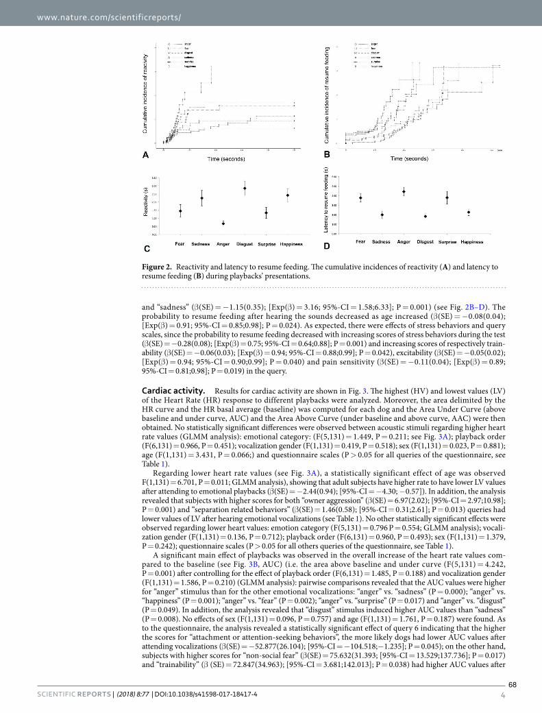
www.nature.com/scientificreports/
4SCIENTIFIC REPORTS | (2018) 8:77 | DOI:10.1038/s41598-017-18417-4
and “sadness” (β(SE) = −1.15(0.35); [Exp(β) = 3.16; 95%-CI = 1.58;6.33]; P = 0.001) (see Fig. 2B–D). The probability to resume feeding after hearing the sounds decreased as age increased (β(SE) = −0.08(0.04); [Exp(β) = 0.91; 95%-CI = 0.85;0.98]; P = 0.024). As expected, there were efects of stress behaviors and query scales, since the probability to resume feeding decreased with increasing scores of stress behaviors during the test (β(SE) = −0.28(0.08); [Exp(β) = 0.75; 95%-CI = 0.64;0.88]; P = 0.001) and increasing scores of respectively train-ability (β(SE) = −0.06(0.03); [Exp(β) = 0.94; 95%-CI = 0.88;0.99]; P = 0.042), excitability (β(SE) = −0.05(0.02); [Exp(β) = 0.94; 95%-CI = 0.90;0.99]; P = 0.040) and pain sensitivity (β(SE) = −0.11(0.04); [Exp(β) = 0.89; 95%-CI = 0.81;0.98]; P = 0.019) in the query.
Cardiac activity. Results for cardiac activity are shown in Fig. 3. he highest (HV) and lowest values (LV) of the Heart Rate (HR) response to different playbacks were analyzed. Moreover, the area delimited by the HR curve and the HR basal average (baseline) was computed for each dog and the Area Under Curve (above baseline and under curve, AUC) and the Area Above Curve (under baseline and above curve, AAC) were then obtained. No statistically signiicant diferences were observed between acoustic stimuli regarding higher heart rate values (GLMM analysis): emotional category: (F(5,131) = 1.449, P = 0.211; see Fig. 3A); playback order (F(6,131) = 0.966, P = 0.451); vocalization gender (F(1,131) = 0.419, P = 0.518); sex (F(1,131) = 0.023, P = 0.881); age (F(1,131) = 3.431, P = 0.066;) and questionnaire scales (P > 0.05 for all queries of the questionnaire, see Table 1).
Regarding lower heart rate values (see Fig. 3A), a statistically significant effect of age was observed F(1,131) = 6.701, P = 0.011; GLMM analysis), showing that adult subjects have higher rate to have lower LV values ater attending to emotional playbacks (β(SE) = −2.44(0.94); [95%-CI = −4.30; −0.57]). In addition, the analysis revealed that subjects with higher scores for both “owner aggression” (β(SE) = 6.97(2.02); [95%-CI = 2.97;10.98]; P = 0.001) and “separation related behaviors” (β(SE) = 1.46(0.58); [95%-CI = 0.31;2.61]; P = 0.013) queries had lower values of LV ater hearing emotional vocalizations (see Table 1). No other statistically signiicant efects were observed regarding lower heart values: emotion category (F(5,131) = 0.796 P = 0.554; GLMM analysis); vocali-zation gender (F(1,131) = 0.136, P = 0.712); playback order (F(6,131) = 0.960, P = 0.493); sex (F(1,131) = 1.379, P = 0.242); questionnaire scales (P > 0.05 for all others queries of the questionnaire, see Table 1).
A signiicant main efect of playbacks was observed in the overall increase of the heart rate values com-pared to the baseline (see Fig. 3B, AUC) (i.e. the area above baseline and under curve (F(5,131) = 4.242, P = 0.001) ater controlling for the efect of playback order (F(6,131) = 1.485, P = 0.188) and vocalization gender (F(1,131) = 1.586, P = 0.210) (GLMM analysis): pairwise comparisons revealed that the AUC values were higher for “anger” stimulus than for the other emotional vocalizations: “anger” vs. “sadness” (P = 0.000); “anger” vs. “happiness” (P = 0.001); “anger” vs. “fear” (P = 0.002); “anger” vs. “surprise” (P = 0.017) and “anger” vs. “disgust” (P = 0.049). In addition, the analysis revealed that “disgust” stimulus induced higher AUC values than “sadness” (P = 0.008). No efects of sex (F(1,131) = 0.096, P = 0.757) and age (F(1,131) = 1.761, P = 0.187) were found. As to the questionnaire, the analysis revealed a statistically signiicant efect of query 6 indicating that the higher the scores for “attachment or attention-seeking behaviors”, the more likely dogs had lower AUC values ater attending vocalizations (β(SE) = −52.877(26.104); [95%-CI = −104.518;−1.235]; P = 0.045); on the other hand, subjects with higher scores for “non-social fear” (β(SE) = 75.632(31.393; [95%-CI = 13.529;137.736]; P = 0.017) and “trainability” (β (SE) = 72.847(34.963); [95%-CI = 3.681;142.013]; P = 0.038) had higher AUC values ater
Figure 2. Reactivity and latency to resume feeding. he cumulative incidences of reactivity (A) and latency to resume feeding (B) during playbacks’ presentations.
68

www.nature.com/scientificreports/
5SCIENTIFIC REPORTS | (2018) 8:77 | DOI:10.1038/s41598-017-18417-4
hearing emotional playbacks. No other statistically signiicant efects were found (P > 0.05 for all the remaining queries of the questionnaire, see Table 1).
Regarding the overall decrease of the heart rate values compared to the baseline (i.e. the area under baseline and above curve, AAC), the GLMM analysis revealed that the higher the scores for trainability, the more likely dogs had lower AAC values (β(SE) = −27.611(10.678); [95%-CI = −48.736; −6.487]; P = 0.011) (see Table 1).
No other statistical signiicant efects were observed in AAC values: emotional category (F(5,131) = 0.304, P = 0.910; GLM analysis) (Fig. 3B); vocalization gender (F(1,131) = 0.006, P = 0.941); playback order (F(6,131) = 0.928, P = 0.477); sex (F(1,131) = 0.806, P = 0.371); age (1,131) = 0.237, P = 0.627) and query scales (P > 0.05 for all the rest queries of the questionnaire, see Table 1).
Behavior score. As to behavioral score, analysis of the stressed behavioral category revealed that there was a significant difference between acoustic stimuli (F(5,131) = 10.851, P = 0.000; GLMM analysis, see Fig. 4) ater controlling for the efect of both playback order (F(6,131) = 0.840, P = 0.541), vocalization gen-der (F(1,131) = 2.128, P = 0.147) and age (F(1,131) = 0.420, P = 0.518). A statistically signiicant efect of sex was observed (F(1,131) = 4.994, P = 0.027) indicating that male subjects have lower rate compared to females to display lower rate to display stress behaviors ater attending to emotional playbacks (β(SE) = −0.44(0.19); [95%-CI = −0.82; −0.05]; P = 0.027). Post hoc pairwise comparisons revealed that dogs showed more stress-related behavior when they attended to “anger” and “fear” playbacks than to the others sounds, (“anger” “vs. “surprise” and “disgust” (P = 0.000); “anger” vs. “sadness” (P = 0.001); “anger” vs. “happiness” (P = 0.004); “fear” vs. “disgust” and “surprise” (P = 0.000); “fear” vs. “sadness” (P = 0.002); “fear” vs. “happiness” (P = 0.004); Fisher’s LSD). In addition stressed behavioral score was higher for “happiness” than “disgust” (P = 0.006). Finally, pairwise comparisons revealed higher stress behavioral scores while hearing the playbacks of “sadness” than “disgust” (P = 0.026) (see Fig. 4). Signiicant positive relationships were found between stress levels and que-ries 1 (β(SE) = 0.05(0.02); [95%-CI = 0.01;0.09]; P = 0.007) and 4 (β(SE) = 0.05(0.04); [95%-CI = 0.00;0.10]; P = 0.045; GLMM analysis) of temperament questionnaire scores indicating that the stronger the “aggressiveness to strangers” and “non social fear”, the more likely dogs have higher stress levels when attending to playbacks sounds (see Table 1).
Finally, tail-wagging behavior was observed during ive occasions and 3 of these occurred ater “surprise” and 2 ater “happiness” sounds. In addition, ater “surprise” playbacks dogs approached the speakers 2 times (given the low frequency of these observed behaviors, statistical analysis was not performed).
Figure 3. Cardiac activity. (A) Highest value (HV) and lowest value (LV) of the dogs’ heart rate (HR) in response to presentation of diferent human vocalizations (means with S.E.M. are shown). (B) he Areas Under Curve (AUC; A) and Above Curve (AAC; B) in response to presentation of human vocalizations (means with S.E.M. are shown); Diferent numbers indicate statistical signiicance according to Fisher’s LSD test.
69

www.nature.com/scientificreports/
6SCIENTIFIC REPORTS | (2018) 8:77 | DOI:10.1038/s41598-017-18417-4
DiscussionPrevious studies have reported that dogs’ olfactory system works in an asymmetrical way to decode diferent emo-tions conveyed by human odors32. Our results demonstrate that this asymmetry is also manifested in the auditory sensory domain since dogs showed an asymmetrical head-orienting response to the playbacks of diferent human non-verbal emotional vocalizations. In particular, they turned the head with their let ear leading in response to “fear” and “sadness” human vocalizations. Given that in the head-orienting paradigm the head-turning direction indicates an advantage of the contralateral hemisphere in processing sounds14, the let head turning in response to “fear” and “sadness” vocalizations here reported suggests the prevalent activation of the right hemisphere. his inding is consistent with the general hypothesis of the right hemisphere dominant role in the analysis of intense emotional stimuli (e.g. horse42–44; dog45). Other evidences come from studies on cats, showing that, using the same head-orienting paradigm, they turned the head with their let ear leading in response to dogs’ “disturbance” and “isolation” vocalizations21.
Furthermore, dogs’ right hemisphere activation to process stimuli of negative emotional valence has also been reported by studies on motor functions (e.g. tail wagging behavior, see Siniscalchi et al.35) and on sensory domains (e.g. vision46; olfaction47). Speciically, a bias to the let side (right hemisphere) in the head-turning response has been observed when dogs were presented with visual alarming stimuli (i.e. black silhouette of a snake and of a cat displaying an agonistic aversive posture46) and a right nostril preferential use (right hemisphere) to investigate conspeciic “isolation” odours32. Our data from the arousal dimension indicate that although both “sadness” and “fear” vocalizations are processed mainly by the right hemisphere, ater hearing “sadness” playbacks dogs were less stressed than ater hearing “fear” (see scattergrams, Fig. 5). he latter could be explained by the fact that despite both “fear” and “sadness” vocalizations are characterized by negative valence, they can difer on the functional and communicative level. In some individuals, “sadness” vocalizations could be clearly an approach evoking call while “fear” vocalizations could produce a diferent reaction in the receiver (approach/withdrawal) depending on the social context in which it is produced and perceived. However, considering the communicative function of these vocalizations, it could be hypothesized that the “fear” ones may elicit stronger reactions in the listener, explaining the higher arousal and stress behaviors registered in response to this vocalization. Moreover, in the light of recent indings48,49, the higher arousal and stressed behaviors showed by dogs ater hearing “fear” vocalizations, which is a higher-arousal emotion compared to “sadness”, suggest the occurrence of a cross-species emotional contagion between human and dogs. Nevertheless, further investigations are needed to address this issue.
As Fig. 3 shows, there was a clear tendency for dogs to turn their head to the let side in response to “anger” playbacks, but it didn’t reach statistical signiicance. Previous studies hypothesized that dogs perceive the “anger” emotion to have a negative emotional valence50. It has been recently reported indeed that dogs showed a let gaze bias while looking at human negative facial expressions (angry faces), suggesting the right hemisphere involve-ment in processing the emotional message conveyed51. Furthermore, dogs looked preferentially at the lower face region of unfamiliar humans showing a negative expression (“sadness” and “angry”), avoiding consequently an eye contact with a potential threatening stimulus50. he high emotional valence attributed to the anger emotion is also attested by the longer time employed to correctly associate a reward to a human angry face rather than a happy one7. One possible explanation for the weaker let orienting bias observed in response to the “anger” vocal-izations with respect to “fear” and “sadness”, is that these sounds displayed an acoustic feature resembling the one of canine “threatening growls” (harsh, low frequency call). Although the emotional valence of this canine vocal-ization is similar to the “anger” one (most likely eliciting a right hemisphere activity), overall, a specialization of the let hemisphere for processing conspeciic vocalizations has been observed17. In addition, fMRI studies iden-tiied two auditory regions in the dog brain, one bilaterally located and the other one in the let dorsal auditory
Figure 4. Behavioral score. Data for the score of the stress/anxiety behavioral category from the behavioral score for each dog during presentation of diferent playbacks (means with S.E.M. are shown); Diferent numbers indicate statistical signiicance according to Fisher’s LSD test.
70

www.nature.com/scientificreports/
7SCIENTIFIC REPORTS | (2018) 8:77 | DOI:10.1038/s41598-017-18417-4
cortex, both responding selectively to conspeciic sounds52. Hence, it cannot be entirely ruled out the possibility that some subjects might have misinterpreted the “anger” vocalizations, categorizing them as a conspeciic call. As a consequence, this phenomenon might have produced a sort of let hemisphere “interference” in processing the sound. On the other hand, as results for the head orienting response to “anger” sounds were marginally signif-icant, it would be interesting to test this condition in future studies using a larger sample of dogs in order to verify if the lack of statistical signiicance is only a question of statistical power.
Regarding the “happiness” vocalization”, a clear right bias in the head-orienting response (let hemisphere advantage) was observed. Previous studies have reported a let-hemisphere specialization for approach behav-ior53. Speciically in dogs, a let-brain activation was indirectly observed throughout asymmetric tail wagging movements to the right side in response to stimuli that could be expected to elicit approach tendencies, such as seeing the owner11. hus, the involvement of the let hemisphere in the analysis of “happiness” vocalizations suggests that dogs perceived this sound as an expression of a positive emotional state that could elicit approach-ing behaviors, having a central role in the beginning and maintaining the dog-human interaction (note that tail wagging behaviors were observed during “happiness” playbacks). his evidence is supported by recent fMRI studies indicating a let bias for more positive human sounds52 and an increase of functional connectivity in the let hemisphere in response to positive rewarding speech compared to neutral one29.
Overall, results from latency to resume feeding, cardiac activity and stress levels suggested that hearing “hap-piness” vocalization induced, as expected, low arousal levels with respect to hearing “fear” and “anger” but not “sadness”. he latter suggests that relying solely on the arousal dimension would not make it clear to distinguish between the emotions conveyed by sadness and happiness vocalizations (see scattergrams, Fig. 5). In dogs, this hypothesis is supported by recent indings that indicate that parasympathetic deactivation (i.e. arousal increasing) is associated with a more positive emotional state elicited by diferent positive stimuli (food or social rewards33).
Regarding “surprise” and “disgust” vocalizations, we found no biases in dogs’ head-turning response. his result may suggest that the dogs perceived these sounds to be less distinguishable than the others in terms of both emotional valence and degree of familiarity. In particular, concerning the “disgust” vocalizations, our results it in with the hypothesis of Turcsàn et al.8 about the ambiguous valence that this emotion could have for dogs. In everyday life, diferent objects or situations eliciting a “disgust” emotion in the owner could be attractive for the dog (e.g. feaces) or, on the contrary, could be associated with a negative outcome (e.g. scolding). hus, dogs’ behavior responses (approaching or withdrawal) and the emotional valence attributed (negative or positive) could be strictly dependent on the individual experiences. Regarding surprise, evidence from human studies reported that this emotion could be perceived as both positive and negative, depending on the goal conduciveness of the surprising event54 (note that in our experiments, during hearing surprise sounds, although the arousal levels were similar to those observed in response to sadness, tail wagging behavior and approaching behaviors to the speaker were observed). More interestingly, recent cognitive and psychophysiological studies indicate the possibility that surprise may be a (mildly) negative emotion55. he latter would be very similar to the slight let orienting (but not statistically signiicant) bias (right-hemisphere activation) observed here in dogs.
Overall, our results provide evidences about the existence of an emotional modulation of the dog brain to process basic human non-verbal emotional vocalizations. In particular, results from our experiments have shown that dogs process human emotional vocalizations in an asymmetrical way, predominantly using the right hem-isphere in response to vocalizations with a clear negative emotional valence (i.e. “fear” and “sadness”) and the let hemisphere in response to “happiness” playbacks. In addition, both cardiac activity and behavior response support the hypothesis that dogs are sensitive to emotional cues of human vocalizations, indicating that coupling the use of valence and arousal dimensions is a useful tool to deeply investigate brain emotional functioning in the animal kingdom.
Materials and MethodsSubjects. hirty-six domestic dogs of various breeds were recruited for this study. We excluded 6 dogs: two dogs, because they showed distress soon ater entry into the room; two dogs did not respond to any playbacks (i.e.
Figure 5. Scattergrams of (A) Arousal and (B,C) Valence dimensions. Data for the score of the arousal (higher heart rate and stress/anxiety behavioral category) and valence (head orienting response) dimensions of diferent playbacks (means are shown).
71

www.nature.com/scientificreports/
8SCIENTIFIC REPORTS | (2018) 8:77 | DOI:10.1038/s41598-017-18417-4
did not stop feeding behavior); one dog was inluenced by the owner during the test; one dog due to procedural problem (connection lost between the cardiac wireless system for telemetric measurements and the computer). Hence the inal sample consisted of 14 males (3 neutered) and 16 females (6 neutered) whose ages ranged from 1 to 13 years (3.90 ± 2.83; mean ± S.D.; see Suppl. Table 1). All subjects were pets living in households. To join the study, dogs were required to be food motivated, healthy and experimentally naïve. hey also had to fast for at least 8 hours before the testing session. Before the experiment begun, clinical and audiological evaluation for hearing impairment were performed on all the sample by two veterinarians of the Department of Veterinary Medicine, University of Bari. None of the tested dogs had hearing impairment.
Stimuli. Seven men and seven women, aged between 24 and 37 years, were asked to pronounce a set of non-verbal vocalizations, each expressing one of the six basic emotions33: happiness, surprise, disgust, fear, sad-ness and anger. According to Sauter et al.13, happiness sounds were laughs, disgust sounds were retches, fear sounds were screams, sadness sounds were sobs and anger sounds were growls. Surprise sounds were strong expirations producing “oh” vocalizations (see Fig. 6).
he sounds were produced in an anechoic chamber and each vocalization was digitally recorded employing Roland Edirol R-09HR, at a 24-bit quantization and 96 kHz sampling rate. he recordings were done in mono in order to avoid possible let-right asymmetries during playbacks.
Each acoustic stimulus was edited using Audition 2.0 (Adobe Inc.) so that it contained about 1 second of sound (vocalization) preceded and followed respectively by 2 s and 3 s of silence. Furthermore stimuli were equal-ized and their amplitude were homogenized in order to reach an average loudness of 69 dB when measured from the dog’s position. In addition recordings were iltered to remove background noises. Protmex MS6708 Portable Digital Decibel Sound Level Meter was used to ensure that the speakers broadcast at the same volume.
In order to select the most signiicant and clear vocalizations, all recordings were then presented to 10 volun-teers, ive men and ive women, aged between 20 and 30 years, in a casual order but identical between subjects, and played at constant volume. Ater listening to each auditory stimulus, they were asked to ill in a questionnaire, indicating if it expressed a positive or negative emotion, which of the six basic emotions it represented and rating on a 3-point-scale how clearly they perceived the emotion conveyed (see Table 2 supplementary materials). A sub-sample of 18 vocalizations (three x each basic emotion) was then selected according to questionnaire results, so that three sets of the six emotional vocalizations were obtained (see supplementary material for the criteria selection, Suppl. Table 2 and emotional vocalizations sets’ details, Suppl. Table 3).
Apparatus. Experiment was carry out in an isolated room of the Department of Veterinary Medicine, University of Bari. Two speakers (FBT-200W8RA®) connected to a sound mixer were used to play simultane-ously acoustic samples. A bowl, fastened to the loor with adhesive tape and full of dogs’ favorite food, was placed between the speakers, centrally (2,60 m from each speaker) and aligned with them. Furthermore, two plastic panels (30 cm high, 50 cm in depth) were located on the two side of the bowl at a distance of 30 cm, to help dogs to maintain a central position during the test (see Fig. 7).
A digital video camera was used to record dogs’ responses to acoustic stimuli. It was positioned on a tripod directly in front of the bowl, facing the subject and at a distance of about 2 m.
Procedure. Each dog was presented with one of the three sets made up of the six basic emotional vocaliza-tions (12 subjects per set). he playbacks’ order of each set was randomized between subjects. he test consisted of three weekly trials. In each trial two diferent vocalizations (one per emotion) were played.
he owner led the dog to the bowl on a loose leash. Once the subject took the right position (facing the video camera and centrally positioned between the two speakers) and soon ater it started feeding, the owner let the dog of the leash and positioned himself 3 m behind the dog. Owners were instructed to stand still and not to interact with their dogs during the test. Ater 10 seconds from the owner positioning, the irst stimulus was played. he two diferent vocalizations were played with at least 30 seconds interval between them. If ater hearing the vocal-ization the subject did not resume feeding within this interval, the other playback was postponed. he maximum time allowed to resume feeding was 5 minutes. In the event of not resuming to feed before the session end, the missing vocalization was presented in the subsequent session.
Two experimenters from an adjacent room via a closed-circuit video system controlled stimuli playbacks. It consisted of a webcam, used to monitor the subjects’ reaction and position, and two computers (one inside the test room and the other outside it), connected by a local area network, to control the stimuli playbacks.
Head-orienting response. First, a % Response index (%Res) for each dog head-orienting response to human vocalizations was calculated using the formula %Res = (L + R + NT/L + R + NT + N), where L and R signify respectively the number of Let and Right head-orienting responses, NT the number of times in which the dog stopped feeding without turning his head toward the speakers and N signiies “No response” (i.e. if the dog did not turn its head within ive seconds ater the playback). Given that dogs respond turning their head in dif-ferent directions according to the emotional valence of the sound heard17, three responses were considered: turn right, turn let and no response, when the dog did not turn its head within 5 seconds from the sound playback. Ater a pilot test we decided to abandon the multiple presentation of the same acoustic stimulus since habituation to human vocalizations occurred very quickly. Lateral asymmetries in the direction of head-turning responses for each dog were scored as follows: a score of 1.0 represents head turning to the let side, −1.0 head turning to the right side and a score of 0 indicates no turns of the head.
Behavior score. Dogs’ behavior was video recorded continuously throughout the experiment. Scores for stress/anxiety and ailiative behaviors were computed allocating a score of 1 for each behaviors displayed. A total
72

www.nature.com/scientificreports/
9SCIENTIFIC REPORTS | (2018) 8:77 | DOI:10.1038/s41598-017-18417-4
of 28 behaviors were considered (see Suppl. Table 4 supplementary for the entire behavior list). he reactivity time (i.e. time elapsing from playback start and feeding stop) and the latency time (i.e. the time to resume feeding from the bowl ater playbacks) were also measured; the maximum time allowed to resume feeding was 5 minutes.
For both, head-orienting responses and behavior scores, video footages were analyzed by two trained observ-ers who were blind to the testing paradigm. he inter observer reliability was assessed by means of independent parallel coding of videotaped sessions and calculated as percentage agreement; percentage agreement was always more than 94%.
Cardiac activity. he evaluation of dogs’ heart rate response during session was carried out following the methodology previously described by Siniscalchi and colleagues32,35. Briely, the cardiac activity was recorded con-tinuously during sessions, using the PC-Vetgard+tm Multiparameter wireless system for telemetric measurements
Figure 6. Spectrograms. Spectrograms’ samples of human vocalizations with diferent emotional valence used as playbacks.
73

www.nature.com/scientificreports/
1 0SCIENTIFIC REPORTS | (2018) 8:77 | DOI:10.1038/s41598-017-18417-4
(see Fig. 7). he heart rate response was calculated from the onset of the sound and during the following 25 s. If the dog did not resume feeding within this interval, the heart rate response was analysed till it resumed to feed (maximum time allowed was 5 minutes). Dogs became accustomed to vests, keeping the electrodes in contact with their chest, during weekly visit to the laboratory before the experimental test until they showed no behavior signs of stress.
he heart rate (HR) curve obtained during the pre-experimental phase (ECG R-R intervals during the record-ing period) was used in order to calculate the HR basal average (baseline). he highest (HV) and lowest values (LV) of the HR response to diferent playbacks were scored. In addition, the area delimited by the HR curve and the baseline was computed for each dog and each sound separately using Microsot Excel®. he Area Under Curve (above baseline and under curve, AUC) was then graphically separated from the Area Above Curve (under baseline and above curve, AAC). Each area value was then calculated and expressed as number of pixels (Adobe Photoshop Elite®). HR changes for each dog during presentations of diferent emotional vocalizations were then analyzed by comparing diferent area values with the corresponding baseline.
Questionnaire. A modiied version of the questionnaire, deriving from the Hsu and Serpell study56, was submitted to owners before the beginning of the session, in order to gather information on the canine-human relationship of their dogs (see Suppl. Table 5). Owners were asked to rate dogs’ response in a given situation on a four-point scale, where a score of zero represented no reaction to the stimulus while a score of four represented a strong reaction to it. he total score for each query was calculated by adding up the score obtained for each of the given situations.
Statistical AnalysisHead orienting response. Given that data for %Res were not normally distributed, the analysis was con-ducted by means of non-parametric tests (Friedman’s ANOVA).
A binomial GLMM analysis was performed to assess the inluence of “emotion category”, “vocalization gen-der”, “playback order”, “sex” and “age” on the test variable: “head orienting response” with the “query scales” as covariants and “subjects” as random variable. To detect diferences between the emotion categories Fisher’s Least Signiicant Diference (LSD) pairwise comparisons were performed. In addition, asymmetries at group-level (i.e. emotion category) were assessed via One-Sample Wilcoxon Signed Ranks Test, to report signiicant deviations from zero.
Reactivity and latency to resume feeding. For both reactivity and latency data, as they contained censored meas-urements, survival analysis methods were used57. Speciically mixed efects Cox regression modeling and Kaplan Meier estimates were used to analyze reactivity and the latency to resume feeding with the “emotion category” as the main factor (ater a visual inspection of the data we decided to indicate “anger” as a reference category) and “subjects” as random variable. Mixed efects Cox proportional hazard models were used to analyze the efect of “vocalization gender”, “playback order”, “sex”, “age”, “Stress-behaviors” and “query scales” on the test variables: “reactivity” and “latency to resume feeding”.
Figure 7. Experimental set-up. Schematic representation of the testing apparatus.
74

www.nature.com/scientificreports/
1 1SCIENTIFIC REPORTS | (2018) 8:77 | DOI:10.1038/s41598-017-18417-4
Cardiac activity and behavior score. GLMM analyses was performed to assess the inluence of “emo-tion category”, “vocalization gender”, “playback order”, “sex” and “age” on the test variables: “HV”, “LV”, “AUC”, “AAC” and “Stress-behaviors” with the “query scales” as covariants and “subjects” as random variable. To detect diferences between the emotion categories Fisher’s Least Signiicant Diference (LSD) pairwise comparisons were performed.
Ethics statement. he experiments were conducted according to the protocols approved by the Italian Minister for Scientiic Research in accordance with EC regulations and were approved by the Department of Veterinary Medicine (University of Bari) Ethics Committee EC (Approval Number: 3/16); in addition, before the experiment began, the procedure was explained to owners and written informed consent was obtained.
Data availability. he datasets generated during and/or analysed during the current study are available from the corresponding author on reasonable request.
References 1. Huber, L., Racca, A., Scaf, B., Virányi, Z. & Range, F. Discrimination of familiar human faces in dogs (Canis familiaris). Learn Motiv
44, 258–269 (2013). 2. Gácsi, M., Miklósi, Á., Varga, O., Topál, J. & Csányi, V. Are readers of our face readers of our minds? Dogs (Canis familiaris) show
situation-dependent recognition of human’s attention. Anim Cogn 7, 144–153 (2004). 3. Call, J., Bräuer, J., Kaminski, J. & Tomasello, M. Domestic dogs (Canis familiaris) are sensitive to the attentional state of humans. J
Comp Psychol 117, 257 (2003). 4. Virányi, Z., Topál, J., Gácsi, M., Miklósi, Á. & Csányi, V. Dogs respond appropriately to cues of humans’ attentional focus. Behav
Process 66, 161–172 (2004). 5. Nagasawa, M., Murai, K., Mogi, K. & Kikusui, T. Dogs can discriminate human smiling faces from blank expressions. Anim Cogn 14,
525–533 (2011). 6. Deputte, B. L. & Doll, A. Do dogs understand human facial expressions? J Vet Behav 6, 78–79 (2011). 7. Müller, C. A., Schmitt, K., Barber, A. L. & Huber, L. Dogs can discriminate emotional expressions of human faces. Curr Biol 25,
601–605 (2015). 8. Turcsán, B., Szánthó, F., Miklósi, Á. & Kubinyi, E. Fetching what the owner prefers? Dogs recognize disgust and happiness in human
behavior. Anim Cogn 18, 83–94 (2015). 9. Albuquerque, N. et al. Dogs recognize dog and human emotions. Biol Lett 12, 20150883 (2016). 10. Huber, A., Barber, A. L., Faragó, T., Müller, C. A., & Huber, L. Investigating emotional contagion in dogs (Canis familiaris) to
emotional sounds of humans and conspeciics. Anim Cogn 1–13, https://doi.org/10.1007/s10071-017-1092-8 (2017). 11. Quaranta, A., Siniscalchi, M. & Vallortigara, G. Asymmetric tail-wagging responses by dogs to diferent emotive stimuli. Curr Biol
17, R199–R201 (2007). 12. Ekman, P. Facial expression and emotion. Am Psychol 48, 384–392 (1993). 13. Sauter, D. A., Eisner, F., Ekman, P. & Scott, S. K. Cross-cultural recognition of basic emotions through nonverbal emotional
vocalizations. Proc. Natl. Acad. Sci 107, 2408–2412 (2010). 14. Rogers, L. J. & Vallortigara G. (Eds) Eye and Ear preferences in Lateralized brain functions. (Humana Press, 2017). 15. Hauser, M. D., Agnetta, B. & Perez, C. Orienting asymmetries in rhesus monkeys: the efect of time-domain changes on acoustic
perception. Anim Behav 56, 41–47 (1998). 16. Scheumann, M. & Zimmermann, E. Sex-specific asymmetries in communication sound perception are not related to hand
preference in an early primate. BMC Biol 6, 3, https://doi.org/10.1186/1741-7007-6-3 (2008). 17. Siniscalchi, M., Quaranta, A. & Rogers, L. J. Hemispheric specialization in dogs for processing diferent acoustic stimuli. PLoS One
3, e3349, https://doi.org/10.1371/journal.pone.0003349 (2008). 18. Poremba, A. et al. Species-speciic calls evoke asymmetric activity in the monkey’s temporal lobes. Nature 427, 448–451, https://doi.
org/10.1038/nature02268 (2004). 19. Petersen, M. R., Beecher, M. D., Zoloth, S. R., Moody, D. B. & Stebbins, W. C. Neural lateralization of species-speciic vocalizations
by Japanese macaques (Macaca fuscata). Science 202, 324–327 (1978). 20. Basile, M. et al. Socially dependent auditory laterality in domestic horses (Equus caballus). Anim Cogn 12, 611–619 (2009). 21. Siniscalchi, M., Laddago, S. & Quaranta, A. Auditory lateralization of conspeciic and heterospeciic vocalizations in cats. Laterality
21, 215–227 (2016). 22. Böye, M., Güntürkün, O. & Vauclair, J. Right ear advantage for conspeciic calls in adults and subadults, but not infants, California
sea lions (Zalophus californianus): hemispheric specialization for communication? Eur J Neurosci 21, 1727–1732 (2005). 23. Gil-Da-Costa, R. & Hauser, M. D. Vervet monkeys and humans show brain asymmetries for processing conspeciic vocalizations,
but with opposite patterns of laterality. Proc R Soc Lond B Bio 273, 2313–2318 (2006). 24. Teufel, C. R., Hammerschmidt, K. & Fischer, J. Lack of orienting asymmetries in Barbary macaques: implications for studies of
lateralized auditory processing. Anim Behav 73, 249–255 (2007). 25. Fischer, J. et al. Orienting asymmetries and lateralized processing of sounds in humans. BMC Neurosci. 10, 14 (2009). 26. Siniscalchi, M., Lusito, R., Sasso, R. & Quaranta, A. Are temporal features crucial acoustic cues in dog vocal recognition? Anim Cogn
15, 815–821 (2012). 27. Reinholz-Trojan, A., Włodarczyk, E., Trojan, M., Kulczyński, A. & Stefańska, J. Hemispheric specialization in domestic dogs (Canis
familiaris) for processing diferent types of acoustic stimuli. Behav Processes 91, 202–205, https://doi.org/10.1016/j.beproc.2012.07.001 (2012).
28. Ratclife, V. F. & Reby, D. Orienting asymmetries in dogs’ responses to diferent communicatory components of human speech. Curr Biol 24, 2908–2912 (2014).
29. Andics, A. et al. Neural mechanisms for lexical processing in dogs. Science 353, 1030–1032, https://doi.org/10.1126/science.aaf3777 (2016).
30. Andics, A. et al. Erratum for the Report “Neural mechanisms for lexical processing in dogs”. Science 356, eaan3276, https://doi.org/10.1126/science.aan3276 (2017).
31. Paul, E. S., Harding, E. J. & Mendl, M. Measuring emotional processes in animals: the utility of a cognitive approach. Neurosci Biobehav Rev 29, 469–491 (2005).
32. Siniscalchi, M., d’Ingeo, S. & Quaranta, A. he dog nose “KNOWS” fear: Asymmetric nostril use during sniing at canine and human emotional stimuli. Behav Brain Res 304, 34–41 (2016).
33. Zupan, M., Buskas, J., Altimiras, J. & Keeling, L. J. Assessing positive emotional states in dogs using heart rate and heart rate variability. Physiol Behav 155, 102–111 (2016).
34. Katayama, M. et al. Heart rate variability predicts the emotional state in dogs. Behav Processes 128, 108–112 (2016).
75

www.nature.com/scientificreports/
1 2SCIENTIFIC REPORTS | (2018) 8:77 | DOI:10.1038/s41598-017-18417-4
35. Siniscalchi, M., Lusito, R., Vallortigara, G. & Quaranta, A. Seeing let- or right-asymmetric tail wagging produces diferent emotional responses in dogs. Curr Biol 23, 2279–2282 (2013).
36. Leliveld, L. M., Langbein, J. & Puppe, B. he emergence of emotional lateralization: evidence in non-human vertebrates and implications for farm animals. Appl Anim Behav Sci 145, 1–14 (2013).
37. Davidson, R. J., & Hugdahl, K. (Eds). Brain asymmetry. 305–357 (MIT Press, 1996). 38. Rogers, L. J. Relevance of brain and behavior lateralization to animal welfare. Appl Anim Behav Sci 127, 1–11 (2010). 39. Mendl, M., Burman, O. H. & Paul, E. S. An integrative and functional framework for the study of animal emotion and mood. Proc R
Soc Lond Ser B Biol sci 277, 2895–2904 (2010). 40. Puppe, B., Zebunke, M., Düpjan, S. & Langbein, J. Kognitiv-emotionale Umweltbewältigung beim Hausschwein–Herausforderung
für Tierhaltung und Tierschutz. Züchtungskunde 84, 307–319 (2012). 41. Leliveld, L. M., Scheumann, M. & Zimmermann, E. Efects of caller characteristics on auditory laterality in an early primate
(Microcebus murinus). PloS one 5, e9031, https://doi.org/10.1371/journal.pone.0009031 (2010). 42. Austin, N. P. & Rogers, L. J. Asymmetry of light and escape turning responses in horses. Laterality 12, 464–474 (2007). 43. Austin, N. P. & Rogers, L. J. Limb preferences and lateralization of aggression, reactivity and vigilance in feral horses, Equus caballus.
Anim Behav 83, 239–247 (2012). 44. Siniscalchi, M., Padalino, B., Lusito, R. & Quaranta, A. Is the let forelimb preference indicative of a stressful situation in horses?
Behav Processes 107, 61–67 (2014). 45. Siniscalchi, M. & Quaranta, A. Wagging to the right or to the let: lateralisation and what it tells of the dog’s social brain in he Social
Dog: Behavior and Cognition (Eds Kaminski, J., Marshall-Pescini, S.) 373–393 (Academic Press, 2014). 46. Siniscalchi, M., Sasso, R., Pepe, A., Vallortigara, G. & Quaranta, A. Dogs turn let to emotional stimuli. Behav Brain Res 208, 516–521
(2010). 47. Siniscalchi, M. Olfaction and the Canine Brain in Canine Olfaction Science and Law (eds Jezierski, T., Ensminger, J. & Papet, L. E.)
31–37 (CRC Press, 2016). 48. Custance, D. & Mayer, J. Empathic-like responding by domestic dogs (Canis familiaris) to distress in humans: an exploratory study.
Anim Cogn 15, 851–859 (2012). 49. Yong, M. H. & Rufman, T. Emotional contagion: Dogs and humans show a similar physiological response to human infant crying.
Behav process 108, 155–165 (2014). 50. Barber, A. L., Randi, D., Müller, C. A. & Huber, L. he processing of human emotional faces by pet and lab dogs: Evidence for
lateralization and experience efects. PloS One 11, e0152393, https://doi.org/10.1371/journal.pone.0152393 (2016). 51. Racca, A., Guo, K., Meints, K. & Mills, D. S. Reading faces: diferential lateral gaze bias in processing canine and human facial
expressions in dogs and 4-year-old children. PLoS One 7, e36076, https://doi.org/10.1371/journal.pone.0036076 (2012). 52. Andics, A., Gácsi, M., Faragó, T., Kis, A. & Miklósi, A. Voice-sensitive regions in the dog and human brain are revealed by
comparative fMRI. Curr Biol 24, 574–578 (2014). 53. Rogers, L. J., Vallortigara, G., & Andrew, R. J. (Eds) Divided brains: the biology and behavior of brain asymmetries, 229 (Cambridge
University Press, 2013). 54. Maguire, R., Maguire, P. & Keane, M. T. Making sense of surprise: an investigation of the factors inluencing surprise judgments. J
Exp Psychol Learn Mem Cogn 37, 176 (2011). 55. Noordewier, M. K. & Breugelmans, S. M. On the valence of surprise. Cogn Emot 27, 1326–1334 (2013). 56. Hsu, Y. & Serpell, J. A. Development and validation of a questionnaire for measuring behavior and temperament traits in pet dogs.
J Am Vet Med Assoc 223, 1293–1300 (2003). 57. Jahn-Eimermacher, A., Lasarzik, I. & Raber, J. Statistical analysis of latency outcomes in behavior experiments. Behav Brain Res 221,
271–275 (2011).
Acknowledgementshis research is part of the following projects: UNIBA – 2012, supported by the University of Bari, Italy, through a inancial grantship to AQ; UNIBA – 2014, supported by the University of Bari, Italy, through a inancial grantship to MS. We are grateful to Dr Alexia Tamburro for revising the English text.
Author ContributionsM.S., S.D., S.F. and A.Q. designed and performed the research; M.S., S.D., S.F. and A.Q. analyzed the data; M.S., S.D. and A.Q. wrote the manuscript. All of the authors reviewed and approved the manuscript.
Additional InformationSupplementary information accompanies this paper at https://doi.org/10.1038/s41598-017-18417-4.
Competing Interests: he authors declare that they have no competing interests.
Publisher's note: Springer Nature remains neutral with regard to jurisdictional claims in published maps and institutional ailiations.
Open Access This article is licensed under a Creative Commons Attribution 4.0 International License, which permits use, sharing, adaptation, distribution and reproduction in any medium or
format, as long as you give appropriate credit to the original author(s) and the source, provide a link to the Cre-ative Commons license, and indicate if changes were made. he images or other third party material in this article are included in the article’s Creative Commons license, unless indicated otherwise in a credit line to the material. If material is not included in the article’s Creative Commons license and your intended use is not per-mitted by statutory regulation or exceeds the permitted use, you will need to obtain permission directly from the copyright holder. To view a copy of this license, visit http://creativecommons.org/licenses/by/4.0/. © he Author(s) 2017
76

Chapter 5
Study 3
Dogs perception of human emotional facial
expressions
77

Dogs perception of human emotional facial expressions: article 7
Study aim
Recent scientific literature shows that dogs have a peculiar sensitivity for human faces and are able to detect human emotional states by looking at their faces. Dogs successfully discriminate between neutral facial expressions and emotional ones, and they can learn to differentiate happy faces from angry faces. Previous studies have reported an asymmetrical processing of human emotional faces, which is probably dependent on the valence of the emotion expressed. In the light of these evidences, the present study aimed at investigating whether dogs understand the emotional message conveyed by human facial expressions and which significance and valence they attribute to it. Furthermore, dogs’ responses to the left and right human hemiface expressing the same emotion were compared, in order to shed light on the mechanism underlying dogs’ perception of human emotional faces. Methods
Pictures of male and female human face expressing a neutral state and the Ekman’s six basic emotions cross-culturally recognized (i.e. anger, fear, happiness, sadness, surprise, disgust) were taken. A composite photograph (mirrored chimeric picture) was created for each of the two emotional hemifaces, consisting of the original and its mirror-reversed hemiface photograph (a right-right (R-R) or left-left (L-L) hemifaces chimeric picture). Mirrored chimeric pictures were then presented to 21 domestic dogs, simultaneously into their left and right visual hemifields, using the head-turning paradigm. Dogs’ responses were evaluated considering their head-turning response (valence dimension), their physiological activity (cardiac activity) and their behaviour (arousal dimension).
Results
Asymmetries in the head-orienting response to human emotional facial expressions were found. Specifically, dogs turned their head to the left (right hemisphere advantage) in response to anger, fear and happiness emotional faces, and to the right (left hemisphere advantage) in response to human facial expression of surprise. Moreover, dogs displayed higher behavioural and cardiac activity when looking at human faces expressing clear arousal emotional states and in response to left-left emotional chimeras.
Conclusions
Dogs are sensitive to the emotional cues conveyed by human faces. Results support the existence of an asymmetrical emotional modulation of the canine brain to process basic human emotions. Furthermore, dogs and humans show similarities in processing human emotional faces, perceiving the left hemiface as displaying stronger emotions than the right one.
78

La percezione delle espressioni facciali emotive dell’uomo nel cane:
articolo 7
Obiettivi dello studio
La recente letteratura scientifica mostra che i cani hanno una sensibilità particolare per i volti dell’uomo e sono in grado di riconoscere gli stati emotivi umani osservandone i volti. I cani discriminano con successo tra le espressioni facciali neutre e quelle emotive, e possono imparare a differenziare i volti “felici” dai volti “arrabbiati”. Precedenti studi hanno dimostrato un'elaborazione asimmetrica delle facce emotive umane nel cane, che è probabilmente dipendente dalla valenza dell'emozione espressa. Alla luce di queste evidenze, il presente studio ha valutato se i cani comprendono il messaggio emotivo trasmesso dalle espressioni facciali umane e quale significato e valenza gli attribuiscono. Inoltre, per far luce sul meccanismo alla base della percezione dei volti emotivi umani da parte dei cani, sono state confrontate le risposte dei cani alle emifacce sinistre e destre di volti che esprimevano la stessa emozione.
Metodi
Sono state scattate delle foto di volti umani maschili e femminili che esprimevano uno stato neutro e le sei emozioni di base di Ekman riconosciute universalmente (ovvero rabbia, paura, felicità, tristezza, sorpresa, disgusto). È stata creata dunque un’immagine composita (immagine chimerica speculare) per ciascuna delle due emifacce emotive, costituita da ciascuna emifaccia della fotografia originale e dalla sua immagine speculare (immagine chimerica speculare con emifacce destra-destra (R-R) o sinistra-sinistra (L-L)). Le immagini chimeriche speculari sono state quindi presentate a 21 cani domestici, simultaneamente nei loro emicampi visivi sinistro e destro, usando il paradigma di rotazione della testa. Le risposte dei cani sono state valutate considerando la loro risposta di rotazione della testa (dimensione della valenza), la loro attività fisiologica (attività cardiaca) e il loro comportamento (dimensione dell'arousal). Risultati
Sono state riscontrate delle asimmetrie nella rotazione della testa in risposta alle espressioni facciali emotive umane. Nello specifico, i cani hanno girato la testa verso sinistra (vantaggio dell'emisfero destro) in risposta a facce emotive di rabbia, paura e felicità, e a destra (vantaggio dell'emisfero sinistro) in risposta all'espressione facciale umana di sorpresa. Inoltre, i cani hanno mostrato una maggiore attività comportamentale e cardiaca quando hanno guardato i volti umani che esprimevano una chiara condizione di “arousal” e in risposta alle chimere emotive sinistra-sinistra.
Conclusioni
I cani sono sensibili ai segnali emotivi espressi dai volti umani. I risultati dimostrano l’esistenza di una modulazione asimmetrica del cervello del cane per l’elaborazione delle emozioni umane di base. Inoltre, cani e uomo mostrano delle similitudini nei processi di elaborazione dei volti emotivi umani, poiché valutano le emozioni espresse dall’emifaccia visiva di sinistra come più intense rispetto a quelle espresse dalla destra.
79

Perception des émotions faciales humaines chez le chien: article 7
Objectif de l’étude
La littérature scientifique récente montre que les chiens ont une sensibilité particulière pour les visages humains et sont capables de détecter des états émotionnels humains en regardant leurs visages. Les chiens peuvent distinguer des visages neutres de visages exprimant des émotions et ils peuvent aussi apprendre à différencier des visages exprimant la joie d’autres exprimant la colère. Des études antérieures ont montré un traitement asymétrique des visages humains exprimant des émotions, dépendant probablement de la valence de l'émotion.
Sur la base de ces données, l’objectif de la présente étude était de définir si les chiens étaient capables de comprendre le message émotionnel exprimé par les visages humains, quel sens et quelle valence ils leurs attribuaient. Par ailleurs, afin de caractériser les mécanismes sousjacents permettant le traitement de la perception des émotions humaines exprimées par le visage, les réponses des chiens à des hémi-visages humains gauches et droits exprimant la même émotion ont été comparées.
Méthodes
Les photographies de visages d’hommes et de femmes exprimant un état neutre et les six émotions de base d'Ekman multi-culturellement reconnues (colère, peur, joie, tristesse, surprise et dégoût) ont été prises. Une photographie composite (image chimérique en miroir) a été créé pour chacun des deux hemi-visages émotionnels, il s’agissait de l'hémi-visage original et de son image en miroir (droit-droit (R-R) ou gauche-gauche (L-L)). Les images chimériques en miroirs ont été alors présentées à 21 chiens domestiques, simultanément dans leurs hemi-champs visuels gauche et droit, utilisant le paradigme de rotation de la tête. Les réponses des Chiens ont été analysées en considérant l’orientation droite ou gauche de la tête (valence), l’activité physiologique (activité cardiaque) et leurs comportements (niveau de vigilance).
Résultats
L’orientation de la tête en réponse aux expressions émotionnelles des visages humains variées en fonction de l’émotion exprimée. Précisément, les chiens ont tourné leur tête à gauche (hémisphère droit) en réponse aux visages humains exprimant la colère, la peur et la joie et à droite (hémisphère gauche) en réponse à ceux exprimant la surprise. De plus, les chiens ont montré des activités comportementales et cardiaques plus élevées à la vue de visages exprimant des états émotionnels stimulants et en réponse aux chimères gauches-gauches.
Conclusions
Les chiens sont sensibles aux émotions exprimées par des visages humains. Les résultats montrent l'existence d'une modulation émotionnelle asymétrique dans le cerveau des chiens pour traiter des émotions humaines de base. En outre, il y a des similitudes entre chiens et humains dans le traitement des émotions faciales humaines, la perception de l’hémi-visage gauche a produits des émotions plus fortes que le droit.
80

Emotional
stimuli Hemispheric bias Cardiac activity Stress levels
Fear Right High increase Moderate
Sadness No bias Moderate increase Moderate
Anger Right High increase High
Disgust No bias Moderate increase Moderate
Surprise Left Moderate increase Low
Happiness Right High increase High
Schematic representation of study 3 results, taken from visual inspection of the graphs.
81
Orienting asymmetries and physiological reactivity in dogs’ responseto human emotional faces
Marcello Siniscalchi1 & Serenella d’Ingeo1& Angelo Quaranta1
# Psychonomic Society, Inc. 2018
Abstract
Recent scientific literature shows that emotional cues conveyed by human vocalizations and odours are processed in an asym-metrical way by the canine brain. In the present study, during feeding behaviour, dogs were suddenly presented with 2-D stimulidepicting human faces expressing the Ekman’s six basic emotion (e.g. anger, fear, happiness, sadness, surprise, disgust, andneutral), simultaneously into the left and right visual hemifields. A bias to turn the head towards the left (right hemisphere) ratherthan the right side was observed with human faces expressing anger, fear, and happiness emotions, but an opposite bias (lefthemisphere) was observed with human faces expressing surprise. Furthermore, dogs displayed higher behavioural and cardiacactivity to picture of human faces expressing clear arousal emotional state. Overall, results demonstrated that dogs are sensitive toemotional cues conveyed by human faces, supporting the existence of an asymmetrical emotional modulation of the canine brainto process basic human emotions.
Keywords Dog . Lateralization . Emotion . Behaviour . Vision . Physiology
The ability to recognize other individuals’ emotions plays apivotal role in the creation and maintenance of social relation-ships in animals living in social groups (Nagasawa, Murai,Mogi, & Kikusui, 2011). It allows them to correctly evaluatethe motivation and intentions of another individual and toadjust accordingly their behaviour during daily interactions.For humans, facial expressions constitute an important sourceof information, as age, gender, direction of attention (Tsao &Livingstone, 2008), and, most importantly, the individualemotional state (Ekman, Friesen, & Ellsworth, 2013).
Living in close contact with humans, dogs have developedunique socio-cognitive skills that enable them to interact andcommunicate efficiently with humans (Lindblad-Toh et al.,2005). The recent literature reports dogs’ ability to interpretdifferent human visual signals expressed by both body pos-tures (e.g. pointing gestures; Soproni, Miklósi, Topál, &Csányi, 2002) and human faces as well. Dogs’ peculiar sensi-tivity for human faces is demonstrated by a specialization of
the brain temporal cortex regions for processing them (Cuaya,Hernández-Pérez, & Concha, 2016; Dilks et al., 2015) and byevidences coming from behavioural observations. In particu-lar, looking at human face, dogs are able to detect the directionof humans’ gaze, their attentional and emotional state (Call,Bräuer, Kaminski, & Tomasello, 2003; Müller, Schmitt,Barber, & Huber, 2015). Dogs successfully discriminate be-tween neutral facial expressions and emotional ones (Deputte,& Doll, 2011; Nagasawa et al., 2011), and, among these, theycan learn to differentiate happy faces from angry faces (Mülleret al., 2015).
Recent literature shows that dogs process human faces sim-ilarly to humans. They are able to discriminate familiar humanfaces using the global visual information both of the faces andthe head (Huber, Racca, Scaf, Virányi, & Range, 2013), scan-ning all the facial features systematically (e.g. eyes, nose andmouth; Somppi et al., 2016) and relying on configural elabo-ration (Pitteri, Mongillo, Carnier, Marinelli, & Huber, 2014).Moreover, dogs, as well as humans, focus their attentionmain-ly in the eye region, showing faces identification impairmentswhen it is masked (Pitteri et al., 2014; Somppi et al., 2016).Interestingly, their gazing pattern of faces informative regionsvaries according to the emotion expressed. Dogs tend to lookmore at the forehead region of positive emotional expressionand at the mouth and the eyes of negative facial expressions(Barber, Randi, Müller, & Huber, 2016), but they avert their
* Marcello [email protected]
1 Department of Veterinary Medicine, Section of Behavioral Sciencesand Animal Bioethics, University of Bari BAldo Moro^, StradaProv.le per Casamassima, Km 3, 70010 Valenzano, Italy
Learning & Behavior
https://doi.org/10.3758/s13420-018-0325-2
82

gaze from angry eyes (Somppi et al., 2016). The attentionalbias shown toward the informative regions of human emotion-al faces suggests, therefore, that dogs use facial cues to encodehuman emotions. Furthermore, in exploring human faces (butnot conspecific ones), dogs, as humans, rely more on infor-mation contained in their left visual field (Barber et al., 2016;Guo, Meints, Hall, Hall, & Mills, 2009; Ley & Bryden, 1979;Racca, Guo,Meints, &Mills, 2012). Although symmetric, thetwo sides of human faces differ in emotional expressivity.Previous studies employing mirrored chimeric (i.e. compositepictures made up of the normal and mirror-reversed hemifaceimages, obtained by splitting the face down the midline) and3-D rotated pictures of faces, reported that people perceive theleft hemiface as displaying stronger emotions more than theright one (Lindell, 2013; Nicholls, Ellis, Clement, & Yoshino,2004), especially for negative emotions (Borod, Haywood, &Koff, 1997; Nicholls et al., 2004; Ulrich, 1993). Consideringthat the muscles of the left side of the face are mainly con-trolled by the contralateral hemisphere, such a difference inthe emotional intensity displayed suggests a right hemispheredominant role in expressing emotions (Dimberg & Petterson,2000). Moreover, in humans, the right hemisphere has also acrucial role in the processing of emotions, since individualswith right-hemisphere lesions showed impairments in theirability to recognize others emotions (Bowers, Bauer, Coslett,& Heilman, 1985). A right-hemispheric asymmetry in pro-cessing human faces has also been found in dogs, whichshowed a left gaze bias in attending to neutral human faces(Barber et al., 2016; Guo et al., 2009; Racca et al., 2012).Nevertheless, the results on dogs looking bias for emotionalfaces are inconsistent. Whilst a left gaze bias was shown inresponse to all human faces regardless the emotion expressed(Barber et al., 2016), Racca et al. (2012) observe this prefer-ence only for neutral and negative emotions, but not for thepositive ones. Thus, the possibility that such a preference isdependent on the valence of the emotion conveyed and sub-sequently perceived cannot be excluded. Furthermore, it re-mains still unclear whether dogs understand the emotionalmessage conveyed by human facial expressions and whichsignificance and valence they attribute to it.
Recent studies indicate that the analysis of both the valence(lateralized behaviour) and arousal dimensions (physiologicalresponse) is a useful tool to deeply investigate brain emotionalfunctioning in canine species (Siniscalchi, d’Ingeo, Fornelli,& Quaranta, 2018; Siniscalchi, d’Ingeo, & Quaranta, 2016).In dogs, the asymmetry in processing human emotional stim-uli with different valence has been reported for olfaction(D’Aniello, Semin, Alterisio, Aria, & Scandurra, 2018;Siniscalchi et al., 2016) and audition (Siniscalchi et al.,2018). In particular, right hemisphere dominance was reportedin response to human odours (e.g. veterinary sweat;Siniscalchi et al., 2011) and emotional vocalizations with aclear negative emotional valence (head turning preferentially
toward the left in response to ‘fear’ and ‘sadness’ vocaliza-tions; Siniscalchi et al., 2018). On the contrary, left hemi-sphere dominance was shown in the analysis of positive vo-calizations (head turning preferentially toward the right in re-sponse to Bhappiness^ vocalizations; Siniscalchi et al., 2018)and during sniffing approaching eliciting odours (collected infear and physical stress conditions; Siniscalchi et al., 2016).Concerning visual emotional stimuli, dogs showed a bias toturn their head toward the left side (right hemisphere domi-nance) when presented with a potential threatening stimuli(e.g. a snake silhouette; Siniscalchi, Sasso, Pepe,Vallortigara, & Quaranta, 2010). Overall, physiological re-sponse support the hypothesis that dogs are sensitive to emo-tional cues conveyed by both human vocalizations andodours, since a high cardiac activity was recorded in responseto clear arousal stimuli (Siniscalchi et al., 2016, 2018).
In the light of these reports, we presented to dogs picturesof human faces expressing the Ekman’s six basic emotionscross-culturally recognized (i.e. anger, fear, happiness,sadness, surprise, disgust; Ekman, 1993), evaluating theirhead-turning response (valence dimension), their physiologi-cal activity (cardiac activity) and their behaviour (arousaldimension).
Furthermore, in order to deepen the current knowledgeabout the mechanism of dogs’ perception of human emotionalfaces and their similarity with humans’ one, we presented todogs two chimeric mirrored pictures of the same emotionalface, comparing their response toward the right and leftchimeras.
Materials and methods
Visual stimuli
Four right-handed volunteers, two men and two women, be-tween ages and 33 years of age, were photographed whileposing the six Ekman’s universal emotions (Ekman, 1993):fear, anger, happiness, surprise, sadness, and disgust. In addi-tion, a picture of a neutral expression was taken, where sub-jects had to relax and look straight ahead (Moreno, Borod,Welkowitz, & Alpert, 1990).
All the facial emotional expressions were captured using afull HD digital camera (Sony Alpha 7 II ILCE-7M2K®) po-sitioned on a tripod and centrally placed in front of the subjectat a distance of about 2 m. Before being portrayed, subjectswere informed about the aim of the study and the procedure tobe followed. They had to avoid make-up (except mascara) andto take off glasses, piercings, and earrings that could be usedby dogs as a cue to discriminate the different expressions.Furthermore, an experimenter showed them a picture of theemotional facial expressions used by Schmidt and Cohn(2001), as a general reference for the expressive characteristics
Learn Behav
83

required. Subjects were then asked upon oral command topose the different emotional facial expressions with thegreatest intensity as possible. The order of the oral commandwas randomly assigned.
All the photographs were edited using Adobe Photoshop tohomogenize the size of the stimuli and to add a uniform blackbackground. Each face was cut along the vertical midlinebisecting the right and the left hemiface, following the proce-dure described in Moreno et al. (1990). A composite photo-graph (mirrored chimeric picture) was then created for each ofthe two pictures, consisting of the original and its mirror-reversed hemiface photograph (a right-right (R-R) or left-left(L-L) hemifaces chimeric picture). As a result, two differentpictures per each emotion were obtained, representing respec-tively the left and right hemiface expression of the same emo-tion (see Fig. 1). A Sencore ColorPro 5 colorimeter sensor andSencore ColorPro 6000 software were used to calibrate thecolours of the monitor to CIE Standard Illuminant D65 andto equalize pictures’ brightness.
All the 56 visual stimuli (due pictures × seven emotions ×four subjects) were then presented to four women and fourmen, between 23 and 62 years of age, in order to select themost significant ones. The pictures were shown as aPowerPoint slideshow in full screen mode on a monitor(Asus VG248QE®) and in a random order between subjects.Each volunteer seated in front of the screen and had to rate ona 6-point scale (ranging between 0 and 5) the intensity ofneutral, happiness, disgust, fear, anger, surprise, and sadnessperceived per each facial expression shown. According to thequestionnaire results, the pictures of a man and a woman wereselected for the final test. (see Fig. 1).
Subjects
Twenty-six domestic dogs of various breeds were recruited forthis research. To be involved in the study, subjects had tosatisfy several criteria: They had to live in households, to befood motivated, and not to be affected by chronic diseases. Inaddition, a Veterinary Behaviourist of the Department ofVeterinaryMedicine had to certify their health and the absenceof any ocular and behavioural pathologies. Subjects had to fastfor at least 8 hours before the testing session.We excluded fivesubjects: three dogs did not respond to any visual stimuli (i.e.did not stop feeding behaviour), and two dogs were influencedby the owner during the experiment. Hence, the final sampleconsisted of 21 subjects, 12 males (three neutered) and ninefemales (six neutered) whose ages ranged from 1 to 13 years(M = 3.90, SD = 2.83).
Experimental setup
The experiment was carry out in an isolated and dark room ofthe Department of Veterinary Medicine, University of Bari. A
lamp was used to illuminate the room artificially and uniform-ly, to avoid that any light reflections on the screen could inter-fere with dogs perception of the visual stimuli. Two monitors(Asus VG248QE® , 24 - in . FHD, 1920 × 1080 ;Brightness(Max): 350 cd/m2) connected to a computer by anHDMI splitter were used to display simultaneously the visualstimuli. They were positioned on the two sides of a bowlcontaining dogs’ favourite food, at a distance of 1,90 m andaligned with it (see Fig. 2).
In addition, two plastic panels (10-cm high, 50-cm indepth) were located on the two side of the bowl at a distanceof 30 cm, to ensure dogs’ central position during the test.Furthermore, two cameras, one recording in the standardmode and the other in night mode, were used to record thedog’s behaviour during trials. They were positioned on tripodsin front of the subject, at a distance of about 3 m and 3.50 mand at a high of 1.30 m and 2 m, respectively (see Fig. 2).
Procedure
Participants were randomly divided in two groups accordingto the gender of the presented human faces, so that each sub-ject was presented with only female or male pictures. The testconsisted in 2 weekly trials in which a maximum of two dif-ferent emotional faces dyads were shown per each dog untilthe full set of stimuli was completed (i.e. each subject waspresented with all the seven emotional faces).
The right-right (R-R) or left-left (L-L) hemifaces chimericpictures of the same emotion were randomly assigned to eachtrial (and counterbalanced considering the whole sample), aswell as the order of the emotional faces displayed.
Once in the testing room, the owner led the dog to the bowlon a loose leash, helping it to take a central position in the testingapparatus and waited till he started to feed. Then, he let the dogoff the leash and positioned himself 2.5 m behind it. During thetest, the owner had to maintain this position, looking straight tothe wall in front of him and avoiding any interactions with thedog. After 10 seconds from the owner positioning, the first emo-tional face was displayed. Visual stimuli appeared simultaneous-ly on the two screens, where they remain for 4 seconds. Thechimeric pictures of the different emotions were presented inthe middle of the screen. The interstimulus interval was at least7 seconds, but if a subject did not resume feeding within thistime, the following stimulus presentation was postponed. Themaximum time allowed to resume feeding was 5 minutes.Visual stimuli were presented as a PowerPoint slideshow inwhich the first, the last, and in between stimuli slides were ho-mogeneous black. All the seven emotional face dyads weredisplayed only once per each dog since it was registered a highlevel of habituation to the stimuli during the pilot test.
Two experimenters controlled the stimuli presentation froman adjacent room with the same system described inSiniscalchi et al. (2018).
Learn Behav
84

Data analysis
Head-orienting response Lateral asymmetries in the headturning response were considered since they represent anindirect parameter of the main involvement of the hemi-sphere contralateral to the side of the turn in processingthe stimulus (Siniscalchi et al., 2010). Three different re-sponses were evaluated: turn right, turn left, and no re-sponse, when a subject did not turn its head within 6seconds from the picture appearance. The asymmetricalresponse was computed attributing a score of 1.0 for left
head turning responses, −1.0 for the head turning to theright side or zero in the event of no turns of the head.
Behavioural scoreDogs’ behaviours were video recorded con-tinuously throughout the experiment. A total of 26 behaviourswere considered, belonging to the stress behavioural category(Handelman, 2012): ears held in tension, slightly spatulatetongue, tongue way out, braced legs, tail down-tucked,panting, salivating, look away of avoidance, flattened ears,head lowered, paw lifted, lowering of the body posture, vo-calization, whining, shaking of the body, running away,
Fig. 1 Visual stimuli. Digital photographs of human faces with different emotional expressions used as stimulus images
Learn Behav
85

hiding, freezing, lips licking, yawning, splitting, blinking,seeking attention from the owner, sniffing on the ground, turnaway, and height seeking posture.
Two trained observers, analysed the video footages andallocated a score of 1 per each behaviour shown. The interob-server reliability was assessed by means of independent par-allel coding of videotaped sessions and calculated as percent-age agreement; percentage agreement was always more than91%. Furthermore, the latency time needed to turn the headtoward the stimuli (i.e. reactivity) and to resume feeding fromthe bowl after the pictures appearance were computed.
Cardiac activity The heart rate response to the stimuli presenta-tion was evaluated following the procedures and the analysispreviously described in Siniscalchi et al. (2016) and Siniscalchiet al. (2018). The PC-Vetgard+tmMultiparameter wireless sys-tem, to which dogs were previously accustomed, was used torecord continuously the cardiac activity during the test. Theheart rate response was analysed from the pictures appear-ance for at least the following 10 seconds or till the dogresumed to feed (maximum time allowed was 5 minutes).For the analysis, a heart rate curve was obtained during apre-test in order to calculate the heart rate basal average(HR baseline). The highest (HV) and lowest (LV) valuesof the heart rate registered during the test were scored.Moreover, the area delimited by the HR curve and thebaseline was computed for each dog and each visual stim-ulus using Microsoft Excel®. The area under the curve(above baseline and under curve; AUC) and the area abovethe curve (under baseline and above curve; AAC) valueswere calculated as number of pixels employing AdobePhotoshop. HR changes for each dog during presentationsof different emotional faces were then analysed by compar-ing different area values with the corresponding baseline.
Statistical analysis
Head-orienting response
Given that data for percentage of responses (%Res) were notnormally distributed, the analysis was conducted by means ofnonparametric tests (Friedman’s ANOVA).
A binomial GLMM analysis was performed to assess theinfluence of emotion category, human face gender, and sex onthe test variable: head-orienting response, with subjects asrandom a variable. To detect differences between the emotioncategories, Fisher’s least significant difference (LSD) pairwisecomparisons were performed. In addition, asymmetries atgroup-level (i.e. emotion category) were assessed via one-sample Wilcoxon signed ranks test, to report significant devi-ations from zero.
Latency to resume feeding, reactivity, behavioural score
and cardiac activity
GLMM analyses was performed to assess the influence ofemotion category, human face gender, and sex on the testvariable: latency to resume feeding, reactivity, AUC, AAC,and stress behaviours, with subjects as a random variable.To detect differences between the emotion categoriesFisher’s least significant difference (LSD), pairwise compari-sons were performed.
Ethics statement
The experiments were conducted according to the pro-tocols approved by the Italian Minister for ScientificResearch in accordance with EC regulations and wereapproved by the Department of Veterinary Medicine
Fig. 2 Testing apparatus. Schematic representation of the experimental setup
Learn Behav
86

(University of Bari) Ethics Committee EC (ApprovalNumber: 5/15); in addition, before the experiment be-gan, informed consent was obtained from all the partic-ipants included in the study.
Results
Head-orienting response
Friedman’s ANOVA revealed that there was no effect of hu-man facial expression on the percentage of response, χ2(6, N= 21) = 8,900, p = .179; average %: anger (90.4%), fear(80.9%), disgust (71.4%), sadness (71.4%), surprise(76.2%), happiness (95.2%), and neutral (71,4%).
Results for the head-orienting response to visual stimuli areshown in Fig. 3. A significant main effect of different humanfacial expression was observed, F(6, 107) = 3.895, p = .001(GLMM analysis). Pairwise comparisons revealed that thismain effect was due to fear, anger., and happiness stimulibeing significantly different from surprise (p < .001) and neu-tral (fear and anger vs. neutral, p < .005; happiness vs. neutral,p < .001). The analysis revealed also that disgust was signif-icantly different from anger and happiness (p < .05). In addi-tion, separate analysis for different human faces revealed thatfor fear, anger, and happiness facial expressions, dogs consis-tently turned their head with the left eye leading (fear: Z =117.000, p = .029; anger: Z = 150.000, p = .012; happiness:Z = 168.000, p = .007; one-sample Wilcoxon signed ranks
test; see Fig. 3). A slight tendency to turn the head to the leftside was observed for ‘sadness’ human faces, but it was notstatistically significant (Z = 60.000, p = .593). On the otherhand, dogs significantly turned their head to the right side inresponse to pictures of human ‘surprise’ emotional faces (Z =34.000, p = .046). No statistical significant biases in the head-turning response were found for ‘disgust’ and ‘neutral’ visualstimuli (p > .05). In addition, binomial GLMM analysis re-vealed that the direction of head-orienting response turns wasnot significantly influenced by human face gender, F(1, 107)= 3.820, p = 0.053; sex, F(1, 107) = 1.359, p = 0.246; andvisual stimuli chimeras, F(1, 107) = 2.985, p = 0.087.
Latency to resume feeding and reactivity
A significant main effect of visual emotional stimuli was iden-tified inmean latency to resume feeding,F(6, 107) = 10.359, p= .000 (GLMM analysis; see Fig. 4a): pairwise comparisonsrevealed that the latency was longer for ‘anger’ than for anyother emotional human faces (p < .001, Fisher’s LSD). Inaddition the dogs were less likely to resume feeding fromthe bowl when they attend to ‘fear’ stimulus with respect to‘disgust’ (p = .020), and ‘neutral’ (p = .031) stimuli. No effectsof human face gender, F(1, 107) = 0.305, p = .582, and sex,F(1, 107) = 2.985, p = 0.087,) on the latency to resume feed-ing were revealed.
Finally, GLMM analysis revealed that left-left chimericpictures (M = 5.498, SEM = 0.323) elicited significant longer
Fig. 3 Head-orienting response to human faces expressing differentemotions. Laterality index for the head-orienting response of each dogto visual stimuli: A score of 1.0 represents exclusive head turning to the
left side and −1.0 exclusive head turning to the right side (group meanswith SEM are shown); asterisks indicate significant biases. *p < .05, **p< .01 (one-sample Wilcoxon signed ranks test)
Learn Behav
87

latencies with respect to right-right ones (M = 4.526, SEM =0.324), F(1, 107) = 6.654, p = .011.
As for reactivity, a significant main effect of visual emo-tional stimuli was identified: F(6, 107) = 3.702, p = .002(GLMM analysis; see Fig. 4b): Pairwise comparisons re-vealed that the reactivity was shorter for fear and anger thanfor any other emotional human faces (p < .05; Fisher’s LSD,fear and anger vs. neutral, p < .001). No effects of human facegender, F(1, 107) = 1.350, p = 0.248; sex, F(1, 107) = 0.158, p= .692; and visual stimuli chimeras, F(1, 107) = 0.005, p =
.943, on the reactivity to respond to visual stimuli wererevealed.
Behavioural score
As to behavioural score, analysis of the stressed behaviouralcategory revealed that there was a significant difference be-tween visual stimuli, F(6, 106) = 29.074, p = .000 (GLMManalysis: see Fig. 5). Post hoc analysis revealed that dogsshowed more stress-related behaviours when they attended
Fig. 4 Latency to resume feeding (a) and reactivity (b). a Latency toresume feeding from the bowl for each dog for each visual stimulus(group means with SEM are shown); asterisks indicate significantbiases. *p < .05,. ***p < .001, Fisher’s LSD test. b Latency time
needed to turn the head toward the stimuli (i.e. reactivity) (group meanswith SEM are shown); asterisks indicate significant biases. *p < .05, ***p< .001, Fisher’s LSD test
Learn Behav
88

to anger and happiness stimuli than to the other emotionalfaces (p < .001, Fisher’s LSD). In addition, stressed behav-ioural score was higher for fear than for disgust and neutral (p< .001). Pairwise comparisons analysis revealed also thatstress behaviours were higher when subjects attended to sad-ness and disgust stimuli with respect to surprise and neutralones (sadness and disgust vs. surprise, p < .01; sadness vs.neutral, p < .001; disgust vs. neutral, p < .01).
No effects of human face gender, F(1, 106) = 0.012, p =0.913, and sex, F(1, 106) = 0.301, p = 0.584, on scores forstress behaviours were revealed.
Finally, GLMM analysis revealed that left-left chimericpictures (M = 4.528, SEM = 0.185) elicited significant higherscores for stress behaviours with respect to right-right ones (M= 3.564, SEM = 0.181), F(1, 106) = 15.495, p < .001.
Cardiac activity
Results for the cardiac activity are shown in Fig. 6. A statisti-cally significant main effect of different emotional faces wasobserved in the overall time at which heart-rate values werehigher than the basal average, AUC: F(6, 107) = 49.117, p <.001; overall, pairwise comparisons analysis revealed that AUCvalues were higher for stimuli depicting clear arousing emotion-al state than for the other stimuli: fear, anger, and happiness vs.
sadness, disgust, surprise, and neutral (p < .001); anger vs.happiness (p = .040); fear vs. happiness (p = .002), vs. neutral(p = .004); in addition the overall time at which heart-rate valueswere higher than the basal average was higher for surprise thandisgust (p = .004) and neutral (p = .043). Similarly to the be-havioural results, GLMM analysis for left-left and right-righthuman chimeric faces revealed that the composite picturesmade up of the left hemiface elicited significantly strongerAUC levels with respect to the composite pictures made up ofthe right hemiface (L-L pictures: M = 6,809,123.945, SEM =178,468.906; R-R pictures: M = 5,933,745.620, SEM =178,471.283), F(1, 107) = 12.878, p = .001. No effects ofhuman face gender, F(1, 107) = 0.012, p = 0.913, or sex, F(1,107) = 0.873, p = 0.352, on AUC values were revealed.
No statistical significant effects were observed in AACvalues: emotion category, F(6, 107) = 0.578, p = .747; humanface gender, F(1, 107) = 0.016, p = .899; sex, F(1, 107) =0.018, p = .893; and visual stimuli chimeras, F(1, 107) =0.627, p = .238.
Discussion
Overall, our results revealed side biases associated with left-right asymmetries in the head-orienting response to visual
Fig. 5 Behavioural score. Data for the score of the stress/anxiety behavioural category from the behavioural score for each dog during presentation ofdifferent visual stimuli (means with SEM are shown), *p < .05, **p < .01, ***p < .001, Fisher’s LSD test
Learn Behav
89

stimuli depicting Ekman’s basic emotional facial expressions.In particular, dogs turned their head to the left in response toanger, fear and happiness emotional faces. Given that the in-formation presented in lateral part of each visual hemifield aremainly analysed by the contralateral hemisphere (Siniscalchiet al., 2010), the left head-turning response suggests a right-hemispheric-dominant activity in processing these emotionalstimuli. The prevalent activation of the right hemisphere in thevisual analysis of anger and fear stimuli is consistent with thespecialisation found in several vertebrates of right neural
structures for the expression of intense emotions, includingaggression, escape behaviour, and fear (Rogers & Andrew,2002; Rogers, Vallortigara, & Andrew, 2013). Specifically,in dogs the main involvement of the right hemisphere in theanalysis of arousing visual stimuli has been reported in re-sponse to ‘alarming’ black animal silhouettes (i.e. a catsilhouette, displaying a defensive threat posture, and a snakesilhouette, considered as an alarming stimulus for mammals;Lobue & DeLoache, 2008; Siniscalchi et al., 2010).Furthermore, a right hemispheric bias in the analysis of stimuli
Fig. 6 Areas under the curve (AUC; a) and above the curve (AAC; b) in response to presentation of human faces expressing different emotions (meanswith SEM are shown), *p < .05, **p < .01, ***p < .001, Fisher’s LSD test
Learn Behav
90

with a high emotional valence has been found in the auditoryand olfactory sensory modalities (Siniscalchi et al., 2016;Siniscalchi et al., 2018). In particular, regarding olfaction, itwas observed a prevalent use of the right nostril (i.e. a righthemisphere activation, since the mammalian olfactory systemascends mainly ipsilaterally to the brain; Royet & Plailly,2004) during a free sniffing behaviour of odours that are clear-ly arousing for dogs (e.g. adrenaline and veterinary sweat;Siniscalchi et al., 2011) and in sniffing at human ‘fear’ odours(i.e. sweat samples collected while humans watched a fear-eliciting video; Siniscalchi et al., 2016). Similarly, dogsshowed an asymmetrical head-orienting response to the leftside (right hemisphere activity) in response to the playbacks ofhuman fear emotional vocalizations (Siniscalchi et al., 2018).
Overall, our results from the arousal dimension supportedthe prevalent activation of the right hemisphere in the analysisof anger, fear, and happiness human faces since tested subjectsexhibited a longer latency to resume feeding and a higherstress levels in response to these emotional stimuli comparedto the others over the experiment.
In addition, the dominant role of the right hemisphere in theanalysis of anger, fear, and happiness faces is supported by ahigher cardiac activity registered for these visual stimuli com-pared with the others. In fact, in dogs, as well as in othermammals, the right hemisphere has a greater effectiveness inthe regulation of the sympathetic outflow to the heart, which isa fundamental organ for the control of the ‘fight or flight’behavioural response (Wittling, 1995, 1997).
Thus, although humans and dogs show similarities in theperception of faces expressing emotions with a negative va-lence, such as anger and fear, our results about ‘happiness’faces suggest that dogs process human smiling faces different-ly than humans do. One possible logical explanation for theinvolvement of the right hemisphere in the analysis of a ‘hap-piness’ emotional face is that, due to the absence of auditoryinformation, the evident bared teeth with lifted lips character-izing human smiles could elicit an alerting behavioural re-sponse in dogs (right hemisphere activity). In fact, in dog’sbody communication, showing evident bared teeth with lipslifted and tongue retracted are clear messages to back off andare often followed by more serious aggression behaviour(Handelman, 2012). This hypothesis is supported by recentfindings demonstrating that dogs’ perception of canine andhuman facial expressions is based, indeed, on the compositionformed by eyes, midface, andmouth (Somppi et al., 2016). Analternative hypothesis is that, given the low visual acuity inperiphery and similarity in facial configuration between happyand angry expression, it is plausible that dogs will mistakehappy with angry expression at the initial face detection stage,hence activating right hemisphere to process negative emo-tions. However, differences in the latency of head turningtoward different expression categories (i.e. anger and fear withrespect to other emotional faces) indicate that dogs are able to
detect different expressive faces presented at periphery visualfield, suggesting that the latter hypothesis is unlikely.
The absence of a significant bias in the head turning re-sponse to ‘sadness’ visual stimuli could be explained by thefact that the functional and communicative levels of this emo-tion could vary in relation to different contexts in which it isproduced and perceived. For example, although previousstudies reported that human ‘sadness’ vocalizations are per-ceived as having a negative emotional valence (dogs showed aright hemisphere advantage in processing these sounds;Siniscalchi et al., 2018), Custance and Mayer (2012) demon-strated that ‘sadness’ facial expression displayed by a humanpretending to cry could clearly elicit an approaching behav-ioural response (left hemisphere) in the receiver (namely thedog), even if they are unknown.
Regarding ‘surprise’ facial expressions, a clear right bias indogs’ head-turning response was observed, suggesting theprevalent activation of the left hemisphere in processing thesestimuli. Previous studies on humans reported that the emotionof surprise could express different levels of arousal intensityand, therefore, the emotional valence attributed (negative orpositive) could be strictly dependent on the individual priorexperiences (Maguire, Maguire, & Keane, 2011). One possi-ble explanation for the involvement of dogs left hemisphere inthe analysis of this emotion could be found in previous neu-roimaging studies on humanswhich showed a greater discrim-ination accuracy of stimuli with variable levels of arousal oc-curring in the left human amygdala compared to the right one(Hardee, Thompson, & Puce, 2008; Morris et al., 1996).Furthermore, relying only on visual and not on auditory infor-mation (as previously described for ‘happiness’), dogs wouldhave interpreted the ‘surprise’ face as a relaxed expressionwhich typically elicits an approaching behavioural responsethat is under the left hemisphere control (Siniscalchi, Lusito,Vallortigara, & Quaranta, 2013). In interspecific communica-tive patterns, indeed, the open mouth without evident baredteeth and lifted lips are often associated with a relaxed emo-tional state and willingness of approach (Handelman, 2012).The latter hypothesis would be confirmed by the evidence thatalthough the cardiac activity increased during the presentationof ‘surprise’ stimuli, stress levels remained very low.
Regarding ‘disgust’ human emotional faces, no biases indogs’ head-turning response were observed. This finding fitsin with our previous results on dogs’ perception of human‘disgust’ vocalizations (Siniscalchi et al., 2018) and confirmsthe hypothesis of Turcsán Szánthó, Miklósi, and Kubinyi(2015) about the ambiguous valence that this emotion couldhave for canine species. Dogs, indeed, perceive ‘disgust’ asbeing a less distinguishable emotion than the others, and thevalence that they attribute to it could be strictly dependent ontheir previous experiences. In everyday life, the same object orsituation could elicit different motivational and emotionalstates in humans and dogs. For instance, dog faeces could
Learn Behav
91

elicit a ‘disgust’ emotional state in the owner, while, on thecontrary, they could be considered as ‘attractive’ by dogs,eliciting their approaching responses.
Finally, as for separated analysis of mirrored chimeric faces(composite pictures made up of the left-left and the right-righthemifaces), our results showed that dogs displayed a higherbehavioural response and cardiac activity in response to left-left pictures compared to right-right ones. Thus, it can beconcluded that dogs and humans show similarities in process-ing human emotional faces, since it has been reported thatpeople perceive the left hemiface composite pictures asdisplaying stronger emotions than the right one (Lindell,2013; Nicholls et al., 2004). Moreover, this finding is consis-tent with the general hypothesis of the main involvement ofthe right hemisphere in expressing high arousal emotions(Dimberg & Petterson, 2000).
Overall, our data showed that dogs displayed a higher be-havioural and cardiac activity in response to human face pic-tures expressing clear arousal emotional states, demonstratingthat dogs are sensitive to emotional cues conveyed by humanfaces. In addition, a bias to the left in the head-orienting re-sponse (right hemisphere) was observed when they looked athuman faces expressing anger, fear and happiness emotions,while an opposite bias (left hemisphere dominant activity) wasobserved in response to ‘surprise’ human faces. These findingssupport the existence of an asymmetrical emotional modulationof dogs’ brain to process basic human emotions. In particular,they are consistent with the valence model, since they show aright hemisphere main involvement in processing clearly arous-ing stimuli (negative emotions) and a left hemisphere dominantactivity in processing positive emotions. Regarding ‘happiness’faces, although it has been previously reported dogs ability todifferentiate happy from angry faces (Müller et al., 2015;Somppi et al., 2016), the prevalent use of the right hemispherein response to ‘happiness’ visual stimulus indicated that in theabsence of the related vocalization (‘happiness’ human vocali-zation elicits a clear activation of the left hemisphere in thecanine brain), dogs could perceive this emotional face as a stim-ulus with a negative emotional valence, focusing the importanceto baring the teeth during human–dog interactions. Thus, thestudy findings highlight the importance of evaluating both thevalence dimension (head-turning response) and the arousal di-mension (behaviour and cardiac activity) for a deep understand-ing of dogs’ perception of human emotions.
References
Barber, A. L., Randi, D., Müller, C. A., & Huber, L. (2016). The process-ing of human emotional faces by pet and lab dogs: Evidence forlateralization and experience effects. PLOS ONE, 11(4), e0152393.https://doi.org/10.1371/journal.pone.0152393
Borod, J. C., Haywood, C. S., & Koff, E. (1997). Neuropsychologicalaspects of facial asymmetry during emotional expression: A reviewof the normal adult literature.Neuropsychology Review, 7(1), 41–60.https://doi.org/10.1007/BF02876972
Bowers, D., Bauer, R. M., Coslett, H. B., & Heilman, K. M. (1985).Processing of faces by patients with unilateral hemisphere lesions:I. Dissociation between judgments of facial affect and facial identity.Brain and Cognition, 4(3), 258–272. https://doi.org/10.1016/0278-2626(85)90020-X
Call, J., Bräuer, J., Kaminski, J., & Tomasello, M. (2003). Domestic dogs(Canis familiaris) are sensitive to the attentional state of humans.Journal of Comparative Psychology, 117(3), 257. https://doi.org/10.1037/0735-7036.117.3.257
Cuaya, L. V., Hernández-Pérez, R., & Concha, L. (2016). Our faces in thedog's brain: Functional imaging reveals temporal cortex activationduring perception of human faces. PLOS ONE, 11(3), e0149431.https://doi.org/10.1371/journal.pone.0149431
Custance, D., &Mayer, J. (2012). Empathic-like responding by domesticdogs (Canis familiaris) to distress in humans: An exploratory study.Animal cognition, 15(5), 851–859. https://doi.org/10.1007/s10071-012-0510-1
D’Aniello, B., Semin, G. R., Alterisio, A., Aria, M., & Scandurra, A.(2018). Interspecies transmission of emotional information viachemosignals: From humans to dogs (Canis lupus familiaris).Animal Cognition, 21(1), 67–78. https://doi.org/10.1007/s10071-017-1139-x
Deputte, B. L., & Doll, A. (2011). Do dogs understand human facialexpressions?. Journal of Veterinary Behavior: Clinical
Applications and Research, 6(1), 78–79. https://doi.org/10.1016/j.jveb.2010.09.013
Dilks, D. D., Cook, P., Weiller, S. K., Berns, H. P., Spivak, M., & Berns,G. S. (2015). Awake fMRI reveals a specialized region in dog tem-poral cortex for face processing. PeerJ, 3, e1115. https://doi.org/10.7717/peerj.1115
Dimberg, U., & Petterson, M. (2000). Facial reactions to happy and angryfacial expressions: Evidence for right hemisphere dominance.Psychophysiology, 37(5), 693–696. https://doi.org/10.1017/S0048577200990759
Ekman, P. (1993). Facial expression and emotion. American Psychologist,48(4), 384. https://doi.org/10.1037/0003-066X.48.4.384
Ekman, P., Friesen, W. V., & Ellsworth, P. (2013). Emotion in the humanface: Guidelines for research and an integration of findings. NewYork, NY: Elsevier.
Guo, K., Meints, K., Hall, C., Hall, S., & Mills, D. (2009). Left gaze biasin humans, rhesus monkeys and domestic dogs. Animal Cognition,12(3), 409–418. https://doi.org/10.1007/s10071-008-0199-3
Handelman, B. (2012). Canine behavior: A photo illustrated handbook.Wenatchee, WA: Dogwise Publishing.
Hardee, J. E., Thompson, J. C., & Puce, A. (2008). The left amygdalaknows fear: Laterality in the amygdala response to fearful eyes.Social Cognitive and Affective Neuroscience, 3(1), 47–54. https://doi.org/10.1093/scan/nsn001
Huber, L., Racca, A., Scaf, B., Virányi, Z., & Range, F. (2013).Discrimination of familiar human faces in dogs (Canis familiaris).Learning and Motivation, 44(4), 258–269. https://doi.org/10.1016/j.lmot.2013.04.005
Ley, R. G., & Bryden, M. P. (1979). Hemispheric differences in process-ing emotions and faces. Brain and Language, 7(1), 127–138. https://doi.org/10.1016/0093-934X(79)90010-5
Lindblad-Toh, K., Wade, C. M., Mikkelsen, T. S., Karlsson, E. K., Jaffe,D. B., Kamal, M.,… Mauceli, E. (2005). Genome sequence, com-parative analysis and haplotype structure of the domestic dog.Nature, 438 (7069), 803. https://doi.org/10.1038/nature04338
Lindell, A. K. (2013). Continuities in emotion lateralization in human andnon-human primates. Frontiers in Human Neuroscience, 7, 464.https://doi.org/10.3389/fnhum.2013.00464
Learn Behav
92

LoBue, V., & DeLoache, J. S. (2008). Detecting the snake in the grass:Attention to fear-relevant stimuli by adults and young children.Psychological Science, 19(3), 284–289. https://doi.org/10.1111/j.1467-9280.2008.02081.x
Maguire, R., Maguire, P., & Keane, M. T. (2011). Making sense of sur-prise: An investigation of the factors influencing surprise judgments.Journal of Experimental Psychology: Learning, Memory, and
Cognition, 37(1), 176. https://doi.org/10.1037/a0021609Moreno, C. R., Borod, J. C., Welkowitz, J., & Alpert, M. (1990).
Lateralization for the expression and perception of facial emotionas a function of age. Neuropsychologia, 28(2), 199–209. https://doi.org/10.1016/0028-3932(90)90101-S
Morris, J. S., Frith, C. D., Perrett, D. I., Rowland, D., Young, A. W.,Calder, A. J., & Dolan, R. J. (1996). A differential neural responsein the human amygdala to fearful and happy facial expressions.Nature, 383(6603), 812. https://doi.org/10.1038/383812a0
Müller, C. A., Schmitt, K., Barber, A. L., & Huber, L. (2015). Dogs candiscriminate emotional expressions of human faces. CurrentBiology, 25(5), 601–605. https://doi.org/10.1016/j.cub.2014.12.055
Nagasawa, M., Murai, K., Mogi, K., & Kikusui, T. (2011). Dogs can dis-criminate human smiling faces from blank expressions. AnimalCognition, 14(4), 525–533. https://doi.org/10.1007/s10071-011-0386-5
Nicholls, M. E., Ellis, B. E., Clement, J. G., & Yoshino, M. (2004).Detecting hemifacial asymmetries in emotional expression withthree-dimensional computerized image analysis. Proceedings of
the Royal Society B: Biological Sciences, 271(1540), 663. https://doi.org/10.1098/rspb.2003.2660
Pitteri, E., Mongillo, P., Carnier, P., Marinelli, L., & Huber, L. (2014).Part-based and configural processing of owner’s face in dogs. PLOSONE, 9(9), e108176. https://doi.org/10.1371/journal.pone.0108176
Racca, A., Guo, K., Meints, K., & Mills, D. S. (2012). Reading faces:Differential lateral gaze bias in processing canine and human facialexpressions in dogs and 4-year-old children. PLOS ONE, 7(4),e36076. https://doi.org/10.1371/journal.pone.0036076
Rogers, L. J., & Andrew, R. (Eds.). (2002). Comparative vertebrate lat-eralization. Cambridge, UK: Cambridge University Press.
Rogers, L. J., Vallortigara, G., & Andrew, R. J. (2013). Divided brains:The biology and behaviour of brain asymmetries. Cambridge, UK:Cambridge University Press.
Royet, J. P., & Plailly, J. (2004). Lateralization of olfactory processes.Chemical Senses, 29(8), 731–745. https://doi.org/10.1093/chemse/bjh067
Schmidt, K. L., & Cohn, J. F. (2001). Human facial expressions as adap-tations: Evolutionary questions in facial expression research.American Journal of Physical Anthropology, 116(S33), 3–24.https://doi.org/10.1002/ajpa.2001
Siniscalchi, M., d’Ingeo, S., Fornelli, S., & Quaranta, A. (2018).Lateralized behavior and cardiac activity of dogs in response tohuman emotional vocalizations. Scientific reports, 8(1), 77. https://doi.org/10.1038/s41598-017-18417-4
Siniscalchi, M., d’Ingeo, S., & Quaranta, A. (2016). The dog noseBKNOWS^ fear: Asymmetric nostril use during sniffing at canineand human emotional stimuli. Behavioural Brain Research, 304,34–41. https://doi.org/10.1016/j.bbr.2016.02.011
Siniscalchi, M., Lusito, R., Vallortigara, G., & Quaranta, A. (2013).Seeing left-or right-asymmetric tail wagging produces differentemotional responses in dogs. Current Biology, 23(22), 2279–2282.https://doi.org/10.1016/j.cub.2013.09.027
Siniscalchi, M., Sasso, R., Pepe, A. M., Dimatteo, S., Vallortigara, G., &Quaranta, A. (2011). Sniffing with the right nostril: Lateralization ofresponse to odour stimuli by dogs. Animal Behaviour, 82(2), 399–404. https://doi.org/10.1016/j.anbehav.2011.05.020
Siniscalchi, M., Sasso, R., Pepe, A. M., Vallortigara, G., & Quaranta, A.(2010). Dogs turn left to emotional stimuli. Behavioural BrainResearch, 208(2), 516–521. https://doi.org/10.1016/j.bbr.2009.12.042
Somppi, S., Törnqvist, H., Kujala, M. V., Hänninen, L., Krause, C. M., &Vainio, O. (2016). Dogs evaluate threatening facial expressions bytheir biological validity–Evidence from gazing patterns. PLOSONE, 11(1), e0143047. https://doi.org/10.1371/journal.pone.0143047
Soproni, K., Miklósi, Á., Topál, J., & Csányi, V. (2002). Dogs’ (Canisfamilaris) responsiveness to human pointing gestures. Journal ofComparative Psychology, 116(1), 27. https://doi.org/10.1037/0735-7036.116.1.27
Tsao, D. Y., & Livingstone,M. S. (2008).Mechanisms of face perception.Annual Review Neuroscience, 31, 411-437. https://doi.org/10.1146/annurev.neuro.30.051606.094238
Turcsán, B., Szánthó, F., Miklósi, Á., & Kubinyi, E. (2015). Fetchingwhat the owner prefers? Dogs recognize disgust and happiness inhuman behaviour. Animal Cognition, 18(1), 83–94. https://doi.org/10.1007/s10071-014-0779-3
Ulrich, G. (1993). Asymmetries of expressive facial movements duringexperimentally induced positive vs. negative mood states: A video-analytical study. Cognition & Emotion, 7(5), 393-405. https://doi.org/10.1080/02699939308409195
Wittling, W (1995). Brain asymmetry in the control of autonomic-physiologic activity. In R. J. Davidson & K. Hugdahl (Eds.), Brainasymmetry (pp. 305–357). Cambridge, MA: MIT Press.
Wittling,W. (1997). The right hemisphere and the human stress response.Acta Physiologica Scandinavica: Supplementum, 640, 55–59.
Learn Behav
93
Chapter 6
Study 4 Horses perception of human voices is modulated
by the valence of previous horse-human
interactions
94

Horses perception of human voices is modulated by the valence of previous
horse-human interactions: article 8
Study aim Recent studies show that horses are able to differentiate between humans by hearing their voices and by looking at their faces, and to discriminate the valence of human emotions. Moreover, it has been found that horses form a long-lasting memory of humans based on the type of interaction they had with them, which affects horses’ subsequent perception of individuals. In the light of these evidences, the aim of the present study was to investigate whether horses perceive and process human voices according to the valence of prior human-horse interactions. Moreover, the influence of horses’ life condition on their emotional perception was explored.
Methods Voices of women reading a text without any emotional connotations were recorded. 21 horses belonging to two different populations, one living in a riding centre and the other in naturalistic conditions (leisure horses), constituted the studied sample. Each horse had daily interaction with two experimenters while the two voices were broadcast (training phase), in order to associated them with a positive (food) and a negative (food soaked in vinegar) experience. At the end of the training, each horse was presented with both experimenters’ voices, using the head-turning paradigm. Asymmetries in the head turning response as well as in visual attention toward the sound source were evaluated (valence dimension). On the other hand, horses behaviour and cardiac activity were measured to analyse animals’ arousal. Moreover, electroencephalographic recordings were performed during the stimuli broadcasting to study brain neuronal activity in response to human voices.
Results Positive experiences produced expectancies of positive outcomes (left hemisphere activation and ears held forwards) and resulted in horses’ positive attitude to interact with human, promoting attention (gamma oscillations in the right hemisphere) and approaching behaviour. On the other hand, negative experiences caused negative affective states (right hemisphere activation and ears held backwards) and produced negative expectancies about horse-human future interactions. Furthermore, results show differences in the horses’ response to the acoustic stimuli according to their living conditions. Riding centre horses consistently turned their head to the right (left hemisphere activation) and held their ears forwards in response to the “positive” stimuli; whereas they displayed more frustration-related behaviour when hearing the “negative” voices. Leisure horses, instead, spent more time with the ears backwards in response to the “negative” acoustic stimuli.
Conclusions Horse perception of a human voice is modulated by the valence of the prior horse-human interactions, affecting horses’ subsequent behaviour toward humans. Moreover, results suggest that life and welfare conditions could affect animal perception of human signals.
95

La percezione delle voci dell’uomo nel cavallo è modulata dalla valenza delle
precedenti interazioni cavallo-uomo: articolo 8
Obiettivi dello studio Studi recenti dimostrano che i cavalli sono in grado di associare un uomo alla sua voce e al suo volto e di discernere la valenza delle emozioni umane. E’ dimostrato altresì che essi fissano un ricordo di lunga durata dell’uomo, basato sul tipo d’interazioni precedenti. La valenza (positiva o negativa) di questo ricordo influenza la successiva percezione del soggetto da parte dei cavalli. Alla luce di queste evidenze, lo scopo del presente studio è stato quello di valutare se i cavalli percepiscono ed elaborano le voci umane in base alla valenza delle precedenti interazioni uomo-cavallo. Inoltre, è stata esaminata l'influenza della condizione di vita dei cavalli sulla loro percezione emotiva. Metodi Sono state registrate voci di donne mentre leggevano un testo privo di qualsiasi connotazione emotiva. Il campione studiato si è composto di ventuno cavalli appartenenti a due popolazioni che differivano per le loro condizioni di vita (centro equestre e condizioni naturalistiche). Ogni cavallo del campione ha interagito quotidianamente con due operatori mentre venivano riprodotte le voci registrate (fase di training), affinché associasse ciascuno di loro a un'esperienza positiva (cibo) e a una negativa (cibo imbevuto di aceto). Al termine della fase di training, si sono presentate ad ogni cavallo entrambe le voci degli operatori, utilizzando il paradigma di rotazione della testa. Sono state valutate le asimmetrie nella risposta di rotazione della testa e l'attenzione visiva verso la sorgente sonora (dimensione della valenza). Inoltre, per analizzare l'arousal dei soggetti, sono stati misurati i comportamenti dei cavalli e la loro attività cardiaca. Infine, per studiare l'attività cerebrale in risposta alle voci umane, sono state effettuate delle registrazioni elettroencefalografiche durante la riproduzione degli stimoli. Risultati Le esperienze positive hanno prodotto aspettative di esiti positivi (attivazione dell'emisfero sinistro e orecchie posizionate in avanti) e un’attitudine positiva dei cavalli all’interazione con l'uomo, promuovendo l'attenzione (oscillazioni gamma nell'emisfero destro) e i comportamenti di approccio. Al contrario, le esperienze negative hanno causato stati emotivi negativi (attivazione dell'emisfero destro e orecchie posizionate indietro) e hanno prodotto aspettative negative sulle future interazioni tra uomo e cavallo. I risultati hanno mostrato, inoltre, delle differenze nella risposta dei cavalli agli stimoli acustici in base alle loro condizioni di vita. I cavalli del centro equestre hanno ruotato la loro testa verso destra (attivazione dell’emisfero sinistro) e hanno posizionato le loro orecchie in avanti in risposta alle voci “positive”; al contrario, hanno mostrato più comportamenti di frustrazione in risposta alle voci “negative”. I cavalli che vivevano in condizioni naturalistiche, invece, hanno posizionato le loro orecchie indietro per più tempo in risposta alle voci “negative”. Conclusioni La percezione delle voci dell’uomo nel cavallo è modulata dalla valenza delle precedenti interazioni uomo-cavallo, la quale influenza il comportamento successivo dei soggetti nei confronti dell’uomo. I risultati suggeriscono, inoltre, che le condizioni di vita e di benessere dei soggetti possono influenzare la loro percezione dei segnali dell’uomo.
96

La perception de voix humaines par le cheval est modulée par la valence
d’interactions Homme-cheval antérieures: article 8
Objectif de l’étude Des études récentes ont montré que les chevaux sont non seulement capables de reconnaitre des humains en entendant leurs voix ou en voyant leurs visages mais qu’ils sont aussi capable de distinguer la valence des émotions humaines exprimées. De plus, il a aussi été montré que les chevaux mémorisent durablement des individus humains en fonction du type d'interactions qu'ils ont eu avec eux, ce qui affectent la perception ultérieure de ces humains par les chevaux. En se basant sur ces données, le but de la présente étude était de définir si les chevaux étaient capables de percevoir et de traiter des voix humaines selon la valence des leurs interactions antérieures. De plus, l'influence des conditions de vie des chevaux sur leur perception émotionnelle a été étudiée.
Méthodes Les voix de femmes lisant un texte sans aucune connotation émotionnelle ont été enregistrées. L’échantillon était composé de 21 chevaux appartenant à deux populations différentes, une vivant dans en centre équestre et une autre vivant dans des conditions semi-naturalistes. Chaque cheval a eu quotidiennement des interactions avec deux expérimentateurs pendant que les voix enregistrée étaient reproduites (phase d’entrainement), afin de les associer à une expérience positive (nourriture) et une négative (nourriture dénaturée par du vinaigre). Après cet entrainement, les voix des deux expérimentateurs ont été diffusées à chaque cheval et les réponses ont été analysées en utilisant le paradigme de rotation de la tête. L’asymétrie des orientations de la tête ainsi que l'attention visuelle envers la source sonore ont été analysées (valence). Par ailleurs, le comportement de chevaux et l'activité cardiaque ont été mesurés afin d’évaluer le niveau d'éveil des animaux. Par ailleurs, afin d’étudier l'activité cérébrale en réponse aux voix humaines des enregistrements électroencéphalographiques ont été réalysés pendant la diffusion du stimulus.
Résultats Les expériences positives ont produit les résultats positifs attendus (activation de l'hémisphère gauche et oreilles en avant) avec une attitude positive des chevaux envers l'homme, prêtant attention (oscillations gamma dans l'hémisphère droit) et comportement d’approche. Les expériences négatives ont produit des états émotionnels négatifs (activation de l'hémisphère droit et oreilles en arrière) et une attitude négative dans les interactions avec l’homme. De plus, les résultats ont montré des réponses différentes aux stimuli sonores en fonction de leurs conditions de vie. Les chevaux de centre équestre ont constament tourné leur tête à droite (hémisphère gauche) et dirigé leurs oreilles en avant en réponse aux stimuli positifs; ils ont, par contre, montré plus de comportements de frustration en réponse aux stimuli négatifs. Les chevaux de loisir ont eux passés plus de temps avec leurs oreilles dirigées en arrière en réponse à des stimuli négatifs.
Conclusions La valence des interactions Homme-cheval antérieures module la perception par le cheval de la voix humaine et par conséquent affecte le comportement ultérieur des chevaux envers des humains. De plus, les résultats suggèrent que le mode de vie et les conditions de bien-être peuvent affecter la perception animale de signaux humains.
97

Emotional stimuli
Hemispheric bias Ears' position
Total population
Positive voice Left Forwards
Negative voice Right Backwards
Riding centre
Positive voice Left Forwards
Negative voice No bias No preference
Leisure
Positive voice No bias No preference
Negative voice No bias Backwards
Schematic representation of study 4 results.
98

Horses perception of human voices is modulated by the valence of
previous horse-human interactions: a behavioural and
electrophysiological study
Serenella d’Ingeo1,2, Angelo Quaranta1, Marcello Siniscalchi1, Mathilde Stomp2, Caroline
Coste2, Charlotte Bagnard2, Martine Hausberger4*, Hugo Cousillas3*
1Department of Veterinary Medicine, Section of Behavioral Sciences and Animal Bioethics, University of Bari “Aldo Moro”, Bari, Italy. 2Université de Rennes, UMR 6552 −Laboratoire Ethologie Animale et Humaine-EthoS-, CNRS, Université de Caen- Normandie, Station Biologique, 35380, Paimpont, France. 3Université de Rennes, UMR CNRS 6552 –Laboratoire Ethologie Animale et Humaine-EthoS- CNRS, Université de Caen-Normandie, Campus de Beaulieu, 263 avenue du général Leclerc, 35042, Rennes, cedex, France 4CNRS- UMR 6552, − Laboratoire Ethologie Animale et Humaine-EthoS-, Université de Rennes 1, Université de Caen-Normandie, 263 avenue du Général Leclerc, 35042, Rennes, Cedex, France. * These authors contributed equally to this work
Abstract Previous studies have reported horses’ ability to differentiate between humans by hearing their voices and to form long-lasting memories of them based on the type of human-horse interactions. The present study combines behavioural and physiological measures (i.e. cardiac activity) with electroencephalography in fully awake animals to study horses perception of human voices associated with a prior positive and negative experience. Results show that positive experiences produced expectancies of positive outcomes (left hemisphere activation and ears held forward) and promoted attention (gamma oscillations in the right hemisphere); whereas negative experiences caused negative affective states (right hemisphere activation and ears held backward), producing negative expectancies about horse-human future interactions. Furthermore, it has been found that horses’ perception of human is affected by their life and welfare conditions. Overall, results demonstrate that horse perception of a human voice is modulated by the valence of the prior horse-human interactions.
Introduction
Humans are an important element of domestic horses’ environment and daily life, which
have a significant and potential impact on horses’ welfare. There is therefore a growing
interest in investigating the horses’ perception of different signals produced by humans,
particularly focused on the emotional content and processing of such stimuli.
99

The recent literature reports horses’ ability to differentiate between familiar and unfamiliar
humans by hearing their voices (Sankey et al, 2010b) and by looking at their faces (Stone et
al., 2009; Krueger et al., 2011) and to discriminate the valence of human emotions (Smith
et al, 2016, 2018). Moreover, it has been shown that horses have a cognitive representation
of humans, as they are capable of cross-modally matching visual, auditory and olfactory
cues to identify a familiar individual (Sankey et al., 2011a; Lampe & Andre, 2012; Proops &
McComb, 2012). Horses form a long-lasting memory of humans based on the type of
interaction they had with them. The way in which humans interact with horses has an
effect on horses’ perception of individuals and on their reactions in subsequent interactions
(Fureix et al., 2009). In particular, recent studies reported that horses trained with the
positive reinforcement displayed an increased interest toward humans and seek more
contact from them; whereas a negative reinforcement elicited an increase of the horses’
emotional state and induced less contacts (Sankey et al., 2010a,b). Moreover, the valence
(positive or negative) of the memory of humans and the emotional value that horses
attribute to specific individuals were found to be extended to unknown people, affecting
consequently the horses’ general perception of humans (Fureix et al., 2009). Therefore, it is
fundamental to understand which valence horses attribute to humans in order to improve
human-horse relationship, enhancing horses welfare and facilitating training procedures
(Hall et al., 2018). Although it has previously demonstrated that horses trained with a
positive reinforcement engage in more affiliative behaviour with humans in different
contexts (Sankey et al., 2010b), evidences about horses different perception of a past
positive or negative experience with humans are still scarce.
One of the possible methods used to investigate the perception of a stimulus consists in
the study of animals’ lateralized response, which provides important information about the
valence attributed to the stimulus (Rogers & Vallortigara, 2017). It can be determined by
the evaluation of the hemispheric biases in processing stimuli, with the left hemisphere
specialized in processing positively valence stimuli and the right hemisphere specialized for
processing negatively valence stimuli (Leliveld et al., 2013). Behavioural asymmetries have
been described in horses for the sensory modality (Larose et al, 2006; Austin & Rogers,
2007; Smith et al; 2016, 2018; De Boyers de Roches et al., 2008; Siniscalchi et al., 2015;
Basile et al., 2009), suggesting different functional specializations of the two brain
hemispheres in processing stimuli. In particular, visual and auditory stimuli are primarily
100

processed by the hemisphere contralateral to the eye or ear used in attending to them, since
visual and auditory neural fibres mainly decussate in the brain (Rogers, 2017). To study
lateralized attention to visual and auditory stimuli in horses the most common behavioural
methods employed in experimental protocols evaluate the preferential eye used for visual
exploration and the head-turning response, respectively (Larose et al, 2006; De Boyers de
Roches et al., 2008; Austin & Rogers 2007; Basile et al., 2009). Visual laterality has been
particularly investigated in horses, since eye preference is considered as a robust indicator
of the different hemisphere specializations (Austin & Rogers, 2014). The high reliability of
this parameter is due to the lateral position of the eyes and to the almost complete
decussation of the optic fibres in this species (for the 80-90%; Harman et al., 1999). Recent
studies have reported that horses not only show a left eye bias to explore novel objects
(Larose et al, 2006), but also that visual laterality is associated with horses’ emotionality.
Specifically, it has been shown that the highest the emotionality the more likely horses use
the left eye for visual exploration (Larose et al, 2006). Moreover, they prefer to investigate
negative stimuli with their left eye (De Boyers de Roches et al., 2008; Smith et al, 2016),
displaying stronger reactions to objects (Austin & Rogers, 2007) and to humans (Sankey et
al., 2011b) when presented on their left side. These findings suggest a right hemisphere
specialization in processing novel and potential threatening stimuli in horses, which is
consistent with the general right hemisphere involvement in the perception of highly
intense emotions found in several vertebrates (dogs: Siniscalchi et al., 2016, 2017, 2018a,b;
chicks: Dharmaretnam & Rogers, 2005; toads: Lippolis et al., 2002; marmosets: Hook-
Costigan & Rogers, 1998).
As for the auditory laterality, horses displayed an opposite pattern of ear side preference in
response to whinnies produced by known and unknown conspecifics, showing a left
hemisphere preference (right ear/head-turning) to process the familiar calls while no
preference for strangers’ calls (Basile et al., 2009). Moreover, horses display a right-ear (left
hemisphere) bias in response to positive human voices (i.e. laughs) compared with negative
voices (i.e. growls), demonstrating that horses do perceive the emotional content of human
vocalizations (Smith et al., 2018). The left hemisphere specialization found in horses is
therefore consistent with the right asymmetries found in a broad range of vertebrates in
response to familiar and approach-eliciting stimuli (McNeilage et al., 2009).
The study of behavioural lateralization provides insights into the animals’ perception of the
101
different valence attributed to stimuli. In order to deeply understand the perception and
processing of stimuli, we used in our study the approach recently proposed by Siniscalchi
et al. (2018a,b), which combines the valence dimension (i.e. behavioural asymmetries) with
the arousal dimension, evaluated by stress-related behaviour and cardiac activity. In horses,
indeed, it has been demonstrated that the right hemisphere bias in the olfactory and visual
analysis of clear arousal stimuli (i.e. adrenaline and conspecific urines and angry faces)
elicited more stress related behaviour and avoidance behaviour respectively, which were
accompanied by an increased cardiac activity (Siniscalchi et al., 2015; Smith et al., 2016).
Furthermore, we employed the electroencephalographic (EEG) technique to evaluate the
brain mechanisms involved in the perception and processing of stimuli with different
valence, to investigate brain lateralization and the different brain activation patterns
involved in event-related memory retrieval. Specifically, we used an EEG telemetric tool
recently developed by Cousillas et al. (2017) to measure brain activity.
In this study we investigated 1) whether the valence of previous interactions could affect
horses perception of human voices and 2) whether the horses life conditions and welfare
could impact the perceived valence.
Materials and Methods
Subjects
The study was carried out in Brittany (France) on twenty-one horses of various breeds, 12
females and 9 males (6 geldings), aged between 2 and 22 years (10,90±5,48; mean±S.D.).
They belonged to two populations, which differed in their management and life conditions.
The first population lived in a riding centre (RC, “lycée agricole de Plöermel”) with
restricted conditions (single stalls, limited access to roughage, constrained riding techniques,
Lesimple et al., 2010) and was composed of 10 horses, 6 mares and 4 geldings, whose ages
ranged from 8 to 17 years (11,9 ± 3,03; mean±S.D.). The single stalls (3.40 x 3.30m) were
in a barn, and allowed some social contact between neighbouring horses through the door
openings and grids in the wall. Horses were fed with industrial pellets three times per day
and hay (5 to 7 kgs) once a day) and had water ad libitum. They were involved in riding
102
lessons (beginners to slightly advanced riders) 1 to 3 hours every day (except Sundays) and
were ridden with a typical English riding style.
The other population was composed of six groups of leisure horses, living in naturalistic
conditions (stable groups of 2-4 individuals in pasture). The leisure population (L) was
composed of 11 individuals, 6 mares, 3 stallions and 2 geldings, aged between 2 and 22
years (10±7,07; mean±S.D.), which belonged either to the University of Rennes 1 or to a
private owner. They all lived in 1-2 ha natural pastures, were fed with grass or hay ad
libitum and were occasionally used for leisure riding (i.e. low hands and long reins). They
had daily interaction with humans (occasional food, visits, observations).
Acoustic stimuli
Voices of twenty-eight women, whose age ranged from 21 to 62 years (34,29±11,22;
mean±S.D.), were recorded in an anechoic chamber employing GoldWave® v5.70
software on a Dell Lattitude E6520 computer, in mono, at a 16 bit quantization and 44100
Hz frequency sampling. Subjects had to read the text previously used by Tallet et al. (2016),
containing all the French phonemes and without any emotional connotations: “Petit Louis,
les yeux ouverts, rêvait dans son lit bleu. Le jour des vacances était arrivé. Il sentait l’odeur
du bon pain chaud et du chocolat que maman préparait. Papa et lui iraient à la gare
chercher son cousin. Ils feraient du camping à la campagne. Louis n’aurait plus peur des
ruades de l’âne brun.” Each reading lasted about 17 seconds and was repeated three times
with a 4s interval. Hence, acoustic stimuli of 1 min (60s±1,28s; mean±S.D.) were obtained.
The recordings were then edited using GoldWave® v5.70 software to equalize them and to
homogenize their amplitude. Furthermore, they were filtered to eliminate all the
background noises.
A pair of voices was randomly assigned to one horse of each group (hence in the total
population two horses received the same couple of voices) and each voice was associated
with the positive or negative experience for one horse.
Training
For 7 consecutive days horses had daily interactions in their home environment (in the box
or in the pasture) with two unknown female experimenters (trainers), each of them wearing
a loudspeaker broadcasting a voice (one same voice per horse per experimenter) during a
103

bucket presentation to the horse. The bucket was filled with food for the “positive”
experimenter, and with food soaked in red vinegar for the “negative” experimenter. Food
soaked in red wine vinegar, indeed, is considered unpalatable for horses, as confirmed
clearly by Henry et al. (2017).
Thus, each horse was confronted to the same pair of experimenters but the valence of the
interaction, as well as the couple of voices broadcast, varied between horses, in order to
avoided pseudo-replication.
The acoustic stimuli playback started as soon as the experimenter approached the animal
frontally, at a distance of about 50 cm, and positioned the bucket under the horse’s head.
The voices were broadcast by a loudspeaker (JBL-GO®) connected to an mp3 player
(DJIX M340FM®) that the experimenters wore from a band around the neck. The
average loudness of the sound measured from horse position was about 60dB. The
succession of experimenters between days for each horse (mean interval between two
experimenters of 4 minutes) and between subjects for different days varied randomly.
All the training sessions were recorded by a GoPro (Clipsonic X89PC®) positioned on the
experimenter’s forehead. Furthermore, the experimenters wore the same blue coat to
avoid the influence of other cues on the association food-voice.
The training schedule was constant over days. In particular, the horses housed in the riding
centre were tested in the morning before the first meal was provided, so that they were
motivated; whereas the other group was trained later in the morning, since they usually
received no such meal.
Test
Experimental se tup
The experiment was carried out in a familiar environment, in particular in an arena in the
riding centre and in an enclosed part within the pasture where the leisure horses lived. A
loudspeaker (Sony SRS77G®) connected to an mp3 player (DJIX M340FM®) was placed
behind the horse, centrally and at a distance of about 10m, which ensured head turning if
the horse responded to the stimulus (see also Basile et al., 2009). Furthermore, two digital
cameras (JVC GZ-R410BE/GZ-RX645BE®) were used to record continuously horses’
behaviour during the test. They were placed centrally in front and behind the horse.
104

One experimenter (Sd or HC) controlled the stimuli broadcasting from a designated
position, located in front of the horse at a distance of about 2m.
Procedure
The test took place the day after the end of the training. The two voices, previously
associated with the positive and the negative experiences, were presented consecutively to
each horse, in a random order between subjects. The test was carried out without the
presence of the two trainers and food. An experimenter (MS or MH) handled the horse,
positioning herself centrally and in front of the animal, to avoid the occurrence of any bias
in the head-turning response. Once the position of the horse’s head was central and
symmetric with respect to the loudspeaker, the acoustic stimulus was broadcast. According
to the results of pilot tests, we decided to keep the horse halter-restrained, , during the first
repetition of the text reading (first 17s), as in Basile et al. (2009), and then to release it,
leaving it free to move in the arena. Thanks to this procedure, horses were more focused
on the sound instead of when they were released since the sound onset.
Soon after releasing the horse, the experimenter joined the other experimenters (Sd or HC)
staying still till the end of the stimulus presentation.
Ethical s tatement
The experiment was carried out in accordance with the European Communities Council
Directive of 22 September 2010 (2010/63/UE) and the French law relative to the
protection of the animal used in scientific experiment (Décret n°2013-118 13 février 2013;
Article R. 21488). This experiment only included behavioural observations and non-
invasive interactions with the horses. Thus, it did not enter in the scope of application of
the European directive, and consequently did not require an authorization to experiment.
Data analysis
Behavioural parameters
The video recordings were analysed and the following parameters were noted: laterality,
ears’ positions and subjects’ behaviour in response to stimuli.
105

Lateral i ty
Asymmetries in the head turning response and in the visual hemifield used in attending to
the loudspeaker were analysed. The head turning is an unconditioned response and it
reflects the dominant involvement of the brain hemisphere contralateral to the side of
turning in processing the sound; specifically, the right turning response indicates the
primary processing of the stimulus by the left hemisphere, whereas the left turning
response indicates the primary processing of the stimulus by the right hemisphere (Rogers
et al., 2013).
In addition, the latency time between the stimuli onset and the head orienting response was
computed. The latency threshold was set up following the method described by Basile et al.
(2009), based on the visual inspection of the frequency distribution of the head turning
response (Fig. 1). The head turning response was considered as valid when it occurred
within 7,5s from the sound onset. Three different responses were then considered: head
turning to the right side, left head-turning response and no response (if the horse did not
turn its head within 7,5s after the sound onset).
Fig. 1. Number of head-turning responses according to time after stimulus onset.
Moreover, as described in previous studies on visual laterality (e.g. Larose et al 2006, de
Boyer des Roches et al., 2008), we assessed the time spent by the horse, when released,
with the loudspeaker in its right or left visual hemifield, using instantaneous scan sampling
106

(with a scan every 2 sec.) Given that horses have almost a complete decussation of the
optic fibres in the brain (Harman et al, 1999), visual laterality gives additionally important
information about the emotional valence attributed to the stimulus.
Ears’ pos i t ions
Ears’ positions were evaluated since they constitute a reliable parameter of the horses’
perception of a situation (e.g. Fureix et al., 2009). The position was recorded using
instantaneous scan sampling (one scan every 2 sec.) only while the horse was handled (the
first 17s of the stimuli broadcasting). Two main different positions were considered: ears
directed backward, occurring mostly in agonistic and “negative” interactions or discomfort
(e.g. McDonnell & Haviland, 1995; Fureix et al., 2009, Hausberger et al., 2016) and ears
directed forward, which indicates attention or a positive perception of a situation or
interaction (Fureix et al., 2009, Rochais et al., 2016; Stomp et al., 2018). The total number
of scans in which both ears were directed forward and backward was computed for each
subject. Other positions, such as asymmetrical or sideward positions appeared to be more
ambiguous, since the pilot analyses showed no effect of treatment on them. Therefore,
they were not further analysed.
Behaviour
Horses’ behaviour was video-recorded during the whole test and then analysed using
continuous focal animal sampling by one trained observer (Sd), who was blind to the voice
valence. An emphasis was given to three behavioural categories that could reflect food
motivation, attention or stress related to the situation: frustration, vigilance and visual
attention directed toward the loudspeaker. The frequency of each behaviour was analysed
(List of behaviours, table 1, Waring 2003).
107

Electrophysiological parameters
Cardiac ac t iv i ty
The heart rate variations occurring in response to the stimuli presentation were recorded by
Polar Equine RS800CX®. It was composed of a heart rate sensor belt (Equine H2 heart
rate sensor belt®) positioned around the horse’s chest and of a watch fasten on it that
registered the cardiac activity during the stimuli broadcasting.
Before the beginning of the test and once the horse was correctly positioned (pre-test), its
heart rate was recorded for 1 minute, to obtain its baseline. The heart rate curve obtained
in the pre-test phase, computing the ECG RR intervals, was then used to calculate the
heart rate basal average (baseline), as previously described by Siniscalchi et al. (2016). The
individual heart rate during the stimuli broadcasting was recorded from the stimuli onset
and for the entire duration of the acoustic stimuli.
The area delimited by the heart rate curve and the basal average was computed for each
subject and for the “positive” and “negative” stimuli, employing Microsoft Excel®. The
area under the curve (under the curve and above the baseline, AUC) and the area above the
curve (under the baseline and above the curve, AAC) were graphically separated; each area
value was computed calculating the number of pixel composing it using Adobe Photoshop
Elite®.
Behavioural+category
Chewing
Lips+moviment
Head+shaking
Pawing
Yawning
Scratching
Fixed+position
Head;neck+up
Eyes+open+and+alert
Tail+raised
Neck+arched
Glance+the+loudspeaker+(<+1s)
Gaze+the+loudspaker+(>+1s)
Head+and+eyes+directed+toward+the+louspeaker
Approach+the+
lousdpeaker
Horses+distance+from+the+louspeaker+≤+9m+
Table&1
List+of+scored+behaviours+
Behaviour
Frustration
Vigilance
Visual+attention
108

Electroencephalography (EEG)
Seventeen out of the 21 horses (8 subjects of the riding centre, 5 mares and 3 geldings; and
9 leisure horses, 5 mares, 2 stallions and 2 geldings) could be used for EEG recordings
(four horses were not accustomed to the EEG device at the testing time). They were daily
trained to wear a non-invasive EEG headset recently developed by Cousillas et al. (2017)
for a week before the beginning of the test. The same device was then used to record the
horses’ brain activity during the test. The headset is made up of 4 electrodes positioned on
the frontal and parietal bones (two for each side of the head) and of one ground electrode
placed on the back of the left ear, which is useful to eliminate the muscular artefacts. The
device allows an easy and fast positioning of the electrodes and it records the differences
between the two hemispheres activity as well as between the most frontal and occipital part
of the brain (Rochais et al., 2018). The headset is also composed by a telemetric EEG
recorder made by RF-TRACK (Cesson-Sevigne, France) and by an amplifier based on a
Texas Instruments integrated circuit ADS1294 connected to a Bluetooth transmitter. The
entire telemetric system is fixed on the helmet and, thus, it allows the animals to move
freely, avoiding any bias due to head immobilization (Stomp et al., in prep).
The device sampling-rate is 250Hz and thanks to the telemetric system, the signal recorded
is sent to a computer, which processes it via an EEG software for horses (based on
LabView and developed by RF-TRACK), allowing the operators to see the EEG tracing in
real time and to verify the quality of the EEG signals.
Before the beginning of the test, the helmet was positioned on the horses’ head and as
soon as they reached the designated position (10 metres from the loudspeaker and centrally
positioned) the registration started. For the analysis, the 8 seconds before (baseline) and the
8 seconds following stimulus onset were analysed and compared. This time window was
chosen with regards to behavioural response latencies (see above).
For the data analysis, the large artefacts due to the animals’ body movements were
removed using a smoothly Savitzy Golay function integrated in a homemade software
made with Python. The EEG recordings were then processed by the “EEG-Replay”
software, developed by RF-TRACK, which extracted the brain waves percentage of each
hemisphere, calculating them automatically as the proportion of the mean power of the
five main types of brain waves: delta (δ: 0-4Hz), theta (θ: 4-8Hz), alpha (α: 8-12Hz), beta (β:
12-30Hz) and gamma (γ: >30 Hz). The median of the percentage values of each frequency
109

band recorded during the 8s before and 8s after the stimuli onset was computed. Therefore,
the median percentage value of each frequency band corresponding to brain basal activity
(baseline) and to the neuronal activity in response to the acoustic stimuli (stimulation) was
obtained for each horse. The median percentage value of the baseline was then subtracted
from the median percentage value of the stimulation for each subject, to evaluate the
differences in the brain activity due to the perception of “positive” and “negative” acoustic
stimuli. Thus, the values obtained, which expressed the differences in the brain activity
before and after the stimuli onset, were analysed.
Statistical analyses
The statistical analysis was performed using SPSS software. Normality was assessed by
Shapiro–Wilk test. Wilcoxon signed rank tests were used to detect differences between the
“positive” and “negative” voices for several parameters: visual laterality, animals’ behaviour,
ears’ position and cardiac activity. Differences between the two populations of horses in
their response to the acoustic stimuli were assessed via Mann-Whitney test.
Asymmetries at a group-level (i.e. emotional valence of the voices) in the head turning
response were assessed via One-Sample Wilcoxon Signed Ranks Test, to report significant
deviation from zero.
Moreover, differences in the EEG relative median percentages of different waves’
frequency in each hemisphere and between the two hemispheres were analysed by
Wilcoxon signed rank tests. In addition, Spearman’s correlations were used to measure the
association between the proportions of two waves’ frequency bands and the direction of
their relationship. Results were considered statistically significant for p<0.05.
Results
Behavioural results
Total populat ion s tudied
110

Most horses responded (i.e. turning their head) to the voice playbacks with the same
frequency (56% for the “positive” and 78% for the “negative” voices (McNemar, p>0,05)
and the same latency (e.g. first head turning: Positive: 4,67s ± 2,72; Negative: 3,97s ± 2,48,
t test paired samples: p>0,05) whatever the voice valence.
However, the two types of voices were clearly differentiated as, when the horses were
handled, there were more right head turning for the “positive” voices (Z= 54,00, p=0,035,
one-sample Wilcoxon signed ranks test,) while there was no laterality bias for the “negative”
voices (Z=37 p=0,285) (Fig. 2). When they were released, they spent most time with the
loudspeaker in a monocular rather than a binocular field (Z=20 and 0, p<0,001 for both
types of voice; Wilcoxon signed rank test); in particular, then they spent more time with the
loudspeaker on their left side while the “negative” voice was being broadcasted (Z=173,
p=0,044; Wilcoxon signed rank test), whereas no lateral biases were observed for the
“positive” voice (Fig. 3)
Figure 2. Number of right and left head turning at the total population level in
response to the positive and negative voices. One-sample Wilcoxon signed rank test, *
p<.05.
111

Figure 3. Visual laterality at the total population level. Number of scans in which the
horses, when released, had the loudspeaker in the right (RVH) and left (LVH) visual
hemifields according to the stimulus valence (means and SEM). Wilcoxon signed rank test,
* p<.05.
Moreover, when held, they spent most time with the ears forward during the playback of
the “positive” voice (Z=22, p=0,029; Wilcoxon signed rank test), and most of all spent
more time with the ears backwards when the “negative” voice was broadcasted than when
it was the “positive” one (Z=106, p=0,048; Wilcoxon signed rank test) (Fig. 4).
Despite the fact that the horses showed visual attention towards the voices, as shown by
head turning responses, they generally remained quiet and there was no significant
differences overall in the behaviours expressed towards the two categories of voices
(Frustration and Vigilance: p>0,05, t test paired samples; Visual attention: p>0,05,
Wilcoxon signed rank test). 60% of the horses showed a frustration-related behaviour at
least once and 69% expressed vigilance at least once. When the “negative behaviours” (i.e.
vigilance and frustration categories) were pooled, no differences were found between the
“positive” and “negative” voices (p>0,05, Wilcoxon signed rank test).
Anecdotally, 3 horses pulled the rope to turn their head and body toward the “positive”
stimuli, and 7 pulled the rope to direct their attention toward the “negative” stimuli (by
looking at the loudspeaker) and subsequently moved forward increasing their distance from
the sound source.
112
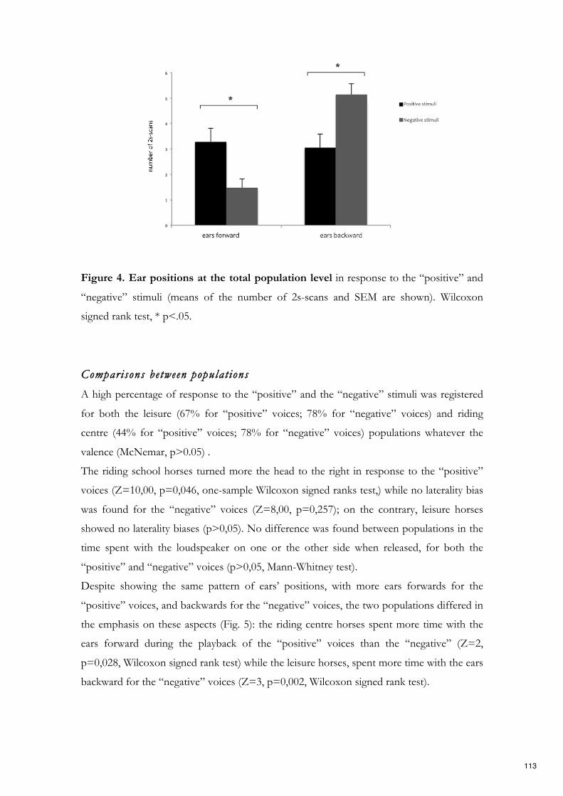
Figure 4. Ear positions at the total population level in response to the “positive” and
“negative” stimuli (means of the number of 2s-scans and SEM are shown). Wilcoxon
signed rank test, * p<.05.
Comparisons between populat ions
A high percentage of response to the “positive” and the “negative” stimuli was registered
for both the leisure (67% for “positive” voices; 78% for “negative” voices) and riding
centre (44% for “positive” voices; 78% for “negative” voices) populations whatever the
valence (McNemar, p>0.05) .
The riding school horses turned more the head to the right in response to the “positive”
voices (Z=10,00, p=0,046, one-sample Wilcoxon signed ranks test,) while no laterality bias
was found for the “negative” voices (Z=8,00, p=0,257); on the contrary, leisure horses
showed no laterality biases (p>0,05). No difference was found between populations in the
time spent with the loudspeaker on one or the other side when released, for both the
“positive” and “negative” voices (p>0,05, Mann-Whitney test).
Despite showing the same pattern of ears’ positions, with more ears forwards for the
“positive” voices, and backwards for the “negative” voices, the two populations differed in
the emphasis on these aspects (Fig. 5): the riding centre horses spent more time with the
ears forward during the playback of the “positive” voices than the “negative” (Z=2,
p=0,028, Wilcoxon signed rank test) while the leisure horses, spent more time with the ears
backward for the “negative” voices (Z=3, p=0,002, Wilcoxon signed rank test).
113

Overall, horses of both populations showed the same high level of interest toward both the
“positive” and “negative” stimuli, displaying at least one of the behaviours included in
visual attention category (RC: 80% of 10 subjects for the “positive” stimuli and 90% for
the “negative” stimuli; L: 82% of 11 subjects for both “positive” and “negative” stimuli).
As for frustration behaviour, the statistical analysis revealed that horses living in the riding
centre showed more frustration-related behaviour than the leisure horses in response to the
“negative” stimuli (U=95,5, p=0,003, Mann-Whitney test); no such differences were found
in response to the “positive” stimuli. When the “negative behaviours” (i.e. vigilance and
frustration categories) were pooled, it has been found a significant differences between
populations in response to the “negative” voices; in particular, riding centre horses
displayed more negative behaviour than leisure horses when hearing the “negative” voices
(U=26, p=0,043, Mann-Whitney test). No differences between the populations were found
for the “positive” voices (p>0,05, Mann-Whitney test).
Figure 5. Ears’ positions in the two populations. Ear positions in response to the
“positive” and “negative” stimuli in the Riding Centre and Leisure horses (mean number of
scans and SEM). Wilcoxon signed rank test, * p<.05.
114
Physiological results
Cardiac ac t iv i ty
Statistical analysis of the cardiac activity revealed that the AUC values (i.e. the area under
the heart rate curve and the baseline) were higher than the AAC values (i.e. the area under
the baseline and above the heart rate curve) for both the “positive” and “negative” stimuli
at the entire population level (positive: Z=1, p=0,017; negative: Z=0, p=0,005, Wilcoxon
signed rank test). Therefore, both the “positive” and the “negative” voices induced a higher
heart rate than the basal average in the overall time.
By contrast, no statistically significant difference was found between the “positive” and
“negative” stimuli for both the AUC and AAC values (p>0,05, Wilcoxon signed rank test).
When comparing the two populations, no statistical significant differences between AUC
and AAC were found for both the “positive” and “negative” voices (p>0,05, Mann-
Whitney test).
Electroencephalography (EEG)
Results for the EEG measurements showed that, at the total population level, horses
reacted differently to the two types of acoustic stimuli presented. Specifically, there was a
statistically significant difference in the relative power of the waves frequencies in response
to the “positive” voices (X2 (9)=20,96, p=0,013, Friedman test), with a clear increase of
gamma waves in the right hemisphere, significantly more than delta frequency bands (Z=-
2,22, p=0,026, Wilcoxon signed rank test)(Fig. 5). No statistically significant differences in
the wave proportions were found in the left hemisphere (p>0,05) and no laterality biases
were observed for the “negative” voices (Friedman test: p>0,05) (Fig. 6).
When comparing EEG profiles in response to the “positive” and “negative” stimuli, a
negative and statistically significant correlation was found for gamma waves and theta
waves in the right hemisphere (Spearman correlations: gamma positive and gamma
negative: r=-0,559, p= 0,024; theta positive and theta negative: r=-0,538, p=0,031), while a
positive correlation between alpha waves was observed in the left hemisphere (Spearman
correlation: r= 0,549, p=0,034). Nevertheless, no statistically significant differences in
115

wave proportions were found between the “positive” and the “negative” acoustic stimuli
(p >0,05 Wilcoxon signed rank test).
Figure 6. Wave median proportions of the total population in the right and in the left
hemisphere in response to the “positive” stimuli. Wilcoxon signed rank test, * p<.05.
(explanations can be found in the main text).
Discussion
Overall, our results revealed that horses’ perception of human voices is modulated by the
positive or negative valence of the previous experiences with humans.
Horses responded with an opposite pattern of their ears’ position for the “positive” and
“negative” human voices. They held their ears forward for longer in response to the
“positive” voices while they oriented them more backward when hearing the “negative”
voices. Ears’ positions are considered reliable indices of individual’s emotional state and
116
they are often used to evaluate the animal’s general perception of a situation or of an
interaction with both conspecifics and humans (Waring, 2003; Hauberger & Muller, 2002;
Fureix et al., 2009). In particular, the backward position is a cue of a negative perception of
a situation or of an interaction, since it is displayed during agonistic encounters and it is
generally associated with discomfort (stress or pain) (Waring, 2003; Fureix et al., 2012). The
forward position, instead, is indicative of a positive perception of a situation or interaction
and it promotes more approach/interest behaviour toward other conspecifics (Waring,
2003). Thus, the different ears’ positions suggest that horses attributed a different valence
to human voices according to the type of prior horse-human interactions.
Moreover, results from the perceptual laterality measures showed a different side bias for
the head-turning and for the eye preferential use in response to the “positive” and
“negative” voices. Horses consistently turned their head to the right side when hearing the
voices associated with the positive experience. Given that the auditory information is
processed in the hemisphere contralateral to the side of the head turning in horses (Basile
et al., 2009), the asymmetry manifested in the head orienting response suggests that the left
hemisphere main involvement for the initial attention and primarily processing of the
“positive” voices. This finding is consistent with the left hemisphere involvement in top-
down retrieval of memories and details associated with specific individuals (Rhodes, 1985)
and with its specializations for feeding response and detection of food (Rogers, 2002). In
addition, the left hemisphere has a complementary specialization for pro-social behaviour
and for the approach motivation, taking charge of the response to stimuli regarded as
positive (Rogers, 2010).
Thus, it is entirely possible that human voices associated with a previous positive
experience (namely food) could have elicited horses selective attention to search for food
and their willingness to approach the human producing the vocalizations to obtain it;
indeed, a few of the horses tried to approach the loudspeaker after hearing the sound.
On the contrary, when considering the laterality bias for visual inspection of stimuli,
although horses showed a clear preference to use their monocular view to investigate both
the “positive” and “negative” stimuli, they spent more time with the loudspeaker on their
left side (i.e. left eye) in response to the “negative” stimuli. Given that the visual inputs are
primarily processed by the opposite hemisphere (Rogers 2010), as described for the
auditory domain, the left bias observed during the entire stimuli presentation suggests a
117

right hemisphere main involvement in the analysis of the “negative” voices. The prevalent
activation of the right hemisphere in the processing of these sounds is consistent with the
right neural structures specializations for the perception of highly arousal stimuli, for the
expression of intense emotions and the control of rapid responses (Rogers, Vallortigara, &
Andrew, 2013). Specifically, in horses the right hemisphere activation has been reported in
the visual analysis of potentially fear-inducing stimuli, such as novel objects (Larose et al.
2006), humans (Sankey et al., 2011b) and clearly negative stimuli (i.e. white coat worn by
the veterinarian, De Boyers de Roches et al., 2008). In the light of these evidences, the
horses’ left-eye preference to investigate the source of the “negative” voices (i.e. the
loudspeaker) can be related to the animal emotional assessment of the real threatening
potentiality of the stimuli and to the individual readiness to produce a rapid fight or flight
response.
Horses’ different perception of the human voices valence is further confirmed by the
electroencephalography results. The EEG spectral analysis revealed a negative correlation
of gamma and theta relative power between the “positive” and “negative” voices in the
right hemisphere. In other words, the two voices with an opposite valence produced
opposite changes in gamma and theta relative power. Oscillation of theta and gamma
bands are involved in several aspects of memory, including the information encoding of
memory-relevant objects (Siegel et al., 2009) as well as the consolidation and the retrieval
of stored memories (Düzel et al., 2010). A link between theta activity and emotional
states/regulation has been reported both in humans and in animals (Knyazev, 2007; Stomp
et al., in prep). In a recent study, an event-related synchronization of right hippocampal
theta activity has been shown in humans during a discrimination task between faces of
conspecifics or primate babies eliciting different emotions (pleasant and unpleasant
pictures) (Nishitani, 2003). Regarding gamma bands, oscillations in its power are generally
associated with high-level mental activities, such as emotions (Fitzgibbon et al., 2004) and
stimuli related sensory/cognitive functions, including the processing of specific stimuli
information (Jensen et al., 2007). Although the opposite change of gamma and theta bands
suggests a different and opposite pattern of brain processing of the “positive” and
“negative” voices, the specific role of each wave in the emotional processing and emotional
state in horses still need further investigations. On the contrary, our results show a positive
correlation of alpha bands activity between the “positive” and “negative” voices in the left
118

hemisphere. This relationship can be explained by the alpha bands involvement in memory
demands and in mental representation of objects and events, which could have occurred
for both the acoustic stimuli (Knyazev, 2007).
Interestingly, when analysing the different EEG profiles, it emerged a clear prevalence of
the gamma waves in the right hemisphere in response to the “positive” voices. It has been
previously found that food reward increases horses’ selective attention toward the human
providing it (Rochairs et al., 2014). Moreover, a high attentional state has been found to be
associated with a gamma wave proportions increase in the right hemisphere (Rochais et al.,
2018) that is also involved in the identification of possible target of horses’ attention
(Andrew and Watkins, 2002). In addition, sustained EEG gamma oscillations have been
reported to occur during an expectancy cognitive task in humans (Fitzgibbon et al., 2004).
Thus, the gamma prevalence in the right hemisphere described here could be explained by
the animals’ higher level of expectancy to receive food when hearing the “positive” voices
compared to the “negative” stimuli; and it is further confirmed by the negative correlation
between the gamma proportions recorded for the two stimuli.
In the light of the EEG and behavioural results, we could hypothesize that horses
perceived the “positive” voices as having a positive valence, as suggested by the left
hemisphere involvement in the primarily processing of such stimuli and by the forward
ears’ position; consequently, the memory of the positive interactions with humans could
have elicited an increase in animals’ attentional state toward human voices, producing
positive expectation to obtain food, as suggested by the increased gamma power in the
right hemisphere. Therefore, the left hemisphere fist involvement followed by the right
hemisphere activation could indicate a different cognitive process for the perception and
integration of the acoustic “positive” signals.
Overall, our findings further confirm that horses are profoundly affected by the valence of
prior interactions with humans and by the type of reinforcement used during training.
As for the cardiac activity, our results showed an increase of horses’ heart rate for both the
“positive” and “negative” voices during the total time of stimuli presentation, but it was
independent from the acoustic stimuli valence. Although it has been previously reported
that stimuli perceived as clearly negative elicit an increase of subject’s arousal due to the
activation of the hypothalamic-pituitary-adrenal (HPA) axis (a,b et al., 2015, 2016, 2017,
2018a,b), the lack of such increase in response to the “negative” voices suggests that horses
119

perceived the negative event associated with the human voice as mildly aversive. This
hypothesis is supported by the absence of alarm behaviours and clear withdrawal responses
in response to the “negative” voices, as well as by the similar level of vigilance displayed
when hearing the “positive” acoustic stimuli. Therefore, the negative event associated with
the human voice could not have represented a real threat for the horses.
When comparing the two populations, our results highlight some differences in the horses’
response to the acoustic stimuli according to the population they belonged to. Riding
centre horses appeared to be more sensitive than leisure horses to the different valence of
the prior experience associated with human voices. The acoustic stimuli, indeed, induced
two opposite reactions in riding centre horses, which consistently turned their head to the
right (left hemisphere activation) and held their ears forward in response to the “positive”
stimuli; whereas they displayed more frustration-related behaviour when hearing the
“negative” voices. Leisure horses, instead, appeared to be more sensitive to the negative
valence of the human voices, since they spent more time with the ears backward in
response to the “negative” acoustic stimuli. It has been demonstrated that housing
conditions impact horses emotionality. In particular, social environmental conditions affect
horses behaviour, physiology and reaction to challenging situations (Rivera et al., 2002).
Horses stabled individually show a greater reactivity to novel objects (Lesimple et al., 2011)
and a higher sensibility to social stress (Christensen et al., 2002) than horses stabled in
groups. Moreover, they tend to display more stress related behaviour (i.e. chewing and
licking the wall) than pastured horses (Heleski et al., 2002). The higher level of frustration
showed by riding centre horses compared to the leisure horses, which also perceived this
stimuli as negative (backward ears’ position), can be therefore explained by their general
higher sensibility to the stress due to their housing conditions. Another possible
explanation for the different perception of the human voices valence can be related to the
different food motivation of the two populations. The use of food as reinforcement during
working activities for the riding centre horses could have impacted on the general
significance of food, increasing horses’ expectations to obtain food from humans who
interacted with them using food. This could explain the stronger reaction registered in
response to the “positive” voices with respect to leisure horses, as well as the higher
frustration displayed in response to the “negative” voices, probably caused by the
impossibility of obtaining the food reward that was instead “negative” food (soaked with
120

vinegar).
Overall, our results demonstrate that horses not only associate human voices with the type
of previous experiences they had with them, but they also recall the valence of such
experiences when hearing human voices. In particular, the valence of previous interactions
can influence horses future attitude to interact with humans. Our results demonstrate that
positive interactions produce positive expectations and a positive attitude to interact with
humans, promoting attention and approaching behaviour. On the contrary, negative
experiences cause negative affective states and therefore negative expectations about the
forthcoming interactions. Furthermore, our results highlight some differences in the horses’
response to the acoustic stimuli according to the population they belonged to, with the
riding centre horses being more sensitive to the different valence (positive and negative) of
the prior experience associated with human voices and with leisure horses being more
sensitive to the negative valence of the human voices.
Our study firstly combines behavioural and physiological measures (i.e. cardiac activity)
with electroencephalography in fully awake animals to study horses perception of human-
related emotional stimuli, constituting a promising new approach to investigate horses’
brain emotional functioning.
121

References
Andrew, R. J., & Watkins, J. A. S. (2002). Evidence for cerebral lateralization from senses
other than vision. Comparative vertebrate lateralization, 365-382.
Austin, N. P., & Rogers, L. J. (2007). Asymmetry of flight and escape turning responses in
horses. Laterality, 12(5), 464-474.
Austin, N.P., & Rogers, L. J. (2014). Lateralization of agonistic and vigilance responses in
Przewalski horses (Equus przewalskii). Applied Animal Behaviour Science, 151, 43-50.
Basile, M., Boivin, S., Boutin, A., Blois-Heulin, C., Hausberger, M., & Lemasson, A. (2009).
Socially dependent auditory laterality in domestic horses (Equus caballus). Animal
cognition, 12(4), 611-619.
Christensen, J. W., Ladewig, J., Søndergaard, E., & Malmkvist, J. (2002). Effects of
individual versus group stabling on social behaviour in domestic stallions. Applied Animal
Behaviour Science, 75(3), 233-248.
Cousillas, H., Oger, M., Rochais, C., Pettoello, C., Ménoret, M., Henry, S., & Hausberger,
M. (2017). An Ambulatory Electroencephalography System for Freely Moving Horses: An
Innovating Approach. Frontiers in veterinary science, 4, 57.
De Boyer des Roches, A. D. B., Richard-Yris, M. A., Henry, S., Ezzaouïa, M., &
Hausberger, M. (2008). Laterality and emotions: visual laterality in the domestic horse
(Equus caballus) differs with objects' emotional value. Physiology & Behavior, 94(3), 487-
490.
Dharmaretnam, M., & Rogers, L. J. (2005). Hemispheric specialization and dual processing
in strongly versus weakly lateralized chicks. Behavioural brain research, 162(1), 62-70.
122

Düzel, E., Penny, W. D., & Burgess, N. (2010). Brain oscillations and memory. Current
opinion in neurobiology, 20(2), 143-149.
Fitzgibbon, S. P., Pope, K. J., Mackenzie, L., Clark, C. R., & Willoughby, J. O. (2004).
Cognitive tasks augment gamma EEG power. Clinical Neurophysiology, 115(8), 1802-1809.
Fureix, C., Jego, P., Sankey, C., & Hausberger, M. (2009). How horses (Equus caballus) see
the world: humans as significant “objects”. Animal Cognition, 12(4), 643-654.
Hall, C., Randle, H., Pearson, G., Preshaw, L., & Waran, N. (2018). Assessing equine
emotional state. Applied Animal Behaviour Science, 205, 183-193.
Harman, A. M., Moore, S., Hoskins, R., & Keller, P. (1999). Horse vision and an
explanation for the visual behaviour originally explained by the ‘ramp retina’. Equine
veterinary journal, 31(5), 384-390.
Hausberger, M., & Muller, C. (2002). A brief note on some possible factors involved in the
reactions of horses to humans. Applied Animal Behaviour Science, 76(4), 339-344.
Hausberger, M., Fureix, C., & Lesimple, C. (2016). Detecting horses’ sickness: in search of
visible signs. Applied Animal Behaviour Science, 175, 41-49.
Heleski, C. R., Shelle, A. C., Nielsen, B. D., & Zanella, A. J. (2002). Influence of housing
on weanling horse behavior and subsequent welfare. Applied Animal Behaviour Science,
78(2-4), 291-302.
Henry, S., Fureix, C., Rowberry, R., Bateson, M., & Hausberger, M. (2017). Do horses with
poor welfare show ‘pessimistic’cognitive biases?. The Science of Nature, 104(1-2), 8.
Hook-Costigan, M A., & Rogers, L. J. (1998). Eye preferences in common marmosets
(Callithrix jacchus): Influence of age, stimulus, and hand preference. Laterality:
Asymmetries of Body, Brain and Cognition, 3(2), 109-130.
123

Jensen, O., Kaiser, J., & Lachaux, J. P. (2007). Human gamma-frequency oscillations
associated with attention and memory. Trends in neurosciences, 30(7), 317-324.
Knyazev, G. G. (2007). Motivation, emotion, and their inhibitory control mirrored in brain
oscillations. Neuroscience & Biobehavioral Reviews, 31(3), 377-395.
Krueger, K., Flauger, B., Farmer, K. & Maros, K. 2011 Horses (Equus caballus) use human
local enhancement cues and adjust to human attention. Anim. Cogn. 14, 187–201.
Lampe, J. F., & Andre, J. (2012). Cross-modal recognition of human individuals in
domestic horses (Equus caballus). Animal cognition, 15(4), 623-630.
Larose, C., Richard-Yris, M. A., Hausberger, M., & Rogers, L. J. (2006). Laterality of horses
associated with emotionality in novel situations. Laterality, 11(4), 355-367.
Leliveld, L. M., Langbein, J., & Puppe, B. (2013). The emergence of emotional
lateralization: evidence in non-human vertebrates and implications for farm
animals. Applied Animal Behaviour Science, 145, 1-14.
Lesimple C, Fureix C, Menguy H, Hausberger M (2010) Human Direct Actions May Alter
Animal Welfare, a Study on Horses (Equus caballus). PLoS ONE 5(4): e10257.
Lesimple, C., Fureix, C., LeScolan, N., Richard-Yris, M. A., & Hausberger, M. (2011).
Housing conditions and breed are associated with emotionality and cognitive abilities in
riding school horses. Applied Animal Behaviour Science, 129(2-4), 92-99.
Lippolis, G., Bisazza, A., Rogers, L.J., & Vallortigara, G. (2002). Lateralization of predator
avoidance responses in three species of toads. Laterality, 7, 163–183.
McDonnell, S. M., & Haviland, J. C. S. (1995). Agonistic ethogram of the equid bachelor
band. Applied Animal Behaviour Science, 43(3), 147-188.
124

Nishitani, N. (2003). Dynamics of cognitive processing in the human hippocampus by
neuromagnetic and neurochemical assessments. Neuroimage 20, 561–571.
Proops, L., & McComb, K. (2012). Cross-modal individual recognition in domestic horses
(Equus caballus) extends to familiar humans. Proceedings of the Royal Society of London
B: Biological Sciences, rspb20120626.
Rhodes, G. (1985). Lateralized processes in face recognition. British journal of Psychology,
76(2), 249-271.
Rivera, E., Benjamin, S., Nielsen, B., Shelle, J., & Zanella, A. J. (2002). Behavioral and
physiological responses of horses to initial training: the comparison between pastured
versus stalled horses. Applied animal behaviour science, 78(2-4), 235-252.
Rochais, C., Henry, S., Sankey, C., Nassur, F., Gorecka-Bruzda, A., & Hausberger, M.
(2014). Visual attention, an indicator of human-animal relationships? A study of domestic
horses (Equus caballus). Frontiers in psychology, 5, 108.
Rochais, C., Fureix, C., Lesimple, C., & Hausberger, M. (2016). Lower attention to daily
environment: a novel cue for detecting chronic horses’ back pain?. Scientific reports, 6,
20117.
Rochais, C., Sébilleau, M., Ménoret, M., Oger, M., Henry, S., Hausberger, M., & Cousillas,
H. (2018). Attentional state and brain processes: state-dependent lateralization of EEG
profiles in horses. Scientific reports, 8(1), 10153.
Rogers, L. J. (2002). Lateralization in vertebrates: its early evolution, general pattern, and
development. In Advances in the Study of Behavior (Vol. 31, pp. 107-161). Academic
Press.
125

Rogers, L. J., Vallortigara, G., & Andrew, R. J. (2013). Divided brains: The biology and
behaviour of brain asymmetries. Cambridge, UK: Cambridge University Press.
Rogers, L. J., & Vallortigara, G. (2017). Lateralized Brain Functions- Methods in Human
and Non-Human Species. Humana Press, New York, USA. ISBN: 978-1-4939-6723-0
Rogers, L. J. Eye and Ear Preferences in Rogers, L. J., & Vallortigara, G. (2017) Lateralized
Brain Functions- Methods in Human and Non-Human Species. Humana Press, New York,
USA, pp. 79-102. ISBN: 978-1-4939-6723-0
Sankey, C., Henry, S., André, N., Richard-Yris, M. A., & Hausberger, M. (2011a). Do
horses have a concept of person?. Plos One, 6(3), e18331.
Sankey, C., Henry, S., Clouard, C., Richard-Yris, M. A., & Hausberger, M. (2011b).
Asymmetry of behavioral responses to a human approach in young naive vs. trained horses.
Physiology & behavior, 104(3), 464-468.
Sankey, C., Richard-Yris, M. A., Leroy, H., Henry, S., & Hausberger, M. (2010a). Positive
interactions lead to lasting positive memories in horses, Equus caballus. Animal
Behaviour, 79(4), 869-875.
Sankey, C., Richard-Yris, M. A., Henry, S., Fureix, C., Nassur, F., & Hausberger, M.
(2010b). Reinforcement as a mediator of the perception of humans by horses (Equus
caballus). Animal cognition, 13(5), 753-764.
Siegel, M., Warden, M. R., & Miller, E. K. (2009). Phase-dependent neuronal coding of
objects in short-term memory. Proceedings of the National Academy of Sciences, 106(50),
21341-21346.
Siniscalchi, M., Padalino, B., Aubé, L., & Quaranta, A. (2015). Right-nostril use during
sniffing at arousing stimuli produces higher cardiac activity in jumper horses. Laterality:
Asymmetries of Body, Brain and Cognition, 20(4), 483-500.
126
Siniscalchi, M., d’Ingeo, S., & Quaranta, A. (2016). The dog nose “KNOWS” fear:
Asymmetric nostril use during sniffing at canine and human emotional stimuli. Behavioural
brain research, 304, 34-41.
Siniscalchi, M., d’Ingeo, S., & Quaranta, A. (2018a). Orienting asymmetries and
physiological reactivity in dogs’ response to human emotional faces. Learning & Behavior,
1-12.
Siniscalchi, M., d’Ingeo, S., Fornelli, S. & Quaranta A. (2018b). Lateralized behavior and
cardiac activity of dogs in response to human emotional vocalizations. Scientific Reports,
8:77.
Smith, A. V., Proops, L., Grounds, K., Wathan, J., & McComb, K. (2016). Functionally
relevant responses to human facial expressions of emotion in the domestic horse (Equus
caballus). Biology letters, 12(2), 20150907.
Smith, A. V., Proops, L., Grounds, K., Wathan, J., Scott, S. K., & McComb, K. (2018).
Domestic horses (Equus caballus) discriminate between negative and positive human
nonverbal vocalisations. Scientific reports, 8(1), 13052.
Stomp, M., Leroux, M., Cellier, M., Henry, S., Lemasson, A., & Hausberger, M. (2018). An
unexpected acoustic indicator of positive emotions in horses. PloS one, 13(7), e0197898.
Stone, S. M. (2010). Human facial discrimination in horses: can they tell us apart?. Animal
cognition, 13(1), 51-61.
Tallet, C., Rakotomahandry, M., Guérin, C., Lemasson, A., & Hausberger, M. (2016).
Postnatal auditory preferences in piglets differ according to maternal emotional experience
with the same sounds during gestation. Scientific reports, 6.
Waring, G. H. (2003). Horse Behavior, Norwich. NY: Noyes Publications/William Andrew
Pub.
127

Chapter 7
Discussion
The assessment of animals’ welfare cannot disregard the careful evaluation of animals’
feelings. The study of emotional perception and processing is vital for evaluating how
environmental and social stimuli can impact animals’ well-being.
The integrated approach proposed in this thesis project allows an accurate description of
animals’ emotions along the valence and intensity dimensions. In particular, information
about the valence that an individual attributes to an emotional stimulus has been obtained
by the evaluation of brain asymmetrical processing of emotional stimuli, measured both
directly by the EEG technique and indirectly through lateralized behaviours. On the other
hand, the intensity of an affective experience has been evaluated measuring both
behavioural manifestations of vigilance and stress and the cardiac activity increase (in terms
of heart rate), which are mediated by the sympathetic nervous system activation that
indicates subjects’ arousal increase. Information about the arousal dimension of the
emotional processing can be further provided by the EEG technique, by evaluating, for
instance, the overall activation of the frontal regions or of the right parieto-temporal region
in response to emotional stimuli, as previously described in human literature (Heller, 1993;
Dawson et al., 1992). Nevertheless, further studies are necessary to investigate the
relationship between the different brain regions activation and the modulation of
autonomic arousal in animals.
Overall, the results of this thesis project demonstrate that dogs and horses process
differently human (and conspecific) emotional signals according to their valence and
intensity.
The main results of the studies carried out are summarized in the following paragraphs.
128

7.1 Horses
Horses’ perception of a human voice is modulated by the valence of the prior horse-human
interactions. Positive experiences produced expectancies of positive outcomes (left
hemisphere activation and ears forward) and resulted in horses’ positive attitude to interact
with humans, promoting attention (gamma oscillations in the right hemisphere) and
approaching behaviour. On the other hand, negative experiences caused negative affective
states (right hemisphere activation and ears backwards) and produced negative
expectancies about horse-human future interactions, potentially affecting horses welfare.
Furthermore, results highlight some differences in the horses’ response to the acoustic
stimuli according to the population they belonged to and consequently to their living
conditions. Riding centre horses appeared more sensitive to the different valence (positive
and negative) of the prior experience associated with human voices, whereas leisure horses
appeared more sensitive to the negative valence of the human voices. These findings
suggest that life and welfare conditions could affect animals’ perception of human signals.
Although recent studies have reported that horses distinguish the valence of human non-
verbal emotional vocalization of anger and happiness (Smith et al., 2018), it could be
interesting to evaluate their reactions along the arousal dimension and to study the changes
in their brain activity, in order to characterize more accurately horses’ perception of human
emotional vocalizations.
7.2 Dogs
Dogs perception and processing of human (and conspecific) emotions has been analysed in
three different studies, in which dogs lateralized response, behavioural and physiological
changes elicited by olfactory, auditory and visual emotional signals have been evaluated.
In study 1, an asymmetry in dogs’ nostril use during sniffing at different human and
conspecific emotional stimuli has been found, suggesting that dogs could detect the
emotional state of a member of their social group through olfaction. In particular, dogs
consistently used their right nostril (right hemisphere) to sniff other conspecifics’ odours
collected during a stressful event (i.e. separation from its owner, “dog isolation” stimuli).
These odours induced a state of increased arousal in dogs, which showed a cardiac activity
increase and stress/alerting behaviour. These findings are consistent with the general
specialization of the right hemisphere in processing and in the expression of intense
129

Emotional stimuli
Hemispheric bias Cardiac activity Stress levels
Odours
Isolation Right High increase High
Disturb No bias High increase Moderate
Play No bias Moderate increase Moderate
Schematic representation of study 1 results about dog odours.
emotions mediated by the sympathetic nervous system, which activates an acute response
to stressors (i.e. emergency response), by increasing subjects’ cardiac activity, stress and
vigilance levels (Rogers et al., 2013). Therefore, these results suggest that dogs perceived
the conspecific stress odours as negative and intense stimuli. In a functional perspective,
the odour of stress could inform other conspecifics about the presence of a threat or a
danger in the place where it is released, producing physiological changes in the receivers
that prepare them to respond rapidly and adequately to the threat. Moreover, the high
arousal and stress levels registered in the receivers could indicate that an emotional
contagion between conspecifics had occurred.
On the contrary, dogs showed a left nostril (left hemisphere) bias when sniffing human
odours collected during a fearful and a physically stressing event (i.e. after a run). The left
hemisphere involvement in processing these emotional odours could be related to the
activation of the left amygdala, which is specialized in a fine-tuned and detailed assessment
of danger (Morris et al., 1999; Glascher & Adolphs, 2003). Alternatively, it could be related
to the left hemisphere specialization for predatory behaviour (Rogers & Andrew, 2002;
Siniscalchi et al., 2013). Indeed, human fear and physical stress signals could have triggered
dogs’ prey drive, eliciting dogs’ motivation to approach the stimulus (to find, pursue and
capture the prey), which is under the left hemisphere control (Harmon-Jones & Allen
observations (1997, 1998). This hypothesis is further supported by the left hemisphere
main role in sustaining subject’s attention and in risk taking, allowing sustained inspection
of potential sources of danger and inhibiting a fast and emotive response mediated by the
right hemisphere activation (Rogers et al., 2013). In fact, these functions are fundamental
for a successful pursuit and capture of prey. Moreover, the increased vigilance (targeting
130

Emotional stimuli
Hemispheric bias Cardiac activity Stress levels
Odours
Fear Left High increase High
Running Left High increase Moderate
Joy No bias High increase Moderate
Vocalizations
Fear Right High increase High
Sadness Right Moderate increase Moderate
Anger No bias High increase High
Disgust No bias High increase Low
Surprise No bias High increase Low
Happiness Left Moderate increase Moderate
Faces
Fear Right High increase Moderate
Sadness No bias Moderate increase Moderate
Anger Right High increase High
Disgust No bias Moderate increase Moderate
Surprise Left Moderate increase Low
Happiness Right High increase High
Schematic representation of the results about human emotional stimuli.
behaviour) displayed by dogs in response to human physical stress odours and fear could
further support the hypothesis that these stimuli triggered dogs’ prey drive behaviour since
they are generally displayed during the prey drive sequence (Handelman, 2012). One of the
communicative functions of fear expression is “to signal the presence of some threat other
than the expresser, perhaps alerting the perceiver to danger in the environment” (Marsh et
131

al., 2005). Thus, fearful animals could release specific signals in the environment through
their body odours to alert other conspecifics about the presence of a threat, for instance,
the presence of a predator. On the other hand, the predator can use the prey odour track
to pursue it. In other words, the fearful prey odours activate the predator’s prey drive and
appetitive motivation, eliciting approaching behaviours toward the prey. This hypothesis
could explain the dogs’ left hemisphere involvement in processing human “fear” and
“physical stress” odours since it controls both the predatory and approach-related
behaviours as well as dogs high level of vigilance (Siniscalchi et al., 2013c; Rogers et al.,
2013).
Interestingly, results show that contrarily to human physical stress odours, the odours
collected during a fearful event elicited stress behaviour in dogs. One possible explanation
is that the human fear odours have triggered similar emotions and behaviour in the dogs,
producing an emotional contagion between these species. This hypothesis is supported by
the left hemisphere activation, which is specialized for pro-social, affiliative and
approaching behaviour (Rogers, 2010). Although aversive, fear emotion has been described
as an appetitive stimulus, which elicits approach in the receiver, encouraging the formation
of social bonds (Marsh et al., 2005). Despite being described in the literature (Custance &
Mayer, 2012), further studies are needed to determine if the emotional contagion could
occur between humans and dogs.
Although “dog disturb”, “dog play” and “human joy” stimuli elicited a cardiac activity
increase and moderate stress levels in dogs, any bias in the nostril use was observed. This
finding suggests a balanced activity of the right and left hemisphere for the perception of
these emotional odours during the time. Moreover, the lack of hemispheric bias could
indicate the need of integratinge the olfactory information with inputs coming from other
sensory modalities for a more precise emotional recognition. For instance, in play
interactions, dogs use specific signals, known as metasignals, to impart information about
how other signals should be interpreted, indicating, for example, that a threat signal is not
true hostility but has to be taken as a play (Handelman, 2012).
In study 2, results demonstrate that dogs process asymmetrically human non-verbal
emotional vocalizations according to their emotional content. In particular, a left bias in the
head orienting response (right hemisphere activation; Uemura, 2015) has been found in
response to “fear” and “sadness” human vocalizations, which elicited the increase of
132

arousal levels in tested subjects (cardiac activity and stress behaviour). The right
hemisphere involvement in processing fear vocalizations is consistent with previous human
lesional studies, reporting a significant impairment of subjects affected by right amygdala,
hippocampus and temporal lesions in the perception of the emotional content of fear
vocalizations (Phillips et al., 1998; Pourtois et al., 2005; Milesi et al., 2014). Considering the
right hemisphere specialization for processing intense emotions, and particularly the
negative ones (Siniscalchi et al., 2008, 2010, 2013b), and the increase in cardiac activity and
stress level registered, it seems reasonable to conclude that dogs perceived that “fear” and
“sadness” vocalizations express a negative emotion. Nevertheless, “fear” and “sadness”
vocalizations differed in terms of stress levels elicited in the receiver that was higher in
response to “fear” vocalizations. In fact, “fear” and “sadness” vocalizations have different
communicative and functional roles. Although both stimuli have been generally classified
as aversive (Marsh et al., 2005), “fear” vocalizations could elicit stronger reactions in the
receiver (higher arousal) that can result in different and opposed motivations (approach or
withdrawal), depending on the social context in which they are produced. For instance, the
fear emotion could elicit a withdrawal and avoidance response in the receiver if it is
referred to an environmental danger (i.e. predator); on the contrary, in social interactions
fearful individuals generally display subordination behaviours to appear non-threatening
and appeasing, eliciting approach rather than avoidance in the receiver (Schenkel, 1967;
Marsh et al., 2005). Regarding sadness, it has been defined as a moderately intense
unpleasant feeling (Lazarus, 2001; Russell & Feldman Barrett, 1999). The right hemisphere
main activation for processing these emotional vocalizations and subjects’ arousal
moderate increase here found are consistent with the general definition of this emotion and
suggest that dogs perceived these calls as non-threatening and alarming. Moreover, sadness
has been found to elicit approach in the receiver, which experiences the same feeling of the
senders and consequently acts empathically approaching them (Vrijsen et al., 2013). Indeed,
previous studies suggest that sadness elicits not only the desire for affiliation, but also
emphatic behaviour in the receiver (Batson et al., 1981). Therefore, dogs’ perception of the
“negative” emotional content of sadness vocalizations and their moderate arousal increase
in response to these vocalizations could suggest the occurrence of the emotional contagion
between human and dogs, as previously described by Custance and Mayer (2012).
Nevertheless, this hypothesis needs further support and further studies are necessary to
133

investigate this issue.
Dogs’ clear tendency to turn their head to their left side (right hemisphere activation,
Uemura 2015) has been observed in response to “anger” vocalizations, but it did not reach
statistical significance. One possible explanation is that these sounds have similar acoustic
characteristics of the conspecific threatening growl (harsh, low-frequency call), which
conveys the same emotional message and negative valence of human “anger” (Handelman,
2012). However, conspecific vocalizations are mainly processed by the left hemisphere
(Siniscalchi et al., 2008) and, in particular, by a left dorsal auditory region, which is
specifically sensitive to conspecific vocalizations (Andics et al., 2014). Therefore, some
subjects could have misinterpreted the “anger” vocalizations classifying them as
conspecific calls, and, as a result, it caused a weaker left orienting bias at the total sample
level. Another possible explanation for the left hemisphere involvement in processing
“anger” vocalizations could be related to the left hemisphere important role in the threat
assessment and in selecting an adequate response (Rogers et al., 2013). Results from the
arousal dimension demonstrate, indeed, that dogs perceived human “anger” vocalizations
as strongly intense and alarming (lowest reactivity and highest stress, cardiac activity and
time needed to resume feeding), suggesting that it could be evaluated as a potential threat.
This hypothesis is supported by dogs tendency to avoid eye contact when facing a human
angry face and by the left gaze bias (right hemisphere) shown while looking at human angry
faces (Barber et al., 2016; Racca et al., 2012). Dogs could have initially and rapidly
processed these vocalizations with the right hemisphere, which caused subjects’ arousal
increase. The right hemisphere activation is associated, indeed, with the expression of
intense emotions mediated by an intense autonomic activity (Lane & Jennings, 1995),
producing a rapid fight or flight response. However, considering that both the approach
(i.e. fight) and withdrawal (i.e. flight) responses cause a high expenditure of energy, the left
hemisphere activation and mediation is necessary for correctly evaluating the real potential
of a threat and for selecting an appropriate response. Therefore, the left hemisphere
activity could have been prevalent in some individuals during the experiment in order to
control the emotional response mediated by the right hemisphere, since the threat was not
real in the experimental context (physical absence of the sender).
Regarding the “happiness” vocalizations, dogs showed a clear right bias in their head
orienting response (left hemisphere activation) and moderate arousal levels (cardiac activity
134

and stress behaviour) that were similar to “sadness” vocalizations. These findings are
consistent with the left hemisphere involvement in processing positive human sounds
(Andics et al., 2014) and in controlling the response to positive rewarding speech (Andics
et al., 2016) in dogs. Moreover, a left hemisphere main activation was found in dogs when
seeing a positive stimulus, which elicits their approaching behaviour (i.e. the owner)
(Sinicalchi et al., 2013). Further support for the left hemisphere main role in processing
human happiness emotion comes from human lesional studies, which demonstrate that left
hemisphere lesions cause impairments in recognizing joyful prosody (Milesi et al., 2014).
Thus, the left hemisphere involvement and the low arousal level registered suggest that
dogs perceived human “happiness” vocalizations as positive stimuli. It has been recently
found, indeed, that a positive emotional state could induce a parasympathetic deactivation
(Zupan et al., 2016), supporting the increase in subjects’ arousal state here reported.
Another possible explanation for the left hemisphere activation is that, among the six
emotional non-verbal vocalizations employed in the study, the “happiness” vocalizations
are the only ones used in the naturalistic speech as they were presented to dogs. In other
words, humans potentially use more laughs to express happiness than growls to express
anger, for example. Fear, anger, sadness, disgust and surprise are more commonly
expressed with the prosodic features within a sentence and more frequently accompanied
by semantic cues. Thus, the “happiness” vocalizations could be easily and rapidly
categorized and consequently it explains the main left hemisphere involvement in
processing them. Moreover, since an increase in the arousal levels occurred in response to
“happiness” vocalizations (even though moderate), it could be hypothesized that after an
initial activation of the right hemisphere, which caused the arousal increase, the left
hemisphere assumed control interrupting subjects’ emotional response and further
assessment by the right hemisphere since the “happiness” stimulus is already classified as
non-aversive.
Finally, dogs showed no bias in the head-turning response and moderate arousal levels
when hearing the “disgust” and “surprise” vocalizations. These findings are consistent with
human lesional studies reporting a lack of lateralization in processing these emotional
stimuli (disgust: Phillips et al., 1997; Calder et al., 2001; Jabbi et al., 2007; surprise:
Dellacherie et al., 2011). The absence of a specific brain region involvement in processing
“surprise” and “disgust” vocalization could be explained by the ambiguous valence that
135
these emotions have for dogs, which depends on the context of their production and on a
single subject’s past experiences (Maguire et al., 2011; Turcsán et al., 2015). Objects or
situations eliciting a “disgust” emotion in the owner could be attractive for the dog (e.g.
faces) or, on the contrary, could be associated with a negative outcome (e.g. scolding).
Concerning surprise, instead, evidence from human studies reported that it could be
perceived as both positive and negative, depending on the goal conduciveness of the
surprising event (Maguire et al., 2011).
In study 3, results demonstrate that human emotional faces are processed asymmetrically in
dogs brain and elicit different arousal levels according to the emotional message conveyed.
In particular, a right hemispheric-dominant activity (left head-turning response) has been
found in response to “anger”, “fear” and “happiness” emotional faces. These findings are
consistent with evidence from human studies demonstrating a higher level of accuracy and
faster responses in recognizing human anger, fear and happiness faces when presented in
the left visual hemifield, suggesting a right hemisphere main role for processing them
(Alves et al., 2009; Stafford & Brandaro, 2010). Here, the prevalent activation of the right
hemisphere in response to “anger”, “fear” and “happiness” human faces was also
associated with dogs’ arousal increase, in terms of both cardiac activity and stress behaviour.
These findings suggest that the perception of such emotional faces induces an intense
autonomic activity that allows the dogs to react rapidly to a potential danger/threat. In the
light of this evidence, it seems reasonable to conclude that dogs perceived human “anger”,
“fear” and “happiness” faces as intense and potentially threatening stimuli (negative
valence). This hypothesis is supported by the right hemisphere specialization for processing
and for the expression of intense emotions under the control of the sympathetic nervous
system in emergency situations (i.e. the presence of a threat) (Rogers et al., 2013). In dogs,
indeed, the right hemisphere activation has been reported for the analysis of visual
alarming stimuli (i.e. a cat silhouette, displaying a defensive threat posture, and a snake
silhouette, considered as an alarming stimulus for mammals; Lobue & DeLoache, 2008;
Siniscalchi et al., 2010) and stimuli that could be expected to elicit withdrawal tendencies
(tail wagging to the left in response to a dominant unfamiliar dog; Siniscalchi et al., 2013).
Regarding “happiness” faces, results suggest that dogs perceived the emotional content of
human smiling faces differently from humans, which rate this emotion as positive (e.g.
Wedding & Stalans, 1985). Although the right hemisphere involvement and the cardiac
136
activity increase indicates that dogs perceived human faces as expressing an intense
emotion regardless of its valence, the increase of stress levels suggests that human smiles
are perceived as unpleasant stimuli by dogs. One possible explanation could be found in
facial configuration similarities of human smiles and dogs angry expression. The evident
bared teeth with lips lifted and tongue retracted, which characterize human smiles, are
instead a clear message to back off in dog communication and are often followed by more
serious aggressive display or by aggression (Handelman, 2012). Therefore, in absence of
auditory information and relying only on facial configuration, dogs could have attributed a
negative valence to human happiness faces, explaining right hemisphere activation, and the
high arousal and stress levels registered. Furthermore, results show some similarities
between dog and human brain processing of human happiness visual stimuli. Evidence
from human studies reports, indeed, a right hemisphere advantage in visual joy perception
(Milesi et al., 2014; Alves et al., 2009), suggesting the presence of common mechanisms for
the perception of this emotional face in dogs and humans. Nevertheless, further studies
have produced conflicting results about human brain processing of happy faces. A left
hemisphere involvement in processing human happiness faces has been reported (Pourtois
et al., 2005), and particularly indicated by a greater activation of the left hemisphere in
response to happy facial expressions (Davidson & Fox, 1982) and a faster and more
accurate response (rated as more positive) when the happy face was presented in subjects’
right visual hemifield (i.e. left hemisphere activation; Reuter-Lorenz & Davidson, 1981;
Davidson et al., 1987). However, the right hemisphere involvement observed in both
species could be explained by the general right hemisphere dominance for face processing
regardless the emotion expressed, which was found, indeed, in both species and further
confirmed by this study results (i.e. left-left chimeric faces elicited stronger responses)(Guo
et al., 2009).
Regarding “surprise” facial expressions, dogs showed a right bias in their head orienting
response, which suggests a main left hemisphere activation in processing these stimuli. This
finding is consistent with the higher accuracy shown by humans when “surprise” faces
were presented on their right compared to their left side (Stafford & Brandaro, 2010).
Moreover, the “surprise” facial expression elicited a cardiac activity increase (arousal), but
contrarily to human “happiness” faces, subjects’ stress levels remained very low. This
indicates that “surprise” faces elicited a state of “positive” arousal in dogs. Therefore,
137

considering the left hemisphere main involvement in processing these emotional stimuli
and the “positive” arousal registered, it seems reasonable to conclude that dogs perceived
human “surprise” facial expressions as positive stimuli. One possible explanation could be
that dogs interpreted this facial expression as a relaxed expression, which typically elicits
approach. Recent studies revealed, indeed, the presence of similarities between human and
dogs joyful expression, characterized by the retraction of the mouth corner and the
dropping of the jaw that causes a slight teeth exposure (Schirmer et al., 2013; Schirmer &
Adolphs, 2017). In particular, the facial configuration of human “surprise” shows
similarities with dogs playful expression (namely “play face”), which is characterized by the
open mouth and lifted lips without evident bared teeth. In the intraspecific communication,
this expression is often associated with a positive or relaxed emotional state and it elicits
the expression of approaching behaviour (Handelman, 2012), which is under the left
hemisphere control (Rogers et al., 2013). Therefore, relying only on visual information,
dogs would have interpreted the “surprise” face as a positive stimulus. However, future
studies are needed to investigate if dogs attend to the same facial regions to recognize
human “surprise” and dog “play” face (for instance employing the eye-tracking technique).
Regarding human “sadness” and “disgust” emotional faces, no biases in dogs head-turning
response and a moderate increase of subjects’ arousal levels were observed. These findings
suggest firstly that these stimuli do no convey an intense emotional message for dogs and
secondly that subjects’ perception of human “sadness” and “disgust” faces is variable. The
lack of a consistent bias in the head-turning response could indicate that, despite being
both classified as “negative” (Marsh et al., 2005), the perception of these emotions could
be modulated by previous experiences or needs to be integrated with further input coming
from the other sensory modalities. In particular, it has been found that “sadness” faces are
mainly processed by the right hemisphere in humans (Wedding & Stalans, 1985; Sackheim
et al., 1988) but they could elicit approaching responses both in humans (Vrijsen et al.,
2013) and in dogs (see above, Custance & Mayer, 2012) mediated by the left hemisphere
activation. Therefore, the “sadness” faces could have elicited a balanced activity of two
hemispheres, resulting in the absence of a significant side bias. As discussed above about
“disgust” vocalizations, “disgust” emotional faces could have an ambiguous valence to
dogs (Turcsán et al., 2015) and their perception of such emotion could be strictly
dependent on their previous experiences.
138

Overall, the results of the three different studies show that dogs discriminate and perceive
differently the emotional content of human (and conspecific) visual, auditory and olfactory
signals. In fact, they provide complementary information about dogs perception of human
emotional stimuli through different sensory modalities that can be integrated to obtain a
general overview of dogs perception of human emotions.
Among the six basic emotions described by Ekman (1993) (i.e. anger, disgust, fear,
happiness, surprise and sadness), the emotions of anger, fear, happiness and sadness appear
to be more distinguishable and meaningful to dogs than disgust and surprise. In particular,
dogs clearly perceived the human emotion of anger, attributing a negative valence and a
high intensity to it, as demonstrated by the right hemisphere main activation and the
highest level of arousal (cardiac activity and stress behaviours) registered in response to
“anger” with respect to all the other emotions. These findings suggest that dogs perceive
angry humans as potentially threatening stimuli. Moreover, the right hemisphere
involvement in processing both the angry faces and vocalizations (just a trend) could also
suggest that this emotion could elicit dogs aversive motivation, producing withdrawal
responses, as previously reported in human studies (Marsh et al., 2005; Davidson, 1995).
Nevertheless, this hypothesis needs to be further investigated in future studies.
As far as fear is concerned, dogs attributed a clear negative valence to the fearful human
faces and vocalizations (right hemisphere activation and high arousal and stress levels),
suggesting that dogs perceive the negative affective state of the human signaller, which
could potentially elicit dogs withdrawal response. However, when sniffing human odours
collected during fearful situations, dogs preferentially used their left nostril (left hemisphere
activation) when sniffing them. The latter can be explained by the left amygdala
involvement in processing fear odours to accurately determine whether the potential threat
is real (Morris et al., 1996; Gläscher & Adolphs, 2003). Alternatively, the left hemisphere
activation for processing these emotional odours can also be related to evolutionary
mechanisms, since it has been shown that it is also involved in the control of predatory
behaviour in dogs (Siniscalchi et al., 2013c). However, fear expressions have also an
affiliative function in canines. They use stereotyped non-verbal displays of subordination to
avoid conspecifics’ aggression, communicating their affiliative and non-threatening
intentions in order to elicit approaching behaviour in the receiver (Schenkel, 1967). It has
been described in human studies that fear encourages the formation of social bonds and, as
139

other distress-related emotions like sadness or anxiety, it elicits affiliation and caregiving
from a member of the social group (Marsh et al., 2005; Schachter, 1959). Therefore, given
the long cohabitation with humans and the creation of social bonds between these two
species (Nagasawa et al., 2015; Siniscalchi et al., 2013a), it is possible that fearful human
faces and vocalizations are perceived as expressions of human negative affective state but,
contrarily to anger emotion, they could elicit dogs approaching behaviours, contributing to
the maintenance of human-dog social bonds. The lower arousal level (cardiac activity and
stress behaviours) found in response to fear with respect to anger emotion, suggests that
dogs may not perceive fearful humans as clear threatening stimuli but it indicates a more
complex mechanism for processing this emotion that could also be dependent on the
context of its production.
Although apparently conflicting, results about dogs perception of human visual, auditory
and olfactory signals of fear could be also interpreted as different aspects of the same
continuum. In fact, they elicited a similar response in tested dogs, increasing their cardiac
activity and their stress levels (i.e. their arousal). The opposite pattern of the hemispheres
activation in response to auditory and visual stimuli on one hand (right hemisphere), and to
olfactory stimuli on the other hand (left hemisphere), could be related to the analysis of
different parameters. The right hemisphere activation, indeed, is related to the dogs first
reaction to the auditory and visual stimuli, whereas the left hemisphere activation refers to
dogs nostril preferential use in the total time spent sniffing the “fear” odours. Dogs initial
and fast reaction to the emotional stimuli of fear mediated by the right hemisphere, which
controls subjects’ arousal increase and their expression of intense emotion, could be then
followed by a shift of the balance toward the left hemisphere, which analyses in detail the
real potential of a threat and selects the suitable response to the stimuli. As a result, the
animal could approach the stimulus or avoid it depending on the context of its production
(as described above).
Although human “sadness” vocalizations were mainly processed by the right hemisphere,
the absence of a significant hemispheric bias in processing human “sadness” faces suggests
that the perceptual mechanism of this emotion is complex. The right hemisphere
involvement and dogs’ arousal increase (even though moderate) demonstrate that dogs
perceive the negative emotional content of human “sadness” vocalizations. Nevertheless,
both “sadness” faces and vocalizations elicited a lower arousal level than the other two
140

negative emotions, such as anger and fear. This finding, together with dogs’ tendency to
approach (left hemisphere activation) a human displaying a “sadness” facial expression, as
reported by Custance and Mayer (2012), suggests a possible affiliative role of human
expression of sadness in the human-dog social system and for the maintenance of their
relationship. Future studies are needed to clarify the communicative role of human fear and
sadness for dogs, evaluating their emotional response and motivations to such emotions.
Concerning the emotion of happiness, previous studies reported that dogs recognize
human happiness both by looking at human faces (Muller et al., 2015) and cross-modally,
matching the auditory and visual signals expressing this emotion (Albuquerque et al., 2016).
Nevertheless, the results of this thesis project suggest that the perception and processing
mechanisms of human happiness are complex. Dogs did not attribute a clear and
unambiguous valence to this emotion. Although any hemispheric biases have been found
for happiness odours, a right hemisphere advantage and a left hemisphere involvement
have been found in processing respectively happiness faces and vocalizations. Moreover,
the visual, auditory and olfactory signals produced a different level of arousal in the
receiver, which was highest in response to “happiness” faces. The low arousal (cardiac
activity and stress behaviour) and the left hemisphere activation in processing happiness
vocalizations suggest that dogs perceive these stimuli as positive. On the contrary, the high
arousal (cardiac activity and stress behaviours) and the right hemisphere involvement in
processing “happiness” faces suggest that human happiness visual signals are perceived as
negative and potentially threatening. Dogs misinterpretation of the emotional content of
human happy faces could be related to the different communicative meaning of the
expression of evident bared teeth and lifted lips, which characterize both dogs display of
anger and human smiles. In dogs body communication, indeed, represents a clear
threatening message for requesting other individuals to back off (Handelman, 2012).
Therefore, dogs need to integrate visual signals with auditory signals in order to distinguish
more clearly this emotion. However, the inconsistency in the perceptual mechanisms here
found suggests that human happiness might constitute an ambiguous emotional signal for
dogs.
Similarly, it has been found an incoherent response to visual and auditory signals of disgust
and surprise, suggesting that these emotions have an ambiguous valence for dogs, which is
strictly dependent on individual experiences. In particular, different objects of everyday life
141

(e.g. animals’ faces) that elicit the disgust emotion in humans can be attractive for dogs,
eliciting their approaching response, or, on the contrary, they can be associated to a
negative outcome (e.g. scolding), eliciting their withdrawal response. Regarding surprise,
although results demonstrate that dogs perceived “surprised” human faces as positive
stimuli (left hemisphere activation and low-stress level) probably relying on the “relaxed”
face configuration, the inconsistent response to the “surprise” vocalizations suggests that
dogs might not have a clear and unambiguous mental representation of such emotion.
However, these findings are consistent with previous human studies reporting that the
surprise emotion could be perceived as both positive and negative, depending on the goal
conduciveness of the surprising event and on the arousal intensity expressed, as well as on
individual previous experiences (Maguire et al., 2011). Therefore, the emotional valence
attributed to these emotions may vary according to the context of its production and to the
individual experiences that affect its perception.
Overall, results demonstrate complex processing mechanisms of human emotions in dogs.
Despite have been sharing the same living environment for more than 15.000 years
(Lindblad-Toh et al., 2005), dogs and humans belong to two different species that have
their own and specific communicative signals and motivations. Dogs developed excellent
social and cognitive skills to adapt to the human environment (e.g. to interpret pointing
gestures: Soproni et al., 2002) but their perception of human signals is modulated by their
own communicative system. Therefore, dogs interpretation of human communicative
signals and their subsequent motivations may lead to misunderstanding, as described for
the happy faces or the prey-drive behaviour elicited by human “fear” odours, that must be
taken into account during human-dog interactions.
Results of this thesis project provide new insights into dogs emotional processing of
olfactory, visual and auditory stimuli, describing the unimodal perception of other
individuals’ emotions. Future studies could employ multimodal stimulation (e.g. face and
odour), which are more closely related to the emotional expression in real life (Schirmer &
Adolphs, 2017), in order to evaluate how the different sensory modalities converge for a
holistic representation of emotions in dogs.
142

Conclusions and future directions
The knowledge about animals’ perception of emotional social cues and about the
perceptual mechanisms of emotional stimuli would help to reduce negative high-arousal
stimuli and to promote, on the contrary, positive emotions in animals’ management. For
instance, Baciadonna et al. (2018) argued that positive valence and low arousing emotional
stimuli (e.g. positive emotional-linked calls) could be used to decrease the impact of
stressful events, such as veterinary practices, rehoming or transport. Moreover, the
evaluation of behavioural manifestations of cerebral lateralization provides insight into the
cognitive processing and into the valence of emotion that an animal experiences (Leliveld
et al., 2013). For instance, animals were shown to display higher emotionality when
approached on their left side (Larose et al., 2006; Siniscalchi et al., 2014). Therefore, the
assessment of lateralized pattern could help to determine whether an animal experiences a
certain situation or event as positive or negative, providing also information about animals’
coping ability with specific emotional events. Dominance by the right hemisphere suggests
a negative perception of a situation and it can be associated with the subject’s arousal
increase; whereas dominance by the left hemisphere indicates that the animal perceives the
event as being positive and that it is possible in the control of the situation (“not
emotional”) (Leliveld et al., 2013). The evaluation of animal behavioural lateralization
cannot disregard the assessment of general behaviour, particularly stress behaviour, in
order to better characterize an animal emotional state. Results of this thesis projects
demonstrate that the integration of the information related to animals behaviour, arousal
and laterality is fundamental to evaluate their emotional processing and their subsequent
emotional state. Moreover, it is necessary to consider that the right and left hemispheres
constantly interact with each other. The behavioural lateralization reflects indirectly the one
hemisphere dominant activity in response to a specific event/stimulus, but the perceptive
mechanisms are much more complex and involve the interaction and the activity of both
hemispheres (Rogers et al., 2013).
From a theoretical viewpoint, results of this thesis project support the hemispheres
specializations for emotional processing found in several vertebrates, with the right
hemisphere main activation in response to intense emotional stimuli and the left
hemisphere involvement in processing emotional stimuli that elicit pro-social and
143
approaching behaviour (Rogers et al., 2010). In particular, similarities between human and
dog emotional perception and processing of different emotional stimuli have been
observed, suggesting the existence of common and share mechanisms within the vertebrate
taxa and in particular between these two species. Regarding the different theoretical
hypothesis on hemisphere specializations for emotional processing, namely the “right
hemisphere hypothesis” and the “valence hypothesis”, results demonstrate that the single
evaluation of brain lateralization is not sufficient to evaluate animals emotional perception
but it needs to be integrated with information about the arousal dimension and subjects’
behaviour. The right hemisphere is mainly involved in processing and in the expression of
intense emotions and it is generally associated with arousal levels increase. However, this
process could occur in both pleasant and unpleasant situation. Therefore, it is necessary to
consider subjects behaviour (e.g. stress behaviour) in order to evaluate the positive or
negative valence attributed to the specific stimulus.
The combined analysis of behavioural lateralization and subjects’ arousal level in response
to an emotional stimulus could significantly improve daily management and veterinary
practices, as well as human-animal interactions. For instance, when proposing a negative
stimulus (e.g. a syringe), approaching the animal on its right side is advised and if its
reactions suggest a right hemisphere dominant activity, it could be recommended to
propose positive emotional stimuli in order to reduce the potentially negative perception of
the event and the stress related to it. Therefore, the assessment of animal welfare and the
consequent application of different measures to improve it needs to be done at the single
subject level, considering its personality and temperament, at least for dogs and horses.
It is important to note that the results of this thesis project, as well as from literature about
brain lateralization, describe an average response of the studied population, which could be
different at the individual level. Nevertheless, studies on lateralized behaviour provide
evidence that it can be useful for defining novel parameters to evaluate animal welfare but
they need to be associated with other measures, like the animal behavioural response, in
order to give an accurate picture of an individual affective state. There is currently a need
to improve the available parameters for assessing animal welfare at a group level first (e.g.
in shelters or in livestock). The study of lateralized behaviour and their connection with the
animals emotional processing and experience provide the basis for defining novel
parameters to assess animals affective state.
144

Beside the lateralized behaviour, future studies could identify new and alternative
behavioural parameters that indicate the valence attributed to a specific stimulus. In this
thesis project, specific behaviours related to horses and dogs emotional state have been
evaluated (e.g. mouth-licking as a sign of stress in dogs: Albuquerque et al., 2018; dog stress
behaviour reviewed in: Handelman, 2012). Future studies could employ animals’ facial
expressions (described by the Facial Action Coding Systems, FACS) as novel parameters to
assess their affective state. It has been recently reported, indeed, that facial metrics can
convey information about pigs emotional responses to contexts involving aggression and
fear (Camerlink et al., 2019). Moreover, it has been found that dogs display specific
discriminatory facial movements in response to different emotional stimuli, particularly to
positive anticipation and fearful and happiness contexts (Caeiro et al., 2017).
Previous studies reported that human handedness is related to side preference in
processing faces since it biases subjects’ attention (Levy et al., 1983). Considering that a
similar relationship between dog motor laterality and visuospatial attention have been
found (article 4, Appendix C), future studies are needed to investigate the possible
relationship between motor laterality and emotional face perception in dogs.
It has been previously reported that the breed may influence the horses’ emotionality; in
particular, Trotter horses are less emotional than French Saddlebreds (Larose et al., 2006).
It could be interesting in the future to characterize the emotionality of different dog and
horse breeds and their sensibility to environmental stressors in order to modulate
management practices and human-animal interactions according to their emotional needs
and sensibility. Moreover, future studies can investigate the possible relationship between
EEG profiles at rest with the level of emotional reaction to different stimuli, in order to
evaluate whether some brain activity profiles predispose animals to a higher sensibility to
stress. Human studies, indeed, showed that the degree of frontal alpha lateralization
predicts the response to emotional content and the expression of emotions (Wheeler et al.,
1993; Allen et al., 2001; Schmidt, 1999; Schmidt & Fox, 1994). High levels of left frontal
alpha are associated with a higher predisposition to approach new situations and to a more
positive response to positive stimuli (i.e. happy movies). On the contrary, the increased
level of frontal right alpha generates withdrawal-like responses and a more negative
response to negative stimuli (i.e. fearful movies) (Harmon-Jones & Allen, 1997, 1998;
Sutton & Davidson, 1997; Wiedemann et al., 1999). Moreover, frontal alpha asymmetries
145

predict the style of social approach. In particular, a dominance of frontal alpha oscillation
in the left hemisphere is associated with higher degree of sociability, whereas higher right
frontal alpha is associated with higher degree of shyness (Allen et al., 2001; Schmidt, 1999;
Schmidt & Fox, 1994). Therefore, future studies may investigate whether the asymmetric
frontal brain activity at rest is associated with the emotional responses of horses and dogs
and whether there are EEG profiles common characteristics within each breed. This
knowledge would help to improve animals’ genetic selection, reducing subjects’ genetic
predispositions to stress and, together with more individual-based management practices, it
could significantly improve animal welfare.
146

References
"OIE A new definition for the Terrestrial Animal Health Code: 'animal welfare'".
Terrestrial Animal Health Code. OIE. Retrieved 20 December 2013.
https://www.oie.int/doc/ged/D10905.PDF
Adolphs, R., Jansari, A., Tranel, D. (2001). Hemispheric perception of emotional valence
from facial expressions. Neuropsychology, 15, 516–24.
Ahern, G. L., Schwartz, G. E. (1979). Differential lateralization for positive vs negative
emotion. Neuropsychologia, 17, 693–8.
Albuquerque, N., Guo, K., Wilkinson, A., Savalli, C., Otta, E., & Mills, D. (2016). Dogs
recognize dog and human emotions. Biology letters, 12(1), 20150883.
Albuquerque, N., Guo, K., Wilkinson, A., Resende, B., & Mills, D. S. (2018). Mouth-licking
by dogs as a response to emotional stimuli. Behavioural processes, 146, 42-45.
Allen, J. J., Harmon-Jones, E., & Cavender, J. H. (2001). Manipulation of frontal EEG
asymmetry through biofeedback alters self-reported emotional responses and facial EMG.
Psychophysiology, 38(4), 685-693.
Allen, J. J., Urry, H. L., Hitt, S. K., & Coan, J. A. (2004). The stability of resting frontal
electroencephalographic asymmetry in depression. Psychophysiology, 41(2), 269-280.
Alves, N. T., Aznar-Casanova, J. A., & Fukusima, S. S. (2009). Patterns of brain asymmetry
in the perception of positive and negative facial expressions. Laterality: Asymmetries of
Body, Brain and Cognition, 14(3), 256-272.
Andics, A., Gácsi, M., Faragó, T., Kis, A., & Miklósi, Á. (2014). Voice-sensitive regions in
the dog and human brain are revealed by comparative fMRI. Current Biology, 24(5), 574-
578.
147

Andics, A., Gábor, A., Gácsi, M., Faragó, T., Szabó, D., & Miklósi, Á. (2016). Neural
mechanisms for lexical processing in dogs. Science, 353(6303), 1030-1032.
Andrew, R. J., Tommasi, L. & Ford, N. (2000). Motor control by vision and the evolution
of cerebral lateralisation. Brain & Language, 73: 220–235.
Austin, N. P., & Rogers, L. J. (2007). Asymmetry of flight and escape turning responses in
horses. Laterality, 12(5), 464-474.
Austin, N. P., & Rogers, L. J. (2012). Limb preferences and lateralization of aggression,
reactivity and vigilance in feral horses, Equus caballus. Animal Behaviour, 83(1), 239-247.
Baciadonna, L., Duepjan, S., Briefer, E. F., de la Torre, M. P., & Nawroth, C. (2018).
Looking on the Bright Side of Livestock Emotions—the Potential of Their Transmission
to Promote Positive Welfare. Frontiers in veterinary science, 5.
Barber, A. L., Randi, D., Müller, C. A., & Huber, L. (2016). The processing of human
emotional faces by pet and lab dogs: evidence for lateralization and experience effects. PloS
one, 11(4), e0152393.
Basile, M., Boivin, S., Boutin, A., Blois-Heulin, C., Hausberger, M., & Lemasson, A. (2009).
Socially dependent auditory laterality in domestic horses (Equus caballus). Animal
cognition, 12(4), 611-619.
Batson, C. D., Duncan, B. D., Ackerman, P., Buckley, T., & Birch, K. (1981). Is empathic
emotion a source of altruistic motivation? Journal of Personality and Social Psychology, 40,
290–302.
Berns, G. S., Brooks, A. M., & Spivak, M. (2015). Scent of the familiar: an fMRI study of
canine brain responses to familiar and unfamiliar human and dog odors. Behavioural
processes, 110, 37-46.
148

Bertrand, O., & Tallon-Baudry, C. (2000). Oscillatory gamma activity in humans: a possible
role for object representation. International Journal of Psychophysiology, 38(3), 211-223.
Bisazza, A., Rogers, L. J., & Vallortigara, G. (1998). The origins of cerebral asymmetry: a
review of evidence of behavioural and brain lateralization in fishes, reptiles and amphibians.
Neuroscience & Biobehavioral Reviews, 22(3), 411-426.
Borod, J. C., Cicero, B. A., Obler, L. K., … & Whalen, J. R. (1998). Right hemisphere
emotional perception: evidence across multiple channels. Neuropsychology, 12, 446–58.
Bradshaw, J. L. & Nettleton, N. C. (1982). Language lateralization to the dominant
hemisphere: tool use, gesture and language in hominid evolution. Current Psychology, 2:
171–92.
Bunford, N., Reicher, V., Kis, A., Pogány, Á., Gombos, F., Bódizs, R., & Gácsi, M. (2018).
Differences in pre-sleep activity and sleep location are associated with variability in
daytime/nighttime sleep electrophysiology in the domestic dog. Scientific reports, 8(1),
7109.
Burton, F., 2000. The horse’s world. In: McEwen, J. (Ed), Ultimate horse care. Ringpress
Book Ltd, Sydney. pp 140–162.
Caeiro, C., Guo, K., & Mills, D. (2017). Dogs and humans respond to emotionally
competent stimuli by producing different facial actions. Scientific Reports, 7(1), 15525.
Calder, A. J., Lawrence, A. D. & Young, A. W. 2001 Neuropsychology of fear and loathing.
Nat. Rev. Neurosci. 2, 352–363.
Camerlink, I., Coulange, E., Farish, M., Baxter, E. M., & Turner, S. P. (2018). Facial
expression as a potential measure of both intent and emotion. Scientific reports, 8(1),
17602.
149
Casperd, J. M. & Dunbar, R. I.M. (1996). Asymmetries in the visual processing of
emotional cues during agonistic interactions in gelada baboons. Behavioral Processses, 37:
57–65.
Christensen, J. W., Søndergaard, E., Thodberg, K., & Halekoh, U. (2011). Effects of
repeated regrouping on horse behaviour and injuries. Applied animal behaviour science,
133(3-4), 199-206.
Cousillas, H., Oger, M., Rochais, C., Pettoello, C., Ménoret, M., Henry, S., & Hausberger,
M. (2017). An Ambulatory Electroencephalography System for Freely Moving Horses: An
Innovating Approach. Frontiers in veterinary science, 4, 57.
Custance, D., & Mayer, J. (2012). Empathic-like responding by domestic dogs (Canis
familiaris) to distress in humans: An exploratory study. Animal cognition, 15(5), 851–859.
Davidson, R. J. 1995. Cerebral asymmetry, emotion, and affective style. In: Davidson, R. J.,
& Hugdahl, K. (Eds.), Brain asymmetry. Cambridge: MIT Press. pp. 361–387
Davidson, R. J., & Hugdahl, K. (Eds.). (1996). Brain asymmetry. Mit Press.
Davidson, R. J., Mednick, D., Moss, E., Saron, C., & Schaffer, C. E. 1987. Ratings of
emotion in faces are influenced by the visual field to which stimuli are presented. Brain and
Cognition, 6, 403–411
Dawson, G., Panagiotides, H., Grofer Klinger, L., & Hill, D. (1992). The role of frontal
lobe functioning in infant self-regulatory behavior. Brain and Cognition, 20, 152–175.
De Boyer des Roches, A. D. B., Richard-Yris, M. A., Henry, S., Ezzaouïa, M., &
Hausberger, M. (2008). Laterality and emotions: visual laterality in the domestic horse
(Equus caballus) differs with objects' emotional value. Physiology & Behavior, 94(3), 487-
490.
150

Dellacherie, D., Hasboun, D., Baulac, M., Belin, P., & Samson, S. (2011). Impaired
recognition of fear in voices and reduced anxiety after unilateral temporal lobe
resection. Neuropsychologia, 49(4), 618-629.
Demaree, H. A., Everhart, D. E., Youngstrom, E. A., & Harrison, D. W. (2005). Brain
lateralization of emotional processing: historical roots and a future incorporating
“dominance”. Behavioral and cognitive neuroscience reviews, 4(1), 3-20.
Deng, C., & Rogers, L. J. (2002). Social recognition and approach in the chick:
lateralization and effect of visual experience. Animal Behaviour, 63(4), 697-706.
Désiré, L., Boissy, A., & Veissier, I. (2002). Emotions in farm animals: a new approach to
animal welfare in applied ethology. Behavioural processes, 60(2), 165-180.
Dilks, D. D., Cook, P., Weiller, S. K., Berns, H. P., Spivak, M., & Berns, G. S. (2015).
Awake fMRI reveals a specialized region in dog temporal cortex for face processing. PeerJ,
3, e1115.
Duncan, I. J. H. (1996). Animal welfare defined in terms of feelings. Acta Agriculturae
Scandinavica, Section A. 27: 29–35.
Duncan, I. J. (2005). Science-based assessment of animal welfare: farm animals. Revue
scientifique et technique-Office international des epizooties, 24(2), 483.
Ekman, P. (1993). Facial expression and emotion. American Psychologist, 48(4), 384.
European Union, 1997. Treaty of Amsterdam amending the Treaty on European Union,
the Treaties establishing the European Communities and certain related acts. Off. J. C 340,
0110, 10/11/1997.
Feh, C., & De Mazieres, J. (1993). Grooming at a preferred site reduces heart rate in horses.
Animal Behaviour, 46(6), 1191-1194.
151

Feh, C., 2005. Relationships and communication in socially natural horse herds. In: Mills,
D., McDonnell, S. (Eds.), The Domestic Horse: the Evolution, Development and
Management of Its Behaviour. Cambridge University Press, Cambridge, pp. 83–93.
Fernandez-Carriba, S., Loches, A. & Hopkins, W. D. (2002) Asymmetry of facial
expression of emotions by chimpanzees. Neuropsychologia, 40: 1523–1533
Fischer, J., Teufel, C., Drolet, M., Patzelt, A., Rusamen, R., von Cramon, D. Y., &
Schubotz, R. I. (2009). Orienting asymmetries and lateralized processing of sounds in
humans. BMC Neuroscience, 10.
Fitzgibbon, S. P., Pope, K. J., Mackenzie, L., Clark, C. R., & Willoughby, J. O. (2004).
Cognitive tasks augment gamma EEG power. Clinical Neurophysiology, 115(8), 1802-1809.
Fogle, B. The Dog’s Mind. Pelham Editions: London, UK, 1992; p. 203.
Fox, N. A., & Davidson, R. J. (1986) . Taste-elicited changes in facial signs of emotion and
the asymmetry of brain electrical activity in human newborns. Neuropsychologia, 24, 417–
422.
Fraser, D., 2008. Understanding Animal Welfare: the Science in Its Cultural Context.
Oxford, Wiley-Blackwell.
Fureix, C., Beaulieu, C., Argaud, S., Rochais, C., Quinton, M., Henry, S., Hausberger, M., &
Mason, G. (2015). Investigating anhedonia in a non-conventional species: Do some riding
horses Equus caballus display symptoms of depression?. Applied Animal Behaviour
Science, 162, 26-36.
Gácsi, M., Maros, K., Sernkvist, S., Faragó, T., & Miklosi, A. (2013). Human analogue safe
haven effect of the owner: behavioural and heart rate response to stressful social stimuli in
dogs. PLoS One, 8(3), e58475.
152

Gläscher, J., & Adolphs, R. (2003). Processing of the arousal of subliminal and supraliminal
emotional stimuli by the human amygdala. Journal of Neuroscience, 23(32), 10274-10282.
Guntekin, B., Başar, E., (2014). A reviewof brain oscillations in perception of faces and
emotional pictures. Neuropsychologia, 58, 33–51.
Güntekin, B., & Başar, E. (2016). Review of evoked and event-related delta responses in
the human brain. International Journal of Psychophysiology, 103, 43-52.
Guo, K., Meints, K., Hall, C., Hall, S. & Mills, D. (2009). Left gaze bias in humans, rhesus
monkeys and domestic dog. Animal Cognition, 12: 409–418
Hall, C., Randle, H., Pearson, G., Preshaw, L., & Waran, N. (2018). Assessing equine
emotional state. Applied Animal Behaviour Science.
Hamilton, C. R. & Vermeire, B. A. (1988). Complementary hemispheric specialization in
monkeys. Science, 242: 1691–1694
Handelman, B. (2012). Canine behavior: A photo illustrated handbook. Wenatchee, WA:
Dogwise Publishing.
Harding, E. J., Paul, E. S., & Mendl, M. (2004). Animal behaviour: cognitive bias and
affective state. Nature, 427(6972), 312.
Harman, A. M., Moore, S., Hoskins, R., & Keller, P. (1999). Horse vision and an
explanation for the visual behaviour originally explained by the ‘ramp retina’. Equine
veterinary journal, 31(5), 384-390.
Harmon-Jones, E., & Allen, J. J. (1997). Behavioral activation sensitivity and resting frontal
EEG asymmetry: Covariation of putative indicators related to risk for mood disorders.
Journal of abnormal psychology, 106(1), 159.
153

Harmon-Jones, E., & Allen, J. J. (1998). Anger and frontal brain activity: EEG asymmetry
consistent with approach motivation despite negative affective valence. Journal of
personality and social psychology, 74(5), 1310.
Hauser, M. D., & Andersson, K. (1994). Left-hemisphere dominance for processing
vocalizations in adult, but not infant, rhesus monkeys: Field experiments. Proceedings of
the National Academy of Sciences, USA, 9, 3946–3948.
Heller, W. (1993). Neuropsychologica l mechanisms of individual differences in emotion,
personality, and arousal. Neuropsychology, 7, 476–489.
Hews, D. K. & Worthington, R. A. (2001). Fighting from the right side of the brain: Left
visual field preference during aggression in free-ranging male tree lizards (Urosaurus
ornatus). Brain Behavior and Evolution, 58: 356–361.
Hydbring-Sandberg, E., von Walter, L. W., Hoglund, K., Svartberg, K., Swenson, L., &
Forkman, B. (2004). Physiological reactions to fear provocation in dogs. Journal of
Endocrinology, 180(3), 439-448.
Iotchev, I. B., Kis, A., Bódizs, R., Luijtelaar, G., & Kubinyi, E. (2017). EEG transients in
the sigma range during non-REM sleep predict learning in dogs. Scientific reports, 7(1),
12936.
Ishii, R., Canuet, L., Ishihara, T., Aoki, Y., Ikeda, S., Hata, M., ... & Iwase, M. (2014).
Frontal midline theta rhythm and gamma power changes during focused attention on
mental calculation: an MEG beamformer analysis. Frontiers in human neuroscience, 8, 406.
Jabbi, M., Swart, M. & Keysers, C. (2007) Empathy for positive and negative emotions in
the gustatory cortex. Neuroimage 34, 1744–1753.
Janczarek, I., & Kędzierski, W. (2011). Emotional response to novelty and to expectation
of novelty in young race horses. Journal of Equine Veterinary Science, 31(9), 549-554.
154

Jones, N. A., & Fox, N. A. (1992). Electroencephalogram asymmetry during emotionally
evocative films and its relation to positive and negative affectivity. Brain and Cognition, 20,
280–299.
Kanwisher, N., Chunn, M. M., McDermott, J. & Ledden, P. J. (1996). Functional imaging
of human visual recognition. Cognitive Brain Research, 5: 55–67.
Kendrick, K. M. (2006). Brain asymmetries for face recognition and emotion control in
sheep. Cortex, 42: 96–98.
Killgore, W. D., & Yurgelun-Todd, D. A. (2007). The right-hemisphere and valence
hypotheses: could they both be right (and sometimes left)?. Social cognitive and affective
neuroscience, 2(3), 240-250.
Kis, A., Szakadát, S., Gácsi, M., Kovács, E., Simor, P., Török, C., ... & Topál, J. (2017). The
interrelated effect of sleep and learning in dogs (Canis familiaris); an EEG and behavioural
study. Scientific Reports, 7, 41873.
Kreibig, S. D., Wilhelm, F. H., Roth, W. T., & Gross, J. J. (2007). Cardiovascular,
electrodermal, and respiratory response patterns to fear‐and sadness‐inducing films.
Psychophysiology, 44(5), 787-806.
Kuhne, F., Hößler, J. C., & Struwe, R. (2014). Emotions in dogs being petted by a familiar
or unfamiliar person: Validating behavioural indicators of emotional states using heart rate
variability. Applied Animal Behaviour Science, 161, 113-120.
Kujala, M. V., Törnqvist, H., Somppi, S., Hänninen, L., Krause, C. M., Vainio, O., &
Kujala, J. (2013). Reactivity of dogs' brain oscillations to visual stimuli measured with non-
invasive electroencephalography. PLoS One, 8(5), e61818.
Lane, R. D. & Jennings, J. R. (1995). Hemispheric asymmetry, autonomic asymmetry and
the problem of sudden cardiac death. In: R. J. Davidson & K. Hugdahl (eds.), Brain
155
Asymmetry, Cambridge, MA: MIT Press, pp. 271–304.
Lazarus, R. S. (2001). Model of appraisal in the emotion system: integrating theory,
research, and applications. In K. R. Scherer, A. Schorr, & T. Johnstone (Eds.), Appraisal
processes in emotion: Theory, methods, research (pp. 37–67). Oxford, UK: Oxford
University Press.
Leliveld, L. M. C., Scheumann, M., & Zimmermann, E. (2010). Effects of caller
characteristics on auditory laterality in an early primate (Microcebus murinus). PLoS One, 5.
Lemasson, A., Koda, H., Kato, A., Oyakawa, C., Blois-Heulin, C., & Masataka, N. (2010).
Influence of sound specificity and familiarity on Japanese macaques’ (Macaca fuscata)
auditory laterality. Behavioural Brain Research, 208, 286–289.
Levy, J., Heller, W., Banich, M. T., & Burton, L. A. (1983). Asymmetry of perception in
free viewing of chimeric faces. Brain and cognition, 2(4), 404-419.
Lindblad-Toh, K., Wade, C. M., Mikkelsen, T. S., Karlsson, E. K., Jaffe, D. B., Kamal, M.,
Clamp, M., Chang, J.L., Kulbokas III, E.J., Zody, M.C., & Mauceli, E. (2005). Genome
sequence, comparative analysis and haplotype structure of the domestic dog. Nature, 438
(7069), 803
Lippolis, G., Bisazza, A., Rogers, J. & Vallortigara, G. (2002) Lateralization of predator
avoidance responses in three species of toads. Laterality, 7: 163–183.
Lippolis, G., Westman, W., McAllan, B. M. & Rogers, L. J. (2005) Lateralization of escape
responses in the striped-faced dunnart, Sminthopsis macroura (Dasyuridae: Marsupalia).
Laterality, 10: 457–470.
Lippolis, G., Joss, J. & Rogers, L. J. (2009). Australian lungfish (Neoceratodus forsteri): a
missing link in the evolution of complementary side biases for predator avoidance and prey
capture. Brain Behavior and Evolution, 73: 295–303.
156
LoBue, V., & DeLoache, J. S. (2008). Detecting the snake in the grass: Attention to fear-
relevant stimuli by adults and young children. Psychological Science, 19(3), 284–289.
MacNeilage, P. F., Rogers, L. J. & Vallortigara, G. (2009). Origins of the left and right brain.
Scientific American, 301: 60–67.
Maguire, R., Maguire, P., & Keane, M. T. (2011). Making sense of surprise: An
investigation of the factors influencing surprise judgments. Journal of Experimental
Psychology: Learning, Memory, and Cognition, 37(1), 176.
Maros, K., Dóka, A., & Miklósi, Á. (2008). Behavioural correlation of heart rate changes in
family dogs. Applied Animal Behaviour Science, 109(2-4), 329-341.
Marsh, A. A., Ambady, N., & Kleck, R. E. (2005). The effects of fear and anger facial
expressions on approach-and avoidance-related behaviors. Emotion, 5(1), 119.
Mauss, I. B., & Robinson, M. D. (2009). Measures of emotion: A review. Cognition and
emotion, 23(2), 209-237.
Mazza, V., Pagano, S. 2017. Electroencephalographic Asymmetries in Human Cognition.
In: Rogers, L. J., & Vallortigara, G. (Eds.) Lateralized Brain Functions- Methods in Human
and Non-Human Species. Humana Press, New York, USA, pp. 407-440.
McBride, S. D., Parker, M. O., Roberts, K., & Hemmings, A. (2017). Applied
neurophysiology of the horse; implications for training, husbandry and welfare. Applied
Animal Behaviour Science, 190, 90-101.
McFarland, D., 1999. Animal Behaviour: Psychobiology, Ethology and Evolution, 3rd
edition. Addison Wesley Longman Ltd., Harlow, Essex.
McGreevy, P. D., & Rogers, L. J. (2005). Motor and sensory laterality in thoroughbred
horses. Applied Animal Behaviour Science, 92(4), 337-352.
157

Mendl, M., & Paul, E. S. (2004). Consciousness, emotion and animal welfare: insights from
cognitive science. Animal Welfare, 13(1), 17-25.
Mendl, M., Burman, O. H., & Paul, E. S. (2010). An integrative and functional framework
for the study of animal emotion and mood. Proceedings of the Royal Society of London B:
Biological Sciences, 277(1696), 2895-2904.
Milesi, V., Cekic, S., Péron, J., Frühholz, S., Cristinzio, C., Seeck, M., & Grandjean, D.
(2014). Multimodal emotion perception after anterior temporal lobectomy (ATL). Frontiers
in human neuroscience, 8, 275.
Momozawa, Y., Terada, M., Sato, F., Kikusui, T., Takeuchi, Y., Kusunose, R., & Mori, Y.
(2007). Assessing equine anxiety-related parameters using an isolation test in combination
with a questionnaire survey. Journal of Veterinary Medical Science, 69(9), 945-950.
Morris, R. D. & Hopkins, W. D. (1993). Perception of human chimeric faces by
chimpanzees: Evidence for a right hemisphere advantage. Brain and Cognition, 21: 111–
122.
Morris, J. S., Öhman, A., & Dolan, R. J. (1999). A subcortical pathway to the right
amygdala mediating “unseen” fear. Proceedings of the National Academy of Sciences,
96(4), 1680-1685.
Muller, C. A., Schmitt, K., Barber, A. L. A., Huber, L. (2015). Dogs can discriminate
emotional expressions of human faces. Current Biology; 25: 601–605.
Nagasawa, M., Mitsui, S., En, S., Ohtani, N., Ohta, M., Sakuma, Y., Onaka, T., Mogi, K., &
Kikusui, T. (2015). Oxytocin-gaze positive loop and the coevolution of human-dog bonds.
Science, 348(6232), 333-336.
Niedermeyer, E. 1999. The normal EEG of the waking adult. In: Niedermeyer, E., &
Lopes da Silva, F. H. (Eds.). Electroencephalography FBasic principles, clinical applications,
158

and related fields, 4th ed. Baltimore: Williams & Wilkins. pp. 149–173.
Overall, K. Abnormal canines behaviours and behavioural pathologies not primarily
involving pathological aggression (2013). In: Overall, K. (Ed.) Manual of Clinical
Behavioral Medicine for Dogs and Cats-E-Book. Elsevier Health Sciences, p.231-311.
Paul, E. S., Harding, E. J., & Mendl, M. (2005). Measuring emotional processes in animals:
the utility of a cognitive approach. Neuroscience & Biobehavioral Reviews, 29(3), 469-491.
Peirce, J.W.,Leigh,A. E.&Kendrick,K.M. (2000).Configurational coding, familiarity and the
right hemisphere advantage for face recognition in sheep. Neuropsychologia, 38: 475–483.
Peirce, J. W., Leigh, A. E., da Costa, A. P. C. & Kendrick, K. M. (2001). Human face
recognition in sheep: Lack of configurational coding and right hemisphere advantage.
Behavioural Processes, 55: 13–26.
Peters, S. M., Bleijenberg, E. H., van Dierendonck, M. C., van der Harst, J. E., & Spruijt, B.
M. (2012). Characterization of anticipatory behaviour in domesticated horses (Equus
caballus). Applied animal behaviour science, 138(1-2), 60-69.
Phillips, M. L., Young, A. W., Senior, C., Brammer, M., Andrew, C., Calder, A. J., Bullmore,
E. T., Perrett, D. I., Rowland, D., Williams, S. C. R., Gray, J. A. , & David A. S. (1997). A
specific neural substrate for perceiving facial expressions of disgust. Nature, 389(6650), 495.
Phillips, M. L., Young, A. W., Scott, S., Calder, A. J., Andrew, C., Giampietro, V., Williams,
S. C. R., Bullmore, E. T., Brammer, M., & Gray, J. A. (1998). Neural responses to facial and
vocal expressions of fear and disgust. Proceedings of the Royal Society of London B:
Biological Sciences, 265(1408), 1809-1817.
Pinsk, M. A., DeSimone, K.,Moore, T., Gross, C.G. & Kastner, S. (2005). Representations
of faces and body parts in macaque temporal cortex: a functional MRI study. Proceedings
of the National Academy of Sciences of the USA, 102: 6996–7001.
159

Pourtois, G., de Gelder, B., Bol, A., & Crommelinck, M. (2005). Perception of facial
expressions and voices and of their combination in the human brain. Cortex, 41(1), 49-59.
Proops, L., & McComb, K. (2010). Attributing attention: the use of human-given cues by
domestic horses (Equus caballus). Animal cognition, 13(2), 197-205.
Quaranta, A., Siniscalchi, M., and Vallortigara, G. (2007). Asymmetric tail-wagging
responses by dogs to different emotive stimuli. Current Biology, 17, R199–R201.
Racca, A., Guo, K., Meints, K., & Mills, D. S. (2012). Reading faces: differential lateral gaze
bias in processing canine and human facial expressions in dogs and 4-year-old children.
PLoS one, 7(4), e36076.
Rainville, P., Bechara, A., Naqvi, N., & Damasio, A. R. (2006). Basic emotions are
associated with distinct patterns of cardiorespiratory activity. International journal of
psychophysiology, 61(1), 5-18.
Reuter-Lorenz, P. A., Givis, R. P., & Moscovitch, M. 1983. Hemispheric specialization and
the perception of emotion: Evidence from right-handers and from inverted and
noninverted left-handers. Neuropsychologia, 21, 687–692.
Rietmann, T. R., Stuart, A. E. A., Bernasconi, P., Stauffacher, M., Auer, J. A., & Weishaupt,
M. A. (2004). Assessment of mental stress in warmblood horses: heart rate variability in
comparison to heart rate and selected behavioural parameters. Applied Animal Behaviour
Science, 88(1-2), 121-136.
Robins, A. & Rogers, L. J. (2004). Lateralised prey catching responses in the toad (Bufo
marinus): Analysis of complex visual stimuli. Animal Behaviour, 68: 567–575.
Rochais, C., Henry, S., Fureix, C., & Hausberger, M. (2016). Investigating attentional
processes in depressive-like domestic horses (Equus caballus). Behavioural processes, 124,
93-96.
160

Rochais, C., Sébilleau, M., Ménoret, M., Oger, M., Henry, S., Hausberger, M., & Cousillas,
H. (2018). Attentional state and brain processes: state-dependent lateralization of EEG
profiles in horses. Scientific reports, 8(1), 10153.
Rodway, P., Wright, L., Hardie, S. (2003). The valence-specific laterality effect in free
viewing conditions: the influence of sex, handedness, and response bias. Brain Cognition,
53, 452–63.
Rogers, L. J. (2002). Lateralization in vertebrates: its early evolution, general pattern, and
development. In Advances in the Study of Behavior (Vol. 31, pp. 107-161). Academic
Press.
Rogers, L. J., & Andrew, R. 2002. Comparative vertebrate lateralization. Cambridge, UK:
Cambridge University Press.
Rogers, L. J. (2010). Relevance of brain and behavioural lateralization to animal welfare.
Applied Animal Behaviour Science, 127(1-2), 1-11.
Rogers, L. J., Vallortigara, G., & Andrew, R. J. (2013). Divided brains: the biology and
behaviour of brain asymmetries. Cambridge University Press.
Rogers, L. J., & Vallortigara, G. (2015). When and why did brains break symmetry?.
Symmetry, 7(4), 2181-2194.
Rogers, L. J., & Vallortigara, G., 2017. Lateralized Brain Functions- Methods in Human
and Non-Human Species. Humana Press, New York, USA, ISBN: 978-1-4939-6723-0
Rogers, L. J. 2017. Eye and Ear Preferences. In: Rogers, L. J., & Vallortigara, G. (Eds.)
Lateralized Brain Functions- Methods in Human and Non-Human Species. Humana Press,
New York, USA, pp. 79-102.
161

Royet, J. P., & Plailly, J. (2004). Lateralization of olfactory processes. Chemical senses,
29(8), 731-745.
Russell, J. A., & Feldman Barrett, L. (1999). Core affect, prototypical emotional episodes,
and other things called emotion: Dissecting the elephant. Journal of Personality and Social
Psychology, 76 , 805–819.
Sammler, D., Grigutsch, M., Fritz, T., & Koelsch, S. (2007). Music and emotion:
electrophysiological correlates of the processing of pleasant and unpleasant music.
Psychophysiology, 44(2), 293-304.
Sankey, C., Richard-Yris, M. A., Leroy, H., Henry, S., & Hausberger, M. (2010a). Positive
interactions lead to lasting positive memories in horses, Equus caballus. Animal
Behaviour, 79(4), 869-875.
Sankey, C., Richard-Yris, M. A., Henry, S., Fureix, C., Nassur, F., & Hausberger, M.
(2010b). Reinforcement as a mediator of the perception of humans by horses (Equus
caballus). Animal cognition, 13(5), 753-764.
Schachter, S. (1959). The psychology of affiliation. Stanford, CA: Stanford University Press.
Schenkel, R. (1967). Submission: Its features in the wolf and dog. American Zoologist, 7,
319–329.
Schirmer, A., Seow, C. S., & Penney, T. B. (2013). Humans process dog and human facial
affect in similar ways. PLoS One, 8(9), e74591.
Schirmer, A., & Adolphs, R. (2017). Emotion perception from face, voice, and touch:
comparisons and convergence. Trends in Cognitive Sciences, 21(3), 216-228.
Schmidt, L. A., & Fox, N. A. (1994). Patterns of cortical electrophysiology and autonomic
activity in adults' shyness and sociability. Biological Psychology, 38(2-3), 183-198.
162
Schmidt, L. A. (1999). Frontal brain electrical activity in shyness and sociability.
Psychological Science, 10(4), 316-320.
Schmidt, L. A., & Trainor, L. J. (2001). Frontal brain electrical activity (EEG) distinguishes
valence and intensity of musical emotions. Cognition & Emotion, 15(4), 487-500.
Siniscalchi, M., Quaranta, A., & Rogers, L. J. (2008). Hemispheric specialization in dogs for
processing different acoustic stimuli. PLoS One, 3(10), e3349.
Siniscalchi, M., Sasso, R., Pepe, A. M., Vallortigara, G., & Quaranta, A. (2010). Dogs turn
left to emotional stimuli. Behavioural Brain Research, 208(2), 516-521.
Siniscalchi, M., Sasso, R., Pepe, A. M., Dimatteo, S., Vallortigara, G., & Quaranta, A. (2011).
Sniffing with the right nostril: lateralization of response to odour stimuli by dogs. Animal
behaviour, 82(2), 399-404.
Siniscalchi, M., Stipo, C., & Quaranta, A. (2013a). “Like Owner, Like Dog”: Correlation
between the Owner’s Attachment Profile and the Owner-Dog Bond. PLoS ONE, 8,
e78455.
Siniscalchi, M., Lusito, R., Vallortigara, G., & Quaranta, A. (2013b). Seeing left-or right-
asymmetric tail wagging produces different emotional responses in dogs. Current Biology,
23(22), 2279-2282.
Siniscalchi, M., Pergola, G., & Quaranta, A. (2013c). Detour behaviour in attack-trained
dogs: Left-turners perform better than right-turners. Laterality: Asymmetries of Body,
Brain and Cognition, 18(3), 282-293.
Siniscalchi, M., Bertino, D., & Quaranta, A. (2014). Laterality and performance of agility
trained dogs. Laterality: Asymmetries of Body, Brain and Cognition, 19(2), 219-234.
Siniscalchi, M., Padalino, B., Aubé, L., & Quaranta, A. (2015). Right-nostril use during
163
sniffing at arousing stimuli produces higher cardiac activity in jumper horses. Laterality:
Asymmetries of Body, Brain and Cognition, 20(4), 483-500.
Siniscalchi, M. (2017). Olfactory lateralization. In: Rogers, L. J., & Vallortigara, G. (Eds.)
Lateralized Brain Functions. Humana Press, New York, NY. pp. 103-120.
Siniscalchi, M., Bertino, D., d’Ingeo, S., Quaranta, A. (2019). Relationship between motor
laterality and aggressive behavior in sheepdogs. Symmetry, under revision.
Smith, A. V., Proops, L., Grounds, K., Wathan, J., & McComb, K. (2016). Functionally
relevant responses to human facial expressions of emotion in the domestic horse (Equus
caballus). Biology letters, 12(2), 20150907.
Smith, A. V., Proops, L., Grounds, K., Wathan, J., Scott, S. K., & McComb, K. (2018).
Domestic horses (Equus caballus) discriminate between negative and positive human
nonverbal vocalisations. Scientific reports, 8(1), 13052.
Somppi, S., Törnqvist, H., Hänninen, L., Krause, C., & Vainio, O. (2012). Dogs do look at
images: eye tracking in canine cognition research. Animal cognition, 15(2), 163-174.
Soproni, K., Miklósi, Á., Topál, J., & Csányi, V. (2002). Dogs’ (Canis familaris)
responsiveness to human pointing gestures. Journal of Comparative Psychology, 116(1), 27.
Stafford, L. D., & Brandaro, N. (2010). Valence specific laterality effects in free viewing
conditions: The role of expectancy and gender of image. Brain and Cognition, 74(3), 324-
331.
Steriade, M. (2005). Cellular substrates of brain rhythms. Electroencephalography: Basic
principles, clinical applications, and related fields, 5, 31-83.
Sutton, S. K., & Davidson, R. J. (1997). Prefrontal brain asymmetry: A biological substrate
of the behavioral approach and inhibition systems. Psychological science, 8(3), 204-210.
164

Tallet, C., Rakotomahandry, M., Guérin, C., Lemasson, A., & Hausberger, M. (2016).
Postnatal auditory preferences in piglets differ according to maternal emotional experience
with the same sounds during gestation. Scientific Reports, 6, 37238.
Teufel, C., Hammerschmidt, K., & Fischer, J. (2007). Lack of orienting asymmetries in
Barbary macaques: Implications for studies of lateralized auditory processing. Animal
Behaviour, 73, 249–255.
Teufel, C., Ghazanfar, A. A., & Fischer, J. (2010). On the relationship between lateralized
brain function and orienting asymmetries. Behavioral Neuroscience, 124(4), 437.
Turcsán, B., Szánthó, F., Miklósi, Á., & Kubinyi, E. (2015). Fetching what the owner
prefers? Dogs recognize disgust and happiness in human behaviour. Animal
cognition, 18(1), 83-94.
Uemura, E. E. (2015). Fundamentals of canine neuroanatomy and neurophysiology. John
Wiley & Sons.
Vallortigara, G., Cozzutti, C., Tommasi, L. & Rogers, L. J. (2001). How birds use their eyes:
Opposite left–right specialisation for the lateral and frontal visual hemifield in the domestic
chick. Current Biology, 11: 29–33.
Vallortigara, G. (2005). Editorial for cortex forum: cerebral lateralization: a common theme
in the organization of the vertebrate brain. Cortex, 42: 5–7.
Vrijsen, J. N., van Oostrom, I., Speckens, A., Becker, E. S., & Rinck, M. (2013). Approach
and avoidance of emotional faces in happy and sad mood. Cognitive Therapy and Research,
37(1), 1-6.
Waring, G. H., 2003. Horse behaviour, 2nd edn. Noyes Publications/ William Andrew
Publishing, Norwich/New York.
165

Wedding, D., & Stalans, L. (1985). Hemispheric differences in the perception of positive
and negative faces. International Journal of Neuroscience, 27(3-4), 277-281.
Weiten, W., 1992. Psychology: Themes and Variations, 2nd. Brooks/Cole Publishing
Company, Pacific Grove, California.
Wheeler, R. E., Davidson, R. J., & Tomarken, A. J. (1993). Frontal brain asymmetry and
emotional reactivity: A biological substrate of affective style. Psychophysiology, 30(1), 82-
89.
Wiedemann, G., Pauli, P., Dengler, W., Lutzenberger, W., Birbaumer, N., & Buchkremer,
G. (1999). Frontal brain asymmetry as a biological substrate of emotions in patients with
panic disorders. Archives of General Psychiatry, 56(1), 78-84.
Wróbel, A. (2000). Beta activity: a carrier for visual attention. Acta neurobiologiae
experimentalis, 60(2), 247-260.
Zupan, M., Buskas, J., Altimiras, J., & Keeling, L. J. (2016). Assessing positive emotional
states in dogs using heart rate and heart rate variability. Physiology & behavior, 155, 102-
111.
166

Appendix A
Communication in dogs: article 1
167

animals
Review
Communication in Dogs
Marcello Siniscalchi * ID , Serenella d’Ingeo, Michele Minunno and Angelo Quaranta
Department of Veterinary Medicine, Section of Behavioral Sciences and Animal Bioethics, University of Bari
“Aldo Moro”, 70121 Bari, Italy; [email protected] (S.d.); [email protected] (M.M.);
[email protected] (A.Q.)
* Correspondence: [email protected]; Tel.: +39-080-544-3948
Received: 30 June 2018; Accepted: 28 July 2018; Published: 31 July 2018�����������������
Simple Summary: Communication takes place between members of the same species, as well as
between heterospecific individuals, such as the long co-habitation process and inter-dependent
relationship present in domestic dogs and humans. Dogs engage in visual communication by
modifying different parts of their body; in tactile communication; and also in auditory and olfactory
communication, with vocalizations and body odours, respectively. The aim of this review is to
provide an overview of the recent literature about dog communication, describing the different nature
of the signals used in conspecific and heterospecific interactions and their communicative meaning.
Lateralized dog brain patterns underlying basic neural mechanisms are also discussed, for both
conspecific and heterospecific social communication.
Abstract: Dogs have a vast and flexible repertoire of visual, acoustic, and olfactory signals that allow
an expressive and fine tuned conspecific and dog–human communication. Dogs use this behavioural
repertoire when communicating with humans, employing the same signals used during conspecific
interactions, some of which can acquire and carry a different meaning when directed toward humans.
The aim of this review is to provide an overview of the latest progress made in the study of dog
communication, describing the different nature of the signals used in conspecific (dog–dog) and
heterospecific (dog–human) interactions and their communicative meaning. Finally, behavioural
asymmetries that reflect lateralized neural patterns involved in both dog–dog and dog–human social
communication are discussed.
Keywords: dog; communication; behaviour
1. Introduction
Communication occurs between members of the same species, as well as between heterospecific
individuals, as occurs between domestic dogs and humans [1]. Living in close contact with humans for
at least 30,000 years [2], dogs have developed specific skills enabling them to communicate flexibly with
humans [3]. There is now evidence suggesting that the dog–human relationship can be characterized
as an “attachment”, which closely resembles the one reported between infants and their primary
caregivers [4,5]. Specifically, the co-habitation process and the human–dog attachment caused both in
human and in dogs changes in their cross-species communicative abilities, the result of which is to
perceive and understand the other species’ signals and correctly respond to them [6].
Dogs show a flexible behavioural repertoire when communicating with humans, employing the
same signals used in intraspecific interactions (dog–dog), some of which can acquire and carry a
different meaning when used toward humans (e.g., eye contact, [7]). They use their whole body to
communicate, conveying information intentionally or otherwise [8]. Not all the signals, in fact, are
under voluntary control. When a dog experiences an emotional state, for example anxiety, it releases a
Animals 2018, 8, 131; doi:10.3390/ani8080131 www.mdpi.com/journal/animals
168

Animals 2018, 8, 131 2 of 20
specific body odour into the environment [8,9]. Despite being involuntary, this signal is received as a
communicative signal by other individual because it informs them about the sender’s inner state and
it can produce changes in the receiver’s behaviour [8].
Dogs are engaged in visual communication by modifying different parts of their body, in tactile
communication, and in auditory and olfactory communication, with vocalizations and body odours,
respectively. The aim of this review is to provide an overview on the recent literature about dog
communication, describing the different nature of the signals used in conspecific and heterospecific
interactions and their communicative meaning.
2. Visual Communication
Dogs communicate visually with other individual modifying the position of different parts of
their body (see Figures 1 and 2). Control by voluntary muscles allows dogs to display a wide range of
postures and body part positions that convey different information about the signaler’s inner state and
intentions [8]. However, humans, through artificial selection over many years, have produced changes
in dogs’ anatomy and morphology that have reduced the social signaling capacity of several breeds [10].
For instance, brachycephalic dog lost the flexibility in displaying different facial expressions and dogs
with permanently erected ears or with a very short tails lost part of their behavioural repertoire
expressed by these anatomical structures [10]. The long or dense fur of some breeds obscures several
visual signals, like piloerection, or even entire parts of dogs’ body (eyes, mouth, or legs) [10,11].
Therefore, visual communication could be extremely challenging for some dogs, both for correctly
delivering and for interpreting visual information.
‐
Figure 1. The female is looking at the little red male, asking him to increase the distance. The little red
male is approaching in a curving but conflicting way; he has hackles and his face expresses tension.
May be he is testing the reaction of the female, asking her to stand up; the female face expresses threat
(she probably does not want to interact with him).
Broadly speaking, individuals’ proximity and direct interactions are required during visual
communication [12]. In dogs’ encounters with other conspecifics, body size and body posture
are the first visual signals perceived, providing the very first information about other individuals’
intentions [10]. Dogs can communicate confidence, alertness, or threat by increasing their body
size, pulling themselves up to their full height, and increasing the tension of the body muscles [8].
The individual’s body size can be further increased by piloerection of the hackles (Figure 1). The
piloerection reflex occurs in several contexts related to the individual’s increase in arousal, indicating,
for example, fear or surprise, or to communicate aggression or stress [8]. It still remains a question to
be further investigated whether the location of the raised hackles on the dog’s body could be really
informative about dogs’ emotional states.
169

Animals 2018, 8, 131 3 of 20
On the other hand, dogs can reduce sizes perceived by other individuals by lowering their body
and their tail and flattening back their ears to avoid conflicts or during stressful interactions [8,11]
(Figure 3).
‐
Figure 2. The two dogs have a very strong relationship. The Czech wolf needs to be close to his
“adoptive mother” while he is looking at something that catches his attention. The female is looking at
something else with a body language that gives information; she is much more self-confident.
‐
Figure 3. Free-ranging dogs. (A) The black male displays courtship behavior. His expression shows a
closing distance request. (A,B) The female is showing her intention to avoid a conflict, but also her
firm intention to enhance distance to protect her puppy.
170

Animals 2018, 8, 131 4 of 20
The tail contributes to help define postural displays and its positions and movements are used to
convey different information about the individuals’ emotional state and intentions. The tail is held high
to communicate confidence, arousal, or the dog’s willingness to positively approach another individual,
for example greeting and playing [10,11], while it is held stiff to express a threat or the individual’s
anxiety [8,10,11]. On the contrary, a tail held low or tucked between the limbs signals fear, anxiety, or
appeasement as it contributes to decreasing the individual’s body size [8,10,11]. Dogs wag their tails
loosely from side to side to communicate friendliness or their excitability [8]. Fast movements of the
tail, instead, express different inner states according to its position; dogs communicate confidence if
they hold their tail high, while a low wagging is generally associated with anxiousness, nervousness,
or internal conflict [8,10]. There is now evidence that the direction of tail wagging movements is
also directly involved in intraspecific communication. Specifically, when dogs look at stimuli with a
positive emotional valence (e.g., their owner), their tail moves more towards the right side; on the other
hand, when dogs look at clear negative emotional stimuli (an unfamiliar dog with a clear agonistic
behaviour), a higher amplitude of tail wagging to the left appears. Given that the movement of the tail
depends on the contralateral side of the brain [13], left–right directions of tail wagging are consistent
with Davidson’s laterality–valence hypothesis about the specialization of the left side of the brain for
the control of approaching behavioural responses (right-wag for positive stimulus) and the main role of
the right side of the brain for the control of withdrawal responses (left-wag for negative stimulus) [14].
The decisive aspect for visual intraspecific communication is that dogs seem to be able to detect tail
movement asymmetries of other conspecifics, and thus indirectly deduce their emotional state [15].
In close-range social interactions, dogs can also obtain and deliver information about their inner
state through their facial expression, modifying gaze, ears, and mouth position (Figures 3 and 4).
Previously, facial expressions were considered involuntary displays of an individual’s emotional state.
However, recent research has discovered that dogs produce facial expressions as an active attempt to
communicate with others [16].
The eye region plays an important informative role in face recognition in dogs. Dogs usually stare
at other individuals to threaten them, while they avoid making eye contact to appease and to decrease
the tension during an interaction [8,10]. Eye tracking studies demonstrate, indeed, that dogs address
their attention principally to the eye region when processing conspecific faces [17,18]. Canine eyes can
communicate individuals’ emotional states. Eyes are “soft” in relaxing and non-threatening contexts
(Figures 4 and 5), while they are “hard” when partially open and with brow wrinkled, expressing a
high level of tension (Figure 6) [8].
‐
‐
‐
‐
Figure 4. In this photo two, relaxed facial expressions are shown. The mouths are not tense, the looks
are not direct, and the proximity tells us that the two dogs have a good relationship.
171
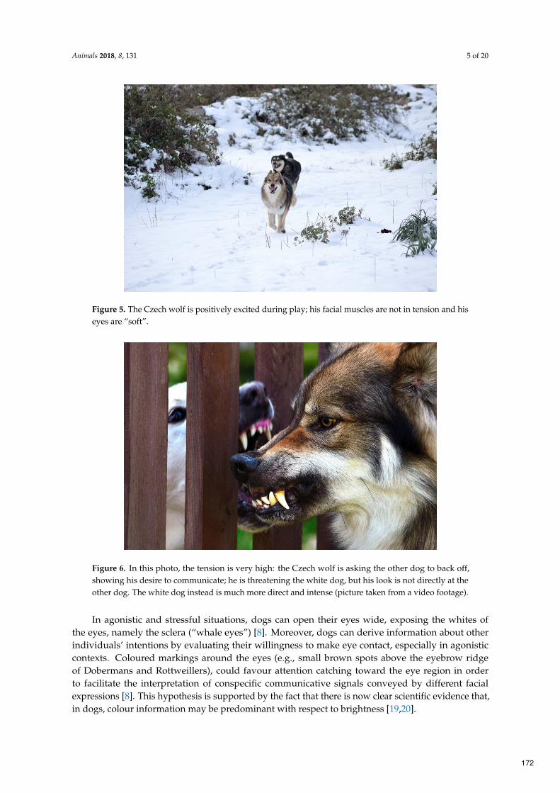
Animals 2018, 8, 131 5 of 20
Figure 5. The Czech wolf is positively excited during play; his facial muscles are not in tension and his
eyes are “soft”.
Figure 6. In this photo, the tension is very high: the Czech wolf is asking the other dog to back off,
showing his desire to communicate; he is threatening the white dog, but his look is not directly at the
other dog. The white dog instead is much more direct and intense (picture taken from a video footage).
In agonistic and stressful situations, dogs can open their eyes wide, exposing the whites of
the eyes, namely the sclera (“whale eyes”) [8]. Moreover, dogs can derive information about other
individuals’ intentions by evaluating their willingness to make eye contact, especially in agonistic
contexts. Coloured markings around the eyes (e.g., small brown spots above the eyebrow ridge
of Dobermans and Rottweillers), could favour attention catching toward the eye region in order
to facilitate the interpretation of conspecific communicative signals conveyed by different facial
expressions [8]. This hypothesis is supported by the fact that there is now clear scientific evidence that,
in dogs, colour information may be predominant with respect to brightness [19,20].
172

Animals 2018, 8, 131 6 of 20
Along with the eyes, ear position represents a relevant signal in individuals’ emotional expression,
even though its role in face processing has been rarely investigated. As highlighted above for the tail, it
is necessary to consider breed differences in the morphology of the ears and ability to move them when
defining the “relaxed” position, and the different changes should be evaluated by examining any ear
base modifications [11]. Generally speaking, dogs can pull their ears back various degrees according
to the animals’ arousal state. Ears can vary from simply “back”, to communicate an appeasement
intention, to “flattened” or “pressed back”, in frightened individuals (Figure 3) or as an agonistic
response (Figure 6). In extremely fearful individuals, ears can be pressed back so far on the head
that they completely disappear (“seal ears”) [8]. On the contrary, ears kept forward are associated
with interest, attention, and approach-oriented intentions [11], while sideward position indicates a
conflicting inner state (“airplane ears”) [8].
Although the mouth region is less investigated compared with the eyes when dogs process
conspecific faces, the mouth acquires a particular importance when evaluating whether a facial
expression is potentially threatening. Dogs, indeed, look more at the mouth region of pictures
portraying threatening and neutral conspecific facial expressions [18]. In our opinion, the eyes staring
at a fixed point that is displayed in the “neutral” expression could be interpreted as “eye stalking” by
the receiver, who can focus their attention on the mouth to perceive other information (lip position or
a vocalization) to correctly interpret this expression.
Mouth configuration varies according to its position (open or close) and to the labial commissures
shape, which conveys important information about the individual’s aggressive intentions and its
stress state. The labial commissure of the mouth is drawn forward (“short lips”) in agonistic displays
(Figure 6) and the related opening degree of the mouth increases according to threat intensity [8]. On
the contrary, dogs pull back their labial commissure (“long lips”) to communicate stress [8,11], the
intensity of which increases if the commissures are drawn more backward and form a “C” shape [8].
Along with postural and facial displays, dogs can exhibit other behaviours to signal their inner
state; for example, they turn their head away from a stimulus when stressed (Figure 3B), they lift
their forehead paw to indicate uncertainty, or they lick their lips to communicate their appeasement
intentions [8,21].
Overall, despite the fact that communication behaviours can be described separately, single
behaviours need to be considered and observed in the context of all the other signals displayed
at that time, as well as the general body language, in order to interpret correctly the individual’s
emotional state.
Dog–human communication has received growing interest over the past twenty years. In particular,
several studies investigating dogs’ comprehension of human visual signals revealed that dogs are tuned
into human visual communication [3]. Dogs, indeed, already show a high sensitivity to human-given
cues in an early stage of their development [22–24], following spontaneously human body postures,
gaze direction, and pointing to find a target location [25–27]. They also prefer to rely more on
human gestures rather than auditory cues in a two-choice task, in which the information received is
contradictory, suggesting that gestures are more salient for them [28].
Most importantly, recent studies reported that dogs are skilful in interpreting the communicative
intent of humans by understanding the ostensive-referential nature of specific signals, such as
eye contact or directed-speech [29–31]. Ostensive cues are a characteristic element of human
communicative interactions that express the sender’s intention to initiate a communicative
interaction [7]. Thus, dogs’ ability to recognise human ostensive signals, which is unique in the animal
kingdom, suggests a high level of adaptation to the human social environment [30]. Furthermore,
the flexible comprehension of human gestures allows dogs to efficiently discriminate which of the
numerous and different human social behaviours displayed in the everyday life are directed to them [7].
Dogs, indeed, evaluate the same behaviour differently according to the presence of an ostensive cue
that precedes or accompanies it, ignoring the unintended movements [29]. Among human ostensive
signals, eye contact represents the most important and efficient one [7,29]. From an early age, dogs
173

Animals 2018, 8, 131 7 of 20
show a spontaneous tendency to gaze at human faces and to make eye contact [32] in a wide range of
contexts, for example, in unsolvable tasks or to beg for food from humans [33,34]. Given the specific
nature of the contexts in which it is displayed, the human-directed gaze has been interpreted as a
“request of help” [7,33]. Thus, dogs use eye contact to communicate with humans differently from
conspecific communication, in which it represents a clear threatening signal [8]. On the contrary, in
interspecific communication, and in a friendly context, it facilitates the beginning and the maintenance
of human–dog interaction [35]. Therefore, through the domestication process, dogs have modified the
functional meaning of this typical behavioural pattern to adapt it to a cross-species communication,
acquiring a human-like communication mode [30]. Furthermore, human–dog mutual gaze enhances
the establishment of an affiliative relationship and a social bond between dogs and humans by the same
oxytocin-mediated effect described for mother–infant dyad and for human sexual partners [5,36,37].
The informative role of the eyes for human–dog communication is also demonstrated by the
greater interest by canids in investigating the eye region compared with the other inner facial features
in processing human faces [17]. Moreover, dogs assess human’s attentional state during communicative
interactions by evaluating human gaze direction and adapting their behaviour accordingly [38]. It has
been recently shown that human attention affects dogs’ facial expression production, as dogs increase
all facial movements when a human is attending to them. This evidence highlights both dogs’ ability
to act differently according to humans’ readiness to interact with them and, more importantly, dogs’
communicative intent in producing facial expressions. In particular, “tongue show” and “inner brow
raiser” facial movements are used as flexible signals to catch human attention, because, for example, the
“eyebrow raising” triggers human innate tendency to respond to this ostensive signal [16]. Dogs mainly
rely on humans’ availability to make eye contact when they communicate with them, increasing their
visual communicative behaviour according to their presence. In particular, eye contact has a crucial role
for the dogs’ referential communication with humans [38]. Dogs, indeed, are able not only to flexibly
use human gaze to regulate their behaviour in specific contexts, but also to communicate with humans
to direct their attention to a specific object of their interest, by performing the so-called “showing
behaviour” [33,39]. It has been recently reported that dogs can use up to 19 different referential
gestures during everyday interactions with humans, eliciting humans’ appropriate responses [6]. They
use their body position and sustained gaze as a local enhancement signal [40] or they alternate their
gaze between the target object and humans to indicate to them the object location [33,41]. These
signals are displayed to communicate with humans and are modulated both by human availability to
communicate with them [42] and by human responses. Dogs, indeed, produce persistently referential
signals until they elicit a satisfactory human response [43], but they are also able to interrupt them
when they are no longer successful [44].
Recent studies have demonstrated the existence of behavioural synchronization between dogs
and humans (see for review [45]). The canine synchronizes its locomotor behaviour with that of its
owner in different contexts, both indoors [46] and outdoors [47], and when facing an unfamiliar human.
Dogs synchronize their behaviour with the owner’s withdrawal response toward strangers, taking
longer time to approach them [48]. It has also been reported that the behavioural synchronization
phenomenon is affected by dogs’ affiliation toward humans; pet dogs show a higher performance in
synchronizing their behaviour with their owner’ than shelter dogs with their caregivers. Moreover,
behavioural synchronization affects dogs’ social preference toward humans, and in particular, toward
individuals synchronizing their locomotor activity with them [45]. Thus, authors concluded that,
as previously described in humans, this phenomenon increases social cohesion and affiliation in
dog–human dyads, contributing to emotional contagion [49].
Despite dogs’ high social competence to communicate and interact with humans and to perceive
and correctly respond to their signals, there are some open questions that still need to be addressed.
Although dogs react to the informative nature of human ostensive-referential cues, they may
interpret human gestures as an order rather than understanding the human communicative intent
to share information [3,30]. This hypothesis is supported by dogs’ higher attitude to follow owners’
174

Animals 2018, 8, 131 8 of 20
signals rather than those from a stranger and to follow human gestures to locate food even if the
olfactory information about its position is contradictory [1]. Furthermore, it has been reported in a
recent eye-tracking study that dogs are able to discriminate between social and non-social interactions
depicted on a picture, showing a longer gaze toward the individuals in a social context compared with
a non-social one [50].
A further important aspect of dog–human visual communication is the ability to perceive other
individuals’ emotions expressed by their faces. There is broad evidence that both dogs and humans are
skilled in recognizing the other species’ emotions by looking at their faces [51,52]. Specifically, domestic
dogs show a functional understanding of human emotional facial expressions, responding differently
according to its valence. They regulate their behaviour toward an unknown or ambiguous object by
using human emotional referential expressions, especially when provided by their owner; dogs prefer
to approach it or to stay away if the human expresses happiness or fear/disgust, respectively [53,54].
Interestingly, when the informants are inattentive, dogs actively attempt to involve them to obtain
information, alternating their gaze between the object and them. Taken together, these findings
demonstrate the existence of social referencing in dogs [53].
Dogs’ perception of human emotions allows them to adjust their behaviour during everyday
interactions with humans and to respond appropriately. A recent study reports that dogs display
mouth-licking behaviour, which is a stress indicator, more often when presented with negative
emotional facial expressions compared with positive ones [55]. This evidence suggests both that
they perceived the negative valence of the human emotion, increasing their level of stress, and
that they responded adequately to it, displaying a behaviour used in conspecific communication to
“appease” the sender [8]. Recent scientific literature shows that emotional cues conveyed by human
emotional faces are processed in an asymmetrical way by the canine brain. Specifically, using a
behavioural method commonly employed to study both visual and auditory lateralization (namely
the “head orienting paradigm” [56,57]), it has been shown that dogs are sensitive to human faces
expressing Ekman’s six basic emotions (e.g., anger, fear, happiness, sadness, surprise, disgust, and
neutral) with a specialization of the right hemisphere for the analysis of human faces expressing
“anger”, “fear” and, “happiness” emotions, but an opposite bias (left hemisphere) for human faces
expressing “surprise” [57].
3. Acoustic Communication
Domestic dogs have a broad and sophisticated vocal repertoire [58]. Although their vocalizations
are similar to their closest relative, the wolf, dogs vocalize in a wider variety of social contexts
compared with wolves and they retain this characteristic even into adulthood [59]. Dogs’ vocal
behaviour underwent considerable changes during the domestication process, generally considered as
a result of dogs’ adaptation to the human social environment [60]. The effect of living in proximity to
humans on animals’ vocal behaviour has been demonstrated, indeed, by a pioneering study showing
that, after a 40-year selection, tame red foxes emitted more human-directed vocalizations than their
ancestors [61]. Thus, as described for the foxes, dogs could have acquired a tendency to vocalize
more during interactions with humans, which could have been artificially selected, together with
other socio-cognitive abilities of understanding human cues. Dogs developed, therefore, novel forms
of the pre-existing vocalizations, which acquired different acoustic and functional characteristics,
facilitating their communication with humans [59]. Humans, indeed, are able to derive information
from dogs’ vocalisations, not only about the senders’ physical characteristics, rating, for example,
growls produced by larger dogs as more aggressive than those of smaller dogs [62], but also about
its emotional state [63,64]. The development of different and numerous vocal signals in dogs could
have been modulated, therefore, by their efficacy of conveying specific information to communicate
with humans. This hypothesis is further supported by the existence of an individual variability of the
acoustic features of barks directed to humans in non-agonistic contexts (during ball play, in requesting
175

Animals 2018, 8, 131 9 of 20
situations, or before going for a walk), which can be shaped according to the owner’s response during
everyday interactions [59,65].
The specific role of auditory signals in communication with humans is confirmed by the significant
decrease of their production in feral and stray dogs [59], suggesting that dogs’ social contact with
humans represents the main regulatory factor of their expression.
Here, we provide an overview of dogs’ most common vocalizations, focusing on their
functional–contextual features, both in intraspecific and heterospecific communication.
Among the different vocal signals, the bark is certainly the most typical vocalization of dogs.
Contrary to previous beliefs, which claimed that barks are a byproduct of domestication lacking any
functional value, recent studies demonstrated the context-related acoustical features of barks [60,66,67],
suggesting that they are means of communication in dogs.
Barks are short, explosive, and repetitive signals, with a highly variable acoustic structure
(dominant frequency range between 160 and 2630 Hz), differing between breeds and even between
individuals [60,66]. They are generally used in short-range interactions and in several behavioural
contexts, like greeting, warning/alerting, calling for attention, or during play [58]. Moreover, barking
is an allomimetic behaviour, that is, a group activity in which several individuals bark in unison with
other conspecifics, mirroring and stimulating each other [8].
Dog breeds show a different use of barks in their vocal communication. Wolf-related breeds, for
example, Shar-pei, Chow-Chow or Basenji, have a very rare propensity to bark, whereas other breeds
present a specific type of barking, such as hunting dogs [59].
Barks carry various information about the signaler’s physical characteristics, familiarity, and inner
state [62,67,68], allowing dogs to differentiate not only between barks produced by different individuals
in the same context [68], but also between the different contexts in which they are produced [67]. Recent
studies report, indeed, that the barks acoustic features vary predictably according to the context; dogs
emit longer and lower frequency barks when a stranger approaches them, while high pitched barks
are mainly produced in isolation situations [63,66]. Dogs distinguish between the different acoustic
structure of barks and react accordingly to its content and the familiarity of the signaler, staying closer
to the gate of their house in response to an unfamiliar dog barking at a stranger and remaining inside
the house during the barks of a lonely familiar dog [67]. These findings demonstrate that barks have a
functional role in intra-specific communication.
Recent studies have reported that, similar to barks, growls also convey meaningful information to
dogs [62,64,69]. These low-frequency broadband vocalizations are mainly produced during agonistic
interactions as a warning or threatening signal or during play interactions [8,58]. Canines can assess
the body size of another individual by listening to its growl, correctly matching the sound heard
with the picture portraying it [69]. Moreover, they discriminate between growls produced in different
contexts, showing more inhibited behaviour to take a bone if a “guarding” growl is played [64]. It
has recently been found that dogs’ growls have a context-dependent acoustics structure; in particular,
its temporal features, fundamental frequency, and formant dispersions differ between play and
aggressive growls, produced to threaten a stranger or to guard a bone [62,64]. In spite of the specific
acoustic characteristics of growls produced during play with humans (short and high-pitched), these
vocalizations, in particular, all “play vocalizations”, which also include barks and huffing [8], are less
distinguishable for dogs compared with those recorded in disturbing and isolation situations [56].
Dogs’ difficulty in clearly perceiving these vocalizations can be due to the lack of other metasignals,
for example, visual signals, that provide further contextual information, helping dogs to correctly
interpret them [8]. The context specificity of growls and the different reaction of dogs to the different
“context-type” growls demonstrate its important role in communication between dogs.
Dogs’ acoustic communication includes whines, which are indicators of stressful arousal but
also greeting and attention-seeking behaviours [8]; howls, which maintain group cohesion; groans
and yelps, signs of acute distress and acute pain, respectively; and grunts, which are considered as
pleasure-related signals [58]. It has recently been found that canines can extract information about the
176

Animals 2018, 8, 131 10 of 20
emotional state of other dogs from their vocalizations. In fact, they can correctly identify the emotional
valence of conspecific vocalizations, associating playful and aggressive ones with the corresponding
emotional faces [70].
Moreover, conspecific vocalizations in the dog brain, as in other vertebrates, are analysed mainly
by the left hemisphere, and its involvement depends on the characteristics of the sound. For example,
when dogs were presented with the reversed temporal acoustic features of their calls (e.g., canine
vocalizations of play, disturbance, and isolation), a shift from a left-hemisphere bias (normal call
versions) to a right-hemisphere bias (play calls) or to no brain asymmetry (disturbance and isolation
calls) has been reported. In addition, it is interesting to note that when intraspecific vocalizations elicit
intense emotions, a right hemisphere bias appeared, confirming the hypothesis on the role of the right
side of the dog brain in the analysis of arousing communicative signals [71].
Dogs and humans use vocal signals in cross-species communicative interactions that are able to
produce changes in other species behaviours [72].
On one hand, canines understand the meaning of some human words and perceive the emotional
content of human vocalizations. They are able to learn up to 200 words’ meaning and they link
it with the object they refer to [73]. Furthermore, dogs use human voice intonation as a social
referential cue, extracting information about people’s reaction to novel or ambiguous objects and
acting accordingly [53,54,74]. Although vocal signals are less significant than visual ones in guiding
dogs in ambiguous choice situations [43,74], the tone of human voice seems to be more efficient in
communicating the human’s motive [75]. A recent study shows that dogs regulate their behaviour
according to the humans’ intentions expressed by different verbal utterances, following human
gestures when they are given with a cooperative intention, but ignoring them if given with a
low-pitched/prohibiting voice [75]. Moreover, canines detect the intention of humans to engage in
playing interactions when the human’s postural signals are accompanied by vocalizations, suggesting
a specific play-eliciting function of vocalizations in human–dog social interaction [76].
The ability of the dog to correctly interpret the emotional valence of a sound also extends to
human vocalizations. Specifically, using a cross-modal paradigm, it has been demonstrated that
dogs can correctly match “happy” or “angry” human faces with a vocalization expressing the same
emotional valence [70]. Furthermore, recent research indicates that human emotional vocalizations are
processed in an asymmetrical way by the dog brain, with the prevalent use of the right hemisphere in
the analysis of vocalizations with a clear negative emotional valence (i.e., “fear” and “sadness”) and
the main use of the left hemisphere in the analysis of positive vocalization (“happiness”) [77].
On the other hand, dogs use vocalizations to communicate with humans, particularly to solicit
their care and to attract their attention when faced with an unsolvable problem [10,33]. They mainly use
short-distance calls in interactions with humans, like barks, growls, and whines, compared with long
distance calls, which are used instead to communicate with conspecifics [60]. Despite little information
about the intentional control of vocal productions [78], a growing body of literature demonstrates
that dogs’ vocalizations are effective means for interspecific communication, conveying information
for humans [63,79,80]. Humans are able to assess the signaler’s size by listening to its growls [81]
and, more interestingly, they perceive the emotional content and attribute contexts to different dogs’
vocalizations [62,63,79,80]. They generally interpret growls from large dogs as being more aggressive
than those emitted by smaller dogs [82], and they correctly attribute emotions to growls according to
their social context (play, threatening, or food guarding) [80].
Humans can also categorize barks according to their emotional content, rating barks directed
toward a stranger as more aggressive, barks produced in an isolation situation as more “despaired”, and
barks recorded during play interactions as happier [63]. Moreover, regarding the acoustical structure
of barks, humans generally rate low-frequency, low tonality, rapid-pulsating barks as more aggressive,
while more tonal, high pitched, and slow-pulsating barks are considered to be happier or more
desperate [63]. Recent findings demonstrate that humans rely on the same acoustical rules to assess
the emotional content and the context of dogs’ and conspecific vocalizations, suggesting the existence
177

Animals 2018, 8, 131 11 of 20
of a wider common mechanism of animals to express emotions through vocal signals and to apply the
same rules to encode other individuals’ inner states by listening to their vocalizations [83]. Moreover,
the humans’ ability to categorize dogs’ vocal signals is independent from previous experience with
dogs because the same performance has been found in recognizing dogs vocalizations in adults and
five-year-old children, as well as in congenitally blind people [63,84,85].
Overall, humans’ ability to categorize dogs’ vocalizations demonstrates that dogs’ vocal
signals have a communicative relevance for humans and that they represent effective means for
dog–human communication.
4. Olfactory Communication
There are relatively few studies about the role of olfaction in dogs’ communication with both
conspecifics and humans. The little research attention to the chemosignals characteristics of conveying
senders’ information might be due to the minor role of olfaction in human–human communication
(compared with vision and hearing) and to human minor sensibility to odours [10]. However, dogs’
high olfactory sensitivity (10,000–100,000 times higher than humans’) allows them to access social and
contextual information through their sense of smell [11,12]. Body odours contain chemical signals that
have specifically evolved to communicate with other individuals (Figure 7) [86]. Nevertheless, to date,
dogs’ perception of the different information conveyed by odours and their role in social interaction
are scarcely investigated and future studies are needed to address this issue.
Broadly speaking, olfactory communication is extremely efficient as odours persist in the
environment, allowing animals to acquire information of the signaler without requiring the individuals’
physical proximity and direct interactions [12]. The olfactory signal release is below the threshold
of consciousness [87] and it can occur without a communicative intent, as described for individual,
gender, and emotion-related information [9,88]. Nevertheless, dogs can intentionally deposit their
odour in the environment (“mark”) through urines, faeces, and glandular secretions; this behaviour is
known as scent marking (Figure 7). Therefore, olfactory communication in dogs takes place via a direct
interaction between two individuals through close olfactory inspection, but it also occurs indirectly
through scent marking [8].
Regarding dogs’ perception of conspecific and human odours, they spontaneously recognize
individuals by their smell [10] and they prefer specific parts of human body for olfactory
investigation [10,89], suggesting that different body parts produce specific odours that might convey
different information.
Dogs discriminate conspecifics on the basis of their odour [90]. Moreover, they distinguish their
own odour from that of others when presented with urine samples [90,91]. In social interactions, dogs
engage in olfactory investigation as part of greeting behaviours to collect information about the other
individuals [8]. They show a different interest for specific parts of conspecifics’ body for olfactory
exploration, sniffing more intensely the face, the neck, the inguinal, and the perianal areas [8,10]. The
odours are produced by different types of glands located in these areas, in particular, those located
at the corner of the mouth, in the ear pinnae, the preputial and vaginal glands, and anal sacs. The
particular interest shown in sniffing these areas suggests that dogs can obtain different information
from the investigated regions, which may produce different odours [10]. Moreover, dogs collect social
information by investigating other individuals’ urine and faeces placed in the environment [8]. Male
and female dogs show a strong interest in unfamiliar urine and they investigate them to perform mate
and threat assessment [92]. Olfactory communication includes scent marking behaviour, consisting of
a first phase of investigation of other individuals’ marks followed by the deposit of the individual’s
odours close to or on the existing marks [8,93]. Scent marking behaviour includes not only olfactory
elements, but also visual and auditory components [11]. The placement of urine and faeces (the latter
role has not been studied in detail) in the environment may be accompanied by ground-scratching
behaviour, which adds both visual and auditory signals, produced by the act of scratching, and enrich
178
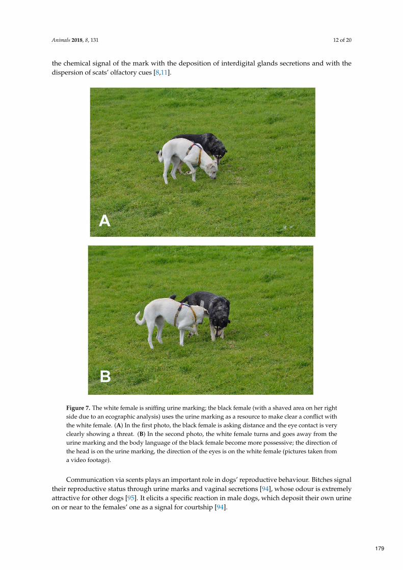
Animals 2018, 8, 131 12 of 20
the chemical signal of the mark with the deposition of interdigital glands secretions and with the
dispersion of scats’ olfactory cues [8,11].
‐
Figure 7. The white female is sniffing urine marking; the black female (with a shaved area on her right
side due to an ecographic analysis) uses the urine marking as a resource to make clear a conflict with
the white female. (A) In the first photo, the black female is asking distance and the eye contact is very
clearly showing a threat. (B) In the second photo, the white female turns and goes away from the
urine marking and the body language of the black female become more possessive; the direction of
the head is on the urine marking, the direction of the eyes is on the white female (pictures taken from
a video footage).
Communication via scents plays an important role in dogs’ reproductive behaviour. Bitches signal
their reproductive status through urine marks and vaginal secretions [94], whose odour is extremely
attractive for other dogs [95]. It elicits a specific reaction in male dogs, which deposit their own urine
on or near to the females’ one as a signal for courtship [94].
179

Animals 2018, 8, 131 13 of 20
Dogs can also release their odour in the environment by rolling on the ground, marking with their
face and their entire body. Although this behaviour is still included in the canines’ repertoire and it
maintains its communicative characteristic, it is no longer relevant for the evolutionary success of this
species. Wolves use scent-rolling behaviour to pick up the scent of the pray and to carry it back to the
pack, providing information about the health and location of the prey [8].
Moreover, it has been recently demonstrated that dogs are able to perceive the emotional content
of conspecific odours, which induces behavioural and physiological effects in the receiver according to
their valence [9,96]. Research specifically showed that during the sniffing of odours collected from
perianal, interdigital, and salivary secretions soon after the end of a negative emotional event, in
which the dog was left alone in an unfamiliar environment, dogs consistently used their right nostril.
Given that the neural olfactory pathways ascend ipsilaterally to the brain, right nostril use reflects a
main activation of the right hemisphere. In addition, the sniffing conspecific odours collected during
“isolation” and “disturbance” situations causes an increase in heart rate and stress behaviours in dogs.
Once again, these findings support the main role of the right side of the dog brain in the analysis of
clear arousing signals.
Canines recognize humans by their odour. They are able to discriminate body odour of two
identical twins living in the same environment [97] and to respond spontaneously to metabolic changes
of their owner on the base of their scent [98]. Moreover, they associate the humans’ odour with previous
experiences they had with them. They show an increase of their arousal state when presented with the
veterinary sweat odour, which is generally associated with stressful experiences [95]; on the other hand,
they associate familiar human odours with positive outcomes [99]. In a recent functional magnetic
resonance imaging (fMRI) study, it is reported that familiar human odours activate the caudate nucleus,
which is associated with positive expectations and reward, including social reward [99].
Dogs show a preference for investigating specific parts of the human body, and in particular,
some specific areas of children’s bodies, namely the face and the upper limbs, suggesting that human
odours produced at different anatomical parts could also provide different specific olfactory cues [9].
It has been recently found that, as for conspecific odours, dogs detect the emotional content of
human odours, which induces different changes in their behaviour and in their cardiac activity [9,96].
In particular, an increase in behavioural and cardiac reactivity occurs during the sniffing of human
odours collected during fearful situations. It is interesting to note that, contrary to that which has
been observed for arousing conspecific odours, during the sniffing of the smell of human fear, bias in
the use of the left nostril (i.e., left hemisphere activity) has been observed [9]. The latter suggest that
chemosignals communicate conspecific and heterospecific emotions using different sensory pathways.
Moreover, dogs adjust their social behaviour toward humans according to the valence of the
odour perceived, showing more stranger-directed behaviour when presented with the “happiness”
odour, and more owner-directed behaviour when presented with the “fear” odour [96].
These findings demonstrate that chemosignals carry contextual-related information, supporting
their specific role in dog–human communication.
5. Tactile Communication
Although rarely studied, tactile communication is an equally important aspect of dog
communication. Tactile communication between dogs is used during agonistic interactions to impress
an opponent (by an intense physical contact, putting paws over back or body of subordinate, grabbing
the muzzle of the subordinate or young individuals and clasping another canids during ritualized
aggression) or to maintain social bond (Figure 8) [8,100,101]. In particular, social cohesion is maintained
by specific activities, such as resting in close contact (see Figure 9), placing the head over the shoulders
of another dog during greetings or sexual approaches preceding mating, and by social grooming,
which generally includes “face washing” (licking the other animal’s face) or “nibble” grooming, using
the front teeth and rapid jaw open–close movements [8].
180

Animals 2018, 8, 131 14 of 20
‐
Figure 8. The Czech wolf is doing a muzzle grab during a bout of play.
‐
Figure 9. These two Rhodesian Ridgebacks usually sleep and rest in very close physical contact with
each other. They have a very strong bond; the dog on the left is a daughter of the one on the right.
In a similar way, some human gestures during human–dog physical interaction could result
in positive or negative canine emotional states, which drive to corresponding approaching and
withdrawal behavioral responses of the dogs, even if they have been initiated with a different
motivation [102]. People, equal if familiar or unfamiliar to a pet, tend to show their affection
towards their pets by initiating physical contact. This is due to the fact that the tactile contact during
human–dog interactions causes a series of benefits with regard to the physiology, the mental states,
and the immune system of humans. For example, in humans, a decrease of both blood pressure
and heart rate and an increase of the immune system function have been reported after petting
dogs [103–105]. Otherwise, being petted serves as positive reinforcement for dogs as confirmed
by associated heart-rate deceleration [106]. Nevertheless, physical contact in social interactions
has different features in interspecific and intraspecific communication, in terms of both frequency
and duration. Dogs rarely use physical contact to communicate with other individuals and tactile
interactions (for example, grooming) are generally short lasting. On the contrary, humans tend to
initiate and maintain physical contact with dogs with a higher frequency and longer duration, as it
181

Animals 2018, 8, 131 15 of 20
is a typical feature of human–human communication. For this reason, some dogs may appear less
relaxed during human–dog tactile interaction (see Figure 10) tolerating physical contact or displaying
a withdrawal behavioral response [107]. Some dogs tend to show discomfort using ambivalent signals
and conflict behaviors during all close physical interactions and this phenomenon also depends on
which specific part of their body is touched [108].
-
-
- -
‐
Figure 10. This dog is not relaxed during this tactile interaction. Although the physical contact is
“gentle” (on the dog’s side and not on his head), the human is standing on the dog, making him
feel uncomfortable.
A significant influence of human-dog familiarity on dogs’ behavioral responses during tactile
human-dog interactions has also been found. In particular, the work of Kuhne and colleagues [100]
showed that dogs being petted by a familiar person showed significantly more appeasement gestures
(e.g., blinking, looking elsewhere, closing both eyes, averted head, sitting, laying down, etc.),
redirected behaviors (e.g., sniffing/licking on the floor, digging, drinking, visual scanning, etc.),
and social approach behavior than dogs being petted by an unfamiliar person. Furthermore, significant
differences in dogs’ behavioral responses depending on human-dog familiarity could be seen if the
dogs were petted on specific parts of their body, supporting other findings that showed that dogs
may generally dislike their hind legs, paws, and the top of their head being touched [109]. Dogs
may interpret petting these specific canine body regions as agonistic communicative signals, which
could create interferences with a normal and balanced human-dog bond [102]. Canines seem to better
tolerate physical contacts (displaying less conflict and withdrawal behavioral responses) on the sides
of their chest and under their chin. However, given that dogs’ reactions to handling depend on
different factors (e.g., genetics and early experience, including socialization with humans, as well as
physical and mental health, breed, learning and especially the context in which the interaction takes
place [110,111]), there is no scientific concluding evidence yet concerning how to safely pet and play
with dogs [109].
6. Conclusions
Dogs have a vast repertoire of visual, tactile, acoustic, and olfactory signals that they use for
an expressive and fine-tuned communication with both conspecifics and humans. Nevertheless,
the communicative importance of the different body parts in social interactions still remains poorly
investigated. Future studies could evaluate dogs’ gaze pattern and olfactory attention toward human
182

Animals 2018, 8, 131 16 of 20
and conspecific bodies both in human–dog and conspecific interactions, in order to better identify
which body regions are more informative for dogs during communicative interactions.
Author Contributions: The authors contributed equally to this manuscript.
Funding: This research received no external funding.
Conflicts of Interest: The authors declare no conflict of interest.
References
1. Elgier, A.M.; Jakovcevic, A.; Barrera, G.; Mustaca, A.E.; Bentosela, M. Communication between domestic
dogs (Canis familiaris) and humans: Dogs are good learners. Behav. Process. 2009, 81, 402–408. [CrossRef]
[PubMed]
2. Thalmann, O.; Shapiro, B.; Cui, P.; Schuenemann, V.J.; Sawyer, S.K.; Greenfield, D.L.; Germonpré, M.B.;
Sablin, M.V.; López-Giráldez, F.; Domingo-Roura, X.; et al. Complete mitochondrial genomes of ancient
canids suggest a European origin of domestic dogs. Science 2013, 342, 871–874. [CrossRef] [PubMed]
3. Kaminski, J.; Nitzschner, M. Do dogs get the point? A review of dog–human communication ability.
Learn. Motiv. 2013, 44, 294–302. [CrossRef]
4. Topàl, J.; Miklòsi, Á.; Csànyi, V.; Dòka, A. Attachment behaviour in dogs (Canis familiaris): A new application
of Ainsworth’s (1969) Strange Situation Test. J. Comp. Psychol. 1998, 112, 219–229. [CrossRef] [PubMed]
5. Siniscalchi, M.; Stipo, C.; Quaranta, A. “Like Owner, Like Dog”: Correlation between the Owner’s
Attachment Profile and the Owner-Dog Bond. PLoS ONE 2013, 8, e78455. [CrossRef] [PubMed]
6. Worsley, H.K.; O’Hara, S.J. Cross-species referential signalling events in domestic dogs (Canis familiaris).
Anim. Cogn. 2018, 21, 457–465. [CrossRef] [PubMed]
7. Topál, J.; Kis, A.; Oláh, K. Dogs’ sensitivity to human ostensive cues: A unique adaptation? In The Social Dog,
1st ed.; Kaminski, J., Marshall-Pescini, S., Eds.; Academic Press: Cambridge, MA, USA, 2014; p. 329, ISBN
9780124078185.
8. Handelman, B. Canine Behavior: A Photo Illustrated Handbook; Dogwise Publishing: Wenatchee, WA, USA,
2012; ISBN 0976511827.
9. Siniscalchi, M.; d’Ingeo, S.; Quaranta, A. The dog nose “KNOWS” fear: Asymmetric nostril use during
sniffing at canine and human emotional stimuli. Behav. Brain Res. 2016, 304, 34–41. [CrossRef] [PubMed]
10. Bradshaw, J.W.; Rooney, N. Dog social behavior and communication. In The Domestic Dog; Serpell, J., Ed.;
Cambridge University Press: Cambridge, UK, 2016; pp. 133–159, ISBN 978-0521425377.
11. Hecht, J.; Horowitz, A. Introduction to dog behaviour. In Animal Behaviour for Shelter Veterinarians and Staff,
1st ed.; Weiss, E., Mohan-Gibbons, H., Zawistowski, S., Eds.; Wiley-Blackwell: London, UK, 2015; pp. 5–30,
ISBN 978-1118711118.
12. Wells, D.L. Behaviour of Dogs. In The Ethology of Domestic Animals: An Introductory Text, 3rd ed.; Jensen, P., Ed.;
CABI: Oxford, UK, 2017; pp. 228–238, ISBN 9781786391650.
13. Buxton, D.F.; Goodman, D.C. Motor function and the corticospinal tracts in the dog and raccoon. J. Comp.
Neurol. 1967, 129, 341–360. [CrossRef] [PubMed]
14. Davidson, R.J. Well-being and affective style: Neural substrates and biobehavioural correlates. Phil. Trans. R.
Soc. Lond. B 2004, 359, 1395–1411. [CrossRef] [PubMed]
15. Siniscalchi, M.; Lusito, R.; Vallortigara, G.; Quaranta, A. Seeing left-or right-asymmetric tail wagging
produces different emotional responses in dogs. Curr. Biol. 2013, 23, 2279–2282. [CrossRef] [PubMed]
16. Kaminski, J.; Hynds, J.; Morris, P.; Waller, B.M. Human attention affects facial expressions in domestic dogs.
Sci. Rep. 2017, 7, 12914. [CrossRef] [PubMed]
17. Somppi, S.; Törnqvist, H.; Hänninen, L.; Krause, C.M.; Vainio, O. How dogs scan familiar and inverted faces:
An eye movement study. Anim. Cogn. 2014, 17, 793–803. [CrossRef] [PubMed]
18. Somppi, S.; Törnqvist, H.; Kujala, M.V.; Hänninen, L.; Krause, C.M.; Vainio, O. Dogs evaluate threatening
facial expressions by their biological validity–Evidence from gazing patterns. PLoS ONE 2016, 11, e0143047.
[CrossRef] [PubMed]
19. Kasparson, A.A.; Badridze, J.; Maximov, V.V. Colour cues proved to be more informative for dogs than
brightness. Proc. Biol. Sci. 2013, 280, 20131356. [CrossRef] [PubMed]
183

Animals 2018, 8, 131 17 of 20
20. Siniscalchi, M.; d’Ingeo, S.; Fornelli, S.; Quaranta, A. Are dogs red–green colour blind? R. Soc. Open Sci. 2017,
4, 170869. [CrossRef] [PubMed]
21. Beerda, B.; Schilder, M.B.H.; van Hoff, J.A.R.A.M.; de Vries, H.W. Manifestations of chronic and acute stress
in dogs. Appl. Anim. Behav. Sci. 1997, 52, 307–319. [CrossRef]
22. Agnetta, B.; Hare, B.; Tomasello, M. Cues to food location that domestic dogs (Canis familiaris) of different
ages do and do not use. Anim. Cogn. 2000, 3, 107–112. [CrossRef]
23. Riedel, J.; Schumann, K.; Kaminski, J.; Call, J.; Tomasello, M. The early ontogeny of human–dog
communication. Anim. Behav. 2008, 75, 1003–1014. [CrossRef]
24. Wallis, L.J.; Range, F.; Müller, C.A.; Serisier, S.; Huber, L.; Virányi, Z. Training for eye contact modulates gaze
following in dogs. Anim. Behav. 2015, 106, 27–35. [CrossRef] [PubMed]
25. Miklósi, Á.; Soproni, K. A comparative analysis of animals’ understanding of the human pointing gesture.
Anim. Cogn. 2006, 9, 81–93. [CrossRef] [PubMed]
26. Soproni, K.; Miklósi, Á.; Topal, J.; Csanyi, V. Comprehension of human communicative signs in pet dogs
(Canis familiaris). J. Comp. Psychol. 2001, 115, 122–126. [CrossRef] [PubMed]
27. Udell, M.A.R.; Giglio, R.F.; Wynne, C.D.L. Domestic dogs (Canis familiaris) use human gestures but not
nonhuman tokens to find hidden food. J. Comp. Psychol. 2008, 122, 84–93. [CrossRef] [PubMed]
28. D’Aniello, B.; Scandurra, A.; Alterisio, A.; Valsecchi, P.; Prato-Previde, E. The importance of gestural
communication: A study of human–dog communication using incongruent information. Anim. Cogn. 2016,
19, 1231–1235. [CrossRef] [PubMed]
29. Kaminski, J.; Schulz, L.; Tomasello, M. How dogs know when communication is intended for them. Dev. Sci.
2012, 15, 222–232. [CrossRef] [PubMed]
30. Miklósi, Á.; Topál, J. What does it take to become ‘best friends’? Evolutionary changes in canine social
competence. Trends Cogn. Sci. 2013, 17, 287–294. [CrossRef] [PubMed]
31. Téglás, E.; Gergely, A.; Kupán, K.; Miklósi, Á.; Topál, J. Dogs’ gaze following is tuned to human
communicative signals. Curr. Biol. 2012, 22, 209–212. [CrossRef] [PubMed]
32. Virányi, Z.; Gácsi, M.; Kubinyi, E.; Topál, J.; Belényi, B.; Ujfalussy, D.; Miklósi, Á. Comprehension of human
pointing gestures in young human-reared wolves (Canis lupus) and dogs (Canis familiaris). Anim. Cogn. 2008,
11, 373–387. [CrossRef] [PubMed]
33. Miklósi, Á.; Polgárdi, R.; Topál, J.; Csányi, V. Intentional behaviour in dog–human communication: An
experimental analysis of ‘showing’ behaviour in the dog. Anim. Cogn. 2000, 3, 159–166. [CrossRef]
34. Gácsi, M.; Miklósi, Á.; Varga, O.; Topál, J.; Csányi, V. Are readers of our face readers of our minds? Dogs
(Canis familiaris) show situation-dependent recognition of human’s attention. Anim. Cogn. 2004, 7, 144–153.
[CrossRef] [PubMed]
35. Vas, J.; Topál, J.; Gácsi, M.; Miklósi, Á.; Csányi, V. A friend or an enemy? Dogs’ reaction to an unfamiliar
person showing behavioural cues of threat and friendliness at different times. Appl. Anim. Behav. Sci. 2005,
94, 99–115. [CrossRef]
36. Ross, H.E.; Young, L.J. Oxytocin and the neural mechanisms regulating social cognition and affiliative
behavior. Front. Neuroendocrinol. 2009, 30, 534–547. [CrossRef] [PubMed]
37. Nagasawa, M.; Mitsui, S.; En, S.; Ohtani, N.; Ohta, M.; Sakuma, Y.; Onaka, T.; Mogi, K.; Kikusui, T.
Oxytocin-gaze positive loop and the coevolution of human-dog bonds. Science 2015, 348, 333–336. [CrossRef]
[PubMed]
38. Schwab, C.; Huber, L. Obey or not obey? Dogs (Canis familiaris) behave differently in response to attentional
states of their owners. J. Comp. Psychol. 2006, 120, 169–175. [CrossRef] [PubMed]
39. Kaminski, J.; Neumann, M.; Bräuer, J.; Call, J.; Tomasello, M. Dogs, Canis familiaris, communicate with
humans to request but not to inform. Anim. Behav. 2011, 82, 651–658. [CrossRef]
40. Gaunet, F.; Deputte, B.L. Functionally referential and intentional communication in the domestic dog: Effects
of spatial and social contexts. Anim. Cogn. 2011, 14, 849–860. [CrossRef] [PubMed]
41. Miklósi, Á.; Kubinyi, E.; Topál, J.; Gácsi, M.; Virányi, Z.; Csányi, V. A simple reason for a big difference:
Wolves do not look back at humans but dogs do. Curr. Biol. 2003, 13, 763–766. [CrossRef]
42. Savalli, C.; Resende, B.; Gaunet, F. Eye contact is crucial for referential communication in pet dogs. PLoS ONE
2016, 11, e0162161. [CrossRef] [PubMed]
43. Gaunet, F. How do guide dogs and pet dogs (Canis familiaris) ask their owners for their toy and for playing?
Anim. Cogn. 2010, 13, 311–323. [CrossRef] [PubMed]
184

Animals 2018, 8, 131 18 of 20
44. Elgier, A.M.; Jakovcevic, A.; Mustaca, A.E.; Bentosela, M. Learning and owner–stranger effects on
interspecific communication in domestic dogs (Canis familiaris). Behav. Proc. 2009, 81, 44–49. [CrossRef]
[PubMed]
45. Duranton, C.; Gaunet, F. Behavioral synchronization and affiliation: Dogs exhibit human-like skills.
Learn. Behav. 2018. [CrossRef] [PubMed]
46. Duranton, C.; Bedossa, T.; Gaunet, F. Pet dogs synchronize their walking pace with that of their owners in
open outdoor areas. Anim. Cogn. 2018, 21, 219–226. [CrossRef] [PubMed]
47. Duranton, C.; Bedossa, T.; Gaunet, F. Interspecific behavioural synchronization: Dogs present locomotor
synchrony with humans. Sci. Rep. 2017, 7, 12384. [CrossRef] [PubMed]
48. Duranton, C.; Bedossa, T.; Gaunet, F. When facing an unfamiliarperson, pet dogs present social referencing
based on their owner’s direction of movement alone. Anim. Behav. 2016, 113, 147–156. [CrossRef]
49. Duranton, C.; Gaunet, F. Behavioural synchronization from an ethological perspective: Short overview of its
adaptive values. Adapt. Behav. 2016, 24, 181–191. [CrossRef]
50. Törnqvist, H.; Somppi, S.; Koskela, A.; Krause, C.M.; Vainio, O.; Kujala, M.V. Comparison of dogs and
humans in visual scanning of social interaction. R. Soc. Open Sci. 2015, 2, 150341. [CrossRef] [PubMed]
51. Bloom, T.; Friedman, H. Classifying dogs’ (Canis familiaris) facial expressions from photographs. Behav. Proc.
2013, 96, 1–10. [CrossRef] [PubMed]
52. Müller, C.A.; Schmitt, K.; Barber, A.L.; Huber, L. Dogs can discriminate emotional expressions of human
faces. Curr. Biol. 2015, 25, 601–605. [CrossRef] [PubMed]
53. Merola, I.; Prato-Previde, E.; Marshall-Pescini, S. Dogs’ social referencing towards owners and strangers.
PLoS ONE 2012, 7, e47653. [CrossRef] [PubMed]
54. Merola, I.; Prato-Previde, E.; Lazzaroni, M.; Marshall-Pescini, S. Dogs’ comprehension of referential emotional
expressions: Familiar people and familiar emotions are easier. Anim. Cogn. 2014, 17, 373–385. [CrossRef]
[PubMed]
55. Albuquerque, N.; Guo, K.; Wilkinson, A.; Resende, B.; Mills, D.S. Mouth-licking by dogs as a response to
emotional stimuli. Behav. Process. 2018, 146, 42–45. [CrossRef] [PubMed]
56. Siniscalchi, M.; Quaranta, A.; Rogers, L.J. Hemispheric specialization in dogs for processing different acoustic
stimuli. PLoS ONE 2008, 3, e3349. [CrossRef] [PubMed]
57. Siniscalchi, M.; d’Ingeo, S.; Quaranta, A. Orienting asymmetries and physiological reactivity in dogs’
response to human emotional faces. Learn. Behav. 2018. [CrossRef] [PubMed]
58. Yeon, S.C. The vocal communication of canines. J. Vet. Behav. 2007, 2, 141–144. [CrossRef]
59. Pongrácz, P.; Molnár, C.; Miklósi, Á. Barking in family dogs: An ethological approach. Vet. J. 2010, 183,
141–147. [CrossRef] [PubMed]
60. Feddersen-Petersen, D.U. Vocalization of European wolves (Canis lupus lupus L.) and various dog breeds
(Canis lupus f. fam.). Arch. Tierz. 2000, 43, 387–397. [CrossRef]
61. Gogoleva, S.S.; Volodin, J.A.; Volodina, E.V.; Trut, L.N. To bark or not to bark:vocalizations by red foxes
selected for tameness or aggressiveness toward humans. Bioacoustics 2012, 18, 99–132. [CrossRef]
62. Taylor, A.M.; Reby, D.; McComb, K. Context-related variation in the vocal growling behaviour of the domestic
dog (Canis familiaris). Ethology 2009, 115, 905–915. [CrossRef]
63. Pongrácz, P.; Molnár, C.S.; Miklósi, Á.; Csányi, V. Human listeners are able to classify dog (Canis familiaris)
barks recorded in different situations. J. Comp. Psychol. 2005, 119, 136–144. [CrossRef] [PubMed]
64. Faragó, T.; Pongrácz, P.; Range, F.; Virányi, Z.; Miklósi, Á. ‘The bone is mine’: Affective and referential
aspects of dog growls. Anim. Behav. 2010, 79, 917–925. [CrossRef]
65. Molnár, C.S.; Kaplan, F.; Roy, P.; Pachet, F.; Pongrácz, P.; Dóka, A.; Miklósi, Á. Classification of dog barks: A
machine learning approach. Anim. Cogn. 2008, 11, 389–400. [CrossRef] [PubMed]
66. Yin, S.; McCowan, B. Barking in domestic dogs: Context specificity and individual identification. Anim. Behav.
2004, 68, 343–355. [CrossRef]
67. Pongrácz, P.; Szabó, É.; Kis, A.; Péter, A.; Miklósi, Á. More than noise?—Field investigations of intraspecific
acoustic communication in dogs (Canis familiaris). Appl. Anim. Behav. Sci. 2014, 159, 62–68. [CrossRef]
68. Molnár, C.; Pongrácz, P.; Faragó, T.; Dóka, A.; Miklósi, Á. Dogs discriminate between barks: The effect of
context and identity of the caller. Behav. Process. 2009, 82, 198–201. [CrossRef] [PubMed]
69. Faragó, T.; Pongrácz, P.; Miklósi, Á.; Huber, L.; Virányi, Z.; Range, F. Dogs’ expectation about signalers’ body
size by virtue of their growls. PLoS ONE 2010, 5, e15175. [CrossRef] [PubMed]
185

Animals 2018, 8, 131 19 of 20
70. Albuquerque, N.; Guo, K.; Wilkinson, A.; Savalli, C.; Otta, E.; Mills, D. Dogs recognize dog and human
emotions. Biol. Lett. 2016, 12, 20150883. [CrossRef] [PubMed]
71. Siniscalchi, M.; Lusito, R.; Sasso, R.; Quaranta, A. Are temporal features crucial acoustic cues in dog vocal
recognition? Anim. Cogn. 2012, 15, 815–821. [CrossRef] [PubMed]
72. Andics, A.; Miklósi, Á. Neural processes of vocal social perception: Dog-human comparative fMRI studies.
Neurosci. Biobehav. Rev. 2018, 85, 54–64. [CrossRef] [PubMed]
73. Kaminski, J.; Call, J.; Fischer, J. Word learning in a domestic dog: Evidence for “fast mapping”. Science 2004,
304, 1682–1683. [CrossRef] [PubMed]
74. Colbert-White, E.N.; Tullis, A.; Andresen, D.R.; Parker, K.M.; Patterson, K.E. Can dogs use vocal intonation
as a social referencing cue in an object choice task? Anim. Cogn. 2018, 21, 253–265. [CrossRef] [PubMed]
75. Pettersson, H.; Kaminski, J.; Herrmann, E.; Tomasello, M. Understanding of human communicative motives
in domestic dogs. Appl. Anim. Behav. Sci. 2011, 133, 235–245. [CrossRef]
76. Rooney, N.J.; Bradshaw, J.W.; Robinson, I.H. Do dogs respond to play signals given by humans? Anim. Behav.
2001, 61, 715–722. [CrossRef]
77. Siniscalchi, M.; d’Ingeo, S.; Fornelli, S.; Quaranta, A. Lateralized behavior and cardiac activity of dogs in
response to human emotional vocalizations. Sci. Rep. 2018, 8, 77. [CrossRef] [PubMed]
78. Gruber, T.; Grandjean, D. A comparative neurological approach to emotional expressions in primate
vocalizations. Neurosci. Biobehav. Rev. 2017, 73, 182–190. [CrossRef] [PubMed]
79. Pongrácz, P.; Molnár, C.; Miklósi, Á. Acoustic parameters of dog barks carry emotional information for
humans. Appl. Anim. Behav. Sci. 2006, 100, 228–240. [CrossRef]
80. Faragó, T.; Takács, N.; Miklósi, Á.; Pongrácz, P. Dog growls express various contextual and affective content
for human listeners. R. Soc. Open Sci. 2017, 4, 170134. [CrossRef] [PubMed]
81. Taylor, A.M.; Reby, D.; McComb, K. Human listeners attend to size information in domestic dog growls.
J. Acoust. Soc. Am. 2008, 123, 2903–2909. [CrossRef] [PubMed]
82. Taylor, A.M.; Reby, D.; McComb, K. Why do large dogs sound more aggressive to human listeners: Acoustic
bases of motivational misattributions. Ethology 2010, 116, 1155–1162. [CrossRef]
83. Faragó, T.; Andics, A.; Devecseri, V.; Kis, A.; Gácsi, M.; Miklósi, Á. Humans rely on the same rules to
assess emotional valence and intensity in conspecific and dog vocalizations. Biol. Lett. 2014, 10, 20130926.
[CrossRef] [PubMed]
84. Molnár, C.; Pongrácz, P.; Miklósi, Á. Seeing with ears: Sightless humans’ perception of dog bark provides a
test for structural rules in vocal communication. Q. J. Exp. Psychol. 2010, 63, 1004–1013. [CrossRef] [PubMed]
85. Pongrácz, P.; Molnár, C.; Dóka, A.; Miklósi, Á. Do children understand man’s best friend? Classification of
dog barks by pre-adolescents and adults. Appl. Anim. Behav. Sci. 2011, 135, 95–102. [CrossRef]
86. Wyatt, T.D. How animals communicate via pheromones. Am. Sci. 2015, 103, 114. [CrossRef]
87. Pause, B.M. Processing of body odor signals by the human brain. Chemosens. Percept. 2012, 5, 55–63.
[CrossRef] [PubMed]
88. Penn, D.J.; Oberzauche, E.; Grammer, K.; Fischer, G.; Soini, H.A.; Wiesler, D.; Novotny, M.V.; Dixon, S.J.;
Xu, Y.; Brereton, R.G. Individual and gender fingerprints in human body odor. J. R. Soc. Interface 2007, 4,
331–340. [CrossRef] [PubMed]
89. Siniscalchi, M. Olfaction and the Canine Brain. In Canine Olfaction Science and Law; Jezierski, T., Ensminger, J.,
Papet, L.E., Eds.; CRC Press: Boca Raton, FL, USA, 2016; pp. 31–37, ISBN 1482260239.
90. Bekoff, M. Observations of scent-marking and discriminating self from others by a domestic dog
(Canis familiaris): Tales of displaced yellow snow. Behav. Process. 2001, 55, 75–79. [CrossRef]
91. Horowitz, A. Smelling themselves: Dogs investigate their own odours longer when modified in an “olfactory
mirror” test. Behav. Process. 2017, 143, 17–24. [CrossRef] [PubMed]
92. Lisberg, A.E.; Snowdon, C.T. The effects of sex, gonadectomy and status on investigation patterns of
unfamiliar conspecific urine in domestic dogs, Canis familiaris. Anim. Behav. 2009, 77, 1147–1154. [CrossRef]
93. Cafazzo, S.; Natoli, E.; Valsecchi, P. Scent-marking behaviour in a pack of free-ranging domestic dogs.
Ethology 2012, 118, 955–966. [CrossRef]
94. Pal, S.K. Urine marking by free-ranging dogs (Canis familiaris) in relation to sex, season, place and posture.
Appl. Anim. Behav. Sci. 2003, 80, 45–59. [CrossRef]
95. Siniscalchi, M.; Sasso, R.; Pepe, A.M.; Dimatteo, S.; Vallortigara, G.; Quaranta, A. Sniffing with the right
nostril: Lateralization of response to odour stimuli by dogs. Anim. Behav. 2011, 82, 399–404. [CrossRef]
186

Animals 2018, 8, 131 20 of 20
96. D’Aniello, B.; Semin, G.R.; Alterisio, A.; Aria, M.; Scandurra, A. Interspecies transmission of emotional
information via chemosignals: From humans to dogs (Canis lupus familiaris). Anim. Cogn. 2018, 21, 67–78.
[CrossRef] [PubMed]
97. Pinc, L.; Bartoš, L.; Reslova, A.; Kotrba, R. Dogs discriminate identical twins. PLoS ONE 2011, 6, e20704.
[CrossRef] [PubMed]
98. Chen, M.; Daly, M.; Williams, N.; Williams, S.; Williams, C.; Williams, G. Non-invasive detection of
hypoglycaemia using a novel, fully biocompatible and patient friendly alarm system. BMJ 2000, 321,
1565–1566. [CrossRef] [PubMed]
99. Berns, G.S.; Brooks, A.M.; Spivak, M. Scent of the familiar: An fMRI study of canine brain responses to
familiar and unfamiliar human and dog odors. Behav. Process. 2015, 110, 37–46. [CrossRef] [PubMed]
100. Kuhne, F.; Hoessler, J.C.; Struwe, R. Affective behavioural responses by dogs to tactile human-dog
interactions. Berl. Munch. Tierarztl. Wochenschr. 2012, 125, 371–378. [CrossRef] [PubMed]
101. Overall, K.L. Normal canine behavior. In Clinical Behavioral Medicine for Small Animals; Overall, K.L., Ed.;
Mosby: St Louis, MO, USA, 1997; pp. 9–44, ISBN 978-0801668203.
102. Miklósi, Á. Dogs in anthropogenic environments: Society and family. In Dog Behaviour, Evolution, and
Cognition, 2nd ed.; Miklósi, Á., Ed.; University Press: Oxford, UK, 2016; pp. 47–66, ISBN 978-0198787778.
103. Baun, M.M.; Bergstrom, N.; Langston, N.F.L.T. Physiological effects of human/companion animal bonding.
Nurs. Res. 1984, 33, 126–129. [CrossRef] [PubMed]
104. Vormbrock, J.K.; Grossberg, J.M. Cardiovascular effects of human–pet dog interactions. J. Behav. Med. 1988,
11, 509–517. [CrossRef] [PubMed]
105. Charnetski, C.J.; Riggers, S.; Brennan, F.X. Effect of petting a dog on immune system function. Psychol. Rep.
2004, 95, 1087–1091. [CrossRef] [PubMed]
106. Kostarczyk, E.; Fonberg, E. Heart rate mechanisms in instrumental conditioning reinforced by petting in
dogs. Physiol. Behav. 1981, 28, 27–30. [CrossRef]
107. Donaldson, J. The Culture Clash. A Revolutionary New Way of Understanding the Relationship between Humans
and Domestic Dogs; James & Kenneth Publishers: Berkeley, CA, USA, 1996; ISBN 978-1888047059.
108. Luescher, A.U.; Reisner, I.R. Canine aggression toward familiar people: A new look at an old problem.
Vet. Clin. N. Am. Small Anim. Pract. 2008, 38, 1107–1130. [CrossRef] [PubMed]
109. De Keuster, T.; Lamoureux, J.; Kahn, A. Epidemiology of dog bites: A Belgian experience of canine behaviour
and public health concerns. Vet. J. 2006, 173, 482–487. [CrossRef] [PubMed]
110. Koolhaas, J.M.; Korte, S.M.; De Boer, S.F.; Van Der Vegt, B.J.; Van Reenen, C.G.; Hopster, H.; De Jong, I.C.;
Ruis, M.A.W.; Blokhuis, H.J. Coping styles in animals: Current status in behavior and stress-physiology.
Neurosci. Biobehav. Rev. 1999, 23, 925–935. [CrossRef]
111. Vas, J.; Topál, J.; Gyóri, B.; Miklósi, Á. Consistency of dogs’ reactions to threatening cues of an unfamiliar
person. Appl. Anim. Behav. Sci. 2008, 112, 331–344. [CrossRef]
© 2018 by the authors. Licensee MDPI, Basel, Switzerland. This article is an open access
article distributed under the terms and conditions of the Creative Commons Attribution
(CC BY) license (http://creativecommons.org/licenses/by/4.0/).
187

Appendix B
Are dogs red-green colour blind?: article 2
188

rsos.royalsocietypublishing.org
ResearchCite this article: Siniscalchi M, d’Ingeo S,
Fornelli S, Quaranta A. 2017 Are dogs
red–green colour blind?. R. Soc. open sci.
4: 170869.
http://dx.doi.org/10.1098/rsos.170869
Received: 9 July 2017
Accepted: 6 October 2017
Subject Category:
Biology (whole organism)
Subject Areas:
behaviour/neuroscience/physiology
Keywords:
dog, colour vision, physiology,
behavioural method
Author for correspondence:
Marcello Siniscalchi
e-mail: [email protected]
Electronic supplementary material is available
online at https://dx.doi.org/10.6084/m9.
igshare.c.3917998.
Are dogs red–green colourblind?Marcello Siniscalchi, Serenella d’Ingeo, Serena Fornelli
and Angelo Quaranta
Department of Veterinary Medicine, Section of Behavioral Sciences and Animal
Bioethics, University of Bari ‘Aldo Moro’, Bari, Italy
MS, 0000-0002-1592-6549
Neurobiological and molecular studies suggest a dichromaticcolour vision in canine species, which appears to be similar tothat of human red–green colour blindness. Here, we show thatdogs exhibit a behavioural response similar to that of red–greenblind human subjects when tested with a modified version of atest commonly used for the diagnosis of human deuteranopia(i.e. the Ishihara’s test). Besides contributing to increasing theknowledge about the perceptual ability of dogs, the presentwork describes for the first time, to our knowledge, a methodthat can be used to assess colour vision in the animal kingdom.
1. IntroductionDogs’ retinal structure clearly provides the potential forcolour vision [1,2]. Specifically, visual-evoked potential [3,4]and immunohistochemical [1] studies have demonstrated thatdogs possess two classes of cone pigments, one sensitive tolong/medium wavelength light (555 nm spectral sensitivity;red/green) and the other sensitive to short wavelength light(429 nm spectral sensitivity; blue). The presence of these twodiscrete cone subtypes indicates a potential dichromatic vision.Concerning visual acuity, dogs are less able than humans toperceive clearly all the details of an object (four to eight time worsethan humans) [5,6]. This is owing to the different neural structuresof the dogs’ eyes and in particular to the fewer connections ofthe rods to the ganglion cells and the smaller number of opticnerve fibres [5]. Furthermore, dogs can discriminate brightnessdifferences but their ability is about two times worse than inhumans [7].
Although early behavioural studies on dogs’ colour visionproduced conflicting results (reviewed by [8]), recent behaviouralstudies support the presence of dichromatic vision in caninespecies, indicating that colour cues are important for dogsduring their normal activities under natural photonic lightingconditions [9].
Colour vision tests in the animal kingdom include bothspontaneous and learned behaviour [10]. However, the most
2017 The Authors. Published by the Royal Society under the terms of the Creative Commons
Attribution License http://creativecommons.org/licenses/by/4.0/, which permits unrestricted
use, provided the original author and source are credited.
on November 8, 2017http://rsos.royalsocietypublishing.org/Downloaded from
189

2
rsos.royalsocietypublishing.orgR.Soc.open
sci.4:170869................................................
employed technique of testing colour vision in dogs uses associative learning with a foodreward [9,10,11]. Using this procedure, Kasparson et al. [9] recently showed that colour proved to bemore informative than brightness when dogs choose between visual stimuli differing both in brightnessand hue. Associative learning was also used by Neitz et al. [11] to study different wavelength colourmatching in three adult pure breed dogs. Overall, results of this work supported the hypothesis thatcolour perception is essential for canine vision and that it is dichromatic in character. In addition,computer estimation of the spectral sensitivity of the two photopigments of the dog’s retina suggestedthat dichromatic vision in canine species resembles that of human deuteranopia (i.e. red–green colourblindness).
In order to directly test this hypothesis, we used to our knowledge, for the first time, an orientingresponse (e.g. movements of the eyes, the head and the whole body) to movements of a coloured targetin the dog’s visual field. The employment of unlearned response has a clear advantage because nopreliminary training is required prior to the colour vision test, allowing the testing of a large number ofsubjects in a short period of time and avoiding motivational and reinforcement issues typical of learnedresponse (e.g. food reward occurring during associative learning).
Investigating the understanding of colour perception/blindness in dogs is particularly interestingfor two main reasons: (i) the dog is an important animal model of human retinal genetic disorders [1];and (ii) the dog plays a number of significant roles within the human community (e.g. animal assistedtherapy, search and rescue work and as guide dogs for visually-impaired humans) often requiring theuse of visual cues.
Furthermore, considering that dogs’ vision is weaker than the human one, this could affect theirresponses in an ethological experiment [6], deepening the understanding of colour perception couldbe decisive in the design of visual tasks suitable for dogs’ visual capabilities.
2. Material and methods
2.1. SubjectsSubjects were 21 domestic dogs of various breeds. We excluded six dogs: four dogs, because after hearingthe beep used to capture their attention on the screen, they did not look at any stimuli; two dogs werepotentially influenced by the owner during the test (i.e. the owner repositioned the dog’s head to thescreen). Hence the final sample consisted of 16 dogs (three Australian shepherds, one Épagneul Breton,one Weimaraner, one Labrador retriever and 10 mixed-breed dogs). Dogs ranged from 1 to 8 years of age(2.00 ± 1.96; mean years ± s.d). All dogs (nine females and seven males) were pets living in households.Only one male and five females were desexed. No subject had been tested previously.
2.2. Experimental setupThe experiment was carried out at the Department of Veterinary Medicine of Bari University, in Italy, ina rectangular room (5.85 m long, 3.50 m wide) isolated from the rest of the Department in order to avoidany noise interference.
Visual stimuli were presented on a large screen display homogeneously illuminated (Nec MultisyncV321
®32′′ with a refresh rate of 85 Hz and a resolution of 1280 × 1024 dpi), which was placed on one
side of the testing room. Apart from the light arising from the monitor screens (163 lux measuredapproximately by the distance of the dog’s head from the screen), the room was maintained under naturallight conditions. The experiment was carried out throughout the daylight hours. The light penetratedinto the room through two vertical windows located on the two sides of the screen and 1 m behind it.During the test, the average brightness of the room was 197 lux (max = 201/min = 195 lux) and no extraartificial lights were turned on. Dogs were led in the testing room 5 min before the beginning of the testin order to let them become accustomed to the light conditions of the room. Meanwhile, owners wereinformed about the aim of the study, the procedures and the order of the stimuli presentation. They werealso asked to not interact with their dogs during the test and to stare at a fixed spot centrally located tothe screen and about 20 cm above it, to avoid the fact that any involuntary cues provided by them couldinfluence dogs’ reactions to the stimuli.
In the testing room a chair for the dog’s owner was placed at one side of the room, facing the screenat a distance of about 2 m and centrally positioned. The owner was asked to sit on the chair during thetrials. The dog sat or was laid between the owner’s legs, facing towards the screen on which animationswere presented. Two digital video cameras were used to record the dogs’ behavioural responses (Sony
on November 8, 2017http://rsos.royalsocietypublishing.org/Downloaded from
190

3
rsos.royalsocietypublishing.orgR.Soc.open
sci.4:170869................................................
RG-Cat-2
RG-Cat-6
B-Cat
cat animation frames edited using red colour shades taken from the number ‘two’ of Ishihara plate no. 22
cat animation frames edited using red colour shades taken from the number ‘six’ of Ishihara plate no. 22
frames used to edit black cat silhouette animation (control stimulus)
Ishihara plate no. 22
(Ishihara 38 plates CVD test)
Figure 1. Ishihara plate no. 22 (Ishihara 38 plates for colour vision deiciency (CVD) test) and single frames used to edit, respectively,RG-Cat-2, RG-Cat-6 and B-Cat animations.
handycam HDR-XR550E, 25 fps). Synchronization of video cameras was carried out by starting eachrecording simultaneously with the use of a single remote control. The first camera was positioned behindthe dog–owner dyad, facing the screen while the second camera was positioned in front of the dogabove the screen in order to record subjects’ spontaneous looking behaviour. Only the videos recordedby the latter were used for the data analysis. Since the end of the ‘beep’ sound signalled the stimulusappearance, the analysis was carried out considering the audio track of the video.
2.3. Choosing a valid targetOne of the goals of the orienting paradigm is that the behavioural response (e.g. movements of the eyes,the head and the whole body) of the subjects must be easily and clearly detectable. In order to verify theseconditions, six dogs (one Irish setter, five mixed-breed dogs) aged between 2 and 13 years (6.33 ± 1.72;mean years ± s.e.m.) were preliminarily tested with two different black targets against a whitebackground:(i) a black animated silhouette of the ‘running cat’ (moving target) and (ii) a black square of the samecat target’s surface (fixed target). The ‘running cat’ (B-Cat) was obtained from the web. Four frames wererequired to cover the cat’s entire running sequence (figure 1, B-Cat), then the digitalized sequence waslooped and projected onto a computer screen.
Stimuli were displayed during the experiments as POWERPOINT slideshows. The black silhouetteswere presented on a white background. Both for the cat and the square an animated entrance fromone side of the screen to the other with a linear velocity of 1.192 pixels s−1 was set. Side (left/right)and order (cat–cat–square–square or square–square–cat–cat) of entry was alternated over trials. Eachstimulus was presented twice × each dog, for a total of four stimuli in each trial: two cats and twosquares. The first, the last and in between stimuli slides were homogeneous black. The change betweenthe black and stimulus slides (stimuli presentation) was controlled by the experimenter through a closed-circuit video system located in an adjacent room and was dependent on the attention of the dog to thescreen. A ‘beep’ sound lasting 1 s was used to focus dogs’ attention on the screen. Stimuli animationswere displayed immediately after the end of the ‘beep’ sound. A loudspeaker placed centrally behindthe monitor played the sound. The loudspeaker and the dog’s head were all in a straight line in orderto avoid any possible left–right dog’s head orienting response owing to the beep sound and not to thevisual stimuli. The change between the stimulus and the black slides (disappearance of the stimulus)was automatic after 3 s.
on November 8, 2017http://rsos.royalsocietypublishing.org/Downloaded from
191

4
rsos.royalsocietypublishing.orgR.Soc.open
sci.4:170869................................................
Audio–visual stimuli presentation was controlled by the experimenter through a computer from anadjacent room via a closed-circuit video system.
Data analysis revealed that although there was no difference in the reaction time (i.e. the time betweenthe appearance of the target on the screen and the looking behaviour of the dog) between targets (B-Cat (2.15 ± 0.03; m ± s.e.m.) versus black square (2.05 ± 0.03; m ± s.e.m.) (Z = 16, p > 0.25; related samplesWilcoxon signed-rank test)), the score for alerting behavioural response was higher for the cat target(B-Cat (3.41 ± 0.59; m ± s.e.m.) with respect to the black square (1.25 ± 0.25; m ± s.e.m.) (Z = 21, p < 0.05;related samples Wilcoxon signed-rank test)). For this reason a ‘running cat’ animated silhouette was thenused as the target stimulus.
2.4. Red–green blindness testSince recent studies showed a same efficacy of printed and computer versions of the Ishihara platesin screening for human colour deficiency [12], different shades of colours were taken from computerversion plates of Ishihara’s tests for colour deficiency (figure 1). In particular, given that from the use ofIshihara’s diagnostic test for human deuteranopia it appears that colour-blind subjects have difficulty ininterpreting correctly the number ‘6’ depicted on Ishihara’s plate no. 22 (i.e. people with normal visionread the number ‘26’ while red–green colour-blind subjects read only the number ‘2’), this plate has beenused and modified for testing deuteranopia in dogs.
Two new animated cat targets were edited using eight frames (4 × each animation) in which red andgreen shades of Ishihara plate no. 22 were used.
In particular, the red–green cat ‘2’ (RG-Cat-2) and the red–green cat ‘6’ (RG-Cat-6) animations wereedited using, respectively, the red shades employed to depict the number ‘2’ and the number ‘6’ of theIshihara plate no. 22, both having the green shades of the same plate on background (figure 1: RG-Cat-2,RG-Cat-6).
Cats’ silhouettes were then adapted frame by frame to Ishihara plates and digitalized/looped aspreviously reported for the ‘B-Cat’ target in order to create two new cat moving targets (see the electronicsupplementary material videos RG-Cat-2 and RG-Cat-6).
We predicted that if dogs are red–green colour-blind most of the tested subjects should see andcorrectly interpret (showing orientation movements of the head and tip of predatory behaviour) themovements of the cat obtained by using the same colour shades employed to depict the number ‘2’ on theIshihara’s plate no. 22. On the other hand, dogs should show difficulties in interpreting the movementsof the cat edited by using the same colour shades of the number ‘6’ taken from the same plate.
In addition, two different random animations were made using only the two shades of colours takenfrom the background of the Ishihara’s plate no. 22 (green background plates: G-Background-1 and G-Background-2) in order to test whether the orienting dog’s response was owing to the plate animationper se instead of to the perceived cat’s movements (see the electronic supplementary material videosG-Background-1 and G-Background-2).
Five stimuli were displayed during each trial by POWERPOINT slideshows on a white background andwere presented in pairs: the first slide showed two G-Background-1 (control animated plates), the secondand the third presentation showed, respectively, the animations of RG-Cat-6 versus G-Background-2 andRG-Cat-2 versus G-Background-2; finally the B-Cat animation was presented alone as a control.
Animations of RG-Cat-2 versus RG-Cat-6 were presented to dogs during a separate trial: the first slideshowed two G-Background-1 (control animated plates), the second presentation showed, respectively,the animations of RG-Cat-2 versus RG-Cat-6; finally the B-Cat animation was presented alone as acontrol.
The change between stimuli (stimuli presentation) was controlled by the experimenter and wasdependent on the attention of the dog to the screen (inter-stimulus presentation time: 7–120 s). Thestimulus presentation procedure was the same as described in §2.3. The side of appearance of the‘moving cat’ stimuli was randomized within each session. Owners were asked not to influence theirdogs’ behaviour (e.g. either to indicate the screen or to force looking behaviour). If the dog was distractedduring the presentation of the stimuli or if it left the starting position (despite the beep sound) it wasrepositioned and the stimulus was then represented. Dogs visual angle was approximately of 2° 51′
0.85′′ and it was calculated using the following formula: visual angle = 2 × atan [(object size/2)/objectdistance].
The behaviour of the dogs was video recorded continuously during stimulus. Two trained observerswho were blind to the testing paradigm subsequently analysed the video footage. The video was usedto score any of the following listed behaviour: ears up-forward, scanning (dog turning head from left to
on November 8, 2017http://rsos.royalsocietypublishing.org/Downloaded from
192

5
rsos.royalsocietypublishing.orgR.Soc.open
sci.4:170869................................................
orienting response to different visual targets
(a) (b) (c)
Figure 2. Experimental setup and orienting response to diferent visual targets: (a) RG-Cat-6; (b) RG-Cat-2; (c) B-Cat.
right), eyes wide open, forward body orientation, eye/ear directed towards the target, gaze, head slightlylowered, paw lifted, freezing, alert position and head tilt. Each performed behaviour was allocated ascore of 1, and the total for each dog was used to generate a reactivity index for the ‘alerting–targeting’behavioural category.
In addition, looking time to different visual stimuli was measured, analysing dogs behaviour from thebeginning of the target animation (soon after the ‘beep’ sound) until the stimuli disappeared (figure 2 andelectronic supplementary material video test).
Inter-observer reliability was assessed by means of independent parallel coding of a random sampleof videotaped sessions (i.e. 70%) and calculated as percentage agreement (which was always higher than94%).
A Sencore COLORPRO 5 colorimeter sensor and Sencore COLORPRO 6000 software were used tocalibrate the colours of the monitor to CIE Standard Illuminant D65 (the mean, maximum and minimumCIEDE2000 monitor’s values after colour calibration were respectively 0.67, 1.64, 0.35). The sameapparatus was also used to measure the brightness of the white background and the average brightnessof the tested stimuli (table 1).
Furthermore, before testing, a computer version of Ishihara plate no. 22, RG-Cat-2 and RG-Cat-6animations were directly presented to four human males clinically diagnosed with deuteranopia. All ofthe four red–green colour-blind subjects read only the number ‘2’ during inspection of the Ishihara plateno. 22 and recognized the moving cat during presentation of the RG-Cat-2 and not during RG-Cat-6.
2.5. Achromatic testA subsample of nine dogs (five females and four males) was tested with achromatic versions of RG-Catstimuli in order to see if subjects use achromatic cues to perceive cat’s moving animations.
Two new animated cat targets were edited using eight frames (4 × each animation) in whichachromatic versions of red and green shades of Ishihara plate no. 22 were used (figure 3 and table 1).
In particular, the A-Cat-2 and the A-Cat-6 stimuli were edited using respectively the achromaticversions of the RG-Cat-2 and the RG-Cat-6 animations used during the red–green colour-blind test.
We predicted that if dogs use achromatic cues to perceive the cat animation most of the tested subjectsshould see and correctly interpret (showing orientation movements of the head and tip of predatorybehaviour) more the movements of the cat obtained by using the achromatic version of the same colourshades employed to depict the number ‘2’ on the Ishihara’s plate no. 22 with respect to the movementsof the cat edited by using the achromatic version of the same colour shades of the number ‘6’ taken fromthe same plate (this prediction is based on the fact that achromatic contrasts are more apparent in thefirst condition than in the second).
In addition, two different random achromatic animations (i.e. A-Background-1 and A-Background-2)were made using G-Background-1, and G-Background-2 stimuli without colour cues.
Five stimuli were displayed during each trial by POWERPOINT slideshows on a white background andwere presented in pairs: the first slide showed two A-Background-1 (control animated plates), the secondand the third presentation showed, respectively, the animations of A-Cat-6 versus A-Background-2 andA-Cat-2 versus A-Background-2; finally, the B-Cat animation was presented alone as a control on thesame side of the A-Cat targets.
on November 8, 2017http://rsos.royalsocietypublishing.org/Downloaded from
193

6
rsos.royalsocietypublishing.orgR.Soc.open
sci.4:170869................................................
Table1.Brightnessoftestedstimuli:brightnessofthewhitebackgroundandtheaveragebrightnessofthetestedstimuli(cdm
−2 ).
brightness
(cdm
−2 )
white
background
G-Background-1
G-Background-2
RG-Cat-2
RG-Cat-6
A-Background-1
A-Background-2
A-Cat-2
A-Cat-6
B-Cat
mean
97.83
32.44
30.63
41.90
37.92
32.15
30.27
41.99
37.81
0.28
.............................................................................................................................................................................................................................................................................................................................................................................
minimum
94.88
26.05
24.9
31.03
17.74
28.69
22.39
36.84
32.24
0.18
.............................................................................................................................................................................................................................................................................................................................................................................
maximum
100.44
37.18
40.1
47.04
48.71
40.15
38.22
48.98
50.38
0.40
.............................................................................................................................................................................................................................................................................................................................................................................
on November 8, 2017http://rsos.royalsocietypublishing.org/Downloaded from
194

7
rsos.royalsocietypublishing.orgR.Soc.open
sci.4:170869................................................
A-Cat-2
A-Cat-6
achromatic cat animation frames edited using red colour shades taken from the number ‘two’ of Ishihara plate no. 22
achromatic cat animation frames edited using red colour shades taken from the number ‘six’ of Ishihara plate no. 22
Figure 3. Achromatic stimuli: single frames used to edit respectively A-Cat-2 and A-Cat-6 animations.
The testing procedure was identical to that described above for the red–green colour blind test.The red–green colour blind and the achromatic tests were presented separately (at about a 1-week
interval) and the presentation order was alternated between subjects (i.e. five subjects performed thered–green blindness test first and four dogs performed the achromatic test first).
3. Results
3.1. Red–green colour-blind testResults for the red–green colour-blind test are shown in figure 4a,b.
Friedman’s ANOVA revealed a main effect of the type of visual stimulus on the mean looking time(χ2
4 = 35.986, p < 0.001) (figure 4a). Dunn’s post hoc test revealed that this main effect was owing to thetime spent at looking both RG-Cat-2 and B-Cat being longer with respect to other stimuli: RG-Cat-2(1.50 ± 0.25; m ± s.e.m.) versus G-Background-1 (0.09 ± 0.05 (s); m ± s.e.m.) (p < 0.01); RG-Cat-2 versusG-Background-2 (0.31 ± 0.11 (s); m ± s.e.m.) and versus RG-Cat-6 (0.63 ± 0.35 (s); m ± s.e.m.) (p < 0.05);B-Cat (1.87 ± 0.22 (s); m ± s.e.m.) versus G-Background-1 (p < 0.001); B-Cat versus G-Background-2and RG-Cat-6 (p < 0.01). No statistical significant differences were found between the other pairwisecomparisons (p > 0.05, Dunn’s post hoc test).
As to the behavioural score, the analysis revealed that there was a statistical significant difference inthe score for alerting behavioural response between visual stimuli (χ2
4 = 45.356, p < 0.001; Friedman’sANOVA, figure 4b). Dunn’s post hoc test revealed that the RG-Cat-2 (2.31 ± 0.35; m ± s.e.m.) eliciteda higher alerting behavioural response with respect to the G-Background-1 (0.25 ± 0.11; m ± s.e.m.),G-Background-2 (0.31 ± 0.08; m ± s.e.m.) (p < 0.01) and RG-Cat-6 (0.43 ± 0.15; m ± s.e.m.) (p < 0.05).Similarly, the analysis revealed higher alerting behavioural response between B-Cat and the othervisual stimuli with the exception of RG-Cat-2: B-Cat (3.50 ± 0.36; m ± s.e.m.) versus G-Background-1,G-Background-2 and RG-Cat-6 (p < 0.001). No other significant statistical differences were found (p > 0.05for all pairwise comparisons, Dunn’s post hoc test).
3.2. Achromatic testResults for the achromatic test are shown in figure 5a,b.
Friedman’s ANOVA revealed a main effect of the type of visual stimulus on the mean looking time(χ2
4 = 19.885 , p = 0.001) (figure 5a). Dunn’s post hoc test revealed that this main effect was owing
on November 8, 2017http://rsos.royalsocietypublishing.org/Downloaded from
195

8
rsos.royalsocietypublishing.orgR.Soc.open
sci.4:170869................................................
no. trials = 32(a)
(b)
2.5
2.0
1.5
1.0
lookin
g t
ime
(s)
0.5
0
RG
-Cat
-2
RG
-Cat
-6
G-B
ackg
roun
d-1
G-B
ackg
roun
d-2
B-C
at
**
* ** *
*** **
no. trials = 324.5
4.0
3.5
3.0
2.5
2.0
1.5
aler
ting–ta
rget
ing (
score
)
1.0
0.5
0
RG
-Cat
-2
RG
-Cat
-6
G-B
ackg
roun
d-1
G-B
ackg
roun
d-2
B-C
at
***
* ** **
*** ***
Figure 4. Red–green colour-blind test: looking time (a) and score for alerting–targeting behaviour (b) during presentation of diferentcoloured visual stimuli (means with s.e.m. are shown; *p< 0.05, **p< 0.01, ***p< 0.001; Dunn’s post hoc test).
to the time spent at looking the B-Cat being longer with respect to other stimuli: B-Cat (2.23 ± 0.13(s); m ± s.e.m.) versus A-Cat-2 (0.55 ± 0.18 (s); m ± s.e.m.) and G-Background-1 (0.46 ± 0.13 (s); m ±
s.e.m.) (p < 0.01); B-Cat versus G-Background-2 (0.31 ± 0.11 (s); m ± s.e.m.) and A-Cat-6 (0.66 ± 0.21(s); m ± s.e.m.) (p < 0.05). No statistical significant differences were found between the other pairwisecomparisons (p > 0.05, Dunn’s post hoc test).
As to the behavioural score, the analysis revealed a main effect of the type of visual stimulus onthe score for alerting behavioural response between visual stimuli (χ2
4 = 18.648, p < 0.01; Friedman’sANOVA, figure 5b). Dunn’s post hoc test revealed that this main effect was owing to the score foralerting behavioural response to the B-Cat stimulus being higher with respect to other stimuli: B-Cat(3.75 ± 0.16 (s); m ± s.e.m.) versus G-Background-1 (0.44 ± 0.17 (s); m ± s.e.m.) (p < 0.01); B-Cat versusG-Background-2 (0.66 ± 0.11 (s); m ± s.e.m.), A-Cat-2 (0.62 ± 0.18 (s); m ± s.e.m.) and A-Cat-6 (0.75 ± 0.25(s); m ± s.e.m.) (p < 0.05). No statistical significant differences were found between the other pairwisecomparisons (p > 0.05, Dunn’s post hoc test).
on November 8, 2017http://rsos.royalsocietypublishing.org/Downloaded from
196

9
rsos.royalsocietypublishing.orgR.Soc.open
sci.4:170869................................................
no. trials = 9(a)
(b)
2.5
2.0
1.5
1.0lookin
g t
ime
(s)
0.5
0
A-C
at-2
A-C
at-6
A-B
ackg
roun
d-1
A-B
ackg
roun
d-2
B-C
at
* **** *
no. trials = 9
4.5
4.0
3.5
3.0
2.5
2.0
1.5
aler
ting–ta
rget
ing (
score
)
1.0
0.5
0
A-C
at-2
A-C
at-6
A-B
ackg
roun
d-1
A-B
ackg
roun
d-2
B-C
at
** ** *
Figure 5. Achromatic test: looking time (a) and score for alerting–targeting behaviour (b) during presentation of diferent achromaticvisual stimuli (means with s.e.m. are shown; *p< 0.05, **p< 0.01, ***p< 0.001; Dunn’s post hoc test).
4. DiscussionOur results revealed that during presentations of a cat’s moving animations having the same red–green colour shade of the number ‘2’ of Ishihara’s plate no. 22 (i.e. RG-Cat-2), most of the dogs exhibitan orienting response to the stimulus (both the eyes and the head oriented toward the animated catsilhouette) together with clear targeting behaviour. These results are similar to those reported in red–green colour-blind humans who clearly recognize the red number ‘2’ during inspection of Ishihara’splate no. 22 and the cat during presentation of the RG-Cat-2 animation.
On the other hand, when dogs were presented with the red cat animation having the same red–greencolour shade of the number ‘6’ of the Ishihara’s plate no. 22, a significant lowering of both orienting andtargeting behavioural response was observed, pointing to a considerable loss of subjects’ capacities inperceiving the target.
on November 8, 2017http://rsos.royalsocietypublishing.org/Downloaded from
197

10
rsos.royalsocietypublishing.orgR.Soc.open
sci.4:170869................................................
The above result is in accordance with the difficulty of the red–green colour-blind humans torecognize the red-depicted number ‘6’ of Ishihara’s plate no. 22 and their inability to perceive the catmovements during presentation of the RG-Cat-6 animation.
Furthermore, two aspects should be considered when the orienting response is used to evaluatevisual discrimination: (i) habituation to the movements of the target (i.e. false negative response) and(ii) aspecific head/eyes orienting response (i.e. false positive response). Given that in the orientingresponse, habituation to the movements of the targets presented as the first could decrease the responseto the next stimulus, we designed the experiment so that the two coloured targets were presented onlyonce to each subject and the control stimulus (i.e. the black cat silhouette presented against a white background) was presented as the last during each session. In order to deal with the possibility of a ‘falsepositive response’, we scored during the experiment the targeting–alerting dogs behavioural responsetowards the coloured cat animations which was then compared with the behavioural response towardsthe control cat animation (B-Cat). A statistical significant correlation between the alerting–targetingbehavioural response towards the coloured cat animation and the control ones make the hypothesis thatdogs recognize the moving cat likely. In addition, in support to this hypothesis the behavioural scorefor alerting–targeting towards the control cat animation was higher compared with that of the blacksquare, indicating that the subjects were able to clearly distinguish the movements of the cat from thoseof the square.
Regarding the dog–owner dyads, the possibility that the owners influenced the dogs’ orientingresponse during the test is very remote because, although the dyad was very close during the test (thedog sat or was laid between the owner’s legs), the owners were asked to stare a fixed spot centrallylocated to the screen. In addition, given the results, it is reasonable to assume, that the owners didnot influence the dogs, because they otherwise might have reacted to the animations that must havebeen visible to the owners (i.e. both RG-Cat-2 and RG-Cat-6 stimuli because all the owners have normalcolour vision).
Another aspect to consider is that the luminance difference between stimulus colours and backgroundcould affect colour discrimination thresholds [13]. Despite the fact that a modified version with the samecolour characteristics of the Ishihara plate was used (already validated to test human deuteranopia)and that the brightness discrimination in dogs is about two times worse than in humans [7], thehypothesis that subjects have used achromatic cues to recognize the moving cat stimulus cannot beexcluded. However, the results of the achromatic test clearly show that this is very unlikely, because whenpresented with the achromatic versions of the RG-Cat stimuli (i.e. A-Cat-2 and A-Cat-6) there were nodifferences in terms of both the looking time and the targeting behavioural response between cat stimuliand backgrounds. In other words, both the orienting and the targeting behavioural responses towardsthe cat’s moving animations having the same red–green colour shade of the number ‘2’ of Ishihara’s plateno. 22, decrease considerably when its achromatic version was presented to dogs.
This finding supported previous results demonstrating that, in dogs, colour information may bepredominant with respect to brightness [9].
In addition, the observed difference between the red–green blindness test and the achromatic oneis consistent with reports of compelling evidence that colour can contribute to motion perception [14–18], but is in contrast with recent other studies that have shown the lack of such a relationship [19].Specifically, it has been suggested that at least two different mechanisms for processing motion exist:the first one that is extremely sensitive to colour (which was probably the one used in our experimentto perceive the RG-Cat-2 animation), which is engaged mainly with slow speeds, while the second onetreats colour signals like low-contrast luminance signals and it is engaged with faster speeds [14,18]. Thatbeing said, it would be extremely interesting to check in future studies if the contribution of colour cuesto the detection of the cat’s motion is altered by the animation speed.
Overall, our results, together with the above-reported studies, confirm that dog colour vision isdichromatic in its nature, resembling that of human red–green blindness. Besides contributing toincreasing knowledge about the perceptual ability of dogs, the present work showed, to our knowledge,for the first time, directly canine red–green blindness by using a modified test of colour vision in humans(Ishihara’s test), thereby allowing direct comparison to colour vision (and colour blindness) in humans.Furthermore, the method used will open the door to the development of new techniques (e.g. couplingthe modified version of the Ishihara’s test with the use of eye-gaze detection and tracking systems) toassess colour vision in the animal kingdom.
Ethics. The experiments were conducted according to the protocols approved by the Italian Minister for ScientificResearch in accordance with EC regulations and were approved by the Department of Veterinary Medicine (University
on November 8, 2017http://rsos.royalsocietypublishing.org/Downloaded from
198

11
rsos.royalsocietypublishing.orgR.Soc.open
sci.4:170869................................................
of Bari) Ethics Committee EC (approval number: 4/16); in addition, before the experiment began, the procedure wasexplained to owners and written informed consent was obtained.Data accessibility. The datasets supporting this article have been uploaded as the electronic supplementary material.Authors’ contributions. M.S., S.D. and A.Q designed this study, performed data analyses and wrote the paper; M.S., S.D.,S.F. and A.Q conducted fieldwork. All authors gave final approval for publication.Competing interests. We declare we have no competing interests.Funding. We received no funding for this study.Acknowledgements. We are grateful to Dr Alexia Tamburro for revising the English text, Nicola De Benedictis for technicalassistance and two anonymous reviewers for useful comments on an earlier version of this manuscript.
References
1. Mowat FM, Petersen-Jones SM, Williamson H,
Williams DL, Luthert PJ, Ali RR, Bainbridge JW. 2008
Topographical characterization of cone
photoreceptors and the area centralis of the canine
retina.Mol. Vis. 14, 2518–2527.
2. Mellersh CS et al. 2006 Canine RPGRIP1 mutation
establishes cone-rod dystrophy in miniature
longhaired dachshunds as a homologue of
human Leber congenital amaurosis. Genomics
88, 293–301. (doi:10.1016/j.ygeno.2006.
05.004)
3. Aguirre G. 1978 Retinal degeneration in the dog: rod
dysplasia. Exp. Eye Res. 26, 233–253. (doi:10.1016/
0014-4835(78)90072-6)
4. Odom JV, Bromberg NM, DawsonWW. 1983 Canine
visual acuity: retinal and cortical ield potentials
evoked by pattern stimulation. Am. J. Physiol. I. 245,
R637–R641.
5. Guyton AC, Hall JE. 1999 Textbook of medical
physiology, pp. 637–650. Philadelphia, PA: Elsevier
Health Science.
6. Pongrácz P, Ujvári V, Faragó T, Miklósi Á, Péter A.
2017 Do you see what I see? The diference between
dog and human visual perception may afect the
outcome of experiments. Behav. Process.
140, 53–60. (doi:10.1016/j.beproc.2017.04.
002)
7. Pretterer G, Bubna-Littitz H, Windischbauer G,
Gabler C, Griebel U. 2004 Brightness discrimination
in the dog. J. Vis. 6, 241–249. (doi:10.1167/4.3.10)
8. Rosengreen A. 1969 Experiments in color
discrimination in dogs. Acta Zool. Fenn. 121, 3–19.
9. Kasparson AA, Badridze J, Maximov VV. 2013 Colour
cues proved to be more informative for dogs than
brightness. Proc. R. Soc. B 280, 20131356.
(doi:10.1098/rspb.2013.1356)
10. Kelber A1, Vorobyev M, Osorio D. 2003 Animal colour
vision–behavioural tests and physiological
concepts. Biol. Rev. 78, 81–118. (doi:10.1017/
S1464793102005985)
11. Neitz J, Geist T, Jacobs GH. 1989 Color vision in
the dog. Vis Neurosci. 3, 119–125. (doi:10.1017/
S0952523800004430)
12. Campbell TG, Lehn A, Blum S, Airey C, Brown H. 2016
iPad colour vision apps for dyschromatopsia
screening. J. Clin. Neurosci. 29, 92–94. (doi:10.1016/
j.jocn.2015.10.042)
13. Witzel C, Gegenfurtner KR. 2013 Categorical
sensitivity to color diferences. J. Vis. 13, 1.
(doi:10.1167/13.7.1)
14. Derrington AM. 2000 Can colour contribute to
motion? Curr. Biol. 10, R268–R270. (doi:10.1016/
S0960-9822(00)00403-6)
15. Dougherty RF, Press WA, Wandell BA. 1999
Perceived speed of colored stimuli. Neuron 24,
893–899. (doi:10.1016/S0896-6273(00)
81036-3)
16. Wandell BA, Poirson AB, NewsomeWT, Baseler HA,
Boynton GM, Huk A, Gandhi S, Sharpe LT. 1999 Color
signals in human motionselective cortex. Neuron
24, 900–909. (doi:10.1016/S0896-6273(00)
81037-5)
17. Seidemann E, Poirson AB, Wandell BA, Newsome
WT. 1999 Color signals in area MT of the macaque
monkey. Neuron 24, 911–917. (doi:10.1016/S0896-
6273(00)81038-7)
18. Gegenfurtner KR, Hawken MJ. 1996 Interaction of
motion and color in the visual pathways. Trends
Neurosci. 19, 394–401. (doi:10.1016/S0166-2236
(96)10036-9)
19. Yamaguchi S, Wolf R, Desplan C, Heisenberg M.
2008 Motion vision is independent of color in
Drosophila. Proc. Natl Acad. Sci. USA 105,
4910–4915. (doi:10.1073/pnas.071148
4105)
on November 8, 2017http://rsos.royalsocietypublishing.org/Downloaded from
199

Appendix C
Relationship between visuospatial attention and
paw preference in dogs: article 4
200

1Scientific RepoRts | 6:31682 | DOI: 10.1038/srep31682
www.nature.com/scientificreports
Relationship between visuospatial attention and paw preference in dogsMarcello siniscalchi, serenella d’Ingeo, serena Fornelli & Angelo Quaranta
the relationship between visuospatial attention and paw preference was investigated in domestic dogs.
Visuospatial attention was evaluated using a food detection task that closely matches the so-called
“cancellation” task used in human studies. paw preference was estimated by quantifying the dog’s
use of forepaws to hold a puzzle feeder device (namely the “Kong”) while eating its content. Results
clearly revealed a strong relationship between visuospatial attention bias and motor laterality, with
a left-visuospatial bias in the left-pawed group, a right-visuospatial bias in the right-pawed group and
with the absence of signiicant visuospatial attention bias in ambi-pawed subjects. The current indings are the irst evidence for the presence of a relationship between motor lateralization and visuospatial attentional mechanisms in a mammal species besides humans.
It is well established that there is a complementary specialization of the two sides of the brain in terms of spatial attention, so that the right hemisphere processes information from the let visual ield, and the let hemisphere processes information from the right visual ield1,2. Attention deicit on the contralesional side of space following unilateral stroke (namely, unilateral spatial neglect) is a clear external manifestation of this phenomenon3,4.
However, let hemispatial neglect caused by damage to the right hemisphere occurs more than right hemi-spatial neglect due to let hemisphere stroke and asymmetries in recovery time show that right spatial neglect resolves more quickly than let (in other words, a right functionally-intact hemisphere can compensate for dam-aged let hemispheric spatial functions)5–7. Taken together, these indings supported the hypothesis of a right hemispheric advantage in the control of spatial attention resources8. Neuropsychological tests in healthy human subjects, such as the cancellation task, provide further evidence of right hemisphere superiority in spatial atten-tion, reporting a systematic letward bias during “cancellation” of visual items on a sheet of paper placed midline in front of them (i.e. “pseudoneglect” phenomenon)9,10.
A very similar letward visuospatial bias was reported in a food detection task in which birds were required to explore an area in front of them and to sample grains11. Briely, the letward visuospatial bias was evident in both pecking activity and the order in which single pecks were made to the let and to the right-hand side of a surface uniformly spread with grains11.
Although preferential handedness is one of the striking features of motor control in humans12 and clear evi-dence exists that contralesional limb activation could reduce unilateral spatial neglect13,14, there are very few studies about how handedness may interact with spatial bias.
An efect of handedness on spatial perceptual biases has been recently reported in human studies2,15. FMRI analysis reported a right-lateralized brain network associated with attention system in right-handed but not in let-handed subjects15. Furthermore, during an auditory spatial localization task, Bareham et al.2 reported an opposite lateralized pattern of shit in attention associated with drowsiness in a population of 26 right-handed and 26 non right-handed healthy humans, suggesting that the relationship of handedness with hemispheric later-alization for attention is task-dependent.
he domestic dog may ofer a valid animal model to study the relationship between motor lateralization and visuospatial attention mechanisms since the dog brain appears to be lateralized in a variety of perceptual sen-sory modalities (e.g. vision16,17, auditory18, olfaction19) and paw preference has been widely reported in diferent motor tasks20–22. In addition, paw preference in canine species has also been associated with functional diferences at both behavioral and physiological levels23. Finally, it could be proitable to use canine species as a model to study the extent to which motor lateralized processes are related to visual attention, since dogs play a number of
Department of Veterinary Medicine, Section of Behavioral Sciences and Animal Bioethics,University of Bari “Aldo Moro”, italy. correspondence and requests for materials should be addressed to M.S. (email: [email protected])
received: 08 March 2016
Accepted: 25 July 2016
Published: 22 August 2016
opeN
201

www.nature.com/scientificreports/
2Scientific RepoRts | 6:31682 | DOI: 10.1038/srep31682
signiicant roles within the human community as workers during activities which demand spatial and motor skills (animal-assisted therapy, police work, security, and as guide dogs for visually-impaired humans).
In the light of such evidence, the aim of our research was to examine visuospatial attention lateralization in the canine species by presenting dogs with a food detection task that closely matches the cancellation task. Furthermore, we investigated the correlation between visuospatial bias and paw preference (evaluated by observ-ing the use of the forepaws to handle a puzzle feeder device, namely the “Kong” test) to establish whether motor lateralization could be related to the development and control of spatial attention resources.
ResultsAll the dogs started the experiment within the allowed time (2 minutes) and no behavioral signs of stress were manifested at any time during the experiment.
Number of food items eaten in the left-right hemispace during the “cancellation” task. Repeated-measures ANOVA analysis revealed a signiicant efect of distance on the number of food items eaten by the dogs (F(6, 114) = 84.431, P < 0.001), indicating that the amount of food items eaten lowered with distance from the centre (linear contrast: F(1, 19) = 87.206, P < 0.001) (see Fig. 1). A significant distance × sex (F(6, 114) = 2.815, P < 0.05) and distance × sex × paw preference (F(12, 114) = 5, 179, P < 0.001) interactions were revealed, indicating that male dogs tended to eat more items further from the centre than females and this was more evident for the right-pawed group (see Fig. 1).
Although there was no signiicant let/right efect in the total number of food items eaten by the dogs during the cancellation task (sidedness: F(1, 19) = 0.185, P = 0.672), the results revealed a significant sidedness × paw-preference interaction (F(2, 19) = 10.195, P < 0.001) showing a signiicant rightward bias in right-pawed dogs (n = 7: Let = 6.62 ± 0.64, Right = 6.84 ± 0.62; m ± sem: t(6) = 3.708, P < 0.05), a signiicant letward bias in let-pawed subjects (n = 7: Let = 6.63 ± 0.64; Right = 6.33 ± 0.62; m ± sem: t(6) = − 2.581, P < 0.05) and no bias in ambi-pawed dogs (n = 11: Let = 5.87 ± 0.46, Right = 5.88 ± 0.45; m ± sem: t(10) = − 0.286, P = 0.780) (see Fig. 2). Contrast revealed that the previously reported let/right efect becomes more evident with increasing
Figure 1. Average number of food items eaten by male and female dogs in the three behavioral categories. For the analysis, the surface of the Plexiglas board was divided into an array of 15 identical vertical sectors, with 7 sectors both to the let and right of the central midline sector. For each dog, all food items within each sector were counted. Data presented are means with S.E.M. calculated for each dog over the four trials.
202

www.nature.com/scientificreports/
3Scientific RepoRts | 6:31682 | DOI: 10.1038/srep31682
distance from the centre (distance × sidedness × paw preference: F(12, 114) = 2.269, P < 0.05; see Fig. 3). Finally, ANOVA revealed a signiicant sidedness × sex interaction (F(1, 19) = 5.473, P < 0.05) showing a slight preference for male dogs to eat more food items located on the let-hand side with respect to the centre (Let = 6.70 ± 0.56; Right = 6.58 ± 0.54: t(8) = 1.041, P = 0.328) and for female dogs on the right side (Left = 6.04 ± 0.38; Right = 6.12 ± 0.37: t(15) = − 1.015, P = 0.326).
No other statistically signiicant efects were apparent: paw preference (F(2, 19) = 0.689, P = 0.514); distance × paw preference (F(12, 114) = 1.516, P = 0.128); sex (1, 19) = 0.695, P = 0.415); paw preference × sex (F(2, 19) = 3.002, P = 0.074); sidedness × paw preference × sex (F(2, 19) = 1.473, P = 0.254); distance × sidedness (F(6, 114) = 0.406, P = 0.874); distance × sidedness × sex (F(6, 114) = 0.451, P = 0.843); distance × sidedness × sex × paw preference (F(12, 114) = 1.297, P = 0.230).
eating order of food items in the left-right hemispace during the “cancellation” task. Similarly, the efect of distance on the eating order of food items was signiicant (F(6, 114) = 227.085, P < 0.0001). Sectors close to the centre were chosen earlier than distant ones (linear contrast: F(1, 19) = 291.112, P < 0.001). A sig-niicant sidedness × paw preference interaction (F(2, 19) = 8.193, P < 0.001) indicated that let-pawed dogs showed a letward bias in eating order (let = 439.54 ± 27.39, right = 392.97 ± 21.63: t(6) = 2.572, P < 0.05)
Figure 2. Average number of food items eaten by let-pawed, right-pawed and ambidextrous dogs. For the analysis, the surface of the Plexiglas board was divided into an array of 15 identical vertical sectors, with 7 sectors both to the let and right of the central midline sector. For each dog, all food items within each sector were counted. Data presented are means with S.E.M. calculated for each dog over the four trials.
203

www.nature.com/scientificreports/
4Scientific RepoRts | 6:31682 | DOI: 10.1038/srep31682
while ambi-pawed dogs showed no left/right preference with regard to the eating order of food items (left = 401.27 ± 23.21, right = 395.46 ± 68.55: t(10) = 0.409, P = 0.691). A rightward trend on eating order was observed in the right-pawed group, though this was not statistically significant (left = 355.44 ± 41.06; right = 399.577 ± 45.04: t(6) = − 1.755, P = 0.130) (see Fig. 3).
No other statistically significant effects were apparent: sex (F(1, 19) = 0.549, P = 0.468), paw prefer-ence (F(2, 19) = 0.233, P = 0.795), distance × paw preference (F(12, 114) = 0.712, P = 0.737), distance × sex (F(6, 114) = 1.265, P = 0.279), paw preference × sex (F(2, 19) = 2.101, P = 0.150), distance × paw preference × sex (F(12, 114) = 0.824, P = 0.626), sidedness (F(1, 19) = 0.064, P = 0.804), sidedness × sex (F(1, 19) = 3.272, P = 0.086), sidedness × paw preference × sex (F(2, 19) = 1.982, P = 0.165), distance × sidedness (F(6, 114) = 1.992, P = 0.072), distance × sidedness × paw preference (F(12, 114) = 1.409, P = 0.172), distance × sidedness × sex (F(6, 114) = 0.485, P = 818), distance × sidedness × paw preference × sex (F(12, 114) = 1.800, P = 0.056).
Figure 3. Average score for the order in which let-pawed, right-pawed and ambidextrous dogs ate food items in each sector. For the analysis, the surface of the Plexiglas board was divided into an array of 15 identical vertical sectors, with 7 sectors both to the let and right of the central midline sector. he spatial position of the food items eaten was scored and ranked based on the order in which they occurred. he location where dogs ate irst received the highest score (112, as there were112 food items) while the last eating location received the lowest score of 1. hese raw data were then analyzed considering the sequence with which food items were eaten at each let/right spatial position within each of the 7 sectors. Data presented are means with S.E.M. calculated for each dog over the four trials.
204

www.nature.com/scientificreports/
5Scientific RepoRts | 6:31682 | DOI: 10.1038/srep31682
Correlations between paw preferences and orienting attention laterality indices. Positive and statistically signiicant correlations were found between paw preferences and orienting attention laterality indices: LI (paw preference) × LI (Number of food items eaten) (r25 = 0.544, P = 0.004); LI (paw preference) × LI (Eating order of food items) (r25 = 0.414, P = 0.040); LI (Number of food items eaten) × LI (Eating order of food items) (r25 = 0.566, P = 0.003) (Pearson correla-tion), indicating that the paw preferentially used by the dogs during the Kong test was signiicantly related to the subjects’ orienting attention visual side.
In addition, repeated-measures ANOVA analysis revealed that there was no main efect of sessions on the two laterality indices (LI (Number of food items eaten): (F(3, 72) = 0.655, P = 0.582); LI(Eating order of food items): (F(3, 72) = 2.652, P = 0.055)), indicating that dogs were consistent in their performance across trials during the visual spatial task.
DiscussionLateralization of spatial attention has been reported in humans and birds that primarily attend to visual items in the let side of the space, suggesting right hemisphere superiority in the control of visuospatial function8,11. Here we report for the irst time the presence of visuospatial lateralization in canine species, with diferent directions in relation to paw preference. he main results can be summarized as follows: dogs selected for their paw preference in a motor task requiring subjects to hold a food object (i.e. namely the Kong test) showed diferent visuospatial lateralization bias during a food detection task resembling the so-called cancellation test.
Let-pawed dogs exhibit a letward bias in the total number of food items eaten from the testing apparatus (i.e. the Plexiglas board), a reversed rightward bias was observed in right-pawed subjects and no bias in ambidextrous dogs. his is intriguing, since it is the irst evidence that clearly indicates a relationship between motor function and visuospatial bias in the animal kingdom, besides humans. he evidence of signiicant diference between the pawedness groups for the visuospatial food detection task is consistent with reports of a relationship between handedness and lateralization for spatial processes24–26, but is in contrast with other studies that have shown the lack of such a relationship27,28,29.
Nevertheless, in human fMRI studies, it has been reported that both attention network and spatial cognition are predominantly right hemisphere lateralized in right-handers but bilateral or even slight let-lateralized in non-right handers15,25. Here we found a reversed pattern in dogs. A possible explanation for this reversed pattern may emerge from a recent comparative study by Wells et al.26 who hypothesized that dogs, like humans, may use their non-dominant limb to stabilize the Kong ball and their dominant forelimb for postural support.
Since previous researchers in other animal models have shown that task type and complexity inluence both the strength30,31 and degree30,32,33 of motor lateralization, more studies are required before deinitive conclusions can be made. In dogs, diferent techniques have been used to determine motor lateral biases20–22,34,35; for example, removal of a blanket from over the head22, removal of tape placed over their nose21 or their eyes35, presentation of a paw on command22 and food retrieval from various devices20,22. Although motor lateralization results from both the Kong and Tape removal tests applied to the same population of dogs seem to be generally consistent between breeds, sexes and over time, diferences between behavioral results from these two tests (i.e. a lack of consistency) suggest that motor lateralization is task-dependent even in canine species36. As a consequence, further research is required to verify whether the visuospatial biases reported here correlate with other expressions of canine motor laterality. However, it is interesting to note in the present work that the subjects’ motivation during both the visu-ospatial (the adapted version of the cancellation task) and the motor tasks (the Kong test) was very similar (i.e. food detection/retrieval) suggesting that motivation could also be a factor in lateralized visuospatial and motor biases. Data to support this hypothesis result from a previous study reporting that dogs’ visual motor bias to reach a target during a detour task was afected by subjects’ motivation to chase and capture a prey (i.e. prey-drive)37.
Furthermore, considering the eating order of food items, the signiicant efect of sidedness (let vs. right hem-ispace) was revealed only in the let-preferent behavioral category, which showed a clear letward bias (right hemisphere activity).
his pattern is consistent with previous indings reporting a more reliable association between spatial abilities and sinistrality in human behavioral studies27 and with the more general hypothesis regarding right hemispheric superiority in the control of spatial attention resources38,39.
Another interesting aspect to consider is that canine forelimb attempts to reach the Kong are visually-guided movements. Experimental evidence from human studies have shown that visual attention in relation to forelimb movements (i.e. “motor attention”) and visuospatial “orienting attention” are distinct phenomena40,41. Indeed, it appears to be the neural structures located in the let hemisphere rather than in the right, that are dominant for motor attention40,41. In the light of this evidence if we consider a motor attention component in the Kong test, the fact that in canine species it is not lateralized in the let hemisphere (as it is in humans) but is related to an orienting attention function could support the hypothesis that, in humans, let hemisphere lateralization of motor attention could be a consequence of let hemisphere dominance for language. Nevertheless, the preferred paw used to stabilize the Kong ball was used predominantly also during contralateral attempts (i.e. when the puzzle feeder device was located contralaterally with respect to the dog’s visually preferred side, see supplementary mate-rials), strengthening the fact that the asymmetries revealed by the Kong test are more likely to be motor rather than visual by their very nature.
In conclusion, dogs show a strong relationship between visuospatial orienting attention bias and paw pref-erence related to food detection. Apart from contributing to our understanding of the evolution of brain later-alization in the animal kingdom, the very existence of such a relationship open the door to their exploitation in animal welfare, providing new evidence of the importance of a motor ability approach in order to help the rehabilitation of visual attention during pathological conditions (namely, unilateral spatial neglect). In addition, our indings have direct implications for canine species, not only because such an understanding would enhance the basic knowledge of dog biology, but also because a functional understanding of relationships between motor and visuospatial functions would enhance human abilities to improve canine training during diferent activities
205

www.nature.com/scientificreports/
6Scientific RepoRts | 6:31682 | DOI: 10.1038/srep31682
(animal-assisted therapy, police, guide for vision impaired). For example, it would be proitable to know the visu-ospatial orienting bias of a dog in order to optimize the capture of his attention during training or to choose the handling side that interferes less with the dog’s orienting attention.
Materials and MethodsSubjects. Subjects were 25 domestic dogs of various breeds (2 Australian Shepherds, 1 English Cocker Spaniel, 1 Flat-Coated Retriever, 1 Golden Retriever, 1 Beagle, 1 Shiba Inu, 1 Weimaraner, 17 mixed-breed dogs). Dogs ranged from 1 to 13 years of age (5.3 ± 3.8; mean ± s.d.). All dogs (16 females, 10 of which neutered; 9 males, 2 of which neutered) were pets living in households.
testing apparatus and procedure. he experiment was carried out in a large isolated room (20 m2) at the Department of Veterinary Medicine of Bari University, Italy.
he testing apparatus consisted of a Plexiglas board measuring 75 × 40 cm; the board was divided by rubber strips (1 cm in height) into 120 compartments, each one measuring 5 cm × 5 cm (15 sectors of 8 compartments each); each compartment was illed with a food item (a circular würstel slice), except the central sector (which was let empty), for a total of 112 food items (see Fig. 4). All slices were the same size and were placed in the mid-dle of each compartment. he Plexiglas board was covered with brown paper on its lower surface and was ixed at a height of 34 cm from the loor. For small breed subjects, another Plexiglas board was built (60 cm × 32 cm; 16.5 cm above loor level with 4 cm × 4 cm compartments). he dogs could access the Plexiglas board by inserting their head through a U-shaped gap made in the centre of a wooden barrier, at about dog head height (see Fig. 4).
Trials were video-recorded with a high-resolution camera (Sony HDR-XR550) placed on a tripod at a dis-tance of 1 m on the opposite side of the board from the dog and the whole experiment was monitored using a closed-circuit video system.
he test consisted of four trials (5 minutes each), during which the dogs were led on a long leash to the barrier and then let free to insert their head in the gap and to eat the food items put on the Plexiglas board. here was a 10 min interval between each trial. he owner was positioned on the dog side and behind the testing appara-tus (standing motionless without saying or doing anything). Since the owner’s position in the dog’s right or let visual ield can afect its emotional state, owner position with respect to dogs was alternated during the course of the trials42. We wished to avoided placing the owner on the same axis as the dog and the apparatus since there is clear evidence that dogs may be able to detect visuo-spatial information from their owners43,44 (e.g. by looking at human faces, dogs are able to recognize the direction in which humans are facing or gazing and their attentional states). At the same time, from a pilot study, we directly noted that when the owner was positioned central to and behind the dog, the latter was distracted (since the dog loses sight of its owner it frequently turns its head back in order to check the owner’s position). Each dog was tested twice (2 trials per session), and the second experimental session took place at least one week ater the irst (12.12 ± 3.08 days; mean ± S.D.). he let-right position of the owner with respect to dogs was counterbalanced across the whole sample. he owners were told not to interact in any way with the dogs.
paw preference test. Paw preference was determined using the most commonly used challenge to test canine motor preferences, i.e. the Kong ball test26. Each dog was tested 10 minutes ater the end of each experi-mental session using a modiied version of the Kong test used by Branson and Rogers, 2006. Depending on its weight, the subject was presented with a Large Classic Kong or with a Small Classic Kong, and was let free to play with it. he Kong was illed with the same würstels used during the trials and presented to the dogs on a lat surface in an empty room (15 m2) at the Department of Veterinary Medicine of Bari University. Dogs’ paw usage was recorded over a period of 15 minutes using a digital video camera by the same operator throughout the exper-iment, who was instructed not to interact with the dog during testing.
Figure 4. Testing apparatus used to study visuospatial bias in dogs. For the analysis, the Plexiglas surface was divided into an array of 15 identical vertical sectors (7 sectors to the right and to the let side of the central one “CTR”); explanation in the text.
206

www.nature.com/scientificreports/
7Scientific RepoRts | 6:31682 | DOI: 10.1038/srep31682
Pre-requisite for inclusion in the analysis was a minimum of 50 (let + right) paw attempts to stabilise the Kong in 60 min (15 min × 4 sessions).
Data analysis. Individual lateralisation in paw usage during the Kong test was calculated using the following index (LIpaw-preference): (Total number of times the let paw was used during the test)/(total number of times the let paw was used during the test + total number of times the right paw was used during the test)) × 100.
Animals were selected on the basis of a signiicant individual preference (estimated by one-tailed binomial test < 0.05) for using a particular paw in the total number of attempts during the 15 min of testing.
In addition, visuospatial bias in orienting attention was computed using two laterality indices as follows:LI (Number of food items eaten) = (L − R/L + R) × 100, where L and R indicate, respectively, the mean number of food
items eaten from the let and the right hemispace during the cancellation task.LI (Eating order of food items) = (L − R/L + R) × 100, where L and R indicate, respectively, the mean score obtained by
the eating order of food items from the let and the right hemispace during the cancellation task.In addition, since in dogs, as in other species, there is clear evidence of a right-to-let hemisphere dominance
in taking charge of behavior when routine responses to stimuli emerge as a result of familiarization, the two lat-erality indices of visuospatial biases were also calculated for each session in order to verify whether the dogs were consistent in their performance across trials during the cancellation task.
Although none of the dogs were speciically food-deprived, most had not eaten for 8–10 hrs before testing. In addition, if within 2 minutes the dog did not start either to interact with the Kong ball or to eat from the Plexiglas board, the test was stopped and the subjects removed from the sample.
Parametric data were analyzed in a 2-within factors ANOVA model, considering as a irst repeated measure variable the distance of each sector from the centre of each let/right position (7 in dogs), and, as a second factor, the diference between the amount of let and right food items eaten by each dog in each single position. Sex and paw preference were considered as the between-subjects factors.
he experiments were conducted according to the protocols approved by the Italian Ministry for Scientiic Research in accordance with EC regulations and were approved by the Department of Veterinary Medicine (University of Bari) Ethics Committee EC (Approval Number: 7/15); in addition, before the experiment began, the procedure was explained to owners and written informed consent was obtained.
References1. Kinsbourne, M. In Adv Neurol, vol. 18 (eds Weinstein, E. A. & Friedland, R. P.) 41–49 (Raven, 1977).2. Bareham, C. A., Bekinschtein, T. A., Scott, S. K. & Manly, T. Does let-handedness confer resistance to spatial bias? Sci Rep. 17(5),
doi: 10.1038/srep09162 (2015).3. Posner, M. I., Walker, J. A., Friedrich, F. J. & Rafal, R. D. Efects of parietal injury on covert orienting of attention. J Neurosci 4,
1863–1874 (1984).4. Mesulam, M. M. A cortical network for directed attention and unilateral neglect. Ann Neurol 10, 309–3025 (1981).5. Stone, S. P., Halligan, P. W. & Greenwood, R. J. he incidence of neglect phenomena and related disorders in patients with an acute
right or let hemisphere stroke. Age Ageing 22, 46–52 (1993).6. Farne, A. et al. Patterns of spontaneous recovery of neglect and associated disorders in acute right brain-damaged patients. J Neurol
Neurosurg Psychiatry 75, 1401–1410 (2004).7. Suchan, J., Rorden, C. & Karnath, H. O. Neglect severity ater let and right brain damage. Neuropsychologia 50, 1136–1141, doi:
10.1016/j.neuropsychologia. 2011.12.018 (2012).8. Bottini, G. & Toraldo, A. he inluence of contralesional targets on the cancellation of ipsilesional targets in unilateral neglect. Brain
Cogn 53, 117–12 (2003).9. Jewell, G. & McCourt, M. E. Pseudoneglect: a review and meta-analysis of performance factors in line bisection tasks.
Neuropsychologia 38, 93–110 (2000).10. Albert, M. A simple test of visual neglect. Neurology 23, 658–664 (1973).11. Diekamp, B., Regolin, L., Güntürkün, O. & Vallortigara, G. A let-sided visuospatial bias in birds. Curr Biol 24, R372–R373 (2005).12. Sun, T. & Walsh, C. A. Molecular approaches to brain asymmetry and handedness. Nat Rev Neurosci 7, 655–662 (2006).13. Robertson, I. H., North, N. T. & Geggie, C. Spatiomotor cueing in unilateral let neglect: three case studies of its therapeutic efects.
J Neurol Neurosurg Psychiatry 55, 799–805 (1992).14. Robertson, I. H. & North, N. Spatio-motor cueing in unilateral let neglect: the role of hemispace, hand and motor activation.
Neuropsychologia 30, 553–563 (1992).15. Liu, H., Stulebeam, S. M., Sepulcre, J., Hedden, T. & Buckner, R. L. Evidence from intrinsic activity that asymmetry of the human
brain is controlled by multiple factors. Proc Natl Acad Sci USA 106, 20499–20503, doi: 10.1073/pnas.0908073106 (2009).16. Siniscalchi, M., Sasso, R., Pepe, A., Vallortigara, G. & Quaranta, A. Dogs turn let to emotional stimuli. Behav Brain Res 208, 516–521
(2010).17. Siniscalchi, M., Lusito, R., Vallortigara, G. & Quaranta, A. Seeing let- or right-asymmetric tail wagging produces diferent emotional
responses in dogs. Curr Biol 23, 2279–2282. doi: 10.1016/j.cub.2013.09.027 (2013).18. Siniscalchi, M., Quaranta A. & Rogers L. J. Hemispheric specialization in dogs for processing diferent acoustic stimuli. PLoS One 3,
e3349, doi: 10.1371/journal.pone.0003349 (2008).19. Siniscalchi, M. et al. Sniing with the right nostril: lateralization of response to odour stimuli by dogs. Anim Behav 8, 399–404
(2011).20. Branson, N. J. & Rogers, L. J. Relationship between paw preference and noise phobia in Canis familiaris. J Comp Physiol 120,
176–183 (2006).21. Quaranta, A. et al. Inluence of behavioral lateralization on interleukin-2 and interleukin- 6 gene expression in dogs before and ater
immunization with rabies vaccine. Behav Brain Res 186, 256–260 (2008).22. Wells, D. L. Lateralised behaviour in the domestic dog, Canis familiaris. Behav Processes 61, 27–35 (2003).23. Siniscalchi, M. & Quaranta, A. Paw preference and its implications on dog’s cognition, In: Kaminski, J., Marshall-Pescini, S. (Eds)
he Social Dog: Behavior and Cognition, Academic Press, Waltham, MA, pp. 373–393 (2014).24. Vogel, J. J., Bowers, C. A. & Vogel, D. S. Cerebral lateralization of spatial abilities: A meta-analysis. Brain Cogn 52, 197–204 (2003).25. Shimoda, N., Takeda, K., Imai, I., Kaneko, J. & Kato, H. Cerebral laterality diferences in handedness: A mental rotation study with
NIRS. Neurosci Lett 430, 43–47 (2008).26. Wells, D. L., Hepper, P. G., Milligan, A. D. S. & Barnard, S. Comparing lateral bias in dogs and humans using the Kong ball test.
Applied Animal Behaviour Science 176, 70–76 (2016).
207

www.nature.com/scientificreports/
8Scientific RepoRts | 6:31682 | DOI: 10.1038/srep31682
27. Reio, T. G., Czarnolewski, M. & Eliot, J. Handedness and spatial ability: diferential patterns of relationships. Laterality 9, 339–358 (2004).
28. Whitehouse, A. J. O. & Bishop, D. V. M. Hemispheric division of function is the result of independent probabilistic biases. Neuropsychologia 47, 1938–1943 (2009).
29. Badzakova-Trajkov, G., Häberling, I. S., Roberts, R. P. & Corballis, M. C. Cerebral Asymmetries: Complementary and Independent Processes. PLoS One 5, e9682, doi: 10.1371/journal.pone.0009682 (2010).
30. Trouillard, E. & Blois-Heulin, C. Manual laterality and task complexity in De Brazza’s monkey (Cercopithecus neglectus). Laterality 10, 7–27 (2005).
31. Rogers, L. J. Lateralization in vertebrates: Its early evolution, general pattern, and development. In: Advances in the Study of Behavior, 31, 107–161 (San Diego: Academic Press Inc, 2002).
32. Fagot, J. & Vauclaire, J. Manual laterality in nonhuman primates: A distinction between handedness and manual specialization. Psychol Bull 109, 76–89 (1991).
33. Tomkins, L. M., homson, P. C. & McGreevy, P. D. First-stepping Test as a measure of motor laterality in dogs (Canis familiaris). J Vet Behav 5, 247–255 (2010).
34. Tomkins, L. M., McGreevy, P. D. & Branson, N. J. Lack of standardization in reporting motor laterality in the domestic dog (Canis familiaris). J Vet Behav 5, 235–239 (2010).
35. Tan, U. Paw preferences in dogs. Int J Neurosci 32, 825–829 (1987).36. Batt, L. S., Batt, M. S., Baguley, J. A. & McGreevy, P. D. Stability of motor lateralization in maturing dogs. Laterality 13, 468–479
(2008).37. Siniscalchi, M., Pergola, G. & Quaranta, A. Detour behaviour in attack-trained dogs: Let-turners perform better than right-turners.
Laterality 18, 282–293 (2013).38. Harvey, M., Milner, A. D. & Roberts, R. C. An investigation of hemispatial neglect using the Landmark Task. Brain Cogn 27, 59–78
(1995).39. Weintraub, S. & Mesulam, M. M. Right cerebral dominance in spatial attention. Further evidence based on ipsilateral neglect. Arch
Neurol 44, 621–625 (1987).40. Rushworth, M. F., Krams, M. & Passingham, R. E. he attentional role of the let parietal cortex: the distinct lateralization and
localization of motor attention in the human brain. J Cogn Neurosci 13, 698–710 (2001).41. Rushworth, M. F., Ellison, A. & Walsh, V. Complementary localization and lateralization of orienting and motor attention. Nat
Neurosci 6, 656–661 (2001).42. Siniscalchi, M., Bertino, D. & Quaranta, A. Laterality and performance of agility-trained dogs. Laterality 9, 219–234 (2014).43. Virányi, Z., Topál, J., Gácsi, M., Miklósi, A. & Csányi, V. Dogs respond appropriately to cues of humans’ attentional focus. Behav
Process 66, 161–172 (2004).44. Call, J., Brauer, J., Kaminski, J. & Tomasello, M. Domestic dogs (Canis familiaris) are sensitive to the attentional state of humans.
Journal of Comparative Psychology 117, 257–262 (2003).
AcknowledgementsWe thank two anonymous reviewers for their thoughtful comments and Michela Granatiero for her precious help during data analysis. We are extremely grateful to Dr Anthony Green for revising the English text.
Author ContributionsM.S., S.D., S.F. and A.Q. designed and performed the research; M.S., S.D., S.F. and A.Q. analyzed the data; M.S. and A.Q. wrote the manuscript. All of the authors reviewed and approved the manuscript.
Additional InformationSupplementary information accompanies this paper at http://www.nature.com/srep
Competing inancial interests: he authors declare no competing inancial interests.
How to cite this article: Siniscalchi, M. et al. Relationship between visuospatial attention and paw preference in dogs. Sci. Rep. 6, 31682; doi: 10.1038/srep31682 (2016).
his work is licensed under a Creative Commons Attribution 4.0 International License. he images or other third party material in this article are included in the article’s Creative Commons license,
unless indicated otherwise in the credit line; if the material is not included under the Creative Commons license, users will need to obtain permission from the license holder to reproduce the material. To view a copy of this license, visit http://creativecommons.org/licenses/by/4.0/ © he Author(s) 2016
208

Supplementary Information article 4
Title of manuscript:
Relationship between visuospatial attention and paw preference in dogs
Authors:
Marcello Siniscalchi, Serenella d’Ingeo, Serena Fornelli and Angelo Quaranta
Supplementary information includes:
Supplementary analysis for the Kong ball test
209

Supplementary analysis
Kong ball test
Video footages from a sub sample of six dogs (three left preferent and three right
preferent) were analysed in order to score the total number of ipsilateral (i.e. the Kong
ball was on the same side of the body with respect to the paw used to stabilise it),
contralateral (i.e. the Kong ball was on the contralateral side of the body with respect to
the paw used to stabilise it) and frontal attempts to the Kong (the Kong and the
longitudinal axis of the dog’s body were all in a straight line). Overall, contralateral
attempts (11.4 %) to the Kong occurred less frequently with respect to frontal (33.7 %)
and ipsilateral ones (54.9 %). The lower frequency of contralateral attempts to stabilise
the Kong ball could be explained by the fact that, differently from contralateral forelimb
usage in humans, contralateral paw usage in dogs may seriously interfere with normal
posture and, therefore, stability (i.e. contralateral paw usage has a cost in terms of dog’s
ergonomic movements). Nevertheless, the statistical analysis revealed a significant
preference for using the preferred paw during contralateral (t(5) = 5.707, P < 0.01; one-
tailed one-sample t-test) and frontal attempts (t(5) = 5.577, P < 0.01; one-tailed one-
sample t-test) to stabilise the Kong ball.
210

Appendix D
Supplementary Information study 1 (article 5)
Title of manuscript:
The dog nose “KNOWS” fear: Asymmetric nostril use during sniffing at canine and
human emotional stimuli
Authors:
Marcello Siniscalchi, Serenella d’Ingeo, Angelo Quaranta
Supplementary information includes:
Visual analogue scale
List of scored behaviours
Dogs temperament questionnaire
Cardiac activity results
Behavioural score results
211

VISUAL ANALOGUE SCALE JOY After watching the video, how do you feel?
FEAR After watching the video, how do you feel?
Table 1.
Visual analogue scale used to evaluate emotional response to “joy” and “fear” eliciting movies.
212

List of scored behaviours
Behavioral Categories Scored Behavior !
Neutral/Relaxed lips loose
eye soft
ears hanging relaxed
lack of body tension
tail hanged in a relaxed manner at half-mast
legs not braced
forward body orientation
!
Stressed/Anxiety ears held in tension
slightly spatulate tongue
tongue way out
braced legs
tail down-tucked
panting
salivating
look away of avoidance
flattended ears
head lowered
paw lifted
lowering of the body posture
vocalization
whining
shaking of the body,
running away
hiding
seeking attention from the tester
freezing
lips licking
yawning
splitting
blinking
push the owner back
seeking attention from the owner
!
!
213

! head turn
sniffing on the ground
turn away
height seeking posture!
!
Alerting/Targeting ears up-forward
scanning (dog turn head from left to right)
eyes wide open
forward body orientation
eye/ear directed toward the target
salivating
tail up
gaze
head slightly lowered
paw lifted
tail over back
freezing
alert position
head tilt
!
Table 2. List of behaviors scored according to the three different categories:
Neutral/Relaxed; Stress/Anxiety and Alerting/Targeting.
214

Table 2
Item 1-Stranger-directed aggression Dog acts aggressively
When approached directly by and unfamiliar male adult while being walked or exercised on a leash When approached directly by and unfamiliar female adult while being walked or exercised on a leash When approached directly by and unfamiliar child while being walked or exercised on a leash Toward unfamiliar persons approaching the dog while it is the owner’s car When an unfamiliar persons approaching the owner or a member of the owner’s family at home When an unfamiliar persons approaching the owner or a member of the owner’s family away from home When mailmen or other delivery workers approach the home When strangers walk past the home while the dog is in the yard When joggers, cyclists, roller skateboarders pass the home while the dog is in the yard Toward unfamiliar persons visiting the home
2-Owner-directed aggression
Dog acts aggressively When verbally corrected or punished by a member of the household When toys, bones, or other objects are taken away by a member of the household When bathed or groomed by a member of the household When approached directly by a member of the household while it is eating When food is taken away by a member of the household When stared at directly by a member of the household When a member of the household retrieves food or objects stolen by the dog
3-Stranger-directed fear
Dog acts anxious or fearful When approached directly by an unfamiliar male adult while away from the home When approached directly by an unfamiliar female adult while away from the home When approached directly by an unfamiliar child adult while away from the home When unfamiliar persons visit the home
4-Non social fear Dog acts anxious or fearful
In response to sudden or loud noises In heavy traffic In response to strange or unfamiliar objects on or near the sidewalk During thunderstorms When first exposed to unfamiliar situations In response to wind or wind-blown objects
5-Dog-directed fear or aggression Dog acts aggressively
When approached directly by an unfamiliar male dog while being walked or exercised on a leash When approached directly by an unfamiliar female dog while being walked or exercised on a leash Toward unfamiliar dogs visiting the home
Dog acts aggressively When approached directly by an unfamiliar dog of the same or larger size When approached directly by an unfamiliar male dog of a smaller size
6-Separation-related behaviour Dog displays
Shaking, shivering, or trembling when left or about be left on its own Excessive salivation when left or about to be left on its own Restlessness, agitation, or pacing when left or about to be left on its own Whining when left or about to be left on its own Barking when left or about to be left on its own
215

Howling when left or about to be left on its own Chewing or scratching at doors, floor, windows, and curtains when left or about to be left on its own Loss of appetite when left or about to be left on its own
7-Attachment or attention-seeking behaviour Dog
Displays a strong attachment for a particular member of the household Tends to follow a member of household from room to room about the house Tends to sit close to or in contact with a member of the household when that individual is sitting down Tends to nudge, nuzzle, or paw a member of the household for attention when that individual is sitting down Becomes agitated when a member of the household shows affection for another persons Becomes agitated when a member of the household shows affection for another dog or animal
8-Trainability Dog
Returns immediately when called while off leash Obeys a sit command immediately Obeys a stay command immediately Will fetch or attempt to fetch sticks, balls, and other objects Seem to attend to or listen closely to everything the owner says or does Is slow to respond to correction or punishment Is slow to learn new tricks or tasks Is easily distracted by interesting sights, sounds, or smells
9-Chasing Dog
Acts aggressively toward cats, squirrels, and other animals entering its yard Chases cats if given the chance Chases birds if given the chance Chases squirrels and other small animals if given the chance
10-Excitability Dog overreacts or is excitable
When a member of the household returns home after a brief absence When playing with a member of the household When the doorbell rings Just before being taken for a walk Just before being taken on a car trip When visitors arrive at its home
11-Pain sensitivity Dog acts anxious or fearful
When examined or treated by a veterinarian When having its claws clipped by a household member When groomed or bathed by a household member
_________________________________________________________________________________
Table 2. Questionnaire presented to owners in order to gather information about their dogs’ temperament.
216

D
og-d
istu
rb
Dog-i
sola
tion
Dog-p
lay
D
og-n
eutr
al
Hum
an-f
ear
Hum
an-j
oy
Hum
an-r
unnin
g
Hum
an-n
eutr
al
Dog-d
istu
rb
H
V =
P >
0.0
5
HV
= P
>0.0
5
HV
= P
<0.0
5
HV
= P
>0.0
5
HV
= P
>0.0
5
HV
= P
>0.0
5
HV
= P
<0.0
1
-
A
UC
= P
> 0
.05
AU
C =
P >
0.0
5
AU
C =
P<
0.0
5
AU
C =
P >
0.0
5
AU
C =
P >
0.0
5
AU
C =
P >
0.0
5
AU
C =
P<
0.0
1
Dog-i
sola
tion
HV
= P
> 0
.05
AU
C =
P >
0.0
5
-
HV
= P
<0.0
5
AU
C =
P<
0.0
5
HV
= P
<0.0
1
AU
C =
P<
0.0
1
HV
= P
> 0
.05
AU
C =
P >
0.0
5
HV
= P
<0.0
5
AU
C =
P >
0.0
5
HV
= P
> 0
.05
AU
C =
P >
0.0
5
HV
= P
<0.0
1
AU
C =
P<
0.0
1
Dog-p
lay
HV
= P
> 0
.05
AU
C =
P >
0.0
5
HV
= P
<0.0
5
AU
C =
P<
0.0
5
-
HV
= P
> 0
.05
AU
C =
P >
0.0
5
HV
= P
<0.0
5
AU
C =
P >
0.0
5
HV
= P
> 0
.05
AU
C =
P >
0.0
5
HV
= P
> 0
.05
AU
C =
P<
0.0
1
HV
= P
> 0
.05
AU
C =
P >
0.0
5
Dog-n
eutr
al
HV
= P
<0.0
5
AU
C =
P<
0.0
5
HV
= P
<0.0
1
AU
C =
P<
0.0
1
HV
= P
> 0
.05
AU
C =
P >
0.0
5
-
HV
= P
<0.0
1
AU
C =
P<
0.0
5
HV
= P
> 0
.05
AU
C =
P >
0.0
5
HV
= P
<0.0
1
AU
C =
P<
0.0
1
HV
= P
> 0
.05
AU
C =
P >
0.0
5
Hum
an-f
ear
HV
= P
>0.0
5
AU
C =
P >
0.0
5
HV
= P
>0.0
5
AU
C =
P >
0.0
5
HV
= P
<0.0
5
AU
C =
P >
0.0
5
HV
= P
<0.0
1
AU
C =
P<
0.0
5
-
HV
= P
>0.0
5
AU
C =
P >
0.0
5
HV
= P
>0.0
5
AU
C =
P<
0.0
5
HV
= P
<0.0
1
AU
C =
P<
0.0
5
Hum
an-j
oy
HV
= P
>0.0
5
AU
C =
P >
0.0
5
HV
= P
<0.0
5
AU
C =
P >
0.0
5
HV
= P
> 0
.05
AU
C =
P >
0.0
5
HV
= P
> 0
.05
AU
C =
P >
0.0
5
HV
= P
>0.0
5
AU
C =
P >
0.0
5
-
HV
= P
>0.0
5
AU
C =
P >
0.0
5
HV
= P
>0.0
5
AU
C =
P >
0.0
5
Hum
an-r
unnin
g
HV
= P
>0.0
5
AU
C =
P >
0.0
5
HV
= P
>0.0
5
AU
C =
P >
0.0
5
HV
= P
>0.0
5
AU
C =
P<
0.0
1
HV
= P
<0.0
1
AU
C =
P<
0.0
1
HV
= P
>0.0
5
AU
C =
P<
0.0
5
HV
= P
>0.0
5
AU
C =
P >
0.0
5
-
HV
= P
<0.0
01
AU
C =
P<
0.0
1
Hum
an-n
eutr
al
HV
= P
<0.0
1
AU
C =
P<
0.0
1
HV
= P
<0.0
1
AU
C =
P<
0.0
1
HV
= P
> 0
.05
AU
C =
P >
0.0
5
HV
= P
> 0
.05
AU
C =
P >
0.0
5
HV
= P
<0.0
1
AU
C =
P<
0.0
5
HV
= P
>0.0
5
AU
C =
P >
0.0
5
HV
= P
<0.0
01
AU
C =
P<
0.0
1
-
Tab
le 4
. C
ardia
c ac
tivit
y. R
esult
s of
var
iance
anal
ysi
s fo
r hig
hes
t val
ue
(HV
) an
d t
he
area
s under
curv
e (A
UC
) of
the
dogs’
hea
rt r
ate
in r
esponse
to
pre
senta
tion o
f dif
fere
nt
odours
.
217

D
og-d
istu
rb
Dog-i
sola
tion
D
og-p
lay
Dog-n
eutr
al
Hum
an-f
ear
Hum
an-j
oy
Hum
an-r
unnin
g
Hum
an-n
eutr
al
Dog-d
istu
rb
S
= P
< 0
.01
S =
P >
0.0
5
S =
P >
0.0
5
S =
P <
0.0
5
S =
P >
0.0
5
S =
P >
0.0
5
S =
P <
0.0
5
-
T
= P
> 0
.05
T=
P <
0.0
5
T=
P <
0.0
5
T=
P >
0.0
5
T=
P <
0.0
5
T=
P >
0.0
5
T=
P<
0.0
1
Dog-i
sola
tion
S =
P <
0.0
1
T =
P >
0.0
5
-
S =
P <
0.0
1
T=
P <
0.0
5
S =
P <
0.0
1
T=
P <
0.0
5
S =
P >
0.0
5
T =
P >
0.0
5
S =
P <
0.0
1
T=
P <
0.0
5
S =
P <
0.0
1
T =
P >
0.0
5
S =
P <
0.0
01
T=
P <
0.0
01
Dog-p
lay
S =
P >
0.0
5
T=
P <
0.0
5
S =
P <
0.0
1
T=
P <
0.0
5
-
S =
P >
0.0
5
T =
P >
0.0
5
S =
P <
0.0
5
T =
P >
0.0
5
S =
P >
0.0
5
T =
P >
0.0
5
S =
P >
0.0
5
T=
P <
0.0
5
S =
P >
0.0
5
T =
P >
0.0
5
Dog-n
eutr
al
S =
P >
0.0
5
T=
P <
0.0
5
S =
P <
0.0
1
T=
P <
0.0
5
S =
P >
0.0
5
T =
P >
0.0
5
-
S =
P <
0.0
5
T=
P <
0.0
5
S =
P >
0.0
5
T =
P >
0.0
5
S =
P >
0.0
5
T=
P <
0.0
1
S =
P >
0.0
5
T =
P >
0.0
5
Hum
an-f
ear
S =
P <
0.0
5
T =
P >
0.0
5
S =
P>
0.0
5
T =
P >
0.0
5
S =
P <
0.0
5
T =
P >
0.0
5
S =
P <
0.0
5
T=
P <
0.0
5
-
S =
P <
0.0
1
T=
P <
0.0
5
S =
P <
0.0
1
T=
P <
0.0
5
S =
P <
0.0
1
T=
P <
0.0
5
Hum
an-j
oy
S =
P>
0.0
5
T=
P <
0.0
5
S =
P <
0.0
1
T=
P <
0.0
5
S =
P >
0.0
5
T =
P >
0.0
5
S =
P >
0.0
5
T =
P >
0.0
5
S =
P <
0.0
1
T=
P <
0.0
5
-
S =
P>
0.0
5
T=
P <
0.0
1
S =
P>
0.0
5
T =
P >
0.0
5
Hum
an-r
unnin
g
S =
P>
0.0
5
T =
P >
0.0
5
S =
P <
0.0
1
T =
P >
0.0
5
S =
P>
0.0
5
T=
P <
0.0
5
S =
P>
0.0
5
T=
P <
0.0
1
S =
P <
0.0
1
T=
P <
0.0
5
S =
P>
0.0
5
T=
P <
0.0
1
-
S =
P>
0.0
5
T=
P <
0.0
1
Hum
an-n
eutr
al
S =
P <
0.0
5
T=
P <
0.0
1
S =
P <
0.0
01
T=
P <
0.0
01
S =
P>
0.0
5
T =
P >
0.0
5
S =
P >
0.0
5
T =
P >
0.0
5
S =
P<
0.0
1
T=
P <
0.0
5
S =
P>
0.0
5
T =
P >
0.0
5
S =
P>
0.0
5
T=
P <
0.0
1
-
Tab
le 5
. B
ehav
iou
ral
sco
re.
Res
ult
s o
f v
aria
nce
an
aly
sis
for
the
sco
re o
f th
e “s
tres
s/an
xie
ty”
(S)
and
“ta
rget
ing
/ale
rtin
g”
(T)
beh
avio
ral
cate
go
ries
det
erm
ined
fro
m t
he
beh
avio
ura
l sc
ore
fo
r ea
ch d
og
du
rin
g i
nsp
ecti
on
of
dif
fere
nt
od
ou
rs.
218

Appendix E
Supplementary Information study 2 (article 6)
Title of manuscript:
Lateralized behavior and cardiac activity of dogs in response to human emotional
vocalizations.
Authors:
Marcello Siniscalchi, Serenella d’Ingeo, Serena Fornelli, Angelo Quaranta
Supplementary information includes:
Subjects’ characteristics
Questionnaire used to evaluate emotional playbacks
Vocalizations chosen
List of scored behaviour
Dogs temperament questionnaire
219

Supplementary Table 1. Subjects characteristics.
Dog Breed Sex Age Neutered
Argo Pointer M 10 no
Cudron Mongrel M 2 yes
Demon Mongrel M 4 no
Glengran Australian shepherd M 4 no
Morgan Bull terrier M 1 no
Popo Mongrel M 1 no
Raton Australian shepherd M 4 no
Rico French bulldog M 2 no
Russel Mongrel M 2 no
Schizzo Mongrel M 13 yes
Totò Mongrel M 8 yes
Woodstock Mongrel M 6 no
Zen Mongrel M 8 no
Bud Australian shepherd M 1 no
America Australian shepherd F 2 no
Bea Jack russel F 2 no
Bette Mongrel F 1 no
Chanel Dachsund F 3 no
Cheri Australian shepherd F 4 yes
Dea Irish setter F 5 yes
Kima Mongrel F 6 yes
Kira German shepherd F 2 no
Maia Mongrel F 3 no
Nika Mongrel F 4 yes
Sofia Mongrel F 2 yes
Stella Mongrel F 4 no
Tiffany Cocker spaniel F 4 no
Zana Mongrel F 4 yes
Zoe Mongrel F 2 no
220

Supplementary Table 2. Questionnaire used to evaluate and classify playbacks of
the six human basic emotions.
• The emotion expressed is:
☐ Positive ☐ Negative
• Which emotion it represented?
☐ Disgust ☐ Happiness
☐ Fear ☐ Anger
☐ Surprise ☐ Sadness
• On a 3-point-scale, how clearly you perceived the emotion conveyed
(1=minimum; 3=maximum)
☐ 1 ☐ 2 ☐ 3
The criterion for selecting the samples was as follows: a 90% agreement was used to
select the vocalizations in the first instance using a match between the valence scoring
and the type of emotion expressed (e.g. for joy= Positive + Joy; Fear= Negative + Fear).
Then higher scores for the clarity of emotion conveying were used to select the final
sample. If there were vocalizations with the same score, a random selection proceeded.
For surprise vocalization, the selection criteria were the same except for the emotional
valence score (i.e. positive-negative), which was not considered. Using a 90%
agreement criterion, participants identified respectively 77 (out of 84) for “joy”
vocalizations, 74 (out of 84) for “anger”, 70 (out of 84) for “fear”, 64 (out of 84) for
“disgust”, 68 (out of 84) for “sadness” and 61 (out of 84) for “surprise”.
221

Supplementary Table 3. Questionnaire used to evaluate and classify playbacks of
the six human basic emotions.
Emotion Set 1 Set 2 Set 3
Fear Male Female Female
Sadness Female Female Male
Anger Male Female Female
Happiness Female Male Female
Surprise Female Male Male
Disgust Female Male Female
222

Supplementary Table 4. List of behaviors scored according to the Stress/Anxiety category.
Behavioral Category Scored Behavior
Stressed/Anxiety ears held in tension
slightly spatulate tongue
tongue way out
braced legs
tail down-tucked
panting
salivating
look away of avoidance
flattended ears
head lowered
paw lifted
lowering of the body posture
vocalization
whining
shaking of the body,
running away
hiding
freezing
lips licking
yawning
splitting
blinking
seeking attention from the owner sniffing
on the ground
turn away
height seeking posture
Affiliative Tail wagging
Approach to the speaker
223

Supplementary Table 5. Questionnaire presented to owners in order to gather
information about their dogs’ temperament and the dog-human relationship.
Item
1-Stranger-directed aggression
Dog acts aggressively
When approached directly by and unfamiliar male adult while being walked or
exercised on a leash
When approached directly by and unfamiliar female adult while being walked or
exercised on a leash
When approached directly by and unfamiliar child while being walked or exercised
on a leash
Toward unfamiliar persons approaching the dog while it is the owner’s car
When an unfamiliar persons approaching the owner or a member of the owner’s
family at home
When an unfamiliar persons approaching the owner or a member of the owner’s
family away from home
When mailmen or other delivery workers approach the home
When strangers walk past the home while the dog is in the yard
When joggers, cyclists, roller skateboarders pass the home while the dog is in the
yard
Toward unfamiliar persons visiting the home
2-Owner-directed aggression
Dog acts aggressively
When verbally corrected or punished by a member of the household
When toys, bones, or other objects are taken away by a member of the household
When bathed or groomed by a member of the household
When approached directly by a member of the household while it is eating
When food is taken away by a member of the household
When stared at directly by a member of the household
When a member of the household retrieves food or objects stolen by the dog
3-Stranger-directed fear
Dog acts anxious or fearful
When approached directly by an unfamiliar male adult while away from the home
When approached directly by an unfamiliar female adult while away from the home
When approached directly by an unfamiliar child adult while away from the home
When unfamiliar persons visit the home
4-Non social fear
Dog acts anxious or fearful
In response to sudden or loud noises
In heavy traffic
224

In response to strange or unfamiliar objects on or near the sidewalk
During thunderstorms
When first exposed to unfamiliar situations
In response to wind or wind-blown objects
5-Separation-related behaviour
Dog displays
Shaking, shivering, or trembling when left or about be left on its own
Excessive salivation when left or about to be left on its own
Restlessness, agitation, or pacing when left or about to be left on its own
Whining when left or about to be left on its own
Barking when left or about to be left on its own
Howling when left or about to be left on its own
Chewing or scratching at doors, floor, windows, and curtains when left or about to be
left on its own
Loss of appetite when left or about to be left on its own
6-Attachment or attention-seeking behaviour
Dog
Displays a strong attachment for a particular member of the household
Tends to follow a member of household from room to room about the house
Tends to sit close to or in contact with a member of the household when that
individual is sitting down
Tends to nudge, nuzzle, or paw a member of the household for attention when that
individual is sitting down
Becomes agitated when a member of the household shows affection for another
persons
Becomes agitated when a member of the household shows affection for another dog or
animal
7-Trainability
Dog
Returns immediately when called while off leash
Obeys a sit command immediately
Obeys a stay command immediately
Will fetch or attempt to fetch sticks, balls, and other objects
Seem to attend to or listen closely to everything the owner says or does
Is slow to respond to correction or punishment
Is slow to learn new tricks or tasks
Is easily distracted by interesting sights, sounds, or smells
8-Excitability
Dog overreacts or is excitable
When a member of the household returns home after a brief absence
When playing with a member of the household
225

When the doorbell rings
Just before being taken for a walk
Just before being taken on a car trip
When visitors arrive at its home
9-Pain sensitivity
Dog acts anxious or fearful
When examined or treated by a veterinarian
When having its claws clipped by a household member
When groomed or bathed by a household member
________________________________________________________________________
226

Publications and congresses
List of peer-reviewed publications Siniscalchi, M., Bertino, D., d’Ingeo, S., & Quaranta, A. (2019). Relationship between
motor laterality and aggressive behavior in sheepdogs. Symmetry. Under review
Siniscalchi, M., d’Ingeo, S., Minunno, M., & Quaranta, A. (2018). Communication in dogs.
Animals, 8(8), 131.
Siniscalchi, M., d’Ingeo, S., & Quaranta, A. (2018). Orienting asymmetries and
physiological reactivity in dogs’ response to human emotional faces. Learning & Behavior,
1-12.
Siniscalchi, M., d’Ingeo, S., Fornelli, S. & Quaranta A. (2018). Lateralized behavior and
cardiac activity of dogs in response to human emotional vocalizations. Scientific Reports,
8:77.
Siniscalchi, M., d’Ingeo, S., Fornelli, S. & Quaranta A. (2017). Are dogs red–green colour
blind? Royal Society open science, 4: 170869.
Siniscalchi, M., d’Ingeo, S., & Quaranta, A. (2017). Lateralized Functions in the Dog
Brain. Symmetry, 9(5), 71.
Siniscalchi, M., d’Ingeo, S., Fornelli, S., & Quaranta, A. (2016). Relationship between
visuospatial attention and paw preference in dogs. Scientific Reports, 6.
Siniscalchi, M., d’Ingeo, S., & Quaranta, A. (2016). The dog nose “KNOWS” fear:
Asymmetric nostril use during sniffing at canine and human emotional stimuli. Behavioural
brain research, 304, 34-41.
227

Congresses
Oral presentat ions
d’Ingeo S. (March 2018). “Latéralité, mesures physiologiques (ECG et EEG) de bien-être animal
chez le chien et le cheval”. 3ème Journee des doctorants de la filière equine, "ma thèse en 180
secondes", Paris, FR.
d’Ingeo S, Siniscalchi M, Quaranta A. (August 2016). “Smelling emotions through the dog nose:
sniffing canine and human emotionally arousing odours produces asymmetric nostril use and enhances dog's
emotional response”. 16th congress of the International Society for Behavioural Ecology
(ISBE), Post-Conference Symposium “Cerebral and motor lateralization”, University of
Exeter, UK.
Poster
d’Ingeo S, Rinaldi G, Fornelli S, Siniscalchi M, Quaranta A. (June 2018). “The dog-owner
relationship in country areas of the Southern Italy: differences between pet and working dogs”. 48eme
colloque de la Société Française pour l’Etude du Comportement Animal (SFECA), Rennes,
FR.
228

Summary
Introduction. Animal welfare is considered to be a multidimensional phenomenon based
upon life experiences and conditions, characterized by how an individual feels and functions
(Hall et al., 2018). For the animal welfare to be safeguarded, it is of crucial importance to
understand and to characterize an animal state. In social species like horses and dogs,
individual emotions and their transfer to other conspecifics contribute to the social stability
of the group/pack. In particular, the transfer of emotions between individuals of stable
social groups, which occurs via visual, auditory and olfactory signals, is fundamental for
animals survivor, since it regulates social interactions and it strengthens bonds between
individuals (Baciadonna et al., 2018). Positive emotions enhance group cohesion through
affiliative behaviour (like mutual grooming) and reduce unnecessary energy expenditure
and risk of injury (Feh & Mazières, 1993; Feh, 2005). On the contrary, social instability may
result in negative emotions, which produce for example an increase of inter-individual
aggressions (Christensen et al., 2011). Humans have become an integral part of horses and
dogs social groups and, consequently, one of the principal factors that influence and
contribute to the animals’ well-being. Thus, it is absolutely necessary to determine how
animals perceive humans and if their emotions have an influence on animals’ affective
states, on short- and long-terms. This knowledge will certainly contribute to defining a
more complete perspective on ways to improve animals’ welfare.
The study of emotions in animals is difficult but assumptions of emotional states are
usually derived from neurophysiological, behavioural and cognitive measurements (Désiré
et al., 2002; Mendl et al., 2010; Mendl & Paul, 2004). According to the cognitive approach
recently described by Mendl & Paul (2004), the evaluation of the above-mentioned
parameters permits the characterization of emotional states along the valence dimension
(i.e. positive or negative, rewarding or punishing, pleasant or unpleasant) and
arousal/intensity dimension (i.e. contentment versus excitement) (Paul et al., 2005). In
particular, physiological measures that evaluate changes in heart and brain activity together
with the observation of stress-related/vigilance behaviour allow the assessment of animals’
arousal. On the other hand, assumptions of the emotional valence could be derived from
the study of behavioural lateralization, which reflects brain asymmetries in processing
229

stimuli. Considering that it has been described a right hemisphere specialization for
processing withdrawal and intense emotions (e.g. fear and aggression) and a left
hemisphere dominance for processing emotions that elicit approach (Davidson & Hugdahl,
1996; Rogers, 2010), the analysis of the external manifestation of the prevalent activation of
one hemisphere (i.e. lateralised behaviours) could provide information about the valence
that animals attribute to environmental stimuli. Therefore, the study of the brain
lateralization provides important information about the emotional processing in animals,
particularly for the categorization of emotions along the valence dimension (Leliveld et al.,
2013). Cerebral lateralization is measured employing easy and non-invasive methodologies
based on behavioural observations of lateralized sensory activities, for example, the
preferential use of a nostril, an eye or an ear to attend to a stimulus (Rogers & Vallortigara,
2017). The recent literature about horses and dogs perceptual laterality provides evidence
suggesting that behavioural laterality is a suitable measure to examine animals emotional
processing along the valence dimension.
Affective states in animals are related to physiological changes in arousal, which are mainly
regulated by the autonomic nervous system (ANS) (Weiten, 1992). Specifically, the
activation of the ANS results in changes of subjects’ behaviour and of several physiological
parameters, including the heart rate (HR) (Fraser, 2008). The latter represents an objective
index of increased sympathetic tone that reflects changes in the arousal dimension (Zupan
et al., 2016), providing information about the intensity of the animals’ affective states (Hall
et al., 2018). Several studies investigating horses’ and dogs’ HR changes in response to
different emotional situations, have demonstrated that the heart rate appears to be a
suitable parameter to evaluate horses and dogs emotional perception and processing
(Siniscalchi et al., 2013, 2015; Zupan et al., 2016), providing reliable and valuable
information along the arousal dimension.
Functional asymmetries in emotional perception and the mechanisms underlyng the
emotion processing can be studied employing the electroencephalography (EEG)
technique. In human literature, an asymmetrical frontal EEG activity according to the
valence of the perceived emotion has been shown. In particular, a greater relative left
frontal EEG activity in response to positive affects and a greater relative right EEG frontal
activity in response negative affects has been reported (Jones & Fox, 1992). Moreover,
EEG measurements can provide information about the intensity of the emotion perceived
230

or experienced (Heller, 1993; Dawson et al., 1992), although the specific brain regions
involved in subjects’ arousal increase are still debated in the scientific community.
Research questions and aims. The main aim of the present research project was to
investigate dog and horse perception of the emotional content of human signals that
potentially affects animals’ affective state and welfare. To address this issue, sensory stimuli
expressing different emotions were presented to the studied population. An integrated
approach combining the analysis of behavioural lateralization, cardiac and brain activity,
and subjects’ behaviour was applied in order to answer to the following questions: 1) Do
dogs and horses perceive the different emotional content of human signals? 2) Do dogs
and horses attribute a different valence and intensity to the human emotions perceived?
The research was carried out in two different locations according to the species studied.
During the first two years of the PhD, studies on dog emotional perception of humans
(and conspecifics) visual, auditory and olfactory signals were conducted at the “Section of
Behavioural Sciences and Animal Bioethics”, at the Department of Veterinary Medicine of
the University of Bari (Italy). The research about horse emotional perception of human
voices, instead, was carried out at the “EthoS” - UMR 6552 CNRS - research unit, at the
University of Rennes 1 (France), during the last year of the PhD.
The first study investigated dogs perception of human (and conspecific) odours collected
during different emotional events (i.e. fear, physical activity and joy; Chapter 3), exploring
for the first time the possible role of odour chemosignals in the transfer of emotional cues.
Dogs perception of human non-verbal vocalizations and facial expression of the six
Ekman’s basic emotions (i.e. fear, happiness, disgust, anger, surprise, sadness; Ekman, 1993)
was investigated in the second and third study respectively (Chapter 4 and 5).
As for horses, the fourth study addressed the questions of 1) whether the valence of
previous interactions could affect horses perception of human voices 2) whether the horses’
living conditions and welfare could impact the valence perceived (Chapter 7).
Research Methodology. Animals’ emotional perception was studied analysing
behavioural lateralization, heart rate, brain activity (measured by the
electroencephalography) and animals’ behaviour.
231

In particular, lateralized behavioural responses to olfactory, visual and auditory stimuli, in
terms of a nostril, an eye and an ear preferential use, were measured to assess the valence
of the emotion perceived. As for olfactory stimuli, cotton swabs impregnated with different
emotional odours were installed under a digital video camera that recorded dogs’ nostril
preferential use during their sniffing activity. As for the auditory and visual stimuli, dog and
horse lateralized biases were evaluated using the head-turning paradigm. Emotional sounds
and pictures were presented simultaneously on each side of the animal (or from behind it,
for horses) and its lateralized behavioural response of turning the head to attend the stimuli
was measured.
Dogs’ and horses’ cardiac activity was measured during stimuli presentation employing
non-invasive techniques. Specifically, horses’ heart rate variations were recorded by Polar
Equine RS800CX®, whereas dogs’ cardiac activity was recorded employing the PC-
Vetgard® + tm Multiparameter wireless system for telemetric measurements. The subjects’
heart rate was recorded continuously during the stimuli presentation. For the analysis, R-R
intervals were calculated in order to obtain the heart rate curve. It was then compared to
the subject’s baseline, recorded in the pre-experimental phase. The area delimited by the
heart rate curve and the baseline was computed for each subject and each stimulus, in
order to evaluate the heart rate changes during stimuli presentation.
The electroencephalography (EEG) technique was employed to measure the neuronal
activity of horses’ brain during the acoustic stimuli broadcasting. The electrophysiological
recordings were performed using ambulatory EEG headset recently developed by Cousillas
et al. (2017). Brain activity was recorded before the onset of the stimulus playback (baseline)
and soon after it, in order to compare the brain basal activity with the neuronal activity
following the stimuli presentation.
Finally, subjects’ behaviour was continuously recorded throughout the experiments and the
frequency of each behaviour shown was measured.
General discussion. Overall, the results of this thesis project demonstrate that dogs and
horses process differently human emotional signals according to their valence and intensity.
Stimuli with a negative emotional valence are mainly processed by the right hemisphere,
eliciting a high level of arousal. On the contrary, positive stimuli are mainly processed by
the left hemisphere and they are generally associated with low arousal level.
232

Results show that horse perception of a human voice is modulated by the valence of the
prior horse-human interactions. Positive experiences produced expectancies of positive
outcomes (left hemisphere activation and ears held forward) and resulted in horses’
positive attitude to interact with humans, promoting attention (gamma oscillations in the
right hemisphere) and approaching behaviour. On the other hand, negative experiences
caused negative affective states (right hemisphere activation and ears held backwards) and
produced negative expectancies about horse-human future interactions, potentially
affecting horses welfare. Furthermore, results highlight some differences in the horses’
response to the acoustic stimuli according to their life conditions, with the riding centre
horses being more sensitive to the different valence (positive and negative) of the prior
experience associated with human voices and with leisure horses being more sensitive to
the negative valence of the human voices. These findings suggest that life and welfare
conditions could affect animals’ perception of human signals.
As for dogs, results show that they discriminate and perceive differently the emotional
content of human (and conspecific) visual, auditory and olfactory signals. Among the six
basic emotions described by Ekman (1993) (i.e. anger, disgust, fear, happiness, surprise and
sadness), the emotions of anger, fear, happiness and sadness appear to be more
distinguishable and meaningful to dogs than disgust and surprise. It has been found, indeed,
an incoherent response to visual and auditory signals of disgust and surprise, suggesting
that these emotions have an ambiguous valence for dogs, which is strictly dependent on
individual experiences. On the contrary, dogs clearly perceived the human emotion of
anger, attributing a negative valence and a high intensity to it, as demonstrated by the right
hemisphere main activation and highest level of arousal (cardiac activity and stress
behaviours) registered in response to “anger” with respect to all the other emotions. These
findings suggest that dogs perceive angry humans as potentially threatening stimuli.
As far as fear is concerned, dogs attributed a clear negative valence to the fearful human
faces and vocalizations (right hemisphere activation and high arousal and stress levels),
suggesting that dogs perceive the negative affective state of the human signaller, which
could potentially elicit dogs’ withdrawal response. However, when sniffing human odours
collected during fearful situations, dogs preferentially used their left nostril (left hemisphere
activation) to investigate them. This finding, together with the lower arousal level (cardiac
activity and stress behaviours) shown in response to fear with respect to anger emotion,
233

suggests that dogs may not perceive fearful humans as a clear threatening stimuli but it
indicates a more complex mechanism for processing this emotion, potentially involving
dogs’ prey drive or affiliative motivations (left hemisphere activation). Similarly, the
absence of a coherent hemispheric bias in processing human sadness faces and
vocalizations suggests that the perceptual mechanism of this emotion is complex. The right
hemisphere involvement and dogs’ arousal increase (even though moderate) in processing
sadness vocalizations demonstrates that dogs perceive the negative emotional content of
human sadness vocalizations. Nevertheless, both “sadness” faces and vocalizations elicited
a lower arousal level than the other two negative emotions, such as anger and fear. This
finding, together with dogs tendency to approach (left hemisphere activation) a human
displaying a “sadness” facial expression (Custance and Mayer, 2012), suggests a possible
affiliative role of human expression of sadness in the human-dog social system and for the
maintenance of their relationship.
Concerning the emotion of happiness, results suggest that human happiness might
constitute an ambiguous emotional signal for dogs. It has been found, indeed, an
inconsistent lateralized bias in response to human happiness odours, vocalizations and
faces. Moreover, happiness signals produced a different level of arousal in the receiver that
was highest in response to happy faces. On one hand, the low arousal and the left
hemisphere activation for processing happiness vocalizations suggest that dogs perceive
these stimuli as positive; on the other hand, the high arousal (cardiac activity and stress
behaviours) and the right hemisphere activation registered in response to “happiness” faces
suggest that happiness visual signals are perceived as negative and potentially threatening.
The facial configuration of human smiles, with evident bared teeth and lifted lips, is similar
to dogs’ display of anger, which represents a clear threatening message for requesting other
individuals to back off (Handelman, 2012). Therefore, dogs could misinterpret the
emotional content of human happy faces. It is reasonable to conclude that dogs need to
integrate visual signals with auditory signals in order to distinguish more clearly this
emotion.
Overall, results demonstrate complex processing mechanisms of human emotions in dogs.
Despite have been sharing the same living environment for more than 15.000 years
(Lindblad-Toh et al., 2005), dogs and humans belong to two different species that have
their own and specific communicative signals and motivations. Therefore, dogs
234

interpretation of human communicative signals and their subsequent motivations may lead
to misunderstandings, as described for the happy faces and the prey-drive behaviour
elicited by human fear odour, that must be taken into account during human-dog
interactions.
Conclusions. The knowledge about animals’ perception of emotional social cues and
about the perceptual mechanisms of emotional stimuli would help to reduce negative high-
arousal stimuli and to promote, on the contrary, positive emotions in animals’ management.
Moreover, the assessment of lateralized pattern could help to determine whether an animal
experiences a certain situation or event as positive or negative and it provides information
about animals’ coping ability with specific emotional events. The evaluation of the animal
behavioural lateralization in response to an emotional stimulus could significantly improve
daily management and veterinary practices, as well as human-animal interactions. Therefore,
the assessment of animals’ welfare and the consequent application of different measures to
improve it, need to be done at the single subject level, considering its personality and
temperament, at least for dogs and horses.
Future studies may investigate the emotionality of different dog and horse breeds and their
sensibility to environmental stressors in order to modulate management practices and
human-animal interaction according to their emotional needs and sensibility. Moreover, it
could be interesting to investigate the possible relationship between EEG profiles at rest
with the level of emotional reaction to different stimuli, in order to evaluate whether some
brain activity profiles predispose animals to a higher sensibility to stress. This knowledge
would help to improve animals’ genetic selection, reducing subjects’ genetic predispositions
to stress and, together with more individual-based management practices, it could
significantly improve animals’ welfare.
235

Riassunto
Introduzione. Il benessere animale è considerato un fenomeno multidimensionale basato
sulle condizioni e sulle esperienze di vita dei singoli soggetti, ed è legato alle funzioni
organiche e alle emozioni dell’individuo stesso (Hall et al., 2018). Per garantire il benessere
degli animali, dunque, è necessario conoscere e caratterizzare i loro stati emotivi. Nelle
specie sociali, come il cane e il cavallo, le emozioni di ciascun individuo e il loro
trasferimento ad altri conspecifici contribuiscono alla stabilità sociale del gruppo o del
branco. In particolare, il trasferimento delle emozioni tra gli individui di un gruppo sociale
stabile, che avviene per mezzo di segnali visivi, uditivi e olfattivi, è fondamentale per la
sopravvivenza degli individui stessi, poiché regola le interazioni sociali e rafforza i legami
tra i membri del gruppo (Baciadonna et al., 2018). Le emozioni positive, generalmente
espresse mediante comportamenti affiliativi (come il grooming reciproco), aumentano la
coesione del gruppo sociale, riducono inutili dispendi energetici e il rischio di danni fisici
(Feh & Mazières, 1993; Feh, 2005). Al contrario, l’instabilità sociale produce emozioni
negative, che possono determinare, ad esempio, un aumento delle aggressioni tra gli
individui (Christensen et al., 2011).
L’uomo è diventato parte integrante del gruppo sociale dei cani e dei cavalli e, di
conseguenza, è divenuto uno dei principali fattori in grado di influire sul loro stato di
benessere. La conoscenza della percezione che gli animali hanno dell’uomo e della possibile
influenza che le emozioni umane hanno sullo stato emotivo degli animali, a breve e a lungo
termine, risulta dunque fondamentale, e contribuisce fattivamente alla definizione di una
prospettiva più completa sulle possibili strategie da attuare per il miglioramento del
benessere degli animali.
Lo studio delle emozioni degli animali è complesso ma ipotesi sul loro stato emotivo
possono essere formulate sulla base di misurazioni neurofisiologiche, comportamentali e
cognitive (Désiré et al., 2002; Mendl et al., 2010; Mendl & Paul, 2004). Secondo l’approccio
cognitivo recentemente descritto da Mendl e Paul (2004), la valutazione dei suddetti
parametri permette la caratterizzazione degli stati emotivi lungo la dimensione della valenza
(cioè positivo o negativo, ricompensa o punizione, piacevole o spiacevole) e
dell’intensità/arousal (appagamento/relax versus eccitazione) (Paul et al., 2005). In
236

particolare, le misure fisiologiche che valutano i cambiamenti dell’attività cardiaca e
cerebrale, assieme all’osservazione di comportamenti di stress/vigilanza, permettono una
valutazione dell’arousal degli animali. Al contrario, la valutazione della lateralizzazione
comportamentale, che è espressione dell’asimmetria cerebrale nella processazione degli
stimoli, permette di formulare delle ipotesi sulla valenza emotiva percepita. In particolare,
una prevalente attivazione dell’emisfero destro è stata riscontrata per la processazione delle
emozioni intense (come paura e aggressività) e per l’espressione di risposte di retrazione,
mentre una prevalente attivazione dell’emisfero sinistro è stata riscontrata per la
processazione di emozioni che inducono una risposta di approccio (Davidson & Hugdahl,
1996; Rogers, 2010). Di conseguenza, l’analisi delle manifestazioni esterne della prevalente
attivazione di uno dei due emisferi (ovvero dei comportamenti lateralizzati) fornisce
informazioni circa la valenza che un animale attribuisce ad un dato stimolo ambientale.
L’asimmetria cerebrale funzionale è misurata con metodologie semplici e non invasive,
basate sull’osservazione comportamentale di attività sensoriali lateralizzate, come per
esempio l’utilizzo preferenziale di una narice, di un occhio o di un orecchio per la
percezione di uno stimolo (Rogers & Vallortigara, 2017). I risultati degli studi sulla lateralità
percettiva del cane e del cavallo suggeriscono che la lateralità comportamentale sia un
parametro idoneo per valutare la valenza attribuita dagli animali alle emozioni percepite.
Gli stati emotivi degli animali sono correlati a cambiamenti fisiologici dell’arousal, i quali
sono principalmente regolati dal sistema nervoso autonomo (SNA) (Weiten, 1992). In
particolare, l’attivazione del SNA produce la variazione dei comportamenti del soggetto e
di diversi parametri fisiologici, tra cui la frequenza cardiaca, il cui aumento rappresenta un
parametro oggettivo dell’aumento del tono simpatico e riflette i cambiamenti nella
dimensione dell’arousal del singolo soggetto (Zupan et al., 2016), fornendo dunque
informazioni in merito all’intensità dello stato emotivo dell’animale (Hall et al., 2018).
Numerosi studi che hanno valutato le variazioni della frequenza cardiaca del cane e del
cavallo in risposta a differenti situazioni emotive, hanno dimostrato che la frequenza
cardiaca è un parametro affidabile ed idoneo per valutare la percezione delle emozioni in
queste specie (Siniscalchi et al., 2013, 2015; Zupan et al., 2016), ed inoltre fornisce
informazioni utili sull’arousal dei soggetti.
Le asimmetrie cerebrali funzionali per la percezione emotiva ed i meccanismi che regolano
la processazione delle emozioni possono essere studiati mediante la tecnica
237

dell’elettroencefalografia (EEG). Studi effettuati in campo umano hanno mostrato
un’attività elettroencefalografica asimmetrica delle regioni frontali, con l’attivazione di un
emisfero cerebrale piuttosto che dell’altro in base alla differente valenza dell’emozione
percepita. In particolare è stata riscontrata una maggiore relativa attività
elettroencefalografica frontale nell’emisfero destro in risposta ad emozioni negative e una
maggiore relativa attività elettroencefalografica frontale nell’emisfero sinistro in risposta ad
emozioni positive (Jones & Fox, 1992). Le misurazioni elettroencefalografiche possono
fornire, inoltre, delle informazioni in merito all’intensità dell’emozione percepita ed
espressa (Heller, 1993; Dawson et al., 1992), sebbene l’attivazione delle specifiche regioni
cerebrali corrispondente all’aumento dell’arousal sia ancora argomento di discussione nella
comunità scientifica.
Quesiti e obiettivi della ricerca. Il principale scopo del presente progetto di tesi è stato
quello di studiare la percezione del contenuto emotivo dei segnali umani da parte del cane e
del cavallo, e il potenziale impatto che questi segnali possono avere sullo stato emotivo e,
di conseguenza, sul benessere degli animali. A tal fine sono stati proposti alla popolazione
esaminata degli stimoli sensoriali che esprimevano diverse emozioni. È stato utilizzato un
approccio integrato che ha combinato l’analisi della lateralizzazione comportamentale con
quella della frequenza cardiaca, dell’attività cerebrale, e dei comportamenti dei soggetti al
fine di rispondere a due quesiti: 1) i cani e i cavalli percepiscono il diverso contenuto
emotivo dei segnali dell’uomo? 2) i cani e i cavalli attribuiscono una diversa valenza ed
intensità alle emozioni percepite?
La ricerca si è svolta in due siti differenti, in relazione alla specie d’interesse. Gli studi sulla
percezione emotiva del cane dei segnali visivi, uditivi e olfattivi dell’uomo (e dei
conspecifici) sono stati condotti presso la “Sezione di Scienze Comportamentali e Bioetica
Animale” del Dipartimento di Medicina Veterinaria dell’Università degli studi di Bari
(Italia), durante i primi due anni del Dottorato di Ricerca. Lo studio della percezione delle
voci umane da parte del cavallo, invece, si è svolto presso l’Unità di Ricerca “EthoS” -
UMR 6552 CNRS- dell’Università di Rennes 1 (Francia), durante l’ultimo anno del
Dottorato di Ricerca.
Con il primo lavoro di ricerca si è analizzata la percezione da parte del cane di odori umani
(e di conspecifici) raccolti in diverse situazioni emotive (paura, attività fisica e gioia;
238

Chapter 3), esplorando per la prima volta il possibile ruolo che i chemosegnali contenuti
negli odori hanno nel trasferimento di segnali emotivi. La percezione del cane delle
vocalizzazioni umane non-verbali e delle espressioni facciali delle sei emozioni di base di
Ekman (paura, gioia, disgusto, rabbia, sorpresa, tristezza; Ekman, 1993) è stata oggetto
rispettivamente del secondo e terzo studio (Chapter 4 e 5). Per quanto concerne il cavallo,
il quarto studio ha valutato 1) se la valenza di precedenti interazioni può influenzare la
percezione da parte dei cavalli della voce dell’uomo con cui ha interagito e 2) se le differenti
condizioni di vita dei cavalli e il loro stato di benessere possono influire sulla valenza
percepita (Chapter 6).
Metodologia generale. La percezione delle emozioni da parte degli animali è stata studiata
analizzando la lateralità comportamentale, la frequenza cardiaca, l’attività cerebrale
(misurata mediante l’elettroencefalografia) e il comportamento degli animali.
In particolare, le risposte comportamentali lateralizzate a stimoli olfattivi, visivi e uditivi,
ovvero l’utilizzo preferenziale di una narice, di un occhio o di un orecchio, sono stati
misurati per valutare la valenza che l’animale attribuisce all’emozione percepita. Per gli
stimoli olfattivi, i tamponi di cotone impregnati degli odori delle diverse emozioni sono
stati installati sotto una videocamera digitale che ha registrato l’utilizzo preferenziale di una
delle due narici per annusare lo stimolo. Per gli stimoli uditivi e visivi, l’utilizzo
preferenziale di un occhio e/o di un orecchio per percepire lo stimolo è stato valutato
mediante l’applicazione del paradigma di rotazione della testa, sia per i cani che per i cavalli.
I suoni e le immagini emotive sono state presentate simultaneamente su ciascuno dei due
lati dell’animale (o dietro di questo nel caso del cavallo) ed si è valutata la sua risposta
comportamentale lateralizzata di rotazione della testa per rivolgere l’attenzione verso lo
stimolo.
La frequenza cardiaca dei cani e dei cavalli è stata misurata durante la presentazione degli
stimoli emotivi attraverso tecniche non invasive. In particolare le variazioni della frequenza
cardiaca dei cavalli sono state registrate mediante il dispositivo Polar Equine RS800CX®,
mentre l’attività cardiaca dei cani è stata registrata dal sistema wireless PC-Vetgard® + tm
Multiparameter per misure telematiche. La frequenza cardiaca dei soggetti è stata rilevata in
maniera continuativa durante la presentazione degli stimoli. Per l’analisi, si sono calcolati gli
intervalli ECG RR dai quali si è ottenuta la curva della frequenza cardiaca del soggetto.
239

Questa è stata comparata con la frequenza di base (baseline) dell’individuo, registrata nella
fase pre-sperimentale. L’area delimitata dalla curva della frequenza cardiaca e dalla baseline
è stata calcolata per ciascun soggetto e ciascuno stimolo, al fine di valutare le variazioni
della frequenza cardiaca durante la presentazione degli stimoli.
La tecnica dell’elettroencefalografia (EEG) è stata impiegata per misurare l’attività
neuronale cerebrale dei cavalli durante la riproduzione degli stimoli acustici. Le registrazioni
elettroencefalografiche sono state effettuate mediante un casco EEG ambulatoriale
recentemente sviluppato da Cousillas et al. (2017). L’attività cerebrale è stata registrata
prima della riproduzione dello stimolo (baseline) e dal momento della sua insorgenza. Si è
dunque comparata l’attività di base con l’attività neuronale conseguente alla presentazione
dello stimolo.
Infine, si è registrato il comportamento dei soggetti in maniera continuativa durante gli
esperimenti e si è successivamente misurata la frequenza di presentazione di ciascun
comportamento in risposta allo stimolo presentato.
Discussione generale. I risultati di questo progetto di tesi dimostrano che i cani e i cavalli
processano differentemente i segnali emotivi dell’uomo in base alla loro valenza ed
intensità. Gli stimoli a valenza emotiva negativa sono stati principalmente processati
dall’emisfero destro ed hanno prodotto elevati livelli di arousal. Al contrario, gli stimoli a
valenza positiva sono stati principalmente processati dall’emisfero sinistro e si sono
generalmente associati a bassi livelli di arousal.
Si è dimostrato che la percezione della voce dell’uomo da parte del cavallo è modulata dalla
valenza delle precedenti interazioni uomo-cavallo. Le esperienze positive producono
aspettative di esiti positivi (attivazione dell’emisfero sinistro e posizione delle orecchie in
avanti) e producono un’attitudine positiva dei cavalli ad interagire con l’uomo,
promuovendo l’attenzione (oscillazioni gamma nell’emisfero di destra) e comportamenti di
approccio. D’altro canto, esperienze negative causano stati emotivi negativi (attivazione
dell’emisfero destro e posizione delle orecchie indietro) e producono aspettative negative
sulle future interazioni cavallo-uomo, influenzando potenzialmente il benessere
dell’animale. Inoltre, i risultati hanno mostrato delle differenze nella risposta dei cavalli agli
stimoli acustici in base alle loro condizioni di vita. I cavalli che vivevano nel centro equestre
si sono mostrati più sensibili alla differente valenza (positiva e negativa) delle precedenti
240

esperienze associate alle voci dell’uomo, mentre i cavalli coinvolti in attività ricreative, che
vivevano in una condizione naturalistica, si sono mostrati più sensibili alla sola valenza
negativa delle voci dell’uomo.
Per quanto riguarda i cani, i risultati dimostrano che questi discriminano e percepiscono
differentemente il contenuto emotivo dei segnali visivi, uditivi e olfattivi dell’uomo (e dei
conspecifici). Tra le sei emozioni di base descritte da Ekman (1993) (ovvero rabbia,
disgusto, paura, gioia, sorpresa e tristezza), le emozioni di rabbia, paura, gioia e tristezza
sembrano essere più significative e maggiormente distinguibili per il cane rispetto a quelle
di disgusto e sorpresa. È stata registrata, infatti, una risposta non consistente ai segnali
visivi ed uditivi di disgusto e sorpresa, che suggerisce che queste emozioni hanno una
valenza ambigua per i cani, probabilmente legata alle esperienze di ciascun individuo. Al
contrario, i cani hanno percepito chiaramente l’emozione umana di rabbia, attribuendogli
una valenza negativa e un’alta intensità, come dimostrato dall’attivazione principale
dell’emisfero destro e dai livelli di arousal più elevati (attività cardiaca e comportamenti di
stress) rispetto a tutte le altre emozioni. Questi risultati suggeriscono che i cani
percepiscono gli uomini arrabbiati come stimoli potenzialmente minacciosi.
Riguardo all’emozione di paura, i cani hanno attribuito una valenza negativa alle facce e alle
vocalizzazioni di uomini spaventati (attivazione dell’emisfero destro ed elevati livelli di
arousal e di stress), e dunque hanno percepito lo stato emotivo negativo dell’uomo che
esprime questa emozione, che potrebbe dunque produrre delle risposte di
retrazione/evitamento nel cane. Tuttavia, quando hanno annusato gli odori dell’uomo
raccolti in situazioni di paura, i cani hanno utilizzato preferenzialmente la loro narice
sinistra (attivazione dell’emisfero sinistro) per annusali. Questo risultato, assieme a livelli di
arousal più bassi (attività cardiaca e comportamenti di stress) registrati in risposta agli
stimoli di paura rispetto a quelli di rabbia, suggerisce che i cani potrebbero percepire gli
uomini spaventati come stimoli non chiaramente minacciosi, e indicano un più complesso
meccanismo di processazione di questa emozione, che potrebbe coinvolgere l’istinto
predatorio del cane e le motivazioni affiliative (attivazione dell’emisfero sinistro).
Parimenti, l’assenza di un chiaro coinvolgimento di uno dei due emisferi cerebrali nella
processazione delle facce e vocalizzazioni di tristezza, suggerisce che i meccanismi che
regolano la percezione di questa emozione siano altrettanto complessi. Il coinvolgimento
dell’emisfero destro nella processazione delle vocalizzazioni di tristezza e l’aumento
241

dell’arousal dei soggetti (sebbene moderato) dimostra che i cani ne percepiscono il
contenuto emotivo negativo. Tuttavia, sia le vocalizzazioni che le facce che esprimono
questa emozione hanno prodotto livelli di arousal più bassi rispetto alle altre due emozioni
negative di paura e rabbia. Questo risultato, insieme alla tendenza dei cani ad approcciare
(attivazione dell’emisfero sinistro) una persona che mostra un’espressione facciale di
tristezza (Custance e Mayer, 2012), suggerisce un possibile ruolo affiliativo dell’espressione
di tristezza nel sistema sociale uomo-cane, utile per il mantenimento della loro relazione.
Per quanto attiene all’emozione di gioia, i risultati suggeriscono che questa potrebbe
costituire un segnale emotivo ambiguo per i cani. È stata riscontrata, infatti, una risposta
lateralizzata inconsistente agli odori, alle vocalizzazioni e alle facce umane di gioia. Inoltre, i
segnali relativi a questa emozione hanno prodotto differenti livelli di arousal nel ricevente,
che sono stati più alti in risposta alle facce di gioia. Se un lato, i bassi livelli di arousal e
l’attivazione dell’emisfero sinistro per la processazione delle vocalizzazioni suggeriscono
che i cani percepiscono questi segnali come positivi, dall’altro gli alti livelli di arousal
(attività cardiaca e comportamenti di stress) e l’attivazione dell’emisfero destro registrati in
risposta alle facce di gioia suggeriscono che i segnali visivi di questa emozione sono
percepiti come negativi e potenzialmente minacciosi. La configurazione facciale del sorriso
dell’uomo, con i denti evidenti e serrati e le labbra sollevate, è simile all’espressione visiva
della rabbia nel cane, che rappresenta un chiaro segnale di minaccia e di richiesta di
aumento della distanza sociale (Handelman, 2012). I cani potrebbero, dunque, interpretare
erroneamente il contenuto emotivo delle facce umane di gioia. Si potrebbe
ragionevolmente concludere, pertanto, che i cani hanno bisogno di integrare i segnali visivi
con quelli uditivi per un corretto riconoscimento di questa emozione.
I risultati, dunque, mostrano che i meccanismi di processazione delle emozioni dell’uomo
da parte del cane sono complessi. Nonostante condividano lo stesso ambiente di vita da
più di 15.000 anni (Lindblad-Toh et al., 2005), il cane e l’uomo appartengono a due specie
differenti che hanno propri e specifici segnali comunicativi e differenti motivazioni. Di
conseguenza, l’interpretazione errata da parte del cane di alcuni segnali comunicativi
dell’uomo e le motivazioni che ne derivano, possono portare a fraintendimenti nella
comunicazione uomo-cane, come descritto per le facce di gioia e per i comportamenti
predatori indotti dall’odore della paura dell’uomo, e pertanto tali ambiguità devono essere
necessariamente considerate durante le interazioni uomo-cane.
242

Conclusioni. La conoscenza della percezione dei segnali emotivi sociali e dei meccanismi
percettivi degli stimoli emotivi dell’uomo da parte degli animali potrebbe essere di supporto
per l’individuazione e per la successiva riduzione degli stimoli negativi ad elevato arousal,
promuovendo al contrario le emozioni positive nel management degli animali. Inoltre, la
valutazione dei comportamenti lateralizzati potrebbe essere utile per valutare se un animale
percepisce una specifica situazione o evento come positivo o negativo, e fornisce
informazioni in merito all’abilità di coping degli animali in specifici eventi emotivi. La
valutazione della preferenza spontanea dell’animale per l’utilizzo di un lato del suo corpo in
risposta a uno stimolo emotivo potrebbe significativamente migliorare le pratiche di
gestione quotidiana dello stesso coma anche le pratiche veterinarie, e senza dubbio
migliorare le interazioni uomo-animale. Dunque, la valutazione del benessere degli animali
e la conseguente applicazione di diverse misure atte a miglioralo, devono essere fatte al
livello del singolo soggetto, considerando la sua personalità e il suo temperamento, almeno
per il cane e il cavallo.
Studi futuri potrebbero analizzare l’emotività delle differenti razze di cani e cavalli e la loro
sensibilità a stressor ambientali, al fine di modulare le pratiche di gestione e le interazioni
uomo-animale in base alle necessità emotive e alla sensibilità dei soggetti. Inoltre, potrebbe
essere interessante valutare ed approfondire la possibile relazione tra i profili
elettroencefalografici a riposo e l’intensità delle risposte emotive degli animali a differenti
stimoli, al fine di verificare se i profili di attività cerebrale possono predisporre gli animali
ad una più alta sensibilità allo stress. Queste conoscenze potrebbero migliorare la selezione
genetica degli animali, riducendo le predisposizioni genetiche dei soggetti allo stress e,
assieme all’applicazione di pratiche di gestione regolate sulle necessità del singolo individuo,
potrebbero significativamente migliorare il benessere degli animali.
243

Résumé Introduction. Le bien-être animal est considéré comme un phénomène multidimensionnel
basé sur les expériences et les conditions de vie de chaque individu et lié aux fonctions
organiques et à la sensibilité de l’individu même (Hall et al., 2018). Afin de garantir le bien-
être des animaux il est nécessaire de connaitre et de caractériser leurs états émotionnels.
Dans les espèces sociales comme le chien et le cheval, les émotions de chaque individu et
leurs transferts à d’autres conspécifiques contribuent à la stabilité sociale du groupe. En
particulier, le transfert des émotions entre individus d’un groupe social stable via des
signaux visuels, auditifs ou olfactifs est fondamental pour la survie des individus alors qu’il
régule les interactions sociales et renforce les liens entre les membres du groupe
(Baciadonna et al., 2018). Les émotions positives, généralement exprimées par des
comportements affiliatifs (comme le grooming réciproque), augmentent la cohésion du
groupe social, réduisent les dépenses énergétiques inutiles et le risque de blessures (Feh &
Mazières, 1993 ; Feh, 2005). A l’inverse, l’instabilité sociale produit des émotions négatives
qui peuvent par exemple produire une augmentation des agressions entre les individus
(Christensen et al., 2011).
L’homme fait désormais partie du groupe social des chiens et des chevaux et est devenu
par conséquent l’un des principaux facteurs pouvant influencer leur état de bien-être. Les
connaissances de la perception qu’ont les animaux de l’homme ainsi que l’influence que les
émotions humaines peuvent avoir sur l’état émotionnel des animaux, à court et long terme,
sont essentielles pour définir une stratégie mieux adaptée et complète d’amélioration du
bien-être animal.
L’étude des émotions animales est complexe mais les hypothèses sur leurs états émotifs
peuvent être formulées sur la base de mesures neurophysiologiques, comportementales et
cognitives (Désiré et al., 2002 ; Mendl et al., 2010 ; Mendl & Paul, 2004). Selon l’approche
cognitive récemment décrite par Mendl et Paul (2004), l’évaluation de ces paramètres
permet de caractériser la valence des états émotifs (positifs ou négatifs, récompenses ou
punitions, plaisir ou déplaisir) ainsi que l’intensité/vigilance (ralentissement/relaxation
versus excitation) (Paul et al., 2005). En particulier, les mesures physiologiques qui
montrent les changements d’activité cardiaque et cérébrale associées aux observations
244
comportementales de stress/vigilance, permettent une évaluation du niveau de vigilance
des animaux. Par contre, l’évaluation de la latéralité comportementale qui est un reflet de la
latéralité cérébrale du traitement des stimuli permet de formuler des hypothèses sur la
valence émotive perçue.
Notamment il a été observée une activation prévalente de l’hémisphère droit pour le
traitement des émotions intenses (p. ex., la peur et l’agression) et pour l’expression de
réactions de rétraction alors que il a été relevée une dominance de l’hémisphère gauche
pour le traitement des émotions qui induisent une réaction d’approche (Davidson &
Hugdahl, 1996; Rogers, 2010) Par conséquent l’analyse de manifestations extérieures de
l’activation prévalente de l’un de deux hémisphères (les comportements latéralisés) fournit
des informations sur la valence que l’animal attribue à un certain stimulus environnemental.
L’asymétrie cérébrale fonctionnelle est mesurée par des méthodes simples et non invasives
basées sur l’observation des comportements sensoriels latéralisés, comme par exemple
l’utilisation préférentielle d’une narine, d’un œil ou d’une oreille dans la perception d’un
stimulus (Rogers & Vallortigara, 2017). Les résultats des études portant sur la latéralité
perceptuelles chez le chien et le cheval montrent que la latéralité comportementale est un
bon indice pour évaluer la valence attribuée par l’animal à l’émotion qu’il a ressenti.
Les états émotionnels des animaux sont corrélés à des changements physiologiques
principalement régulés par le système nerveux autonome (SNA) et reflétant leur niveau de
vigilance (Weiten, 1992). En particulier, l’activation du SNA modifie divers paramètres
physiologiques, parmi lesquels la fréquence cardiaque, pour laquelle une augmentation de
cette fréquence représente un paramètre objectif d’une augmentation du tonus
sympathique et reflète les changements du niveau de vigilance de l’individu (Zupan et al.,
2016), fournissant ainsi des informations sur l’intensité de l’état émotif de l’animal (Hall et
al., 2018). De nombreuses études, chez le chien et le cheval, ont validé et démontré que les
variations de la fréquence cardiaque en réponse à différentes situations émotives est un
paramètre fiable pour évaluer la perception des émotions chez ces espèces (Siniscalchi et al.,
2015 ; Zupan et al., 2016). De plus, ce critère fourni aussi une information utile sur le
niveau de vigilance de l’individu.
Les asymétries cérébrales fonctionnelles de la perception des émotions et les mécanismes
qui régulent le traitement des émotions peuvent être étudiés par électroencéphalographie
(EEG). Les études faites chez l’humain ont montré une activité
245

électroencéphalographiques asymétrique des régions frontales, avec l’activation d’un
hémisphère plutôt que l’autre en fonction de la valence de l’émotion perçue. En particulier,
il a été décrit une activité électroencéphalographiques frontale relativement plus forte dans
l’hémisphère droit en réponse à des émotions négatives et plus forte dans l’hémisphère
gauche en réponse à des émotions positives (Jones & Fox, 1992). De plus, les mesures
électroencéphalographiques peuvent aussi nous fournir des informations sur l’intensité des
émotions perçues et exprimées (Heller, 1993 ; Dawson et al., 1992), bien que l’activation de
régions cérébrales spécifiques correspondant à l’augmentation de l’état de vigilance soit
encore largement discutée dans la communauté scientifique.
Questions et objectifs de la recherche. Le but principal de ce projet de thèse était
d’étudier la perception que les chiens et les chevaux ont du contenu émotionnel des
signaux humains et l’impact potentiel que peuvent avoir ces signaux sur l’état émotionnel
de ces animaux et par conséquent sur leur bien-être. Dans ce but, nous avons présenté à
ces animaux des stimuli exprimant diverses émotions. Nous avons utilisé une approche
intégrée combinant l’analyse de la latéralité comportementale, de la fréquence cardiaque et
de l’activité cérébrale afin de répondre à 2 questions : 1) les chiens et les chevaux
perçoivent-ils le contenu émotionnel des signaux humains ? 2) Les chiens et les chevaux
attribuent-ils une valence et une intensité différentes selon les émotions humaines perçues?
Cette recherche a été faite sur deux sites différents selon l’espèce étudiée. Les travaux sur le
chien portant sur la perception émotionnelle de signaux visuels, auditifs et olfactifs
humains (et de conspécifiques) ont été menés dans les locaux de la « section de sciences
comportementales et de bioéthique animale » du département de médecine vétérinaire de
l’Université de Bari (Italie) pendant les deux premières années du doctorat de recherche.
L’étude sur la perception par le cheval des voix humaines ont été faites dans l’unité de
recherche EthoS - UMR 6552 CNRS – Université de Rennes 1 (France) pendant la
dernière année du doctorat de recherche.
Dans la première étude, nous avons analysé chez le chien la perception d’odeurs humaines
(et de conspécifiques) recueillies dans diverses situations émotionnelles (peur, activité
physique et joie ; Chapter 3). Il s’agissait là de la première étude cherchant à mettre en
évidence le possible rôle de signaux chimiques dans le transfert de signaux émotionnels. La
perception par le chien de vocalisations humaines non-verbale et des expressions faciales
246

des six émotions de base d’Ekman (peur, joie, dégout, rage, surprise et tristesse ; Ekman,
1993) ont fait respectivement l’objet des deuxième et troisième études (Chapter 4 et 5). En
ce qui concerne le cheval, la quatrième étude a évalué 1) si la valence d’interactions
antérieures peut influencer la perception par les chevaux de la voix de l’homme avec lequel
ils ont interagi et 2) si les conditions de vie des chevaux et leur état de bien-être peuvent
influencer la perception de la valence émotionnelle (Chapter 6).
Méthodologie générale. La perception des émotions par les animaux a été étudiée via
l’analyse de la latéralité comportementale, la fréquence cardiaque, l’activité cérébrale (EEG)
et les comportements des animaux.
En particulier, les réponses comportementales latéralisées à des stimuli olfactifs, visuels et
auditifs, c’est-à-dire l’utilisation préférentielle d’une narine, d’un œil ou d’une oreille ont été
mesurés afin de déterminer la valence que l’animal attribue à l’émotion perçue. Pour les
stimuli olfactifs, les tampons de coton imprégnés des odeurs des diverses émotions ont été
installés sous un vidéo-caméra numérique qui enregistrait l’utilisation préférentielle de l’une
des deux narines pour sentir le stimulus. Pour les stimuli auditifs et visuels, l’utilisation d’un
œil et/ou d’une oreille pour percevoir le stimulus a été évaluée en appliquant le paradigme
de rotation de la tête chez les deux espèces chien et cheval. Les sons et les images
émotionnelles ont été présentées simultanément des deux côtés de l’animal (ou derrière
dans l’axe de l’animal pour le cheval) et nous avons évalué la réponse comportementale
latéralisée de rotation de la tête pour faire attention au stimulus.
La fréquence cardiaque des chiens et des chevaux a été mesurée à l’aide de techniques non-
invasives pendant la présentation des stimuli émotionnels. En particulier, les variations de
la fréquence cardiaque des chevaux ont été enregistrées par un dispositif Polar Equine
RS800CX®, alors que l’activité cardiaque des chiens a été enregistrée à l’aide d’un système
télémétrique PC-Vetgard® + tm Multiparameter. La fréquence cardiaque des individus a été
enregistrée de manière continue pendant la présentation des stimuli. Pour l’analyse les
intervalles de temps entre les battements ont été mesurés afin d’obtenir le rythme cardiaque
de chaque individu. Cette fréquence a été comparée à la fréquence moyenne (baseline) de
l’individu enregistrée dans les phases pré-expérimentales. L’aire délimitée par la courbe de
la fréquence cardiaque et celle de la baseline a été mesurée pour chaque individu et chaque
stimulus afin d’évaluer les variations de la fréquence cardiaque pendant la présentation des
247

stimuli.
L’électroencéphalographie (EEG) a été utilisée pour mesurer l’activité cérébrale des
chevaux pendant la diffusion des stimuli auditifs. Les enregistrements EEG ont été
effectués à l’aide d’un casque EEG ambulatoire développé récemment dans l’UMR EthoS
(Cousillas et al., 2017). L’activité cérébrale a été enregistrée avant (baseline) et pendant la
diffusion des stimuli. L’activité cérébrale pendant le stimulus a ainsi été comparée à celle de
la baseline.
Enfin, les comportements de chaque individu ont été enregistrés en continu pendant les
expériences et la fréquence d’apparition de chaque comportement en réponse au stimulus
présenté a été calculée.
Discussion générale. Les résultats de ce travail de thèse montrent que les chiens et les
chevaux traitent différemment les signaux émotionnels en fonction de leur valence et de
leur intensité. Les stimuli à valence émotionnelle négative ont été principalement traités par
l’hémisphère droit et ont produit un niveau de vigilance élevé. A l’opposé, les stimuli à
valence positive ont été principalement traités par l’hémisphère gauche et étaient
généralement associés à des niveaux de vigilance relativement faibles.
Les résultats montrent chez le cheval que la perception de la voix humaine est modulée par
la valence des interactions antérieures Homme-cheval. Les expériences positives produisent
une attente de résultats positifs (activation de l’hémisphère gauche et positionnement des
oreilles en avant) et produisent une attitude positive des chevaux pour interagir avec
l’homme induisant une attention accrue (ondes gamma dans l’hémisphère droit) et des
comportements d’approche. Par contre, des expériences négatives produisent des états
émotifs négatifs (activation de l’hémisphère droit et position des oreilles en arrière) et
produisent une attente négative pour de futures interactions Cheval-Homme avec un
impact potentiel sur le bien-être de l’animal. Par ailleurs, les résultats montrent des
réponses différentes aux stimuli auditifs en fonction du mode de vie des chevaux. Les
chevaux vivant en centre équestre se sont montrés plus sensibles aux différentes valences
(positive et négative) des expériences antérieures associées aux voix humaines, alors que les
chevaux ayant des activités récréatives et qui vivaient dans conditions semi-naturelles n’ont
montrés de sensibilité qu’à la valence négative des voix humaines.
248

En ce qui concerne les chiens, les résultats montrent qu’ils discriminent et perçoivent
différemment le contenu émotionnel humains et conspécifiques sur les plans visuels,
auditifs et olfactifs (et des). Parmi les six émotions de base décrites par Ekman (1993) (rage,
dégout, peur, joie, surprise et tristesse), la rage, la peur, la joie et la tristesse sont de façon
significativement majoritairement plus perçus par le chien que le dégout et la surprise. En
effet, les enregistrements montrent des réponses peu consistantes aux signaux visuels et
auditifs de dégout et de surprise suggérant que ces signaux émotionnels ont une valence
ambiguë pour le chien, probablement due à l’expérience passée de chaque individu. Par
contre, les chiens ont clairement perçu l’émotion humaine de rage en lui attribuant une
valence négative et une plus forte intensité que les autres émotions, comme le montre
l’activation de l’hémisphère droit et le niveau de vigilance plus élevé (activité cardiaque et
comportements de stress). Ces résultats montrent que les chiens perçoivent la rage
humaine comme un stimulus potentiellement menaçant.
En ce qui concerne la peur, les chiens ont attribué une valence négative aux visages et
vocalisations d’hommes apeurés (activation de l’hémisphère droit, niveau de vigilance et
comportements de stress plus élevé), ils perçoivent donc l’état émotionnel négatif de
l’homme qui exprime cette émotion qui pourrait produire chez le chien des réponses de
recul ou d’évitement. Cependant, lorsqu’ils ont senti les odeurs humaines recueillies en
situation de peur, les chiens ont utilisé préférentiellement leur narine gauche (activation de
l’hémisphère gauche). Ce résultat, associé au niveau de vigilance plus bas (activité cardiaque
et comportement de stress) enregistré en réponse aux stimuli de peur comparés à ceux de la
rage, suggère que le chien pourrait percevoir les hommes apeurés comme des stimuli non
menaçant, ce qui indiquerait un mécanisme de traitement plus complexe de cette émotion
qui mettrait en jeu aussi l’instinct prédateur et les motivations affiliatives du chien
(activation de l’hémisphère gauche).
De même, l’absence d’une participation claire de l’un des deux hémisphères dans le
traitement des visages et vocalisations de tristesse suggère que le mécanisme de traitement
de cette émotion est très complexe. La participation de l’hémisphère droit dans le
traitement des vocalisations de tristesse et le niveau de vigilance plus élevé (malgré modéré)
montre que les chiens perçoivent le contenu émotionnel négatif. Cependant, les
vocalisations et les visages qui exprimaient cette émotion ont produit des niveaux de
vigilance plus bas que les deux autres émotions négatives (peur et rage). Ce résultat, associé
249

à la tendance des chiens à approcher (activation de l’hémisphère gauche) une personne qui
a un visage exprimant la tristesse (Custance & Mayer, 2012), suggère un possible rôle
affiliatif de l’expression de la tristesse dans le système social Homme-chien, utile au
maintien de leur relation.
En ce qui concerne la joie, les résultats suggèrent que cette émotion pourrait constituer un
signal émotionnel ambigu pour le chien. En effet, nous avons noté une réponse latéralisée
inconsistante aux odeurs, aux vocalisations et aux visages humains de joie. De plus, les
signaux relatifs à cette émotion ont produit différents niveaux de vigilance du receveur qui
étaient plus haut en réponse aux visages de joie. D’une part, les niveaux de vigilance bas et
l’activation de l’hémisphère gauche pour traiter les vocalisations suggèrent que le chien
perçoit ces signaux comme positifs. D’autre part, l’activation de l’hémisphère droit
enregistrée en réponse à des visages exprimant la joie suggère que les signaux visuels de
cette émotion sont perçus comme négatifs et potentiellement menaçant. La configuration
faciale du sourire humain avec des dents serrées visibles et les lèvres relevées ressemble à
l’expression visuelle de la rage chez le chien, lequel représente un signal clair de menace et
une demande d’éloignement social (Handelman, 2012). Les chiens interprèteraient donc de
manière erronée le contenu émotionnel d’un visage humain exprimant la joie. On pourrait
raisonnablement conclure que les chiens ont besoins d’intégrer les signaux visuels et
auditifs afin de reconnaître correctement cette émotion.
Les résultats montrent donc que chez les chiens, les mécanismes de traitement des
émotions humaines sont complexes. Bien qu’ils partagent le même environnement de vie
depuis plus de 15000 ans (Lindblad-Toh et al., 2005), le chien et l’Homme appartiennent à
deux espèces différentes qui ont leurs propre et spécifiques signaux de communication et
différentes motivations. Ainsi, l’interprétation erronée de la part du chien de certains
signaux de communication humaine et les motivations qui en découlent, peut emmener à
des malentendus dans la communication Homme-chien, comme nous l’avons décrit pour
les visages joyeux et les comportements prédateurs induits par l’odeur humaine de peur. Il
est nécessaire que ces ambiguïtés soient prisent en compte dans les interactions Homme-
chien.
Conclusions. La connaissance de la perception par les animaux des signaux émotionnels
sociaux et des mécanismes de perception des stimuli émotionnels humains pourraient être
250

un support pour caractériser et réduire les stimuli négatifs induisant un stress et au
contraire favoriser les émotions positives dans la gestion des animaux. De plus, l’évaluation
des comportements latéralisés pourrait être utile pour évaluer si un animal perçoit une
situation particulière ou un évènement comme positif ou négatif et fournirait des
informations sur la capacité d’adaptation des animaux à des évènements émotionnels
particuliers. L’évaluation de la préférence spontanée de l’animal pour l’utilisation d’un côté
de son corps en réponse à un stimulus émotionnel pourrait améliorer significativement les
pratiques quotidiennes de gestion animale ainsi que les pratiques vétérinaires et sans doute
améliorer les interactions Homme-animal. L’évaluation du bien-être animal et l’application
de diverses mesures qui en découlent afin d’améliorer le bien-être doivent être faits, au
moins pour les animaux étudiés ici (chiens et chevaux) au niveau individuel en tenant
compte de la personnalité et du tempérament de l’individu.
De futures études pourraient analyser l’émotivité des différentes races de chien et de
chevaux et leur sensibilité aux stresseurs environnementaux afin de moduler les pratiques
de gestion et les interactions Homme-animal sur la base de l’émotivité et la sensibilité des
individus. De plus, il serait intéressant d’évaluer et d’approfondir une possible relation entre
les profils électroencéphalographiques au repos et l’intensité des réponses émotives des
animaux à différents stimuli afin de vérifier si les profils d’activité cérébrale peuvent
prédisposer les animaux à une plus grande sensibilité au stress. Ces connaissances
pourraient améliorer la sélection génétique des animaux, en réduisant la prédisposition
génétique des individus au stress ainsi que l’application de pratiques de gestion régulées
selon les besoins de chaque individu pourrait permettre d’améliorer de manière significative
le bien-être animal.
251

RIASSUNTO BREVE
Il benessere animale è considerato un fenomeno multidimensionale basato sulle condizioni e sulle esperienze di vita dei singoli soggetti, ed è legato alle funzioni organiche e alle emozioni dell’individuo stesso. Lo studio delle emozioni degli animali è complesso ma ipotesi sul loro stato emotivo possono essere formulate sulla base di misurazioni neurofisiologiche, comportamentali e cognitive. Studi recenti hanno dimostrato che la lateralità cerebrale e comportamentale, la frequenza cardiaca e l’attività cerebrale (misurata mediante elettroencefalografia) sono parametri idonei per valutare la percezione delle emozioni negli animali e nell’uomo, lungo le dimensioni della valenza e dell’arousal. Il principale scopo del presente progetto di tesi è stato quello di studiare la percezione del contenuto emotivo dei segnali umani da parte del cane e del cavallo, e il potenziale impatto che questi segnali possono avere sullo stato emotivo e, di conseguenza, sul benessere degli animali. È stato utilizzato un approccio integrato che ha combinato l’analisi della lateralizzazione comportamentale con quella della frequenza cardiaca, dell’attività cerebrale e dei comportamenti dei soggetti al fine di rispondere a due quesiti: 1) i cani e i cavalli percepiscono il diverso contenuto emotivo dei segnali dell’uomo? 2) i cani e i cavalli attribuiscono una diversa valenza ed intensità alle emozioni umane percepite? I risultati di questo progetto di tesi dimostrano che i cani e i cavalli processano differentemente i segnali emotivi in base alla loro valenza ed intensità. Si è dimostrato che la percezione della voce dell’uomo da parte del cavallo è modulata dalla valenza delle precedenti interazioni uomo-cavallo e dalle condizioni di vita dei soggetti. Per quanto riguarda i cani, i risultati dimostrano che questi discriminano e percepiscono differentemente il contenuto emotivo dei segnali visivi, uditivi e olfattivi dell’uomo, e forniscono nuove conoscenze sul funzionamento emotivo del cervello del cane. I risultati del presente lavoro di tesi offrono un quadro teorico per la formulazione di parametri utili per la valutazione del benessere animale. Parole chiave Cane, Cavallo, Emozioni, Lateralità, Elettrofisiologia
252

RÉSUMÉ Le bien-être animal est considéré un phénomène multidimensionnel basé sur les conditions et les expériences de vie de chaque individu, et lié aux fonctions organiques et à la sensibilité de l’individu même. L’étude des émotions animales est complexe mais les hypothèses sur leurs états émotifs peuvent être formulées sur la base de mesures neurophysiologiques, comportementales et cognitives. Des études récentes ont montré que la latéralité cérébrale et comportementale, la fréquence cardiaque et l'activité cérébrale (mesurée par électroencéphalographie sont des paramètres qui permettent d’évaluer la perception de la valence et du niveau stimulant des émotions chez l’animal et l'Homme. Le but principal de ce projet de thèse était d’étudier la perception que les chiens et les chevaux ont du contenu émotionnel des signaux humains et l’impact potentiel que peuvent avoir ces signaux sur l’état émotionnel de ces animaux et par conséquent sur leur bien-être. Dans ce but, nous avons présenté à ces animaux des stimuli exprimant diverses émotions. Nous avons utilisé une approche intégrée combinant l’analyse de la latéralité comportementale, de la fréquence cardiaque, de l’activité cérébrale et du comportement des sujets afin de répondre à 2 questions: 1) les chiens et les chevaux perçoivent-ils le contenu émotionnel des signaux humains? 2) Les chiens et les chevaux attribuent-ils une valence et une intensité différentes selon les émotions humaines perçues. Les résultats de ce travail de thèse montrent que les chiens et les chevaux traitent différemment les signaux émotionnels en fonction de leur valence et de leur intensité. La perception de la voix de l'homme par le cheval est modulée par la valence des interactions homme-cheval antérieures et par les conditions de vie des chevaux. En ce qui concerne les chiens, nos résultats montrent qu’ils discriminent et perçoivent les émotions contenues dans les signaux visuels, auditifs et olfactifs humains différemment, et nous fournissent de nouvelles connaissances sur le fonctionnement émotionnel du cerveau du chien. Les résultats de ce travail de thèse apportent un cadre théorique pour définir des paramètres utiles à l'évaluation du bien-être animal. Mots clés Chien, Cheval, Emotions, Latéralité, Electrophysiologie ABSTRACT Animal welfare is considered to be a multidimensional phenomenon based upon life experiences and conditions, characterized by how an individual feels and functions. The study of emotions in animals is difficult but assumptions of emotional states are usually derived from neurophysiological, behavioural and cognitive measurements. Recent literature shows that cerebral and behavioural laterality, cardiac activity and brain activity (measured by electroencephalography) are suitable parameters to examine animals’ and human emotional processing along the valence and arousal dimensions. The main aim of the present research project was to investigate dogs and horses perception of the emotional content of human signals that potentially affects animals’ affective state and welfare. An integrated approach combining the analysis of behavioural lateralization, cardiac and brain activity, and subjects’ behaviour was applied in order to answer to the following questions: 1) Do dogs and horses perceive the different emotional content of human signals? 2) Do dogs and horses attribute a different valence and intensity to the human emotions perceived? Overall, the results of this thesis project demonstrate that dogs and horses process differently emotional signals according to their valence and intensity. In particular, horses perception of a human voice is modulated by the valence of the prior horse-human interactions and by subjects’ living conditions. As for dogs, results demonstrate that they discriminate and perceive differently the emotional content of human visual, auditory and olfactory signals, providing new insights into the emotional functioning of the canine brain. The current research offers a theoretical framework for defining useful parameters to evaluate animal welfare. Keywords Dog, Horse, Emotion, Lateralization, Electrophysiology
253

Titre: Latéralité, mesures physiologiques (ECG et EEG) de bien ètre animal chez le chien et le cheval
Mots clés : Chien, Cheval, Emotions, Latéralité, Electrophysiologie
Résumé: Le bien-être animal est considéré un phénomène multidimensionnel basé sur les conditions et les expériences de vie de chaque individu, et lié aux fonctions organiques et à la sensibilité de l’individu même. L’étude des émotions animales est complexe mais les hypothèses sur leurs états émotifs peuvent être formulées sur la base de mesures neurophysiologiques, comportementales et cognitives. Des études récentes ont montré que la latéralité cérébrale et comportementale, la fréquence cardiaque et l'activité cérébrale (mesurée par électroencéphalographie sont des paramètres qui permettent d’évaluer la perception de la valence et du niveau stimulant des émotions chez l’animal et l'Homme. Le but principal de ce projet de thèse était d’étudier la perception que les chiens et les chevaux ont du contenu émotionnel des signaux humains et l’impact potentiel que peuvent avoir ces signaux sur l’état émotionnel de ces animaux et par conséquent sur leur bien-être. Dans ce but, nous avons présenté à ces animaux des stimuli exprimant diverses émotions. Nous avons utilisé une approche intégrée combinant l’analyse de la latéralité comportamen-
tale, de la fréquence cardiaque, de l’activité cérébrale et du comportement des sujets afin de répondre à 2 questions: 1) les chiens et les chevaux perçoivent-ils le contenu émotionnel des signaux humains? 2) Les chiens et les chevaux attribuent-ils une valence et une intensité différentes selon les émotions humaines perçues. Les résultats de ce travail de thèse montrent que les chiens et les chevaux traitent différemment les signaux émotionnels en fonction de leur valence et de leur intensité. La perception de la voix de l'homme par le cheval est modulée par la valence des interactions homme-cheval antérieures et par les conditions de vie des chevaux. En ce qui concerne les chiens, nos résultats montrent qu’ils discriminent et perçoivent les émotions contenues dans les signaux visuels, auditifs et olfactifs humains différemment, et nous fournissent de nouvelles connaissances sur le fonctionnement émotionnel du cerveau du chien. Les résultats de ce travail de thèse apportent un cadre théorique pour définir des paramètres utiles à l'évaluation du bien-être animal.
Title: Laterality, heart rate and EEG as measurements of animal welfare in dogs and horses
Keywords: Dog, Horse, Emotion, Lateralization, Electrophysiology
Abstract: Animal welfare is considered to be a multidimensional phenomenon based upon life experiences and conditions, characterized by how an individual feels and functions. The study of emotions in animals is difficult but assumptions of emotional states are usually derived from neurophysiological, behavioural and cognitive measurements. Recent literature shows that cerebral and behavioural laterality, cardiac activity and brain activity (measured by electroencephalography) are suitable parameters to examine animals’ and human emotional processing along the valence and arousal dimensions. The main aim of the present research project was to investigate dogs and horses perception of the emotional content of human signals that potentially affects animals’ affective state and welfare. An integrated approach combining the analysis of behavioural lateralization, cardiac and brain activity, and subjects’ behaviour was applied in
order to answer to the following questions: 1) Do dogs and horses perceive the different emotional content of human signals? 2) Do dogs and horses attribute a different valence and intensity to the human emotions perceived? Overall, the results of this thesis project demonstrate that dogs and horses process differently emotional signals according to their valence and intensity. In particular, horses perception of a human voice is modulated by the valence of the prior horse-human interactions and by subjects’ living conditions. As for dogs, results demonstrate that they discriminate and perceive differently the emotional content of human visual, auditory and olfactory signals, providing new insights into the emotional functioning of the canine brain. The current research offers a theoretical framework for defining useful parameters to evaluate animal welfare.