Parametric Analysis of Orthopedic Screws in Relation to Bone Density
Disclaimer: No
(RERB) at Uni
Tampa, FL 3361
*Reprint req
13020 Telecom
E-mail addre
J Shoulder Elbow Surg (2010) 19, 38-45
1058-2746/2010
doi:10.1016/j.jse
www.elsevier.com/locate/ymse
Large coronal shear fractures of the capitellumand trochlea treated with headless compression screws
Mark Mighell, MDa,*, Nazeem A. Virani, MDb, Robert Shannon, MDc,Eddy L. Echols Jr, MDd, Brian L. Badman, MDe, Christopher J. Keatingf
aDivision of Arthritis and Joint Reconstruction, Florida Orthopaedic Institute, Tampa, FLbDepartment of Orthopaedics and Sports Medicine, University of South Florida, Tampa, FLcDesert Orthopedics, Bend, ORdFlorida Orthopaedic Institute, Tampa, FLeShoulder and Elbow Service, OrthoIndy, Brownsburg, INfFoundation for Orthopaedic Research and Education, Tampa, FL
Background: The purpose of this study is to retrospectively evaluate the clinical outcomes of 18 patientswith large coronal shear fractures of the capitellum and lateral trochlea that underwent open reduction andinternal fixation with headless compression screws.Methods: Eighteen patients were identified (16 women, 2 men) with an average age of 45 years and anaverage follow-up of 26 months. Fractures were classified according to the Dubberley classification as11 type-1A injuries and 7 type-2A injuries.Results: All patients, with the exception of 1, had good to excellent functional results by theBroberg-Morrey scale (mean score, 93.3). Average arc of motion was 128� in flexion/extension and176� in pronation/supination. Radiographically, 3 patients had subsequent development of avascularnecrosis and 5 developed arthrosis. No significant negative correlation was noted between the developmentof avascular necrosis and clinical outcome. Minor complications occurred in 2 patients, but there were nore-operations.Conclusion: Headless compression screw fixation allows for stable fixation in patients with large coronalshear fractures of the distal humerus without posterior comminution.Level of Evidence: 4.� 2010 Journal of Shoulder and Elbow Surgery Board of Trustees.
Keywords: Capitellum; Fracture; Internal Fixation
Fractures of the humeral capitellum are rare injuries.1,12,15,16
These fractures result from a direct force transmitted
ne IRB Approved by Research Ethics Review Board
versity Community Hospital, 3100 East Fletcher Ave,
3
uests: Mark Mighell, MD, Florida Orthopaedic Institute,
Parkway N., Tampa, FL 33637.
ss: [email protected] (M. Mighell).
/$36.00 - see front matter � 2010 Journal of Shoulder and Elbo
.2009.05.012
through the radial head that provides a shearing and/orcompressive load to the capitellum and occasionally to thetrochlea.28 One proposed mechanism of injury is a fall onan outstretched hand, with the force transmitted through theradius as an individual attempt to break the fall.11
Displaced capitellar and trochlear fractures invariably leadto poor clinical outcomes if left untreated.1 The bonyfragments usually displace superiorly and may unite to the
w Surgery Board of Trustees.
Internal fixation of capitellum fractures 39
anterior humerus. This can cause a mechanical block toelbow flexion by obstructing the radial and/or coronoidfossa. The articular step-off created by the displacedfragment also predisposes the joint to the subsequentdevelopment of post-traumatic arthrosis.10
The classification of capitellar fractures has evolved overthe years with several systems proposed; however, no singleclassification has been universally accepted.4,5,10,18,26 Bryanand Morrey were the first to classify these complex injuriesinto 3 subtypes: type 1 injuries consisted of coronal shearfractures of the capitellum that had little to no involvement ofthe trochlea; type 2 injuries were shear fracture of the cap-itellum with minimal attached subchondral bone; and type 3injuries were essentially comminuted fractures of the cap-itellum.4 McKee et al subsequently added a 4th type thatinvolved a large coronal shear fracture of the distal humerus,where the capitellum and trochlea consisted as 1 singlefragment.10,18 Recognizing that capitellar fractures are partof a larger spectrum of injury to the distal humerus articularsurface, Ring described 5 injury patterns (types 1-5) on thebasis of radiographs and intraoperative findings.26 Lastly,Dubberley et al most recently proposed a classificationsystem that takes into account posterior condylar commi-nution and also recognizes fractures splitting the trochleaand capitellum into different fragments as a separate entity.5
These authors contended that this classification was better atdirecting treatment, and found that isolated capitellar and/ortrochlear fractures without comminution had better resultsthan patients with more complex fracture patterns.
Treatment strategies for these injuries have evolved overtime from conservative management to open surgicalapproaches. While favorable outcomes have been reportedwith cast immobilization, this treatment is not routinelyadvocated because of the inherent difficulties with main-tenance of the reduction in a cast.22 In effect, surgicalintervention is typically regarded as the standard ofmanagement and has included both simple excision of thefragment and internal fixation. Although simple excision isstraightforward and has been associated with favorablefunctional outcomes, excision of the capitellum can lead tocontracture and instability.1,7,9,19,33,34 Restricted mobilitymay occur as a result of the formation of intra-articularadhesions between the exposed cancellous bone andoverlying soft tissues. Instability may develop when theinjury is associated with collateral ligament insufficiency orwhen the fracture extends beyond the lateral trochlear ridgeand results in disassociation between the ulno-humeraljoint.18,19,33 Currently, open reduction and internal fixationare regarded as the preferred method for treating theseinjuries. Given the rarity of these fractures, it has beendifficult to formulate a universally accepted method offixation. The purpose of this study is to report on thedemographic characteristics and clinical and radiographicoutcomes for 18 patients with large coronal shear fracturesof the capitellum treated with headless compression screwswith a minimum of 1-year follow-up.
Materials and methods
Eighteen consecutive large coronal shear fractures of thecapitellum and lateral trochlea were treated with internal fixation,using headless compression screws between December 2001 andMarch 2008. The senior author did all 18 surgeries. Patients wereconsecutively enrolled into the IRB-approved study upon diag-nosis of a capitellum fracture. They were subsequently followedclinically and radiographically with subjective and objectiveoutcome measures obtained. No patients were excluded orwithdrawn.
Sixteen patients were female and 2 were male. All patientswere available for a minimum of 12 months of follow-up. Theaverage age in the present series was 45 years (range, 20-68) andthe average follow-up period was 25.5 months (range, 12-64). Themean time from presentation to internal fixation was 10 days(range, 2-22 days). All injuries were the result of trauma (fall), andthe nondominant side was affected 89% (16/18) of the time. Themost common mechanism of injury was a ground level fall(Table I).
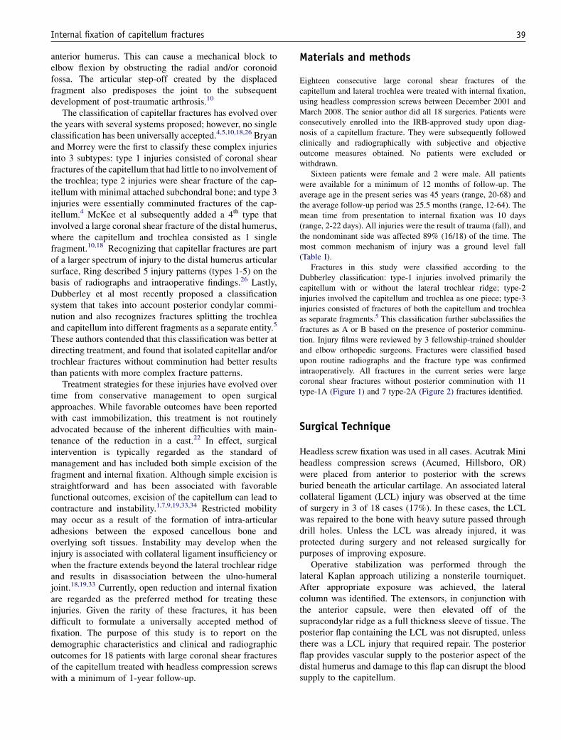
Fractures in this study were classified according to theDubberley classification: type-1 injuries involved primarily thecapitellum with or without the lateral trochlear ridge; type-2injuries involved the capitellum and trochlea as one piece; type-3injuries consisted of fractures of both the capitellum and trochleaas separate fragments.5 This classification further subclassifies thefractures as A or B based on the presence of posterior comminu-tion. Injury films were reviewed by 3 fellowship-trained shoulderand elbow orthopedic surgeons. Fractures were classified basedupon routine radiographs and the fracture type was confirmedintraoperatively. All fractures in the current series were largecoronal shear fractures without posterior comminution with 11type-1A (Figure 1) and 7 type-2A (Figure 2) fractures identified.
Surgical Technique
Headless screw fixation was used in all cases. Acutrak Miniheadless compression screws (Acumed, Hillsboro, OR)were placed from anterior to posterior with the screwsburied beneath the articular cartilage. An associated lateralcollateral ligament (LCL) injury was observed at the timeof surgery in 3 of 18 cases (17%). In these cases, the LCLwas repaired to the bone with heavy suture passed throughdrill holes. Unless the LCL was already injured, it wasprotected during surgery and not released surgically forpurposes of improving exposure.
Operative stabilization was performed through thelateral Kaplan approach utilizing a nonsterile tourniquet.After appropriate exposure was achieved, the lateralcolumn was identified. The extensors, in conjunction withthe anterior capsule, were then elevated off of thesupracondylar ridge as a full thickness sleeve of tissue. Theposterior flap containing the LCL was not disrupted, unlessthere was a LCL injury that required repair. The posteriorflap provides vascular supply to the posterior aspect of thedistal humerus and damage to this flap can disrupt the bloodsupply to the capitellum.
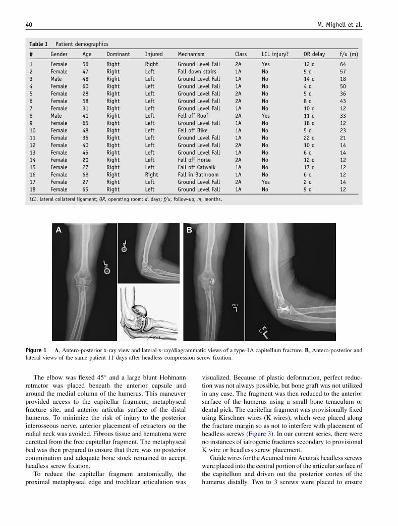
Table I Patient demographics
# Gender Age Dominant Injured Mechanism Class LCL injury? OR delay f/u (m)
1 Female 56 Right Right Ground Level Fall 2A Yes 12 d 642 Female 47 Right Left Fall down stairs 1A No 5 d 573 Male 48 Right Left Ground Level Fall 1A No 14 d 184 Female 60 Right Left Ground Level Fall 1A No 4 d 505 Female 28 Right Left Ground Level Fall 2A No 5 d 366 Female 58 Right Left Ground Level Fall 2A No 8 d 437 Female 31 Right Left Ground Level Fall 1A No 10 d 128 Male 41 Right Left Fell off Roof 2A Yes 11 d 339 Female 65 Right Left Ground Level Fall 1A No 18 d 1210 Female 48 Right Left Fell off Bike 1A No 5 d 2311 Female 35 Right Left Ground Level Fall 1A No 22 d 2112 Female 40 Right Left Ground Level Fall 2A No 10 d 1413 Female 45 Right Left Ground Level Fall 1A No 6 d 1414 Female 20 Right Left Fell off Horse 2A No 12 d 1215 Female 27 Right Left Fall off Catwalk 1A No 17 d 1216 Female 68 Right Right Fall in Bathroom 1A No 6 d 1217 Female 27 Right Left Ground Level Fall 2A Yes 2 d 1418 Female 65 Right Left Ground Level Fall 1A No 9 d 12
LCL, lateral collateral ligament; OR, operating room; d, days; f/u, follow-up; m, months.
Figure 1 A, Antero-posterior x-ray view and lateral x-ray/diagrammatic views of a type-1A capitellum fracture. B, Antero-posterior andlateral views of the same patient 11 days after headless compression screw fixation.
40 M. Mighell et al.
The elbow was flexed 45� and a large blunt Hohmannretractor was placed beneath the anterior capsule andaround the medial column of the humerus. This maneuverprovided access to the capitellar fragment, metaphysealfracture site, and anterior articular surface of the distalhumerus. To minimize the risk of injury to the posteriorinterosseous nerve, anterior placement of retractors on theradial neck was avoided. Fibrous tissue and hematoma werecuretted from the free capitellar fragment. The metaphysealbed was then prepared to ensure that there was no posteriorcomminution and adequate bone stock remained to acceptheadless screw fixation.
To reduce the capitellar fragment anatomically, theproximal metaphyseal edge and trochlear articulation was
visualized. Because of plastic deformation, perfect reduc-tion was not always possible, but bone graft was not utilizedin any case. The fragment was then reduced to the anteriorsurface of the humerus using a small bone tenaculum ordental pick. The capitellar fragment was provisionally fixedusing Kirschner wires (K wires), which were placed alongthe fracture margin so as not to interfere with placement ofheadless screws (Figure 3). In our current series, there wereno instances of iatrogenic fractures secondary to provisionalK wire or headless screw placement.
Guide wires for the Acumed mini Acutrak headless screwswere placed into the central portion of the articular surface ofthe capitellum and driven out the posterior cortex of thehumerus distally. Two to 3 screws were placed to ensure
Figure 2 A, Lateral x-ray view and antero-posterior xray/diagrammatic views of a type-2A capitellum fracture. B, Ten-monthpostoperative antero-posterior and lateral views of a patient with a type-2A capitellum fracture fixed with headless compression screws.
Internal fixation of capitellum fractures 41
rotational stability (Figure 1, B). When possible, a divergentscrew pattern was preferred. Furthermore, attempts weremade to assure adequate screw spread to avoid iatrogenicfragmentation of the capitellum. Each screw was placed overthe guide wire and countersunk for at least 2 mm to avoiderosion of the articular surface of the proximal radius fromprominent hardware. Intraoperative fluoroscopy was used toexamine hardware position and fracture reduction in theantero-posterior (AP) and lateral planes.
Postoperatively, patients were held in a splint at 90� offlexion in neutral rotation for 5-7 days. Patients were thenallowed to come out of the splint and perform a homeexercise program for the ensuing 4 weeks. The programconsisted of gravity-aided, active-assisted supine flexionand upright extension. Patients were instructed to performthese exercises a minimum of 3 times each day. Patients
were evaluated 6 weeks postoperatively and radiographswere obtained during that visit. Range of motion andfunction were also evaluated and patients were placed intoa supervised occupational therapy program if they lackeda 100� arc of flexion and extension.
Clinical outcomes were evaluated postoperatively atapproximately 4 weeks, 8 weeks, 3 months, 6 months, and1 year. After the 1-year visit, patients were called back forre-evaluation for the purposes of this study. At each follow-up clinic visit, range of motion (ROM) in flexion/extensionand pronation/supination was recorded. The Broberg-Morrey (BM) scale, which is a functional rating based ona 100-point index, and the American Shoulder and ElbowSurgeons (ASES) scores were used.2,13 Broberg-Morreyscores of 95-100 are considered excellent, 80-94 good,60-79 fair, and 0-59 points considered a poor outcome.
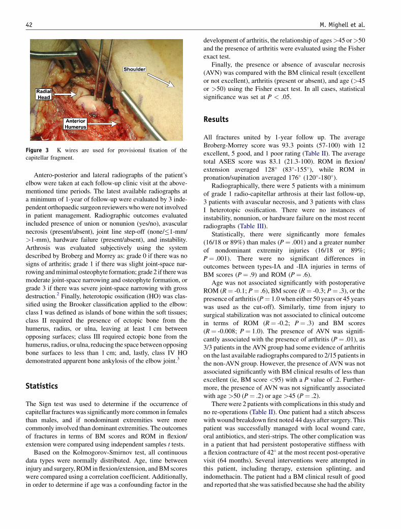
Figure 3 K wires are used for provisional fixation of thecapitellar fragment.
42 M. Mighell et al.
Antero-posterior and lateral radiographs of the patient’selbow were taken at each follow-up clinic visit at the above-mentioned time periods. The latest available radiographs ata minimum of 1-year of follow-up were evaluated by 3 inde-pendent orthopaedic surgeon reviewers who were not involvedin patient management. Radiographic outcomes evaluatedincluded presence of union or nonunion (yes/no), avascularnecrosis (present/absent), joint line step-off (none/�1-mm/>1-mm), hardware failure (present/absent), and instability.Arthrosis was evaluated subjectively using the systemdescribed by Broberg and Morrey as: grade 0 if there was nosigns of arthritis; grade 1 if there was slight joint-space nar-rowing and minimal osteophyte formation; grade 2 if there wasmoderate joint-space narrowing and osteophyte formation, orgrade 3 if there was severe joint-space narrowing with grossdestruction.2 Finally, heterotopic ossification (HO) was clas-sified using the Brooker classification applied to the elbow:class I was defined as islands of bone within the soft tissues;class II required the presence of ectopic bone from thehumerus, radius, or ulna, leaving at least 1 cm betweenopposing surfaces; class III required ectopic bone from thehumerus, radius, or ulna, reducing the space between opposingbone surfaces to less than 1 cm; and, lastly, class IV HOdemonstrated apparent bone ankylosis of the elbow joint.3
Statistics
The Sign test was used to determine if the occurrence ofcapitellar fractures was significantly more common in femalesthan males, and if nondominant extremities were morecommonly involved than dominant extremities. The outcomesof fractures in terms of BM scores and ROM in flexion/extension were compared using independent samples t tests.
Based on the Kolmogorov-Smirnov test, all continuousdata types were normally distributed. Age, time betweeninjury and surgery, ROM in flexion/extension, and BM scoreswere compared using a correlation coefficient. Additionally,in order to determine if age was a confounding factor in the
development of arthritis, the relationship of ages>45 or>50and the presence of arthritis were evaluated using the Fisherexact test.
Finally, the presence or absence of avascular necrosis(AVN) was compared with the BM clinical result (excellentor not excellent), arthritis (present or absent), and age (>45or >50) using the Fisher exact test. In all cases, statisticalsignificance was set at P < .05.
Results
All fractures united by 1-year follow up. The averageBroberg-Morrey score was 93.3 points (57-100) with 12excellent, 5 good, and 1 poor rating (Table II). The averagetotal ASES score was 83.1 (21.3-100). ROM in flexion/extension averaged 128� (83�-155�), while ROM inpronation/supination averaged 176� (120�-180�).
Radiographically, there were 5 patients with a minimumof grade 1 radio-capitellar arthrosis at their last follow-up,3 patients with avascular necrosis, and 3 patients with classI heterotopic ossification. There were no instances ofinstability, nonunion, or hardware failure on the most recentradiographs (Table III).
Statistically, there were significantly more females(16/18 or 89%) than males (P¼ .001) and a greater numberof nondominant extremity injuries (16/18 or 89%;P¼ .001). There were no significant differences inoutcomes between types-IA and -IIA injuries in terms ofBM scores (P¼ .9) and ROM (P¼ .6).
Age was not associated significantly with postoperativeROM (R¼ -0.1; P¼ .6), BM score (R¼ -0.3; P¼ .3), or thepresence of arthritis (P¼ 1.0 when either 50 years or 45 yearswas used as the cut-off). Similarly, time from injury tosurgical stabilization was not associated to clinical outcomein terms of ROM (R¼ -0.2; P¼ .3) and BM scores(R¼ -0.008; P¼ 1.0). The presence of AVN was signifi-cantly associated with the presence of arthritis (P¼ .01), as3/3 patients in the AVN group had some evidence of arthritison the last available radiographs compared to 2/15 patients inthe non-AVN group. However, the presence of AVN was notassociated significantly with BM clinical results of less thanexcellent (ie, BM score <95) with a P value of .2. Further-more, the presence of AVN was not significantly associatedwith age >50 (P¼ .2) or age >45 (P¼ .2).
There were 2 patients with complications in this study andno re-operations (Table II). One patient had a stitch abscesswith wound breakdown first noted 44 days after surgery. Thispatient was successfully managed with local wound care,oral antibiotics, and steri-strips. The other complication wasin a patient that had persistent postoperative stiffness witha flexion contracture of 42� at the most recent post-operativevisit (64 months). Several interventions were attempted inthis patient, including therapy, extension splinting, andindomethacin. The patient had a BM clinical result of goodand reported that she was satisfied because she had the ability
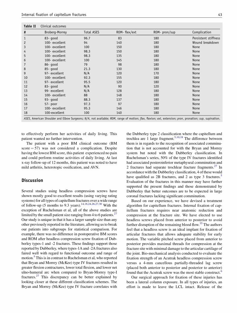
Table II Clinical outcomes
# Broberg-Morrey Total ASES ROM- flex/ext ROM- pron/sup Complication
1 83- good 96.7 83 180 Persistent stiffness2 100- excellent 94 140 180 Wound breakdown3 100- excellent 100 150 180 None4 100- excellent 98.3 150 180 None5 100- excellent 98.3 135 180 None6 100- excellent 100 145 180 None7 86- good 79 98 180 None8 85- good 21.3 130 180 None9 97- excellent N/A 120 170 None10 100- excellent 92.3 155 180 None11 97- excellent 95.5 120 180 None12 83- good N/A 90 120 None13 99- excellent N/A 128 180 None14 100- excellent 88 148 180 None15 93- good 88.3 137 180 None16 57- poor 97.3 97 180 None17 100- excellent 95.3 146 180 None18 100-excellent 100 140 180 None
ASES, American Shoulder and Elbow Surgeons; N/A, not available; ROM, range of motion; flex, flexion; ext, extension; pron, pronation; sup, supination.
Internal fixation of capitellum fractures 43
to effectively perform her activities of daily living. Thispatient wanted no further intervention.
The patient with a poor BM clinical outcome (BMscore¼ 57) was not considered a complication. Despitehaving the lowest BM score, this patient experienced no painand could perform routine activities of daily living. At lastx-ray follow-up of 12 months, this patient was noted to havemild arthritis, heterotopic ossification, and AVN.
Discussion
Several studies using headless compression screws haveshown mostly good to excellent results (using varying ratingsystems) for all types of capitellum fractures over a wide rangeof follow-up (5 months to 9.3 years).14,18,24,26,27,30 With theexception of Ruchelsman et al, all of the above studies arelimited by the small patient size ranging from 4 to 6 patients.27
Our study is unique in that it has a larger sample size than anyother previously reported in the literature, allowing us to breakour patients into subgroups for statistical comparison. Forexample, there was no difference in postoperative BM scoresand ROM after headless compression screw fixation of Dub-berley types-1 and -2 fractures. These findings support thosereported by Dubberley, where types-1A and -2A fractures alsofaired well with regard to functional outcome and range ofmotion.5 This is in contrast to Ruchelsman et al, who reportedthat Bryan and Morrey (McKee) type IV fractures resulted ingreater flexion contractures, lower total flexion, and lower netulno-humeral arc when compared to Bryan-Morrey type-Ifractures.27 This discrepancy can be better explained bylooking closer at these different classification schemes. TheBryan and Morrey (McKee) type IV fracture correlates with
the Dubberley type 2 classification where the capitellum andtrochlea are 1 large fragment.5,10,18 The difference betweenthem is in regards to the recognition of associated comminu-tion that is not accounted for with the Bryan and Morreysystem but noted with the Dubberley classification. InRuchelsman’s series, 50% of the type IV fractures identifiedhad associated posteroinferior metaphyseal comminution and2 fractures had separate trochlear fracture fragments.27 Inaccordance with the Dubberley classification, 4 of these wouldhave qualified as 2B fractures, and 2 as type 3 fractures.5
Evaluation of the fractures in this manner may have furthersupported the present findings and those demonstrated byDubberley that better outcomes are to be expected in largecoronal fractures lacking significant comminution.
Based on our experience, we have devised a treatmentalgorithm for capitellum fractures. Internal fixation of cap-itellum fractures requires near anatomic reduction andcompression at the fracture site. We have elected to useheadless screws placed from anterior to posterior to avoidfurther disruption of the remaining blood flow.35 The authorsfeel that a headless screw is an ideal implant for fixation ofarticular fractures that allows adequate stability for earlymotion. The variable pitched screw placed from anterior toposterior provides maximal threads for compression at thefracture site with minimal damage to the articular cartilage ofthe joint. Bio-mechanical analysis conducted to evaluate thefixation strength of an Acutrak headless compression screwversus a 4-mm cancellous partially-threaded lag screw(placed both anterior to posterior and posterior to anterior)found that the Acutrak screw was the most stable construct.6
Our surgical approach for fixation of these injuries hasbeen a lateral column exposure. In all types of injuries, aneffort is made to leave the LCL intact. Release of the
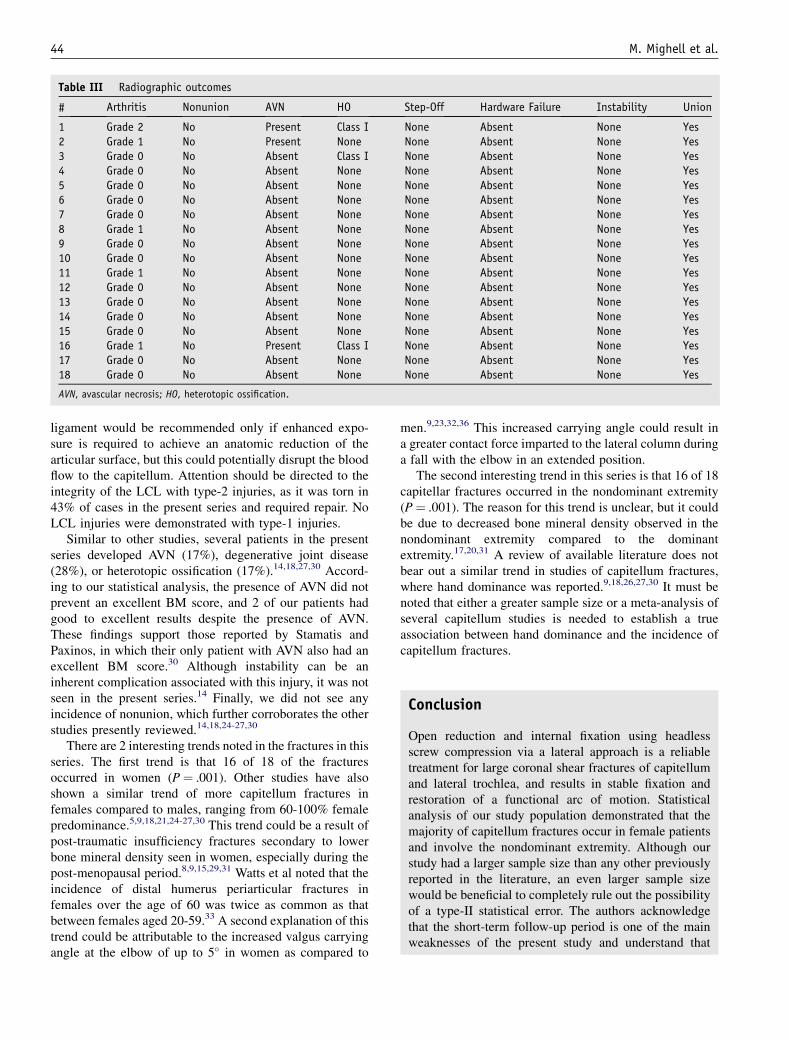
Table III Radiographic outcomes
# Arthritis Nonunion AVN HO Step-Off Hardware Failure Instability Union
1 Grade 2 No Present Class I None Absent None Yes2 Grade 1 No Present None None Absent None Yes3 Grade 0 No Absent Class I None Absent None Yes4 Grade 0 No Absent None None Absent None Yes5 Grade 0 No Absent None None Absent None Yes6 Grade 0 No Absent None None Absent None Yes7 Grade 0 No Absent None None Absent None Yes8 Grade 1 No Absent None None Absent None Yes9 Grade 0 No Absent None None Absent None Yes10 Grade 0 No Absent None None Absent None Yes11 Grade 1 No Absent None None Absent None Yes12 Grade 0 No Absent None None Absent None Yes13 Grade 0 No Absent None None Absent None Yes14 Grade 0 No Absent None None Absent None Yes15 Grade 0 No Absent None None Absent None Yes16 Grade 1 No Present Class I None Absent None Yes17 Grade 0 No Absent None None Absent None Yes18 Grade 0 No Absent None None Absent None Yes
AVN, avascular necrosis; HO, heterotopic ossification.
44 M. Mighell et al.
ligament would be recommended only if enhanced expo-sure is required to achieve an anatomic reduction of thearticular surface, but this could potentially disrupt the bloodflow to the capitellum. Attention should be directed to theintegrity of the LCL with type-2 injuries, as it was torn in43% of cases in the present series and required repair. NoLCL injuries were demonstrated with type-1 injuries.
Similar to other studies, several patients in the presentseries developed AVN (17%), degenerative joint disease(28%), or heterotopic ossification (17%).14,18,27,30 Accord-ing to our statistical analysis, the presence of AVN did notprevent an excellent BM score, and 2 of our patients hadgood to excellent results despite the presence of AVN.These findings support those reported by Stamatis andPaxinos, in which their only patient with AVN also had anexcellent BM score.30 Although instability can be aninherent complication associated with this injury, it was notseen in the present series.14 Finally, we did not see anyincidence of nonunion, which further corroborates the otherstudies presently reviewed.14,18,24-27,30
There are 2 interesting trends noted in the fractures in thisseries. The first trend is that 16 of 18 of the fracturesoccurred in women (P¼ .001). Other studies have alsoshown a similar trend of more capitellum fractures infemales compared to males, ranging from 60-100% femalepredominance.5,9,18,21,24-27,30 This trend could be a result ofpost-traumatic insufficiency fractures secondary to lowerbone mineral density seen in women, especially during thepost-menopausal period.8,9,15,29,31 Watts et al noted that theincidence of distal humerus periarticular fractures infemales over the age of 60 was twice as common as thatbetween females aged 20-59.33 A second explanation of thistrend could be attributable to the increased valgus carryingangle at the elbow of up to 5� in women as compared to
men.9,23,32,36 This increased carrying angle could result ina greater contact force imparted to the lateral column duringa fall with the elbow in an extended position.
The second interesting trend in this series is that 16 of 18capitellar fractures occurred in the nondominant extremity(P¼ .001). The reason for this trend is unclear, but it couldbe due to decreased bone mineral density observed in thenondominant extremity compared to the dominantextremity.17,20,31 A review of available literature does notbear out a similar trend in studies of capitellum fractures,where hand dominance was reported.9,18,26,27,30 It must benoted that either a greater sample size or a meta-analysis ofseveral capitellum studies is needed to establish a trueassociation between hand dominance and the incidence ofcapitellum fractures.
Conclusion
Open reduction and internal fixation using headlessscrew compression via a lateral approach is a reliabletreatment for large coronal shear fractures of capitellumand lateral trochlea, and results in stable fixation andrestoration of a functional arc of motion. Statisticalanalysis of our study population demonstrated that themajority of capitellum fractures occur in female patientsand involve the nondominant extremity. Although ourstudy had a larger sample size than any other previouslyreported in the literature, an even larger sample sizewould be beneficial to completely rule out the possibilityof a type-II statistical error. The authors acknowledgethat the short-term follow-up period is one of the mainweaknesses of the present study and understand that
Internal fixation of capitellum fractures 45
a longer follow-up period is necessary to determine thetrue incidence of post traumatic arthritis of the elbow.The classification system utilized is a subject of debate;however, the Dubberley system takes into account thecomplexity of these fractures and helps us predict whichfracture patterns are best treated through a lateralapproach. Our clinical results are similar to the findingsof Dubberley, that good results can be expected ofcoronal shear fractures of the distal humerus withoutassociated posterior comminution.5
Acknowledgment
The authors wish to express a special thanks to themedical staff at University Community Hospital wherethe majority of the surgeries were performed. Theauthors would like to acknowledge Michele Pliner, ofthe Foundation for Orthopaedic Research and Education,for her editorial assistance and Jean Bonnette for herillustrations.
References
1. Alvarez E, Patel MR, Nimberg G, Pearlman HS. Fracture of the
capitulum humeri. J Bone Joint Surg Am 1975;57:1093-6.
2. Broberg MA, Morrey BF. Results of delayed excision of the radial
head after fracture. J Bone Joint Surg Am 1986;68:669-74.
3. Brooker AF, Bowerman JW, Robinson RA, Riley LH. Ectopic ossifi-
cation following total hip replacement. Incidence and a method of
classification. J Bone Joint Surg Am 1973;55:1629-32.
4. Bryan RS, Morrey BF. Fractures of the distal humerus. In: Morrey BF,
editor. The elbow and its disorders. Philadelphia, PA: WB Saunders;
1985. p. 302-39.
5. Dubberley JH, Faber KJ, Macdermid JC, Patterson SD, King GJ.
Outcome after open reduction and internal fixation of capitellar and
trochlear fractures. J Bone Joint Surg Am 2006;88:46-54.
6. Elkowitz SJ, Polatsch DB, Egol KA, Kummer FJ, Koval KJ.
Capitellum fractures: A biomechanical evaluation of three fixation
methods. J Orthop Trauma 2002;16:503-6.
7. Fowles JV, Kassab MT. Fracture of the capitulum humeri: Treatment
by excision. J Bone Joint Surg Am 1974;56:794-8.
8. Gass M, Dawson-Hughes B. Preventing osteoporosis-related fractures:
An overview. Am J Med 2006;119(4S1):S3-11.
9. Grantham SA, Norris TR, Bush DC. Isolated fracture of the humeral
capitellum. Clin Orthop Relat Res 1981;161:262-9.
10. Guitton TG, Doornberg JN, Raaymakers EL, Ring D, Kloen P. Fractures
of the capitellum and trochlea. J Bone Joint Surg Am 2009;91:390-7.
11. Harrington JP, McKee MD. Coronal shear fractures of the capitellum
and trochlea. Tech Shoulder Elbow Surg 2000;1:240-6.
12. Hotchkiss RN. Fractures and dislocations of the elbow. In:
Rockwood CA, Green DP, Bucholz RW, Heckman JD, editors.
Rockwood and Greens’ fractures in adults. Fourth ed. Philadelphia,
PA: Lippincott-Raven; 1996. p. 929-1024.
13. King GJ, Richards RR, Zuckerman JD, Blasier R, Dillman C,
Friedman RJ, et al. A standardized method for assessment of elbow
function. J Shoulder Elbow Surg 1999;8:351-4.
14. Lambert SM, Pike J, Railton GT. Fractures of the humeral capitellum:
Herbert screw fixation. J R Coll Surg Edinb 1994;39:321-3.
15. Lane JM, Serota AC, Raphael B. Osteoporosis: Differences and
similarities in male and female patients. Orthop Clin North Am 2006;37:
601-9.
16. Lecestre R. Round table on fracture of the lower end of the Humerus.
Societe Francais de Chirurgie Orthopedique et Traumatologie. Orthop
Trans 1980;4:123.
17. MacIntyre NJ, Adachi JD, Webber CE. In vivo detection of struc-
tural differences between dominant and nondominant radii using
peripheral quantitative computed tomography. J Clin Densitom
1999;2:413-22.
18. McKee MD, Jupiter JB, Bamberger HB. Coronal shear fractures of the
distal end of the humerus. J Bone Joint Surg Am 1996;78:49-54.
19. Milch H. Fractures and fracture dislocations of the humeral condyles.
J Trauma 1964;4:592-607.
20. Min JY, Min KB, Paek D, Cho SI. Side differences in the bone density
of the distal radius and calcaneus in Koreans aged 4-86 years. J Clin
Densitom 2007;10:184-8.
21. Mosheiff R, Liebergall M, Elyashuv O, Mattan Y, Segal D. Surgical
treatment of fractures of the capitellum in adults: A modified
technique. J Orthop Trauma 1991;5:297-300.
22. Ochner RS, Bloom H, Palumbo RC, Coyle MP. Closed reduction of
coronal fractures of the capitellum. J Trauma 1996;40:199-203.
23. Paraskevas G, Papadopoulos A, Papaziogas B, Spanidou S,
Argiriadou H, Gigis J. Study of the carrying angle of the human elbow
joint in full extension: a morphometric analysis. Surg Radiol Anat
2004;26:19-23.
24. Poynton AR, Kelly IP, O’Rourke SK. Fractures of the capitellum-
a comparison of two fixation methods. Injury 1998;29:341-3.
25. Richards RR, Khoury GW, Burke FD, Waddell JP. Internal fixation of
capitellar fractures using Herbert screws: a report of four cases. Can J
Surg 1987;30:188-91.
26. Ring D, Jupiter JB, Gulotta L. Articular fractures of the distal part of
the humerus. J Bone Joint Surg Am 2003;85-A:232-8.
27. Ruchelsman DE, Tejwani NC, Kwon YW, Egol KA. Open reduction
and internal fixation of capitellar fractures with headless screws.
J Bone Joint Surg Am 2008;90:1321-9.
28. Sano S, Rokkaku T, Saito S, Tokunaga S, Abe Y, Moriya H. Herbert
screw fixation of capitellar fractures. J Shoulder Elbow Surg 2005;14:
307-11.
29. Seeman E. Reduced bone density in women with fractures:
Contribution of low peak bone density and rapid bone loss. Osteoporos
Int 1994;4(S1):S15-25.
30. Stamatis E, Paxinos O. The treatment and functional outcome of Type
IV coronal shear fractures of the distal humerus: A retrospective
review of five cases. J Orthop Trauma 2003;17:279-84.
31. Taaffe DR, Lewis B, Marcus R. Quantifying the effect of hand
preference on upper limb bone mineral and soft tissue composition in
young and elderly women by dual-energy X-ray absorptiometry. Clin
Physiol 1994;14:393-404.
32. Van Roy P, Baeyens JP, Fauvart D, Lanssiers R, Clarijs JP. Arthro-
kinematics of the elbow: Study of the carrying angle. Ergonomics
2005;48:1645-56.
33. Watts AC, Morris A, Robinson CM. Fractures of the distal humeral
articular surface. J Bone Joint Surg Br 2007;89:510-5.
34. Wilson JN. Injuries of the elbow. In: Watson-Jones R, editor. Fractures
and joint injuries. Sixth ed. Edinburgh: Churchill Livingstone; 1982.
p. 587-90.
35. Yamaguchi K, Sweet FA, Bindra R, Morrey BF, Gelberman RH. The
extraosseous and intraosseous arterial anatomy of the adult elbow.
J Bone Joint Surg Am 1997;79:1653-62.
36. Yilmaz E, Karakurt L, Belhan O, Bulut M, Serin E, Avci M. Variation
of carrying angle with age, sex, and special reference to side.
Orthopedics 2005;28:1360-3.
Copyright © 2022 FDOKUMEN