Quasi-Feynman formulas – a method of obtaining the ... - arXiv
Upload
independentCategory
view
0download
0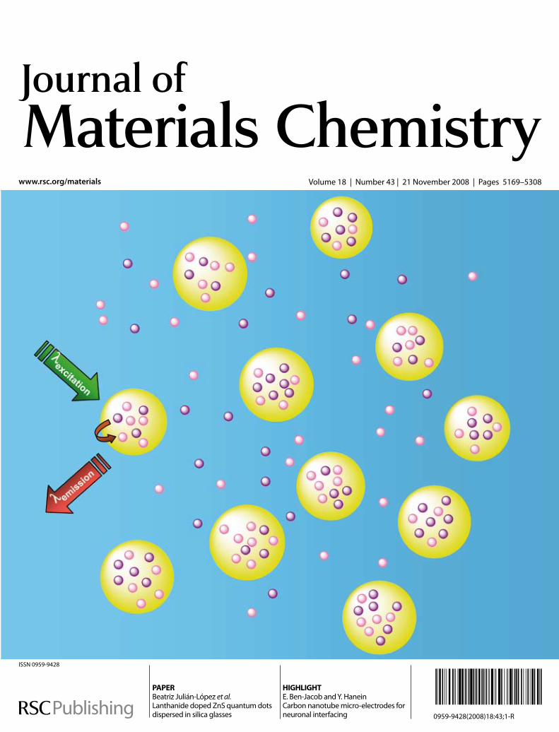
Volum
e18|Num
ber43|2008Journal of M
aterials Chem
istry Pages5169–5308
www.rsc.org/materials Volume18|Number43|21November2008|Pages5169–5308
ISSN0959-9428
PAPERBeatrizJulián-Lópezet al.LanthanidedopedZnSquantumdotsdispersedinsilicaglasses
HIGHLIGHTE.Ben-JacobandY.HaneinCarbonnanotubemicro-electrodesforneuronalinterfacing 0959-9428(2008)18:43;1-R
www.rsc.org/metallomicsRegistered Charity Number 207890
A new journal from RSC Publishinglaunching in 2009
MetallomicsIntegrated biometal science
This timely new journal will cover the research � elds related to metals in biological, environmental and clinical systems and is expected to be the core publication for the emerging metallomics community. The journal will be supported by an international Editorial Board, chaired by Professor Joseph A. Caruso of the University of Cincinnati/Agilent Technologies Metallomics Center of the Americas.
Metallomics will publish six issues in the � rst year, increasing to 12 issues in 2010. The journal will contain a full mix of research articles including Communications, Reviews, Full papers, and Editorials. From launch, the latest issue will be freely available online to all readers. Free institutional access to previous issues throughout 2009 and 2010 will be available following a simple registration process.
Contact the editor, Niamh O’Connor, at [email protected] for further information or visit the website.
Num
ber 1|
2008M
etallomics
Pages1–100 1754-5692(2008)1:1;1-6
www.rsc.org/metallomics Volume 1 | Number 1 | January 2009 | Pages 1–100
ISSN 1756-5901
MetallomicsIntegrated biometal science
1756-5901(2009) 1:1;l-m
060877
Submit your work now!Supporting the

PAPER www.rsc.org/materials | Journal of Materials Chemistry
Lanthanide doped ZnS quantum dots dispersed in silica glasses: an easyone pot sol–gel synthesis for obtaining novel photonic materials†
Jose Planelles-Arago,*a Beatriz Julian-Lopez,*a Eloisa Cordoncillo,a Purificacion Escribano,a Fabienne Pelle,b
Bruno Vianab and Clement Sanchezb
Received 2nd June 2008, Accepted 13th August 2008
First published as an Advance Article on the web 3rd October 2008
DOI: 10.1039/b809254k
Silica glasses containing both ZnS quantum dots (QDs) and luminescent lanthanide ions are attractive
candidates to develop new lighting displays, sensor devices or laser emitters. This work reports an easy
sol–gel method to prepare Eu3+-doped and Eu3+,Mn2+-codoped ZnS nanocrystals dispersed in
a transparent silica matrix. Semiconductor nanocrystals with an average size of 5–6 nm and exhibiting
both cubic and hexagonal phases were obtained at low temperature. The luminescent interactions
between ZnS QDs, Eu3+ and Mn2+ ions provided materials with different optical responses but also
gave information about the organization of the different species in the nanocomposite. Indeed, Eu ions
were found to be both dispersed within the silica and located at the surface of the nanochalcogenide, the
latter providing a ZnS / Eu3+ energy transfer. Incorporation of Mn2+ into the ZnS lattice induced the
appearance of defect states that enhance the blue luminescence of the nanocomposite. These results
underline the sensitivity of optical processes to the nature and organization of the active species, which
is of vital importance for the design of photonic materials.
Introduction
The synthesis and optical studies of inorganic and hybrid
nanostructured materials have become a major interdisciplinary
area of research over the past 20 years.1,2 Semiconductor nano-
particles (NPs) play a major role in several new technologies, the
intense interest in this area derives from their unique chemical,
physical and electronic properties, which give rise to their
potential uses in the fields of displays, lighting, sensors and lasers,
as well as other areas.
Zinc sulfide (ZnS) as an important wide-bandgap (3.6 eV)
semiconductor has been used as a key material for ultraviolet
light-emitting diodes and injection lasers,3 flat-panel displays,4
electroluminescent devices and infrared windows.5
In recent years, some unique characteristics of ZnS nano-
crystals different from bulk crystals have enlarged the range of
applications. Among the wide-bandgap semiconductors, ZnS
has a large exciton binding energy (40 meV) and a small
Bohr radius (2.4 nm) which make it an excellent candidate
for exploring the intrinsic recombination processes in dense
excitonic systems.
Furthermore, ZnS nanocrystals or quantum dots, having sizes
comparable to that of the bulk Bohr exciton radius, exhibit
discrete electron energy levels with high oscillator strength
aDepartamento de Quımica Inorganica y Organica, ESTCE, UniversitatJaume I, 12071, Castellon, Spain. E-mail: [email protected]; [email protected].; Fax: +34 964 728214; Tel: +34 964 728234bLaboratoire de Chimie de la Matiere Condensee, CNRS-UMR 7574,75231, Paris, France. E-mail: [email protected].; Fax: +33146347489; Tel: +33 153737933
† Electronic supplementary information (ESI) available: ATR-FTIRspectra of as-synthesized and fired EZS samples. See DOI:10.1039/b809254k
This journal is ª The Royal Society of Chemistry 2008
and strong luminescence due to the well known quantum
confinement effects. Thus, the study of this nanosized semi-
conductor is of considerable importance and great efforts have
been focused on their synthesis and physical properties.6 As one
of the most important semiconductors, ZnS has been known
for a long time as a versatile and excellent phosphor host
material: when doped with appropriate ions, a variety of
photoluminescent, cathodoluminescent,7 electroluminescent,8
and thermoluminescent9 properties can be achieved. The lumi-
nescence characteristics of impurity-activated ZnS nanocrystals
differ markedly from those of bulk ZnS. Yang et al.10 give two
reasons for this behavior: first, the high degree of dispersion in
the nanocrystalline system; second, the size-dependent proper-
ties of semiconductor nanoparticles. It is well known that
rare earth (RE) elements are effective luminescent centres for
RE-doped semiconductors, because the excitation of the RE
ions can occur by the recombination of photogenerated carriers
confined in the semiconductor, and subsequent energy transfer
to the RE ions.11
In a previous work12 we reported the synthesis and optical
study of Eu3+-doped CdS nanocrystals embedded in silica
matrices. Lanthanide doped CdS nanoparticles confined and
randomly dispersed in highly transparent silica glasses were
prepared by an easy one-pot sol–gel methodology that provides
excellent optical properties.13 An accurate spectroscopic study
revealed the existence of energy transfer processes between CdS
nanocrystals and europium ions when these species are spatially
close. However, very short distances between the active species
led to back-transfer processes and a reduction of the lumines-
cence. When CdS nanocrystals are replaced by ZnS, a more
extended, useful and less toxic semiconductor, important
changes in the optical properties of the material are expected.
Since the ionic radius of the Cd2+ ion is larger than that of the
J. Mater. Chem., 2008, 18, 5193–5199 | 5193

Zn2+ ion (0.78 and 0.60 A respectively in four-fold coordination
for blende-type structures),14 it is expected that Eu3+ ions (with an
ionic radius of 1.07 A for eight-fold coordination) are not able to
be incorporated in the ZnS network and could be located at the
nanoparticle surface. In this situation, the ZnS / Eu3+ energy
transfer could be more efficient (i.e. Eu3+ ions are incorporated
more easily in CdS than in ZnS and therefore a weaker Eu3+ /
ZnS back-transfer process should occur). Furthermore, in order
to facilitate energy transfer to Eu3+ centers upon excitation of the
ZnS host, Mn2+ ions can be introduced in these materials. Mn-
doped ZnS nanocrystals have been widely studied9 showing
a good degree of Mn2+ incorporation in the ZnS host and an
efficient ZnS / Mn2+ energy transfer phenomenon that gives
rise to a characteristic orange luminescence attributed to the spin
forbidden electronic transition of Mn2+ ions in a tetrahedral site.
On the other hand, efficient Mn2+ / Ln3+ energy transfer
processes (where Ln3+ is a lanthanide ion) have been observed in
a variety of rare-earth doped Mn containing crystals such as
MnF2 and RbMnF3.15 Due to the similar energies of the transi-
tions 4T1 /6A1 of Mn2+ and 5D0 /
7FJ (J ¼ 0–6) of Eu3+ ions
(see Fig. 1), an energy transfer Mn2+ / Eu3+ is expected when
ZnS NPs are excited.
This work presents the structural and optical study of Eu3+-
doped and Eu3+,Mn2+-codoped ZnS nanoparticles trapped in
an amorphous and transparent silica matrix. These multicom-
ponent nanomaterials have been prepared by a one pot sol–gel
technique, where nanocrystals are generated by thermal treat-
ment. Structural characterization of the nanocomposites is
performed by high resolution transmission electron microscopy
(HRTEM). The optical features of each system, as well as
energy transfer between semiconductor nanoparticles and the
doping ions, are analyzed by UV-visible spectrophotometry,
excitation and emission spectra and time-resolved lumines-
cence.
Fig. 1 Optical mechanism suggested for Eu3+,Mn2+-codoped ZnS
nanocrystals. The Mn2+ ions provide energy transfer to Eu3+ centers upon
excitation of ZnS nanocrystals.
5194 | J. Mater. Chem., 2008, 18, 5193–5199
Experimental
Sample preparation
Highly transparent and colorless silica monoliths containing
Eu3+-doped and Eu3+,Mn2+-codoped ZnS nanoparticles were
prepared using a modification of a previously reported sol–gel
methodology.12,13 In our procedure ZnS nanocrystals are
generated by decomposition of Zn2+-DMSO (DMSO ¼ dime-
thylsulfoxide) complexes formed in solution and their growth is
restricted by the sol–gel silica medium avoiding undesired
particle aggregation processes. The role of DMSO is two-fold: it
acts as a solvent and as a sulfur source, thus avoiding the use of
H2S in the synthesis of ZnS. After a mild heat treatment, highly
transparent glasses containing crystalline Eu3+-doped ZnS and
Eu3+,Mn2+-codoped nanoparticles are obtained providing
interesting optical properties.
The Eu3+-doped ZnS sample, labelled as EZS, is prepared as
follows: solutions of zinc nitrate (Zn(NO3)2$6H2O, 98%, Strem)
and europium nitrate (Eu(NO3)3$6H2O, 99.9%, Strem),
dissolved in the minimum amount of DMSO (dimethyl sulfoxide,
(CH3)2SO, Panreac), are mixed and left stirring for 30 min. The
resulting solution is added to a mixture of TEOS (tetraethox-
ysilane, (Si(OC2H5)4, Strem, 99%), DMSO, and water to obtain
a molar composition of 1 TEOS : 44.4 DMSO : 0.025 Zn(II) :
7.182 � 10�3 Eu(III) : 10.5 H2O. After stirring for 30 min at room
temperature, the resulting solution is heated at 80 �C for a week.
Finally, different amounts of the final sol are poured into vessels
and allowed to dry by means of infrared lamps. After drying,
transparent and colorless xerogels are fired in a furnace at 500 �C
for 30 min with a heating rate of 5 �C min�1. This treatment was
chosen according to the results of differential thermal analysis
and thermogravimetry.
The Eu3+,Mn2+-codoped ZnS sample, labelled as EMZS, is
prepared as described for the EZS sample, but using
manganese(II) acetylacetonate (Mn(CH3COCHC(O)CH3)2,
95%, Strem) as Mn2+ precursor. The resulting molar composition
of EMZS sample is 1 TEOS : 44.4 DMSO : 0.025 Zn(II) : 8.587 �10�3 Mn(II) : 3.591 � 10�3 Eu(III) : 10.5 H2O. As references for
optical measurements, non-doped ZnS nanoparticles in SiO2
(ZS), Eu3+-doped SiO2 (ES) and pure SiO2 (S) samples were also
prepared following the same experimental procedure.
Characterization techniques
The characterization by high resolution transmission electron
microscopy (HRTEM) was carried out on a Philips CM20
(200 kV) microscope (CME, Orleans University, France). For
this analysis, the ground samples were dispersed in ethanol and
a drop was deposited onto a carbon-coated copper grid.
The chemical compositions of the materials were determined
by X-ray fluorescence (XRF) measurements using a Siemens SRS
3000 wavelength dispersive XRF spectrophotometer.
Absorption spectra were recorded in a Cary 5 Varian spec-
trophotometer using an attenuator in order to avoid saturation
of the absorption signal. Excitation and emission spectra were
recorded using an Eclipse (Varian) spectrofluorimeter, contain-
ing a Xe lamp source. Time-resolved luminescence measurements
were performed with lexc ¼ 280 nm and lem ¼ 617 nm
(5D0 decay) and lifetime values were extracted from decay
This journal is ª The Royal Society of Chemistry 2008

Table 2 Chemical compositions of EZS and EMZS samples
Si Zn Mn Eu
EMZS 92.0573% 5.6779% 1.3301% 0.9347%EZS 92.4897% 5.6691% — 1.8412%
profiles. All optical measurements were carried out at room
temperature.
The Fourier transform infrared (FTIR) spectra were recorded
between 4000 and 400 cm�1 (resolution of 4 cm�1) on a Equinox
55 Bruker spectrometer (ESI†). The spectra were obtained on
bulk samples using an ATR (attenuated total reflection) instru-
ment equipped with a ZnSe monocrystal. The advantage of
measuring with the ATR device is to detect the presence
of hydroxyl groups inside the sol–gel material.
Results and discussion
Microstructural characterization
The first characterization of the EZS and EMZS samples was
performed by X-ray diffraction. The patterns of raw and fired
monoliths exhibited the typical vitreous halo characteristic of the
amorphous silica matrix. In order to observe nanocrystallization
inside the amorphous silica glass we used high resolution trans-
mission electron microscopy (HRTEM). HRTEM micrographs
evidence the crystalline nature of the nanoaggregates in annealed
samples. Fig. 2 shows a typical spherical 6 nm ZnS particle
embedded in the amorphous silica medium. The selected area
electron diffraction (SAED) pattern of these nanocrystallites
is shown in the inset of Fig. 2. As expected, in the case of
nanocrystals, the electron diffraction pattern shows a set of rings
instead of spots due to the random orientation of the nano-
crystallites, corresponding to the diffraction from different
atomic planes of the nanocrystallites.16
Higher magnification TEM pictures of individual crystallites
were recorded in order to resolve the lattice planes. The d inter-
planar distances obtained from the HRTEM micrographs, and
shown in Table 1, are similar to those included in JCPDS files
for cubic and hexagonal ZnS polymorphs (JCPDS: 5-566 and
79-2204). These data suggest that for the nanometer-sized
particles of ZnS, the equilibrium temperature for the cubic-to-
Fig. 2 HRTEM micrographs and EDX pattern (inset) of EZS sample
after firing at 500 �C.
Table 1 d-Spacing of the crystalline ZnS NPs measured from HRTEMmicrographs
d-Spacing/A (hkl) values Phase assignment
3.3 (100) hexagonal3.1 (002)/(111) hexagonal/cubic2.6 (200) cubic
This journal is ª The Royal Society of Chemistry 2008
hexagonal transition is significantly reduced from the bulk
value.17 Therefore, we can find both ZnS phases coexisting in our
samples. This result is consistent with the observations of
Goldstein et al.18 who observed the melting temperature of
nanoparticles of CdS to be substantially reduced over the bulk
value.
Energy dispersive X-ray (EDX) analyses were performed in
order to determine the composition of the aggregates but their
size was smaller than that of the detection area and no reliable
conclusions could be extracted. No information about doping
ions localization (both Eu3+ and Mn2+ ions) could be obtained
because of the low molar percentage of these elements in our
samples. However, XRF analysis will shed some light on the
content of these ions within the material. Table 2 details the
chemical compositions of EZS and EMZS samples determined
by XRF measurements. One can see from the XRF data that the
Zn, Eu and Mn contents are very close to those in the precursor
DMSO solutions.
UV-Visible spectroscopy
The UV-visible absorption spectra of annealed samples are
illustrated in Fig. 3. The spectrum of the reference S sample (pure
silica) reveals the good optical quality of the glasses since it is
completely transparent in the 250–700 nm range. Upon incor-
porating ZnS nanoparticles into the silica matrix (ZS), the
resulting spectrum shows an absorption edge at �314 nm
(3.95 eV, value obtained from the first derivative method used to
determine the edge position in all samples). The corresponding
bandgap energy for the ZnS nanocrystallites is larger than that
observed in bulk ZnS (3.7 eV, 335 nm). This blue shift toward
higher energy can be explained as a quantum size effect, due
to the electron–hole confinement in a small volume.19 Indeed,
Fig. 3 UV-Visible optical absorption spectra of Eu3+-doped and
Eu3+,Mn2+-codoped ZnS samples, EZS and EMZS respectively. As
references, non-doped ZnS nanoparticles in SiO2 (ZS) and silica matrix
(S) absorption spectra have been included. The acronym a.u. in the y axis
refers to absorbance units.
J. Mater. Chem., 2008, 18, 5193–5199 | 5195

particle sizes of ZnS were found to be around 5 nm, according to
a calibration curve presented by Rossetti et al.20 This curve
following the Brus model is based on variational calculations
of the cubic ZnS lowest excited state energy as a function of
nanoparticle diameter.21 These particle size calculations are in
good agreement with our HRTEM observations.
Regarding ZnS nanocrystals in EZS and EMZS samples, they
exhibit similar absorption edges to the ZS sample. These profiles
are also analogous to those corresponding to Eu3+-doped ZnS
nanoparticles obtained by other synthesis methods and reported
previously.22 Some studies indicate that addition of the doping
ions cannot change the average size of the nanocrystals,23 but it
can increase the size distribution significantly, modifying the
shape of absorption curves. In our curves, only slight contribu-
tions due to the presence of Eu3+ and Mn2+ doping ions can be
observed. The presence of Eu3+ ions increases the absorption
intensity in the UV region, which can be attributed to the Eu–O
charge transfer. Furthermore, absorption bands corresponding
to the forbidden 4f–4f transitions are usually weak and only
a small absorption located at 394 nm is detected and attributed to
the 7F0 / 5L6 transition.7
Photoluminescence: excitation
Photoluminescence excitation spectra of annealed Eu3+ con-
taining samples (ES, EZS and EMZS) performed monitoring the5D0 /
7F2 transition (615 nm) are presented in Fig. 4. For the ES
sample, the excitation spectrum exhibits the bands associated
with the Eu3+ 4f–4f transitions at 362 nm (7F1 / 5D4), 380 nm
(7F0 /5G3), 394 nm (7F0 /
5L6), 414 nm (7F0 /5D3), 465 nm
(7F0 / 5D2), 526 nm (7F0 / 5D1) and 534 nm (7F1 / 5D1). If
ZnS nanocrystals are present and spatially close to Eu3+ ions,
an energy transfer from semiconductor nanoparticles to Eu3+
centers should be expected according to our previous studies on
Eu3+-doped CdS nanocrystals.12 The excitation spectrum of the
Eu3+ emission suggests the existence of ZnS / Eu3+ energy
Fig. 4 Excitation spectra of Eu3+-doped and Eu3+,Mn2+-codoped ZnS
samples, EZS and EMZS respectively, the emission of Eu3+ was moni-
tored at 615 nm (5D0 / 7F2 transition). As a reference, the Eu3+-doped
SiO2 (ES) spectrum has been included. Spectra have been normalized to
the 394 nm band and then shifted along the y-axis (in arbitrary units, a.u.)
with the aim of facilitating their visibility.
5196 | J. Mater. Chem., 2008, 18, 5193–5199
transfer process in Eu3+-doped ZnS samples. Thus, when the ZnS
absorption band is present in the excitation spectrum ZnS /
Eu3+ transfer occurs. Although the excitation spectrum of the
Eu3+-doped ZnS sample (EZS) is dominated by the intra-
configurational 4f–4f transitions, it shows a slight ZnS excitation
band in the range 250–350 nm showing an inefficient ZnS–Eu3+
energy transfer. Due to the poor incorporation of Eu3+ ions in
nanocrystalline ZnS, the interaction between ZnS nanoparticles
and Eu3+ ions is indeed expected to be weak. There are several
reasons for the problematic incorporation of lanthanide ions in
ZnS nanocrystallites.24 First, the ionic radius of the lanthanide
ions is generally larger than that of Zn2+ ions (Eu3+ and Zn2+ radii
are 0.95 and 0.74 A respectively for coordination number 6).14
For a Eu3+ ion on a Zn2+ lattice site, the ZnS lattice should be
strongly distorted, which is energetically unfavourable. Besides,
due to the large ionic radius, Eu3+ cations indeed prefer high
coordination number sites (typically six or higher). In ZnS,
however, the coordination number of the cation lattice site is
only four, which is very unfavorable for Eu3+. In addition, the
trivalent charge of the Eu ion has to be compensated in the
lattice, so it is therefore questionable if trivalent europium can be
incorporated in a sulfide compound. A divalent state is expected
to be more suitable in terms of charge but the Eu2+ size prevents
the insertion (1.17 A for coordination number 6).14
In order to improve the interaction between ZnS nanoparticles
and Eu3+ centers, Mn2+ was introduced in our samples. Mn2+ is
more easily incorporated in the ZnS lattice and efficient ZnS /
Mn2+ energy transfer has been demonstrated previously.11
As shown in Fig. 4, the excitation spectrum of the EMZS
sample indicates a more efficient ZnS / Eu3+ energy transfer
phenomenon.
In this spectrum, Eu3+ excitation bands are drastically reduced
in intensity in comparison to the broad absorption band clearly
observed below 350 nm. These results could be attributed to an
energy transfer from ZnS particles to Eu3+ centers. The proposed
mechanism for this transfer process, in the presence of Mn2+, can
be illustrated as shown in Fig. 1. In the process, upon ZnS
excitation, the energy can be transferred from ZnS to Mn2+ and
these ions, in turn, can transfer to Eu3+ centers, giving rise to the
characteristic 5D0 / 7F2 emission (transition targeted in the
excitation spectra). We suggest that this Mn2+ / Eu3+ transfer
can occur due to the closeness between the excited states 4T1 and5D0 corresponding to Mn2+ and Eu3+, respectively.
Photoluminescence: emission
Emission spectra of the samples were recorded upon excitation at
280 nm, which corresponds to the semiconductor absorption
onset detected in the UV-visible spectra, in order to study the
energetic interaction between ZnS nanocrystals and Eu3+ and
Mn2+ doping ions. Fig. 5 depicts the luminescence spectra
recorded for the samples EZS and EMZS together with those of
the references ES and ZS, after annealing at 500 �C. They
have been normalized to the host independent band located at
�590 nm (parity-allowed magnetic dipole 5D0 /7F1 transition)
with the aim of comparing the relative intensities of the bands.
The features of Eu3+ radiative relaxation in Eu-doped silica
glass are first studied from the emission spectrum of the reference
ES (in grey). It exhibits the main emission bands characteristic of
This journal is ª The Royal Society of Chemistry 2008

Fig. 5 Emission spectra of Eu3+-doped (EZS) and Eu3+,Mn2+-codoped
(EZMS) ZnS–SiO2 samples, together with those of Eu3+-doped SiO2 (ES)
and ZnS–SiO2 (ZS) references, annealed at 500 �C upon excitation at
280 nm. They are normalized according to the host independent 5D0 /7F1 emission (590 nm). The intensity scale of the ZS spectrum has been
multiplied in order to stand out the large emission band centred at
475 nm. The acronym a.u. in the y axis refers to arbitrary units.
the transitions from the 5D0 levels to the 7FJ multiplets (J ¼ 0, 1,
2, 3 and 4) at 579, 593, 617, 656 and 701 nm, respectively. The
broadening of the bands indicates that Eu3+ ions are distributed
in different environments. This behaviour, usually found in
lanthanide-doped glasses,25–27 is explained by the inhomogeneous
dispersion of the active centres within the amorphous silica
matrix as a consequence of its low solubility (the ionic radii of
Si4+ and Eu3+ are 0.26 A and 1.07 A respectively for their typical
coordination numbers 4 and 8).14 Actually, europium species are
expected to be aggregated in small clusters randomly distributed
within the silica glass.
On the other hand, the spectrum (shown in Fig. 5) of the
reference ZS sample shows a broad blue emission at 470 nm. For
most semiconductor nanocrystals, emission bands related to
excitonic luminescence and to impurities/defects luminescence
can appear in the fluorescence spectrum. Since the former are
sharp and located at the absorption edge, we can attribute the
detected blue emission to the electron–hole recombination
through shallow surface trap states of ZnS nanocrystals with
lattice defects.28
When the compounds are doped with Eu3+ ions (EZS), both
contributions from Eu ions and ZnS nanocrystals are detected in
the emission spectrum. Both radiative paths for energy relaxa-
tion are observed, but lanthanide emission is predominant. The
low intensity of the surface state emission suggests a low defect
density in ZnS nanoparticles, similar to that observed for the ZS
sample (note that on Fig. 5 the emission spectrum of ZS has been
amplified). This fact indicates that the presence of Eu-doping
ions in the sol–gel synthesis does not affect significantly the
processes of nucleation and growth of ZnS nanocrystals within
the silica matrix. Another interesting feature in the spectrum is
the broadness exhibited by the Eu3+ emission peaks, revealing
different surroundings for lanthanide cations. Going deeper
in this topic, we analysed the ratio between the areas of the
‘‘hypersensitive’’ electric 5D0 /7F2 and the magnetic dipolar 5D0
This journal is ª The Royal Society of Chemistry 2008
/ 7F1 transitions, which is considered as a parameter to measure
the ‘‘asymmetry’’ in the vicinity of Eu3+ ions and Eu–O cova-
lency. It has been established that this ratio increases when the
lattice environment is distorted due to the odd parity crystal field
parameters. After deconvolution of the spectra, we found that
the asymmetry parameter for EZS is slightly higher than for ES
(ratioEZS ¼ 3.2, ratioES ¼ 2.7). These values well agree with those
typically found in sol–gel derived materials.29–31 However, the
difference can be associated to the change in chemical
surroundings of Eu3+ in different media. If we bear in mind that
Eu ions cannot enter the ZnS lattice and substitute for the Zn2+
ions, the more distorted local environment of Eu sites as well as
the more covalent Eu–O bond in EZS can be related to some
interaction of Eu3+ ions with ZnS nanoparticles, which must be
spatially close. This proximity also explains the possibility of ZnS
/ Eu3+ energy transfer detected in excitation spectra. However,
it is very difficult to know exactly the Eu location and the extent
of this interaction (chemically or physically adsorbed at the
surface, co-occupying the zenith of the ZnS lattice by the multi-
ZnS crystals, etc.).
The activation of the nanocomposite with Mn2+ ions (sample
EMZS) leads to a significant increase of the blue emission
(13 times more intense than in EZS) in comparison to the Eu3+
emission. The first conclusion is that the extent of lattice defects
in ZnS nanocrystals is much higher than in EZS and ZS. The
intensity of the radiative emission from defects is consistent with
the formation of a solid solution Mn2+:ZnS, since Mn2+ ions with
a ionic radius 10% larger than Zn2+ would be occupying Zn
positions and creating distortions in the crystalline structure.32–34
In this sample, Eu3+ ions would be, as well, spatially close to
Mn:ZnS nanocrystals because of the Mn:ZnS / Eu3+ energy
transfer detected in the excitation spectrum, however the main
energy relaxation pathway is through defect center emission.
Mn2+ luminescence in Mn-doped materials should present
a characteristic red emission at around 590 nm which is not
observed here. It is reported that the emission intensity decreases
when the Mn2+ concentration increases.35 The absence of Mn2+
emission in the present compounds suggests high local concen-
trations of Mn2+ ions in the nanocrystals, which is expected due
to their small size. Due to the low loading of Mn2+ ions in these
materials, uniform doping of ZnS nanoparticles might be diffi-
cult. It is likely that only a fraction of the ZnS nanocrystals
would actually be doped or in contact with Eu3+ or Mn2+. In the
case of the EMZS sample, mixed regions could exist in combi-
nation with others rich in Eu3+ or in Mn2+ ions. Another possi-
bility would be the existence of an efficient Mn2+ / Eu3+ energy
transfer because of the good resonance between the energy
corresponding to the 4T1 / 6A1 (Mn2+) and 5D0 / 7FJ (Eu3+)
transitions. This transfer should enhance the Eu photo-
luminescence, but this is not the case, even if we can notice some
reduction of the emission (not visible in Fig. 5 because of the
normalization). This result is not surprising since the reduction
of luminescence in highly defective structures has already been
pointed out by Sun et al..22 A possible explanation for this
quenching could be that the presence of Mn2+ produce a signifi-
cant surface modification and crystal defects acting as recombi-
nation centers, which increase the non-radiative relaxation paths.
These observations underline the potential of optical measure-
ments to give structural information that is difficult to be identify
J. Mater. Chem., 2008, 18, 5193–5199 | 5197

by HRTEM for instance. Regarding the asymmetry parameter,
the deconvolution of the EMZS spectrum gave a ratio of 2.6.
This value which is close to the ES one reveals a more symmetric
vicinity of Eu3+ ions than in the EZS sample, and may be
associated to a higher number of Eu3+ ions inside Eu aggregates.
This effect is certainly due to the presence of Mn which hinders
the interaction between Eu ions and ZnS crystals.
Time resolved luminescence
Fluorescence decay profiles were measured monitoring the Eu3+
5D0 /7F2 transition at 617 nm. Decay curves display non-single
exponential behaviour. This could be attributed to the strong
disorder for Eu surroundings (Eu aggregates). This agrees well
with the inhomogeneous broadening of the emission bands.
Mean lifetime values were calculated using the equation:
t ¼Ð t1t0IðtÞtdt
Ð t1t0IðtÞdt
where t0 ¼ 0 and t1 is the time where the luminescence intensity
reaches the background. The lifetime values are reported in
Table 3.
Lifetime values vary between 435 and 511 ms before annealing,
in accordance with other similar Eu-doped sol–gel glasses.36–38
One can notice a reduction of the lifetime values after thermal
treatment which is in contrast to the usual results in case of
Eu-doped silica compounds. To explain this behavior, different
factors must be analyzed. The first parameter that usually plays
an important role in the decay rate is the presence of CH– and
OH– groups in the Eu3+ environment. These groups are well-
known quenchers of the luminescence39 since they absorb near
Eu emission, favouring non-radiative relaxation phenomena.
The heat treatment should reduce these groups, providing longer
lifetime values, but this behaviour does not agree with the
experimental results. FTIR measurements have been performed
in order to verify the absence of OH and CH groups (see ESI†).
Therefore, another parameter dominates the deexcitation
processes in our samples: the dispersion/aggregation degree of
active ions. It is known that, even with low doping content,
a high local concentration of Eu ions facilitates the non-radiative
processes. Thus, the values in Table 3 indicate some extent of Eu
aggregation in the three samples (EZS, EMZS and ES). This
point was previously suggested from emission spectra. Further-
more, the decrease of the values with temperature corresponds to
the migration of the active ions towards a more stable coordi-
nation. Since lanthanide ions require high coordination numbers,
the better accommodation in ZnS–SiO2 materials is to be
segregated as nano-oxodomains. Thus, Eu clustering has been
evidenced from emission decay measurements, but probably
Table 3 Lifetime values of the samples before and after annealing
t/ms
EZS EMZS ES
Before annealing 435 435 511After annealing 291 342 284
5198 | J. Mater. Chem., 2008, 18, 5193–5199
there are still some dispersed europium ions which can interact
with ZnS nanocrystals, as shown in the excitation and emission
spectra. Therefore, the optical behavior depends not only on the
active elements concentration in nanocomposites, but most
probably on their distribution within the media also. These
points are key parameters to design efficient photonic materials.
Conclusions
This work reports an easy sol–gel method to prepare Eu3+-doped
and Eu3+,Mn2+-codoped ZnS nanocrystals dispersed in a trans-
parent and amorphous silica matrix. Structural characterization
showed that nanocrystals, with average diameters of 6 nm,
exhibit both cubic and hexagonal ZnS phases. The reduction of
the cubic-to-hexagonal transition temperature can be due to
quantum confinement effects, as evidenced in the UV-visible
absorption spectra. Combination of excitation, emission and
time-resolved decay measurements allowed us to explain the
luminescent interactions between ZnS nanocrystals, Eu3+ and
Mn2+ ions, and provided information about the distribution of
the different species in the nanocomposite. Eu cannot enter the
ZnS lattice but the ZnS / Eu3+ energy transfer is demonstrated,
indicating spatial proximity. Indeed Eu3+ is likely located at the
surface of the nanochalcogenide. Incorporation of Mn2+ in the
system distorts the ZnS lattice, generating a large number of
defect states that enhances the blue luminescence of the nano-
composite. The existence of Eu segregated domains was also
evidenced by fluorescence decay measurements. These results
underline the sensitivity of optical processes to the structure of
the nanocomposite and surface defects. Therefore, the control
of the active ions location is of vital importance for the design of
photonic materials.
Acknowledgements
This research was supported by the Spanish Government (MEC:
MAT-2005-00541) and Bancaixa Foundation-Universitat Jaume
I (P1 1B2007-47) projects. J. Planelles and B. Julian specially
thank MEC for their PhD fellowship and ‘‘Ramon y Cajal’’
program, respectively.
References
1 P. Escribano, B. Julian-Lopez, J. Planelles-Arago, E. Cordoncillo,B. Viana and C. Sanchez, J. Mater. Chem., 2008, 18, 23.
2 Special Issue on Photonic Crystals: Adv. Mater., 2001, 13, 369.3 T. Yamamoto, S. Kishimoto and S. Ilida, Physica B, 2001, 308, 916.4 M. Bredol and J. Merikhi, J. Mater. Sci., 1998, 33, 471.5 R. Vacassy, S. M. Scholz, J. Dutta, H. Hofmann, C. J. G. Plummer,
G. Carrot, J. Hilborn and M. Akine, Mater. Res. Soc. Symp. Proc.,1998, 501, 369; P. Calandra, M. Goffredi and V. T. Liveri, ColloidsSurf., A, 1999, 160, 9.
6 Y. Jiang, X. M. Meng, J. Liu, Z. Y. Xie, C. S. Lee and S. T. Lee, Adv.Mater., 2003, 15, 323; C. Ma, D. Moore, J. Li and Z. L. Wang, Adv.Mater., 2003, 15, 228; Z. W. Wang, L. L. Daemen, Y. S. Zhao,C. S. Zha, R. T. Downs, X. D. Wang, Z. L. Wang andR. J. Hemley, Nat. Mater., 2005, 4, 922; Y. F. Hao, G. W. Meng,Z. L. Wang, C. H. Ye and L. D. Zhang, Nano Lett., 2006, 6, 1650.
7 L. Ozawa, Cathodoluminescence: Theory and Applications, Kodansha,VCH, Tokyo, Weinheim, 1990; Phosphor Handbook, ed. S. Shionoyaand W. M. Yen, CRC, Boca Raton, FL, 1999.
8 J. Valenta, D. Guennani, A. Manar, B. Honerlage, T. Cloitre andR. L. Aulombard, Solid State Commun., 1996, 98, 695; Y. Yamada,
This journal is ª The Royal Society of Chemistry 2008

T. Yamamoto, S. Nakamura, T. Taguchi, F. Sasaki, S. Kobayashiand T. Tani, Appl. Phys. Lett., 1996, 69, 88.
9 W. Chen, Z. G. Wang, Z. J. Yin and L. Y. Lin, Appl. Phys. Lett., 1997,70, 1465.
10 P. Yang, M. K. Lu, D. R. Yuan, C. F. Song, S. W. Liu andX. F. Cheng, Opt. Mater., 2003, 24, 497.
11 R. Bhargava and R. Gallagher, Phys. Rev. Lett., 1994, 72, 416;W. Q. Peng, S. C. Qu, G. W. Cong, X. Q. Zhang and Z. G. Wang,J. Cryst. Growth, 2005, 282, 179; S. Arora and S. Sundar, SolidState Commun., 2007, 144, 319; G. Ehrhart, B. Capoen, O. Robbe,F. Beclin, Ph. Boy, S. Turrell and M. Bouazaoui, Opt. Mater.,2008, 30, 1595.
12 B. Julian, J. Planelles-Arago, E. Cordoncillo, P. Escribano,P. Aschehoug, C. Sanchez, B. Viana and F. Pelle, J. Mater. Chem.,2006, 16, 4612.
13 E. Cordoncillo, P. Escribano, G. Monros, M. A. Tena, V. Orera andJ. Carda, J. Solid State Chem., 1995, 118, 1; E. Cordoncillo, J. Carda,M. A. Tena, G. Monros and P. Escribano, J. Sol–Gel Sci. Technol.,1997, 8, 1043.
14 Handbook of Chemistry and Physics, ed. D. R. Lide, CRC, BocaRaton, FL, 2007.
15 B. Di Bartolo, J. Danko and D. Pacheco, Phys. Rev. B, 1987, 35, 6386.16 J. Nanda, S. Sapra, D. D. Sarma, N. Chandrasekharan and G. Hodes,
Chem. Mater., 2000, 12, 1018.17 S. B. Qadri, E. F. Skelton, D. Hsu, A. D. Dinsmore, J. Yang,
H. F. Gray and B. R. Ratna, Phys. Rev. B, 1999, 60, 9191.18 A. N. Goldstein, C. M. Echer and A. P. Alivisatos, Science, 1992, 256,
1425.19 R. Bhargava, D. Gallagher and T. Welker, J. Lumin., 1994, 60, 275.20 R. Rossetti, R. Hull, J. M. Gibson and L. E. Brus, J. Chem. Phys.,
1985, 82, 552.21 L. E. Brus, J. Chem. Phys., 1984, 80, 4403.
This journal is ª The Royal Society of Chemistry 2008
22 L. Sun, C. Yan, C. Liu, C. Liao, D. Li and J. Yu, J. Alloys Compd.,1998, 275, 234; D. D. Papakonstantinou, J. Huang and P. Lianos,J. Mater. Sci. Lett., 1998, 17, 1571.
23 A. A. Khosravi, M. Kundu, L. Jatwa, S. K. Deshpande,U. A. Bhagwat, M. Sastry and S. K. Kulkarni, Appl. Phys. Lett.,1995, 67, 2702.
24 A. Bol, R. van Beek and A. Meijerink, Chem. Mater., 2002, 14, 1121.25 A. Femandes, M. C. Goncalves, V. de Zea Bermudez, R. A. Ferreira,
L. D. Carlos, A. Charas and J. Morgado, J. Alloys Compd., 2008, 451,510.
26 D. Levy, R. Reisfeld and D. Avnir, Chem. Phys. Lett., 1984, 109, 593.27 B. Julian, R. Corberan, E. Cordoncillo, P. Escribano, B. Viana and
C. Sanchez, J. Mater. Chem., 2004, 14, 3337.28 W. Chen, Z. Wang, Z. Lin and L. Lin, J. Appl. Phys., 1997, 82, 3111.29 C. Sanchez, Proc. SPIE–Int. Soc. Opt. Eng., 1990, 1328, 40.30 M. Nogami, T. Enomoto and T. Hayakawa, J. Lumin., 2002, 97, 147.31 B. Julian, R. Corberan, E. Cordoncillo, P. Escribano, B. Viana and
C. Sanchez, Nanotechnology, 2005, 16, 2707.32 W. Q. Peng, S. C. Qu, G. W. Cong, X. Q. Zhang and Z. G. Wang,
J. Cryst. Growth, 2005, 282, 179.33 K. Sooklal, B. S. Cullum, S. M. Angel and C. J. Murphy, J. Phys.
Chem., 1996, 100, 4551.34 L. Sun, C. Yan, C. Liu, C. Liao, D. Li and J. Yu, J. Alloys Compd.,
1998, 275, 234.35 S. Kar, S. Biswas and S. Chaudhuri, Synth. React. Inorg. Met.-Org.
Nano-Met. Chem., 2006, 36, 193.36 E. Pecoraro, R. A. Sa Ferreira, C. Molina, S. J. L. Ribeiro,
Y. Messsaddeq and L. D. Carlos, J. Alloys Compd., 2008, 451, 136.37 H. You and M. Nogami, J. Phys. Chem. B, 2004, 108, 12003.38 R. Goncalves, Y. Messaddeq and M. Atik, Mater. Res., 1999, 2, 11.39 R. S. Meltzer, W. M. Yen, H. Zheng, S. P. Feofilov, M. J. Dejneka,
B. Tissue and H. B. Yua, J. Lumin., 2001, 94, 217.
J. Mater. Chem., 2008, 18, 5193–5199 | 5199
Copyright © 2022 FDOKUMEN