Laboratory Utilization Policies, Part 1 - SelectHealth Medical ...
546
SelectHealth Medical Policies Laboratory Utilization Policies, Part 1 Table of Contents By accessing and/or downloading SelectHealth policies, you automatically agree to the Medical and Coding/ Reimbursement Policy Manual Terms and Conditions. Policy Title Policy Number Last Revised β-Hemolytic Streptococcus Testing AHS-G2159 09/15/21 Allergen Testing AHS-G2031 09/15/21 ANA/ENA Testing AHS-G2022 09/15/21 Biochemical Markers of Alzheimer Disease and Dementia AHS-G2048 09/15/21 Bone Turnover Markers Testing AHS-G2051 11/12/21 Cardiac Biomarkers for Myocardial Infarction AHS-G2150 09/15/21 Cardiovascular Disease Risk Assessment AHS-G2050 09/15/21 Celiac Disease Testing AHS-G2043 09/15/21 Cervical Cancer Screening AHS-G2002 09/15/21 Diagnosis of Idiopathic Environmental Intolerance AHS-G2056 09/15/21 Diagnosis of Vaginitis including Multi-target PCR Testing AHS-M2057 09/15/21 Diagnostic Testing of Common Sexually Transmitted Infections AHS-G2157 11/12/21 Diagnostic Testing of Influenza AHS-G2119 09/15/21 Diagnostic Testing of Iron Homeostasis & Metabolism AHS-G2011 09/15/21 DNA Ploidy Cell Cycle Analysis AHS-M2136 09/15/21 Epithelial Cell Cytology in Breast Cancer Risk Assessment AHS-G2059 09/15/21 Evaluation of Dry Eyes AHS-G2138 09/15/21 Fecal Analysis in the Diagnosis of Intestinal Dysbiosis and Fecal Microbiota Transplant Testing AHS-G2060 09/15/21 Fecal Calprotectin Testing AHS-G2061 09/15/21 Flow Cytometry AHS-F2019 09/15/21 Folate Testing AHS-G2154 09/15/21 Gamma-glutamyl Transferase AHS-G2173 09/15/21 General Inflammation Testing AHS-G2155 09/15/21 Helicobacter Pylori Testing AHS-G2044 09/15/21 Hemoglobin A1c AHS-G2006 09/15/21 Hepatitis C AHS-G2036 09/15/21 CONTINUED...
-
Upload
khangminh22 -
Category
Documents
-
view
2 -
download
0
Transcript of Laboratory Utilization Policies, Part 1 - SelectHealth Medical ...

SelectHealth Medical Policies Laboratory Utilization Policies, Part 1
Table of Contents
By accessing and/or downloading SelectHealth policies, you automatically agree to the Medical and Coding/ Reimbursement Policy Manual Terms and Conditions.
Policy Title Policy Number Last Revised
β-Hemolytic Streptococcus Testing AHS-G2159 09/15/21
Allergen Testing AHS-G2031 09/15/21
ANA/ENA Testing AHS-G2022 09/15/21
Biochemical Markers of Alzheimer Disease and Dementia AHS-G2048 09/15/21
Bone Turnover Markers Testing AHS-G2051 11/12/21
Cardiac Biomarkers for Myocardial Infarction AHS-G2150 09/15/21
Cardiovascular Disease Risk Assessment AHS-G2050 09/15/21
Celiac Disease Testing AHS-G2043 09/15/21
Cervical Cancer Screening AHS-G2002 09/15/21
Diagnosis of Idiopathic Environmental Intolerance AHS-G2056 09/15/21
Diagnosis of Vaginitis including Multi-target PCR Testing AHS-M2057 09/15/21
Diagnostic Testing of Common Sexually Transmitted Infections AHS-G2157 11/12/21
Diagnostic Testing of Influenza AHS-G2119 09/15/21
Diagnostic Testing of Iron Homeostasis & Metabolism AHS-G2011 09/15/21
DNA Ploidy Cell Cycle Analysis AHS-M2136 09/15/21
Epithelial Cell Cytology in Breast Cancer Risk Assessment AHS-G2059 09/15/21
Evaluation of Dry Eyes AHS-G2138 09/15/21
Fecal Analysis in the Diagnosis of Intestinal Dysbiosis and Fecal Microbiota Transplant Testing AHS-G2060 09/15/21
Fecal Calprotectin Testing AHS-G2061 09/15/21
Flow Cytometry AHS-F2019 09/15/21
Folate Testing AHS-G2154 09/15/21
Gamma-glutamyl Transferase AHS-G2173 09/15/21
General Inflammation Testing AHS-G2155 09/15/21
Helicobacter Pylori Testing AHS-G2044 09/15/21
Hemoglobin A1c AHS-G2006 09/15/21
Hepatitis C AHS-G2036 09/15/21
CONTINUED...

Laboratory Utilization Policies (Part 1), Continued
Policy Title Policy Number Last Revised
HIV Genotyping and Phenotyping AHS-M2093 09/15/21
Hormonal Testing in Adult Males AHS-G2013 09/15/21
Identification of Microorganisms using Nucleic Acid Probes AHS-M2097 09/15/21
Immune Cell Function Assay AHS-G2098 09/15/21
See Laboratory Utilization Policies (Part 2) for the following:
Policy Title Policy NumberImmunohistochemistry AHS-P2018
Immunopharmacologic Monitoring of Therapeutic Serum Antibodies AHS-G2105
In Vitro Chemoresistance and Chemosensitivities Assays AHS-G2100
Intracellular Micronutrient Analysis AHS-G2099
Laboratory Procedures Reimbursement Policy AHS-R2162
Laboratory Testing for the Diagnosis of Inflammatory Bowel Disease AHS-G2121
Lyme Disease AHS-G2143
Measurement of Thromboxane Metabolites for ASA Resistance AHS-G2107
Metabolite Markers of Thiopurines Testing AHS-G2115
Onychomycosis Testing AHS-M2172
Oral Screening Lesion Identification Systems and Genetic Screening AHS-G2113
Pancreatic Enzyme Testing for Acute Pancreatitis AHS-G2153
Parathyroid Hormone, Phosphorus, Calcium, and Magnesium Testing AHS-G2164
Pathogen Panel Testing AHS-G2149
Pediatric Preventive Screening AHS-G2042
Plasma HIV-1 and HIV-2 RNA Quantification for HIV Infection AHS-M2116
Prenatal Screening AHS-G2035
Prenatal Screening for Fetal Aneuploidy AHS-G2055
Prescription Medication and Illicit Drug Testing in the Outpatient Setting AHS-T2015
Table of Contents, continued
By accessing and/or downloading SelectHealth policies, you automatically agree to the Medical and Coding/ Reimbursement Policy Manual Terms and Conditions.
Policy Title Policy NumberProstate Biopsies AHS-G2007
Prostate Specific Antigen (PSA) Testing AHS-G2008
ST2 Assay For Chronic Heart Failure AHS-G2130
Salivary Hormone Testing AHS-G2120
Serum Biomarker Testing for Multiple Sclerosis and Related Neurologic Diseases AHS-G2123
Serum Testing for Hepatic Fibrosis in the Evaluation and Monitoring of Chronic Liver Disease AHS-G2110
Serum Tumor Markers for Malignancies AHS-G2124
Testing for Alpha-1 Antitrypsin Deficiency AHS-M2068
Testing for Diagnosis of Active or Latent Tuberculosis AHS-G2063
Testing for Mosquito- or Tick-Related Infections AHS-G2158
Thyroid Disease Testing AHS-G2045
Urinary Tumor Markers for Bladder Cancer AHS-G2125
Urine Culture Testing for Bacteria AHS-G2156
Vectra DA Blood Test for Rheumatoid Arthritis AHS-G2127
Venous and Arterial Thrombosis Risk Testing AHS-M2041
Vitamin B12 and Methylmalonic Acid Testing AHS-G2014
Vitamin D AHS-G2005
ZIKA Virus Risk Assessment AHS-G2133

Laboratory Utilization Policies (Part 1), Continued
Confidential and Proprietary Information of Avalon Health Services, LLC, d/b/a Avalon Healthcare Solutions. All Rights Reserved. G2159 β -Hemolytic Streptococcus Testing
Page 1 of 22
β-Hemolytic Streptococcus Testing Policy #: AHS – G2159 Prior Policy Name & Number
(as applicable): Implementation Date: 9/15/21 Date of Last Revision:
I. Policy Description Streptococcus are Gram-positive, catalase-negative bacteria that are further divided into α-hemolytic, such as S. pneumoniae and S. mutans; β-hemolytic, such as S. pyogenes (Group A), S. agalactiae (Group B), and S. dysgalactiae subsp equisimilis (Groups C and G); and γ-hemolytic, such as Enterococcus faecalis and E. faecium (Wessels, 2019, 2020). Streptococcal infections can be manifested in a variety of pathologies, including cutaneous infections, pharyngitis, acute rheumatic fever, pneumonia, postpartum endometritis, and toxic shock syndrome to name a few. Streptococcal infections can be identified using bacterial cultures obtained from blood, saliva, pus, mucosal, and skin samples as well as rapid antigen diagnostic testing (RADT) and nucleic acid-based methodologies (Chow, 2020; Wessels, 2019, 2020).
Note: For prenatal screening of Group B Streptococcus, please review policy AHS-G2035.
II. Related Policies Policy Number Policy Title
AHS-G2035 Prenatal Screening
III. Indications and/or Limitations of Coverage
Disclaimer:
1. Policies are subject to change without notice. 2. Policies outline coverage determinations for SelectHealth Commercial, SelectHealth
Advantage® (Medicare/CMS) and SelectHealth Community Care® (Medicaid/CHIP) plans. Refer to the “Policy” section for more information.
Application of coverage criteria depends on an individual’s benefit coverage at the time of the request.
For SelectHealth Advantage (Medicare/CMS), coverage is determined by the Centers for Medicare and Medicaid Services (CMS). If a coverage determination has not been adopted by CMS and InterQual criteria are not available, the SelectHealth Commercial policy applies. For the most up-to-date Medicare policies and coverage, please visit their search website or the manual website,
For SelectHealth Community Care (Medicaid), coverage is determined by the State of Utah Medicaid program. If Utah State Medicaid has no published coverage position and InterQual criteria are not available, the SelectHealth Commercial criteria will apply. For the most up-to-date Medicaid policies and coverage, please visit their website or the Utah Medicaid Code Look-Up tool.

Laboratory Utilization Policies (Part 1), Continued
Confidential and Proprietary Information of Avalon Health Services, LLC, d/b/a Avalon Healthcare Solutions. All Rights Reserved. G2159 β -Hemolytic Streptococcus Testing
Page 2 of 22
1. Bacterial culture testing from a throat swab for streptococcal infection for a respiratory illness MEETS COVERAGE CRITERIA in the following situations:
a. Patients have a modified Centor criteria score of 3 or greater (See Note 1 below); OR
b. Suspected bacterial pharyngitis in the absence of viral features, including cough, oral ulcers, and rhinorrhea; OR
c. After a negative rapid antigen diagnostic test (RADT) in a symptomatic child or adolescent.
2. Blood culture testing for a streptococcal infection MEETS COVERAGE CRITERIA in the following situations:
a. In patients who fail to demonstrate clinical improvement and in those who have progressive symptoms or clinical deterioration after initiation of antibiotic therapy; OR
b. In cases of suspected prosthetic joint infection.
3. Bacterial culture testing for a streptococcal infection from a skin swab or pus MEETS COVERAGE CRITERIA in cases of skin and/or soft tissue infections.
4. Bacterial culture testing for streptococci from a throat swab DOES NOT MEET COVERAGE CRITERIA in cases of suspected viral pharyngitis.
5. Rapid antigen diagnostic testing (RADT) for a streptococcal infection DOES NOT MEET COVERAGE CRITERIA in the following cases:
a. As a follow-up test to either a positive or negative bacterial culture test for a streptococcal infection; OR
b. As a screening method in an asymptomatic patient (except in cases of children under the age of three years who have a mitigating circumstance, including a symptomatic family member); OR
c. In cases of suspected viral pharyngitis.
6. Serological titer testing MEETS COVERAGE CRITERIA in the following cases:
a. suspected acute rheumatic fever
b. post-streptococcal glomerulonephritis (PSGN)
c. for diffuse, non-purulent, non-necrotizing cellulitis and prominent systemic symptoms
7. Serological titer testing DOES NOT MEET COVERAGE CRITERIA in all other situations.
8. The simultaneous coding for BOTH amplification and direct probes DOES NOT MEET COVERAGE CRITERIA.
β-Hemolytic Streptococcus Testing, continued

Laboratory Utilization Policies (Part 1), Continued
Confidential and Proprietary Information of Avalon Health Services, LLC, d/b/a Avalon Healthcare Solutions. All Rights Reserved. G2159 β -Hemolytic Streptococcus Testing
Page 3 of 22
The following does not meet coverage criteria due to a lack of available published scientific literature confirming that the test(s) is/are required and beneficial for the diagnosis and treatment of a patient’s illness.
9. The following tests DO NOT MEET COVERAGE CRITERIA:
a. Panel tests that screen and identify multiple streptococcal strains (S. pyogenes [group A], S. agalactiae [group B], S. dysgalactiae [groups C/G], α-hemolytic streptococcus, and/or γ-hemolytic streptococcus), using either immunoassay or nucleic acid-based assays, such as the Solana Strep Complete Assay and the Lyra Direct Strep Assay; OR
b. MALDI-TOF identification of streptococcus; OR
c. Anti-streptolysin O immunoassay (EXCEPT in cases of suspected acute rheumatic fever or post-streptococcal glomerulonephritis (PSGN) or for diffuse, non-purulent, non-necrotizing cellulitis and prominent systemic symptoms); OR
d. The quantification of any strain of streptococcus using nucleic acid amplification, including PCR; OR
e. Hyaluronidase activity or anti-hyaluronidase immunoassay (EXCEPT in cases of suspected acute rheumatic fever or post-streptococcal glomerulonephritis (PSGN)); OR
f. Streptokinase activity or anti-streptokinase immunoassay (EXCEPT in cases of suspected acute rheumatic fever or post-streptococcal glomerulonephritis (PSGN)); OR
g. Nicotinamide-adenine dinucleotidase activity or anti-nicotinamide-adenine immunoassay
Note 1: Centor criteria includes tonsillar exudates, tender anterior cervical lymphadenopathy, fever, and absence of cough with each criterion being worth one point (Chow, 2018, 2020).
Note 2: For prenatal screening of Group B Streptococcus, please review policy AHS-G2035.
IV. Scientific Background Bacterial acute pharyngitis is most commonly caused by a Group A Streptococcus (S. pyogenes or GAS), accounting for 5-15% of all acute pharyngitis cases in adults. Group C or Group G Streptococcus (S. dysgalactiae subsp equisimilis or GCS/GGS) is believed to be a causative agent in 5-10% of the cases of pharyngitis; however, “pharyngitis cause group C or G Streptococcus is clinically indistinguishable from GAS pharyngitis” but is more common in young adults and college students (Chow, 2018, 2020). “Diagnosis of infection due to group C streptococci (GCS) and group G streptococci (GGS) depends on identification of the organism in a culture from a clinical specimen. In general, a positive culture from a normally sterile site, such as blood, synovial fluid, or cerebrospinal fluid (CSF), can be considered definitive evidence of infection in the setting of a compatible clinical syndrome. The interpretation of positive cultures for GCS or GGS from the pharynx or from cutaneous sites such as open ulcers or wounds is less straightforward since asymptomatic colonization of the upper airway and skin also occurs (Wessels, 2019, 2020).” GAS occurs most frequently in the very young and the elderly; although, GAS
β-Hemolytic Streptococcus Testing, continued

Laboratory Utilization Policies (Part 1), Continued
Confidential and Proprietary Information of Avalon Health Services, LLC, d/b/a Avalon Healthcare Solutions. All Rights Reserved. G2159 β -Hemolytic Streptococcus Testing
Page 4 of 22
infections can occur in any age-group. The rates of severe GAS infections have been increasing in the United States as well as in other developed nations (Schwartz, Facklam, & Breiman, 1990).
The Centor criteria can be used to gauge the likelihood of pharyngitis due to a GAS infection. The four components of the Centor criteria are tonsillar exudates, tender anterior cervical lymphadenopathy, fever, and absence of cough with each criterion being worth one point. Patients who score less than three according to the Centor criteria are unlikely to have pharyngitis due to GAS and do not require strep testing or antibiotics; patients scoring ≥3 can be tested for GAS pharyngitis (Chow, 2018, 2020).
GAS is associated with bacterial pharyngitis, scarlet fever, acute rheumatic fever, and post-streptococcal glomerulonephritis. Group A strep pharyngitis presents as a sudden-onset of sore throat with odynophagia and fever; it is commonly referred to as “strep throat”. In children, additional symptoms can include abdominal pain, nausea, and vomiting. Viral pharyngitis, which accounts for more than 80% of pharyngitis, typically presents with cough, rhinorrhea, hoarseness, oral ulcers, and conjunctivitis unlike GAS pharyngitis. Rare cases of mucopurulent rhinitis caused by GAS has been reported in children under the age of three (CDC, 2018b). Scarlet fever can accompany strep throat. Besides the typical erythematous rash that typically begins on the trunk before spreading outward, scarlet fever can also present as a flushed face, “and the area around the mouth may appear pale (i.e., circumoral pallor).” “Strawberry tongue” can occur due to “yellowish white coating with red papillae” (CDC, 2018e). Scarlet fever is more easily transmitted than asymptomatic carriers through saliva and nasal secretions. Acute Rheumatic Fever (AFR), besides the characteristic fever, can affect the cardiovascular system (carditis and valvulitis), the musculoskeletal system (arthritis), the integumentary system (subcutaneous nodules and erythema marginatum), and the central nervous system (chorea). “Inadequate or lack of antibiotic treatment of streptococcal pharyngitis increases the risk of someone developing acute rheumatic fever. In approximately one-third of patients, acute rheumatic fever follows subclinical streptococcal infections or infections for which medical attention was not sought (CDC, 2018a).” Post-streptococcal glomerulonephritis (PSGN) presents with edema, hypertension, proteinuria, macroscopic hematuria, lethargy, and, at times, anorexia. “Laboratory examination usually reveals mild normocytic normochromic anemia, slight hypoproteinemia, elevated blood urea nitrogen and creatinine, elevated erythrocyte sedimentation rate, and low total hemolytic complement and C3 complement.” Urine output is usually decreased, and urine examination “often reveals protein (usually <3 grams per day) and hemoglobin with red blood cell casts (CDC, 2018c).”
The virulence factors of GAS include M proteins, a group of more than 80 known proteins that protein the bacteria against phagocytosis; streptolysin O, a thiol-activated cytolysin; hyaluronidase, which hydrolyzes hyaluronic acid within the host tissue; streptokinase, an enzyme that activates plasmin; nicotinamide-adenine dinucleotidase (NADase), a glycohydrolase of uncertain function; and deoxyribonucleases (DNases) A, B, C, and D. Streptolysin O bind to the eukaryotic membrane’s cholesterol to facilitate the characteristic cellular lysis of a GAS infection. Cholesterol and anti-streptolysis O (ASO) antibodies can mitigate streptolysin O damage, and ASO titers often increase following an infection with the peak occurring around four to five weeks post-infection. “Nonsuppurative complications such as rheumatic fever and poststreptococcal glomerulonephritis generally develop during the second or third week of illness… About 80 percent of patients with acute
β-Hemolytic Streptococcus Testing, continued

Laboratory Utilization Policies (Part 1), Continued
Confidential and Proprietary Information of Avalon Health Services, LLC, d/b/a Avalon Healthcare Solutions. All Rights Reserved. G2159 β -Hemolytic Streptococcus Testing
Page 5 of 22
rheumatic fever or poststreptococcal glomerulonephritis demonstrate a rise in ASO titer; however, the degree of ASO titer elevation does not correlate with severity of disease. In patients with suspected rheumatic fever or glomerulonephritis but with an undetectable ASO titer, prompt testing for other antistreptococcal antibodies such as anti-DNase B (detectable for six to nine months following infection), streptokinase, and antihyaluronidase should be performed.” (Stevens & Bryant, 2020)
Acute rheumatic fever (ARF) can occur two to four weeks following GAS pharyngitis. The five major manifestations of ARF are carditis and valvulitis (up to 70% of patients exhibit this condition with ARF), arthritis (up to 66%), CNS system involvement (10-30%), subcutaneous nodules (0-10%), and erythema marginatum (<6%) (A. Steer & Gibofsky, 2018a). A diagnosis of ARF is not predicated by confirmation of a preceding GAS infection; however, it is helpful, especially in diagnosing children and young adults with arthritis and/or carditis. Evidence of GAS should include either a positive throat culture, a positive RADT, or an elevated or rising titer of either ASO or anti-DNase B. These two antibodies are used frequently in clinical practice due to their high sensitivity in diagnosing streptococcal infections (A. Steer & Gibofsky, 2018a, 2018b; A. C. Steer, Smeesters, & Curtis, 2015). A study by Blyth and Robertson demonstrated that the sensitivity of using only a single antibody in the diagnosis of streptococcus ranged from 70.5-72.7%; however, the combination of ASO and anti-DNase B increased the specificity to 88.6% with a sensitivity of 95.5%. The addition of anti-streptokinase (ASK) did not increase either the sensitivity or specificity of testing (Blyth & Robertson, 2006).
A study in Norway in 2013 show that necrotizing soft tissue infections can be caused by GAS or GGS/GCS. The mean annual incidence rate is 1.4 per 100,000. During the time period studied (2000-2009), 61 cases of necrotizing soft tissue infections in Norway were due to GAS while nine cases were due to GCS/GGS. “Our findings indicate a high frequency of streptococcal necrotizing fasciitis in our community. GCS/GGS infections contribute to the disease burden but differ from GAS cases in frequency and predisposing factors.” They note that “the GCS/GGS patients were older, had comorbidities more often and had anatomically more superficial disease than the GAS patients (Bruun et al., 2013).” A review in 2014 also noted the population most affected by GCS/GGS, but they note that “the case fatality in bacteremia has been reported to be 15-18% (Rantala, 2014).”
Group B Streptococcus (GBS) is frequently found in human gastrointestinal tracts and genitalia and can be spread to the upper respiratory tract of newborns. In neonates, a GBS infections can cause bacteremia, pneumonia, meningitis, and sepsis. GBS can also cause complications in pregnancy, such as urinary tract infections and chorioamnionitis. GBS, in pregnant and postpartum women, is of special concern since it is implicated in up to 31% of cases of bacteremia without a focus, 8% of postpartum endometritis, and 2% of pneumonia; moreover, if left unchecked, GBS can also result in preterm labor and miscarriage. In the adult population at large, GBS infections can be manifest as soft tissue infections, sepsis, and bacteremia (Barshak, 2020; Puopolo, Madoff, & Baker, 2019). “Invasive disease in infants is categorized on the basis of chronologic age at onset. Early-onset disease usually occurs within the first 24 hours of life (range, 0 through 6 days) and is characterized by signs of systemic infection, respiratory distress, apnea, shock, pneumonia, and less often, meningitis (5%–10% of cases). Late-onset disease, which typically occurs at 3 to 4 weeks of age (range, 7 through 89 days), commonly manifests as occult bacteremia or meningitis (approximately 30% of cases); other focal infections, such
β-Hemolytic Streptococcus Testing, continued

Laboratory Utilization Policies (Part 1), Continued
Confidential and Proprietary Information of Avalon Health Services, LLC, d/b/a Avalon Healthcare Solutions. All Rights Reserved. G2159 β -Hemolytic Streptococcus Testing
Page 6 of 22
as osteomyelitis, septic arthritis, necrotizing fasciitis, pneumonia, adenitis, and cellulitis, occur less commonly. Nearly 50% of survivors of early- or late-onset meningitis have long-term neurologic sequelae (encephalomalacia, cortical blindness, cerebral palsy, visual impairment, hearing deficits, or learning disabilities). Late, late-onset disease occurs at 90 days of age and beyond, usually in very preterm infants requiring prolonged hospitalization (Pediatrics, 2018).”
Type of Testing
Test Description Rationale Culture Cultures can be taken from a swab of the
affected tissue when possible, such as the back of the throat and tonsils (1). The cultures are typically grown on a solid, complex rich medium such as Trypticase Soy Agar (TSA) supplemented with 5% sheep blood so that the zone of b-hemolysis can easily be visualized (2). Culture testing can be supplemented with additional conventional identification tests, such as the Lancefield antigen determination test and the PYR test (3).
The CDC considers the throat culture the ‘gold standard’ (4). This testing method can be time intensive. “Throat culture also can identify other bacteria that cause pharyngitis less commonly than GAS (eg, group C and group G streptococci, Arcanobacterium haemolyticum). However, most laboratories do not routinely identify these pathogens in throat cultures unless specifically requested to do so (5).”
Serology Many possible serological tests can be performed, including a measurement of the antibody titers associated with a streptococcal infection. Virulence factors that can be monitored include hyaluronidase, streptokinase, nicotinamide-adenine dinucleotidase, DNase B, and streptolysin O. DNase B and streptolysin O are more frequently used in clinical practice (6).
Anti-streptococcal antibody titers represent past infections and should not be used to routinely diagnose an acute infection (7). Antistreptolysin O (ASO) and/or anti-DNase B (ADB) testing can be used to determine prior streptococcal infection associated with disorders such as rheumatic fever and glomerulonephritis. “An increase in titer from acute to convalescent (at least two weeks apart) is considered the best evidence of antecedent GAS infection. The antibody response of ASO peaks at approximately three to five weeks following GAS pharyngitis, which usually is during the first to third week of ARF, while ADB titers peak at six to eight weeks (8).” Antibody titers are dependent on the age of the patients with children having considerably higher ‘normal’ levels than adults due to frequent exposure to S. pyrogenes (3).
Rapid Antigen Diagnostic Testing (RADT)
RADTs can be performed on a swab at the point of care or can be transported to a lab for testing (9). Numerous RADTs directly detect antigens through an agglutination method or the use of immunoassays, including enzyme-based assays, optical assays, and liposome-based assays that are commercially available (3).
Many RADTs are commercially available but can vary considerably in specificity, sensitivity, and ease of use. “In pediatric patients, if the direct antigen test is negative, and if the direct antigen test is known to have a sensitivity of <80%, a second throat swab should be examined by a more sensitive direct NAAT or by culture as a means of arbitrating possible false-negative direct antigen test results. This secondary testing is not necessarily required in adults.
β-Hemolytic Streptococcus Testing, continued

Laboratory Utilization Policies (Part 1), Continued
Confidential and Proprietary Information of Avalon Health Services, LLC, d/b/a Avalon Healthcare Solutions. All Rights Reserved. G2159 β -Hemolytic Streptococcus Testing
Page 7 of 22
Test Description Rationale A convenient means of facilitating this 2-step algorithm of testing for Streptococcus pyogenes in pediatric patients is to collect a dual swab initially, recognizing that the second swab will be discarded if the direct antigen test is positive (9).”
Nucleic Acid Amplification Tests (NAATs)
NAATs amplify DNA or RNA to detect the presence of microorganisms. Some are offered as point-of-care (POC) rapid diagnostic tests while others require special laboratory equipment (9). Some NAATs utilize real-time polymerase chain reaction (rt-PCR), such as the Lyra Direct Strep Assay, while others use a helicase-dependent amplification (HDA)-based methodology like the Solana Strep Complete assay. NAATs are often qualitative but specific NAATs can be quantitative. NAATs can vary in their selectivity, sensitivity, and ability to differentiate between strains of streptococci.
More sensitive than antibody-based testing for streptococcus. Direct NAATs usually require the use of enriched broth cultures. “Negative direct NAAT results do not have to be arbitrated by a secondary test (9).”
Matrix-Assisted Laser Desorption Ionization-Time of Flight (MALDI-TOF)
MALDI-TOF mass spectrometry can be used to quickly identify both gram-negative and gram-positive bacteria once the organism is available in a pure culture on solid medium. The results of the MALDI-TOF test is compared to a known database of spectra of microorganisms for identification (10).
“For less common organisms, the MALDI-TOF result may not be conclusive, and additional bench tests or molecular tests may be required (10).”
(1) Reference: (AACC, 2015) (2) Reference: (Gera & McIver, 2013) (3) Reference: (Spellerberg & Brandt, 2016) (4) Reference: (CDC, 2018b) (5) Reference: (Wald, 2020)
(6) Reference: (Stevens & Bryant, 2018, 2020) (7) Reference: (Shulman et al., 2012) (8) Reference: (A. Steer & Gibofsky, 2018b) (9) Reference: (Miller et al., 2018b) (10) Reference: (Freeman & Roberts, 2019)
Clinical Validity and Utility
Rapid in vitro diagnostic tests (RIDT), such as the Alere I Strep A, have been CLIA-waived by the FDA. These tests provide results more quickly than the traditional “gold standard” bacterial culture testing. A 2018 study comparing rapid antigen GAS testing, the Alere I Strep A test—an RIDT using isothermal nucleic acid amplification, and throat cultures. “The sensitivity and specificity of the molecular test were 98% and 100%, respectively, compared with culture. There was a 9% false-positive rate with the rapid antigen-based testing…. The Alere test is sufficiently sensitive and specific for definitive GAS testing in a pediatric urgent care setting (Weinzierl, Jerris, Gonzalez, Piccini, & Rogers, 2018).” In 2016, Cohen et al extensively reviewed the use of rapid antigen detection tests (RADT) for GAS in children. They reviewed 98 unique studies consisting of a total of 101,121 participants and compared both major types of RADTs—enzyme
β-Hemolytic Streptococcus Testing, continued

Laboratory Utilization Policies (Part 1), Continued
Confidential and Proprietary Information of Avalon Health Services, LLC, d/b/a Avalon Healthcare Solutions. All Rights Reserved. G2159 β -Hemolytic Streptococcus Testing
Page 8 of 22
immunoassays (EIA) and optical immunoassays (OIA). “RADT had a summary sensitivity of 85.6%...There was substantial heterogeneity in sensitivity across studies; specificity was more stable. There was no trade-off between sensitivity and specificity….The sensitivity of EIA and OIA tests was comparable (summary sensitivity 85.4% versus 86.2%)… Based on these results, we would expect that amongst 100 children with strep throat, 86 would be correctly detected with the rapid test while 14 would be missed and not receive antibiotic treatment (J. F. Cohen, Bertille, Cohen, & Chalumeau, 2016).” Another multicenter study using the Alere I Strep A test on cultures obtained from 481 patients of all ages show that the RIDT had 96.0% sensitivity and 94.6% specificity. The authors conclude that this “could provide a one-step, rapid, point-of-care testing method for GAS pharyngitis and obviate backup testing on negative results (D. M. Cohen et al., 2015).” This study did note that there are newer tests available that have higher sensitivity, but these tests require more time than the Alere I Strep A method.
Due to the time constraints of clinical laboratories and the variability of RADTs, nucleic acid amplification test (NAAT) use has been increasing in clinical settings. The FDA has approved multiple NAATs for the detection of Streptococcus. The Lyra Direct strep assay is an FDA-approved, NAAT that uses real-time PCR to qualitatively detect the presence of GAS and GGS/GCS in throat swab samples. It should be noted, though, that this assay does not distinguish between GGS and GCS. A study by Boyanton et al. evaluated the efficacy of the Lyra Direct method as compared to the traditional, time-consuming culture test for GAS and GGS/GCS. The sample sizes were not large (n = 19 for GAS and n = 5 for GGS/GCS out of a total of 161 samples submitted); however, the Lyra Direct strep assay did correctly detect “all b-hemolytic streptococci...” and “in batch mode, the Lyra assay reduced intra-laboratory turnaround time by 60% (18.1 h versus 45.0 h) but increased hands-on time by 96% (3 min 16 s versus 1 min 40 s per specimen) (Boyanton, Darnell, Prada, Hansz, & Robinson-Dunn, 2016).” The authors note that the RADTs “have largely augmented bacterial culture (the gold standard). However, the performance of commercially available [RADTs] varies greatly depending upon the manufacturer, methodology used (i.e., optical immunoassay, immunochromatographic, or enzyme immunoassay), and the patient population (i.e., pediatric versus adult) being tested. Due to these limitations, nucleic acid amplification tests (NAATs) are being implemented in clinical laboratories (Boyanton et al., 2016).” The Solana method is also an FDA-approved NAAT, but it uses a rapid helicase-dependent amplification (HDA) methodology. Solana is available for either GAS testing or as a panel testing for GAS, GCS, and GGS. A study by Uphoff and colleagues compared the Solana GAS testing to that of conventional culture testing. Their research used 1082 throat swab specimens. The traditional culture tested positive in 20.7% of the samples as compared to 22.6% positive values in the HDA-based methodology. The Solana assay in their results had 98.2% sensitivity and 97.2% specificity. “In 35 min, the HDA method provided rapid, sensitive GAS detection, making culture confirmation unnecessary (Uphoff et al., 2016).” Recently, another study compared an HDA-based method to the Simplex GAS Direct PCR-based method, which is another FDA-approved diagnostic test. The Simplex GAS Direct method does not require initial DNA extraction from the sample, a potential time-saving benefit. The study used 289 throat swabs. The HDA- based method “compared to Simplexa qPCR had sensitivity, specificity, positive predictive value and negative predictive value of 93.1% vs 100%, 100% vs. 100%, 100% vs. 100% and 98.31% vs. 100% respectively… Simplexa qPCR has improved performance and diagnostic efficiency in a high-volume laboratory compared to [HDA-based method] for GAS detection in throat swabs (Church, Lloyd, Larios, & Gregson, 2018).”
β-Hemolytic Streptococcus Testing, continued

Laboratory Utilization Policies (Part 1), Continued
Confidential and Proprietary Information of Avalon Health Services, LLC, d/b/a Avalon Healthcare Solutions. All Rights Reserved. G2159 β -Hemolytic Streptococcus Testing
Page 9 of 22
The Solana® Strep Complete Assay by Quidel received FDA clearance in 2016. According to Quidel’s FDA application, it is defined as “a rapid in vitro diagnostic test, using isothermal amplification technology (helicase-dependent amplification, HDA) for the qualitative detection and differentiation of Streptococcus pyogenes (Group A β-hemolytic Streptococcus) and Streptococcus dysgalactiae (pyogenic Group C and G β-hemolytic Streptococcus) nucleic acids isolated from throat swab specimens obtained from patients with signs and symptoms of pharyngitis, such as sore throat (Lollar, 2016).” This test must be performed using Quidel’s Solana proprietary equipment. According to the 510(k) application, the Solana Strep Complete Assay panel has a clinical sensitivity and specificity for GAS of 98.8% and 98.9%, respectively, as compared to the Lyra Direct Strep Assay’s reported 96.5% sensitivity and 98.0% specificity for GAS. The Lyra Direct Strep Assay is a real-time PCR-based assay that cannot differentiate between the pyogenic strains of streptococci. Concerning the pyrogenic GCS/GGS, the Solana Strep Complete Assay panel has a clinical sensitivity of 100% with a specificity of 99.5% as compared to Lyra Direct Strep Assay’s reported 95.7% sensitivity and 98.3% specificity for GCS/GGS strains. The reported testing time also varies between the two assays with Solana requiring 25 minutes versus 60-70 minutes for the Lyra Direct Strep Assay (Lollar, 2016).
A recent study by Helmig and Gertsen evaluated the accuracy of PCR-based testing for GBS in pregnant women. Their study used rectovaginal swabs from 106 women in gestational weeks 35-37. For each, both a GBC culture and a PCR-based molecular GBS test (Xpert GBS of Cepheid Ltd) were performed. Only one PCR test yielded no result, so the invalid PCR-based test rate is <1%. 25/106 of the GBS cultures tested positive as compared to 27/105 of the PCR-based test. The specificity of the PCR-based test was 97.5% with a 100% sensitivity and a 92.6% positive predictive value. The authors conclude that “the PCR test has sufficient accuracy to direct intrapartum antibiotic prophylaxis for GBS transmission during delivery (Helmig & Gertsen, 2017).” A preliminary study in France of 1416 mothers with newborns compared swab cultures and GBS PCR assay for their predictive value of early-onset bacterial sepsis (EOS) in newborns since GBS is the most common cause of EOS. The results show that “the diagnostic values of the two tests highlighted a nonsignificant superiority of intrapartum GBS PCR assay” but that “the negative predictive value was improved with intrapartum PCR assay (negative likelihood ratio [LR]: 0.3 [0.1-0.9] vs. 0.6 [0.4-1.1])…. These results suggest that the intrapartum GBS PCR assay offers a better predictive value of GBS EOS that the usual vaginal culture swab at the 9th month but requires confirmation by large studies (Raignoux et al., 2016).”
Luo et al. “evaluated the overall diagnosis and treatment of acute pharyngitis in the United States, including predictors of test type and antibiotic prescription”. Five categories of tests were identified, which were RADT [rapid antigen detection test], RADT plus culture, other tests, nucleic acid amplification testing (NAAT), and no test. Pharyngitis events from 2011-2015 were examined and a total of 18.8 million pharyngitis events across 11.6 million patients were included. 68.2% of events were found to occur once, with 29.1% requiring further follow-up. 43% of events were diagnosed by RADT and 20% were diagnosed by RADT plus culture. NAAT testing also increased 3.5-fold from 2011-2015 (going from 0.06% to 0.27%). Antibiotics were used in 49.3% of events as a whole. For RADT plus culture, antibiotics were used 31.2% of the time, for NAAT alone, 34.5%, for RADT alone, 54.2%, for no test, 57.1%. The authors concluded that “Diagnostic testing can help lower the incidence of inappropriate
β-Hemolytic Streptococcus Testing, continued

Laboratory Utilization Policies (Part 1), Continued
Confidential and Proprietary Information of Avalon Health Services, LLC, d/b/a Avalon Healthcare Solutions. All Rights Reserved. G2159 β -Hemolytic Streptococcus Testing
Page 10 of 22
antibiotic use, and inclusion of NAAT in the clinical guidelines for GAS pharyngitis warrants consideration.” (Luo et al., 2019)
Baptista de O Luiz et al. evaluated the “prevalence and persistence of beta-haemolytic streptococci throat carriage and type the bacterial population”. A total of 121 children and 127 young adult volunteers contributed throat swabs (for culture), and these volunteers were screened quarterly for beta-haemolytic bacterial species. Carriage was detected in 34 volunteers (13.7%). Seventeen children were found to carry Group A Streptococcus, while seventeen young adults were found to carry four separate subspecies (Streptococcus dysgalactiae subsp. equisimilis (SDSE), Streptococcus pyogenes, Streptococcus agalactiae and the Streptococcus anginosus group). The authors also identified persistent carriage for as long as 6 months in two children and for as long as 1 year in three young adults. The authors concluded that “prevalence was slightly greater among children, but persistent carriage was greater among young adults, with SDSE being the species most associated with persistence.” (FB, Alves, & Barros, 2019)
Fraser et al. performed a meta-analysis to assess the cost-effectiveness of point-of-care testing for detection of Group A Streptococcus. The authors remarked that this type of testing has seen increased use as an adjunct for managing care, such as for prescribing antibiotics. Thirty-eight studies of clinical effectiveness were included, along with three studies of cost-effectiveness. Twenty-six articles “reported on the test accuracy of point-of-care tests and/or clinical scores with biological culture as a reference standard”. Overall, 21 point-of-care tests were evaluated. The authors identified two populations of interest; “patients with Centor/McIsaac scores of ≥ 3 points or FeverPAIN scores of ≥ 4 points”. Test sensitivity for these populations ranged from 0.829-0.946 while test specificity ranged from 0.849-0.991. However, the authors did note there was significant heterogeneity and expressed doubts that any single study “accurately captured a test's true performance”. The authors developed an economic model to explore the cost-effectiveness of this type of testing, and 14 of the 21 tests were included in this model. Per the current National Institute for Health and Care Excellence's cost-effectiveness thresholds, these tests were not found to be cost-effective. The authors acknowledged significant uncertainties in the estimates, such as penalties for antibiotic over-prescriptions. The authors concluded that “the systematic review and the cost-effectiveness models identified uncertainties around the adoption of point-of-care tests in primary and secondary care settings. Although sensitivity and specificity estimates are promising, we have little information to establish the most accurate point-of-care test.” (Fraser et al., 2020; Kim et al., 2019)
V. Guidelines and Recommendations Centers for Disease Control and Prevention (CDC, 2018b, 2018c, 2018e)
Acute Pharyngitis (CDC, 2018b): Most cases of acute pharyngitis are viral. Only 20-30% of sore throats in children and 5-15% in adults are due to GAS. History and clinical examination can be used to diagnosis viral pharyngitis when clear viral symptoms (e.g., cough, rhinorrhea, hoarseness, oral ulcers, conjunctivitis) are present; these patients do not need testing for group A strep. However, clinical examination cannot be used to differentiate viral and group A strep pharyngitis in the absence of viral symptoms, even for experienced clinicians. The diagnosis of group A strep pharyngitis is confirmed by
β-Hemolytic Streptococcus Testing, continued

Laboratory Utilization Policies (Part 1), Continued
Confidential and Proprietary Information of Avalon Health Services, LLC, d/b/a Avalon Healthcare Solutions. All Rights Reserved. G2159 β -Hemolytic Streptococcus Testing
Page 11 of 22
either a rapid antigen detection test (RADT) or a throat culture. RADTs have high specificity for group A strep but varying sensitivities when compared to throat culture, which is considered the gold standard diagnostic test. Testing for group A strep pharyngitis is not routinely indicated for: children younger than 3 years of age or adults. The CDC also notes that a negative RADT should be followed with a throat culture in children with symptoms of pharyngitis (CDC, 2018b).
The CDC also comments on asymptomatic Group A carriers, stating that these carriers usually do not require treatment. The CDC defines carriers as having “positive throat cultures or are RADT positive, but do not have clinical symptoms or an immunologic response to group A strep antigens on laboratory testing” (CDC, 2018b).
Scarlet Fever (CDC, 2018e): Scarlet fever (scarlatina) consists of an erythematous rash caused by GAS and can occur along with acute pharyngitis. “The differential diagnosis of scarlet fever with pharyngitis includes multiple viral pathogens that can cause acute pharyngitis with a viral exanthema. Clinicians need to use either a rapid antigen detection test (RADT) or throat culture to confirm scarlet fever with pharyngitis. RADTs have high specificity for group A strep but varying sensitivities when compared to throat culture. Throat culture is the gold standard diagnostic test. Clinicians should follow up a negative RADT in a child with symptoms of scarlet fever with a throat culture. Clinicians should have a mechanism in place to contact the family and initiate antibiotics if the back-up throat culture is positive” (CDC, 2018e).
Post-Streptococcal Glomerulonephritis (PSGN) (CDC, 2018c): PSGN is primarily due to a GAS infection, but rare cases of GCS-induced PSGN have been reported. Clinical features include edema, hypertension, proteinuria, macroscopic hematuria, and lethargy. “The differential diagnosis of PSGN includes other infectious and non-infectious causes of acute glomerulonephritis. Clinical history and findings with evidence of a preceding group A strep infection should inform a PSGN diagnosis. Evidence of preceding group A strep infection can include isolation of group A strep from throat or skin lesions or elevated streptococcal antibodies” (CDC, 2018c).
Acute Rheumatic Fever (CDC, 2018a): The CDC notes that no definitive diagnostic test exists for acute rheumatic fever and recommends using the Jones criteria to make a clinical diagnosis.
American Association of Pediatrics (AAP), Red Book (Kimberlin DW, 2018)
The AAP has published the Red Book as guidance for infectious diseases in the pediatric population. Their relevant comments and recommendations include:
• “Children with pharyngitis and obvious viral symptoms (eg, rhinorrhea, cough, hoarseness, oral ulcers) should not be tested or treated for GAS [Group A Streptococcus] infection; testing also generally is not recommended for children younger than 3 years.”
• “Several rapid diagnostic tests for GAS pharyngitis are available…Specificities of these tests generally are high (very few false-positive results), but the reported sensitivities vary considerably (ie, false-negative results occur).”
β-Hemolytic Streptococcus Testing, continued

Laboratory Utilization Policies (Part 1), Continued
Confidential and Proprietary Information of Avalon Health Services, LLC, d/b/a Avalon Healthcare Solutions. All Rights Reserved. G2159 β -Hemolytic Streptococcus Testing
Page 12 of 22
• “The US Food and Drug Administration (FDA) has cleared a variety of rapid tests for use in home settings. Parents should be informed that home use is discouraged because of the risk of false-positive testing that represents colonization.”
• “Because of the very high specificity of rapid tests, a positive test result does not require throat culture confirmation. Rapid diagnostic tests using techniques such as polymerase chain reaction (PCR), chemiluminescent DNA probes, and isothermal nucleic acid amplification tests have been developed…Some studies suggest that these tests may be as sensitive as standard throat cultures on sheep blood agar.”
• “Children with manifestations highly suggestive of viral infection, such as coryza, conjunctivitis, hoarseness, cough, anterior stomatitis, discrete ulcerative oral lesions, or diarrhea, are very unlikely to have true GAS pharyngitis and should not be tested.”
• “Testing children younger than 3 years generally is not indicated. Although small outbreaks of GAS pharyngitis have been reported in young children in child care settings, the risk of ARF is so remote in young children in industrialized countries that diagnostic studies for GAS pharyngitis generally are not indicated for children younger than 3 years.”
• “In contrast, children with acute onset of sore throat and clinical signs and symptoms such as pharyngeal exudate, pain on swallowing, fever, and enlarged tender anterior cervical lymph nodes, without concurrent viral symptoms and/or exposure to a person with GAS pharyngitis, are more likely to have GAS infection and should have a rapid antigen test and a throat culture if the rapid test result is negative, with treatment initiated if a test result is positive.”
• “Testing asymptomatic household contacts for GAS infection is not recommended except when the contacts are at increased risk of developing sequelae of GAS infection, such as ARF or acute glomerulonephritis; if test results are positive, such contacts should be treated.”
• “Testing asymptomatic household contacts usually is not helpful. However, if multiple household members have pharyngitis or other GAS infections, simultaneous cultures of all household members and treatment of all with positive cultures or rapid antigen test results may be of value.”
• “In suspected invasive GAS infections, cultures of blood and of focal sites of possible infection are indicated.”
• “Laboratory evidence of antecedent GAS infection should be confirmed in all cases of suspected ARF [acute rheumatic fever], and evidence includes an increased or rising ASO or anti-DNAase B titer, or a positive rapid antigen or streptococcal throat culture. Because of the long latency between GAS infection and presentation with chorea, such laboratory evidence may be lacking in cases where chorea is the major criteria.”
• “Post-treatment throat swab cultures are indicated only for patients who are at particularly high risk of ARF [acute rheumatic fever] (eg, those living in an area with endemic infection).”
American Heart Association (AHA) (Gewitz Michael et al., 2015; Puopolo, Lynfield, & Cummings, 2019)
The AHA published a revision to the Jones criteria for diagnosis of acute rheumatic fever in 2015. In it, they note the importance of identifying laboratory evidence of a Group A Streptococcal infection. The
β-Hemolytic Streptococcus Testing, continued

Laboratory Utilization Policies (Part 1), Continued
Confidential and Proprietary Information of Avalon Health Services, LLC, d/b/a Avalon Healthcare Solutions. All Rights Reserved. G2159 β -Hemolytic Streptococcus Testing
Page 13 of 22
AHA lists three clinical features that can serve as evidence for a preceding Group A Streptococcus infection, which are as follows:
• “Increased or rising anti-streptolysin O titer or other streptococcal antibodies (anti-DNASE B). A rise in titer is better evidence than a single titer result.”
• “A positive throat culture for group A β-hemolytic streptococci.”
• “A positive rapid group A streptococcal carbohydrate antigen test in a child whose clinical presentation suggests a high pretest probability of streptococcal pharyngitis.” (Gewitz Michael et al., 2015)
2017 Institute for Clinical Systems Improvement (ICSI) (Short et al., 2017)
In 2017, the ICSI updated their guidelines titled Diagnosis and treatment of respiratory illness in children and adults. They give the following consensus recommendation: “It is the consensus of the ICSI work group to NOT test for Group A Streptococcal (GAS) pharyngitis in patients with modified Centor criteria scores less than three or when viral features like rhinorrhea, cough, oral ulcers and/or hoarseness are present. Testing should generally be reserved for patients when there is a high suspicion for GAS and for whom there is intention to treat with antibiotics (Short et al., 2017).” The Centor criteria include age of patient, physical state of the tonsils and lymph nodes, temperature, and presence or absence of cough (Walker & Habboushe, 2018).
American Thoracic Society (ATS) and Infectious Diseases Society of America (IDSA) (Metlay et al., 2019)
The ATS and IDSA published a joint guideline on the diagnosis and treatment of community-acquired pneumonia in adults. The guideline notes that group A Streptococcus may be associated with influenza pneumonia. Their relevant recommendations are listed below:
• “We recommend not obtaining sputum Gram stain and culture routinely in adults with CAP managed in the outpatient setting (strong recommendation, very low quality of evidence).”
• “We recommend not obtaining blood cultures in adults with CAP managed in the outpatient setting (strong recommendation, very low quality of evidence).” (Metlay et al., 2019)
Infectious Diseases Society of America (IDSA) (Miller et al., 2018b; Stevens et al., 2014)
The 2014 update of the IDSA’s guidelines concerning skin and soft tissue infections included a recommendation (strong; moderate-quality evidence) of “Gram stain and culture of the pus or exudates from skin lesions of impetigo and ecthyma are recommended to help identify whether Staphylococcus aureus and/or β-hemolytic Streptococcus is the cause, but treatment without these studies is reasonable in typical cases.” They make a similar recommendation in the cases of pus from carbuncles and abscesses as well as pyomyositis; however, they do not recommend (strong, moderate) a “Gram stain and culture of pus from inflamed epidermoid cysts”. As for erysipelas and cellulitis, “cultures of blood or cutaneious aspirates, biopsies, or swabs are not routinely recommended (strong, moderate) …cultures of blood are recommended (strong, moderate), and cultures and microscopic examination of cutaneious aspirates, biopsies, or swabs should be considered in patients with malignancy on chemotherapy,
β-Hemolytic Streptococcus Testing, continued

Laboratory Utilization Policies (Part 1), Continued
Confidential and Proprietary Information of Avalon Health Services, LLC, d/b/a Avalon Healthcare Solutions. All Rights Reserved. G2159 β -Hemolytic Streptococcus Testing
Page 14 of 22
neutropenia, severe cell-mediated immunodeficiency, immersion injuries, and animal bites (weak, moderate).” (Stevens et al., 2014)
IDSA and the American Society for Microbiology (ASM) published a guideline in 2018 titled “A Guide to Utilization of the Microbiology Laboratory for Diagnosis of Infectious Diseases”. This guideline includes items on the laboratory diagnosis of pharyngitis, which are as follows:
• For Streptococcus pyogenes, direct NAAT, nucleic acid probe tests, or a rapid direct antigen test (followed by a culture or NAAT test if negative) may all be performed.
• For Groups C and G β-hemolytic streptococci, a NAAT may be performed, or a combination of throat culture and antigen tests on isolates for groups C and G streptococci may be performed.
Other relevant comments include:
• “A rapid antigen test for Streptococcus pyogenes may be performed at the point of care by healthcare personnel or transported to the laboratory for performance of the test…in pediatric patients, if the direct antigen test is negative, and if the direct antigen test is known to have a sensitivity of <80%, a second throat swab should be examined by a more sensitive direct NAAT or by culture as a means of arbitrating possible false-negative direct antigen test results…this secondary testing is not necessarily required in adults”
• “Direct and amplified NAATs for Streptococcus pyogenes are more sensitive than direct antigen tests and, as a result, negative direct NAAT results do not have to be arbitrated by a secondary test.”
• “Detection of group C and G β-hemolytic streptococci is accomplished by throat culture in those patients in whom there exists a concern for an etiologic role for these organisms. Only large colony types are identified, as tiny colonies demonstrating groups C and G antigens are in the Streptococcus anginosus (S. milleri) group.” (Miller et al., 2018b)
American Academy of Otolaryngology-Head and Neck Surgery Foundation (Mitchell et al., 2019)
Although the main focus of this guideline is the tonsillectomy procedure in children, there are some relevant comments. The Academy notes that “In practice, streptococcal carriage is strongly suggested by positive strep cultures or other strep tests when the child lacks signs or symptoms of acute pharyngitis.” (Mitchell et al., 2019) IDSA endorsed this guideline in February 2019 (IDSA, 2019a).
American Academy of Orthopaedic Surgeons (AAOS, 2019)
Although this guideline focuses on management of periprosthetic joint infections, there is a relevant recommendation, which states that “synovial fluid aerobic and anaerobic bacterial cultures” have moderate evidence to support their use to “aid in the diagnosis of prosthetic joint infection (PJI)” (AAOS, 2019). IDSA endorsed this guideline in March 2019 (IDSA, 2019b).
2011 Pediatric Infectious Diseases Society (PIDS) and Infectious Diseases Society of America (IDSA) (Bradley et al., 2011)
The 2011 joint PIDS-IDSA guidelines concerning pediatric community-acquired pneumonia (CAP) recommended (strong recommendation; moderate-quality evidence) that “blood cultures should not be
β-Hemolytic Streptococcus Testing, continued

Laboratory Utilization Policies (Part 1), Continued
Confidential and Proprietary Information of Avalon Health Services, LLC, d/b/a Avalon Healthcare Solutions. All Rights Reserved. G2159 β -Hemolytic Streptococcus Testing
Page 15 of 22
routinely performed in nontoxic, fully immunized children with CAP managed in the outpatient setting” and that “blood cultures should be obtained in children who fail to demonstrate clinical improvement and in those who have progressive symptoms or clinical deterioration after initiation of antibiotic therapy”. Concerning inpatient services, they recommend (strong recommendation; low-quality evidence) that “blood cultures should be obtained in children requiring hospitalization for presumed bacterial CAP that is moderate to severe, particularly those with complicated pneumonia”; however, “in improving patients who otherwise meet criteria for discharge, a positive blood culture with identification or susceptibility results pending should not be routinely preclude discharge of that patient with appropriate oral or intravenous antimicrobial therapy. The patient can be discharged if close follow-up is assured (weak recommendation; low-quality evidence)”. For pneumococcal bacteremia, they do not recommend repeated blood cultures to document resolution (weak recommendation; low-quality evidence), but they do recommend “repeated blood cultures to document resolution of bacteremia…caused by S. aureus, regardless of clinical status (strong recommendation; low-quality evidence)”. With respect to sputum gram stain and culture, “sputum samples for culture and Gram stain should be obtained in hospitalized children who can produce sputum” (weak recommendation; low-quality evidence). They do not recommend using urinary antigen detection testing “for the diagnosis of pneumococcal pneumonia in children; false-positive tests are common (strong recommendation; high-quality evidence) (Bradley et al., 2011).”
American College of Obstetricians and Gynecologists (ACOG, 2020)
The ACOG issued Committee Opinion #797 in 2020. ACOG recommends that “Regardless of planned mode of birth, all pregnant women should undergo antepartum screening for GBS at 36 0/7–37 6/7 weeks of gestation, unless intrapartum antibiotic prophylaxis for GBS is indicated because of GBS bacteriuria during the pregnancy or because of a history of a previous GBS-infected newborn” (ACOG, 2020).
American Society for Microbiology (ASM, 2020)
The ASM endorsed the above ACOG recommendation, stating that “The recommended screening interval has changed from 35-37 weeks (per CDC 2010 guidelines) to 36 0/7 to 37 6/7 weeks (ACOG 2019 recommendations)”. The guideline also recommends performing “antimicrobial susceptibility testing on all GBS [Group B Streptococcus] isolates from pregnant women with penicillin allergy” (ASM, 2020).
National Institute for Health and Care Excellence (NICE, 2019)
NICE published an update on “rapid tests for group A streptococcal infections in people with a sore throat”. They stated that “rapid tests for strep A infections are not recommended for routine adoption for people with a sore throat. This is because their effect on improving antimicrobial prescribing and stewardship, and on patient outcomes, as compared with clinical scoring tools alone, is likely to be limited.” (NICE, 2019)
β-Hemolytic Streptococcus Testing, continued

Laboratory Utilization Policies (Part 1), Continued
Confidential and Proprietary Information of Avalon Health Services, LLC, d/b/a Avalon Healthcare Solutions. All Rights Reserved. G2159 β -Hemolytic Streptococcus Testing
Page 16 of 22
VI. State and Federal Regulations (as applicable) The FDA approved the Lyra Direct Strep Assay (k133833) on 04/16/2014 and reclassified it on 07/11/2014. It is a “Real-Time PCR in vitro diagnostic test for the qualitative detection and differentiation of Group A β -hemolytic Streptococcus (Streptococcus pyogenes) and pyogenic Group C and G β -hemolytic Streptococcus nucleic acids isolated from throat swab specimens obtained from patients with signs and symptoms of pharyngitis, such as sore throat. The assay does not differentiate between pyogenic Groups C and G β-hemolytic Streptococcus (Hojvat, 2014).” The FDA has also approved the Solana Strep Complete Assay by Quidel that is “an in vitro diagnostic test for the detection of Group A, C and G beta- hemolytic Streptococcus in throat swab specimens from symptomatic patients” on 10/25/2016 (K162274) (FDA, 2016).
On 03/06/2019, the FDA approved GenePOC’s Strep A assay to be performed using GenePOC’s Revogene instrument as a “single-use test for qualitative detection of Streptococcus pyogenes (group A Streptococcus-GAS) nucleic acids from throat swab specimens obtained from patients with signs and symptoms of pharyngitis (FDA, 2019).”
A search of “Strep A” on the FDA Medical Devices Database (Devices@FDA) on 07/30/2020 yielded 180 results. A similar search of “Strep B” on 07/30/2020 yielded 21 results (FDA, 2020).
Additionally, many labs have developed specific tests that they must validate and perform in house. These laboratory-developed tests (LDTs) are regulated by the Centers for Medicare and Medicaid (CMS) as high-complexity tests under the Clinical Laboratory Improvement Amendments of 1988 (CLIA ’88). As an LDT, the U. S. Food and Drug Administration has not approved or cleared this test; however, FDA clearance or approval is not currently required for clinical use.
VII. Applicable CPT/HCPCS Procedure Codes Code Number Code Description
83789
Mass spectrometry and tandem mass spectrometry (eg, MS, MS/MS, MALDI, MS-TOF, QTOF), non-drug analyte(s) not elsewhere specified, qualitative or quantitative, each specimen
86060 Antistreptolysin 0; titer 86063 Antistreptolysin 0; screen 86215 Deoxyribonuclease, antibody 86317 Immunoassay for infectious agent antibody, quantitative, not otherwise specified
86318 Immunoassay for infectious agent antibody, qualitative or semiquantitative, single step method (eg, reagent strip)
87040 Culture, bacterial; blood, aerobic, with isolation and presumptive identification of isolates (includes anaerobic culture, if appropriate)
87070 Culture, bacterial; any other source except urine, blood or stool, aerobic, with isolation and presumptive identification of isolates
87071 Culture, bacterial; quantitative, aerobic with isolation and presumptive identification of isolates, any source except urine, blood or stool
β-Hemolytic Streptococcus Testing, continued

Laboratory Utilization Policies (Part 1), Continued
Confidential and Proprietary Information of Avalon Health Services, LLC, d/b/a Avalon Healthcare Solutions. All Rights Reserved. G2159 β -Hemolytic Streptococcus Testing
Page 17 of 22
87077 Culture, bacterial; aerobic isolate, additional methods required for definitive identification, each isolate
87081 Culture, presumptive, pathogenic organisms, screening only
87430
Infectious agent antigen detection by immunoassay technique, (eg, enzyme immunoassay [EIA], enzyme-linked immunosorbent assay [ELISA], immunochemiluminometric assay [IMCA]) qualitative or semiquantitative, multiple-step method; Streptococcus, group A
87650 Infectious agent detection by nucleic acid (DNA or RNA); Streptococcus, group A, direct probe technique
87651 Infectious agent detection by nucleic acid (DNA or RNA); Streptococcus, group A, amplified probe technique
87652 Infectious agent detection by nucleic acid (DNA or RNA); Streptococcus, group A, quantification
87797 Infectious agent detection by nucleic acid (DNA or RNA), not otherwise specified; direct probe technique, each organism
87798 Infectious agent detection by nucleic acid (DNA or RNA), not otherwise specified; amplified probe technique, each organism
87799 Infectious agent detection by nucleic acid (DNA or RNA), not otherwise specified; quantification, each organism
87880 Infectious agent antigen detection by immunoassay with direct optical observation; Streptococcus, group A
Current Procedural Terminology© American Medical Association. All Rights reserved.
Procedure codes appearing in Medical Policy documents are included only as a general reference tool for each policy. They may not be all-inclusive.
VIII. Evidence-based Scientific References AACC. (2015, 12/30/2017). Strep Throat Test. Lab Tests Online. Retrieved from
https://labtestsonline.org/tests/strep-throat-test AAOS. (2019). DIAGNOSIS AND PREVENTION OF PERIPROSTHETIC JOINT INFECTIONS CLINICAL PRACTICE
GUIDELINE. Retrieved from https://aaos.org/globalassets/quality-and-practice-resources/pji/pji-clinical-practice-guideline-final-9-18-19-.pdf
ACOG. (2011). ACOG Committee Opinion No. 485: Prevention of early-onset group B streptococcal disease in newborns. Obstet Gynecol, 117(4), 1019-1027. doi:10.1097/AOG.0b013e318219229b
ACOG. (2018). Committee Opinion No. 485: Prevention of Early-Onset Group B Streptococcal Disease in Newborns: Correction. Obstet Gynecol, 131(2), 397. doi:10.1097/aog.0000000000002466
ACOG. (2020). Prevention of Group B Streptococcal Early-Onset Disease in Newborns. Retrieved from https://www.acog.org/clinical/clinical-guidance/committee-opinion/articles/2020/02/prevention-of-group-b-streptococcal-early-onset-disease-in-newborns
ASM. (2020). Guidelines for the Detection and Identification of Group B Streptococcus. Retrieved from https://asm.org/ASM/media/Policy-and-Advocacy/images/ASM-GBS-guideline-031020.pdf?ext=.pdf
Barshak, M. B. (2020, 1/28/2020). Group B streptococcal infections in nonpregnant adults. UpToDate. Retrieved from https://www.uptodate.com/contents/group-b-streptococcal-infections-in-nonpregnant-adults
β-Hemolytic Streptococcus Testing, continued

Laboratory Utilization Policies (Part 1), Continued
Confidential and Proprietary Information of Avalon Health Services, LLC, d/b/a Avalon Healthcare Solutions. All Rights Reserved. G2159 β -Hemolytic Streptococcus Testing
Page 18 of 22
Blyth, C. C., & Robertson, P. W. (2006). Anti-streptococcal antibodies in the diagnosis of acute and post-streptococcal disease: streptokinase versus streptolysin O and deoxyribonuclease B. Pathology, 38(2), 152-156. doi:10.1080/00313020600557060
Boyanton, B. L., Jr., Darnell, E. M., Prada, A. E., Hansz, D. M., & Robinson-Dunn, B. (2016). Evaluation of the Lyra Direct Strep Assay To Detect Group A Streptococcus and Group C and G Beta-Hemolytic Streptococcus from Pharyngeal Specimens. J Clin Microbiol, 54(1), 175-177. doi:10.1128/jcm.02405-15
Bradley, J. S., Byington, C. L., Shah, S. S., Alverson, B., Carter, E. R., Harrison, C., . . . Swanson, J. T. (2011). The Management of Community-Acquired Pneumonia in Infants and Children Older Than 3 Months of Age: Clinical Practice Guidelines by the Pediatric Infectious Diseases Society and the Infectious Diseases Society of America. Clinical Infectious Diseases, 53(7), e25-e76. doi:10.1093/cid/cir531
Bruun, T., Kittang, B. R., de Hoog, B. J., Aardal, S., Flaatten, H. K., Langeland, N., . . . Skrede, S. (2013). Necrotizing soft tissue infections caused by Streptococcus pyogenes and Streptococcus dysgalactiae subsp. equisimilis of groups C and G in western Norway. Clin Microbiol Infect, 19(12), E545-550. doi:10.1111/1469-0691.12276
CDC. (2018a, 07/12/2018). Acute Rheumatic Fever. Retrieved from https://www.cdc.gov/groupastrep/diseases-hcp/acute-rheumatic-fever.html
CDC. (2018b, 11/01/2018). Pharyngitis (Strep Throat). Retrieved from https://www.cdc.gov/groupastrep/diseases-hcp/strep-throat.html
CDC. (2018c, 11/01/2018). Post-Streptococcal Glomerulonephritis. Retrieved from https://www.cdc.gov/groupastrep/diseases-hcp/post-streptococcal.html
CDC. (2018d, 07/23/2018). Protect Your Baby from Group B Strep. Retrieved from https://www.cdc.gov/features/groupbstrep/index.html
CDC. (2018e, 11/01/2018). Scarlet Fever. Retrieved from https://www.cdc.gov/groupastrep/diseases-hcp/scarlet-fever.html
Chow, A. W. (2018, 05/18/2018). Evaluation of acute pharyngitis in adults. UpToDate. Retrieved from https://www.uptodate.com/contents/evaluation-of-acute-pharyngitis-in-adults
Chow, A. W. (2020, 04/17/2020). Evaluation of acute pharyngitis in adults. UpToDate. Retrieved from https://www.uptodate.com/contents/evaluation-of-acute-pharyngitis-in-adults
Church, D. L., Lloyd, T., Larios, O., & Gregson, D. B. (2018). Evaluation of Simplexa Group A Strep Direct Kit Compared to Hologic Group A Streptococcal Direct Assay for Detection of Group A Streptococcus in Throat Swabs. J Clin Microbiol, 56(3). doi:10.1128/jcm.01666-17
Cohen, D. M., Russo, M. E., Jaggi, P., Kline, J., Gluckman, W., & Parekh, A. (2015). Multicenter Clinical Evaluation of the Novel Alere i Strep A Isothermal Nucleic Acid Amplification Test. J Clin Microbiol, 53(7), 2258-2261. doi:10.1128/jcm.00490-15
Cohen, J. F., Bertille, N., Cohen, R., & Chalumeau, M. (2016). Rapid antigen detection test for group A streptococcus in children with pharyngitis. Cochrane Database Syst Rev, 7, Cd010502. doi:10.1002/14651858.CD010502.pub2
FB, O. L., Alves, K. B., & Barros, R. R. (2019). Prevalence and long-term persistence of beta-haemolytic streptococci throat carriage among children and young adults. J Med Microbiol, 68(10), 1526-1533. doi:10.1099/jmm.0.001054
FDA. (2016, 06/18/2018). Product Classification. Retrieved from https://www.accessdata.fda.gov/scripts/cdrh/cfdocs/cfpcd/classification.cfm?ID=3515
β-Hemolytic Streptococcus Testing, continued
Laboratory Utilization Policies (Part 1), Continued
Confidential and Proprietary Information of Avalon Health Services, LLC, d/b/a Avalon Healthcare Solutions. All Rights Reserved. G2159 β -Hemolytic Streptococcus Testing
Page 19 of 22
FDA. (2019). 510(k) Substantial Equivalence Determination Desion Summary (K183366). Retrieved from https://www.accessdata.fda.gov/cdrh_docs/reviews/K183366.pdf
FDA. (2020). Devices@FDA. Retrieved from https://www.accessdata.fda.gov/scripts/cdrh/devicesatfda/index.cfm
Fraser, H., Gallacher, D., Achana, F., Court, R., Taylor-Phillips, S., Nduka, C., . . . Mistry, H. (2020). Rapid antigen detection and molecular tests for group A streptococcal infections for acute sore throat: systematic reviews and economic evaluation. Health Technol Assess, 24(31), 1-232. doi:10.3310/hta24310
Freeman, J., & Roberts, S. (2019, 10/24/2019). Approach to Gram stain and culture results in the microbiology laboratory. UpToDate. Retrieved from https://www.uptodate.com/contents/approach-to-gram-stain-and-culture-results-in-the-microbiology-laboratory
Gera, K., & McIver, K. S. (2013). Laboratory Growth and Maintenance of Streptococcus pyogenes (The Group A Streptococcus, GAS). Curr Protoc Microbiol, 30, 9d.2.1-9d.2.13. doi:10.1002/9780471729259.mc09d02s30
Gewitz Michael, H., Baltimore Robert, S., Tani Lloyd, Y., Sable Craig, A., Shulman Stanford, T., Carapetis, J., . . . Kaplan Edward, L. (2015). Revision of the Jones Criteria for the Diagnosis of Acute Rheumatic Fever in the Era of Doppler Echocardiography. Circulation, 131(20), 1806-1818. doi:10.1161/CIR.0000000000000205
Helmig, R. B., & Gertsen, J. B. (2017). Diagnostic accuracy of polymerase chain reaction for intrapartum detection of group B streptococcus colonization. Acta Obstet Gynecol Scand, 96(9), 1070-1074. doi:10.1111/aogs.13169
Hojvat, S. A. (2014). Evaluation of Class III Designation--De Novo Request. Silver Spring, MD: Food and Drug Administration Retrieved from https://www.accessdata.fda.gov/cdrh_docs/pdf13/k133883.pdf
IDSA. (2019a). Clinical Practice Guideline: Tonsillectomy in Children (Update) (Endorsed). Retrieved from https://www.idsociety.org/practice-guideline/tonsillectomy-in-children/
IDSA. (2019b). Diagnosis and Prevention of Periprosthetic Joint Infections (Endorsed). Retrieved from https://www.idsociety.org/practice-guideline/periprosthetic-joint-infections/
Kim, H. N., Kim, J., Jang, W. S., Nam, J., & Lim, C. S. (2019). Performance evaluation of three rapid antigen tests for the diagnosis of group A Streptococci. BMJ Open, 9(8), e025438. doi:10.1136/bmjopen-2018-025438
Kimberlin DW, B. M., Jackson MA, Long SS. (2018). Group A Streptococcal Infections. Lollar, R. (2016). K162274 510(k) premarket notification of intent to market Solana Strep Complete
Assay. FDA Retrieved from https://www.accessdata.fda.gov/cdrh_docs/pdf16/K162274.pdf Luo, R., Sickler, J., Vahidnia, F., Lee, Y.-C., Frogner, B., & Thompson, M. (2019). Diagnosis and
Management of Group a Streptococcal Pharyngitis in the United States, 2011–2015. BMC Infectious Diseases, 19(1), 193. doi:10.1186/s12879-019-3835-4
Metlay, J. P., Waterer, G. W., Long, A. C., Anzueto, A., Brozek, J., Crothers, K., . . . Whitney, C. G. (2019). Diagnosis and Treatment of Adults with Community-acquired Pneumonia. An Official Clinical Practice Guideline of the American Thoracic Society and Infectious Diseases Society of America. Am J Respir Crit Care Med, 200(7), e45-e67. doi:10.1164/rccm.201908-1581ST
Miller, J. M., Binnicker, M. J., Campbell, S., Carroll, K. C., Chapin, K. C., Gilligan, P. H., . . . Yao, J. D. (2018a). A Guide to Utilization of the Microbiology Laboratory for Diagnosis of Infectious
β-Hemolytic Streptococcus Testing, continued

Laboratory Utilization Policies (Part 1), Continued
Confidential and Proprietary Information of Avalon Health Services, LLC, d/b/a Avalon Healthcare Solutions. All Rights Reserved. G2159 β -Hemolytic Streptococcus Testing
Page 20 of 22
Diseases: 2018 Update by the Infectious Diseases Society of America and the American Society for Microbiologya. Clinical Infectious Diseases, ciy381-ciy381. doi:10.1093/cid/ciy381
Miller, J. M., Binnicker, M. J., Campbell, S., Carroll, K. C., Chapin, K. C., Gilligan, P. H., . . . Yao, J. D. (2018b). A Guide to Utilization of the Microbiology Laboratory for Diagnosis of Infectious Diseases: 2018 Update by the Infectious Diseases Society of America and the American Society for Microbiologya. Clinical Infectious Diseases, 67(6), e1-e94. doi:10.1093/cid/ciy381
Mitchell, R. B., Archer, S. M., Ishman, S. L., Rosenfeld, R. M., Coles, S., Finestone, S. A., . . . Nnacheta, L. C. (2019). Clinical Practice Guideline: Tonsillectomy in Children (Update). Otolaryngol Head Neck Surg, 160(1_suppl), S1-s42. doi:10.1177/0194599818801757
NICE. (2019). Rapid tests for group A streptococcal infections in people with a sore throat. Retrieved from https://www.nice.org.uk/guidance/dg38
Osmon, D. R., Berbari, E. F., Berendt, A. R., Lew, D., Zimmerli, W., Steckelberg, J. M., . . . Wilson, W. R. (2013). Diagnosis and Management of Prosthetic Joint Infection: Clinical Practice Guidelines by the Infectious Diseases Society of Americaa. Clinical Infectious Diseases, 56(1), e1-e25. doi:10.1093/cid/cis803
Pediatrics, A. A. o. (2018). Group B Streptococcal Infections. In D. Kimberlin, M. Brady, M. Jackson, & S. Long (Eds.), Red Book: 2018 Report of the Committee on Infectious Diseases (pp. 762-768): American Academy of Pediatrics.
Puopolo, K. M., Lynfield, R., & Cummings, J. J. (2019). Management of Infants at Risk for Group B Streptococcal Disease. Pediatrics, 144(2), e20191881. doi:10.1542/peds.2019-1881
Puopolo, K. M., Madoff, L. C., & Baker, C. J. (2019). Group B streptococcal infection in pregnant women. UpToDate. Retrieved from https://www.uptodate.com/contents/group-b-streptococcal-infection-in-pregnant-women
Raignoux, J., Benard, M., Huo Yung Kai, S., Dicky, O., Berrebi, A., Bibet, L., . . . Assouline-Azogui, C. (2016). [Is rapid intrapartum vaginal screening test of group B streptococci (GBS) during partum useful in identifying infants developing early-onset GBS sepsis in postpartum period?]. Arch Pediatr, 23(9), 899-907. doi:10.1016/j.arcped.2016.06.003
Rantala, S. (2014). Streptococcus dysgalactiae subsp. equisimilis bacteremia: an emerging infection. Eur J Clin Microbiol Infect Dis, 33(8), 1303-1310. doi:10.1007/s10096-014-2092-0
Schwartz, B., Facklam, R. R., & Breiman, R. F. (1990). Changing epidemiology of group A streptococcal infection in the USA. Lancet, 336(8724), 1167-1171.
Short, S., Bashir, H., Marshall, P., Miller, N., Olmschenk, D., Prigge, K., & Solyntjes, L. (2017). Diagnosis and Treatment of Respiratory Illness in Children and Adults (5th ed.). Bloomington, MN: Institute for Clinical Systems Improvement.
Shulman, S. T., Bisno, A. L., Clegg, H. W., Gerber, M. A., Kaplan, E. L., Lee, G., . . . Van Beneden, C. (2012). Clinical practice guideline for the diagnosis and management of group A streptococcal pharyngitis: 2012 update by the Infectious Diseases Society of America. Clin Infect Dis, 55(10), e86-102. doi:10.1093/cid/cis629
Spellerberg, B., & Brandt, C. (2016). Laboratory Diagnosis of Streptococcus pyogenes (group A streptococci). In J. J. Ferretti, D. L. Stevens, & V. A. Fischetti (Eds.), Streptococcus pyogenes : Basic Biology to Clinical Manifestations. Oklahoma City (OK): University of Oklahoma Health Sciences Center.
Steer, A., & Gibofsky, A. (2018a, 05/18/2018). Acute rheumatic fever: Clinical manifestations and diagnosis. UpToDate. Retrieved from https://www.uptodate.com/contents/acute-rheumatic-fever-clinical-manifestations-and-diagnosis
β-Hemolytic Streptococcus Testing, continued

Laboratory Utilization Policies (Part 1), Continued
Confidential and Proprietary Information of Avalon Health Services, LLC, d/b/a Avalon Healthcare Solutions. All Rights Reserved. G2159 β -Hemolytic Streptococcus Testing
Page 21 of 22
Steer, A., & Gibofsky, A. (2018b, 05/18/2018). Acute rheumatic fever: Clinical manifestations and diagnosis. UpToDate. Retrieved from https://www.uptodate.com/contents/acute-rheumatic-fever-clinical-manifestations-and-diagnosis
Steer, A. C., Smeesters, P. R., & Curtis, N. (2015). Streptococcal Serology: Secrets for the Specialist. Pediatr Infect Dis J, 34(11), 1250-1252. doi:10.1097/inf.0000000000000881
Stevens, D. L., Bisno, A. L., Chambers, H. F., Dellinger, E. P., Goldstein, E. J. C., Gorbach, S. L., . . . Wade, J. C. (2014). Practice Guidelines for the Diagnosis and Management of Skin and Soft Tissue Infections: 2014 Update by the Infectious Diseases Society of America. Clinical Infectious Diseases, 59(2), e10-e52. doi:10.1093/cid/ciu296
Stevens, D. L., & Bryant, A. (2018, 01/26/2018). Group A streptococcus: Virulence factors and pathogenic mechanisms. UpToDate. Retrieved from https://www.uptodate.com/contents/group-a-streptococcus-virulence-factors-and-pathogenic-mechanisms
Stevens, D. L., & Bryant, A. (2020, 4/24/2020). Group A streptococcus: Virulence factors and pathogenic mechanisms. UpToDate. Retrieved from https://www.uptodate.com/contents/group-a-streptococcus-virulence-factors-and-pathogenic-mechanisms
Uphoff, T. S., Buchan, B. W., Ledeboer, N. A., Granato, P. A., Daly, J. A., & Marti, T. N. (2016). Multicenter Evaluation of the Solana Group A Streptococcus Assay: Comparison with Culture. J Clin Microbiol, 54(9), 2388-2390. doi:10.1128/jcm.01268-16
Verani, J. R., McGee, L., & Schrag, S. J. (2010). Prevention of perinatal group B streptococcal disease--revised guidelines from CDC, 2010. MMWR Recomm Rep, 59(Rr-10), 1-36.
Wald, E. R. (2020, 06/24/2020). Group A streptococcal tonsillopharyngitis in children and adolescents: Clinical features and diagnosis. UpToDate. Retrieved from https://www.uptodate.com/contents/group-a-streptococcal-tonsillopharyngitis-in-children-and-adolescents-clinical-features-and-diagnosis
Walker, G., & Habboushe, J. (2018). Centor Score (Modified/McIsaac) for Strep Pharyngitis. Retrieved from https://www.mdcalc.com/centor-score-modified-mcisaac-strep-pharyngitis
Weinzierl, E. P., Jerris, R. C., Gonzalez, M. D., Piccini, J. A., & Rogers, B. B. (2018). Comparison of Alere i Strep A Rapid Molecular Assay With Rapid Antigen Testing and Culture in a Pediatric Outpatient Setting. American Journal of Clinical Pathology, aqy038-aqy038. doi:10.1093/ajcp/aqy038
Wessels, M. R. (2019, 03/25/2019). Group C and group G streptococcal infection. UpToDate. Retrieved from https://www.uptodate.com/contents/group-c-and-group-g-streptococcal-infection
Wessels, M. R. (2020, 4/17/2020). Group C and group G streptococcal infection. UpToDate. Retrieved from https://www.uptodate.com/contents/group-c-and-group-g-streptococcal-infection
β-Hemolytic Streptococcus Testing, continued

Laboratory Utilization Policies (Part 1), Continued
Confidential and Proprietary Information of Avalon Health Services, LLC, d/b/a Avalon Healthcare Solutions. All Rights Reserved. G2159 β -Hemolytic Streptococcus Testing
Page 22 of 22
IX. Revision History Revision Date Summary of Changes
Disclaimer This document is for informational purposes only and should not be relied on in the diagnosis and care of individual patients. Medical and Coding/Reimbursement policies do not constitute medical advice, plan preauthorization, certification, an explanation of benefits, or a contract. Members should consult with appropriate healthcare providers to obtain needed medical advice, care, and treatment. Benefits and eligibility are determined before medical guidelines and payment guidelines are applied. Benefits are determined by the member’s individual benefit plan that is in effect at the time services are rendered. The codes for treatments and procedures applicable to this policy are included for informational purposes. Inclusion or exclusion of a procedure, diagnosis or device code(s) does not constitute or imply member coverage or provider reimbursement policy. Please refer to the member's contract benefits in effect at the time of service to determine coverage or non-coverage of these services as it applies to an individual member. SelectHealth® makes no representations and accepts no liability with respect to the content of any external information cited or relied upon in this policy. SelectHealth updates its Coverage Policies regularly and reserves the right to amend these policies without notice to healthcare providers or SelectHealth members. Members may contact Customer Service at the phone number listed on their member identification card to discuss their benefits more specifically. Providers with questions about this Coverage Policy may call SelectHealth Provider Relations at (801) 442-3692. No part of this publication may be reproduced, stored in a retrieval system or transmitted, in any form or by any means, electronic, mechanical, photocopying, or otherwise, without permission from SelectHealth. ”Intermountain Healthcare” and its accompanying logo, the marks of “SelectHealth” and its accompanying marks are protected and registered trademarks of the provider of this Service and or Intermountain Health Care, Inc., IHC Health Services, Inc., and SelectHealth, Inc. Also, the content of this Service is proprietary and is protected by copyright. You may access the copyrighted content of this Service only for purposes set forth in these Conditions of Use. © CPT Only – American Medical Association
β-Hemolytic Streptococcus Testing, continued

Laboratory Utilization Policies (Part 1), Continued
Confidential and Proprietary Information of Avalon Health Services, LLC, d/b/a Avalon Healthcare Solutions. All Rights Reserved. G2031 Allergen Testing
Page 1 of 22
Allergen Testing Policy #: AHS – G2031 Prior Policy Name & Number
(as applicable):
Implementation Date: 9/15/21 Date of Last Revision:
I. Policy Description Allergic disease is characterized by inappropriate or exaggerated immune reactions to foreign antigens (allergens) that are generally innocuous to most people, but when introduced into a genetically-predisposed individual, elicit a hypersensitivity reaction (R. Hamilton, 2020). Hypersensitivity reactions can be classified into four types, two of which are associated with allergy, type I immediate immunoglobulin E (IgE) reactions and type IV T cell mediated reactions (K.-L. Chang & J. C. Guarderas, 2018). Type I reactions involve the formation of IgE antibodies specific to the allergen. When the subject is re-exposed to that allergen, the allergen binds multiple IgE molecules, resulting in the release of an array of inflammatory mediators, including histamines, that precipitate the symptoms of allergic disease (R. Hamilton, 2018).
Allergen testing in serum is designed to detect the presence of allergen-specific IgE. A positive test for allergen-specific IgE confirms the presence of the antibody only. Actual reactivity must be determined by history or supervised challenge (Kowal & DuBuske, 2020). Several diagnostic procedures have been developed to elicit and assess hypersensitivity reactions including epicutaneous, intradermal, patch, bronchial, exercise, and ingestion challenge tests (Bernstein et al., 2008).
II. Related Policies Policy Number Policy Title
Not applicable
Disclaimer:
1. Policies are subject to change without notice. 2. Policies outline coverage determinations for SelectHealth Commercial, SelectHealth
Advantage® (Medicare/CMS) and SelectHealth Community Care® (Medicaid/CHIP) plans. Refer to the “Policy” section for more information.

Laboratory Utilization Policies (Part 1), Continued
Confidential and Proprietary Information of Avalon Health Services, LLC, d/b/a Avalon Healthcare Solutions. All Rights Reserved. G2031 Allergen Testing
Page 2 of 22
III. Indications and/or Limitations of Coverage
1. Specific IgE in-vitro allergy testing MEETS COVERAGE CRITERIA:
a. In lieu of skin testing for an INITIAL allergy screen. When in-vitro testing is ordered, the medical record must clearly document the indication and why it is being used instead of skin testing.
b. When skin testing is either contraindicated (see Policy Guidelines below for details), or when direct skin testing results are not consistent with the history of an anaphylactic or other severe reaction to an allergen and further treatment decisions would be impacted by confirmation of sensitivity, in the evaluation of:
i. individuals with asthma, or
ii. individuals with suspected allergen-induced chronic rhinitis, or
iii. individuals with suspected food allergy, or
iv. individuals with suspected insect venom allergy, or
v. individuals with suspected allergy to specific drugs
2. Specific IgE in-vitro allergy testing MEETS COVERAGE CRITERIA when:
a. Allergens chosen for testing are based on the individual’s history, physical examination, and environment.
b. It is limited to 20 allergen specific antibodies per year.
3. In-vitro testing for total serum IgE MEETS COVERAGE CRITERIA for:
a. Individuals with moderate to severe asthma being considered for Xolair therapy, or
b. Individuals suspected of allergic bronchopulmonary aspergillosis
4. Routine re-testing for allergies to the same allergens DOES NOT MEET COVERAGE CRITERIA in the absence of a new clinical presentation.
5. The Antigen Leukocyte Antibody test (ALCAT) DOES NOT MEET COVERAGE CRITERIA.
Application of coverage criteria depends on an individual’s benefit coverage at the time of the request.
For SelectHealth Advantage (Medicare/CMS), coverage is determined by the Centers for Medicare and Medicaid Services (CMS). If a coverage determination has not been adopted by CMS and InterQual criteria are not available, the SelectHealth Commercial policy applies. For the most up-to-date Medicare policies and coverage, please visit their search website or the manual website,
For SelectHealth Community Care (Medicaid), coverage is determined by the State of Utah Medicaid program. If Utah State Medicaid has no published coverage position and InterQual criteria are not available, the SelectHealth Commercial criteria will apply. For the most up-to-date Medicaid policies and coverage, please visit their website or the Utah Medicaid Code Look-Up tool.
Allergen Testing, continued

Laboratory Utilization Policies (Part 1), Continued
Confidential and Proprietary Information of Avalon Health Services, LLC, d/b/a Avalon Healthcare Solutions. All Rights Reserved. G2031 Allergen Testing
Page 3 of 22
The following does not meet coverage criteria due to a lack of available published scientific literature confirming that the test(s) is/are required and beneficial for the diagnosis and treatment of a patient’s illness.
6. In-vitro testing of allergen specific IgG or non-specific IgG, IgA, IgM, and/or IgD in the evaluation of suspected allergy DOES NOT MEET COVERAGE CRITERIA.
7. Basophil Activation flow cytometry testing (BAT) for measuring hypersensitivity to allergens DOES NOT MEET COVERAGE CRITERIA.
8. In-vitro allergen testing using bead-based epitope assays such as VeriMAP Peanut Dx and others DOES NOT MEET COVERAGE CRITERIA.
Policy Guidelines
Skin testing is contraindicated in the following situations:
• Patients who have certain skin conditions (for e.g. dermatographism, urticaria, cutaneous mastocytosis, atopic dermatitis, severe diffuse psoriasis)
• Patient who are taking medications that may interfere with the treatment of anaphylaxis (for e.g. Beta-blockers and Angiotensin Converting Enzyme inhibitors) or may impair skin test sensitivity (for e.g. tricyclic antidepressants, antihistamines)
• Patients who are at high risk to testing (for e.g., poorly controlled asthma, clinical history of severe reaction to minute amounts of allergen, cardiac arrhythmia, unstable angina)
• Patients who have experienced an anaphylactic event within the past one month
• Uncooperative patients (e.g., small children, individuals with mental or physical impairments)
IV. Scientific Background Allergies affect over 50 million Americans, including approximately 30 percent of adults and 40 percent of children (Jackson, Howie., Akinbami, & CDC, 2013; NASEM, 2016). The incidence of allergic disease is increasing (Pawankar, Holgate, Canonica, Lockey, & Blaiss, 2013) and is estimated to result in over $17 billion in health care costs and 200,000 emergency department visits annually (Adams, Kirzinger, & Martinez, 2013).
A majority of environmental, food, and medication allergies with clinical significance are type I immunoglobulin E (IgE)-mediated allergies (Kowal & DuBuske, 2019). Diagnosis of an IgE-mediated allergy involves identification of the allergen, demonstration of IgE specific to that allergen, and confirmation that symptoms occur when the patient is exposed to the allergen. The IgE response to an allergen can be assessed using skin or serum testing. Patch testing is preferred for delayed T-cell mediated response (K.-L. Chang & J. C. Guarderas, 2018; Zug et al., 2014).
Allergic diseases, respiratory infections, and autoimmune conditions have similar clinical presentations and self-reported symptoms have a relatively low positive predictive value (PPV) (Sampson et al., 2014). Thus, laboratory allergy and immunologic testing are useful in clarifying diagnosis and guiding treatment when the frequency, duration, and sequelae of upper respiratory infections exceed the norm or when rhinosinusitis or asthma symptoms persist despite treatment (Chow et al., 2012). Allergy testing is also useful in identifying causative allergen in atopic dermatitis (eczema), contact dermatitis, urticaria,
Allergen Testing, continued

Laboratory Utilization Policies (Part 1), Continued
Confidential and Proprietary Information of Avalon Health Services, LLC, d/b/a Avalon Healthcare Solutions. All Rights Reserved. G2031 Allergen Testing
Page 4 of 22
angioedema, and food or drug allergies. Knowing the causal allergen helps provide clinically relevant information for avoidance and treatment (K.-L. Chang & J. C. Guarderas, 2018).
Skin Testing
Skin testing is the most rapid, sensitive, and cost-effective testing modality for the detection of immunoglobulin E (IgE)-mediated disease. The procedure lasts less than an hour with minimal patient discomfort. There are several published practice parameters for allergen skin testing (Bernstein et al., 2008; K.-L. Chang & J. C. Guarderas, 2018; Kowal & DuBuske, 2019).
Serum IgE
IgE is one of five immunoglobulins and the one primarily involved in allergic disease. At the cellular level, the allergic response starts with “atopy,” a genetic predisposition to produce specific IgE after exposure to allergens. CD4+ helper T cells are predisposed to the “T helper type 2” (Th2) response, which causes the Th2 cells to secrete large amounts of interleukins 4 and 13, which then promotes production of the allergen-specific IgE. From there, the allergen-specific IgE binds to high-affinity receptors on mast cells and basophils. At this point, if the relevant allergen is ingested in large enough amounts, the IgE molecules may cluster (cross-linking). This cross-linking causes the mast cells and basophils to release chemical and protein mediators, resulting in the characteristic allergic response (Stokes & Casale, 2019).
Immunoassays measuring both total IgE and allergen-specific IgE in serum and other bodily fluids have been developed. Specific IgE immunoassays do not require patient cooperation, are not limited in patients with skin disease, are not blocked by antihistamines, and pose no risk of adverse reactions (Bernstein et al., 2008; K.-L. Chang & J. C. Guarderas, 2018; Stokes & Casale, 2019). Total IgE is usually unrelated to IgE levels for a specific allergen but may be useful in other conditions, such as asthma (Stokes & Casale, 2019).
Other Tests
Patch testing is the gold standard for identification of a contact allergen (Mowad, 2006; Rietschel, 1997). Although occlusive patch testing is the most common technique, open, prophetic (provocative), repeated insult, photopatch, and atopy patch tests are also available if special situations indicate their use (Bernstein et al., 2008).
Cellular activation assays measuring the release of histamine from basophils (Kim et al., 2016; Santos & Lack, 2016) or mast cells (Bahri et al., 2018) as diagnostic or prognostic indicators of allergy have been the subject of intense research. Basophil and eosinophilic reactivity tests have been found to be associated with food-induced allergic responses and have been shown in current research to be modified over time during immunotherapy (Sampson et al., 2014). The basophil activation test (BAT) in particular has emerged as having superior specificity and comparable sensitivity to diagnose food allergies when compared with skin prick test and specific IgE (Santos & Shreffler, 2017). Histamine release from leukocytes of allergic persons is an excellent in vitro correlate of allergy; however, it is currently still considered a research test by the Academy of Allergy, Asthma, and Immunology (AAAI) (Bernstein et al., 2008).
BAT has the potential to be a useful tool for measuring hypersensitivity to allergens, especially for patients who are not suitable for skin testing due to skin status or prior severe reactions since it is an ex vivo, flow cytometry-based assay. BAT, for use as standard clinical practice, is currently limited by its lack of standardization in methodology as well as between systems used. A study by Depince-Berger, Sidi-Yahya, Jeraiby, and Lambert (2017) has proposed standardization between systems and instruments
Allergen Testing, continued

Laboratory Utilization Policies (Part 1), Continued
Confidential and Proprietary Information of Avalon Health Services, LLC, d/b/a Avalon Healthcare Solutions. All Rights Reserved. G2031 Allergen Testing
Page 5 of 22
using whole blood-ethylenediaminetetraacetic acid (EDTA) samples with instrumentation standardization. “BAT would strongly benefit from easy implementation [EDTA, one step stimulation/labeling, wash, full sample analysis over time parameter, B cell relative basophil count] and standardization of instrument settings on MFI [median fluorescence intensity] targets whatever system or instrument is used” (Depince-Berger et al., 2017). Hemmings, Kwok, McKendry, and Santos (2018) note that standardization, quality assurance, and clinical validation will facilitate the transition of the BAT from research to clinical practice.
The Antigen Leukocyte Antibody Test (ALCAT) is another test available for the assessment of allergens. ALCAT measures food/immune reactions through stimulation of leukocytes. The immunological reactions to this stimulation are intended to identify sensitivities regardless of pathway as antibodies do not necessarily need to be involved. CellScienceSystems suggests individuals with a variety of disorders (such as gastrointestinal, neurological, et al.) to take this test (CellScienceSystems, 2019). Although the ALCAT machine is FDA registered and there are a few papers published, results are not reproducible when subject to rigorous testing and do not correlate with clinical evidence of allergy (Beyer & Teuber, 2005; Hammond & Lieberman, 2018; Wuthrich, 2005).
Panels encompassing a large number of analytes are also offered by labs. For example, Genova Labs offers a blood test for IgG and IgE antibodies for 87 different foods. Genova also offers several variations on this test, such as “Vegetarian” (21 foods), “Spices” (24 spices), “Molds” (15 molds), and more (Genova, 2019b). A similar test measuring IgG4 antibodies for 90 commonly consumed foods is also offered (Genova, 2019a).
Spiriplex offers a microarray-style panel for allergen testing, called “Allergenex.” This test contains many purified allergen proteins to which a patient’s blood sample can bind. This binding creates a quantifiable signal that allows the user to identify the number of IgE antibodies present, and therefore provide a picture of allergy. Spiriplex offers a test for 26 common food allergens, a test for 37 inhalant allergens, and 63 combined food and inhalant allergens (Spiriplex, 2017).
The VeriMAP Peanut Dx and the VeriMAP™ Peanut Sensitivity are both peanut-allergen specific bead-based epitope assays manufactured by AllerGenis LLC. Their “Component Resolved Diagnostic” approach divides the allergenic proteins into smaller segments called “epitopes” and then measures the reactivity of IgE or IgG4 levels to these epitopes. These reactivity levels are combined into a patient allergy profile (AllerGenis, 2019). According to their website, the Luminex assay enables “clear quantification and the screening of samples and epitopes with small volumes of serum or plasma. The result is a highly reliable and reproducible diagnostic tool with greater sensitivity of epitope detection compared to peptide microarrays” (AllerGenis, 2020).
Analytical Validity
Variables that can influence the wheal size when performing skin prick tests (SPT) include multiple operators, extract concentrations and quality, skin test devices, time of day, location on the skin, and the measuring of results (Nelson, 2001; Werther et al., 2012).
In 2006, Oppenheimer and Nelson evaluated variability and analytical validity of skin testing. A questionnaire was sent to all physician and fellow members of the American College of Allergy, Asthma and Immunology who were currently practicing in the United States. The objective of this questionnaire was to determine the diversity of skin testing practices among allergists. The results showed great variability among physicians. In particular, “The average number of skin prick tests performed ranged from 5.09 (grasses) to 10.9 (trees), whereas the average number of intradermal tests performed ranged
Allergen Testing, continued

Laboratory Utilization Policies (Part 1), Continued
Confidential and Proprietary Information of Avalon Health Services, LLC, d/b/a Avalon Healthcare Solutions. All Rights Reserved. G2031 Allergen Testing
Page 6 of 22
from 2.03 (grasses) to 5.6 (perennial). The allergen extract concentrations used for intradermal testing varied widely. Expressed as a dilution of the concentrated extracts, 20.8% use 1:100 dilutions, 10.3% use 1:500 dilutions, and 59.4% use 1:1,000 dilutions. Significant variability also occurred regarding devices and the technique with which the devices were used. Most clinicians (92.1%) used the most concentrated extract available for skin prick testing. For reporting the results of skin testing, 53.8% used a 0 to 4+ scale, and only 28.3% measured orthogonal diameters. Of those using a 0 to 4+ scale, two thirds related the results to the size of the histamine control (Oppenheimer & Nelson, 2006).” The results from this survey emphasize potential areas of improvement for allergists regarding skin test use and data.
The Clinical and Laboratory Standards Institute (CLSI) has evaluated the analytical validity of serum IgE measurements and found that “Clinical/diagnostic sensitivity and specificity of IgE antibody assays cannot be accurately determined due to the absence of definitive gold standard methods for defining allergic disease. Total and allergen-specific IgE analyses achieve among the highest analytical performance of any antibody assay by following consensus procedures in CLSI-ILA20-A3” (R. G. Hamilton et al., 2015).
Knight et al. (2018) “examined the qualitative concordance between SPT and sIgE as measured on the HYTEC™288 platform for 10 commonly encountered inhalant allergens”; a total of 232 subjects were included. Overall concordance between SPT and sIgE was >70% for all allergens tested. Sensitivity ranged from 25% to 95% depending on the allergen, while specificity was significantly higher for all allergens (78-97%). Negative predictive value (NPV) was >85% for all allergens tested, while PPV was more variable, ranging from 22% to 88%. The authors noted that “these results are similar to findings in other studies comparing SPT with sIgE” (Knight et al., 2018).
Carlsson, Thorell, Sjolander, and Larsson-Faria (2015) examined the inter- and intra- variability of IgE and IgE receptor expression in the blood of seasonal allergic rhinitis (SAR) subjects. Thirty-two patients with SAR were included; the high-affinity IgE receptor, also known as FcεRI, and the low affinity receptor, also known as CD23, were measured. The authors found that “FcεRI expression on basophils and CD23 expression on B cells showed low intrasubject variability both in and out of the pollen season,” although there was a small seasonal difference with lower total IgE levels and FcεRI expression during the pollen season (Carlsson et al., 2015).
Siroux et al. (2017) explored the effect of allergen nature, route of exposure, and dose of exposure on IgE and IgG responses. A total of 340 patients (170 with asthma, 170 without) were included, and IgE/IgG responses to 47 inhalant and food allergens were analyzed and compared between 5 French regions according to route of allergen exposure (inhaled or food). “Ubiquitous” allergens (grass, olive/ash pollen, house dust mites) did not show marked difference in specific IgE level between regions. For region-specific allergens (ragweed, birch, cypress), IgE sensitization was associated with regional pollen exposure. Airborne allergens cross-reacting with food allergens led to frequent IgG recognition. The authors concluded that “the variability in allergen-specific IgE and IgG frequencies depends on exposure, route of exposure, and overall immunogenicity of the allergen. Allergen contact by the oral route might preferentially induce IgG responses” (Siroux et al., 2017).
Sookrung, Jotikaprasardhna, Bunnag, Chaicumpa, and Tungtrongchitr (2019) measured the agreement of a SPT and serum-specific IgE test to Periplaneta americana (American cockroach, ACR) allergies. ACR-extract was used, and sera was obtained from 66 individuals clinically diagnosed with chronic allergic rhinitis. Of the 66 samples, 46 were positive and 20 negative after a SPT to ACR-extract. Serum IgE levels were then measured by a commercial test kit. The authors note that of the SPT positive cases to ACR-
Allergen Testing, continued

Laboratory Utilization Policies (Part 1), Continued
Confidential and Proprietary Information of Avalon Health Services, LLC, d/b/a Avalon Healthcare Solutions. All Rights Reserved. G2031 Allergen Testing
Page 7 of 22
extract, only 32.6% were also positive for serum IgE, indicating low concordance between the two testing methods (Sookrung et al., 2019).
He and Reisacher (2019) measured the sensitivity, specificity, and predictive value of oral mucosal brush biopsies (MBB) as a new diagnostic test for peanut allergies. Twenty individuals participated in this study; each participant underwent oral MBB and serum testing for peanut IgE. The authors note that “At 0.12 kU/L, the sensitivity of oral MBB testing was 80% and the specificity was 85%, whereas at 1.0 kU/L, the sensitivity of sIgE testing was 50% and the specificity was 100%. From the ROC curves, the areas under the ROC curve (AUC) for oral MBB and sIgE were 0.91 (p < 0.001) and 0.74 (p = 0.007), respectively. Combination testing further increased both sensitivity and accuracy over oral MBB alone” (He & Reisacher, 2019). These results are promising for oral MBB, although more research needs to be completed.
Clinical Validity and Utility
In 1998, Tschopp et al. (1998) compared three diagnostic tests for atopic diseases. Total serum IgE, Phadiatop, and the SPT were compared for 8329 individuals. Current allergic asthma (CAA) and current allergic rhinitis (CAR) were the conditions studied. The prevalence of CAA was 1.8% and prevalence for CAR was 16.3%. The prevalence of positive tests was 29%, 23%, and 23% for Phadiatop, SPT, and IgE, respectively. The results were as follows: “To diagnose current allergic asthma (CAA) and current allergic rhinitis (CAR), the sensitivity of Phadiatop was significantly higher than that of SPT (72.5% vs 65.4%, 77.1% vs 68.4% respectively) and IgE (72.5% vs 56.9%, 77.1% vs 43.9%, respectively. The sensitivity of SPT was significantly higher (68.4% vs 43.9%) than that of IgE to diagnose CAR. When CAA and CAR were excluded, the SPT specificity was significantly higher than that of Phadiatop (77.8% vs 71.9% and 85.9% vs 80.5%, respectively): when CAR was excluded, SPT was significantly higher than IgE (85.9 vs 81.4%). SPT had significantly the best positive predictive value for CAA (5.2% for SPT vs 4.6% for both IgE and Phadiatop) and CAR (48.7% for SPT vs 43.5% for Phadiatop and 31.6% for IgE). The three markers of atopy had roughly the same negative predictive value (NPV) for CAA, but IgE had a significantly lower NPV for CAR than SPT and Phadiatop (88.1% vs 93.3% and 94.7%, respectively). The diagnostic efficiency of SPT was significantly higher than that of Phadiatop (83.1% vs 79.9% and 77.6 vs 71.9%, respectively) to diagnose CAR and CAA. IgE and SPT had equal efficiency (77.6%), which was significantly higher than that of Phadiatop, to diagnose CAA (71.9%) (Tschopp et al., 1998).” The authors concluded that “SPT have the best positive predictive value and the best efficiency to diagnose respiratory atopic diseases. Furthermore, SPT give information on sensitivity to individual allergens and should therefore be used primarily by clinicians to assess respiratory allergic diseases” (Tschopp et al., 1998).
Usmani and Wilkinson (2007) performed a retrospective analysis of patients who had been prick tested to “establish whether an incomplete diagnosis would have been reached if patch testing had been omitted.” The authors observed that if “investigation of allergic skin disease is undertaken by a non-dermatologist, it is unlikely that patch testing will be performed.” A total of 330 patients had been prick tested in the time period specified. Sixty-eight patients had positive reactions on prick testing, and 36 of those had positive patch tests. Of the 262 patients who had negative prick tests, 121 had positive patch tests (46.1%) of current relevance to patient history in 92 subjects (35.1%). The authors concluded that “omission of patch testing from the investigation of allergic skin disease, even when contact urticaria may be the sole suspected diagnosis, would result in the frequent missed diagnosis of contact allergy” (Usmani & Wilkinson, 2007).
Allergen Testing, continued

Laboratory Utilization Policies (Part 1), Continued
Confidential and Proprietary Information of Avalon Health Services, LLC, d/b/a Avalon Healthcare Solutions. All Rights Reserved. G2031 Allergen Testing
Page 8 of 22
In 2014, a meta-analysis examined the clinical validity of SPT and IgE measurement for food allergy. Twenty-four studies consisting of 2831 participants were included. The results were as follows: “For cows' milk allergy, the pooled sensitivities were 88% (SPT), and 87% (IgE) and specificities were 68% and 48%. For egg, pooled sensitivities were 92% and 93% and specificities were 58% and 49% for SPT and specific-IgE. For wheat, pooled sensitivities were 73% and 83% and specificities were 73% and 43% for SPT and sIgE. For soy, pooled sensitivities were 55% and 83% and specificities were 68% and 38% for SPT and sIgE. For peanut, pooled sensitivities were 95% and 96%, and specificities were 61% and 59% for SPT and sIgE (Soares-Weiser et al., 2014).”
Klemans et al. (2015) examined the diagnostic accuracy of using sIgE to peanut components to improve sensitivity and specificity of peanut allergen testing. Twenty-two studies were included. The authors found that “sIgE to Ara h 2 [a peanut component] showed the best diagnostic accuracy of all diagnostic tests to diagnose peanut allergy. Compared to the currently used SPT and sIgE to peanut extract, sIgE to Ara h 2 was superior in diagnosing peanut allergy” (Klemans et al., 2015). The authors also found that the worst accuracy was observed to be sIgE to Ara8 and Ara9. The authors concluded that “sIgE to Ara 2 should replace SPT and sIgE to peanut extract in daily clinical practice” (Klemans et al., 2015).
Caglayan Sozmen et al. (2015) examined the diagnostic accuracy of using the patch test to avoid oral food challenge (OFC). They found that in 243 children that underwent OFC to suspected food, clinically relevant food allergies were seen in 40 (65%) children to egg and in 22 (35%) to cow's milk. The sensitivity of SPT for both milk and egg was 92%, specificity 91%, PPV 35%, and NPV 93%. Sensitivity, specificity, PPV, and NPV of atopy patch test for both milk and egg were 21%, 73%, 20%, and 74%, respectively.
Santos et al. (2014) studied the performance of basophil activation tests (BAT) as a diagnostic marker for peanut allergy. Forty-three peanut-allergic children, 36 peanut-sensitized but tolerant children, and 25 non–peanut-sensitized nonallergic children underwent SPT, sIgE, and BAT. The authors found that BAT in peanut-allergic children showed a peanut dose-dependent upregulation of CD63 and CD203c while there was no significant response in the other two cohorts. BAT optimal diagnostic cutoffs showed 97% accuracy, 95% PPV, and 98% NPV. BAT allowed reduction of required oral food challenges (OFCs) by two-thirds. BAT proved particularly useful in cases in which specialists could not accurately diagnose peanut allergy with SPT and sIgE to peanut and to Arah2. Using a 2-step diagnostic approach in which BAT was performed only after equivocal SPT or Arah2-sIgE, BAT had a major effect (97% reduction) on the number of OFCs required.
Santos et al. (2015) also studied the utility of BAT to predict the severity and reactivity to peanut during OFCs. They found that “Of the 124 children submitted to OFCs to peanut, 52 reacted with clinical symptoms that ranged from mild oral symptoms to anaphylaxis. Severe reactions occurred in 41% of cases, and 57% reacted to 0.1 g or less of peanut protein. The ratio of the percentage of CD63(+) basophils after stimulation with peanut and after stimulation with anti-IgE (CD63 peanut/anti-IgE) was independently associated with severity, whereas the basophil allergen threshold sensitivity CD-sens (1/EC₅₀ × 100, where EC₅₀ = half maximal effective concentration) value was independently associated with the threshold of allergic reactions to peanut during OFCs. Patients with CD63 peanut/anti-IgE levels of 1.3 or greater had an increased risk of severe reactions (relative risk, 3.4). Patients with a CD-sens value of 84 or greater had an increased risk of reacting to 0.1 g or less of peanut protein (relative risk, 1.9) (Santos et al., 2015).” The authors concluded that “Basophil reactivity is associated with severity, and basophil sensitivity is associated with the threshold of allergic reactions to peanut. CD63
Allergen Testing, continued

Laboratory Utilization Policies (Part 1), Continued
Confidential and Proprietary Information of Avalon Health Services, LLC, d/b/a Avalon Healthcare Solutions. All Rights Reserved. G2031 Allergen Testing
Page 9 of 22
peanut/anti-IgE and CD-sens values can be used to estimate the severity and threshold of allergic reactions during OFCs” (Santos et al., 2015).
Davila, Valero, Entrenas, Valveny, and Herraez (2015) explored the association between total IgE and severity of asthma. A total of 383 patients were included (129 mild, 82 moderate, and 172 severe). Serum IgE levels were noted to vary “markedly” (147% coefficient of variation). The authors did not find an association between total IgE and forced expiratory volume in 1 second (FEV1) or asthma severity; although, the severe subgroup had a higher percentage of patients with >400 IU/mL. Independent predictors of higher IgE were found to be younger age, sensitization to ≥ 2 allergens, male gender, and family history of asthma. The authors concluded that “we did not find a significant association between serum total IgE levels and asthma severity or airflow limitation, except for a higher percentage of patients with IgE > 400 IU/mL in the severe subgroup” (Davila et al., 2015).
Tannert, Mortz, Skov, and Bindslev-Jensen (2017) investigated the relevance of a positive skin test and positive IgE test to penicillin allergy. Twenty-five patients with positive results were given penicillin, and another 19 patients deemed allergic were included. However, only 9 of the 25 patients given penicillin were challenge-positive. Positive results from each test alone did not predict allergy. The authors concluded that “the best predictor for a clinically significant (IgE-mediated) penicillin allergy is a combination of a positive case history with simultaneous positive ST result and s-IgE or a positive challenge result” (Tannert et al., 2017).
V. Guidelines and Recommendations The American Academy of Allergy, Asthma and Immunology (AAAAI) and the American College of Allergy, Asthma and Immunology (ACAAI) (AAAAI, 2012; Bernstein et al., 2008; Boyce et al., 2010; Lieberman et al., 2015; Sampson et al., 2014)
The AAAI and ACAAI published practice parameters in 2008 for allergy testing (Bernstein et al., 2008) which noted that “For individual patients, the choice of test allergens is guided by the history and physical examination and the physician’s knowledge, training, and experience.” The guidelines recommended that “Specific IgE immunoassays may be preferable to skin testing under special clinical conditions, such as widespread skin disease, patients receiving skin test suppressive therapy, uncooperative patients, or when the history suggests an unusually greater risk of anaphylaxis from skin testing.” They also note that for both skin testing and in-vitro specific IgE testing, “the allergens selected … should be determined based on the patient’s age, history, environment and living conditions (e.g., region of the country), occupation, and activities.” Also, “The best indicators in the selection of appropriate pollens for clinical use are extensive prevalence in the air and concurrent allergy symptoms during annually recurrent seasons when such pollens are expected to be present in the ambient air.”
They AAAAI and ACAAI guidelines also state, “As is the case with skin tests, a direct correlation cannot be assumed between the presence of specific IgE (sIgE) antibodies and clinical disease.” Additionally, “sensitivity and the positive predictive value of both prick/puncture and specific IgE tests generally tend to be higher among pollens, stable anaphylactogenic foods, house dust mite, certain epidermals, and fungi compared with venoms, drugs, and chemicals.”
With regards to total IgE testing, these groups indicate, “Measurements of total serum IgE concentration are of modest clinical value when used as a screen for allergic disease or for predicting the risk of allergic disease.”
Allergen Testing, continued

Laboratory Utilization Policies (Part 1), Continued
Confidential and Proprietary Information of Avalon Health Services, LLC, d/b/a Avalon Healthcare Solutions. All Rights Reserved. G2031 Allergen Testing
Page 10 of 22
The AAAAI and ACAAI also note that “IgG and IgG subclass antibody tests for food allergy do not have clinical relevance, are not validated, lack sufficient quality control, and should not be performed.”
In regard to basophil activation assays they state, “Histamine and leukotriene release measurements from human basophils after incubation with allergen are valuable research tools for in vitro investigations of allergy (Bernstein et al., 2008).”
Their practice parameter on drug allergy also states that “The basophil activation test is a recently described method of evaluating expression of CD63 on basophils after stimulation with an allergen. There are limited data using this method to evaluate patients with possible allergies to β-lactam antibiotics and nonsteroidal anti-inflammatory drugs (NSAIDs)” (Boyce et al., 2010).
They also recommend, “Because anaphylactic reactions cannot be distinguished from anaphylactoid, nonimmune occurrences, it has been recommended that plasma histamine, tryptase, and specific IgEs (if available) may be ordered at the time of reaction and skin tests be performed later” (Boyce et al., 2010).
In their 2014 practice parameter on food allergy (Sampson et al., 2014) they acknowledge: “Basophil and eosinophilic reactivity tests have been shown to be associated with food-induced allergic responses and have been shown in current research to be modified over time during immunotherapy.”
Their 2014 practice parameter on rhinosinusitis also recommends to “Perform an evaluation for specific IgE antibodies to airborne allergens in patients with RARS or CRS.”
In their 2015 practice parameter on anaphylaxis (Lieberman et al., 2015), they recommend “Skin tests and/or in vitro tests for specific IgE and challenge tests might be appropriate to help define the cause of the anaphylaxis.”
They also recommend against routinely obtaining total serum IgE levels for the diagnosis of food allergy, however because of the low PPV of self-reported symptoms and lack of pathognomonic signs on physical examination, they recommend that the accurate diagnosis of IgE-mediated food allergy should be aided by laboratory allergy testing, including skin prick and/or serum IgE testing. The clinician should use specific IgE tests (skin prick tests, serum tests, or both) to foods as diagnostic tools; however, testing should be focused on foods suspected of provoking the reaction, and test results alone should not be considered diagnostic of food allergy.
In a Choosing Wisely (CW) report, the AAAAI recommends against performing “unproven diagnostic tests, such as immunoglobulin G (lgG) testing or an indiscriminate battery of immunoglobulin E(lgE) tests, in the evaluation of allergy” (AAAAI, 2012).
In another CW report, the AAAAI recommends against routine diagnostic testing in patients with chronic urticaria, stating that “skin or serum-specific IgE testing for inhalants or foods is not indicated, unless there is a clear history implicating an allergen as a provoking or perpetuating factor for urticaria” (AAAAI, 2012).
World Allergy Organization Position Paper (I. J. Ansotegui et al., 2020)
In 2020, the World Allergy Organization published a position paper on IgE allergy diagnostics and other relevant allergy tests. Key statements from the paper can be found below:
“Clinical suspicion of allergic sensitization is confirmed by demonstrating the presence of allergen-specific IgE antibodies in vivo (skin tests) or in vitro.
Allergen Testing, continued

Laboratory Utilization Policies (Part 1), Continued
Confidential and Proprietary Information of Avalon Health Services, LLC, d/b/a Avalon Healthcare Solutions. All Rights Reserved. G2031 Allergen Testing
Page 11 of 22
Confirmation of allergen sensitization and the identification of causal allergens are essential for optimizing the management of allergic conditions.
Skin prick testing (SPT) is the most frequently used method for the detection of IgE antibodies, due to its rapidity, simplicity and low cost. Skin prick tests and other skin test results must be interpreted by a clinician with adequate knowledge of medical history, clinical findings, and relevant type I allergens (including environmental, food, animal, insect, fungal, and drug allergens). Skin tests should include the relevant allergens in the given geographical area and ideally carried out only using standardized allergenic extracts.
In vitro tests, including molecular based allergy diagnostics, using either in single-plex and in multi-plexed strategies and other more functional tests, such as Basophil Activation Tests allow to better define the IgE profile of the patient. This approach is in line with the Precision Medicine statements (I. J. Ansotegui et al., 2020).”
The paper also states that “Skin tests, especially SPT, represent the most reliable and cost-effective tool for the diagnosis and management of IgE-mediated diseases. They demonstrate a good correlation with outcomes of nasal, conjunctival, dermal, oral and bronchial challenges” (I. J. Ansotegui et al., 2020).
Clinical conditions where SPT is indicated include:
• “Asthma; • Rhinitis/rhinosinusitis/rhino-conjunctivitis/conjunctivitis; • Eczema/atopic dermatitis (in the setting of selectively high clinical suspicion for underlying presence
of IgE hypersensitivity to specific allergens); • Suspected food allergy (oral allergy syndrome, anaphylaxis/acute onset or exacerbation of urticaria
or eczema that is temporally correlated with food ingestion); • Suspected drug allergy; • Hymenoptera venom allergy (systemic reactions immediately following insect sting); • Suspected occupational disease or exposure to selected potential allergens; • Chronic urticaria in rare selected cases which strongly suggest an allergen as potential trigger/
aggravating factor; • Less common disorders, such as eosinophilic esophagitis, eosinophilic gastroenteritis or allergic
bronchopulmonary aspergillosis, where IgE sensitization is one of the characteristics of its pathogenesis. However, there is controversy regarding the utility of SPT for these illnesses (I. J. Ansotegui et al., 2020).”
“SPT is not routinely indicated in the following instances in the absence of other existing features of allergic disease:
• Suspected food intolerance (e.g., irritable bowel syndrome, etc.); • Chronic urticaria in the absence of allergic features in the history; • Desire to lose weight (according to nonconventional approaches, obesity may be due to food
intolerance, but no supporting scientific data have been reported in the literature); • Non-specific food-associated symptoms to food additives/preservatives/colorants; • Evaluation of the effectiveness of allergen immunotherapy (but may be supportive in
Hymenoptera venom immunotherapy); • Non-specific respiratory symptoms to irritants (i.e., smoke, perfumes, detergents, chemicals and
other strong odors);
Allergen Testing, continued

Laboratory Utilization Policies (Part 1), Continued
Confidential and Proprietary Information of Avalon Health Services, LLC, d/b/a Avalon Healthcare Solutions. All Rights Reserved. G2031 Allergen Testing
Page 12 of 22
• Screening for allergic sensitization patterns in the absence of clinical symptoms (i.e., family history of allergy);
• Non-specific cutaneous rashes in the absence of atopic features or other allergic symptoms; migraine, except for the indication of specific hypersensitivity to hormones. However, strong scientific data are still missing.
• Chronic fatigue syndrome (I. J. Ansotegui et al., 2020).”
World Allergy Organization (WAO), Allergic Rhinitis and its Impact on Asthma (ARIA), and the Global Allergy and Asthma European Network (GA2LEN) (Ignacio J. Ansotegui et al., 2020)
The WAO, ARIA, and GA2LEN published a consensus document in 2020 focused on molecular-based allergy diagnoses. Precision allergy molecular diagnostic applications (PAMD@) “can increase the accuracy of an allergy diagnosis in certain circumstances. In allergic patients, a molecular approach is suitable for the following:
• assessing the risk of potential allergic reactions, which depend on the individual allergic (clinical) sensitization profile;
• evaluating whether unknown potential triggering factors are present (i.e., the presence of sIgE versus allergenic molecules correlated with high risk for allergic reactions)” (Ignacio J. Ansotegui et al., 2020).
National Institute of Allergy and Infectious Diseases (NIAID) (Boyce et al., 2010; Togias et al., 2017)
The NIAID convened an expert panel to review current information and to make recommendations related to the evaluation of food allergy (FA), including the use of specific IgE (sIgE) testing (Boyce et al., 2010). With regards to allergen-specific serum IgE determination, NIAID recommended that “sIgE tests for identifying foods that potentially provoke IgE-mediated food-induced allergic reactions, but alone these tests are not diagnostic of FA.” It stated that “sIgE testing and skin prick testing both depend on the presence of allergen-specific antibodies. Because the former test measures sIgE in the serum and the latter reflects IgE bound to cutaneous mast cells, their results may not always correlate. Serum testing can be especially useful when SPTs cannot be done (for example, due to extensive dermatitis or dermatographism), or when antihistamines cannot be discontinued.” The NIAID also recommended not using the combination of skin prick test (SPT), sIgE tests and atopy patch test (ATP) for the routine diagnosis of food allergy.
Additionally, the NIAID notes that “the routine use of measuring total serum IgE should not be used to make a diagnosis of FA.”
“Non-standardized tests” such as basophil histamine release/activation, lymphocyte stimulation, allergen-specific IgG, cytotoxicity assays, and mediator release assays should not be used in the routine evaluation of FA, according to the NIAID guidelines (Boyce et al., 2010).
In 2017, the NIAID published addendum guidelines for the prevention of peanut allergy in the United States. These guidelines note that the expert panel (EP) “recommends that evaluation with peanut-specific IgE (peanut sIgE) measurement, SPTs, or both be strongly considered before introduction of peanut to determine if peanut should be introduced and, if so, the preferred method of introduction. To minimize a delay in peanut introduction for children who may test negative, testing for peanut sIgE may be the preferred initial approach in certain health care settings, such as family medicine, pediatrics, or dermatology practices, in which skin prick testing is not routine” (Togias et al., 2017). Further, “The EP
Allergen Testing, continued

Laboratory Utilization Policies (Part 1), Continued
Confidential and Proprietary Information of Avalon Health Services, LLC, d/b/a Avalon Healthcare Solutions. All Rights Reserved. G2031 Allergen Testing
Page 13 of 22
does not recommend food allergen panel testing or the addition of sIgE testing for foods other than peanut because of their poor positive predictive value, which could lead to misinterpretation, overdiagnosis of food allergy, and unnecessary dietary restrictions” (Togias et al., 2017). More, if an infant has severe eczema, an egg allergy, or both, the EP recommends to “Strongly consider evaluation by sIgE measurement and/or SPT and, if necessary, an OFC. Based on test results, introduce peanut-containing foods” (Togias et al., 2017).
American Academy of Pediatrics (AAP) (AAP, 2012; Greer, Sicherer, & Burks, 2019; S. Sicherer, 2017)
In 2012, AAP released a clinical report on allergy testing in childhood. It stated that “Both serum sIgE tests and SPT are sensitive and have similar diagnostic properties.” The AAP summary included the following:
• “Treatment decisions for infants and children with allergy should be made on the basis of history and, when appropriate, identified through directed serum sIgE or SPT testing. Newer in vitro sIgE tests have supplanted radioallergosorbent tests.”
• “Positive sIgE test results indicate sensitization but are not equivalent to clinical allergy. Large panels of indiscriminately performed screening tests may, therefore, provide misleading information.”
• “Increasingly higher levels of sIgE (higher concentrations on serum tests or SPT wheal size) generally correlate with an increased risk of clinical allergy.”
• “Use of a multiallergen serum test can be helpful for screening for atopic disease if there is a clinical suspicion. If positive, allergen-specific testing may be considered.
• “Tests for allergen-specific IgG antibodies are not helpful for diagnosing allergies (AAP, 2012).”
In 2019, the AAP published new guidelines on the prevention of childhood food allergies and other allergic conditions. This article states that “The new recommendations for the prevention of peanut allergy are based largely on the LEAP trial and are endorsed by the AAP.” The AAP endorsed guidelines were published by Togias et al. (2017) and are noted above. They state that the highest-risk infants (those with severe eczema and/or egg allergies) should be introduced to peanuts by 4-6 months; further, allergy testing is strongly advised before peanut introduction. SPT and blood testing for peanut-specific IgE (sIgE) are allowable (Greer et al., 2019; S. Sicherer, 2017).
U.S. Food and Drug Administration (FDA) on Xolair (FDA, 2007)
The availability of Xolair for treatment of allergic asthma also has implications for allergy testing. According to the package insert, “Xolair is indicated for adults and adolescents (12 years of age and above) with moderate to severe persistent asthma who have a positive skin test or in vitro reactivity to a perennial aeroallergen and whose symptoms are inadequately controlled with inhaled corticosteroids. Determine doses (mg) and dosing frequency by serum total IgE level (IU/mL), measured before the start of treatment, and body weight (kg).” The prescribing information also notes that “Total IgE levels are elevated during treatment and remain elevated for up to one year after the discontinuation of treatment. Therefore, re-testing of IgE levels during Xolair treatment cannot be used as a guide for dose determination (FDA, 2007).”
Centers for Medicare & Medicaid Services (CMS) Local Coverage Determination Coding Guidelines (CMS, 2017)
CMS has published local coverage determination (LCD) coding guidelines for allergy testing and allergy immunotherapy.
Allergen Testing, continued

Laboratory Utilization Policies (Part 1), Continued
Confidential and Proprietary Information of Avalon Health Services, LLC, d/b/a Avalon Healthcare Solutions. All Rights Reserved. G2031 Allergen Testing
Page 14 of 22
“Evaluation and management codes reported with allergy testing or allergy immunotherapy are appropriate only if a significant, separately identifiable service is administered.”
“Allergy testing is not performed on the same day as allergy immunotherapy in standard medical practice. These codes should, therefore, not be reported together. Additionally, the testing becomes an integral part to rapid desensitization kits (CPT code 95180) and would therefore not be reported separately.”
International Consensus Statement on Allergy and Rhinology: Allergic Rhinitis (Wise et al., 2018)
The authors reviewed the existing evidence behind various aspects of evaluation and diagnosis of the AR patient, and developed the following recommendations (Wise et al., 2018):
• “History taking is essential in the diagnosis of AR. Physical examination is recommended in the diagnosis of AR, and when combined with patient history, it increases diagnostic accuracy and excludes alternative causes. Making a presumptive diagnosis of AR on history (ideally combined with physical examination) is reasonable and would not delay treatment initiation. Confirmation with diagnostic testing is required for progression to AIT, or desirable with inadequate response to initial treatment.”
• “Skin-prick testing (SPT) is recommended for evaluation of allergen sensitivities in appropriately selected patients. Regular use of the same SPT device will allow clinicians to familiarize themselves with it and interpretation of results may therefore be more consistent. The use of standardized allergen extracts can further improve consistency of interpretation. Patients can benefit from identification of their specific sensitivities. SPT is a quick and relatively comfortable way to test several antigens with accuracy similar to other available methods of testing.”
• “Total IgE assessment is an option to assess atopic status. However, the evidence does not support a routine use.”
• “Serum sIgE testing may be used in the evaluation of AR. Using standardized allergens and rigorous proficiency testing on the part of laboratories may improve accuracy. Patients can benefit from identification of their specific sensitivities. Further, in some patients who cannot undergo skin testing, sIgE testing is a safe and effective alternative.”
• “The average pooled sensitivity of SPT is 85% which is often slightly higher than that of serum sIgE testing; however, this is not universally true depending on the allergen tested and the characteristics of the patient. Based on accuracy, convenience, cost, and promptness of results, SPT is often chosen as the first line diagnostic instrument to detect sensitivity to aeroallergens. Intradermal testing can be used as a second line test to exclude reactivity if the clinical suspicion is very high. In cases where dermatographism is present and/or patients are unable to wean off medications that affect skin testing, sIgE testing may be a better choice.”
• “BAT is an option for AR diagnosis when first-line tests are inconclusive or for measuring response to AIT. Basophil sensitivity may be a useful marker for following response to immunotherapy.”
• “Skin testing is not appropriate in all patients. Absolute or relative contraindications to SPT include uncontrolled or severe asthma, severe or unstable cardiovascular disease, concurrent beta-blocker therapy, and pregnancy. Certain medications and skin conditions may interfere with skin testing.”
• The list of medications that may interfere with skin testing are as follows: H1, H2, or topical antihistamines, anti-IgE (omalizumab), leukotriene receptor antagonists, tricyclic antidepressants,
Allergen Testing, continued

Laboratory Utilization Policies (Part 1), Continued
Confidential and Proprietary Information of Avalon Health Services, LLC, d/b/a Avalon Healthcare Solutions. All Rights Reserved. G2031 Allergen Testing
Page 15 of 22
selective serotonin reuptake inhibitors (SSRIs), benzodiazepines, topical (cutaneous) or systemic corticosteroids, and topical calcineurin inhibitors (ie. tacrolimus, picrolimus).
• The guideline states that because of the “lack of published studies on this topic, an Aggregate Grade of Evidence and evidence-based recommendation cannot be provided.” However, they mention dermatitis and dermatographism as two skin conditions that may interfere with skin testing. (Wise et al., 2018)
The National Academies of Science, Engineering and Medicine (NASEM, 2016; S. H. Sicherer et al., 2017)
The National Academies of Science, Engineering and Medicine convened an expert committee to review the science and management practices of food allergy. Overall, they found that:
• “Currently, no simple diagnostic tests exist for food allergy.” • “Food allergy evaluation procedures include a medical history and physical examination, and also
may include food-specific skin prick test, food-specific serum immunoglobulin E test, diagnostic food elimination diet, and oral food challenge (OFC). Selection of the specific tests needs to be individualized based on the medical history of each patient.”
• “The BAT shows promising preliminary data, the potential utility is recognized and will require additional validation and standardization. “Guidelines suggest not using the BAT clinically on the grounds that it is nonstandardized, but recognize its use as a research tool (NASEM, 2016).”
In 2017, the National Academies of Science, Engineering and Medicine convened an expert committee to examine critical issues related to food allergy. Regarding diagnosis and prognosis, the committee notes that “physicians [should] use evidence-based, standardized procedures as the basis for food allergy diagnosis and avoid nonstandardized and unproven procedures….When food allergy is suspected, the patient should be evaluated by a physician who has the training and experience to select and interpret appropriate diagnostic tests” (S. H. Sicherer et al., 2017).
American Academy of Family Physicians (AAFP) (K. L. Chang & J. C. Guarderas, 2018)
AAFP’s recommendations for practice state: “Allergy and immunologic testing can help clarify the diagnosis and guide treatment. Immediate immunoglobulin E (IgE) and delayed T cell–mediated reactions are the main types of allergic responses. The allergens suspected in an immediate IgE-mediated response are identified through serum IgE-specific antibody or skin testing. For patients with an inhalant allergy, skin or IgE-specific antibody testing is preferred. In patients with food allergies, eliminating the suspected allergenic food from the diet is the initial treatment. If this is ineffective, IgE-specific antibody or skin testing can exclude allergens. An oral food challenge should be performed to confirm the diagnosis. Patients with an anaphylactic reaction to an insect sting should undergo IgE-specific antibody or skin testing. Skin testing for penicillin has a high negative predictive value and can help when penicillin administration is indicated and there are limited alternatives. Testing for other drug allergies has less well-determined sensitivity and specificity, but can guide the diagnosis. Patch testing can help identify the allergen responsible for contact dermatitis (K. L. Chang & J. C. Guarderas, 2018).”
ChoosingWisely Canada (ChoosingWisely, 2019)
ChoosingWisely Canada recommends that food allergen testing (including screening panels and IgE tests) should include consideration of medical history. They also state that “testing should be selected based on the history and should not include large screening panels” (ChoosingWisely, 2019).
Allergen Testing, continued

Laboratory Utilization Policies (Part 1), Continued
Confidential and Proprietary Information of Avalon Health Services, LLC, d/b/a Avalon Healthcare Solutions. All Rights Reserved. G2031 Allergen Testing
Page 16 of 22
European Academy of Allergy and Clinical Immunology (EAACI) (Shamji et al., 2017)
The EAACI published guidelines on “Biomarkers for monitoring the clinical efficacy of allergen Immunotherapy (AIT).” In it, they concluded that “to date, there are no validated and generally accepted candidate biomarkers that are predictive or indicative of the clinical response to AIT.” However, they did note sIgE/tIgE ratio and IgE-FAB as candidate biomarkers for future research (Shamji et al., 2017).
National Institute for Health and Care Excellence (NICE) (NICE, 2016, 2017)
NICE published a guideline on asthma, recommending against use of serum total or specific IgE for diagnosing asthma. Specific IgE should only be used to identify triggers to asthma (NICE, 2017).
NICE also released a statement on multiplex allergen testing, particularly “ImmunoCAP ISAC 112 and Microtest.” Although they acknowledge the test’s promise, they state that there is “insufficient evidence to recommend the routine adoption of multiplex allergen testing, ImmunoCAP ISAC 112 or Microtest, to help diagnose allergy and predict the risk of an allergic reaction in people with allergy that is difficult to diagnose, when used with standard clinical assessment (NICE, 2016).”
European Academy of Allergy and Clinical Immunology (EAACI) (Hoffmann et al., 2015)
The EAACI released a position statement on the BAT. In it, they concluded that “Basophil activation test has been established as a routine diagnostic test with standardized allergen preparations in a number of service laboratories… An important next step is the standardization and automation of analysis of BAT. Once that is achieved, it will be possible to do large multicenter trials to characterize the diagnostic performance of BAT and broaden its use as a clinical tool (Hoffmann et al., 2015).”
VI. State and Federal Regulations (as applicable) The FDA has cleared 44 assays for total IgE, and 63 assays for allergen specific IgE as of July 13, 2020 (FDA, 2020). Additionally, many labs have developed specific tests that they must validate and perform in house. These laboratory-developed tests (LDTs) are regulated by the Centers for Medicare and Medicaid (CMS) as high-complexity tests under the Clinical Laboratory Improvement Amendments of 1988 (CLIA ’88). As an LDT, the U. S. Food and Drug Administration has not approved or cleared this test; however, FDA clearance or approval is not currently required for clinical use.
VII. Applicable CPT/HCPCS Procedure Codes Code Number Code Description
82784 Gammaglobulin (immunoglobulin); IgA, IgD, IgG, IgM, each
82785 Gammaglobulin; IgE
82787 Immunoglobulin subclasses (e.g., IgG 1, 2, 3, or 4) each
83516 Immunoassay for analyte other than infectious agent antibody or infectious agent antigen; qualitative or semiquantitative, multiple step method (ALCAT)
83520 Immunoassay, analyte quantitative; not otherwise specified [anti-IgE receptor antibody testing]
Allergen Testing, continued

Laboratory Utilization Policies (Part 1), Continued
Confidential and Proprietary Information of Avalon Health Services, LLC, d/b/a Avalon Healthcare Solutions. All Rights Reserved. G2031 Allergen Testing
Page 17 of 22
86001 Allergen specific IgG quantitative or semi-quantitative, each allergen
86003 Allergen specific IgE quantitative or semi-quantitative, each allergen
86005 Qualitative, multi-allergen screen (dipstick, paddle or disk)
86008 Allergen specific IgE; quantitative or semiquantitative, recombinant or purified component, each
88184 Flow cytometry, cell surface, cytoplasmic, or nuclear marker, technical component only; first marker [anti-IgE receptor antibody testing] (BAT)
88185 Flow cytometry, cell surface, cytoplasmic, or nuclear marker, technical component only; each additional marker
0165U
Peanut allergen-specific quantitative assessment of multiple epitopes using enzyme-linked immunosorbent assay (ELISA), blood, individual epitope results and probability of peanut allergy Proprietary test: VeriMAP™ Peanut Dx – Bead-based Epitope Assay Lab/Manufacturer: AllerGenis™ Clinical Laboratory
0178U
Peanut allergen-specific quantitative assessment of multiple epitopes using enzyme-linked immunosorbent assay (ELISA), blood, report of minimum eliciting exposure for a clinical reaction Proprietary test: VeriMAP™ Peanut Sensitivity - Bead Based Epitope Assay Lab/Manufacturer: AllerGenis™ Clinical Laboratory
Current Procedural Terminology© American Medical Association. All Rights reserved.
Procedure codes appearing in Medical Policy documents are included only as a general reference tool for each policy. They may not be all-inclusive.
VIII. Evidence-based Scientific References AAAAI. (2012). Retrieved from http://www.choosingwisely.org/clinician-lists/american-academy-allergy-
asthma-immunology-diagnostic-tests-for-allergy-evaluation/ AAP. (2012). Allergy Testing in Childhood: Using Allergen-Specific IgE Tests. Retrieved from
https://pdfs.semanticscholar.org/51a4/dfa0a84e4dc8b0893529a6197bd9b94bdbfe.pdf Adams, P. F., Kirzinger, W. K., & Martinez, M. (2013). Summary health statistics for the U.S. population:
National Health Interview Survey, 2012. Vital Health Stat 10(259), 1-95. Retrieved from http://dx.doi.org/
AllerGenis. (2019). PIONEERING PRECISION DIAGNOSTICS FOR FOOD ALLERGY. Retrieved from https://www.allergenis.com/technology
AllerGenis. (2020). PIONEERING PRECISION DIAGNOSTICS FOR FOOD ALLERGY. Retrieved from https://www.allergenis.com/technology
Ansotegui, I. J., Melioli, G., Canonica, G. W., Caraballo, L., Villa, E., Ebisawa, M., . . . Zuberbier, T. (2020). IgE allergy diagnostics and other relevant tests in allergy, a World Allergy Organization position paper. World Allergy Organ J, 13(2), 100080. doi:10.1016/j.waojou.2019.100080
Ansotegui, I. J., Melioli, G., Canonica, G. W., Gomez, R. M., Jensen-Jarolim, E., Ebisawa, M., . . . Oppenheimer, J. (2020). A WAO - ARIA - GA(2)LEN consensus document on molecular-based
Allergen Testing, continued

Laboratory Utilization Policies (Part 1), Continued
Confidential and Proprietary Information of Avalon Health Services, LLC, d/b/a Avalon Healthcare Solutions. All Rights Reserved. G2031 Allergen Testing
Page 18 of 22
allergy diagnosis (PAMD@): Update 2020. World Allergy Organ J, 13(2), 100091. doi:10.1016/j.waojou.2019.100091
Bahri, R., Custovic, A., Korosec, P., Tsoumani, M., Barron, M., Wu, J., . . . Bulfone-Paus, S. (2018). Mast cell activation test in the diagnosis of allergic disease and anaphylaxis. In J Allergy Clin Immunol (Vol. 142, pp. 485-496 e416).
Bernstein, I. L., Li, J. T., Bernstein, D. I., Hamilton, R., Spector, S. L., Tan, R., . . . Weber, R. (2008). Allergy diagnostic testing: an updated practice parameter. Ann Allergy Asthma Immunol, 100(3 Suppl 3), S1-148. Retrieved from http://dx.doi.org/
Beyer, K., & Teuber, S. S. (2005). Food allergy diagnostics: scientific and unproven procedures. Curr Opin Allergy Clin Immunol, 5(3), 261-266. Retrieved from http://dx.doi.org/
Boyce, J. A., Assa'ad, A., Burks, A. W., Jones, S. M., Sampson, H. A., Wood, R. A., . . . Schwaninger, J. M. (2010). Guidelines for the Diagnosis and Management of Food Allergy in the United States: Summary of the NIAID-Sponsored Expert Panel Report. J Allergy Clin Immunol, 126(6), 1105-1118. doi:10.1016/j.jaci.2010.10.008
Caglayan Sozmen, S., Povesi Dascola, C., Gioia, E., Mastrorilli, C., Rizzuti, L., & Caffarelli, C. (2015). Diagnostic accuracy of patch test in children with food allergy. Pediatr Allergy Immunol, 26(5), 416-422. doi:10.1111/pai.12377
Carlsson, M., Thorell, L., Sjolander, A., & Larsson-Faria, S. (2015). Variability of total and free IgE levels and IgE receptor expression in allergic subjects in and out of pollen season. Scand J Immunol, 81(4), 240-248. doi:10.1111/sji.12270
CellScienceSystems. (2019). Identify food and chemical sensitivities with the Alcat Test. Retrieved from https://cellsciencesystems.com/providers/alcat-test/
Chang, K.-L., & Guarderas, J. C. (2018). Allergy Testing: Common Questions and Answers. American Family Physician, 98(1), 34-39. Retrieved from /afp/2018/0701/p34.pdf
Chang, K. L., & Guarderas, J. C. (2018). Allergy Testing: Common Questions and Answers. Am Fam Physician, 98(1), 34-39.
ChoosingWisely. (2019). Five Things Physicians and Patients Should Question. Retrieved from https://choosingwiselycanada.org/paediatrics/
Chow, A. W., Benninger, M. S., Brook, I., Brozek, J. L., Goldstein, E. J., Hicks, L. A., . . . File, T. M., Jr. (2012). IDSA clinical practice guideline for acute bacterial rhinosinusitis in children and adults. Clin Infect Dis, 54(8), e72-e112. doi:10.1093/cid/cir1043
CMS. (2017). Local Coverage Determination: Allergy Testing and Allergy Immunotherapy. Retrieved from https://downloads.cms.gov/medicare-coverage-database/lcd_attachments/30471_1/L30471_alrg001_codingbilling.pdf
Davila, I., Valero, A., Entrenas, L. M., Valveny, N., & Herraez, L. (2015). Relationship between serum total IgE and disease severity in patients with allergic asthma in Spain. J Investig Allergol Clin Immunol, 25(2), 120-127.
Depince-Berger, A. E., Sidi-Yahya, K., Jeraiby, M., & Lambert, C. (2017). Basophil activation test: Implementation and standardization between systems and between instruments. Cytometry A, 91(3), 261-269. doi:10.1002/cyto.a.23078
FDA. (2007). Xolair Label. Retrieved from https://www.accessdata.fda.gov/drugsatfda_docs/label/2007/103976s5102lbl.pdf
FDA. (2020). Devices@FDA. Retrieved from https://www.accessdata.fda.gov/scripts/cdrh/devicesatfda/index.cfm
Genova. (2019a). Allergix® IgG4 Food Antibodies 90 - Serum. Retrieved from https://www.gdx.net/product/allergix-igg4-food-antibodies-90-food-sensitivity-test-serum
Allergen Testing, continued

Laboratory Utilization Policies (Part 1), Continued
Confidential and Proprietary Information of Avalon Health Services, LLC, d/b/a Avalon Healthcare Solutions. All Rights Reserved. G2031 Allergen Testing
Page 19 of 22
Genova. (2019b). IgG Food Antibodies. Retrieved from https://www.gdx.net/product/igg-food-antibodies-food-sensitivity-test-blood
Greer, F. R., Sicherer, S. H., & Burks, A. W. (2019). The Effects of Early Nutritional Interventions on the Development of Atopic Disease in Infants and Children: The Role of Maternal Dietary Restriction, Breastfeeding, Hydrolyzed Formulas, and Timing of Introduction of Allergenic Complementary Foods. Pediatrics, 143(4). doi:10.1542/peds.2019-0281
Hamilton, R. (2018). Allergen sampling in the environment - UpToDate. In A. Feldweg (Ed.), UpToDate. Retrieved from https://www.uptodate.com/contents/allergen-sampling-in-the-environment?source=search_result&search=allergen&selectedTitle=5~150
Hamilton, R. (2020). Allergen sampling in the environment - UpToDate. In A. Feldweg (Ed.), UpToDate. Retrieved from https://www.uptodate.com/contents/allergen-sampling-in-the-environment?source=search_result&search=allergen&selectedTitle=5~150
Hamilton, R. G., Matsson, P. N., Hovanec-Burns, D. L., Van Cleve, M., Chan, S., Kober, A., . . . Quicho, R. (2015). Analytical Performance Characteristics, Quality Assurance and Clinical Utility of Immunological Assays for Human IgE Antibodies of Defined Allergen Specificities.(CLSI-ILA20-A3). Journal of Allergy and Clinical Immunology, 135(2), AB8.
Hammond, C., & Lieberman, J. A. (2018). Unproven Diagnostic Tests for Food Allergy. Immunol Allergy Clin North Am, 38(1), 153-163. doi:10.1016/j.iac.2017.09.011
He, Y. T., & Reisacher, W. R. (2019). Sensitivity, specificity, and predictive value of oral mucosal brush biopsy for the diagnosis of peanut allergy. Int Forum Allergy Rhinol, 9(6), 624-628. doi:10.1002/alr.22302
Hemmings, O., Kwok, M., McKendry, R., & Santos, A. F. (2018). Basophil Activation Test: Old and New Applications in Allergy. Current Allergy and Asthma Reports, 18(12), 77. doi:10.1007/s11882-018-0831-5
Hoffmann, H. J., Santos, A. F., Mayorga, C., Nopp, A., Eberlein, B., Ferrer, M., . . . Knol, E. F. (2015). The clinical utility of basophil activation testing in diagnosis and monitoring of allergic disease. Allergy, 70(11), 1393-1405. doi:10.1111/all.12698
Jackson, K. D., Howie., L. D., Akinbami, L. J., & CDC. (2013). Trends in Allergic Conditions Among Children: United States, 1997-2011. NCHS Data Brief. No 121. Retrieved from https://www.cdc.gov/nchs/products/databriefs/db121.htm
Kim, S. Y., Kim, J. H., Jang, Y. S., Choi, J. H., Park, S., Hwang, Y. I., . . . Jung, K. S. (2016). The Basophil Activation Test Is Safe and Useful for Confirming Drug-Induced Anaphylaxis. Allergy Asthma Immunol Res, 8(6), 541-544. doi:10.4168/aair.2016.8.6.541
Klemans, R. J., van Os-Medendorp, H., Blankestijn, M., Bruijnzeel-Koomen, C. A., Knol, E. F., & Knulst, A. C. (2015). Diagnostic accuracy of specific IgE to components in diagnosing peanut allergy: a systematic review. Clin Exp Allergy, 45(4), 720-730. doi:10.1111/cea.12412
Knight, V., Wolf, M. L., Trikha, A., Curran-Everett, D., Hiserote, M., & Harbeck, R. J. (2018). A comparison of specific IgE and skin prick test results to common environmental allergens using the HYTEC™ 288. Journal of Immunological Methods, 462, 9-12. doi:https://doi.org/10.1016/j.jim.2018.07.005
Kowal, K., & DuBuske, L. (2019). Overview of skin testing for allergic disease - UpToDate. UpToDate. Retrieved from https://www.uptodate.com/contents/overview-of-skin-testing-for-allergic-disease?source=search_result&search=allergy%20testing&selectedTitle=2~150
Kowal, K., & DuBuske, L. (2020). Overview of skin testing for allergic disease - UpToDate. UpToDate. Retrieved from https://www.uptodate.com/contents/overview-of-skin-testing-for-allergic-disease?source=search_result&search=allergy%20testing&selectedTitle=2~150
Allergen Testing, continued

Laboratory Utilization Policies (Part 1), Continued
Confidential and Proprietary Information of Avalon Health Services, LLC, d/b/a Avalon Healthcare Solutions. All Rights Reserved. G2031 Allergen Testing
Page 20 of 22
Lieberman, P., Nicklas, R. A., Randolph, C., Oppenheimer, J., Bernstein, D., Bernstein, J., . . . Tilles, S. A. (2015). Anaphylaxis--a practice parameter update 2015. Ann Allergy Asthma Immunol, 115(5), 341-384. doi:10.1016/j.anai.2015.07.019
Mowad, C. M. (2006). Patch testing: pitfalls and performance. Curr Opin Allergy Clin Immunol, 6(5), 340-344. doi:10.1097/01.all.0000244794.03239.8e
NASEM. (2016). Finding a Path to Safety in Food Allergy: Assessment of the Global Burden, Causes, Prevention, Management, and Public Policy. Retrieved from Washington (DC): http://dx.doi.org/10.17226/23658
Nelson, H. S. (2001). Variables in Allergy Skin Testing. Immunology and Allergy Clinics, 21(2), 281-290. doi:10.1016/S0889-8561(05)70206-X
NICE. (2016). ImmunoCAP ISAC 112 and Microtest for multiplex allergen testing. Retrieved from https://www.nice.org.uk/guidance/dg24/chapter/1-Recommendations
NICE. (2017). Asthma: diagnosis, monitoring and chronic asthma management. Retrieved from https://www.nice.org.uk/guidance/ng80/chapter/Recommendations
Oppenheimer, J., & Nelson, H. S. (2006). Skin testing: a survey of allergists. Ann Allergy Asthma Immunol, 96(1), 19-23. doi:10.1016/s1081-1206(10)61034-4
Pawankar, R., Holgate, S. T., Canonica, G. W., Lockey, R. F., & Blaiss, M. S. (2013). WAO White Book on Allergy | World Allergy Organization.
Rietschel, R. L. (1997). COMPARISON OF ALLERGIC AND IRRITANT CONTACT DERMATITIS. Immunology and Allergy Clinics, 17(3), 359-364. doi:10.1016/S0889-8561(05)70314-3
Sampson, H. A., Aceves, S., Bock, S. A., James, J., Jones, S., Lang, D., . . . Wallace, D. (2014). Food allergy: a practice parameter update-2014. J Allergy Clin Immunol, 134(5), 1016-1025.e1043. doi:10.1016/j.jaci.2014.05.013
Santos, A. F., Douiri, A., Becares, N., Wu, S. Y., Stephens, A., Radulovic, S., . . . Lack, G. (2014). Basophil activation test discriminates between allergy and tolerance in peanut-sensitized children. J Allergy Clin Immunol, 134(3), 645-652. doi:10.1016/j.jaci.2014.04.039
Santos, A. F., Du Toit, G., Douiri, A., Radulovic, S., Stephens, A., Turcanu, V., & Lack, G. (2015). Distinct parameters of the basophil activation test reflect the severity and threshold of allergic reactions to peanut. J Allergy Clin Immunol, 135(1), 179-186. doi:10.1016/j.jaci.2014.09.001
Santos, A. F., & Lack, G. (2016). Basophil activation test: food challenge in a test tube or specialist research tool? Clin Transl Allergy, 6, 10. doi:10.1186/s13601-016-0098-7
Santos, A. F., & Shreffler, W. G. (2017). Road map for the clinical application of the basophil activation test in food allergy. Clin Exp Allergy, 47(9), 1115-1124. doi:10.1111/cea.12964
Shamji, M. H., Kappen, J. H., Akdis, M., Jensen-Jarolim, E., Knol, E. F., Kleine-Tebbe, J., . . . Schmidt-Weber, C. B. (2017). Biomarkers for monitoring clinical efficacy of allergen immunotherapy for allergic rhinoconjunctivitis and allergic asthma: an EAACI Position Paper. Allergy, 72(8), 1156-1173. doi:10.1111/all.13138
Sicherer, S. (2017). New guidelines detail use of ‘infant-safe’ peanut to prevent allergy. Retrieved from https://www.aappublications.org/news/2017/01/05/PeanutAllergy010517
Sicherer, S. H., Allen, K., Lack, G., Taylor, S. L., Donovan, S. M., & Oria, M. (2017). Critical Issues in Food Allergy: A National Academies Consensus Report. Pediatrics, 140(2). doi:10.1542/peds.2017-0194
Siroux, V., Lupinek, C., Resch, Y., Curin, M., Just, J., Keil, T., . . . Bousquet, J. (2017). Specific IgE and IgG measured by the MeDALL allergen-chip depend on allergen and route of exposure: The EGEA study. J Allergy Clin Immunol, 139(2), 643-654.e646. doi:10.1016/j.jaci.2016.05.023
Allergen Testing, continued

Laboratory Utilization Policies (Part 1), Continued
Confidential and Proprietary Information of Avalon Health Services, LLC, d/b/a Avalon Healthcare Solutions. All Rights Reserved. G2031 Allergen Testing
Page 21 of 22
Soares-Weiser, K., Takwoingi, Y., Panesar, S. S., Muraro, A., Werfel, T., Hoffmann-Sommergruber, K., . . . Sheikh, A. (2014). The diagnosis of food allergy: a systematic review and meta-analysis. Allergy, 69(1), 76-86. doi:10.1111/all.12333
Sookrung, N., Jotikaprasardhna, P., Bunnag, C., Chaicumpa, W., & Tungtrongchitr, A. (2019). Concordance of skin prick test and serum-specific IgE to locally produced component-resolved diagnostics for cockroach allergy. Ann Allergy Asthma Immunol, 122(1), 93-98. doi:10.1016/j.anai.2018.09.463
Spiriplex. (2017). MICROARRAY TECHNOLOGY. Retrieved from https://spiriplex.com/science/ Stokes, J., & Casale, T. (2019). The relationship between IgE and allergic disease. In A. Feldweg (Ed.),
UpToDate. Retrieved from https://www.uptodate.com/contents/the-relationship-between-ige-and-allergic-disease?source=search_result&search=allergy+testing&selectedTitle=7%7E150
Tannert, L. K., Mortz, C. G., Skov, P. S., & Bindslev-Jensen, C. (2017). Positive Skin Test or Specific IgE to Penicillin Does Not Reliably Predict Penicillin Allergy. J Allergy Clin Immunol Pract, 5(3), 676-683. doi:10.1016/j.jaip.2017.03.014
Togias, A., Cooper, S. F., Acebal, M. L., Assa'ad, A., Baker, J. R., Jr., Beck, L. A., . . . Boyce, J. A. (2017). Addendum guidelines for the prevention of peanut allergy in the United States: Report of the National Institute of Allergy and Infectious Diseases-sponsored expert panel. J Allergy Clin Immunol, 139(1), 29-44. doi:10.1016/j.jaci.2016.10.010
Tschopp, J. M., Sistek, D., Schindler, C., Leuenberger, P., Perruchoud, A. P., Wuthrich, B., . . . Brandli, O. (1998). Current allergic asthma and rhinitis: diagnostic efficiency of three commonly used atopic markers (IgE, skin prick tests, and Phadiatop). Results from 8329 randomized adults from the SAPALDIA Study. Swiss Study on Air Pollution and Lung Diseases in Adults. Allergy, 53(6), 608-613. Retrieved from https://pubmed.ncbi.nlm.nih.gov/9689343/
Usmani, N., & Wilkinson, S. M. (2007). Allergic skin disease: investigation of both immediate- and delayed-type hypersensitivity is essential. Clin Exp Allergy, 37(10), 1541-1546. doi:10.1111/j.1365-2222.2007.02805.x
Werther, R. L., Choo, S., Lee, K. J., Poole, D., Allen, K. J., & Tang, M. L. (2012). Variability in Skin Prick Test Results Performed by Multiple Operators Depends on the Device Used. World Allergy Organ J, 5(12), 200-204. doi:10.1097/WOX.0b013e31827e6513
Wise, S. K., Lin, S. Y., Toskala, E., Orlandi, R. R., Akdis, C. A., Alt, J. A., . . . Zacharek, M. (2018). International Consensus Statement on Allergy and Rhinology: Allergic Rhinitis. Int Forum Allergy Rhinol, 8(2), 108-352. doi:10.1002/alr.22073
Wuthrich, B. (2005). Unproven techniques in allergy diagnosis. J Investig Allergol Clin Immunol, 15(2), 86-90. Retrieved from http://dx.doi.org/
Zug, K. A., Pham, A. K., Belsito, D. V., DeKoven, J. G., DeLeo, V. A., Fowler, J. F., Jr., . . . Zirwas, M. J. (2014). Patch testing in children from 2005 to 2012: results from the North American contact dermatitis group. Dermatitis, 25(6), 345-355. doi:10.1097/der.0000000000000083
Allergen Testing, continued

Laboratory Utilization Policies (Part 1), Continued
Confidential and Proprietary Information of Avalon Health Services, LLC, d/b/a Avalon Healthcare Solutions. All Rights Reserved. G2031 Allergen Testing
Page 22 of 22
IX. Revision History Revision Date Summary of Changes
Disclaimer This document is for informational purposes only and should not be relied on in the diagnosis and care of individual patients. Medical and Coding/Reimbursement policies do not constitute medical advice, plan preauthorization, certification, an explanation of benefits, or a contract. Members should consult with appropriate healthcare providers to obtain needed medical advice, care, and treatment. Benefits and eligibility are determined before medical guidelines and payment guidelines are applied. Benefits are determined by the member’s individual benefit plan that is in effect at the time services are rendered. The codes for treatments and procedures applicable to this policy are included for informational purposes. Inclusion or exclusion of a procedure, diagnosis or device code(s) does not constitute or imply member coverage or provider reimbursement policy. Please refer to the member's contract benefits in effect at the time of service to determine coverage or non-coverage of these services as it applies to an individual member. SelectHealth® makes no representations and accepts no liability with respect to the content of any external information cited or relied upon in this policy. SelectHealth updates its Coverage Policies regularly and reserves the right to amend these policies without notice to healthcare providers or SelectHealth members. Members may contact Customer Service at the phone number listed on their member identification card to discuss their benefits more specifically. Providers with questions about this Coverage Policy may call SelectHealth Provider Relations at (801) 442-3692. No part of this publication may be reproduced, stored in a retrieval system or transmitted, in any form or by any means, electronic, mechanical, photocopying, or otherwise, without permission from SelectHealth. ”Intermountain Healthcare” and its accompanying logo, the marks of “SelectHealth” and its accompanying marks are protected and registered trademarks of the provider of this Service and or Intermountain Health Care, Inc., IHC Health Services, Inc., and SelectHealth, Inc. Also, the content of this Service is proprietary and is protected by copyright. You may access the copyrighted content of this Service only for purposes set forth in these Conditions of Use. © CPT Only – American Medical Association
Allergen Testing, continued

Laboratory Utilization Policies (Part 1), Continued
Confidential and Proprietary Information of Avalon Health Services, LLC, d/b/a Avalon Healthcare Solutions. All Rights Reserved. G2022 ANA/ENA Testing
Page 1 of 20
ANA/ENA Testing Policy #: AHS – G2022 Prior Policy Name & Number
(as applicable): Implementation Date: 9/15/21 Date of Last Revision:
I. Policy Description The antinuclear antibody (ANA) assay is used to detect autoantibodies (AAB) against intracellular antigens, originally known as antinuclear antibodies (Tan, 1989). The name antinuclear for the ANA test, maintained for historical and laboratory coding purposes, does not convey that autoantibodies to cell compartments other than the nucleus are also detected (Chan et al., 2015). The term “extractable nuclear antigens” (ENA) is an artefact from when the antigens were extracted from the cell into saline solution prior to testing, and analysis of ENA assists in autoimmune disease diagnosis and progression. ENAs include Sm, U1 RNP, Ro, and La antigens (Bloch, 2020). Commonly used as part of the initial diagnostic workup to screen for evidence of systemic autoimmunity (Satoh et al., 2007), detection and identification of AABs are important in the diagnosis of systemic autoimmune rheumatic diseases (SARDs), such as systemic lupus erythematosus (SLE), Sjögren's syndrome (SjS), mixed connective tissue disease (MCTD), systemic sclerosis (SSc), and idiopathic inflammatory myopathies (IIMs) (Tebo, 2017).
II. Related Policies Policy Number Policy Title AHS-G2098 Immune Cell Function Assay AHS-G2127 Vectra DA Blood Test for Rheumatoid Arthritis AHS-G2155 General Inflammation Testing
Disclaimer:
1. Policies are subject to change without notice. 2. Policies outline coverage determinations for SelectHealth Commercial, SelectHealth
Advantage® (Medicare/CMS) and SelectHealth Community Care® (Medicaid/CHIP) plans. Refer to the “Policy” section for more information.

Laboratory Utilization Policies (Part 1), Continued
Confidential and Proprietary Information of Avalon Health Services, LLC, d/b/a Avalon Healthcare Solutions. All Rights Reserved. G2022 ANA/ENA Testing
Page 2 of 20
III. Indications and/or Limitations of Coverage
1. Testing for antinuclear antibodies (ANA) MEETS COVERAGE CRITERIA for individuals in whom the clinical suspicion of autoimmune diseases is high based on signs, symptoms and other factors.
2. ENA panel testing of specific autoantibodies such as nRNP, SS-A, SS-B, Sm, RNP, Sc170, or Jo1 MEETS COVERAGE CRITERIA in patients with abnormal, raised antibody titer or abnormal immunological findings in serum and clinical correlation with the appropriate autoimmune disorder.
3. Testing of dsDNA MEETS COVERAGE CRITERIA up to four (4) times per year after an initial positive ANA test, and clinical correlation.
4. Testing of specific antibodies when ANA test is negative or low positive MEETS COVERAGE CRITERIA only in the following situations:
a. Testing for Anti-Jo-1 in unique clinical subset of myositis
b. Testing for Anti-SSA in the setting of lupus or Sjörgren’s syndrome
5. Monitoring of disease with ANA testing or ANA titers DOES NOT MEET COVERAGE CRITERIA.
6. ANA and/or ENA testing of individuals with nonspecific symptoms including, but not limited to, fatigue if not present with other symptoms suggestive of SLE, DOES NOT MEET COVERAGE CRITERIA.
7. Testing of ANA and/or ENA DOES NOT MEET COVERAGE CRITERIA in individuals during wellness visits or general encounters without abnormal findings.
8. Testing of specific antibodies in the absence of a positive ANA test DOES NOT MEET COVERAGE CRITERIA in all other situations.
Application of coverage criteria depends on an individual’s benefit coverage at the time of the request.
For SelectHealth Advantage (Medicare/CMS), coverage is determined by the Centers for Medicare and Medicaid Services (CMS). If a coverage determination has not been adopted by CMS and InterQual criteria are not available, the SelectHealth Commercial policy applies. For the most up-to-date Medicare policies and coverage, please visit their search website or the manual website,
For SelectHealth Community Care (Medicaid), coverage is determined by the State of Utah Medicaid program. If Utah State Medicaid has no published coverage position and InterQual criteria are not available, the SelectHealth Commercial criteria will apply. For the most up-to-date Medicaid policies and coverage, please visit their website or the Utah Medicaid Code Look-Up tool.
ANA/ENA Testing, continued

Laboratory Utilization Policies (Part 1), Continued
Confidential and Proprietary Information of Avalon Health Services, LLC, d/b/a Avalon Healthcare Solutions. All Rights Reserved. G2022 ANA/ENA Testing
Page 3 of 20
The following does not meet coverage criteria due to a lack of available published scientific literature confirming that the test(s) is/are required and beneficial for the diagnosis and treatment of a patient’s illness.
1. The use of cell-bound complement activation products (e.g., AVISE Lupus) DOES NOT MEET COVERAGE CRITERIA for the diagnosis of systemic lupus erythematosus (SLE).
2. Any other serum biomarker panel testing with proprietary algorithms and/or index scores for the diagnosis of systemic lupus erythematosus or connective tissue diseases (e.g., Avise CTD) DOES NOT MEET COVERAGE CRITERIA for all applications.
IV. Scientific Background
Autoimmune diseases occur when an individual’s immune system mistakenly attacks his or her own tissue. This can lead to a variety of conditions and diseases which vary in severity. Autoimmune diseases are estimated to affect 5% of the US population (Sirotti et al., 2017), are associated with increased morbidity and mortality, and are among the leading causes of death (under 65 years) and disability for women in the US (Simon et al., 2017).
Systemic lupus erythematosus (SLE) is one of more than 80 known autoimmune disorders, affecting approximately 23.2/100,000 people in the United States (Rees, Doherty, Grainge, Lanyon, & Zhang, 2017). The Lupus Foundation in America recently reported that lupus affects approximately 1.5 million people in the United States (LFA, 2019). SLE can present with a wide range of clinical manifestations, typically related to connective-tissue disorders, and often mimics other illnesses (Zucchi et al., 2019). This autoimmune disorder leads to inflammation and irreversible damage in one or more organs, including the joints, skin, nervous system, and kidneys (Durcan, O'Dwyer, & Petri, 2019). The cause of SLE is not entirely understood, but it is predicted to manifest due to a combination of genetic and environmental factors, such as vitamin D deficiency, sunburn, and/or viral infections (Finzel, Schaffer, Rizzi, & Voll, 2018). SLE affects women more than men and is a challenging disease to diagnose because of a broad assortment of signs, symptoms, and serological abnormalities (Durcan et al., 2019). SLE morbidity can be attributed to both tissue damage, toxic treatments, and complications associated with treatments, such as immunosuppression, long-term organ damage due to corticosteroid therapy, and accelerated coronary artery disease (Durcan et al., 2019; Fava & Petri, 2019). An early SLE diagnosis is particularly challenging as early-staged tests lack specificity; further, clinical signs and symptoms often only appear after organ damage has occurred, indicating later stages of the disease (Thong & Olsen, 2017). SLE diagnoses are made based on lab findings, clinical manifestations, serology, and histology of impacted organs (Thong & Olsen, 2017). However, current SLE screening tests are notoriously unreliable (Bhana, 2019).
The systems by which the immune system maintains tolerance to an individual's own antigens can be overcome by release of intracellular antigens following excessive cell death, ineffective clearance of apoptotic debris, inflammation-induced modification of self-antigens, or molecular mimicry, leading to the production of antibodies against self-antigens or autoantibodies (AAB) (Suurmond & Diamond, 2015). Autoantibodies mediate both systemic inflammation and tissue injury and may play a role in the pathogenesis of many autoimmune diseases (Suurmond & Diamond, 2015). Generally, AAB development precedes the clinical onset of autoimmune disease (Damoiseaux, Andrade, Fritzler, & Shoenfeld, 2015) and has predictive value (Satoh et al., 2007); thus, AABs serve as good serological
ANA/ENA Testing, continued

Laboratory Utilization Policies (Part 1), Continued
Confidential and Proprietary Information of Avalon Health Services, LLC, d/b/a Avalon Healthcare Solutions. All Rights Reserved. G2022 ANA/ENA Testing
Page 4 of 20
markers to screen for evidence of autoimmunity (Aggarwal, 2014). Autoantibodies can target a variety of molecules (including nucleic acids, lipids, and proteins) from many cellular localizations—nucleus, cytoplasm, cell surface, extracellular organelles (Suurmond & Diamond, 2015), and different specific AABs are associated with particular diagnoses, symptoms, unique syndromes, subsets of disease, and clinical activity (Satoh et al., 2007). See Table 1 from Suurmond and Diamond (2015), below:
However, serum AAB are present in 18.1% of the general population, and titers are higher in females and increase with age (Selmi et al., 2016). Additionally, only in a few cases does the antibody titer correlates with the severity of clinical manifestations or the response to treatment (Damoiseaux et al., 2015). The use of ANA detection as a diagnostic test originated with the observation of the LE cell (Hargraves, Richmond, & Morton, 1948). Since then, several tests have been developed to detect these antibodies.
The indirect immunofluorescence (IIF) test is the most widely used assay for the detection of AAB and remains the reference method of choice (ACR, 2015). Detection of ANAs by the IIF technique demonstrates binding to specific intracellular structures within the cells, resulting in staining patterns reported using the consensus nomenclature and representative patterns defined by The International Consensus on ANA staining Patterns (ICAP) initiative (Chan et al., 2016) and the degree of binding reflected by the fluorescence intensity or titer (Tebo, 2017). The test takes advantage of a HEp-2 cell line, which have large, easy to visualize, nuclei and contain nearly all of the clinically important autoantigens, making these cells ideal for the detection of the corresponding AABs (Bloch, 2019). The ANA IIF assay using HEp-2 slide has a high sensitivity for screening of SARDs and efforts to harmonize the nomenclatures for testing and reporting (Chan et al., 2015) have made this a powerful screening tool (Tebo, 2017). The frequency of ANA in SLE and SSc is 95–100%, 50–70% in SJS and 30–50% in rheumatoid arthritis (RA) (Satoh et al., 2007); however, their isolated finding in an otherwise healthy individual has a low positive predictive value which needs to be integrated with other laboratory parameters and patient risk factors (Selmi et al., 2016). Disadvantages of the indirect immunofluorescence test include its labor-intensiveness, significant training requirements for competence, and subjectivity in titer and pattern recognition; moreover, because the staining pattern
ANA/ENA Testing, continued

Laboratory Utilization Policies (Part 1), Continued
Confidential and Proprietary Information of Avalon Health Services, LLC, d/b/a Avalon Healthcare Solutions. All Rights Reserved. G2022 ANA/ENA Testing
Page 5 of 20
usually does not identify the responsible autoantibody, additional testing may be required (Bloch, 2019; Tebo, 2017). Automated image analysis provides a viable option for distinguishing
ANA/ENA Testing, continued

Laboratory Utilization Policies (Part 1), Continued
Confidential and Proprietary Information of Avalon Health Services, LLC, d/b/a Avalon Healthcare Solutions. All Rights Reserved. G2022 ANA/ENA Testing
Page 6 of 20
between positive and negative results although the ability to assign specific patterns is insufficient to replace manual microscopic interpretation (Yoo, Oh, Cha, Koh, & Kang, 2017).
The antinuclear antibody (ANA) test is commonly used in the evaluation of autoimmune disorders, as these antibodies are responsible for attacking healthy or normal cells. More than 95% of individuals with SLE will have a positive ANA test (Bhana, 2019). However, ANAs are present in “a significant proportion of normal individuals and lacks specificity or prognostic value” (Thong & Olsen, 2017). In particular, approximately only 11-13% of individuals with a positive ANA test will actually have SLE, and approximately 15% will be completely healthy (Bhana, 2019). Other SLE diagnostic methods include the monitoring of anti-double-stranded DNA (anti-dsDNA), C3 and C4 complement levels, CH50 complement levels, erythrocyte sedimentation rate (ESR) and/or C-reactive protein (CRP) levels, antiphospholipid antibodies, and urine protein-to-creatinine ratios (D. J. Wallace & Gladman, 2019).
If SLE is suspected based on the clinical picture following a positive ANA screen, the sera should be tested for antibodies to double-stranded DNA (dsDNA). Anti-dsDNA antibodies are present in two-thirds of patients with SLE, and they have a good association with disease activity and lupus nephritis. Serial monitoring of anti-dsDNA antibodies has modest correlation with disease activity (Aggarwal, 2014).
A positive ANA screen should also be followed by identification of sub-specificities by screening for antibodies to extractable nuclear antigens (ENAs). ENAs were identified by using saline extract of nuclei as the antigen. Antibodies to ENA can be determined using double immunodiffusion, immunoblotting, ELISA, or bead-based assay using recombinant or affinity-purified antigens. Different ENAs have an association with different connective tissue diseases (Aggarwal, 2014).
Reflex tests for positive ANA screens have been proposed to improve appropriateness in diagnosis of SARDs and avoid unnecessary second level testing. For specific autoantibodies responsible for certain fluorescent ANA patterns, such as homogeneous, speckled, fine grainy (Scl70-like), nucleolar, centromeric or speckled cytoplasmic, the identification of precise autoantibody markers is considered essential while for others it is not deemed to be necessary (Tonutti et al., 2016). See Table 1 from Tonutti et al, 2016, below.
Proprietary tests exist for the assessment of SLE. For example, the “SLE-key” by ImmunArray is a molecular diagnostic test that is intended to help rule out an SLE diagnosis. This test determines the pattern of circulating antibodies and compares it to the proprietary pattern of antigens, “iCHIP”. The pattern is compared to both SLE-affected and healthy control patterns, and an algorithm is used to assess the patient’s likelihood of being affected with SLE. iCHIP was developed based on 250 affected
ANA/ENA Testing, continued

Laboratory Utilization Policies (Part 1), Continued
Confidential and Proprietary Information of Avalon Health Services, LLC, d/b/a Avalon Healthcare Solutions. All Rights Reserved. G2022 ANA/ENA Testing
Page 7 of 20
and 250 healthy patients, and out of a 163 patient sample, the key was validated to “rule out” SLE at 94% sensitivity, 75% specificity, and 93% negative predictive value (ImmunArray, 2016, 2017). Another set of proprietary tests offered are from Exagen, under the “AVISE” line. Their line of tests utilizes a two-tiered testing method and a novel algorithm that measures 10 SLE relevant markers to deliver an index calculation value suggestive of the presence or absence of SLE. This includes tests for prognosis (10 biomarkers including various autoantibodies such as anti-C1q and antiribosomal P), diagnosis (10 biomarkers, includes ENA panel), and monitoring (6 biomarkers, includes anti-dsDNA and anti-C1q). AVISE CTD (standing for connective tissue disease) is intended to assist with the differential diagnosis of several autoimmune diseases and includes several ANA biomarkers, as well as an ENA panel. Other tests offered, such as AVISE Anti-CarP (evaluates autoantibodies to carbamylated proteins for rheumatoid patients) still include ANA components (AVISE, 2020).
AVISE Lupus by Exagen is a laboratory developed test (LDT) designed to assist in SLE diagnoses. This LDT utilizes a two-tiered testing method and a novel algorithm that measures 10 SLE relevant markers to deliver an index calculation value suggestive of the presence or absence of SLE. The AVISE Lupus test also uses cell-bound complement activation products (CB-CAPs) to measure complement system activation (Exagen, 2020). The 10 SLE relevant markers in this test include anti-dsDNA, anti-Smith (anti-Sm) antibodies, erythrocyte-bound C4d or B-lymphocyte-bound C4d (BC4d), ANA, CB-CAPs, and autoantibody specificity components (Exagen, 2020). As noted on their website, “The AVISE Lupus test is an ideal test for ANA positive patients with a clinical suspicion of lupus” (Exagen, 2020).
Clinical Utility and Validity
A variety of manual or automated single or multiplex immunoassays have been introduced to make the process of detecting autoantibodies more efficient, including enzyme-linked immunoabsorbant assays (ELISA), fluorescent microsphere assays, and chemiluminescence immunoassays—each with different performance characteristics (Tebo, 2017). In these assays, a panel of purified native or recombinant autoantigens is prepared, and each antigen is immobilized on a solid surface (microtiter plate, fluorescent microsphere, or membrane) and incubated with diluted human serum (Bloch, 2019). The advantages of these alternative approaches to ANA IIF testing include their suitability for high-throughput testing, semi-quantification of test results, the lack of subjectivity, and the consolidation of ANA-related tests in a single platform as a positive test also provides identification of the responsible autoantibody (Bloch, 2019; Tebo, 2017). It has been estimated that solid phase assays may decrease the labor cost of ANA testing by as much as 95 percent (Bloch, 2019). In a recent study which evaluated the performance of an automated chemiluminescence immunoassay (CIA) and fluorescence enzyme immunoassay (FEIA) and compared their performance to that of IIFA, both FEIA and CIA screen significantly outperformed IIF, with a higher specificity for FEIA and higher sensitivity for CIA (van der Pol, Bakker-Jonges, Kuijpers, & Schreurs, 2018). The use of solid phase assays as the initial test for the detection of ANA is concerning because the number of autoantigens that are included in solid phase assays is limited compared with the number that are present in the HEp-2 cell substrate, thus limiting sensitivity (Bloch, 2019). Consequently, IIF remains the gold standard, and in cases of strong clinical suspicion of SARD and a negative screen from a solid phase assay, IIF should be performed (van der Pol et al., 2018).
Tipu et al. investigated the specificity and pattern for ANA in systemic rheumatic disease patients. 4347 samples were sent, and 397 were positive for ANA. Of these 397, 96 were positive on the anti-ENA screen and tested for anti-ENA reactivity. Anti-SSA antibodies were found in 59 of these samples. The
ANA/ENA Testing, continued

Laboratory Utilization Policies (Part 1), Continued
Confidential and Proprietary Information of Avalon Health Services, LLC, d/b/a Avalon Healthcare Solutions. All Rights Reserved. G2022 ANA/ENA Testing
Page 8 of 20
most common ANA patterns were “coarse” and “fine-speckled” (43 and 22 of 81 respectively). However, no specific ANA pattern was associated with anti-ENA reactivity (Tipu & Bashir, 2018).
Kim et al. performed a meta-analysis comparing ANA measurement by automated indirect immunofluorescence (AIIF) and manual indirect immunofluorescence (MIIF). 22 studies including 6913 positive and 1818 negative samples of manual indirect immunofluorescence (MIIF) were included. Among this cohort, 524 samples with combined systemic rheumatic diseases (SRDs), 132 systemic lupus erythematosus (SLE) samples, and 104 systemic sclerosis (SSc) samples, and 520 controls were available. Positive concordance (PC) between AIIF and MIIF was 93.7%, although PC of total pattern and titer were lower. Clinical sensitivities of AIIF vs MIIF were 84.7% vs 78.2% for combined SRDs, 95.5% vs 93.9% for SLE, and 86.5% vs 83.7% for SSc. Clinical specificities of AIIF vs MIIF were 75.6% vs 79.6% for combined SRDs, 74.2% vs 83.3% for SLE, and 74.2% vs 83.3% for SSc. The authors concluded that the sensitivities did not differ between methods, but the specificities of SLE and SSc were statistically significant changes (Kim et al., 2019).
Dervieux et al. (2017) performed the analytical validation of Exagen’s multianalyte panel test for SLE. This assay uses quantitative flow cytometry to assess the levels of the complement split product C4d bound to erythrocytes (EC4d) and B-lymphocytes (BC4d), in units of mean fluorescence intensity (MFI), and immunoassays to assay for antinuclear and anti-double stranded DNA antibodies (e.g. autoantibodies). The results were reported on a two-tiered index score as either positive or negative. The authors included specimens from both patients with SLE as well as individuals without SLE. Controls consisting of three-level C4 coated positive beads were run daily. The authors note that at ambient temperature both EC4d and BC4d are stable for 2 days and for 4 days if the samples are stored at 4˚C. “Median intra-day and inter-day CV [coefficient of variation] range from 2.9% to 7.8% (n=30) and 7.3% to 12.4% (n=66), respectively. The 2-tiered index score is reproducible over 4 consecutive days upon storage of blood at 4°C. A total of 2,888 three-level quality control data were collected from 6 flow cytometers with an overall failure rate below 3%. Median EC4d level is 6 net MFI (Interquartile [IQ] range 4-9 net MFI) and median BC4d is 18 net MFI (IQ range 13-27 net MFI) among 86,852 specimens submitted for testing. The incidence of 2-tiered positive test results is 13.4% (Dervieux et al., 2017).”
Putterman et al. (2014) compared the performance of C4d CB-CAPs on erythrocyte and B cells with antibodies to dsDNA, C3, and C4 in patients with SLE. A total of 794 individuals participated in this study, which included 205 healthy controls, 304 patients with SLE, and 285 patients with other rheumatic diseases. Both erythrocytes and B cells were measured with flow cytometry, and antibodies, including anti-dsDNA, were measured with solid-phase immunoassays. SLE activity was determined using the SLE Disease Activity Index SELENA Modification, and the two-tiered AVISE Lupus test was developed. Results showed that “The combination of EC4d and BC4d in multivariate testing methodology with anti-dsDNA and autoantibodies to cellular and citrullinated antigens yielded 80% sensitivity for SLE and specificity ranging from 70% (Sjogren’s syndrome) to 92% (rheumatoid arthritis) (98%vs. normal)” (Putterman et al., 2014). Overall, the measurement of CB-CAPs was more sensitive for SLE diagnostic purposes than complement or anti-dsDNA measurements.
Ramsey-Goldman et al. (2020) evaluated the use of CB-CAPs, using flow cytometry, or a multianalyte assay panel (MAP) that includes CB-CAPs (e.g. AVISE Lupus) on patients with suspected SLE (n = 92) who fulfilled three classification criteria of the American College of Rheumatology (ACR). They also compared the data with individuals with established SLE (n = 53). At the initial visit, the individuals with suspected SLE had statistically higher positive CB-CAP (28%) or MAP results (40%) than individuals with established
ANA/ENA Testing, continued

Laboratory Utilization Policies (Part 1), Continued
Confidential and Proprietary Information of Avalon Health Services, LLC, d/b/a Avalon Healthcare Solutions. All Rights Reserved. G2022 ANA/ENA Testing
Page 9 of 20
SLE. “In probable SLE, MAP scores of >0.8 at enrollment predicted fulfillment of a fourth ACR criterion within 18 months (hazard ratio 3.11, P<0.01).” The authors, who did acknowledge compensation from Exagen, conclude that “[a] MAP score above 0.8 predicts transition to classifiable SLE according to ACR criteria (Ramsey-Goldman et al., 2020).”
Oglesby et al. (2014) performed a cost-savings impact analysis on when the diagnosis of SLE is made and how it affects the clinical and economic outcomes. Using a claims database of claims made between January 2000 and June 2010, the authors separated individuals into two groups (n = 4166 per group) —early diagnosis (within 6 months of onset of symptoms) and late diagnosis (6 or more months after the onset of symptoms)—based upon an algorithm using a patient’s ICD-9 diagnosis code(s) on the claim(s) and when SLE medications were dispensed. Additional propensity scores were matched using data based on “age, gender, diagnosis year, region, health plan type, and comorbidities”. Results show that the early diagnosis group had lower rates of mild, moderate, and severe flares as well as lower rates of hospitalization as compared to the late diagnosis group. Moreover, “[c]ompared with the late diagnosis patients, mean all-cause inpatient costs PPPM [per patient per month] were lower for the early diagnosis patients (US$406 vs. US$486; p = 0.016). Corresponding SLE-related hospitalization costs were also lower for early compared with late diagnosis patients (US$71 vs US$95; p = 0.013).” The values are adjusted to 2010 US dollars. The authors note that the other resource use and cost categories were consistent, concluding “[p]atients diagnosed with SLE sooner may experience lower flare rates, less healthcare utilization, and lower costs from a commercially insured population perspective (Oglesby et al., 2014).”
Daniel J. Wallace et al. (2019) performed a randomized prospective trial to assess the clinical utility of the AVISE lupus MAP test (MAP/CB-CAP) as compared to standard diagnosis laboratory testing (SDLT). 145 patients with a history of positive antinuclear antibody status were randomly assigned to either an SDLT arm (n = 73) or the MAP/CB-CAP arm (n = 72) of the study. Treatment changes were recorded based on either the SDLT or MAP/CB-CAP results. Even though the demographics between the two arms of the study were similar, the results were different. “Post-test likelihood of SLE resulting from randomisation in the MAP/CB-CAPs testing arm was significantly lower than that resulting from randomisation to SDLT arm on review of test results (−0.44±0.10 points vs −0.19±0.07 points) and at the 12-week follow-up visit (−0.61±0.10 points vs −0.31±0.10 points) (p<0.05). Among patients randomised to the MAP/CB-CAPs testing arm, two-tiered positive test results associated significantly with initiation of prednisone (p=0.034) (Daniel J. Wallace et al., 2019).” The authors conclude that testing such as the AVISE Lupus test has clinical utility and does affect treatment decisions.
A longitudinal, retrospective study by Mossell, Goldman, Barken, and Alexander (2016) of 46 patients who were anti-nuclear antibodies (ANA) positive but SLE-specific autoantibodies negative was conducted to evaluate the clinical utility of the AVISE Lupus test. 23 of the patients were in the “case” group (i.e. positive result based on the AVISE Lupus test), and 23 patients were in the “control” or negative results group. The charts of each individual were reviewed at two different times: T0 (or the initial time) and T1 (or approximately 1 year later). The case group was diagnosed with SLE at a higher rate than the control group (87% vs. 17%, respectively); moreover, the case group fulfilled 4 of the ACR classification criterial of SLE at a higher rate than the control group (43% vs 17%, respectively). The authors found that the sensitivity of the AVISE Lupus test (83%) is statistically significantly higher than the ACR score (42%, p = 0.006). Even at the initial baseline, individuals in the case group were prescribed anti-rheumatic medications more frequently (83% vs. 35%, p = 0.002) than the control group, indicating
ANA/ENA Testing, continued

Confidential and Proprietary Information of Avalon Health Services, LLC, d/b/a Avalon Healthcare Solutions. All Rights Reserved. G2022 ANA/ENA Testing
Page 10 of 20
that a positive AVISE Lupus test may result in a more aggressive early treatment therapy (Mossell et al., 2016).
Liang, Taylor, and McMahon (2020) assayed the utility of the AVISE test in predicting lupus diagnosis and progression in 117 patients who previously did not have a diagnosis of SLE. The study assessed the patients at the time of the initial AVISE test (t = 0) and two years later (t = 2) using the SLE diagnosis criteria of the Systemic Lupus International Collaborating Clinics (SLICC) and ACR and the SLICC Damage Index (SDI) to measure SLE damage. After two years, patients who tested positive developed SLE at a significantly higher rate than those who tested negative using the AVISE test (65% vs 10.3%, p < 0.0001). AVISE-positive patients have more SLE damage after two years than AVISE-negative patients (1.9±1.3 vs 1.03±1.3, p=0.01). In particular, the authors note that the levels of BC4d “correlated with the number of SLICC criteria at t=0 (r=0.33, p< 0.0001) and t=2 (r=0.34, p<0.0001), as well as SDI at t=0 (r=0.25, p=0.003) and t=2 (r=0.26, p=0.002) (Liang et al., 2020).”
A study by Clarke et al. (2020) demonstrates the cost-effective management of systemic lupus erythematosus (SLE) using a multivariate assay panel (MAP) rather than standard diagnostic laboratory tests (SDLTs). The higher specificity of MAP allows for an earlier SLE diagnosis, prompt initiation of the appropriate therapy, and fewer unnecessary and costly hospitalizations or investigations. Current SDLTS, such as ANA tests, have a high diagnostic sensitivity, but a high false-positive rate. MAP combines complement C4d activation products on erythrocytes and B cells with SDLTs, with antibodies to nuclear antigens, dsDNA IgG (with Crithidia confirmation), Smith, Sjogren’s syndrome type-B (SS-B/La), topoisomerase I (Scl-70), centromere protein B (CENP), histidyl t-RNA synthetase (Jo-1), and cyclic citrullinated peptites (CCP) to improve SLE diagnosis. MAP “yields improved overall diagnostic performance with a sensitivity and specificity of 80% and 86%, respectively, compared with a sensitivity and specificity of 83% and 76%, respectively, for SDLTs. Despite the lower sensitivity, the superior specificity of MAP (86%) over SDLTs (76%) results in a higher positive predictive value associated with MAP (36.75%) compared with SDLTs (26.02%)”(Clarke et al., 2020). The improved specificity of MAP resulted in a cost savings of $1,991,152 to a US commercial plan over a 4-year time horizon, which translates to $0.04 in per member per month (PMPM) savings (Clarke et al., 2020).
A study by Yeo et al. (2020) demonstrates that there is little benefit to repeat ANA testing if the initial test was negative by evaluating the cost of repeat ANA testing. From 2011 to 2018, 36,715 ANA tests were performed for 28,840 patients at a total cost of $675,029. Of these tests, 21.4% were repeats in which 54.9% of the patients initially tested negative. Of those who tested negative and repeated ANA testing, only 19% of the patients had a positive result when the test was repeated once in under two years, and this positive test did not lead to a change in diagnosis. Therefore, the authors conclude that “repeat ANA testing after a negative result has low utility and results in high cost” (Yeo et al., 2020).
V. Guidelines and Recommendations
American College of Rheumatology (ACR, 1997, 2015; Rouster-Stevens et al., 2014; Yazdany et al., 2013)
In 1997, the Diagnostic and Therapeutic Criteria Committee of the ACR revised the 1982 criteria for SLE. Often referred to as the 1997 ACR criteria, these revisions included the addition of “[p]ositive finding of antiphospholipid antibodies based on 1) an abnormal serum level of IgG or IgM anticardiolipin
ANA/ENA Testing, continued
Laboratory Utilization Policies, Continued

Confidential and Proprietary Information of Avalon Health Services, LLC, d/b/a Avalon Healthcare Solutions. All Rights Reserved. G2022 ANA/ENA Testing
Page 11 of 20
antibodies, 2) a positive test result for lupus anticoagulant using a standard method, or 3) a false-positive serologic test for syphilis known to be positive for at least 6 months and confirmed by Treponema pallidum immobilization or fluorescent treponemal antibody absorption test (Hochberg, 1997).” The 1997 ACR criteria consists of 11 possible different criterion and each criterion may have more than one definition. A minimum score of 4 out of 11 is indicative of SLE. According to the Centers for Disease Control and Prevention (CDC), rheumatologists can use these criteria “to classify SLE for research purposes”(CDC, 2018). The 1997 ACR criteria in a study by Mosca et al. (2019), using a cohort of 616 patients, has a reported accuracy of 75.5%, sensitivity of 66.1%, and specificity of 91.6%. The criteria are as follows (ACR, 1997; CDC, 2018):
1. Malar Rash
2. Discoid Rash
3. Photosensitivity
4. Oral Ulcers
5. Nonerosive Arthritis
6. Pleuritis or Pericarditis
7. Renal Disorder
8. Neurologic Disorder
9. Hematologic Disorder
10. Immunologic Disorder
11. Positive Antinuclear Antibody
The American College of Rheumatology published a statement on the Methodology of Testing for Antinuclear Antibodies (ACR, 2015) which states:
1. The ACR supports the immunofluorescence antinuclear antibody (ANA) test using Human Epithelial type 2 (HEp-2) substrate, as the gold standard for ANA testing.
2. Hospital and commercial laboratories using alternative bead-based multiplex platforms or other solid phase assays for detecting ANAs must provide data to ordering healthcare providers on request that the alternative assay has the same or improved sensitivity compared to IF ANA.
3. In-house assays for detecting ANA as well as anti-DNA, anti-Sm, anti-RNP, anti-Ro/SS-A, anti La/SS-B, etc., should be standardized according to national (e.g, CDC) and/or international (e.g., WHO, IUIS) standards.
4. Laboratories should specify the methods utilized for detecting ANAs when reporting their results.
The above positions were reaffirmed in 2019 (ACR, 2019).
ANA/ENA Testing, continued
Laboratory Utilization Policies, Continued
Confidential and Proprietary Information of Avalon Health Services, LLC, d/b/a Avalon Healthcare Solutions. All Rights Reserved. G2022 ANA/ENA Testing
Page 12 of 20
The ACR also have developed a list of 5 tests, treatments or services that are commonly used in rheumatology practice, but their value should be questioned. The ANA testing was the first on the final top 5 items list with level of evidence Grade 1C. In their review, the Task Force considered recommendations currently published by CAP, ACR, ISLM. They have issued the following recommendation: “Do not test antinuclear antibody (ANA) subserologies without a positive ANA and clinical suspicion of immune-mediated disease (Yazdany et al., 2013).” For their list of 5 tests for pediatric rheumatology, two pertain to ANA testing (Rouster-Stevens et al., 2014). “Do not order autoantibody panels unless positive ANAs and evidence of rheumatic disease. There is no evidence that autoantibody testing (including ANA and autoantibody panels) enhances the diagnosis of children with musculoskeletal pain in the absence of evidence of rheumatic disease as determined by a careful history and physical examination.” They also state, “Do not repeat a confirmed positive ANA in patients with established JIA or SLE (Rouster-Stevens et al., 2014).”
Canadian Rheumatology Association (CRA) (Keeling et al., 2018)
In the 2018 CRA guidelines and recommendations for assessing and monitoring SLE, they state, “Best clinical practice includes a complete history and physical examination at baseline, with laboratory monitoring possibly including but not limited to complete blood count (CBC), liver enzymes, creatine kinase, creatinine and estimated glomerular filtration rate (eGFR), urine routine/microscopic (urinalysis), urine protein-creatinine ratio, C-reactive protein (CRP), erythrocyte sedimentation rate (ESR), complements (C3, C4), anti-dsDNA, antinuclear antibodies, antibodies to extractable nuclear antigens, antiphospholipid antibodies (aPL), lupus anticoagulant (LAC), anticardiolipin (aCL), anti-β2-glycoprotein I (anti-β2-GPI), and lipid profile. Follow-up (sic) laboratory monitoring will depend on the patient’s clinical status and may include CBC, eGFR, urinalysis, urine protein-creatinine ratio, CRP, and/or ESR, C3, C4, and anti-dsDNA antibodies.” The CRA goes on to note that “There is no current evidence that compares outcomes when specific tests are performed or not performed at baseline or at followup [sic]. This best-practice statement is therefore based on the utility of results to inform subsequent care of the patient with SLE” (Keeling et al., 2018).
Choosing Wisely Canada
The CRA also made a recommendation regarding ANA through Choosing Wisely Canada. In it, they note “Don’t order ANA as a screening test in patients without specific signs or symptoms of systemic lupus erythematosus (SLE) or another connective tissue disease (CTD)” (CRA, 2019).
British Society for Rheumatology (BSR) (Gordon et al., 2018)
In 2018, the BSR released their guidelines concerning the management of SLE in adults. With a Grade B recommendation, they state that the diagnosis of SLE requires at least one immunological abnormality alongside clinical features of the disease. “If there is a clinical suspicion of lupus, blood tests (including serological marker tests) should be checked.” Also, with a Grade B recommendation they state that a positive ANA test in the absence of clinical features of an autoimmune rheumatic disease is of poor value since approximately 5% of all adults will test positive; moreover, a negative ANA test result indicates low probability of SLE since 95% of SLE patients will test positive. “The presence of anti-dsDNA antibodies [Grade B], low complement levels [Grade C] or anti-Smith (Sm) antibodies [Grade C] are highly predictive of a diagnosis of SLE in patients with relevant clinical features. Anti-Ro/La and anti-RNP antibodies are less-specific markers of SLE [Grade C] as they are found in other autoimmune rheumatic
ANA/ENA Testing, continued
Laboratory Utilization Policies, Continued

Laboratory Utilization Policies (Part 1), Continued
Confidential and Proprietary Information of Avalon Health Services, LLC, d/b/a Avalon Healthcare Solutions. All Rights Reserved. G2022 ANA/ENA Testing
Page 13 of 20
disorders as well as SLE [Grade C].” They do state the following: “All lupus patients should be tested for aPLs because their presence indicates a group at increased risk of arterial/venous thrombotic events and adverse pregnancy outcomes.” Regarding the use of antibodies in monitoring the disease, they state, “Serial anti-dsDNA antibodies and C3 and C4 levels are useful because rising, high anti-dsDNA antibodies and falling, low complement levels are associated with flare, particularly in patients with LN. In general, concomitantly rising anti-dsDNA titres and decreasing C3 and/or C4 levels are more important predictors of current or impending flares than the absolute levels, and levels of anti-dsDNA antibodies may actually fall at the time of flare (Gordon et al., 2018).” They specifically state that ANA, anti-Sm, and anti-RNP antibodies do not require repeat testing, but anti-dsDNA and aPL (LA, aCL, anti-beta2-glycoptroteinI) can be reviewed every few months (every 1-3 months for assessment, every 6-12 months for monitoring). (Gordon et al., 2018) The BSR also makes the following recommendation through ChoosingWisely UK: “Testing ANA and ENAs should be reserved for patients suspected to have a diagnosis of a connective tissue disease, e.g. lupus. Testing ANA and ENAs should be avoided in the investigation of widespread pain or fatigue alone. Repeat testing is not normally indicated unless the clinical picture changes significantly” (BSR, 2018).
European League Against Rheumatism/American College of Rheumatology (EULAR/ACR) (Aringer et al., 2019)
The EULAR/ACR published a joint guideline to develop new classification criteria for systemic lupus erythematosus (SLE). In it, they stated that antinuclear antibodies (ANA) “at a titer of ≥1:80 on HEp-2 cells or an equivalent positive test” was to be an “entry criterion”: if absent, the condition is not SLE; if present, apply additive criteria such as leukopenia or oral ulcers. Antiphospholipid antibodies, complement proteins, and SLE-specific antibodies (anti-dsDNA antibodies, Anti-Smith antibodies) are all included as additive criteria for SLE diagnosis (Aringer et al., 2019).
American Academy of Pediatrics (AAP, 2019)
The AAP released guidelines through ChoosingWisely. In it, they state “Do not order antinuclear antibody (ANA) and other autoantibody testing on a child unless there is strong suspicion or specific signs of autoimmune disease” (AAP, 2019).
European Dermatology Forum S1 (Knobler et al., 2017)
This guideline addresses sclerosing diseases of the skin, such as localized scleroderma, systemic sclerosis and overlap syndromes. The guideline recommends against routine screening for antinuclear antibodies. Screening for extractable nuclear antigens is also only recommended to “confirm or exclude” systemic sclerosis. The Forum also mentions that both rheumatoid factor and anti-cyclic citrullinated peptide antibodies may be detected in systemic sclerosis, but are associated with arthritis (Knobler et al., 2017).
European Society for Paediatric Gastroenterology Hepatology and Nutrition (ESPGHAN, 2019)
The ESPGHAN notes that positivity for circulating autoantibodies is “key” for diagnosis of autoimmune hepatitis (AIH). They also state that identifying certain autoantibodies may differentiate between the two types of AIH (“ANA and SMA characterize AIH-1; anti-LKM1 and anti-LC-1 define AIH-2”) (Mieli-Vergani et al., 2018).
ANA/ENA Testing, continued

Laboratory Utilization Policies (Part 1), Continued
Confidential and Proprietary Information of Avalon Health Services, LLC, d/b/a Avalon Healthcare Solutions. All Rights Reserved. G2022 ANA/ENA Testing
Page 14 of 20
Systemic Lupus International Collaborating Clinics (SLICC) (Petri et al., 2012)
The 2012 SLICC Classification Criteria for SLE splits the 17 criteria into two divisions—either clinical or immunologic. An individual scoring at least a 4, including at least one clinical criterion and one immunologic criterion, is classified as having SLE. The criteria are cumulative and do not need to be concurrently expressed or present (Petri et al., 2012). Mosca et al. (2019) also analyzed the accuracy and validity of the SLICC classification criteria, using a cohort of 616 patients, reporting an accuracy of 83.1%, sensitivity of 83.5%, and specificity of 82.4%. The criteria include the following (Petri et al., 2012):
A. Clinical Criteria
1. Acute cutaneous lupus, such as lupus malar rash or subacute cutaneous lupus
2. Chronic cutaneous lupus, such as classic discoid rash or discoid lupus/lichen planus overlap
3. Nonscarring alopecia
4. Oral or nasal ulcers
5. Joint disease
6. Serositis
7. Renal criteria, such as urine protein-to-creatinine ratio representing 500 mg protein/24 hours or red blood cell casts
8. Neurologic criteria, such seizures, psychosis, myelitis, and so on
9. Hemolytic anemia
10. Leukopenia or lymphopenia
11. Thrombocytopenia
B. Immunologic Criteria
1. ANA
2. Anti-dsDNA
3. Anti-Sm
4. Antiphospholipid antibodies
5. Low complement (Low C3, Low C4, or Low CH50)
6. Direct Coombs test in the absence of hemolytic anemia
VI. State and Federal Regulations (as applicable)
A search for “antinuclear” on the FDA website on February 8, 2021 yielded 26 results. Many labs have developed specific tests that they must validate and perform in house. These laboratory-developed tests (LDTs) are regulated by the Centers for Medicare and Medicaid (CMS) as high-complexity tests under the Clinical Laboratory Improvement Amendments of 1988 (CLIA ’88). As an LDT, the U. S. Food
ANA/ENA Testing, continued

Laboratory Utilization Policies (Part 1), Continued
Confidential and Proprietary Information of Avalon Health Services, LLC, d/b/a Avalon Healthcare Solutions. All Rights Reserved. G2022 ANA/ENA Testing
Page 16 of 20
Bloch, D. (2020, 4/20/20). Antibodies to double-stranded (ds)DNA, Sm, and U1 RNP. UpToDate. Retrieved from https://www.uptodate.com/contents/antibodies-to-double-stranded-ds-dna-sm-and-u1-rnp
BSR. (2018). Recommendations 2018. Retrieved from http://www.choosingwisely.co.uk/i-am-a-clinician/recommendations/#1528717503996-57a4ac0d-2345
CDC. (2018, 10/17/2018). Systemic Lupus Erythematosus (SLE). Retrieved from https://www.cdc.gov/lupus/facts/detailed.html#diagnose
Chan, E. K., Damoiseaux, J., Carballo, O. G., Conrad, K., de Melo Cruvinel, W., Francescantonio, P. L., . . . Andrade, L. E. (2015). Report of the First International Consensus on Standardized Nomenclature of Antinuclear Antibody HEp-2 Cell Patterns 2014-2015. Front Immunol, 6, 412. doi:10.3389/fimmu.2015.00412
Chan, E. K., Damoiseaux, J., de Melo Cruvinel, W., Carballo, O. G., Conrad, K., Francescantonio, P. L., . . . Andrade, L. E. (2016). Report on the second International Consensus on ANA Pattern (ICAP) workshop in Dresden 2015. Lupus, 25(8), 797-804. doi:10.1177/0961203316640920
Clarke, A. E., Weinstein, A., Piscitello, A., Heer, A., Chandra, T., Doshi, S., . . . Powell, T. (2020). Evaluation of the Economic Benefit of Earlier Systemic Lupus Erythematosus (SLE) Diagnosis Using a Multivariate Assay Panel (MAP). ACR Open Rheumatology, n/a(n/a). doi:https://doi.org/10.1002/acr2.11177
CRA. (2019). Five Things Physicians and Patients Should Question. Retrieved from https://choosingwiselycanada.org/rheumatology/
Damoiseaux, J., Andrade, L. E., Fritzler, M. J., & Shoenfeld, Y. (2015). Autoantibodies 2015: From diagnostic biomarkers toward prediction, prognosis and prevention. Autoimmun Rev, 14(6), 555-563. doi:10.1016/j.autrev.2015.01.017
Dervieux, T., Conklin, J., Ligayon, J. A., Wolover, L., O'Malley, T., Alexander, R. V., . . . Ibarra, C. A. (2017). Validation of a multi-analyte panel with cell-bound complement activation products for systemic lupus erythematosus. J Immunol Methods, 446, 54-59. doi:10.1016/j.jim.2017.04.001
Durcan, L., O'Dwyer, T., & Petri, M. (2019). Management strategies and future directions for systemic lupus erythematosus in adults. Lancet, 393(10188), 2332-2343. doi:10.1016/s0140-6736(19)30237-5
ESPGHAN. (2019). 2019 Diagnosis and Management of Paediatric Autoimmune Hepatitis. Retrieved from https://www.espghan.org/knowledge-center/publications/Clinical-Advice-Guides/2019_Diagnosis_and_Management_of_Paediatric_Autoimmune_Hepatitis
Exagen. (2020). AVISE Lupus. Retrieved from https://exagen.com/tests/lupus/ Fava, A., & Petri, M. (2019). Systemic lupus erythematosus: Diagnosis and clinical management. J
Autoimmun, 96, 1-13. doi:10.1016/j.jaut.2018.11.001 Finzel, S., Schaffer, S., Rizzi, M., & Voll, R. E. (2018). [Pathogenesis of systemic lupus erythematosus]. Z
Rheumatol, 77(9), 789-798. doi:10.1007/s00393-018-0541-3 Gordon, C., Amissah-Arthur, M.-B., Gayed, M., Brown, S., Bruce, I. N., D’Cruz, D., . . . Guidelines Working,
G. (2018). The British Society for Rheumatology guideline for the management of systemic lupus erythematosus in adults. Rheumatology, 57(1), e1-e45. doi:10.1093/rheumatology/kex286
Hargraves, M. M., Richmond, H., & Morton, R. (1948). Presentation of two bone marrow elements; the tart cell and the L.E. cell. Proc Staff Meet Mayo Clin, 23(2), 25-28. Retrieved from http://dx.doi.org/
Hochberg, M. C. (1997). Updating the American College of Rheumatology revised criteria for the classification of systemic lupus erythematosus. Arthritis Rheum, 40(9), 1725. doi:10.1002/art.1780400928
ANA/ENA Testing, continued

Laboratory Utilization Policies (Part 1), Continued
Confidential and Proprietary Information of Avalon Health Services, LLC, d/b/a Avalon Healthcare Solutions. All Rights Reserved. G2022 ANA/ENA Testing
Page 17 of 20
ImmunArray. (2016). What is SLE-key? Retrieved from http://sle-key.com/what-is-sle-key/ ImmunArray. (2017). Using the SLE-key® Rule-Out Test in Clinical Practice. Retrieved from http://sle-
key.com/wp-content/uploads/2017/05/Using-the-SLE-key-Rule-Out-Test-in-Clinical-Practice.pdf Keeling, S. O., Alabdurubalnabi, Z., Avina-Zubieta, A., Barr, S., Bergeron, L., Bernatsky, S., . . . Santesso, N.
(2018). Canadian Rheumatology Association Recommendations for the Assessment and Monitoring of Systemic Lupus Erythematosus. J Rheumatol, 45(10), 1426-1439. doi:10.3899/jrheum.171459
Kim, J., Lee, W., Kim, G. T., Kim, H. S., Ock, S., Kim, I. S., & Jeong, S. (2019). Diagnostic utility of automated indirect immunofluorescence compared to manual indirect immunofluorescence for anti-nuclear antibodies in patients with systemic rheumatic diseases: A systematic review and meta-analysis. Semin Arthritis Rheum, 48(4), 728-735. doi:10.1016/j.semarthrit.2018.03.015
Knobler, R., Moinzadeh, P., Hunzelmann, N., Kreuter, A., Cozzio, A., Mouthon, L., . . . Krieg, T. (2017). European Dermatology Forum S1-guideline on the diagnosis and treatment of sclerosing diseases of the skin, Part 1: localized scleroderma, systemic sclerosis and overlap syndromes. Journal of the European Academy of Dermatology and Venereology, 31(9), 1401-1424. doi:10.1111/jdv.14458
LFA. (2019, 05/01/2019). Lupus Foundation of America Survey: More than Half of Americans Lack Awareness, Understanding of Lupus. Retrieved from https://www.lupus.org/news/lupus-foundation-of-america-survey-more-than-half-of-americans-lack-awareness-understanding-of-lupus#
Liang, E., Taylor, M., & McMahon, M. (2020). Utility of the AVISE Connective Tissue Disease test in predicting lupus diagnosis and progression. Lupus Science & Medicine, 7(1), e000345. doi:10.1136/lupus-2019-000345
Mieli-Vergani, G., Vergani, D., Baumann, U., Czubkowski, P., Debray, D., Dezsofi, A., . . . Hadzic, N. (2018). Diagnosis and Management of Pediatric Autoimmune Liver Disease: ESPGHAN Hepatology Committee Position Statement. J Pediatr Gastroenterol Nutr, 66(2), 345-360. doi:10.1097/mpg.0000000000001801
Mosca, M., Costenbader, K. H., Johnson, S. R., Lorenzoni, V., Sebastiani, G. D., Hoyer, B. F., . . . Touma, Z. (2019). Brief Report: How Do Patients With Newly Diagnosed Systemic Lupus Erythematosus Present? A Multicenter Cohort of Early Systemic Lupus Erythematosus to Inform the Development of New Classification Criteria. Arthritis Rheumatol, 71(1), 91-98. doi:10.1002/art.40674
Mossell, J., Goldman, J. A., Barken, D., & Alexander, R. V. (2016). The Avise Lupus Test and Cell-bound Complement Activation Products Aid the Diagnosis of Systemic Lupus Erythematosus. Open Rheumatol J, 10, 71-80. doi:10.2174/1874312901610010071
Oglesby, A., Korves, C., Laliberté, F., Dennis, G., Rao, S., Suthoff, E. D., . . . Duh, M. S. (2014). Impact of early versus late systemic lupus erythematosus diagnosis on clinical and economic outcomes. Appl Health Econ Health Policy, 12(2), 179-190. doi:10.1007/s40258-014-0085-x
Petri, M., Orbai, A. M., Alarcón, G. S., Gordon, C., Merrill, J. T., Fortin, P. R., . . . Magder, L. S. (2012). Derivation and validation of the Systemic Lupus International Collaborating Clinics classification criteria for systemic lupus erythematosus. Arthritis Rheum, 64(8), 2677-2686. doi:10.1002/art.34473
Putterman, C., Furie, R., Ramsey-Goldman, R., Askanase, A., Buyon, J., Kalunian, K., . . . Dervieux, T. (2014). Cell-bound complement activation products in systemic lupus erythematosus: comparison with anti-double-stranded DNA and standard complement measurements. Lupus Sci Med, 1(1), e000056. doi:10.1136/lupus-2014-000056
ANA/ENA Testing, continued

Laboratory Utilization Policies (Part 1), Continued
Confidential and Proprietary Information of Avalon Health Services, LLC, d/b/a Avalon Healthcare Solutions. All Rights Reserved. G2022 ANA/ENA Testing
Page 18 of 20
Ramsey-Goldman, R., Alexander, R. V., Massarotti, E. M., Wallace, D. J., Narain, S., Arriens, C., . . . Weinstein, A. (2020). Complement Activation in Patients With Probable Systemic Lupus Erythematosus and Ability to Predict Progression to American College of Rheumatology-Classified Systemic Lupus Erythematosus. Arthritis Rheumatol, 72(1), 78-88. doi:10.1002/art.41093
Rees, F., Doherty, M., Grainge, M. J., Lanyon, P., & Zhang, W. (2017). The worldwide incidence and prevalence of systemic lupus erythematosus: a systematic review of epidemiological studies. Rheumatology (Oxford), 56(11), 1945-1961. doi:10.1093/rheumatology/kex260
Rouster-Stevens, K. A., Ardoin, S. P., Cooper, A. M., Becker, M. L., Dragone, L. L., Huttenlocher, A., . . . Ferguson, P. J. (2014). Choosing Wisely: the American College of Rheumatology's Top 5 for pediatric rheumatology. Arthritis Care Res (Hoboken), 66(5), 649-657. doi:10.1002/acr.22238
Satoh, M., Chan, E. K., Sobel, E. S., Kimpel, D. L., Yamasaki, Y., Narain, S., . . . Reeves, W. H. (2007). Clinical implication of autoantibodies in patients with systemic rheumatic diseases. Expert Rev Clin Immunol, 3(5), 721-738. doi:10.1586/1744666x.3.5.721
Selmi, C., Ceribelli, A., Generali, E., Scire, C. A., Alborghetti, F., Colloredo, G., . . . Meroni, P. L. (2016). Serum antinuclear and extractable nuclear antigen antibody prevalence and associated morbidity and mortality in the general population over 15 years. Autoimmun Rev, 15(2), 162-166. doi:10.1016/j.autrev.2015.10.007
Simon, T. A., Kawabata, H., Ray, N., Baheti, A., Suissa, S., & Esdaile, J. M. (2017). Prevalence of Co-existing Autoimmune Disease in Rheumatoid Arthritis: A Cross-Sectional Study. Adv Ther, 34(11), 2481-2490. doi:10.1007/s12325-017-0627-3
Sirotti, S., Generali, E., Ceribelli, A., Isailovic, N., De Santis, M., & Selmi, C. (2017). Personalized medicine in rheumatology: the paradigm of serum autoantibodies. Auto Immun Highlights, 8(1), 10. doi:10.1007/s13317-017-0098-1
Suurmond, J., & Diamond, B. (2015). Autoantibodies in systemic autoimmune diseases: specificity and pathogenicity. J Clin Invest, 125(6), 2194-2202. doi:10.1172/jci78084
Tan, E. M. (1989). Antinuclear antibodies: diagnostic markers for autoimmune diseases and probes for cell biology. Adv Immunol, 44, 93-151. Retrieved from http://dx.doi.org/
Tebo, A. E. (2017). Recent Approaches To Optimize Laboratory Assessment of Antinuclear Antibodies. Clin Vaccine Immunol, 24(12). doi:10.1128/cvi.00270-17
Thong, B., & Olsen, N. J. (2017). Systemic lupus erythematosus diagnosis and management. Rheumatology (Oxford), 56(suppl_1), i3-i13. doi:10.1093/rheumatology/kew401
Tipu, H., & Bashir, M. (2018). Determination Of Specificity And Pattern Of Antinuclear Antibodies (ana) In Systemic Rheumatic Disease Patients Positive For Ana Testing. Journal of College of Physicians And Surgeons Pakistan, 28, 40-43. doi:10.29271/jcpsp.2018.01.40
Tonutti, E., Bizzaro, N., Morozzi, G., Radice, A., Cinquanta, L., Villalta, D., . . . Bagnasco, M. (2016). The ANA-reflex test as a model for improving clinical appropriateness in autoimmune diagnostics. Auto Immun Highlights, 7(1). doi:10.1007/s13317-016-0080-3
van der Pol, P., Bakker-Jonges, L. E., Kuijpers, J., & Schreurs, M. W. J. (2018). Analytical and clinical comparison of two fully automated immunoassay systems for the detection of autoantibodies to extractable nuclear antigens. Clin Chim Acta, 476, 154-159. doi:10.1016/j.cca.2017.11.014
Wallace, D. J., Alexander, R. V., O'Malley, T., Khosroshahi, A., Hojjati, M., Loupasakis, K., . . . Dervieux, T. (2019). Randomised prospective trial to assess the clinical utility of multianalyte assay panel with complement activation products for the diagnosis of SLE. Lupus Science & Medicine, 6(1), e000349. doi:10.1136/lupus-2019-000349. (Accession No. 31592328)
ANA/ENA Testing, continued

Laboratory Utilization Policies (Part 1), Continued
Confidential and Proprietary Information of Avalon Health Services, LLC, d/b/a Avalon Healthcare Solutions. All Rights Reserved. G2022 ANA/ENA Testing
Page 19 of 20
Wallace, D. J., & Gladman, D. (2019, 12/10/2019). Clinical manifestations and diagnosis of systemic lupus erythematosus in adults. Retrieved from https://www.uptodate.com/contents/clinical-manifestations-and-diagnosis-of-systemic-lupus-erythematosus-in-adults?search=Clinical%20manifestations%20and%20diagnosis%20of%20systemic%20lupus%20erythematosus%20in%20adults&source=search_result&selectedTitle=1~150&usage_type=default&display_rank=1
Yazdany, J., Schmajuk, G., Robbins, M., Daikh, D., Beall, A., Yelin, E., . . . The American College of Rheumatology Core Membership, G. (2013). Choosing wisely: The American College of Rheumatology's top 5 list of things physicians and patients should question. Arthritis Care Res (Hoboken), 65(3), 329-339. doi:10.1002/acr.21930
Yeo, A. L., Le, S., Ong, J., Connelly, K., Ojaimi, S., Nim, H., . . . Leech, M. (2020). Utility of repeated antinuclear antibody tests: a retrospective database study. The Lancet Rheumatology, 2(7), e412-e417. doi:https://doi.org/10.1016/S2665-9913(20)30084-9
Yoo, I. Y., Oh, J. W., Cha, H. S., Koh, E. M., & Kang, E. S. (2017). Performance of an Automated Fluorescence Antinuclear Antibody Image Analyzer. Ann Lab Med, 37(3), 240-247. doi:10.3343/alm.2017.37.3.240
Zucchi, D., Elefante, E., Calabresi, E., Signorini, V., Bortoluzzi, A., & Tani, C. (2019). One year in review 2019: systemic lupus erythematosus. Clin Exp Rheumatol, 37(5), 715-722. Retrieved from https://pubmed.ncbi.nlm.nih.gov/31376267/
ANA/ENA Testing, continued

Laboratory Utilization Policies (Part 1), Continued
Confidential and Proprietary Information of Avalon Health Services, LLC, d/b/a Avalon Healthcare Solutions. All Rights Reserved. G2022 ANA/ENA Testing
Page 20 of 20
IX. Revision History
Revision Date Summary of Changes
Disclaimer This document is for informational purposes only and should not be relied on in the diagnosis and care of individual patients. Medical and Coding/Reimbursement policies do not constitute medical advice, plan preauthorization, certification, an explanation of benefits, or a contract. Members should consult with appropriate healthcare providers to obtain needed medical advice, care, and treatment. Benefits and eligibility are determined before medical guidelines and payment guidelines are applied. Benefits are determined by the member’s individual benefit plan that is in effect at the time services are rendered. The codes for treatments and procedures applicable to this policy are included for informational purposes. Inclusion or exclusion of a procedure, diagnosis or device code(s) does not constitute or imply member coverage or provider reimbursement policy. Please refer to the member's contract benefits in effect at the time of service to determine coverage or non-coverage of these services as it applies to an individual member. SelectHealth® makes no representations and accepts no liability with respect to the content of any external information cited or relied upon in this policy. SelectHealth updates its Coverage Policies regularly and reserves the right to amend these policies without notice to healthcare providers or SelectHealth members. Members may contact Customer Service at the phone number listed on their member identification card to discuss their benefits more specifically. Providers with questions about this Coverage Policy may call SelectHealth Provider Relations at (801) 442-3692. No part of this publication may be reproduced, stored in a retrieval system or transmitted, in any form or by any means, electronic, mechanical, photocopying, or otherwise, without permission from SelectHealth. ”Intermountain Healthcare” and its accompanying logo, the marks of “SelectHealth” and its accompanying marks are protected and registered trademarks of the provider of this Service and or Intermountain Health Care, Inc., IHC Health Services, Inc., and SelectHealth, Inc. Also, the content of this Service is proprietary and is protected by copyright. You may access the copyrighted content of this Service only for purposes set forth in these Conditions of Use. © CPT Only – American Medical Association
ANA/ENA Testing, continued

Laboratory Utilization Policies (Part 1), Continued
Confidential and Proprietary Information of Avalon Health Services, LLC, d/b/a Avalon Healthcare Solutions. All Rights Reserved. G2048 Biochemical Markers of Alzheimer Disease and Dementia
Page 1 of 17
Biochemical Markers of Alzheimer Disease and Dementia Policy #: AHS – G2048 Prior Policy Name & Number
(as applicable): AHS – G2048 – Biochemical Markers of Alzheimer’s Disease
Implementation Date: 9/15/21 Date of Last Revision:
I. Policy Description Alzheimer disease (AD) is a neurodegenerative disease defined by a gradual decline in memory, cognitive functions, gross atrophy of the brain, and accumulation of extracellular amyloid plaques and intracellular neurofibrillary tangles (Karch, Cruchaga, & Goate, 2014).
II. Related Policies Policy Number Policy Title
AHS-M2038 Genetic Testing for Familial Alzheimer Disease
III. Indications and/or Limitations of Coverage
The following does not meet coverage criteria due to a lack of available published scientific literature confirming that the test(s) is/are required and beneficial for the diagnosis and treatment of a patient’s illness.
1. Measurement of cerebrospinal fluid biomarkers of Alzheimer disease, including but not limited to tau protein, amyloid beta peptides, α-synuclein, or neural thread proteins, DOES NOT MEET COVERAGE CRITERIA.
Disclaimer: 1. Policies are subject to change without notice. 2. Policies outline coverage determinations for SelectHealth Commercial, SelectHealth
Advantage® (Medicare/CMS) and SelectHealth Community Care® (Medicaid/CHIP) plans. Refer to the “Policy” section for more information.
Application of coverage criteria depends on an individual’s benefit coverage at the time of the request. For SelectHealth Advantage (Medicare/CMS), coverage is determined by the Centers for Medicare and Medicaid Services (CMS). If a coverage determination has not been adopted by CMS and InterQual criteria are not available, the SelectHealth Commercial policy applies. For the most up-to-date Medicare policies and coverage, please visit their search website or the manual website,
For SelectHealth Community Care (Medicaid), coverage is determined by the State of Utah Medicaid program. If Utah State Medicaid has no published coverage position and InterQual criteria are not available, the SelectHealth Commercial criteria will apply. For the most up-to-date Medicaid policies and coverage, please visit their website or the Utah Medicaid Code Look-Up tool.

Laboratory Utilization Policies (Part 1), Continued
Confidential and Proprietary Information of Avalon Health Services, LLC, d/b/a Avalon Healthcare Solutions. All Rights Reserved. G2048 Biochemical Markers of Alzheimer Disease and Dementia
Page 2 of 17
2. Measurement of plasma and/or serum biomarkers of Alzheimer disease, including but not limited to tau protein, amyloid beta peptides, neural thread proteins, ApoE, and ApoE4, DOES NOT MEET COVERAGE CRITERIA.
3. Measurement of urinary biomarkers of Alzheimer disease DOES NOT MEET COVERAGE CRITERIA , including but not limited to neural thread proteins, amyloid beta peptides, and urinary extracellular vesicle analysis.
4. The use of multianalyte assays and/or algorithmic analysis and/or any other tests not mentioned above for prognosis, diagnosis, and/or management of Alzheimer disease or dementia DOES NOT MEET COVERAGE CRITERIA.
IV. Scientific Background Alzheimer disease (AD) is a devastating neurodegenerative disease with a strong genetic component and is the predominant form of dementia (50–75%) (M. Prince et al., 2013). In 2015, over 46 million people lived with dementia worldwide, and this number is estimated to increase to 131.5 million by 2050 (Martin Prince, 2016). The average lifetime risk of developing AD is 10–12%; this risk at least doubles with the presence of a first-degree relative with the disorder (Goldman et al., 2011). The genetic predisposition of AD, even for late-onset AD patients, is estimated to be 60–80% (Gatz et al., 2006). According to the Centers for Disease Control and Prevention (CDC), the total adjusted death rates in the U.S. varied according to ethnicity with white, non-Hispanics having a rate of 70.8 per 100,000 individuals as compared to 65.0 and 46.0 per 100,000 for non-Hispanic black and Hispanic individuals (Kramarow & Tejada-Vera, 2019).
Most patients develop clinical symptoms at or after the age of 65 (spontaneous or late-onset AD), however 2–10% of patients have an earlier onset of disease (early-onset AD) (Shea et al., 2016). AD is characterized by severe neuronal loss, aggregation of extracellular amyloid β plaques, and intraneuronal tau protein tangles, resulting in progressive deterioration of memory and cognitive functions and ultimately requiring full-time medical care (Frigerio & Strooper, 2016). There is an enormous burden on public health due to the high costs associated with care and treatment. Aside from drugs that temporarily relieve symptoms, no treatment exists for AD (Van Cauwenberghe, Van Broeckhoven, & Sleegers, 2016).
Many genetic studies have recently identified that late-onset Alzheimer disease is associated with the apolipoprotein E (APOE), apolipoprotein J (APOJ), and sortilin-related receptor (SORL) genes mainly expressed by various types of glial cells such as microglia, oligodendrocytes, and astrocytes; this has helped AD-related research stray from neurons and toward glial cells and neuroinflammation (Arranz & De Strooper, 2019).
Because the pathological processes of AD and other degenerative dementias are likely well underway before clinical symptoms manifest, biomarkers may have potential utility in the early diagnosis of dementia (McDade & Peterson, 2018). Mild cognitive impairment (MCI) is an intermediate state between normal cognition and dementia, recognizable as an early manifestation of dementia. MCI due to AD is the most common type of MCI (Bennett et al., 2002).
Studies have examined the use of cerebrospinal fluid (CSF) markers for predicting conversion from MCI to dementia. The most replicated CSF biomarkers include tau protein or phosphorylated tau protein and amyloid beta 42 (Aß42) peptide, which may be represented by a low ratio of Aß42 to Aß40 levels, or a low ratio of Aß42 to tau levels. However, these tests vary in sensitivity (36 to 100 percent) and specificity (29 to 91 percent), and in types of assay used. Recent research notes that the Aß42/40 ratio should be used over the measurement of Aß42 alone, as this ratio gives a more accurate diagnosis when analyzing CSF AD biomarkers (Hansson, Lehmann, Otto, Zetterberg, & Lewczuk, 2019). Currently, these markers are of marginal clinical utility and do not
Biochemical Markers of Alzheimer Disease and Dementia, continued

Laboratory Utilization Policies (Part 1), Continued
Confidential and Proprietary Information of Avalon Health Services, LLC, d/b/a Avalon Healthcare Solutions. All Rights Reserved. G2048 Biochemical Markers of Alzheimer Disease and Dementia
Page 3 of 17
have an established role in the evaluation of patients in the clinical setting (McDade & Peterson, 2018; Wolk & Dickerson, 2018).
Other biomarkers in CSF such as cargo proteins (e.g. chromogranin-B, α-synuclein), carnosinase I, chromogranin A, and NrCAM (neuronal cell adhesion molecule) have been proposed to provide clinical value for assessment of AD. Levels of each of the above CSF proteins are found to be statistically different among clinically defined patient groups with different degrees of cognitive impairment. However, the absence of a clinical treatment makes this relatively invasive test of questionable clinical utility (Schaffer et al., 2015; Wolk & Dickerson, 2018).
Plasma levels of the E4 variant of apolipoprotein E (ApoE4) may be a less invasive option for diagnosing patients. ApoE facilitates the delivery of cholesterol and promotes neuronal functionality and decreased apoE4 levels associated with neuronal degradation are suggestive of AD (Farrer et al., 1997). However, results are inconsistent across various studies. The correlation between altered levels of ApoE and ApoE4 with AD pathology is still not definitive, and standardization of methods is needed (Schaffer et al., 2015).
Several studies have been conducted comparing the telomere length of peripheral blood leukocytes with those in the cerebellum (Patel, Shah, Coleman, & Sabbagh, 2011). The shortening of telomere length is indicative of chronic stress on the human body, common in AD patients. However, cerebellar telomere length is not considered a diagnostic tool to evaluate the risk of inherited AD (Patel et al., 2011). Moreover, many other diseases also contain pathologies that induce stress on the body, so results may be confounded with other underlying health problems (Schaffer et al., 2015).
High concentrations of neuronal thread protein (NTP), specifically AD-associated NTP (AD7c-NTP), in urine is found to be representative of AD pathology (Patel et al., 2011). NTP is a brain protein that interacts with antibodies produced against pancreatic thread protein (PTP), a protein that contains structural components highly similar to the fibrils found in neuronal plaques in AD patients (Blennow, Zetterberg, & Fagan, 2012; Patel et al., 2011). Moreover, AD7c-NTP is reflective of neuronal cell dysfunction. Unfortunately, NTP is more useful in determining the progression of the disease in patients who already have AD and not for early diagnosis (Lonneborg, 2008; Schaffer et al., 2015).
Recent studies have also identified a potential relationship between nanoscale extracellular vesicles (exosomes) and AD. Researchers note that exosomes may be an important factor in the progression of AD pathogenesis, but first need to identify the underlying AD-related mechanisms (Jiang et al., 2019).
None of these tests or biomarkers are valid as a stand-alone diagnostic test. The lack of standardized techniques makes diagnostic accuracy across all scenarios difficult to achieve. Current AD diagnostic standards using evaluation of clinical presentation have maintained a high level of accuracy, combined with the lack of a clinical treatment make all early AD diagnostic tests and biomarkers of limited clinical utility (François, Bull, Fenech, & Leifert, 2019; Schaffer et al., 2015). However, research criteria have incorporated both molecular and topographic biomarker data into the research definitions of both symptomatic and pre-symptomatic forms of AD, anticipating that once biomarkers become more standardized they will be incorporated into clinical diagnostic algorithms for AD (Morris et al., 2014; Wolk & Dickerson, 2018).
Proprietary tests exist for assessment of AD biomarkers. C2N Diagnostics received a “Breakthrough Device Designation” from the FDA in January 2019 for their test measuring the ratio of Aß42 to Aß40 (C2N, 2019a). C2N also offers tests evaluating ApoE levels and other biomarkers, as well as a “Multi-Analyte Dementia Panel” (C2N, 2019b). Other media, such as saliva, have been proposed to provide diagnostic information for AD. A total of 6230 metabolites from saliva were tested, and 3 were found to differentiate between MCI , AD, and cognitively normal patients (Huan et al., 2018).
Clinical Validity and Utility
Biochemical Markers of Alzheimer Disease and Dementia, continued

Laboratory Utilization Policies (Part 1), Continued
Confidential and Proprietary Information of Avalon Health Services, LLC, d/b/a Avalon Healthcare Solutions. All Rights Reserved. G2048 Biochemical Markers of Alzheimer Disease and Dementia
Page 4 of 17
Dage et al. (2016) studied the correlation of tau protein levels (in plasma) with neuronal damage. A total of 378 cognitively normal (CN) patients were examined, along with 161 patients with mild cognitive impairment (MCI). Baseline plasma tau protein levels were measured. The authors found that plasma tau levels were higher in MCI patients compared to CN patients (4.34 pg/mL for MCI compared to 41.4 pg/ML for CN, p = .078). The authors also performed a regression accounting for age, gender, education, and ApoE, which suggested that higher plasma tau levels were associated with worse memory loss and abnormal cortical thickness (Dage et al., 2016).
Lewczuk et al. (2017) compared the ratio of Aβ42/40 to just Aβ42 as measurements of clinical AD. A total of 200 patients (150 PET-negative, 50 PET-positive for amyloid) were examined and compared to the positron emission tomography (PET) results. The authors found that the ratio of Aβ42/40 agreed more strongly with the PET results (89.4% concordance compared to 74.9% concordance for Aβ42 only). A larger area under the curve was found for the Aβ42/40 measurement compared to just Aβ42 (0.936 compared to 0.814). The authors concluded that “the CSF Aβ42/40 ratio is superior to Aβ42 alone as a marker of amyloid-positivity by PET” (Lewczuk et al., 2017).
Talwar et al. (2016) performed a meta-analysis on CSF ApoE levels in AD patients. Twenty-four studies, including 1064 AD cases and 1338 healthy controls, were reviewed. The authors found that although the total sample did not indicate a significant association between AD and ApoE levels, a subgroup analysis controlling for sample size (n > 43) indicated significantly lower ApoE levels in AD patients compared to controls. The authors considered CSF ApoE levels to have “potential” as an indicator of AD association (Talwar et al., 2016).
H. Wang et al. (2018) evaluated the clinical value of α-synuclein in MCI and AD. The investigators added α-synuclein and phosphorylated α-synuclein to a biomarker panel containing Aβ42, tau, and phosphorylated tau and evaluated the new panel’s performance. A total of 729 CSF samples were taken. The phosphorylated version of α-synuclein was found to weakly associate with diagnosis at baseline, but total α-synuclein was not. CSF α-synuclein was found to predict the Alzheimer's Disease Assessment Scale-Cognitive, memory, executive function, and progression from MCI to AD. Longitudinal biomarker changes were not found to differ between groups. Overall, α-synuclein was found to potentially better predict AD changes better than the classic biomarkers (H. Wang et al., 2018).
Zhang et al. (2014) performed a meta-analysis focusing on urinary Alzheimer-associated neuronal thread protein (AD7c-NTP)’s diagnostic ability for AD. Nine studies were reviewed for probable and possible AD, and the authors evaluated AD7c-NTP’s sensitivity at 0.87, specificity at 0.89, positive likelihood ratio at 8.13, and negative likelihood ratio at 0.15 (Zhang et al., 2014).
S. Wang, Kojima, Mobley, and West (2019) explored the potential of urinary extracellular vesicle (EV) biomarkers in neurological disorders, including AD, Parkinson Disease (PD), and Huntington Disease (HD). A discovery cohort of 50 individuals was used to create the initial set of EV proteins and a set of 108 individuals was used to further develop the list of biomarkers. The authors identified “hundreds” of commonly expressed EV proteins with stable expression. SNAP23 and calbindin were most elevated in PD cases, with an 86% prediction of diagnostic success in the discovery cohort and 76% prediction of diagnostic success in the replication cohort. Moreover, “Broad Gene set analysis (GSEA) further reveals a prominent link to Alzheimer's disease with 10.4% of the genes known to be down-regulated in the brains from patients with Alzheimer's disease identified in urinary EVs” (S. Wang et al., 2019).
Liu et al. (2018) examined the urinary metabolic profile of β-amyloid 25-35 (Aβ 25-35)-injected rats. This was intended to establish AD in the rats, allowing the impairment of spatial learning and memory to be tested in the rats after 8 weeks. The authors identified the characteristic AD symptoms after 8 weeks (cognitive dysfunction, hippocampus damage, Aβ formation and tau phosphorylation) as well as 45 altered metabolites involving 8 metabolic pathways. The investigators concluded that “pathogenesis of AD was mainly due to gut microbiome
Biochemical Markers of Alzheimer Disease and Dementia, continued

Laboratory Utilization Policies (Part 1), Continued
Confidential and Proprietary Information of Avalon Health Services, LLC, d/b/a Avalon Healthcare Solutions. All Rights Reserved. G2048 Biochemical Markers of Alzheimer Disease and Dementia
Page 5 of 17
dysbiosis, inhibition of energy metabolism, oxidative stress injury and loss of neuronal protective substances” (Liu et al., 2018).
Fossati et al. (2019) studied the correlation of plasma tau with cerebrospinal fluid (CSF) tau and phosphorylated tau (P-tau). A total of 97 subjects were included (68 healthy controls and 29 AD patients). Plasma tau was found to be higher in AD patients compared to healthy controls (area under curve: 0.79). However, CSF tau and plasma tau were “poorly” correlated. The addition of plasma tau to the receiver operating curve of CSF tau increased the area under curve to 0.82 from 0.80 and increased the curve of P-tau to 0.88 from 0.87. The authors concluded that “adding plasma tau to CSF tau or P-tau improves diagnostic accuracy, suggesting that plasma tau may represent a useful biomarker for AD” (Fossati et al., 2019).
Tatebe et al. (2017) developed an immunoassay to quantify plasma p-tau181. Three cohorts were used to validate the assay. In the first cohort (20 AD patients, 15 controls), the tau levels were found to be higher in the AD patients (0.171 ± 0.166 pg/ml in AD versus 0.0405 ± 0.0756 pg/ml in controls). In the second cohort (20 Down Syndrome patients, 22 controls), the tau levels were higher in the Down Syndrome patients (0.767 ± 1.26 pg/ml in DS versus 0.0415 ± 0.0710 pg/ml in controls). Finally, in the third cohort (8 AD patients, 3 other neurological diseases), the tau levels were found to correlate well with the CSF tau levels (r2 = 0.4525). Overall, the authors suggested that “that the plasma p-tau181 is a promising blood biomarker for brain AD pathology” (Tatebe et al., 2017).
Shen et al. (2019) completed a meta-analysis review of 170 studies to research the role of inflammatory markers in AD and MCI. Increased periphery levels, compared to controls, were found with many types of biomarkers including high-sensitivity C reactive protein, p<0.05; interleukin-6, p<0.005; soluble tumour necrosis factor receptor 1, p<0.005; soluble tumour necrosis factor receptor 2, p<0.005; alpha1-antichymotrypsin, p<0.005; IL-1β, p<0.05; soluble CD40 ligand, p<0.05; CSF levels of IL-10, p<0.05; monocyte chemoattractant protein-1, p<0.005; transforming growth factor-beta 1, p<0.05; soluble triggering receptor expressed on myeloid cells2, p<0.001; YKL-40, p<0.001; α1-ACT, p<0.001; nerve growth factor, p<0.005; and visinin-like protein-1, p<0.005 (Shen et al., 2019). The authors conclude that all of the significant relationships found in this large meta-analysis help to support “the notion that AD and MCI are accompanied by inflammatory responses in both the periphery and CSF” (Shen et al., 2019).
Palmqvist et al. (2019) analyzed two different, cross-sectional, multicenter studies (n=1079). The CSF Aβ42/Aβ40 ratio was used to identify AD via Elecsys immunoassays from Roche Diagnostics; further, plasma neurofilament light chain (NFL), heavy chain (NFH), and APOE genotype were also analyzed in the first cohort of patients (n=842). “In cohort 1, plasma Aβ42 and Aβ40 predicted Aβ status with an area under the receiver operating characteristic curve (AUC) of 0.80 (95% CI, 0.77-0.83). When adding APOE, the AUC increased significantly to 0.85 (95% CI, 0.82-0.88)” (Palmqvist et al., 2019). Cohort 2 had similar results with a slightly higher AUC (0.86; 95% CI, 0.81-0.91). The authors conclude by stating that “Plasma Aβ42 and Aβ40 measured using Elecsys immunoassays predict Aβ status in all stages of AD with similar accuracy in a validation cohort. Their accuracy can be further increased by analyzing APOE genotype” (Palmqvist et al., 2019).
V. Guidelines and Recommendations National Institute of Neurological and Communicative Disorders and Stroke (NINCDS) and the Alzheimer’s Disease and Related Disorders Association (ADRDA) (McKhann et al., 2011) In 1984, the NINCDS and ADRDA developed clinical criteria for the diagnosis of AD. While evidence to date has used NINCDS/ADRDA’s AD classification, in 2011, the National Institute on Aging and the Alzheimer’s Association workgroup revised diagnostic criteria for diagnosis of dementia due to Alzheimer’s disease (McKhann et al., 2011).
Biochemical Markers of Alzheimer Disease and Dementia, continued

Laboratory Utilization Policies (Part 1), Continued
Confidential and Proprietary Information of Avalon Health Services, LLC, d/b/a Avalon Healthcare Solutions. All Rights Reserved. G2048 Biochemical Markers of Alzheimer Disease and Dementia
Page 6 of 17
The biomarkers reviewed in this policy are included in a category among revisions to AD diagnostic criteria- “probable AD dementia with evidence of the AD pathophysiological process”. However, the diagnostic criteria workgroup publication noted “we do not advocate the use of AD biomarker tests for routine diagnostic purposes at the present time. There are several reasons for this limitation: 1) the core clinical criteria provide very good diagnostic accuracy and utility in most patients; 2) more research needs to be done to ensure that criteria that include the use of biomarkers have been appropriately designed, 3) there is limited standardization of biomarkers from one locale to another, and 4) access to biomarkers is limited to varying degrees in community settings. Presently, the use of biomarkers to enhance certainty of AD pathophysiological process may be useful in three circumstances: investigational studies, clinical trials, and as optional clinical tools for use where available and when deemed appropriate by the clinician” (McKhann et al., 2011).
Alzheimer’s Association (Mattsson et al., 2011; Shaw et al., 2018) The Alzheimer’s Association has initiated a quality control program for CSF markers, noting that “Measurements of CSF AD biomarkers show large between laboratory variability, likely caused by factors related to analytical procedures and the analytical kits. Standardization of laboratory procedures and efforts by kit vendors to increase kit performance might lower variability, and will likely increase the usefulness of CSF AD biomarkers” (Mattsson et al., 2011).
In 2013, the Alzheimer’s Association published recommendations for operationalizing the detection of cognitive impairment in the primary care setting (Cordell et al., 2013). It stated that “the use of biomarkers (e.g., CSF tau and beta amyloid proteins, amyloid tracer positron emission tomography scans) was not considered as these measures are not currently approved or widely available for clinical use.”
In 2018, a multidisciplinary group of the Alzheimer’s Association published criteria for lumbar puncture and CSF testing in the diagnosis of AD The committee recommends CSF biomarker testing for six clinical indications deemed appropriate, as listed in the table on the next page.
Table 1: Clinical indications for appropriate use of LP and cerebrospinal fluid testing in the diagnosis of AD (Shaw et al., 2018)
No. Indication Ratings 1 Cognitively unimpaired and within normal range functioning for age as
established by objective testing; no conditions suggesting high risk and no SCD [subjective cognitive decline] or expressed concern about developing AD
Inappropriate
2 Cognitively unimpaired patient based on objective testing, but considered by patient, family informant, and/or clinician to be at risk for AD based on family history
Inappropriate
3 Patients with SCD (cognitively unimpaired based on objective testing) who are considered to be at increased risk for AD Appropriate
4 Patients with SCD (cognitively unimpaired based on objective testing) who are not considered to be at increased risk for AD Inappropriate
5 MCI that is persistent, progressing, and unexplained Appropriate
6 Patients with symptoms that suggest possible AD Appropriate
7 MCI or dementia with an onset at an early age (<65) Appropriate
8 Meeting core clinical criteria for probable AD with typical age of onset Appropriate
Biochemical Markers of Alzheimer Disease and Dementia, continued

Laboratory Utilization Policies (Part 1), Continued
Confidential and Proprietary Information of Avalon Health Services, LLC, d/b/a Avalon Healthcare Solutions. All Rights Reserved. G2048 Biochemical Markers of Alzheimer Disease and Dementia
Page 7 of 17
9 Symptoms of REM sleep behavior disorder Inappropriate
10 Patients whose dominant symptom is a change in behavior (e.g., Capgras Syndrome, paranoid delusions, unexplained delirium, combative symptoms, and depression) and where AD diagnosis is being considered
Appropriate
11 Use to determine disease severity in patients having already received a diagnosis of AD Inappropriate
12 Individuals who are apolipoprotein E (APOE) ɛ4 carriers with no cognitive impairment Inappropriate
13 Use of LP in lieu of genotyping for suspected ADAD mutation carriers Inappropriate
14 ADAD mutation carriers, with or without symptoms Inappropriate
Abbreviations: AD, Alzheimer's disease; LP, lumbar puncture; REM, rapid eye movement; SCD, subjective cognitive decline; ADAD, autosomal dominant Alzheimer's disease; MCI, mild cognitive impairment.
The workgroup has also identified several gray areas where more research is needed. The authors note that “One question that will need further data is whether measuring a ratio of CSF Aβ42/40 yields better diagnostic performance than measuring Aβ42 alone. Another question is how to characterize neurodegeneration using CSF biomarkers, and whether neurodegeneration in the absence of positive amyloid biomarkers predicts progression in persons with MCI” (Shaw et al., 2018). Further, the authors also state that “much more work is needed to document the potential impact of CSF AD biomarker testing on clinical outcomes in patients across the spectrum of AD” (Shaw et al., 2018).
Expert Working Group for the EU Joint Program—Neurodegenerative Disease Research (JPND) BIOMARKAPD Program (Herukka et al., 2017; Simonsen et al., 2017) An expert working group, comprised of 28 international members, was convened to develop recommendations for CSF AD biomarkers in the diagnostic evaluation of dementia. “The working group recommended using the CSF biomarkers in MCI as an add‐on to clinical evaluation alone for predicting functional decline or progression to AD dementia and, based on the available evidence, the recommendation was strong. However, in comparison with the outcome of using hippocampal atrophy as a biomarker, the working group issued a weak recommendation to incorporate CSF biomarkers in the diagnostic workup compared with hippocampal atrophy. Because of insufficient evidence, the working group could not recommend CSF biomarkers as an alternative to FDG‐PET or amyloid‐PET in predicting future decline or conversion. The working group recommended using CSF biomarkers to inform future disease management, but the strength of this recommendation was weak because of the small amount of evidence (Simonsen et al., 2017).”
Six clinical questions were asked by Simonsen et al. (2017):
1. “In patients with MCI, will CSF biomarkers (alone or in combination) compared with (A) clinical measures alone and/or (B) other imaging biomarkers identify or exclude AD as the cause of MCI?” a. Final recommendation: N/A
2. “In patients with MCI, will CSF biomarkers (alone or in combination) compared with (A) clinical measures alone and/or (B) other imaging biomarkers predict conversion to AD dementia within 3 years?” a. Final recommendation: Yes, strong
3. “In patients with MCI, will CSF biomarkers (alone or in combination) compared with (A) clinical measures alone and/or (B) other imaging biomarkers predict functional or cognitive decline?” a. Final recommendation: Yes, strong
Biochemical Markers of Alzheimer Disease and Dementia, continued

Laboratory Utilization Policies (Part 1), Continued
Confidential and Proprietary Information of Avalon Health Services, LLC, d/b/a Avalon Healthcare Solutions. All Rights Reserved. G2048 Biochemical Markers of Alzheimer Disease and Dementia
Page 8 of 17
4. “In patients with MCI, will CSF biomarkers (alone or in combination) compared with (A) clinical measures alone and/or (B) other imaging biomarkers change disease management?” a. Yes, weak
5. “In patients with MCI, will CSF biomarkers (alone or in combination) compared with (A) clinical measures alone and/or (B) other imaging biomarkers improve patient well‐being?” a. Yes, weak
6. “In patients with MCI, will CSF biomarkers (alone or in combination) compared with (A) clinical measures alone and/or (B) other imaging biomarkers reduce health care costs?” a. No, weak
Additional recommendations were made by Herukka et al. (2017) for CSF AD biomarkers in the diagnostic evaluation of mild cognitive impairment. The same six clinical questions were asked as above by Herukka et al. (2017):
1. “In patients with MCI, will CSF biomarkers (alone or in combination) compared with (A) clinical measures alone and/or (B) other imaging biomarkers identify or exclude AD as the cause of MCI?” a. Final recommendation: N/A
2. “In patients with MCI, will CSF biomarkers (alone or in combination) compared with (A) clinical measures alone and/or (B) other imaging biomarkers predict conversion to AD dementia within 3 years?” a. Final recommendation: Yes, strong
3. “In patients with MCI, will CSF biomarkers (alone or in combination) compared with (A) clinical measures alone and/or (B) other imaging biomarkers predict functional or cognitive decline?” a. Final recommendation: Yes, strong
4. “In patients with MCI, will CSF biomarkers (alone or in combinat ion) compared with (A) clinical measures alone and/or (B) other imaging biomarkers change disease management?” a. Yes, weak
5. “In patients with MCI, will CSF biomarkers (alone or in combination) compared with (A) clinical measures alone and/or (B) other imaging biomarkers improve patient well‐being?” a. Yes, weak
6. “In patients with MCI, will CSF biomarkers (alone or in combination) compared with (A) clinical measures alone and/or (B) other imaging biomarkers reduce health care costs?” a. No, weak
National Institute on Aging (NIA, NIH) and Alzheimer’s Association (Albert et al., 2011; Jack et al., 2018) In 2011, the National Institute on Aging and Alzheimer’s Association workgroups published guidelines for the diagnosis of AD. The authors also note that “Two fundamental issues about individuals with MCI may be answered by the use of biomarkers: (1) To establish support for the underlying etiology of the clinical syndrome in an individual with MCI, which will have major importance for choosing the correct therapy, when effective treatments are available. (2) To determine the likelihood of cognitive and functional progression for an individual MCI patient to a more severe stage of MCI or to dementia, and the likelihood that this progression will occur within a defined period (Albert et al., 2011).” The authors also note that “in these recommendations, CSF tau is considered to be a strong marker of the neuronal injury associated with AD. However, the two biomarkers in combination are extremely informative. Together with low CSF Aβ42, elevated CSF tau provides a high likelihood of progression to AD in patients with MCI; however, because many biochemical events may be associated with AD, the authors confirm that “Additional work in this area is needed to know how useful these markers will be” (Albert et al., 2011).
Biochemical Markers of Alzheimer Disease and Dementia, continued

Laboratory Utilization Policies (Part 1), Continued
Confidential and Proprietary Information of Avalon Health Services, LLC, d/b/a Avalon Healthcare Solutions. All Rights Reserved. G2048 Biochemical Markers of Alzheimer Disease and Dementia
Page 9 of 17
In 2018, guidelines were published by the National Institute on Aging and Alzheimer’s Association for the preclinical, mild cognitive impairment, and dementia stages of AD, and are intended for use in observational and interventional research, not routine clinical care. These guidelines state that “there is now a growing consensus that application of biomarkers should be harmonized conceptually across the disease continuum and that biomarkers of neurodegeneration are not equivalent to those reflecting amyloid and pathologic tau accumulation” (Jack et al., 2018). Further, regarding the guidelines noted above from 2011, the authors state that “Studies published since 2011 have reinforced the idea that certain imaging and CSF biomarkers are valid proxies for neuropathologic changes of AD…. additional research has highlighted the fact that measures of neurodegeneration or neuronal injury that are commonly used in AD research—magnetic resonance imaging (MRI), fluoro-deoxyglucose (FDG) PET, and CSF total tau (T-tau)—are not specific for AD but rather are nonspecific indicators of damage that may derive from a variety of etiologies, for example, cerebrovascular injury (Jack et al., 2018).” The authors also state that the “data firmly establish that more advanced disease defined by biomarkers predicts greater likelihood of and more rapid cognitive decline. Thus, a solid evidence base exists proving that combinations of biomarker abnormalities are useful for staging the Alzheimer’s continuum” (Jack et al., 2018).
Global Biomarker Standardization Consortium (GBSC) (GBSC, 2019) The GBSC of the Alzheimer’s Association has noted that before biomarkers can be used in clinical practice, they “must be standardized and validated on a global scale” (GBSC, 2019).
American Academy of Neurology (AAN) (Petersen et al., 2018) This guideline was issued as an update to the 2001 AAN guideline on mild cognitive impairment (MCI) and endorsed by the Alzheimer’s Association. The panel determined that the field of biomarkers is rapidly evolving. And, according to the panel, there are no biomarkers that could clearly predict progression in patients with MCI.
They have provided the following recommendations:
• Recommendation A7a
“For patients and families asking about biomarkers in MCI, clinicians should counsel that there are no accepted biomarkers available at this time (Level B).”
• Recommendation A7b
“For interested patients, clinicians may discuss the option of biomarker research or refer patients or both, if feasible, to centers or organizations that can connect patients to this research (e.g., subspecialty centers, Trial Match, ClinicalTrials.gov) (Level C).”
In 2001, the Quality Standards Committee of the American Academy of Neurology issued a “Practice parameter: Diagnosis of dementia (an evidence-based review).” Relevant statements to the current policy include the following:
"...no laboratory tests have yet emerged that are appropriate or routine use in the clinical evaluation of patients with suspected AD. Several promising avenues genotyping, imaging and biomarkers are being pursued, but proof that a laboratory test has value is arduous. Ultimately, the putative diagnostic test must be administered to a representative sample of patients with dementia who eventually have pathologic confirmation of their diagnoses. A valuable test will be one that increases diagnostic accuracy over and above a competent clinical diagnosis."
"There are no CSF or other biomarkers recommended for routine use in determining the diagnosis of AD at this time” (Knopman et al., 2001)
Biochemical Markers of Alzheimer Disease and Dementia, continued
Laboratory Utilization Policies (Part 1), Continued
Confidential and Proprietary Information of Avalon Health Services, LLC, d/b/a Avalon Healthcare Solutions. All Rights Reserved. G2048 Biochemical Markers of Alzheimer Disease and Dementia
Page 10 of 17
Dementia with Lewy Bodies (DLB) Consortium (McKeith et al., 2017) The DLB Consortium published a consensus report on the diagnosis and management of dementia with Lewy bodies, which are characteristic of Alzheimer’s Disease and other neurological conditions. The Consortium states that “direct biomarker evidence of LB-related pathology is not yet available for clinical diagnosis” (McKeith et al., 2017).
Consensus of the Task Force on Biological Markers in Psychiatry of the World Federation of Societies of Biological Psychiatry (Lewczuk et al., 2018) The Federation published an update on cerebrospinal fluid (CSF) and blood biomarkers for neurodegenerative dementias. The Federation considers blood-based biomarkers to “offer an ideal complementary step to advanced CSF and neuroimaging biomarkers and can serve as the first-step in a multi-stage process”, although these biomarkers still require validation and “a great deal of additional work” (Lewczuk et al., 2018).
International Working Group (IWG) (Dubois et al., 2014) In 2014, Dubois et al. (2014) published a position paper which presents a new diagnostic algorithm for AD which states: “Aβ1–42 and tau (T-tau or P-tau) should be used in combination, and the CSF AD signature, which combines low Aβ1 and high T-tau or P-tau concentrations, significantly increases the accuracy of AD diagnosis even at a prodromal stage. This combination reaches a sensitivity of 90–95% and a specificity of about 90% in AD. CSF biomarkers cannot be used as standalone tests and should be interpreted in a larger clinical context with confounding factors taken into account. An important concern is the large variability in CSF measures between laboratories and across techniques, and the lack of agreement on cutoff thresholds. These variations have made direct comparison of study results difficult. Several programmes of standardisation, including the Alzheimer’s Association Quality Control programme for CSF biomarkers, initiatives within the Joint Program for Neurodegenerative Diseases, and the Global Biomarker Standardisation Consortium, and by industry, will minimise between-laboratory variations in the future and allow identification of uniform cutoff levels.”
The IWG describes specific biochemical evidence in their definitions of AD:
“In-vivo evidence of Alzheimer’s pathology (one of the following)
• Decreased Aβ1–42 together with increased T-tau or P-tau in CSF
• Increased tracer retention on amyloid PET
• AD autosomal dominant mutation present (in PSEN1, PSEN2, or APP)”
United States Preventive Services Task Force (USPSTF) (Owens et al., 2020) In 2020, the USPSTF published a recommendations stating that “current evidence is insufficient to assess the balance of benefits and harms of screening for cognitive impairment in older adults” (Owens et al., 2020).
European Federation of Neurological Societies (EFNS) (Hort et al., 2010; Sorbi et al., 2012) The EFNS published updated guidelines in 2012 for the diagnosis and management of disorders associated with dementia. These guidelines state that “Routine CSF analysis may help to rule out or rule in certain infectious causes (Good Practice Point). CSF abeta 1‐42/tau/p‐tau assessment helps to differentiate AD (Level B). Assessment of CSF total tau and 14‐3‐3 protein is recommended in rapidly progressive dementia when sCJD is suspected (Good Practice Point)” (Sorbi et al., 2012).
Biochemical Markers of Alzheimer Disease and Dementia, continued

Laboratory Utilization Policies (Part 1), Continued
Confidential and Proprietary Information of Avalon Health Services, LLC, d/b/a Avalon Healthcare Solutions. All Rights Reserved. G2048 Biochemical Markers of Alzheimer Disease and Dementia
Page 11 of 17
VI. State and Federal Regulations (as applicable) A search of the FDA Devices database on 06/25/2020 for the terms “peptide,” “Alzheimer,” “tau,” “synuclein,” “chromogranin,” “neuregulin,” “chitinase,” “carnosinase,” “apoE,” and “thread protein” yielded no results pertaining to testing for AD.
On February 15, 2018, the FDA released a statement concerning the advancement of the development of novel treatments for neurological conditions, including Alzheimer’s disease. FDA Commissioner Scott Gottlieb, M.D., states, “Symptoms and progression of neurological diseases can also vary significantly across patients, and even within patients, and across organ systems. Some diseases, like Alzheimer’s, may progress invisibly for years. Once clinical symptoms become apparent, significant function may already be lost. These issues can make drug development more challenging for companies and are deeply frustrating for patients and caregivers living with these serious and life-threatening conditions. The FDA recognizes the urgent need for new medical treatments for many serious conditions including neurological disorders such as muscular dystrophies, amyotrophic lateral sclerosis (ALS), Alzheimer’s disease (AD), migraine and epilepsy. This requires us to become more nimble, collaborative and patient-focused. As part of our ongoing efforts to expand access to safe and effective treatment options across all disease areas and promote innovation, the FDA is modernizing multiple aspects of our drug regulatory programs – including how we communicate scientific and regulatory guidance for drug development (Gottlieb, 2018).” Concurrently, the FDA released a guidance for industry concerning AD for public comment for 90 days. Within the guidance, the FDA states, “FDA supports and endorses the use of diagnostic criteria that are based on a contemporary understanding of the pathophysiology and evaluation of AD… Important findings applicable to the categorization of AD along its continuum of progression include the presence of pathophysiological changes as measured by biomarkers, the presence or absence of detectable abnormalities on sensitive neuropsychological measures, and the presence or absence of functional impairment manifested as meaningful daily life impact the present with subjective complaints or reliable observer reports (FDA, 2018).” The final draft of the guidance should be released in the future after the public comment period has concluded.
Many labs have developed specific tests that they must validate and perform in house. These laboratory-developed tests (LDTs) are regulated by the Centers for Medicare and Medicaid (CMS) as high-complexity tests under the Clinical Laboratory Improvement Amendments of 1988 (CLIA ’88). As an LDT, the U. S. Food and Drug Administration has not approved or cleared this test; however, FDA clearance or approval is not currently required for clinical use.
VII. Applicable CPT/HCPCS Procedure Codes Code Number Code Description
83520 Immunoassay for analyte other than infectious agent antibody or infectious agent antigen; quantitative, not otherwise specified
0206U
Neurology (Alzheimer disease); cell aggregation using morphometric imaging and protein kinase C-epsilon (PKCe) concentration in response to amylospheroid treatment by ELISA, cultured skin fibroblasts, each reported as positive or negative for Alzheimer disease
0207U Quantitative imaging of phosphorylated ERK1 and ERK2 in response to bradykinin treatment by in situ immunofluorescence, using cultured skin fibroblasts, reported
Biochemical Markers of Alzheimer Disease and Dementia, continued

Laboratory Utilization Policies (Part 1), Continued
Confidential and Proprietary Information of Avalon Health Services, LLC, d/b/a Avalon Healthcare Solutions. All Rights Reserved. G2048 Biochemical Markers of Alzheimer Disease and Dementia
Page 12 of 17
as a probability index for Alzheimer disease (List separately in addition to code for primary procedure)
Current Procedural Terminology© American Medical Association. All Rights reserved
Biochemical Markers of Alzheimer Disease and Dementia, continued

Laboratory Utilization Policies (Part 1), Continued
Confidential and Proprietary Information of Avalon Health Services, LLC, d/b/a Avalon Healthcare Solutions. All Rights Reserved. G2048 Biochemical Markers of Alzheimer Disease and Dementia
Page 13 of 17
VIII. Evidence-based Scientific References Albert, M. S., DeKosky, S. T., Dickson, D., Dubois, B., Feldman, H. H., Fox, N. C., . . . Phelps, C. H. (2011). The
diagnosis of mild cognitive impairment due to Alzheimer's disease: recommendations from the National Institute on Aging-Alzheimer's Association workgroups on diagnostic guidelines for Alzheimer's disease. Alzheimers Dement, 7(3), 270-279. doi:10.1016/j.jalz.2011.03.008
Arranz, A. M., & De Strooper, B. (2019). The role of astroglia in Alzheimer's disease: pathophysiology and clinical implications. Lancet Neurol, 18(4), 406-414. doi:10.1016/s1474-4422(18)30490-3
Bennett, D. A., Wilson, R. S., Schneider, J. A., Evans, D. A., Beckett, L. A., Aggarwal, N. T., . . . Bach, J. (2002). Natural history of mild cognitive impairment in older persons. Neurology, 59(2), 198-205. Retrieved from http://dx.doi.org/
Blennow, K., Zetterberg, H., & Fagan, A. M. (2012). Fluid biomarkers in Alzheimer disease. Cold Spring Harb Perspect Med, 2(9), a006221. doi:10.1101/cshperspect.a006221
C2N. (2019a). C2N Diagnostics Receives Breakthrough Device Designation from U.S. FDA for Blood Test to Screen for Alzheimer’s Disease Risk. Retrieved from https://www.c2ndiagnostics.com/press/press/2019/1/24/c2n-diagnostics-receives-breakthrough-device-designation-from-us-fda-for-blood-test-to-screen-for-alzheimers-disease-risk
C2N. (2019b). Our Products. Retrieved from https://www.c2ndiagnostics.com/products/home Cordell, C. B., Borson, S., Boustani, M., Chodosh, J., Reuben, D., Verghese, J., . . . Fried, L. B . (2013). Alzheimer's
Association recommendations for operationalizing the detection of cognitive impairment during the Medicare Annual Wellness Visit in a primary care setting. Alzheimers Dement, 9(2), 141-150. doi:10.1016/j.jalz.2012.09.011
Dage, J. L., Wennberg, A. M. V., Airey, D. C., Hagen, C. E., Knopman, D. S., Machulda, M. M., . . . Mielke, M. M. (2016). Levels of tau protein in plasma are associated with neurodegeneration and cognitive function in a population-based elderly cohort. Alzheimers Dement, 12(12), 1226-1234. doi:10.1016/j.jalz.2016.06.001
Dubois, B., Feldman, H. H., Jacova, C., Hampel, H., Molinuevo, J. L., Blennow, K., . . . Cummings, J. L. (2014). Advancing research diagnostic criteria for Alzheimer's disease: the IWG-2 criteria. Lancet Neurol, 13(6), 614-629. doi:10.1016/s1474-4422(14)70090-0
Farrer, L. A., Cupples, L. A., Haines, J. L., Hyman, B., Kukull, W. A., Mayeux, R., . . . van Duijn, C. M. (1997). Effects of age, sex, and ethnicity on the association between apolipoprotein E genotype and Alzheimer disease. A meta-analysis. APOE and Alzheimer Disease Meta Analysis Consortium. Jama, 278(16), 1349-1356. Retrieved from http://dx.doi.org/
FDA. (2018, 01/29/2018). Early Alzheimer's Disease: Developing Drugs for Treatment. Guidance for Industry. Retrieved from https://www.fda.gov/downloads/Drugs/GuidanceComplianceRegulatoryInformation/Guidances/UCM596728.pdf
Fossati, S., Ramos Cejudo, J., Debure, L., Pirraglia, E., Sone, J. Y., Li, Y., . . . de Leon, M. J. (2019). Plasma tau complements CSF tau and P-tau in the diagnosis of Alzheimer's disease. Alzheimers Dement (Amst), 11, 483-492. doi:10.1016/j.dadm.2019.05.001
François, M., Bull, C. F., Fenech, M. F., & Leifert, W. R. (2019). Current State of Saliva Biomarkers for Aging and Alzheimer's Disease. Curr Alzheimer Res, 16(1), 56-66. doi:10.2174/1567205015666181022094924
Frigerio, C. S., & Strooper, B. D. (2016). Alzheimer's Disease Mechanisms and Emerging Roads to Novel Therapeutics. http://dx.doi.org/10.1146/annurev-neuro-070815-014015. doi:10.1146/annurev-neuro-070815-014015
Biochemical Markers of Alzheimer Disease and Dementia, continued

Laboratory Utilization Policies (Part 1), Continued
Confidential and Proprietary Information of Avalon Health Services, LLC, d/b/a Avalon Healthcare Solutions. All Rights Reserved. G2048 Biochemical Markers of Alzheimer Disease and Dementia
Page 14 of 17
Gatz, M., Reynolds, C. A., Fratiglioni, L., Johansson, B., Mortimer, J. A., Berg, S., . . . Pedersen, N. L. (2006). Role of genes and environments for explaining Alzheimer disease. Arch Gen Psychiatry, 63(2), 168-174. doi:10.1001/archpsyc.63.2.168
GBSC. (2019). Biomarker Consortium. Retrieved from https://www.alz.org/research/for_researchers/partnerships/biomarker_consortium
Goldman, J. S., Hahn, S. E., Catania, J. W., LaRusse-Eckert, S., Butson, M. B., Rumbaugh, M., . . . Bird, T. (2011). Genetic counseling and testing for Alzheimer disease: Joint practice guidelines of the American College of Medical Genetics and the National Society of Genetic Counselors. Genet Med, 13(6), 597-605. doi:10.1097/GIM.0b013e31821d69b8
Gottlieb, S. (2018, 02/15/2018). Statement from FDA Commissioner Scott Gottlieb, M.D. on advancing the development of novel treatments for neurological conditions; part of broader effort on modernizing FDA’s new drug review programs. FDA Statement. Retrieved from https://www.fda.gov/NewsEvents/Newsroom/PressAnnouncements/ucm596897.htm
Hansson, O., Lehmann, S., Otto, M., Zetterberg, H., & Lewczuk, P. (2019). Advantages and disadvantages of the use of the CSF Amyloid β (Aβ) 42/40 ratio in the diagnosis of Alzheimer's Disease. Alzheimers Res Ther, 11(1), 34. doi:10.1186/s13195-019-0485-0
Herukka, S. K., Simonsen, A. H., Andreasen, N., Baldeiras, I., Bjerke, M., Blennow, K., . . . Waldemar, G. (2017). Recommendations for cerebrospinal fluid Alzheimer's disease biomarkers in the diagnostic evaluation of mild cognitive impairment. Alzheimers Dement, 13(3), 285-295. doi:10.1016/j.jalz.2016.09.009
Hort, J., O'Brien, J. T., Gainotti, G., Pirttila, T., Popescu, B. O., Rektorova, I., . . . Scheltens, P. (2010). EFNS guidelines for the diagnosis and management of Alzheimer's disease. Eur J Neurol, 17(10), 1236-1248. doi:10.1111/j.1468-1331.2010.03040.x
Huan, T., Tran, T., Zheng, J., Sapkota, S., MacDonald, S. W., Camicioli, R., . . . Li, L. (2018). Metabolomics Analyses of Saliva Detect Novel Biomarkers of Alzheimer's Disease. J Alzheimers Dis, 65(4), 1401-1416. doi:10.3233/jad-180711
Jack, C. R., Jr., Bennett, D. A., Blennow, K., Carrillo, M. C., Dunn, B., Haeberlein, S. B., . . . Sperling, R. (2018). NIA-AA Research Framework: Toward a biological definition of Alzheimer's disease. Alzheimers Dement, 14(4), 535-562. doi:10.1016/j.jalz.2018.02.018
Jiang, L., Dong, H., Cao, H., Ji, X., Luan, S., & Liu, J. (2019). Exosomes in Pathogenesis, Diagnosis, and Treatment of Alzheimer's Disease. Med Sci Monit, 25, 3329-3335. doi:10.12659/msm.914027
Karch, C. M., Cruchaga, C., & Goate, A. M. (2014). Alzheimer's disease genetics: from the bench to the clinic. Neuron, 83(1), 11-26. doi:10.1016/j.neuron.2014.05.041
Knopman, D. S., DeKosky, S. T., Cummings, J. L., Chui, H., Corey-Bloom, J., Relkin, N., . . . Stevens, J. C. (2001). Practice parameter: diagnosis of dementia (an evidence-based review). Report of the Quality Standards Subcommittee of the American Academy of Neurology. Neurology, 56(9), 1143-1153. doi:10.1212/wnl.56.9.1143
Kramarow, E., & Tejada-Vera, B. (2019). Adjusted Death Rates* from Dementia,† by Sex, Race, and Hispanic Origin — National Vital Statistics System, United States, 2017. MMWR Morb Mortal Wkly Rep, 68, 670. Retrieved from https://www.cdc.gov/mmwr/volumes/68/wr/mm6830a6.htm?s_cid=mm6830a6_w
Lewczuk, P., Matzen, A., Blennow, K., Parnetti, L., Molinuevo, J. L., Eusebi, P., . . . Fagan, A. M. (2017). Cerebrospinal Fluid Abeta42/40 Corresponds Better than Abeta42 to Amyloid PET in Alzheimer's Disease. J Alzheimers Dis, 55(2), 813-822. doi:10.3233/jad-160722
Lewczuk, P., Riederer, P., O'Bryant, S. E., Verbeek, M. M., Dubois, B., Visser, P. J., . . . Kornhuber, J. (2018). Cerebrospinal fluid and blood biomarkers for neurodegenerative dementias: An update of the Consensus of the Task Force on Biological Markers in Psychiatry of the World Federation of Societies of Biological Psychiatry. World J Biol Psychiatry, 19(4), 244-328. doi:10.1080/15622975.2017.1375556
Biochemical Markers of Alzheimer Disease and Dementia, continued

Laboratory Utilization Policies (Part 1), Continued
Confidential and Proprietary Information of Avalon Health Services, LLC, d/b/a Avalon Healthcare Solutions. All Rights Reserved. G2048 Biochemical Markers of Alzheimer Disease and Dementia
Page 15 of 17
Liu, Y., Wei, M., Yue, K., Hu, M., Li, S., Men, L., . . . Liu, Z. (2018). Study on Urine Metabolic Profile of Abeta25-35-Induced Alzheimer's Disease Using UHPLC-Q-TOF-MS. Neuroscience, 394, 30-43. doi:10.1016/j.neuroscience.2018.10.001
Lonneborg, A. (2008). Biomarkers for Alzheimer disease in cerebrospinal fluid, urine, and blood. Mol Diagn Ther, 12(5), 307-320. Retrieved from http://dx.doi.org/
Mattsson, N., Andreasson, U., Persson, S., Arai, H., Batish, S. D., Bernardini, S., . . . Blennow, K. (2011). The Alzheimer’s Association external quality control program for cerebrospinal fluid biomarkers. Alzheimers Dement, 7(4), 386-395 e386. doi:10.1016/j.jalz.2011.05.2243
McDade, E., & Peterson, R. (2018). Mild cognitive impairment: Prognosis and treatment - UpToDate. In A. Eichler (Ed.), UpToDate. Retrieved from https://www.uptodate.com/contents/mild-cognitive-impairment-prognosis-and-treatment?source=search_result&search=biomarkers%20alzheimer%27s&selectedTitle=4~150
McKeith, I. G., Boeve, B. F., Dickson, D. W., Halliday, G., Taylor, J. P., Weintraub, D., . . . Kosaka, K. (2017). Diagnosis and management of dementia with Lewy bodies: Fourth consensus report of the DLB Consortium. Neurology, 89(1), 88-100. doi:10.1212/wnl.0000000000004058
McKhann, G. M., Knopman, D. S., Chertkow, H., Hyman, B. T., Jack, C. R., Kawas, C. H., . . . Phelps, C. H. (2011). The diagnosis of dementia due to Alzheimer’s disease: Recommendations from the National Institute on Aging-Alzheimer’s Association workgroups on diagnostic guidelines for Alzheimer’s disease. Alzheimers Dement, 7(3), 263-269. doi:10.1016/j.jalz.2011.03.005
Morris, J. C., Blennow, K., Froelich, L., Nordberg, A., Soininen, H., Waldemar, G., . . . Dubois, B. (2014). Harmonized diagnostic criteria for Alzheimer's disease: recommendations. J Intern Med, 275(3), 204-213. doi:10.1111/joim.12199
Owens, D. K., Davidson, K. W., Krist, A. H., Barry, M. J., Cabana, M., Caughey, A. B., . . . Wong, J. B. (2020). Screening for Cognitive Impairment in Older Adults: US Preventive Services Task Force Recommendation Statement. Jama, 323(8), 757-763. doi:10.1001/jama.2020.0435
Palmqvist, S., Janelidze, S., Stomrud, E., Zetterberg, H., Karl, J., Zink, K., . . . Hansson, O. (2019). Performance of Fully Automated Plasma Assays as Screening Tests for Alzheimer Disease-Related β-Amyloid Status. JAMA Neurol, 76(9), 1060-1069. doi:10.1001/jamaneurol.2019.1632
Patel, S., Shah, R. J., Coleman, P., & Sabbagh, M. (2011). Potential peripheral biomarkers for the diagnosis of Alzheimer's disease. Int J Alzheimers Dis, 2011, 572495. doi:10.4061/2011/572495
Petersen, R. C., Lopez, O., Armstrong, M. J., Getchius, T. S. D., Ganguli, M., Gloss, D., . . . Rae-Grant, A. (2018). Practice guideline update summary: Mild cognitive impairment: Report of the Guideline Development, Dissemination, and Implementation Subcommittee of the American Academy of Neurology. Neurology, 90(3), 126-135. doi:10.1212/wnl.0000000000004826
Prince, M. (2016). World Alzheimer Report 2015. Retrieved from http://www.worldalzreport2015.org/ Prince, M., Bryce, R., Albanese, E., Wimo, A., Ribeiro, W., & Ferri, C. P. (2013). The global prevalence of
dementia: a systematic review and metaanalysis. Alzheimers Dement, 9(1), 63-75.e62. doi:10.1016/j.jalz.2012.11.007
Schaffer, C., Sarad, N., DeCrumpe, A., Goswami, D., Herrmann, S., Morales, J., . . . Osborne, J. (2015). Biomarkers in the Diagnosis and Prognosis of Alzheimer's Disease. J Lab Autom, 20(5), 589-600. doi:10.1177/2211068214559979
Shaw, L. M., Arias, J., Blennow, K., Galasko, D., Molinuevo, J. L., Salloway, S., . . . Fifer, S. (2018). Appropriate use criteria for lumbar puncture and cerebrospinal fluid testing in the diagnosis of Alzheimer's disease. Alzheimers Dement, 14(11), 1505-1521. doi:10.1016/j.jalz.2018.07.220
Shea, Y. F., Chu, L. W., Chan, A. O., Ha, J., Li, Y., & Song, Y. Q. (2016). A systematic review of familial Alzheimer's disease: Differences in presentation of clinical features among three mutated genes and potential ethnic differences. J Formos Med Assoc, 115(2), 67-75. doi:10.1016/j.jfma.2015.08.004
Biochemical Markers of Alzheimer Disease and Dementia, continued

Laboratory Utilization Policies (Part 1), Continued
Confidential and Proprietary Information of Avalon Health Services, LLC, d/b/a Avalon Healthcare Solutions. All Rights Reserved. G2048 Biochemical Markers of Alzheimer Disease and Dementia
Page 16 of 17
Shen, X. N., Niu, L. D., Wang, Y. J., Cao, X. P., Liu, Q., Tan, L., . . . Yu, J. T. (2019). Inflammatory markers in Alzheimer's disease and mild cognitive impairment: a meta-analysis and systematic review of 170 studies. J Neurol Neurosurg Psychiatry, 90(5), 590-598. doi:10.1136/jnnp-2018-319148
Simonsen, A. H., Herukka, S. K., Andreasen, N., Baldeiras, I., Bjerke, M., Blennow, K., . . . Waldemar, G. (2017). Recommendations for CSF AD biomarkers in the diagnostic evaluation of dementia. Alzheimers Dement, 13(3), 274-284. doi:10.1016/j.jalz.2016.09.008
Sorbi, S., Hort, J., Erkinjuntti, T., Fladby, T., Gainotti, G., Gurvit, H., . . . Scheltens, P. (2012). EFNS-ENS Guidelines on the diagnosis and management of disorders associated with dementia. Eur J Neurol, 19(9), 1159-1179. doi:10.1111/j.1468-1331.2012.03784.x
Talwar, P., Sinha, J., Grover, S., Agarwal, R., Kushwaha, S., Srivastava, M. V., & Kukreti, R. (2016). Meta -analysis of apolipoprotein E levels in the cerebrospinal fluid of patients with Alzheimer's disease. J Neurol Sci, 360, 179-187. doi:10.1016/j.jns.2015.12.004
Tatebe, H., Kasai, T., Ohmichi, T., Kishi, Y., Kakeya, T., Waragai, M., . . . Tokuda, T. (2017). Quantification of plasma phosphorylated tau to use as a biomarker for brain Alzheimer pathology: pilot case-control studies including patients with Alzheimer's disease and down syndrome. Mol Neurodegener, 12(1), 63. doi:10.1186/s13024-017-0206-8
Van Cauwenberghe, C., Van Broeckhoven, C., & Sleegers, K. (2016). The genetic landscape of Alzheimer disease: clinical implications and perspectives. Genet Med, 18(5), 421-430. doi:10.1038/gim.2015.117
Wang, H., Stewart, T., Toledo, J. B., Ginghina, C., Tang, L., Atik, A., . . . Zhang, J. (2018). A Longitudinal Study of Total and Phosphorylated alpha-Synuclein with Other Biomarkers in Cerebrospinal Fluid of Alzheimer's Disease and Mild Cognitive Impairment. J Alzheimers Dis, 61(4), 1541-1553. doi:10.3233/jad-171013
Wang, S., Kojima, K., Mobley, J. A., & West, A. B. (2019). Proteomic analysis of urinary extracellular vesicles reveal biomarkers for neurologic disease. EBioMedicine, 45, 351-361. doi:10.1016/j.ebiom.2019.06.021
Wolk, D., & Dickerson, B. (2018). Clinical features and diagnosis of Alzheimer disease - UpToDate. In A. Eichler (Ed.), UpToDate. Retrieved from https://www.uptodate.com/contents/clinical-features-and-diagnosis-of-alzheimer-disease?source=search_result&search=biomarkers%20alzheimer%27s&selectedTitle=1~150
Zhang, J., Zhang, C. H., Li, R. J., Lin, X. L., Chen, Y. D., Gao, H. Q., & Shi, S. L. (2014). Accuracy of urinary AD7c-NTP for diagnosing Alzheimer's disease: a systematic review and meta-analysis. J Alzheimers Dis, 40(1), 153-159. doi:10.3233/jad-131445
Biochemical Markers of Alzheimer Disease and Dementia, continued

Laboratory Utilization Policies (Part 1), Continued
Confidential and Proprietary Information of Avalon Health Services, LLC, d/b/a Avalon Healthcare Solutions. All Rights Reserved. G2051 Bone Turnover Markers Testing
Page 1 of 14
Bone Turnover Markers Testing Policy #: AHS - G2051 Prior Policy Name & Number
(as applicable): AHS - G2051 – Bone Turnover Markers for Diagnosis and Management of Osteoporosis and Diseases Associated with High Bone Turnover
Implementation Date: 9/15/21 Date of Last Revision: 11/12/21
I. Policy Description Bone metabolism involves a continual, dynamic equilibrium between bone growth and resorption. Bone turnover markers (BTMs) are biochemical markers for assessment of bone formation or bone resorption. These markers may be useful in determining risk of fracture and bone loss (Rosen, 2019b).
II. Related Policies Policy Number Policy Title
AHS-G2005 Vitamin D Testing AHS-G2164 Parathyroid Hormone, Phosphorus, Calcium, and Magnesium Testing
III. Indications and/or Limitations of Coverage
Disclaimer:
1. Policies are subject to change without notice. 2. Policies outline coverage determinations for SelectHealth Commercial, SelectHealth
Advantage® (Medicare/CMS) and SelectHealth Community Care® (Medicaid/CHIP) plans. Refer to the “Policy” section for more information.
Application of coverage criteria depends on an individual’s benefit coverage at the time of the request.
For SelectHealth Advantage (Medicare/CMS), coverage is determined by the Centers for Medicare and Medicaid Services (CMS). If a coverage determination has not been adopted by CMS and InterQual criteria are not available, the SelectHealth Commercial policy applies. For the most up-to-date Medicare policies and coverage, please visit their search website or the manual website,
For SelectHealth Community Care (Medicaid), coverage is determined by the State of Utah Medicaid program. If Utah State Medicaid has no published coverage position and InterQual criteria are not available, the SelectHealth Commercial criteria will apply. For the most up-to-date Medicaid policies and coverage, please visit their website or the Utah Medicaid Code Look-Up tool.

Laboratory Utilization Policies (Part 1), Continued
Confidential and Proprietary Information of Avalon Health Services, LLC, d/b/a Avalon Healthcare Solutions. All Rights Reserved. G2051 Bone Turnover Markers Testing
Page 2 of 14
1. Measurement of bone turnover markers (Note 1) MEETS COVERAGE CRITERIA for the following situations in osteoporosis patients:
a. In initial evaluation and management
b. In fracture risk prediction
2. Measurement of bone turnover markers (Note 1) MEETS COVERAGE CRITERIA in individuals treated with bisphosphonates for assessment of patient compliance of bisphosphonate therapy.
3. Measurement of bone turnover markers (Note 1) DOES NOT MEET COVERAGE CRITERIA for teriparatide treatment monitoring in osteoporosis patients.
The following does not meet coverage criteria due to a lack of available published scientific literature confirming that the test(s) is/are required and beneficial for the diagnosis and treatment of a patient’s illness.
4. Measurement of bone turnover markers (Note 1) DOES NOT MEET COVERAGE CRITERIA as a diagnostic test for osteoporosis.
5. Measurement of bone turnover markers (Note 1) DOES NOT MEET COVERAGE CRITERIA in the diagnosis and management of patients with other conditions associated with high rates of bone turnover, including but not limited to Paget's disease, primary hyperparathyroidism and renal osteodystrophy.
Note 1: Bone turnover markers include (Rosen, 2018, 2019a, 2019b; Talwar, 2017):
1. Bone formation markers a. Serum bone–specific alkaline phosphatase (BSAP/BALP)
b. Serum osteocalcin (OC)
c. Serum type 1 procollagen (C-terminal/N-terminal): C1NP or P1NP
2. Bone resorption markers a. Urinary hydroxyproline (HYP)
b. Urinary total pyridinoline (PYD)
c. Urinary free deoxypyridinoline (DPD)
d. Urinary or serum collagen type 1 cross-linked N-telopeptide (NTX)
e. Urinary or serum collagen type 1 cross-linked C-telopeptide (CTX)
f. Bone sialoprotein (BSP)
g. Serum Tartrate-resistant acid phosphatase 5b (TRACP5b)
h. Cathepsin K
Bone Turnover Markers Testing, continued

Laboratory Utilization Policies (Part 1), Continued
Confidential and Proprietary Information of Avalon Health Services, LLC, d/b/a Avalon Healthcare Solutions. All Rights Reserved. G2051 Bone Turnover Markers Testing
Page 3 of 14
IV. Scientific Background The resorption and reformation of bone are normally tightly regulated and coupled so that bone mass does not change. Bone disease occurs when these processes are uncoupled (Rosen, 2018, 2019a). Biomarkers involved in the processes of resorption or formation have been proposed as measures for prediction of future bone loss, fracture risk, and more. Resorption markers include pyridinium crosslinks (PYD, DPD), C- and N-telopeptides (CTX, ICTP, NTX), tartrate-resistant acid phosphatase (TRACP) 5b, and cathepsin K, while formation markers include procollagen type I propeptides (PICP, PINP), osteocalcin, and bone-specific alkaline phosphatase (BSAP, also known as BALP) (Rosen, 2018, 2019a).
Formation markers are characteristic of bone formation rate. PICP and PINP are carboxy- and amino-sides of the tropocollagen peptide, which is a precursor to type I collagen in bone. The serum concentration of these peptides reflects synthesis of new collagen. Osteocalcin is a component of osteoid, and BSAP is the alkaline phosphatase specific to osteoblasts. These biomarkers reflect the activity of osteoblasts. Of these markers, BSAP and PINP are considered the most clinically useful (Rosen, 2019a, 2019b).
Resorption markers are characteristic of bone resorption rate (breakdown of bone). Pyridinium crosslinks are components of bone collagen, C- and N- telopeptides are crosslinks between bone collagen molecules, TRACP is anchored to the osteoclasts that initiate bone resorption, and cathepsin K is involved in digestion of the organic matrix (Manolagas, 2018; Rosen, 2017, 2019b). Of these markers, urinary NTX and serum CTX are considered the most clinically useful (Rosen, 2017, 2019b).
The measurement and use of these biomarkers remain complicated. Biologic variability between and within patients is significant, as factors such as age, gender, body mass index, circadian rhythms, menstruation, smoking, time of food consumption, exercise, and more may influence the levels of BTMs (Rosen, 2017, 2018, 2019a, 2019b). Moreover, assays used to measure these biomarkers vary considerably, as both urinary and serum samples have been used. Lack of standardization has limited the use of BTMs in the clinical setting (Rosen, 2017, 2019b).
Analytical Validity Eastell et al. assessed the biological variability between serum and urinary N-telopeptides of type I collagen (NTX). 277 postmenopausal women were included, and urine and serum specimens were included to identify short-term variability. Long-term variability was determined by comparing NTX at baseline and at 2 months. The authors found the median short-term coefficient of variation (CV) was 13.1% for urinary NTX and 6.3% for serum NTX. Long-term CV% was found to be 15.6% for urinary NTX and 7.5% for serum NTX. The authors also observed that to be 90% confident that a decrease in NTX after antiresorptive therapy was not caused by variability alone, a 31% decrease in urinary NTX and a 14% decrease in serum NTX are needed (R. Eastell et al., 2000).
Seibel et al. (2001) described the results of an international proficiency testing program for biochemical bone markers among clinical laboratories. The authors sent out 2 urinary and 2 serum pools (both normal and increased concentrations of markers) to 79 laboratories. The CVs were as follows: “serum bone-specific alkaline phosphatase (n = 47 laboratories), 16–48%; serum osteocalcin (n = 31), 16–42%; urinary free deoxypyridinoline (n = 30), 6.4–12%; urinary total deoxypyridinoline and pyridinoline (n = 29), 27–28%; urinary N-terminal cross-linked telopeptide of type I collagen (n = 10),
Bone Turnover Markers Testing, continued

Laboratory Utilization Policies (Part 1), Continued
Confidential and Proprietary Information of Avalon Health Services, LLC, d/b/a Avalon Healthcare Solutions. All Rights Reserved. G2051 Bone Turnover Markers Testing
Page 4 of 14
39%; serum C-terminal cross-linked telopeptide of type I collagen (ICTP; n = 8), 22–27%; urinary hydroxyproline (n = 13), 12%”. The authors concluded that “even with identical assays and methods, results for most biochemical markers of bone turnover differ markedly among laboratories (Seibel et al., 2001).”
Schafer et al. (2010) assessed the laboratory reproducibility of urine N-telopeptide (NTX) and serum bone-specific alkaline phosphatase (BAP). The authors obtained serum and urine from five postmenopausal women and sent specimens to six labs over 8 months. They found that “Longitudinal coefficients of variation (CVs) ranged from 5.4% to 37.6% for NTX and from 3.1% to 23.6% for BAP. Within-run CVs ranged from 1.5% to 17.2% for NTX.”
Hlaing et al. notes that “although automated platforms have substantially improved the analytical variability of bone turnover markers, reproducibility still varies substantially” (Hlaing & Compston, 2014). The National Bone Health Alliance executed a project to standardize bone turnover marker collection procedures and reduce pre-analytical variability (Bauer et al., 2012). The results of that project and the IOF and IFCC Bone Marker Standards Working Group identification of PINP and CTX-I in blood to be the reference markers of bone turnover for the fracture risk prediction and monitoring of osteoporosis treatment (Vasikaran, Eastell, et al., 2011) have resulted in recommendations for standard sample handling and patient preparation (Szulc, Naylor, Hoyle, Eastell, & Leary, 2017). Standardization and harmonization of clinical assays for bone turnover markers such as CTx and P1NP are ongoing (IFCC, 2018).
Clinical Validity and Utility Johansson et al. (2014) performed a meta-analysis to “examine the performance characteristics of serum procollagen type I N propeptide (s-PINP) and serum C-terminal cross-linking telopeptide of type I collagen (s-CTX) in fracture risk prediction in untreated individuals in prospective cohort studies.” Six studies were included. The authors identified a “significant” association between s -CTX and risk of fracture (gradient of risk [GR] = 1.18). The hazard ratio per standard deviation increase in s -PINP was found to be 1.23 for men and women and unadjusted for bone mineral density. The association between s-CTX and fracture risk was found to be 1.23. The authors concluded that “there is a modest but significant association between BTMs and risk of future fractures”.
Marques et al. “assessed whether circulating bone formation and resorption markers (BTM) were individual predictors for trabecular and cortical bone loss, periosteal expansion, and fracture risk in older adults aged 66 to 93”. 1069 participants were included. Bone formation was assessed by serum procollagen type I N propeptide (PINP) and osteocalcin, and bone resorption was assessed by C-terminal cross-linking telopeptide of type I collagen (CTX). Inter-assay coefficients of variation were <3% for all BTM. A total of 54 men and 182 women sustained a fracture during the median follow-up of 11.7 years. The authors found that “increase in BTM levels was associated with faster cortical and trabecular bone loss at the femoral neck and proximal femur in men and women. Higher BTM levels were positively related with periosteal expansion rate at the femoral neck in men. Markers were not associated with fracture risk (Marques et al., 2016).”
Mederle et al. investigated the correlation between bone mass density (BMD) and “serum levels of BTMs (tartrate-resistant acid phosphatase-5b [TRAP-5b]), bone-specific alkaline phosphatase (BSAP), in postmenopausal osteoporotic women as compared to healthy postmenopausal subjects”. 132
Bone Turnover Markers Testing, continued

Laboratory Utilization Policies (Part 1), Continued
Confidential and Proprietary Information of Avalon Health Services, LLC, d/b/a Avalon Healthcare Solutions. All Rights Reserved. G2051 Bone Turnover Markers Testing
Page 5 of 14
postmenopausal women with osteoporosis were included along with 81 healthy postmenopausal women. BSAP was found to have a sensitivity of 76.5% and specificity of 84.3% at a cutoff of 21.27 U/L, and TRAP-5b was found to have a sensitivity of 86.3% and specificity of 90.6% at a cutoff of 3.45 U/L. The authors concluded that “our study showed that BMD correlates negatively with BTMs and TRAP-5b presents a good specificity in identifying patients with postmenopausal osteoporosis (Mederle et al., 2018).”
Tian et al. performed a meta-analysis “to explore whether bone turnover biomarkers (BTMs), i.e., C-terminal telopeptide of type I collagen (CTX) and procollagen type I aminoterminal propeptide (PINP), are associated with fracture.” Nine studies were included. PINP had a “significant” positive association with fracture (adjusted gradient risk [GR] = 1.28) after adjusting for confounders. CTX was also seen to associate with fracture (GR = 1.20). The authors concluded, “Our results indicate a statistically significant but modest association between BTMs (s-PINP or s-CTX) and future fracture risk after adjusting for BMD and clinical risk factors. The causal relationship between the two clinical conditions requires future validation with more standardized studies (Tian et al., 2019).”
Naylor et al. evaluated bone turnover markers (BTMs)’ ability to monitor “offset of treatment with bisphosphonates (BP) in osteoporosis”. This was done by comparing the changes in BTMs and total hip (TH) bone mineral density (BMD). CTX and PINP were the BTMs analyzed, and offset was defined by “an increase greater than the least significant change (LSC) and an increase above the reference mean value”. Fifty women were included, and at 48 weeks after stopping BPs, “CTX was greater than the LSC for 66% of women and PINP 72%; CTX was above the reference mean for 64% of women and PINP 42%”. The authors also found that the decrease in TH-BMD was greater for women with the largest increases in BTMs, compared to those with “continued suppression”. The authors concluded that “The measurement of BTM after withdrawal of BPs is potentially useful to evaluate patients that are taking a pause from treatment. An increase in BTMs more than the LSC and/or reference mean reflects loss of treatment effect and identifies patients that are likely to have a decrease in BMD” (Naylor et al., 2019).
Massera et al. evaluated the associations of osteocalcin (OC) and C-telopeptide of type I collagen (CTX) with “long-term incidence of hip fracture in older women”. 1680 women from the population-based Cardiovascular Health Study were included, and over a median follow-up period of 12.3 years, 288 hip fractures occurred. The authors found that increasing levels of CTX up to the middle-upper range (hazard ratio = 1.52 per standard deviation increase), with increases past this range only incrementally increasing risk (hazard ratio = 0.8). The authors identified an “inverted U-shaped relationship with incident fracture after adjustment” when comparing quartiles to each other, and an association was only seen for the quartile 3 to quartile 1 comparison (hazard ratio = 1.63). In a subset with “available measures”, both OC and CTX were “inversely associated with bone mineral density of the hip”. The authors concluded that “CTX, but not OC, levels were associated with incident hip fracture in post-menopausal women, a relationship characterized by an inverted U-shape” (Massera et al., 2019).
V. Guidelines and Recommendations National Osteoporosis Foundation (Cosman et al., 2014) In 2014, the National Osteoporosis Foundation updated their guideline for prevention and treatment of osteoporosis (Cosman et al., 2014). Regarding biochemical markers of bone turnover, the guideline states:
Bone Turnover Markers Testing, continued

Laboratory Utilization Policies (Part 1), Continued
Confidential and Proprietary Information of Avalon Health Services, LLC, d/b/a Avalon Healthcare Solutions. All Rights Reserved. G2051 Bone Turnover Markers Testing
Page 6 of 14
Biochemical markers of bone turnover may:
• Predict risk of fracture independently of bone density. • Predict extent of fracture risk reduction when repeated after 3-6 months of treatment with FDA-
approved therapies. • Predict magnitude of BMD increases with FDA-approved therapies. • Predict rapidity of bone loss. • Help determine adequacy of patient compliance and persistence with osteoporosis therapy. • Help determine duration of “drug holiday” and when and if medication should be restarted
(Data are quite limited to support this use.)
The North American Menopause Society (NAMPS, 2010) In 2010, the North American Menopause Society issued an updated position statement (NAMPS, 2010) on the management of osteoporosis in postmenopausal women. It stated, “the routine use of biochemical markers of bone turnover in clinical practice is not generally recommended.”
International Osteoporosis Foundation (IOF) and the International Federation of Clinical Chemistry and Laboratory Medicine (IFCC) (Vasikaran, Cooper, et al., 2011) In 2011, the International Osteoporosis Foundation (IOF) and the International Federation of Clinical Chemistry and Laboratory Medicine (IFCC) published a position statement by a joint IOF-IFCC Bone Marker Standards Working Group (Vasikaran, Cooper, et al., 2011). The group’s overall conclusion was, “In summary, the available studies relating to bone turnover marker changes to fracture risk reduction with osteoporosis treatments are promising. Further studies are needed that take care of sample handling, ensure that bone turnover markers are measured in all available patients, and use the appropriate statistical methods, including an assessment of whether the final bone turnover marker level is a guide to facture risk.”
American Association of Clinical Endocrinologists and American College of Endocrinology (P. M. Camacho et al., 2016; Pauline M. Camacho et al., 2020) An update to the 2016 Guidelines for the Diagnosis and Treatment of Postmenopausal Osteoporosis was published in 2020. In it, the AACE/ACE state “Consider using bone turnover markers in the initial evaluation and follow-up of osteoporosis patients. Elevated levels can predict more rapid rates of bone loss and higher fracture risk”, which is identical to the 2016 statement, but the 2020 edition is graded at an “A”, up from “B” in 2016.
Similarly, the statement “Consider using bone turnover markers (BTMs) for assessment of patient compliance and efficacy of therapy. Significant reductions in BTMs are seen with antiresorptive therapy and have been associated with fracture reduction, and significant increases indicate good response to anabolic therapy” remains unchanged from the 2016 version.
Other relevant recommendations include:
• “Consider bone turnover markers at or below the median value for premenopausal women as a target for response to therapy for patients taking antiresorptive agents . Consider significant increases in bone formation markers as a pharmacologic response to anabolic therapy.”
• “The ending of a bisphosphonate holiday should be based on individual patient circumstances such as… an increase in bone turnover markers.”
Bone Turnover Markers Testing, continued

Laboratory Utilization Policies (Part 1), Continued
Confidential and Proprietary Information of Avalon Health Services, LLC, d/b/a Avalon Healthcare Solutions. All Rights Reserved. G2051 Bone Turnover Markers Testing
Page 7 of 14
Overall, although the joint guidelines acknowledge that BTMs cannot diagnose osteoporosis, they note that “elevated levels can predict more rapid rates of bone loss” and “are associated with increased fracture risk independent of BMD [bone mineral density] in some studies”. Further, automated immunoassays have improved BTMs’ reproducibility, and “changes in markers have been associated with bone response to therapy and reduction of fracture risk”. Despite the numerous analytical issues with BTM assessment (lack of standardization, high cost, et al.), the guidelines note that some experts routinely use BTMs in clinical practice. They also note that the preferred bone turnover markers are PINP for bone formation and CTX for bone resorption. And, in the situations when patients might experience renal insufficiency or when there are insurance issues, then bone-specific alkaline phosphatase may be used. The guidelines conclude that “BTMs are useful in certain situations, such as assessment of fracture risk and to provide early feedback to patients that their drug is or is not working, which leads to discussions pertaining to medication compliance, drug absorption, and/or therapeutic efficacy. BTMs do not need to be assessed in all osteoporosis patients” (Pauline M. Camacho et al., 2020).
Consensus Group Report, managed by Scientific Advisory Board of European Society on Clinical and Economic Aspects of Osteoporosis, Osteoarthritis and Musculoskeletal Diseases (Lorentzon et al., 2019) This working group was intended to “to provide guidance to clinicians on how to use BTMs in patient evaluation in postmenopausal osteoporosis, in fracture risk prediction and in the monitoring of treatment efficacy and adherence to osteoporosis medication”. Their conclusions are listed below:
• “The bone formation marker serum PINP [N-terminal collagen type I extension propeptide] and resorption marker serum βCTX-I [bone alkaline phosphatase for bone formation and C-terminal cross-linking telopeptide of type I collagen] are the preferred markers for evaluating bone turnover in the clinical setting.”
• “Bone turnover markers cannot be used to diagnose osteoporosis but can be of value in patient evaluation and can improve the ability to detect some causes of secondary osteoporosis.”
• “Serum βCTX-I and PINP correlate only moderately with bone loss in postmenopausal women and with osteoporosis medication-induced gains in BMD. Therefore, the use of bone turnover markers cannot be recommended to monitor osteoporosis treatment effect in individual patients.”
• “Adding data on serum βCTX-I and PINP levels in postmenopausal women can only improve fracture risk prediction slightly in addition to clinical risk factors and BMD and therefore has limited value.”
• “Bisphosphonates are the most commonly used osteoporosis medications, but adherence to oral bisphosphonates falls below 50% within the first year of treatment. Monitoring PINP and βCTX-I is effective in monitoring treatment adherence and can be defined as the sufficient suppression of these markers (by more than the LSC or to the lower half of the reference interval for young and healthy premenopausal women)”.
The guideline remarks “It is possible that monitoring the bone marker response may aid in the use of bisphosphonate treatment frequency and dosing when denosumab treatment is stopped.” (Lorentzon et al., 2019)
Bone Turnover Markers Testing, continued

Laboratory Utilization Policies (Part 1), Continued
Confidential and Proprietary Information of Avalon Health Services, LLC, d/b/a Avalon Healthcare Solutions. All Rights Reserved. G2051 Bone Turnover Markers Testing
Page 8 of 14
U.S. Preventative Services Task Force (USPSTF) (Viswanathan et al., 2018) The 2018 USPSTF recommendation on screening to prevent osteoporotic fractures (Viswanathan et al., 2018) address clinical risk assessment and bone density measurement but do not mention bone turnover markers.
Endocrine Society (Richard Eastell et al., 2019; Shoback et al., 2020; Singer et al., 2014) The Endocrine Society released a guideline titled “Pharmacological Management of Osteoporosis in Postmenopausal Women”, which noted, “Monitoring bone turnover markers (serum C-terminal crosslinking telopeptide for antiresorptive therapy or procollagen type 1 N-terminal propeptide for bone anabolic therapy) is an alternative way of identifying poor response or nonadherence to therapy (Richard Eastell et al., 2019).”
The Endocrine Society published an update to the above guideline in 2020, and the above statement concerning monitoring of bone turnover markers remained in the 2020 edition (Shoback et al., 2020).
The Endocrine Society also released guidelines regarding the management of Paget’s Disease. They recommended “that in patients with increased bone turnover, biochemical follow-up should be used as a more objective indicator of relapse than symptoms” (Singer et al., 2014).
“For most patients, measurement of total alkaline phosphatase or other baseline disease activity markers at 6 to 12 weeks, when bone turnover will have shown a substantial decline, is an acceptable and cost-effective option” (Singer et al., 2014).
National Osteoporosis Guideline Group (NOGG, 2017) The NOGG notes bone turnover markers as a possible measure to evaluate during investigation of osteoporosis (NOGG, 2017).
Kidney Disease Improving Global Outcomes (KDIGO): Mineral and bone disorder (KDIGO, 2017) KDIGO released guidelines pertaining to bone turnover related to CKD.
• “In patients with CKD [stages] G3a–G5D, we suggest that measurements of serum PTH or bone-specific alkaline phosphatase can be used to evaluate bone disease because markedly high or low values predict underlying bone turnover.”
• “In patients with CKD [stages] G3a–G5D, we suggest not to routinely measure bone-derived turnover markers of collagen synthesis (such as procollagen type I C-terminal propeptide) and breakdown (such as type I collagen cross-linked telopeptide, cross-laps, pyridinoline, or deoxypyridinoline)” (KDIGO, 2017).
The Renal Association also published a “commentary” on the KDIGO guidelines in 2018. In it, they remarked that “Although iPTH, whole PTH, and bALP levels were associated with bone turnover, no biomarker singly or in combination was sufficiently robust to diagnose low, normal, and high bone turnover in an individual patient [on dialysis].” (Burton, Goldsmith, Ruddock, Shroff, & Wan, 2018)
Bone Turnover Markers Testing, continued

Laboratory Utilization Policies (Part 1), Continued
Confidential and Proprietary Information of Avalon Health Services, LLC, d/b/a Avalon Healthcare Solutions. All Rights Reserved. G2051 Bone Turnover Markers Testing
Page 9 of 14
Fourth International Workshop on the Management of Asymptomatic Primary Hyperparathyroidism (Bilezikian et al., 2014) This workshop published guidelines regarding management of asymptomatic primary hyperparathyroidism (PHPT). They note bone turnover markers as an optional measurement of asymptomatic PHPT, listing “bone-specific alkaline phosphatase activity, osteocalcin, P1NP [select one]; serum CTX, urinary NTX [select one]” (Bilezikian et al., 2014).
International Osteoporosis Foundation and European Society for Clinical and Economic Aspects of Osteoporosis and Osteoarthritis (Kanis, Cooper, Rizzoli, & Reginster, 2018) The IOF/ESCEO issued joint guidelines stating the following:
“Bone markers (serum procollagen type I N propeptide (s-PINP) and serum C-terminal cross-linking telopeptide of type I collagen (s-CTX) as markers of bone formation and bone resorption, respectively) have some prognostic significance for fracture in situations where bone mineral density (BMD) is unavailable.”
The joint guidelines also note that if harmonization efforts for other bone turnover markers are successful, these markers may see use for fracture risk. Procollagen I N-terminal peptide (P1NP) and C-telopeptide breakdown products (especially serum CTX) are considered the most informative biochemical markers for monitoring of osteoporosis (Kanis et al., 2018).
The International Federation of Clinical Chemistry and Laboratory Medicine (Bhattoa, 2018) The most recent review of bone turnover markers for the journal of the International Federation of Clinical Chemistry and Laboratory Medicine (Bhattoa, 2018) found that “Although quite sensitive to a multitude of exogenous and endogenous pre-analytical factors, bone markers are best used in monitoring anti-osteoporosis therapy efficacy and compliance. Combination of BMD measurement by DEXA with biochemical markers of bone turnover levels, at least one bone resorption and one bone formation marker, may potentially improve early detection of individuals at increased risk for bone loss and eventually non-traumatic bone fracture. Furthermore, they have widespread clinical utility in osteoporosis, renal osteodystrophy, certain oncological conditions and rheumatic diseases.”
International Society for Clinical Densitometry (ISCD, 2019) The ISCD includes a comment on bone turnover markers in their guideline titled “Official Positions”, stating that “Serial BMD [bone mineral density] testing in combination with clinical assessment of fracture risk, bone turnover markers and other factors…can be used to determine whether treatment should be initiated in untreated patients, according to locally applicable guidelines” (ISCD, 2019).
Paget's Association, Guideline Development Group (Ralston et al., 2019) This Guideline Development Group published a guideline titled “Diagnosis and Management of Paget's Disease of Bone in Adults”. The relevant remarks include:
• “Serum total ALP [total alkaline phosphatase] is widely available and considerably cheaper than other biochemical markers that have been assessed in PDB [Paget's Disease of Bone].”
Bone Turnover Markers Testing, continued

Laboratory Utilization Policies (Part 1), Continued
Confidential and Proprietary Information of Avalon Health Services, LLC, d/b/a Avalon Healthcare Solutions. All Rights Reserved. G2051 Bone Turnover Markers Testing
Page 10 of 14
• “If total ALP values are normal and clinical suspicion of metabolically active PDB is high, measurement of BALP, PINP, or uNTX may be considered to screen for metabolically active disease.”
• “…elevations in markers of bone turnover occur in many disease states and cannot be used in isolation for the diagnosis of PDB.”
• “Measurement of PINP is recommended to predict lesion extent, as defined by scintigraphy, after bisphosphonate therapy.”
• “Measurement of biochemical markers of bone turnover are not recommended a means of predicting the response of bone pain to osteoclast inhibitors in PDB” (Ralston et al., 2019).
VI. State and Federal Regulations (as applicable) Several tests for bone turnover markers have been cleared by the U.S. Food and Drug Administration (FDA) using the 510(k) process including the collagen cross-links tests; pyrilinks test from Metra Biosystems which measures collagen type 1 cross-link, pyridium, Osteomark test from Ostex International which measures cross-linked N-telopeptides of type 1 collagen (NTx), and Serum Crosslaps One-step ELISA test which measures hydroxyproline. Other bone turnover cleared through the FDA 510(k) process tests include; Ostase from Beckman Coulter which measures bone-specific alkaline hosphatase (B-ALP), N-MID Osteocalcin One-step ELISA from Osteometer Bio Tech which measures osteocalcin (OC), and Elecsys® N-MID Osteocalcin Immunoassay (Roche Diagnostics).
Other tests of bone turnover are considered a laboratory developed test (LDT); developed, validated and performed by individual laboratories. LDTs are regulated by the Centers for Medicare and Medicaid (CMS) as high-complexity tests under the Clinical Laboratory Improvement Amendments of 1988 (CLIA’88). As an LDT, the U. S. Food and Drug Administration has not approved or cleared these tests; however, FDA clearance or approval is not currently required for clinical use.
VII. Applicable CPT/HCPCS Procedure Codes Code Number Code Description
82523 Collagen cross links, any method 83500 Hydroxyproline; free 83505 Hydroxyproline; total 83937 Osteocalcin (bone g1a protein) 84080 Phosphatase, alkaline; isoenzymes
Current Procedural Terminology© American Medical Association. All Rights reserved.
Procedure codes appearing in policy documents are included only as a general reference tool for each policy. They may not be all-inclusive.
Bone Turnover Markers Testing, continued
Laboratory Utilization Policies (Part 1), Continued
Confidential and Proprietary Information of Avalon Health Services, LLC, d/b/a Avalon Healthcare Solutions. All Rights Reserved. G2051 Bone Turnover Markers Testing
Page 11 of 14
VIII. Evidence-based Scientific References Bauer, D., Krege, J., Lane, N., Leary, E., Libanati, C., Miller, P., . . . Randall, S. (2012). National Bone Health
Alliance Bone Turnover Marker Project: current practices and the need for US harmonization, standardization, and common reference ranges. Osteoporos Int, 23(10), 2425-2433. doi:10.1007/s00198-012-2049-z
Bhattoa, H. P. (2018). Laboratory aspects and clinical utility of bone turnover markers. Ejifcc, 29(2), 117-128. Retrieved from https://www.ncbi.nlm.nih.gov/pmc/articles/PMC6053812/
Bilezikian, J. P., Brandi, M. L., Eastell, R., Silverberg, S. J., Udelsman, R., Marcocci, C., & Potts, J. T., Jr. (2014). Guidelines for the Management of Asymptomatic Primary Hyperparathyroidism: Summary Statement from the Fourth International Workshop. The Journal of Clinical Endocrinology & Metabolism, 99(10), 3561-3569. doi:10.1210/jc.2014-1413
Burton, J. O., Goldsmith, D. J., Ruddock, N., Shroff, R., & Wan, M. (2018). Renal association commentary on the KDIGO (2017) clinical practice guideline update for the diagnosis, evaluation, prevention, and treatment of CKD-MBD. BMC Nephrology, 19(1), 240. doi:10.1186/s12882-018-1037-8
Camacho, P. M., Petak, S. M., Binkley, N., Clarke, B. L., Harris, S. T., Hurley, D. L., . . . Watts, N. B. (2016). AMERICAN ASSOCIATION OF CLINICAL ENDOCRINOLOGISTS AND AMERICAN COLLEGE OF ENDOCRINOLOGY CLINICAL PRACTICE GUIDELINES FOR THE DIAGNOSIS AND TREATMENT OF POSTMENOPAUSAL OSTEOPOROSIS - 2016. Endocr Pract, 22(Suppl 4), 1-42. doi:10.4158/ep161435.gl
Camacho, P. M., Petak, S. M., Binkley, N., Diab, D. L., Eldeiry, L. S., Farooki, A., . . . Watts, N. B. (2020). AMERICAN ASSOCIATION OF CLINICAL ENDOCRINOLOGISTS/AMERICAN COLLEGE OF ENDOCRINOLOGY CLINICAL PRACTICE GUIDELINES FOR THE DIAGNOSIS AND TREATMENT OF POSTMENOPAUSAL OSTEOPOROSIS— 2020 UPDATE EXECUTIVE SUMMARY. Endocrine Practice, 26(5), 564-570. doi:10.4158/GL-2020-0524
Cosman, F., de Beur, S. J., LeBoff, M. S., Lewiecki, E. M., Tanner, B., Randall, S., & Lindsay, R. (2014). Clinician's Guide to Prevention and Treatment of Osteoporosis. Osteoporos Int, 25(10), 2359-2381. doi:10.1007/s00198-014-2794-2
Eastell, R., Mallinak, N., Weiss, S., Ettinger, M., Pettinger, M., Cain, D., . . . Chesnut, C., 3rd. (2000). Biological variability of serum and urinary N-telopeptides of type I collagen in postmenopausal women. J Bone Miner Res, 15(3), 594-598. doi:10.1359/jbmr.2000.15.3.594
Eastell, R., Rosen, C. J., Black, D. M., Cheung, A. M., Murad, M. H., & Shoback, D. (2019). Pharmacological Management of Osteoporosis in Postmenopausal Women: An Endocrine Society* Clinical Practice Guideline. The Journal of Clinical Endocrinology & Metabolism, 104(5), 1595-1622. doi:10.1210/jc.2019-00221
Hlaing, T. T., & Compston, J. E. (2014). Biochemical markers of bone turnover - uses and limitations. Ann Clin Biochem, 51(Pt 2), 189-202. doi:10.1177/0004563213515190
IFCC. (2018). Chapter 8.3.50 Standardisation of Bone Marker Assays (WG-BMA) in collaboration with IOF. In E. Cavalier (Ed.), IFCC Handbook-2018-2020. Retrieved from https://www.ifcc.org/media/477331/ifcc-handbook-2018-2020-chapter-08.pdf
ISCD. (2019). 2019 ISCD Official Positions – Adult. Retrieved from https://www.iscd.org/official-positions/2019-iscd-official-positions-adult/
Johansson, H., Oden, A., Kanis, J. A., McCloskey, E. V., Morris, H. A., Cooper, C., & Vasikaran, S. (2014). A meta-analysis of reference markers of bone turnover for prediction of fracture. Calcif Tissue Int, 94(5), 560-567. doi:10.1007/s00223-014-9842-y
Bone Turnover Markers Testing, continued

Laboratory Utilization Policies (Part 1), Continued
Confidential and Proprietary Information of Avalon Health Services, LLC, d/b/a Avalon Healthcare Solutions. All Rights Reserved. G2051 Bone Turnover Markers Testing
Page 12 of 14
Kanis, J. A., Cooper, C., Rizzoli, R., & Reginster, J.-Y. J. O. I. V. (2018). European guidance for the diagnosis and management of osteoporosis in postmenopausal women. (1), 3-44. Retrieved from https://www.deepdyve.com/lp/springer-journals/european-guidance-for-the-diagnosis-and-management-of-osteoporosis-in-RAKeOAcgKm?articleList=%2Fsearch%3Fquery%3DEuropean%2Bguidance%2Bfor%2Bthe%2Bdiagnosis%2Band%2Bmanagement%2Bof%2Bosteoporosis%2Bin%2Bpostmenopausal%2Bwomen
KDIGO. (2017). KDIGO 2017 Clinical Practice Guideline Update for the Diagnosis, Evaluation, Prevention, and Treatment of Chronic Kidney Disease–Mineral and Bone Disorder (CKD-MBD). Retrieved from https://kdigo.org/wp-content/uploads/2017/02/2017-KDIGO-CKD-MBD-GL-Update.pdf
Lorentzon, M., Branco, J., Brandi, M. L., Bruyère, O., Chapurlat, R., Cooper, C., . . . Cavalier, E. (2019). Algorithm for the Use of Biochemical Markers of Bone Turnover in the Diagnosis, Assessment and Follow-Up of Treatment for Osteoporosis. Adv Ther, 36(10), 2811-2824. doi:10.1007/s12325-019-01063-9
Manolagas, S. (2018). Normal skeletal development and regulation of bone formation and resorption. In J. Mulder (Ed.), UpToDate. Retrieved from https://www.uptodate.com/contents/normal-skeletal-development-and-regulation-of-bone-formation-and-resorption?topicRef=2047&source=see_link#H1207804
Marques, E. A., Gudnason, V., Lang, T., Sigurdsson, G., Sigurdsson, S., Aspelund, T., . . . Harris, T. B. (2016). Association of bone turnover markers with volumetric bone loss, periosteal apposition, and fracture risk in older men and women: the AGES-Reykjavik longitudinal study. Osteoporos Int, 27(12), 3485-3494. doi:10.1007/s00198-016-3675-7
Massera, D., Xu, S., Walker, M. D., Valderrábano, R. J., Mukamal, K. J., Ix, J. H., . . . Kizer, J. R. (2019). Biochemical markers of bone turnover and risk of incident hip fracture in older women: the Cardiovascular Health Study. Osteoporos Int, 30(9), 1755-1765. doi:10.1007/s00198-019-05043-1
Mederle, O. A., Balas, M., Ioanoviciu, S. D., Gurban, C. V., Tudor, A., & Borza, C. (2018). Correlations between bone turnover markers, serum magnesium and bone mass density in postmenopausal osteoporosis. Clin Interv Aging, 13, 1383-1389. doi:10.2147/cia.S170111
NAMPS. (2010). Management of osteoporosis in postmenopausal women: 2010 position statement of The North American Menopause Society. Menopause, 17(1), 25-54; quiz 55-26. doi:10.1097/gme.0b013e3181c617e6
Naylor, K. E., McCloskey, E. V., Jacques, R. M., Peel, N. F. A., Paggiosi, M. A., Gossiel, F., . . . Eastell, R. (2019). Clinical utility of bone turnover markers in monitoring the withdrawal of treatment with oral bisphosphonates in postmenopausal osteoporosis. Osteoporos Int, 30(4), 917-922. doi:10.1007/s00198-018-04823-5
NOGG. (2017). Clinical guideline for the prevention and treatment of osteoporosis. Retrieved from https://www.sheffield.ac.uk/NOGG/NOGG%20Guideline%202017.pdf
Ralston, S. H., Corral-Gudino, L., Cooper, C., Francis, R. M., Fraser, W. D., Gennari, L., . . . Tuck, S. P. (2019). Diagnosis and Management of Paget's Disease of Bone in Adults: A Clinical Guideline. J Bone Miner Res, 34(4), 579-604. doi:10.1002/jbmr.3657
Rosen, H. (2017). Bone physiology and biochemical markers of bone turnover. In J. Mulder (Ed.), UpToDate. Retrieved from https://www.uptodate.com/contents/bone-physiology-and-biochemical-markers-of-bone-turnover?topicRef=2053&source=see_link#H3
Bone Turnover Markers Testing, continued

Laboratory Utilization Policies (Part 1), Continued
Confidential and Proprietary Information of Avalon Health Services, LLC, d/b/a Avalon Healthcare Solutions. All Rights Reserved. G2051 Bone Turnover Markers Testing
Page 13 of 14
Rosen, H. (2018). Use of biochemical markers of bone turnover in osteoporosis - UpToDate. In J. Mulder (Ed.), UpToDate. Retrieved from https://www.uptodate.com/contents/use-of-biochemical-markers-of-bone-turnover-in-osteoporosis
Rosen, H. (2019a). Bone physiology and biochemical markers of bone turnover. In J. Mulder (Ed.), UpToDate. Retrieved from https://www.uptodate.com/contents/bone-physiology-and-biochemical-markers-of-bone-turnover?topicRef=2053&source=see_link#H3
Rosen, H. (2019b). Use of biochemical markers of bone turnover in osteoporosis - UpToDate. In J. Mulder (Ed.), UpToDate. Retrieved from https://www.uptodate.com/contents/use-of-biochemical-markers-of-bone-turnover-in-osteoporosis
Schafer, A. L., Vittinghoff, E., Ramachandran, R., Mahmoudi, N., & Bauer, D. C. (2010). Laboratory reproducibility of biochemical markers of bone turnover in clinical practice. Osteoporos Int, 21(3), 439-445. doi:10.1007/s00198-009-0974-2
Seibel, M. J., Lang, M., & Geilenkeuser, W. J. (2001). Interlaboratory variation of biochemical markers of bone turnover. Clin Chem, 47(8), 1443-1450. Retrieved from http://www.clinchem.org/cgi/pmidlookup?view=long&pmid=11468235
Shoback, D., Rosen, C. J., Black, D. M., Cheung, A. M., Murad, M. H., & Eastell, R. (2020). Pharmacological Management of Osteoporosis in Postmenopausal Women: An Endocrine Society Guideline Update. The Journal of Clinical Endocrinology & Metabolism, 105(3), 587-594. doi:10.1210/clinem/dgaa048
Singer, F. R., Bone, H. G., III, Hosking, D. J., Lyles, K. W., Murad, M. H., Reid, I. R., & Siris, E. S. (2014). Paget's Disease of Bone: An Endocrine Society Clinical Practice Guideline. The Journal of Clinical Endocrinology & Metabolism, 99(12), 4408-4422. doi:10.1210/jc.2014-2910
Szulc, P., Naylor, K., Hoyle, N. R., Eastell, R., & Leary, E. T. (2017). Use of CTX-I and PINP as bone turnover markers: National Bone Health Alliance recommendations to standardize sample handling and patient preparation to reduce pre-analytical variability. Osteoporos Int, 28(9), 2541-2556. doi:10.1007/s00198-017-4082-4
Talwar, S. (2017). Bone Markers in Osteoporosis: Bone Turnover Markers, Bone Formation Markers, Bone Resorption Markers. Medscape. Retrieved from http://emedicine.medscape.com/article/128567-overview
Tian, A., Ma, J., Feng, K., Liu, Z., Chen, L., Jia, H., & Ma, X. (2019). Reference markers of bone turnover for prediction of fracture: a meta-analysis. J Orthop Surg Res, 14(1), 68. doi:10.1186/s13018-019-1100-6
Vasikaran, S., Cooper, C., Eastell, R., Griesmacher, A., Morris, H. A., Trenti, T., & Kanis, J. A. (2011). International Osteoporosis Foundation and International Federation of Clinical Chemistry and Laboratory Medicine position on bone marker standards in osteoporosis. Clin Chem Lab Med, 49(8), 1271-1274. doi:10.1515/cclm.2011.602
Vasikaran, S., Eastell, R., Bruyere, O., Foldes, A. J., Garnero, P., Griesmacher, A., . . . Kanis, J. A. (2011). Markers of bone turnover for the prediction of fracture risk and monitoring of osteoporosis treatment: a need for international reference standards. Osteoporos Int, 22(2), 391-420. doi:10.1007/s00198-010-1501-1
Viswanathan, M., Reddy, S., Berkman, N., Cullen, K., Middleton, J. C., Nicholson, W. K., & Kahwati, L. C. (2018). Screening to Prevent Osteoporotic Fractures: Updated Evidence Report and Systematic Review for the US Preventive Services Task Force. Jama, 319(24), 2532-2551. doi:10.1001/jama.2018.6537
Bone Turnover Markers Testing, continued

Laboratory Utilization Policies (Part 1), Continued
Confidential and Proprietary Information of Avalon Health Services, LLC, d/b/a Avalon Healthcare Solutions. All Rights Reserved. G2051 Bone Turnover Markers Testing
Page 14 of 14
IX. Revision History Revision Date Summary of Changes
Disclaimer This document is for informational purposes only and should not be relied on in the diagnosis and care of individual patients . Medical and Coding/Reimbursement policies do not constitute medical advice, plan preauthorization, certification, an explanation of benefits, or a contract. Members should consult with appropriate healthcare providers to obtain needed medical advice, care, and treatment. Benefits a nd eligibility are determined before medical guidelines and payment guidelines are applied. Benefits ar e determined by the member’s individual benefit plan that is in effect at the time services are rendered. The codes for treatments and procedures applicable to this policy are included for informational purposes. Inclusion or exclusion of a procedure, diagnosis or device code(s) does not constitute or imply member coverage or provider reimbursement policy. Please refer to the member's contract benefits in effect at the time of service to determine coverage or non-coverage of these services as it applies to an individual member. SelectHealth® makes no representations and accepts no liability with respect to the content of any external information cited or relied upon in this policy. SelectHealth updates its Coverage Policies regularly and reserves the right to amend these policies without notice to healthcare providers or SelectHealth members. Members may contact Customer Service at the phone number listed on their member identification card to discuss their benefits more specifically. Providers with questions about this Coverage Policy may call SelectHealth Provider Relations at (801) 442-3692. No part of this publication may be reproduced, stored in a retrieval system or transmitted, in any form or by any means, electronic, mechanical, photocopying, or otherwise, without permission from SelectHealth. ”Intermountain Healthcare” and its accompanying logo, the marks of “SelectHealth” and its accompanying marks are protected and registered trademarks of the provider of this Service and or Intermountain Health Care, Inc., IHC Health Services, Inc., and SelectHealth, Inc. Also, the content of this Service is proprietary and is protected by copyright. You may access the copyrighted content of this Service only for purposes set forth in these Conditions of Use. © CPT Only – American Medical Association
Bone Turnover Markers Testing, continued

Laboratory Utilization Policies (Part 1), Continued
Confidential and Proprietary Information of Avalon Health Services, LLC, d/b/a Avalon Healthcare Solutions. All Rights Reserved. G2150 Cardiac Biomarkers for Myocardial Infarction
Page 1 of 17
Cardiac Biomarkers for Myocardial Infarction Policy #: AHS – G2150 Prior Policy Name & Number
(as applicable): Implementation Date: 9/15/21 Date of Last Revision:
I. Policy Description Cardiac biomarkers are the biochemical markers released in blood from the injured myocardial tissue. They become elevated in blood after a certain period and can be measured. Examples of cardiac biomarkers commonly used in the clinical setting include Troponin and Creatine Kinase MB isoenzyme (CKMB) (Thygesen, Alpert, & White, 2007).
II. Related Policies Policy Number Policy Title
AHS-G2050 Cardiovascular Disease Risk Assessment
III. Indications and/or Limitations of Coverage
1. Measurement of cardiac troponin (troponin T or I) for the diagnosis of myocardial infarction (MI) MEETS COVERAGE CRITERIA in all patients presenting with signs and symptoms of acute coronary syndrome* (please see Note 1)
Disclaimer:
1. Policies are subject to change without notice. 2. Policies outline coverage determinations for SelectHealth Commercial, SelectHealth
Advantage® (Medicare/CMS) and SelectHealth Community Care® (Medicaid/CHIP) plans. Refer to the “Policy” section for more information.
Application of coverage criteria depends on an individual’s benefit coverage at the time of the request.
For SelectHealth Advantage (Medicare/CMS), coverage is determined by the Centers for Medicare and Medicaid Services (CMS). If a coverage determination has not been adopted by CMS and InterQual criteria are not available, the SelectHealth Commercial policy applies. For the most up-to-date Medicare policies and coverage, please visit their search website or the manual website,
For SelectHealth Community Care (Medicaid), coverage is determined by the State of Utah Medicaid program. If Utah State Medicaid has no published coverage position and InterQual criteria are not available, the SelectHealth Commercial criteria will apply. For the most up-to-date Medicaid policies and coverage, please visit their website or the Utah Medicaid Code Look-Up tool.

Laboratory Utilization Policies (Part 1), Continued
Confidential and Proprietary Information of Avalon Health Services, LLC, d/b/a Avalon Healthcare Solutions. All Rights Reserved. G2150 Cardiac Biomarkers for Myocardial Infarction
Page 2 of 17
2. Measurement of following cardiac biomarkers for the diagnosis and/or prognosis of MI DOES NOT MEET COVERAGE CRITERIA in patients presenting with signs and symptoms of acute coronary syndrome*:
a. Aspartate aminotransferase (AST/SGOT)
b. Cardiac creatine kinase isoenzyme MB (CKMB)
c. Creatine kinase (CK)
d. Creatine kinase Isoenzymes
e. Lactate Dehydrogenase (LD, LDH)
f. Myoglobin
3. Measurement of cardiac biomarkers in patients presenting with signs and symptoms of acute coronary syndrome* in an outpatient setting which is not capable of performing adequate clinical MI evaluation (eg. independent lab or physician’s office) DOES NOT MEET COVERAGE CRITERIA.
The following does not meet coverage criteria due to a lack of available published scientific literature confirming that the test(s) is/are required and beneficial for the diagnosis and treatment of a patient’s illness.
4. Measurement of the following cardiac biomarkers for the diagnosis and/or prognosis of MI DO NOT MEET COVERAGE CRITERIA in patients presenting with signs and symptoms of acute coronary syndrome*:
a. Copeptin
b. Troponin C
c. C-reactive protein
d. Heart-type fatty acid binding protein (H-FABP)
e. any other cardiac biomarkers not listed above
*Note 1: Acute Coronary Syndrome/Myocardial Infarction Common Signs and Symptoms (Reeder, 2020):
• Ischemic chest pain with radiation to an upper extremity, radiation to both arms, and pain associated with diaphoresis or with nausea and vomiting.
• Squeezing, tightness, pressure, constriction, crushing, strangling, burning, heartburn, fullness in the chest, band-like sensation, knot in the center of the chest, lump in throat, ache, heavy weight on chest and toothache (when there is radiation to the lower jaw).
• Ischemic pain often radiates to other parts of the body including the upper abdomen (epigastrium), shoulders, arms (upper and forearm), wrist, fingers, neck and throat, lower jaw and teeth (but not upper jaw), and not infrequently to the back (specifically the interscapular region).
• Shortness of breath, belching, nausea, indigestion, vomiting, diaphoresis, dizziness, lightheadedness, clamminess, and fatigue.
Cardiac Biomarkers for Myocardial Infarction, continued

Laboratory Utilization Policies (Part 1), Continued
Confidential and Proprietary Information of Avalon Health Services, LLC, d/b/a Avalon Healthcare Solutions. All Rights Reserved. G2150 Cardiac Biomarkers for Myocardial Infarction
Page 3 of 17
Atypical Signs and Symptoms (Reeder, 2020):
Dyspnea alone, weakness, nausea and/or vomiting, epigastric pain or discomfort, palpitations, syncope, or cardiac arrest.
Reimbursement Limitations:
Maximum of 4 serial troponin tests will be reimbursed (e.g., Repeat troponin measurements) in the first 24-72 hours after presentation
IV. Scientific Background Acute coronary syndromes (ACS) represent continuous events starting with angina, reversible injury, progressing to unstable angina, are frequently associated with minor myocardial damage, and myocardial infarction (MI) that results in extensive tissue necrosis (Thygesen et al., 2007). Patients with ACS usually present with chest pain and associated signs and symptoms. These patients are subdivided into two major categories based on the 12-lead electrocardiogram (ECG). If an ST-segment elevation is observed on the ECG, it is indicative of acute ST-elevation myocardial infarction (STEMI) type of ACS. If the ECG shows ST-segment depression, T-wave changes, or no ECG abnormalities, it is indicative of non-ST elevation myocardial infarction (NSTEMI) and unstable angina. ACS are complex. However, the most common cause is atherosclerotic coronary artery disease with rupture of atherosclerotic plaque (Amsterdam et al., 2014). The first documented definition of acute MI was established in 1979 by the World Health Organization (WHO). It included in the criteria for MI diagnosis the recommendation to use the rise or fall patterns of cardiac biomarkers, such as creatine kinase (CK), creatine kinase’s MB isoenzyme (CK-MB), lactate dehydrogenase (LDH) or aspartate aminotransferase (AST) activities (WHO, 1979). Since then, other societies have proposed their own criteria for diagnosis. The third universal definition of MI includes typical clinical symptoms, suggestive ECG changes, or imaging evidence of new loss of viable myocardium or new regional wall abnormality with a rise and/or fall of cardiac biomarkers (Thygesen et al., 2012). Nonetheless, the universal criteria are being refined by cardiovascular societies and will likely change with scientific progress and better understanding of MI pathophysiology.
Myocardial infarction results in cardiac injury and extensive tissue necrosis. The cellular membranes become compromised and release structural proteins and other macromolecules into cardiac interstitial, called cardiac biomarkers. The levels of these cardiac biomarkers in blood will rise and fall with time after MI (Thygesen et al., 2007). The first cardiac biomarker, aspartate aminotransferase (AST), was used for MI diagnosis in 1954. AST is present in human tissues as two isoenzymes: cytoplasmic and mitochondrial. AST is a non-specific biomarker and its activity could also be elevated in other conditions, such as hepatic congestion secondary to congestive heart failure. Since then, other cardiac biomarkers were used as an aid in diagnosis of MI, but due to their non-specificity and other reasons, many of them are no longer used in clinical practice or their use remains very limited (Danese & Montagnana, 2016).
Cardiac Biomarkers for Myocardial Infarction, continued

Laboratory Utilization Policies (Part 1), Continued
Confidential and Proprietary Information of Avalon Health Services, LLC, d/b/a Avalon Healthcare Solutions. All Rights Reserved. G2150 Cardiac Biomarkers for Myocardial Infarction
Page 4 of 17
The most common cardiac biomarkers and their characteristics are summarized in the table from (Danese & Montagnana, 2016):
Proprietary tests for various biomarkers are available in several clinical settings. Platforms including Roche’s “CARDIAC Trop T Sensitive test” and Responsebio’s battery of cardiac tests emphasize their speed (on the scale of minutes) and versatility (ResponseBio, 2020; Roche, 2020).
Lactate Dehydrogenase (LDH, also known as LD)
Lactate dehydrogenase is a cytoplasmic enzyme present in many different tissues, such as skeletal muscle, liver, heart, kidney, and red blood cells. Five isoenzymes have been identified by gel electrophoresis and other techniques (Marshall, Williams, & Williams, 1991). The heart isoenzymes, LD1 and LD2, have activity increases in blood five to ten hours after MI symptoms onset and remains elevated for up to ten days (Danese & Montagnana, 2016). LD has poor specificity for cardiac tissue and is generally not recommended as a biomarker for the diagnosis of MI (Amsterdam et al., 2014; Jaffe & Morrow, 2019).
Myoglobin
Myoglobin is an oxygen-binding, cytoplasmic, heme protein. It is one of the first cardiac biomarkers measurable in the serum that appears between one and three hours after MI symptoms onset. Myoglobin is present in skeletal and cardiac muscles and is cleared by the kidneys (Vaidya, 1994). Its clinical utility is limited by its poor specificity. The main reason of using myoglobin in a clinical setting was its sensitivity for MI (Danese & Montagnana, 2016); but with appearance of sensitive troponin assays, myoglobin use offers little advantage for the diagnosis of MI (Eggers, Oldgren, Nordenskjold, & Lindahl, 2004; Kavsak et al., 2007). Currently, there are no recommendations for myoglobin to be used in the diagnosis of MI (Amsterdam et al., 2014), and its use as cardiac biomarker is discouraged (Jaffe & Morrow, 2017).
Cardiac Biomarkers for Myocardial Infarction, continued

Laboratory Utilization Policies (Part 1), Continued
Confidential and Proprietary Information of Avalon Health Services, LLC, d/b/a Avalon Healthcare Solutions. All Rights Reserved. G2150 Cardiac Biomarkers for Myocardial Infarction
Page 5 of 17
Creatine Kinase (CK) Isoenzymes and Isoform MB (CKMB)
The cytosolic enzyme creatine kinase (CK), formerly known as creatine phosphokinase (Danese & Montagnana, 2016), is present as three cytosolic isoenzymes and one mitochondrial isoenzyme. These isoenzymes are dimers of M (muscle) and B (brain) chains that exist in three combinations: MM, MB and BB (Bessman & Carpenter, 1985). The CKMM is predominant in both heart and skeletal muscle, but CKMB is more specific for the myocardium. The total CK activity could be detected in blood 3-9 hours after MI, but it reaches the maximum levels in blood in 10-20 hours and returns to normal in about 72 hours (Penttila, Penttila, & Rantanen, 2000). The measurement of total CK activity is not specific to MI because it also increases in liver, biliary tract, kidneys, and skeletal muscle disease, and its measurement is problematic in older individuals with lower muscle mass (Dillon et al., 1982; Heller, Blaustein, & Wei, 1983; Yusuf et al., 1987). CKMB mass (CKMB protein concentration measurements) was once the cardiac biomarker of the choice that replaced CK, CKMB activity, AST, and LDH (Danese & Montagnana, 2016). However, with arrival of cardiac troponin assays, the use of CKMB became less popular. Some clinicians advocate for the use of CKMB for diagnosis and prognosis of MI, but cardiac troponins have shown either equally reliable or superior results compared to CKMB; consequently, troponin is the recommended test for MI diagnosis now (Amsterdam et al., 2014; Jaffe & Morrow, 2017).
Engel and Rockson (2020) studied the use of CK-MB in early diagnosis of myocardial infarction within the first nine hours of the hospital stay. 528 patient charts with complaints of chest pain within the past year were studied. An enzymatic diagnosis was assigned if CK-MB exceeded the normal values. The diagnosis of each patient before 9 hours (early diagnosis) was compared to the ultimate diagnosis at 14-24 hours (final diagnosis). Of the 528 patients, 195 (36.9%) had an early MI diagnosis within 9 hours and 190 patients (97.4%) of these did have an ultimate diagnosis of MI. Therefore, the authors conclude that "standard CK-MB measurements within 9 hours of arrival provided an accurate clinical assessment in > 99% of the cases (Engel & Rockson, 2020)."
Troponins
The regulatory protein troponin in the troponin complex is composed of three isoforms. Troponin C (TnC) is responsible for calcium binding and has no role to play as a cardiac biomarker. Troponin I (TnI) and Troponin T (TnT) are responsible for inhibition of ATPase activity and tropomyosin binding, respectively (Greaser & Gergely, 1971). Contrary to all previously used cardiac biomarkers, cardiac troponins have high specificity for cardiac tissue. The cardiac troponins have a specific pattern of expression because they have different amino sequences encoded by different genes for skeletal and cardiac muscles. Cardiac TnI has an additional 31-amino acid residue compared to skeletal muscle. This protein is not expressed in normal, regenerating, or diseased skeletal muscle from human or animal origin (Bodor, Porterfield, Voss, Smith, & Apple, 1995). Cardiac TnT has an additional 11-amino acid residue, but this protein was also found in regenerating rat skeletal muscle, during human fetal development, and in diseased human skeletal muscle (Anderson, Malouf, Oakeley, Pagani, & Allen, 1991; Bodor et al., 1997; Saggin, Gorza, Ausoni, & Schiaffino, 1990). In addition, cardiac TnT was also found in skeletal muscle specimens from patients with muscular dystrophy, polymyositis, and chronic renal disease (Bodor et al., 1997; McLaurin, Apple, Voss, Herzog, & Sharkey, 1997).
Cardiac Biomarkers for Myocardial Infarction, continued

Laboratory Utilization Policies (Part 1), Continued
Confidential and Proprietary Information of Avalon Health Services, LLC, d/b/a Avalon Healthcare Solutions. All Rights Reserved. G2150 Cardiac Biomarkers for Myocardial Infarction
Page 6 of 17
Neumann et al evaluated high-sensitivity troponin (troponin I and T)’s ability to predict myocardial infarction and subsequent 30-day outcomes. The authors developed a risk assessment tool based on patients presenting to the emergency department with “symptoms suggestive of myocardial infarction”. Concentrations of troponin I or T were measured at presentation and after early or late serial sampling. Cutoffs were then determined to create cutoffs for risk assessment. Among the 22651 patients (9604 in derivation cohort, 13047 in validation cohort), the total prevalence of myocardial infarction was 15.3%. The authors found that “lower high-sensitivity troponin concentrations at presentation and smaller absolute changes during serial sampling were associated with a lower likelihood of myocardial infarction and a lower short-term risk of cardiovascular events” (Neumann et al., 2019).
Anand et al evaluated the adoption rate of the universal definition of myocardial infarction and the corresponding recommendations. 1902 medical centers over 23 countries were surveyed, and the authors obtained answers regarding the primary biomarker, diagnostic thresholds, and clinical pathways used to identify myocardial infarction. The authors found that cardiac troponin was the primary biomarker used at 96% of surveyed sites, with 41% of these sites using high-sensitivity troponins. The sites using high-sensitivity assays were also more likely to use serial sampling (91% vs 78% using “contemporary” sensitivity troponin) and the 99% percentile diagnostic threshold (74% vs 66%). Use of creatine kinase-MB (CKMB) was “very limited” outside of Latin America (Anand, Shah, Beshiri, Jaffe, & Mills, 2019).
In addition, other cardiac biomarkers, such as heart-type fatty acid binding protein (H-FABP) and copeptin, have been reported in the scientific literature. However, they are not commonly used in clinical settings (Jaffe & Morrow, 2017).
Boeddinghaus et al. (2020) compared the diagnostic accuracy of high-sensitivity cardiac troponin (hs-cTn) TriageTrue assay in patients with suspected myocardial infarction (MI) with other laboratory assays including f hs-cTnTElecsys assay and hs-cTnI-Architect assay. 1,261 patients with symptoms suggestive of MI were enrolled in the study. The TriageTrue assay ruled out patients with troponin I concentration < 3 ng/l and classified these patients as low risk of MI and ruled in patients with a troponin I concentration > 60 ng/l. 178 out of the 1,261 patients enrolled in the study were diagnosed with MI based on troponin I levels of > 60 ng/l using the TriageTrue assay. TriageTrue troponin I concentrations were higher in patients with MI than in patients with other final diagnoses. Other diagnosis included unstable angina in 13 of 1,261 (9%), tachyarrhythmia, Takotsubo syndrome, heart failure, or myocarditis in 208 patients (17%), and noncardiac symptoms in 714 patients (57%). The AUC of the TriageTrue assay was 0.95, the hs-cTnT-Elecsys assay AUC was 0.93, and hs-cTnI-Architect assay AUC was 0.92. The TriageTrue algorithm allowed providers to make a triage decision after 1 hour in 401 of 545 patients. The efficacy for rule-out or rule-in was 43% for the TriageTrue, 25% in f hs-cTnTElecsys, and 22% in hs-cTnI-Architect. Ruled-out patients had cumulative event rates of 0% at 30 days and 1.6% at 2 years. Overall, the authors conclude that “POC-hs-cTnI-TriageTrue assay provides high diagnostic accuracy in patients with suspected MI with a clinical performance that is at least comparable to that of best-validated central laboratory assays (Boeddinghaus et al., 2020).”
Cardiac Biomarkers for Myocardial Infarction, continued

Laboratory Utilization Policies (Part 1), Continued
Confidential and Proprietary Information of Avalon Health Services, LLC, d/b/a Avalon Healthcare Solutions. All Rights Reserved. G2150 Cardiac Biomarkers for Myocardial Infarction
Page 7 of 17
Heart-type fatty acid binding protein (H-FABP)
H-FABP, a small cytoplasmic protein present in cardiomyocytes, is believed to have a function in myocardial lipid homeostasis (Glatz & van der Vusse, 1990). Because of its small size, this protein appears in the blood after MI almost as early as myoglobin, but it has better specificity than myoglobin for cardiac tissue (Van Nieuwenhoven et al., 1995). Seino et al compared the use of H-FABP with rapid troponin in 371 patients with acute chest pain (Seino et al., 2003). Their study demonstrated that H-FABP had significantly higher sensitivity (89%) than troponin T (22%) and myoglobin (38%), but it has lower specificity (52%) than troponin (94%). Other studies were performed to compare H-FABP to troponins; however, they were unable to demonstrate superior results compared to troponins. H-FABP is not encouraged for assessment of MI as troponins are generally superior (Jaffe & Morrow, 2017).
In a prospective, cross-sectional study, Nguyen et al. (2020) studied the diagnostic utility of H-FABP in the early diagnosis of acute MI in comparison with troponin I and CK-MB. 216 patients enrolled in the study with 179 of those diagnosed with acute MI. H-FABP, CK-MB, and troponin I levels were compared. H-FABP reached its highest concentration in 6-12 hours after symptoms of chest pain, with a mean value of 169 ng/mL in acute MI patients. The cut-off value was 5.7 ng/ml with 90.5% sensitivity and 100% specificity. The combination of H-FABP, CK-MB and troponin I together had the highest sensitivity of 97.2%. The AUC of H-FABP was observed to be 0.99, which was higher than CK-MB (0.92) and troponin I (0.86). The authors conclude that "H-FABP can be used as a reliable diagnostic cardiac biomarker in the early detection of AMI for patients who came to the emergency room within 12h of onset of chest pain (Nguyen et al., 2020)."
Copeptin
Copeptin is the 39 amino acid C-terminal fragment cleaved from pro-arginine vasopressin (AVP). After MI, copeptin levels increase rapidly and decline over the next two to five days (Khan et al., 2007). In the Copeptin Helps in the Early Detection of Patients With Acute Myocardial Infarction (CHOPIN) 16-site study involving 1,967 patients presenting within 6 hours of pain onset, copeptin was shown to have a potential value in ruling out MI with a negative predictive value greater than 99% when combined with TnI measurements (Maisel et al., 2013). The Advantageous Predictors of Acute Coronary Syndrome Evaluation (APACE) multicenter study, involving 1,439 patients presenting with MI symptoms, demonstrated no benefit in using copeptin as a an early rule-out cardiac biomarker for MI (Hillinger et al., 2015). Copeptin is not encouraged for assessment of MI as troponins are generally superior (Jaffe & Morrow, 2017).
Jeong et al. (2020) studied the diagnostic value of copeptin for early diagnosis of acute MI in comparison with troponin I and CK-MB. 271 patients complaining of chest pain within 6 hours of onset were studied within the emergency department. The diagnostic performance of copeptin, troponin I, and CK-MB was compared by assessing the AUC and ROC curve analysis. After comparing AUC, copeptin had a significantly better diagnosis value than troponin I in patients with chest pain within two hours of onset. In addition, troponin I and copeptin together had better diagnostic performance than CK-MB and troponin I combination. Overall, the authors conclude that "the combination of troponin I and copeptin improves AMI diagnostic performance in patients with early-onset chest pain in an ED setting (Jeong et al., 2020)."
Cardiac Biomarkers for Myocardial Infarction, continued

Laboratory Utilization Policies (Part 1), Continued
Confidential and Proprietary Information of Avalon Health Services, LLC, d/b/a Avalon Healthcare Solutions. All Rights Reserved. G2150 Cardiac Biomarkers for Myocardial Infarction
Page 8 of 17
V. Guidelines and Recommendations 2018 ESC/ACC/AHA/WHF Fourth Universal Definition of Myocardial Infarction (Jaffe et al., 2018)
Both cTnI and cTnT are recommended for evaluation of myocardial injury, and high-sensitivity cTn assays are recommended for routine clinical use. An acute MI is designated when a rising/falling pattern is seen with cTn levels and if there is at least one measurement greater than the 99th percentile of the upper reference limit (URL) (Jaffe et al., 2018).
CKMB is considered less sensitive and specific than either troponin. However, in the absence of a cTn assay, CK-MB is considered the best alternative. A measurement of CK-MB above the 99th percentile of the URL should be “designated as the decision level for the diagnosis of MI”. Sex-specific CK-MB values should be used (Jaffe et al., 2018).
In the 2019 AHA guideline discussing the “Contemporary Diagnosis and Management of Patients With Myocardial Infarction in the Absence of Obstructive Coronary Artery Disease [MINOCA]”, the AHA notes that the diagnostic criteria of MINOCA follows the “Fourth Universal Definition of Myocardial Infarction” above, specifically the rise or fall of cardiac troponin levels with at least one value above the 99th percentile of the reference limit. The guideline considers this definition “fundamental” to identifying and defining MINOCA (Tamis-Holland Jacqueline et al., 2019).
2014 AHA/ACC Guideline for the Management of Patients with Non-ST-Elevation Acute Coronary Syndromes (NSTE-ACS) (Amsterdam et al., 2014)
The American College of Cardiology (ACC) and the American Heart Association (AHA) have developed clinical practice guidelines to provide recommendations applicable to patients with or at risk of developing cardiovascular disease and to provide guidance to clinicians on optimal management of patients with NSTE-ACS. In their comprehensive document, the AHA/ACC panel has provided recommendations for initial evaluation and management of patients presenting with ACS symptoms, for the early hospital care, myocardial revascularization, late hospital care, hospital discharge and posthospital discharge care, special patient groups and quality of care and outcomes for ACS. The Task Force recommended to stratify patients with suspected ACS based on the likelihood of ACS and those with high-risk features should be referred immediately to the emergency department (ED). They have provided specific recommendations for the use of cardiac biomarkers in the diagnosis and prognosis of MI. They specifically recommended using troponin (troponin I or T when contemporary assay is used) for the diagnosis of MI. According to AHA/ACC guidelines, the cardiac troponin is recommended and should be measured at presentation and 3 to 6 hours after symptom onset in all patients who present with ACS symptoms. The panelists recommended identifying rising and/or falling pattern of troponin. In addition, they recommended measuring troponin levels beyond 6 hours after symptom onset in patients with normal troponins on serial examination when ECG changes and/or clinical presentation suggests ACS. If the onset of symptoms is not clearly identified, they recommended using the time of presentation as the time of onset for measuring troponin. The AHA/ACC guideline clearly highlighted that CKMB or myoglobin should not be used for the diagnosis of ACS. All recommendations for the use of cardiac biomarkers in the diagnosis of MI were level A evidence.
Cardiac Biomarkers for Myocardial Infarction, continued

Laboratory Utilization Policies (Part 1), Continued
Confidential and Proprietary Information of Avalon Health Services, LLC, d/b/a Avalon Healthcare Solutions. All Rights Reserved. G2150 Cardiac Biomarkers for Myocardial Infarction
Page 9 of 17
The AHA/ACC guideline considered all recommendations in the use of cardiac biomarkers for ACS prognosis as level of evidence B. They considered the presence and magnitude of troponin elevations useful for short- and long-term prognosis. The re-measurement of troponin once on day 3 or day 4 in patients with MI was considered reasonable to estimate the infarct size and dynamics of necrosis. Finally, they considered the use of B-type natriuretic peptide to be reasonable for additional prognostic information.
The recommendations for the use of cardiac biomarkers in the diagnosis and prognosis of MI was well summarized in Table from 2014 AHA/ACC guidelines p.2655 (Amsterdam et al., 2014):
2013 (published 2014) Society for Cardiovascular Angiography and Interventions (SCAI) (Moussa et al., 2013)
In their expert consensus document titled Consideration of a New Definition of Clinically Relevant Myocardial Infarction After Coronary Revascularization, the SCAI expert panel introduced a new definition of clinically relevant MI after coronary revascularization percutaneous coronary intervention (PCI) or coronary artery bypass grafting (CABG). In their definition of clinically relevant MI after both PCI and CABG procedures, authors gave recommendations according to 3 different types of clinical presentation. In the first case, when patient has a normal CKMB baseline: “The peak CK-MB measured within 48 hours of the procedure rises to >10x the local laboratory ULN, or to >5x ULN with new pathologic Q-waves in >2 contiguous leads or new persistent LBBB, OR in the absence of CK-MB measurements and a normal baseline cTn, a cTn (I or T) level measured within 48 hours of the PCI rises to >70x the local laboratory ULN, or >35x ULN with new pathologic Q-waves in >2 contiguous leads or new persistent LBBB”. In the case when patients have elevated baseline CKMB (or cTn) with stable of
Cardiac Biomarkers for Myocardial Infarction, continued

Laboratory Utilization Policies (Part 1), Continued
Confidential and Proprietary Information of Avalon Health Services, LLC, d/b/a Avalon Healthcare Solutions. All Rights Reserved. G2150 Cardiac Biomarkers for Myocardial Infarction
Page 10 of 17
falling biomarkers levels, they issued the following recommendation: “The CK-MB (or cTn) rises by an absolute increment equal to those levels recommended above from the most recent pre-procedure level”. And, in patients with elevated CKMB (or cTn), but without stable or falling biomarkers level, the recommendation was: “The CK-MB (or cTn) rises by an absolute increment equal to those levels recommended above plus new ST-segment elevation or depression plus signs consistent with a clinically relevant MI, such as new onset or worsening heart failure or sustained hypotension”. The authors have expressed preference to use CKMB instead of cTn, but they have included cTn in their definition if CKMB was not available.
2015 AHA Guidelines Update for Cardiopulmonary Resuscitation and Emergency Cardiovascular Care
In their review of previously issued guidelines, the expert panel introduced new recommendations for diagnostic interventions in ACS regarding cardiac biomarkers. They still recommended to use Troponin in following situations: “We recommend against using hs-cTnT and cTnI alone measured at 0 and 2 hours (without performing clinical risk stratification) to identify patients at low risk for ACS (Class III: Harm, LOE B-NR). We recommend that hs-cTnI measurements that are less than the 99th percentile, measured at 0 and 2 hours, may be used together with low-risk stratification (TIMI score of 0 or 1 or low risk per Vancouver rule) to predict a less than 1% chance of 30-day MACE (Class IIa, LOE B-NR). We recommend that negative cTnI or cTnT measurements at 0 and between 3 and 6 hours may be used together with very low-risk stratification (TIMI score of 0, low-risk score per Vancouver rule, North American Chest Pain score of 0 and age less than 50 years, or low-risk HEART score) to predict a less than 1% chance of 30-day MACE (Class IIa, LOE B-NR)”. They did not express a preference in cardiac biomarkers to use, nor did they gave any recommendations regarding CKMB (O’Connor Robert et al., 2015).
European Society of Cardiology (ESC) (Collet et al., 2021; Gencer et al., 2016; Knuuti et al., 2019)
The ESC notes measurement of cardiac troponins as “mandatory” in all patients with suspected non-ST-elevation acute coronary syndromes. The guidelines assert that cardiac troponins are more sensitive and specific biomarkers of cardiomyocyte injury than CK, CKMB, and myoglobin. However, if troponin measurement is not possible, measurement of copeptin is recommended.
The ESC also acknowledges the natriuretic peptides (B-type natriuretic peptide, N-terminal pro-B-type natriuretic peptide and midregional pro-A-type natriuretic peptide) as providing useful prognostic information along with the troponins. The ESC mentions other biomarkers such as midregional pro-adrenomedullin, growth differentiation factor 15 and copeptin, but they cannot recommend them at this time as their added value in risk assessment seems “marginal” (Gencer et al., 2016).
The 2019 ESC guidelines focusing on chronic coronary syndromes states that for “clinical suspicion of coronary artery disease instability…management should follow the Guidelines for ACS without persistent ST-segment elevation”, which is discussed above (Knuuti et al., 2019).
The 2021 ESC guidelines focus on diagnosis of acute coronary syndrome. In regard to MI, they recommend that “the routine use of copeptin as an additional biomarker for the early rule-out of MI
Cardiac Biomarkers for Myocardial Infarction, continued

Laboratory Utilization Policies (Part 1), Continued
Confidential and Proprietary Information of Avalon Health Services, LLC, d/b/a Avalon Healthcare Solutions. All Rights Reserved. G2150 Cardiac Biomarkers for Myocardial Infarction
Page 11 of 17
should be considered where hs-cTn assays are not available.” In addition, “CK-MB shows a more rapid decline after MI and may provide added value for detection of early reinfarction (Collet et al., 2021).”
ACC/AATS/AHA/ASE/ASNC/SCAI/ SCCT/STS 2016 Appropriate Use Criteria for Coronary Revascularization in Patients With Acute Coronary Syndromes Guidelines (Patel et al., 2016)
In 2016 The American College of Cardiology (ACC), Society for Cardiovascular Angiography and Interventions (SCAI), Society of Thoracic Surgeons (STS), and American Association for Thoracic Surgery (AATS), along with key specialty and subspecialty societies created an Appropriate Use Task Force with the mission to revise the appropriate use criteria (AUC) for coronary revascularization. They have used clinical scenarios to mimic patient presentations seen in everyday clinical practice and included information on symptom status, presence of clinical instability or ongoing ischemic symptoms and other characteristics. They follow 2014 AHA/ACC recommendations for the use of cardiac biomarkers (Amsterdam et al., 2014).
2017 International Liaison Committee on Resuscitation (ILCOR) Consensus on Cardiopulmonary Resuscitation and Emergency Cardiovascular Care Science with Treatment Recommendations Summary (Olasveengen et al., 2017).
No recommendations were given regarding the use of cardiac markers.
American Society for Clinical Pathology (ASCP, 2015)
The ASCP recommends against testing CK-MB or myoglobin to diagnose an acute myocardial infarction. Instead, they recommend testing either troponin I or T. They also assert that both troponins are specific to cardiac injury and that there is much support for relying solely on troponin (ASCP, 2015).
National Institute for Health and Care Excellence (NICE, 2016, 2020)
NICE recommends diagnosis of MI using the “detection of rise and/or fall of cardiac biomarkers values [preferably cardiac troponin (cTn)] with at least one value above the 99th percentile of the upper reference limit and at least one of the following:
• symptoms of ischaemia
• new or presumed new significant ST‑segment‑T wave (ST‑T) changes or new left bundle branch block (LBBB)
• development of pathological Q waves in the ECG
• imaging evidence of new loss of viable myocardium or new regional wall motion abnormality
• identification of an intracoronary thrombus by angiography” (NICE, 2016).
In 2020, NICE released recommendations on the use of high sensitivity troponin tests to help rule out NSTEMI earlier in those presenting to an emergency department with chest pain and suspected acute coronary syndrome. NICE recommends the use of the following assays: Access High-Sensitivity Troponin I Assay, ADVIA Centaur High-Sensitivity Cardiac Troponin‑I Assay, Alinity High Sensitive Troponin‑I assay, ARCHITECT STAT High Sensitive Troponin‑I assay, Atellica IM High-Sensitivity Cardiac Troponin I Assay, Dimension Vista High-Sensitivity Cardiac Troponin I Assay, Dimension EXL High-Sensitivity Cardiac
Cardiac Biomarkers for Myocardial Infarction, continued

Laboratory Utilization Policies (Part 1), Continued
Confidential and Proprietary Information of Avalon Health Services, LLC, d/b/a Avalon Healthcare Solutions. All Rights Reserved. G2150 Cardiac Biomarkers for Myocardial Infarction
Page 12 of 17
Troponin I Assay, Elecsys Troponin T-high sensitive assay, Elecsys Troponin T-high sensitive STAT assay, VIDAS High sensitive Troponin I assay, and VITROS High Sensitivity Troponin I Assay. NICE mentions that although the “TriageTrue test has the potential to be cost effective, its diagnostic accuracy when used on whole blood is uncertain (NICE, 2020).” Regarding use of these assays, NICE recommends using a threshold at or near the limit of detection, which varies depending on the assay used. If this sample is positive, it should not be used to rule in NSTEMI. If taking multiple samples, take a sample at initial assessment followed by a second sample taken 30 minutes to 3 hours after. Use 99th percentile thresholds or thresholds at or near the limit of detection of the assay (NICE, 2020).
VI. State and Federal Regulations (as applicable) There is a multitude of FDA-approved cardiac biomarkers tests for Troponin and CKMB currently available in high and moderate complexity formats. Additionally, many labs have developed specific tests that they must validate and perform in house. These laboratory-developed tests (LDTs) are regulated by the Centers for Medicare and Medicaid (CMS) as high-complexity tests under the Clinical Laboratory Improvement Amendments of 1988 (CLIA ’88). As an LDT, the U. S. Food and Drug Administration has not approved or cleared this test; however, FDA clearance or approval is not currently required for clinical use.
VII. Applicable CPT/HCPCS Procedure Codes Code Number Code Description
82550 Creatine kinase (CK), (CPK); total 82552 Creatine kinase (CK), (CPK); isoenzymes 82553 Creatine kinase (CK), (CPK); MB fraction only 82554 Creatine kinase (CK), (CPK); isoforms 82725 Fatty acids, nonesterified 83615 Lactate dehydrogenase (LD), (LDH); 83625 Lactate dehydrogenase (LD), (LDH); isoenzymes, separation and quantitation 83874 Myoglobin 84450 Transferase; aspartate amino (AST) (SGOT) 84484 Troponin, quantitative 84512 Troponin, qualitative 84588 Vasopressin (antidiuretic hormone, ADH) 86140 C-reactive protein
Current Procedural Terminology© American Medical Association. All Rights reserved.
Procedure codes appearing in Medical Policy documents are included only as a general reference tool for each policy. They may not be all-inclusive.
Cardiac Biomarkers for Myocardial Infarction, continued

Laboratory Utilization Policies (Part 1), Continued
Confidential and Proprietary Information of Avalon Health Services, LLC, d/b/a Avalon Healthcare Solutions. All Rights Reserved. G2150 Cardiac Biomarkers for Myocardial Infarction
Page 13 of 17
VIII. Evidence-based Scientific References Amsterdam, E. A., Wenger, N. K., Brindis, R. G., Casey, D. E., Ganiats, T. G., Holmes, D. R., . . . Zieman, S.
J. (2014). 2014 AHA/ACC Guideline for the Management of Patients With Non–ST-Elevation Acute Coronary Syndromes. A Report of the American College of Cardiology/American Heart Association Task Force on Practice Guidelines, 130(25), e344-e426. doi:10.1161/cir.0000000000000134
Anand, A., Shah, A. S. V., Beshiri, A., Jaffe, A. S., & Mills, N. L. (2019). Global Adoption of High-Sensitivity Cardiac Troponins and the Universal Definition of Myocardial Infarction. Clin Chem, 65(3), 484-489. doi:10.1373/clinchem.2018.298059
Anderson, P. A., Malouf, N. N., Oakeley, A. E., Pagani, E. D., & Allen, P. D. (1991). Troponin T isoform expression in humans. A comparison among normal and failing adult heart, fetal heart, and adult and fetal skeletal muscle. Circ Res, 69(5), 1226-1233.
ASCP. (2015). Retrieved from https://www.choosingwisely.org/clinician-lists/american-society-clinical-pathology-myoglobin-to-diagnose-acute-myocardial-infarction/
Bessman, S. P., & Carpenter, C. L. (1985). The creatine-creatine phosphate energy shuttle. Annu Rev Biochem, 54, 831-862. doi:10.1146/annurev.bi.54.070185.004151
Bodor, G. S., Porterfield, D., Voss, E. M., Smith, S., & Apple, F. S. (1995). Cardiac troponin-I is not expressed in fetal and healthy or diseased adult human skeletal muscle tissue. Clin Chem, 41(12 Pt 1), 1710-1715.
Bodor, G. S., Survant, L., Voss, E. M., Smith, S., Porterfield, D., & Apple, F. S. (1997). Cardiac troponin T composition in normal and regenerating human skeletal muscle. Clin Chem, 43(3), 476-484.
Boeddinghaus, J., Nestelberger, T., Koechlin, L., Wussler, D., Lopez-Ayala, P., Walter, J. E., . . . Geigy, N. (2020). Early Diagnosis of Myocardial Infarction With Point-of-Care High-Sensitivity Cardiac Troponin I. Journal of the American College of Cardiology, 75(10), 1111-1124. doi:doi:10.1016/j.jacc.2019.12.065
Collet, J. P., Thiele, H., Barbato, E., Barthélémy, O., Bauersachs, J., Bhatt, D. L., . . . Siontis, G. C. M. (2021). 2020 ESC Guidelines for the management of acute coronary syndromes in patients presenting without persistent ST-segment elevation. Eur Heart J, 42(14), 1289-1367. doi:10.1093/eurheartj/ehaa575
Danese, E., & Montagnana, M. (2016). An historical approach to the diagnostic biomarkers of acute coronary syndrome. Ann Transl Med, 4(10), 194. doi:10.21037/atm.2016.05.19
Dillon, M. C., Calbreath, D. F., Dixon, A. M., Rivin, B. E., Roark, S. F., Ideker, R. E., & Wagner, G. S. (1982). Diagnostic problem in acute myocardial infarction: CK-MB in the absence of abnormally elevated total creatine kinase levels. Arch Intern Med, 142(1), 33-38.
Eggers, K. M., Oldgren, J., Nordenskjold, A., & Lindahl, B. (2004). Diagnostic value of serial measurement of cardiac markers in patients with chest pain: limited value of adding myoglobin to troponin I for exclusion of myocardial infarction. Am Heart J, 148(4), 574-581. doi:10.1016/j.ahj.2004.04.030
Engel, G., & Rockson, S. G. (2020). Feasibility and Reliability of Rapid Diagnosis of Myocardial Infarction. The American Journal of the Medical Sciences, 359(2), 73-78. doi:https://doi.org/10.1016/j.amjms.2019.12.012
Gencer, B., Brotons, C., Mueller, C., Mukherjee, D., Chew, D. P., Andreotti, F., . . . Group, E. S. C. S. D. (2016). 2015 ESC Guidelines for the management of acute coronary syndromes in patients presenting without persistent ST-segment elevation: Task Force for the Management of Acute Coronary Syndromes in Patients Presenting without Persistent ST-Segment Elevation of the
Cardiac Biomarkers for Myocardial Infarction, continued

Laboratory Utilization Policies (Part 1), Continued
Confidential and Proprietary Information of Avalon Health Services, LLC, d/b/a Avalon Healthcare Solutions. All Rights Reserved. G2150 Cardiac Biomarkers for Myocardial Infarction
Page 14 of 17
European Society of Cardiology (ESC). European Heart Journal, 37(3), 267-315. doi:10.1093/eurheartj/ehv320
Glatz, J. F., & van der Vusse, G. J. (1990). Cellular fatty acid-binding proteins: current concepts and future directions. Mol Cell Biochem, 98(1-2), 237-251.
Greaser, M. L., & Gergely, J. (1971). Reconstitution of troponin activity from three protein components. J Biol Chem, 246(13), 4226-4233.
Heller, G. V., Blaustein, A. S., & Wei, J. Y. (1983). Implications of increased myocardial isoenzyme level in the presence of normal serum creatine kinase activity. Am J Cardiol, 51(1), 24-27.
Hillinger, P., Twerenbold, R., Jaeger, C., Wildi, K., Reichlin, T., Gimenez, M. R., . . . Mueller, C. (2015). Optimizing Early Rule-Out Strategies for Acute Myocardial Infarction: Utility of 1-Hour Copeptin. Clin Chem, 61(12), 1466-1474. doi:10.1373/clinchem.2015.242743
Jaffe, A. S., Chaitman, B. R., Morrow, D. A., Bax, J. J., White, H. D., Alpert, J. S., . . . Group, E. S. C. S. D. (2018). Fourth universal definition of myocardial infarction (2018). European Heart Journal, 40(3), 237-269. doi:10.1093/eurheartj/ehy462
Jaffe, A. S., & Morrow, D. A. (2017, 07/03/2017). Biomarkers of cardiac injury other than troponin. UpToDate. Retrieved from https://www.uptodate.com/contents/biomarkers-of-cardiac-injury-other-than-troponin
Jaffe, A. S., & Morrow, D. A. (2019, 3/21/2019). Biomarkers of cardiac injury other than troponin. UpToDate. Retrieved from https://www.uptodate.com/contents/biomarkers-of-cardiac-injury-other-than-troponin
Jeong, J. H., Seo, Y. H., Ahn, J. Y., Kim, K. H., Seo, J. Y., Chun, K. Y., . . . Park, P. W. (2020). Performance of Copeptin for Early Diagnosis of Acute Myocardial Infarction in an Emergency Department Setting. Ann Lab Med, 40(1), 7-14. doi:10.3343/alm.2020.40.1.7
Kavsak, P. A., MacRae, A. R., Newman, A. M., Lustig, V., Palomaki, G. E., Ko, D. T., . . . Jaffe, A. S. (2007). Effects of contemporary troponin assay sensitivity on the utility of the early markers myoglobin and CKMB isoforms in evaluating patients with possible acute myocardial infarction. Clin Chim Acta, 380(1-2), 213-216. doi:10.1016/j.cca.2007.01.001
Khan, S. Q., Dhillon, O. S., O’Brien, R. J., Struck, J., Quinn, P. A., Morgenthaler, N. G., . . . Ng, L. L. (2007). C-Terminal Provasopressin (Copeptin) as a Novel and Prognostic Marker in Acute Myocardial Infarction. Leicester Acute Myocardial Infarction Peptide (LAMP) Study, 115(16), 2103-2110. doi:10.1161/circulationaha.106.685503
Knuuti, J., Wijns, W., Saraste, A., Capodanno, D., Barbato, E., Funck-Brentano, C., . . . Group, E. S. C. S. D. (2019). 2019 ESC Guidelines for the diagnosis and management of chronic coronary syndromes: The Task Force for the diagnosis and management of chronic coronary syndromes of the European Society of Cardiology (ESC). European Heart Journal, 41(3), 407-477. doi:10.1093/eurheartj/ehz425
Maisel, A., Mueller, C., Neath, S.-X., Christenson, R. H., Morgenthaler, N. G., McCord, J., . . . Peacock, W. F. (2013). Copeptin Helps in the Early Detection of Patients With Acute Myocardial Infarction: Primary Results of the CHOPIN Trial (Copeptin Helps in the early detection Of Patients with acute myocardial INfarction). Journal of the American College of Cardiology, 62(2), 150-160. doi:https://doi.org/10.1016/j.jacc.2013.04.011
Marshall, T., Williams, J., & Williams, K. M. (1991). Electrophoresis of serum isoenzymes and proteins following acute myocardial infarction. J Chromatogr, 569(1-2), 323-345.
Cardiac Biomarkers for Myocardial Infarction, continued

Laboratory Utilization Policies (Part 1), Continued
Confidential and Proprietary Information of Avalon Health Services, LLC, d/b/a Avalon Healthcare Solutions. All Rights Reserved. G2150 Cardiac Biomarkers for Myocardial Infarction
Page 15 of 17
McLaurin, M. D., Apple, F. S., Voss, E. M., Herzog, C. A., & Sharkey, S. W. (1997). Cardiac troponin I, cardiac troponin T, and creatine kinase MB in dialysis patients without ischemic heart disease: evidence of cardiac troponin T expression in skeletal muscle. Clin Chem, 43(6 Pt 1), 976-982.
Moussa, I. D., Klein, L. W., Shah, B., Mehran, R., Mack, M. J., Brilakis, E. S., . . . Stone, G. W. (2013). Consideration of a new definition of clinically relevant myocardial infarction after coronary revascularization: an expert consensus document from the Society for Cardiovascular Angiography and Interventions (SCAI). J Am Coll Cardiol, 62(17), 1563-1570. doi:10.1016/j.jacc.2013.08.720
Neumann, J. T., Twerenbold, R., Ojeda, F., Sorensen, N. A., Chapman, A. R., Shah, A. S. V., . . . Blankenberg, S. (2019). Application of High-Sensitivity Troponin in Suspected Myocardial Infarction. N Engl J Med, 380(26), 2529-2540. doi:10.1056/NEJMoa1803377
Nguyen, T. N., Le, P. X. M., Le, T. X., Nguyen, K. D. A., Nguyen, T. T., Nguyen, T. M., & Tran, V. T. (2020). THE VALUE OF HEART-FATTY ACID BINDING PROTEIN (H-FABP) IN THE EARLY DIAGNOSTIC OF PATIENTS WITH ACUTE MYOCARDIAL INFARCTION. Journal of the American College of Cardiology, 75(11_Supplement_1), 18-18. doi:doi:10.1016/S0735-1097(20)30645-8
NICE. (2016). Chest pain of recent onset: assessment and diagnosis. Retrieved from https://www.nice.org.uk/guidance/cg95/chapter/Recommendations
NICE. (2020). High-sensitivity troponin tests for the early rule out of NSTEMI. Retrieved from https://www.nice.org.uk/guidance/dg40/chapter/1-Recommendations
O’Connor Robert, E., Al Ali Abdulaziz, S., Brady William, J., Ghaemmaghami Chris, A., Menon, V., Welsford, M., & Shuster, M. (2015). Part 9: Acute Coronary Syndromes. Circulation, 132(18_suppl_2), S483-S500. doi:10.1161/CIR.0000000000000263
Olasveengen, T. M., de Caen, A. R., Mancini, M. E., Maconochie, I. K., Aickin, R., Atkins, D. L., . . . Nolan, J. P. (2017). 2017 International Consensus on Cardiopulmonary Resuscitation and Emergency Cardiovascular Care Science With Treatment Recommendations Summary. Circulation, 136(23), e424-e440. doi:10.1161/cir.0000000000000541
Patel, M. R., Calhoon, J. H., Dehmer, G. J., Grantham, J. A., Maddox, T. M., Maron, D. J., & Smith, P. K. (2016). ACC/AATS/AHA/ASE/ASNC/SCAI/SCCT/STS 2016 Appropriate Use Criteria for Coronary Revascularization in Patients With Acute Coronary Syndromes. A Report of the American College of Cardiology Appropriate Use Criteria Task Force, American Association for Thoracic Surgery, American Heart Association, American Society of Echocardiography, American Society of Nuclear Cardiology, Society for Cardiovascular Angiography and Interventions, Society of Cardiovascular Computed Tomography, and the Society of Thoracic Surgeons. doi:10.1016/j.jacc.2016.10.034
Penttila, I., Penttila, K., & Rantanen, T. (2000). Laboratory diagnosis of patients with acute chest pain. Clin Chem Lab Med, 38(3), 187-197. doi:10.1515/cclm.2000.027
Reeder, G., S., Awtry, E., Mahler, S., A. (2020). Initial evaluation and management of suspected acute coronary syndrome (myocardial infarction, unstable angina) in the emergency department. Retrieved from http://www.uptodate.com UpToDate Inc http://www.uptodate.com
ResponseBio. (2020). POINT OF CARE CARDIAC. Retrieved from https://responsebio.com/acute-care-diagnostics/cardiovascular/
Roche. (2020). Roche CARDIAC Trop T Sensitive test (visual). Retrieved from https://diagnostics.roche.com/global/en/products/params/roche-cardiac-trop-t-sensitive-test-visual.html
Saggin, L., Gorza, L., Ausoni, S., & Schiaffino, S. (1990). Cardiac troponin T in developing, regenerating and denervated rat skeletal muscle. Development, 110(2), 547-554.
Cardiac Biomarkers for Myocardial Infarction, continued

Laboratory Utilization Policies (Part 1), Continued
Confidential and Proprietary Information of Avalon Health Services, LLC, d/b/a Avalon Healthcare Solutions. All Rights Reserved. G2150 Cardiac Biomarkers for Myocardial Infarction
Page 16 of 17
Seino, Y., Ogata, K., Takano, T., Ishii, J., Hishida, H., Morita, H., . . . Kitaura, Y. (2003). Use of a whole blood rapid panel test for heart-type fatty acid-binding protein in patients with acute chest pain: comparison with rapid troponin T and myoglobin tests. Am J Med, 115(3), 185-190.
Tamis-Holland Jacqueline, E., Jneid, H., Reynolds Harmony, R., Agewall, S., Brilakis Emmanouil, S., Brown Todd, M., . . . null, n. (2019). Contemporary Diagnosis and Management of Patients With Myocardial Infarction in the Absence of Obstructive Coronary Artery Disease: A Scientific Statement From the American Heart Association. Circulation, 139(18), e891-e908. doi:10.1161/CIR.0000000000000670
Thygesen, K., Alpert, J. S., Jaffe, A. S., Simoons, M. L., Chaitman, B. R., & White, H. D. (2012). Third Universal Definition of Myocardial Infarction. Circulation, 126(16), 2020-2035. doi:10.1161/CIR.0b013e31826e1058
Thygesen, K., Alpert, J. S., & White, H. D. (2007). Universal Definition of Myocardial Infarction. Circulation, 116(22), 2634-2653. doi:10.1161/circulationaha.107.187397
Van Nieuwenhoven, F. A., Kleine, A. H., Wodzig, W. H., Hermens, W. T., Kragten, H. A., Maessen, J. G., . . . Glatz, J. F. (1995). Discrimination between myocardial and skeletal muscle injury by assessment of the plasma ratio of myoglobin over fatty acid-binding protein. Circulation, 92(10), 2848-2854.
WHO. (1979). Nomenclature and criteria for diagnosis of ischemic heart disease. Report of the Joint International Society and Federation of Cardiology/World Health Organization task force on standardization of clinical nomenclature. Retrieved from https://www.ahajournals.org/doi/pdf/10.1161/01.CIR.59.3.607
Yusuf, S., Collins, R., Lin, L., Sterry, H., Pearson, M., & Sleight, P. (1987). Significance of elevated MB isoenzyme with normal creatine kinase in acute myocardial infarction. Am J Cardiol, 59(4), 245-250.
Cardiac Biomarkers for Myocardial Infarction, continued

Laboratory Utilization Policies (Part 1), Continued
Confidential and Proprietary Information of Avalon Health Services, LLC, d/b/a Avalon Healthcare Solutions. All Rights Reserved. G2150 Cardiac Biomarkers for Myocardial Infarction
Page 17 of 17
IX. Revision History Revision Date Summary of Changes
Disclaimer This document is for informational purposes only and should not be relied on in the diagnosis and care of individual patients. Medical and Coding/Reimbursement policies do not constitute medical advice, plan preauthorization, certification, an explanation of benefits, or a contract. Members should consult with appropriate healthcare providers to obtain needed medical advice, care, and treatment. Benefits and eligibility are determined before medical guidelines and payment guidelines are applied. Benefits are determined by the member’s individual benefit plan that is in effect at the time services are rendered. The codes for treatments and procedures applicable to this policy are included for informational purposes. Inclusion or exclusion of a procedure, diagnosis or device code(s) does not constitute or imply member coverage or provider reimbursement policy. Please refer to the member's contract benefits in effect at the time of service to determine coverage or non-coverage of these services as it applies to an individual member. SelectHealth® makes no representations and accepts no liability with respect to the content of any external information cited or relied upon in this policy. SelectHealth updates its Coverage Policies regularly and reserves the right to amend these policies without notice to healthcare providers or SelectHealth members. Members may contact Customer Service at the phone number listed on their member identification card to discuss their benefits more specifically. Providers with questions about this Coverage Policy may call SelectHealth Provider Relations at (801) 442-3692. No part of this publication may be reproduced, stored in a retrieval system or transmitted, in any form or by any means, electronic, mechanical, photocopying, or otherwise, without permission from SelectHealth. ”Intermountain Healthcare” and its accompanying logo, the marks of “SelectHealth” and its accompanying marks are protected and registered trademarks of the provider of this Service and or Intermountain Health Care, Inc., IHC Health Services, Inc., and SelectHealth, Inc. Also, the content of this Service is proprietary and is protected by copyright. You may access the copyrighted content of this Service only for purposes set forth in these Conditions of Use. © CPT Only – American Medical Association
Cardiac Biomarkers for Myocardial Infarction, continued

Laboratory Utilization Policies (Part 1), Continued
Confidential and Proprietary Information of Avalon Health Services, LLC, d/b/a Avalon Healthcare Solutions. All Rights Reserved. G2050 Cardiovascular Disease Risk Assessment
Page 1 of 44
Cardiovascular Disease Risk Assessment Policy #: AHS – G2050
Prior Policy Name & Number (as applicable): • AHS – G2010 – Lipid Panels • AHS – G2050 – Novel Biomarkers in Risk Assessment and Management of
Cardiovascular Disease • AHS – G2053 – Cardiovascular Risk Panels • AHS – G2106 – Measurement of Serum Intermediate Density Lipoproteins • AHS – M2082 – Measurement of Lipoprotein, Associated Phospholipase A2 • AHS – G2104 – Measurement of Long-Chain Omega-3 Fatty Acids • AHS – G2096 – Homocysteine Testing • AHS – M2090 – Genotyping for 9P21 Single Nucleotide Polymorphisms • AHS – M2102 – KIF6 Genotyping for Predicting Cardiovascular Risk and/or
Effectiveness of Statin Therapy • AHS – M2064 – Genetic Expression to Predict Coronary Artery Disease
Implementation Date: 9/15/21
Date of Last Revision:
I. Policy Description
Cardiovascular risk assessment comprises of the means and processes to predict the probability of developing a cardiovascular disease. These are a group of tests and health factors that have been proven to indicate a person's chance of having a cardiovascular event such as a heart attack or stroke.
Tests typically used to assess cardiovascular risk include:
1. Lipid profile or panel
2. Biomarkers
3. Cardiovascular Risk Panels
II. Related Policies
Policy Number Policy Title AHS-M2141 Testing of Homocysteine Metabolism-Related Conditions AHS-G2014 Vitamin B12 and Methylmalonic Acid Testing
Disclaimer:
1. Policies are subject to change without notice. 2. Policies outline coverage determinations for SelectHealth Commercial, SelectHealth
Advantage® (Medicare/CMS) and SelectHealth Community Care® (Medicaid/CHIP) plans. Refer to the “Policy” section for more information.

Laboratory Utilization Policies (Part 1), Continued
Confidential and Proprietary Information of Avalon Health Services, LLC, d/b/a Avalon Healthcare Solutions. All Rights Reserved. G2050 Cardiovascular Disease Risk Assessment
Page 2 of 44
III. Indications and/or Limitations of Coverage
Coverage Criteria 1, regarding lipid panels, is only intended for adult (18 years or older) patients.
1. Lipid Panel
a. Measurement of total cholesterol, HDL-C, LDL-C and triglycerides as part of an assessment of cardiovascular risk factors MEETS COVERAGE CRITERIA:
i. Every five years in patients ages 18 to 79 years.
ii. Annual screening for patients of all ages at increased risk for cardiovascular disease as defined by 2013 ACC/AHA Pooled Cohort Equations to calculate 10-year risk of CVD events (see Note 1).
b. A lipid panel MEETS COVERAGE CRITERIA when evaluating an individual diagnosed with diseases associated with dyslipidemia limited to the following conditions:
iii. Nephrotic Syndrome
iv. Hypothyroidism
v. Hyperthyroidism
vi. Pancreatitis
vii. Diabetes
viii. Chronic Kidney Disease
ix. Cushing Syndrome
x. Pregnancy
xi. Cholestatic Liver Disease
Application of coverage criteria depends on an individual’s benefit coverage at the time of the request.
For SelectHealth Advantage (Medicare/CMS), coverage is determined by the Centers for Medicare and Medicaid Services (CMS). If a coverage determination has not been adopted by CMS and InterQual criteria are not available, the SelectHealth Commercial policy applies. For the most up-to-date Medicare policies and coverage, please visit their search website or the manual website,
For SelectHealth Community Care (Medicaid), coverage is determined by the State of Utah Medicaid program. If Utah State Medicaid has no published coverage position and InterQual criteria are not available, the SelectHealth Commercial criteria will apply. For the most up-to-date Medicaid policies and coverage, please visit their website or the Utah Medicaid Code Look-Up tool.
Cardiovascular Disease Risk Assessment, continued

Laboratory Utilization Policies (Part 1), Continued
Confidential and Proprietary Information of Avalon Health Services, LLC, d/b/a Avalon Healthcare Solutions. All Rights Reserved. G2050 Cardiovascular Disease Risk Assessment
Page 3 of 44
xii. Lipid metabolism disorders, such as Gaucher disease in adults
c. Before beginning statin therapy, a lipid panel MEETS COVERAGE CRITERIA for establishing baseline levels for monitoring therapy.
d. For individuals receiving statin therapy, lipid panel testing MEETS COVERAGE CRITERIA up to every four to twelve weeks after initiation or change of therapy. Subsequently, annual lipid panel testing is considered medically necessary for individuals receiving statin therapy.
e. Lipid panel testing MEETS COVERAGE CRITERIA for individuals on a long-term drug therapy that requires lipid monitoring, including but not limited to, Accutane and anti-psychotics.
f. A lipid panel MEETS COVERAGE CRITERIA when evaluating and managing an individual diagnosed with HIV and receiving antiretroviral therapy (ART):
xiii. Prior to initiating ART (baseline)
xiv. Within one to three months after starting or modifying ART every 6 to 12 months thereafter
2. Apolipoprotein B (Apo B)
Measurement of apolipoprotein B (apoB) MEETS COVERAGE CRITERIA for individuals with one of the following:
a. Hypertriglyceridemia
b. Diabetes mellitus
c. Obesity or metabolic syndrome
d. Other dyslipidemias (such as very low LDL-C)
3. Lipoprotein (a)
Measurement of lipoprotein a (Lp(a)) MEETS COVERAGE CRITERIA in adult individuals with one of the following:
a. Family history of first-degree relatives with premature atherosclerotic cardiovascular disease (ASCVD) (less than 55 years of age in men; less than 65 years of age in women)
b. Individuals with premature ASCVD (less than 55 years of age in men; less than 65 years of age in women), particularly in the absence of traditional risk factors
c. Individuals with primary severe hypercholesterolemia (LDL-C greater than or equal to 190 mg/dL) or suspected familial hypercholesterolemia (FH)
d. Individuals at very high risk of ASCVD to better define those who are more likely to benefit from PCSK9 inhibitor therapy.
Cardiovascular Disease Risk Assessment, continued

Laboratory Utilization Policies (Part 1), Continued
Confidential and Proprietary Information of Avalon Health Services, LLC, d/b/a Avalon Healthcare Solutions. All Rights Reserved. G2050 Cardiovascular Disease Risk Assessment
Page 4 of 44
4. High-sensitivity C-Reactive Protein (hs-CRP)
a. Testing for high-sensitivity C-reactive protein (hs-CRP) MEETS COVERAGE CRITERIA if, after quantitative risk assessment using ACC/AHA Pooled Cohort Equations to calculate 10-year risk of CVD events (see Note 1), a risk-based treatment decision is uncertain
The following does not meet coverage criteria due to a lack of available published scientific literature confirming that the test(s) is/are required and beneficial for the diagnosis and treatment of a patient’s illness.
b. Testing for hs-CRP DOES NOT MEET COVERAGE CRITERIA for all other indications, including
i. Use as a screening test for the general population
ii. For monitoring response to therapy
5. High-sensitivity Cardiac Troponin
Measurement of High-sensitivity cardiac troponin T (hs-cTnT) DOES NOT MEET COVERAGE CRITERIA for cardiovascular risk assessment and stratification in the outpatient setting.
6. Gene expression testing to predict coronary artery disease
Gene expression testing to predict coronary artery disease (CAD) DOES NOT MEET COVERAGE CRITERIA for all indications, including but not limited to prediction of the likelihood of CAD in stable, nondiabetic patients.
7. Homocysteine
Homocysteine testing for cardiovascular disease risk assessment screening, evaluation and management DOES NOT MEET COVERAGE CRITERIA. Homocysteine testing for other indications than CVD is addressed in Avalon Policy AHS-M2141-Testing of Homocysteine Metabolism-Related Conditions and AHS-G2014-Vitamin B12 and Methylmalonic Acid Testing. (Rationale note - Avalon covers other alternative tests (e.g. Lipid Panel) which are widely used and equally valuable in CV risk assessment)
Cardiovascular Disease Risk Assessment, continued

Laboratory Utilization Policies (Part 1), Continued
Confidential and Proprietary Information of Avalon Health Services, LLC, d/b/a Avalon Healthcare Solutions. All Rights Reserved. G2050 Cardiovascular Disease Risk Assessment
Page 5 of 44
8. Novel Cardiovascular Biomarkers
Measurement of novel lipid and non‒lipid biomarkers (e.g., apolipoprotein AI, apolipoprotein E, B-type natriuretic peptide, cystatin C, fibrinogen, leptin, LDL subclass, HDL subclass) DOES NOT MEET COVERAGE CRITERIA as an adjunct to LDL cholesterol in the risk assessment of cardiovascular disease.
9. Cardiovascular Risk Panels
Cardiovascular risk panels, consisting of multiple individual biomarkers intended to assess cardiac risk (other than simple lipid panels, see Policy Guidelines below), DOES NOT MEET COVERAGE CRITERIA.
10. Serum Intermediate Density Lipoprotein
Measurement of serum intermediate density lipoproteins DOES NOT MEET COVERAGE CRITERIA as an indicator of cardiovascular disease risk.
11. Lipoprotein-associated Phospholipase A2
Measurement of lipoprotein-associated phospholipase A2 (Lp-PLA2) MEETS COVERAGE CRITERIA as an indicator of risk of cardiovascular disease.
12. Long-chain Omega-3 Fatty Acid
Measurement of long-chain omega-3 fatty acids in red blood cell membranes, including but not limited to its use as a cardiac risk factor DOES NOT MEET COVERAGE CRITERIA.
13. The use of genotyping for 9p21 single nucleotide polymorphisms DOES NOT MEET COVERAGE CRITERIA for all indications, including identification of patients who may be at increased risk of cardiovascular disease or its manifestations. (e.g., MI, ischemic stroke, peripheral arterial disease, coronary artery calcification) or identification of patients who may be at increased risk for aneurysmal disease (abdominal aortic aneurysms, intracranial aneurysms, polypoidal choroidal vasculopathy).
14. KIF6 Genotyping DOES NOT MEET COVERAGE CRITERIA for predicting cardiovascular risk and/or the effectiveness of statin therapy.
15. All other tests for assessing CHD risk DO NOT MEET COVERAGE CRITERIA.
Note 1: 2013 ACC/AHA Guideline on the Assessment of Cardiovascular Risk (Goff et al., 2014): Risk factors include gender, age, race, smoking, hypertension, diabetes, total cholesterol, high and low density lipoprotein cholesterol and calculators are available at: “A web-based application enabling estimation of 10-year and lifetime risk of ASCVD is available at http://my.americanheart.org/cvriskcalculator and http://www.cardiosource.org/en/Science-And-Quality/Practice-Guidelines-and-Quality-Standards/2013-Prevention-Guideline-Tools.aspx.”
Cardiovascular Disease Risk Assessment, continued

Laboratory Utilization Policies (Part 1), Continued
Confidential and Proprietary Information of Avalon Health Services, LLC, d/b/a Avalon Healthcare Solutions. All Rights Reserved. G2050 Cardiovascular Disease Risk Assessment
Page 6 of 44
Policy Guidelines:
A simple lipid panel is generally composed of the following lipid markers:
• Total cholesterol
• LDL cholesterol
• HDL cholesterol
• Triglycerides
Certain calculated ratios, such as the total/HDL cholesterol may also be reported as part of a simple lipid panel.
Other types of lipid testing, i.e., apolipoproteins, lipid particle number or particle size, lipoprotein (a), etc., are not considered to be components of a simple lipid profile.
IV. Scientific Background
Statistics show that cardiovascular disease (including coronary heart disease, stroke and hypertension) is America's leading health problem, and the leading cause of death. According to the 2020 update of the Heart Disease and Stroke statistics report released by the American Heart Association:
• Approximately 121 million people in this country suffer from some form of cardiovascular disease (encompassing coronary heart disease, heart failure, hypertension, and stroke).
• The direct and indirect costs of cardiovascular disease and stroke are about $351.3 billion and increasing every year.
• An estimated 18 million U.S. adults have coronary heart disease, 116 million U.S. adults have high blood pressure, and 26 million have diabetes.
• Heart failure affects over 6 million U.S. adults.
• On average, someone in the U.S. suffers a stroke every 40 seconds.
• Women have a higher lifetime risk of stroke than men.
• Approximately 15 of U.S. adults smoke cigarettes “some days” or “every day”.
• An estimated 68 percent of U.S. adults are overweight or obese (Virani Salim et al., 2020).
Cardiovascular Risk Assessment
Traditionally, the most important indicators for cardiac risk are those of a person's health history. These include factors such as family history, age, weight, exercise, and cigarette smoking status (P. Wilson, 2020b).
Cardiovascular Disease Risk Assessment, continued

Laboratory Utilization Policies (Part 1), Continued
Confidential and Proprietary Information of Avalon Health Services, LLC, d/b/a Avalon Healthcare Solutions. All Rights Reserved. G2050 Cardiovascular Disease Risk Assessment
Page 7 of 44
Tests typically used to assess cardiovascular risk include:
1. Lipid profile or panel, which is the most important blood test for cardiac risk assessment
2. Biomarkers
3. Cardiovascular Risk Panels
Lipid Profile or Panel
A lipid profile or lipid panel is a panel of blood tests that serves as an initial broad medical screening tool for abnormalities in lipids, such as cholesterol and triglycerides. The results of this test can identify certain genetic diseases and can determine approximate risks for cardiovascular disease and other diseases. The lipid profile typically includes measurements of low-density lipoprotein (LDL), high-density lipoprotein (HDL), triglycerides, and total cholesterol. Using these values, a laboratory may also calculate the very low-density lipoprotein (VLDL) and total cholesterol/HDL cholesterol ratio (R. Rosenson, 2020d).
Biomarkers
Traditional risk algorithms may miss up to 20% of cardiovascular disease (CVD) events (MacNamara, Eapen, Quyyumi, & Sperling, 2015). Numerous biomarkers have been proposed as potential risk markers for CVD. These biomarkers include but are not limited to: several apolipoproteins (A, B, AI, E, LDL, HDL), B-type natriuretic peptide, and C-reactive protein. These biomarkers have been proposed as an alternative or addition for risk stratification in CVD or as treatment targets for lipid-lowering therapy (R. Rosenson, 2020b, 2020c; P. Wilson, 2020a). However, even the most promising biomarkers have only demonstrated modest associations and predictive ability.
Apolipoprotein B (Apo B)
Apo B is a major protein in the construction and regulation of lipids. There are two forms of apo B, apo B-48 and apo B-100. Apo B-100 is the major protein found in LDL and VLDL. Each LDL particle has one molecule of Apo B-100 per particle. Therefore, the apo B concentration may represent the amount of LDL well (R. Rosenson, 2017, 2020b, 2020d). Increased levels of apo B have been associated with atherosclerosis development in several large-scale studies; however, apo B levels have yet to become routinely measured in clinical practice (Morita, 2016; Trompet, Packard, & Jukema, 2018).
Researchers have hypothesized that lowering apo B levels in young or middle-aged individuals will reduce the number of atherosclerosis cases later in life (Robinson et al., 2018). Further, atherosclerotic changes in retinal arteries have been associated with CAD as well as apo B, TG, TC, and LDL-C levels (Tedeschi-Reiner, Strozzi, Skoric, & Reiner, 2005). Lamprea-Montealegre et al. (2020) have analyzed data from 9270 participants with chronic kidney disease to determine if triglyceride-rich lipoproteins contribute to a greater CVD risk in this population; it was determined that increased apo B along with other triglyceride and cholesterol-related concentrations were associated with an increased risk for atherosclerotic CVD risk in chronic kidney disease patients. A second study (n=8570) has researched the relationship between apo B levels relative to LDL-C and non-LDL-C, as well as how these levels affect subclinical atherosclerotic cardiovascular disease (ASCVD) (Cao et al., 2019). Results showed that higher apo B levels were associated with an increase in coronary artery calcium (CAC) levels among adults older than 45 years who were not taking statins, “but provided only modest additional predictive value of apo
Cardiovascular Disease Risk Assessment, continued

Laboratory Utilization Policies (Part 1), Continued
Confidential and Proprietary Information of Avalon Health Services, LLC, d/b/a Avalon Healthcare Solutions. All Rights Reserved. G2050 Cardiovascular Disease Risk Assessment
Page 8 of 44
B for CAC prevalence, incidence, or progression beyond LDL-C or non-HDL-C (Cao et al., 2019).” An equation to predict major cardiovascular events based on apo B levels has even been developed, and when studied, this equation showed major cardiovascular event “risk prediction comparable to directly-measured apo B in high risk patients with previous coronary heart disease (Hwang, Ahn, Han, Park, & Park, 2017).”
The 2019 European Society of Cardiology (ESC) and European Atherosclerosis Society (EAS) published guidelines for the management of dyslipidaemias. These guidelines stated that “ApoB analysis is recommended for risk assessment, particularly in people with high TG [triglycerides], Diabetes mellitus (DM), obesity or metabolic syndrome, or very low LDL-C (Mach et al., 2019).” The ESC and EAS justify these recommendations by stating that the measurement of LDL-C levels in patients with dyslipidaemia may be inaccurate due to high DM or TG levels. “Because apo B provides an accurate estimate of the total concentration of atherogenic particles under all circumstances, it is the preferred measurement to further refine the estimate of ASCVD risk that is modifiable by lipid-lowering therapy (Mach et al., 2019).”
Apolipoprotein A-I (Apo-I)
Apo A-I is a lipid-binding protein which comprises HDL molecules. HDL contains two associated apolipoproteins, A-I and A-II, and together they are the primary components of the HDL molecules. Due to Apo A-I’s role as a primary structural protein for HDL, it significantly factors into the density ranges of HDL, which ultimately contribute to their overall measurement (R. Rosenson, 2020b).
Direct measurement of apo AI has been proposed as more accurate than the traditional use of HDL level. Low levels of apo A–1 may be associated with an increased risk for CVD. Testing for apo A–1 is often performed with apolipoprotein B and reported as a ratio (apo B: apo A-1), thus providing a measure of atherogenic to antiatherogenic lipoprotein particles (Sandhu et al., 2016).
Apolipoprotein E (Apo E)
Apo E is the primary apolipoprotein found in VLDLs and chylomicrons. Apo E is essential in the metabolism of cholesterol and triglycerides and helps to clear chylomicrons and VLDL. Apo E polymorphisms have functional effects on lipoprotein metabolism. Some Apo E genotypes are more atherogenic than others, and their measurement could provide additional information of risk of coronary artery disease (R. Rosenson, 2017, 2020b).
B-type or Brain Natriuretic Peptide (BNP)
BNP is a hormone released by the ventricles of the heart when pressure to the cardiac muscles increases or there is volume overload. BNP is now an established biochemical marker for heart failure, as the level of BNP in plasma increases proportionally based on disease severity (Kuwahara, Nakagawa, & Nishikimi, 2018). Further, BNP has been accepted as an “independent surrogate marker of rehospitalization and death” for heart disease (Li & Wang, 2005), and exhibits both diagnostic and prognostic capabilities (Tomcsányi, Somlói, Bózsik, Frész, & Nagy, 2018).
While BNP has shown great promise for diagnostic congestive heart failure purposes, a BNP guided heart failure treatment strategy seems to be controversial; some report that this type of treatment has led to greater health-related costs and does not increase patient outcomes (Mark et al., 2018). Still,
Cardiovascular Disease Risk Assessment, continued

Laboratory Utilization Policies (Part 1), Continued
Confidential and Proprietary Information of Avalon Health Services, LLC, d/b/a Avalon Healthcare Solutions. All Rights Reserved. G2050 Cardiovascular Disease Risk Assessment
Page 9 of 44
many drugs, such as beta blockers, amiodarone, spironolactone, and angiotensin converting enzyme inhibitors, have been beneficial in reduction of circulating BNP during the management of chronic heart failure (Li & Wang, 2005). A major limitation of BNP is that a wide range of values are observed in patients with and without heart failure; for example, obese individuals tend to have lower levels of this hormone than healthy individuals (Colucci, 2019).
Januzzi et al. (2019) used data from the GUIDing Evidence Based Therapy Using Biomarker Intensified Treatment in Heart Failure (GUIDE-IT) trial to develop a greater understanding of the prognostic capabilities of amino-terminal pro-B-type natriuretic peptide (NT-proBNP) following heart failure. A total of 638 individuals participated in the study. The authors concluded that “Patients with heart failure with reduced ejection fraction whose NT-proBNP levels decreased to ≤1,000 pg/ml during GDMT [guideline-directed medical therapy] had better outcomes” (Januzzi et al., 2019). These results highlight the potential for NT-proBNP to be used as a prognostic tool following heart failure.
High Density Lipoprotein (HDL)
Apart from apolipoprotein content (AI and AII), HDL can be classified by size (small and large), by density (HDL2, HDL3), and by electrical charge (pre-beta, alpha and pre-alpha). There has been substantial interest in evaluating whether HDL subclass testing can be used to provide additional information on cardiovascular risk compared to HDL alone. HDL levels have been noted to be inversely related to CVD risk and possibly even protective against CVD. However, there are still many questions about the relationship between HDL and CVD risk, such as whether HDL levels are causative of lower CVD risk (R. Rosenson, 2020a, 2020b).
Low Density Lipoprotein (LDL)
LDL proteins are a significant risk factor in predicting atherosclerosis. The mechanism of how LDL subclass particles impact risk of CVD has not been determined although many mechanisms have been proposed. Even though LDL cholesterol levels may be normal, an elevation of small, dense LDL particles may be associated with CVD. One theory is that the small LDL particles can be more easily deposited into the intima, lead to atherosclerosis, and eventually CVD. Another is that LDL particles may upregulate the angiotensin II receptor, thereby promoting atherosclerosis (R. Rosenson, 2017, 2020b).
Lipoprotein(a)
Lipoprotein(a) (Lp[a]) is a low-density lipoprotein and has been determined to have atherogenic potential. Lp(a) has been proposed as an independent risk factor for coronary artery disease (CAD). Although research has shown it accumulates in atherosclerotic lesions, the actual process remains unclear. Serum levels of Lp(a) are highly determined by genetic polymorphisms, and the 90th percentile of Lp(a) levels was estimated at about 39 mg/dL. The overall degree of risk associated with Lp(a) levels appears to be modest, and the degree of risk may be mediated by other factors such as LDL levels and/or hormonal status. The standard method for measuring Lp(a) is density gradient ultracentrifugation. Although enzyme-linked immunosorbent assay (ELISA) techniques are available; they unable to distinguish between apo(a) isoforms, leading to inaccurate results (R. Rosenson, 2018). Lp(a) may have prognostic value in certain situations, such as in women with hypercholesterolemia (Grundy Scott et al., 2019).
Cardiovascular Disease Risk Assessment, continued

Laboratory Utilization Policies (Part 1), Continued
Confidential and Proprietary Information of Avalon Health Services, LLC, d/b/a Avalon Healthcare Solutions. All Rights Reserved. G2050 Cardiovascular Disease Risk Assessment
Page 10 of 44
A study focusing on the possible role of Lp(a) in CVD was performed by Willeit et al. (2018); 26069 subjects were analyzed, and the authors found a linear relationship between elevated lp(a) levels and CVD risk at a baseline of ≥30 mg/dL and an on-statin level of ≥50 mg/dL. The baseline hazard ratios were 1.13 and 1.36 for 30-50 mg/dL and >50 mg/dL respectively, and the hazard ratios for patients on statins were 1.08 and 1.42 respectively (Willeit et al., 2018).
Mehta et al. investigated “independent and joint associations of Lp(a) and FHx [family history of coronary heart disease] with atherosclerotic cardiovascular disease (ASCVD) and CHD [coronary heart disease] among asymptomatic subjects”. A total of 12149 patients were included and observed over 21 years. The median age of this cohort was 54 years, and 44% of these patients had FHx. A total of 3114 ASCVD events were observed. Both FHx and elevated Lp(a) were independently associated with ASCVD, with a hazard ratio of 1.17 for FHx and a hazard ratio of 1.25 for elevated Lp(a). Patients with both FHx and elevated Lp(a) were found to have a hazard ratio of 1.43. Similar findings were found for CHD. The authors also noted that ASCVD and CHD risk reclassification and discrimination indices had improved accuracy with both FHx and Lp(a) included. The authors concluded that “elevated plasma Lp(a) and FHx have independent and additive joint associations with cardiovascular risk and may be useful concurrently for guiding primary prevention therapy decisions” (Mehta et al., 2020).
Cystatin C
Cystatin C is a protease inhibitor protein that plays a role in inflammation and obesity. Serum testing has been proposed to diagnose impaired kidney function, which in turn may be a risk factor for coronary heart disease (Rule, 2020). There is no published literature proving the effectiveness of Cystatin C as a biomarker for predicting cardiovascular risk and other confounding factors such as inflammation levels still need to be parsed out from Cystatin C. Overall, Cystatin C is not routinely used as a CVD biomarker (Sarnak, 2019).
Fibrinogen
Fibrinogen is a circulating glycoprotein that plays an important role in platelet aggregation and blood viscosity. Fibrinogen has been suggested as a possible indicator of inflammation that accompanies atherosclerosis. The independent predictive power, impact on management strategies, and clinical utility of fibrinogen measurement have shown conflicting results. One study of 150000 subjects demonstrated a log-linear relationship of fibrinogen and cardiovascular events, but another study of 90000 subjects did not find a relationship so further research is required (P. Wilson, 2017, 2020b). A recent study has reported that higher fibrinogen levels increased the risk of a stroke in large arteries or small vessels but decreased the risk of cardioembolic stroke (Maners, Gill, Pankratz, & Tang, 2019).
Pieters et al. investigated the contribution of fibrinogen, as well as other biomarkers, on cardiovascular disease (CVD) mortality. A total of 4487 patients were evaluated over a period of 14 years. The authors noted that 551 patients had CVD at baseline and over the time period investigated, 321 CVD deaths occurred. Fibrinogen was found to associate (“cluster”) with C-Reactive protein only and was associated with both baseline CVD and CVD mortality at follow-up. Both fibrinogen and gamma-glutamyl transferase were found to be mediators between CVD status and all-cause mortality, as well as between CVD status and CVD mortality (Pieters, Ferreira, de Maat, & Ricci, 2020).
Cardiovascular Disease Risk Assessment, continued

Laboratory Utilization Policies (Part 1), Continued
Confidential and Proprietary Information of Avalon Health Services, LLC, d/b/a Avalon Healthcare Solutions. All Rights Reserved. G2050 Cardiovascular Disease Risk Assessment
Page 11 of 44
Leptin
Leptin is a protein secreted by fat cells and plays a role in fat metabolism. As leptin increases with obesity, it is thought to be associated with CVD. Leptin may play a role in regulating blood pressure, insulin sensitivity, inflammatory vascular responses, and more. However, a meta-analysis covering 13 studies, 4257 CVD patients, and 26710 controls indicated no significant relationship between leptin and CVD or stroke once other cardiovascular risk factors were controlled. The authors recommend further research to evaluate the effectiveness of leptin as a predictor of CVD (P. Wilson, 2017, 2020b; Yang et al., 2017). A recent study found that, in a Chinese cohort, serum leptin levels were identified as a marker for patients with first-ever acute ischemic stroke, and were also associated with stroke size and severity (Liu et al., 2019).
Drug Therapies Requiring Lipid Monitoring
Lipid-lowering Therapy with Statins Statins, such as ezetimibe, are a type of drug often prescribed to lower lipid levels or cholesterol. Pignone (2020) has reported that statins may reduce CVD risk by 20 to 30%, regardless of initial LDL-C levels. Statins are also beneficial for the treatment of arterial stiffness, independent of their hypolipidemic effect; treatment with a high dosage of statins will decrease LDL-C levels and improve arterial stiffness levels (Reklou, Katsiki, Karagiannis, & Athyros, 2020). Kongpakwattana et al. (2019) report that the use of statin therapy in combination with non-stain lipid-modifying agents is more beneficial to reduce CVD risk than using only one treatment method.
A meta-analysis of statin trials completed by Boekholdt et al. (2014) analyzed data from 38,153 patients; during the follow-up of only 5,387 patients, it was identified that 6,286 major cardiac events occurred. Great variability was recorded in LDL-C, apo B and non-HDL-C levels based on fixed statin levels over a one-year period. “Among trial participants treated with high-dose statin therapy, >40% did not reach an LDL-C target <70 mg/dl,” suggesting that high-dose statin therapy effectiveness may depend on the individual (Boekholdt et al., 2014).
Antipsychotics Several atypical antipsychotic medications, such as risperidone, sertindole and olanzapine, have been FDA approved for the treatment of psychiatric disorders, including bipolar disorder, depression, and schizophrenia; unfortunately, these medications may lead to a plethora of side effects, including dyslipidemia, hypertension, increased CVD risk, obesity, sudden cardiac death, and insulin resistance (Beauchemin et al., 2019; Polcwiartek, Kragholm, Schjerning, Graff, & Nielsen, 2016). Specifically, antipsychotic-induced corrected QT prolongation may increase the risk of Torsades de Pointes (a form of polymorphic ventricular tachycardia), leading to sudden cardiac death (Polcwiartek et al., 2016). While newer antipsychotics have been updated to lessen the pro-arrhythmic impact of their predecessors, they may contribute to cardiac death in a new way: by worsening the metabolic profile (Howell, Yarovova, Khwanda, & Rosen, 2019). It is recommended that any individuals in need of antipsychotics seriously consider the risks of these medications before accepting this type of treatment.
A ten-year study compared the CVD risk of patients with schizophrenia taking antipsychotics with healthy controls. The overall CVD risk was 5.16% in patients with schizophrenia, and 3.02% in the healthy control group; further, risk scores were significantly higher and HDL levels were significantly lower in patients taking multiple antipsychotics (Kilicaslan, Karakilic, & Erol, 2019). A recent meta-
Cardiovascular Disease Risk Assessment, continued

Laboratory Utilization Policies (Part 1), Continued
Confidential and Proprietary Information of Avalon Health Services, LLC, d/b/a Avalon Healthcare Solutions. All Rights Reserved. G2050 Cardiovascular Disease Risk Assessment
Page 12 of 44
analysis by Rotella et al. (2020) aimed to identify the long-term metabolic and cardiovascular effects of antipsychotic drugs. A total of 3013 studies were screened, and 92 were used for data analysis. The researchers have found a significantly higher risk of CVD death for sertindole users compared to risperidone users and state that “Long-term cardiovascular effects of APs [antipsychotics] deserve to be studied more extensively (Rotella et al., 2020).”
Accutane Accutane, also known as isotretinoin, is a synthetic vitamin A derivative and oral medication often prescribed for the treatment of severe acne; it was approved by the FDA in 1982 to treat resistant, nodular acne that has not responded to conventional therapeutic measures such as systemic antibiotics (Pile & Sadiq, 2019). Unfortunately, isotretinoin therapy may cause various cardiac events, including congenital heart disease, atrial tachycardia, and cardiac remodeling (Guler, Babur Guler, Yavuz, & Kizilirmak, 2015). Akcay and Yuksel (2019) have reported that isotretinoin use may have been related to the development of Kounis syndrome (acute coronary syndrome due to a reduction of blood flow to the heart) in one patient. Alan, Unal, and Yildirim (2016) reported that isotretinoin use may have triggered premature ventricular contractions in a 33-year old woman. Karadag et al. (2012) completed a study comprised of 70 patients who were being treated with 0.5-1.0 mg/kg per day of isotretinoin; in each patient, heart rate, blood pressure, EEG, biochemical and hematologic parameters were all measured. “We found that isotretinoin did not affect P- and QT-wave measurement (Karadag et al., 2012).”
Isotretinoin may also affect serum lipid levels. Zane, Leyden, Marqueling, and Manos (2006) studied 13772 patients with acne currently using oral isotretinoin therapy. Results showed that 31% of isotretinoin users had high cholesterol levels, 11% had high liver transaminase levels, and 44% had high triglyceride levels (Zane et al., 2006). In a more recent study, Lee et al. (2016) completed a systematic review and meta-analysis from 1960-2013 which studied the effects of oral isotretinoin use. Data was only admitted if 40 mg/day of isotretinoin was used for at least four weeks. The authors stated that “This meta-analysis showed that (1) isotretinoin is associated with a statistically significant change in the mean value of several laboratory tests (white blood cell count and hepatic and lipid panels), yet (2) the mean changes across a patient group did not meet a priori criteria for high-risk and (3) the proportion of patients with laboratory abnormalities was low (Lee et al., 2016; Zane et al., 2006).” The authors concluded by stating that these results do not support monthly laboratory testing for patients taking standard isotretinoin doses for acne purposes.
Other Cardiovascular Markers
High-sensitivity C Reactive Protein (hsCRP) Data from numerous studies have shown an association between elevated serum or plasma concentrations of CRP and atherosclerotic vascular disease, risk of recurrent cardiovascular events, and the incidence of initial cardiovascular events among individuals not known to have atherosclerosis (Crea, 2020).
CRP can be measured using either traditional assays or high sensitivity CRP (hs-CRP) assays. Traditional assays have limited use when screening for cardiovascular disease due to their limit of detection (3-5 mg/L). On the other hand, hs-CRP assays can detect concentrations of CRP down to 0.3 mg/L and below. These hs-CRP assays are used to assess cardiovascular risk because they can detect and quantify CRP within the range normally seen in asymptomatic patients (<3 mg/L). Elevated CRP levels, either alone or in combination with other cardiovascular risk factors, have been associated with a higher risk of future
Cardiovascular Disease Risk Assessment, continued
Laboratory Utilization Policies (Part 1), Continued
Confidential and Proprietary Information of Avalon Health Services, LLC, d/b/a Avalon Healthcare Solutions. All Rights Reserved. G2050 Cardiovascular Disease Risk Assessment
Page 13 of 44
cardiovascular events. Studies evaluating CRP in asymptomatic populations have shown that the baseline level of CRP predicts the long-term risk of a first myocardial infarction (MI), ischemic stroke, hypertension, peripheral vascular disease, sudden cardiac death, and all-cause mortality (Crea, 2018, 2020).
Homocysteine Homocysteine is an amino acid that is produced by the body. Elevated levels of homocysteine may result in damage to the walls of the artery, increase the potential for thrombosis and lead to advanced atherosclerosis. Hence, elevated homocysteine levels have been demonstrated to increase the risk of CVD. However, the testing of homocysteine levels is not consistently recommended because, based on current research, the lowering of plasma homocysteine levels does not necessarily lower the risk of CVD. Further research is required to support the clinical utility of lowering homocysteine levels (R. S. Rosenson, Smith, C. Christopher, Bauer, Kenneth A., 2020).
Intermediate Density Lipoproteins (IDL) Intermediate Density Lipoproteins (remnant cholesterol or lipoproteins) are the cholesterol content of triglyceride-rich lipoproteins, which is composed of VLDL and IDL in the fasting state, and is a combination of VLDL, IDL and chylomicron remnants in the nonfasting state. It can be estimated by triglyceride (TG) levels in the absence of advanced lipoprotein testing. Elevated nonfasting plasma triglyceride is associated with increased risk for CVD (Varbo et al., 2013). Triglycerides are unlikely to directly cause CVD, thus VLDL and IDL are more commonly identified as the source of this increased risk for CVD (Jepsen et al., 2016). VLDL and IDL have been shown to be proatherogenic with both proinflammatory and prothrombotic effects (Joshi et al., 2016).
Genetic case studies have shown that elevated levels of remnant cholesterol are causally associated with both low-grade inflammation and CVD. Elevated levels of LDL cholesterol are associated with CVD, but not with low-grade inflammation. This indicates that elevated LDL cholesterol levels cause atherosclerosis without inflammation, whereas elevated remnant cholesterol levels lead to both atherosclerosis and inflammation (Varbo, Benn, & Nordestgaard, 2014; Varbo et al., 2013).
Another measure which includes IDL is Non-HDL-C, which is derived from the simple calculation of total cholesterol minus HDL-C. The Emerging Risk Factors Collaboration concluded that apoB and non-HDL-C predicted risk similar to directly measured LDL-C and that fasting did not affect the hazard ratios (HRs) (Di Angelantonio et al., 2009).
Lipoprotein-associated Phospholipase A2 (Lp-PLA2) Lipoprotein-associated phospholipase A2 (Lp-PLA2) is an inflammatory enzyme expressed in atherosclerotic plaques. It has been proposed that Lp– PLA2 testing may aid in detecting CVD risk due to its association with other biomarkers, such as LDL. The rationale for Lp-PLA2 as a key inflammatory biomarker is attractive because this enzyme is produced in atherosclerotic plaques with elevated expression found in CVD patients (R. S. Rosenson & Stafforini, 2012).
Numerous studies evaluate Lp-PLA2 as a predictor of cardiovascular risk (Garg et al., 2015; LPSC, 2010; Sudhir, 2006). These studies demonstrate that Lp-PLA2 is an independent predictor of CVD. Preliminary clinical trials of Lp-PLA2 inhibitors showed some improvements in physiologic measures, such as reduction in hs-CRP (Sudhir, 2006). However, further clinical trials of Lp-PLA2 inhibitors failed to
Cardiovascular Disease Risk Assessment, continued

Laboratory Utilization Policies (Part 1), Continued
Confidential and Proprietary Information of Avalon Health Services, LLC, d/b/a Avalon Healthcare Solutions. All Rights Reserved. G2050 Cardiovascular Disease Risk Assessment
Page 14 of 44
demonstrate significant improvements in patient outcomes (Mohler et al., 2008). Although Lp-PLA2 does not appear to have any predictive power with apparently healthy individuals, it may have utility for symptomatic patients. The link between the enzyme and LDL is found in the enzyme’s plasma activity, which tends to vanish with treatment (R. S. Rosenson & Stafforini, 2012). De Stefano et al. (2019) stated that Lp-PLA2 may be considered as a new vascular specific biomarker to predict CVD in a population of patients with metabolic diseases.
Long-Chain Omega-3 Fatty Acids Omega-3 fatty acids, a specific group of polyunsaturated fatty acids containing a double bond three carbons from the methyl terminus, are main building blocks of many fats and oils. Long-chain omega 3 fatty acids (≥C20, LC) include eicosapentaenoic acid (EPA, 20:5ω3), docosapentaenoic acid (DPA, 22:5ω3) and docosahexaenoic acid (DHA, 22:6ω3) and are thought to be beneficial in the prevention of coronary heart disease (CHD) (Mozaffarian, 2019). Circulating blood levels of EPA and DHA are inversely and significantly associated with reduced CHD event risk (de Oliveira Otto et al., 2013). Blood levels of omega-3 fatty acids may be more related to CVD benefit than the daily dose of fish oil supplements (Superko et al., 2014). The blood EPA/arachidonic acid (AA) ratio may be a clinically relevant measurement as AA has atherogenic and thrombogenic metabolites. Although this ratio has substantial individual variability, an EPA/AA ratio >0.75 has been associated with a significantly lower number of major coronary events in a Japanese population (Itakura et al., 2011). Determination of blood omega-3 levels may help guide the appropriate use of dietary fish or omega-3 supplements in a personalized heart disease prevention strategy.
The relationship of fish and dietary omega-3 fatty acids and cardiovascular disease (CVD) has been investigated in numerous studies and comprehensive reviews and recommendations exist, but guidance on blood concentrations is missing. Some prospective fish oil treatment investigations report a significant reduction in CVD events, but others do not (Bosch et al., 2012; Itakura et al., 2011). A meta-analysis did not find a statistically significant relationship between omega-3 consumption and CVD mortality (Rizos, Ntzani, Bika, Kostapanos, & Elisaf, 2012). A science advisory from the AHA stated that for individuals with prevalent CHD such as a recent MI event, treatment with omega-3 PUFA supplements is reasonable; further, for patients with prevalent heart failure without preserved left ventricular function, fish oil treatment is recommended, while treatment is not recommended for patients with diabetes mellitus, prediabetes or as a method for stroke prevention (Siscovick et al., 2017).
Troponins (I, T) Troponins are specific biomarkers for cardiac injury and are often used to diagnose myocardial infarctions. These proteins control the calcium-mediated interaction of actin and myosin in the muscle, and the cardiac versions of these proteins are unique to the heart. There are two primary categories of tests for troponins; “sensitive or contemporary” and “high-sensitive.” The high-sensitive version is preferred due to its superior accuracy (Gibson, 2020; A. Jaffe, 2020; A. Jaffe, Morrow, David, 2019).
Elevated levels of troponins are proposed to predict CVD risk. Ford et al. (2016) performed a study evaluating troponin levels in 3318 men in relation to CVD risk. A hazard ratio of 2.3 for the highest quartile of troponin (≥5.2 ng/L) compared to the lowest quartile (≤3.1 ng/L) was found. The authors also found a 5-fold reduction in coronary events when troponin levels decreased by a quarter (Ford et al., 2016).
Cardiovascular Disease Risk Assessment, continued

Laboratory Utilization Policies (Part 1), Continued
Confidential and Proprietary Information of Avalon Health Services, LLC, d/b/a Avalon Healthcare Solutions. All Rights Reserved. G2050 Cardiovascular Disease Risk Assessment
Page 15 of 44
Tang et al. evaluated the ability of high-sensitivity cardiac troponin I (hs-cTnI) to assess cardiovascular risk and mortality. A total of 5876 patients ages 66-90 years were included. A total of 1053 deaths (321 CVD-related) occurred, within a median follow-up of 6.3 years. Patients with an elevated hs-cTnI and without history of CVD had a similar mortality risk to patients with a CVD history but without an elevated hs-cTnI. However, after adjustment, elevated hs-cTnI was found to be associated with mortality risk, by a hazard ratio of 2.38 over low hs-cTnI and no CVD. Elevated hs-cTnI was found to be independently associated with incident CVD by a hazard ratio of 3.41, ASCVD (HR = 2.02) and heart failure (HR = 6.16). The authors concluded that “Hs-cTnI improves mortality and CVD risk stratification in older adults beyond traditional risk factors and improved model discrimination more than hs-cTnT for certain outcomes” (Tang et al., 2020)
Suthahar et al. “evaluate[d] associations of high-sensitivity cardiac troponin-T (cTnT) with cardiovascular disease (CVD), heart failure (HF), and mortality in community-dwelling women and men”. A total of 8226 adults were included in the study. The authors detected cTnT levels in 1102 women and 2396 men. The authors found these baseline levels to be associated with a greater risk of developing CVD in women compared to men (women hazard ratio = 1.48, men hazard ratio = 1.20). Similar sex-related differences were found for heart failure and mortality. Women at 6 ng/L were also found to have significantly increased risk for CVD, HF, and mortality whereas men were only found to have significantly increased risk for CVD at the same level of cTnT (Suthahar et al., 2020).
Genetic Markers According to the AHA in 2013, genomics serves several roles in cardiovascular health and disease, including disease prediction, discovery of genetic loci influencing CVD, functional evaluation of these genetic loci to understand mechanisms, and identification of therapeutic targets (Ganesh et al., 2013). The AHA notes that several clinically useful diagnostic tests have been discovered for single-gene CVDs. However, it is laborious to develop genetic testing for complex CVD because individual common variants have a smaller impact on risk. Furthermore, the influence of factors, such as environmental variables, phenotypic heterogeneity, and pathogenic complexity, makes understanding the genomics behind complex CVDs more difficult. Characterization of the clinical phenotype requires consideration of the clinical details of the diseases and traits under study (Ganesh et al., 2013). Many genetic loci and polymorphisms have been proposed to influence cardiovascular risk. Palotie et al. (2016) identified 49310 single nucleotide polymorphisms (SNPs), created a genomic risk score (GRS) based on those SNPs, and evaluated the GRS with five population cohorts totaling over 15000 samples. The GRS was found to result in a hazard ratio of 1.74 for the three Finnish population cohorts (n = 12676) and a hazard ratio of 1.28 for the two Framingham cohorts (n = 3406). The GRS was also found to capture different trajectories of absolute risk, with men in the top 20% attaining 10% cumulative CHD risk 12–18 y earlier than those in the bottom 20% (Palotie et al., 2016).
Gene expression changes, particularly in peripheral blood cells, may provide some information on CVD risk. Gene expression is thought to reflect the pathological state of the current cell, tissue, or organ system, therefore providing molecular insight on the status of cardiac tissue before clinical symptoms start to appear (Elashoff et al., 2011). Genetic variation may lead to vastly differing concentrations of many genetic products. For example, Zhernakova et al. (2018) evaluated the plasma concentrations of 92 cardiovascular disease-related proteins, of which 73 had a genetic association. Cis- and trans- isoforms of the protein quantitative trait loci produced genetic variation, and genetic variation
Cardiovascular Disease Risk Assessment, continued

Laboratory Utilization Policies (Part 1), Continued
Confidential and Proprietary Information of Avalon Health Services, LLC, d/b/a Avalon Healthcare Solutions. All Rights Reserved. G2050 Cardiovascular Disease Risk Assessment
Page 16 of 44
combined with microbial variation produced an average of 17.5% variation of inter-individual variation in plasma proteins (Zhernakova et al., 2018).
Corus CAD by CardioDX is a test that integrates gene expression levels as well as other factors such as age to provide a risk score for coronary artery disease (CAD) (CardioDX, 2019). The risk score is reported from 1-40. A total of 23 genes are evaluated, including genes associated with neutrophil, natural killer cell, and T cell activation (Rosenberg et al., 2010). The test was validated by two separate studies (CardioDX, 2019). However, CardioDX shut down in January of 2019.
A total of 1343 nondiabetic patients from the PREDICT trial were sequentially allocated to independent development (N= 694) and validation (N= 649) sets. At a score threshold of 14.75, corresponding to a disease likelihood of 20% from the validation set data, the sensitivity was 85% with a specificity of 43%, corresponding to negative and positive predictive values of 83% and 46%, respectively, with 33% of patients having scores below this value (Rosenberg et al., 2010).
Another study by Thomas et al. (2013) evaluated Corus CAD’s genomic risk score in patients referred for myocardial perfusion imaging. Blood samples from 431 patients were evaluated. The area under the receiving operating curve was found to be 0.79. The sensitivity, specificity, and negative predictive values were identified at 89%, 52%, and 96%, respectively, with a prespecified threshold of ≤15; approximately 46% of patients were identified below this score. The authors concluded that this genomic risk score outperformed clinical factors and myocardial perfusion imaging (Thomas et al., 2013).
9p21 SNP Genetic determinants of the development CHD have been investigated in multiple studies with 46 SNPs across the genome significantly associated with an increased risk of CHD (Deloukas et al., 2013). The strongest association with CHD risk were linked to SNPs around locus 9p21 (Palomaki, Melillo, & Bradley, 2010; Patel et al., 2014; Paynter et al., 2009; Samani et al., 2007; P. Wilson, 2017, 2020b). However, despite clear association between variants and incident CHD, locus 9p21 SNPs have not been definitively shown to significantly improve CHD risk prediction compared with traditional risk factors (Dutta et al., 2011; Palomaki et al., 2010; Paynter et al., 2009; Virani et al., 2012). Trenkwalder et al. (2019) identified an association between the LPA locus and an increased risk of aortic valve stenosis, particularly in patients without CAD; however, 9p21 was not associated with aortic valve stenosis. The pathophysiologic impact of these genetic variants likely varies depending on other environmental factors or comorbid conditions (P. Wilson, 2017, 2020b). Some proprietary labs such as Celera Corporation (now owned by Quest Diagnostics) and Decode Genetics offer tests revolving around testing of this gene mutation (Quest, 2019).
Palomaki et al. (2010) performed a targeted systematic review of published literature to evaluate the clinical utility of 9p21 single nucleotide polymorphism testing. Forty-seven distinct data sets from 22 articles were analyzed, including 35872 cases and 95837 controls. The authors found a statistically significant association between 9p21 SNPs and heart disease that varied by age at disease onset, but the magnitude of the association was small. These risk alleles were found to associate more strongly with cardiovascular events in younger persons than cardiovascular events overall. However, the authors noted that the clinical utility of 9p21-related biomarkers cannot be assumed due to numerous complicating factors such as patient adherence (Palomaki et al., 2010).
Cardiovascular Disease Risk Assessment, continued

Laboratory Utilization Policies (Part 1), Continued
Confidential and Proprietary Information of Avalon Health Services, LLC, d/b/a Avalon Healthcare Solutions. All Rights Reserved. G2050 Cardiovascular Disease Risk Assessment
Page 17 of 44
Gransbo et al. (2013) evaluated if testing for 9p21 SNP adds useful information to CVD prediction beyond assessment of traditional risk factors. The common SPP variant rs4977574, on chromosome 9p21, was genotyped in 24777 subjects who were free from CVD prior to the baseline examination. The genotyping of the 9p21 gene was not found to significantly improve predictive accuracy of CVD although 9p21 significantly predicted CVD in the whole population in additive models. The authors evaluated the attributable risk conferred by 9p21 to be 13%. The investigators concluded that a variation of chromosome 9p21 alone “does not add clinically meaningful information in terms of CVD prediction beyond traditional risk factors at any age” (Gransbo et al., 2013).
Dehghan et al. (2016) evaluated the Genome-Wide association study (GWAS) for incident MI and CHD in prospective cohort studies, which analyzed a total of 64297 individuals, including 3898 MI cases and 5465 CHD cases. A modest association between the 9p21 locus SNP (rs1333049) and MI and marginal association with CHD was found. Among an inception cohort of those who experienced MI during follow-up, this risk allele was associated with a decreased risk of subsequent mortality. The authors concluded that the role of 9p21 locus may be complex and that the protective effect of the risk alleles at 9p21 is unclear; however, the authors note that several other studies have found that the 9p21 locus is increasing the CHD risk for the first event only and not for subsequent events. Overall, the authors suggest further investigation of genetic causes of complex disorders due to their unusual findings of the 9p21 gene (Dehghan et al., 2016).
Kessler, Vilne, and Schunkert (2016) have reviewed the impact of GWAS on the pathophysiology and therapy of CVD. The researchers have found a correlation between the number of individuals participating in the study and the number of genome-wide significant variants detected by the GWAS. Progress in genotyping techniques has led to denser genotyping, resulting in increased number of loci with genome-wide significant association for CAD. Over 50 genetic variants associated with cardiovascular risk have been found. Almost all risk variants identified by GWAS were found in European individuals with the average European carrying several risk alleles associated with CHD. Most of these risk alleles were found in non-coding parts of genome, suggesting that these variants affect gene regulation rather than protein structure. For example, the 9p21 locus encodes different isoforms of antisense non-coding RNA in the INK4 locus (ANRIL) which is involved in the preferred synthesis of non-circular/circular forms of the long non-coding RNA, affecting expression of multiple genes (Kessler et al., 2016). The authors, however, state that variants identified represent no more than 10% of the heritable risk. Although an important progress was made with GWAS, further understanding of inheritance patterns is needed. The results available so far provided valuable insights into pathophysiology of CAD and MI, but they are only a starting point for individualized treatment strategies (Kessler et al., 2016).
In summary, no studies were found that outlined changes to preventive and monitoring strategies, patient management, or improvement in clinical outcomes due to genotyping for 9p21 SNPs. There is a lack of published evidence regarding the clinical utility of genetic testing. The contribution of 9p21 genotyping to overall cardiovascular risk assessment, above that of traditional risk factors, is not likely to be clinically important.
Kinesin family member 6 (KIF6) Kinesin family member 6 (KIF6) is a protein encoded by the KIF6 gene. This protein is involved in transport along microtubules, and the primary mutation of Trp719Arg affects its ability to bind its cargo. KIF6 has been studied as a marker for coronary artery disease and effectiveness of statin therapy. Initial
Cardiovascular Disease Risk Assessment, continued

Laboratory Utilization Policies (Part 1), Continued
Confidential and Proprietary Information of Avalon Health Services, LLC, d/b/a Avalon Healthcare Solutions. All Rights Reserved. G2050 Cardiovascular Disease Risk Assessment
Page 18 of 44
studies of two trials with statins found an association between the KIF6 p.Trp719Arg (rs20455, c.2155T>C) polymorphism and clinical outcomes with statin therapy, showing carriers of the 719Arg allele having greater reduction in clinical events compared with noncarriers (Iakoubova et al., 2008). However, subsequent studies dispute the reported associations.
Hopewell et al. (2011) studied the effects of the KIF6 Trp719Arg polymorphism (rs20455) on vascular risk and response to statin therapy in 18348 participants from the Heart Protection Study. Participants received 40 mg simvastatin daily for 4 to 6 weeks before being randomly allocated 40 mg simvastatin daily or placebo for 5 years. The KIF6 genotype was not significantly associated, among placebo-allocated participants, with the risks of incident major vascular events, major coronary events, revascularizations, or strokes. The investigators concluded that statin therapy significantly reduces the incidence of coronary and other major vascular events to a similar extent, irrespective of KIF6 genotype. The authors further stated that use of KIF6 genotyping to guide statin therapy is not warranted (Hopewell et al., 2011).
Ridker, MacFadyen, Glynn, and Chasman (2011) evaluated the effect of polymorphism at rs20455 encoding the KIF6 719Arg allele on outcomes of 8781 Caucasian trial participants in the Justification for the Use of Statins in Prevention: An Intervention Trial Evaluating Rosuvastatin (JUPITER) study. Rosuvastatin was equally effective at reducing cardiovascular event rates among carriers and noncarriers of the KIF6 719Arg allele, and carriers of the 719Arg allele were not found to have more vascular events or a significant difference in hsCRP or lipid levels. The authors concluded that for rosuvastatin there appears to be no clinical utility to screening for the KIF6 genotype as a method to determine vascular risk or to predict statin efficacy (Ridker et al., 2011).
Arsenault et al. (2012) evaluated if carriers of the KIF6 719Arg variant benefit more from statin therapy, in terms of CVD risk reduction, than noncarriers through two large prospective, randomized, double-blinded, controlled trials. A total of 10001 patients with stable CHD and LDL-C levels <130 mg/dL (3.4 mmol/L) of the Treating to New Targets (TNT) study were randomly assigned to receive either 10- or 80-mg of atorvastatin per day and followed up for a median of 4.9 years. 8888 patients with a history of myocardial infarction from the Incremental Decrease in End Points Through Aggressive Lipid-Lowering (IDEAL) study were randomly assigned to receive 20–40 mg of simvastatin or 80 mg of atorvastatin and followed up for a median of 4.8 years. The investigators concluded that carriers of the KIF6 719Arg allele were not at increased cardiovascular risk and did not obtain consistent cardiovascular benefit from high-dose statin therapy compared with noncarriers (Arsenault et al., 2012).
Charland et al. (2014) investigated the impact of providing KIF6 test results and risk information directly to 647 tested patients on 6-month statin adherence and persistence compared with concurrent non-tested matched controls. The investigators observed that significantly more tested patients were adherent and persisted on therapy compared to the control group, with tested patients rating out at a 63.4% adherence (defined as ingesting medication on 80% of days), compared to 45% for the control group over 6 months (Charland et al., 2014). The test group also persisted on therapy more often than the control group (69.1% for the testing group, 53.3% for the control). The investigators concluded that the study provided the first evidence that pharmacogenetic testing may modify patient adherence (Charland et al., 2014).
Cardiovascular Disease Risk Assessment, continued

Laboratory Utilization Policies (Part 1), Continued
Confidential and Proprietary Information of Avalon Health Services, LLC, d/b/a Avalon Healthcare Solutions. All Rights Reserved. G2050 Cardiovascular Disease Risk Assessment
Page 19 of 44
Ruiz-Ramos et al. (2015) performed a systematic review and meta-analysis of previously published association studies between Trp719Arg polymorphism of KIF6 and the development of CHD. This electronic search included papers published between 1993 and 2014. Twenty-three studies consisting of 38906 subjects were identified (17812 cases, 21094 controls), with zero of the four analyses performed exhibiting heterogeneity. A significant association was not found between Caucasian populations and the polymorphism or the CAD subgroup and the polymorphism. The authors concluded that allele 719Arg may have a protective association to present CHD in all populations and the Trp719Arg polymorphism of the KIF6 gene is an important risk factor for developing MI (Ruiz-Ramos et al., 2015).
Cardiovascular Risk Panels/Profiles
Cardiovascular risk panels refer to combinations of cardiac markers that are used for the risk assessment of developing cardiovascular disease, major adverse cardiovascular events, or ischemic cerebrovascular events. Commercially available risk panels use different combinations of lipids, inflammatory, genetic, and metabolic markers. Risk panels report the results of multiple individual tests, whereas quantitative risk scores generally use proprietary algorithms to combine the results of multiple markers into one score. The clinical utility of risk panels is lacking as the impact of results on patient management is unknown.
Examples of commercially available cardiovascular risk panels include, but are not limited to:
1. Genova Diagnostics CV Health Plus™ Panel:
• Lipid markers (LDL; total cholesterol; HDL; triglycerides; LDL size; LDL particle number; HDL size; HDL particle number; lipoprotein (a))
• Independent risk factors (hs-CRP; fibrinogen; homocysteine; LP-PLA2)
• Insulin Resistance Score by Lipid Fractionation (LDL size; Small LDL particle number; HDL size; Large HDL particle number; Large very low-density lipoprotein (VLDL) particle number; VLDL size)
• A similar test is offered by Genova Diagnostics that includes the above markers as well as analysis of polymorphisms in Apo E, MTHFR, Factor II, and Factor V (Genova, 2021).
2. Cleveland HeartLab CVD Inflammation Testing Profile
• F2-isoprostanes; oxidized LDL; hs-CRP; ADMA/SDMA; microalbumin; myeloperoxidase; Lp-PLA2 activity (HeartLab, 2020).
3. Singulex® clinical lab test panels:
• Cardiac Function panel: SMC™ cTnl; NT-proBNP
• Vascular Inflammation panel: SMC™ Endothelin; SMC™IL-6; SMC™ IL-17A; SMC™ TNFα; Ferritin; homocysteine; Lp-PLA2; hs-CRP; uric acid; vitamin D; anti-CCP; rheumatoid factor; folate; vitamin B12.
Cardiovascular Disease Risk Assessment, continued

Laboratory Utilization Policies (Part 1), Continued
Confidential and Proprietary Information of Avalon Health Services, LLC, d/b/a Avalon Healthcare Solutions. All Rights Reserved. G2050 Cardiovascular Disease Risk Assessment
Page 20 of 44
• Lipid Management panel: Total Cholesterol, LDL-C (direct), apo A-1, apo B, sdLDL, HDL-C, HDL with fractionation (HDL + HDL2 + HDL3), triglycerides, Lp(a), total CK.
• Diabetes and Weight Management: Adiponectin, Cortisol, Cystatin C, Ferritin, GGT, Glucose, HbA1c, Insulin, Leptin, Vitamin D, Calcium, Magnesium, Phosphorus (Singulex, 2019). Singulex ceased operations as of June 2019 (360dx, 2019).
4. WellnessFX various packages:
• Basic offerings include panels with markers such as Apo A-1 and Apo-B; the basic panel comes with over 25 markers, with the “premium” panel assessing 92 markers (WellnessFX, 2021).
V. Guidelines and Recommendations
American College of Cardiology (ACC) and the American Heart Association (AHA) (Arnett et al., 2019; Goff et al., 2014; Greenland et al., 2010)
Recent 2019 ACC and AHA guidelines state that “Adults who are 40 to 75 years of age and are being evaluated for cardiovascular disease prevention should undergo 10-year atherosclerotic cardiovascular disease (ASCVD) risk estimation and have a clinician–patient risk discussion before starting on pharmacological therapy, such as antihypertensive therapy, a statin, or aspirin. In addition, assessing for other risk-enhancing factors can help guide decisions about preventive interventions in select individuals, as can coronary artery calcium scanning (Arnett et al., 2019).” Laboratory testing was not addressed in this update. The ACC and AHA published joint guidelines on the assessment of cardiovascular risk in asymptomatic patients in 2010 (Greenland et al., 2010), and updated in 2013 (Goff et al., 2014). In adults between the ages of 20 and 79 who are free from CVD, the ACC/AHA state that it is reasonable to assess risk factors (smoking, hypertension, diabetes, total cholesterol, high density lipoprotein cholesterol) every four to six years so as to calculate 10-year CVD risk (Goff et al., 2014). ACC/AHA also made the following recommendations on reclassification or contribution to risk assessment when high-sensitivity C-reactive protein (hs-CRP), apolipoprotein B (ApoB), glomerular filtration rate (GFR), microalbuminuria, family history, cardiorespiratory fitness, ankle-brachial index (ABI), carotid intima-media thickness (CIMT), or coronary artery calcium (CAC) score are considered in addition to the variables that are in the traditional risk scores: 1. If, after quantitative risk assessment, a risk-based treatment decision is uncertain, assessment of 1
or more of the following—family history, hs-CRP, ABI or CAC may be considered to inform treatment decision making.
Cardiovascular Disease Risk Assessment, continued

Laboratory Utilization Policies (Part 1), Continued
Confidential and Proprietary Information of Avalon Health Services, LLC, d/b/a Avalon Healthcare Solutions. All Rights Reserved. G2050 Cardiovascular Disease Risk Assessment
Page 21 of 44
2. CIMT is not recommended for routine measurement in clinical practice for risk assessment for a first ASCVD event.
3. The contribution to risk assessment for a first ASCVD event using ApoB, chronic kidney disease,
albuminuria, or cardiorespiratory fitness is uncertain at present (Goff et al., 2014). The 2010 guidelines contained the following statement concerning testing for Lp-PLA2: Lipoprotein-associated phospholipase A2 might be reasonable for cardiovascular risk assessment in intermediate-risk asymptomatic adults. However, the 2013 guidelines on the assessment of cardiovascular risk do not mention Lp-PLA2 testing (Goff et al., 2014; Greenland et al., 2010). The updated guidelines do not address arterial compliance, lipoprotein-associated phospholipase, long-chain omega-3 fatty acids, 9p21 polymorphisms, KIF6 genotyping, endothelial function assessment as methods to assess initial CVD risk. The 2013 ACC/AHA guidelines on the assessment of cardiovascular risk did not address assessment of 9p21 polymorphisms or KIF6 genotyping, however, the 2010 guidelines recommended against genotype testing for “CHD risk assessment in asymptomatic adults” (Goff et al., 2014; Greenland et al., 2010). The ACC notes cutoffs of certain biomarkers for increased ASCVD risk, which are as follows: persistently elevated, primary hypertriglyceridemia ≥175 mg/dL, ≥2 mg/L hs-CRP, ≥50 mg/dL or ≥125 nmol Lp(a), ≥130 mg/dL Apo B (corresponding to >160 mg/dL LDL-C), and <0.9 ankle-brachial index (ABI) (ACC, 2018; Grundy Scott et al., 2019). The ACC and AHA also released joint guidelines with the AAPA, ABC, ACPM, AGS, APhA, ASH, ASPC, NMA, and PCNA, stating that screening and management of dyslipidemia/hypercholesterolemia is recommended in adults with hypertension (defined as >130/80 mmHg) (Whelton et al., 2018). 2018 AHA/ACC/AACVPR/AAPA/ABC/ACPM/ADA/AGS/APhA/ASPC/NLA/PCNA Guideline on the Management of Blood Cholesterol: A Report of the American College of Cardiology/American Heart Association Task Force on Clinical Practice Guidelines (Grundy et al., 2019) This joint report discusses management of blood cholesterol. The report addresses treatments, populations of interest, and serum assessments of relevant cardiovascular biomarkers such as Apo B and lipoprotein A. The relevant recommendations are listed below: The report notes that although measurement of Apo B may be “unreliable”, persistent elevation of Apo B may be considered a risk factor. The report remarks that a level of >130 mg/dL Apo B should be considered a risk-enhancing factor [of ASCVD], as it corresponds to an LDL-C level of ≥160 mg/dL. The report also remarks that Lp(a) is considered a risk factor for ASCVD at levels of “≥50 mg/dL or ≥125 nmol/L”. However, the authors write that it should be “considered in women only in the presence of
Cardiovascular Disease Risk Assessment, continued

Laboratory Utilization Policies (Part 1), Continued
Confidential and Proprietary Information of Avalon Health Services, LLC, d/b/a Avalon Healthcare Solutions. All Rights Reserved. G2050 Cardiovascular Disease Risk Assessment
Page 22 of 44
hypercholesterolemia and with the understanding that the improvement in risk prediction in adult women in a large clinical trial was minimal”. The power of these risk factors can be seen in the “pooled cohort equation”, “the single most robust tool for estimating 10-year risk in US adults 40 to 75 years of age”. These algorithms have strong representative power for larger populations. However, a notable limitation of these algorithms is that they are not as accurate for individuals. Consequently, identification of these risk-enhancing factors such as high Apo B or Lp(a) may allow the clinician to provide more accurate assessments of a patient’s ASCVD risk (Grundy et al., 2019). American Diabetes Association (ADA, 2020, 2021a, 2021b) The updated ADA Standards of Medical Care in Diabetes document also includes a section focused on cardiovascular disease and risk management. Laboratory related guidelines are below: • “In asymptomatic patients, routine screening for coronary artery disease is not recommended as it
does not improve outcomes as long as atherosclerotic cardiovascular disease risk factors are treated.”
• “Consider investigations for coronary artery disease in the presence of any of the following: atypical cardiac symptoms (e.g., unexplained dyspnea, chest discomfort); signs or symptoms of associated vascular disease including carotid bruits, transient ischemic attack, stroke, claudication, or peripheral arterial disease; or electrocardiogram abnormalities.”
• “In adults not taking statins or other lipid-lowering therapy, it is reasonable to obtain a lipid profile at the time of diabetes diagnosis, at an initial medical evaluation, and every 5 years thereafter if under the age of 40 years, or more frequently if indicated.”
• “…risk scores and other cardiovascular biomarkers have been developed for risk stratification of secondary prevention patients (i.e., those who are already high risk because they have ASCVD) but are not yet in widespread use.”
• “The American College of Cardiology/American Heart Association ASCVD risk calculator (Risk Estimator Plus) is generally a useful tool to estimate 10-year risk of a first ASCVD event…The 10-year risk of a first ASCVD event should be assessed to better stratify ASCVD risk and help guide therapy, as described below.”
• “Obtain a lipid profile at initiation of statins or other lipid-lowering therapy, 4–12 weeks after initiation or a change in dose, and annually thereafter as it may help to monitor the response to therapy and inform medication adherence (ADA, 2020, 2021a).”
Also, for children and adolescents, the following recommendations were given for dyslipidemia testing: • “Initial lipid testing should be performed when initial glycemic control has been achieved and age is
≥2 years. If initial LDL cholesterol is ≤100 mg/dL (2.6 mmol/L), subsequent testing should be performed at 9-11 years of age. Initial testing may be done with a nonfasting non-HDL cholesterol level with confirmatory testing with a fasting lipid panel.
• If LDL cholesterol values are within the accepted risk level (<100 mg/dL [2.6 mmol/L]), a lipid profile
Cardiovascular Disease Risk Assessment, continued

Laboratory Utilization Policies (Part 1), Continued
Confidential and Proprietary Information of Avalon Health Services, LLC, d/b/a Avalon Healthcare Solutions. All Rights Reserved. G2050 Cardiovascular Disease Risk Assessment
Page 23 of 44
repeated every 3 years is reasonable (ADA, 2020, 2021b).” National Lipid Association (D. P. Wilson et al., 2019) This Scientific Statement was published to provide an update on the use of lipoprotein A [Lp(a)] in the clinical setting, particularly for atherosclerotic cardiovascular disease (ASCVD). The Association lists the following recommendations for Lp(a) testing in clinical practice: For adults over 20 years old, “Measurement of Lp(a) is reasonable to refine risk assessment for ASCVD events in: • Individuals with a family history of first-degree relatives with premature ASCVD (<55 y[ears] of age
in men, 65 y of age in women) • Individuals with premature ASCVD (males aged <55 y and females aged <65 y), particularly in the
absence of traditional risk factors • Individuals with primary severe hypercholesterolemia (LDL ≥190 mg/dL) or suspected FH [familial
hypercholesterolemia] • Individuals at very high** risk of ASCVD to better define those who are more likely to benefit from
PCSK9 inhibitor therapy.”
**Very high risk is defined as “Individuals with a history of multiple major ASCVD events or 1 major ASCVD event and multiple high-risk conditions.”
The guidelines further remark that “Measurement of Lp(a) may be reasonable with:
• Intermediate (7.5%–19.9%) 10-y ASCVD risk when the decision to use a statin is uncertain, to improve risk stratification in primary prevention.
• Borderline (5%–7.4%) 10-y ASCVD risk when the decision to use a statin is uncertain, to improve risk stratification in primary prevention.
• Less-than-anticipated LDL-C lowering, despite good adherence to therapy. • A family history of elevated Lp(a). • Calcific valvular aortic stenosis. • Recurrent or progressive ASCVD, despite optimal lipid-lowering therapy.”
Finally, the guidelines list recommendations for “youth” (<20 years old), stating that “Measurement of Lp(a) may be reasonable with:
• Clinically suspected or genetically confirmed FH. • Individuals with a family history of first-degree relatives with premature ASCVD (<55 y of age in men,
65 y of age in women) • An unknown cause of ischemic stroke • A parent or sibling found to have an elevated Lp(a) (D. P. Wilson et al., 2019).”
Cardiovascular Disease Risk Assessment, continued

Laboratory Utilization Policies (Part 1), Continued
Confidential and Proprietary Information of Avalon Health Services, LLC, d/b/a Avalon Healthcare Solutions. All Rights Reserved. G2050 Cardiovascular Disease Risk Assessment
Page 24 of 44
Centers for Disease Control and Prevention (CDC, 2017a, 2019) The CDC highlights the importance of cardiovascular disease biomarkers and has developed a reference laboratory and clinical standardization program to provide reference measurements for HDL-C, LDL-C, TG and total cholesterol (TC). The accuracy of the labs that analyze these biomarkers is also monitored by the CDC (CDC, 2017a). The CDC notes that several health conditions increase the risk of heart disease including smoking, diabetes mellitus, obesity, high blood pressure and unhealthy blood cholesterol levels. It is stated that “High blood cholesterol usually has no signs or symptoms. The only way to know whether you have high cholesterol is to get your cholesterol checked. Your health care team can do a simple blood test, called a “lipid profile,” to measure your cholesterol levels (CDC, 2019).” The CDC has also developed the Lipids Standardization Program (LSP). This program ensures that the measurements reported in research studies and clinical laboratories are accurate. Blinded samples traceable to the CDC Reference Laboratory are provided to participants. The samples will be measured for total cholesterol (TC), glycerides (TG), high-density lipoprotein cholesterol (HDL-C), apolipoprotein A-I (apo A-I), and apolipoprotein B (apo B). LSP participants report their results from the provided samples back to the CDC where these results are then analyzed; if results are accurate, those laboratories receive a certificate and are considered CDC-certified (CDC, 2017b). American Association of Clinical Endocrinologists (AACE) and American College of Endocrinology (ACE) (AACE, 2021; Garber et al., 2020; Jellinger et al., 2017) The 2017 AACE and ACE Guidelines for Management of Dyslipidemia and Prevention of Cardiovascular Disease recommend:
• Screening guidelines for dyslipidemia vary by age group;
• Although ASCVD risk in young adults is low, adults older than 20 years should be evaluated for dyslipidemia every 5 years as part of a global risk assessment
• Middle-aged individuals (Men 45-65 years, Women 55-65 years) should be screened for dyslipidemia at least every 1 to 2 years.
• All individuals with diabetes should be screened with a lipid profile at the time of diagnosis and annually thereafter. Some individuals with diabetes can be screened less frequently based on clinical considerations
• Annual screen for dyslipidemia for adults over 65 is recommended
• In children at risk for FH (e.g., family history of premature cardiovascular disease or elevated cholesterol), screening should be at 3 years of age, between 9 and 11, and at age 18
• Screen adolescents older than 16 years every 5 years or more frequently if they have ASCVD risk factors, have overweight or obesity, have other elements of the insulin resistance syndrome, or have a family history of premature ASCVD
Cardiovascular Disease Risk Assessment, continued

Laboratory Utilization Policies (Part 1), Continued
Confidential and Proprietary Information of Avalon Health Services, LLC, d/b/a Avalon Healthcare Solutions. All Rights Reserved. G2050 Cardiovascular Disease Risk Assessment
Page 25 of 44
• Direct measurement of LDL-C should be used to assess LDL-C in certain high-risk individuals, such as those with fasting TG concentrations greater than 250 mg/dL or those with diabetes or known vascular disease
• Apolipoproteins, Apo B and/or an apo B/apo A1 ratio calculation and evaluation may be useful in at-risk individuals.
• hsCRP is recommended to stratify ASCVD risk in individuals with a standard risk assessment that is borderline, or in those with an intermediate or higher risk with an LDL-C concentration <130 mg/dL.
• Lp-PLA2 measurement, is recommended when it is necessary to further stratify an individual’s ASCVD risk, especially in the presence of hsCRP elevations.
• The routine measurement of homocysteine, uric acid, plasminogen activator inhibitor-1, or other inflammatory markers is not recommended because the benefit of doing so is not sufficiently proven.
• Coronary artery calcification (CAC) measurement has been shown to be of high predictive value and is useful in refining risk stratification
• Carotid intima media thickness (CIMT) may be considered to refine risk stratification (Jellinger et al., 2017).
The AACE/ACE published an updated algorithm in 2020. This algorithm focuses on “management of dyslipidemia and prevention of cardiovascular disease” and “complements” the above guidelines but includes information not available in 2017. Their relevant recommendations are listed below: The guideline lists Apo B, LDL, Lp(a), and hs-CRP as biomarkers that may be “considered” in assessment of ASCVD risk for patients. The guideline also remarks that “measurement of apo B is useful in assessing the success of lipid-lowering therapy, since apo B may remain above goal after achieving the LDL-C goal.” Apo B is listed as a component of treatment goals, alongside LDL-C, Non-HDL-C, and TG [triglycerides]. The guideline recommends “considering” measurement of Lp(a) (lipoprotein A) in the following settings: • “All patients with clinical ASCVD, especially premature or recurrent ASCVD despite LDL-C lowering; • Individuals with a family history of premature ASCVD and/or increased Lp(a); • Individuals with South Asian or African ancestry, especially with a family history of ASCVD or
increased Lp(a); • Individuals with a 10-year ASCVD risk ≥10% (primary prevention setting), in order to stratify risk; • Patients with a personal or family history of aortic valve stenosis; • Patients with refractory elevations of LDL-C despite aggressive LDL-C-lowering therapy (i.e., statin
resistance)” (AACE, 2021). The AACE also published a “consensus statement” on the “comprehensive type 2 diabetes management algorithm”. The guideline includes a set of PowerPoint slides at the bottom, which recommend measuring Lp(a) in the following settings: presence of family history of premature ASCVD and/or increased Lp(a), and all patients with premature or recurrent ASCVD despite LDL-C lowering (Garber et al., 2020).
Cardiovascular Disease Risk Assessment, continued

Laboratory Utilization Policies (Part 1), Continued
Confidential and Proprietary Information of Avalon Health Services, LLC, d/b/a Avalon Healthcare Solutions. All Rights Reserved. G2050 Cardiovascular Disease Risk Assessment
Page 26 of 44
2016, 2019 European Society of Cardiology (ESC) and European Atherosclerosis Society (EAS) Guidelines for the Management of Dyslipidaemias (Catapano et al., 2016; Mach et al., 2019) ESC and EAS guidelines give the following 2019 recommendations: • “Lp(a) measurement should be considered at least once in each adult person’s lifetime to identify
those with very high inherited Lp(a) levels >180 mg/dL (>430 nmol/L) who may have a lifetime risk of ASCVD equivalent to the risk associated with heterozygous familial hypercholesterolemia.
• “Persons with documented ASCVD, type 1 or type 2 DM (T1DM and T2DM, respectively), very high levels of individual risk factors, or chronic kidney disease (CKD) are generally at very-high or high total CV risk. No risk estimation models are needed for such persons…”
• ApoB analysis is recommended for risk assessment, particularly in people with high TG, DM, obesity or metabolic syndrome, or very low LDL-C. It can be used as an alternative to LDL-C, if available, as the primary measurement for screening, diagnosis, and management, and may be preferred over non-HDL-C in people with high TG, DM, obesity, or very low LDL-C.
• CAC score assessment with CT may be helpful in reaching decisions about treatment in people who are at moderate risk of ASCVD. Obtaining such a score may assist in discussions about treatment strategies in patients where the LDL-C goal is not achieved with lifestyle intervention alone and there is a question of whether to institute LDL-C-lowering treatment. Assessment of arterial (carotid or femoral) plaque burden on ultrasonography may also be informative in these circumstances (Mach et al., 2019).”
Total cholesterol may be used to estimate total cardiovascular risk. LDL-C is recommended to be used as the primary lipid analysis for diagnosis, management, screening, and risk estimation. HDL-C and Non-HDL-C are also strong, independent risk factors (Catapano et al., 2016). Apo B, Lp(a), Apo B/Apo A-I, and Non-HDL-C/HDL-C may all be used as alternative markers for cardiovascular risk. The guidelines note that measuring Apo B and Apo A-I is convenient, accurate, does not require fasting, and is not susceptible to TG levels. The guidelines also recommend against routine measurement of Apo C-III as its use is unknown (Catapano et al., 2016).
European Society of Cardiology (ESC) and the European Association for the Study of Diabetes (EASD) (Cosentino et al., 2020) This joint guideline was published for “diabetes, pre-diabetes, and cardiovascular diseases”. Their relevant recommendations are listed below: • Routine assessment of novel biomarkers is not recommended for CV risk stratification. The guideline noted that “the addition of circulating biomarkers for CV risk assessment has limited clinical value” and stated that “in patients with DM [diabetes mellitus] without known CVD,
Cardiovascular Disease Risk Assessment, continued

Laboratory Utilization Policies (Part 1), Continued
Confidential and Proprietary Information of Avalon Health Services, LLC, d/b/a Avalon Healthcare Solutions. All Rights Reserved. G2050 Cardiovascular Disease Risk Assessment
Page 27 of 44
measurement of C-reactive protein or fibrinogen (inflammatory markers) provides minor incremental value to current risk assessment”. The guideline also noted high-sensitivity cardiac troponin T as not adding incremental “discriminative power” for patients with DM without known CVD (Cosentino et al., 2020), although elevated high-sensitivity cardiac troponin T was noted as an independent predictor of renal decline and CV events in patients with type 1 diabetes (Cosentino et al., 2020)
Endocrine Society (ES) (Newman et al., 2020)
This guideline was published with the intent to assess and treat dyslipidemia in patients with endocrine disorders. Their relevant recommendations are listed below: • “In adults with endocrine disorders, we recommend a lipid panel for the assessment of triglyceride
levels and for calculating low-density lipoprotein cholesterol.” • “In adults with endocrine disorders, we recommend conducting a cardiovascular risk assessment by
evaluating traditional risk factors, including the calculation of 10-year atherosclerotic cardiovascular disease risk using a tool such as the Pooled Cohort Equations.”
The guideline also remarks that certain “advanced” lipid testing (assessment of markers such as Apo B, lipid fractionation, and Lp(a)) may be helpful in “characterizing” lipid abnormalities, but “add little” to risk prediction beyond the standard lipid profile. The guideline also notes that genetic tests for risk stratification may “modestly” improve risk prediction, but are not available in clinical practice. The guideline does remark that Apo B and non-HDL cholesterol should be considered “risk-enhancing” factors. The guideline goes on to discuss Lp(a), noting that the marker can be helpful in assessing familial risk, but adds “little” in terms of global risk assessment across the general population. The guideline acknowledges other evidence supporting Lp(a)’s use as a marker to manage treatment. Other serum biomarkers and biomarker panels were also considered to add “little” to global risk assessment. Finally, the guideline recommends the use of hs-CRP as a “risk-enhancing factor that may drive more aggressive treatment or the need for advanced risk assessment” (Newman et al., 2020).
American Society for Clinical Pathology (ASCP, 2016)
The ASCP recommends against routinely ordering expanded lipid panels (such as particle sizing or nuclear magnetic resonance) as screening for cardiovascular disease (ASCP, 2016).
Scottish Intercollegiate Guidelines Network (SIGN, 2017)
SIGN recommends that patients with established cardiovascular disease, stage 3 chronic kidney disease, micro or macro albuminuria, familial hypercholesterolemia, diabetes and are older than 40 should be considered high risk for cardiovascular events. Diabetes patients under 40 but who have had the disease for 20 years, target organ damage, or other cardiovascular risk factors are also considered high risk.
Cardiovascular Disease Risk Assessment, continued

Laboratory Utilization Policies (Part 1), Continued
Confidential and Proprietary Information of Avalon Health Services, LLC, d/b/a Avalon Healthcare Solutions. All Rights Reserved. G2050 Cardiovascular Disease Risk Assessment
Page 28 of 44
A cardiovascular risk assessment should be offered to patients 40 or older or any patient with a first degree relative with premature atherosclerotic CVD or familial dyslipidemia, at least every five years. Asymptomatic individuals should be considered high risk if they are evaluated at a ≥20% risk of a first cardiovascular event within ten years. A lipid profile should include total cholesterol, HDL, triglycerides, and should not be taken during intercurrent illness (SIGN, 2017).
National Institute for Health and Care Excellence (NICE, 2014, updated 2016)
A baseline lipid profile should be taken before treatment. This should include total cholesterol, HDL cholesterol, non-HDL, and triglyceride levels. Total and HDL cholesterol should be measured for best estimate of CVD risk. Omega-3 compounds have “no evidence” to help prevent CVD and NICE recommends against distribution of these compounds for CVD treatment (NICE, 2016).
US Preventive Services Task Force (USPSTF) (Bibbins-Domingo et al., 2017; Chou et al., 2016; Curry et al., 2018; USPSTF, 2015) The USPSTF Task Force Recommendations include periodic assessment of cardiovascular risk factors from ages 40 to 75 years, including measurement of total cholesterol, LDL-C, and HDL-C levels. The optimal intervals for cardiovascular risk assessment are uncertain. Based on other guidelines and expert opinion, reasonable options include annual assessment of blood pressure and smoking status and measurement of lipid levels every 5 years. Shorter intervals may be useful for persons at higher risk, and longer intervals are appropriate for persons who are regularly at average risk (Bibbins-Domingo et al., 2017). The USPSTF found insufficient evidence that screening for dyslipidemia in younger adults has an effect on cardiovascular outcomes, and no studies that evaluated the effects of screening vs no screening, treatment vs no treatment, or delayed vs earlier treatment in adults in this age group. Thus, the USPSTF recommends neither for nor against screening for dyslipidemia in this age group. The USPSTF also noted there was insufficient evidence to assess the balance of benefits and harms of screening for dyslipidemia in children and adolescents (Chou et al., 2016). The USPSTF states that “current evidence is insufficient to assess the benefits and harms of adding ankle-brachial index (ABI), high-sensitivity C-reactive protein (hsCRP) level, or coronary artery calcium (CAC) score to traditional risk assessment for cardiovascular disease (CVD) in asymptomatic adults to prevent CVD events (USPSTF, 2018).” However, the USPSTF recommends screening for abnormal blood glucose for adults aged 40-70 who are overweight or obese (USPSTF, 2015). The USPSTF concludes that the current evidence is insufficient to assess the balance of benefits and harms of screening for primary hypertension in asymptomatic children and adolescents to prevent
Cardiovascular Disease Risk Assessment, continued

Laboratory Utilization Policies (Part 1), Continued
Confidential and Proprietary Information of Avalon Health Services, LLC, d/b/a Avalon Healthcare Solutions. All Rights Reserved. G2050 Cardiovascular Disease Risk Assessment
Page 29 of 44
subsequent cardiovascular disease (Moyer, 2013). However, the USPSTF does recommend screening for obesity in children 6 years or older (Bibbins-Domingo et al., 2017).
Canadian Cardiovascular Harmonized National Guidelines Endeavour (C-CHANGE) (Tobe et al., 2018)
C-CHANGE recommends a cardiovascular risk assessment for adults from 40-75 years old every five years. A risk assessment may also be done when a patient’s expected risk changes. C-CHANGE recommends plasma lipid screening for adults older than 40 years, as well as lipid screening if a patient has any of the following conditions: • Clinical evidence of atherosclerosis
• Abdominal aortic aneurysm
• Diabetes mellitus
• Arterial hypertension
• Current cigarette smoking
• Stigmata of dyslipidemia (arcus cornealis xanthelasma or xanthoma)
• Family history of cardiovascular disease‡
• Chronic kidney disease§
• Obesity (BMI ≥ 30 kg/m2)
• Inflammatory disease
• HIV infection
• Erectile dysfunction
• Chronic obstructive pulmonary disease
• Hypertensive diseases of pregnancy
‡Men aged < 55 yr and women aged < 65 yr of age in first-degree relative §Chronic kidney disease: eGFR < 60 mL/min/1.73 m2 or ACR > 3 mg/mmol for at least 3-mo duration (Tobe et al., 2018)
Canadian Cardiovascular Society (CCS, 2016)
A CV risk assessment is recommended for adults 40-75 every five years, or when a patient’s expected risk status changes. Nonfasting lipid and lipoprotein testing is recommended during the CV risk assessment. The CCS notes that since LDL-C is still the most familiar to clinicians, it is still recommended, but the society “anticipates a shift to preferential use of non-HDL-C or Apo B in the future”. The CCS recommends non-HDL-C and
Cardiovascular Disease Risk Assessment, continued

Laboratory Utilization Policies (Part 1), Continued
Confidential and Proprietary Information of Avalon Health Services, LLC, d/b/a Avalon Healthcare Solutions. All Rights Reserved. G2050 Cardiovascular Disease Risk Assessment
Page 30 of 44
Apo B as alternate targets to LDL-C. Lp(a) “might aid” risk assessment for subjects with an intermediate Framingham Risk Score or family history of premature CAD. Repeat measures of this biomarker are not indicated (CCS, 2016).
2014 HIV Medicine Association of the Infectious Diseases Society of America (Aberg et al., 2014)
HIV-infected patients commonly develop dyslipidemia after starting antiretroviral therapy (ART). The lipid abnormalities developed in HIV-infected patients are associated with increased cardiovascular risk. HIV Medicine Association of the Infectious Diseases Society of America have updated their guidelines in 2013 to include a new section on metabolic comorbidities. They recommend obtaining a fasting lipid profile prior to and within 1-3 months after starting ART and every 6-12 months in all patients. The Association also notes that HbA1c may be tested or used for screening and states that a lower cutoff of 5.8% for diabetes may be used for patients on ART instead of the higher 6.5%. Finally, the Association recommends measuring HbA1c every six months in patients with diabetes (Aberg et al., 2014).
American Heart Association Statements (AHA) (Ashley et al., 2012; Musunuru et al., 2017; Rao et al., 2015) A policy statement from the AHA on genetics and cardiovascular disease stated “although robust GWAS [genome wide association studies] evidence exists linking common variants to complex CVD, studies are not yet available to inform the clinical benefit of providing such genetic information to patients (Ashley et al., 2012).” Another AHA statement on evaluating genetic variations in association with cardiovascular risk was released in 2017. The statement notes that Corus CAD is “a clinically available diagnostic test that has been evaluated, has been deemed to be valid and useful, and accordingly is covered by many insurers.” The AHA further states that Corus CAD has met many of the National Cancer Institute’s set of 30 criteria (Musunuru et al., 2017).
U.S. Department of Veterans Affairs (VA) and U.S. Department of Defense (DoD) (O'Malley et al., 2020) The United States VA and DoD published a joint guideline regarding management of dyslipidemia for reducing CVD risk in adults. Their relevant recommendations are listed below: • “For primary prevention in patients over age 40 and not on statin therapy who have not developed
new cardiovascular risk factors (e.g., diabetes, hypertension, tobacco use), we suggest against offering a cardiovascular disease risk assessment more frequently than every five years.”
• “For primary prevention in patients not on statin therapy, we suggest against routinely ordering a
lipid panel more frequently than every 10 years.”
Cardiovascular Disease Risk Assessment, continued

Laboratory Utilization Policies (Part 1), Continued
Confidential and Proprietary Information of Avalon Health Services, LLC, d/b/a Avalon Healthcare Solutions. All Rights Reserved. G2050 Cardiovascular Disease Risk Assessment
Page 31 of 44
• “For cardiovascular risk assessment in primary prevention, we suggest using a 10-year risk
calculator.” • “We suggest against the routine use of coronary artery calcium testing.” • “We suggest against the routine use of additional risk markers (e.g., high-sensitivity C-reactive
protein, ankle-brachial index, coronary artery calcium) when assessing cardiovascular risk.”
The guideline also remarks that several other markers, such as “coronary artery calcium (CAC), high-sensitivity C-reactive protein, ankle–brachial index, and apolipoprotein evaluations” have been proposed as useful tools to determine risk. However, these markers have been deemed “limited in further refining risk”. Although CAC was considered to best of the markers listed, the guideline still recommended against routine CAC testing.
The guideline also recommends against “routine lipid level testing for risk assessment and monitoring, unless it is specifically intended to guide decision making” (O'Malley et al., 2020).
2016 European Society of Cardiology (ESC) and Other Societies on Cardiovascular Disease Prevention in Clinical Practice (Piepoli et al., 2016)
In 2016, the Sixth Joint Task Force of the ESC and Other Societies on Cardiovascular Disease Prevention in Clinical Practice published guidelines on cardiovascular disease prevention in clinical practice. The Task Force recognized that genetic screening could be useful in some conditions such as familial hypercholesterolemia. However, they do not recommend genetic screening in general population. According to the Task Force, there is no consensus regarding which genes and their corresponding single nucleotide polymorphisms that should be included in a genetic risk score. They also state that the available data is currently insufficient to make any recommendations regarding genetic testing for prediction of cardiovascular risk and/or effectiveness of statin therapy. They do not recommend the use of genetic markers for the prediction of CVD and by extension cannot recommend for or against any commercial tests (Piepoli et al., 2016). Routine assessment of circulating or urinary biomarkers is not recommended for CVD risk stratification. The Task Force states that there is conflicting data on the utility of these biomarkers (such as hsCRP, various apolipoproteins, etc.) and that the overall contributions of these biomarkers to the current methods of risk assessment are minor. Furthermore, the degree to which these biomarkers do influence a patient’s total CVD risk is highly variable (Piepoli et al., 2016). The Task Force recommends repeating risk assessment every 5 years, and more often for higher risk patients. However, the Task Force only recommends this screening procedure for men >40 years and women >50 years or post-menopausal, declaring that “systematic risk assessment in men <40 years and women <50 with no known CV risk factors is not recommended” due to low cost effectiveness (Piepoli et al., 2016).
Cardiovascular Disease Risk Assessment, continued

Laboratory Utilization Policies (Part 1), Continued
Confidential and Proprietary Information of Avalon Health Services, LLC, d/b/a Avalon Healthcare Solutions. All Rights Reserved. G2050 Cardiovascular Disease Risk Assessment
Page 32 of 44
VI. State and Federal Regulations (as applicable)
Numerous cardiovascular tests have been approved by the FDA, including but not limited to: biomarker measurements, lipid measurements, lipid panels, risk panels, and mutation analyses. Additionally, many labs have developed specific tests that they must validate and perform in house. These laboratory-developed tests (LDTs) are regulated by the Centers for Medicare and Medicaid (CMS) as high-complexity tests under the Clinical Laboratory Improvement Amendments of 1988 (CLIA ’88). As an LDT, the U. S. Food and Drug Administration has not approved or cleared this test; however, FDA clearance or approval is not currently required for clinical use.
VII. Applicable CPT/HCPCS Procedure Codes
Code Number Code Description 80061 Lipid panel 82172 Apoliporotein 82465 Total serum cholesterol 82610 Cystatin C 83090 Homocysteine 83695 Lipoprorotein (a) 83698 Lipoprotein-associated phospholipase A2 (Lp-PLA2) 83700 Lipoprotein, blood; electorophoretic separation and quantitation
83701 Lipoprotein, blood; high resolution fractionation and quantitation of lipoproteins including lipoprotein subclasses when performed (e.g., electrophoresis, ultracentrifugation) [VAP cholesterol test]
83704 Lipoprotein, blood; quantitation of lipoprotein particle numbers and lipoprotein particle subclasses (e.g., by nuclear magnetic resonance spectroscopy)
83718 Lipoprotein, direct measurement; high density cholesterol (HDL cholesterol) 83719 Lipoprotein, direct measurement; VLDL cholesterol 83721 Lipoprotein, direct measurement; low density cholesterol (LDL cholesterol) 83722 Lipoprotein, direct measurement; small dense LDL cholesterol 83880 Natriuretic peptide 84478 Triglycerides 84484 Troponin, quantitative 84512 Troponin, Qualitative 85384 Fibrinogen; activity 85415 Fibrinolytic factors and inhibitors; plasminogen activator
86141 C-reactive protein; high sensitivity (hsCRP) [2 or more major risk factors, LDL 100-300 mg/dl, and intermediate risk of CVD by global risk assessment
0052U Lipoprotein, blood, high resolution fractionation and quantitation of lipoproteins, including all five major lipoprotein classes and subclasses of HDL, LDL, and VLDL by vertical auto profile ultracentrifugation
Current Procedural Terminology© American Medical Association. All Rights reserved.
Procedure codes appearing in Medical Policy documents are included only as a general reference tool for each policy. They may not be all-inclusive.
Cardiovascular Disease Risk Assessment, continued

Laboratory Utilization Policies (Part 1), Continued
Confidential and Proprietary Information of Avalon Health Services, LLC, d/b/a Avalon Healthcare Solutions. All Rights Reserved. G2050 Cardiovascular Disease Risk Assessment
Page 33 of 44
VIII. Evidence-based Scientific References
360dx. (2019). Immunodiagnostics Firm Singulex Shutters Abruptly. Retrieved from https://www.360dx.com/immunoassays/immunodiagnostics-firm-singulex-shutters-abruptly#.X_M7DFxKiUk
AACE. (2021). CONSENSUS STATEMENT BY THE AMERICAN ASSOCIATION OF CLINICAL ENDOCRINOLOGISTS AND AMERICAN COLLEGE OF ENDOCRINOLOGY ON THE MANAGEMENT OF DYSLIPIDEMIA AND PREVENTION OF CARDIOVASCULAR DISEASE ALGORITHM – 2020 EXECUTIVE SUMMARY. Retrieved from https://pro.aace.com/pdfs/lipids/CS-2020-0490.pdf
Aberg, J. A., Gallant, J. E., Ghanem, K. G., Emmanuel, P., Zingman, B. S., & Horberg, M. A. (2014). Primary Care Guidelines for the Management of Persons Infected With HIV: 2013 Update by the HIV Medicine Association of the Infectious Diseases Society of America. Clinical Infectious Diseases, 58(1), e1-e34. doi:10.1093/cid/cit665
ACC. (2018). 2018 Guideline on the Management of Blood Cholesterol. Retrieved from https://www.acc.org/~/media/Non-Clinical/Files-PDFs-Excel-MS-Word-etc/Guidelines/2018/Guidelines-Made-Simple-Tool-2018-Cholesterol.pdf
ADA. (2020). Standards of Medical Care in diabetes—2020. Retrieved from https://care.diabetesjournals.org/content/43/Supplement_1
ADA. (2021a). Standards of Medical Care in diabetes—2021, Chapter 10. Retrieved from https://care.diabetesjournals.org/content/44/Supplement_1/S125
ADA. (2021b). Standards of Medical Care in diabetes—2021, Chapter 13. Retrieved from https://care.diabetesjournals.org/content/44/Supplement_1/S180
Akcay, M., & Yuksel, S. (2019). Isotretinoin-associated possible Kounis syndrome: A case report and a review of other cardiovascular side effects reported in the literature. Turk Kardiyol Dern Ars, 47(4), 324-328. doi:10.5543/tkda.2018.67055
Alan, S., Unal, B., & Yildirim, A. (2016). Premature ventricular contractions associated with isotretinoin use. An Bras Dermatol, 91(6), 820-821. doi:10.1590/abd1806-4841.20165138
Arnett, D. K., Blumenthal, R. S., Albert, M. A., Buroker, A. B., Goldberger, Z. D., Hahn, E. J., . . . Ziaeian, B. (2019). 2019 ACC/AHA Guideline on the Primary Prevention of Cardiovascular Disease: A Report of the American College of Cardiology/American Heart Association Task Force on Clinical Practice Guidelines. Circulation, 140(11), e596-e646. doi:10.1161/cir.0000000000000678
Arsenault, B. J., Boekholdt, S. M., Hovingh, G. K., Hyde, C. L., DeMicco, D. A., Chatterjee, A., . . . Kastelein, J. J. (2012). The 719Arg variant of KIF6 and cardiovascular outcomes in statin-treated, stable coronary patients of the treating to new targets and incremental decrease in end points through aggressive lipid-lowering prospective studies. Circ Cardiovasc Genet, 5(1), 51-57. doi:10.1161/circgenetics.111.960252
ASCP. (2016). American Society for Clinical Pathology. Retrieved from http://www.choosingwisely.org/clinician-lists/american-society-clinical-pathology-expanded-lipid-panels-to-screen-for-cardiovascular-disease/
Ashley, E. A., Hershberger, R. E., Caleshu, C., Ellinor, P. T., Garcia, J. G., Herrington, D. M., . . . Worrall, B. B. (2012). Genetics and cardiovascular disease: a policy statement from the American Heart Association. Circulation, 126(1), 142-157. doi:10.1161/CIR.0b013e31825b07f8
Beauchemin, M., Geguchadze, R., Guntur, A. R., Nevola, K., Le, P. T., Barlow, D., . . . Houseknecht, K. L. (2019). Exploring mechanisms of increased cardiovascular disease risk with antipsychotic
Cardiovascular Disease Risk Assessment, continued

Laboratory Utilization Policies (Part 1), Continued
Confidential and Proprietary Information of Avalon Health Services, LLC, d/b/a Avalon Healthcare Solutions. All Rights Reserved. G2050 Cardiovascular Disease Risk Assessment
Page 34 of 44
medications: Risperidone alters the cardiac proteomic signature in mice. Pharmacol Res, 152, 104589. doi:10.1016/j.phrs.2019.104589
Bibbins-Domingo, K., University of California, S. F., Grossman, D. C., Group Health Research Institute, S., Washington, Curry, S. J., University of Iowa, I. C., . . . Austin, U. o. T. a. (2017). Statin Use for the Primary Prevention of Cardiovascular Disease in Adults: US Preventive Services Task Force Recommendation Statement. Jama, 316(19), 1997-2007. doi:10.1001/jama.2016.15450
Boekholdt, S. M., Hovingh, G. K., Mora, S., Arsenault, B. J., Amarenco, P., Pedersen, T. R., . . . Kastelein, J. J. (2014). Very low levels of atherogenic lipoproteins and the risk for cardiovascular events: a meta-analysis of statin trials. J Am Coll Cardiol, 64(5), 485-494. doi:10.1016/j.jacc.2014.02.615
Bosch, J., Gerstein, H. C., Dagenais, G. R., Diaz, R., Dyal, L., Jung, H., . . . Yusuf, S. (2012). n-3 fatty acids and cardiovascular outcomes in patients with dysglycemia. N Engl J Med, 367(4), 309-318. doi:10.1056/NEJMoa1203859
Cao, J., Nomura, S. O., Steffen, B. T., Guan, W., Remaley, A. T., Karger, A. B., . . . Tsai, M. Y. (2019). Apolipoprotein B discordance with low-density lipoprotein cholesterol and non-high-density lipoprotein cholesterol in relation to coronary artery calcification in the Multi-Ethnic Study of Atherosclerosis (MESA). J Clin Lipidol. doi:10.1016/j.jacl.2019.11.005
CardioDX. (2019). Corus CAD Overview. Retrieved from http://www.cardiodx.com/patient-resources/corus-cad-test/
Catapano, A. L., Graham, I., De Backer, G., Wiklund, O., Chapman, M. J., Drexel, H., . . . Cooney, M. T. (2016). 2016 ESC/EAS Guidelines for the Management of Dyslipidaemias. Eur Heart J, 37(39), 2999-3058. doi:10.1093/eurheartj/ehw272
CCS. (2016). 2016 Canadian Cardiovascular Society Guidelines for the Management of Dyslipidemia for the Prevention of Cardiovascular Disease in the Adult. Retrieved from https://www.onlinecjc.ca/article/S0828-282X(16)30732-2/pdf
CDC. (2017a). Cardiovascular Disease Biomarker Standardization Programs. Retrieved from https://www.cdc.gov/labstandards/cvd.html
CDC. (2017b). LSP: Lipids Standardization Program. Retrieved from https://www.cdc.gov/labstandards/lsp.html
CDC. (2019). Know Your Risk for Heart Disease. Retrieved from https://www.cdc.gov/heartdisease/risk_factors.htm
Charland, S. L., Agatep, B. C., Herrera, V., Schrader, B., Frueh, F. W., Ryvkin, M., . . . Stanek, E. J. (2014). Providing patients with pharmacogenetic test results affects adherence to statin therapy: results of the Additional KIF6 Risk Offers Better Adherence to Statins (AKROBATS) trial. Pharmacogenomics J, 14(3), 272-280. doi:10.1038/tpj.2013.27
Chou, R., Dana, T., Blazina, I., Daeges, M., Bougatsos, C., & Jeanne, T. L. (2016). Screening for Dyslipidemia in Younger Adults: A Systematic Review for the U.S. Preventive Services Task Force. Ann Intern Med, 165(8), 560-564. doi:10.7326/m16-0946
Colucci, W. (2019). Natriuretic peptide measurement in heart failure. Retrieved from https://www.uptodate.com/contents/natriuretic-peptide-measurement-in-heart-failure?search=Brain%20natriuretic%20peptide&source=search_result&selectedTitle=1~150&usage_type=default&display_rank=1
Cosentino, F., Grant, P. J., Aboyans, V., Bailey, C. J., Ceriello, A., Delgado, V., . . . Group, E. S. C. S. D. (2020). 2019 ESC Guidelines on diabetes, pre-diabetes, and cardiovascular diseases developed in collaboration with the EASD: The Task Force for diabetes, pre-diabetes, and cardiovascular
Cardiovascular Disease Risk Assessment, continued

Laboratory Utilization Policies (Part 1), Continued
Confidential and Proprietary Information of Avalon Health Services, LLC, d/b/a Avalon Healthcare Solutions. All Rights Reserved. G2050 Cardiovascular Disease Risk Assessment
Page 35 of 44
diseases of the European Society of Cardiology (ESC) and the European Association for the Study of Diabetes (EASD). European Heart Journal, 41(2), 255-323. doi:10.1093/eurheartj/ehz486
Crea, F., Morrow, David. (2018). C-reactive protein in cardiovascular disease. Retrieved from https://www.uptodate.com/contents/c-reactive-protein-in-cardiovascular-disease?search=screening-for-cardiovascular-risk-with-c-reactive-protein&source=search_result&selectedTitle=1~150&usage_type=default&display_rank=1
Crea, F., Morrow, David. (2020). C-reactive protein in cardiovascular disease. Retrieved from https://www.uptodate.com/contents/c-reactive-protein-in-cardiovascular-disease?search=screening-for-cardiovascular-risk-with-c-reactive-protein&source=search_result&selectedTitle=1~150&usage_type=default&display_rank=1
Curry, S. J., Krist, A. H., Owens, D. K., Barry, M. J., Caughey, A. B., Davidson, K. W., . . . Wong, J. B. (2018). Screening for Cardiovascular Disease Risk With Electrocardiography: US Preventive Services Task Force Recommendation Statement. Jama, 319(22), 2308-2314. doi:10.1001/jama.2018.6848
de Oliveira Otto, M. C., Wu, J. H., Baylin, A., Vaidya, D., Rich, S. S., Tsai, M. Y., . . . Mozaffarian, D. (2013). Circulating and dietary omega-3 and omega-6 polyunsaturated fatty acids and incidence of CVD in the Multi-Ethnic Study of Atherosclerosis. J Am Heart Assoc, 2(6), e000506. doi:10.1161/jaha.113.000506
De Stefano, A., Mannucci, L., Tamburi, F., Cardillo, C., Schinzari, F., Rovella, V., . . . Tesauro, M. (2019). Lp-PLA2, a new biomarker of vascular disorders in metabolic diseases. Int J Immunopathol Pharmacol, 33, 2058738419827154. doi:10.1177/2058738419827154
Dehghan, A., Bis, J. C., White, C. C., Smith, A. V., Morrison, A. C., Cupples, L. A., . . . O'Donnell, C. J. (2016). Genome-Wide Association Study for Incident Myocardial Infarction and Coronary Heart Disease in Prospective Cohort Studies: The CHARGE Consortium. PLoS One, 11(3), e0144997. doi:10.1371/journal.pone.0144997
Deloukas, P., Kanoni, S., Willenborg, C., Farrall, M., Assimes, T. L., Thompson, J. R., . . . Samani, N. J. (2013). Large-scale association analysis identifies new risk loci for coronary artery disease. Nat Genet, 45(1), 25-33. doi:10.1038/ng.2480
Di Angelantonio, E., Sarwar, N., Perry, P., Kaptoge, S., Ray, K. K., Thompson, A., . . . Danesh, J. (2009). Major lipids, apolipoproteins, and risk of vascular disease. Jama, 302(18), 1993-2000. doi:10.1001/jama.2009.1619
Dutta, A., Henley, W., Lang, I. A., Murray, A., Guralnik, J., Wallace, R. B., & Melzer, D. (2011). The coronary artery disease-associated 9p21 variant and later life 20-year survival to cohort extinction. Circ Cardiovasc Genet, 4(5), 542-548. doi:10.1161/circgenetics.111.960146
Elashoff, M. R., Wingrove, J. A., Beineke, P., Daniels, S. E., Tingley, W. G., Rosenberg, S., . . . Topol, E. J. (2011). Development of a blood-based gene expression algorithm for assessment of obstructive coronary artery disease in non-diabetic patients. BMC Med Genomics, 4, 26. doi:10.1186/1755-8794-4-26
Ford, I., Shah, A. S., Zhang, R., McAllister, D. A., Strachan, F. E., Caslake, M., . . . Mills, N. L. (2016). High-Sensitivity Cardiac Troponin, Statin Therapy, and Risk of Coronary Heart Disease. J Am Coll Cardiol, 68(25), 2719-2728. doi:10.1016/j.jacc.2016.10.020
Ganesh, S. K., Arnett, D. K., Assimes, T. L., Basson, C. T., Chakravarti, A., Ellinor, P. T., . . . Waldman, S. A. (2013). Genetics and genomics for the prevention and treatment of cardiovascular disease: update: a scientific statement from the American Heart Association. Circulation, 128(25), 2813-2851. doi:10.1161/01.cir.0000437913.98912.1d
Cardiovascular Disease Risk Assessment, continued

Laboratory Utilization Policies (Part 1), Continued
Confidential and Proprietary Information of Avalon Health Services, LLC, d/b/a Avalon Healthcare Solutions. All Rights Reserved. G2050 Cardiovascular Disease Risk Assessment
Page 36 of 44
Garber, A. J., Handelsman, Y., Grunberger, G., Einhorn, D., Abrahamson, M. J., Barzilay, J. I., . . . Umpierrez, G. E. (2020). CONSENSUS STATEMENT BY THE AMERICAN ASSOCIATION OF CLINICAL ENDOCRINOLOGISTS AND AMERICAN COLLEGE OF ENDOCRINOLOGY ON THE COMPREHENSIVE TYPE 2 DIABETES MANAGEMENT ALGORITHM – 2020 EXECUTIVE SUMMARY. Endocrine Practice, 26(1), 107-139. doi:10.4158/CS-2019-0472
Garg, P. K., McClelland, R. L., Jenny, N. S., Criqui, M. H., Greenland, P., Rosenson, R. S., . . . Cushman, M. (2015). Lipoprotein-associated phospholipase A2 and risk of incident cardiovascular disease in a multi-ethnic cohort: The multi ethnic study of atherosclerosis. Atherosclerosis, 241(1), 176-182. doi:10.1016/j.atherosclerosis.2015.05.006
Genova. (2021). CV Health Plus Genomics™. Retrieved from https://www.gdx.net/product/cv-health-plus-genomics-cardiovascular-health-test
Gibson, M., Morrow, D. (2020). Elevated cardiac troponin concentration in the absence of an acute coronary syndrome. Retrieved from https://www.uptodate.com/contents/elevated-cardiac-troponin-concentration-in-the-absence-of-an-acute-coronary-syndrome?search=troponin&source=search_result&selectedTitle=2~150&usage_type=default&display_rank=2
Goff, D. C., Jr., Lloyd-Jones, D. M., Bennett, G., Coady, S., D'Agostino, R. B., Sr., Gibbons, R., . . . Wilson, P. W. (2014). 2013 ACC/AHA guideline on the assessment of cardiovascular risk: a report of the American College of Cardiology/American Heart Association Task Force on Practice Guidelines. J Am Coll Cardiol, 63(25 Pt B), 2935-2959. doi:10.1016/j.jacc.2013.11.005
Gransbo, K., Almgren, P., Sjogren, M., Smith, J. G., Engstrom, G., Hedblad, B., & Melander, O. (2013). Chromosome 9p21 genetic variation explains 13% of cardiovascular disease incidence but does not improve risk prediction. J Intern Med, 274(3), 233-240. doi:10.1111/joim.12063
Greenland, P., Alpert, J. S., Beller, G. A., Benjamin, E. J., Budoff, M. J., Fayad, Z. A., . . . Yancy, C. W. (2010). 2010 ACCF/AHA guideline for assessment of cardiovascular risk in asymptomatic adults: a report of the American College of Cardiology Foundation/American Heart Association Task Force on Practice Guidelines. J Am Coll Cardiol, 56(25), e50-103. doi:10.1016/j.jacc.2010.09.001
Grundy Scott, M., Stone Neil, J., Bailey Alison, L., Beam, C., Birtcher Kim, K., Blumenthal Roger, S., . . . Yeboah, J. (2019). 2018 AHA/ACC/AACVPR/AAPA/ABC/ACPM/ADA/AGS/APhA/ASPC/NLA/PCNA Guideline on the Management of Blood Cholesterol: A Report of the American College of Cardiology/American Heart Association Task Force on Clinical Practice Guidelines. Circulation, 139(25), e1082-e1143. doi:10.1161/CIR.0000000000000625
Grundy, S. M., Stone Neil, J., Bailey Alison, L., Beam, C., Birtcher Kim, K., Blumenthal Roger, S., . . . Yeboah, J. (2019). 2018 AHA/ACC/AACVPR/AAPA/ABC/ACPM/ADA/AGS/APhA/ASPC/NLA/PCNA Guideline on the Management of Blood Cholesterol. Circulation, 0(0), CIR.0000000000000625. Retrieved from https://doi.org/10.1161/CIR.0000000000000625
Guler, E., Babur Guler, G., Yavuz, C., & Kizilirmak, F. (2015). An unknown side effect of isotretinoin: pericardial effusion with atrial tachycardia. Anatol J Cardiol, 15(2), 168-169. doi:10.5152/akd.2015.5790
HeartLab, C. (2020). Retrieved from http://www.clevelandheartlab.com/providers/the-science/ Hopewell, J. C., Parish, S., Clarke, R., Armitage, J., Bowman, L., Hager, J., . . . Collins, R. (2011). No impact
of KIF6 genotype on vascular risk and statin response among 18,348 randomized patients in the heart protection study. J Am Coll Cardiol, 57(20), 2000-2007. doi:10.1016/j.jacc.2011.02.015
Howell, S., Yarovova, E., Khwanda, A., & Rosen, S. D. (2019). Cardiovascular effects of psychotic illnesses and antipsychotic therapy. Heart, 105(24), 1852-1859. doi:10.1136/heartjnl-2017-312107
Cardiovascular Disease Risk Assessment, continued

Laboratory Utilization Policies (Part 1), Continued
Confidential and Proprietary Information of Avalon Health Services, LLC, d/b/a Avalon Healthcare Solutions. All Rights Reserved. G2050 Cardiovascular Disease Risk Assessment
Page 37 of 44
Hwang, Y. C., Ahn, H. Y., Han, K. H., Park, S. W., & Park, C. Y. (2017). Prediction of future cardiovascular disease with an equation to estimate apolipoprotein B in patients with high cardiovascular risk: an analysis from the TNT and IDEAL study. Lipids Health Dis, 16(1), 158. doi:10.1186/s12944-017-0549-8
Iakoubova, O. A., Tong, C. H., Rowland, C. M., Kirchgessner, T. G., Young, B. A., Arellano, A. R., . . . Sacks, F. M. (2008). Association of the Trp719Arg polymorphism in kinesin-like protein 6 with myocardial infarction and coronary heart disease in 2 prospective trials: the CARE and WOSCOPS trials. J Am Coll Cardiol, 51(4), 435-443. doi:10.1016/j.jacc.2007.05.057
Itakura, H., Yokoyama, M., Matsuzaki, M., Saito, Y., Origasa, H., Ishikawa, Y., . . . Matsuzawa, Y. (2011). Relationships between plasma fatty acid composition and coronary artery disease. J Atheroscler Thromb, 18(2), 99-107. Retrieved from http://dx.doi.org/
Jaffe, A. (2020). Troponin testing: Analytical considerations. Retrieved from https://www.uptodate.com/contents/troponin-testing-analytical-considerations?topicRef=86&source=see_link
Jaffe, A., Morrow, David. (2019). Biomarkers of cardiac injury other than troponin. Retrieved from https://www.uptodate.com/contents/biomarkers-of-cardiac-injury-other-than-troponin?sectionName=Why%20troponin%20is%20preferred&topicRef=86&anchor=H516921337&source=see_link#H516921337
Januzzi, J. L., Jr., Ahmad, T., Mulder, H., Coles, A., Anstrom, K. J., Adams, K. F., . . . O'Connor, C. M. (2019). Natriuretic Peptide Response and Outcomes in Chronic Heart Failure With Reduced Ejection Fraction. J Am Coll Cardiol, 74(9), 1205-1217. doi:10.1016/j.jacc.2019.06.055
Jellinger, P. S., Handelsman, Y., Rosenblit, P. D., Bloomgarden, Z. T., Fonseca, V. A., Garber, A. J., . . . Davidson, M. (2017). AMERICAN ASSOCIATION OF CLINICAL ENDOCRINOLOGISTS AND AMERICAN COLLEGE OF ENDOCRINOLOGY GUIDELINES FOR MANAGEMENT OF DYSLIPIDEMIA AND PREVENTION OF CARDIOVASCULAR DISEASE. Endocr Pract, 23(Suppl 2), 1-87. doi:10.4158/ep171764.appgl
Jepsen, A.-M. K., Langsted, A., Varbo, A., Bang, L. E., Kamstrup, P. R., & Nordestgaard, B. G. (2016). Increased Remnant Cholesterol Explains Part of Residual Risk of All-Cause Mortality in 5414 Patients with Ischemic Heart Disease. Clinical Chemistry, 62(4), 593. doi:10.1373/clinchem.2015.253757
Joshi, P. H., Khokhar, A. A., Massaro, J. M., Lirette, S. T., Griswold, M. E., Martin, S. S., . . . Group, t. L. I. C. L. S. (2016). Remnant Lipoprotein Cholesterol and Incident Coronary Heart Disease: The Jackson Heart and Framingham Offspring Cohort Studies. doi:10.1161/JAHA.115.002765
Karadag, A. S., Gumrukcuoglu, H. A., Gunes Bilgili, S., Ozkol, H. U., Ertugrul, D. T., Simsek, H., . . . Calka, O. (2012). Does isotretinoin therapy have any effects on electrocardiography, heart rate and blood pressure? J Dermatolog Treat, 23(3), 168-171. doi:10.3109/09546634.2010.546831
Kessler, T., Vilne, B., & Schunkert, H. (2016). The impact of genome-wide association studies on the pathophysiology and therapy of cardiovascular disease. EMBO Mol Med, 8(7), 688-701. doi:10.15252/emmm.201506174
Kilicaslan, E. E., Karakilic, M., & Erol, A. (2019). The Relationship between 10 Years Risk of Cardiovascular Disease and Schizophrenia Symptoms: Preliminary Results. Psychiatry Investig, 16(12), 933-939. doi:10.30773/pi.2019.0063
Kongpakwattana, K., Ademi, Z., Chaiyasothi, T., Nathisuwan, S., Zomer, E., Liew, D., & Chaiyakunapruk, N. (2019). Cost-Effectiveness Analysis of Non-Statin Lipid-Modifying Agents for Secondary
Cardiovascular Disease Risk Assessment, continued

Laboratory Utilization Policies (Part 1), Continued
Confidential and Proprietary Information of Avalon Health Services, LLC, d/b/a Avalon Healthcare Solutions. All Rights Reserved. G2050 Cardiovascular Disease Risk Assessment
Page 38 of 44
Cardiovascular Disease Prevention Among Statin-Treated Patients in Thailand. Pharmacoeconomics, 37(10), 1277-1286. doi:10.1007/s40273-019-00820-6
Kuwahara, K., Nakagawa, Y., & Nishikimi, T. (2018). Cutting Edge of Brain Natriuretic Peptide (BNP) Research - The Diversity of BNP Immunoreactivity and Its Clinical Relevance. Circ J, 82(10), 2455-2461. doi:10.1253/circj.CJ-18-0824
Lamprea-Montealegre, J. A., Staplin, N., Herrington, W. G., Haynes, R., Emberson, J., Baigent, C., & de Boer, I. H. (2020). Apolipoprotein B, Triglyceride-Rich Lipoproteins, and Risk of Cardiovascular Events in Persons with CKD. Clin J Am Soc Nephrol, 15(1), 47-60. doi:10.2215/cjn.07320619
Lee, Y. H., Scharnitz, T. P., Muscat, J., Chen, A., Gupta-Elera, G., & Kirby, J. S. (2016). Laboratory Monitoring During Isotretinoin Therapy for Acne: A Systematic Review and Meta-analysis. JAMA Dermatol, 152(1), 35-44. doi:10.1001/jamadermatol.2015.3091
Li, N., & Wang, J. A. (2005). Brain natriuretic peptide and optimal management of heart failure. J Zhejiang Univ Sci B, 6(9), 877-884. doi:10.1631/jzus.2005.B0877
Liu, G., Dong, M., Ma, S., Fu, L., Xiao, Y., Zhong, L., & Geng, J. (2019). Serum leptin is associated with first-ever ischemic stroke, lesion size and stroke severity in a Chinese cohort. Neurol Res, 41(2), 125-131. doi:10.1080/01616412.2018.1544399
LPSC. (2010). Lipoprotein-associated phospholipase A2 and risk of coronary disease, stroke, and mortality: collaborative analysis of 32 prospective studies. The Lancet, 375(9725), 1536-1544. doi:10.1016/S0140-6736(10)60319-4
Mach, F., Baigent, C., Catapano, A. L., Koskinas, K. C., Casula, M., Badimon, L., . . . Wiklund, O. (2019). 2019 ESC/EAS Guidelines for the management of dyslipidaemias: lipid modification to reduce cardiovascular risk. Eur Heart J. doi:10.1093/eurheartj/ehz455
MacNamara, J., Eapen, D. J., Quyyumi, A., & Sperling, L. (2015). Novel biomarkers for cardiovascular risk assessment: current status and future directions. Future Cardiol, 11(5), 597-613. doi:10.2217/fca.15.39
Maners, J., Gill, D., Pankratz, N., & Tang, W. (2019). Mark, D. B., Cowper, P. A., Anstrom, K. J., Sheng, S., Daniels, M. R., Knight, J. D., . . . Felker, G. M. (2018).
Economic and Quality-of-Life Outcomes of Natriuretic Peptide-Guided Therapy for Heart Failure. J Am Coll Cardiol, 72(21), 2551-2562. doi:10.1016/j.jacc.2018.08.2184
Mehta, A., Virani, S. S., Ayers, C. R., Sun, W., Hoogeveen, R. C., Rohatgi, A., . . . Khera, A. (2020). Lipoprotein(a) and Family History Predict Cardiovascular Disease Risk. J Am Coll Cardiol, 76(7), 781-793. doi:10.1016/j.jacc.2020.06.040
Mohler, E. R., 3rd, Ballantyne, C. M., Davidson, M. H., Hanefeld, M., Ruilope, L. M., Johnson, J. L., & Zalewski, A. (2008). The effect of darapladib on plasma lipoprotein-associated phospholipase A2 activity and cardiovascular biomarkers in patients with stable coronary heart disease or coronary heart disease risk equivalent: the results of a multicenter, randomized, double-blind, placebo-controlled study. J Am Coll Cardiol, 51(17), 1632-1641. doi:10.1016/j.jacc.2007.11.079
Morita, S. Y. (2016). Metabolism and Modification of Apolipoprotein B-Containing Lipoproteins Involved in Dyslipidemia and Atherosclerosis. Biol Pharm Bull, 39(1), 1-24. doi:10.1248/bpb.b15-00716
Moyer, V. A. (2013). Screening for primary hypertension in children and adolescents: U.S. Preventive Services Task Force recommendation statement. Ann Intern Med, 159(9), 613-619. doi:10.7326/0003-4819-159-9-201311050-00725
Mozaffarian, D. (2019). Fish oil and marine omega-3 fatty acids. Retrieved from https://www.uptodate.com/contents/fish-oil-and-marine-omega-3-fatty-acids?search=Omega-
Cardiovascular Disease Risk Assessment, continued

Laboratory Utilization Policies (Part 1), Continued
Confidential and Proprietary Information of Avalon Health Services, LLC, d/b/a Avalon Healthcare Solutions. All Rights Reserved. G2050 Cardiovascular Disease Risk Assessment
Page 39 of 44
3%20Fatty%20Acid&source=search_result&selectedTitle=1~150&usage_type=default&display_rank=1
Musunuru, K., Ingelsson, E., Fornage, M., Liu, P., Murphy Anne, M., Newby, L. K., . . . Woo, D. (2017). The Expressed Genome in Cardiovascular Diseases and Stroke: Refinement, Diagnosis, and Prediction: A Scientific Statement From the American Heart Association. Circulation: Cardiovascular Genetics, 10(4), e000037. doi:10.1161/HCG.0000000000000037
Newman, C. B., Blaha, M. J., Boord, J. B., Cariou, B., Chait, A., Fein, H. G., . . . Tannock, L. R. (2020). Lipid Management in Patients with Endocrine Disorders: An Endocrine Society Clinical Practice Guideline. The Journal of Clinical Endocrinology & Metabolism, 105(12), 3613-3682. doi:10.1210/clinem/dgaa674
NICE. (2016). Cardiovascular disease: risk assessment and reduction, including lipid modification. Retrieved from https://www.nice.org.uk/guidance/cg181/chapter/1-Recommendations
O'Malley, P. G., Arnold, M. J., Kelley, C., Spacek, L., Buelt, A., Natarajan, S., . . . Downs, J. R. (2020). Management of Dyslipidemia for Cardiovascular Disease Risk Reduction: Synopsis of the 2020 Updated U.S. Department of Veterans Affairs and U.S. Department of Defense Clinical Practice Guideline. Ann Intern Med, 173(10), 822-829. doi:10.7326/m20-4648
Palomaki, G. E., Melillo, S., & Bradley, L. A. (2010). Association between 9p21 genomic markers and heart disease: a meta-analysis. Jama, 303(7), 648-656. doi:10.1001/jama.2010.118
Palotie, A., Havulinna, A. S., De Livera, A. M., Tikkanen, E., Sijbrands, E. J., Abraham, G., . . . Salomaa, V. (2016). Genomic prediction of coronary heart disease. Eur Heart J, 37(43), 3267-3278. doi:10.1093/eurheartj/ehw450
Patel, R. S., Asselbergs, F. W., Quyyumi, A. A., Palmer, T. M., Finan, C. I., Tragante, V., . . . Holmes, M. V. (2014). Genetic variants at chromosome 9p21 and risk of first versus subsequent coronary heart disease events: a systematic review and meta-analysis. J Am Coll Cardiol, 63(21), 2234-2245. doi:10.1016/j.jacc.2014.01.065
Paynter, N. P., Chasman, D. I., Buring, J. E., Shiffman, D., Cook, N. R., & Ridker, P. M. (2009). Cardiovascular disease risk prediction with and without knowledge of genetic variation at chromosome 9p21.3. Ann Intern Med, 150(2), 65-72. Retrieved from http://dx.doi.org/
Piepoli, M. F., Hoes, A. W., Agewall, S., Albus, C., Brotons, C., Catapano, A. L., . . . Binno, S. (2016). 2016 European Guidelines on cardiovascular disease prevention in clinical practice: The Sixth Joint Task Force of the European Society of Cardiology and Other Societies on Cardiovascular Disease Prevention in Clinical Practice (constituted by representatives of 10 societies and by invited experts)Developed with the special contribution of the European Association for Cardiovascular Prevention & Rehabilitation (EACPR). Eur Heart J, 37(29), 2315-2381. doi:10.1093/eurheartj/ehw106
Pieters, M., Ferreira, M., de Maat, M. P. M., & Ricci, C. (2020). Biomarker association with cardiovascular disease and mortality - The role of fibrinogen. A report from the NHANES study. Thromb Res, 198, 182-189. doi:10.1016/j.thromres.2020.12.009
Pignone, M. P. (2020). Management of elevated low density lipoprotein-cholesterol (LDL-C) in primary prevention of cardiovascular disease. Retrieved from https://www.uptodate.com/contents/management-of-elevated-low-density-lipoprotein-cholesterol-ldl-c-in-primary-prevention-of-cardiovascular-disease
Pile, H. D., & Sadiq, N. M. (2019). Isotretinoin. In StatPearls. Treasure Island (FL): StatPearls Publishing StatPearls Publishing LLC.
Cardiovascular Disease Risk Assessment, continued

Laboratory Utilization Policies (Part 1), Continued
Confidential and Proprietary Information of Avalon Health Services, LLC, d/b/a Avalon Healthcare Solutions. All Rights Reserved. G2050 Cardiovascular Disease Risk Assessment
Page 40 of 44
Polcwiartek, C., Kragholm, K., Schjerning, O., Graff, C., & Nielsen, J. (2016). Cardiovascular safety of antipsychotics: a clinical overview. Expert Opin Drug Saf, 15(5), 679-688. doi:10.1517/14740338.2016.1161021
Quest. (2019). Cardio IQ® 9p21 Genotype Retrieved from https://www.questdiagnostics.com/testcenter/TestDetail.action?ntc=90648&searchString=
Rao, G., Powell-Wiley, T., Ancheta, I., Hairston, K., Kirley, K., Lear, S., . . . Rosal, M. (2015). Identification of Obesity and Cardiovascular Risk in Ethnically and Racially Diverse Populations - A Scientific Statement From the American Heart Association. Circulation, 132, 457–472. Retrieved from https://www.ahajournals.org/lookup/doi/10.1161/CIR.0000000000000223
Reklou, A., Katsiki, N., Karagiannis, A., & Athyros, V. (2020). Effects of Lipid Lowering Drugs on Arterial Stiffness: One More Way to Reduce Cardiovascular Risk? Curr Vasc Pharmacol, 18(1), 38-42. doi:10.2174/1570161117666190121102323
Ridker, P., MacFadyen, J., Glynn, R., & Chasman, D. (2011). Kinesin-Like Protein 6 (KIF6) Polymorphism and the Efficacy of Rosuvastatin in Primary Prevention. Circulation: Cardiovascular Genetics, 4(3), 312-317. doi:10.1161/CIRCGENETICS.110.959353
Rizos, E. C., Ntzani, E. E., Bika, E., Kostapanos, M. S., & Elisaf, M. S. (2012). Association between omega-3 fatty acid supplementation and risk of major cardiovascular disease events: a systematic review and meta-analysis. Jama, 308(10), 1024-1033. doi:10.1001/2012.jama.11374
Robinson, J. G., Williams, K. J., Gidding, S., Boren, J., Tabas, I., Fisher, E. A., . . . Sniderman, A. (2018). Eradicating the Burden of Atherosclerotic Cardiovascular Disease by Lowering Apolipoprotein B Lipoproteins Earlier in Life. J Am Heart Assoc, 7(20), e009778. doi:10.1161/jaha.118.009778
Rosenberg, S., Elashoff, M. R., Beineke, P., Daniels, S. E., Wingrove, J. A., Tingley, W. G., . . . Topol, E. J. (2010). Multicenter validation of the diagnostic accuracy of a blood-based gene expression test for assessing obstructive coronary artery disease in nondiabetic patients. Ann Intern Med, 153(7), 425-434. doi:10.7326/0003-4819-153-7-201010050-00005
Rosenson, R. (2017). Lipoprotein classification, metabolism, and role in atherosclerosis. Retrieved from https://www.uptodate.com/contents/lipoprotein-classification-metabolism-and-role-in-atherosclerosis?search=apolipoprotein%20b&source=search_result&selectedTitle=1~67&usage_type=default&display_rank=1
Rosenson, R. (2018). Lipoprotein(a) and cardiovascular disease. Retrieved from https://www.uptodate.com/contents/lipoprotein-a-and-cardiovascular-disease?source=see_link
Rosenson, R. (2020a). HDL cholesterol: Clinical aspects of abnormal values. Retrieved from https://www.uptodate.com/contents/hdl-cholesterol-clinical-aspects-of-abnormal-values?topicRef=4565&source=see_link
Rosenson, R. (2020b). Lipoprotein classification, metabolism, and role in atherosclerosis. Retrieved from https://www.uptodate.com/contents/lipoprotein-classification-metabolism-and-role-in-atherosclerosis?search=apolipoprotein%20b&source=search_result&selectedTitle=1~67&usage_type=default&display_rank=1
Rosenson, R. (2020c). Lipoprotein(a) and cardiovascular disease. Retrieved from https://www.uptodate.com/contents/lipoprotein-a-and-cardiovascular-disease?source=see_link
Rosenson, R. (2020d). Measurement of blood lipids and lipoproteins. Retrieved from https://www.uptodate.com/contents/measurement-of-blood-lipids-and-lipoproteins?search=lipid%20panel&source=search_result&selectedTitle=2~150&usage_type=default&display_rank=2
Cardiovascular Disease Risk Assessment, continued
Laboratory Utilization Policies (Part 1), Continued
Confidential and Proprietary Information of Avalon Health Services, LLC, d/b/a Avalon Healthcare Solutions. All Rights Reserved. G2050 Cardiovascular Disease Risk Assessment
Page 41 of 44
Rosenson, R. S., Smith, C. Christopher, Bauer, Kenneth A. (2020). Overview of homocysteine. Retrieved from https://www.uptodate.com/contents/overview-of-homocysteine?search=Homocysteine&source=search_result&selectedTitle=1~149&usage_type=default&display_rank=1#H3163022070
Rosenson, R. S., & Stafforini, D. M. (2012). Modulation of oxidative stress, inflammation, and atherosclerosis by lipoprotein-associated phospholipase A2. J Lipid Res, 53(9), 1767-1782. doi:10.1194/jlr.R024190
Rotella, F., Cassioli, E., Calderani, E., Lazzeretti, L., Ragghianti, B., Ricca, V., & Mannucci, E. (2020). Long-term metabolic and cardiovascular effects of antipsychotic drugs. A meta-analysis of randomized controlled trials. Eur Neuropsychopharmacol. doi:10.1016/j.euroneuro.2019.12.118
Ruiz-Ramos, D., Hernández-Díaz, Y., Tovilla-Zárate, C. A., Juárez-Rojop, I., López-Narváez, M. L., González-Castro, T. B., . . . Baños-González, M. A. (2015). The Trp719Arg polymorphism of the KIF6 gene and coronary heart disease risk: systematic review and meta-analysis. Hereditas, 152, 3. doi:10.1186/s41065-015-0004-7
Rule, A., Glassock, Richard. (2020). The aging kidney. Retrieved from https://www.uptodate.com/contents/the-aging-kidney?search=Cystatin%20C%20CVD&source=search_result&selectedTitle=4~150&usage_type=default&display_rank=4
Samani, N. J., Erdmann, J., Hall, A. S., Hengstenberg, C., Mangino, M., Mayer, B., . . . Schunkert, H. (2007). Genomewide association analysis of coronary artery disease. N Engl J Med, 357(5), 443-453. doi:10.1056/NEJMoa072366
Sandhu, P. K., Musaad, S. M., Remaley, A. T., Buehler, S. S., Strider, S., Derzon, J. H., . . . Christenson, R. H. (2016). Lipoprotein Biomarkers and Risk of Cardiovascular Disease: A Laboratory Medicine Best Practices (LMBP) Systematic Review. J Appl Lab Med, 1(2), 214-229. doi:10.1373/jalm.2016.021006
Sarnak, M., Gibson, Michael, Henrich, William. (2019). Chronic kidney disease and coronary heart disease. Retrieved from https://www.uptodate.com/contents/chronic-kidney-disease-and-coronary-heart-disease
SIGN. (2017). SIGN 149 • Risk estimation and the prevention of cardiovascular disease. Retrieved from https://www.sign.ac.uk/assets/sign149.pdf
Singulex. (2019). Singulex Clinical Lab Blood Tests. Retrieved from http://dev1.singulex.boldfocus.com/blood-tests-descriptions/
Siscovick, D. S., Barringer, T. A., Fretts, A. M., Wu, J. H., Lichtenstein, A. H., Costello, R. B., . . . Mozaffarian, D. (2017). Omega-3 Polyunsaturated Fatty Acid (Fish Oil) Supplementation and the Prevention of Clinical Cardiovascular Disease: A Science Advisory From the American Heart Association. Circulation, 135(15), e867-e884. doi:10.1161/cir.0000000000000482
Sudhir, K. (2006). Lipoprotein-associated phospholipase A2, vascular inflammation and cardiovascular risk prediction. Vasc Health Risk Manag, 2(2), 153-156. Retrieved from https://www.ncbi.nlm.nih.gov/pmc/articles/PMC1994000/
Superko, H. R., Superko, A. R., Lundberg, G. P., Margolis, B., Garrett, B. C., Nasir, K., & Agatston, A. S. (2014). Omega-3 Fatty Acid Blood Levels Clinical Significance Update. Curr Cardiovasc Risk Rep, 8(11). doi:10.1007/s12170-014-0407-4
Suthahar, N., Meems, L. M. G., van Veldhuisen, D. J., Walter, J. E., Gansevoort, R. T., Heymans, S., . . . de Boer, R. A. (2020). High-Sensitivity Troponin-T and Cardiovascular Outcomes in the Community:
Cardiovascular Disease Risk Assessment, continued

Laboratory Utilization Policies (Part 1), Continued
Confidential and Proprietary Information of Avalon Health Services, LLC, d/b/a Avalon Healthcare Solutions. All Rights Reserved. G2050 Cardiovascular Disease Risk Assessment
Page 42 of 44
Differences Between Women and Men. Mayo Clin Proc, 95(6), 1158-1168. doi:10.1016/j.mayocp.2020.01.017
Tang, O., Matsushita, K., Coresh, J., Hoogeveen, R. C., Windham, B. G., Ballantyne, C. M., & Selvin, E. (2020). High-Sensitivity Cardiac Troponin I for Risk Stratification in Older Adults. J Am Geriatr Soc. doi:10.1111/jgs.16912
Tedeschi-Reiner, E., Strozzi, M., Skoric, B., & Reiner, Z. (2005). Relation of atherosclerotic changes in retinal arteries to the extent of coronary artery disease. Am J Cardiol, 96(8), 1107-1109. doi:10.1016/j.amjcard.2005.05.070
Thomas, G., Voros, S., McPherson, J., Lansky, A., Winn, M. E., Bateman, T., . . . Rosenberg, S. (2013). A Blood-Based Gene Expression Test for Obstructive Coronary Artery Disease Tested in Symptomatic Nondiabetic Patients Referred for Myocardial Perfusion Imaging The COMPASS Study. Circulation: Cardiovascular Genetics, 6(2), 154-162. doi:10.1161/CIRCGENETICS.112.964015
Tobe, S. W., Stone, J. A., Anderson, T., Bacon, S., Cheng, A. Y. Y., Daskalopoulou, S. S., . . . Liu, P. P. (2018). Canadian Cardiovascular Harmonized National Guidelines Endeavour (C-CHANGE) guideline for the prevention and management of cardiovascular disease in primary care: 2018 update. Cmaj, 190(40), E1192-e1206. doi:10.1503/cmaj.180194
Tomcsányi, J., Somlói, M., Bózsik, B., Frész, T., & Nagy, E. (2018). [The value of early repeated N-terminal pro-B-type natriuretic peptide measurement in acute heart failure]. Orv Hetil, 159(25), 1009-1012. doi:10.1556/650.2018.31095
Trenkwalder, T., Nelson, C. P., Musameh, M. D., Mordi, I. R., Kessler, T., Pellegrini, C., . . . Samani, N. J. (2019). Effects of the coronary artery disease associated LPA and 9p21 loci on risk of aortic valve stenosis. Int J Cardiol, 276, 212-217. doi:10.1016/j.ijcard.2018.11.094
Trompet, S., Packard, C. J., & Jukema, J. W. (2018). Plasma apolipoprotein-B is an important risk factor for cardiovascular disease, and its assessment should be routine clinical practice. Curr Opin Lipidol, 29(1), 51-52. doi:10.1097/mol.0000000000000476
USPSTF. (2015). Screening for abnormal blood glucose and type 2 diabetes mellitus: U.s. preventive services task force recommendation statement. Ann Intern Med, 163(11), 861-868. doi:10.7326/M15-2345
USPSTF. (2018). Risk assessment for cardiovascular disease with nontraditional risk factors: Us preventive services task force recommendation statement. Jama, 320(3), 272-280. doi:10.1001/jama.2018.8359
Varbo, A., Benn, M., & Nordestgaard, B. G. (2014). Remnant cholesterol as a cause of ischemic heart disease: evidence, definition, measurement, atherogenicity, high risk patients, and present and future treatment. Pharmacol Ther, 141(3), 358-367. doi:10.1016/j.pharmthera.2013.11.008
Varbo, A., Benn, M., Tybjaerg-Hansen, A., Jorgensen, A. B., Frikke-Schmidt, R., & Nordestgaard, B. G. (2013). Remnant cholesterol as a causal risk factor for ischemic heart disease. J Am Coll Cardiol, 61(4), 427-436. doi:10.1016/j.jacc.2012.08.1026
Virani Salim, S., Alonso, A., Benjamin Emelia, J., Bittencourt Marcio, S., Callaway Clifton, W., Carson April, P., . . . null, n. (2020). Heart Disease and Stroke Statistics—2020 Update: A Report From the American Heart Association. Circulation, 141(9), e139-e596. doi:10.1161/CIR.0000000000000757
Virani, S. S., Brautbar, A., Lee, V. V., Elayda, M., Morrison, A. C., Grove, M. L., . . . Ballantyne, C. M. (2012). Chromosome 9p21 Single Nucleotide Polymorphisms are Not Associated with Recurrent
Cardiovascular Disease Risk Assessment, continued

Laboratory Utilization Policies (Part 1), Continued
Confidential and Proprietary Information of Avalon Health Services, LLC, d/b/a Avalon Healthcare Solutions. All Rights Reserved. G2050 Cardiovascular Disease Risk Assessment
Page 43 of 44
Myocardial Infarction in Patients with Established Coronary Artery Disease. Circ J, 76(4), 950-956. Retrieved from http://dx.doi.org/
WellnessFX. (2021). Your Wellness In Your Hands. Retrieved from https://www.wellnessfx.com/ Whelton, P. K., Carey, R. M., Aronow, W. S., Casey, D. E., Jr., Collins, K. J., Dennison Himmelfarb, C., . . .
Wright, J. T., Jr. (2018). 2017 ACC/AHA/AAPA/ABC/ACPM/AGS/APhA/ASH/ASPC/NMA/PCNA Guideline for the Prevention, Detection, Evaluation, and Management of High Blood Pressure in Adults: Executive Summary: A Report of the American College of Cardiology/American Heart Association Task Force on Clinical Practice Guidelines. Hypertension, 71(6), 1269-1324. doi:10.1161/hyp.0000000000000066
Willeit, P., Ridker, P. M., Nestel, P. J., Simes, J., Tonkin, A. M., Pedersen, T. R., . . . Tsimikas, S. (2018). Baseline and on-statin treatment lipoprotein(a) levels for prediction of cardiovascular events: individual patient-data meta-analysis of statin outcome trials. Lancet, 392(10155), 1311-1320. doi:10.1016/s0140-6736(18)31652-0
Wilson, D. P., Jacobson, T. A., Jones, P. H., Koschinsky, M. L., McNeal, C. J., Nordestgaard, B. G., & Orringer, C. E. (2019). Use of Lipoprotein(a) in clinical practice: A biomarker whose time has come. A scientific statement from the National Lipid Association. J Clin Lipidol, 13(3), 374-392. doi:10.1016/j.jacl.2019.04.010
Wilson, P. (2017). Overview of the possible risk factors for cardiovascular disease - UpToDate. In B. Downey (Ed.), UpToDate. Retrieved from https://www.uptodate.com/contents/overview-of-the-possible-risk-factors-for-cardiovascular-disease?source=search_result&search=genetic%20cardiovascular%20risk&selectedTitle=1~150#H1267867
Wilson, P. (2020a). Cardiovascular disease risk assessment for primary prevention: Our approach. Retrieved from https://www.uptodate.com/contents/cardiovascular-disease-risk-assessment-for-primary-prevention-our-approach?search=biomarkers%20CVD&source=search_result&selectedTitle=5~150&usage_type=default&display_rank=5
Wilson, P. (2020b). Overview of the possible risk factors for cardiovascular disease - UpToDate. In B. Downey (Ed.), UpToDate. Retrieved from https://www.uptodate.com/contents/overview-of-the-possible-risk-factors-for-cardiovascular-disease?source=search_result&search=genetic%20cardiovascular%20risk&selectedTitle=1~150#H1267867
Yang, H., Guo, W., Li, J., Cao, S., Zhang, J., Pan, J., . . . Zhang, S. (2017). Leptin concentration and risk of coronary heart disease and stroke: A systematic review and meta-analysis. PLoS One, 12(3), e0166360. doi:10.1371/journal.pone.0166360
Zane, L. T., Leyden, W. A., Marqueling, A. L., & Manos, M. M. (2006). A population-based analysis of laboratory abnormalities during isotretinoin therapy for acne vulgaris. Arch Dermatol, 142(8), 1016-1022. doi:10.1001/archderm.142.8.1016
Zhernakova, D. V., Le, T. H., Kurilshikov, A., Atanasovska, B., Bonder, M. J., Sanna, S., . . . consortium, B. (2018). Individual variations in cardiovascular-disease-related protein levels are driven by genetics and gut microbiome. Nat Genet, 50(11), 1524-1532. doi:10.1038/s41588-018-0224-7
Cardiovascular Disease Risk Assessment, continued

Laboratory Utilization Policies (Part 1), Continued
Confidential and Proprietary Information of Avalon Health Services, LLC, d/b/a Avalon Healthcare Solutions. All Rights Reserved. G2050 Cardiovascular Disease Risk Assessment
Page 44 of 44
IX. Revision History
Revision Date Summary of Changes
Disclaimer This document is for informational purposes only and should not be relied on in the diagnosis and care of individual patients. Medical and Coding/Reimbursement policies do not constitute medical advice, plan preauthorization, certification, an explanation of benefits, or a contract. Members should consult with appropriate healthcare providers to obtain needed medical advice, care, and treatment. Benefits and eligibility are determined before medical guidelines and payment guidelines are applied. Benefits are determined by the member’s individual benefit plan that is in effect at the time services are rendered. The codes for treatments and procedures applicable to this policy are included for informational purposes. Inclusion or exclusion of a procedure, diagnosis or device code(s) does not constitute or imply member coverage or provider reimbursement policy. Please refer to the member's contract benefits in effect at the time of service to determine coverage or non-coverage of these services as it applies to an individual member. SelectHealth® makes no representations and accepts no liability with respect to the content of any external information cited or relied upon in this policy. SelectHealth updates its Coverage Policies regularly and reserves the right to amend these policies without notice to healthcare providers or SelectHealth members. Members may contact Customer Service at the phone number listed on their member identification card to discuss their benefits more specifically. Providers with questions about this Coverage Policy may call SelectHealth Provider Relations at (801) 442-3692. No part of this publication may be reproduced, stored in a retrieval system or transmitted, in any form or by any means, electronic, mechanical, photocopying, or otherwise, without permission from SelectHealth. ”Intermountain Healthcare” and its accompanying logo, the marks of “SelectHealth” and its accompanying marks are protected and registered trademarks of the provider of this Service and or Intermountain Health Care, Inc., IHC Health Services, Inc., and SelectHealth, Inc. Also, the content of this Service is proprietary and is protected by copyright. You may access the copyrighted content of this Service only for purposes set forth in these Conditions of Use. © CPT Only – American Medical Association
Cardiovascular Disease Risk Assessment, continued

Laboratory Utilization Policies (Part 1), Continued
Confidential and Proprietary Information of Avalon Health Services, LLC, d/b/a Avalon Healthcare Solutions. All Rights Reserved. G2043 Celiac Disease Testing
Page 1 of 23
Celiac Disease Testing Policy #: AHS – G2043 Prior Policy Name & Number
(as applicable): Implementation Date: 9/15/21 Date of Last Revision:
I. Policy Description Celiac disease is a hereditary, chronic autoimmune disorder triggered by the ingestion of gluten, a protein found in wheat, rye, and barley. When an individual with celiac disease ingests gluten, the body mounts an immune response that attacks the small intestine. These attacks lead to damage on the villi within the small intestine, inhibiting nutrient absorption (CDF, 2018).
II. Related Policies Policy Number Policy Title
AHS-G2121 Laboratory Testing for the Diagnosis of Inflammatory Bowel Disease AHS-G2155 General Inflammation Testing
III. Indications and/or Limitations of Coverage
Disclaimer:
1. Policies are subject to change without notice. 2. Policies outline coverage determinations for SelectHealth Commercial, SelectHealth
Advantage® (Medicare/CMS) and SelectHealth Community Care® (Medicaid/CHIP) plans. Refer to the “Policy” section for more information.
Application of coverage criteria depends on an individual’s benefit coverage at the time of the request.
For SelectHealth Advantage (Medicare/CMS), coverage is determined by the Centers for Medicare and Medicaid Services (CMS). If a coverage determination has not been adopted by CMS and InterQual criteria are not available, the SelectHealth Commercial policy applies. For the most up-to-date Medicare policies and coverage, please visit their search website or the manual website,
For SelectHealth Community Care (Medicaid), coverage is determined by the State of Utah Medicaid program. If Utah State Medicaid has no published coverage position and InterQual criteria are not available, the SelectHealth Commercial criteria will apply. For the most up-to-date Medicaid policies and coverage, please visit their website or the Utah Medicaid Code Look-Up tool.

Laboratory Utilization Policies (Part 1), Continued
Confidential and Proprietary Information of Avalon Health Services, LLC, d/b/a Avalon Healthcare Solutions. All Rights Reserved. G2043 Celiac Disease Testing
Page 2 of 23
1. Serologic testing for the diagnosis of celiac disease MEETS COVERAGE CRITERIA with the IgA anti-tissue transglutaminase (TTG) and the total IgA test for individuals with signs and symptoms of celiac disease (See Note 1).
2. Testing for IgA endomysial antibodies MEETS COVERAGE CRITERIA in individuals at risk for celiac disease (See Note 1) when IgA anti-TTG is negative or weakly positive.
3. Testing for IgG endomysial antibodies, IgG deamidated gliadin peptide, or IgG TTG MEETS COVERAGE CRITERIA in individuals with clinical suspicion of celiac disease, as defined above, with an IgA deficiency.
4. Testing for IgA and IgG antibodies to deamidated gliadin peptides MEETS COVERAGE CRITERIA for the diagnosis of celiac disease in children under 2 years of age with a clinical suspicion of celiac disease as defined above and in those over 2 years of age as a substitute for anti-TTG testing.
5. Genetic testing for HLA DQ2 and DQ8 MEETS COVERAGE CRITERIA for:
a. Symptomatic individuals for whom other testing is undiagnostic or
b. Symptomatic individuals with positive serology tests who are unable to undergo biopsy evaluation
6. Biopsy of the small intestine MEETS COVERAGE CRITERIA for confirmation of celiac disease for individuals at high risk for celiac disease regardless of the result of celiac disease serology testing.
7. Rapid antigen point-of-care testing for anti-TTG DOES NOT MEET COVERAGE CRITERIA.
8. Panel testing, multiplex, or multi-analyte testing (for more than two analytes) for the diagnosis or the evaluation of celiac disease DOES NOT MEET COVERAGE CRITERIA.
The following does not meet coverage criteria due to a lack of available published scientific literature confirming that the test(s) is/are required and beneficial for the diagnosis and treatment of a patient’s illness.
9. Testing for anti-reticulin antibodies DOES NOT MEET COVERAGE CRITERIA for the diagnosis of celiac disease.
10. Testing of stool or saliva samples for the evaluation of celiac disease DOES NOT MEET COVERAGE CRITERIA.
11. Serologic testing using an HLA-DQ-gluten tetramer-based assay, including flow cytometry-based HLA-DQ-gluten tetramer assays, DOES NOT MEET COVERAGE CRITERIA.
Celiac Disease Testing, continued

Laboratory Utilization Policies (Part 1), Continued
Confidential and Proprietary Information of Avalon Health Services, LLC, d/b/a Avalon Healthcare Solutions. All Rights Reserved. G2043 Celiac Disease Testing
Page 3 of 23
NOTE 1: Signs and symptoms of celiac disease may include, but are not limited to, the following: unexplained chronic or intermittent diarrhea; unexplained weight loss; unexplained chronic or intermittent abdominal pain or bloating; recurrent nausea or vomiting; unexplained iron deficiency anemia; unexplained vitamin B12 or folate deficiency; unexplained liver transaminase elevations; autoimmune hepatitis; dermatitis herpetiformis; type 1 diabetes; intestinal blockages; unexplained subfertility or miscarriage; unexplained osteoporosis, osteomalacia, or low bone density; and/or primary biliary cirrhosis. Individuals with Down syndrome, Turner syndrome, or Willams-Beuren syndrome are also at high risk for celiac disease. Additionally, in pediatric patients, fatty stools, delayed puberty, amenorrhea, failure to thrive, stunted growth, and/or short stature may also be associated with celiac disease (S. Husby et al., 2020; NICE, 2020; NIDDK, 2016b).
IV. Scientific Background Celiac disease (CD) is an autoimmune disease which occurs due to the body’s unfavorable response after the ingestion of gluten. In particular, the body’s immune system attacks the small intestine, leading to damage and inhibiting nutrient absorption (CDF, 2018). The clinical presentation of CD is varied and age dependent. In children, failure to thrive, malnutrition, diarrhea, abdominal pain, and distension are common. In adults, abdominal pain, diarrhea or constipation, bloating, and excessive gas are frequent symptoms. Other gastrointestinal symptoms include unexpected weight loss and distension (Kelly, 2019). CD is more common in females than males (Jansson-Knodell et al., 2019), and a high prevalence of cases are often found in first degree relatives of CD patients, highlighting genetic aspects of the disease (Nellikkal, Hafed, Larson, Murray, & Absah, 2019). Currently, the only treatment for CD is to maintain a gluten-free diet to ameliorate symptoms and improve the quality of life (Caio et al., 2019).
The National Institute of Diabetes and Digestive and Kidney Diseases (NIDDK, 2016a) provides the following statistics for CD:
• An estimated 1 in 141 Americans has CD.
• The majority of people are unaware of their status.
• CD can affect all races but is present at a higher rate in Caucasians.
• CD can affect both genders, but a higher rate is identified in females.
• CD is more common among people with Down syndrome, Turner syndrome, and type 1 diabetes .
• Patients with CD are at risk for Addison’s disease, Hashimoto’s disease, primary biliary cirrhosis, and type 1 diabetes.
CD has a strong genetic component. The two primary genetic factors for CD susceptibility are the human leukocyte antigen (HLA)-DQ2 and DQ8 alleles (Brown, Guandalini, Semrad, & Kupfer, 2019). These genes highlight the role of T cells and the immune response in CD (Tye-Din, Galipeau, & Agardh, 2018). Approximately 90-95% of CD patients have the HLA-DQ2 protein encoded by the HLA-DQA1*05 and DQB1*02 alleles. The remaining CD patients have mutations in the HLA-DQ8 protein encoded by the HLA-DQA1*03 and DQB1*03:02 alleles. Stankovic et al. (2014) noted that the absence of susceptible HLA-DQ genotypes makes CD “very unlikely, close to 100%.” However, the use of genotyping in
Celiac Disease Testing, continued

Laboratory Utilization Policies (Part 1), Continued
Confidential and Proprietary Information of Avalon Health Services, LLC, d/b/a Avalon Healthcare Solutions. All Rights Reserved. G2043 Celiac Disease Testing
Page 4 of 23
diagnosing CD is not without controversy. Paul, Hoghton, and Sandhu (2017) report that 25-40% of white Caucasians are positive for the HLA-DQ2/DQ8 haplotype but that only 0.1-1% of the population will develop CD. They also note that the European guidelines released in 2012 recommend genotyping for HLA-DQ2/DQ8 in children with very high anti-TTG titers, but the authors recommend that “HLA-DQ2/DQ8 testing must not be done to 'screen' or 'diagnose' children” with CD (Paul et al., 2017).
Antibodies for the assessment of CD generally fall into one of two categories: autoantibodies (tTG-IgA, anti-endomysial antibody [EMA-IgA]) or antibodies targeting gliadin (DGP-IgA or IgG, antigliadin antibody (AGA)-IgA or IgG). Endomysial antibodies bind to tissue transglutaminase and produce a characteristic staining pattern. Similarly, anti-endomysial antibodies bind to tTG-2, another tissue transglutaminase. The other category of celiac antibodies involve gliadin, which is a component of gluten. Traditional antigliadin antibody tests (AGA-IgA, AGA-IgG) yielded a false positive rate of up to 20%, so they have been replaced with a deamidated gliadin peptide (DGP) (Kelly, 2019).
Genetic testing for HLA DQ2 and DQ8 may also be used to confirm a CD diagnosis. Serologic and histologic HLA-DQ testing requires the patient to be on a gluten-containing diet, which can be a disadvantage to testing. Recently, testing methods for HLA-DQ-gluten tetramer-based assays using flow-cytometry have been developed; these tests can accurately determine whether the patient is on a gluten-containing or gluten-free diet. The assay has a reported 97% sensitivity and 95% specificity for patients on a gluten-free diet as compared to controls (patients without CD) (Sarna et al., 2018). The authors conclude, “This test would allow individuals with suspected celiac disease to avoid gluten challenge and duodenal biopsy, but requires validation in a larger study” (Sarna et al., 2018).
Other markers, such as wheat specific immunoglobulin-E (sIgE), have been incorporated into molecular panels for CD, such as the one that was offered by TrueHealth (TrueHealth, 2019). However, this company is no longer operating, and this test is no longer available. Point of care tests, such as the Simtomax®, have also been developed; this test detects IgA and IgG antibodies against deamidated gliadin peptides (DGP) and provides a response in just ten minutes (Arenda, 2020). Finally, direct-to-consumer (DTC) testing exists for CD as well. The FDA-approved 23andme panel includes CD. This test detects a single nucleotide polymorphism in HLA-DQA1 (FDA, 2017).
Clinical Validity and Utility Olen et al. (2012) evaluated the diagnostic performance and actual costs in clinical practice of immunoglobulin (Ig)G/IgA DGP (deamidated gliadin peptide antibodies) as a complement to IgA-TTG for the diagnosis of pediatric CD. The authors identified 278 children with CD that received a duodenal biopsy. Sensitivity and specificity for tTG were 94% and 86% respectively, but corresponding values for DGP were 91% and 26%. Positive predictive values were 88% for tTG and 51% for DGP. The authors concluded that for diagnosing CD, tTG is superior to DGP, even in children younger than 2 years. Further, combining tTG and DGP does not provide a better trade-off between number of missed cases of CD, number of unnecessary duodenal biopsies, and cost than utilizing tTG alone (Olen et al., 2012).
Sakly et al. (2012) evaluated the usefulness of anti-DGP antibodies (a-DGP) in the diagnosis of CD. The study included 103 untreated CD patients of all ages and 36 CD patients under a gluten-free diet. The specificity of a-DGP was 93.6% for IgG and 92% for IgA as compared to the 100% for each by anti-endomysium antibodies (AEA) and tTG. The authors concluded that the findings of this study showed
Celiac Disease Testing, continued

Laboratory Utilization Policies (Part 1), Continued
Confidential and Proprietary Information of Avalon Health Services, LLC, d/b/a Avalon Healthcare Solutions. All Rights Reserved. G2043 Celiac Disease Testing
Page 5 of 23
“that a-DGP increases neither the sensitivity nor the specificity of AEA and AtTG [anti-tissue transglutaminase antibodies]” (Sakly et al., 2012).
Bufler et al. (2015) evaluated the diagnostic performance of three serological tests for CD. A total of 91 children with CD contributed 411 sera samples and were compared to 98 healthy controls. Transglutaminase type 2(TG2)-IgA, deamidated gliadin peptide (DGP)-IgG, and DGP-IgA were measured. The sensitivity for diagnosis was high for TG2-IgA and DGP-IgG (>90%) but lower for DGP-IgA. Specificity was >97% for all three. Non-adherence to a gluten-free diet was best indicated by positive TG2-IgA. The authors concluded that “combined testing for TG2-IgA and DGP-IgG does not increase the detection rate of CD in IgA competent children compared to TG2-IgA only” (Bufler et al., 2015).
Silvester et al. (2017) performed a meta-analysis to evaluate the “sensitivity and specificity of tTG IgA and EMA [endomysial antibody] IgA assays in identifying patients with celiac disease who have persistent villous atrophy despite a gluten-free diet (GFD).” The authors identified 26 studies for inclusion. The assays were found to have high specificity for identifying patients with persistent villous atrophy (0.83 for tTG IgA, 0.91 for EMA IgA, but with low sensitivity (0.50 for tTG IgA, 0.45 for EMA IgA). No significant difference was seen between pediatric and adult patients. The authors concluded that “we need more-accurate non-invasive markers of mucosal damage in children and adults with celiac disease who are following a GFD” (Silvester et al., 2017).
A report by Selleski et al. (2018) shows that only some of the DQ2/DQ8 alleles were significantly different between pediatric CD patients and pediatric non-CD patients. A total of 97% of the CD patients were positive for at least either DQ2 or DQ8; however, 29.9% of the non-CD patients were also positive for DQ2. In fact, “No significant association was found between DQ2.2 variant and celiac disease in the studied population (Selleski et al., 2018).” Previously, high regard had been given to DQ2.2 variant as being a predisposing variant for CD (Mubarak et al., 2013). Finally, a rapid nucleic acid amplification test using multiplex ligation-dependent probe amplification (MLPA) to detect HLA-DQ2.2, HLA-DQ2.5, and HLA-DQ8 has been developed with a reported 100% specificity for those particular genotypes (Vijzelaar et al., 2016), but this test has not been FDA-approved for use in the United States.
Bajor et al. (2019) performed a meta-analysis focusing on the association between the HLA-DQB1*02 gene doses and the characteristics of CD. The authors identified 24 studies for inclusion in the review and observed that homozygosity of the DQB1*02 allele led to more frequent classical CD (odds ratio [OR] 1.758). The gene dosing effect was more prominent in children (OR: 2.082). Atrophic histology (Marsh grade 3) was more prevalent with a double dose compared to a zero dose (OR: 2.626). No gene dosing effect was seen with diarrhea, age at diagnosis, severity of villous atrophy, or type 1 diabetes. The authors concluded that “A double dose of HLA-DQB1*02 gene seems to predispose patients to developing classical CD and villous atrophy. Risk stratification by HLA-DQB1*02 gene dose requires further clarification due to the limited available evidence” (Bajor et al., 2019).
Tangermann et al. (2019) completed a prospective study which included 1055 patients all tested for CD with the Simtomax point of care test. The Simtomax detects IgA and IgG antibodies against deamidated gliadin peptides (DGP). All results were compared to the gold standard: histologic analysis of duodenal biopsies. Of all patients who participated in this study, the overall CD prevalence was identified at 4.1%; the Simtomax identified CD with a 79% sensitivity, 94% specificity, 37% positive predictive value, and 99% negative predictive value (Tangermann et al., 2019). When the adult (n=888) and pediatric (n=167)
Celiac Disease Testing, continued

Laboratory Utilization Policies (Part 1), Continued
Confidential and Proprietary Information of Avalon Health Services, LLC, d/b/a Avalon Healthcare Solutions. All Rights Reserved. G2043 Celiac Disease Testing
Page 6 of 23
patients were analyzed separately, the Simtomax was found to identify CD with 100% sensitivity and 95% specificity in adults, and 72% sensitivity in children; the authors note that the Simtomax test detected CD with a lower sensitivity than expected (Tangermann et al., 2019).
V. Guidelines and Recommendations American College of Gastroenterology (ACG) (Rubio-Tapia, Hill, Kelly, Calderwood, & Murray, 2013) The ACG recommends to test for CD in the following scenarios (Rubio-Tapia et al., 2013):
1. “Patients with symptoms, signs, or laboratory evidence suggestive of malabsorption, such as chronic diarrhea with weight loss, steatorrhea, postprandial abdominal pain and bloating, should be tested for CD. (Strong recommendation, high level of evidence)”
2. “Patients with symptoms, signs, or laboratory evidence for which CD is a treatable cause should be considered for testing for CD. (Strong recommendation, moderate level of evidence)”
3. “Patients with a first-degree family member who has a confirmed diagnosis of CD should be tested if they show possible signs or symptoms or laboratory evidence of CD. (Strong recommendation, high level of evidence)”
4. “Patients with type I diabetes mellitus should be tested for CD if there are any digestive symptoms, or signs, or laboratory evidence suggestive of celiac disease. (Strong recommendation, high level of evidence)”
5. “Celiac disease should be sought among the explanations for elevated serum aminotransferase levels when no other etiology is found, (Strong recommendation, high level of evidence)”
6. “Consider testing of asymptomatic relatives with a first-degree family member who has a confirmed diagnosis of CD (Conditional recommendation, high level of evidence)”
The ACG guidelines indicate that “Immunoglobulin A (IgA) anti-tissue transglutaminase (TTG) antibody is the preferred single test for detection of CD in individuals over the age of 2 years.” Also, if there is “a high probability of CD wherein the possibility of IgA deficiency is considered, total IgA should be measured.” Additionally, “an alternative approach is to include both IgA and IgG-based testing, such as IgG-deamidated gliadin peptides (DGPs), in these high-probability patients.” In those patients with low or deficient IgA, the ACG recommends “IgG-based testing (IgG DGPs and IgG TTG).” The guidelines also indicate that all serological testing should be done while the individual is on a gluten-containing diet.
Intestinal biopsy is recommended by the ACG for individuals with positive serology testing and for those with a clinical presentation consistent with CD, “even if the serologies are negative.”
Although antibodies directed against native gliadin are not recommended for the primary detection of CD,” the ACG notes that “when screening children younger than 2 years of age for CD, the IgA TTG test should be combined with DGP (IgA and IgG).”
With regard to HLA-DQ2 / DQ8 genotype testing, the ACG recommends that it “should not be used routinely in the initial diagnosis of CD” but rather “should be used to effectively rule out the disease in selected clinical situations” such as, “equivocal small-bowel histological finding (Marsh I-II) in
Celiac Disease Testing, continued

Laboratory Utilization Policies (Part 1), Continued
Confidential and Proprietary Information of Avalon Health Services, LLC, d/b/a Avalon Healthcare Solutions. All Rights Reserved. G2043 Celiac Disease Testing
Page 7 of 23
seronegative patients; evaluation of patients on a GFD in whom no testing for CD was done before GFD; patients with discrepant celiac-specific serology and histology; patients with suspicion of refractory CD where the original diagnosis of celiac remains in question; or patients with Down’s syndrome… Because HLA-DQ2 is present in approximately 25%–30% of the white population, testing for CD with either HLA-DQ type is not useful because the PPV is only about 12%.” Concerning HLA typing, “HLA typing and histological response may help to rule out or confirm the diagnosis of CD in patients with sero-negative CD.”
The ACG does not recommend stool or salivary testing, indicating that are not validated for use in the diagnosis of CD.
The ACG advocates monitoring of adherence to a gluten-free diet, based on “a combination of history and serology.” Additionally, “upper endoscopy with intestinal biopsies is recommended for monitoring in cases with lack of clinical response or relapse of symptoms despite a GFD.”
Celiac Disease Diagnostic Testing Algorithm (Rubio-Tapia et al., 2013)
Celiac Disease Testing, continued

Laboratory Utilization Policies (Part 1), Continued
Confidential and Proprietary Information of Avalon Health Services, LLC, d/b/a Avalon Healthcare Solutions. All Rights Reserved. G2043 Celiac Disease Testing
Page 8 of 23
American Gastroenterological Association (AGA) (Steffen Husby, Murray, & Katzka, 2019) Relative to ongoing monitoring of individuals with celiac disease, the AGA recommends periodic serologic testing.
The AGA published an update on CD testing in 2019. Their new “best practice advice” is as follows:
• “Best Practice Advice 1: Serology is a crucial component of the detection and diagnosis of CD, particularly tissue transglutaminase–immunoglobulin A (TG2-IgA), IgA testing, and less frequently, endomysial IgA testing.”
• “Best Practice Advice 2: Thorough histological analysis of duodenal biopsies with Marsh classification, counting of lymphocytes per high-power field, and morphometry is important for diagnosis as well as for differential diagnosis.”
• “Best Practice Advice 2a: TG2-IgA, at high levels (> ×10 upper normal limit) is a reliable and accurate test for diagnosing active CD. When such a strongly positive TG2-IgA is combined with a positive endomysial antibody in a second blood sample, the positive predictive value for CD is virtually 100%. In adults, esophagogastroduodenoscopy (EGD) and duodenal biopsies may then be performed for purposes of differential diagnosis.”
• “Best Practice Advice 3: IgA deficiency is an infrequent but important explanation for why patients with CD may be negative on IgA isotype testing despite strong suspicion. Measuring total IgA levels, IgG deamidated gliadin antibody tests, and TG2-IgG testing in that circumstance is recommended.”
• “Best Practice Advice 4: IgG isotype testing for TG2 antibody is not specific in the absence of IgA deficiency.”
• “Best Practice Advice 5: In patients found to have CD first by intestinal biopsies, celiac-specific serology should be undertaken as a confirmatory test before initiation of a gluten-free diet (GFD).”
• “Best Practice Advice 6: In patients in whom CD is strongly suspected in the face of negative biopsies, TG2-IgA should still be performed and, if positive, repeat biopsies might be considered either at that time or sometime in the future.”
• “Best Practice Advice 7: Reduction or avoidance of gluten before diagnostic testing is discouraged, as it may reduce the sensitivity of both serology and biopsy testing.”
• “Best Practice Advice 8: When patients have already started on a GFD before diagnosis, we suggest that the patient go back on a normal diet with 3 slices of wheat bread daily preferably for 1 to 3 months before repeat determination of TG2-IgA.”
• “Best Practice Advice 9: Determination of HLA-DQ2/DQ8 has a limited role in the diagnosis of CD. Its value is largely related to its negative predictive value to rule out CD in patients who are seronegative in the face of histologic changes, in patients who did not have serologic confirmation at the time of diagnosis, and in those patients with a historic diagnosis of CD; especially as very young children before the introduction of celiac-specific serology” (Steffen Husby et al., 2019).
Celiac Disease Testing, continued

Laboratory Utilization Policies (Part 1), Continued
Confidential and Proprietary Information of Avalon Health Services, LLC, d/b/a Avalon Healthcare Solutions. All Rights Reserved. G2043 Celiac Disease Testing
Page 9 of 23
European Society for Pediatric Gastroenterology, Hepatology, and Nutrition (ESP-GHAN) (S. Husby et al., 2020; S. Husby et al., 2012) Updated and expanded evidence-based guidelines for diagnosing CD were published in 2020 by the ESP-GHAN. The following recommendations were included (S. Husby et al., 2020):
• “We recommend considering testing for CD in children and adolescents with symptoms, signs and conditions shown in Table 2.” o Signs and symptoms in Table 2 include:
▪ “Gastrointestinal: chronic or intermittent diarrhea, chronic constipation not responding to usual treatment, chronic abdominal pain, distended abdomen, recurrent nausea, recurrent vomiting
▪ Extraintestinal symptoms: weight loss, failure to thrive, stunted growth/short stature, delayed puberty, amenorrhea, irritability, chronic fatigue, neuropathy, arthritis/arthralgia, chronic iron-deficiency anemia, decreased bone materialization (osteopenia/osteoporosis), repetitive fractures, recurrent aphthous stomatitis, dermatitis herpetiformis -type rash, dental enamel defects, abnormal liver biochemistry
▪ Specific conditions: first-degree relatives with CD, autoimmune conditions: TIDM, thyroid disease, liver disease, Down syndrome, Turner syndrome, Williams-Beuren syndrome, IgA deficiency”
• “HLA- typing does not add to the certainty of the diagnosis if the other criteria for CD diagnosis are fulfilled. Testing for HLA DQ2 and DQ8 may be useful in other circumstances. If no risk alleles are found, CD is unlikely. We recommend that HLA typing is not required in patients with positive TGA-IgA, if they qualify for CD diagnosis with biopsies or if they have high serum TGA-IgA (≥10× ULN) and EMA-IgA positivity. If a patient tests negative for HLA DQ2 and DQ8, the risk of CD is very low, while a positive result does not confirm the diagnosis.”
• “Recent studies suggest that the no-biopsy approach to diagnose CD can be applied in asymptomatic children. In asymptomatic children, however, the PPV of high TGA-IgA ≥10× ULN may be lower than in symptomatic children, which needs to be considered during the decision-making process. We give a conditional recommendation that, taking available evidence into account, CD can be diagnosed without duodenal biopsies in asymptomatic children, using the same criteria as in patients with symptoms. We recommend that the decision whether or not to perform diagnostic duodenal biopsies should be made during a shared decision-making process together with the parent(s) and, if appropriate, with the child.”
• “The three specific coeliac antibodies (TGA-IgA, EMA-IgA, DGP-IgG) show different performance. TGA-IgA scored highest by a comparison of assay accuracy and is therefore regarded as the most appropriate primary test for CD in the diagnostic work up of children with suspected CD. We recommend that in subjects with normal serum IgA values for age, TGA-IgA should be used as the initial test regardless of age.”
• “We recommend testing for total IgA and TGA-IgA as initial screening in children with suspected CD. In patients with low total IgA concentrations, an IgG-based test (DGP, EMA, or TGA) should be performed as a second step. Testing for EMA, DGP or AGA antibodies (IgG and IgA) as initial screening in clinical practice is not recommended.”
• “We recommend that for CD diagnosis without biopsies, TGA-IgA serum concentration of at least 10× ULN should be obligatory. Only antibody tests with proper calibrator curve-based calculation
Celiac Disease Testing, continued

Laboratory Utilization Policies (Part 1), Continued
Confidential and Proprietary Information of Avalon Health Services, LLC, d/b/a Avalon Healthcare Solutions. All Rights Reserved. G2043 Celiac Disease Testing
Page 10 of 23
and having the 10× ULN value within their measurement range, should be used. We recommend against omitting biopsies in IgA-deficient cases with positive IgG-based serological tests.”
• “We recommend that in children with TGA ≥10X ULN, and parents/patient agreement to the no-biopsy approach, the CD diagnosis should be confirmed by a positive EMA-IgA test in a second blood sample.”
• “At least 4 biopsies from the distal duodenum and at least 1 from the duodenal bulb should be taken for histology assessment during a gluten-containing diet. Reading of biopsies should be performed on optimally orientated biopsies. A villous to crypt ratio of <2 indicates mucosal lesions. In cases of discordant results between TGA-IgA results and histopathology, re-cutting of biopsies and/or second opinion from an experienced pathologist should be requested (S. Husby et al., 2020.)”
In 2012, the European Society for Pediatric Gastroenterology, Hepatology, and Nutrition (ESP-GHAN) recommended that CD testing be considered for: “children and adolescents with the otherwise unexplained symptoms and signs of chronic or intermittent diarrhoea, failure to thrive, weight loss, stunted growth, delayed puberty, amenorrhoea, iron-deficiency anaemia, nausea or vomiting, chronic abdominal pain, cramping or distension, chronic constipation, chronic fatigue, recurrent aphthous stomatitis (mouth ulcers), dermatitis herpetiformis–like rash, fracture with inadequate traumas/osteopenia/osteoporosis, and abnormal liver biochemistry.” Testing should also be offered to “asymptomatic children and adolescents with an increased risk for CD such as type 1 diabetes mellitus (T1DM), Down syndrome, autoimmune thyroid disease, Turner syndrome, Williams syndrome, selective immunoglobulin A (IgA) deficiency, autoimmune liver disease, and first-degree relatives with CD (S. Husby et al., 2012).”
ESP-GHAN recommends that “the initial test be IgA class anti-TG2 from a blood sample. If total serum IgA is not known, then this also should be measured.” If the individual has humoral IgA deficiency, “at least 1 additional test measuring IgG class CD-specific antibodies should be done (IgG anti-TG2, IgG anti-DGP or IgG EMA.” They also note that “tests measuring antibodies against DGP may be used as additional tests in patients who are negative for other CD-specific antibodies but in whom clinical symptoms raise a strong suspicion of CD, especially if they are younger than 2 years,” and “tests for the detection of IgG or IgA antibodies against native gliadin peptides (conventional gliadin antibody test) should not be used for CD diagnosis.” They also indicate that “tests for the detection of antibodies of any type in faecal samples should not be used.”
For individuals with “severe symptoms and a strong clinical suspicion of CD” and negative serology testing, “small intestinal biopsies and HLA-DQ testing are recommended.”
With regard to the evaluation of asymptomatic children and adolescents with CD-associated conditions, ESP-GHAN recommends HLA testing “should be offered as the first line test,” due to its high negative predictive value. “If the patient is DQ8 and/or DQ2 positive, homozygous for only the bchains of the HLA-DQ2 complex (DQB1_0202), or HLA testing is not done, then an anti-TG2 IgA test and total IgA determination should be performed, but preferably not before the child is 2 years old. If antibodies are negative, then repeated testing for CD-specific antibodies is recommended (S. Husby et al., 2012).”
Celiac Disease Testing, continued

Laboratory Utilization Policies (Part 1), Continued
Confidential and Proprietary Information of Avalon Health Services, LLC, d/b/a Avalon Healthcare Solutions. All Rights Reserved. G2043 Celiac Disease Testing
Page 11 of 23
ESP-GHAN also recommends that in asymptomatic individuals at increased genetic risk for CD “duodenal biopsies with the demonstration of an enteropathy should always be part of the CD diagnosis.” As an initial step, “it is recommended that the more specific test for EMA be performed. If the EMA test is positive, then the child should be referred for duodenal biopsies. If the EMA test is negative, then repeated serological testing on a normal gluten-containing diet in 3 to 6 monthly intervals is recommended (S. Husby et al., 2012).” Testing of infants, as with all serologic testing for CD, should be done only when the individual is on a gluten-containing diet.
North American Society for Pediatric Gastroenterology, Hepatology, and Nutrition (NASPGHAN) (Hill et al., 2016) NASPGHAN updated their recommendations in 2015 (published in 2016) for gluten-related disorders, including CD, wheat allergy (WA), and nonceliac gluten sensitivity (NCGS). Concerning who should be tested for gluten-related disorders, “Children with symptoms consistent with gluten-related disorders, or who have self-identified relief of symptoms when avoiding gluten, should undergo testing for CD and/or WA before the elimination of dietary gluten. CD should be an early consideration in those with typical gastrointestinal symptoms such as chronic diarrhea, abdominal pain, distension, and weight loss.” The table below outlines their recommendations for considering CD testing:
Celiac Disease Testing, continued

Laboratory Utilization Policies (Part 1), Continued
Confidential and Proprietary Information of Avalon Health Services, LLC, d/b/a Avalon Healthcare Solutions. All Rights Reserved. G2043 Celiac Disease Testing
Page 12 of 23
“Children belonging to groups known to be at increased risk for CD may initially have no symptoms, or very minor symptoms, despite having intestinal histologic changes that are characteristic for CD. Included in these groups are first-degree relatives of an index case, people with trisomy 21, Turner syndrome, Williams syndrome, and IgA deficiency, and those with other autoimmune conditions (Hill et al., 2016).”
For initial testing, they recommend the TTG-IgA antibody test due to its reliability and cost-effectiveness. They note that co-testing for serum IgA can be performed to “identify those who have selective IgA deficiency”; however, “use of a panel of antibodies instead of a single tTG-IgA test is not recommended. Although this approach may be associated with a marginal increase in the sensitivity of the test, it decreases the specificity and significantly increases the costs (Hill et al., 2016).” Testing for serum antibodies against gliadin is less sensitive, reliable, and specific as compared to TTG and EMA.
They do not recommend genetic testing for HLA variants as an initial diagnostic test or screening for CD since up to 40% of the general population contains one of the variant alleles. “Testing for HLA-DQ2/8 is best reserved for patients in whom there is a diagnostic dilemma, such as when there is a discrepancy between the serological and histologic findings or when a GFD [gluten-free diet] has been started before any testing (Hill et al., 2016).”
They do not recommend the use of rapid, point-of-care tests for TTG since these tests do not allow for the quantitative analysis of the antibody.
National Institute for Health and Care Excellence (NICE) (NICE, 2015, 2016, 2020) In 2020, NICE published guidance on diagnosing CD. These guidelines state that serological testing should be offered to “people with any of the following: persistent unexplained abdominal or gastrointestinal symptoms, faltering growth, prolonged fatigue, unexpected weight loss, severe or persistent mouth ulcers, unexplained iron, vitamin B12 or folate deficiency, type 1 diabetes, at diagnosis, autoimmune thyroid diseases, at diagnosis, irritable bowel syndrome (in adults), [and] first -degree relatives of people with celiac disease (NICE, 2020).”
Further, serological testing for CD could be considered in patients with “metabolic bone disorder (reduced bone mineral density or osteomalacia), unexplained neurological symptoms (particularly peripheral neuropathy or ataxia), unexplained subfertility or miscarriage, persistently raised liver enzymes with unknown cause, dental enamel defects, Down’s syndrome, and Turner syndrome (NICE, 2020).”
Finally, regarding serological testing:
• “Test for total IgA and IgA tTG as the first choice • Use IgA EMA if IgA tTG is weakly positive • Consider using IgG EMA, IgG DGP or IgG tTG if IgA is deficient (IgA deficiency is defined as total IgA
less than 0.07 g per litre) (NICE, 2020).”
Celiac Disease Testing, continued

Laboratory Utilization Policies (Part 1), Continued
Confidential and Proprietary Information of Avalon Health Services, LLC, d/b/a Avalon Healthcare Solutions. All Rights Reserved. G2043 Celiac Disease Testing
Page 13 of 23
In 2015, the National Institute for Health and Care Excellence (NICE) recommended CD serologic testing in symptomatic young people and adults with the following algorithm (NICE, 2015):
• First test for total serum IgA and TTG • Next test for IgA endomysial antibodies (EMA) if TTG is inconclusive (i.e. weakly positive) • “Consider using IgG EMA, IgG deamidated gliadin peptide (DGP) or IgG tTG if IgA is deficient”
For children with suspected CD, they recommend:
• First test for total serum IgA and TTG • “Consider using IgG EMA, IgG DGP or IgG tTG if IgA is deficient”
NICE also recommends offer CD testing for people with any of the following:
• Autoimmune thyroid disease • Persistent unexplained abdominal or gastrointestinal symptoms • Irritable bowel syndrome • Type 1 diabetes • First-degree relatives (parents, siblings or children) with coeliac disease • Other symptoms indicative of possible CD, including faltering growth in children, prolonged fatigue,
unexpected weight loss, severe or persistent mouth ulcers, unexplained dietary deficiencies
NICE also recommends considering CD testing for people with the following:
• Metabolic bone disorder • Unexplained neurological symptoms • Unexplained subfertility or recurrent miscarriage • Down’s syndrome or Turner’s syndrome • Dental enamel defects • Persistent elevated hepatic enzymes of unknown etiology
They do note that “People who are following a normal diet (containing gluten) should be advised to eat gluten in more than 1 meal every day for at least 6 weeks before testing for coeliac disease (NICE, 2016).”
NICE indicates that HLA testing should not be done as part of the initial testing. Also, “Only consider using HLA DQ2 (DQ2.2 and DQ2.5)/DQ8 testing in the diagnosis of coeliac disease in specialist settings (for example, in children who are not having a biopsy, or in people who already have limited gluten ingestion and choose not to have a gluten challenge) (NICE, 2015).”
United States Preventive Services Task Force (Bibbins-Domingo et al., 2017) The United States Preventative Services Task Force (Bibbins-Domingo et al., 2017) recently published guidelines on the screening of asymptomatic populations for celiac disease and found that:
“The USPSTF concludes that the current evidence is insufficient to assess the balance of benefits and harms of screening for celiac disease in asymptomatic persons. Evidence is lacking, and the balance of benefits and harms cannot be determined.” However, it was noted that: “Persons at increased risk for
Celiac Disease Testing, continued

Laboratory Utilization Policies (Part 1), Continued
Confidential and Proprietary Information of Avalon Health Services, LLC, d/b/a Avalon Healthcare Solutions. All Rights Reserved. G2043 Celiac Disease Testing
Page 14 of 23
celiac disease include those who have a positive family history (e.g., a first- or second-degree relative), with an estimated prevalence of 5% to 20%, and persons with other autoimmune diseases (e.g., type 1 diabetes mellitus, inflammatory luminal gastrointestinal disorders, Down syndrome, Turner syndrome, IgA deficiency, and IgA nephropathy). Several specialty societies recommend screening in these populations.”
World Gastroenterology Organisation (WGO) Global Guidelines (Bai & Ciacci, 2017) The WGO published guidelines on CD testing in 2017. A cascade with “resource-sensitive” options is listed.
The “Gold Standard” lists the following items for diagnosis of CD:
• Celiac disease–specific antibodies: assessment and intestinal biopsy • Anti-tTG IgA or anti-EMA IgA, and total IgA to exclude IgA deficiency • In case of selective IgA deficiency, IgG-based tests should be used: anti-DGP, anti-tTG, or EMA (the
latter 2 are highly sensitive, but with lower specificity) • Symptomatic patients with a positive serological test or a titer just below the cut-off (borderline)
should be referred for endoscopy with multiple duodenal biopsies to confirm or exclude the diagnosis of celiac disease. Pitfalls in histologic diagnosis are common, and findings are characteristic, but not specific
• Asymptomatic patients with a positive serological test should be retested after consuming a gluten-containing diet for 3 months, to confirm persistent seropositivity before referral for endoscopy
The following items are listed for management of CD:
• Follow-up monitoring, including antibody tests (anti-tTG IgA or DGP-IgG in case of IgA deficiency): after 3 to 6 mo in the first year and once a year thereafter in stable patients responding to the gluten-free diet
The WGO also notes that although the presence of HLA risk alleles is “necessary” for celiac disease, it is insufficient for CD development. However, it does have a high negative predictive value, in that absence of those risk alleles excludes CD as a diagnosis.
The WGO notes two main groups of serological markers for untreated CD:
• Autoantibodies targeting the auto-antigen: EMA and anti-tTG antibodies • Antibodies targeting the offending agent (gliadin): anti- bodies against synthetic deamidated gliadin
peptides (anti-DGPs)
Celiac Disease Testing, continued

Laboratory Utilization Policies (Part 1), Continued
Confidential and Proprietary Information of Avalon Health Services, LLC, d/b/a Avalon Healthcare Solutions. All Rights Reserved. G2043 Celiac Disease Testing
Page 15 of 23
A summary of the characteristics of CD antibody tests is listed below:
The WGO also lists several conditions associated with a higher risk of CD. Those conditions are as follows:
• Type 1 diabetes mellitus • Autoimmune thyroid disease • Autoimmune liver disease • Down syndrome • Turner syndrome • Williams syndrome • Selective IgA deficiency • Unexplained elevated serum aminotransferase levels
The WGO also recommends that first-degree relatives of index (affected) patients to be screened for CD.
Celiac Disease Testing, continued

Laboratory Utilization Policies (Part 1), Continued
Confidential and Proprietary Information of Avalon Health Services, LLC, d/b/a Avalon Healthcare Solutions. All Rights Reserved. G2043 Celiac Disease Testing
Page 16 of 23
Finally, the WHO recommends against use of urine, stool, or saliva measurements in clinical practice, as they have a “lower performance” than blood-based tests (Bai & Ciacci, 2017).
European Society for the Study of Coeliac Disease (ESsCD) (Al-Toma et al., 2019) The ESsCD published guidelines on CD, including recommendations on serological and genetic testing. These recommendations are listed below:
• “Adult patients with symptoms, signs or laboratory evidence suggestive of malabsorption should be tested with serology for CD. (Strong recommendation, high level of evidence)”
• “Screening of asymptomatic first-degree family member of CD patient is recommended. If available, HLA-typing may be offered as the first-line test; if negative, no further work-up is needed. (Conditional recommendation, high level of evidence)”
• “CD should be excluded in patients with unexplained elevation of serum aminotransferase levels. (Strong recommendation, high level of evidence)”
• “T1DM should be screened regularly for CD. (Strong recommendation, high level of evidence)” • “IgA-TG2 antibody is the preferred single test for detection of CD at any age. (Strong
recommendation, high level of evidence)” • “Total IgA level needs to be measured concurrently with serology testing to determine whether IgA
levels are sufficient. (Strong recommendation, moderate level of evidence)” • “In patients with selective total IgA-deficiency, IgG-based testing (IgG-DGPs or IgG-TG2) should be
performed at diagnosis and follow-up. (Strong recommendation, moderate level of evidence)” • “All diagnostic serologic testing should be done while patients on a gluten-containing diet. (Strong
recommendation, high level of evidence)” • “Antibodies directed against native gliadin (AGA) are not recommended for the primary detection of
CD. (Strong recommendation, high level of evidence)” • “Intestinal-permeability tests are neither sensitive nor specific and are not recommended for CD
diagnosis. (Strong recommendation, moderate level of evidence)” • “Serum I-FABP might be useful in identifying dietary non-adherence and unintentional gluten intake.
(Strong recommendation, moderate level of evidence)” • “A newly diagnosed adult CD patient should undergo testing to uncover deficiencies of essential
micronutrient, e.g. iron, folic acid, vitamin D and vitamin B12. (Strong recommendation, moderate level of evidence)”
• “CD diagnosis may be made without duodenal biopsy in symptomatic children with high TG2 levels (>10 times ULN) and EMA in the presence of HLA-DQ2/8. The diagnosis is confirmed by an antibody decline and preferably a clinical response to a GFD”. (Conditional recommendation, moderate level of evidence)
The ESsCD also lists recommendations for HLA-DQ2/8 typing, which are as follows:
• “A negative HLA test is helpful to exclude the possibility of CD. This is especially helpful in those
already on a GFD before testing.” • “When diagnosis of CD is uncertain, e.g., negative serology, but histology suggestive of CD.” • “To distinguish siblings who can be reassured that it is unlikely that they will develop CD from those
who need to be monitored. Furthermore, the data on the quality of life on a GFD in those patients
Celiac Disease Testing, continued

Laboratory Utilization Policies (Part 1), Continued
Confidential and Proprietary Information of Avalon Health Services, LLC, d/b/a Avalon Healthcare Solutions. All Rights Reserved. G2043 Celiac Disease Testing
Page 17 of 23
detected by screening are conflicting, but there is a trend towards improvement. Also, the lack of understanding of the natural history of undiagnosed CD may justify screening asymptomatic persons.”
• “In subjects with other autoimmune diseases and some genetic disorders who should be investigated for CD.”
• “HLA-DQ2/DQ8 testing should not be used routinely in the initial diagnosis of CD. It is recommended that the results of such testing should be included along with a caution that patients at risk should be serologically tested for CD without changing their diet. (Strong recommendation, moderate level of evidence) (Al-Toma et al., 2019).”
British Society of Gastroenterology (BSG) (Ludvigsson et al., 2014) In 2014 the BSG published guidelines for the diagnosis and management of adult CD. The following guidelines were included:
• “Diagnosis of CD requires duodenal biopsy when the patient is on a gluten-containing diet and for the vast majority of adult patients also positive serology. (Grade B)
• Biopsy remains essential for the diagnosis of adult CD and cannot be replaced by serology. Follow-up should aim at strict adherence to a gluten-free diet. (Grade B)
• In individuals undergoing an upper endoscopy in whom laboratory tests or symptoms or endoscopic features suggest CD, duodenal biopsy should be considered. (Grade C)
• HLA typing should be used to rule out CD. A positive DQ2.5 or DQ8 can never confirm the diagnosis. (Grade B)
• HLA typing should be used in individuals who are self-treated on a GFD and never had appropriate testing for CD before changing their diet. (Grade B)
• HLA typing can be used to rule out CD, and minimise future testing, in high-risk individuals with CD, for example, first-degree relatives. (Grade B)
• The diagnosis of CD requires duodenal biopsy when the patient is on a gluten-containing diet and for the vast majority of adult patients also positive serology. (Grade B)
• Duodenal biopsy should be retained as the mainstay for the diagnosis of adult CD and cannot be replaced by serology. (Grade B)
• At endoscopy, if there is suspicion of CD, then at least four biopsy specimens should be obtained, including a duodenal bulb biopsy. (Grade C)
• In serologically negative patients showing signs of malabsorption (such as anaemia or diarrhoea) or a family history of CD, a duodenal biopsy should be considered. (Grade C)
• Follow-up biopsies may be considered in patients with CD, and are potentially helpful in identifying patients at increased risk of lymphoma. (Grade B) (Ludvigsson et al., 2014)”
British Society of Paediatric Gastroenterology, Hepatology, and Nutrition (BSPGHAN) and Coeliac UK (Murch et al., 2013) In 2013, the BSPGHAN and Coeliac UK published joint guidelines for the management of CD in children. These guidelines note that “Anti-tTG antibody positivity alone is insufficient for diagnosis. Therapeutic trials of GFD are NOT indicated if CD is suspected.” Further, if the patient is symptomatic, IgA and IgA tTG should be checked first. “
Celiac Disease Testing, continued

Laboratory Utilization Policies (Part 1), Continued
Confidential and Proprietary Information of Avalon Health Services, LLC, d/b/a Avalon Healthcare Solutions. All Rights Reserved. G2043 Celiac Disease Testing
Page 18 of 23
• “If tTG negative and IgA normal, CD unlikely: If IgA low, then further testing (e.g., IgG tTG and possible biopsy) is required.
• If tTG raised—but less than 10×upper limit of normal for assay: Duodenal biopsy is required. At endoscopy, take four biopsies from D2 or lower and 1–2 from duodenal bulb (as patchy changes may be present). Ensure adequate gluten intake prior to testing with advice from dietician if necessary
• If tTG raised—and greater than 10×upper limit of normal for assay: Take further blood sample to check IgA-EMA and determine HLA-DQ2/HLA-DQ8 typing. If EMA+ and patient either DQ2 or DQ8, the diagnosis is confirmed without the need for a duodenal biopsy. If EMA antibody testing is not locally available, a second strongly positive tTG antibody may be substituted and serum saved for later EMA testing (Murch et al., 2013).”
VI. State and Federal Regulations (as applicable) The Quanta Lite Celiac Screen ELISA test for tissue transglutaminase/gliadin and the Quanta Lite Celiac DGP Screen by Inova Diagnostics, Inc. were approved by the FDA on 01/28/1999 and 12/13/2006, respectively. Quanta Plex Celiac IgA and IgG profiles by Inova Diagnostics, Inc. were approved on 03/14/2007 and 06/20/2007.
EliA Celikey IgG for use with the EliA Celikey IgG Immunoassay by Phadia US, Inc. was approved by the FDA on 12/26/2006.
The FIDIS Celiac on the FIDS Analyser and FIDIS CELIAC kit by Biomedical Diagnostics S.A. were approved by the FDA on 09/24/2004 and 03/29/2006, respectively.
The IMMULISA CELIAC ELISA testing systems for gliadin IgA/IgG and TTG IgA/IgG by IMMCO Diagnostics, Inc. were approved on 02/04/2010 and 03/10/2010. IMMCO’s IMMULISA enhanced celiac fusion (TTG/DGP) IgA/IgG antibody ELISA system was approved on 10/25/2013.
Bio-Rad Laboratories’ Bioplex 2200 Celiac IgA IgG kits were approved on 09/19/2013. The IgX Plex Celiac qualitative assay and Ig Plex Celiac DG panel by SQI diagnostics systems, Inc. were approved by the FDA on 06/02/2011 and 11/06/2014, respectively.
SQI Diagnostics received FDA clearance for the Ig_plex Celiac DGP which detects IgA and IgG antibodies to deamidated gliadin peptide (DGP) and tissue transglutaminase (tTG) in human serum. This was approved by the FDA on Nov 06, 2014 (FDA, 2014).
No nucleic acid-based test solely for celiac disease has been approved by the FDA as of July 2019. The FDA has approved the direct-to-consumer panel test by 23andme that includes a single nucleotide polymorphism in HLA-DQA1 (FDA, 2017).
Additionally, many labs have developed specific tests that they must validate and perform in house. These laboratory-developed tests (LDTs) are regulated by the Centers for Medicare and Medicaid (CMS) as high-complexity tests under the Clinical Laboratory Improvement Amendments of 1988 (CLIA ’88). As an LDT, the U. S. Food and Drug Administration has not approved or cleared this test; however, FDA clearance or approval is not currently required for clinical use.
Celiac Disease Testing, continued

Laboratory Utilization Policies (Part 1), Continued
Confidential and Proprietary Information of Avalon Health Services, LLC, d/b/a Avalon Healthcare Solutions. All Rights Reserved. G2043 Celiac Disease Testing
Page 19 of 23
VII. Applicable CPT/HCPCS Procedure Codes Code Number Code Description
81376 HLA Class II typing, low resolution (e.g., antigen equivalents); one locus (e.g., HLA-DRB1, -DRB3/4/5, -DQB1, -DQA1, -DPB1, or -DPA1), each
81377 HLA Class II typing, low resolution (e.g., antigen equivalents); one antigen equivalent, each
81382 HLA Class II typing, high resolution (ie, alleles or allele groups); one locus (e.g., HLA-DRB1, -DRB3/4/5, -DQB1, -DQA1, -DPB1, or -DPA1), each
81383 HLA Class I typing, high resolution (i.e., alleles or allele groups); one allele or allele group (e.g., HLA-DBQ1*06:02P), each
82784 Gammaglobulin (immunoglobulin); IgA, IgD, IgG, IgM, each
83516 Immunoassay for analyte other than infectious agent antibody or infectious agent antigen; qualitative or semiquantitative, multiple method
86255 Fluorescent noninfectious agent anitbody, screen, each antibody
86256 Titer, each antibody (fluorescent technique for antigen identification in tissue, use 88346, for indirect fluorescence, use 88347) (FTA, use 86780) (Gel [agar] diffusion tests, use 86331)
88305 Surgical pathology, gross and microscopic 88346 Immunofluorescence, per specimen; initial single antibody stain procedure
88350 Immunofluorescence, per specimen; each additional single antibody stain procedure
Current Procedural Terminology© American Medical Association. All Rights reserved.
VIII. Evidence-based Scientific References Al-Toma, A., Volta, U., Auricchio, R., Castillejo, G., Sanders, D. S., Cellier, C., . . . Lundin, K. E. A. (2019).
European Society for the Study of Coeliac Disease (ESsCD) guideline for coeliac disease and other gluten-related disorders. United European Gastroenterol J, 7(5), 583-613. doi:10.1177/2050640619844125
Arenda. (2020). SIMTOMAX DGP TEST. Retrieved from https://www.arenda.hr/en/simtomax-dgp-test.aspx
Bai, J. C., & Ciacci, C. (2017). World Gastroenterology Organisation Global Guidelines: Celiac Disease February 2017. J Clin Gastroenterol, 51(9), 755-768. doi:10.1097/mcg.0000000000000919
Bajor, J., Szakács, Z., Farkas, N., Hegyi, P., Illés, A., Solymár, M., . . . Vincze, Á. (2019). Class ical celiac disease is more frequent with a double dose of HLA-DQB1*02: A systematic review with meta-analysis. PLoS One, 14(2), e0212329. doi:10.1371/journal.pone.0212329
Bibbins-Domingo, K., Grossman, D. C., Curry, S. J., Barry, M. J., Davidson, K. W., Doubeni, C. A., . . . Tseng, C. W. (2017). Screening for Celiac Disease: US Preventive Services Task Force Recommendation Statement. Jama, 317(12), 1252-1257. doi:10.1001/jama.2017.1462
Brown, N. K., Guandalini, S., Semrad, C., & Kupfer, S. S. (2019). A Clinician's Guide to Celiac Disease HLA Genetics. Am J Gastroenterol, 114(10), 1587-1592. doi:10.14309/ajg.0000000000000310
Celiac Disease Testing, continued

Laboratory Utilization Policies (Part 1), Continued
Confidential and Proprietary Information of Avalon Health Services, LLC, d/b/a Avalon Healthcare Solutions. All Rights Reserved. G2043 Celiac Disease Testing
Page 20 of 23
Bufler, P., Heilig, G., Ossiander, G., Freudenberg, F., Grote, V., & Koletzko, S. (2015). Diagnostic performance of three serologic tests in childhood celiac disease. Z Gastroenterol, 53(2), 108-114. doi:10.1055/s-0034-1385704
Caio, G., Volta, U., Sapone, A., Leffler, D. A., De Giorgio, R., Catassi, C., & Fasano, A. (2019). Celiac disease: a comprehensive current review. BMC Med, 17(1), 142. doi:10.1186/s12916-019-1380-z
CDF. (2018). What is Celiac disease? Retrieved from https://celiac.org/celiac-disease/understanding-celiac-disease-2/what-is-celiac-disease/
FDA. (2014). IG_PLEX CELIAC DGP PANEL. Retrieved from https://www.accessdata.fda.gov/scripts/cdrh/devicesatfda/index.cfm?db=pmn&id=K140691
FDA. (2017). DECISION SUMMARY. Retrieved from https://www.accessdata.fda.gov/cdrh_docs/reviews/DEN160026.pdf
Hill, I. D., Fasano, A., Guandalini, S., Hoffenberg, E., Levy, J., Reilly, N., & Verma, R. (2016). NASPGHAN Clinical Report on the Diagnosis and Treatment of Gluten-related Disorders. J Pediatr Gastroenterol Nutr, 63(1), 156-165. doi:10.1097/mpg.0000000000001216
Husby, S., Koletzko, S., Korponay-Szabó, I., Kurppa, K., Mearin, M. L., Ribes-Koninckx, C., . . . Wessels, M. (2020). European Society Paediatric Gastroenterology, Hepatology and Nutrition Guidelines for Diagnosing Coeliac Disease 2020. J Pediatr Gastroenterol Nutr, 70(1), 141-156. doi:10.1097/mpg.0000000000002497
Husby, S., Koletzko, S., Korponay-Szabó, I. R., Mearin, M. L., Phillips, A., Shamir, R., . . . for the ESPGHAN Working Group on Coeliac Disease Diagnosis, o. b. o. t. E. G. C. (2012). European Society for Pediatric Gastroenterology, Hepatology, and Nutrition Guidelines for the Diagnosis of Coeliac Disease. J Pediatr Gastroenterol Nutr, 54(1), 136-160. doi:10.1097/MPG.0b013e31821a23d0
Husby, S., Murray, J. A., & Katzka, D. A. (2019). AGA Clinical Practice Update on Diagnosis and Monitoring of Celiac Disease—Changing Utility of Serology and Histologic Measures: Expert Review. Gastroenterology, 156(4), 885-889. doi:10.1053/j.gastro.2018.12.010
Jansson-Knodell, C. L., Hujoel, I. A., West, C. P., Taneja, V., Prokop, L. J., Rubio-Tapia, A., & Murray, J. A. (2019). Sex Difference in Celiac Disease in Undiagnosed Populations: A Systematic Review and Meta-analysis. Clin Gastroenterol Hepatol, 17(10), 1954-1968.e1913. doi:10.1016/j.cgh.2018.11.013
Kelly, C. P. (2019). Diagnosis of celiac disease in adults. UpToDate. Retrieved from https://www.uptodate.com/contents/diagnosis-of-celiac-disease-in-adults
Ludvigsson, J. F., Bai, J. C., Biagi, F., Card, T. R., Ciacci, C., Ciclitira, P. J., . . . Sanders, D. S. (2014). Diagnosis and management of adult coeliac disease: guidelines from the British Society of Gastroenterology. Gut, 63(8), 1210-1228. doi:10.1136/gutjnl-2013-306578
Mubarak, A., Spierings, E., Wolters, V., van Hoogstraten, I., Kneepkens, C. M., & Houwen, R. (2013). Human leukocyte antigen DQ2.2 and celiac disease. J Pediatr Gastroenterol Nutr, 56(4), 428-430. doi:10.1097/MPG.0b013e31827913f9
Murch, S., Jenkins, H., Auth, M., Bremner, R., Butt, A., France, S., . . . Thorpe, M. (2013). Joint BSPGHAN and Coeliac UK guidelines for the diagnosis and management of coeliac disease in children. Arch Dis Child, 98(10), 806-811. doi:10.1136/archdischild-2013-303996
Nellikkal, S. S., Hafed, Y., Larson, J. J., Murray, J. A., & Absah, I. (2019). High Prevalence of Celiac Disease Among Screened First-Degree Relatives. Mayo Clin Proc, 94(9), 1807-1813. doi:10.1016/j.mayocp.2019.03.027
Celiac Disease Testing, continued

Laboratory Utilization Policies (Part 1), Continued
Confidential and Proprietary Information of Avalon Health Services, LLC, d/b/a Avalon Healthcare Solutions. All Rights Reserved. G2043 Celiac Disease Testing
Page 21 of 23
NICE. (2015, 09/02/2015). Coeliac disease: recognition, assessment and management. National Guideline. Retrieved from https://www.nice.org.uk/guidance/ng20/resources/coeliac-disease-recognition-assessment-and-management-pdf-1837325178565
NICE. (2016, 10/19/2016). Coeliac disease. Quality Standards. Retrieved from https://www.nice.org.uk/guidance/qs134/resources/coeliac-disease-pdf-75545419042501
NICE. (2020). Coeliac disease overview. Retrieved from https://pathways.nice.org.uk/pathways/coeliac-disease
NIDDK. (2016a, June 2016). Definition & Facts for Celiac Disease. Retrieved from https://www.niddk.nih.gov/health-information/digestive-diseases/celiac-disease/definition-facts
NIDDK. (2016b, 06/2016). Symptoms & Causes of Celiac Disease. Retrieved from https://www.niddk.nih.gov/health-information/digestive-diseases/celiac-disease/symptoms-causes
Olen, O., Gudjonsdottir, A. H., Browaldh, L., Hessami, M., Elvin, K., Liedberg, A. S., . . . Grahnquist, L. (2012). Antibodies against deamidated gliadin peptides and tissue transglutaminase for diagnosis of pediatric celiac disease. J Pediatr Gastroenterol Nutr, 55(6), 695-700. doi:10.1097/MPG.0b013e3182645c54
Paul, S. P., Hoghton, M., & Sandhu, B. (2017). Limited role of HLA DQ2/8 genotyping in diagnosing coeliac disease. Scott Med J, 62(1), 25-27. doi:10.1177/0036933016689008
Rubio-Tapia, A., Hill, I. D., Kelly, C. P., Calderwood, A. H., & Murray, J. A. (2013). ACG clinical guidelines: diagnosis and management of celiac disease. Am J Gastroenterol, 108(5), 656-676; quiz 677. doi:10.1038/ajg.2013.79
Sakly, W., Mankai, A., Ghdess, A., Achour, A., Thabet, Y., & Ghedira, I. (2012). Performance of anti-deamidated gliadin peptides antibodies in celiac disease diagnosis. Clin Res Hepatol Gastroenterol, 36(6), 598-603. doi:10.1016/j.clinre.2012.01.008
Sarna, V. K., Lundin, K. E. A., Morkrid, L., Qiao, S. W., Sollid, L. M., & Christophersen, A. (2018). HLA-DQ-Gluten Tetramer Blood Test Accurately Identifies Patients With and Without Celiac Disease in Absence of Gluten Consumption. Gastroenterology, 154(4), 886-896.e886. doi:10.1053/j.gastro.2017.11.006
Selleski, N., Almeida, L. M., Almeida, F. C., Pratesi, C. B., Nobrega, Y. K. M., & Gandolfi, L. (2018). PREVALENCE OF CELIAC DISEASE PREDISPOSING GENOTYPES, INCLUDING HLA-DQ2.2 VARIANT, IN BRAZILIAN CHILDREN. Arq Gastroenterol, 55(1), 82-85. doi:10.1590/s0004-2803.201800000-16
Silvester, J. A., Kurada, S., Szwajcer, A., Kelly, C. P., Leffler, D. A., & Duerksen, D. R. (2017). Tests for Serum Transglutaminase and Endomysial Antibodies Do Not Detect Most Patients With Celiac Disease and Persistent Villous Atrophy on Gluten-free Diets: a Meta-analysis. Gastroenterology, 153(3), 689-701.e681. doi:10.1053/j.gastro.2017.05.015
Stankovic, B., Radlovic, N., Lekovic, Z., Ristic, D., Radlovic, V., Nikcevic, G., . . . Zukic, B. (2014). HLA genotyping in pediatric celiac disease patients. Bosn J Basic Med Sci, 14(3), 171-176. doi:10.17305/bjbms.2014.3.28
Tangermann, P., Branchi, F., Itzlinger, A., Aschenbeck, J., Schubert, S., Maul, J., . . . Schumann, M. (2019). Low Sensitivity of Simtomax Point of Care Test in Detection of Celiac Disease in a Prospective Multicenter Study. Clin Gastroenterol Hepatol, 17(9), 1780-1787.e1785. doi:10.1016/j.cgh.2018.09.032
Celiac Disease Testing, continued

Laboratory Utilization Policies (Part 1), Continued
Confidential and Proprietary Information of Avalon Health Services, LLC, d/b/a Avalon Healthcare Solutions. All Rights Reserved. G2043 Celiac Disease Testing
Page 22 of 23
TrueHealth. (2019). Celiac Disease and Gluten Sensitivity Testing. Retrieved from https://truehealthdiag.com/services/gluten/#av_section_4
Tye-Din, J. A., Galipeau, H. J., & Agardh, D. (2018). Celiac Disease: A Review of Current Concepts in Pathogenesis, Prevention, and Novel Therapies. Front Pediatr, 6, 350. doi:10.3389/fped.2018.00350
Vijzelaar, R., van der Zwan, E., van Gammeren, A., Yilmaz, R., Verheul, A., van Hoogstraten, I. , . . . Kortlandt, W. (2016). Rapid Detection of the Three Celiac Disease Risk Genotypes HLA-DQ2.2, HLA-DQ2.5, and HLA-DQ8 by Multiplex Ligation-Dependent Probe Amplification. Genet Test Mol Biomarkers, 20(3), 158-161. doi:10.1089/gtmb.2015.0233
Celiac Disease Testing, continued

Laboratory Utilization Policies (Part 1), Continued
Confidential and Proprietary Information of Avalon Health Services, LLC, d/b/a Avalon Healthcare Solutions. All Rights Reserved. G2043 Celiac Disease Testing
Page 23 of 23
IX. Revision History Revision Date Summary of Changes
Disclaimer This document is for informational purposes only and should not be relied on in the diagnosis and care of individual patients . Medical and Coding/Reimbursement policies do not constitute medical advice, plan preauthorization, certification, an explanation of benefits, or a contract. Members should consult with appropriate healthcare providers to obtain needed medical advice, care, and treatment. Benefits a nd eligibility are determined before medical guidelines and payment guidelines are applied. Benefits ar e determined by the member’s individual benefit plan that is in effect at the time services are rendered. The codes for treatments and procedures applicable to this policy are included for informational purposes. Inclusion or exclusion of a procedure, diagnosis or device code(s) does not constitute or imply member coverage or provider reimbursement policy. Please refer to the member's contract benefits in effect at the time of service to determine coverage or non-coverage of these services as it applies to an individual member. SelectHealth® makes no representations and accepts no liability with respect to the content of any external information cited or relied upon in this policy. SelectHealth updates its Coverage Policies regularly and reserves the right to amend these policies without notice to healthcare providers or SelectHealth members. Members may contact Customer Service at the phone number listed on their member identification card to discuss their benefits more specifically. Providers with questions about this Coverage Policy may call SelectHealth Provider Relations at (801) 442-3692. No part of this publication may be reproduced, stored in a retrieval system or transmitted, in any form or by any means, electronic, mechanical, photocopying, or otherwise, without permission from SelectHealth. ”Intermountain Healthcare” and its accompanying logo, the marks of “SelectHealth” and its accompanying marks are protected and registered trademarks of the provider of this Service and or Intermountain Health Care, Inc., IHC Health Services, Inc., and SelectHealth, Inc. Also, the content of this Service is proprietary and is protected by copyright. You may access the copyrighted content of this Service only for purposes set forth in these Conditions of Use. © CPT Only – American Medical Association
Celiac Disease Testing, continued

Laboratory Utilization Policies (Part 1), Continued
Confidential and Proprietary Information of Avalon Health Services, LLC, d/b/a Avalon Healthcare Solutions. All Rights Reserved. G2002 Cervical Cancer Screening
Page 1 of 18
Cervical Cancer Screening Policy #: AHS – G2002 Prior Policy Name & Number
(as applicable): Implementation Date: 9/15/21 Date of Last Revision:
I. Policy Description Cervical cancer screening detects cervical precancerous lesions and cancer through cytology, human papillomavirus (HPV) testing, and if needed, colposcopy (Feldman, Goodman, & Peipert, 2020). The principal screening test to detect cancer in asymptomatic women is the Papanicolaou (Pap) smear. It involves cells being scraped from the cervix during a pelvic examination and spread onto a slide. The slide is then sent to an accredited laboratory to be stained, observed, and interpreted (Feldman & Crum, 2020).
Human papilloma virus (HPV) has been associated with development of cervical intraepithelial neoplasia, and FDA approved HPV tests detecting the presence of viral DNA from high risk strains have been developed and validated as an adjunct primary cancer screening method (Feldman & Crum, 2019).
For more information specifically regarding HPV, please refer to AHS-G2157 Diagnostic Testing of STIs.
II. Related Policies Policy Number Policy Title
AHS-G2157 Diagnostic Testing of Common Sexually Transmitted Infections
III. Indications and/or Limitations of Coverage
Disclaimer:
1. Policies are subject to change without notice. 2. Policies outline coverage determinations for SelectHealth Commercial, SelectHealth
Advantage® (Medicare/CMS) and SelectHealth Community Care® (Medicaid/CHIP) plans. Refer to the “Policy” section for more information.
Application of coverage criteria depends on an individual’s benefit coverage at the time of the request.
For SelectHealth Advantage (Medicare/CMS), coverage is determined by the Centers for Medicare and Medicaid Services (CMS). If a coverage determination has not been adopted by CMS and InterQual criteria are not available, the SelectHealth Commercial policy applies. For the most up-to-date Medicare policies and coverage, please visit their search website or the manual website,
For SelectHealth Community Care (Medicaid), coverage is determined by the State of Utah Medicaid program. If Utah State Medicaid has no published coverage position and InterQual criteria are not available, the SelectHealth Commercial criteria will apply. For the most up-to-date Medicaid policies and coverage, please visit their website or the Utah Medicaid Code Look-Up tool.

Laboratory Utilization Policies (Part 1), Continued
Confidential and Proprietary Information of Avalon Health Services, LLC, d/b/a Avalon Healthcare Solutions. All Rights Reserved. G2002 Cervical Cancer Screening
Page 2 of 18
The criteria below are based on recommendations by the U.S. Preventive Services Task Force, The National Cancer Institute, NCCN, The American Society for Colposcopy and Cervical Pathology, The American Cancer Society, The American Society for Clinical Pathology, and the American College of Obstetricians and Gynecologists.
1. Women under 21 years of age DO NOT MEET COVERAGE CRITERIA for cervical cancer screening unless one of the following criteria are met:
a. History of HIV and/or other Non-HIV immunocompromised conditions
b. Previous diagnosis of cervical cancer
c. Previous diagnosis of cervical dysplasia
d. History of an organ transplant
e. High risk sexual behavior
2. Cervical cancer screening MEETS COVERAGE CRITERIA in immunosuppressed women without an HIV infection in the following situations:
a. Annual cytology testing for individuals 30 years or younger
b. Every 3 years co-testing (cytology and HPV) for individuals 30 years or older
3. For women 21 - 29 years of age, cervical cancer screening using conventional or liquid based Papanicolaou (Pap) smears MEETS COVERAGE CRITERIA at a frequency of every 3 years.
4. For women 30 - 65 years of age, cervical cancer screening using conventional or liquid based Pap smear at a frequency of every 3 years, or cervical cancer screening using the high-risk HPV test alone at a frequency of every 5 years, or co-testing (cytology with concurrent high-risk HPV testing) at a frequency of every 5 years, MEETS COVERAGE CRITERIA.
5. Testing for high-risk strains HPV-16 and HPV-18 MEETS COVERAGE CRITERIA if BOTH of the following co-testing criteria are present:
a. Cytology negative AND
b. HPV positive
6. Cervical cancer screening MEETS COVERAGE CRITERIA for women >65 years of age who are considered high-risk (women with a high-grade precancerous lesion or cervical cancer, women with in utero exposure to diethylstilbestrol, or women who are immunocompromised).
7. Routine cervical cancer screening DOES NOT MEET COVERAGE CRITERIA in women >65 years of age who are not considered high-risk and have an adequate screening history:
a. Three consecutive negative Pap smears, or
b. Two consecutive negative HPV tests within 10 years before cessation of screening, with the most recent test occurring within 5 years
8. Repeat cervical cancer screening by Pap smear or HPV testing in one year MEETS COVERAGE CRITERIA if a previous cervical cancer screen had an abnormal cytology result and/or was positive
Cervical Cancer Screening, continued

Laboratory Utilization Policies (Part 1), Continued
Confidential and Proprietary Information of Avalon Health Services, LLC, d/b/a Avalon Healthcare Solutions. All Rights Reserved. G2002 Cervical Cancer Screening
Page 3 of 18
for HPV, or if the woman is at high risk for cervical cancer (organ transplant, exposure to the drug DES, immunocompromised women).
9. Cervical cancer screening (at any age) DOES NOT MEET COVERAGE CRITERIA for women who have undergone surgical removal of uterus and cervix and have no history of cervical cancer or pre-cancer.
The following does not meet coverage criteria due to a lack of available published scientific literature confirming that the test(s) is/are required and beneficial for the diagnosis and treatment of a patient’s illness.
10. The following DO NOT MEET COVERAGE CRITERIA:
a. Inclusion of low-risk strains of HPV in co-testing, as the clinical utility has not been established.
b. Other technologies for cervical cancer screening because of insufficient evidence of clinical utility.
IV. Scientific Background The American Cancer Society estimates that 13,800 new cases of cervical cancer will be diagnosed in 2020 and approximately 4,290 women will die from the disease (ACS, 2020). To screen for cervical cancer, a Papanicolaou (Pap) test or human papillomavirus (HPV) test is performed. Co-testing with both is also a common clinical practice. To obtain the cell sample for cytology, cells are scraped from both the ectocervix (external surface) and endocervix (cervical canal) during a speculum exam to evaluate the squamocolumnar junction where most neoplasia occur. Cytological examination can be performed as either a traditional Pap smear where the swab is rolled directly on the slide for observation or as a liquid-based thin layer cytology examination where the swab is swirled in a liquid solution so that the free cells can be trapped and plated as a monolayer on the glass slide. One advantage of the liquid cytology assay is that the same sample can be used for HPV testing whereas a traditional Pap smear requires a second sample to be taken. HPV testing is typically a nucleic acid-based assay that checks for the presence of high-risk types of HPV, especially types 16 and 18. HPV testing can be performed on samples obtained during a cervical exam; furthermore, testing on samples obtained from vaginal swabs, tampons, and urine samples have been reported (Feldman & Crum, 2019).
Analytical Validity
A study by Marchand, Mundt, Klein, and Agarwal (2005) explored the optimal collection technique for Pap testing. Their study consisted of two different cytology labs and 128 clinicians over the course of one year. They discovered that in conventional Pap testing the sequence of collection—the cytobrush for the endocervix and the spatula for the ectocervix—had no effect on the quality of the assay. Further, 47% of the clinicians who had high levels of absent endocervical cells on their samples used the cytobrush method alone. The authors conclude, “The combination of the Cytobrush (endocervix) and spatula (ectocervix) is superior for a quality Pap smear. The sequence of collection was not important in conventional Pap smears. The broom alone performs poorly (Marchand et al., 2005).”
Urine-based HPV DNA testing as a screening tool would be a less invasive method than cervical examinations and swabs. A study by Mendez et al. (2014) using both urine samples and cervical swabs
Cervical Cancer Screening, continued

Laboratory Utilization Policies (Part 1), Continued
Confidential and Proprietary Information of Avalon Health Services, LLC, d/b/a Avalon Healthcare Solutions. All Rights Reserved. G2002 Cervical Cancer Screening
Page 4 of 18
from 52 female patients, however, showed that there was only 76% agreement between the two methodologies. The urine testing correctly identified 100% of the uninfected individuals but only 65% of the infected as compared to the cervical swab controls (Mendez et al., 2014). An extensive meta-analysis of 14 different studies using urinary testing, on the other hand, reported an 87% sensitivity and 94% specificity of the urine-based methodology for all strains of HPV, but the sensitivity for high-risk strains alone was only 77%. The specificity for the high-risk strains alone was reported to be higher at 98%. “The major limitations of this review are the lack of a strictly uniform method for the detection of HPV in urine and the variation in accuracy between individual studies. Testing urine for HPV seems to have good accuracy for the detection of cervical HPV, and testing first void urine samples is more accurate than random or midstream sampling. When cervical HPV detection is considered difficult in particular subgroups, urine testing should be regarded as an acceptable alternative (Pathak, Dodds, Zamora, & Khan, 2014).”
Clinical Validity and Utility
The National Cancer Institute (NCI) reports that “Regular Pap screening decreases cervix cancer incidence and mortality by at least 80%” (NCI, 2020). They also note that Pap testing can result in the possibility of additional diagnostic testing, especially in younger women, when unwarranted, especially in cases of possible low-grade squamous intraepithelial lesions (LSILs); however, even though 50% of women undergoing Pap testing required additional, follow-up diagnostic procedures, only 5% were treated for LSILs. The NCI also reports that “HPV-based screening provides 60% to 70% greater protection against invasive cervical carcinoma, compared with cytology” (NCI, 2019).
A study by Sabeena et al. (2019) measured the utility of urine-based sampling for cervical cancer screening in low-resource settings. The researchers compared 114 samples to determine the accuracy of HPV detection (by polymerase chain reaction (PCR)) in paired cervical and urine samples. Samples were taken from patients previously diagnosed with cervical cancer through histological methods. Of the 114 samples, “HPV DNA was tested positive in cervical samples of 89 (78.1%) and urine samples of 55 (48.2%) patients. The agreement between the two sampling methods was 66.7%” (Sabeena et al., 2019). HPV detection in urine samples had a sensitivity of 59.6% and a specificity of 92%. The authors concluded, “Even though not acceptable as an HPV DNA screening tool due to low sensitivity, the urine sampling method is inexpensive and more socially acceptable for large epidemiological surveys in developing countries to estimate the burden” (Sabeena et al., 2019).
Cervical cancer guidelines published by the National Comprehensive Cancer Network (NCCN) (NCCN, 2020) state that, although the rates of both incidence and mortality of squamous cell carcinoma of the cervix has been declining over the last thirty years, “adenocarcinoma of the cervix has increased over the past 3 decades, probably because cervical cytologic screening methods are less effective for adenocarcinoma.” A study in the United Kingdom supports this because the risk-reduction associated with 3-yearly screening was reduced by 75% for squamous carcinoma and 83% for adenosquamous carcinoma, but adenocarcinoma was reduced only by 43% (Sasieni, Castanon, & Cuzick, 2009). Another extensive study of more than 900,000 women in Sweden showed that PCR-based HPV testing for the high-risk types 16 and 18 is better at predicting the risk of both in situ and invasive adenocarcinoma. The authors conclude, “infections with HPV 16 and 18 are detectable up to at least 14 years before diagnosis
Cervical Cancer Screening, continued

Laboratory Utilization Policies (Part 1), Continued
Confidential and Proprietary Information of Avalon Health Services, LLC, d/b/a Avalon Healthcare Solutions. All Rights Reserved. G2002 Cervical Cancer Screening
Page 5 of 18
of cervical adenocarcinoma. Our data provide prospective evidence that the association of HPV 16/18 with cervical adenocarcinoma is strong and causal (Dahlstrom et al., 2010).”
A report by Chen et al. (2011) reviewed HPV testing and the risk of the development of cervical cancer. Of the 11,923 women participating in the study, 86% of the women who tested positive for HPV did not develop cervical cancer with ten years. The authors concluded, “HPV negativity was associated with a very low long-term risk of cervical cancer. Persistent detection of HPV among cytologically normal women greatly increased risk. Thus, it is useful to perform repeated HPV testing following an initial positive test (Chen et al., 2011).”
In 2018, the results of the multi-year HPV for cervical cancer screening trial (FOCAL) randomized clinical trial testing of the use of HPV testing alone for detection of cervical intraepithelial neoplasia (CIN) grade 3 or worse (CIN3+) were published. More than 19,000 women participated in the study split between the intervention group (HPV testing alone) and the control group (liquid-based cytology). “Baseline HPV-negative women had a significantly lower cumulative incidence of CIN3+ at 48 months than cytology-negative women (CIN3+ incidence rate, 1.4/1000 [95% CI, 0.8-2.4]; CIN3+ risk ratio, 0.25 [95% CI, 0.13-0.48]). Among women undergoing cervical cancer screening, the use of primary HPV testing compared with cytology testing resulted in a significantly lower likelihood of CIN3+ at 48 months. Further research is needed to understand long-term clinical outcomes as well as cost-effectiveness (Ogilvie et al., 2018).” In a commentary concerning the findings of this trial, the author notes that “multiple randomized trials have shown that primary HPV screening linked to subsequent identification and treatment of cervical precancer is more effective than Pap testing in reducing the incidence of cervical cancer and precancer, at the cost of lower specificity and more false-negative subsequent colposcopic assessments (Massad, 2018).” The author does address the limitations of the FOCAL study, including that the study concluded prior to seeing what effects, if any, women vaccinated against HPV 16 and HPV 18 would have since the adolescents vaccinated upon FDA approval of the vaccine would not have necessarily been included within the study. They also state that a limitation of the FOCAL trial is “the use of a pooled HPV test for screening, incorporating all carcinogenic HPV types in a single positive or negative result” (Massad, 2018).
Melnikow et al. (2018) performed a review for the USPSTF regarding cervical cancer screening through high-risk (hr) HPV testing. The authors reviewed the following studies: “8 randomized clinical trials (n = 410556), 5 cohort studies (n = 402615), and 1 individual participant data (IPD) meta-analysis (n = 176464).” Primary hr-HPV testing was found to detect cervical intraepithelial neoplasia (CIN) 3+ at an increased rate (relative risk rate ranging from 1.61 to 7.46) in round 1 screening. False positive rates for primary hr-HPV testing ranged from 6.6% to 7.4%, compared with 2.6% to 6.5% for cytology, whereas in cotesting, false-positives ranged from 5.8% to 19.9% in the first round of screening, compared with 2.6% to 10.9% for cytology. Overall, the authors concluded that “primary hrHPV screening detected higher rates of CIN 3+ at first-round screening compared with cytology. Cotesting trials did not show initial increased CIN 3+ detection (Melnikow et al., 2018).”
Cervical Cancer Screening, continued

Laboratory Utilization Policies (Part 1), Continued
Confidential and Proprietary Information of Avalon Health Services, LLC, d/b/a Avalon Healthcare Solutions. All Rights Reserved. G2002 Cervical Cancer Screening
Page 6 of 18
V. Guidelines and Recommendations U.S. Preventive Services Task Force (USPSTF) (Bibbins-Domingo et al., 2017; USPSTF, 2018a)
The USPSTF updated their recommendations in 2018. The recommendations are outlined in the table below. The USPSTF did change the recommendation concerning women aged 30-65 to now include the possibility of high-risk HPV testing alone once every 5 years as a screening. They still allow the possibility of co-testing every 5 years or for Pap testing alone every 3 years.
The USPSTF notes certain risk factors that may increase the risk of cervical cancer, such as “HIV infection, a compromised immune system, in utero exposure to diethylstilbestrol, and previous treatment of a high-grade precancerous lesion or cervical cancer.” Cytology, primary testing for high-risk HPV alone, or both methods simultaneously may detect the high-risk lesions that are precursors to cervical cancer (USPSTF, 2018b).
ACOG endorses these recommendations from the USPSTF (ACOG, 2018a).
USPSTF Summary of Recommendations and Evidence (USPSTF, 2018b)
Population Recommendation Grade
Women 21 to 29 Screen for cervical cancer every 3 years with cytology alone. For women 30-65 years, screen for cervical cancer every 3 years with cytology alone, every 5 years with high-risk (hr) HPV testing alone, or every 5 years with co-testing.
The USPSTF recommends the service. There is high certainty that the net benefit is substantial. Offer or provide this service. Grade A
Women younger than 21, older than 65, who have had adequate prior screening, or who have had had a hysterectomy
Do not screen for cervical cancer. The USPSTF recommends against the service. There is moderate or high certainty that the service has no net benefit or that the harms outweigh the benefits. Discourage the use of this service. Grade D
In 2017, “The USPSTF concludes that the current evidence is insufficient to assess the balance of benefits and harms of performing screening pelvic examinations in asymptomatic, nonpregnant adult women. (I statement) This statement does not apply to specific disorders for which the USPSTF already recommends screening (ie, screening for cervical cancer with a Papanicolaou smear, screening for gonorrhea and chlamydia).”
Cervical Cancer Screening, continued

Laboratory Utilization Policies (Part 1), Continued
Confidential and Proprietary Information of Avalon Health Services, LLC, d/b/a Avalon Healthcare Solutions. All Rights Reserved. G2002 Cervical Cancer Screening
Page 7 of 18
National Comprehensive Cancer Network (NCCN) (NCCN, 2020)
Concerning cervical cancer, the NCCN states, “Persistent human papillomavirus (HPV) infection is the most important factor in the development of cervical cancer. The incidence of cervical cancer appears to be related to the prevalence of HPV in the population…. Screening methods using HPV testing may increase detection of adenocarcinoma” (NCCN, 2020). The NCCN lists chronic, persistent HPV infection along with persistently abnormal Pap tests as criteria to be considered for women contemplating hysterectomy. Further, the NCCN also states that “In developed countries, the substantial decline in incidence and mortality of squamous cell carcinoma of the cervix is presumed to be the result of effective screening, although racial, ethnic, and geographic disparities exist” (NCCN, 2020).
National Cancer Institute (NCI) (NCI, 2020)
Concerning the use of Pap testing in screening, the NCI recommends: “Based on solid evidence, regular screening of appropriate women for cervical cancer with the Pap test reduces mortality from cervical cancer. The benefits of screening women younger than 21 years are small because of the low prevalence of lesions that will progress to invasive cancer. Screening is not beneficial in women older than 65 years if they have had a recent history of negative test results… Based on solid evidence, regular screening with the Pap test leads to additional diagnostic procedures (e.g., colposcopy) and treatment for low-grade squamous intraepithelial lesions (LSILs), with long-term consequences for fertility and pregnancy. These harms are greatest for younger women, who have a higher prevalence of LSILs, lesions that often regress without treatment. Harms are also increased in younger women because they have a higher rate of false-positive results (NCI, 2020).”
Concerning the use of HPV DNA testing, the NCI states: “Based on solid evidence, screening with the HPV DNA or HPV RNA test detects high-grade cervical dysplasia, a precursor lesion for cervical cancer. Additional clinical trials show that HPV testing is superior to other cervical cancer screening strategies. In April 2014, the U.S. Food and Drug Administration approved an HPV DNA test that can be used alone for the primary screening of cervical cancer risk in women aged 25 years and older… Based on solid evidence, HPV testing identifies numerous infections that will not lead to cervical dysplasia or cervical cancer. This is especially true in women younger than 30 years, in whom rates of HPV infection may be higher (NCI, 2020).”
Concerning co-testing, they recommend: “Based on solid evidence, screening every 5 years with the Pap test and the HPV DNA test (cotesting) in women aged 30 years and older is more sensitive in detecting cervical abnormalities, compared with the Pap test alone. Screening with the Pap test and HPV DNA test reduces the incidence of cervical cancer… Based on solid evidence, HPV and Pap cotesting is associated with more false-positives than is the Pap test alone. Abnormal test results can lead to more frequent testing and invasive diagnostic procedures (NCI, 2020).”
Choosing Wisely and the American Society for Colposcopy and Cervical Pathology (ASCCP) (ASCCP, 2017a, 2017b)
The ASCCP recommends: “Don’t perform cervical cytology (Pap tests) or HPV screening in immunocompetent women under age 21. Cervical cancer is rare in adolescents and screening does not
Cervical Cancer Screening, continued

Laboratory Utilization Policies (Part 1), Continued
Confidential and Proprietary Information of Avalon Health Services, LLC, d/b/a Avalon Healthcare Solutions. All Rights Reserved. G2002 Cervical Cancer Screening
Page 8 of 18
appear to lower that risk. Screening adolescents for cervical cancer exposes them to the potential harms of tests, biopsies, and procedures, without proven benefit.”
The ASCCP also recommends against screening for low-risk HPV types (ASCCP, 2017a).
In 2019, the ASCCP also published guidelines for cervical cancer screening in immunosuppressed women without an HIV infection. The following table was provided by Moscicki et al. (2019):
Table 3. Summary of Cervical Cancer Screening Recommendations for Non-HIV Immunocompromised Women
Risk group category Recommendation
Solid organ transplant
• Cytology is recommended if younger than 30 y • Co-testing is preferred, but cytology is acceptable if 30 y or older • If using cytology alone, perform annual cervical cytology. If results of 3
consecutive cytology results are normal, perform cytology every 3 y • If using co-testing, perform baseline co-test with cytology and HPV. If result
of cytology is normal and HPV is negative, co-testing can be performed every 3 y
• If transplant before the age of 21 y, begin screening within 1 y of sexual debut
• Continue screening throughout lifetime (older than 65 y). Discontinue screening based on shared discussion regarding quality and duration of life rather than age
Allogeneic hematopoietic stem cell transplant
• Cytology is recommended if younger than 30 y • Co-testing is preferred, but cytology is acceptable if 30 y or older • If using cytology alone, perform annual cervical cytology. If results of 3
consecutive cytology results are normal, perform cytology every 3 y • If using co-testing, perform baseline co-test with cytology and HPV. If result
of cytology is normal and HPV is negative, co-testing can be performed every 3 y
• If transplant before the age of 21 y, begin screening within 1 y of sexual debut
• Continue screening throughout lifetime (older than 65 y). Discontinue screening based on shared discussion regarding quality and duration of life rather than age
• For HSCT patients who develop a new diagnosis of genital GVHD or chronic GVHD, resume annual cervical cytology until 3 consecutive normal results at which time perform cytology every 3 y, or perform an initial baseline co-test and, if cytology is normal and HPV is negative, perform co-testing every 3 y
Cervical Cancer Screening, continued
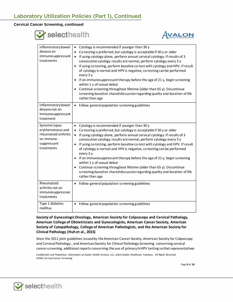
Laboratory Utilization Policies (Part 1), Continued
Confidential and Proprietary Information of Avalon Health Services, LLC, d/b/a Avalon Healthcare Solutions. All Rights Reserved. G2002 Cervical Cancer Screening
Page 9 of 18
Inflammatory bowel disease on immunosuppressant treatments
• Cytology is recommended if younger than 30 y • Co-testing is preferred, but cytology is acceptable if 30 y or older • If using cytology alone, perform annual cervical cytology. If results of 3
consecutive cytology results are normal, perform cytology every 3 y • If using co-testing, perform baseline co-test with cytology and HPV. If result
of cytology is normal and HPV is negative, co-testing can be performed every 3 y
• If on immunosuppressant therapy before the age of 21 y, begin screening within 1 y of sexual debut
• Continue screening throughout lifetime (older than 65 y). Discontinue screening based on shared discussion regarding quality and duration of life rather than age
Inflammatory bowel disease not on immunosuppressant treatment
• Follow general population screening guidelines
Systemic lupus erythematosus and rheumatoid arthritis on immune-suppressant treatments
• Cytology is recommended if younger than 30 y • Co-testing is preferred, but cytology is acceptable if 30 y or older • If using cytology alone, perform annual cervical cytology. If results of 3
consecutive cytology results are normal, perform cytology every 3 y • If using co-testing, perform baseline co-test with cytology and HPV. If result
of cytology is normal and HPV is negative, co-testing can be performed every 3 y
• If on immunosuppressant therapy before the age of 21 y, begin screening within 1 y of sexual debut
• Continue screening throughout lifetime (older than 65 y). Discontinue screening based on shared discussion regarding quality and duration of life rather than age
Rheumatoid arthritis not on immunosuppressive treatments
• Follow general population screening guidelines
Type 1 diabetes mellitus
• Follow general population screening guidelines
Society of Gynecologic Oncology, American Society for Colposcopy and Cervical Pathology, American College of Obstetricians and Gynecologists, American Cancer Society, American Society of Cytopathology, College of American Pathologists, and the American Society for Clinical Pathology (Huh et al., 2015)
Since the 2011 joint guidelines issued by the American Cancer Society, American Society for Colposcopy and Cervical Pathology , and American Society for Clinical Pathology Screening concerning cervical cancer screening, additional reports concerning the use of primary hrHPV testing so that representatives
Cervical Cancer Screening, continued

Laboratory Utilization Policies (Part 1), Continued
Confidential and Proprietary Information of Avalon Health Services, LLC, d/b/a Avalon Healthcare Solutions. All Rights Reserved. G2002 Cervical Cancer Screening
Page 10 of 18
from the Society of Gynecologic Oncology, American Society for Colposcopy and Cervical Pathology, American College of Obstetricians and Gynecologists, American Cancer Society, American Society of Cytopathology, College of American Pathologists, and the American Society for Clinical Pathology convened to issue interim clinical guidance in 2015. In the 2011 statement, primary hrHPV testing was not recommended. The 2015 recommendations include:
• “Because of equivalent or superior effectiveness, primary hrHPV screening can be considered as an alternative to current US cytology-based cervical cancer screening methods. Cytology alone and cotesting remain the screening options specifically recommended in major guidelines.”
• “A negative hrHPV test provides greater reassurance of low CIN3+ risk than a negative cytology result.”
• “Rescreening after a negative primary hrHPV screen should occur no sooner than every 3 years.”
• “Primary hrHPV screening should not be initiated prior to 25 years of age.”
They give the following algorithm concerning screening (Huh et al., 2015):
Cervical Cancer Screening, continued

Laboratory Utilization Policies (Part 1), Continued
Confidential and Proprietary Information of Avalon Health Services, LLC, d/b/a Avalon Healthcare Solutions. All Rights Reserved. G2002 Cervical Cancer Screening
Page 11 of 18
American College of Obstetricians and Gynecologists (ACOG) (Chelmow & ACOG, 2016)
In Practice Bulletin #168, ACOG updated their recommendations concerning cervical cancer screening based on new studies. The table below outlines their recommendation concerning screening:
ACOG does state that women who immunocompromised, including those who are HIV-positive, should start screening younger than age 21. Even though in their table, they do not recommend screening by HPV testing alone, they include the following noted caveat: “After the Joint Recommendations were published, a test for screening with HPV testing alone was approved by the U.S. Food and Drug Administration. Gynecologic care providers using this test should follow the interim guidance developed by the American Society for Colposcopy and Cervical Pathology and the Society for Gynecologic Oncology (Huh et al., 2015) (Chelmow & ACOG, 2016).”
Cervical Cancer Screening, continued

Laboratory Utilization Policies (Part 1), Continued
Confidential and Proprietary Information of Avalon Health Services, LLC, d/b/a Avalon Healthcare Solutions. All Rights Reserved. G2002 Cervical Cancer Screening
Page 12 of 18
The following table outlines the ACOG recommendation concerning the management of cervical cancer screening (Chelmow & ACOG, 2016):
This guideline was reaffirmed in 2018 (ACOG, 2018b).
VI. State and Federal Regulations (as applicable) The FDA has approved the APTIMA HPV 16 18/45 Genotype Assay, a nucleic acid amplification test (NAAT), for the qualitative detection of mRNA for HPV 16, 18, and 45 from Gen-Probe Incorporated on 10/12/2012; however, this test cannot distinguish between 18 and 45. Previously, on 10/28/2011, the FDA approved Gen-Probe Incorporated’s APTIMA HPV Assay, an NAAT that tests for 14 high-risk types of HPV but is unable to distinguish between the 14 types.
Hologic, Inc. has two FDA-approved HPV NAAT tests—Cervista HPV 16/18 and Cervista HPV HR and GENFIND DNA Extraction Kit. Both were approved on 03/12/2009. The former is a fluorescent, isothermal-based reaction that detects HPV 16 and 18 whereas the latter screens for DNA from the 14 high-risk HPV strains (FDA, 2018b). A subsequent search on July 18, 2019 did not yield any new HPV-related results (FDA, 2019).
The COBAS HPV test by Roche Molecular Systems, Inc. was approved by the FDA on 04/19/2011 as a NAAT for 14 high-risk types of HPV. This test can specifically identify HPV 16 and 18 but cannot distinguish from the other 12 types of HPV. On 07/02/2018, the FDA released an approval order statement (P100020/S025) “for an expansion of the intended use for the FDA-approved cobas HPV Test to include cervical specimens collected in SurePath Preservative Fluid as a specimen type” (FDA, 2018c). This approval allows for the cobas HPV Test to be used as a first-line cervical cancer screening using the
Cervical Cancer Screening, continued

Laboratory Utilization Policies (Part 1), Continued
Confidential and Proprietary Information of Avalon Health Services, LLC, d/b/a Avalon Healthcare Solutions. All Rights Reserved. G2002 Cervical Cancer Screening
Page 13 of 18
SurePath preservative, a medium often used for Pap tests (Rice, 2018). In 2020, the Cobas HPV was FDA approved for use on Cobas 6800/8800 Systems (FDA, 2020).
On February 12, 2018, the FDA approved the BD Onclarity™ HPV Assay which detects 14 high-risk HPV genotypes including high-risk strains 16 and 18. “The BD Onclarity HPV Assay is a qualitative in vitro test for the detection of Human Papillomavirus in cervical specimens collected by a clinician using an endocervical brush/spatula combination or broom and placed in BD SurePath vial” (FDA, 2018a).
For more information regarding HPV, please refer to AHS-G2157 Diagnostic testing of STIs.
Additionally, many labs have developed specific tests that they must validate and perform in house. These laboratory-developed tests (LDTs) are regulated by the Centers for Medicare and Medicaid (CMS) as high-complexity tests under the Clinical Laboratory Improvement Amendments of 1988 (CLIA ’88). As an LDT, the U. S. Food and Drug Administration has not approved or cleared this test; however, FDA clearance or approval is not currently required for clinical use.
VII. Applicable CPT/HCPCS Procedure Codes Code Number Code Description
87623 Infectious agent detection by nucleic acid (DNA or RNA); Human Papillomavirus (HPV), low-risk types (e.g., 6, 11, 42, 43, 44)
87624 Infectious agent detection by nucleic acid (DNA or RNA); Human Papillomavirus (HPV), high-risk types (e.g., 16, 18, 31, 33, 35, 39, 45, 51, 52, 56, 58, 59, 68)
87625 Infectious agent detection by nucleic acid (DNA or RNA); Human Papillomavirus (HPV), types 16 and 18 only, includes type 45, if performed
88141 Cytopathology, cervical or vaginal (any reporting system), requiring interpretation by physician
88142 Cytopathology, cervical or vaginal (any reporting system), collected in preservative fluid, automated thin layer preparation; manual screening under physician supervision
88143 Cytopathology, cervical or vaginal (any reporting system), collected in preservative fluid, automated thin layer preparation; with manual screening and rescreening under physician supervision
88147 Cytopathology smears, cervical or vaginal; screening by automated system under physician supervision
88148 Cytopathology smears, cervical or vaginal; screening by automated system with manual rescreening under physician supervision
88150 Cytopathology, slides, cervical or vaginal; manual screening under physician supervision
88152 Cytopathology, slides, cervical or vaginal; with manual screening and computer-assisted rescreening under physician supervision
88153 Cytopathology, slides, cervical or vaginal; with manual screening and rescreening under physician supervision
88164 Cytopathology, slides, cervical or vaginal (The Bethesda System); manual screening under physician supervision
Cervical Cancer Screening, continued

Laboratory Utilization Policies (Part 1), Continued
Confidential and Proprietary Information of Avalon Health Services, LLC, d/b/a Avalon Healthcare Solutions. All Rights Reserved. G2002 Cervical Cancer Screening
Page 14 of 18
88165 Cytopathology, slides, cervical or vaginal (The Bethesda System); with manual screening and rescreening under physician supervision
88166 Cytopathology, slides, cervical or vaginal (The Bethesda System); with manual screening and computer-assisted rescreening under physician supervision
88167 Cytopathology, slides, cervical or vaginal (The Bethesda System); with manual screening and computer-assisted rescreening using cell selection and review under physician supervision
88174 Cytopathology, cervical or vaginal (any reporting system), collected in preservative fluid, automated thin layer preparation; screening by automated system, under physician supervision
88175 Cytopathology, cervical or vaginal (any reporting system), collected in preservative fluid, automated thin layer preparation; with screening by automated system and manual rescreening or review, under physician supervision
0500T Infectious agent detection by nucleic acid (DNA or RNA), Human Papillomavirus (HPV) for five or more separately reported high-risk HPV types (e.g., 16, 18, 31, 33, 35, 39, 45, 51, 52, 56, 58, 59, 68) (i.e., genotyping)
G0123 Screening cytopathology, cervical or vaginal (any reporting system), collected in preservative fluid, automated thin layer preparation, screening by cytotechnologist under physician supervision
G0124 Screening cytopathology, cervical or vaginal (any reporting system), collected in preservative fluid, automated thin layer preparation, requiring interpretation by physician
G0141 Screening cytopathology smears, cervical or vaginal, performed by automated system, with manual rescreening, requiring interpretation by physician
G0143 Screening cytopathology, cervical or vaginal (any reporting system), collected in preservative fluid, automated thin layer preparation, with manual screening and rescreening by cytotechnologist under physician supervision
G0144 Screening cytopathology, cervical or vaginal (any reporting system), collected in preservative fluid, automated thin layer preparation, with screening by automated system, under physician supervision
G0145 Screening cytopathology, cervical or vaginal (any reporting system), collected in preservative fluid, automated thin layer preparation, with screening by automated system and manual rescreening under physician supervision
G0147 Screening cytopathology smears, cervical or vaginal, performed by automated system under physician supervision
G0148 Screening cytopathology smears, cervical or vaginal, performed by automated system with manual rescreening
G0476 Infectious agent detection by nucleic acid (DNA or RNA); human papillomavirus (HPV), high-risk types (e.g., 16, 18, 31, 33, 35, 39, 45, 51, 52, 56, 58, 59, 68) for cervical cancer screening, must be performed in addition to pap test
P3000 Screening Papanicolaou smear, cervical or vaginal, up to three smears, by technician under physician supervision
P3001 Screening Papanicolaou smear, cervical or vaginal, up to three smears, requiring interpretation by physician
Cervical Cancer Screening, continued

Laboratory Utilization Policies (Part 1), Continued
Confidential and Proprietary Information of Avalon Health Services, LLC, d/b/a Avalon Healthcare Solutions. All Rights Reserved. G2002 Cervical Cancer Screening
Page 15 of 18
Q0091 Screening Papanicolaou smear; obtaining, preparing and conveyance of cervical or vaginal smear to laboratory
Current Procedural Terminology© American Medical Association. All Rights reserved.
Procedure codes appearing in Medical Policy documents are included only as a general reference tool for each policy. They may not be all-inclusive.
VIII. Evidence-based Scientific References ACOG. (2018a). Practice Advisory: Cervical Cancer Screening (Update) Retrieved from https://www.acog.org/Clinical-Guidance-and-Publications/Practice-Advisories/Practice-
Advisory-Cervical-Cancer-Screening-Update?IsMobileSet=false ACOG. (2018b). Practice Bulletins. Retrieved from https://www.acog.org/Clinical-Guidance-and-
Publications/Practice-Bulletins-List ACS. (2020, 01/04/2018). Key Statistics for Cervical Cancer. Retrieved from
https://www.cancer.org/cancer/cervical-cancer/about/key-statistics.html ASCCP. (2017a, 02/14/2017). Don’t order screening tests for low-risk HPV types. . Five Things Physicians and Patients Should Question. Retrieved from
http://www.choosingwisely.org/clinician-lists/asccp-screening-tests-for-low-risk-hpv-types/ ASCCP. (2017b, 02/14/2017). Don’t perform cervical cytology (Pap tests) or HPV screening in
immunocompetent women under age 21. Five Things Physicians and Patients Should Question. Retrieved from http://www.choosingwisely.org/clinician-lists/asccp-pap-tests-or-hpv-screening-in-women-under-21/
Bibbins-Domingo, K., Grossman, D. C., Curry, S. J., Barry, M. J., Davidson, K. W., Doubeni, C. A., . . . Tseng, C. W. (2017). Screening for Gynecologic Conditions With Pelvic Examination: US Preventive Services Task Force Recommendation Statement. Jama, 317(9), 947-953. doi:10.1001/jama.2017.0807
Chelmow, D., & ACOG. (2016). Practice Bulletin No. 168: Cervical Cancer Screening and Prevention. Obstet Gynecol, 128(4), e111-130. doi:10.1097/aog.0000000000001708
Chen, H. C., Schiffman, M., Lin, C. Y., Pan, M. H., You, S. L., Chuang, L. C., . . . Chen, C. J. (2011). Persistence of type-specific human papillomavirus infection and increased long-term risk of cervical cancer. J Natl Cancer Inst, 103(18), 1387-1396. doi:10.1093/jnci/djr283
Dahlstrom, L. A., Ylitalo, N., Sundstrom, K., Palmgren, J., Ploner, A., Eloranta, S., . . . Sparen, P. (2010). Prospective study of human papillomavirus and risk of cervical adenocarcinoma. Int J Cancer, 127(8), 1923-1930. doi:10.1002/ijc.25408
FDA. (2018a). BD ONCLARITY HPV ASSAY. Retrieved from https://www.accessdata.fda.gov/scripts/cdrh/devicesatfda/index.cfm?db=pma&id=391601
FDA. (2018b, 07/09/2018). BD ONCLARITY HPV ASSAY. Devices@FDA. Retrieved from https://www.accessdata.fda.gov/scripts/cdrh/devicesatfda/index.cfm?db=pma&id=391601
FDA. (2018c, 08/06/2018). PMA Monthly approvals from 7/1/2018 to 7/31/2018. Retrieved from https://www.fda.gov/downloads/MedicalDevices/ProductsandMedicalProcedures/DeviceApprovalsandClearances/PMAApprovals/UCM615947.pdf
FDA. (2019). Devices@FDA. Devices@FDA. Retrieved from https://www.accessdata.fda.gov/scripts/cdrh/devicesatfda/index.cfm
FDA. (2020). Cobas HPV For Use On The Cobas 6800/8800 Systems. Retrieved from https://www.accessdata.fda.gov/scripts/cdrh/devicesatfda/index.cfm?db=pma&id=448383
Cervical Cancer Screening, continued

Laboratory Utilization Policies (Part 1), Continued
Confidential and Proprietary Information of Avalon Health Services, LLC, d/b/a Avalon Healthcare Solutions. All Rights Reserved. G2002 Cervical Cancer Screening
Page 16 of 18
Feldman, S., & Crum, C. (2019). Cervical cancer screening tests: Techniques for cervical cytology and human papillomavirus testing. In S. Falk & B. Goff (Eds.), UpToDate. Retrieved from https://www.uptodate.com/contents/cervical-cancer-screening-tests-techniques-for-cervical-cytology-and-human-papillomavirus-testing
Feldman, S., & Crum, C. (2020). Cervical cancer screening tests: Techniques for cervical cytology and human papillomavirus testing. In S. Falk & B. Goff (Eds.), UpToDate. Retrieved from https://www.uptodate.com/contents/cervical-cancer-screening-tests-techniques-for-cervical-cytology-and-human-papillomavirus-testing
Feldman, S., Goodman, A., & Peipert, J. (2020). Screening for cervical cancer. In B. Goff, J. G. Elmore, & J. Melin (Eds.), UpToDate. Retrieved from https://www.uptodate.com/contents/screening-for-cervical-cancer
Huh, W. K., Ault, K. A., Chelmow, D., Davey, D. D., Goulart, R. A., Garcia, F. A., . . . Einstein, M. H. (2015). Use of primary high-risk human papillomavirus testing for cervical cancer screening: interim clinical guidance. J Low Genit Tract Dis, 19(2), 91-96. doi:10.1097/lgt.0000000000000103
Marchand, L., Mundt, M., Klein, G., & Agarwal, S. C. (2005). Optimal collection technique and devices for a quality pap smear. Wmj, 104(6), 51-55.
Massad, L. S. (2018). Replacing the Pap Test With Screening Based on Human Papillomavirus Assays. Jama, 320(1), 35-37. doi:10.1001/jama.2018.7911
Melnikow, J., Henderson, J. T., Burda, B. U., Senger, C. A., Durbin, S., & Weyrich, M. S. (2018). Screening for Cervical Cancer With High-Risk Human Papillomavirus Testing: Updated Evidence Report and Systematic Review for the US Preventive Services Task Force. Jama, 320(7), 687-705. doi:10.1001/jama.2018.10400
Mendez, K., Romaguera, J., Ortiz, A. P., Lopez, M., Steinau, M., & Unger, E. R. (2014). Urine-based human papillomavirus DNA testing as a screening tool for cervical cancer in high-risk women. Int J Gynaecol Obstet, 124(2), 151-155. doi:10.1016/j.ijgo.2013.07.036
Moscicki, A. B., Flowers, L., Huchko, M. J., Long, M. E., MacLaughlin, K. L., Murphy, J., . . . Gold, M. A. (2019). Guidelines for Cervical Cancer Screening in Immunosuppressed Women Without HIV Infection. J Low Genit Tract Dis, 23(2), 87-101. doi:10.1097/lgt.0000000000000468
NCCN. (2020). NCCN Clinical Practice Guidelines in Oncology Cervical Cancer. NCCN Guidelines, 1.2020 - January 14, 2020. Retrieved from https://www.nccn.org/professionals/physician_gls/pdf/cervical.pdf
NCI. (2019, 06/14/20108). Cervical Cancer Screening (PDQ®)–Health Professional Version. Retrieved from https://www.cancer.gov/types/cervical/hp/cervical-screening-pdq
NCI. (2020, 06/14/20108). Cervical Cancer Screening (PDQ®)–Health Professional Version. Retrieved from https://www.cancer.gov/types/cervical/hp/cervical-screening-pdq
Ogilvie, G. S., van Niekerk, D., Krajden, M., Smith, L. W., Cook, D., Gondara, L., . . . Coldman, A. J. (2018). Effect of Screening With Primary Cervical HPV Testing vs Cytology Testing on High-grade Cervical Intraepithelial Neoplasia at 48 Months: The HPV FOCAL Randomized Clinical Trial. Jama, 320(1), 43-52. doi:10.1001/jama.2018.7464
Pathak, N., Dodds, J., Zamora, J., & Khan, K. (2014). Accuracy of urinary human papillomavirus testing for presence of cervical HPV: systematic review and meta-analysis. Bmj, 349, g5264. doi:10.1136/bmj.g5264
Rice, S. L., Editor. (2018, August 2018). Cobas HPV test approved for first-line screening using SurePath preservative fluid. CAP Today.
Cervical Cancer Screening, continued

Laboratory Utilization Policies (Part 1), Continued
Confidential and Proprietary Information of Avalon Health Services, LLC, d/b/a Avalon Healthcare Solutions. All Rights Reserved. G2002 Cervical Cancer Screening
Page 17 of 18
Sabeena, S., Kuriakose, S., Binesh, D., Abdulmajeed, J., Dsouza, G., Ramachandran, A., . . . Arunkumar, G. (2019). The Utility of Urine-Based Sampling for Cervical Cancer Screening in Low-Resource Settings. Asian Pac J Cancer Prev, 20(8), 2409-2413. doi:10.31557/apjcp.2019.20.8.2409
Sasieni, P., Castanon, A., & Cuzick, J. (2009). Screening and adenocarcinoma of the cervix. Int J Cancer, 125(3), 525-529. doi:10.1002/ijc.24410
USPSTF. (2018a). Screening for cervical cancer: US Preventive Services Task Force recommendation statement. Jama, 320(7), 674-686. doi:10.1001/jama.2018.10897
USPSTF. (2018b). Screening for Cervical Cancer: US Preventive Services Task Force Recommendation StatementUSPSTF Recommendation: Screening for Cervical CancerUSPSTF Recommendation: Screening for Cervical Cancer. Jama, 320(7), 674-686. doi:10.1001/jama.2018.10897
Cervical Cancer Screening, continued

Laboratory Utilization Policies (Part 1), Continued
Confidential and Proprietary Information of Avalon Health Services, LLC, d/b/a Avalon Healthcare Solutions. All Rights Reserved. G2002 Cervical Cancer Screening
Page 18 of 18
IX. Revision History Revision Date Summary of Changes
Disclaimer This document is for informational purposes only and should not be relied on in the diagnosis and care of individual patients. Medical and Coding/Reimbursement policies do not constitute medical advice, plan preauthorization, certification, an explanation of benefits, or a contract. Members should consult with appropriate healthcare providers to obtain needed medical advice, care, and treatment. Benefits and eligibility are determined before medical guidelines and payment guidelines are applied. Benefits are determined by the member’s individual benefit plan that is in effect at the time services are rendered. The codes for treatments and procedures applicable to this policy are included for informational purposes. Inclusion or exclusion of a procedure, diagnosis or device code(s) does not constitute or imply member coverage or provider reimbursement policy. Please refer to the member's contract benefits in effect at the time of service to determine coverage or non-coverage of these services as it applies to an individual member. SelectHealth® makes no representations and accepts no liability with respect to the content of any external information cited or relied upon in this policy. SelectHealth updates its Coverage Policies regularly and reserves the right to amend these policies without notice to healthcare providers or SelectHealth members. Members may contact Customer Service at the phone number listed on their member identification card to discuss their benefits more specifically. Providers with questions about this Coverage Policy may call SelectHealth Provider Relations at (801) 442-3692. No part of this publication may be reproduced, stored in a retrieval system or transmitted, in any form or by any means, electronic, mechanical, photocopying, or otherwise, without permission from SelectHealth. ”Intermountain Healthcare” and its accompanying logo, the marks of “SelectHealth” and its accompanying marks are protected and registered trademarks of the provider of this Service and or Intermountain Health Care, Inc., IHC Health Services, Inc., and SelectHealth, Inc. Also, the content of this Service is proprietary and is protected by copyright. You may access the copyrighted content of this Service only for purposes set forth in these Conditions of Use. © CPT Only – American Medical Association
Cervical Cancer Screening, continued

Laboratory Utilization Policies (Part 1), Continued
Confidential and Proprietary Information of Avalon Health Services, LLC, d/b/a Avalon Healthcare Solutions. All Rights Reserved. G2056 Diagnosis of Idiopathic Environmental Intolerance
Page 1 of 13
Diagnosis of Idiopathic Environmental Intolerance Policy #: AHS – G2056 Prior Policy Name & Number
(as applicable): Implementation Date: 9/15/21 Date of Last Revision:
I. Policy Description
Idiopathic environmental intolerance (IEI), formerly called multiple chemical sensitivity (MCS), is a subjective condition characterized by recurrent, nonspecific symptoms attributed to low levels of chemical, biologic, or physical agents in the absence of consistent objective diagnostic physical findings or laboratory tests that define an illness (AAAAI, 1999; ACOEM, 1999; D. Black & Temple, 2019).
II. Related Policies
Policy Number Policy Title AHS-G2031 Allergen Testing AHS-G2099 Intracellular Micronutrient Analysis
III. Indications and/or Limitations of Coverage
Disclaimer:
1. Policies are subject to change without notice. 2. Policies outline coverage determinations for SelectHealth Commercial, SelectHealth
Advantage® (Medicare/CMS) and SelectHealth Community Care® (Medicaid/CHIP) plans. Refer to the “Policy” section for more information.
Application of coverage criteria depends on an individual’s benefit coverage at the time of the request.
For SelectHealth Advantage (Medicare/CMS), coverage is determined by the Centers for Medicare and Medicaid Services (CMS). If a coverage determination has not been adopted by CMS and InterQual criteria are not available, the SelectHealth Commercial policy applies. For the most up-to-date Medicare policies and coverage, please visit their search website or the manual website,
For SelectHealth Community Care (Medicaid), coverage is determined by the State of Utah Medicaid program. If Utah State Medicaid has no published coverage position and InterQual criteria are not available, the SelectHealth Commercial criteria will apply. For the most up-to-date Medicaid policies and coverage, please visit their website or the Utah Medicaid Code Look-Up tool.

Laboratory Utilization Policies (Part 1), Continued
Confidential and Proprietary Information of Avalon Health Services, LLC, d/b/a Avalon Healthcare Solutions. All Rights Reserved. G2056 Diagnosis of Idiopathic Environmental Intolerance
Page 2 of 13
The following does not meet coverage criteria due to a lack of available published scientific literature confirming that the test(s) is/are required and beneficial for the diagnosis and treatment of a patient’s illness.
1. Laboratory tests designed to affirm the diagnosis of idiopathic environmental illness DO NOT MEET COVERAGE CRITERIA.
2. Screening blood, saliva, serum, plasma, urine, and/or stool samples for volatile solvents, organic acids, and organophosphates DO NOT MEET COVERAGE CRITERIA in all circumstances including but not limited to the following compounds:
a. 2-methylhippurate b. 2-methylpentane c. 3-methylpentane d. 3,4-dihydroxyphenylpropionate e. 4-nonylphenol f. alpha-keto-beta-methylvalerate g. alpha-ketoisovalerate h. arabinitol i. atrazine or atrazine mercapturate j. benzene k. benzoate l. bisphenol A (BPA) m. diethydithiophosphate (DEDTP), diethylthiophosphate (DETP), dimethyldithiophosphate
(DMDTP), dimethylthiophosphate (DMTP) n. ethylbenzene o. hexane p. Hippurate q. Indican r. Picolinate s. Polychlorinated biphenyls (PCBs) t. Quinolinate u. Styrene v. Taurine w. Toluene x. Triclosan y. Xylene
Diagnosis of Idiopathic Environmental Intolerance, continued

Laboratory Utilization Policies (Part 1), Continued
Confidential and Proprietary Information of Avalon Health Services, LLC, d/b/a Avalon Healthcare Solutions. All Rights Reserved. G2056 Diagnosis of Idiopathic Environmental Intolerance
Page 3 of 13
3. Phthalates and parabens profiling using a blood, serum, plasma, saliva, urine, and/or stool sample DOES NOT MEET COVERAGE CRITERIA.
4. Chlorinated pesticides, including DDE and DDT, profiling in asymptomatic patients using a blood, serum, plasma, saliva, urine, and/or stool sample DOES NOT MEET COVERAGE CRITERIA.
5. Testing blood, serum, plasma, saliva, urine, and/or stool samples for carnitine sufficiency, oxidative stress and antioxidant sufficiency, detoxification adequacy, methylation sufficiency status, lipoic acid and CoQ10 sufficiency, and/or intestinal hyperpermeability DO NOT MEET COVERAGE CRITERIA in asymptomatic individuals and/or during general encounters. These tests include, but are not limited to, the following:
a. Amino acid testing except for newborn screening and for documented metabolic disorders
b. Carotene/beta-carotene
c. Citrate
d. Vanillylmandelic acid (VMA) testing except for use in diagnosis of neuroblastoma or neuroendocrine tumors or for monitoring effectiveness of treatment of cancer
e. Homovanillic acid (HVA) testing except for use in diagnosis and evaluating neuroblastomas
f. 5-hydroxyindolacetic acid (5-HIAA) testing except for use in diagnosis and evaluating carcinoid syndrome or for staging, treatment, and surveillance of suspected neuroendocrine tumors
g. Elastase except for pancreatic insufficiency
h. Fat differentiation testing, qualitative and quantitative
i. CoQ10
6. Testing blood, serum, plasma, saliva, urine, and/or stool samples for vitamin sufficiency, mineral sufficiency, and/or nutritional analysis DO NOT MEET COVERAGE CRITERIA in asymptomatic individuals and/or during general encounters without abnormal findings. These tests include, but are not limited to, the following:
a. Amino acid testing except for newborn screenings or for documented metabolic disorders
b. Allergen-specific IgG testing for screening food sensitivities, vitamin sufficiency, or mineral sufficiency
c. Carotene/beta-carotene
d. Citrate
e. Vanillylmandelic acid (VMA) testing except for use in diagnosis of neuroblastoma or neuroendocrine tumors or for monitoring effectiveness of treatment of cancer
f. Homovanillic acid (HVA) testing except for us in diagnosis and evaluating neuroblastomas
Diagnosis of Idiopathic Environmental Intolerance, continued

Laboratory Utilization Policies (Part 1), Continued
Confidential and Proprietary Information of Avalon Health Services, LLC, d/b/a Avalon Healthcare Solutions. All Rights Reserved. G2056 Diagnosis of Idiopathic Environmental Intolerance
Page 4 of 13
g. 5-hydroxyindolacetic acid (5-HIAA) testing except for use in diagnosis and evaluating carcinoid syndrome or for staging, treatment, and surveillance of suspected neuroendocrine tumors
h. Lipid peroxides
i. Behenic acid
j. Lignoceric acid
k. Fat differentiation testing, qualitative and quantitative
l. Prealbumin
7. The use of a breath hydrogen and/or breath methane test to assess or diagnose the following conditions DOES NOT MEET COVERAGE CRITERIA:
a. Idiopathic environmental intolerance
b. Food allergies and sensitivities
c. Carbohydrate sensitivity or intolerance, including but not limited to, lactose sensitivity, lactose intolerance, and/or lactase deficiency
d. Digestive disorders
e. Constipation, diarrhea, or flatulence
f. Neurological/neuromuscular disorders, including but not limited to, Parkinson disease and fibromyalgia
g. Rosacea
h. Obesity
i. As part of a wellness visit and/or general encounter without abnormal findings
8. Testing blood, serum, urine, cerebrospinal fluid, fingernails, hair, and/or stool sample for metals, including but not limited to, aluminum, arsenic, cadmium, chromium, copper, lead, magnesium, manganese, mercury, molybdenum, nickel, zinc, and heavy metals not otherwise specified DO NOT MEET COVERAGE CRITERIA in asymptomatic individuals and/or general encounters without abnormal findings.
IV. Scientific Background
Patients with idiopathic environmental intolerance (IEI) typically report sensitivity to multiple, chemically unrelated substances and become ill due to a wide range of nonspecific symptoms when exposed. Symptoms may include anxiety, shortness of breath, chest pain, and more. Psychiatric disorders may also be at the core of the IEI patient (D. Black & Temple, 2019). The mean age of patients reporting IEI is between 30 and 40 years, women are diagnosed more than men, and individuals who are married are significantly more likely to be diagnosed with IEI than those who are not (D. Black & Temple, 2019). IEI also occurs in 40% of people with chronic fatigue syndrome and in 16% of people with fibromyalgia (D. W. Black, Carver, & Carver, 2020).
Diagnosis of Idiopathic Environmental Intolerance, continued

Laboratory Utilization Policies (Part 1), Continued
Confidential and Proprietary Information of Avalon Health Services, LLC, d/b/a Avalon Healthcare Solutions. All Rights Reserved. G2056 Diagnosis of Idiopathic Environmental Intolerance
Page 5 of 13
The symptoms of IEI are nonspecific, ambiguous and common in the general population. There is no characteristic set of symptoms and ultimately no major differences between patients self-reporting IEI and those that do not. Virtually any symptom can be considered a symptom of IEI (D. Black & Temple, 2019). Within the definition of multiple chemical sensitivity, identified symptoms included “asthmatic-like, skin irritation, dermatitis, migraine, dysuria, dyspepsia, symptoms of supposed sensitization to food, persistent arthromial pain, vertigo, vestibular impairment,” with 80% of patients experiencing “asthenia, arthromial pain, dyspepsia, coriza, eructation, chest pain, insomnia” (Quarato et al., 2020). The classification of IEI as a distinct medical disorder is also in question, as a lack of reliable case reports, lack of consistent findings or laboratory results, and reliance on surveys or self-reporting all cloud the condition and understanding of this disorder (D. Black & Temple, 2019). Recently, many articles have been published suggesting a relationship between electromagnetic fields and IEI. Electromagnetic fields may include radiofrequencies from telecommunication devices (Eltiti, Wallace, Russo, & Fox, 2018; Huang, Cheng, & Guo, 2018), Wi-Fi and base stations (ANSES, 2018). For an unknown reason, these individuals claim to react to the exposure of certain electromagnetic triggers that most people can tolerate without issues; these triggers are below established toxicological and hazardous thresholds. ANSES (2018) researched the relationship between electric field exposure and IEI symptoms and stated that “either the symptoms experienced by EHS individuals are not caused by exposure to electromagnetic fields and there are no quantifiable biological and/or physiological abnormalities when they are exposed to electromagnetic fields (assumption 1) or the absence of results is due to the methodological limitations of the provocation studies (subject selection, sample size, exposure type, etc.) (assumption 2).” These findings were corroborated by Schmiedchen, Driessen, and Oftedal (2019), who, in their systematic review of articles pertaining to EHS, stated, “limitations in design, conduct and analysis could therefore have given rise to either false positive for false negative results,” and that the “nocebo effect or medical/mental disorders may explain the complaints in many individuals.” Characteristic symptoms of electromagnetic hypersensitivity include sleep and circadian rhythm disorders, migraines and headaches, hypersensitivity, and other related syndromes and disorders such as fibromyalgia, tinnitus and multiple chemical sensitivity (ANSES, 2018). Tests such as elimination diets, food challenges, and provocation-neutralization tests have been used to test for food or chemical sensitivities. Immunological tests or tests measuring the amount of various chemicals in body tissues have also been performed (D. Black & Temple, 2019). In fact, testing for a wide range of autoantibodies is generally discouraged, as “pretest probability is low, and false-positive results are far more likely than true-positive results; a weakly positive ANA [antinuclear antibodies] is present in about 20% of the population” (D. W. Black et al., 2020). However, these assessments are typically not rigorous enough to provide strong evidence; for example, these tests are often not performed blinded or with placebo controls. No unusual laboratory findings have been reliably linked to IEI (D. Black & Temple, 2018, 2019). Due to the vast amount of causes, symptoms, responses, and general heterogeneity of this condition, it may be very difficult to provide a scientifically valid or useful test. Worse, testing may even exacerbate or increase the number of symptoms of a patient. Physicians should use caution in testing for reassurance of patients as negative findings may increase anxiety instead (Barsky & Borus, 1999; D. Black & Temple, 2019).
Diagnosis of Idiopathic Environmental Intolerance, continued

Laboratory Utilization Policies (Part 1), Continued
Confidential and Proprietary Information of Avalon Health Services, LLC, d/b/a Avalon Healthcare Solutions. All Rights Reserved. G2056 Diagnosis of Idiopathic Environmental Intolerance
Page 6 of 13
Due to the number of symptoms that may be considered part of IEI, there are a corresponding amount of tests performed. These tests are generally unnecessary as the condition itself is far too ambiguous to reliably test for and any test can be ordered under the guise of IEI. For example, assessment of factors such as elastase, stool culturing, or fat differentiation may all be done for the sake of IEI treatment. These tests may have legitimate medical purposes (for instance a stool culture may be useful for numerous conditions) but their use for IEI is essentially none, as IEI itself carries no reliable characteristics to test for. Other tests that evaluate a tangentially relevant analyte, such as micronutrient panels or a lactose intolerance breath test, may be done for IEI’s sake as well. Since virtually any symptom or sign can be called IEI, these tests are sometimes ordered for nonspecific or subjective symptoms such as fatigue or pain. However, these tests cannot provide any useful results because of the dubious nature of IEI itself. Another commonly used test for IEI are panels that test multiple factors in one. For example, the Triad Bloodspot Profile offered by Genova Diagnostics measures organic acid levels, “the level of IgG4 reactions for 30 common foods,” and “essential amino acid imbalances” (Genova, 2021d). Genova offers several similar panels, such as the Organix Comprehensive Profile (which tests 46 analytes for subjective symptoms such as depression, weight issues and chemical sensitivities) (Genova, 2021c), the NutrEval FMV (which tests 118 analytes for symptoms such as fatigue, weight issues, and sports fitness optimization) (Genova, 2021a) and the Allergix IgG4 Food Antibodies (which tests 90 foods for sensitivity). Genova Diagnostics also offers the GI Effects Profile (advanced stool tests for the management of GI health), a full line of allergy testing and assessment tests (measuring IgG and IgE food antibodies, inhalants, molds and spices), the Ion Profile (which evaluates various types of organic, amino and fatty acids as well as nutrient and toxic elements), the CDSA 2.0 Profile with Parasitology (evaluates the microbiome, digestion and absorption), and SIBO Profile tests (breath tests which measure methane gases and exhaled hydrogen) (Genova, 2020). An evaluation of symptoms of IEI patients includes a history, physical examination, and laboratory tests (complete blood count, serum electrolytes and glucose, urine analysis) with further testing guided by reported symptoms. An occupational or environmental history is also useful as patients typically report problems from chemical exposure (D. Black & Temple, 2019). A questionnaire such as the “Environmental Exposure and Sensitivity Intolerance” (EESI) may be used for an initial screening (Rossi & Pitidis, 2018). A psychiatric history is also recommended as psychiatric disorders are often co-morbid with IEI. A screening questionnaire such as the Patient Health Questionnaire (PHQ-9) can be used to identify psychiatric conditions in an IEI patient (D. Black & Temple, 2019; Gilbody, Richards, Brealey, & Hewitt, 2007). Micronutrients are the essential vitamins and minerals required by the body for proper functioning. Panels have been developed which evaluate intracellular levels of essential vitamins and minerals. These panels may also be used on IEI patients. This may help to identify nutritional deficiencies in otherwise healthy patients or in patients suffering from some type of disease. SpectraCell Laboratories have developed the Micronutrient Test Panel, which is able to measure 31 vitamins, minerals, metabolites,
Diagnosis of Idiopathic Environmental Intolerance, continued

Laboratory Utilization Policies (Part 1), Continued
Confidential and Proprietary Information of Avalon Health Services, LLC, d/b/a Avalon Healthcare Solutions. All Rights Reserved. G2056 Diagnosis of Idiopathic Environmental Intolerance
Page 7 of 13
amino acids, fatty acids and antioxidants; this test also measures how these micronutrients affect cellular functioning in an individual (SpectaCell, 2021). SpectraCell Laboratories have also developed the SPECTROX™, claiming it measures total antioxidant function in an individual, reporting on the repair mechanisms and net ability of each individual’s cells (SpectraCell, 2008). As noted above, Genova Diagnostics has developed the NutrEval FMV that measures 118 markers, including amino acids, fatty acids and organic acids (Genova, 2021a). ONE (Optimal Nutritional Evaluation) FMV, also by Genova Diagnostics, is a urine-based nutritional test which assesses “the functional need for antioxidants, B-vitamins, minerals, digestive support and amino acids” (Genova, 2021b). The company notes that the ONE FMV test may be used for patients with mood disorders, fatigue, digestive issues, weight problems, general health, dietary guidance and fitness. Another nutrient panel blood test, developed by Life Extension, measures vitamin B12, folate, vitamin D 25-hydroxy, vitamin A, vitamin C, selenium, zinc, coQ10 and magnesium (LifeExtension, 2020). Finally, Vibrant America provides a test which measures approximately 40 intracellular and extracellular vitamins, minerals, fatty acids, amino acids and antioxidants (Vibrant, 2017). Very little information suggests that the intracellular micronutrient analysis assists with positive health outcomes. Houston (2013) published an article on the role of vitamins, minerals and overall nutrition in the prevention and treatment of hypertension. This article reviewed hypertension-related clinical trials that include information on the “efficacy of nutrition, weight loss, exercise, and nutritional supplements, vitamins, minerals, and antioxidants” (Houston, 2013). Approximately 3338 patients were treated with micronutrient testing over a five-year period, with 20% of these patients exhibiting abnormally high blood pressure. After six months, 62% of the hypertensive patients reached lower blood pressure goals. Hence, the author states that the diagnosis and treatment of various nutritional deficiencies can decrease the number of cardiac events as well as reduce blood pressure and improve vascular biology. However, data for the control group not treated with micronutrients was not provided for comparison. Another technique that has been used to assess nutritional status is the measurement of the hepatic proteins prealbumin and albumin. However, it seems that a physical examination has evolved as the main technique to diagnose malnutrition in a clinical setting. “The current consensus is that laboratory markers are not reliable by themselves but could be used as a complement to a thorough physical examination” in a malnutrition diagnosis (Bharadwaj et al., 2016). The Academy of Nutrition and Dietetics also do not accept albumin and prealbumin as a diagnostic tool for malnutrition and state that “There is no laboratory test that is both sensitive to and specific for protein-calorie malnutrition” (AND, 2017). IEI patients may also report bowel irritability. Small intestinal bacterial overgrowth (SIBO) occurs when excessive aerobic and anaerobic bacteria colonize the small bowel. These bacteria are typically found in the colon and can cause chronic diarrhea and malabsorption if they colonize the small bowel (Pimentel, 2019). SIBO may be diagnosed by a breath test. However, a validated gold standard method for diagnosing SIBO has not been indicated (Rezaie et al., 2017). The SIBO breath test uses carbohydrates in a simple, non-invasive and widely available testing method. A carbohydrate substrate (such as lactulose or glucose) is administered to the patient, which leads to the production of an analyte such as hydrogen
Diagnosis of Idiopathic Environmental Intolerance, continued

Laboratory Utilization Policies (Part 1), Continued
Confidential and Proprietary Information of Avalon Health Services, LLC, d/b/a Avalon Healthcare Solutions. All Rights Reserved. G2056 Diagnosis of Idiopathic Environmental Intolerance
Page 8 of 13
or methane. “In individuals without SIBO, the administration of lactulose results in a single peak in breath hydrogen/methane within two to three hours due to the metabolism of lactulose by colonic flora. In patients with SIBO, administration of lactulose results in an early peak in breath hydrogen/methane levels due to metabolism by small bowel bacteria” (Pimentel, 2019). As noted above, Genova Diagnostics has developed the SIBO Profile test which is a two or three hour breath test that measures methane gases and exhaled hydrogen (Genova, 2020). This test requires the patient to ingest a lactulose solution. Bratten, Spanier, and Jones (2008) completed a study with 224 patients with irritable bowel syndrome (IBS) and 40 controls. A lactulose breath test (LBT) was used to measure methane and hydrogen production to identify patients with IBS. Results showed that “The majority of patients with IBS and healthy subjects meet criteria for an "abnormal" LBT using previously published test criteria, and groups are not discriminated using this diagnostic method” (Bratten et al., 2008). The authors then questioned the utility of an LBT to diagnose IBS as the testing did not discriminate between IBS patients and healthy controls. A more recent study by Ghoshal, Srivastava, Ghoshal, and Misra (2014) evaluated 80 patients with IBS for SIBO. Culture had previously diagnosed 15/80 patients with SIBO. Both lactulose and glucose hydrogen breath tests (LHBT and GHBT, respectively) were used to measure SIBO. The authors conclude that “The specificity of GHBT was 100%, but the sensitivity of this test and the diagnostic performances of LHBT and breath methane were all very poor” (Ghoshal et al., 2014).
V. Guidelines and Recommendations
Due to the dubious nature of this condition, several prominent medical studies have regarded this condition with suspicion. In 1992, the American Medical Association stated that multiple chemical sensitivity (now IEI) should not be recognized as a syndrome until accurate, reproducible, and well-controlled studies can be done (AMA, 1992). Other societies such as the American College of Physicians and the American Academy of Allergy and Immunology hold similar views (AAAAI, 1986; ACP, 1989).
VI. State and Federal Regulations (as applicable)
No specific U.S. Food and Drug Administration (FDA) approval or clearance of a test for idiopathic environmental intolerance was found. Additionally, many labs have developed specific tests that they must validate and perform in house. These laboratory-developed tests (LDTs) are regulated by the Centers for Medicare and Medicaid (CMS) as high-complexity tests under the Clinical Laboratory Improvement Amendments of 1988 (CLIA ’88). As an LDT, the U. S. Food and Drug Administration has not approved or cleared this test; however, FDA clearance or approval is not currently required for clinical use.
Diagnosis of Idiopathic Environmental Intolerance, continued

Laboratory Utilization Policies (Part 1), Continued
Confidential and Proprietary Information of Avalon Health Services, LLC, d/b/a Avalon Healthcare Solutions. All Rights Reserved. G2056 Diagnosis of Idiopathic Environmental Intolerance
Page 9 of 13
VII. Applicable CPT/HCPCS Procedure Codes
Code Number Code Description 82127 Amino acids; single, qualitative, each specimen 82136 Amino acids, 2 to 5 amino acids, quantitative, each specimen 82139 Amino acids, 6 or more amino acids, quantitative, each specimen 82379 Carnitine (total and free), quantitative, each specimen 82380 Carotene 82441 Chlorinated hydrocarbons, screen 82495 Chromium 82507 Citrate 82525 Copper
82542 Column chromatography, includes mass spectrometry, if performed (e.g., HPLC, LC, LC/MS, LC/MS-MS, GC, GC/MS-MS, GC/MS, HPLC/MS), non-drug analyte(s) not elsewhere specified, qualitative or quantitative, each specimen
82656 Elastase, pancreatic (EL-1), fecal, qualitative or semi-quantitative 82715 Fat differential, feces, quantitative 82978 Glutathione 83150 Homovanillic acid (HVA) 83497 Hydroxyindolacetic acid, 5-(HIAA) 83918 Organic acids; total, quantitative, each specimen 83919 Organic acids; qualitative, each specimen 83921 Organic acid, single, quantitative 84134 Prealbumin 84255 Selenium 84585 Vanillylmandelic acid (VMA), urine 84600 Volatiles (eg, acetic anhydride, diethylether) 84630 Zinc 86001 Allergen specific IgG quantitative or semiquantitative, each allergen
86353 Lymphocyte transformation, mitogen (phytomitogen) or antigen induced blastogenesis
88348 Electron microscopy, diagnostic
83015 Heavy metal (e.g., arsenic, barium, beryllium, bismuth, antimony, mercury); qualitative, any number of analytes
83018 Heavy metal (e.g., arsenic, barium, beryllium, bismuth, antimony, mercury); quantitative, each, not elsewhere specified
82108 Aluminum 82300 Cadmium 83735 Magnesium 83885 Nickel 83785 Manganese 82726 Very long chain fatty acids
Diagnosis of Idiopathic Environmental Intolerance, continued

Laboratory Utilization Policies (Part 1), Continued
Confidential and Proprietary Information of Avalon Health Services, LLC, d/b/a Avalon Healthcare Solutions. All Rights Reserved. G2056 Diagnosis of Idiopathic Environmental Intolerance
Page 10 of 13
89125 Fat stain, feces, urine, or respiratory secretions 82710 Fat or lipids, feces; quantitative 84590 Vitamin A 84446 Tocopherol alpha (Vitamin E) 83655 Lead
91065 Breath hydrogen or methane test (e.g., for detection of lactase deficiency, fructose intolerance, bacterial overgrowth, or oro-cecal gastrointestinal transit)
Current Procedural Terminology© American Medical Association. All Rights reserved.
Procedure codes appearing in Medical Policy documents are included only as a general reference tool for each policy. They may not be all-inclusive.
VIII. Evidence-based Scientific References
AAAAI. (1986). Clinical ecology. Executive Committee of the American Academy of Allergy and Immunology. J Allergy Clin Immunol, 78(2), 269-271.
AAAAI. (1999). Idiopathic environmental intolerances. American Academy of Allergy, Asthma and Immunology (AAAAI) Board of Directors. J Allergy Clin Immunol, 103(1 Pt 1), 36-40.
ACOEM. (1999). ACOEM position statement. Multiple chemical sensitivities: idiopathic environmental intolerance. College of Occupational and Environmental Medicine. J Occup Environ Med, 41(11), 940-942.
ACP. (1989). Clinical ecology. American College of Physicians. Ann Intern Med, 111(2), 168-178. AMA. (1992). Clinical ecology. Council on Scientific Affairs, American Medical Association. Jama, 268(24),
3465-3467. AND. (2017). Should Albumin and Prealbumin Be Used as Indicators for Malnutrition? JOURNAL OF THE
ACADEMY OF NUTRITION AND DIETETICS. Retrieved from https://jandonline.org/article/S2212-2672(17)30444-6/pdf
ANSES. (2018). OPINION of the French Agency for Food, Environmental and Occupational Health & Safety regarding the expert appraisal on “electromagnetic hypersensitivity (EHS) or idiopathic environmental intolerance attributed to electromagnetic fields (IEI-EMF)”. Retrieved from https://www.anses.fr/en/system/files/AP2011SA0150EN.pdf
Barsky, A. J., & Borus, J. F. (1999). Functional somatic syndromes. Ann Intern Med, 130(11), 910-921. Bharadwaj, S., Ginoya, S., Tandon, P., Gohel, T. D., Guirguis, J., Vallabh, H., . . . Hanouneh, I. (2016).
Malnutrition: laboratory markers vs nutritional assessment. Gastroenterol Rep (Oxf), 4(4), 272-280. doi:10.1093/gastro/gow013
Black, D., & Temple, S. (2018). Overview of idiopathic environmental intolerance (multiple chemical sensitivity) - UpToDate. In D. Solomon (Ed.), UpToDate. Retrieved from https://www.uptodate.com/contents/overview-of-idiopathic-environmental-intolerance-multiple-chemical-sensitivity?source=search_result&search=idiopathic%20enviornmental%20intolerance&selectedTitle=1~150#H20
Black, D., & Temple, S. (2019, August 24). Idiopathic environmental intolerance (multiple chemical sensitivity). Retrieved from https://www.uptodate.com/contents/idiopathic-environmental-intolerance-multiple-chemical-sensitivity
Black, D. W., Carver, R. J., & Carver, L. A. (2020, July). Idiopathic Environmental Intolerance (Multiple Chemical Sensitivity; Environmental Illness). Retrieved from
Diagnosis of Idiopathic Environmental Intolerance, continued

Laboratory Utilization Policies (Part 1), Continued
Confidential and Proprietary Information of Avalon Health Services, LLC, d/b/a Avalon Healthcare Solutions. All Rights Reserved. G2056 Diagnosis of Idiopathic Environmental Intolerance
Page 11 of 13
https://www.merckmanuals.com/professional/special-subjects/idiopathic-environmental-intolerance/idiopathic-environmental-intolerance
Bratten, J. R., Spanier, J., & Jones, M. P. (2008). Lactulose breath testing does not discriminate patients with irritable bowel syndrome from healthy controls. Am J Gastroenterol, 103(4), 958-963. doi:10.1111/j.1572-0241.2008.01785.x
Bush, R. K., Portnoy, J. M., Saxon, A., Terr, A. I., & Wood, R. A. (2006). The medical effects of mold exposure. J Allergy Clin Immunol, 117(2), 326-333. doi:10.1016/j.jaci.2005.12.001
Eltiti, S., Wallace, D., Russo, R., & Fox, E. (2018). Symptom Presentation in Idiopathic Environmental Intolerance With Attribution to Electromagnetic Fields: Evidence for a Nocebo Effect Based on Data Re-Analyzed From Two Previous Provocation Studies. Front Psychol, 9, 1563. doi:10.3389/fpsyg.2018.01563
Genova. (2020). Testing Services Overview. Retrieved from https://www.gdx.net/files/clinicians/how-to-order/Genova-Diagnostics-Testing-Services-Overview.pdf
Genova. (2021a). NutrEval® FMV. Retrieved from https://www.gdx.net/product/nutreval-fmv-nutritional-test-blood-urine
Genova. (2021b). ONE (Optimal Nutritional Evaluation) FMV™. Retrieved from https://www.gdx.net/product/one-fmv-nutritional-test-urine
Genova. (2021c). Organix® Comprehensive Profile - Urine. Retrieved from https://www.gdx.net/product/organix-comprehensive-profile-metabolic-function-test-urine
Genova. (2021d). TRIAD® Bloodspot Profile. Retrieved from https://www.gdx.net/product/triad-bloodspot-profile-metabolic-nutritional-test-blood-spot
Ghoshal, U. C., Srivastava, D., Ghoshal, U., & Misra, A. (2014). Breath tests in the diagnosis of small intestinal bacterial overgrowth in patients with irritable bowel syndrome in comparison with quantitative upper gut aspirate culture. Eur J Gastroenterol Hepatol, 26(7), 753-760. doi:10.1097/meg.0000000000000122
Gilbody, S., Richards, D., Brealey, S., & Hewitt, C. (2007). Screening for depression in medical settings with the Patient Health Questionnaire (PHQ): a diagnostic meta-analysis. J Gen Intern Med, 22(11), 1596-1602. doi:10.1007/s11606-007-0333-y
Houston, M. C. (2013). The role of nutrition, nutraceuticals, vitamins, antioxidants, and minerals in the prevention and treatment of hypertension. Altern Ther Health Med, 19 Suppl 1, 32-49. Retrieved from https://www.ncbi.nlm.nih.gov/pubmed/23981465
Huang, P. C., Cheng, M. T., & Guo, H. R. (2018). Representative survey on idiopathic environmental intolerance attributed to electromagnetic fields in Taiwan and comparison with the international literature. Environ Health, 17(1), 5. doi:10.1186/s12940-018-0351-8
LifeExtension. (2020). Nutrient Panel Blood Test. Retrieved from https://www.lifeextension.com/lab-testing/itemlc100024/nutrient-panel-blood-test
Martini, A., Iavicoli, S., & Corso, L. (2013). Multiple chemical sensitivity and the workplace: current position and need for an occupational health surveillance protocol. Oxid Med Cell Longev, 2013, 351457. doi:10.1155/2013/351457
Mouzaki, M., Bronsky, J., Gupte, G., Hojsak, I., Jahnel, J., Pai, N., . . . Sundaram, S. (2019). Nutrition Support of Children With Chronic Liver Diseases: A Joint Position Paper of the North American Society for Pediatric Gastroenterology, Hepatology, and Nutrition and the European Society for Pediatric Gastroenterology, Hepatology, and Nutrition. J Pediatr Gastroenterol Nutr, 69(4), 498-511. doi:10.1097/mpg.0000000000002443
Diagnosis of Idiopathic Environmental Intolerance, continued

Laboratory Utilization Policies (Part 1), Continued
Confidential and Proprietary Information of Avalon Health Services, LLC, d/b/a Avalon Healthcare Solutions. All Rights Reserved. G2056 Diagnosis of Idiopathic Environmental Intolerance
Page 12 of 13
Multiple chemical sensitivity: a 1999 consensus. (1999). Arch Environ Health, 54(3), 147-149. doi:10.1080/00039899909602251
Pimentel, M. (2019). Small intestinal bacterial overgrowth: Clinical manifestations and diagnosis. Retrieved from https://www.uptodate.com/contents/small-intestinal-bacterial-overgrowth-clinical-manifestations-and-diagnosis?search=Small%20intestinal%20bacterial%20overgrowth&source=search_result&selectedTitle=2~145&usage_type=default&display_rank=2
Quarato, M., De Maria, L., Caputi, A., Cavone, D., Cannone, E. S. S., Mansi, F., . . . Vimercati, L. (2020). A case report of idiopathic environmental intolerance: A controversial and current issue. Clin Case Rep, 8(1), 79-85. doi:10.1002/ccr3.2535
Rezaie, A., Buresi, M., Lembo, A., Lin, H., McCallum, R., Rao, S., . . . Pimentel, M. (2017). Hydrogen and Methane-Based Breath Testing in Gastrointestinal Disorders: The North American Consensus. Am J Gastroenterol, 112(5), 775-784. doi:10.1038/ajg.2017.46
Rossi, S., & Pitidis, A. (2018). Multiple Chemical Sensitivity: Review of the State of the Art in Epidemiology, Diagnosis, and Future Perspectives. J Occup Environ Med, 60(2), 138-146. doi:10.1097/jom.0000000000001215
Schmiedchen, K., Driessen, S., & Oftedal, G. (2019). Methodological limitations in experimental studies on symptom development in individuals with idiopathic environmental intolerance attributed to electromagnetic fields (IEI-EMF) - a systematic review. Environ Health, 18(1), 88. doi:10.1186/s12940-019-0519-x
SpectaCell. (2021). Micronutrient Test Panel. Retrieved from https://www.spectracell.com/micronutrient-test-panel
SpectraCell. (2008). SPECTROX™ (Total Antioxidant Function). Retrieved from https://assets.speakcdn.com/Assets/2606/0e2022931_supplement-spectrox.pdf
Usai-Satta, P., Giannetti, C., Oppia, F., & Cabras, F. (2018). The North American Consensus on Breath Testing: The Controversial Diagnostic Role of Lactulose in SIBO. Am J Gastroenterol, 113(3), 440. doi:10.1038/ajg.2017.392
Vibrant. (2017). MICRONUTRIENTS. Retrieved from https://www.vibrant-america.com/micronutrient/ WHO. (2013). WHO Guidelines Approved by the Guidelines Review Committee. In Guideline: Updates on
the Management of Severe Acute Malnutrition in Infants and Children. Geneva: World Health Organization. Copyright (c) World Health Organization 2013.
Diagnosis of Idiopathic Environmental Intolerance, continued

Laboratory Utilization Policies (Part 1), Continued
Confidential and Proprietary Information of Avalon Health Services, LLC, d/b/a Avalon Healthcare Solutions. All Rights Reserved. G2056 Diagnosis of Idiopathic Environmental Intolerance
Page 13 of 13
IX. Revision History
Revision Date Summary of Changes
Disclaimer This document is for informational purposes only and should not be relied on in the diagnosis and care of individual patients. Medical and Coding/Reimbursement policies do not constitute medical advice, plan preauthorization, certification, an explanation of benefits, or a contract. Members should consult with appropriate healthcare providers to obtain needed medical advice, care, and treatment. Benefits and eligibility are determined before medical guidelines and payment guidelines are applied. Benefits are determined by the member’s individual benefit plan that is in effect at the time services are rendered. The codes for treatments and procedures applicable to this policy are included for informational purposes. Inclusion or exclusion of a procedure, diagnosis or device code(s) does not constitute or imply member coverage or provider reimbursement policy. Please refer to the member's contract benefits in effect at the time of service to determine coverage or non-coverage of these services as it applies to an individual member. SelectHealth® makes no representations and accepts no liability with respect to the content of any external information cited or relied upon in this policy. SelectHealth updates its Coverage Policies regularly and reserves the right to amend these policies without notice to healthcare providers or SelectHealth members. Members may contact Customer Service at the phone number listed on their member identification card to discuss their benefits more specifically. Providers with questions about this Coverage Policy may call SelectHealth Provider Relations at (801) 442-3692. No part of this publication may be reproduced, stored in a retrieval system or transmitted, in any form or by any means, electronic, mechanical, photocopying, or otherwise, without permission from SelectHealth. ”Intermountain Healthcare” and its accompanying logo, the marks of “SelectHealth” and its accompanying marks are protected and registered trademarks of the provider of this Service and or Intermountain Health Care, Inc., IHC Health Services, Inc., and SelectHealth, Inc. Also, the content of this Service is proprietary and is protected by copyright. You may access the copyrighted content of this Service only for purposes set forth in these Conditions of Use. © CPT Only – American Medical Association
Diagnosis of Idiopathic Environmental Intolerance, continued

Laboratory Utilization Policies (Part 1), Continued
Confidential and Proprietary Information of Avalon Health Services, LLC, d/b/a Avalon Healthcare Solutions. All Rights Reserved. M2057 Diagnosis of Vaginitis including Multi-target PCR Testing
Page 1 of 19
Diagnosis of Vaginitis including Multi-target PCR Testing Policy #: AHS – M2057 Prior Policy Name & Number
(as applicable): Implementation Date: 9/15/21 Date of Last Revision:
I. Policy Description Vaginitis is defined as inflammation of the vagina with symptoms of discharge, itching, and discomfort often due to a disruption of the vaginal microflora. The most common infections are bacterial vaginosis, Candida vulvovaginitis, and trichomoniasis (Sobel, 1999). Other causes include vaginal atrophy in postmenopausal women, cervicitis, foreign body, irritants, and allergens (Sobel, 2020a).
Bacterial vaginosis (BV) is characterized by a shift in microbial species from the normally dominant hydrogen-peroxide producing Lactobacillus species to Gardnerella vaginalis and anaerobic commensals (Eschenbach et al., 1989; Hill, 1993; Lamont et al., 2011; Ling et al., 2010; Sobel, 2020b).
Vulvovaginal candidiasis (VVC) is characterized by Candida species. It is the second most common cause of vaginitis symptoms (after BV) and accounts for approximately one-third of vaginitis cases (Sobel, 2020c; Workowski & Bolan, 2015).
Trichomoniasis is caused by the flagellated protozoan Trichomonas vaginalis, which principally infects the squamous epithelium in the urogenital tract: vagina, urethra, and paraurethral glands (Kissinger, 2015; Sobel & Mitchell, 2020).
II. Related Policies Policy Number Policy Title
AHS-G2157 Diagnostic Testing of Common Sexually Transmitted Infections AHS-G2002 Cervical Cancer Screening AHS-M2097 Identification of Microorganisms Using Nucleic Acid Probes AHS-G2149 Pathogen Panel Testing
Disclaimer:
1. Policies are subject to change without notice. 2. Policies outline coverage determinations for SelectHealth Commercial, SelectHealth
Advantage® (Medicare/CMS) and SelectHealth Community Care® (Medicaid/CHIP) plans. Refer to the “Policy” section for more information.

Laboratory Utilization Policies (Part 1), ContinuedDiagnosis of Vaginitis including Multi-target PCR Testing, continued
Confidential and Proprietary Information of Avalon Health Services, LLC, d/b/a Avalon Healthcare Solutions. All Rights Reserved. M2057 Diagnosis of Vaginitis including Multi-target PCR Testing
Page 2 of 19
III. Indications and/or Limitations of Coverage
1. Testing of pH, testing for the presence of amines, saline wet mount, hydrogen peroxide (KOH) wet mount and microscopic examination of vaginal fluids MEETS COVERAGE CRITERIA in patients with symptoms of vaginitis.
2. Direct Probe DNA-based identification of Gardnerella, Trichomonas, and Candida MEETS COVERAGE CRITERIA in patients with symptoms of vaginitis.
3. Vaginal cultures for Candida species MEET COVERAGE CRITERIA for the diagnosis of vulvovaginal candidiasis in patients with clinical signs and symptoms of vaginitis and negative findings on wet-mount preparations and a normal pH test.
4. Measurement of sialidase activity in vaginal fluid MEETS COVERAGE CRITERIA for the diagnosis of bacterial vaginosis in women with symptoms of vaginitis.
5. Nucleic Acid Amplification Test (NAAT) or Polymerase Chain Reaction (PCR)-based identification of Trichomonas vaginalis MEETS COVERAGE CRITERIA in patients with symptoms of vaginitis.
6. Screening for Trichomonas MEETS COVERAGE CRITERIA for women with risk factors, including: new or multiple partners; history of sexually transmitted diseases (STDs); exchange of sex for payment; or injection drug use.
7. Polymerase Chain Reaction (PCR) based identification of Candida DOES NOT MEET COVERAGE CRITERIA for any indication.
8. Screening for trichomoniasis and bacterial vaginosis DOES NOT MEET COVERAGE CRITERIA in asymptomatic patients, including asymptomatic pregnant patients at average or high risk for premature labor.
Application of coverage criteria depends on an individual’s benefit coverage at the time of the request.
For SelectHealth Advantage (Medicare/CMS), coverage is determined by the Centers for Medicare and Medicaid Services (CMS). If a coverage determination has not been adopted by CMS and InterQual criteria are not available, the SelectHealth Commercial policy applies. For the most up-to-date Medicare policies and coverage, please visit their search website or the manual website,
For SelectHealth Community Care (Medicaid), coverage is determined by the State of Utah Medicaid program. If Utah State Medicaid has no published coverage position and InterQual criteria are not available, the SelectHealth Commercial criteria will apply. For the most up-to-date Medicaid policies and coverage, please visit their website or the Utah Medicaid Code Look-Up tool.

Laboratory Utilization Policies (Part 1), Continued
Confidential and Proprietary Information of Avalon Health Services, LLC, d/b/a Avalon Healthcare Solutions. All Rights Reserved. M2057 Diagnosis of Vaginitis including Multi-target PCR Testing
Page 3 of 19
The following does not meet coverage criteria due to a lack of available published scientific literature confirming that the test(s) is/are required and beneficial for the diagnosis and treatment of a patient’s illness.
9. Rapid identification of Trichomonas by enzyme immunoassay DOES NOT MEET COVERAGE CRITERIA in patients with symptoms of vaginitis.
10. PCR testing and Multitarget polymerase chain reaction (PCR) testing for diagnosis of bacterial vaginosis DO NOT MEET COVERAGE CRITERIA.
11. Using molecular-based panel testing, including, but not limited to testing such as SmartJane™, to test for microorganisms involved in vaginal flora imbalance and/or infertility DOES NOT MEET COVERAGE CRITERIA.
IV. Scientific Background Vaginitis is characterized by several symptoms including odor, itching, abnormal vaginal discharge, burning and irritation; this inflammatory ailment is considered the most common gynecologic diagnosis in primary care as most women experience vaginitis at least once in their lives (Paladine & Desai, 2018). A diagnosis of vaginitis can be given based on a combination of symptoms, physical examination, and office or laboratory-based testing methods.
The squamous epithelium of the vagina in premenopausal women is rich in glycogen, a substrate for lactobacilli, which create an acidic vaginal environment (pH 4.0 to 4.5). This acidity helps maintain the normal vaginal flora and inhibits growth of pathogenic organisms. Disruption of the normal ecosystem by menstrual cycle, sexual activity, contraceptive, pregnancy, foreign bodies, estrogen level, sexually transmitted diseases, and use of hygienic products or antibiotics can lead to development of vaginitis. Bacterial vaginosis (BV), vulvovaginal candidiasis (VVC), and trichomoniasis are the three most common infections responsible for vaginitis. Other causes include: vaginal atrophy in postmenopausal women, cervicitis, foreign body, irritants and allergens (Sobel, 2020a).
Bacterial vaginosis is caused by an imbalance of naturally occurring vaginal bacteria, characterized by both a change in the most common type of bacteria present, along with an increase in the total number of bacteria present. Normal vaginal microbiota is dominated by the species Lactobacilli, which are known to produce hydrogen peroxide and lactic acid, which help to keep the acidic vaginal environment below pH 4.5 (Jones, 2019; Kairys & Garg, 2020). Though the origin of vaginal bacterial infections is still unclear, it is believed that most of such infections are the result of another bacteria, Gardnerella vaginalis, creating a biofilm which allows opportunistic bacteria to grow within the vagina, causing a decrease in the Lactobacilli and subsequent disruption of the pH of the system. An entire host of etiologic organisms have been identified as possible instigators and exacerbators, including Atopobium vaginae, Megasphaera phylotype 1 and 2, Leptotrichia aminionii, Mobiluncus spp, Prevotella spp, Mycoplasma hominis, Bacteroides spp, Sneathia, and BV-associated bacteria (BVAB)1, 2, and 3, though as aforementioned the causative mechanism and the interaction between these species are still uncertain (Jones, 2019).
Diagnosis of Vaginitis including Multi-target PCR Testing, continued
Laboratory Utilization Policies (Part 1), Continued
Confidential and Proprietary Information of Avalon Health Services, LLC, d/b/a Avalon Healthcare Solutions. All Rights Reserved. M2057 Diagnosis of Vaginitis including Multi-target PCR Testing
Page 4 of 19
Laboratory documentation of the etiology of vaginitis is important before initiating therapy, given the nonspecific nature and considerable overlap of the symptoms (Anderson, Klink, & Cohrssen, 2004; Ellis, Lerch, & Whitcomb, 2001; Landers, Wiesenfeld, Heine, Krohn, & Hillier, 2004). Diagnostic testing enables targeted treatment, increases therapeutic compliance, and increases the likelihood of partner notification (Sobel, 2020a; Workowski & Bolan, 2015).
Measurement of vaginal pH is the primary initial finding that drives the diagnostic. The pH of the normal vaginal secretions in premenopausal women with relatively high estrogen levels is 4.0 to 4.5. The pH of normal vaginal secretions in premenarchal and postmenopausal women in whom estrogen levels are low is ≥4.7. An elevated pH in a premenopausal woman suggests infections, such as BV (pH>4.5) or trichomoniasis (pH 5 to 6) and helps to exclude Candida vulvovaginitis (pH 4 to 4.5). Vaginal pH may also be altered by lubricating gels, semen, douches, intravaginal medications and in pregnant women, leakage of amniotic fluid (Anderson et al., 2004; Sobel, 2020a).
Analytical Validity
Microscopic examination of normal vaginal discharge reveals a predominance of squamous epithelial cells, rare polymorphonuclear leukocytes (PMNs), and Lactobacillus species. The primary goal of the examination is to look for candidal buds or hyphae, motile trichomonads, epithelial cells studded with adherent coccobacilli (clue cells), and increased numbers of PMNs (Sobel, 2020a). The microscopic evaluation of BV is usually based on Amsel criteria (Amsel et al., 1983). Amsel criteria state that the presence of at least three out of the following four criteria are indicative of a BV diagnosis: increased homogeneous thin vaginal discharge, pH secretion > 4.5, amine odor when potassium hydroxide 10% solution is added to a vaginal secretion sample, and the presence of clue cells in wet preparations (Amsel et al., 1983). If clinical criteria are used to define infection, then reported sensitivity may range from 62 to 100 percent (Spiegel, 1991). Using Gram's stain as the standard for diagnosing BV, the sensitivity of Amsel criteria for diagnosis of BV is over 90 percent and specificity is 77 percent (Landers et al., 2004). The Nugent score is also available as a Gram staining scoring system to diagnose BV based on vaginal swab samples (Amegashie et al., 2017). Because BV represents complex changes in the vaginal flora, vaginal culture has no role in diagnosis. If microscopy is not available, commercial diagnostic testing methods (e.g., rapid antigen and nucleic acid amplification tests) are used for confirming the clinical suspicion of BV. Polymerase chain reaction (PCR)-based assays to quantify BV-associated bacteria (Cartwright et al., 2012; Menard, Fenollar, Henry, Bretelle, & Raoult, 2008) have good sensitivity and specificity compared with standard clinical tests (Dumonceaux et al., 2009; Menard et al., 2010). However, they are expensive and of limited utility (Sobel, 2020b).
Trichomoniasis can be diagnosed by the presence of motile trichomonads on wet mount, but it is identified in only 60 to 70 percent of culture-confirmed cases. Culture on Diamond's medium was considered the gold standard method for diagnosing a T. vaginalis infection (Workowski & Bolan, 2015); however, nucleic acid amplification tests (Baron et al., 2013) have become the accepted gold standard for the diagnosis of T. vaginalis. One study found the sensitivities for T. vaginalis using wet mount, culture, rapid antigen testing, and transcription-mediated amplification testing were 65, 96, 90, and 98 percent, respectively (Huppert et al., 2007). Coexistence of T. vaginalis and BV pathogens is common,
Diagnosis of Vaginitis including Multi-target PCR Testing, continued
Laboratory Utilization Policies (Part 1), Continued
Confidential and Proprietary Information of Avalon Health Services, LLC, d/b/a Avalon Healthcare Solutions. All Rights Reserved. M2057 Diagnosis of Vaginitis including Multi-target PCR Testing
Page 5 of 19
with coinfection rates of 60 to 80 percent (Sobel & Mitchell, 2020; Sobel, Subramanian, Foxman, Fairfax, & Gygax, 2013).
Microscopy is negative in up to 50 percent of patients with culture-confirmed VVC (Sobel, 1985). Since there are no reliable point of care tests for Candida available in the United States (Abbott, 1995; Chatwani et al., 2007; Dan, Leshem, & Yeshaya, 2010; Hopwood, Evans, & Carney, 1985; Marot-Leblond et al., 2009; Matsui et al., 2009), culture must be obtained. PCR methods have high sensitivity and specificity and a shorter turn-around time than culture (Diba, Namaki, Ayatolahi, & Hanifian, 2012; Mahmoudi Rad, Zafarghandi, Amel Zabihi, Tavallaee, & Mirdamadi, 2012; Tabrizi, Pirotta, Rudland, & Garland, 2006; Weissenbacher et al., 2009), but they are costly and offer no proven benefit over culture in symptomatic women (Sobel, 2020c).
Lynch et al. (2019) collected vaginal swabs from 93 women in a cross-sectional study; results from microscopy were compared to two molecular approaches (a qPCR assay with a BV interpretive algorithm and a microbiome profiling test of the 16S rRNA gene produced by Illumina) (Lynch et al., 2019). Results show that “Microscopy plus BV Nugent score had 76% overall agreement with the qPCR plus BV interpretive algorithm method”; further, “Microscopic identification of Candida versus that by qPCR had 94% agreement (9 positive, 78 negative) (Lynch et al., 2019).” The qPCR assays gave additional information regarding the types of bacteria present, and the 16S microbiome analysis identified differentiating patterns between BV, aerobic vaginitis (AV), and Lactobacillus type infections.
Cartwright, Pherson, Harris, Clancey, and Nye (2018) have published data regarding the clinical validity of a PCR-based assay for the detection of BV. This multicenter study included 1579 patients and compared PCR results to samples realized by both the Nugent gram stain and a clinical evaluation using Amsel criteria. Next-generation sequencing was used to confirm differing results. After the resolution of discordant test results using next-generation sequencing, the BV-PCR assay reported a sensitivity of 98.7%, a specificity of 95.9%, a positive predictive value of 92.9% and a negative predictive value of 96.9% (Cartwright et al., 2018). These results show that this PCR-based assay can diagnose BV in symptomatic women efficiently.
Clinical Validity and Utility
As previously stated, microscopy, rather than bacterial culture, is the standard of care for diagnosing BV, and commercially available tests are available in the absence of microscopy but are not widely used. A study of 176 women using the Affirm VP III test (a DNA hybridization probe test that identifies high concentrations of G. vaginalis) reported comparable results to wet mount examination with no false positives and only three false negatives for T. vaginalis, and three false positives and four false negatives for G. vaginalis (Briselden & Hillier, 1994). This test “takes less than one hour to perform and is the best option when findings on physical examination suggest BV… but microscopy cannot be performed to look for clue cells (Sobel, 2020b).”
The OSOM BVBlue chromogenic diagnostic point-of-care test is a CLIA-waived test with a reported 10 minute read time. One study of 173 pregnant women reported a sensitivity and specificity of 94% and 96% respectively, as compared to Gram stain score (Sumeksri, Koprasert, & Panichkul, 2005). These results were comparable to the previously reported values of 91.7% sensitivity and 97.8% specificity in
Diagnosis of Vaginitis including Multi-target PCR Testing, continued

Laboratory Utilization Policies (Part 1), Continued
Confidential and Proprietary Information of Avalon Health Services, LLC, d/b/a Avalon Healthcare Solutions. All Rights Reserved. M2057 Diagnosis of Vaginitis including Multi-target PCR Testing
Page 6 of 19
an earlier, smaller study of non-menstruating women (n=57) (Myziuk, Romanowski, & Johnson, 2003). A larger study (n=288 women) reported a sensitivity of 88% and specificity of 91% as compared to the Amstel criteria. The authors of this report concluded that women who “are not in settings where the conventional diagnostic methods are either practical or possible… would greatly benefit from access to rapid and reliable point-of-care tests to improve the diagnosis and management of BV (Bradshaw et al., 2005).”
The FDA approved the use of the BD MAX Vaginal Panel as “an automated qualitative in vitro diagnostic test for the direct detection of DNA targets from bacteria associated with BV (qualitative results reported based on detection and quantitation of targeted organism markers), Candida species associated with vulvovaginal candidiasis, and Trichomonas vaginalis from vaginal swabs in patients who are symptomatic for vaginitis/vaginosis. The test utilizes real-time PCR for the amplification of specific DNA targets and utilizes fluorogenic target-specific hybridization probes to detect and differentiate DNA (FDA, 2016).” A 2017 cross-sectional, multi-site study into the clinical validation of this system (n=1740 symptomatic women) reported a sensitivity and specificity of 90.9% and 94.1%, respectively for the Candida group and 90.5% sensitivity and 85.8% specificity for BV. For C. glabrata specifically, the assay had only 75.9% sensitivity but 99.7% specificity. For trichomoniasis, the sensitivity and specificity were 93.1% and 99.3%, respectively (Gaydos et al., 2017). These researchers also compared the results of this test to clinician assessment. Again, to qualify for the study, the women must have at least one symptom of BV. Using Amsel’s criteria, the investigational test sensitivity was 92.7% as compared to the 75.6% sensitivity of the clinician assessment. The authors conclude, “The investigational test showed significantly higher sensitivity for detecting vaginitis, involving more than one cause, than did clinician diagnosis. Taken together, these results suggest that a molecular investigational test can facilitate accurate detection of vaginitis (Schwebke et al., 2018).” It should be noted, however, that these studies only included symptomatic women, and, therefore, the possible clinical nonspecificity (i.e. instances where an asymptomatic woman would test positive) is not addressed. Sherrard (2019) compared BV, candidiasis, and trichomoniasis diagnostic results from the BD MAX Vaginal Panel to a current test used in a UK specialist sexual health service center. The authors reported that the BD MAX Vaginal Panel had a sensitivity of 86.4% and specificity of 86.0% for Candida species, and a sensitivity of 94.4% and specificity of 79% for BV; the specificity for BV was lower in this study than what has been previously reported (Sherrard, 2019).
SureSwab®(Quest Diagnostics, Inc.) is a multi-target PCR test using RT-PCR to screen for a number of microorganisms involved in vaginal flora imbalances, including B. vaginalis, T. vaginalis, C. albicans, C. glabrata, C. tropicalis, and C. parapsilosis, from a vaginal swab. The swab can be collected either by a physician or the patient (Quest, 2019a). Similarly, Quest Diagnostics also offers the SureSwab® Vaginosis/Vaginitis Plus test, which tests for the presence of Chlamydia trachomatis and Neisseria gonorrhoeae in addition to the microorganisms of the SureSwab® test (Quest, 2019b). The test is based on the unique set of primers synthesized by a CDC research team to identify Candida that purports to diagnose vulvovaginal candidiasis while ruling out other genital infections (CDC, 2016). The CDC research group, led by Dr. C.J. Morrison, developed the DNA probes to identify medically important Candida species by the internal transcribed spacer 2 region of ribosomal DNA. The specific hybridization was measured by a sample-to-background ratio of 58.7, 53.2, 46.9, 59.9, and 54.7 for C. albicans, C.
Diagnosis of Vaginitis including Multi-target PCR Testing, continued
Laboratory Utilization Policies (Part 1), Continued
Confidential and Proprietary Information of Avalon Health Services, LLC, d/b/a Avalon Healthcare Solutions. All Rights Reserved. M2057 Diagnosis of Vaginitis including Multi-target PCR Testing
Page 7 of 19
tropicalis, C. glabrata, C. parapsilosis, and C. krusei, respectively. The negative control sample-to-background ratio was 0.9 (Das, Brown, Kellar, Holloway, & Morrison, 2006).
The OSOM Trichomonas vaginalis (TV) Rapid Test by Sekisui Diagnostics is “an antigen-detection test using immunochromatographic capillary flow dipstick technology that can be performed at the point of care (CDC, 2015b).” The diagnostic accuracy of the OSOM TV Rapid assay was tested against the common laboratory-based Anyplex II STI-7 Detection in a South African cross-sectional study; all irregular results were further tested with the Fast Track Diagnostics (FTD) STD9 assay (Garrett et al., 2019). Vaginal swabs from 247 women were tested for this study. “The sensitivity and specificity of OSOM TV were 75.0% (45.0-100) and 100% (100-100)”, respectively, showing a very high specificity and lower sensitivity (Garrett et al., 2019).
The AMPLISwab™ by MedLabs is a comprehensive test created to assess the different organisms responsible for a variety of female genital tract infections, including causative pathogens for cervicitis, nongonococcal urethritis, pelvic inflammatory disease and infertility, sexually transmitted infections, and vaginitis (e.g., bacterial vaginosis, candidiasis and trichomoniasis). The test requires one swab to test for 23 total organisms, broken down into four categories (7 yeast, 12 bacteria and 1 reference bacteria, 1 parasite, and 2 types of herpes viruses), employing testing methodologies such as automated DNA/RNA extraction, transcription mediated amplification (TMA), and real-time polymerase chain reaction (RT-PCR) for the quantification of select organisms implicated in bacterial vaginosis (MedLabs, 2015).
The multiplex PCR assay SmartJane™ measures a specimen’s vaginal flora (such as Lactobacillus iners or Treponema pallidum). The test proposes that the results can provide a health snapshot of the environment tested based on the levels of microorganisms detected. The procedure for the test requires the user to self-sample by collecting a vaginal swab and sending the sample back to Ubiome where it is analyzed. The labs use Precision Sequencing technology to extract DNA from the microorganisms in the sample and Illumina Next-Generation to sequence the targeted genes. Then, phylogenetic algorithms are used to analyze and organize the DNA from those microorganisms. Finally, a clinical report detailing the levels of the targeted microorganisms is sent to the user and medical provider (Ubiome, 2018). The report contains measurements of its targeted microorganisms, informing the patient whether those measurements are within the normal reference ranges for certain conditions, and whether certain high danger pathogens are present. The manufacturers state that on average SmartJane™ has a sensitivity and specificity for the species of microorganism of 99.4% and 100.0%, respectively. SmartJane™ tests for 19 different HPV strains and common pathological agents involved in sexually transmitted infections in addition to more than 20 different microorganisms involved in BV, including G. vaginalis (Ubiome, 2017).
Even though studies have shown that PCR methods have a higher specificity and sensitivity than culture and shorter turn-around time in identifying Candida (Diba et al., 2012; Mahmoudi Rad et al., 2012; Tabrizi et al., 2006; Weissenbacher et al., 2009), their use may be adding to clinical nonspecificity. Tabrizi et al. (2006) reported that PCR “detected four additional Candida albicans, three Candida parapsilosis and one Candida tropicalis when compared with culture. All but one case additionally detected by PCR were found in patients with no VVC symptoms (Tabrizi et al., 2006).” These data
Diagnosis of Vaginitis including Multi-target PCR Testing, continued

Laboratory Utilization Policies (Part 1), Continued
Confidential and Proprietary Information of Avalon Health Services, LLC, d/b/a Avalon Healthcare Solutions. All Rights Reserved. M2057 Diagnosis of Vaginitis including Multi-target PCR Testing
Page 8 of 19
support the earlier findings by Giraldo et al. (2000) where, unlike culture testing, “Candida was identified by PCR in a similar proportion of patients with previous recurrent vulvovaginal candidiasis (30%) and in controls (28.8%).” Taken together, these studies indicate that, even though PCR is more sensitive than culture, it may be identifying cases of Candida in asymptomatic women that are clinically
V. Guidelines and Recommendations Centers for Disease Control and Prevention (CDC) (CDC, 2015a, 2015b, 2015c) The CDC published recommendations for the evaluation of diseases characterized by vaginal discharge in the 2015 Sexually Transmitted Diseases Treatment Guidelines (CDC, 2015a). These guidelines state, “Various diagnostic methods are available to identify the etiology of an abnormal vaginal discharge…In the clinician’s office, the cause of vaginal symptoms might be determined by pH, a potassium hydroxide (KOH) test, and microscopic examination of fresh samples of the discharge” and “In settings where pH paper, KOH, and microscopy are not available, alternative commercially available point-of-care tests or clinical laboratory testing can be used to diagnose vaginitis (CDC, 2015a).” For the evaluation of BV, the CDC recommends that “BV can be diagnosed by the use of clinical criteria (i.e., Amsel’s Diagnostic Criteria) (Amsel et al., 1983) or Gram stain”; further, “Other tests, including Affirm VP III (Becton Dickinson, Sparks, MD), a DNA hybridization probe test for high concentrations of G. vaginalis, and the OSOM BV Blue test (Sekisui Diagnostics, Framingham, MA), which detects vaginal fluid sialidase activity, have acceptable performance characteristics compared with Gram stain. Although a prolineaminopeptidase card test is available for the detection of elevated pH and trimethylamine, it has low sensitivity and specificity and therefore is not recommended. PCR has been used in research settings for the detection of a variety of organisms associated with BV, but evaluation of its clinical utility is still underway. Detection of specific organisms might be predictive of BV by PCR (Cartwright et al., 2012; Fredricks, Fiedler, Thomas, Oakley, & Marrazzo, 2007). Additional validation is needed before these tests can be recommended to diagnose BV. Culture of G. vaginalis is not recommended as a diagnostic tool because it is not specific. Cervical Pap tests have no clinical utility for the diagnosis of BV because of their low sensitivity and specificity (CDC, 2015a).” The guidelines also state that “evidence is insufficient to recommend routine screening for BV in asymptomatic pregnant women at high or low risk for preterm delivery for the prevention of preterm birth (CDC, 2015a)”, which is in compliance with the 2008 USPSTF recommendations (USPSTF, 2008). For the evaluation of vulvovaginal candidiasis, the CDC recommends: “Examination of a wet mount with KOH preparation should be performed for all women with symptoms or signs of VVC, and women with a positive result should be treated. For those with negative wet mounts but existing signs or symptoms, vaginal cultures for Candida should be considered…PCR testing for yeast is not FDA-cleared; culture for yeast remains the gold standard for diagnosis (CDC, 2015c).” DNA hybridization probe tests are not addressed. For the evaluation of Trichomoniasis, the CDC recommends: “Diagnostic testing for T. vaginalis should be performed in women seeking care for vaginal discharge… The use of highly sensitive and specific tests is recommended for detecting T. vaginalis. Among women, NAAT is highly sensitive, often detecting three to five times more T. vaginalis infections than wet-mount microscopy, a method with poor
Diagnosis of Vaginitis including Multi-target PCR Testing, continued

Laboratory Utilization Policies (Part 1), Continued
Confidential and Proprietary Information of Avalon Health Services, LLC, d/b/a Avalon Healthcare Solutions. All Rights Reserved. M2057 Diagnosis of Vaginitis including Multi-target PCR Testing
Page 9 of 19
sensitivity (51%–65%) (CDC, 2015b; Hollman, Coupey, Fox, & Herold, 2010; Roth et al., 2011).” Regarding point of care testing, it is stated that “Other FDA-cleared tests to detect T. vaginalis in vaginal secretions include the OSOM Trichomonas Rapid Test (Sekisui Diagnostics, Framingham, MA), an antigen-detection test using immunochromatographic capillary flow dipstick technology that can be performed at the point of care, and the Affirm VP III (Becton Dickinson, Sparks, MD), a DNA hybridization probe test that evaluates for T. vaginalis, G. vaginalis, and Candida albicans. The results of the OSOM Trichomonas Rapid Test are available in approximately 10 minutes, with sensitivity 82%–95% and specificity 97%–100% (Campbell, Woods, Lloyd, Elsayed, & Church, 2008; Huppert et al., 2007). Self-testing might become an option, as a study of 209 young women aged 14–22 years found that >99% could correctly perform and interpret her own self-test using the OSOM assay, with a high correlation with clinician interpretation (96% agreement, κ = 0.87) (Huppert et al., 2010). The results of the Affirm VP III are available within 45 minutes. Sensitivity and specificity are 63% and 99.9%, respectively, compared with culture and TMA; sensitivity might be higher among women who are symptomatic (Andrea & Chapin, 2011; Brown, Fuller, Jasper, Davis, & Wright, 2004; CDC, 2015b).” American Academy of Family Physicians (AAFP) (Hainer & Gibson, 2011; Paladine & Desai, 2018) The AAFP published an article (Hainer & Gibson, 2011) on the diagnosis of vaginitis which states that: “Physicians traditionally diagnose vaginitis using the combination of symptoms, physical examination, pH of vaginal fluid, microscopy, and the whiff test. When combined, these tests have a sensitivity and specificity of 81 and 70 percent, respectively, for BV; 84 and 85 percent for vulvovaginal candidiasis; and 85 and 100 percent for trichomoniasis when compared with the DNA probe standard…A cost-effectiveness analysis of diagnostic strategies for vaginitis undiagnosed by pelvic examination, wet-mount preparation, and related office tests showed that the least expensive strategy was to perform yeast culture, gonorrhea and chlamydia probes at the initial visit, and Gram stain and Trichomonas culture only when the vaginal pH exceeded 4.9. Other strategies cost more and increased duration of symptoms by up to 1.3 days (Hainer & Gibson, 2011).” In 2018, the AAFP has published the following guidelines: • “Symptoms alone cannot differentiate between the causes of vaginitis. Office-based or laboratory
testing should be used with the history and physical examination findings to make the diagnosis. (C evidence rating)
• Do not obtain culture for the diagnosis of bacterial vaginosis because it represents a polymicrobial infection. (C evidence rating)
• Nucleic acid amplification testing is recommended for the diagnosis of trichomoniasis in symptomatic or high-risk women. (C evidence rating) (Paladine & Desai, 2018).”
U.S. Preventive Services Task Force Recommendations (USPSTF) (Owens et al., 2020; USPSTF, 2008)
In 2020, the USPSTF published recommendations discouraging the use of screening for BV in pregnancy: “The USPSTF recommends against screening for bacterial vaginosis in pregnant persons not at increased risk for preterm delivery”. On a similar note, the USPSTF maintains its 2008 recommendation stating “that the current evidence is insufficient to assess the balance of benefits and harms of screening for bacterial vaginosis in pregnant persons at increased risk for preterm delivery” (Owens et al., 2020).
Diagnosis of Vaginitis including Multi-target PCR Testing, continued

Laboratory Utilization Policies (Part 1), Continued
Confidential and Proprietary Information of Avalon Health Services, LLC, d/b/a Avalon Healthcare Solutions. All Rights Reserved. M2057 Diagnosis of Vaginitis including Multi-target PCR Testing
Page 10 of 19
American College of Obstetrics and Gynecology (ACOG) (ACOG, 2006, 2017, 2018, 2020)
ACOG published recommendations (ACOG, 2006) for the evaluation of vaginitis in 2006, and reaffirmed in 2017 (ACOG, 2017, 2018), which state: “Evaluation of women with vaginitis should include a focused history about the entire spectrum of vaginal symptoms, including change in discharge, vaginal malodor, itching, irritation, burning, swelling, dyspareunia, and dysuria.” Further, “During speculum examination, samples should be obtained for vaginal pH, amine (“whiff”) test, and saline (wet mount) and 10% potassium hydroxide (KOH) microscopy. The pH and amine testing can be performed either through direct measurement or by colorimetric testing.” With a Level B recommendation, ACOG states, “Microscopy is the first line for diagnosing vulvovaginal candidiasis and trichomoniasis. In selected patients, culture for yeast and T. vaginalis should be obtained in addition to standard office-based testing.” Additionally, “A vaginal Gram stain for Nugent scoring of the bacterial flora may help to identify patients with BV. Other currently available ancillary tests for diagnosing vaginal infections include rapid tests for enzyme activity from BV-associated organisms, Trichomonas vaginalis antigen, and point-of care testing for DNA of G. vaginalis, T. vaginalis, and Candida species; however, the role of these tests in the proper management of patients with vaginitis is unclear. Depending on risk factors, DNA amplification tests can be obtained for Neisseria gonorrheae and Chlamydia trachomatis (ACOG, 2006).” The ACOG published in 2020 Practice Bulletin Number 215 on vaginitis in nonpregnant patients. In these guidelines, the ACOG made these recommendations for diagnostic testing based on good and consistent scientific evidence (Level B): • “The use of Amsel clinical criteria or Gram stain with Nugent scoring is recommended for the
diagnosis of bacterial vaginosis.” • “Nucleic acid amplification testing is recommended for the diagnosis of trichomoniasis.” • “In a symptomatic patient, diagnosis of vulvovaginal candidiasis requires one of the following two
findings: 1) visualization of spores, pseudohyphae, or hyphae on wet-mount microscopy or 2) vaginal fungal culture or commercial diagnostic test results positive for Candida species.”
The ACOG also published recommendations based on limited or inconsistent scientific evidence (Level B), along with a series of recommendations based on consensus and expert opinion (Level C). Those relating to diagnostic testing are reported below:
• “Patients should be retested within 3 months after treatment for T vaginalis because of the high rates of infection recurrence.” (Level B)
• “Pap tests are not reliable for the diagnosis of vaginitis. Diagnostic confirmation is recommended for incidental findings of vulvovaginal candidiasis, bacterial vaginosis, or trichomoniasis on a Pap test.” (Level B)
• “A complete medical history, physical examination of the vulva and vagina, and clinical testing of vaginal discharge (ie. pH testing, a potassium hydroxide [KOH] “whiff test”, and microscopy) are recommended for the initial evaluation of patients with vaginitis symptoms.” (Level C)
Infectious Diseases Society of America (IDSA) Clinical Practice Guidelines (Miller et al., 2018; Pappas et al., 2016)
IDSA has published an updated clinical guideline (Pappas et al., 2016) for the management of candidiasis in which recommendations include diagnosing vulvovaginal candidiasis before proceeding with empiric
Diagnosis of Vaginitis including Multi-target PCR Testing, continued

Laboratory Utilization Policies (Part 1), Continued
Confidential and Proprietary Information of Avalon Health Services, LLC, d/b/a Avalon Healthcare Solutions. All Rights Reserved. M2057 Diagnosis of Vaginitis including Multi-target PCR Testing
Page 11 of 19
antifungal therapy. The usual diagnosis is clinical based on signs and symptoms of vaginitis such as pruritus, irritation, vaginal soreness, vulvar edema, erythema and many others. Clinical signs and symptoms are nonspecific and could be attributed to causes other than vulvovaginal candidiasis. Therefore, authors recommend confirming clinical diagnosis by a wet -mount preparation with saline and 10% KOH to demonstrate the presence of yeast and a normal pH. In cases where signs and symptoms are suggestive of vulvovaginal candidiasis, but microscopic findings and pH are negative, culture testing confirms the diagnosis according to published guidelines. The IDSA also discusses the possible use of PCR in diagnosing invasive candidiasis, even though the guidelines later state that “Cultures of blood or other samples collected under sterile conditions have long been considered diagnostic gold standards for invasive candidiasis…The role of PCR in testing samples other than blood is not established (Pappas et al., 2016).” In the 2018 IDSA A guide to Utilization of the Microbiology Laboratory for Diagnosis of Infectious Diseases, the IDSA states, “For vaginosis (altered vaginal flora) a Gram stain and recently available microbiome-based assays are more specific than culture and probe testing for Gardnerella vaginalis alone… A number of point-of-care tests can be performed from a vaginal discharge specimen while the patient is in the healthcare setting. Although point-of-care tests are popular, the sensitivity and specificity for making a specific diagnosis vary widely and these assays, while rapid, are often diagnostically poor (Miller et al., 2018).” The IDSA notes that the FDA has approved the use of the Max Vaginal Panel by Becton Dickinson in symptomatic females. “Preliminary data show greater specificity of this approach compared to methods that identify only G. vaginalis, as well as consistency in both reproducible as well as standardized results (Miller et al., 2018).”
Society of Obstetricians and Gynecologists of Canada (SOGC) (van Schalkwyk & Yudin, 2015; Yudin & Money, 2017)
The SOGC published guidelines for the screening and management of BV in pregnancy. These guidelines state that the following:
• “In symptomatic pregnant women, testing for and treatment of bacterial vaginosis is recommended for symptom resolution. Diagnostic criteria are the same for pregnant and non-pregnant women (I-A).
• Asymptomatic women and women without identified risk factors for preterm birth should not undergo routine screening for or treatment of bacterial vaginosis (I-B).
• Women at increased risk for preterm birth may benefit from routine screening for and treatment of bacterial vaginosis (I-B).
• Testing should be repeated one month after treatment to ensure that cure was achieved (III-L) (Yudin & Money, 2017).”
The SOGC also published guidelines regarding the screening and management of trichomoniasis, VVC, and BV. These guidelines state that “Bacterial vaginosis should be diagnosed using either clinical (Amsel’s) or laboratory (Gram stain with objective scoring system) criteria (II-2A) (van Schalkwyk & Yudin, 2015).”
Diagnosis of Vaginitis including Multi-target PCR Testing, continued

Laboratory Utilization Policies (Part 1), Continued
Confidential and Proprietary Information of Avalon Health Services, LLC, d/b/a Avalon Healthcare Solutions. All Rights Reserved. M2057 Diagnosis of Vaginitis including Multi-target PCR Testing
Page 12 of 19
VI. State and Federal Regulations (as applicable) A search of the term “vaginitis” on the FDA Device database on 01/29/2021 yielded 145 records. Additionally, many labs have developed specific tests that they must validate and perform in house. These laboratory-developed tests (LDTs) are regulated by the Centers for Medicare and Medicaid (CMS) as high-complexity tests under the Clinical Laboratory Improvement Amendments of 1988 (CLIA ’88). As an LDT, the U. S. Food and Drug Administration has not approved or cleared this test; however, FDA clearance or approval is not currently required for clinical use.
VII. Applicable CPT/HCPCS Procedure Codes Code Number Code Description
81513
Infectious disease, bacterial vaginosis, quantitative real-time amplification of RNA markers for Atopobium vaginae, Gardnerella vaginalis, and Lactobacillus species, utilizing vaginal-fluid specimens, algorithm reported as a positive or negative result for bacterial vaginosis Proprietary test: Aptima® BV Assay Lab/Manufacturer: Hologic, Inc
81514
Infectious disease, bacterial vaginosis and vaginitis, quantitative real-time amplification of DNA markers for Gardnerella vaginalis, Atopobium vaginae, Megasphaera type 1, Bacterial Vaginosis Associated Bacteria-2 (BVAB-2), and Lactobacillus species (L. crispatus and L. jensenii), utilizing vaginal-fluid specimens, algorithm reported as a positive or negative for high likelihood of bacterial vaginosis, includes separate detection of Trichomonas vaginalis and/or Candida species (C. albicans, C. tropicalis, C. parapsilosis, C. dubliniensis), Candida glabrata, Candida krusei, when reported Proprietary test: BD MAX™ Vaginal Panel Lab/Manufacturer: Becton Dickson and Company
82120 Amines, vaginal fluid, qualitative 83986 Ph; body fluid, not otherwise specified
87070 Culture, bacterial; any other source except urine, blood or stool, aerobic, with isolation and presumptive identification of isolates
87149 Culture, typing; identification by nucleic acid (DNA or RNA) probe, direct probe technique, per culture or isolate, each organism probed
87150 Culture, typing; identification by nucleic acid (DNA or RNA) probe, amplified probe technique, per culture or isolate, each organism probed
87210 Smear, primary source with interpretation; wet mount for infectious agents (e.g., saline, India ink, KOH preps)
87480 Infectious agent detection by nucleic acid (dna or rna); candida species, direct probe technique
87481 Infectious agent detection by nucleic acid (dna or rna); candida species, amplified probe technique
87482 Infectious agent detection by nucleic acid (dna or rna); candida species, quantification technique
Diagnosis of Vaginitis including Multi-target PCR Testing, continued

Laboratory Utilization Policies (Part 1), Continued
Confidential and Proprietary Information of Avalon Health Services, LLC, d/b/a Avalon Healthcare Solutions. All Rights Reserved. M2057 Diagnosis of Vaginitis including Multi-target PCR Testing
Page 13 of 19
87510 Infectious agent detection by nucleic acid (dna or rna); gardnerella vaginalis, direct probe technique
87511 Infectious agent detection by nucleic acid (DNA or RNA); Gardnerella vaginalis, amplified probe technique
87512 Infectious agent detection by nucleic acid (DNA or RNA); Gardnerella vaginalis, quantification
87660 Infectious agent detection by nucleic acid (dna or rna); trichomonas vaginalis, direct probe technique
87661 Infectious agent detection by nucleic acid (dna or rna); trichomonas vaginalis, amplified probe technique
87797 Infectious agent detection by nucleic acid (DNA or RNA), not otherwise specified; direct probe technique, each organism
87798 Infectious agent detection by nucleic acid (DNA or RNA), not otherwise specified; amplified probe technique, each organism
87799 Infectious agent detection by nucleic acid (DNA or RNA), not otherwise specified; quantification, each organism
87800 Infectious agent detection by nucleic acid (dna or rna), multiple organisms; direct probe(s) technique
87808 Infectious agent antigen detection by immunoassay with direct optical (i.e., visual) observation; Trichomonas vaginalis
87905 Infectious agent enzymatic activity other than virus (e.g., sialidase activity in vaginal fluid)
Q0111 Wet mounts, including preparations of vaginal, cervical or skin specimens
81513
Infectious disease, bacterial vaginosis, quantitative real-time amplification of RNA markers for Atopobium vaginae, Gardnerella vaginalis, and Lactobacillus species, utilizing vaginal-fluid specimens, algorithm reported as a positive or negative result for bacterial vaginosis Proprietary test: Aptima® BV Assay Lab/Manufacturer: Hologic, Inc
Current Procedural Terminology© American Medical Association. All Rights reserved.
Procedure codes appearing in Medical Policy documents are included only as a general reference tool for each policy. They may not be all-inclusive.
VIII. Evidence-based Scientific References Abbott, J. (1995). Clinical and microscopic diagnosis of vaginal yeast infection: a prospective analysis.
Ann Emerg Med, 25(5), 587-591. Retrieved from http://dx.doi.org/ ACOG. (2006). ACOG Practice Bulletin. Clinical management guidelines for obstetrician-gynecologists,
Number 72, May 2006: Vaginitis. Obstet Gynecol, 107(5), 1195-1206. Retrieved from http://dx.doi.org/
ACOG. (2017). College Publications. Obstet Gynecol, 129(6), 1147-1148. doi:10.1097/aog.0000000000002107
ACOG. (2018). Practice Bulletins. Retrieved from https://www.acog.org/Clinical-Guidance-and-Publications/Practice-Bulletins-List
Diagnosis of Vaginitis including Multi-target PCR Testing, continued

Laboratory Utilization Policies (Part 1), Continued
Confidential and Proprietary Information of Avalon Health Services, LLC, d/b/a Avalon Healthcare Solutions. All Rights Reserved. M2057 Diagnosis of Vaginitis including Multi-target PCR Testing
Page 14 of 19
ACOG. (2020). Vaginitis in Nonpregnant Patients: ACOG Practice Bulletin, Number 215. Obstetrics & Gynecology, 135(1), e1-e17. doi:10.1097/aog.0000000000003604
Amegashi.e., C. P., Gilbert, N. M., Peipert, J. F., Allsworth, J. E., Lewis, W. G., & Lewis, A. L. (2017). Relationship between nugent score and vaginal epithelial exfoliation. PLoS One, 12(5), e0177797. doi:10.1371/journal.pone.0177797
Amsel, R., Totten, P. A., Spiegel, C. A., Chen, K. C., Eschenbach, D., & Holmes, K. K. (1983). Nonspecific vaginitis. Diagnostic criteria and microbial and epidemiologic associations. Am J Med, 74(1), 14-22. Retrieved from http://dx.doi.org/
Anderson, M. R., Klink, K., & Cohrssen, A. (2004). Evaluation of vaginal complaints. Jama, 291(11), 1368-1379. doi:10.1001/jama.291.11.1368
Andrea, S. B., & Chapin, K. C. (2011). Comparison of Aptima Trichomonas vaginalis transcription-mediated amplification assay and BD affirm VPIII for detection of T. vaginalis in symptomatic women: performance parameters and epidemiological implications. J Clin Microbiol, 49(3), 866-869. doi:10.1128/jcm.02367-10
Baron, E. J., Miller, J. M., Weinstein, M. P., Richter, S. S., Gilligan, P. H., Thomson, R. B., Jr., . . . Pritt, B. S. (2013). A guide to utilization of the microbiology laboratory for diagnosis of infectious diseases: 2013 recommendations by the Infectious Diseases Society of America (IDSA) and the American Society for Microbiology (ASM)(a). Clin Infect Dis, 57(4), e22-e121. doi:10.1093/cid/cit278
Bradshaw, C. S., Morton, A. N., Garland, S. M., Horvath, L. B., Kuzevska, I., & Fairley, C. K. (2005). Evaluation of a point-of-care test, BVBlue, and clinical and laboratory criteria for diagnosis of bacterial vaginosis. J Clin Microbiol, 43(3), 1304-1308. doi:10.1128/jcm.43.3.1304-1308.2005
Briselden, A. M., & Hillier, S. L. (1994). Evaluation of affirm VP Microbial Identification Test for Gardnerella vaginalis and Trichomonas vaginalis. J Clin Microbiol, 32(1), 148-152. Retrieved from https://www.ncbi.nlm.nih.gov/pmc/articles/PMC262986/
Brown, H. L., Fuller, D. D., Jasper, L. T., Davis, T. E., & Wright, J. D. (2004). Clinical evaluation of affirm VPIII in the detection and identification of Trichomonas vaginalis, Gardnerella vaginalis, and Candida species in vaginitis/vaginosis. Infect Dis Obstet Gynecol, 12(1), 17-21. doi:10.1080/1064744042000210375
Campbell, L., Woods, V., Lloyd, T., Elsayed, S., & Church, D. L. (2008). Evaluation of the OSOM Trichomonas rapid test versus wet preparation examination for detection of Trichomonas vaginalis vaginitis in specimens from women with a low prevalence of infection. J Clin Microbiol, 46(10), 3467-3469. doi:10.1128/jcm.00671-08
Cartwright, C. P., Lembke, B. D., Ramachandran, K., Body, B. A., Nye, M. B., Rivers, C. A., & Schwebke, J. R. (2012). Development and validation of a semiquantitative, multitarget PCR assay for diagnosis of bacterial vaginosis. J Clin Microbiol, 50(7), 2321-2329. doi:10.1128/jcm.00506-12
Cartwright, C. P., Pherson, A. J., Harris, A. B., Clancey, M. S., & Nye, M. B. (2018). Multicenter study establishing the clinical validity of a nucleic-acid amplification-based assay for the diagnosis of bacterial vaginosis. Diagn Microbiol Infect Dis, 92(3), 173-178. doi:10.1016/j.diagmicrobio.2018.05.022
CDC. (2015a, 06/04/2015). Bacterial Vaginosis - 2015 STD Treatment Guidelines. Retrieved from https://www.cdc.gov/std/tg2015/bv.htm
CDC. (2015b, 08/12/2016). Trichomoniasis - 2015 STD Treatment Guidelines. Retrieved from https://www.cdc.gov/std/tg2015/trichomoniasis.htm
CDC. (2015c, 06/04/2015). Vulvovaginal Candidiasis - 2015 STD Treatment Guidelines. Retrieved from https://www.cdc.gov/std/tg2015/candidiasis.htm
Diagnosis of Vaginitis including Multi-target PCR Testing, continued

Laboratory Utilization Policies (Part 1), Continued
Confidential and Proprietary Information of Avalon Health Services, LLC, d/b/a Avalon Healthcare Solutions. All Rights Reserved. M2057 Diagnosis of Vaginitis including Multi-target PCR Testing
Page 15 of 19
CDC. (2016, 10/11/2017). A Diagnostic Test to Detect Different Vaginal Yeast Infection Types. Office of the Associate Director for Science (OADS) Success Stories. Retrieved from https://www.cdc.gov/od/science/technology/techtransfer/successstories/candida.htm
Chatwani, A. J., Mehta, R., Hassan, S., Rahimi, S., Jeronis, S., & Dandolu, V. (2007). Rapid testing for vaginal yeast detection: a prospective study. Am J Obstet Gynecol, 196(4), 309.e301-304. doi:10.1016/j.ajog.2006.11.025
Dan, M., Leshem, Y., & Yeshaya, A. (2010). Performance of a rapid yeast test in detecting Candida spp. in the vagina. Diagn Microbiol Infect Dis, 67(1), 52-55. doi:10.1016/j.diagmicrobio.2009.12.010
Das, S., Brown, T. M., Kellar, K. L., Holloway, B. P., & Morrison, C. J. (2006). DNA probes for the rapid identification of medically important Candida species using a multianalyte profiling system. FEMS Immunol Med Microbiol, 46(2), 244-250. doi:10.1111/j.1574-695X.2005.00031.x
Diba, K., Namaki, A., Ayatolahi, H., & Hanifian, H. (2012). Rapid identification of drug resistant Candida species causing recurrent vulvovaginal candidiasis. Med Mycol J, 53(3), 193-198. Retrieved from http://dx.doi.org/
Dumonceaux, T. J., Schellenberg, J., Goleski, V., Hill, J. E., Jaoko, W., Kimani, J., . . . Severini, A. (2009). Multiplex detection of bacteria associated with normal microbiota and with bacterial vaginosis in vaginal swabs by use of oligonucleotide-coupled fluorescent microspheres. J Clin Microbiol, 47(12), 4067-4077. doi:10.1128/jcm.00112-09
Ellis, I., Lerch, M. M., & Whitcomb, D. C. (2001). Genetic testing for hereditary pancreatitis: guidelines for indications, counselling, consent and privacy issues. Pancreatology, 1(5), 405-415. Retrieved from http://dx.doi.org/
Eschenbach, D. A., Davick, P. R., Williams, B. L., Klebanoff, S. J., Young-Smith, K., Critchlow, C. M., & Holmes, K. K. (1989). Prevalence of hydrogen peroxide-producing Lactobacillus species in normal women and women with bacterial vaginosis. J Clin Microbiol, 27(2), 251-256. Retrieved from http://dx.doi.org/
FDA. (2016). EVALUATION OF AUTOMATIC CLASS III DESIGNATION FOR BD MAX Vaginal Panel. Retrieved from https://www.accessdata.fda.gov/cdrh_docs/reviews/DEN160001.pdf
Fredricks, D. N., Fiedler, T. L., Thomas, K. K., Oakley, B. B., & Marrazzo, J. M. (2007). Targeted PCR for detection of vaginal bacteria associated with bacterial vaginosis. J Clin Microbiol, 45(10), 3270-3276. doi:10.1128/jcm.01272-07
Garrett, N., Mitchev, N., Osman, F., Naidoo, J., Dorward, J., Singh, R., . . . Mindel, A. (2019). Diagnostic accuracy of the Xpert CT/NG and OSOM Trichomonas Rapid assays for point-of-care STI testing among young women in South Africa: a cross-sectional study. BMJ Open, 9(2), e026888. doi:10.1136/bmjopen-2018-026888
Gaydos, C. A., Beqaj, S., Schwebke, J. R., Lebed, J., Smith, B., Davis, T. E., . . . Cooper, C. K. (2017). Clinical Validation of a Test for the Diagnosis of Vaginitis. Obstet Gynecol, 130(1), 181-189. doi:10.1097/aog.0000000000002090
Giraldo, P., von Nowaskonski, A., Gomes, F. A., Linhares, I., Neves, N. A., & Witkin, S. S. (2000). Vaginal colonization by Candida in asymptomatic women with and without a history of recurrent vulvovaginal candidiasis. Obstet Gynecol, 95(3), 413-416. Retrieved from https://www.ncbi.nlm.nih.gov/pubmed/?term=10711554%5Buid%5D
Hainer, B. L., & Gibson, M. V. (2011). Vaginitis: Diagnosis and Treatment. American Family Physician, 83(7), 807-815. Retrieved from /afp/2011/0401/p807.pdf
Hill, G. B. (1993). The microbiology of bacterial vaginosis. Am J Obstet Gynecol, 169(2 Pt 2), 450-454. Retrieved from http://dx.doi.org/
Diagnosis of Vaginitis including Multi-target PCR Testing, continued

Laboratory Utilization Policies (Part 1), Continued
Confidential and Proprietary Information of Avalon Health Services, LLC, d/b/a Avalon Healthcare Solutions. All Rights Reserved. M2057 Diagnosis of Vaginitis including Multi-target PCR Testing
Page 16 of 19
Hollman, D., Coupey, S. M., Fox, A. S., & Herold, B. C. (2010). Screening for Trichomonas vaginalis in high-risk adolescent females with a new transcription-mediated nucleic acid amplification test (NAAT): associations with ethnicity, symptoms, and prior and current STIs. J Pediatr Adolesc Gynecol, 23(5), 312-316. doi:10.1016/j.jpag.2010.03.004
Hopwood, V., Evans, E. G., & Carney, J. A. (1985). Rapid diagnosis of vaginal candidosis by latex particle agglutination. J Clin Pathol, 38(4), 455-458. Retrieved from http://dx.doi.org/
Huppert, J. S., Hesse, E., Kim, G., Kim, M., Agreda, P., Quinn, N., & Gaydos, C. (2010). Adolescent women can perform a point-of-care test for trichomoniasis as accurately as clinicians. Sex Transm Infect, 86(7), 514-519. doi:10.1136/sti.2009.042168
Huppert, J. S., Mortensen, J. E., Reed, J. L., Kahn, J. A., Rich, K. D., Miller, W. C., & Hobbs, M. M. (2007). Rapid antigen testing compares favorably with transcription-mediated amplification assay for the detection of Trichomonas vaginalis in young women. Clin Infect Dis, 45(2), 194-198. doi:10.1086/518851
Jones, A. (2019). Bacterial Vaginosis: A Review of Treatment, Recurrence, and Disparities. The Journal for Nurse Practitioners, 15(6), 420-423. doi:https://doi.org/10.1016/j.nurpra.2019.03.010
Kairys, N., & Garg, M. (2020). Bacterial Vaginosis. Retrieved from https://www.ncbi.nlm.nih.gov/books/NBK459216/
Kissinger, P. (2015). Epidemiology and treatment of trichomoniasis. Curr Infect Dis Rep, 17(6), 484. doi:10.1007/s11908-015-0484-7
Lamont, R. F., Sobel, J. D., Akins, R. A., Hassan, S. S., Chaiworapongsa, T., Kusanovic, J. P., & Romero, R. (2011). The vaginal microbiome: new information about genital tract flora using molecular based techniques. Bjog, 118(5), 533-549. doi:10.1111/j.1471-0528.2010.02840.x
Landers, D. V., Wiesenfeld, H. C., Heine, R. P., Krohn, M. A., & Hillier, S. L. (2004). Predictive value of the clinical diagnosis of lower genital tract infection in women. Am J Obstet Gynecol, 190(4), 1004-1010. doi:10.1016/j.ajog.2004.02.015
Ling, Z., Kong, J., Liu, F., Zhu, H., Chen, X., Wang, Y., . . . Xiang, C. (2010). Molecular analysis of the diversity of vaginal microbiota associated with bacterial vaginosis. BMC Genomics, 11, 488. doi:10.1186/1471-2164-11-488
Lynch, T., Peirano, G., Lloyd, T., Read, R., Carter, J., Chu, A., . . . Church, D. (2019). Molecular Diagnosis of Vaginitis: Comparing Quantitative PCR and Microbiome Profiling Approaches to Current Microscopy Scoring. J Clin Microbiol, 57(9). doi:10.1128/jcm.00300-19
Mahmoudi Rad, M., Zafarghandi, A., Amel Zabihi, M., Tavallaee, M., & Mirdamadi, Y. (2012). Identification of Candida species associated with vulvovaginal candidiasis by multiplex PCR. Infect Dis Obstet Gynecol, 2012, 872169. doi:10.1155/2012/872169
Marot-Leblond, A., Nail-Billaud, S., Pilon, F., Beucher, B., Poulain, D., & Robert, R. (2009). Efficient diagnosis of vulvovaginal candidiasis by use of a new rapid immunochromatography test. J Clin Microbiol, 47(12), 3821-3825. doi:10.1128/jcm.01168-09
Matsui, H., Hanaki, H., Takahashi, K., Yokoyama, A., Nakae, T., Sunakawa, K., & Omura, S. (2009). Rapid detection of vaginal Candida species by newly developed immunochromatography. Clin Vaccine Immunol, 16(9), 1366-1368. doi:10.1128/cvi.00204-09
MedLabs. (2015). AMPLISwab™ Women's Health. Retrieved from http://www.medlabdx.com/AmpliSwab.html
Menard, J. P., Fenollar, F., Henry, M., Bretelle, F., & Raoult, D. (2008). Molecular quantification of Gardnerella vaginalis and Atopobium vaginae loads to predict bacterial vaginosis. Clin Infect Dis, 47(1), 33-43. doi:10.1086/588661
Diagnosis of Vaginitis including Multi-target PCR Testing, continued
Laboratory Utilization Policies (Part 1), Continued
Confidential and Proprietary Information of Avalon Health Services, LLC, d/b/a Avalon Healthcare Solutions. All Rights Reserved. M2057 Diagnosis of Vaginitis including Multi-target PCR Testing
Page 17 of 19
Menard, J. P., Mazouni, C., Fenollar, F., Raoult, D., Boubli, L., & Bretelle, F. (2010). Diagnostic accuracy of quantitative real-time PCR assay versus clinical and Gram stain identification of bacterial vaginosis. Eur J Clin Microbiol Infect Dis, 29(12), 1547-1552. doi:10.1007/s10096-010-1039-3
Miller, J. M., Binnicker, M. J., Campbell, S., Carroll, K. C., Chapin, K. C., Gilligan, P. H., . . . Yao, J. D. (2018). A Guide to Utilization of the Microbiology Laboratory for Diagnosis of Infectious Diseases: 2018 Update by the Infectious Diseases Society of America and the American Society for Microbiology. Clinical Infectious Diseases, ciy381-ciy381. doi:10.1093/cid/ciy381
Myziuk, L., Romanowski, B., & Johnson, S. C. (2003). BVBlue test for diagnosis of bacterial vaginosis. J Clin Microbiol, 41(5), 1925-1928. Retrieved from https://www.ncbi.nlm.nih.gov/pmc/articles/PMC154737/
Owens, D. K., Davidson, K. W., Krist, A. H., Barry, M. J., Cabana, M., Caughey, A. B., . . . Wong, J. B. (2020). Screening for Bacterial Vaginosis in Pregnant Persons to Prevent Preterm Delivery: US Preventive Services Task Force Recommendation Statement. Jama, 323(13), 1286-1292. doi:10.1001/jama.2020.2684
Paladine, H. L., & Desai, U. A. (2018). Vaginitis: Diagnosis and Treatment. Am Fam Physician, 97(5), 321-329. Retrieved from https://www.aafp.org/afp/2018/0301/p321.html
Pappas, P. G., Kauffman, C. A., Andes, D. R., Clancy, C. J., Marr, K. A., Ostrosky-Zeichner, L., . . . Sobel, J. D. (2016). Clinical Practice Guideline for the Management of Candidiasis: 2016 Update by the Infectious Diseases Society of America. Clin Infect Dis, 62(4), e1-50. doi:10.1093/cid/civ933
Quest. (2019a). SureSwab®, Bacterial Vaginosis/Vaginitis. Test Directory. Retrieved from https://testdirectory.questdiagnostics.com/test/test-detail/15509/sureswab-bacterial-vaginosisvaginitis?p=r&q=Sureswab&cc=MASTER
Quest. (2019b). SureSwab®, Bacterial Vaginosis/Vaginitis Plus. Test Directory. Retrieved from https://testdirectory.questdiagnostics.com/test/test-detail/17333/sureswab-vaginosisvaginitis-plus?p=r&q=Sureswab&cc=MASTER
Roth, A. M., Williams, J. A., Ly, R., Curd, K., Brooks, D., Arno, J., & Van Der Pol, B. (2011). Changing sexually transmitted infection screening protocol will result in improved case finding for trichomonas vaginalis among high-risk female populations. Sex Transm Dis, 38(5), 398-400. doi:10.1097/OLQ.0b013e318203e3ce
Schwebke, J. R., Gaydos, C. A., Nyirjesy, P., Paradis, S., Kodsi, S., & Cooper, C. K. (2018). Diagnostic Performance of a Molecular Test versus Clinician Assessment of Vaginitis. J Clin Microbiol, 56(6). doi:10.1128/jcm.00252-18
Sherrard, J. (2019). Evaluation of the BD MAX Vaginal Panel for the detection of vaginal infections in a sexual health service in the UK. Int J STD AIDS, 30(4), 411-414. doi:10.1177/0956462418815284
Sobel, J. D. (1985). Epidemiology and pathogenesis of recurrent vulvovaginal candidiasis. Am J Obstet Gynecol, 152(7 Pt 2), 924-935. Retrieved from http://dx.doi.org/
Sobel, J. D. (1999). Vulvovaginitis in healthy women. Compr Ther, 25(6-7), 335-346. Retrieved from http://dx.doi.org/
Sobel, J. D. (2020a, 4/8/2020). Approach to women with symptoms of vaginitis - UpToDate. UpToDate. Retrieved from https://www.uptodate.com/contents/approach-to-women-with-symptoms-of-vaginitis?source=search_result&search=bacterial%20vaginosis&selectedTitle=2~97
Sobel, J. D. (2020b, 9/4/2020). Bacterial vaginosis: Clinical manifestations and diagnoses. UpToDate. Retrieved from https://www.uptodate.com/contents/bacterial-vaginosis-clinical-manifestations-and-diagnosis
Diagnosis of Vaginitis including Multi-target PCR Testing, continued

Laboratory Utilization Policies (Part 1), Continued
Confidential and Proprietary Information of Avalon Health Services, LLC, d/b/a Avalon Healthcare Solutions. All Rights Reserved. M2057 Diagnosis of Vaginitis including Multi-target PCR Testing
Page 18 of 19
Sobel, J. D. (2020c, 3/24/2020). Candida vulvovaginitis: Clinical manifestations and diagnosis. UpToDate. Retrieved from https://www.uptodate.com/contents/candida-vulvovaginitis?source=see_link#H6
Sobel, J. D., & Mitchell, C. (2020, 12/1/5/2020). Trichomoniasis. UpToDate. Retrieved from https://www.uptodate.com/contents/trichomoniasis?source=see_link§ionName=Rapid%20antigen%20and%20DNA%20hybridization%20probes&anchor=H10#H10
Sobel, J. D., Subramanian, C., Foxman, B., Fairfax, M., & Gygax, S. E. (2013). Mixed vaginitis-more than coinfection and with therapeutic implications. Curr Infect Dis Rep, 15(2), 104-108. doi:10.1007/s11908-013-0325-5
Spiegel, C. A. (1991). Bacterial vaginosis. Clin Microbiol Rev, 4(4), 485-502. Retrieved from http://dx.doi.org/
Sumeksri, P., Koprasert, C., & Panichkul, S. (2005). BVBLUE test for diagnosis of bacterial vaginosis in pregnant women attending antenatal care at Phramongkutklao Hospital. J Med Assoc Thai, 88 Suppl 3, S7-13.
Tabrizi, S. N., Pirotta, M. V., Rudland, E., & Garland, S. M. (2006). Detection of Candida species by PCR in self-collected vaginal swabs of women after taking antibiotics. In Mycoses (Vol. 49, pp. 523-524). Germany.
Ubiome. (2017). SmartJane Physician's Guidelines Clinical Approach and Recommendations. Retrieved from http://s3-us-west-1.amazonaws.com/ubiome-assets/wp-content/uploads/2017/12/06202128/SmartJane-Physician-Guidelines.pdf
Ubiome. (2018). SmartGut. Retrieved from https://ubiome.com/providers/smartgut/ USPSTF. (2008). Screening for bacterial vaginosis in pregnancy to prevent preterm delivery: U.S.
Preventive Services Task Force recommendation statement. Ann Intern Med, 148(3), 214-219. Retrieved from http://dx.doi.org/
van Schalkwyk, J., & Yudin, M. H. (2015). Vulvovaginitis: screening for and management of trichomoniasis, vulvovaginal candidiasis, and bacterial vaginosis. J Obstet Gynaecol Can, 37(3), 266-274. doi:10.1016/s1701-2163(15)30316-9
Weissenbacher, T., Witkin, S. S., Ledger, W. J., Tolbert, V., Gingelmaier, A., Scholz, C., . . . Mylonas, I. (2009). Relationship between clinical diagnosis of recurrent vulvovaginal candidiasis and detection of Candida species by culture and polymerase chain reaction. Arch Gynecol Obstet, 279(2), 125-129. doi:10.1007/s00404-008-0681-9
Workowski, K. A., & Bolan, G. A. (2015). Sexually transmitted diseases treatment guidelines, 2015. MMWR Recomm Rep, 64(Rr-03), 1-137. Retrieved from http://dx.doi.org/
Yudin, M. H., & Money, D. M. (2017). No. 211-Screening and Management of Bacterial Vaginosis in Pregnancy. J Obstet Gynaecol Can, 39(8), e184-e191. doi:10.1016/j.jogc.2017.04.018
Diagnosis of Vaginitis including Multi-target PCR Testing, continued

Laboratory Utilization Policies (Part 1), Continued
Confidential and Proprietary Information of Avalon Health Services, LLC, d/b/a Avalon Healthcare Solutions. All Rights Reserved. M2057 Diagnosis of Vaginitis including Multi-target PCR Testing
Page 19 of 19
IX. Revision History Revision Date Summary of Changes
Disclaimer This document is for informational purposes only and should not be relied on in the diagnosis and care of individual patients. Medical and Coding/Reimbursement policies do not constitute medical advice, plan preauthorization, certification, an explanation of benefits, or a contract. Members should consult with appropriate healthcare providers to obtain needed medical advice, care, and treatment. Benefits and eligibility are determined before medical guidelines and payment guidelines are applied. Benefits are determined by the member’s individual benefit plan that is in effect at the time services are rendered. The codes for treatments and procedures applicable to this policy are included for informational purposes. Inclusion or exclusion of a procedure, diagnosis or device code(s) does not constitute or imply member coverage or provider reimbursement policy. Please refer to the member's contract benefits in effect at the time of service to determine coverage or non-coverage of these services as it applies to an individual member. SelectHealth® makes no representations and accepts no liability with respect to the content of any external information cited or relied upon in this policy. SelectHealth updates its Coverage Policies regularly and reserves the right to amend these policies without notice to healthcare providers or SelectHealth members. Members may contact Customer Service at the phone number listed on their member identification card to discuss their benefits more specifically. Providers with questions about this Coverage Policy may call SelectHealth Provider Relations at (801) 442-3692. No part of this publication may be reproduced, stored in a retrieval system or transmitted, in any form or by any means, electronic, mechanical, photocopying, or otherwise, without permission from SelectHealth. ”Intermountain Healthcare” and its accompanying logo, the marks of “SelectHealth” and its accompanying marks are protected and registered trademarks of the provider of this Service and or Intermountain Health Care, Inc., IHC Health Services, Inc., and SelectHealth, Inc. Also, the content of this Service is proprietary and is protected by copyright. You may access the copyrighted content of this Service only for purposes set forth in these Conditions of Use. © CPT Only – American Medical Association
Diagnosis of Vaginitis including Multi-target PCR Testing, continued

Laboratory Utilization Policies (Part 1), Continued
Confidential and Proprietary Information of Avalon Health Services, LLC, d/b/a Avalon Healthcare Solutions. All Rights Reserved. G2157 Diagnostic Testing of Common Sexually Transmitted Infections
Page 1 of 44
Diagnostic Testing of Common Sexually Transmitted Infections Policy #: AHS – G2157 Prior Policy Name & Number
(as applicable): Portions of this policy replaces portions of M2097- Identification of Microorganisms using Nucleic Acid Probes
Implementation Date: 9/15/21 Date of Last Revision: 11/12/21
I. Policy Description Sexually transmitted infections (STIs), often referred to as sexually transmitted diseases or STDs, include a variety of pathogenic bacteria, virus, and other microorganisms that are spread through sexual contact and can cause a multitude of complications if left untreated. Chlamydia and gonorrhea, caused by Chlamydia trachomatis and Neisseria gonorrhoeae, respectively, have high rates of occurrence in the United States and can cause pelvic inflammatory disease (PID), infertility, and pregnancy complications. The causative agent of syphilis is Treponema pallidum; if left untreated, syphilis can lead to serious cardiac and neurological conditions (Ghanem & Tuddenham, 2020). Trichmoniasis is a common genitourinary infection caused by Trichomonas vaginalis. This infection is the most common cause of vaginal complaints, but other areas such as the prostate and bladder may be affected (Sobel, 2020). Human papillomavirus (HPV) is a double-stranded DNA virus that can be sexually transmitted and is associated with cervical cancer, vulvar/vaginal cancer, anal cancer, oropharyngeal cancer, penile cancer, and both genital and nongenital warts. “Globally, anogenital HPV is the most common sexually transmitted infection” with an estimated 80% of sexually active adults exposed to it at least once in their lifetime (Palefsky, 2019). Herpes simplex virus (HSV) is a common STI where many individuals are asymptomatic. HSV infection has been linked to an increased risk of other infections, including HIV, and in rare cases, can also result in HSV meningitis or proctitis (Albrecht, 2018). In general, risk factors for STIs can include both behavioral elements, such as multiple sex partners, working in a sex trade, and inconsistent use of condoms when in non-monogamous relationships as well as demographic risks, including men who have sex with men (MSM), prior STI diagnosis, admission to correctional facilities, and lower socioeconomic status (Ghanem & Tuddenham, 2017).
Disclaimer:
1. Policies are subject to change without notice. 2. Policies outline coverage determinations for SelectHealth Commercial, SelectHealth
Advantage® (Medicare/CMS) and SelectHealth Community Care® (Medicaid/CHIP) plans. Refer to the “Policy” section for more information.

Laboratory Utilization Policies (Part 1), ContinuedDiagnostic Testing of Common Sexually Transmitted Infections, continued
Confidential and Proprietary Information of Avalon Health Services, LLC, d/b/a Avalon Healthcare Solutions. All Rights Reserved. G2157 Diagnostic Testing of Common Sexually Transmitted Infections
Page 2 of 44
This policy is limited to testing for C. trachomatis, N. gonorrhoeae, T. pallidum, T. vaginalis, HSV, and HPV. The following conditions and/or tests are discussed in the corresponding policies:
• HIV: AHS-M2093 HIV Genotyping and Phenotyping; AHS-M2116 Plasma HIV-1 RNA Quantification for HIV-1 Infection
• Hepatitis C: AHS-G2036
• Preventive Screening: AHS-G2009
• Pediatric Preventive Screening: AHS-G2042
• Prenatal Screening: AHS-G2035
• Cervical Cancer Screening: AHS-G2002
• Pathogen Panel Testing: AHS-G2149
II. Related Policies Policy Number Policy Title
AHS-G2002 Cervical Cancer Screening
AHS-G2009 Preventive Screening in Adults
AHS-G2035 Prenatal Screening
AHS-G2036 Hepatitis C
AHS-G2042 Pediatric Preventive Screening
AHS-G2149 Pathogen Panel Testing
AHS-M2057 Diagnosis of Vaginitis Including Multi-Target PCR Testing
AHS-M2093 HIV Genotyping and Phenotyping
AHS-M2116 Plasma HIV-1 RNA Quantification For HIV-1 Infection
III. Indications and/or Limitations of Coverage
Application of coverage criteria depends on an individual’s benefit coverage at the time of the request.
For SelectHealth Advantage (Medicare/CMS), coverage is determined by the Centers for Medicare and Medicaid Services (CMS). If a coverage determination has not been adopted by CMS and InterQual criteria are not available, the SelectHealth Commercial policy applies. For the most up-to-date Medicare policies and coverage, please visit their search website or the manual website,
For SelectHealth Community Care (Medicaid), coverage is determined by the State of Utah Medicaid program. If Utah State Medicaid has no published coverage position and InterQual criteria are not available, the SelectHealth Commercial criteria will apply. For the most up-to-date Medicaid policies and coverage, please visit their website or the Utah Medicaid Code Look-Up tool.

Laboratory Utilization Policies (Part 1), Continued
Confidential and Proprietary Information of Avalon Health Services, LLC, d/b/a Avalon Healthcare Solutions. All Rights Reserved. G2157 Diagnostic Testing of Common Sexually Transmitted Infections
Page 3 of 44
1. Testing for syphilis infection MEETS COVERAGE CRITERIA in the following situations: a. For any asymptomatic person in a high-risk category* (See Note 1), once a year assessment using
either a “standard” or “reverse” algorithm that includes initial and confirmatory tests for any initial positive test such as:
i. Treponemal Ig test AND
ii. Nontreponemal test; OR
b. Once every three months for HIV-positive men or MSM; OR
c. As part of a pregnancy screening; OR
d. For diagnosis of any person presenting with signs and/or symptoms of a syphilis infection* (See Note 2); OR
e. A nontreponemal test as test of cure of treatment of a positive syphilis infection.
2. Screening for syphilis of asymptomatic individuals NOT belonging to a high-risk category* (See Note 1) DOES NOT MEET COVERAGE CRITERIA except for the following: a. As part of newborn screening; OR
b. As part of a pregnancy screening; OR
c. As part of follow-up of victim of sexual assault.
3. Testing for syphilis using PCR or NAAT DOES NOT MEET COVERAGE CRITERIA.
4. Nucleic acid amplification tests (NAATs) for chlamydia MEETS COVERAGE CRITERIA in the following situations: a. Once a year assessment for any asymptomatic person in a high-risk category* (See Note 3); OR
b. As part of a pregnancy screening; OR
c. For diagnosis of any person presenting with signs and/or symptoms of a chlamydial infection* (See Note 4); OR
d. For diagnosis of any person with suspected lymphogranuloma venereum (LGV); OR
e. As test of cure of treatment at least three months after initial chlamydial diagnosis.
5. Screening for chlamydia of asymptomatic individuals NOT belonging to a high-risk category* (See Note 3) DOES NOT MEET COVERAGE CRITERIA except for the following: a. As part of newborn screening; OR
b. As part of pregnancy screening; OR
c. As part of follow-up of victim of sexual assault.
6. Serology testing for chlamydia or lymphogranuloma venereum (LGV) DOES NOT MEET COVERAGE CRITERIA.
7. Nucleic acid amplification tests (NAATs) for gonorrhea MEETS COVERAGE CRITERIA in the following situations:
Diagnostic Testing of Common Sexually Transmitted Infections, continued

Laboratory Utilization Policies (Part 1), Continued
Confidential and Proprietary Information of Avalon Health Services, LLC, d/b/a Avalon Healthcare Solutions. All Rights Reserved. G2157 Diagnostic Testing of Common Sexually Transmitted Infections
Page 4 of 44
a. Once a year assessment for any asymptomatic person in a high-risk category* (See Note 3); OR
b. As part of a pregnancy screening; OR
c. For diagnosis of any person presenting with signs and/or symptoms of a gonorrheal infection* (See Note 5); OR
d. As test of cure of treatment.
8. Culture testing for N. gonorrhoeae MEETS COVERAGE CRITERIA for testing antimicrobial susceptibility if patient does not respond to initial treatment.
9. Screening for gonorrhea of asymptomatic individuals NOT belonging to a high-risk category* (See Note 3) DOES NOT MEET COVERAGE CRITERIA except for the following: a. As part of newborn screening; OR
b. As part of pregnancy screening; OR
c. As part of follow-up of victim of sexual assault.
10. Nucleic acid amplification tests (NAATs) for herpes simplex virus-1 or herpes simplex virus-2 (HSV-1 and HSV-2, respectively) in patients with active genital ulcers or mucocutaneous lesions MEETS COVERAGE CRITERIA.
11. Using immunoassay testing for herpes simplex virus-1 (HSV-1), and/or herpes simplex (non-specific type test) DOES NOT MEET COVERAGE CRITERIA.
12. Using type-specific serologic testing for herpes simplex virus-2 (HSV-2) using a glycoprotein G2 (gG2) test MEETS COVERAGE CRITERIA in the following situations:
a. Recurrent or atypical genital symptoms or lesions with a negative herpes simplex virus PCR or culture result
b. Clinical diagnosis of genital herpes with a negative PCR or culture result or without laboratory confirmation
c. Patient’s partner has genital herpes
13. Screening for herpes simplex virus-1 or herpes simplex virus-2 (HSV-1 and HSV-2, respectively) in asymptomatic patients DOES NOT MEET COVERAGE CRITERIA.
14. Testing for human papillomavirus (HPV) MEETS COVERAGE CRITERIA in the following: a. Immunohistochemistry testing for p16 or NAAT testing for HPV, including testing for high-risk
types HPV-16 and HPV-18, in the diagnosis and/or assessment of cancer or cancer therapy; OR
b. As part of a cervical screening as indicated in AHS-G2002.
15. Screening for HPV DOES NOT MEET COVERAGE CRITERIA in the following situations: a. Screening for oncogenic high-risk types, such as HPV-16 and HPV-18, as part of a general sexually
transmitted disease (STD) or sexually transmitted infection (STI) screening process or panel for asymptomatic patients; OR
Diagnostic Testing of Common Sexually Transmitted Infections, continued

Laboratory Utilization Policies (Part 1), Continued
Confidential and Proprietary Information of Avalon Health Services, LLC, d/b/a Avalon Healthcare Solutions. All Rights Reserved. G2157 Diagnostic Testing of Common Sexually Transmitted Infections
Page 5 of 44
b. As part of diagnosis of anogenital warts; OR
c. Screening for low-risk types of HPV; OR
d. In the general population either as part of a panel of tests or as an individual NAAT to determine HPV status.
16. Nucleic acid amplification tests (NAATs) or PCR-based testing for T. vaginalis MEETS COVERAGE CRITERIA in the following situations: a. Symptomatic individuals* (See Note 6)
b. Asymptomatic individuals belonging to a high-risk group
i. Concurrent STI or History of STIs
ii. Individuals in high prevalence settings, such as STI clinics
iii. Individuals who exchange sex for payment
c. NOTE: For further guidance for individuals with vaginitis, please refer to policy AHS-2057 Diagnosis of Vaginitis Including Multi-Target PCR Testing.
17. Rapid identification of Trichomonas by enzyme immunoassay DOES NOT MEET COVERAGE CRITERIA.
18. Screening and/or testing prior to Preexposure prophylaxis (PrEP) regimen MEETS COVERAGE CRITERIA in the following:
a. To determine baseline renal function (serum creatinine and estimate creatine clearance).
b. To confirm a baseline negative antibody result for HIV c. To determine whether a patient tests positive for Hepatitis B (HBV) and/or Hepatitis C
d. A pregnancy test
19. Screening and/or testing during Preexposure prophylaxis (PrEP) regimen for HIV prevention MEETS COVERAGE CRITERIA in the following:
a. Blood test to confirm a negative antibody result for HIV once every three months
b. Renal function (serum creatinine and estimate creatinine clearance) three months after beginning PrEP and up to one time every six months thereafter c. Nucleic Acid Amplification Test (NAAT) to screen for gonorrhea and chlamydia based on anatomic site of exposure and/or blood test for syphilis once every three months for MSM
Diagnostic Testing of Common Sexually Transmitted Infections, continued

Laboratory Utilization Policies (Part 1), Continued
Confidential and Proprietary Information of Avalon Health Services, LLC, d/b/a Avalon Healthcare Solutions. All Rights Reserved. G2157 Diagnostic Testing of Common Sexually Transmitted Infections
Page 6 of 44
d. Nucleic Acid Amplification Test (NAAT) to screen for gonorrhea and chlamydia based on anatomic site of exposure and/or blood test for syphilis for women with reproductive potential once every three months
e. Nucleic Acid Amplification Test (NAAT) to screen for gonorrhea and chlamydia based on anatomic site of exposure and/or a blood test for syphilis for sexually active individuals once at nine months after PrEP is initiated and once every six months thereafter
f. A pregnancy test once every three months
The following does not meet coverage criteria due to a lack of available published scientific literature confirming that the test(s) is/are required and beneficial for the diagnosis and treatment of a patient’s illness.
20. Using nucleic acid testing to quantify the following microorganisms DOES NOT MEET COVERAGE CRITERIA: a. Chlamydia trachomatis
b. Neisseria gonorrhoeae
c. Herpes Simplex Virus-1
d. Herpes Simplex Virus-2
e. Human Papillomavirus
f. Treponema pallidum
g. Trichomonas vaginalis
NOTE 1: High-risk for Syphilis (Cantor, Pappas, Daeges, & Nelson, 2016; CDC, 2017c): • Sexually active men who have sex with men (MSM) • Sexually active HIV-positive status • Having a sexual partner recently diagnosed with an STI • Exchanging sex for money or drugs • Individuals in adult correctional facilities • During pregnancy when the following risk factors are present:
o Sexually active HIV-positive status o Sexually active with multiple partners o Sexually active in conjunction with drug use or transactional sex o Late entry to prenatal care (i.e., first visit during the second trimester or later) or no prenatal care o Methamphetamine or heroin use o Incarceration of the woman or her partner o Unstable housing or homelessness
Diagnostic Testing of Common Sexually Transmitted Infections, continued

Laboratory Utilization Policies (Part 1), Continued
Confidential and Proprietary Information of Avalon Health Services, LLC, d/b/a Avalon Healthcare Solutions. All Rights Reserved. G2157 Diagnostic Testing of Common Sexually Transmitted Infections
Page 7 of 44
NOTE 2: Signs and Symptoms of a Syphilis Infection (CDC, 2017c)
• Chancre • Skin rash and/or mucous membrane lesions in mouth, vagina, anus, hands, and feet • Condyloma lata • Secondary symptomology can include fever, fatigue, sore throat, swollen lymph nodes, weight loss,
muscle aches, headache, and hair loss
NOTE 3: High-risk for Chlamydia and/or Gonorrhea (CDC, 2016a, 2019b; LeFevre, 2014):
• Sexually active men who have sex with men (MSM) • Sexually active HIV-positive status • Sexually active women under the age of 25 • Women age 25 or over who have multiple sexual partners • Having a sexual partner recently diagnosed with an STI • Previous or concurrent STI • Exchanging sex for money or drugs
NOTE 4: Signs and Symptoms of a Chlamydia Infection (CDC, 2016a):
• Genital symptoms, including “discharge, burning during urination, unusual sores, or rash” • Pelvic Inflammatory Disease, including “symptoms of abdominal and/or pelvic pain, along with signs
of cervical motion tenderness, and uterine or adnexal tenderness on examination” • Urethritis • Pyuria • Dysuria • Increase in frequency in urination • Epididymitis (with or without symptomatic urethritis) in men • Proctitis • Sexually acquired chlamydial conjunctivitis NOTE 5: Signs and Symptoms of Gonorrhea (CDC, 2016b): • Dysuria • Urethral infection • Urethral or vaginal discharge • Epididymitis (Testicular or scrotal pain) • Rectal infection symptoms include anal itching, discharge, rectal bleeding, and painful bowel
movements NOTE 6: Signs and Symptoms of Trichomoniasis (CDC, 2015, 2020):
• Vaginal or penile discharge • Itching, burning sensation, or soreness of the genitalia • Discomfort or burning sensation during/after urination and/or ejaculation • Urethritis • Epididymitis • Prostatitis
Diagnostic Testing of Common Sexually Transmitted Infections, continued

Laboratory Utilization Policies (Part 1), Continued
Confidential and Proprietary Information of Avalon Health Services, LLC, d/b/a Avalon Healthcare Solutions. All Rights Reserved. G2157 Diagnostic Testing of Common Sexually Transmitted Infections
Page 8 of 44
IV. Scientific Background Chlamydia Chlamydia, caused by the bacterium Chlamydia trachomatis, is usually an asymptomatic sexually transmitted infection that can be passed to a newborn from an infected mother, potentially resulting in conjunctivitis and/or pneumonia. Symptomatic infections can include cervicitis, pelvic inflammatory disease (PID), and Fitzhugh-Curtis syndrome in women as well as epididymitis, prostatitis, and reactive arthritis triad in men. Both men and women can have proctitis, urethritis, conjunctivitis, pharyngitis, and genital lymphogranuloma venereum as a result of a chlamydial infection. Nucleic acid amplification testing (NAAT) for chlamydia is the gold standard due to high specificity and sensitivity instead of using culture testing, microscopy, or antigen detection (Hsu, 2019). In the U.S. alone, in 2018, over 1.7 million cases of chlamydia were reported to the CDC, but the CDC estimates that 2.86 million chlamydial infections occur annually (CDC, 2016a). This under-reporting is due to individuals who are asymptomatic and, therefore, do not seek treatment. Highest prevalence occurs among men who have sex with men (MSM) and young people. “It is estimated that 1 in 20 sexually active young women aged 14-24 years has chlamydia” (CDC, 2016a).
Gonorrhea Gonorrhea is a sexually transmitted infection caused by the bacterium Neisseria gonorrhoeae. A gonorrheal infection can cause many of the same complications as chlamydia, including PID, cervicitis, and Fitzhugh-Curtis syndrome in women and epididymitis in men. Urethritis, pharyngitis, and proctitis can also occur; in fact, “N. gonorrhoeae can be isolated from the urethra in up to 90 percent of women with gonococcal cervicitis” (Ghanem, 2018). Like chlamydia, if left untreated, gonorrhea can be spread from mother to newborn, resulting in conjunctivitis. NAAT is the best method to diagnose gonorrhea, but culture testing is still used to determine antimicrobial susceptibility due to an increase in antibiotic resistance (Bignell & Unemo, 2013). In 2016, the CDC reported an 18.5% increase since 2015 in the number of cases of gonorrhea reported in the United States (CDC, 2017b). The CDC also reported 583,405 new cases of gonorrhea in the United States in 2018 (CDC, 2019b).
Syphilis Syphilis is caused by the bacterium Treponema pallidum, and it progresses, if left untreated, through various stages—primary, secondary, early-latent, late-latent, and late stage syphilis—until infecting the central nervous system. “Syphilis infection is associated with HIV infection and increases the risk for acquiring or spreading HIV” (Cantor et al., 2016). Worldwide, the median rates of infection in males and females were 17.7 cases per 100,000 and 17.2 cases per 100,000, respectively, according to the World Health Organization. The U.S. has reported an increase in the rate of syphilis between 2000 and 2016, and approximately 90% of the new cases of primary and secondary syphilis during this period occurred in men with 81% occurring in men who have sex with men (MSM). “Of particular concern is the uptick in cases of congenital syphilis that is also being seen, with 15.7 cases per 100,000 live births reported in 2016” (Hicks & Clement, 2020).
Diagnostic Testing of Common Sexually Transmitted Infections, continued

Laboratory Utilization Policies (Part 1), Continued
Confidential and Proprietary Information of Avalon Health Services, LLC, d/b/a Avalon Healthcare Solutions. All Rights Reserved. G2157 Diagnostic Testing of Common Sexually Transmitted Infections
Page 9 of 44
Similar to other STIs, syphilis is often asymptomatic. For symptomatic syphilis, the signs and symptoms can vary, depending on the stage of disease. Primary syphilis can have a characteristic chancre, a skin lesion, that is usually painless and often heals even in the absence of treatment. Secondary syphilis occurs weeks to months later and can be manifested by typical immunologic responses, such as fever, lethargy, and so on; adenopathy; rash; alopecia; hepatitis; gastrointestinal abnormalities; and even early symptoms of neurological infection, if left untreated. Later stages of syphilis can include cardiovascular abnormalities and progression of neurological syphilitic infection. Asymptomatic, latent syphilis can also occur; moreover, “pregnant women with latent syphilis can transmit T. pallidum to their fetus for up to four years after acquisition” (Hicks & Clement, 2018).
The standard protocol for diagnosing a syphilis infection is to use a two-tiered serological testing algorithm of treponemal testing and nontreponemal testing. Treponemal testing is typically more complex than the latter, and they both rely upon the detection of specific treponemal antigens using enzyme immunoassay (EIA), particle agglutination assay, fluorescence, or chemiluminescence immunoassay (CIA). Nontreponemal testing methods, including the rapid plasma reagin test (RPR) and the venereal disease research laboratory (VDRL) test, “are based upon the reactivity of serum from infected patients to a cardiolipin-cholesterol-lecithin antigen” (Hicks & Clement, 2019). Rapid serological testing using darkfield microscopy is not as universally used due to complexity and cost. NAAT has not been FDA-approved at this time and is not typically performed for genital syphilis. “There is no internationally approved PCR for T. pallidum and accordingly, it is crucial to select a strictly validated method and always use it with appropriate quality controls” (Janier et al., 2014).
Herpes Simplex Virus (HSV) Herpes Simplex Virus-2 (HSV-2) is the common cause of most of genital herpes simplex infections worldwide with the CDC estimating that 50 million people in the U.S. were infected with HSV-2 in 2015 (Workowski & Bolan, 2015). More than 770,000 people in the U.S. are infected each year with genital herpes; moreover, HSV-1 genital herpes is increasing in recent years. This trend is believed to be due to a decline in childhood oral HSV-1 infections that in the past increased immune resistance to genital HSV-1 infections (CDC, 2017a). Primary genital herpes infections can present with genital ulcers as well as other immunological responses, such as fever and lymphadenopathy; however, for some people, a primary genital herpes infection is asymptomatic. Nonprimary infections occur when a patient acquires HSV-1 with pre-existing HSV-2 antibodies or vice versa. Recurrent infections can be either symptomatic or asymptomatic, which can be referred as subclinical. A minority of HSV-positive patients can also present with meningitis and/or proctitis (Albrecht, 2017). Vertical transmission from mother to newborn can occur during delivery, especially if the mother acquires a primary infection near the end of the pregnancy. This vertical transmission can occur even if the mother is asymptomatic (Riley & Wald, 2020). Diagnosis of genital herpes infection can be performed by viral culture, NAAT, and serological testing. “Cell culture and PCR-based testing are the preferred tests for a patient presenting with active lesions, although PCR-based testing has the greatest overall sensitivity and specificity” (Albrecht, 2017).
Human Papillomavirus (HPV) Anogenital HPV infection is the most common STI worldwide with an estimation that “at least 80 percent of sexually active women and men are exposed to HPV once in their lifetime. However, many
Diagnostic Testing of Common Sexually Transmitted Infections, continued

Laboratory Utilization Policies (Part 1), Continued
Confidential and Proprietary Information of Avalon Health Services, LLC, d/b/a Avalon Healthcare Solutions. All Rights Reserved. G2157 Diagnostic Testing of Common Sexually Transmitted Infections
Page 10 of 44
experts believe that virtually all sexually active adults have been infected by HPV…”(Palefsky, 2018). This is due to the large number of different types of HPV known to infect the genital tract—at least 40 characterized to date—and the transitory nature of HPV infections. HPV is associated with a variety of cancers, including anal, penile, vulvar, vaginal, and oropharyngeal cancer; moreover, the carcinogenic effect of an HPV infection can be years after the initial diagnosis of HPV. Multiple HPV vaccinations have been approved for use in the U.S., and the CDC recommends vaccination for HPV for all children ages 11 or 12 (CDC, 2019a). HPV can be detected from swab samples and can be included in many routine cervical exams. High-risk oncogenic HPV testing is commercially available (Feldman & Crum, 2020).
Trichomoniasis Trichomoniasis is a common genitourinary infection caused by Trichomonas vaginalis. This infection is the most common cause of vaginal complaints, but other areas such as the prostate and bladder may be affected. Affected individuals may experience symptoms such as discharge or dysuria. However, up to 85% of women and over 75% of men are asymptomatic. Microscopy is typically the first-line test used for diagnostics as it is useful for evaluation of discharge, and a confirmatory nucleic acid amplification test (NAAT) may be performed if microscopy results are inconclusive. In men, a culture (with up to 95% sensitivity and >95% specificity) or NAAT are usually performed for diagnosis (Sobel, 2019).
Analytical Validity A 2005 study by Cook and colleagues (Cook, Hutchison, Ostergaard, Braithwaite, & Ness, 2005) reviewed the validity of NAAT for chlamydia and gonorrhea from urine samples as compared to swabs obtained directly from either the cervix or urethra. They reviewed 29 different studies and only included studies using collections of samples obtained from two anatomic sites. Each test required either a secondary culture confirmation or a secondary NAAT-based confirmation. Over 20,000 different patients were included in the pooled study, and three different NAAT assays were monitored—polymerase chain reaction (PCR), transcription-mediated amplification (TMA), and strand displacement amplification (SDA). “The pooled study specificities of each of the 3 assays exceeded 97% when urine samples were tested, for both chlamydial infection and gonorrhea and in both men and women.” The use of PCR for gonorrheal testing, though, from female urine samples had only 55.6% specificity. The authors concluded the following: “Results of nucleic acid amplification tests for C. trachomatis on urine samples are nearly identical to those obtained on samples collected directly from the cervix or urethra. Although all 3 assays can also be used to test for N. gonorrhoeae, the sensitivity of the polymerase chain reaction assay in women is too low to recommend its routine use to test for gonorrhea in urine specimens (Cook et al., 2005).”
Due to an increase in demand for enzyme immunoassay-based testing of syphilis, Wong et al. (2011) evaluated the validity of such testing—using the Trep-Sure EIA test—to that of the documented Venereal Disease Research Laboratory (VDRL) test and Treponema pallidum particle agglutination (TPPA) assay. Their research included 674 samples. The EIA-based test had a sensitivity of 98.0% and a specificity of 98.6% (Cantor et al., 2016). The authors conclude that “an IgM/IgG sensitive EIA would be an effective alternative to VDRL for syphilis screening” (Wong et al., 2011). An earlier study using another EIA-based assay, the Trep-Check IgG EIA test, conducted at the National Microbiology
Diagnostic Testing of Common Sexually Transmitted Infections, continued

Laboratory Utilization Policies (Part 1), Continued
Confidential and Proprietary Information of Avalon Health Services, LLC, d/b/a Avalon Healthcare Solutions. All Rights Reserved. G2157 Diagnostic Testing of Common Sexually Transmitted Infections
Page 11 of 44
Laboratory of Canada (Tsang, Martin, Lau, & Sawatzky, 2007) did not report as positive results as the Wong study. This research consisted of 604 samples submitted from local or provincial hospitals for confirmation of local testing. Their findings were that the Trep-Check IgG EIA had a sensitivity of 85.3% and specificity of 95.6%, but they also report a positive predictive value of 53.7% (Tsang et al., 2007) as compared to the positive predictive value of 98.4% of the Trep-Sure EIA test (Cantor et al., 2016; Wong et al., 2011). These results can be compared to the published results of the accuracy of the TPPA assay of 87.1% sensitivity, 100% specificity, and 100% positive predictive value—albeit in a smaller sample size (n = 198) (Cantor et al., 2016; Juarez-Figueroa, Uribe-Salas, Garcia-Cisneros, Olamendi-Portugal, & Conde-Glez, 2007).
The US Preventive Services Task Force (USPSTF) conducted a systematic review of the use of serologic screening for genital herpes and published their findings in 2016 (Feltner, Grodensky, Ebel, & et al., 2016). Their extensive review consisted of 17 different studies, ranging from 24 to 3,290 participants, in 19 different publications. Reviewing only the serological testing of HSV-2, they note that the “pooled estimates of sensitivity and specificity of the most commonly used test at the manufacturer’s cutpoint were 99% (95% CI, 97%-100%) and 81% (95% CI, 68%-98%), respectively.” However, they also note that “use of this test at the manufacturer’s cutpoint in a population of 100 000 with a prevalence of HSV-2 of 16% (the seroprevalence in US adults with unknown symptom status) would result in 15 840 true-positive results and 15,960 false-positive results (positive predive value, 50%).” They note the potential psychosocial harm due to false-positive results. The authors conclude, “Serologic screening for genital herpes is associate with a high rate of false-positive test results and psychosocial harms” (Feltner et al., 2016).
The ATHENA study conducted in 2008-2009 and published in Lancet in 2011 consisted of more than 40,000 women in the U.S. aged 25 or over in 61 different clinical centers. The goal was to assess high-risk HPV16 and HPV18 testing versus traditional methods. Their results show that “in women who had colposcopy, the Cobas HPV test was more sensitive than liquid-based cytology for detection of CIN3 [cervical intraepithelial neoplasia grade 3] or worse” with 92.0% versus 53.3% for liquid cytology. “Addition of liquid-based cytology to HPV testing increased sensitivity for CIN3 or worse to 96.7%...but increased the number of screen positives by 35.2%.” The authors conclude, “HPV testing with separate HPV16 and HPV18 detection could provide an alternative, more sensitive, and efficient strategy for cervical cancer screening than do methods based solely on cytology (Castle et al., 2011).” Guenat and colleagues report a coefficient of variation of less than 8% for repeatability and reproducibility when using the Novaprep HQ+ medium in liquid-based cytology for HPV (Guenat, Launay, Riethmuller, Mougin, & Pretet, 2016). Another study comparing the validity of using urine samples in comparison with cervical samples for monitoring HPV in women over the age of 30 shows that the sensitivity of the urine testing varies considerably depending on the NAAT assay used. The multiplex type-specific PCR (E7-MPG) assay had a sensitivity of 80% and specificity of only 61% whereas the GP5+/6+ PCR assay resulted in 58% and 89%, respectively, for sensitivity and specificity as compared to the gold standard cervical swabs (Tshomo et al., 2017).
A 2017 study by Gaydos et al. (2017) compared the efficacy of rapid and point-of-care tests for T. vaginalis in women and men. The tests reviewed in the study included the OSOM lateral flow test, the
Diagnostic Testing of Common Sexually Transmitted Infections, continued

Laboratory Utilization Policies (Part 1), Continued
Confidential and Proprietary Information of Avalon Health Services, LLC, d/b/a Avalon Healthcare Solutions. All Rights Reserved. G2157 Diagnostic Testing of Common Sexually Transmitted Infections
Page 12 of 44
GeneXpert test, the AmpliVue test, and the Solana test. The authors report a sensitivity of 83–86% for the OSOM test. They note that “AmpliVue demonstrated a sensitivity for vaginal swabs of 100% compared with wet preparation/culture and 90.7% compared with NAATs. Solana demonstrated a sensitivity of 98.6%–100% for vaginal swabs and 92.9%–98% for female urines, compared with wet preparation/culture. Compared with other NAATs, the sensitivity for Solana was 89.7% for swabs and 100% for urine… The sensitivity [of the GeneXpert TV test] compared with wet preparation/culture for self-collected vaginal swabs was 96.4%, 98.9% for endocervical specimens and 98.4% for female urine. For men, sensitivity for urines was excellent (97.2%). The specificity for all assays was excellent (Gaydos et al., 2017).”
A study by Golden et al. (2019) compared the sensitivity of syphilis serological testing using the rapid plasma reagin (RPR) test and an experimental 23S rRNA Treponema pallidum real-time transcription-mediated amplification (TMA) assay. This study included 545 men who have sex with men (MSM); a total of 506 pharyngeal specimens and 410 rectal specimens were provided for this study. Twenty-two men were diagnosed with syphilis based on serological testing results; further, two more men were diagnosed based on TMA testing results. The authors report that “At least 1 specimen was TMA positive for 12 of 24 men with syphilis (sensitivity, 50% [95% confidence interval [CI], 29 to 71%]). RPR testing and clinical diagnosis were 92% sensitive (95% CI, 73 to 99%) in identifying infected men” (Golden et al., 2019). A combinatory approach of mucosal TMA testing and serological testing may improve the sensitivity of syphilis screening.
Clinical Validity and Utility A 2017 review of point-of-care tests (POCTs) versus near-patient NAAT for chlamydia reviewed 11 different studies consisting of a combined total of more than 13,000 patients. The pooled results show that POCTs have a sensitivity of only 53%, 37%, and 63% for cervical swabs, vaginal swabs, and male urine, respectively, but that the specificity for each ranged from 97-99%. The near-patient NAAT has a sensitivity of >98% regardless of sample with a specificity of 99.4%. “The systematic reviews show that antigen detection POCTs for CT [C. trachomatis], although easy to use, lacked sufficient sensitivity to be recommended as a screening test. A near-patient NAAT shows acceptable performance as a screening or diagnostic test but requires electricity, takes 90 min and is costly (Kelly et al., 2017).” Likewise, a review of five POCTs and one near-patient NAAT for gonorrhea in 2017 show that POTC immunochromatographic tests and optical immunoassays had sensitivities ranging from 12.5% to 70% compared to laboratory NAAT for cervical and vaginal swab samples. The specificities of the near-patient NAATs were >99.8% with sensitivities >95% (Guy et al., 2017).
A 2018 review of laboratory testing for T. pallidum in Australia (Brischetto, Gassiep, Whiley, & Norton, 2018) compared the clinical value of PCR testing for syphilis as compared to the traditional serological testing using RPR, agglutination, and/or chemiluminescence immunoassay (CMIA). This review covered all testing at the Australian lab from 2010 to 2017. They show that 19% of PCR results were positive for syphilis with 97% of those patients also showing positive serological results. The T. pallidum PCR had a sensitivity of 68% and specificity of 99% as compared to the serology testing sensitivity of 97% and 88% specificity. “Our results show that most patients with positive T. pallidum PCR results also had positive
Diagnostic Testing of Common Sexually Transmitted Infections, continued

Laboratory Utilization Policies (Part 1), Continued
Confidential and Proprietary Information of Avalon Health Services, LLC, d/b/a Avalon Healthcare Solutions. All Rights Reserved. G2157 Diagnostic Testing of Common Sexually Transmitted Infections
Page 13 of 44
syphilis serology. Therefore, T. pallidum PCR adds little clinical value over serology for the diagnosis of syphilis in certain clinical settings (Brischetto et al., 2018).” A 2015 Chinese study (Zhiyan et al., 2015) does show that the CMIA screening is not as specific as the TPPA agglutination assay for syphilis with 18 of the 149 CMIA-positive samples being false-positive results.
The 2016 USPSTF review of genital herpes serological testing (Feltner et al., 2016) included a review of the HerpeSelect serological test consisting of the data from ten studies with a combined total of 6537 participants. The pooled, combined results show a sensitivity of 99% and specificity of 81%. Four additional studies they reviewed used the biokit HSV-2 Rapid Test assay. These studies had a combined total of 1512 participants. The sensitivity is considerably lower (84%), but the specificity was higher than the HerpeSelect assay (95%).
A study by Liu and associates (Liu et al., 2014) evaluated the clinical performance of the QuantiVirus HPV E6/E7 mRNA with respect to identifying ≥Grade 2 cervical intraepithelial neoplasia. Approximately 40.3% of the 335 female patients tested positive for high-risk HPV. They note that “the positivity rate of HPV E6/E7 mRNA increased with the severity of cytological and histological evaluation…a high specificity and a low positivity rate of E6/E7 mRNA testing as a triage test in HPV DNA-positive women can be translated into a low referral for colposcopy (Liu et al., 2014).” Another study of the QuantiVirus system in 2017 (Yao et al., 2017) of 404 HPV-positive women show no statistical difference between QuantiVirus and cytological testing in sensitivity, specificity, positive predictive value, and negative predictive value for predicting high-grade squamous intraepithelial lesion (HSIL). “HPV E6/E7 mRNA detection in cervical exfoliated cells shows the same performance as Pap triage for HSIL identification for HPV-positive women. Detection of HPV E6/E7 mRNA may be used as a new triage option for HPV-positive women (Yao et al., 2017).” A review by Arbyn and colleagues concerning the efficacy of repeat cytology versus HPV testing for atypical squamous cells of undetermined significance (ASCUS) and low-grade squamous intraepithelial lesions (LSIL) demonstrated that the pooled sensitivity of the Hybrid Capture 2 (HC2) assay for the high-risk HPV types was significantly higher than performing repeat cytology (relative sensitivity of 1.27 and 1.23, respectively) for detecting CIN2+ but was significantly lower than repeat cytology for LSIL. “HPV-triage with HC2 can be recommended to triage women with ASCUS because it has higher accuracy…than repeat cytology. When triaging women with LSIL, an HC2 test yields a significantly higher sensitivity, but a significantly lower specificity, compared to repeat cytology. Therefore, practice recommendations for management of women with LSIL should be balanced, taking local circumstances into account (Arbyn et al., 2013).”
Schwebke et al. (2018) evaluated the rates of Trichomonas vaginalis infections in a large high-risk population. The study included 77740 women and 12604 men from family planning and/or sexually transmitted disease clinics, and a nucleic acid amplification test was used. The overall rate of trichomonas was found to be 11.3% in the female cohort and 6.1% in the male cohort. The rates of infection increased with age in both genders, with females under 18 reaching a 6.3% rate and females above 50 reaching a 16% rate. For males under 18, the rate of infection was 1.2% and for males over 50, the rate was 11.5% (Schwebke et al., 2018).
A study by Gaydos et al. (2019) showed that, for women in the emergency department (ED), the use of rapid diagnostic tests for Chlamydia trachomatis and Neisseria gonorrhoeae infections can improve
Diagnostic Testing of Common Sexually Transmitted Infections, continued

Laboratory Utilization Policies (Part 1), Continued
Confidential and Proprietary Information of Avalon Health Services, LLC, d/b/a Avalon Healthcare Solutions. All Rights Reserved. G2157 Diagnostic Testing of Common Sexually Transmitted Infections
Page 14 of 44
clinical management. This randomized clinical trial was composed of 254 women undergoing pelvic examinations for both C. trachomatis and N. gonorrhoeae testing; the women were split into control and rapid test groups. For the rapid test group, the GeneXpert rapid test was used. The authors report that “Undertreatment for both C trachomatis and N gonorrhoeae in the ED was 0% for the rapid test group and 43.8% for the control standard-of-care group. Clinicians overtreated 46.5% of uninfected standard-of-care control patients for C trachomatis compared with 23.1% of uninfected rapid test patients. For patients uninfected with N gonorrhoeae, clinicians overtreated 46.7% of standard-of-care control patients compared with 25.4% of rapid test patients” (Gaydos et al., 2019). These results show that rapid testing of C. trachomatis and N. gonorrhoeae led to a significant reduction in overtreatment compared to the control group.
V. Guidelines and Recommendations National Comprehensive Cancer Network (NCCN) (NCCN, 2019, 2020) Anal Carcinoma (NCCN, 2019a, 2020b): HPV, especially high-risk types HPV-16 and HPV-18, are linked to anal carcinoma. The NCCN refers to a study that detected HPV in 84% of anal carcinoma samples and 0% in rectal cancer samples, and they state that “the prevalence of HPV-16/18 to be 72% in patients with invasive anal cancer.” Precursor high-grade anal intraepithelial neoplasia (AIN) “can be identified by cytology, HPV testing, digital rectal examination (DRE), high-resolution anoscopy, and/or biopsy.” They also state that “recent data suggest that HPV- and/or p16-positivity are prognostic for improved OS [overall survival] in patients with anal carcinoma.” For females, the NCCN also recommends a gynecologic examination due to the link between HPV and anal carcinoma.
Cervical Cancer (NCCN, 2019b, 2020c): “Persistent human papillomavirus (HPV) infection is the most important factor in the development of cervical cancer. The incidence of cervical cancer appears to be related to the prevalence of HPV in the population…. Screening methods using HPV testing may increase detection of adenocarcinoma.” The NCCN lists chronic, persistent HPV infection along with persistently abnormal Pap tests as criteria to be considered for women contemplating hysterectomy after the completion of childbearing.
Head and Neck Cancers (NCCN, 2019c, 2020d): The NCCN in the Head and Neck Cancers guidelines now specifically states, “Tumor human papillomavirus (HPV) testing by p16 immunohistochemistry (IHC) required” in their workup for cancer of the oropharynx because the p16 status dictates the treatment options to be considered (per the ORPH-1 workup). This version of the guidelines also includes a page on the “Principles of P16 Testing for HPV-Mediated Oropharyngeal Cancer” where they state the following:
“P16 expression is highly correlated with HPV status and prognosis and is widely available.”
“A few HPV testing options are available for use in the clinical setting . Expression of p16 as detected by IHC is a widely available surrogate biomarker that has very good agreement with HPV status as determined by the gold standard of HPV E6/E7 mRNA expression. Other tests include HPV detection through PCR and in situ hybridization (ISH).
“Sensitivity of IHC staining for p16 and PCR-based assay is high, although specificity is highest for ISH.”
Diagnostic Testing of Common Sexually Transmitted Infections, continued

Laboratory Utilization Policies (Part 1), Continued
Confidential and Proprietary Information of Avalon Health Services, LLC, d/b/a Avalon Healthcare Solutions. All Rights Reserved. G2157 Diagnostic Testing of Common Sexually Transmitted Infections
Page 15 of 44
“Due to variations in sensitivity and specificity values of testing options, multiple methods may be used in combination for HPV detection, but HPV detection through PCR and ISH may provide additional sensitivity for the former and specificity for the latter in the case of an equivocal p16 or unclear clinical scenario.”
“Sufficient pathologic material for HPV testing can be obtained through FNA.”
“A small proportion of tumors at non-oropharyngeal sites (eg, paranasal sinus, oral cavity, larynx) are HPV-related. However, given the small proportion and lack of consistent evidence in support of prognostic significance, routine HPV testing or p16 [testing] of non-oropharyngeal cancers is not recommended.”
“Guidelines for testing are available from the College of American Pathologists.”
Occult Primary Cancers (NCCN, 2020a): The NCCN now lists HPV to be tested for Occult Primary cancers. The NCCN also states that for squamous cell carcinoma with a clinical presentation in the head and neck nodes, “Check results of p 16 immunohistochemistry/HPV in situ hybridization and EBV in situ hybridization; positive results can help localize primary site.” Further, the guidelines note that HPV can be used as a potential immunohistochemistry marker for unknown primary cancers, including tumors identified in the cervix, vulva, vagina, penis, anal, oropharynx; a nuclear (DNA ISH) or nuclear/cytoplasmic (RNA ISH) staining pattern is recommended (NCCN, 2020a).
Penile Cancer (NCCN, 2019d, 2020e): “Overall, about 45% to 80% of penile cancers are related to HPV, with a strong correlation with types 16, 6 and 18.” Discerning whether a penile cancer lesion is infected with HPV is important for laser ablation therapy as noted in the section titled “Principles of Penile Organ-Sparing Approaches.”
Vulvar Cancer (NCCN, 2019e, 2020f): “Risk factors for the development of vulvar neoplasia include increasing age, infection with human papillomavirus (HPV), cigarette smoking, inflammatory conditions affecting the vulva, and immunodeficiency…. Usual-type VIN [vulvar intraepithelial neoplasia] was linked to persistent infection with carcinogenic strains of HPV, while differentiated VIN was commonly associated with vulvar dermatologic conditions such as lichen sclerosus. In 2015, the ISVVD updated the description to 3 classes of vulvar lesions: 1) low-grade squamous intraepithelial lesion (LSIL) due to flat condyloma or HPV effect; 2) high-grade squamous intraepithelial lesions (HSIL, formerly considered usual-type VIN); and 3) differentiated VIN.” The NCCN notes that 80-90% of HSIL cases have HPV infections and that between 30%-69% of all vulvar cancers are believed to be “attributable to HPV infection.” In the “Diagnosis and Workup” section, they state, “Appropriate patients should receive smoking cessation counseling and HPV testing.” The guidelines also note for the surveillance of vulvar cancer: “cervical/vaginal cytology screening as indicated for the detection of lower genital tract neoplasia (may include HPV testing)” (NCCN, 2020f).
Diagnostic Testing of Common Sexually Transmitted Infections, continued

Laboratory Utilization Policies (Part 1), Continued
Confidential and Proprietary Information of Avalon Health Services, LLC, d/b/a Avalon Healthcare Solutions. All Rights Reserved. G2157 Diagnostic Testing of Common Sexually Transmitted Infections
Page 16 of 44
2014, 2016 U.S. Preventive Services Task Force (USPSTF) (Cantor et al., 2016; Feltner et al., 2016; LeFevre, 2014; Moyer, 2014) Screening for Chlamydia and Gonorrhea (LeFevre, 2014): The USPSTF recommends (Grade B) to screen for chlamydia and gonorrhea in “sexually active females aged 24 years or younger and in older women who are at increased risk for infection.” They also conclude (an “I” statement) “that the current evidence is insufficient to assess the balance of benefits and harms of screening for chlamydia and gonorrhea in men.” Besides age, “other risk factors for infection include having a new sex partner, more than 1 sex partner, a sex partner with concurrent partners, or a sex partner who has an STI; inconsistent condom use among persons who are not in mutually monogamous relationships; previous or coexisting STI; and exchanging sex for money or drugs.” They clearly state that both chlamydia and gonorrhea should be tested using NAATs.
Screening for Oral Cancer (Moyer, 2014): Given the link between HPV infection and oral cancers, the USPSTF released their findings concerning the screening of asymptomatic patients. “The USPSTF concludes that the current evidence is insufficient to assess the balance of benefits and harms of screening for oral cancer in asymptomatic adults.” They also state the following: “Although there is interest in screening for oral HPV infection, medical and dental organizations do not recommend it. Currently, no screening test for oral HPV infection has been approved by the U.S. Food and Drug Administration (FDA). Evaluating the accuracy of tests that detect oral HPV infection is a potentially promising area of research (Moyer, 2014).”
Serological Screening for Genital Herpes (Feltner et al., 2016): HSV-2 is the primary causative agent of genital herpes, and HSV-2 infection during pregnancy can cause fetal morbidity and mortality. Due to its prevalence in the U.S. and the possible consequences of a genital herpes infection, the USPSTF researched the validity and practicality of HSV-2 screening in asymptomatic patients. They conclude that “serologic screening for genital herpes is associated with a high rate of false-positive test results and potential psychosocial harms. Evidence from RCTs [randomized clinical trials] does not establish whether preventive antiviral medication for asymptomatic HSV-2 infection has benefit.” Overall, the USPSTF “recommends against routine serologic screening for genital herpes simplex virus (HSV) infection in asymptomatic adolescents and adults, including those who are pregnant.”
Screening for Syphilis (Cantor et al., 2016): Previously, in 2004, the USPSTF “recommended routine screening for syphilis in asymptomatic men and nonpregnant women at increased risk of infection (A recommendation) and recommended against routine screening for those not at increased risk (D recommendation).” The previous study did not address the frequency of repeat testing. The current 2016 study adds to the previous recommendations. “Screening HIV-positive men or MSM for syphilis every 3-months is associated with improved syphilis detection. Treponemal or nontreponemal tests are accurate screening tests but require confirmation. Research is needed on the effect of screening on clinical outcomes; effective screening strategies, including reverse sequence screening, in various patient populations; and harms of screening.”
Diagnostic Testing of Common Sexually Transmitted Infections, continued

Laboratory Utilization Policies (Part 1), Continued
Confidential and Proprietary Information of Avalon Health Services, LLC, d/b/a Avalon Healthcare Solutions. All Rights Reserved. G2157 Diagnostic Testing of Common Sexually Transmitted Infections
Page 17 of 44
Centers for Disease Control and Prevention (CDC) (Papp, Schachter, Gaydos, & Van Der Pol, 2014; Workowski & Bolan, 2015) Diseases Characterized by Genital, Anal, or Perianal Ulcers : “…all persons who have genital, anal, or perianal ulcers should be evaluated;… Specific evaluation of genital, anal, or perianal ulcers includes 1) syphilis serology, darkfield examination, or PCR testing if available; 2) culture or PCR testing for genital herpes; and 3) serologic testing for type-specific HSV antibody.” Later, in the section specifically focused on genital HSV infections, the CDC states, “…the clinical diagnosis of genital herpes should be confirmed by type-specific laboratory testing. Both type-specific virologic and type-specific serologic tests for HSV should be available in clinical settings that provide care to persons with or at risk for STDs.” They st ress that the patient’s prognosis does depend on the type of HSV infection, especially since “recurrences and subclinical shedding are much more frequent for genital HSV-2 infection than for genital HSV-1 infection.” Regarding testing, “cell culture and PCR are the preferred HSV tests for persons who seek medical treatment for genital ulcers or other mucocutaneous lesions” (Workowski & Bolan, 2015). NAATs are more sensitive than viral culture testing. On the CDC’s detailed fact sheet about genital herpes, they state, “Routine serologic HSV screening of pregnant women is not recommended” (CDC, 2017a).
Syphilis: “Darkfield examinations and tests to detect T. pallidum directly from lesion exudate or tissue are the definitive methods for diagnosing early syphilis. Although no T. pallidum detection tests are commercially available, some laboratories provide locally developed and validated PCR tests for the detection of T. pallidum DNA. A presumptive diagnosis of syphilis requires use of two tests: a nontreponemal test (i.e., Venereal Disease Research Laboratory [VDRL] or Rapid Plasma Reagin [RPR] and a treponemal test (i.e., fluorescent treponemal antibody absorbed [FTA-ABS] tests, the T. pallidum passive particle agglutination [TP-PA] assay, various enzyme immunoassays [EIAs], chemiluminescence immunoassays, immunoblots, or rapid treponemal assays)…Use of only one type of serologic test is insufficient for diagnosis and can result in false-negative results in persons tested during primary syphilis and false-positive results in persons without syphilis.” If a patient shows signs and symptoms of neurosyphilis, including “cranial nerve dysfunction, auditory or ophthalmic abnormalities, meningitis, stroke, acute or chronic altered mental status, and loss of vibration sense,” further testing is required-CSF cell count or protein and a reactive CSF-VDRL (Workowski & Bolan, 2015).
Chlamydial Infections: “Annual screening of all sexually active women aged <25 years is recommended, as is screening of older women at increased risk for infection (e.g., those who have a new sex partner, more than one sex partner, a sex partner with concurrent partners, or a sex partner who has a sexually transmitted infection…the screening of sexually active young men should be considered in clinical settings with a high prevalence of chlamydia (e.g., adolescent clinics, correctional facilities, and STD clinics) or in populations with high burden of infection (e.g., MSM) (Workowski & Bolan, 2015).” Additional guidelines state that “Routine screening is not recommended for men. However, the screening of sexually active young men should be considered in clinical settings with a high prevalence of chlamydia (e.g., adolescent clinics, correctional facilities, and STD clinics) when resources permit and do not hinder screening efforts in women” (CDC, 2016a).
Diagnostic Testing of Common Sexually Transmitted Infections, continued

Laboratory Utilization Policies (Part 1), Continued
Confidential and Proprietary Information of Avalon Health Services, LLC, d/b/a Avalon Healthcare Solutions. All Rights Reserved. G2157 Diagnostic Testing of Common Sexually Transmitted Infections
Page 18 of 44
NAAT testing of first-catch urine or swab specimens is recommended. In the diagnostic considerations section of Chlamydial Infections, the CDC does not address any differences between symptomatic or asymptomatic screening, and they do not mention any specific diagnostic considerations of patients showing signs or symptoms of a chlamydial infection. In the 2014 CDC guide for laboratory testing of chlamydia and gonorrhea, they, too, recommend using NAATs and not the older nonculture or non-NAAT testing methods. “Older nonculture tests and non-NAATs have inferior sensitivity and specificity characteristics and no longer are recommended (Papp et al., 2014).” They do recommend the use of NAATs for extragenital infections, even though such tests have not been FDA-approved, because of their “increased sensitivity, ease of specimen transport and processing” provided that such testing meet CLIA regulatory requirements and any local or state regulations (Papp et al., 2014).
Gonococcal Infections: The CDC recommendation concerning gonococcal screening is similar to that of chlamydia—women aged <25 years and older women and men in high-risk categories. “Screening for gonorrhea in men and older women who are at low risk for infection is not recommended.” NAAT allows for best testing of genitourinary infection. However, “culture is available for detection of rectal, oropharyngeal, and conjunctival gonococcal infection, but NAAT is not FDA-cleared for use with these specimens.” NAAT testing of rectal and/or oropharyngeal swab specimens can be performed in certain laboratories that have met CLIA requirements even though the testing methodology has not been FDA-approved. Symptomatic men can have testing of urethral secretions performed; however, a negative Gram stain cannot rule out a gonococcal infection, but a positive Gram stain “can be considered diagnostic for infection”. Follow-up testing post-treatment for urogenital or rectal gonorrhea is not necessary, but NAAT testing should be performed 14 days after treatment for pharyngeal gonorrhea. Vaginitis is the most common symptom of infection in preadolescent girls (Workowski & Bolan, 2015).”
In the 2014 laboratory guide, the CDC states that “N. gonorrhoeae culture capacity is still needed for evaluating suspected cases of treatment failure and monitoring antimicrobial susceptibility.” They also state, “C. trachomatis and N. gonorrhoeae culture capacity might still be needed in instances of child sexual assault in boys and extragenital infections in girls” (Papp et al., 2014).
Human Papillomavirus Infections: Even though testing for oncogenic HPV variants exists, the CDC states, “These tests should not be used for male partners of women with HPV or women aged <25 years, for diagnosis of genital warts, or as a general STD test.” For patients showing signs and symptoms of anogenital warts, the CDC states, “HPV testing is not recommended for anogenital wart diagnosis, because test results are not confirmatory and do not guide genital wart management.” For cervical screening, “for women aged ≥30 years, screening can include several FDA-approved oncogenic or high-risk HPV (HR-HPV) tests.” Between the ages of 30 and 65, women should be screened every three years (if only doing a Pap test) or every five years if co-testing with a Pap test and an HR-HPV test (Workowski & Bolan, 2015).
The CDC (2019a) also notes that “Routine screening for women aged 21 to 65 years old can prevent cervical cancer”; further, “There are HPV tests that can be used to screen for cervical cancer. These tests are only recommended for screening in women aged 30 years and older. HPV tests are not recommended to screen men, adolescents, or women under the age of 30 years.”
Diagnostic Testing of Common Sexually Transmitted Infections, continued
Laboratory Utilization Policies (Part 1), Continued
Confidential and Proprietary Information of Avalon Health Services, LLC, d/b/a Avalon Healthcare Solutions. All Rights Reserved. G2157 Diagnostic Testing of Common Sexually Transmitted Infections
Page 19 of 44
Finally, the CDC (2016c) states that “there is currently no approved test for HPV in men. Routine testing (also called ‘screening’) to check for HPV or HPV-related disease before there are signs or symptom, is not recommended by the CDC for anal, penile, or throat cancers in men in the United States. However, some healthcare providers do offer anal Pap tests to men who may be at increased risk for anal cancer, including men with HIV or men who receive anal sex. If you have symptoms and are concerned about cancer, please see a healthcare provider.”
Trichomoniasis infections: The CDC states that diagnostic testing should be done in women with vaginal discharge and that screening “might be considered” for individuals in high-prevalence settings such as STD clinics or asymptomatic high-risk populations such as those with history of STD. The CDC recommends NAATs as the primary diagnostic test. Finally, the CDC states that “Data are insufficient to recommend routine screening, alternative treatment regimens of longer duration, or retesting in men (CDC, 2015).”
International Union Against Sexually Transmitted Infections (IUSTI) (Bignell & Unemo, 2013; H. J. de Vries, Zingoni, Kreuter, Moi, & White, 2015; Gilson, 2019; Janier et al., 2014; Lacey, Woodhall, Wikstrom, & Ross, 2013; Lanjouw et al., 2016; Patel et al., 2017) The Management of Anogenital Warts (European): “HPV detection or typing does not influence management and is not recommended. Some practitioners use the acetic acid test to diagnose sub-clinical HPV lesions; its place in diagnosis and management is uncertain (Gilson, 2019).”
The Diagnosis and Treatment of Gonorrhea in Adults (Bignell & Unemo, 2013): NAATs, bacterial culture, and microscopy can be used in the diagnosis of uncomplicated gonorrhea. “No test offers 100% sensitivity and specificity.” They do state (with a grade C recommendation) that microscopy can be used for testing symptomatic men, but it is not recommended for use in asymptomatic men, rectal infection, or endocervical infection due to low sensitivity. Culture testing is the only method to use for determining antimicrobial susceptibility, but culture testing is not as sensitive as NAAT. The IUSTI includes the following list for “Indications for testing” (grade C recommendation):
• Symptoms or signs of urethral discharge in men; • Vaginal discharge with risk factor for STI (age <30 years, new sexual partner); • Mucopurulent cervicitis; • Persons diagnosed with any other STI; • Sexual partner of persons with an STI or PID; • Acute epididymo-orchitis in a male aged <40 years; • Acute pelvic inflammatory disease; • When screening young adults (<25 years of age) for sexually transmitted infections; • When screening individuals with new or multiple recent sexual partners; • Purulent conjunctivitis in a neonate or adult; • Mother of a newborn with ophthalmia neonatorum.
Diagnostic Testing of Common Sexually Transmitted Infections, continued

Laboratory Utilization Policies (Part 1), Continued
Confidential and Proprietary Information of Avalon Health Services, LLC, d/b/a Avalon Healthcare Solutions. All Rights Reserved. G2157 Diagnostic Testing of Common Sexually Transmitted Infections
Page 20 of 44
The Management of Lymphogranuloma Venereum (H. J. C. de Vries et al., 2019): Lymphogranuloma venereum (LGV) is a condition caused by chlamydia. The clinical features can vary, depending on the site of inoculation (genital versus rectum) and can include hemorrhagic proctitis, lymphadenopathy, papule or pustule formation, and buboes. Reactive inflammatory responses or physical signs of in infection may include “constitutional symptoms such as low-grade fever, chills, malaise, myalgia, [and] arthralgia.” Regarding a diagnosis of lymphogranuloma venereum (LGV), “a sample tested C. trachomatis positive with a commercial nucleic acid amplification test (NAAT) platform should be confirmed with an LGV discriminatory NAAT.” Further, “For sensitive and specific detection of LGV genovar (L1, L2 and L3, including subvariant)-specific C. trachomatis DNA, laboratories are currently recommended to use a two-step procedure (1,B):
• “A commercially available NAAT is used to detect C. trachomatis DNA/RNA in suspected clinical samples. These tests cannot discriminate between LGV and non-LGV genovars. Although no commercially available C. trachomatis NAATs are FDA-cleared for extragenital specimens, for several NAATs sufficient evidence supports the use of these tests for the detection of C. trachomatis DNA/RNA also in rectal and pharyngeal C. trachomatis infections. Some C. trachomatis NAAT are CE-labelled for use on rectal and pharyngeal samples in Europe.
• If C. trachomatis DNA/RNA is detected, LGV genovar specific C. trachomatis DNA should be detected from the same specimen. There are multiplex NAATs for genital ulcerative disease that detect LGV but these have not yet been appropriately evaluated in the context of rectal LGV. Different in-house or laboratory-developed NAATs have been designed and used. The sensitivities of these NAATs are generally lower than the commercially available C. trachomatis screening NAAT (H. J. C. de Vries et al., 2019).”
The Management of Syphilis (Janier et al., 2014; Janier et al., 2020): The three stages (primary, secondary, and tertiary) can be overlapping. Primary syphilis begins with appearance of an ulcer (also known as a chancre), usually in the anogenital region with regional lymphadenopathy. “Any anogenital ulcer should be considered syphilitic unless proven otherwise.” The secondary stage is characterized by “multisystem involvement due to bacteriaemia, within the first year but may recur up into the second year after infection” and can include skin rash, generalized lymphadenopathy, arthritis, hepatitis, splenomegaly, and kidney dysfunction. Early neurosyphilis can occur in secondary syphilis and can include “meningitis, cranial nerve palsies, auricular and ophthalmic abnormalities (such as uveitis, retinitis, otitis and papillar oedema).” They list the following as conditions of tertiary syphilis:
• “Gummatous syphilis: nodules/plaques or ulcers (skin, mucosae, visceral)” • “Late neurosyphilis encompasses meningitis, cranial nerve dysfunction, meningovascular syphilis
(stroke, myelitis) and parenchymatous neurosyphilis (general paresis, tabes dorsalis)” • “Cardiovascular syphilis: aortic regurgitation, stenosis of coronary ostia, aortic aneurysm (mainly
thoracic)”
The following guidelines were given regarding laboratory testing for T. pallidum:
• “Direct detection methods provide definitive diagnosis of syphilis.
Diagnostic Testing of Common Sexually Transmitted Infections, continued

Laboratory Utilization Policies (Part 1), Continued
Confidential and Proprietary Information of Avalon Health Services, LLC, d/b/a Avalon Healthcare Solutions. All Rights Reserved. G2157 Diagnostic Testing of Common Sexually Transmitted Infections
Page 21 of 44
• Darkfield examination (DFE) of chancres and erosive cutaneous lesions was the old gold standard method for definitive diagnosis. It gives immediate results. However, the method is labor intensive, subjective, and can result in some false positive and (many) false negative results. Due to the availability of more sensitive and specific tests (specifically the PCR), it is not recommended for routine diagnosis anymore.
• Polymerase chain reaction (PCR) testing is the preferred method particularly but not exclusively for oral and other lesions where contamination with commensal treponemes is likely. It can be performed using tissues, cerebrospinal fluid (CSF) or blood (although insensitive in the latter). There is no internationally approved PCR assay for T. pallidum and accordingly, it is crucial to select a strictly validated and quality-assured method and always use it with appropriate quality controls.
• Immunohistochemistry using a polyclonal antibody against T. pallidum can be efficient to identify treponemes in skin, mucosal and tissue lesions, but it is not suitable for routine diagnosis.
• Hybridization in tissues is not used for routine diagnosis. • Warthin-Starry (argentic) staining on tissues is very difficult to perform and of limited value in most
cases. • (Direct fluorescent antibody test is obsolete) • For molecular epidemiological typing, PCR, PCR-restriction fragment length polymorphism (RFLP)
and/or DNA-sequencing (e.g. multilocus sequence typing (MLST) or whole genome sequencing) can be performed on clinical specimens. However, due to the highly conserved genome of T. pallidum the discriminatory ability of typing methods is in general low (Janier et al., 2020)”
Primary Screening Test(s)
• “TT [TPHA, MHA-TP, TPPA or EIA/ELISA/CLIA] – a TT-based screening algorithm, using by preference an automatized EIA/ELISA/CLIA, is used in many large, well-resourced European laboratories and is particularly suitable for automated high-throughput screening of asymptomatic populations including blood/plasma donors. The algorithm identifies persons with previous successful treatment of syphilis as well as those with untreated syphilis. It is usually more sensitive in detecting very early syphilis compared to the use of a screening NTT. However, it can also result in a high number of false positive tests (i.e. very low positive predictive value) in low-prevalence populations.
• NTT [RPR or VDRL] – a NTT-based screening algorithm; preferably quantitative (i.e. to detect prozone phenomenon in infectious syphilis), is still recommended in some countries. In this algorithm, only active (infectious) syphilis is detected, however, it has a lower sensitivity compared to using a TT as primary screening test, and in particular very early syphilis can be missed.
• TT combined with a NTT - this algorithm is particularly useful in cases where the suspicion of very early syphilis is high (recent chancre, contacts of syphilis cases etc.), because in some patients NTT may become reactive before TT (Janier et al., 2020).”
Confirmatory test(s) if any screening test is positive
• “In the case a TT being used alone as a primary screening test, if positive, a confirmatory TT of a different type is of limited value in informing treatment, but a reflex quantitative NTT (reaching at least 1:8 to 1:16 dilution) should be performed in all cases on the same serum (1, B). Although a
Diagnostic Testing of Common Sexually Transmitted Infections, continued

Laboratory Utilization Policies (Part 1), Continued
Confidential and Proprietary Information of Avalon Health Services, LLC, d/b/a Avalon Healthcare Solutions. All Rights Reserved. G2157 Diagnostic Testing of Common Sexually Transmitted Infections
Page 22 of 44
confirmatory TT may be important for counselling, notification and may have a psychological impact, it has limited impact on treatment.69 In patients with a positive TT, a negative NTT and no suspicion of very early syphilis (no chancre), both tests should be repeated after 1 month (1, D). However, CLIA and EIA used in many European settings have suboptimal specificity, resulting in a low positive predictive value in low prevalence population. 22,49,56 If such tests are used, additionally a reflex confirmatory test by TPHA or TPPA should be performed (1, C).
• In the case a NTT alone is used as a primary screening test, a positive test must be followed by a reflex TT on the same serum. If quantitative NTT was not initially done, the NTT should be repeated quantitatively (1, B).
• In the case both a TT and a NTT are used as primary screening tests such as (EIA/ELISA/CLIA/TPHA/TPPA plus VDRL/RPR), the NTT must be performed quantitatively (if not initially done) in case of positive or discrepant screening tests (1, B).
• The IgG-immunoblot for Treponema pallidum has no added major value to other TT. It is expensive and interpretation of undetermined immunoblot is elusive (1 to 4 bands).
The Management of Chlamydia Trachomatis Infections (Lanjouw et al., 2016): “Appropriate testing of symptomatic and asymptomatic sexually active individual is recommended to identify and treat the C. trachomatis infections.” With a Grade A recommendation, they recommend using NAATs that identify specific nucleic acid, either DNA or RNA) of C. trachomatis “due to their superior sensitivity, specificity, and speed.”
The following list contains the indications for laboratory testing as recommended by the IUSTI with a Grade C recommendation (Lanjouw et al., 2016):
Indications for laboratory testing (Level of evidence IV; Grade C recommendation)
• Risk factor(s) for C. trachomatis infection and/or other STI (age<25 years, new sexual contact in the last year, more than one partner in the last year);
• Symptoms or signs of urethritis in men; • Cervical or vaginal discharge with risk factor for STI; • Acute epididymo-orchitis in a male aged <40 years or with risk factors for STI; • Acute pelvic pain and/or symptoms or signs of PID; • Proctitis/proctocolitis according to risk; • Purulent conjunctivitis in a neonate or adult; • Atypical neonatal pneumonia; • Persons diagnosed with other STI; • Sexual contact of persons with an STI or PID; • Termination of pregnancy; • Any intrauterine interventions or manipulations.
The Management of Genital Herpes (Patel et al., 2017): The principle change to the IUSTI guidelines in this recent version is that “HSV DNA detection rather than cell culture is now the gold standard for diagnosis.” With a grade C recommendation, “serological testing is not routinely recommended in
Diagnostic Testing of Common Sexually Transmitted Infections, continued

Laboratory Utilization Policies (Part 1), Continued
Confidential and Proprietary Information of Avalon Health Services, LLC, d/b/a Avalon Healthcare Solutions. All Rights Reserved. G2157 Diagnostic Testing of Common Sexually Transmitted Infections
Page 23 of 44
asymptomatic patients.” They note that there are specific groups where it may be useful, including pregnant women, sexual partners of HSV-positive people, those with a history of recurrent or atypical genital disease, and those with first-episode genital herpes whose differentiation may aid in counseling and management (because seroconversion happens typically at 90 days post-infection).
Male Training Center for Family Planning & Reproductive Health (MTC), Office of Population Affairs, Department of Health and Human Services (Marcell & Health, 2014) In general, the MTC recommends at least annual testing for chlamydia, gonorrhea, syphilis, HIV/AIDS, and Hepatitis C for anyone in an at-risk population, including MSM. For syphilis, certain populations require testing at 3- to 6-month intervals, including those who exchange sex for drugs, commercial sex workers, and young MSM.
The MTC does not recommend screening for pharyngeal chlamydia infections. They do recommend follow-up test three months after initial positive chlamydia test. They recommend using a urine-based NAAT for chlamydia for at-risk male populations under the age of 25, which include MSM, patients at STI clinics, and military personnel (under the age of 30), and inmates entering jails or detention centers (under the age of 30). Men who have had receptive anal intercourse in the preceding year should have a NAAT performed on a rectal swab to check for rectal chlamydial infection.
The MTC recommends using NAAT for gonorrhea testing of at-risk male adolescents and adults, including MSM. “Males with gonorrhea infection should be re-screened for reinfection at 3 months.” Annual exams for MSM include screening for urethral infections, pharyngeal infections using NAAT for those “who have had receptive oral intercourse” during the preceding year, and rectal infections using NAAT of rectal swabs for those “who have had receptive anal intercourse” during the preceding year. “More frequent STD screening (i.e., at 3 – 6 month intervals) is indicated for MSM who have multiple or anonymous partners (Marcell & Health, 2014).”
Canadian Guidelines on Sexually Transmitted Infections (Chernesky, 2018) “For anal warts, no specific testing is recommended to verify the presence or type of HPV as this will not alter management. Anal Pap and/or HPV testing may be of value to identify precancerous anal intraepithelial neoplasia (AIN) in high-risk groups… Although no products are currently licensed for these [pharyngeal] specimens in Canada, validated NAATs can be used to detect oropharyngeal N. gonorrhoeae and C. trachomatis infections. Confirmation of positives with culture or a second NAAT should be performed.” NAAT can be performed on first-void urine samples from male patients or vaginal swabs or urine samples obtained from female patients. Since NAAT allows for the testing of antimicrobial susceptibility in gonorrheal infections, “depending on the clinical situation, consideration should be given to using both culture and NAAT, especially in symptomatic patients.” For oral lesions of suspected HSV, they recommend using NAAT or to obtain fluid for culture. “NAATs approach sensit ivities and specificities of 100%, with rapid turn-around of results.” For syphilis, “NAATs can be used as a non-serological method for identifying T. pallidum in mucosa and skin involve. They are very sensitive and specific. When genital lesions characteristic of early syphilis are present, clear serous fluid may be collected for dark-field microscopy, enabling observation of morphology and movement of the spirochetes for the detection of T. pallidum (not reliable for oral or rectal lesions).”
Diagnostic Testing of Common Sexually Transmitted Infections, continued

Laboratory Utilization Policies (Part 1), Continued
Confidential and Proprietary Information of Avalon Health Services, LLC, d/b/a Avalon Healthcare Solutions. All Rights Reserved. G2157 Diagnostic Testing of Common Sexually Transmitted Infections
Page 24 of 44
American Academy of Pediatrics (AAP) (Murray et al., 2014) Chlamydia: The AAP recommends annual screening for sexually active females 25 years old or younger. They also recommend annual urethral and rectal chlamydia screenings for sexually active MSM, but more frequent screening (every 3-6 months) for those who are in a higher risk category, such as multiple partners, sex-for-drugs, and so on. Anyone who has been exposed to chlamydia in the past 60 days should also be tested. “Consider screening sexually active males annually in settings with high prevalence rates, such as jails or juvenile corrections facilities, national job training programs, STD clinics, high school clinics, and adolescent clinics for patients who have a history of multiple partners.” Anyone who has tested positive for chlamydia should be retested three months after receiving treatment.
Gonorrhea: Similar to chlamydia, the AAP recommends annual screening for sexually active females under the age of 25. “Routinely screen sexually active adolescent and young adults MSM for pharyngeal, rectal, and urethral gonorrhea infection annually if engaging in receptive oral or anal intercourse or insertive intercourse, respectively.” Again, like chlamydial infections, those participating in higher risk activities should be tested every 3-6 months. Anyone who has been exposed to gonorrhea in the past 60 days should also be tested. Finally, the screening recommendations for other males are similar to the recommendations concerning chlamydial infections. Anyone who has tested positive for gonorrhea should be retested three months after receiving treatment.
Syphilis: “The routine screening of nonpregnant, heterosexual adolescents is not recommended. However, screening is recommended for all sexually active adolescent and young adults MSM annually or every 3 to 6 months if high risk and can be considered for youth whose behaviors put them at higher risk.”
National Institute for Health and Care Excellence (NICE) (NCCC, 2018) NICE released their guidelines concerning cancer of the upper aerodigestive tract in 2016 (with updates in 2018 online). Recommendation 1.6.1: “Test all squamous cell carcinomas of the oropharynx using p16 immunohistochemistry. Regard the p16 test result as positive only if there is strong nuclear and cytoplasmic staining in more than 70% of tumour cells.” In Recommendation 1.6.2: “Consider high-risk HPV DNA or RNA in-situ hybridisation in all p16-positive cancers of the oropharynx to confirm HPV status.” In explaining their recommendations, NICE states, “HPV testing is currently recommended in cancer of the oropharynx because it has significant prognostic implication.”
National Health Care for the Homeless Council (Audain et al., 2013) The National Health Care for the Homeless Council recommends HPV screening along with cervical screening every 6 months for HIV-positive, homeless women. This can be decreased to an annual screening once two consecutive normal Pap smears occur. They also recommend annual gonococcal and chlamydial tests for all HIV-positive patients. “Consider anal Pap smear and tests for rectal N. gonorrhoeae and C. trachomatis infection at baseline and annually in MSM and any patient with history of anogenital condylomata.”
Diagnostic Testing of Common Sexually Transmitted Infections, continued

Laboratory Utilization Policies (Part 1), Continued
Confidential and Proprietary Information of Avalon Health Services, LLC, d/b/a Avalon Healthcare Solutions. All Rights Reserved. G2157 Diagnostic Testing of Common Sexually Transmitted Infections
Page 25 of 44
Canadian Paediatric Society (CPS) (Allen, MacDonald, & Canadian Paediatric Society, 2018; Allen, MacDonald, & Top, 2019; Robinson & Canadian Paediatric Society, 2018) The 2018 update to the CPS practice point titled Congenital syphilis: No longer just of historical interest included the following:
“Syphilis serology should routinely be performed at the first prenatal visit, followed by appropriate maternal counselling and therapy, if reactive. Rescreening should occur at 28 to 32 weeks’ gestation and at delivery in high-risk women, including women who originate from a country with a high prevalence of syphilis. Routine rescreening should also be considered in areas experiencing outbreaks of heterosexual syphilis. If syphilis serology was not performed during pregnancy, newborns should not be discharged from hospital until maternal serology has been drawn and follow-up of results has been arranged. If the cause is not known for a hydropic or stillbirth newborn, the mother should be screened for syphilis postpartum (Robinson & Canadian Paediatric Society, 2018).”
The CPS practice point Sexually transmitted infections in adolescents: Maximizing opportunities for optimal care (Allen et al., 2018) included the following table concerning what screening tests should be used for each condition. These guidelines were updated in 2019, and reaffirmed in 2020 (Allen et al., 2019).
TABLE 1: What screening tests should be used use to detect sexually transmitted infections?
Infection Screening tests/samples Follow-up testing
Chlamydia NAAT is the most sensitive and specific test. Can be performed on urine, urethral swabs, vaginal or cervical swabs*
A culture of cervical or urethral specimen is the test of choice for medico-legal cases (eg., sexual assault). Confirmation by NAAT using a different set of primers or DNA sequencing may be used. For pharyngeal and rectal specimens, NAAT may be considered; discuss with testing laboratory
Test-of-cure 3 to 4 weeks after treatment:
– Compliance is uncertain
– Second-line or alternative treatment was used
– Re-exposure risk is high
– An adolescent is pregnant
Syphilis Serology remains the usual diagnostic test unless the patient has lesions compatible with syphilis Treponemal-specific screening assays (e.g., EIA) are more sensitive than non-treponemal tests, though testing algorithms vary across jurisdictions If treponemal-specific assay is positive, a second treponemal test is usually required
Follow-up testing depends on the nature of infection, as follows: Primary, secondary, early latent infection: Repeat serology at 1, 3, 6, and 12 months after treatment
Late latent infection: Repeat serology 12 and 24 months after treatment
Diagnostic Testing of Common Sexually Transmitted Infections, continued

Laboratory Utilization Policies (Part 1), Continued
Confidential and Proprietary Information of Avalon Health Services, LLC, d/b/a Avalon Healthcare Solutions. All Rights Reserved. G2157 Diagnostic Testing of Common Sexually Transmitted Infections
Page 26 of 44
Neurosyphilis: Repeat 6, 12, and 24 months after treatment
Gonorrhea NAAT can be used to detect gonorrhea from urine, and urethral, vaginal and cervical swabs in symptomatic and asymptomatic individuals*
Culture allows for antimicrobial susceptibility testing and should be performed if a patient does not promptly respond to therapy Cultures should be submitted for asymptomatic or symptomatic MSM, who have an increased incidence of antibiotic resistance For rectal and pharyngeal testing, discuss preferred specimens with the testing laboratory Culture is preferred for pharyngeal and rectal specimens For medico-legal purposes, a positive result obtained from NAATs should be confirmed using culture or a different set of primers, or by DNA sequencing techniques
Test-of-cure (culture 3 to 7 days post-treatment or NAAT 2 to 3 weeks later) if:
– Second-line or alternative treatment was used
– Antimicrobial resistance is a concern
– Compliance is uncertain
– Re-exposure risk is high
– An adolescent is pregnant
– Previous treatment failure
– Pharyngeal or rectal infection
– Infection is disseminated
– Signs, symptoms persist post-treatment
*Discuss specimen selection to ensure that the NAAT is validated for the specimen to be collected and the patient being tested. For example, NAAT testing has not been validated for children ≤12 years of age and for medico-legal specimens.
British Association for Sexual Health and HIV (BASHH) (Bignell & Fitzgerald, 2011; Kingston et al., 2016; Nwokolo et al., 2016; Patel et al., 2015; White, O'Farrell, & Daniels, 2013) UK National Guideline for the Management of Lymphogranuloma Venereum (White et al., 2013): “Commercial molecular diagnostic techniques to detect C. trachomatis remain the primary test of choice, with referral of C. trachomatis-positive specimens for molecular tests to confirm the presence of LGV-associated DNA.” Testing should be performed on anyone exhibiting symptoms of an LGV infection, including hemorrhagic proctitis, primary lesions, suspected LGV-associated pharyngitis, secondary lesions, buboes, lymphadenitis, and/or lymphadenopathy. Main diagnostic techniques include using either NAATs, “culture on cycloheximide-treated McCoy cells of material from suspected LGV lesions,” or serology testing.f “Serology cannot necessarily distinguish past from current LGV infection, which might prove restrictive given the high number of recurrent LGV infections now seen in MSM.”
UK National Guideline for the Management of Anogenital Herpes (Patel et al., 2015): The clinical signs and symptoms of an HSV infection can include “painful ulceration, dysuria, vaginal or urethral discharge” as well as systemic symptoms of fever and myalgia. Other signs can include bilateral lymphadenitis —although, alternating sides can occur in subsequent episodes—and proctitis. With a Grade C recommendation, “The confirmation and typing of the infection and its type, by direct detection of HSV
Diagnostic Testing of Common Sexually Transmitted Infections, continued

Laboratory Utilization Policies (Part 1), Continued
Confidential and Proprietary Information of Avalon Health Services, LLC, d/b/a Avalon Healthcare Solutions. All Rights Reserved. G2157 Diagnostic Testing of Common Sexually Transmitted Infections
Page 27 of 44
in genital lesions, are essential for diagnosis, prognosis, counselling, and management.” BASHH gives an “A” recommendation of directly testing swabs from either anogenital lesions or the rectal mucosa in suspected proctitis. They recommend with a “B” rating that virus typing be performed to differentiate HSV-1 from HSV-2 in newly diagnosed cases of genital herpes. NAATs are the preferred testing method (grade “A” recommendation) since HSV culture tests can miss around 30% of PCR-positive samples.
UK National Guideline for the Management of Infection with Chlamydia Trachomatis (updated 2018) (Nwokolo et al., 2016): “Testing for genital and extra-genital chlamydia should be performed using NAATs (Grade B).” MSM who test positive for both HIV and chlamydia should be tested for LGV even if asymptomatic for the latter (Grade B). They give a Grade B recommendation for LGV testing in patients presenting with proctitis and a Grade C recommendation for treating both sexes presenting with proctitis the same.
The guidelines were updated in 2018, but NAAT testing is still considered the current standard of care for all chlamydia cases by the BASHH; “Although no test is 100% sensitive or specific, NAATs are known to be more sensitive and specific than EIAs” (BASHH, 2018).
UK National Guidelines on the Management of Syphilis (updated 2017, 2019) (Kingston et al., 2016): They recommend (2A) “where appropriate expertise and equipment are available, perform dark ground microscopy on possible chancres” and (1A) that “T. pallidum testing by PCR is appropriate on lesions where the organism may be expected to be located.” Within the section on serology, they recommend (1B) that “An EIA/CLIA, preferably detecting both IgM and IgG IS the screening test of choice”; “positive screening tests should be confirmed with a different treponemal test (not the FTA-abs) and a second specimen for confirmatory testing obtained” (1B); “a quantitative RPR or VDRL should be performed when screening tests are positive” (1A); and (1B) repeat testing for syphilis at 6 and 12 weeks if an isolated episode and “at two weeks after possible chancres that are dark-ground and/or PCR negative are observed.” These guidelines were updated in 2017 and 2019, but diagnostic testing methods were not changed.
Cumulative Guideline Table Year & Society Condition Microorganism Recommendation
2020 NCCN Anal Carcinoma HPV HPV linked to anal cancers and HPV positivity linked to positive OS
2020 NCCN Cervical Cancer HPV
Overwhelming evidence of link between HPV and cervical cancer; chronic HPV infection status used in aiding treatment/surgical options
2020 NCCN
Head and Neck Cancers/ Oropharyngeal Cancer
HPV Requires HPV p16 testing by IHC; HPV status is imperative in determining therapy
Diagnostic Testing of Common Sexually Transmitted Infections, continued
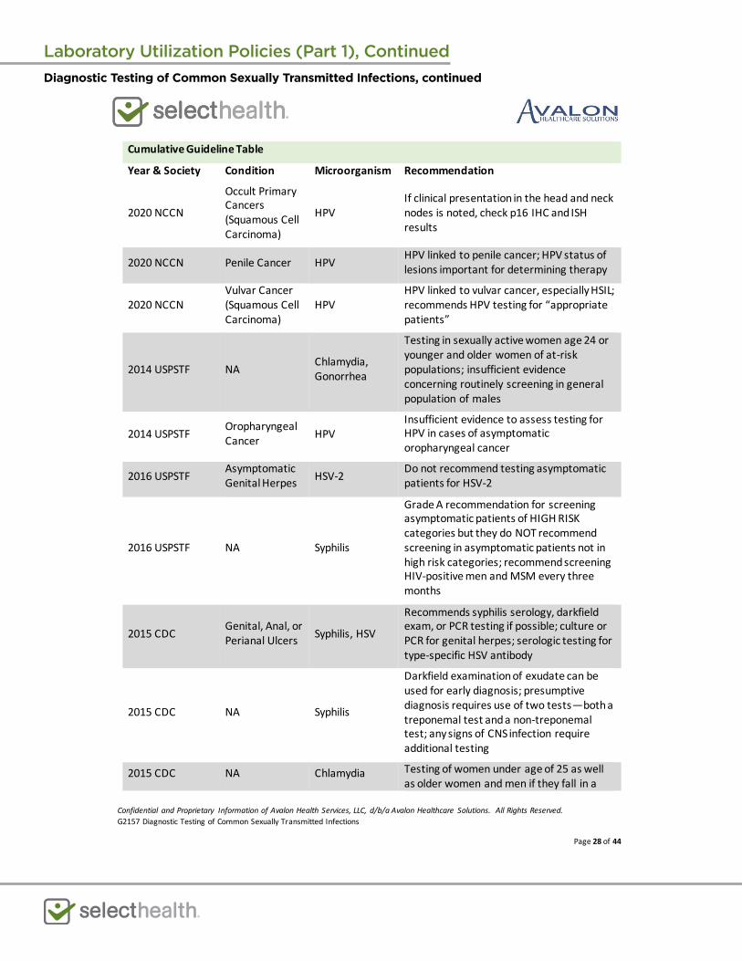
Laboratory Utilization Policies (Part 1), Continued
Confidential and Proprietary Information of Avalon Health Services, LLC, d/b/a Avalon Healthcare Solutions. All Rights Reserved. G2157 Diagnostic Testing of Common Sexually Transmitted Infections
Page 28 of 44
Cumulative Guideline Table
Year & Society Condition Microorganism Recommendation
2020 NCCN
Occult Primary Cancers (Squamous Cell Carcinoma)
HPV If clinical presentation in the head and neck nodes is noted, check p16 IHC and ISH results
2020 NCCN Penile Cancer HPV HPV linked to penile cancer; HPV status of lesions important for determining therapy
2020 NCCN Vulvar Cancer (Squamous Cell Carcinoma)
HPV HPV linked to vulvar cancer, especially HSIL; recommends HPV testing for “appropriate patients”
2014 USPSTF NA Chlamydia, Gonorrhea
Testing in sexually active women age 24 or younger and older women of at-risk populations; insufficient evidence concerning routinely screening in general population of males
2014 USPSTF Oropharyngeal Cancer HPV
Insufficient evidence to assess testing for HPV in cases of asymptomatic oropharyngeal cancer
2016 USPSTF Asymptomatic Genital Herpes HSV-2 Do not recommend testing asymptomatic
patients for HSV-2
2016 USPSTF NA Syphilis
Grade A recommendation for screening asymptomatic patients of HIGH RISK categories but they do NOT recommend screening in asymptomatic patients not in high risk categories; recommend screening HIV-positive men and MSM every three months
2015 CDC Genital, Anal, or Perianal Ulcers Syphilis, HSV
Recommends syphilis serology, darkfield exam, or PCR testing if possible; culture or PCR for genital herpes; serologic testing for type-specific HSV antibody
2015 CDC NA Syphilis
Darkfield examination of exudate can be used for early diagnosis; presumptive diagnosis requires use of two tests—both a treponemal test and a non-treponemal test; any signs of CNS infection require additional testing
2015 CDC NA Chlamydia Testing of women under age of 25 as well as older women and men if they fall in a
Diagnostic Testing of Common Sexually Transmitted Infections, continued

Laboratory Utilization Policies (Part 1), Continued
Confidential and Proprietary Information of Avalon Health Services, LLC, d/b/a Avalon Healthcare Solutions. All Rights Reserved. G2157 Diagnostic Testing of Common Sexually Transmitted Infections
Page 29 of 44
Cumulative Guideline Table
Year & Society Condition Microorganism Recommendation high-risk category; do NOT recommend testing of asymptomatic men and older women
2015 CDC NA Gonorrhea
Testing of women under age of 25 as well as older women and men if they fall in a high-risk category; do NOT recommend testing of asymptomatic men and older women; men showing signs of urethral gonococcal infection should be tested
2015, 2016 CDC NA HPV
Recommends against using oncogenic HPV testing for asymptomatic men, women aged 25 and over, or for general STD testing.
There is no approved test for HPV in men, and routine testing is not recommended for anal, penile, or throat cancers in men.
2015 CDC Anogenital Warts HPV
“HPV testing is not recommended for anogenital wart diagnosis, because test results are not confirmatory and do not guide genital wart management.”
2015, 2019 CDC Cervical Screening HPV
For women aged 30 or older, HPV testing can be part of cervical screening. For women ages 30-65, if co-testing Pap test and HR-HPV, then frequency is every 5 years…if only doing a Pap test, the frequency is every 3 years
HPV tests to screen for cervical cancer are recommended for women 30 years and older. They are not recommended to screen, men, adolescents, or women under the age of 30.
2019 IUSTI Anogenital Warts HPV
Do not recommend HPV testing for symptomatic anogenital warts since it adds no information for clinical use.
Diagnostic Testing of Common Sexually Transmitted Infections, continued
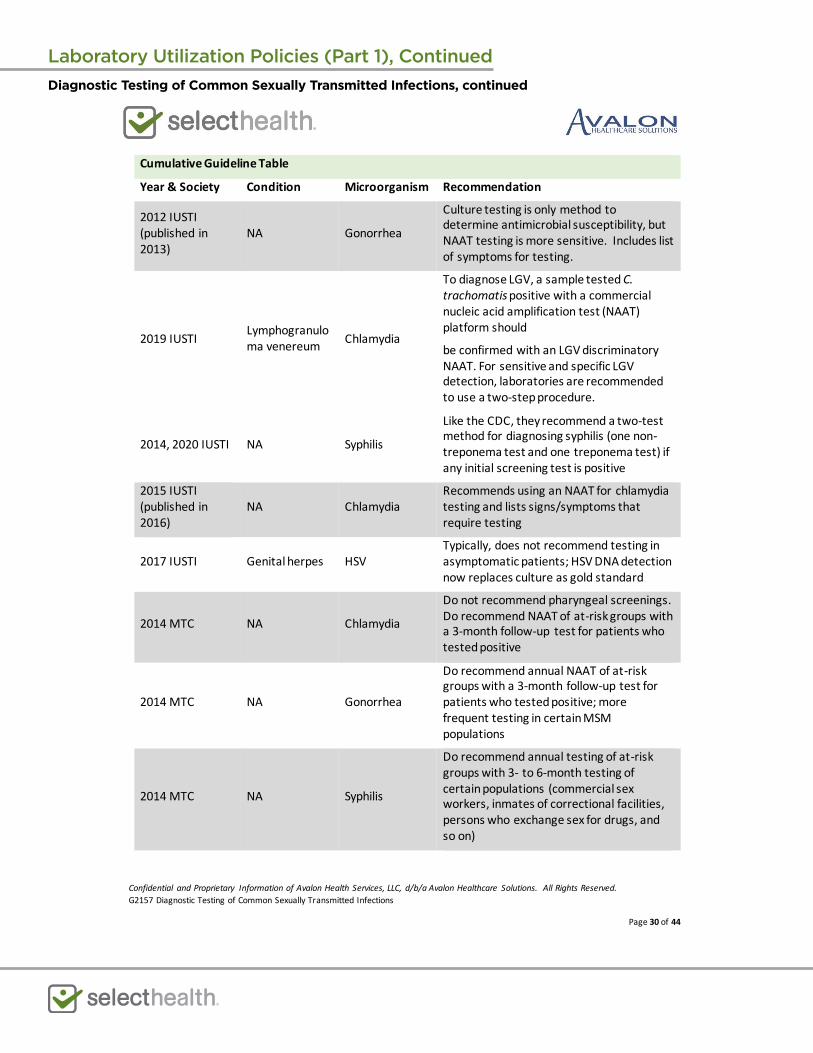
Laboratory Utilization Policies (Part 1), Continued
Confidential and Proprietary Information of Avalon Health Services, LLC, d/b/a Avalon Healthcare Solutions. All Rights Reserved. G2157 Diagnostic Testing of Common Sexually Transmitted Infections
Page 30 of 44
Cumulative Guideline Table
Year & Society Condition Microorganism Recommendation
2012 IUSTI (published in 2013)
NA Gonorrhea
Culture testing is only method to determine antimicrobial susceptibility, but NAAT testing is more sensitive. Includes list of symptoms for testing.
2019 IUSTI Lymphogranuloma venereum Chlamydia
To diagnose LGV, a sample tested C. trachomatis positive with a commercial nucleic acid amplification test (NAAT) platform should
be confirmed with an LGV discriminatory NAAT. For sensitive and specific LGV detection, laboratories are recommended to use a two-step procedure.
2014, 2020 IUSTI NA Syphilis
Like the CDC, they recommend a two-test method for diagnosing syphilis (one non-treponema test and one treponema test) if any initial screening test is positive
2015 IUSTI (published in 2016)
NA Chlamydia Recommends using an NAAT for chlamydia testing and lists signs/symptoms that require testing
2017 IUSTI Genital herpes HSV Typically, does not recommend testing in asymptomatic patients; HSV DNA detection now replaces culture as gold standard
2014 MTC NA Chlamydia
Do not recommend pharyngeal screenings. Do recommend NAAT of at-risk groups with a 3-month follow-up test for patients who tested positive
2014 MTC NA Gonorrhea
Do recommend annual NAAT of at-risk groups with a 3-month follow-up test for patients who tested positive; more frequent testing in certain MSM populations
2014 MTC NA Syphilis
Do recommend annual testing of at-risk groups with 3- to 6-month testing of certain populations (commercial sex workers, inmates of correctional facilities, persons who exchange sex for drugs, and so on)
Diagnostic Testing of Common Sexually Transmitted Infections, continued

Laboratory Utilization Policies (Part 1), Continued
Confidential and Proprietary Information of Avalon Health Services, LLC, d/b/a Avalon Healthcare Solutions. All Rights Reserved. G2157 Diagnostic Testing of Common Sexually Transmitted Infections
Page 31 of 44
Cumulative Guideline Table
Year & Society Condition Microorganism Recommendation
2018 Canadian Guidelines on STIs
NA
Chlamydia, Syphilis, Gonorrhea, HSV, and HPV
NAATs are more specific and sensitive than culture testing when available. For gonorrheal infections, only culture can test for antimicrobial susceptibility in gonorrhea.
2014 AAP Adolescents & young adults
Chlamydia, Gonorrhea
All sexually active young women (under the age of 25) and MSM should have annual screenings. For those at higher risk, they should be screened every 3-6 months. Anyone who tests positive should be retested 3 months after receiving treatment.
2014 AAP Adolescents & young adults Syphilis Do NOT recommend routine screening
except for sexually active young MSM.
2016 NICE Oropharyngeal Cancers HPV
Test all carcinomas of the oropharynx using p16 IHC; consider using high-risk HPV DNA/RNA in situ hybridization in all p16-positive cancers
2013 National Health Care for the Homeless Council
HIV/AIDS HPV
For women, every 6 months along with Pap for HIV-positive patients; decrease to annual after two consecutive normal Pap tests
2013 National Health Care for the Homeless Council
HIV/AIDS Chlamydia, Gonorrhea
Annual testing of all HIV-positive patients; consider anal Pap smear and rectal tests for MSM annually
2013 National Health Care for the Homeless Council
HIV/AIDS with anogenital condylomata
Chlamydia, Gonorrhea
Consider annual anal Pap smear and rectal tests for both chlamydia and GC for patients with history of anogenital condylomata
2018 CPS Pregnant women Syphilis
Testing at first prenatal visit as well as 28-32 weeks; if not tested during pregnancy, child does not leave the hospital without being tested
2020 CPS Adolescents/young adults
Chlamydia, Syphilis, Gonorrhea
See detailed testing and frequency in table within the guidelines above
Diagnostic Testing of Common Sexually Transmitted Infections, continued

Laboratory Utilization Policies (Part 1), Continued
Confidential and Proprietary Information of Avalon Health Services, LLC, d/b/a Avalon Healthcare Solutions. All Rights Reserved. G2157 Diagnostic Testing of Common Sexually Transmitted Infections
Page 32 of 44
Cumulative Guideline Table
Year & Society Condition Microorganism Recommendation
2015 BASHH (published in 2016)
NA Syphilis
Dark-field microscopy or PCR tests can be performed. For serology, EIA/CLIA is the screening test of choice (preferably where both IgM and IgG are detected). Positive tests must be followed by a quantitative RPR or VDRL.
2013 BASHH Suspected LGV Chlamydia
Testing should use either NAAT, culture testing, or serology; however, the latter cannot distinguish current from past infections.
2014 BASHH (published in 2015)
Anogenital herpes HSV
NAAT is preferred over other forms of testing (“A” grade). Differentiation of virus type should be determined on new cases of genital herpes (“B” grade).
2015, 2018 BASHH NA Chlamydia
Test for chlamydia using NAATs. Both sexes presenting with proctitis should be treated the same with respect to LGV testing. HIV-positive men with chlamydia should also be tested for LGV, even if asymptomatic.
Abbreviations: CLIA = chemiluminescent assay; EIA = enzyme immunoassay; GC = gonococcal; HPV = human papillomavirus; HR-HPV = high risk or oncogenic HPV testing; HSIL = high-grade squamous intraepithelial lesions; HSV = herpes simplex virus; IHC = immunohistochemistry; LGV = lymphogranuloma venereum; MSM = men having sex with men; NA = not applicable; NAAT = nucleic acid amplification testing; OS = overall survival; RPR = rapid plasma reagin test; VDRL = Venereal Diseases Research Laboratory carbon antigen test
VI. State and Federal Regulations (as applicable) The FDA approved the BD Onclarity HPV Assay, a qualitative in vitro assay of cervical swabs using PCR (i.e. a nucleic acid amplification test or NAAT), by Becton, Dickinson and Company on 02/12/2018. “The test specifically identifies types 16, 18 and 45 while concurrently detecting the other HR HPV types that include 31, 33, 35, 39, 51, 52, 56, 58, 59, 66 and 68. The BD Onclarity HPV Assay is indicated: 1) In women 21 years and older with ASC-US (atypical squamous cells of undetermined significance) cervical cytology test results, the BD Onclarity HPV Assay can be used to determine the need for referral to colposcopy;2) In women 21 years and older with ASC-US cervical cytology test results, the BD Onclarity HPV assay can be used to detect high-risk HPV genotypes 16, 18 and 45. This information together with physicians assessment of screening history, other risk factors, and professional guidelines, may be used to guide patient management. The results of this test are not intended to prevent women from proceeding to colposcopy;3) In women 30 years and older, the BD Onclarity HPV Assay can be used
Diagnostic Testing of Common Sexually Transmitted Infections, continued

Laboratory Utilization Policies (Part 1), Continued
Confidential and Proprietary Information of Avalon Health Services, LLC, d/b/a Avalon Healthcare Solutions. All Rights Reserved. G2157 Diagnostic Testing of Common Sexually Transmitted Infections
Page 33 of 44
together with cervical cytology to adjunctively screen to detect high risk HPV types. This information, together with the physicians assessment of screening history, other factors, and professional guidelines, may be used to guide patient management;4) In women 30 years and older, the BD Onclarity HPV Assay can be used to detect high-risk HPV genotypes 16, 18 and 45. This information, together with the physicians assessment of screening history, other factors, and professional guidelines, may be used to guide patient management; and5) In women 25 years and older, the BD Onclarity HPV Assay can be used as a first-line primary cervical cancer screening test to detect high risk HPV, including 16 and 18. Women who test negative for the high risk HPV types by the BD Onclarity HPV Assay should be followed up in accordance with the physicians assessment of screening and medical history, other risk factors, and professional guidelines. Women who test positive for HPV genotypes 16 and/or 18 by the BD Onclarity HPV Assay should be referred to colposcopy. Women who test high risk HPV positive and 16 and 18 negative by the BD Onclarity HPV Assay (12 other HR HPV Positive) should be evaluated by cervical cytology to determine the need for referral to colposcopy (FDA, 2018a).” Further, BD recently reported that the BD Onclarity Assay received FDA approval for extended genotyping capabilities beyond HPV genotypes 16, 18, and 45; genotypes 31, 51, 52, 33/58, 35/39/68, and 56/59/66 are now included. This is “the only FDA-approved assay to individually identify and report these genotype results” (BD, 2020).
The FDA has also approved the APTIMA HPV 16 18/45 Genotype Assay, a NAAT, for the qualitative detection of mRNA for HPV 16, 18, and 45 from Gen-Probe Incorporated on 10/12/2012; however, this test cannot distinguish between 18 and 45. Previously, on 10/28/2012, the FDA approved Gen-Probe Incorporated’s APTIMA HPV Assay, an NAAT that tests for 14 high-risk types of HPV but is unable to distinguish between the 14 types. The COBAS HPV test by Roche Molecular Systems, Inc. was approved by the FDA on 04/19/2011 as a NAAT for 14 high-risk types of HPV. This test can specifically identify HPV 16 and 18 but cannot distinguish from the other 12 types of HPV. Hologic, Inc. has two FDA-approved HPV NAAT tests—Cervista HPV 16/18 and Cervista HPV HR and GENFIND DNA Extraction Kit. Both were approved on 03/12/2009. The former is a fluorescent, isothermal-based reaction that detects HPV 16 and 18 whereas the latter screens for DNA from the 14 high-risk HPV strains (FDA, 2018b).
The FDA has approved many tests for HSV, chlamydia, gonorrhea, and syphilis. A search of the FDA Devices database of “HSV” on 09/24/2020 yielded 108 results. Likewise, a search of “chlamydia” and “syphilis” had 143 and 36 records, respectively. “Neisseria” and “gonorrhea” yielded a combined 59 records of approved FDA devices as of 09/24/2020. “Trichomonas” yielded 17 records of approved FDA devices as of 09/24/2020 (FDA, 2020).
Additionally, many labs have developed specific tests that they must validate and perform in house. These laboratory-developed tests (LDTs) are regulated by the Centers for Medicare and Medicaid (CMS) as high-complexity tests under the Clinical Laboratory Improvement Amendments of 1988 (CLIA ’88). As an LDT, the U. S. Food and Drug Administration has not approved or cleared this test; however, FDA clearance or approval is not currently required for clinical use.
Diagnostic Testing of Common Sexually Transmitted Infections, continued

Laboratory Utilization Policies (Part 1), Continued
Confidential and Proprietary Information of Avalon Health Services, LLC, d/b/a Avalon Healthcare Solutions. All Rights Reserved. G2157 Diagnostic Testing of Common Sexually Transmitted Infections
Page 34 of 44
VII. Applicable CPT/HCPCS Procedure Codes Code Number Code Description
82565 Creatinine; blood
82575 Creatinine; clearance
84702 Gonadotropin, chorionic (hCG); quantitative
84703 Gonadotropin, chorionic (hCG); qualitative
86592 Syphilis test, non-treponemal antibody; qualitative (e.g., VDRL, RPR, ART)
86593 Syphilis test, non-treponemal antibody; quantitative
86631 Antibody; Chlamydia
86701 Antibody; HIV-1
86702 Antibody; HIV-2
86703 Antibody; HIV-1 and HIV-2, single result
86704 Hepatitis B core antibody (HBcAb); total
86705 Hepatitis B core antibody (HBcAb); IgM antibody
86706 Hepatitis B surface antibody (HBsAb)
86632 Antibody; Chlamydia, IgM
86694 Antibody; herpes simplex, non-specific type test
86695 Antibody; herpes simplex, type 1
86696 Antibody; herpes simplex, type 2
86780 Antibody; Treponema pallidum
86803 Hepatitis C antibody
86804 Hepatitis C antibody; confirmatory test (eg, immunoblot)
87081 Culture, presumptive, pathogenic organisms, screening only;
87110 Culture, chlamydia, any source
Diagnostic Testing of Common Sexually Transmitted Infections, continued

Laboratory Utilization Policies (Part 1), Continued
Confidential and Proprietary Information of Avalon Health Services, LLC, d/b/a Avalon Healthcare Solutions. All Rights Reserved. G2157 Diagnostic Testing of Common Sexually Transmitted Infections
Page 35 of 44
87181 Susceptibility studies, antimicrobial agent; agar dilution method, per agent (e.g., antibiotic gradient strip)
87340
Infectious agent antigen detection by immunoassay technique, (eg, enzyme immunoassay [EIA], enzyme-linked immunosorbent assay [ELISA], fluorescence immunoassay [FIA], immunochemiluminometric assay [IMCA]) qualitative or semiquantitative; hepatitis B surface antigen (HBsAg)
87490 Infectious agent detection by nucleic acid (DNA or RNA); Chlamydia trachomatis, direct probe technique
87491 Infectious agent detection by nucleic acid (DNA or RNA); Chlamydia trachomatis, amplified probe technique
87492 Infectious agent detection by nucleic acid (DNA or RNA); Chlamydia trachomatis, quantification
87528 Infectious agent detection by nucleic acid (DNA or RNA); Herpes simplex virus, direct probe technique
87529 Infectious agent detection by nucleic acid (DNA or RNA); Herpes simplex virus, amplified probe technique
87530 Infectious agent detection by nucleic acid (DNA or RNA); Herpes simplex virus, quantification
87590 Infectious agent detection by nucleic acid (DNA or RNA); Neisseria gonorrhoeae, direct probe technique
87591 Infectious agent detection by nucleic acid (DNA or RNA); Neisseria gonorrhoeae, amplified probe technique
87592 Infectious agent detection by nucleic acid (DNA or RNA); Neisseria gonorrhoeae, quantification
87623 Infectious agent detection by nucleic acid (DNA or RNA); Human Papillomavirus (HPV), low-risk types (e.g., 6, 11, 42, 43, 44)
87624 Infectious agent detection by nucleic acid (DNA or RNA); Human Papillomavirus (HPV), high-risk types (e.g., 16, 18, 31, 33, 35, 39, 45, 51, 52, 56, 58, 59, 68)
87625 Infectious agent detection by nucleic acid (DNA or RNA); Human Papillomavirus (HPV), types 16 and 18 only, includes type 45, if performed
87660 Infectious agent detection by nucleic acid (DNA or RNA); Trichomonas vaginalis, direct probe technique
Diagnostic Testing of Common Sexually Transmitted Infections, continued

Laboratory Utilization Policies (Part 1), Continued
Confidential and Proprietary Information of Avalon Health Services, LLC, d/b/a Avalon Healthcare Solutions. All Rights Reserved. G2157 Diagnostic Testing of Common Sexually Transmitted Infections
Page 36 of 44
87661 Infectious agent detection by nucleic acid (DNA or RNA); Trichomonas vaginalis, amplified probe technique
87797 Infectious agent detection by nucleic acid (DNA or RNA), not otherwise specified; direct probe technique, each organism
87798 Infectious agent detection by nucleic acid (DNA or RNA), not otherwise specified; amplified probe technique, each organism
87799 Infectious agent detection by nucleic acid (DNA or RNA), not otherwise specified; quantification, each organism
87808 Infectious agent antigen detection by immunoassay with direct optical observation; Trichomonas vaginalis
88341 Immunohistochemistry or immunocytochemistry, per specimen; each additional single antibody stain procedure (List separately in addition to code for primary procedure)
88342 Immunohistochemistry or immunocytochemistry, per specimen; initial single antibody stain procedure
G0432 Infectious agent antibody detection by enzyme immunoassay (EIA) technique, HIV-1 and/or HIV-2, screening
G0433 Infectious agent antibody detection by enzyme-linked immunosorbent assay (ELISA) technique, HIV-1 and/or HIV-2, screening
G0435 Infectious agent antibody detection by rapid antibody test, HIV-1 and/or HIV-2, screening
G0472 Hepatitis C antibody screening, for individual at high risk and other covered indication(s)
G0475 Hiv antigen/antibody, combination assay, screening
G0499
Hepatitis b screening in non-pregnant, high risk individual includes hepatitis b surface antigen (HBSAG) followed by a neutralizing confirmatory test for initially reactive results, and antibodies to HBSAG (anti-HBs) and Hepatitis B core antigen (anti-HBc)
S3645 HIV-1 antibody testing of oral mucosal transudate
Current Procedural Terminology© American Medical Association. All Rights reserved.
Procedure codes appearing in Medical Policy documents are included only as a general reference tool for each policy. They may not be all-inclusive.
Diagnostic Testing of Common Sexually Transmitted Infections, continued

Laboratory Utilization Policies (Part 1), Continued
Confidential and Proprietary Information of Avalon Health Services, LLC, d/b/a Avalon Healthcare Solutions. All Rights Reserved. G2157 Diagnostic Testing of Common Sexually Transmitted Infections
Page 37 of 44
VIII. Evidence-based Scientific References Albrecht, M. A. (2017, 01/10/2017). Epidemiology, clinical manifestations, and diagnosis of
genital herpes simplex virus infection. UpToDate. Retrieved from https://www.uptodate.com/contents/epidemiology-clinical-manifestations-and-diagnosis-of-genital-herpes-simplex-virus-infection
Albrecht, M. A. (2018, 01/10/2017). Epidemiology, clinical manifestations, and diagnosis of genital herpes simplex virus infection. UpToDate. Retrieved from https://www.uptodate.com/contents/epidemiology-clinical-manifestations-and-diagnosis-of-genital-herpes-simplex-virus-infection
Allen, U. D., MacDonald, N. E., & Canadian Paediatric Society, I. D. a. I. C. (2018, 12/18/2018). Sexually transmitted infections in adolescents: Maximizing opportunities for optimal care. Position Statements and Practice Points. Retrieved from https://www.cps.ca/en/documents/position/sexually-transmitted-infections?utm_source=AMMI+Canada+members&utm_campaign=1930829c79-Information_Update_Job_Posting11_8_2013&utm_medium=email&utm_term=0_a934e89600-1930829c79-231313337
Allen, U. D., MacDonald, N. E., & Top, K. (2019). Diagnosis and management of sexually transmitted infections in adolescents. Retrieved from https://www.cps.ca/en/documents/position/sexually-transmitted-infections
Arbyn, M., Roelens, J., Simoens, C., Buntinx, F., Paraskevaidis, E., Martin-Hirsch, P. P., & Prendiville, W. J. (2013). Human papillomavirus testing versus repeat cytology for triage of minor cytological cervical lesions. Cochrane Database Syst Rev(3), Cd008054. doi:10.1002/14651858.CD008054.pub2
Audain, G., Bookhardt-Murray, L., Fogg, C., Gregerson, P., Haley, C., Luther, P., & Treherne, L. (2013). Adapting your practice: Treatment and recommendations for unstably housed patients with HIV/AIDS (I. National Health Care for the Homeless Council Ed. Third ed.). Nashville, TN: Health Care for the Homeless Clinicians' Network.
BASHH. (2018). BASHH CLINICAL EFFECTIVENESS GROUP Update on the treatment of Chlamydia trachomatis (CT) infection. Retrieved from https://www.bashhguidelines.org/current-guidelines/urethritis-and-cervicitis/chlamydia-2015/
BD. (2020). BD receives FDA Approval for HPV Test with Extended Genotyping Capabilities. Retrieved from https://www.bd.com/en-us/company/news-and-media/press-releases/july-22-2020-bd-receives-fda-approval-for-hpv-test-with-extended-genotyping-capabilities
Bignell, C., & Fitzgerald, M. (2011). UK national guideline for the management of gonorrhoea in adults, 2011. Int J STD AIDS, 22(10), 541-547. doi:10.1258/ijsa.2011.011267
Bignell, C., & Unemo, M. (2013). 2012 European guideline on the diagnosis and treatment of gonorrhoea in adults. Int J STD AIDS, 24(2), 85-92. doi:10.1177/0956462412472837
Diagnostic Testing of Common Sexually Transmitted Infections, continued

Laboratory Utilization Policies (Part 1), Continued
Confidential and Proprietary Information of Avalon Health Services, LLC, d/b/a Avalon Healthcare Solutions. All Rights Reserved. G2157 Diagnostic Testing of Common Sexually Transmitted Infections
Page 38 of 44
Brischetto, A., Gassiep, I., Whiley, D., & Norton, R. (2018). Retrospective Review of Treponema pallidum PCR and Serology Results: Are Both Tests Necessary? J Clin Microbiol, 56(5). doi:10.1128/jcm.01782-17
Cantor, A. G., Pappas, M., Daeges, M., & Nelson, H. D. (2016). Screening for syphilis: Updated evidence report and systematic review for the us preventive services task force. JAMA, 315(21), 2328-2337. doi:10.1001/jama.2016.4114
Castle, P. E., Stoler, M. H., Wright, T. C., Jr., Sharma, A., Wright, T. L., & Behrens, C. M. (2011). Performance of carcinogenic human papillomavirus (HPV) testing and HPV16 or HPV18 genotyping for cervical cancer screening of women aged 25 years and older: a subanalysis of the ATHENA study. Lancet Oncol, 12(9), 880-890. doi:10.1016/s1470-2045(11)70188-7
CDC. (2015, 06/04/2015). Trichomoniasis. 2015 STD Treatment Guidelines. Retrieved from https://www.cdc.gov/std/tg2015/trichomoniasis.htm
CDC. (2016a, 09/26/2017). Chlamydia - CDC Fact Sheet (Detailed). Retrieved from https://www.cdc.gov/std/chlamydia/stdfact-chlamydia-detailed.htm
CDC. (2016b, 09/26/2017). Gonorrhea - CDC Fact Sheet (Detailed Version). Retrieved from https://www.cdc.gov/std/gonorrhea/stdfact-gonorrhea-detailed.htm
CDC. (2016c). HPV & Men Fact Sheet. Retrieved from https://www.cdc.gov/std/hpv/stdfact-hpv-and-men.htm
CDC. (2017a, 02/09/2017). Genital Herpes - CDC Fact Sheet (Detailed). Retrieved from https://www.cdc.gov/std/herpes/stdfact-herpes-detailed.htm
CDC. (2017b, 09/26/2017). The State of STDs - Infographic. Retrieved from https://www.cdc.gov/std/stats16/infographic.htm
CDC. (2017c, 02/13/2017). Syphilis-CDC Fact Sheet (Detailed). Retrieved from https://www.cdc.gov/std/syphilis/stdfact-syphilis-detailed.htm
CDC. (2019a, 11/16/2017). Genital HPV Infection - Fact Sheet. Retrieved from https://www.cdc.gov/std/hpv/stdfact-hpv.htm
CDC. (2019b, 09/26/2017). Gonorrhea - CDC Fact Sheet (Detailed Version). Retrieved from https://www.cdc.gov/std/gonorrhea/stdfact-gonorrhea-detailed.htm
CDC. (2020, 01/31/2017). Trichomoniasis - CDC Fact Sheet. Retrieved from https://www.cdc.gov/std/trichomonas/stdfact-trichomoniasis.htm
Chernesky, M. (2018). Section 3: Canadian Guidelines on Sexually Transmitted Infections-Laboratory diagnosis of sexually transmitted infections. Ottawa, Ontaria, Canada: Public Health Agency of Canada Retrieved from https://www.canada.ca/en/public-health/services/infectious-diseases/sexual-health-sexually-transmitted-infections/canadian-guidelines/sexually-transmitted-infections/canadian-guidelines-sexually-transmitted-infections-18.html#a23
Cook, R. L., Hutchison, S. L., Ostergaard, L., Braithwaite, R. S., & Ness, R. B. (2005). Systematic review: noninvasive testing for Chlamydia trachomatis and Neisseria gonorrhoeae. Ann Intern Med, 142(11), 914-925.
Diagnostic Testing of Common Sexually Transmitted Infections, continued

Laboratory Utilization Policies (Part 1), Continued
Confidential and Proprietary Information of Avalon Health Services, LLC, d/b/a Avalon Healthcare Solutions. All Rights Reserved. G2157 Diagnostic Testing of Common Sexually Transmitted Infections
Page 39 of 44
de Vries, H. J., Zingoni, A., Kreuter, A., Moi, H., & White, J. A. (2015). 2013 European guideline on the management of lymphogranuloma venereum. J Eur Acad Dermatol Venereol, 29(1), 1-6. doi:10.1111/jdv.12461
de Vries, H. J. C., de Barbeyrac, B., de Vrieze, N. H. N., Viset, J. D., White, J. A., Vall-Mayans, M., & Unemo, M. (2019). 2019 European guideline on the management of lymphogranuloma venereum. J Eur Acad Dermatol Venereol, 33(10), 1821-1828. doi:10.1111/jdv.15729
FDA. (2018a, 07/09/2018). BD ONCLARITY HPV ASSAY. Devices@FDA. Retrieved from https://www.accessdata.fda.gov/scripts/cdrh/devicesatfda/index.cfm?db=pma&id=391601
FDA. (2018b). Devices@FDA. Retrieved from https://www.accessdata.fda.gov/scripts/cdrh/devicesatfda/index.cfm
FDA. (2020). Devices@FDA. Retrieved from https://www.accessdata.fda.gov/scripts/cdrh/devicesatfda/index.cfm
Feldman, S., & Crum, C. P. (2020, 04/14/2017). Cervical cancer screening tests: Techniques for cervical cytology and human papillomavirus testing. UpToDate. Retrieved from https://www.uptodate.com/contents/cervical-cancer-screening-tests-techniques-for-cervical-cytology-and-human-papillomavirus-testing
Feltner, C., Grodensky, C., Ebel, C., & et al. (2016). Serologic screening for genital herpes: An updated evidence report and systematic review for the us preventive services task force. JAMA, 316(23), 2531-2543. doi:10.1001/jama.2016.17138
Gaydos, C. A., Ako, M. C., Lewis, M., Hsieh, Y. H., Rothman, R. E., & Dugas, A. F. (2019). Use of a Rapid Diagnostic for Chlamydia trachomatis and Neisseria gonorrhoeae for Women in the Emergency Department Can Improve Clinical Management: Report of a Randomized Clinical Trial. Ann Emerg Med, 74(1), 36-44. doi:10.1016/j.annemergmed.2018.09.012
Gaydos, C. A., Klausner, J. D., Pai, N. P., Kelly, H., Coltart, C., & Peeling, R. W. (2017). Rapid and point-of-care tests for the diagnosis of Trichomonas vaginalis in women and men. Sex Transm Infect, 93(S4), S31-s35. doi:10.1136/sextrans-2016-053063
Ghanem, K. G. (2018, 06/15/2018). Clinical manifestations and diagnosis of Neisseria gonorrhoeae infection in adults and adolescents. UpToDate. Retrieved from https://www.uptodate.com/contents/clinical-manifestations-and-diagnosis-of-neisseria-gonorrhoeae-infection-in-adults-and-adolescents
Ghanem, K. G., & Tuddenham, S. (2017, 12/07/2017). Screening for sexually transmitted infections. UpToDate. Retrieved from https://www.uptodate.com/contents/screening-for-sexually-transmitted-infections
Ghanem, K. G., & Tuddenham, S. (2020, 12/07/2017). Screening for sexually transmitted infections. UpToDate. Retrieved from https://www.uptodate.com/contents/screening-for-sexually-transmitted-infections
Gilson, R., Nugent, Diarmuid, Werner, Ricardo Niklas, Ballesteros, Juan, Ross, John. (2019). 2019 European Guideline for the Management of Anogenital Warts Retrieved from https://www.iusti.org/regions/Europe/pdf/2019/IUSTIguidelinesHPV2019.pdf
Diagnostic Testing of Common Sexually Transmitted Infections, continued

Laboratory Utilization Policies (Part 1), Continued
Confidential and Proprietary Information of Avalon Health Services, LLC, d/b/a Avalon Healthcare Solutions. All Rights Reserved. G2157 Diagnostic Testing of Common Sexually Transmitted Infections
Page 40 of 44
Golden, M., O'Donnell, M., Lukehart, S., Swenson, P., Hovey, P., Godornes, C., . . . Getman, D. (2019). Treponema pallidum Nucleic Acid Amplification Testing To Augment Syphilis Screening among Men Who Have Sex with Men. J Clin Microbiol, 57(8). doi:10.1128/jcm.00572-19
Guenat, D., Launay, S., Riethmuller, D., Mougin, C., & Pretet, J. L. (2016). Validation of Novaprep((R)) HQ+ liquid-based cytology medium for high-risk human papillomavirus detection by hc2. Infect Agent Cancer, 11, 41. doi:10.1186/s13027-016-0092-7
Guy, R. J., Causer, L. M., Klausner, J. D., Unemo, M., Toskin, I., Azzini, A. M., & Peeling, R. W. (2017). Performance and operational characteristics of point-of-care tests for the diagnosis of urogenital gonococcal infections. Sex Transm Infect, 93(S4), S16-s21. doi:10.1136/sextrans-2017-053192
Hicks, C. B., & Clement, M. (2018, 05/21/2018). Syphilis: Epidemiology, pathophysiology, and clinical manifestations in HIV-uninfected patients. UpToDate. Retrieved from https://www.uptodate.com/contents/syphilis-epidemiology-pathophysiology-and-clinical-manifestations-in-hiv-uninfected-patients
Hicks, C. B., & Clement, M. (2019, 04/03/2017). Syphilis: Screening and diagnostic testing. UpToDate. Retrieved from https://www.uptodate.com/contents/syphilis-screening-and-diagnostic-testing
Hicks, C. B., & Clement, M. (2020, 05/21/2018). Syphilis: Epidemiology, pathophysiology, and clinical manifestations in HIV-uninfected patients. UpToDate. Retrieved from https://www.uptodate.com/contents/syphilis-epidemiology-pathophysiology-and-clinical-manifestations-in-hiv-uninfected-patients
Hsu, K. (2019, 01/17/2018). Clinical manifestations and diagnosis of Chlamydia trachomatis infections. UpToDate. Retrieved from https://www.uptodate.com/contents/clinical-manifestations-and-diagnosis-of-chlamydia-trachomatis-infections
Janier, M., Hegyi, V., Dupin, N., Unemo, M., Tiplica, G. S., Potocnik, M., . . . Patel, R. (2014). 2014 European guideline on the management of syphilis. J Eur Acad Dermatol Venereol, 28(12), 1581-1593. doi:10.1111/jdv.12734
Janier, M., Unemo, M., Dupin, N., Tiplica, G. S., Potocnik, M., & Patel, R. (2020). 2020 European guideline on the management of syphilis. Acta Clin Belg. doi:10.1080/17843286.2020.1773112
Juarez-Figueroa, L., Uribe-Salas, F., Garcia-Cisneros, S., Olamendi-Portugal, M., & Conde-Glez, C. J. (2007). Evaluation of a rapid strip and a particle agglutination tests for syphilis diagnosis. Diagn Microbiol Infect Dis, 59(2), 123-126. doi:10.1016/j.diagmicrobio.2007.04.008
Kelly, H., Coltart, C. E. M., Pant Pai, N., Klausner, J. D., Unemo, M., Toskin, I., & Peeling, R. W. (2017). Systematic reviews of point-of-care tests for the diagnosis of urogenital Chlamydia trachomatis infections. Sex Transm Infect, 93(S4), S22-s30. doi:10.1136/sextrans-2016-053067
Kingston, M., French, P., Higgins, S., McQuillan, O., Sukthankar, A., Stott, C., . . . Sullivan, A. (2016). UK national guidelines on the management of syphilis 2015. Int J STD AIDS, 27(6), 421-446. doi:10.1177/0956462415624059
Diagnostic Testing of Common Sexually Transmitted Infections, continued
Laboratory Utilization Policies (Part 1), Continued
Confidential and Proprietary Information of Avalon Health Services, LLC, d/b/a Avalon Healthcare Solutions. All Rights Reserved. G2157 Diagnostic Testing of Common Sexually Transmitted Infections
Page 41 of 44
Lacey, C. J., Woodhall, S. C., Wikstrom, A., & Ross, J. (2013). 2012 European guideline for the management of anogenital warts. J Eur Acad Dermatol Venereol, 27(3), e263-270. doi:10.1111/j.1468-3083.2012.04493.x
Lanjouw, E., Ouburg, S., de Vries, H. J., Stary, A., Radcliffe, K., & Unemo, M. (2016). 2015 European guideline on the management of Chlamydia trachomatis infections. Int J STD AIDS, 27(5), 333-348. doi:10.1177/0956462415618837
LeFevre, M. L. (2014). Screening for Chlamydia and gonorrhea: U.S. Preventive Services Task Force recommendation statement. Ann Intern Med, 161(12), 902-910. doi:10.7326/m14-1981
Liu, T. Y., Xie, R., Luo, L., Reilly, K. H., He, C., Lin, Y. Z., . . . Wang, H. B. (2014). Diagnostic validity of human papillomavirus E6/E7 mRNA test in cervical cytological samples. J Virol Methods, 196, 120-125. doi:10.1016/j.jviromet.2013.10.032
Marcell, A. V., & Health, M. T. C. f. F. P. a. R. (2014). Preventive Male Sexual and Reproductive Health Care: Recommendations for Clinical Practice. Retrieved from http://content.guidelinecentral.com/guideline/get/pdf/2787
Moyer, V. A. (2014). Screening for oral cancer: U.S. preventive services task force recommendation statement. Ann Intern Med, 160(1), 55-60. doi:10.7326/M13-2568
Murray, P., Braverman, P., Adelman, W., Breuner, C., Levine, D., Marcell, A. V., . . . Burstein, G. (2014). Screening for nonviral sexually transmitted infections in adolescents and young adults. Pediatrics, 134(1), e302-311. doi:10.1542/peds.2014-1024
NCCC. (2018). National Institute for Health and Care Excellence: Clinical Guidelines. In Cancer of the Upper Aerodigestive Tract: Assessment and Management in People Aged 16 and Over. London: National Institute for Health and Care Excellence (UK) Copyright (c) National Collaborating Centre for Cancer.
NCCN. (2019a). NCCN Clinical Practice Guidelines in Oncology Anal Carcinoma. NCCN Guidelines, 1.2019. Retrieved from https://www.nccn.org/professionals/physician_gls/pdf/anal.pdf
NCCN. (2019b). NCCN Clinical Practice Guidelines in Oncology Cervical Cancer. NCCN Guidelines, 4.2019. Retrieved from https://www.nccn.org/professionals/physician_gls/pdf/cervical.pdf
NCCN. (2019c). NCCN Clinical Practice Guidelines in Oncology Head and Neck Cancers. NCCN Guidelines, 1.2019. Retrieved from https://www.nccn.org/professionals/physician_gls/pdf/head-and-neck.pdf
NCCN. (2019d). NCCN Clinical Practice Guidelines in Oncology Penile Cancer. NCCN Guidelines, 2.2019. Retrieved from https://www.nccn.org/professionals/physician_gls/pdf/penile.pdf
NCCN. (2019e). NCCN Clinical Practice Guidelines in Oncology Vulvar Cancer (Squamous Cell Carcinoma). NCCN Guidelines, 2.2019. Retrieved from https://www.nccn.org/professionals/physician_gls/pdf/vulvar.pdf
NCCN. (2020a). NCCN Clinical Practice Guidelines in Occult Primary. NCCN Guidelines, 3.2020. Retrieved from https://www.nccn.org/professionals/physician_gls/pdf/occult.pdf
Diagnostic Testing of Common Sexually Transmitted Infections, continued

Laboratory Utilization Policies (Part 1), Continued
Confidential and Proprietary Information of Avalon Health Services, LLC, d/b/a Avalon Healthcare Solutions. All Rights Reserved. G2157 Diagnostic Testing of Common Sexually Transmitted Infections
Page 42 of 44
NCCN. (2020b). NCCN Clinical Practice Guidelines in Oncology Anal Carcinoma. NCCN Guidelines, 2.2020. Retrieved from https://www.nccn.org/professionals/physician_gls/pdf/anal.pdf
NCCN. (2020c). NCCN Clinical Practice Guidelines in Oncology Cervical Cancer. NCCN Guidelines, 1.2020. Retrieved from https://www.nccn.org/professionals/physician_gls/pdf/cervical.pdf
NCCN. (2020d). NCCN Clinical Practice Guidelines in Oncology Head and Neck Cancers. NCCN Guidelines, 2.2020. Retrieved from https://www.nccn.org/professionals/physician_gls/pdf/head-and-neck.pdf
NCCN. (2020e). NCCN Clinical Practice Guidelines in Oncology Penile Cancer. NCCN Guidelines, 1.2020. Retrieved from https://www.nccn.org/professionals/physician_gls/pdf/penile.pdf
NCCN. (2020f). NCCN Clinical Practice Guidelines in Oncology Vulvar Cancer (Squamous Cell Carcinoma). NCCN Guidelines, 2.2020. Retrieved from https://www.nccn.org/professionals/physician_gls/pdf/vulvar.pdf
Nwokolo, N. C., Dragovic, B., Patel, S., Tong, C. Y., Barker, G., & Radcliffe, K. (2016). 2015 UK national guideline for the management of infection with Chlamydia trachomatis. Int J STD AIDS, 27(4), 251-267. doi:10.1177/0956462415615443
Palefsky, J. M. (2018, 06/13/2018). Human papillomavirus infections: Epidemiology and disease associations. UpToDate. Retrieved from https://www.uptodate.com/contents/human-papillomavirus-infections-epidemiology-and-disease-associations
Palefsky, J. M. (2019, 06/13/2018). Human papillomavirus infections: Epidemiology and disease associations. UpToDate. Retrieved from https://www.uptodate.com/contents/human-papillomavirus-infections-epidemiology-and-disease-associations
Papp, J. R., Schachter, J., Gaydos, C. A., & Van Der Pol, B. (2014). Recommendations for the laboratory-based detection of Chlamydia trachomatis and Neisseria gonorrhoeae--2014. MMWR Recomm Rep, 63(Rr-02), 1-19. Retrieved from https://www.cdc.gov/mmwr/pdf/rr/rr6302.pdf
Patel, R., Green, J., Clarke, E., Seneviratne, K., Abbt, N., Evans, C., . . . Foley, E. (2015). 2014 UK national guideline for the management of anogenital herpes. Int J STD AIDS, 26(11), 763-776. doi:10.1177/0956462415580512
Patel, R., Kennedy, O. J., Clarke, E., Geretti, A., Nilsen, A., Lautenschlager, S., . . . Foley, E. (2017). 2017 European guidelines for the management of genital herpes. Int J STD AIDS, 28(14), 1366-1379. doi:10.1177/0956462417727194
Riley, L. E., & Wald, A. (2020, 02/16/2018). Genital herpes simplex virus infection and pregnancy. UpToDate. Retrieved from https://www.uptodate.com/contents/genital-herpes-simplex-virus-infection-and-pregnancy
Robinson, J., & Canadian Paediatric Society, I. D. a. I. C. (2018, 04/06/2018). Congenital syphilis: No longer just of historical interest. Position Statements and Practice Points. Retrieved from https://www.cps.ca/en/documents/position/congenital-syphilis
Schwebke, J., Merriweather, A., Massingale, S., Scisney, M., Hill, C., & Getman, D. (2018). Screening for Trichomonas vaginalis in a Large High-Risk Population: Prevalence Among
Diagnostic Testing of Common Sexually Transmitted Infections, continued

Laboratory Utilization Policies (Part 1), Continued
Confidential and Proprietary Information of Avalon Health Services, LLC, d/b/a Avalon Healthcare Solutions. All Rights Reserved. G2119 Diagnostic Testing of Influenza
Page 1 of 15
Diagnostic Testing of Influenza Policy #: AHS – G2119 Prior Policy Name & Number
(as applicable): • G2119 – Rapid Flu Tests in the Outpatient
Setting • Also included influenza coverage from prior
M2097 – Identification of Microorganisms Using Nucleic Acid Probes
Implementation Date: 9/15/21 Date of Last Revision:
I. Policy Description
Influenza is an acute respiratory illness caused by influenza A or B viruses resulting in upper and lower respiratory tract infection, fever, malaise, headache, and weakness. It mainly occurs in outbreaks and epidemics during the winter season, and is associated with increased morbidity and mortality in certain high-risk populations (Dolin, 2020).
Rapid influenza diagnostic tests (RIDTs) refer to clinical laboratory improvement amendments (CLIA) waived immunoassays that can detect influenza viruses during the outpatient visit, giving results in a clinically relevant time period to inform treatment decisions (CDC, 2017). Besides RIDTs, influenza can be detected using polymerase chain reaction (PCR)-based assays as well as culture testing; however, the former is not often used in initial clinical management due to time constraints. Serologic testing is not used in outpatient settings for diagnosis (Dolin, 2019b).
II. Related Policies
Policy Number Policy Title AHS-G2149 Pathogen Panel Testing AHS-G2174 Coronavirus Testing in the Outpatient Setting
Disclaimer:
1. Policies are subject to change without notice. 2. Policies outline coverage determinations for SelectHealth Commercial, SelectHealth
Advantage® (Medicare/CMS) and SelectHealth Community Care® (Medicaid/CHIP) plans. Refer to the “Policy” section for more information.

Laboratory Utilization Policies (Part 1), Continued
Confidential and Proprietary Information of Avalon Health Services, LLC, d/b/a Avalon Healthcare Solutions. All Rights Reserved. G2119 Diagnostic Testing of Influenza
Page 2 of 15
III. Indications and/or Limitations of Coverage
1. One single rapid flu test—including either a point-of-contact rapid nucleic acid amplification test (NAAT) or a rapid antigen test—OR one single traditional NAAT in the outpatient setting for a patient in a single visit, but not both an antigen and NAAT for a single patient in a single visit, MEETS COVERAGE CRITERIA for diagnosis of patients who present with signs and symptoms consistent with influenza disease (See Note 1 below) when influenza activity has been documented in the community or geographic area.
2. Viral culture testing for influenza in an outpatient setting DOES NOT MEET COVERAGE CRITERIA.
3. Outpatient influenza testing, including rapid antigen flu tests, rapid NAAT or RT-PCR tests, traditional RT-PCR tests, and viral culture testing DOES NOT MEET COVERAGE CRITERIA in asymptomatic patients.
4. Serology testing for influenza DOES NOT MEET COVERAGE CRITERIA under any circumstance.
Note 1: Typical Influenza Signs and Symptoms (CDC, 2018)
• Fever: A 100.4◦F or higher temperature or feeling feverish/chills AND one or more:
o Cough
o Sore throat
o Headaches and/or body aches
o Difficulty breathing or shortness of breath
o Fatigue
o Runny or stuffy nose
Application of coverage criteria depends on an individual’s benefit coverage at the time of the request.
For SelectHealth Advantage (Medicare/CMS), coverage is determined by the Centers for Medicare and Medicaid Services (CMS). If a coverage determination has not been adopted by CMS and InterQual criteria are not available, the SelectHealth Commercial policy applies. For the most up-to-date Medicare policies and coverage, please visit their search website or the manual website,
For SelectHealth Community Care (Medicaid), coverage is determined by the State of Utah Medicaid program. If Utah State Medicaid has no published coverage position and InterQual criteria are not available, the SelectHealth Commercial criteria will apply. For the most up-to-date Medicaid policies and coverage, please visit their website or the Utah Medicaid Code Look-Up tool.
Diagnostic Testing of Influenza, continued

Laboratory Utilization Policies (Part 1), Continued
Confidential and Proprietary Information of Avalon Health Services, LLC, d/b/a Avalon Healthcare Solutions. All Rights Reserved. G2119 Diagnostic Testing of Influenza
Page 3 of 15
IV. Scientific Background
The influenza virus causes seasonal epidemics that result in severe illnesses and death every year. Influenza characteristically begins with the abrupt onset of fever, headache, myalgia, and malaise (Dolin, 1976; Kilbourne & Loge, 1950; Loeb et al., 2012; Nicholson, 1992), accompanied by manifestations of respiratory tract illness, such as nonproductive cough, sore throat, and nasal discharge (Dolin, 2019a).
High titers of influenza virus are often present in respiratory secretions of infected persons. Influenza is transmitted primarily via respiratory droplets produced from sneezing and coughing (Brankston, Gitterman, Hirji, Lemieux, & Gardam, 2007; Dolin, 2019a; Mubareka et al., 2009) which requires close contact with an infected individual. The typical incubation period for influenza is one to four days (average two days) (CDC, 2017; Cox & Subbarao, 1999). The serial interval among household contacts is three to four days (Cowling et al., 2010). When initiated promptly (within the first 24 to 30 hours), antiviral therapy can shorten the duration of influenza symptoms by approximately one-half to three days (Cooper et al., 2003; Dobson, Whitley, Pocock, & Monto, 2015; Hayden et al., 1997; Heneghan et al., 2014; Jefferson et al., 2014; Nicholson et al., 2000; Zachary, 2020).
In certain circumstances, the diagnosis of influenza can be made clinically, such as during an outbreak. At other times, it is important to establish the diagnosis using laboratory testing. Viral diagnostic test options include rapid antigen tests, immunofluorescence assays, and reverse-transcriptase polymerase chain reaction (RT-PCR)-based testing (CDC, 2017). Among these, RT-PCR is the most sensitive and specific (Dolin, 2019b). Rapid influenza antigen tests are immunoassays that can identify influenza A and B viral nucleoprotein antigens in respiratory specimens (CDC, 2017) which yield qualitative results in approximately 15 minutes or less. However, they have much lower sensitivity (CDC, 2017; Harper et al., 2009; Hurt, Alexander, Hibbert, Deed, & Barr, 2007; Ikenaga et al., 2008). A recent meta-analysis found that the sensitivity of these immunoassays was 62.3 percent and the specificity was 98.2 percent (Chartrand, Leeflang, Minion, Brewer, & Pai, 2012). Furthermore, detectable viral shedding in respiratory secretions peaks at 24 to 48 hours of illness and then rapidly declines (Dolin, 2019b).
A decision analysis by Sintchenko, Gilbert, Coiera, and Dwyer (2002) concluded that treatment based on rapid diagnostic testing results was appropriate first over empirical antiviral treatment, except during influenza epidemics. When the probability of a case being due to influenza reached 42 percent, the two strategies were equivalent. Further, a separate meta-analysis found that rapid diagnostic testing did not add to the overall cost-effectiveness of treatment if the probability of influenza was greater than 25 to 30 percent (Call, Vollenweider, Hornung, Simel, & McKinney, 2005; Dolin, 2019b).
Analytical Validity
Viral culture is a gold standard for influenza diagnosis, but it is very time-consuming with an average 7-day turnaround time; on the other hand, real-time RT-PCR and shell vial (SV) testing require only an average of 4 hours and 48 hours, respectively. A study by Lopez Roa et al. (2011) compared real-time RT-PCR and SV testing against conventional cell culture to detect pandemic influenza A H1N1. The sensitivity of real-time RT-PCR as compared to viral culture testing was 96.5%, and SV had a sensitivity of 73.3% and 65.1%, depending on the use of either A549 cells or Madin-Darby Canine Kidney (MDCK)
Diagnostic Testing of Influenza, continued

Laboratory Utilization Policies (Part 1), Continued
Confidential and Proprietary Information of Avalon Health Services, LLC, d/b/a Avalon Healthcare Solutions. All Rights Reserved. G2119 Diagnostic Testing of Influenza
Page 4 of 15
cells, respectively. The authors conclude, “Real-time RT-PCR displayed high sensitivity and specificity for the detection of influenza A H1N1 in adult patients when compared with conventional techniques” (Lopez Roa et al., 2011).
Clinical Validity and Utility
In 2017, Yoon, Yun, Nam, Choi, and Lim (2017) investigated the use of saliva specimens for detecting influenza A and B using RIDTs. Both saliva and nasopharyngeal swab (NPS) samples were analyzed from 385 patients; each sample was assayed using four different RIDTs—the Sofia Influenza A+B Fluorescence Immunoassay, ichroma TRIAS Influenza A+B, SD Bioline Influenza Ag, and BinaxNOW Influenza A/B antigen kit—as well as real-time RT-PCR. Using real-time RT-PCR as a standard, 31.2% of the patients tested positive for influenza A and 7.5% for influenza B. All four RIDTS had “slightly higher” diagnostic sensitivity in NPS samples than saliva samples; however, both Sofia and ichroma “were significantly superior to those of the other conventional influenza RIDTs with both types of sample” (Yoon et al., 2017). The authors note that the sensitivity of diagnosis improves if both saliva and NPS testing is performed (from 10% to 13% and from 10.3% to 17.2% for A and B, respectively). The researchers conclude, “This study demonstrates that saliva is a useful specimen for influenza detection, and that the combination of saliva and NPS could improve the sensitivities of influenza RIDTs” (Yoon et al., 2017).
Ryu et al. (2016) investigated the efficacy of using instrument-based digital readout systems with RIDTs. In their 2016 paper, the authors included 314 NPS samples from patients with suspected influenza and tested each sample with the Sofia Influenza A+B Fluorescence Immunoassay and BD Veritor System Flu A+B, which use instrument-based digital readout systems, as well as the SD Bioline assay (a traditional immunochromatographic assay) and PCR, the standard. Relative to the RT-PCR standard, for influenza A, the sensitivities for the Sofia, BD Veritor, and SD Bioline assays were 74.2%, 73.0%, and 53.9%, respectively; likewise, for influenza B, the sensitivities, respectively, were 82.5%, 72.8%, and 71.0%. All RIDTS show 100% specificities for both subtypes A and B. The authors conclude, “Digital-based readout systems for the detection of the influenza virus can be applied for more sensitive diagnosis in clinical settings than conventional [RIDTs]” (Ryu et al., 2016). Similar research was performed in 2018 on NPS using RIDTs with digital readout systems—Sofia and Veritor as before along with BUDDI—as compared to standard RT-PCR and the SD Bioline immunochromatographic assay (n=218). The four RIDTs were also tested with diluted solutions from the National Institute for Biological Standards and Control (NIBSC) to probe lower detection limits for each testing method. Again, the digital-based assays exhibited higher sensitivity for influenza. “Sofia showed the highest sensitivity for influenza A and B detection. BUDDI and Veritor showed higher detection sensitivity than a conventional RIDT for influenza A detection. Further study is needed to compare the test performance of RIDTs according to specific, prevalent influenza subtypes” (Ryu et al., 2018).
Another study compared the Alere iNAT, a rapid isothermal nucleic acid amplification assay, to the Sofia Influenza A+B and the BinaxNOW Influenza A&B immunochromatographic (ICT) assay. Using RT-PCR as the standard for 202 NPS samples, the “Alere iNAT detected 75% of those positive by RT-PCR, versus 33.3% and 25.0% for Sofia and BinaxNOW, respectively. The specificity of Alere iNAT was 100% for influenza A and 99% for influenza B” (Hazelton et al., 2015). BinaxNOW also had a sensitivity of only 69%
Diagnostic Testing of Influenza, continued

Laboratory Utilization Policies (Part 1), Continued
Confidential and Proprietary Information of Avalon Health Services, LLC, d/b/a Avalon Healthcare Solutions. All Rights Reserved. G2119 Diagnostic Testing of Influenza
Page 5 of 15
for influenza as compared to RT-PCR in another study of 520 NPS from children under the age of 5 (Moesker et al., 2016).
Young, Illescas, Nicasio, and Sickler (2017) investigated the accuracy of using point-of-care (POC) nucleic acid amplification test (NAAT)-based assays on NPS as compared to the US Food and Drug Administration (FDA)-cleared in vitro PCR test, GenMark Dx Respiratory Viral Panel. Their study consisted of 87 NPS samples from adults. As compared to the RT-PCR gold standard, the cobas Liat Influenza A/B POC test had an overall sensitivity and specificity of 97.9% and 97.5%, respectively, whereas the Alere i Influenza A&B POC test’s sensitivity was only 63.8% with a specificity of 97.5% (Young et al., 2017). Taken together, the authors conclude that “the cobas Influenza A/B assay demonstrated performance equivalent to laboratory-based PCR, and could replace rapid antigen tests” (Young et al., 2017). These results are corroborated by another study that measured the specificity of the cobas POC assay as 100% for influenza A/B with a sensitivity of 96% for influenza A and 100% for influenza B (Melchers, Kuijpers, Sickler, & Rahamat-Langendoen, 2017). Further, a third study reported a 6.5% invalid rate (as defined by as a failure on a first-run assay) by the cobas POC assay; however, “the sensitivities and specificities for all assays [cobas, Xpert Xpress Flu/RSV, and Aries Flu A/B & RSV] were 96.0 to 100.0% and 99.3 to 100% for all three viruses [influenza A, influenza B, and respiratory syncytial virus]” (Ling et al., 2018).
Kanwar, Michael, Doran, Montgomery, and Selvarangan (2020) compared three rapid, POC molecular assays for influenza A and B detection in children: the ID Now influenza A & B assay, the Cobas influenza A/B NAAT, and Xpert Xpress Flu. Each of the three aforementioned tests are CLIA-waived influenza assays. PCR was used to compare results from each. NPS Samples from 201 children were analyzed for this study. The researchers note that “The overall sensitivities for the ID Now assay, LIAT, and Xpert assay for Flu A virus detection (93.2%, 100%, and 100%, respectively) and Flu B virus detection (97.2%, 94.4%, and 91.7%, respectively) were comparable. The specificity for Flu A and B virus detection by all methods was >97%” (Kanwar et al., 2020).
V. Guidelines and Recommendations
Centers for Disease Control and Prevention (CDC) (CDC, 2016, 2017, 2018, 2019a, 2019b)
The CDC gives two sets of guidelines concerning testing for influenza. If influenza is known to be circulating in the community, they give the algorithm displayed in the figure below (CDC, 2019a):
Diagnostic Testing of Influenza, continued

Laboratory Utilization Policies (Part 1), Continued
Confidential and Proprietary Information of Avalon Health Services, LLC, d/b/a Avalon Healthcare Solutions. All Rights Reserved. G2119 Diagnostic Testing of Influenza
Page 6 of 15
If the patient is asymptomatic for influenza, then they do not recommend testing. If the patient is symptomatic and is being admitted to the hospital, then they recommend testing; on the other hand, if a symptomatic patient is not being admitted to the hospital, they recommend testing if the results of the test will influence clinical management. Otherwise, if the test results are not going to influence the clinical management, then do not test but do administer empiric antiviral treatment for any patient in high-risk categories (CDC, 2019a). [For a list of typical signs and symptoms of influenza according to the CDC, please refer to Note 1 within the Coverage criteria section below (CDC, 2018).]
For possible outbreaks in a closed setting or institution, the CDC issued the guideline algorithm in the figure below (CDC, 2019b):
Diagnostic Testing of Influenza, continued

Laboratory Utilization Policies (Part 1), Continued
Confidential and Proprietary Information of Avalon Health Services, LLC, d/b/a Avalon Healthcare Solutions. All Rights Reserved. G2119 Diagnostic Testing of Influenza
Page 7 of 15
If only one person is showing signs and symptoms of influenza, then testing is not recommended but he/she should be closely monitored. If multiple people are showing signs of influenza, then RT-PCR testing is recommended if the results would change control strategies or if there are persons at high risk of complications within the facility or closed setting (CDC, 2019b). [For a list of signs and symptoms and a list of high-risk populations, please see Notes 1 and 2, respectively, in the Coverage criteria section below] (CDC, 2018).
The CDC notes the usefulness of RIDT influenza testing given the rapid testing time (less than 15 minutes on the average) and that some have been cleared for point-of-care use, but they note the limited sensitivity to detect influenza as compared to the reference standards for laboratory confirmation
Diagnostic Testing of Influenza, continued

Laboratory Utilization Policies (Part 1), Continued
Confidential and Proprietary Information of Avalon Health Services, LLC, d/b/a Avalon Healthcare Solutions. All Rights Reserved. G2119 Diagnostic Testing of Influenza
Page 8 of 15
testing, RT-PCR or viral culture. Disadvantages of RIDTs include high false negative results, especially during outbreaks, false positive results during times when influenza activity is low, and the lack of parity in RIDTs in detecting viral antigens. “Testing is not needed for all patients with signs and symptoms of influenza to make antiviral treatment decisions (See Figures 1-4). Once influenza activity has been documented in the community or geographic area, a clinical diagnosis of influenza can be made for outpatients with signs and symptoms consistent with suspected influenza, especially during periods of peak influenza activity in the community (CDC, 2016).”
The CDC notes the practicality of using RIDTs to detect possible influenza outbreaks, especially in closed settings. “RIDTs can be useful to identify influenza virus infection as a cause of respiratory outbreaks in any setting, but especially in institutions (i.e., nursing homes, chronic care facilities, and hospitals), cruise ships, summer camps, schools, etc. Positive RIDT results from one or more ill persons with suspected influenza can support decisions to promptly implement infection prevention and control measures for influenza outbreaks. However, negative RIDT results do not exclude influenza virus infection as a cause of a respiratory outbreak because of the limited sensitivity of these tests. Testing respiratory specimens from several persons with suspected influenza will increase the likelihood of detecting influenza virus infection if influenza virus is the cause of the outbreak, and use of molecular assays such as RT-PCR is recommended if the cause of the outbreak is not determined and influenza is suspected. Public health authorities should be notified promptly of any suspected institutional outbreak and respiratory specimens should be collected from ill persons (whether positive or negative by RIDT) and sent to a public health laboratory for more accurate influenza testing by molecular assays and viral culture.” The CDC recommends using a molecular assay, such as RT-PCR, to test any hospitalized individual with suspected influenza rather than using an RIDT (CDC, 2016).
Infectious Diseases Society of America (IDSA) (Miller et al., 2018; Uyeki et al., 2018)
The IDSA published an update to seasonal influenza in adults and children in 2018. The following three recommendations relating to outpatient influenza testing were published:
• “Clinicians should use rapid molecular assays (ie, nucleic acid amplification tests) over rapid influenza diagnostic tests (RIDTs) in outpatients to improve detection of influenza virus infection.”
• “Clinicians should not use viral culture for initial or primary diagnosis of influenza because results will not be available in a timely manner to inform clinical management (A-III), but viral culture can be considered to confirm negative test results from RIDTs and immunofluorescence assays, such as during an institutional outbreak, and to provide isolates for further characterization.”
• “Clinicians should not use serologic testing for diagnosis of influenza because results from a single serum specimen cannot be reliably interpreted, and collection of paired (acute/convalescent) sera 2–3 weeks apart are needed for serological testing (Uyeki et al., 2018).”
The 2018 IDSA guidelines for the diagnosis of infectious diseases by microbiology laboratories (Miller et al., 2018) under viral pneumonia respiratory infections, specifically including influenza, state: “Rapid antigen tests for respiratory virus detection lack sensitivity and depending upon the product, specificity.
Diagnostic Testing of Influenza, continued

Laboratory Utilization Policies (Part 1), Continued
Confidential and Proprietary Information of Avalon Health Services, LLC, d/b/a Avalon Healthcare Solutions. All Rights Reserved. G2119 Diagnostic Testing of Influenza
Page 9 of 15
A recent meta-analysis of rapid influenza antigen tests showed a pooled sensitivity of 62.3% and a pooled specificity of 98.2%. They should be considered as screening tests only. At a minimum, a negative result should be verified by another method… Several US Food and Drug Administration (FDA)-cleared NAAT platforms are currently available and vary in their approved specimen requirements and range of analytes detected (Miller et al., 2018).” Moreover, they state that the “IDSA/American Thoracic Society (ATS) practice guidelines (currently under revision) consider diagnostic testing as optional for the patient who is not hospitalized.” For children, though, they do recommend testing for viral pathogens in both outpatient and inpatient settings. In the section on general influenza virus infection, again they recommend the use of rapid testing assays, noting the higher sensitivity of the NAAT-based methods over the rapid antigen detection assays. They also state: Serologic testing is not useful for the routine diagnosis of influenza due to high rates of vaccination and/or prior exposure (Miller et al., 2018).”
American Academy of Emergency Medicine (AAEM) (Abraham, Perkins, Vilke, & Coyne, 2016)
The AAEM approved a clinical practice paper on influenza in the emergency department: vaccination, diagnosis, and treatment. This document gives a “Level B” recommendation that states: “Testing for influenza should only be performed if the results will change clinical management. If a RAD [rapid antigen diagnostic] testing method is utilized, the provider should be aware of the limited sensitivity and the potential for false negatives. If clinical suspicion is moderate to high and RAD test is negative, one should consider sending a confirmatory RT-PCR or proceeding with empiric treatment for suspected influenza (Abraham et al., 2016).”
Committee on Infectious Diseases, American Academy of Pediatrics (AAP), 31st Edition (2018-2021, Red Book)
The Committee on Infectious Diseases released joint guidelines with the American Academy of Pediatrics. These joint guidelines recommend that “influenza testing should be performed when the results are anticipated to influence clinical management” (Pediatrics, 2018). Regarding types of testing, the AAP states that “Available FDA-approved rapid molecular assays are highly sensitive and specific diagnostic tests performed in less than 20 minutes by using RNA detection. These molecular assays and polymerase chain reaction (PCR) test confirmation are preferred in patients who are hospitalized because they are more sensitive compared with antigen detection. Immunofluorescence assays may be an alternative to PCR testing, although the sensitivity is lower. Early detection, prompt antiviral treatment, and infection control interventions can lead to improved individual patient outcomes and allow for effective cohorting and disease containment” (AAP, 2018).
National Institute of Health (NIH) (NIH, 2017)
The NIH published a webpage on influenza diagnoses. This page notes that “Diagnostics that enable healthcare professionals to quickly distinguish one flu strain from another at the point of patient care and to detect resistance to antiviral drugs would ensure that patients receive the most appropriate care” (NIH, 2017).
Diagnostic Testing of Influenza, continued
Laboratory Utilization Policies (Part 1), Continued
Confidential and Proprietary Information of Avalon Health Services, LLC, d/b/a Avalon Healthcare Solutions. All Rights Reserved. G2119 Diagnostic Testing of Influenza
Page 10 of 15
VI. State and Federal Regulations (as applicable)
On 1/12/2017, the FDA released the following concerning the reclassification of influenza testing systems: “The Food and Drug Administration (FDA) is reclassifying antigen based rapid influenza virus antigen detection test systems intended to detect influenza virus directly from clinical specimens that are currently regulated as influenza virus serological reagents from class I into class II with special controls and into a new device classification regulation” (Kux, 2017). The effective date is 2/13/2017. This reclassification now requires new minimum standards and annual reactivity testing. “Consequently, many previously available RIDTs can no longer be purchased in the United States” (Azar & Landry, 2018).
A list of tests granted waived status under CLIA (Clinical Laboratory Improvement Amendments of 1988) according to CPT codes is maintained by the Centers for Medicare & Medicaid Services (CMS) website (CMS, 2018). As of 7/24/2018, 27 different influenza tests are listed with the 87804 CPT code for influenza immunoassay with direct optical observation.
A search of the FDA’s Device Database on 8/12/2020 yielded 120 approved records for “influenza” (FDA, 2019, 2020). Additionally, many labs have developed specific tests that they must validate and perform in house. These laboratory-developed tests (LDTs) are regulated by the Centers for Medicare and Medicaid (CMS) as high-complexity tests under the Clinical Laboratory Improvement Amendments of 1988 (CLIA ’88). As an LDT, the U. S. Food and Drug Administration has not approved or cleared this test; however, FDA clearance or approval is not currently required for clinical use.
VII. Applicable CPT/HCPCS Procedure Codes
Code Number Code Description 86710 Antibody; influenza virus 87254 Virus isolation; centrifuge enhanced (shell vial) technique, includes identification
with immunofluorescence stain, each virus 87275 Infectious agent antigen detection by immunofluorescent technique; influenza B
virus 87276 Infectious agent antigen detection by immunofluorescent technique; influenza A
virus 87400 Infectious agent antigen detection by immunoassay technique, (e.g., enzyme
immunoassay [EIA], enzyme-linked immunosorbent assay [ELISA], immunochemiluminometric assay [IMCA]) qualitative or semiquantitative, multiple-step method; Influenza, A or B, each
87501 Infectious agent detection by nucleic acid (DNA or RNA); influenza virus, includes reverse transcription, when performed, and amplified probe technique, each type or subtype
87502 Infectious agent detection by nucleic acid (DNA or RNA); influenza virus, for multiple types or sub-types, includes multiplex reverse transcription, when performed, and multiplex amplified probe technique, first 2 types or sub-types
87503 Infectious agent detection by nucleic acid (DNA or RNA); influenza virus, for multiple types or sub-types, includes multiplex reverse transcription, when performed, and multiplex amplified probe technique, each additional influenza
Diagnostic Testing of Influenza, continued

Laboratory Utilization Policies (Part 1), Continued
Confidential and Proprietary Information of Avalon Health Services, LLC, d/b/a Avalon Healthcare Solutions. All Rights Reserved. G2119 Diagnostic Testing of Influenza
Page 11 of 15
virus type or sub-type beyond 2 (List separately in addition to code for primary procedure)
87631 Infectious agent detection by nucleic acid (DNA or RNA); respiratory virus (e.g., adenovirus, influenza virus, coronavirus, metapneumovirus, parainfluenza virus, respiratory syncytial virus, rhinovirus), includes multiplex reverse transcription, when performed, and multiplex amplified probe technique, multiple types or subtypes, 3-5 targets
87804 Infectious agent antigen detection by immunoassay with direct optical observation; Influenza
Current Procedural Terminology© American Medical Association. All Rights reserved.
Procedure codes appearing in Medical Policy documents are included only as a general reference tool for each policy. They may not be all-inclusive.
VIII. Evidence-based Scientific References AAP. (2018). Recommendations for Prevention and Control of Influenza in Children, 2018-2019.
Pediatrics, 142(4). doi:10.1542/peds.2018-2367 Abraham, M. K., Perkins, J., Vilke, G. M., & Coyne, C. J. (2016). Influenza in the Emergency Department:
Vaccination, Diagnosis, and Treatment: Clinical Practice Paper Approved by American Academy of Emergency Medicine Clinical Guidelines Committee. J Emerg Med, 50(3), 536-542. doi:10.1016/j.jemermed.2015.10.013
Azar, M. M., & Landry, M. L. (2018). Detection of Influenza A and B Viruses and Respiratory Syncytial Virus by Use of Clinical Laboratory Improvement Amendments of 1988 (CLIA)-Waived Point-of-Care Assays: a Paradigm Shift to Molecular Tests. J Clin Microbiol, 56(7). doi:10.1128/jcm.00367-18
Brankston, G., Gitterman, L., Hirji, Z., Lemieux, C., & Gardam, M. (2007). Transmission of influenza A in human beings. Lancet Infect Dis, 7(4), 257-265. doi:10.1016/s1473-3099(07)70029-4
Call, S. A., Vollenweider, M. A., Hornung, C. A., Simel, D. L., & McKinney, W. P. (2005). Does this patient have influenza? Jama, 293(8), 987-997. doi:10.1001/jama.293.8.987
CDC. (2016, 01/18/2017). Rapid Influenza Diagnostic Tests. Retrieved from https://www.cdc.gov/flu/professionals/diagnosis/clinician_guidance_ridt.htm
CDC. (2017). Guidance for Clinicians on the Use of Rapid Influenza Diagnostic Tests. Retrieved from https://www.cdc.gov/flu/professionals/diagnosis/clinician_guidance_ridt.htm
CDC. (2018, 02/20/2018). Algorithm to Assist in Medical Office Telephone Evaluation of Patients with Possible Influenza. Retrieved from https://www.cdc.gov/flu/professionals/antivirals/office-evaluation.htm
CDC. (2019a). Guide for considering influenza testing when influenza viruses are circulating in the community. Retrieved from https://www.cdc.gov/flu/professionals/diagnosis/consider-influenza-testing.htm
CDC. (2019b). Influenza virus testing in investigational outbreaks in institutional or other closed settings. Retrieved from https://www.cdc.gov/flu/professionals/diagnosis/guide-virus-diagnostic-tests.htm
Chartrand, C., Leeflang, M. M., Minion, J., Brewer, T., & Pai, M. (2012). Accuracy of rapid influenza diagnostic tests: a meta-analysis. Ann Intern Med, 156(7), 500-511. doi:10.7326/0003-4819-156-7-201204030-00403
Diagnostic Testing of Influenza, continued

Laboratory Utilization Policies (Part 1), Continued
Confidential and Proprietary Information of Avalon Health Services, LLC, d/b/a Avalon Healthcare Solutions. All Rights Reserved. G2119 Diagnostic Testing of Influenza
Page 12 of 15
CMS. (2018, 01/04/2018). TESTS GRANTED WAIVED STATUS UNDER CLIA. Retrieved from https://www.cms.gov/Regulations-and-Guidance/Legislation/CLIA/Downloads/waivetbl.pdf
Cooper, N. J., Sutton, A. J., Abrams, K. R., Wailoo, A., Turner, D., & Nicholson, K. G. (2003). Effectiveness of neuraminidase inhibitors in treatment and prevention of influenza A and B: systematic review and meta-analyses of randomised controlled trials. Bmj, 326(7401), 1235. doi:10.1136/bmj.326.7401.1235
Cowling, B. J., Chan, K. H., Fang, V. J., Lau, L. L., So, H. C., Fung, R. O., . . . Peiris, J. S. (2010). Comparative epidemiology of pandemic and seasonal influenza A in households. N Engl J Med, 362(23), 2175-2184. doi:10.1056/NEJMoa0911530
Cox, N. J., & Subbarao, K. (1999). Influenza. Lancet, 354(9186), 1277-1282. doi:10.1016/s0140-6736(99)01241-6
Dobson, J., Whitley, R. J., Pocock, S., & Monto, A. S. (2015). Oseltamivir treatment for influenza in adults: a meta-analysis of randomised controlled trials. Lancet, 385(9979), 1729-1737. doi:10.1016/s0140-6736(14)62449-1
Dolin, R. (1976). Influenza: current concepts. Am Fam Physician, 14(3), 72-77. Retrieved from http://dx.doi.org/
Dolin, R. (2019a). Clinical manifestations of seasonal influenza in adults - UpToDate. UpToDate. Retrieved from https://www.uptodate.com/contents/clinical-manifestations-of-seasonal-influenza-in-adults
Dolin, R. (2019b). Diagnosis of seasonal influenza in adults. UpToDate. Retrieved from https://www.uptodate.com/contents/diagnosis-of-seasonal-influenza-in-adults
Dolin, R. (2020). Clinical manifestations of seasonal influenza in adults - UpToDate. UpToDate. Retrieved from https://www.uptodate.com/contents/clinical-manifestations-of-seasonal-influenza-in-adults
FDA. (2019). Devices@FDA. Retrieved from https://www.accessdata.fda.gov/scripts/cdrh/devicesatfda/index.cfm
FDA. (2020). Devices@FDA. Retrieved from https://www.accessdata.fda.gov/scripts/cdrh/devicesatfda/index.cfm
Harper, S. A., Bradley, J. S., Englund, J. A., File, T. M., Gravenstein, S., Hayden, F. G., . . . Zimmerman, R. K. (2009). Seasonal influenza in adults and children--diagnosis, treatment, chemoprophylaxis, and institutional outbreak management: clinical practice guidelines of the Infectious Diseases Society of America. Clin Infect Dis, 48(8), 1003-1032. doi:10.1086/598513
Hayden, F. G., Osterhaus, A. D., Treanor, J. J., Fleming, D. M., Aoki, F. Y., Nicholson, K. G., . . . Wightman, K. (1997). Efficacy and safety of the neuraminidase inhibitor zanamivir in the treatment of influenzavirus infections. GG167 Influenza Study Group. N Engl J Med, 337(13), 874-880. doi:10.1056/nejm199709253371302
Hazelton, B., Gray, T., Ho, J., Ratnamohan, V. M., Dwyer, D. E., & Kok, J. (2015). Detection of influenza A and B with the Alere i Influenza A & B: a novel isothermal nucleic acid amplification assay. Influenza Other Respir Viruses, 9(3), 151-154. doi:10.1111/irv.12303
Heneghan, C. J., Onakpoya, I., Thompson, M., Spencer, E. A., Jones, M., & Jefferson, T. (2014). Zanamivir for influenza in adults and children: systematic review of clinical study reports and summary of regulatory comments. Bmj, 348, g2547. doi:10.1136/bmj.g2547
Hurt, A. C., Alexander, R., Hibbert, J., Deed, N., & Barr, I. G. (2007). Performance of six influenza rapid tests in detecting human influenza in clinical specimens. J Clin Virol, 39(2), 132-135. doi:10.1016/j.jcv.2007.03.002
Diagnostic Testing of Influenza, continued
Laboratory Utilization Policies (Part 1), Continued
Confidential and Proprietary Information of Avalon Health Services, LLC, d/b/a Avalon Healthcare Solutions. All Rights Reserved. G2119 Diagnostic Testing of Influenza
Page 13 of 15
Ikenaga, M., Kosowska-Shick, K., Gotoh, K., Hidaka, H., Koga, H., Masunaga, K., . . . Matsuishi, T. (2008). Genotypes of macrolide-resistant pneumococci from children in southwestern Japan: raised incidence of strains that have both erm(B) and mef(A) with serotype 6B clones. Diagn Microbiol Infect Dis, 62(1), 16-22. doi:10.1016/j.diagmicrobio.2007.10.013
Jefferson, T., Jones, M., Doshi, P., Spencer, E. A., Onakpoya, I., & Heneghan, C. J. (2014). Oseltamivir for influenza in adults and children: systematic review of clinical study reports and summary of regulatory comments. Bmj, 348, g2545. doi:10.1136/bmj.g2545
Kanwar, N., Michael, J., Doran, K., Montgomery, E., & Selvarangan, R. (2020). Comparison of the ID Now Influenza A & B 2, Cobas Influenza A/B, and Xpert Xpress Flu Point-of-Care Nucleic Acid Amplification Tests for Influenza A/B Virus Detection in Children. J Clin Microbiol, 58(3). doi:10.1128/jcm.01611-19
Kilbourne, E. D., & Loge, J. P. (1950). Influenza A prime: a clinical study of an epidemic caused by a new strain of virus. Ann Intern Med, 33(2), 371-379. Retrieved from http://dx.doi.org/
Kux, L. (2017). Microbiology Devices; Reclassification of Influenza Virus Antigen Detection Test Systems Intended for Use Directly With Clinical Specimens. (FDA-2014-N-0440). Washington, D.C.: Federal Register Retrieved from https://www.gpo.gov/fdsys/pkg/FR-2017-01-12/pdf/2017-00199.pdf
Ling, L., Kaplan, S. E., Lopez, J. C., Stiles, J., Lu, X., & Tang, Y. W. (2018). Parallel Validation of Three Molecular Devices for Simultaneous Detection and Identification of Influenza A and B and Respiratory Syncytial Viruses. J Clin Microbiol, 56(3). doi:10.1128/jcm.01691-17
Loeb, M., Singh, P. K., Fox, J., Russell, M. L., Pabbaraju, K., Zarra, D., . . . Fonseca, K. (2012). Longitudinal study of influenza molecular viral shedding in Hutterite communities. J Infect Dis, 206(7), 1078-1084. doi:10.1093/infdis/jis450
Lopez Roa, P., Catalan, P., Giannella, M., Garcia de Viedma, D., Sandonis, V., & Bouza, E. (2011). Comparison of real-time RT-PCR, shell vial culture, and conventional cell culture for the detection of the pandemic influenza A (H1N1) in hospitalized patients. Diagn Microbiol Infect Dis, 69(4), 428-431. doi:10.1016/j.diagmicrobio.2010.11.007
Melchers, W. J. G., Kuijpers, J., Sickler, J. J., & Rahamat-Langendoen, J. (2017). Lab-in-a-tube: Real-time molecular point-of-care diagnostics for influenza A and B using the cobas(R) Liat(R) system. J Med Virol, 89(8), 1382-1386. doi:10.1002/jmv.24796
Miller, J. M., Binnicker, M. J., Campbell, S., Carroll, K. C., Chapin, K. C., Gilligan, P. H., . . . Yao, J. D. (2018). A Guide to Utilization of the Microbiology Laboratory for Diagnosis of Infectious Diseases: 2018 Update by the Infectious Diseases Society of America and the American Society for Microbiologya. Clinical Infectious Diseases, ciy381-ciy381. doi:10.1093/cid/ciy381
Moesker, F. M., van Kampen, J. J. A., Aron, G., Schutten, M., van de Vijver, D., Koopmans, M. P. G., . . . Fraaij, P. L. A. (2016). Diagnostic performance of influenza viruses and RSV rapid antigen detection tests in children in tertiary care. J Clin Virol, 79, 12-17. doi:10.1016/j.jcv.2016.03.022
Mubareka, S., Lowen, A. C., Steel, J., Coates, A. L., Garcia-Sastre, A., & Palese, P. (2009). Transmission of influenza virus via aerosols and fomites in the guinea pig model. J Infect Dis, 199(6), 858-865. Retrieved from http://dx.doi.org/
Nicholson, K. G. (1992). Clinical features of influenza. Semin Respir Infect, 7(1), 26-37. Retrieved from http://dx.doi.org/
Nicholson, K. G., Aoki, F. Y., Osterhaus, A. D., Trottier, S., Carewicz, O., Mercier, C. H., . . . Ward, P. (2000). Efficacy and safety of oseltamivir in treatment of acute influenza: a randomised controlled trial. Neuraminidase Inhibitor Flu Treatment Investigator Group. Lancet, 355(9218), 1845-1850. Retrieved from http://dx.doi.org/
Diagnostic Testing of Influenza, continued

Laboratory Utilization Policies (Part 1), Continued
Confidential and Proprietary Information of Avalon Health Services, LLC, d/b/a Avalon Healthcare Solutions. All Rights Reserved. G2119 Diagnostic Testing of Influenza
Page 14 of 15
NIH. (2017). Influenza Diagnosis. Retrieved from https://www.niaid.nih.gov/diseases-conditions/influenza-diagnosis
Pediatrics, C. o. I. D. A. A. o. (2018). Red Book® 2018. Ryu, S. W., Lee, J. H., Kim, J., Jang, M. A., Nam, J. H., Byoun, M. S., & Lim, C. S. (2016). Comparison of two
new generation influenza rapid diagnostic tests with instrument-based digital readout systems for influenza virus detection. Br J Biomed Sci, 73(3), 115-120. doi:10.1080/09674845.2016.1189026
Ryu, S. W., Suh, I. B., Ryu, S. M., Shin, K. S., Kim, H. S., Kim, J., . . . Lee, J. H. (2018). Comparison of three rapid influenza diagnostic tests with digital readout systems and one conventional rapid influenza diagnostic test. J Clin Lab Anal, 32(2). doi:10.1002/jcla.22234
Sintchenko, V., Gilbert, G. L., Coiera, E., & Dwyer, D. (2002). Treat or test first? Decision analysis of empirical antiviral treatment of influenza virus infection versus treatment based on rapid test results. J Clin Virol, 25(1), 15-21. Retrieved from http://dx.doi.org/
Uyeki, T. M., Bernstein, H. H., Bradley, J. S., Englund, J. A., File, T. M., Jr., Fry, A. M., . . . Pavia, A. T. (2018). Clinical Practice Guidelines by the Infectious Diseases Society of America: 2018 Update on Diagnosis, Treatment, Chemoprophylaxis, and Institutional Outbreak Management of Seasonal Influenzaa. Clinical Infectious Diseases, 68(6), e1-e47. doi:10.1093/cid/ciy866
Yoon, J., Yun, S. G., Nam, J., Choi, S. H., & Lim, C. S. (2017). The use of saliva specimens for detection of influenza A and B viruses by rapid influenza diagnostic tests. J Virol Methods, 243, 15-19. doi:10.1016/j.jviromet.2017.01.013
Young, S., Illescas, P., Nicasio, J., & Sickler, J. J. (2017). Diagnostic accuracy of the real-time PCR cobas((R)) Liat((R)) Influenza A/B assay and the Alere i Influenza A&B NEAR isothermal nucleic acid amplification assay for the detection of influenza using adult nasopharyngeal specimens. J Clin Virol, 94, 86-90. doi:10.1016/j.jcv.2017.07.012
Zachary, K. (2020). Treatment of seasonal influenza in adults - UpToDate. In A. Thorner (Ed.), UpToDate. Retrieved from https://www.uptodate.com/contents/treatment-of-seasonal-influenza-in-adults?source=search_result&search=flu&selectedTitle=1~150
Diagnostic Testing of Influenza, continued

Laboratory Utilization Policies (Part 1), Continued
Confidential and Proprietary Information of Avalon Health Services, LLC, d/b/a Avalon Healthcare Solutions. All Rights Reserved. G2119 Diagnostic Testing of Influenza
Page 15 of 15
IX. Revision History
Revision Date Summary of Changes
Disclaimer This document is for informational purposes only and should not be relied on in the diagnosis and care of individual patients. Medical and Coding/Reimbursement policies do not constitute medical advice, plan preauthorization, certification, an explanation of benefits, or a contract. Members should consult with appropriate healthcare providers to obtain needed medical advice, care, and treatment. Benefits and eligibility are determined before medical guidelines and payment guidelines are applied. Benefits are determined by the member’s individual benefit plan that is in effect at the time services are rendered. The codes for treatments and procedures applicable to this policy are included for informational purposes. Inclusion or exclusion of a procedure, diagnosis or device code(s) does not constitute or imply member coverage or provider reimbursement policy. Please refer to the member's contract benefits in effect at the time of service to determine coverage or non-coverage of these services as it applies to an individual member. SelectHealth® makes no representations and accepts no liability with respect to the content of any external information cited or relied upon in this policy. SelectHealth updates its Coverage Policies regularly and reserves the right to amend these policies without notice to healthcare providers or SelectHealth members. Members may contact Customer Service at the phone number listed on their member identification card to discuss their benefits more specifically. Providers with questions about this Coverage Policy may call SelectHealth Provider Relations at (801) 442-3692. No part of this publication may be reproduced, stored in a retrieval system or transmitted, in any form or by any means, electronic, mechanical, photocopying, or otherwise, without permission from SelectHealth. ”Intermountain Healthcare” and its accompanying logo, the marks of “SelectHealth” and its accompanying marks are protected and registered trademarks of the provider of this Service and or Intermountain Health Care, Inc., IHC Health Services, Inc., and SelectHealth, Inc. Also, the content of this Service is proprietary and is protected by copyright. You may access the copyrighted content of this Service only for purposes set forth in these Conditions of Use. © CPT Only – American Medical Association
Diagnostic Testing of Influenza, continued

Laboratory Utilization Policies (Part 1), Continued
Confidential and Proprietary Information of Avalon Health Services, LLC, d/b/a Avalon Healthcare Solutions. All Rights Reserved. G2011 Diagnostic Testing of Iron Hemostasis & Metabolism
Page 1 of 20
Diagnostic Testing of Iron Homeostasis & Metabolism Policy #: AHS – G2011 Prior Policy Name & Number
(as applicable): AHS-G2011-Ferritin Implementation Date: 9/15/21 Date of Last Revision:
I. Policy Description
Iron, an essential nutrient with a variety of biological uses, is tightly regulated in vivo to maintain homeostasis. Enterocytes absorb iron as Fe2+ either in its non-heme form via DMT1 (divalent metal-ion transporter-1) or in heme form presumably through receptor-mediated endocytosis. The enterocytes then release iron through ferroportin where transferrin binds it as biologically inactive Fe3+. Saturated transferrin delivers iron to erythrocyte precursors in bone marrow where it is incorporated into hemoglobin during erythropoiesis. Transferrin may also salvage iron released by the reticuloendothelial system and macrophages (Knutson, 2017).
All cells require iron; consequently, saturated transferrin can also bind to its receptors (TfR1 or TfR2). The bound transferrin receptor (TfR) undergoes receptor-mediated endocytosis followed by export of divalent iron for cellular use (Byrne, Krishnamurthy, & Wessling-Resnick, 2013). Intracellularly, iron is stored within the central cavity of the protein ferritin, a large spherical protein that can store up to 4500 iron atoms per protein. Ferritin has ferroxidase activity required for iron uptake and storage. In conjunction with transferrin and TfR, ferritin is an acute phase reactant that responds to oxidative stress and inflammation (Camaschella, 2020). Moreover, TfR1 and TfR2, upon activation by transferrin, can initiate signaling cascades required for hepcidin expression (Roetto, Mezzanotte, & Pellegrino, 2018). Hepcidin, a small peptide hormone, acts as a modulator of serum iron concentrations by binding to ferroportin, the only iron exporter; ultimately, this results in the degradation of ferroportin and an intracellular accumulation of iron (Pietrangelo, 2015).
Please note that carbohydrate-deficient transferrin is out of the scope for this policy.
II. Related Policies
Policy Number Policy Title AHS-M2012 Genetic Testing for Hereditary Hemochromatosis
Disclaimer:
1. Policies are subject to change without notice. 2. Policies outline coverage determinations for SelectHealth Commercial, SelectHealth
Advantage® (Medicare/CMS) and SelectHealth Community Care® (Medicaid/CHIP) plans. Refer to the “Policy” section for more information.

Laboratory Utilization Policies (Part 1), ContinuedDiagnostic Testing of Iron Homeostasis & Metabolism, continued
Confidential and Proprietary Information of Avalon Health Services, LLC, d/b/a Avalon Healthcare Solutions. All Rights Reserved. G2011 Diagnostic Testing of Iron Hemostasis & Metabolism
Page 2 of 20
III. Indications and/or Limitations of Coverage
1. Measurement of serum ferritin levels MEETS COVERAGE CRITERIA in any of the following situations:
a. In the evaluation of an individual with abnormal hemoglobin and/or hematocrit levels.
b. In the evaluation and monitoring of iron overload disorders.
c. In individuals with symptoms of hemochromatosis (See Note 1).
d. In individuals with first-degree relatives with confirmed hereditary hemochromatosis (HH)
e. In the evaluation of individuals with liver disease.
f. In the evaluation and monitoring of patients with chronic kidney disease who are being considered for, or are receiving treatment for, anemia at a frequency of every 1 to 3 months.
g. In the evaluation of hemophagocytic lymphohistiocytosis (HLH) and Still’s Disease.
h. For individuals on iron therapy, at a frequency of every 1 to 3 months.
i. In males with secondary hypogonadism.
2. Serum transferrin saturation (using serum iron and serum iron binding capacity measurements) MEETS COVERAGE CRITERIA in the following:
a. For the evaluation of iron overload in individuals with symptoms of hemochromatosis (See Note 1).
b. For the evaluation of iron overload in individuals with first-degree relatives with confirmed hereditary hemochromatosis (HH).
c. For the evaluation of iron deficiency anemia.
3. The use of ferritin or transferrin measurement, including transferrin saturation, as a screening test in asymptomatic patients DOES NOT MEET COVERAGE CRITERIA.
Application of coverage criteria depends on an individual’s benefit coverage at the time of the request.
For SelectHealth Advantage (Medicare/CMS), coverage is determined by the Centers for Medicare and Medicaid Services (CMS). If a coverage determination has not been adopted by CMS and InterQual criteria are not available, the SelectHealth Commercial policy applies. For the most up-to-date Medicare policies and coverage, please visit their search website or the manual website,
For SelectHealth Community Care (Medicaid), coverage is determined by the State of Utah Medicaid program. If Utah State Medicaid has no published coverage position and InterQual criteria are not available, the SelectHealth Commercial criteria will apply. For the most up-to-date Medicaid policies and coverage, please visit their website or the Utah Medicaid Code Look-Up tool.

Laboratory Utilization Policies (Part 1), Continued
Confidential and Proprietary Information of Avalon Health Services, LLC, d/b/a Avalon Healthcare Solutions. All Rights Reserved. G2011 Diagnostic Testing of Iron Hemostasis & Metabolism
Page 3 of 20
The following does not meet coverage criteria due to a lack of available published scientific literature confirming that the test(s) is/are required and beneficial for the diagnosis and treatment of a patient’s illness.
4. Serum hepcidin testing, including immunoassays, DOES NOT MEET COVERAGE CRITERIA.
5. The use of GlycA testing to measure or monitor transferrin or other glycosylated proteins DOES NOT MEET COVERAGE CRITERIA.
NOTE 1: Symptoms of hemochromatosis, according to the National Institute of Diabetes and Digestive and Kidney Diseases (NIDDK) of the National Institutes of Health include the following (NIDDK, 2014):
• Joint pain
• Fatigue
• Unexplained weight loss
• Abnormal bronze or gray skin color
• Abdominal pain
• Loss of sex drive
IV. Scientific Background
Iron is necessary for fundamental metabolic processes and acts as the central component in the catalytic sites of numerous essential enzymes and multiprotein complexes, such as mitochondrial respiratory chain complexes and oxygen binding proteins (Hentze, Muckenthaler, & Andrews, 2004; Zhang, Ghosh, & Rouault, 2014). Tight regulation of iron metabolism for maintaining adequate iron levels is achieved by the interaction of a number of iron metabolism-related proteins (Zhang et al., 2014) as well as the hemostatic modulation of iron absorption, utilization, and recycling (Hentze, Muckenthaler, Galy, & Camaschella, 2010). This strict regulation is pertinent due to the potential toxicity of iron from its redox reactivity and the resultant generation of damaging free radicals (Finazzi & Arosio, 2014).
Several mechanisms in the body regulate the dietary absorption of iron and its concentration in other areas, such as plasma and extracellular milieu; this process is known as systemic iron homeostasis (Ganz, 2013). Iron homeostasis is a complex process where the small peptide hormone hepcidin plays a major role by binding the sole mammalian iron exporter, ferroportin. This leads to ferroportin degradation by lysosomes. Furthermore, hepcidin production is sensitive to extracellular iron concentrations by way of the human homeostatic iron regulator (HFE) protein and the transferrin receptors (TfRs). The HFE protein has been shown to interact with both TfR1 and TfR2, initiating the BMP-SMAD signaling pathway upon transferrin binding. This signaling cascade ultimately increases expression of the HAMP gene that encodes for hepcidin (Pietrangelo, 2015; Vujić, 2014).
Ferritins are a highly conserved family of proteins that detoxify and store excess iron as less reactive ferrihydrite (Hentze et al., 2004). This intracellular iron storage mechanism allows the cell to maintain and utilize spare iron based on changes in metabolic demand (Finazzi & Arosio, 2014). Mammalian ferritins are heteropolymers comprised of tissue-specific combinations of 24 subunits. These subunits
Diagnostic Testing of Iron Homeostasis & Metabolism, continued

Laboratory Utilization Policies (Part 1), Continued
Confidential and Proprietary Information of Avalon Health Services, LLC, d/b/a Avalon Healthcare Solutions. All Rights Reserved. G2011 Diagnostic Testing of Iron Hemostasis & Metabolism
Page 4 of 20
consist of two types: Ferritin L (FTL) and Ferritin H (FTH); a spherical structure is formed from these subunits, facilitating the dynamic storage of iron (Finazzi & Arosio, 2014; Liu & Theil, 2005). The levels and composition of ferritin are regulated by oxidative stress at the transcriptional level (Arosio & Levi, 2010; Bresgen & Eckl, 2015), and by iron responsive proteins (IRP) at the post-transcriptional level (Anderson, Shen, Eisenstein, & Leibold, 2012). Several tissues express a mitochondria-specific ferritin protein that further protect these mitochondria from oxidative damage (Campanella et al., 2009; Paul, Manz, Torti, & Torti, 2017).
Iron is released as needed from ferritin by ferritinophagy, the targeting of ferritin for degradation by lysosomes; this process requires cargo protein nuclear receptor coactivator 4 (NCOA4), as NCOA4-deficient cells cannot degrade ferritin correctly (Mancias, Wang, Gygi, Harper, & Kimmelman, 2014). After release, the iron is transported back to the cytosol by divalent metal transporter 1 (DMT1) (La et al., 2018). This process allows the iron to become available as part of the labile iron pool (Cabantchik, 2014; Kruszewski, 2003).
Degradation of ferritin and resultant accumulation of lethal reactive oxygen species (ROS) has been recognized as a distinct iron-dependent type of regulated, non-apoptotic cell death known as ferroptosis (Hou et al., 2016; Xie et al., 2016). Dysregulated ferroptosis has been implicated in neurotoxicity, neurodegenerative diseases, acute renal failure, drug-induced hepatotoxicity, hepatic and heart ischemia/reperfusion injury, and T-cell immunity (Xie et al., 2016). Abnormal ferroptosis has also been recently found to play a role in drug treatment, particularly in decitabine treatment of myelodysplastic syndrome (MDS). The drug-induced ROS release decreases glutathione (GSH) and glutathione peroxidase 4 (GPX4), features characteristic of this unique cell-death process (Lv et al., 2020).
Ferritin can routinely be detected in serum (Alfrey, 1978) as a result of secretion from macrophages (Cohen et al., 2010) or release during cell death and lysis (Kell & Pretorius, 2014). Serum ferritin (SF) is primarily composed of L subunits, contains relatively little iron, and is partially glycosylated (Santambrogio, Cozzi, Levi, & Arosio, 1987; Wang, Knovich, Coffman, Torti, & Torti, 2010). Causes of elevated SF levels include, but are not limited to, acute or chronic inflammation, chronic alcohol consumption, liver disease, renal failure, metabolic syndrome, or malignancy rather than iron overload (Koperdanova & Cullis, 2015). In healthy adults, levels of SF generally reflect overall iron storage (Costa Matos et al., 2013; Enko et al., 2015; Finch et al., 1986; Jacobs, Miller, Worwood, Beamish, & Wardrop, 1972; Wang et al., 2010; Zanella et al., 1989). This closely correlates with the “gold standards” of measuring iron stores in bone marrow or liver biopsy (Peng & Uprichard, 2017).
Given that iron is an essential component for many metabolic processes, the immune system has developed mechanisms for iron sequestration as part of the inflammatory response in order to prevent invading pathogens and tumors from utilizing iron (Wang et al., 2010). Hence, increased levels of SF during the immune system-based acute phase response do not necessarily correlate with iron availability or stores, but rather are a general indicator of inflammation (Dignass, Farrag, & Stein, 2018). This becomes a critical issue when assessing iron deficiency (ID), as elevations in SF during inflammation can mask the presence of ID (Suchdev et al., 2017). However, this makes the assessment of iron status in the presence of inflammation more complex (Dignass et al., 2018; Knovich, Storey, Coffman, Torti, & Torti, 2009; Munoz, Gomez-Ramirez, et al., 2017). Additionally, the two subunits of ferritin (FTL and FTH) have been reported to differentially locate during periods of inflammation; this complicates the use of these subunits as an inflammatory diagnostic tool (Ahmad et al., 2013). In analyzing data from the Biomarkers Reflecting the Inflammation and Nutritional Determinants of Anemia (BRINDA) project,
Diagnostic Testing of Iron Homeostasis & Metabolism, continued

Laboratory Utilization Policies (Part 1), Continued
Confidential and Proprietary Information of Avalon Health Services, LLC, d/b/a Avalon Healthcare Solutions. All Rights Reserved. G2011 Diagnostic Testing of Iron Hemostasis & Metabolism
Page 5 of 20
Suchdev et al. (2017) identified that all their examined indicators of iron status (SF, serum TfR, total body iron) were affected by inflammation, and suggested utilizing C-reactive protein (CRP), a measure of acute inflammation, and α1-acid glycoprotein, a measure of chronic inflammation, in addition to iron indicators to better account for the full range and severity of inflammation.
Extremely elevated SF, in excess of five times the upper limit of normal (Evensen, Swaak, & Nossent, 2007), can indicate adult-onset Still disease. Still disease is a systemic inflammatory disorder that typically affects young women and is characterized by fever, arthritis, and rash (Knovich et al., 2009; Zandman-Goddard & Shoenfeld, 2007). More extremely elevated SF (above 10,000 ug/L), especially in the context of autoimmune disorders, such as Still disease and systemic lupus erythematosus (SLE), and viral infections, indicates the possibility of hemophagocytic syndrome (Emmenegger et al., 2001), which involves the phagocytosis of red blood cells by macrophages (Knovich et al., 2009), along with a final common pathway of elevated triglycerides, ferritin, pancytopenia, and highly fatal multiple organ failure (Sekigawa et al., 2001).
Hepcidin regulates serum iron levels by activating the endocytosis and proteolysis of ferroportin, the sole mammalian iron exporter. In healthy individuals, iron status is monitored by hepatocytes, which regulate hepcidin promoter activity according to iron needs. If iron levels are low, iron is released by ferroportin, allowing hepcidin levels to remain low; if iron overload is detected, hepcidin is activated to sequester the excess iron (Ueda & Takasawa, 2018). Unregulated activity of hepcidin can therefore result in hypoferremia due to iron sequestration (Ganz & Nemeth, 2009). Interleukin-6 (IL-6), an inflammatory cytokine, stimulates hepcidin to decrease erythropoiesis due to a lack of bioavailable iron for hemoglobin (Kroot, Tjalsma, Fleming, & Swinkels, 2011).
No physiologic process is present in the body to excrete excess iron, leaving individuals susceptible to developing iron overload. Iron overload may result from increased absorption, transfusion, or hereditary disease. Excess iron collects within the internal organs, specifically the liver and heart, where it causes chronic free-radical induced injury (Wang et al., 2010). Excess iron may be a symptom or complication of a hereditary disease, such as hereditary hemochromatosis (HH), an autosomal recessive disorder that causes an enhancement in the intestinal absorption of excess iron (Santos, Krieger, & Pereira, 2012). Too much iron in the body can lead to a plethora of problems, including arthritis, skin pigmentation, hypogonadism, cardiomyopathy, and diabetes. The majority of individuals with HH contain mutant hemochromatosis (HFE) genotypes, including homozygosity for p.Cys282Tyr or p.Cys282Tyr, and compound heterozygosity for p.His63Asp; based on these results, it is suggested that genetic testing be performed for these mutations in all patients with primary iron overload and an idiopathic increase in transferrin saturation (TSAT) and/or SF values (Santos et al., 2012).
Another genetic disorder characterized by excess iron accumulation is known as neuroferritinopathy (NF). NF was first discovered in 2001 and is a movement disorder identified by excess iron in specific areas of the brain (Lehn, Boyle, Brown, Airey, & Mellick, 2012). NF is the only known autosomal dominant genetic disease of neurodegeneration caused by mutations in the ferritin light polypeptide 1 (FTL1) gene (Keogh, Morris, & Chinnery, 2013; Kumar, Rizek, & Jog, 2016). The modification causes mutant L-chain ferritins that negatively alter ferritin function and stability (Kuwata et al., 2019; McNally et al., 2019). Several conditions indicative of NF include brain iron accumulation (NBIA) disorder alongside pantothenate kinase-associated neurodegeneration (PKAN), phospholipase A2-associated neurodegeneration, mitochondrial membrane protein-associated neurodegeneration (MPAN), and beta-propeller protein-associated neurodegeneration (BPAN) (Hayflick, Kurian, & Hogarth, 2018). NBIAs are
Diagnostic Testing of Iron Homeostasis & Metabolism, continued

Laboratory Utilization Policies (Part 1), Continued
Confidential and Proprietary Information of Avalon Health Services, LLC, d/b/a Avalon Healthcare Solutions. All Rights Reserved. G2011 Diagnostic Testing of Iron Hemostasis & Metabolism
Page 6 of 20
typically characterized by dystonia, Parkinsonism, spasticity, and iron accumulation within the basal ganglia. Depending on the NBIA subtype, the condition may also exhibit hyperphosphorylated tau, axonal swelling, and Lewy body formation (Arber, Li, Houlden, & Wray, 2016). NF is typically considered as a diagnosis in patients exhibiting movement disorders, decreased SF, variable phenotypes, negative genetic testing for common movement disorders such as Huntington disease, and imaging showing potential iron deposits in the brain (Kumar et al., 2016).
Iron deficiency (ID), referring to a reduced amount of iron stores, is usually an acquired disorder that affects over one billion people worldwide (Camaschella, 2015; Miller, 2013). Inadequate iron intake is often due to poverty, malnutrition, dietary restriction, and malabsorption; additional causes include menstrual periods, gastrointestinal bleeding, and chronic blood loss (DeLoughery, 2017; Kassebaum et al., 2014; Sankaran & Weiss, 2015). SF analysis is the most efficient test for a diagnosis of ID (DeLoughery, 2017). In children, ID is most commonly caused by insufficient dietary iron intake when compared to a child’s rapid growth rate, as well as gastrointestinal issues due to cow’s milk (Ozdemir, 2015).
It has been reported that more than one in three pregnant women present with iron-deficiency anemia worldwide (Lewkowitz & Tuuli, 2019). Anemia in pregnant women could affect the fetus’ intrauterine growth and may cause neurodevelopmental impairment (Marell et al., 2019). Maternal anemia in early pregnancy is associated with an increased risk of autism spectrum disorder, attention-deficit/hyperactivity disorder, and intellectual disability (Wiegersma, Dalman, Lee, Karlsson, & Gardner, 2019). Efficient vitamin and mineral supplementation are vital during pregnancy for the health of both the mother and of the fetus; however, certain supplements may be more helpful than others. It has been suggested that in pregnant women, intravenous iron administration may be a more effective treatment option than oral iron administration (Lewkowitz & Tuuli, 2019).
Analytical Validity
Low SF (<30ug/L) is a sensitive and specific indicator for ID (Dignass et al., 2018). However, a normal SF level can be misleading in the context of inflammation (Peng & Uprichard, 2017). Dignass et al. (2018) published recommendations which stated that the standard ID level is <30 μg/L and that “A serum ferritin threshold of <100 μg/L or TSAT < 20% can be considered diagnostic for iron deficiency in congestive heart failure (CHF), chronic kidney disease (CKD), and inflammatory bowel disease (IBD). If serum ferritin is 100-300 μg/L, TSAT < 20% is required to confirm iron deficiency. Routine surveillance of serum ferritin and TSAT in these at-risk groups is advisable so that iron deficiency can be detected and managed (Dignass et al., 2018).”
Biomarker glycoprotein acetylation (GlycA) has been associated with chronic inflammation and utilizes nuclear magnetic resonance (NMR) to measure the serum or plasma concentration of the N-acetyl methyl functional groups of N-acetylglucosamine glycans associated with inflammation; these include transferrin, haptoglobin, α1-acid glycoprotein, α1-antitrypsin, and α1-antichymotrypsin (Ritchie et al., 2015). According to Otvos et al. (2015), the simple integration of the GlycA signal to accurately quantify concentration is not possible due to signal overlap with allylic protons of unsaturated fatty acids in the plasma or serum sample; therefore, a linear least-squares deconvolution determination must be performed. In doing so, Otvos et al. (2015) have shown that GlycA has lower imprecision and variability than high-sensitivity C-reactive protein (hsCRP), cholesterol, and triglyceride testing; however, “because the GlycA signals originating from different plasma glycoproteins are not distinguishable, and the glycan on each is heterogeneous and varies dynamically, only a rough estimate can be made of how much each
Diagnostic Testing of Iron Homeostasis & Metabolism, continued

Laboratory Utilization Policies (Part 1), Continued
Confidential and Proprietary Information of Avalon Health Services, LLC, d/b/a Avalon Healthcare Solutions. All Rights Reserved. G2011 Diagnostic Testing of Iron Hemostasis & Metabolism
Page 7 of 20
contributes to measured plasma GlycA concentrations” (Otvos et al., 2015). Consequently, the GlycA test cannot be used to accurately determine concentration of individual proteins, including transferrin.
A study by Dahlfors et al. (2015) measured serum hepcidin in more than 400 patients using a competitive ELISA assay; several types of patients were included in this study including those with liver disorders and iron disorders, as well as healthy individuals. The researchers note that this ELISA assay has a good correlation with light chromatography with tandem mass spectroscopy (LC-MS/MS) (r=0.89), but it does cross-react with forms of hepcidin (hepcidin-20 and -22) that are not associated with iron disorder biomarkers (Dahlfors et al., 2015). Another study by Karlsson (2017) compared the ELISA hepcidin assay to the use of ferritin, C-reactive protein (CRP), and IL-6 to differentiate ID anemia and anemia of inflammation in elder patients. Even though the study was small (n=30), they measured a sensitivity and specificity of the hepcidin assay of 100% and 67%, respectively, as compared to the lower sensitivity but higher specificity of ferritin (91% and 83%, respectively). It was concluded that “Hepcidin shows a strong positive correlation with ferritin, and also correlates positively with C-reactive protein in this patient population (Karlsson, 2017).” Recently, Chen, Liu, and Wright (2019) have developed a high-performance liquid chromatography/tandem mass spectrometry (HPLC/MS/MS) method, in accordance to CLSI-C62A guidelines, to measure serum hepcidin levels. This method has intra- and inter-day coefficients-of-variation (CVs) of <3% and <6%, respectively, with relative error rates ≤1.2% and ≤4.4% at ambient temperature and 4◦C, respectively. The authors also report that the relative error rate after three cycles of freeze-thaw (-70◦C) is ≤1.8% (Chen et al., 2019).
A recent study by da Silva, Silveira, and Fernandes (2019) has showed that both iron deficiency anemia (IDA) and sickle cell disease (SCD) can be detected in whole human blood samples via Raman spectroscopy; this study detected both IDA and SCD, when compared to healthy subject controls, with a sensitivity of 93.8% and a specificity of 95.7%. These results were based on detailed spectra analysis methods such as partial least squares and principal component analysis (da Silva et al., 2019).
Among neonates, Gerday et al. (2020) measured urinary ferritin in neonatal intensive care unit (NICU) patients, and found that in those at risk for iron deficiency (n=49), “a corrected urine ferritin < 12 ng/mL had a sensitivity of 82% (95% CI, 67-93%) and a specificity of 100% (CI, 66-100%) for detecting iron-limited erythropoiesis, with a positive predictive value of 100% (CI, 89-100%).” Though iron deficiency can be confirmed via serum iron, transferrin, SF, among other tests, the volume of blood and costs associated with these tests necessitate a non-invasive and accurate alternative for diagnosing iron deficiency (Gerday et al., 2020).
Clinical Validity and Utility
Dysregulated iron metabolism has been implicated in a variety of pathophysiological conditions from mild ID to anemia, iron overload, inflammation, infection, cancer, and cardiovascular and neurodegenerative diseases (Gozzelino & Arosio, 2016). Initial signs and symptoms of iron overload are insensitive and nonspecific, so laboratory studies including ferritin are clinically useful in the identification and treatment of iron overload (Fleming & Ponka, 2012; Knovich et al., 2009; Koperdanova & Cullis, 2015). According to the Hemochromatosis and Iron Overload Screening (HEIRS) study (McLaren et al., 2003), ferritin levels above 200 ng/mL (449 pmol/L) in women or 300 ng/mL (674 pmol/L) in men with no signs of inflammatory disease warrant additional testing. Therapeutic phlebotomy is indicated in patients with hemochromatosis who have high TSAT and SF levels of more than 1000 ng/mL (2247 pmol/L). Therapeutic phlebotomy is also recommended in patients who do not have anemia (Fleming & Ponka, 2012; Salgia & Brown, 2015; van Bokhoven, van Deursen, & Swinkels, 2011). Saeed et al. (2015)
Diagnostic Testing of Iron Homeostasis & Metabolism, continued

Laboratory Utilization Policies (Part 1), Continued
Confidential and Proprietary Information of Avalon Health Services, LLC, d/b/a Avalon Healthcare Solutions. All Rights Reserved. G2011 Diagnostic Testing of Iron Hemostasis & Metabolism
Page 8 of 20
used a receiver operating characteristic curve to evaluate the value of ferritin >500 ng/mL for diagnosing hemophagocytic lymphohistiocytosis (HLH) in 344 consecutive patients and found that the optimal maximum SF level for the diagnosis of HLH was 3951 ng/mL.
Abioye et al. (2019) collected data from 2,100 pregnant women in Tanzania to determine how capable hematologic biomarkers such as hemoglobin and hepcidin, were at detecting IDA in pregnant woman; hepcidin administration >1.6 µg/L was found to reduce the risk of anemia at delivery by an estimated 49%. This study suggests that both hemoglobin and hepcidin may be helpful in determining iron supplementation needs in “resource-limited countries” (Abioye et al., 2019).
A study by Ismail, Habib, Talaat, Mostafa, and Elghoroury (2019) studied the role of hepcidin in children with β-thalassemia (n = 88 total). The authors measured both serum hepcidin and SF levels as well as determined the hepcidin:ferritin ratio. As expected, serum hepcidin significantly correlated with the hepcidin:ferritin ratio, but the authors reported that there was no statistically significant difference in serum hepcidin levels between splenectomized and non-splenectomized patients. Serum hepcidin levels were more elevated in individuals with β-thalassemia, especially those with β-thalassemia major (βTM), than in control patients (21.74 ng/mL and 13.01 ng/mL, respectively). The authors conclude, “Knowing that hepcidin in serum has a dynamic and multi-factorial regulation, individual evaluation of serum hepcidin and follow up, e.g. every 6 months could be valuable, and future therapeutic hepcidin agonists could be helpful in management of iron burden in such patient (Ismail et al., 2019).”
Yuniati et al. (2019) studied the association between maternal vitamin D, ferritin, and hemoglobin levels during the first trimester of pregnancy, and how these factors affected birthweight. Data collected from these women included maternal demography, bloodwork to test ferritin levels, 25(OH) vitamin D results in their first trimester, and the final birthweight of the child after delivery. A total of 203 Indonesian women were followed until delivery; it was determined that neither vitamin D, ferritin or hemoglobin levels significantly impacted birthweights in this study. However, the authors suggest that other unknown variables may be at play here and that nutritional supplementation during pregnancy is still important (Yuniati et al., 2019).
Kwiatek-Majkusiak et al. (2020) investigated the connection between hepcidin and chronic neuroinflammation. Serum hepcidin and IL-6 were found to be involved in the progression of Parkinson’s Disease. Dysregulation in immune/inflammatory pathways, wherein levels of serum hepcidin and IL-6 would be elevated, were not only predictive of neurodegeneration, with IL-6- induced hepcidin expression in astrocytes, microglia, and epithelial cells, but also response to deep brain stimulation treatment (Kwiatek-Majkusiak et al., 2020).
Brandtner et al. (2020) found linkages between serum markers of iron metabolism and prognosis of sepsis survival. Positive correlations were found between increased serum iron and SF levels and severity of organ failure (SOFA score) and mortality. High TSAT, elevated ferritin and serum iron levels, and low transferrin concentrations were associated with decreased chances of survival as well. This indicates the utility of iron metabolism in the context of extreme systemic inflammation; from this study, it was also concluded that TSAT can be a stand-alone predictor of sepsis survival (Brandtner et al., 2020).
Diagnostic Testing of Iron Homeostasis & Metabolism, continued

Laboratory Utilization Policies (Part 1), Continued
Confidential and Proprietary Information of Avalon Health Services, LLC, d/b/a Avalon Healthcare Solutions. All Rights Reserved. G2011 Diagnostic Testing of Iron Hemostasis & Metabolism
Page 9 of 20
V. Guidelines and Recommendations
Guidelines and recommendations related to the screening of anemia in certain populations are available; however, published recommendations regarding the use of ferritin as a first-line test in asymptomatic individuals have not been identified.
In regard to NF, “At present, no established guidelines or specific management recommendations for patients with NF have been identified. An individualized symptomatic approach to treatment is recommended (Kumar et al., 2016).” To date, the only NBIA guidelines published concerning diagnosis and management of the condition is pantothenate kinase-associated neurodegeneration (PKAN, formerly called Hallervorden-Spatz syndrome) (Hogarth et al., 2017).
American Gastroenterological Association (Ko et al., 2020)
The AGA has published its official recommendations on the gastrointestinal evaluation of iron deficiency anemia (IDA). It has stated:
1. “In patients with anemia, the AGA recommends using a cutoff of 45 ng/mL over 15 ng/mL when using ferritin to diagnose iron deficiency.
a. In patients with inflammatory conditions or chronic kidney disease, other laboratory tests, such as C-reactive protein, transferrin saturation, or soluble transferrin saturation, may be needed in conjunction with ferritin to diagnose iron deficiency anemia (Ko et al., 2020).
American Society of Clinical Oncology (ASCO) and the American Society of Hematology (ASH) (ASCO & ASH, 2019; Bohlius et al., 2019)
The ASCO and ASH have published guidelines regarding the management of cancer-related anemia with erythropoiesis-stimulating agents (ESAs). It is stated that “With the exception of selected patients with MDS, ESAs should not be offered to most patients with nonchemotherapy-associated anemia. During ESA treatment, hemoglobin may be increased to the lowest concentration needed to avoid transfusions. Iron replacement may be used to improve hemoglobin response and reduce RBC transfusions for patients receiving ESA with or without ID. Baseline and periodic monitoring of iron, total iron-binding capacity, transferrin saturation, or ferritin levels is recommended” (Bohlius et al., 2019).
American Academy of Family Physicians (AAFP) (Lanier, Park, & Callahan, 2018)
The AAPF have recommend the following with “C” evidence ratings (consensus, disease-oriented evidence, usual practice, expert opinion, or case series):
• “A low serum ferritin level is associated with a diagnosis of iron deficiency anemia,”
• “Older patients with suspected iron deficiency anemia should undergo endoscopy to evaluate for occult gastrointestinal malignancy,” and
• “Low-dose formulations of iron (15 mg of elemental iron) can be effective for treatment of suspected iron deficiency anemia and have a lower risk of adverse effects than standard preparations” (Lanier et al., 2018).
Also stated is: “Patients with an elevated serum ferritin level or macrocytic anemia should be evaluated for underlying conditions, including vitamin B12 or folate deficiency, myelodysplastic syndrome, and malignancy” (Lanier et al., 2018).
Diagnostic Testing of Iron Homeostasis & Metabolism, continued

Laboratory Utilization Policies (Part 1), Continued
Confidential and Proprietary Information of Avalon Health Services, LLC, d/b/a Avalon Healthcare Solutions. All Rights Reserved. G2011 Diagnostic Testing of Iron Hemostasis & Metabolism
Page 10 of 20
The Endocrine Society (Bhasin et al., 2018)
The Endocrine Society’s 2018 guidelines on testosterone therapy in men with hypogonadism state, “In men deemed to have secondary hypogonadism, additional diagnostic evaluations may be needed to exclude hyperprolactinemia, head trauma, iron overload syndromes, hypothalamic or pituitary tumors, and other infiltrative or destructive hypothalamic–pituitary diseases, as well as genetic disorders associated with gonadotropin deficiency. Measuring serum prolactin and iron saturation and/or serum ferritin can help determine the presence of hyperprolactinemia and iron overload syndromes, respectively (Bhasin et al., 2018).”
The American College of Gastroenterology (ACG) (Kwo, Cohen, & Lim, 2017)
ACG practice guidelines regarding the evaluation of abnormal liver chemistries recommend that “All patients with abnormal liver chemistries in the absence of acute hepatitis should undergo testing for hereditary hemochromatosis with an iron level, transferrin saturation, and serum ferritin [Strong recommendation, very low level of evidence] (Kwo et al., 2017).”
International Consensus Guideline for Clinical Management of Pantothenate Kinase-Associated Neurodegeneration (PKAN) (Hogarth et al., 2017)
An international group released guidelines concerning the clinical management of the NBIA condition PKAN in 2017. Although no specific recommendation is directly given regarding measurement of iron, Hogarth et al. (2017) state, “The role that iron plays in PKAN pathogenesis is still unclear because iron dyshomeostasis is a secondary phenomenon in this disorder. Nevertheless, high iron levels develop in globus pallidus and probably contribute to cell and tissue damage. The utility of iron chelators has been limited by systemic iron depletion. Newer agents more readily cross the blood-brain barrier yet have a lower affinity for iron, thereby minimizing systemic iron loss.” Concerning diagnosis of PKAN, “People suspected to have PKAN based on clinical features should undergo brain MRI using iron sensitive sequences such as SWI, GRE, T2* as a first line diagnostic investigation to identify the characteristic changes. The MRI abnormality, called the ‘eye-of-the-tiger’ sign, is observed on T2-weighted imaging and consists of hypointense signal in the globus pallidus surrounding a region of hyperintense signal (Hogarth et al., 2017).”
International Consensus Statement on the Peri-operative Management of Anemia and Iron Deficiency (Munoz, Gomez-Ramirez, et al., 2017)
An expert workshop, including a number of experienced researchers and clinicians, was conducted to develop a guidance for the diagnosis and management of anemia in surgical patients. A series of best-practice and evidence-based statements to advise on patient care with respect to anemia have been published via this workshop. It was stated that serum ferritin measurement is the most sensitive and specific test used for the identification of absolute iron deficiency (Munoz, Acheson, et al., 2017).
European Crohn’s and Colitis Organisation (ECCO) (Dignass et al., 2015)
ECCO guidelines published in 2015 concerning iron deficiency and anemia in IBD with an EL-5 recommendation state, “For laboratory screening, complete blood count, serum ferritin, and C-reactive protein [CRP] should be used. For patients in remission or mild disease, measurements should be performed every 6 to 12 months. In outpatients with active disease such measurements should be performed at least every 3 months” (Dignass et al., 2015). Also mentioned in the section concerning the workup for anemia with an EL-4 recommendation is that anemia workups “should be initiated if the
Diagnostic Testing of Iron Homeostasis & Metabolism, continued

Laboratory Utilization Policies (Part 1), Continued
Confidential and Proprietary Information of Avalon Health Services, LLC, d/b/a Avalon Healthcare Solutions. All Rights Reserved. G2011 Diagnostic Testing of Iron Hemostasis & Metabolism
Page 11 of 20
hemoglobin is below normal. The minimum workup includes red blood cell indices such as red cell distribution width [RDW] and mean corpuscular volume [MCV], reticulocyte count, differential blood cell count, serum ferritin, transferrin saturation [TfS], and CRP concentration. More extensive workup includes serum concentrations of vitamin B, folic acid, haptoglobin, the percentage of hypochromic red cells, reticulocyte hemoglobin, lactate dehydrogenase, soluble transferrin receptor, creatinine, and urea (Dignass et al., 2015).”
The U.S. Preventive Services Task Force (USPSTF, 2015a, 2015b)
The USPSTF states, “the current evidence is insufficient to assess the balance of benefits and harms of screening for iron deficiency anemia in pregnant women to prevent adverse maternal health and birth outcomes; the current evidence is insufficient to assess the balance of benefits and harms of routine iron supplementation for pregnant women to prevent adverse maternal health and birth outcomes; the current evidence is insufficient to assess the balance of benefits and harms of screening for iron deficiency anemia in children ages 6 to 24 months” (USPSTF, 2015a, 2015b). All recommendations have been given a grade I.
American Society of Hematology (ASH) (Wood, 2014)
In the ASH Guidelines for Quantifying Iron Overload, it is stated that “Despite improved availability of advanced imaging techniques, serum ferritin remains the mostly commonly used metric to monitor iron chelation therapy and remains the sole metric in many countries. Serum ferritin measurements are inexpensive and generally correlate with both total body iron stores and clinical outcomes… Given interpatient and temporal variability of serum ferritin values, serum ferritin is best checked frequently (every 3-6 weeks) so that running averages can be calculated; this corrects for many of the transient fluctuations related to inflammation and liver damage.” Regarding the use of transferrin, the guidelines also state that “Iron that is bound to transferrin is not redox active, nor does it produce extrahepatic iron overload. However, once transferrin saturations exceed 85%, non-transferrin-bound iron (NTBI) species begin to circulate, creating a risk for endocrine and cardiac iron accumulation. A subset of NTBI can catalyze Fenton reactions and is known as labile plasma iron (LPI). Therefore, transferrin saturation, NTBI, and LPI are potentially attractive serum markers for iron toxicity risk. Transferrin saturation is widely available, but values cannot be interpreted if iron chelator is present in the bloodstream, so patients have to be instructed to withhold iron chelation for at least 1 day before measurement… Although some studies link elevated LPI to cardiac iron accumulation, large validation studies are lacking. Therefore, to date, these metrics remain important and interesting research tools, but are not suitable for routine monitoring” (Wood, 2014). Within the conclusion of the paper, the author notes that “Serum markers of somatic stores (ferritin and transferrin saturation) are useful surrogates for total iron stores and extrahepatic risk, respectively. However, they cannot replace LIC or cardiac T2* assessment for monitoring chelator efficacy or stratifying end organ risk” (Wood, 2014).
The National Kidney Foundation-Kidney Disease Outcomes Quality Initiative (NKF-KDOQI) (Kliger et al., 2013)
The National Kidney Foundation-Kidney Disease Outcomes Quality Initiative (KDOQI) published guidelines in 2012. In 2013, the Kidney Disease: Improving Global Outcomes (KDIGO) group reviewed these guidelines in a separate publication. Based on the suggestions made by the KDOQI, the KDIGO “continued to recommend the use of serum ferritin concentration and transferrin saturation (TSAT) to define iron stores and iron availability. For all their imperfections, these metrics remain our best
Diagnostic Testing of Iron Homeostasis & Metabolism, continued

Laboratory Utilization Policies (Part 1), Continued
Confidential and Proprietary Information of Avalon Health Services, LLC, d/b/a Avalon Healthcare Solutions. All Rights Reserved. G2011 Diagnostic Testing of Iron Hemostasis & Metabolism
Page 12 of 20
routinely available tools to assess iron status and manage iron supplementation. In the absence of superior, cost-effective, and easily applicable alternatives, this approach seems reasonable” (Kliger et al., 2013).
Further, the KDOQI stated that ferritin testing along with TSAT as part of the evaluation of iron status in individuals with chronic kidney disease who are being treated for anemia is recommended. Also, in agreement with KDIGO, the KDOQI recommend testing prior to initiation of treatment, once per month during initial treatment, and at least every 3 months after a stable hemoglobin level is reached.
Kidney Disease Improving Global Outcomes (KDIGO, 2012)
In the 2012 KDIGO Clinical Practice Guideline for Anemia in Chronic Kidney Disease publication, a complete blood count, absolute reticulocyte count, serum ferritin, serum transferrin saturation (TSAT), serum vitamin B12, and serum folate levels are recommended as part of an initial evaluation of anemia for all CKD patients, regardless of age or stage of degree progression. Moreover, for patients undergoing ESA therapy, “including the decision to start or continue iron therapy,” both TSAT and ferritin should be tested at least every 3 months; TSAT and ferritin should be tested “more frequently when initiating or increasing ESA dose, when there is blood loss, when monitoring response after a course of IV iron, and in other circumstances where iron stores may become depleted” (KDIGO, 2012).
International Society of Nephrology (ISN) (Madore et al., 2008)
The most recent guidelines from the ISN, released in 2008, state that for CKD patients “who require iron and/or ESA therapy, measurement of serum ferritin and transferrin saturation every 1-3 months is reasonable, depending upon the clinical status of the patient, the hemoglobin response to iron supplementation, the ESA dose, and recent iron status test results; in stable patients with mild anemia (hemoglobin >110 g/l) who are not receiving iron or ESA therapy, assessment of iron status could be performed less frequently, potentially on a yearly basis” (Madore et al., 2008).
VI. State and Federal Regulations (as applicable)
A search of the FDA Devices databases on 10/8/2020 for “ferritin” yielded 124 records of approved products.
Additionally, many labs have developed specific tests that they must validate and perform in house. These laboratory-developed tests (LDTs) are regulated by the Centers for Medicare and Medicaid (CMS) as high-complexity tests under the Clinical Laboratory Improvement Amendments of 1988 (CLIA ’88). As an LDT, the U. S. Food and Drug Administration has not approved or cleared this test; however, FDA clearance or approval is not currently required for clinical use.
Diagnostic Testing of Iron Homeostasis & Metabolism, continued

Laboratory Utilization Policies (Part 1), Continued
Confidential and Proprietary Information of Avalon Health Services, LLC, d/b/a Avalon Healthcare Solutions. All Rights Reserved. G2011 Diagnostic Testing of Iron Hemostasis & Metabolism
Page 13 of 20
VII. Applicable CPT/HCPCS Procedure Codes
Code Number Code Description 82728 Ferritin 83540 Iron 83550 Iron binding capacity 84466 Transferrin 0024U Glycosylated acute phase proteins (GlycA), nuclear magnetic resonance
spectroscopy, quantitative Proprietary test: GlycA Lab/Manufacturer: Laboratory Corporation of America
Current Procedural Terminology© American Medical Association. All Rights reserved.
Procedure codes appearing in Medical Policy documents are included only as a general reference tool for each policy. They may not be all-inclusive.
VIII. Evidence-based Scientific References
Aapro, M., Beguin, Y., Bokemeyer, C., Dicato, M., Gascón, P., Glaspy, J., . . . Herrstedt, J. (2018). Management of anaemia and iron deficiency in patients with cancer: ESMO Clinical Practice Guidelines. Ann Oncol, 29(Suppl 4), iv96-iv110. doi:10.1093/annonc/mdx758
Abioye, A. I., Aboud, S., Premji, Z., Etheredge, A. J., Gunaratna, N. S., Sudfeld, C. R., . . . Fawzi, W. (2019). Hemoglobin and hepcidin have good validity and utility for diagnosing iron deficiency anemia among pregnant women. Eur J Clin Nutr. doi:10.1038/s41430-019-0512-z
Ahmad, S., Moriconi, F., Naz, N., Sultan, S., Sheikh, N., Ramadori, G., & Malik, I. A. (2013). Ferritin L and Ferritin H are differentially located within hepatic and extra hepatic organs under physiological and acute phase conditions. Int J Clin Exp Pathol, 6(4), 622-629. Retrieved from https://www.ncbi.nlm.nih.gov/pmc/articles/PMC3606851/
Alfrey, C. P. (1978). Serum ferritin assay. CRC Crit Rev Clin Lab Sci, 9(3), 179-208. Retrieved from https://www.ncbi.nlm.nih.gov/pubmed/401369
Anderson, C. P., Shen, M., Eisenstein, R. S., & Leibold, E. A. (2012). Mammalian iron metabolism and its control by iron regulatory proteins. Biochim Biophys Acta, 1823(9), 1468-1483. doi:10.1016/j.bbamcr.2012.05.010
Arber, C. E., Li, A., Houlden, H., & Wray, S. (2016). Review: Insights into molecular mechanisms of disease in neurodegeneration with brain iron accumulation: unifying theories. Neuropathol Appl Neurobiol, 42(3), 220-241. doi:10.1111/nan.12242
Arosio, P., & Levi, S. (2010). Cytosolic and mitochondrial ferritins in the regulation of cellular iron homeostasis and oxidative damage. Biochim Biophys Acta, 1800(8), 783-792. doi:10.1016/j.bbagen.2010.02.005
ASCO, & ASH. (2019). Management of cancer-associated anemia with erythropoiesis-stimulating agents: ASCO/ASH clinical practice guideline update. Retrieved from https://ashpublications.org/bloodadvances/article/3/8/1197/260121/Management-of-cancer-associated-anemia-with
Bhasin, S., Brito, J. P., Cunningham, G. R., Hayes, F. J., Hodis, H. N., Matsumoto, A. M., . . . Yialamas, M. A. (2018). Testosterone Therapy in Men With Hypogonadism: An Endocrine Society* Clinical
Diagnostic Testing of Iron Homeostasis & Metabolism, continued

Laboratory Utilization Policies (Part 1), Continued
Confidential and Proprietary Information of Avalon Health Services, LLC, d/b/a Avalon Healthcare Solutions. All Rights Reserved. G2011 Diagnostic Testing of Iron Hemostasis & Metabolism
Page 14 of 20
Practice Guideline. The Journal of Clinical Endocrinology & Metabolism, 103(5), 1715-1744. doi:10.1210/jc.2018-00229
Bohlius, J., Bohlke, K., Castelli, R., Djulbegovic, B., Lustberg, M. B., Martino, M., . . . Lazo-Langner, A. (2019). Management of Cancer-Associated Anemia With Erythropoiesis-Stimulating Agents: ASCO/ASH Clinical Practice Guideline Update. Journal of Clinical Oncology, 37(15), 1336-1351. doi:10.1200/jco.18.02142
Brandtner, A., Tymoszuk, P., Nairz, M., Lehner, G. F., Fritsche, G., Vales, A., . . . Pfeifhofer-Obermair, C. (2020). Linkage of alterations in systemic iron homeostasis to patients' outcome in sepsis: a prospective study. J Intensive Care, 8, 76. doi:10.1186/s40560-020-00495-8
Bresgen, N., & Eckl, P. M. (2015). Oxidative stress and the homeodynamics of iron metabolism. Biomolecules, 5(2), 808-847. doi:10.3390/biom5020808
Byrne, S. L., Krishnamurthy, D., & Wessling-Resnick, M. (2013). Pharmacology of iron transport. Annu Rev Pharmacol Toxicol, 53, 17-36. doi:10.1146/annurev-pharmtox-010611-134648
Cabantchik, Z. I. (2014). Labile iron in cells and body fluids: physiology, pathology, and pharmacology. Front Pharmacol, 5, 45. doi:10.3389/fphar.2014.00045
Camaschella, C. (2015). Iron-Deficiency Anemia. N Engl J Med, 373(5), 485-486. doi:10.1056/NEJMc1507104
Camaschella, C. (2020). Regulation of iron balance. Uptodate.com. Retrieved from https://www.uptodate.com/contents/regulation-of-iron-balance
Campanella, A., Rovelli, E., Santambrogio, P., Cozzi, A., Taroni, F., & Levi, S. (2009). Mitochondrial ferritin limits oxidative damage regulating mitochondrial iron availability: hypothesis for a protective role in Friedreich ataxia. Hum Mol Genet, 18(1), 1-11. doi:10.1093/hmg/ddn308
Chen, M., Liu, J., & Wright, B. (2019). A sensitive and cost-effective HPLC/MS/MS (MRM) method for the clinical measurement of serum hepcidin. Rapid Commun Mass Spectrom. doi:10.1002/rcm.8644
Cohen, L. A., Gutierrez, L., Weiss, A., Leichtmann-Bardoogo, Y., Zhang, D. L., Crooks, D. R., . . . Meyron-Holtz, E. G. (2010). Serum ferritin is derived primarily from macrophages through a nonclassical secretory pathway. Blood, 116(9), 1574-1584. doi:10.1182/blood-2009-11-253815
Costa Matos, L., Batista, P., Monteiro, N., Ribeiro, J., Cipriano, M. A., Henriques, P., . . . Carvalho, A. (2013). Iron stores assessment in alcoholic liver disease. Scand J Gastroenterol, 48(6), 712-718. doi:10.3109/00365521.2013.781217
da Silva, W. R., Silveira, L., Jr., & Fernandes, A. B. (2019). Diagnosing sickle cell disease and iron deficiency anemia in human blood by Raman spectroscopy. Lasers Med Sci. doi:10.1007/s10103-019-02887-1
Dahlfors, G., Stal, P., Hansson, E. C., Barany, P., Sisowath, C., Onelov, L., . . . Beshara, S. (2015). Validation of a competitive ELISA assay for the quantification of human serum hepcidin. Scand J Clin Lab Invest, 75(8), 652-658.
DeLoughery, T. G. (2017). Iron Deficiency Anemia. Med Clin North Am, 101(2), 319-332. doi:10.1016/j.mcna.2016.09.004
Dignass, A., Farrag, K., & Stein, J. (2018). Limitations of Serum Ferritin in Diagnosing Iron Deficiency in Inflammatory Conditions. Int J Chronic Dis, 2018, 9394060. doi:10.1155/2018/9394060
Dignass, A., Gasche, C., Bettenworth, D., Birgegård, G., Danese, S., Gisbert, J. P., . . . Colitis, O. (2015). European Consensus on the Diagnosis and Management of Iron Deficiency and Anaemia in Inflammatory Bowel Diseases. Journal of Crohn's and Colitis, 9(3), 211-222. doi:10.1093/ecco-jcc/jju009
Diagnostic Testing of Iron Homeostasis & Metabolism, continued

Laboratory Utilization Policies (Part 1), Continued
Confidential and Proprietary Information of Avalon Health Services, LLC, d/b/a Avalon Healthcare Solutions. All Rights Reserved. G2011 Diagnostic Testing of Iron Hemostasis & Metabolism
Page 15 of 20
Emmenegger, U., Frey, U., Reimers, A., Fux, C., Semela, D., Cottagnoud, P., . . . Neftel, K. A. (2001). Hyperferritinemia as indicator for intravenous immunoglobulin treatment in reactive macrophage activation syndromes. Am J Hematol, 68(1), 4-10. Retrieved from https://www.ncbi.nlm.nih.gov/pubmed/11559930
Enko, D., Wagner, H., Kriegshauser, G., Kimbacher, C., Stolba, R., & Halwachs-Baumann, G. (2015). Assessment of human iron status: A cross-sectional study comparing the clinical utility of different laboratory biomarkers and definitions of iron deficiency in daily practice. Clin Biochem, 48(13-14), 891-896. doi:10.1016/j.clinbiochem.2015.05.008
Evensen, K. J., Swaak, T. J., & Nossent, J. C. (2007). Increased ferritin response in adult Still's disease: specificity and relationship to outcome. Scand J Rheumatol, 36(2), 107-110. doi:10.1080/03009740600958504
Finazzi, D., & Arosio, P. (2014). Biology of ferritin in mammals: an update on iron storage, oxidative damage and neurodegeneration. Arch Toxicol, 88(10), 1787-1802. doi:10.1007/s00204-014-1329-0
Finch, C. A., Bellotti, V., Stray, S., Lipschitz, D. A., Cook, J. D., Pippard, M. J., & Huebers, H. A. (1986). Plasma ferritin determination as a diagnostic tool. West J Med, 145(5), 657-663. Retrieved from https://www.ncbi.nlm.nih.gov/pubmed/3541387
Fleming, R. E., & Ponka, P. (2012). Iron overload in human disease. N Engl J Med, 366(4), 348-359. doi:10.1056/NEJMra1004967
Ganz, T. (2013). Systemic iron homeostasis. Physiol Rev, 93(4), 1721-1741. doi:10.1152/physrev.00008.2013
Ganz, T., & Nemeth, E. (2009). Iron sequestration and anemia of inflammation. Semin Hematol, 46(4), 387-393. doi:10.1053/j.seminhematol.2009.06.001
Gerday, E., Brereton, J. B., Bahr, T. M., Elmont, J. O., Fullmer, S., Middleton, B. A., . . . Christensen, R. D. (2020). Urinary ferritin; a potential noninvasive way to screen NICU patients for iron deficiency. J Perinatol. doi:10.1038/s41372-020-0746-6
Gozzelino, R., & Arosio, P. (2016). Iron Homeostasis in Health and Disease. Int J Mol Sci, 17(1). doi:10.3390/ijms17010130
Hayflick, S. J., Kurian, M. A., & Hogarth, P. (2018). Neurodegeneration with brain iron accumulation. Handb Clin Neurol, 147, 293-305. doi:10.1016/b978-0-444-63233-3.00019-1
Hentze, M. W., Muckenthaler, M. U., & Andrews, N. C. (2004). Balancing acts: molecular control of mammalian iron metabolism. Cell, 117(3), 285-297. doi:10.1016/S0092-8674(04)00343-5
Hentze, M. W., Muckenthaler, M. U., Galy, B., & Camaschella, C. (2010). Two to tango: regulation of Mammalian iron metabolism. Cell, 142(1), 24-38. doi:10.1016/j.cell.2010.06.028
Hogarth, P., Kurian, M. A., Gregory, A., Csanyi, B., Zagustin, T., Kmiec, T., . . . Hayflick, S. J. (2017). Consensus clinical management guideline for pantothenate kinase-associated neurodegeneration (PKAN). Mol Genet Metab, 120(3), 278-287. doi:10.1016/j.ymgme.2016.11.004
Hou, W., Xie, Y., Song, X., Sun, X., Lotze, M. T., Zeh, H. J., 3rd, . . . Tang, D. (2016). Autophagy promotes ferroptosis by degradation of ferritin. Autophagy, 12(8), 1425-1428. doi:10.1080/15548627.2016.1187366
Ismail, N. A., Habib, S. A., Talaat, A. A., Mostafa, N. O., & Elghoroury, E. A. (2019). The Relation between Serum Hepcidin, Ferritin, Hepcidin: Ferritin Ratio, Hydroxyurea and Splenectomy in Children with beta-Thalassemia. Open Access Maced J Med Sci, 7(15), 2434-2439. doi:10.3889/oamjms.2019.636
Diagnostic Testing of Iron Homeostasis & Metabolism, continued

Laboratory Utilization Policies (Part 1), Continued
Confidential and Proprietary Information of Avalon Health Services, LLC, d/b/a Avalon Healthcare Solutions. All Rights Reserved. G2011 Diagnostic Testing of Iron Hemostasis & Metabolism
Page 16 of 20
Jacobs, A., Miller, F., Worwood, M., Beamish, M. R., & Wardrop, C. A. (1972). Ferritin in the serum of normal subjects and patients with iron deficiency and iron overload. Br Med J, 4(5834), 206-208. Retrieved from https://www.ncbi.nlm.nih.gov/pubmed/5082548
Karlsson, T. (2017). Evaluation of a competitive hepcidin ELISA assay in the differential diagnosis of iron deficiency anaemia with concurrent inflammation and anaemia of inflammation in elderly patients. J Inflamm (Lond), 14, 21. doi:10.1186/s12950-017-0166-3
Kassebaum, N. J., Jasrasaria, R., Naghavi, M., Wulf, S. K., Johns, N., Lozano, R., . . . Murray, C. J. (2014). A systematic analysis of global anemia burden from 1990 to 2010. Blood, 123(5), 615-624. doi:10.1182/blood-2013-06-508325
KDIGO. (2012). KDIGO Clinical Practice Guideline for Anemia in Chronic Kidney Disease. Kidney Int Suppl, 2(4), 279-335. Retrieved from http://www.kdigo.org/clinical_practice_guidelines/pdf/KDIGO-Anemia%20GL.pdf
Kell, D. B., & Pretorius, E. (2014). Serum ferritin is an important inflammatory disease marker, as it is mainly a leakage product from damaged cells. Metallomics, 6(4), 748-773. doi:10.1039/c3mt00347g
Keogh, M. J., Morris, C. M., & Chinnery, P. F. (2013). Neuroferritinopathy. Int Rev Neurobiol, 110, 91-123. doi:10.1016/b978-0-12-410502-7.00006-5
Kliger, A. S., Foley, R. N., Goldfarb, D. S., Goldstein, S. L., Johansen, K., Singh, A., & Szczech, L. (2013). KDOQI US Commentary on the 2012 KDIGO Clinical Practice Guideline for Anemia in CKD. American Journal of Kidney Diseases, 62(5), 849-859. doi:10.1053/j.ajkd.2013.06.008
Knovich, M. A., Storey, J. A., Coffman, L. G., Torti, S. V., & Torti, F. M. (2009). Ferritin for the clinician. Blood Rev, 23(3), 95-104. doi:10.1016/j.blre.2008.08.001
Knutson, M. D. (2017). Iron transport proteins: Gateways of cellular and systemic iron homeostasis. J Biol Chem, 292(31), 12735-12743. doi:10.1074/jbc.R117.786632
Ko, C. W., Siddique, S. M., Patel, A., Harris, A., Sultan, S., Altayar, O., & Falck-Ytter, Y. (2020). AGA Clinical Practice Guidelines on the Gastrointestinal Evaluation of Iron Deficiency Anemia. Gastroenterology, 159(3), 1085-1094. doi:10.1053/j.gastro.2020.06.046
Koperdanova, M., & Cullis, J. O. (2015). Interpreting raised serum ferritin levels. BMJ, 351, h3692. doi:10.1136/bmj.h3692
Kroot, J. J., Tjalsma, H., Fleming, R. E., & Swinkels, D. W. (2011). Hepcidin in human iron disorders: diagnostic implications. Clin Chem, 57(12), 1650-1669. doi:10.1373/clinchem.2009.140053
Kruszewski, M. (2003). Labile iron pool: the main determinant of cellular response to oxidative stress. Mutat Res, 531(1-2), 81-92. Retrieved from https://www.ncbi.nlm.nih.gov/pubmed/14637247
Kumar, N., Rizek, P., & Jog, M. (2016). Neuroferritinopathy: Pathophysiology, Presentation, Differential Diagnoses and Management. Tremor Other Hyperkinet Mov (N Y), 6, 355. doi:10.7916/d8kk9bhf
Kuwata, T., Okada, Y., Yamamoto, T., Sato, D., Fujiwara, K., Fukumura, T., & Ikeguchi, M. (2019). Structure, Function, Folding, and Aggregation of a Neuroferritinopathy-Related Ferritin Variant. Biochemistry, 58(18), 2318-2325. doi:10.1021/acs.biochem.8b01068
Kwiatek-Majkusiak, J., Geremek, M., Koziorowski, D., Tomasiuk, R., Szlufik, S., & Friedman, A. (2020). Serum levels of hepcidin and interleukin 6 in Parkinson's disease. Acta Neurobiol Exp (Wars), 80(3), 297-304.
Kwo, P. Y., Cohen, S. M., & Lim, J. K. (2017). ACG Clinical Guideline: Evaluation of Abnormal Liver Chemistries. Am J Gastroenterol, 112(1), 18-35. doi:10.1038/ajg.2016.517
La, A., Nguyen, T., Tran, K., Sauble, E., Tu, D., Gonzalez, A., . . . Linder, M. C. (2018). Mobilization of iron from ferritin: new steps and details. Metallomics, 10(1), 154-168. doi:10.1039/c7mt00284j
Diagnostic Testing of Iron Homeostasis & Metabolism, continued

Laboratory Utilization Policies (Part 1), Continued
Confidential and Proprietary Information of Avalon Health Services, LLC, d/b/a Avalon Healthcare Solutions. All Rights Reserved. G2011 Diagnostic Testing of Iron Hemostasis & Metabolism
Page 17 of 20
Lanier, J. B., Park, J. J., & Callahan, R. C. (2018). Anemia in Older Adults. Am Fam Physician, 98(7), 437-442. Retrieved from https://www.aafp.org/afp/2018/1001/p437.html
Lehn, A., Boyle, R., Brown, H., Airey, C., & Mellick, G. (2012). Neuroferritinopathy. Parkinsonism & Related Disorders. Retrieved from https://www.sciencedirect.com/science/article/abs/pii/S1353802012002593
Lewkowitz, A. K., & Tuuli, M. G. (2019). Iron-deficiency anaemia in pregnancy: the role of hepcidin. Lancet Glob Health, 7(11), e1476-e1477. doi:10.1016/s2214-109x(19)30414-0
Liu, X., & Theil, E. C. (2005). Ferritins: dynamic management of biological iron and oxygen chemistry. Acc Chem Res, 38(3), 167-175. doi:10.1021/ar0302336
Lv, Q., Niu, H., Yue, L., Liu, J., Yang, L., Liu, C., . . . Wang, H. (2020). Abnormal Ferroptosis in Myelodysplastic Syndrome. Front Oncol, 10, 1656. doi:10.3389/fonc.2020.01656
Madore, F., White, C. T., Foley, R. N., Barrett, B. J., Moist, L. M., Klarenbach, S. W., . . . Manns, B. J. (2008). Clinical practice guidelines for assessment and management of iron deficiency. Kidney Int Suppl(110), S7-s11. doi:10.1038/ki.2008.269
Mancias, J. D., Wang, X., Gygi, S. P., Harper, J. W., & Kimmelman, A. C. (2014). Quantitative proteomics identifies NCOA4 as the cargo receptor mediating ferritinophagy. Nature, 509(7498), 105-109. doi:10.1038/nature13148
Marell, P. S., Blohowiak, S. E., Evans, M. D., Georgieff, M. K., Kling, P. J., & Tran, P. V. (2019). Cord Blood-Derived Exosomal CNTN2 and BDNF: Potential Molecular Markers for Brain Health of Neonates at Risk for Iron Deficiency. Nutrients, 11(10). doi:10.3390/nu11102478
McLaren, C. E., Barton, J. C., Adams, P. C., Harris, E. L., Acton, R. T., Press, N., . . . Iron Overload Study Research, I. (2003). Hemochromatosis and Iron Overload Screening (HEIRS) study design for an evaluation of 100,000 primary care-based adults. Am J Med Sci, 325(2), 53-62. Retrieved from https://www.ncbi.nlm.nih.gov/pubmed/12589228
McNally, J. R., Mehlenbacher, M. R., Luscieti, S., Smith, G. L., Reutovich, A. A., Maura, P., . . . Bou-Abdallah, F. (2019). Mutant L-chain ferritins that cause neuroferritinopathy alter ferritin functionality and iron permeability. Metallomics, 11(10), 1635-1647. doi:10.1039/c9mt00154a
Miller, J. L. (2013). Iron deficiency anemia: a common and curable disease. Cold Spring Harb Perspect Med, 3(7). doi:10.1101/cshperspect.a011866
Munoz, M., Acheson, A. G., Auerbach, M., Besser, M., Habler, O., Kehlet, H., . . . Klein, A. A. (2017). International consensus statement on the peri-operative management of anaemia and iron deficiency. Anaesthesia, 72(2), 233-247. doi:10.1111/anae.13773
Munoz, M., Gomez-Ramirez, S., Besser, M., Pavia, J., Gomollon, F., Liumbruno, G. M., . . . Auerbach, M. (2017). Current misconceptions in diagnosis and management of iron deficiency. Blood Transfus, 15(5), 422-437. doi:10.2450/2017.0113-17
NIDDK. (2014, March 2014). Hemochromatosis. Retrieved from https://www.niddk.nih.gov/health-information/liver-disease/hemochromatosis
Otvos, J. D., Shalaurova, I., Wolak-Dinsmore, J., Connelly, M. A., Mackey, R. H., Stein, J. H., & Tracy, R. P. (2015). GlycA: A Composite Nuclear Magnetic Resonance Biomarker of Systemic Inflammation. Clin Chem, 61(5), 714-723. doi:10.1373/clinchem.2014.232918
Ozdemir, N. (2015). Iron deficiency anemia from diagnosis to treatment in children. Turk Pediatri Ars, 50(1), 11-19. doi:10.5152/tpa.2015.2337
Paul, B. T., Manz, D. H., Torti, F. M., & Torti, S. V. (2017). Mitochondria and Iron: current questions. Expert Rev Hematol, 10(1), 65-79. doi:10.1080/17474086.2016.1268047
Diagnostic Testing of Iron Homeostasis & Metabolism, continued

Laboratory Utilization Policies (Part 1), Continued
Confidential and Proprietary Information of Avalon Health Services, LLC, d/b/a Avalon Healthcare Solutions. All Rights Reserved. G2011 Diagnostic Testing of Iron Hemostasis & Metabolism
Page 18 of 20
Peng, Y. Y., & Uprichard, J. (2017). Ferritin and iron studies in anaemia and chronic disease. Ann Clin Biochem, 54(1), 43-48. doi:10.1177/0004563216675185
Pietrangelo, A. (2015). Genetics, Genetic Testing, and Management of Hemochromatosis: 15 Years Since Hepcidin. Gastroenterology, 149(5), 1240-1251.e1244. doi:10.1053/j.gastro.2015.06.045
Ritchie, S. C., Wurtz, P., Nath, A. P., Abraham, G., Havulinna, A. S., Fearnley, L. G., . . . Inouye, M. (2015). The Biomarker GlycA Is Associated with Chronic Inflammation and Predicts Long-Term Risk of Severe Infection. Cell Syst, 1(4), 293-301. doi:10.1016/j.cels.2015.09.007
Roetto, A., Mezzanotte, M., & Pellegrino, R. M. (2018). The Functional Versatility of Transferrin Receptor 2 and Its Therapeutic Value. Pharmaceuticals (Basel), 11(4). doi:10.3390/ph11040115
Saeed, H., Woods, R. R., Lester, J., Herzig, R., Gul, Z., & Monohan, G. (2015). Evaluating the optimal serum ferritin level to identify hemophagocytic lymphohistiocytosis in the critical care setting. Int J Hematol, 102(2), 195-199. doi:10.1007/s12185-015-1813-1
Salgia, R. J., & Brown, K. (2015). Diagnosis and management of hereditary hemochromatosis. Clin Liver Dis, 19(1), 187-198. doi:10.1016/j.cld.2014.09.011
Sankaran, V. G., & Weiss, M. J. (2015). Anemia: progress in molecular mechanisms and therapies. Nat Med, 21(3), 221-230. doi:10.1038/nm.3814
Santambrogio, P., Cozzi, A., Levi, S., & Arosio, P. (1987). Human serum ferritin G-peptide is recognized by anti-L ferritin subunit antibodies and concanavalin-A. Br J Haematol, 65(2), 235-237. Retrieved from https://www.ncbi.nlm.nih.gov/pubmed/3828232
Santos, P. C., Krieger, J. E., & Pereira, A. C. (2012). Molecular diagnostic and pathogenesis of hereditary hemochromatosis. Int J Mol Sci, 13(2), 1497-1511. doi:10.3390/ijms13021497
Sekigawa, I., Suzuki, J., Nawata, M., Ikeda, K., Koike, M., Iida, N., . . . Oshimi, K. (2001). Hemophagocytosis in autoimmune disease. Clin Exp Rheumatol, 19(3), 333-338. Retrieved from https://www.ncbi.nlm.nih.gov/pubmed/11407091
Suchdev, P. S., Williams, A. M., Mei, Z., Flores-Ayala, R., Pasricha, S. R., Rogers, L. M., & Namaste, S. M. (2017). Assessment of iron status in settings of inflammation: challenges and potential approaches. Am J Clin Nutr, 106(Suppl 6), 1626s-1633s. doi:10.3945/ajcn.117.155937
Ueda, N., & Takasawa, K. (2018). Impact of Inflammation on Ferritin, Hepcidin and the Management of Iron Deficiency Anemia in Chronic Kidney Disease. Nutrients, 10(9). doi:10.3390/nu10091173
USPSTF. (2015a). Iron Deficiency Anemia in Pregnant Women: Screening and Supplementation. Retrieved from https://www.uspreventiveservicestaskforce.org/Page/Document/UpdateSummaryFinal/iron-deficiency-anemia-in-pregnant-women-screening-and-supplementation?ds=1&s=anemia
USPSTF. (2015b). Iron Deficiency Anemia in Young Children: Screening. Retrieved from https://www.uspreventiveservicestaskforce.org/Page/Document/UpdateSummaryFinal/iron-deficiency-anemia-in-young-children-screening?ds=1&s=anemia
van Bokhoven, M. A., van Deursen, C. T., & Swinkels, D. W. (2011). Diagnosis and management of hereditary haemochromatosis. BMJ, 342, c7251. doi:10.1136/bmj.c7251
Vujić, M. (2014). Molecular basis of HFE-hemochromatosis. Front Pharmacol, 5. doi:10.3389/fphar.2014.00042
Wang, W., Knovich, M. A., Coffman, L. G., Torti, F. M., & Torti, S. V. (2010). Serum ferritin: Past, present and future. Biochim Biophys Acta, 1800(8), 760-769. doi:10.1016/j.bbagen.2010.03.011
Wiegersma, A. M., Dalman, C., Lee, B. K., Karlsson, H., & Gardner, R. M. (2019). Association of Prenatal Maternal Anemia With Neurodevelopmental Disorders. JAMA Psychiatry, 76(12), 1-12. doi:10.1001/jamapsychiatry.2019.2309
Diagnostic Testing of Iron Homeostasis & Metabolism, continued

Laboratory Utilization Policies (Part 1), Continued
Confidential and Proprietary Information of Avalon Health Services, LLC, d/b/a Avalon Healthcare Solutions. All Rights Reserved. G2011 Diagnostic Testing of Iron Hemostasis & Metabolism
Page 19 of 20
Wood, J. C. (2014). Guidelines for quantifying iron overload. Hematology Am Soc Hematol Educ Program, 2014(1), 210-215. doi:10.1182/asheducation-2014.1.210
Xie, Y., Hou, W., Song, X., Yu, Y., Huang, J., Sun, X., . . . Tang, D. (2016). Ferroptosis: process and function. Cell Death Differ, 23(3), 369-379. doi:10.1038/cdd.2015.158
Yuniati, T., Judistiani, R. T. D., Natalia, Y. A., Irianti, S., Madjid, T. H., Ghozali, M., . . . Setiabudiawan, B. (2019). First trimester maternal vitamin D, ferritin, hemoglobin level and their associations with neonatal birthweight: Result from cohort study on vitamin D status and its impact during pregnancy and childhood in Indonesia. J Neonatal Perinatal Med. doi:10.3233/npm-180043
Zandman-Goddard, G., & Shoenfeld, Y. (2007). Ferritin in autoimmune diseases. Autoimmun Rev, 6(7), 457-463. doi:10.1016/j.autrev.2007.01.016
Zanella, A., Gridelli, L., Berzuini, A., Colotti, M. T., Mozzi, F., Milani, S., & Sirchia, G. (1989). Sensitivity and predictive value of serum ferritin and free erythrocyte protoporphyrin for iron deficiency. J Lab Clin Med, 113(1), 73-78. Retrieved from https://www.ncbi.nlm.nih.gov/pubmed/2909654
Zhang, D. L., Ghosh, M. C., & Rouault, T. A. (2014). The physiological functions of iron regulatory proteins in iron homeostasis - an update. Front Pharmacol, 5. doi:10.3389/fphar.2014.00124
Diagnostic Testing of Iron Homeostasis & Metabolism, continued

Laboratory Utilization Policies (Part 1), Continued
Confidential and Proprietary Information of Avalon Health Services, LLC, d/b/a Avalon Healthcare Solutions. All Rights Reserved. G2011 Diagnostic Testing of Iron Hemostasis & Metabolism
Page 20 of 20
IX. Revision History
Revision Date Summary of Changes
Disclaimer This document is for informational purposes only and should not be relied on in the diagnosis and care of individual patients. Medical and Coding/Reimbursement policies do not constitute medical advice, plan preauthorization, certification, an explanation of benefits, or a contract. Members should consult with appropriate healthcare providers to obtain needed medical advice, care, and treatment. Benefits and eligibility are determined before medical guidelines and payment guidelines are applied. Benefits are determined by the member’s individual benefit plan that is in effect at the time services are rendered. The codes for treatments and procedures applicable to this policy are included for informational purposes. Inclusion or exclusion of a procedure, diagnosis or device code(s) does not constitute or imply member coverage or provider reimbursement policy. Please refer to the member's contract benefits in effect at the time of service to determine coverage or non-coverage of these services as it applies to an individual member. SelectHealth® makes no representations and accepts no liability with respect to the content of any external information cited or relied upon in this policy. SelectHealth updates its Coverage Policies regularly and reserves the right to amend these policies without notice to healthcare providers or SelectHealth members. Members may contact Customer Service at the phone number listed on their member identification card to discuss their benefits more specifically. Providers with questions about this Coverage Policy may call SelectHealth Provider Relations at (801) 442-3692. No part of this publication may be reproduced, stored in a retrieval system or transmitted, in any form or by any means, electronic, mechanical, photocopying, or otherwise, without permission from SelectHealth. ”Intermountain Healthcare” and its accompanying logo, the marks of “SelectHealth” and its accompanying marks are protected and registered trademarks of the provider of this Service and or Intermountain Health Care, Inc., IHC Health Services, Inc., and SelectHealth, Inc. Also, the content of this Service is proprietary and is protected by copyright. You may access the copyrighted content of this Service only for purposes set forth in these Conditions of Use. © CPT Only – American Medical Association
Diagnostic Testing of Iron Homeostasis & Metabolism, continued

Laboratory Utilization Policies (Part 1), Continued
Confidential and Proprietary Information of Avalon Health Services, LLC, d/b/a Avalon Healthcare Solutions. All Rights Reserved. M2136 DNA Ploidy Cell Cycle Analysis
Page 1 of 7
DNA Ploidy Cell Cycle Analysis Policy #: AHS – M2136 Prior Policy Name & Number
(as applicable): Implementation Date: 9/15/21 Date of Last Revision:
I. Policy Description S-phase fraction (SPF) is an assessment of how many cells are actively synthesizing DNA (UIHC, 2016). It is used as a measure of cell proliferation, particularly for cancer (Pinto, André, & Soares, 1999).
II. Related Policies Policy Number Policy Title
Not Applicable
III. Indications and/or Limitations of Coverage
1. Measurement of flow cytometry-derived DNA content (DNA Index) or cell proliferative activity (S-phase fraction or % S-phase) for prognostic or therapeutic purposes in the routine clinical management of cancers DOES NOT MEET COVERAGE CRITERIA.
IV. Scientific Background Cancer is the uncontrolled growth and spread of abnormal cells and is increasingly shown to be initiated, propagated, and maintained by somatic genetic events (Johnson et al., 2014). In 2020, an expected 1,806,590 Americans will be diagnosed with new cancer cases, and 606,520 Americans will die from the disease (Siegel, Miller, & Jemal, 2020).
Disclaimer:
1. Policies are subject to change without notice. 2. Policies outline coverage determinations for SelectHealth Commercial, SelectHealth
Advantage® (Medicare/CMS) and SelectHealth Community Care® (Medicaid/CHIP) plans. Refer to the “Policy” section for more information.
Application of coverage criteria depends on an individual’s benefit coverage at the time of the request.
For SelectHealth Advantage (Medicare/CMS), coverage is determined by the Centers for Medicare and Medicaid Services (CMS). If a coverage determination has not been adopted by CMS and InterQual criteria are not available, the SelectHealth Commercial policy applies. For the most up-to-date Medicare policies and coverage, please visit their search website or the manual website,
For SelectHealth Community Care (Medicaid), coverage is determined by the State of Utah Medicaid program. If Utah State Medicaid has no published coverage position and InterQual criteria are not available, the SelectHealth Commercial criteria will apply. For the most up-to-date Medicaid policies and coverage, please visit their website or the Utah Medicaid Code Look-Up tool.

Laboratory Utilization Policies (Part 1), ContinuedDNA Ploidy Cell Cycle Analysis, continued
Confidential and Proprietary Information of Avalon Health Services, LLC, d/b/a Avalon Healthcare Solutions. All Rights Reserved. M2136 DNA Ploidy Cell Cycle Analysis
Page 2 of 7
During the cell cycle, DNA synthesis is tightly regulated and only performed just as the cell is about to divide. This step of DNA replication is called the “S-phase” (Christensen, 2021). Dysfunction of DNA replication is significantly associated with cancer, and cancers frequently involve damage or removal of molecular regulators of replication (Van der Aa et al., 2013). Assessment of the fraction of cells in S-phase has been proposed as an indicator of neoplasm aggression. S-phase fraction (SPF) is thought to reflect proliferative activity of cancer and may provide prognostic or therapeutic information (Ermiah et al., 2012). Elevated proliferative activity may predict a worsened disease-free or overall survival in several cancers, such as breast, non-small cell lung, colorectal, ovarian, kidney, bladder, prostate, and endometrial cancers (Bagwell et al., 2001; Gawrychowski, Lackowska, & Gabriel, 2003; Kenney, Zieske, Rinder, & Smith, 2008; Mangili et al., 2008; Pinto et al., 2011; Ross, 1996). However, data supporting the use of SPF as a prognostic tool appears to be inconsistent at best (Locker et al., 2006). Several proprietary tests exist for the assessment of S-phase fraction. For example, NeoGenomics and GenPath both offer tests to evaluate DNA ploidy along with SPF.
Clinical Validity and Utility
Dabic et al. (2008) examined flow cytometric parameters (DNA ploidy and SPF) as predictors of survival in cervical adenocarcinoma. The authors defined proliferative activity as the sum of cells in S or G2/M phase and considered proliferative activity above 15% to be “unfavorable.” The authors evaluated 51 patients from 1978 to 2004, but the p-value for proliferative activity was found to be 0.817, which is not statistically significant. Therefore, the authors concluded that they did not find any association of flow cytometric parameters with patient survival.
Wolfson et al. (2008) studied possible associations between measurements of DNA index (DI), S-phase fraction (SPF), and tumor heterogeneity (TH) using flow cytometry and overall survival for patients with invasive cervical carcinoma treated with definitive irradiation. The investigators examined a total of 57 patients and found 29 to have SPF under 15% and 26 above 15% (with 2 with unknown SPF). However, after a median follow-up of 3.7 years, the authors found no observable associations among DI, SPF, or TH and patient outcome. They stated that additional studies are needed to identify tumor biomarkers that could predict patients at risk for disseminated disease.
Carloni et al. (2017) evaluated the associations between SPF and peritoneal carcinomatosis from ovarian cancer. Fifty-three patients were examined, and although SPF differed among the different ploidy categories, no significant correlation was found between SPF and clinical pathological characteristics of patients. However, the authors did find that sensitivity to taxol was correlated with SPF, therefore concluding that “ploidy and SPF could facilitate the choice of therapy for patients with peritoneal carcinomatosis (Carloni et al., 2017).”
Svanvik, Stromberg, Holmberg, Marcickiewicz, and Sundfeldt (2019) examined 1113 patients diagnosed with stage I-III grade 1-3 endometrioid endometrial carcinoma in 2006-2011. They evaluated both DNA ploidy and SPF and set the SPF cutoff at 8%. The authors found that 5-year relative survival was significantly associated with SPF and DNA ploidy through a univariate statistical analysis. However, when other variables such as age, grade, and stage were added, SPF and DNA ploidy became statistically insignificant. Therefore, the authors concluded that “S-phase fraction, DNA ploidy, and p53

Laboratory Utilization Policies (Part 1), Continued
Confidential and Proprietary Information of Avalon Health Services, LLC, d/b/a Avalon Healthcare Solutions. All Rights Reserved. M2136 DNA Ploidy Cell Cycle Analysis
Page 3 of 7
overexpression did not improve identification of high-risk patients by stage, grade, and age in stage I-III endometrioid endometrial carcinoma (Svanvik et al., 2019).”
Thomas et al. (2020) completed a study to analyze the prognostic implications of DNA repair, DNA ploidy and telomerase in the malignant transformation risk assessment of leukoplakia. Samples from 200 patients with oral leukoplakia, 100 patients with oral cancer and 100 healthy controls were analyzed. The DNA ploidy content was measured with high resolution flow cytometry; the authors identified that “There was significant difference in the distribution of ploidy status, telomerase activity and DNA repair capacity among control, leukoplakia and oral cancer group (p<0.001). When the molecular markers were compared with histological grading of leukoplakia, both DNA ploidy analysis and telomerase activity showed statistical significance (p<0.001) (Thomas et al., 2020).”
Taniguchi et al. investigated the correlation between flow cytometry parameters such as DNA ploidy, DNA index and S-phase fraction and clinical prognostic factors such as mitotic count and Ki-67 labelling index (LI). The cancer of interest was “gastrointestinal stromal tumours (GIST)” and eighteen specimens from laparoscopic local gastrectomy were analyzed. The authors found these flow cytometry parameters to correlate well with mitotic count ≤5 and Ki-67 LI ≤6. DNA index was found to be 83.3% accurate in predicting mitotic count ≤5 and 77.8% accurate in predicting Ki-67 LI ≤6, while S-phase fraction was found to be 94.4% accurate and 88.9% accurate, respectively. The authors concluded that “Rapid flow cytometry parameters can classify risk without the need for histological analysis.” (Taniguchi et al., 2021)
V. Guidelines and Recommendations American Society of Clinical Oncology (ASCO) (Harris et al., 2007; Locker et al., 2006)
The ASCO's updated recommendations on the use of tumor markers in colorectal cancer state that “neither flow-cytometrically derived DNA ploidy (DNA index) nor DNA flow cytometric proliferation analysis (% S phase) should not be used to determine prognosis of early-stage colorectal cancer” (Locker et al., 2006). The recommendations also state that “as such, flow cytometric determination of DNA ploidy or proliferation should, at best, be considered an experimental tool” (Locker et al., 2006).
In 2007, the ASCO updated the guidelines for the use of tumor markers in breast cancer which noted that there is “insufficient evidence to support routine use in clinical practice of DNA/ploidy by flow cytometry” (Harris et al., 2007).
National Comprehensive Cancer Network (NCCN) (NCCN, 2021)
NCCN clinical practice guidelines on diagnosis and/or management of Breast Cancer (Version 2.2021), Cervical Cancer (Version 1.2021), Colon Cancer (Version 2.2021), Small Cell Lung Cancer (Version 2.2021), and Non-Small Cell Lung Cancer (Version 4.2021) do not mention cell proliferation activity (S-phase fraction or % S-phase) as a management tool (NCCN, 2021).
DNA Ploidy Cell Cycle Analysis, continued

Laboratory Utilization Policies (Part 1), Continued
Confidential and Proprietary Information of Avalon Health Services, LLC, d/b/a Avalon Healthcare Solutions. All Rights Reserved. M2136 DNA Ploidy Cell Cycle Analysis
Page 4 of 7
International Society of Gynecological Pathologists (ISGyP) Endometrial Cancer Project: Guidelines From the Special Techniques and Ancillary Studies Group (Cho et al., 2019)
These guidelines focus on biomarkers and their potential use for endometrial carcinoma.
The guideline remarks that “Other than markers which are useful in diagnosis, there are few specific studies that provide definitive evidence for the routine use of IHC [immunohistochemistry] or ploidy analysis in determining the prognosis of EC” and that “There is some literature on the association of ploidy with prognosis, with promising results, but there is a lack of definitive studies to determine its true prognostic impact”.
Overall, the guideline states that “Clearly, large prospective, well defined, uniform studies are needed to determine the possible role of IHC for specific biomarkers and ploidy analysis in the clinical setting.” (Cho et al., 2019)
VI. State and Federal Regulations (as applicable) Numerous FDA-approved tests exist for the assessment of SPF. Additionally, many labs have developed specific tests that they must validate and perform in house. These laboratory-developed tests (LDTs) are regulated by the Centers for Medicare and Medicaid (CMS) as high-complexity tests under the Clinical Laboratory Improvement Amendments of 1988 (CLIA ’88). As an LDT, the U. S. Food and Drug Administration has not approved or cleared this test; however, FDA clearance or approval is not currently required for clinical use.
VII. Applicable CPT/HCPCS Procedure Codes Code Number Code Description
88182 Flow cytometry, cell cycle or DNA analysis Current Procedural Terminology© American Medical Association. All Rights reserved.
Procedure codes appearing in Medical Policy documents are included only as a general reference tool for each policy. They may not be all-inclusive.
VIII. Evidence-based Scientific References Bagwell, C. B., Clark, G. M., Spyratos, F., Chassevent, A., Bendahl, P. O., Stal, O., . . . Baldetorp, B. (2001).
Optimizing flow cytometric DNA ploidy and S-phase fraction as independent prognostic markers for node-negative breast cancer specimens. Cytometry, 46(3), 121-135. Retrieved from http://dx.doi.org/
Carloni, S., Gallerani, G., Tesei, A., Scarpi, E., Verdecchia, G. M., Virzi, S., . . . Arienti, C. (2017). DNA ploidy and S-phase fraction analysis in peritoneal carcinomatosis from ovarian cancer: correlation with clinical pathological factors and response to chemotherapy. Onco Targets Ther, 10, 4657-4664. doi:10.2147/ott.s141117
Cho, K. R., Cooper, K., Croce, S., Djordevic, B., Herrington, S., Howitt, B., . . . Matias-Guiu, X. (2019). International Society of Gynecological Pathologists (ISGyP) Endometrial Cancer Project: Guidelines From the Special Techniques and Ancillary Studies Group. Int J Gynecol Pathol, 38 Suppl 1(Iss 1 Suppl 1), S114-s122. doi:10.1097/pgp.0000000000000496
DNA Ploidy Cell Cycle Analysis, continued

Laboratory Utilization Policies (Part 1), Continued
Confidential and Proprietary Information of Avalon Health Services, LLC, d/b/a Avalon Healthcare Solutions. All Rights Reserved. M2136 DNA Ploidy Cell Cycle Analysis
Page 5 of 7
Christensen, K., Hulick, Peter. (2021). Basic genetics concepts: Chromosomes and cell division. Retrieved from https://www.uptodate.com/contents/basic-genetics-concepts-chromosomes-and-cell-division?search=cell%20cycle%20s%20phase&source=search_result&selectedTitle=1~150&usage_type=default&display_rank=1#H3312662727
Dabic, M. M., Nola, M., Tomicic, I., Dotlic, S., Petrovecki, M., & Jukic, S. (2008). Adenocarcinoma of the uterine cervix: prognostic significance of clinicopathologic parameters, flow cytometry analysis and HPV infection. Acta Obstet Gynecol Scand, 87(3), 366-372. doi:10.1080/00016340801936560
Ermiah, E., Buhmeida, A., Abdalla, F., Khaled, B. R., Salem, N., Pyrhönen, S., & Collan, Y. (2012). Prognostic value of proliferation markers: immunohistochemical ki-67 expression and cytometric s-phase fraction of women with breast cancer in libya. J Cancer, 3, 421-431. doi:10.7150/jca.4944
Gawrychowski, J., Lackowska, B., & Gabriel, A. (2003). Prognosis of the surgical treatment of patients with non-small cell lung cancer (NSCLC)--relation to DNA ploidy. Eur J Cardiothorac Surg, 23(6), 870-877; discussion 877. Retrieved from http://dx.doi.org/
Harris, L., Fritsche, H., Mennel, R., Norton, L., Ravdin, P., Taube, S., . . . Bast, R. C., Jr. (2007). American Society of Clinical Oncology 2007 update of recommendations for the use of tumor markers in breast cancer. J Clin Oncol, 25(33), 5287-5312. doi:10.1200/jco.2007.14.2364
Johnson, D. B., Dahlman, K. H., Knol, J., Gilbert, J., Puzanov, I., Means-Powell, J., . . . Pao, W. (2014). Enabling a Genetically Informed Approach to Cancer Medicine: A Retrospective Evaluation of the Impact of Comprehensive Tumor Profiling Using a Targeted Next-Generation Sequencing Panel. Oncologist, 19(6), 616-622. doi:10.1634/theoncologist.2014-0011
Kenney, B., Zieske, A., Rinder, H., & Smith, B. (2008). DNA ploidy analysis as an adjunct for the detection of relapse in B-lineage acute lymphoblastic leukemia. Leuk Lymphoma, 49(1), 42-48. doi:10.1080/10428190701760052
Locker, G. Y., Hamilton, S., Harris, J., Jessup, J. M., Kemeny, N., Macdonald, J. S., . . . Bast, R. C., Jr. (2006). ASCO 2006 update of recommendations for the use of tumor markers in gastrointestinal cancer. J Clin Oncol, 24(33), 5313-5327. doi:10.1200/jco.2006.08.2644
Mangili, G., Montoli, S., De Marzi, P., Sassi, I., Aletti, G., & Taccagni, G. (2008). The role of DNA ploidy in postoperative management of stage I endometrial cancer. Ann Oncol, 19(7), 1278-1283. doi:10.1093/annonc/mdn041
NCCN. (2021). NCCN Clinical Practice Guidelines in Oncology. Retrieved from https://www.nccn.org/professionals/physician_gls/default.aspx. Retrieved 3/23/21 https://www.nccn.org/professionals/physician_gls/default.aspx
Pinto, A. E., André, S., & Soares, J. (1999). Short-term significance of DNA ploidy and cell proliferation in breast carcinoma: a multivariate analysis of prognostic markers in a series of 308 patients. Journal of Clinical Pathology, 52(8), 604. doi:10.1136/jcp.52.8.604
Pinto, A. E., Pires, A., Silva, G., Bicho, C., Andre, S., & Soares, J. (2011). Ploidy and S-phase fraction as predictive markers of response to radiotherapy in cervical cancer. Pathol Res Pract, 207(10), 623-627. doi:10.1016/j.prp.2011.07.007
Ross, J. S. (1996). DNA ploidy and cell cycle analysis in cancer diagnosis and prognosis. Oncology (Williston Park), 10(6), 867-882, 887; discussion 887-890. Retrieved from http://dx.doi.org/
Siegel, R. L., Miller, K. D., & Jemal, A. (2020). Cancer statistics, 2020. CA Cancer J Clin, 70(1), 7-30. doi:10.3322/caac.21590
DNA Ploidy Cell Cycle Analysis, continued

Laboratory Utilization Policies (Part 1), Continued
Confidential and Proprietary Information of Avalon Health Services, LLC, d/b/a Avalon Healthcare Solutions. All Rights Reserved. M2136 DNA Ploidy Cell Cycle Analysis
Page 6 of 7
Svanvik, T., Stromberg, U., Holmberg, E., Marcickiewicz, J., & Sundfeldt, K. (2019). DNA ploidy status, S-phase fraction, and p53 are not independent prognostic factors for survival in endometrioid endometrial carcinoma FIGO stage I-III. Int J Gynecol Cancer. doi:10.1136/ijgc-2018-000082
Taniguchi, K., Suzuki, A., Serizawa, A., Kotake, S., Ito, S., Suzuki, K., . . . Yamamoto, M. (2021). Rapid Flow Cytometry of Gastrointestinal Stromal Tumours Closely Matches the Modified Fletcher Classification. Anticancer Res, 41(1), 131-136. doi:10.21873/anticanres.14758
Thomas, G., Tr, S., George, S. P., Somanathan, T., Sarojam, S., Krishnankutti, N., . . . Ankathil, R. (2020). Prognostic Implications of DNA Repair, Ploidy and Telomerase in the Malignant Transformation Risk Assessment of Leukoplakia. Asian Pac J Cancer Prev, 21(2), 309-316. doi:10.31557/apjcp.2020.21.2.309
UIHC. (2016). Cancer diagnostic tests and blood tests word list. Retrieved from https://uihc.org/health-topics/cancer-diagnostic-tests-and-blood-tests-word-list
Van der Aa, N., Cheng, J., Mateiu, L., Zamani Esteki, M., Kumar, P., Dimitriadou, E., . . . Voet, T. (2013). Genome-wide copy number profiling of single cells in S-phase reveals DNA-replication domains. Nucleic Acids Res, 41(6), e66. doi:10.1093/nar/gks1352
Wolfson, A. H., Winter, K., Crook, W., Krishan, A., Grigsby, P. W., Markoe, A. M., . . . Lucci, J. A., 3rd. (2008). Are increased tumor aneuploidy and heightened cell proliferation along with heterogeneity associated with patient outcome for carcinomas of the uterine cervix? A combined analysis of subjects treated in RTOG 9001 and a single-institution trial. Int J Radiat Oncol Biol Phys, 70(1), 111-117. doi:10.1016/j.ijrobp.2007.05.069
DNA Ploidy Cell Cycle Analysis, continued

Laboratory Utilization Policies (Part 1), Continued
Confidential and Proprietary Information of Avalon Health Services, LLC, d/b/a Avalon Healthcare Solutions. All Rights Reserved. M2136 DNA Ploidy Cell Cycle Analysis
Page 7 of 7
IX. Revision History Revision Date Summary of Changes
Disclaimer This document is for informational purposes only and should not be relied on in the diagnosis and care of individual patients. Medical and Coding/Reimbursement policies do not constitute medical advice, plan preauthorization, certification, an explanation of benefits, or a contract. Members should consult with appropriate healthcare providers to obtain needed medical advice, care, and treatment. Benefits and eligibility are determined before medical guidelines and payment guidelines are applied. Benefits are determined by the member’s individual benefit plan that is in effect at the time services are rendered. The codes for treatments and procedures applicable to this policy are included for informational purposes. Inclusion or exclusion of a procedure, diagnosis or device code(s) does not constitute or imply member coverage or provider reimbursement policy. Please refer to the member's contract benefits in effect at the time of service to determine coverage or non-coverage of these services as it applies to an individual member. SelectHealth® makes no representations and accepts no liability with respect to the content of any external information cited or relied upon in this policy. SelectHealth updates its Coverage Policies regularly and reserves the right to amend these policies without notice to healthcare providers or SelectHealth members. Members may contact Customer Service at the phone number listed on their member identification card to discuss their benefits more specifically. Providers with questions about this Coverage Policy may call SelectHealth Provider Relations at (801) 442-3692. No part of this publication may be reproduced, stored in a retrieval system or transmitted, in any form or by any means, electronic, mechanical, photocopying, or otherwise, without permission from SelectHealth. ”Intermountain Healthcare” and its accompanying logo, the marks of “SelectHealth” and its accompanying marks are protected and registered trademarks of the provider of this Service and or Intermountain Health Care, Inc., IHC Health Services, Inc., and SelectHealth, Inc. Also, the content of this Service is proprietary and is protected by copyright. You may access the copyrighted content of this Service only for purposes set forth in these Conditions of Use. © CPT Only – American Medical Association
DNA Ploidy Cell Cycle Analysis, continued

Laboratory Utilization Policies (Part 1), Continued
Confidential and Proprietary Information of Avalon Health Services, LLC, d/b/a Avalon Healthcare Solutions. All Rights Reserved. G2059 Epithelial Cell Cytology in Breast Cancer Risk Assessment
Page 1 of 5
Epithelial Cell Cytology in Breast Cancer Risk Assessment Policy #: AHS – G2059 Prior Policy Name & Number
(as applicable): Implementation Date: 9/15/21 Date of Last Revision:
I. Policy Description
Nipple aspiration and/or ductal lavage are non-invasive techniques to obtain epithelial cells for cytological examination to aid in the evaluation of nipple discharge for breast cancer risk (Golshan, 2020).
II. Related Policies
Policy Number Policy Title AHS-G2124 Serum Tumor Markers for Malignancies AHS-M2126 Use of Common Genetic Variants (Single Nucleotide Polymorphisms) to
Predict Risk of Non-Familial Breast Cancer
III. Indications and/or Limitations of Coverage
The following does not meet coverage criteria due to a lack of available published scientific literature confirming that the test(s) is/are required and beneficial for the diagnosis and treatment of a patient’s illness.
Disclaimer:
1. Policies are subject to change without notice. 2. Policies outline coverage determinations for SelectHealth Commercial, SelectHealth
Advantage® (Medicare/CMS) and SelectHealth Community Care® (Medicaid/CHIP) plans. Refer to the “Policy” section for more information.
Application of coverage criteria depends on an individual’s benefit coverage at the time of the request.
For SelectHealth Advantage (Medicare/CMS), coverage is determined by the Centers for Medicare and Medicaid Services (CMS). If a coverage determination has not been adopted by CMS and InterQual criteria are not available, the SelectHealth Commercial policy applies. For the most up-to-date Medicare policies and coverage, please visit their search website or the manual website,
For SelectHealth Community Care (Medicaid), coverage is determined by the State of Utah Medicaid program. If Utah State Medicaid has no published coverage position and InterQual criteria are not available, the SelectHealth Commercial criteria will apply. For the most up-to-date Medicaid policies and coverage, please visit their website or the Utah Medicaid Code Look-Up tool.

Laboratory Utilization Policies (Part 1), Continued
Confidential and Proprietary Information of Avalon Health Services, LLC, d/b/a Avalon Healthcare Solutions. All Rights Reserved. G2059 Epithelial Cell Cytology in Breast Cancer Risk Assessment
Page 2 of 5
1. Cytologic analysis of epithelial cells from nipple aspirations as a technique to assess breast cancer risk and manage patients at high risk of breast cancer DOES NOT MEET COVERAGE CRITIERIA. Techniques of collecting nipple aspiration fluid, include, but are not limited to, ductal lavage and suction.
IV. Scientific Background
Breast cancer is the most frequently diagnosed cancer and the leading cause of cancer death in women in the United States. Approximately 1 in 8 women will develop breast cancer in their lifetime (Siegel, Miller, & Jemal, 2019). Nipple discharge is a common breast complaint. Most nipple discharge is of benign origin; however, it is necessary to differentiate patients with benign nipple discharge from those who have an underlying pathology. In approximately 5-15 percent of pathologic nipple discharge cases, cancer is identified (Golshan, 2018, 2020).
Breast cancer originates in breast epithelium and is associated with progressive molecular and morphologic changes. Women with atypical breast ductal epithelial cells have an increased relative risk of breast cancer. Cytological evaluation of epithelial cells in nipple discharge has been used as a diagnostic aid. Due to the scant cellularity of specimens obtained by expression or aspiration of nipple discharge, ductal lavage was developed to enhance the ease and efficiency of collecting breast epithelial cells for cytologic analysis. The analysis of breast-specific liquid biopsies, such as nipple aspirate fluid, has potential to be used as a biomarker profiling technique for monitoring breast health (Shaheed et al., 2018). Researchers report that the measurement of nipple aspirate fluid, including miRNA, pathological nipple discharge, and breast ductal fluids, may help to improve early detection and management of breast cancer (Moelans, Patuleia, van Gils, van der Wall, & van Diest, 2019).
Analytic Validity
In a retrospective study of 618 patients with nipple discharge over a 14-year period, the sensitivity and specificity of cytology were 17 and 66 percent, respectively; the authors concluded that “nipple discharge cytology has little complementary diagnostic value” (Kooistra, Wauters, van de Ven, & Strobbe, 2009).
Clinical Utility and Validity
Hornberger, Chen, Li, Kakad, and Quay (2015) performed a meta-analysis on the use of nipple aspirate fluid (NAF) in identifying breast cancer based on proliferative epithelial disease (PED). The authors reviewed 16 articles, 20808 unique aspirations, and 17378 subjects. Among cancer-free patients, 51.5% aspirations contained fluid, of which 27.7% showed a PED on cytology. Of the two prospective studies of 7850 women, patients with abnormal cytology showed a 2.1-fold higher risk of developing breast cancer compared to those without fluid (Hornberger et al., 2015).
Chatterton et al. (2016) measured sex steroid levels in nipple aspirate fluid; hormones were measured in samples from 160 breast cancer cases and 157 controls. Results showed a significantly higher concentration of dehydroepiandrosterone (DHEA) in the nipple aspirate fluid of patients with breast cancer compared to controls; further, DHEA levels were highly correlated with estradiol levels, indicating “a potentially important role of this steroid in breast cancer risk” (Chatterton et al., 2016).
Epithelial Cell Cytology in Breast Cancer Risk Assessment, continued

Laboratory Utilization Policies (Part 1), Continued
Confidential and Proprietary Information of Avalon Health Services, LLC, d/b/a Avalon Healthcare Solutions. All Rights Reserved. G2059 Epithelial Cell Cytology in Breast Cancer Risk Assessment
Page 3 of 5
V. Guidelines and Recommendations American Society of Breast Surgeons (ASBS) (ASBS, 2016, 2019)
The Official Statement by the American Society of Breast Surgeons (ASBS, 2019) regarding Screening Mammography does not mention ductal lavage at all in their statement.
In 2016, the ASBS published a consensus guideline on the concordance assessment of image-guided breast biopsies and the management of borderline or high-risk lesions. These guideline state that “The decision to excise a papillary lesion without atypia needs to be individualized based on risk, including such criteria as size; symptomatology, including palpability and presence of nipple discharge; and breast cancer risk factors” (ASBS, 2016). This is the only mention of nipple discharge in the document.
National Comprehensive Cancer Network (NCCN) (NCCN, 2019)
National Comprehensive Cancer Network (NCCN) Clinical Practice Guidelines in Oncology, breast cancer screening and diagnosis guidelines (NCCN, 2019) state that “current evidence does not support the routine use of ductal lavage as a screening procedure, and that ductal lavage is not recommended by the NCCN for breast cancer screening or diagnosis.”
Food and Drug Administration (FDA) (FDA, 2017)
In 2017 the FDA issued a safety warning (FDA, 2017) stating that “…the FDA is unaware of any valid scientific data to show that a nipple aspirate test, when used on its own, is an effective screening tool for any medical condition, including the detection of breast cancer or other breast disease.”
American College of Radiology (ACR) (Lee et al., 2017)
In 2017, the ACR published appropriateness criteria for the evaluation of nipple discharge. These criteria state that “Cytologic examination of nipple discharge has not proven to be effective in differentiating benign from malignant lesions” (Lee et al., 2017).
VI. State and Federal Regulations (as applicable)
A search for “ductal lavage” and “nipple aspirate” on the FDA website on 07/14/2020 yielded no results. Additionally, many labs have developed specific tests that they must validate and perform in house. These laboratory-developed tests (LDTs) are regulated by the Centers for Medicare and Medicaid (CMS) as high-complexity tests under the Clinical Laboratory Improvement Amendments of 1988 (CLIA ’88). As an LDT, the U. S. Food and Drug Administration has not approved or cleared this test; however, FDA clearance or approval is not currently required for clinical use.
VII. Applicable CPT/HCPCS Procedure Codes
Code Number Code Description
88112 Cytopathology, selective cellular enhancement technique with interpretation (e.g., liquid based slide preparation method), except cervical or vaginal
88108 Cytopathology, concentration technique, smears and interpretation (e.g., Saccomanno technique)
Current Procedural Terminology© American Medical Association. All Rights reserved.
Epithelial Cell Cytology in Breast Cancer Risk Assessment, continued

Laboratory Utilization Policies (Part 1), Continued
Confidential and Proprietary Information of Avalon Health Services, LLC, d/b/a Avalon Healthcare Solutions. All Rights Reserved. G2059 Epithelial Cell Cytology in Breast Cancer Risk Assessment
Page 4 of 5
Procedure codes appearing in Medical Policy documents are included only as a general reference tool for each policy. They may not be all-inclusive.
VIII. Evidence-based Scientific References ASBS. (2016). Consensus Guideline on Concordance Assessment of Image-Guided Breast Biopsies and
Management of Borderline or High-Risk Lesions. Retrieved from https://www.breastsurgeons.org/docs/statements/Consensus-Guideline-on-Concordance-Assessment-of-Image-Guided-Breast-Biopsies.pdf
ASBS. (2019). Screening Mammography. Retrieved from https://www.breastsurgeons.org/docs/statements/Position-Statement-on-Screening-Mammography.pdf
Chatterton, R. T., Heinz, R. E., Fought, A. J., Ivancic, D., Shappell, C., Allu, S., . . . Khan, S. A. (2016). Nipple Aspirate Fluid Hormone Concentrations and Breast Cancer Risk. Horm Cancer, 7(2), 127-136. doi:10.1007/s12672-016-0252-7
FDA. (2017). Nipple Aspirate Test Is No Substitute for Mammogram. Retrieved from https://www.fda.gov/consumers/consumer-updates/nipple-aspirate-test-no-substitute-mammogram
Golshan, M. (2018). Nipple discharge - UpToDate. In W. Chen (Ed.), UpToDate. Retrieved from https://www.uptodate.com/contents/nipple-discharge?source=search_result&search=nipple%20lavage&selectedTitle=1~150#H27
Golshan, M. (2020). Nipple discharge - UpToDate. In W. Chen (Ed.), UpToDate. Retrieved from https://www.uptodate.com/contents/nipple-discharge?source=search_result&search=nipple%20lavage&selectedTitle=1~150#H27
Hornberger, J., Chen, S. C., Li, Q., Kakad, P., & Quay, S. C. (2015). Proliferative epithelial disease identified in nipple aspirate fluid and risk of developing breast cancer: a systematic review. Curr Med Res Opin, 31(2), 253-262. doi:10.1185/03007995.2014.988209
Kooistra, B. W., Wauters, C., van de Ven, S., & Strobbe, L. (2009). The diagnostic value of nipple discharge cytology in 618 consecutive patients. Eur J Surg Oncol, 35(6), 573-577. doi:10.1016/j.ejso.2008.09.009
Lee, S. J., Trikha, S., Moy, L., Baron, P., diFlorio, R. M., Green, E. D., . . . Newell, M. S. (2017). ACR Appropriateness Criteria(®) Evaluation of Nipple Discharge. J Am Coll Radiol, 14(5s), S138-s153. doi:10.1016/j.jacr.2017.01.030
Moelans, C. B., Patuleia, S. I. S., van Gils, C. H., van der Wall, E., & van Diest, P. J. (2019). Application of Nipple Aspirate Fluid miRNA Profiles for Early Breast Cancer Detection and Management. Int J Mol Sci, 20(22). doi:10.3390/ijms20225814
NCCN. (2019). NCCN Clinical Practice Guidelines in Oncology; Breast Cancer Screening and Diagnosis v1.2019. Retrieved from https://www.nccn.org/professionals/physician_gls/pdf/breast-screening.pdf. from National Comprehensive Cancer Network https://www.nccn.org/professionals/physician_gls/pdf/breast-screening.pdf
Shaheed, S. U., Tait, C., Kyriacou, K., Linforth, R., Salhab, M., & Sutton, C. (2018). Evaluation of nipple aspirate fluid as a diagnostic tool for early detection of breast cancer. Clin Proteomics, 15, 3. doi:10.1186/s12014-017-9179-4
Siegel, R. L., Miller, K. D., & Jemal, A. (2019). Cancer statistics, 2019. CA Cancer J Clin, 69(1), 7-34. doi:10.3322/caac.21551
Epithelial Cell Cytology in Breast Cancer Risk Assessment, continued

Laboratory Utilization Policies (Part 1), Continued
Confidential and Proprietary Information of Avalon Health Services, LLC, d/b/a Avalon Healthcare Solutions. All Rights Reserved. G2059 Epithelial Cell Cytology in Breast Cancer Risk Assessment
Page 5 of 5
IX. Revision History
Revision Date Summary of Changes
Disclaimer This document is for informational purposes only and should not be relied on in the diagnosis and care of individual patients. Medical and Coding/Reimbursement policies do not constitute medical advice, plan preauthorization, certification, an explanation of benefits, or a contract. Members should consult with appropriate healthcare providers to obtain needed medical advice, care, and treatment. Benefits and eligibility are determined before medical guidelines and payment guidelines are applied. Benefits are determined by the member’s individual benefit plan that is in effect at the time services are rendered. The codes for treatments and procedures applicable to this policy are included for informational purposes. Inclusion or exclusion of a procedure, diagnosis or device code(s) does not constitute or imply member coverage or provider reimbursement policy. Please refer to the member's contract benefits in effect at the time of service to determine coverage or non-coverage of these services as it applies to an individual member. SelectHealth® makes no representations and accepts no liability with respect to the content of any external information cited or relied upon in this policy. SelectHealth updates its Coverage Policies regularly and reserves the right to amend these policies without notice to healthcare providers or SelectHealth members. Members may contact Customer Service at the phone number listed on their member identification card to discuss their benefits more specifically. Providers with questions about this Coverage Policy may call SelectHealth Provider Relations at (801) 442-3692. No part of this publication may be reproduced, stored in a retrieval system or transmitted, in any form or by any means, electronic, mechanical, photocopying, or otherwise, without permission from SelectHealth. ”Intermountain Healthcare” and its accompanying logo, the marks of “SelectHealth” and its accompanying marks are protected and registered trademarks of the provider of this Service and or Intermountain Health Care, Inc., IHC Health Services, Inc., and SelectHealth, Inc. Also, the content of this Service is proprietary and is protected by copyright. You may access the copyrighted content of this Service only for purposes set forth in these Conditions of Use. © CPT Only – American Medical Association
Epithelial Cell Cytology in Breast Cancer Risk Assessment, continued

Laboratory Utilization Policies (Part 1), Continued
Confidential and Proprietary Information of Avalon Health Services, LLC, d/b/a Avalon Healthcare Solutions. All Rights Reserved. G2138 Evaluation of Dry Eyes
Page 1 of 13
Evaluation of Dry Eyes Policy #: AHS – G2138 Prior Policy Name & Number
(as applicable): Implementation Date: 9/15/21 Date of Last Revision:
I. Policy Description Dry eye disease (dysfunctional tear syndrome, DED) is defined by the Dry Eye Workshop II as “a multifactorial disease of the ocular surface characterized by a loss of homeostasis of the tear film, and accompanied by ocular symptoms, in which tear film instability and hyperosmolarity, ocular surface inflammation and damage, and neurosensory abnormalities play etiological roles” (Craig, Nichols, et al., 2017). 5-15% of the United States population suffers from dry eye disease, leaving a substantial burden on functional vision, general health status, and workplace productivity (Dana, Meunier, Markowitz, Joseph, & Siffel, 2020).
II. Related Policies Policy Number Policy Title
AHS-M2083 Genetic Testing for Ophthalmologic Conditions
III. Indications and/or Limitations of Coverage
1. Testing of tear osmolarity in patients suspected of having dry eye MEETS COVERAGE CRITERIA to aid in determining the severity of dry eye disease as well as monitor effectiveness of therapy.
2. Testing for MMP-9 protein in human tears DOES NOT MEET COVERAGE CRITERIA to aid in the diagnosis of patients suspected of having dry eye disease based on comprehensive eye examination.
Disclaimer:
1. Policies are subject to change without notice. 2. Policies outline coverage determinations for SelectHealth Commercial, SelectHealth
Advantage® (Medicare/CMS) and SelectHealth Community Care® (Medicaid/CHIP) plans. Refer to the “Policy” section for more information.
Application of coverage criteria depends on an individual’s benefit coverage at the time of the request.
For SelectHealth Advantage (Medicare/CMS), coverage is determined by the Centers for Medicare and Medicaid Services (CMS). If a coverage determination has not been adopted by CMS and InterQual criteria are not available, the SelectHealth Commercial policy applies. For the most up-to-date Medicare policies and coverage, please visit their search website or the manual website,
For SelectHealth Community Care (Medicaid), coverage is determined by the State of Utah Medicaid program. If Utah State Medicaid has no published coverage position and InterQual criteria are not available, the SelectHealth Commercial criteria will apply. For the most up-to-date Medicaid policies and coverage, please visit their website or the Utah Medicaid Code Look-Up tool.

Laboratory Utilization Policies (Part 1), Continued
Confidential and Proprietary Information of Avalon Health Services, LLC, d/b/a Avalon Healthcare Solutions. All Rights Reserved. G2138 Evaluation of Dry Eyes
Page 2 of 13
3. Testing for lactoferrin and/or IgE to aid in the diagnosis of patients suspected of having dry eye disease DOES NOT MEET COVERAGE CRITERIA.
4. All other testing used in the diagnosis of patients suspected of having dry eye disease DOES NOT MEET COVERAGE CRITERIA.
IV. Scientific Background Tears are necessary for maintaining the health of the inner and outer surfaces of the eyelid and for providing clear vision. The tear film of the eye consists of aqueous, mucous, and lipid components. A healthy tear film is necessary for protecting and moisturizing the cornea, as well as for providing a refracting surface for light entering the eye (Willcox et al., 2017). Dysfunction of any component of the tear film can lead to dry eye disease (dysfunctional tear syndrome, DED). Dry eye is a common and often chronic problem, particularly in older adults as age affects the entire lacrimal functional unit (Ezuddin, Alawa, & Galor, 2015). The exact prevalence of dry eye is unknown due to difficulty in defining the disease and the lack of a single diagnostic test to confirm its presence, but the 2013 National Health and Wellness Survey estimated the rate of dry eye in the United States to be 6.8%, or about 16.4 million people; prevalence tended to increase with age, with the 18-34 age group only comprising 2.7% of the total and the 75+ age group comprising 18.6% (Farrand, Fridman, Stillman, & Schaumberg, 2017; Shtein, 2020). Risk factors for dry eye include increasing age, systemic comorbidities such as diabetes and autoimmune disease, and therapeutic treatments for anxiety, depression, and sleep disorders (Periman, 2020).
Further, the 2017 Tear Film & Ocular Surface (TFOS) Society International Dry Eye Workshop (DEW) II reported that “the core mechanism of dry eye disease is tear hyperosmolarity, which is the hallmark of the disease” (Craig, Nichols, et al., 2017).
Dry eye is classified into two general groups: decreased tear production and increased evaporative loss. Decreased tear production may lead to hyperosmolarity of the tear film and inflamed ocular surface cells. An age-related ductal obstruction is the most common cause of decreased tear production. Increased evaporative loss is typically caused by problems in the Meibomian gland when the glands that produce the lipid portion of the tear film fail. This lipid portion normally allows the tear film to spread evenly, minimizing evaporation. In both groups, tear film hyperosmolarity and subsequent ocular surface inflammation lead to the variety of symptoms and signs associated with dry eye (Shtein, 2020).
Most patients will present with symptoms of chronic eye irritation, such as red eyes, light sensitivity, blurred vision, or unusual sensations (gritty, burning, foreign, etc.). However, significant variability in the patient-reported symptoms and signs, as well as a lack of correlation between these symptoms and signs, make it difficult to diagnose dry eye, and no single definitive test to diagnose dry eye exists. Dry eye is typically diagnosed with a combination of patient symptoms and physical findings, such as reduced blink rate or eyelid malposition (Shtein, 2020). Incomplete blinking may also be considered for mild-to-moderate dry eye assessment (Jie, Sella, Feng, Gomez, & Afshari, 2019). Further, visual acuity was found to be particularly poor in those with vision-related symptoms due to dry eyes (Szczotka-Flynn et al., 2019).
Evaluation of Dry Eyes, continued

Laboratory Utilization Policies (Part 1), Continued
Confidential and Proprietary Information of Avalon Health Services, LLC, d/b/a Avalon Healthcare Solutions. All Rights Reserved. G2138 Evaluation of Dry Eyes
Page 3 of 13
The primary way to treat dry eye is artificial tears, although corticosteroids, topical cyclosporine A, or anti-inflammatories such as Lifitegrast ophthalmic solution 5% may be used to supplement treatment. Avoiding environmental factors, such as heavy smoke or dry heating air, is also recommended (Messmer, 2015). It was recently reported by Holland, Darvish, Nichols, Jones, and Karpecki (2019), who reviewed two decades worth of data on the safety and efficacy of controlled topical ophthalmic drug administration for DED treatment, that poor standardization of endpoints across studies causes challenges in the improvement of this field. However, recent advances in drug delivery and a greater understanding of DED will assist in the improvement of ophthalmic drugs.
Accurate diagnosis of dry eye disease requires a variety of tests including patient-reported symptom questionnaires, tear film break-up time (TFBUT), Schirmer test, ocular surface staining, and meibomian gland functionality. However, many of these tests lack consistency and reliability in diagnosis. New tools have been developed which allow for the quantification of tear film characteristics including measurement of tear osmolarity and measurement of inflammatory mediators such as matrix metallopeptidase enzymes, and biomarkers such as lactoferrin (Shtein, 2020).
Tear Osmolarity
Osmolarity is a measurement of the concentration of dissolved solutes in a solution. Hyperosmolarity of the tear film is a recognized and validated marker of dry eye. The following tear osmolarity thresholds have been suggested for establishing the severity of dry eyes: 270-308 mOsm/L for normal eyes, 308-316 mOsm/L for mild dry eye, and >316 mOsm/L for moderate to severe dry eye (Milner et al., 2017). Tomlinson, Khanal, Ramaesh, Diaper, and McFadyen (2006) suggested a cut-off of 316 mOsm/L, but the sensitivity was found to be 0.59 when applied to the independent sample described in the study. Furthermore, decreasing the cut-off to increase the sensitivity decreased the specificity and overall accuracy significantly. Overall, the overlap between normal and dry eyes contributes heavily to the difficulty in establishing a cut-off (Tomlinson et al., 2006). Some studies suggest that osmolarity shows the strongest correlation with severity of dry eye based on the metrics used, but at the same time lack correlation to other objective signs of dry eye. In general, tear osmolarity results vary between clinical signs and symptoms, which can make them difficult to interpret (Akpek et al., 2018).
The test “TearLab” is based on assessment of the osmolarity of tears. TearLab collects a 50 µL tear sample, analyzes its electrical impedance, and provides an assessment of the osmolarity of the sample and thereby the tear (Willcox et al., 2017). Baenninger et al. (2018) completed an extensive systematic review investigating 1362 healthy eyes of participants from 33 different studies; this review found a weighted mean osmolarity of 298 mOsm/L via the TearLab test. Final comments from the researchers highlighted the great variability of osmolarity measurements that were found with the TearLab system, suggesting caution when interpreting TearLab osmolarity results (Baenninger et al., 2018).
Brissette, Drinkwater, Bohm, and Starr (2019) measured the utility of the TearLab test in 100 patients with DED-like symptoms who had normal tear osmolarity results. This study aimed to use the test to identify diagnoses other than DED. All patients included in the study had a normal tear osmolarity test (<308 mOsm/L in each eye, and an inter-eye difference <8 mOsm/L). The researchers report that “A possible alternate diagnosis was established in 89% of patients with normal tear osmolarity testing. The
Evaluation of Dry Eyes, continued

Laboratory Utilization Policies (Part 1), Continued
Confidential and Proprietary Information of Avalon Health Services, LLC, d/b/a Avalon Healthcare Solutions. All Rights Reserved. G2138 Evaluation of Dry Eyes
Page 4 of 13
most frequent diagnoses included anterior blepharitis (26%) and allergic conjunctivitis (21%)” (Brissette et al., 2019). This highlights the utility of the TearLab test to differentiate between DED and other eye disorders with overlapping symptoms.
In a retrospective study by Tashbayev et al., 757 patients diagnosed with symptomatic dry eye disease (DED) were recruited to investigate the clinical utility of tear osmolarity measurement. The TearLab osmometer was used to measure osmolarity in both eyes and the results were compared to Ocular Surface Disease Index (OSDI), tear film break-up time (TFBUT), ocular surface staining (OSS), Schrimer test, and meibomian gland functionality tests. According to their data, TearLab results were not significantly different between the healthy controls and the DED patients. Many studies confirm that tear osmolarity greater than 308 MOsm/mL indicates a loss of homeostasis in the tear, therefore, is used as a cut-off value. Many of the healthy controls had tear osmolarity levels above the recommended cut-off value of 308 mOsm/L, and a substantial proportion of the diagnosed DED patients had tear osmolarity levels below the cut-off value. In the DED patient group, osmolarity levels in the right and left eye were 275–398 mOsm/L and 272–346 mOsm/L, respectively. In the control group, osmolarity levels in the right and left eyes were 281–369 mOsm/L and 275–398 mOsm/L, respectively. Therefore, the authors suggest that "tear osmolarity measured with TearLab osmometer cannot be used as a key indicator of DED (Tashbayev et al., 2020).”
As shown in the above studies, there have been issues in the past regarding the use of tear osmolarity as a diagnostic tool. First, no criteria for the measurement of osmolarity have been established. Studies reviewing osmolarity as a diagnostic tool do not use uniform numbers in their calculations (e.g. no uniform cut-off values, no standardized severity measures, etc.). To compound this issue, high variance in osmolarity due to outside factors, such as sleep deprivation, altitude, or even whether the right or left eye was used to produce the tears, can occur. This difficulty in establishing osmolarity ranges has caused an overlap between the ranges of healthy and dry eye osmolarity. Although measuring fluctuations between osmolarity readings has been suggested as a diagnostic (caused by increased instability), the line between healthy eyes and dry eyes is blurred (Willcox et al., 2017). However, a recent report by the TFOS DEWS II states that tear osmolarity “is a global, early stage marker of the disease and has been shown to be able to effectively track therapeutic response and inform the clinician as to whether there has been a loss of tear film homeostasis” (Craig, Nichols, et al., 2017).
Matrix Metallopeptidase (MMP) Enzymes
Inflammation is a common factor across the subtypes of DED. Levels of inflammatory mediators, such as cytokines, may be assessed in the tear film. For example, the matrix metallopeptidase (MMP) enzymes play an important role in wound healing and inflammation by degrading collagen. Elevated levels of MMP-9, a member of the MMP family produced by corneal epithelial cells (Chotikavanich et al., 2009; Honda et al., 2010), have been observed in the tears of patients with dry eye (Sambursky et al., 2013). A study with 101 patients with DED and controls (54 controls, 47 with DED) was performed to assess correlation of the protein MMP-9 with dry eye. All 101 underwent MMP-9 testing of the tear film and were evaluated for symptoms and signs of DED. The tear film was then analyzed for MMP-9 by InflammaDry, which detects MMP-9 levels of more than 40 ng/mL. The MMP-9 results were positive in 19 of the 47 dry eye patients (40.4%) and 3 of the 54 controls (5.6%). The authors concluded that
Evaluation of Dry Eyes, continued

Laboratory Utilization Policies (Part 1), Continued
Confidential and Proprietary Information of Avalon Health Services, LLC, d/b/a Avalon Healthcare Solutions. All Rights Reserved. G2138 Evaluation of Dry Eyes
Page 5 of 13
“MMP-9 correlated well with other dry eye tests and identified the presence of ocular surface inflammation in 40% of confirmed dry eye patients,” and suggested it may be helpful to identify patients with autoimmune disease and ocular surface inflammation (Messmer, von Lindenfels, Garbe, & Kampik, 2016). The American Academy of Ophthalmology (AAO) has noted MMP-9 does not differentiate dry eye from any other inflammatory ocular surface disease and does not include this test in its appendix on diagnostic tests (Akpek et al., 2018).
Chan, Ye, Chan, Chu, and Jhanji (2016) aimed to assess the utility of MMP-9 measurement in patients with post-laser-assisted in situ keratomileusis (LASIK) dry eyes compared to aged-matched controls. The InflammaDry was used to measure MMP-9 levels in tear film. Results showed that “The tear film MMP-9 levels were 52.7±32.5 ng/mL in dry eyes and 4.1±2.1 ng/mL in normal eyes (p<0.001). MMP-9 levels were >40 ng/mL in 7/14 (50.0%) post-LASIK dry eyes. The InflammaDry was positive in 8/14 (57.1%) post-LASIK eyes. All positive cases had tear film MMP-9 levels ≥38.03 ng/mL. Agreement between InflammaDry and MMP-9 was excellent with Cohen κ value of 0.857 in post-LASIK dry eyes” (Chan et al., 2016). However, only half of the post-LASIK patients with dry eyes exhibited significant inflammation with heightened levels of MMP-9 (Chan et al., 2016).
A cross-sectional study by Jun JH (2020) investigated if the tear volume in dry eye disease (DED) patients affects the results of the MMP-9 immunoassay (Inflammadry). 188 DED patients were enrolled in the study. Positive MMP-9 tests were confirmed in 120 patients, and negative results were noted in 68 patients. However, the authors observed that with a small sample volume, the reliability of the test result was impaired. The manufacturer also pointed out that less than 6 μl of sample volume could produce false-negative results. In this study, patients with higher tear volumes showed higher band densities, but subjects with lower tear volumes showed lower band densities on the immunoassay. In conditions such as Sjögren syndrome that present with markedly decreased tear secretion, Inflammadry could display negative results despite the elevated tear MMP-9 concentration. In addition, “among the participants of the present study, a strong positive band was identified even in patients with mild or nearly no fluorescein staining of the cornea and conjunctiva, who are expected to have very mild inflammatory eye surface inflammation (Jun JH, 2020).” In conclusion, this study determined the volume dependency of the MMP-9 immunoassay, which could induce false-negative results clinically (Jun JH, 2020).
Lactoferrin
Another biomarker associated with inflammation is lactoferrin. Lactoferrin is thought to promote the healing process resulting from inflamed dry eyes and is used to assess the lacrimal glands (Willcox et al., 2017). The test “TearScan” from Advanced Tear Diagnostics (ATD) uses this biomarker to assess dry eye, listing a sensitivity of 83%, a specificity of 98%, and a coefficient of variation of <9% (ATD, 2016b). However, lactoferrin’s sensitivity for dry eye discrimination was assessed to be 44.2% by a third party review (Versura, Bavelloni, Grillini, Fresina, & Campos, 2013). TearScan uses a quantitative immunoassay to assess lactoferrin and requires a 0.5 µL tear sample. TearScan also offers a similar test assessing the amount of immunoglobulin E (IgE) in tears, purporting that the test can identify any “allergic component of dry eye etiologies”; the sample report lists a sensitivity of 93%, a specificity of 96%, and a coefficient of variation of <9%, but no other studies corroborated these numbers (ATD, 2016a).
Evaluation of Dry Eyes, continued
Laboratory Utilization Policies (Part 1), Continued
Confidential and Proprietary Information of Avalon Health Services, LLC, d/b/a Avalon Healthcare Solutions. All Rights Reserved. G2138 Evaluation of Dry Eyes
Page 6 of 13
A meta-analysis was performed to highlight the potential role of tear lactoferrin as a diagnostic biomarker for dry eye disease (DED). All original studies reporting an estimate of the average lactoferrin concentration in healthy subjects and those affected by DED were searched. A pooled mean difference of 0.62 (95% CI, 0.35–0.89) in lactoferrin concentration was observed in DED patients, showing a significant decrease in lactoferrin concentrations in the tears of subjects affected by DED. A study reported that administration of lactoferrin protein in mice led to a decrease in oxidative damage and an enhancement of tear function (Kawashima et al., 2012). Lastly, the author notes that “to compare data across studies and to validate lactoferrin as a diagnostic biomarker, there is still a need for further development of standardized protocols of tear collection, processing and storage (Ponzini, Scotti, Grandori, Tavazzi, & Zambon, 2020).”
Additional Tests
Other tests noted by the American Academy of Optometry (AAO) are the tear break-up time test, the ocular surface dry staining test, the Schirmer test, and the fluorescein dye disappearance test. The tear break-up time test evaluates the precorneal tear film’s stability with a fluorescein dye, which is inserted in the lower eyelid. If the tear film layer develops a dark discontinuity (usually blue) in under 10 seconds, the result is considered abnormal. The ocular surface dry staining test stains areas of discontinuity of the corneal epithelial surface, which may contribute to dryness. A fluorescein dye is typically used, although a rose bengal dye or a lissamine green dye may be used as well. The Schirmer test quantifies the amount of tears produced by each eye. This is done by placing small strips filter paper in the lower eyelid and checking the length (in mm) of wet strips in a certain amount of time. This test is noted as an extremely variable test, so it should not be used as the only diagnostic test. Finally, the fluorescein dye disappearance test places a certain amount of fluorescein dye on the ocular surface, and then evaluates how much of that dye was cleared from the surface (Akpek et al., 2018; Shtein, 2020).
Evaluation of dry eyes is difficult for numerous reasons. Currently, no “gold standard” or globally accepted guideline for diagnosis of dry eye exists, and no threshold between healthy and affected eyes has been established. Many other features of testing (repeatability, high variability, including highly variable sensitivity and specificity of tests and dependence on clinical conditions) and the disease itself—its multifactorial status, examiner subjectivity, reliance on patient-based questionnaires, for example—make diagnosis of dry eye especially challenging (Kanellopoulos & Asimellis, 2016). Despite promising sensitivities, specificities, or other strong statistical findings, these numbers should still be considered in the context of clinical findings (Akpek et al., 2018).
V. Guidelines and Recommendations Dysfunctional Tear Syndrome (DTS) Panel (Milner et al., 2017)
A study assessed the new diagnostic techniques and treatment options for DED and associated tear film disorders. Experts from the Cornea, External Disease, and Refractive Society (DTS Panel) convened by the study found examining tear osmolarity useful in diagnosis “in combination with other clinical assessments and procedures.” The same panel also stated that the use of MMP-9 may only be valid for more severe cases of dry eye since the diagnostic test is only positive past 40 ng/mL. The panel recommended that osmolarity be evaluated before any ocular surface assessment, then an evaluation
Evaluation of Dry Eyes, continued

Laboratory Utilization Policies (Part 1), Continued
Confidential and Proprietary Information of Avalon Health Services, LLC, d/b/a Avalon Healthcare Solutions. All Rights Reserved. G2138 Evaluation of Dry Eyes
Page 7 of 13
of ocular inflammation can be done, and finally a Schirmer strip test should be done (Milner et al., 2017).
American Academy of Ophthalmology (AAO) (Akpek et al., 2018)
The AAO states “no single test is adequate for establishing the diagnosis of dry eye” and recommends that the combination of findings from diagnostic tests can be useful to understanding a patient’s condition. In particular, the AAO states, “tests results should be considered within the context of symptoms and other clinical findings. Rather than relying solely on a single measure of tear osmolarity, correlation with clinical findings or differences in osmolarity over time or under different conditions is more informative for confirming the diagnosis of dry eye. Indeed, most recent studies confirm that normal subjects have exceptionally stable tear film osmolarity, whereas tear osmolarity values in dry eye subjects become unstable quickly and lose homeostasis with environmental changes. These data reinforce the long-held belief that tear film instability due to increased evaporation of tears resulting in hyperosmolarity (i.e., evaporative dry eye) is a core mechanism of the disease” (Akpek et al., 2018). The guideline covers the currently used diagnostic tests, which are as follows: assessment of tear osmolarity, MMP-9, tear production, fluorescein dye or tear function index, tear break up time, ocular surface dye staining, and lacrimal gland function (Akpek et al., 2018).
The following table is provided by Akpek et al. (2018):
Table 2 Characteristic Findings for Dry Eye Disease Diagnostic Tests
Test Characteristic Findings Tear osmolarity Elevated; test-to-test variability; inter-eye differences
considered abnormal Matrix metalloproteinase-9 Indicates presence of inflammation which dictates
treatment Aqueous tear production (Schirmer test) 10 mm or less considered abnormal Fluorescein dye disappearance test/tear function test
Test result is compared with a standard color scale
Tear break-up time Less than 10 seconds considered abnormal Ocular surface dye staining Staining of inferior cornea and bulbar conjunctiva typical Lacrimal gland function Decreased tear lactoferrin concentrations
Tear Film & Ocular Surface (TFOS) Society (Craig, Nelson, et al., 2017; Craig, Nichols, et al., 2017)
The TFOS society held the International Dry Eye Workshop II in 2017. From this workshop, the society published recommendations on the management and treatment of DED. The authors state that when diagnosing DED, it is important to distinguish between the type (aqueous deficient dry eye or evaporative dry eye) and to determine the underlying etiology as this is crucial for proper management (Craig, Nelson, et al., 2017). These guidelines also stated that “neurotrophic keratopathy accompanied by neuropathic pain and symptoms should definitely be considered in differential diagnosis of patients with intense symptoms despite mild signs (Craig, Nelson, et al., 2017).”
Evaluation of Dry Eyes, continued

Laboratory Utilization Policies (Part 1), Continued
Confidential and Proprietary Information of Avalon Health Services, LLC, d/b/a Avalon Healthcare Solutions. All Rights Reserved. G2138 Evaluation of Dry Eyes
Page 8 of 13
Regarding diagnostic testing, the TFOS states that any patient who obtains a positive score on the Dry Eye Questionnaire-5 or Ocular Surface Disease Index should be subject to a clinical examination. “The presence of any one of three specified signs; reduced non-invasive break-up time; elevated or a large interocular disparity in osmolarity; or ocular surface staining (of the cornea, conjunctiva or lid margin) in either eye, is considered representative of disrupted homeostasis, confirming the diagnosis of DED. If a patient has DED symptoms and their practitioner does not have access to all these tests, a diagnosis is still possible, based on a positive result for any one of the markers, but may require referral for confirmation if the available homeostasis markers are negative (Craig, Nelson, et al., 2017).” After confirmation with any of the aforementioned tests (i.e. reduced non-invasive break-up time <10 seconds, an elevated or large interocular disparity in osmolarity ≥308 m0sm/L in either eye or an interocular difference > 8 m0sm/L, or ocular surface staining including > 5 corneal spots, > 9 conjunctival sports, or a lid margin ≥ 2mm in length and ≥ 25% in width), further evaluation should be conducted including meibography, lipid interferometry, and tear volume measurement to assess severity and help determine the best treatment plan (Craig, Nelson, et al., 2017).
Further, the consensus recommendation from the society on tear osmolarity testing states, “The low variation of normal subjects contributes to the high specificity of the marker and makes it a good candidate for parallelization and therapeutic monitoring. Accordingly, normal subjects don't display elevated osmolarity, so a value over 308 mOsm/L in either eye or a difference between eyes >8 mOsm/L are good indicators of a departure from tear film homeostasis and represent a diseased ocular surface” (Craig, Nichols, et al., 2017).
Regarding MMP-9 testing, the guidelines state that “With the availability of newer immunosuppressive medications and trials concerning these drugs it is logical that inflammation should be assessed. The exact modality used may need to be varied depending on the pathway or target cell upon which the immunosuppressive drug acts, and such diagnostic tools should be used for refining patient selection as well as monitoring after commencement of treatment. Costs of these diagnostic tests should be considered, but these should be calculated from a holistic standpoint. For example, if the tests can assist the channeling of patients to appropriate healthcare services there may be cost savings for reduced referrals” (Craig, Nichols, et al., 2017).
American Optometric Association (AOA, 2010)
The AOA published consensus-based clinical practice guidelines for care of a patient with ocular surface disorders. These guidelines note that there is a “lack of a defined diagnostic test or protocol and a lack of congruity between patient symptoms and clinical tests.” The AOA also notes that the condition itself is ill defined and that dry eye is often a symptom of another condition such as blepharitis or another glandular dysfunction (AOA, 2010). There have not been any updates on this topic from the AOA since this 2010 statement.
Consensus Guidelines for Management of Dry Eye Associated with Sjögren Disease (Foulks et al., 2015)
In 2015, clinical guidelines for management of dry eye associated with Sjögren disease were published by a consensus panel which evaluated reported treatments for DED. The recommendations state,
Evaluation of Dry Eyes, continued

Laboratory Utilization Policies (Part 1), Continued
Confidential and Proprietary Information of Avalon Health Services, LLC, d/b/a Avalon Healthcare Solutions. All Rights Reserved. G2138 Evaluation of Dry Eyes
Page 9 of 13
“Evaluation should include symptoms of both discomfort and visual disturbance as well as determination of the relative contribution of aqueous production deficiency and evaporative loss of tear volume. Objective parameters of tear film stability, tear osmolarity, degree of lid margin disease, and ocular surface damage should be used to stage severity of dry eye disease to assist in selecting appropriate treatment options. Patient education with regard to the nature of the problem, aggravating factors, and goals of treatment is critical to successful management. Tear supplementation and stabilization, control of inflammation of the lacrimal glands and ocular surface, and possible stimulation of tear production are treatment options that are used according to the character and severity of dry eye disease” (Foulks et al., 2015). Further, tear osmolarity was identified as the testing method with the highest level of evidence for all DED related tests.
American Society of Cataract and Refractive Surgery (ASCRS) Cornea Clinical Committee (Starr et al., 2019)
American Society of Cataract and Refractive Surgery (ASCRS) released guidelines to aid surgeons in diagnosing visually significant ocular surface disorders (OSD) before refractive surgery. The ASCRS Cornea Clinical Committee recommends initial screening procedures including ASCRS Standard Patient Evaluation of Eye Dryness (SPEED) II questionnaire, tear osmolarity, and matrix metalloproteinase (MMP-9) testing. If any of the three initial screening tests are abnormal, the patient is at risk for ocular surface disease, and additional diagnostic tests can be performed to determine dry eye sub-type. Non-invasive tests are recommended to minimize disruption to the ocular surface, cornea, and tear film. These tests include tear lipid layer thickness, noninvasive tear breakup time (NIKBUT), tear meniscus height, meibography, topography, tear lactoferrin levels, and measures of optical scatter. However, these tests are not essential to the fundamental algorithm.
The ASCRS also notes a point of care test that assesses lactoferrin levels (TearScan). The guideline notes its three proprietary biomarkers which are as follows: “salivary protein-1 (SP-1, immunoglobulin A [IgA], immunoglobulin G [IgG], immunoglobulin M [IgM]); (2) carbonic anhydrase-6 (CA-6, IgA, IgG, IgM); and (3) parotid secretory protein (PSP, IgA, IgG, IgM)”. The authors comment that this test can be used to detect Sjögren syndrome early. However, the authors also note that “no member of the ASCRS Cornea Clinical Committee has used it [TearScan] in clinical practice” (Starr et al., 2019).
VI. State and Federal Regulations (as applicable) On December 3, 1993, the FDA approved the lactoferrin microassay system by Touch Scientific, Inc (FDA, 1993). Lactoferrin diagnostic kits are commercially available options for tear film biomarkers (Willcox et al., 2017).
On May 14, 2009, the FDA approved TearLab created by Ocusense Inc. From the FDA site: this device is used “to measure the osmolality of human tears to aid in the diagnosis of patients with signs or symptoms of DED, in conjunction with other methods of clinical evaluation” (TearLab, 2009).
On November 20, 2013, the FDA approved InflammaDry created by Rapid Pathogen Screening Inc. From the FDA site: “InflammaDry is a rapid, immunoassay test for the visual, qualitative in vitro detection of elevated levels of the MMP-9 protein in human tears from patients suspected of having dry eye to aid in
Evaluation of Dry Eyes, continued
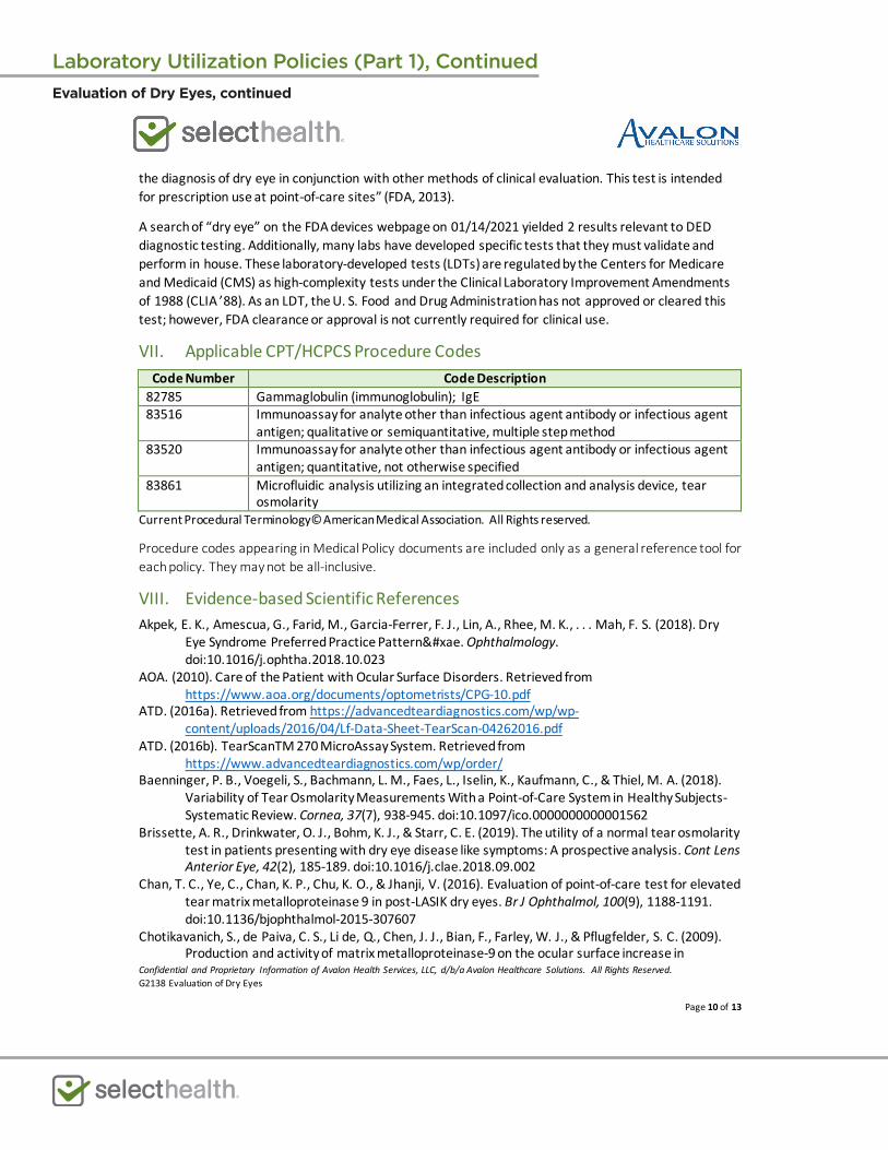
Laboratory Utilization Policies (Part 1), Continued
Confidential and Proprietary Information of Avalon Health Services, LLC, d/b/a Avalon Healthcare Solutions. All Rights Reserved. G2138 Evaluation of Dry Eyes
Page 10 of 13
the diagnosis of dry eye in conjunction with other methods of clinical evaluation. This test is intended for prescription use at point-of-care sites” (FDA, 2013).
A search of “dry eye” on the FDA devices webpage on 01/14/2021 yielded 2 results relevant to DED diagnostic testing. Additionally, many labs have developed specific tests that they must validate and perform in house. These laboratory-developed tests (LDTs) are regulated by the Centers for Medicare and Medicaid (CMS) as high-complexity tests under the Clinical Laboratory Improvement Amendments of 1988 (CLIA ’88). As an LDT, the U. S. Food and Drug Administration has not approved or cleared this test; however, FDA clearance or approval is not currently required for clinical use.
VII. Applicable CPT/HCPCS Procedure Codes Code Number Code Description
82785 Gammaglobulin (immunoglobulin); IgE 83516 Immunoassay for analyte other than infectious agent antibody or infectious agent
antigen; qualitative or semiquantitative, multiple step method 83520 Immunoassay for analyte other than infectious agent antibody or infectious agent
antigen; quantitative, not otherwise specified 83861 Microfluidic analysis utilizing an integrated collection and analysis device, tear
osmolarity Current Procedural Terminology© American Medical Association. All Rights reserved.
Procedure codes appearing in Medical Policy documents are included only as a general reference tool for each policy. They may not be all-inclusive.
VIII. Evidence-based Scientific References Akpek, E. K., Amescua, G., Farid, M., Garcia-Ferrer, F. J., Lin, A., Rhee, M. K., . . . Mah, F. S. (2018). Dry
Eye Syndrome Preferred Practice Pattern®. Ophthalmology. doi:10.1016/j.ophtha.2018.10.023
AOA. (2010). Care of the Patient with Ocular Surface Disorders. Retrieved from https://www.aoa.org/documents/optometrists/CPG-10.pdf
ATD. (2016a). Retrieved from https://advancedteardiagnostics.com/wp/wp-content/uploads/2016/04/Lf-Data-Sheet-TearScan-04262016.pdf
ATD. (2016b). TearScanTM 270 MicroAssay System. Retrieved from https://www.advancedteardiagnostics.com/wp/order/
Baenninger, P. B., Voegeli, S., Bachmann, L. M., Faes, L., Iselin, K., Kaufmann, C., & Thiel, M. A. (2018). Variability of Tear Osmolarity Measurements With a Point-of-Care System in Healthy Subjects-Systematic Review. Cornea, 37(7), 938-945. doi:10.1097/ico.0000000000001562
Brissette, A. R., Drinkwater, O. J., Bohm, K. J., & Starr, C. E. (2019). The utility of a normal tear osmolarity test in patients presenting with dry eye disease like symptoms: A prospective analysis. Cont Lens Anterior Eye, 42(2), 185-189. doi:10.1016/j.clae.2018.09.002
Chan, T. C., Ye, C., Chan, K. P., Chu, K. O., & Jhanji, V. (2016). Evaluation of point-of-care test for elevated tear matrix metalloproteinase 9 in post-LASIK dry eyes. Br J Ophthalmol, 100(9), 1188-1191. doi:10.1136/bjophthalmol-2015-307607
Chotikavanich, S., de Paiva, C. S., Li de, Q., Chen, J. J., Bian, F., Farley, W. J., & Pflugfelder, S. C. (2009). Production and activity of matrix metalloproteinase-9 on the ocular surface increase in
Evaluation of Dry Eyes, continued

Laboratory Utilization Policies (Part 1), Continued
Confidential and Proprietary Information of Avalon Health Services, LLC, d/b/a Avalon Healthcare Solutions. All Rights Reserved. G2138 Evaluation of Dry Eyes
Page 11 of 13
dysfunctional tear syndrome. Invest Ophthalmol Vis Sci, 50(7), 3203-3209. doi:10.1167/iovs.08-2476
Craig, J. P., Nelson, J. D., Azar, D. T., Belmonte, C., Bron, A. J., Chauhan, S. K., . . . Sullivan, D. A. (2017). TFOS DEWS II Report Executive Summary. Ocul Surf, 15(4), 802-812. doi:10.1016/j.jtos.2017.08.003
Craig, J. P., Nichols, K. K., Alpek, M. D., Caffery, B., Dua, H. S., Joo, C. K., . . . Stapleton, F. J. (2017). TFOS DEWS II Definition and Classification Report. Ocul Surf, 15(4), 276-283. doi:10.1016/j.jtos.2017.08.003
Dana, R., Meunier, J., Markowitz, J. T., Joseph, C., & Siffel, C. (2020). Patient-Reported Burden of Dry Eye Disease in the United States: Results of an Online Cross-Sectional Survey. Am J Ophthalmol, 216, 7-17. doi:10.1016/j.ajo.2020.03.044
Ezuddin, N. S., Alawa, K. A., & Galor, A. (2015). Therapeutic Strategies to Treat Dry Eye in an Aging Population. Drugs Aging, 32(7), 505-513. doi:10.1007/s40266-015-0277-6
Farrand, K. F., Fridman, M., Stillman, I. O., & Schaumberg, D. A. (2017). Prevalence of Diagnosed Dry Eye Disease in the United States Among Adults Aged 18 Years and Older. Am J Ophthalmol, 182, 90-98. doi:10.1016/j.ajo.2017.06.033
FDA. (1993, 11/19/2018). K934473. Retrieved from https://www.accessdata.fda.gov/scripts/cdrh/devicesatfda/index.cfm?db=pmn&id=K934473
FDA. (2013). Retrieved from https://www.accessdata.fda.gov/cdrh_docs/pdf13/K132066.pdf Foulks, G. N., Forstot, S. L., Donshik, P. C., Forstot, J. Z., Goldstein, M. H., Lemp, M. A., . . . Jacobs, D. S.
(2015). Clinical guidelines for management of dry eye associated with Sjögren disease. Ocul Surf, 13(2), 118-132. doi:10.1016/j.jtos.2014.12.001
Holland, E. J., Darvish, M., Nichols, K. K., Jones, L., & Karpecki, P. M. (2019). Efficacy of topical ophthalmic drugs in the treatment of dry eye disease: A systematic literature review. Ocul Surf, 17(3), 412-423. doi:10.1016/j.jtos.2019.02.012
Honda, N., Miyai, T., Nejima, R., Miyata, K., Mimura, T., Usui, T., . . . Amano, S. (2010). Effect of latanoprost on the expression of matrix metalloproteinases and tissue inhibitor of metalloproteinase 1 on the ocular surface. Arch Ophthalmol, 128(4), 466-471. doi:10.1001/archophthalmol.2010.40
Jie, Y., Sella, R., Feng, J., Gomez, M. L., & Afshari, N. A. (2019). Evaluation of incomplete blinking as a measurement of dry eye disease. Ocul Surf, 17(3), 440-446. doi:10.1016/j.jtos.2019.05.007
Jun JH, L. Y., Son MJ, Kim H (2020). Importance of tear volume for positivity of tear matrix metalloproteinase-9 immunoassay. PLoS ONE, 15(7). Retrieved from https://doi.org/10.1371/journal.pone.0235408
Kanellopoulos, A. J., & Asimellis, G. (2016). In pursuit of objective dry eye screening clinical techniques. Eye Vis (Lond), 3, 1. doi:10.1186/s40662-015-0032-4
Kawashima, M., Kawakita, T., Inaba, T., Okada, N., Ito, M., Shimmura, S., . . . Tsubota, K. (2012). Dietary lactoferrin alleviates age-related lacrimal gland dysfunction in mice. PLoS ONE, 7(3), e33148. doi:10.1371/journal.pone.0033148
Messmer, E. M. (2015). The pathophysiology, diagnosis, and treatment of dry eye disease. Dtsch Arztebl Int, 112(5), 71-81; quiz 82. doi:10.3238/arztebl.2015.0071
Messmer, E. M., von Lindenfels, V., Garbe, A., & Kampik, A. (2016). Matrix Metalloproteinase 9 Testing in Dry Eye Disease Using a Commercially Available Point-of-Care Immunoassay. Ophthalmology, 123(11), 2300-2308. doi:10.1016/j.ophtha.2016.07.028
Evaluation of Dry Eyes, continued

Laboratory Utilization Policies (Part 1), Continued
Confidential and Proprietary Information of Avalon Health Services, LLC, d/b/a Avalon Healthcare Solutions. All Rights Reserved. G2138 Evaluation of Dry Eyes
Page 12 of 13
Milner, M. S., Beckman, K. A., Luchs, J. I., Allen, Q. B., Awdeh, R. M., Berdahl, J., . . . Yeu, E. (2017). Dysfunctional tear syndrome: dry eye disease and associated tear film disorders - new strategies for diagnosis and treatment. Curr Opin Ophthalmol, 27 Suppl 1(Suppl 1), 3-47. doi:10.1097/01.icu.0000512373.81749.b7
Periman. (2020). The Immunological Basis of Dry Eye Disease and Current Topical Treatment Options. Journal of Ocular Pharmacology and Therapeutics, 36(3), 137-146. doi:10.1089/jop.2019.0060
Ponzini, E., Scotti, L., Grandori, R., Tavazzi, S., & Zambon, A. (2020). Lactoferrin Concentration in Human Tears and Ocular Diseases: A Meta-Analysis. Invest Ophthalmol Vis Sci, 61(12), 9. doi:10.1167/iovs.61.12.9
Sambursky, R., Davitt, W. F., 3rd, Latkany, R., Tauber, S., Starr, C., Friedberg, M., . . . McDonald, M. (2013). Sensitivity and specificity of a point-of-care matrix metalloproteinase 9 immunoassay for diagnosing inflammation related to dry eye. JAMA Ophthalmol, 131(1), 24-28. doi:10.1001/jamaophthalmol.2013.561
Shtein, R. (2020). Dry Eyes UpToDate. Retrieved from https://www.uptodate.com/contents/dry-eyes?source=search_result&search=dry%20eye&selectedTitle=1~150#H12
Starr, C. E., Gupta, P. K., Farid, M., Beckman, K. A., Chan, C. C., Yeu, E., . . . Mah, F. S. (2019). An algorithm for the preoperative diagnosis and treatment of ocular surface disorders. J Cataract Refract Surg, 45(5), 669-684. doi:10.1016/j.jcrs.2019.03.023
Szczotka-Flynn, L. B., Maguire, M. G., Ying, G. S., Lin, M. C., Bunya, V. Y., Dana, R., & Asbell, P. A. (2019). Impact of Dry Eye on Visual Acuity and Contrast Sensitivity: Dry Eye Assessment and Management Study. Optom Vis Sci, 96(6), 387-396. doi:10.1097/opx.0000000000001387
Tashbayev, B., Utheim, T. P., Utheim, Ø. A., Ræder, S., Jensen, J. L., Yazdani, M., . . . Chen, X. (2020). Utility of Tear Osmolarity Measurement in Diagnosis of Dry Eye Disease. Scientific Reports, 10(1), 5542. doi:10.1038/s41598-020-62583-x
TearLab. (2009). TearLab. Retrieved from https://www.tearlab.com/ Tomlinson, A., Khanal, S., Ramaesh, K., Diaper, C., & McFadyen, A. (2006). Tear film osmolarity:
determination of a referent for dry eye diagnosis. Invest Ophthalmol Vis Sci, 47(10), 4309-4315. doi:10.1167/iovs.05-1504
Versura, P., Bavelloni, A., Grillini, M., Fresina, M., & Campos, E. C. (2013). Diagnostic performance of a tear protein panel in early dry eye. Mol Vis, 19, 1247-1257.
Willcox, M. D. P., Argüeso, P., Georgiev, G. A., Holopainen, J. M., Laurie, G. W., Millar, T. J., . . . Jones, L. (2017). TFOS DEWS II Tear Film Report. Ocul Surf, 15(3), 366-403. doi:10.1016/j.jtos.2017.03.006
Evaluation of Dry Eyes, continued

Laboratory Utilization Policies (Part 1), Continued
Confidential and Proprietary Information of Avalon Health Services, LLC, d/b/a Avalon Healthcare Solutions. All Rights Reserved. G2138 Evaluation of Dry Eyes
Page 13 of 13
IX. Revision History Revision Date Summary of Changes
Disclaimer This document is for informational purposes only and should not be relied on in the diagnosis and care of individual patients. Medical and Coding/Reimbursement policies do not constitute medical advice, plan preauthorization, certification, an explanation of benefits, or a contract. Members should consult with appropriate healthcare providers to obtain needed medical advice, care, and treatment. Benefits and eligibility are determined before medical guidelines and payment guidelines are applied. Benefits are determined by the member’s individual benefit plan that is in effect at the time services are rendered. The codes for treatments and procedures applicable to this policy are included for informational purposes. Inclusion or exclusion of a procedure, diagnosis or device code(s) does not constitute or imply member coverage or provider reimbursement policy. Please refer to the member's contract benefits in effect at the time of service to determine coverage or non-coverage of these services as it applies to an individual member. SelectHealth® makes no representations and accepts no liability with respect to the content of any external information cited or relied upon in this policy. SelectHealth updates its Coverage Policies regularly and reserves the right to amend these policies without notice to healthcare providers or SelectHealth members. Members may contact Customer Service at the phone number listed on their member identification card to discuss their benefits more specifically. Providers with questions about this Coverage Policy may call SelectHealth Provider Relations at (801) 442-3692. No part of this publication may be reproduced, stored in a retrieval system or transmitted, in any form or by any means, electronic, mechanical, photocopying, or otherwise, without permission from SelectHealth. ”Intermountain Healthcare” and its accompanying logo, the marks of “SelectHealth” and its accompanying marks are protected and registered trademarks of the provider of this Service and or Intermountain Health Care, Inc., IHC Health Services, Inc., and SelectHealth, Inc. Also, the content of this Service is proprietary and is protected by copyright. You may access the copyrighted content of this Service only for purposes set forth in these Conditions of Use. © CPT Only – American Medical Association
Evaluation of Dry Eyes, continued

Laboratory Utilization Policies (Part 1), Continued
Confidential and Proprietary Information of Avalon Health Services, LLC, d/b/a Avalon Healthcare Solutions. All Rights Reserved. G2060 Fecal Analysis in the Diagnosis of Intestinal Dysbiosis and Fecal Microbiota Transplant Testing
Page 1 of 19
Fecal Analysis in the Diagnosis of Intestinal Dysbiosis and Fecal Microbiota Transplant Testing
Policy #: AHS – G2060 Prior Policy Name & Number (as applicable): AHS – G2060 – Fecal Analysis in the Diagnosis of Intestinal Dysbiosis
Implementation Date: 9/15/21 Date of Last Revision:
I. Policy Description
Intestinal dysbiosis is defined as a disruption or imbalance of the intestinal microbial ecology (Guinane & Cotter, 2013). Dysbiosis is associated with many diseases, including irritable bowel syndrome (IBS), inflammatory bowel diseases (IBD), obesity, allergy, and diabetes (Carding, Verbeke, Vipond, Corfe, & Owen, 2015).
II. Related Policies
Policy Number Policy Title AHS-G2056 Diagnosis of Idiopathic Environmental Intolerance AHS-G2061 Fecal Calprotectin Testing AHS-G2121 Laboratory Testing for the Diagnosis of Inflammatory Bowel Disease
III. Indications and/or Limitations of Coverage
Disclaimer:
1. Policies are subject to change without notice. 2. Policies outline coverage determinations for SelectHealth Commercial, SelectHealth
Advantage® (Medicare/CMS) and SelectHealth Community Care® (Medicaid/CHIP) plans. Refer to the “Policy” section for more information.
Application of coverage criteria depends on an individual’s benefit coverage at the time of the request.
For SelectHealth Advantage (Medicare/CMS), coverage is determined by the Centers for Medicare and Medicaid Services (CMS). If a coverage determination has not been adopted by CMS and InterQual criteria are not available, the SelectHealth Commercial policy applies. For the most up-to-date Medicare policies and coverage, please visit their search website or the manual website,
For SelectHealth Community Care (Medicaid), coverage is determined by the State of Utah Medicaid program. If Utah State Medicaid has no published coverage position and InterQual criteria are not available, the SelectHealth Commercial criteria will apply. For the most up-to-date Medicaid policies and coverage, please visit their website or the Utah Medicaid Code Look-Up tool.

Laboratory Utilization Policies (Part 1), Continued
Confidential and Proprietary Information of Avalon Health Services, LLC, d/b/a Avalon Healthcare Solutions. All Rights Reserved. G2060 Fecal Analysis in the Diagnosis of Intestinal Dysbiosis and Fecal Microbiota Transplant Testing
Page 2 of 19
1. Fecal analysis by culture prior to fecal microbiota transplant (FMT) for the following microorganisms MEETS COVERAGE CRITERIA:
a. Extended spectrum beta-lactamase (ESBL)-producing Enterobacteriaceae
b. Vancomycin-resistant Enterococci (VRE)
c. Carbapenem-resistant Enterobacteriaceae (CRE)
d. Methicillin-resistant Staphylococcus aureus (MRSA)
e. Campylobacter
f. Shigella
g. Salmonella
2. Fecal analysis by nucleic acid amplification testing (NAAT) prior to fecal microbiota transplant (FMT) for the following microorganisms MEETS COVERAGE CRITERIA.
a. C. Difficile
b. Campylobacter
c. Salmonella
d. Shigella
e. Shiga toxin-producing Escherichia coli
f. Norovirus
g. Rotavirus
h. COVID-19 (SARS-CoV-2)
3. Fecal analysis by nucleic acid amplification testing (NAAT) prior to fecal microbiota transplant (FMT) for the following microorganisms DOES NOT MEET COVERAGE CRITERIA:
a. Extended spectrum beta-lactamase (ESBL)-producing Enterobacteriaceae
b. Vancomycin-resistant Enterococci (VRE)
c. Carbapenem-resistant Enterobacteriaceae (CRE)
d. Methicillin-resistant Staphylococcus aureus (MRSA)
e. Any other microorganisms not listed above
The following does not meet coverage criteria due to a lack of available published scientific literature confirming that the test(s) is/are required and beneficial for the diagnosis and treatment of a patient’s illness.
4. Fecal analysis of the following components DOES NOT MEET COVERAGE CRITERIA as a diagnostic test for the evaluation of intestinal dysbiosis, irritable bowel syndrome, malabsorption, or small intestinal overgrowth of bacteria:
a. Triglycerides
Fecal Analysis in the Diagnosis of Intestinal Dysbiosis and Fecal Microbiota Transplant Testing, continued

Laboratory Utilization Policies (Part 1), Continued
Confidential and Proprietary Information of Avalon Health Services, LLC, d/b/a Avalon Healthcare Solutions. All Rights Reserved. G2060 Fecal Analysis in the Diagnosis of Intestinal Dysbiosis and Fecal Microbiota Transplant Testing
Page 3 of 19
b. Chymotrypsin
c. Iso-butyrate, iso-valerate, and n-valerate
d. Meat and vegetable fibers
e. Long chain fatty acids
f. Cholesterol
g. Total short chain fatty acids
h. Levels of Lactobacilli, bifidobacteria, and E. coli and other "potential pathogens," including Aeromona, Bacillus cereus, Campylobacter, Citrobacter, Klebsiella, Proteus, Pseudomonas, Salmonella, Shigella, S. aureus, Vibrio
i. Identification and quantitation of fecal yeast (including C. albicans, C. tropicalis, Rhodoptorul and Geotrichum )
j. N-butyrate
k. Beta-glucoronidase
l. pH
m. Short chain fatty acid distribution (adequate amount and proportions of the different short chain fatty acids reflect the basic status of intestinal metabolism)
n. Fecal secretory IgA
IV. Scientific Background
The human intestinal tract has a diverse and complex microbial community necessary for health and nutrition. The gut microbiome is estimated to consist of upwards of 1000 bacterial species (Guinane & Cotter, 2013; Ley, Peterson, & Gordon, 2006; Qin et al., 2010). The microbiota functions with the immune system to protect against pathogens. It also performs essential metabolic functions, extracting certain forms of energy and nutrients from food and providing a source of other essential nutrients and vitamins (Carding et al., 2015).
The gut is colonized at birth, but the intestinal microbiome changes rapidly during the first year of life. In adults, each individual’s unique population of gut microbiota is fairly stable over time; however, alterations in the microbiota can result from exposure to various environmental factors, including diet, toxins, drugs, and pathogens (Carding et al., 2015; Lozupone, Stombaugh, Gordon, Jansson, & Knight, 2012; Snapper & Abraham, 2020). This change in an individual’s normal microbiota is called “dysbiosis” (Johnston, 2020). Dysbiosis has been associated with obesity (Ley, Turnbaugh, Klein, & Gordon, 2006; Zhang et al., 2009) malnutrition (Kau, Ahern, Griffin, Goodman, & Gordon, 2011), systematic diseases such as diabetes (Qin et al., 2012) and chronic inflammatory diseases such as inflammatory bowel disease (IBD) (Frank et al., 2007; Guinane & Cotter, 2013). Both direct assessment of the gut microbiota (examination of bacteria levels) and indirect assessment (measurement of non-living markers such as pH or beta-glucoronidase) have been proposed for investigation of intestinal dysbiosis.
Microbial or microbial-derived components have also been cited as potential representations of dysbiosis. For example, short-chain fatty acids have been identified as a mechanism to regulate intestinal processes and, as such, may represent dysbiosis (Johnston, 2020). These fatty acids are the
Fecal Analysis in the Diagnosis of Intestinal Dysbiosis and Fecal Microbiota Transplant Testing, continued

Laboratory Utilization Policies (Part 1), Continued
Confidential and Proprietary Information of Avalon Health Services, LLC, d/b/a Avalon Healthcare Solutions. All Rights Reserved. G2060 Fecal Analysis in the Diagnosis of Intestinal Dysbiosis and Fecal Microbiota Transplant Testing
Page 4 of 19
products of bacterial fermentation of fiber, and the concentrations of these fatty acids have been noted to decrease in IBD cases. Some fatty acids, especially butyrate, have been demonstrated to factor in signaling cascades that control immune function, which indicates a role in controlling intestinal inflammation (Parada Venegas et al., 2019). Ongoing research has uncovered many other potential links between intestinal metabolism and gut microbiota so many markers have been suggested as potential indicators of dysbiosis.
Many tests exist for the assessment of the gut microbiome. Due to the amount of conditions associated (or proposed to be associated) with gut microbiome balance, there are many corresponding tests, including screening measures intended for completely healthy individuals. These tests primarily revolve around nucleic acid amplification; microbial DNA or RNA is obtained from the sample, unique sequences are identified, and the nucleic acid is quantified (Raby, 2020). For instance, Viome offers a comprehensive screening panel that measures “all microorganisms” in the gut (including viruses, archaea, yeast, fungi, parasites, and bacteriophages). Those measurements are combined into a score for various issues, such as inflammatory activity, digestive efficiency, methane gas production, overall gas production, and more (Viome, 2019a). Viome also provides a list of nutritional recommendations, broken down into individual foods. Viome performs RNA sequencing with Illumina NextSeq and uses bioinformatics algorithms to classify taxonomic data (Viome, 2019b).
Some companies may offer companion products with their gut microbiome tests. BioHM provides a similar assessment of bacterial and fungal species in an individual’s gastrointestinal tract, but the company also offers a series of probiotics. These probiotics are intended for various purposes, such as colon cleansing or immunity (BioHM, 2018). Other companies offering a gut microbiome test include Thryve, GenCove, DayTwo, American Gut, and Genova (DNATestingChoice, 2019; Genova, 2019).
The potential clinical impact of imbalance in the intestinal microbiota suggests a need for standardized diagnostic methods to facilitate microbiome profiling. Documenting dysbiosis has traditionally relied on classical microbiological techniques and the ability to culture pure isolates for identification and classification; however, the ability to classify bacteria and archaea according to individual 16S rRNA sequences can now possibly provide a rapid and detailed means of profiling complex communities of microorganisms (Casen et al., 2015; Zoetendal, Akkermans, & De Vos, 1998). Laboratory analysis of various fecal biomarkers have also been proposed as a method of identifying individuals with intestinal dysbiosis and may be useful in providing insight into the role of intestinal health and disease, and the development of non-gastrointestinal conditions associated with intestinal dysbiosis. However, there is a current lack of literature on the normal ranges of these biomarkers, which limit the applicability of these analyses in a general clinical setting (Bäckhed et al., 2012; Berry & Reinisch, 2013; Pang, Leach, Katz, Day, & Ooi, 2014).
A technique revolving around restoring balance in a patient’s microbiome is fecal microbiota transplantation (FMT). FMT is the infusion of stool from a healthy donor to a patient with presumed gut dysbiosis. The concept behind this technique is that the healthy donor’s stool can facilitate a restoration of the ill patient’s gut microbiome. This technique has seen some significant success in the treatment of C. difficile infections and may have potential applications in some other gastrointestinal or metabolic conditions such as IBD or IBS. As with any transplant procedure, there are several screening procedures
that must be undertaken to minimize risk of infection or other disease transmission. These screening procedures include evaluation of donor history, serum testing, and stool testing. The pathogens
Fecal Analysis in the Diagnosis of Intestinal Dysbiosis and Fecal Microbiota Transplant Testing, continued

Laboratory Utilization Policies (Part 1), Continued
Confidential and Proprietary Information of Avalon Health Services, LLC, d/b/a Avalon Healthcare Solutions. All Rights Reserved. G2060 Fecal Analysis in the Diagnosis of Intestinal Dysbiosis and Fecal Microbiota Transplant Testing
Page 5 of 19
screened for in the donor’s stool sample may vary between institutions, although some pathogens are universally screened for (such as enteric pathogens). (Kim & Gluck, 2019)
Falony et al. (2016) analyzed “two independent, extensively phenotyped cohorts: the Belgian Flemish Gut Flora Project (FGFP; discovery cohort; N = 1106) and the Dutch LifeLines-DEEP study (LLDeep; replication; N = 1135).” These two sets were integrated with global data sets, combining to yield 3948 items. A “core” set of 14 genera was identified. 69 clinical and questionnaire-based covariates were found to be associated with microbiota compositional variation with a 92% replication rate. The authors noted that “stool consistency showed the largest effect size, whereas medication explained largest total variance and interacted with other covariate-microbiota associations, but early-life events such as birth mode were not reflected in adult microbiota composition” (Falony et al., 2016)
Zhernakova et al. (2016) sequenced the gut microbiomes of 1,135 participants from a Dutch population-based cohort. The authors identified relations between the microbiome and “126 exogenous and intrinsic host factors, including 31 intrinsic factors, 12 diseases, 19 drug groups, 4 smoking categories, and 60 dietary factors.” “Significant” associations were found between the gut microbiome and various intrinsic, environmental, dietary, medication parameters, and disease phenotypes. The authors calculated that 18.7% of variation in microbial composition could be explained by these factors, and they observed that fecal chromogranin A was exclusively associated with 61 microbial species, totaling 53% of the microbial composition. A more diverse microbiome was associated with low CgA concentrations. The authors concluded that “these results are an important step toward a better understanding of environment-diet-microbe-host interactions” (Zhernakova et al., 2016).
Lo Presti et al. (2019) profiled the fecal and mucosal microbiota of IBD and IBS patients. 38 IBD patients, 44 IBS patients, and 47 healthy controls were included, and overall, 107 fecal samples were provided. The authors found that “Anaerostipes and Ruminococcaceae were identified as the most differentially abundant bacterial taxa in controls, Erysipelotrichi was identified as [a] potential biomarker for IBS, while Gammaproteobacteria, Enterococcus, and Enterococcaceae [were identified] for IBD” (Lo Presti et al., 2019).
Malham et al. (2019) investigated the microbiotic profile of pediatric IBD. 143 IBD patients and 34 healthy controls were included. A reduced “richness” in microbiotic profile was observed in IBD patients compared to healthy controls. In ulcerative colitis (UC), that reduced richness was associated with high intestinal inflammation and extensive disease. Nine species were “significantly” associated with a healthy microbiome, and three species were associated with IBD. The authors remarked that the microbiome composition could differentiate between Crohn’s Disease, UC, and healthy controls (Malham et al., 2019).
Danilova et al. (2019) compared the gut microbiome composition of IBD patients to healthy controls. 95 IBD patients and 96 healthy controls were included. The authors noted an increase of Proteobacteria and Bacteroidetes bacteria and decrease of Firmicutes bacteria and Euryarchaeota archaea in IBD patients. Butyrate-producing and hydrogen-utilizing bacteria were observed to have lower representation in IBD patients. Short-chain fatty acids (SCFA) were also found to have a lower absolute content in IBD patients. The authors suggested that this finding may “indicate inhibition of functional activity and number of anaerobic microflora and/or an [sic] change in SCFA utilization by colonocytes” (Danilova et al., 2019).
Vaughn et al. (2018) in reviewing the current status of intestinal dysbiosis and fecal transplantation found that “it is hypothesized that intestinal dysbiosis may contribute to the pathogenesis of many
Fecal Analysis in the Diagnosis of Intestinal Dysbiosis and Fecal Microbiota Transplant Testing, continued
Laboratory Utilization Policies (Part 1), Continued
Confidential and Proprietary Information of Avalon Health Services, LLC, d/b/a Avalon Healthcare Solutions. All Rights Reserved. G2060 Fecal Analysis in the Diagnosis of Intestinal Dysbiosis and Fecal Microbiota Transplant Testing
Page 6 of 19
diseases, especially those involving the gastrointestinal tract. Therefore, fecal microbiota transplantation (FMT) is increasingly being explored as a potential treatment that aims to optimize microbiota composition and functionality (Vaughn, Rank, & Khoruts, 2018).” Holleran et al also found that fecal transplant is not recommended for use outside of Clostridium difficile infection (CDI) due to concerns regarding outcome and safety; however, several case series and randomized controlled trials have described its use in a research environment for a few gastrointestinal conditions related to intestinal dysbiosis, including ulcerative colitis (UC), Crohn's disease (CD) and irritable bowel syndrome (IBS). The most successful reports of the clinical efficacy of FMT in gastrointestinal conditions outside of CDI have been in treating UC (Holleran et al., 2018).
Costello et al. (2019) evaluated fecal microbiota transplantation (FMT)’s efficacy on inducing remission in ulcerative colitis (UC). The authors compared anaerobically prepared donor FMT (n = 38) to autologous FMT (stool provided by patient themselves, n = 35). The primary outcome was defined as “steroid-free remission of UC… a total Mayo score of ≤2 with an endoscopic Mayo score of 1 or less at week 8.” A total of 69 patients completed the trial, with the primary outcome being achieved in 12 of 38 donor FMT patients, compared to 3 of 35 receiving autologous FMT. Five of the 12 patients achieving the primary outcome in the “donor cohort” maintained remission at 12 months. The authors concluded that “in this preliminary study of adults with mild to moderate UC, 1-week treatment with anaerobically prepared donor FMT compared with autologous FMT resulted in a higher likelihood of remission at 8 weeks. Further research is needed to assess longer-term maintenance of remission and safety” (Costello et al., 2019).
Myneedu et al. (2019) performed a meta-analysis to evaluate whether fecal microbiota transplantation (FMT) was successful in treating IBS. A total of 8 single-arm trials (SATs, 90 patients total) and 5 randomized controlled trials (RCTs, 151 patients, 105 controls) were included. In the SAT cohort, the authors identified 59.5% of IBS patients demonstrating a significant improvement. In the RCT cohort, there were no significant differences between treatment and control cohorts, either by the IBS Severity Scoring System or the IBS Quality of Life (IBS-QOL). The authors concluded that “FMT was not effective in IBS. Variations in FMT methods and patient factors may contribute to the heterogeneous results of the trials” (Myneedu, Deoker, Schmulson, & Bashashati, 2019).
V. Guidelines and Recommendations
World Gastroenterology Organization Global Guidelines (Bernstein et al., 2016; Hunt et al., 2014) The WGO published guidelines on functional gastrointestinal (GI) symptoms. In it, they identify diagnostic tests for these symptoms. The basic diagnostic tests are as follows: • Complete blood cell count (CBC) • Erythrocyte sedimentation rate (ESR) / C-reactive protein (CRP) • Biochemistry panel • Fecal occult blood (patient aged > 50 y) • Pregnancy test • Liver function tests
Fecal Analysis in the Diagnosis of Intestinal Dysbiosis and Fecal Microbiota Transplant Testing, continued

Laboratory Utilization Policies (Part 1), Continued
Confidential and Proprietary Information of Avalon Health Services, LLC, d/b/a Avalon Healthcare Solutions. All Rights Reserved. G2060 Fecal Analysis in the Diagnosis of Intestinal Dysbiosis and Fecal Microbiota Transplant Testing
Page 7 of 19
• Calprotectin or other fecal test to detect inflammatory bowel disease in patients thought to have IBS, but in whom inflammatory bowel disease (IBD) is a possibility; now routine in many primary care settings (in the United Kingdom)
• Celiac serology; considered routine in areas with a high prevalence of celiac disease • Stool testing for ova and parasites (Hunt et al., 2014)
The WGO also released their global guidelines for Inflammatory Bowel Disease in 2015 (published in 2016). Their recommendations concerning stool examination and testing are as follows:
• “Routine fecal examinations and cultures should be carried out to eliminate bacterial, viral, or parasitic causes of diarrhea.”
• “Testing for Clostridium difficile (should be considered even in the absence of antecedent antibiotics) — should be carried out within 2 hours of passage of stools.”
• “A check for occult blood or fecal leukocytes should be carried out if a patient presents without a history of blood in the stool, as this can strengthen the indication for lower endoscopy. Where lower endoscopy is readily available, these tests are rarely indicated.”
• “Lactoferrin, α1-antitrypsin. The main reason for listing this test is to rule out intestinal inflammation, rather than using it as a positive diagnostic test. It may not be available in developing countries, but it can be undertaken relatively inexpensively and easily with rapid-turnaround slide-based enzyme-linked immunoassay (ELISA) tests.”
• “Calprotectin — a simple, reliable, and readily available test for measuring IBD activity — may be better for UC than CD; the rapid fecal calprotectin tests could be very helpful in developing countries. If available, a home test may be useful as a routine for follow-up (Bernstein et al., 2016).”
American Gastroenterological Association (AGA) (Colombel, Shin, & Gibson, 2019)
The AGA published a review to “describe key principles in the diagnosis and management of functional gastrointestinal (GI) symptoms in patients with inflammatory bowel disease”. In it, they include the following relevant items:
• “Alternative pathophysiologic mechanisms should be considered and evaluated (small intestinal bacterial overgrowth, bile acid diarrhea, carbohydrate intolerance, chronic pancreatitis) based on predominant symptom patterns.”
• “Until further evidence is available, fecal microbiota transplant should not be offered for treatment of functional GI symptoms in IBD.”
• “In a recent cross-sectional analysis, no association was observed between IBS symptoms and microbiome alterations among patients with IBD although effects of confounding could not be excluded.” (Colombel et al., 2019)
Fecal Analysis in the Diagnosis of Intestinal Dysbiosis and Fecal Microbiota Transplant Testing, continued

Laboratory Utilization Policies (Part 1), Continued
Confidential and Proprietary Information of Avalon Health Services, LLC, d/b/a Avalon Healthcare Solutions. All Rights Reserved. G2060 Fecal Analysis in the Diagnosis of Intestinal Dysbiosis and Fecal Microbiota Transplant Testing
Page 8 of 19
American College of Gastroenterology (ACG) (Lichtenstein et al., 2018; Pimentel, Saad, Long, & Rao, 2020; Rubin, Ananthakrishnan, Siegel, Sauer, & Long, 2019)
The ACG published an update on SIBO (Small Intestinal Bacterial Overgrowth). This guideline addresses diagnostic testing and treatment options for SIBO. Their recommendations include:
• “We suggest the use of breath testing (glucose hydrogen or lactulose hydrogen) for the diagnosis of SIBO in patients with IBS (conditional recommendation, very low level of evidence).”
• “We suggest using glucose hydrogen or lactulose hydrogen breath testing for the diagnosis of SIBO in symptomatic patients with suspected motility disorders (conditional recommendation, very low level of evidence).”
• “We suggest testing for SIBO using glucose hydrogen or lactulose hydrogen breath testing in symptomatic patients (abdominal pain, gas, bloating, and/or diarrhea) with previous luminal abdominal surgery (conditional recommendation, very low level of evidence).”
• “We suggest testing for methane using glucose or lactulose breath tests to diagnose the overgrowth of methane-producing organisms (IMO) in symptomatic patients with constipation (conditional recommendation, very low level of evidence).”
The ACG also notes that although “Small bowel aspirate and culture is often considered the gold standard for the diagnosis of SIBO,” there have been some preliminary studies focusing on use of nucleic acid testing to diagnose SIBO. However, the ACG remarks that “Large-scale studies are currently underway to evaluate this further.”
The ACG also published a guideline regarding the management of Crohn’s Disease. In it, they recommend that “In patients who have symptoms of active Crohn's disease, stool testing should be performed to include fecal pathogens, Clostridium difficile testing, and may include studies that identify gut inflammation such as a fecal calprotectin” (Lichtenstein et al., 2018).
The ACG published a guideline regarding management of ulcerative colitis. In it, the ACG writes that “FMT requires more study and clarification of treatment before use as a therapy for UC [ulcerative colitis].” The ACG comments that the variability across all steps of the procedure (donor screening, delivery, treatment duration, et al.) makes interpretation of the current results “difficult”. Finally, the ACG notes that some institutions have been using “comprehensive intestinal pathogen testing through PCR-based assays that include many bacterial and viral pathogens,” but that the “prevalence and impact of non–C. diff intestinal pathogens detected through such assays remain to be robustly established” (Rubin et al., 2019). European Crohn’s and Colitis Organization (ECCO) and the European Society of Gastrointestinal and Abdominal Radiology (ESGAR) (Maaser et al., 2018)
These joint guidelines include some relevant items on inflammatory bowel disease (IBD), which includes both Crohn’s disease (CD) and ulcerative colitis (UC). These items include:
Fecal Analysis in the Diagnosis of Intestinal Dysbiosis and Fecal Microbiota Transplant Testing, continued

Laboratory Utilization Policies (Part 1), Continued
Confidential and Proprietary Information of Avalon Health Services, LLC, d/b/a Avalon Healthcare Solutions. All Rights Reserved. G2060 Fecal Analysis in the Diagnosis of Intestinal Dysbiosis and Fecal Microbiota Transplant Testing
Page 9 of 19
• “At diagnosis, every patient should have a biochemical assessment with full blood count, inflammatory markers (C-reactive protein [CRP])… and a stool sample for microbiological analysis, including C. difficile.”
• “Stool specimens should be obtained to exclude common pathogens and specifically assayed for C difficile toxin.” (Maaser et al., 2018)
2012 Rome Foundation Report (Simren et al., 2013)
An international Working Group convened in 2012 “to provide clinical guidance on modulation of gut microbiota in IBS” and released their findings on intestinal microbiota in functional bowel disorders: a Rome foundation report in 2013. They state the following “Diagnostic and therapeutic general recommendations”:
• “There is currently no clinically useful way of identifying whether the microbiota are disturbed in particular patients with irritable bowel syndrome (IBS).
• Dietary evaluation and exclusion of possible sources of unabsorbable carbohydrates including fermentable oligo-, di- and mono-saccharides and polyols and excessive fibre could be beneficial in select patients.
• Probiotics have a reasonable evidence base and should be tried, for a period of at least 1 month, at adequate doses before a judgement is made about the response to treatment.
• The utility of testing for small intestinal bacterial overgrowth (SIBO) in the setting of IBS remains an area of uncertainty.
• If SIBO is strongly suspected based on clinical presentation and testing is being considered, using stringent criteria for the glucose breath test or jejunal aspirate appear to be the best tests.
• Consideration should be given to discontinuing proton pump inhibitors in those with SIBO. • There is emerging evidence that non-absorbable antibiotics may have the potential to reduce
symptoms in some patients with IBS (Simren et al., 2013).”
European Society for Pediatric Gastroenterology, Hepatology, and Nutrition/European Society for Pediatric Infectious Diseases (ESPGHAN/ESPID)
These joint guidelines reviewed management of acute gastroenteritis (AGE) in children. In it, they note that AGE does not require a specific diagnostic workup and that “microbiological investigation is not helpful in most cases.” Fecal markers are also not recommended for differentiating viral and bacterial AGE. However, the guidelines observe that “microbiological investigations may be considered in children with underlying chronic conditions (e.g., oncologic diseases, IBDs, etc), in those in extremely severe conditions, or in those with prolonged symptoms in whom specific treatment is considered” (Guarino et al., 2014).
National Institute for Health and Care Excellence (NICE, 2017)
NICE updated their IBS guidelines in 2017. In it, they list the following items about diagnostic tests:
Fecal Analysis in the Diagnosis of Intestinal Dysbiosis and Fecal Microbiota Transplant Testing, continued

Laboratory Utilization Policies (Part 1), Continued
Confidential and Proprietary Information of Avalon Health Services, LLC, d/b/a Avalon Healthcare Solutions. All Rights Reserved. G2060 Fecal Analysis in the Diagnosis of Intestinal Dysbiosis and Fecal Microbiota Transplant Testing
Page 10 of 19
"In people who meet the IBS diagnostic criteria, the following tests should be undertaken to exclude other diagnoses:
• full blood count (FBC) • erythrocyte sedimentation rate (ESR) or plasma viscosity • c‑reactive protein (CRP) • antibody testing for coeliac disease (endomysial antibodies [EMA] or tissue transglutaminase [TTG]).
The following tests are not necessary to confirm diagnosis in people who meet the IBS diagnostic criteria:
• ultrasound • rigid/flexible sigmoidoscopy • colonoscopy; barium enema • thyroid function test • faecal ova and parasite test • faecal occult blood • hydrogen breath test (for lactose intolerance and bacterial overgrowth)” (NICE, 2017).
British Society of Gastroenterology (BSG) (Arasaradnam et al., 2018; Lamb et al., 2019)
The BSG published a guideline on the investigation of chronic diarrhoea in adults. Relevant items include:
• For malabsorption, fecal tests have not received “significant support” in publications and have not “established themselves in clinical practice outside specialist centres”.
• “We suggest culture of small bowel aspirates as it is the most sensitive test for small bowel bacterial overgrowth (SBBO), but methods are poorly standardized and positive results may not reflect clinically significant SBBO… in the absence of an optimal test to confirm the presence of bacterial overgrowth and in those with a high test probability of SBBO, we recommend an empirical trial of antibiotics; the value of this approach has not been subject to definitive study.”
• “We recommend faecal elastase testing as the preferred non-invasive test for pancreatic function” (Arasaradnam et al., 2018).
The BSG also published an extensive guideline on the management of Inflammatory Bowel Disease (including both ulcerative colitis (UC) and Crohn’s disease) in adults. Their relevant comments and recommendations include:
• “In patients presenting with suspected UC, stool cultures and Clostridium difficile toxin assay should always be performed to rule out infective causes.”
• “Ileocolonoscopy with biopsy is established as the first-line investigation for suspected Crohn’s disease.”
• “We recommend that all patients presenting with acute flares of colitis should have stool cultures for enteroinvasive bacterial infections and stool Clostridium difficile assay.”
Fecal Analysis in the Diagnosis of Intestinal Dysbiosis and Fecal Microbiota Transplant Testing, continued

Laboratory Utilization Policies (Part 1), Continued
Confidential and Proprietary Information of Avalon Health Services, LLC, d/b/a Avalon Healthcare Solutions. All Rights Reserved. G2060 Fecal Analysis in the Diagnosis of Intestinal Dysbiosis and Fecal Microbiota Transplant Testing
Page 11 of 19
• “In spite of these encouraging data, FMT [Faecal microbial transplantation] remains an investigational treatment for use only in clinical trials in IBD.”
• “There is currently no place for FMT in the management of IBD unless complicated by C. difficile infection outside of the clinical trial setting” (Lamb et al., 2019)
British Society of Gastroenterology (BSG) and Healthcare Infection Society (HIS) (Mullish et al., 2018)
This joint guideline was published to provide guidance on “the use of faecal microbiota transplant as treatment for recurrent or refractory Clostridium difficile infection and other potential indications.” These guidelines include a list of items that should be screened for potential stool donors, which are as follows:
• “Clostridium difficile PCR”
• “Campylobacter, Salmonella, and Shigella by standard stool culture and/ or PCR”
• “Shiga toxin-producing Escherichia coli by PCR”
• “Multi-drug resistant bacteria, at least CPE [carbapenemase-producing Enterobacteriaceae] and ESBL [extended spectrum beta-lactamase]”
• “Stool ova, cysts and parasite analysis, including for Microsporidia”
• “Faecal antigen for Cryptosporidium and Giardia”
• “Acid fast stain for Cyclospora and Isospora”
• “Helicobacter pylori faecal antigen”
• “Norovirus, rotavirus PCR.”
The above list is for stool screening. A separate list is provided for serum screening. The guideline also recommends that “donors should have successfully completed a donor health questionnaire and laboratory screening assays both before and after the period of stool donation” (Mullish et al., 2018).
Infectious Diseases Society of America/American College of Gastroenterology/American Society for Gastrointestinal Endoscopy/American Gastroenterological Association/North American Society for Pediatric Gastroenterology, Hepatology and Nutrition (Davidovics et al., 2019; IDSA/ACG/ASGE/AGA/NASPGHAN, 2013; Michail, Nicholson, Kahn, & Kellermayer, 2020)
These joint guidelines were sent to the FDA regarding recurrent Clostridium difficile infection (CDI). In it, the guidelines recommend screening donors for fecal microbiota transplantation (FMT) for C. difficile toxin B and performing a culture for enteric pathogens (IDSA/ACG/ASGE/AGA/NASPGHAN, 2013).
NASPGHAN published an FMT guideline for children in 2019, and the same analytes for screening (C difficile toxin B, culture for enteric pathogens) were recommended (Davidovics et al., 2019).
An addendum was published to the 2019 guidelines due to the 2019 FDA Safety Warning regarding FMT. In it, the following recommendation was made: “FMT donor stool screening should include (but not be
Fecal Analysis in the Diagnosis of Intestinal Dysbiosis and Fecal Microbiota Transplant Testing, continued

Laboratory Utilization Policies (Part 1), Continued
Confidential and Proprietary Information of Avalon Health Services, LLC, d/b/a Avalon Healthcare Solutions. All Rights Reserved. G2060 Fecal Analysis in the Diagnosis of Intestinal Dysbiosis and Fecal Microbiota Transplant Testing
Page 12 of 19
limited to) MDRO testing for spectrum beta-lactamase (ESBL)-producing Enterobacteriaceae, vancomycin-resistant Enterococci (VRE), carbapenem-resistant Enterobacteriaceae (CRE), and methicillin-resistant Staphylococcus aureus (MRSA). Donors and/or stools positive for MDROs should not be used for FMT” (Michail et al., 2020).
Food and Drug Administration (FDA, 2019, 2020)
The FDA has issued a guidance statement for fecal microbiota transplant (FMT) stating that it will exercise enforcement discretion regarding the investigational new drug (IND) requirements for the use of fecal microbiota for transplantation. In 2019, the FDA updated their guidance on FMT, stating that “FMT donor stool testing must include MDRO testing to exclude use of stool that tests positive for MDRO. The MDRO tests should at minimum include extended spectrum beta-lactamase (ESBL)-producing Enterobacteriaceae, vancomycin-resistant enterococci (VRE), carbapenem-resistant Enterobacteriaceae (CRE), and methicillin-resistant Staphylococcus aureus (MRSA). Culture of nasal or peri-rectal swabs is an acceptable alternative to stool testing for MRSA only. Bookend testing (no more than 60 days apart) before and after multiple stool donations is acceptable if stool samples are quarantined until the post-donation MDRO tests are confirmed negative (FDA, 2019).”
In an April 2020 update, the FDA addressed the topic of fecal microbiota transplantation within the context of the 2020 COVID-19 outbreak. The FDA included additional protections regarding stool donation and donor screening, which are as follows:
• “Stool donor screening, including an assessment of whether, since December 1, 2019, the donor was diagnosed with laboratory-confirmed SARS-CoV-2 infection, experienced symptoms of COVID-19 (e.g., fever, cough, shortness of breath) not explained by another diagnosis, or was exposed to a suspected or confirmed case of COVID-19 or SARS-CoV-2 infection.”
• “Testing of the stool donation or stool donor for SARS-CoV-2 virus or RNA. Testing approaches might include testing upper respiratory specimens (e.g., nasal swabs) or other specimens (e.g., rectal swabs or stool donations).” (FDA, 2020)
Fecal Microbiota Transplantation Workgroup (2011)
This Working Group published guidelines on FMT. Fecal donor screening recommendations were included. The following analytes were recommended to be screened:
• “C difficile toxin B by PCR; if unavailable, then evaluation for toxins A and B by enzyme immunoassay (EIA)
• Routine bacterial culture for enteric pathogens • Fecal Giardia antigen • Fecal Cryptosporidium antigen • Acid-fast stain for Cyclospora, Isospora, and, if antigen testing unavailable, Cryptosporidium • Ova and parasites • Helicobacter pylori fecal antigen (for upper gastrointestinal [GI] routes of FMT administration)”
(Bakken et al., 2011).
Fecal Analysis in the Diagnosis of Intestinal Dysbiosis and Fecal Microbiota Transplant Testing, continued

Laboratory Utilization Policies (Part 1), Continued
Confidential and Proprietary Information of Avalon Health Services, LLC, d/b/a Avalon Healthcare Solutions. All Rights Reserved. G2060 Fecal Analysis in the Diagnosis of Intestinal Dysbiosis and Fecal Microbiota Transplant Testing
Page 13 of 19
American Gastroenterological Association (AGA, 2015)
The AGA published guidelines on FMT, including information on donor pathogen screening. C. difficile toxin B and culture for enteric pathogens were “suggested” to be screened for, Giardia, Cryptosporidium, Isospora and Cyclospora, Listeria, E. coli O157, Vibrio, and Norovirus should be “considered”, and Cytomegalovirus, Human T-cell lymphoma virus, Epstein–Barr virus, Dientamoeba fragilis, Blastocystis hominis, Strongyloides stercoralis, Entamoeba histolytica, H. pylori, Schistosoma, JC virus, Vancomycin-resistant enterococci, and Methicillin-resistant Staphylococcus aureus should “maybe” [term used by authors] be screened (Kelly et al., 2015).
VI. State and Federal Regulations (as applicable)
Searches for “dysbiosis” and “microbiome” on the FDA website on August 6, 2020, yielded no results. Additionally, many labs have developed specific tests that they must validate and perform in house. These laboratory-developed tests (LDTs) are regulated by the Centers for Medicare and Medicaid (CMS) as high-complexity tests under the Clinical Laboratory Improvement Amendments of 1988 (CLIA ’88). As an LDT, the U. S. Food and Drug Administration has not approved or cleared this test; however, FDA clearance or approval is not currently required for clinical use.
VII. Applicable CPT/HCPCS Procedure Codes
Code Number Code Description 82239 Bile acids; total
82542 Column chromatography, includes mass spectrometry, if performed (e.g., HPLC, LC, LC/MS, LC/MS-MS, GC, GC/MS-MS, GC/MS, HPLC/MS), non-drug analyte(s) not elsewhere specified, qualitative or quantitative, each specimen
82710 Fat or lipids, feces; quantitative 82715 Fat differential, feces, quantitative 82725 Fatty acids, nonesterified 82784 Gammaglobulin (immunoglobulin); IgA, IgD, IgG, IgM, each
83520 Immunoassay for analyte other than infectious agent antibody or infectious agent antigen; quantitative, not otherwise specified
83630 Lactoferrin, fecal; qualitative 83986 pH; body fluid, not otherwise specified 84311 Spectrophotometry, analyte not elsewhere specified
87045 Culture, bacterial; stool, aerobic, with isolation and preliminary examination (e.g., KIA, LIA), salmonella and shigella species
87046 Culture, bacterial; stool, aerobic, additional pathogens, isolation and presumptive identification of isolates, each plate
87075 Culture, bacterial; any source, except blood, anaerobic with isolation and presumptive identification of isolates
87102 Culture, fungi (mold or yeast) isolation, with presumptive identification of isolates; other source (except blood)
87177 Ova and parasites, direct smears, concentration and identification
Fecal Analysis in the Diagnosis of Intestinal Dysbiosis and Fecal Microbiota Transplant Testing, continued

Laboratory Utilization Policies (Part 1), Continued
Confidential and Proprietary Information of Avalon Health Services, LLC, d/b/a Avalon Healthcare Solutions. All Rights Reserved. G2060 Fecal Analysis in the Diagnosis of Intestinal Dysbiosis and Fecal Microbiota Transplant Testing
Page 14 of 19
87209 Smear, primary source with interpretation; complex special stain (e.g., trichrome, iron hemotoxylin) for ova and parasites
87328
Infectious agent antigen detection by immunoassay technique, (e.g., enzyme immunoassay [EIA], enzyme-linked immunosorbent assay [ELISA], immunochemiluminometric assay [IMCA]) qualitative or semiquantitative, multiple-step method; cryptosporidium
87329
Infectious agent antigen detection by immunoassay technique, (e.g., enzyme immunoassay [EIA], enzyme-linked immunosorbent assay [ELISA], immunochemiluminometric assay [IMCA]) qualitative or semiquantitative, multiple-step method; giardia
87336
Infectious agent antigen detection by immunoassay technique, (e.g., enzyme immunoassay [EIA], enzyme-linked immunosorbent assay [ELISA], immunochemiluminometric assay [IMCA]) qualitative or semiquantitative, multiple-step method; entamoeba histolytica dispar group
87493 Infectious agent detection by nucleic acid (DNA or RNA); Clostridium difficile, toxin gene(s), amplified probe technique
87500 Infectious agent detection by nucleic acid (DNA or RNA); vancomycin resistance (e.g., enterococcus species van A, van B), amplified probe technique
87641 Infectious agent detection by nucleic acid (DNA or RNA); Staphylococcus aureus, methicillin resistant, amplified probe technique
87798 Infectious agent detection by nucleic acid (DNA or RNA), not otherwise specified; amplified probe technique, each organism
89160 Meat fibers, feces Current Procedural Terminology© American Medical Association. All Rights reserved.
Procedure codes appearing in Medical Policy documents are included only as a general reference tool for each policy. They may not be all-inclusive.
VIII. Evidence-based Scientific References
Arasaradnam, R. P., Brown, S., Forbes, A., Fox, M. R., Hungin, P., Kelman, L., . . . Walters, J. R. F. (2018). Guidelines for the investigation of chronic diarrhoea in adults: British Society of Gastroenterology, 3rd edition. Gut, 67(8), 1380. doi:10.1136/gutjnl-2017-315909
Bäckhed, F., The Wallenberg Laboratory, U. o. G., Sahlgrenska University Hospital, Göteborg, Sweden 41345, Institute for Genome Sciences at the University of Maryland School of Medicine, B., MD 21201, USA, Ringel, Y., Division of Gastroenterology and Hepatology, D. o. M., University of North Carolina at Chapel Hill, NC 27599, USA, Dairy & Food Culture Technologies, C., CO 80122, USA, . . . [email protected]. (2012). Defining a Healthy Human Gut Microbiome: Current Concepts, Future Directions, and Clinical Applications. Cell Host & Microbe, 12(5), 611-622. doi:10.1016/j.chom.2012.10.012
Bakken, J. S., Borody, T., Brandt, L. J., Brill, J. V., Demarco, D. C., Franzos, M. A., . . . Surawicz, C. (2011). Treating Clostridium Difficile Infection With Fecal Microbiota Transplantation. Clinical Gastroenterology and Hepatology, 9(12), 1044-1049. doi:10.1016/j.cgh.2011.08.014
Bernstein, C. N., Eliakim, A., Fedail, S., Fried, M., Gearry, R., Goh, K. L., . . . LeMair, A. (2016). World Gastroenterology Organisation Global Guidelines Inflammatory Bowel Disease: Update August 2015. J Clin Gastroenterol, 50(10), 803-818. doi:10.1097/mcg.0000000000000660
Fecal Analysis in the Diagnosis of Intestinal Dysbiosis and Fecal Microbiota Transplant Testing, continued

Laboratory Utilization Policies (Part 1), Continued
Confidential and Proprietary Information of Avalon Health Services, LLC, d/b/a Avalon Healthcare Solutions. All Rights Reserved. G2060 Fecal Analysis in the Diagnosis of Intestinal Dysbiosis and Fecal Microbiota Transplant Testing
Page 15 of 19
Berry, D., & Reinisch, W. (2013). Intestinal microbiota: a source of novel biomarkers in inflammatory bowel diseases? Best Pract Res Clin Gastroenterol, 27(1), 47-58. doi:10.1016/j.bpg.2013.03.005
BioHM. (2018). Retrieved from https://biohmhealth.com/ Carding, S., Verbeke, K., Vipond, D. T., Corfe, B. M., & Owen, L. J. (2015). Dysbiosis of the gut microbiota
in disease. Microb Ecol Health Dis, 26. doi:10.3402/mehd.v26.26191 Casen, C., Vebo, H. C., Sekelja, M., Hegge, F. T., Karlsson, M. K., Ciemniejewska, E., . . . Rudi, K. (2015).
Deviations in human gut microbiota: a novel diagnostic test for determining dysbiosis in patients with IBS or IBD. Aliment Pharmacol Ther, 42(1), 71-83. doi:10.1111/apt.13236
Colombel, J. F., Shin, A., & Gibson, P. R. (2019). AGA Clinical Practice Update on Functional Gastrointestinal Symptoms in Patients With Inflammatory Bowel Disease: Expert Review. Clin Gastroenterol Hepatol, 17(3), 380-390.e381. doi:10.1016/j.cgh.2018.08.001
Costello, S. P., Hughes, P. A., Waters, O., Bryant, R. V., Vincent, A. D., Blatchford, P., . . . Andrews, J. M. (2019). Effect of Fecal Microbiota Transplantation on 8-Week Remission in Patients With Ulcerative Colitis: A Randomized Clinical Trial. Jama, 321(2), 156-164. doi:10.1001/jama.2018.20046
Danilova, N. A., Abdulkhakov, S. R., Grigoryeva, T. V., Markelova, M. I., Vasilyev, I. Y., Boulygina, E. A., . . . Abdulkhakov, R. A. (2019). Markers of dysbiosis in patients with ulcerative colitis and Crohn's disease. Ter Arkh, 91(4), 17-24. doi:10.26442/00403660.2019.04.000211
Davidovics, Z. H., Michail, S., Nicholson, M. R., Kociolek, L. K., Pai, N., Hansen, R., . . . Kellermayer, R. (2019). Fecal Microbiota Transplantation for Recurrent Clostridium difficile Infection and Other Conditions in Children: A Joint Position Paper From the North American Society for Pediatric Gastroenterology, Hepatology, and Nutrition and the European Society for Pediatric Gastroenterology, Hepatology, and Nutrition. J Pediatr Gastroenterol Nutr, 68(1), 130-143. doi:10.1097/mpg.0000000000002205
DNATestingChoice. (2019). Microbiome Testing. Retrieved from https://dnatestingchoice.com/en-us/microbiome-testing
Falony, G., Joossens, M., Vieira-Silva, S., Wang, J., Darzi, Y., Faust, K., . . . Raes, J. (2016). Population-level analysis of gut microbiome variation. Science, 352(6285), 560-564. doi:10.1126/science.aad3503
FDA. (2019). Fecal Microbiota for Transplantation: Safety Communication- Risk of Serious Adverse Reactions Due to Transmission of Multi-Drug Resistant Organisms. Retrieved from https://www.fda.gov/safety/medwatch-safety-alerts-human-medical-products/fecal-microbiota-transplantation-safety-communication-risk-serious-adverse-reactions-due
FDA. (2020). Fecal Microbiota for Transplantation: New Safety Information - Regarding Additional Protections for Screening Donors for COVID-19 and Exposure to SARS-CoV-2 and Testing for SARS-CoV-2. Retrieved from https://www.fda.gov/safety/medical-product-safety-information/fecal-microbiota-transplantation-new-safety-information-regarding-additional-protections-screening
Frank, D. N., St Amand, A. L., Feldman, R. A., Boedeker, E. C., Harpaz, N., & Pace, N. R. (2007). Molecular-phylogenetic characterization of microbial community imbalances in human inflammatory bowel diseases. Proc Natl Acad Sci U S A, 104(34), 13780-13785. doi:10.1073/pnas.0706625104
Genova. (2019). Organix® Dysbiosis Profile. Retrieved from https://www.gdx.net/uk/product/organix-dysbiosis-test-urine
Guarino, A., Ashkenazi, S., Gendrel, D., Lo Vecchio, A., Shamir, R., & Szajewska, H. (2014). European Society for Pediatric Gastroenterology, Hepatology, and Nutrition/European Society for Pediatric
Fecal Analysis in the Diagnosis of Intestinal Dysbiosis and Fecal Microbiota Transplant Testing, continued

Laboratory Utilization Policies (Part 1), Continued
Confidential and Proprietary Information of Avalon Health Services, LLC, d/b/a Avalon Healthcare Solutions. All Rights Reserved. G2060 Fecal Analysis in the Diagnosis of Intestinal Dysbiosis and Fecal Microbiota Transplant Testing
Page 16 of 19
Infectious Diseases Evidence-Based Guidelines for the Management of Acute Gastroenteritis in Children in Europe: Update 2014. 59(1), 132-152. doi:10.1097/mpg.0000000000000375
Guinane, C. M., & Cotter, P. D. (2013). Role of the gut microbiota in health and chronic gastrointestinal disease: understanding a hidden metabolic organ. Therap Adv Gastroenterol, 6(4), 295-308. doi:10.1177/1756283x13482996
Holleran, G., Scaldaferri, F., Ianiro, G., Lopetuso, L., Mc Namara, D., Mele, M. C., . . . Cammarota, G. (2018). Fecal microbiota transplantation for the treatment of patients with ulcerative colitis and other gastrointestinal conditions beyond Clostridium difficile infection: an update. Drugs Today (Barc), 54(2), 123-136. doi:10.1358/dot.2018.54.2.2760765
Hunt, R., Quigley, E., Abbas, Z., Eliakim, A., Emmanuel, A., Goh, K.-L., . . . Review, T. (2014). Coping With Common Gastrointestinal Symptoms in the Community: A Global Perspective on Heartburn, Constipation, Bloating, and Abdominal Pain/Discomfort May 2013. J Clin Gastroenterol, 48(7). Retrieved from https://journals.lww.com/jcge/Fulltext/2014/08000/Coping_With_Common_Gastrointestinal_Symptoms_in.4.aspx
IDSA/ACG/ASGE/AGA/NASPGHAN. (2013). Current Consensus Guidance on Donor Screening and Stool Testing for FMT. Retrieved from https://www.naspghan.org/files/documents/Joint_Scty_Sign-on_FDA%20FMT_final%207.15.13%20(1).pdf
Johnston, R. (2020). An overview of the innate immune system. Retrieved from https://www.uptodate.com/contents/an-overview-of-the-innate-immune-system?search=dysbiosis&source=search_result&selectedTitle=1~20&usage_type=default&display_rank=1
Kau, A. L., Ahern, P. P., Griffin, N. W., Goodman, A. L., & Gordon, J. I. (2011). Human nutrition, the gut microbiome and the immune system. Nature, 474(7351), 327-336. doi:10.1038/nature10213
Kelly, C. R., Kahn, S., Kashyap, P., Laine, L., Rubin, D., Atreja, A., . . . Wu, G. (2015). Update on Fecal Microbiota Transplantation 2015: Indications, Methodologies, Mechanisms, and Outlook. Gastroenterology, 149(1), 223-237. doi:10.1053/j.gastro.2015.05.008
Kim, K. O., & Gluck, M. (2019). Fecal Microbiota Transplantation: An Update on Clinical Practice. Clin Endosc, 52(2), 137-143. doi:10.5946/ce.2019.009
Lamb, C. A., Kennedy, N. A., Raine, T., Hendy, P. A., Smith, P. J., Limdi, J. K., . . . Hawthorne, A. B. (2019). British Society of Gastroenterology consensus guidelines on the management of inflammatory bowel disease in adults. Gut, 68(Suppl 3), s1. doi:10.1136/gutjnl-2019-318484
Ley, R. E., Peterson, D. A., & Gordon, J. I. (2006). Ecological and evolutionary forces shaping microbial diversity in the human intestine. Cell, 124(4), 837-848. doi:10.1016/j.cell.2006.02.017
Ley, R. E., Turnbaugh, P. J., Klein, S., & Gordon, J. I. (2006). Microbial ecology: human gut microbes associated with obesity. Nature, 444(7122), 1022-1023. doi:10.1038/4441022a
Lichtenstein, G. R., Loftus, E. V., Isaacs, K. L., Regueiro, M. D., Gerson, L. B., & Sands, B. E. (2018). ACG Clinical Guideline: Management of Crohn's Disease in Adults. Official journal of the American College of Gastroenterology | ACG, 113(4). Retrieved from https://journals.lww.com/ajg/Fulltext/2018/04000/ACG_Clinical_Guideline__Management_of_Crohn_s.10.aspx
Lo Presti, A., Zorzi, F., Del Chierico, F., Altomare, A., Cocca, S., Avola, A., . . . Guarino, M. P. L. (2019). Fecal and Mucosal Microbiota Profiling in Irritable Bowel Syndrome and Inflammatory Bowel Disease. Front Microbiol, 10, 1655. doi:10.3389/fmicb.2019.01655
Fecal Analysis in the Diagnosis of Intestinal Dysbiosis and Fecal Microbiota Transplant Testing, continued

Laboratory Utilization Policies (Part 1), Continued
Confidential and Proprietary Information of Avalon Health Services, LLC, d/b/a Avalon Healthcare Solutions. All Rights Reserved. G2060 Fecal Analysis in the Diagnosis of Intestinal Dysbiosis and Fecal Microbiota Transplant Testing
Page 17 of 19
Lozupone, C. A., Stombaugh, J. I., Gordon, J. I., Jansson, J. K., & Knight, R. (2012). Diversity, stability and resilience of the human gut microbiota. Nature, 489(7415), 220-230. doi:10.1038/nature11550
Maaser, C., Sturm, A., Vavricka, S. R., Kucharzik, T., Fiorino, G., Annese, V., . . . Stoker, J. (2018). ECCO-ESGAR Guideline for Diagnostic Assessment in IBD Part 1: Initial diagnosis, monitoring of known IBD, detection of complications. Journal of Crohn's and Colitis, 13(2), 144-164K. doi:10.1093/ecco-jcc/jjy113
Malham, M., Lilje, B., Houen, G., Winther, K., Andersen, P. S., & Jakobsen, C. (2019). The microbiome reflects diagnosis and predicts disease severity in paediatric onset inflammatory bowel disease. Scand J Gastroenterol, 1-7. doi:10.1080/00365521.2019.1644368
Michail, S., Nicholson, M., Kahn, S., & Kellermayer, R. (2020). Addendum for: Fecal Microbiota Transplantation for Recurrent Clostridium difficile Infection and Other Conditions in Children: A Joint Position Paper From the North American Society for Pediatric Gastroenterology, Hepatology, and Nutrition and the European Society for Pediatric Gastroenterology, Hepatology, and Nutrition. J Pediatr Gastroenterol Nutr, 70(3). Retrieved from https://journals.lww.com/jpgn/Fulltext/2020/03000/Addendum_for__Fecal_Microbiota_Transplantation_for.27.aspx
Mullish, B. H., Quraishi, M. N., Segal, J. P., McCune, V. L., Baxter, M., Marsden, G. L., . . . Williams, H. R. T. (2018). The use of faecal microbiota transplant as treatment for recurrent or refractory <em>Clostridium difficile</em> infection and other potential indications: joint British Society of Gastroenterology (BSG) and Healthcare Infection Society (HIS) guidelines. Gut, 67(11), 1920. doi:10.1136/gutjnl-2018-316818
Myneedu, K., Deoker, A., Schmulson, M. J., & Bashashati, M. (2019). Fecal microbiota transplantation in irritable bowel syndrome: A systematic review and meta-analysis. United European Gastroenterol J, 7(8), 1033-1041. doi:10.1177/2050640619866990
NICE. (2017). Irritable bowel syndrome in adults: diagnosis and management. Retrieved from https://www.nice.org.uk/guidance/cg61/chapter/1-Recommendations#diagnosis-of-ibs
Pang, T., Leach, S. T., Katz, T., Day, A. S., & Ooi, C. Y. (2014). Fecal Biomarkers of Intestinal Health and Disease in Children. Front Pediatr, 2. doi:10.3389/fped.2014.00006
Parada Venegas, D., De la Fuente, M. K., Landskron, G., Gonzalez, M. J., Quera, R., Dijkstra, G., . . . Hermoso, M. A. (2019). Short Chain Fatty Acids (SCFAs)-Mediated Gut Epithelial and Immune Regulation and Its Relevance for Inflammatory Bowel Diseases. Front Immunol, 10, 277. doi:10.3389/fimmu.2019.00277
Pimentel, M., Saad, R. J., Long, M. D., & Rao, S. S. C. (2020). ACG Clinical Guideline: Small Intestinal Bacterial Overgrowth. Official journal of the American College of Gastroenterology | ACG, 115(2). Retrieved from https://journals.lww.com/ajg/Fulltext/2020/02000/ACG_Clinical_Guideline__Small_Intestinal_Bacterial.9.aspx
Qin, J., Li, R., Raes, J., Arumugam, M., Burgdorf, K. S., Manichanh, C., . . . Ehrlich, S. D. (2010). A human gut microbial gene catalogue established by metagenomic sequencing. Nature, 464(7285), 59-65. doi:10.1038/nature08821
Qin, J., Li, Y., Cai, Z., Li, S., Zhu, J., Zhang, F., . . . Kristiansen, K. (2012). A metagenome-wide association study of gut microbiota in type 2 diabetes. Nature, 490(7418), 55-60. doi:10.1038/nature11450
Raby, B. (2020). Tools for genetics and genomics: Polymerase chain reaction. Rubin, D. T., Ananthakrishnan, A. N., Siegel, C. A., Sauer, B. G., & Long, M. D. (2019). ACG Clinical
Guideline: Ulcerative Colitis in Adults. Official journal of the American College of
Fecal Analysis in the Diagnosis of Intestinal Dysbiosis and Fecal Microbiota Transplant Testing, continued
Laboratory Utilization Policies (Part 1), Continued
Confidential and Proprietary Information of Avalon Health Services, LLC, d/b/a Avalon Healthcare Solutions. All Rights Reserved. G2060 Fecal Analysis in the Diagnosis of Intestinal Dysbiosis and Fecal Microbiota Transplant Testing
Page 18 of 19
Gastroenterology | ACG, 114(3). Retrieved from https://journals.lww.com/ajg/Fulltext/2019/03000/ACG_Clinical_Guideline__Ulcerative_Colitis_in.10.aspx
Simren, M., Barbara, G., Flint, H. J., Spiegel, B. M., Spiller, R. C., Vanner, S., . . . Zoetendal, E. G. (2013). Intestinal microbiota in functional bowel disorders: a Rome foundation report. Gut, 62(1), 159-176. doi:10.1136/gutjnl-2012-302167
Snapper, S., & Abraham, C. (2020). Immune and microbial mechanisms in the pathogenesis of inflammatory bowel disease - UpToDate. Retrieved from https://www.uptodate.com/contents/immune-and-microbial-mechanisms-in-the-pathogenesis-of-inflammatory-bowel-disease?source=see_link
Vaughn, B. P., Rank, K. M., & Khoruts, A. (2018). Fecal Microbiota Transplantation: Current Status in Treatment of GI and Liver Disease. Clin Gastroenterol Hepatol. doi:10.1016/j.cgh.2018.07.026
Viome. (2019a, 03/28/2019). Demo Two. Retrieved from https://www.viome.com/storage/app/media/pdf/ViomeResults_BennySmalls.pdf
Viome. (2019b). Recommendations. Retrieved from https://www.viome.com/storage/app/media/pdf/ViomeRecommendations_BennySmalls.pdf
Zhang, H., DiBaise, J. K., Zuccolo, A., Kudrna, D., Braidotti, M., Yu, Y., . . . Krajmalnik-Brown, R. (2009). Human gut microbiota in obesity and after gastric bypass. Proc Natl Acad Sci U S A, 106(7), 2365-2370. doi:10.1073/pnas.0812600106
Zhernakova, A., Kurilshikov, A., Bonder, M. J., Tigchelaar, E. F., Schirmer, M., Vatanen, T., . . . Fu, J. (2016). Population-based metagenomics analysis reveals markers for gut microbiome composition and diversity. Science, 352(6285), 565-569. doi:10.1126/science.aad3369
Zoetendal, E. G., Akkermans, A. D., & De Vos, W. M. (1998). Temperature gradient gel electrophoresis analysis of 16S rRNA from human fecal samples reveals stable and host-specific communities of active bacteria. Appl Environ Microbiol, 64(10), 3854-3859. Retrieved from http://dx.doi.org/
Fecal Analysis in the Diagnosis of Intestinal Dysbiosis and Fecal Microbiota Transplant Testing, continued

Laboratory Utilization Policies (Part 1), Continued
Confidential and Proprietary Information of Avalon Health Services, LLC, d/b/a Avalon Healthcare Solutions. All Rights Reserved. G2060 Fecal Analysis in the Diagnosis of Intestinal Dysbiosis and Fecal Microbiota Transplant Testing
Page 19 of 19
IX. Revision History
Revision Date Summary of Changes
Disclaimer This document is for informational purposes only and should not be relied on in the diagnosis and care of individual patients. Medical and Coding/Reimbursement policies do not constitute medical advice, plan preauthorization, certification, an explanation of benefits, or a contract. Members should consult with appropriate healthcare providers to obtain needed medical advice, care, and treatment. Benefits and eligibility are determined before medical guidelines and payment guidelines are applied. Benefits are determined by the member’s individual benefit plan that is in effect at the time services are rendered. The codes for treatments and procedures applicable to this policy are included for informational purposes. Inclusion or exclusion of a procedure, diagnosis or device code(s) does not constitute or imply member coverage or provider reimbursement policy. Please refer to the member's contract benefits in effect at the time of service to determine coverage or non-coverage of these services as it applies to an individual member. SelectHealth® makes no representations and accepts no liability with respect to the content of any external information cited or relied upon in this policy. SelectHealth updates its Coverage Policies regularly and reserves the right to amend these policies without notice to healthcare providers or SelectHealth members. Members may contact Customer Service at the phone number listed on their member identification card to discuss their benefits more specifically. Providers with questions about this Coverage Policy may call SelectHealth Provider Relations at (801) 442-3692. No part of this publication may be reproduced, stored in a retrieval system or transmitted, in any form or by any means, electronic, mechanical, photocopying, or otherwise, without permission from SelectHealth. ”Intermountain Healthcare” and its accompanying logo, the marks of “SelectHealth” and its accompanying marks are protected and registered trademarks of the provider of this Service and or Intermountain Health Care, Inc., IHC Health Services, Inc., and SelectHealth, Inc. Also, the content of this Service is proprietary and is protected by copyright. You may access the copyrighted content of this Service only for purposes set forth in these Conditions of Use. © CPT Only – American Medical Association
Fecal Analysis in the Diagnosis of Intestinal Dysbiosis and Fecal Microbiota Transplant Testing, continued

Laboratory Utilization Policies (Part 1), Continued
Confidential and Proprietary Information of Avalon Health Services, LLC, d/b/a Avalon Healthcare Solutions. All Rights Reserved. G2061 Fecal Calprotectin Testing
Page 1 of 12
Fecal Calprotectin Testing in Adults Policy #: AHS – G2061 Prior Policy Name & Number
(as applicable): Implementation Date: 9/15/21 Date of Last Revision:
I. Policy Description Calprotectin is a small calcium-binding protein found in high concentration in the cytosol of neutrophils (Fagerhol, Dale, & Andersson, 1980) and to a lesser extent monocytes and macrophages (Hsu et al., 2009). Active intestinal inflammation and disturbance of the mucosa results in entrance of neutrophils (containing calprotectin) into the lumen and subsequent excretion in feces. Detection of fecal calprotectin is used to distinguish inflammatory bowel disease (IBD) from irritable bowel syndrome (IBS) and other causes of abdominal discomfort, bloating, and diarrhea (Walsham & Sherwood, 2016).
II. Related Policies Policy Number Policy Title
AHS-G2060 Fecal Analysis In The Diagnosis Of Intestinal Dysbiosis AHS-G2121 Laboratory Testing for the Diagnosis of Inflammatory Bowel Disease AHS-G2155 General Inflammation Testing
III. Indications and/or Limitations of Coverage
Disclaimer:
1. Policies are subject to change without notice. 2. Policies outline coverage determinations for SelectHealth Commercial, SelectHealth
Advantage® (Medicare/CMS) and SelectHealth Community Care® (Medicaid/CHIP) plans. Refer to the “Policy” section for more information.
Application of coverage criteria depends on an individual’s benefit coverage at the time of the request.
For SelectHealth Advantage (Medicare/CMS), coverage is determined by the Centers for Medicare and Medicaid Services (CMS). If a coverage determination has not been adopted by CMS and InterQual criteria are not available, the SelectHealth Commercial policy applies. For the most up-to-date Medicare policies and coverage, please visit their search website or the manual website,
For SelectHealth Community Care (Medicaid), coverage is determined by the State of Utah Medicaid program. If Utah State Medicaid has no published coverage position and InterQual criteria are not available, the SelectHealth Commercial criteria will apply. For the most up-to-date Medicaid policies and coverage, please visit their website or the Utah Medicaid Code Look-Up tool.

Laboratory Utilization Policies (Part 1), Continued
Confidential and Proprietary Information of Avalon Health Services, LLC, d/b/a Avalon Healthcare Solutions. All Rights Reserved. G2061 Fecal Calprotectin Testing
Page 2 of 12
1. Fecal calprotectin testing MEETS COVERAGE CRITERIA when evaluating a differential diagnosis between non-inflammatory gastrointestinal disease (e.g. IBS) and inflammatory gastrointestinal disease (e.g. IBD).
2. Fecal calprotectin testing MEETS COVERAGE CRITERIA for monitoring of gastrointestinal conditions such as inflammatory bowel disease (IBD) and to assess response to therapy and relapse.
The following does not meet coverage criteria due to a lack of available published scientific literature confirming that the test(s) is/are required and beneficial for the diagnosis and treatment of a patient’s illness.
3. Fecal calprotectin testing DOES NOT MEET COVERAGE CRITERIA for any other conditions not mentioned above.
IV. Scientific Background Inflammatory bowel disease (IBD) includes several chronic, immune-mediated inflammatory gastrointestinal disorders; the most common of these disorders are Crohn’s disease and ulcerative colitis (Boirivant & Cossu, 2012). On the other hand, irritable bowel syndrome (IBS), another gastrointestinal disorder, is a non-inflammatory condition. These disorders often share similar symptoms including abdominal discomfort, pain, bloating, and diarrhea (Burri & Beglinger, 2014). An estimated two thirds of Americans have experienced these IBS and/or IBD symptoms (Almario et al., 2018). Differentiating gastrointestinal tract symptoms due to IBS from those due to residual inflammation from IBD is challenging (Gibson, 2019; Halpin & Ford, 2012). However, the detection of fecal calprotectin can be used to effectively distinguish between these conditions (Walsham & Sherwood, 2016).
Calprotectin is a small calcium- and zinc-binding protein. This protein is primarily detected in monocytes and macrophages. During active intestinal inflammation, neutrophils migrate to the mucosa, damaging the mucosal structure. This causes leakage of these neutrophils and therefore calprotectin into the lumen and eventually the feces. Calprotectin is homogenously distributed in feces, is stable up to 7 days at room temperature, and correlates well with the “gold standard” of the indium-labeled leukocyte test (Walsham & Sherwood, 2016).
Fecal calprotectin is now accepted as one of the most useful tools to assist with the clinical management of IBD, although the optimal cut-off laboratory value for both differentiating IBD from IBS and managing IBD may vary depending on clinical settings (Maaser et al., 2019; Mumolo et al., 2018). A value of 50 µg/g is quoted by the majority of manufacturers of calprotectin kits (Tibble, Sigthorsson, Foster, Forgacs, & Bjarnason, 2002). In a young patient, a cutoff of 150 µg/g is recommended. As fecal calprotectin is increased in gastroenteritis associated with viral or bacterial infection, a value between 50 µg/g and 150 µg/g should always be repeated 2-3 weeks later (Walsham & Sherwood, 2016).
Fecal calprotectin is typically measured with polyclonal or monoclonal antibodies that detect various features on the protein structure; these tests may be quantitative or qualitive. Manufacturers of this type of test include Calpro and Bühlmann (Walsham & Sherwood, 2016).
Fecal Calprotectin Testing, continued

Laboratory Utilization Policies (Part 1), Continued
Confidential and Proprietary Information of Avalon Health Services, LLC, d/b/a Avalon Healthcare Solutions. All Rights Reserved. G2061 Fecal Calprotectin Testing
Page 3 of 12
Clinical Validity and Utility Fecal calprotectin is increasing in utilization for the evaluation of IBD (Higuchi & Bousvaros, 2020). Meta-analyses of fecal calprotectin by both von Roon et al. (2007) and van Rheenen, Van de Vijver, and Fidler (2010) found an overall sensitivity and specificity for IBD of >90%. Waugh et al. (2013) also completed a meta-analysis as part of the national Health Technology Assessment program which found a pooled sensitivity of 93% and specificity of 94% when distinguishing between IBS and IBD in adults with a fecal calprotectin cut-off of 50 µg/g; “For distinguishing between IBD and non-IBD in paediatric populations with ELISA tests, sensitivities ranged from 95% to 100% at cut-off of 50 µg/g and specificities of 44-93%” (Waugh et al., 2013). Others convey a slightly lower specificity in the pediatric population with a reported sensitivity of 97% and specificity of 70% (Degraeuwe et al., 2015).
Fecal calprotectin has also been studied as a marker to evaluate response to treatment. Turner et al. (2010) compared four fecal markers (M2-pyruvate kinase, calprotectin, lactoferrin, and S100A12) in their ability to predict steroid refractoriness in cases of severe pediatric ulcerative colitis. Stool samples from 101 children were studied, and the Pediatric UC (ulcerative colitis) Activity Index (PUCAI) was scored after three days of intravenous steroid therapy. The authors found that M2-pyruvate kinase was superior to the other three (calprotectin, lactoferrin, and S100A12) in predicting response to corticosteroid treatment and in construct validity, but it did not add to predictive ability of the PUCAI. The investigators concluded that “PUCAI, a simple clinical index, performed better than the faecal markers in predicting outcome following a course of intravenous corticosteroids in severe UC ” (Turner et al., 2010).
Molander et al. (2012) evaluated fecal calprotectin levels after induction therapy with TNFα antagonists to determine whether this treatment can help to predict the outcome of IBD patients during maintenance therapy. Sixty patients with IBD were treated with TNFα antagonists and had their fecal calprotectin measured. Fecal calprotectin was found to be normalized (≤100 μg/g) in 31 patients and elevated in 29 patients. After 12 months, 26 of the 31 patients with normal fecal calprotectin levels were in clinical remission whereas only 11 of the 29 with elevated fecal calprotectin were in remission. A cutoff concentration of 139 μg/g was found to have a sensitivity of 72% and specificity of 80% to predict a risk of clinically active disease after 1 year.
Molander et al. (2015) also studied whether fecal calprotectin can predict relapse after stopping TNFα-blocking therapy in IBD patients in remission. Forty-nine patients were examined, of which 15 relapsed (34 in remission). Relapsing patients showed an elevated fecal calprotectin for a median of 94 days before relapsing. Normal fecal calprotectin levels were “highly predictive” of clinical and endoscopic remission. The authors suggested that fecal calprotectin may be used as “a surrogate marker for predicting and identifying patients requiring close follow-up in clinical practice” (Molander et al., 2015).
Furthermore, Mao et al. (2012) performed a meta-analysis of the predictive capacity of fecal calprotectin in IBD relapse. A total of 672 patients (318 with ulcerative colitis, 354 with Crohn’s Disease) from six studies were examined. The authors found the pooled sensitivity and specificity of fecal calprotectin to predict relapse of quiescent IBD to be 78 and 73%, respectively. The area under the summary receiver-operating characteristic (sROC) curve was 0.83, and the diagnostic odds ratio was 10.31. The authors concluded that “as a simple and noninvasive marker, FC [fecal calprotectin] is useful to predict relapse in quiescent IBD patients” (Mao et al., 2012).
Fecal Calprotectin Testing, continued

Laboratory Utilization Policies (Part 1), Continued
Confidential and Proprietary Information of Avalon Health Services, LLC, d/b/a Avalon Healthcare Solutions. All Rights Reserved. G2061 Fecal Calprotectin Testing
Page 4 of 12
Foster et al. (2019) also measured fecal calprotectin levels to predict relapse in pediatric Chron’s disease patients. A cohort of 53 children participated in this study, and eight children experienced a clinical relapse; “Baseline fecal calprotectin levels were higher in patients that developed symptomatic relapse [median (interquartile range), relapse 723 μg/g (283-1758) vs 244 μg/g (61-627), P = 0.02]” (Foster et al., 2019). The authors noted that fecal calprotectin levels > 250 μg/g were accurate predictors of a relapse occurring in the next three months; therefore, routine fecal calprotectin testing in children in clinical remission for Chron’s disease may be useful to predict relapse.
Rosenfeld et al. (2016) published a study to evaluate the perspective of gastroenterologists regarding the impact of fecal calprotectin on the management of patients with IBD. A total of 279 completed surveys were collected. Ninety surveys indicated fecal calprotectin testing was used to differentiate IBD from IBS, 85 indicated that fecal calprotectin was used to differentiate IBS symptoms from IBD in IBD patients, and 104 indicated fecal calprotectin was used as a marker for objective inflammation. Fecal calprotectin levels also resulted in a management change in 143 surveys, including 118 fewer colonoscopies. Overall, 272 surveys stated they would order fecal calprotectin again.
Abej, El-Matary, Singh, and Bernstein (2016) investigated the association between fecal calprotectin and other measures of clinical activity for patients with IBD. A total of 240 patients with IBD contributed 183 fecal samples, and a fecal calprotectin measurement above ≥250 µg was considered a positive result. Fecal calprotectin was associated with “colonoscopy findings of active IBD, low albumin, anemia, and elevated CRP.” The authors concluded that fecal calprotectin “is a useful marker of disease activity and a valuable tool in managing persons with IBD in clinical practice” (Abej et al., 2016).
El-Matary, Abej, Deora, Singh, and Bernstein (2017) studied 115 fecal samples from 77 children with IBD. The authors found that fecal calprotectin positively correlated with other clinical activity indices and erythrocyte sedimentation rate. Fecal calprotectin was also found to negatively correlate with hemoglobin. Sixty-four of the 74 total positive fecal calprotectin measurements led to a “treatment escalation with subsequent significant clinical improvement while… 34 out of 41 (83%) measurements resulted in no change in treatment and were associated with remission on follow-up” (El-Matary et al., 2017).
Tham et al. (2018) showed that fecal calprotectin is an accurate surrogate marker of postoperative endoscopic recurrence of Crohn’s disease. They evaluated the diagnostic sensitivity, specificity, and diagnostic odds ratio (DOR), and constructed summary receiver operating characteristic (SROC) curves in a meta-analysis of 54 studies; Nine studies were eligible for analysis. Diagnostic accuracy was calculated for fecal calprotectin values of 50, 100, 150 and 200 µg/g. A significant threshold effect was observed for all fecal calprotectin values. The optimal diagnostic accuracy was obtained for a fecal calprotectin value of 150 µg/g, with a pooled sensitivity of 70% [95% confidence interval (CI) 59-81%], specificity 69% (95% CI 61-77%), and DOR 5.92 (95% CI 2.61-12.17); the area under the SROC curve was 0.73 (Tham et al., 2018).
The cost-effectiveness of the use of fecal calprotectin in the diagnosis of IBD has been investigated (Yang, Clark, & Park, 2014). The authors compared cost-effectiveness of measuring fecal calprotectin before endoscopy compared to direct endoscopic evaluation alone. Fecal calprotectin screening was found to save $417 per adult patient and $300 per pediatric patient, but delayed 2.2/32 adult diagnoses (of IBD) and 4.8/61 child diagnoses. The authors noted that if endoscopic biopsy remained the diagnostic standard, direct endoscopic evaluation would cost an additional $18955 in adults and $6250 in children to avoid one false-negative result from fecal calprotectin screening (Yang et al., 2014).
Fecal Calprotectin Testing, continued

Laboratory Utilization Policies (Part 1), Continued
Confidential and Proprietary Information of Avalon Health Services, LLC, d/b/a Avalon Healthcare Solutions. All Rights Reserved. G2061 Fecal Calprotectin Testing
Page 5 of 12
The National Institute for Health and Care Excellence (NICE) also provided an economic analysis of fecal calprotectin use from the External Assessment Group. The analysis compared three different courses; “testing interpreted using a threshold of 50 micrograms/g followed by colonoscopy; testing interpreted using a threshold of 100 micrograms/g followed by colonoscopy; and direct referral for colonoscopy as separate diagnostic strategies in children with lower gastrointestinal symptoms of abdominal pain or discomfort, bloating or change in bowel habit, for at least 6 weeks, who had been referred for specialist investigation” (NICE, 2017a). The goal of this analysis was to identify subgroups with IBD and, of those with IBD, who would need further diagnostic testing such as a colonoscopy. Overall, 100% of the direct referral group received a colonoscopy, 61.5% of the group with the 50-microgram threshold received a colonoscopy, and 54.4% of the group with the 100-microgram threshold received a colonoscopy (NICE, 2017a). The 50-microgram threshold resulted in a savings of £205 per patient, and the 100-microgram threshold resulted in a savings of £240 per patient.
V. Guidelines and Recommendations National Institute for Health and Care Excellence (NICE) (NICE, 2017b) NICE published guidance on fecal calprotectin testing which included the following recommendations:
• “Fecal calprotectin testing is recommended as an option to support clinicians with the differential diagnosis of inflammatory bowel disease (IBD) or irritable bowel syndrome (IBS) in adults with recent onset lower gastrointestinal symptoms for whom specialist assessment is being considered, if cancer is not suspected (2017b).”
• “Fecal calprotectin testing is recommended as an option to support clinicians with the differential diagnosis of IBD or non-IBD (including IBS) in children with suspected IBD who have been referred for specialist assessment (2017b).”
American Gastrointestinal Association (AGA) (Colombel, Shin, & Gibson, 2019) The AGA published a practice update on functional gastrointestinal symptoms in patients with IBD. The following best practice advice recommendations on fecal calprotectin were given regarding the diagnosis and management of functional gastrointestinal symptoms in patients IBD:
• “Best practice advice 1: A stepwise approach to rule-out ongoing inflammatory activity should be followed in IBD patients with persistent GI symptoms (measurement of fecal calprotectin, endoscopy with biopsy, cross-sectional imaging).
• Best practice advice 2: In those patients with indeterminate fecal calprotectin levels and mild symptoms, clinicians may consider serial calprotectin monitoring to facilitate anticipatory management (Colombel et al., 2019).”
American College of Gastroenterology (ACG) (Lichtenstein et al., 2018; Rubin, Ananthakrishnan, Siegel, Sauer, & Long, 2019) The ACG Clinical Guideline (Lichtenstein et al., 2018) for the Management of Crohn’s disease in adults recommends:
“Fecal calprotectin is a helpful test that should be considered to help differentiate the presence of IBD from irritable bowel syndrome (IBS) (strong recommendation, moderate level of evidence).”
Fecal Calprotectin Testing, continued

Laboratory Utilization Policies (Part 1), Continued
Confidential and Proprietary Information of Avalon Health Services, LLC, d/b/a Avalon Healthcare Solutions. All Rights Reserved. G2061 Fecal Calprotectin Testing
Page 6 of 12
“In patients who have symptoms of active Crohn’s disease, stool testing should be performed to include fecal pathogens, Clostridium difficile testing, and may include studies that identify gut inflammation such as a fecal calprotectin.”
“Fecal calprotectin and fecal lactoferrin measurements may have an adjunctive role in monitoring disease activity. Fecal markers may have a role in noninvasively monitoring disease activity in CD [Crohn’s disease]. Studies have shown that both fecal lactoferrin and fecal calprotectin are sensitive markers of disease activity and correlate with a number of the endoscopic activity indices such as the colonic SES-CD. There have been several studies that suggest that levels of fecal calprotectin can be used to monitor patients for postoperative recurrence after ileocolic resection for Crohn’s disease. Levels of >100 μ g/g indicate endoscopic recurrence with a sensitivity in the range of 89%. In patients with an infliximab-induced remission, fecal calprotectin of >160 μ g/g has a sensitivity of 91.7% and a specificity of 82.9% to predict relapse… The presence of biomarkers of disease activity can be assessed (such as CRP, fecal calprotectin) but should not exclusively serve as end point for treatment as normalization of the biomarker can occur despite having active mucosal inflammation/ulceration… Although not specific for CD activity, determination of serum CRP and/or fecal calprotectin is suggested as a useful laboratory correlate with disease activity assessed by the CDAI (Lichtenstein et al., 2018).”
The Crohn’s Disease Activity Index (CDAI) is a tool that can provide a numerical value in assessing Crohn’s disease; however, fecal calprotectin is not a criterion of the index. Within the supplemental information of the guidelines, the authors state, “This is a weighted subjective tool that includes scores for liquid bowel movements per day, general wellbeing, abdominal pain and extra-intestinal manifestations. This index does require 7 days of measurements making it difficult to use in the clinic setting. Due to the subjective nature of some of the measurements it is not an optimal tool for measuring disease activity and is generally not used in routine clinical practice”(Lichtenstein et al., 2018).
The guidelines do not address the frequency of fecal calprotectin testing for adjunctive monitoring .
The ACG also published guidelines for clinical management of ulcerative colitis in adults in 2019. In it, they note that “Fecal calprotectin (FC) can be used in patients with UC as a noninvasive marker of disease activity and to assess response to therapy and relapse” (Rubin et al., 2019).
European Crohn’s and Colitis Organisation (ECCO) (Gomollón et al., 2016; Magro et al., 2017) The ECCO released a consensus on diagnosis and management of ulcerative colitis (UC). In it, they state that fecal calprotectin should be included on an initial investigation of UC. ECCO considers fecal calprotectin an “accurate” marker of colonic inflammation and “a useful non-invasive marker in the follow-up of UC patients” (Magro et al., 2017).
The ECCO also provided a statement on diagnosis and management of Crohn’s Disease. ECCO notes that fecal calprotectin may be used in the initial laboratory investigation, and “might be useful deciding which patient should undergo an endoscopic investigation, especially in the paediatric setting .” Fecal calprotectin is also observed to be an emerging surrogate marker for mucosal healing, but has not demonstrated a clear predictive value. Fecal calprotectin may also help in monitoring disease activity (Gomollón et al., 2016).
Fecal Calprotectin Testing, continued

Laboratory Utilization Policies (Part 1), Continued
Confidential and Proprietary Information of Avalon Health Services, LLC, d/b/a Avalon Healthcare Solutions. All Rights Reserved. G2061 Fecal Calprotectin Testing
Page 7 of 12
European Crohn’s and Colitis Organisation (ECCO) and the European Society of Gastrointestinal and Abdominal Radiology (ESGAR) (Maaser et al., 2019) The ECCO-ESGAR published guidelines for the diagnostic assessment in IBD. When monitoring known IBD cases, the following guidelines were provided:
• “Response to treatment in active ulcerative colitis [UC] should be determined by a combination of clinical parameters, endoscopy, and laboratory markers such as C-reactive protein [CRP] and faecal calprotectin [EL1]
• In patients with UC who clinically respond to medical therapy, mucosal healing [MH] should be determined endoscopically or by faecal calprotectin [FC] approximately 3 to 6 months after treatment initiation [EL5] (Maaser et al., 2019)”
A relevant portion of “Table 1. Markers of disease activity for monitoring asymptomatic IBD patients” is shown below (Maaser et al., 2019):
Validity (correlation with gold standard)
Responsiveness to changes in condition
Signal-to-noise ratio (ability to differentiate changes in condition from background variability)
Practicality
Endoscopy Gold standard Gold standard Gold standard Low
Requires bowel preparation and general anesthesia in children
Faecal calprotectin
Good Good
Rises quickly in case of relapse; falls rapidly with successful treatment
Moderate
Risk of false-positive results
High
Possible reluctance of patients for repeated stool collection
VI. State and Federal Regulations (as applicable) In March 2006, the PhiCal™ (Genova Diagnostics) quantitative ELISA test for measuring concentrations of fecal calprotectin in fecal stool was cleared for marketing by the U.S. Food and Drug Administration (FDA) through the 510(k) processes. This test is indicated to aid in the diagnosis of inflammatory bowel disease (IBD) and to differentiate IBD from irritable bowel syndrome (IBS); it is intended to be used in conjunction with other diagnostic testing and clinical considerations (FDA, 2006). On December 26, 2018, a successor device called “LIAISON Calprotectin, LIAISON Calprotectin Control Set, LIAISON Calprotectin Calibration Verifiers, LIAISON Q.S.E.T. Buffer, LIAISON Q.S.E.T. Device” was approved. The
Fecal Calprotectin Testing, continued

Laboratory Utilization Policies (Part 1), Continued
Confidential and Proprietary Information of Avalon Health Services, LLC, d/b/a Avalon Healthcare Solutions. All Rights Reserved. G2061 Fecal Calprotectin Testing
Page 8 of 12
new description is as follows: “The DiaSorin LIAISON® Calprotectin assay is an in vitro diagnostic chemiluminescent immunoassay (CLIA) intended for the quantitative measurement, in human stool, of fecal calprotectin, a neutrophilic protein that is a marker of mucosal inflammation. The LIAISON® Calprotectin assay can be used as an aid in the diagnosis of inflammatory bowel diseases (IBD), specifically Crohn’s disease and ulcerative colitis, and as an aid in differentiation of IBD from irritable bowel syndrome (IBS). Test results are to be used in conjunction with information obtained from the patients’ clinical evaluation and other diagnostic procedures. The test has to be performed on the LIAISON® XL Analyzer” (FDA, 2018).
In January 2014, CalPrest® (Eurospital SpA, Trieste, Italy) was cleared for marketing by FDA through the 510(k) processes. According to the FDA summary, CalPrest® “is identical” to the PhiCal™ test “in that they are manufactured by Eurospital S.p.A. Trieste, Italy. The only differences are the name of the test on the labels, the number of calibrators in the kit and the dynamic range of the assay.” Ca lPrest®NG (Eurospital SpA) was cleared for marketing in November 2016 (FDA, 2016).
On October 16, 2018, the FDA approved the QUANTA Flash Calprotectin And Fecal Extraction Device. The device’s intended use is as follows: “QUANTA Flash Calprotectin is a chemiluminescent immunoassay for the quantitative determination of fecal calprotectin in extracted human stool samples. Elevated levels of fecal calprotectin, in conjunction with clinical findings and other laboratory tests, can aid in the diagnosis of inflammatory bowel disease (IBD) (ulcerative colitis and Crohn’s disease), and in the differentiation of IBD from irritable bowel syndrome (IBS).” This device has a predicate device, which was approved in 2017 (FDA, 2018).
On September 24, 2019, BUHLMANN Laboratories AG received FDA approval for the Buhlmann FCAL Turbo And CALEX Cap fecal calprotectin extraction device. This device is to be used in conjunction with the automated calprotectin test, BÜHLMANN fCAL® turbo. The BÜHLMANN fCAL® turbo is an in vitro diagnostic assay which quantitatively measures fecal calprotectin (FDA, 2019).
Rapid fecal calprotectin tests, such as CalproSmart™, are available internationally for use as point -of-care testing, but these have not been approved for use in the U.S. by the FDA.
Additionally, many labs have developed specific tests that they must validate and perform in house. These laboratory-developed tests (LDTs) are regulated by the Centers for Medicare and Medicaid (CMS) as high-complexity tests under the Clinical Laboratory Improvement Amendments of 1988 (CLIA ’88). As an LDT, the U. S. Food and Drug Administration has not approved or cleared this test; however, FDA clearance or approval is not currently required for clinical use.
Fecal Calprotectin Testing, continued

Laboratory Utilization Policies (Part 1), Continued
Confidential and Proprietary Information of Avalon Health Services, LLC, d/b/a Avalon Healthcare Solutions. All Rights Reserved. G2061 Fecal Calprotectin Testing
Page 9 of 12
VII. Applicable CPT/HCPCS Procedure Codes Code Number Code Description
83993 Assay for calprotectin fecal Current Procedural Terminology© American Medical Association. All Rights reserved.
Procedure codes appearing in Medical Policy documents are included only as a general reference tool for each policy. They may not be all-inclusive.
VIII. Evidence-based Scientific References Abej, E., El-Matary, W., Singh, H., & Bernstein, C. N. (2016). The Utility of Fecal Calprotectin in the Real-
World Clinical Care of Patients with Inflammatory Bowel Disease. Can J Gastroenterol Hepatol, 2016, 2483261. doi:10.1155/2016/2483261
Almario, C. V., Ballal, M. L., Chey, W. D., Nordstrom, C., Khanna, D., & Spiegel, B. M. R. (2018). Burden of Gastrointestinal Symptoms in the United States: Results of a Nationally Representative Survey of Over 71,000 Americans. Am J Gastroenterol, 113(11), 1701-1710. doi:10.1038/ s41395-018-0256-8
Boirivant, M., & Cossu, A. (2012). Inflammatory bowel disease. Oral Dis, 18(1), 1-15. doi:10.1111/j.1601-0825.2011.01811.x
Burri, E., & Beglinger, C. (2014). The use of fecal calprotectin as a biomarker in gastrointestinal disease. Expert Rev Gastroenterol Hepatol, 8(2), 197-210. doi:10.1586/17474124.2014.869476
Colombel, J. F., Shin, A., & Gibson, P. R. (2019). AGA Clinical Practice Update on Functional Gastrointestinal Symptoms in Patients With Inflammatory Bowel Disease: Expert Review. Clin Gastroenterol Hepatol, 17(3), 380-390.e381. doi:10.1016/j.cgh.2018.08.001
Degraeuwe, P. L., Beld, M. P., Ashorn, M., Canani, R. B., Day, A. S., Diamanti, A., . . . Kessels, A. G. (2015). Faecal calprotectin in suspected paediatric inflammatory bowel disease. J Pediatr Gastroenterol Nutr, 60(3), 339-346. doi:10.1097/mpg.0000000000000615
El-Matary, W., Abej, E., Deora, V., Singh, H., & Bernstein, C. N. (2017). Impact of Fecal Calprotectin Measurement on Decision-making in Children with Inflammatory Bowel Disease. Front Pediatr, 5, 7. doi:10.3389/fped.2017.00007
Fagerhol, M. K., Dale, I., & Andersson, T. (1980). A radioimmunoassay for a granulocyte protein as a marker in studies on the turnover of such cells. Bull Eur Physiopathol Respir, 16 Suppl, 273-282. Retrieved from http://dx.doi.org/
FDA. (2006). 510(k) SUBSTANTIAL EQUIVALENCE DETERMINATION Retrieved from https://www.accessdata.fda.gov/cdrh_docs/reviews/K050007.pdf
FDA. (2016). 510(k) Retrieved from https://www.accessdata.fda.gov/cdrh_docs/pdf16/K160447.pdf FDA. (2018). 510(k) Retrieved from https://www.accessdata.fda.gov/cdrh_docs/pdf18/K182698.pdf FDA. (2019). Buhlmann FCAL Turbo And CALEX Cap. Retrieved from
https://www.accessdata.fda.gov/scripts/cdrh/devicesatfda/index.cfm?db=pmn&id=K191718 Foster, A. J., Smyth, M., Lakhani, A., Jung, B., Brant, R. F., & Jacobson, K. (2019). Consecutive fecal
calprotectin measurements for predicting relapse in pediatric Crohn's disease patients. World J Gastroenterol, 25(10), 1266-1277. doi:10.3748/wjg.v25.i10.1266
Gibson, P. (2019). Irritable bowel syndrome in patients with inflammatory bowel disease - UpToDate. In K. Robson (Ed.), UpToDate. Retrieved from https://www.uptodate.com/contents/irritable-
Fecal Calprotectin Testing, continued

Laboratory Utilization Policies (Part 1), Continued
Confidential and Proprietary Information of Avalon Health Services, LLC, d/b/a Avalon Healthcare Solutions. All Rights Reserved. G2061 Fecal Calprotectin Testing
Page 10 of 12
bowel-syndrome-in-patients-with-inflammatory-bowel-disease?source=search_result&search=fecal%20calprotectin&selectedTitle=4~19#H6829241
Gomollón, F., Dignass, A., Annese, V., Tilg, H., Van Assche, G., Lindsay, J. O., . . . on behalf of, E. (2016). 3rd European Evidence-based Consensus on the Diagnosis and Management of Crohn’s Disease 2016: Part 1: Diagnosis and Medical Management. Journal of Crohn's and Colitis, 11(1), 3-25. doi:10.1093/ecco-jcc/jjw168
Halpin, S. J., & Ford, A. C. (2012). Prevalence of symptoms meeting criteria for irritable bowel syndrome in inflammatory bowel disease: systematic review and meta-analysis. Am J Gastroenterol, 107(10), 1474-1482. doi:10.1038/ajg.2012.260
Higuchi, L. M., & Bousvaros, A. (2020). Clinical presentation and diagnosis of inflammatory bowel disease in children - UpToDate. In M. Heyman (Ed.), UpToDate. Retrieved from https://www.uptodate.com/contents/clinical-presentation-and-diagnosis-of-inflammatory-bowel-disease-in-children?source=search_result&search=Clinical%20presentation%20and%20diagnosis%20of%20inflammatory%20bowel%20disease%20in%20children.&selectedTitle=1~150
Hsu, K., Champaiboon, C., Guenther, B. D., Sorenson, B. S., Khammanivong, A., Ross, K. F., . . . Herzberg, M. C. (2009). ANTI-INFECTIVE PROTECTIVE PROPERTIES OF S100 CALGRANULINS. Antiinflamm Antiallergy Agents Med Chem, 8(4), 290-305. Retrieved from http://dx.doi.org/
Lichtenstein, G. R., Loftus, E. V., Isaacs, K. L., Regueiro, M. D., Gerson, L. B., & Sands, B. E. (2018). ACG Clinical Guideline: Management of Crohn's Disease in Adults. Am J Gastroenterol, 113(4), 481-517. doi:10.1038/ajg.2018.27
Maaser, C., Sturm, A., Vavricka, S. R., Kucharzik, T., Fiorino, G., Annese, V., . . . Stoker, J. (2019). ECCO-ESGAR Guideline for Diagnostic Assessment in IBD Part 1: Initial diagnosis, monitoring of known IBD, detection of complications. J Crohns Colitis, 13(2), 144-164. doi:10.1093/ecco-jcc/jjy113
Magro, F., Gionchetti, P., Eliakim, R., Ardizzone, S., Armuzzi, A., Barreiro-de Acosta, M., . . . Colitis, O. (2017). Third European Evidence-based Consensus on Diagnosis and Management of Ulcerative Colitis. Part 1: Definitions, Diagnosis, Extra-intestinal Manifestations, Pregnancy, Cancer Surveillance, Surgery, and Ileo-anal Pouch Disorders. Journal of Crohn's and Colitis, 11(6), 649-670. doi:10.1093/ecco-jcc/jjx008
Mao, R., Xiao, Y. L., Gao, X., Chen, B. L., He, Y., Yang, L., . . . Chen, M. H. (2012). Fecal calprotectin in predicting relapse of inflammatory bowel diseases: a meta-analysis of prospective studies. Inflamm Bowel Dis, 18(10), 1894-1899. doi:10.1002/ibd.22861
Molander, P., af Bjorkesten, C. G., Mustonen, H., Haapamaki, J., Vauhkonen, M., Kolho, K. L., . . . Sipponen, T. (2012). Fecal calprotectin concentration predicts outcome in inflammatory bowel disease after induction therapy with TNFalpha blocking agents. Inflamm Bowel Dis, 18(11), 2011-2017. doi:10.1002/ibd.22863
Molander, P., Farkkila, M., Ristimaki, A., Salminen, K., Kemppainen, H., Blomster, T., . . . Sipponen, T. (2015). Does fecal calprotectin predict short-term relapse after stopping TNFalpha-blocking agents in inflammatory bowel disease patients in deep remission? J Crohns Colitis, 9(1), 33-40. doi:10.1016/j.crohns.2014.06.012
Mumolo, M. G., Bertani, L., Ceccarelli, L., Laino, G., Di Fluri, G., Albano, E., . . . Costa, F. (2018). From bench to bedside: Fecal calprotectin in inflammatory bowel diseases clinical setting. World J Gastroenterol, 24(33), 3681-3694. doi:10.3748/wjg.v24.i33.3681
NICE. (2017a). Faecal calprotectin diagnostic tests for inflammatory diseases of the bowel. Retrieved from https://www.nice.org.uk/guidance/dg11/chapter/6-Considerations
Fecal Calprotectin Testing, continued

Laboratory Utilization Policies (Part 1), Continued
Confidential and Proprietary Information of Avalon Health Services, LLC, d/b/a Avalon Healthcare Solutions. All Rights Reserved. G2061 Fecal Calprotectin Testing
Page 11 of 12
NICE. (2017b). Faecal calprotectin diagnostic tests for inflammatory diseases of the bowel DG11. NICE Diagnostics guidance. Retrieved from https://www.nice.org.uk/guidance/DG11
Rosenfeld, G., Greenup, A. J., Round, A., Takach, O., Halparin, L., Saadeddin, A., . . . Bressler, B. (2016). FOCUS: Future of fecal calprotectin utility study in inflammatory bowel disease. World J Gastroenterol, 22(36), 8211-8218. doi:10.3748/wjg.v22.i36.8211
Rubin, D. T., Ananthakrishnan, A. N., Siegel, C. A., Sauer, B. G., & Long, M. D. (2019). ACG Clinical Guideline: Ulcerative Colitis in Adults. Am J Gastroenterol, 114(3), 384-413. doi:10.14309/ajg.0000000000000152
Tham, Y. S., Yung, D. E., Fay, S., Yamamoto, T., Ben-Horin, S., Eliakim, R., . . . Kopylov, U. (2018). Fecal calprotectin for detection of postoperative endoscopic recurrence in Crohn's disease: systematic review and meta-analysis. Therap Adv Gastroenterol, 11, 1756284818785571. doi:10.1177/1756284818785571
Tibble, J. A., Sigthorsson, G., Foster, R., Forgacs, I., & Bjarnason, I. (2002). Use of surrogate markers of inflammation and Rome criteria to distinguish organic from nonorganic intestinal disease. Gastroenterology, 123(2), 450-460. Retrieved from http://dx.doi.org/
Turner, D., Leach, S. T., Mack, D., Uusoue, K., McLernon, R., Hyams, J., . . . Day, A. S. (2010). Faecal calprotectin, lactoferrin, M2-pyruvate kinase and S100A12 in severe ulcerative colitis: a prospective multicentre comparison of predicting outcomes and monitoring response. Gut, 59(9), 1207-1212. doi:10.1136/gut.2010.211755
van Rheenen, P. F., Van de Vijver, E., & Fidler, V. (2010). Faecal calprotectin for screening of patients with suspected inflammatory bowel disease: diagnostic meta-analysis. Bmj, 341, c3369. doi:10.1136/bmj.c3369
von Roon, A. C., Karamountzos, L., Purkayastha, S., Reese, G. E., Darzi, A. W., Teare, J. P., . . . Tekkis, P. P. (2007). Diagnostic precision of fecal calprotectin for inflammatory bowel disease and colorectal malignancy. Am J Gastroenterol, 102(4), 803-813. doi:10.1111/j.1572-0241.2007.01126.x
Walsham, N. E., & Sherwood, R. A. (2016). Fecal calprotectin in inflammatory bowel disease. Clin Exp Gastroenterol, 9, 21-29. doi:10.2147/ceg.s51902
Waugh, N., Cummins, E., Royle, P., Kandala, N. B., Shyangdan, D., Arasaradnam, R., . . . Johnston, R. (2013). Faecal calprotectin testing for differentiating amongst inflammatory and non-inflammatory bowel diseases: systematic review and economic evaluation. Health Technol Assess, 17(55), xv-xix, 1-211. doi:10.3310/hta17550
Yang, Z., Clark, N., & Park, K. T. (2014). Effectiveness and cost-effectiveness of measuring fecal calprotectin in diagnosis of inflammatory bowel disease in adults and children. Clin Gastroenterol Hepatol, 12(2), 253-262.e252. doi:10.1016/j.cgh.2013.06.028
Fecal Calprotectin Testing, continued

Laboratory Utilization Policies (Part 1), Continued
Confidential and Proprietary Information of Avalon Health Services, LLC, d/b/a Avalon Healthcare Solutions. All Rights Reserved. G2061 Fecal Calprotectin Testing
Page 12 of 12
IX. Revision History Revision Date Summary of Changes
Disclaimer This document is for informational purposes only and should not be relied on in the diagnosis and care of individual patients . Medical and Coding/Reimbursement policies do not constitute medical advice, plan preauthorization, certification, an explanation of benefits, or a contract. Members should consult with appropriate healthcare providers to obtain needed medical advice, care, and treatment. Benefits a nd eligibility are determined before medical guidelines and payment guidelines are applied. Benefits ar e determined by the member’s individual benefit plan that is in effect at the time services are rendered. The codes for treatments and procedures applicable to this policy are included for informational purposes. Inclusion or exclusion of a procedure, diagnosis or device code(s) does not constitute or imply member coverage or provider reimbursement policy. Please refer to the member's contract benefits in effect at the time of service to determine coverage or non-coverage of these services as it applies to an individual member. SelectHealth® makes no representations and accepts no liability with respect to the content of any external information cited or relied upon in this policy. SelectHealth updates its Coverage Policies regularly and reserves the right to amend these policies without notice to healthcare providers or SelectHealth members. Members may contact Customer Service at the phone number listed on their member identification card to discuss their benefits more specifically. Providers with questions about this Coverage Policy may call SelectHealth Provider Relations at (801) 442-3692. No part of this publication may be reproduced, stored in a retrieval system or transmitted, in any form or by any means, electronic, mechanical, photocopying, or otherwise, without permission from SelectHealth. ”Intermountain Healthcare” and its accompanying logo, the marks of “SelectHealth” and its accompanying marks are protected and registered trademarks of the provider of this Service and or Intermountain Health Care, Inc., IHC Health Services, Inc., and SelectHealth, Inc. Also, the content of this Service is proprietary and is protected by copyright. You may access the copyrighted content of this Service only for purposes set forth in these Conditions of Use. © CPT Only – American Medical Association
Fecal Calprotectin Testing, continued

Laboratory Utilization Policies (Part 1), Continued
Confidential and Proprietary Information of Avalon Health Services, LLC, d/b/a Avalon Healthcare Solutions. All Rights Reserved. F2019 Flow Cytometry
Page 1 of 11
Flow Cytometry Policy #: AHS – F2019 Prior Policy Name & Number
(as applicable): Implementation Date: 9/15/21 Date of Last Revision:
I. Policy Description
Flow cytometry is a technique for live cell analysis that measures optical light scattering features to determine physical characteristics (Adan, Alizada, Kiraz, Baran, & Nalbant, 2017). This instrument is beneficial for calculating the number of cells in a biologic sample, as well as for measuring cellular properties, such as size, shape, viability, and granularity (Verbsky & Routes, 2018). Flow cytometry may also be used for diagnostic and prognostic purposes when monitoring certain diseases, and for identifying the presence of specific biomarkers.
II. Related Policies
Policy Number Policy Title AHS-M2175 Minimal Residual Disease
III. Indications and/or Limitations of Coverage
1. Flow cytometry immunophenotyping of cell surface markers MEETS COVERAGE CRITERIA for any of the following conditions:
Disclaimer:
1. Policies are subject to change without notice. 2. Policies outline coverage determinations for SelectHealth Commercial, SelectHealth
Advantage® (Medicare/CMS) and SelectHealth Community Care® (Medicaid/CHIP) plans. Refer to the “Policy” section for more information.
Application of coverage criteria depends on an individual’s benefit coverage at the time of the request.
For SelectHealth Advantage (Medicare/CMS), coverage is determined by the Centers for Medicare and Medicaid Services (CMS). If a coverage determination has not been adopted by CMS and InterQual criteria are not available, the SelectHealth Commercial policy applies. For the most up-to-date Medicare policies and coverage, please visit their search website or the manual website,
For SelectHealth Community Care (Medicaid), coverage is determined by the State of Utah Medicaid program. If Utah State Medicaid has no published coverage position and InterQual criteria are not available, the SelectHealth Commercial criteria will apply. For the most up-to-date Medicaid policies and coverage, please visit their website or the Utah Medicaid Code Look-Up tool.

Laboratory Utilization Policies (Part 1), Continued
Confidential and Proprietary Information of Avalon Health Services, LLC, d/b/a Avalon Healthcare Solutions. All Rights Reserved. F2019 Flow Cytometry
Page 2 of 11
a. Cytopenias, lymphomas, leukemia and lymphoproliferative disorders or myelodysplastic syndrome;
b. B-cell monitoring for immunosuppressive disorders;
c. T-cell monitoring for HIV infection and AIDS
d. Mast cell neoplasms
e. Paroxysmal nocturnal hemoglobinuria
f. Post-operative monitoring of members who have undergone organ transplantation
g. Plasma cell disorders
h. Primary Immunodeficiencies (PIDs), and PIDs involving T, NK
i. Hypercellular Hematolymphoid Disorders
j. Chronic Lymphocytic Leukemia (CLL)
k. Chronic Myeloproliferative Disorders (CMPD)
l. Minimal Residual Disease (MRD)
m. Molar pregnancy
n. Primary Platelet Disorders, Non-neoplastic
o. Red Cell and White Cell Disorders, Non-neoplastic
2. The following reimbursement limitations will apply for flow cytometry:
a. For flow cytometric immunophenotyping for the assessment of potential hematolymphoid neoplasia, use codes 88184-88189.
b. Code 88184 should be used for the first marker, per specimen, and is reimbursable up to a maximum of two units per date of service.
c. Code 88185 should be used for each additional marker and is reimbursable up to a maximum of 35 units, per date of service.
d. In patients with a neoplasm with an established immunophenotype, subsequent tests for that neoplasm should be limited to diagnostically relevant markers.
e. Codes 88187, 88188, and 88189 should not be used together in any combination. They are mutually exclusive and reimbursable as a single unit only.
f. Codes 88187-88189 should not be used in conjunction with codes 86355, 86356, 86357, 86359, 86360, 86361, 86367.
g. Use codes 86355, 86357, 86359, 86360, 86361, or 86367 for cell enumeration. These codes are reimbursable as single units only.
Flow Cytometry, continued
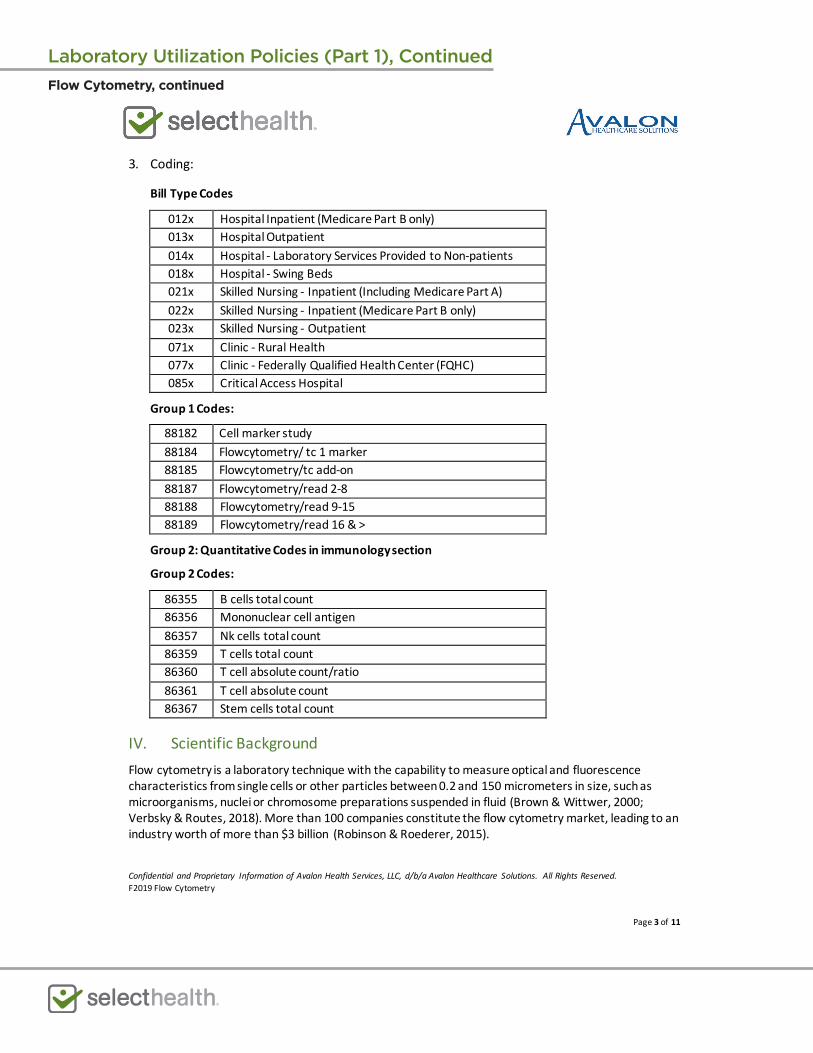
Laboratory Utilization Policies (Part 1), Continued
Confidential and Proprietary Information of Avalon Health Services, LLC, d/b/a Avalon Healthcare Solutions. All Rights Reserved. F2019 Flow Cytometry
Page 3 of 11
3. Coding:
Bill Type Codes
012x Hospital Inpatient (Medicare Part B only) 013x Hospital Outpatient 014x Hospital - Laboratory Services Provided to Non-patients 018x Hospital - Swing Beds 021x Skilled Nursing - Inpatient (Including Medicare Part A) 022x Skilled Nursing - Inpatient (Medicare Part B only) 023x Skilled Nursing - Outpatient 071x Clinic - Rural Health 077x Clinic - Federally Qualified Health Center (FQHC) 085x Critical Access Hospital
Group 1 Codes:
88182 Cell marker study 88184 Flowcytometry/ tc 1 marker 88185 Flowcytometry/tc add-on 88187 Flowcytometry/read 2-8 88188 Flowcytometry/read 9-15 88189 Flowcytometry/read 16 & >
Group 2: Quantitative Codes in immunology section
Group 2 Codes:
86355 B cells total count 86356 Mononuclear cell antigen 86357 Nk cells total count 86359 T cells total count 86360 T cell absolute count/ratio 86361 T cell absolute count 86367 Stem cells total count
IV. Scientific Background Flow cytometry is a laboratory technique with the capability to measure optical and fluorescence characteristics from single cells or other particles between 0.2 and 150 micrometers in size, such as microorganisms, nuclei or chromosome preparations suspended in fluid (Brown & Wittwer, 2000; Verbsky & Routes, 2018). More than 100 companies constitute the flow cytometry market, leading to an industry worth of more than $3 billion (Robinson & Roederer, 2015).
Flow Cytometry, continued

Laboratory Utilization Policies (Part 1), Continued
Confidential and Proprietary Information of Avalon Health Services, LLC, d/b/a Avalon Healthcare Solutions. All Rights Reserved. F2019 Flow Cytometry
Page 4 of 11
A typical flow cytometer contains five main components: a flow cell, a laser, optical parts, detectors which amplify signals, and an electronic or computer system (Verbsky & Routes, 2018). This device measures thousands of cells instantaneously by passing them through the laser beam, and it can even sort the cells into 96- or 384-well plates, tubes, and slides based on identified cellular properties (McKinnon, 2018). Size is determined by the forward angle light scatter, and internal properties such as cellular granularity are measured by the right-angle light scatter (Brown & Wittwer, 2000; Verbsky & Routes, 2018). These fluorescent light signals are converted into electronic signals and then analyzed by a computer to generate final results (McKinnon, 2018).
Fluorescent reagents may be used to enhance a sample before administration into the flow cytometer. These reagents may include DNA binding dyes, fluorescently conjugated antibodies, viability dyes, fluorescent expression proteins, and ion indicator dyes (McKinnon, 2018). Each fluorescent dye binds to cellular components differently, leading to distinguished outcomes when passed by the light source. A fluorochrome, or chemical that can re-emit light when excited, can assist in the detection of specific cellular properties. The use of multiple fluorochromes at once allows several characteristics to be identified instantaneously as different colors emit different wavelengths of light; common dyes include propidium iodide, phycoerythrin, and fluorescein (Brown & Wittwer, 2000).
Immunophenotyping is the most common use of flow cytometry and entails the identification of cellular markers from the immune system, such as T cell subsets and cytokines, as well as antigen-specific responses. Unfortunately, immunophenotyping faces issues in the clinical world due to a lack of standardized procedures (Finak et al., 2016). Current instruments allow for up to 28 colors to be used in immunophenotyping experiments, yet many researchers use less than this (McKinnon, 2018).
Clinical Validity and Utility
Technically, any biologic sample can be analyzed by flow cytometry. However, blood is the most common sample type, including both whole blood and peripheral blood mononuclear cells (Verbsky & Routes, 2018). Flow cytometry can be employed for prognostic and diagnostic purposes. This technique has been used to identify both primary immunodeficiencies and secondary or acquired immunodeficiencies such as HIV (Verbsky & Routes, 2018). Primary immunodeficiencies represent more than 300 known genetic disorders, and flow cytometry is a major component of the diagnosis of these disorders (Abraham & Aubert, 2016). Flow cytometry may also be used for prenatal diagnoses, hematology, transplantation, crop improvement, sperm sorting for sex preselection, post-bone marrow transplantation analyses, and during immunosuppression and chemotherapy treatments (Halder, Nath, & Jha, 2017; Verbsky & Routes, 2018).
Today, many assays have been developed for flow cytometry purposes. These assays can identify biomarkers for cancer and stem cells, DNA and RNA, reactive oxygen species, and the functional status of yeast or bacteria (Robinson & Roederer, 2015). Newer techniques have also been developed such as mass cytometry: the combination of flow cytometry and mass spectrometry (Cosma, Nolan, & Gaudilliere, 2017). Flow fluorescent in situ hybridization (FISH) is another combinatory technique which is the combination of fluorescent in situ hybridization in suspension (FISHIS) and flow cytometry using DNA or gene-specific probes.
Flow Cytometry, continued

Laboratory Utilization Policies (Part 1), Continued
Confidential and Proprietary Information of Avalon Health Services, LLC, d/b/a Avalon Healthcare Solutions. All Rights Reserved. F2019 Flow Cytometry
Page 5 of 11
Flow cytometry techniques have been used to identify several types of cancer. Fromm, Thomas, and Wood (2009) used flow cytometry to identify classical Hodgkin lymphoma, neoplastic Hodgkin, and Reed Sternberg cells in lymph nodes with 88.7% sensitivity and 100% specificity. Paiva, Merino, and San Miguel (2016) state that next generation multiparameter flow cytometry “should be considered mandatory in the routine evaluation of multiple myeloma patients both at diagnosis and after therapy, and represents an attractive technique to integrate with high-throughput DNA and RNA-seq methods to help in understanding the mechanisms behind dissemination and chemoresistance of multiple myeloma.” Finally, Novikov et al. (2019) used flow cytometry immunophenotyping to identify malignant T-cell clones in mature peripheral T-cell lymphomas with 97% sensitivity and 91% specificity.
Wang et al. (2019) published a study on the applicability of multiparameter (multicolor) flow cytometry (MFC) for detecting MRD to predict relapse in patients with AML after allogeneic transplantation. The researchers also compared MFC to MRD status determined using real-time quantitative polymerase chain reaction (RT-qPCR) from 158 bone marrow samples from 44 different individuals. “Strong concordance was found between MFC-based and RT-qPCR-based MRD status (κ = 0.868).” Moreover, for individuals in complete remission (CR), “the positive MRD status detected using MFC was correlated with a worse prognosis [HRs (P values) for relapse, event-free survival, and overall survival: 4.83 (<0.001), 2.23 (0.003), and 1.79 (0.049), respectively]; the prognosis was similar to patients with an active disease before HSCT [hematopoietic stem cell transplantation] (Wang et al., 2019).”
V. Guidelines and Recommendations
Flow cytometry is broadly used for many conditions such as cancers, which are mentioned across many different societies. The below section is not a comprehensive list of guidance for flow cytometry.
The European Research Initiative on CLL (ERIC) & European Society for Clinical Cell Analysis (ESCCA) Harmonisation Project (Rawstron et al., 2018)
This group has published guidelines on chronic lymphocytic leukemia (CLL) in hopes to determine “35 potential flow cytometry markers as being “required,” “recommended,” “suggested,” “uninformative,” or “not sure” for the diagnosis of CLL (Rawstron et al., 2018).” A marker is required if >75% of ERIC/ESCCA members determine that it should be required, and a marker is pushed forward for review if >50% of all members determine that it should be recommended or required. Results are shown in the following figure:
Flow Cytometry, continued

Laboratory Utilization Policies (Part 1), Continued
Confidential and Proprietary Information of Avalon Health Services, LLC, d/b/a Avalon Healthcare Solutions. All Rights Reserved. F2019 Flow Cytometry
Page 6 of 11
Figure 1 [taken from (Rawstron et al., 2018)]:
Flow Cytometry, continued

Laboratory Utilization Policies (Part 1), Continued
Confidential and Proprietary Information of Avalon Health Services, LLC, d/b/a Avalon Healthcare Solutions. All Rights Reserved. F2019 Flow Cytometry
Page 7 of 11
International/European Leukemia Net Working Group for Flow Cytometry in Myelodysplastic Syndromes (Porwit et al., 2014)
An international working party was organized to develop flow cytometry techniques in the classification of myelodysplastic syndromes (MDS). The group has stated the following guidelines:
• “In laboratories where comprehensive immunophenotyping can be performed, an MDS immunophenotyping panel… is recommended.
• In patients with cytological findings suggesting MDS of RCUD (refractory anemia subtype) or refractory anemia with ringed sideroblasts categories, aberrant flow cytometry (FCM) findings in the granulopoietic or myelomonocytic lineages may indicate multilineage dysplasia, which is of prognostic significance. Morphological findings in these cases should be thoroughly re-evaluated to avoid misclassification.
• It is important to note even small populations of myeloid progenitors with multiple immunophenotypic aberrant features (such as aberrant expression of CD7, CD56 or CD11b, see Table 1), since they indicate a higher risk of progression to AML. FCM findings in these cases should be included in the individual risk assessment (Porwit et al., 2014).”
The Clinical Cytometry Society 2006 Bethesda International Consensus (Davis et al., 2007)
In 2006, a panel of subject matter experts convened to define the clinical indications that warrant the use of flow cytometry, as well as to identity of the reagents that should be used in the initial and secondary evaluations for those conditions (Davis et al., 2007). The output of that gathering was the 2006 Bethesda International Consensus Recommendations on the Immunophenotypic Analysis of Hematolymphoid Neoplasia by Flow Cytometry. The panel indicated that flow cytometry is useful for the evaluation of cytopenias, elevated leukocyte count, observation of atypical cells or blasts and evaluation of body fluids, plasmacytosis or monoclonal gammopathy, organomegaly and tissue masses, and certain patient monitoring indications.
The Bethesda recommendations indicate that flow cytometry is not indicated for mature neutrophilia, polyclonal hypergammaglobulinemia, polycythemia, thrombocytosis, and basophilia because “they are usually not associated with hematolymphoid malignancy or associated with hematolymphoid neoplasms that are not detectable by” flow cytometry.
The Bethesda recommendations also indicate that selection of reagents for the initial evaluation panel should be based on specimen type (peripheral blood, bone marrow, tissue, etc.), clinical information and cell morphology studies. They identify initial panels for specific indications that range from a total of 4 reagents to a maximum of 12 reagents.
For secondary evaluation, where the initial evaluation is not conclusive or informative, the Bethesda recommendations again identify groups of reagents that should be used, based on indication. The secondary panels ranged from 5 to 23 reagents.
Flow Cytometry, continued

Laboratory Utilization Policies (Part 1), Continued
Confidential and Proprietary Information of Avalon Health Services, LLC, d/b/a Avalon Healthcare Solutions. All Rights Reserved. F2019 Flow Cytometry
Page 8 of 11
Specific recommendations for the initial evaluation were:
• B cells: CD5, CD10, CD19, CD20, CD45, Kappa, Lambda • T cells and NK cells: CD2, CD3, CD4, CD5, CD7, CD8, CD45, CD56 • Myelomonocytic cells: CD7, CD11b, CD13, CD14, CD15, CD16, CD33, CD34, CD45, CD56, CD117,
HLA-DR • Myelomonocytic cells (limited): CD13, CD33, CD34, CD45 • Plasma cells CD19, CD38, CD45, CD56
For secondary evaluation, the Bethesda recommendations were:
• B cells: CD9, CD11c, CD15, CD22, cCD22, CD23, CD25, CD13, CD33, CD34, CD38, CD43, CD58, cCD79a, CD79b, CD103, FMC7, Bcl-2, cKappa, cLambda, TdT, Zap-70, cIgM
• T cells and NK cells: CD1a, cCD3, CD10, CD16, CD25, CD26, CD30, CD34, CD45RA, CD45RO, CD57, ab-TCR, gd-TCR, cTIA-1, T-beta chain isoforms, TdT
• Myelomonocytic cells: CD2, CD4, CD25, CD36, CD38, CD41, CD61, cCD61, CD64, CD71, cMPO, CD123, CD163, CD235a
• Plasma cells: CD10, CD117, CD138, cKappa, cLambda
The American Society of Clinical Oncology Tumor Markers Expert Panel (ASCO) (Harris et al., 2007; Locker et al., 2006)
In 2006, the ASCO updated the recommendations for the use of tumor marker tests in the prevention, screening, treatment, and surveillance of gastrointestinal cancers. These recommendations state that “Neither flow-cytometrically derived DNA ploidy (DNA index) nor DNA flow cytometric proliferation analysis (% S phase) should be used to determine prognosis of early-stage colorectal cancer (Locker et al., 2006).”
In 2007, the ASCO updated the recommendations for the use of tumor marker tests in the prevention, screening, treatment, and surveillance of breast cancer (Harris et al., 2007); the authors noted that “DNA/ploidy by flow cytometry demonstrated insufficient evidence to support routine use in clinical practice.”
VI. State and Federal Regulations (as applicable)
A search of the FDA database on 1/26/2021 using the term “flow cytometry” yielded 11 results. Many of these results were flow cytometry reagents, calibration kits, and parts or systems of the cytometer. Further, many labs have developed specific tests that they must validate and perform in house. These laboratory-developed tests (LDTs) are regulated by the Centers for Medicare and Medicaid (CMS) as high-complexity tests under the Clinical Laboratory Improvement Amendments of 1988 (CLIA ’88). As an LDT, the U. S. Food and Drug Administration has not approved or cleared this test; however, FDA clearance or approval is not currently required for clinical use.
Flow Cytometry, continued

Laboratory Utilization Policies (Part 1), Continued
Confidential and Proprietary Information of Avalon Health Services, LLC, d/b/a Avalon Healthcare Solutions. All Rights Reserved. F2019 Flow Cytometry
Page 9 of 11
VII. Applicable CPT/HCPCS Procedure Codes
Code Number Code Description 86355 B cells, total count
86356 Mononuclear cell antigen, quantitative (e.g., flow cytometry), not otherwise specified, each antigen (Do not report 88187-88189 for interpretation of 86355, 86356, 86357, 86359, 86360, 86361, 86367)
86357 Natural killer (NK) cells, total count 86359 T cells; total count 86360 absolute CD4 and CD8 count, including ratio 86361 absolute CD4 count
86367 Stem cells (i.e., CD34), total count (For flow cytometric immunophenotyping for the assessment of potential hematolymphoid neoplasia, see 88184-88189)
88182 Flow Cytometry, cell cycle or DNA analysis
88184 Flow Cytometry, cell surface, cytoplasmic, or nuclear marker, technical component only; first marker.
88185 Each additional marker (listed separately in addition to code 88184 for the first marker)
88187 Flow cytometry, interpretation; 2 to 8 markers 88188 9 to 15 markers 88189 16 or more markers
Current Procedural Terminology© American Medical Association. All Rights reserved.
Procedure codes appearing in Medical Policy documents are included only as a general reference tool for each policy. They may not be all-inclusive.
VIII. Evidence-based Scientific References
Abraham, R. S., & Aubert, G. (2016). Flow Cytometry, a Versatile Tool for Diagnosis and Monitoring of Primary Immunodeficiencies. Clin Vaccine Immunol, 23(4), 254-271. doi:10.1128/cvi.00001-16
Adan, A., Alizada, G., Kiraz, Y., Baran, Y., & Nalbant, A. (2017). Flow cytometry: basic principles and applications. Crit Rev Biotechnol, 37(2), 163-176. doi:10.3109/07388551.2015.1128876
Brown, M., & Wittwer, C. (2000). Flow cytometry: principles and clinical applications in hematology. Clin Chem, 46(8 Pt 2), 1221-1229. Retrieved from http://clinchem.aaccjnls.org/content/46/8/1221
Cosma, A., Nolan, G., & Gaudilliere, B. (2017). Mass cytometry: The time to settle down. Cytometry A, 91(1), 12-13. doi:10.1002/cyto.a.23032
Davis, B. H., Holden, J. T., Bene, M. C., Borowitz, M. J., Braylan, R. C., Cornfield, D., . . . Stetler-Stevenson, M. (2007). 2006 Bethesda International Consensus recommendations on the flow cytometric immunophenotypic analysis of hematolymphoid neoplasia: medical indications. Cytometry B Clin Cytom, 72 Suppl 1, S5-13. doi:10.1002/cyto.b.20365
Finak, G., Langweiler, M., Jaimes, M., Malek, M., Taghiyar, J., Korin, Y., . . . McCoy, J. P. (2016). Standardizing Flow Cytometry Immunophenotyping Analysis from the Human ImmunoPhenotyping Consortium. Sci Rep, 6, 20686. doi:10.1038/srep20686
Flow Cytometry, continued
Laboratory Utilization Policies (Part 1), Continued
Confidential and Proprietary Information of Avalon Health Services, LLC, d/b/a Avalon Healthcare Solutions. All Rights Reserved. F2019 Flow Cytometry
Page 10 of 11
Fromm, J. R., Thomas, A., & Wood, B. L. (2009). Flow cytometry can diagnose classical hodgkin lymphoma in lymph nodes with high sensitivity and specificity. Am J Clin Pathol, 131(3), 322-332. doi:10.1309/ajcpw3un9dyldspb
Halder, M., Nath, S., & Jha, S. (2017). Flow Cytometry and Its Utility. Chromosome Structure and Aberrations, 109-126. Retrieved from https://link.springer.com/chapter/10.1007/978-81-322-3673-3_5
Harris, L., Fritsche, H., Mennel, R., Norton, L., Ravdin, P., Taube, S., . . . Bast, R. C., Jr. (2007). American Society of Clinical Oncology 2007 update of recommendations for the use of tumor markers in breast cancer. J Clin Oncol, 25(33), 5287-5312. doi:10.1200/jco.2007.14.2364
Locker, G. Y., Hamilton, S., Harris, J., Jessup, J. M., Kemeny, N., Macdonald, J. S., . . . Bast, R. C., Jr. (2006). ASCO 2006 update of recommendations for the use of tumor markers in gastrointestinal cancer. J Clin Oncol, 24(33), 5313-5327. doi:10.1200/jco.2006.08.2644
McKinnon, K. M. (2018). Flow Cytometry: An Overview. Curr Protoc Immunol, 120, 5.1.1-5.1.11. doi:10.1002/cpim.40
Novikov, N. D., Griffin, G. K., Dudley, G., Drew, M., Rojas-Rudilla, V., Lindeman, N. I., & Dorfman, D. M. (2019). Utility of a Simple and Robust Flow Cytometry Assay for Rapid Clonality Testing in Mature Peripheral T-Cell Lymphomas. Am J Clin Pathol, 151(5), 494-503. doi:10.1093/ajcp/aqy173
Paiva, B., Merino, J., & San Miguel, J. F. (2016). Utility of flow cytometry studies in the management of patients with multiple myeloma. Curr Opin Oncol, 28(6), 511-517. doi:10.1097/cco.0000000000000331
Porwit, A., van de Loosdrecht, A. A., Bettelheim, P., Brodersen, L. E., Burbury, K., Cremers, E., . . . Bene, M. C. (2014). Revisiting guidelines for integration of flow cytometry results in the WHO classification of myelodysplastic syndromes-proposal from the International/European LeukemiaNet Working Group for Flow Cytometry in MDS. Leukemia, 28(9), 1793-1798. doi:10.1038/leu.2014.191
Rawstron, A. C., Kreuzer, K. A., Soosapilla, A., Spacek, M., Stehlikova, O., Gambell, P., . . . Montserrat, E. (2018). Reproducible diagnosis of chronic lymphocytic leukemia by flow cytometry: An European Research Initiative on CLL (ERIC) & European Society for Clinical Cell Analysis (ESCCA) Harmonisation project. Cytometry B Clin Cytom, 94(1), 121-128. doi:10.1002/cyto.b.21595
Robinson, J. P., & Roederer, M. (2015). HISTORY OF SCIENCE. Flow cytometry strikes gold. Science, 350(6262), 739-740. doi:10.1126/science.aad6770
Verbsky, J., & Routes, J. (2018). Flow cytometry for the diagnosis of primary immunodeficiencies. Retrieved from https://www.uptodate.com/contents/flow-cytometry-for-the-diagnosis-of-primary-immunodeficiencies?source=search_result&search=flow%20cytometry&selectedTitle=1~150
Wang, Z., Guo, M., Zhang, Y., Xu, S., Cheng, H., Wu, J., . . . Tang, G. (2019). The applicability of multiparameter flow cytometry for the detection of minimal residual disease using different-from-normal panels to predict relapse in patients with acute myeloid leukemia after allogeneic transplantation. Int J Lab Hematol, 41(5), 607-614. doi:10.1111/ijlh.13070
Flow Cytometry, continued

Laboratory Utilization Policies (Part 1), Continued
Confidential and Proprietary Information of Avalon Health Services, LLC, d/b/a Avalon Healthcare Solutions. All Rights Reserved. F2019 Flow Cytometry
Page 11 of 11
IX. Revision History
Revision Date Summary of Changes
Disclaimer This document is for informational purposes only and should not be relied on in the diagnosis and care of individual patients. Medical and Coding/Reimbursement policies do not constitute medical advice, plan preauthorization, certification, an explanation of benefits, or a contract. Members should consult with appropriate healthcare providers to obtain needed medical advice, care, and treatment. Benefits and eligibility are determined before medical guidelines and payment guidelines are applied. Benefits are determined by the member’s individual benefit plan that is in effect at the time services are rendered. The codes for treatments and procedures applicable to this policy are included for informational purposes. Inclusion or exclusion of a procedure, diagnosis or device code(s) does not constitute or imply member coverage or provider reimbursement policy. Please refer to the member's contract benefits in effect at the time of service to determine coverage or non-coverage of these services as it applies to an individual member. SelectHealth® makes no representations and accepts no liability with respect to the content of any external information cited or relied upon in this policy. SelectHealth updates its Coverage Policies regularly and reserves the right to amend these policies without notice to healthcare providers or SelectHealth members. Members may contact Customer Service at the phone number listed on their member identification card to discuss their benefits more specifically. Providers with questions about this Coverage Policy may call SelectHealth Provider Relations at (801) 442-3692. No part of this publication may be reproduced, stored in a retrieval system or transmitted, in any form or by any means, electronic, mechanical, photocopying, or otherwise, without permission from SelectHealth. ”Intermountain Healthcare” and its accompanying logo, the marks of “SelectHealth” and its accompanying marks are protected and registered trademarks of the provider of this Service and or Intermountain Health Care, Inc., IHC Health Services, Inc., and SelectHealth, Inc. Also, the content of this Service is proprietary and is protected by copyright. You may access the copyrighted content of this Service only for purposes set forth in these Conditions of Use. © CPT Only – American Medical Association
Flow Cytometry, continued

Laboratory Utilization Policies (Part 1), Continued
Confidential and Proprietary Information of Avalon Health Services, LLC, d/b/a Avalon Healthcare Solutions. All Rights Reserved. G2154 Folate Testing
Page 1 of 18
Folate Testing Policy #: AHS – G2154 Prior Policy Name & Number
(as applicable): Implementation Date: 9/15/21 Date of Last Revision:
I. Policy Description Folate, or vitamin B9, is a generic term for a water-soluble vitamin obtained from the diet that is involved in the transfer of methyl groups (i.e. single carbon-containing groups) in multiple biochemical metabolic pathways, including nucleic acid biosynthesis and methionine/homocysteine metabolism. Folate metabolism is closely linked to vitamin B12, cobalamin. Folate deficiency can be implicated in many disease states and processes; however, it is usually easily remedied with either a change in diet or a dietary supplement of the synthetic form, folic acid (Means Jr & Fairfield, 2020; NIH, 2018).
II. Related Policies Policy Number Policy Title
Not Applicable
III. Indications and/or Limitations of Coverage
1. Measurement of serum folate concentration MEETS COVERAGE CRITERIA for evaluation of patients when all of the following criteria are met:
a. patient has been diagnosed with megaloblastic or macrocytic or unexplained anemia; AND
Disclaimer:
1. Policies are subject to change without notice. 2. Policies outline coverage determinations for SelectHealth Commercial, SelectHealth
Advantage® (Medicare/CMS) and SelectHealth Community Care® (Medicaid/CHIP) plans. Refer to the “Policy” section for more information.
Application of coverage criteria depends on an individual’s benefit coverage at the time of the request.
For SelectHealth Advantage (Medicare/CMS), coverage is determined by the Centers for Medicare and Medicaid Services (CMS). If a coverage determination has not been adopted by CMS and InterQual criteria are not available, the SelectHealth Commercial policy applies. For the most up-to-date Medicare policies and coverage, please visit their search website or the manual website,
For SelectHealth Community Care (Medicaid), coverage is determined by the State of Utah Medicaid program. If Utah State Medicaid has no published coverage position and InterQual criteria are not available, the SelectHealth Commercial criteria will apply. For the most up-to-date Medicaid policies and coverage, please visit their website or the Utah Medicaid Code Look-Up tool.

Laboratory Utilization Policies (Part 1), Continued
Confidential and Proprietary Information of Avalon Health Services, LLC, d/b/a Avalon Healthcare Solutions. All Rights Reserved. G2154 Folate Testing
Page 2 of 18
b. anemia and/or macrocytosis does not resolve after folic acid treatment
2. Measurement of serum folate concentration DOES NOT MEET COVERAGE CRITERIA for any other indications not described above
3. Measurement of RBC folate DOES NOT MEET COVERAGE CRITERIA for any indications
IV. Scientific Background Folate, or vitamin B9, naturally occurs as polyglutamated compounds (pteroylpolyglutamates) in many plant and animal products. The synthetic form is a monoglutamate-containing compound called folic acid. Folic acid is more chemically stable for commercial production and storage, but it is less bioavailable than the naturally occurring folate (Means Jr & Fairfield, 2020). Biochemically, folate is a coenzyme in single-carbon transfers in vivo and is directly linked to the cobalamin (vitamin B12) cycle, methionine metabolism, and nucleic acid biosynthesis. Dietary folates are hydrolyzed via γ-glutamylhydrolase (or folate conjugase) prior to absorption in the intestinal mucosa (IOM, 1998). Both folate and vitamin B12 are required for formation of 5,10-methylene tetrahydrofolate, which is the cofactor involved in purine synthesis. Methylenetetrahydrofolate reductase (MTHFR) is the enzyme responsible in converting 5,10-methylene tetrahydrofolate to 5-methyl tetrahydrofolate, which is required for methionine synthase, the enzyme that converts homocysteine to methionine. The interlinked one-carbon cycle is depicted in the figure below with the metabolites assayed in clinical laboratories in bold (Finer, Saravanan, Hitman, & Yajnik, 2013).
Folate Testing, continued

Laboratory Utilization Policies (Part 1), Continued
Confidential and Proprietary Information of Avalon Health Services, LLC, d/b/a Avalon Healthcare Solutions. All Rights Reserved. G2154 Folate Testing
Page 3 of 18
Role of Folate in Anemia
Anemia occurs when the body lacks healthy red blood cells (RBCs), leading to an insufficient amount of oxygen delivered to tissues. Typical symptoms of anemia include fatigue, weakness, pale skin and lightheadedness.
Macrocytic anemia refers to anemias that have high mean corpuscular volume with large RBCs. Mean corpuscular volume, or mean cell volume, can be defined as the average volume of RBCs in an individual. Megaloblastic anemia is a specific macrocytic anemia due to nucleic acid metabolic defects that result in “nuclear-cytoplasmic dyssynchrony, reduced number of cell divisions in the bone marrow, and nuclear abnormalities in both myeloid and erythroid precursors” caused by folate and/or vitamin B12 deficiency (Means Jr & Fairfield, 2021). These abnormal RBCs are the principle clinical manifestations of folate deficiency and symptoms “include weakness, fatigue, difficulty concentrating, irritability, headache, heart palpitations, and shortness of breath” (NIH, 2018).
\
Folate Testing, continued

Laboratory Utilization Policies (Part 1), Continued
Confidential and Proprietary Information of Avalon Health Services, LLC, d/b/a Avalon Healthcare Solutions. All Rights Reserved. G2154 Folate Testing
Page 4 of 18
Folate and Neural Tube Defects (NTDs)
Neural tube defects (NTDs) develop early in pregnancy and are malformations of the brain and/or spine that include spina bifida and anencephaly. Folate deficiency is directly linked to NTDs. The role of folate in NTD development is not well-characterized. The role of folate in either the methylation cycle or nucleic acid synthesis has been suggested to play a part in NTD development during embryogenesis, and some studies have indicated that it is the bioavailability of specific folates in women that can increase the likelihood of NTDs (Imbard, Benoist, & Blom, 2013; Rothenberg et al., 2004). Women typically do not obtain enough folate from diet alone, so women of childbearing age are recommended to take a synthetic folic acid supplement to decrease the likelihood of NTDs in offspring (Bibbins-Domingo et al., 2017). To decrease the occurrence of NTDs and folate deficiency, the United States and Canada mandated folic acid supplementation to cereal grains in 1998, and as of March 2018 “87 countries have legislation to mandate fortification of at least one industrially milled cereal grain” (FFI, 2018). A review by Imbard et al. (2013) of 17 different studies on the impact of folic acid fortification of NTD rates show that 16 show a decrease in the rate of NTDs. Only a study of the rate of NTDs in California showed no decline since fortification. The reduction of the United States overall was 26-30% since folic acid fortification (Imbard et al., 2013).
Causes of Folate Deficiency
Folate deficiency can be caused by dietary intake. Nutritional deficits may occur due to diet, alcoholism, depression, and even overcooked foods. Many malabsorptive disorders, such as celiac disease and ulcerative colitis, can also result in a decrease in folate uptake. Further, bariatric procedures may result in decreased absorption, and drugs, including methotrexate and trimethoprim that inhibit dihydrofolate reductase (DHFR), can also cause a folate deficiency. It is also important to note that an increased need of folate for DNA synthesis during pregnancy and lactation, chronic hemolytic anemias, exfoliative skin diseases, and hemodialysis cause folic acid deficiency (Means Jr & Fairfield, 2021).
Methodology of Folate Testing
Folate concentrations have been measured from serum, erythrocytes (RBC), and urine. Serum folate levels may not “differentiate between what may be a transitory reduction in folate intake or chronic folate deficiency accompanied by depleted folate stores and functional changes” (IOM, 1998). RBCs have a lifespan of approximately 120 days, and folate is only taken in during initial erythropoiesis (red blood cell production); consequently, RBC folate concentrations are less likely to be affected by transitory dietary fluctuations. However, Wu, Chanarin, Slavin, and Levi (1975) show that both RBC folate and serum folate levels correlate to hepatocyte folate levels (IOM, 1998; Wu et al., 1975). Galloway and Rushworth (2003) released a study in conjunction with the National Pathology Alliance review in the United Kingdom comparing data of laboratories of the National Health Service that routinely use serum folate testing only, RBC folate testing only, or both serum and RBC folate testing together. The researchers conclude that there is no need to use both tests to determine folate concentration as an initial screen. “The serum folate assay provided equivalent information to the measurement of red cell folate and evidence from the literatures [sic] suggest that the serum folate assay should be the method of choice” (Galloway & Rushworth, 2003).
Folate Testing, continued

Laboratory Utilization Policies (Part 1), Continued
Confidential and Proprietary Information of Avalon Health Services, LLC, d/b/a Avalon Healthcare Solutions. All Rights Reserved. G2154 Folate Testing
Page 5 of 18
Clinical Utility and Validity
A study by Shojania and von Kuster (2010) investigated the use of serum folate testing (SF) and RBC folate testing (RF) in cases of anemia in a country that has mandated folic acid supplementation in grain products. By examining the data for folate testing in anemia at two different teaching hospitals in Canada, they report that in one hospital in 2001 “11 out of 2154 (0.5%) SF were low (<7.0 nmol/L) and 4 out of 560 (0.7%) RF were low (<417 nmol/L). In no subject with low SF or RF could the anemia be attributed to folate deficiency.” For the other hospital, the data from 1999-2001 shows that “19 out of 991 (1.9%) had low RF (<225 nmol/L) but in only 2 patients (0.2%) the low RF was in folate deficiency anemia range” (Shojania & von Kuster, 2010). The authors conclude that neither serum folate testing nor RBC folate testing is justified in cases of anemia for folic acid fortified countries due to such low incidence rates of folate deficiency anemia.
Another study by Joelson, Fiebig, and Wu (2007) examined the records of three different hospitals in the U.S. that service a high number of indigent patients. The researchers reported the data from three non-consecutive years (1997, 2000, and 2004) to examine the impact of folate fortification in food products. Using the RBC folate levels only with a RBC folate cutoff value of 160 ng/mL (363.6 nmol/L), “the combined incidence of folate deficiency decreased from 4.8% in 1997 to 0.6% in 2004…Even when the folate concentration was found to be low, the majority of these subjects did not have macrocytosis.” This study included a total of 4134 RBC folate tests performed over the course of three years. It is of interest to note that the number of tests performed increased from 813 in 1997 to 1759 in 2004. The authors do note of a potential limitation of the study since the data of the patients cannot be separated into specific groups (pregnant women, alcoholics, socioeconomic classes, and so on). The authors conclude “that folate deficiency has become a rare event in the United States, and the utility of routine folate measurements for patients with anemia and/or increased mean corpuscular volume are difficult to justify” (Joelson et al., 2007).
Urinary folate levels do not reflect either the stored folate concentrations or the fluctuations in folate concentration due to transitory dietary changes. Only about 1-2% of the folate excreted in the urine is unmetabolized and “excretion continued in the face of advanced folate depletion” (IOM, 1998). One study of ten postmenopausal women on a low folate diet measured folate turnover using urinary testing of folate and folate metabolites. “Folate intake did not significantly influence ApABG (para-acetamidobenzoylglutamate) or pABG (para-aminobenzoylglutamate) excretion.” ApABG and pABG along with pterins are the major folate catabolites. The authors conclude that “the rate of folate catabolite excretion is related mainly to masses of slow-turnover folate pools governed by long-term folate intake” (Gregory, Swendseid, & Jacob, 2000).
Epstein-Peterson et al. (2020) collected and analyzed all folate tests performed in 2017 at an academic cancer center. In total, 937 patients were tested 1065 times; approximately 7% of tests indicated a folate deficiency, and folate deficiency was significantly associated with a higher risk of death (P=0.01) (Epstein-Peterson et al., 2020).
Folate Testing, continued

Laboratory Utilization Policies (Part 1), Continued
Confidential and Proprietary Information of Avalon Health Services, LLC, d/b/a Avalon Healthcare Solutions. All Rights Reserved. G2154 Folate Testing
Page 6 of 18
V. Guidelines and Recommendations Centers for Disease Control and Prevention (CDC) (CDC, 1991, 1992, 2018)
The “CDC urges all women of reproductive age to take 400 micrograms (mcg) of folic acid each day, in addition to consuming food with folate from a varied diet, to help prevent some major birth defects of the baby’s brain (anencephaly) and spine (spina bifida)” (CDC, 2018). This recommendation includes all women of reproductive age planning to become pregnant or not, as about half of U.S. pregnancies are unplanned.
Morbidity and Mortality Weekly Report (MMWR)
Both in 1991 and 1992, the CDC addressed the need for women to consume folic acid daily. In the first recommendation, the CDC recommended that women who had already had at least one pregnancy with a neural tube defect (NTD) to consume 4 mg of folic acid per day. “Women should take the supplement from at least 4 weeks before conception through the first 3 months of pregnancy.” This recommendation required a physician’s supervision and “were not intended for 1) women who have never given birth to an infant or had a fetus with a neural tube defect, 2) relatives of women who have had an infant or fetus with a neural tube defect, 3) women who themselves have spina bifida, or 4) women who take the anticonvulsant valproic acid—a known cause of spina bifida (CDC, 1991).”
In 1992, the CDC modified the recommendation to include all women of childbearing age to consume folate since the addition of folate would potentially decrease the number of NTDs by 50%. “All women of childbearing age in the United States who are capable of becoming pregnant should consume 0.4 mg of folic acid per day for the purpose of reducing their risk of having a pregnancy affected with spina bifida or other NTDs. Because the effects of high intakes are not well known but include complicating the diagnosis of vitamin [B12] deficiency, care should be taken to keep total folate consumption at less than 1 mg per day, except under the supervision of a physician. Women who have had a prior NTD-affected pregnancy are at high risk of having a subsequent affected pregnancy. When these women are planning to become pregnant, they should consult their physicians for advice (CDC, 1992).” Neither recommendation from the CDC included a requirement for either serum folate or red blood cell folate concentration.
Institute of Medicine (IOM) (IOM, 1998)
The Institute of Medicine released their detailed report titled “Dietary Reference Intakes for Thiamin, Riboflavin, Niacin, Vitamin B6, Folate, Vitamin B12, Pantothenic Acid, Biotin, and Choline” in 1998. This report does not outline any recommendation concerning determining the concentration of either serum folate or red blood cell folate concentrations; however, the table below outlines their recommendations concerning the upper limits (UL) of daily intake of folate for various groups (taken from (IOM, 1998)):
Folate Testing, continued

Laboratory Utilization Policies (Part 1), Continued
Confidential and Proprietary Information of Avalon Health Services, LLC, d/b/a Avalon Healthcare Solutions. All Rights Reserved. G2154 Folate Testing
Page 7 of 18
The IOM does specifically state that “individuals who are at risk of vitamin B12 deficiency …may be at increased risk of the precipitation of neurological disorders if they consume excess folate.” Vitamin B12 deficiency can be due to dietary deficiencies (such as a complete vegan diet), pernicious anemia, gastrectomy, atrophic gastritis, bacterial infection, pancreatic insufficiency, and terminal ileal disease or resection (IOM, 1998).
American Society for Clinical Pathology (ASCP)/Choosing Wisely (ASCP, 2017)
The ASCP published a recommendation in 2017 in Choosing Wisely, an American Board of Internal Medicine (ABIM) initiative, where they clearly state the following: “Do not order red blood cell folate levels at all. In adults, consider folate supplementation instead of serum folate testing in patients with macrocytic anemia.” They indicate that the drastic decrease in folic deficiency in both the U.S. and Canada after mandated folic acid supplementation in foods no longer requires for either serum folate or red blood cell folate concentrations be tested. “While red blood cell folate levels have been used in the past as a surrogate for tissue folate levels or a marker for folate status over the lifetime of red blood cells, the result of this testing does not, in general, add to the clinical diagnosis or therapeutic plan” (ASCP, 2017).
Folate Testing, continued

Laboratory Utilization Policies (Part 1), Continued
Confidential and Proprietary Information of Avalon Health Services, LLC, d/b/a Avalon Healthcare Solutions. All Rights Reserved. G2154 Folate Testing
Page 8 of 18
The Doctors of BC (formerly the British Columbia Medical Association) (BCMA, 2012)
The Guidelines & Protocols Advisory Committee of the British Columbia Medical Association in conjunction with the Province of British Columbia released their guidelines concerning folate and folate deficiency. They state that “serum folate and red blood cell (RBC) folate tests are no longer being offered (except at Vancouver General Hospital and St. Paul’s hospital under limited indications and require approval from the respective Medical Biochemist on call)” because of the drastic decrease in folate deficiency in the province. “In two outpatient laboratories in British Columbia, 99.8% and 99.1% of folate tests were normal in 2010…If folate deficiency is suspected, it is reasonable to give oral folic acid (0.4-1 mg/day) without doing laboratory investigation for deficiency at least until the hemoglobin and mean corpuscular volume normalizes (or longer if the underlying cause cannot be eliminated) (BCMA, 2012).”
National Pathology Alliance (of the United Kingdom) (Galloway & Rushworth, 2003)
The National Pathology Alliance of the United Kingdom in 2003 published in the Journal of Clinical Pathology their recommendation “that serum folate measurements provide equivalent information to red cell folate measurements.”
American Association of Clinical Endocrinologists (AACE)/The American College of Endocrinology (ACE), The Obesity Society (TOS), American Society for Metabolic and Bariatric Surgery (ASMBS), Obesity Medicine Association (OMA), and American Society of Anesthesiologists (ASA) (Gonzalez-Campoy et al., 2013; Handelsman et al., 2015; Jellinger et al., 2017; Mechanick et al., 2019)
In 2013, the AACE, ACE, and TOS issued joint guidelines regarding healthy eating for the prevention and treatment of metabolic and endocrine diseases in adults (Gonzalez-Campoy et al., 2013). Based on the data from the National Health and Nutrition Examination Survey (NHANES), they state “that patients with vitamin B12 deficiency had higher folate levels, were more likely to be anemic, and had more cognitive impairment that those with normal serum folate levels” [evidence level (EL) 2]. They evaluate the evidence concerning the link between folate and cardiovascular disease as EL4 and the link between NTDs and folate as EL1. With respect to pregnancy nutritional needs, they “should be assessed prior to conception to improve pregnancy outcome…All women of childbearing age should consume at least 400 µg dietary equivalents of folate per day” [EL4] and that during pregnancy the daily amount should be increased to 600 µg [EL3].
The AACE and ACE in 2015 released their Clinical Practice Guidelines for Developing a Diabetes Mellitus Comprehensive Care Plan (Handelsman et al., 2015). Concerning patients with diabetic nephropathy, the suggest that they “undergo annual or more frequent assessment of electrolytes”. For those with anemia, iron, transferrin saturation (TSAT), ferritin, vitamin B12, and folate levels “should be further investigated” [EL4].
In 2017, the AACE and ACE released their guidelines for management of dyslipidemia and prevention of cardiovascular disease (Jellinger et al., 2017). Since bile acid sequestrant treatments such as cholestyramine can cause folate depletion in children, they recommend that children on such treatments supplement their diet with a multivitamin. They also note that folate, B6, and B12
Folate Testing, continued

Laboratory Utilization Policies (Part 1), Continued
Confidential and Proprietary Information of Avalon Health Services, LLC, d/b/a Avalon Healthcare Solutions. All Rights Reserved. G2154 Folate Testing
Page 9 of 18
supplementation can help mediate hyperhomocysteinemia, but that the supplements do not reduce risk of atherosclerotic cardiovascular disease.
In 2019, the AACE/ACE, TOS, ASMBS, OMA, and ASA issued joint guidelines for the perioperative nutritional, metabolic, and nonsurgical support of the bariatric surgery patient (Mechanick et al., 2019). Here, as part of a pre-operative bariatric surgery checklist that has a “Grade A” recommendation, they include “nutrient screening with iron studies, B12 and folic acid (RBC folate, homocysteine, methylmalonic acid optional) …consider more extensive testing in patients undergoing malabsorptive procedures based on symptoms and risks.” With regards to patients who become pregnant after having a bariatric procedure, they recommend (with Grade D) having nutritional surveillance laboratory screenings done each trimester for folate deficiency along with iron, calcium, B12, and vitamin D, and if after a malabsorptive procedure, fat-soluble vitamins, zinc, and copper. With a Grade C, they state that “nutritional anemias resulting from malabsorptive bariatric surgical procedures can involve deficiencies in vitamin B12, folate, protein, copper, selenium, and zinc and may be evaluated when routine aggressive case finding for iron-deficiency anemia is negative.” Additionally, findings of folate deficiency in patients with obesity prior to bariatric surgery by the ASMBS “justifies aggressive case finding preoperatively with biochemical testing, specifically using sensitive markers, such as red-blood-cell folate and homocysteine (methylmalonic acid is normal with folate deficiency and normal B12 status)” and “particular attention should be given to female patients of childbearing age.”
National Institute for Health and Care Excellence (NICE) [(NICE, 2015) reaffirmed 2019]
The National Institute for Health and Care Excellence (NICE) of the Department of Health in the United Kingdom published their extensive guidelines concerning bladder cancer on February 25, 2015. Within the section concerning the follow-up treatment for muscle-invasive bladder cancer, they recommend a protocol after radical cystectomy that includes “monitoring for metabolic acidosis and B12 and folate deficiency at least annually.”
American Academy of Family Physicians (AAFP) (Kaferle & Strzoda, 2009)
The AAFP released the recommendations concerning macrocytosis and macrocytic anemia in 2009. Of note, they state that “serum folate levels are not useful because they fluctuate rapidly with dietary intake and are not cost effective. RBC folate levels more accurately correlate with folate stores and should be performed if folate deficiency is suspected.” They give the following key recommendation (with evidence rating of “C” or “consensus, disease-oriented evidence, usual practice, expert opinion, or case series”) to “obtain red blood cell folate level if other etiologies are not found (serum folate levels may be misleading).” In the evaluation of macrocytic anemia, they included a flowchart outlining the order of steps and tests to be taken, including when the RBC folate level should be checked. For a patient exhibiting a mean corpuscular volume 100 fL and an abnormal peripheral smear showing megaloblastic features and a reticulocyte count under 2%, they should have their RBC folate level measured only if the vitamin B12 level is >400 pg. The flowchart is included below (Kaferle & Strzoda, 2009).
Folate Testing, continued

Laboratory Utilization Policies (Part 1), Continued
Confidential and Proprietary Information of Avalon Health Services, LLC, d/b/a Avalon Healthcare Solutions. All Rights Reserved. G2154 Folate Testing
Page 10 of 18
American Academy of Neurology (AAN) (AAN, 2009; Knopman et al., 2001)
In 2001, the AAN updated their practice parameters for the diagnosis of dementia. Within the section concerning the comorbidities that should be screened in an initial assessment for dementia, they recommend folate testing along with complete blood count, serum electrolytes, B12, blood urea nitrogen/creatinine, syphilis serology, thyroid function, and glucose. They did note that as of that time “no studies were identified that evaluated these recommendations” since the last practice parameters released in 1994.
Folate Testing, continued

Laboratory Utilization Policies (Part 1), Continued
Confidential and Proprietary Information of Avalon Health Services, LLC, d/b/a Avalon Healthcare Solutions. All Rights Reserved. G2154 Folate Testing
Page 11 of 18
In 2009, the AAN published guidelines regarding the management and care of women with epilepsy (WWE) during pregnancy. These guidelines state that “Folic acid supplementation is generally recommended to reduce the risk of MCMs [major congenital malformations] during pregnancy, and although the data are insufficient to show that it is effective in WWE, there is no evidence of harm and no reason to suspect that it would not be effective in this group. Therefore, all women of childbearing potential, with or without epilepsy, should be encouraged to take at least 0.4 mg of folic acid daily prior to conception and during pregnancy. There was insufficient published information to address the dosing of folic acid (AAN, 2009).”
Kidney Disease Improving Global Outcomes (KDIGO) (McMurray et al., 2012)
KDIGO released their updated KDIGO Clinical Practice Guideline for Anemia in Chronic Kidney Disease in 2012. They gave a “not graded” recommendation for “in patients with CKD [chronic kidney disease] and anemia (regardless of age and CKD stage), include the following tests in initial evaluation of the anemia:
• Complete blood count (CBC), which should include Hb concentration, red cell indices, white blood cell count and differential, and platelet count
• Absolute reticulocyte count
• Serum ferritin level
• Serum transferring saturation (TSAT)
• Serum vitamin B12 and folate levels”
They also state that “RBC folate levels can be measured when serum folate levels are equivocal or when there is concern that recent dietary intake may obscure underlying folate deficiency using serum levels alone.”
Australian National Blood Authority (NBA, 2012)
The Australian National Blood Authority of the National Health and Medical Research Council of Australia released their Patient Blood Management Guidelines: Module 2 Perioperative, an extensive set of guidelines concerning the perioperative patient. They include a preoperative hemoglobin assessment that requires the preoperative tests for full blood count, iron studies (including ferritin), CRP, and renal function. If the patient is anemic and has a ferritin level > 100 mcg/L, then, they state to “check B12/folate levels and reticulocyte count” for possible anemia due to a chronic disease or inflammation. As a footnote, they include “check B12/folate if macrocytic or if there are risk factors for deficiency (e.g. decreased intake or absorption), or if anaemia is unexplained.”
American Society for Parenteral and Enteral Nutrition (ASPEN) & Society of Critical Care Medicine (SSCM) (Choban, Dickerson, Malone, Worthington, & Compher, 2013; Mehta et al., 2017; NGC, 2013, 2016)
In 2013, ASPEN and SSCM issued joint clinical guidelines concerning the nutrition support of hospitalized obese adults. With a “Recommendation: Weak” status, they recommended “in acutely ill hospitalized patients with history of these procedures [sleeve gastrectomy, gastric bypass, or biliopancreatic diversion ± duodenal switch], evaluation for evidence of depletion of iron, copper, zinc, selenium,
Folate Testing, continued

Laboratory Utilization Policies (Part 1), Continued
Confidential and Proprietary Information of Avalon Health Services, LLC, d/b/a Avalon Healthcare Solutions. All Rights Reserved. G2154 Folate Testing
Page 12 of 18
thiamine, folate, and vitamins B12 and D is suggested as well as repletion of deficiency states” (Choban et al., 2013).
In 2016, ASPEN and SSCM issued their Guidelines for the provision and assessment of nutrition support therapy in the adult critically ill patient. The committee recommended that “evaluation for and treatment of micronutrient deficiencies such as calcium, thiamin, vitamin B12, fat-soluble vitamins (A, D, E, K), and folate, along with the trace minerals iron, selenium, zinc, and copper, should be considered” (NGC, 2016). In 2017, ASPEN and SSCM updated their Guidelines for the provision and assessment of nutrition support therapy in the pediatric critically ill patient. These guidelines do not mention folate testing (Mehta et al., 2017).
Academy of Nutrition and Dietetics (AND) (Thompson et al., 2017)
The AND released their Oncology evidence-based nutrition practice guideline in 2013 and reaffirmed the guideline in a 2017 publication. On the “Assessment of Biochemical Data Medical Tests, and Procedures on Adult Oncology Patients” portion, the committee recommended with “Consensus, Imperative” that “the RDN [Registered Dietitian Nutritionist] should evaluate available data and recommend as indicated: biochemical data, medical tests and procedures of adult oncology patients” and included on their list is “Nutritional anemia profile (hemoglobin, hematocrit, folate, B12, iron)”. “Assessment of these factors is needed to effectively determine nutrition diagnoses and plan the nutrition interventions.”
European Crohn’s and Colitis Organisation (ECCO) (Dignass et al., 2015)
ECCO’s guidelines concerning irritable bowel disorders (IBD) included an extensive discussion on causes and treatments of anemia in IBD—both iron deficiency anemia and non-iron deficiency anemia. With an [EL 5], they state that “deficiencies of Vitamin B12 and folate should be treated to avoid anaemia. Serum levels of vitamin B12 and folic acid should be measured at least annually, or if macrocytosis is present. Patients at risk for vitamin B12 or folic acid deficiency [e.g., small bowel disease or resection] need closer surveillance. The recommended timelines are based on expert opinions and reflect common clinical practice, but do not apply to patients with extensive small bowel resection, extensive ileal Crohn’s disease, or ileal-anal pouch.”
American College of Gastroenterology (ACG) (Rubio-Tapia, Hill, Kelly, Calderwood, & Murray, 2013) (reaffirmed 2016)
In their guidelines and recommendations concerning the diagnosis and management of celiac disease (CD) in 2013, the ACG recommended the following statement with Conditional recommendation, low level of evidence: “People with newly diagnosed CD should undergo testing and treatment for micronutrient deficiencies. Deficiencies to be considered for testing should include, but not be limited to, iron, folic acid, vitamin D, and vitamin B12.”
Folate Testing, continued

Laboratory Utilization Policies (Part 1), Continued
Confidential and Proprietary Information of Avalon Health Services, LLC, d/b/a Avalon Healthcare Solutions. All Rights Reserved. G2154 Folate Testing
Page 13 of 18
US Preventive Services Task Force (USPSTF) (Bibbins-Domingo et al., 2017)
The USPSTF in 2017 updated their 2009 recommendation regarding folic acid supplementation in women of childbearing age. The USPSTF gives an “A” recommendation “that all women who are planning or capable of pregnancy take a daily supplement containing 0.4 to 0.8 mg (400-800 µg) of folic acid.” The USPSTF does not make any statement regarding folate screening for women not of childbearing age.
British Committee for Standards in Haematology (BCSH) (Devalia, Hamilton, & Molloy, 2014; Killick et al., 2016)
In 2014, the BCSH released guidelines on folate deficiencies. They noted that “routine red cell folate testing is not necessary because serum folate alone is sufficient in most cases.” However, they also acknowledged that “in the presence of strong clinical suspicion of folate deficiency, despite a normal serum level, a red cell folate assay may be undertaken, having ruled out cobalamin deficiency.” The BCSH also noted that “folate status is generally checked in clinical situations similar to those of cobalamin deficiency (Grade 1A).”
In 2016, the BCSH recommended that a “documented vitamin B12 or folate deficiency should be corrected before a final diagnosis of AA is confirmed. Bone marrow aplasia due to vitamin deficiency is exceedingly rare (Killick et al., 2016).”
Renal Association Clinical Practice Guideline (Mikhail et al., 2017)
The Renal Association recommends measuring serum folate concentration for evaluation of anemia in CKD (Mikhail et al., 2017).
National Comprehensive Cancer Network (NCCN, 2021)
The NCCN recommends measurement of RBC folate as part of the initial evaluation for myelodysplastic syndromes. Serum folate may be considered as an alternative, but is not preferable to RBC folate (NCCN, 2021).
VI. State and Federal Regulations (as applicable) The FDA has approved multiple tests for the evaluation of both human serum folate and red blood cell folate levels either alone, together, or included in panels. As of 04/02/2021, there are 84 records on the FDA Devices database for folate.
Additionally, many labs have developed specific tests that they must validate and perform in house. These laboratory-developed tests (LDTs) are regulated by the Centers for Medicare and Medicaid (CMS) as high-complexity tests under the Clinical Laboratory Improvement Amendments of 1988 (CLIA ’88). As an LDT, the U. S. Food and Drug Administration has not approved or cleared this test; however, FDA clearance or approval is not currently required for clinical use.
Folate Testing, continued

Laboratory Utilization Policies (Part 1), Continued
Confidential and Proprietary Information of Avalon Health Services, LLC, d/b/a Avalon Healthcare Solutions. All Rights Reserved. G2154 Folate Testing
Page 14 of 18
VII. Applicable CPT/HCPCS Procedure Codes Code Number Code Description
82746 Folic acid; serum 82747 Folic acid; RBC
Current Procedural Terminology© American Medical Association. All Rights reserved.
Procedure codes appearing in Medical Policy documents are included only as a general reference tool for each policy. They may not be all-inclusive.
VIII. Evidence-based Scientific References AAN. (2009). Management issues for women with epilepsy—focus on pregnancy Vitamin K, folic acid,
blood levels, and breastfeeding. Retrieved from https://www.aan.com/Guidelines/home/GetGuidelineContent/345
ASCP. (2017, 10/19/2017). Do not order red blood cell folate levels at all. In adults, consider folate supplementation instead of serum folate testing in patients with macrocytic anemia. Twenty Things Physicians and Patients Should Question. Retrieved from http://www.choosingwisely.org/clinician-lists/ascp-do-not-order-red-blood-cell-folate-levels/
BCMA. (2012, 01/01/2012). Folate Deficiency - Investigation & Management. Retrieved from https://www2.gov.bc.ca/gov/content/health/practitioner-professional-resources/bc-guidelines/folate-deficiency
Bibbins-Domingo, K., Grossman, D. C., Curry, S. J., Davidson, K. W., Epling, J. W., Jr., Garcia, F. A., . . . Tseng, C. W. (2017). Folic Acid Supplementation for the Prevention of Neural Tube Defects: US Preventive Services Task Force Recommendation Statement. JAMA, 317(2), 183-189. doi:10.1001/jama.2016.19438
CDC. (1991). Use of folic acid for prevention of spina bifida and other neural tube defects--1983-1991. MMWR Morb Mortal Wkly Rep, 40(30), 513-516.
CDC. (1992). Recommendations for the use of folic acid to reduce the number of cases of spina bifida and other neural tube defects. MMWR Recomm Rep, 41(Rr-14), 1-7.
CDC. (2018, April 11). Folic Acid. Retrieved from https://www.cdc.gov/ncbddd/folicacid/about.html Choban, P., Dickerson, R., Malone, A., Worthington, P., & Compher, C. (2013). A.S.P.E.N. Clinical
Guidelines: nutrition support of hospitalized adult patients with obesity. Journal of Parenteral and Enteral Nutrition, 37(6), 714-744. doi:10.1177/0148607113499374
Devalia, V., Hamilton, M. S., & Molloy, A. M. (2014). Guidelines for the diagnosis and treatment of cobalamin and folate disorders. Br J Haematol, 166(4), 496-513. doi:10.1111/bjh.12959
Dignass, A. U., Gasche, C., Bettenworth, D., Birgegard, G., Danese, S., Gisbert, J. P., . . . Vavricka, S. (2015). European consensus on the diagnosis and management of iron deficiency and anaemia in inflammatory bowel diseases. J Crohns Colitis, 9(3), 211-222. doi:10.1093/ecco-jcc/jju009
Epstein-Peterson, Z. D., Li, D. G., Lavery, J. A., Barrow, B., Chokshi, I., & Korenstein, D. (2020). Inpatient folate testing at an academic cancer center: single-year experience. Support Care Cancer. doi:10.1007/s00520-019-05267-1
FFI. (2018, March 2018). Global Progress of Industrially Milled Cereal Grains. Retrieved from http://www.ffinetwork.org/global_progress/
Folate Testing, continued

Laboratory Utilization Policies (Part 1), Continued
Confidential and Proprietary Information of Avalon Health Services, LLC, d/b/a Avalon Healthcare Solutions. All Rights Reserved. G2154 Folate Testing
Page 15 of 18
Finer, S., Saravanan, P., Hitman, G., & Yajnik, C. (2013). The role of the one-carbon cycle in the developmental origins of Type 2 diabetes and obesity. Diabetic Medicine, 31(3), 263-272. doi:10.1111/dme.12390
Galloway, M., & Rushworth, L. (2003). Red cell or serum folate? Results from the National Pathology Alliance benchmarking review. J Clin Pathol, 56(12), 924-926.
Gonzalez-Campoy, J. M., St Jeor, S. T., Castorino, K., Ebrahim, A., Hurley, D., Jovanovic, L., . . . Thomas, K. T. (2013). Clinical practice guidelines for healthy eating for the prevention and treatment of metabolic and endocrine diseases in adults: cosponsored by the American Association of Clinical Endocrinologists/the American College of Endocrinology and the Obesity Society. Endocr Pract, 19 Suppl 3, 1-82. doi:10.4158/ep13155.gl
Gregory, I. I. I. J. F., Swendseid, M. E., & Jacob, R. A. (2000). Urinary Excretion of Folate Catabolites Responds to Changes in Folate Intake More Slowly than Plasma Folate and Homocysteine Concentrations and Lymphocyte DNA Methylation in Postmenopausal Women. The Journal of Nutrition, 130(12), 2949-2952. doi:10.1093/jn/130.12.2949
Handelsman, Y., Bloomgarden, Z. T., Grunberger, G., Umpierrez, G., Zimmerman, R. S., Bailey, T. S., . . . Zangeneh, F. (2015). American association of clinical endocrinologists and american college of endocrinology - clinical practice guidelines for developing a diabetes mellitus comprehensive care plan - 2015. Endocr Pract, 21 Suppl 1, 1-87. doi:10.4158/ep15672.Gl
Imbard, A., Benoist, J.-F., & Blom, H. J. (2013). Neural Tube Defects, Folic Acid and Methylation. International Journal of Environmental Research and Public Health, 10(9), 4352-4389. doi:10.3390/ijerph10094352
IOM. (1998). The National Academies Collection: Reports funded by National Institutes of Health. In Dietary Reference Intakes for Thiamin, Riboflavin, Niacin, Vitamin B6, Folate, Vitamin B12, Pantothenic Acid, Biotin, and Choline. Washington (DC): National Academies Press (US) National Academy of Sciences.
Jellinger, P. S., Handelsman, Y., Rosenblit, P. D., Bloomgarden, Z. T., Fonseca, V. A., Garber, A. J., . . . Davidson, M. (2017). AMERICAN ASSOCIATION OF CLINICAL ENDOCRINOLOGISTS AND AMERICAN COLLEGE OF ENDOCRINOLOGY GUIDELINES FOR MANAGEMENT OF DYSLIPIDEMIA AND PREVENTION OF CARDIOVASCULAR DISEASE. Endocr Pract, 23(Suppl 2), 1-87. doi:10.4158/ep171764.appgl
Joelson, D. W., Fiebig, E. W., & Wu, A. H. (2007). Diminished need for folate measurements among indigent populations in the post folic acid supplementation era. Arch Pathol Lab Med, 131(3), 477-480. doi:10.1043/1543-2165(2007)131[477:Dnffma]2.0.Co;2
Kaferle, J., & Strzoda, C. E. (2009). Evaluation of macrocytosis. Am Fam Physician, 79(3), 203-208. Killick, S. B., Bown, N., Cavenagh, J., Dokal, I., Foukaneli, T., Hill, A., . . . Marsh, J. C. (2016). Guidelines for
the diagnosis and management of adult aplastic anaemia. Br J Haematol, 172(2), 187-207. doi:10.1111/bjh.13853
Knopman, D. S., DeKosky, S. T., Cummings, J. L., Chui, H., Corey-Bloom, J., Relkin, N., . . . Stevens, J. C. (2001). Practice parameter: diagnosis of dementia (an evidence-based review). Report of the Quality Standards Subcommittee of the American Academy of Neurology. Neurology, 56(9), 1143-1153.
McMurray, J., Parfrey, P., Adamson, J. W., Aljama, P., Berns, J. S., Bohlius, J., . . . Ganz, T. (2012). Kidney disease: Improving global outcomes (KDIGO) anemia work group. KDIGO clinical practice guideline for anemia in chronic kidney disease. Kidney International Supplements, 2(4), 279.
Folate Testing, continued

Laboratory Utilization Policies (Part 1), Continued
Confidential and Proprietary Information of Avalon Health Services, LLC, d/b/a Avalon Healthcare Solutions. All Rights Reserved. G2154 Folate Testing
Page 16 of 18
Means Jr, R. T., & Fairfield, K. M. (2020, December 10). Causes and pathophysiology of vitamin B12 and folate deficiencies. Retrieved from https://www.uptodate.com/contents/causes-and-pathophysiology-of-vitamin-b12-and-folate-deficiencies
Means Jr, R. T., & Fairfield, K. M. (2021, March 26). Clinical manifestations and diagnosis of vitamin B12 and folate deficiency. Retrieved from https://www.uptodate.com/contents/clinical-manifestations-and-diagnosis-of-vitamin-b12-and-folate-deficiency
Mechanick, J. I., Apovian, C., Brethauer, S., Garvey, W. T., Joffe, A. M., Kim, J., . . . Still, C. D. (2019). CLINICAL PRACTICE GUIDELINES FOR THE PERIOPERATIVE NUTRITION, METABOLIC, AND NONSURGICAL SUPPORT OF PATIENTS UNDERGOING BARIATRIC PROCEDURES - 2019 UPDATE: COSPONSORED BY AMERICAN ASSOCIATION OF CLINICAL ENDOCRINOLOGISTS/AMERICAN COLLEGE OF ENDOCRINOLOGY, THE OBESITY SOCIETY, AMERICAN SOCIETY FOR METABOLIC & BARIATRIC SURGERY, OBESITY MEDICINE ASSOCIATION, AND AMERICAN SOCIETY OF ANESTHESIOLOGISTS - EXECUTIVE SUMMARY. Endocr Pract, 25(12), 1346-1359. doi:10.4158/gl-2019-0406
Mehta, N. M., Skillman, H. E., Irving, S. Y., Coss-Bu, J. A., Vermilyea, S., Farrington, E. A., . . . Braunschweig, C. (2017). Guidelines for the Provision and Assessment of Nutrition Support Therapy in the Pediatric Critically Ill Patient: Society of Critical Care Medicine and American Society for Parenteral and Enteral Nutrition. Pediatr Crit Care Med, 18(7), 675-715. doi:10.1097/pcc.0000000000001134
Mikhail, A., Brown, C., Williams, J. A., Mathrani, V., Shrivastava, R., Evans, J., . . . Bhandari, S. (2017). Renal association clinical practice guideline on Anaemia of Chronic Kidney Disease. BMC Nephrol, 18(1), 345. doi:10.1186/s12882-017-0688-1
NBA. (2012). Patient blood management guidelines: module 2 perioperative. Canberra, Australia: National Blood Authority Retrieved from https://www.blood.gov.au/system/files/documents/pbm-module-2.pdf
NCCN. (2021, January 15). Myelodysplastic Syndromes Version 3.2021. Retrieved from https://www.nccn.org/professionals/physician_gls/pdf/mds.pdf
NGC. (2013). A.S.P.E.N. clinical guidelines: nutrition support of hospitalized adult patients with obesity. Retrieved from https://www.guideline.gov/summaries/summary/47538/aspen-clinical-guidelines-nutrition-support-of-hospitalized-adult-patients-with-obesity?q=folate
NGC. (2016). Guidelines for the provision and assessment of nutrition support therapy in the adult critically ill patient: Society of Critical Care Medicine (SCCM) and American Society for Parenteral and Enteral Nutrition (A.S.P.E.N.). Retrieved from https://www.guideline.gov/summaries/summary/50143/guidelines-for-the-provision-and-assessment-of-nutrition-support-therapy-in-the-adult-critically-ill-patient-society-of-critical-care-medicine-sccm-and-american-society-for-parenteral-and-enteral-nutrition-aspen?q=folate
NICE. (2015). Bladder cancer: diagnosis and management. (NG2). United Kingdom: National Institute for Health and Care Excellence Retrieved from https://www.nice.org.uk/guidance/ng2
NIH. (2018, 10/04/2018). Folate Dietary Supplement Fact Sheet. Retrieved from https://ods.od.nih.gov/factsheets/Folate-HealthProfessional/
Rothenberg, S. P., da Costa, M. P., Sequeira, J. M., Cracco, J., Roberts, J. L., Weedon, J., & Quadros, E. V. (2004). Autoantibodies against Folate Receptors in Women with a Pregnancy Complicated by a Neural-Tube Defect. New England Journal of Medicine, 350(2), 134-142. doi:10.1056/NEJMoa031145
Folate Testing, continued

Laboratory Utilization Policies (Part 1), Continued
Confidential and Proprietary Information of Avalon Health Services, LLC, d/b/a Avalon Healthcare Solutions. All Rights Reserved. G2154 Folate Testing
Page 17 of 18
Rubio-Tapia, A., Hill, I. D., Kelly, C. P., Calderwood, A. H., & Murray, J. A. (2013). ACG clinical guidelines: diagnosis and management of celiac disease. Am J Gastroenterol, 108(5), 656-676; quiz 677. doi:10.1038/ajg.2013.79
Shojania, A. M., & von Kuster, K. (2010). Ordering folate assays is no longer justified for investigation of anemias, in folic acid fortified countries. BMC Research Notes, 3, 22-22. doi:10.1186/1756-0500-3-22
Thompson, K. L., Elliott, L., Fuchs-Tarlovsky, V., Levin, R. M., Voss, A. C., & Piemonte, T. (2017). Oncology Evidence-Based Nutrition Practice Guideline for Adults. J Acad Nutr Diet, 117(2), 297-310.e247. doi:10.1016/j.jand.2016.05.010
Wu, A., Chanarin, I., Slavin, G., & Levi, A. J. (1975). Folate Deficiency in the Alcoholic—its Relationship to Clinical and Haematological Abnormalities, Liver Disease and Folate Stores. British Journal of Haematology, 29(3), 469-478. doi:10.1111/j.1365-2141.1975.tb01844.x
Folate Testing, continued

Laboratory Utilization Policies (Part 1), Continued
Confidential and Proprietary Information of Avalon Health Services, LLC, d/b/a Avalon Healthcare Solutions. All Rights Reserved. G2154 Folate Testing
Page 18 of 18
IX. Revision History Revision Date Summary of Changes
Disclaimer This document is for informational purposes only and should not be relied on in the diagnosis and care of individual patients. Medical and Coding/Reimbursement policies do not constitute medical advice, plan preauthorization, certification, an explanation of benefits, or a contract. Members should consult with appropriate healthcare providers to obtain needed medical advice, care, and treatment. Benefits and eligibility are determined before medical guidelines and payment guidelines are applied. Benefits are determined by the member’s individual benefit plan that is in effect at the time services are rendered. The codes for treatments and procedures applicable to this policy are included for informational purposes. Inclusion or exclusion of a procedure, diagnosis or device code(s) does not constitute or imply member coverage or provider reimbursement policy. Please refer to the member's contract benefits in effect at the time of service to determine coverage or non-coverage of these services as it applies to an individual member. SelectHealth® makes no representations and accepts no liability with respect to the content of any external information cited or relied upon in this policy. SelectHealth updates its Coverage Policies regularly and reserves the right to amend these policies without notice to healthcare providers or SelectHealth members. Members may contact Customer Service at the phone number listed on their member identification card to discuss their benefits more specifically. Providers with questions about this Coverage Policy may call SelectHealth Provider Relations at (801) 442-3692. No part of this publication may be reproduced, stored in a retrieval system or transmitted, in any form or by any means, electronic, mechanical, photocopying, or otherwise, without permission from SelectHealth. ”Intermountain Healthcare” and its accompanying logo, the marks of “SelectHealth” and its accompanying marks are protected and registered trademarks of the provider of this Service and or Intermountain Health Care, Inc., IHC Health Services, Inc., and SelectHealth, Inc. Also, the content of this Service is proprietary and is protected by copyright. You may access the copyrighted content of this Service only for purposes set forth in these Conditions of Use. © CPT Only – American Medical Association
Folate Testing, continued

Laboratory Utilization Policies (Part 1), Continued
Confidential and Proprietary Information of Avalon Health Services, LLC, d/b/a Avalon Healthcare Solutions. All Rights Reserved. G2173 Gamma-glutamyl Transferase
Page 1 of 18
Gamma-glutamyl Transferase Policy #: AHS – G2173 Prior Policy Name & Number
(as applicable): Implementation Date: 9/15/21 Date of Last Revision:
I. Policy Description Gamma-glutamyl transferase (GGT), also known as gamma-glutamyl transpeptidase (GGTP) (Singh, Tiwary, Patil, Sharma, & Shukla, 2006; Vroon & Israili, 1990), is an enzyme present in the cell membrane of many different tissue types, including the heart, brain, seminal vesicles, kidneys, bile duct, spleen and gallbladder (Dillon & Miller, 2016). GGT is traditionally considered a predictive marker for liver dysfunction, bile duct ailments and alcohol consumption (Koenig & Seneff, 2015). However, new research has suggested that GGT may be useful as an early predictive marker for several other conditions, including heart failure, arterial stiffness, arterial plaque, gestational diabetes, atherosclerosis, several infectious diseases and numerous types of cancer (Koenig & Seneff, 2015).
NOTE: This policy is intended only for adult individuals aged 18 years and older.
II. Related Policies Policy Number Policy Title
AHS-G2036 Hepatitis C AHS-G2110 Serum Marker Panels for Hepatic Fibrosis in the Evaluation and Monitoring of
Chronic Liver Disease
III. Indications and/or Limitations of Coverage
Disclaimer:
1. Policies are subject to change without notice. 2. Policies outline coverage determinations for SelectHealth Commercial, SelectHealth
Advantage® (Medicare/CMS) and SelectHealth Community Care® (Medicaid/CHIP) plans. Refer to the “Policy” section for more information.
Application of coverage criteria depends on an individual’s benefit coverage at the time of the request.
For SelectHealth Advantage (Medicare/CMS), coverage is determined by the Centers for Medicare and Medicaid Services (CMS). If a coverage determination has not been adopted by CMS and InterQual criteria are not available, the SelectHealth Commercial policy applies. For the most up-to-date Medicare policies and coverage, please visit their search website or the manual website,
For SelectHealth Community Care (Medicaid), coverage is determined by the State of Utah Medicaid program. If Utah State Medicaid has no published coverage position and InterQual criteria are not available, the SelectHealth Commercial criteria will apply. For the most up-to-date Medicaid policies and coverage, please visit their website or the Utah Medicaid Code Look-Up tool.

Laboratory Utilization Policies (Part 1), ContinuedGamma-glutamyl Transferase, continued
Confidential and Proprietary Information of Avalon Health Services, LLC, d/b/a Avalon Healthcare Solutions. All Rights Reserved. G2173 Gamma-glutamyl Transferase
Page 2 of 18
1. Serum GGT testing* (See Note 1) MEETS COVERAGE CRITERIA in individuals with elevated alkaline phosphatase activity.
2. Serum GGT testing* (See Note 1) to assess liver injury, function, and/or disease MEETS COVERAGE CRITERIA in individuals who meet at least one of the following:
a. Chronic alcohol or drug ingestion
b. Long-term drug therapy known to have a potential for causing liver toxicity
c. Exposure to hepatotoxins
d. Viral hepatitis, amoebiasis, tuberculosis, psittacosis, or similar infections that may cause hepatic injury
e. Primary or secondary malignant neoplasms
f. Diabetes mellitus
g. Malnutrition
h. Disorders of iron and mineral metabolism
i. Sarcoidosis
j. Amyloidosis
k. Lupus
l. Hypertension
m. Gastrointestinal disease
n. Pancreatic disease
o. As part of liver function assessment subsequent to liver transplantation
3. Serum GGT testing DOES NOT MEET COVERAGE CRITERIA as part of a wellness check or for general encounters without abnormal findings.
*Note 1: A maximum of one unit of GGT per week will be reimbursed for adult individuals. In accordance with NCD 190.32, “When used to assess liver dysfunction secondary to existing non-hepatobiliary disease with no change in signs, symptoms, or treatment, it is generally not necessary to repeat a GGT determination after a normal result has been obtained unless new indications are present (CMS, 2019).”
IV. Scientific Background Gamma-glutamyl transferase (GGT) is a cell surface enzyme found throughout the body. GGT cleaves extracellular glutathione (an antioxidant) and other gamma-glutamyl compounds to increase the availability of amino acids for intracellular glutathione synthesis purposes; GGT also plays an important role in maintaining glutathione homeostasis, as well as in providing defense against oxidative stress (Ndrepepa & Kastrati, 2016). The measurement of circulating GGT is often used as a diagnostic tool for the identification of liver diseases, biliary diseases and alcohol consumption. This is because GGT is very abundant in the liver; considerable GGT concentrations are also found in the intestine, kidney, prostate and pancreas (Newsome et al., 2018). While GGT measurement may not be useful in the diagnosis of

Laboratory Utilization Policies (Part 1), Continued
Confidential and Proprietary Information of Avalon Health Services, LLC, d/b/a Avalon Healthcare Solutions. All Rights Reserved. G2173 Gamma-glutamyl Transferase
Page 3 of 18
specific types of liver disease, it is one of the best predictors of overall liver mortality (Newsome et al., 2018). Additional research has shown that elevated GGT concentrations in the serum may also be associated with an increased risk of type 2 diabetes, gestational diabetes, hypertension, stroke, coronary heart disease, and cancer (Koenig & Seneff, 2015). Abnormal GGT levels are also identified in anorexia nervosa, Guillain-Barré syndrome, hyperthyroidism, obesity, dystrophica myotonica (Gowda et al., 2009) and cigarette smoking (AACC, 2020). Certain drugs may lead to unusual GGT levels in the blood as well. It has been reported by the AACC (2020) that drugs such as phenytoin, carbamazepine, barbiturates (including phenobarbital), lipid-lowering drugs, antibiotics, antifungal agents, anticoagulants, immunosuppressive medications, antidepressants, hormones, nonsteroidal anti-inflammatory drugs (NSAIDs), oral contraceptives, testosterone, and histamine receptor blockers may cause an increase or decrease in GGT levels. LabCorp (2019) does not recommend ordering a GGT test if the patient is currently taking phenytoin or phenobarbital since these medications may lead to false elevations in GGT.
GGT measurement may also be a useful secondary measure to assist with liver diagnoses. Alkaline phosphatase (ALP) is an enzyme found throughout the body and is typically identified in the liver or bone. Meanwhile, GGT is not found in bone (Singh et al., 2006). Therefore, if elevated ALP levels are detected in a patient, physicians may use a high GGT level to rule out bone disease as the cause of an elevation of ALP; however, if GGT is low or normal, then elevated ALP levels are more likely to be caused by bone disease (AACC, 2020). This means that elevated GGT levels suggest that elevated ALP levels are of a hepatic origin (Kwo, Cohen, & Lim, 2017).
GGT is always slightly higher in men than women; moreover, in women, GGT levels increase with age (AACC, 2020). Koenig and Seneff (2015) report that population-wide GGT levels have increased steadily in the United States over the last three decades. This may factor into an increased disease risk over time. It has been hypothesized that GGT levels are increasing due to a greater exposure to environmental and endogenous toxins which result in increased levels of oxidative and nitrosative stress (Koenig & Seneff, 2015). Elevated serum GGT levels are known markers of oxidative stress (Yamada et al., 2006), which occurs when an imbalance is present between antioxidants and free radicals in the body. Simple lifestyle changes, such as avoiding exposure to toxic chemicals and limiting iron intake, may help to lower GGT levels.
Liver function tests are blood tests typically ordered as a panel rather than solitarily. These tests measure the level of several liver enzymes in serum or plasma samples. The liver enzymes frequently measured to detect liver abnormalities include serum alanine aminotransferase (ALT), aspartate aminotransferase (AST), ALP and bilirubin; other liver tests may incorporate the measurement of GGT, albumin and prothrombin time (Kwo et al., 2017). Some report that GGT is only occasionally included in a liver function testing panel (Dillon & Miller, 2016), while others report that GGT is still a commonly measured serum liver enzyme (Friedman, 2020). Nevertheless, Dillon and Miller (2016) conclude that GGT should be measured on liver functioning test panels “some of the time.” This is likely because GGT measurement is not very specific, and its elevation will typically not help the physician to differentiate between diseases.
Gamma-glutamyl Transferase, continued

Laboratory Utilization Policies (Part 1), Continued
Confidential and Proprietary Information of Avalon Health Services, LLC, d/b/a Avalon Healthcare Solutions. All Rights Reserved. G2173 Gamma-glutamyl Transferase
Page 4 of 18
GGT and Liver-Related Diseases
The liver is an organ in the abdomen which detoxifies metabolites, manufactures proteins and generates biochemicals required for growth and digestion. Many types of liver disease exist, such as hepatitis A, hepatitis B, hepatitis C, cirrhosis, fatty liver disease and liver cancer to name a few. GGT is elevated in the blood in most diseases that cause damage to the liver, including hepatitis and cirrhosis (AACC, 2020).
Hepatitis C is a viral infection that targets the liver and causes inflammation. An increase in serum GGT levels is seen in approximately 30% of patients with a chronic hepatitis C infection; GGT levels will peak in the second or third week of illness and may remain elevated for up to six weeks (Gowda et al., 2009). Further, the GGT-to-platelet ratio has been identified as a reliable laboratory marker in the prediction of liver fibrosis stage in patients with a chronic hepatitis B infection; this ratio was more reliable than AST-to-platelet ratio index (APRI) and fibrosis-4 score (FIB-4) (J. Lee et al., 2018; R. Q. Wang et al., 2016). The FIB-4 score is a non-invasive scoring system based on several laboratory tests to estimate the amount of scarring in the liver. GGT is also acknowledged as a more specific tool for the identification of non-alcoholic fatty liver disease than ALT (Dillon & Miller, 2016). Finally, GGT has also been identified as a useful prognostic tool for patients with hepatocellular carcinoma, the most common type of primary liver cancer (Z. Wang et al., 2014).
GGT and Bile Duct Diseases
The bile ducts are thin tubes that connect the liver to the small intestine. These ducts help to transport bile from the liver and gallbladder to the small intestine; the bile then assists with the digestion of fats in foods. Singh et al. (2006) report that in 55 patients aged 23 to 45 years, “GGT and ALP levels were normal in patients of chronic cholecystitis with cholelithiasis but significantly high in patients of common bile duct obstruction.”
Biliary atresia (BA) is a childhood disease characterized by absent, narrow or blocked bile duct(s). BA is often identified in infants, and high GGT levels are typically detected. Agin et al. (2016) note that GGT elevation is one of the most reliable tests for diagnosing BA. Other researchers have reported that the value of GGT as a diagnostic tool for BA is largely dependent on age. Specifically, “GGT levels contribute to the diagnosis of BA before 120 days. Age must be considered if using GGT levels as a diagnostic test for BA (Chen, Dong, Shen, Yan, & Zheng, 2016).” GGT was found to have the highest diagnostic value in patients who were between 61-90 days old (sensitivity of 82.8% and specificity of 81.6%), and the lowest diagnostic value in patients who were greater than 121 days old (Chen et al., 2016).
GGT and Kidney/Renal Diseases
The kidneys filter the body’s blood by removing waste and maintaining electrolyte balance. Acute kidney or renal injuries are sudden episodes of kidney damage or failure. Lippi et al. (2018) showed that, in dogs with acute kidney injury, significantly higher GGT urine levels were identified.
Chronic kidney disease (CKD) occurs when the kidneys are no longer able to filter blood correctly. Several liver enzyme serum levels, including GGT, have been measured in patients with CKD. However, one analysis reported that relevant GGT data were scant and that “those found reported that there were no differences between the patients with or without chronic kidney disease” (Sette & Almeida
Gamma-glutamyl Transferase, continued

Laboratory Utilization Policies (Part 1), Continued
Confidential and Proprietary Information of Avalon Health Services, LLC, d/b/a Avalon Healthcare Solutions. All Rights Reserved. G2173 Gamma-glutamyl Transferase
Page 5 of 18
Lopes, 2014). Noborisaka, Ishizaki, Yamazaki, Honda, and Yamada (2013) researched elevated serum GGT levels in cigarette smokers and monitored the development of CKD. The authors completed a 6-year retrospective study on 2,603 male workers and concluded that the “Elevation of serum GGT in smokers, to a large extent, depends on the associated alcohol consumption. Elevated GGT in smokers plays at least a partial role in the development of CKD, mainly proteinuria, and the underlying mechanisms remain to be elucidated (Noborisaka et al., 2013).”
GGT and Pancreatic Diseases
The pancreas is located in the abdomen and helps to regulate blood sugar and digestion. Several disorders of the pancreas exist and include type 1 diabetes, type 2 diabetes, pancreatic cancer, and pancreatitis. Elevated GGT levels have been used as a prognostic factor to predict survival time in patients with unresectable pancreatic cancer (Engelken, Bettschart, Rahman, Parks, & Garden, 2003).
Pancreatitis occurs when the pancreas becomes inflamed due to its own digestive chemicals. Elevated GGT levels are often identified in patients with acute and chronic pancreatitis (Vroon & Israili, 1990). However, Gori et al. (2019) recently researched the GGT to urinary creatinine ratio in dogs with acute pancreatitis and found no association with any outcome in the study.
GGT and Alcohol Consumption
Increased levels of GGT and alcohol consumption are often correlated. Still, this relationship varies between individuals. GGT concentrations may increase with only small amounts of alcohol consumption in some; on the other hand, only about 75% of chronic drinkers will have elevated GGT levels (AACC, 2020). Nivukoski et al. (2019) report that regular alcohol use is associated with increased GGT and ALT levels. Choe et al. (2019) report that GGT has low sensitivity as a blood biochemical marker of excessive alcohol intake, but the combined use of the CAGE questionnaire (a four-question questionnaire widely used to screen for alcohol problems) and the measurement of serum GGT is a useful tool for alcohol dependence screening.
GGT and Metabolic Syndrome-Related Risk
Metabolic syndromes are a group of conditions which include high blood sugar, high blood pressure (hypertension), obesity, and abnormal cholesterol levels. GGT has been identified as a biomarker for metabolic syndrome risk (Grundy, 2007). Further, M. Y. Lee et al. (2019) report that GGT levels are significantly higher in subjects with a metabolic syndrome-related disorder than in healthy individuals. Metabolic syndromes collectively increase an individual’s risk for the development of many diseases, including heart disease, stroke, type 2 diabetes and neurologic disorders.
Cardiovascular Disease
Cardiovascular disease (CVD), also known as heart disease, encompasses a group of conditions that narrow or block a blood vessel. This may lead to a heart attack, chest pain or stroke. Ndrepepa and Kastrati (2016) previously stated that while more research needs to be conducted, “Ample evidence suggests that elevated GGT activity is associated with increased risk of CVD such as coronary heart disease (CHD), stroke, arterial hypertension, heart failure, cardiac arrhythmias and all-cause and CVD-related mortality. The evidence is weaker for an association between elevated GGT activity and acute
Gamma-glutamyl Transferase, continued

Laboratory Utilization Policies (Part 1), Continued
Confidential and Proprietary Information of Avalon Health Services, LLC, d/b/a Avalon Healthcare Solutions. All Rights Reserved. G2173 Gamma-glutamyl Transferase
Page 6 of 18
ischemic events and myocardial infarction.” GGT has been widely identified as a biomarker for cardiovascular risk; in particular, high levels of GGT are associated with a greater risk of atherosclerotic cardiovascular disease (Grundy, 2007), and high GGT variability is associated with an increased risk of myocardial infarction and CVD related mortality (Chung et al., 2019). GGT and the risk of atherosclerosis and coronary heart disease has been reported by Ndrepepa, Colleran, and Kastrati (2018) who report that “it remains unknown whether GGT plays a direct role in the pathophysiology of atherosclerosis and CHD or is merely a correlate of coexisting cardiovascular risk factors.” A study by Arasteh et al. (2018) researched how serum GGT can be used as a predictive biomarker for stenosis severity in patients with coronary artery disease; these authors report a significant association between serum GGT activity and patients with coronary artery disease. GGT is considered an inexpensive and readily available biomarker that may provide more information than current tools on the prediction of coronary plaque burdens and plaque structures in young adults (Celik et al., 2014).
Cerebrovascular Accident
A cerebrovascular accident (CVA) or stroke occurs when a blood vessel leading to the brain ruptures or is blocked by a blood clot. There are three main types of CVAs: transient ischemic attack, ischemic stroke, and hemorrhagic stroke. A transient ischemic attack only lasts a few minutes and occurs because of a temporary blood vessel blockage to part of the brain. An ischemic stroke occurs when an artery in the brain is completely blocked, and a hemorrhagic stroke occurs when a ruptured blood vessel causes bleeding in the brain. Several studies have identified a relationship between GGT levels and both hemorrhagic and ischemic CVAs (Korantzopoulos et al., 2009; Xu et al., 2017; Yao et al., 2019).
GGT levels have been associated with functional outcomes after an aneurysms and/or stroke. Xu et al. (2017) state that patients with high GGT levels are more likely to have a poor prognosis after aneurysmal subarachnoid hemorrhage than patients with lower GGT levels, suggesting that serum GGT may be an important prognostic factor for the prediction of aneurysm outcomes. Yang, Kang, and Lee (2020) also report that high GGT levels were significantly associated with cardioembolic stroke through atrial fibrillation (irregular heartbeat). More, GGT variability has been associated with an increased risk of stroke in the general population (Chung et al., 2019), and serum GGT levels have been associated with a greater risk of ischemic or nonembolic stroke in individuals older than 70 years (Korantzopoulos et al., 2009). Serum GGT levels were also found to be significantly elevated in patients who died from an acute ischemic stroke, and high GGT levels were associated with an increased risk of death in male patients with an intracranial arterial calcification (Yao et al., 2019).
Type 2 Diabetes
Type 2 diabetes occurs when the body either does not produce enough insulin, or resists insulin. Diabetes and GGT levels have been researched by Kaneko et al. (2019) who state that the simultaneous elevation of GGT and ALT is significantly associated with the development of type 2 diabetes mellitus; confounding factors include alcohol consumption and obesity. Further, when GGT and ALT were included in type 2 diabetes risk prediction, the accuracy of the prediction was improved (Kaneko et al., 2019). Kunutsor, Abbasi, and Adler (2014) report that greater circulating GGT levels lead to an increased risk of type 2 diabetes in both males and females. Higher GGT levels have also been associated with a
Gamma-glutamyl Transferase, continued

Laboratory Utilization Policies (Part 1), Continued
Confidential and Proprietary Information of Avalon Health Services, LLC, d/b/a Avalon Healthcare Solutions. All Rights Reserved. G2173 Gamma-glutamyl Transferase
Page 7 of 18
greater amount of insulin resistance and therefore a higher risk of developing the disease (Grundy, 2007).
On the other hand, Nano et al. (2017) analyzed 1125 cases of prediabetes and 811 cases of type 2 diabetes. A mendelian randomization (MR) study was performed and the authors found that “MR analyses did not support a causal role of GGT on the risk of prediabetes or diabetes. The association of GGT with diabetes in observational studies is likely to be driven by reverse causation or confounding bias. As such, therapeutics targeted at lowering GGT levels are unlikely to be effective in preventing diabetes (Nano et al., 2017).” This study is important as the results contradict other related studies. Another bidirectional mendelian randomization study analyzed data from 64,094 individuals with type 2 diabetes and 607,012 control subjects; no association between GGT and type 2 diabetes risk was found (De Silva et al., 2019). Further, Shibabaw, Dessie, Molla, Zerihun, and Ayelign (2019) also report that, based on their study, GGT levels were not significantly higher in type 2 diabetes patients compared to healthy controls (P=0.065).
Neurodegenerative Diseases
Recently, abnormal GGT serum levels have been associated with an increased risk of neurodegenerative disease development. Serum GGT levels and Parkinson disease risk in men and women was studied by Yoo et al. (2020) who found that the top quartile of patients with high serum GGT levels was associated with a lower Parkinson disease risk in men and a higher risk in women (n=20,895 Parkinson disease patients). Another study focused on Alzheimer disease showed that alcohol consumption was associated with an earlier Alzheimer disease age of onset survival and increased GGT blood concentration levels (Andrews, Goate, & Anstey, 2020). Alcohol consumption and GGT levels were not associated with late onset Alzheimer disease risk. Further, Hong et al. (2020) recently reported that GGT variability may lead to an increased risk of all-cause dementia, and Yavuz et al. (2008) found that GGT levels were increased significantly in Alzheimer disease patients in a cross-sectional study of 132 patients with Alzheimer disease and 158 healthy age-matched controls.
Clinical Validity and Utility
Individuals infected with hepatitis C virus are at an increased risk of developing hepatocellular carcinoma even after a sustained virological response is achieved. A total of 642 patients who had achieved a sustained virological response after a hepatitis C infection participated in this study; 33 participants developed hepatocellular carcinoma (Huang et al., 2014). The data showed that “Baseline gamma-glutamyl transferase [GGT] levels strongly correlate with hepatocellular carcinoma development in non-cirrhotic patients with successful hepatitis C virus eradication,” suggesting that serum GGT measurement may help to identify specific patients at high risk for developing hepatocellular carcinoma (Huang et al., 2014).
Dong et al. (2018) researched BA (biliary artresia) in a large cohort of Chinese patients. Data from a total of 1728 newborn infants with obstructive jaundice was collected for this study. The authors note that five predictors including “gender, weight, direct bilirubin (DB), alkaline phosphatase (ALP), and gammaglutamyl transpeptidase (GGT) were significantly different between the BA and non-BA groups (P < .05) (Dong et al., 2018).” GGT may therefore be an efficient tool for BA diagnoses.
Gamma-glutamyl Transferase, continued

Laboratory Utilization Policies (Part 1), Continued
Confidential and Proprietary Information of Avalon Health Services, LLC, d/b/a Avalon Healthcare Solutions. All Rights Reserved. G2173 Gamma-glutamyl Transferase
Page 8 of 18
The relationship between liver enzymes and the risk of metabolic syndrome have been researched several times. Liu, Zhou, Lu, Wang, and Qiu (2018) completed a large cross-sectional study with 1444 elderly participants to determine the association between liver enzymes and the risk of metabolic syndrome. The authors noted that “The prevalence of MetS [metabolic syndrome] and its components increased remarkably with increasing quartiles of alanine aminotransferase (ALT), gamma-glutamyltransferase (GGT) and alkaline phosphatase (ALP) but not with aspartate aminotransferase (AST) in the elderly,” showing that these liver enzymes as positively associated with metabolic syndrome development in elderly populations (Liu et al., 2018). Another study completed by S. Wang et al. (2017) assessed liver function and metabolic syndrome. This study enrolled 32,768 ostensibly healthy participants. Regarding GGT, the authors note that the metabolic syndrome risk “significantly increased… in high quartiles for both genders,” suggesting that high GGT levels are a risk factor for the development of metabolic syndromes.
Ndrepepa, Holdenrieder, et al. (2018) compared GGT and ALP to see which was a better prognostic marker for mortality in patients with coronary heart disease. A total of 3768 patients with coronary heart disease participated in this 3-year study. The median value of GGT was 36.2 U/L and the median value of ALP was 69.3 U/L; “Overall, there were 304 deaths: 195 deaths occurred in patients with GGT >median (n = 1882) and 109 deaths occurred in patients with GGT ≤median (n = 1886)… According to ALP activity, 186 deaths occurred in patients with ALP >median (n = 1883) and 118 deaths occurred in patients with ALP ≤median (n = 1885) (Ndrepepa, Holdenrieder, et al., 2018).” The authors conclude that GGT is a stronger prognostic marker for all-cause mortality in patients with coronary heart disease than ALP.
Conigrave et al. (2002) completed a large, multicenter study with 1863 participants from five countries. This study aimed to measure carbohydrate-deficient transferrin (CDT) and GGT as markers of alcohol consumption. The authors concluded that “CDT was [a] little better than GGT in detecting high- or intermediate-risk alcohol consumption in this large, multicenter, predominantly community-based sample. As the two tests are relatively independent of each other, their combination is likely to provide better performance than either test alone. Test interpretation should take account sex, age, and body mass index (Conigrave et al., 2002).”
Rosoff et al. (2019) studied the association between lipid and liver function enzymes and high-intensity binge drinking (HIBD). This cross-sectional study included 1519 participants. Binge drinking was defined according to the National Institute on Alcohol Abuse and Alcoholism. GGT was one of several enzymes measured (others included high-density lipoprotein cholesterol (HDL-C), low-density lipoprotein cholesterol, total cholesterol (TC), triglycerides (TRG), ALT and AST). The authors state that “HIBD was associated with increased levels of HDL-C, TC, TRG, ALT, AST, and GGT (Rosoff et al., 2019).” Further, the authors also note that the largest increases associated with HIBD was found based on GGT levels, suggesting that “GGT may be most sensitive to HIBD” (Rosoff et al., 2019).
A study completed by Jousilahti, Rastenyte, and Tuomilehto (2000) researched the relationship between serum GGT levels, self-reported alcohol consumption and the risk of stroke. A total of 14,874 participants took part in this study over five years. The authors report that “Serum GGT concentration was associated with the risk of total and ischemic stroke in both genders. There was also a significant
Gamma-glutamyl Transferase, continued

Laboratory Utilization Policies (Part 1), Continued
Confidential and Proprietary Information of Avalon Health Services, LLC, d/b/a Avalon Healthcare Solutions. All Rights Reserved. G2173 Gamma-glutamyl Transferase
Page 9 of 18
association among men between GGT and the risk of intracerebral hemorrhage and among women between GGT and the risk of subarachnoid hemorrhage (Jousilahti et al., 2000).” Further, a relationship was not found regarding self-reported alcohol and any type of stroke.
Yang et al. (2020) studied the effects of GGT on stroke occurrence mediated by atrial fibrillation (AF). A total of 880 patients with acute ischemic stroke participated in this study, and AF was identified in 132 of the patients. The authors found that high GGT levels were not associated with large-artery atherosclerosis stroke but were associated with cardioembolic stroke. “The GGT level was significantly associated with cardioembolic stroke via AF. The results obtained in the present study may explain why GGT is associated with stroke (Yang et al., 2020).”
Hong et al. (2020) completed a study to determine if there was a relationship between GGT variability and dementia risk in diabetes mellitus patients. This study included 37,983 diabetic patients who were diagnosed with dementia over a 6.12-year follow-up period. “In the fully adjusted model, the group with the highest quartile of GGT variability had a 19% increased risk of all-cause dementia when compared with the lowest quartile group (Hong et al., 2020).” The authors conclude by stating that in patients with diabetes mellitus, a high amount of GGT variability increased the risk of dementia regardless of other factors such as baseline GGT level.
D. Y. Lee et al. (2020) examined the prognostic value of GGT variability in predicting the risk of stroke, myocardial infarction, and mortality in diabetic patients. 698,937 patients greater than 40 years of age, with a history of diabetes, and without a history of stroke, MI, liver cirrhosis, or chronic hepatitis were included in the study. GGT variability was assessed as the average successive variability (ASV) of serial GGT measurements during the five years before the baseline examination. Subjects were stratified according to quartiles of baseline GGT and GGT ASV. The lower quartile contained subjects with lower GGT levels. According to the results, subjects in GGT ASV quartile 4 were more obese were more likely to have hypertension, dyslipidemia, or chronic kidney disease, and had a higher risk for stroke, MI, and mortality. On the other hand, subjects in quartile 1 were older, and had a higher prevalence of chronic kidney disease but a lower prevalence of hypertension and obesity. The authors conclude that GGT variability is associated with a higher risk of stroke, MI, and mortality; therefore, " it is important to identify the factors that contribute to increased GGT variability to extend the lives of patients with diabetes (D. Y. Lee et al., 2020)."
V. Guidelines and Recommendations American College of Gastroenterology (ACG) (Kwo et al., 2017)
Guidelines from the ACG recommend the following:
• “Before initiation of evaluation of abnormal liver chemistries, one should repeat the lab panel and/or perform a clarifying test (e.g., GGT if serum alkaline phosphate is elevated) to confirm that the liver chemistry is actually abnormal. (Strong recommendation, very low level of evidence).
• An elevation of alkaline phosphatase should be confirmed with an elevation in GGT. Given its lack of specificity for liver disease, GGT should not be used as a screening test for underlying liver disease in
Gamma-glutamyl Transferase, continued

Laboratory Utilization Policies (Part 1), Continued
Confidential and Proprietary Information of Avalon Health Services, LLC, d/b/a Avalon Healthcare Solutions. All Rights Reserved. G2173 Gamma-glutamyl Transferase
Page 10 of 18
the absence of other abnormal liver chemistries. (Strong recommendation, very low level of evidence).
• An elevated alkaline phosphatase level of hepatic origin may be confirmed by elevation of gamma-glutamyl transferase (GGT) or fractionation of alkaline phosphatase
• Measurement of GGT may represent a complementary test to identify patterns of alcoholism or alcohol abuse, although GGT by itself is not helpful in establishing a diagnosis of alcoholic liver disease
• If the alkaline phosphatase is elevated in the presence of other elevated liver chemistries, confirmation of hepatic origin is not required. With isolated alkaline phosphatase elevation, confirmation with GGT, or fractionation of alkaline phosphatase isoenzymes can be used to help differentiate liver alkaline phosphatase from non-liver sources. However, GGT elevation is not specific for cholestatic liver disease, and can be elevated in >50% of alcoholic patients without obvious evidence of liver disease. GGT can also be elevated in patients with pancreatic disease, myocardial infarction, renal failure, emphysema, diabetes, and in patients taking certain medications such as phenytoin and barbiturates. Given its lack of specificity for liver disease, GGT should not be used as a screening test for underlying liver disease in the absence of abnormal liver chemistries
• Those who present with an elevation in alkaline phosphatase with normal AST, ALT, and bilirubin levels should have their alkaline phosphatase elevation confirmed with a GGT level and if elevated an ultrasound of the liver should be ordered (Kwo et al., 2017).”
British Society of Gastroenterology (BSG) (Newsome et al., 2018)
The BSG’s guidelines on the management of abnormal liver blood tests state that “Initial investigation for potential liver disease should include bilirubin, albumin, alanine aminotransferase (ALT), alkaline phosphatase (ALP) and γ-glutamyltransferase (GGT), together with a full blood count if not already performed within the previous 12 months. (level 2b, grade B) (Newsome et al., 2018).”
European Association for Study of Liver (EASL) (Andrade et al., 2019; Thursz et al., 2018)
The EASL published clinical practice guidelines for drug-induced liver injuries (DILI). These guidelines state that “ALT, ALP and TBL [total bilirubin] are the standard analytes to define liver damage and liver dysfunction in DILI. AST [aspartate aminotransferase] values can be used to reliably substitute ALT in calculating the pattern of injury when the latter is unavailable at DILI recognition, whereas GGT is less reliable as an ALP substitute. Grade C (Andrade et al., 2019).”
The EASL also published clinical practice guidelines for the management of alcohol-related liver disease (ALD). These guidelines state that “As the measurement of GGT, ALT, AST and MCV [mean corpuscular volume] is easy and inexpensive, they remain the most frequently used markers for early detection of ALD. However, all these laboratory values are only indirect markers for ALD, with low sensitivity and specificity... No single marker or combination of markers can differentiate between different causes of liver disease (Thursz et al., 2018).” The authors also note that “Screening investigations should not only include liver function tests (LFTs), i.e. gamma glutamyl transpeptidase (GGT), serum ALT and serum AST,
Gamma-glutamyl Transferase, continued

Laboratory Utilization Policies (Part 1), Continued
Confidential and Proprietary Information of Avalon Health Services, LLC, d/b/a Avalon Healthcare Solutions. All Rights Reserved. G2173 Gamma-glutamyl Transferase
Page 11 of 18
but also performance of a test to detect liver fibrosis (e.g. TE [transient elastography]) (Thursz et al., 2018).”
European Association for Study of Liver (EASL) and Latin American Association for the Study of the Liver (ALEH) (Castera et al., 2015)
Guidelines from the EASL and ALEH state that “Serum biomarkers can be used in clinical practice due to their high applicability (>95%) and good interlaboratory reproducibility. However, they should be preferably obtained in fasting patients (particularly those including hyaluronic acid) and following the manufacturer’s recommendations for the patented tests (Castera et al., 2015).” The guidelines provide a list of several serum biomarkers including GGT.
European Society for Paediatric Gastroenterology, Hepatology and Nutrition (ESPGHAN) (Vos et al., 2017)
An expert committee for the ESPGHAN published guidelines for the diagnosis and treatment of nonalcoholic fatty liver disease in children (0-18 years). These guidelines include a section on additional testing to consider for chronic liver diseases. Regarding screening labs, the ESPGHAN recommends considering testing for “CBC [complete blood count] with differential, AST, bilirubin (total, conjugated), alkaline phosphatase, GGT, INR [international normalized ratio], albumin, total protein, [and] hemoglobin A1c” (Vos et al., 2017).
The North American Society for Pediatric Gastroenterology, Hepatology, and Nutrition (NASPGHAN) and the European Society for Pediatric Gastroenterology, Hepatology, and Nutrition (ESPGHAN) (Fawaz et al., 2017)
The NASPGHAN and ESPGHAN published joint guidelines for the evaluation of cholestatic jaundice in infants. While the background of these guidelines does mention GGTP, the official society recommendations do not mention the measurement of GGTP for the evaluation of cholestatic jaundice in infants. The NASPGHAN and ESPGHAN do recommend that “Any formula-fed infant noted to be jaundiced after 2 weeks of age should be evaluated for cholestasis with measurement of total and conjugated (direct) serum bilirubin (1A). Depending upon local practice, breast-fed babies that appear otherwise well may be followed clinically until 3 weeks of age, at which time if they appear icteric should then undergo serum evaluation of total and conjugated (direct) serum bilirubin (Fawaz et al., 2017).”
The background of the article states that “During the evaluation of the infant with cholestasis, laboratory investigations will help define the etiology, the severity of the liver disease and detect treatable conditions. A critical and important initial blood test is the measurement of serum conjugated (direct) bilirubin (DB), which, if elevated, is a reliable laboratory indicator of cholestasis at this age. Accompanying evaluation of DB levels are standard biochemical and synthetic liver tests to assess the severity of the liver disease to include TB, alanine aminotransferase (ALT), aspartate aminotransferase (AST), alkaline phosphatase (AP), gamma glutamyl transpeptidase (GGTP), prothrombin time (PT) with the international normalized ratio (INR), glucose, and albumin (Fawaz et al., 2017).”
Gamma-glutamyl Transferase, continued

Laboratory Utilization Policies (Part 1), Continued
Confidential and Proprietary Information of Avalon Health Services, LLC, d/b/a Avalon Healthcare Solutions. All Rights Reserved. G2173 Gamma-glutamyl Transferase
Page 12 of 18
North American Society for Pediatric Gastroenterology, Hepatology and Nutrition (NASPGHAN) (Moyer et al., 2004)
The NASPGHAN published guidelines in 2004 for the evaluation of cholestatic jaundice in infants. These guidelines state that “GGTP [gamma-glutamyl transpeptidase] and lipoprotein X are not routinely recommended in the evaluation of cholestasis in young infants (Moyer et al., 2004).” More, this article states that “Gamma glutamyl transpeptidase (GGT) has been used in the past to distinguish biliary atresia from neonatal hepatitis, but wide variability in levels makes interpretation of test results difficult. Especially in the older infant with cholestasis, a very low GGT level may be useful to exclude obstruction and, in conjunction with an elevated alkaline phosphatase level, suggests genetic and metabolic causes of intracellular cholestasis. The degree of elevation of GGT is not useful in discriminating the etiology of the cholestasis (Moyer et al., 2004).”
Canadian Association of Gastroenterology (CAG) (Minuk, 1998)
The CAG practice guidelines for the evaluation of abnormal liver enzyme tests state that GGT may be used as a second-line biochemical test. Specifically, the guidelines state that “All patients with at least one abnormal liver screening test (abnormal ALT, AST or ALP) should have the following liver biochemical tests performed: gamma-glutamyl transferase (GGT), albumin, bilirubin (including direct if the total bilirubin is elevated) and either prothrombin time (PT) or international normalized ratio (INR). These tests can be performed as initial screening tests if it is inconvenient for the patient to return to the physician’s office within a reasonable period of time (weeks or months depending on the severity of the enzyme abnormalities) (Minuk, 1998).”
American Society of Addiction Medicine (ASAM, 2020)
ASAM released clinical practice guidelines on the use of laboratory tests which measure impairment of hepatic functioning. ASAM recommends measurement of GGT and ALT to identify recent heavy alcohol use and risk for alcohol withdrawal. When using a urine test, GGT is recommended as the marker of heavy alcohol consumption (ASAM, 2020).
British Society of Paediatric Gastroenterology Hepatology and Nutrition (BSPGHAN) (BSPGHAN, 2020)
BSPGHAN released guidelines on diagnosis of non-alcoholic fatty liver disease (NAFLD). BSPGHAN recommends GGT as a first line liver function assessments including other such as ALT, AST, ALP, split bilirubin, FBC, coagulation screen, albumin, fasting lipid profile, immunoglobulin and complement levels, autoimmune profile including ANCA, anti-transglutaminase antibodies, thyroid function tests, A1AT levels, copper and caeruloplasmin, and Hepatitis A, B, C, and E serology. BSPGHAN also recommends assessing for GGT at each clinic follow up (BSPGHAN, 2020).
Gamma-glutamyl Transferase, continued

Laboratory Utilization Policies (Part 1), Continued
Confidential and Proprietary Information of Avalon Health Services, LLC, d/b/a Avalon Healthcare Solutions. All Rights Reserved. G2173 Gamma-glutamyl Transferase
Page 13 of 18
VI. State and Federal Regulations (as applicable) FDA
A search of the FDA database on 03/25/2021 using the term “gamma-glutamyl transferase” yielded 18 results. Many labs have developed specific tests that they must validate and perform in house. These laboratory-developed tests (LDTs) are regulated by the Centers for Medicare and Medicaid (CMS) as high-complexity tests under the Clinical Laboratory Improvement Amendments of 1988 (CLIA ’88). As an LDT, the U. S. Food and Drug Administration has not approved or cleared this test; however, FDA clearance or approval is not currently required for clinical use.
CMS
NCD 190.32 Gamma Glutamyl Transferase https://www.cms.gov/medicare-coverage-database/details/ncd-details.aspx?ncdid=153
VII. Applicable CPT/HCPCS Procedure Codes Code Number Code Description
82977 Glutamyltransferase, gamma (GGT) Current Procedural Terminology© American Medical Association. All Rights reserved.
Procedure codes appearing in Medical Policy documents are included only as a general reference tool for each policy. They may not be all-inclusive.
VIII. Evidence-based Scientific References AACC. (2020). Gamma-Glutamyl Transferase (GGT). Retrieved from
https://labtestsonline.org/tests/gamma-glutamyl-transferase-ggt Agin, M., Tumgor, G., Alkan, M., Ozden, O., Satar, M., & Tuncer, R. (2016). Clues to the diagnosis of
biliary atresia in neonatal cholestasis. Turk J Gastroenterol, 27(1), 37-41. doi:10.5152/tjg.2015.150379
Andrade, R., Aithal, G., Björnsson, E., Kaplowitz, N., Kullak-Ublick, G., Larrey, D., & Karlsen, T. (2019). EASL Clinical Practice Guidelines: Drug-induced liver injury. J Hepatol, 70(6), 1222-1261. doi:10.1016/j.jhep.2019.02.014
Andrews, S. J., Goate, A., & Anstey, K. J. (2020). Association between alcohol consumption and Alzheimer's disease: A Mendelian randomization study. Alzheimers Dement, 16(2), 345-353. doi:10.1016/j.jalz.2019.09.086
Arasteh, S., Moohebati, M., Avan, A., Esmaeili, H., Ghazizadeh, H., Mahdizadeh, A., . . . Ghayour-Mobarhan, M. (2018). Serum level of gamma-glutamyl transferase as a biomarker for predicting stenosis severity in patients with coronary artery disease. Indian Heart J, 70(6), 788-792. doi:10.1016/j.ihj.2017.11.017
ASAM. (2020). The ASAM Clinical Practice Guideline on Alcohol Withdrawal Management. J Addict Med, 14(3S Suppl 1), 1-72. doi:10.1097/adm.0000000000000668
BSPGHAN. (2020). UK Fatty Liver Guideline. Retrieved from https://bspghan.org.uk/wp-content/uploads/2020/08/LSG_UK-Fatty-Liver-Guideline-August-2020.pdf
Gamma-glutamyl Transferase, continued

Laboratory Utilization Policies (Part 1), Continued
Confidential and Proprietary Information of Avalon Health Services, LLC, d/b/a Avalon Healthcare Solutions. All Rights Reserved. G2173 Gamma-glutamyl Transferase
Page 14 of 18
Castera, L., Chan, H., Arrese, M., Afdhal, N., Bedossa, P., Friedrich-Rust, M., & Han KH, P., M. (2015). EASL-ALEH Clinical Practice Guidelines: Non-invasive tests for evaluation of liver disease severity and prognosis. J Hepatol, 63(1), 237-264. doi:10.1016/j.jhep.2015.04.006
Celik, O., Cakmak, H. A., Satilmis, S., Gungor, B., Akin, F., Ozturk, D., . . . Uslu, N. (2014). The relationship between gamma-glutamyl transferase levels and coronary plaque burdens and plaque structures in young adults with coronary atherosclerosis. Clin Cardiol, 37(9), 552-557. doi:10.1002/clc.22307
Chen, X., Dong, R., Shen, Z., Yan, W., & Zheng, S. (2016). Value of Gamma-Glutamyl Transpeptidase for Diagnosis of Biliary Atresia by Correlation With Age. J Pediatr Gastroenterol Nutr, 63(3), 370-373. doi:10.1097/mpg.0000000000001168
Choe, Y. M., Lee, B. C., Choi, I. G., Suh, G. H., Lee, D. Y., & Kim, J. W. (2019). Combination of the CAGE and serum gamma-glutamyl transferase: an effective screening tool for alcohol use disorder and alcohol dependence. Neuropsychiatr Dis Treat, 15, 1507-1515. doi:10.2147/ndt.S203855
Chung, H. S., Lee, J. S., Kim, J. A., Roh, E., Lee, Y. B., Hong, S. H., . . . Choi, K. M. (2019). gamma-Glutamyltransferase Variability and the Risk of Mortality, Myocardial Infarction, and Stroke: A Nationwide Population-Based Cohort Study. J Clin Med, 8(6). doi:10.3390/jcm8060832
CMS. (2019, 10/2019). National Coverage Determination (NCD) for Gamma Glutamyl Transferase (190.32). Retrieved from https://www.cms.gov/medicare-coverage-database/details/ncd-details.aspx?ncdid=153
Conigrave, K. M., Degenhardt, L. J., Whitfield, J. B., Saunders, J. B., Helander, A., & Tabakoff, B. (2002). CDT, GGT, and AST as markers of alcohol use: the WHO/ISBRA collaborative project. Alcohol Clin Exp Res, 26(3), 332-339. Retrieved from https://onlinelibrary.wiley.com/doi/abs/10.1111/j.1530-0277.2002.tb02542.x
De Silva, N. M. G., Borges, M. C., Hingorani, A. D., Engmann, J., Shah, T., Zhang, X., . . . Lawlor, D. A. (2019). Liver Function and Risk of Type 2 Diabetes: Bidirectional Mendelian Randomization Study. Diabetes, 68(8), 1681-1691. doi:10.2337/db18-1048
Dillon, J. F., & Miller, M. H. (2016). Gamma glutamyl transferase 'To be or not to be' a liver function test? Ann Clin Biochem, 53(6), 629-631. doi:10.1177/0004563216659887
Dong, R., Jiang, J., Zhang, S., Shen, Z., Chen, G., Huang, Y., . . . Zheng, S. (2018). Development and Validation of Novel Diagnostic Models for Biliary Atresia in a Large Cohort of Chinese Patients. EBioMedicine, 34, 223-230. doi:10.1016/j.ebiom.2018.07.025
Engelken, F. J., Bettschart, V., Rahman, M. Q., Parks, R. W., & Garden, O. J. (2003). Prognostic factors in the palliation of pancreatic cancer. Eur J Surg Oncol, 29(4), 368-373. doi:10.1053/ejso.2002.1405
Fawaz, R., Baumann, U., Ekong, U., Fischler, B., Hadzic, N., Mack, C. L., . . . Karpen, S. J. (2017). Guideline for the Evaluation of Cholestatic Jaundice in Infants: Joint Recommendations of the North American Society for Pediatric Gastroenterology, Hepatology, and Nutrition and the European Society for Pediatric Gastroenterology, Hepatology, and Nutrition. J Pediatr Gastroenterol Nutr, 64(1), 154-168. doi:10.1097/mpg.0000000000001334
Friedman, L. (2020). Approach to the patient with abnormal liver biochemical and function tests - UptoDate. Retrieved from https://www.uptodate.com/contents/approach-to-the-patient-with-abnormal-liver-biochemical-and-function-tests?search=Gamma-glutamyl%20Transferase&source=search_result&selectedTitle=2~150&usage_type=default&display_rank=2
Gamma-glutamyl Transferase, continued

Laboratory Utilization Policies (Part 1), Continued
Confidential and Proprietary Information of Avalon Health Services, LLC, d/b/a Avalon Healthcare Solutions. All Rights Reserved. G2173 Gamma-glutamyl Transferase
Page 15 of 18
Gori, E., Pierini, A., Lippi, I., Boffa, N., Perondi, F., & Marchetti, V. (2019). Urinalysis and Urinary GGT-to-Urinary Creatinine Ratio in Dogs with Acute Pancreatitis. Vet Sci, 6(1). doi:10.3390/vetsci6010027
Gowda, S., Desai, P. B., Hull, V. V., Math, A. A., Vernekar, S. N., & Kulkarni, S. S. (2009). A review on laboratory liver function tests. Pan Afr Med J, 3, 17. Retrieved from https://www.ncbi.nlm.nih.gov/pubmed/21532726
Grundy, S. M. (2007). Gamma-glutamyl transferase: another biomarker for metabolic syndrome and cardiovascular risk. Arterioscler Thromb Vasc Biol, 27(1), 4-7. doi:10.1161/01.ATV.0000253905.13219.4b
Hong, S. H., Han, K., Park, S., Kim, S. M., Kim, N. H., Choi, K. M., . . . Yoo, H. J. (2020). Gamma-Glutamyl Transferase Variability and Risk of Dementia in Diabetes Mellitus: A Nationwide Population-Based Study. J Clin Endocrinol Metab, 105(3). doi:10.1210/clinem/dgaa019
Huang, C. F., Yeh, M. L., Tsai, P. C., Hsieh, M. H., Yang, H. L., Hsieh, M. Y., . . . Yu, M. L. (2014). Baseline gamma-glutamyl transferase levels strongly correlate with hepatocellular carcinoma development in non-cirrhotic patients with successful hepatitis C virus eradication. J Hepatol, 61(1), 67-74. doi:10.1016/j.jhep.2014.02.022
Jousilahti, P., Rastenyte, D., & Tuomilehto, J. (2000). Serum gamma-glutamyl transferase, self-reported alcohol drinking, and the risk of stroke. Stroke, 31(8), 1851-1855. doi:10.1161/01.str.31.8.1851
Kaneko, K., Yatsuya, H., Li, Y., Uemura, M., Chiang, C., Hirakawa, Y., . . . Aoyama, A. (2019). Association of gamma-glutamyl transferase and alanine aminotransferase with type 2 diabetes mellitus incidence in middle-aged Japanese men: 12-year follow up. J Diabetes Investig, 10(3), 837-845. doi:10.1111/jdi.12930
Koenig, G., & Seneff, S. (2015). Gamma-Glutamyltransferase: A Predictive Biomarker of Cellular Antioxidant Inadequacy and Disease Risk. Dis Markers, 2015, 818570. doi:10.1155/2015/818570
Korantzopoulos, P., Tzimas, P., Kalantzi, K., Kostapanos, M., Vemmos, K., Goudevenos, J., . . . Milionis, H. (2009). Association between serum gamma-glutamyltransferase and acute ischemic nonembolic stroke in elderly subjects. Arch Med Res, 40(7), 582-589. doi:10.1016/j.arcmed.2009.07.012
Kunutsor, S. K., Abbasi, A., & Adler, A. I. (2014). Gamma-glutamyl transferase and risk of type II diabetes: an updated systematic review and dose-response meta-analysis. Ann Epidemiol, 24(11), 809-816. doi:10.1016/j.annepidem.2014.09.001
Kwo, P. Y., Cohen, S. M., & Lim, J. K. (2017). ACG Clinical Guideline: Evaluation of Abnormal Liver Chemistries. Am J Gastroenterol, 112(1), 18-35. doi:10.1038/ajg.2016.517
LabCorp. (2019). γ-Glutamyl Transferase (GGT). Retrieved from https://www.labcorp.com/tests/001958/glutamyl-transferase-ggt
Lee, D. Y., Han, K., Yu, J. H., Park, S., Seo, J. A., Kim, N. H., . . . Kim, N. H. (2020). Prognostic value of long-term gamma-glutamyl transferase variability in individuals with diabetes: a nationwide population-based study. Scientific Reports, 10(1), 15375. doi:10.1038/s41598-020-72318-7
Lee, J., Kim, M. Y., Kang, S. H., Kim, J., Uh, Y., Yoon, K. J., & Kim, H. S. (2018). The gamma-glutamyl transferase to platelet ratio and the FIB-4 score are noninvasive markers to determine the severity of liver fibrosis in chronic hepatitis B infection. Br J Biomed Sci, 75(3), 128-132. doi:10.1080/09674845.2018.1459147
Lee, M. Y., Hyon, D. S., Huh, J. H., Kim, H. K., Han, S. K., Kim, J. Y., & Koh, S. B. (2019). Association between Serum Gamma-Glutamyltransferase and Prevalence of Metabolic Syndrome Using Data from the Korean Genome and Epidemiology Study. Endocrinol Metab (Seoul), 34(4), 390-397. doi:10.3803/EnM.2019.34.4.390
Gamma-glutamyl Transferase, continued

Laboratory Utilization Policies (Part 1), Continued
Confidential and Proprietary Information of Avalon Health Services, LLC, d/b/a Avalon Healthcare Solutions. All Rights Reserved. G2173 Gamma-glutamyl Transferase
Page 16 of 18
Lippi, I., Perondi, F., Meucci, V., Bruno, B., Gazzano, V., & Guidi, G. (2018). Clinical utility of urine kidney injury molecule-1 (KIM-1) and gamma-glutamyl transferase (GGT) in the diagnosis of canine acute kidney injury. Vet Res Commun, 42(2), 95-100. doi:10.1007/s11259-018-9711-7
Liu, C. F., Zhou, W. N., Lu, Z., Wang, X. T., & Qiu, Z. H. (2018). The associations between liver enzymes and the risk of metabolic syndrome in the elderly. Exp Gerontol, 106, 132-136. doi:10.1016/j.exger.2018.02.026
Minuk, G. Y. (1998). Canadian Association of Gastroenterology Practice Guidelines: evaluation of abnormal liver enzyme tests. Can J Gastroenterol, 12(6), 417-421. doi:10.1155/1998/943498
Moyer, V., Freese, D. K., Whitington, P. F., Olson, A. D., Brewer, F., Colletti, R. B., & Heyman, M. B. (2004). Guideline for the evaluation of cholestatic jaundice in infants: recommendations of the North American Society for Pediatric Gastroenterology, Hepatology and Nutrition. J Pediatr Gastroenterol Nutr, 39(2), 115-128. doi:10.1097/00005176-200408000-00001
Nano, J., Muka, T., Ligthart, S., Hofman, A., Darwish Murad, S., Janssen, H. L. A., . . . Dehghan, A. (2017). Gamma-glutamyltransferase levels, prediabetes and type 2 diabetes: a Mendelian randomization study. Int J Epidemiol, 46(5), 1400-1409. doi:10.1093/ije/dyx006
Ndrepepa, G., Colleran, R., & Kastrati, A. (2018). Gamma-glutamyl transferase and the risk of atherosclerosis and coronary heart disease. Clin Chim Acta, 476, 130-138. doi:10.1016/j.cca.2017.11.026
Ndrepepa, G., Holdenrieder, S., Cassese, S., Fusaro, M., Xhepa, E., Laugwitz, K. L., . . . Kastrati, A. (2018). A comparison of gamma-glutamyl transferase and alkaline phosphatase as prognostic markers in patients with coronary heart disease. Nutr Metab Cardiovasc Dis, 28(1), 64-70. doi:10.1016/j.numecd.2017.09.005
Ndrepepa, G., & Kastrati, A. (2016). Gamma-glutamyl transferase and cardiovascular disease. Ann Transl Med, 4(24), 481. doi:10.21037/atm.2016.12.27
Newsome, P. N., Cramb, R., Davison, S. M., Dillon, J. F., Foulerton, M., Godfrey, E. M., . . . Yeoman, A. (2018). Guidelines on the management of abnormal liver blood tests. Gut, 67(1), 6-19. doi:10.1136/gutjnl-2017-314924
Nivukoski, U., Bloigu, A., Bloigu, R., Aalto, M., Laatikainen, T., & Niemela, O. (2019). Liver enzymes in alcohol consumers with or without binge drinking. Alcohol, 78, 13-19. doi:10.1016/j.alcohol.2019.03.001
Noborisaka, Y., Ishizaki, M., Yamazaki, M., Honda, R., & Yamada, Y. (2013). Elevated Serum Gamma-Glutamyltransferase (GGT) Activity and the Development of Chronic Kidney Disease (CKD) in Cigarette Smokers. Nephrourol Mon, 5(5), 967-973. doi:10.5812/numonthly.13652
Rosoff, D. B., Charlet, K., Jung, J., Lee, J., Muench, C., Luo, A., . . . Lohoff, F. W. (2019). Association of High-Intensity Binge Drinking With Lipid and Liver Function Enzyme Levels. JAMA Netw Open, 2(6), e195844. doi:10.1001/jamanetworkopen.2019.5844
Sette, L. H., & Almeida Lopes, E. P. (2014). Liver enzymes serum levels in patients with chronic kidney disease on hemodialysis: a comprehensive review. Clinics (Sao Paulo), 69(4), 271-278. doi:10.6061/clinics/2014(04)09
Shibabaw, T., Dessie, G., Molla, M. D., Zerihun, M. F., & Ayelign, B. (2019). Assessment of liver marker enzymes and its association with type 2 diabetes mellitus in Northwest Ethiopia. BMC Res Notes, 12(1), 707. doi:10.1186/s13104-019-4742-x
Singh, M., Tiwary, S., Patil, D., Sharma, D., & Shukla, V. (2006). Gamma-Glutamyl Transpeptidase (GGT) As A Marker In Obstructive Jaundice. The Internet Journal of Surgery, 9. Retrieved from http://ispub.com/IJS/9/2/7169
Gamma-glutamyl Transferase, continued

Laboratory Utilization Policies (Part 1), Continued
Confidential and Proprietary Information of Avalon Health Services, LLC, d/b/a Avalon Healthcare Solutions. All Rights Reserved. G2173 Gamma-glutamyl Transferase
Page 17 of 18
Thursz, M., Gual, A., Lackner, C., Mathurin, P., Moreno, C., Spahr, L., . . . Cortez-Pinto, H. (2018). EASL Clinical Practice Guidelines: Management of alcohol-related liver disease. J Hepatol, 69(1), 154-181. doi:10.1016/j.jhep.2018.03.018
Vos, M. B., Abrams, S. H., Barlow, S. E., Caprio, S., Daniels, S. R., Kohli, R., . . . Xanthakos, S. A. (2017). NASPGHAN Clinical Practice Guideline for the Diagnosis and Treatment of Nonalcoholic Fatty Liver Disease in Children: Recommendations from the Expert Committee on NAFLD (ECON) and the North American Society of Pediatric Gastroenterology, Hepatology and Nutrition (NASPGHAN). J Pediatr Gastroenterol Nutr, 64(2), 319-334. doi:10.1097/mpg.0000000000001482
Vroon, D., & Israili, Z. (1990). Clinical Methods: The History, Physical, and Laboratory Examinations. 3rd edition.
Wang, R. Q., Zhang, Q. S., Zhao, S. X., Niu, X. M., Du, J. H., Du, H. J., & Nan, Y. M. (2016). Gamma-glutamyl transpeptidase to platelet ratio index is a good noninvasive biomarker for predicting liver fibrosis in Chinese chronic hepatitis B patients. J Int Med Res, 44(6), 1302-1313. doi:10.1177/0300060516664638
Wang, S., Zhang, J., Zhu, L., Song, L., Meng, Z., Jia, Q., . . . Jia, Q. (2017). Association between liver function and metabolic syndrome in Chinese men and women. Sci Rep, 7, 44844. doi:10.1038/srep44844
Wang, Z., Song, P., Xia, J., Inagaki, Y., Tang, W., & Kokudo, N. (2014). Can gamma-glutamyl transferase levels contribute to a better prognosis for patients with hepatocellular carcinoma? Drug Discov Ther, 8(3), 134-138. doi:10.5582/ddt.2014.01025
Xu, T., Wang, W., Zhai, L., Zhang, Y. F., Zhou, H. Z., Wu, X. M., . . . Ke, K. F. (2017). Serum Gamma-glutamyl Transferase Levels Predict Functional Outcomes after Aneurysmal Subarachnoid Hemorrhage. Biomed Environ Sci, 30(3), 170-176. doi:10.3967/bes2017.024
Yamada, J., Tomiyama, H., Yambe, M., Koji, Y., Motobe, K., Shiina, K., . . . Yamashina, A. (2006). Elevated serum levels of alanine aminotransferase and gamma glutamyltransferase are markers of inflammation and oxidative stress independent of the metabolic syndrome. Atherosclerosis, 189(1), 198-205. doi:10.1016/j.atherosclerosis.2005.11.036
Yang, W., Kang, D. W., & Lee, S. H. (2020). Effects of Gamma-Glutamyl Transferase on Stroke Occurrence Mediated by Atrial Fibrillation. J Clin Neurol, 16(1), 60-65. doi:10.3988/jcn.2020.16.1.60
Yao, T., Li, J., Long, Q., Li, G., Ding, Y., Cui, Q., & Liu, Z. (2019). Association between Serum Gamma-glutamyl transferase and Intracranial Arterial Calcification in Acute Ischemic Stroke Subjects. Sci Rep, 9(1), 19998. doi:10.1038/s41598-019-56569-7
Yavuz, B. B., Yavuz, B., Halil, M., Cankurtaran, M., Ulger, Z., Cankurtaran, E. S., . . . Ariogul, S. (2008). Serum elevated gamma glutamyltransferase levels may be a marker for oxidative stress in Alzheimer's disease. Int Psychogeriatr, 20(4), 815-823. doi:10.1017/s1041610208006790
Yoo, D., Kim, R., Jung, Y. J., Han, K., Shin, C. M., & Lee, J. Y. (2020). Serum gamma-glutamyltransferase activity and Parkinson's disease risk in men and women. Sci Rep, 10(1), 1258. doi:10.1038/s41598-020-58306-x
Gamma-glutamyl Transferase, continued

Laboratory Utilization Policies (Part 1), Continued
Confidential and Proprietary Information of Avalon Health Services, LLC, d/b/a Avalon Healthcare Solutions. All Rights Reserved. G2173 Gamma-glutamyl Transferase
Page 18 of 18
IX. Revision History Revision Date Summary of Changes
Disclaimer This document is for informational purposes only and should not be relied on in the diagnosis and care of individual patients. Medical and Coding/Reimbursement policies do not constitute medical advice, plan preauthorization, certification, an explanation of benefits, or a contract. Members should consult with appropriate healthcare providers to obtain needed medical advice, care, and treatment. Benefits and eligibility are determined before medical guidelines and payment guidelines are applied. Benefits are determined by the member’s individual benefit plan that is in effect at the time services are rendered. The codes for treatments and procedures applicable to this policy are included for informational purposes. Inclusion or exclusion of a procedure, diagnosis or device code(s) does not constitute or imply member coverage or provider reimbursement policy. Please refer to the member's contract benefits in effect at the time of service to determine coverage or non-coverage of these services as it applies to an individual member. SelectHealth® makes no representations and accepts no liability with respect to the content of any external information cited or relied upon in this policy. SelectHealth updates its Coverage Policies regularly and reserves the right to amend these policies without notice to healthcare providers or SelectHealth members. Members may contact Customer Service at the phone number listed on their member identification card to discuss their benefits more specifically. Providers with questions about this Coverage Policy may call SelectHealth Provider Relations at (801) 442-3692. No part of this publication may be reproduced, stored in a retrieval system or transmitted, in any form or by any means, electronic, mechanical, photocopying, or otherwise, without permission from SelectHealth. ”Intermountain Healthcare” and its accompanying logo, the marks of “SelectHealth” and its accompanying marks are protected and registered trademarks of the provider of this Service and or Intermountain Health Care, Inc., IHC Health Services, Inc., and SelectHealth, Inc. Also, the content of this Service is proprietary and is protected by copyright. You may access the copyrighted content of this Service only for purposes set forth in these Conditions of Use. © CPT Only – American Medical Association
Gamma-glutamyl Transferase, continued

Laboratory Utilization Policies (Part 1), Continued
Confidential and Proprietary Information of Avalon Health Services, LLC, d/b/a Avalon Healthcare Solutions. All Rights Reserved. G2155 General Inflammation Testing
Page 1 of 25
General Inflammation Testing Policy #: AHS – G2155 Prior Policy Name & Number
(as applicable): Implementation Date: 9/15/21 Date of Last Revision:
I. Policy Description Inflammatory response can occur due to tissue injury and/or various disorders, including arthritis, lupus, and infection. Acute phase reactants, such as serum C-reactive protein (CRP), are released in the acute phase response during inflammation and can be used to monitor inflammation. Inflammation may also be measured using the simple laboratory technique of erythrocyte sedimentation rate (ESR) (Kushner, 2021).
II. Related Policies Policy Number Policy Title
AHS-G2022 ANA/ENA Testing AHS-G2150 Cardiac Biomarkers for Myocardial Infarction AHS-G2153 Pancreatic Enzyme Testing for Acute Pancreatitis
III. Indications and/or Limitations of Coverage
1. Measurement of erythrocyte sedimentation rate (ESR) for patients with Hodgkin Lymphoma MEETS COVERAGE CRITERIA.
2. Measurement of either C-Reactive Protein (CRP) or ESR in the diagnosis, assessment and monitoring of inflammatory disorders, and/or undiagnosed conditions, and/or to detect acute phase inflammation MEETS COVERAGE CRITERIA (*please see Note 1).
Disclaimer:
1. Policies are subject to change without notice. 2. Policies outline coverage determinations for SelectHealth Commercial, SelectHealth
Advantage® (Medicare/CMS) and SelectHealth Community Care® (Medicaid/CHIP) plans. Refer to the “Policy” section for more information.
Application of coverage criteria depends on an individual’s benefit coverage at the time of the request.
For SelectHealth Advantage (Medicare/CMS), coverage is determined by the Centers for Medicare and Medicaid Services (CMS). If a coverage determination has not been adopted by CMS and InterQual criteria are not available, the SelectHealth Commercial policy applies. For the most up-to-date Medicare policies and coverage, please visit their search website or the manual website,
For SelectHealth Community Care (Medicaid), coverage is determined by the State of Utah Medicaid program. If Utah State Medicaid has no published coverage position and InterQual criteria are not available, the SelectHealth Commercial criteria will apply. For the most up-to-date Medicaid policies and coverage, please visit their website or the Utah Medicaid Code Look-Up tool.

Laboratory Utilization Policies (Part 1), Continued
Confidential and Proprietary Information of Avalon Health Services, LLC, d/b/a Avalon Healthcare Solutions. All Rights Reserved. G2155 General Inflammation Testing
Page 2 of 25
3. Measurement of both CRP and ESR, at the same visit, in the diagnosis, assessment and monitoring of inflammatory disorders, and/or undiagnosed conditions, and/or to detect acute phase inflammation MEETS COVERAGE CRITERIA.
4. Measurement of either CRP and/or ESR during general exam without abnormal findings DOES NOT MEET COVERAGE CRITERIA.
*NOTE 1: For policy regarding the use of CRP as a cardiac biomarker, please see policy AHS-G2150 Cardiac Biomarkers for Myocardial Infarction. For policy regarding ANA/ENA Testing for systemic autoimmune rheumatic diseases and idiopathic inflammatory myopathies, please see policy AHS-G2022 ANA/ENA Testing.
IV. Scientific Background Conditions Associated with Acute Inflammatory Responses
Diseases most associated with an acute inflammatory response measured by C-reactive protein (CRP) and/or erythrocyte sedimentation rate (ESR) include arthritis, especially rheumatoid arthritis (RA), polymyalgia rheumatica (PMR), giant cell arteritis (GCA), systemic lupus erythematosus (SLE), cardiovascular disease (CVD) (Kushner, 2021), and Hodgkin lymphoma (HL) (NCCN, 2021a). RA is a systemic polyarthritis that can lead to joint loss as well as tendon and ligament deformation to the point of affecting day-to-day living. The diagnosis of RA can be made in a patient “with inflammatory arthritis involving three or more joints, positive RF [rheumatoid factor] and/or anti-citrullinated peptide/protein antibody, disease duration of more than six weeks, and elevated CRP or ESR, but without evidence of diseases with similar clinical features” (Venables & Maini, 2021). PMR “is an inflammatory rheumatic condition characterized clinically by aching and morning stiffness about the shoulders, hip girdle, and neck (Docken, 2021).” PMR is frequently associated with GCA (also known as Horton disease), which is vasculitis of medium-to-large blood vessels and can include the aorta and cranial arteries. Cranial arteritis can lead to permanent vision loss. An estimated 40-50% of patients with GCA also suffer from PMR whereas 15% of all PMR patients are also diagnosed with GCA. Due to inflammation of the aorta and aortic branches, aortic aneurysm and aortic dissection can occur in patients with GCA (Docken & Rosenbaum, 2019). In both PMR and GCA, ESR and CRP levels are typically elevated. SLE “is a complex autoimmune disease with chronic relapsing-remitting course and variable manifestations leading a spectrum from mild mucocutaneous to devastating, life-threatening illness… Epigenetic modifications mediate the effect of the environment on immunologic responses, eventually leading to an inflammatory, autoimmune, multi-systemic disease characterized by autoantibody production and tissue injury (Gergianaki & Bertsias, 2018).” Since patients with SLE can be prone to infection, ESR and CRP may be used in monitoring inflammation (Kushner, 2021). CVD is a very common inflammatory disorder in the United States. Although serum CRP is a non-specific inflammatory marker and is not a causative agent of CVD, serum CRP can be used as a biomarker for CVD (Black, Kushner, & Samols, 2004; Kushner, 2021). Hodgkin lymphoma accounts for 10% of lymphomas and is characterized as a B-cell lymphoma “containing a minority of neoplastic cells (Reed-Sternberg cells and their variants) in an inflammatory background” (Aster & Pozdnyakova, 2020). ESR is elevated in HL, and an ESR ≥50 is considered as an “early-stage unfavorable factor” (NCCN, 2021a).
General Inflammation Testing, continued

Laboratory Utilization Policies (Part 1), Continued
Confidential and Proprietary Information of Avalon Health Services, LLC, d/b/a Avalon Healthcare Solutions. All Rights Reserved. G2155 General Inflammation Testing
Page 3 of 25
Erythrocyte Sedimentation Rate (ESR)
Erythrocyte sedimentation rate (ESR) is a common laboratory method used to monitor general inflammation. ESR is used to analyze many different conditions, including RA, SLE, arteritis, PMR (Kushner, 2021; Wu et al., 2010). The simple Westergren method of ESR consists of measuring the distance a blood sample travels in a tube within one hour. The International Council for Standardization in Hematology (ICSH) established a calibration reference to this method using citrate-diluted samples. Automated ESR methods have been established; however, some of these analyzers use different dilution solutions, such as EDTA, rather than citrate. EDTA is commonly used as an anticoagulant in hematology measurements whereas the use of citrate is less prevalent. Horsti, Rontu, and Collings (2010) compared blood samples from 200 patients using the traditional Westergren method versus an EDTA-based method. Their data has an R2 value of only 0.72 and 55 subjects had a difference of over 30%, clearly indicating that ESR is significantly affected by sample preparation methods (Horsti et al., 2010). ESR can also be affected by red blood cell morphology, ambient conditions (such as high room temperature or tilting of the ESR tube), anemia, renal disease, obesity, heart failure, and hypofibrinogenemia (Kushner, 2021; Taylor & Maini, 2020).
More, ESR may be affected by noninflammatory factors, thus reducing its specificity for inflammatory processes. Noninflammatory biological factors and environmental conditions can increase a sample’s observed ESR. If the serum sample contains elevated concentrations of ions or charged proteins, an elevated ESR may occur; for example, an increase in positively charged plasma proteins could result in agglutination of erythrocytes within a sample for rapid sedimentation (Hale, Ricotta, & Freed, 2019).
The ICSH established a Working Group to investigate the ESR methodology used in laboratories; the findings of this working group were published in 2017. Data from over 6000 laboratories on four different continents was examined. Of the laboratories included in the study, only 28% used the “gold standard” Westergren method exclusively (i.e. the method with the established validation by the ICSH) “while 72% of sites used modified or alternate methods.” The data obtained from the new methodologies could deviate from the Westergren method by up to 142% and could differ “from each other of up to 42%.” The ICSH released recommendations based up the results of these studies. One such recommendation for labs using the non-Westergren method of ESR is to “consider adding an interpretative comment to every result stating that ‘This result was obtained with an ESR instrument that is not based on the standard Westergren method. The sensitivity and specificity of this method for various disease states may be different from the standard Westergren method’” (Kratz et al., 2017).
C-reactive Protein (CRP)
C-reactive protein (CRP) was first discovered in the early twentieth century when it was isolated in a co-precipitation reaction with the pneumococcal C polysaccharide. The polysaccharide component bound by CRP later was identified to be phosphocholine. Since then, studies have shown that CRP can bind a number of ligands other than bacterial cell wall components. During an acute inflammatory response, hepatocytes can upregulate CRP synthesis more than 1000-fold. The increase in serum CRP “after tissue injury or infection suggests that it contributes to host defense and that it is part of the innate immune response” (Black et al., 2004). Determining CRP concentration and fluctuations in plasma CRP can be
General Inflammation Testing, continued

Laboratory Utilization Policies (Part 1), Continued
Confidential and Proprietary Information of Avalon Health Services, LLC, d/b/a Avalon Healthcare Solutions. All Rights Reserved. G2155 General Inflammation Testing
Page 4 of 25
useful in monitoring inflammatory response; however, what dictates “normal” CRP levels is of debate since CRP concentrations can vary considerably between individuals, people groups, and laboratory testing methodology. The units used to denote CRP concentrations also vary between laboratories (Kushner, 2021).
Clinical Validity and Utility of CRP and ESR in Measuring Inflammatory Processes
Both CRP and ESR have been used to monitor RA. Elevated CRP and ESR does correlate to observed radiologic damage in RA. Unlike ESR, CRP can be evaluated in stored serum. This could be advantageous due to the time constraints of ESR testing (Taylor & Maini, 2020). A 2009 study by Crowson, Rahman, and Matteson (2009) show that the use of both ESR and CRP testing in the case of RA is not warranted. Data from three randomized trials of 1247 RA patients was examined. “Where available, the CRP alone may be preferred for disease activity assessment as a simple, validated, reproducible, non age-dependent test” (Crowson et al., 2009). Since both ESR and CRP have been incorporated into composite scoring for RA, the elimination of one or the other will not hinder the quantitative evaluation of the patient using a composite scoring system such as DAS (Disease Activity Score) or SDAI (Simplified Disease Activity Index). A 2015 Danish study clearly shows that the data obtained in DAS using either ESR or CRP “are interchangeable when assessing RA patients and the two versions of DAS28 are comparable” (Nielung et al., 2015). This study compared the baseline data and one-year follow-up of 109 different patients with RA using the DAS28-ESR and DAS28-CRP. Using the EULAR (European League Against Rheumatism) response criteria, only 14 patients show a divergence between using the ESR and CRP methods. Of those 14, “12 showed a better response (in terms of responder category) using DAS28-CRP, while two patients showed a better response using DAS28-ESR.” However, a 2006 study by Fransen and van Riel (2006) show that it is still possible for a patient to have a high number of swollen joints and yet receive a low DAS28-ESR score within the remission range due to a low ESR value since ESR has a significant weight on the DAS28-ESR algorithm (Fransen & van Riel, 2006). This study did not include CRP measurements to compare its validity to that of the DAS28-ESR. Another study released in 2010 (Hensor, Emery, Bingham, & Conaghan, 2010) shows that the DAS28-CRP could also underestimate RA remission rates since those values are usually lower than the corresponding DAS28-ESR values, but the discrepancy is not significant if age and gender are added as factors into the DAS28-CRP methodology. To confound issues, “newer biologic agents that target specific inflammatory cytokines are differentially reflected in the ESR and CRP and may therefore disproportionately deflate the composite score (Anderson et al., 2012).”
ESR cannot be used to predict RA as a screening method. Suarez-Almazor and colleagues investigated the predictive value of ESR for connective tissue diseases (CTD) and RA. Their review of 711 records by more than 300 different primary care physicians in Alberta show that ESR positively predicted 35% for CTD and only 17% for cases of RA. For SLE, the positive predictive value for ESR was even lower at only 3%. CRP testing was not included in this study. The authors note that “most tests were negative, and were often requested in patients without CTD, resulting in low positive predictive values and questionable clinical utility” (Suarez-Almazor et al., 1998). A study by Keenan, Swearingen, and Yazici (2008) compared the utilization of ESR and CRP in RA, SLE, and osteoarthritis. The data showed that for the 188 patients with RA, the number of patients with both ESR and CRP elevated were statistically the same as those with normal test levels or those with only one test elevated. Conclusions stated “that
General Inflammation Testing, continued

Laboratory Utilization Policies (Part 1), Continued
Confidential and Proprietary Information of Avalon Health Services, LLC, d/b/a Avalon Healthcare Solutions. All Rights Reserved. G2155 General Inflammation Testing
Page 5 of 25
another look at the role of ESR and CRP as markers of inflammation in RA patients seen in routine care may be in order (Keenan et al., 2008).”
Bitik et al. (2015) researched the use of elevated ESR and CRP levels in distinguishing the definitive diagnosis of a rheumatic disorder from patients with nonspecific inflammation. In their study of 112 patients, 47 had a previously diagnosed rheumatic disorder and 65 had no history of a rheumatism. Of the 65 patients with no history of a rheumatic disorder, 52.3% were diagnosed with a new rheumatic disorder with PMR/GCA comprising 38.2%, while 47.7% had a non-rheumatic diagnosis. Within this latter group, only the “CRP levels were significantly higher in infections when compared with new onset RD (rheumatic disease) or malignancies (p < 0.05) (Bitik et al., 2015).” The ESR levels between the three groups were statistically insignificant. This indicates that CRP is more sensitive to acute infections than ESR. The authors state that “although ESR and CRP levels have a very low specificity in differentiating between these conditions, in cases of unusually high levels of CRP (especially above 200), more consideration should be given to infections or malignancies.”
A 2014 study of 60 different PMR patients compared the efficacy of ESR and CRP in assessing disease activity versus patient-reported outcomes and plasma fibrinogen. In this study, the VASDA (Visual analog scale disease activity) and VASQOL (VAS quality of life), two patient-reported outcome methods, were the most responsive to changes in disease activity. Of the serum biomarkers, fibrinogen, ESR, and CRP, fibrinogen was the most accurate with a correlation coefficient of 1.63 whereas 1.2 and 1.05 were the correlation coefficients of ESR and CRP, respectively. These data suggest that plasma fibrinogen would be a more sensitive measure of PMR disease activity as compared to either ESR or CRP (McCarthy et al., 2014).
A two-year retrospective study released in 2010 (Ernst, Weiss, Tracy, & Weiss, 2010) researched the validity of using either ESR and/or CRP in assessing septic joints. This study consisted of 163 patients and included both genders as well as patients with alcohol or drug histories. The mean ESR value for the 119 control non-septic joints was 46 while the septic joint mean ESR value was 57, which was however, the mean CRP value was 13 in the septic joints and 8.5 in the non-septic joints. The conclusion of the authors is that “CRP is helpful in determining the presence of a septic joint; ESR is not (Ernst et al., 2010).”
ESR is used in determining the algorithm to follow in the treatment of Hodgkin lymphoma (CHL). For example, in stage 1A CHL, a patient with an ESR <50 would follow either the NCCN HODG-3 or HODG-4 algorithm with an initial 2-3 cycles of ABVD (Adriamycin, bleomycin, vinblastine, dacarbazine) most likely whereas a stage 1A patient with an ESR ≥50 would follow the NCCN HODG-6 algorithm with a possible involved-site radiation therapy (ISRT) initially along with the chemotherapy since an ESR ≥50 is considered an “unfavorable factor” (NCCN, 2021a).
CRP elevation is associated with a number of inflammatory disorders (including RA), tissue damage (such as after a myocardial infarction), as well as bacterial infections; however, CRP levels in SLE do not mirror disease progression (Kushner, 2021). Even during cases of severe disease phenotypes, CRP levels can be normal to modestly increased. One possible reason is CRP suppression by type I interferons, which are increased in SLE. Another possibility is that low concentrations of wildtype CRP play a role in lupus.
General Inflammation Testing, continued

Laboratory Utilization Policies (Part 1), Continued
Confidential and Proprietary Information of Avalon Health Services, LLC, d/b/a Avalon Healthcare Solutions. All Rights Reserved. G2155 General Inflammation Testing
Page 6 of 25
“Three lines of investigation have raised the possibility that low plasma levels of CRP may be related to the pathogenesis of SLE: 1) an association between SLE and several CRP genetic polymorphisms, at least one of which is associated with low CRP levels, 2) the possibility that low CRP levels may contribute to defective clearance of autoantigens during apoptosis, and 3) the therapeutic efficacy of CRP in mouse models of SLE (Gaitonde, Samols, & Kushner, 2008).” Also, CRP and anti-CRP may form large complexes in patients with SLE, which could also decrease the serum concentrations of free CRP (Gordon et al., 2018). A study by O’Neill and colleagues in 2010 show that anti-CRP levels are directly proportional in an increase to disease activity (32.6, 24.8, and 16.8 AU, respectively, for high activity, low activity, and control groups) and that anti-CRP levels were above the upper limit of normal in 26.3% of the high activity cases versus only 12.8% for the low activity cases (O'Neill et al., 2010). Patients with SLE usually have elevated ESR, but this elevation may be due to persistent polyclonal hypergammaglobulinemia (increased production of several different immunoglobulins) (Gordon et al., 2018).
Periprosthetic joint infections (PJI) may also benefit from testing of CRP and ESR. Joint arthroplasties (replacements) are typically performed in response to joint damage or destruction and commonly involve areas such as the hip, knee, or shoulder. Up to 2% of total knee replacements may become infected. Common signs of infection are present in PJI such as joint pain or warmth at the incision site, and microbiological cultures may be performed to confirm the diagnosis. CRP and ESR have been suggested as supportive biomarkers in cases where a definitive diagnosis cannot be made. CRP and ESR are considered minor clinical diagnostic criteria in some definitions of PJI, but due to the ubiquity of these markers, their levels are usually interpreted cautiously (E. Berbari, Baddour, Larry, Chen, Antonia, 2020).
E. Berbari et al. (2010) performed a meta-analysis of inflammatory markers in prosthetic joint infection. A total of 30 studies including 3909 revision total hip or knee replacements were assessed, and of the 3909 operations, 1270 infections occurred. CRP was included in 23 of 30 studies, and its diagnostic odds ratio was found to be 13.1. ESR was included in 25 of 30 studies, and its diagnostic odds ratio was calculated to be 7.2. Interleukin-6 was found to be the best marker of all markers addressed, albeit with only three studies (E. Berbari et al., 2010).
Perez-Prieto et al. (2017) examined the performance of CRP and ESR for PJI diagnosis. A total of 73 patients were included in the study. Preoperative CRP levels were found to be normal in 23 patients, and of those 23 patients, 17 patients also had normal ESR levels. Further, 16 patients with normal CRP levels were found to have “low-virulence” organisms (such as Propionibacterium acnes and coagulase-negative staphylococci) present. Overall, the authors found that 23% of the patients included in this study would not have been diagnosed with PJI according to the American Association of Orthopaedic Surgeons (AAOS) guidelines or the Musculoskeletal Infection Society definition (Perez-Prieto et al., 2017).
Kheir, Tan, Shohat, Foltz, and Parvizi (2018) evaluated the accuracy of inflammatory markers in diagnosis periprosthetic joint infections (PJI). A total of “549 periprosthetic joint infection cases and 653 aseptic total joint arthroplasty revisions” were reviewed. The sensitivity of ESR to diagnose PJI was 0.85 and 0.88 for CRP. ESR was also elevated in antibiotic-resistant strains of bacteria compared to culture-negative cases. For CRP, gram-negative species had higher levels of CRP than culture-negative cases.
General Inflammation Testing, continued

Laboratory Utilization Policies (Part 1), Continued
Confidential and Proprietary Information of Avalon Health Services, LLC, d/b/a Avalon Healthcare Solutions. All Rights Reserved. G2155 General Inflammation Testing
Page 7 of 25
Overall, the authors concluded that both ESR and CRP had higher false-negative levels than previously reported (Kheir et al., 2018).
Hamann et al. (2019) compared the DAS28-ESR and DAS28-CRP to determine the impact on disease activity stratification in RA. A total of 31,074 paired data sets were included in this study and were obtained from the British Society for Rheumatology Biologics Register for RA. Results showed that “DAS28-CRP scores were ∼0.3 lower than DAS28-ESR overall, with greatest differences for women (-0.35) and patients over 50 years old (-0.34). Mean male DAS28-CRP scores were 0.15 less than corresponding DAS28-ESR scores (Hamann et al., 2019).” When DAS28-CRP data is adjusted by gender, significant agreement (P<0.001) is seen with DAS28-CRP and DAS28-ESR scores.
Bingham et al. (2019) measured the specificity and sensitivity of ESR and CRP when screening for a PJI infection using the standard MSIS cutoff of 30 mm/h and 10 mg/L, respectively. The researchers also hoped to determine the optimal CRP and ESR cutoff to achieve a ≥95% sensitivity. Data from a total of 81 PJI patients and 83 noninfected arthroplasty patients was analyzed for this study. Results showed that “The ESR cutoff that resulted in a sensitivity ≥ to 95% (95% CI: 85.2-97.6%) was 10 mm/h, and the CRP cutoff that resulted in a sensitivity ≥ to 95% (95% CI: 87.1-98.4%) was 5 mg/L. The sensitivity and specificity with a combined ESR and CRP of 10 mm/h and 5 mg/L was 100% (Bingham et al., 2019).” The authors note that the accepted cutoff of 30 mm/h and 10 mg/L leads to a high number of false positives and low sensitivity; these thresholds therefore need to be reevaluated.
In a prospective cohort study, Watson et al. (2019) compared the diagnostic value of CRP and ESR and evaluated whether measuring two inflammatory markers increases accuracy. For each test, sensitivity, specificity, PPV, NPV, and AUC were calculated. 136,961 patients with inflammatory testing were measured of which 61.2% had a single marker measured and 38.8% had multiple markers measured. CRP and ESR were broadly similar in terms of sensitivity, specificity, PPV, and NPV. However, CRP had the highest overall AUC of 0.682 while the AUC for ESR was 0.589. Adding a second test did little improvement in AUC. When CRP and ESR were both tested for, the AUC increased from 0.682 to 0.688. Overall, the authors conclude that "Testing multiple inflammatory markers simultaneously does not increase ability to rule out disease and should generally be avoided. CRP has marginally superior diagnostic accuracy for infections, and is equivalent for autoimmune conditions and cancers, so should generally be the first-line test (Watson et al., 2019)."
In a cross-sectional study, Sherkatolabbasieh, Firouzi, and Shafizadeh (2020) investigated platelet count, ESR, and CRP levels in pediatric patients with inflammatory disease. 150 children (75 male and 75 female) with diagnosis of infectious and inflammatory diseases were included in the study. Platelet count, ESR, and CRP levels were measured at the time of hospitalization and at discharge. At time of hospitalization, all 150 children had abnormal ESR levels, 73.3% had abnormal CRP levels, and 8% had abnormal platelet levels. At time of discharge, only one patient recovered to normal ESR levels, 88% had normal CRP, and 93.3% had normal platelet count. The Fisher exact test showed a significant relationship between platelet count and CRP levels at the time of discharge (p < 0.0002) and admission (p <0.007), especially in the female patients. CRP levels were significantly higher in the female patients and changes in platelet count were more prevalent. No relation between platelet count and ESR was observed at admission and discharge. This study found that there are differences in inflammatory
General Inflammation Testing, continued

Laboratory Utilization Policies (Part 1), Continued
Confidential and Proprietary Information of Avalon Health Services, LLC, d/b/a Avalon Healthcare Solutions. All Rights Reserved. G2155 General Inflammation Testing
Page 8 of 25
markers between the two sexes. The authors conclude that this study showed significant correlation between CRP and platelet levels in girls and CRP level measurement is useful in treatment follow up (Sherkatolabbasieh et al., 2020).
V. Guidelines and Recommendations World Health Organization (WHO) (WHO, 2018, 2019)
On May 16, 2018, the WHO released their first edition of the Model List of Essential In Vitro Diagnostics (EDL) “to advance universal health coverage, address health emergencies, and promote healthier populations.” This list of in vitro diagnostics (IVD) is to be used as a reference of the essential diagnostic tools for laboratories to complement their Model List of Essential Medicines. With respect to the diagnostic tool “to detect inflammation as an indicator of various conditions,” the WHO recommends CRP either in an EIA (enzyme immunoassay) or RDT (rapid diagnostic test) assay format. The specimen type can be venous whole blood, serum, or plasma.
In 2019, the WHO released the Second WHO Model List of Essential In Vitro Diagnostics. In a table titled General IVDs for Use in Clinical Laboratories, CRP is once again listed. The WHO now recommends CRP in an RDT, latex agglutination assay or immunoassay format (WHO, 2019).
National Comprehensive Cancer Network (NCCN) (NCCN, 2020, 2021a, 2021b)
The 2021 NCCN guidelines concerning Hodgkin Lymphoma (NCCN, 2021a) uses ESR as a diagnostic tool in characterizing the type of Classic Hodgkin Lymphoma (CHL) as well as the primary treatment of the disease. In the diagnosis/workup of Hodgkin Lymphoma in adults (age ≥18 years) (recommendation 2A), they list erythrocyte sedimentation rate (ESR) as “essential” and that ESR should be tested within 6 months of diagnosis.
In the guidelines concerning follow-up after completion of treatment, the NCCN (2021a) states that “interim physical examinations and blood tests (CBC [complete blood count], platelets, and ESR is elevated at initial diagnosis and chemistry profile) are performed every 3 to 6 months for 1 to 2 years and then every 6 to 12 months for the next 3 years and then annually.” ESR is also used in determining the dosage of involved-site radiation therapy (ISRT). “A dose of 20 Gy following ABVD X 2 is sufficient if the patient has non-bulky stage I-IIA disease with an ESR <50, no extralymphatic lesions, and only one or two lymph node regions involved.” An ESR ≥50 is considered as an “early-stage unfavorable factor (NCCN, 2021a).” Please note that the NCCN guidelines concerning Hodgkin Lymphoma do not contain any information concerning the use of CRP as a diagnostic or prognostic tool (NCCN, 2021a).
In the NCCN guidelines concerning the B-cell lymphomas under the section concerning Castleman’s Disease (NCCN, 2021b), the NCCN recommends (category 2A) as “essential” laboratory tests “LDH, CRP, [and] ESR.” Within the discussion of the text, it does not mention if all three are required or if only a minimum of one of the three tests are essential in the workup. The guidelines for B-cell lymphomas do not list either CRP or ESR for follow-up testing post-treatment.
Regarding diagnostic criteria for idiopathic MCD (Multicentric Castleman Disease), minor diagnostic criteria include elevated CRP (>10 mg/L) or ESR (>15 mm/h) where an “Elevation of CRP is mandatory
General Inflammation Testing, continued

Laboratory Utilization Policies (Part 1), Continued
Confidential and Proprietary Information of Avalon Health Services, LLC, d/b/a Avalon Healthcare Solutions. All Rights Reserved. G2155 General Inflammation Testing
Page 9 of 25
and tracking CRP levels is highly recommended, but ESR will be accepted if CRP is not available (NCCN, 2021b).”
In the NCCN guidelines concerning the T-cell lymphomas, they state that the “evaluation of serological markers such as rheumatoid factor (RF), antinuclear antibodies (ANA), and erythrocyte sedimentation rate (ESR) is useful in patients with autoimmune disease”(NCCN, 2020). [Please note that the Avalon policy AHS-G2022 covers ANA testing.] The guidelines concerning T-cell lymphomas do not mention the diagnostic or prognostic use of CRP.
American Society for Clinical Pathology (ASCP) (Pathology, 2015)
In the Choosing Wisely site of the ABIM Foundation, the ASCP released the recommendation to not “order an erythrocyte sedimentation rate (ESR) to look for inflammation in patients with undiagnosed conditions. Order a C-reactive protein (CRP) to detect acute phase inflammation” due to the sensitivity and specificity of CRP for acute phase of inflammation. “In the first 24 hours of a disease process, the CRP will be elevated, while the ESR may be normal. If the source of inflammation is removed, the CRP will return to normal within a day or so, while the ESR will remain elevated for several days until excess fibrinogen is removed from the serum.”
European League Against Rheumatism (EULAR) (Colebatch et al., 2013; Combe et al., 2017; Dejaco et al., 2018; Dejaco et al., 2015; Mukhtyar et al., 2009)
In 2009, EULAR issued their recommendations concerning the management of large vessel vasculitis. With a “Level of Evidence 3, Strength of recommendation C”, they recommend “monitoring of therapy for large vessel vasculitis should be clinical and supported by measurement of inflammatory markers…. For patients with giant cell arteritis, a relapse is usually associated with a rise in ESR and CRP” (Mukhtyar et al., 2009). In this paper, no mention of the frequency of ESR and/or CRP testing is mentioned.
In 2013 in EULAR recommendations for the use of imaging of the joints in the clinical management of rheumatoid arthritis (Colebatch et al., 2013), they state that “baseline inflammatory disease measured by scintigraphy appears to be associated with radiographic progression. In addition, multiple regression analysis has demonstrated that progression of radiographic joint destruction was primarily predicted by 99mTc-IgG scintigraphy; joint swelling, ESR and IgM RF (Rheumatoid Factor) were not predictive. This suggests that scintigraphy may be superior to conventional clinical and laboratory measurements in the prediction of joint destruction.” This set of guidelines did not include any mention concerning CRP or the frequency of ESR testing.
In 2015, EULAR and the American College of Rheumatology (ACR) issued joint recommendations concerning the management of polymyalgia rheumatica (PMR) (Dejaco et al., 2015). Within their recommendations, they list assessments that “every case of PMR should have…prior to the prescription of therapy (primary or secondary care).” They include a basic laboratory workup “to exclude mimicking conditions and establish a baseline for monitoring of therapy”, and they state that this includes “rheumatoid factor and/or anti-cyclic citrullinated peptide antibodies (ACPA), C-reactive protein and/or erythrocyte sedimentation rate (ESR), blood count, glucose, creatinine, liver function tests, bone profile
General Inflammation Testing, continued

Laboratory Utilization Policies (Part 1), Continued
Confidential and Proprietary Information of Avalon Health Services, LLC, d/b/a Avalon Healthcare Solutions. All Rights Reserved. G2155 General Inflammation Testing
Page 10 of 25
(including calcium, alkaline phosphatase) and dipstick urinalysis.” They do not state a specific preference of either CRP or ESR nor do they state the frequency of testing.
EULAR in 2016 updated their 2007 recommendations concerning the management of early arthritis (Combe et al., 2017). The 2016 updates included the following recommendation: “Monitoring of disease activity should include tender and swollen joint counts, patient and physician global assessments, ESR and CRP, usually by applying a composite measure. Arthritis activity should be assessed at 1-month to 3-month intervals until the treatment target has been reached.” The recommendation concerning including both ESR and CRP did not change between the 2016 and 2007 recommendations. Within the discussion of the recommendations, they state, “In every patient with active arthritis, closely monitoring disease activity is now considered of particular importance in the therapeutic strategy to provide a good outcome…. Monitoring disease activity should be as frequent as the level of disease activity mandates, usually every 1-3 months, then potentially less frequently (such as every 6-12 months) once the treatment target has been achieved. Nevertheless, three changes were proposed to this item…. First, a composite measure was recommended as the method of choice to monitor disease activity; second, a specific time frame for monitoring structural damage was deliberately left out and third, patient-reported outcomes were expanded beyond functional assessments” (Combe et al., 2017).
In 2018, EULAR issued EULAR recommendations for the use of imaging in large vessel vasculitis in clinical practice (Dejaco et al., 2018). They make no recommendation concerning the preference of ESR or CRP nor do they state the frequency of testing; they do state “in patients with a high clinical suspicion of GCA (>50%), for example, in case of new-onset headache, visual symptoms, jaw claudication and elevated erythrocyte sedimentation rate (ESR) and C reactive protein, a positive ultrasound would result in a post-test probability of >95%.”
American College of Rheumatology (ACR) (Anderson et al., 2012; Barber et al., 2019; England et al., 2019; National Guideline, 2016; Singh et al., 2015; Ward et al., 2016)
In 2012, ACR released their recommendations concerning the clinical practice of using disease activity measures of rheumatoid arthritis (RA) (Anderson et al., 2012). The recommend using the Disease Activity Score with 28-joint counts (DAS28), the Clinical Disease Activity Index, the Patient Activity Scale (PAS), the PAS-II, the Simplified Disease Activity Index (SDAI), and Routine Assessment of Patient Index Data with 3 measures. The DAS28 is a composite test that can use either CRP or ESR data. The ACR states that both the CRP or ESR used in the DAS28 have been validated in RA. Of the six activity measures recommended by the ACR, only DAS28 received “excellent” recommendations for all three psychometric properties—reliability, validity, and responsiveness. Within the guidelines, the ACR also issued the scores corresponding to remission, low/minimal, moderate, and high/severe RA for all of the disease activity measures, including the DAS28, as well as the mathematical formula using either CRP or ESR data to determine the DAS28. CRP is also used in the SDAI; however, the SDAI is rated as “good” for reliability because they state that “test-retest reliability for composite has not been evaluated” for the SDAI. No mention of frequency of testing is made. They do note that the “inclusion of acute-phase reactants in the DAS28 and SDAI complicates the logistics and timing using these measures in point-of-care clinical decision making. Although these measures have traditionally been used in clinical trials,
General Inflammation Testing, continued

Laboratory Utilization Policies (Part 1), Continued
Confidential and Proprietary Information of Avalon Health Services, LLC, d/b/a Avalon Healthcare Solutions. All Rights Reserved. G2155 General Inflammation Testing
Page 11 of 25
academic medical centers, and large multispecialty clinics, logistical barriers have likely delayed their widespread adoption in smaller practice settings (Anderson et al., 2012).”
The ACR in 2015 (Singh et al., 2015) issued guidelines for the treatment of RA. While not specifying a preference of either CRP or ESR in diagnosing or predicting the prognosis of RA, they do state in their “Key provisos and principles” that “functional status assessment using a standardized, validated measure should be performed routinely for RA patients, at least once per year, but more frequently if disease is active.” They also state that disease activity be measured using ACR-validated scales, including the aforementioned DAS28 and/or SDAI. Moreover, they define RA remission as “a tender joint count, swollen joint count, C-reactive protein level (mg/dl), and patient global assessment of ≤1 each or a Simplified DAS of ≤3.3, 1 of 6 ACR-endorsed disease activity measures”.
Also, in 2015 (but published in 2016), the ACR and the Spondylitis Association of America (SAA) issued their joint recommendations concerning the treatment of ankylosing spondylitis (AS) and nonradiographic axial spondyloarthritis (National Guideline, 2016; Ward et al., 2016). Regarding “the treatment of patients with either active or stable AS…we conditionally recommend regular-interval use and monitoring of the CRP concentrations or erythrocyte sedimentation rate (ESR) over usual care without regular CRP or ESR monitoring.” This received a “very low-quality evidence; vote 100% agreement” rating. They do make note that as of the time of publication “no studies addressed the effect of routine monitoring of a disease activity measure” but that “the panel thought that monitoring would be most helpful in patients with active symptoms as a guide to treatment.” Testing is not required for every clinic visit.
In 2019, updated recommendations by the RA disease activity measures working group were published by England et al. (2019). Recommended tests include the Clinical Disease Activity Index (CDAI), the Simplified Disease Activity Index (SDAI), the Routine Assessment of Patient Index Data 3 (RAPID3), and the 28-Joint Disease Activity Score (DAS28). As noted above, the DAS28 is a composite test that can use either CRP or ESR data. The ACR states that both the CRP or ESR used in the DAS28 have been validated in RA.
American Academy of Family Physicians (AAFP) (Caylor & Perkins, 2013; Wasserman, 2018)
In 2013, the AAFP released Recognition and Management of Polymyalgia Rheumatica and Giant Cell Arteritis. For polymyalgia rheumatica (PMR), they note that “a normal ESR is found in 6% to 20% of persons with [PMR], although in those cases C-reactive protein level is elevated. ESR predicts relapse more reliably, but C-reactive protein is more sensitive, and is less affected by age and other factors (Caylor & Perkins, 2013).” For giant cell arteritis (GCA), ESR is elevated in up to 89% of patients, but the sensitivity and specificity increase to 99% and 97%, respectively, if both ESR and CRP are tested. Regardless of using either ESR or CRP testing, the AAFP recommends that either ESR or CRP is tested at each clinic visit for patients with either PMR or GCA.
American College of Radiology (ACR) (Ha et al., 2014; National Guideline, 2014)
The ACR released their updated guidelines concerning the follow-up of Hodgkin lymphoma in 2014. They state that “limited data are available on the role of routine blood work in detecting relapses.” ESR
General Inflammation Testing, continued

Laboratory Utilization Policies (Part 1), Continued
Confidential and Proprietary Information of Avalon Health Services, LLC, d/b/a Avalon Healthcare Solutions. All Rights Reserved. G2155 General Inflammation Testing
Page 12 of 25
is listed as one of the tests conducted as routine blood work in follow-up of Hodgkin lymphoma. They summarize their findings as the following: “In general a majority of recurrences can be detected initially by history and physical examination rather than by routine imaging studies or blood tests such as ESR, CBC, and chemistry (Ha et al., 2014).” Four of the five variants they reviewed had ESR tests conducted 1 – 2 times per year, and the ACR rated the use of ESR as a 3, 5, 5, and 7 in these four variants where a “3” indicates “usually not appropriate,” a “5” is “may be appropriate”, and a “7” falls in the “usually appropriate” category.
The ACR released guidelines concerning management of multi-system inflammatory syndrome in children and devised a two-tier algorithm for diagnosis. ACR recommends routine lab tests as tier 1 testing which include complete blood count, comprehensive metabolic panel, erythrocyte sedimentation rate (ESR), and CRP. If tier 1 lab results include CRP ≥5 or ESR≥40 and one suggestive lab feature such as neutrophilia or platelet count <150,000µL, the child should undergo tier 2 testing, which involves EKG and echocardiogram (Henderson et al., 2020).
The British Society for Rheumatology (BSR) & British Health Professionals in Rheumatology (BHPR) (Dasgupta, 2010; Dasgupta, Borg, Hassan, Alexander, et al., 2010; Dasgupta, Borg, Hassan, Barraclough, et al., 2010; Gordon et al., 2018)
In 2010, BSR and BHPR issued joint guidelines concerning the management of giant cell arteritis (GCA) (Dasgupta, 2010; Dasgupta, Borg, Hassan, Alexander, et al., 2010). They recommend “early recognition and diagnosis of GCA is paramount. Particular attention should be paid to the predictive features of ischaemic neuro-ophthalmic complications.” As part of this diagnostic recommendation, they specifically list laboratory tests that should be included— “full blood count, urea and electrolytes, liver function tests, CRP, ESR.” They note that, although elevated ESR and CRP levels are hallmarks of GCA, “GCA can occur in the face of lower levels of inflammatory markers, if the clinical picture is typical.” Another specific recommendation states, “Monitoring of therapy should be clinical and supported by the measurement of inflammatory markers (C; this is a consensus statement)” and that at each visit “full blood count, ESR/CRP, urea and electrolytes, [and] glucose” lab tests be performed.
Also, in 2010, BSR and BHPR issued joint guidelines concerning the management of polymyalgia rheumatica (PMR) (Dasgupta, Borg, Hassan, Barraclough, et al., 2010). For PMR, they recommend initial lab testing for diagnosis to include either ESR and/or CRP prior to initiating long-term steroid therapy. Also, during such therapy, they recommend monitoring either ESR or CRP every three months. This is a portion of the recommendation (B) of “vigilant monitoring of patients for response to treatment and disease activity.”
The British Society for Rheumatology (BSR) (Gordon et al., 2018; Mackie et al., 2020)
The BSR alone issued their guidelines for the management of systemic lupus erythematosus (SLE) in 2018 (Gordon et al., 2018). For the statement “CRP low or normal unless infection,” the BSR gives an overall level of evidence of 2++ with a B grade of recommendation whereas they grade the statement “ESR correlates with active lupus” a 2+ and only a C grade of recommendation. “ESR is often raised in active SLE, but can also reflect persistent polyclonal hypergammaglobulinaemia, and is not a reliable marker of disease activity…. A significantly raised CRP is more likely to indicate infection, and patients
General Inflammation Testing, continued

Laboratory Utilization Policies (Part 1), Continued
Confidential and Proprietary Information of Avalon Health Services, LLC, d/b/a Avalon Healthcare Solutions. All Rights Reserved. G2155 General Inflammation Testing
Page 13 of 25
with raised CRP will need therefore to be thoroughly screened for infection, given that infection is the commonest cause of death in lupus patients. In contrast, a raised ESR does not discriminate between active lupus and infection.” They recommend that CRP is tested at initial diagnosis and then every 1-3 months during active disease states. Once stabilized, then testing frequency can be every 6-12 months. They also state that CRP testing should be conducted on mothers with SLE during pregnancy, but they do not state the frequency of the testing during pregnancy.
The BSR has also published guidelines on the diagnosis and treatment of giant cell arteritis (GCA). Regarding which evaluations should be performed when starting treatment, the BSR states that “When starting glucocorticoids for suspected GCA, diagnostically relevant symptoms and signs should be documented. Blood should be taken for full blood count, CRP and ESR before or immediately after commencing high-dose glucocorticoids. If GCA is strongly suspected, the first dose of glucocorticoid can be given without waiting for laboratory results (Mackie et al., 2020).” Further, the BSR provides a list of clinical assessments which should be carried out at or near a GCA diagnosis. These lists includes “Measures of activity of GCA: laboratory markers of inflammation (CRP for all patients, plus either ESR or plasma viscosity) and full blood count (platelet count may be elevated in GCA).” Finally, regarding follow-up visits, “Each follow-up visit should include at least a full history, targeted physical examination and measurement of at least a full blood count, ESR and/or CRP, plus follow-up of any abnormalities relevant to the individual patient as well as drug-specific screening for toxicity (Mackie et al., 2020).”
Canadian Rheumatology Association (CRA) (Bykerk et al., 2012; Keeling et al., 2018)
The 2012 guidelines by the CRA titled Canadian Rheumatology Association Recommendations for Pharmacological Management of Rheumatoid Arthritis with Traditional and Biologic Disease-modifying Antirheumatic Drugs recommends (with Level II and Strength B) “the presence of the following poor prognostic features should be assessed at baseline and considered when making treatment decisions: RF positivity, anti-CCP positivity, functional limitation, high number of swollen and tender joints, early erosions, extraarticular features, high ESR or CRP.” They also recommend (with Level I and Strength A) “RA care providers should monitor disease activity as frequently as every 1 to 3 months in patients with active RA.” The disease activity should be monitored by a validated method, such as DAS28 or SDAI.
In 2018, CRA released guidelines on assessment and monitoring of Systemic Lupus Erythematosus. Regarding diagnosis, CRA recommends that best clinical practice includes a complete history and physical examination at baseline with laboratory monitoring which could possibly include (but is not limited to) the following tests: “complete blood count (CBC), liver enzymes, creatine kinase, creatinine and estimated glomerular filtration rate (eGFR), urine routine/microscopic (urinalysis), urine protein-creatinine ratio, C-reactive protein (CRP), erythrocyte sedimentation rate (ESR), complements (C3, C4), anti-dsDNA, antinuclear antibodies, antibodies to extractable nuclear antigens, antiphospholipid antibodies (aPL), lupus anticoagulant (LAC), anticardiolipin (aCL), anti-β2-glycoprotein I (anti-β2-GPI), and lipid profile. Follow up laboratory monitoring will depend on the patient’s clinical status and may include CBC, eGFR, urinalysis, urine protein-creatinine ratio, CRP, and/or ESR, C3, C4, and anti-dsDNA antibodies (Keeling et al., 2018).”
General Inflammation Testing, continued

Laboratory Utilization Policies (Part 1), Continued
Confidential and Proprietary Information of Avalon Health Services, LLC, d/b/a Avalon Healthcare Solutions. All Rights Reserved. G2155 General Inflammation Testing
Page 14 of 25
The National Collaborating Centre for Chronic Conditions (NCC-CC) (Conditions, 2009)
The NCC-CC produced extensive guidelines for RA on behalf of the National Health Service of the UK in 2009. They state in their guidelines that “in people with recent-onset active RA, measure C-reactive protein (CRP) and key components of disease activity (using a composite score such as DAS28) monthly until treatment has controlled the disease to a level previously agreed with the person with RA [Recommendation 35].” Regarding using CRP for prognostication, they state that “baseline CRP is a poor predictor of who will go on to develop RA.” Another recommendation [Recommendation 34] within the guidelines says to “measure CRP and key components of disease activity (using a composite score such as DAS28) regularly in people with RA to inform decision-making about increasing treatment to control disease [and] cautiously decreasing treatment when disease is controlled (Conditions, 2009).”
The Rheumatoid Arthritis Working Group of The Royal Australian College of General Practitioners (RACGP) (March et al., 2009)
The RACGP released guidelines concerning the diagnosis and management of early rheumatoid arthritis for the National Health and Medical Research Council of Australia in 2009. They recommend (Grade A) the use of ESR and/or CRP. “For patients presenting with painful and swollen joints, GPs should support clinical examination with appropriate tests to exclude other forms of arthritis and other differential diagnoses, and to predict patients likely to progress to erosive disease. Base investigations should include erythrocyte sedimentation (ESR) and/or C-reactive protein (CRP).” Prior to beginning treatment with an antirheumatic drug therapy, they also recommend CRP testing as good practice. ESR/CRP testing should be a part of basic therapy “to monitor for continuing efficacy” (Grade A). With a Grade B recommendation, “general practitioners should be involved in monitoring disease progression, response to treatment and comorbidities in conjunction with the treating rheumatologist and other members of the multidisciplinary team.... Arthritis activity should be assessed at least three times per year. Treatment should be adjusted to keep the swollen and tender joint count, and the CRP levels, as low as possible (March et al., 2009).”
Joint Task Force on Practice Parameters (JTFPP) of the Academy of Allergy, Asthma & Immunology (AAAAI); the American College of Allergy, Asthma & Immunology (ACAAI); and the Joint Council of Allergy, Asthma & Immunology (Bernstein et al., 2014)
The JTFPP within their guidelines concerning the diagnosis and management of acute and chronic urticaria state, “Targeted laboratory testing based on history or physical examination findings is appropriate, and limited laboratory testing can be obtained. Limited laboratory testing includes a CBC with differential, sedimentation rate, and/or C-reactive protein, liver enzyme, and thyroid-stimulating hormone (TSH) measurement… Targeted laboratory testing based on history and/or physical examination (e.g., obtaining TSH in a patient with weight gain, heat/cold intolerance, and thyromegaly) is recommended (Bernstein et al., 2014).”
National Institute for Health and Care Excellence (NICE) (National Guideline, 2015; NICE, 2017, 2020)
NICE first issued the guidelines concerning irritable bowel syndrome (IBS) in 2008 with updates in 2015 and 2017. After initial assessment for IBS, they recommend ESR and CRP along with full blood count and
General Inflammation Testing, continued

Laboratory Utilization Policies (Part 1), Continued
Confidential and Proprietary Information of Avalon Health Services, LLC, d/b/a Avalon Healthcare Solutions. All Rights Reserved. G2155 General Inflammation Testing
Page 15 of 25
antibody testing for celiac disease or tissue transglutaminase to exclude other possible diagnoses. They do not state anything concerning follow-up testing of either ESR or CRP.
In 2020, NICE issues guidelines concerning management of rheumatoid arthritis (RA). In adults with active RA, they recommend measuring CRP and disease activity monthly in specialist care until remission or low disease activity is achieved (NICE, 2020).
Musculoskeletal Infection Society (MSIS) (Javad Parvizi et al., 2018; J. Parvizi et al., 2011)
The MSIS published a set of diagnostic criteria for PJI (prosthetic joint infection), including “major” and “minor” criteria. Elevated CRP and ESR were considered one “minor” criterion. Four out of six minor criteria were sufficient for a PJI diagnosis according to the guideline (J. Parvizi et al., 2011).
The MSIS published an updated definition in 2018. In the update, a point scale was added to the diagnostic criteria. ≥6 points were considered an infection, 2-5 points were considered “possibly” infected, and 0-1 point was considered “not” infected. Elevated CRP or D-dimer was given a value of two points, and elevated ESR was given a value of one point (Javad Parvizi et al., 2018).
American Academy of Orthopaedic Surgeons (AAOS) (AAOS, 2019)
The AAOS notes that “Strong evidence supports the use of [ESR and CRP] to aid in the preoperative diagnosis of prosthetic joint infection (PJI).” However, the AAOS remarks that neither biomarker is perfectly accurate for PJI diagnosis and should not be used as sole tests for diagnosis. Critically, neither marker informs clinicians of the microbiology of the PJI.
These guidelines were endorsed by IDSA, the American College of Radiology, and the Society of Nuclear Medicine and Molecular Imaging (AAOS, 2019).
Table Summarizing Guidelines and Recommendations
Society Year Condition Test
Preference (if stated)
Frequency of Testing
(if stated) Recommendations
WHO 2018, 2019
General Inflammation
CRP NS CRP in an RDT, latex agglutination assay or immunoassay is an essential diagnostic tool
NCCN 2019, 2020
Hodgkin Lymphoma
ESR Every 3 to 6 months for 1 to 2 years and then every 6 to 12 months for the next 3 years and then annually
Can be used in evaluating therapy
NCCN 2019, 2020
Castleman’s Disease
CRP and ESR NS “Essential” tests but does not explicitly state to use both
NCCN 2020 T-cell lymphomas
ESR NS “Useful” but does not state as requirement
General Inflammation Testing, continued
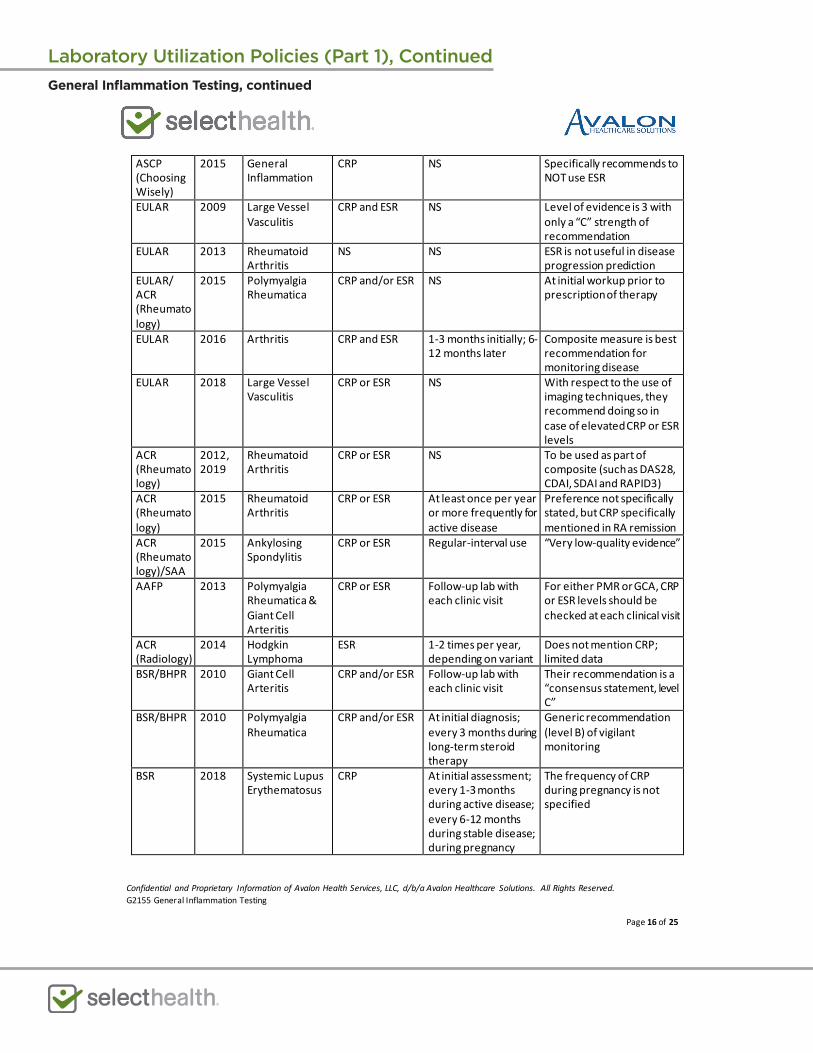
Laboratory Utilization Policies (Part 1), Continued
Confidential and Proprietary Information of Avalon Health Services, LLC, d/b/a Avalon Healthcare Solutions. All Rights Reserved. G2155 General Inflammation Testing
Page 16 of 25
ASCP (Choosing Wisely)
2015 General Inflammation
CRP NS Specifically recommends to NOT use ESR
EULAR 2009 Large Vessel Vasculitis
CRP and ESR NS Level of evidence is 3 with only a “C” strength of recommendation
EULAR 2013 Rheumatoid Arthritis
NS NS ESR is not useful in disease progression prediction
EULAR/ ACR (Rheumatology)
2015 Polymyalgia Rheumatica
CRP and/or ESR NS At initial workup prior to prescription of therapy
EULAR 2016 Arthritis CRP and ESR 1-3 months initially; 6-12 months later
Composite measure is best recommendation for monitoring disease
EULAR 2018 Large Vessel Vasculitis
CRP or ESR NS With respect to the use of imaging techniques, they recommend doing so in case of elevated CRP or ESR levels
ACR (Rheumatology)
2012, 2019
Rheumatoid Arthritis
CRP or ESR NS To be used as part of composite (such as DAS28, CDAI, SDAI and RAPID3)
ACR (Rheumatology)
2015 Rheumatoid Arthritis
CRP or ESR At least once per year or more frequently for active disease
Preference not specifically stated, but CRP specifically mentioned in RA remission
ACR (Rheumatology)/SAA
2015 Ankylosing Spondylitis
CRP or ESR Regular-interval use “Very low-quality evidence”
AAFP 2013 Polymyalgia Rheumatica & Giant Cell Arteritis
CRP or ESR Follow-up lab with each clinic visit
For either PMR or GCA, CRP or ESR levels should be checked at each clinical visit
ACR (Radiology)
2014 Hodgkin Lymphoma
ESR 1-2 times per year, depending on variant
Does not mention CRP; limited data
BSR/BHPR 2010 Giant Cell Arteritis
CRP and/or ESR Follow-up lab with each clinic visit
Their recommendation is a “consensus statement, level C”
BSR/BHPR 2010 Polymyalgia Rheumatica
CRP and/or ESR At initial diagnosis; every 3 months during long-term steroid therapy
Generic recommendation (level B) of vigilant monitoring
BSR 2018 Systemic Lupus Erythematosus
CRP At initial assessment; every 1-3 months during active disease; every 6-12 months during stable disease; during pregnancy
The frequency of CRP during pregnancy is not specified
General Inflammation Testing, continued

Laboratory Utilization Policies (Part 1), Continued
Confidential and Proprietary Information of Avalon Health Services, LLC, d/b/a Avalon Healthcare Solutions. All Rights Reserved. G2155 General Inflammation Testing
Page 17 of 25
BSR 2020 Giant Cell Arteritis
ESR and/or CRP At or near diagnosis of GCA and during follow-up visits
Measures of activity of GCA: laboratory markers of inflammation (CRP for all patients, plus either ESR or plasma viscosity) and full blood count (platelet count may be elevated in GCA)
CRA 2012, 2018
Rheumatoid Arthritis,
CRP or ESR,
At initial assessment prior to treatment; every 1-3 months during active disease
During active disease, CRP/ESR monitoring is part of composite testing, such as DAS28 or SDAI
CRA 2018 Systemic Lupus Erythematosus
CRP and ESR NS Include CRP and ESR monitoring at baseline and in follow-up
NCC-CC 2009 Rheumatoid Arthritis
CRP At initial assessment; monthly until disease is controlled
Recommendation 34: regular use of CRP and DAS28 to inform decision-making Recommendation 35: use of CRP/DAS28 for initial assessment and then monthly until disease is controlled
RACGP 2009 Rheumatoid Arthritis
CRP and/or ESR At initial assessment; to monitor therapy efficacy; CRP testing at least every 4 months
For initial assessment, CRP and/or ESR should be used for diagnosing/assessing RA; however, in Recommendation 29, only CRP testing is specifically mentioned. Recommendation 29 is concerning disease monitoring.
NICE NICE
2015 2020
Irritable Bowel Disorders
CRP and ESR
NS
Only at initial assessment for exclusionary purposes
NICE 2020 Rheumatoid Arthritis
CRP monthly
Measure CRP and disease activity monthly until remission or low disease activity
JTFPP 2014 Acute and Chronic Urticaria
CRP and/or ESR NS Can be used in diagnosis and management of disease
MSIS 2011, 2018
Periprosthetic Joint Infections (PJI)
CRP and/or ESR NS Included as minor criteria to support diagnosis of PJI. Guidelines note these markers may be elevated
General Inflammation Testing, continued

Laboratory Utilization Policies (Part 1), Continued
Confidential and Proprietary Information of Avalon Health Services, LLC, d/b/a Avalon Healthcare Solutions. All Rights Reserved. G2155 General Inflammation Testing
Page 18 of 25
for unrelated reasons after surgery
AAOS 2019 Periprosthetic Joint Infections
CRP and/or ESR NS May provide supporting evidence for pre-operative diagnosis of PJI. Guidelines note these markers may be elevated for unrelated reasons after surgery
NS = Not specified; NA = Not applicable; EIA = enzyme immunoassay; RDT = rapid diagnostic test; DAS = Disease Activity Score; SDAI = Simplified Disease Activity Index
VI. State and Federal Regulations (as applicable) Testing of serum acute phase reactants and ESR is performed in laboratories meeting Clinical Laboratory Improvement Act (CLIA) quality standards. The FDA has approved multiple tests for human CRP, including assays for conventional CRP, high sensitivity CRP (hsCRP), and cardiac CRP (cCRP). On September 22, 2005, the FDA issued guidelines concerning the assessment of CRP (FDA, 2005). A search of the FDA Medical Devices database (FDA, 2018) on April 20, 2021, shows that the FDA has approved ESR systems from multiple companies, including the ESR Control -M Hematology Erythrocyte Sedimentation system (K972172) and the ESR Control -HC Hematology Erythrocyte Sedimentation system (K972170) by R & D Systems, the Seditainer Erythrocyte Sedimentation Rate System (K953994) from Becton Dickinson Vacutainer Systems, the Westergren Dispette for ESR (K831195) by Ulster Scientific, and the Dade ESR Kit (K823368) from American Dade.
VII. Applicable CPT/HCPCS Procedure Codes Code Number Code Description
85651 Sedimentation rate, erythrocyte; non-automated 85652 Sedimentation rate, erythrocyte; automated 86140 C-reactive protein
Current Procedural Terminology© American Medical Association. All Rights reserved.
Procedure codes appearing in Medical Policy documents are included only as a general reference tool for each policy. They may not be all-inclusive.
VIII. Evidence-based Scientific References AAOS. (2019). Diagnosis and Prevention of Periprosthetic Joint Infections Clinical Practice Guideline.
Retrieved from https://www.aaos.org/pjiguideline Anderson, J., Caplan, L., Yazdany, J., Robbins Mark, L., Neogi, T., Michaud, K., . . . Kazi, S. (2012).
Rheumatoid arthritis disease activity measures: American College of Rheumatology recommendations for use in clinical practice. Arthritis Care & Research, 64(5), 640-647. doi:10.1002/acr.21649
Aster, J. C., & Pozdnyakova, O. (2020, 06/11/2018). Epidemiology, pathologic features, and diagnosis of classic Hodgkin lymphoma. UpToDate. Retrieved from https://www.uptodate.com/contents/epidemiology-pathologic-features-and-diagnosis-of-classic-hodgkin-lymphoma
General Inflammation Testing, continued

Laboratory Utilization Policies (Part 1), Continued
Confidential and Proprietary Information of Avalon Health Services, LLC, d/b/a Avalon Healthcare Solutions. All Rights Reserved. G2155 General Inflammation Testing
Page 19 of 25
Barber, C. E. H., Zell, J., Yazdany, J., Davis, A. M., Cappelli, L., Ehrlich-Jones, L., . . . Michaud, K. (2019). 2019 American College of Rheumatology Recommended Patient-Reported Functional Status Assessment Measures in Rheumatoid Arthritis. Arthritis Care Res (Hoboken), 71(12), 1531-1539. doi:10.1002/acr.24040
Berbari, E., Baddour, Larry, Chen, Antonia. (2020). Prosthetic joint infection: Epidemiology, microbiology, clinical manifestations, and diagnosis. Retrieved from https://www.uptodate.com/contents/prosthetic-joint-infection-epidemiology-microbiology-clinical-manifestations-and-diagnosis?search=periprosthetic%20joint%20infection&source=search_result&selectedTitle=1~150&usage_type=default&display_rank=1#H2957426242
Berbari, E., Mabry, T., Tsaras, G., Spangehl, M., Erwin, P. J., Murad, M. H., . . . Osmon, D. (2010). Inflammatory blood laboratory levels as markers of prosthetic joint infection: a systematic review and meta-analysis. J Bone Joint Surg Am, 92(11), 2102-2109. doi:10.2106/jbjs.I.01199
Bernstein, J. A., Lang, D. M., Khan, D. A., Craig, T., Dreyfus, D., Hsieh, F., . . . Wallace, D. (2014). The diagnosis and management of acute and chronic urticaria: 2014 update. J Allergy Clin Immunol, 133(5), 1270-1277. doi:10.1016/j.jaci.2014.02.036
Bingham, J. S., Hassebrock, J. D., Christensen, A. L., Beauchamp, C. P., Clarke, H. D., & Spangehl, M. J. (2019). Screening for Periprosthetic Joint Infections With ESR and CRP: The Ideal Cutoffs. J Arthroplasty. doi:10.1016/j.arth.2019.11.040
Bitik, B., Mercan, R., Tufan, A., Tezcan, E., Küçük, H., İlhan, M., . . . Göker, B. (2015). Differential diagnosis of elevated erythrocyte sedimentation rate and C-reactive protein levels: a rheumatology perspective. European Journal of Rheumatology, 2(4), 131-134. doi:10.5152/eurjrheum.2015.0113
Black, S., Kushner, I., & Samols, D. (2004). C-reactive Protein. J Biol Chem, 279(47), 48487-48490. doi:10.1074/jbc.R400025200
Bykerk, V. P., Akhavan, P., Hazlewood, G. S., Schieir, O., Dooley, A., Haraoui, B., . . . Bombardier, C. (2012). Canadian Rheumatology Association Recommendations for Pharmacological Management of Rheumatoid Arthritis with Traditional and Biologic Disease-modifying Antirheumatic Drugs. The Journal of Rheumatology, 39(8), 1559. Retrieved from http://www.jrheum.org/content/39/8/1559.abstract
Caylor, T. L., & Perkins, A. (2013). Recognition and management of polymyalgia rheumatica and giant cell arteritis. Am Fam Physician, 88(10), 676-684.
Colebatch, A. N., Edwards, C. J., Østergaard, M., van der Heijde, D., Balint, P. V., Agostino, M.-A., . . . Conaghan, P. G. (2013). EULAR recommendations for the use of imaging of the joints in the clinical management of rheumatoid arthritis. Annals of the Rheumatic Diseases, 72(6), 804. Retrieved from http://ard.bmj.com/content/72/6/804.abstract
Combe, B., Landewe, R., Daien, C. I., Hua, C., Aletaha, D., Álvaro-Gracia, J. M., . . . van Vollenhoven, R. (2017). 2016 update of the EULAR recommendations for the management of early arthritis. Annals of the Rheumatic Diseases, 76(6), 948. Retrieved from http://ard.bmj.com/content/76/6/948.abstract
Conditions, N. C. C. f. C. (2009). National Institute for Health and Clinical Excellence: Guidance. In Rheumatoid Arthritis: National Clinical Guideline for Management and Treatment in Adults. London: Royal College of Physicians (UK) Royal College of Physicians of London.
General Inflammation Testing, continued

Laboratory Utilization Policies (Part 1), Continued
Confidential and Proprietary Information of Avalon Health Services, LLC, d/b/a Avalon Healthcare Solutions. All Rights Reserved. G2155 General Inflammation Testing
Page 20 of 25
Crowson, C. S., Rahman, M. U., & Matteson, E. L. (2009). Which Measure of Inflammation to Use? A Comparison of Erythrocyte Sedimentation Rate and C-Reactive Protein Measurements from Randomized Clinical Trials of Golimumab in Rheumatoid Arthritis. The Journal of Rheumatology, 36(8), 1606. Retrieved from http://www.jrheum.org/content/36/8/1606.abstract
Dasgupta, B. (2010). Concise guidance: diagnosis and management of giant cell arteritis. Clin Med (Lond), 10(4), 381-386.
Dasgupta, B., Borg, F. A., Hassan, N., Alexander, L., Barraclough, K., Bourke, B., . . . Samanta, A. (2010). BSR and BHPR guidelines for the management of giant cell arteritis. Rheumatology (Oxford), 49(8), 1594-1597. doi:10.1093/rheumatology/keq039a
Dasgupta, B., Borg, F. A., Hassan, N., Barraclough, K., Bourke, B., Fulcher, J., . . . Samanta, A. (2010). BSR and BHPR guidelines for the management of polymyalgia rheumatica. Rheumatology (Oxford), 49(1), 186-190. doi:10.1093/rheumatology/kep303a
Dejaco, C., Ramiro, S., Duftner, C., Besson, F. L., Bley, T. A., Blockmans, D., . . . Schmidt, W. A. (2018). EULAR recommendations for the use of imaging in large vessel vasculitis in clinical practice. Annals of the Rheumatic Diseases, 77(5), 636. Retrieved from http://ard.bmj.com/content/77/5/636.abstract
Dejaco, C., Singh Yogesh, P., Perel, P., Hutchings, A., Camellino, D., Mackie, S., . . . Dasgupta, B. (2015). 2015 Recommendations for the Management of Polymyalgia Rheumatica: A European League Against Rheumatism/American College of Rheumatology Collaborative Initiative. Arthritis & Rheumatology, 67(10), 2569-2580. doi:10.1002/art.39333
Docken, W. P. (2021, 11/02/2017). Clinical manifestations and diagnosis of polymyalgia rheumatica. UpToDate. Retrieved from https://www.uptodate.com/contents/clinical-manifestations-and-diagnosis-of-polymyalgia-rheumatica
Docken, W. P., & Rosenbaum, J. T. (2019, 12/08/2017). Clinical manifestations of giant cell arteritis. UpToDate. Retrieved from https://www.uptodate.com/contents/clinical-manifestations-of-giant-cell-arteritis
England, B. R., Tiong, B. K., Bergman, M. J., Curtis, J. R., Kazi, S., Mikuls, T. R., . . . Michaud, K. (2019). 2019 Update of the American College of Rheumatology Recommended Rheumatoid Arthritis Disease Activity Measures. Arthritis Care Res (Hoboken), 71(12), 1540-1555. doi:10.1002/acr.24042
Ernst, A. A., Weiss, S. J., Tracy, L. A., & Weiss, N. R. (2010). Usefulness of CRP and ESR in predicting septic joints. South Med J, 103(6), 522-526. doi:10.1097/SMJ.0b013e3181ddd246
FDA. (2005). Review Criteria for Assessment of C-Reactive Protein (CRP), High Sensitivity C-Reactive Protein (hsCRP)k and Cardiac C-Reactive Protein (cCRP) Assays. Rockville, MD: U.S. Department of Health and Human Services Retrieved from https://www.fda.gov/downloads/medicaldevices/deviceregulationandguidance/guidancedocuments/ucm071017.pdf
FDA. (2018). Devices@FDA. Retrieved from https://www.accessdata.fda.gov/scripts/cdrh/devicesatfda/index.cfm
Fransen, J., & van Riel, P. L. (2006). DAS remission cut points. Clin Exp Rheumatol, 24(6 Suppl 43), S-29-32.
Gaitonde, S., Samols, D., & Kushner, I. (2008). C-reactive protein and systemic lupus erythematosus. Arthritis Care & Research, 59(12), 1814-1820. doi:10.1002/art.24316
General Inflammation Testing, continued

Laboratory Utilization Policies (Part 1), Continued
Confidential and Proprietary Information of Avalon Health Services, LLC, d/b/a Avalon Healthcare Solutions. All Rights Reserved. G2155 General Inflammation Testing
Page 21 of 25
Gergianaki, I., & Bertsias, G. (2018). Systemic Lupus Erythematosus in Primary Care: An Update and Practical Messages for the General Practitioner. Frontiers in Medicine, 5, 161. doi:10.3389/fmed.2018.00161
Gordon, C., Amissah-Arthur, M.-B., Gayed, M., Brown, S., Bruce, I. N., D’Cruz, D., . . . Guidelines Working, G. (2018). The British Society for Rheumatology guideline for the management of systemic lupus erythematosus in adults. Rheumatology, 57(1), e1-e45. doi:10.1093/rheumatology/kex286
Ha, C. S., Hodgson, D. C., Advani, R., Dabaja, B. S., Dhakal, S., Flowers, C. R., . . . Constine, L. S. (2014, 2014). Follow-up of Hodgkin lymphoma. American College of Radiology ACR Appropriateness Criteria. Retrieved from https://acsearch.acr.org/docs/69388/Narrative/
Hale, A. J., Ricotta, D. N., & Freed, J. A. (2019). Evaluating the Erythrocyte Sedimentation Rate. Jama, 321(14), 1404-1405. doi:10.1001/jama.2019.1178
Hamann, P. D. H., Shaddick, G., Hyrich, K., Green, A., McHugh, N., & Pauling, J. D. (2019). Gender stratified adjustment of the DAS28-CRP improves inter-score agreement with the DAS28-ESR in rheumatoid arthritis. Rheumatology (Oxford), 58(5), 831-835. doi:10.1093/rheumatology/key374
Henderson, L. A., Canna, S. W., Friedman, K. G., Gorelik, M., Lapidus, S. K., Bassiri, H., . . . Mehta, J. J. (2020). American College of Rheumatology Clinical Guidance for Multisystem Inflammatory Syndrome in Children Associated With SARS-CoV-2 and Hyperinflammation in Pediatric COVID-19: Version 1. Arthritis Rheumatol, 72(11), 1791-1805. doi:10.1002/art.41454
Hensor, E. M. A., Emery, P., Bingham, S. J., & Conaghan, P. G. (2010). Discrepancies in categorizing rheumatoid arthritis patients by DAS-28(ESR) and DAS-28(CRP): can they be reduced? Rheumatology, 49(8), 1521-1529. doi:10.1093/rheumatology/keq117
Horsti, J., Rontu, R., & Collings, A. (2010). A Comparison Between the StaRRsed Auto-Compact Erythrocyte Sedimentation Rate Instrument and the Westergren Method. Journal of Clinical Medicine Research, 2(6), 261-265. doi:10.4021/jocmr476w
Keeling, S. O., Alabdurubalnabi, Z., Avina-Zubieta, A., Barr, S., Bergeron, L., Bernatsky, S., . . . Santesso, N. (2018). Canadian Rheumatology Association Recommendations for the Assessment and Monitoring of Systemic Lupus Erythematosus. J Rheumatol, 45(10), 1426-1439. doi:10.3899/jrheum.171459
Keenan, R. T., Swearingen, C. J., & Yazici, Y. (2008). Erythrocyte sedimentation rate and C-reactive protein levels are poorly correlated with clinical measures of disease activity in rheumatoid arthritis, systemic lupus erythematosus and osteoarthritis patients. Clin Exp Rheumatol, 26(5), 814-819.
Kheir, M. M., Tan, T. L., Shohat, N., Foltz, C., & Parvizi, J. (2018). Routine Diagnostic Tests for Periprosthetic Joint Infection Demonstrate a High False-Negative Rate and Are Influenced by the Infecting Organism. J Bone Joint Surg Am, 100(23), 2057-2065. doi:10.2106/jbjs.17.01429
Kratz, A., Plebani, M., Peng, M., Lee, Y. K., McCafferty, R., & Machin, S. J. (2017). ICSH recommendations for modified and alternate methods measuring the erythrocyte sedimentation rate. International Journal of Laboratory Hematology, 39(5), 448-457. doi:10.1111/ijlh.12693
Kushner, I. (2021, 07/12/2017). Acute phase reactants. UpToDate. Retrieved from https://www.uptodate.com/contents/acute-phase-reactants
Mackie, S. L., Dejaco, C., Appenzeller, S., Camellino, D., Duftner, C., Gonzalez-Chiappe, S., . . . Dasgupta, B. (2020). British Society for Rheumatology guideline on diagnosis and treatment of giant cell arteritis. Rheumatology (Oxford), 59(3), e1-e23. doi:10.1093/rheumatology/kez672
General Inflammation Testing, continued

Laboratory Utilization Policies (Part 1), Continued
Confidential and Proprietary Information of Avalon Health Services, LLC, d/b/a Avalon Healthcare Solutions. All Rights Reserved. G2155 General Inflammation Testing
Page 22 of 25
March, L., Barrett, C., Gale, F., Lassere, M., McQuade, J., Trevena, L., . . . Landgren, F. (2009). Clinical guideline for the diagnosis and management of early rheumatoid arthritis. South Melbourne, Victoria, Australia: The Royal Australian College of General Practitioners Retrieved from https://www.racgp.org.au/download/documents/Guidelines/Musculoskeletal/racgp_ra_guideline.pdf
McCarthy, E. M., MacMullan, P. A., Al-Mudhaffer, S., Madigan, A., Donnelly, S., McCarthy, C. J., . . . McCarthy, G. M. (2014). Plasma Fibrinogen Along with Patient-reported Outcome Measures Enhances Management of Polymyalgia Rheumatica: A Prospective Study. The Journal of Rheumatology, 41(5), 931. Retrieved from http://www.jrheum.org/content/41/5/931.abstract
Mukhtyar, C., Guillevin, L., Cid, M. C., Dasgupta, B., de Groot, K., Gross, W., . . . Luqmani, R. (2009). EULAR recommendations for the management of large vessel vasculitis. Annals of the Rheumatic Diseases, 68(3), 318. Retrieved from http://ard.bmj.com/content/68/3/318.abstract
National Guideline, C. (2014). ACR Appropriateness Criteria Follow-up of Hodgkin lymphoma. Retrieved from https://www.guideline.gov/summaries/summary/48304/acr-appropriateness-criteria--followup-of-hodgkin-lymphoma?q=ESR
National Guideline, C. (2015). Irritable bowel syndrome in adults: diagnosis and management of irritable bowel syndrome in primary care. Retrieved from https://www.guideline.gov/summaries/summary/49049/irritable-bowel-syndrome-in-adults-diagnosis-and-management-of-irritable-bowel-syndrome-in-primary-care?q=ESR
National Guideline, C. (2016). American College of Rheumatology/Spondylitis Association of America/Spondyloarthritis Research and Treatment Network 2015 recommendations for the treatment of ankylosing spondylitis and nonradiographic axial spondyloarthritis. Retrieved from https://www.guideline.gov/summaries/summary/50567/american-college-of-rheumatologyspondylitis-association-of-americaspondyloarthritis-research-and-treatment-network-2015-recommendations-for-the-treatment-of-ankylosing-spondylitis-and-nonradiographic-axial-spondyloarthritis?q=ESR
NCCN. (2020). T-Cell Lymphomas Version 1.2020 - January 6, 2020. Retrieved from https://www.nccn.org/professionals/physician_gls/pdf/t-cell.pdf
NCCN. (2021a). Hodgkin Lymphoma Version 1.2020 - January 30, 2020. Retrieved from https://www.nccn.org/professionals/physician_gls/pdf/hodgkins.pdf
NCCN. (2021b). NCCN Clinical Practice Guidelines in Oncology B-Cell Lymphomas. NCCN Guidelines, 5.2019. Retrieved from https://www.nccn.org/professionals/physician_gls/pdf/b-cell.pdf
NICE. (2017, April 2017). Irritable bowel syndrome in adults: diagnosis and management. Clinical Guideline. Retrieved from https://www.nice.org.uk/guidance/cg61/resources/irritable-bowel-syndrome-in-adults-diagnosis-and-management-pdf-975562917829
NICE. (2020). Rheumatoid arthritis in adults: management. Retrieved from https://www.nice.org.uk/guidance/ng100/chapter/Recommendations
Nielung, L., Christensen, R., Danneskiold-Samsøe, B., Bliddal, H., Holm, C. C., Ellegaard, K., . . . Bartels, E. M. (2015). Validity and Agreement between the 28-Joint Disease Activity Score Based on C-Reactive Protein and Erythrocyte Sedimentation Rate in Patients with Rheumatoid Arthritis. Arthritis, 2015, 401690. doi:10.1155/2015/401690
O'Neill, S. G., Giles, I., Lambrianides, A., Manson, J., D'Cruz, D., Schrieber, L., . . . Rahman, A. (2010). Antibodies to apolipoprotein A-I, high-density lipoprotein, and C-reactive protein are associated with disease activity in patients with systemic lupus erythematosus. Arthritis & Rheumatism, 62(3), 845-854. doi:10.1002/art.27286
General Inflammation Testing, continued

Laboratory Utilization Policies (Part 1), Continued
Confidential and Proprietary Information of Avalon Health Services, LLC, d/b/a Avalon Healthcare Solutions. All Rights Reserved. G2155 General Inflammation Testing
Page 23 of 25
Parvizi, J., Tan, T. L., Goswami, K., Higuera, C., Della Valle, C., Chen, A. F., & Shohat, N. (2018). The 2018 Definition of Periprosthetic Hip and Knee Infection: An Evidence-Based and Validated Criteria. The Journal of Arthroplasty, 33(5), 1309-1314.e1302. doi:https://doi.org/10.1016/j.arth.2018.02.078
Parvizi, J., Zmistowski, B., Berbari, E. F., Bauer, T. W., Springer, B. D., Della Valle, C. J., . . . Zalavras, C. G. (2011). New definition for periprosthetic joint infection: from the Workgroup of the Musculoskeletal Infection Society. Clin Orthop Relat Res, 469(11), 2992-2994. doi:10.1007/s11999-011-2102-9
Pathology, A. S. f. C. (2015, 02/03/2015). Don't order an erythrocyte sedimentation rate (ESR) to look for inflammation in patients with undiagnosed conditions. Twenty Things Physicians and Patients Should Question. Retrieved from http://www.choosingwisely.org/clinician-lists/american-society-clinical-pathology-erythrocyte-sedimentation-rate-for-acute-phase-inflammation/
Perez-Prieto, D., Portillo, M. E., Puig-Verdie, L., Alier, A., Martinez, S., Sorli, L., . . . Monllau, J. C. (2017). C-reactive protein may misdiagnose prosthetic joint infections, particularly chronic and low-grade infections. Int Orthop, 41(7), 1315-1319. doi:10.1007/s00264-017-3430-5
Sherkatolabbasieh, H., Firouzi, M., & Shafizadeh, S. (2020). Evaluation of platelet count, erythrocyte sedimentation rate and C-reactive protein levels in paediatric patients with inflammatory and infectious disease. New Microbes and New Infections, 37, 100725. doi:https://doi.org/10.1016/j.nmni.2020.100725
Singh, J. A., Saag, K. G., Bridges, S. L., Akl, E. A., Bannuru, R. R., Sullivan, M. C., . . . McAlindon, T. (2015). 2015 American College of Rheumatology Guideline for the Treatment of Rheumatoid Arthritis. Arthritis & Rheumatology, 68(1), 1-26. doi:10.1002/art.39480
Suarez-Almazor, M. E., Gonzalez-Lopez, L., Gamez-Nava, J. I., Belseck, E., Kendall, C. J., & Davis, P. (1998). Utilization and predictive value of laboratory tests in patients referred to rheumatologists by primary care physicians. J Rheumatol, 25(10), 1980-1985.
Taylor, P. C., & Maini, R. N. (2020, 05/05/2017). Biologic markers in the diagnosis and assessment of rhematoid arthritis. UpToDate. Retrieved from https://www.uptodate.com/contents/biologic-markers-in-the-diagnosis-and-assessment-of-rheumatoid-arthritis
Venables, P., & Maini, R. N. (2021, 08/23/2018). Diagnosis and differential diagnosis of rheumatoid arthritis. UpToDate. Retrieved from https://www.uptodate.com/contents/diagnosis-and-differential-diagnosis-of-rheumatoid-arthritis
Ward, M. M., Deodhar, A., Akl, E. A., Lui, A., Ermann, J., Gensler, L. S., . . . Caplan, L. (2016). American College of Rheumatology/Spondylitis Association of America/Spondyloarthritis Research and Treatment Network 2015 Recommendations for the Treatment of Ankylosing Spondylitis and Nonradiographic Axial Spondyloarthritis. Arthritis & rheumatology (Hoboken, N.J.), 68(2), 282-298. doi:10.1002/art.39298
Wasserman, A. (2018). Rheumatoid Arthritis: Common Questions About Diagnosis and Management. Am Fam Physician, 97(7), 455-462. Retrieved from https://www.aafp.org/afp/2018/0401/p455.html
Watson, J., Jones, H. E., Banks, J., Whiting, P., Salisbury, C., & Hamilton, W. (2019). Use of multiple inflammatory marker tests in primary care: using Clinical Practice Research Datalink to evaluate accuracy. Br J Gen Pract, 69(684), e462-e469. doi:10.3399/bjgp19X704309
WHO. (2018). World Health Organization Model List of Essential In Vitro Diagnostics First edition (2018). Retrieved from https://www.who.int/medical_devices/diagnostics/WHO_EDL_2018.pdf
General Inflammation Testing, continued

Laboratory Utilization Policies (Part 1), Continued
Confidential and Proprietary Information of Avalon Health Services, LLC, d/b/a Avalon Healthcare Solutions. All Rights Reserved. G2155 General Inflammation Testing
Page 24 of 25
WHO. (2019). Second WHO Model List of Essential In Vitro Diagnostics. Retrieved from https://www.who.int/docs/default-source/nutritionlibrary/complementary-feeding/second-who-model-list-v8-2019.pdf?sfvrsn=6fe86adf_1
Wu, A. H., Lewandrowski, K., Gronowski, A. M., Grenache, D. G., Sokoll, L. J., & Magnani, B. (2010). Antiquated tests within the clinical pathology laboratory. Am J Manag Care, 16(9), e220-227.
General Inflammation Testing, continued

Laboratory Utilization Policies (Part 1), Continued
Confidential and Proprietary Information of Avalon Health Services, LLC, d/b/a Avalon Healthcare Solutions. All Rights Reserved. G2155 General Inflammation Testing
Page 25 of 25
IX. Revision History Revision Date Summary of Changes
Disclaimer This document is for informational purposes only and should not be relied on in the diagnosis and care of individual patients. Medical and Coding/Reimbursement policies do not constitute medical advice, plan preauthorization, certification, an explanation of benefits, or a contract. Members should consult with appropriate healthcare providers to obtain needed medical advice, care, and treatment. Benefits and eligibility are determined before medical guidelines and payment guidelines are applied. Benefits are determined by the member’s individual benefit plan that is in effect at the time services are rendered. The codes for treatments and procedures applicable to this policy are included for informational purposes. Inclusion or exclusion of a procedure, diagnosis or device code(s) does not constitute or imply member coverage or provider reimbursement policy. Please refer to the member's contract benefits in effect at the time of service to determine coverage or non-coverage of these services as it applies to an individual member. SelectHealth® makes no representations and accepts no liability with respect to the content of any external information cited or relied upon in this policy. SelectHealth updates its Coverage Policies regularly and reserves the right to amend these policies without notice to healthcare providers or SelectHealth members. Members may contact Customer Service at the phone number listed on their member identification card to discuss their benefits more specifically. Providers with questions about this Coverage Policy may call SelectHealth Provider Relations at (801) 442-3692. No part of this publication may be reproduced, stored in a retrieval system or transmitted, in any form or by any means, electronic, mechanical, photocopying, or otherwise, without permission from SelectHealth. ”Intermountain Healthcare” and its accompanying logo, the marks of “SelectHealth” and its accompanying marks are protected and registered trademarks of the provider of this Service and or Intermountain Health Care, Inc., IHC Health Services, Inc., and SelectHealth, Inc. Also, the content of this Service is proprietary and is protected by copyright. You may access the copyrighted content of this Service only for purposes set forth in these Conditions of Use. © CPT Only – American Medical Association
General Inflammation Testing, continued

Laboratory Utilization Policies (Part 1), Continued
Confidential and Proprietary Information of Avalon Health Services, LLC, d/b/a Avalon Healthcare Solutions. All Rights Reserved. G2044 Helicobacter Pylori Testing
Page 1 of 17
Helicobacter pylori Testing Policy #: AHS – G2044 Prior Policy Name & Number
(as applicable): Implementation Date: 9/15/21 Date of Last Revision:
I. Policy Description
Helicobacter pylori (H. pylori) is a spiral-shaped, gram negative bacteria that has thrived to live in acidic environments and grows in close association with the stomach lining; therefore, H. pylori infection causes chronic inflammation (infection) in the stomach and is associated with conditions such as peptic ulcer disease, chronic gastritis, gastric adenocarcinoma, and gastric mucosa associated lymphoid tissue (MALT) lymphoma (Lamont, 2020).
II. Related Policies
Policy Number Policy Title Not applicable
III. Indications and/or Limitations of Coverage
Disclaimer:
1. Policies are subject to change without notice. 2. Policies outline coverage determinations for SelectHealth Commercial, SelectHealth
Advantage® (Medicare/CMS) and SelectHealth Community Care® (Medicaid/CHIP) plans. Refer to the “Policy” section for more information.
Application of coverage criteria depends on an individual’s benefit coverage at the time of the request.
For SelectHealth Advantage (Medicare/CMS), coverage is determined by the Centers for Medicare and Medicaid Services (CMS). If a coverage determination has not been adopted by CMS and InterQual criteria are not available, the SelectHealth Commercial policy applies. For the most up-to-date Medicare policies and coverage, please visit their search website or the manual website,
For SelectHealth Community Care (Medicaid), coverage is determined by the State of Utah Medicaid program. If Utah State Medicaid has no published coverage position and InterQual criteria are not available, the SelectHealth Commercial criteria will apply. For the most up-to-date Medicaid policies and coverage, please visit their website or the Utah Medicaid Code Look-Up tool.

Laboratory Utilization Policies (Part 1), Continued
Confidential and Proprietary Information of Avalon Health Services, LLC, d/b/a Avalon Healthcare Solutions. All Rights Reserved. G2044 Helicobacter Pylori Testing
Page 2 of 17
1. Urea Breath testing OR stool antigen testing for H. pylori infection MEETS COVERAGE CRITERIA for adult patients (>18y) in the following situations:
a. In the evaluation of a patient with suspected H. pylori infection and the following situations:
i. dyspeptic symptoms, or
ii. active peptic ulcer disease (PUD), or
iii. past PUD without H. pylori history, or
iv. low-grade gastric mucosa-associated lymphoid tissue (MALT) lymphoma, or
v. a history of endoscopic resection of early gastric cancer (EGC), or
vi. in patients with gastric intestinal metaplasia (GIM), or
vii. patients with uninvestigated dyspepsia who are under the age of 60 years and without alarm features, or
viii. Patients initiating chronic treatment with a non-steroidal anti-inflammatory drug (NSAID), or
ix. Patients with unexplained iron deficiency anemia, or
x. In the evaluation of a patient with chronic immune thrombocytopenic purpura (ITP) and suspected H. pylori infection.
xi. In patients with family history of gastric cancer
xii. In patients who are first-generation immigrants from high prevalence areas
b. Re-evaluation to measure success of eradication of H. pylori infection, at least 4 weeks post-treatment.
i. Any patient with an H. pylori-associated ulcer.
ii. As part of the follow-up of patients with persistent symptoms of dyspepsia following appropriate antibiotic treatment for H. pylori.
iii. In patients with Gastric MALT Lymphoma.
iv. In individuals who have undergone resection of early gastric cancer
Helicobacter Pylori Testing, continued
Laboratory Utilization Policies (Part 1), Continued
Confidential and Proprietary Information of Avalon Health Services, LLC, d/b/a Avalon Healthcare Solutions. All Rights Reserved. G2044 Helicobacter Pylori Testing
Page 3 of 17
2. Urea Breath testing OR stool antigen testing for H. pylori infection MEETS COVERAGE CRITERIA for pediatric patients (<18y) in the following situations:
a. In the evaluation of a patient with chronic immune thrombocytopenic purpura (ITP) and suspected H. pylori infection.
b. Re-evaluation to measure success of eradication of H. pylori infection, at least 4 weeks post-treatment.
3. Biopsy-based endoscopic histology test and either Rapid Urease Test or culture with susceptibility testing MEETS COVERAGE CRITERIA in pediatric patients (<18y) for the diagnosis of H. pylori infection in following situations:
a. Children with gastric or duodenal ulcers
b. Children with refractory iron deficiency anemia (IDA) in which other causes have been ruled out
4. Biopsy-based endoscopic histology test and Rapid Urease Test or culture with susceptibility testing MEETS COVERAGE CRITERIA in adult patients (>18 y) undergoing endoscopic examination or in those with alarm symptoms for the diagnosis of H. pylori infection
5. Urea Breath testing OR stool antigen testing for H. pylori infection DOES NOT MEET COVERAGE CRITERIA for asymptomatic pediatric (<18y) and asymptomatic adult (>18y) patients in all other situations and adult patients with typical symptoms of gastroesophageal reflux disease (GERD) who do not have a history of peptic ulcer disease (PUD)
6. Serologic testing for H. pylori infection DOES NOT MEET COVERAGE CRITERIA in adult and pediatric patients as it does not distinguish between currently active infection with past exposure and an infection that has been cured.
7. Biopsy-based endoscopic histology test and Rapid Urease Test or culture with susceptibility testing DOES NOT MEET COVERAGE CRITERIA in pediatric patients (<18y) for the diagnosis of H. pylori infection in following situations:
a. Children with functional abdominal pain
b. As part of initial investigation in children with iron deficiency anemia
c. When investigating causes of short stature
8. Testing with the Urea Breath test and/or stool antigen and/or biopsy-based testing DOES NOT MEET COVERAGE CRITERIA in patients with recent use of antibiotics, proton pump inhibitors (PPIs) or bismuth.
9. Concurrent testing with the Urea Breath test and/or stool antigen testing and/or biopsy-based testing DOES NOT MEET COVERAGE CRITERIA as simultaneous use of both methods does not improve clinical understanding.
Helicobacter Pylori Testing, continued

Laboratory Utilization Policies (Part 1), Continued
Confidential and Proprietary Information of Avalon Health Services, LLC, d/b/a Avalon Healthcare Solutions. All Rights Reserved. G2044 Helicobacter Pylori Testing
Page 4 of 17
10. The use of nucleic acid testing for H. pylori, including polymerase chain reaction (PCR), 16S rRNA, 23S rRNA, and next-generation sequencing (NGS) of H. pylori, DOES NOT MEET COVERAGE CRITERIA as it is not practical for routine diagnosis.
IV. Scientific Background
Infection with H. pylori is common, with conservative estimates at 50% of the world’s population affected. Prevalence in the United States is significant, estimated to be 30 – 40% in the general population (Siao & Somsouk, 2014). H. pylori is associated with many conditions, such as peptic ulcer disease, chronic gastritis, and gastric mucosa associated lymphoid tissue (MALT) lymphoma. Other conditions such as dyspepsia have been attributed to H. pylori as well (Lamont, 2020). Common symptoms of these conditions include gastritis, dyspepsia, heartburn, and stomach pain (Jensen, 2019; Longstreth, 2017).
Identification of H. pylori infection is accomplished with one or more of the several tests available. The choice of test is guided by the reason for the test, cost and availability of the test, the patient’s age and clinical presentation, prevalence in a population, and the patient’s use of certain medications. Testing for H. pylori infection is done for two main reasons; to detect active infection that will be treated and to confirm eradication of the infection post-treatment. Invasive and non-invasive approaches have been used. Endoscopy and collection of biopsy specimens for evaluation of H. pylori infection and early gastric cancer detection typically is done in older individuals and those with “alarm” symptoms, including bleeding, unexplained anemia, unexplained weight loss, progressing dysphagia, recurrent vomiting, a family history of gastrointestinal cancer, or a personal history of esophagogastric malignancy. Tissue samples can be tested for H. pylori via methods such as a rapid urease test, culture, or staining. Molecular methods include PCR and Next-generation sequencing, and serological methods include ELISA, immunoassays, and dried blood spots. Other non-invasive methods include urea breath test and stool antigen test. Testing for eradication of infection may be performed with the same tests used for diagnosis (Lamont, 2020).
Analytical Validity
Non-invasive options for detection of active H. pylori infection include urea breath tests and stool antigen testing. The stool antigen test is an immunoassay that detects the presence of H. pylori in a stool sample. The test is reported to have greater than 90% sensitivity and specificity for detection of active H. pylori infection, and its use has been FDA cleared for all ages. This test may be used for initial diagnostic purposes and for post-treatment testing. Urea breath tests, which take advantage of the bacteria’s urease activity, may also be used to detect active H. pylori infection. The patient ingests a solution containing either 13C or 14C labeled urea, after a set amount of time, the patient’s breath is collected and analyzed for the presence of 13C or 14C labeled CO2. If H. pylori is present, it will have metabolized the labeled urea and labeled CO2 will be detected, thus indicating infection with H. pylori. This test takes approximately 15-20 minutes (Lamont, 2020).
ELISA-based serological tests are also available for detection of H. pylori. However, serological tests often need validation at the local level, which may not be practical in routine practice. Furthermore, serological tests do not distinguish between past and present infections. Serological tests also have a very low positive predictive value in populations with low or average prevalence, as the antibodies will
Helicobacter Pylori Testing, continued

Laboratory Utilization Policies (Part 1), Continued
Confidential and Proprietary Information of Avalon Health Services, LLC, d/b/a Avalon Healthcare Solutions. All Rights Reserved. G2044 Helicobacter Pylori Testing
Page 5 of 17
be detected even after an infection has been treated or naturally resolved. In these low-prevalence areas, a positive serological test is more likely to be a false positive (Lamont, 2020).
Other tests such as PCR-based tests are infrequently used. The PCR test, despite its high accuracy, is often too expensive for routine use. In fact, nested PCR tests have approached 100% sensitivity and 100% specificity for detection of H. pylori (Singh et al., 2008), but the test may not be widely available and may be of limited use due to high cost (Lamont, 2020; Patel, Pratap, Jain, Gulati, & Nath, 2014). PCR tests have been used for diagnostic purposes as well as identifying genetic variants of the bacteria and pathogenic genes present in a patient. A variety of body fluids, such as stool and saliva, have been used in PCR tests for this bacterial species (Patel et al., 2014).
Some medications are known to inhibit the growth or urease activity of H. pylori and can cause a false negative H. pylori test result. Proton pump inhibitors, antibiotics, and bismuth-containing medications may decrease sensitivity of tests, thereby increasing rates of a false negative. Eradication testing is often done weeks after treatment is completed (Lamont, 2020).
Dechant et al. (2020) evaluated the accuracy of various rapid urease tests (RUTs) and compared it with histopathology results. No differences were detected in the sensitivity or specificity of the various RUTs and RUTs had comparable results to histology; however, in patients treated with proton pump inhibitors and antibiotics. RUTs seemed to be more sensitive compared to histology. Pohl, Keller, Bordier, and Wagner (2019) discuss the drawbacks of RUTs, including false negative test results if the bacterial load is less than 104 in the gastric biopsy and false positive test results with some urease positive bacteria, affecting the sensitivity and specificity of RUTs. Commercially available RUTs, such as HpFast, CLOTest, and HpOne, have reported specificities ranging from 95% to 100%, but their sensitivity is moderate (85% to 95%) (Pohl et al., 2019).
Clinical Validity and Utility
The stool antigen test has been shown to have strong accuracy. A meta-analysis by Gisbert et al. focusing on 2499 patients of 22 studies found the diagnostic test to have a sensitivity of 0.94 and a specificity of 0.97. The monoclonal version of the test was shown to be more sensitive than the polyclonal one (0.95 vs 0.83). The authors also evaluated the diagnostic test after eradication of the bacteria in 957 patients of 12 studies. The authors evaluated the antigen test at 0.93 sensitivity and 0.96 specificity post-eradication (Gisbert, de la Morena, & Abraira, 2006).
A new automated LIAISON® Meridian H. pylori SA assay, a chemiluminescent immunoassay that uses novel monoclonal antibodies for capture and detection of the H. pylori stool antigen, was evaluated for its clinical performance. Opekun et al. (2020) studied the utility of this assay on 277 patients who tested positive for H. pylori infection from an endoscopy. Comparing histology, culture, and rapid urease test results, the assay delivered a sensitivity of 95.5% and specificity of 97.6%. The authors conclude that LIAISON® “brings reliable noninvasive testing for H. pylori to the laboratory that is in very good agreement with the current, more invasive biopsy-based methods such as histology, culture, or rapid urease test” (Opekun et al., 2020).
The rapid in-office, monoclonal test is widely used and provides significant benefit in terms of availability and speed. However, a study using the test as a reference to compare against a new test
Helicobacter Pylori Testing, continued

Laboratory Utilization Policies (Part 1), Continued
Confidential and Proprietary Information of Avalon Health Services, LLC, d/b/a Avalon Healthcare Solutions. All Rights Reserved. G2044 Helicobacter Pylori Testing
Page 6 of 17
found the in-office test to only have a 0.50 sensitivity and 0.96 specificity out of 162 patients (Korkmaz, Findik, Ugurluoglu, & Terzi, 2015).
The UBT has also been well-validated. A meta-analysis by Ferwana et al. including 3999 patients of 23 studies found the diagnostic test to have a pooled sensitivity of 0.96 and a pooled specificity of 0.93. The authors noted that their populations had significant heterogeneity, but concluded that the UBT had high diagnostic accuracy for detecting an H. pylori infection (Ferwana et al., 2015). This test is often considered the gold standard for diagnosing an H. pylori infection (Patel et al., 2014).
Serological tests to assess infection have also been used. A meta-analysis by Loy et al. focused on commercial serological kits assessing H. pylori. Loy et al. found these kits to have a pooled sensitivity of 0.85 and specificity of 0.79. The authors concluded that there was no major difference in accuracy between any of the kits tested (Loy, Irwig, Katelaris, & Talley, 1996).
As costs of sequencing decreases, use of Next Generation Sequencing (NGS) to detect H. pylori infection and its antibiotic resistance has increased. In a study by Nezami et al., 133 H. pylori positive specimens from histological evaluation were analyzed by NGS to detect mutations in gyrA, 23S rRNA, and 16s rRNA genes. NGS detected H. pylori in 126/133 cases (95% sensitivity). NGS also detected multiple mutations associated with resistance in 92 cases (73%), one mutation in 63 cases (50%), and mutations in several genes in 29 cases (23%). In the 58 cases where treatment history was available, therapy failure was observed in cases where the number of mutated genes was high. Therapy failed in 11/16 cases with multiple gene mutations and 5/27 cases with one gene mutation (Nezami, Jani, Alouani, Rhoads, & Sadri, 2019).
Yang et al. performed a meta-analysis investigating the association between H. pylori and colorectal cancer. 27 studies encompassing 14357 cases were included. The authors found an increased rate of colorectal cancer with H. pylori infection (odds ratio [OR] = 1.27). The authors also identified odds ratios for certain subgroups, such as Western countries (OR = 1.34), serological testing (OR = 1.20), multiple methods of testing (OR = 2.63), and cross-sectional studies (OR = 1.92) (Yang, Xu, & Zhu, 2019).
Wang et al. performed a meta-analysis assessing the association between H. pylori and osteoporosis. 21 studies totaling 9655 patients were analyzed. The authors found that H. pylori infection was associated with an increased risk of osteoporosis with an odds ratio of 1.39. However, the decrease of bone mineral density in H. pylori positive patients was not found to be significant compared to H. pylori negative patients (Wang et al., 2019).
Zhou et al. investigated the association between H. pylori infection and non-alcoholic fatty liver disease (NAFLD). 15 studies including 97228 patients were evaluated. The authors identified an increased risk of NAFLD in H. pylori positive patients compared to H. pylori negative patients by an odds ratio of 1.19. Similar results were found despite differing subgroups, such as geographical locations. Testing method did not significantly change the results, and there was no significant difference when using multiple detection methods (Zhou et al., 2019).
Helicobacter Pylori Testing, continued

Laboratory Utilization Policies (Part 1), Continued
Confidential and Proprietary Information of Avalon Health Services, LLC, d/b/a Avalon Healthcare Solutions. All Rights Reserved. G2044 Helicobacter Pylori Testing
Page 7 of 17
V. Guidelines and Recommendations
American Gastroenterological Association (AGA) (Allen, Katzka, Robert, & Leontiadis, 2015; Gupta et al., 2020; Ko et al., 2020; Talley, 2005)
The AGA recommends that “patients 55 years or younger without alarm features should receive H. pylori test and treat followed by acid suppression if symptoms remain” and note that “H. pylori testing is optimally performed by a 13C-urea breath test or stool antigen test.” Alarm features include symptoms such as recurrent vomiting and weight loss. Additionally, the AGA indicates that “although the yield of endoscopy is low, it is recommended for patients older than 55 years of age and for younger patients…presenting with new-onset dyspepsia.” They reason that endoscopy with biopsy is the preferred test for this age group because upper gastrointestinal malignancy becomes more common after age 55 years (Talley, 2005).
In 2015 the AGA published a Technical review on Upper Gastrointestinal biopsy to evaluate dyspepsia in the absence of visible mucosal lesions and found that:
• In the defined population, biopsy of normal-appearing gastric mucosa can detect HP [H. pylori] infection that would be missed on the exam without biopsies. The quality of evidence is very low, and there are noninvasive methods to detect HP infection.
• “Detection of HP infection with tissue biopsy and its eradication in patients with dyspepsia is associated with symptom improvement and reduction of risk for HP-related comorbidities, including gastric cancer compared with no biopsy (or no eradication). The quality of evidence is moderate. The effect on symptom resolution is not universal and it does not appear to improve well-being. Quality of evidence for this statement is low” (Allen et al., 2015).
The AGA also released guidelines focusing on gastric intestinal metaplasia. In it, they recommend testing for H. pylori (followed by eradication) over no testing and eradication (Gupta et al., 2020).
The AGA released guidelines on gastrointestinal evaluation of iron deficiency anemia. AGA recommends that patients with iron deficiency anemia, without other identifiable etiology after bidirectional endoscopy, should undergo noninvasive testing for H. pylori over no testing at all to reduce the incidence of gastric cancer (Ko et al., 2020).
American College of Gastroenterology/Canadian Association of Gastroenterology (Chey, Leontiadis, Howden, & Moss, 2017; Moayyedi et al., 2017)
The ACG and CAG have released guidelines on testing for H. pylori:
• All patients with active peptic ulcer disease (PUD), a past history of PUD (unless previous cure of H. pylori infection has been documented), low-grade gastric mucosa-associated lymphoid tissue (MALT) lymphoma, or a history of endoscopic resection of early gastric cancer (EGC) should be tested for H. pylori infection. Those who test positive should be offered treatment for the infection.
• In patients with uninvestigated dyspepsia who are under the age of 60 years and without alarm features, non-endoscopic testing for H. pylori infection is a consideration. Those who test positive should be offered eradication therapy.
Helicobacter Pylori Testing, continued

Laboratory Utilization Policies (Part 1), Continued
Confidential and Proprietary Information of Avalon Health Services, LLC, d/b/a Avalon Healthcare Solutions. All Rights Reserved. G2044 Helicobacter Pylori Testing
Page 8 of 17
• When upper endoscopy is undertaken in patients with dyspepsia, gastric biopsies should be taken to evaluate for H. pylori infection. Infected patients should be offered eradication therapy.
• Patients with typical symptoms of gastroesophageal reflux disease (GERD) without history of PUD need not be tested for H. pylori infection. For those who are found to be infected, treatment should be offered, acknowledging that effects on GERD symptoms are unpredictable.
• In patients taking long-term low-dose aspirin, testing for H. pylori infection could be considered.
• Patients initiating chronic treatment with a non-steroidal anti-inflammatory drug (NSAID) should be tested for H. pylori infection. Those who test positive should be offered eradication therapy.
• Patients with unexplained iron deficiency (ID) anemia despite an appropriate evaluation or idiopathic thrombocytopenic purpura should be tested for H. pylori infection.
• There is insufficient evidence to support routine testing and treating of H. pylori in asymptomatic individuals with a family history of “gastric cancer or patients with lymphocytic gastritis, hyperplastic gastric polyps and hyperemesis gravidarum”.
• The ACG recommends the breath test and fecal stool antigen test as eradication tests, supported by moderate evidence (Chey et al., 2017).
Another set of joint guidelines from the ACG and Canadian Association of Gastroenterology (CAG) noted that dyspepsia patients under 60 should be tested for H. pylori (Moayyedi et al., 2017).
National Institute for Health and Care Excellence (NICE, 2015, 2019) (reaffirmed 2020)
NICE recommends testing for H. pylori with a carbon-13 urea breath test or a stool antigen test. A re-test should be with a breath test. Office-based serological tests are not recommended. NICE recommends a “2-week washout period after proton pump inhibitor (PPI) use before testing for Helicobacter pylori.” NICE recommends that individuals with positive H. pylori tests be offered therapy to eradicate the bacteria; however, they note that re-testing to confirm eradication should not be routinely offered. NICE limits the recommendation for post-treatment testing to “people with peptic ulcer (gastric or duodenal)…6 to 8 weeks after beginning treatment, depending on the size of the lesion (NICE, 2019).
NICE released further guidelines in 2015 reaffirming the carbon-13 urea breath test and the stool antigen test to test for H. pylori. A locally validated lab-based serology test may also be used to assess H. pylori. NICE reaffirms the 2 week washout period before testing for H. pylori if the patient is on PPIs as well as the 4 week washout period if the patient is on antibiotics (NICE, 2015).
This guideline was reaffirmed in 2020 (NICE, 2020).
American College of Cardiology (Bhatt et al., 2008)
The American College of Cardiology recommends testing for and eradicating H. pylori in patients with a history of ulcer disease before starting chronic antiplatelet therapy (Bhatt et al., 2008).
Helicobacter Pylori Testing, continued
Laboratory Utilization Policies (Part 1), Continued
Confidential and Proprietary Information of Avalon Health Services, LLC, d/b/a Avalon Healthcare Solutions. All Rights Reserved. G2044 Helicobacter Pylori Testing
Page 9 of 17
The European Society for Pediatric Gastroenterology Hepatology and Nutrition (ESPGHAN) and The North American Society for Pediatric Gastroenterology, Hepatology and Nutrition (NASPGHAN) (L. Jones et al., 2017)
The ESPGHAN and NASPGHAN have issued updated guidelines for management of H. pylori in children and adolescents. They have proposed recommendations for diagnosis and management of H. pylori infection in pediatric patients. They have defined pediatric patients as children and adolescents below 18 years of age. The following recommendations were stated: The guidelines recommend biopsies for rapid urease test and other cultures should only be taken if treatment is likely to be offered in the case of a confirmed infection. Treatment may be considered if H. pylori is an incidental finding at endoscopy. The guidelines recommend against a “test and treat” strategy for H. pylori infection in children. The panelists explained that performing a noninvasive test to detect infection and treat is not needed because H. pylori infection usually does not cause any symptoms in the absence of peptic ulcer disease (PUD). The guidelines recommend that “testing for H. pylori be performed in children with gastric or duodenal PUD.” The guidelines recommend against diagnostic testing for H. pylori infection in children with functional abdominal pain, iron deficiency anemia, and when investigating causes of short stature. Serology-based testing was also not recommended. PPIs should be stopped 2 weeks before H. pylori testing, and antibiotics should be stopped 4 weeks before H. pylori testing. Diagnosis should be based on either: “positive culture or H. pylori gastritis on histopathology with at least 1 other positive biopsy-based test”. The non-invasive diagnostic testing was indicated in children when investigating causes of chronic immune thrombocytopenic purpura or for the assessment of anti-H. pylori therapy at least after 4 weeks of therapy (L. Jones et al., 2017).
Maastricht V/Florence Consensus Report (2017)
This report was published on behalf of the European Helicobacter and Microbiota Study Group and Consensus panel. The panel reports that UBT is “the most investigated and best recommended non-invasive test in the context of a ‘test-and-treat’ strategy”. The panel also notes that monoclonal tests can be used and that serological tests can be used only after validation. However, rapid “office” serology tests are not recommended and “should be avoided”. The guidelines recommend the rapid urease test (RUT) as a first line diagnostic test if there is an indication for endoscopy and no contraindication for biopsy. The guideline state that H. pylori is linked to “unexplained iron deficiency anaemia (IDA), idiopathic thrombocytopenic purpura (ITP), and vitamin B12 deficiency”, and in these disorders, an H. pylori infection should be “sought and eradicated”. The guidelines state that PPIs should be stopped 2
Helicobacter Pylori Testing, continued

Laboratory Utilization Policies (Part 1), Continued
Confidential and Proprietary Information of Avalon Health Services, LLC, d/b/a Avalon Healthcare Solutions. All Rights Reserved. G2044 Helicobacter Pylori Testing
Page 10 of 17
weeks and antibiotics and other bismuth compounds should be stopped 4 weeks before testing for H. pylori. In cases of chronic (active) gastritis in which H. pylori is not detected by histochemistry, immunohistochemical testing of H. pylori can be used as an ancillary test. If histology is normal, no immunohistochemical staining should be performed. It is recommended to perform clarithromycin susceptibility testing when a standard clarithromycin-based treatment is considered as the first-line therapy, except in populations or regions with well documented low clarithromycin resistance (<15%). Pepsinogen (Pg) serology is considered the most useful non-invasive test to explore gastric mucosa status (non-atrophic vs atrophic). The PgI/PgII ratio can never be assumed as a biomarker of gastric neoplasia. UBT is the best option for confirmation of H. pylori eradication and monoclonal SAT is an alternative. It should be performed at least 4 weeks after completion of therapy (Malfertheiner et al., 2017). The Maastricht IV from 2012 also addressed testing for the cagA and vacA variants, stating that no specific genetic or virulence markers can be recommended at this time (Malfertheiner et al., 2012).
American Society for Clinical Pathology (ASCP, 2016)
The ASCP recommends against using the serological tests for H. pylori and recommends the stool antigen and breath tests instead. The ASCP states that serological evaluation is no longer clinically useful and the stool and breath tests have superior statistical power (ASCP, 2016).
American Society of Hematology (ASH) (Neunert et al., 2019)
ASH published an update to the immune thrombocytopenic purpura guidelines in 2019. In it, they “suggest” that “Screening for H pylori be considered for patients with ITP in whom eradication therapy would be used if testing is positive”. However, ASH still recommends against “routine testing for H pylori in children with chronic ITP” (Neunert et al., 2019).
Houston Consensus Conference (El-Serag et al., 2018)
This conference included 11 experts on “management of adult and pediatric patients with H. pylori, from different geographic regions of the United States” and was convened to “discuss key factors in diagnosis of H. pylori infection, including identification of appropriate patients for testing, effects of antibiotic susceptibility on testing and treatment, appropriate methods for confirmation of infection and eradication, and relevant health system considerations”. Two cohorts of approval were present: one of the 11 experts, and another consisting of a selected group of United States-based gastroenterologists. These recommendations were intended to provide practical advice for US practitioners, and also guidelines to be adopted by US health care systems. Recommendations approved by both groups are listed below:
• “Statement 1: We recommend that all patients with active H pylori infection be treated (100%
agree/strongly agree, Grade 1A).
Helicobacter Pylori Testing, continued

Laboratory Utilization Policies (Part 1), Continued
Confidential and Proprietary Information of Avalon Health Services, LLC, d/b/a Avalon Healthcare Solutions. All Rights Reserved. G2044 Helicobacter Pylori Testing
Page 11 of 17
• Statement 2: All patients with current or past gastric or duodenal ulcers should be tested for H pylori infection (100% agree/strongly agree; Grade 1A).
• Statement 3: We recommend that all patients with uninvestigated dyspepsia be tested for H pylori infection (100% agree/strongly agree, Grade 1A).
• Statement 4: We recommend routine testing for H pylori infection in patients with reflux symptoms only if they are at high risk for H pylori-related disease (91% agree/strongly agree, Grade 1C).
• Statement 5: We recommend that patients with gastric mucosa-associated lymphoid tissue (MALT) lymphoma be tested for H pylori infection (100% agree/strongly agree, Grade 1B).
• Statement 6: We recommend that individuals with family history of gastric cancer be tested for H pylori infection (100% agree/strongly agree, Grade 1B).
• Statement 7: We recommend that patients who are first-generation immigrants from high prevalence areas be tested for H pylori infection (82% agree/strongly agree, Grade 1B).
• Statement 8: We suggest that patients of Latino and African American racial or ethnic groups may be considered for H pylori testing due to their high risk of infection (91% agree/strongly agree, Grade 2C).”
• Statement 17: We recommend that validated diagnostic testing of stool or gastric mucosal biopsy by culture and susceptibility, or molecular analysis be universally available (100% agree/strongly agree, Grade 1).
• Statement 18: We suggest that antibiotics that may be routinely evaluated for susceptibility include amoxicillin, clarithromycin, levofloxacin, metronidazole, and tetracycline (100% agree/strongly agree, Grade 2C).
• Statement 20: We recommend the use of tests for active H pylori infection (i.e., UBT, HpSAg testing) for the initial diagnosis (100% agree/strongly agree, Grade 1A).
• Statement 22: We recommend that serology not be utilized for detection of active H pylori infection (100% agree/strongly agree, Grade 1A).
• Statement 23: We recommend that bismuth and antibiotics be stopped at least 4 weeks before H pylori testing with tests for active infection (i.e., UBT, and HpSAg testing and histology; 100% agree/strongly agree, Grade 1C).
• Statement 27: We recommend that all patients receiving treatment for H pylori receive posttreatment confirmation of eradication. We recommend that only tests that evaluate for active infection, such as UBT, HpSAg test, or histology (if endoscopy is required for other reasons), are utilized for this purpose (100% agree/strongly agree, Grade 1A).
• Statement 28: Once appropriate testing has confirmed eradication, we recommend against further H pylori testing, (100% agree/strongly agree, Grade 1C).”
The following recommendations reached a consensus by the expert panel, but not the external group:
• “Statement 9: We recommend that patients with idiopathic thrombocytopenia be tested for H pylori infection (experts vs survey: 100% vs 68% agree/strongly agree, Expert Grade 1B).
• Statement 10: We suggest that patients receiving long-term PPIs (>1 month) be tested for H pylori infection (experts vs survey: 82% vs 68% agree/strongly agree, Expert Grade 2C).
• Statement 11: We recommend that family members residing in the same household of patients with proven active H pylori infections undergo H pylori testing (experts vs survey: 91% vs 78% agree/strongly agree, Expert Grade 1B).
Helicobacter Pylori Testing, continued

Laboratory Utilization Policies (Part 1), Continued
Confidential and Proprietary Information of Avalon Health Services, LLC, d/b/a Avalon Healthcare Solutions. All Rights Reserved. G2044 Helicobacter Pylori Testing
Page 12 of 17
• Statement 12: We recommend that individuals with a family history of peptic ulcer disease be tested for H pylori infection (experts vs survey: 91% vs (73% agree/strongly agree, Expert Grade 1B)” (El-Serag et al., 2018).
VI. State and Federal Regulations (as applicable)
A search for “pylori” yielded 57 results on January 07, 2021, which encompasses various immunoassays and breath tests (FDA, 2021).
On Feb 22, 2012, the FDA approved the BreathTek UBT for H. pylori Kit created by Otsuka America Pharmaceutical, Inc. TheBreathTek UBT for H. pylori Kit (BreathTek UBT Kit) is intended for use in the qualitative detection of urease associated with H. pylori in the human stomach and is indicated as an aid in the initial diagnosis and post-treatment monitoring of H. pylori infection in adults, and pediatric patients 3 to 17 years old. The test may be used for monitoring treatment if used at 4 weeks following completion of therapy. The FDA notes its sensitivity and specificity to be 0.958 and 0.992 respectively (FDA, 2012).
On Jan 17, 2002, the FDA approved the BreathTek UBiT for H. pylori created by Meretek Diagnostics Inc. The scientific basis underlying the BreathTek UBT and the BreathTek UBiT UBT kit is identical. The urea breath test is FDA cleared for use in individuals 18 years of age and older (FDA, 2002).
On February 18, 2020, the FDA approved the PyloPlus UBT System created by ARJ Medical Inc. PyloPlus detects urease associated with H. pylori in the stomach and is indicated as an aid in the initial diagnosis of H. pylori infection in adults 18 years and older (FDA, 2020).
Additionally, many labs have developed specific tests that they must validate and perform in house. These laboratory-developed tests (LDTs) are regulated by the Centers for Medicare and Medicaid (CMS) as high-complexity tests under the Clinical Laboratory Improvement Amendments of 1988 (CLIA ’88). As an LDT, the U. S. Food and Drug Administration has not approved or cleared this test; however, FDA clearance or approval is not currently required for clinical use.
VII. Applicable CPT/HCPCS Procedure Codes
Code Number Code Description
83009 Helicobacter pylori, blood test analysis for urease activity, non-radioactive isotope (e.g., C-13)
83013 Helicobacter pylori; breath test analysis for urease activity, non-radioactive isotope (e.g., C-13)
83014 Helicobacter pylori; drug admin
86318 Immunoassay for infectious agent antibody(ies), qualitative or semiquantitative, single step-method (e.g., reagent strip);
86677 Antibody; Helicobacter pylori
87070 Culture, bacterial; any other source except urine, blood or stool, aerobic, with isolation and presumptive identification of isolates
87081 Culture, presumptive, pathogenic organisms, screening only
87077 Culture, bacterial; aerobic isolate, additional methods required for definitive identification, each isolate
Helicobacter Pylori Testing, continued

Laboratory Utilization Policies (Part 1), Continued
Confidential and Proprietary Information of Avalon Health Services, LLC, d/b/a Avalon Healthcare Solutions. All Rights Reserved. G2044 Helicobacter Pylori Testing
Page 13 of 17
87181 Susceptibility studies, antimicrobial agent; agar dilution method, per agent (e.g., antibiotic gradient strip)
87186 Susceptibility studies, antimicrobial agent; microdilution or agar dilution (minimum inhibitory concentration [MIC] or breakpoint), each multi-antimicrobial, per plate
87205 Smear, primary source with interpretation; Gram or Giemsa stain for bacteria, fungi, or cell types
87338
Infectious agent antigen detection by immunoassay technique, (e.g., enzyme immunoassay [EIA], enzyme-linked immunosorbent assay [ELISA], fluorescence immunoassay [FIA], immunochemiluminometric assay [IMCA]) qualitative or semiquantitative; Helicobacter pylori, stool
87339
Infectious agent antigen detection by immunoassay technique, (e.g., enzyme immunoassay [EIA], enzyme-linked immunosorbent assay [ELISA], fluorescence immunoassay [FIA], immunochemiluminometric assay [IMCA]) qualitative or semiquantitative; Helicobacter pylori
88305 Level IV - Surgical pathology, gross and microscopic examination
87149 Culture, typing; identification by nucleic acid (DNA or RNA) probe, direct probe technique, per culture or isolate, each organism probed
87150 Culture, typing; identification by nucleic acid (DNA or RNA) probe, amplified probe technique, per culture or isolate, each organism probed
87153 Culture, typing; identification by nucleic acid sequencing method, each isolate (e.g., sequencing of the 16S rRNA gene)
0008U
Helicobacter pylori detection and antibiotic resistance, DNA, 16S and 23S rRNA, gyrA, pbp1, rdxA and rpoB, next generation sequencing, formalin-fixed paraffin-embedded or fresh tissue, predictive, reported as positive or negative for resistance to clarithromycin, fluoroquinolones, metronidazole, amoxicillin, tetracycline and rifabutin
83009 Helicobacter pylori, blood test analysis for urease activity, non-radioactive isotope (e.g., C-13)
83013 Helicobacter pylori; breath test analysis for urease activity, non-radioactive isotope (e.g., C-13)
83014 Helicobacter pylori; drug admin
86318 Immunoassay for infectious agent antibody(ies), qualitative or semiquantitative, single step-method (e.g., reagent strip);
Current Procedural Terminology© American Medical Association. All Rights reserved.
Procedure codes appearing in Medical Policy documents are included only as a general reference tool for each policy. They may not be all-inclusive.
VIII. Evidence-based Scientific References
Allen, J. I., Katzka, D., Robert, M., & Leontiadis, G. I. (2015). American Gastroenterological Association Institute Technical Review on the Role of Upper Gastrointestinal Biopsy to Evaluate Dyspepsia in the Adult Patient in the Absence of Visible Mucosal Lesions. Gastroenterology, 149(4), 1088-1118. doi:10.1053/j.gastro.2015.07.040
Helicobacter Pylori Testing, continued

Laboratory Utilization Policies (Part 1), Continued
Confidential and Proprietary Information of Avalon Health Services, LLC, d/b/a Avalon Healthcare Solutions. All Rights Reserved. G2044 Helicobacter Pylori Testing
Page 14 of 17
ASCP. (2016). Do not request serology for H. pylori. Use the stool antigen or breath tests instead. Retrieved from http://www.choosingwisely.org/clinician-lists/american-society-clinical-pathology-serology-for-h-pylori/
Bhatt, D. L., Scheiman, J., Abraham, N. S., Antman, E. M., Chan, F. K., Furberg, C. D., . . . Quigley, E. M. (2008). ACCF/ACG/AHA 2008 expert consensus document on reducing the gastrointestinal risks of antiplatelet therapy and NSAID use: a report of the American College of Cardiology Foundation Task Force on Clinical Expert Consensus Documents. Circulation, 118(18), 1894-1909. doi:10.1161/circulationaha.108.191087
Chey, W. D., Leontiadis, G. I., Howden, C. W., & Moss, S. F. (2017). ACG Clinical Guideline: Treatment of Helicobacter pylori Infection. Am J Gastroenterol, 112(2), 212-239. doi:10.1038/ajg.2016.563
Dechant, F. X., Dechant, R., Kandulski, A., Selgrad, M., Weber, F., Reischl, U., . . . Weigand, K. (2020). Accuracy of Different Rapid Urease Tests in Comparison with Histopathology in Patients with Endoscopic Signs of Gastritis. Digestion, 101(2), 184-190. doi:10.1159/000497810
El-Serag, H. B., Kao, J. Y., Kanwal, F., Gilger, M., LoVecchio, F., Moss, S. F., . . . Graham, D. Y. (2018). Houston Consensus Conference on Testing for Helicobacter pylori Infection in the United States. Clinical Gastroenterology and Hepatology, 16(7), 992-1002.e1006. doi:https://doi.org/10.1016/j.cgh.2018.03.013
FDA. (2002). 510k summary. Retrieved from https://www.accessdata.fda.gov/cdrh_docs/pdf/K014225.pdf
FDA. (2012). SUMMARY OF SAFETY AND EFFECTIVENESS Retrieved from https://www.accessdata.fda.gov/cdrh_docs/pdf10/P100025B.pdf
FDA. (2020). PyloPlus UBT System. Retrieved from https://www.accessdata.fda.gov/scripts/cdrh/devicesatfda/index.cfm?db=pma&id=409747
FDA. (2021). Devices@FDA. Retrieved from https://www.accessdata.fda.gov/scripts/cdrh/devicesatfda/index.cfm?start_search=1&q=cHlsb3Jp&approval_date_from=&approval_date_to=&sort=approvaldatedesc&pagenum=10
Ferwana, M., Abdulmajeed, I., Alhajiahmed, A., Madani, W., Firwana, B., Hasan, R., . . . Knawy, B. (2015). Accuracy of urea breath test in Helicobacter pylori infection: meta-analysis. World J Gastroenterol, 21(4), 1305-1314. doi:10.3748/wjg.v21.i4.1305
Gisbert, J. P., de la Morena, F., & Abraira, V. (2006). Accuracy of monoclonal stool antigen test for the diagnosis of H. pylori infection: a systematic review and meta-analysis. Am J Gastroenterol, 101(8), 1921-1930. doi:10.1111/j.1572-0241.2006.00668.x
Gupta, S., Li, D., El Serag, H. B., Davitkov, P., Altayar, O., Sultan, S., . . . Mustafa, R. A. (2020). AGA Clinical Practice Guidelines on Management of Gastric Intestinal Metaplasia. Gastroenterology, 158(3), 693-702. doi:10.1053/j.gastro.2019.12.003
Jensen, P., Feldman, Mark. (2019). Acute and chronic gastritis due to Helicobacter pylori. Retrieved from https://www.uptodate.com/contents/acute-and-chronic-gastritis-due-to-helicobacter-pylori?search=H.%20Pylori%20symptoms&source=search_result&selectedTitle=2~150&usage_type=default&display_rank=2
Ko, C. W., Siddique, S. M., Patel, A., Harris, A., Sultan, S., Altayar, O., & Falck-Ytter, Y. (2020). AGA Clinical Practice Guidelines on the Gastrointestinal Evaluation of Iron Deficiency Anemia. Gastroenterology, 159(3), 1085-1094. doi:10.1053/j.gastro.2020.06.046
Korkmaz, H., Findik, D., Ugurluoglu, C., & Terzi, Y. (2015). Reliability of stool antigen tests: investigation of the diagnostic value of a new immunochromatographic Helicobacter pylori approach in dyspeptic patients. Asian Pac J Cancer Prev, 16(2), 657-660.
Helicobacter Pylori Testing, continued

Laboratory Utilization Policies (Part 1), Continued
Confidential and Proprietary Information of Avalon Health Services, LLC, d/b/a Avalon Healthcare Solutions. All Rights Reserved. G2044 Helicobacter Pylori Testing
Page 15 of 17
L. Jones, N., Koletzko, S., Goodman, K., Bontems, P., Cadranel, S., Casswall, T., . . . Rowland, M. (2017). Joint ESPGHAN/NASPGHAN guidelines for the management of Helicobacter pylori in children and adolescents (update 2016) (Vol. 64).
Lamont, J. T. (2020). Indications and diagnostic tests for Helicobacter pylori infection - UpToDate. Retrieved from https://www.uptodate.com/contents/indications-and-diagnostic-tests-for-helicobacter-pylori-infection?search=heliobacter%20pylori%20testing&source=search_result&selectedTitle=1~150&usage_type=default&display_rank=1. Retrieved 1/12/21 https://www.uptodate.com/contents/indications-and-diagnostic-tests-for-helicobacter-pylori-infection?search=heliobacter%20pylori%20testing&source=search_result&selectedTitle=1~150&usage_type=default&display_rank=1
Liou, J.-M., Malfertheiner, P., Lee, Y.-C., Sheu, B.-S., Sugano, K., Cheng, H.-C., . . . El-Omar, E. M. (2020). Screening and eradication of <em>Helicobacter pylori</em> for gastric cancer prevention: the Taipei global consensus. Gut, 69(12), 2093-2112. doi:10.1136/gutjnl-2020-322368
Longstreth, G., Lacy, Brian. (2017). Approach to the adult with dyspepsia. Retrieved from https://www.uptodate.com/contents/approach-to-the-adult-with-dyspepsia?search=H.%20Pylori%20symptoms&source=search_result&selectedTitle=3~150&usage_type=default&display_rank=3
Loy, C. T., Irwig, L. M., Katelaris, P. H., & Talley, N. J. (1996). Do commercial serological kits for Helicobacter pylori infection differ in accuracy? A meta-analysis. Am J Gastroenterol, 91(6), 1138-1144.
Malfertheiner, P., Megraud, F., Morain, C. A., Atherton, J., Axon, A. T. R., Bazzoli, F., . . . Kuipers, E. J. (2012). Management of <em>Helicobacter pylori</em> infection—the Maastricht IV/ Florence Consensus Report. Gut, 61(5), 646. doi:10.1136/gutjnl-2012-302084
Malfertheiner, P., Megraud, F., Morain, C. A., Gisbert, J. P., Kuipers, E. J., Axon, A. T., . . . El-Omar, E. M. (2017). Management of <em>Helicobacter pylori</em> infection—the Maastricht V/Florence Consensus Report. Gut, 66(1), 6. doi:10.1136/gutjnl-2016-312288
Moayyedi, P., Lacy, B. E., Andrews, C. N., Enns, R. A., Howden, C. W., & Vakil, N. (2017). ACG and CAG Clinical Guideline: Management of Dyspepsia. Am J Gastroenterol, 112(7), 988-1013. doi:10.1038/ajg.2017.154
Neunert, C., Terrell, D. R., Arnold, D. M., Buchanan, G., Cines, D. B., Cooper, N., . . . Vesely, S. K. (2019). American Society of Hematology 2019 guidelines for immune thrombocytopenia. Blood Advances, 3(23), 3829-3866. doi:10.1182/bloodadvances.2019000966
Nezami, B. G., Jani, M., Alouani, D., Rhoads, D. D., & Sadri, N. (2019). Helicobacter pylori Mutations Detected by Next-Generation Sequencing in Formalin-Fixed, Paraffin-Embedded Gastric Biopsy Specimens Are Associated with Treatment Failure. J Clin Microbiol, 57(7). doi:10.1128/jcm.01834-18
NICE. (2015). Dyspepsia and gastro-oesophageal reflux disease in adults. Retrieved from https://www.nice.org.uk/guidance/qs96/resources/dyspepsia-and-gastrooesophageal-reflux-disease-in-adults-investigation-and-management-2098972399813. from National Institute for Health and Care Excellence (UK)Copyright (c) National Institute for Health and Care Excellence, 2015. https://www.nice.org.uk/guidance/qs96/resources/dyspepsia-and-gastrooesophageal-reflux-disease-in-adults-investigation-and-management-2098972399813
NICE. (2019). Gastro-oesophageal reflux disease and dyspepsia in adults: investigation and management. Retrieved from https://www.nice.org.uk/guidance/cg184. from National Institute for Health and
Helicobacter Pylori Testing, continued

Laboratory Utilization Policies (Part 1), Continued
Confidential and Proprietary Information of Avalon Health Services, LLC, d/b/a Avalon Healthcare Solutions. All Rights Reserved. G2044 Helicobacter Pylori Testing
Page 16 of 17
Care Excellence (UK)Copyright (c) National Institute for Health and Care Excellence, 2014. https://www.nice.org.uk/guidance/cg184
NICE. (2020). Helicobacter pylori testing and eradication in adults Retrieved from file:///C:/Users/AHCS8330/Downloads/dyspepsia-and-gastro-oesophageal-reflux-disease-helicobacter-pylori-testing-and-eradication-in-adults.pdf
Opekun, A. R., Zierold, C., Rode, A., Blocki, F. A., Fiorini, G., Saracino, I. M., . . . Sutton, F. M. (2020). Clinical Performance of the Automated LIAISON® Meridian H. pylori SA Stool Antigen Test. Biomed Res Int, 2020, 7189519. doi:10.1155/2020/7189519
Patel, S. K., Pratap, C. B., Jain, A. K., Gulati, A. K., & Nath, G. (2014). Diagnosis of Helicobacter pylori: what should be the gold standard? World J Gastroenterol, 20(36), 12847-12859. doi:10.3748/wjg.v20.i36.12847
Pohl, D., Keller, P. M., Bordier, V., & Wagner, K. (2019). Review of current diagnostic methods and advances in Helicobacter pylori diagnostics in the era of next generation sequencing. World J Gastroenterol, 25(32), 4629-4660. doi:10.3748/wjg.v25.i32.4629
Siao, D., & Somsouk, M. (2014). Helicobacter pylori: evidence-based review with a focus on immigrant populations. J Gen Intern Med, 29(3), 520-528. doi:10.1007/s11606-013-2630-y
Singh, V., Mishra, S., Rao, G. R., Jain, A. K., Dixit, V. K., Gulati, A. K., . . . Nath, G. (2008). Evaluation of nested PCR in detection of Helicobacter pylori targeting a highly conserved gene: HSP60. Helicobacter, 13(1), 30-34. doi:10.1111/j.1523-5378.2008.00573.x
Talley, N. J. (2005). American Gastroenterological Association medical position statement: evaluation of dyspepsia. Gastroenterology, 129(5), 1753-1755. doi:10.1053/j.gastro.2005.09.019
Wang, T., Li, X., Zhang, Q., Ge, B., Zhang, J., Yu, L., . . . Xiong, H. (2019). Relationship between Helicobacter pylori infection and osteoporosis: a systematic review and meta-analysis. BMJ Open, 9(6), e027356. doi:10.1136/bmjopen-2018-027356
Yang, F., Xu, Y. L., & Zhu, R. F. (2019). Helicobacter pylori infection and the risk of colorectal carcinoma: a systematic review and meta-analysis. Minerva Med, 110(5), 464-470. doi:10.23736/s0026-4806.19.05942-1
Zhou, B. G., Yang, H. J., Xu, W., Wang, K., Guo, P., & Ai, Y. W. (2019). Association between Helicobacter pylori infection and nonalcoholic fatty liver disease: A systematic review and meta-analysis of observational studies. Helicobacter, 24(3), e12576. doi:10.1111/hel.12576
Helicobacter Pylori Testing, continued

Laboratory Utilization Policies (Part 1), Continued
Confidential and Proprietary Information of Avalon Health Services, LLC, d/b/a Avalon Healthcare Solutions. All Rights Reserved. G2044 Helicobacter Pylori Testing
Page 17 of 17
IX. Revision History
Revision Date Summary of Changes
Disclaimer This document is for informational purposes only and should not be relied on in the diagnosis and care of individual patients. Medical and Coding/Reimbursement policies do not constitute medical advice, plan preauthorization, certification, an explanation of benefits, or a contract. Members should consult with appropriate healthcare providers to obtain needed medical advice, care, and treatment. Benefits and eligibility are determined before medical guidelines and payment guidelines are applied. Benefits are determined by the member’s individual benefit plan that is in effect at the time services are rendered. The codes for treatments and procedures applicable to this policy are included for informational purposes. Inclusion or exclusion of a procedure, diagnosis or device code(s) does not constitute or imply member coverage or provider reimbursement policy. Please refer to the member's contract benefits in effect at the time of service to determine coverage or non-coverage of these services as it applies to an individual member. SelectHealth® makes no representations and accepts no liability with respect to the content of any external information cited or relied upon in this policy. SelectHealth updates its Coverage Policies regularly and reserves the right to amend these policies without notice to healthcare providers or SelectHealth members. Members may contact Customer Service at the phone number listed on their member identification card to discuss their benefits more specifically. Providers with questions about this Coverage Policy may call SelectHealth Provider Relations at (801) 442-3692. No part of this publication may be reproduced, stored in a retrieval system or transmitted, in any form or by any means, electronic, mechanical, photocopying, or otherwise, without permission from SelectHealth. ”Intermountain Healthcare” and its accompanying logo, the marks of “SelectHealth” and its accompanying marks are protected and registered trademarks of the provider of this Service and or Intermountain Health Care, Inc., IHC Health Services, Inc., and SelectHealth, Inc. Also, the content of this Service is proprietary and is protected by copyright. You may access the copyrighted content of this Service only for purposes set forth in these Conditions of Use. © CPT Only – American Medical Association
Helicobacter Pylori Testing, continued

Laboratory Utilization Policies (Part 1), Continued
Confidential and Proprietary Information of Avalon Health Services, LLC, d/b/a Avalon Healthcare Solutions. All Rights Reserved. G2006 Hemoglobin A1c
Page 1 of 19
Hemoglobin A1c Policy #: AHS – G2006 Prior Policy Name & Number
(as applicable): Implementation Date: 9/15/21 Date of Last Revision:
I. Policy Description
Glycated hemoglobin (A1c) results from post-translational attachment of glucose to the hemoglobin in red blood cells at a rate dependent upon the prevailing blood glucose concentration. Therefore, their levels correlate well with glycemic control over the previous 8 to 12 weeks (McCulloch, 2018). The measurement of hemoglobin A1c is recommended for diabetes management, including screening, diagnosis, and monitoring for diabetes and prediabetes.
Diabetes describes several heterogeneous diseases in which various genetic and environmental factors can result in the progressive loss of β-cell mass and/or function that manifests clinically as hyperglycemia (Skyler et al., 2017).
II. Related Policies
Policy Number Policy Title AHS-G2009 Preventive Screening in Adults AHS-G2035 Prenatal Screening
III. Indications and/or Limitations of Coverage
Disclaimer:
1. Policies are subject to change without notice. 2. Policies outline coverage determinations for SelectHealth Commercial, SelectHealth
Advantage® (Medicare/CMS) and SelectHealth Community Care® (Medicaid/CHIP) plans. Refer to the “Policy” section for more information.
Application of coverage criteria depends on an individual’s benefit coverage at the time of the request.
For SelectHealth Advantage (Medicare/CMS), coverage is determined by the Centers for Medicare and Medicaid Services (CMS). If a coverage determination has not been adopted by CMS and InterQual criteria are not available, the SelectHealth Commercial policy applies. For the most up-to-date Medicare policies and coverage, please visit their search website or the manual website,
For SelectHealth Community Care (Medicaid), coverage is determined by the State of Utah Medicaid program. If Utah State Medicaid has no published coverage position and InterQual criteria are not available, the SelectHealth Commercial criteria will apply. For the most up-to-date Medicaid policies and coverage, please visit their website or the Utah Medicaid Code Look-Up tool.

Laboratory Utilization Policies (Part 1), Continued
Confidential and Proprietary Information of Avalon Health Services, LLC, d/b/a Avalon Healthcare Solutions. All Rights Reserved. G2006 Hemoglobin A1c
Page 2 of 19
1. Measurement of hemoglobin A1c MEETS COVERAGE CRITERIA for individuals with a diagnosis of either Type 1 or Type 2 diabetes as follows:
a. Upon initial diagnosis to establish a baseline value and to determine treatment goals.
b. Twice a year (every 6 months) in individuals who are meeting treatment goals and who, based on daily glucose monitoring, appear to have stable glycemic control.
c. Quarterly in individuals who are not meeting treatment goals for glycemic control.
d. Quarterly in individuals whose pharmacologic therapy has changed.
2. Measurement of hemoglobin A1c MEETS COVERAGE CRITERIA to help in detection and diagnosis of pre-diabetes or Type 2 diabetes in the following populations once every three years:
a. asymptomatic individuals who are overweight or obese as defined by the ADA (BMI ≥25 kg/m2 or BMI ≥23 kg/m2 in Asian Americans) and who have one or more of the following risk factors:
i. First degree relative with diabetes; OR
ii. High-risk race/ethnicity (e.g., African American, Latino or Hispanics, Native American, Asian American, Pacific Islanders); OR
iii. History of cardiovascular disease; OR
iv. Hypertension (≥140/90 mmHg or on therapy for hypertension); OR
v. HDL cholesterol level <35 mg/dL (0.90 mmol/L) and/or a triglyceride level >250 mg/dL (2.82 mmol/L); OR
vi. Women with polycystic ovary syndrome; OR
vii. Physical inactivity; OR
viii. Other clinical conditions associated with insulin resistance (eg. Severe obesity, acanthosis nigricans)
b. women who were previously diagnosed with gestational diabetes
3. For pre-diabetic individuals, screening for type 2 diabetes with hemoglobin A1c test once a year MEETS COVERAGE CRITERIA.
4. Diabetes screening with a hemoglobin A1c determination MEETS COVERAGE CRITERIA once every 3 years for children (age 10 years and older OR after the onset of puberty, whichever occurs earlier) with the following characteristics:
a. Overweight (BMI ≥85th percentile) or obese (BMI ≥95th percentile) as defined by ADA AND
b. Must have one or more of the following additional risk factors:
i. Maternal history of diabetes or gestational diabetes mellitus during the child’s gestation; OR
ii. Family history of type 2 diabetes in first- or second-degree relative; OR
Hemoglobin A1c, continued

Laboratory Utilization Policies (Part 1), Continued
Confidential and Proprietary Information of Avalon Health Services, LLC, d/b/a Avalon Healthcare Solutions. All Rights Reserved. G2006 Hemoglobin A1c
Page 3 of 19
iii. High-risk race/ethnicity (e.g., African American, Latino or Hispanics, Native American, Asian American, Pacific Islanders); OR
iv. Signs of insulin resistance or conditions associated with insulin resistance (acanthosis nigricans, hypertension, dyslipidemia, polycystic ovary syndrome, or small-for-gestational-age birth weight)
5. Measurement of hemoglobin A1c MEETS COVERAGE CRITERIA for pregnant individuals up to once per month during pregnancy.
6. Measurement of hemoglobin A1c DOES NOT MEET COVERAGE CRITERIA in the following circumstances:
a. in individuals who have been transfused within the past 120 days; OR
b. in individuals with a condition associated with increased red blood cell turnover, such as sickle cell disease, hemodialysis, recent blood loss or transfusion, or erythropoietin therapy; OR
c. in conjunction with measurement of fructosamine; OR
d. to diagnose the acute onset of type 1 diabetes in individuals with symptoms of hyperglycemia; OR
e. as a screening test for cystic fibrosis-related diabetes.
IV. Scientific Background
Diabetes is a major health concern in the United States. According to the Centers for Disease Control and Prevention:
• Prevalence: In 2018, 34.2 million Americans, or 10.5% of the population, had diabetes. Approximately 1.25 million American children and adults have type 1 diabetes.
• Undiagnosed: Of the 34.2 million, 27.1 million were diagnosed, and 7.3 million were undiagnosed.
• Prevalence in seniors: The percentage of Americans age 65 and older remains high, at 26.8%, or 14.3 million seniors (diagnosed and undiagnosed).
• New Cases: 1.5 million Americans are diagnosed with diabetes every year.
• Prediabetes: In 2018, 88 million Americans age 18 and older had prediabetes.
• Deaths: Diabetes remains the 7th leading cause of death in the United States in 2017, with 83,564 death certificates listing it as the underlying cause of death, and a total of 270,702 death certificates listing diabetes as an underlying or contributing cause of death.
• Total economic cost of diabetes care in the United States: $327 billion in 2017 (ADA, 2017; CDC, 2020).
Hemoglobin A1c, continued

Laboratory Utilization Policies (Part 1), Continued
Confidential and Proprietary Information of Avalon Health Services, LLC, d/b/a Avalon Healthcare Solutions. All Rights Reserved. G2006 Hemoglobin A1c
Page 4 of 19
Diabetes can be classified into the following general categories:
• “Type 1 diabetes (due to autoimmune β-cell destruction, usually leading to absolute insulin deficiency)”
• “Type 2 diabetes (due to a progressive loss of β-cell insulin secretion frequently on the background of insulin resistance)”
• “Gestational diabetes mellitus (GDM) (diabetes diagnosed in the second or third trimester of pregnancy that was not clearly overt diabetes prior to gestation)”
• “Specific types of diabetes due to other causes, e.g., monogenic diabetes syndromes (such as neonatal diabetes and maturity-onset diabetes of the young [MODY]), diseases of the exocrine pancreas (such as cystic fibrosis and pancreatitis), and drug- or chemical-induced diabetes (such as with glucocorticoid use, in the treatment of HIV/AIDS, or after organ transplantation)” (ADA, 2020a).
The diagnosis of diabetes mellitus is easily established when a patient presents with classic symptoms of hyperglycemia, which include polyuria, polydipsia, nocturia, blurred vision, and, infrequently, weight loss. The frequency of symptomatic diabetes has been decreasing in parallel with improved efforts to diagnose diabetes earlier through screening. Increasingly, the majority of patients are asymptomatic, and hyperglycemia is noted on routine laboratory evaluation, prompting further testing (Inzucchi, 2020).
Glycated hemoglobin A1c (also known as HbA1c, A1c, glycohemoglobin, hemoglobin A1c) testing plays a key role in the management of diabetes. New hemoglobin enters circulation with minimal glucose attached. However, glucose irreversibly binds to hemoglobin based on the surrounding blood glucose concentration. Therefore, A1c is considered a measure of blood glucose level, albeit an indirect one. It is best correlated with the mean glucose level over the last 8 to 12 weeks as red blood cells experience significant turnover. Various factors may affect the reliability of A1c (atypical hemoglobins or hemoglobinopathies, chronic kidney disease, et al.), but most assays have been standardized to the Diabetes Control and Complications Trial (DCCT) standard, which “estimated the mean blood glucose concentrations derived from seven measurements a day (before and 90 minutes after each of the three major meals, and before bedtime), performed once every three months and compared the average glucose concentration with A1c values in patients with type 1 diabetes“ (McCulloch, 2018).
The HbA1c assay provides information about the degree of long-term glucose control (Nathan, Singer, Hurxthal, & Goodson, 1984), and has been recommended for the diagnosis and monitoring of diabetes (ADA, 2010; IEC, 2009). Long term blood sugar control has been associated with decreased risk of retinopathy, nephropathy, neuropathy, and cardiovascular disease, peripheral arterial, cerebrovascular disease (Hanssen, Bangstad, Brinchmann-Hansen, & Dahl-Jorgensen, 1992) and myocardial fibrosis in adults with diabetes (Al-Badri et al., 2018). Higher HbA1c variability has been associated with higher all-cause mortality in patients with Type 2 Diabetes (Gu et al., 2018).
Analytical Validity
The International Federation of Clinical Chemistry and Laboratory Medicine (IFCC) Working Group on HbA1c Standardization has developed a reference measurement system and the measurement of HbA1c is currently well-standardized (Hoelzel et al., 2004), and a sound reference system is in place to ensure continuity and stability of the analytical validity of HbA1c measurement (Weykamp et al., 2008). In contrast, plasma glucose concentration remains difficult to assay with consistent accuracy (Gambino, 2007). HbA1c has greater analytical stability (consistency with repetitive sample testing) and less day-to-
Hemoglobin A1c, continued

Laboratory Utilization Policies (Part 1), Continued
Confidential and Proprietary Information of Avalon Health Services, LLC, d/b/a Avalon Healthcare Solutions. All Rights Reserved. G2006 Hemoglobin A1c
Page 5 of 19
day variability than either the fasting plasma glucose (FPG )or 2-h PG (Petersen, Jorgensen, Brandslund, De Fine Olivarius, & Stahl, 2005; Rohlfing et al., 2002). For any given individual, the HbA1c exhibits little short-term biologic variability; its coefficient of variation (CV) is 3.6%, compared to FPG (CV of 5.7%) and 2-h PG (CV of 16.6%) (Malkani & Mordes, 2011; Selvin, Crainiceanu, Brancati, & Coresh, 2007).
A sample proficiency testing survey performed by the National Glycohemoglobin Standardization Program (NGSP) and College of American Pathologists (CAP) evaluated the accuracy of A1c assays. The survey found that “method-specific, between-laboratory CV’s [sic] ranged from 0.9% to 4.5%” and “approximately 91% of laboratories are using methods with CVs <3.5% at all four HbA1c levels.” The survey also noted the current pass limit was ±6%, but using a pass rate of 5%, 92.9% to 96.1% of labs passed (NGSP, 2019).
Clinical Validity
A1c, FPG, and 2-h PG measure different aspects of glycemia and are frequently discordant for diagnosing diabetes. A1c ≥6.5% identifies fewer individuals as having diabetes than glucose-based criteria; however, a recent study concluded that 12% of patients can be misclassified with respect to diabetes diagnosis due to laboratory instrument error in measuring glucose (Miller et al., 2008). The New Hoorn Study analyzed the diagnostic properties of the A1c, using OGTT as the diagnostic criterion (van 't Riet et al., 2010). The analysis suggested that an A1c of 5.8% had a sensitivity of 72% and specificity of 91%. This compares with specificity of 24% and sensitivity of 99% for the A1c cut-point of 6.5%. On the other hand, the 6.5% cutpoint had a positive predictive value of 93%, compared with a positive predictive value of only 24% for a cut-point of 5.8% (Malkani & Mordes, 2011).
Cowie et al. “examined prevalences of previously diagnosed diabetes and undiagnosed diabetes and high risk for diabetes using recently suggested A1c criteria in the U.S. during 2003–2006. We compared these prevalences to those in earlier surveys and those using glucose criteria.” 14611 individuals were included (completed a household interview) and classified for diagnosed diabetes and by A1c, fasting, and 2-h glucose challenge values. Diagnostic values for A1c were ≥6.5% for “undiagnosed” diabetes and 6%-6.5% for “high risk” of diabetes. The authors found that by these A1c diagnostic values, the “crude prevalence” of diabetes in adults older than 20 years was 20.4 million, of which 19% went undiagnosed based on A1c ≥ 6.5%. The authors then stated that the A1c criteria only diagnosed 30% of the undiagnosed diabetic group (Cowie et al., 2010).
Clinical Utility
Goodney et al. evaluated the consistency of A1c testing of diabetes patients and its effect on cardiovascular outcomes. 1574415 Medicare patients with diabetes mellitus were included, and the consistency of testing was separated into three categories: “low (testing in 0 or 1 of 3 years), medium (testing in 2 of 3 years), and high (testing in all 3 years).” 70.2% of patients received high-consistency testing, 17.6% received medium-consistency, and 12.2% received low-consistency. Major adverse cardiovascular events (MACE) included “death, myocardial infarction, stroke, amputation, or the need for leg revascularization”. Low-consistency patients was associated with death or other adverse events (hazard ratio: 1.21). The authors concluded that “consistent annual hemoglobin A1c testing is associated with fewer adverse cardiovascular outcomes in this observational cohort of Medicare patients of diabetes mellitus (Goodney et al., 2016).”
Hemoglobin A1c, continued

Laboratory Utilization Policies (Part 1), Continued
Confidential and Proprietary Information of Avalon Health Services, LLC, d/b/a Avalon Healthcare Solutions. All Rights Reserved. G2006 Hemoglobin A1c
Page 6 of 19
The GOAL study (Al Mansari et al., 2018) used A1c to assess diabetes control in a real-world practice study aimed to assess predictive factors for achieving the glycemic hemoglobin A1c (HbA1c) at 6 months as targeted by the treating physician in adults with type 2 diabetes. 2704 patients with a mean A1c of 9.7% were enrolled. After 6 months, lower baseline A1c (≥ 8.5% vs <7%) was found to be a predictive factor for achieving glycemic control. The authors also observed “absolute changes in the mean HbA1c of −1.7% and −2% were observed from baseline to 6 and 12 months, respectively (Al Mansari et al., 2018).”
Mitsios et al. evaluated the association between A1c and stroke risk. 29 studies (n=532779) were included. The authors compared the non-diabetic A1c range (<5.7%) to the diabetic range (≥6.5%) and found that the diabetic range was associated with a 2.15-fold increased risk of first-ever stroke. The pre-diabetes range of 5.7%-6.5% was also not associated with first-ever stroke. The authors also observed that for every 1% increase in A1c, the hazard ratio of first-ever stroke increased (1.12-fold for non-diabetic ranges, 1.17 for diabetic ones). This increased risk was also seen for ischemic stroke, with a hazard ratio of 1.49 for non-diabetic ranges and 1.24 for diabetic ranges (Mitsios, Ekinci, Mitsios, Churilov, & Thijs, 2018).
Ludvigsson et al. evaluated the association between preterm birth risk and periconceptional HbA1c levels in women with type 1 diabetes (T1D). Preterm birth was defined as <37 weeks and several secondary outcomes were also examined, which were “neonatal death, large for gestational age, macrosomia, infant birth injury, hypoglycemia, respiratory distress, 5-minute Apgar score less than 7, and stillbirth”. A total of 2474 singletons born to women with T1D and 1165216 reference infants (children born to mothers without T1D) were included. The authors identified 552 preterm births in the T1D cohort (22.3%) compared to 54287 in the control cohort (4.7%). Incidences of preterm birth were measured at several separate thresholds, including <6.5%, 6.5%-7.8%, 7.8%-9.1%, and >9.1%. The T1D cohort’s adjusted risk ratios (aRR) of preterm birth compared to the control cohort were as follows: 2.83 for <6.5%, 4.22 for 6.5%-7.8%, 5.56 for 7.8%-9.1%, and 6.91 for >9.1%. The corresponding aRRs for “medically indicated preterm birth” (n=320) were 5.26, 7.42, 11.75 and 17.51 respectively. Increased HbA1c levels were also found to be associated with the secondary clinical outcomes. The authors concluded that “the risk for preterm birth was strongly linked to periconceptional HbA1c levels. (Ludvigsson et al., 2019)”
Tommerdahl et al. evaluated several biomarkers for their accuracy in screening for cystic fibrosis (CF)-related diabetes. These biomarkers included “hemoglobin A1c (HbA1c), 1,5-anhydroglucitol (1,5AG), fructosamine (FA), and glycated albumin (GA)” and were compared to the current gold standard, OGTT 2-hour glucose. Fifty-eight patients with CF were included and “area under the receiver operative characteristic (ROC-AUC) curves were generated.” All ROC-AUCs for each biomarker were “low” both for cystic fibrosis-related prediabetes (CFPD, ROC-AUC 0.52-0.67) and CF-related diabetes (CFRD) (0.56-0.61). For CFRD, HbA1c was measured to have a 78% sensitivity and 41% specificity at a cutoff of 5.5%, which corresponds to a ROC-AUC of 0.61. The authors concluded that “All alternate markers tested demonstrate poor diagnostic accuracy for identifying CFRD by 2hG (Tommerdahl et al., 2019).”
Saito et al. examined the association of HbA1c variability (defined as visit-to-visit) and later onset of malignancies. The authors included 2640 patients 50 years or older, with diabetes. A total of 330 patients (12.5%) developed malignancies during follow-up. The authors stratified the patients into quartiles of glycemic variability (defined as standard deviation of HbA1c) and found a “dose-dependent association with tumorigenesis” in the three highest quartiles. The odds ratios were as follows: 1.20 for
Hemoglobin A1c, continued

Laboratory Utilization Policies (Part 1), Continued
Confidential and Proprietary Information of Avalon Health Services, LLC, d/b/a Avalon Healthcare Solutions. All Rights Reserved. G2006 Hemoglobin A1c
Page 7 of 19
the second quartile, 1.43 for the third, and 2.19 for the highest. The authors concluded that “these results demonstrated that visit-to-visit HbA1c variability is a potential risk factor for later tumorigenesis. The association may be mediated by oxidative stress or hormone variability. (Saito, Noto, Takahashi, & Kobayashi, 2019)”
Mañe et al. evaluated the “suitability of first-trimester fasting plasma glucose and HbA1c levels in non-diabetic range to identify women without diabetes at increased pregnancy risk”. Primary outcomes were defined as “macrosomia and pre-eclampsia” and secondary outcomes were defined as “preterm delivery, Caesarean section and large-for-gestational age”. A total of 1228 pregnancies were included. Women with an HbA1c of ≥5.8% were found to have an increased risk of marcosomia (odds ratio [OR] = 2.69), an HbA1c of ≥5.9% was found to be associated with a three-fold risk of pre-eclampsia, and an HbA1c of ≥6% was found to be associated with a four-fold risk of “large-for-gestational age”. Fasting plasma glucose levels were not found to be associated with any pregnancy outcome. (Mañé et al., 2019).
V. Guidelines and Recommendations The American Diabetes Association (ADA, 2020a, 2020b, 2020c, 2020d, 2020e)
The ADA publishes an extensive yearly guideline encompassing the standards of medical care in diabetes. The 2020 recommendations state:
Screening for and diagnosis of diabetes (Chapter [Ch] 2) (ADA, 2020a):
• Criteria for testing for diabetes or prediabetes in asymptomatic adult:
o Testing should be considered in overweight or obese (BMI ≥25 kg/m2 or ≥23 kg/m2 in Asian Americans) adults who have one or more of the following risk factors:
First-degree relative with diabetes
High-risk race/ethnicity (e.g., African American, Latino, Native American, Asian American, Pacific Islander)
History of CVD
Hypertension (≥140/90 mmHg or on therapy for hypertension)
HDL cholesterol level <35 mg/dL (0.90 mmol/L) and/or a triglyceride level >250 mg/dL (2.82 mmol/L)
Women with polycystic ovary syndrome
Physical inactivity
Hemoglobin A1c, continued

Laboratory Utilization Policies (Part 1), Continued
Confidential and Proprietary Information of Avalon Health Services, LLC, d/b/a Avalon Healthcare Solutions. All Rights Reserved. G2006 Hemoglobin A1c
Page 8 of 19
Other clinical conditions associated with insulin resistance (e.g., severe obesity, acanthosis nigricans)
o Patients with prediabetes (A1c ≥5.7% [39 mmol/mol], IGT [impaired glucose tolerance], or IFG [impaired fasting glucose]) should be tested yearly.
o Women who were diagnosed with GDM should have lifelong testing at least every 3 years.
o For all other patients, testing should begin at age 45 years.
o If results are normal, testing should be repeated at a minimum of 3-year intervals, with consideration of more frequent testing depending on initial results and risk status.
• Diabetes may be diagnosed based on plasma glucose criteria, either the fasting plasma glucose (FPG) or the 2-h plasma glucose (2-h PG) value after a 75-g oral glucose tolerance test (OGTT) or A1c criteria where A1c ≥6.5% (48 mmol/mol).
• “To avoid misdiagnosis or missed diagnosis, the A1c test should be performed using a method that is certified by the NGSP and standardized to the Diabetes Control and Complications Trial (DCCT) assay. Grade B”
• “Marked discordance between measured A1c and plasma glucose levels should raise the possibility of A1c assay interference due to hemoglobin variants (i.e., hemoglobinopathies) and consideration of using an assay without interference or plasma blood glucose criteria to diagnose diabetes. Grade B”
• “In conditions associated with an altered relationship between A1c and glycemia, such as sickle cell disease, pregnancy (second and third trimesters and the postpartum period), glucose-6-phosphate dehydrogenase deficiency, HIV, hemodialysis, recent blood loss or transfusion, or erythropoietin therapy, only plasma blood glucose criteria should be used to diagnose diabetes. Grade B”
• “To test for prediabetes and type 2 diabetes, fasting plasma glucose, 2-h plasma glucose during 75-g oral glucose tolerance test, and A1c are equally appropriate. Grade B”
• “A1c is not recommended as a screening test for cystic fibrosis–related diabetes. Grade B”
• “Beginning 5 years after the diagnosis of cystic fibrosis–related diabetes, annual monitoring for complications of diabetes is recommended.”
• “Test for undiagnosed diabetes at the first prenatal visit in those with risk factors using standard
diagnostic criteria.”
Hemoglobin A1c, continued
Laboratory Utilization Policies (Part 1), Continued
Confidential and Proprietary Information of Avalon Health Services, LLC, d/b/a Avalon Healthcare Solutions. All Rights Reserved. G2006 Hemoglobin A1c
Page 9 of 19
• “A1C is not recommended for diagnosis of diabetes in children with cystic fibrosis or symptoms suggestive of acute onset of type 1 diabetes and only A1C assays without interference are appropriate for children with hemoglobinopathies, the ADA continues to recommend A1C for diagnosis of type 2 diabetes in this cohort (ungraded)”
• “Test for gestational diabetes mellitus at 24–28 weeks of gestation in pregnant women not previously known to have diabetes.” (ADA, 2020a)
For management of diabetes (Ch 2):
“The A1c test should be performed using a method that is certified by the NGSP (www.ngsp.org) and standardized or traceable to the Diabetes Control and Complications Trial (DCCT) reference assay. Although point-of-care A1c assays may be NGSP certified or U.S. Food and Drug Administration approved for diagnosis, proficiency testing is not always mandated for performing the test. Therefore, point-of-care assays approved for diagnostic purposes should only be considered in settings licensed to perform moderate-to-high complexity tests… point-of-care A1C assays may be more generally applied for assessment of glycemic control in the clinic (ADA, 2020a).”
Comorbidities (Ch 4) (ADA, 2020b) “Individuals with HIV are at higher risk for developing prediabetes and diabetes on antiretroviral (ARV) therapies, so a screening protocol is recommended. The A1c test may underestimate glycemia in people with HIV; it is not recommended for diagnosis and may present challenges for monitoring.” (ADA, 2020b) Glycemic Targets (Ch 6) (ADA, 2020c)
• “Perform the A1c test at least two times a year in patients who are meeting treatment goals (and who have stable glycemic control).”
• “Perform the A1c test quarterly in patients whose therapy has changed or who are not meeting glycemic goals.”
• “Point-of-care testing for A1c provides the opportunity for more timely treatment changes.” (ADA, 2020c)
Children & Adolescents (Ch 13) (ADA, 2020d)
• The majority of patients diagnosed with autoimmune type 1 diabetes are under the age of 18. The recommendations concerning hemoglobin A1c for children and adolescents are as follows:
o “Fasting plasma glucose, 2-h plasma glucose during a 75-g oral glucose tolerance test, and A1c can be used to test for prediabetes or diabetes in children and adolescents. Grade B”
o “Although A1c is not recommended for diagnosis of diabetes in children with cystic fibrosis or symptoms suggestive of acute onset of type 1 diabetes and only A1c assays without
Hemoglobin A1c, continued

Laboratory Utilization Policies (Part 1), Continued
Confidential and Proprietary Information of Avalon Health Services, LLC, d/b/a Avalon Healthcare Solutions. All Rights Reserved. G2006 Hemoglobin A1c
Page 10 of 19
interference are appropriate for children with hemoglobinopathies, ADA continues to recommend A1c for diagnosis of type 2 diabetes in this population (ungraded)”
o “If tests are normal, repeat testing at a minimum of 3-year intervals E, or more frequently if BMI is increasing. C”
o “A1C goals must be individualized and reassessed over time. An A1C of <7% (53 mmol/mol) is appropriate for many children. B” (ADA, 2020d)
• Concerning screening of asymptomatic children and adolescents (under the age of 18 but after the onset of puberty or after 10 years of age, whichever occurs earlier) for type 2 diabetes or prediabetes, the ADA recommends the following (ADA, 2020d):
o Criteria: Consider testing in youth “who have [sic] overweight (≥85th percentile) or obesity (≥95th percentile) Grade A
o Plus, one or more additional risk factors based on the strength of their association with diabetes as indicated by evidence grades:
• Maternal history of diabetes or GDM during the child's gestation-Grade A
• Family history of type 2 diabetes in first- or second-degree relative-Grade A
• Race/ethnicity (Native American, African American, Latino, Asian American, Pacific Islander)-Grade A
• Signs of insulin resistance or conditions associated with insulin resistance (acanthosis nigricans, hypertension, dyslipidemia, polycystic ovary syndrome, or small-for-gestational-age birth weight)-Grade B
Pregnancy (Ch 14) (ADA, 2020e)
• “…although A1c may be useful, it should be used as a secondary measure of glycemic control in pregnancy, after self-monitoring of blood glucose.”
• “Due to increased red blood cell turnover, A1c is slightly lower in normal pregnancy than in
normal nonpregnant women. Ideally, the A1c target in pregnancy is <6% (42 mmol/mol) if this can be achieved without significant hypoglycemia, but the target may be relaxed to <7% (53 mmol/mol) if necessary to prevent hypoglycemia”
• “Given the alteration in red blood cell kinetics during pregnancy and physiological changes in
glycemic parameters, A1c levels may need “to be monitored more frequently than usual (e.g., monthly).”
Hemoglobin A1c, continued

Laboratory Utilization Policies (Part 1), Continued
Confidential and Proprietary Information of Avalon Health Services, LLC, d/b/a Avalon Healthcare Solutions. All Rights Reserved. G2006 Hemoglobin A1c
Page 11 of 19
• “The OGTT is recommended over A1C at 4–12 weeks postpartum because A1C may be persistently impacted (lowered) by the increased red blood cell turnover related to pregnancy, by blood loss at delivery, or by the preceding 3-month glucose profile. The OGTT is more sensitive at detecting glucose intolerance, including both prediabetes and diabetes.”
• “Because GDM is associated with an increased lifetime maternal risk for diabetes estimated at
50–70% after 15–25 years, women should also be tested every 1–3 years thereafter if the 4–12 weeks postpartum 75-g OGTT is normal. Ongoing evaluation may be performed with any recommended glycemic test (e.g., annual A1C, annual fasting plasma glucose, or triennial 75-g OGTT using nonpregnant thresholds).” (ADA, 2020e)
Diabetes Canada Committee (Committee, 2018) This Expert Committee published a comprehensive guideline on the prevention and management of diabetes. Relevant items, recommendations, and comments include:
• “Screen for type 2 diabetes using a fasting plasma glucose and/or glycated hemoglobin (A1C) every 3 years in individuals ≥40 years of age or in individuals at high risk on a risk calculator (33% chance of developing diabetes over 10 years).”
• “In the absence of evidence for interventions to prevent or delay type 1 diabetes, routine screening for type 1 diabetes is not recommended.”
• “For most individuals with diabetes, A1C should be measured approximately every 3 months to ensure that glycemic goals are being met or maintained. In some circumstances, such as when significant changes are made to therapy, or during pregnancy, it is appropriate to check A1C more frequently. Testing at least every 6 months should be performed in adults during periods of treatment and healthy behavior stability when glycemic targets have been consistently achieved.”
• A1C can be misleading in various medical conditions (“e.g. hemoglobinopathies, iron deficiency, hemolytic anemia, severe hepatic or renal disease”) and should not be used for “ diagnostic use in children and adolescents (as the sole diagnostic test), pregnant women as part of routine screening for gestational diabetes, those with cystic fibrosis or those with suspected type 1 diabetes”
• Diabetes “should” be diagnosed at a level of A1C ≥6.5%. (Committee, 2018) The American College of Physicians (ACP) (Qaseem et al., 2018)
The ACP published guidelines (Qaseem et al., 2018) for glycemic control based on A1c which state:
Guidance Statement 1:
Clinicians should personalize goals for glycemic control in patients with type 2 diabetes on the basis of a discussion of benefits and harms of pharmacotherapy, patients' preferences, patients' general health and life expectancy, treatment burden, and costs of care.
Hemoglobin A1c, continued

Laboratory Utilization Policies (Part 1), Continued
Confidential and Proprietary Information of Avalon Health Services, LLC, d/b/a Avalon Healthcare Solutions. All Rights Reserved. G2006 Hemoglobin A1c
Page 12 of 19
Guidance Statement 2:
Clinicians should aim to achieve an HbA1c level between 7% and 8% in most patients with type 2 diabetes.
Guidance Statement 3:
Clinicians should consider deintensifying pharmacologic therapy in patients with type 2 diabetes who achieve HbA1c levels less than 6.5%.
The United States Preventive Services Task Force (USPSTF, 2016)
The USPSTF recommends screening overweight or obese adults ages 40-70 years for abnormal blood glucose, with a grade B recommendation. In it, they recommend hemoglobin A1c as one of the screening tests (USPSTF, 2016).
World Health Organization (WHO) (WHO, 2016, 2020)
The Global Report on Diabetes (WHO, 2016) states that: “Glycated haemoglobin (HbA1c) is the method of choice for monitoring glycaemic control in diabetes. An advantage of using HbA1c is that the patient does not need to be in a fasting state. Ideally it should be measured twice a year in people with type 2 diabetes and more frequently in those with type 1 diabetes. However, HbA1c testing is more costly than glucose measurement, and therefore less readily available. If HbA1c testing is not available, fasting or post-meal blood glucose is an acceptable substitute.”
The WHO also published a “module” titled “Hearts-D: Diagnosis and Management of Type 2 Diabetes in 2020. In it, a testing algorithm for “treatment of type 2 diabetes mellitus with insulin” is included at the bottom. The algorithm calls for an HbA1c assessment to be performed “in 3 months” if the patient is stabilized as a result of the insulin treatment. (WHO, 2020)
The National Academy of Clinical Biochemistry (NACB, 2011)
The NACB guidelines (NACB, 2011) state:
• “Laboratories should use only Hb A1c assay methods that are certified by the National Glycohemoglobin Standardization Program (NGSP) as traceable to the DCCT reference. The manufacturers of Hb A1c assays should also show traceability to the IFCC reference method.”
• “Laboratories that measure HbA1c should participate in a proficiency-testing program, such as the College of American Pathologists (CAP) HbA1c survey, that uses fresh blood samples with targets set by the NGSP Laboratory Network.”
• “HbA1c testing should be performed at least biannually in all patients and quarterly for patients whose therapy has changed or who are not meeting treatment goals.”
Hemoglobin A1c, continued

Laboratory Utilization Policies (Part 1), Continued
Confidential and Proprietary Information of Avalon Health Services, LLC, d/b/a Avalon Healthcare Solutions. All Rights Reserved. G2006 Hemoglobin A1c
Page 13 of 19
• “HbA1c may be used for the diagnosis of diabetes, with values >6.5% being diagnostic. An NGSP-certified method should be performed in an accredited laboratory. Analogous to its use in the management of diabetes, factors that interfere with or adversely affect the Hb A1c assay will preclude its use in diagnosis.”
• “Point-of-care HbA1c assays are not sufficiently accurate to use for the diagnosis of diabetes.”
American Academy of Family Physicians (AAFP, 2016)
The AAFP published the revised Summary of Recommendations for Clinical Preventive Services in 2016 that stated that “the AAFP recommends screening for abnormal blood glucose as part of cardiovascular risk assessment in adults aged 40 to 70 years who are overweight or obese. The AAFP noted that “glucose abnormalities can be detected by measuring HgbA1c, fasting plasma glucose, or oral glucose tolerance test. Abnormal results should be confirmed.” The AAFP further stated that “there is limited evidence on the best rescreening interval for adults with normal results but screening every 3 years is a reasonable option.”(AAFP, 2016)
International Society for Pediatric and Adolescent Diabetes (ISPAD) (DiMeglio et al., 2018)
IPSAD published a comprehensive set of guidelines for “children, adolescents, and young adults with diabetes”. Some relevant chapters and recommendations include:
Chapter 5: Management of cystic fibrosis-related diabetes in children and adolescents
IPSAD recommends against use of HbA1c as a screening test for cystic fibrosis-related diabetes (CFRD) and states that screening for CFRD should be performed with the 2-hour 75 g (1.75 g/kg) OGTT instead. However, for patients already with CFRD, HbA1c measurement is recommended quarterly to guide insulin therapy decisions. (Moran et al., 2018)
Chapter 8: Glycemic control targets and glucose monitoring for children, adolescents, and young adults with diabetes
Although this guideline primarily focuses on glycemic targets for children with diabetes, a few relevant items are listed.
• IPSAD recommends hemoglobin A1c measurements “at least quarterly” for successful glycemic management.
• IPSAD also notes that fructosamine is used for “assessment of shorter periods of control than HbA1c” and “may be useful in monitoring glucose control in individuals with abnormal red cell survival time.” (DiMeglio et al., 2018)
Endocrine Society (LeRoith et al., 2019)
The Endocrine Society published this guideline regarding management of diabetes in older adults. In it, they recommend screening for prediabetes or diabetes every 2 years for patients 65 years or older.
Hemoglobin A1c, continued

Laboratory Utilization Policies (Part 1), Continued
Confidential and Proprietary Information of Avalon Health Services, LLC, d/b/a Avalon Healthcare Solutions. All Rights Reserved. G2006 Hemoglobin A1c
Page 14 of 19
Fasting plasma glucose and/or HbA1c may be used. However, the Society does recommend caution when interpreting HbA1c results, as older patients are more likely to have conditions that alter red blood cell turnover. (LeRoith et al., 2019)
National Institute for Health and Care Excellence (NICE, 2019)
NICE published an update to their guideline on diabetes management. In it, they make the following recommendations:
• “In adults with type 2 diabetes, measure HbA1c levels at: 3–6-monthly intervals (tailored to individual needs), until the HbA1c is stable on unchanging therapy and 6-monthly intervals once the HbA1c level and blood glucose lowering therapy are stable.”
• “If HbA1c monitoring is invalid because of disturbed erythrocyte turnover or abnormal haemoglobin type, estimate trends in blood glucose control using one of the following: quality-controlled plasma glucose profiles, total glycated haemoglobin estimation (if abnormal haemoglobins) [or] fructosamine estimation.” (NICE, 2019)
American Association of Clinical Endocrinologists/American College of Endocrinology (AACE/ACE) (Garber et al., 2015; Garber et al., 2019)
The 2019 Consensus statement from the AACE/ACE on the Management of Type 2 Diabetes states:
• The hemoglobin A1c (A1c) target should be individualized based on numerous factors such as age, life expectancy, comorbid conditions, duration of diabetes, risk of hypoglycemia or adverse consequences from hypoglycemia, patient motivation, and adherence.
• An A1c level of ≤6.5% is considered optimal if it can be achieved in a safe and affordable manner, but higher targets may be appropriate for certain individuals and may change for a given individual over time.
• Therapy must be evaluated frequently (e.g., every 3 months) until stable using multiple criteria, including A1c, SMBG records (fasting and postprandial) or continuous glucose monitoring tracings, documented and suspected hypoglycemia events, lipid and BP values, adverse events (weight gain, fluid retention, hepatic or renal impairment, or CVD), comorbidities, other relevant laboratory data, concomitant drug administration, complications of diabetes, and psychosocial factors affecting patient care. Less frequent monitoring is acceptable once targets are achieved (Garber et al., 2019)
VI. State and Federal Regulations (as applicable)
A search for “Hemoglobin A1c” on the FDA website yielded 42 results on August 4, 2020. (FDA, 2020). Additionally, many labs have developed specific tests that they must validate and perform in house. These laboratory-developed tests (LDTs) are regulated by the Centers for Medicare and Medicaid (CMS) as high-complexity tests under the Clinical Laboratory Improvement Amendments of 1988 (CLIA ’88). As
Hemoglobin A1c, continued

Laboratory Utilization Policies (Part 1), Continued
Confidential and Proprietary Information of Avalon Health Services, LLC, d/b/a Avalon Healthcare Solutions. All Rights Reserved. G2006 Hemoglobin A1c
Page 15 of 19
an LDT, the U. S. Food and Drug Administration has not approved or cleared this test; however, FDA clearance or approval is not currently required for clinical use.
VII. Applicable CPT/HCPCS Procedure Codes
Code Number Code Description 83036 Hemoglobin; glycosylated (A1c) 83037 Hemoglobin; glycosylated (A1c) by device cleared by fda for home use 82985 Glycated protein
Current Procedural Terminology© American Medical Association. All Rights reserved.
Procedure codes appearing in Medical Policy documents are included only as a general reference tool for each policy. They may not be all-inclusive.
VIII. Evidence-based Scientific References AAFP. (2016). Screening for Abnormal Blood Glucose and Type 2 Diabetes Mellitus: Recommendation
Statement. Am Fam Physician, 93(2), Online. ADA. (2010). Diagnosis and classification of diabetes mellitus. Diabetes Care, 33 Suppl 1, S62-69.
doi:10.2337/dc10-S062 ADA. (2017). Statistics About Diabetes. Retrieved from
https://www.diabetes.org/resources/statistics/statistics-about-diabetes ADA. (2020a). 2. Classification and Diagnosis of Diabetes: Standards of Medical Care in Diabetes—2020.
Diabetes Care, 43(Supplement 1), S14. doi:10.2337/dc20-S002 ADA. (2020b). 4. Comprehensive Medical Evaluation and Assessment of Comorbidities: Standards of
Medical Care in Diabetes—2020. Diabetes Care, 43(Supplement 1), S37. doi:10.2337/dc20-S004 ADA. (2020c). 6. Glycemic Targets: Standards of Medical Care in Diabetes—2020. Diabetes Care,
43(Supplement 1), S66. doi:10.2337/dc20-S006 ADA. (2020d). 13. Children and Adolescents: Standards of Medical Care in Diabetes−2020. Diabetes Care,
43(Supplement 1), S163. doi:10.2337/dc20-S013 ADA. (2020e). 14. Management of Diabetes in Pregnancy: Standards of Medical Care in Diabetes—2020.
Diabetes Care, 43(Supplement 1), S183. doi:10.2337/dc20-S014 Al-Badri, A., Hashmath, Z., Oldland, G. H., Miller, R., Javaid, K., Syed, A. A., . . . Chirinos, J. A. (2018). Poor
Glycemic Control Is Associated With Increased Extracellular Volume Fraction in Diabetes. Diabetes Care. doi:10.2337/dc18-0324
Al Mansari, A., Obeid, Y., Islam, N., Fariduddin, M., Hassoun, A., Djaballah, K., . . . Chaudhury, T. (2018). GOAL study: clinical and non-clinical predictive factors for achieving glycemic control in people with type 2 diabetes in real clinical practice. BMJ Open Diabetes Res Care, 6(1), e000519. doi:10.1136/bmjdrc-2018-000519
CDC. (2020). National Diabetes Statistics Report 2020 Estimates of Diabetes and Its Burden in the United States. Retrieved from https://www.cdc.gov/diabetes/pdfs/data/statistics/national-diabetes-statistics-report.pdf
Committee, D. C. C. P. G. E. (2018). Diabetes Canada 2018 Clinical Practice Guidelines for the Prevention and Management of Diabetes in Canada. Retrieved from http://guidelines.diabetes.ca/docs/CPG-2018-full-EN.pdf
Hemoglobin A1c, continued
Laboratory Utilization Policies (Part 1), Continued
Confidential and Proprietary Information of Avalon Health Services, LLC, d/b/a Avalon Healthcare Solutions. All Rights Reserved. G2006 Hemoglobin A1c
Page 16 of 19
Cowie, C. C., Rust, K. F., Byrd-Holt, D. D., Gregg, E. W., Ford, E. S., Geiss, L. S., . . . Fradkin, J. E. (2010). Prevalence of Diabetes and High Risk for Diabetes Using A1C Criteria in the U.S. Population in 1988–2006. Diabetes Care, 33(3), 562. doi:10.2337/dc09-1524
DiMeglio, L. A., Acerini, C. L., Codner, E., Craig, M. E., Hofer, S. E., Pillay, K., & Maahs, D. M. (2018). ISPAD Clinical Practice Consensus Guidelines 2018: Glycemic control targets and glucose monitoring for children, adolescents, and young adults with diabetes. Pediatr Diabetes, 19 Suppl 27, 105-114. doi:10.1111/pedi.12737
FDA. (2020). Devices@FDA. Retrieved from https://www.accessdata.fda.gov/scripts/cdrh/devicesatfda/index.cfm
Gambino, R. (2007). Glucose: a simple molecule that is not simple to quantify. Clin Chem, 53(12), 2040-2041. doi:10.1373/clinchem.2007.094466
Garber, A. J., Abrahamson, M. J., Barzilay, J. I., Blonde, L., Bloomgarden, Z. T., Bush, M. A., . . . Davidson, M. H. (2015). AACE/ACE comprehensive diabetes management algorithm 2015. Endocr Pract, 21(4), 438-447. doi:10.4158/ep15693.cs
Garber, A. J., Abrahamson, M. J., Barzilay, J. I., Blonde, L., Bloomgarden, Z. T., Bush, M. A., . . . Umpierrez, G. E. (2019). CONSENSUS STATEMENT BY THE AMERICAN ASSOCIATION OF CLINICAL ENDOCRINOLOGISTS AND AMERICAN COLLEGE OF ENDOCRINOLOGY ON THE COMPREHENSIVE TYPE 2 DIABETES MANAGEMENT ALGORITHM - 2019 EXECUTIVE SUMMARY. Endocr Pract, 25(1), 69-100. doi:10.4158/cs-2018-0535
Goodney, P. P., Newhall, K. A., Bekelis, K., Gottlieb, D., Comi, R., Chaudrain, S., . . . Skinner, J. S. (2016). Consistency of Hemoglobin A1c Testing and Cardiovascular Outcomes in Medicare Patients With Diabetes. J Am Heart Assoc, 5(8). doi:10.1161/jaha.116.003566
Gu, J., Pan, J. A., Fan, Y. Q., Zhang, H. L., Zhang, J. F., & Wang, C. Q. (2018). Prognostic impact of HbA1c variability on long-term outcomes in patients with heart failure and type 2 diabetes mellitus. Cardiovasc Diabetol, 17(1), 96. doi:10.1186/s12933-018-0739-3
Hanssen, K. F., Bangstad, H. J., Brinchmann-Hansen, O., & Dahl-Jorgensen, K. (1992). Blood glucose control and diabetic microvascular complications: long-term effects of near-normoglycaemia. Diabet Med, 9(8), 697-705. Retrieved from http://dx.doi.org/
Hoelzel, W., Weykamp, C., Jeppsson, J. O., Miedema, K., Barr, J. R., Goodall, I., . . . Wiedmeyer, H. M. (2004). IFCC reference system for measurement of hemoglobin A1c in human blood and the national standardization schemes in the United States, Japan, and Sweden: a method-comparison study. Clin Chem, 50(1), 166-174. doi:10.1373/clinchem.2003.024802
IEC. (2009). International Expert Committee report on the role of the A1C assay in the diagnosis of diabetes. Diabetes Care, 32(7), 1327-1334. doi:10.2337/dc09-9033
Inzucchi, S., Lupsa, Beatrice. (2020). Clinical presentation and diagnosis of diabetes mellitus in adults - UpToDate. Retrieved from https://www.uptodate.com/contents/clinical-presentation-and-diagnosis-of-diabetes-mellitus-in-adults?source=search_result&search=a1c&selectedTitle=5~150. https://www.uptodate.com/contents/clinical-presentation-and-diagnosis-of-diabetes-mellitus-in-adults?source=search_result&search=a1c&selectedTitle=5~150
LeRoith, D., Biessels, G. J., Braithwaite, S. S., Casanueva, F. F., Draznin, B., Halter, J. B., . . . Sinclair, A. J. (2019). Treatment of Diabetes in Older Adults: An Endocrine Society* Clinical Practice Guideline. The Journal of Clinical Endocrinology & Metabolism, 104(5), 1520-1574. doi:10.1210/jc.2019-00198
Hemoglobin A1c, continued

Laboratory Utilization Policies (Part 1), Continued
Confidential and Proprietary Information of Avalon Health Services, LLC, d/b/a Avalon Healthcare Solutions. All Rights Reserved. G2006 Hemoglobin A1c
Page 17 of 19
Ludvigsson, J. F., Neovius, M., Söderling, J., Gudbjörnsdottir, S., Svensson, A. M., Franzén, S., . . . Pasternak, B. (2019). Maternal Glycemic Control in Type 1 Diabetes and the Risk for Preterm Birth: A Population-Based Cohort Study. Ann Intern Med, 170(10), 691-701. doi:10.7326/m18-1974
Malkani, S., & Mordes, J. P. (2011). The implications of using Hemoglobin A1C for diagnosing Diabetes Mellitus. Am J Med, 124(5), 395-401. doi:10.1016/j.amjmed.2010.11.025
Mañé, L., Flores-Le Roux, J. A., Pedro-Botet, J., Gortazar, L., Chillarón, J. J., Llauradó, G., . . . Benaiges, D. (2019). Is fasting plasma glucose in early pregnancy a better predictor of adverse obstetric outcomes than glycated haemoglobin? Eur J Obstet Gynecol Reprod Biol, 234, 79-84. doi:10.1016/j.ejogrb.2018.12.036
McCulloch, D. (2018). Estimation of blood glucose control in diabetes mellitus - UpToDate. Retrieved from https://www.uptodate.com/contents/estimation-of-blood-glucose-control-in-diabetes-mellitus?source=see_link§ionName=Glycated%20hemoglobin&anchor=H3#H3. https://www.uptodate.com/contents/estimation-of-blood-glucose-control-in-diabetes-mellitus?source=see_link§ionName=Glycated%20hemoglobin&anchor=H3#H3
Miller, W. G., Myers, G. L., Ashwood, E. R., Killeen, A. A., Wang, E., Ehlers, G. W., . . . Toth, A. (2008). State of the art in trueness and interlaboratory harmonization for 10 analytes in general clinical chemistry. Arch Pathol Lab Med, 132(5), 838-846. doi:10.1043/1543-2165(2008)132[838:sotait]2.0.co;2
Mitsios, J. P., Ekinci, E. I., Mitsios, G. P., Churilov, L., & Thijs, V. (2018). Relationship Between Glycated Hemoglobin and Stroke Risk: A Systematic Review and Meta-Analysis. J Am Heart Assoc, 7(11). doi:10.1161/jaha.117.007858
Moran, A., Pillay, K., Becker, D., Granados, A., Hameed, S., & Acerini, C. L. (2018). ISPAD Clinical Practice Consensus Guidelines 2018: Management of cystic fibrosis-related diabetes in children and adolescents. Pediatr Diabetes, 19 Suppl 27, 64-74. doi:10.1111/pedi.12732
NACB. (2011). Guidelines and Recommendations for Laboratory Analysis in the Diagnosis and Management of Diabetes Mellitus. In D. Sacks (Ed.), LABORATORY MEDICINE PRACTICE GUIDELINES. Retrieved from https://www.aacc.org/science-and-practice/practice-guidelines/diabetes-mellitus
Nathan, D. M., Singer, D. E., Hurxthal, K., & Goodson, J. D. (1984). The clinical information value of the glycosylated hemoglobin assay. N Engl J Med, 310(6), 341-346. doi:10.1056/nejm198402093100602
NGSP. (2019, 06/2019). College of American Pathologists (CAP) GH5 Survey Data: . Retrieved from http://www.ngsp.org/CAP/CAP19a.pdf
NICE. (2019). Type 2 diabetes in adults: management. Retrieved from https://www.nice.org.uk/guidance/ng28/chapter/1-Recommendations
Petersen, P. H., Jorgensen, L. G., Brandslund, I., De Fine Olivarius, N., & Stahl, M. (2005). Consequences of bias and imprecision in measurements of glucose and hba1c for the diagnosis and prognosis of diabetes mellitus. Scand J Clin Lab Invest Suppl, 240, 51-60. doi:10.1080/00365510500236135
Qaseem, A., Wilt, T. J., Kansagara, D., Horwitch, C., Barry, M. J., & Forciea, M. A. (2018). Hemoglobin A1c Targets for Glycemic Control With Pharmacologic Therapy for Nonpregnant Adults With Type 2 Diabetes Mellitus: A Guidance Statement Update From the American College of Physicians. Ann Intern Med, 168(8), 569-576. doi:10.7326/m17-0939
Hemoglobin A1c, continued

Laboratory Utilization Policies (Part 1), Continued
Confidential and Proprietary Information of Avalon Health Services, LLC, d/b/a Avalon Healthcare Solutions. All Rights Reserved. G2006 Hemoglobin A1c
Page 18 of 19
Rohlfing, C., Wiedmeyer, H. M., Little, R., Grotz, V. L., Tennill, A., England, J., . . . Goldstein, D. (2002). Biological variation of glycohemoglobin. Clin Chem, 48(7), 1116-1118. Retrieved from http://dx.doi.org/
Saito, Y., Noto, H., Takahashi, O., & Kobayashi, D. (2019). Visit-to-Visit Hemoglobin A1c Variability Is Associated With Later Cancer Development in Patients With Diabetes Mellitus. Cancer J, 25(4), 237-240. doi:10.1097/ppo.0000000000000387
Selvin, E., Crainiceanu, C. M., Brancati, F. L., & Coresh, J. (2007). Short-term variability in measures of glycemia and implications for the classification of diabetes. Arch Intern Med, 167(14), 1545-1551. doi:10.1001/archinte.167.14.1545
Skyler, J. S., Bakris, G. L., Bonifacio, E., Darsow, T., Eckel, R. H., Groop, L., . . . Ratner, R. E. (2017). Differentiation of Diabetes by Pathophysiology, Natural History, and Prognosis. Diabetes, 66(2), 241-255. doi:10.2337/db16-0806
Tommerdahl, K. L., Brinton, J. T., Vigers, T., Nadeau, K. J., Zeitler, P. S., & Chan, C. L. (2019). Screening for cystic fibrosis-related diabetes and prediabetes: Evaluating 1,5-anhydroglucitol, fructosamine, glycated albumin, and hemoglobin A1c. Pediatr Diabetes, 20(8), 1080-1086. doi:10.1111/pedi.12914
USPSTF. (2016). Screening for Abnormal Blood Glucose and Type 2 Diabetes Mellitus: Recommendation Statement. Am Fam Physician, 93(2), Online.
van 't Riet, E., Alssema, M., Rijkelijkhuizen, J. M., Kostense, P. J., Nijpels, G., & Dekker, J. M. (2010). Relationship between A1C and glucose levels in the general Dutch population: the new Hoorn study. Diabetes Care, 33(1), 61-66. doi:10.2337/dc09-0677
Weykamp, C., John, W. G., Mosca, A., Hoshino, T., Little, R., Jeppsson, J. O., . . . Siebelder, C. (2008). The IFCC Reference Measurement System for HbA1c: a 6-year progress report. Clin Chem, 54(2), 240-248. doi:10.1373/clinchem.2007.097402
WHO. (2016). Global Report on Diabetes. Retrieved from http://www.who.int/diabetes/global-report/en/
WHO. (2020). Diagnosis and Management of Type 2 Diabetes. Retrieved from https://www.who.int/publications/i/item/who-ucn-ncd-20.1
Hemoglobin A1c, continued

Laboratory Utilization Policies (Part 1), Continued
Confidential and Proprietary Information of Avalon Health Services, LLC, d/b/a Avalon Healthcare Solutions. All Rights Reserved. G2006 Hemoglobin A1c
Page 19 of 19
IX. Revision History
Revision Date Summary of Changes
Disclaimer This document is for informational purposes only and should not be relied on in the diagnosis and care of individual patients. Medical and Coding/Reimbursement policies do not constitute medical advice, plan preauthorization, certification, an explanation of benefits, or a contract. Members should consult with appropriate healthcare providers to obtain needed medical advice, care, and treatment. Benefits and eligibility are determined before medical guidelines and payment guidelines are applied. Benefits are determined by the member’s individual benefit plan that is in effect at the time services are rendered. The codes for treatments and procedures applicable to this policy are included for informational purposes. Inclusion or exclusion of a procedure, diagnosis or device code(s) does not constitute or imply member coverage or provider reimbursement policy. Please refer to the member's contract benefits in effect at the time of service to determine coverage or non-coverage of these services as it applies to an individual member. SelectHealth® makes no representations and accepts no liability with respect to the content of any external information cited or relied upon in this policy. SelectHealth updates its Coverage Policies regularly and reserves the right to amend these policies without notice to healthcare providers or SelectHealth members. Members may contact Customer Service at the phone number listed on their member identification card to discuss their benefits more specifically. Providers with questions about this Coverage Policy may call SelectHealth Provider Relations at (801) 442-3692. No part of this publication may be reproduced, stored in a retrieval system or transmitted, in any form or by any means, electronic, mechanical, photocopying, or otherwise, without permission from SelectHealth. ”Intermountain Healthcare” and its accompanying logo, the marks of “SelectHealth” and its accompanying marks are protected and registered trademarks of the provider of this Service and or Intermountain Health Care, Inc., IHC Health Services, Inc., and SelectHealth, Inc. Also, the content of this Service is proprietary and is protected by copyright. You may access the copyrighted content of this Service only for purposes set forth in these Conditions of Use. © CPT Only – American Medical Association
Hemoglobin A1c, continued

Laboratory Utilization Policies (Part 1), Continued
Confidential and Proprietary Information of Avalon Health Services, LLC, d/b/a Avalon Healthcare Solutions. All Rights Reserved. G2036 Hepatitis C
Page 1 of 20
Hepatitis C Policy #: AHS – G2036 Prior Policy Name & Number
(as applicable): Implementation Date: 9/15/21 Date of Last Revision:
I. Policy Description Hepatitis C is a liver infection caused by the Hepatitis C virus (HCV). Hepatitis C is a blood-borne virus that can be spread via sharing needles or other equipment to inject drugs as well as in inadequate infection control in healthcare settings (CDC, 2018).
Hepatitis C causes liver disease and inflammation. A chronic HCV infection can lead to hepatic damage, including cirrhosis and hepatocellular carcinoma, and is the most common cause of liver transplantation in the United States (AASLD-IDSA, 2015).
II. Related Policies Policy Number Policy Title AHS-G2009 Preventive Screening in Adults AHS-G2110 Serum Marker Panels for Hepatic Fibrosis in the Evaluation and Monitoring of
Chronic Liver Disease AHS-G2157 Diagnostic Testing of Common Sexually Transmitted Infections
III. Indications and/or Limitations of Coverage
Disclaimer:
1. Policies are subject to change without notice. 2. Policies outline coverage determinations for SelectHealth Commercial, SelectHealth
Advantage® (Medicare/CMS) and SelectHealth Community Care® (Medicaid/CHIP) plans. Refer to the “Policy” section for more information.
Application of coverage criteria depends on an individual’s benefit coverage at the time of the request.
For SelectHealth Advantage (Medicare/CMS), coverage is determined by the Centers for Medicare and Medicaid Services (CMS). If a coverage determination has not been adopted by CMS and InterQual criteria are not available, the SelectHealth Commercial policy applies. For the most up-to-date Medicare policies and coverage, please visit their search website or the manual website,
For SelectHealth Community Care (Medicaid), coverage is determined by the State of Utah Medicaid program. If Utah State Medicaid has no published coverage position and InterQual criteria are not available, the SelectHealth Commercial criteria will apply. For the most up-to-date Medicaid policies and coverage, please visit their website or the Utah Medicaid Code Look-Up tool.

Laboratory Utilization Policies (Part 1), Continued
Confidential and Proprietary Information of Avalon Health Services, LLC, d/b/a Avalon Healthcare Solutions. All Rights Reserved. G2036 Hepatitis C
Page 2 of 20
1. Screening at least once in a lifetime for Hepatitis C infection MEETS COVERAGE CRITERIA for adults between the ages of 18 years and 79 years.
2. Testing for Hepatitis C infection for all adults (>18 years old) with recognized conditions or exposures MEETS COVERAGE CRITERIA in the following situations:
a. Illicit intranasal or injectable drug use
b. Receipt of clotting factor concentrates produced before 1987
c. History of hemodialysis
d. Evidence of liver disease (based on clinical presentation, persistently abnormal alanine aminotransferase (ALT) levels, or abnormal liver function studies)
e. Presence of HIV infection
f. Receipt of an organ transplant before July 1992
g. Receipt of a blood transfusion or blood component before July 1992.
h. Individuals notified that they received blood from a donor who later tested positive for an HCV infection
i. History of incarceration
j. Receipt of a tattoo in an unregulated setting
k. Healthcare, emergency medical, and public safety workers after needle sticks, sharps, or mucosal exposures to HCV-positive blood
l. Children born to HCV-positive women
m. Current sexual partners of HCV-infected persons
3. Routine periodic HCV testing MEETS COVERAGE CRITERIA for individuals with ongoing risk factors, while risk factors persist:
a. Individuals who currently inject drugs and share needles, syringes, or other drug preparation equipment
b. Individuals who are receiving ongoing hemodialysis
4. One time testing for HCV genotype MEETS COVERAGE CRITERIA prior to initiation of treatment to guide selection of the most appropriate antiviral regimen.
5. For patients with acute HCV infection, monitoring HCV RNA MEETS COVERAGE CRITERIA to determine spontaneous clearance of HCV infection versus persistence of infection. Testing can be performed every 4 to 8 weeks for 6 to 12 months.
6. Testing for HCV viral load, using a quantitative nucleic acid test, MEETS COVERAGE CRITERIA in the following situations:
a. prior to initiation of HCV therapy, AND
Hepatitis C, continued
Laboratory Utilization Policies (Part 1), Continued
Confidential and Proprietary Information of Avalon Health Services, LLC, d/b/a Avalon Healthcare Solutions. All Rights Reserved. G2036 Hepatitis C
Page 3 of 20
b. after 4 weeks of therapy AND
c. at the end of treatment AND
d. 12 weeks and 24 weeks after completion of treatment.
IV. Scientific Background The Centers for Disease Control and Prevention estimate that 2.4 million people in the United States have chronic hepatitis C (CDC, 2016a, 2020a). Prevalence of the infection is highest in individuals born between 1945 and 1965. This rate is approximately six times higher than that seen in other adult age groups, and the CDC estimated approximately 50,300 new infections occurring each year (CDC, 2016b, 2018). Hepatitis C virus (HCV) infection is the most common reason for liver transplantation in adults in the U.S. and may lead to hepatocellular carcinoma (S. Chopra, 2018; Chopra, 2019).
It is estimated that 20% of people with HCV infection will develop cirrhosis, and nearly 5% will die from liver disease resulting from the HCV infection. The number of deaths from hepatitis is increasing and is projected to continue to increase for several more decades unless treatment is scaled up considerably (Razavi et al., 2014). Although HCV infection is common, it is estimated that 50-75% of individuals who are infected are unaware of their infection as symptoms are absent or nonspecific until much later, and therefore do not receive the care and treatment that can mitigate progression to severe liver disease and possibly death (Hagan et al., 2006; Rein et al., 2012).
HCV is spread through exposure to blood of infected individuals. Such exposure includes injection drug use, blood transfusions (prior to 1992), and to a lesser extent, high-risk sexual behaviors. Additionally, being born to an HCV-infected mother, hemodialysis, intranasal drug use, tattoos, incarceration, needle sticks, and invasive procedures (prior to implementation of universal precautions) are also associated with increased risk of HCV infection. Some countries are experiencing a recent resurgence of HCV infection among young intravenous drug users and HIV-infected homosexual men (CDC, 2015a; Wandeler et al., 2015).
HCV is a small, positive-stranded RNA-enveloped virus with a highly variable genome (Simmonds, 2001). Assessment of the HCV genotype is crucial for management of the HCV infection. There are currently six major genotypes of HCV, and major treatment decisions (regimen, dosing, duration) vary from genotype to genotype (S. Chopra, Arora, Sanjeev, 2020). Some regimens for one genotype (such as ledipasvir-sofosbuvir [“Harvoni”] for genotype 1) may not be effective for another (in this case, Harvoni may be used for genotypes 1, 4, 5, and 6 but not 2 or 3) (S. Chopra, Muir, Andrew, 2020; Lexicomp, 2019).
HCV is frequently asymptomatic, necessitating the need of strong screening procedures. As many as 50% of HCV-infected individuals are unaware of their diagnosis, and risk factors such as drug use or blood transfusions may increase risk of acquiring an HCV infection. Several expert groups, such as the CDC, have delineated screening recommendations in order to provide better care against the virus (S. Chopra, Arora, Sanjeev, 2018).
Hepatitis C can be diagnosed with either serologic antibody assays or molecular RNA tests. A serologic assay can detect an active infection and a resolved HCV infection, but cannot differentiate whether the infection is acute, chronic, or no longer present. Various serologic assays include enzyme immunoassays (EIA), chemiluminescence immunoassays (CIA), and point-of-care rapid immunoassays (Spach, 2020).
Hepatitis C, continued

Laboratory Utilization Policies (Part 1), Continued
Confidential and Proprietary Information of Avalon Health Services, LLC, d/b/a Avalon Healthcare Solutions. All Rights Reserved. G2036 Hepatitis C
Page 4 of 20
Molecular RNA tests detect Hepatitis C RNA, and the process includes nucleic acid test (NAT) or nucleic acid amplification test (NAAT). The NAT test becomes positive 1-2 weeks after initial infection and it has become the gold standard test for patients who have a positive EIA screening test. The NAT can detect whether a patient has a current active infection or a resolved infection (Spach, 2020).
Many point-of-care tests have been developed to diagnose hepatitis C efficiently. These point-of-care tests are particularly important for diagnoses in economically impoverished areas. Examples of these tests include OraQuick, TriDot and SDBioline. The OraQuick HCV test is a FDA approved point-of-care test which utilizes a fingerstick and a small whole blood sample to detect the virus. This test is reportedly more than 98% accurate and provides results in 20 minutes (OraSure, 2013). The 4th Generation HCV Tri-Dot is a rapid test which can detect all subtypes of HCV with 100% sensitivity and 98.9% specificity (JMitra&Co, 2015). This test uses human serum or plasma and can provide results in three minutes. Finally, the SDBioline is an immunochromatographic rapid test that can identify HCV antibodies in human blood, serum or plasma (Abbott, 2020). This test uses a safe fingerstick procedure to obtain a sample.
Hepatitis panel tests have also been developed. For example, the VIDAS® Hepatitis panel by BioMérieux tests for hepatitis A, B and C in less than two hours (BioMérieux, 2018). This panel includes nine automated assays and is a rapid, reliable and simple testing method. Legacy Health’s Hepatitis Chronic Panel detects Hepatitis B and C within 24-48 hours through a CIA method (Legacy_Health, 2021).
A hepatitis C vaccine is currently not available although many vaccines are under development; barriers to the development of such a vaccine include virus diversity, a lack of knowledge of the immune responses when an infection occurs, and limited models for the testing of new vaccines (Ansaldi, Orsi, Sticchi, Bruzzone, & Icardi, 2014; Bailey, Barnes, & Cox, 2019). The World Health Organization hopes for a 90% reduction in new hepatitis C cases by the year 2030 (Bailey et al., 2019).
Management of HCV infection typically involves monitoring the effect of treatment. The goal of treatment is to achieve a “sustained virologic response” (SVR), which is defined as “an undetectable RNA level 12 weeks following the completion of therapy” (S. Chopra, Pockros, Paul, 2020). This measure is a proxy for elimination of HCV RNA. The assessment schedule may vary regimen to regimen, but the viral load is generally evaluated every few weeks (S. Chopra, Pockros, Paul, 2020).
Clinical Validity and Utility
Messina et al. (2015) performed a meta-analysis on the prevalence of HCV genotypes worldwide. The authors evaluated 1217 studies encompassing approximately 90% of the global population. They calculated genotype 1 to comprise 83.4 million cases (46.2% of all HCV cases), genotype 3 to comprise 54.3 million cases (30.1%), and genotypes 2, 4, and 6 to comprise a combined 22.8% cases. Genotype 5 comprised less than 1% of HCV cases. The diversity of genotypes also varied; the highest diversity is observed in China and South-East Asia, while in some countries, such as Egypt and Mongolia, almost all HCV infections are caused by a single genotype (Messina et al., 2015).
Inoue et al. (2017) described four HCV patients whose treatment failed. These four HCV patients had received a treatment regimen of daclatasvir plus asunaprevir, which is used for genotype 1b. However, these four patients were re-tested and found to have a different genotype; 3 patients had genotype 2 and the 4th patient had genotype 1a. The authors suggested that the daclatasvir plus asunaprevir regimen was ineffective for patients without genotype 1b (Inoue et al., 2017).
Hepatitis C, continued

Laboratory Utilization Policies (Part 1), Continued
Confidential and Proprietary Information of Avalon Health Services, LLC, d/b/a Avalon Healthcare Solutions. All Rights Reserved. G2036 Hepatitis C
Page 5 of 20
Moreno et al. (2016) performed a cost analysis of expanded HCV coverage. Two scenarios were simulated, one with expanded fibrosis coverage to stage 2 fibrosis, and the other to all fibrosis cases. Over a 20-year simulation, treatment costs increased, but private payers experienced overall savings of $10 billion to $14 billion after treatment costs. A positive “spillover” benefit of $400 million to Medicare was seen in the 5-year model, and a benefit of $7 billion to Medicare was seen in the 20-year model (Moreno et al., 2016).
Linthicum et al. (2016) assessed the cost-effectiveness of expanding screening and treatment coverage over a 20-year horizon. The authors investigated three scenarios, each of which expanded coverage to a different stage of fibrosis. “Net social value” was the primary outcome evaluated, and it was calculated by the “value of benefits from improved quality-adjusted survival and reduced transmission minus screening, treatment, and medical costs.” Overall, the scenario with only fibrosis stage 3 and fibrosis stage 4 covered generated $0.68 billion in social value, but the scenario with all fibrosis patients (stages 0-4) treated produced $824 billion in social value. The authors also noted that the scenario with all fibrosis stages covered created net social value by year 9 whereas the scenario with only stages 3 and 4 covered needed all 20 years to break even (Linthicum et al., 2016).
Chen et al. (2019) completed a meta-analysis to research the relationship between type 2 diabetes mellitus development and patients with a HCV infection. Studies were included from 2010 to 2019. Five types of HCV individuals were incorporated in this study including those who were “non-HCV controls, HCV-cleared patients, chronic HCV patients without cirrhosis, patients with HCV cirrhosis and patients with decompensated HCV cirrhosis” (Chen et al., 2019). HCV infection was found to be a significant risk factor for type 2 diabetes mellitus development. Further, “HCV clearance spontaneously or through clinical treatment may immediately reduce the risk of the onset and development of T2DM [type 2 diabetes mellitus] (Chen et al., 2019).”
Saeed et al. (2020) completed a systematic review and meta-analysis of health utilities for patients diagnosed with a chronic hepatitis C infection. Health utility can be defined as a measure of health-related quality or general health status. A total of 51 studies comprised of 15,053 patients were included in this study. The researchers have found that “Patients receiving interferon-based treatment had lower utilities than those on interferon-free treatment (0.647 vs 0.733). Patients who achieved sustained virologic response (0.786) had higher utilities than those with mild to moderate CHC [chronic hepatitis C]. Utilities were substantially higher for patients in experimental studies compared to observational studies (Saeed et al., 2020).” Overall, these results show that chronic hepatitis C infections are significantly harming global health status based on the measurements provided by health utility instruments.
Vetter et al. (2020) conducted a retrospective study to assess the performance of rapid diagnostic tests (RDTs) for Hepatitis C virus (HCV) infection. 13 RDTs were studied including the Standard Q HCV Ab by SD Biosensor, HCV Hepatitis Virus Antibody Test by Antron Laboratories, HCV-Ab Rapid Test by Beijing Wantal Biological Pharmacy Enterprise, Rapid Anti-HCV Test by InTec, First Response HCV Card Test by Premier Medical Corporation, Signal HCV Version 3.0 by Arkray Healthcare, TRI DOT HCV by J. Mitra & Co, Modified HCV-only Ab Test by Biosynex SA, SD Bioline HCV by Abbott Diagnostics, OraQuick HCV by OraSure, Prototype HCV Ab Test by BioLytical Laboratories, Prototype DPP HCV by Chembio Diagnostic Systems, and Prototype Care Start HCV by Access Bio. 1,710 samples were evaluated in which 648 samples were HCV positive and 264 samples were also HIV positive. In the samples from HIV negative patients, most RDTs showed high sensitivity of > 98% and specificity of >99%. In HIV positive patients, sensitivity was lower with only 1 RDT reaching >95%. However, specificity was higher, with only 4 RDTs
Hepatitis C, continued

Laboratory Utilization Policies (Part 1), Continued
Confidential and Proprietary Information of Avalon Health Services, LLC, d/b/a Avalon Healthcare Solutions. All Rights Reserved. G2036 Hepatitis C
Page 6 of 20
showing a specificity of <97%. The authors concluded that these tests are compliant with the World Health Organization (WHO) guidance which recommends an HCV RDT to have a sensitivity of >98% and specificity >97%. However, in HIV positive patients, the specificity remained high, but none of the tests met the WHO sensitivity criteria. The authors conclude that "these findings serve as a valuable baseline to investigate RDT performance in prospectively collected whole blood samples in the intended use settings (Vetter et al., 2020)."
In a prospective study, Chevaliez et al. (2020) evaluated the use of molecular point of care (POC) testing and dried blood spot (DBS) for HCV screening in people who inject drugs (PWID). 89 HCV-seropositive PWID were further assessed with a liver assessment, blood tests, POC HCV RNA testing, and fingerstick DBS sampling. 77 patients had paired fingerstick capillary whole blood for POC HCV RNA testing and fingerstick sampling with interpretable results, while the other 12 samples had no valid result due to low sample volume. The POC HCV RNA test detected 30 HCV-seropositive PWID and DBS sampling detected 27 HCV-seropositive PWID. The rate of invalid results using the POC test was below 10%, so it may be performed by staff without extensive clinical training in decentralizing testing location. This study also showed high concordance for detection of active HCV infection from DBS compare to the POC test. The authors conclude that the use of POC diagnostic testing and DBS sampling should be recommended as a one-step screening strategy to increase diagnosis, increase treatment, and reduce the number of visits.
V. Guidelines and Recommendations Centers for Disease Control and Prevention (CDC) (CDC, 2012, 2015b, 2020b, 2020c)
The Centers for Disease Control and Prevention (CDC) recommends HCV testing in the following individuals:
• One-time HCV testing is recommended regardless of age for those:
o “With HIV
o People who ever injected drugs and shared needles, syringes, or other drug preparation equipment, including those who injected once or a few times many years ago
o Have certain medical conditions, including persons: People who ever received maintenance hemodialysis People with persistently abnormal ALT levels
o Were prior recipients of transfusions or organ transplants, including: people who received clotting factor concentrates produced before 1987 people who received a transfusion of blood or blood components before July 1992 people who received an organ transplant before July 1992 people who were notified that they received blood from a donor who later tested positive
for HCV infection
o health care, emergency medical, and public safety personnel after needle sticks, sharps, or mucosal exposure to HCV-positive blood
o children born to mothers with HCV infection
Hepatitis C, continued

Laboratory Utilization Policies (Part 1), Continued
Confidential and Proprietary Information of Avalon Health Services, LLC, d/b/a Avalon Healthcare Solutions. All Rights Reserved. G2036 Hepatitis C
Page 7 of 20
• Routine HCV testing is recommended for people with ongoing risk factors, while risk factors persist:
o People who currently inject drugs and share needles, syringes, or other drug preparation equipment
o People with selected medical conditions, including:
people who ever received maintenance hemodialysis
• CDC recommends that any person who requests hepatitis C testing should receive it, regardless of disclosure of risk, because many persons may be reluctant to disclose stigmatizing risks (CDC, 2020c).”
The CDC also notes that the initial HCV test should be “with an FDA-approved test for antibody to HCV.” A positive result for the HCV antibody indicates either a current infection or previous infection that has resolved. For those individuals, the CDC recommends testing by an FDA-approved HCV nucleic acid test (NAT) to differentiate between active infection and resolved infection. “Persons who test anti-HCV positive or have indeterminate antibody test results who are also positive by HCV NAT should be considered to have active HCV infection; these persons need referral for further medical evaluation and care.” Finally, the CDC also recommends repeat testing for individuals with ongoing risk behaviors (CDC, 2012).
The CDC published guidance for healthcare personnel with potential exposure to HCV. CDC recommends testing the source patient and the healthcare personnel. When testing the source patient, baseline testing should be performed within 48 hours after exposure by testing for HCV RNA or HCV antibodies. All HCV RNA testing should be performed with a nucleic acid test. If the source patient was HCV RNA positive or if source patient testing was not performed, baseline testing for healthcare personnel should follow the same steps through nucleic acid testing 3-6 weeks post-exposure. A final HCV antibody test should be performed at 4-6 months post-exposure to ensure a negative HCV RNA test result (CDC, 2020b).
United States Preventive Services Task Force (USPSTF) (Moyer, 2013; Owens et al., 2020; USPSTF, 2013)
The United States Preventive Services Task Force (USPSTF) recommends HCV screening in adults aged 18 to 79 years (B recommendation) including all asymptomatic adults and pregnant persons with anti-HCV antibody testing followed by confirmatory PCR testing.
American Association for the Study of Liver Diseases (AASLD) and the Infectious Disease Society of America (IDSA) (AASLD-IDSA, 2017, 2018a, 2018b, 2020)
AASLD-IDSA guidelines recommend one-time HCV testing in the following situations:
“One-time, routine, opt out HCV testing is recommended for all individuals aged 18 years and older. Rating: I, B
One-time HCV testing should be performed for all persons less than 18 years old with behaviors, exposures, or conditions or circumstances associated with an increased risk of HCV infection (see below). Rating: I,B
Prenatal HCV testing as part of routine prenatal care is recommended with each pregnancy. Rating: I, B
Hepatitis C, continued

Laboratory Utilization Policies (Part 1), Continued
Confidential and Proprietary Information of Avalon Health Services, LLC, d/b/a Avalon Healthcare Solutions. All Rights Reserved. G2036 Hepatitis C
Page 8 of 20
Periodic repeat HCV testing should be offered to all persons with behaviors, exposures, or conditions or circumstances associated with an increased risk of HCV exposure (see below). Rating: IIa, C
Annual HCV testing is recommended for all persons who inject drugs and for HIV-infected men who have unprotected sex with men, and men who have sex with men taking pre-exposure prophylaxis (PrEP). Rating: IIa, C
Other persons should be screened for risk factors for HCV infection, and one-time testing should be performed for all persons with behaviors, exposures, and conditions associated with an increased risk of HCV infection.
1. Risk behaviors
o Injection-drug use (current or ever, including those who injected once)
o Intranasal illicit drug use
o Men who have sex with men
2. Risk exposures
o Persons on long-term hemodialysis (ever)
o Persons with percutaneous/parenteral exposures in an unregulated setting
o Healthcare, emergency medical, and public safety workers after needlesticks, sharps, or mucosal exposures to HCV-infected blood
o Children born to HCV-infected women
o Prior recipients of transfusions or organ transplants, including persons who:
Were notified that they received blood from a donor who later tested positive for HCV infection
Received a transfusion of blood or blood components, or underwent an organ transplant before July 1992
Received clotting factor concentrates produced before 1987
o Persons who were ever incarcerated
Other considerations
HIV infection
Sexually active persons about to start pre-exposure prophylaxis (PreP) for HIV
Hepatitis C, continued

Laboratory Utilization Policies (Part 1), Continued
Confidential and Proprietary Information of Avalon Health Services, LLC, d/b/a Avalon Healthcare Solutions. All Rights Reserved. G2036 Hepatitis C
Page 9 of 20
Unexplained chronic liver disease and/or chronic hepatitis including elevated alanine aminotransferase levels
o Solid organ donors (deceased and living) (AASLD-IDSA, 2020)”
Rating: Class I, Level B
Recommendations for Initial HCV Testing and Follow-up
“HCV-antibody testing with reflex HCV RNA polymerase chain reaction (PCR) is recommended for initial HCV testing. Rating: Class I, Level A
Among persons with a negative HCV-antibody test who were exposed to HCV within the prior 6 months, HCV-RNA or follow-up HCV-antibody testing 6 months or longer after exposure is recommended. HCV-RNA testing can also be considered for immunocompromised persons. Rating: Class I, Level C
Among persons at risk of reinfection after previous spontaneous or treatment-related viral clearance, initial HCV-RNA testing is recommended because a positive HCV-antibody test is expected. Rating: Class I, Level C
Quantitative HCV-RNA testing is recommended prior to the initiation of antiviral therapy to document the baseline level of viremia (ie, baseline viral load). Rating: Class I, Level A
HCV genotype testing may be considered for those in whom it may alter treatment recommendations. Rating: Class I, Level A
Persons found to have a positive HCV-antibody test and negative results for HCV RNA by PCR should be informed that they do not have evidence of current (active) HCV infection but are not protected from reinfection. Rating: Class I, Level A (AASLD-IDSA, 2019, 2020).”
For acute HCV infections, AASLD-IDSA issued the following recommendations:
HCV antibody and HCV RNA testing are recommended when acute HCV infection is suspected due to exposure, clinical presentation, or elevated aminotransferase levels (Rating: Class I, Level C)
Regular laboratory monitoring is recommended in the setting of acute HCV infection. Monitoring HCV RNA (eg, every 4 weeks to 8 weeks) for 6 months to 12 months is also recommended to determine spontaneous clearance of HCV infection versus persistence of infection (Rating: Class I, Level B)
If a decision has been made to initiate treatment during the acute infection period, monitoring HCV RNA for at least 12 weeks to 16 weeks before starting treatment is recommended to allow for spontaneous clearance (Rating: Class IIa, Level C) (AASLD-IDSA, 2017).
For monitoring patients who are starting hepatitis C treatment, are on treatment, or have completed therapy, AASLD-IDSA issued the following recommendations:
“HCV genotype and subtype and quantitative HCV RNA (HCV viral load) is recommended prior to initiation of antiviral therapy.” (Rating: Class I, Level C)
“Hepatic function panels (defined as albumin, total and direct bilirubin, alanine aminotransferase [ALT], aspartate aminotransferase [AST], and alkaline phosphatase levels) are recommended before starting antiviral therapy.” (Rating: Class I, Level C)
Hepatitis C, continued

Laboratory Utilization Policies (Part 1), Continued
Confidential and Proprietary Information of Avalon Health Services, LLC, d/b/a Avalon Healthcare Solutions. All Rights Reserved. G2036 Hepatitis C
Page 10 of 20
“Quantitative HCV viral load testing is recommended after 4 weeks of therapy and at 12 weeks following completion of therapy.” (Rating: Class I, Level B)
“Quantitative HCV viral load testing can be considered at the end of treatment and 24 weeks or longer following the completion of therapy.” (Rating: Class I, Level B)
Hepatic function panels are also recommended for monitoring of disease progression every 6 to 12 months in patients that did not achieve an SVR (AASLD-IDSA, 2018b).
Recommendations for Post-Treatment Follow-Up for Patients in Whom Treatment Failed
“Disease progression assessment every 6 to 12 months with a hepatic function panel, complete blood count (CBC), and international normalized ratio (INR) is recommended. Rating: I, C
Surveillance for hepatocellular carcinoma with liver ultrasound examination, with or without alpha fetoprotein (AFP), every 6 months is recommended for patients with cirrhosis in accordance with the AASLD guidance on the diagnosis, staging, and management of hepatocellular carcinoma. Rating: Low, Conditional (AASLD-IDSA, 2020).”
Recommendations for Post-Treatment Follow-Up for Patients in Whom Treatment Failed
“As part of prenatal care, all pregnant women should be tested for HCV infection, ideally at the initial visit. Rating: IIb, C (AASLD-IDSA, 2020)”
Recommendations for Monitoring HCV-Infected Women During Pregnancy
“HCV RNA and routine liver function tests are recommended at initiation of prenatal care for HCV-antibody–positive pregnant women to assess the risk of mother-to-child transmission (MTCT) and degree of liver disease. Rating: I, B
All pregnant women with HCV infection should receive prenatal and intrapartum care that is appropriate for their individual obstetric risk(s) as there is no currently known intervention to reduce MTCT. Rating: I, B
In HCV-infected pregnant women with pruritus or jaundice, there should be a high index of suspicion for intrahepatic cholestasis of pregnancy (ICP) with subsequent assessment of alanine aminotransferase (ALT), aspartate aminotransferase (AST), and serum bile acids.
HCV-infected women with cirrhosis should be counseled about the increased risk of adverse maternal and perinatal outcomes. Antenatal and perinatal care should be coordinated with a maternal-fetal medicine (ie, high-risk pregnancy) obstetrician (AASLD-IDSA, 2020).”
Assessment of Liver Disease Severity
A section focused on determining the severity of liver diseases associated with an HCV infection is also included as part of the background of these AASLD-IDSA guidelines. The authors state the following:
“The severity of liver disease associated with chronic HCV infection is a key factor in determining the initial and follow-up evaluation of patients. Noninvasive tests using serum biomarkers or imaging allow for accurate diagnosis of cirrhosis in most individuals (see pretreatment workup in When and in Whom
Hepatitis C, continued

Laboratory Utilization Policies (Part 1), Continued
Confidential and Proprietary Information of Avalon Health Services, LLC, d/b/a Avalon Healthcare Solutions. All Rights Reserved. G2036 Hepatitis C
Page 11 of 20
to Initiate HCV Therapy). Liver biopsy is rarely required but may be considered if other causes of liver disease are suspected.
Noninvasive methods frequently used to estimate liver disease severity include:
Liver-directed physical exam (normal in most patients)
Routine blood tests (e.g., ALT, AST, albumin, bilirubin, international normalized ratio [INR], and CBC with platelet count)
Serum fibrosis marker panels
Transient elastography
Liver imaging (e.g., ultrasound or CT scan) (AASLD-IDSA, 2020)”
American Association for the Study of Liver Diseases (AASLD) (AASLD, 2014)
AASLD recommends not repeating hepatitis C viral load testing outside of antiviral therapy, stating that “the results of virologic testing do not change clinical management or outcomes” (AASLD, 2014).
World Gastroenterology Organisation (WGO) (WGO, 2016)
The WGO has provided the following recommendations on hepatitis C screening:
“It is recommended that HCV serology testing be offered to individuals who are part of a population with high HCV seroprevalence or who have a history of HCV risk exposure/behaviour. Strong recommendation, moderate quality of evidence
It is suggested that nucleic acid testing for the detection of HCV RNA be performed directly following a positive HCV serological test to establish the diagnosis of chronic HCV infection, in addition to nucleic acid testing for HCV RNA as part of the assessment for starting treatment for HCV infection. Conditional recommendation, very low quality of evidence (WGO, 2016).”
The WGO also includes a table which shows the populations with a high HCV prevalence or who have a history of HCV risk. The following groups are included:
“Persons who have received medical or dental interventions in health-care settings where infection control practices are substandard
Persons who have received blood transfusions prior to the time when serological testing of blood donors for HCV was initiated or in countries where serological testing of blood donations for HCV is not routinely performed
People who inject drugs (PWID)
Persons who have had tattoos, body piercing or scarification procedures done where infection control practices are substandard
Children born to mothers infected with HCV
Persons with HIV infection
Hepatitis C, continued

Laboratory Utilization Policies (Part 1), Continued
Confidential and Proprietary Information of Avalon Health Services, LLC, d/b/a Avalon Healthcare Solutions. All Rights Reserved. G2036 Hepatitis C
Page 12 of 20
Persons who use/have used intranasal drugs
Prisoners and previously incarcerated persons (WGO, 2016)”
Finally the WGO mentions liver function tests several times, stating that “A number of clinical considerations are important for the management of persons with chronic HCV infection”; further, “Pre-treatment evaluation of the risk of adverse events should be based on the patient’s clinical details, concomitant medications, and knowledge of treatment regimen to be administered. The potential for DDIs [drug-drug interactions] should be assessed before treatment, and a regimen that has a low risk of DDI selected. Standard laboratory tests that are assessed prior to treatment initiation include a full blood count (FBC), international normalized ratio (INR), renal function and liver function tests: ALT, AST, bilirubin, albumin and alkaline phosphatase (WGO, 2016).”
The WGO also mentions that “in persons with HCV infection being treated for TB, it is important to monitor liver function tests” and that “Baseline liver function tests for individuals with chronic liver disease are encouraged prior to initiating treatment for latent TB infection. For individuals with abnormal baseline test results, routine periodic laboratory testing should be carried out during the treatment of latent TB infection (WGO, 2016).”
World Health Organization (WHO) (WHO, 2017, 2018)
Recommendations on screening for HCV infection (WHO, 2017, 2018):
Testing Approach Recommendations
Focused testing in most affected populations
In all settings (and regardless of whether delivered through facility- or community-based testing), it is recommended that serological testing for HCV antibody (anti-HCV) be offered with linkage to prevention, care and treatment services to the following:
• Adults and adolescents from populations most affected by HCV infection (i.e., who are either part of a population with high HCV seroprevalence or who have a history of exposure and/or high-risk behaviors for HCV infection);
• Adults, adolescents and children with a clinical suspicion of chronic viral hepatitis (i.e., symptoms, signs, laboratory markers) (strong recommendation, low quality of evidence)
Note: Periodic re-testing using HCV NAT should be considered for those with ongoing risk of acquisition or reinfection.
General population testing
In settings with a ≥2% (intermediate) or ≥5% (high) HCV antibody seroprevalence in the general population, it is recommended that all adults have access to and be offered HCV serological testing with linkage to prevention, care and treatment services. General population testing approaches should make use of existing community- or facility-based testing opportunities or programs such as HIV or TB clinics, drug treatment services and antenatal clinics (conditional recommendation, low quality of evidence)
Hepatitis C, continued

Laboratory Utilization Policies (Part 1), Continued
Confidential and Proprietary Information of Avalon Health Services, LLC, d/b/a Avalon Healthcare Solutions. All Rights Reserved. G2036 Hepatitis C
Page 13 of 20
Which serological assay to use
• To test for serological evidence of past or present infection in adults, adolescents and children (>18 months of age), an HCV serological assay (antibody or antibody/antigen) using either a rapid diagnostic test (RDT) or laboratory-based immunoassay formats that meet minimum safety, quality and performance standards (with regard to both analytical and clinical sensitivity and specificity) is recommended.
• In settings where there is limited access to laboratory infrastructure and testing, and/or in populations where access to rapid testing would facilitate linkage to care and treatment, RDTs are recommended. (Strong recommendation, low/moderate quality of evidence)
Canadian Association for the Study of the Liver (CASL) (Shah et al., 2018)
The CASL has published new guidelines regarding management of HCV.
“Determination of HCV RNA, genotype and subtype (i.e., 1a v. 1b) is helpful in the management of patients with chronic HCV infection, and genotyping before starting therapy is still recommended.” Assessment of HCV genotype, HCV RNA, and resistance testing are recommended as part of initial workup (i.e. before initiation of therapy).
“In those with ongoing risk exposures, annual HCV RNA testing to assess for reinfection is suggested” (Shah et al., 2018).
Further, the CASL (Shah et al., 2018) includes the following routine bloodwork as a suggested work-up before beginning HCV therapy:
• Complete blood count • Liver enzymes (alanine transaminase, aspartate transaminase, alkaline phosphatase) • Liver function (bilirubin, INR, albumin) • Creatinine
American Gastroenterological Association (AGA) (Jacobson, Lim, & Fried, 2017; Kanwal et al., 2017)
The AGA released these best practice statements for care of patients with chronic HCV that have achieved a sustained virologic response (SVR):
• “SVR should be confirmed by undetectable HCV RNA at 12 weeks after completion of an all-oral DAA treatment regimen.”
• “Routine confirmation of SVR at 48 weeks post end of treatment is recommended. Testing for HCV RNA at 24 weeks post treatment should be considered on an individual patient basis.”
• “Routine testing for HCV RNA beyond 48 weeks after end of treatment to evaluate for late virologic relapse is not supported by available evidence; periodic testing for HCV RNA is recommended for patients with ongoing risk factors for reinfection” (Jacobson et al., 2017).
The AGA has also released a “pathway” for HCV treatment (an algorithm).
Hepatitis C, continued

Laboratory Utilization Policies (Part 1), Continued
Confidential and Proprietary Information of Avalon Health Services, LLC, d/b/a Avalon Healthcare Solutions. All Rights Reserved. G2036 Hepatitis C
Page 14 of 20
Prior to treatment, the AGA recommends identifying the HCV genotype, as well as taking a hepatic function panel (defined as albumin, total and direct bilirubin, alanine aminotransferase, aspartate aminotransferase, and alkaline phosphatase).
For all three lengths of treatment courses (8, 12, 16 weeks), the AGA recommends assessing viral load and liver function (the same hepatic panel listed above) (Kanwal et al., 2017).
European Association for the Study of the Liver (EASL) (EASL, 2018, 2020)
The EASL released guidelines on treatment of hepatitis C. The EASL recommends:
• “Screening strategies for HCV infection should be defined according to the local epidemiology of HCV infection, ideally within the framework of local, regional or national action plans.
• Liver disease severity must be assessed prior to therapy. • Rapid diagnostic tests using serum, plasma, fingerstick whole blood or crevicular fluid (saliva) as
matrices can be used instead of classical EIAs as point-of-care tests to facilitate anti-HCV antibody screening and improve access to care.
• “It is still useful to determine the HCV genotype and subtype where such determination is available and does not limit access to care, to identify patients who may benefit from treatment tailoring. However, “testing for HCV resistance prior to treatment is not recommended” (EASL, 2018, 2020).
Canadian Task Force on Preventive Health Care (CTFPHC) (CTFPHC, 2017)
The CTFPHC has given the following recommendations:
• “We recommend against screening for HCV in adults who are not at elevated risk (strong recommendation, very low-quality evidence).
• We recommend against screening for HCV in adults who are not at elevated risk (strong recommendation, very low-quality evidence) (CTFPHC, 2017).”
Society of Obstetricians and Gynecologists of Canada (SOGC) (Boucher & Gruslin, 2017)
The SOGC has published guidelines for the reproductive care of women living with a hepatitis C infection. These guidelines state that “Universal screening for HCV is not recommended, although targeted screening should be offered to all women falling into any at-risk category. Testing should take place following adequate counselling and informed consent of the patient (III B) (Boucher & Gruslin, 2017).”
Further, the SOGC also states that for care during pregnancy, “Antenatal care will need to be tailored individually to meet the specific needs of the woman's medical and obstetrical condition, including the monitoring of liver function (II-2 A) (Boucher & Gruslin, 2017).”
Indian Health Services (IHS) (IHS, 2021)
Indian Health Services published recommendations on Hepatitis C screening. IHS recommends using an anti-HCV antibody test such as a point-of-care test on a fingerstick capillary or venipuncture whole-blood sample or a laboratory-based HCV ELISA test on a serum sample. IHS recommends screening the following patients:
• "Adults 18 years and older, including people with diabetes, at least once for HCV infection, regardless of their risk factors.
• All pregnant persons, regardless of age, during each pregnancy.
Hepatitis C, continued

Laboratory Utilization Policies (Part 1), Continued
Confidential and Proprietary Information of Avalon Health Services, LLC, d/b/a Avalon Healthcare Solutions. All Rights Reserved. G2036 Hepatitis C
Page 15 of 20
• People at higher risk of HCV exposure (IHS, 2021)."
IHS also provides guidance on how to diagnose a chronic HCV infection:
• "For individuals with a positive HCV antibody screening test result, perform the laboratory-based HCV RNA PCR test to confirm the presence of HCV.
• The presence of HCV indicates active infection. These individuals should be referred for direct acting anti-viral (DAA) agent treatment.
• The absence of HCV indicates no active infection. • For individuals with a negative HCV antibody test result who might have been exposed to HCV
within the previous 6 months, perform an HCV RNA PCR or follow-up HCV antibody test at least 6 months after exposure (IHS, 2021)."
VI. State and Federal Regulations (as applicable) A search on the FDA website for “Hepatitis C” on 04/25/2021, yielded 31 results. Additionally, many labs have developed specific tests that they must validate and perform in house. These laboratory-developed tests (LDTs) are regulated by the Centers for Medicare and Medicaid (CMS) as high-complexity tests under the Clinical Laboratory Improvement Amendments of 1988 (CLIA ’88). As an LDT, the U. S. Food and Drug Administration has not approved or cleared this test; however, FDA clearance or approval is not currently required for clinical use.
VII. Applicable CPT/HCPCS Procedure Codes Code Number Code Description
86803 Hepatitis C antibody 86804 Hepatitis C antibody; confirmatory test (eg, immunoblot) 87520 Infectious agent detection by nucleic acid (dna or rna); hepatitis c, direct probe
technique 87521 Infectious agent detection by nucleic acid (dna or rna); hepatitis c, amplified probe
technique 87522 Infectious agent detection by nucleic acid (dna or rna); hepatitis c, quantification,
includes reverse transcription when performed 87902 Infectious agent genotype analysis by nucleic acid (dna or rna); hepatitis c virus G0472 Hepatitis C antibody screening, for individual at high risk and other covered
indication(s) Current Procedural Terminology© American Medical Association. All Rights reserved.
Procedure codes appearing in Medical Policy documents are included only as a general reference tool for each policy. They may not be all-inclusive.
Hepatitis C, continued

Laboratory Utilization Policies (Part 1), Continued
Confidential and Proprietary Information of Avalon Health Services, LLC, d/b/a Avalon Healthcare Solutions. All Rights Reserved. G2036 Hepatitis C
Page 16 of 20
VIII. Evidence-based Scientific References AASLD-IDSA. (2015). Hepatitis C guidance: AASLD-IDSA recommendations for testing, managing, and
treating adults infected with hepatitis C virus. Hepatology, 62(3), 932-954. doi:10.1002/hep.27950
AASLD-IDSA. (2017). Management of Acute HCV Infection. Retrieved from https://www.hcvguidelines.org/unique-populations/acute-infection
AASLD-IDSA. (2018a). HCV Testing and Linkage to Care. Retrieved from https://www.hcvguidelines.org/evaluate/testing-and-linkage
AASLD-IDSA. (2018b). Monitoring Patients Who Are Starting HCV Treatment, Are on Treatment, or Have Completed Therapy. Retrieved from https://www.hcvguidelines.org/evaluate/monitoring
AASLD-IDSA. (2019). HCV Testing and Linkage to Care. Retrieved from https://www.hcvguidelines.org/evaluate/testing-and-linkage
AASLD-IDSA. (2020). HCV Testing and Linkage to Care. Retrieved from https://www.hcvguidelines.org/evaluate/testing-and-linkage
AASLD. (2014). Retrieved from http://www.choosingwisely.org/clinician-lists/american-association-study-liver-disease-hepatitis-c-viral-load-testing/
Abbott. (2020). SD BIOLINE HCV. Retrieved from https://www.alere.com/en/home/product-details/sd-bioline-hcv.html
Ansaldi, F., Orsi, A., Sticchi, L., Bruzzone, B., & Icardi, G. (2014). Hepatitis C virus in the new era: perspectives in epidemiology, prevention, diagnostics and predictors of response to therapy. World J Gastroenterol, 20(29), 9633-9652. doi:10.3748/wjg.v20.i29.9633
Bailey, J. R., Barnes, E., & Cox, A. L. (2019). Approaches, Progress, and Challenges to Hepatitis C Vaccine Development. Gastroenterology, 156(2), 418-430. doi:10.1053/j.gastro.2018.08.060
BioMérieux. (2018). VIDAS® Hepatitis panel. Retrieved from https://www.biomerieux.com.au/product/vidas-hepatitis-panel
Boucher, M., & Gruslin, A. (2017). No. 96-The Reproductive Care of Women Living With Hepatitis C Infection. J Obstet Gynaecol Can, 39(7), e1-e25. doi:10.1016/j.jogc.2017.04.007
CDC. (2012). Recommendations for the Identification of Chronic Hepatitis C Virus Infection Among Persons Born During 1945–1965. Retrieved from https://www.cdc.gov/mmwr/preview/mmwrhtml/rr6104a1.htm
CDC. (2015a). Community Outbreak of HIV Infection Linked to Injection Drug Use of Oxymorphone — Indiana, 2015. Retrieved from https://www.cdc.gov/mmwr/preview/mmwrhtml/mm6416a4.htm
CDC. (2015b). Testing Recommendations for Hepatitis C Virus Infection. Retrieved from https://www.cdc.gov/hepatitis/hcv/guidelinesc.htm
CDC. (2016a). Hepatitis C. Retrieved from https://www.cdc.gov/dotw/hepatitisc/index.html CDC. (2016b). Surveillance for Viral Hepatitis – United States, 2016. Retrieved from
https://www.cdc.gov/hepatitis/statistics/2016surveillance/commentary.htm CDC. (2018). Surveillance for Viral Hepatitis – United States, 2018. Retrieved from
https://www.cdc.gov/hepatitis/statistics/2018surveillance/HepC.htm CDC. (2020a). Hepatitis C. Retrieved from https://www.cdc.gov/hepatitis/hcv/hcvfaq.htm CDC. (2020b). Testing and Clinical Management of Health Care Personnel Potentially Exposed to
Hepatitis C Virus — CDC Guidance, United States, 2020. Retrieved from https://www.cdc.gov/mmwr/volumes/69/rr/rr6906a1.htm?s_cid=rr6906a1_w#suggestedcitation
Hepatitis C, continued

Laboratory Utilization Policies (Part 1), Continued
Confidential and Proprietary Information of Avalon Health Services, LLC, d/b/a Avalon Healthcare Solutions. All Rights Reserved. G2036 Hepatitis C
Page 17 of 20
CDC. (2020c). Testing Recommendations for Hepatitis C Virus Infection. Retrieved from https://www.cdc.gov/hepatitis/hcv/guidelinesc.htm
Chen, Y., Ji, H., Shao, J., Jia, Y., Bao, Q., Zhu, J., . . . Shen, Y. (2019). Different Hepatitis C Virus Infection Statuses Show a Significant Risk of Developing Type 2 Diabetes Mellitus: A Network Meta-Analysis. Dig Dis Sci. doi:10.1007/s10620-019-05918-7
Chevaliez, S., Wlassow, M., Volant, J., Roudot-Thoraval, F., Bachelard, A., Poiteau, L., . . . Dominguez, S. (2020). Assessing Molecular Point-of-Care Testing and Dried Blood Spot for Hepatitis C Virus Screening in People Who Inject Drugs. Open Forum Infect Dis, 7(6), ofaa196. doi:10.1093/ofid/ofaa196
Chopra, S. (2018). Clinical manifestations and natural history of chronic hepatitis C virus infection. Retrieved from https://www.uptodate.com/contents/clinical-manifestations-and-natural-history-of-chronic-hepatitis-c-virus-infection?search=Hepatitis%20C&topicRef=3673&source=see_link#H349503874
Chopra, S. (2019). Clinical manifestations and natural history of chronic hepatitis C virus infection. Retrieved from https://www.uptodate.com/contents/clinical-manifestations-and-natural-history-of-chronic-hepatitis-c-virus-infection?search=Hepatitis%20C&topicRef=3673&source=see_link#H349503874
Chopra, S., Arora, Sanjeev. (2018). Screening for chronic hepatitis C virus infection. Retrieved from https://www.uptodate.com/contents/screening-for-chronic-hepatitis-c-virus-infection?search=Hepatitis%20C%20screening&source=search_result&selectedTitle=1~79&usage_type=default&display_rank=1
Chopra, S., Arora, Sanjeev. (2020). Patient evaluation and selection for antiviral therapy for chronic hepatitis C virus infection. Retrieved from https://www.uptodate.com/contents/patient-evaluation-and-selection-for-antiviral-therapy-for-chronic-hepatitis-c-virus-infection?sectionName=HCV%20genotype&search=Hepatitis%20C&topicRef=89950&anchor=H620697012&source=see_link#H620697012
Chopra, S., Muir, Andrew. (2020). Treatment regimens for chronic hepatitis C virus genotype 1 infection in adults. Retrieved from https://www.uptodate.com/contents/treatment-regimens-for-chronic-hepatitis-c-virus-genotype-1-infection-in-adults?search=Hepatitis%20C&topicRef=3668&source=see_link
Chopra, S., Pockros, Paul. (2020). Overview of the management of chronic hepatitis C virus infection. Retrieved from https://www.uptodate.com/contents/overview-of-the-management-of-chronic-hepatitis-c-virus-infection?search=Hepatitis%20C&source=search_result&selectedTitle=1~150&usage_type=default&display_rank=1#H9643992
CTFPHC. (2017). Recommendations on hepatitis C screening for adults. Retrieved from https://www.cmaj.ca/content/cmaj/189/16/E594.full.pdf
EASL. (2018). EASL Recommendations on Treatment of Hepatitis C 2018. J Hepatol, 69(2), 461-511. doi:10.1016/j.jhep.2018.03.026
EASL. (2020). EASL recommendations on treatment of Hepatitis C 2020. Retrieved from https://easl.eu/wp-content/uploads/2020/10/EASL-recommendations-on-treatment-of-hepatitis-C.pdf
Hagan, H., Campbell, J., Thiede, H., Strathdee, S., Ouellet, L., Kapadia, F., . . . Garfein, R. S. (2006). Self-reported hepatitis C virus antibody status and risk behavior in young injectors. Public Health Rep, 121(6), 710-719. doi:10.1177/003335490612100611
Hepatitis C, continued

Laboratory Utilization Policies (Part 1), Continued
Confidential and Proprietary Information of Avalon Health Services, LLC, d/b/a Avalon Healthcare Solutions. All Rights Reserved. G2036 Hepatitis C
Page 18 of 20
IHS. (2021). Hepatitis C and Tuberculosis Screening. Retrieved from https://www.ihs.gov/diabetes/clinician-resources/soc/hepc-tb-screening/
Inoue, J., Kanno, A., Wakui, Y., Miura, M., Kobayashi, T., Morosawa, T., . . . Shimosegawa, T. (2017). Identification of Genotype 2 HCV in Serotype-1 Hepatitis C Patients Unresponsive to Daclatasvir plus Asunaprevir Treatment. Tohoku J Exp Med, 241(1), 21-28. doi:10.1620/tjem.241.21
Jacobson, I. M., Lim, J. K., & Fried, M. W. (2017). American Gastroenterological Association Institute Clinical Practice Update-Expert Review: Care of Patients Who Have Achieved a Sustained Virologic Response After Antiviral Therapy for Chronic Hepatitis C Infection. Gastroenterology, 152(6), 1578-1587. doi:10.1053/j.gastro.2017.03.018
JMitra&Co. (2015). HCV TRI-DOT. Retrieved from http://www.jmitra.co.in/ourdivision/diagnosticdivision/rapidtestkits/hcvrange/hcv_tri_dot.aspx
Kanwal, F., Bacon, B. R., Beste, L. A., Brill, J. V., Gifford, A. L., Gordon, S. C., . . . Younossi, Z. M. (2017). Hepatitis C Virus Infection Care Pathway—A Report From the American Gastroenterological Association Institute HCV Care Pathway Work Group. Gastroenterology, 152(6), 1588-1598. doi:10.1053/j.gastro.2017.03.039
Legacy_Health. (2021). Hepatitis Chronic Panel. Retrieved from https://www.legacyhealth.org/for-health-professionals/refer-a-patient/laboratory-services/test-table/hepatitis-chronic-panel
Lexicomp. (2019). Ledipasvir and sofosbuvir: Drug information. Retrieved from https://www.uptodate.com/contents/ledipasvir-and-sofosbuvir-drug-information?search=Hepatitis%20C&topicRef=16592&source=see_link
Linthicum, M. T., Gonzalez, Y. S., Mulligan, K., Moreno, G. A., Dreyfus, D., Juday, T., . . . Brookmeyer, R. (2016). Value of expanding HCV screening and treatment policies in the United States. Am J Manag Care, 22(6 Spec No.), Sp227-235. Retrieved from https://www.ajmc.com/journals/issue/2016/2016-5-vol22-sp/value-of-expanding-hcv-screening-and-treatment-policies-in-the-united-states?p=1
Messina, J. P., Humphreys, I., Flaxman, A., Brown, A., Cooke, G. S., Pybus, O. G., & Barnes, E. (2015). Global distribution and prevalence of hepatitis C virus genotypes. Hepatology, 61(1), 77-87. doi:10.1002/hep.27259
Moreno, G. A., Mulligan, K., Huber, C., Linthicum, M. T., Dreyfus, D., Juday, T., . . . Lakdawalla, D. N. (2016). Costs and spillover effects of private insurers' coverage of hepatitis C treatment. Am J Manag Care, 22(6 Spec No.), Sp236-244. Retrieved from https://www.ajmc.com/journals/issue/2016/2016-5-vol22-sp/costs-and-spillover-effects-of-private-insurers-coverage-of-hepatitis-c-treatment?p=1
Moyer, V. A. (2013). Screening for Hepatitis C Virus Infection in Adults: U.S. Preventive Services Task Force Recommendation Statement. Ann Intern Med, 159(5), 349-357. doi:10.7326/0003-4819-159-5-201309030-00672
OraSure. (2013). OraQuick® HCV test Retrieved from https://www.orasure.com/products-infectious/products-infectious-oraquick-hcv.asp
Owens, D. K., Davidson, K. W., Krist, A. H., Barry, M. J., Cabana, M., Caughey, A. B., . . . Wong, J. B. (2020). Screening for Hepatitis C Virus Infection in Adolescents and Adults: US Preventive Services Task Force Recommendation Statement. Jama. doi:10.1001/jama.2020.1123
Razavi, H., Waked, I., Sarrazin, C., Myers, R. P., Idilman, R., Calinas, F., . . . Estes, C. (2014). The present and future disease burden of hepatitis C virus (HCV) infection with today's treatment paradigm. J Viral Hepat, 21 Suppl 1, 34-59. doi:10.1111/jvh.12248
Hepatitis C, continued

Laboratory Utilization Policies (Part 1), Continued
Confidential and Proprietary Information of Avalon Health Services, LLC, d/b/a Avalon Healthcare Solutions. All Rights Reserved. G2036 Hepatitis C
Page 19 of 20
Rein, D. B., Smith, B. D., Wittenborn, J. S., Lesesne, S. B., Wagner, L. D., Roblin, D. W., . . . Weinbaum, C. M. (2012). The cost-effectiveness of birth-cohort screening for hepatitis C antibody in U.S. primary care settings. Ann Intern Med, 156(4), 263-270. doi:10.7326/0003-4819-156-4-201202210-00378
Saeed, Y. A., Phoon, A., Bielecki, J. M., Mitsakakis, N., Bremner, K. E., Abrahamyan, L., . . . Wong, W. W. L. (2020). A Systematic Review and Meta-Analysis of Health Utilities in Patients With Chronic Hepatitis C. Value Health, 23(1), 127-137. doi:10.1016/j.jval.2019.07.005
Shah, H., Bilodeau, M., Burak, K. W., Cooper, C., Klein, M., Ramji, A., . . . Feld, J. J. (2018). The management of chronic hepatitis C: 2018 guideline update from the Canadian Association for the Study of the Liver. Canadian Medical Association Journal, 190(22), E677. doi:10.1503/cmaj.170453
Simmonds, P. (2001). Reconstructing the origins of human hepatitis viruses. Philos Trans R Soc Lond B Biol Sci, 356(1411), 1013-1026. doi:10.1098/rstb.2001.0890
Spach. (2020). Hepatitis C Diagnostic Testing. Retrieved from https://www.hepatitisc.uw.edu/go/screening-diagnosis/diagnostic-testing/core-concept/all
USPSTF. (2013). Screening for Hepatitis C Virus Infection in Adults: U.S. Preventive Services Task Force Recommendation Statement. Ann Intern Med, 159(5), 349-357. doi:10.7326/0003-4819-159-5-201309030-00672
Vetter, B. N., Reipold, E. I., Ongarello, S., Audu, R., Ige, F. A., Alkhazashvili, M., . . . Fransen, K. (2020). Sensitivity and Specificity of Rapid Diagnostic Tests for Hepatitis C Virus With or Without HIV Coinfection: A Multicentre Laboratory Evaluation Study. The Journal of Infectious Diseases. doi:10.1093/infdis/jiaa389
Wandeler, G., Schlauri, M., Jaquier, M. E., Rohrbach, J., Metzner, K. J., Fehr, J., . . . Yerly, S. (2015). Incident Hepatitis C Virus Infections in the Swiss HIV Cohort Study: Changes in Treatment Uptake and Outcomes Between 1991 and 2013. Open Forum Infect Dis, 2(1), ofv026. doi:10.1093/ofid/ofv026
WGO. (2016). WHO Guidelines Approved by the Guidelines Review Committee. In Guidelines for the Screening Care and Treatment of Persons with Chronic Hepatitis C Infection: Updated Version. Geneva: World Health Organization
Copyright (c) World Health Organization 2016. WHO. (2017). GUIDELINES ON HEPATITIS B AND C TESTING. WHO. (2018). GUIDELINES FOR THE CARE AND TREATMENT OF PERSONS DIAGNOSED WITH CHRONIC
HEPATITIS C VIRUS INFECTION. Retrieved from https://apps.who.int/iris/bitstream/handle/10665/273174/9789241550345-eng.pdf?ua=1
Hepatitis C, continued

Laboratory Utilization Policies (Part 1), Continued
Confidential and Proprietary Information of Avalon Health Services, LLC, d/b/a Avalon Healthcare Solutions. All Rights Reserved. G2036 Hepatitis C
Page 20 of 20
IX. Revision History Revision Date Summary of Changes
Disclaimer This document is for informational purposes only and should not be relied on in the diagnosis and care of individual patients. Medical and Coding/Reimbursement policies do not constitute medical advice, plan preauthorization, certification, an explanation of benefits, or a contract. Members should consult with appropriate healthcare providers to obtain needed medical advice, care, and treatment. Benefits and eligibility are determined before medical guidelines and payment guidelines are applied. Benefits are determined by the member’s individual benefit plan that is in effect at the time services are rendered. The codes for treatments and procedures applicable to this policy are included for informational purposes. Inclusion or exclusion of a procedure, diagnosis or device code(s) does not constitute or imply member coverage or provider reimbursement policy. Please refer to the member's contract benefits in effect at the time of service to determine coverage or non-coverage of these services as it applies to an individual member. SelectHealth® makes no representations and accepts no liability with respect to the content of any external information cited or relied upon in this policy. SelectHealth updates its Coverage Policies regularly and reserves the right to amend these policies without notice to healthcare providers or SelectHealth members. Members may contact Customer Service at the phone number listed on their member identification card to discuss their benefits more specifically. Providers with questions about this Coverage Policy may call SelectHealth Provider Relations at (801) 442-3692. No part of this publication may be reproduced, stored in a retrieval system or transmitted, in any form or by any means, electronic, mechanical, photocopying, or otherwise, without permission from SelectHealth. ”Intermountain Healthcare” and its accompanying logo, the marks of “SelectHealth” and its accompanying marks are protected and registered trademarks of the provider of this Service and or Intermountain Health Care, Inc., IHC Health Services, Inc., and SelectHealth, Inc. Also, the content of this Service is proprietary and is protected by copyright. You may access the copyrighted content of this Service only for purposes set forth in these Conditions of Use. © CPT Only – American Medical Association
Hepatitis C, continued

Laboratory Utilization Policies (Part 1), Continued
Confidential and Proprietary Information of Avalon Health Services, LLC, d/b/a Avalon Healthcare Solutions. All Rights Reserved. M2093 HIV Genotyping and Phenotyping
Page 1 of 13
HIV Genotyping and Phenotyping Policy #: AHS – M2093 Prior Policy Name & Number
(as applicable): Implementation Date: 9/15/21 Date of Last Revision:
I. Policy Description Human immunodeficiency virus (HIV) is an RNA retrovirus that infects human immune cells (specifically CD4 cells), causing progressive deterioration of the immune system ultimately leading to acquired immune deficiency syndrome (AIDS) characterized by susceptibility to opportunistic infections and HIV-related cancers (CDC, 2014).
II. Related Policies Policy Number Policy Title
AHS-M2116 Plasma HIV-1 and HIV-2 RNA Quantification for HIV Infection
III. Indications and/or Limitations of Coverage
1. HIV genotyping or phenotyping MEETS COVERAGE CRITERIA in patients who have failed a course of antiviral therapy OR have suboptimal viral load reduction OR have been noncompliant with therapy.
2. HIV genotyping or phenotyping MEETS COVERAGE CRITERIA for guiding treatment decisions in patients with acute or recent infection (within the last 6 months).
Disclaimer:
1. Policies are subject to change without notice. 2. Policies outline coverage determinations for SelectHealth Commercial, SelectHealth
Advantage® (Medicare/CMS) and SelectHealth Community Care® (Medicaid/CHIP) plans. Refer to the “Policy” section for more information.
Application of coverage criteria depends on an individual’s benefit coverage at the time of the request.
For SelectHealth Advantage (Medicare/CMS), coverage is determined by the Centers for Medicare and Medicaid Services (CMS). If a coverage determination has not been adopted by CMS and InterQual criteria are not available, the SelectHealth Commercial policy applies. For the most up-to-date Medicare policies and coverage, please visit their search website or the manual website,
For SelectHealth Community Care (Medicaid), coverage is determined by the State of Utah Medicaid program. If Utah State Medicaid has no published coverage position and InterQual criteria are not available, the SelectHealth Commercial criteria will apply. For the most up-to-date Medicaid policies and coverage, please visit their website or the Utah Medicaid Code Look-Up tool.
HIV Genotyping and Phenotyping, continued

Laboratory Utilization Policies (Part 1), Continued
Confidential and Proprietary Information of Avalon Health Services, LLC, d/b/a Avalon Healthcare Solutions. All Rights Reserved. M2093 HIV Genotyping and Phenotyping
Page 2 of 13
3. HIV genotyping or phenotyping in antiretroviral naive patients entering treatment MEETS COVERAGE CRITERIA.
4. HIV genotyping or phenotyping MEETS COVERAGE CRITERIA for all HIV-infected pregnant women before initiation of antiretroviral therapy and for those entering pregnancy with detectable HIV RNA levels while on therapy.
The following does not meet coverage criteria due to a lack of available published scientific literature confirming that the test(s) is/are required and beneficial for the diagnosis and treatment of a patient’s illness.
5. Routine use of combined genotyping and phenotyping DOES NOT MEET COVERAGE CRITERIA.
6. Drug susceptibility phenotype prediction using genotypic comparison to known genotypic/phenotypic database DOES NOT MEET COVERAGE CRITERIA.
IV. Scientific Background Human immunodeficiency virus (HIV) targets the immune system, eventually hindering the body’s ability to fight infections and diseases. If not treated, an HIV infection may lead to acquired immunodeficiency syndrome (AIDS) which is a condition caused by the virus. There are two main types of HIV: HIV-1 and HIV-2; both are genetically different. HIV-1 is more common and widespread than HIV-2.
HIV replicates rapidly; a replication cycle rate of approximately one to two days ensures that after a single year, the virus in an infected individual may be 200 to 300 generations removed from the initial infection-causing virus (Coffin & Swanstrom, 2013). This leads to great genetic diversity of each HIV infection in a single individual. As an RNA retrovirus, HIV requires the use of a reverse transcriptase for replication purposes. A reverse transcriptase is an enzyme which generates complimentary DNA from an RNA template. This enzyme is error-prone with the overall single-step point mutation rate reaching ∼3.4 × 10⁻⁵ mutations per base per replication cycle (Mansky & Temin, 1995), leading to approximately one genome in three containing a mutation after each round of replication (some of which confer drug resistance). This rate is comparable to other RNA viruses. This pace of replication, duration of infection, and size of the replicating population allows the retrovirus to evolve rapidly in response to selective influences (Coffin & Swanstrom, 2013).
Due to the high rate of mutation in HIV viruses, drug resistance mutations are common. Some drugs may be resisted by a single mutation—these drugs have a “low genetic barrier” to resistance. Such mutations are common enough to be termed “signature mutations,” which are frequently associated with a specific drug resistance. For example, the K103N mutation commonly leads to resistance for efavirenz. Efavirenz is a standard retroviral medication used to treat and prevent HIV and AIDs. To combat this, medical professionals can now assess drug-resistant HIV variants using phenotypic testing and genotypic testing (Kozal, 2019a).
Genotypic assays detect the presence of specific drug-resistance mutations in several different genes (protease, reverse transcriptase, and integrase genes). For example, assays may test for resistance in nucleoside reverse transcriptase inhibitors (NRTIs), non-nucleoside reverse transcriptase inhibitors
HIV Genotyping and Phenotyping, continued

Laboratory Utilization Policies (Part 1), Continued
Confidential and Proprietary Information of Avalon Health Services, LLC, d/b/a Avalon Healthcare Solutions. All Rights Reserved. M2093 HIV Genotyping and Phenotyping
Page 3 of 13
(NNRTIs), or protease inhibitors (PIs). The definition of a resistance conferring mutation is blurred, but generally includes one or more of the following conditions:
• The mutation confers phenotypic resistance when introduced into a drug-sensitive laboratory strain of HIV.
• The mutation is selected for during serial in vitro passage of the virus in the presence of a drug.
• The mutation is selected for during clinical therapy with that drug.
• The presence of the mutation in clinical isolates is associated with phenotypic resistance and virologic failure (Kozal, 2019b).
Interpretation of genotypic data may be done either by clinical expertise or through a database (in which the genotype is correlated with the phenotype) (Kozal, 2019b).
Several HIV genotypic assays are available. The ViroSeq HIV-1 Genotyping system by Abbott helps to detect HIV-1 genomic mutations that may lead to resistance to certain types of antiretroviral drugs (Abbott, 2018). Further, the ATCC® HIV-1 Drug Resistance Genotyping Kit has been developed by the American Type Culture Collection (ATCC), the Centers for Disease Control and Prevention (CDC) and Thermo Fischer Scientific; this is a real time- polymerase chain reaction (rt-PCR) assay which may help to identify and monitor HIV-1 drug resistance (ATCC, 2014).
Phenotypic resistance assays measure the extent to which an antiretroviral drug inhibits viral replication. Phenotypic testing typically assesses the fold-change in susceptibility of a patient’s virus and the treatment response, while also correlating the mutations present with the fold-change in susceptibility. Recombinant virus assays (RVAs) are used; protease, reverse transcriptase, or integrase gene sequences from circulating viruses are inserted into a reference strain of HIV, and this new HIV strain is measured by the phenotypic assay. The primary phenotypic assay is “PhenoSense” from LabCorp although “Antivirogram” was used in the past (Kozal, 2019b). The Human Immunodeficiency Virus 1 (HIV-1) PhenoSense GT® Plus Integrase (Monogram® Phenotype + Genotype) test by LabCorp measures HIV genotypic and phenotypic resistance from plasma samples (LabCorp, 2020).
Advantages of the genotype assays include lower cost and shorter turnaround time. However, interpretation of these assays is complicated by combinations of individual mutations that may have a differential effect on resistance that differs from the individual mutation alone (Kozal, 2019b). Mutation combinations are known to cause resistance to certain drugs, but increase susceptibility to others, impact viral fitness, and contribute to major pathways of resistance; additionally, the interactions of mutations affecting various mechanisms can be difficult to predict. Over 20 rules-based genotypic interpretation systems (GIS) have been proposed (Fox et al., 2007; Kozal, 2019b).
Advantages of phenotypic assays include an ability to measure resistance more directly and examine the relative effect of multiple mutations on drug resistance. Limitations of the phenotypic assays include a longer turnaround time, greater expense, and biologic cut-offs above achievable drug levels. Phenotypic resistance assays may be helpful when evaluating HIV strains with known or suspected complex drug resistance mutation patterns as their actual resistance may not be accurately predicted by simply detecting the presence of multiple mutations (Kozal, 2019b). Both assays are limited by decreased
HIV Genotyping and Phenotyping, continued

Laboratory Utilization Policies (Part 1), Continued
Confidential and Proprietary Information of Avalon Health Services, LLC, d/b/a Avalon Healthcare Solutions. All Rights Reserved. M2093 HIV Genotyping and Phenotyping
Page 4 of 13
sensitivity for low-level minority variants that comprise less than 5 to 20 percent of the virus population (Kozal, 2019b).
Analytical and Clinical Validity and Utility
Rosemary et al. (2018) performed a comparison of two genotyping assays, ViroSeq and ATCC kit. A total of 183 samples with a viral load ≥1000 copies/mL were sequenced by ViroSeq and randomly selected (85 successfully genotyped, 98 unsuccessfully genotyped). The ATCC kit also genotyped 115 of the 183 samples, and out of the 98 unsuccessfully genotyped samples, the ATCC kit was able to genotype 42. Overall, 127 of the 183 samples were genotyped. The authors noted that the sequences of the genotyped samples were 98% identical and had “similar HIVDR profiles at individual patient level” (Rosemary et al., 2018).
Zhang, Rhee, Taylor, and Shafer (2005) compared two phenotyping assays, Antivirogram and PhenoSense. Reverse transcriptase inhibitor susceptibility results were evaluated for 202 isolates from Antivirogram and 126 from PhenoSense. The authors found the median deviance for wild-type and mutant isolates to be lower for PhenoSense compared to Antivirogram, and PhenoSense was more likely to detect resistance to abacavir, didanosine, and stavudine when common drug resistance mutations were present (Zhang et al., 2005).
Shen, Yu, Harrison, and Weber (2016) assessed the ability to predict phenotypic drug resistance from genotypic data. The authors used two machine learning algorithms to predict drug resistance to HIV protease inhibitors and reverse transcriptase inhibitors as well as the severity of that resistance from a query sequence. The accuracy of these classifications was found to be >0.973 for eight PR inhibitors and 0.986 for ten RT inhibitors and the r2 was 0.772–0.953 for the PR cohort and 0.773–0.995 for the RT cohort. The algorithms’ results were verified by “five-fold cross validation” on the genotype-phenotype datasets (Shen et al., 2016).
Taylor et al. (2019) have developed a MiSeq-HyDRA platform for enhanced HIV drug resistance genotyping and surveillance; this platform uses next generation sequencing (NGS) as opposed to Sanger sequencing (SS) methods which are limited due to low data throughput and limited detection of low abundant drug resistant variants (LADRVs). NGS and SS are both DNA sequencing techniques. The authors tested this novel platform with HIV-1 samples amplified at viral loads of ≥1,000 copies/ml. “The gross error rate of this platform was determined at 0.21%, and minor variations were reliably detected down to 0.50% in plasmid mixtures (Taylor et al., 2019).” The authors conclude by stating that this genotypic platform using NGS has many advantages including an increased sensitivity for LADRV detection, reduced costs and labor, and the potential to routinely monitor for HIV drug resistance.
Raymond et al. (2020) evaluated the performance of the Vela Dx Sentosa next-generation sequencing (NGS) system for HIV-1 DNA genotypic resistance. 40 DNA samples were analyzed with Vela Dx Sentosa assay and the results were compared with Sanger sequencing. The Vela Dx Sentosa assay was 100% successful in amplifying and sequencing the protease and reverse transcriptase, and 86% successful in amplifying integrase sequences when the HIV DNA load was greater than 2.5 log copies/million cells. The Sentosa and Sanger sequencing were concordant for predicting protease-reverse transcriptase resistance in 20% of the 14/18 samples which were successfully sequenced. Sentosa was able to predict
HIV Genotyping and Phenotyping, continued

Laboratory Utilization Policies (Part 1), Continued
Confidential and Proprietary Information of Avalon Health Services, LLC, d/b/a Avalon Healthcare Solutions. All Rights Reserved. M2093 HIV Genotyping and Phenotyping
Page 5 of 13
a higher level of resistance in three of the samples. The Vela Dx Sentosa predicted the prevalence of drug resistance to protease inhibitors (7%), nucleoside reverse transcriptase inhibitor (59%), nonnucleoside reverse transcriptase inhibitor (31%), and integrase inhibitors (20%). Overall, the authors conclude that the Vela Dx Sentosa assay can accurately predict HIV DNA drug resistance (Raymond et al., 2020).
Fogel et al. (2020) also analyzed the ability of next-generation sequencing methods to analyze HIV drug resistance. In this case, 145 plasma samples were analyzed using the ViroSeq HIV-1 Genotyping System and the veSEQ-HIV assay. Results were compared with the Abbott RealTime Viral Load assay. 142 HIV protease and reverse transcriptase sequences and 138 integrase sequences were obtained with ViroSeq. On the other hand, veSEQ-HIV detected 70.4% of the samples with protease, reverse transcriptase, and integrase sequences. Drug resistance mutations were detected in 33 ViroSeq samples and 42 veSEQ-HIV samples. Overall, veSEQ-HIV predicted more drug resistance mutations and worked better for larger viral loads. Results from veSEQ-HIV strongly correlated with the results from Abbott RealTime Viral Load assay. The authors conclude that the veSEQ-HIV assay provided results for most samples with higher viral loads, was accurate for predicting drug resistance mutations, but detected mutations at lower levels compared with the ViroSeq assay (Fogel et al., 2020).
V. Guidelines and Recommendations Department of Health and Human Services (DHHS) (DHHS, 2019, 2020a, 2020b)
The Department of Health and Human Services (DHHS, 2019, 2020a, 2020b) updated their guidelines for using drug resistance assays in HIV infections. The guidelines recommend HIV genotyping or phenotyping in the following situations:
• “In acute (early) HIV infection: Drug-resistance testing is recommended. A genotypic assay is generally preferred. Treatment should not be delayed while awaiting results of resistance testing.”
• “HIV drug-resistance genotype testing should be performed in women living with HIV whose HIV RNA levels are above the threshold for resistance testing (i.e., >500 to 1,000 copies/mL) before initiating antiretroviral therapy (ART) in antiretroviral (ARV)-naive pregnant women who have not been previously tested for ARV resistance, initiating ART in ARV-experienced pregnant women (including those who have received pre-exposure prophylaxis), or modifying ART regimens for women who are newly pregnant and receiving ART drugs or who have suboptimal virologic response to the ARV drugs started during pregnancy (DHHS, 2020b).”
• “If the use of an integrase strand transfer inhibitor (INSTI) is being considered and INSTI resistance is a concern, providers should supplement standard resistance testing with a specific INSTI genotypic resistance assay. INSTI resistance may be a concern if a patient received prior treatment that included an INSTI, or a patient has a history with a sexual partner on INSTI therapy (DHHS, 2020b).”
• “In ART-naive patients with chronic HIV infection: Drug-resistance testing is recommended at entry into HIV care to guide selection of initial ART. A genotypic assay is generally preferred.”
• “In patients with virologic failure: Drug-resistance testing is recommended in patients on combination ART with HIV RNA levels >1,000 copies/mL. In patients with HIV RNA levels >500 copies/mL <1000 copies/ml, testing may not be successful but should still be considered. A standard
HIV Genotyping and Phenotyping, continued

Laboratory Utilization Policies (Part 1), Continued
Confidential and Proprietary Information of Avalon Health Services, LLC, d/b/a Avalon Healthcare Solutions. All Rights Reserved. M2093 HIV Genotyping and Phenotyping
Page 6 of 13
genotypic resistance assay is generally preferred for patients experiencing virologic failure on their first or second regimens and for those with noncomplex resistance patterns.
• “Adding phenotypic testing to genotypic testing is generally preferred in patients with known or suspected complex drug-resistance patterns.”
• “In patients with suboptimal suppression of viral load: Drug resistance testing is recommended in patients with suboptimal viral load suppression after initiation of ART.”
• “In HIV-infected pregnant women: Genotypic resistance testing is recommended for all pregnant women before initiation of ART (AIII) and for those entering pregnancy with detectable HIV RNA levels while on therapy.”
DHHS does not recommend drug-resistance assays in the following situations:
• “After therapy is discontinued: Drug-resistance testing is not usually recommended more than 4 weeks after ARV drugs are discontinued.”
• “In patients with low HIV RNA levels: Drug-resistance testing is not usually recommended in patients with a plasma viral load <500 copies/ml.”
• In Patients with Undetectable Viral Load or Low-Level Viremia: “HIV-1 proviral DNA resistance assays may be useful in patients with HIV RNA below the limit of detection or with low-level viremia, where a HIV RNA genotypic assay is unlikely to be successful.”
The DHHS also added guidelines on genotypic and phenotypic testing for pediatric HIV infection:
• “Antiretroviral (ARV) drug-resistance testing is recommended at the time of HIV diagnosis, before initiation of therapy, in all ARTnaive patients (AII). Genotypic resistance testing is preferred for this purpose.”
• “Phenotypic resistance testing should be considered (usually in addition to genotypic resistance testing) for patients with known or suspected complex drug resistance mutation patterns, which generally arise after a patient has experienced virologic failure on multiple ARV regimens (DHHS, 2020a).”
The Australasian Society for HIV, Viral Hepatitis and Sexual Health Medicine (ASHM) Sub-Committee for Guidance on HIV Management in Australia (ASHM, 2018)
The Australasian Society for HIV, Viral Hepatitis and Sexual Health Medicine (ASHM) Sub-Committee for Guidance on HIV Management in Australia has released commentary to the US DHHS Guidelines for the use of Antiretroviral Agents in HIV-1 Infected Adults and Adolescents. The Panel’s recommendations are below:
“For Antiretroviral Therapy-Naive Persons:
• HIV drug-resistance testing is recommended at entry into care for persons with HIV to guide selection of the initial antiretroviral therapy (ART) regimen (AII). If therapy is deferred, repeat testing may be considered at the time of ART initiation (CIII).
• Genotypic, rather than phenotypic, testing is the preferred resistance testing to guide therapy in antiretroviral (ARV)-naive patients (AIII).
HIV Genotyping and Phenotyping, continued

Laboratory Utilization Policies (Part 1), Continued
Confidential and Proprietary Information of Avalon Health Services, LLC, d/b/a Avalon Healthcare Solutions. All Rights Reserved. M2093 HIV Genotyping and Phenotyping
Page 7 of 13
• In persons with acute or recent (early) HIV infection, in pregnant people with HIV, or in people who will initiate ART on the day of or soon after HIV diagnosis, ART initiation should not be delayed while awaiting resistance testing results; the regimen can be modified once results are reported (AIII).
• Standard genotypic drug-resistance testing in ARV-naive persons involves testing for mutations in the reverse transcriptase (RT) and protease (PR) genes. If transmitted integrase strand transfer inhibitor (INSTI) resistance is a concern, providers should ensure that genotypic resistance testing also includes the integrase gene (AIII).
For Antiretroviral Therapy-Experienced Persons:
• HIV drug-resistance testing should be performed to assist the selection of active drugs when changing ART regimens in the following patients:
o Persons with virologic failure and HIV RNA levels >1,000 copies/mL (AI)
o Persons with HIV RNA levels >500 copies/mL but <1,000 copies/mL, drug-resistance testing may be unsuccessful but should still be considered (BII)
o Persons with suboptimal viral load reduction (AII)
• When a person with HIV experiences virologic failure while receiving an INSTI-based regimen, genotypic testing for INSTI resistance (which may need to be ordered separately) should be performed to determine whether to include a drug from this class in subsequent regimens (AII).
• Drug-resistance testing in the setting of virologic failure should be performed while the person is taking prescribed ARV drugs or, if that is not possible, within 4 weeks after discontinuing therapy (AII). If more than 4 weeks have elapsed since the ARVs were discontinued, resistance testing may still provide useful information to guide therapy; however, it is important to recognize that previously selected resistance mutations can be missed due to lack of drug-selective pressure (CIII).
• Genotypic testing is preferred over phenotypic resistance testing to guide therapy in persons with suboptimal virologic response or virologic failure while on first- or second-line regimens and in individuals in whom resistance mutation patterns are known or not expected to be complex (AII).
• The addition of phenotypic to genotypic resistance testing is recommended for persons with known or suspected complex drug-resistance mutation patterns (BIII).
• All prior and current drug-resistance test results, if available, should be considered when constructing a new regimen for a patient (AIII).
Rating of Recommendations: A = Strong; B = Moderate; C = Optional
Rating of Evidence: I = Data from randomized controlled trials; II = Data from well-designed nonrandomized trials or observational cohort studies with long-term clinical outcomes; III = Expert opinion (ASHM, 2018).”
European AIDS Clinical Society (EACS) (EACS, 2018, 2019, 2020)
The EACS recommends a genotypic resistance test at HIV diagnosis and virological failure. Genotyping is also recommended before ART (antiretroviral therapy), or ideally at the time of HIV diagnosis (EACS, 2019). Additional genotypic recommendations include if the patient was not previously tested or if the
HIV Genotyping and Phenotyping, continued

Laboratory Utilization Policies (Part 1), Continued
Confidential and Proprietary Information of Avalon Health Services, LLC, d/b/a Avalon Healthcare Solutions. All Rights Reserved. M2093 HIV Genotyping and Phenotyping
Page 8 of 13
patient is at risk of a super-infection. The EACS recommends a genotypic test over a phenotypic test as genotype tests are more available and more sensitive (EACS, 2018, 2020).
International AIDS Society – USA Panel (Hirsch et al., 2008)
The International AIDS Society recommends antiretroviral drug resistance testing in adult HIV-1 infection in the following situations (Hirsch et al., 2008):
• Untreated established HIV-1 infection: The guidelines recommend resistance testing for all patients at the time of diagnosis of HIV-1 infection as part of the initial, comprehensive assessment
• Treatment failure: The society states that “because of the high prevalence of infection due to drug-resistant virus among antiretroviral-treated patients with confirmed, detectable plasma virus, drug resistance testing should be performed in all cases of treatment failure”
• Acute and early phase HIV-1 infection: The guidelines state “genotypic resistance testing is recommended for any patient who presents within several months after HIV-1 infection because of the high reported rates of transmitted drug resistance”
• Pregnancy: The society recommends genotypic resistance testing “for all HIV-1-infected pregnant women with detectable plasma virus, both for their own health and for the health of their infants”
International Antiviral Society – USA Panel (Gunthard et al., 2019)
The International Antiviral (formerly AIDS) Society-USA expert panel has provided the following recommendations:
“Recommendations for Methods for HIV-1 Resistance Testing
• As a first choice, genotypic resistance testing is recommended (evidence rating AIIa).
• Phenotypic resistance testing is recommended, in certain situations:
o 1. to evaluate HIV susceptibility to new and investigational drugs when drug-resistant mutation patterns have not been fully established (evidence rating AIIa);
o 2. when genotypic test results are too complex to interpret (evidence rating CIII); or
o 3. when ART options are highly limited and, as a result, salvage ART must rely on residual susceptibilities to different drugs that are difficult to predict from genotypic data (evidence rating CIII).
• The recommended compartment for drug resistance testing is plasma (evidence rating AII).
• Inclusion of the protease and first half of the reverse transcriptase (up to at least nucleotide 215) is recommended for all genotypic testing (evidence rating BIII).
• Routine InSTI resistance testing in drug-naive individuals is currently not recommended (BIII).
• Baseline InSTI resistance testing is recommended in select patients with evidence of TDR, such as those with nRTI- or multi-class resistance (evidence rating AIII).
• Monitoring of TDR/pretreatment drug resistance to InSTI in selected sites in resource-rich settings and low- and middle-income countries is recommended (evidence rating AIII).
HIV Genotyping and Phenotyping, continued

Laboratory Utilization Policies (Part 1), Continued
Confidential and Proprietary Information of Avalon Health Services, LLC, d/b/a Avalon Healthcare Solutions. All Rights Reserved. M2093 HIV Genotyping and Phenotyping
Page 9 of 13
• Sequencing of other regions (C-terminus of reverse transcriptase, gag) or even a near full-length of HIV-1 is not recommended for routine clinical management (evidence rating AIIa).
• Genotypic tropism testing is recommended if a CCR5 antagonist is considered for treatment (evidence rating BIIa).
• Peripheral blood mononuclear cell genotypic resistance testing is recommended in patients with low-level viremia or in patients who are virologically suppressed (evidence rating AIII) (Gunthard et al., 2019)”
New York State Department of Health AIDS Institute (NYSDOH, 2020)
Determining HIV Drug Resistance
• When determining the optimal regimen for achieving viral suppression, clinicians should perform genotypic resistance testing that includes the protease (A2), reverse transcriptase (A2), and integrase genes (B2) at baseline, whether or not ART is being initiated.
o In patients experiencing treatment failure [a] or incomplete viral suppression; such testing should be performed while patients are still on therapy, but no later than 4 weeks after stopping ART, given the rapid return of wildtype virus. (A2)
o Perform co-receptor tropism testing prior to initiation of a CCR5 antagonist. (A1)
o If fusion inhibitor resistance is suspected, that test should be obtained as a supplement to the other genotypic resistance tests. (A2) (NYSDOH, 2020).”
European HIV Drug Resistance Guidelines Panel (Vandamme et al., 2011)
Guidelines from the European HIV Drug Resistance Guidelines Panel include the following:
“Postexposure prophylaxis
• Use genotypic information from the index case to guide PEP. If this genotype is not known, do not delay PEP, but if a sample from the index case is available, genotype index case to change or simplify PEP if needed.
Which assay to use
• The panel recommends the use of genotyping in most routine clinical situations. Current genotyping can be performed below a viral load of 1,000 copies/ml.
• Consider additional phenotyping for new drugs, in heavily pretreated patients and for HIV-2 where genotyping is not easily interpretable (Vandamme et al., 2011).”
VI. State and Federal Regulations (as applicable) Multiple genotypic and phenotypic assays exist for the assessment of HIV mutations. Additionally, many labs have developed specific tests that they must validate and perform in house. These laboratory-developed tests (LDTs) are regulated by the Centers for Medicare and Medicaid (CMS) as high-complexity tests under the Clinical Laboratory Improvement Amendments of 1988 (CLIA ’88). As an LDT, the U. S. Food and Drug Administration has not approved or cleared this test; however, FDA clearance or approval is not currently required for clinical use.
HIV Genotyping and Phenotyping, continued

Laboratory Utilization Policies (Part 1), Continued
Confidential and Proprietary Information of Avalon Health Services, LLC, d/b/a Avalon Healthcare Solutions. All Rights Reserved. M2093 HIV Genotyping and Phenotyping
Page 10 of 13
VII. Applicable CPT/HCPCS Procedure Codes Code Number Code Description
87900 Infectious agent drug susceptibility phenotype prediction using regularly updated genotypic bioinformatics
87901 Infectious agent genotype analysis by nucleic acid (DNA or RNA); HIV-1, reverse transcriptase and protease regions
87903 Infectious agent phenotype analysis by nucleic acid (DNA or RNA) with drug resistance tissue culture analysis, HIV 1; first through 10 drugs tested
87904 Infectious agent phenotype analysis by nucleic acid (DNA or RNA) with drug resistance tissue culture analysis, HIV 1; each additional drug tested (List separately in addition to code for primary procedure)
87906 Infectious agent genotype analysis by nucleic acid (DNA or RNA); HIV-1, other region (e.g., integrase, fusion)
0219U
Infectious agent (human immunodeficiency virus), targeted viral next-generation sequence analysis (i.e., protease [PR], reverse transcriptase [RT], integrase [INT]), algorithm reported as prediction of antiviral drug susceptibility Proprietary test: Sentosa® SQ HIV-1 Genotyping Assay Lab/Manufacturer: Vela Diagnostics USA, Inc
Current Procedural Terminology© American Medical Association. All Rights reserved.
Procedure codes appearing in Medical Policy documents are included only as a general reference tool for each policy. They may not be all-inclusive.
VIII. Evidence-based Scientific References Abbott. (2018). VIROSEQ HIV-1 GENOTYPING SYSTEM. Retrieved from
https://www.molecular.abbott/us/en/products/infectious-disease/viroseq-hiv-1-genotyping-system
ASHM. (2018). Antiretroviral Guidelines. Retrieved from https://arv.ashm.org.au/drug-resistance-testing/
ATCC. (2014). ATCC Teams with CDC and Thermo Fisher Scientific on Public Health RT-PCR Assay. Retrieved from https://www.atcc.org/~/media/PDFs/Press%20Releases/2014%20Press%20Releases/HIV%20Kits%20Press%20Release.ashx
CDC. (2014). Revised surveillance case definition for HIV infection--United States, 2014. MMWR Recomm Rep, 63(Rr-03), 1-10. Retrieved from https://www.cdc.gov/mmwr/preview/mmwrhtml/rr6303a1.htm
Coffin, J., & Swanstrom, R. (2013). HIV Pathogenesis: Dynamics and Genetics of Viral Populations and Infected Cells. In Cold Spring Harb Perspect Med (Vol. 3).
DHHS. (2019). Guidelines for the use of antiretroviral agents in HIV-1-infected adults and adolescents. Retrieved from https://clinicalinfo.hiv.gov/sites/default/files/guidelines/documents/AdultandAdolescentGL.pdf
DHHS. (2020a). Guidelines for the Use of Antiretroviral Agents in
HIV Genotyping and Phenotyping, continued

Laboratory Utilization Policies (Part 1), Continued
Confidential and Proprietary Information of Avalon Health Services, LLC, d/b/a Avalon Healthcare Solutions. All Rights Reserved. M2093 HIV Genotyping and Phenotyping
Page 11 of 13
Pediatric HIV Infection. Retrieved from https://clinicalinfo.hiv.gov/sites/default/files/guidelines/documents/PediatricGuidelines.pdf
DHHS. (2020b). Recommendations for the Use of Antiretroviral Drugs in Pregnant Women with HIV Infection and Interventions to Reduce Perinatal HIV Transmission in the United States. Retrieved from https://clinicalinfo.hiv.gov/en/guidelines/perinatal/guidelines-panel-members?view=full
EACS. (2018). GUIDELINES. Retrieved from http://www.eacsociety.org/files/2018_guidelines-9.1-english.pdf
EACS. (2019). GUIDELINES Version 10.0 November 2019. Retrieved from https://www.eacsociety.org/files/2019_guidelines-10.0_final.pdf
EACS. (2020). European AIDS Clinical Society October 2020 Update. Retrieved from https://www.eacsociety.org/files/guidelines-10.1_30032021_1.pdf
Fogel, J. M., Bonsall, D., Cummings, V., Bowden, R., Golubchik, T., de Cesare, M., . . . Eshleman, S. H. (2020). Performance of a high-throughput next-generation sequencing method for analysis of HIV drug resistance and viral load. Journal of Antimicrobial Chemotherapy, 75(12), 3510-3516. doi:10.1093/jac/dkaa352
Fox, Z. V., Geretti, A. M., Kjaer, J., Dragsted, U. B., Phillips, A. N., Gerstoft, J., . . . Lundgren, J. D. (2007). The ability of four genotypic interpretation systems to predict virological response to ritonavir-boosted protease inhibitors. Aids, 21(15), 2033-2042. doi:10.1097/QAD.0b013e32825a69e4
Gunthard, H. F., Calvez, V., Paredes, R., Pillay, D., Shafer, R. W., Wensing, A. M., . . . Richman, D. D. (2019). Human Immunodeficiency Virus Drug Resistance: 2018 Recommendations of the International Antiviral Society-USA Panel. Clin Infect Dis, 68(2), 177-187. doi:10.1093/cid/ciy463
Hirsch, M. S., Gunthard, H. F., Schapiro, J. M., Brun-Vezinet, F., Clotet, B., Hammer, S. M., . . . Richman, D. D. (2008). Antiretroviral drug resistance testing in adult HIV-1 infection: 2008 recommendations of an International AIDS Society-USA panel. Clin Infect Dis, 47(2), 266-285. doi:10.1086/589297
Kozal, M. (2019a). Interpretation of HIV drug resistance testing. Retrieved from https://www.uptodate.com/contents/interpretation-of-hiv-drug-resistance-testing?sectionName=Viral%20fitness&topicRef=3769&anchor=H19102152&source=see_link#H19102152
Kozal, M. (2019b). Overview of HIV drug resistance testing assays - UpToDate. In J. Mitty (Ed.), UpToDate. Retrieved from https://www.uptodate.com/contents/overview-of-hiv-drug-resistance-testing-assays?source=search_result&search=hiv%20genotyping&selectedTitle=1~57
LabCorp. (2020). Human Immunodeficiency Virus 1 (HIV-1) PhenoSense GT® Plus Integrase (Monogram® Phenotype + Genotype). Retrieved from https://www.labcorp.com/tests/551920/human-immunodeficiency-virus-1-hiv-1-phenosense-gt-plus-integrase-monogram-phenotype-genotype
Mansky, L. M., & Temin, H. M. (1995). Lower in vivo mutation rate of human immunodeficiency virus type 1 than that predicted from the fidelity of purified reverse transcriptase. J Virol, 69(8), 5087-5094. Retrieved from https://www.ncbi.nlm.nih.gov/pmc/articles/pmid/7541846/
NYSDOH. (2020). Clinical Guidelines Program. Retrieved from https://cdn.hivguidelines.org/wp-content/uploads/20200302134040/NYSDOH-AI-HIV-Resistance-Assays_3-2-2020_HG.pdf
Raymond, S., Nicot, F., Abravanel, F., Minier, L., Carcenac, R., Lefebvre, C., . . . Izopet, J. (2020). Performance evaluation of the Vela Dx Sentosa next-generation sequencing system for HIV-1 DNA genotypic resistance. Journal of Clinical Virology, 122, 104229. doi:https://doi.org/10.1016/j.jcv.2019.104229
HIV Genotyping and Phenotyping, continued

Laboratory Utilization Policies (Part 1), Continued
Confidential and Proprietary Information of Avalon Health Services, LLC, d/b/a Avalon Healthcare Solutions. All Rights Reserved. M2093 HIV Genotyping and Phenotyping
Page 12 of 13
Rosemary, A., Chika, O., Jonathan, O., Godwin, I., Georgina, O., Azuka, O., . . . Emmanuel, I. (2018). Genotyping performance evaluation of commercially available HIV-1 drug resistance test. PLoS One, 13(6), e0198246. doi:10.1371/journal.pone.0198246
Shen, C., Yu, X., Harrison, R. W., & Weber, I. T. (2016). Automated prediction of HIV drug resistance from genotype data. BMC Bioinformatics, 17 Suppl 8, 278. doi:10.1186/s12859-016-1114-6
Taylor, T., Lee, E. R., Nykoluk, M., Enns, E., Liang, B., Capina, R., . . . Ji, H. (2019). A MiSeq-HyDRA platform for enhanced HIV drug resistance genotyping and surveillance. Sci Rep, 9(1), 8970. doi:10.1038/s41598-019-45328-3
Vandamme, A. M., Camacho, R. J., Ceccherini-Silberstein, F., de Luca, A., Palmisano, L., Paraskevis, D., . . . Sonnerborg, A. (2011). European recommendations for the clinical use of HIV drug resistance testing: 2011 update. AIDS Rev, 13(2), 77-108. Retrieved from http://gruposdetrabajo.sefh.es/afvih/images/stories/Documentos/04resistan2011.pdf
Zhang, J., Rhee, S. Y., Taylor, J., & Shafer, R. W. (2005). Comparison of the precision and sensitivity of the Antivirogram and PhenoSense HIV drug susceptibility assays. J Acquir Immune Defic Syndr, 38(4), 439-444.
HIV Genotyping and Phenotyping, continued

Laboratory Utilization Policies (Part 1), Continued
Confidential and Proprietary Information of Avalon Health Services, LLC, d/b/a Avalon Healthcare Solutions. All Rights Reserved. M2093 HIV Genotyping and Phenotyping
Page 13 of 13
IX. Revision History Revision Date Summary of Changes
Disclaimer This document is for informational purposes only and should not be relied on in the diagnosis and care of individual patients. Medical and Coding/Reimbursement policies do not constitute medical advice, plan preauthorization, certification, an explanation of benefits, or a contract. Members should consult with appropriate healthcare providers to obtain needed medical advice, care, and treatment. Benefits and eligibility are determined before medical guidelines and payment guidelines are applied. Benefits are determined by the member’s individual benefit plan that is in effect at the time services are rendered. The codes for treatments and procedures applicable to this policy are included for informational purposes. Inclusion or exclusion of a procedure, diagnosis or device code(s) does not constitute or imply member coverage or provider reimbursement policy. Please refer to the member's contract benefits in effect at the time of service to determine coverage or non-coverage of these services as it applies to an individual member. SelectHealth® makes no representations and accepts no liability with respect to the content of any external information cited or relied upon in this policy. SelectHealth updates its Coverage Policies regularly and reserves the right to amend these policies without notice to healthcare providers or SelectHealth members. Members may contact Customer Service at the phone number listed on their member identification card to discuss their benefits more specifically. Providers with questions about this Coverage Policy may call SelectHealth Provider Relations at (801) 442-3692. No part of this publication may be reproduced, stored in a retrieval system or transmitted, in any form or by any means, electronic, mechanical, photocopying, or otherwise, without permission from SelectHealth. ”Intermountain Healthcare” and its accompanying logo, the marks of “SelectHealth” and its accompanying marks are protected and registered trademarks of the provider of this Service and or Intermountain Health Care, Inc., IHC Health Services, Inc., and SelectHealth, Inc. Also, the content of this Service is proprietary and is protected by copyright. You may access the copyrighted content of this Service only for purposes set forth in these Conditions of Use. © CPT Only – American Medical Association
HIV Genotyping and Phenotyping, continued

Laboratory Utilization Policies (Part 1), Continued
Confidential and Proprietary Information of Avalon Health Services, LLC, d/b/a Avalon Healthcare Solutions. All Rights Reserved. G2013 Hormonal Testing in Males
Page 1 of 22
Hormonal Testing in Adult Males Policy #: AHS – G2013 Prior Policy Name & Number
(as applicable): G2013 – Testosterone Testing Implementation Date: 9/15/21 Date of Last Revision:
I. Policy Description
Testosterone is a naturally occurring lipophilic androgen hormone that is produced by both males and females for various functions. In males, testosterone is produced by the interstitial cells of Leydig in the testis. Testosterone is required for synthesis of dihydrotestosterone (DHT) as well as estradiol (E2). Sex hormone-binding globulin (SHBG) binds testosterone to aid in transport and intratesticular bioavailability.
Dysregulation in testosterone levels could lead to serious conditions, including hypogonadism and other testosterone excess or deficiency conditions. Additional hormones, including follicle-stimulating hormone (FSH), luteinizing hormone (LH), and prolactin, play roles in male development. As part of the hypothalamic-pituitary-gonadal axis, FSH and LH bind to gonadal receptors to modulate testosterone. During conditions of dyshomeostasis, such as hypogonadism, FSH, LH, and prolactin serum levels can be used as diagnostic tools (Shalender Bhasin et al., 2018; Gill-Sharma, 2018).
II. Related Policies
Policy Number Policy Title AHS-G2163 Hormonal Testing in Females
III. Indications and/or Limitations of Coverage
Disclaimer:
1. Policies are subject to change without notice. 2. Policies outline coverage determinations for SelectHealth Commercial, SelectHealth
Advantage® (Medicare/CMS) and SelectHealth Community Care® (Medicaid/CHIP) plans. Refer to the “Policy” section for more information.
Application of coverage criteria depends on an individual’s benefit coverage at the time of the request.
For SelectHealth Advantage (Medicare/CMS), coverage is determined by the Centers for Medicare and Medicaid Services (CMS). If a coverage determination has not been adopted by CMS and InterQual criteria are not available, the SelectHealth Commercial policy applies. For the most up-to-date Medicare policies and coverage, please visit their search website or the manual website,
For SelectHealth Community Care (Medicaid), coverage is determined by the State of Utah Medicaid program. If Utah State Medicaid has no published coverage position and InterQual criteria are not available, the SelectHealth Commercial criteria will apply. For the most up-to-date Medicaid policies and coverage, please visit their website or the Utah Medicaid Code Look-Up tool.

Laboratory Utilization Policies (Part 1), Continued
Confidential and Proprietary Information of Avalon Health Services, LLC, d/b/a Avalon Healthcare Solutions. All Rights Reserved. G2013 Hormonal Testing in Males
Page 2 of 22
1. Testing for serum total testosterone* (See Note 1) MEETS COVERAGE CRITERIA in symptomatic males being evaluated for androgen deficiency.
2. Repeat testing for serum total testosterone* (See Note 1) MEETS COVERAGE CRITERIA in males with low initial serum testosterone results. Sample collection should occur in early morning, after fasting, and at least one week after the initial test.
3. For males with hypogonadism, gynecomastia, and/or other forms of testicular hypofunction, measurement of serum free testosterone, sex hormone-binding globulin (SHBG), and/or albumin MEETS COVERAGE CRITERIA if total testosterone is confirmed as borderline or low.
4. Testing for serum total testosterone* (See Note 1) MEETS COVERAGE CRITERIA in symptomatic males being evaluated for conditions associated with androgen excess.
5. Measurement of serum free testosterone using a medically accepted algorithm based on total serum testosterone, SBHG, and/or albumin or bioavailable testosterone MEETS COVERAGE CRITERIA in individuals suspected of having a disorder that is accompanied by increased or decreased SHBG levels (See Notes 2 and 3).
6. Testing for serum total testosterone measurements* (See Note 1) MEETS COVERAGE CRITERIA in monitoring treatment response in men taking enzyme inhibitors for prostate cancer.
7. Testing for serum total testosterone* (See Note 1) MEETS COVERAGE CRITERIA in men receiving testosterone replacement therapy every 3-6 months for the first year after initiation of therapy, and annually thereafter.
8. Testing for serum total testosterone* (See Note 1) MEETS COVERAGE CRITERIA in gender-dysphoric/gender-incongruent persons at baseline, during the treatment and for the therapy monitoring.
9. Measurement of serum luteinizing hormone (LH) in individuals with primary hypogonadism MEETS COVERAGE CRITERIA.
10. Measurement of serum follicle-stimulating hormone (FSH) and serum prolactin to differentiate primary and secondary male hypogonadism MEETS COVERAGE CRITERIA.
11. Measurement of serum prolactin, LH, FSH, growth hormone (GH), thyroid stimulating hormone (TSH), and adrenocorticotropic hormone (ACTH) MEETS COVERAGE CRITERIA in the following situations:
a. For the diagnosis and management of pituitary adenoma
b. Diagnosis of hypopituitarism
12. For males with gynecomastia, serum estradiol testing MEETS COVERAGE CRITERIA ONCE in a lifetime prior to initiating testosterone therapy.
Hormonal Testing in Adult Males, continued

Laboratory Utilization Policies (Part 1), Continued
Confidential and Proprietary Information of Avalon Health Services, LLC, d/b/a Avalon Healthcare Solutions. All Rights Reserved. G2013 Hormonal Testing in Males
Page 3 of 22
13. Testing for serum free testosterone and/or bioavailable testosterone as primary testing (i.e. in the absence of prior serum TOTAL testosterone testing) DOES NOT MEET COVERAGE CRITERIA.
14. Testing for serum total testosterone, free testosterone, and/or bioavailable testosterone DOES NOT MEET COVERAGE CRITERIA in asymptomatic individuals or in individuals with non-specific symptoms.
15. Serum LH, FSH, prolactin, and/or estradiol in males in other situations DOES NOT MEET COVERAGE CRITERIA.
The following does not meet coverage criteria due to a lack of available published scientific literature confirming that the test(s) is/are required and beneficial for the diagnosis and treatment of a patient’s illness.
16. Salivary testing for testosterone DOES NOT MEET COVERAGE CRITERIA.
17. Measurement of serum dihydrotestosterone in males DOES NOT MEET COVERAGE CRITERIA except in diagnosing 5-alpha reductase deficiency in individuals with ambiguous genitalia, hypospadias, or microphallus.
NOTE 1: Due to considerable variability in serum total testosterone testing, the Centers for Disease Control and Prevention (CDC) developed a standardization program for total testosterone assays (Hormone Standardization [HoSt]/Testosterone). An assay certified by the CDC’s HoSt/Testosterone program is standardized to within ±6.4% of the CDC total testosterone reference standard. It is STRONGLY RECOMMENDED that serum total testosterone testing be performed on an assay that has been certified by the CDC HoSt/Testosterone program (Shalender Bhasin et al., 2018). A list of CDC-certified assays is available on the HoSt website (CDC, 2020).
NOTE 2: Conditions associated with decreased SHBG concentrations according to the 2018 Endocrine Society Guidelines (Shalender Bhasin et al., 2018):
• Obesity • Diabetes mellitus • Use of glucocorticoids, progestins, and androgenic steroids • Nephrotic syndrome • Hypothyroidism • Acromegaly • Polymorphisms in the SHBG gene
NOTE 3: Conditions associated with increased SHBG concentrations according to the 2018 Endocrine Society Guidelines (Shalender Bhasin et al., 2018):
• Aging • HIV disease • Cirrhosis and hepatitis • Hyperthyroidism • Use of some anticonvulsants
Hormonal Testing in Adult Males, continued

Laboratory Utilization Policies (Part 1), Continued
Confidential and Proprietary Information of Avalon Health Services, LLC, d/b/a Avalon Healthcare Solutions. All Rights Reserved. G2013 Hormonal Testing in Males
Page 4 of 22
• Use of estrogens • Polymorphisms in the SHBG gene
IV. Scientific Background
The steroid hormone, testosterone, plays a role in both male and female development and health. In males, testosterone is involved in the stage-specific differentiation of germ cells, spermatogenesis, and the synthesis of dihydrotestosterone (DHT) and estradiol (E2). DHT stimulates sexual differentiation of male genitalia during embryogenesis, genital maturation during puberty, and growth of pubic and facial hair (Kinter & Anekar, 2020). E2 is required in males for modulating libido, erectile function, and spermatogenesis (Schulster, Bernie, & Ramasamy, 2016). Serum testosterone is typically solubilized by binding to the androgen-binding protein (ABP) or sex hormone-binding globulin (SHBG), which aids in regulating their transport, distribution, metabolism, and biological activity. ABP and SHBG have similar primary structure, but they differ in the types of oligosaccharides associated with them (Hammond & Bocchinfuso, 1995).
Primary and secondary hypogonadism, two forms of testicular hypofunction, can be differentiated by the concentration of serum luteinizing hormone (LH), follicle-stimulating hormone (FSH), and prolactin. Primary hypogonadism is associated with low levels of testosterone and normal to high levels of LH and FSH. Secondary hypogonadism is associated with low levels of testosterone and normal to low levels of LH and FSH (Carnegie, 2004). The anterior pituitary gland of hypothalamic-pituitary-gonadal axis releases LH and FSH, which act on the gonadal receptors to regulate testosterone production. Binding of LH to Leydig cell receptors initiates testosterone production, while testosterone secretion is further regulated by feedback inhibition (Nassar & Leslie, 2018). Males who develop hypogonadism prior to puberty often exhibit depressed secondary sex characteristics, eunuchoid stature, small testes, gynecomastia, and a small phallus. For males who develop hypogonadism after the onset of puberty, the physical findings are similar, except for a normal stature and normal phallus size (Snyder, 2020). Besides hypogonadism, testosterone production can also be affected by certain medications, chemotherapy, lifestyle, and aging (Meldrum et al., 2012; Nassar & Leslie, 2018).
In adult males, total serum testosterone levels decrease at an average rate of 1.6% per year. The concentrations of free and bioavailable testosterone decrease more rapidly, typically 2% - 3% annually, due to the natural increase in SHBG. By the age of 60, 20% of men will have testosterone levels below the normal range, “and the figure rises to 50% in those aged over 80” (Stanworth & Jones, 2008). Significant decrease in testosterone may result in symptoms such as fatigue, decreased libido, erectile dysfunction, depression, muscle weakness, and others. Unfortunately, these symptoms are not specific to testosterone deficiency (Shalender Bhasin et al., 2018). Low testosterone levels are associated with diabetes (Hassanabad & Fatehi, 2018), metabolic syndrome (Mohammed, Al-Habori, Abdullateef, & Saif-Ali, 2018), cardiovascular disease (G. Corona et al., 2018; Wang et al., 2018), obesity (Molina-Vega, Munoz-Garach, Damas-Fuentes, Fernandez-Garcia, & Tinahones, 2018), sleep apnea (Viana et al., 2017), and other disorders (Nassar & Leslie, 2018). Additionally, testosterone elevations are associated with serious conditions including tumors, hyperthyroidism, and genetic disorders such as congenital adrenal hyperplasia (Shalender Bhasin et al., 2018; Nassar & Leslie, 2018).
Within the serum, testosterone can be either free (i.e. not bound to a specific protein) or protein-bound. Only 1% - 4% of circulating testosterone is usually found free. SHBG binds testosterone with a high
Hormonal Testing in Adult Males, continued

Laboratory Utilization Policies (Part 1), Continued
Confidential and Proprietary Information of Avalon Health Services, LLC, d/b/a Avalon Healthcare Solutions. All Rights Reserved. G2013 Hormonal Testing in Males
Page 5 of 22
affinity whereas serum albumin (HSA), corticosteroid-binding globulin (CBG), and orosomucoid binds testosterone with a much lower affinity. “Bioavailable” testosterone refers to the amount of free testosterone and albumin-bound testosterone as indicated in the figure below (Goldman et al., 2017).
CDC Hormone Standardization (HoSt) Program--Testosterone
Serum testosterone testing can measure either total testosterone (TT) concentration, free testosterone, or bioavailable testosterone. Total testosterone can be “measured using radioimmunoassay, immunometric assays, or liquid chromatography–tandem mass spectrometry. Considerable inter-assay and inter-laboratory variability is often found in TT measurements. When 1133 laboratories using 14 different assays measured TT concentrations using the same College of American Pathologists quality control sample from a single hypogonadal man, the measured values ranged from 45 to 365 ng/dL (1.6 to 12.7 nmol/L) (Shalender Bhasin et al., 2018).”
The Centers for Disease Control and Prevention (CDC) released their analysis of total testosterone in serum by isotope dilution-liquid chromatography-tandem mass spectrometry (ID-LC-MS/MS) in 2012. As a part of the CDC HoSt Program (CDC Hormone Standardization Program) to certify and calibrate hormone assays, the CDC monitors and validates hormone testing by laboratories and manufacturers. “Calibration is further verified by analyzing serum material with assigned reference values for total testosterone every 6 months and comparing the results obtained against predefined acceptance limit, which is ± 6.4 % from the target value (CDC, 2012).” According to CDC standards, total testosterone using ID/HPLC/MS/MS methodology has a reportable range of 2.5 – 1000 ng/dL or 0.09 – 34.7 nM with a limit of detection of 0.36 ng/dL or 0.012 nM. As for accuracy in terms of “trueness and precision,” the CDC reports that total serum testosterone precision (%CV) ranges from 2.2% to 5.5%. The following limitation was noted: “This method was tested for total testosterone analysis in human serum and may not be suitable for other specimens such as plasma, whole blood, urine, and/or saliva. The analytical performance parameters need to be reassessed and verified when other specimen matrices are used (CDC, 2012).” A list of the assays certified by the CDC HoSt/Testosterone program can be found on the CDC’s HoSt website (CDC, 2020).
Analytical Validity
A recent study used a liquid chromatography with tandem mass spectrometry (LC-MS-MS) method to assess salivary testosterone, androstenedione, dehydroepiandrosterone, and 17-OH-progesterone. The authors state that the accuracy of this method is “between 83.0 and 106.1% for all analytes” and
Hormonal Testing in Adult Males, continued

Laboratory Utilization Policies (Part 1), Continued
Confidential and Proprietary Information of Avalon Health Services, LLC, d/b/a Avalon Healthcare Solutions. All Rights Reserved. G2013 Hormonal Testing in Males
Page 6 of 22
conclude that this “LC-MS/MS method allowed a sensitive evaluation of androgen salivary levels and represents an optimal technique to explore the relevance of a comprehensive androgen profile as measured in saliva for the study of androgen secretion modulation and activity in physiologic and pathologic states (Mezzullo et al., 2017).” However, another study compared various ELISA-based salivary testosterone assays and noted that “proportional errors between the methods calls [sic] for caution” as one of the four methods yielded no results due to malfunction (Andersson, Bergquist, Theodorsson, & Strom, 2017). Another study compared salivary testosterone measurements using immunoassays with those measured by tandem mass spectrometry. The authors conclude that the immunoassay-based methods “tended to inflate estimates of lower testosterone concentrations” (Welker et al., 2016).
Recently, van der Veen et al. (2019) developed and validated a LC-MS/MS method to establish reliable reference intervals in five plasma steroid hormones (progesterone, 17-hydroxyprogesterone, androstenedione, testosterone and dihydrotestosterone); these researchers utilized samples from 280 healthy male and female participants over a four-month period. Women taking oral contraceptive pills were found to have lower levels of 17-OH-progesterone and androstenedione; further, it was identified that hormonal biological variation was typically greater in women compared to men (van der Veen et al., 2019). Final conclusions stated that “The gender-specific determination of the reference intervals, together with the observation that the biological variation demonstrated a high degree of variation, allows interpretation of data on individual and group level for improved biochemical characterization of patients in clinical practice (van der Veen et al., 2019).”
Star-Weinstock and Dey (2019) developed an accurate and sensitive method to measure testosterone in hypogonadal adults and children of both genders; this quantification method utilized electrospray ionization (ESI)-LC-MS/MS and achieved a “sensitivity of 1 ng/dL from 100 µL sample volume.” The authors note that two highlights of this novel method are that this sample preparation technique “includes simultaneous protein precipitation and derivatization,” and that this total testosterone measurement method was certified by the CDC Hormone Standardization program (Star-Weinstock & Dey, 2019).
Sun et al. (2020) developed and validated an isotope dilution ultra-performance liquid chromatography tandem mass spectrometry method (ID-UPLC-MS/MS) to measure human serum testosterone. This method offers higher accuracy and lower variability than the traditional immunoassays, especially when measuring low testosterone levels in hypogonadism. To assess accuracy of the method, pure testosterone was added to the serum samples and the actual concentrations after two serial liquid-liquid extractions were measured. The actual concentrations were close to the female and male levels, with a recovery rate ranging from 94.32 to 108.6%. Sensitivity, specificity, and precision were also measured and met the performance criteria standards established by Clinical and Laboratory Standards Institute (Lynch, 2016) and the Hormone Standardization Program of the Center for Disease Control (Yun et al., 2012). “Moreover, the [ID-UPLC-MS/MS] method exhibited a good consistency between low and high concentrations of testosterone. In addition, the method required a simple sample preparation and a small sample volume, therefore it may be suitable for routine clinical practice” (Sun et al., 2020).
Clinical Validity and Utility
Equilibrium dialysis is the gold standard for determining free serum testosterone. Unfortunately, it is technically difficult and has limited availability. Compared to other less accurate methods, it is expensive. It relies on the accuracy and precision of total testosterone determination. In equilibrium
Hormonal Testing in Adult Males, continued

Laboratory Utilization Policies (Part 1), Continued
Confidential and Proprietary Information of Avalon Health Services, LLC, d/b/a Avalon Healthcare Solutions. All Rights Reserved. G2013 Hormonal Testing in Males
Page 7 of 22
dialysis, a semipermeable membrane is used to retain the bound testosterone on one side of the membrane while the free testosterone equilibrates between the two sides. It is dependent on environmental conditions including pH, ionic strength, and temperature; in fact, steroids, such as testosterone, can bind up to 2.5 times higher at 4◦C than at 37◦C. One study shows that increasing the temperature from 37◦C to 41◦C increased the free cortisol level by approximately 80% (Goldman et al., 2017).
Immunoassays to measure free and bioavailable testosterone are inaccurate. The Endocrine Society urges the use of medically accepted algorithms that rely on TT, SHBG, and/or albumin to estimate serum free testosterone (Shalender Bhasin et al., 2018). Multiple algorithms have been published (Sartorius, Ly, Sikaris, McLachlan, & Handelsman, 2009; Vermeulen, Verdonck, & Kaufman, 1999; Zakharov et al., 2015). The recent allosteric model proposed by Zakharov and colleagues models the binding of testosterone as a multi-step, dimeric process. This allosteric model has “close correspondence with those measured using equilibrium dialysis” (Zakharov et al., 2015).
A 2017 international study comprised of multiple cohorts with healthy, non-obese males attempted to “derive standardized, age-specific reference ranges” for circulating testosterone; it was stated that “a substantial proportion of intercohort variation in testosterone levels is due to assay differences (Travison et al., 2017).” Further, the issue in developing standards for circulating testosterone due to variation in body mass and comorbidities was also noted. “Another unresolved issue relates to whether the reference sample should include only the healthy nonobese men or whether it should include the entire population of men 19 to 39 years. Obesity and comorbid conditions affect circulating total testosterone concentrations; therefore, inclusion of obese men with comorbid conditions could distort the reference ranges. Whether the reference ranges generated in nonobese men are appropriate for use in obese men deserves further investigation. Even though men with known diagnoses of conditions or diseases associated with hypogonadism were excluded, it is possible a small percentage of individuals in these cohorts may be hypogonadal (Travison et al., 2017).”
Shukla et al. (2018) organized a cross-sectional study to measure the relationship between prostatic specific antigen (PSA) and serum testosterone levels in both healthy men and men with partial androgen deficiency (PADAM); a total of 255 men participated in this study. “Mean total testosterone and serum PSA was 9.35 ± 1.33 nmol/L, 1.96 ± 0.76 ng/mL in males with PADAM and 15.30 ± 1.95 nmol/L, 1.85 ± 0.73 ng/mL respectively in males without PADAM. No significant relationship was observed between serum PSA and serum testosterone levels among healthy males irrespective of PADAM (Shukla et al., 2018).” Results from this study suggest that PSA values do not need to be adjusted “for biopsy decisions according to testosterone levels” (Shukla et al., 2018).
In a retrospective cohort study, eighty-five severely hypogonadal men were observed for changes in serum prostate specific antigen (PSA) concentrations during testosterone treatment for 18 months. The Endocrine Society clinical guidelines recommend measuring PSA in hypogonadal men over the age of 50 at three months and twelve months after starting testosterone therapy and urologic referral if serum PSA > 1.4 ng/mL above baseline or to an absolute value > 4 ng/mL (S. Bhasin et al., 2018). Studies have been performed in men with mild to moderate hypogonadism which reported smaller increases in serum PSA concentrations during testosterone treatment; however, no studies have reported serum PSA changes in response to testosterone treatment of severely hypogonadal men. In this study, testosterone treatment “increased the median serum testosterone concentration from 36 ng/dL at baseline to 395 ng/dL at 6-18 months. This treatment resulted in a median increment in PSA above
Hormonal Testing in Adult Males, continued

Laboratory Utilization Policies (Part 1), Continued
Confidential and Proprietary Information of Avalon Health Services, LLC, d/b/a Avalon Healthcare Solutions. All Rights Reserved. G2013 Hormonal Testing in Males
Page 8 of 22
baseline of 0.70 ng/mL at 6-18 months…31% of men had increases in PSA >1.4 ng/mL; and 13% of men reached absolute PSA concentrations >4.0 ng/mL” (Sachdev, Cucchiara, & Snyder, 2020). The authors suggest that “testosterone treatment of severely hypogonadal men often increases PSA above the commonly accepted thresholds for urologic referral [and] that future clinical guidelines for the expected PSA response to testosterone replacement reflect the degree of hypogonadism” (Sachdev et al., 2020).
A total of nine years of registry data, comprised of 650 patients with hypogonadism, was analyzed to determine the impact of long-term intramuscular testosterone treatment (1000 mg every 10-12 weeks) (Zitzmann, Nieschlag, Traish, & Kliesch, 2019). Serum testosterone concentrations were found to increase “from 5.7±2.3 nmol/L to 19.4±2.8 nmol/L in men with classical hypogonadism and from 7.8±2.4 nmol/L to 19.2±3.1 nmol/L in men with functional hypogonadism”; final conclusions suggest that patients with the functional form of hypogonadism may benefit the most from testosterone treatment as “men with functional hypogonadism were more likely to lose 10% weight and 5% of waist circumference (WC) than men with classical hypogonadism” (Zitzmann et al., 2019). Men with functional hypogonadism were also more likely to be obese at the start of the study.
V. Guidelines and Recommendations The Endocrine Society (ES)-Androgen Deficiency Testosterone Therapy (Shalender Bhasin et al., 2018)
The ES, in the updated 2018 guidelines concerning testosterone therapy and hypogonadism in males, summarized their recommendations with respect to testosterone testing with the following:
• Recommendation 1.1: “We recommend diagnosing hypogonadism in men with symptoms and signs of testosterone deficiency and unequivocally and consistently low serum total testosterone and/or free testosterone concentrations (when indicated).” (Level 1+++)
• Recommendation 1.2: “We recommend against routine screening of men in the general population for hypogonadism.” (Level 1++)
• Recommendation 1.3: “In men who have hypogonadism, we recommend distinguishing between primary (testicular) and secondary (pituitary–hypothalamic) hypogonadism by measuring serum luteinizing hormone and follicle-stimulating hormone concentrations.” (Level 1+++)
• Recommendation 1.4: “In men with hypogonadism, we suggest further evaluation to identify the etiology of hypothalamic, pituitary, and/or testicular dysfunction.” (Level 2++)
• Recommendation 3.1: “In hypogonadal men who have started testosterone therapy, we recommend evaluating the patient after treatment initiation to assess whether the patient has responded to treatment, is suffering any adverse effects, and is complying with the treatment regimen.” (Ungraded Good Practice Statement)
Within the explanations and technical comments of the recommendations, the ES specifically states, “Clinicians should not use direct analog-based free testosterone immunoassays, as they are inaccurate.” Moreover, recommendations state that serum total testosterone testing is preferred and should be performed on two separate days after fasting since testosterone concentrations can vary due to many circumstances. For men who initially test low total testosterone, who test near the lower limit, or who have conditions associated with altering sex hormone-binding globulin
Hormonal Testing in Adult Males, continued

Laboratory Utilization Policies (Part 1), Continued
Confidential and Proprietary Information of Avalon Health Services, LLC, d/b/a Avalon Healthcare Solutions. All Rights Reserved. G2013 Hormonal Testing in Males
Page 9 of 22
(SBHG), then free testosterone can be measured either by using an accepted algorithm based on the total testosterone, SBHG, and albumin concentrations or by direct equilibrium dialysis methods rather than the use of immunoassays. As for bioavailable testosterone testing, the ES states, “Measuring bioavailable [testosterone] concentrations using ammonium sulfate precipitation is technically challenging. Furthermore, there are no detailed studies (similar to those described previously that relate FT [free testosterone] concentrations to manifestations of [testosterone] deficiency) that use bioavailable [testosterone] concentrations.” Men beginning hormone replacement therapy should have their serum testosterone and hematocrit levels measured initially to establish a baseline and then, depending on the therapy, have the levels measured again three-to-six months later. While on testosterone therapy, the total testosterone and hematocrit levels should be checked annually thereafter. Concerning secondary hypogonadism, serum prolactin and either serum ferritin or iron saturation measurements are recommended to check for the possibility of reversibility of the condition. The testing algorithm also recommends testing serum luteinizing hormone (LH) and follicle-stimulating hormone (FSH) to differentiate primary and secondary hypogonadism. The algorithm for testing for hypogonadism is shown in the figure below (Shalender Bhasin et al., 2018):
Hormonal Testing in Adult Males, continued

Laboratory Utilization Policies (Part 1), Continued
Confidential and Proprietary Information of Avalon Health Services, LLC, d/b/a Avalon Healthcare Solutions. All Rights Reserved. G2013 Hormonal Testing in Males
Page 10 of 22
American Urological Association (AUA) (Mulhall et al., 2018)
The AUA published guidelines concerning the evaluation and management of testosterone deficiency in 2018. Five recommendations are given concerning the diagnosis of testosterone deficiency: • “1. Clinicians should use a total testosterone level below 300 ng/dL as a reasonable cut-off in
support of the diagnosis of low testosterone. (Moderate Recommendation; Evidence Level: Grade B) • 2. The diagnosis of low testosterone should be made only after two total testosterone
measurements are taken on separate occasions with both conducted in an early morning fashion. (Strong Recommendation; Evidence Level: Grade A)
• 3. The clinical diagnosis of testosterone deficiency is only made when patients have low total testosterone levels combined with symptoms and/or signs. (Moderate Recommendation; Evidence Level: Grade B)
• 4. Clinicians should consider measuring total testosterone in patients with a history of unexplained anemia, bone density loss, diabetes, exposure to chemotherapy, exposure to testicular radiation, HIV/AIDS, chronic narcotic use, male infertility, pituitary dysfunction, and chronic corticosteroid use even in the absence of symptoms or signs associated with testosterone deficiency. (Moderate Recommendation; Evidence Level: Grade B)
• 5. The use of validated questionnaires is not currently recommended to either define which patients are candidates for testosterone therapy or to monitor symptom response in patients on testosterone therapy. (Conditional Recommendation; Evidence Level: Grade C)” Other recommendations by the AUA concerning adjunctive testing in males include the following:
• “6. In patients with low testosterone, clinicians should measure serum luteinizing hormone levels. (Strong Recommendation; Evidence Level: Grade A)
• 7. Serum prolactin levels should be measured in patients with low testosterone levels combined with low or low/normal luteinizing hormone levels. (Strong Recommendation; Evidence Level: Grade A)
• 8. Patients with persistently high prolactin levels of unknown etiology should undergo evaluation for endocrine disorders. (Strong Recommendation; Evidence Level: Grade A)
• 9. Serum estradiol should be measured in testosterone deficient patients who present with breast symptoms or gynecomastia prior to the commencement of testosterone therapy. (Expert Opinion)”
• For men undergoing testosterone therapy, “testosterone levels should be measured every 6-12 months while on testosterone therapy. (Expert Opinion)” (Mulhall et al., 2018)
The Endocrine Society (ES)-Endocrine Treatment of Gender-Dysphoric/Gender-Incongruent Persons (Hembree et al., 2017, 2018a, 2018b)
The ES published guidelines suggesting that testosterone level monitoring is suggested at baseline and every 6-12 months during suppression of puberty treatment protocol in gender-dysphoric/gender-incongruent persons. The laboratory monitoring of testosterone levels is also suggested at baseline and every 6-12 months during induction of puberty protocol. Measurement of serum testosterone levels is suggested every 3 months until levels are in the normal physiologic male range during the monitoring of transgender males on gender-affirming hormone therapy. Testosterone testing is also needed midway
Hormonal Testing in Adult Males, continued

Laboratory Utilization Policies (Part 1), Continued
Confidential and Proprietary Information of Avalon Health Services, LLC, d/b/a Avalon Healthcare Solutions. All Rights Reserved. G2013 Hormonal Testing in Males
Page 11 of 22
between injections for monitoring of testosterone enanthate/cypionate injections, alternatively peak and trough levels could be measured to ensure levels remain in the normal male range. For parenteral testosterone undecanoate, testosterone should be measured just before the following injection. For transdermal testosterone, the testosterone level can be measured no sooner than after 1 week of daily application (at least 2h after application). For monitoring transgender females on gender-affirming hormone therapy, measurement of serum testosterone is indicated every 3 months (Hembree et al., 2017, 2018a, 2018b). The Doctors of BC (formerly The British Columbia Medical Association (BCMA)) (BCMA, 2011, 2018) The doctors of BC recommend against screening for testosterone deficiency in asymptomatic individuals and indicate that the presence of erectile dysfunction alone is not sufficient to warrant serum testosterone testing. The doctors of BC also recommend against serum testosterone testing in females for the purpose of identifying hypoandrogenism (BCMA, 2011). Further, “Men receiving stable androgen replacement can be tested annually” for testosterone levels, but testosterone testing for women with low libido is not recommended (BCMA, 2018). American Academy of Pediatrics (AAP) —Choosing Wisely Initiative (AAP, 2017) As a part of the Five Things Physicians and Patients Should Question series of the Choosing Wisely initiative of the American Board of Internal Medicine (ABIM) foundation, the AAP states the following: “Avoid ordering LH and FSH and either estradiol or testosterone for children with pubic hair and/or body odor but no other signs of puberty. Premature adrenarche is usually the diagnosis and does not involve activation of the pituitary- gonadal axis but is due to an early increase in adrenal androgens. DHEA-S levels are elevated for age but do not alter the management of this common and generally benign condition (AAP, 2017).” European Academy of Andrology (EAA) (Giovanni Corona et al., 2020; Kanakis et al., 2019; Rochira, Antonio, & Vanderschueren, 2018) The EAA published guidelines concerning management of bone health in males and testing in Andrology, a journal jointly published by the EAA and the American Society of Andrology. Recommendations include the following: • “We recommend having serum total testosterone measured twice on a morning blood sample.”
(Level 1+++) • “We recommend measuring again total testosterone and SHBG if only a single measurement
documenting low testosterone is available. LH and prolactin are useful to better characterize hypogonadism.” (Level 1+++)
• “We do not recommend routine measurement of serum estradiol.” (Level 1++) • “We suggest measuring estradiol only when a validated mass spectrometry-based method is
available and in rare cases in which severe estrogen deficiency is suspected.” (Level 2++)
Within the evidence and rationale behind the recommendations, the EAA goes on to state, “We suggest using calculated free testosterone when needed, based on the measurement of total serum
Hormonal Testing in Adult Males, continued

Laboratory Utilization Policies (Part 1), Continued
Confidential and Proprietary Information of Avalon Health Services, LLC, d/b/a Avalon Healthcare Solutions. All Rights Reserved. G2013 Hormonal Testing in Males
Page 12 of 22
testosterone, SHBG, and albumin... It can easily be obtained using online available calculators (see Appendix 2 [of (Rochira et al., 2018)] for Web links). Commercially available kits for direct measurement of free testosterone should not be used due to their poor accuracy and reliability. (Rochira et al., 2018)” Concerning other hormones, the EEA states, “all patients with documented low serum testosterone consulting with hypogonadal symptoms should receive a biochemical evaluation of their gonadal status, with measurement of serum total testosterone, SHBG, luteinizing hormone (LH), follicle-stimulating hormone (FSH), and prolactin (Rochira et al., 2018).” The EAA also published clinical practice guidelines regarding gynecomastia evaluation and management. The EAA recommended testing several hormones for gynecomastia including “testosterone (T), estradiol (E2), sex hormone-binding globulin (SHBG), luteinizing hormone (LH), follicular stimulating hormone (FSH), thyroid stimulating hormone (TSH), prolactin, human chorionic gonadotropin (hCG), alpha-fetal protein (AFP), liver and renal function tests (Kanakis et al., 2019).”
Turopean Academy of Andrology (EAA) guidelines on investigation, treatment and monitoring of functional hypogonadism in males
The EAA recently published clinical practice guidelines on investigation, treatment, and monitoring of functional hypogonadism in males to provide certain recommendations:
1. “We recommend against universal screening for hypogonadism in middle-aged or older men, by structured interviews or questionnaires and/or random total T measurements.
2. We recommend that the clinical diagnosis of functional hypogonadism should be confirmed by measurement of serum total T with a well validated assay on fasting morning (before 11 am) blood samples obtained on two different days.
3. Functional hypogonadism should be diagnosed only after exclusion of organic causes of hypogonadism. In addition, to morning total T, luteinizing hormone (LH) should be measured in all patients with suspected functional hypogonadism to differentiate between the primary and secondary causes.
4. We recommend either measuring or calculating free T (fT), in addition to total T, in patients with conditions that alter sex hormone-binding globulin (SHBG) and when total T concentrations are in the borderline range (~8-12 nmol/L) if the clinical suspicion of hypogonadism is strong” (Giovanni Corona et al., 2020).
Hormonal Testing in Adult Males, continued

Laboratory Utilization Policies (Part 1), Continued
Confidential and Proprietary Information of Avalon Health Services, LLC, d/b/a Avalon Healthcare Solutions. All Rights Reserved. G2013 Hormonal Testing in Males
Page 13 of 22
Above, is a proposed flow chart to diagnose and manage functional hypogonadism (Giovanni Corona et al., 2020).
Hormonal Testing in Adult Males, continued

Laboratory Utilization Policies (Part 1), Continued
Confidential and Proprietary Information of Avalon Health Services, LLC, d/b/a Avalon Healthcare Solutions. All Rights Reserved. G2013 Hormonal Testing in Males
Page 14 of 22
Canadian Urological Association (CUA) and Canadian Society of Endocrinology and Metabolism (CSAM) (Morales et al., 2015) The CUA and CSAM endorsed joint guidelines published in the CMAJ in 2015. The following recommendations were given concerning hormone testing in males for testosterone deficiency syndrome:
• “We recommend a thorough history and physical examination, instead of the exclusive reliance on standard questionnaires, to identify patients requiring biochemical testing (strong recommendation; moderate-quality evidence).”
• “The initial biochemical test should be total testosterone level measured in serum samples taken in the morning; determinations of bioavailable testosterone or free testosterone should be restricted to patients with equivocally low total testosterone levels (strong recommendation; high-quality evidence).”
• “We recommend that sample collection for testosterone measurement occur between 7 am and 11 am, or within 3 hours after waking in the case of shift workers (strong recommendation; moderate-quality evidence).”
• “Testosterone levels should be measured with the use of testosterone assays traceable to internationally recognized standardized reference material; commercial assays should be certified by the testosterone standardization program of the US Centers for Disease Control and Prevention (strong recommendation; high-quality evidence).”
• “Measurement of sex hormone–binding globulin with calculated free or bioavailable testosterone should be restricted to men with symptoms of testosterone deficiency and equivocally low testosterone levels (strong recommendation; moderate-quality evidence).”
• “We recommend investigation for secondary or reversible causes of hypogonadism in all men with testosterone deficiency syndrome (strong recommendation; moderate-quality evidence).”
• “We recommend investigation for testosterone deficiency syndrome and treatment with testosterone in men with anemia or sarcopenia of undetermined origin (strong recommendation; moderate-quality evidence).”
• “We recommend assessment of response and adverse effects at three and six months after onset of therapy (strong recommendation; high-quality evidence).”
• “Testosterone levels should be assessed at three and six months after onset of therapy and then annually thereafter if stable (weak recommendation; low-quality evidence).”
European Association of Urology (EAU) (A. Salonia (Chair), 2020; Heidenreich et al., 2014) In the 2014 EAU guidelines concerning the treatment of castration-resistant prostate cancer, the EAU states, “Follow-up after ADT should include analysis of PSA and testosterone levels, and screening for cardiovascular disease and metabolic syndrome (Heidenreich et al., 2014).”
The European Association of Urology (EAU) recently released guidelines on sexual and reproductive health and expressed the following recommendations for diagnosis of hypogonadism:
Hormonal Testing in Adult Males, continued

Laboratory Utilization Policies (Part 1), Continued
Confidential and Proprietary Information of Avalon Health Services, LLC, d/b/a Avalon Healthcare Solutions. All Rights Reserved. G2013 Hormonal Testing in Males
Page 15 of 22
1. “Check for concomitant diseases, drugs and substances that can interfere with testosterone production/action.
2. Total testosterone must be measured in the morning (7.00 and 11.00 hours) and in the fasting state, with a reliable method.
3. Repeat total testosterone on at least two separate occasions when below 12 nmol/L and before starting testosterone therapy.
4. 12 nmol/L total testosterone (3.5 ng/mL) represents a reliable threshold to diagnose late onset hypogonadism (LOH).
5. Consider sex hormone-binding globulin and free-testosterone calculation when indicated. 6. Calculated free-testosterone < 225 pmol/L has been suggested as a possible cut off for diagnosis of
LOH. 7. Analyse luteinising hormone and follicle-stimulating hormone serum levels to differentiate between
primary hypogonadism and secondary hypogonadism forms. 8. Consider prolactin (PRL) measurement if low desire (or other suggestive signs/symptoms) and low or
low-normal testosterone is present. 9. Perform pituitary magnetic resonance imaging (MRI) in secondary hypogonadism, with elevated PRL
or specific symptoms of a pituitary mass and/or presence of other anterior pituitary hormone deficiencies.
10. Perform pituitary MRI in secondary severe hypogonadism (total testosterone < 6 nmol/L). 11. Screen for late onset hypogonadism (including in T2DM) only in symptomatic men. 12. Do not use structured interviews and self-reported questionnaires for systematic screening for LOH
as they have low specificity” (A. Salonia (Chair), 2020).
The EAU recommends that the standard and most accurate method for testosterone serum testing is Liquid Chromatography-Tandem Mass Spectrometry (LC-MS/MS). Standardized automated platform immuno-assays are reliable techniques to measure testosterone; however, only LC-MS/MS can provide an accurate measurement of free testosterone (fT) levels. There is uncertainty as to what threshold of fT level indicates hypogonadism, but some data indicates that fT levels below 225 pmol/L is associated with hypogonadism. A flow chart for diagnostic evaluation of late-onset hypogonadism is provided below (A. Salonia (Chair), 2020):
Hormonal Testing in Adult Males, continued

Laboratory Utilization Policies (Part 1), Continued
Confidential and Proprietary Information of Avalon Health Services, LLC, d/b/a Avalon Healthcare Solutions. All Rights Reserved. G2013 Hormonal Testing in Males
Page 16 of 22
National Comprehensive Cancer Network (NCCN, 2020a, 2020b) Within the algorithm concerning the systemic therapy for castration-naïve disease, the NCCN says to “document castrate level of testosterone if on ADT [androgen deprivation therapy]” when assessing progression along with the physical exam and PSA every 3-6 months. The NCCN also states to “continue ADT [androgen deprivation therapy] to maintain castrate serum levels of testosterone (<50 ng/dL).” Additional recommendations state, “close monitoring of PSA and testosterone levels and possibly imaging is required when using intermittent ADT, especially during off-treatment periods, and patients may need to switch to continuous ADT upon signs of disease progression (NCCN, 2020b).”
Hormonal Testing in Adult Males, continued
Laboratory Utilization Policies (Part 1), Continued
Confidential and Proprietary Information of Avalon Health Services, LLC, d/b/a Avalon Healthcare Solutions. All Rights Reserved. G2013 Hormonal Testing in Males
Page 17 of 22
The NCCN also published some guidance regarding assessment of hormones for neuroendocrine and adrenal tumors. For pituitary tumors, they list serum prolactin and LH/FSH; for “suspected or confirmed adrenocortical carcinoma”, they list DHEA-S and testosterone; for hypercortisolemic Cushing’s Syndrome, they list ACTH [adrenocorticotropic hormone] (NCCN, 2020a) International Late Effects of Childhood Cancer Guideline Harmonization Group (IGHG) & PanCareSurFup (PCSF) Consortium (Skinner et al., 2017) Within the guidelines and recommendations issued in 2017 by the IGHG and the PCSF Consortium for patients with possible impaired spermatogenesis, it is recommend that “Clinical measurement of testicular volume and of follicle-stimulating hormone and inhibin B might be reasonable for the identification of impaired spermatogenesis in survivors treated with potentially gonadotoxic chemotherapy or radiotherapy potentially exposing the testes in whom semen analysis has been declined or is not possible and who desire assessment about possible future fertility. Be aware of the diagnostic limitations of these tests that may result in false positives or false negatives (level B evidence).” With respect to patients with possible testosterone deficiency, “Measurement of testosterone concentration in an early morning blood sample at clinically appropriate intervals is reasonable in postpubertal survivors treated with radiotherapy potentially exposing the testes to 12 Gy or more or with TBI (expert opinion). In the presence of clinical signs of hypogonadism, or of previous low-normal or borderline testosterone concentrations, or if it is not possible to obtain an early morning blood sample, it is reasonable to measure luteinising hormone concentration in addition to testosterone (expert opinion) (Skinner et al., 2017).”
VI. State and Federal Regulations (as applicable)
A search of the FDA Device database on 10/19/2020 yielded 63 records for “testosterone.” Estradiol testing has been approved by the FDA in a wide array of methodologies. A search of the FDA-approved device database on 10/19/2020 yielded 79 different records for “prolactin”. Likewise, the FDA has approved 22 devices for measuring follicle stimulating hormone (FSH) and 24 devices for luteinizing hormone (LH).
Additionally, many labs have developed specific tests that they must validate and perform in house. These laboratory-developed tests (LDTs) are regulated by the Centers for Medicare and Medicaid (CMS) as high-complexity tests under the Clinical Laboratory Improvement Amendments of 1988 (CLIA ’88). As an LDT, the U. S. Food and Drug Administration has not approved or cleared this test; however, FDA clearance or approval is not currently required for clinical use.
VII. Applicable CPT/HCPCS Procedure Codes
Code Number Code Description 82024 Adrenocorticotropic hormone (ACTH) 82040 Albumin; serum, plasma or whole blood 82642 Dihydrotestosterone (DHT) 82670 Estradiol; total 82681 Estradiol; free, direct measurement (e.g., equilibrium dialysis) 83001 Gonadotropin; follicle stimulating hormone (FSH)
Hormonal Testing in Adult Males, continued

Laboratory Utilization Policies (Part 1), Continued
Confidential and Proprietary Information of Avalon Health Services, LLC, d/b/a Avalon Healthcare Solutions. All Rights Reserved. G2013 Hormonal Testing in Males
Page 18 of 22
83002 Gonadotropin; luteinizing hormone (LH) 83003 Growth hormone, human (HGH) (somatotropin) 84146 Prolactin 84270 Sex hormone binding globulin (SHBG) 84402 Testosterone; free 84403 Testosterone; total 84410 Testosterone; bioavailable, direct measurement (e.g., differential precipitation)
Current Procedural Terminology© American Medical Association. All Rights reserved.
Procedure codes appearing in Medical Policy documents are included only as a general reference tool for each policy. They may not be all-inclusive.
VIII. Evidence-based Scientific References
A. Salonia (Chair), C. B., J. Carvalho, G. Corona, T.H. Jones, A. Kadioglu, I. Martinez-Salamanca, S. Minhas (Vice-Chair), E.C. Serefoǧlu, P. Verze. (2020). European Association of Urology: Sexual and Reproductive Health. Retrieved from https://uroweb.org/guideline/sexual-and-reproductive-health/
AAP. (2017, 10/02/2017). Avoid ordering LH and FSH and either estradiol or testosterone for children with pubic hair and/or body odor but no other signs of puberty. Choosing Wisely: Five Things Physicians and Patients Should Question. Retrieved from http://www.choosingwisely.org/clinician-lists/aap-soen-testing-children-pubic-hair-no-other-signs-puberty/
Andersson, C. R., Bergquist, J., Theodorsson, E., & Strom, J. O. (2017). Comparisons between commercial salivary testosterone enzyme-linked immunosorbent assay kits. Scand J Clin Lab Invest, 77(8), 582-586. doi:10.1080/00365513.2017.1339231
BCMA. (2011, 06/01/2011). Testosterone Testing Protocol. Guidelines & Protocols. Retrieved from https://www2.gov.bc.ca/gov/content/health/practitioner-professional-resources/bc-guidelines/testosterone-testing
BCMA. (2018, September 19, 2018). Testosterone Testing Protocol. Retrieved from https://www2.gov.bc.ca/assets/gov/health/practitioner-pro/bc-guidelines/testosterone.pdf
Bhasin, S., Brito, J. P., Cunningham, G. R., Hayes, F. J., Hodis, H. N., Matsumoto, A. M., . . . Yialamas, M. A. (2018). Testosterone Therapy in Men With Hypogonadism: An Endocrine Society Clinical Practice Guideline. J Clin Endocrinol Metab, 103(5), 1715-1744. doi:10.1210/jc.2018-00229
Bhasin, S., Brito, J. P., Cunningham, G. R., Hayes, F. J., Hodis, H. N., Matsumoto, A. M., . . . Yialamas, M. A. (2018). Testosterone Therapy in Men With Hypogonadism: An Endocrine Society* Clinical Practice Guideline. The Journal of Clinical Endocrinology & Metabolism, 103(5), 1715-1744. doi:10.1210/jc.2018-00229
Carnegie, C. (2004). Diagnosis of hypogonadism: clinical assessments and laboratory tests. Rev Urol, 6 Suppl 6(Suppl 6), S3-8.
CDC. (2012). Total Testosterone. Laboratory Procedure Manual. Retrieved from https://www.cdc.gov/nchs/data/nhanes/nhanes_11_12/tst_g_met.pdf
CDC. (2020, 09/28/2020). HoSt/VDSCP: Certified Participants. Retrieved from https://www.cdc.gov/labstandards/hs_certified_participants.html
Hormonal Testing in Adult Males, continued

Laboratory Utilization Policies (Part 1), Continued
Confidential and Proprietary Information of Avalon Health Services, LLC, d/b/a Avalon Healthcare Solutions. All Rights Reserved. G2013 Hormonal Testing in Males
Page 19 of 22
Corona, G., Goulis, D. G., Huhtaniemi, I., Zitzmann, M., Toppari, J., Forti, G., . . . Wu, F. C. (2020). European Academy of Andrology (EAA) guidelines on investigation, treatment and monitoring of functional hypogonadism in males. Andrology, 8(5), 970-987. doi:10.1111/andr.12770
Corona, G., Rastrelli, G., Di Pasquale, G., Sforza, A., Mannucci, E., & Maggi, M. (2018). Endogenous Testosterone Levels and Cardiovascular Risk: Meta-Analysis of Observational Studies. J Sex Med, 15(9), 1260-1271. doi:10.1016/j.jsxm.2018.06.012
Gill-Sharma, M. K. (2018). Testosterone Retention Mechanism in Sertoli Cells: A Biochemical Perspective. Open Biochem J, 12, 103-112. doi:10.2174/1874091x01812010103
Goldman, A. L., Bhasin, S., Wu, F. C. W., Krishna, M., Matsumoto, A. M., & Jasuja, R. (2017). A Reappraisal of Testosterone's Binding in Circulation: Physiological and Clinical Implications. Endocr Rev, 38(4), 302-324. doi:10.1210/er.2017-00025
Hammond, G. L., & Bocchinfuso, W. P. (1995). Sex hormone-binding globulin/androgen-binding protein: steroid-binding and dimerization domains. J Steroid Biochem Mol Biol, 53(1-6), 543-552.
Hassanabad, M. F., & Fatehi, M. (2018). Androgen Therapy in Male Patients Suffering from Type 2 Diabetes: A Review of Benefits and Risks. Curr Diabetes Rev. doi:10.2174/1573399814666180731125724
Heidenreich, A., Bastian, P. J., Bellmunt, J., Bolla, M., Joniau, S., van der Kwast, T., . . . Mottet, N. (2014). EAU guidelines on prostate cancer. Part II: Treatment of advanced, relapsing, and castration-resistant prostate cancer. Eur Urol, 65(2), 467-479. doi:10.1016/j.eururo.2013.11.002
Hembree, W. C., Cohen-Kettenis, P. T., Gooren, L., Hannema, S. E., Meyer, W. J., Murad, M. H., . . . T'Sjoen, G. G. (2017). Endocrine Treatment of Gender-Dysphoric/Gender-Incongruent Persons: An Endocrine Society Clinical Practice Guideline. J Clin Endocrinol Metab, 102(11), 3869-3903. doi:10.1210/jc.2017-01658
Hembree, W. C., Cohen-Kettenis, P. T., Gooren, L., Hannema, S. E., Meyer, W. J., Murad, M. H., . . . T'Sjoen, G. G. (2018a). CORRIGENDUM FOR "Endocrine Treatment of Gender-Dysphoric/Gender-Incongruent Persons: An Endocrine Society Clinical Practice Guideline". J Clin Endocrinol Metab, 103(2), 699. doi:10.1210/jc.2017-02548
Hembree, W. C., Cohen-Kettenis, P. T., Gooren, L., Hannema, S. E., Meyer, W. J., Murad, M. H., . . . T'Sjoen, G. G. (2018b). CORRIGENDUM FOR "Endocrine Treatment of Gender-Dysphoric/Gender-Incongruent Persons: An Endocrine Society Clinical Practice Guideline". J Clin Endocrinol Metab, 103(7), 2758-2759. doi:10.1210/jc.2018-01268
Kanakis, G. A., Nordkap, L., Bang, A. K., Calogero, A. E., Bartfai, G., Corona, G., . . . Jorgensen, N. (2019). EAA clinical practice guidelines-gynecomastia evaluation and management. Andrology, 7(6), 778-793. doi:10.1111/andr.12636
Kinter, K. J., & Anekar, A. A. (2020). Biochemistry, Dihydrotestosterone: StatPearls Publishing, Treasure Island (FL).
Lynch, K. L. (2016). CLSI C62-A: A New Standard for Clinical Mass Spectrometry. Clinical Chemistry, 62(1), 24-29. doi:10.1373/clinchem.2015.238626
Meldrum, D. R., Gambone, J. C., Morris, M. A., Esposito, K., Giugliano, D., & Ignarro, L. J. (2012). Lifestyle and metabolic approaches to maximizing erectile and vascular health. Int J Impot Res, 24(2), 61-68. doi:10.1038/ijir.2011.51
Mezzullo, M., Fazzini, A., Gambineri, A., Di Dalmazi, G., Mazza, R., Pelusi, C., . . . Fanelli, F. (2017). Parallel diurnal fluctuation of testosterone, androstenedione, dehydroepiandrosterone and 17OHprogesterone as assessed in serum and saliva: validation of a novel liquid chromatography-
Hormonal Testing in Adult Males, continued

Laboratory Utilization Policies (Part 1), Continued
Confidential and Proprietary Information of Avalon Health Services, LLC, d/b/a Avalon Healthcare Solutions. All Rights Reserved. G2013 Hormonal Testing in Males
Page 20 of 22
tandem mass spectrometry method for salivary steroid profiling. Clin Chem Lab Med, 55(9), 1315-1323. doi:10.1515/cclm-2016-0805
Mohammed, M., Al-Habori, M., Abdullateef, A., & Saif-Ali, R. (2018). Impact of Metabolic Syndrome Factors on Testosterone and SHBG in Type 2 Diabetes Mellitus and Metabolic Syndrome. J Diabetes Res, 2018, 4926789. doi:10.1155/2018/4926789
Molina-Vega, M., Munoz-Garach, A., Damas-Fuentes, M., Fernandez-Garcia, J. C., & Tinahones, F. J. (2018). Secondary male hypogonadism: A prevalent but overlooked comorbidity of obesity. Asian J Androl. doi:10.4103/aja.aja_44_18
Morales, A., Bebb, R. A., Manjoo, P., Assimakopoulos, P., Axler, J., Collier, C., . . . Lee, J. C. (2015). Diagnosis and management of testosterone deficiency syndrome in men: clinical practice guideline. Cmaj, 187(18), 1369-1377. doi:10.1503/cmaj.150033
Mulhall, J. P., Trost, L. W., Brannigan, R. E., Kurtz, E. G., Redmon, J. B., Chiles, K. A., . . . Lewis, R. W. (2018, 2018). Evaluation and Management of Testosterone Deficiency. Clinical Guidelines. Retrieved from https://www.auanet.org/guidelines/testosterone-deficiency-(2018)
Nassar, G. N., & Leslie, S. W. (2018). Physiology, Testosterone. In StatPearls. Treasure Island (FL): StatPearls Publishing, StatPearls Publishing LLC.
NCCN. (2020a). Neuroendocrine and Adrenal Tumors Version 2, 2020. Retrieved from https://www.nccn.org/professionals/physician_gls/pdf/neuroendocrine.pdf
NCCN. (2020b). Prostate Cancer, Version 2, 2020. Retrieved from https://www.nccn.org/professionals/physician_gls/pdf/prostate.pdf
Rochira, V., Antonio, L., & Vanderschueren, D. (2018). EAA clinical guideline on management of bone health in the andrological outpatient clinic. Andrology, 6(2), 272-285. doi:10.1111/andr.12470
Sachdev, S., Cucchiara, A. J., & Snyder, P. J. (2020). Prostate Specific Antigen Concentrations in Response to Testosterone Treatment of Severely Hypogonadal Men. Journal of the Endocrine Society. doi:10.1210/jendso/bvaa141
Sartorius, G., Ly, L. P., Sikaris, K., McLachlan, R., & Handelsman, D. J. (2009). Predictive accuracy and sources of variability in calculated free testosterone estimates. Ann Clin Biochem, 46(Pt 2), 137-143. doi:10.1258/acb.2008.008171
Schulster, M., Bernie, A. M., & Ramasamy, R. (2016). The role of estradiol in male reproductive function. Asian J Androl, 18(3), 435-440. doi:10.4103/1008-682x.173932
Shukla, A., Sharda, B., Bhardwaj, S., Kailash, U., Kalani, R., Satyanarayana, L., & Shrivastava, A. (2018). Association Between Serum Testosterone and Serum PSA Among Men With and Without Partial Androgen Deficiency. Indian Journal of Clinical Biochemistry, 1-5. Retrieved from https://link.springer.com/article/10.1007/s12291-018-0785-3
Skinner, R., Mulder, R. L., Kremer, L. C., Hudson, M. M., Constine, L. S., Bardi, E., . . . Green, D. M. (2017). Recommendations for gonadotoxicity surveillance in male childhood, adolescent, and young adult cancer survivors: a report from the International Late Effects of Childhood Cancer Guideline Harmonization Group in collaboration with the PanCareSurFup Consortium. Lancet Oncol, 18(2), e75-e90. doi:10.1016/s1470-2045(17)30026-8
Snyder, P. J. (2020). Clinical features and diagnosis of male hypogonadism. UpToDate. Retrieved from https://www.uptodate.com/contents/clinical-features-and-diagnosis-of-male-hypogonadism
Stanworth, R. D., & Jones, T. H. (2008). Testosterone for the aging male; current evidence and recommended practice. Clin Interv Aging, 3(1), 25-44.
Hormonal Testing in Adult Males, continued

Laboratory Utilization Policies (Part 1), Continued
Confidential and Proprietary Information of Avalon Health Services, LLC, d/b/a Avalon Healthcare Solutions. All Rights Reserved. G2013 Hormonal Testing in Males
Page 21 of 22
Star-Weinstock, M., & Dey, S. (2019). Development of a CDC-certified total testosterone assay for adult and pediatric samples using LC–MS/MS. Clinical Mass Spectrometry, 13, 27-35. Retrieved from https://www.sciencedirect.com/science/article/pii/S2376999819300017
Sun, G., Xue, J., Li, L., Li, X., Cui, Y., Qiao, B., . . . Li, H. (2020). Quantitative determination of human serum testosterone via isotope dilution ultra‑performance liquid chromatography tandem mass spectrometry. Mol Med Rep, 22(2), 1576-1582. doi:10.3892/mmr.2020.11235
Travison, T. G., Vesper, H. W., Orwoll, E., Wu, F., Kaufman, J. M., Wang, Y., . . . Bhasin, S. (2017). Harmonized Reference Ranges for Circulating Testosterone Levels in Men of Four Cohort Studies in the United States and Europe. J Clin Endocrinol Metab, 102(4), 1161-1173. doi:10.1210/jc.2016-2935
van der Veen, A., van Faassen, M., de Jong, W. H. A., van Beek, A. P., Dijck-Brouwer, D. A. J., & Kema, I. P. (2019). Development and validation of a LC-MS/MS method for the establishment of reference intervals and biological variation for five plasma steroid hormones. Clin Biochem, 68, 15-23. doi:10.1016/j.clinbiochem.2019.03.013
Vermeulen, A., Verdonck, L., & Kaufman, J. M. (1999). A critical evaluation of simple methods for the estimation of free testosterone in serum. J Clin Endocrinol Metab, 84(10), 3666-3672. doi:10.1210/jcem.84.10.6079
Viana, A., Jr., Daflon, A. C., Couto, A., Neves, D., de Araujo-Melo, M. H., & Capasso, R. (2017). Nocturnal Hypoxemia is Associated With Low Testosterone Levels in Overweight Males and Older Men With Normal Weight. J Clin Sleep Med, 13(12), 1395-1401. doi:10.5664/jcsm.6832
Wang, A., Arver, S., Flanagan, J., Gyberg, V., Nasman, P., Ritsinger, V., & Mellbin, L. G. (2018). Dynamics of testosterone levels in patients with newly detected glucose abnormalities and acute myocardial infarction. Diab Vasc Dis Res, 1479164118802543. doi:10.1177/1479164118802543
Welker, K. M., Lassetter, B., Brandes, C. M., Prasad, S., Koop, D. R., & Mehta, P. H. (2016). A comparison of salivary testosterone measurement using immunoassays and tandem mass spectrometry. Psychoneuroendocrinology, 71, 180-188. doi:10.1016/j.psyneuen.2016.05.022
Yun, Y.-M., Botelho, J. C., Chandler, D. W., Katayev, A., Roberts, W. L., Stanczyk, F. Z., . . . Fitzgerald, R. L. (2012). Performance Criteria for Testosterone Measurements Based on Biological Variation in Adult Males: Recommendations from the Partnership for the Accurate Testing of Hormones. Clinical Chemistry, 58(12), 1703-1710. doi:10.1373/clinchem.2012.186569
Zakharov, M. N., Bhasin, S., Travison, T. G., Xue, R., Ulloor, J., Vasan, R. S., . . . Jasuja, R. (2015). A multi-step, dynamic allosteric model of testosterone's binding to sex hormone binding globulin. Mol Cell Endocrinol, 399, 190-200. doi:10.1016/j.mce.2014.09.001
Zitzmann, M., Nieschlag, E., Traish, A., & Kliesch, S. (2019). Testosterone Treatment in Men with Classical vs. Functional Hypogonadism: A 9-Year Registry. Journal of the Endocrine Society, 3. Retrieved from https://academic.oup.com/jes/article/3/Supplement_1/SUN-222/5484291
Hormonal Testing in Adult Males, continued

Laboratory Utilization Policies (Part 1), Continued
Confidential and Proprietary Information of Avalon Health Services, LLC, d/b/a Avalon Healthcare Solutions. All Rights Reserved. G2013 Hormonal Testing in Males
Page 22 of 22
IX. Revision History
Revision Date Summary of Changes
Disclaimer This document is for informational purposes only and should not be relied on in the diagnosis and care of individual patients. Medical and Coding/Reimbursement policies do not constitute medical advice, plan preauthorization, certification, an explanation of benefits, or a contract. Members should consult with appropriate healthcare providers to obtain needed medical advice, care, and treatment. Benefits and eligibility are determined before medical guidelines and payment guidelines are applied. Benefits are determined by the member’s individual benefit plan that is in effect at the time services are rendered. The codes for treatments and procedures applicable to this policy are included for informational purposes. Inclusion or exclusion of a procedure, diagnosis or device code(s) does not constitute or imply member coverage or provider reimbursement policy. Please refer to the member's contract benefits in effect at the time of service to determine coverage or non-coverage of these services as it applies to an individual member. SelectHealth® makes no representations and accepts no liability with respect to the content of any external information cited or relied upon in this policy. SelectHealth updates its Coverage Policies regularly and reserves the right to amend these policies without notice to healthcare providers or SelectHealth members. Members may contact Customer Service at the phone number listed on their member identification card to discuss their benefits more specifically. Providers with questions about this Coverage Policy may call SelectHealth Provider Relations at (801) 442-3692. No part of this publication may be reproduced, stored in a retrieval system or transmitted, in any form or by any means, electronic, mechanical, photocopying, or otherwise, without permission from SelectHealth. ”Intermountain Healthcare” and its accompanying logo, the marks of “SelectHealth” and its accompanying marks are protected and registered trademarks of the provider of this Service and or Intermountain Health Care, Inc., IHC Health Services, Inc., and SelectHealth, Inc. Also, the content of this Service is proprietary and is protected by copyright. You may access the copyrighted content of this Service only for purposes set forth in these Conditions of Use. © CPT Only – American Medical Association
Hormonal Testing in Adult Males, continued

Laboratory Utilization Policies (Part 1), Continued
Confidential and Proprietary Information of Avalon Health Services, LLC, d/b/a Avalon Healthcare Solutions. All Rights Reserved. M2097 Identification of Microorganisms Using Nucleic Acid Probes
Page 1 of 10
Identification of Microorganisms Using Nucleic Acid Probes Policy #: AHS – M2097 Prior Policy Name & Number
(as applicable): Implementation Date: 9/15/21 Date of Last Revision:
I. Policy Description Nucleic acid hybridization technologies utilize complementary properties of the DNA double-helix structures to anneal together DNA fragments from different sources. These techniques are utilized in polymerase chain reaction (PCR) and fluorescent resonance energy transfer (FRET) techniques to identify microorganisms (Khan, 2014).
II. Related Policies Policy Number Policy Title
AHS-G2143 Lyme Disease AHS-G2149 Pathogen Panel Testing AHS-G2157 Diagnostic Testing of Common Sexually Transmitted Infections AHS-G2158 Testing for Mosquito- or Tick-Related Infections AHS-M2057 Diagnosis of Vaginitis Including Multi-Target PCR Testing
III. Indications and/or Limitations of Coverage
Disclaimer:
1. Policies are subject to change without notice. 2. Policies outline coverage determinations for SelectHealth Commercial, SelectHealth
Advantage® (Medicare/CMS) and SelectHealth Community Care® (Medicaid/CHIP) plans. Refer to the “Policy” section for more information.
Application of coverage criteria depends on an individual’s benefit coverage at the time of the request.
For SelectHealth Advantage (Medicare/CMS), coverage is determined by the Centers for Medicare and Medicaid Services (CMS). If a coverage determination has not been adopted by CMS and InterQual criteria are not available, the SelectHealth Commercial policy applies. For the most up-to-date Medicare policies and coverage, please visit their search website or the manual website,
For SelectHealth Community Care (Medicaid), coverage is determined by the State of Utah Medicaid program. If Utah State Medicaid has no published coverage position and InterQual criteria are not available, the SelectHealth Commercial criteria will apply. For the most up-to-date Medicaid policies and coverage, please visit their website or the Utah Medicaid Code Look-Up tool.

Laboratory Utilization Policies (Part 1), ContinuedIdentification of Microorganisms using Nucleic Acid Probes, continued
Confidential and Proprietary Information of Avalon Health Services, LLC, d/b/a Avalon Healthcare Solutions. All Rights Reserved. M2097 Identification of Microorganisms Using Nucleic Acid Probes
Page 2 of 10
1. The status of nucleic acid identification using direct probe, amplified probe, or quantification for the microorganism’s procedure codes is summarized in Table 1 below. "MCC" in the table below indicates that the test MEETS COVERAGE CRITERIA; while “DNMCC” tests indicates that the test DOES NOT MEET COVERAGE CRITERIA.
Microorganism Direct Probe Amplified Probe Quantification Bartonella henselae or quintana
87471(MCC) 87472 (DNMCC)
Candida species (For vaginitis, please review AHS-M2057 Diagnosis of Vaginitis Including Multi-Target PCR Testing)
87480 (MCC) for vaginitis 87480 (DNMCC) for all other situations except vaginitis
87481 (DNMCC) for all situations
87482 (DNMCC) for all situations
Chlamydia pneumoniae 87485 (MCC) 87486 (MCC) 87487 (DNMCC) Clostridium difficile 87493 (MCC) Cytomegalovirus 87495 (MCC) 87496 (MCC) 87497 (MCC) Enterococcus, Vancomycin-resistant (e.g., enterococcus vanA, vanB)
87500 (MCC)
Enterovirus 87498 (MCC) Hepatitis B
87516 (MCC) 87517 (MCC)
Hepatitis G 87525 (DNMCC) 87526 (DNMCC) 87527 (DNMCC) Herpes virus-6 87531 (MCC only for
individuals <5 y/o, solid organ/bone marrow transplant, or HIV patient) 87531 (DNMCC in all other situations)
87532 (DNMCC) 87533 (MCC only for individuals <5 y/o, solid organ/bone marrow transplant, or HIV patient) 87533 (DNMCC in all other situations)
Legionella pneumophila 87540 (MCC) 87541 (MCC) 87542 (DNMCC) Mycoplasma pneumoniae 87580 (MCC) 87581 (MCC) 87582 (DNMCC) Mycoplasma genitalium 87563 (MCC) Respiratory syncytial virus 87634 (MCC) Staphylococcus aureus 87640 (MCC) Staphylococcus aureus, methicillin resistant
87641 (MCC)
* DNMCC= Does Not Meet Coverage Criteria; MCC = Meets coverage criteria.
2. The technique for quantification includes both amplification and direct probes; therefore, simultaneous coding for both amplification or direct probes DOES NOT MEET COVERAGE CRITERIA.
3. PCR testing for the following microorganisms that do not have specific CPT codes MEETS COVERAGE CRITERIA (not an all-inclusive list):
a. Actinomyces, for identification of actinomyces species in tissue specimens

Laboratory Utilization Policies (Part 1), ContinuedIdentification of Microorganisms using Nucleic Acid Probes, continued
Confidential and Proprietary Information of Avalon Health Services, LLC, d/b/a Avalon Healthcare Solutions. All Rights Reserved. M2097 Identification of Microorganisms Using Nucleic Acid Probes
Page 3 of 10
b. Adenovirus, to diagnose adenovirus myocarditis, and to diagnose adenovirus infection in immunocompromised hosts, including transplant recipients
c. Bacillus Anthracis
d. BK polyomavirus in transplant recipients receiving immunosuppressive therapies and persons with immunosuppressive diseases
e. Bordetella pertussis and B. parapertussis, for diagnosis of whooping cough in individuals with coughing
f. Brucella spp., for members with signs and symptoms of Brucellosis, and history of direct contact with infected animals and their carcasses or secretions or by ingesting unpasteurized milk or milk products
g. Burkholderia infections (including B. cepacia, B. gladioli), diagnosis
h. Chancroid (Haemophilus ducreyi), for diagnosis of persons with genital ulcer disease
i. Coxiella burnetii, for confirmation of acute Q fever
j. EBOLA
k. Epidemic typhus (Rickettsia prowazekii), diagnosis
l. Epstein Barr Virus (EBV): for detection of EBV in post-transplant lymphoproliferative disorder; or for testing for EBV in persons with lymphoma; or for those who are immunocompromised for other reasons.
m. Francisella tularensis, for presumptive diagnosis of tularemia
n. Hantavirus, diagnosis
o. Hemorrhagic fevers and related syndromes caused by viruses of the family Bunyaviridae (Rift Valley fever, Crimean-Congo hemorrhagic fever, hemorrhagic fever with renal syndromes), for diagnosis in acute phase in persons with clinical presentation suggestive of these conditions
p. Hepatitis D virus, for confirmation of active infection in persons with anti-HDV antibodies
q. Hepatitis E virus, for definitive diagnosis in persons with anti-HEV antibodies
r. Human T Lymphotropic Virus type 1 and type 2 (HTLV-I and HTLV-II), to confirm the presence of HTLV-I and HTLV-II in the cerebrospinal fluid of persons with signs or symptoms of HTLV-I/HTLV-II
s. Human metapneumovirus
t. JC polyomavirus, in transplant recipients receiving immunosuppressive therapies, in persons with immunosuppressive diseases, and for diagnosing progressive multifocal

Laboratory Utilization Policies (Part 1), ContinuedIdentification of Microorganisms using Nucleic Acid Probes, continued
Confidential and Proprietary Information of Avalon Health Services, LLC, d/b/a Avalon Healthcare Solutions. All Rights Reserved. M2097 Identification of Microorganisms Using Nucleic Acid Probes
Page 4 of 10
leukoencephalopathy in persons with multiple sclerosis or Crohn's disease receiving natalizumab (Tysabri)
u. Leishmaniasis, diagnosis
v. Measles virus (Morbilliviruses), for diagnosis of measles
w. Mumps
x. Neisseria meningitidis, to establish diagnosis where antibiotics have been started before cultures have been obtained
y. Parvovirus, for detecting chronic infection in immunocompromised persons
z. Psittacosis, for diagnosis of Chlamydophila (Chlamydia) psittaci infection
aa. Rubella, diagnosis
bb. Toxoplasma gondii, for detection of T. gondii infection in immunocompromised persons with signs and symptoms of toxoplasmosis, and for detection of congenital Toxoplasma gondii infection (including testing of amniotic fluid for toxoplasma infection)
cc. Varicella-Zoster infections
dd. Whipple's disease (T. whippeli), biopsy tissue from small bowel, abdominal or peripheral lymph nodes, or other organs of persons with signs and symptoms, to establish the diagnosis
ee. Yersinia Pestis
Policy Guidelines
A discussion of every infectious agent that might be detected with a probe technique is beyond the scope of this policy. Many probes have been combined into panels of tests. For the purposes of this policy, other than the respiratory virus panel, only individual probes are reviewed.
IV. Scientific Background Nucleic acid hybridization technologies, including polymerase chain reaction (PCR), ligase- or helicase-dependent amplification, and transcription-mediated amplification, are beneficial tools for pathogen detection in blood culture and other clinical specimens due to high specificity and sensitivity (Khan, 2014). The use of nucleic acid-based methods to detect bacterial pathogens in a clinical laboratory setting offers “increased sensitivity and specificity over traditional microbiological techniques” due to its specificity, sensitivity, reduction in time, and high-throughput capability; however, “contamination potential, lack of standardization or validation for some assays, complex interpretation of results, and increased cost are possible limitations of these tests” (Mothershed & Whitney, 2006).

Laboratory Utilization Policies (Part 1), ContinuedIdentification of Microorganisms using Nucleic Acid Probes, continued
Confidential and Proprietary Information of Avalon Health Services, LLC, d/b/a Avalon Healthcare Solutions. All Rights Reserved. M2097 Identification of Microorganisms Using Nucleic Acid Probes
Page 5 of 10
V. Guidelines and Recommendations Specific guidelines for testing of many organisms listed within the policy coverage criteria is found in the updated 2018 Infectious Diseases Society of America (IDSA) guidelines and recommendations titled, “A Guide to Utilization of the Microbiology Laboratory for Diagnosis of Infectious Diseases: 2018 Update by the Infectious Diseases Society of America and the American Society for Microbiology” (Miller et al., 2018). “This document is organized by body system, although many organisms are capable of causing disease in >1 body system. There may be a redundant mention of some organisms because of their propensity to infect multiple sites. One of the unique features of this document is its ability to assist clinicians who have specific suspicions regarding possible etiologic agents causing a specific type of disease. When the term “clinician” is used throughout the document, it also includes other licensed, advanced practice providers. Another unique feature is that in most chapters, there are targeted recommendations and precautions regarding selecting and collecting specimens for analysis for a disease process. It is very easy to access critical information about a specific body site just by consulting the table of contents. Within each chapter, there is a table describing the specimen needs regarding a variety of etiologic agents that one may suspect as causing the illness. The test methods in the tables are listed in priority order according to the recommendations of the authors and reviewers (Miller et al., 2018).”
Centers of Disease Control and Prevention (CDC)
MRSA
The CDC remarks that nucleic acid amplification tests (NAATs, such as PCR) “can be used for direct detection of mecA, the most common gene mediating oxacillin resistance in staphylococci”, but will not detect novel resistance mechanisms or uncommon phenotypes (CDC, 2019c).
Candida Auris (C. auris)
The CDC writes that “Molecular methods based on sequencing the D1-D2 region of the 28s rDNA or the Internal Transcribed Region (ITS) of rDNA also can identify C. auris.” The CDC further notes that various PCR methods have been developed for identifying C. auris (CDC, 2020a).
Chlamydia Pneumoniae (C. pneumoniae)
The CDC writes that RT-PCR is the “preferred” method of detecting a C. pneumoniae infection, with qPCR preferred for an acute infection. The CDC further notes that a positive culture should be confirmed by a second test, such as PCR (CDC, 2019a).
Ebola
The CDC states that for diagnosis of Ebola, “there must be a combination of symptoms suggestive of EVD AND a possible exposure to EVD within 21 days before the onset of symptoms”. The CDC notes that PCR is one of the most common diagnostic methods (CDC, 2019b).
Salmonella
The CDC writes that diagnosis requires detection of the Salmonella bacteria, be it through culture or a “culture-independent diagnostic test (CIDT)” (CDC, 2019d).

Laboratory Utilization Policies (Part 1), ContinuedIdentification of Microorganisms using Nucleic Acid Probes, continued
Confidential and Proprietary Information of Avalon Health Services, LLC, d/b/a Avalon Healthcare Solutions. All Rights Reserved. M2097 Identification of Microorganisms Using Nucleic Acid Probes
Page 6 of 10
Giardia
The CDC states that microscopy with direct fluorescent antibody testing (DFA) is considered the test of choice for diagnosing giardiasis, but rapid immunochromatographic cartridge assays, enzyme immunoassay kits, microscopy with trichrome staining, and molecular assays may be alternatively used as well. To obtain more accurate test results, the CDC recommends collecting three stool specimens from patients over the course of a few days. But, only molecular testing (e.g., DNA sequencing) can identify Giardia strains (CDC, 2021b).
Non-Polio Enterovirus
The CDC remarks that their laboratories “routinely” perform qualitative testing for enteroviruses, parechoviruses, and uncommon picornaviruses (CDC, 2018).
Respiratory Syncytial Virus (RSV)
The CDC writes that real-time reverse transcriptase-polymerase chain reaction (rRT-PCR) and antigen detection tests are the most commonly used diagnostic tests and are effective in infants and young children. However, the highly sensitive rRT-PCR is recommended to be used when testing older children and adults with RSV. (CDC, 2020c).
Mycoplasma Genitalium
The CDC writes that “NAAT is the preferred method for M. genitalium detection” (CDC, 2015).
Miscellaneous
The CDC does not mention the need to quantify [through PCR] Bartonella, Legionella pneumophila or Mycoplasma pneumoniae. However, PCR can be performed for both Legionella pneumophila and Mycoplasma pneumoniae specimen (CDC, 2016, 2020b, 2021a). No guidance was found on Hepatitis G.
Committee on Infectious Diseases, American Academy of Pediatrics, 31st Edition (2018-2021, Red Book)
The Committee on Infectious Diseases released joint guidelines with the American Academy of Pediatrics. In it, they note that “the presumptive diagnosis of mucocutaneous candidiasis or thrush usually can be made clinically”. They also state that FISH probes may rapidly detect Candida species from positive blood culture samples, although PCR assays have also been developed for this purpose (Pediatrics, 2018).
VI. State and Federal Regulations (as applicable) As of 04/13/2021, a list of current U.S. Food and Drug Administration (FDA, 2021) approved or cleared nucleic acid-based microbial tests is available at: https://www.fda.gov/medical-devices/vitro-diagnostics/nucleic-acid-based-tests.

Laboratory Utilization Policies (Part 1), ContinuedIdentification of Microorganisms using Nucleic Acid Probes, continued
Confidential and Proprietary Information of Avalon Health Services, LLC, d/b/a Avalon Healthcare Solutions. All Rights Reserved. M2097 Identification of Microorganisms Using Nucleic Acid Probes
Page 7 of 10
VII. Applicable CPT/HCPCS Procedure Codes Code Number Code Description
87471 Infectious agent detection by nucleic acid (DNA or RNA); Bartonella henselae and Bartonella quintana, amplified probe technique
87472 Infectious agent detection by nucleic acid (DNA or RNA); Bartonella henselae and Bartonella quintana, quantification
87480 Infectious agent detection by nucleic acid (DNA or RNA); Candida species, direct probe technique
87481 Infectious agent detection by nucleic acid (DNA or RNA); Candida species, amplified probe technique
87482 Infectious agent detection by nucleic acid (DNA or RNA); Candida species, quantification
87485 Infectious agent detection by nucleic acid (DNA or RNA); Chlamydia pneumoniae, direct probe technique
87486 Infectious agent detection by nucleic acid (DNA or RNA); Chlamydia pneumoniae, amplified probe technique
87487 Infectious agent detection by nucleic acid (DNA or RNA); Chlamydia pneumoniae, quantification
87493 Infectious agent detection by nucleic acid (DNA or RNA); Clostridium difficile, toxin gene(s), amplified probe technique
87495 Infectious agent detection by nucleic acid (DNA or RNA); cytomegalovirus, direct probe technique
87496 Infectious agent detection by nucleic acid (DNA or RNA); cytomegalovirus, amplified probe technique
87497 Infectious agent detection by nucleic acid (DNA or RNA); cytomegalovirus, quantification
87498 Infectious agent detection by nucleic acid (DNA or RNA); enterovirus, amplified probe technique, includes reverse transcription when performed
87500
Infectious agent detection by nucleic acid (DNA or RNA); vancomycin resistance (e.g., enterococcus species van A, van B), amplified probe technique
87516 Infectious agent detection by nucleic acid (DNA or RNA); hepatitis B virus, amplified probe technique
87517 Infectious agent detection by nucleic acid (DNA or RNA); hepatitis B virus, quantification
87525 Infectious agent detection by nucleic acid (DNA or RNA); hepatitis G, direct probe technique
87526 Infectious agent detection by nucleic acid (DNA or RNA); hepatitis G, amplified probe technique

Laboratory Utilization Policies (Part 1), ContinuedIdentification of Microorganisms using Nucleic Acid Probes, continued
Confidential and Proprietary Information of Avalon Health Services, LLC, d/b/a Avalon Healthcare Solutions. All Rights Reserved. M2097 Identification of Microorganisms Using Nucleic Acid Probes
Page 8 of 10
87527 Infectious agent detection by nucleic acid (DNA or RNA); hepatitis G, quantification
87531 Infectious agent detection by nucleic acid (DNA or RNA); Herpes virus-6, direct probe technique
87532 Infectious agent detection by nucleic acid (DNA or RNA); Herpes virus-6, amplified probe technique
87533 Infectious agent detection by nucleic acid (DNA or RNA); Herpes virus-6, quantification
87540 Infectious agent detection by nucleic acid (DNA or RNA); Legionella pneumophila, direct probe technique
87541 Infectious agent detection by nucleic acid (DNA or RNA); Legionella pneumophila, amplified probe technique
87542 Infectious agent detection by nucleic acid (DNA or RNA); Legionella pneumophila, quantification
87563 Infectious agent detection by nucleic acid (DNA or RNA); Mycoplasma genitalium, amplified probe technique
87580 Infectious agent detection by nucleic acid (DNA or RNA); Mycoplasma pneumoniae, direct probe technique
87581 Infectious agent detection by nucleic acid (DNA or RNA); Mycoplasma pneumoniae, amplified probe technique
87582 Infectious agent detection by nucleic acid (DNA or RNA); Mycoplasma pneumoniae, quantification
87634 Infectious agent detection by nucleic acid (DNA or RNA); respiratory syncytial virus, amplified probe technique
87640 Infectious agent detection by nucleic acid (DNA or RNA); Staphylococcus aureus, amplified probe technique
87641 Infectious agent detection by nucleic acid (DNA or RNA); Staphylococcus aureus, methicillin resistant, amplified probe technique
87797 Infectious agent detection by nucleic acid (DNA or RNA), not otherwise specified; direct probe technique, each organism
87798 Infectious agent detection by nucleic acid (DNA or RNA), not otherwise specified; amplified probe technique, each organism
87799 Infectious agent detection by nucleic acid (DNA or RNA), not otherwise specified; quantification, each organism
Current Procedural Terminology© American Medical Association. All Rights reserved.
Procedure codes appearing in Medical Policy documents are included only as a general reference tool for each policy. They may not be all-inclusive.

Laboratory Utilization Policies (Part 1), ContinuedIdentification of Microorganisms using Nucleic Acid Probes, continued
Confidential and Proprietary Information of Avalon Health Services, LLC, d/b/a Avalon Healthcare Solutions. All Rights Reserved. M2097 Identification of Microorganisms Using Nucleic Acid Probes
Page 9 of 10
VIII. Evidence-based Scientific References CDC. (2015). 2015 Sexually Transmitted Diseases Treatment Guidelines, Emerging Issues. Retrieved from
https://www.cdc.gov/std/tg2015/emerging.htm#myco CDC. (2016). Bartonella Infection (Cat Scratch Disease, Trench Fever, and Carrión’s Disease). Retrieved
from https://www.cdc.gov/bartonella/testing-faq/index.html CDC. (2018, November 14). Non-Polio Enterovirus, CDC Laboratory Testing & Procedures. Retrieved from
https://www.cdc.gov/non-polio-enterovirus/lab-testing/testing-procedures.html CDC. (2019a, January 10). Chlamydia pneumoniae Infection, Diagnostic Methods. Retrieved from
https://www.cdc.gov/pneumonia/atypical/cpneumoniae/hcp/diagnostic.html CDC. (2019b, November 5). Ebola (Ebola Virus Disease), Diagnosis. Retrieved from
https://www.cdc.gov/vhf/ebola/diagnosis/index.html CDC. (2019c, February 6). Methicillin-resistant Staphylococcus aureus (MRSA), Laboratory Testing.
Retrieved from https://www.cdc.gov/mrsa/lab/index.html#anchor_1548439781 CDC. (2019d, December 5). Salmonella, Diagnostic and Public Health Testing. Retrieved from
https://www.cdc.gov/salmonella/general/diagnosis-treatment.html CDC. (2020a, May 29). Identification of Candida auris. Retrieved from
https://www.cdc.gov/fungal/candida-auris/identification.html CDC. (2020b, June 5). Mycoplasma pneumoniae Infections - Diagnostic methods Retrieved from
https://www.cdc.gov/pneumonia/atypical/mycoplasma/hcp/diagnostic-methods.html CDC. (2020c, December 18). Respiratory Syncytial Virus Infection (RSV), For Healthcare Professionals.
Retrieved from https://www.cdc.gov/rsv/clinical/index.html#lab CDC. (2021a, March 25). Legionella (Legionnaires' Disease and Pontiac Fever) - Diagnosis, Treatment,
and Prevention. Retrieved from https://www.cdc.gov/legionella/clinicians/diagnostic-testing.html
CDC. (2021b, March 1). Parasites - Giardia for Medical Professionals. Retrieved from https://www.cdc.gov/parasites/giardia/medical-professionals.html
FDA. (2021, March 10). Nucleic Acid Based Tests. Retrieved from https://www.fda.gov/medical-devices/vitro-diagnostics/nucleic-acid-based-tests
Khan, A. (2014). Rapid Advances in Nucleic Acid Technologies for Detection and Diagnostics of Pathogens. J Microbiol Exp, 1(2). doi:10.15406/jmen.2014.01.00009
Miller, J. M., Binnicker, M. J., Campbell, S., Carroll, K. C., Chapin, K. C., Gilligan, P. H., . . . Yao, J. D. (2018). A Guide to Utilization of the Microbiology Laboratory for Diagnosis of Infectious Diseases: 2018 Update by the Infectious Diseases Society of America and the American Society for Microbiology. Clinical Infectious Diseases, ciy381-ciy381. doi:10.1093/cid/ciy381
Mothershed, E. A., & Whitney, A. M. (2006). Nucleic acid-based methods for the detection of bacterial pathogens: present and future considerations for the clinical laboratory. Clin Chim Acta, 363(1-2), 206-220. doi:10.1016/j.cccn.2005.05.050
Pediatrics, C. o. I. D. A. A. o. (2018). Red Book® 2018.

Laboratory Utilization Policies (Part 1), ContinuedIdentification of Microorganisms using Nucleic Acid Probes, continued
Confidential and Proprietary Information of Avalon Health Services, LLC, d/b/a Avalon Healthcare Solutions. All Rights Reserved. M2097 Identification of Microorganisms Using Nucleic Acid Probes
Page 10 of 10
IX. Revision History Revision Date Summary of Changes
Disclaimer This document is for informational purposes only and should not be relied on in the diagnosis and care of individual patients. Medical and Coding/Reimbursement policies do not constitute medical advice, plan preauthorization, certification, an explanation of benefits, or a contract. Members should consult with appropriate healthcare providers to obtain needed medical advice, care, and treatment. Benefits and eligibility are determined before medical guidelines and payment guidelines are applied. Benefits are determined by the member’s individual benefit plan that is in effect at the time services are rendered. The codes for treatments and procedures applicable to this policy are included for informational purposes. Inclusion or exclusion of a procedure, diagnosis or device code(s) does not constitute or imply member coverage or provider reimbursement policy. Please refer to the member's contract benefits in effect at the time of service to determine coverage or non-coverage of these services as it applies to an individual member. SelectHealth® makes no representations and accepts no liability with respect to the content of any external information cited or relied upon in this policy. SelectHealth updates its Coverage Policies regularly and reserves the right to amend these policies without notice to healthcare providers or SelectHealth members. Members may contact Customer Service at the phone number listed on their member identification card to discuss their benefits more specifically. Providers with questions about this Coverage Policy may call SelectHealth Provider Relations at (801) 442-3692. No part of this publication may be reproduced, stored in a retrieval system or transmitted, in any form or by any means, electronic, mechanical, photocopying, or otherwise, without permission from SelectHealth. ”Intermountain Healthcare” and its accompanying logo, the marks of “SelectHealth” and its accompanying marks are protected and registered trademarks of the provider of this Service and or Intermountain Health Care, Inc., IHC Health Services, Inc., and SelectHealth, Inc. Also, the content of this Service is proprietary and is protected by copyright. You may access the copyrighted content of this Service only for purposes set forth in these Conditions of Use. © CPT Only – American Medical Association

Laboratory Utilization Policies (Part 1), Continued
Confidential and Proprietary Information of Avalon Health Services, LLC, d/b/a Avalon Healthcare Solutions. All Rights Reserved. G2098 Immune Cell Function Assay
Page 1 of 11
Immune Cell Function Assay Policy #: AHS – G2098 Prior Policy Name & Number
(as applicable): AHS – G2098 – Immune Cell Function for Management of Organ Transplant Rejection Assay
Implementation Date: 9/15/21
Date of Last Revision:
I. Policy Description Immune cell function assays involve measurement of peripheral blood lymphocyte response (intracellular ATP levels, proliferation) following stimulation to assess the degree of functionality of the cell-mediated immune response (Buttgereit, Burmester, & Brand, 2000).
For guidance on procedures utilizing flow cytometry, please refer to AHS-F2019 Flow Cytometry.
II. Related Policies Policy Number Policy Title
AHS-F2019 Flow Cytometry AHS-M2091 Transplant Rejection Testing
III. Indications and/or Limitations of Coverage
Disclaimer:
1. Policies are subject to change without notice. 2. Policies outline coverage determinations for SelectHealth Commercial, SelectHealth
Advantage® (Medicare/CMS) and SelectHealth Community Care® (Medicaid/CHIP) plans. Refer to the “Policy” section for more information.
Application of coverage criteria depends on an individual’s benefit coverage at the time of the request.
For SelectHealth Advantage (Medicare/CMS), coverage is determined by the Centers for Medicare and Medicaid Services (CMS). If a coverage determination has not been adopted by CMS and InterQual criteria are not available, the SelectHealth Commercial policy applies. For the most up-to-date Medicare policies and coverage, please visit their search website or the manual website,
For SelectHealth Community Care (Medicaid), coverage is determined by the State of Utah Medicaid program. If Utah State Medicaid has no published coverage position and InterQual criteria are not available, the SelectHealth Commercial criteria will apply. For the most up-to-date Medicaid policies and coverage, please visit their website or the Utah Medicaid Code Look-Up tool.

Laboratory Utilization Policies (Part 1), Continued
Confidential and Proprietary Information of Avalon Health Services, LLC, d/b/a Avalon Healthcare Solutions. All Rights Reserved. G2098 Immune Cell Function Assay
Page 2 of 11
The following does not meet coverage criteria due to a lack of available published scientific literature confirming that the test(s) is/are required and beneficial for the diagnosis and treatment of a patient’s illness.
1. An immune cell function assay DOES NOT MEET COVERAGE CRITERIA for all indications including, but not limited to:
a. Management of solid organ transplant rejection in an individual undergoing immunosuppressive therapy;
b. Identification of risk for rejection prior to any solid organ transplantation;
c. Management of autologous or allogeneic hematopoietic stem cell transplantation;
d. Management of immunodeficiency disorders including human immunodeficiency virus (HIV) and severe combined immunodeficiency disease (SCID);
e. Management of or prediction of infection risk in immune mediated disorders including rheumatoid arthritis (RA), multiple sclerosis, and lupus nephritis;
f. Testing for urticaria;
g. Diagnosis and management of Lyme disease (for example, iSpot Lyme Test).
h. Management of inflammatory bowel diseases;
i. Monitoring immune response following surgery.
IV. Scientific Background Primary immunodeficiencies are a group of rare disorders in which part of the body’s immune system is absent or functions incorrectly. These disorders occur in as many as 1:2000 live births and are most often categorized according to a combination of mechanistic and clinical descriptive characteristics (Bonilla et al., 2015). Specific cellular immunity is mediated by T cells, and defects affecting these T cells underlie the most severe immunodeficiencies. As antibody production by B cells requires intact T cell function, most T cell defects lead to combined (cellular and humoral) immunodeficiency (Butte & Stiehm, 2019).
In vitro studies of T cell function measure peripheral blood T cell responses to several different types of stimuli (Bonilla, 2008):
• Mitogens (such as the plant lectins phytohemagglutinin, concanavalin A, pokeweed mitogen, anti-CD3).
• Specific antigens (such as tetanus and diphtheria toxoids or Candida albicans antigens).
• Allogeneic lymphocytes (i.e., mixed lymphocyte culture).
Exposure of T cells to stimulus leads to their metabolic activation and polyclonal expansion (Fernandez-Ruiz, Kumar, & Humar, 2014). Response can be measured by indicators of proliferation, ATP synthesis and release, or expansion of specific subpopulations (Stiehm, 2017).
Immune Cell Function Assay, continued

Laboratory Utilization Policies (Part 1), Continued
Confidential and Proprietary Information of Avalon Health Services, LLC, d/b/a Avalon Healthcare Solutions. All Rights Reserved. G2098 Immune Cell Function Assay
Page 3 of 11
The evaluation of specific immune responses is essential for diagnosis of primary immune deficiencies. Screening tests used to evaluate patients with suspected primary immune deficiencies are relatively inexpensive, performed rapidly, and reasonably sensitive and specific (Notarangelo, 2010; Oliveira & Fleisher, 2010). Abnormal screening test results indicate the need for more sophisticated tests. This stepwise approach ensures an efficient and thorough evaluation of mechanisms of immune dysfunction that underlie the clinical presentation; this process includes the narrowing of diagnostic options before using costly sophisticated tests that might be required to arrive at specific diagnoses (Bonilla et al., 2015). Abnormal T cell counts measure T cell mitogen responses that are absent or extremely low; this is a crucial element in the diagnosis of several primary immune deficiencies, most notably, severe combined immunodeficiency (SCID) (Picard et al., 2015). Additionally, T-cell recognition of alloantigens is the primary and central event that leads to the cascade of events that result in rejection of a transplanted organ (Vella, 2019). Several commercial assays have been developed based on the traditional assessment of T-cell stimulation to predict or assess transplant rejection.
The ImmuKnow assay measures the ability of CD4 T-cells to respond to mitogenic stimulation by phytohemagglutinin-L in vitro by quantifying the amount of adenosine triphosphate (ATP) produced and released from these cells following stimulation (Zhang et al., 2016). Since the CD4 lymphocytes orchestrate cell-mediated immunity responses through immunoregulatory signaling, measurement of intracellular ATP levels following CD4 activation is intended to estimate the net state of immune system in immunocompromised patients (Chon, 2020) and one of the few well-established strategies for functional immune monitoring in solid organ transplant recipients (Sottong, Rosebrock, Britz, & Kramer, 2000).
The Pleximmune™ blood test measures the inflammatory immune response of recipient T-cells to the donor in co-culture of lymphocytes from both sources (Ashokkumar et al., 2009; Ashokkumar et al., 2017; Sindhi et al., 2016). The Pleximmune test sensitivity and specificity for predicting acute cellular rejection was found to be 84% and 81%, respectively, in a training set–validation set testing of 214 children. Early clinical experience shows that test predictions are particularly useful in planning immunosuppression in the setting of indeterminate biopsy findings or in modifying protocol-mandated treatment when combined with all other available clinical information about an individual patient (Sindhi et al., 2016).
Clinical Validity and Utility
A population-based study comparing the assay results in healthy controls and solid organ transplant recipients established three categories to define patient's cell-mediated immune response: strong (≥525 ng ml−1), moderate (226–524 ng ml−1) and low (≤225 ng ml−1) (Fernandez-Ruiz et al., 2014; Kowalski et al., 2006). Numerous authors have analyzed the predictive value of the ImmuKnow® (Viracor) assay for acute rejection, as recently summarized in a meta-analysis that found a relatively high specificity (0.75) but a low sensitivity (0.43), with significant heterogeneity across studies (Fernandez-Ruiz et al., 2014; Ling et al., 2012). The ImmuKnow® assay has been examined in clinical trials for its potential use in monitoring immunosuppression medication regimens in solid organ transplant patients.
Immune Cell Function Assay, continued

Laboratory Utilization Policies (Part 1), Continued
Confidential and Proprietary Information of Avalon Health Services, LLC, d/b/a Avalon Healthcare Solutions. All Rights Reserved. G2098 Immune Cell Function Assay
Page 4 of 11
Kowalski et al. (2006) performed a meta-analysis of 504 solid organ transplant recipients (heart, kidney, kidney-pancreas, liver, and small bowel) from 10 U.S. centers. The authors found that “A recipient with an immune response value of 25 ng/ml adenosine triphosphate (ATP) was 12 times more likely to develop an infection than a recipient with a stronger immune response. Similarly, a recipient with an immune response of 700 ng/ml ATP was 30 times more likely to develop a cellular rejection than a recipient with a lower immune response value (Kowalski et al., 2006).” The authors also hypothesized an “immunological target of immune function,” created by the intersection of odds ratio curves at 280 ng/ml ATP. The authors concluded “the Cylex ImmuKnow assay has a high negative predictive value and provides a target immunological response zone for minimizing risk and managing patients to stability (Kowalski et al., 2006).”
Wang et al. (2014) performed a meta-analysis of six studies which found “The pooled sensitivity, specificity, positive likelihood ratio (PLR), negative likelihood ratio (NLR), and diagnostic odds ratio (DOR) of ImmuKnow for predicting the risk of infection were 0.51, 0.75, 1.97, 0.67, and 3.56, respectively. A DOR of 13.81, with a sensitivity of 0.51, a specificity of 0.90, a PLR of 4.45, and an NLR of 0.35, was found in the analysis of the predictive value for acute rejection.” The authors concluded, “Our analysis did not support the use of the ImmuKnow assay to predict or monitor the risks of infection and acute rejection in renal transplant recipients. Further studies are needed to confirm the relationships between the ImmuKnow assay and infection and acute rejection in kidney transplantation (Wang et al., 2014).”
Jo et al. (2015) analyzed CD4 T-lymphocytes ATP levels along with lymphocyte subsets in 160 samples from 111 post-allogeneic hematopoietic stem cell transplantation (alloHSCT) patients. In patients with stable status, the 6-month post-alloHSCT ImmuKnow® levels were found to be significantly higher than those tested within 6 months post-alloHSCT. ImmuKnow® results 6 months post-alloHSCT showed low positive correlation with natural killer cell count (r = 0.328) and the values tested later than 6 months post-alloHSCT were positively correlated with CD4 T cell count (r = 0.425). However, ImmuKnow® levels for acute graft-versus-host disease (GVHD) or infection episodes were not significantly different compared to those for stable alloHSCT. The authors concluded that “the combined test of ImmuKnow levels and lymphocyte subsets may be helpful for immune monitoring following alloHSCT.”
Ravaioli et al. (2015) aimed to “assess the clinical benefits of adjusting immunosuppressive therapy in liver recipients based on immune function assay results.” A total of 100 patients received serial immune function testing via the ImmuKnow in vitro diagnostic assay (compared to 102 controls who received standard practice). The authors found that “based on immune function values, tacrolimus doses were reduced 25% when values were less than 130 ng/mL adenosine triphosphate (low immune cell response) and increased 25% when values were greater than 450 ng/mL adenosine triphosphate (strong immune cell response)” (Ravaioli et al., 2015). The authors also found that survival and infection rates were better in the treatment arm compared to the control arm. Overall, the investigators concluded “Immune function testing provided additional data which helped optimize immunosuppression and improve patient outcomes” (Ravaioli et al., 2015).
Piloni et al. (2016) evaluated 61 lung recipients who underwent follow-up for lung transplantation between 2010 and 2014 in order to correlate ImmuKnow® values with functional immunity in lung
Immune Cell Function Assay, continued

Laboratory Utilization Policies (Part 1), Continued
Confidential and Proprietary Information of Avalon Health Services, LLC, d/b/a Avalon Healthcare Solutions. All Rights Reserved. G2098 Immune Cell Function Assay
Page 5 of 11
transplant recipients. The authors found that 71 out of 127 samples (56%) showed an over-immunosuppression with an ImmuKnow® assay mean level of 112.92 ng/ml (SD ± 58.2) vs. 406.14 ng/ml (SD ± 167.7) of the rest of our cohort. In the over-immunosuppression group, the authors found 51 episodes of infection (71%). The mean absolute ATP level was significantly different between patients with or without infection (202.38 ± 139.06 ng/ml vs. 315.51 ± 221.60 ng/ml). The authors concluded that “the ImmuKnow assay levels were significantly lower in infected lung transplant recipients compared with non-infected recipients and in RAS patients” (Piloni et al., 2016).
Chiereghin et al. (2017) evaluated symptomatic infectious episodes that occurred during the first year after an organ transplant. A total of 135 infectious episodes were studied with 77 of the infections bacterial, 45 viral, and 13 fungal. Significantly lower median ImmuKnow® intracellular ATP levels were identified in patients with bacterial or fungal infections compared to infection-free patients, whereas patients with viral infection did not have a significantly different median ATP level compared to non-infected patients. The authors concluded that bacteria were responsible for most symptomatic infections post-transplant and that ImmuKnow measurements may be useful for “identifying patients at high risk of developing infection, particularly of fungal and bacterial etiology” (Chiereghin et al., 2017).
Liu et al. (2019) studied the potential of the ImmuKnow assay to diagnose infection in pediatric patients who have received a living-donor liver transplant. A total of 66 patients participated in this study and were divided into infection (n=28) and non-infection (n=38) groups. The researchers report that the “CD4+ T lymphocyte ATP value of the infection group was significantly lower compared with that of the non-infection group” (Liu et al., 2019). This suggests that for pediatric patients who have received a living-donor liver transplant, low CD4+ T lymphocyte ATP levels may be related to infection rates. The ImmuKnow assay may be a helpful tool in this scenario to predict infection.
Weston, Rinde-Hoffman, and Lopez-Cepero (2020) used the ImmuKnow assay to adjust immunosuppression in heart transplant recipients with severe systemic infections. In particular, if a patient developed an infection, the ImmuKnow assay was used to recommend adjustments in immunosuppression. This assay was used on 80 patients; thirteen of these patients developed a more serious infection. The researchers conclude that “Heart transplant recipients with severe systemic infections presented with a decreased ImmuKnow®, suggesting over immunosuppression. ImmuKnow® can be used as an objective measurement in withdrawing immunosuppression in heart transplant recipients with severe systemic infections (Weston et al., 2020).”
Ashokkumar et al. (2017) evaluated PlexImmune through the assessment of CD-154 T-cytotoxic memory cells. A total of 280 samples (158 training set, 122 validation) from 214 children were examined. Recipient CD-154 cells induced by stimulation with donor cells were expressed as a fraction of those induced by human leukocyte antigen (HLA) nonidentical cells, and a resulting immunoreactivity index (IR) ≥1 implied increased rejection-risk. The authors found that “an IR of 1.1 or greater in posttransplant training samples and IR of 1.23 or greater in pretransplant training samples predicted liver transplant (LTx) or intestine transplant (ITx) rejection with sensitivity, specificity, positive, and negative predictive values of 84%, 80%, 64%, and 92%, respectively, and 57%, 89%, 78%, and 74%, respectively (Ashokkumar et al., 2017).” The authors concluded that “Allospecific CD154+T-cytotoxic memory cells
Immune Cell Function Assay, continued

Laboratory Utilization Policies (Part 1), Continued
Confidential and Proprietary Information of Avalon Health Services, LLC, d/b/a Avalon Healthcare Solutions. All Rights Reserved. G2098 Immune Cell Function Assay
Page 6 of 11
predict acute cellular rejection after LTx or ITx in children. Adjunctive use can enhance clinical outcomes (Ashokkumar et al., 2017).”
However, at the present time, there is no consensus on the utility of these tests, despite the amount of literature devoted to determine its real value for predicting post-transplant complications (Clark & Cotler, 2020; Fernandez-Ruiz et al., 2014; Kowalski et al., 2006; Ling et al., 2012; Rodrigo et al., 2012).
V. Guidelines and Recommendations The American Academy of Allergy, Asthma & Immunology (AAAAI) and the American College of Allergy, Asthma & Immunology (ACAAI) (Bonilla et al., 2015)
The American Academy of Allergy, Asthma & Immunology (AAAAI) and the American College of Allergy, Asthma & Immunology (ACAAI) published practice parameters for the diagnosis and management of primary immunodeficiency (Bonilla et al., 2015) which stated that:
“Evaluation of specific immune responses is essential for diagnosis of PIDDs [primary immunodeficiency diseases]. Measurement of serum immunoglobulin levels and lymphocyte responses to mitogens are useful indicators of global B- and T-cell development and function.”
The guideline also lists “In vitro proliferative response to mitogens and antigens” as an advanced test used when “Abnormal screening test results indicate the need for more sophisticated tests” (Bonilla et al., 2015). The screening test indicated is: Flow cytometry to enumerate CD4 and CD8 T cells and NK cells.
Normal or abnormal T cell response to mitogen stimulation is listed in the diagnostic algorithm for the diagnosis of combined or syndromic immunodeficiencies. Specifically, it states that “Infants with low TREC counts should have secondary screening by using flow cytometry to enumerate T-cell numbers and the proportion of naive cells. T-cell counts of less than 1500/mm3 or a proportion of naive cells of less than 50% should be followed up measuring the in vitro response to a mitogen, such as PHA.” It is also listed as a characteristic laboratory finding for WAS, AT related disorders, Good syndrome, XLP1, MSMD, MyD88, WHIM, EV and in the management of DGS, and immuno-osseous dysplasias.
The International Society of Heart and Lung Transplantation (Costanzo et al., 2010; Kobashigawa et al., 2018; Levine et al., 2016)
Guidelines for the care of heart transplant recipients published in 2010 by The International Society of Heart and Lung Transplantation do not include ImmuKnow®.
An ISHLT consensus document for the management of antibodies in a heart transplantation was published in 2018. This document do not mention the ImmuKnow or Pleximmune assays, but does state that “Solid-phase assays, such as the Luminex SAB assay, are recommended to detect circulating antibodies” (Kobashigawa et al., 2018).
An ISHLT consensus document for the antibody-mediated rejection of the lung was published in 2016. This consensus document does not mention the ImmuKnow or Pleximmune assays (Levine et al., 2016).
Immune Cell Function Assay, continued

Laboratory Utilization Policies (Part 1), Continued
Confidential and Proprietary Information of Avalon Health Services, LLC, d/b/a Avalon Healthcare Solutions. All Rights Reserved. G2098 Immune Cell Function Assay
Page 7 of 11
The American Society of Transplantation (AST) (AST, 2009; Humar & Michaels, 2006)
The American Society of Transplantation does not include the use of the ImmuKnow assay in its publication: "Recommendations for Screening, Monitoring and Reporting of Infectious Complications in Immunosuppression Trials in Recipients of Organ Transplantation” (Humar & Michaels, 2006).
Educational guidelines for the management of kidney transplant recipients in the community setting and for infectious diseases in transplant recipients published in 2009 by the American Society of Transplantation (AST) also do not include ImmuKnow® (AST, 2009).
American Association for the Study of Liver Diseases (AASLD) and the American Society of Transplantation (AST) (Lucey et al., 2013)
Practice guidelines from the AASLD and the AST on the long-term management of successful adult liver transplants were published in 2013. These guidelines do not mention the ImmuKnow assay.
Third International Consensus Guidelines on the Management of Cytomegalovirus in Solid-organ Transplantation (Kotton et al., 2018)
The International Cytomegalovirus CMV Consensus Group of the Transplantation Society published an international consensus statement on the management of CMV in solid organ transplant in 2018. In it, they note that “Clinical utility studies demonstrate that alteration of patient management based on the results of an immune-based assay is feasible, safe, and cost-effective” (Kotton et al., 2018).
VI. State and Federal Regulations (as applicable) ImmuKnow® (Viracor, previously, Cylex) is an immune cell function assay cleared for marketing by the U.S. Food and Drug Administration (FDA) in April 2002 to detect cell-mediated immunity (CMI) in an immunosuppressed patient population. Cylex obtained 510(k) clearances from the FDA to market the Immune Cell Function Assay based on substantial equivalence to two flow cytometry reagents. The FDA-indicated use of the Cylex Immune Cell Function Assay is for the detection of cell-mediated immunity in an immunosuppressed population. A subsequent 510(k) marketing clearance for a device modification was issued by the FDA for this assay in 2010. There were no changes to the indications or intended use.
In August 2014, Pleximmune™ (Plexision, Pittsburgh, PA) was approved by FDA through the humanitarian device exemption process. The test is intended for use in the pre-transplantation and early and late post-transplantation period in pediatric liver and small bowel transplant patients for the purpose of predicting the risk of transplant rejection within 60 days after transplantation or 60 days after sampling.
Immune Cell Function Assay, continued

Laboratory Utilization Policies (Part 1), Continued
Confidential and Proprietary Information of Avalon Health Services, LLC, d/b/a Avalon Healthcare Solutions. All Rights Reserved. G2098 Immune Cell Function Assay
Page 8 of 11
VII. Applicable CPT/HCPCS Procedure Codes Code Number Code Description
86352 Cellular function assay involving stimulation (e.g., mitogen or antigen) & detection of biomarker (e.g., ATP)
Current Procedural Terminology© American Medical Association. All Rights reserved.
Procedure codes appearing in Medical Policy documents are included only as a general reference tool for each policy. They may not be all-inclusive.
VIII. Evidence-based Scientific References Ashokkumar, C., Gupta, A., Sun, Q., Ningappa, M. B., Higgs, B. W., Mazariegos, G., . . . Sindhi, R. (2009).
Allospecific CD154+ T cells identify rejection-prone recipients after pediatric small-bowel transplantation. Surgery, 146(2), 166-173. doi:10.1016/j.surg.2009.04.006
Ashokkumar, C., Soltys, K., Mazariegos, G., Bond, G., Higgs, B. W., Ningappa, M., . . . Sindhi, R. (2017). Predicting Cellular Rejection With a Cell-Based Assay: Preclinical Evaluation in Children. Transplantation, 101(1), 131-140. doi:10.1097/tp.0000000000001076
AST. (2009). GUIDELINES FOR POST-KIDNEY TRANSPLANT MANAGEMENT IN THE COMMUNITY SETTING. Retrieved from https://www.myast.org/guidelines-post-kidney-transplant-management-community-setting
Bonilla, F. A. (2008). Interpretation of lymphocyte proliferation tests. Ann Allergy Asthma Immunol, 101(1), 101-104. doi:10.1016/s1081-1206(10)60842-3
Bonilla, F. A., Khan, D. A., Ballas, Z. K., Chinen, J., Frank, M. M., Hsu, J. T., . . . Wallace, D. (2015). Practice parameter for the diagnosis and management of primary immunodeficiency. J Allergy Clin Immunol, 136(5), 1186-1205.e1181-1178. doi:10.1016/j.jaci.2015.04.049
Butte, M., & Stiehm, R. (2019). Laboratory evaluation of the immune system - UpToDate. In A. Feldweg (Ed.), UpToDate. Retrieved from https://www.uptodate.com/contents/laboratory-evaluation-of-the-immune-system?source=search_result&search=lymphocyte%20mitogen%20response&selectedTitle=2~150
Buttgereit, F., Burmester, G. R., & Brand, M. D. (2000). Bioenergetics of immune functions: fundamental and therapeutic aspects. Immunol Today, 21(4), 192-199. Retrieved from http://dx.doi.org/
Chiereghin, A., Petrisli, E., Ravaioli, M., Morelli, M. C., Turello, G., Squarzoni, D., . . . Lazzarotto, T. (2017). Infectious agents after liver transplant: etiology, timeline and patients' cell-mediated immunity responses. Med Microbiol Immunol, 206(1), 63-71. doi:10.1007/s00430-016-0485-7
Chon, W. J., Malone, Andrew, Anglicheau, Dany. (2020). Investigational methods in the diagnosis of acute renal allograft rejection - UpToDate. In UpToDate. Retrieved from https://www.uptodate.com/contents/investigational-methods-in-the-diagnosis-of-acute-renal-allograft-rejection?source=machineLearning&search=immunoknow&selectedTitle=1~1§ionRank=1&anchor=H3#H3
Clark, N., & Cotler, S. (2020). Infectious complications in liver transplantation - UptoDate. Retrieved from https://www.uptodate.com/contents/infectious-complications-in-liver-transplantation?search=immuknow&source=search_result&selectedTitle=1~1&usage_type=default&display_rank=1
Immune Cell Function Assay, continued

Laboratory Utilization Policies (Part 1), Continued
Confidential and Proprietary Information of Avalon Health Services, LLC, d/b/a Avalon Healthcare Solutions. All Rights Reserved. G2098 Immune Cell Function Assay
Page 9 of 11
Costanzo, M. R., Dipchand, A., Starling, R., Anderson, A., Chan, M., Desai, S., . . . Vanhaecke, J. (2010). The International Society of Heart and Lung Transplantation Guidelines for the care of heart transplant recipients. J Heart Lung Transplant, 29(8), 914-956. doi:10.1016/j.healun.2010.05.034
Fernandez-Ruiz, M., Kumar, D., & Humar, A. (2014). Clinical immune-monitoring strategies for predicting infection risk in solid organ transplantation. Clin Transl Immunology, 3(2), e12. doi:10.1038/cti.2014.3
Humar, A., & Michaels, M. (2006). American Society of Transplantation recommendations for screening, monitoring and reporting of infectious complications in immunosuppression trials in recipients of organ transplantation. Am J Transplant, 6(2), 262-274. doi:10.1111/j.1600-6143.2005.01207.x
Jo, Y., Lim, J., Kim, Y., Han, K., Min, W. S., & Oh, E. J. (2015). CD4 T-cell function assay using Cylex ImmuKnow and lymphocyte subset recovery following allogeneic hematopoietic stem cell transplantation. Transpl Immunol, 33(2), 78-83. doi:10.1016/j.trim.2015.09.001
Kobashigawa, J., Colvin, M., Potena, L., Dragun, D., Crespo-Leiro, M. G., Delgado, J. F., . . . Zuckermann, A. (2018). The management of antibodies in heart transplantation: An ISHLT consensus document. J Heart Lung Transplant, 37(5), 537-547. doi:10.1016/j.healun.2018.01.1291
Kotton, C. N., Kumar, D., Caliendo, A. M., Huprikar, S., Chou, S., Danziger-Isakov, L., & Humar, A. (2018). The Third International Consensus Guidelines on the Management of Cytomegalovirus in Solid-organ Transplantation. Transplantation, 102(6), 900-931. doi:10.1097/tp.0000000000002191
Kowalski, R. J., Post, D. R., Mannon, R. B., Sebastian, A., Wright, H. I., Sigle, G., . . . Britz, J. A. (2006). Assessing relative risks of infection and rejection: a meta-analysis using an immune function assay. Transplantation, 82(5), 663-668. doi:10.1097/01.tp.0000234837.02126.70
Levine, D. J., Glanville, A. R., Aboyoun, C., Belperio, J., Benden, C., Berry, G. J., . . . Zeevi, A. (2016). Antibody-mediated rejection of the lung: A consensus report of the International Society for Heart and Lung Transplantation. J Heart Lung Transplant, 35(4), 397-406. doi:10.1016/j.healun.2016.01.1223
Ling, X., Xiong, J., Liang, W., Schroder, P. M., Wu, L., Ju, W., . . . He, X. (2012). Can immune cell function assay identify patients at risk of infection or rejection? A meta-analysis. Transplantation, 93(7), 737-743. doi:10.1097/TP.0b013e3182466248
Liu, W., Wang, K., Zhao, Y. H., Song, G. P., Gao, W., & Li, D. H. (2019). Clinical relevance of a CD4(+) T cell immune function assay in the diagnosis of infection in pediatric living-donor liver transplantation. Exp Ther Med, 18(5), 3823-3828. doi:10.3892/etm.2019.8003
Lucey, M. R., Terrault, N., Ojo, L., Hay, J. E., Neuberger, J., Blumberg, E., & Teperman, L. W. (2013). Long-term management of the successful adult liver transplant: 2012 practice guideline by the American Association for the Study of Liver Diseases and the American Society of Transplantation. Liver Transpl, 19(1), 3-26. doi:10.1002/lt.23566
Notarangelo, L. D. (2010). Primary immunodeficiencies. J Allergy Clin Immunol, 125(2 Suppl 2), S182-194. doi:10.1016/j.jaci.2009.07.053
Oliveira, J. B., & Fleisher, T. A. (2010). Laboratory evaluation of primary immunodeficiencies. J Allergy Clin Immunol, 125(2 Suppl 2), S297-305. doi:10.1016/j.jaci.2009.08.043
Picard, C., Al-Herz, W., Bousfiha, A., Casanova, J. L., Chatila, T., Conley, M. E., . . . Gaspar, H. B. (2015). Primary Immunodeficiency Diseases: an Update on the Classification from the International Union of Immunological Societies Expert Committee for Primary Immunodeficiency 2015. J Clin Immunol, 35(8), 696-726. doi:10.1007/s10875-015-0201-1
Immune Cell Function Assay, continued

Laboratory Utilization Policies (Part 1), Continued
Confidential and Proprietary Information of Avalon Health Services, LLC, d/b/a Avalon Healthcare Solutions. All Rights Reserved. G2098 Immune Cell Function Assay
Page 10 of 11
Piloni, D., Magni, S., Oggionni, T., Benazzo, A., Stella, G., Scudeller, L., . . . Meloni, F. (2016). Clinical utility of CD4+ function assessment (ViraCor-IBT ImmuKnow test) in lung recipients. Transpl Immunol, 37, 35-39. doi:10.1016/j.trim.2016.04.001
Ravaioli, M., Neri, F., Lazzarotto, T., Bertuzzo, V. R., Di Gioia, P., Stacchini, G., . . . Pinna, A. D. (2015). Immunosuppression Modifications Based on an Immune Response Assay: Results of a Randomized, Controlled Trial. Transplantation, 99(8), 1625-1632. doi:10.1097/tp.0000000000000650
Rodrigo, E., Lopez-Hoyos, M., Corral, M., Fabrega, E., Fernandez-Fresnedo, G., San Segundo, D., . . . Arias, M. (2012). ImmuKnow as a diagnostic tool for predicting infection and acute rejection in adult liver transplant recipients: a systematic review and meta-analysis. Liver Transpl, 18(10), 1245-1253. doi:10.1002/lt.23497
Sindhi, R., Ashokkumar, C., Higgs, B. W., Levy, S., Soltys, K., Bond, G., . . . Zeevi, A. (2016). Profile of the Pleximmune blood test for transplant rejection risk prediction. Expert Rev Mol Diagn, 16(4), 387-393. doi:10.1586/14737159.2016.1139455
Sottong, P. R., Rosebrock, J. A., Britz, J. A., & Kramer, T. R. (2000). Measurement of T-lymphocyte responses in whole-blood cultures using newly synthesized DNA and ATP. Clin Diagn Lab Immunol, 7(2), 307-311. Retrieved from http://dx.doi.org/
Stiehm, R. (2017). Laboratory evaluation of the immune system - UpToDate. In A. Feldweg (Ed.), UpToDate. Retrieved from https://www.uptodate.com/contents/laboratory-evaluation-of-the-immune-system?source=search_result&search=lymphocyte%20mitogen%20response&selectedTitle=2~150
Vella, J. (2019). Transplantation immunobiology - UpToDate. In D. Brennan (Ed.), UpToDate. Retrieved from https://www.uptodate.com/contents/transplantation-immunobiology?source=search_result&search=organ%20rejection&selectedTitle=2~150#H1
Wang, Z., Liu, X., Lu, P., Han, Z., Tao, J., Wang, J., . . . Gu, M. (2014). Performance of the ImmuKnow assay in differentiating infection and acute rejection after kidney transplantation: a meta-analysis. Transplant Proc, 46(10), 3343-3351. doi:10.1016/j.transproceed.2014.09.109
Weston, M. W., Rinde-Hoffman, D., & Lopez-Cepero, M. (2020). Monitoring cell-mediated immunity during immunosuppression reduction in heart transplant recipients with severe systemic infections. Clin Transplant, 34(3), e13809. doi:10.1111/ctr.13809
Zhang, W., Zhong, H., Zhuang, L., Yu, J., Xu, X., Wang, W., . . . Zheng, S. (2016). Peripheral blood CD4(+) cell ATP activity measurement to predict HCC recurrence post-DCD liver transplant. Int J Clin Pract, 70 Suppl 185(Suppl Suppl 185), 11-16. doi:10.1111/ijcp.12811
Immune Cell Function Assay, continued

Laboratory Utilization Policies (Part 1), Continued
© 2021 SelectHealth. All rights reserved. 1525052 11/21
Confidential and Proprietary Information of Avalon Health Services, LLC, d/b/a Avalon Healthcare Solutions. All Rights Reserved. G2098 Immune Cell Function Assay
Page 11 of 11
IX. Revision History Revision Date Summary of Changes
Disclaimer This document is for informational purposes only and should not be relied on in the diagnosis and care of individual patients. Medical and Coding/Reimbursement policies do not constitute medical advice, plan preauthorization, certification, an explanation of benefits, or a contract. Members should consult with appropriate healthcare providers to obtain needed medical advice, care, and treatment. Benefits and eligibility are determined before medical guidelines and payment guidelines are applied. Benefits are determined by the member’s individual benefit plan that is in effect at the time services are rendered. The codes for treatments and procedures applicable to this policy are included for informational purposes. Inclusion or exclusion of a procedure, diagnosis or device code(s) does not constitute or imply member coverage or provider reimbursement policy. Please refer to the member's contract benefits in effect at the time of service to determine coverage or non-coverage of these services as it applies to an individual member. SelectHealth® makes no representations and accepts no liability with respect to the content of any external information cited or relied upon in this policy. SelectHealth updates its Coverage Policies regularly and reserves the right to amend these policies without notice to healthcare providers or SelectHealth members. Members may contact Customer Service at the phone number listed on their member identification card to discuss their benefits more specifically. Providers with questions about this Coverage Policy may call SelectHealth Provider Relations at (801) 442-3692. No part of this publication may be reproduced, stored in a retrieval system or transmitted, in any form or by any means, electronic, mechanical, photocopying, or otherwise, without permission from SelectHealth. ”Intermountain Healthcare” and its accompanying logo, the marks of “SelectHealth” and its accompanying marks are protected and registered trademarks of the provider of this Service and or Intermountain Health Care, Inc., IHC Health Services, Inc., and SelectHealth, Inc. Also, the content of this Service is proprietary and is protected by copyright. You may access the copyrighted content of this Service only for purposes set forth in these Conditions of Use. © CPT Only – American Medical Association
Immune Cell Function Assay, continued